WO2022220158A1 - 作業支援装置、作業支援方法、及び作業支援プログラム - Google Patents
作業支援装置、作業支援方法、及び作業支援プログラム Download PDFInfo
- Publication number
- WO2022220158A1 WO2022220158A1 PCT/JP2022/016718 JP2022016718W WO2022220158A1 WO 2022220158 A1 WO2022220158 A1 WO 2022220158A1 JP 2022016718 W JP2022016718 W JP 2022016718W WO 2022220158 A1 WO2022220158 A1 WO 2022220158A1
- Authority
- WO
- WIPO (PCT)
- Prior art keywords
- work
- area
- status
- support device
- work support
- Prior art date
- Legal status (The legal status is an assumption and is not a legal conclusion. Google has not performed a legal analysis and makes no representation as to the accuracy of the status listed.)
- Ceased
Links
Images


Classifications
-
- G—PHYSICS
- G16—INFORMATION AND COMMUNICATION TECHNOLOGY [ICT] SPECIALLY ADAPTED FOR SPECIFIC APPLICATION FIELDS
- G16H—HEALTHCARE INFORMATICS, i.e. INFORMATION AND COMMUNICATION TECHNOLOGY [ICT] SPECIALLY ADAPTED FOR THE HANDLING OR PROCESSING OF MEDICAL OR HEALTHCARE DATA
- G16H30/00—ICT specially adapted for the handling or processing of medical images
- G16H30/40—ICT specially adapted for the handling or processing of medical images for processing medical images, e.g. editing
-
- G—PHYSICS
- G16—INFORMATION AND COMMUNICATION TECHNOLOGY [ICT] SPECIALLY ADAPTED FOR SPECIFIC APPLICATION FIELDS
- G16H—HEALTHCARE INFORMATICS, i.e. INFORMATION AND COMMUNICATION TECHNOLOGY [ICT] SPECIALLY ADAPTED FOR THE HANDLING OR PROCESSING OF MEDICAL OR HEALTHCARE DATA
- G16H40/00—ICT specially adapted for the management or administration of healthcare resources or facilities; ICT specially adapted for the management or operation of medical equipment or devices
- G16H40/20—ICT specially adapted for the management or administration of healthcare resources or facilities; ICT specially adapted for the management or operation of medical equipment or devices for the management or administration of healthcare resources or facilities, e.g. managing hospital staff or surgery rooms
-
- A—HUMAN NECESSITIES
- A61—MEDICAL OR VETERINARY SCIENCE; HYGIENE
- A61B—DIAGNOSIS; SURGERY; IDENTIFICATION
- A61B5/00—Measuring for diagnostic purposes; Identification of persons
-
- A—HUMAN NECESSITIES
- A61—MEDICAL OR VETERINARY SCIENCE; HYGIENE
- A61B—DIAGNOSIS; SURGERY; IDENTIFICATION
- A61B6/00—Apparatus or devices for radiation diagnosis; Apparatus or devices for radiation diagnosis combined with radiation therapy equipment
- A61B6/02—Arrangements for diagnosis sequentially in different planes; Stereoscopic radiation diagnosis
- A61B6/03—Computed tomography [CT]
-
- G—PHYSICS
- G16—INFORMATION AND COMMUNICATION TECHNOLOGY [ICT] SPECIALLY ADAPTED FOR SPECIFIC APPLICATION FIELDS
- G16H—HEALTHCARE INFORMATICS, i.e. INFORMATION AND COMMUNICATION TECHNOLOGY [ICT] SPECIALLY ADAPTED FOR THE HANDLING OR PROCESSING OF MEDICAL OR HEALTHCARE DATA
- G16H15/00—ICT specially adapted for medical reports, e.g. generation or transmission thereof
-
- G—PHYSICS
- G16—INFORMATION AND COMMUNICATION TECHNOLOGY [ICT] SPECIALLY ADAPTED FOR SPECIFIC APPLICATION FIELDS
- G16H—HEALTHCARE INFORMATICS, i.e. INFORMATION AND COMMUNICATION TECHNOLOGY [ICT] SPECIALLY ADAPTED FOR THE HANDLING OR PROCESSING OF MEDICAL OR HEALTHCARE DATA
- G16H30/00—ICT specially adapted for the handling or processing of medical images
- G16H30/20—ICT specially adapted for the handling or processing of medical images for handling medical images, e.g. DICOM, HL7 or PACS
-
- G—PHYSICS
- G16—INFORMATION AND COMMUNICATION TECHNOLOGY [ICT] SPECIALLY ADAPTED FOR SPECIFIC APPLICATION FIELDS
- G16H—HEALTHCARE INFORMATICS, i.e. INFORMATION AND COMMUNICATION TECHNOLOGY [ICT] SPECIALLY ADAPTED FOR THE HANDLING OR PROCESSING OF MEDICAL OR HEALTHCARE DATA
- G16H40/00—ICT specially adapted for the management or administration of healthcare resources or facilities; ICT specially adapted for the management or operation of medical equipment or devices
- G16H40/60—ICT specially adapted for the management or administration of healthcare resources or facilities; ICT specially adapted for the management or operation of medical equipment or devices for the operation of medical equipment or devices
- G16H40/63—ICT specially adapted for the management or administration of healthcare resources or facilities; ICT specially adapted for the management or operation of medical equipment or devices for the operation of medical equipment or devices for local operation
-
- G—PHYSICS
- G16—INFORMATION AND COMMUNICATION TECHNOLOGY [ICT] SPECIALLY ADAPTED FOR SPECIFIC APPLICATION FIELDS
- G16H—HEALTHCARE INFORMATICS, i.e. INFORMATION AND COMMUNICATION TECHNOLOGY [ICT] SPECIALLY ADAPTED FOR THE HANDLING OR PROCESSING OF MEDICAL OR HEALTHCARE DATA
- G16H50/00—ICT specially adapted for medical diagnosis, medical simulation or medical data mining; ICT specially adapted for detecting, monitoring or modelling epidemics or pandemics
- G16H50/20—ICT specially adapted for medical diagnosis, medical simulation or medical data mining; ICT specially adapted for detecting, monitoring or modelling epidemics or pandemics for computer-aided diagnosis, e.g. based on medical expert systems
-
- G—PHYSICS
- G16—INFORMATION AND COMMUNICATION TECHNOLOGY [ICT] SPECIALLY ADAPTED FOR SPECIFIC APPLICATION FIELDS
- G16H—HEALTHCARE INFORMATICS, i.e. INFORMATION AND COMMUNICATION TECHNOLOGY [ICT] SPECIALLY ADAPTED FOR THE HANDLING OR PROCESSING OF MEDICAL OR HEALTHCARE DATA
- G16H50/00—ICT specially adapted for medical diagnosis, medical simulation or medical data mining; ICT specially adapted for detecting, monitoring or modelling epidemics or pandemics
- G16H50/70—ICT specially adapted for medical diagnosis, medical simulation or medical data mining; ICT specially adapted for detecting, monitoring or modelling epidemics or pandemics for mining of medical data, e.g. analysing previous cases of other patients
Definitions
- the present disclosure relates to a work support device, work support method, and work support program.
- JP-A-7-323024 a site indicated by the specified coordinates is obtained from the coordinates on the medical image specified by the doctor and the data obtained by dividing the medical image into regions for each site, and the site with the abnormality is obtained. and the name of the disease are disclosed.
- Japanese Patent Application Laid-Open No. 7-031591 discloses a technique for detecting the type and position of an abnormality contained in a medical image and generating an interpretation report including the type and position of the detected abnormality based on fixed phrases.
- the present disclosure has been made in view of the circumstances described above.
- the purpose is to provide a support method and a work support program.
- a work support device of the present disclosure is a work support device including at least one processor, the processor acquires a medical image, and is a target of at least one of a user's interpretation work and a medical document creation work on the medical image. Control is performed to display in an identifiable manner which status among a plurality of statuses related to work by the user is in an area where there is a possibility of becoming.
- the plurality of statuses may include at least one of a status requiring work and a status not requiring work.
- the work support device of the present disclosure has a plurality of statuses, a status where the user has not confirmed the area, a status where the user designates the area as a work target and the work is incomplete, a status where the work is not completed, and a work area. and the status that the work on the area is completed.
- the processor places a predetermined mark on the area. By adding this, control may be performed to display the status in an identifiable manner.
- the processor designates an area as a status that the user has not confirmed among a plurality of statuses, and designates an area as a work target, and the work is incomplete. Control may be performed to display the status differently from other statuses.
- the processor may further perform control to display information about each of the plurality of areas.
- the processor may perform control to display a list of information regarding each of the plurality of areas.
- the processor may perform control to display a list of information regarding each of the plurality of areas for each status.
- the region may be a region containing an abnormal shadow.
- the region may be extracted from the medical image by extraction processing by a computer.
- the area may be an area designated by the user.
- the processor may perform control to identifiably display whether or not there is a change from the same area detected in the past examination, for the area.
- the processor may perform control to identifiably display whether or not the same area has been detected in past inspections for the area.
- the processor may perform control to identifiably display whether or not there is a change from the same area reported in the past inspection for the area.
- the processor may perform control to identifiably display whether or not the same area has been reported in past examinations for the area.
- the work support method of the present disclosure acquires a medical image, and for a region that may be the target of at least one of the user's interpretation work and medical document creation work in the medical image, a plurality of areas related to the work by the user.
- a processor provided in the work support device executes a process of controlling to display in an identifiable manner which of the statuses is.
- the work support program of the present disclosure acquires a medical image, and selects a plurality of areas related to the user's work for a possible target area for at least one of the user's interpretation work and medical document creation work in the medical image. It is for causing the processor provided in the work support device to execute a process of controlling to display in an identifiable manner which one of the statuses is.
- FIG. 1 is a block diagram showing a schematic configuration of a medical information system; FIG. It is a block diagram which shows an example of the hardware constitutions of a work assistance apparatus.
- 1 is a block diagram showing an example of a functional configuration of a work support device;
- FIG. It is a figure which shows an example of an observation sentence display screen. It is a figure which shows an example of an observation sentence display screen. It is a figure which shows an example of a status display screen.
- It is a flow chart which shows an example of work support processing. It is a figure which shows an example of a list display screen.
- FIG. 11 is a diagram showing an example of a status display screen according to a modified example;
- FIG. 11 is a diagram showing an example of a status display screen according to a modified example;
- FIG. 11 is a diagram showing an example of a status display screen according to a modified example;
- FIG. 11 is a diagram showing an example of a status display screen according to a modified example
- FIG. 10 is a diagram showing an example of a display screen in popup format
- It is a figure which shows an example of the display screen in a tab format.
- FIG. 11 is a diagram showing an example of a status display screen according to a modified example
- FIG. 11 is a diagram showing an example of a status display screen according to a modified example;
- the medical information system 1 is a system for taking images of a diagnostic target region of a subject and storing the medical images acquired by the taking, based on an examination order from a doctor of a clinical department using a known ordering system. .
- the medical information system 1 is a system for interpretation of medical images and creation of interpretation reports by interpretation doctors, and for viewing interpretation reports and detailed observations of medical images to be interpreted by doctors of the department that requested the diagnosis. be.
- a medical information system 1 includes a plurality of imaging devices 2, a plurality of image interpretation workstations (WorkStation: WS) 3 which are image interpretation terminals, a clinical department WS 4, an image server 5, and an image database.
- the imaging device 2, the interpretation WS3, the clinical department WS4, the image server 5, and the interpretation report server 7 are connected to each other via a wired or wireless network 9 so as to be able to communicate with each other.
- the image DB 6 is connected to the image server 5 and the interpretation report DB 8 is connected to the interpretation report server 7 .
- the imaging device 2 is a device that generates a medical image representing the diagnostic target region by imaging the diagnostic target region of the subject.
- the imaging device 2 may be, for example, a simple X-ray imaging device, an endoscope device, a CT (Computed Tomography) device, an MRI (Magnetic Resonance Imaging) device, a PET (Positron Emission Tomography) device, or the like.
- a medical image generated by the imaging device 2 is transmitted to the image server 5 and stored.
- the clinical department WS4 is a computer used by doctors in the clinical department for detailed observation of medical images, viewing interpretation reports, and creating electronic medical charts.
- each process of creating a patient's electronic medical record, requesting image browsing to the image server 5, and displaying the medical image received from the image server 5 is executed by executing a software program for each process.
- each process such as automatic detection or highlighting of a region suspected of a disease in a medical image, request for viewing an interpretation report to the interpretation report server 7, and display of an interpretation report received from the interpretation report server 7 is performed. , by executing a software program for each process.
- the image server 5 incorporates a software program that provides a general-purpose computer with the functions of a database management system (DBMS).
- DBMS database management system
- the incidental information includes, for example, an image ID (identification) for identifying individual medical images, a patient ID for identifying a patient who is a subject, an examination ID for identifying examination content, and an ID assigned to each medical image. It includes information such as a unique ID (UID: unique identification) that is assigned to the user.
- the additional information includes the examination date when the medical image was generated, the examination time, the type of imaging device used in the examination for obtaining the medical image, patient information (for example, the patient's name, age, gender, etc.).
- examination site i.e., imaging site
- imaging information e.g., imaging protocol, imaging sequence, imaging technique, imaging conditions, use of contrast agent, etc.
- multiple medical images acquired in one examination Information such as the series number or the collection number at the time is included.
- the interpretation report server 7 incorporates a software program that provides DBMS functions to a general-purpose computer.
- the interpretation report server 7 receives an interpretation report registration request from the interpretation WS 3 , the interpretation report is formatted for a database and registered in the interpretation report database 8 . Also, upon receiving a search request for an interpretation report, the interpretation report is searched from the interpretation report DB 8 .
- the interpretation report DB 8 stores, for example, an image ID for identifying a medical image to be interpreted, an interpreting doctor ID for identifying an image diagnostician who performed the interpretation, a lesion name, lesion position information, findings, and confidence levels of findings. An interpretation report in which information such as is recorded is registered.
- Network 9 is a wired or wireless local area network that connects various devices in the hospital. If the interpretation WS 3 is installed in another hospital or clinic, the network 9 may be configured to connect the local area networks of each hospital with the Internet or a dedicated line. In any case, the network 9 preferably has a configuration such as an optical network that enables high-speed transfer of medical images.
- the interpretation WS 3 requests the image server 5 to view medical images, performs various image processing on the medical images received from the image server 5, displays the medical images, analyzes the medical images, emphasizes display of the medical images based on the analysis results, and analyzes the images. Create an interpretation report based on the results.
- the interpretation WS 3 also supports the creation of interpretation reports, requests registration and viewing of interpretation reports to the interpretation report server 7 , displays interpretation reports received from the interpretation report server 7 , and the like.
- the interpretation WS3 performs each of the above processes by executing a software program for each process.
- the interpretation WS 3 includes a work support device 10, which will be described later, and among the above processes, processing other than the processing performed by the work support device 10 is performed by a well-known software program. omitted.
- the interpretation WS3 does not perform processing other than the processing performed by the work support apparatus 10, and a computer that performs the processing is separately connected to the network 9, and in response to a processing request from the interpretation WS3, the computer performs the processing. You may make it carry out the process which changed.
- the work support device 10 included in the interpretation WS3 will be described in detail below.
- the work support device 10 includes a CPU (Central Processing Unit) 20 , a memory 21 as a temporary storage area, and a nonvolatile storage section 22 .
- the work support device 10 also includes a display 23 such as a liquid crystal display, an input device 24 such as a keyboard and a mouse, and a network I/F (InterFace) 25 connected to the network 9 .
- CPU 20 , memory 21 , storage unit 22 , display 23 , input device 24 and network I/F 25 are connected to bus 27 .
- the storage unit 22 is implemented by a HDD (Hard Disk Drive), SSD (Solid State Drive), flash memory, or the like.
- a work support program 30 is stored in the storage unit 22 as a storage medium.
- the CPU 20 reads out the work support program 30 from the storage unit 22 , expands it in the memory 21 , and executes the expanded work support program 30 .
- the work support device 10 includes an acquisition unit 40 , an extraction unit 42 , an analysis unit 44 , a generation unit 46 , a display control unit 48 and a reception unit 50 .
- the CPU 20 functions as an acquisition unit 40 , an extraction unit 42 , an analysis unit 44 , a generation unit 46 , a display control unit 48 and a reception unit 50 .
- the acquisition unit 40 acquires a medical image to be diagnosed (hereinafter referred to as a "diagnosis target image") from the image server 5 via the network I/F 25.
- a medical image to be diagnosed hereinafter referred to as a "diagnosis target image”
- the image to be diagnosed is a CT image of the liver.
- the extracting unit 42 extracts areas in the image to be diagnosed acquired by the acquiring unit 40 that are likely to be targets for medical document creation work by a user such as a doctor.
- medical documents include interpretation reports and the like.
- an area including an abnormal shadow is applied as an area that may be the target of medical document creation work will be described, but the present invention is not limited to this.
- organ regions such as the lungs and liver may be applied as possible regions for medical documentation work, or anatomical regions such as subsegments S1-S8 of the liver may be applied.
- a region of structure may be applied.
- a region that may be the target of medical document creation work is applied as the region extracted by the extraction unit 42
- the present invention is not limited to this.
- a region that may be the target of the interpretation work may be applied, or a region that may be the target of both the interpretation work and the medical document creation work. area may be applied.
- the extraction unit 42 extracts a region containing an abnormal shadow using a learned model M1 for detecting an abnormal shadow from an image to be diagnosed.
- An abnormal shadow means a shadow suspected of a disease such as a nodule.
- the learned model M1 is configured by, for example, a CNN (Convolutional Neural Network) that receives medical images and outputs information about abnormal shadows contained in the medical images.
- the trained model M1 is learned by machine learning using, for example, many combinations of medical images containing abnormal shadows and information identifying regions in the medical images in which the abnormal shadows are present, as learning data. is a model.
- the extraction unit 42 inputs the diagnosis target image to the learned model M1.
- the learned model M1 outputs information specifying an area in which an abnormal shadow exists in the input image for diagnosis.
- the extraction unit 42 may extract the region containing the abnormal shadow by a known computer-aided diagnosis (CAD), or may extract a region specified by the user as the region containing the abnormal shadow.
- the region extraction processing by the extraction unit 42 may be executed by an external computer such as the clinical department WS4. In this case, the extractor 42 is provided in the external computer.
- the analysis unit 44 analyzes each abnormal shadow extracted by the extraction unit 42 and derives findings of the abnormal shadow. Specifically, the extraction unit 42 derives the findings of the abnormal shadow using the learned model M2 for deriving the findings of the abnormal shadow.
- the trained model M2 is configured by, for example, a CNN that inputs a medical image containing an abnormal shadow and information identifying a region in the medical image in which the abnormal shadow exists, and outputs findings of the abnormal shadow.
- the trained model M2 is, for example, a machine that uses, as learning data, a large number of combinations of medical images containing abnormal shadows, information specifying regions in the medical images in which abnormal shadows exist, and findings of the abnormal shadows. It is a model learned by learning.
- the analysis unit 44 inputs information specifying an image to be diagnosed and an area in which an abnormal shadow extracted by the extraction unit 42 from the image to be diagnosed exists to the learned model M2.
- the learned model M2 outputs findings of abnormal shadows included in the input diagnosis target image. Examples of abnormal shadow findings include location, size, presence or absence of calcification, benign or malignant, presence or absence of marginal irregularities, and type of disease.
- the generation unit 46 generates a plurality of observation sentences based on the observations derived by the analysis unit 44. Specifically, for example, the generation unit 46 generates a plurality of observation sentences by inputting observations into a recurrent neural network trained to generate text from input words. Examples of multiple observation sentences include multiple observation sentences with different items such as "A malignant mass of XX mm is observed in liver S6" and "A tumor of XX mm is observed in liver S6.” mentioned. Note that the plurality of observation sentences may be plural observation sentences having the same meaning but different expressions.
- the display control unit 48 controls the display of information representing the area extracted by the extraction unit 42 on the display 23 .
- the display control unit 48 performs control to display the region extracted by the extraction unit 42 in such a manner that it is identifiable as to which of a plurality of statuses related to the user's medical document creation work.
- a plurality of statuses related to the user's medical document creation work In this embodiment, an example in which the following four statuses are applied as a plurality of statuses related to medical document creation work will be described.
- the first status is a status in which the user has not confirmed the area extracted by the extraction unit 42 .
- the second status is a status in which the user has specified that the area extracted by the extraction unit 42 is to be the target of the medical document creation work, and the creation work has not been completed.
- a third status is a status in which the user designates that the region extracted by the extraction unit 42 is excluded from the medical document creation work.
- a fourth status is a status that the medical document creation work for the region extracted by the extraction unit 42 has been completed. Note that the plurality of statuses may be two or three of these four statuses.
- the first status and second status correspond to statuses that require work to create medical documents. Further, the third status and fourth status correspond to statuses that do not require medical document creation work.
- a fourth status is a status in which the medical document creation work is no longer necessary due to the completion of the medical document creation work.
- the display control unit 48 controls the display of the information representing the area extracted by the extraction unit 42 for the first time on the display 23, the display control unit 48 controls the display so that the first status is identifiable. Specifically, for example, the display control unit 48 performs control to display the diagnosis target image on the display 23 in a state where the region extracted by the extraction unit 42 in the diagnosis target image is filled with a preset color. During this control, when a plurality of abnormal shadows of different disease types are extracted by the extraction unit 42, the display control unit 48 can identify the disease type by using different colors for each disease type. can be
- the display control unit 48 adds a predetermined mark to the area to display the second status. Control to display the status in an identifiable manner.
- the display control unit 48 controls the display 23 to display a plurality of observation sentences generated by the generation unit 46 for that area when the user designates an area and instructs to display an observation sentence.
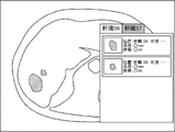
- FIG. 4 shows an example of an observation sentence display screen displayed on the display 23 by this control.
- the observation sentence display screen displays a plurality of observation sentences, a button that the user designates when selecting each observation sentence, and a button that the user designates when the user determines that there is no observation. be done.
- the display control unit 48 cancels the filling of the area and displays the outer edge of the area as a predetermined area. Control is performed to display the fourth status in an identifiable manner by drawing with colored lines.
- FIG. 5 shows another example of the remark display screen.
- FIG. 4 shows an example in which an abnormal shadow is detected in the liver
- FIG. 5 shows an example in which the shape of the liver itself is detected as an abnormal shadow.
- the display control unit 48 displays the third status in a identifiable manner by graying out the area. to control.
- this operation for example, the user designates a button for no finding on the finding text display screen shown in FIG. 4 .
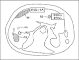
- FIG. 6 shows an example in which the statuses of the areas R1 and R2 are the first status, and the area R3 is the second status. A check mark C is added to the region R3 as a predetermined mark. Also, FIG. 6 shows an example in which the status of the area R4 is the third status and the status of the area R5 is the fourth status.
- the method of displaying the first to fourth statuses in an identifiable manner is not limited to the above example.
- the first status to the fourth status are identified by the line type or thickness of the outline of the area, the transparency or pattern of the filling of the area, the blinking of the area, the animation display of the area, and the addition of different marks. can be displayed if possible.
- the mark is not limited to a check mark, and may be an arrow or a symbol such as "+".
- the display control unit 48 may perform control to display the first to fourth statuses identifiably by enlarged display or reduced display.
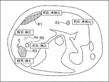
- FIG. 9 shows an example in which areas R1 and R2 whose status is the first status are enlarged and displayed.
- the display control unit 48 sets the first status and the second status among the first status to the fourth status so as to be different from the other statuses (that is, the third status and the fourth status). You may perform control to display. In this case, for example, the display control unit 48 controls to highlight only the first status and the second status among the first to fourth statuses.
- FIG. 10 shows an example in which the statuses of regions R1 to R5 are the same as in FIG. In the example of FIG. 10, areas R1 and R2 whose status is the first status and area R3 whose status is the second status are highlighted by being surrounded by bounding boxes.
- the display control unit 48 may perform control to highlight and display an area by filling the area with a preset color. Further, for example, the display control unit 48 may perform control to highlight and display the area by drawing the outer edge of the area with a line of a preset color.
- the display control unit 48 when the user performs an end operation such as an operation of closing the status display screen while the area of the first status or the second status remains, performs the medical document creation work. may be controlled to display a message to the effect that is incomplete. Further, in this case, it may be possible for the user to collectively set the areas with the first status or the second status to the third status with a single operation. Further, the display control unit 48 may perform control to display the image of the area portion as it is without performing additional display for the area with the third status or the fourth status.
- statuses other than the above-described first to fourth statuses may be applied as the plurality of statuses related to the user's medical document creation work.
- An example of the status in this case is the status of referral to another doctor.
- An example of this status display screen is shown in FIG. In the example of FIG. 11, an icon I1 representing a doctor is displayed near the area R1 to indicate that the status of the area R1 is to refer to another doctor.
- the reception unit 50 receives information representing the area selected by the user from among the areas extracted by the extraction unit 42 .
- the receiving unit 50 also receives an operation indicating which of the above four statuses the selected area should be. Further, the receiving unit 50 receives an opinion text selected by the user from among the plurality of opinion texts displayed on the display 23 under the control of the display control unit 48 . This accepted statement of findings is used to create a medical document.
- the CPU 20 executes the work support program 30 to execute the work support process shown in FIG.
- the work support process shown in FIG. 7 is executed, for example, when the user inputs an instruction to start execution.
- the acquiring unit 40 acquires the diagnosis target image from the image server 5 via the network I/F 25.
- the extracting unit 42 extracts a region in the image to be diagnosed acquired in step S10 that may be a target for a user such as a doctor to create a medical document.
- the analysis unit 44 analyzes each abnormal shadow extracted in step S12, and derives findings of the abnormal shadow.
- step S16 the display control unit 48 performs control to display on the display 23 information representing the area extracted in step S12. During this control, the display control unit 48 performs control to identifiably display that the status of each area is the first status, as described above.
- step S18 the accepting unit 50 accepts an operation by the user. If the operation received by the receiving unit 50 in step S18 is an operation in which the user designates an area and sets the status of the designated area to the second status, the process proceeds to step S20.
- step S20 the display control unit 48 adds a predetermined mark to the area designated by the user, thereby performing control to display the second status in an identifiable manner.
- the process of step S20 ends, the process returns to step S18.
- step S18 If the operation accepted by the accepting unit 50 in step S18 is an operation in which the user designates an area and instructs to display an observation text, the process proceeds to step S22.
- step S22 the generation unit 46 generates a plurality of observation sentences based on the observation derived in step S14 for the region specified by the user, as described above.
- step S24 the display control unit 48 performs control to display on the display 23 a plurality of observation sentences generated in step S22 for the area specified by the user.
- step S26 the reception unit 50 determines whether or not the observation text selected by the user from among the plurality of observation texts displayed on the display 23 in step S24 has been received. If this determination is affirmative, the process proceeds to step S28.
- step S28 the display control unit 48 unpaints the area specified by the user and draws the outer edge of the area with lines of a predetermined color, thereby displaying the fourth status in a distinguishable manner. control. When the process of step S28 ends, the process returns to step S18.
- step S26 If the operation accepted by the accepting unit 50 in step S26 is an operation in which an area is specified by the user and the status of the specified area is set as the third status, the determination in step S26 becomes a negative determination, and the process proceeds to step S30. transition to In step S30, the display control unit 48 performs control to display the third status in an identifiable manner by graying out the area designated by the user. When the process of step S30 ends, the process returns to step S18.
- step S18 If the operation accepted by the accepting unit 50 in step S18 is an operation to end the display of the screen, the work support process ends.
- the present embodiment it is possible to grasp the user's work status for each area that is included in the medical image and may be the user's work target.
- the display control unit 48 displays information about each of the plurality of regions extracted by the extraction unit 42 when the user performs an operation to instruct display of a list.
- a list display may be performed.
- the display control unit 48 may perform control for displaying a list of information regarding each of the plurality of areas extracted by the extraction unit 42 for each status.
- the display control unit 48 may perform control to display the information on each of the plurality of areas extracted by the extraction unit 42 in a display mode other than the list display.
- the display control unit 48 may perform control to display information about the area extracted by the extraction unit 42 in a pop-up format.
- FIG. 12 shows an example in which information about an area designated by an arrow representing a mouse cursor is displayed in a pop-up format.
- the display control unit 48 may perform control to display, in a pop-up format, information about areas having the same status as the specified area, not only for the specified area, but also for unspecified areas. . Further, in this case, as shown in FIG.
- the display control unit 48 may perform control to display information about each of the plurality of regions extracted by the extraction unit 42 in a tab format.
- each anatomical structure region is divided into different tabs. It shows an example of how information is displayed.
- FIG. 13 shows an example in which the subsegments of the liver divided into S1 to S8 are applied as the anatomical structure area, and the tab of S6 of the liver is specified.
- the display control unit 48 can identify whether each of the plurality of regions extracted by the extraction unit 42 has changed from the same region detected in the past examination. may be controlled to be displayed in In this case, in order to ignore the error, the display control unit 48 may consider that there is no change for a change equal to or less than the predetermined amount of change.
- FIG. 14 shows an example in which regions R1 and R2 in the status display screen are the same regions extracted by the extraction unit 42 from the diagnosis target image captured in the previous examination. Also, FIG. 14 shows an example in which the area of the region R1 is larger than that of the diagnosis target image captured in the previous examination, and the region R2 has no change in area from the diagnosis target image captured in the previous examination.
- the display control unit 48 displays each of the plurality of regions extracted by the extraction unit 42 in such a manner that whether or not the same region was detected in the past examination can be identified. You may perform control to do.
- regions R1 and R2 show an example in which the same region was not extracted by the extracting unit 42 in the diagnosis target image captured in the previous examination.
- regions R3 to R5 show an example in which the same region is extracted by the extracting unit 42 from the diagnosis target image photographed in the previous examination.
- the detected area here means an area detected by extraction processing by a computer, such as the extraction processing by the extraction unit 42, for example.
- the display control unit 48 may perform control to display, for each of the plurality of regions extracted by the extraction unit 42, the presence or absence of change from the same region reported in the past examination so as to be identifiable. . In this case, in order to ignore the error, the display control unit 48 may consider that there is no change for a change equal to or less than the predetermined amount of change. Further, the display control unit 48 may perform control to display each of the plurality of regions extracted by the extraction unit 42 in such a manner that whether or not the same region was reported in the past examination can be identified.
- the reported region here means, for example, a region not extracted by a computer extraction process, but discovered by the doctor and specified via the input device 24 . In other words, the reported area here means an area that the doctor reported in a medical document such as an interpretation report in the past.
- the work support device 10 may select one observation sentence from among a plurality of observation sentences based on the reliability of the observation included in the observation sentences.
- the various processors include, in addition to the CPU, which is a general-purpose processor that executes software (programs) and functions as various processing units, circuits such as FPGAs (Field Programmable Gate Arrays), etc. Programmable Logic Device (PLD) which is a processor whose configuration can be changed, ASIC (Application Specific Integrated Circuit) etc. Circuits, etc. are included.
- the CPU which is a general-purpose processor that executes software (programs) and functions as various processing units, circuits such as FPGAs (Field Programmable Gate Arrays), etc.
- Programmable Logic Device PLD which is a processor whose configuration can be changed, ASIC (Application Specific Integrated Circuit) etc. Circuits, etc. are included.
- One processing unit may be composed of one of these various processors, or a combination of two or more processors of the same type or different types (for example, a combination of multiple FPGAs, a combination of a CPU and an FPGA). combination). Also, a plurality of processing units may be configured by one processor.
- a single processor is configured by combining one or more CPUs and software.
- a processor functions as multiple processing units.
- SoC System on Chip
- the various processing units are configured using one or more of the above various processors as a hardware structure.
- an electric circuit combining circuit elements such as semiconductor elements can be used.
- the work support program 30 has been pre-stored (installed) in the storage unit 22, but the present invention is not limited to this.
- the work support program 30 is provided in a form recorded in a recording medium such as a CD-ROM (Compact Disc Read Only Memory), a DVD-ROM (Digital Versatile Disc Read Only Memory), and a USB (Universal Serial Bus) memory. good too.
- the work support program 30 may be downloaded from an external device via a network.
Landscapes
- Health & Medical Sciences (AREA)
- Engineering & Computer Science (AREA)
- Medical Informatics (AREA)
- General Health & Medical Sciences (AREA)
- Public Health (AREA)
- Epidemiology (AREA)
- Primary Health Care (AREA)
- Biomedical Technology (AREA)
- General Business, Economics & Management (AREA)
- Business, Economics & Management (AREA)
- Life Sciences & Earth Sciences (AREA)
- Nuclear Medicine, Radiotherapy & Molecular Imaging (AREA)
- Radiology & Medical Imaging (AREA)
- Physics & Mathematics (AREA)
- Biophysics (AREA)
- Pathology (AREA)
- Heart & Thoracic Surgery (AREA)
- Molecular Biology (AREA)
- Surgery (AREA)
- Animal Behavior & Ethology (AREA)
- Veterinary Medicine (AREA)
- High Energy & Nuclear Physics (AREA)
- Optics & Photonics (AREA)
- Medical Treatment And Welfare Office Work (AREA)
Priority Applications (2)
| Application Number | Priority Date | Filing Date | Title |
|---|---|---|---|
| JP2023514607A JP7840932B2 (ja) | 2021-04-14 | 2022-03-31 | 作業支援装置、作業支援方法、及び作業支援プログラム |
| US18/481,260 US20240029874A1 (en) | 2021-04-14 | 2023-10-05 | Work support apparatus, work support method, and work support program |
Applications Claiming Priority (4)
| Application Number | Priority Date | Filing Date | Title |
|---|---|---|---|
| JP2021068675 | 2021-04-14 | ||
| JP2021-068675 | 2021-04-14 | ||
| JP2021208526 | 2021-12-22 | ||
| JP2021-208526 | 2021-12-22 |
Related Child Applications (1)
| Application Number | Title | Priority Date | Filing Date |
|---|---|---|---|
| US18/481,260 Continuation US20240029874A1 (en) | 2021-04-14 | 2023-10-05 | Work support apparatus, work support method, and work support program |
Publications (1)
| Publication Number | Publication Date |
|---|---|
| WO2022220158A1 true WO2022220158A1 (ja) | 2022-10-20 |
Family
ID=83639623
Family Applications (1)
| Application Number | Title | Priority Date | Filing Date |
|---|---|---|---|
| PCT/JP2022/016718 Ceased WO2022220158A1 (ja) | 2021-04-14 | 2022-03-31 | 作業支援装置、作業支援方法、及び作業支援プログラム |
Country Status (3)
| Country | Link |
|---|---|
| US (1) | US20240029874A1 (https=) |
| JP (1) | JP7840932B2 (https=) |
| WO (1) | WO2022220158A1 (https=) |
Citations (6)
| Publication number | Priority date | Publication date | Assignee | Title |
|---|---|---|---|---|
| JPH04333972A (ja) * | 1991-05-10 | 1992-11-20 | Toshiba Corp | 医用診断支援システム |
| JP2002143141A (ja) * | 2000-11-14 | 2002-05-21 | Konica Corp | 画像診断支援装置 |
| JP2015198928A (ja) * | 2014-03-31 | 2015-11-12 | 株式会社東芝 | 医用画像処理装置、および医用画像処理システム |
| JP2019149005A (ja) * | 2018-02-27 | 2019-09-05 | 富士フイルム株式会社 | 医療文書作成支援装置、方法およびプログラム |
| JP2019146935A (ja) * | 2018-02-28 | 2019-09-05 | 富士フイルム株式会社 | 学習用データ作成支援装置、学習用データ作成支援方法、および学習用データ作成支援プログラム |
| WO2020129385A1 (ja) * | 2018-12-19 | 2020-06-25 | 富士フイルム株式会社 | 医療文書作成支援装置、方法およびプログラム |
Family Cites Families (1)
| Publication number | Priority date | Publication date | Assignee | Title |
|---|---|---|---|---|
| JP7192372B2 (ja) * | 2018-10-05 | 2022-12-20 | コニカミノルタ株式会社 | 情報処理装置、医用画像表示装置及びプログラム |
-
2022
- 2022-03-31 WO PCT/JP2022/016718 patent/WO2022220158A1/ja not_active Ceased
- 2022-03-31 JP JP2023514607A patent/JP7840932B2/ja active Active
-
2023
- 2023-10-05 US US18/481,260 patent/US20240029874A1/en active Pending
Patent Citations (6)
| Publication number | Priority date | Publication date | Assignee | Title |
|---|---|---|---|---|
| JPH04333972A (ja) * | 1991-05-10 | 1992-11-20 | Toshiba Corp | 医用診断支援システム |
| JP2002143141A (ja) * | 2000-11-14 | 2002-05-21 | Konica Corp | 画像診断支援装置 |
| JP2015198928A (ja) * | 2014-03-31 | 2015-11-12 | 株式会社東芝 | 医用画像処理装置、および医用画像処理システム |
| JP2019149005A (ja) * | 2018-02-27 | 2019-09-05 | 富士フイルム株式会社 | 医療文書作成支援装置、方法およびプログラム |
| JP2019146935A (ja) * | 2018-02-28 | 2019-09-05 | 富士フイルム株式会社 | 学習用データ作成支援装置、学習用データ作成支援方法、および学習用データ作成支援プログラム |
| WO2020129385A1 (ja) * | 2018-12-19 | 2020-06-25 | 富士フイルム株式会社 | 医療文書作成支援装置、方法およびプログラム |
Also Published As
| Publication number | Publication date |
|---|---|
| JPWO2022220158A1 (https=) | 2022-10-20 |
| JP7840932B2 (ja) | 2026-04-06 |
| US20240029874A1 (en) | 2024-01-25 |
Similar Documents
| Publication | Publication Date | Title |
|---|---|---|
| US20240029252A1 (en) | Medical image apparatus, medical image method, and medical image program | |
| JP2019153250A (ja) | 医療文書作成支援装置、方法およびプログラム | |
| US11093699B2 (en) | Medical image processing apparatus, medical image processing method, and medical image processing program | |
| US12406755B2 (en) | Document creation support apparatus, method, and program | |
| US20190267120A1 (en) | Medical document creation support apparatus, method, and program | |
| US20220392619A1 (en) | Information processing apparatus, method, and program | |
| US20230281810A1 (en) | Image display apparatus, method, and program | |
| US20220366151A1 (en) | Document creation support apparatus, method, and program | |
| JP7542710B2 (ja) | 文書作成支援装置、文書作成支援方法及びプログラム | |
| JPWO2019193983A1 (ja) | 医療文書表示制御装置、医療文書表示制御方法、及び医療文書表示制御プログラム | |
| WO2021107098A1 (ja) | 文書作成支援装置、文書作成支援方法及び文書作成支援プログラム | |
| JPWO2019193982A1 (ja) | 医療文書作成支援装置、医療文書作成支援方法、及び医療文書作成支援プログラム | |
| JP7007469B2 (ja) | 医療文書作成支援装置、方法およびプログラム、学習済みモデル、並びに学習装置、方法およびプログラム | |
| US20240046028A1 (en) | Document creation support apparatus, document creation support method, and document creation support program | |
| WO2021193548A1 (ja) | 文書作成支援装置、方法およびプログラム | |
| JP2020154630A (ja) | 医用情報収集装置 | |
| WO2022220158A1 (ja) | 作業支援装置、作業支援方法、及び作業支援プログラム | |
| WO2022153702A1 (ja) | 医用画像表示装置、方法およびプログラム | |
| JP7840933B2 (ja) | 文書作成支援装置、文書作成支援方法、及び文書作成支援プログラム | |
| JP7748454B2 (ja) | 文書作成支援装置、文書作成支援方法、及び文書作成支援プログラム | |
| WO2021107142A1 (ja) | 文書作成支援装置、方法およびプログラム | |
| JP7798808B2 (ja) | 文書作成支援装置、文書作成支援方法及びプログラム | |
| JP7368592B2 (ja) | 文書作成支援装置、方法およびプログラム | |
| WO2023054646A1 (ja) | 情報処理装置、情報処理方法及び情報処理プログラム | |
| JP2024162560A (ja) | 情報処理装置、方法およびプログラム |
Legal Events
| Date | Code | Title | Description |
|---|---|---|---|
| 121 | Ep: the epo has been informed by wipo that ep was designated in this application |
Ref document number: 22788083 Country of ref document: EP Kind code of ref document: A1 |
|
| WWE | Wipo information: entry into national phase |
Ref document number: 2023514607 Country of ref document: JP |
|
| NENP | Non-entry into the national phase |
Ref country code: DE |
|
| 122 | Ep: pct application non-entry in european phase |
Ref document number: 22788083 Country of ref document: EP Kind code of ref document: A1 |