WO2022152298A1 - 载 plk1 抑制剂的聚合物囊泡药物及其制备方法与应用 - Google Patents
载 plk1 抑制剂的聚合物囊泡药物及其制备方法与应用 Download PDFInfo
- Publication number
- WO2022152298A1 WO2022152298A1 PCT/CN2022/072328 CN2022072328W WO2022152298A1 WO 2022152298 A1 WO2022152298 A1 WO 2022152298A1 CN 2022072328 W CN2022072328 W CN 2022072328W WO 2022152298 A1 WO2022152298 A1 WO 2022152298A1
- Authority
- WO
- WIPO (PCT)
- Prior art keywords
- vol
- dtc
- peg
- plk1 inhibitor
- loaded
- Prior art date
- Legal status (The legal status is an assumption and is not a legal conclusion. Google has not performed a legal analysis and makes no representation as to the accuracy of the status listed.)
- Ceased
Links
Classifications
-
- A—HUMAN NECESSITIES
- A61—MEDICAL OR VETERINARY SCIENCE; HYGIENE
- A61K—PREPARATIONS FOR MEDICAL, DENTAL OR TOILETRY PURPOSES
- A61K9/00—Medicinal preparations characterised by special physical form
- A61K9/10—Dispersions; Emulsions
- A61K9/127—Synthetic bilayered vehicles, e.g. liposomes or liposomes with cholesterol as the only non-phosphatidyl surfactant
- A61K9/1271—Non-conventional liposomes, e.g. PEGylated liposomes or liposomes coated or grafted with polymers
- A61K9/1273—Polymersomes; Liposomes with polymerisable or polymerised bilayer-forming substances
-
- A—HUMAN NECESSITIES
- A61—MEDICAL OR VETERINARY SCIENCE; HYGIENE
- A61K—PREPARATIONS FOR MEDICAL, DENTAL OR TOILETRY PURPOSES
- A61K31/00—Medicinal preparations containing organic active ingredients
- A61K31/33—Heterocyclic compounds
- A61K31/395—Heterocyclic compounds having nitrogen as a ring hetero atom, e.g. guanethidine or rifamycins
- A61K31/495—Heterocyclic compounds having nitrogen as a ring hetero atom, e.g. guanethidine or rifamycins having six-membered rings with two or more nitrogen atoms as the only ring heteroatoms, e.g. piperazine or tetrazines
- A61K31/505—Pyrimidines; Hydrogenated pyrimidines, e.g. trimethoprim
- A61K31/519—Pyrimidines; Hydrogenated pyrimidines, e.g. trimethoprim ortho- or peri-condensed with heterocyclic rings
- A61K31/525—Isoalloxazines, e.g. riboflavins, vitamin B2
-
- A—HUMAN NECESSITIES
- A61—MEDICAL OR VETERINARY SCIENCE; HYGIENE
- A61K—PREPARATIONS FOR MEDICAL, DENTAL OR TOILETRY PURPOSES
- A61K47/00—Medicinal preparations characterised by the non-active ingredients used, e.g. carriers or inert additives; Targeting or modifying agents chemically bound to the active ingredient
- A61K47/30—Macromolecular organic or inorganic compounds, e.g. inorganic polyphosphates
- A61K47/34—Macromolecular compounds obtained otherwise than by reactions only involving carbon-to-carbon unsaturated bonds, e.g. polyesters, polyamino acids, polysiloxanes, polyphosphazines, copolymers of polyalkylene glycol or poloxamers
-
- A—HUMAN NECESSITIES
- A61—MEDICAL OR VETERINARY SCIENCE; HYGIENE
- A61K—PREPARATIONS FOR MEDICAL, DENTAL OR TOILETRY PURPOSES
- A61K47/00—Medicinal preparations characterised by the non-active ingredients used, e.g. carriers or inert additives; Targeting or modifying agents chemically bound to the active ingredient
- A61K47/30—Macromolecular organic or inorganic compounds, e.g. inorganic polyphosphates
- A61K47/42—Proteins; Polypeptides; Degradation products thereof; Derivatives thereof, e.g. albumin, gelatin or zein
-
- A—HUMAN NECESSITIES
- A61—MEDICAL OR VETERINARY SCIENCE; HYGIENE
- A61P—SPECIFIC THERAPEUTIC ACTIVITY OF CHEMICAL COMPOUNDS OR MEDICINAL PREPARATIONS
- A61P35/00—Antineoplastic agents
Definitions
- the invention belongs to medical technology, and particularly relates to a PLK1 inhibitor-loaded polymer vesicle medicine and a preparation method and application thereof.
- PARP poly(ADP-ribose) polymerase
- the present invention reports a novel strategy combining polypeptide-targeted polymeric vesicles with the PLK1 inhibitor Vol as a dual-targeted drug for the treatment of ovarian cancer.
- Peptide-targeted vesicles A3-Ps are obtained by self-assembly of polymers PEG-P(TMC-DTC)-PAsp and A3-PEG-P(TMC-DTC), and PEG-P(TMC-DTC)-based polymer vesicles
- the vesicle has a dense disulfide cross-linked membrane layer, which brings excellent stability and rapid intracellular drug release, and the short-chain polyaspartic acid in the inner shell of the vesicle can enhance the capacity of the vesicle to load Vol , has high affinity for human ovarian cancer cells such as ES-2, SKOV3 and OVCAR3.
- the present invention is the first report on the targeted delivery of Vol for the treatment of
- a polymer vesicle loaded with a PLK1 inhibitor including a polymer vesicle and a PLK1 inhibitor
- the polymer vesicle is prepared from PEG-P(T-DTC)-PAsp
- the polymer vesicle is The vesicles were prepared from PEG-P(T-DTC)-PAsp and A-PEG-P(T-DTC).
- the invention also discloses the application of the polymer in the preparation of PLK1 inhibitor-loaded polymer vesicles, or in the preparation of anti-tumor drugs, especially in the preparation of drugs for treating ovarian cancer; the polymer is PEG- P(T-DTC)-PAsp, or the polymer is PEG-P(T-DTC)-PAsp and A-PEG-P(T-DTC).
- the invention further discloses the application of the above-mentioned PLK1 inhibitor-loaded polymer vesicles in the preparation of antitumor drugs, especially in the preparation of drugs for treating ovarian cancer.
- the invention discloses a preparation method of the above-mentioned PLK1 inhibitor-loaded polymer vesicles.
- the polymer solution and the PLK1 inhibitor solution are mixed, incubated, and then dialyzed to obtain PLK1 inhibitor-loaded vesicles.
- the PLK1 inhibitor-loaded polymer vesicles of the present invention are composed of polymer vesicles and PLK1 inhibitors, and are nano-drugs. After the particle size and Zeta potential are measured, the content of Vol is quantified by an ultraviolet spectrophotometer to calculate the load. Drug Volume (DLC) and Encapsulation Efficiency (DLE).
- DLC Drug Volume
- DLE Encapsulation Efficiency
- the PLK1 inhibitor is volasertib (Vol); in PEG-P(T-DTC)-PAsp and A-PEG-P(T-DTC), T is an ester unit or a carbonate unit, that is, a cyclic ester monomer Or ring-opened units of cyclic carbonate monomers, such as TMC, LA, CL; A is a peptide targeting molecule, such as A3 polypeptide; PAsp is polyaspartic acid; DTC is dithiolane trimethylene carbonate monolayer body.
- Vol Vol
- T is an ester unit or a carbonate unit, that is, a cyclic ester monomer Or ring-opened units of cyclic carbonate monomers, such as TMC, LA, CL
- A is a peptide targeting molecule, such as A3 polypeptide
- PAsp is polyaspartic acid
- DTC is dithiolane trimethylene carbonate monolayer body.
- cyclic ester monomer or cyclic carbonate monomer as the first monomer, dithiolane trimethylene carbonate monomer as the second monomer, using polyethylene glycol as an initiator, and polymerizing to obtain PEG-P(T -DTC), then through the activation of p-nitrophenyl chloroformate and the coupling reaction with polyaspartic acid to obtain PEG-P(T-DTC)-PAsp; or cyclic ester monomer or cyclic carbonate monomer as The first monomer and the dithiolane trimethylene carbonate monomer are the second monomer, and the polyethylene glycol with the active end group is used as the initiator, and the PEG-P(T) with the active end group is obtained by polymerization. -DTC), and then react with the targeting polypeptide to obtain A-PEG-P(T-DTC).
- P(T-DTC) is a hydrophobic segment; wherein, the molecular weight of the PEG segment is 2000-10000 Da; the hydrophobic chain The total molecular weight of the segment is 2.5 to 10 times that of the PEG segment; the molecular weight of the PDTC segment in the hydrophobic segment accounts for 10% to 35% of the total molecular weight of the hydrophobic segment; the molecular weight of polyaspartic acid is 300 to 5000Da.
- the molecular weight of the PEG segment is 3400-8000 Da; the total molecular weight of the hydrophobic segment is 2.8 to 6 times the molecular weight of the PEG segment; the molecular weight of the PDTC segment in the hydrophobic segment accounts for 11% to 25% of the total molecular weight of the hydrophobic segment. %; the molecular weight of polyaspartic acid is 1000-2000 Da.
- the present invention reports a novel strategy of targeting polypeptide (A3)-modified polymer vesicles (A3-Ps) loaded with molecular targeting drug PLK1 inhibitor volasertib (Vol) for the treatment of ovarian cancer.
- A3-Ps-Vol ranged from 7.7% to 8.0%, and the particle sizes ranged from 25 nm to 32 nm.
- Figure 1 shows the hydrogen NMR spectrum (600 MHz, DMSO- d 6 ) of A3-PEG-P(TMC-DTC).
- Figure 2 shows (A) cryo-EM characterization of A3-Ps-Vol; (B) the particle size changes of vesicles after storage of Ps-Vol and A3-Ps-Vol (1 mg/mL) in a refrigerator at 4°C for one month; (C) The particle size change of vesicles after 50-fold dilution or adding 10% fetal bovine serum; (D) The amount of drug leakage stored in a refrigerator at 4°C for one month; (E) Volasertib release in the presence of 10 mM GSH (polymer concentration of 0.4 mg/mL); (F) Toxicity of empty vesicles to SKOV-3 cells.
- B the particle size changes of vesicles after storage of Ps-Vol and A3-Ps-Vol (1 mg/mL) in a refrigerator at 4°C for one month
- C The particle size change of vesicles after 50-fold dilution or adding 10% fetal bovine serum
- D The amount
- Figure 4 (A) cell cycle analysis and (B) cells of SKOV-3 cells incubated with A3-Ps-Vol, Ps-Vol, and free Vol (200 nM) for 4 h, and then incubated in fresh medium for 20 h. Apoptosis detection.
- Figure 5 shows the endocytosis and drug release of Cy5-A3-Ps-Vol-FITC and Cy5-Ps-Vol-FITC in SKOV-3 cells characterized by CLSM.
- A Image of CLSM incubated for 4 h. Scale bar: 25 ⁇ m.
- B Semi-quantitative analysis of intracellular fluorescence signals of FITC and Cy5. **p ⁇ 0.01.
- C Co-localization analysis of intracellular FITC and Cy5 signals.
- Figure 6 is (A) intravital imaging of mice injected with Cy5-Ps-Vol or Cy5-A3-Ps-Vol at different time points; (B) Intravenous injection of Ps-Vol-FITC or A3-Ps-Vol-FITC Fluorescence images of major organs in vitro after 48 h; (C) Cy5-Ps-Vol and Cy5-A3-Ps-Vol (red) Fluorescent sections of tumors 48 h after injection, nuclei and blood vessels were stained with DAPI (blue) and CD31-FTIC (green) staining.
- Figure 7 shows the effect and histological analysis of A3-Ps-Vol on the treatment of SKOV-3 subcutaneous tumor-bearing mice.
- A tumor volume change
- B mouse body weight change
- C tumor volume change on day 16
- D tumor inhibition rate on day 16
- E analysis of tumor sections in different groups.
- H&E PLK1 immunohistochemical (IHC) analysis and TUNEL.
- White and red scales represent 25 and 100 ⁇ m, respectively.
- Statistical analysis was performed using one-way ANOVA and Tukey's multiple comparison test. * p ⁇ 0.05, ** p ⁇ 0.01, and *** p ⁇ 0.001.
- Figure 8 shows the resistance of A3-Ps-Vol and Free Vol to healthy nude mice under two administration modes: single injection of 50 mg Vol/kg (A) and 6 injections of 20 mg Vol/kg (once in 3 days). Subject to study (B). # indicates the termination of the experiment due to the death of the mouse. (C) Routine blood analysis of mice in free Vol and A3-Ps-Vol groups at a single dose of 50 mg Vol/kg.
- the present invention reports a novel strategy of targeting polypeptide (A3)-modified polymer vesicles (A3-Ps) loaded with molecular targeting drug PLK1 inhibitor volasertib (Vol) for the treatment of ovarian cancer:
- A3- The drug loading of Ps-Vol ranges from 7.7% to 8.0%, and the particle size ranges from 25 nm to 32 nm.
- the PLK1 inhibitor-loaded polymer vesicle of the present invention is composed of a polymer vesicle and a PLK1 inhibitor, and the dual-targeted nano-formulation is an efficient and low-toxicity therapy for treating ovarian cancer.
- A3 polypeptide (cyclic peptide cDGWGPNc, 98%) was purchased from Shanghai Jill Biochemical. Volasertib (Vol, > 99.8%, MedChemExpress), FITC and Cy5 (Lumiprobe) were used directly after purchase. PLK1 antibody, GAPDH antibody (Santa Cruz Biotechnology), microBCA (Thermo Scientific) were used according to the manufacturer's instructions.
- SKOV-3 cells were purchased from Chinese Academy of Sciences (Shanghai, China).
- BALB/c nude mice female, 4-6 weeks, 16–20 g
- the NMR characterization of the polymers was carried out in a 600 M 1 H NMR spectrometer (DirectDrive2) using DMSO-d6 as solvent, and the chemical shifts were calibrated according to residual DMSO ( ⁇ 2.50 ppm).
- the particle size and size distribution of vesicles were determined in PB by a dynamic laser light scattering instrument Zetasizer Nano-ZS (Malvern) equipped with a 633 nm He-Ne laser at 25 °C, and Zeta potential was determined by a U-shaped electrophoresis cell.
- Volasertib drug loading was determined by UV absorbance at 330 nm wavelength and standard curve by UV spectrophotometer (UH5300, HITACHI).
- Polypeptides coupled to polymers were determined by a microplate reader (Multiscan FC, Thermo) using Pierce-BCA assay (Thermo-Fisher Scientific).
- CLSM images of cells were taken on a confocal laser scanning microscope (LSM710, Leica).
- Flow analysis of cells was determined by flow cytometer (BD, FACS Calibur flow cytometer).
- In vivo and ex vivo fluorescence imaging was determined by a near-infrared fluorescence imaging system (IVIS Lumina II). Biodistribution of volasertib-FITC in various tissues was quantified by fluorophotometer (Cary Eclipse, ex. 480 nm, em. 560 nm).
- preparations for related experiments such as drug-loaded vesicles, free drug, and empty vesicles were all in HEPES (pH 7.4, 5 mM) buffer.
- CN2020105763476 carbonate polymer vesicles loaded with small molecule drugs
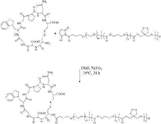
- A3-PEG-P(TMC-DTC) was obtained by amidation reaction of A3 polypeptide (cDGWGPNc) with the above-mentioned NHS-PEG-P(TMC-DTC). Briefly, NHS-PEG-(TMC-DTC) (200 mg, 6.2 ⁇ mol) was dissolved in 2 mL DMF, which was added dropwise to a solution of A3 polypeptide (13.6 mg, 12.4 ⁇ mol) and triethylamine under nitrogen protection. (10.3 ⁇ L, 74.4 ⁇ mol) in 1 mL of DMF, kept stirring, added dropwise for a total of 30 min, and then moved to 35 °C to react for 24 h.
- A3-PEG-P(TMC-DTC) was synthesized by amidation reaction of A3 with NHS-PEG-P(TMC-DTC), as follows.
- the first monomer TMC was replaced with LA or CL to obtain polymers with different hydrophobic segments.
- A3 was replaced with Cy5-NH 2 to obtain Cy5-labeled polymer PEG-P(TMC-DTC)-Cy5.
- Example 2 Preparation of Ps-Vol and A3-Ps-Vol:
- the DMSO solutions of PEG-P(TMC-DTC)-PAsp and A3-PEG-P(TMC-DTC) with the above concentration of 10 mg/mL were prepared by mass The ratio of 80:20 was mixed, 100 mL was mixed with Vol's DMSO solution (20 mg/mL, 5 mL), and 0.9 mL of HEPES (5 mM, pH 6.8) buffer was added, 200 After stirring at rpm for 5 minutes, transfer to a shaker (100 rpm, 35°C) for 6 h, then dialyzed against HEPES (5 mM, pH 7.4) (dialysis bag MW 7000).
- the stability of vesicles was characterized by long-term storage of Ps-Vol and A3-Ps-Vol, 50-fold dilution, and changes in vesicle size in the presence of serum by DLS. After A3-Ps-Vol was stored in HEPES (pH 7.4, 5 mM) at 4 °C for different times, the UV absorbance was measured by dialysis for 1 h to characterize the loading stability of the drug Vol.
- A3-Ps-Vol Vol-loaded targeting vesicles were obtained by self-assembly of PEG-P(TMC-DTC)-PAsp, A3-PEG-P(TMC-DTC) and Vol in HEPES (pH 6.8, 5 mM) , the following characterizations were performed in HEPES (pH 7.4, 5 mM).
- the theoretical drug loading (DLC) was set to 10 wt.% and the ratio of A3-PEG-P(TMC-DTC) was varied from 10%, 20% to 30% to obtain vesicles with different A3 densities, denoted as: 10A3 -Ps-Vol, 20A3-Ps-Vol and 30A3-Ps-Vol.
- A3-Ps-Vols The particle size of all A3-Ps-Vols is very small, and the particle size increases from 24 to 32 nm with increasing A3-PEG-P(TMC-DTC) content (particle size distribution from 0.06 to 0.10) (Table 1 ). Cryo-EM also confirmed that A3-Ps-Vol has a small particle size and a uniform distribution (Fig. 2A). In addition, all A3-Ps-Vols exhibited high drug loading (DLC: 7.7 ⁇ 8 wt.%) and encapsulation efficiency (DLE: 83% ⁇ 87%). The Zeta potential of A3-Ps-Vol is slightly negative (-4.3 ⁇ -5.5 mV). However, the non-target group Ps-Vol obtained only by self-assembly of PEG-P(TMC-DTC)-PAsp had similar size, DLC and Zeta potential.
- A3-Ps-Vol and Ps-Vol showed little change in particle size after high dilution (0.02 ⁇ 1 mg/mL), showing excellent stability; store one in the presence of 10% FBS or in a refrigerator at 4°C Months also remained stable (Fig. 2B, C). After one month of storage at 4°C, less than 5% of the drug was leaked (Fig. 2D). Its high stability is derived from the PEG-P (TMC-DTC)-based disulfide cross-linked vesicle membrane in vesicles, and the electrostatic interaction between Vol and PAsp also plays a role in stable loading.
- TMC-DTC PEG-P
- Reduction-responsive vol release was investigated by adding GSH (10 mM) to Ps-Vol and A3-Ps-Vol. Briefly, vesicles (0.4 mg/mL) were loaded into release bags (MWCO: 7000 Da) in PBS with or without GSH (10 mM), and then placed in a shaker at 37 °C, 200 rpm. in bed. At preset time points, take 5 mL of release medium for HPLC detection (280 nm) Vol and supplement with 5 mL of fresh dialysis medium.
- SKOV3 cells were plated into 12-well plates (containing small discs) at 50,000 per well. After overnight adherence, Ps-Vol-FITC or A3-Ps-Vol-FITC (Vol concentration of 200 nM) was added. ) were incubated for 4 h. The cells were then stained with rhodamine for 50 min, fixed with 4% paraformaldehyde for 15 min, and stained with DAPI for 5 min. Wash 3 times with PBS between each step. Small discs with cells are used for CLSM observation.
- SKOV3 cells were incubated with the above preparations for 1, 2, 4, 8, and 16 hours, respectively, and DAPI stained with CLSM Observed. Fluorescence signals of Cy5 and FITC were semi-quantitatively analyzed by Image J software for fluorescence intensity and colocalization.
- SKOV3 cells were plated in 6-well plates, 500,000 per well, and adhered overnight. After adding A3-Ps-Vol, Ps-Vol or free Vol (Vol concentration of 200 nM) and incubating for 4 h, the medium was replaced and the incubation continued for 20 h. After washing with PBS, RIPA was lysed for 15 min in an ice bath and centrifuged at 12,000 rpm for 15 min. The supernatant was taken and the protein concentration was measured by Nanodrop. Each sample was adjusted to the same protein concentration, boiled for 5 min, separated by 10% SDS-PAGE, and then transferred to PVDF membrane.
- SKOV3 cells were plated in a 6-well plate with 500,000 cells per well. After overnight adherence, A3-Ps-Vol, Ps-Vol, and free Vol (200 nM) were added and incubated for 4 h. Cultured in fresh medium for 44 h. They were then digested, fixed, stored at -24°C overnight, and treated with propidium iodide (PI) and RNase for 30 min for cell cycle detection by flow cytometry.
- PI propidium iodide
- Apoptosis experiment The same as the above cell cycle experiment conditions, the apoptosis experiment was also carried out under the conditions of 200 nM and 4+44 h. PBS, free drug Vol and Ps-Vol were the control groups. Cells were trypsinized without EDTA and double stained with propidium iodide (PI) and Annexin V-Alexa Flour 647, and detected by flow cytometry.
- PI propidium iodide
- Annexin V-Alexa Flour 647 Annexin V-Alexa Flour 647
- MTT experiment SKOV3 cells were plated in a 96-well plate, 3000 cells per well, and after 24 h of adherent growth, 20 mL of free Vol, Ps-Vol or A3-Ps-Vol were added (the density of surface A3 was 10%, 20% and 30%, respectively). %). Final concentrations in the well range from 0.1 nM to 10 ⁇ M. After a total of 72 h of incubation, 10 ⁇ L of MTT (5.0 mg/mL) was added for 3 h. Remove the supernatant, add 150 ⁇ L of DMSO to dissolve for 20 min, and then use a microplate reader to measure at 570 nm, and divide it with the PBS control group (100%) to obtain the cell viability.
- DU145 cells were used as a negative control. Toxicity of empty vesicles was determined by measuring MTT after the polymer was incubated with SKOV-3 cells at concentrations of 0.5, 0.2, 0.1, and 0.05 mg/mL for 72 h.
- A3-Ps-Vol can bind to the ATP-binding domain of PLK1 more easily than free Vol, regulate cell mitosis, disrupt spindle formation, block cells in G2/M phase, and at the same time, inactivate PLK1 in tumor cells , leading to apoptosis.
- vesicles were prepared with Cy5-labeled polymer PEG-P(TMC-DTC)-Cy5 and FITC-labeled Vol, and the targeting of A3 was studied by CLSM, and tracking Release behavior of carrier and drug in SKOV-3 cells.
- Figure 5A shows that the A3-Ps-Vol group had significantly enhanced intracellular Cy5 and FITC signals compared with Ps-Vol after 4 h incubation. Quantitative analysis of Cy5 and FITC showed that the endocytosis enhancement brought by the targeting effect of A3-Ps-Vol tended to be stable until 16 h, which was twice that of Ps-Vol.
- Example 4 In vivo imaging and biodistribution: In order to study the tumor enrichment of A3-Ps-Vol, Cy5 (ex. 649, em. 670) labeled drug-loaded vesicles Cy5-Ps-Vol and Cy5-A3 prepared by conventional method -Ps-Vol, intravenously injected into nude mice bearing SKOV3 subcutaneous tumor, 0.06 mg Cy5 per mouse.
- the SKOV3 tumor model was established by subcutaneous injection of 5 million SKOV3 cells in the right posterior back. Cy5 signal was tracked by a small animal in vivo imaging system (IVIS Lumina II) at 2, 4, 12, 24, 48 hours after injection.
- mice were sacrificed, the tumor was removed and fixed with 4% paraformaldehyde, sectioned, and the nuclei were stained with DAPI and FITC-labeled CD31. Blood vessels, the distribution of drugs in tumor tissue was observed by laser confocal (LSM710, Leica).
- Figure 6A shows the Cy5 signal of the A3-Ps-Vol group that is progressively enhanced at the tumor.
- the A3-Ps-Vol group at 12 h showed a significant advantage over the Ps-Vol group, and the fluorescence intensity at 48 h was 2.5 times that of the Ps-Vol group.
- the fluorescence signals of isolated organs and tumors confirmed the targeting of A3-Ps. Vol-enrichment within normal and tumor tissues was visualized by loading Vol-FITC. The enrichment of A3-Ps-Vol at the tumor site was 2-fold higher than that of the Ps-Vol group ( Fig. 6B ).
- SKOV3 tumor model was established by subcutaneously injecting 5 million SKOV3 cells; on the 28th day after inoculation, tumor-bearing mice were randomly divided into 5 groups with 5 mice in each group (tumor volume was about 75 mm 3 ). ). 200 ⁇ L of free Vol (10 mg Vol/kg), Ps-Vol (10 mg Vol/kg), A3-Ps-Vol (10 or 20 mg Vol/kg) and PBS were injected into nude mice via tail vein, respectively. The day is marked as day 0. The above formulations were administered on days 0, 3, 6, 9, 12, and 15.
- TIR tumor inhibition rate
- Tumors and major organs liver, heart, spleen, lung, and kidney were collected and processed for H&E staining. Tumor sections were also used for TUNNEL analysis to study tumor cell apoptosis. In order to study the inhibition of PLK1 in tumors, tumor sections were labeled with PLK1 antibody by PLK1 antibody, then stained with hematoxylin, and finally observed by microscope.
- FIG. 7A The resulting experiment (Fig. 7A) showed that A3-Ps-Vol (10 mg/kg) significantly inhibited tumor growth, better than Ps-Vol and free Vol, demonstrating the importance of A3 polypeptides in SKOV-3 tumor therapy.
- the dose was increased to 20 mg/kg, the antitumor effect of A3-Ps-Vol was further improved (*p ⁇ 0.05), the tumor volume was reduced to half that of the initial treatment.
- Both Ps-Vol and free Vol groups showed partial tumor suppression.
- the mice in each group did not lose weight (Fig. 7B), indicating that A3-Ps-Vol not only well inhibited the growth of SKOV-3 tumors, but also avoided toxic side effects.
- FIG. 7C Mice were sacrificed on day 16, and the ex vivo tumor photos (Fig. 7C) clearly showed that the A3-Ps-Vol (20 mg/kg) group had the smallest tumor size and achieved the best anti-tumor effect. Notably, A3-Ps-Vol achieved 80% and 91% tumor inhibition rate (TIR) at 10 and 20 mg Vol/kg doses, respectively, which was significantly higher than that of free Vol (50%) and Ps-Vol ( 58%) (Fig. 7D).
- TIR tumor inhibition rate
- Tumor sections were analyzed by H&E, immunohistochemistry and TUNEL staining (Figure 7E).
- the tumor section results of H&E were consistent with the treatment effect.
- Tumors in the A3-Ps-Vol group showed the largest and greatest extent of tumor necrosis.
- Immunohistochemical pictures showed that the PBS group had abundant PLK1 expression, while the free Vol and Ps-Vol groups effectively inhibited PLK1, and the A3-Ps-Vol group inhibited PLK1 more significantly.
- the A3-Ps-Vol (20 mg Vol/kg) group not only inhibited PLK1 best, but also led to apoptosis, presenting a large number of loose and fragmented tumor nuclei.
- the TUNEL images showed that A3-Ps-Vol significantly enhanced tumor cell apoptosis compared with free Vol and Ps-Vol.
- Example 6 Tolerance Experiment Tolerance of A3-Ps-Vol was observed by the response of healthy mice to the preparation. Healthy nude mice were injected via tail vein with a single dose of A3-Ps-Vol and free Vol (50 mg Vol/kg), or 6 injections of 20 mg Vol/kg every 3 days (0, 3, 6, 9, 12). , 15 days of administration), 3 animals in each group. Changes in body weight, behavioral changes, and survival were monitored to assess the toxicity of Vol and A3-Ps-Vol. 24 hours after the injection of 50 mg Vol/kg, blood was drawn for routine blood testing to observe the hematological toxicity of volasertib and A3-Ps-Vol.
- Figures 8A and 8B show that A3-Ps-Vol was well tolerated.
- the body weight of the mice remained basically unchanged in both single-dose and multi-dose modes, and the mice had no abnormal behavior; while the free Vol was well tolerated in the single-dose group. Tolerated, but in the multi-dose group, acute death was induced after 5 injections.
- Figure 8C blood routine results showed that A3-Ps-Vol had lower hematological toxicity than free Vol. Therefore, A3-Ps-Vol was better tolerated than free Vol.
- vol brings limited therapeutic effects and certain toxic side effects, such as the existing literature [ROS-Responsive Polymeric Micelles for Triggered Simultaneous Delivery of PLK1 Inhibitor/miR-34a and Effective Synergistic Therapy in Pancreatic Cancer] disclosed that the particle size of vol-loaded nanoparticles was 90 nm, the surface charge was 35.5 ⁇ 2.7 mV, the circulation time was short, and tumor enrichment had been decreasing within 24h; Compared with A3-Ps-Vol with low toxicity, it is a very promising ovarian cancer treatment method.
- the drug-loaded vesicles of the present invention have high drug loading, small size and low surface charge, especially in vivo.
- the stability was better, the circulation time was long, and the tumor enrichment was more, and the tumor enrichment continued to increase within 48 h.
- the targeting polypeptide-modified vesicles (A3-Ps) disclosed in the present invention can deliver Vol to the SKOV-3 subcutaneous tumor of nude mice, achieving the curative effect of highly effective tumor suppression while reducing side effects. This is the first study to target the delivery of Vol in ovarian cancer.
- This dual-targeting strategy tumor targeting of vesicles and molecular targeting of Vol is a very promising treatment modality for ovarian cancer: 1.
- A3-Ps greatly improved the circulation time, tumor uptake and tumor penetration of Vol, subtly solving the problem of low tumor enrichment of Vol.
- the Vol-targeted delivery system A3-Ps also has the potential to address drug resistance in the clinic. Therefore, the dual-target nano-formulation has shown potential in the targeted therapy of ovarian cancer.
Landscapes
- Health & Medical Sciences (AREA)
- Chemical & Material Sciences (AREA)
- Life Sciences & Earth Sciences (AREA)
- Medicinal Chemistry (AREA)
- General Health & Medical Sciences (AREA)
- Veterinary Medicine (AREA)
- Public Health (AREA)
- Pharmacology & Pharmacy (AREA)
- Animal Behavior & Ethology (AREA)
- Epidemiology (AREA)
- Proteomics, Peptides & Aminoacids (AREA)
- Inorganic Chemistry (AREA)
- Chemical Kinetics & Catalysis (AREA)
- General Chemical & Material Sciences (AREA)
- Organic Chemistry (AREA)
- Nuclear Medicine, Radiotherapy & Molecular Imaging (AREA)
- Dispersion Chemistry (AREA)
- Medicinal Preparation (AREA)
Abstract
本发明公开了载PLK1抑制剂的聚合物囊泡药物及其制备方法与应用;基于PEG-P(TMC-DTC)的聚合物囊泡A3-Ps有着致密的双硫键交联的膜,带来了卓越的稳定性和快速的细胞内药物释放,且囊泡膜的内壳的短链聚天冬氨酸能增强该囊泡装载分子靶向药volasertib (Vol)的能力,A3-Ps的载药量、载药率高,粒径小。载Vol囊泡A3-Ps-Vol在提升Vol抗肿瘤活性的同时减少了其系统毒性,A3-Ps-Vol和非靶向Ps-Vol及自由药Vol相比有最好的抑制SKOV-3细胞的活性,IC 50为49 nM,比自由药Vol低3.5倍,这种双靶向的纳米制剂是一种高效低毒的治疗卵巢癌的疗法。
Description
本发明属于医药技术,具体涉及载PLK1抑制剂的聚合物囊泡药物及其制备方法与应用。
作为女性疾病中致命的恶性肿瘤,卵巢癌的五年生存率仅为30%-40%。高致死率的原因是晚期卵巢癌病人的复发和频繁化疗(如铂药和taxane)后的耐药性。因而临床上对治疗卵巢癌的药物开发有着迫切的需求。在多种恶性肿瘤治疗方面,有着明确特异性和低毒性的靶向分子药物逐渐成为传统化疗药物的更好的替代品。聚腺苷二磷酸核糖聚合酶(PARP)抑制剂-olaparib在BRCA突变的卵巢癌病例中显示了良好的治疗效果,BRCA突变大约占所有卵巢癌病人的10%-15%。研究发现,有26%的卵巢癌病人的Polo样激酶1(PLK1)表达呈阳性,并且PLK1的表达和卵巢癌肿瘤的恶性程度呈正相关性, 因此PLK1抑制剂volasertib(Vol)是另一个有望用于治疗卵巢癌的分子靶向药。但是,和其他小分子药类似,Vol通常会被人体快速清除,并且肿瘤的富集有限。此外,耐药性是小分子药物的另一个潜在挑战。
目前关于Vol的靶向递送的研究很少,本发明报道了一种新型的策略,将多肽靶向的聚合物囊泡和PLK1抑制剂Vol结合,作为治疗卵巢癌的双重靶向药物。多肽靶向的囊泡A3-Ps通过聚合物PEG-P(TMC-DTC)-PAsp和A3-PEG-P(TMC-DTC)自组装得到,基于PEG-P(TMC-DTC)的聚合物囊泡有着致密的双硫键交联膜层,带来了卓越的稳定性和快速的细胞内药物释放,且囊泡的内壳的短链聚天冬氨酸能增强该囊泡装载Vol的能力,对人卵巢癌细胞,如ES-2, SKOV3和OVCAR3有高亲和力。本发明是第一篇关于Vol的靶向递送用于卵巢癌治疗的报道,结果表明,该双重靶向药物显著改善了小鼠SKOV-3
皮下肿瘤的治疗效果。
本发明采用如下技术方案:载PLK1抑制剂的聚合物囊泡,包括聚合物囊泡与PLK1抑制剂;所述聚合物囊泡由PEG-P(T-DTC)-PAsp制备,或者所述聚合物囊泡由PEG-P(T-DTC)-PAsp与A-PEG-P(T-DTC)制备。
本发明还公开了聚合物在制备载PLK1抑制剂的聚合物囊泡中的应用,或者在制备抗肿瘤药物中的应用,尤其在制备治疗卵巢癌药物中的应用;所述聚合物为PEG-P(T-DTC)-PAsp,或者所述聚合物为PEG-P(T-DTC)-PAsp与A-PEG-P(T-DTC)。
本发明进一步公开了上述载PLK1抑制剂的聚合物囊泡在制备抗肿瘤药物中的应用,尤其在制备治疗卵巢癌药物中的应用。
本发明公开了上述载PLK1抑制剂的聚合物囊泡的制备方法,将聚合物溶液与PLK1抑制剂溶液混合,然后孵育,再透析,得到载PLK1抑制剂囊泡。简要地,将100
mL聚合物DMSO溶液(10
mg/mL)与溶解在DMSO中的Vol(20 mg/mL, 5 mL)混合;混合溶液加入0.9
mL的HEPES缓冲溶液(5 mM,
pH 6.8),200 rpm搅拌5分钟后,分散相被转入摇床(100 rpm,35℃)孵育6 h,再在HEPES (5 mM, pH
7.4)中透析(截留分子量为7000 Da),得到载PLK1抑制剂囊泡。本发明载PLK1抑制剂的聚合物囊泡由聚合物囊泡与PLK1抑制剂组成,为一种纳米药物,经过粒径和Zeta电位的测量,通过紫外分光光度计定量Vol的含量来计算出载药量(DLC)和包封率 (DLE)。
本发明中,PLK1抑制剂为volasertib(Vol);PEG-P(T-DTC)-PAsp、A-PEG-P(T-DTC)中,T为酯单元或者碳酸酯单元,即环酯单体或者环碳酸酯单体开环后的单元,比如TMC、LA、CL;A为多肽靶向分子,比如A3多肽;PAsp为聚天冬氨酸;DTC为二硫戊环三亚甲基碳酸酯单体。
DTC、TMC、LA、CL对应单体的化学结构式分别如下,开环聚合形成重复单元:
 、
、
 、
、
 、
、
 。
。
以环酯单体或者环碳酸酯单体作为第一单体、二硫戊环三亚甲基碳酸酯单体为第二单体,以聚乙二醇为引发剂,聚合得到PEG-P(T-DTC),再通过氯甲酸对硝基苯酯的活化及与聚天冬氨酸偶联反应得到PEG-P(T-DTC)-PAsp;或者以环酯单体或者环碳酸酯单体作为第一单体、二硫戊环三亚甲基碳酸酯单体为第二单体,以带有活性端基的聚乙二醇为引发剂,聚合得到带有活性端基的PEG-P(T-DTC),再与靶向多肽反应得到A-PEG-P(T-DTC)。
本发明PEG-P(T-DTC)-PAsp与A-PEG-P(T-DTC)中,P(T-DTC)为疏水链段;其中,PEG链段的分子量为2000~10000Da;疏水链段的总分子量为PEG链段分子量的2.5~10倍;疏水链段中PDTC链段的分子量占疏水链段总分子量的10%~35%;聚天冬氨酸的分子量为300~5000Da。优选的,PEG链段的分子量为3400-8000Da;疏水链段的总分子量为PEG链段分子量的2.8~6倍;疏水链段中PDTC链段的分子量占疏水链段总分子量的11%~25%;聚天冬氨酸的分子量为1000~2000Da。
作为女性中的高致死率的恶性肿瘤,卵巢癌的五年生存率为30%-40%。因此,开发高效低毒的卵巢癌治疗方法迫在眉睫。在此,本发明报道了一种靶向多肽(A3)修饰的聚合物囊泡(A3-Ps)装载分子靶向药PLK1抑制剂volasertib(Vol)的新策略,用于治疗卵巢癌。A3-Ps-Vol的载药量从7.7%到8.0%,粒径从25 nm到32 nm。在卵巢癌细胞SKOV3的内吞实验中,通过对荧光分子FITC标记的Vol定量发现, A3表面密度为20%的A3-Ps有着最佳的靶向效果,分别是非靶组Ps的2.3倍,自由药Vol-FITC的3.3倍。载Vol的囊泡A3-Ps-Vol和非靶组和自由药组相比有着最好的抑制SKOV-3细胞的活性,IC
50为49 nM,比自由药Vol低3.5倍。其在荷SKOV-3皮下瘤的裸鼠治疗实验中显示优异的抗肿瘤活性,有效抑制了肿瘤的生长,而Ps-Vol或自由药Vol组的肿瘤则持续生长。A3-Ps-Vol在提升Vol抗肿瘤活性的同时减少了其系统毒性。这种双靶向纳米制剂是一种高效、低毒的治疗卵巢癌的药物。
图 1为A3-PEG-P(TMC-DTC)
的核磁氢谱(600 MHz, DMSO-
d
6
)。
图 2为(A) A3-Ps-Vol的冷冻电镜表征;(B)Ps-Vol和A3-Ps-Vol(1 mg/mL)在4℃冰箱内储存一个月后囊泡的粒径变化;(C)50倍稀释或添加10%胎牛血清后囊泡的粒径变化;(D) 4℃冰箱内储存一个月的药物泄露量;(E)
10 mM GSH存在条件下volasertib释放(聚合物浓度为0.4
mg/mL);(F) 空囊泡对SKOV-3细胞的毒性。
图 3为 (A) A3表面密度为10%、20%或30%的A3-Ps-Vol、Ps-Vol及自由Vol在与SKOV-3细胞孵育72 h后的细胞毒性 (n = 4);(B) SKOV-3细胞与载Vol-FITC的Ps、A3-Ps孵育4 h的流式细胞仪结果;(C) A3-Ps-Vol和Ps-Vol分别与DU145细胞孵育48 h的细胞毒性和蛋白印迹分析(E);(D) 载Vol-FITC的A3-Ps、Ps与DU145细胞孵育4 h的流式结果。
图 4 SKOV-3细胞分别与A3-Ps-Vol、Ps-Vol、自由Vol (200 nM)孵育4 h,后在新鲜培养基中继续孵育20 h的(A)细胞周期分析和(B) 细胞凋亡检测。
图 5为激光共聚焦显微镜CLSM表征Cy5-A3-Ps-Vol-FITC和Cy5-Ps-Vol-FITC在SKOV-3细胞中的内吞与药物释放。(A) 孵育4h的CLSM图片。比例尺:25 μm。 (B) 细胞内的FITC和Cy5的荧光信号的半定量分析。** p < 0.01.
(C) 细胞内FITC和Cy5信号的共定位分析。
图6为 (A)静脉注射Cy5-Ps-Vol或Cy5-A3-Ps-Vol的小鼠在不同时间点的活体成像;(B) 静脉注射Ps-Vol-FITC或A3-Ps-Vol-FITC 48 h后的主要器官离体荧光图;(C)
Cy5-Ps-Vol和 Cy5-A3-Ps-Vol (红) 注射48h后的肿瘤荧光切片,细胞核和血管分别用DAPI (蓝) and
CD31-FTIC (绿)染色。比例尺:50 μm;(D) 图 6C中囊泡与血管的荧光信号的共定位分析;(E)
Ps-Vol-FITC和A3-Ps-Vol-FITC注射到SKOV-3荷瘤小鼠后48 h的生物分布。
图7为A3-Ps-Vol治疗荷SKOV-3皮下瘤小鼠的效果和组织学分析。 (A) 肿瘤体积变化;(B) 小鼠体重变化;(C) 第16天肿瘤体积变化;(D) 第16天抑瘤率;(E)不同组的肿瘤切片分析。H&E、PLK1免疫组化 (IHC) 分析和TUNEL。白色和红色标尺分别代表25、 100 μm。采用单因素方差分析和Tukey多重比较检验进行统计分析。 *
p< 0.05, **
p < 0.01, and ***
p
< 0.001。
图8为A3-Ps-Vol、自由Vol分别在两种给药模式:单针50 mg Vol/kg (A)、6针20 mg Vol/kg (3天一次)下、对健康裸鼠的耐受研究 (B)。 # 表示因小鼠死亡导致的实验终止。(C)单针50 mg Vol/kg剂量下自由 Vol、A3-Ps-Vol组小鼠的血常规分析。
作为女性中的高致死率的恶性肿瘤,卵巢癌的五年生存率为30%~40%。因此,开发高效低毒的卵巢癌治疗方法迫在眉睫。在此,本发明报道了一种靶向多肽(A3)修饰的聚合物囊泡(A3-Ps)装载分子靶向药PLK1抑制剂volasertib(Vol)的新策略,用于治疗卵巢癌:A3-Ps-Vol的载药量从7.7%到8.0%,粒径从25 nm到32 nm。在卵巢癌细胞SKOV3的内吞实验中,通过对荧光分子FITC标记的Vol定量发现, A3表面密度为20%的A3-Ps有着最佳的靶向效果,分别是非靶组Ps的2.3倍,自由药Vol-FITC的3.3倍。载Vol的A3-Ps和非靶组和自由Vol组先比有着最好的抑制SKOV-3细胞的活性,IC50为49 nM,比自由药Vol低3.5倍。其在荷SKOV-3皮下瘤的裸鼠治疗实验结果显示了优异的抗肿瘤活性,有效抑制了肿瘤的生长,而Ps-Vol或自由Vol组的肿瘤则持续生长。A3-Ps-Vol在提升Vol抗肿瘤活性的同时减少了其系统毒性。本发明载PLK1抑制剂的聚合物囊泡由聚合物囊泡与PLK1抑制剂组成,这种双靶向纳米制剂是一种高效、低毒的治疗卵巢癌的疗法。
本发明所有原料都为市售产品,具体实验操作方法为常规方法。
A3多肽(环肽cDGWGPNc,98%)购买自上海吉尔生化。Volasertib(Vol,> 99.8%,MedChemExpress)、FITC和Cy5 (Lumiprobe) 购买后直接使用。PLK1抗体、GAPDH 抗体(Santa Cruz Biotechnology)、microBCA
(Thermo Scientific)都按照制造商说明使用。SKOV-3 细胞购买自中国科学院(中国上海)。BALB/c裸鼠(雌性,4-6周,16 ~ 20g)从上海Slac实验动物有限公司(中国上海)购买,并在SPF条件下喂养。所有动物实验操作均按苏州大学实验动物中心及苏州大学动物护理及使用委员会批准的规程办理。
聚合物的核磁表征以DMSO-d6为溶剂、在600 M
1H
NMR核磁仪(DirectDrive2)中测定,根据残留DMSO(δ2.50 ppm)校准化学位移。囊泡的粒径和粒径分布是在25℃下,通过配备633nmHe-Ne激光的动态激光光散射仪Zetasizer Nano-ZS(Malvern)在PB中测定,通过U形电泳槽测定Zeta电位。Volasertib载药量通过紫外分光光度计(UH5300, HITACHI)测定在330
nm波长的紫外吸光度和标准曲线来测定。用Pierce-BCA分析法(Thermo-Fisher Scientific)通过酶标仪(Multiscan
FC,Thermo)测定聚合物上偶联的多肽。细胞的CLSM图像是在共聚焦激光扫描显微镜(LSM710,Leica)上拍摄的。细胞的流式分析通过流式细胞仪(BD, FACS Calibur flow cytometer)测定。活体和离体荧光成像通过近红外荧光成像系统(IVIS Lumina II)测定。volasertib-FITC在各组织中的生物分布通过荧光光度计(Cary
Eclipse, ex. 480 nm, em. 560 nm)来定量。
呈现的数据以平均值±标准差(SD)表示。采用单因素方差分析和Tukey多重比较检验分析各组间差异。*p<0.05为显著,**p<0.01,***p<0.001为高度显著。
实施例中,载药囊泡、自由药、空囊泡等相关实验的制剂,均是在HEPES(pH 7.4, 5 mM)的缓冲液中。
实施例一:PEG-P(TMC-DTC)-PAsp (Mn = 5.0-(15.0-2.0)-1.29 kg/mol) 按照现有报道合成,比如CN2019104726138(还原敏感可逆交联的具有不对称膜结构的聚合物囊泡及其在制备治疗肝癌药物中的应用)实施例一PEG-P(TMC-DTC)-KD10;N-羟基丁二酰亚胺功能化的嵌段聚合物
NHS-PEG-P(TMC-DTC) (Mn=7.5-(15.0-2.0) kg/mol, Mw/Mn=1.1)按照现有报道合成,比如CN2020105763476(载小分子药物的碳酸酯聚合物囊泡及其制备方法与应用)制备例NHS-PEG-P(TMC-DTC)。
A3-PEG-P(TMC-DTC)通过A3多肽(cDGWGPNc)和上述NHS-PEG-P(TMC-DTC)的酰胺化反应获得。简言之,NHS-PEG-(TMC-DTC)
(200 mg, 6.2 μmol)溶解于2 mL
DMF中,氮气保护下将其逐滴加入溶有A3多肽(13.6
mg, 12.4 μmol)和三乙胺(10.3
μL, 74.4 μmol)的1 mL DMF中,保持搅拌,滴加时间共30 min,然后移入35℃下反应24 h。之后,反应液逐滴加入到20倍体积的冰乙醇和冰乙醚的混合液(1:1,
v/v)沉淀、抽滤、真空干燥得到A3-PEG-P(TMC-DTC)。产率为87%。通过
1H NMR
图谱(
图
1)中A3的特征峰的出现(6.8-8.8 ppm)、2.83 ppm处的NHS信号峰消失来定性分析A3的成功偶联。A3官能度的定量通过MicroBCA试剂盒测定为92.5%。A3-PEG-P(TMC-DTC)通过A3与NHS-PEG-P(TMC-DTC) 通过酰胺化反应合成,如下。
将第一单体TMC更换为LA或CL,得到不同疏水链段的聚合物。将A3更换为Cy5-NH
2,得到Cy5标记聚合物PEG-P(TMC-DTC)-Cy5。
实施例二 Ps-Vol和A3-Ps-Vol的制备:将上述浓度为10 mg/mL的PEG-P(TMC-DTC)-PAsp、A3-PEG-P(TMC-DTC) 的DMSO溶液以质量比80∶20混合,取100 mL与Vol的DMSO溶液(20 mg/mL, 5 mL)混合,再加入0.9 mL的HEPES(5 mM, pH 6.8)缓冲液,200
rpm搅拌5分钟后,转入摇床(100
rpm,35℃)孵育6 h,然后在HEPES (5 mM, pH 7.4)中透析(透析袋截留分子量为7000
Da),得到A3表面密度20%的载Vol的囊泡20A3-Ps-Vol。除额外标注,后续实验中所提到的A3-Ps-Vol都是表面密度20%的载Vol囊泡。通过DLS和电泳以及紫外吸收分别测定了囊泡的粒径、Zeta电位和Vol载药量(DLC)和包封率 (DLE)。
通过进一步调整PEG-P(TMC-DTC)-PAsp、A3-PEG-P(TMC-DTC)质量比,比如100∶0、90∶10、70∶30,采用相同的制备方法,分别得到Ps-Vol、10A3-Ps-Vol、30A3-Ps-Vol。
通过DLS检测Ps-Vol和A3-Ps-Vol的长期储存、50倍稀释和血清存在条件下的囊泡粒径的变化来表征囊泡的稳定性。A3-Ps-Vol在4℃的HEPES(pH 7.4, 5 mM)存储不同时间后,透析1小时测定紫外吸光度以表征药物Vol的装载稳定性。
载Vol靶向囊泡(A3-Ps-Vol)通过PEG-P(TMC-DTC)-PAsp、A3-PEG-P(TMC-DTC)和Vol在HEPES(pH 6.8, 5 mM)中自组装得到,在HEPES(pH 7.4, 5 mM)中进行以下表征。理论载药量(DLC)设置为10 wt.%且A3-PEG-P(TMC-DTC)的比例从10%,20%到30%变化,得到不同A3密度的囊泡,分别表示为:10A3-Ps-Vol、20A3-Ps-Vol和30A3-Ps-Vol。所有A3-Ps-Vol的粒径均非常小,随着A3-PEG-P(TMC-DTC)含量的增大,粒径从24增加到32 nm(粒径分布为0.06 ~ 0.10)(表1)。冷冻电镜也佐证了A3-Ps-Vol有着小的粒径和均匀的分布(图2A)。另外,所有A3-Ps-Vol展现了高载药量(DLC:7.7 ~ 8 wt.%)和包封率(DLE:83% ~ 87%)。A3-Ps-Vol的Zeta电位略呈负(-4.3 ~ -5.5 mV)。而仅由PEG-P(TMC-DTC)-PAsp自组装得到的非靶组Ps-Vol,有相似的尺寸、DLC和Zeta电位。
A3-Ps-Vol和Ps-Vol在高倍数的稀释后(0.02 ~ 1 mg/mL),粒径几乎没有变化,展现了极好的稳定性;在10% FBS存在下或4℃冰箱存储一个月也能保持稳定(图2B, C)。在4℃存储一个月后,仅不到5%的药物泄露(图2D)。其高稳定性来源于囊泡中基于PEG-P(TMC-DTC)的双硫交联的囊泡膜,另外,Vol和PAsp的静电相互作用也起到了稳定装载的作用。
通过在Ps-Vol和A3-Ps-Vol中添加GSH(10 mM)来研究还原响应性的vol释放。简言之,囊泡(0.4 mg/mL)装入释放袋(MWCO:7000 Da)中,分别放入含或不含GSH(10 mM)的PBS中,然后置于37℃、200 rpm的摇床中。在预设的时间点,取5 mL释放介质用于HPLC检测(280 nm)Vol,并补充新鲜透析介质5 mL。
Ps-Vol和A3-Ps-Vol在GSH (10 mM)条件下,24 h可分别释放76%和79%的vol。而无GSH的囊泡则保持稳定,仅不到10%的vol释放,展现了良好的还原响应性释放特质(图2E)。另外,空囊泡对SKOV-3细胞未展现出细胞毒性,说明了良好的生物相容性(图2F)。
另外,未偶联PAsp的PEG-P(TMC-DTC)
(5-15-2 kg/mol) 聚合物囊泡或PEG-P(CL-DTC)(5-4-2
kg/mol)胶束来装载Vol,制剂名称分别为Ps(5152)和Ms。表1结果发现,两者的装载效率均< 15%,最终装载量均£ 2.0 wt.%,证明了PEG-P(TMC-DTC)-PAsp载Vol的优越性。
表1 Ps-Vol、A3-Ps-Vol表征(HEPES,pH 7.4, 5 mM)。
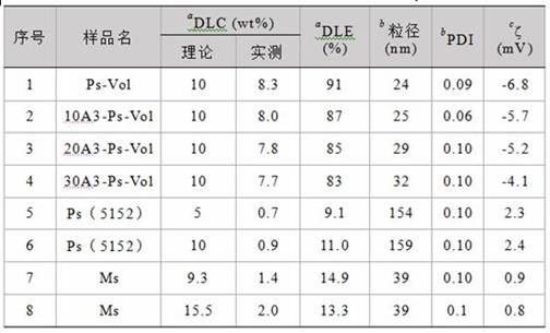
a UV-vis光谱测得。
b DLS测得 (25 °C)。
c 配备标准毛细管电泳池的Zetasizer
Nano-ZS (25 °C) 测得。
实施例三细胞层面的抗肿瘤活性和内吞研究:A3-Ps-Vol对SKOV3细胞的靶向性通过流式细胞仪和激光共聚焦显微镜CLSM来研究,以标记了FITC的Vol(Vol-FITC)为荧光探针。简要地,SKOV3细胞铺至6孔板中,每孔30万,过夜贴壁后,加入载Vol-FITC的不同A3密度的A3-Ps-Vol、Ps-Vol和自由Vol-FITC孵育4 h(Vol浓度为200 nM)。之后PBS清洗,胰酶消化、离心(1000 rpm,3 min),并重新分散在PBS中用于流式细胞仪测量。为了证明A3-Ps-Vol靶向的特异性,以DU145细胞为阴性对照。
CLSM实验中,SKOV3细胞被铺至12孔板中(含小圆片),每孔5万,过夜贴壁后,加入Ps-Vol-FITC或A3-Ps-Vol-FITC(Vol浓度为200 nM)孵育4 h。而后用罗丹明染色50 min,用4 %多聚甲醛固定细胞15 min,DAPI染细胞核5 min。每步之间用PBS清洗3次。有细胞的小圆片用于CLSM观察。另外,为了监测Cy5-Ps-Vol-FITC和Cy5-A3-Ps-Vol-FITC的细胞内吞情况,SKOV3细胞被分别与上述制剂孵育1、2、4、8、16小时,DAPI染色用CLSM观察。Cy5和FITC的荧光信号通过Image J软件半定量分析荧光强度和共定位。
PLK1蛋白的表达:SKOV3细胞铺于6孔板,50万每孔,过夜贴壁。加入A3-Ps-Vol、Ps-Vol或自由Vol(Vol浓度为200 nM)孵育4 h后,更换培养基继续孵育20 h。PBS清洗后,在冰浴中RIPA裂解15 min,12000 rpm离心15 min。取上清,通过Nanodrop测量蛋白浓度。每个样品调整到相同蛋白浓度后煮沸5 min,通过10%SDS-PAGE分离,后将蛋白转至PVDF膜上。5%的奶粉溶液用于封闭(1.5 h,25℃),之后与PLK1抗体和GAPDH抗体分别在4℃孵育过夜。后PVDF膜经TBST冲洗,与HRP二抗室温共孵育1.5 h。最终通过电化学发光法测定PLK1蛋白浓度。
细胞周期实验:SKOV3细胞铺于6孔板,每孔50万细胞,过夜贴壁后,加入A3-Ps-Vol、Ps-Vol、自由Vol(200 nM)孵育4 h后移除培养基,加入新鲜培养基培养44 h。然后消化、固定、-24℃存放过夜,再用碘化丙啶(PI)和RNA酶处理30 min用于流式细胞仪检测细胞周期。
细胞凋亡实验:与上述细胞周期实验条件相同,细胞凋亡实验也在200 nM和4+44 h的条件下进行。PBS、自由药Vol和Ps-Vol为对照组。细胞经不含EDTA的胰酶消化,并通过碘化丙啶(PI)和 Annexin V-Alexa Flour 647双染色,流式细胞仪检测。
MTT实验:SKOV3细胞铺于96孔板,3000每孔,贴壁生长24 h后,加入20 mL自由Vol、Ps-Vol或A3-Ps-Vol(表面A3密度分别为10%,20%和30%)。孔内终浓度从0.1 nM至10 μM。共孵育72 h后,加入10 μL MTT (5.0 mg/mL)3 h。移去上清,加入150 μL DMSO溶解20 min后即用酶标仪在570 nm测定,与PBS对照组(100%)相除得细胞存活率。DU145细胞为阴性对照。空囊泡的毒性通过聚合物在浓度为0.5、0.2、0.1、0.05 mg/mL下与SKOV-3细胞共同孵育72 h后测定MTT得到。
A3-Ps-Vol、Ps-Vol和自由Vol对SKOV-3细胞的抗肿瘤MTT实验结果表明,20%A3表面密度的A3-Ps-Vol有着最好的抗卵巢癌肿瘤活性,IC50为49 nM,分别比Ps-Vol和自由Vol低3.4 ~ 3.5倍(图3A)。以装载奥沙利铂Oxaliplatin或培美曲塞Pemetrexed的A3-Ps(制备方式同实施例二)作为对比例,进行上述同样的MTT实验,得到IC50分别为180 nM、和150 nM。可以看出,本发明A3-Ps-Vol比载其他两种药对SKOV-3细胞的毒性强。
细胞摄取实验通过细胞内的FITC-Vol的信号进行分析,不同组别的内吞量按以下顺序递减:20A3-Ps
> 10A3-Ps ≈ 30A3-Ps > Ps > 自由Vol(图3B)。其中20A3-Ps-Vol-FITC是Ps-Vol-FITC的2.3倍,自由Vol-FITC 3.3倍,而在阴性对照DU145细胞中,有无A3多肽在毒性实验和细胞内吞实验中并未显现出靶向差异性,证实了A3-Ps-Vol对SKOV-3细胞的特异性靶向(图3C, D)。蛋白印迹实验(图3E)展现了SKOV-3细胞的PLK1表达在A3-Ps-Vol(200 nM)孵育后大幅减少,而Ps-Vol和自由Vol仅有温和的作用,证明了A3-Ps-Vol对PLK1蛋白的特异性抑制能力。
细胞周期实验结果显示,和Vol制剂(200 nM)孵育后,SKOV-3细胞发生了显著的G2/M期阻滞和G1、S期的减少(图 4A)。A3-Ps-Vol将35%的细胞阻滞在G2/M期,是自由Vol的2.7倍。在图4B中,A3-Ps-Vol引发了显著的SKOV-3细胞晚凋(33.9%),是自由Vol和Ps-Vol的3.4倍。因此,A3-Ps-Vol可以比自由Vol更容易与PLK1的ATP结合域结合,调控细胞有丝分裂,扰乱纺锤体形成,阻滞细胞在G2/M期,同时,也使得肿瘤细胞中的PLK1失活,导致细胞凋亡。
为了更加直观地呈现A3-Ps的靶向效果,用Cy5标记的聚合物PEG-P(TMC-DTC)-Cy5、FITC标记的Vol制备囊泡,通过CLSM来研究A3的靶向性,以及跟踪载体和药物在SKOV-3细胞内的释放行为。图5A展示了孵育4 h后A3-Ps-Vol组相较于Ps-Vol有明显增强的细胞内Cy5和FITC信号。通过对Cy5和FITC定量分析表明,直至16 h,A3-Ps-Vol的靶向效果带来的内吞增强趋于稳定,是Ps-Vol的2倍。值得注意的是,随着孵育时间的延长,Cy5与FITC的共定位率在4 h的时候降至20%,说明了Vol在细胞内的快速释放 (图5B, C)。这些结果表明,A3-Ps-Vol有着明确的针对SKOV-3细胞的靶向性和增强的细胞内吞以及细胞内响应性释放。
实施例四活体成像和生物分布:为了研究A3-Ps-Vol的肿瘤富集,常规法制备的Cy5(ex. 649, em. 670)标记的载药囊泡Cy5-Ps-Vol和Cy5-A3-Ps-Vol,通过静脉注射至荷SKOV3皮下瘤裸鼠体内,0.06 mg Cy5 每只小鼠。SKOV3肿瘤模型通过右后背部皮下注射500万SKOV3细胞建立。在注射后的2、4、12、24、48小时,通过小动物活体成像系统(IVIS Lumina II)来追踪Cy5信号。另外,为了观察A3-Ps-Vol在肿瘤的穿透效果,在注射后的48 h,处死小鼠,取出肿瘤并用4%多聚甲醛固定,切片,并用DAPI染细胞核、用FITC标记的CD31染血管,通过激光共聚焦(LSM710,Leica)观察药物在肿瘤组织内的分布。
图 6A 展示了在肿瘤处逐渐增强的A3-Ps-Vol组的Cy5信号。12小时A3-Ps-Vol组表现出区别于Ps-Vol的显著优势,并且48 h的荧光强度是Ps-Vol组的2.5倍。相似的,离体的器官和肿瘤的荧光信号也证实了A3-Ps的靶向性。通过载Vol-FITC,正常组织和肿瘤组织内的Vol富集被可视化。A3-Ps-Vol在肿瘤部位的富集是Ps-Vol组的2倍(
图
6B)。另外,肿瘤切片显示,Cy5标记的A3-Ps-Vol在肿瘤内分布很广,而Ps-Vol则仅分布在肿瘤血管及其周围(
图
6C, D),表明了本发明纳米药物A3-Ps-Vol能够促进Vol的肿瘤穿透,不仅能增强SKOV-3细胞的摄取,还靶向肿瘤间质。对Vol的定量分析发现,SKOV-3肿瘤内Vol的富集量为4.8% ID/g,明显高于肝脏和肾等组织(
图
6E)。而Ps-Vol在肿瘤的富集则低了2倍,且在肾和肝脏富集更高。因此,A3-Ps-Vol在治疗多种恶性肿瘤方面存在潜力。
实施例五活体抗肿瘤实验:SKOV3肿瘤模型通过皮下注射500万SKOV3细胞建立;在接种后的第28天,荷瘤鼠被随机分为5组,每组5只(肿瘤体积为75 mm
3左右)。200 μL的自由Vol (10 mg
Vol/kg)、Ps-Vol (10 mg Vol/kg)、A3-Ps-Vol (10 or 20 mg Vol/kg) 和PBS通过尾静脉分别注射至裸鼠体内,该天被标记为第0天。以上制剂在第0,3,6,9,12,15天给药。肿瘤体积和体重每2天测量一次,肿瘤尺寸通过游标卡尺测量,体积经公式V=0.5×a×b
2计算得出(a为长,b为宽)。以初始体积和初始体重为标准计算相对值。在第17天处死小鼠,取出肿瘤,称重、拍照和进行病理分析。抑瘤率(TIR)通过以下公式计算:TIR(%)=(1 - 肿瘤治疗组重量/PBS组肿瘤重量)×100。
肿瘤和主要脏器肝、心、脾、肺、肾收集处理用于H&E染色。肿瘤切片也被用于TUNNEL分析,研究肿瘤细胞的凋亡情况。为了研究肿瘤内的PLK1的抑制情况,肿瘤切片通过PLK1抗体标记PLK1蛋白,再通过苏木精染色,最后通过显微镜观察。
结果实验(图7A)表明,A3-Ps-Vol(10 mg/kg)显著抑制了肿瘤的生长,优于Ps-Vol和自由Vol,证明了A3多肽在SKOV-3肿瘤治疗中的重要性。值得注意的是,剂量提高至20
mg/kg,A3-Ps-Vol的抑瘤效果进一步提升(*p
< 0.05),肿瘤体积降至初始治疗时的一半。Ps-Vol和自由Vol组均表现出部分的肿瘤抑制。令人兴奋的是,各组小鼠体重没有下降(图7B),说明A3-Ps-Vol不仅能够很好地抑制SKOV-3肿瘤的生长,同时避免了毒副作用。小鼠在第16天处死,离体肿瘤照片(图7C)清晰显示了A3-Ps-Vol(20 mg/kg)组有着最小的肿瘤尺寸,取得了最好的抗肿瘤效果。值得注意的是,A3-Ps-Vol在10和20 mg Vol/kg剂量下分别取得了80%和91%的抑瘤率(TIR),显著高于自由Vol(50%)和Ps-Vol(58%)(图7D)。
肿瘤切片通过H&E、免疫组化和TUNEL染色分析(图7E)。H&E的肿瘤切片结果和治疗效果一致。A3-Ps-Vol组肿瘤里显示出最多、最大范围的肿瘤坏死。免疫组化图片显示出PBS组有丰富的PLK1表达,而自由Vol和Ps-Vol组对PLK1产生了有效的抑制,A3-Ps-Vol组对PLK1的抑制更加显著。A3-Ps-Vol(20 mg Vol/kg)组不仅PLK1抑制得最佳,也导致了细胞凋亡,呈现了大量的松散零碎的肿瘤细胞核。TUNEL图片展示了A3-Ps-Vol比自由Vol和Ps-Vol能显著加强了肿瘤细胞的凋亡。
实施例六耐受实验:A3-Ps-Vol的耐受通过健康小鼠对制剂的反应观察。健康裸鼠通过尾静脉分别注射单剂量A3-Ps-Vol和自由Vol(50 mg Vol/kg),或6针 20 mg Vol/kg 每3天一针(第0,3,6,9,12,15天给药),每组3只。监测体重变化、行为变化和生存期来评估Vol和A3-Ps-Vol的毒性。在注射50 mg Vol/kg的24 h后,取血测试血常规,以观测volasertib和A3-Ps-Vol的血液学毒性。
图8A和图8B展示了A3-Ps-Vol的良好的耐受性,单剂量和多剂量模式中小鼠体重都基本不变,小鼠行为无异常;而自由Vol尽管在单剂量组有好的耐受,但在多剂量给药组中,5针后引发了小鼠急性的死亡。另外,
图
8C血常规结果表明,A3-Ps-Vol比自由Vol有更低的血液学毒性。因此,A3-Ps-Vol比自由Vol有更好的耐受。
在现有技术中,Vol带来有限的治疗效果和一定的毒副作用,比如现有文献[ROS-Responsive
Polymeric Micelles for Triggered Simultaneous Delivery of PLK1
Inhibitor/miR-34a and Effective Synergistic Therapy in Pancreatic Cancer]公开了载vol纳米粒的粒径为90 nm,表面电荷为35.5 ± 2.7 mV,其循环时间短,24h内肿瘤富集一直在下降;因而高效和低毒的A3-Ps-Vol是一种非常有前景的卵巢癌治疗方法,相比较而言,本发明的载药囊泡载药量高、尺寸小、表面电荷低,尤其是在体内的稳定性更好,且循环时间长,肿瘤富集多,在48 h内,肿瘤富集持续增长。本发明公开的靶向多肽修饰的囊泡(A3-Ps)可以递送Vol至裸鼠的SKOV-3皮下肿瘤,达到高效肿瘤抑制的同时减少副作用的疗效。这是第一篇针对卵巢癌靶向递送Vol的研究。该双靶向策略(囊泡的肿瘤靶向和Vol的分子靶向)是一种非常有前景的卵巢癌治疗方式:1.和传统药物相比,该药物有高的特异性和低的系统毒性;2,A3-Ps极大地改善了Vol的循环时间,肿瘤摄取和肿瘤穿透,巧妙地解决了Vol低肿瘤富集的问题。该Vol靶向递送系统A3-Ps还有可能解决临床中的耐药问题。因而该双靶纳米制剂在卵巢癌的靶向治疗中展现了应用潜力。
Claims (10)
- 载PLK1抑制剂的聚合物囊泡,其特征在于,所述载PLK1抑制剂的聚合物囊泡包括聚合物囊泡与PLK1抑制剂;所述聚合物囊泡由PEG-P(T-DTC)-PAsp制备,或者所述聚合物囊泡由PEG-P(T-DTC)-PAsp与A-PEG-P(T-DTC)制备。
- 根据权利要求1所述载PLK1抑制剂的聚合物囊泡,其特征在于,PLK1抑制剂为volasertib。
- 根据权利要求1所述载PLK1抑制剂的聚合物囊泡,其特征在于,PEG-P(T-DTC)-PAsp与A-PEG-P(T-DTC)中,PEG链段的分子量为2000~10000Da;疏水链段的总分子量为PEG链段分子量的2.5~10倍;疏水链段中PDTC链段的分子量占疏水链段总分子量的10%~35%;聚天冬氨酸的分子量为300~5000Da。
- 聚合物在制备载PLK1抑制剂的聚合物囊泡中的应用,或者在制备抗肿瘤药物中的应用;所述聚合物为PEG-P(T-DTC)-PAsp,或者所述聚合物为PEG-P(T-DTC)-PAsp与A-PEG-P(T-DTC)。
- 权利要求1所述载PLK1抑制剂的聚合物囊泡在制备抗肿瘤药物中的应用。
- 根据权利要求5所述的应用,其特征在于,所述肿瘤为卵巢癌。
- 权利要求1所述载PLK1抑制剂的聚合物囊泡的制备方法,其特征在于,将聚合物溶液与PLK1抑制剂溶液混合,然后孵育,再透析,得到载PLK1抑制剂的聚合物囊泡。
- 根据权利要求7所述载PLK1抑制剂的聚合物囊泡的制备方法,其特征在于,以环酯单体或者环碳酸酯单体作为第一单体、双硫环碳酸酯单体为第二单体,以聚乙二醇为引发剂,聚合得到PEG-P(T-DTC),再通过氯甲酸对硝基苯酯活化后与聚天冬氨酸偶联反应得到PEG-P(T-DTC)-PAsp;以环酯单体或者环碳酸酯单体作为第一单体、双硫环碳酸酯单体为第二单体,以带有活性端基的聚乙二醇为引发剂,聚合得到带有活性端基的PEG-P(T-DTC),再与靶向多肽反应得到A-PEG-P(T-DTC)。
- 根据权利要求8所述PLK1抑制剂的聚合物囊泡载的制备方法,其特征在于,T为酯单元或者碳酸酯单元;A为多肽靶向分子。
- 根据权利要求8所述载PLK1抑制剂的聚合物囊泡的制备方法,其特征在于,环酯单体为CL或者LA,环碳酸酯单体为TMC;双硫环碳酸酯单体为DTC,其化学结构式如下:。

Applications Claiming Priority (2)
| Application Number | Priority Date | Filing Date | Title |
|---|---|---|---|
| CN202110065127.1 | 2021-01-18 | ||
| CN202110065127.1A CN113018263A (zh) | 2021-01-18 | 2021-01-18 | 载plk1抑制剂的聚合物囊泡药物及其制备方法与应用 |
Publications (1)
| Publication Number | Publication Date |
|---|---|
| WO2022152298A1 true WO2022152298A1 (zh) | 2022-07-21 |
Family
ID=76459742
Family Applications (1)
| Application Number | Title | Priority Date | Filing Date |
|---|---|---|---|
| PCT/CN2022/072328 Ceased WO2022152298A1 (zh) | 2021-01-18 | 2022-01-17 | 载 plk1 抑制剂的聚合物囊泡药物及其制备方法与应用 |
Country Status (2)
| Country | Link |
|---|---|
| CN (1) | CN113018263A (zh) |
| WO (1) | WO2022152298A1 (zh) |
Cited By (1)
| Publication number | Priority date | Publication date | Assignee | Title |
|---|---|---|---|---|
| CN116474114A (zh) * | 2023-04-10 | 2023-07-25 | 苏州大学 | 一种载分子靶向药物聚合物囊泡及其制备方法与应用 |
Families Citing this family (2)
| Publication number | Priority date | Publication date | Assignee | Title |
|---|---|---|---|---|
| CN113018263A (zh) * | 2021-01-18 | 2021-06-25 | 苏州大学 | 载plk1抑制剂的聚合物囊泡药物及其制备方法与应用 |
| CN114917357B (zh) * | 2022-01-30 | 2024-04-26 | 苏州大学 | 转铁蛋白导向的装载plk1抑制剂的聚合物囊泡及其制备方法与应用 |
Citations (4)
| Publication number | Priority date | Publication date | Assignee | Title |
|---|---|---|---|---|
| CN110229323A (zh) * | 2019-05-31 | 2019-09-13 | 苏州大学 | 还原敏感可逆交联的具有不对称膜结构的聚合物囊泡及其在制备治疗肝癌药物中的应用 |
| CN111714457A (zh) * | 2020-06-22 | 2020-09-29 | 苏州大学 | 载小分子药物的碳酸酯聚合物囊泡及其制备方法与应用 |
| CN112076159A (zh) * | 2020-09-14 | 2020-12-15 | 苏州大学 | 具有不对称膜结构的载药聚合物囊泡及制备方法与在制备治疗急性髓系白血病药物中的应用 |
| CN113018263A (zh) * | 2021-01-18 | 2021-06-25 | 苏州大学 | 载plk1抑制剂的聚合物囊泡药物及其制备方法与应用 |
-
2021
- 2021-01-18 CN CN202110065127.1A patent/CN113018263A/zh active Pending
-
2022
- 2022-01-17 WO PCT/CN2022/072328 patent/WO2022152298A1/zh not_active Ceased
Patent Citations (4)
| Publication number | Priority date | Publication date | Assignee | Title |
|---|---|---|---|---|
| CN110229323A (zh) * | 2019-05-31 | 2019-09-13 | 苏州大学 | 还原敏感可逆交联的具有不对称膜结构的聚合物囊泡及其在制备治疗肝癌药物中的应用 |
| CN111714457A (zh) * | 2020-06-22 | 2020-09-29 | 苏州大学 | 载小分子药物的碳酸酯聚合物囊泡及其制备方法与应用 |
| CN112076159A (zh) * | 2020-09-14 | 2020-12-15 | 苏州大学 | 具有不对称膜结构的载药聚合物囊泡及制备方法与在制备治疗急性髓系白血病药物中的应用 |
| CN113018263A (zh) * | 2021-01-18 | 2021-06-25 | 苏州大学 | 载plk1抑制剂的聚合物囊泡药物及其制备方法与应用 |
Non-Patent Citations (3)
| Title |
|---|
| WANG ZHE, ZHAO SONGSONG, GU WENXING, DONG YANGYANG, MENG FENGHUA, YUAN JIANDONG, ZHONG ZHIYUAN: "Alpha 3 Integrin Binding Peptide-Functionalized Polymersomes Loading PLK1 Inhibitor Volasertib for Dually Targeted Molecular Therapy of Ovarian Cancer <i>In vivo</i>", SSRN ELECTRONIC JOURNAL, vol. 1, 12 November 2020 (2020-11-12), XP055952053, DOI: 10.2139/ssrn.3729648 * |
| WANG, ZHE ET AL.: "α3 Integrin-binding Peptide-Functionalized Polymersomes Loaded with Volasertib for Dually-Targeted Molecular Therapy for Ovarian Cancer", ACTA BIOMATERIALIA, vol. 124, 6 February 2021 (2021-02-06), XP086524356, DOI: 10.1016/j.actbio.2021.02.007 * |
| ZHOU, CHENG ET AL.: "GE11 Peptide-installed Chimaeric Polymersomes Tailor-made for High-efficiency EGFR-targeted Protein Therapy of Orthotopic Hepatocellular Carcinoma", ACTA BIOMATERIALIA, vol. 113, 18 June 2020 (2020-06-18), XP086239091, DOI: 10.1016/j.actbio.2020.06.020 * |
Cited By (1)
| Publication number | Priority date | Publication date | Assignee | Title |
|---|---|---|---|---|
| CN116474114A (zh) * | 2023-04-10 | 2023-07-25 | 苏州大学 | 一种载分子靶向药物聚合物囊泡及其制备方法与应用 |
Also Published As
| Publication number | Publication date |
|---|---|
| CN113018263A (zh) | 2021-06-25 |
Similar Documents
| Publication | Publication Date | Title |
|---|---|---|
| Niu et al. | A novel chitosan-based nanomedicine for multi-drug resistant breast cancer therapy | |
| Gu et al. | Nanotechnology-mediated immunochemotherapy combined with docetaxel and PD-L1 antibody increase therapeutic effects and decrease systemic toxicity | |
| Li et al. | Sarcoma-targeting peptide-decorated polypeptide nanogel intracellularly delivers shikonin for upregulated osteosarcoma necroptosis and diminished pulmonary metastasis | |
| Song et al. | Erythrocyte-biomimetic nanosystems to improve antitumor effects of paclitaxel on epithelial cancers | |
| Ray et al. | PEG-b-poly (carbonate)-derived nanocarrier platform with pH-responsive properties for pancreatic cancer combination therapy | |
| Luo et al. | Co-delivery of paclitaxel and STAT3 siRNA by a multifunctional nanocomplex for targeted treatment of metastatic breast cancer | |
| Belhadj et al. | Multifunctional targeted liposomal drug delivery for efficient glioblastoma treatment | |
| Liu et al. | An eximious and affordable GSH stimulus-responsive poly (α-lipoic acid) nanocarrier bonding combretastatin A4 for tumor therapy | |
| Mei et al. | Effective treatment of the primary tumor and lymph node metastasis by polymeric micelles with variable particle sizes | |
| Luo et al. | LyP-1-conjugated nanoparticles for targeting drug delivery to lymphatic metastatic tumors | |
| Yu et al. | Mitochondrial targeting topotecan-loaded liposomes for treating drug-resistant breast cancer and inhibiting invasive metastases of melanoma | |
| Huang et al. | Bridging the gap between macroscale drug delivery systems and nanomedicines: a nanoparticle-assembled thermosensitive hydrogel for peritumoral chemotherapy | |
| Chen et al. | KE108-conjugated unimolecular micelles loaded with a novel HDAC inhibitor thailandepsin-A for targeted neuroendocrine cancer therapy | |
| Zhang et al. | Bovine serum albumin-based and dual-responsive targeted hollow mesoporous silica nanoparticles for breast cancer therapy | |
| Shukla et al. | Multifunctional hybrid nanoconstructs facilitate intracellular localization of doxorubicin and genistein to enhance apoptotic and anti-angiogenic efficacy in breast adenocarcinoma | |
| Haggag et al. | Repurposing of Guanabenz acetate by encapsulation into long-circulating nanopolymersomes for treatment of triple-negative breast cancer | |
| WO2022152298A1 (zh) | 载 plk1 抑制剂的聚合物囊泡药物及其制备方法与应用 | |
| WO2019204799A1 (en) | Cationic amphiphilic polymers for codelivery of hydrophobic agents and nucleic acids | |
| Wang et al. | Folate-mediated targeted PLK1 inhibition therapy for ovarian cancer: a comparative study of molecular inhibitors and siRNA therapeutics | |
| Ma et al. | Multi-functionalized dendrimers for targeted co-delivery of sorafenib and paclitaxel in liver cancers | |
| Wang et al. | Prodrug nanoparticles rationally integrating stroma modification and chemotherapy to treat metastatic pancreatic cancer | |
| JP7164205B2 (ja) | キナ酸修飾ナノ粒子およびその使用 | |
| Chen et al. | Synergistic antitumor efficacy of doxorubicin and gambogic acid-encapsulated albumin nanocomposites | |
| Gaber et al. | Co-administration of tretinoin enhances the anti-cancer efficacy of etoposide via tumor-targeted green nano-micelles | |
| Yuan et al. | Regulating tumor-associated macrophage polarization by cyclodextrin-modified PLGA nanoparticles loaded with R848 for treating colon cancer |
Legal Events
| Date | Code | Title | Description |
|---|---|---|---|
| 121 | Ep: the epo has been informed by wipo that ep was designated in this application |
Ref document number: 22739173 Country of ref document: EP Kind code of ref document: A1 |
|
| NENP | Non-entry into the national phase |
Ref country code: DE |
|
| 122 | Ep: pct application non-entry in european phase |
Ref document number: 22739173 Country of ref document: EP Kind code of ref document: A1 |