WO2022124245A1 - 腱または靭帯移植用の移植材料 - Google Patents
腱または靭帯移植用の移植材料 Download PDFInfo
- Publication number
- WO2022124245A1 WO2022124245A1 PCT/JP2021/044608 JP2021044608W WO2022124245A1 WO 2022124245 A1 WO2022124245 A1 WO 2022124245A1 JP 2021044608 W JP2021044608 W JP 2021044608W WO 2022124245 A1 WO2022124245 A1 WO 2022124245A1
- Authority
- WO
- WIPO (PCT)
- Prior art keywords
- graft
- tendon
- ligament
- cell sheet
- transplant material
- Prior art date
- Legal status (The legal status is an assumption and is not a legal conclusion. Google has not performed a legal analysis and makes no representation as to the accuracy of the status listed.)
- Ceased
Links
Images





Classifications
-
- A—HUMAN NECESSITIES
- A61—MEDICAL OR VETERINARY SCIENCE; HYGIENE
- A61L—METHODS OR APPARATUS FOR STERILISING MATERIALS OR OBJECTS IN GENERAL; DISINFECTION, STERILISATION OR DEODORISATION OF AIR; CHEMICAL ASPECTS OF BANDAGES, DRESSINGS, ABSORBENT PADS OR SURGICAL ARTICLES; MATERIALS FOR BANDAGES, DRESSINGS, ABSORBENT PADS OR SURGICAL ARTICLES
- A61L27/00—Materials for grafts or prostheses or for coating grafts or prostheses
- A61L27/36—Materials for grafts or prostheses or for coating grafts or prostheses containing ingredients of undetermined constitution or reaction products thereof, e.g. transplant tissue, natural bone, extracellular matrix
-
- A—HUMAN NECESSITIES
- A61—MEDICAL OR VETERINARY SCIENCE; HYGIENE
- A61L—METHODS OR APPARATUS FOR STERILISING MATERIALS OR OBJECTS IN GENERAL; DISINFECTION, STERILISATION OR DEODORISATION OF AIR; CHEMICAL ASPECTS OF BANDAGES, DRESSINGS, ABSORBENT PADS OR SURGICAL ARTICLES; MATERIALS FOR BANDAGES, DRESSINGS, ABSORBENT PADS OR SURGICAL ARTICLES
- A61L27/00—Materials for grafts or prostheses or for coating grafts or prostheses
- A61L27/36—Materials for grafts or prostheses or for coating grafts or prostheses containing ingredients of undetermined constitution or reaction products thereof, e.g. transplant tissue, natural bone, extracellular matrix
- A61L27/38—Materials for grafts or prostheses or for coating grafts or prostheses containing ingredients of undetermined constitution or reaction products thereof, e.g. transplant tissue, natural bone, extracellular matrix containing added animal cells
-
- A—HUMAN NECESSITIES
- A61—MEDICAL OR VETERINARY SCIENCE; HYGIENE
- A61L—METHODS OR APPARATUS FOR STERILISING MATERIALS OR OBJECTS IN GENERAL; DISINFECTION, STERILISATION OR DEODORISATION OF AIR; CHEMICAL ASPECTS OF BANDAGES, DRESSINGS, ABSORBENT PADS OR SURGICAL ARTICLES; MATERIALS FOR BANDAGES, DRESSINGS, ABSORBENT PADS OR SURGICAL ARTICLES
- A61L27/00—Materials for grafts or prostheses or for coating grafts or prostheses
- A61L27/40—Composite materials, i.e. containing one material dispersed in a matrix of the same or different material
-
- A—HUMAN NECESSITIES
- A61—MEDICAL OR VETERINARY SCIENCE; HYGIENE
- A61L—METHODS OR APPARATUS FOR STERILISING MATERIALS OR OBJECTS IN GENERAL; DISINFECTION, STERILISATION OR DEODORISATION OF AIR; CHEMICAL ASPECTS OF BANDAGES, DRESSINGS, ABSORBENT PADS OR SURGICAL ARTICLES; MATERIALS FOR BANDAGES, DRESSINGS, ABSORBENT PADS OR SURGICAL ARTICLES
- A61L27/00—Materials for grafts or prostheses or for coating grafts or prostheses
- A61L27/50—Materials characterised by their function or physical properties, e.g. injectable or lubricating compositions, shape-memory materials, surface modified materials
- A61L27/54—Biologically active materials, e.g. therapeutic substances
Definitions
- the present invention relates to a transplant material for tendon or ligament transplantation.
- Anterior cruciate ligament ligament reconstruction is an operation to restore the function of the damaged ligament by replacing the torn anterior cruciate ligament with a graft.
- the torn ligament rarely heals spontaneously, and ACL reconstruction is essential for athletes and the like to return to sports.
- ACL reconstruction autologous tendon transplantation is performed, and many top-level players such as soccer, rugby, American football, and basketball can return after surgery and play the same role as before the injury.
- ADSC shows a tendency to differentiate into osteochondral cartilage and 2,24,27,51 according to the environment, and shows a tendency to differentiate into tendon cells 22,26 , but it is different as a side effect.
- the possibility of spontaneous ossification has also been reported8.
- Kosaka's and Zhang applied the ADSC suspension to ACL reconstruction, and both reported that although injection was the method of transplanting ADSC, good results were obtained 21,50 .
- the present invention provides a transplant material for tendon or ligament transplantation.
- the present invention also provides a transplant material comprising a cell sheet containing ADSC.
- the present inventors increased factors that promote ADSC engraftment inside the graft, engraftment in surrounding tissues, and tissue regeneration.
- To increase the number, thickness, and density of collagen fibers in the graft to promote calcification of the bone in contact with the ADSC of the graft, to suppress the enlargement of the bone hole after graft transplantation, and to suppress the enlargement of the bone hole after graft transplantation. It has been found to increase maximum breaking load and rigidity.
- the present invention is based on such findings.
- a transplant material for reconstructing a tendon or a ligament which comprises a cell sheet containing adipose-derived stem cells.
- the transplant material according to the above [1] which comprises the layered cell sheet.
- the transplant material described in the crab comprises 90% or more of the cells positive for CD29, CD44, CD73, CD90, CD105, and CD166 and 5% or less of the cells positive for CD14, CD31, CD45 and Lin1.
- the transplant material according to any one of [1] to [7].
- a product comprising a container containing the transplant material according to any one of the above [1] to [8].
- the combination according to [10] above, wherein the surface is the surface of the joint between the tendon or ligament of the graft and the bone.
- Transplant material including defined cell sheets.
- the cell sheet defined in any of the above [1] to [8] is contained, which is transplanted in a state of being attached to the transplanted graft and thereby used for suppressing the rupture of the graft after transplantation.
- Transplant material [18] The transplant material according to any one of the above [1] to [8], further comprising a transplant graft.
- a method of reconstructing a tendon or ligament in a subject in need thereof. (I) Containing the fixation of the graft to the site in which the tendon or ligament needs to be reconstructed in the subject, where the fixed graft has a cell sheet containing adipose-derived stem cells on its surface or
- the graft comprises fixing the graft to the site where the tendon or ligament needs to be reconstructed in the subject, and attaching a cell sheet containing adipose-derived stem cells to the surface of the fixed graft.
- a method of reconstructing a tendon or ligament in a subject in need thereof comprising immobilizing a graft at a site in a subject in which a tendon or ligament needs to be reconstructed, wherein the immobilized graft has a cell sheet containing adipose-derived stem cells on its surface.
- a method of strengthening a reconstructed tendon or ligament in a subject Including attaching a cell sheet containing adipose-derived stem cells to the surface of a graft immobilized at a site that needs to be reconstructed, Method.
- the implantable material of the present invention may reduce the need for reoperation.
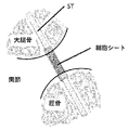
- FIG. 1 shows the installation state of the semitendinosus tendon (ST) in the anterior cruciate ligament ligament reconstruction.
- the center of ST is covered with a cell sheet.
- the cell sheet is also coated on the joint surface with the bone in ST.
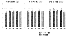
- FIG. 2 shows changes over time in rabbit weight, graft length, and graft diameter after transplantation. "W” in the graph indicates the number of weeks elapsed after surgery.
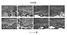
- FIG. 3 shows the results of histological evaluation of the graft after transplantation.
- “B” represents bone
- "G” represents graft
- arrowheads represent sharpy-like fibers, chondrocytes, and layered bone.
- FIG. 4 shows the score transition after the tendon-bone healing.
- FIG. 4 shows the score transition after the tendon-bone healing.
- FIG. 5 is a fluorescence-stained image showing that transplanted adipose-derived stem cells (ADSC) migrate and engraft in the tissue around the graft. ADSC was fluorescently stained prior to transplantation.
- FIG. 6 shows that transplanted adipose-derived stem cells (ADSC) migrate and engraft inside the graft after transplantation. ADSC was fluorescently stained prior to transplantation.
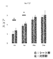
- FIG. 7 shows the time course of the area of the bone hole prepared at the time of graft transplantation after transplantation.
- FIG. 8 shows the change over time in the amount of bone mineral (volume ratio) with respect to the total tissue after graft transplantation.
- FIG. 8 shows the volume ratio when the portion having a calcification degree of 400 mg / ml or more is regarded as bone, and the right panel of FIG. 8 shows the volume when the portion having a calcification degree of 800 mg / ml or more is regarded as bone. Shows the ratio.
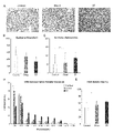
- FIG. 9 shows an electron micrograph of a cross section of the graft after transplantation (panel A), as well as the number of corasen fibers (panel B), thickness (panel C) and thickness distribution (panel E), and fiber density (panel E). E) is shown.
- the upper panel of FIG. 10 is a photograph showing the state of a biomechanical experiment. Panel A in FIG. 10 shows the maximum breaking load (N) of the graft after transplantation. Panel B in FIG.
- FIG. 10 shows the stiffness value (N / mm) of the graft after transplantation.
- FIG. 11 shows the mode of rupture.
- the left panel shows the pattern of the graft coming out of the tibia
- the central panel shows the pattern of the graft tearing in the center
- the right panel shows the pattern of the graft coming out of the femur.
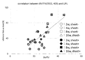
- FIG. 12 shows the relationship between the bone mineral content (volume ratio) and the maximum breaking load with respect to the total tissue after graft transplantation.
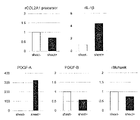
- FIG. 13 shows the mRNA expression of various genes in the graft.
- FIG. 14 shows the mRNA expression of various genes in the graft.
- a "subject” is a vertebrate, eg, a bird or mammal, eg, a mammal, eg, a mouse, rat, hamster, guinea pig, horse, cow, pig, goat, sheep, donkey, dog. , And primates such as monkeys, chimpanzees, gorillas, orangoutans, bonovos, and humans, especially humans.
- the subject is used in the sense of including humans as described above, and the term “non-human” is used when excluding humans.
- the "subject” is transplanted with a graft.
- treatment means medically intervening in a subject. Treatment is used to include therapeutic treatment. Therapeutic treatment can elicit therapeutic effects such as amelioration of disease, disability and condition, suppression of exacerbations, reduction of rate of exacerbations, arrest, and cure. As used herein, the "therapeutically effective amount” is an amount that produces a therapeutic effect.
- the term "transplant material” means a biomaterial used for transplantation into the body of a subject.
- the biomaterial may be an artificial material, a natural material, or a processed product of a natural material.
- the transplant material is preferably biocompatible.
- the transplant material can be autologous or allogeneic.
- Cell sheets containing adipose-derived stem cells can be autologous or allogeneic to the subject to be transplanted.
- implant material does not necessarily require the presence of a graft.
- the implant material may include a graft. That is, the transplant material may comprise a cell sheet or may further comprise a graft in addition to the cell sheet.
- adipose-derived stem cells are cells isolated from adipose tissue and capable of differentiating into multiple mature cells, including adipose cells, osteoblasts and cartilage cells. be.
- the adipose-derived stem cells can be human adipose-derived stem cells.
- Fat-derived stem cells can express one or all selected from the group consisting of CD29, CD44, CD73, CD90, CD105, and CD166 (or can be positive in one or more).
- Fat-derived stem cells can be one that does not express one or all selected from the group consisting of CD14, CD31, CD45 and Lin1 (or can be negative in one or more or all).
- the "cell sheet” is a structure having a sheet shape composed of a single layer of cells.
- the cells are usually cultured in a single sheet shape on the solid phase surface. Breaking cell-cell adhesion causes cells to dissociate to the single-cell level, but recovery without breaking (or completely breaking) cell-cell adhesion gives a cell sheet.
- the disruption of cell-cell adhesion is carried out by treatment with a protease such as trypsin.
- a temperature-responsive culture dish can be used to recover the cell sheet without destroying cell-cell adhesion.
- Multiple cell sheets can be used in layers (ie, layered).
- extracellular matrix is an insoluble substance that exists outside the cell in vivo or in vitro.
- the extracellular matrix may contain adhesive proteins involved in cell adhesion. Adhesive proteins can be secreted to the cell surface during cell culture. In the cell sheet, this adhesive protein can be maintained on the sheet surface. This allows the cell sheet to maintain adhesion to other cells. Cell sheets that maintain adhesiveness have increased engraftment to tissues and organs and may be useful for transplantation applications.
- a "graft" is a tissue to be transplanted or a part thereof. Grafts are also called grafts. For example, in the reconstruction of the anterior cruciate ligament ligament, tendon or ligament grafts such as the hamstring tendon (including the hemitendonoidal tendon), the patellar tendon, and the iliotibial band can be used.
- the graft is, in one preferred embodiment, an autologous graft to the subject.
- the graft is, in one preferred embodiment, an allogeneic graft to the subject.
- a cell sheet containing adipose-derived stem cells is provided.
- INDUSTRIAL APPLICABILITY a transplant material including a cell sheet containing adipose-derived stem cells is provided.
- INDUSTRIAL APPLICABILITY there is provided a transplant material for reconstructing a tendon or a ligament, which comprises a cell sheet containing adipose-derived stem cells.
- the cell sheet containing the adipose-derived stem cells of the present invention contains, for example, 80% or more, 85% or more, 90% or more or 95% or more of cells positive for CD29, CD44, CD73, CD90, CD105, and CD166.
- the cell sheet containing the adipose-derived stem cells of the present invention contains, for example, 10% or less, 9% or less, 8% or less, 7% or less, 6% or less of cells positive for any of CD14, CD31, CD45 and Lin1. Includes 5% or less, 4% or less, 3% or less, or 2% or less.
- the cell sheet containing the adipose-derived stem cells of the present invention comprises, for example, 80% or more, 85% or more, 90 of cells positive for 1 or more selected from the group consisting of CD29, CD44, CD73, CD90, CD105, and CD166. 10% or less, 9% or less, 8% or less, 7% or less, 1 or more positive cells selected from the group consisting of CD14, CD31, CD45 and Lin1 containing% or more or 95% or more. Includes 6% or less, 5% or less, 4% or less, 3% or less, or 2% or less.
- the cell sheet containing the adipose-derived stem cells of the present invention contains, for example, 90% or more of cells positive for CD29, CD44, CD73, CD90, CD105, and CD166, and any of CD14, CD31, CD45, and Lin1. Contains 5% or less of positive cells.
- the cell sheet containing the adipose-derived stem cells of the present invention contains, for example, 95% or more of cells positive for CD29, CD44, CD73, CD90, CD105, and CD166, and any of CD14, CD31, CD45, and Lin1. Contains 2% or less of positive cells.
- the adipose-derived stem cells can be positive for CD29, CD44, CD73, CD90, CD105, and CD166 and negative for any of CD14, CD31, CD45, and Lin1.
- the percentage is calculated based on the number of cells. Whether each factor is positive or negative can be determined by flow cytometry using an antibody ligated with a fluorescent dye. By comparing with one or both of the positive control cells and the negative control cells, the signal intensity in the negative case and the positive case can be grasped. By analyzing cells by flow cytometry, it is possible to determine whether they are positive or negative depending on whether they are negative or show strong fluorescence that is significantly different from negative.
- the cell sheet containing the adipose-derived stem cells of the present invention can be used by adhering to the surface of the transplant graft in the form of a sheet.
- the cell sheet containing the adipose-derived stem cells of the present invention can be used by adhering to the surface of a tendon graft or a ligament graft in the form of a sheet. Therefore, the cell sheet containing the adipose-derived stem cells of the present invention can be a transplant material used for tendon or ligament reconstruction. It is not necessary to use an adhesive (for example, fibrin glue) to attach the cell sheet to the surface of the transplant graft.
- an adhesive for example, fibrin glue
- the cell sheet containing the adipose-derived stem cells of the present invention can be used by adhering to the surface of a tendon or ligament graft, which has the effect of preventing the expansion of the bone hole in contact with the graft and improving the bone density. ..
- a tendon or ligament graft which has the effect of preventing the expansion of the bone hole in contact with the graft and improving the bone density. ..
- the cell sheet containing the adipose-derived stem cells of the present invention to the vicinity of the bone pore in contact with the graft, the expansion of the bone pore after graft transplantation can be prevented and the improvement of bone density can be induced. It can also promote bone formation with a lamellar structure at the tendon-bone junction.
- the cell sheet containing the adipose-derived stem cells of the present invention can be used by adhering to the surface serving as the junction with the bone of the graft.
- the cell sheet containing the adipose-derived stem cells of the invention adheres to the surface of the graft to be the junction with bone, in order to prevent enlargement of the bone hole after graft transplantation or to induce an increase in bone density.
- the cell sheet containing the adipose-derived stem cells of the present invention can preferably adhere to the graft surface in the form of a sheet.
- the cell sheet containing the adipose-derived stem cells of the present invention can be used by adhering it to the surface of a tendon or ligament graft in the form of a sheet, whereby (1) the effect of promoting the production of type 1 collagen and (2) type 3 It can exert an effect of suppressing collagen production, an effect of (3) an effect of increasing a collagen fiber diameter (for example, an average diameter), and an effect of (4) an effect of improving the occupancy rate of collagen fibers in a collagen fiber cross section. Therefore, the cell sheet containing the adipose-derived stem cells of the present invention can be used by adhering to a surface other than the junction with the bone of the graft.
- the cell sheet containing the adipose-derived stem cells of the present invention is used for (1) promoting the production of type 1 collagen, (2) using it for suppressing the production of type 3 collagen, and (3) collagen. It is used for one or more applications selected from the group consisting of an application for increasing a fiber diameter (for example, an average diameter) and an application for improving the occupation rate of collagen fibers in a collagen fiber cross section. For the purpose, it can be used by adhering it to a surface other than the joint portion of the graft with the bone. Therefore, according to the present invention, the cell sheet containing the adipose-derived stem cells of the present invention or the transplant material containing the cell sheet can be used to promote the production of (1) type 1 collagen in the graft.
- the cell sheet containing the adipose-derived stem cells of the present invention or the transplant material containing the cell sheet can be used to suppress the production of (2) type 3 collagen in the graft.
- the cell sheet containing the adipose-derived stem cells of the present invention or the transplant material containing the cell sheet can be used in grafting to (3) increase the collagen fiber diameter (for example, the average diameter).
- the cell sheet containing the adipose-derived stem cells of the present invention or the transplant material containing the cell sheet can be used in grafting to (4) improve the occupancy of collagen fibers in the collagen fiber cross section.
- the cell sheet containing the adipose-derived stem cells of the present invention can preferably adhere to the graft surface in the form of a sheet.
- the cell sheet containing the adipose-derived stem cells of the present invention can be used to suppress detachment from the tibia, detachment from the femur, and rupture of the graft after transplantation.
- the cell sheet containing the adipose-derived stem cells of the present invention can be preferably used to suppress the rupture of the graft after transplantation. Thereby, the cell sheet containing the adipose-derived stem cells of the present invention can reduce the need for reoperation after graft transplantation (for example, after ACL reconstruction).
- the cell sheet containing the adipose-derived stem cells of the present invention can be layered and attached. By doing so, it is considered that the physiological action of the cell sheet is enhanced.
- the layering can be performed by superimposing two or more sheets.
- the layering can be performed by stacking two to ten layers, two to five layers, or two or three layers of sheets.
- the cell sheet containing the adipose-derived stem cells of the present invention can be used by being attached to the surface of a tendon or ligament graft in the form of a sheet, but the attachment can be performed so as to cover the surface of the graft. Adhesion can also be done by wrapping the sheet around the graft surface. Adhesion can also be performed by wrapping a layered sheet around the graft surface. The layering can be from 2 layers to several layers, for example, 3 layers, 4 layers, 5 layers, 6 layers or more.
- the cell sheet containing the adipose-derived stem cells of the invention can be provided in a sheet-like manner attached to the surface of a tendon or ligament graft.
- the cell sheet containing the adipose-derived stem cells of the present invention may be provided in combination with a tendon or ligament graft.
- the cell sheet containing the adipose-derived stem cells of the invention is attached to the surface of the junction of the tendon or ligament graft with bone.
- the cell sheet containing the adipose-derived stem cells of the invention is attached to a surface other than the bone junction of a tendon or ligament graft.
- Fat-derived stem cells can be isolated from aspirated adipose tissue. Separation and purification of adipose-derived stem cells from adipose tissue can be carried out with reference to previously reported known methods. For example, the method described in the literature Arterioscler Thromb Vasc Biol. 29: 1723-1729, 2009 can be used. Alternatively, commercially available human adipose-derived stem cells can be used. Fat-derived stem cells can be human cells. Fat-derived stem cells can be autologous cells. Fat-derived stem cells can be allogeneic cells. Human adipose-derived stem cells can be cells established for medical purposes and quality-controlled (for example, autologous cells, particularly allogeneic cells).
- Fat-derived stem cells can be cultured by a method well known to those skilled in the art.
- the culture can be an adhesive culture.
- Cell sheets can be prepared by various methods well known to those skilled in the art. For example, cell sheets can be made using a temperature responsive culture dish. The culture surface of the temperature-responsive culture dish is coated with the temperature-responsive polymer.
- the temperature-responsive polymer is hydrophobic at normal culture temperatures (eg, about 37 ° C.), but changes to hydrophilicity in a temperature-dependent manner as the temperature is lowered.
- the temperature at which the temperature changes from hydrophobic to hydrophilic or from hydrophilic to hydrophobic is called the lower limit critical solution temperature (LCST).
- LCST lower limit critical solution temperature
- the sheet can be collected using a support.
- the support is not particularly limited, and examples thereof include a non-woven fabric sheet, for example, a polymer-coated polylactic acid non-woven fabric sheet and the like.
- the temperature-responsive polymer examples include poly (N-alkylacrylamide), poly (N-vinylalkylamide), polyvinylalkyl ether, and polyethylene glycol / polypropylene glycol block copolymers.
- poly (N-isopropylacrylamide) (PNIPAAm) and poly (2-alkyl-2-oxazoline) can be preferably used.
- various methods for controlling LCST for example, a method for copolymerizing n-butyl methacrylate and a method for copolymerizing N, N-dimethylacrylamide
- a desired LCST for example, 10 ° C.
- a polymer having ⁇ 35 ° C. can be obtained. It is not necessary to use a proteolytic enzyme such as trypsin to withdraw the cell sheet from the temperature-responsive culture dish, and by not using the proteolytic enzyme, the cell sheet retains an extracellular matrix such as adhesive protein on the surface. Can be recovered in the same state.
- a proteolytic enzyme such as trypsin
- the cell sheet containing the adipose-derived stem cells of the present invention can be provided in a state of being placed on a support.
- the cell sheet containing the adipose-derived stem cells of the present invention may be encapsulated in a transport container.
- the cell sheet containing the adipose-derived stem cells of the present invention may be encapsulated in a transport container in a state of being placed on a support.
- the transport container can introduce the culture solution in a liquid-tight manner, and preferably, the culture solution can be introduced in a liquid-tight manner without containing air bubbles.
- the graft is a transplant material.
- the graft can be a transplant material that can be used for tendon reconstruction and / or ligament reconstruction. Grafts used for such purposes are referred to as "tendon or ligament reconstruction grafts".
- the cell sheet containing the adipose-derived stem cells of the present invention can be used by adhering to the surface of the graft. By doing so, various advantageous effects as described above can be obtained.
- Various tendon or ligament reconstruction grafts can be used.
- a tendon or ligament graft such as a hamstring tendon (semitendinosus tendon), a patellar tendon, and an iliotibial band can be used.
- a cell sheet containing the adipose-derived stem cells of the present invention can be used in combination with these grafts (attached, for example, attached in the form of a sheet).
- the graft can be an autologous graft. This is because autologous grafts are unlikely to cause immune-related problems such as immune rejection.
- the graft can be implanted in the injured part of the tendon or ligament. Reconstruction of tendons or ligaments can be performed by a physician as appropriate.
- a cell sheet containing the adipose-derived stem cells of the present invention can be attached to a graft (for example, attached in the form of a sheet) and then used for reconstruction of a tendon or ligament (for example, reconstruction of the anterior cruciate ligament).
- the damaged tendon or ligament can be excised and, as an alternative, a graft can be transplanted to reconstruct the tendon or ligament.
- a bone hole can be created to secure the graft to the tibia and femur, and a graft with a cell sheet attached can be inserted inside the bone hole.
- Fixation of the graft to the bone can be performed by suturing the periosteum and the graft or fixing with an implant.
- the cell sheet can be attached to the graft so that the cell sheet enters the bone hole, as was performed in the examples.
- a cell sheet containing the adipose-derived stem cells of the present invention can be further attached to the graft (attached to the surface in the form of a sheet or wrapped) after these procedures and before closing the wound. This is because it is considered that the effect of the cell sheet can be enhanced. Therefore, the cell sheet containing the adipose-derived stem cells of the present invention can also be used for attachment to a graft immobilized on a recipient after transplantation. After transplantation, the recipient can be managed in the same manner as after normal tendon and ligament reconstruction.
- a recipient is an individual (eg, a human) to whom a graft is transplanted.
- the cell sheet containing the adipose-derived stem cells of the present invention may be provided in a state of being attached to a graft.
- the cell sheet containing the adipose-derived stem cells of the present invention may be packaged in one container separately from the graft.
- the cell sheet containing the adipose-derived stem cells of the present invention can be provided alone.
- the present invention provides a method of reconstructing a tendon or ligament in a subject in need thereof, comprising fixing a graft to a site in which the tendon or ligament needs to be reconstructed.
- the present invention is a method of reconstructing a tendon or ligament in a subject in need thereof, comprising fixing the graft to a site in which the tendon or ligament needs to be reconstructed, wherein the fixed graft is:
- a method is provided having a cell sheet containing adipose-derived stem cells on the surface. The method of the present invention can be carried out in the reconstruction of the anterior cruciate ligament.
- Reconstruction of the anterior cruciate ligament may include attaching a cell sheet containing the adipose-derived stem cells of the invention to the graft before or after, or before and after fixation.
- the method of the present invention comprises attaching (eg, wrapping) the cell sheet to the graft before fixing the graft, or attaching (eg, wrapping) the cell sheet to the graft after fixing the graft. It may be further included.
- Transplant materials containing cell sheets containing adipose-derived stem cells can be used in such methods.
- Transplant materials containing cell sheets containing adipose-derived stem cells can be used by attaching or wrapping them on a graft.
- the present invention provides the use of a cell sheet containing adipose-derived stem cells in the manufacture of a transplant material used in the method of reconstructing a tendon or ligament of the present invention.
- the present invention provides a cell sheet containing adipose-derived stem cells used in the reconstruction method of the present invention.
- the present invention provides a container containing a cell sheet containing adipose-derived stem cells.
- the container can be a shipping container.
- the cell sheet can be held on the support.
- a cell sheet is attached to the inner surface of the container while maintaining the sheet shape, and the attached surface can be coated with a temperature-responsive polymer.
- the container may be one capable of maintaining the cell sheet at 35-37 ° C.
- Cell sheets can be used for tendon or ligament reconstruction.
- ACL anterior cruciate ligament
- ADSC adipose-derived stem cell
- Rabbits All rabbits were purchased from Japan Marie (Tokyo) and bred in an animal facility accredited by the Keio University Facility Animal Management and Use Committee in an environment free of specific pathogens. Rabbits were bred in an environment that complies with the Keio University Animal Experiment Guidelines. All animal experiment protocols were approved by the Keio University Facility Animal Management and Utilization Committee, and were carried out in accordance with the guidelines approved by the committee.
- ADSC Adipose-derived stem cell
- adipose-devised cell Lot # 1001002; Invitrogen
- 4 ⁇ 10 5 cells were seeded in 6 cm UpCell (cell seed inc), cultured for 4 days to make overconfluent, and then the cells were exfoliated from the culture surface as ADSC sheets by cooling the dish to room temperature.
- L-ascorbyl magnesium phosphate n hydrate (Fujifilm Wako pure chemical corporation) was also added to 0.1 mg / ml medium (MesenPro RS medium; Thermo Fisher).
- Ketamine hydrochloride (Daiichi Sankyo Co., Ltd.) 35 mg / kg and xylazine hydrochloride (Bayer) 5 mg / kg were subcutaneously administered to the posterior neck.
- Buprenorphine 0.2 mg (Otsuka Pharmaceutical Co., Ltd.) was inserted into the anus, and the antibiotic enrofloxacin (Bayer) 7 mg / kg was intramuscularly injected into the upper right arm.
- Shaving, disinfection, drape, and local anesthesia with lidocaine hydrochloride (Maruishi Pharmaceutical Co., Ltd.) were performed immediately before the sword.
- a 5 cm midline incision was made, a 1 cm incision was made in the medial fascia, the semitendinosus muscle (hereinafter referred to as ST) was identified, and a graft with a length of 3 to 4 cm was collected.
- ST semitendinosus muscle
- baseball sutures were performed on both ends of the graft with 4-0 Vicryl Plus (Johnson & Johnson).
- one sheet of about 1 cm was wound around the center of the graft after this.
- the midvastus approach was used to deploy the joint, dislocate the patella laterally, and excise the original ACL. Bone holes were created in the original foot print of the ACL with a 2.0 mm drill for both the tibia and femur and washed.
- the graft was passed from the inside of the joint to the outside of the joint (Fig. 1).
- a 4-0 baseball suture was applied to the periosteum and sutured with neutral tension.
- another sheet was wrapped around the intra-articular graft just before the wound was closed.
- the wound was closed, and the skin was covered with dermabond advanced (Johnson and Johnson) to complete the operation.
- the femur and tibia were connected by grafts and cell sheets, as shown in FIG. 1, and the cell sheets were brought into contact with the bone foramen.
- BV / TV Bone volume / tissue volume (%, hereinafter BV / TV) and bone hole cross-sectional area were determined using 3D BON system (Ratoc), ImageJ software (ImageJ version 1.52; National Institutes of Health). The cross-sectional area of the bone hole was determined by dividing the femoral foramen into four parts, defining the part closest to the joint surface as the joint opening, and measuring the cross-sectional area at the same site. BV / TV measured the amount of bone mineral in a 3.5 mm cubic volume at the center of the femoral foramen ( Figure 2).
- the obtained sample was infiltrated with propylene oxide (PO) twice for 1 hour each, placed in a 70:30 mixture of PO and resin (Quetol-812; manufactured by Nissin EM) for 2 hours, and then the tube cap was opened.
- the PO was volatilized overnight.
- the blocks were ultrathin sectioned at 70 nm with a diamond knife using an ultramicrotome (ULTRACUT UCT; Leica) and the sections were placed on a copper lattice. This grid was observed with a transmission electron microscope (JEM-1400Plus; JEOL Ltd.) at an acceleration voltage of 100 kV.
- the digital image (3296 ⁇ 2472 pixels) was taken with a CCD camera (EM-14830RUBY2; JEOL Ltd.). Ten TEM images were randomly taken blindly for each sample and analyzed using an image analysis system (ImageJ version 1.52; National Institutes of Health). In the evaluation of the fibril diameter, the minimum fibril diameter was always measured in order to prevent an error due to skewing of sectioning. Fibril structures ⁇ 10 nm in diameter were excluded to avoid measurement of microfibril or non-collagen fibril components. The number of collagen fibers, fibril diameter, and collagen occupancy / field were calculated.
- RNA expression analysis Knee joints were collected at 4 weeks after surgery, one for each of the control group and the sheet group, and only the intra-articular graft portion was collected. Immediately after dipping the collected graft in an appropriate amount of RNA later, mincing was performed with 1 mL of Trisol for 3 minutes. After co-injection of 200 ⁇ L of chloroform, vortex was applied, and the mixture was centrifuged at 12,000 rpm at 4 ° C. for 15 minutes. The supernatant was transferred to a new microtube and 500 ⁇ L of 2-propanol was added. 700 ⁇ L was transferred to an RNA Mini kit (Qiagen) column and centrifuged at 12,000 rpm at room temperature (RT) for 15 seconds.
- RNA Mini kit Qiagen
- Bar graph data and line graph data are represented by mean ⁇ SD. Boxplot values are shown as minimum, maximum, median sample, and first and third quartiles.
- Statistical analysis was performed using Welch's t-test between the control and sheet groups at each time point. Pearson's correlation analysis was performed for the correlation between the biomechanical test and BV / TV, and the ANOVA test was used for the electron microscopic analysis. A P-value ⁇ 0.05 was determined to be statistically significant.
- the graft follows the generally known necrosis (inflammatory) phase, followed by the regrowth phase, followed by the maturation (remodeling) phase. rice field. Specifically, at 4 weeks (4w) after graft transplantation, the number of cells decreased in the parenchyma of the graft in both the sheet group and the control group, and the arrangement of collagen fibers became irregular. On the other hand, a relatively large number of fibroblasts appeared in the sheet group as compared with the control group, and the number of fibroblasts reached the peak at that time.
- control group showed a histological image of the necrosis stage, but the sheet group showed a histological image of a partial regrowth stage in addition to the necrotic stage.
- the formation of sharpy-like fibers and the appearance of hypertrophic chondrocytes were also observed in the control group, but these were more common in the sheet group. And there was less bone destruction in the sheet group.
- the collagen fiber arrangement in the graft parenchyma was still irregular in both the sheet group and the control group.
- the graft parenchyma of the control group entered the regrowth phase, and the number of fibroblasts increased and reached a peak compared to 4 weeks, but in the sheet group, the number of cells tended to decrease compared to 4 weeks.
- the regrowth phase was nearing its end.
- sharpy-like fibers and hypertrophic chondrocytes were clearly observed in the control group, but osteoid formation began to be observed in the sheet group, and the destruction image of the junction was less than that in the control group.
- the graft part entered the remodeling phase in both the sheet group and the control group, and the arrangement of cells in the graft became irregular but gradually became spindle-shaped, and at 8 weeks.
- the number of cells in the graft tended to decrease, and the arrangement of collagen fibers became regular compared to up to 8 weeks. Osteoid formation was also apparent in the control group at the tendon-bone junction, and in both groups, the tendon-bone junction formed a smooth, vague junction with few irregularities.
- the number of cells in the graft parenchyma was similar to that of the original ST in both the sheet group and the control group, and the cells showed a more regular arrangement compared to before 16 weeks, and collagen fibers also appeared. It had the same orientation as the original graft.
- the boundary was ambiguous as in 16 weeks, and bone formation with a lamellar structure was observed.
- FIG. 11 There are three rupture patterns shown in FIG. 11 in the biomechanical experiment: the pattern in which the graft leaves the tibia (left panel in FIG. 11), the pattern in which the graft ruptures in the center (center in FIG. 11), and the pattern in which the graft leaves the femur. (Fig. 11, right panel).
- the number of patterns that rupture in the center and patterns that come out of the bone are as shown in Table 2.
- the results showed that the rate of parenchymal tearing was high in the sheet group in each week (Table 2).
- FIGS. 13 and 14 The results of gene expression analysis were as shown in FIGS. 13 and 14. As shown in FIG. 13, IL-1 ⁇ , PDGF-A, and VEGF-A showed higher expression in the group transplanted with the graft together with the cell sheet than in the control group.
- the number of ADSC cells in the ADSC sheet wrapped around the graft was advantageous in that it was smaller than the number of ADSC cells in the above suspension. It is considered that the reason for this is that the transplanted cells diffused into the entire joint by the injection of the cell suspension, and the cell concentration at the required place decreased.
- ADSC was used as a sheet and wrapped around the graft in order to clarify the localization of cells.
- the ADSC sheet has an extracellular matrix formed on the surface, and this is what ADSC is in suspension. Different in that.
- the sheet group significantly prevented the enlargement of the bone hole at 4 weeks after the operation. It has been reported that ACL reconstruction using hamstrings causes postoperative bone hole enlargement and loosening of the graft, requiring reoperation 9,47 . It has also been reported that in ACL reconstruction, the bone foramen temporarily expands at a relatively early stage 48 and may shrink over time, but 25,29 , transplantation of MSC prevents bone foramen enlargement. There are also reports that it was possible15. In this experiment, at 4 weeks, the action of ADSC suppresses the transient postoperative bone destruction and bone resorption at the tendon-interosseous junction to prevent bone hole enlargement and increase the contact area between the bone and the graft. As a result, it is possible that the maximum breaking load was significantly improved in the biomechanical experiment at an early stage except 8 weeks.
- ADSC trophic factor
- ADSC promoted the synthesis of type 1 collagen and suppressed the synthesis of type 3 collagen, so that the collagen fiber diameter in the sheet group was larger than that in the control group, and per cross-sectional area. It is considered that the occupation ratio of collagen fibers in the collagen fiber increased, and as a result, it affected the improvement of the maximum breaking load at the time of parenchymal rupture.
- Previous reports have reported that re-rupture after ACL reconstruction is more likely to occur in the parenchyma rather than in the tendon-bone junction 28,38 , and the combined use of ADSC makes the parenchyma stronger. By doing so, it can be expected that the re-rupture rate will be reduced.
Landscapes
- Health & Medical Sciences (AREA)
- Chemical & Material Sciences (AREA)
- Life Sciences & Earth Sciences (AREA)
- Engineering & Computer Science (AREA)
- Medicinal Chemistry (AREA)
- Dermatology (AREA)
- Veterinary Medicine (AREA)
- Oral & Maxillofacial Surgery (AREA)
- Transplantation (AREA)
- Epidemiology (AREA)
- Biomedical Technology (AREA)
- Animal Behavior & Ethology (AREA)
- General Health & Medical Sciences (AREA)
- Public Health (AREA)
- Chemical Kinetics & Catalysis (AREA)
- Botany (AREA)
- Composite Materials (AREA)
- Molecular Biology (AREA)
- Materials Engineering (AREA)
- Cell Biology (AREA)
- Zoology (AREA)
- Materials For Medical Uses (AREA)
Abstract
本発明は、腱または靭帯の再建用の移植材料を提供する。本発明によれば、脂肪由来幹細胞を含む細胞シートを含む、腱または靭帯の再建用の移植材料が提供される。
Description
本発明は、腱または靭帯移植用の移植材料に関する。
前十字靭帯靭帯再建術(ACL再建術)は、断裂した前十字靭帯を移植片(グラフト)に交換することによって、損傷した靭帯の機能回復を図る手術である。断裂した靭帯が自然治癒することはほとんどなく、スポーツ選手などにおいてはスポーツ復帰のためにはACL再建術が必須である。ACL再建術においては、自家腱移植が行われ、サッカー、ラグビー、アメフット、バスケットボール等のトップレベルの数多くの選手が術後も復帰し、けがをする前と同様に活躍させることもできる。
今日までACL再建術には様々な治療が行われてきたが、人工靱帯を用いた治療は長期成績が不良であることから新規の治療法の開発が課題となっていた11,31。最近では既存のACL再建術に各種幹細胞を応用した報告も出てきており10、MSCを用いた報告44、腱由来幹細胞(Tenocyte)を用いた報告30、ADSCを用いた報告21などがある。各種細胞を関節内にそのまま注射したり32、フィブリングルーにまぜて関節内に注射したり16、シートにして移植した報告などがある18。それぞれの論文で技術的意義を報告する報告もある一方で、技術的意義が得られなかったという報告もある1,37。また、ADSCには環境に合わせて骨軟骨への分化傾向を示したり2,24,27,51、腱細胞への分化傾向を示したりするという報告もなされているが22,26、副作用として異所性骨化が生じる可能性についても報告されている8。KosakaのものやZhangはADSC懸濁液をACL再建に応用し、両者ともADSCの移植方法としては注射であるが、良好な成績が得られたと報告している21,50。
本発明は、腱または靭帯移植用の移植材料を提供する。本発明はまた、ADSCを含む細胞シートを含む移植材料を提供する。
本発明者らは、ADSCシートを巻き付けた自家腱をACL再建術により移植したモデル動物において、ADSCがグラフト内部に生着することや周辺組織に生着すること、組織再生を促進する因子を増加させること、グラフトのコラーゲン線維の本数、太さ、および密度を向上させること、グラフトのADSCが接する骨の石灰化を促進すること、グラフト移植後の骨孔拡大を抑制すること、並びに、グラフトの最大破断荷重および剛性を増加させることを見出した。本発明は、このような知見に基づくものである。
本発明によれば、以下の発明が提供され得る。
[1]脂肪由来幹細胞を含む細胞シートを含む、腱または靭帯の再建用の移植材料。
[2]重層化した前記細胞シートを含む、上記[1]に記載の移植材料。
[3]前記細胞シートが、シート表面に接着性タンパク質を維持している、上記[1]または[2]に記載の移植材料。
[4]腱または靭帯の再建用のグラフトの表面に付着させて用いられる、上記[1]~[3]のいずれかに記載の移植材料。
[5]腱または靭帯の再建用のグラフトの腱または靭帯と骨との接合部の表面に付着させて用いられる、上記[1]~[3]のいずれかに記載の移植材料。
[6]腱または靭帯の再建用のグラフトの腱または靭帯と骨との接合部以外の表面に付着させて用いられる、上記[1]~[3]のいずれかに記載の移植材料。
[7]腱または靭帯の再建術後のグラフト接合部の骨孔拡大を抑制することに用いるための、脂肪由来幹細胞を含む細胞シートを含む移植材料、または上記[1]~[6]のいずれかに記載の移植材料。
[8]細胞シートが、CD29、CD44、CD73、CD90、CD105、およびCD166が陽性である細胞を90%以上含み、およびCD14、CD31、CD45およびLin1が陽性である細胞を5%以下含む、上記[1]~[7]のいずれかに記載の移植材料。
[9]上記[1]~[8]のいずれかに記載の移植材料を含む容器を含む、製品。
[10]腱または靭帯の再建用のグラフトと、その表面に付着した上記[1]~[8]のいずれかに記載の移植材料との組合せ。
[11]前記表面が、グラフトの腱または靭帯と骨との接合部の表面である、上記[10]に記載の組合せ。
[12]前記表面が、グラフトの腱または靭帯と骨との接合部以外の表面である、上記[10]に記載の組合せ。
[13]移植グラフトに付着させた状態で移植され、これにより、グラフト中の1型コラーゲンの産生を促進することに用いるための、上記[1]~[8]のいずれかに定義された細胞シートを含む、移植材料。
[14]移植グラフトに付着させた状態で移植され、これにより、グラフト中の3型コラーゲンの産生を抑制することに用いるための、上記[1]~[8]のいずれかに定義された細胞シートを含む、移植材料。
[15]移植グラフトに付着させた状態で移植され、これにより、グラフト中のコラーゲン線維径を増大させることに用いるための、上記[1]~[8]のいずれかに定義された細胞シートを含む、移植材料。
[16]移植グラフトに付着させた状態で移植され、これにより、グラフト中のコラーゲン線維断面におけるコラーゲン線維の占有率を向上させることに用いるための、上記[1]~[8]のいずれかに定義された細胞シートを含む、移植材料。
[17]移植グラフトに付着させた状態で移植され、これにより、移植後グラフトの断裂を抑制することに用いるための、上記[1]~[8]のいずれかに定義された細胞シートを含む、移植材料。
[18]移植グラフトをさらに含む、上記[1]~[8]のいずれかに記載の移植材料。
[19]その必要のある対象において腱または靭帯を再建する方法であって、
(i)対象において腱または靭帯を再建する必要のある部位にグラフトを固定することを含み、ここで、固定したグラフトは、表面に脂肪由来幹細胞を含む細胞シートを有するか、または、
(ii)対象において腱または靭帯を再建する必要のある部位にグラフトを固定することと、固定したグラフトの表面に脂肪由来幹細胞を含む細胞シートを付着させることを含む、
方法。
[20]その必要のある対象において腱または靭帯を再建する方法であって、
(i)対象において腱または靭帯を再建する必要のある部位にグラフトを固定することを含み、ここで、固定したグラフトは、表面に脂肪由来幹細胞を含む細胞シートを有する、方法。
[21]対象において再建された腱または靭帯を強化する方法であって、
再建する必要のある部位に固定されたグラフトの表面に表面に脂肪由来幹細胞を含む細胞シートを付着させることを含む、
方法。
[1]脂肪由来幹細胞を含む細胞シートを含む、腱または靭帯の再建用の移植材料。
[2]重層化した前記細胞シートを含む、上記[1]に記載の移植材料。
[3]前記細胞シートが、シート表面に接着性タンパク質を維持している、上記[1]または[2]に記載の移植材料。
[4]腱または靭帯の再建用のグラフトの表面に付着させて用いられる、上記[1]~[3]のいずれかに記載の移植材料。
[5]腱または靭帯の再建用のグラフトの腱または靭帯と骨との接合部の表面に付着させて用いられる、上記[1]~[3]のいずれかに記載の移植材料。
[6]腱または靭帯の再建用のグラフトの腱または靭帯と骨との接合部以外の表面に付着させて用いられる、上記[1]~[3]のいずれかに記載の移植材料。
[7]腱または靭帯の再建術後のグラフト接合部の骨孔拡大を抑制することに用いるための、脂肪由来幹細胞を含む細胞シートを含む移植材料、または上記[1]~[6]のいずれかに記載の移植材料。
[8]細胞シートが、CD29、CD44、CD73、CD90、CD105、およびCD166が陽性である細胞を90%以上含み、およびCD14、CD31、CD45およびLin1が陽性である細胞を5%以下含む、上記[1]~[7]のいずれかに記載の移植材料。
[9]上記[1]~[8]のいずれかに記載の移植材料を含む容器を含む、製品。
[10]腱または靭帯の再建用のグラフトと、その表面に付着した上記[1]~[8]のいずれかに記載の移植材料との組合せ。
[11]前記表面が、グラフトの腱または靭帯と骨との接合部の表面である、上記[10]に記載の組合せ。
[12]前記表面が、グラフトの腱または靭帯と骨との接合部以外の表面である、上記[10]に記載の組合せ。
[13]移植グラフトに付着させた状態で移植され、これにより、グラフト中の1型コラーゲンの産生を促進することに用いるための、上記[1]~[8]のいずれかに定義された細胞シートを含む、移植材料。
[14]移植グラフトに付着させた状態で移植され、これにより、グラフト中の3型コラーゲンの産生を抑制することに用いるための、上記[1]~[8]のいずれかに定義された細胞シートを含む、移植材料。
[15]移植グラフトに付着させた状態で移植され、これにより、グラフト中のコラーゲン線維径を増大させることに用いるための、上記[1]~[8]のいずれかに定義された細胞シートを含む、移植材料。
[16]移植グラフトに付着させた状態で移植され、これにより、グラフト中のコラーゲン線維断面におけるコラーゲン線維の占有率を向上させることに用いるための、上記[1]~[8]のいずれかに定義された細胞シートを含む、移植材料。
[17]移植グラフトに付着させた状態で移植され、これにより、移植後グラフトの断裂を抑制することに用いるための、上記[1]~[8]のいずれかに定義された細胞シートを含む、移植材料。
[18]移植グラフトをさらに含む、上記[1]~[8]のいずれかに記載の移植材料。
[19]その必要のある対象において腱または靭帯を再建する方法であって、
(i)対象において腱または靭帯を再建する必要のある部位にグラフトを固定することを含み、ここで、固定したグラフトは、表面に脂肪由来幹細胞を含む細胞シートを有するか、または、
(ii)対象において腱または靭帯を再建する必要のある部位にグラフトを固定することと、固定したグラフトの表面に脂肪由来幹細胞を含む細胞シートを付着させることを含む、
方法。
[20]その必要のある対象において腱または靭帯を再建する方法であって、
(i)対象において腱または靭帯を再建する必要のある部位にグラフトを固定することを含み、ここで、固定したグラフトは、表面に脂肪由来幹細胞を含む細胞シートを有する、方法。
[21]対象において再建された腱または靭帯を強化する方法であって、
再建する必要のある部位に固定されたグラフトの表面に表面に脂肪由来幹細胞を含む細胞シートを付着させることを含む、
方法。
ACL再建術では、移植後にグラフトが断裂する症例が認められ、このような例においては、再手術が必要となる。本発明の移植材料は、再手術の必要性を低減し得る。
本明細書では、「対象」は、脊椎動物であり、例えば、鳥類または哺乳動物、例えば、哺乳動物、例えば、マウス、ラット、ハムスター、モルモット、ウマ、ウシ、ブタ、ヤギ、ヒツジ、ロバ、イヌ、およびネコ、並びにサル、チンパンジ、ゴリラ、オランウータン、ボノボ、ヒトなどの霊長類、特に、ヒトが挙げられる。本明細書では、対象は上述のようにヒトを含む意味で用いられ、ヒトを排除する場合には、用語「非ヒト」を用いる。本明細書では、「対象」は、グラフトを移植される。
本明細書では、「処置」は、対象に医学的に介入することを意味する。処置は、治療的処置を含む意味で用いられる。治療的処置は、疾患、障害および状態の改善、悪化の抑制、悪化の速度の低減、停止、並びに治癒などの治療的効果を引き起し得る。本明細書では、「治療上有効量」とは、治療的効果が生じる量である。
本明細書では、「移植材料」とは、対象の体内に移植することに用いられる生体材料を意味する。生体材料は、人工的な素材であってもよいし、天然の素材、または天然の素材の加工物であってもよい。移植材料は、生体適合性を有することが好ましい。移植材料は、自家または同種異系であり得る。脂肪由来幹細胞を含む細胞シートは、移植される対象に対して自家または同種異系であり得る。移植材料という用語は、必ずしもグラフトの存在を必要としない。しかし、移植材料は、グラフトを含んでいてもよい。すなわち、移植材料は、細胞シートを含むか、または細胞シートに加えてグラフトをさらに含み得る。
本明細書では、「脂肪由来幹細胞」(ADSC)とは、脂肪組織から単離された細胞であり、脂肪細胞、骨芽細胞および軟骨細胞を含む複数の成熟細胞に分化することができる細胞である。脂肪由来幹細胞は、ヒト脂肪由来幹細胞であり得る。脂肪由来幹細胞は、CD29、CD44、CD73、CD90、CD105、およびCD166からなる群から選択される1以上またはすべてを発現するものであり得る(または1以上もしくはすべてにおいて陽性であり得る)。脂肪由来幹細胞は、CD14、CD31、CD45およびLin1からなる群から選択される1以上またはすべてを発現しないものであり得る(または1以上もしくはすべてにおいて陰性であり得る)。
本明細書では、「細胞シート」とは、一層の細胞からなるシート形状を有する構造体である。細胞は、通常、固相表面上では一層のシート形状で培養される。細胞間接着を破壊すると細胞は単一細胞レベルに乖離するが、細胞間接着を破壊せず(または完全には破壊せずに)回収することによって、細胞シートが得られる。細胞間接着の破壊は、トリプシンなどのプロテアーゼ処理により行われる。細胞間接着を破壊せずに細胞シートを回収するためには、温度応答性培養皿を用いることができる。複数枚の細胞シートは、重ねて(すなわち重層化して)用いることができる。
本明細書では、「細胞外マトリクス」とは、インビボで、またはインビトロで細胞の外に存在する不溶性の物質である。細胞外マトリクスには、細胞の接着に関与する接着性タンパク質が含まれ得る。接着性タンパク質は、細胞が培養中に細胞表面に分泌することができる。細胞シートにおいては、この接着性タンパク質がシート表面に維持され得る。これにより、細胞シートが他の細胞との接着性を維持し得る。接着性を維持した細胞シートは、組織や臓器への生着性が高まり、移植用途に有用であり得る。
本明細書では、「グラフト」とは、移植される組織またはその一部である。グラフトは移植片ともよばれる。例えば、前十字靭帯靭帯の再建術では、ハムストリング腱(半腱様筋腱含む)、膝蓋腱、および腸脛靭帯などの腱または靭帯のグラフトを用いることができる。グラフトは、ある好ましい態様では対象に対して自家のグラフトである。グラフトは、ある好ましい態様では対象に対して同種異系のグラフトである。
本発明によれば、脂肪由来幹細胞を含む細胞シートが提供される。本発明によれば、脂肪由来幹細胞を含む細胞シートを含む、移植材料が提供される。本発明によれば、脂肪由来幹細胞を含む細胞シートを含む、腱または靭帯の再建用の移植材料が提供される。
本発明の脂肪由来幹細胞を含む細胞シートは、例えば、CD29、CD44、CD73、CD90、CD105、およびCD166が陽性である細胞を80%以上、85%以上、90%以上または95%以上含む。本発明の脂肪由来幹細胞を含む細胞シートは、例えば、CD14、CD31、CD45およびLin1のいずれかが陽性である細胞を10%以下、9%以下、8%以下、7%以下、6%以下、5%以下、4%以下、3%以下、または2%以下含む。本発明の脂肪由来幹細胞を含む細胞シートは、例えば、CD29、CD44、CD73、CD90、CD105、およびCD166からなる群から選択される1以上が陽性である細胞を80%以上、85%以上、90%以上または95%以上含み、および/または、CD14、CD31、CD45およびLin1からなる群から選択される1以上が陽性である細胞を10%以下、9%以下、8%以下、7%以下、6%以下、5%以下、4%以下、3%以下、または2%以下含む。本発明の脂肪由来幹細胞を含む細胞シートは、例えば、CD29、CD44、CD73、CD90、CD105、およびCD166が陽性である細胞を90%以上含み、かつ、CD14、CD31、CD45およびLin1のいずれかが陽性である細胞を5%以下含む。本発明の脂肪由来幹細胞を含む細胞シートは、例えば、CD29、CD44、CD73、CD90、CD105、およびCD166が陽性である細胞を95%以上含み、かつ、CD14、CD31、CD45およびLin1のいずれかが陽性である細胞を2%以下含む。ある態様では、脂肪由来幹細胞は、CD29、CD44、CD73、CD90、CD105、およびCD166が陽性であり得、かつ、CD14、CD31、CD45およびLin1のいずれかが陰性であり得る。ここで割合は、細胞数に基づいて計算される。それぞれの因子が陽性であるか陰性であるかは、蛍光色素を連結させた抗体を用いたフローサイトメトリにより決定することができる。陽性対照細胞および陰性対照細胞のいずれかまたは両方と比較することによって、陰性である場合と陽性である場合のシグナル強度を把握することができる。細胞をフローサイトメトリにより分析することによって、陰性であるか、陰性とは有意に異なる強い蛍光を示すかによって、陽性か陰性かを決定することができる。
本発明の脂肪由来幹細胞を含む細胞シートは、移植グラフトの表面にシート状に付着させて用いられ得る。本発明の脂肪由来幹細胞を含む細胞シートは、腱のグラフトまたは靭帯のグラフトの表面にシート状に付着させて用いられ得る。したがって、本発明の脂肪由来幹細胞を含む細胞シートは、腱または靭帯の再建用に用いられる移植材料であり得る。なお、移植グラフトの表面への細胞シートの付着には、接着剤(例えば、フィブリン糊)を用いる必要は無い。
本発明の脂肪由来幹細胞を含む細胞シートは、腱または靭帯のグラフトの表面に付着させて用いられ得るが、これにより、グラフトと接する骨孔の拡大を防ぐことや骨密度を向上させる効果を奏する。このように本発明の脂肪由来幹細胞を含む細胞シートは、グラフトと接する骨孔の付近に付着させておくことによって、グラフト移植後の骨孔の拡大を防ぎ、骨密度の向上を誘発し得る。また、腱-骨接合部において層板構造を有する骨形成を促進させ得る。したがって、本発明の脂肪由来幹細胞を含む細胞シートは、グラフトの骨との接合部となる表面に付着させて用いることができる。ある態様では、グラフト移植後の骨孔の拡大を防ぎ、または骨密度の向上を誘発させるために、本発明の脂肪由来幹細胞を含む細胞シートは、グラフトの骨との接合部となる表面に付着させて用いることができる。本発明の脂肪由来幹細胞を含む細胞シートは、グラフト表面に対して、好ましくは、シート状に付着され得る。
本発明の脂肪由来幹細胞を含む細胞シートは、腱または靭帯のグラフトの表面にシート状に付着させて用いられ得るが、これにより、(1)1型コラーゲンの産生促進効果、(2)3型コラーゲンの産生抑制効果、(3)コラーゲン線維径(例えば、平均径)の増大効果、および(4)コラーゲン線維断面におけるコラーゲン線維の占有率の向上効果を奏し得る。したがって、本発明の脂肪由来幹細胞を含む細胞シートは、グラフトの骨との接合部以外の表面に付着させて用い得る。ある態様では、本発明の脂肪由来幹細胞を含む細胞シートは、(1)1型コラーゲンの産生を促進させるための用途、(2)3型コラーゲンの産生を抑制するための用途、(3)コラーゲン線維径(例えば、平均径)を増大させるための用途、および(4)コラーゲン線維断面におけるコラーゲン線維の占有率を向上させるための用途、からなる群から選択される1以上の用途に用いることを目的として、グラフトの骨との接合部以外の表面に付着させて用いことができる。したがって、本発明によれば、本発明の脂肪由来幹細胞を含む細胞シートまたは当該細胞シートを含む移植材料は、グラフトにおいて、(1)1型コラーゲンの産生を促進させるために用いることができる。本発明によればまた、本発明の脂肪由来幹細胞を含む細胞シートまたは当該細胞シートを含む移植材料は、グラフトにおいて、(2)3型コラーゲンの産生を抑制するために用いることができる。本発明によればまた、本発明の脂肪由来幹細胞を含む細胞シートまたは当該細胞シートを含む移植材料は、グラフトにおいて、(3)コラーゲン線維径(例えば、平均径)を増大させるために用いることができる。本発明によればまた、本発明の脂肪由来幹細胞を含む細胞シートまたは当該細胞シートを含む移植材料は、グラフトにおいて、(4)コラーゲン線維断面におけるコラーゲン線維の占有率を向上させるために用いることができる。本発明の脂肪由来幹細胞を含む細胞シートは、グラフト表面に対して、好ましくは、シート状に付着され得る。
本発明の脂肪由来幹細胞を含む細胞シートは、グラフト移植後の脛骨からの脱離、大腿骨からの脱離、および移植後グラフトの断裂を抑制することに用いられ得る。本発明の脂肪由来幹細胞を含む細胞シートは、移植後グラフトの断裂を抑制することに好ましく用いられ得る。これにより、本発明の脂肪由来幹細胞を含む細胞シートは、グラフト移植後(例えば、ACL再建術後)の再手術の必要性を低減させ得る。
本発明の脂肪由来幹細胞を含む細胞シートは、重層化して付着させ得る。このようにすることで、細胞シートによる生理学的作用が増強されると考えられる。重層化は、2層以上のシートを重ね合わせることにより行われ得る。重層化は、重層化は、2層~10層、2層~5層、または2層もしくは3層のシートを重ね合わせることにより行われ得る。
本発明の脂肪由来幹細胞を含む細胞シートは、腱または靭帯のグラフトの表面にシート状に付着させて用いられ得るが、付着は、グラフト表面を覆うように行われ得る。付着はまた、シートをグラフト表面に巻き付けるように行われ得る。付着はまた、重層化したシートをグラフト表面に巻き付けるように行われ得る。重層化は、2層から数層、例えば、3層、4層、5層、6層またはそれ以上とすることができる。
本発明のある態様では、本発明の脂肪由来幹細胞を含む細胞シートは、腱または靭帯のグラフトの表面にシート状に付着させた状態で提供され得る。あるいは、本発明の脂肪由来幹細胞を含む細胞シートは、腱または靭帯のグラフトとの組合せとして提供され得る。ある態様では、本発明の脂肪由来幹細胞を含む細胞シートは、腱または靭帯のグラフトの骨との接合部の表面に付着されている。ある態様では、本発明の脂肪由来幹細胞を含む細胞シートは、腱または靭帯のグラフトの骨との接合部以外の表面に付着されている。
脂肪由来幹細胞は、吸引脂肪組織から単離することができる。脂肪組織からの脂肪由来幹細胞の分離及び精製は、従来報告された公知の方法を参考にして実施できる。例えば、文献Arterioscler Thromb Vasc Biol. 29:1723-1729, 2009に記載の方法を用いることができる。あるいは、市販のヒト脂肪由来幹細胞を使用することもできる。脂肪由来幹細胞は、ヒトの細胞であり得る。脂肪由来幹細胞は、自家細胞であり得る。脂肪由来幹細胞は、同種異系細胞であり得る。ヒト脂肪由来幹細胞は、医療用途を目的として樹立され、品質管理がなされた細胞(例えば、自家細胞、特に同種異系細胞)とすることができる。
脂肪由来幹細胞は、当業者に周知の方法により培養をすることができる。培養は、接着培養であり得る。細胞シートは、当業者に周知の種々の方法により作製することができる。例えば、細胞シートは、温度応答性培養皿を用いて作製することができる。温度応答性培養皿は、その培養面が温度応答性ポリマーで被覆されている。温度応答性ポリマーは、通常の培養温度(例えば、約37℃)では疎水性であるが、温度を低下させると温度依存的に親水性に変化する。疎水性から親水性に変化する、または親水性から疎水性に変化する温度を下限臨界溶液温度(LCST)という。温度応答性培養皿を用いて脂肪由来幹細胞を37℃付近で培養すると、細胞は疎水性の培養面状で良好に増殖し、一層のシートを形成する。シートが形成されたら、温度応答性培養皿をインキュベーターから取り出して、下限臨界溶液温度以下(例えば、氷上)に温度を下げると細胞シートは、シート構造を維持したまま培養面から離脱する。その後、離脱した細胞シートを回収することができる。シートの回収は、支持体を用いて行うことができる。支持体としては、特に限定されないが例えば、不織布シート、例えば、ポリマーコートされたポリ乳酸不織布シートなどが挙げられる。温度応答性ポリマーとしては、ポリ(N-アルキルアクリルアミド)、ポリ(N-ビニルアルキルアミド)、ポリビニルアルキルエーテル、ポリエチレングリコール/ポリプロピレングリコールブロック共重合体が上げられる。例えば、ポリ(N-イソプロピルアクリルアミド)(PNIPAAm)およびポリ(2-アルキル-2-オキサゾリン)は、好ましく用いられ得る。また、LCSTを制御する方法(例えば、n-ブチルメタクリレートを共重合させる方法やN,N-ジメチルアクリルアミドを共重合させる方法)も種々開発されており、当業者は所望のLCST(例えば、10℃~35℃)を有するポリマーを得ることができる。温度応答性培養皿からの細胞シートの離脱に際して、トリプシンなどのタンパク質分解酵素を用いる必要は無く、タンパク質分解酵素を用いないことにより、細胞シートは、表面の接着性タンパク質などの細胞外マトリクスを保持した状態で回収され得る。
本発明の脂肪由来幹細胞を含む細胞シートは、支持体上に載置された状態で提供され得る。本発明の脂肪由来幹細胞を含む細胞シートは、輸送容器内に内包されていてもよい。本発明の脂肪由来幹細胞を含む細胞シートは、支持体上に載置された状態で輸送容器内に内包されていてもよい。輸送容器は、培養液を液密に導入することができ、好ましくは気泡を入れずに培養液を液密に導入することができる。
本発明では、グラフトは、移植材料である。グラフトは、腱の再建、および/または靭帯の再建に用いることができる移植材料であり得る。このような用途に用いるグラフトを「腱または靭帯の再建用のグラフト」という。本発明の脂肪由来幹細胞を含む細胞シートは、グラフトの表面に付着させて用いることができる。このようにすることで、上述のような様々な有利な効果を得ることができる。腱または靭帯の再建用のグラフトとしては、様々なものを用いることができる。例えば、前十字靭帯の再建用のグラフトとしては、ハムストリング腱(半腱様筋腱)、膝蓋腱、および腸脛靭帯などの腱または靭帯のグラフトを用いることができる。これらのグラフトに本発明の脂肪由来幹細胞を含む細胞シートを組み合わせて(付着させて、例えば、シート状に付着させて)用いることができる。グラフトは、自家のグラフトであり得る。自家のグラフトは、免疫拒絶等の免疫に関係する問題を生じる可能性が低いためである。
本発明では、グラフトは、腱または靭帯の損傷部に移植され得る。腱または靭帯の再建術は、医師により適宜行うことができる。本発明の脂肪由来幹細胞を含む細胞シートをグラフトに付着させ(例えば、シート状に付着させ)、その後、腱または靭帯の再建(例えば、前十字靭帯の再建)に用いることができる。損傷した腱または靭帯を切除し、その代替として、グラフトを移植して腱または靭帯を再建することができる。脛骨および大腿骨にグラフトを固定するために骨孔を作製し、その内部に細胞シートを付着させたグラフトを挿入することができる。グラフトの骨への固定は、骨膜とグラフトの縫合もしくはインプラントを用いた固定により行うことができる。この際には、実施例で行ったように、細胞シートが骨孔内に入るようにグラフトに細胞シートを付着させることができる。また、必要に応じて、これらの手技の後で閉創前にグラフトにさらに本発明の脂肪由来幹細胞を含む細胞シートを付着させる(表面にシート状に付着させる、または巻き付ける)ことができる。これによって、細胞シートの効果を増強することができると考えられるためである。したがって、本発明の脂肪由来幹細胞を含む細胞シートは、移植後にレシピエントに固定されたグラフトに付着させることにも用いることができる。移植後は、通常の腱や靭帯の再建術後と同様にレシピエントを管理することができる。レシピエントとは、グラフトを移植される個体(例えば、ヒト)である。
本発明の脂肪由来幹細胞を含む細胞シートは、グラフトに付着させた状態で提供されてもよい。あるいは、本発明の脂肪由来幹細胞を含む細胞シートは、グラフトとは別体として1つの容器に包装されていてもよい。グラフトが自家移植の場合には、本発明の脂肪由来幹細胞を含む細胞シートは、単体で提供され得る。
本発明では、その必要のある対象において腱または靭帯を再建する方法であって、対象において腱または靭帯を再建する必要のある部位にグラフトを固定することを含む、方法が提供される。本発明では、その必要のある対象において腱または靭帯を再建する方法であって、対象において腱または靭帯を再建する必要のある部位にグラフトを固定することを含み、ここで、固定したグラフトは、表面に脂肪由来幹細胞を含む細胞シートを有する、方法が提供される。本発明の方法は、前十字靭帯の再建において実施され得る。前十字靭帯の再建術は、固定の前もしくは後に、または前および後にグラフトに本発明の脂肪由来幹細胞を含む細胞シートを付着させることを含み得る。本発明の方法は、グラフトを固定する前に、グラフトに上記細胞シートを付着させる(例えば、巻き付ける)こと、またはグラフトを固定した後に、グラフトに上記細胞シートを付着させる(例えば、巻き付ける)ことをさらに含んでいてもよい。脂肪由来幹細胞を含む細胞シートを含む移植材料は、このような方法において用いることができる。脂肪由来幹細胞を含む細胞シートを含む移植材料は、グラフトに付着させて、または巻き付けて用いることができる。
本発明では、脂肪由来幹細胞を含む細胞シートの使用であって、本発明の腱または靭帯を再建する方法に用いられる移植材料の製造における使用が提供される。本発明では、本発明の再建方法に用いられる、脂肪由来幹細胞を含む細胞シートが提供される。
本発明では、脂肪由来幹細胞を含む細胞シートを含む容器が提供される。容器は、輸送用容器であり得る。容器中では、細胞シートは支持体上に保持されていることができる。または、容器の内面には細胞シートがシート形状を保って付着しており、当該付着面は、温度応答性ポリマーで被覆されていることができる。容器は、細胞シートを35~37℃に維持することができるものであり得る。細胞シートは、腱または靭帯の再建用に用いることができる。
試験デザイン
20週齢の雌の日本白色ウサギ88匹の右膝に膝前十字靭帯(ACL)再建術を行い、脂肪由来幹細胞(ADSC)シートの有無で2週、4週、8週、16週、24週のフォローを行った。膝蓋骨脱臼の合併症を生じた6例、骨孔位置不良の5例、術中グラフト作成トラブルが生じた3例、経過中に死亡した1例、バイオメカニカル実験トラブルが生じた1例を除外し、計72匹を解析した。バイオメカニカル試験とμCTによる画像解析に44匹(内24週の6匹を電子顕微鏡解析にも使用)、病理組織学的解析に24匹、免疫組織化学解析に4匹使用した。
20週齢の雌の日本白色ウサギ88匹の右膝に膝前十字靭帯(ACL)再建術を行い、脂肪由来幹細胞(ADSC)シートの有無で2週、4週、8週、16週、24週のフォローを行った。膝蓋骨脱臼の合併症を生じた6例、骨孔位置不良の5例、術中グラフト作成トラブルが生じた3例、経過中に死亡した1例、バイオメカニカル実験トラブルが生じた1例を除外し、計72匹を解析した。バイオメカニカル試験とμCTによる画像解析に44匹(内24週の6匹を電子顕微鏡解析にも使用)、病理組織学的解析に24匹、免疫組織化学解析に4匹使用した。
ウサギ
すべてのウサギはジャパンクレア(東京都)から購入し、慶應義塾大学施設動物管理・使用委員会の認定を受けた動物施設で、特定の病原体のない環境下で飼育した。ウサギは、慶應義塾大学動物実験ガイドラインに準拠した環境で飼育した。すべての動物実験プロトコールは、慶應義塾大学施設動物管理・利用委員会の承認を受け、同委員会が認定したガイドラインに沿って実施した。
すべてのウサギはジャパンクレア(東京都)から購入し、慶應義塾大学施設動物管理・使用委員会の認定を受けた動物施設で、特定の病原体のない環境下で飼育した。ウサギは、慶應義塾大学動物実験ガイドラインに準拠した環境で飼育した。すべての動物実験プロトコールは、慶應義塾大学施設動物管理・利用委員会の承認を受け、同委員会が認定したガイドラインに沿って実施した。
脂肪由来幹細胞(ADSC)シート
細胞種はADSC(Stem Pro human adipose-derived cell, Lot # 1001002; Invitrogen)を用いた。P4の段階で6cm UpCell(cell seed inc)に4×10⁵個播種し、4日間培養してオーバーコンフルエントとし、その後にディッシュを常温に冷やすことにより細胞をADSCシートとして培養面から剥離させた。培養の際、0.1mg/ml medium(MesenPro RS medium; Thermo Fisher)に、リン酸L-アスコルビルマグネシウムn水和物(Fujifilm Wako pure chemical corporation)も添加した。
細胞種はADSC(Stem Pro human adipose-derived cell, Lot # 1001002; Invitrogen)を用いた。P4の段階で6cm UpCell(cell seed inc)に4×10⁵個播種し、4日間培養してオーバーコンフルエントとし、その後にディッシュを常温に冷やすことにより細胞をADSCシートとして培養面から剥離させた。培養の際、0.1mg/ml medium(MesenPro RS medium; Thermo Fisher)に、リン酸L-アスコルビルマグネシウムn水和物(Fujifilm Wako pure chemical corporation)も添加した。
手術方法
ケタミン塩酸塩(第一三共株式会社)35mg/kgとキシラジン塩酸塩(バイエル)5mg/kgを後頸部に皮下投与を行った。ブプレノルフィン 0.2mg(大塚製薬株式会社)を挿肛し、抗生剤エンロフロキサシン(バイエル)7mg/kgを右上腕に筋注した。剃毛を行い、消毒してドレープをかけて加刀直前にリドカイン塩酸塩(丸石製薬株式会社)で局所麻酔を行った。5cmの正中切開し、内側の筋膜を1cm切開し、半腱様筋(以下ST)を同定して長さ3~4cmのグラフトを採取した。筋肉を削いだ後に4-0バイクリルプラス(ジョンソン・エンド・ジョンソン)でグラフトの両端にベースボールスーチャーを行った。シート群ではこの後グラフト中心部分に約1cmの1枚のシートを巻き付けた。Midvastus approachにて関節内を展開し、膝蓋骨を外側脱臼させ、元々のACLは切除した。2.0mmドリルでACLのoriginal foot printに脛骨、大腿骨とも骨孔を作成し洗浄を行った。グラフトが骨孔を通る際のシートの損傷を懸念し、グラフトは関節内から関節外に向けて通した(図1)。大腿骨・脛骨ともグラフトは骨膜に4-0ベースボールスーチャーを行い、中立的張力で縫合した。シート群では閉創直前にもう1枚のシートを関節内グラフト部分に巻き付けた。可動域制限が生じていないことを確認した後に閉創、dermabond advanced(Johnson and Johnson)で皮膚を覆って手術終了とした。手術後の大腿骨および脛骨は、図1に示されるように、グラフトおよび細胞シートにより連結し、細胞シートを骨孔と接触させた。術後の個体の体重変動、移植後のグラフト長および径は、図2に示される通りであった。図2に示されるように、シートありグラフト移植群(シート群)とシート無しグラフト移植群(対照群)とで有意な差は認められなかった。
ケタミン塩酸塩(第一三共株式会社)35mg/kgとキシラジン塩酸塩(バイエル)5mg/kgを後頸部に皮下投与を行った。ブプレノルフィン 0.2mg(大塚製薬株式会社)を挿肛し、抗生剤エンロフロキサシン(バイエル)7mg/kgを右上腕に筋注した。剃毛を行い、消毒してドレープをかけて加刀直前にリドカイン塩酸塩(丸石製薬株式会社)で局所麻酔を行った。5cmの正中切開し、内側の筋膜を1cm切開し、半腱様筋(以下ST)を同定して長さ3~4cmのグラフトを採取した。筋肉を削いだ後に4-0バイクリルプラス(ジョンソン・エンド・ジョンソン)でグラフトの両端にベースボールスーチャーを行った。シート群ではこの後グラフト中心部分に約1cmの1枚のシートを巻き付けた。Midvastus approachにて関節内を展開し、膝蓋骨を外側脱臼させ、元々のACLは切除した。2.0mmドリルでACLのoriginal foot printに脛骨、大腿骨とも骨孔を作成し洗浄を行った。グラフトが骨孔を通る際のシートの損傷を懸念し、グラフトは関節内から関節外に向けて通した(図1)。大腿骨・脛骨ともグラフトは骨膜に4-0ベースボールスーチャーを行い、中立的張力で縫合した。シート群では閉創直前にもう1枚のシートを関節内グラフト部分に巻き付けた。可動域制限が生じていないことを確認した後に閉創、dermabond advanced(Johnson and Johnson)で皮膚を覆って手術終了とした。手術後の大腿骨および脛骨は、図1に示されるように、グラフトおよび細胞シートにより連結し、細胞シートを骨孔と接触させた。術後の個体の体重変動、移植後のグラフト長および径は、図2に示される通りであった。図2に示されるように、シートありグラフト移植群(シート群)とシート無しグラフト移植群(対照群)とで有意な差は認められなかった。
組織学的評価
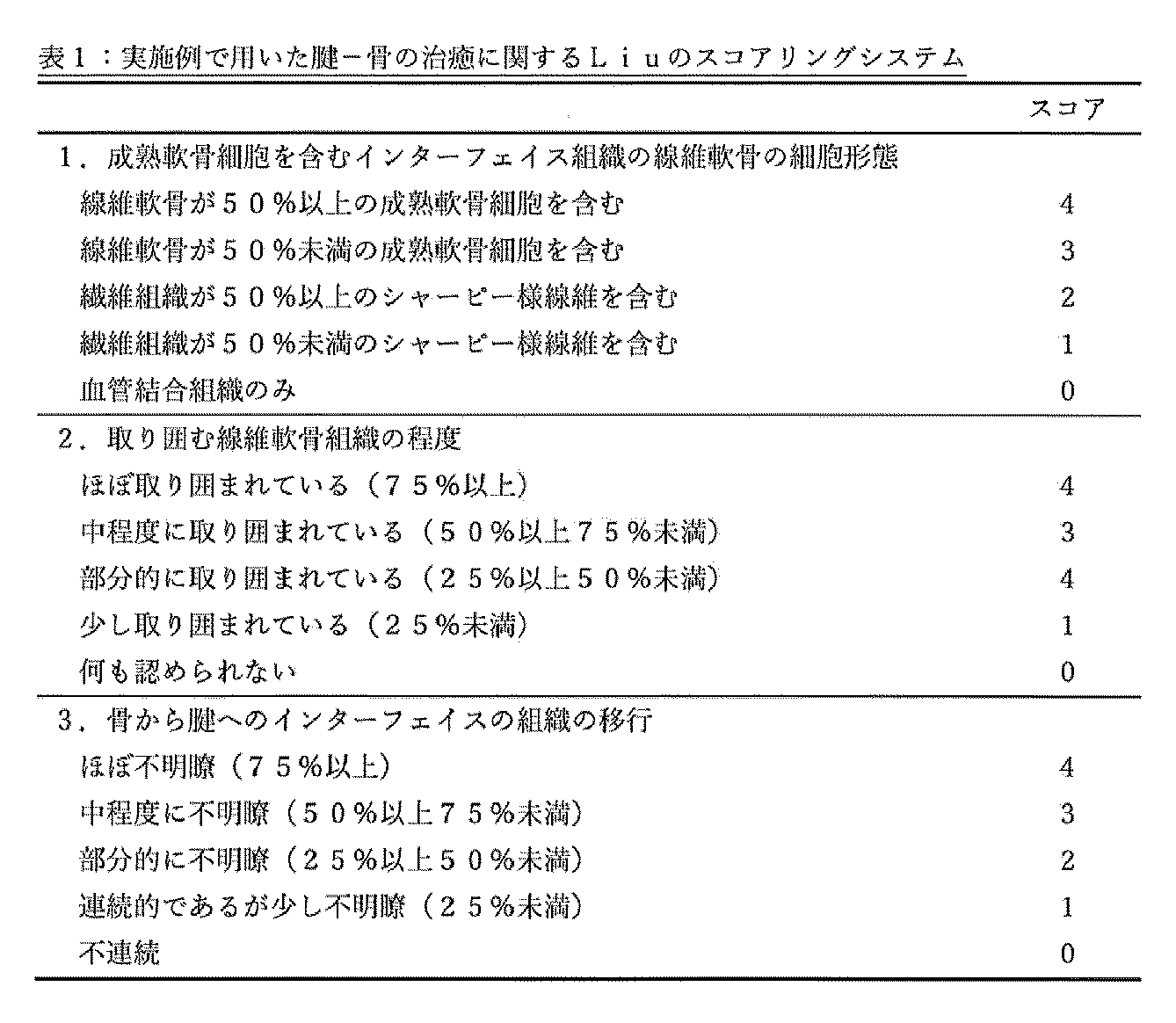
病理解析用の検体は個体の犠牲後に10%中性ホルマリン固定を3日間行い、EDTAで脱灰を5週間行った。その後大腿骨孔部分で骨孔軸に沿って割面を入れて組織切片を作成し、マッソントリクローム染色を行った。明視野での顕微鏡観察を行い、腱-骨間接合部分とグラフト実質部の治癒過程の組織学的評価を行った。スコアリングシステムについては既報にあるLiuのスコアリングシステムをそのまま採用することとした25,43(表1参照)。Liuのスコアリングシステムの特徴として、腱と骨のインターフェイスの成熟度合いを評価できる点で優れている。
病理解析用の検体は個体の犠牲後に10%中性ホルマリン固定を3日間行い、EDTAで脱灰を5週間行った。その後大腿骨孔部分で骨孔軸に沿って割面を入れて組織切片を作成し、マッソントリクローム染色を行った。明視野での顕微鏡観察を行い、腱-骨間接合部分とグラフト実質部の治癒過程の組織学的評価を行った。スコアリングシステムについては既報にあるLiuのスコアリングシステムをそのまま採用することとした25,43(表1参照)。Liuのスコアリングシステムの特徴として、腱と骨のインターフェイスの成熟度合いを評価できる点で優れている。
マイクロコンピュータ断層撮影
バイオメカ実験の直前に大腿骨の撮像を行った。μCTはR_mCT2(Rigaku corporation)を使用し、fields of view: 30 mm、voltage: 90 kV、current: 160 mA、scan technique: fine 3minの条件で撮像した。3D BON system(Ratoc), ImageJ software(ImageJ version 1.52; National Institutes of Health)を用いて骨体積/組織体積(%、以下BV/TV)、および骨孔断面積を決定した。骨孔断面積は、大腿骨孔を4分割し、一番関節面に近い部分を関節開口部と定義し、同部位での断面積の測定をすることにより求めた。BV/TVは大腿骨孔中心で3.5mm立方の体積中の骨塩量を計測した(Figure 2)。
バイオメカ実験の直前に大腿骨の撮像を行った。μCTはR_mCT2(Rigaku corporation)を使用し、fields of view: 30 mm、voltage: 90 kV、current: 160 mA、scan technique: fine 3minの条件で撮像した。3D BON system(Ratoc), ImageJ software(ImageJ version 1.52; National Institutes of Health)を用いて骨体積/組織体積(%、以下BV/TV)、および骨孔断面積を決定した。骨孔断面積は、大腿骨孔を4分割し、一番関節面に近い部分を関節開口部と定義し、同部位での断面積の測定をすることにより求めた。BV/TVは大腿骨孔中心で3.5mm立方の体積中の骨塩量を計測した(Figure 2)。
バイオメカニカル実験
個体の犠牲直後に膝の周囲の軟部組織を切除し、骨とグラフトだけにして-80℃で凍結保存した。試験前に常温解凍し、Autograph AG-IS引張試験機(Shimadzu Corporation)を用いて、脛骨と大腿骨にk-wireを通し機械台にセッティングし(図10参照)、10mm/minの速度で最大破断荷重試験を行った。実験から得られた曲線結果を元に最大破断荷重(UFL, ultimate failure load)と剛性値(stiffness)の測定を行った。
個体の犠牲直後に膝の周囲の軟部組織を切除し、骨とグラフトだけにして-80℃で凍結保存した。試験前に常温解凍し、Autograph AG-IS引張試験機(Shimadzu Corporation)を用いて、脛骨と大腿骨にk-wireを通し機械台にセッティングし(図10参照)、10mm/minの速度で最大破断荷重試験を行った。実験から得られた曲線結果を元に最大破断荷重(UFL, ultimate failure load)と剛性値(stiffness)の測定を行った。
透過型電子顕微鏡 走査型
生体力学的分析後、損傷していない移植片を0.1Mリン酸緩衝液(PB) pH7.4, 4℃で一晩、2%パラホルムアルデヒド(PFA)と2%グルタルアルデヒド(GA)を用いて固定した。この固定後、サンプルを0.1M PBで30分ずつ3回洗浄し、0.1M PB中の2%オスミウム四酸化物(OsO4)で一晩4℃でポスト固定した。得られた試料を、勾配化エタノール溶液中で脱水処理を行った。これらの脱水工程の後、試料を室温で一晩100%エタノール中で連続的に脱水した。得られたサンプルにプロピレンオキサイド(PO)を各1hずつ2回浸潤させ、POと樹脂(Quetol-812;日新EM社製)の70:30混合液に2h入れた後、チューブのキャップを開けたままにして、一晩POを揮発させた。新鮮な100%樹脂に移し、60℃で48時間重合させた後、超ミクロトーム(ULTRACUT UCT;ライカ)を用いてダイヤモンドナイフで70nmでブロックを極薄切片化し、切片を銅格子上に置いた。このグリッドを透過型電子顕微鏡(JEM-1400Plus;日本電子株式会社)で加速電圧100kVで観察した。デジタル画像(3296×2472画素)は、CCDカメラ(EM-14830RUBY2;日本電子(株))で撮影した。
TEM画像は、各試料ごとにブラインドで無作為に10枚撮影し、画像解析システム(ImageJ version 1.52; National Institutes of Health)を用いて解析した。
フィブリル径の評価は、セクショニングの斜行による誤差を防ぐため、必ず最小フィブリル径を測定した。直径<10nmのフィブリル構造は、ミクロフィブリルまたは非コラーゲンフィブリル成分の測定を避けるために除外した。コラーゲン線維の数、フィブリル径、コラーゲン占有率/フィールドを計算した。
生体力学的分析後、損傷していない移植片を0.1Mリン酸緩衝液(PB) pH7.4, 4℃で一晩、2%パラホルムアルデヒド(PFA)と2%グルタルアルデヒド(GA)を用いて固定した。この固定後、サンプルを0.1M PBで30分ずつ3回洗浄し、0.1M PB中の2%オスミウム四酸化物(OsO4)で一晩4℃でポスト固定した。得られた試料を、勾配化エタノール溶液中で脱水処理を行った。これらの脱水工程の後、試料を室温で一晩100%エタノール中で連続的に脱水した。得られたサンプルにプロピレンオキサイド(PO)を各1hずつ2回浸潤させ、POと樹脂(Quetol-812;日新EM社製)の70:30混合液に2h入れた後、チューブのキャップを開けたままにして、一晩POを揮発させた。新鮮な100%樹脂に移し、60℃で48時間重合させた後、超ミクロトーム(ULTRACUT UCT;ライカ)を用いてダイヤモンドナイフで70nmでブロックを極薄切片化し、切片を銅格子上に置いた。このグリッドを透過型電子顕微鏡(JEM-1400Plus;日本電子株式会社)で加速電圧100kVで観察した。デジタル画像(3296×2472画素)は、CCDカメラ(EM-14830RUBY2;日本電子(株))で撮影した。
TEM画像は、各試料ごとにブラインドで無作為に10枚撮影し、画像解析システム(ImageJ version 1.52; National Institutes of Health)を用いて解析した。
フィブリル径の評価は、セクショニングの斜行による誤差を防ぐため、必ず最小フィブリル径を測定した。直径<10nmのフィブリル構造は、ミクロフィブリルまたは非コラーゲンフィブリル成分の測定を避けるために除外した。コラーゲン線維の数、フィブリル径、コラーゲン占有率/フィールドを計算した。
RNA発現解析
コントロール群とシート群それぞれ1匹ずつ術後4週時点で膝関節を採取し、関節内グラフト部分のみを採取した。採取したグラフトを即座に適当量のRNA laterにひたした後に、Trisol 1mLで3分間ミンスを行った。200μLのクロロホルムを混注した後にボルテックスをかけ、12,000回転4℃で15分間にわたり遠心分離した。上清を新しいマイクロチューブへ移し、500μLの2-プロパノールを添加した。RNA Mini kit(Qiagen)カラムに700μLを移し、12,000回転、室温(以下RT)で15秒間遠心分離した。RW1 350μLを添加し、12,000回転、RTで15秒間にわたり遠心した。次いでDNase(RNase-free DNase Set; Qiagen) 10μL+RDD 70μLをカラムメンブレン上に滴下し、室温で15分静置した。その後RPE500μLを用いて3回遠心洗浄を行った。その後、RPEで2分間の遠心分離した。試料は-80℃で保存した。サンプルは、Genewiz社への委託により、シークエンシングによるリード数に基づく発現解析を行った。対照(Sheet(-))を1としてシートあり(Sheet(+))の場合の遺伝子発現量を求めた。
コントロール群とシート群それぞれ1匹ずつ術後4週時点で膝関節を採取し、関節内グラフト部分のみを採取した。採取したグラフトを即座に適当量のRNA laterにひたした後に、Trisol 1mLで3分間ミンスを行った。200μLのクロロホルムを混注した後にボルテックスをかけ、12,000回転4℃で15分間にわたり遠心分離した。上清を新しいマイクロチューブへ移し、500μLの2-プロパノールを添加した。RNA Mini kit(Qiagen)カラムに700μLを移し、12,000回転、室温(以下RT)で15秒間遠心分離した。RW1 350μLを添加し、12,000回転、RTで15秒間にわたり遠心した。次いでDNase(RNase-free DNase Set; Qiagen) 10μL+RDD 70μLをカラムメンブレン上に滴下し、室温で15分静置した。その後RPE500μLを用いて3回遠心洗浄を行った。その後、RPEで2分間の遠心分離した。試料は-80℃で保存した。サンプルは、Genewiz社への委託により、シークエンシングによるリード数に基づく発現解析を行った。対照(Sheet(-))を1としてシートあり(Sheet(+))の場合の遺伝子発現量を求めた。
統計解析
棒グラフデータおよび折れ線グラフデータは、平均値±SDで表された。Boxplotの値は、最小値、最大値、サンプル中央値、および第一四分位と第三四分位として示されている。統計分析は、各時点での対照群とシート群の間のウェルチのt検定を用いて実施した。生体力学的検査とBV/TVとの相関についてはピアソンの相関分析を行い、電子顕微鏡分析についてはANOVA検定を用いた。P値<0.05は統計的に有意と判定した。
棒グラフデータおよび折れ線グラフデータは、平均値±SDで表された。Boxplotの値は、最小値、最大値、サンプル中央値、および第一四分位と第三四分位として示されている。統計分析は、各時点での対照群とシート群の間のウェルチのt検定を用いて実施した。生体力学的検査とBV/TVとの相関についてはピアソンの相関分析を行い、電子顕微鏡分析についてはANOVA検定を用いた。P値<0.05は統計的に有意と判定した。
[結果]
病理組織学的評価では、図3~6に示されるように、グラフトは一般的に知られているネクローシス(炎症)期、次いで再増殖期、さらに続けて成熟(リモデリング)期の経過をたどった。具体的には、グラフト移植後4週(4w)の時点では、シート群および対照群の両群ともグラフト実質部で細胞数の減少が生じ、またコラーゲン線維の配列が不規則となった。一方、対照群と比較するとシート群では相対的に線維芽細胞が多く出現し、その時点で線維芽細胞数のピークを迎えていた。つまり対照群ではネクローシス期の組織像を呈していたが、シート群ではネクローシス期に加え、部分的に再増殖期の組織像も呈していた。腱-骨間接合部では対照群でもシャーピー様線維の形成や肥大軟骨細胞の出現が見られるが、これらはシート群でより多く認められた。そしてシート群では骨破壊も少なかった。
8週(8w)の時点では、シート群および対照群の両群ともグラフト実質部のコラーゲン線維の配列は未だに不規則な状態であった。そして、対照群のグラフト実質部は再増殖期に突入し、4週と比較すると線維芽細胞数が増大しピークを迎えるようになるが、シート群では4週と比較すると細胞数は減少傾向であり、再増殖期は終了に近付いていた。また腱-骨間接合部では対照群でもシャーピー様線維や肥大軟骨細胞が明らかに認められるようになるが、シート群では類骨形成を認めはじめ、接合部の破壊像も対照群より少なかった。
16週(16w)になるとシート群および対照群の両群ともグラフト部分はリモデリング期に突入し、グラフト内の細胞の配列はまだ不規則ではあるが次第に紡錘形を呈するようになり、8週と比較するとグラフト内細胞数は減少傾向にあり、またコラーゲン線維の配列は8週までと比較して規則的となった。腱-骨間接合部では対照群でも類骨形成が明らかとなり、また両群とも腱-骨間接合部部分は凹凸が少なく滑らかで境目のあいまいな接合部を形成していた。
24週(24w)ではシート群および対照群の両群ともグラフト実質部の細胞数は本来のSTと同程度となり、16週以前と比較すると細胞はより規則正しい配列を呈するようになり、コラーゲン線維も本来のグラフトと同じような配向を呈していた。また腱-骨間接合部では16週と同様に境目のあいまいな状態を維持しており、また層板構造を持つ骨形成が認められた。
病理組織学的評価では、図3~6に示されるように、グラフトは一般的に知られているネクローシス(炎症)期、次いで再増殖期、さらに続けて成熟(リモデリング)期の経過をたどった。具体的には、グラフト移植後4週(4w)の時点では、シート群および対照群の両群ともグラフト実質部で細胞数の減少が生じ、またコラーゲン線維の配列が不規則となった。一方、対照群と比較するとシート群では相対的に線維芽細胞が多く出現し、その時点で線維芽細胞数のピークを迎えていた。つまり対照群ではネクローシス期の組織像を呈していたが、シート群ではネクローシス期に加え、部分的に再増殖期の組織像も呈していた。腱-骨間接合部では対照群でもシャーピー様線維の形成や肥大軟骨細胞の出現が見られるが、これらはシート群でより多く認められた。そしてシート群では骨破壊も少なかった。
8週(8w)の時点では、シート群および対照群の両群ともグラフト実質部のコラーゲン線維の配列は未だに不規則な状態であった。そして、対照群のグラフト実質部は再増殖期に突入し、4週と比較すると線維芽細胞数が増大しピークを迎えるようになるが、シート群では4週と比較すると細胞数は減少傾向であり、再増殖期は終了に近付いていた。また腱-骨間接合部では対照群でもシャーピー様線維や肥大軟骨細胞が明らかに認められるようになるが、シート群では類骨形成を認めはじめ、接合部の破壊像も対照群より少なかった。
16週(16w)になるとシート群および対照群の両群ともグラフト部分はリモデリング期に突入し、グラフト内の細胞の配列はまだ不規則ではあるが次第に紡錘形を呈するようになり、8週と比較するとグラフト内細胞数は減少傾向にあり、またコラーゲン線維の配列は8週までと比較して規則的となった。腱-骨間接合部では対照群でも類骨形成が明らかとなり、また両群とも腱-骨間接合部部分は凹凸が少なく滑らかで境目のあいまいな接合部を形成していた。
24週(24w)ではシート群および対照群の両群ともグラフト実質部の細胞数は本来のSTと同程度となり、16週以前と比較すると細胞はより規則正しい配列を呈するようになり、コラーゲン線維も本来のグラフトと同じような配向を呈していた。また腱-骨間接合部では16週と同様に境目のあいまいな状態を維持しており、また層板構造を持つ骨形成が認められた。
以上を上記のスコアリングシステムで評価すると、図4に示されるように、対照群 vs シート群において4週間後に3.0±0.9 vs 4.2±0.8(P=0.04),8週間後に5.7±0.5 vs 6.5±0.5(P=0.02),16週間後に8.2±0.4 vs 8.5±0.5(P=0.26),24週間後に8.7±0.5 vs 9.0±1.1(P=0.52)となった。
マイクロコンピュータ断層評価 大腿骨孔を4分割し、関節内に一番近い断面を関節開口部と定義し、同部位の断面積(mm2)を計測した。結果は、図7に示される通りであった。図7に示されるように、対照群(-) vs シート群(+)において、2週間後に3.9±1.1 vs 3.5±1.1(P=0.61),4週間後に7.3±1.0 vs 5.1±0.9(P=0.01),8週間後に6.6±2.1 vs 4.7±0.8(P=0.25),16週間後に4.3±0.7 vs 4.6±1.0(P=0.64),24週間後に4.3±1.1 vs 4.1±1.5(P=0.53)となった。関節内開口部の断面積は術後4週においてシート群で有意に小さかったが、その他の時点で有意差は認められなかった。
また、腱-骨間接合部の石灰化評価のために400mg/ml以上の石灰化度を有する部分を骨として計算し、BV/TV(%)を求めた。結果は図8に示される通りであった。図8左パネルに示されるように、対照群(-) vs シート群(+)において、2週間後に21.9±6.0 vs 20.1±5.9(P=0.65),4週間後に21.5±3.7 vs 22.8±2.0(P=0.53),8週間後に29.5±1.3 vs 24.4±3.5(P=0.06),16週間後に33.5±2.6 vs 38.4±4.6(P=0.11),24週間後に32.4±4.4 vs 43.4±8.2(P=0.05)であり、腱-骨間接合部の骨の石灰化度の経時的な上昇を認めた。800mg/ml以上の石灰化度を有する部分を骨として解析を行うと、図8右パネルに示されるように、2週間後に0.41±0.2 vs 0.33±0.2(P=0.56),4週間後に0.7±0.7 vs 1.3±0.8(P=0.29),8週間後に1.2±0.8 vs 2.6±2.2(P=0.35),16週間後に3.3±1.8 vs 6.8±4.6(P=0.21),24週間後に4.0±3.4 vs 10.0±1.8(P=0.02)となった。いずれの石灰化度でもBV/TVは24週時点においてシート群で有意に高かった。
また、腱-骨間接合部の石灰化評価のために400mg/ml以上の石灰化度を有する部分を骨として計算し、BV/TV(%)を求めた。結果は図8に示される通りであった。図8左パネルに示されるように、対照群(-) vs シート群(+)において、2週間後に21.9±6.0 vs 20.1±5.9(P=0.65),4週間後に21.5±3.7 vs 22.8±2.0(P=0.53),8週間後に29.5±1.3 vs 24.4±3.5(P=0.06),16週間後に33.5±2.6 vs 38.4±4.6(P=0.11),24週間後に32.4±4.4 vs 43.4±8.2(P=0.05)であり、腱-骨間接合部の骨の石灰化度の経時的な上昇を認めた。800mg/ml以上の石灰化度を有する部分を骨として解析を行うと、図8右パネルに示されるように、2週間後に0.41±0.2 vs 0.33±0.2(P=0.56),4週間後に0.7±0.7 vs 1.3±0.8(P=0.29),8週間後に1.2±0.8 vs 2.6±2.2(P=0.35),16週間後に3.3±1.8 vs 6.8±4.6(P=0.21),24週間後に4.0±3.4 vs 10.0±1.8(P=0.02)となった。いずれの石灰化度でもBV/TVは24週時点においてシート群で有意に高かった。
バイオメカニカル実験
対照群(sheet-)とシート群(sheet+)の最大破断荷重試験の結果は図10Aに示される通りであった。最大破断荷重(N)は、図10Aにしめされるように、2週間後に17.2±5.5 vs 37.3±10.3(P=0.01),4週間後に28.6±1.9 vs 47.4±10.4(P=0.003),8週間後に53.0±14.3 vs 48.1±9.3(P=0.59),16週間後に66.2±9.3 vs 95.2±43.1(P=0.24),24週間後に66.7±27.3 vs 85.3±29.5(P=0.39)であった。バイオメカニカル実験では8週を除いてシート群で最大破断荷重が強く、特に4週までの早期で有意差を認めた。剛性値(N/mm)は図10Bに示される通りであった。以下の通りであった。剛性値は、図10Bに示されるように、2週間後に7.7±0.7 vs 13.1±2.5(P=0.01),4週間後に14.6±4.5 vs 18.0±5.3(P=0.31),8週間後に17.5±5.8 vs 16.2±4.9(P=0.74),16週間後に24.8±7.0 vs 28.7±6.0(P=0.43),24週間後に24.0±4.2 vs 26.5±6.0(P=0.52)であった。術後2週を除いて剛性値には有意差は見られなかった。
対照群(sheet-)とシート群(sheet+)の最大破断荷重試験の結果は図10Aに示される通りであった。最大破断荷重(N)は、図10Aにしめされるように、2週間後に17.2±5.5 vs 37.3±10.3(P=0.01),4週間後に28.6±1.9 vs 47.4±10.4(P=0.003),8週間後に53.0±14.3 vs 48.1±9.3(P=0.59),16週間後に66.2±9.3 vs 95.2±43.1(P=0.24),24週間後に66.7±27.3 vs 85.3±29.5(P=0.39)であった。バイオメカニカル実験では8週を除いてシート群で最大破断荷重が強く、特に4週までの早期で有意差を認めた。剛性値(N/mm)は図10Bに示される通りであった。以下の通りであった。剛性値は、図10Bに示されるように、2週間後に7.7±0.7 vs 13.1±2.5(P=0.01),4週間後に14.6±4.5 vs 18.0±5.3(P=0.31),8週間後に17.5±5.8 vs 16.2±4.9(P=0.74),16週間後に24.8±7.0 vs 28.7±6.0(P=0.43),24週間後に24.0±4.2 vs 26.5±6.0(P=0.52)であった。術後2週を除いて剛性値には有意差は見られなかった。
バイオメカニカル実験における断裂様式は、図11に示される3通り:グラフトが脛骨から抜けるパターン(図11左パネル)、グラフトが中央で断裂するパターン(図11中央)、およびグラフトが大腿骨から抜けるパターン(図11右パネル)であった。中央で断裂するパターンと骨から抜けるパターンの件数は表2に示される通りであった。いずれの週でもシート群で実質部断裂の割合が高いという結果となった(表2)。

腱-骨間接合部の石灰化度(400mg/ml)と実質部断裂を生じたケースを除き、骨からの腱の脱離を生じた検体の破断荷重の相関を見たところ、両者にはR=0.72の相関が見られた(P=0.0002)(図12)。
透過型電子顕微鏡による評価
1視野あたりの線維総本数は本来のSTにおいて333±125であるのに対して、術後24週の電子顕微鏡解析では、対照群(-) vs シート群(+)において、527±235 vs 473±175であり有意差は認めなかった(図9)。しかし、線維径(minimum ferret径;nm)は本来のSTが90±16であるのに対して、術後24週の電子顕微鏡解析では、対照群(-) vs シート群(+)で50.5±51.5 vs 58.7±56.8(P< 0.001)となりシート群で有意に太かった(図9左下パネル)。線維径のヒストグラムを見てみると、シート群でよりSTに近い組成が得られた(図9中央下パネル)。また、1視野あたりのコラーゲン線維の占拠割合は本来のSTが64.0±1.3であるのに対して、術後24週の電子顕微鏡解析では、対照群(-) vs シート群(+)で47.4±14 vs 54.5±11(P=0.03)となりシート群で有意に高かった(図9右下パネル)。
1視野あたりの線維総本数は本来のSTにおいて333±125であるのに対して、術後24週の電子顕微鏡解析では、対照群(-) vs シート群(+)において、527±235 vs 473±175であり有意差は認めなかった(図9)。しかし、線維径(minimum ferret径;nm)は本来のSTが90±16であるのに対して、術後24週の電子顕微鏡解析では、対照群(-) vs シート群(+)で50.5±51.5 vs 58.7±56.8(P< 0.001)となりシート群で有意に太かった(図9左下パネル)。線維径のヒストグラムを見てみると、シート群でよりSTに近い組成が得られた(図9中央下パネル)。また、1視野あたりのコラーゲン線維の占拠割合は本来のSTが64.0±1.3であるのに対して、術後24週の電子顕微鏡解析では、対照群(-) vs シート群(+)で47.4±14 vs 54.5±11(P=0.03)となりシート群で有意に高かった(図9右下パネル)。
遺伝子発現解析の結果は、図13および14に示される通りであった。図13に示されるように、IL-1β、PDGF-A、およびVEGF-Aに関して、細胞シートと共にグラフトを移植した群において対照群よりも高発現を示した。
本実施例によれば、グラフトに巻き付けたADSCシート中のADSC細胞数は、上記の懸濁液中のADSC細胞数よりも少ない点で有利であった。この理由としては、細胞懸濁液の注射では移植細胞が関節内全体に拡散してしまい、必要箇所における細胞濃度が低下したためであると考えられる。今回の実験では細胞の局在を明確にするためにADSCをシートにしてグラフト周囲に巻き付けたが、ADSCシートは、表面に細胞外マトリックスが形成されており、懸濁液中のADSCとはこの点において異なる。
本実施例によれば、対照群と比較し、シート群で術後4週時点での骨孔拡大を有意に予防していた。ハムストリングスを用いたACL再建術では術後に骨孔拡大が生じ、グラフトがゆるんでしまうことで再手術を要することが報告されている9,47。また、ACL再建術においては比較的早期の段階で骨孔は一時的に拡大し48、経過とともに縮小する可能性も報告されているが25,29、MSCを移植することで骨孔拡大を予防できたとの報告もある15。今回の実験では4週時点においてADSCの作用によって腱-骨間接合部における術後の一過性の骨破壊および骨吸収を抑制して骨孔拡大を予防し、骨とグラフトの接触面積が増えたことで、結果的にバイオメカニカル実験において8週を除く早期の段階で最大破断荷重が有意に改善した可能性がある。
また、病理学的には、移植グラフトは、Kosakaの報告にあるような組織修復とほぼ同じ経過をたどった21。シート群ではグラフト実質部において線維芽細胞の移入が比較的早期の段階で生じており、また血管新生も対照群より多く、またシャーピー様線維の形成や肥大軟骨細胞の出現がより多く認められた。シート群の4週の時点で対照群の8週と似た組織像が部分的に生じており、ADSCを移植したことでグラフト実質部と腱-骨間接合部の成熟が促進されたことも、シート群のバイオメカニカル強度の向上につながった一因と考えられる。
ADSCからの栄養因子の作用によって腱-骨間接合部の回復が促進されたり、ADSC自身がグラフト実質部で生細胞として機能したりした可能性がある。その際に、ADSCシートが細胞外マトリクス(接着性タンパク質)を有したことが、結果に正の影響を与えた可能性がある。
またバイオメカニカル実験では両群とも経時的に実質部断裂の割合が増えるという結果になったが、シート群においてADSCが特に石灰化度の高い骨組織形成を促進し、腱-骨間接合部の石灰化度の経時的上昇によって、骨からの腱の脱離の割合を減少させたものと考えられる。慢性期腱板断裂の手術時にADSCのゲル移植を併用した報告では腱-骨間接合部でのBMD(bone mineral density)が向上したとの報告もあり17、ADSCには骨形成作用があることが今回の実験からも示唆された。
16週以降では病理学的に両群とも腱-骨間接合部は境界があいまいな状態となり、次第に層板骨構造も見られるようになってくるが、スコア上の有意差はつかなかった。また骨孔拡大についても16週以降において両群で有意差はみられなかった。しかし、バイオメカニカル実験では、最大破断荷重は対照群と比較しシート群で平均値は高かったため、実質部の評価として透過型電子顕微鏡解析を行った。Takeuchiの報告によると移植グラフトのコラーゲン線維は経時的に縮小し、コラーゲン線維の占拠割合も減少するという報告がなされている46。また術後に新しく合成される線維は3型コラーゲンによる径が細いものが多いとも報告されている33。今回の電子顕微鏡の結果からは、ADSCが1型コラーゲンの合成を促進したり、3型コラーゲンの合成を抑制したりしたことでシート群においてコラーゲンの線維径が対照群よりも太く、断面積あたりのコラーゲン線維の占拠割合が高くなり、結果的に実質部断裂時の最大破断荷重の向上に影響した可能性が考えられる。過去の報告ではACL再建術後の再断裂は腱-骨間接合部ではなく実質部で生じる割合が高いとの報告もなされており28,38、ADSCを併用することで実質部をより強固にすることにより再断裂率を低下させることが期待できる。

Claims (20)
- 脂肪由来幹細胞を含む細胞シートを含む、腱または靭帯の再建用の移植材料。
- 重層化した前記細胞シートを含む、請求項1に記載の移植材料。
- 前記細胞シートが、シート表面に接着性タンパク質を維持している、請求項1または2に記載の移植材料。
- 腱または靭帯の再建用のグラフトの表面に付着させて用いられる、請求項1~3のいずれか一項に記載の移植材料。
- 腱または靭帯の再建用のグラフトの腱または靭帯と骨との接合部の表面に付着させて用いられる、請求項1~4のいずれか一項に記載の移植材料。
- 腱または靭帯の再建用のグラフトの腱または靭帯と骨との接合部以外の表面に付着させて用いられる、請求項1~4のいずれか一項に記載の移植材料。
- 腱または靭帯の再建術後のグラフト接合部の骨孔拡大を抑制することに用いるための、脂肪由来幹細胞を含む細胞シートを含む移植材料、または請求項1~6のいずれか一項に記載の移植材料。
- 細胞シートが、CD29、CD44、CD73、CD90、CD105、およびCD166が陽性である細胞を90%以上含み、およびCD14、CD31、CD45およびLin1が陽性である細胞を5%以下含む、請求項1~7のいずれか一項に記載の移植材料。
- 請求項1~8のいずれか一項に記載の移植材料を含む容器を含む、製品。
- 腱または靭帯の再建用のグラフトと、その表面に付着した請求項1~8のいずれか一項に記載の移植材料との組合せ。
- 前記表面が、グラフトの腱または靭帯と骨との接合部の表面である、請求項10に記載の組合せ。
- 前記表面が、グラフトの腱または靭帯と骨との接合部以外の表面である、請求項10に記載の組合せ。
- 移植グラフトに付着させた状態で移植され、これにより、グラフト中の1型コラーゲンの産生を促進することに用いるための、請求項1~8のいずれか一項に定義された細胞シートを含む、移植材料。
- 移植グラフトに付着させた状態で移植され、これにより、グラフト中の3型コラーゲンの産生を抑制することに用いるための、請求項1~8のいずれか一項に定義された細胞シートを含む、移植材料。
- 移植グラフトに付着させた状態で移植され、これにより、グラフト中のコラーゲン線維径を増大させることに用いるための、請求項1~8のいずれか一項に定義された細胞シートを含む、移植材料。
- 移植グラフトに付着させた状態で移植され、これにより、グラフト中のコラーゲン線維断面におけるコラーゲン線維の占有率を向上させることに用いるための、請求項1~8のいずれか一項に定義された細胞シートを含む、移植材料。
- 移植グラフトに付着させた状態で移植され、これにより、移植後グラフトの断裂を抑制することに用いるための、請求項1~8のいずれか一項に定義された細胞シートを含む、移植材料。
- 移植グラフトをさらに含む、請求項1~8のいずれか一項に記載の移植材料。
- その必要のある対象において腱または靭帯を再建する方法であって、
(i)対象において腱または靭帯を再建する必要のある部位にグラフトを固定することを含み、ここで、固定したグラフトは、表面に脂肪由来幹細胞を含む細胞シートを有するか、または、
(ii)対象において腱または靭帯を再建する必要のある部位にグラフトを固定することと、固定したグラフトの表面に脂肪由来幹細胞を含む細胞シートを付着させることを含む、
方法。 - その必要のある対象において腱または靭帯を再建する方法であって、
(i)対象において腱または靭帯を再建する必要のある部位にグラフトを固定することを含み、ここで、固定したグラフトは、表面に脂肪由来幹細胞を含む細胞シートを有する、方法。
Priority Applications (1)
| Application Number | Priority Date | Filing Date | Title |
|---|---|---|---|
| JP2022568253A JP7654205B2 (ja) | 2020-12-07 | 2021-12-06 | 腱または靭帯移植用の移植材料 |
Applications Claiming Priority (2)
| Application Number | Priority Date | Filing Date | Title |
|---|---|---|---|
| JP2020202456 | 2020-12-07 | ||
| JP2020-202456 | 2020-12-07 |
Publications (1)
| Publication Number | Publication Date |
|---|---|
| WO2022124245A1 true WO2022124245A1 (ja) | 2022-06-16 |
Family
ID=81973219
Family Applications (1)
| Application Number | Title | Priority Date | Filing Date |
|---|---|---|---|
| PCT/JP2021/044608 Ceased WO2022124245A1 (ja) | 2020-12-07 | 2021-12-06 | 腱または靭帯移植用の移植材料 |
Country Status (2)
| Country | Link |
|---|---|
| JP (1) | JP7654205B2 (ja) |
| WO (1) | WO2022124245A1 (ja) |
Citations (1)
| Publication number | Priority date | Publication date | Assignee | Title |
|---|---|---|---|---|
| US20120189587A1 (en) * | 2011-01-26 | 2012-07-26 | The Chinese University Of Hong Kong | Cell sheet for tissue repair and bio-artificial tissue engineering, method of producing the same and method of using the same |
-
2021
- 2021-12-06 JP JP2022568253A patent/JP7654205B2/ja active Active
- 2021-12-06 WO PCT/JP2021/044608 patent/WO2022124245A1/ja not_active Ceased
Patent Citations (1)
| Publication number | Priority date | Publication date | Assignee | Title |
|---|---|---|---|---|
| US20120189587A1 (en) * | 2011-01-26 | 2012-07-26 | The Chinese University Of Hong Kong | Cell sheet for tissue repair and bio-artificial tissue engineering, method of producing the same and method of using the same |
Non-Patent Citations (5)
| Title |
|---|
| CHEN CAN, LIU FEI, TANG YIFU, QU JIN, CAO YONG, ZHENG CHENG, CHEN YANG, LI MUZHI, ZHAO CHUNFENG, SUN LUNQUAN, HU JIANZHONG, LU HON: "Book-Shaped Acellular Fibrocartilage Scaffold with Cell-loading Capability and Chondrogenic Inducibility for Tissue-Engineered Fibrocartilage and Bone–Tendon Healing", APPLIED MATERIALS & INTERFACES, vol. 11, no. 3, 23 January 2019 (2019-01-23), US , pages 2891 - 2907, XP055941892, ISSN: 1944-8244, DOI: 10.1021/acsami.8b20563 * |
| GONÇALVES ANA I; RODRIGUES MÁRCIA T; GOMES MANUELA E: "Tissue-engineered magnetic cell sheet patches for advanced strategies in tendon regeneration", ACTA BIOMATERIALIA, vol. 63, 1 January 2017 (2017-01-01), Amsterdam NL, pages 110 - 122, XP085239101, ISSN: 1742-7061, DOI: 10.1016/j.actbio.2017.09.014 * |
| KOTARO, HIGA : "P-02-39 Effect of adipose stem cell constructs made with a bio-3D printer on the healing of the bone-graft tendon junction in ACL reconstruction", ABSTRACTS OF THE 19TH ANNUAL MEETING OF THE JAPANESE SOCIETY OF REGENERATIVE MEDICINE; MARCH 12-14, 2020, vol. 19, no. Suppl., 1 February 2020 (2020-02-01) - 14 March 2020 (2020-03-14), JP, pages 749, XP009538193 * |
| NEO PUAY YONG, TEH THOMAS, TAY ALEX, ASUNCION MARIA, PNG SI NING, TOH SIEW LOK, GOH JAMES CHO-HONG: "Stem cell-derived cell-sheets for connective tissue engineering", CONNECTIVE TISSUE RESEARCH, vol. 57, no. 6, 1 November 2016 (2016-11-01), US , pages 428 - 442, XP009537448, ISSN: 0300-8207, DOI: 10.3109/03008207.2016.1173035 * |
| SHIN MYUNG JIN, SHIM IN KYONG, KIM DONG MIN, CHOI JAE HEE, LEE YU NA, JEON IN-HO, KIM HYOJUNE, PARK DONGJUN, KHOLINNE ERICA, YANG : "Engineered Cell Sheets for the Effective Delivery of Adipose-Derived Stem Cells for Tendon-to-Bone Healing", AMERICAN JOURNAL OF SPORTS MEDICINE, vol. 48, no. 13, 1 November 2020 (2020-11-01), Waltham MA, pages 3347 - 3358, XP055941891, ISSN: 0363-5465, DOI: 10.1177/0363546520964445 * |
Also Published As
| Publication number | Publication date |
|---|---|
| JPWO2022124245A1 (ja) | 2022-06-16 |
| JP7654205B2 (ja) | 2025-04-01 |
Similar Documents
| Publication | Publication Date | Title |
|---|---|---|
| Tang et al. | Structure and ingredient-based biomimetic scaffolds combining with autologous bone marrow-derived mesenchymal stem cell sheets for bone-tendon healing | |
| Shen et al. | Long-term effects of knitted silk–collagen sponge scaffold on anterior cruciate ligament reconstruction and osteoarthritis prevention | |
| Hu et al. | Exogenous stromal derived factor-1 releasing silk scaffold combined with intra-articular injection of progenitor cells promotes bone-ligament-bone regeneration | |
| Liu et al. | Integration of a calcined bovine bone and BMSC-sheet 3D scaffold and the promotion of bone regeneration in large defects | |
| Gu et al. | Application of marrow mesenchymal stem cell‐derived extracellular matrix in peripheral nerve tissue engineering | |
| Liu et al. | Biomimetic tendon extracellular matrix composite gradient scaffold enhances ligament-to-bone junction reconstruction | |
| Briggs et al. | Histological evaluation of chondral defects after autologous chondrocyte implantation of the knee | |
| Kirker-Head et al. | BMP-silk composite matrices heal critically sized femoral defects | |
| Yuan et al. | AMECM/DCB scaffold prompts successful total meniscus reconstruction in a rabbit total meniscectomy model | |
| JP4542106B2 (ja) | 軟骨治療剤組成物 | |
| CN102614546B (zh) | 用于组织修复和生物人造组织工程的细胞片及其制备方法 | |
| Huang et al. | Effect of kartogenin-loaded gelatin methacryloyl hydrogel scaffold with bone marrow stimulation for enthesis healing in rotator cuff repair | |
| US20210205498A1 (en) | Tenocyte containing bioscaffolds and treatment using the same | |
| US20050043814A1 (en) | Acellular matrix implanted into an articular cartilage or osteochondral lesion protected with a biodegradable polymer modified to have extended polymerization time and methods for preparation and use thereof | |
| Akhlaghi et al. | Improved bone regeneration through amniotic membrane loaded with buccal fat pad-derived MSCs as an adjuvant in maxillomandibular reconstruction | |
| JP2002512842A (ja) | ヒドロゲル・細胞組成物の発達誘導と支持 | |
| US20110014267A1 (en) | Biosynthetic cartilaginous matrix and methods for their production | |
| JP2008178670A (ja) | 軟組織修復にコラゲナーゼを使った組織癒合法 | |
| Bi et al. | Bone mesenchymal stem cells contribute to ligament regeneration and graft–bone healing after anterior cruciate ligament reconstruction with silk–collagen scaffold | |
| EP3041522B1 (en) | A flap for de-novo tissue regeneration | |
| Pan et al. | Delivery of epidermal growth factor receptor inhibitor via a customized collagen scaffold promotes meniscal defect regeneration in a rabbit model | |
| Chailakhyan et al. | Autologous bone marrow-derived mesenchymal stem cells provide complete regeneration in a rabbit model of the Achilles tendon bundle rupture | |
| Xing et al. | Effects of nanoparticle-mediated growth factor gene transfer to the injured microenvironment on the tendon-to-bone healing strength | |
| Lu et al. | Comparative evaluation of the book‐type acellular bone scaffold and fibrocartilage scaffold for bone‐tendon healing | |
| JP7654205B2 (ja) | 腱または靭帯移植用の移植材料 |
Legal Events
| Date | Code | Title | Description |
|---|---|---|---|
| 121 | Ep: the epo has been informed by wipo that ep was designated in this application |
Ref document number: 21903348 Country of ref document: EP Kind code of ref document: A1 |
|
| ENP | Entry into the national phase |
Ref document number: 2022568253 Country of ref document: JP Kind code of ref document: A |
|
| NENP | Non-entry into the national phase |
Ref country code: DE |
|
| 122 | Ep: pct application non-entry in european phase |
Ref document number: 21903348 Country of ref document: EP Kind code of ref document: A1 |