WO2022113336A1 - 免疫染色方法、試料交換室、及び荷電粒子線装置 - Google Patents
免疫染色方法、試料交換室、及び荷電粒子線装置 Download PDFInfo
- Publication number
- WO2022113336A1 WO2022113336A1 PCT/JP2020/044477 JP2020044477W WO2022113336A1 WO 2022113336 A1 WO2022113336 A1 WO 2022113336A1 JP 2020044477 W JP2020044477 W JP 2020044477W WO 2022113336 A1 WO2022113336 A1 WO 2022113336A1
- Authority
- WO
- WIPO (PCT)
- Prior art keywords
- sample
- container
- charged particle
- particle beam
- exchange chamber
- Prior art date
- Legal status (The legal status is an assumption and is not a legal conclusion. Google has not performed a legal analysis and makes no representation as to the accuracy of the status listed.)
- Ceased
Links
Images


Classifications
-
- G—PHYSICS
- G01—MEASURING; TESTING
- G01N—INVESTIGATING OR ANALYSING MATERIALS BY DETERMINING THEIR CHEMICAL OR PHYSICAL PROPERTIES
- G01N1/00—Sampling; Preparing specimens for investigation
- G01N1/28—Preparing specimens for investigation including physical details of (bio-)chemical methods covered elsewhere, e.g. G01N33/50, C12Q
- G01N1/30—Staining; Impregnating ; Fixation; Dehydration; Multistep processes for preparing samples of tissue, cell or nucleic acid material and the like for analysis
- G01N1/31—Apparatus therefor
- G01N1/312—Apparatus therefor for samples mounted on planar substrates
-
- G—PHYSICS
- G01—MEASURING; TESTING
- G01N—INVESTIGATING OR ANALYSING MATERIALS BY DETERMINING THEIR CHEMICAL OR PHYSICAL PROPERTIES
- G01N1/00—Sampling; Preparing specimens for investigation
- G01N1/28—Preparing specimens for investigation including physical details of (bio-)chemical methods covered elsewhere, e.g. G01N33/50, C12Q
- G01N1/34—Purifying; Cleaning
-
- G—PHYSICS
- G01—MEASURING; TESTING
- G01N—INVESTIGATING OR ANALYSING MATERIALS BY DETERMINING THEIR CHEMICAL OR PHYSICAL PROPERTIES
- G01N33/00—Investigating or analysing materials by specific methods not covered by groups G01N1/00 - G01N31/00
- G01N33/48—Biological material, e.g. blood, urine; Haemocytometers
- G01N33/50—Chemical analysis of biological material, e.g. blood, urine; Testing involving biospecific ligand binding methods; Immunological testing
- G01N33/53—Immunoassay; Biospecific binding assay; Materials therefor
-
- H—ELECTRICITY
- H01—ELECTRIC ELEMENTS
- H01J—ELECTRIC DISCHARGE TUBES OR DISCHARGE LAMPS
- H01J37/00—Discharge tubes with provision for introducing objects or material to be exposed to the discharge, e.g. for the purpose of examination or processing thereof
- H01J37/02—Details
- H01J37/18—Vacuum locks ; Means for obtaining or maintaining the desired pressure within the vessel
- H01J37/185—Means for transferring objects between different enclosures of different pressure or atmosphere
-
- H—ELECTRICITY
- H01—ELECTRIC ELEMENTS
- H01J—ELECTRIC DISCHARGE TUBES OR DISCHARGE LAMPS
- H01J37/00—Discharge tubes with provision for introducing objects or material to be exposed to the discharge, e.g. for the purpose of examination or processing thereof
- H01J37/02—Details
- H01J37/20—Means for supporting or positioning the object or the material; Means for adjusting diaphragms or lenses associated with the support
-
- G—PHYSICS
- G01—MEASURING; TESTING
- G01N—INVESTIGATING OR ANALYSING MATERIALS BY DETERMINING THEIR CHEMICAL OR PHYSICAL PROPERTIES
- G01N1/00—Sampling; Preparing specimens for investigation
- G01N1/28—Preparing specimens for investigation including physical details of (bio-)chemical methods covered elsewhere, e.g. G01N33/50, C12Q
- G01N1/30—Staining; Impregnating ; Fixation; Dehydration; Multistep processes for preparing samples of tissue, cell or nucleic acid material and the like for analysis
- G01N2001/305—Fixative compositions
-
- G—PHYSICS
- G01—MEASURING; TESTING
- G01N—INVESTIGATING OR ANALYSING MATERIALS BY DETERMINING THEIR CHEMICAL OR PHYSICAL PROPERTIES
- G01N1/00—Sampling; Preparing specimens for investigation
- G01N1/28—Preparing specimens for investigation including physical details of (bio-)chemical methods covered elsewhere, e.g. G01N33/50, C12Q
- G01N1/30—Staining; Impregnating ; Fixation; Dehydration; Multistep processes for preparing samples of tissue, cell or nucleic acid material and the like for analysis
- G01N1/31—Apparatus therefor
- G01N2001/315—Basket-type carriers for tissues
-
- H—ELECTRICITY
- H01—ELECTRIC ELEMENTS
- H01J—ELECTRIC DISCHARGE TUBES OR DISCHARGE LAMPS
- H01J2237/00—Discharge tubes exposing object to beam, e.g. for analysis treatment, etching, imaging
- H01J2237/20—Positioning, supporting, modifying or maintaining the physical state of objects being observed or treated
- H01J2237/2002—Controlling environment of sample
- H01J2237/2003—Environmental cells
- H01J2237/2004—Biological samples
-
- H—ELECTRICITY
- H01—ELECTRIC ELEMENTS
- H01J—ELECTRIC DISCHARGE TUBES OR DISCHARGE LAMPS
- H01J2237/00—Discharge tubes exposing object to beam, e.g. for analysis treatment, etching, imaging
- H01J2237/20—Positioning, supporting, modifying or maintaining the physical state of objects being observed or treated
- H01J2237/204—Means for introducing and/or outputting objects
-
- H—ELECTRICITY
- H01—ELECTRIC ELEMENTS
- H01J—ELECTRIC DISCHARGE TUBES OR DISCHARGE LAMPS
- H01J2237/00—Discharge tubes exposing object to beam, e.g. for analysis treatment, etching, imaging
- H01J2237/26—Electron or ion microscopes
- H01J2237/28—Scanning microscopes
-
- H—ELECTRICITY
- H01—ELECTRIC ELEMENTS
- H01J—ELECTRIC DISCHARGE TUBES OR DISCHARGE LAMPS
- H01J37/00—Discharge tubes with provision for introducing objects or material to be exposed to the discharge, e.g. for the purpose of examination or processing thereof
- H01J37/26—Electron or ion microscopes; Electron or ion diffraction tubes
- H01J37/28—Electron or ion microscopes; Electron or ion diffraction tubes with scanning beams
Definitions
- the present invention relates to an immunostaining method, a sample exchange chamber having a staining mechanism, and a charged particle beam apparatus.
- FFPE formalin-fixed paraffin embedded
- Immunostaining which is one of the staining methods, mainly utilizes the antigen-antibody reaction, which is a specific reaction between an antigenic substance such as a protein and an antibody, to visualize the expression of a specific gene and various marker proteins, thereby functioning the tissue. It is a technique for identifying proteins and small organs, and is indispensable for pathological diagnosis and research.
- TEM Transmission Electron Microscope
- TEM has high resolution and is suitable for observing the structure of fine cells, and is used for pathological diagnosis and research.
- institutions that can own it are limited.
- complicated pretreatment such as preparation of ultra-thin sections takes time and effort, and since an electron beam is used as the radiation source, the observation image is displayed in black and white, so the cell components are colored according to the color like an optical microscope. It cannot be identified. For this reason, understanding of observation images also requires specialized knowledge and proficiency.
- Patent Document 1 discloses an SEM that generates an enlarged image of the surface of a sample placed in the atmosphere.
- HRP Horseradish peroxidase
- a sample exchange chamber used for a charged particle beam device, and a container into which a substrate on which a sample is placed can be introduced, and a container.
- a sample exchange chamber having a structure including a staining mechanism for dyeing a sample, a cleaning mechanism for cleaning the sample, an exhaust mechanism for vacuum exhausting the container, and a sterilization mechanism for sterilizing the inside of the container and the sample.
- a charged particle beam device in order to achieve the above object, in the present invention, a charged particle beam device, a sample chamber, a container into which a substrate on which the sample is placed can be introduced, a staining mechanism for dyeing the sample, and a sample.
- a sample exchange chamber having a cleaning mechanism for cleaning the sample, an exhaust mechanism for vacuum exhausting the container, and a sterilization mechanism for sterilizing the inside of the container and the sample, and a sample between the sample exchange chamber and the sample chamber without exposure to the atmosphere.
- a charged particle beam apparatus having an autoloader mechanism capable of transferring the sample.
- the present invention it is possible to automate the work related to staining in the sample chamber, observation by SEM, characteristic X-ray analysis, and analysis of results.
- FIG. It is a figure which shows one configuration example of the sample exchange chamber which concerns on Example 1.
- FIG. It is a schematic diagram which shows one structural example of the charged particle beam apparatus which concerns on Example 1.
- FIG. It is a flowchart which shows the operation of the charged particle beam apparatus which concerns on Example 1.
- FIG. It is a flowchart which shows the procedure of the new dyeing method which concerns on Example 1.
- FIG. It is a figure which shows an example of the staining of the pathological section which concerns on Example 1.
- FIG. It is a figure which shows the result of characteristic X-ray analysis of the pathological section stained by the staining method which concerns on Example 1.
- Example 1 a container into which a substrate on which a sample is placed can be introduced, a staining mechanism for dyeing the sample, a cleaning mechanism for cleaning the sample, an exhaust mechanism for vacuum exhausting the container, the inside of the container, and the sample are used. It is an embodiment of a sample exchange chamber provided with a sterilization mechanism for sterilization, and a charged particle beam device using the same.
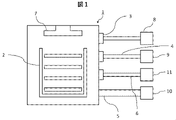
- FIG. 1 is a configuration diagram of a sample exchange chamber used in the charged particle beam device according to the first embodiment.
- the sample exchange chamber 1 has a container 2, a specific solution introduction port 3 with a tube, a cleaning liquid introduction port 4, a vacuum exhaust port 5, a waste liquid port 6, and a sterilization mechanism 7.
- the sample exchange chamber 1 also includes a control circuit for controlling each component, but the illustration thereof is omitted here.
- the specific solution inlet 3 constitutes a staining mechanism for staining the sample.
- the cleaning liquid introduction port 4 constitutes a cleaning mechanism for cleaning the sample.
- the vacuum exhaust port 5 constitutes an exhaust mechanism for evacuating the container.
- the sterilization mechanism 7 is a sterilization mechanism that sterilizes the inside of the container and the sample.
- the container 2 is, for example, a cassette type container having a structure in which a plurality of substrates on which a sample is placed can be introduced. It should be noted that the container 2 holds a sufficient space in which the sample placed on the substrate does not come into contact with the container 2 or another substrate when the substrate is introduced.
- the specific solution inlet 3 with a tube can be opened and closed, and is connected to the sample exchange chamber 1 or the container 2.
- the specific solution is a heavy metal such as silver or gold used for sensitizing a sample, for example.
- a tube is connected to the specific solution introduction port 3, and the other end of the tube is attached to the specific solution container 8 installed outside the sample exchange chamber 1. Through the tube, a sufficient amount of the specific solution for staining the sample in the container 2 can be introduced into the sample exchange chamber 1 or the container 2.
- the cleaning solution introduction port 4 with a tube can be opened and closed, and is connected to the sample exchange chamber 1 or the container 2.
- the cleaning liquid is, for example, water.
- a tube is connected to the cleaning liquid introduction port 4, and the other end of the tube is attached to a cleaning liquid container 9 installed outside the sample exchange chamber 1. The cleaning solution is introduced into the sample exchange chamber 1 or the container 2 through the tube, and the specific solution on the sample is washed away.
- the vacuum exhaust port 5 is connected to the pump 10 outside the sample exchange chamber 1 in order to evacuate the sample exchange chamber 1.
- the waste liquid port 6 with a tube is connected to the waste liquid container 11 outside the sample exchange chamber 1.
- the waste liquid port 6 can discharge the specific solution or the cleaning liquid from the sample exchange chamber 1 or the container 2 to the waste liquid container 11.
- the sterilization mechanism 7 is, for example, an ultraviolet (UV) irradiator that is installed in the sample exchange chamber 1 and has a function of sterilizing the inside of the sample exchange chamber 1. If the sample contains a toxic virus, the activity may not decrease even after fixation, but the virus is not exposed to the atmosphere and the charged particle beam device because it can be sterilized in the sample exchange chamber 1. That is, it is possible to reduce the possibility that a virus that cannot be inactivated by fixation affects workers and the like.
- UV ultraviolet
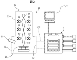
- FIG. 2 is a diagram showing a state in which the sample exchange chamber 1 of the present embodiment shown in FIG. 1 is applied to the charged particle beam device 21.
- the charged particle beam device 21 can be configured as, for example, a scanning electron microscope (SEM) for capturing an observation image of a sample. Since a section sample in a scanning electron microscope with a thickness of about several ⁇ m for an optical microscope is sufficient, section preparation becomes easy. Therefore, the pretreatment can be performed more easily and quickly.
- SEM scanning electron microscope
- the charged particle beam device 21 includes a device main body 22, a sample exchange chamber 1, an autoloader mechanism 23, and a control device 24.
- the device main body 22 is configured by integrating the lens barrel 25 and the sample chamber 26.
- the lens barrel 25 has an electron gun 27, a condenser lens 28, a deflection coil 29, and an objective lens 30.
- the condenser lens 29 and the objective lens 30 are electromagnets having a coil, and the electromagnetic field generated from each of them exerts a focusing action on the electron beam emitted from the electron gun 27 and functions as a lens for forming an electron beam EB.
- the lens barrel 25 also includes a control circuit for controlling each configuration, but the illustration thereof is omitted here.
- the detector 31 is arranged at an appropriate position in the lens barrel 25 or the sample chamber 26, and detects each signal.
- the sample chamber 26 is provided with an opening / closing port 32 that can be opened and closed, and has a structure in which the sample table 33 is housed. The sample is placed on the sample table 33.
- the autoloader mechanism 23 is connected to the introduction / outlet port 32 of the sample chamber 26.
- the autoloader mechanism 23 is further connected to the sample exchange chamber 1.
- the autoloader mechanism 23 has a mechanism capable of transferring a sample from the sample exchange chamber 1 to the sample chamber 26 and from the sample chamber 26 to the sample exchange chamber 1 without exposing the sample to the atmosphere. Since the autoloader mechanism 23 is also evacuated, the time for evacuating and opening to the atmosphere when transferring the sample between the sample chamber 26 and the sample exchange chamber 1 can be shortened.
- the control device 24 is composed of, for example, a personal computer (PC) and has a function of controlling all operations in the device main body 22, the sample exchange chamber 1, and the autoloader mechanism 23. For example, by setting in advance by the user, it is possible to automatically perform luminance / focus adjustment, imaging, and characteristic X-ray analysis at the time of imaging in a plurality of samples, and at the same time, it is possible to identify a positive site.
- the sample exchange chamber 1 the presence / absence and frequency of staining / cleaning, the presence / absence and timing of sterilization can be set and automatically executed.
- FIG. 3 is a flowchart showing the operation of the device from the introduction of the sample by the user to the imaging of the sample by the charged particle beam device 21.
- Step S1 The user mounts the substrate on which the sample is placed in the container 2 in the sample exchange chamber 1.
- automation can be performed by inputting instructions to the sample exchange chamber 1, the autoloader mechanism 23, and the charged particle beam device 21 into the PC which is the control device 24.
- the instructions to be input include, for example, staining in the sample exchange chamber 1, washing, presence / absence and order of sterilization, time, imaging settings, identification of positive sites by characteristic X-ray analysis, and the like.
- Steps S2 to S3 The sample exchange chamber 1 injects the specific solution into the sample exchange chamber 1 or the container 2 according to the instruction input by the user, and the sample is infectiously colored (step S2). The specific solution is discharged from the waste liquid port 6 after the dyeing is completed (step S3).
- FIG. 3: Steps S4 to S5 The sample exchange chamber 1 injects the cleaning liquid into the sample exchange chamber 1 or the container 2 according to the instruction input by the user, and cleans the sample (step S4). The cleaning liquid is discharged from the waste liquid port 6 after the cleaning is completed (step S5).
- Steps S7 to S10) The plurality of samples in the sample exchange chamber 1 are sequentially introduced into the sample chamber 26 one by one by the autoloader mechanism 23 and placed on the sample table 32 (step S7).
- the charged particle beam device 21 captures a sample and performs characteristic X-ray analysis according to instructions such as an imaging range and imaging conditions input in advance by the user (step S8).
- the sample for which imaging and analysis have been completed is transferred from the sample chamber 26 to the sample exchange chamber 1 by the autoloader mechanism 23 and returned to the original position in the container 2 (step S9).
- the sterilization mechanism 7 sterilizes the sample (step S10).
- the sample is sterilized in the sample exchange chamber 1 for a sufficient time (configured in step S1) for the virulent and infectious virus to become inactive.
- the sterilization in step S10 can also be performed in the sample exchange chamber 1 before the sample is introduced into the sample chamber 26. After that, steps S7 to S10 are repeated until the imaging of the number of samples specified by the user is completed.
- the sample exchange chamber 1, the autoloader mechanism 23, and the charged particle beam device main body 22 are integrated, so that all the operations from staining to imaging can be automated. That is, it is possible to automate the dyeing, cleaning, sample introduction, imaging, and sample derivation that have been performed by the user. Therefore, the number of user processes can be significantly reduced.
- FIG. 4 shows a flowchart of the new dyeing method of this embodiment.
- the inventor of the present application may refer to the new dyeing method as the Takaki method. That is, the sample is immunostained, and the immunostained sample is subjected to heavy metal infectious color.
- the new staining method 41 from tissue collection to immunostaining, conventional HRP or the like is used as a labeled antibody for the target antigen, and the positive site thereof is DAB-colored, which is the same method as a general immunostaining method.
- osmium staining S42 with 1% osmium tetroxide is performed as needed, but this may be omitted. This step is used to strengthen the immune response.
- Heavy metal increased infection color S43 is dyed by combining heavy metals such as Thiosemicarbazide, gold, silver and sodium. For example, after treating with 1% sodium bisulfite for 1 minute (room temperature), the sample is left in silver mesenamine for 15 minutes (60 ° C.) for sensitization. The concentration of mesenamine silver used here is changed to the optimum concentration depending on the observation site. Then, it is treated with gold chloride for 3 minutes (room temperature), and the sample is fixed with 5% sodium thiosulfate (1 minute). Finally, it is washed with water, dried, and imaged. By adding the heavy metal increasing infection color S43, the positive site is sensitized. Further, even with SEM, a clear contrast that can sufficiently discriminate a positive site can be obtained.
- heavy metals such as Thiosemicarbazide, gold, silver and sodium.
- the sample taken if the setting to alert the failure of staining is indicated when the area of the site where the color of the increased infection does not exceed the threshold value continues, all the samples mounted in the sample exchange chamber 1 can be used. It is possible to return to the re-staining work before imaging, and it is possible to prevent time loss.
- the area of the sensitized portion can be automatically derived from the contrast difference of the image or the result of the characteristic X-ray analysis.
- the captured image is binarized by, for example, a PC constituting the control device 24, so that the area ratio of the positive site can be obtained.
- the obtained area ratio can be used for determining a disease, recognizing a specific cell, and determining the presence or absence of a virus.
- the site of the increased infection color can be identified by displaying only the element used for the increased infection color. This makes it possible to discriminate minute positive sites that cannot be detected by an optical microscope. Therefore, in the new dyeing method 41 of FIG. 4, since image analysis is possible, it is possible to make a determination regardless of the skill level.
- FIG. 5 shows an example of a pathological section obtained by staining a pathological section by the new staining method 41 in comparison with the case of the conventional method.
- the lower part of the figure is an enlargement of a part of the upper part.
- (a) of the figure it is difficult to stain only the target portion by the conventional osmium black method.
- the frequency of overstaining is high, and pretreatment requires time and skill.
- the positive site is sensitized with a heavy metal and is shown in bright color.
- the intracellular tissue that has undergone an immune reaction can be easily visualized.
- FIG. 6 is a diagram showing the results of characteristic X-ray analysis of pathological sections stained with the heavy metal increased infection color S43 of the novel staining method 41.
- A of the figure shows an image of a reflected electron signal
- (b) of the figure shows an image of visualizing a positive site by sensing gold used for an increased infection color by characteristic X-ray analysis.
- the novel staining method 41 even minute positive sites are clarified, and more rapid pathological diagnosis becomes possible.
- the sample exchange chamber has a mechanism capable of sterilizing the sample, it is possible to observe a virulent or infectious virus or the like without exposing it to the atmosphere or the inside of the device.
- each section can be made to a thickness of about several ⁇ m, which does not require labor and skill required for section preparation, and high contrast can be obtained by sensitizing a positive site with a heavy metal after immunostaining. Can be done. Immune-positive reactions can be easily visualized even by immunoelectron microscopic observation using SEM, which has been difficult until now. Furthermore, by performing characteristic X-ray analysis of the same site, more accurate site identification of the positive site and quantification by area ratio become possible.
Landscapes
- Health & Medical Sciences (AREA)
- Life Sciences & Earth Sciences (AREA)
- Chemical & Material Sciences (AREA)
- Engineering & Computer Science (AREA)
- Analytical Chemistry (AREA)
- Immunology (AREA)
- Biomedical Technology (AREA)
- Molecular Biology (AREA)
- General Physics & Mathematics (AREA)
- General Health & Medical Sciences (AREA)
- Biochemistry (AREA)
- Physics & Mathematics (AREA)
- Pathology (AREA)
- Hematology (AREA)
- Urology & Nephrology (AREA)
- Food Science & Technology (AREA)
- Medicinal Chemistry (AREA)
- Biotechnology (AREA)
- Cell Biology (AREA)
- Microbiology (AREA)
- Sampling And Sample Adjustment (AREA)
Priority Applications (4)
| Application Number | Priority Date | Filing Date | Title |
|---|---|---|---|
| PCT/JP2020/044477 WO2022113336A1 (ja) | 2020-11-30 | 2020-11-30 | 免疫染色方法、試料交換室、及び荷電粒子線装置 |
| CN202080107489.6A CN116615652B (zh) | 2020-11-30 | 2020-11-30 | 免疫染色方法、试样交换室及带电粒子线装置 |
| US18/039,446 US12546687B2 (en) | 2020-11-30 | 2020-11-30 | Immunostaining method, sample exchange chamber, and charged particle beam apparatus |
| JP2022564993A JP7530441B2 (ja) | 2020-11-30 | 2020-11-30 | 免疫染色方法、試料交換室、及び荷電粒子線装置 |
Applications Claiming Priority (1)
| Application Number | Priority Date | Filing Date | Title |
|---|---|---|---|
| PCT/JP2020/044477 WO2022113336A1 (ja) | 2020-11-30 | 2020-11-30 | 免疫染色方法、試料交換室、及び荷電粒子線装置 |
Publications (1)
| Publication Number | Publication Date |
|---|---|
| WO2022113336A1 true WO2022113336A1 (ja) | 2022-06-02 |
Family
ID=81755501
Family Applications (1)
| Application Number | Title | Priority Date | Filing Date |
|---|---|---|---|
| PCT/JP2020/044477 Ceased WO2022113336A1 (ja) | 2020-11-30 | 2020-11-30 | 免疫染色方法、試料交換室、及び荷電粒子線装置 |
Country Status (4)
| Country | Link |
|---|---|
| US (1) | US12546687B2 (https=) |
| JP (1) | JP7530441B2 (https=) |
| CN (1) | CN116615652B (https=) |
| WO (1) | WO2022113336A1 (https=) |
Citations (4)
| Publication number | Priority date | Publication date | Assignee | Title |
|---|---|---|---|---|
| JP2005529341A (ja) * | 2002-06-05 | 2005-09-29 | クアントミックス・リミテッド | サンプルを含む流体のsem検査のための方法 |
| WO2008044351A1 (fr) * | 2006-10-12 | 2008-04-17 | National University Corporation Okayama University | Nouvelle protéine transporteuse chez les mammifères et son utilisation |
| JP2009080108A (ja) * | 2007-09-05 | 2009-04-16 | Shiseido Co Ltd | 生物組織薄切切片からsem標本を作製する方法及びsem標本、並びに皮膚の状態を評価する方法 |
| JP2017201289A (ja) * | 2016-04-28 | 2017-11-09 | 国立大学法人浜松医科大学 | 電子顕微鏡によるナノ粒子の直接的な同定・定量のための検出キットおよび方法 |
Family Cites Families (10)
| Publication number | Priority date | Publication date | Assignee | Title |
|---|---|---|---|---|
| JPH08335449A (ja) | 1995-06-06 | 1996-12-17 | Kawasaki Steel Corp | 走査電子顕微鏡用試料の前処理方法及び装置 |
| JP4178741B2 (ja) * | 2000-11-02 | 2008-11-12 | 株式会社日立製作所 | 荷電粒子線装置および試料作製装置 |
| EP1953225A1 (en) | 2005-10-14 | 2008-08-06 | Genomembrane, Inc. | Novel transporter protein in mammal and utilization of the same |
| JP4991144B2 (ja) | 2005-11-30 | 2012-08-01 | 株式会社日立ハイテクノロジーズ | 試料測定方法、及び荷電粒子線装置 |
| US8507879B2 (en) * | 2006-06-08 | 2013-08-13 | Xei Scientific, Inc. | Oxidative cleaning method and apparatus for electron microscopes using UV excitation in an oxygen radical source |
| EP1890136A1 (en) * | 2006-08-16 | 2008-02-20 | FEI Company | Method for obtaining images from slices of a specimen |
| JP2011034895A (ja) * | 2009-08-05 | 2011-02-17 | Hitachi High-Technologies Corp | 荷電粒子線装置及び試料汚染除去機構 |
| TW201222617A (en) | 2010-10-07 | 2012-06-01 | Hitachi High Tech Corp | Sample device for charged particle beam |
| JP6330074B2 (ja) | 2017-03-21 | 2018-05-23 | 株式会社日立ハイテクノロジーズ | 荷電粒子線装置、試料観察方法、試料台、観察システム、および発光部材 |
| JP6669795B2 (ja) | 2018-03-15 | 2020-03-18 | 株式会社ホロン | 走査型電子顕微鏡および検査装置 |
-
2020
- 2020-11-30 CN CN202080107489.6A patent/CN116615652B/zh active Active
- 2020-11-30 US US18/039,446 patent/US12546687B2/en active Active
- 2020-11-30 WO PCT/JP2020/044477 patent/WO2022113336A1/ja not_active Ceased
- 2020-11-30 JP JP2022564993A patent/JP7530441B2/ja active Active
Patent Citations (4)
| Publication number | Priority date | Publication date | Assignee | Title |
|---|---|---|---|---|
| JP2005529341A (ja) * | 2002-06-05 | 2005-09-29 | クアントミックス・リミテッド | サンプルを含む流体のsem検査のための方法 |
| WO2008044351A1 (fr) * | 2006-10-12 | 2008-04-17 | National University Corporation Okayama University | Nouvelle protéine transporteuse chez les mammifères et son utilisation |
| JP2009080108A (ja) * | 2007-09-05 | 2009-04-16 | Shiseido Co Ltd | 生物組織薄切切片からsem標本を作製する方法及びsem標本、並びに皮膚の状態を評価する方法 |
| JP2017201289A (ja) * | 2016-04-28 | 2017-11-09 | 国立大学法人浜松医科大学 | 電子顕微鏡によるナノ粒子の直接的な同定・定量のための検出キットおよび方法 |
Also Published As
| Publication number | Publication date |
|---|---|
| CN116615652B (zh) | 2025-03-28 |
| JPWO2022113336A1 (https=) | 2022-06-02 |
| JP7530441B2 (ja) | 2024-08-07 |
| US20240019346A1 (en) | 2024-01-18 |
| US12546687B2 (en) | 2026-02-10 |
| CN116615652A (zh) | 2023-08-18 |
Similar Documents
| Publication | Publication Date | Title |
|---|---|---|
| CN102543639B (zh) | 用于带电粒子束系统的环境单元 | |
| US7978258B2 (en) | Automatic exposure time selection for imaging tissue | |
| JP2004212355A (ja) | バイオ電子顕微鏡及び試料の観察方法 | |
| Fu et al. | AutoCLEM: an automated workflow for correlative live-cell fluorescence microscopy and cryo-electron tomography | |
| US11600020B2 (en) | Biological substance quantification method, image processing device, pathological diagnosis support system, and recording medium storing computer readable program | |
| JP7530441B2 (ja) | 免疫染色方法、試料交換室、及び荷電粒子線装置 | |
| JP5615489B2 (ja) | 基板表面の検査方法及び検査装置 | |
| Avci et al. | An optofluidic platform for cell-counting applications | |
| Reinhard et al. | Laboratory-based correlative soft X-ray and fluorescence microscopy in an integrated setup | |
| JP2020173204A (ja) | 画像処理システム、画像処理方法及びプログラム | |
| US11593938B2 (en) | Rapid and automatic virus imaging and analysis system as well as methods thereof | |
| US11423533B2 (en) | Image processing method and image processing system | |
| JPWO2022113336A5 (https=) | ||
| JP2725659B2 (ja) | 荷電ビーム描画装置 | |
| Halford et al. | The value of direct fluorescent antibody (DFA) testing for the detection of Pneumocystis carinii in cytological specimens | |
| JP2024544989A (ja) | 電子ビーム検査デバイスおよび検査方法 | |
| WO2021192910A1 (ja) | 画像生成方法、画像生成装置及びプログラム | |
| US20240362778A1 (en) | Rapid and automatic virus imaging and analysis system as well as methods thereof | |
| US20230170178A1 (en) | Rapid and automatic virus imaging and analysis system as well as methods thereof | |
| JPS61267246A (ja) | 異物検出装置 | |
| Chua et al. | Vitrocam: A simple low cost Vitrobot camera for assessing grid quality | |
| Chaillet | Macromolecular localization in cryogenic electron tomograms | |
| Kim et al. | Development and evaluation of an automated stainer for acid-fast bacilli | |
| Basha | Microscopy and Specimen Preparation | |
| Haloi | Light and Electron Microscope |
Legal Events
| Date | Code | Title | Description |
|---|---|---|---|
| 121 | Ep: the epo has been informed by wipo that ep was designated in this application |
Ref document number: 20963600 Country of ref document: EP Kind code of ref document: A1 |
|
| WWE | Wipo information: entry into national phase |
Ref document number: 202080107489.6 Country of ref document: CN |
|
| ENP | Entry into the national phase |
Ref document number: 2022564993 Country of ref document: JP Kind code of ref document: A |
|
| WWE | Wipo information: entry into national phase |
Ref document number: 18039446 Country of ref document: US |
|
| NENP | Non-entry into the national phase |
Ref country code: DE |
|
| 122 | Ep: pct application non-entry in european phase |
Ref document number: 20963600 Country of ref document: EP Kind code of ref document: A1 |
|
| WWG | Wipo information: grant in national office |
Ref document number: 202080107489.6 Country of ref document: CN |
|
| WWG | Wipo information: grant in national office |
Ref document number: 18039446 Country of ref document: US |