WO2021240175A1 - Ratiometric sensing - Google Patents
Ratiometric sensing Download PDFInfo
- Publication number
- WO2021240175A1 WO2021240175A1 PCT/GB2021/051320 GB2021051320W WO2021240175A1 WO 2021240175 A1 WO2021240175 A1 WO 2021240175A1 GB 2021051320 W GB2021051320 W GB 2021051320W WO 2021240175 A1 WO2021240175 A1 WO 2021240175A1
- Authority
- WO
- WIPO (PCT)
- Prior art keywords
- nucleic acid
- target nucleic
- redox
- oligonucleotide
- genetic
- Prior art date
- Legal status (The legal status is an assumption and is not a legal conclusion. Google has not performed a legal analysis and makes no representation as to the accuracy of the status listed.)
- Ceased
Links
Classifications
-
- C—CHEMISTRY; METALLURGY
- C12—BIOCHEMISTRY; BEER; SPIRITS; WINE; VINEGAR; MICROBIOLOGY; ENZYMOLOGY; MUTATION OR GENETIC ENGINEERING
- C12Q—MEASURING OR TESTING PROCESSES INVOLVING ENZYMES, NUCLEIC ACIDS OR MICROORGANISMS; COMPOSITIONS OR TEST PAPERS THEREFOR; PROCESSES OF PREPARING SUCH COMPOSITIONS; CONDITION-RESPONSIVE CONTROL IN MICROBIOLOGICAL OR ENZYMOLOGICAL PROCESSES
- C12Q1/00—Measuring or testing processes involving enzymes, nucleic acids or microorganisms; Compositions therefor; Processes of preparing such compositions
- C12Q1/68—Measuring or testing processes involving enzymes, nucleic acids or microorganisms; Compositions therefor; Processes of preparing such compositions involving nucleic acids
- C12Q1/6813—Hybridisation assays
- C12Q1/6816—Hybridisation assays characterised by the detection means
- C12Q1/6825—Nucleic acid detection involving sensors
-
- C—CHEMISTRY; METALLURGY
- C12—BIOCHEMISTRY; BEER; SPIRITS; WINE; VINEGAR; MICROBIOLOGY; ENZYMOLOGY; MUTATION OR GENETIC ENGINEERING
- C12Q—MEASURING OR TESTING PROCESSES INVOLVING ENZYMES, NUCLEIC ACIDS OR MICROORGANISMS; COMPOSITIONS OR TEST PAPERS THEREFOR; PROCESSES OF PREPARING SUCH COMPOSITIONS; CONDITION-RESPONSIVE CONTROL IN MICROBIOLOGICAL OR ENZYMOLOGICAL PROCESSES
- C12Q1/00—Measuring or testing processes involving enzymes, nucleic acids or microorganisms; Compositions therefor; Processes of preparing such compositions
- C12Q1/68—Measuring or testing processes involving enzymes, nucleic acids or microorganisms; Compositions therefor; Processes of preparing such compositions involving nucleic acids
- C12Q1/6813—Hybridisation assays
- C12Q1/6827—Hybridisation assays for detection of mutation or polymorphism
-
- C—CHEMISTRY; METALLURGY
- C12—BIOCHEMISTRY; BEER; SPIRITS; WINE; VINEGAR; MICROBIOLOGY; ENZYMOLOGY; MUTATION OR GENETIC ENGINEERING
- C12Q—MEASURING OR TESTING PROCESSES INVOLVING ENZYMES, NUCLEIC ACIDS OR MICROORGANISMS; COMPOSITIONS OR TEST PAPERS THEREFOR; PROCESSES OF PREPARING SUCH COMPOSITIONS; CONDITION-RESPONSIVE CONTROL IN MICROBIOLOGICAL OR ENZYMOLOGICAL PROCESSES
- C12Q1/00—Measuring or testing processes involving enzymes, nucleic acids or microorganisms; Compositions therefor; Processes of preparing such compositions
- C12Q1/68—Measuring or testing processes involving enzymes, nucleic acids or microorganisms; Compositions therefor; Processes of preparing such compositions involving nucleic acids
- C12Q1/6813—Hybridisation assays
- C12Q1/6834—Enzymatic or biochemical coupling of nucleic acids to a solid phase
Definitions
- This invention relates to a genetic probe for the detection of a single point variant of a target nucleic acid, and methods of determining the percentage of single point variants of a target nucleic acid in a pool of the target nucleic acid.
- Single point variants such as Single Nucleotide Polymorphisms (SNPs) and somatic mutations
- SNPs Single Nucleotide Polymorphisms
- somatic mutations are variations in one nucleobase at one site in a particular sequence of genomic DNA, and they play an important role in the development and prognosis of diseases with a genetic component, including cancer.
- SNPs Single Nucleotide Polymorphisms
- somatic mutations are variations in one nucleobase at one site in a particular sequence of genomic DNA, and they play an important role in the development and prognosis of diseases with a genetic component, including cancer.
- allelic i.e. single point variant
- heterozygous alleles i.e. samples from two copies of DNA that contain both variants
- homozygous counterparts i.e. two identical copies
- Such a situation could arise in regions of cancerous tissue, where the extent of a mutation (which would inform the amount of tissue to remove through surgery) is unknown.
- heterozygous mRNA transcripts where both copies of DNA are transcribed, but one more than another; such a situation could signify a misregulation in transcription associated with a particular disease.
- Point variant sensing methodology has recently been developed in which base identities can be read-out routinely from target samples of DNA (see Duprey, et al., ACS Chem. Biol., 2016, 11, 717-721; Zhao, et al., Biorg. Med. Chem. Lett., 2012, 22, 129; Duprey et al., Chem. Commun, 2011, 47, 6629; and Li et al., Anal. Chem., 2016, 88, 883-889).
- this approach uses duplex formation (hybridisation), involving a tagged DNA probe to generate a fluorescent signal.
- analysis is based on the strength of the signal generated (i.e.
- the sensing signal comes from the fluorescence emission from an anthracene tag on the probe strand either increasing or decreasing at a particular monitoring wavelength (e.g. 426 nm) upon duplex formation, with the intensity of the signal directly depending on the identity of the base opposite (See Figure 1).
- WO2019/043353 (which is incorporated herein by reference) is a patent application directed to single point variant sensing probes comprising a nanoparticle, and an oligonucleotide probe anchored to the surface of the nanoparticle, comprising an oligonucleotide backbone with a tag incorporated therein via a linker group; and a reference probe anchored to the surface of the nanoparticle.
- an aim is to further increase the accuracy and/or reproducibility of the data from oligonucleotide probes.
- an aim of the present invention is to provide improved single point variant sensing methodology and the provision of improved probes for such single point variant sensing.
- a genetic probe for determining the identity of a single targeted nucleotide in a target nucleic acid
- the genetic probe comprises: an oligonucleotide with a metal redox-active sensor molecule incorporated therein via a linker group, wherein the metal redox-active sensor molecule is positioned within the oligonucleotide backbone between two bases; a redox-active internal-reference molecule attached to the oligonucleotide; and a surface, wherein the oligonucleotide is anchored to the surface.
- the single targeted nucleotide in a target nucleic acid may otherwise be referred to as a single point variant.
- the single targeted nucleotide is a single point variant.
- the genetic probe of the invention provides a highly accurate read out the point variant identity using electrochemistry rather than fluorescence using a surface and an internal second metal reporter group for ratiometric sensing.
- the oligonucleotide backbone forms a duplex with the target strand of interest. It can be designed to be complementary to the target strand. Single base variations can be detected through changes in the electrical current (or charge) of the metal redox-active sensor molecule.
- the present invention allows ratiometric sensing, whereby the electrical current (or charge generated) signal from two separate redox-active materials (i.e. the metal redox-active sensor molecule and the redox-active internal reference molecule) can be analysed. Dividing one signal intensity by another obviates the need to determine the initial probe concentration; this both simplifies and facilitates the sensing process, in particular for analysis in cellular environments where probe concentrations would be difficult to determine.
- the present invention permits a ratiometric system which allows the detection of different single point variants without the requirement for a baseline emission level for each experiment.
- the invention allows for a simple calibration where an initial ratio can be observed by running a CV (cyclic voltammogram) on an ‘unbound’ probe on an electrode in a buffer, then the electrode can be placed into a sample solution of the target nucleic acid (e.g. a patient sample), allowed to equilibrate for a few minutes, and then a second CV can be conducted and compared.
- CV cyclic voltammogram
- the ratiometric sensing approach provided by the present invention overcomes the problem of the variation in cell uptake of the probe. As long as there is surplus target to probe, the ratio of the measured metal redox-active sensor molecule current (or charge) to redox-active internal-reference molecule current (or charge) will give an accurate reading for what single point variant is present in each cell.
- ratiometric sensing is not new per se, the provision of the metal redox-active sensor molecule and redox-active internal-reference molecule in the same probe molecule is new. This is advantageous because it provides a stable environment where the metal redox-active sensor molecule and redox-active internal-reference molecule are provided together, and there is certainty that their concentrations in the test environment are identical.
- the genetic probe of the invention is readily taken up by cells without the need for chemical transfection.
- the present invention provides a genetic probe, as well as related compositions.
- the genetic probe may not require, or may not be used with, a reference probe anchored to the same surface.
- the genetic probe comprises a plurality of the oligonucleotides anchored to the surface.
- the plurality of oligonucleotides form a monolayer on the surface.
- there are three or more strands of oligonucleotides per surface such as four or more or five or more. It may be that there are from three to 5000 strands of oligonucleotide on the surface, such as from four to 4000 or from five to 3000 or from 10 to 1000, e.g. from 10 to 500 or from 50 to 500.
- the “loading” of oligonucleotides on the surface may be higher. This will of course be to some extent dependent on the size of the surface.
- the loading may in some embodiments be 50 or more oligonucleotides, or 100 or more, or 500 or more, or 1000 or more.
- the loading may in some embodiments be 2000 or oligonucleotides, or 4000 or more, or 6000 or more, or 8000 or more.
- the surface density of the oligonucleotides on the surface may be 1 x 10 10 per cm 2 or more, or 1 x 10 11 per cm 2 or more, e.g. 1 x 10 12 per cm 2 or more, such as from 1 to 5 x 10 13 per cm 2 .
- the surface density of the oligonucleotides on the surface may be between 5 and 10 x 1012 molecules cm-2 (1 to 2 x 10-11 mol-1 cm-2).
- the area of the electrode surface may be between 0.01 cm 2 and 0.05 cm 2 . In one embodiment, the area of the electrode surface may be between 0.018 cm 2 and 0.030 cm 2 .
- the number of oligonucleotides anchored to the surface is sufficient to form a monolayer on the surface. The skilled person will recognise that the area of the surface may determine the number of oligonucleotides required to support a monolayer.
- the metal redox-active sensor molecule may also be referred to as a “redox-active tag”.
- the metal redox-active sensor molecule is suitably capable of partial insertion and stacking between adjacent base pairs of double-stranded oligonucleotides, for example via a linker group in the backbone.
- the metal redox-active sensor molecule is therefore suitably a planar macrocyclic transition metal complex.
- a “metal redox-active sensor molecule” refers to a compound that can be oxidized and reduced, i.e. which contains one or more chemical functions that accept and transfer electrons.
- the metal redox-active sensor molecule may be a redox-active molecule from any of the known non-protein redox-active molecules. It may suitably include one or more organic group.
- the metal redox-active sensor molecule may be based on metal complexes containing intercalating ligands (e.g. chrysene, dipyridophenazine, phi).
- the metal redox-active sensor molecule may be based on a planar macrocyclic transition metal complex (e.g. Ni(II) or Cu(II) [14] cyclidene).
- the metal redox-active sensor molecule comprises a macrocyclic transition metal complex.
- a macrocyclic transition metal complex comprises a cyclic ligand compound having a ring size of at least nine and having three or more donor sites, with a transition metal bonded in its centre.
- a four-coordinate macrocyclic transition metal complex has four donor sites.
- the metal redox-active sensor molecule is selected from four- coordinate macrocyclic transition metal complexes where the cyclic ligand compound is planar and has a ring size of 10 or more.
- the metal for the macrocyclic transition metal complex is cobalt Co (II), nickel Ni(II), copper Cu (II) or iron Fe (II), e.g. Ni(II) or Cu(II).
- the metal for the macrocyclic transition metal complex is Ni(II) or Cu(II).
- the cyclic ligand compound is planar and has a ring size of 12 or more, e.g. from 12 to 16. In one embodiment the cyclic ligand compound is planar and has a ring size of 14.
- the ring may suitably include one or more double bond, e.g. two or more double bonds, or three or more double bonds, or four or more double bonds.
- the ring may suitably include N and/or O donor atoms.
- one or more of the donor sites is N, such as two or more or three or more.
- Preferably all four donor sites are N.
- the ring is attached to a linker group, which may be of formula (I) set out below.
- the ring may optionally also have one or more pendant groups.
- the pendant groups may substitute one or more hydrogen on any one or more of the carbons in the ring.
- the pendant groups may, for example, be selected from hydroxyl, carboxyl, Cl -4 alkyl, amino (NR’2, where each R’ is independently selected from H and Cl -4 alkyl), Cl- C4 ether, sulfate, thiol, C1-C4 thioether, nitro, nitrile, Cl- C4 ester, phenyl, pyridinyl, pyrimidinyl, furanyl, pyrrolyl, thiophenyl, imidazolyl, and thiazolyl.
- the one or more pendant groups are selected from hydroxyl, carboxyl, Cl -4 alkyl, amino (NR’2, where each R’ is independently selected from H and Cl -4 alkyl), Cl - C4 ether, and Cl- C4 ester.
- the metal redox-active sensor molecule is a transition metal complex with a cyclidene [14] ligand as shown below:
- M may, for example, be Ni(II) or Cu(II) or Fe(II) or Co(II). In one embodiment it is Ni(II) or Cu(II).
- cyclidene can be understood to be similar in size and shape to an aromatic fused ring structure such as pyrene. Therefore it can intercalate in a similar manner.
- the metal redox-active sensor molecule is a transition metal complex with a cyclidene [14] ligand as shown below:
- M may, for example, be Ni(II) or Cu(II) or Fe(II) or Co(II). In one embodiment it is Ni(II) or Cu(II).
- the metal redox-active sensor molecule is Cu(II) complexed with a cyclidene [14] ligand.
- the oligonucleotide comprises an oligonucleotide backbone with a metal redox-active sensor molecule incorporated therein via a linker group. Thus in the oligonucleotide of the genetic probe there are nucleotides on either side of the linker group, which is attached to the metal redox-active sensor molecule.
- Standard phosphoramidite chemistry using automated DNA synthesis can be used to incorporate the linker group (and thus the metal redox-active sensor molecule) into the oligonucleotide backbone.
- the linker group has at least two hydroxy groups, one of which is protected with a DMT (4,4'-dimethoxytrityl) group whilst the other is provided with the reactive phosphoramidite moiety.
- the automated DNA synthesis can then be carried out on the nucleotide bases plus this linker group.
- the linker group suitably provides a three carbon linkage between the nucleotides of the oligonucleotide backbone. This then mimics the spacing that would be provided by a sugar base.
- the linker group may be based on an amino alcohol, such as D- or L-threoninol or serinol.
- the linker group is based on D- or L- threoninol.
- the presence of the stereogenic centre in threoninol allows the selection of one of the two stereoisomers to “tune” the properties of the genetic probe, because this changing of stereochemistry affects how the metal redox-active sensor molecule reacts to different single point variant targets.
- the linker group may be of formula (I): ii) wherein
- L is connected to the tag and is selected from C3-16 alkyl (e.g. C3-14 or C3-12 or C3- 10 alkyl), C3-16 alkenyl (e.g. C3-14 or C3-12 or C3-10 alkenyl), and C3-16 alkynyl (e.g.
- C3-14 or C3-12 or C3-10 alkynyl wherein one, two or three carbon atoms may optionally be substituted with a heteroatom independently selected from O, S and N, and wherein one, two, three or four hydrogen atoms may optionally be substituted with a group independently selected from hydroxyl, carboxyl, amino (NR’2, where each R’ is independently selected from H and Cl -4 alkyl) , Cl- C4 alkoxy, Cl- C4 ether, C1-C4 thioether, nitro, nitrile, Cl- C4 ester, phenyl, pyridinyl, pyrimidinyl, furanyl, pyrrolyl, thiophenyl, imidazolyl, and thiazolyl;
- A, B and Z are each independently selected from hydrogen, Cl -4 alkyl, amino (NR’2, where each R’ is independently selected from H and Cl -4 alkyl), and Cl -4 alkoxy.
- the linker group therefore provides a three-carbon spacing between the 3’ and 5’ hydroxyl groups, which is advantageous due to providing a mimic of a natural sugar spacing. Further, the linker group provides a 5- to 18- carbon spacing between the oligonucleotide and the metal redox-active sensor molecule, ensuring that there is sufficient distance between them.
- L is directly connected to the metal redox-active sensor molecule. It may be that L is directly connected to an aromatic ring in the metal redox-active sensor molecule.
- L is selected from C3-16 alkyl (e.g. C3-14 or C3-12 or C3-10 alkyl), wherein one, two or three carbon atoms may optionally be substituted with a heteroatom independently selected from O, S and N, and wherein one, two, three or four hydrogen atoms may optionally be substituted with a group independently selected from hydroxyl, carboxyl, amino (NR’2, where each R’ is independently selected from H and Cl -4 alkyl), Cl- C4 alkoxy, Cl- C4 ether, and C1-C4 thioether.
- C3-16 alkyl e.g. C3-14 or C3-12 or C3-10 alkyl
- one, two or three carbon atoms may optionally be substituted with a heteroatom independently selected from O, S and N
- one, two, three or four hydrogen atoms may optionally be substituted with a group independently selected from hydroxyl, carboxyl, amino (NR’2, where each R’ is independently selected from H and Cl -4 al
- L is selected from C3-16 alkyl (e.g. C3-14 or C3-12 or C3-10 alkyl or C3-6 alkyl), wherein one to three carbon atoms are substituted with a heteroatom independently selected from O, S and N, and wherein one to four hydrogen atoms are substituted with a group independently selected from hydroxyl, carboxyl, amino (NR’ 2 , where each R’ is independently selected from H and Cl -4 alkyl), Cl- C4 alkoxy, Cl - C4 ether, and C1-C4 thioether.
- C3-16 alkyl e.g. C3-14 or C3-12 or C3-10 alkyl or C3-6 alkyl
- one to three carbon atoms are substituted with a heteroatom independently selected from O, S and N
- one to four hydrogen atoms are substituted with a group independently selected from hydroxyl, carboxyl, amino (NR’ 2 , where each R’ is independently selected from H and Cl -4 alkyl), Cl- C4
- the alkyl group is a straight chain. However, where the alkyl group has three or more carbon atoms, it may optionally be branched. If the alkyl group is branched, preferably the branch is Cl or C2 and the remainder of the carbon atoms form the backbone of the alkyl group. In particular, embodiments where there is a Cl or C2 sized branch extending from a C2-10 (e.g. C3-10 or C4-10) backbone are envisaged.
- the L group contains an ester moiety.
- the carbon of the ester moiety is directly attached to the metal redox-active sensor molecule.
- the L group contains an amide moiety.
- a carbon atom is substituted with an N atom and a hydrogen atom is substituted with a carboxyl group.
- the nitrogen of the amide moiety is directly attached to the three-carbon linkage between the 3’ and 5’ hydroxyl groups.
- A, B and Z are each independently selected from hydrogen and Cl -4 alkyl and NH 2. In one embodiment one or more of A, B and Z is hydrogen. In one embodiment two or more of A, B and Z are each hydrogen.
- one or two of A, B and Z are each hydrogen and one or two of A, B and Z are each Cl -3 alkyl, e.g. Cl -2 alkyl.
- Z is Cl-3 alkyl, e.g. Cl-2 alkyl.
- the L group is a C3-10 (e.g. C3-6) alkyl where one carbon atom is substituted with O and one hydrogen atom is substituted with carboxyl, so as to provide an ester moiety, where optionally the carbon of the ester moiety is directly attached to the metal redox-active sensor molecule, and where Z is Cl-3 alkyl, e.g. Cl-2 alkyl, and where optionally A and B are both hydrogen.
- Z is hydrogen.
- the linker group is therefore of formula (la): pa)
- L is connected to the metal redox-active sensor molecule and is selected from C4-16 alkyl (e.g. C4-14 or C4-12 or C4-10 alkyl), C4-16 alkenyl (e.g. C4-14 or C4-12 or C4- 10 alkenyl), and C4-16 alkynyl (e.g.
- C4-14 or C4-12 or C4-10 alkynyl wherein one, two or three carbon atoms may optionally be substituted with a heteroatom independently selected from O, S and N, and wherein one, two, three or four hydrogen atoms may optionally be substituted with a group independently selected from hydroxyl, carboxyl, amino (NR’2, where each R’ is independently selected from H and Cl -4 alkyl) , Cl- C4 alkoxy, Cl- C4 ether, C1-C4 thioether, nitro, nitrile, Cl- C4 ester, phenyl, pyridinyl, pyrimidinyl, furanyl, pyrrolyl, thiophenyl, imidazolyl, and thiazolyl; and
- a and B are each independently selected from hydrogen, Cl -4 alkyl, amino (NR’2, where each R’ is independently selected from H and Cl -4 alkyl), Cl -4 alkoxy.
- This linker group is beneficial in that it provides a 6- to 18- carbon spacing between the oligonucleotide and the metal redox-active sensor molecule, ensuring that there is sufficient distance between them.
- L is selected from C4-16 alkyl (e.g. C4-14 or C4-12 or C4-10 alkyl), wherein one, two or three carbon atoms may optionally be substituted with a heteroatom independently selected from O, S and N, and wherein one, two, three or four hydrogen atoms may optionally be substituted with a group independently selected from hydroxyl, carboxyl, amino (NR’2, where each R’ is independently selected from H and Cl -4 alkyl), Cl- C4 alkoxy, Cl- C4 ether, and C1-C4 thioether.
- C4-16 alkyl e.g. C4-14 or C4-12 or C4-10 alkyl
- one, two or three carbon atoms may optionally be substituted with a heteroatom independently selected from O, S and N
- one, two, three or four hydrogen atoms may optionally be substituted with a group independently selected from hydroxyl, carboxyl, amino (NR’2, where each R’ is independently selected from H and Cl -4 al
- L is selected from C4-12 alkyl (e.g. C4-10 alkyl), wherein one to three carbon atoms are substituted with a heteroatom independently selected from O, S and N, and wherein one to four hydrogen atoms are substituted with a group independently selected from hydroxyl, carboxyl, amino (NR’ 2, where each R’ is independently selected from H and Cl -4 alkyl), Cl- C4 alkoxy, Cl- C4 ether, and C1-C4 thioether.
- C4-12 alkyl e.g. C4-10 alkyl
- one to three carbon atoms are substituted with a heteroatom independently selected from O, S and N
- one to four hydrogen atoms are substituted with a group independently selected from hydroxyl, carboxyl, amino (NR’ 2, where each R’ is independently selected from H and Cl -4 alkyl), Cl- C4 alkoxy, Cl- C4 ether, and C1-C4 thioether.
- the L group contains an ether moiety.
- a carbon atom is substituted with an O atom.
- the oxygen of the ether moiety is directly attached to the metal redox-active sensor molecule. It may be that the oxygen is directly attached to an aromatic ring in the metal redox-active sensor molecule.
- the L group contains an amide moiety.
- a carbon atom is substituted with an N atom and a hydrogen atom is substituted with a carboxyl group.
- the nitrogen of the amide moiety is directly attached to the three-carbon linkage between the 3’ and 5’ hydroxyl groups.
- the L group is a C4-12 (e.g. C4-10) alkyl group whereby a carbon atom is substituted with an N atom and a hydrogen atom is substituted with a carboxyl group, so as to provide an amide moiety, and a carbon atom is substituted with an O atom, so as to provide an ether moiety.
- the oxygen of the ether moiety is directly attached the metal redox-active sensor molecule, e.g. to an aromatic ring in the metal redox-active sensor molecule.
- the nitrogen of the amide moiety is directly attached to the three-carbon linkage between the 3’ and 5’ hydroxyl groups.
- L is a linker chain that is bonded to the metal redox-active sensor molecule and is selected from C4-12 alkyl, e.g. C4-10 alkyl, and contains (i) an amide moiety and (ii) an ether moiety.
- the nitrogen of the amide moiety is suitably directly bonded to the three carbon linkage between the 3’ and 5’ hydroxyl groups.
- the oxygen of the ether moiety is suitably directly attached to an aromatic ring of the metal redox-active sensor molecule.
- a Cl-8, e.g. Cl-7, alkyl chain extends between the ether moiety and the amide moiety.
- the alkyl group is straight chain.
- One, two, or three hydrogen atoms may optionally be substituted with a group independently selected from hydroxyl, carboxyl, thiol, and amino (NR’2, where each R’ is independently selected from H and Cl -4 alkyl); however in one embodiment there are no substituents on the chain.
- the L group is -0-(Cl-C9 alkyl)-CONH- , such as 0-(Cl-C8 alkyl)-CONH- or -0-(Cl-C7 alkyl -CONH-.
- one to three hydrogen atoms on the chain may optionally be substituted with a group independently selected from hydroxyl, carboxyl, amino (NR’2, where each R’ is independently selected from H and Cl -4 alkyl) , Cl - C4 alkoxy, Cl - C4 ether, and C1-C4 thioether.
- the alkyl group is straight chain. However, where the alkyl group has three or more carbon atoms, it may optionally be branched. If the alkyl group is branched, preferably the branch is Cl or C2 and the remainder of the carbon atoms form the backbone of the alkyl group. In particular, embodiments where there is a Cl or C2 sized branch extending from a C2-10 (e.g. C3-10 or C4-10) backbone are envisaged.
- a and B are each independently selected from hydrogen, Cl -4 alkyl and N3 ⁇ 4. In one embodiment A and B are each independently selected from hydrogen, Cl -3 alkyl and N3 ⁇ 4 . In one embodiment A and B are each independently selected from hydrogen, Cl -2 alkyl and N3 ⁇ 4 .
- A is selected from hydrogen, Cl -3 alkyl and N3 ⁇ 4 and B is hydrogen.
- a and B are both hydrogen, and thus the linker group is based on serinol.
- A is methyl and B is hydrogen, and thus the linker group is based on threoninol.
- the linker group is of formula (lb) m where n is an integer from 1 to 7, e.g. 1, 3, 4, 5, 6 or 7.
- n is an integer from 1 to 7, e.g. 1, 3, 4, 5, 6 or 7.
- the redox-active internal-reference molecule may comprise a redox-active tag, such as any known suitable redox-active tag.
- the redox-active internal-reference molecule comprises or consists of an organometallic compound, where metal is a transition metal.
- the redox-active internal-reference molecule is in one embodiment a transition metal with an aromatic ligand or a chelating carboxylate-based ligand.
- transition metal of the redox-active internal-reference molecule may be different to the transition metal of the metal redox-active sensor molecule.
- the transition metals may be the same, but their molecule/complexes may be different.
- the same transition metal can have different redox peak positions, if it is part of two different molecules/complexes.
- Fe(II) in decamethyl ferrocene (with a redox peak around -75 mV) can have a considerably different peak to the Fe(II) peaks of other ferrocene derivatives.
- the redox peaks of the redox-active internal-reference molecule and the metal redox-active sensor molecule are distinguishable from each other in a cyclic voltammogram (e.g. they exhibit different potential (E) and have peak to peak separation).
- the redox peaks of the redox-active internal-reference molecule and the metal redox-active sensor molecule are different. In one embodiment, the difference in the redox peaks between the redox-active internal-reference molecule and the metal redox-active sensor molecule is at least about lOOmV. In another embodiment, the difference in the redox peaks between the redox-active internal- reference molecule and the metal redox-active sensor molecule is at least about 200mV. In another embodiment, the difference in the redox peaks between the redox- active internal-reference molecule and the metal redox-active sensor molecule is at least about 250mV.
- the difference in the redox peaks between the redox-active internal-reference molecule and the metal redox-active sensor molecule is about 260mV, or more. In another embodiment, the difference in the redox peaks between the redox-active internal-reference molecule and the metal redox-active sensor molecule may be about 300mV, or more.
- the redox peaks of the transition metal of the redox-active internal-reference molecule and the transition metal of the metal redox-active sensor molecule are different. In one embodiment, the difference in the redox peaks between the transition metal of the redox-active internal-reference molecule and the transition metal of the metal redox-active sensor molecule is at least about lOOmV. In another embodiment, the difference in the redox peaks between the transition metal of the redox-active internal-reference molecule and the transition metal of the metal redox- active sensor molecule is at least about 200mV.
- the difference in the redox peaks between the transition metal of the redox-active internal-reference molecule and the transition metal of the metal redox-active sensor molecule is at least about 250mV. In another embodiment, the difference in the redox peaks between the transition metal of the redox-active internal-reference molecule and the transition metal of the metal redox-active sensor molecule is about 260mV, or more. In another embodiment, the difference in the redox peaks between the transition metal of the redox-active internal-reference molecule and the transition metal of the metal redox- active sensor molecule may be about 300mV, or more.
- the metal of the redox-active internal-reference molecule is cobalt Co (II), nickel Ni(II), copper Cu (II) or iron Fe (II), e.g. Ni(II) or Cu(II). In one embodiment the metal of the redox-active internal-reference molecule is iron Fe (II).
- the redox-active internal-reference molecule comprises or consists of ferrocene. In one embodiment, the redox-active internal-reference molecule comprises or consists of ferrocene and the metal redox-active sensor molecule may comprise or consist of Ni(II) or Cu(II) [14] cyclidene).
- the redox-active internal-reference molecule may comprise a non-metallic redox active molecule, such as methylene blue.
- the redox-active internal-reference molecule comprises or consists of a metallo-porphyrin complex.
- the metallo-porphyrin complex may be any one of an Fe, Cr, Mn, Co or Ni porphyrin complex.
- Such metallo-porphyrin complexes are described in M. T. de Groot and M. T. M. Koper, Phys. Chem. Chem. Phys., 2008, 10, 1023-1031, which is herein incorporated by reference.
- the redox-active internal-reference molecule may be linked to the oligonucleotide by any suitable means known to the skilled person.
- the redox-active internal-reference molecule may be linked to the oligonucleotide using the same linking chemistry as described herein for the metal redox-active sensor molecule, for example by phosphoramidite chemistry during strand synthesis, or post-synthetically using a standard DNA conjugation technique (e.g. via amide or maleimide coupling).
- the redox-active internal-reference molecule may be linked to the oligonucleotide at a position that is at least two nucleotides away from the position of the metal redox- active sensor molecule. In another embodiment, the redox-active internal-reference molecule may be linked to the oligonucleotide at a position that is at least three nucleotides away from the position of the metal redox-active sensor molecule. In another embodiment, the redox-active internal-reference molecule may be linked to the oligonucleotide at a position that is at least four nucleotides away from the position of the metal redox-active sensor molecule.
- the redox- active internal-reference molecule may be linked to the oligonucleotide at a position that is at least five nucleotides away from the position of the metal redox-active sensor molecule.
- the redox-active internal-reference molecule may be linked to the free end of the oligonucleotide (i.e. the non-attached end of the oligonucleotide relative to the surface), or at least linked close to the free-end (i.e. within three nucleotides).
- the redox-active internal-reference molecule may be part of the backbone of the oligonucleotide.
- the oligonucleotide may comprise five or more nucleotides, such as eight or more nucleotides, e.g. from eight to 60 nucleotides, or from nine to 50, or from 10 to 40, or from 12 to 30, or from 15 to 25 nucleotides. In one embodiment, the oligonucleotide may be between about 8 and 90 nucleotides in length. In another embodiment, the oligonucleotide may be between about 8 and 100, or more, nucleotides in length.
- the oligonucleotide may be at least about 8 nucleotides in length. In another embodiment, the oligonucleotide may be at least about 10 nucleotides in length. In another embodiment, the oligonucleotide may be at least about 12 nucleotides in length. In another embodiment, the oligonucleotide may be at least about 15 nucleotides in length. In another embodiment, the oligonucleotide may be no more than about 30 nucleotides in length.
- the oligonucleotide may in one embodiment be no more than about 150 nucleotides in length. In another embodiment, the oligonucleotide may be no more than about 100 nucleotides in length. In another embodiment, the oligonucleotide may be no more than about 90 nucleotides in length. In another embodiment, the oligonucleotide may be no more than about 40 nucleotides in length. In another embodiment, the oligonucleotide may be no more than about 30 nucleotides in length. In another embodiment, the oligonucleotide may be no more than about 20 nucleotides in length.
- the oligonucleotide may be between about 8 and about 50 nucleotides in length. In another embodiment, the oligonucleotide may be between about 8 and about 35 nucleotides in length. In another embodiment, the oligonucleotide may be between about 10 and about 30 nucleotides in length. In another embodiment, the oligonucleotide may be about 24 nucleotides in length.
- the oligonucleotide backbone may comprise or consist of DNA.
- the oligonucleotide may comprise or consist of RNA. In one embodiment, the oligonucleotide is an oligoribonucleotide.
- the oligonucleotide may comprise or consist of a nucleotide analogue or derivative, such as a functional nucleotide analogue or derivative having equivalent complementation as DNA or RNA.
- the oligonucleotide may comprise combinations of DNA, RNA and/or nucleotide analogues.
- Nucleotide analogues may comprise PNA or LNA.
- the oligonucleotide may comprise or consist of PMO.
- the metal redox-active sensor molecule may be located at any suitable position within the oligonucleotide backbone, except the metal redox-active sensor molecule may not be positioned at an end of the oligonucleotide.
- the metal redox-active sensor molecule for example via a linker group, may be located at a position that will be opposing a nucleotide to be interrogated (e.g. a single point variant) when the oligonucleotide is hybridised with the target nucleic acid.
- the linker group is located 5 or more nucleotides from the end of the oligonucleotide that is anchored to the surface, such as 7 or more, or 9 or more, or 11 or more, or 13 or more, or 15 or more. For example is may be located between 7 and 20 nucleotides from the end of the oligonucleotide that is anchored to the surface.
- the metal redox-active sensor molecule is located 5 or more nucleotides from the end of the oligonucleotide that is anchored to the surface, such as 7 or more, or 9 or more, or 11 or more, or 13 or more, or 15 or more. For example is may be located between 7 and 20 nucleotides from the end of the oligonucleotide that is anchored to the surface.
- This location of five or more nucleotides from the anchored end is preferred, as this creates distance between the surface and the metal redox-active sensor molecule that is attached to the oligonucleotide, for example via a linker group.
- the linker group is located 5 or more nucleotides from the free end (i.e. the non-anchored end) of the oligonucleotide, such as 7 or more, or 9 or more, or 11 or more, or 13 or more, or 15 or more. For example is may be located between 7 and 20 nucleotides from the free end of the oligonucleotide.
- the linker group is located 5 or more nucleotides from the end of the oligonucleotide that is anchored to the surface and 5 or more nucleotides from the free end (i.e. the non-anchored end) of the oligonucleotide.
- the metal redox-active sensor molecule is located 5 or more nucleotides from the end of the oligonucleotide that is anchored to the surface and 5 or more nucleotides from the free end (i.e. the non-anchored end) of the oligonucleotide.
- the oligonucleotide comprises or consists of the P21 oligonucleotide probe: 5’-AGTCGCGXCTCAGCT-3’, wherein X is the site of the metal redox-active sensor molecule.
- the oligonucleotide comprises or consists of the BRAF V600E oligonucleotide probe: 5’- AGATTTCXCTGTAGC-3’, wherein X is the site of the metal redox-active sensor molecule.
- the oligonucleotide comprises or consists of the KRAS oligonucleotide probe: 5’-TACGCCAXCAGCTCC-3’, wherein X is the site of the metal redox-active sensor molecule.
- the oligonucleotide comprises or consists of the KRAS oligonucleotide probe: 5’-
- X TACGCCAX’CAGCTCCthiol-3’, wherein X’ is the ferrocene reference molecule, and X” is the cyclidene unit.
- the oligonucleotide comprises or consists of the KRAS SNP oligonucleotide probe: 5’- X" TAC GCC AX'C AGC TCC thiol -3’, where X’ is the metal redox-active sensor molecule, such as a ferrocene, and X” is the redox-active internal-reference molecule, such as a cyclidene unit.
- the oligonucleotide of the genetic probe may have a linear structure in the presence and/or absence of target nucleic acid hybridisation.
- the oligonucleotide of the genetic probe may not have a secondary structure, for example the oligonucleotide may not be arranged to form a hairpin loop structure.
- the genetic probe does not require a conformational change in the oligonucleotide to act as genetic probe.
- the surface is the surface of an electrode.
- the oligonucleotide probe may be anchored to the surface of an electrode.
- the surface such as the surface of an electrode, may be metallic.
- the surface may be electro- conductive.
- the surface may comprise glass-like carbon (otherwise known as glassy carbon or non-graphitizing carbon).
- the surface may comprise silica, such as a silica nanoparticle.
- a suitable surface may comprise or consist of gold, platinum, glassy carbon, boron doped diamond, a gold/platinum alloy, or silver.
- the nanoparticle may be coated with the oligonucleotide and suspended in a suspension, and an electrode may be provided separately in the suspension.
- the oligonucleotide coated nanoparticles may be anchored to the surface of the electrode, for example using DNA strands as discussed in Kaur et al. (Chem. Commun., 2018,54, 11108-11111), which is incorporated herein by reference.
- the metallic surface may comprise scandium, titanium, vanadium, chromium, manganese, iron, cobalt, nickel, copper, zinc, yttrium, zirconium, niobium, molybdenum, ruthenium, rhodium, palladium, silver, cadmium, hafnium, tantalum, tungsten, rhenium, osmium, iridium, platinum, gold, gadolinium, aluminium, gallium, indium, tin, thallium, lead, bismuth, magnesium, calcium, strontium, barium, lithium, sodium, potassium, boron, silicon, phosphorus, germanium, arsenic, antimony, and combinations, alloys or oxides thereof.
- the noble metal may be formed from any one or more of the elements in Groups 10 and 11 of the periodic table of elements. It may be that the noble metal is selected from palladium, silver, platinum and/or gold.
- the metallic surface may optionally be a composite. It is contemplated that the composite metallic surface could comprise a noble metal together with one or more of silica or titania or graphene. For example it could be a composite formed from a noble metal (e.g. gold) and graphene. In other embodiments the metallic surface is substantially entirely, or solely, formed from noble metal, e.g. gold. In some embodiments, the metallic surface is formed from gold, silver and/or platinum. In some embodiments the metallic surface is formed from platinum and/or gold.
- the metallic surface is a gold surface.
- gold in medical studies is well-established, e.g. in in vivo sensing. Gold has good chemical stability in a biological medium and good biocompatibility. The surface of gold can be readily derivatised to aid the attachment of functional structures to the particles.
- the metallic surface is in polycrystalline form, such as polycrystalline gold (pc-Au).
- the metallic surface such as a gold surface
- a coating comprises chondroitin sulfate, dextran sulfate, carboxymethyl dextran, alginic acid, pectin, carragheenan, fucoidan, agaropectin, porphyran, karaya gum, gellan gum, xanthan gum, hyaluronic acids, glucosamine, galactosamine, chitin (or chitosan), polyglutamic acid, polyaspartic acid, lysozyme, cytochrome C, ribonuclease, trypsinogen, chymotrypsinogen, a-chymotrypsin, polylysine, polyarginine, histone, protamine, graphene, ovalbumin or dextrin or cyclodextrin.
- the surface may be planar (i.e. flat), or the surface may be the surface of a nanoparticle.
- the surface is a flat spot, such as a circular spot, of metallic material on a substrate.
- the surface is a flat spot, such as a circular spot, of metallic material on a non-metallic substrate.
- the metallic surface may be a surface on a non-metallic surface.
- a dot/spot on a non-metallic surface may be a dot/spot on a different metallic surface, such as a gold spot(s) on a platinum surface.
- the surface is indium tin oxide (ITO).
- ITO indium tin oxide
- the surface of indium tin oxide (ITO) be a nanoparticle thereof. Genetic probes using indium tin oxide (ITO) nanoparticles are described by Liu et al. (Langmuir 2015, 31, 1, 371-377. Publication
- the surface may have a surface area of about 0.025 cm 2 .
- the surface may have a surface area of between about 0.018 cm 2 and 0.030 cm 2 .
- the surface may in one embodiment have maximum diameter of from 1 to 2 mm.
- the surface may be an electrode.
- the surface may be electrically connected, for example with conductive material, to an electrical measuring device, for example that can control/measure voltage and/or current.
- the surface is electrically connected, for example with conductive material, to a potentiostat.
- the genetic probe may be adapted to be used for electrochemical measurements using a three electrode cell set up, for example using a platinum wire counter electrode and a Ag/AgCl reference electrode.
- the electrolyte may be the buffer solution.
- electrochemical techniques can be used with the genetic probe for sensing. For example square wave voltammetry and cyclic voltammetry may be used, amongst others.
- the surface has an oligonucleotide, or a plurality thereof, anchored to its surface.
- the anchoring may in one embodiment be due to the oligonucleotide probe being bonded to the surface of the surface, e.g. covalently bonded.
- the bonding is via a sulphur linkage, e.g. a sulphur-gold bond.
- the oligonucleotide may be adsorbed on the surface.
- ITO nanoparticles are known to adsorb DNA (Liu et al. Langmuir 2015, 31, 1, 371— 377. Publication Date:December 18, 2014. https://doi.org/10.1021/la503917jL The skilled person will recognise that adsorption of DNA on surfaces such as ITO is mainly by the phosphate backbone.
- the two main routes of chemical modification are to either modify the oligonucleotide with a functional group which can covalently bind to the surface, or to modify the surface so it can electrostatically bind to the oligonucleotide of the genetic probe.
- any appropriate chemistry may be used to anchor the oligonucleotide of the genetic probe to the surface, for example click-chemistry may be used to anchor the oligonucleotide to the surface by reaction of a chemical group on the oligonucleotide with an opposing/complementary reactive group on the surface.
- the surface and/or the oligonucleotide may comprise reactive or charged groups for anchoring the oligonucleotide to the surface.
- the anchoring may be via use of a thiol anchor.
- a thiol anchor may attach to a thymine base on the oligonucleotide.
- the anchor may comprise a phosphoramidate bond.
- the anchor may comprise a triazole.
- the oligonucleotide of the genetic probe may be anchored by immobilisation using a carbodiimide crosslinker, such as EDC (also called EDAC; l-Ethyl-3-(3- dimethylaminopropyl) carbodiimide hydrochloride, or DCC (dicyclohexyl carbodiimide).
- EDC also called EDAC
- l-Ethyl-3-(3- dimethylaminopropyl) carbodiimide hydrochloride or DCC (dicyclohexyl carbodiimide).
- the oligonucleotide may be anchored by immobilisation of using the carbodiimide linker upon a surface modified with stearic acid or octadecylamine.
- the oligonucleotide may be anchored by immobilisation using a carbodiimide crosslinker, such as EDC, upon a surface modified with primary amino groups or aminoethanethiol.
- the oligonucleotide may be anchored through attachment of nucleic acid, such as ssDNA, onto a phosphoric acid-terminated surface.
- the phosphoric acid may comprise MBPA (mercaptobutylphosphoric acid).
- the oligonucleotide may be anchored through attachment of nucleic acid onto a film of aluminum alkenebisphosphonate on the surface of the substrate.
- the oligonucleotide may be anchored onto a mercaptosilane coating on the surface via the amino groups of the nucleic acid bases. In another embodiment, the oligonucleotide may be anchored using functionalised polypyrrole.
- the oligonucleotide of the genetic probe may be anchored using any one of the covalent cross-linking reactions discussed in Pividori et al. Biosensors & Bioelectronics 15; pp. 191-303, 2000, which is herein incorporated by reference.
- the oligonucleotide of the genetic probe may comprise a modified nucleotide, comprising a reactive group to form an anchor.
- the reactive group for attachment to the surface may be termed an anchor unit.
- the oligonucleotide may comprise a modified thymine for use as an anchor.
- the anchor may comprise a modified thymine.
- the modified thymine may comprise a deoxythymidine (dT) modified with an anchor unit.
- the anchor unit may comprise thiol groups, such as dithiols.
- the anchor unit may comprise at least two or three dithiols as a surface anchor.
- the anchor unit may comprise a propagylamidopentanol linker attached to the thymine, such as at the C5 position of the thymine.
- the oligonucleotide probe may comprise modified thymine comprising a deoxythymidine (dT) modified with anchor unit comprising three dithiols as a surface anchor and a propagylamidopentanol unit attached to the C5 position of the thymine.
- the reactive group to form an anchor may comprise biotin for linking with streptavidin, or comprise streptavidin for linking with biotin.
- the oligonucleotide of the genetic probe may be anchored to a modified surface by the use of silane coupling agents to introduce functional groups to the surface (such as thiols, amines, or aldehydes) for linking to a nucleic acid probe modified with an appropriate reactive group, which would form an anchoring bond.
- silane coupling agents to introduce functional groups to the surface (such as thiols, amines, or aldehydes) for linking to a nucleic acid probe modified with an appropriate reactive group, which would form an anchoring bond.
- the most widely used method for covalently coating particles with thiol modified DNA is the ‘salt ageing’ method developed by the Mirkin group. See Hurst, S. J. et al, Anal. Chem. 78, 8313-8 (2006) which is herein incorporated by reference.
- the thiolated oligonucleotides are added to the nanoparticles in one addition, before the salt concentration is slowly increased. This increase in salt concentration is done over a period of many hours, as adding too much salt at once causes the citrate stabilised nanoparticles to aggregate.
- the salt allows maximum coating of the particles as it reduces the repulsion between the negatively charged oligonucleotides, allowing for closer packing on the nanoparticle surface.
- Zhang et al. found that lowering the pH of the solution to 3.0 during the DNA attachment step allowed for rapid coverage of nanoparticles. See Zhang, X. et al, J. Am. Chem. Soc. 7266-7269 (2012) which is herein incorporated by reference.
- the technique of adding a thiol binding group to the oligonucleotide and subsequent attachment via the thiol group can be used in the present invention to anchor the oligonucleotide of the genetic probe to the surface, for example a metallic surface, such as a gold surface.
- an activated ester form of thioctic acid can be synthesised, e.g. as described in Stokes, R. J. et al, Chem. Commun. (Camb). 2811-2813 (2007), which is herein incorporated by reference.
- an amine group can be added to the oligonucleotide backbone (e.g. the 5’ end), to provide an amine-terminated oligonucleotide.
- the activated ester can then be coupled to the amine-terminated oligonucleotide, by formation of an amide linkage, giving a thioctic acid modified oligonucleotide.
- This technique of adding a thioctic acid binding group to the oligonucleotide and subsequent attachment via the thioctic acid binding group can be used in the present invention to anchor the oligonucleotide of the genetic probe to the surface, for example a metallic surface, such as a gold surface.
- the thioctic acid includes a C5 chain that creates distance between the surface and the metal redox-active sensor molecule (tag) that is incorporated in the oligonucleotide backbone.
- the spacer region there will normally be a spacer group between the binding group and the oligonucleotide, sometimes referred to as the spacer region. It is known to vary this spacer group and its size. For example, it is known to use a polyethylene glycol (PEG) spacer group, and it has been found that this increases the loading of oligonucleotides onto the surface, when compared to a spacer group consisting of just 10 A bases or 10 T bases. See Hurst, S. J., et al, Anal. Chem. 78, 8313-8 (2006), which is herein incorporated by reference.
- PEG polyethylene glycol
- a spacer group between the binding group and the oligonucleotide e.g. a PEG spacer group, to assist with increasing the amount of oligonucleotide of the genetic probe anchored to the surface.
- sonication it is possible to use sonication to assist with increasing the amount of oligonucleotide anchored to the surface.
- sonication for 10 seconds or more, or 15 seconds or more, e.g. from 20 to 60 seconds, may be used.
- Another method known in the art for binding oligonucleotides to surfaces is to modify the surface with a highly cationic compound, such as quaternary ammonium chains.
- the negatively charged oligonucleotide is then bound to the cationic surface through electrostatic interactions.
- This technique of attachment via surface modification to make it cationic, e.g. by functionalisation with quaternary ammonium chains, can be used in the present invention to anchor the oligonucleotide to the surface, for example a metallic surface, such as a gold surface.
- silyl ethers may be used to attach the oligonucleotide to the silica surface.
- Two or more genetic probes according to the invention may be provided on a surface.
- an array of genetic probes according to the invention may be provided on a surface.
- multiple dots/spots of surfaces may be provided, with the oligonucleotides attached thereon.
- an array of genetic probes wherein the array of genetic probes comprises two or more genetic probes according to the invention provided on a surface.
- the array of probes may not be electrically connected.
- genetic probes of two or more, such as an array may be separated by non-electroconductive material, for example a non-metallic surface.
- the genetic probes may comprise compartmentalised electrodes, for example so that they can be used individually, and the signals of different genetic probes do not interfere with each other.
- An example of an array of electrodes is provided in Swensen et al. J. Am. Chem. Soc. 2009, 131, 4262-4266, or Lubin and Plaxco, Ace. Chem. Res., 2010, 43, 496-505, which are herein incorporated by reference.
- the array may comprise two or more, or five or more, or ten or more, or fifty or more, or 100 or more, genetic probes according to the first aspect of the invention.
- Multiple genetic probes may be the same, or different to each other.
- the oligonucleotide of one genetic probe may be arranged to hybridise to a different target nucleic acid relative to another genetic probe on the same surface.
- the oligonucleotide of one genetic probe may be arranged to interrogate a different nucleotide position in the same target nucleic acid relative to another genetic probe on the same surface. This may be arranged by providing the metal redox-active sensor molecule at different positions in the oligonucleotide backbone, or by modifying or shifting the sequence of the nucleotides flanking the metal redox-active sensor molecule.
- the oligonucleotides anchored to one genetic probe on the surface may be different in sequence to the oligonucleotides anchored to another genetic probe on the same surface.
- composition comprising a plurality (e.g. two or more) of genetic probes according to the invention, optionally wherein the surfaces of the genetic probes are the surfaces of nanoparticles.
- the composition may further comprise a solution, such as a buffer solution.
- a buffer solution may comprise sodium phosphate buffer.
- a method of determining a single point variant nucleotide in a target nucleic acid in a pool (population) of the target nucleic acid comprising:
- the genetic probe is capable of detecting the single point variant nucleotide wherein the genetic probe comprises an oligonucleotide that is substantially complimentary to the target nucleic acid, and wherein the redox-active sensor molecule of the genetic probe is in a position that is arranged to be paired with a nucleotide of the target nucleic acid to be interrogated, whereby the electrical signal (e.g. current) of the redox-active sensor molecule differs in intensity depending on the nucleotide’s identity or variant structure;
- the electrical signal e.g. current
- the genetic probe -determining the electrical signal (e.g. current) ratio of the metal redox-active sensor molecule relative to the redox-active internal-reference molecule for unbound genetic probe; -contacting the genetic probe with the pool of target nucleic acid such that the genetic probe hybridises to the target nucleic acid;
- a method of determining the presence or percentage of a single point variant nucleotide of a target nucleic acid in a pool (population) of the target nucleic acid comprising: -contacting the pool of target nucleic acid with a genetic probe in accordance with the invention herein, wherein the genetic probe is capable of detecting the single point variant nucleotide, wherein the genetic probe comprises an oligonucleotide that is substantially complimentary to the target nucleic acid, and wherein the redox-active sensor molecule is in a position that is arranged to be paired with a nucleotide of the target nucleic acid to be interrogated, whereby the electrical signal (e.g. current) of the redox-active sensor molecule differs in intensity depending on the nucleotide’s identity or variant structure;
- the electrical signal e.g. current
- studies on DNA and RNA sequences has revealed a surprisingly linear dependence in the electrical signal intensity of the redox-active sensor molecule as a function of the single point variant ratio in the target in a sample, thus allowing the single point variant ratio (i.e. allelic ratio) to be calibrated and then read-out for unknown mixtures through a simple measure of the electrical signal intensity of the redox-active sensor molecule.
- the method provides a rapid, cheap and reliable read out out of the allelic (i.e. single point variant) ratio to inform clinical decision making.
- the DNA sequence can be targeted or, where appropriate, mRNA transcripts analysed indirectly (e.g. via cDNA formation and then PCR amplification) or directly if enough target were present (e.g. mRNA detection in cells).
- the electrical signal (e.g. current) of the redox-active sensor molecule differs in intensity depending on the nucleotide’s identity or modified structure .
- a method of determining the percentage of single point variants of a target nucleic acid in a pool of the target nucleic acid comprising:
- Detecting the percentage change in electrical signal (e.g. current) intensity of the redox-active sensor molecule when the pool of target nucleic acid is contacted by the genetic probe comprising the redox-active sensor molecule may comprise detecting the change in electrical signal (e.g. current) intensity of the redox-active sensor molecule upon hybridisation of the oligonucleotide to the target nucleic acid.
- the hybridisation of the oligonucleotide with the target nucleic acid sequence may be detected by detecting the electrical signal (e.g. current) or change in electrical signal (e.g. current) intensity from the redox-active sensor molecule and/or from the redox- active internal-reference molecule.
- the electrical current may be continuously detected using techniques well known in the art. These include, but are not limited to, electronic methods, for example voltammetry (e.g. cyclic voltammetry) or amperommetry.
- Cyclic voltammetry can carried out on 0.02 cm 2 polycrystalline gold electrodes, for example using a Bioanalytical Systems (BAS) Model CV-50W electrochemical analyzer at 20 ⁇ 2 °C (the skilled person will recognise that higher or lower temperatures can be used, such as as low as 15°C) in buffer, such as 100 mM phosphate buffer (pH 7).
- a normal three-electrode configuration consisting of a modified gold-disk working electrode, a saturated calomel reference electrode (SCE, Fisher Scientific), and a platinum wire auxiliary electrode can be used.
- the working compartment of the electrochemical cell can be separated from the reference compartment by a modified Luggin capillary. Potentials can then be reported versus SCE. Heterogeneous electron-transfer rates can be determined and analyzed by CV (Nahir, 1994; Weber, 1994; Tender, 1994, which is herein incorporated by reference).
- the target nucleic acid may be provided at concentrations of between 1 mM and 10 fM. In one embodiment, the target nucleic acid is provided at a concentration of at least lOfM.
- the pool of target nucleic acid may be in a sample.
- the sample may comprise a cell lysate, a bodily fluid sample, or a nucleic acid sample, such as a sample of purified or partially purified nucleic acid.
- the target nucleic acid may be eukaryote, prokaryote or viral nucleic acid.
- the eukaryote nucleic acid may be mammalian or fungal nucleic acid.
- the target nucleic acid is human.
- the target nucleic acid may be associated with a disease or condition or a known SNP.
- the target nucleic acid sequence may comprise or consist of DNA or RNA.
- the target nucleic acid sequence may comprise a mixture of DNA and RNA.
- the target nucleic acid sequence may comprise genomic nucleic acid.
- the target nucleic acid sequence may comprise viral RNA; mRNA; ncRNA; small RNA; and siRNA; or combinations thereof.
- the target nucleic acid sequence may comprise miRNA.
- the target nucleic acid sequence may comprise mitochondrial nucleic acid.
- the target nucleic acid sequence may comprise or consist of chromosomal and/or non-chromosomal DNA.
- the target nucleic acid comprises circulating DNA, such as circulating tumour DNA (ctDNA)
- the target nucleic acid sequence comprises mRNA transcript.
- the target nucleic acid sequence may comprise cDNA formed from mRNA transcripts. PCR amplification may be used to increase copy number prior to analysis, for example in the case of cDNA being detected.
- the cell or population of cells may be eukaryote or prokaryote.
- the cell or population of cells may be mammalian or fungal.
- the cell or population of cells may be human.
- the cell, population of cells, or sample may be derived from a patient. For example, it may be a patient having a condition, or suspected of having a condition, or at risk of having a condition.
- the cell, population of cells, or sample may be derived from a patient of unknown condition.
- the target nucleic acid, cell or population of cells may be from a subject who has, or is suspected to have, or is at risk of having, a condition associated with a single point variant.
- the single point variant in the target nucleic acid may be associated with a disease or condition.
- the single point variant in the target nucleic acid may be indicative of a disease or condition.
- the indication may be diagnostic or prognostic.
- the indication may be an indication of risk or likelihood of developing a disease or condition.
- Such conditions may comprise cancer or Alzheimer’s Disease.
- the condition may be Sickle Cell Anaemia.
- the condition or disease associated with a single point variant may comprise cancer, such as breast cancer, lung cancer, colorectal cancer or melanoma.
- the lung cancer may be associated with a single point variant in the genes of PIK3CA, KRAS, NRAS, AKT1, ALK, or EGFR, or combinations thereof.
- the colorectal cancer may be associated with a single point variant in the genes of KRAS and/or PIK3CA.
- the breast cancer may be associated with a single point variant in BRAF.
- the condition associated with a SNP may comprise Alzheimer’s disease or Sickle Cell Anaemia.
- the Alzheimer’s Disease may be associated with a single point variant in the P21 gene.
- the single point variant may in any of the genes selected from P21, BRAF, PIK3CA, KRAS, NRAS, AKT1, ALK, and EGFR, or combinations thereof.
- the cancer may comprise cancer associated with a single point variant in the BRAF gene, such as some breast cancers.
- the Alzheimer’s Disease may be associated with an SNP in the P21 gene.
- the single point variant may comprise the P21 gene transversion (rs 1801270; C to A); associated with Alzheimer’s Disease.
- the target nucleic acid may comprise sequence of the BRAF gene, or P21 gene.
- the target nucleic acid comprises the P21 ribonucleic acid target: 3’-UCAGCGCXGAGUCGA-5’, wherein X is the site of the single point variant.
- the target nucleic acid comprises the P21 deoxyribonucleic acid target: 3’-TCAGCGCXGAGTCGA-5’, wherein X is the site of the single point variant.
- the target nucleic acid comprises the BRAF single point variant nucleic acid target: 3’-TCTAAAGXGACATCG-5’, wherein X is the site of the single point variant.
- the target nucleic acid comprises the KRAS deoxyribonucleic acid target: 3’- GGA GCT GXT GGC GTA -5’, wherein X is the site of the single point variant.
- the single point variant comprises a sequence variation of a single nucleotide to an alternative nucleotide.
- the nucleotide that is subject to a variation/polymorphism may comprise adenine (A), thymine (T), cytosine (C), or guanine (G), or in the case of RNA, adenine (A), uracil (U), cytosine (C), or guanine (G).
- the nucleotide modification comprises or consists of a natural or synthetic modification to a nucleotide.
- the nucleotide modification may comprise methylation of the nucleic acid.
- the nucleotide modification may comprise hydroxymethylation of the nucleic acid.
- the nucleotide modification comprises or consists of an 8-oxoguanine modification.
- the nucleotide of the target nucleic acid to be interrogated by the oligonucleotide probe may comprise a methylated nucleotide, such as a methylated cytosine.
- the methylated nucleotide may be hydroxymethylated.
- the oligonucleotide of the genetic probe may comprise or consist of DNA.
- the oligonucleotide of the genetic probe may comprise or consist of RNA.
- the oligonucleotide of the genetic probe is an oligoribonucleotide.
- the oligonucleotide of the genetic probe may comprise or consist of a nucleotide analogue or derivative, such as a functional nucleotide analogue or derivative having equivalent complementation as DNA or RNA.
- the oligonucleotide of the genetic probe may comprise combinations of DNA, RNA and/or nucleotide analogues. Nucleotide analogues may comprise PNA or LNA. In another embodiment, the oligonucleotide of the genetic probe may comprise or consist of PMO.
- the oligonucleotide may be at least about 8 nucleotides in length. In another embodiment, the oligonucleotide may be at least about 10 nucleotides in length. In another embodiment, the oligonucleotide may be at least about 12 nucleotides in length. In another embodiment, the oligonucleotide may be at least about 15 nucleotides in length. In another embodiment, the oligonucleotide may be no more than about 30 nucleotides in length. In another embodiment, the oligonucleotide may be no more than about 25 nucleotides in length. In another embodiment, the oligonucleotide may be no more than about 40 nucleotides in length.
- the oligonucleotide may be no more than about 100 nucleotides in length. In one embodiment, the oligonucleotide may be between about 8 and about 200 nucleotides in length. In another embodiment, the oligonucleotide may be between about 8 and about 150 nucleotides in length. In another embodiment, the oligonucleotide may be between about 8 and about 100 nucleotides in length. In one embodiment, the oligonucleotide may be between about 8 and about 50 nucleotides in length. In another embodiment, the oligonucleotide may be between about 8 and about 35 nucleotides in length.
- the oligonucleotide may be between about 8 and about 30 nucleotides in length. In another embodiment, the oligonucleotide may be between about 10 and about 30 nucleotides in length. In another embodiment, the oligonucleotide may be about 24 nucleotides in length.
- the oligonucleotide may comprise a known/pre-determined sequence.
- the oligonucleotide may be complementary to the target nucleic acid sequence.
- the oligonucleotide may be 100% complementary to the target nucleic acid sequence, with the exception of the metal redox-active sensor molecule position.
- the oligonucleotide may be at least about 95%, or at least about 90% complementary to the target nucleic acid sequence.
- the oligonucleotide may be at least about 80% complementary to the target nucleic acid sequence.
- the oligonucleotide may be substantially complementary to the target nucleic acid sequence along the whole length of the oligonucleotide, with the exception of the metal redox-active sensor molecule position.
- the oligonucleotide may be complementary to the target nucleic acid sequence along a length of at least about 8 consecutive nucleotides.
- the oligonucleotide may be complementary to the target nucleic acid sequence along a length of at least about 10 consecutive nucleotides.
- the oligonucleotide may be complementary to the target nucleic acid sequence along a length of at least about 15 consecutive nucleotides.
- the oligonucleotide may be complementary to the target nucleic acid sequence along a length of at least about 18 consecutive nucleotides.
- the oligonucleotide may be complementary to the target nucleic acid sequence along a length of at least about 25 consecutive nucleotides.
- the oligonucleotide may be sufficiently complementary to the target nucleic acid sequence to be able to selectively hybridise under stringent conditions.
- the oligonucleotide may hybridise to target nucleic acid, such as under stringent conditions
- the oligonucleotide comprises or consists of the P21 oligonucleotide probe: 5’-AGTCGCGXCTCAGCT-3’, wherein X is the site of the metal redox-active sensor molecule.
- the oligonucleotide comprises or consists of the BRAF SNP oligonucleotide probe: 5’- AGATTTCXCTGTAGC-3’, wherein X is the site of the metal redox-active sensor molecule.
- the oligonucleotide comprises or consists of the P21 oligonucleotide probe: 5’-TACGCCAXCAGCTCC-3’, wherein X is the site of the metal redox-active sensor molecule.
- the oligonucleotide comprises or consists of the P21 oligonucleotide probe: 5’-
- X TACGCCAX’CAGCTCCthiol-3’, wherein X’ is the ferrocene reference molecule, and X” is the cyclidene unit.
- the oligonucleotide comprises or consists of the BRAF SNP oligonucleotide probe sequence of: 5'- X” AGA TTT C-X’-C TGT AGC thiol -3' where X’ is the metal redox-active sensor molecule, such as ferrocene, and X” is the redox-active internal-reference molecule, such as a cyclidene unit.
- the oligonucleotide comprises or consists of the KRAS SNP oligonucleotide probe sequence of 5' - X” TAC GCC AX'C AGC TCC thiol -3' where X’ is the metal redox-active sensor molecule, such as ferrocene, and X” is the redox- active internal-reference molecule, such as a cyclidene unit.
- sequence identity may be determined by BLAST sequence alignment (www.ncbi.nlm.nih.gov/BLAST/) using standard/default parameters. For example, the sequence may have at least 99% identity and still function according to the invention. In other embodiments, the sequence may have at least 98% identity and still function according to the invention. In another embodiment, the sequence may have at least 95% identity and still function according to the invention.
- the calibration may comprise the determination of emission intensity upon hybridization of the oligonucleotide probe to a plurality of standards of target nucleic acid with a known single point variant. At least three different standards may be used for calibration.
- the calibration for example for determining allelic ratio, may comprise the detection of a percentage change in emission intensity upon hybridization of the oligonucleotide probe to a plurality of standards of target nucleic acid with a known single point variant ratio. At least three different ratio standards may be used for calibration.
- the method may further comprise the use of a second genetic probe.
- An identical assay may be undertaken with a separate/second genetic probe.
- the second genetic probe may comprise a different linker length to the metal redox-active sensor molecule relative to the first genetic probe. Additionally, or alternatively, the second genetic probe may comprise a different linker stereochemistry and/or a different metal redox-active sensor molecule relative to the first genetic probe.
- a second genetic probe overcomes a problem in some situations, for example if no target is present or the calibration line crosses the x-axis at a particular ratio of one base to another. For example, at the point at which the calibration line crosses the x-axis, it would not be clear in a test with a single genetic probe whether (i) there is no target present in solution or (ii) if the ratio is below the x-axis threshold of the calibration curve. However this could be addressed by a dual genetic probe approach where the intercept with the x-axis would occur at a different base ratio value. A dual genetic probe approach would also give a further verification of the results obtained.
- the method may further comprise the use of a second redox-active internal-reference molecule or an additional fluorescent tag/reporter on the genetic probe.
- the second redox-active internal-reference molecule fluorescent tag/reporter may indicate duplex formation through a change in intensity of the emission or electrical current as appropriate.
- a second redox-active internal-reference molecule or an additional fluorescent tag/reporter overcomes the problem in some situations, for example if no target is present or the calibration line crosses the x-axis at a particular ratio of one base to another.
- the second redox-active internal-reference molecule or additional fluorescent tag/reporter can indicate duplex formation, thereby confirming the presence or absence of the target nucleic acid, and the potential need to use an alternative or second genetic probe to read out the ratio that is below the calibration x- axis threshold for the first genetic probe.
- a second redox- active internal-reference molecule or additional fluorescent tag/reporter also allows a ratiometric method for reading out the single point variant ratio. For example, the ratio value between the two probes can provide an adjustment value/factor to be applied in order to account for differences in concentration of the probe.
- a method of determining the status of a condition associated with a known single point variant in a subject comprising: providing a sample from the subject comprising a target nucleic acid, wherein the target nucleic acid may comprise the single point variant; determining the presence or percentage of the single point variant in the sample relative to target nucleic acid not having the single point variant in accordance with the method of the invention herein, wherein the presence or percentage of the single point variant is indicative of the status of the condition associated with the single point variant in the subject.
- the status may provide a diagnosis and/or prognosis for the condition. Additionally or alternatively, the status may comprise the progression of the condition. Further additionally or alternatively, the status may comprise the severity of the condition.
- the invention can also be used for epigenetic screening purposes (i.e. to establish the Me-C/C ratio within a sample), given that the probes can also discern base modifications (i.e. methylation of cytosine) as well as base changes.
- a method of determining the epigenetic status of a target nucleic acid of a subject comprising determining the presence or percentage of single point variants of the target nucleic acid in accordance with the method herein, wherein the presence or percentage of the single point variants of the target nucleic acid is indicative of the epigenetic status of the target nucleic acid in the subject.
- the epigenetic status may comprise the extent of genetic regulation of a target nucleic acid wherein the regulation is associated with the single point variants.
- the epigenetic status may comprise the determination of the extent or presence of methylation or hydroxymethylation of a nucleotide in a target nucleic acid, in particular, a nucleic acid involved in genetic regulation.
- the methylation/hydroxymethylation may comprise cytosine methylation/hydroxymethylation.
- a genetic probe in accordance with the invention herein, for determining the single point variant ratio or single nucleotide identity of target nucleic acid in a pool of the target nucleic acid.
- the use may be in vitro. In another embodiment the use may be in vivo.
- a genetic probe in accordance with the invention herein, for diagnosis and/or prognosis of a condition associated with a single point variant in a subject.
- the condition or disease associated with a single point variant may comprise cancer, such as breast cancer, lung cancer, colorectal cancer or melanoma.
- the lung cancer may be associated with a single point variantin the genes of PIK3CA, KRAS, NRAS, AKT1, ALK, or EGFR, or combinations thereof.
- the colorectal cancer may be associated with a single point variantin the genes of KRAS and/or PIK3CA.
- the breast cancer may be associated with a single point variantin BRAF.
- the condition associated with a single point variant may comprise Alzheimer’s disease or Sickle Cell Anaemia.
- the Alzheimer’s Disease may be associated with an single point variant in the P21 gene.
- the single point variant may in any of the genes selected from P21, BRAF, PIK3CA, KRAS, NRAS, AKT1, AFK, and EGFR, or combinations thereof.
- the condition associated with a single point variant may comprise Barrett’s oesophagus or cancer, such as colorectal cancer.
- the colorectal cancer may be associated with MLH1 methylation.
- the single point variant may comprise methylation of MLH1.
- kits for the detection of and/or analysis of the ratio of, a single point variant of a target nucleic acid in a pool of the target nucleic acid comprising:
- the redox-active sensor molecule is in a position that is arranged to be paired with a nucleotide of the target nucleic acid to be interrogated;
- first target nucleic acid for use as a standard in a calibration, wherein the first target nucleic acid comprises the single point variant to be analysed;
- the first and second standard target nucleic acids are provided in a standard mixture having a predetermined ratio for use in the calibration.
- the first and second standard target nucleic acids may be provided separately to each other.
- the kit may comprise 1, 2, 3, 4, 5, 6 or more standard mixtures of the first and second standard target nucleic acids in different predetermined ratios.
- a first standard mixture may comprise 1:0 of the first standard target nucleic acid relative to the second standard target nucleic acid.
- a second standard mixture may comprise 1: 1 of the first standard target nucleic acid relative to the second standard target nucleic acid.
- a third standard mixture may comprise 0: 1 of the first standard target nucleic acid relative to the second standard target nucleic acid.
- a fourth standard mixture may comprise 2: 1 of the first standard target nucleic acid relative to the second standard target nucleic acid.
- a fifth standard mixture may comprise 1:2 of the first standard target nucleic acid relative to the second standard target nucleic acid.
- the kit may further comprise a standard calibration chart, for example comprising an indication of the expected change in intensity of the electrical signal of the single point variant in a pool of target nucleic acid.
- the kit may further comprise a standard calibration chart, comprising an indication of the change in intensity of the electrical signal relative to the percentage of the single point variant in a pool of target nucleic acid.
- the kit may comprise a linear regression formula to be used with the recorded change in intensity of the electrical signal in the pool of target nucleic acid.
- the kit may comprise an exonuclease, such as a T4 exonuclease to convert dsDNA to single stranded.
- the kit may further comprise primers and/or a polymerase for amplification, such as LAMP or PCR amplification, of the target nucleic acid.
- the primers may comprise Loop primers for loop mediated isothermal amplification (LAMP).
- the kit may further comprise a reverse transcriptase for conversion of RNA sequences to cDNA.
- the kit may comprise a buffer, such as a sodium phosphate buffer.
- the methods and use of the invention herein may be carried out at room temperature.
- Room temperature may be about 24°C, for example between about 20-26°C.
- the methods of the invention herein may be carried out below or substantially below the melting temperature of the oligonucleotide and the target nucleic acid, for example at least 5°C the melting temperature of the oligonucleotide and the target nucleic acid.
- the methods of the invention herein may be carried out below 40°C, 35°C, 32°C, 30°C, or 28°C.
- genetic in the context of genetic probe described herein is understood to mean a sensor or probe that is capable of analysis or interrogation of a nucleic acid sequence. Such term includes, but is not limited, to gene sequences, intergenic sequence, or any sequence of nucleic acid. Synthetic nucleic acid sequences may also be capable of analysis/interrogation.
- condition or disease associated with used herein is understood to include a disease or condition of a subject that is directly or indirectly caused by the single point variant.
- the single point variant may or may not be the single causative modification leading to the condition or disease, for example the single point variant may contribute to the condition or disease in association with other contributing factors.
- the association may be a clinical association.
- the association may be a statistical association. The detection and/or finding of a particular ratio of a single point variant may indicate a higher risk of having or developing the disease or condition in a subject. Other modifications, symptoms or clinical manifestations may be used to contribute to determining the status, diagnosis or prognosis of the condition or disease.
- single point variant used herein is understood to include any standard or non-standard variation to a given sequence, including a single nucleotide polymorphism (SNP), somatic mutations, single nucleotide modifications and mutations, such as a change in nucleotide base, or a modification of a base, such as methylation.
- SNP single nucleotide polymorphism
- the change or variation may be relative to wild-type, or relative to more prevalent bases or known sub-groups in a population, or relative to bases that are not associated with a disease or condition.
- the single point variant may be relative to a standard/control sequence, such as a known sequence.
- Figure 1 A: Schematic of probe, immobilised as a SAM on a gold electrode, binding target and forming a duplex.
- Figure 2 The base discriminating ability of the test probe across a range of gold electrodes.
- Figure 3 Reusability: Cyclic Voltammogram demonstrating the regeneration of the unbound ratio after initial target addition. The ratio decreases again upon a second addition of target.
- Figure 4 A three electrode cell set up.
- Figure 5 Test Sequence Ratiometric Probe in 10 mM Tris HC1 Buffer pH 7.0 100 mM NaCl. Buffer conditions particularly favour discrimination between thymine and adenine. Error bars reported show the standard error of the mean at a confidence level of 95%.
- Figure 6 Test Sequence Ratiometric Probe in 10 mM Na Phosphate Buffer pH 7.0 1 M NaCICE. Buffer conditions particularly favour the discrimination of thymine and guanine. Error bars reported show the standard error of the mean at a confidence level of 95%.
- Figure 9 BRAF Sequence Ratiometric Probe in 10 mM Tris HC1 Buffer pH 7.0 100 mM NaCl. Buffer conditions particularly favour discrimination between thymine and adenine. Error bars reported show the standard error of the mean at a confidence level of 95%.
- Figure 10 BRAF Sequence Ratiometric Probe in 10 mM Na Phosphate Buffer pH 7.0 1 M NaCICE. Buffer conditions give very poor nucleobase discrimination. Error bars reported show the standard error of the mean at a confidence level of 95%.
- Figure 11 KRAS Sequence Ratiometric Probe in 10 mM Tris HC1 Buffer pH 7.0 100 mM NaCl. Error bars reported show the standard error of the mean at a confidence level of 95%.
- Figure 12 KRAS Sequence Ratiometric Probe in 10 mM Na Phosphate Buffer pH 7.0 1 M NaC104. Buffer conditions favour the discrimination of both KRAS SNPs: G to either T or A. Error bars reported show the standard error of the mean at a confidence level of 95%.
- Figure 17 CuCy:Fc current ratio % decreases upon RNA target (100 nM) binding in 10 mM sodium phosphate buffer (pH 7.0) 1 M NaCICU.
- the probe contains two independent redox centres which allow for a ratiometric sensing approach.
- the unbound probe can be recovered after initial use, as seen through the regeneration in the ratio of the redox peaks, allowing the probe to be used multiple times.
- This probe is capable of base discrimination at 10 fM concentrations of target. ⁇
- the complementary nature of DNA results in the probe exhibiting strong selectivity towards a pre-programmed target DNA species. It will not bind, and therefore not sense, other DNA species/sequences.
- the composition of the DNA that makes up the probe can be altered depending on the desired target species.
- the sequence was programmed to target nucleobase variations/mutations that are associated with a variety of cancers, including pancreatic cancer (KRAS mutation) and bowel cancer (BRAF mutation).
- KRAS mutation pancreatic cancer
- BRAF mutation bowel cancer
- This new nucleic acid probe design contains a cyclidene moiety in the centre of the strand, a ferrocene unit at the 5’ - terminus and a disulphide group at the 3’- terminus.
- Ferrocene is unable to intercalate into the duplex and this, as well as its location at the end of the strand, results in its immediate environment being unchanged upon duplex formation. This leads to little to no variation in the ferrocene electrochemical read out signal upon target binding, allowing the ferrocene redox signal to act as an internal reference.
- the disulphide group on the 3’- terminus can be reduced to a thiolate, which allows it to be immobilised onto the surface of a gold electrode via the formation of a self-assembled monolayer. Attaching the probe to a surface is hugely advantageous over solution-based electrochemical measurements, allowing a greatly reduced amount of probe material to be required, a significant reduction in the electrochemical background signal, and very low limits of detection. This surface bound probe has a limit of detection beyond 10 fM, 8 orders of magnitude more sensitive than the solution based probe.
- the ratiometric probe can be reused because of the ability to remove the target species and regenerate the unbound probe. This was possible with the single redox centre cyclidene probe, however the combination of washing steps, soaks and sonication required to remove the target results in a weaker current signal. With a single redox centre, a weaker signal would infer a binding event, and there is no way of knowing if the unbound probe has been fully recovered.
- a ratiometric approach means the magnitude of the current signal is not the main method for determining whether a target is bound. The ratio between the two redox centres is restored when the unbound probe has been fully recovered. Partial recovery of the unbound probe is highlighted by incomplete ratio recovery. Subsequent target addition causes the ratio to decrease by an amount consistent with the initial addition of target species, Error! Reference source not found..
- Oligonucleotides were synthesised on an Applied Biosystems ABI 394 (Foster City, CA, 30 U.S.A). Standard phosphoramidites of Pac-dA, iPr-Pac-dG, Ac-dC, dT were purchased from LGC Genomics and 3 ’-thiol-modifier 6 S-S CPG from Glen Research. The phosphoramidites (including cyclidene and ferrocene units) were dissolved in anhydrous acetonitrile to 0.1 M prior to synthesis. Strands were synthesised at a 1 pmol scale on SynBaseTM CPG 1000/110 solid supports from LGC Genomics.
- Phosphoramidites were activated with 5-ethylthio-lH-tetrazole (0.25 M) in acetonitrile prior to coupling with the previous nucleobase in the sequence.
- phenoxyacetic anhydride and methylimidazole were added to cap any unreacted material, and iodine (0.02 M) in THF/pyridine/water (7:2: 1) was added to oxidise the phosphotriester formed.
- the resins were placed in freshly prepared 1 ml solutions of potassium carbonate (0.05 M) in methanol and left overnight to cleave the strands from the resin and remove the protecting groups.
- the solutions were neutralised with acetic acid (6 pi) and the solvent was removed on a Thermo Scientific speed vac.
- the dried powders were redissolved in 1 ml ultrapure water and desalted with a NAP- 10 column from GE Healthcare to remove any residual resin and potassium carbonate.
- the solutions were then concentrated to 1 ml and stored in the freezer before purification.
- Table 1 HPLC solvent gradient system employed for the purification of unmodified oligonucleotide probes and targets.
- the oligonucleotides that were purified by this method are listed and characterised in the table below:
- Table 2 Mass spectrometry data and purity by analytical HPLC of unmodified oligonucleotide probes and targets synthesised.
- the column was heated to 60 °C prior to sample injection and for the duration of the run.
- the UV/vis absorbance of each run was monitored at 260 nm and 428 nm, to monitor the DNA and ferrocene, respectively.
- a solvent gradient system of HPLC grade acetonitrile (Fisher Scientific) and 0.1 M triethylamine acetate (TEAA) in HPLC grade water (Fisher Scientific) was employed for the purification of modified probes, and is listed in the table below:
- Table 3 HPLC solvent gradient system employed for the purification of modified oligonucleotide probes.
- Electrochemical measurements were performed on a BioAnalytical Systems Inc. (BASi, West Lafayette, IN, USA) EC epsilon potentiostat using a C3 cell stand.
- a traditional 3-electrode set-up consisting of a Ag/AgCl reference electrode (3M KC1), platinum wire counter electrode and a polycrystalline gold disk working electrode, was used throughout.
- Four working electrodes were used in each experiment, two with a 1.6 mm diameter, two with a 2.0 mm diameter.
- Reference electrodes and counter electrode were purchased from IJ Cambria (Llanelli, Wales), gold working electrodes were purchased from CH Instruments Inc. (Austin, TX, USA).
- the platinum wire counter electrode was flame annealed before use, whilst the reference electrode was thoroughly rinsed with ultrapure water.
- the gold working electrodes were polished with a diamond suspension (1.0 pm) on a polishing pad (BASi, West Lafayette, IN, USA) for 3 mins before subsequent polishing with successively finer grades of alumina slurry; 1.0 pm for 3 mins, 0.3 pm for 3 mins and finally 0.05 pm for 5 mins. Between each step the electrode was washed with ultrapure water. The working electrode was then sonicated for 30 seconds in a deoxygenated 1: 1 mixture of ultrapure water and ethanol.
- the working electrode was then subjected to electrochemical cleaning in 0.5 M deoxygenated sulfuric acid. Chronoamperometry was employed to hold the potential at firstly 2 V for 5 s, before -0.35 V for 10 s. A series of cyclic voltammograms (CV) were then recorded between -0.35 V and 1.5 V at scan rates of 4 V s 1 (until consistent, approximately 20 cycles) and 0.1 V s 1 (4 cycles), before a final CV was recorded at 0.5 V s 1 . From this final CV, the surface roughness and consequently the geometric area of the electrode were calculated from the gold oxide reduction peak (using a literature value of 482 pC cm 2 to evaluate electrochemical area 4, 5 ). Working electrodes were thoroughly rinsed with ultrapure water and dried under a stream of argon, before SAM fabrication.
- the working electrode (now functionalised with a SAM of DNA probes) was allowed to equilibrate for 5 mins in the buffer before electrochemical measurements were taken.
- Square wave voltammetry (SWV) measurements were performed in triplicate, before cyclic voltammograms were recorded.
- the target strand was then added to the solution and the probe was allowed 20 mins to hybridise, before electrochemical measurements are repeated.
- the parameters for measurements were as follows: The potential window was dependent on the buffer conditions used, with a window of -75 mV to 525 mV used for 10 mM tris hydrochloride buffer pH 7.0, 100 mM sodium chloride, whilst a window of -75 mV to 550 mV was used for 10 mM sodium phosphate buffer pH 7.0, 1 M sodium perchlorate. For SWV, a 4 mV potential step, 25 mV amplitude and 200 Hz frequency were employed. CVs were recorded at a range of scan rates to assess the scan rate dependence: 10, 20, 40, 60, 80, 100, 250, 500 and 1000 mV s 1 .
- the ratio of the cyclidene complex current intensity to the reference ferrocene current intensity was used to assess the changes in current upon target binding. The ratio was calculated for both the reduction and oxidation peaks in the CV and the values averaged. The Cyc:Fc ratio for the unbound probe was compared to the ratio exhibited after the target species was added. The CVs recorded at a scan rate of 1000 mV s 1 were used for these calculations due to the strength of the signal, but any scan rate could be used. The data was processed using Origin Student 2019b software (Northampton, MA, USA).
- the probe was hybridised with a series of target strands that varied the nucleobase situated opposite the cyclidene unit in the duplex.
- the concentration of target added was systematically decreased by an order of magnitude to ascertain the limit of detection of the ratiometric probe. Concentrations between 1 mM and 10 fM were found to give consistent decreases in the Cyc:Fc ratio. Target concentrations of 5 fM, 1 fM and 100 aM also gave decreases in Cyc:Fc ratio, but these were smaller than previously observed and no longer consistent.
- the values reported are the average percentage decrease observed in the Cyc:Fc ratio upon target binding. Each value was repeated at least in triplicate and across a variety of polycrystalline gold working electrodes. Due to the consistency of the electrochemical output, the average value incorporates measurements taken between 1 pM and 10 fM target concentrations across four polycrystalline gold working electrodes.
- Figures 5-8 show results from a test sequence of the ratiometric probe.
- Figures 9-10 show results for detecting BRAF V600e sequence with the ratiometric probe.
- Figures 11-12 show results for detecting KRAS sequence with the ratiometric probe.
- the working electrode with probe was soaked in ultrapure water and sonicated for 1 min. The electrode was then rinsed with ultrapure water and square wave and cyclic voltammogram measurements were taken as previously stated.
- Figure 13 shows that the unbound probe can be regenerated multiple times through 1 minute of sonication in ultrapure water.
- Materials - SAMs formed on polycrystalline gold disc working electrodes 1.6 mm or 2.0 mm in diameter.
- Figure 14 shows sensing of NINA_RNA_scram.
- Figure 15 shows sensing of NINA_RNA_A.
- Figure 16 shows sensing of NINA_RNA_U.
- Figure 17 shows a summary of the CuCy:Fc current ratio % reduction.
Landscapes
- Chemical & Material Sciences (AREA)
- Life Sciences & Earth Sciences (AREA)
- Organic Chemistry (AREA)
- Zoology (AREA)
- Wood Science & Technology (AREA)
- Proteomics, Peptides & Aminoacids (AREA)
- Health & Medical Sciences (AREA)
- Engineering & Computer Science (AREA)
- Analytical Chemistry (AREA)
- Microbiology (AREA)
- Immunology (AREA)
- Molecular Biology (AREA)
- Biotechnology (AREA)
- Biophysics (AREA)
- Physics & Mathematics (AREA)
- Biochemistry (AREA)
- Bioinformatics & Cheminformatics (AREA)
- General Engineering & Computer Science (AREA)
- General Health & Medical Sciences (AREA)
- Genetics & Genomics (AREA)
- Measuring Or Testing Involving Enzymes Or Micro-Organisms (AREA)
Abstract
The present invention relates to a genetic probe for determining the identity of a single targeted nucleotide in a target nucleic acid, wherein the genetic probe comprises: an oligonucleotide with a metal redox-active sensor molecule incorporated therein via a linker group, wherein the metal redox-active sensor molecule is positioned within the oligonucleotide backbone between two bases; a redox-active internal-reference molecule attached to the oligonucleotide; and a surface, wherein the oligonucleotide is anchored to the surface. The invention also relates to related arrays, compositions, methods, uses and kits of such a genetic probe.
Description
RATIOMETRIC SENSING
This invention relates to a genetic probe for the detection of a single point variant of a target nucleic acid, and methods of determining the percentage of single point variants of a target nucleic acid in a pool of the target nucleic acid.
Background to the Invention
Single point variants, such as Single Nucleotide Polymorphisms (SNPs) and somatic mutations, are variations in one nucleobase at one site in a particular sequence of genomic DNA, and they play an important role in the development and prognosis of diseases with a genetic component, including cancer. In clinical research, surgery and diagnostics, there is a need for a method that gives a rapid, cheap and reliable read-out out of the allelic (i.e. single point variant) ratio to inform clinical decision making.
Several commercial assays for identifying single point variant composition are known, e.g. TaqMan. However although heterozygous alleles (i.e. samples from two copies of DNA that contain both variants) can be readily distinguished from their homozygous counterparts (i.e. two identical copies), it is still difficult to quantify samples containing a non-50/50 ratio of the nucleobases. Such a situation could arise in regions of cancerous tissue, where the extent of a mutation (which would inform the amount of tissue to remove through surgery) is unknown. Or it could arise in heterozygous mRNA transcripts, where both copies of DNA are transcribed, but one more than another; such a situation could signify a misregulation in transcription associated with a particular disease.
Point variant sensing methodology has recently been developed in which base identities can be read-out routinely from target samples of DNA (see Duprey, et al., ACS Chem. Biol., 2016, 11, 717-721; Zhao, et al., Biorg. Med. Chem. Lett., 2012, 22, 129; Duprey et al., Chem. Commun, 2011, 47, 6629; and Li et al., Anal. Chem., 2016, 88, 883-889). As with numerous other methods, including commercial ones, this approach uses duplex formation (hybridisation), involving a tagged DNA probe to generate a fluorescent signal. However one difference in this approach is that analysis is based on the strength of the signal generated (i.e. signal intensity) upon duplex formation, not on how well the duplex forms to give a signal. This means the assay
can be done at room temperature and obviates the need to use narrow temperature windows to ensure only one transcript (or transcript product) binds. The sensing signal comes from the fluorescence emission from an anthracene tag on the probe strand either increasing or decreasing at a particular monitoring wavelength (e.g. 426 nm) upon duplex formation, with the intensity of the signal directly depending on the identity of the base opposite (See Figure 1).
Duprey et al. (ACS Chem. Biol., 2016, 11, 717-721) have also demonstrated that the hybridization SNP sensing methodology can also discern base modifications (e.g. methylation of cytosine or oxidation of guanine) as well as base changes.
Prior attempts have been made to use ratiometric sensing of nucleotides using fluorophores, but such attempts have encountered problems with the emission of the second fluorophore fluctuating considerably upon binding to the targets.
WO2019/043353 (which is incorporated herein by reference) is a patent application directed to single point variant sensing probes comprising a nanoparticle, and an oligonucleotide probe anchored to the surface of the nanoparticle, comprising an oligonucleotide backbone with a tag incorporated therein via a linker group; and a reference probe anchored to the surface of the nanoparticle. However an aim is to further increase the accuracy and/or reproducibility of the data from oligonucleotide probes.
Therefore, an aim of the present invention is to provide improved single point variant sensing methodology and the provision of improved probes for such single point variant sensing.
Summary of the Invention
According to a first aspect of the present invention, there is provided a genetic probe for determining the identity of a single targeted nucleotide in a target nucleic acid, wherein the genetic probe comprises: an oligonucleotide with a metal redox-active sensor molecule incorporated therein via a linker group, wherein the metal redox-active sensor molecule is positioned within the oligonucleotide backbone between two bases;
a redox-active internal-reference molecule attached to the oligonucleotide; and a surface, wherein the oligonucleotide is anchored to the surface.
The single targeted nucleotide in a target nucleic acid may otherwise be referred to as a single point variant. In one embodiment the single targeted nucleotide is a single point variant.
Advantageously the genetic probe of the invention provides a highly accurate read out the point variant identity using electrochemistry rather than fluorescence using a surface and an internal second metal reporter group for ratiometric sensing. The oligonucleotide backbone forms a duplex with the target strand of interest. It can be designed to be complementary to the target strand. Single base variations can be detected through changes in the electrical current (or charge) of the metal redox-active sensor molecule. The present invention allows ratiometric sensing, whereby the electrical current (or charge generated) signal from two separate redox-active materials (i.e. the metal redox-active sensor molecule and the redox-active internal reference molecule) can be analysed. Dividing one signal intensity by another obviates the need to determine the initial probe concentration; this both simplifies and facilitates the sensing process, in particular for analysis in cellular environments where probe concentrations would be difficult to determine.
The present invention permits a ratiometric system which allows the detection of different single point variants without the requirement for a baseline emission level for each experiment. The invention allows for a simple calibration where an initial ratio can be observed by running a CV (cyclic voltammogram) on an ‘unbound’ probe on an electrode in a buffer, then the electrode can be placed into a sample solution of the target nucleic acid (e.g. a patient sample), allowed to equilibrate for a few minutes, and then a second CV can be conducted and compared.
The ratiometric sensing approach provided by the present invention overcomes the problem of the variation in cell uptake of the probe. As long as there is surplus target to probe, the ratio of the measured metal redox-active sensor molecule current (or charge) to redox-active internal-reference molecule current (or charge) will give an accurate reading for what single point variant is present in each cell.
Although ratiometric sensing is not new per se, the provision of the metal redox-active sensor molecule and redox-active internal-reference molecule in the same probe molecule is new. This is advantageous because it provides a stable environment where the metal redox-active sensor molecule and redox-active internal-reference molecule are provided together, and there is certainty that their concentrations in the test environment are identical. Advantageously, the genetic probe of the invention is readily taken up by cells without the need for chemical transfection.
The present invention provides a genetic probe, as well as related compositions. The genetic probe may not require, or may not be used with, a reference probe anchored to the same surface.
Multiple oligonucleotides/monolayer
In one embodiment, the genetic probe comprises a plurality of the oligonucleotides anchored to the surface. In one embodiment, the plurality of oligonucleotides form a monolayer on the surface. In one embodiment there are three or more strands of oligonucleotides per surface, such as four or more or five or more. It may be that there are from three to 5000 strands of oligonucleotide on the surface, such as from four to 4000 or from five to 3000 or from 10 to 1000, e.g. from 10 to 500 or from 50 to 500.
In some embodiments the “loading” of oligonucleotides on the surface may be higher. This will of course be to some extent dependent on the size of the surface. The loading may in some embodiments be 50 or more oligonucleotides, or 100 or more, or 500 or more, or 1000 or more. The loading may in some embodiments be 2000 or oligonucleotides, or 4000 or more, or 6000 or more, or 8000 or more.
The surface density of the oligonucleotides on the surface may be 1 x 10¹⁰ per cm2 or more, or 1 x 10¹¹ per cm2 or more, e.g. 1 x 10¹² per cm2 or more, such as from 1 to 5 x 10¹³ per cm2. The surface density of the oligonucleotides on the surface may be between 5 and 10 x 1012 molecules cm-2 (1 to 2 x 10-11 mol-1 cm-2). The area of the electrode surface may be between 0.01 cm2 and 0.05 cm2. In one embodiment, the area of the electrode surface may be between 0.018 cm2 and 0.030 cm2 .
In one embodiment, the number of oligonucleotides anchored to the surface is sufficient to form a monolayer on the surface. The skilled person will recognise that the area of the surface may determine the number of oligonucleotides required to support a monolayer.
The metal redox-active sensor molecule
The metal redox-active sensor molecule may also be referred to as a “redox-active tag”. The metal redox-active sensor molecule is suitably capable of partial insertion and stacking between adjacent base pairs of double-stranded oligonucleotides, for example via a linker group in the backbone. The metal redox-active sensor molecule is therefore suitably a planar macrocyclic transition metal complex.
A “metal redox-active sensor molecule” refers to a compound that can be oxidized and reduced, i.e. which contains one or more chemical functions that accept and transfer electrons.
The metal redox-active sensor molecule may be a redox-active molecule from any of the known non-protein redox-active molecules. It may suitably include one or more organic group.
The metal redox-active sensor molecule may be based on metal complexes containing intercalating ligands (e.g. chrysene, dipyridophenazine, phi). The metal redox-active sensor molecule may be based on a planar macrocyclic transition metal complex (e.g. Ni(II) or Cu(II) [14] cyclidene).
In one embodiment the metal redox-active sensor molecule comprises a macrocyclic transition metal complex. The skilled person will appreciate that a macrocyclic transition metal complex comprises a cyclic ligand compound having a ring size of at least nine and having three or more donor sites, with a transition metal bonded in its centre. A four-coordinate macrocyclic transition metal complex has four donor sites.
In one embodiment the metal redox-active sensor molecule is selected from four- coordinate macrocyclic transition metal complexes where the cyclic ligand compound is planar and has a ring size of 10 or more.
In one embodiment the metal for the macrocyclic transition metal complex is cobalt Co (II), nickel Ni(II), copper Cu (II) or iron Fe (II), e.g. Ni(II) or Cu(II). In one embodiment the metal for the macrocyclic transition metal complex is Ni(II) or Cu(II).
In one embodiment the cyclic ligand compound is planar and has a ring size of 12 or more, e.g. from 12 to 16. In one embodiment the cyclic ligand compound is planar and has a ring size of 14. The ring may suitably include one or more double bond, e.g. two or more double bonds, or three or more double bonds, or four or more double bonds. The double bonds may be C=C or C=N.
The ring may suitably include N and/or O donor atoms. In one embodiment one or more of the donor sites is N, such as two or more or three or more. Preferably all four donor sites are N.
The ring is attached to a linker group, which may be of formula (I) set out below. The ring may optionally also have one or more pendant groups. The pendant groups may substitute one or more hydrogen on any one or more of the carbons in the ring. The pendant groups may, for example, be selected from hydroxyl, carboxyl, Cl -4 alkyl, amino (NR’2, where each R’ is independently selected from H and Cl -4 alkyl), Cl- C4 ether, sulfate, thiol, C1-C4 thioether, nitro, nitrile, Cl- C4 ester, phenyl, pyridinyl, pyrimidinyl, furanyl, pyrrolyl, thiophenyl, imidazolyl, and thiazolyl.
In one embodiment the one or more pendant groups are selected from hydroxyl, carboxyl, Cl -4 alkyl, amino (NR’2, where each R’ is independently selected from H and Cl -4 alkyl), Cl - C4 ether, and Cl- C4 ester.
It may be that the metal redox-active sensor molecule is a transition metal complex with a cyclidene [14] ligand as shown below:
M may, for example, be Ni(II) or Cu(II) or Fe(II) or Co(II). In one embodiment it is Ni(II) or Cu(II). There may optionally be one or more pendant groups extending off the ring of this cyclidene [14] ligand. These may be as defined above.
It will be appreciated that a four-coordinate macrocyclic transition metal complex can provide the planar configuration that is required for intercalation. Further, cyclidene can be understood to be similar in size and shape to an aromatic fused ring structure such as pyrene. Therefore it can intercalate in a similar manner.
In one embodiment, the metal redox-active sensor molecule is a transition metal complex with a cyclidene [14] ligand as shown below:

M may, for example, be Ni(II) or Cu(II) or Fe(II) or Co(II). In one embodiment it is Ni(II) or Cu(II). In a preferred embodiment the metal redox-active sensor molecule is Cu(II) complexed with a cyclidene [14] ligand. The oligonucleotide comprises an oligonucleotide backbone with a metal redox-active sensor molecule incorporated therein via a linker group. Thus in the oligonucleotide of the genetic probe there are nucleotides on either side of the linker group, which is attached to the metal redox-active sensor molecule.
Standard phosphoramidite chemistry using automated DNA synthesis can be used to incorporate the linker group (and thus the metal redox-active sensor molecule) into the
oligonucleotide backbone. Thus the linker group has at least two hydroxy groups, one of which is protected with a DMT (4,4'-dimethoxytrityl) group whilst the other is provided with the reactive phosphoramidite moiety. The automated DNA synthesis can then be carried out on the nucleotide bases plus this linker group.
The linker group suitably provides a three carbon linkage between the nucleotides of the oligonucleotide backbone. This then mimics the spacing that would be provided by a sugar base. In one embodiment, the linker group may be based on an amino alcohol, such as D- or L-threoninol or serinol.
In one embodiment the linker group is based on D- or L- threoninol. The presence of the stereogenic centre in threoninol allows the selection of one of the two stereoisomers to “tune” the properties of the genetic probe, because this changing of stereochemistry affects how the metal redox-active sensor molecule reacts to different single point variant targets.
The linker group may be of formula (I):
 ii) wherein
L is connected to the tag and is selected from C3-16 alkyl (e.g. C3-14 or C3-12 or C3- 10 alkyl), C3-16 alkenyl (e.g. C3-14 or C3-12 or C3-10 alkenyl), and C3-16 alkynyl (e.g. C3-14 or C3-12 or C3-10 alkynyl), wherein one, two or three carbon atoms may optionally be substituted with a heteroatom independently selected from O, S and N, and wherein one, two, three or four hydrogen atoms may optionally be substituted with a group independently selected from hydroxyl, carboxyl, amino (NR’2, where each R’ is independently selected from H and Cl -4 alkyl) , Cl- C4 alkoxy, Cl- C4
ether, C1-C4 thioether, nitro, nitrile, Cl- C4 ester, phenyl, pyridinyl, pyrimidinyl, furanyl, pyrrolyl, thiophenyl, imidazolyl, and thiazolyl;
A, B and Z are each independently selected from hydrogen, Cl -4 alkyl, amino (NR’2, where each R’ is independently selected from H and Cl -4 alkyl), and Cl -4 alkoxy.
It will be appreciated by the skilled reader that where one, two or three carbon atoms are substituted with a heteroatom independently selected from O, S and N, the number of carbons in the alkyl, cycloalkyl, alkenyl, or alkynyl group is reduced accordingly. Thus, in an R group that includes heteroatomic substitution, the number of carbon atoms is given with reference to the hydrocarbon group prior to the heteroatomic substitution; e.g. methylthioethane and methoxyethane are each a C4 alkyl group that has undergone heteroatomic substitution.
The linker group therefore provides a three-carbon spacing between the 3’ and 5’ hydroxyl groups, which is advantageous due to providing a mimic of a natural sugar spacing. Further, the linker group provides a 5- to 18- carbon spacing between the oligonucleotide and the metal redox-active sensor molecule, ensuring that there is sufficient distance between them.
In one embodiment L is directly connected to the metal redox-active sensor molecule. It may be that L is directly connected to an aromatic ring in the metal redox-active sensor molecule.
It may be that L is selected from C3-16 alkyl (e.g. C3-14 or C3-12 or C3-10 alkyl), wherein one, two or three carbon atoms may optionally be substituted with a heteroatom independently selected from O, S and N, and wherein one, two, three or four hydrogen atoms may optionally be substituted with a group independently selected from hydroxyl, carboxyl, amino (NR’2, where each R’ is independently selected from H and Cl -4 alkyl), Cl- C4 alkoxy, Cl- C4 ether, and C1-C4 thioether.
It may be that L is selected from C3-16 alkyl (e.g. C3-14 or C3-12 or C3-10 alkyl or C3-6 alkyl), wherein one to three carbon atoms are substituted with a heteroatom independently selected from O, S and N, and wherein one to four hydrogen atoms are substituted with a group independently selected from hydroxyl, carboxyl, amino
(NR’2, where each R’ is independently selected from H and Cl -4 alkyl), Cl- C4 alkoxy, Cl - C4 ether, and C1-C4 thioether.
In one embodiment the alkyl group is a straight chain. However, where the alkyl group has three or more carbon atoms, it may optionally be branched. If the alkyl group is branched, preferably the branch is Cl or C2 and the remainder of the carbon atoms form the backbone of the alkyl group. In particular, embodiments where there is a Cl or C2 sized branch extending from a C2-10 (e.g. C3-10 or C4-10) backbone are envisaged.
In one embodiment the L group contains an ester moiety. Thus a carbon atom is substituted with an O atom and a hydrogen atom is substituted with a carboxyl (=0) group. In one such embodiment the carbon of the ester moiety is directly attached to the metal redox-active sensor molecule.
In one embodiment the L group contains an amide moiety. Thus a carbon atom is substituted with an N atom and a hydrogen atom is substituted with a carboxyl group. In one such embodiment the nitrogen of the amide moiety is directly attached to the three-carbon linkage between the 3’ and 5’ hydroxyl groups.
In one embodiment A, B and Z are each independently selected from hydrogen and Cl -4 alkyl and NH2. In one embodiment one or more of A, B and Z is hydrogen. In one embodiment two or more of A, B and Z are each hydrogen.
In one embodiment, one or two of A, B and Z are each hydrogen and one or two of A, B and Z are each Cl -3 alkyl, e.g. Cl -2 alkyl.
In one embodiment, Z is Cl-3 alkyl, e.g. Cl-2 alkyl.
In one embodiment the L group is a C3-10 (e.g. C3-6) alkyl where one carbon atom is substituted with O and one hydrogen atom is substituted with carboxyl, so as to provide an ester moiety, where optionally the carbon of the ester moiety is directly attached to the metal redox-active sensor molecule, and where Z is Cl-3 alkyl, e.g. Cl-2 alkyl, and where optionally A and B are both hydrogen.
In one embodiment, Z is hydrogen. In this embodiment the linker group is therefore of formula (la):
 pa)
pa)
In one embodiment:
L is connected to the metal redox-active sensor molecule and is selected from C4-16 alkyl (e.g. C4-14 or C4-12 or C4-10 alkyl), C4-16 alkenyl (e.g. C4-14 or C4-12 or C4- 10 alkenyl), and C4-16 alkynyl (e.g. C4-14 or C4-12 or C4-10 alkynyl), wherein one, two or three carbon atoms may optionally be substituted with a heteroatom independently selected from O, S and N, and wherein one, two, three or four hydrogen atoms may optionally be substituted with a group independently selected from hydroxyl, carboxyl, amino (NR’2, where each R’ is independently selected from H and Cl -4 alkyl) , Cl- C4 alkoxy, Cl- C4 ether, C1-C4 thioether, nitro, nitrile, Cl- C4 ester, phenyl, pyridinyl, pyrimidinyl, furanyl, pyrrolyl, thiophenyl, imidazolyl, and thiazolyl; and
A and B are each independently selected from hydrogen, Cl -4 alkyl, amino (NR’2, where each R’ is independently selected from H and Cl -4 alkyl), Cl -4 alkoxy.
This linker group is beneficial in that it provides a 6- to 18- carbon spacing between the oligonucleotide and the metal redox-active sensor molecule, ensuring that there is sufficient distance between them.
It may be that L is selected from C4-16 alkyl (e.g. C4-14 or C4-12 or C4-10 alkyl), wherein one, two or three carbon atoms may optionally be substituted with a heteroatom independently selected from O, S and N, and wherein one, two, three or four hydrogen atoms may optionally be substituted with a group independently selected from hydroxyl, carboxyl, amino (NR’2, where each R’ is independently selected from H and Cl -4 alkyl), Cl- C4 alkoxy, Cl- C4 ether, and C1-C4 thioether.
It may be that L is selected from C4-12 alkyl (e.g. C4-10 alkyl), wherein one to three carbon atoms are substituted with a heteroatom independently selected from O, S and
N, and wherein one to four hydrogen atoms are substituted with a group independently selected from hydroxyl, carboxyl, amino (NR’ 2, where each R’ is independently selected from H and Cl -4 alkyl), Cl- C4 alkoxy, Cl- C4 ether, and C1-C4 thioether.
In one embodiment the L group contains an ether moiety. Thus a carbon atom is substituted with an O atom. In one such embodiment the oxygen of the ether moiety is directly attached to the metal redox-active sensor molecule. It may be that the oxygen is directly attached to an aromatic ring in the metal redox-active sensor molecule.
In one embodiment the L group contains an amide moiety. Thus, a carbon atom is substituted with an N atom and a hydrogen atom is substituted with a carboxyl group. In one such embodiment the nitrogen of the amide moiety is directly attached to the three-carbon linkage between the 3’ and 5’ hydroxyl groups.
In one embodiment the L group is a C4-12 (e.g. C4-10) alkyl group whereby a carbon atom is substituted with an N atom and a hydrogen atom is substituted with a carboxyl group, so as to provide an amide moiety, and a carbon atom is substituted with an O atom, so as to provide an ether moiety. In one such embodiment the oxygen of the ether moiety is directly attached the metal redox-active sensor molecule, e.g. to an aromatic ring in the metal redox-active sensor molecule. In one such embodiment the nitrogen of the amide moiety is directly attached to the three-carbon linkage between the 3’ and 5’ hydroxyl groups.
Further substitutions of carbon and/or hydrogen atoms in accordance with the above definition are permitted.
In one embodiment L is a linker chain that is bonded to the metal redox-active sensor molecule and is selected from C4-12 alkyl, e.g. C4-10 alkyl, and contains (i) an amide moiety and (ii) an ether moiety. The nitrogen of the amide moiety is suitably directly bonded to the three carbon linkage between the 3’ and 5’ hydroxyl groups. The oxygen of the ether moiety is suitably directly attached to an aromatic ring of the metal redox-active sensor molecule. In one embodiment, a Cl-8, e.g. Cl-7, alkyl chain extends between the ether moiety and the amide moiety. In one embodiment the alkyl group is straight chain. One, two, or three hydrogen atoms may optionally be substituted with a group independently selected from hydroxyl, carboxyl, thiol, and
amino (NR’2, where each R’ is independently selected from H and Cl -4 alkyl); however in one embodiment there are no substituents on the chain.
In one embodiment the L group is -0-(Cl-C9 alkyl)-CONH- , such as 0-(Cl-C8 alkyl)-CONH- or -0-(Cl-C7 alkyl -CONH-. In each case one to three hydrogen atoms on the chain may optionally be substituted with a group independently selected from hydroxyl, carboxyl, amino (NR’2, where each R’ is independently selected from H and Cl -4 alkyl) , Cl - C4 alkoxy, Cl - C4 ether, and C1-C4 thioether.
In one embodiment the alkyl group is straight chain. However, where the alkyl group has three or more carbon atoms, it may optionally be branched. If the alkyl group is branched, preferably the branch is Cl or C2 and the remainder of the carbon atoms form the backbone of the alkyl group. In particular, embodiments where there is a Cl or C2 sized branch extending from a C2-10 (e.g. C3-10 or C4-10) backbone are envisaged.
In one embodiment A and B are each independently selected from hydrogen, Cl -4 alkyl and N¾. In one embodiment A and B are each independently selected from hydrogen, Cl -3 alkyl and N¾. In one embodiment A and B are each independently selected from hydrogen, Cl -2 alkyl and N¾.
In one embodiment A is selected from hydrogen, Cl -3 alkyl and N¾ and B is hydrogen.
In one embodiment A and B are both hydrogen, and thus the linker group is based on serinol.
In one embodiment A is methyl and B is hydrogen, and thus the linker group is based on threoninol.
In general, embodiments where A and B are different can be advantageous because there are then stereogenic centres, and changing between the stereoisomers can affect how the metal redox-active sensor molecule, reacts to different SNP targets. This therefore allows tuning of the probe.
In one embodiment the linker group is of formula (lb)
 m where n is an integer from 1 to 7, e.g. 1, 3, 4, 5, 6 or 7. The amount of oligonucleotides coating the surface can be established by titrating in known amounts of target until no further change in electrical current (or charge generated) from the metal redox-active sensor molecule is observed.
The redox-active internal-reference molecule attached to the oligonucleotide backbone
In one embodiment, the redox-active internal-reference molecule may comprise a redox-active tag, such as any known suitable redox-active tag. In one embodiment, the redox-active internal-reference molecule comprises or consists of an organometallic compound, where metal is a transition metal. The redox-active internal-reference molecule is in one embodiment a transition metal with an aromatic ligand or a chelating carboxylate-based ligand.
The skilled person will appreciate that the transition metal of the redox-active internal-reference molecule may be different to the transition metal of the metal redox-active sensor molecule. In another embodiment, the transition metals may be the same, but their molecule/complexes may be different. The skilled person will recognise that it is possible that the same transition metal can have different redox peak positions, if it is part of two different molecules/complexes. For example Fe(II) in decamethyl ferrocene (with a redox peak around -75 mV) can have a considerably different peak to the Fe(II) peaks of other ferrocene derivatives. Therefore, in one embodiment, the redox peaks of the redox-active internal-reference molecule and the metal redox-active sensor molecule are distinguishable from each other in a cyclic
voltammogram (e.g. they exhibit different potential (E) and have peak to peak separation).
In one embodiment, the redox peaks of the redox-active internal-reference molecule and the metal redox-active sensor molecule are different. In one embodiment, the difference in the redox peaks between the redox-active internal-reference molecule and the metal redox-active sensor molecule is at least about lOOmV. In another embodiment, the difference in the redox peaks between the redox-active internal- reference molecule and the metal redox-active sensor molecule is at least about 200mV. In another embodiment, the difference in the redox peaks between the redox- active internal-reference molecule and the metal redox-active sensor molecule is at least about 250mV. In another embodiment, the difference in the redox peaks between the redox-active internal-reference molecule and the metal redox-active sensor molecule is about 260mV, or more. In another embodiment, the difference in the redox peaks between the redox-active internal-reference molecule and the metal redox-active sensor molecule may be about 300mV, or more.
In one embodiment, the redox peaks of the transition metal of the redox-active internal-reference molecule and the transition metal of the metal redox-active sensor molecule are different. In one embodiment, the difference in the redox peaks between the transition metal of the redox-active internal-reference molecule and the transition metal of the metal redox-active sensor molecule is at least about lOOmV. In another embodiment, the difference in the redox peaks between the transition metal of the redox-active internal-reference molecule and the transition metal of the metal redox- active sensor molecule is at least about 200mV. In another embodiment, the difference in the redox peaks between the transition metal of the redox-active internal-reference molecule and the transition metal of the metal redox-active sensor molecule is at least about 250mV. In another embodiment, the difference in the redox peaks between the transition metal of the redox-active internal-reference molecule and the transition metal of the metal redox-active sensor molecule is about 260mV, or more. In another embodiment, the difference in the redox peaks between the transition metal of the redox-active internal-reference molecule and the transition metal of the metal redox- active sensor molecule may be about 300mV, or more.
In one embodiment the metal of the redox-active internal-reference molecule is cobalt Co (II), nickel Ni(II), copper Cu (II) or iron Fe (II), e.g. Ni(II) or Cu(II). In one embodiment the metal of the redox-active internal-reference molecule is iron Fe (II).
In one embodiment, the redox-active internal-reference molecule comprises or consists of ferrocene. In one embodiment, the redox-active internal-reference molecule comprises or consists of ferrocene and the metal redox-active sensor molecule may comprise or consist of Ni(II) or Cu(II) [14] cyclidene).
In another embodiment, the redox-active internal-reference molecule may comprise a non-metallic redox active molecule, such as methylene blue.
In one embodiment, the redox-active internal-reference molecule comprises or consists of a metallo-porphyrin complex. The metallo-porphyrin complex may be any one of an Fe, Cr, Mn, Co or Ni porphyrin complex. Such metallo-porphyrin complexes are described in M. T. de Groot and M. T. M. Koper, Phys. Chem. Chem. Phys., 2008, 10, 1023-1031, which is herein incorporated by reference.
The redox-active internal-reference molecule may be linked to the oligonucleotide by any suitable means known to the skilled person. The redox-active internal-reference molecule may be linked to the oligonucleotide using the same linking chemistry as described herein for the metal redox-active sensor molecule, for example by phosphoramidite chemistry during strand synthesis, or post-synthetically using a standard DNA conjugation technique (e.g. via amide or maleimide coupling).
The redox-active internal-reference molecule may be linked to the oligonucleotide at a position that is at least two nucleotides away from the position of the metal redox- active sensor molecule. In another embodiment, the redox-active internal-reference molecule may be linked to the oligonucleotide at a position that is at least three nucleotides away from the position of the metal redox-active sensor molecule. In another embodiment, the redox-active internal-reference molecule may be linked to the oligonucleotide at a position that is at least four nucleotides away from the position of the metal redox-active sensor molecule. In another embodiment, the redox- active internal-reference molecule may be linked to the oligonucleotide at a position that is at least five nucleotides away from the position of the metal redox-active
sensor molecule. The redox-active internal-reference molecule may be linked to the free end of the oligonucleotide (i.e. the non-attached end of the oligonucleotide relative to the surface), or at least linked close to the free-end (i.e. within three nucleotides). The redox-active internal-reference molecule may be part of the backbone of the oligonucleotide.
The oligonucleotide
The oligonucleotide may comprise five or more nucleotides, such as eight or more nucleotides, e.g. from eight to 60 nucleotides, or from nine to 50, or from 10 to 40, or from 12 to 30, or from 15 to 25 nucleotides. In one embodiment, the oligonucleotide may be between about 8 and 90 nucleotides in length. In another embodiment, the oligonucleotide may be between about 8 and 100, or more, nucleotides in length.
In one embodiment, the oligonucleotide may be at least about 8 nucleotides in length. In another embodiment, the oligonucleotide may be at least about 10 nucleotides in length. In another embodiment, the oligonucleotide may be at least about 12 nucleotides in length. In another embodiment, the oligonucleotide may be at least about 15 nucleotides in length. In another embodiment, the oligonucleotide may be no more than about 30 nucleotides in length.
The oligonucleotide may in one embodiment be no more than about 150 nucleotides in length. In another embodiment, the oligonucleotide may be no more than about 100 nucleotides in length. In another embodiment, the oligonucleotide may be no more than about 90 nucleotides in length. In another embodiment, the oligonucleotide may be no more than about 40 nucleotides in length. In another embodiment, the oligonucleotide may be no more than about 30 nucleotides in length. In another embodiment, the oligonucleotide may be no more than about 20 nucleotides in length. In one embodiment, the oligonucleotide may be between about 8 and about 50 nucleotides in length. In another embodiment, the oligonucleotide may be between about 8 and about 35 nucleotides in length. In another embodiment, the oligonucleotide may be between about 10 and about 30 nucleotides in length. In another embodiment, the oligonucleotide may be about 24 nucleotides in length.
The oligonucleotide backbone may comprise or consist of DNA. The oligonucleotide may comprise or consist of RNA. In one embodiment, the oligonucleotide is an oligoribonucleotide. In another embodiment, the oligonucleotide may comprise or consist of a nucleotide analogue or derivative, such as a functional nucleotide analogue or derivative having equivalent complementation as DNA or RNA. The oligonucleotide may comprise combinations of DNA, RNA and/or nucleotide analogues. Nucleotide analogues may comprise PNA or LNA. In another embodiment, the oligonucleotide may comprise or consist of PMO.
The metal redox-active sensor molecule, for example via a linker group, may be located at any suitable position within the oligonucleotide backbone, except the metal redox-active sensor molecule may not be positioned at an end of the oligonucleotide. The metal redox-active sensor molecule, for example via a linker group, may be located at a position that will be opposing a nucleotide to be interrogated (e.g. a single point variant) when the oligonucleotide is hybridised with the target nucleic acid. In one embodiment the linker group is located 5 or more nucleotides from the end of the oligonucleotide that is anchored to the surface, such as 7 or more, or 9 or more, or 11 or more, or 13 or more, or 15 or more. For example is may be located between 7 and 20 nucleotides from the end of the oligonucleotide that is anchored to the surface. In one embodiment the metal redox-active sensor molecule is located 5 or more nucleotides from the end of the oligonucleotide that is anchored to the surface, such as 7 or more, or 9 or more, or 11 or more, or 13 or more, or 15 or more. For example is may be located between 7 and 20 nucleotides from the end of the oligonucleotide that is anchored to the surface.
This location of five or more nucleotides from the anchored end is preferred, as this creates distance between the surface and the metal redox-active sensor molecule that is attached to the oligonucleotide, for example via a linker group.
In one embodiment the linker group is located 5 or more nucleotides from the free end (i.e. the non-anchored end) of the oligonucleotide, such as 7 or more, or 9 or more, or 11 or more, or 13 or more, or 15 or more. For example is may be located between 7 and 20 nucleotides from the free end of the oligonucleotide.
In one embodiment the linker group is located 5 or more nucleotides from the end of the oligonucleotide that is anchored to the surface and 5 or more nucleotides from the free end (i.e. the non-anchored end) of the oligonucleotide. In one embodiment the metal redox-active sensor molecule is located 5 or more nucleotides from the end of the oligonucleotide that is anchored to the surface and 5 or more nucleotides from the free end (i.e. the non-anchored end) of the oligonucleotide.
In one embodiment, the oligonucleotide comprises or consists of the P21 oligonucleotide probe: 5’-AGTCGCGXCTCAGCT-3’, wherein X is the site of the metal redox-active sensor molecule. In another embodiment, the oligonucleotide comprises or consists of the BRAF V600E oligonucleotide probe: 5’- AGATTTCXCTGTAGC-3’, wherein X is the site of the metal redox-active sensor molecule. In one embodiment, the oligonucleotide comprises or consists of the KRAS oligonucleotide probe: 5’-TACGCCAXCAGCTCC-3’, wherein X is the site of the metal redox-active sensor molecule. In another embodiment, the oligonucleotide comprises or consists of the KRAS oligonucleotide probe: 5’-
X”TACGCCAX’CAGCTCCthiol-3’, wherein X’ is the ferrocene reference molecule, and X” is the cyclidene unit.
In one embodiment, the oligonucleotide comprises or consists of the KRAS SNP oligonucleotide probe: 5’- X" TAC GCC AX'C AGC TCC thiol -3’, where X’ is the metal redox-active sensor molecule, such as a ferrocene, and X” is the redox-active internal-reference molecule, such as a cyclidene unit.
The oligonucleotide of the genetic probe may have a linear structure in the presence and/or absence of target nucleic acid hybridisation. The oligonucleotide of the genetic probe may not have a secondary structure, for example the oligonucleotide may not be arranged to form a hairpin loop structure. In one embodiment, the genetic probe does not require a conformational change in the oligonucleotide to act as genetic probe.
The surface
In one embodiment, the surface is the surface of an electrode. In particular, the oligonucleotide probe may be anchored to the surface of an electrode. The surface, such as the surface of an electrode, may be metallic. The surface may be electro- conductive. The surface may comprise glass-like carbon (otherwise known as glassy carbon or non-graphitizing carbon). In another embodiment, the surface may comprise silica, such as a silica nanoparticle. The skilled person will recognise that any suitable surface can be used, where a compound/nucleic acid can be attached. A suitable surface may comprise or consist of gold, platinum, glassy carbon, boron doped diamond, a gold/platinum alloy, or silver.
In one embodiment, for example wherein the surface is a nanoparticle surface, the nanoparticle may be coated with the oligonucleotide and suspended in a suspension, and an electrode may be provided separately in the suspension. Alternatively, the oligonucleotide coated nanoparticles may be anchored to the surface of the electrode, for example using DNA strands as discussed in Kaur et al. (Chem. Commun., 2018,54, 11108-11111), which is incorporated herein by reference.
The metallic surface may comprise scandium, titanium, vanadium, chromium, manganese, iron, cobalt, nickel, copper, zinc, yttrium, zirconium, niobium, molybdenum, ruthenium, rhodium, palladium, silver, cadmium, hafnium, tantalum, tungsten, rhenium, osmium, iridium, platinum, gold, gadolinium, aluminium, gallium, indium, tin, thallium, lead, bismuth, magnesium, calcium, strontium, barium, lithium, sodium, potassium, boron, silicon, phosphorus, germanium, arsenic, antimony, and combinations, alloys or oxides thereof. The noble metal may be formed from any one or more of the elements in Groups 10 and 11 of the periodic table of elements. It may be that the noble metal is selected from palladium, silver, platinum and/or gold. The metallic surface may optionally be a composite. It is contemplated that the composite metallic surface could comprise a noble metal together with one or more of silica or titania or graphene. For example it could be a composite formed from a noble metal (e.g. gold) and graphene. In other embodiments the metallic surface is substantially entirely, or solely, formed from noble metal, e.g. gold. In some embodiments, the metallic surface is formed from gold, silver and/or platinum. In some embodiments the metallic surface is formed from platinum and/or gold.
In one embodiment the metallic surface is a gold surface. The use of gold in medical studies is well-established, e.g. in in vivo sensing. Gold has good chemical stability in a biological medium and good biocompatibility. The surface of gold can be readily derivatised to aid the attachment of functional structures to the particles.
In one embodiment the metallic surface is in polycrystalline form, such as polycrystalline gold (pc-Au).
In some embodiments, the metallic surface, such as a gold surface, may be coated with molecules for attachment of functional elements. In some cases, a coating comprises chondroitin sulfate, dextran sulfate, carboxymethyl dextran, alginic acid, pectin, carragheenan, fucoidan, agaropectin, porphyran, karaya gum, gellan gum, xanthan gum, hyaluronic acids, glucosamine, galactosamine, chitin (or chitosan), polyglutamic acid, polyaspartic acid, lysozyme, cytochrome C, ribonuclease, trypsinogen, chymotrypsinogen, a-chymotrypsin, polylysine, polyarginine, histone, protamine, graphene, ovalbumin or dextrin or cyclodextrin.
The surface may be planar (i.e. flat), or the surface may be the surface of a nanoparticle. In one embodiment, the surface is a flat spot, such as a circular spot, of metallic material on a substrate. In one embodiment, the surface is a flat spot, such as a circular spot, of metallic material on a non-metallic substrate.
The metallic surface may be a surface on a non-metallic surface. For example a dot/spot on a non-metallic surface. In another embodiment, the metallic surface may be a dot/spot on a different metallic surface, such as a gold spot(s) on a platinum surface.
In one embodiment, the surface is indium tin oxide (ITO). The surface of indium tin oxide (ITO) be a nanoparticle thereof. Genetic probes using indium tin oxide (ITO) nanoparticles are described by Liu et al. (Langmuir 2015, 31, 1, 371-377. Publication
Date:December 18, 2014. http s : //do i . o r g/ 10 1021 /I a503917 i ) . which is herein incorporated by reference.
The surface may have a surface area of about 0.025 cm2. The surface may have a surface area of between about 0.018 cm2 and 0.030 cm2. The surface may in one embodiment have maximum diameter of from 1 to 2 mm.
The surface may be an electrode. The surface may be electrically connected, for example with conductive material, to an electrical measuring device, for example that can control/measure voltage and/or current. In one embodiment, the surface is electrically connected, for example with conductive material, to a potentiostat.
The genetic probe may be adapted to be used for electrochemical measurements using a three electrode cell set up, for example using a platinum wire counter electrode and a Ag/AgCl reference electrode. The electrolyte may be the buffer solution. The skilled person will recognise that a range of electrochemical techniques can be used with the genetic probe for sensing. For example square wave voltammetry and cyclic voltammetry may be used, amongst others.
Anchoring to the surface
The surface has an oligonucleotide, or a plurality thereof, anchored to its surface. The anchoring may in one embodiment be due to the oligonucleotide probe being bonded to the surface of the surface, e.g. covalently bonded. In one embodiment the bonding is via a sulphur linkage, e.g. a sulphur-gold bond.
In one embodiment, the oligonucleotide may be adsorbed on the surface. For example ITO nanoparticles are known to adsorb DNA (Liu et al. Langmuir 2015, 31, 1, 371— 377. Publication Date:December 18, 2014. https://doi.org/10.1021/la503917jL The skilled person will recognise that adsorption of DNA on surfaces such as ITO is mainly by the phosphate backbone.
The two main routes of chemical modification are to either modify the oligonucleotide with a functional group which can covalently bind to the surface, or to modify the surface so it can electrostatically bind to the oligonucleotide of the genetic probe.
In general, any appropriate chemistry may be used to anchor the oligonucleotide of the genetic probe to the surface, for example click-chemistry may be used to anchor the
oligonucleotide to the surface by reaction of a chemical group on the oligonucleotide with an opposing/complementary reactive group on the surface. The surface and/or the oligonucleotide may comprise reactive or charged groups for anchoring the oligonucleotide to the surface. The anchoring may be via use of a thiol anchor. In one embodiment a thiol anchor may attach to a thymine base on the oligonucleotide. The anchor may comprise a phosphoramidate bond. Alternatively, the anchor may comprise a triazole.
The oligonucleotide of the genetic probe may be anchored by immobilisation using a carbodiimide crosslinker, such as EDC (also called EDAC; l-Ethyl-3-(3- dimethylaminopropyl) carbodiimide hydrochloride, or DCC (dicyclohexyl carbodiimide). For example, the oligonucleotide may be anchored by immobilisation of using the carbodiimide linker upon a surface modified with stearic acid or octadecylamine. In another embodiment, the oligonucleotide may be anchored by immobilisation using a carbodiimide crosslinker, such as EDC, upon a surface modified with primary amino groups or aminoethanethiol. In another embodiment, the oligonucleotide may be anchored through attachment of nucleic acid, such as ssDNA, onto a phosphoric acid-terminated surface. The phosphoric acid may comprise MBPA (mercaptobutylphosphoric acid). In another embodiment, the oligonucleotide may be anchored through attachment of nucleic acid onto a film of aluminum alkenebisphosphonate on the surface of the substrate. In another embodiment, the oligonucleotide may be anchored onto a mercaptosilane coating on the surface via the amino groups of the nucleic acid bases. In another embodiment, the oligonucleotide may be anchored using functionalised polypyrrole.
The oligonucleotide of the genetic probe may be anchored using any one of the covalent cross-linking reactions discussed in Pividori et al. Biosensors & Bioelectronics 15; pp. 191-303, 2000, which is herein incorporated by reference.
The oligonucleotide of the genetic probe may comprise a modified nucleotide, comprising a reactive group to form an anchor. The reactive group for attachment to the surface may be termed an anchor unit. The oligonucleotide may comprise a modified thymine for use as an anchor. The anchor may comprise a modified thymine. The modified thymine may comprise a deoxythymidine (dT) modified with an anchor unit. The anchor unit may comprise thiol groups, such as dithiols. The anchor unit
may comprise at least two or three dithiols as a surface anchor. The anchor unit may comprise a propagylamidopentanol linker attached to the thymine, such as at the C5 position of the thymine. In one embodiment, the oligonucleotide probe may comprise modified thymine comprising a deoxythymidine (dT) modified with anchor unit comprising three dithiols as a surface anchor and a propagylamidopentanol unit attached to the C5 position of the thymine. The reactive group to form an anchor may comprise biotin for linking with streptavidin, or comprise streptavidin for linking with biotin.
The oligonucleotide of the genetic probe may be anchored to a modified surface by the use of silane coupling agents to introduce functional groups to the surface (such as thiols, amines, or aldehydes) for linking to a nucleic acid probe modified with an appropriate reactive group, which would form an anchoring bond.
It is known in the art to covalently attach oligonucleotides to nanoparticles through a thiol modification on the strand of DNA, forming a covalent bond, e.g. S-Au. See Sandstrom, P. et al, Langmuir 19, 7537-7543 (2003) which is herein incorporated by reference.
The most widely used method for covalently coating particles with thiol modified DNA is the ‘salt ageing’ method developed by the Mirkin group. See Hurst, S. J. et al, Anal. Chem. 78, 8313-8 (2006) which is herein incorporated by reference. The thiolated oligonucleotides are added to the nanoparticles in one addition, before the salt concentration is slowly increased. This increase in salt concentration is done over a period of many hours, as adding too much salt at once causes the citrate stabilised nanoparticles to aggregate. The salt allows maximum coating of the particles as it reduces the repulsion between the negatively charged oligonucleotides, allowing for closer packing on the nanoparticle surface.
Among alternative coating methods, Zhang et al. found that lowering the pH of the solution to 3.0 during the DNA attachment step allowed for rapid coverage of nanoparticles. See Zhang, X. et al, J. Am. Chem. Soc. 7266-7269 (2012) which is herein incorporated by reference.
The technique of adding a thiol binding group to the oligonucleotide and subsequent attachment via the thiol group can be used in the present invention to anchor the oligonucleotide of the genetic probe to the surface, for example a metallic surface, such as a gold surface.
It is also known in the art to use a thioctic acid binding group. This has been found to be more stable, relative to the single thiol bond, when using gold surfaces. A bis- thiolated adduct is formed upon reduction of the disulphide which can bind to gold through both sulphur atoms. See Dougan, J. et al, Nucleic Acids Res. 35, 3668-75 (2007), which is herein incorporated by reference.
Thus an activated ester form of thioctic acid can be synthesised, e.g. as described in Stokes, R. J. et al, Chem. Commun. (Camb). 2811-2813 (2007), which is herein incorporated by reference. Meanwhile, an amine group can be added to the oligonucleotide backbone (e.g. the 5’ end), to provide an amine-terminated oligonucleotide. The activated ester can then be coupled to the amine-terminated oligonucleotide, by formation of an amide linkage, giving a thioctic acid modified oligonucleotide. This technique of adding a thioctic acid binding group to the oligonucleotide and subsequent attachment via the thioctic acid binding group can be used in the present invention to anchor the oligonucleotide of the genetic probe to the surface, for example a metallic surface, such as a gold surface. This technique is preferred, as the thioctic acid includes a C5 chain that creates distance between the surface and the metal redox-active sensor molecule (tag) that is incorporated in the oligonucleotide backbone.

It will be appreciated, however, that the invention is not limited to attachment via a thioctic acid binding group. Any binding group that provides one or more sulphur atom for binding to the surface and that includes a chain (such as a C3-18 chain or C5-
14 chain or C6-12 chain) which creates distance between the surface and the metal redox-active sensor molecule that is incorporated in the oligonucleotide backbone, can be beneficial.
In techniques where the oligonucleotide is modified to add a binding group, there will normally be a spacer group between the binding group and the oligonucleotide, sometimes referred to as the spacer region. It is known to vary this spacer group and its size. For example, it is known to use a polyethylene glycol (PEG) spacer group, and it has been found that this increases the loading of oligonucleotides onto the surface, when compared to a spacer group consisting of just 10 A bases or 10 T bases. See Hurst, S. J., et al, Anal. Chem. 78, 8313-8 (2006), which is herein incorporated by reference.
In the same reference it has also been described that sonication greatly increased the level of DNA coating on the surface. It was proposed that sonication could reduce the amount of non-specifically bound DNA, exposing more of the surface for the DNA to bind to.
Thus in the present invention it is possible to include a spacer group between the binding group and the oligonucleotide, e.g. a PEG spacer group, to assist with increasing the amount of oligonucleotide of the genetic probe anchored to the surface.
Alternatively or additionally, in the present invention it is possible to use sonication to assist with increasing the amount of oligonucleotide anchored to the surface. For example, sonication for 10 seconds or more, or 15 seconds or more, e.g. from 20 to 60 seconds, may be used.
Another method known in the art for binding oligonucleotides to surfaces is to modify the surface with a highly cationic compound, such as quaternary ammonium chains. The negatively charged oligonucleotide is then bound to the cationic surface through electrostatic interactions. This has been used by Sandhu et al. to successfully deliver DNA strands into cells. See Sandhu, K. K. et al, Bioconjug. Chem. 13, 3-6 (2002), which is herein incorporated by reference.
This technique of attachment via surface modification to make it cationic, e.g. by functionalisation with quaternary ammonium chains, can be used in the present invention to anchor the oligonucleotide to the surface, for example a metallic surface, such as a gold surface.
In embodiments wherein the surface comprises silica, silyl ethers may be used to attach the oligonucleotide to the silica surface.
Arrays
Two or more genetic probes according to the invention may be provided on a surface. In one embodiment, an array of genetic probes according to the invention may be provided on a surface. For example multiple dots/spots of surfaces may be provided, with the oligonucleotides attached thereon.
Therefore, according to another aspect of the present invention, there is provided an array of genetic probes, wherein the array of genetic probes comprises two or more genetic probes according to the invention provided on a surface.
The array of probes may not be electrically connected. In one embodiment, genetic probes of two or more, such as an array, may be separated by non-electroconductive material, for example a non-metallic surface. The genetic probes may comprise compartmentalised electrodes, for example so that they can be used individually, and the signals of different genetic probes do not interfere with each other. An example of an array of electrodes is provided in Swensen et al. J. Am. Chem. Soc. 2009, 131, 4262-4266, or Lubin and Plaxco, Ace. Chem. Res., 2010, 43, 496-505, which are herein incorporated by reference.
The array may comprise two or more, or five or more, or ten or more, or fifty or more, or 100 or more, genetic probes according to the first aspect of the invention.
Multiple genetic probes may be the same, or different to each other. For example, the oligonucleotide of one genetic probe may be arranged to hybridise to a different target nucleic acid relative to another genetic probe on the same surface. Additionally or alternatively the oligonucleotide of one genetic probe may be arranged to interrogate a
different nucleotide position in the same target nucleic acid relative to another genetic probe on the same surface. This may be arranged by providing the metal redox-active sensor molecule at different positions in the oligonucleotide backbone, or by modifying or shifting the sequence of the nucleotides flanking the metal redox-active sensor molecule. The oligonucleotides anchored to one genetic probe on the surface may be different in sequence to the oligonucleotides anchored to another genetic probe on the same surface.
Composition of probes
According to another aspect of the invention, there is provided a composition comprising a plurality (e.g. two or more) of genetic probes according to the invention, optionally wherein the surfaces of the genetic probes are the surfaces of nanoparticles.
The composition may further comprise a solution, such as a buffer solution. The buffer solution may comprise sodium phosphate buffer.
Method of determining single point variant nucleotides
According to another aspect of the present invention, there is provided a method of determining a single point variant nucleotide in a target nucleic acid in a pool (population) of the target nucleic acid, the method comprising:
-providing a genetic probe in accordance with the invention herein, wherein the genetic probe is capable of detecting the single point variant nucleotide wherein the genetic probe comprises an oligonucleotide that is substantially complimentary to the target nucleic acid, and wherein the redox-active sensor molecule of the genetic probe is in a position that is arranged to be paired with a nucleotide of the target nucleic acid to be interrogated, whereby the electrical signal (e.g. current) of the redox-active sensor molecule differs in intensity depending on the nucleotide’s identity or variant structure;
-determining the electrical signal (e.g. current) ratio of the metal redox-active sensor molecule relative to the redox-active internal-reference molecule for unbound genetic probe;
-contacting the genetic probe with the pool of target nucleic acid such that the genetic probe hybridises to the target nucleic acid;
-determining the electrical signal (e.g. current) ratio of the metal redox-active sensor molecule relative to the redox-active internal-reference molecule for the hybridised genetic probe and target nucleic acid; and
-comparing the ratio exhibited for unbound genetic probe relative to the hybridised genetic probe and target nucleic acid;
-optionally determining the presence of, or percentage of, single point variant nucleotides by comparing the change in ratio in relation to a calibration value of a known standard.
According to another aspect of the present invention, there is provided a method of determining the presence or percentage of a single point variant nucleotide of a target nucleic acid in a pool (population) of the target nucleic acid, the method comprising: -contacting the pool of target nucleic acid with a genetic probe in accordance with the invention herein, wherein the genetic probe is capable of detecting the single point variant nucleotide, wherein the genetic probe comprises an oligonucleotide that is substantially complimentary to the target nucleic acid, and wherein the redox-active sensor molecule is in a position that is arranged to be paired with a nucleotide of the target nucleic acid to be interrogated, whereby the electrical signal (e.g. current) of the redox-active sensor molecule differs in intensity depending on the nucleotide’s identity or variant structure;
- detecting the percentage change in electrical signal (e.g. current) intensity ratio of the redox-active sensor molecule when the pool of target nucleic acid is contacted by the genetic probe comprising the redox-active sensor molecule; and -optionally determining the presence of, or percentage of, single point variant nucleotides by comparing the percentage change in intensity ratio of the redox-active sensor molecule to a calibration value that has been determined by linear regression of the percentage change in intensity of known standards.
Advantageously, studies on DNA and RNA sequences has revealed a surprisingly linear dependence in the electrical signal intensity of the redox-active sensor molecule as a function of the single point variant ratio in the target in a sample, thus allowing the single point variant ratio (i.e. allelic ratio) to be calibrated and then read-out for
unknown mixtures through a simple measure of the electrical signal intensity of the redox-active sensor molecule. The method provides a rapid, cheap and reliable read out out of the allelic (i.e. single point variant) ratio to inform clinical decision making. The DNA sequence can be targeted or, where appropriate, mRNA transcripts analysed indirectly (e.g. via cDNA formation and then PCR amplification) or directly if enough target were present (e.g. mRNA detection in cells).
In one embodiment the electrical signal (e.g. current) of the redox-active sensor molecule differs in intensity depending on the nucleotide’s identity or modified structure .
According to another aspect of the invention, there is provided a method of determining the percentage of single point variants of a target nucleic acid in a pool of the target nucleic acid, the method comprising:
-contacting the pool of target nucleic acid with the genetic probe according to the invention herein, which is capable of detecting the single point variants,
-detecting the percentage change in electrical signal (e.g. current) intensity of the redox-active sensor molecule when the pool of target nucleic acid is contacted by the oligonucleotide probe comprising the redox-active sensor molecule; and -determining the percentage of single point variants by comparing the percentage change in intensity of the redox-active sensor molecule to a calibration value that has been determined by linear regression of the percentage change in intensity of known standards.
Detecting the percentage change in electrical signal (e.g. current) intensity of the redox-active sensor molecule when the pool of target nucleic acid is contacted by the genetic probe comprising the redox-active sensor molecule may comprise detecting the change in electrical signal (e.g. current) intensity of the redox-active sensor molecule upon hybridisation of the oligonucleotide to the target nucleic acid. The hybridisation of the oligonucleotide with the target nucleic acid sequence may be detected by detecting the electrical signal (e.g. current) or change in electrical signal (e.g. current) intensity from the redox-active sensor molecule and/or from the redox- active internal-reference molecule.
The electrical current may be continuously detected using techniques well known in the art. These include, but are not limited to, electronic methods, for example voltammetry (e.g. cyclic voltammetry) or amperommetry.
Cyclic voltammetry (CV) can carried out on 0.02 cm2 polycrystalline gold electrodes, for example using a Bioanalytical Systems (BAS) Model CV-50W electrochemical analyzer at 20±2 °C (the skilled person will recognise that higher or lower temperatures can be used, such as as low as 15°C) in buffer, such as 100 mM phosphate buffer (pH 7). A normal three-electrode configuration consisting of a modified gold-disk working electrode, a saturated calomel reference electrode (SCE, Fisher Scientific), and a platinum wire auxiliary electrode can be used. The working compartment of the electrochemical cell can be separated from the reference compartment by a modified Luggin capillary. Potentials can then be reported versus SCE. Heterogeneous electron-transfer rates can be determined and analyzed by CV (Nahir, 1994; Weber, 1994; Tender, 1994, which is herein incorporated by reference).
The target nucleic acid may be provided at concentrations of between 1 mM and 10 fM. In one embodiment, the target nucleic acid is provided at a concentration of at least lOfM.
The pool of target nucleic acid may be in a sample. The sample may comprise a cell lysate, a bodily fluid sample, or a nucleic acid sample, such as a sample of purified or partially purified nucleic acid. The target nucleic acid may be eukaryote, prokaryote or viral nucleic acid. The eukaryote nucleic acid may be mammalian or fungal nucleic acid. In one embodiment the target nucleic acid is human. The target nucleic acid may be associated with a disease or condition or a known SNP. The target nucleic acid sequence may comprise or consist of DNA or RNA. The target nucleic acid sequence may comprise a mixture of DNA and RNA. The target nucleic acid sequence may comprise genomic nucleic acid. The target nucleic acid sequence may comprise viral RNA; mRNA; ncRNA; small RNA; and siRNA; or combinations thereof. The target nucleic acid sequence may comprise miRNA. The target nucleic acid sequence may comprise mitochondrial nucleic acid. The target nucleic acid sequence may comprise or consist of chromosomal and/or non-chromosomal DNA. In one embodiment, the target nucleic acid comprises circulating DNA, such as circulating tumour DNA (ctDNA)
In one embodiment, the target nucleic acid sequence comprises mRNA transcript. In another embodiment, the target nucleic acid sequence may comprise cDNA formed from mRNA transcripts. PCR amplification may be used to increase copy number prior to analysis, for example in the case of cDNA being detected.
The cell or population of cells may be eukaryote or prokaryote. The cell or population of cells may be mammalian or fungal. The cell or population of cells may be human. The cell, population of cells, or sample may be derived from a patient. For example, it may be a patient having a condition, or suspected of having a condition, or at risk of having a condition. The cell, population of cells, or sample may be derived from a patient of unknown condition. The target nucleic acid, cell or population of cells may be from a subject who has, or is suspected to have, or is at risk of having, a condition associated with a single point variant. The single point variant in the target nucleic acid may be associated with a disease or condition. The single point variant in the target nucleic acid may be indicative of a disease or condition. The indication may be diagnostic or prognostic. The indication may be an indication of risk or likelihood of developing a disease or condition. Such conditions may comprise cancer or Alzheimer’s Disease. In another embodiment, the condition may be Sickle Cell Anaemia. The skilled person will recognise that the invention herein would be useful for detecting and monitoring any disease or condition associated with a single point variant.
The condition or disease associated with a single point variant may comprise cancer, such as breast cancer, lung cancer, colorectal cancer or melanoma. The lung cancer may be associated with a single point variant in the genes of PIK3CA, KRAS, NRAS, AKT1, ALK, or EGFR, or combinations thereof. The colorectal cancer may be associated with a single point variant in the genes of KRAS and/or PIK3CA. The breast cancer may be associated with a single point variant in BRAF.
The condition associated with a SNP may comprise Alzheimer’s disease or Sickle Cell Anaemia. The Alzheimer’s Disease may be associated with a single point variant in the P21 gene.
The single point variant may in any of the genes selected from P21, BRAF, PIK3CA, KRAS, NRAS, AKT1, ALK, and EGFR, or combinations thereof.
The cancer may comprise cancer associated with a single point variant in the BRAF gene, such as some breast cancers. The Alzheimer’s Disease may be associated with an SNP in the P21 gene. The single point variant may comprise the P21 gene transversion (rs 1801270; C to A); associated with Alzheimer’s Disease. The single point variant may comprise BRAF gene transversion (V600E; X = T to A), which is associated with cancer.
The target nucleic acid may comprise sequence of the BRAF gene, or P21 gene. In one embodiment, the target nucleic acid comprises the P21 ribonucleic acid target: 3’-UCAGCGCXGAGUCGA-5’, wherein X is the site of the single point variant. In another embodiment, the target nucleic acid comprises the P21 deoxyribonucleic acid target: 3’-TCAGCGCXGAGTCGA-5’, wherein X is the site of the single point variant. In another embodiment, the target nucleic acid comprises the BRAF single point variant nucleic acid target: 3’-TCTAAAGXGACATCG-5’, wherein X is the site of the single point variant. In another embodiment, the target nucleic acid comprises the KRAS deoxyribonucleic acid target: 3’- GGA GCT GXT GGC GTA -5’, wherein X is the site of the single point variant.
In one embodiment, the single point variant comprises a sequence variation of a single nucleotide to an alternative nucleotide. The nucleotide that is subject to a variation/polymorphism may comprise adenine (A), thymine (T), cytosine (C), or guanine (G), or in the case of RNA, adenine (A), uracil (U), cytosine (C), or guanine (G).
In another embodiment, the nucleotide modification comprises or consists of a natural or synthetic modification to a nucleotide. The nucleotide modification may comprise methylation of the nucleic acid. The nucleotide modification may comprise hydroxymethylation of the nucleic acid. In another embodiment, the nucleotide modification comprises or consists of an 8-oxoguanine modification. In one embodiment, the nucleotide of the target nucleic acid to be interrogated by the oligonucleotide probe may comprise a methylated nucleotide, such as a methylated cytosine. In one embodiment, the methylated nucleotide may be hydroxymethylated.
The oligonucleotide of the genetic probe may comprise or consist of DNA. The oligonucleotide of the genetic probe may comprise or consist of RNA. In one embodiment, the oligonucleotide of the genetic probe is an oligoribonucleotide. In another embodiment, the oligonucleotide of the genetic probe may comprise or consist of a nucleotide analogue or derivative, such as a functional nucleotide analogue or derivative having equivalent complementation as DNA or RNA. The oligonucleotide of the genetic probe may comprise combinations of DNA, RNA and/or nucleotide analogues. Nucleotide analogues may comprise PNA or LNA. In another embodiment, the oligonucleotide of the genetic probe may comprise or consist of PMO.
In one embodiment, the oligonucleotide may be at least about 8 nucleotides in length. In another embodiment, the oligonucleotide may be at least about 10 nucleotides in length. In another embodiment, the oligonucleotide may be at least about 12 nucleotides in length. In another embodiment, the oligonucleotide may be at least about 15 nucleotides in length. In another embodiment, the oligonucleotide may be no more than about 30 nucleotides in length. In another embodiment, the oligonucleotide may be no more than about 25 nucleotides in length. In another embodiment, the oligonucleotide may be no more than about 40 nucleotides in length. In another embodiment, the oligonucleotide may be no more than about 100 nucleotides in length. In one embodiment, the oligonucleotide may be between about 8 and about 200 nucleotides in length. In another embodiment, the oligonucleotide may be between about 8 and about 150 nucleotides in length. In another embodiment, the oligonucleotide may be between about 8 and about 100 nucleotides in length. In one embodiment, the oligonucleotide may be between about 8 and about 50 nucleotides in length. In another embodiment, the oligonucleotide may be between about 8 and about 35 nucleotides in length. In another embodiment, the oligonucleotide may be between about 8 and about 30 nucleotides in length. In another embodiment, the oligonucleotide may be between about 10 and about 30 nucleotides in length. In another embodiment, the oligonucleotide may be about 24 nucleotides in length.
The oligonucleotide may comprise a known/pre-determined sequence. The oligonucleotide may be complementary to the target nucleic acid sequence. The oligonucleotide may be 100% complementary to the target nucleic acid sequence, with the exception of the metal redox-active sensor molecule position. The oligonucleotide
may be at least about 95%, or at least about 90% complementary to the target nucleic acid sequence. The oligonucleotide may be at least about 80% complementary to the target nucleic acid sequence. The oligonucleotide may be substantially complementary to the target nucleic acid sequence along the whole length of the oligonucleotide, with the exception of the metal redox-active sensor molecule position. The oligonucleotide may be complementary to the target nucleic acid sequence along a length of at least about 8 consecutive nucleotides. The oligonucleotide may be complementary to the target nucleic acid sequence along a length of at least about 10 consecutive nucleotides. The oligonucleotide may be complementary to the target nucleic acid sequence along a length of at least about 15 consecutive nucleotides. The oligonucleotide may be complementary to the target nucleic acid sequence along a length of at least about 18 consecutive nucleotides. The oligonucleotide may be complementary to the target nucleic acid sequence along a length of at least about 25 consecutive nucleotides. The oligonucleotide may be sufficiently complementary to the target nucleic acid sequence to be able to selectively hybridise under stringent conditions. The oligonucleotide may hybridise to target nucleic acid, such as under stringent conditions.
In one embodiment, the oligonucleotide comprises or consists of the P21 oligonucleotide probe: 5’-AGTCGCGXCTCAGCT-3’, wherein X is the site of the metal redox-active sensor molecule. In another embodiment, the oligonucleotide comprises or consists of the BRAF SNP oligonucleotide probe: 5’- AGATTTCXCTGTAGC-3’, wherein X is the site of the metal redox-active sensor molecule.
In one embodiment, the oligonucleotide comprises or consists of the P21 oligonucleotide probe: 5’-TACGCCAXCAGCTCC-3’, wherein X is the site of the metal redox-active sensor molecule. In another embodiment, the oligonucleotide comprises or consists of the P21 oligonucleotide probe: 5’-
X”TACGCCAX’CAGCTCCthiol-3’, wherein X’ is the ferrocene reference molecule, and X” is the cyclidene unit.
In one embodiment, the oligonucleotide comprises or consists of the BRAF SNP oligonucleotide probe sequence of: 5'- X” AGA TTT C-X’-C TGT AGC thiol -3'
where X’ is the metal redox-active sensor molecule, such as ferrocene, and X” is the redox-active internal-reference molecule, such as a cyclidene unit.
In one embodiment, the oligonucleotide comprises or consists of the KRAS SNP oligonucleotide probe sequence of 5' - X” TAC GCC AX'C AGC TCC thiol -3' where X’ is the metal redox-active sensor molecule, such as ferrocene, and X” is the redox- active internal-reference molecule, such as a cyclidene unit.
Where reference is made to an oligonucleotide sequence, the skilled person will understand that one or more substitutions may be tolerated, optionally two substitutions may be tolerated in the sequence, such that it maintains the ability to hybridize to the target sequence, or where the substitution is in a target sequence, the ability to be recognized as the target sequence. References to sequence identity may be determined by BLAST sequence alignment (www.ncbi.nlm.nih.gov/BLAST/) using standard/default parameters. For example, the sequence may have at least 99% identity and still function according to the invention. In other embodiments, the sequence may have at least 98% identity and still function according to the invention. In another embodiment, the sequence may have at least 95% identity and still function according to the invention.
The calibration may comprise the determination of emission intensity upon hybridization of the oligonucleotide probe to a plurality of standards of target nucleic acid with a known single point variant. At least three different standards may be used for calibration.
The calibration, for example for determining allelic ratio, may comprise the detection of a percentage change in emission intensity upon hybridization of the oligonucleotide probe to a plurality of standards of target nucleic acid with a known single point variant ratio. At least three different ratio standards may be used for calibration.
In one embodiment, comparing the percentage change in electrical signal intensity of the metal redox-active sensor molecule to a calibration value that has been determined by linear regression of the percentage change in electrical signal intensity of known standards comprises the calculation of the percentage of single point variants in accordance with the linear regression calculation of Y = a + bX, where X is the
explanatory variable (percentage change in electrical signal intensity) and Y is the dependent variable (the percentage of single point variants). The slope of the line is b, and a is the intercept (the value of y when x = 0).
The method may further comprise the use of a second genetic probe. An identical assay may be undertaken with a separate/second genetic probe. The second genetic probe may comprise a different linker length to the metal redox-active sensor molecule relative to the first genetic probe. Additionally, or alternatively, the second genetic probe may comprise a different linker stereochemistry and/or a different metal redox-active sensor molecule relative to the first genetic probe.
The use of a second genetic probe overcomes a problem in some situations, for example if no target is present or the calibration line crosses the x-axis at a particular ratio of one base to another. For example, at the point at which the calibration line crosses the x-axis, it would not be clear in a test with a single genetic probe whether (i) there is no target present in solution or (ii) if the ratio is below the x-axis threshold of the calibration curve. However this could be addressed by a dual genetic probe approach where the intercept with the x-axis would occur at a different base ratio value. A dual genetic probe approach would also give a further verification of the results obtained.
Additionally or alternatively the method may further comprise the use of a second redox-active internal-reference molecule or an additional fluorescent tag/reporter on the genetic probe. The second redox-active internal-reference molecule fluorescent tag/reporter may indicate duplex formation through a change in intensity of the emission or electrical current as appropriate.
Advantageously, the use of a second redox-active internal-reference molecule or an additional fluorescent tag/reporter overcomes the problem in some situations, for example if no target is present or the calibration line crosses the x-axis at a particular ratio of one base to another. The second redox-active internal-reference molecule or additional fluorescent tag/reporter can indicate duplex formation, thereby confirming the presence or absence of the target nucleic acid, and the potential need to use an alternative or second genetic probe to read out the ratio that is below the calibration x- axis threshold for the first genetic probe. Further advantageously, a second redox-
active internal-reference molecule or additional fluorescent tag/reporter also allows a ratiometric method for reading out the single point variant ratio. For example, the ratio value between the two probes can provide an adjustment value/factor to be applied in order to account for differences in concentration of the probe.
According to another aspect of the invention, there is provided a method of determining the status of a condition associated with a known single point variant in a subject, the method comprising: providing a sample from the subject comprising a target nucleic acid, wherein the target nucleic acid may comprise the single point variant; determining the presence or percentage of the single point variant in the sample relative to target nucleic acid not having the single point variant in accordance with the method of the invention herein, wherein the presence or percentage of the single point variant is indicative of the status of the condition associated with the single point variant in the subject.
In one embodiment, the status may provide a diagnosis and/or prognosis for the condition. Additionally or alternatively, the status may comprise the progression of the condition. Further additionally or alternatively, the status may comprise the severity of the condition.
The invention can also be used for epigenetic screening purposes (i.e. to establish the Me-C/C ratio within a sample), given that the probes can also discern base modifications (i.e. methylation of cytosine) as well as base changes.
According to another aspect of the invention, there is provided a method of determining the epigenetic status of a target nucleic acid of a subject, the method comprising determining the presence or percentage of single point variants of the target nucleic acid in accordance with the method herein, wherein the presence or percentage of the single point variants of the target nucleic acid is indicative of the epigenetic status of the target nucleic acid in the subject.
In one embodiment, the epigenetic status may comprise the extent of genetic regulation of a target nucleic acid wherein the regulation is associated with the single
point variants. The epigenetic status may comprise the determination of the extent or presence of methylation or hydroxymethylation of a nucleotide in a target nucleic acid, in particular, a nucleic acid involved in genetic regulation. The methylation/hydroxymethylation may comprise cytosine methylation/hydroxymethylation.
Other Aspects
According to another aspect of the invention, there is provided the use of a genetic probe in accordance with the invention herein, for determining the single point variant ratio or single nucleotide identity of target nucleic acid in a pool of the target nucleic acid.
The use may be in vitro. In another embodiment the use may be in vivo.
According to another aspect of the invention, there is provided the use of a genetic probe in accordance with the invention herein, for diagnosis and/or prognosis of a condition associated with a single point variant in a subject.
The condition or disease associated with a single point variant may comprise cancer, such as breast cancer, lung cancer, colorectal cancer or melanoma. The lung cancer may be associated with a single point variantin the genes of PIK3CA, KRAS, NRAS, AKT1, ALK, or EGFR, or combinations thereof. The colorectal cancer may be associated with a single point variantin the genes of KRAS and/or PIK3CA. The breast cancer may be associated with a single point variantin BRAF.
The condition associated with a single point variant may comprise Alzheimer’s disease or Sickle Cell Anaemia. The Alzheimer’s Disease may be associated with an single point variant in the P21 gene.
The single point variant may in any of the genes selected from P21, BRAF, PIK3CA, KRAS, NRAS, AKT1, AFK, and EGFR, or combinations thereof.
The condition associated with a single point variant may comprise Barrett’s oesophagus or cancer, such as colorectal cancer. The colorectal cancer may be
associated with MLH1 methylation. The single point variant may comprise methylation of MLH1.
According to another aspect of the invention, there is provided a kit for the detection of and/or analysis of the ratio of, a single point variant of a target nucleic acid in a pool of the target nucleic acid, wherein the kit comprises:
-the genetic probe according to the invention herein, wherein the redox-active sensor molecule is in a position that is arranged to be paired with a nucleotide of the target nucleic acid to be interrogated; and
-a first standard target nucleic acid for use as a standard in a calibration, wherein the first target nucleic acid comprises the single point variant to be analysed; and/or
-a second standard target nucleic acid for use as a standard in calibration, wherein the second target nucleic acid does not comprise the single point variant to be analysed.
In one embodiment, the first and second standard target nucleic acids are provided in a standard mixture having a predetermined ratio for use in the calibration. Alternatively, the first and second standard target nucleic acids may be provided separately to each other. The kit may comprise 1, 2, 3, 4, 5, 6 or more standard mixtures of the first and second standard target nucleic acids in different predetermined ratios. For example, a first standard mixture may comprise 1:0 of the first standard target nucleic acid relative to the second standard target nucleic acid. A second standard mixture may comprise 1: 1 of the first standard target nucleic acid relative to the second standard target nucleic acid. A third standard mixture may comprise 0: 1 of the first standard target nucleic acid relative to the second standard target nucleic acid. A fourth standard mixture may comprise 2: 1 of the first standard target nucleic acid relative to the second standard target nucleic acid. A fifth standard mixture may comprise 1:2 of the first standard target nucleic acid relative to the second standard target nucleic acid. Additionally or alternatively, the kit may further comprise a standard calibration chart, for example comprising an indication of the expected change in intensity of the electrical signal of the single point variant in a pool of target nucleic acid. Additionally or alternatively, the kit may further comprise a standard calibration chart, comprising an indication of the change in intensity of the electrical signal relative to the percentage of the single point variant in a pool of target nucleic acid.
Additionally or alternatively, the kit may comprise a linear regression formula to be used with the recorded change in intensity of the electrical signal in the pool of target nucleic acid.
The kit may comprise an exonuclease, such as a T4 exonuclease to convert dsDNA to single stranded. The kit may further comprise primers and/or a polymerase for amplification, such as LAMP or PCR amplification, of the target nucleic acid. The primers may comprise Loop primers for loop mediated isothermal amplification (LAMP). The kit may further comprise a reverse transcriptase for conversion of RNA sequences to cDNA.
The kit may comprise a buffer, such as a sodium phosphate buffer.
The methods and use of the invention herein may be carried out at room temperature. Room temperature may be about 24°C, for example between about 20-26°C. The methods of the invention herein may be carried out below or substantially below the melting temperature of the oligonucleotide and the target nucleic acid, for example at least 5°C the melting temperature of the oligonucleotide and the target nucleic acid. The methods of the invention herein may be carried out below 40°C, 35°C, 32°C, 30°C, or 28°C.
The term “genetic” in the context of genetic probe described herein is understood to mean a sensor or probe that is capable of analysis or interrogation of a nucleic acid sequence. Such term includes, but is not limited, to gene sequences, intergenic sequence, or any sequence of nucleic acid. Synthetic nucleic acid sequences may also be capable of analysis/interrogation.
The term “condition or disease associated with” used herein is understood to include a disease or condition of a subject that is directly or indirectly caused by the single point variant. The single point variant may or may not be the single causative modification leading to the condition or disease, for example the single point variant may contribute to the condition or disease in association with other contributing factors. The association may be a clinical association. The association may be a statistical association. The detection and/or finding of a particular ratio of a single point variant may indicate a higher risk of having or developing the disease or
condition in a subject. Other modifications, symptoms or clinical manifestations may be used to contribute to determining the status, diagnosis or prognosis of the condition or disease.
The term “single point variant” used herein is understood to include any standard or non-standard variation to a given sequence, including a single nucleotide polymorphism (SNP), somatic mutations, single nucleotide modifications and mutations, such as a change in nucleotide base, or a modification of a base, such as methylation. The change or variation may be relative to wild-type, or relative to more prevalent bases or known sub-groups in a population, or relative to bases that are not associated with a disease or condition. In one embodiment, the single point variant may be relative to a standard/control sequence, such as a known sequence.
The term “comprising” is intended as encompassing all the specifically mentioned features as well optional, additional, unspecified ones, whereas the term “consisting of’ only includes those features as specified. The term “comprising” may be substituted herein with the term “consisting”.
The skilled person will understand that optional features of one embodiment or aspect of the invention may be applicable, where appropriate, to other embodiments or aspects of the invention.
Embodiments of the invention will now be described in more detail, by way of example only, with reference to the accompanying drawings.
Figure 1: A: Schematic of probe, immobilised as a SAM on a gold electrode, binding target and forming a duplex. B: Cyclic Voltammogram of test probe showing a decrease in the ratio of the two redox peaks upon target binding in 10 mM Sodium Phosphate Buffer (pH 7.0) 1M NaC104: Unbound Probe (Solid line) and Target Strand bound with T opposite the Cyclidene (Dash). [DNA Target] = 10 fM.
Figure 2: The base discriminating ability of the test probe across a range of gold electrodes.
Figure 3: Reusability: Cyclic Voltammogram demonstrating the regeneration of the unbound ratio after initial target addition. The ratio decreases again upon a second addition of target.
Figure 4: A three electrode cell set up.
Figure 5: Test Sequence Ratiometric Probe in 10 mM Tris HC1 Buffer pH 7.0 100 mM NaCl. Buffer conditions particularly favour discrimination between thymine and adenine. Error bars reported show the standard error of the mean at a confidence level of 95%.
Figure 6: Test Sequence Ratiometric Probe in 10 mM Na Phosphate Buffer pH 7.0 1 M NaCICE. Buffer conditions particularly favour the discrimination of thymine and guanine. Error bars reported show the standard error of the mean at a confidence level of 95%.
Figure 7: Cyclic Voltammograms of the Test Fc Cyc Ratiometric Probe with Target T Strand in 10 mM Tris HC1 Buffer 100 mM NaCl, [Target] = 100 nM (left) and 10 mM Na Phosphate Buffer 1 M NaCICE, [Target] = 100 pM (right).
Figure 8: Cyclic Voltammograms of the Test Fc Cyc Ratiometric Probe with Target A Strand in 10 mM Tris HC1 Buffer 100 mM NaCl, [Target] = 100 pM (left) and 10 mM Na Phosphate Buffer 1 M NaCICE, [Target] = 10 fM (right).
Figure 9: BRAF Sequence Ratiometric Probe in 10 mM Tris HC1 Buffer pH 7.0 100 mM NaCl. Buffer conditions particularly favour discrimination between thymine and adenine. Error bars reported show the standard error of the mean at a confidence level of 95%.
Figure 10: BRAF Sequence Ratiometric Probe in 10 mM Na Phosphate Buffer pH 7.0 1 M NaCICE. Buffer conditions give very poor nucleobase discrimination. Error bars reported show the standard error of the mean at a confidence level of 95%.
Figure 11: KRAS Sequence Ratiometric Probe in 10 mM Tris HC1 Buffer pH 7.0 100 mM NaCl. Error bars reported show the standard error of the mean at a confidence level of 95%.
Figure 12: KRAS Sequence Ratiometric Probe in 10 mM Na Phosphate Buffer pH 7.0 1 M NaC104. Buffer conditions favour the discrimination of both KRAS SNPs: G to either T or A. Error bars reported show the standard error of the mean at a confidence level of 95%.
Figure 13: The unbound probe can be regenerated multiple times through 1 minute of sonication in ultrapure water. [Target DNA] = 10 fM in a 10 mM Na Phosphate Buffer pH 7.0, 1 M NaCICL.
Figure 14: CVs showing the CuCyFc-NINA probe in its initial state (Unbound) and after target addition (100 nM NINA_RNA_scram) in 10 mM sodium phosphate buffer (pH 7.0) 1 M NaClCL; scan rate = 1000 mV s 1.
Figure 15: CVs showing the CuCyFc-NINA probe in its initial state (Unbound) and after target addition (100 nM NINA_RNA_A) in 10 mM sodium phosphate buffer (pH 7.0) 1 M NaClCL; scan rate = 1000 mV s 1.
Figure 16: CVs showing the CuCyFc-NINA probe in its initial state (Unbound) and after target addition (100 nM NINA_RNA_U) in 10 mM sodium phosphate buffer (pH 7.0) 1 M NaClCL; scan rate = 1000 mV s 1.
Figure 17: CuCy:Fc current ratio % decreases upon RNA target (100 nM) binding in 10 mM sodium phosphate buffer (pH 7.0) 1 M NaCICU.
EXAMPLES
Ratiometric Electrochemical DNA Sensing
Summary:
• Surface bound metal-modified nucleic acid probe capable of discriminating between the four canonical nucleobases via electrochemistry.
• This is of particular interest in medicinal diagnostics, where the ability to detect variations/mutations in nucleobases at certain points in a patient’s DNA can aid the detection of certain diseases and cancers.
• The probe contains two independent redox centres which allow for a ratiometric sensing approach.
• This ratiometric approach to sensing greatly increases the consistency, reliability and reproducibility of the data across a variety of gold electrodes.
• The unbound probe can be recovered after initial use, as seen through the regeneration in the ratio of the redox peaks, allowing the probe to be used multiple times.
• This probe is capable of base discrimination at 10 fM concentrations of target. · The complementary nature of DNA results in the probe exhibiting strong selectivity towards a pre-programmed target DNA species. It will not bind, and therefore not sense, other DNA species/sequences.
• The composition of the DNA that makes up the probe can be altered depending on the desired target species. Following the success of the ratiometric probe with a test DNA sequence, the sequence was programmed to target nucleobase variations/mutations that are associated with a variety of cancers, including pancreatic cancer (KRAS mutation) and bowel cancer (BRAF mutation). The probe was proficient at base discrimination in these new probes and could detect the nucleobase variation.
Precedent:
• This research builds on previous electrochemical sensing published by the group involving cyclidene.1 This work was performed in solution and involved DNA probes containing only one redox centre, a copper cyclidene complex in
the centre of the strand. The cyclidene is of the right size and geometry to intercalate into the duplex formed when the probe binds its target, allowing it to interact with its immediate surroundings. It is this intercalating ability that allows for the different electrochemical readout signals for the individual nucleobases, Error! Reference source not found..
• This new nucleic acid probe design contains a cyclidene moiety in the centre of the strand, a ferrocene unit at the 5’ - terminus and a disulphide group at the 3’- terminus.
• Ferrocene is unable to intercalate into the duplex and this, as well as its location at the end of the strand, results in its immediate environment being unchanged upon duplex formation. This leads to little to no variation in the ferrocene electrochemical read out signal upon target binding, allowing the ferrocene redox signal to act as an internal reference.
• The disulphide group on the 3’- terminus can be reduced to a thiolate, which allows it to be immobilised onto the surface of a gold electrode via the formation of a self-assembled monolayer. Attaching the probe to a surface is hugely advantageous over solution-based electrochemical measurements, allowing a greatly reduced amount of probe material to be required, a significant reduction in the electrochemical background signal, and very low limits of detection. This surface bound probe has a limit of detection beyond 10 fM, 8 orders of magnitude more sensitive than the solution based probe.
The ratiometric probe can be reused because of the ability to remove the target species and regenerate the unbound probe. This was possible with the single redox centre cyclidene probe, however the combination of washing steps, soaks and sonication required to remove the target results in a weaker current signal. With a single redox centre, a weaker signal would infer a binding event, and there is no way of knowing if the unbound probe has been fully recovered. A ratiometric approach, however, means the magnitude of the current signal is not the main method for determining whether a target is bound. The ratio between the two redox centres is restored when the unbound probe has been fully recovered. Partial recovery of the unbound probe is highlighted by incomplete ratio recovery. Subsequent target addition causes the ratio to decrease
by an amount consistent with the initial addition of target species, Error! Reference source not found..
References:
1. J. H. A. Duprey, J. Carr-Smith, S. L. Horswell, J. Kowalski and J. H. R. Tucker, J. Am. Chem. Soc, 2016, 138, 746-749.
2. Y. Du, B. J. Lim, B. Li, Y. S. Jiang, J. L. Sessler and A. D. Ellington, Anal. Chem., 2014, 86, 8010-8016.
3. E. Xiong, X. Zhang, Y. Liu, J. Zhou, P. Yu, X. Li and J. Chen, Anal. Chem., 2015, 87, 7291-7296.
1. Synthesis, Purification and Characterisation of Nucleic Acid Probes
1.1 Oligonucleotide Synthesis:
Oligonucleotides were synthesised on an Applied Biosystems ABI 394 (Foster City, CA, 30 U.S.A). Standard phosphoramidites of Pac-dA, iPr-Pac-dG, Ac-dC, dT were purchased from LGC Genomics and 3 ’-thiol-modifier 6 S-S CPG from Glen Research. The phosphoramidites (including cyclidene and ferrocene units) were dissolved in anhydrous acetonitrile to 0.1 M prior to synthesis. Strands were synthesised at a 1 pmol scale on SynBase™ CPG 1000/110 solid supports from LGC Genomics. Phosphoramidites were activated with 5-ethylthio-lH-tetrazole (0.25 M) in acetonitrile prior to coupling with the previous nucleobase in the sequence. Upon completion of coupling, phenoxyacetic anhydride and methylimidazole were added to cap any unreacted material, and iodine (0.02 M) in THF/pyridine/water (7:2: 1) was added to oxidise the phosphotriester formed. Upon sequence completion, the resins were placed in freshly prepared 1 ml solutions of potassium carbonate (0.05 M) in methanol and left overnight to cleave the strands from the resin and remove the protecting groups. The solutions were neutralised with acetic acid (6 pi) and the solvent was removed on a Thermo Scientific speed vac. The dried powders were redissolved in 1 ml ultrapure water and desalted with a NAP- 10 column from GE Healthcare to remove any residual resin and potassium carbonate. The solutions were then concentrated to 1 ml and stored in the freezer before purification.
1.2 Oligonucleotide Purification:
Semi preparative HPLC purification was performed on an Agilent Technologies 1260 Infinity system using a Phenomenex Clarity 5 pm Oligo-RP LC 250 x 10 mm column. Collected fractions were evaporated to dryness, redissolved in Milli-Q water (1 ml) and desalted using a NAP- 10 column (GE Healthcare), whilst being eluted from the column to 1.5 ml. Purity of oligonucleotides was determined by analytical HPLC using a Phenomenex Clarity 5 pm Oligo RP LC 250 x 4.6 mm column on an Agilent Technologies 1260 Infinity system. Solvent gradients used were identical to semi preparative HPLC. The UV/vis absorbance of each run was monitored at 260 nm. Samples showing >95% purity by analytical HPLC were deemed sufficiently pure for use in experiments. Samples showing <95% purity were repurified by semi preparative HPLC. The characterisation of pure oligonucleotide samples was performed by negative mode electrospray mass spectrometry on a Waters Xevo G2-XS mass spectrometer. Sample concentrations were determined by optical density at 260nm using a BioSpecNfcano micro-volume UV-Vis spectrophotometer (nanodrop) from Shimadzu and the Beer Lambert law, with extinction coefficients obtained from Integrated DNA Technologies’ OligoAnalyzer and a value of 8294 mol 1 cm 1 used for the copper cyclidene complex, and 3300 mol 1 cm 1 for the ferrocene moiety. 1.21 Unmodified Oligonucleotide Probes and Targets:
The column was heated to 60 °C prior to sample injection and for the duration of the run. The UV/vis absorbance of each run was monitored at 260 nm. A solvent gradient system of HPLC grade acetonitrile (Lisher Scientific) and 0.1 M triethylamine acetate (TEAA) in HPLC grade water (Lisher Scientific) was employed for the purification of unmodified probes and targets, and is listed in the table below:
Table 1: HPLC solvent gradient system employed for the purification of unmodified oligonucleotide probes and targets.
 The oligonucleotides that were purified by this method are listed and characterised in the table below: Table 2: Mass spectrometry data and purity by analytical HPLC of unmodified oligonucleotide probes and targets synthesised.


1.22 Modified Oligonucleotide Probes:
The column was heated to 60 °C prior to sample injection and for the duration of the run. The UV/vis absorbance of each run was monitored at 260 nm and 428 nm, to monitor the DNA and ferrocene, respectively. A solvent gradient system of HPLC grade acetonitrile (Fisher Scientific) and 0.1 M triethylamine acetate (TEAA) in HPLC grade water (Fisher Scientific) was employed for the purification of modified probes, and is listed in the table below:
Table 3: HPLC solvent gradient system employed for the purification of modified oligonucleotide probes.


The oligonucleotides that were purified by this method are listed and characterised in the table below: Table 4: Mass spectrometry data and purity by analytical HPLC of modified oligonucleotide probes synthesised.
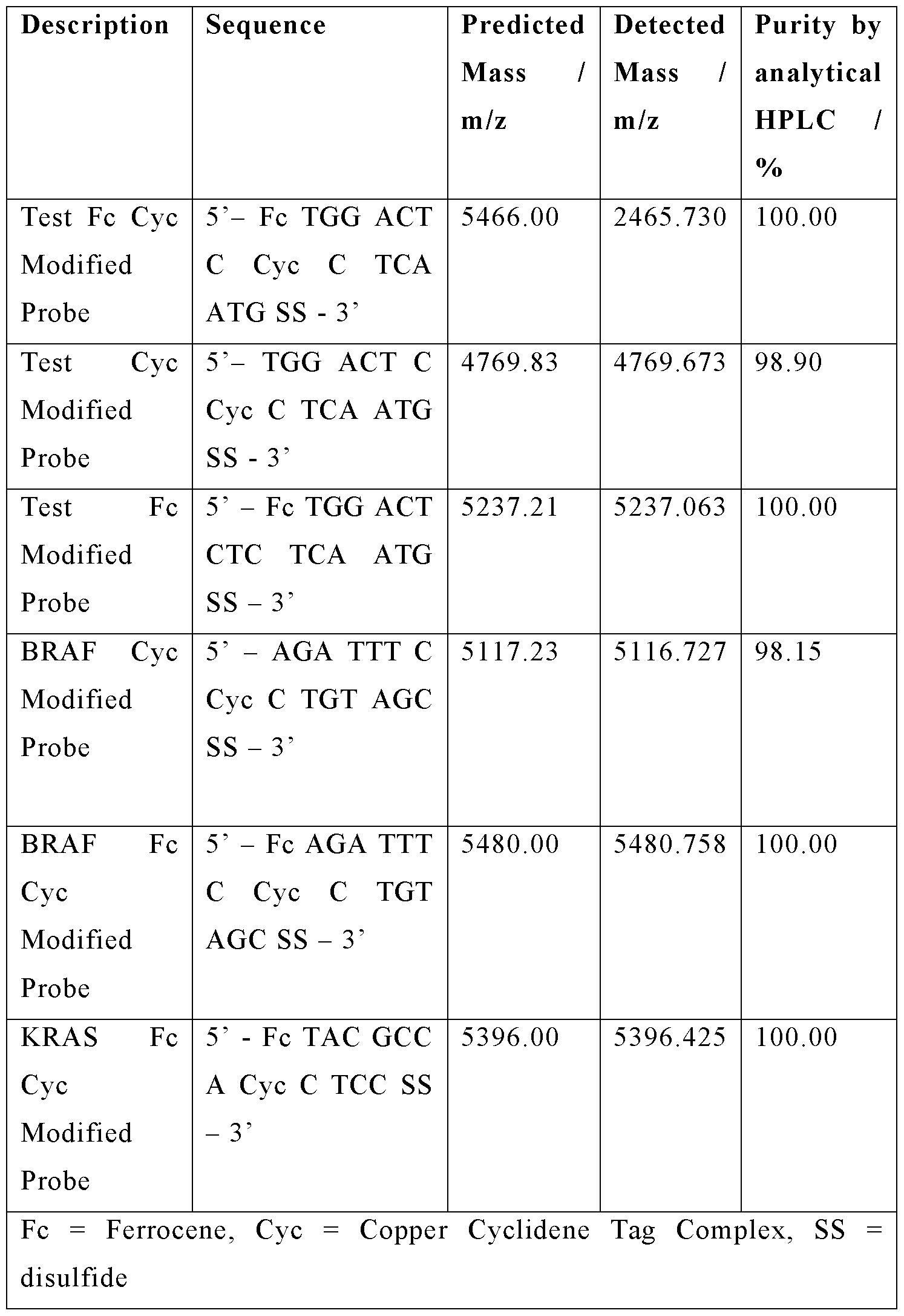 2. Electrochemistry
2.1 Equipment and Electrode Preparation:
Electrochemical measurements were performed on a BioAnalytical Systems Inc. (BASi, West Lafayette, IN, USA) EC epsilon potentiostat using a C3 cell stand. A traditional 3-electrode set-up, consisting of a Ag/AgCl reference electrode (3M KC1), platinum wire counter electrode and a polycrystalline gold disk working electrode, was used throughout. Four working electrodes were used in each experiment, two with a 1.6 mm diameter, two with a 2.0 mm diameter. Reference electrodes and counter electrode were purchased from IJ Cambria (Llanelli, Wales), gold working electrodes were purchased from CH Instruments Inc. (Austin, TX, USA).
All water used was purified on a Merck Millipore Elix-Gradient A10 system (resistivity > 18 mW cm toe < 5 ppb, Millipore, France), for both cleaning and solution preparation. All solutions were thoroughly deoxygenated with argon before use. All glassware and electrochemical cells were cleaned by soaking for several hours in a 1: 1 mixture of ammonia (35%) and hydrogen peroxide (30%), before being copiously rinsed with ultrapure water and being left to soak overnight in ultrapure water. Following a final rinse in ultrapure water, the glassware was dried in an oven for several hours before use.
The platinum wire counter electrode was flame annealed before use, whilst the reference electrode was thoroughly rinsed with ultrapure water. The gold working electrodes were polished with a diamond suspension (1.0 pm) on a polishing pad (BASi, West Lafayette, IN, USA) for 3 mins before subsequent polishing with successively finer grades of alumina slurry; 1.0 pm for 3 mins, 0.3 pm for 3 mins and finally 0.05 pm for 5 mins. Between each step the electrode was washed with ultrapure water. The working electrode was then sonicated for 30 seconds in a deoxygenated 1: 1 mixture of ultrapure water and ethanol.
The working electrode was then subjected to electrochemical cleaning in 0.5 M deoxygenated sulfuric acid. Chronoamperometry was employed to hold the potential at firstly 2 V for 5 s, before -0.35 V for 10 s. A series of cyclic voltammograms (CV) were then recorded between -0.35 V and 1.5 V at scan rates of 4 V s 1 (until consistent, approximately 20 cycles) and 0.1 V s 1 (4 cycles), before a final CV was recorded at
0.5 V s 1. From this final CV, the surface roughness and consequently the geometric area of the electrode were calculated from the gold oxide reduction peak (using a literature value of 482 pC cm 2 to evaluate electrochemical area4, 5). Working electrodes were thoroughly rinsed with ultrapure water and dried under a stream of argon, before SAM fabrication.
2.2 Self-Assembled Monolayer Preparation:
2.0 pL of 100 pM DNA disulphide probe was centrifuged for 30 seconds with 2.0 pL of 10 mM TCEP and incubated at rt for 1 h. The solution was diluted to 200 pL (cone of DNA is 1 pM) with 10 mM sodium phosphate buffer pH 7.0, 1 M sodium perchlorate. The freshly cleaned gold working electrode was soaked in the solution for 2 h at room temperature (rt). The electrode was then rinsed with ultrapure water and soaked in a 2 mM 6-mercapto-l-hexanol, 10 mM sodium phosphate buffer pH 7.0, 1 M sodium perchlorate solution overnight. The electrode was rinsed with ultrapure water, dried under a stream of argon and subjected to electrochemical sensing studies.
2.3 Electrochemical Sensing Procedure:
The working electrode (now functionalised with a SAM of DNA probes) was allowed to equilibrate for 5 mins in the buffer before electrochemical measurements were taken. Square wave voltammetry (SWV) measurements were performed in triplicate, before cyclic voltammograms were recorded. The target strand was then added to the solution and the probe was allowed 20 mins to hybridise, before electrochemical measurements are repeated.
The parameters for measurements were as follows: The potential window was dependent on the buffer conditions used, with a window of -75 mV to 525 mV used for 10 mM tris hydrochloride buffer pH 7.0, 100 mM sodium chloride, whilst a window of -75 mV to 550 mV was used for 10 mM sodium phosphate buffer pH 7.0, 1 M sodium perchlorate. For SWV, a 4 mV potential step, 25 mV amplitude and 200 Hz frequency were employed. CVs were recorded at a range of scan rates to assess the scan rate dependence: 10, 20, 40, 60, 80, 100, 250, 500 and 1000 mV s 1.
The ratio of the cyclidene complex current intensity to the reference ferrocene current intensity (Cyc:Fc) was used to assess the changes in current upon target binding. The ratio was calculated for both the reduction and oxidation peaks in the CV and the
values averaged. The Cyc:Fc ratio for the unbound probe was compared to the ratio exhibited after the target species was added. The CVs recorded at a scan rate of 1000 mV s 1 were used for these calculations due to the strength of the signal, but any scan rate could be used. The data was processed using Origin Student 2019b software (Northampton, MA, USA).
2.4 Single Base Variation Sensing Results:
The probe was hybridised with a series of target strands that varied the nucleobase situated opposite the cyclidene unit in the duplex. The concentration of target added was systematically decreased by an order of magnitude to ascertain the limit of detection of the ratiometric probe. Concentrations between 1 mM and 10 fM were found to give consistent decreases in the Cyc:Fc ratio. Target concentrations of 5 fM, 1 fM and 100 aM also gave decreases in Cyc:Fc ratio, but these were smaller than previously observed and no longer consistent.
The values reported are the average percentage decrease observed in the Cyc:Fc ratio upon target binding. Each value was repeated at least in triplicate and across a variety of polycrystalline gold working electrodes. Due to the consistency of the electrochemical output, the average value incorporates measurements taken between 1 pM and 10 fM target concentrations across four polycrystalline gold working electrodes.
Table 5: Sensing summary of ratiometric probes in 10 mM Tris HCl pH 7.0 100 mM NaCl Buffer. Values reported are the average % decreases in Cyc.Fc current ratio observed upon varying the nucleobase opposite the cyclidene. [Target] = 1 mM to 10 fM.
 Table 6: Sensing summary of ratiometric probes in 10 mM Na Phosphate pH 7.0 1 M NaCl04 Buffer. Values reported are the average % decreases in Cyc.Fc current ratio observed upon varying the nucleobase opposite the cyclidene. [Target] = 1 mM to 10 fM.

Figures 5-8 show results from a test sequence of the ratiometric probe.
Figures 9-10 show results for detecting BRAF V600e sequence with the ratiometric probe.
Figures 11-12 show results for detecting KRAS sequence with the ratiometric probe.
Regeneration of the Unbound Probe:
Following measurements, the working electrode with probe was soaked in ultrapure water and sonicated for 1 min. The electrode was then rinsed with ultrapure water and square wave and cyclic voltammogram measurements were taken as previously stated.
Figure 13 shows that the unbound probe can be regenerated multiple times through 1 minute of sonication in ultrapure water.
Example CuCyFc Probe sensing RNA targets
Materials - SAMs formed on polycrystalline gold disc working electrodes, 1.6 mm or 2.0 mm in diameter.
- Probe sequence (5’ to 3’):
CuCyFc NINA probe [Fc] TGG ACT C[Cu]C TCA ATG [S]
- Target sequences (5’ to 3’):

Sensing Results
Figure 14 shows sensing of NINA_RNA_scram. Figure 15 shows sensing of NINA_RNA_A.
Figure 16 shows sensing of NINA_RNA_U.
Figure 17 shows a summary of the CuCy:Fc current ratio % reduction.
Conclusions · Data demonstrates sensing of complementary RNA targets using the surface- immobilised DNA CuCyFc probe.
• Non-complementary RNA target induced no significant change in the CuCy:Fc current ratio, demonstrating selective sensing.
• Single point variant discrimination (A vs U) demonstrated, matching the analogous A vs T sensing in DNA targets.
Claims
1. A genetic probe for determining the identity of a single targeted nucleotide in a target nucleic acid, wherein the genetic probe comprises: an oligonucleotide with a metal redox-active sensor molecule incorporated therein via a linker group, wherein the metal redox-active sensor molecule is positioned within the oligonucleotide backbone between two bases; a redox-active internal-reference molecule attached to the oligonucleotide; and a surface, wherein the oligonucleotide is anchored to the surface.
2. The genetic probe according to claim 1, wherein the genetic probe comprises a plurality of the oligonucleotides anchored to the surface, and wherein the plurality of oligonucleotides form a monolayer on the surface.
3. The genetic probe according to claim 1 or 2, wherein the metal redox-active sensor molecule comprises a macrocyclic transition metal complex.
4. The genetic probe according to any preceding claim, wherein the metal redox-active sensor molecule is a transition metal complex with a cyclidene [14] ligand as shown below:
 wherein M is Ni(II), Cu(II), Fe(II) or Co(II).
5. The genetic probe according to any preceding claim, wherein the metal for the macrocyclic transition metal complex is Ni(II) or Cu(II).
6. The genetic probe according to any preceding claim, wherein the redox-active internal-reference molecule comprises or consists of an organometallic compound, wherein the metal is a transition metal.
7. The genetic probe according to any preceding claim, wherein the difference in the redox peaks between the redox-active internal-reference molecule and the redox-active sensor molecule is at least about lOOmV.
8. The genetic probe according to any preceding claim, wherein the redox-active internal-reference molecule comprises or consists of ferrocene.
9. The genetic probe according to any preceding claim, wherein the surface is the surface of an electrode.
10. The genetic probe according to any preceding claim, wherein the surface comprises or consists of metal, glass-like carbon, or silica.
11. The genetic probe according to any preceding claim, wherein the surface is a gold surface.
12. The genetic probe according to any preceding claim, wherein the surface is planar or the surface of a nanoparticle.
13. An array of genetic probes, wherein the array of genetic probes comprises two or more genetic probes according to any preceding claim provided on a surface.
14. The array according to claim 13, wherein the genetic probes comprise compartmentalised electrodes.
15. The array according to claim 13 or 14, wherein the oligonucleotide of one genetic probe is arranged to hybridise to a different target nucleic acid relative to another genetic probe on the same surface.
16. The array according to claim 13 or 14, wherein one genetic probe is arranged to interrogate a different nucleotide position in the same target nucleic acid relative to another genetic probe on the same surface.
17. A composition comprising a plurality of genetic probes according to any of claims 1 to 12, optionally wherein the surfaces of the genetic probes are the surfaces of nanoparticles.
18. A method of determining a single point variant nucleotide in a target nucleic acid in a pool of the target nucleic acid, the method comprising:
-providing a genetic probe in according to any of claims 1 to 12, wherein the genetic probe is capable of detecting the single point variant nucleotide, wherein the genetic probe comprises an oligonucleotide that is substantially complimentary to the target nucleic acid, and wherein the redox-active sensor molecule of the genetic probe is in a position that is arranged to be paired with a nucleotide of the target nucleic acid to be interrogated, whereby the electrical signal of the redox-active sensor molecule differs in intensity depending on the nucleotide’s identity or variant structure;
-determining the electrical signal ratio of the metal redox-active sensor molecule relative to the redox-active internal-reference molecule for unbound genetic probe;
-contacting the genetic probe with the pool of target nucleic acid such that the genetic probe hybridises to the target nucleic acid;
-determining the electrical signal ratio of the metal redox-active sensor molecule relative to the redox-active internal-reference molecule for the hybridised genetic probe and target nucleic acid; and
-comparing the ratio exhibited for unbound genetic probe relative to the hybridised genetic probe and target nucleic acid.
19. The method according to claim 18, further comprising determining the presence of, or percentage of, single point variant nucleotides by comparing the change in ratio in relation to a calibration value of a known standard.
20. A method of determining the presence or percentage of a single point variant nucleotide of a target nucleic acid in a pool of the target nucleic acid, the method comprising:
-contacting the pool of target nucleic acid with a genetic probe according to any of claims 1 to 12, wherein the genetic probe is capable of detecting the single point variant nucleotide,
wherein the genetic probe comprises an oligonucleotide that is substantially complimentary to the target nucleic acid, and wherein the redox-active sensor molecule is in a position that is arranged to be paired with a nucleotide of the target nucleic acid to be interrogated, whereby the electrical signal of the redox-active sensor molecule differs in intensity depending on the nucleotide’s identity or variant structure;
- detecting the percentage change in electrical signal intensity ratio of the redox-active sensor molecule when the pool of target nucleic acid is contacted by the genetic probe comprising the redox-active sensor molecule.
21. The method according to claim 20, further comprising determining the presence of, or percentage of, single point variant nucleotides by comparing the percentage change in intensity ratio of the redox-active sensor molecule to a calibration value that has been determined by linear regression of the percentage change in intensity of known standards.
22. A method of determining the percentage of single point variants of a target nucleic acid in a pool of the target nucleic acid, the method comprising:
-contacting the pool of target nucleic acid with the genetic probe according any of claims 1 to 12, which is capable of detecting the single point variants,
-detecting the percentage change in electrical signal intensity of the redox-active sensor molecule when the pool of target nucleic acid is contacted by the oligonucleotide probe comprising the redox-active sensor molecule; and -determining the percentage of single point variants by comparing the percentage change in intensity of the redox-active sensor molecule to a calibration value that has been determined by linear regression of the percentage change in intensity of known standards.
23. The method according to any of claims 18 to 22, wherein the pool of target nucleic acid is in a sample comprising a cell lysate, a bodily fluid sample, or a nucleic acid sample.
24. The method according to any of claims 18 to 23, wherein the target nucleic acid is associated with a disease or condition or a known single nucleotide variant.
25. The method according to any of claims 18 to 24, further comprising the use of a second genetic probe comprising a linker to the metal redox-active sensor molecule of a different length to the relative to the first genetic probe and/or a different linker stereochemistry and/or a different metal redox-active sensor molecule relative to the first genetic probe.
26. The method according to any of claims 18 to 25, further comprising the use of a second redox-active internal-reference molecule or an additional fluorescent tag/reporter on the genetic probe.
27. A method of determining the status of a condition associated with a known single point variant in a subject, the method comprising: providing a sample from the subject comprising a target nucleic acid, wherein the target nucleic acid may comprise the single point variant; determining the presence or percentage of the single point variant in the sample relative to target nucleic acid not having the single point variant in accordance with the method of any of claims 18 to 26, wherein the presence or percentage of the single point variant is indicative of the status of the condition associated with the single point variant in the subject.
28. A method of determining the epigenetic status of a target nucleic acid of a subject, the method comprising determining the presence or percentage of single point variants of the target nucleic acid in accordance with any of claims 18 to 26, wherein the presence or percentage of the single point variants of the target nucleic acid is indicative of the epigenetic status of the target nucleic acid in the subject.
29. Use of a genetic probe in accordance with any of claims 1 to 12, for determining the single point variant ratio or single nucleotide identity of target nucleic acid in a pool of the target nucleic acid.
30. Use of a genetic probe in accordance with any of claims 1 to 12, for diagnosis and/or prognosis of a condition associated with a single point variant in a subject.
31. A kit for the detection of and/or analysis of the ratio of, a single point variant of a target nucleic acid in a pool of the target nucleic acid, wherein the kit comprises:
-the genetic probe according to any of claims 1 to 12, wherein the redox-active sensor molecule is in a position that is arranged to be paired with a nucleotide of the target nucleic acid to be interrogated; and
-a first standard target nucleic acid for use as a standard in a calibration, wherein the first target nucleic acid comprises the single point variant to be analysed; and/or
-a second standard target nucleic acid for use as a standard in calibration, wherein the second target nucleic acid does not comprise the single point variant to be analysed.
Applications Claiming Priority (2)
| Application Number | Priority Date | Filing Date | Title |
|---|---|---|---|
| GBGB2008017.2A GB202008017D0 (en) | 2020-05-28 | 2020-05-28 | Ratiometric sensing |
| GB2008017.2 | 2020-05-28 |
Publications (1)
| Publication Number | Publication Date |
|---|---|
| WO2021240175A1 true WO2021240175A1 (en) | 2021-12-02 |
Family
ID=71526216
Family Applications (1)
| Application Number | Title | Priority Date | Filing Date |
|---|---|---|---|
| PCT/GB2021/051320 Ceased WO2021240175A1 (en) | 2020-05-28 | 2021-05-28 | Ratiometric sensing |
Country Status (2)
| Country | Link |
|---|---|
| GB (1) | GB202008017D0 (en) |
| WO (1) | WO2021240175A1 (en) |
Citations (2)
| Publication number | Priority date | Publication date | Assignee | Title |
|---|---|---|---|---|
| WO2018058028A2 (en) * | 2016-09-25 | 2018-03-29 | The Regents Of The University Of California | Dual-reporter electrochemical sensors with drift correction |
| WO2019043353A1 (en) | 2017-09-01 | 2019-03-07 | The University Of Birmingham | Sensor |
-
2020
- 2020-05-28 GB GBGB2008017.2A patent/GB202008017D0/en not_active Ceased
-
2021
- 2021-05-28 WO PCT/GB2021/051320 patent/WO2021240175A1/en not_active Ceased
Patent Citations (2)
| Publication number | Priority date | Publication date | Assignee | Title |
|---|---|---|---|---|
| WO2018058028A2 (en) * | 2016-09-25 | 2018-03-29 | The Regents Of The University Of California | Dual-reporter electrochemical sensors with drift correction |
| WO2019043353A1 (en) | 2017-09-01 | 2019-03-07 | The University Of Birmingham | Sensor |
Non-Patent Citations (22)
| Title |
|---|
| DOUGAN, J. ET AL., NUCLEIC ACIDS RES., vol. 35, 2007, pages 3668 - 75 |
| DUPREY ET AL., ACS CHEM. BIOL., vol. 11, 2016, pages 717 - 721 |
| DUPREY ET AL., CHEM. COMMUN, vol. 47, 2011, pages 6629 |
| E. XIONGX. ZHANGY. LIUJ. ZHOUP. YUX. LI, J. CHEN, ANAL. CHEM., vol. 87, 2015, pages 7291 - 7296 |
| HURST, S. J. ET AL., ANAL. CHEM., vol. 78, 2006, pages 8313 - 8 |
| J. H. A. DUPREYJ. CARR-SMITHS. L. HORSWELLJ. KOWALSKIJ. H. R. TUCKER, J. AM. CHEM. SOC, vol. 138, 2016, pages 746 - 749 |
| KAUR ET AL., CHEM. COMMUN., vol. 54, 2018, pages 11108 - 11111 |
| LI ET AL., ANAL. CHEM., vol. 88, 2016, pages 883 - 889 |
| LIU ET AL., LANGMUIR, vol. 31, no. 1, 18 December 2014 (2014-12-18), pages 371 - 377, Retrieved from the Internet <URL:https://doi.org/l0.l021/la503917i> |
| LIU ET AL., LANGMUIR, vol. 31, no. 1, 2015, pages 371 - 377 |
| LUBINPLAXCO, ACC. CHEM. RES., vol. 43, 2010, pages 496 - 505 |
| M. T. DE GROOTM. T. M. KOPER, PHYS. CHEM. CHEM. PHYS., vol. 10, 2008, pages 1023 - 1031 |
| PIVIDORI ET AL., BIOSENSORS & BIOELECTRONICS, vol. 15, 2000, pages 191 - 303 |
| SANDHU, K. K. ET AL., BIOCONJUG. CHEM., vol. 13, 2002, pages 3 - 6 |
| SANDSTROM, P. ET AL., LANGMUIR, vol. 19, 2003, pages 7537 - 7543 |
| STOKES, R. J. ET AL., CHEM. COMMUN. (CAMB), 2007, pages 2811 - 2813 |
| SWENSEN ET AL., J. AM. CHEM. SOC., vol. 131, 2009, pages 4262 - 4266 |
| TAKADA TADAO ET AL: "Preparation of ferrocene-functionalized gold nanoparticles by primer extension reaction on the particle surface", BIOORGANIC & MEDICINAL CHEMISTRY LETTERS, ELSEVIER, AMSTERDAM, NL, vol. 24, no. 12, 24 April 2014 (2014-04-24), pages 2661 - 2663, XP028665100, ISSN: 0960-894X, DOI: 10.1016/J.BMCL.2014.04.062 * |
| Y. DUB. J. LIMB. LIY. S. JIANGJ. L. SESSLERA. D. ELLINGTON, ANAL. CHEM., vol. 86, 2014, pages 8010 - 8016 |
| YAMANA K. ET AL: "Electrochemical detection of single-base mismatches in DNA by a redox-active intercalator conjugated oligonucleotide", NUCLEIC ACIDS SYMPOSIUM SERIES, vol. 3, no. 1, 1 September 2003 (2003-09-01), GB, pages 89 - 90, XP055822050, ISSN: 0261-3166, DOI: 10.1093/nass/3.1.89 * |
| ZHANG, X. ET AL., J. AM. CHEM. SOC., 2012, pages 7266 - 7269 |
| ZHAO ET AL., BIORG. MED. CHEM. LETT., vol. 22, 2012, pages 129 |
Also Published As
| Publication number | Publication date |
|---|---|
| GB202008017D0 (en) | 2020-07-15 |
Similar Documents
| Publication | Publication Date | Title |
|---|---|---|
| Han et al. | Facile and label-free electrochemical biosensors for microRNA detection based on DNA origami nanostructures | |
| Ge et al. | A CRISPR/Cas12a-mediated dual-mode electrochemical biosensor for polymerase chain reaction-free detection of genetically modified soybean | |
| JP5389789B2 (en) | Highly specific and sensitive detection based on steric hindrance and enzyme-related signal amplification | |
| Bao et al. | An enzyme-free sensitive electrochemical microRNA-16 biosensor by applying a multiple signal amplification strategy based on Au/PPy–rGO nanocomposite as a substrate | |
| Hocek et al. | Nucleobase modification as redox DNA labelling for electrochemical detection | |
| Zhang et al. | An isothermal electrochemical biosensor for the sensitive detection of microRNA based on a catalytic hairpin assembly and supersandwich amplification | |
| Raoof et al. | Preparation of an electrochemical PNA biosensor for detection of target DNA sequence and single nucleotide mutation on p53 tumor suppressor gene corresponding oligonucleotide | |
| JP2002510791A (en) | An electrochemical sensor using an intercalating redox active moiety | |
| US7202037B2 (en) | Electrochemical sensor using intercalative, redox-active moieties | |
| CA2616259A1 (en) | Electrocatalytic nucleic acid hybridization detection | |
| Hejazi et al. | Electrochemical detection of short sequences of hepatitis C 3a virus using a peptide nucleic acid-assembled gold electrode | |
| EP3320117B1 (en) | Systems for detecting and quantifying nucleic acids | |
| Tan et al. | Electrochemical nucleic acid biosensors: from fabrication to application | |
| Lin et al. | A chronocoulometric LNA sensor for amplified detection of K-ras mutation based on site-specific DNA cleavage of restriction endonuclease | |
| Hu et al. | Conjugated self-doped polyaniline–DNA hybrid as trigger for highly sensitive reagentless and electrochemical self-signal amplifying DNA hybridization sensing | |
| Fojta et al. | Osmium tetroxide complexes as versatile tools for structure probing and electrochemical analysis of biopolymers | |
| Egorova et al. | High-sensitivity label-free electrochemical genosensors for carbon nanotube plasmon-assisted detection of somatic mutations in nucleic acids from formalin-fixed paraffin-embedded tissues | |
| US12043865B2 (en) | Genetic probe for the detection of a single nucleotide polymorphism (SNP) or a single nucleotide modification of a target nucleic acid | |
| WO2021240175A1 (en) | Ratiometric sensing | |
| WO2002073183A1 (en) | Electrochemical detection method of complementarity to nucleic acid bases | |
| Yi et al. | Label-free electrochemical detection of microRNAs via intercalation of hemin into the DNA/RNA hybridization | |
| AU7920100A (en) | Double-strand nucleic acid probes and the use thereof | |
| Di Giusto et al. | Special-purpose modifications and immobilized functional nucleic acids for biomolecular interactions | |
| US20040096859A1 (en) | Method for detecting and/or quantifying an analyte | |
| Takenaka | Threading intercalators as redox indicators |
Legal Events
| Date | Code | Title | Description |
|---|---|---|---|
| 121 | Ep: the epo has been informed by wipo that ep was designated in this application |
Ref document number: 21734892 Country of ref document: EP Kind code of ref document: A1 |
|
| NENP | Non-entry into the national phase |
Ref country code: DE |
|
| 122 | Ep: pct application non-entry in european phase |
Ref document number: 21734892 Country of ref document: EP Kind code of ref document: A1 |