WO2021188055A1 - Modulation of cxcr3 chemokine signalling for osteoporosis therapy - Google Patents
Modulation of cxcr3 chemokine signalling for osteoporosis therapy Download PDFInfo
- Publication number
- WO2021188055A1 WO2021188055A1 PCT/SG2021/050147 SG2021050147W WO2021188055A1 WO 2021188055 A1 WO2021188055 A1 WO 2021188055A1 SG 2021050147 W SG2021050147 W SG 2021050147W WO 2021188055 A1 WO2021188055 A1 WO 2021188055A1
- Authority
- WO
- WIPO (PCT)
- Prior art keywords
- group
- optionally substituted
- receptor antagonist
- bone
- chemokine receptor
- Prior art date
- Legal status (The legal status is an assumption and is not a legal conclusion. Google has not performed a legal analysis and makes no representation as to the accuracy of the status listed.)
- Ceased
Links
Classifications
-
- A—HUMAN NECESSITIES
- A61—MEDICAL OR VETERINARY SCIENCE; HYGIENE
- A61P—SPECIFIC THERAPEUTIC ACTIVITY OF CHEMICAL COMPOUNDS OR MEDICINAL PREPARATIONS
- A61P19/00—Drugs for skeletal disorders
- A61P19/08—Drugs for skeletal disorders for bone diseases, e.g. rachitism, Paget's disease
- A61P19/10—Drugs for skeletal disorders for bone diseases, e.g. rachitism, Paget's disease for osteoporosis
-
- A—HUMAN NECESSITIES
- A61—MEDICAL OR VETERINARY SCIENCE; HYGIENE
- A61K—PREPARATIONS FOR MEDICAL, DENTAL OR TOILETRY PURPOSES
- A61K31/00—Medicinal preparations containing organic active ingredients
- A61K31/33—Heterocyclic compounds
- A61K31/395—Heterocyclic compounds having nitrogen as a ring hetero atom, e.g. guanethidine or rifamycins
- A61K31/495—Heterocyclic compounds having nitrogen as a ring hetero atom, e.g. guanethidine or rifamycins having six-membered rings with two or more nitrogen atoms as the only ring heteroatoms, e.g. piperazine or tetrazines
- A61K31/505—Pyrimidines; Hydrogenated pyrimidines, e.g. trimethoprim
- A61K31/519—Pyrimidines; Hydrogenated pyrimidines, e.g. trimethoprim ortho- or peri-condensed with heterocyclic rings
Definitions
- the present invention belongs to the field of clinical prevention and therapy of osteoporosis.
- Bone homeostasis requires continuous remodeling of bone matrix to maintain structural integrity. This involves extensive communication between bone-forming osteoblasts and bone- resorbing osteoclasts to coordinate progenitor cell recruitment and activation. Only few mediators controlling progenitor activation are known to date and have been targeted for intervention of bone disorders such as osteoporosis. With a strongly increasing incidence of osteoporotic fractures, especially in ageing populations, osteoporosis in an important global health concern.
- osteoblasts and osteoclasts form a functional unit to achieve a balance of bone resorption and formation.
- Deficiencies in this bone cell coupling for example by excess osteoclast activity, lead to reduced bone mineral density and increased bone fracture risk, as observed in osteoporosis patients.
- physiological age-related bone loss is associated with significant changes in bone remodelling characterized by reduced bone cell coupling and decreased bone formation relative to bone resorption, resulting in elevated bone fracture risk.
- osteoclast progenitors which are cells of the macrophage/monocyte lineage
- RANKL receptor-activator of NF-k ligand
- M-CSF macrophage colony stimulating factor
- RANKL forward signalling in osteoblast progenitors delays osteoblast differentiation, which is later released by RANKL reverse signalling through vesicular RANK receptors secreted from osteoclasts demonstrating RANKL’s important role as a coupling factor.
- more coupling factors remain to be identified as osteoclasts also form in a RANKL-independent manner.
- a method for treating osteoporosis comprising the step of administering to a subject in need thereof a CXCR3 chemokine receptor antagonist.
- Fig. 1 is a schematic diagram describing the experimental design.
- This induced rankl expression which in turn activated osteoblast ( col10a1 ) and osteoclast (mpegl) progenitor cells.
- Fig. 2 is a Venn diagram showing number of significantly up-regulated genes in each of osteoblast progenitors (col10a1) and osteoblasts (osx), at 10 dpf, 1 d after Rankl induction. Also shown here is the overlap of up-regulated genes, signified by the overlapping section between the circles. This analysis showed that a small and specific subset of genes was significantly up- regulated in osteoblast progenitors and osteoblasts under Rankl-induced conditions, suggesting a direct response of these cells to the osteoporotic stimulus. It is of note that the 13 commonly up-regulated genes included Cxcl9l.
- Fig. 3 is a heat map showing the change in RNAseq-based expression levels of chemokine ligands (reflected as Z-scores) in triplicates of osteoblast progenitors ( ⁇ H0a1, at 10 and 15 dpf) and osteoclasts (ctsk, at 10 and 12 dpf) without and with Rankl induction at 9 dpf. It is noted that Rankl-induced up-regulation of cxcl9l (last row of the legend on the right “C-X-C motif chemokine 9-like”, denoted by the reference numeral (302)) is shown exclusively in col10a1 cells. While several chemokines were found expressed at basal and variable levels in osteoblast progenitors and osteoclasts, only cxcl9l showed a robust up-regulation in osteoblast progenitors at 1 day post Rankl induction.
- FIG. 4 is a schematic showing an inferred evolutionary history of C-X-C chemokine ligands. Deduced genomic organization of cxcl9l and neighboring genes on ancestral, medaka, and human chromosomes.
- FIG. 5 is a heat map showing the change in RNAseq-based expression levels of chemokine ligands (reflected as Z-scores) in triplicates of osteoblast progenitors ( col10a1 , at 10 and 15 dpf) and osteoclasts ( ctsk , at 10 and 12 dpf) Cxcr3.2 is shown in the seventh row from the top of the legend on the right as “C-X-C- chemokine receptor type 3 isoform X2” with the reference numeral (502). While all identified chemokine receptors are expressed in osteoblast progenitors and osteoclasts at variable levels, cxcr3.2 showed a robust up-regulation in osteoclasts at 2 days post Rankl induction.
- Fig. 6 is a table and histograms showing the results of qPCR validation of cxcl9l and cxcr3.2 regulation in osteoblast progenitors ( col10a1 ) and osteoclasts (ctsk) after Rankl induction.
- A is a table summarizing the baseMean and fold change values obtained by RNAseq and qPCR analysis, respectively. Samples used for RNAseq and qPCR were from independent experiments. The ‘baseMean’ value indicates average of transcript numbers in controls and Rankl-induced cell types for both cell types, respectively.
- (B) depicts the results of qPCR analysis of cxcl9l transcription in FAC-sorted col10a1 (at 10 dpf) and ctsk cells (at 12 dpf) with and without Rankl induction.
- (C) illustrates the results of qPCR analysis of cxcr3.2 transcription in FAC-sorted col10a1 (at 10 dpf) and ctsk cells (at 12 dpf) with and without Rankl induction.
- Three biological and three technical replicates each were analyzed. Error bars show mean value ⁇ SD; * p ⁇ 0.05, ** p ⁇ 0.01 ; n.s. no significant difference, Student’s t-test.
- FIG. 7 is a set of images showing Cxcr3.2 expression and function with respect to osteoblast distribution.
- Panel (A) shows that the expression of cxcr3.2:GFP does not overlap with osx.mCherry expression in osteoblasts without (I and III) or with Rankl induction (II and IV).
- cxcr3.2:GFP cell numbers are increased in centra and neural arches at 1 and 2 dphs. Tight interaction of cxcr3.2:GFP cells with osx.mCherry osteoblasts in arches were noted (arrows denoted by the reference numerals (702)).
- the image inset shows higher magnification view of area with dotted lines.
- Panel (B) shows that, in the absence of ectopic Rankl, osx:GFP osteoblasts are similarly distributed in vertebrae of cxcr3.2+/+ and cxcr3.2-/- siblings at 1 and 2 dphs (arrows denoted by reference numeral (704)).
- osx:GFP cells Upon Rankl induction, osx:GFP cells are redistributed in cxcr3.2+/+ siblings (arrows denoted by reference numeral (706)), as a consequence of ectopic osteoclast differentiation.
- osteoblast distribution in cxcr3.2-/- mutants is not affected (lla, Mb and lid, arrows denoted by reference numeral (708)). Scale bars: 200 pm.
- FIG. 8 is a set of images showing that cxcr3.2 ⁇ GFP is expressed in a subset of macrophages and osteoclasts.
- A Expression of cxcr3.2 ⁇ GFP in a subset of mpegl ⁇ mCherry- positive macrophages without and with Rankl induction, at 1 or 2 dphs.
- Co-expressing cells are labeled with arrows denoted by reference numeral (802), and arrows denoted by reference numeral (804) mark cxcr3.2: G F P- n eg at i ve macrophages.
- FIG. 806 shows fluorescent images of the expression of cxcr3.2 ⁇ GFP in a subset of ctsk. mCherry-positive osteoclasts. Co-expressing cells are labeled with arrows denoted by reference numeral (806), and arrows denoted by reference numeral (810) mark cxcr3.2:G F P- n eg at i ve osteoclasts.
- C shows fluorescent images showing a reduction of cxcr3.2 ⁇ GFP expression during osteoclast maturation (arrows denoted by reference numeral (808)). Scale bars, 200 pm in A and B and 50 pm in C.
- Fig. 9 is a set of graphs showing the quantification of cxcr3.2 ⁇ GFP expression.
- A shows the quantification of cxcr3.2 and mpegl single and double-positive cells.
- B shows the quantification of cxcr3.2 ⁇ GFP expressing macrophages with low or high GFP expression.
- C shows the quantification of cxcr3.2 and ctsk single and double-positive cells.
- a quantification of cell numbers revealed that the number of cxcr3.2 positive macrophages and cxcr3.2 positive osteoclasts is significantly increased after Rankl induction but that the level of cxcr3.2 expression in macrophages diminishes over time as they differentiate into osteoclasts.
- FIG. 10 is a set of images showing generation of medaka cxcl9l and cxcr3.2 mutants.
- A shows the generation of cxcr3.2 CRISPR/Cas9 mutants using three guide RNAs (gRNA1 -3) targeting exon 2 (E2).
- Arrows denoted by reference numeral (1002) indicate position of forward (FP) and reverse (RP) primers for PCR genotyping (WT (+/+): approx. 1.1 kb; homozygous mutants (-/-): approx. 0.2 kb; heterozygous carriers (+/-): 1.1 and 0.2 kb).
- Mutants have a 891 base pair (bp) deletion (section denoted by reference numeral (1004)) between gRNA1 and gRNA3 target sites, leading to a predicted amino acid (a. a.) sequence missing amino acids 38 to 334, as shown in the predicted amino acid sequence, accordingly.
- (B) shows the generation of cxcl9l mutants.
- Two guide RNAs gRNA1 denoted by reference numeral (1006), and gRNA2 denoted by reference numeral (1008)
- E2 exon 2
- E3 exon 3
- FIG. 11 is a set of images showing impaired macrophage recruitment and osteoclast formation in cxcr3.2 mutants.
- A shows a normal distribution of mpegl: mCherry macrophages and cfs/cGFP osteoclasts in cxcr3.2* / ⁇ (I) and cxcr3.2 "A (II) siblings without Rankl induction.
- B shows macrophage and osteoclast distribution in cxcr3.2 (V) and cxcr3.Z' ⁇ (VI) siblings at 11 dpf, 2 days after heat shock to induce Rankl expression (i.e., 2 dphs) in Rankl+ (III) and Rankl- (IV) siblings.
- FIG.12 is a set of images showing the tracking of macrophage migration paths in cxcr3.2* /+
- FIG. 13 is a set of graphs showing the quantification of macrophage dynamics and differentiation in cxcr3.2 mutants.
- the experiments shown in panels (A-C) were performed on sibling embryos obtained from cxcr3.2 heterozygous incrosses, which were induced for Rankl expression, and were imaged for cell counting, and genotyped. Numbers of recruited macrophages (A), total macrophages (B) and ectopic osteoclasts (C) are shown.
- F Number of macrophages moving from the aorta-gonad mesonephros (AGM) towards vertebral column (10-14 hphs, measured in three vertebral bodies), and cells retaining in the AGM (at 14 hphs). More macrophages migrated directly from the AGM to the vertebral column in wild type siblings, while most cxcr3.2-/- mutant macrophages remained in the AGM. Error bars show mean values ⁇ SD, Mann-Whitney test, *p ⁇ 0.05, **p ⁇ 0.01, ****p ⁇ 0.0001 , ns: non-significant; 2 ⁇ Nmovies ⁇ 5.
- R- Rankl negative; R+: Rankl positive; Dr: Directed recruitment towards vertebral column; Pe: Persistence in the AGM; Mf: macrophage; OCs: osteoclasts. Quantification of cell numbers shows that Cxcr3.2 is essential for the recruitment of macrophages to the bone matrix of vertebral bodies and their differentiation into osteoclasts under osteoporotic conditions. The motility of macrophages is reduced in cxcr3.2 deficient mutants, leading to a significant reduction of directed migration towards vertebral bodies and retainment of macrophages at the place of origin (AGM).
- AGM place of origin
- Fig. 14 is a set of higher-magnification views of regions in the dotted boxes in Fig. 11 B.
- macrophages are found in neural arches and vertebral bodies at 1 dphs (la, lb, lc). Macrophage numbers are increased at 2 dphs (lla, Mb, lie), and macrophages start to differentiate into ctsk osteoclasts.
- cxcr3. mutants Ilia, lllb, lllc and IVa, IVb, IVc
- macrophages remain in AGM and fail to migrate to vertebral bodies.
- Fig. 15 is a set of images showing Alizarin Red staining of mineralized matrix in cxcr3.2 ⁇ /+ (I, III) and cxcr3.2 ⁇ ; ⁇ (II, IV) siblings without (I, II) and with (III, IV) Rankl induction.
- Arrows denoted by reference numeral (1502) mark intact neural arches, centra and cleithrum, and arrows denoted by reference numeral (1504) indicate absent arches and severe lesions in centra and cleithrum.
- Mf macrophage
- OC osteoclast
- na neural arch. It is thus shown that the integrity of mineralized bone matrix is protected in cxcr3.2 deficient mutants under osteoporotic conditions.
- Fig. 16 is a set of images showing that the Cxcr3.2 inhibitor NBI-74330 interferes with macrophage recruitment and osteoclast differentiation. Trunk regions of triple transgenic rankliHSEicfp/ctskGFP/mpegl .mCherry larvae treated with DMSO as a negative control (con, la, lb, lia, lib) or NBI-74330 (NBI, Ilia, lllb, IVa, IVb) for 3 hours, followed by heat shock to induce Rankl and imaging at 1 (la, lb, Ilia, lllb) and 2 dphs (lla, lib, IVa, IVb).
- ctsk osteoclasts are labeled with white arrows. (Scale bars, 500 pm). The dotted boxes in Ilia and lllb are described further in Fig. 18. It is therefore understood that inhibition of Cxcr3.2 activity under osteoporotic conditions using the chemical antagonist NBI-74330 results in a reduction of macrophage recruitment and absence of formed osteoclasts.
- Fig. 17 is a set of images showing that the Cxcr3.2 inhibitor NBI-74330 interferes with macrophage recruitment and osteoclast differentiation, specifically of confocal images of macrophages (Mf) and osteoclasts (OC) in vertebral bodies.
- Mf macrophages
- OC osteoclasts
- Rankl induction (I) macrophages are confined to myosepta in the aorta-gonad mesonephros (AGM) of control larvae (control (A); arrowheads denoted by reference numeral (1702)), while they are dispersed in AGM of NBI-treated larvae (NBI (B); arrowheads denoted by reference numeral (1704)).
- Alizarin Red staining shows severe lesions of mineralized matrix in majority of control larvae after Rankl induction, with neural arches resorbed and large lesions in centra ((a) arrows denoted by reference numeral (1716)).
- mineralization defects denoted by reference numeral (1716)
- b with persistent mineralized neural arches and intact centra
- Areas with absent mineralization in centra were quantified (Fig. 24) and are depicted in bar graph at bottom; Mann-Whitney L/test, **** P ⁇ 0.0001 .
- Fig. 18 is a set of graphs showing the quantification of recruited macrophages (A), total number of macrophages (B), and ectopic osteoclasts (C) in areas indicated by dotted boxes in Fig. 16. Data are represented as mean number of cells ⁇ SD, * P ⁇ 0.05, ** P ⁇ 0.01 , *** P ⁇ 0.001 , **** P ⁇ 0.0001 ; ns, nonsignificant, Brown-Forsythe ANOVA with Tukey’s multiple comparisons test, 17 ⁇ N Larvae £ 36, from three independent experiments. The quantification of cell numbers as shown here confirms that the Cxcr3.2 inhibitor NBI-74330 reduces macrophage recruitment to the vertebral bodies and diminishes osteoclast differentiation under osteoporotic conditions.
- FIG. 19 is a set of images showing that AMG487 treatment protects bone upon Rankl induction.
- A shows the results of injection of 20 pM AMG487 into the yolk of rankl:HSE:cfp/mpeg1 :mCherry/ctsk:GFP transgenic embryos two hours prior to heat-shock for Rankl induction (II, IV, VI, VIII, X, XII, XIV).
- Control embryos I, III, V, VII, XI, X and XIII
- Alizarin Red staining of mineralized matrix shows severe bone lesions in control embryos (arrows denoted by reference numeral (1906), Xlllb) and less lesions after AMG487 treatment (black arrows (1906), XlVb).
- Normally mineralized bone under Rankl- (non-osteoporotic conditions) and Rankl+ (osteoporotic conditions) are indicated with arrows denoted by reference numeral (1908).
- Box denoted by reference numeral (1910) (III) indicates region for cell quantification; box denoted by reference numeral (1912) (V) shows region imaged with higher magnification.
- AGM aorta-gonad-mesonephros
- Mf macrophage
- OC osteoclast
- na neural arch
- V vertebra.
- Scale bar 1 mm (whole embryo image), 100 pm (zoom-in image).
- B) shows a graph illustrating the number of macrophages recruited to vertebral column.
- C) is a graph depicting the total number of macrophages in trunk (including AGM).
- D) is a graph showing the number of ectopic osteoclasts in vertebral column.
- FIG. 20 is a set of images showing the results of macrophage analysis after ectopic cxcl9l expression in osx osteoblasts.
- A shows a schematic depicting a strategy for macrophage analysis after ectopic cxcl9l expression in osx osteoblasts.
- mpeg1 ⁇ mCherry transgenic embryos were injected with osx:cxc/9/-p2a-EGFP plasmid and l-Scel meganuclease at one-cell stage, raised to 12 to 14 dpf, and screened for mosaic osx:cxc/9/-p2a-EGFP expression in osteoblasts located at vertebral bodies.
- FIG. B shows confocal images showing single macrophages present at neural arches of noninjected osx:GFP/mpegT:mCherry control larvae ((I); arrows denoted by reference numeral (2002) indicate position of endogenous osx positive osteoblasts in neural arches, n 5 fish).
- Enhanced recruitment of macrophages (arrows denoted by reference numeral (2004)) toward cxc/9/-p2a-EGFP expressing cells (arrow denoted by reference numeral (2006)) in neural arch of osx:cxc/9-p2a-EGFP injected larva ((II); n 7/7 fish).
- Fig. 21 is a set of images showing the results of osteoclast analysis upon ectopic cxcl9l expression in osx-expressing osteoblasts.
- (A) shows a schematic depicting a strategy for osteoclast analysis upon ectopic cxcl9l expression in osx-expressing osteoblasts.
- cte/cGFP transgenic embryos were injected with osx ⁇ cxcl9l- p2a-mCherry plasmid and l-Scel meganuclease at one-cell stage, raised to 12 to 14 dpf, and screened for mosaic osx ⁇ cxcl9l- p2a-mCherry expression in osteoblasts at vertebral bodies.
- FIG. B shows confocal images showing absence of osteoclast formation (arrowheads denoted by reference numeral (2102) indicate position of endogenous osx positive osteoblasts in neural arches) in the vertebral bodies of uninjected osx:mCherry/cte/oGFP larvae ((I); control, n 7 fish).
- Asterisks indicate auto-fluorescent pigment cells at yolk region (Scale bars, 50 pm).
- Fig. 22 is a set of images showing macrophage and osteoclast distribution in cxc/9/-/- mutants without Rankl induction and after ectopic Cxcl9l overexpression.
- A shows images indicating that in the absence of Rankl induction, cxcl9l-/- mutants (denoted by reference numeral 2204) develop normally and show similar distribution of mpeg1 ⁇ mCherry and ctsl ⁇ cGFP cells as cxcl9l+/+ (denoted by reference numeral 2202) siblings. Scale bars: 100 pm.
- FIG. B shows further fluorescent images indicating the distribution of cxcl9l-EGFP expressing cells (light grey) and mpeg1 ⁇ mCherry macrophages (dark grey) in vertebral bodies of osx:cxc/9-p2a-GFP injected larva at 10 and 14 dpf.
- An accumulation of mpeg1 ⁇ mCherry positive macrophages can be seen around Cxcl9l-producing cells in the region of the neural arches, as evident at two different time points. This is not the case in non-injected embryos (without Cxcl9l overexpression).
- Boxes denoted by reference numeral (2206) demarcate neural arches with Cxcl9l overexpression
- boxes denoted by reference numeral (2208) indicate neural arch regions without Cxcl9l overexpression
- boxes denoted by reference numeral (2210) indicate centra of vertebral bodies.
- the graph below shows quantification of macrophages in neural arches without and with Cxcl9l overexpression.
- C shows fluorescent images illustrating the distribution of cxcl9l-mCherry expressing cells (dark grey) and cte/cGFP positive osteoclasts (light grey) in vertebral bodies of osx ⁇ cxd9- p2a-mCherry injected larva at 10 and 14 dpf.
- the dark grey colour indicates cells over-expressing Cxcl9l and light grey colour indicates osteoclasts. It should be noted that the vertebral bodies are only in the centre of the images. Fluorescence observed in the periphery is unspecific auto-fluorescence. Arrowheads denoted by reference numeral (2212) indicate osteoclasts in vicinity of Cxcl9l producing cells, arrowheads denoted by reference numeral (2214) label osteoclasts away from Cxcl9l producing cells. Scale bars: 100 pm in (B) and (C).
- this figure shows that ectopic overexpression of Cxcl9l in clones of cells in the vertebral bodies resulted in an accumulation of macrophages and ectopic differentiation of osteoclasts in the vicinity of Cxcl9l producing cells. This demonstrates that Cxcl9l is sufficient to induce macrophage recruitment and osteoclast differentiation.
- Fig. 23 is a set of images showing normal macrophage recruitment in cxcl9l mutants after Rankl induction.
- (A) shows overview and high-magnification confocal images of cxcl9l +/+ (I, III) and cxc/9/ ⁇ /_ (II, IV) siblings in mpeg 1 -.mCherry/ rankhHSE-.cfp transgenic background at 1 dphs (10 dpf).
- Image III is an expansion of the box denoted by reference numeral (2302) of image I
- image IV is an expansion of the box denoted by reference numeral (2304) of image II.
- Fig. 24 is a set of images showing impaired osteoclast formation and reduced bone lesions in cxcl9l mutants after Rankl induction.
- A shows an overview and confocal images of ctsl ⁇ oGFP- expressing osteoclasts (arrows denoted by reference numeral (2406)) in in cxcl9l +/+ (I, III) and cxc/9f /_ (II, IV) siblings at 2 dphs.
- Image III is an expansion of the box denoted by reference numeral (2402) of image I
- image IV is an expansion of the box denoted by reference numeral (2404) of image II (Scale bars, 100 pm).
- FIG. B shows a graph quantification of cte/oGFP osteoclast numbers in vertebral column (region marked as boxes in A). This figure shows that despite normal macrophage recruitment, osteoclast formation is strongly reduced in cxcl9l deficient mutants under osteoporotic conditions. This demonstrates that Cxcl9l is essential for osteoclast differentiation.
- FIG. 25 is a set of images showing impaired osteoclast formation and reduced bone lesions in cxcl9l mutants after Rankl induction.
- A shows an overview and high-magnification views of Alizarin Red stained mineralized matrix in in cxc/9F /+ (I, III) and cxc/9/ /_ (II, IV) siblings at 12 dpf (3 dphs). Wild-type siblings, but not mutants, show enhanced resorption of mineralized matrix at cleithrum (arrowheads denoted by reference numeral (2502)), neural arches (arrowheads denoted by reference numeral (2504)), and vertebral bodies (arrowheads denoted by reference numeral (2506)).
- Image III is an expansion of the box denoted by reference numeral (2508) of image I
- image IV is an expansion of the box denoted by reference numeral (2510) of image II (Scale bars, 100 pm.).
- (B) shows a graph quantification of bone lesions in vertebral column (position indicated by boxes in A; for method, see Fig. 24). Error bars indicate mean ⁇ SD, ** P ⁇ 0.01 , *** P ⁇ 0.001; ns, not significant, unpaired Student’s two-tailed t test. It is therefore shown that bone degeneration induced by osteoporotic Rankl induction is ameliorated under cxcl9l- deficient conditions.
- FIG. 26 is a set of images showing quantification of bone degeneration.
- A shows an example of Alizarin Red stained vertebral column used for quantification of bone degeneration.
- V7-V11 refer to the 7th to 11th vertebral bodies, respectively.
- A’ shows an example of how to quantify bone degeneration, the postulated V area (enclosed by dotted lines denoted by reference numeral (2602)) and area resorbed (enclosed by dotted lines denoted by reference numeral (2602)) for V7-V11 were determined using ImageJ.
- B shows an equation used for calculation of percentage (%) bone degeneration using values obtained from the analysis indicated in (A’).This figure illustrates how defects in the mineralized bone matrix of vertebral bodies were quantitated in osteoporotic medaka fish.
- Fig. 27 is a set of images showing bone development in cxcl9l mutants.
- A shows that in the absence of Rankl induction (I), Alizarin Red staining revealed normal bone mineralization in the cleithrum (arrows denoted by reference numeral (2702)), neural arches (arrows denoted by reference numeral (2704)) and vertebral bodies (arrows denoted by reference numeral (2706)) of cxcl9F mutants as compared to cxd9t /+ siblings at 12 dpf (II).
- FIG. 2 shows the results after Rankl induction, whereby calcein staining revealed less severe bone lesions in cxd9t / (II, IV) mutants due to reduced osteoclast formation.
- Image III is an expansion of the box denoted by reference numeral (2708) of image I
- image IV is an expansion of the box denoted by reference numeral (2710) of image II. Scale bars: 100 pm.
- this figure shows that in the absence of Cxcl9l, bone degeneration defects in osteoporotic vertebral bodies after Rankl induction are ameliorated and bone integrity is increased.
- Fig. 28 is a set of images showing that osteoclast differentiation after Rankl induction is absent after osteoblasts have been experimentally ablated suggesting that osteoblasts are the source of signals needed for macrophage recruitment and osteoclast differentiation.
- A shows a schematic experimental timeline for osteoblast ablation. osx:mCherry-NTRo larvae were treated with Metronidazole (MTZ) at 9 dpf for 24 hours, and heat shock was applied at 10 dpf to induce Rankl expression.
- (B) shows fluorescent images of osteoblasts (magenta (I, II, V, VI)) and osteoclasts (green (III, IV, VII, VIII)) imaged at 1 and 2 dphs.
- osteoblasts and osteoclasts form normally in controls (I, III, V, VI, arrows denoted by reference numeral (2802) and arrows denoted by reference numeral (2804), respectively) but are absent in osteoblast-ablated larvae (II, IV, VI, VIII, arrowheads denoted by reference numeral (2802) and arrowheads denoted by reference numeral (2804); arrowhead denoted by reference numeral (2806) highlights remaining osteoclasts).
- This figure shows that the experimental ablation of osteoblasts identified osteoblasts as the source of signals required for macrophage recruitment and osteoclast differentiation under osteoporotic conditions.
- Fig. 29 is a set of images showing that osteoclast formation depends on the presence of osteoblasts.
- A shows that metronidazole (MTZ) treatment results in efficient ablation of osteoblasts in osx:mCherry-NTRo/ctsk:GFP embryos (II) when compared to osx:mCherry/dsk:GFP control embryos (I).
- B shows that after ablation of osteoblasts (II, IV), Rankl fails to stimulate the formation of ectopic osteoclasts (arrows denoted by reference numeral (2902)) compared to massive osteoclast formation in control embryos (I, III, arrows denoted by reference numeral (2904)).
- (C) shows a graph quantifying osteoclast numbers, showing mean number ⁇ SD, Brown-Forsythe ANOVA with Games Howell’s test, *p ⁇ 0.05, **p ⁇ 0.01, 9 ⁇ N ⁇ 11, from two independent experiments.
- OB osteoblast
- OC osteoclast.
- D MTZ treatment does not affect normal bone development (I, control-MTZ; Rankl-). Ablation of osteoblasts protects bone from lesions by Rankl induction (III, IV, Rankl+; arrowheads denoted by reference numeral (2906), arrowheads denoted by reference numeral (2908)). Scale bars: 1 mm for A, 200 pm for B and D.
- FIG. 30 is a schematic diagram showing chemokine control of osteoclast recruitment under osteoporotic conditions (left) and the blocking of osteoclast recruitment by the inhibition of Cxcr3 activity by Cxcr3 antagonists (right).
- Rankl acts on osteoblast progenitors located at the mineralized bone matrix of vertebral bodies and either directly or indirectly induces the production and release of Cxcl9l. Freely diffusing Cxcl9l then activates macrophages that express the Cxcr3.2 receptor.
- CXCR3 chemokine receptor refers to a Ga, protein-coupled receptor of the CXC chemokine receptor family.
- Other names for CXCR3 are, for example, G protein-coupled receptor 9 (GPR9) and CD183.
- GPR9 G protein-coupled receptor 9
- CD183 CD183.
- CXCR3-A CXCR3-A
- CXCR3-B chemokine receptor 3-alternative
- CXCR3-A binds to the CXC chemokines CXCL9 (MIG), CXCL10 (IP-10), and CXCL11 (l-TAC) whereas CXCR3-B can also bind to CXCL4 in addition to CXCL9, CXCL10, and CXCL11.
- CXCR3 chemokine receptor refers to all isoforms of CXCR3.
- Antagonist refers to a receptor ligand that inhibits or dampens a biological response by binding to and blocking a receptor, rather than activating it.
- An antagonist has affinity, but no efficacy, for their cognate receptors, and the binding of the antagonist to the cognate receptor will disrupt the interaction and inhibit the function of an antagonist or inverse agonist at receptors.
- An antagonist can achieve its potency, for example, by competing with endogenous ligands or substrates at structurally defined binding sites on the cognate receptors.
- Osteoporosis refers to a systemic skeletal disorder characterized by low bone mass, micro-architectural deterioration of bone tissue leading to bone fragility, and consequent increase in fracture risk.
- the group may be a terminal group or a bridging group”. This is intended to signify that the use of the term is intended to encompass the situation where the group is a linker between two other portions of the molecule as well as where it is a terminal moiety.
- alkyl alkyl
- some publications would use the term “alkylene” for a bridging group and hence in these other publications there is a distinction between the terms “alkyl” (terminal group) and “alkylene” (bridging group). In the present application no such distinction is made and most groups may be either a bridging group or a terminal group.
- optionally substituted means the group to which this term refers may be unsubstituted, or may be substituted with one or more groups independently selected from acyl, alkyl, alkenyl, alkynyl, thioalkyl, cycloalkyl, cycloalkylalkyl, cycloalkenyl, cycloalkylalkenyl, heterocycloalkyl, cycloalkylheteroalkyl, cycloalkyloxy, cycloalkenyloxy, cycloamino, halo, carboxyl, haloalkyl, haloalkynyl, alkynyloxy, heteroalkyl, heteroalkenyl heteroalkynyl, heteroalkyloxy, hydroxyl, hydroxyalkyl, alkoxy, thioalkoxy, alkenyloxy, haloalkoxy, haloalkenyl, haloalkyny
- examples of acyl include acetyl, benzoyl and amino acid derived aminoacyl.
- the group may be a terminal group or a bridging group. If the group is a terminal group it is bonded to the remainder of the molecule through the carbonyl carbon.
- the group may be a terminal group or a bridging group. If the group is a terminal group, it is bonded to the remainder of the molecule through the nitrogen atom.
- the group may be a terminal group or a bridging group. If the group is a terminal group, it is bonded to the remainder of the molecule through the oxygen atom.
- Alkenyl as a group or part of a group denotes an aliphatic hydrocarbon group containing at least one carbon-carbon double bond and which may be straight or branched preferably having 2-12 carbon atoms, more preferably 2-10 carbon atoms, most preferably 2-6 carbon atoms, in the normal chain.
- the group may contain a plurality of double bonds in the normal chain and the orientation about each is independently E or Z.
- Exemplary alkenyl groups include, but are not limited to, ethenyl, propenyl, butenyl, pentenyl, hexenyl, heptenyl, octenyl and nonenyl.
- the group may be a terminal group or a bridging group.
- alkenyloxy refers to an alkenyl-O- group in which alkenyl is as defined herein.
- Preferred alkenyloxy groups are C2-C12 alkenyloxy groups.
- the group may be a terminal group or a bridging group. If the group is a terminal group it is bonded to the remainder of the molecule through the oxygen atom.
- Alkyl as a group or part of a group refers to a straight or branched aliphatic hydrocarbon group, preferably a C1-C12 alkyl, more preferably a C1-C10 alkyl, most preferably C1-C6 unless otherwise noted.
- suitable straight and branched C1-C6 alkyl substituents include methyl, ethyl, n-propyl, 2-propyl, n-butyl, sec-butyl, t-butyl, hexyl, and the like.
- the group may be a terminal group or a bridging group.
- Alkylamino includes both mono-alkylamino and dialkylamino, unless specified.
- “Mono- alkylamino” means a Alkyl-NH- group, in which alkyl is as defined herein.
- Dialkylamino means a (alkyl)2N- group, in which each alkyl may be the same or different and are each as defined herein for alkyl.
- the alkyl group is preferably a C1-C12 alkyl group.
- the group may be a terminal group or a bridging group. If the group is a terminal group it is bonded to the remainder of the molecule through the nitrogen atom.
- the group may be a terminal group or a bridging group. If the group is a terminal group it is bonded to the remainder of the molecule through the carbonyl carbon.
- Alkylaryl means an alkyl-aryl- group in which the aryl and alkyl moieties are as defined herein. Preferred alkylaryl groups contain a C1-12 alkyl moiety. The group may be a terminal group or a bridging group. If the group is a terminal group it is bonded to the remainder of the molecule through the aryl group.
- Alkyloxy refers to an alkyl group as defined herein that is singularly bonded to oxygen.
- the group may be a terminal group or a bridging group. If the group is a terminal group, it is bonded to the remainder of the molecule through the alkyl group.
- Alkyloxyalkyl refers to an alkyloxy-alkyl- group in which the alkyloxy and alkyl moieties are as defined herein.
- the group may be a terminal group or a bridging group. If the group is a terminal group it is bonded to the remainder of the molecule through the alkyl group.
- Alkyloxyaryl refers to an alkyloxy-aryl- group in which the alkyloxy and aryl moieties are as defined herein.
- the group may be a terminal group or a bridging group. If the group is a terminal group it is bonded to the remainder of the molecule through the aryl group.
- the alkyl group is preferably a C1-C12 alkyl group. Examples include, but are not limited to, methoxycarbonyl and ethoxycarbonyl.
- the group may be a terminal group or a bridging group. If the group is a terminal group it is bonded to the remainder of the molecule through the carbonyl carbon.
- Alkyloxycycloalkyl refers to an alkyloxy-cycloalkyl- group in which the alkyloxy and cycloalkyl moieties are as defined herein.
- the group may be a terminal group or a bridging group. If the group is a terminal group it is bonded to the remainder of the molecule through the cycloalkyl group.
- Alkyloxyheteroaryl refers to an alkyloxy-heteroaryl- group in which the alkyloxy and heteroaryl moieties are as defined herein.
- the group may be a terminal group or a bridging group. If the group is a terminal group it is bonded to the remainder of the molecule through the heteroaryl group.
- Alkyloxyheterocycloalkyl refers to an alkyloxy-heterocycloalkyl- group in which the alkyloxy and heterocycloalkyl moieties are as defined herein.
- the group may be a terminal group or a bridging group. If the group is a terminal group it is bonded to the remainder of the molecule through the heterocycloalkyl group.
- the alkyl group is preferably a C1-C12 alkyl group.
- Exemplary alkylsulfinyl groups include, but not limited to, methylsulfinyl and ethylsulfinyl.
- the group may be a terminal group or a bridging group. If the group is a terminal group it is bonded to the remainder of the molecule through the sulfur atom.
- the alkyl group is preferably a C1-C12 alkyl group. Examples include, but not limited to methylsulfonyl and ethylsulfonyl.
- the group may be a terminal group or a bridging group. If the group is a terminal group it is bonded to the remainder of the molecule through the sulfur atom.
- Alkynyl as a group or part of a group means an aliphatic hydrocarbon group containing a carbon-carbon triple bond and which may be straight or branched preferably having from 2-12 carbon atoms, more preferably 2-10 carbon atoms, more preferably 2-6 carbon atoms in the normal chain.
- Exemplary structures include, but are not limited to, ethynyl and propynyl.
- the group may be a terminal group or a bridging group.
- Alkynyloxy refers to an alkynyl-O- group in which alkynyl is as defined herein. Preferred alkynyloxy groups are C2-C12 alkynyloxy groups. The group may be a terminal group or a bridging group. If the group is a terminal group it is bonded to the remainder of the molecule through the oxygen atom.
- Amino refers to groups of the form -NR a R b wherein R a and R b are individually selected from the group including but not limited to hydrogen, optionally substituted alkyl, optionally substituted alkenyl, optionally substituted alkynyl, and optionally substituted aryl groups.
- Aminoalkyl means an Nh -alkyl- group in which the alkyl group is as defined herein.
- the group may be a terminal group or a bridging group. If the group is a terminal group it is bonded to the remainder of the molecule through the alkyl group.
- the group may be a terminal group or a bridging group. If the group is a terminal group it is bonded to the remainder of the molecule through the sulfur atom.
- Aryl as a group or part of a group denotes (i) an optionally substituted monocyclic, or fused polycyclic, aromatic carbocycle (ring structure having ring atoms that are all carbon) preferably having from 5 to 12 atoms per ring.
- aryl groups include phenyl, naphthyl, and the like; (ii) an optionally substituted partially saturated bicyclic aromatic carbocyclic moiety in which a phenyl and a C5-7 cycloalkyl or C5-7 cycloalkenyl group are fused together to form a cyclic structure, such as tetrahydronaphthyl, indenyl or indanyl.
- the group may be a terminal group or a bridging group.
- an aryl group is a C6-C18 aryl group.
- Arylalkenyl means an aryl-alkenyl- group in which the aryl and alkenyl are as defined herein.
- exemplary arylalkenyl groups include phenylallyl.
- the group may be a terminal group or a bridging group. If the group is a terminal group it is bonded to the remainder of the molecule through the alkenyl group.
- Arylalkyl means an aryl-alkyl- group in which the aryl and alkyl moieties are as defined herein. Preferred arylalkyl groups contain a C1-12 alkyl moiety. Exemplary arylalkyl groups include benzyl, phenethyl, 1 -naphthalenemethyl and 2-naphthalenemethyl. The group may be a terminal group or a bridging group. If the group is a terminal group it is bonded to the remainder of the molecule through the alkyl group.
- Arylalkyloxy refers to an aryl-alkyl-O- group in which the alkyl and aryl are as defined herein.
- the group may be a terminal group or a bridging group. If the group is a terminal group it is bonded to the remainder of the molecule through the oxygen atom.
- Arylamino includes both mono-arylamino and di-arylamino unless specified.
- Mono-arylamino means a group of formula aryINH-, in which aryl is as defined herein
- di-arylamino means a group of formula (aryl) N- where each aryl may be the same or different and are each as defined herein for aryl.
- the group may be a terminal group or a bridging group. If the group is a terminal group it is bonded to the remainder of the molecule through the nitrogen atom.
- Arylheteroalkyl means an aryl-heteroalkyl- group in which the aryl and heteroalkyl moieties are as defined herein.
- the group may be a terminal group or a bridging group. If the group is a terminal group it is bonded to the remainder of the molecule through the heteroalkyl group.
- Aryloxy refers to an aryl-O- group in which the aryl is as defined herein.
- the aryloxy is a Cs-Cisaryloxy, more preferably a Cs-Cioaryloxy.
- the group may be a terminal group or a bridging group. If the group is a terminal group it is bonded to the remainder of the molecule through the oxygen atom.
- the group may be a terminal group or a bridging group. If the group is a terminal group it is bonded to the remainder of the molecule through the sulfur atom.
- a “bond” is a linkage between atoms in a compound or molecule.
- the bond may be a single bond, a double bond, or a triple bond.
- Cycloalkenyl means a non-aromatic monocyclic or multicyclic ring system containing at least one carbon-carbon double bond and preferably having from 5-12 carbon atoms per ring.
- Exemplary monocyclic cycloalkenyl rings include cyclopentenyl, cyclohexenyl or cycloheptenyl.
- the cycloalkenyl group may be substituted by one or more substituent groups.
- a cycloalkenyl group typically is a C5-C12 alkenyl group. The group may be a terminal group or a bridging group.
- Cycloalkyl refers to a saturated monocyclic or fused or spiro polycyclic, carbocycle preferably containing from 3 to 12 carbons per ring, such as cyclopropyl, cyclobutyl, cyclopentyl, cyclohexyl and the like, unless otherwise specified. It includes monocyclic systems such as cyclopropyl and cyclohexyl, bicyclic systems such as decalin, and polycyclic systems such as adamantane.
- a cycloalkyl group typically is a C3-C12 alkyl group. The group may be a terminal group or a bridging group.
- Cycloalkylalkyl means a cycloalkyl-alkyl- group in which the cycloalkyl and alkyl moieties are as defined herein.
- Exemplary monocycloalkylalkyl groups include cyclopropylmethyl, cyclopentylmethyl, cyclohexylmethyl and cycloheptylmethyl.
- the group may be a terminal group or a bridging group. If the group is a terminal group it is bonded to the remainder of the molecule through the alkyl group.
- Cycloalkylalkenyl means a cycloalkyl-alkenyl- group in which the cycloalkyl and alkenyl moieties are as defined herein.
- the group may be a terminal group or a bridging group. If the group is a terminal group, it is bonded to the remainder of the molecule through the alkenyl group.
- Cycloalkylheteroalkyl means a cycloalkyl-heteroalkyl- group in which the cycloalkyl and heteroalkyl moieties are as defined herein.
- the group may be a terminal group or a bridging group. If the group is a terminal group, it is bonded to the remainder of the molecule through the heteroalkyl group.
- Cycloalkyloxy refers to a cycloalkyl-O- group in which cycloalkyl is as defined herein.
- the cycloalkyloxy is a C3-Ci2cycloalkyloxy. Examples include, but are not limited to, cyclopropanoxy and cyclobutanoxy.
- the group may be a terminal group or a bridging group. If the group is a terminal group, it is bonded to the remainder of the molecule through the oxygen atom.
- Cycloalkenyloxy refers to a cycloalkenyl-O- group in which the cycloalkenyl is as defined herein.
- the cycloalkenyloxy is a C3-Ci2cycloalkenyloxy.
- the group may be a terminal group or a bridging group. If the group is a terminal group, it is bonded to the remainder of the molecule through the oxygen atom.
- Cycloamino refers to a saturated monocyclic, bicyclic, or polycyclic ring containing at least one nitrogen in at least one ring. Each ring is preferably from 3 to 10 membered, more preferably 4 to 7 membered.
- the group may be a terminal group or a bridging group. If the group is a terminal group it is bonded to the remainder of the molecule through the nitrogen atom.
- Haloalkyl may be used interchangeably with “alkyl halide” and refers to an alkyl group as defined herein in which one or more of the hydrogen atoms has been replaced with a halogen atom selected from the group consisting of fluorine, chlorine, bromine and iodine.
- a haloalkyl group typically has the formula C n H (2n+i-m) X m wherein each X is independently selected from the group consisting of F, Cl, Br and I .
- n is typically from 1 to 10, more preferably from 1 to 6, most preferably 1 to 3.
- m is typically 1 to 6, more preferably 1 to 3.
- Examples of haloalkyl include fluoromethyl, difluoromethyl and trifluoromethyl.
- Haloalkenyl refers to an alkenyl group as defined herein in which one or more of the hydrogen atoms has been replaced with a halogen atom independently selected from the group consisting of F, Cl, Br and I.
- Haloalkynyl refers to an alkynyl group as defined herein in which one or more of the hydrogen atoms has been replaced with a halogen atom independently selected from the group consisting of F, Cl, Br and I.
- Heteroalkyl refers to a straight- or branched-chain alkyl group preferably having from 2 to 12 carbons, more preferably 2 to 6 carbons in the chain, one or more of which has been replaced by a heteroatom selected from S, O, P and N.
- exemplary heteroalkyls include alkyl ethers, secondary and tertiary alkyl amines, amides, alkyl sulfides, and the like.
- heteroalkyl also include hydroxyCi-Cealkyl, Ci-C 6 alkyloxyCrC 6 alkyl, aminoCrCealkyl, CrCealkylaminoCr Cealkyl, and di(Ci-C 6 alkyl)aminoCi-C 6 alkyl.
- the group may be a terminal group or a bridging group.
- Heteroalkenyl refers to a straight- or branched-chain alkenyl group preferably having from 2 to 12 carbons, more preferably 2 to 6 carbons in the chain, one or more of which has been replaced by a heteroatom selected from S, O, P and N.
- exemplary heteroalkenyls include alkenyl ethers, secondary and tertiary alkenyl amines, amides, alkenyl sulfides, and the like.
- heteroalkenyl also include hydroxyCrCealkenyl, CrCealkyloxyCrCealkenyl, aminoCr Cealkenyl, CrC 6 alkylaminoCi-C 6 alkenyl, and di(Ci-C 6 alkyl)aminoCi-C 6 alkenyl.
- the group may be a terminal group or a bridging group.
- Heteroalkynyl refers to a straight- or branched-chain alkenyl group preferably having from 2 to 12 carbons, more preferably 2 to 6 carbons in the chain, one or more of which has been replaced by a heteroatom selected from S, O, P and N.
- exemplary heteroalkynyls include alkynyl ethers, secondary and tertiary alkynyl amines, amides, alkynyl sulfides, and the like.
- heteroalkynyl also include hydroxyCi-Cealkynyl, Ci-C 6 alkyloxyCi-C 6 alkynyl, aminoCi-Cealkynyl, Ci-C 6 alkylaminoCi-C 6 alkynyl, and di(Ci-C 6 alkyl)aminoCi-C 6 alkynyl.
- the group may be a terminal group or a bridging group.
- Heteroalkyloxy refers to an heteroalkyl-O- group in which heteroalkyl is as defined herein.
- the heteroalkyloxy is a CrCi2heteroalkyloxy.
- the group may be a terminal group or a bridging group.
- Heteroaryl either alone or part of a group refers to groups containing an aromatic ring (preferably a 5 or 6 membered aromatic ring) having one or more heteroatoms as ring atoms in the aromatic ring with the remainder of the ring atoms being carbon atoms. Suitable heteroatoms include nitrogen, oxygen and sulphur.
- heteroaryl examples include thiophene, benzothiophene, benzofuran, benzimidazole, benzoxazole, benzothiazole, benzisothiazole, naphtho[2,3- b]thiophene, furan, isoindolizine, xantholene, phenoxatine, pyrrole, imidazole, pyrazole, pyridine, pyrazine, pyrimidine, pyridazine, tetrazole, indole, isoindole, 1 H-indazole, purine, quinoline, isoquinoline, phthalazine, naphthyridine, quinoxaline, cinnoline, carbazole, phenanthridine, acridine, phenazine, thiazole, isothiazole, phenothiazine, oxazole, isooxazole, furazane, phen
- a heteroaryl group is typically a C1-C18 heteroaryl group.
- a heteroaryl group may comprise 3 to 8 ring atoms.
- a heteroaryl group may comprise 1 to 3 heteroatoms independently selected from the group consisting of N, O and S.
- the group may be a terminal group or a bridging group.
- Heteroarylalkyl means a heteroaryl-alkyl group in which the heteroaryl and alkyl moieties are as defined herein. Preferred heteroarylalkyl groups contain a lower alkyl moiety. Exemplary heteroarylalkyl groups include pyridylmethyl. The group may be a terminal group or a bridging group. If the group is a terminal group it is bonded to the remainder of the molecule through the alkyl group.
- Heteroarylalkenyl means a heteroaryl-alkenyl- group in which the heteroaryl and alkenyl moieties are as defined herein.
- the group may be a terminal group or a bridging group. If the group is a terminal group it is bonded to the remainder of the molecule through the alkenyl group.
- Heteroarylheteroalkyl means a heteroaryl-heteroalkyl- group in which the heteroaryl and heteroalkyl moieties are as defined herein.
- the group may be a terminal group or a bridging group. If the group is a terminal group it is bonded to the remainder of the molecule through the heteroalkyl group.
- Heteroarylamino refers to groups containing an aromatic ring (preferably 5 or 6 membered aromatic ring) having at least one nitrogen and at least another heteroatom as ring atoms in the aromatic ring, preferably from 1 to 3 heteroatoms in at least one ring. Suitable heteroatoms include nitrogen, oxygen and sulphur.
- Arylamino and aryl is as defined herein.
- the group may be a terminal group or a bridging group. If the group is a terminal group it is bonded to the remainder of the molecule through the nitrogen atom.
- Heteroaryloxy refers to a heteroaryl-O- group in which the heteroaryl is as defined herein.
- the heteroaryloxy is a CrCisheteroaryloxy.
- the group may be a terminal group or a bridging group. If the group is a terminal group it is bonded to the remainder of the molecule through the oxygen atom.
- Heterocyclic refers to saturated, partially unsaturated or fully unsaturated monocyclic, bicyclic or polycyclic ring system containing at least one heteroatom selected from the group consisting of nitrogen, sulfur and oxygen as a ring atom.
- heterocyclic moieties include heterocycloalkyl, heterocycloalkenyl and heteroaryl.
- Heterocycloalkenyl refers to a heterocycloalkyl as defined herein but containing at least one double bond.
- a heterocycloalkenyl group typically is a C1-C12 heterocycloalkenyl group.
- the group may be a terminal group or a bridging group.
- Heterocycloalkyl refers to a saturated monocyclic, bicyclic, or polycyclic ring containing at least one heteroatom selected from nitrogen, sulfur, oxygen, preferably from 1 to 3 heteroatoms in at least one ring. Each ring is preferably from 3 to 10 membered, more preferably 4 to 7 membered.
- heterocycloalkyl substituents include pyrrolidyl, tetrahydrofuryl, tetrahydrothiofuranyl, piperidyl, piperazyl, tetrahydropyranyl, morphilino, 1 ,3-diazapane, 1 ,4- diazapane, 1 ,4-oxazepane, and 1 ,4-oxathiapane.
- a heterocycloalkyl group typically is a C1-C12 heterocycloalkyl group.
- a heterocycloalkyl group may comprise 3 to 8 ring atoms.
- a heterocycloalkyl group may comprise 1 to 3 heteroatoms independently selected from the group consisting of N, O and S. The group may be a terminal group or a bridging group.
- Heterocycloalkylalkyl refers to a heterocycloalkyl-alkyl- group in which the heterocycloalkyl and alkyl moieties are as defined herein.
- exemplary heterocycloalkylalkyl groups include (2-tetrahydrofuryl)methyl, (2-tetrahydrothiofuranyl) methyl.
- the group may be a terminal group or a bridging group. If the group is a terminal group it is bonded to the remainder of the molecule through the alkyl group.
- Heterocycloalkylalkenyl refers to a heterocycloalkyl-alkenyl- group in which the heterocycloalkyl and alkenyl moieties are as defined herein.
- the group may be a terminal group or a bridging group. If the group is a terminal group it is bonded to the remainder of the molecule through the alkenyl group.
- Heterocycloalkylheteroalkyl means a heterocycloalkyl-heteroalkyl- group in which the heterocycloalkyl and heteroalkyl moieties are as defined herein.
- the group may be a terminal group or a bridging group. If the group is a terminal group it is bonded to the remainder of the molecule through the heteroalkyl group.
- Heterocycloalkyloxy refers to a heterocycloalkyl-O- group in which the heterocycloalkyl is as defined herein.
- the heterocycloalkyloxy is a CrCeheterocycloalkyloxy.
- the group may be a terminal group or a bridging group. If the group is a terminal group it is bonded to the remainder of the molecule through the oxygen atom.
- Heterocycloalkenyloxy refers to a heterocycloalkenyl-O- group in which heterocycloalkenyl is as defined herein.
- the Heterocycloalkenyloxy is a C1-C6 Heterocycloalkenyloxy.
- the group may be a terminal group or a bridging group. If the group is a terminal group it is bonded to the remainder of the molecule through the oxygen atom.
- Heterocycloamino refers to a saturated monocyclic, bicyclic, or polycyclic ring containing at least one nitrogen and at least another heteroatom selected from nitrogen, sulfur, oxygen, preferably from 1 to 3 heteroatoms in at least one ring. Each ring is preferably from 3 to 10 membered, more preferably 4 to 7 membered.
- the group may be a terminal group or a bridging group. If the group is a terminal group it is bonded to the remainder of the molecule through the nitrogen atom.
- Hydroalkyl refers to an alkyl group as defined herein in which one or more of the hydrogen atoms has been replaced with an OH group.
- a hydroxyalkyl group typically has the formula C n H ( 2n +i -x ) (OH)x.
- n is typically from 1 to 10, more preferably from 1 to 6, most preferably from 1 to 3.
- x is typically from 1 to 6, more preferably from 1 to 4.
- the group may be a terminal group or a bridging group. If the group is a terminal group it is bonded to the remainder of the molecule through the sulfur atom.
- the group may be a terminal group or a bridging group. If the group is a terminal group it is bonded to the remainder of the molecule through the nitrogen atom.
- the group may be a terminal group or a bridging group. If the group is a terminal group it is bonded to the remainder of the molecule through the sulfur atom.
- the group may be a terminal group or a bridging group. If the group is a terminal group it is bonded to the remainder of the molecule through the nitrogen atom.
- compositions of this disclosure may include, but are not limited to, ion exchangers, alumina, aluminum stearate, lecithin, serum proteins, such as human serum albumin, buffer substances such as phosphates, glycine, sorbic acid, potassium sorbate, partial glyceride mixtures of saturated vegetable fatty acids, water, salts or electrolytes, such as protamine sulfate, disodium hydrogen phosphate, potassium hydrogen phosphate, sodium chloride, zinc salts, colloidal silica, magnesium trisilicate, polyvinyl pyrrolidone, cellulose-based substances, polyethylene glycol, sodium carboxymethylcellulose, polyacrylates, waxes, polyethylene-polyoxypropylene-block poly
- salts include acid addition salts and base addition salts.
- Such salts be formed by conventional means, for example by reaction of a free acid or a free base form of a compound of formula I with one or more equivalents of an appropriate acid or base, optionally in a solvent, or in a medium in which the salt is insoluble, followed by removal of said solvent, or said medium, using standard techniques (e.g. in vacuo, by freeze-drying or by filtration). Salts also be prepared by exchanging a counter-ion of a compound of formula I in the form of a salt with another counter-ion, for example using a suitable ion exchange resin.
- Examples of pharmaceutically acceptable salts include acid addition salts derived from mineral acids and organic acids, and salts derived from metals such as sodium, magnesium, or preferably, potassium and calcium.
- Examples of acid addition salts include acid addition salts formed with acetic, 2,2-dichioroacetic, adipic, aiginic, aryl suiphonic acids (e.g. benzenesulphonic, naphthalene-2-su!phonic, naphthalene- 1 ,5-disu!phonic and p- toiuenesuiphonic), ascorbic (e.g.
- D-glucuronic D-glucuronic
- glutamic e.g. L-glutamic
- a-oxoglutaric glycolic, hippuric, hydrobromic, hydrochloric, hydriodic, isethionic
- lactic e.g. (+)-L-lactic and ( ⁇ )-DL-iactie
- lactobionic maleic, malic (e.g.
- salts are salts derived from mineral acids such as hydrochloric, hydrobromic, phosphoric, metaphosphoric, nitric and sulphuric acids; from organic acids, such as tartaric, acetic, citric, malic, lactic, fumaric, benzoic, glycolic, gluconic, succinic, aryisuiphonic acids; and from metals such as sodium, magnesium, or preferably, potassium and calcium.
- mineral acids such as hydrochloric, hydrobromic, phosphoric, metaphosphoric, nitric and sulphuric acids
- organic acids such as tartaric, acetic, citric, malic, lactic, fumaric, benzoic, glycolic, gluconic, succinic, aryisuiphonic acids
- metals such as sodium, magnesium, or preferably, potassium and calcium.
- solvates are solvates formed by the incorporation into the solid state structure (e.g. crystal structure) of the compounds of the invention of molecules of a non-toxic pharmaceutically acceptable solvent (referred to below as the solvating solvent).
- a non-toxic pharmaceutically acceptable solvent referred to below as the solvating solvent.
- solvents include water, alcohols (such as ethanol, isopropanol and butanol) and dimethy!sulphoxide.
- Solvates can be prepared by recrystallising the compounds of the invention with a solvent or mixture of solvents containing the solvating solvent.
- Whether or not a solvate has been formed in any given instance can be determined by subjecting crystals of the compound to analysis using well known and standard techniques such as thermogravimetric analysis (TGE), differential scanning calorimetry (DSC) and X-ray crystallography.
- TGE thermogravimetric analysis
- DSC differential scanning calorimetry
- X-ray crystallography X-ray crystallography.
- the solvates can be stoichiometric or non-stoichiometric solvates.
- Particularly preferred solvates are hydrates, and examples of hydrates include hernihydrates, monohydrates and dihydrates.
- “Derivatives” of compounds of formula I as defined herein includes ester derivatives and/or derivatives that have, or provide for, the same biological function and/or activity as any relevant compound of the invention. Thus, for the purposes of this invention, the term also includes prodrugs of compounds of formula I.
- prodrug of a relevant compound of formula i includes any compound that, following oral or parenteral administration, is metabolised in vivo to form that compound in an experimentally-detectable amount, and within a predetermined time (e.g. within a dosing interval of between 6 and 24 hours (i.e. once to four times daily)).
- Prodrugs of compounds of formula I may be prepared by modifying functional groups present on the compound in such a way that the modifications are cleaved, in vivo when such prodrug is administered to a mammalian subject. The modifications typically are achieved by synthesizing the parent compound with a prodrug substituent.
- Prodrugs include compounds of formula I wherein a hydroxyl, amino, su!fhydryl, carboxyl or carbonyl group in a compound of formula I is bonded to any group that may be cleaved in vivo to regenerate the free hydroxyl, amino, sulfhydryl, carboxyl or carbonyl group, respectively.
- Examples of prodrugs include, but are not limited to, esters and carbamates of hydroxyl functional groups, esters groups of carboxyl functional groups, N-acyl derivatives and N-Mannich bases.
- Some of the compounds of the disclosed embodiments may exist as single stereoisomers, racemates, and/or mixtures of enantiomers and /or diastereomers. All such single stereoisomers, racemates and mixtures thereof, are intended to be within the scope of the subject matter described and claimed.
- compounds of the invention may contain more than one asymmetric carbon atom.
- the use of a solid line to depict bonds to asymmetric carbon atoms is meant to indicate that all possible stereoisomers are meant to be included.
- the use of a solid line to depict bonds to one or more asymmetric carbon atoms in a compound of the invention and the use of a solid or dotted wedge to depict bonds to other asymmetric carbon atoms in the same compound is meant to indicate that a mixture of diastereomers is present.
- the term "about”, in the context of concentrations of components of the formulations, typically means +/- 5% of the stated value, more typically +/- 4% of the stated value, more typically +/- 3% of the stated value, more typically, +/- 2% of the stated value, even more typically +/- 1% of the stated value, and even more typically +/- 0.5% of the stated value.
- Osteoporosis is a bone disease characterized by reduced bone mass, deteriorated bone structure, and a high risk of bone fractures that mainly affects post-menopausal women and elderly men.
- a transgenic medaka fish model that expresses medaka Rankl under control of an inducible promoter was used.
- Such a model is able to show inducible expression of Receptor activator of nuclear factor kappa-B ligand (Rankl), and that ectopic osteoclasts trigger excessive bone resorption, as assessed by live imaging in intact specimen.
- the model has been used to show that excessive bone resorption leads to bone mineralization defects that can be prevented by treatment with bisphosphonates, similar to the situation in human osteoporosis patients.
- Cxcr3.2 is expressed in macrophages, which are recruited to the mineralized matrix, where they differentiate into osteoclasts, the major cells that actively control bone resorption.
- the mutant and inhibitor studies demonstrated that Cxcr3.2 is essential for macrophage recruitment to bone resorption sites. That is, chemokine signalling through Cxcr3 is shown to be responsible to recruit osteoclast precursors in a directed and targeted manner to bone matrix and drives their differentiation into bone resorbing osteoclasts. This establishes osteoblast-derived Cxcl9l and the macrophage receptor Cxcr3.2 as druggable components in bone cell coupling, thus presenting a mechanism to control osteoclast progenitor recruitment to sites of bone resorption.
- osteoblast progenitors up-regulate expression of the chemokine ligand Cxcl9l (Figs. 3 and 6). Ectopic expression of Cxcl9l recruits mpegl- positive macrophages to bone matrix and triggers their differentiation into osteoclasts independent from other osteoclast- inducing factors (such as RANKL) (Figs. 20 and 21).
- the chemokine receptor Cxcr3.2 is expressed in a distinct subset of macrophages in the aorta-gonad-mesonephros (AGM) (Fig. 8). Live imaging revealed that upon Cxcl9l up-regulation, Cxcr3.2 positive macrophages get activated, migrate to bone matrix and differentiate into osteoclasts (Figs. 8, 9, 12).
- Cxcr3 inhibitors have previously been generated and clinically tested in the context of inflammatory diseases.
- AMG487 had entered a Phase II clinical trial for psoriasis that was stopped for lack of efficacy.
- Two structurally related Cxcr3 antagonists NBI-74330 and AMG487 are commercially available (Tocris Biosciences, USA). Both inhibitors have been generated by Amgen, and were previously tested in pre-clinical and clinical trials for inflammatory diseases.
- LPS lipopolysaccharide
- CXCR3 inhibitors have been highly effective in the context of several inflammatory diseases, but their role and efficacy in an osteoporosis context remains unclear.
- both inhibitors significantly reduced osteoclast recruitment and differentiation and strongly improved bone integrity under osteoporotic conditions.
- Cxcr3 activity blocks osteoclast recruitment rather than the activity of pre existing osteoclasts.
- Commonly used osteoporosis drugs such as bisphosphonates, target the activity of already existing osteoclasts. This causes significant problems after long-term drug use as old bone does not get renewed by osteoblasts that fail to receive instructive signals from the blocked osteoclasts. It results in failures in bone remodeling and maintenance, leading to increased risk of bone fractures.
- Cxcr3 inhibition targets osteoclast precursor recruitment at an early stage in a dose dependent manner, which is a benefit over existing osteoporosis drugs.
- identification and specific targeting of osteoclast precursor cells at their site of origin the number of forming and active osteoclasts can be controlled without generally blocking activity of already existing osteoclasts.
- Treatment with Cxcr3 antagonists thus may prevent excessive osteoclast formation but allow recruitment of healthy numbers of osteoclasts to ensure physiological bone maintenance and remodeling in patients prone to develop osteoporosis.
- the use of a CXCR3 chemokine receptor antagonist can block osteoclast recruitment, rather than the activity of pre-existing osteoclasts. That is, Cxcr3 inhibition can target osteoclast precursor recruitment at an early stage in a dose dependent manner.
- This can overcome the shortcomings of pre-existing osteoporosis drugs, such as bisphosphonates, which target the activation of already existing osteoclasts and causes significant problems after long term drug use, as old bone is not renewed by osteoblasts that fail to receive instructive signals from the blocked osteoclasts, resulting in failures in bone remodeling and maintenance, leading to increased risk of bone fractures.
- the number of forming and active osteoclasts can advantageously be controlled without generally blocking activity of already existing osteoclasts.
- Treatment with Cxcr3 antagonists can therefore advantageously prevent excessive osteoclast formation, but allow recruitment of healthy numbers of osteoclasts to ensure physiological bone maintenance and remodeling in patients prone to developing osteoporosis.
- Cxcr3 activity has therefore been shown to be crucial in the context of osteoporosis. This is the first to show that Cxcl9l and Cxcr3 are essential druggable regulators of osteoclast recruitment and bone homeostasis.
- Cxcr3 activity can be used to target osteoclasts to desired sites of bone remodeling in patients suffering from osteoclast deficiency and therefore excessive bone formation, for example in osteopetrosis and Fibrodysplasia ossificans progressive (FOP).
- FOP Fibrodysplasia ossificans progressive
- the method comprising the step of administering to a subject in need thereof a CXCR3 chemokine receptor antagonist.
- the CXCR3 chemokine receptor antagonist can be, but is not limited to, a small molecule antagonist, having a molecular weight of less than 900 Da, less than 800 Da, or less than 700 Da.
- the CXCR3 chemokine receptor antagonist can have a molecular weight in the range of about 400 Da to about 900 Da, about 400 Da to about 500 Da, about 400 Da to about 600 Da, about 400 Da to about 700 Da, about 400 Da to about 800 Da, about 500 Da to about 600 Da, about 500 Da to about 700 Da, about 500 Da to about 800 Da, about 500 Da to about 900 Da, about 600 Da to about 700 Da, about 600 Da to about 800 Da, about 600 Da to about 900 Da, about 700 Da to about 800 Da, about 700 Da to about 900 Da or about 800 Da to about 900Da.
- the CXCR3 chemokine receptor antagonist can have a backbone structure which can comprise, for example, quinazolinone or azaquinazolinone.
- the CXCR3 chemokine receptor antagonist comprises quinazolinone.
- the CXCR3 chemokine receptor antagonist comprises azaquinazolinone.
- the CXCR3 chemokine receptor antagonist may have the following formula (I), or may be a pharmaceutically acceptable salt, solvate or derivative thereof: wherein A 1 , A 2 and A 3 may be independently CH or N;
- R 1 may be -CN, optionally substituted alkyl or optionally substituted heteroalkyl; and R 2 and R 3 may be independently an optionally substituted alkyl, optionally substituted heteroalkyl, optionally substituted alkylsulfonyl, optionally substituted heterocycloalkyl, optionally substituted heterocycloalkylheteroalkyl, optionally substituted arylalkyl, optionally substituted arylheteroalkyl, optionally substituted heteroarylalkyl, or optionally substituted heteroarylheteroalkyl.
- a 1 and A 2 may be CH and A 3 may be N.
- R 1 may be heteroalkyl.
- the heteroalkyl may be an alkyloxy or an alkyl ether.
- R 1 may be methoxy, ethoxy, propxy or butoxy.
- R 1 may be ethoxy.
- R 2 may be heteroarylalkyl.
- the alkyl of the heteroarylakly may be methyl, ethyl, propyl or butyl.
- the alkyl of the heteroarylakly may be methyl.
- the heteroaryl of the heteroarylalkyl may be optionally substituted pyridyl.
- R 2 may be 3-methylpyridyl.
- R 3 may be substituted arylalkyl.
- the alkyl of the arylalkyl may be a methyl, ethyl, propyl or butyl.
- the alkyl of the arylalkyl may be a methyl.
- the aryl of the arylalkyl may be optionally substituted benzyl.
- the substitution of the arylalkyl may be on the aryl.
- R 3 may be substituted benzyl.
- the benzyl may be substituted with halogen, optionally substituted alkyl halide or optionally substituted ether.
- the CXCR3 chemokine receptor antagonist may have the following formula (II), or may be a pharmaceutically acceptable salt, solvate or derivative thereof: wherein
- R 4 may be an optionally substituted alkyl
- R 5 , R 6 and R 7 may be independently halogen, optionally substituted alkyl halide or optionally substituted ether
- Z may be N + -0 or N.
- R 4 may be methyl, ethyl, propyl or butyl. R 4 may be ethyl.
- the halogen of R 5 , R 6 or R 7 may be F, Cl, Br or I.
- the optionally substituted alkyl halide of R 5 , R 6 or R 7 may be a trihaloalkyl.
- the alkyl of the trihaloalkyl may be methyl, ethyl, propyl or butyl.
- the optionally substituted alkyl halide of R 5 , R 6 or R 7 may be trihalomethyl.
- the optionally substituted alkyl halide of R 5 may be trifluoromethyl.
- the optionally substituted ether of R 5 , R 6 or R 7 may be an ether of an optionally substituted alkyl.
- the optionally substituted alkyl may be a haloalkyl.
- the haloalkyl may be a trihaloalkyl.
- the alkyl of the trihaloalkyl may be methyl, ethyl, propyl or butyl.
- the optionally substituted ether of R 5 , R 6 or R 7 may be an ether of trihalomethyl.
- the optionally substituted ether of R 5 , R 6 or R 7 may be an ether of trifluoromethyl.
- R 5 may be -CF 3
- R 6 may be F and R 7 may be H.
- R 5 may be H
- R 6 may be -O-CF3 and R 7 may be H.
- the CXCR3 chemokine receptor antagonist may have the following structure, or may be a pharmaceutically acceptable salt, solvate or derivative thereof:
- the CXCR3 chemokine receptor antagonist may be present as the (R)-enantiomer, (S)- enantiomer or a mixture thereof.
- the CXCR3 chemokine receptor antagonist may be present as the (R)-enantiomer.
- the CXCR3 chemokine receptor antagonist may have the following structure, or may be a pharmaceutically acceptable salt, solvate or derivative thereof:
- treatment refers to any and all uses which remedy a disease state or symptoms, prevent the establishment of disease, or otherwise prevent, hinder, retard, or reverse the progression of disease or other undesirable symptoms in any way whatsoever.
- treatment refers to any and all uses which remedy a disease state or symptoms, prevent the establishment of disease, or otherwise prevent, hinder, retard, or reverse the progression of disease or other undesirable symptoms in any way whatsoever.
- One skilled in the art would be able to determine effective, non-toxic dosage levels of the
- CXCR3 chemokine receptor antagonist and an administration pattern which would be suitable for treating osteoporosis to which the CXCR3 chemokine receptor antagonist is applicable.
- the CXCR3 chemokine receptor antagonist can be administered, or is to be administered, at a dosage level in the range of about 0.001 mg to about 100 mg, about 0.001 mg to about 0.01 mg, about 0.001 mg to about 0.1 mg, about 0.001 mg to about 1 mg, about 0.001 mg to about 10 mg, about 0.01 to about 0.1 mg, about 0.01 mg to about 1 mg, about 0.1 mg to about 10 mg, about 0.01 to about 100 mg, about 0.1 mg to about 1 mg, about 0.1 mg to about 10 mg, about 0.1 mg to about 100 mg, about 1 mg to about 10 mg, about 1 mg to about 100 mg or about 10 mg to about 100 mg per kg patient body weight per day.
- the CXCR3 chemokine receptor antagonist can be administered, or is to be administered, at a dosage level in the range of about 0.01 mg/kg to about 25 mg/kg per day, about 0.05 mg/kg to about 10 mg/kg per day, about 0.1 mg/kg to about 5 mg/kg per day, about 0.005 mg/kg to about 0.05 mg/kg per day, about 0.05 mg/kg to about 0.5 mg/kg per day, or about 0.5 mg/kg to about 5 mg/kg per day.
- the CXCR3 chemokine receptor antagonist is to be administered in single or multiple doses. In one example, the CXCR3 chemokine receptor antagonist is to be administered in a single, double, triple or quadruple dose. In another example, the CXCR3 chemokine receptor antagonist can be, or is to be, administered at an interval of, but not limited to, hourly, daily, twice daily, thrice daily, 4 times a day, every second day, every third day, every fourth day, every fifth day, every sixth day, weekly, biweekly, bimonthly, monthly, or combinations thereof.
- Examples of convenient modes of administration include, but are not limited to, injection (subcutaneous, intravenous, etc.), oral administration, inhalation, transdermal application, topical creams or gels or powders, or rectal administration.
- the CXCR3 chemokine receptor antagonist can be coated, for example, with a material to protect the antagonist from the action of enzymes, acids and other natural conditions which may inactivate the therapeutic activity of the antagonist.
- the CXCR3 chemokine receptor antagonist can also be administered parenterally or intraperitoneally.
- the CXCR3 chemokine receptor antagonist is to be administered orally.
- the CXCR3 chemokine receptor antagonist can be administered orally, for example, with an inert diluent or an assimilable edible carrier.
- the CXCR3 chemokine receptor antagonist and other ingredients can also be enclosed in a hard or soft shell gelatin capsule, compressed into tablets, or incorporated directly into an individual's diet.
- the CXCR3 chemokine receptor antagonist can be incorporated with excipients and used in the form of ingestible tablets, buccal tablets, troches, capsules, elixirs, suspensions, syrups, wafers, and the like.
- the oral administration can be in the form of tablets containing about 1 mg to about 1500 mg, about 1 mg to about 2 mg, about 1 mg to about 5 mg, about 1 mg to about 10 mg, about 1 mg to about 20 mg, about 1 mg to about 50 mg, about 1 mg to about 100 mg, about 1 mg to about 200 mg, about 1 mg to about 500 mg, about 2 mg to about 5 mg, about 2 mg to about 10 mg, about 2 mg to about 20 mg, about 2 mg to about 50 mg, about 2 mg to about 100 mg, about 2 mg to about 200 mg, about 2 mg to about 500 mg, about 2 mg to about 1500 mg, about 5 mg to about 10 mg, about 5 mg to about 20 mg, about 5 mg to about 50 mg, about 5 mg to about 100 mg, about 5 mg to about 200 mg, about 5 mg to about 500 mg, about 5 mg to about 1500 mg, about 10 mg to about 20 mg, about 10 mg to about 50 mg, about 10 mg to about 100 mg, about 10 mg to about 200 mg, about 10 mg to about 500 mg, about 10 mg to about 1500 mg, about 10
- the tablet can contain 1 mg, 5 mg, 10 mg, 15 mg, 20 mg, 25 mg, 50 mg, 75 mg, 100 mg, 150 mg, 200 mg, 250 mg, 300 mg, 400 mg, 500 mg, 600 mg, 750 mg, 800 mg, 900 mg, 1000 mg, 1200 mg or 1500 mg of the CXCR3 chemokine receptor antagonist.
- the CXCR3 chemokine receptor antagonist can be administered alone.
- the CXCR3 chemokine receptor antagonist can be administered as a pharmaceutical, veterinarial, or industrial formulation which comprises at least one compound according to the invention.
- the CXCR3 chemokine receptor antagonist can also be present as suitable salts, including pharmaceutically acceptable salts.
- the medicament as disclosed herein, can further comprise a pharmaceutically acceptable excipient.
- pharmaceutically acceptable excipient is intended to include, but is not limited to, solvents, dispersion media, coatings, anti-bacterial and anti-fungal agents, isotonic and absorption delaying agents, and the like.
- the use of such media and agents for pharmaceutically active substances is well known in the art. Except insofar as any conventional media or agent is incompatible with the CXCR3 chemokine receptor antagonist, use thereof in the therapeutic compositions and methods of treatment and prophylaxis is contemplated. Supplementary active compounds may also be incorporated. It is especially advantageous to formulate parenteral compositions in dosage unit form for ease of administration and uniformity of dosage.
- Dosage unit form refers to physically discrete units suited as unitary dosages for the individual to be treated; each unit containing a predetermined quantity of compound(s) is calculated to produce the desired therapeutic effect in association with the required pharmaceutical excipient.
- the CXCR3 chemokine receptor antagonist may be formulated for convenient and effective administration in effective amounts with a suitable pharmaceutically acceptable excipient in an acceptable dosage unit. In the case of compositions containing supplementary active ingredients, the dosages are determined by reference to the usual dose and manner of administration of the said ingredients
- the excipient can be selected from, but is not limited to, agents such as gum tragacanth, acacia, corn starch or gelatin; excipients such as dicalcium phosphate; a disintegrating agent such as corn starch, potato starch, alginic acid and the like; a lubricant such as magnesium stearate; and a sweetening agent such as sucrose, lactose or saccharin or a flavouring agent such as peppermint, oil of wintergreen, or cherry flavouring.
- agents such as gum tragacanth, acacia, corn starch or gelatin
- excipients such as dicalcium phosphate
- a disintegrating agent such as corn starch, potato starch, alginic acid and the like
- a lubricant such as magnesium stearate
- a sweetening agent such as sucrose, lactose or saccharin or a flavouring agent such as peppermint, oil of wintergreen, or cherry flavouring.
- tablets, pills, or capsules may be coated with shellac, sugar or both.
- a syrup or elixir can contain the analogue, sucrose as a sweetening agent, methyl and propylparabens as preservatives, a dye and flavouring such as cherry or orange flavour.
- sucrose as a sweetening agent
- methyl and propylparabens as preservatives
- a dye and flavouring such as cherry or orange flavour.
- any material used in preparing any dosage unit form should be pharmaceutically pure and substantially non toxic in the amounts employed.
- the analogue may be incorporated into sustained- release preparations and formulations.
- the excipient is an orally administrable excipient.
- delayed release formulations are also included in the scope of this disclosure.
- the CXCR3 chemokine receptor antagonist can also be administered, for example, in the form of a “prodrug”.
- prodrug refers to an inactive form of a compound which is transformed in vivo to the active form.
- Suitable prodrugs include, but are not limited to, esters, phosphonate and esters of the active form of the compound.
- the CXCR3 chemokine receptor antagonist of the invention can be used in combination with other known treatments for osteoporosis. Combinations of active agents, including the CXCR3 chemokine receptor antagonist, can be synergistic.
- the subject can be, but is not limited to, an animal that is as risk or is suffering osteoporosis.
- the animal is a human.
- CXCR3 chemokine receptor antagonist for use in the treatment of a disease disclosed herein.
- the CXCR3 chemokine receptor antagonist disclosed herein is for use in the treatment of osteoporosis.
- CXCR3 chemokine receptor antagonist in the manufacture of a medicament for treating a disease disclosed herein.
- the CXCR3 chemokine receptor antagonist disclosed herein is used in the manufacture of a medicament for treating osteoporosis.
- the chemokine Cxcl9l ligand can or is to be administered to a bone target region requiring osteoclast recruitment.
- the osteoclast deficiency can be but is not limited to target site of lethal excessive bone formation, ectopic mineralization and high bone mass.
- the high bone mass is be caused by fibrodysplasia ossificans progressiva (FOP).
- FOP fibrodysplasia ossificans progressiva
- RNAseq analysis in a medaka in vivo osteoporosis model was performed.
- heat shock induction of transgenic Rankl expression at 9-day post-fertilization (dpf) leads to ectopic differentiation of osteoclasts and excessive resorption of mineralized matrix of the vertebral bodies.
- dpf 9-day post-fertilization
- dphs 2-day post-heat shock
- col10a1- positive osteoblast progenitors accumulate at lesion sites, differentiate into premature osteoblasts, and contribute to remineralization of the lesioned matrix.
- osteoblast progenitors col10a1
- premature osteoblasts osx
- osteoclasts ctsk
- Table 1 Genes exclusively up-regulated in col 10al cells or osx cells at 10dpf, or commonly up-regulated in both cell types.
- cxcl9l This gene is therefore referred to as medaka cxcl9-like ( cxcl9l , Olacxcl9t).
- Other chemokine ligands were expressed in osteoblasts and osteoclasts but not significantly regulated upon Rankl induction (Fig. 3). Analysing the regulation of chemokine receptors, it was found that the chemokine receptor gene cxcr3.2 up-regulated in Rankl-induced ctsk expressing osteoclasts at 12 dpf (Fig. 5).
- cxcr3.2 ⁇ GFP fish were crossed to a line that expresses mCherry in macrophages under control of the medaka mpegl promoter (mpepTmCherry), as well as an osx:mCherry line that expresses mCherry in osteoblasts under control of an osx/sp7 promoter. Any expression of the cxcr3.2 reporter in osteoblasts was not detected but tight interactions of recruited cxcr3.2 cells with osx-expressing osteoblasts was (Fig. 7, blue arrows (702)).
- mutant macrophages Upon Rankl induction, speed and migration distance were increased in mutants but were still significantly lower compared to wild-type cells (Fig. 11 B, Fig. 12, Fig. 13 and Fig. 14). Although mutant macrophages increased their motility, they mainly persisted in the aorta-gonad-mesonephros (AGM) and showed reduced directionality toward the centra of the vertebral column (Fig. 11 B, Fig. 12 and Fig. 14; quantification in Fig. 13F). This shows that a Cxcr3.2 deficiency affects the general migratory behaviour of macrophages and particularly interferes with their directed migration from the AGM to mineralized matrix in the vertebral column.
- AGM aorta-gonad-mesonephros
- chemokine receptor Cxcr3.2 is required for macrophage activation upon Rankl induction to recruit them to the mineralized matrix of the vertebral bodies, where they differentiate into osteoclasts.
- other macrophage functions such as bacterial phagocytosis appeared not affected by cxcr3.2 deficiency.
- Escherichia coli injected into larvae were efficiently cleared by recruited cxcr3.2- deficient macrophages, similar to the situation in heterozygous and wild-type siblings.
- Cxcr3.2 is thought to be required, particularly for those macrophages that are destined to differentiate into osteoclasts.
- NBI-74330 and AMG487 are two structurally related antagonists of human CXCR3, with slightly different binding affinities. They have been pharmacologically tested previously in the context of atherosclerotic plaque formation and psoriasis.
- human CXCR3 inhibitors interfere with Cxcr3.2 function in medaka and block osteoclast formation
- cte/i:GFP/mpegT:mCherry//a/7/i/:HSE:c/p transgenic medaka were treated with different doses of these antagonists.
- control larvae treated with DMSO showed macrophage accumulation and osteoclast formation in neural arches and centra at 1 dphs (Fig. 16 and Fig.
- CXC chemokines and their receptors were considered as therapeutic targets.
- a CXCR3 chemokine receptor antagonist in the manufacture of a medicament for the treatment of osteoporosis.
- a method for treating osteoporosis comprising the step of administering to a subject in need thereof a CXCR3 chemokine receptor antagonist.
- a CXCR3 chemokine receptor antagonist for use in the treatment of osteoporosis there is disclosed.
- Osteoblast-Derived Cxcl9l Triggers Macrophage Recruitment and Osteoclast Differentiation in the Absence of Rankl.
- Chemokine receptors control cell migratory behaviour in response to gradients of chemokine ligands.
- the role of Cxcl9l was investigated. For this, cxcl9l mutants were generated by CRISPR/Cas9 (Fig. 9) and macrophage dynamics was tracked by time-lapse imaging. Without Rankl induction, no changes in macrophage distribution were observed in cxcl9l mutants (Fig. 22).
- cxcl9l is up-regulated in osteoblasts. It was therefore tested whether osteoblasts are the sole source for Cxcl9l required for osteoclast recruitment. As cxcl9l was up-regulated in col10a1 osteoblast progenitors, as well as in premature osx-positive osteoblasts (Tables 2 and 4), an established osx:mCherry-NTRo line was used to genetically ablate premature osteoblasts using a Nitroreductase (NTRo) approach as described herein. The formation of ctsk osteoclasts after Rankl induction was subsequently quantified.
- NTRo Nitroreductase
- Cxcl9l was ectopically expressed in premature osteoblasts using a transient transgenic approach.
- an osxpromoter construct driving expression of Cxcl9l fused to EGFP or mCherry via a self-cleaving p2a peptide was injected into medaka embryos (Fig. 20 and Fig. 21).
- Larvae with mosaic osx ⁇ cxcl9l- p2a-mCherry or osx:cxc/9/-p2a-EGFP expression in osteoblasts on the vertebral column surface were selected for analysis at 10 to 14 dpf (Fig. 20B and Fig. 21 B and Fig. 22B and Fig. 22C).
- Fig. 20B and Fig. 21 B and Fig. 22B and Fig. 22C A considerable accumulation of mpegT. mCherry labelled macrophages was noted directly adjacent to the cxc/9/-p2a-EGFP labelled cells, while the remaining vertebral column showed macrophages at a similarly low extent as non-injected controls (Fig. 20B; for quantification, Fig. 22B).
- the human genome encodes more than 40 chemokine ligands and 20 chemokine receptors. In contrast, the repertoire in teleost fish is much smaller. This indicates an expansion of chemokines and their cognate receptors in the tetrapod lineage related to additional and lineage-specific new functions of the more advanced immune system.
- the cxcl transcript identified by the RNAseq analysis is annotated as C-X-C motif chemokine 2.
- CXCL 1, 2, 3, 4a and b CXCL 1, 2, 3, 4a and b
- syn PF4 and PF4V1 CXCL 1, 2, 3, 4a and b
- syn PPBP 5, 6, 7
- 8at 4q13.3 and CXCL 9, 10, 11, and 73at 4q21.1 Such clusters evolve by local gene duplications and subsequent functional divergence (neofunctionalization or subfunction partition).
- the highest amino acid (aa) identity of OlaCxcl9l to human CXCL9 suggests that human CXCL9 shares the ancestral function with the identified medaka gene.
- CXCL9 is produced in osteoblasts and functions as a bone angiostatic and osteogenic factor.
- cell culture studies have shown that blocking CXCL9 or CXCL10 function in vitro impairs differentiation of osteoclast progenitors.
- their role for bone homeostasis in vivo is unclear.
- recent genome-wide association studies identified CXCL9 as a potential candidate locus for periodontitis, an inflammatory disease that destroys tissues surrounding and supporting teeth, subsequently leading to bone loss.
- CXCL9 acts to recruit osteoclasts to sites of bone resorption remained unknown.
- the Rank receptor is expressed in mesenchymal stem cells (MSCs), which can differentiate into osteoblasts, as well as in osteoclast progenitors.
- MSCs mesenchymal stem cells
- forward Rank signalling blocks their differentiation into osteoblasts.
- Rankl- activated osteoclasts produce extracellular vesicles containing Rank receptors that bind to Rankl on MSCs and thereby induce reverse Rankl signalling to allow osteoblast differentiation.
- Rankl has two major origins, from MSCs/osteoblast progenitors to block osteoblastogenesis and from osteocytes to promote osteoclastogenesis.
- Medaka have acellular bone that completely lacks osteocytes but contains col10a1- positive osteoblast progenitors that differentiate into osx expressing premature osteoblasts. It is hypothesized that Rankl forward signalling also occurs in medaka osteoblast progenitors. Transgenic Rankl expression might therefore activate Rank receptors on osteoblast progenitors to produce and release Cxcl9l. Cxcl9l then forms a chemokine gradient to attract macrophages to mineralized matrix where they differentiate into osteoclasts (Fig. 30). Whether these osteoclasts produce vesicular Rank to later promote osteoblast differentiation, similar to in mammals, remains to be investigated.
- the Chemokine Receptor Cxcr3.2 Marks a Subset of Macrophages Destined to Become Osteoclasts.
- cytokines and chemokines have been shown to promote osteoclastogenesis and modulate bone remodeling.
- Chemokines including but not limited to CXCL10, are mainly produced by immune cells, and are implicated in inflammatory bone loss such as, for example, in rheumatoid arthritis or bone metastasis. In the latter, chemokines activate osteoclasts to enhance bone resorption and facilitate colonization of bone marrow by metastasizing cancer cells.
- chemokines such as, but not limited to, CCL2, CCL3, CXCL2, and CXCL12, among others
- CCL2, CCL3, CXCL2, and CXCL12 are linked, e.g., to tooth eruption, as well as fracture healing and bone repair.
- chemokines such as, but not limited to, CCL2, CCL3, CXCL2, and CXCL12, among others
- the immunomodulatory effects of chemokines are well characterized, the physiological roles of osteoblast-derived chemokines are much less understood. Consistent with reports in mammals, the data shown here shows that medaka osteoblast progenitors produce several chemokines, but only Cxcl9l was found to be significantly up-regulated upon Rankl induction in vivo.
- Cxcl9l acts in a paracrine manner and independently, or downstream, of Rankl to recruit osteoclasts to bone matrix. It is of note that a considerable number of induced osteoclasts were detected at a distance from ectopic Cxcl9l-producing cells. Without being bound by theory, it is thought that this is simply a reflection of the migratory dynamics of newly formed osteoclasts in medaka. Alternatively, it cannot be excluded that Cxcl9l controls osteoblasts in an autocrine manner, and that consequently, other osteoblast-derived factors act at a distance to trigger osteoclast differentiation remotely.
- CXCL9-CXCR3 signalling in immune cell activation and migration in development and pathological processes has been well documented. It has, however, also been shown that CXCR3 acts independently of CXCL9, via, for example, CXCL4, CXCL10, or CXCL11 . Likewise, CXCL9 can interact with other receptors than CXCR3, such as, but not limited to, CCR3.
- CXCL9 can interact with other receptors than CXCR3, such as, but not limited to, CCR3.
- the finding that cxcl9l and cxcr3.2 medaka mutants share a similar osteoclast phenotype indicates that Cxcl9l acts through Cxcr3.2 to control osteoclast recruitment to bone matrix.
- cxcl9l mutants showed normal macrophage distribution upon Rankl induction, indicating that Cxcl9l is dispensable for cell recruitment. Without being bound by theory, it is thought that this is due to the presence of additional chemokines produced by col10a1 cells, as described above. It is of note that deletion of cxcl9l blocked differentiation of macrophages into osteoclasts almost entirely, indicating an essential role in this process. Together with the fact that transgene-driven ectopic expression of cxcl9l promoted macrophage recruitment, this indicates that Cxcl9l is a chemotactic cue recognized by cxcr3.2- positive macrophages.
- cxcr3.2- deficient macrophages exhibit increased motility upon Rankl induction, but failed to directionally migrate toward bone matrix. It is thought that macrophages, in this case, were directly stimulated by Rankl binding to Rank receptors on these cells, which triggered dynamic migratory behaviour in the absence of a chemotactic guidance cue. At present, however, it cannot be excluded that Rankl-induced expression of other chemokines that bound to receptors other than Cxcr3.2 and stimulated cell movement, albeit not directed toward bone matrix.
- Cxcr3.2 ⁇ EGFP transgenic medaka were generated using a cxcr3.2 ⁇ GFP expression plasmid kindly provided by Dr. Baubak Bajoghli (University of Tuebingen, Germany) and described in Aghaallaei et al. (DOI: 10.1073/pnas.1000467107).
- the 5'-upstream region of the cxcr3.2 gene, nt -4 to nt -1880 from ATG start codon, i.e., nt 18,395,178-18,397,057 of chromosome 16 was amplified and inserted into the vector pSgfp containing a gfp sequence that is flanked by l-Scel sites. This plasmid was injected into one-cell stage medaka embryos together with l-Scel meganuclease to obtain stable lines.
- mCherry from osx ⁇ cxcl9l- p2a-mCherry was replaced with EGFP.
- the plasmids were injected into one-cell stage medaka embryos together with l-Scel meganuclease. All other transgenic lines have been described before and are available from the Medaka Stock Center, Laboratory of Bioresource, National Institute for Basic Biology, Okazaki, Japan. Staging of larvae, screening, and live imaging of transgenic medaka were performed by methods as described below.
- CRISPR/Cas9 genome editing guide RNAs targeting cxcl9l and cxcr3.2( Table 5) were designed using CRISPR/Cas9 target online prediction (CCTop), and generated by Integrated DNA Technologies (IDT) as CRISPR RNA (crRNA).
- crRNA 36 ng/pL each
- tracrRNA 67 ng/pL, IDT
- Cas9 nuclease (0.25 pg/pL, IDT).
- Primers used for genotyping cxcl9lgenoF/R and cxcr3.2F/R are listed in Table 5.
- RNA-Seq Library Preparation of Fluorescence-Activated Cell Sorting (FACS) Purified Bone Cells and Sequencing.
- FACS Fluorescence-Activated Cell Sorting
- RNA libraries were prepared from FACS-purified osteoblast progenitors (col10a1- positive), premature osteoblasts ( osx/sp7 ), and osteoclasts ( ctsk ) as described previously.
- medaka Rankl + ( rankhHSExfp ) and Rank!- control larvae expressing osteoblast (col10a1 ⁇ nuGFP; osx:mCherry) or osteoclast (cte/cmCherry) reporters were heat-shocked at 9 days post-fertilisation (“dpf”) for 2 hours at 39 °C to induce Rankl expression.
- Larvae were dissociated for FACS using a collagenase/trypsin-based protocol for purification of osteoblasts at 10 dpf ( col10a1 , osx) or 15 dpf ( col10a1 ) and osteoclasts (ctsk) at 10 or 12 dpf.
- RNA was isolated using a total RNA isolation reagent (TRIzol)-based method and purified using the PureLink RNA Micro Kit (Invitrogen) following manufacturer’s instructions.
- cDNA was synthesized from 1 to 10 ng total RNA using the SMARTer Ultra Low Input RNA for lllumina Sequencing Kit, and the Advantage2 polymerase mix (Clontech) for second strand synthesis.
- cDNA was analysed using an Agilent 2100 BioAnalyzer and a High Sensitivity DNA Chip. A Covaris AFA system was used to produce short cDNA fragments of 100 to 300 bp, and DNA concentration was determined using a Qubit dsDNA HS Assay Kit and Qubit2.0 Fluorometer.
- cDNA libraries were generated using the NEBNext Ultra DNA Library Prep Kit for lllumina and NEBNext Multiplex Oligos for lllumina (Index primer set 1 ). After adapter ligation, DNA was PCR- amplified with NEBNext high-fidelity 2x PCR master mix using Index primers (primers 1 to 12). Libraries were multiplexed, and paired-end sequencing (75 to 100 bp read lengths) was performed on an lllumina HiSeq platform at the Genome Institute of Singapore with sequencing depths between 50 and 100 million reads.
- RNAseq analysis was performed with PowerUp SYBR Green Master Mix (Applied Biosystems). Primers for qPCR are listed in Table 5; b-actin was used for normalization. Comparisons of transcript levels in Rankl-expressing relative to heat-shocked Rankl-negative larvae were performed with three biological replicates and three technical replicates each. Samples for RNAseq analysis and qPCR validation were obtained from independent experiments. Data were analysed using Bio-Rad CFX Maestro 1.0 software and Student’s t tests were performed for statistical analysis.
- CXCR3 Inhibitor Treatment The CXCR3 inhibitors AMG487 and NBI-74330 (Tocris Biosciences) were dissolved in DMSO and 100% ethanol to make 10 and 30 mM stock solutions, respectively. Stock solutions were added to 50% hydroxypropyl- -cyclodextrin (Sigma) solution, vortexed vigorously, and further diluted with sterile water and 30% Danieau’s solution [19.3 mM NaCI, 0.23 mM KCI, 0.13 mM MgS0 4 , 0.2 mM Ca(NOs) 2 , 1 .7 mM Hepes, pH 7.0] to 20 and 30 mM working concentrations, respectively.
- AMG487 solution was injected into the yolk of medaka larvae at 3 hours prior to heat shock. Thereafter, larvae were kept in fish medium containing 20 mM AMG487 for the course of the experiment. For NBI-74330, larvae were kept in fish medium containing 30 pM NBI-74330 for 3 hours prior to heat shock, changed to the pure fish medium during heat shock, and then reverted to drug solution for the course of the experiment. Drug solutions were freshly made and changed daily.
- Cell numbers (mpegV. mCherry, cte/oGFP, ctsk ⁇ mCherry, and cxcr3.2 ⁇ GFP) were quantified as previously described with slight modifications. Specifically, using Fiji, fluorescent images were converted to greyscale and thresholded, and the area of a cell (averaged from 15 cells from three larvae), as well as the total area of cells in a selected region of interest, were obtained. Cell numbers were determined by dividing the total area of cells by the average cell area. ANOVA with multiple comparisons, Mann- Whitney U, and Student’s t tests were conducted in Prism (Graphpad) for statistical analysis. Details are indicated in corresponding figure legends.
- Non-pathogenic E. coli K12 strains expressing a GFP reporter were prepared. Briefly, bacteria were cultured overnight to stationary-phase and centrifuged for 1 minute at 13,000rpm. The pellet was washed twice with sterile PBS and then resuspended in sterile PBS at 2x10⁹-4x10⁹ CFU/ml and kept on ice for injection. Bacterial suspensions were microinjected into the muscle of 10 dpf larvae at roughly 2,500 CFU per larva.
Landscapes
- Health & Medical Sciences (AREA)
- Physical Education & Sports Medicine (AREA)
- Pharmacology & Pharmacy (AREA)
- Orthopedic Medicine & Surgery (AREA)
- Rheumatology (AREA)
- Veterinary Medicine (AREA)
- Chemical & Material Sciences (AREA)
- Public Health (AREA)
- General Health & Medical Sciences (AREA)
- Medicinal Chemistry (AREA)
- Animal Behavior & Ethology (AREA)
- Life Sciences & Earth Sciences (AREA)
- Bioinformatics & Cheminformatics (AREA)
- Organic Chemistry (AREA)
- Nuclear Medicine, Radiotherapy & Molecular Imaging (AREA)
- General Chemical & Material Sciences (AREA)
- Chemical Kinetics & Catalysis (AREA)
- Engineering & Computer Science (AREA)
- Epidemiology (AREA)
- Medicines That Contain Protein Lipid Enzymes And Other Medicines (AREA)
Abstract
The present invention relates to a CXCR3 chemokine receptor antagonist in the manufacture of a medicament for the treatment of osteoporosis, a method of treating osteoporosis and a CXCR3 chemokine receptor antagonist for use in the treatment of osteoporosis.
Description
Modulation Of CXCR3 Chemokine Signalling For Osteoporosis
Therapy
FIELD OF THE INVENTION
[1] The present invention belongs to the field of clinical prevention and therapy of osteoporosis.
BACKGROUND OF THE INVENTION
[2] Bone homeostasis requires continuous remodeling of bone matrix to maintain structural integrity. This involves extensive communication between bone-forming osteoblasts and bone- resorbing osteoclasts to coordinate progenitor cell recruitment and activation. Only few mediators controlling progenitor activation are known to date and have been targeted for intervention of bone disorders such as osteoporosis. With a strongly increasing incidence of osteoporotic fractures, especially in ageing populations, osteoporosis in an important global health concern.
[3] During bone remodeling, which is needed to maintain skeletal rigidity and stability, osteoblasts and osteoclasts form a functional unit to achieve a balance of bone resorption and formation. Deficiencies in this bone cell coupling, for example by excess osteoclast activity, lead to reduced bone mineral density and increased bone fracture risk, as observed in osteoporosis patients. Also, physiological age-related bone loss is associated with significant changes in bone remodelling characterized by reduced bone cell coupling and decreased bone formation relative to bone resorption, resulting in elevated bone fracture risk. How bone cells communicate with each other, to coordinate progenitor cell recruitment to sites of bone remodelling in order to achieve homeostasis, remains poorly understood. Well-characterized factors produced by bone forming osteoblasts to activate osteoclast progenitors, which are cells of the macrophage/monocyte lineage, include receptor-activator of NF-k ligand (RANKL) and macrophage colony stimulating factor (M-CSF). Recently, it was shown that RANKL forward signalling in osteoblast progenitors delays osteoblast differentiation, which is later released by RANKL reverse signalling through vesicular RANK receptors secreted from osteoclasts demonstrating RANKL’s important role as a coupling factor. However, more coupling factors remain to be identified as osteoclasts also form in a RANKL-independent manner.
[4] Current drugs for the prevention or treatment of osteoporosis are anti-resorptive or anabolic. Anti-resorptive drugs such as bisphosphonates and denosumab block the activity of bone resorbing osteoclasts and thus inhibit bone resorption. In contrast, anabolic drugs such as parathyroid hormone like teriparatide increase the number and activity of bone forming osteoblasts. All currently available drugs, however, have limitations such as low efficacy and adverse side effects, especially during long-term use.
[5] Therefore, there is a need to provide more effective drugs for treating osteoporosis that overcomes or at least ameliorates, one or more of the disadvantages described above.
SUMMARY
[6] In an aspect there is provided a method for treating osteoporosis, the method comprising the step of administering to a subject in need thereof a CXCR3 chemokine receptor antagonist.
[7] In another aspect, there is provided a CXCR3 chemokine receptor antagonist for use in the treatment of osteoporosis.
[8] In another aspect, there is provided the use of a CXCR3 chemokine receptor antagonist in the manufacture of a medicament for the treatment of osteoporosis.
BRIEF DESCRIPTION OF THE DRAWINGS
[9] The invention will be better understood with reference to the detailed description when considered in conjunction with the non-limiting examples and the accompanying drawings, in which:
[10] Fig. 1 is a schematic diagram describing the experimental design. Medaka transgenic for rankl:HSE:cfp and carrying a reporter for macrophages (e.g., mpeg1: mCherry), osteoclasts (e.g., ctsk:GFP; Top Right (I)), osteoblast progenitors (e.g., col10a1:nuGFP; (II)), and premature osteoblasts (e.g., osx:mCherry; (III)) were heat-shocked at 9 dpf. This induced rankl expression, which in turn activated osteoblast ( col10a1 ) and osteoclast (mpegl) progenitor cells. Signaling between progenitors, as indicated by small circles, was suggested to trigger macrophage differentiation into osteoclasts (ctsk) in the vertebral column (image) and increased bone resorption, as well as differentiation of osteoblasts (osx) for bone formation. Larvae were dissociated to obtain col10a1 (at 10 and 15 dpf), osx (10 dpf), and ctsk (10 and 12 dpf) cells by FACS. Rankl nontransgenic reporter larvae after heat shock were used as controls.
[11] Fig. 2 is a Venn diagram showing number of significantly up-regulated genes in each of osteoblast progenitors (col10a1) and osteoblasts (osx), at 10 dpf, 1 d after Rankl induction. Also shown here is the overlap of up-regulated genes, signified by the overlapping section between the circles. This analysis showed that a small and specific subset of genes was significantly up- regulated in osteoblast progenitors and osteoblasts under Rankl-induced conditions, suggesting a direct response of these cells to the osteoporotic stimulus. It is of note that the 13 commonly up-regulated genes included Cxcl9l.
[12] Fig. 3 is a heat map showing the change in RNAseq-based expression levels of chemokine ligands (reflected as Z-scores) in triplicates of osteoblast progenitors (∞H0a1, at 10 and 15 dpf) and osteoclasts (ctsk, at 10 and 12 dpf) without and with Rankl induction at 9 dpf. It is noted that Rankl-induced up-regulation of cxcl9l (last row of the legend on the right “C-X-C motif
chemokine 9-like”, denoted by the reference numeral (302)) is shown exclusively in col10a1 cells. While several chemokines were found expressed at basal and variable levels in osteoblast progenitors and osteoclasts, only cxcl9l showed a robust up-regulation in osteoblast progenitors at 1 day post Rankl induction.
[13] Fig. 4 is a schematic showing an inferred evolutionary history of C-X-C chemokine ligands. Deduced genomic organization of cxcl9l and neighboring genes on ancestral, medaka, and human chromosomes.
[14] Fig. 5 is a heat map showing the change in RNAseq-based expression levels of chemokine ligands (reflected as Z-scores) in triplicates of osteoblast progenitors ( col10a1 , at 10 and 15 dpf) and osteoclasts ( ctsk , at 10 and 12 dpf) Cxcr3.2 is shown in the seventh row from the top of the legend on the right as “C-X-C- chemokine receptor type 3 isoform X2” with the reference numeral (502). While all identified chemokine receptors are expressed in osteoblast progenitors and osteoclasts at variable levels, cxcr3.2 showed a robust up-regulation in osteoclasts at 2 days post Rankl induction.
[15] Fig. 6 is a table and histograms showing the results of qPCR validation of cxcl9l and cxcr3.2 regulation in osteoblast progenitors ( col10a1 ) and osteoclasts (ctsk) after Rankl induction. (A) is a table summarizing the baseMean and fold change values obtained by RNAseq and qPCR analysis, respectively. Samples used for RNAseq and qPCR were from independent experiments. The ‘baseMean’ value indicates average of transcript numbers in controls and Rankl-induced cell types for both cell types, respectively. (B) depicts the results of qPCR analysis of cxcl9l transcription in FAC-sorted col10a1 (at 10 dpf) and ctsk cells (at 12 dpf) with and without Rankl induction. (C) illustrates the results of qPCR analysis of cxcr3.2 transcription in FAC-sorted col10a1 (at 10 dpf) and ctsk cells (at 12 dpf) with and without Rankl induction. Three biological and three technical replicates each were analyzed. Error bars show mean value ± SD; * p < 0.05, ** p < 0.01 ; n.s. = no significant difference, Student’s t-test. Both analyses showed that the chemokine ligand gene cxcl9l is expressed predominantly in osteoblast progenitors, but not osteoclasts, while the opposite is the case for the chemokine receptor gene cxcr3.2 (high expression in osteoclasts but low expression levels in osteoblast progenitors).
[16] Fig. 7 is a set of images showing Cxcr3.2 expression and function with respect to osteoblast distribution. Panel (A) shows that the expression of cxcr3.2:GFP does not overlap with osx.mCherry expression in osteoblasts without (I and III) or with Rankl induction (II and IV). Upon Rankl induction, cxcr3.2:GFP cell numbers are increased in centra and neural arches at 1 and 2 dphs. Tight interaction of cxcr3.2:GFP cells with osx.mCherry osteoblasts in arches were noted (arrows denoted by the reference numerals (702)). The image inset shows higher magnification view of area with dotted lines. Panel (B) shows that, in the absence of ectopic Rankl, osx:GFP osteoblasts are similarly distributed in vertebrae of cxcr3.2+/+ and cxcr3.2-/- siblings at 1 and 2 dphs (arrows denoted by reference numeral (704)). Upon Rankl induction, osx:GFP cells are redistributed in cxcr3.2+/+ siblings (arrows denoted by reference numeral (706)), as a
consequence of ectopic osteoclast differentiation. In contrast, osteoblast distribution in cxcr3.2-/- mutants is not affected (lla, Mb and lid, arrows denoted by reference numeral (708)). Scale bars: 200 pm. These data confirm that cxcr3.2 is not expressed at detectable levels in osteoblasts. However, cxcr3.2 positive cells tightly interact with CxclOI-producing osteoblasts after Rankl induction. Furthermore, while osteoblasts respond to an osteoporotic stimulus by triggering a bone repair program in controls, this is not the case in cxcr3.2 deficient mutants, demonstrating a role for Cxcr3.2 in bone cell coupling.
[17] Fig. 8 is a set of images showing that cxcr3.2\ GFP is expressed in a subset of macrophages and osteoclasts. (A) Expression of cxcr3.2\ GFP in a subset of mpegl \ mCherry- positive macrophages without and with Rankl induction, at 1 or 2 dphs. Co-expressing cells are labeled with arrows denoted by reference numeral (802), and arrows denoted by reference numeral (804) mark cxcr3.2: G F P- n eg at i ve macrophages. (B) shows fluorescent images of the expression of cxcr3.2\ GFP in a subset of ctsk. mCherry-positive osteoclasts. Co-expressing cells are labeled with arrows denoted by reference numeral (806), and arrows denoted by reference numeral (810) mark cxcr3.2:G F P- n eg at i ve osteoclasts. (C) shows fluorescent images showing a reduction of cxcr3.2\ GFP expression during osteoclast maturation (arrows denoted by reference numeral (808)). Scale bars, 200 pm in A and B and 50 pm in C. These data indicate that upon Rankl induction, cxcr3.2 positive macrophages are recruited to the mineralized bone matrix of the vertebral bodies. There, these cells become ctsk positive as they differentiate into osteoclasts and start down-regulating cxcr3.2 expression.
[18] Fig. 9 is a set of graphs showing the quantification of cxcr3.2\ GFP expression. (A) shows the quantification of cxcr3.2 and mpegl single and double-positive cells. (B) shows the quantification of cxcr3.2\ GFP expressing macrophages with low or high GFP expression. (C) shows the quantification of cxcr3.2 and ctsk single and double-positive cells. Error bars show mean value ± SD, Brown-Forsythe ANOVA with Games-Howell’s multiple comparisons test (A), one-way ANOVA with Tu key’s multiple comparisons test (B), and Mann-Whitney U test (C); ****P < 0.0001 , ***P < 0.001 , **P < 0.01 , *P < 0.05; ns, nonsignificant; n Cxcr3.2/mPegi = 34 fish, n cxcr3.2/ctsk = 7 fish. Time in C is hh:mm:ss. A quantification of cell numbers revealed that the number of cxcr3.2 positive macrophages and cxcr3.2 positive osteoclasts is significantly increased after Rankl induction but that the level of cxcr3.2 expression in macrophages diminishes over time as they differentiate into osteoclasts.
[19] Fig. 10 is a set of images showing generation of medaka cxcl9l and cxcr3.2 mutants. (A) shows the generation of cxcr3.2 CRISPR/Cas9 mutants using three guide RNAs (gRNA1 -3) targeting exon 2 (E2). Arrows denoted by reference numeral (1002) indicate position of forward (FP) and reverse (RP) primers for PCR genotyping (WT (+/+): approx. 1.1 kb; homozygous mutants (-/-): approx. 0.2 kb; heterozygous carriers (+/-): 1.1 and 0.2 kb). Mutants have a 891 base pair (bp) deletion (section denoted by reference numeral (1004)) between gRNA1 and gRNA3 target sites, leading to a predicted amino acid (a. a.) sequence missing amino acids 38 to
334, as shown in the predicted amino acid sequence, accordingly. (B) shows the generation of cxcl9l mutants. Two guide RNAs (gRNA1 denoted by reference numeral (1006), and gRNA2 denoted by reference numeral (1008)) targeting cxc/9/ exon 2 (E2) and exon 3 (E3) were injected. PCR genotyping identified WT (+/+; approximately 1.15 kb), homozygous mutants approx. 0.95 kb) and heterozygous siblings (+/-; 1.15 and 0.95 kb). Mutants have a 214 bp insertion/deletion (indel, comprising the addition of CACA and a 210 bp deletion), leading to a predicted prematurely truncated a.a. sequence (denoted by reference numeral (1010)). PAM sequences are underlined. Sequences of guide RNAs and primers are listed in Table 5. CRISPR- Cas9 was successfully used to generate stable medaka mutant lines carrying loss-of-f unction deficiencies in cxcr3.2 and cxcl9l.
[20] Fig. 11 is a set of images showing impaired macrophage recruitment and osteoclast formation in cxcr3.2 mutants. (A) shows a normal distribution of mpegl: mCherry macrophages and cfs/cGFP osteoclasts in cxcr3.2*/† (I) and cxcr3.2"A (II) siblings without Rankl induction. (B) shows macrophage and osteoclast distribution in cxcr3.2 (V) and cxcr3.Z'~ (VI) siblings at 11 dpf, 2 days after heat shock to induce Rankl expression (i.e., 2 dphs) in Rankl+ (III) and Rankl- (IV) siblings. Arrows in (V) indicate presence of macrophages and osteoclasts in the vertebral column. The arrows in (VI) indicate an absence of these cells. (Scale bars, 1 mm). Cxcr3.2 is required to recruit macrophages towards bone matrix and for their differentiation into osteoclasts under osteoporotic conditions. [21 ] Fig.12 is a set of images showing the tracking of macrophage migration paths in cxcr3.2*/+
(I, II) and cxcr3.2~'~ (III, IV) siblings without (I, III) and with (II, IV) Rankl induction. Time-lapse videos were taken from 4 to 16 hphs. Upon Rankl induction, wild-type macrophages migrate dorsally toward vertebral bodies (V in II), while mutant macrophages have increased motility but exhibit lateral migration restricted to the AGM (in IV). (Scale bars, 200 pm) This figure shows that a deficiency in Cxcr3.2 impairs the directed recruitment of macrophages towards bone matrix under osteoporotic conditions.
[22] Fig. 13 is a set of graphs showing the quantification of macrophage dynamics and differentiation in cxcr3.2 mutants. The experiments shown in panels (A-C) were performed on sibling embryos obtained from cxcr3.2 heterozygous incrosses, which were induced for Rankl expression, and were imaged for cell counting, and genotyped. Numbers of recruited macrophages (A), total macrophages (B) and ectopic osteoclasts (C) are shown. Error bars show mean number of cells ± SD, Mann-Whitney test, **p<0.01, ***p<0.001, ****p<0.0001, ns: nonsignificant, 7≤Ncxcr3.2+A1arvae, 8≤Ncxcr3.2+/-larvae≤13, 5≤Ncxcr3.2-/-1arvae≤20, from three independent experiments. Panels (D-E) show the speed and distance from origin of macrophages, which were extracted from time-lapse movies at six hours after the start of the movies (i.e. 10 hphs). Although speed and traveling distance of cxcr3.2-/- macrophages increased upon Rankl induction, both parameters were lower compared to that of wide type siblings. (F) Number of macrophages moving from the aorta-gonad mesonephros (AGM) towards vertebral
column (10-14 hphs, measured in three vertebral bodies), and cells retaining in the AGM (at 14 hphs). More macrophages migrated directly from the AGM to the vertebral column in wild type siblings, while most cxcr3.2-/- mutant macrophages remained in the AGM. Error bars show mean values ± SD, Mann-Whitney test, *p<0.05, **p<0.01, ****p<0.0001 , ns: non-significant; 2<Nmovies<5. R-: Rankl negative; R+: Rankl positive; Dr: Directed recruitment towards vertebral column; Pe: Persistence in the AGM; Mf: macrophage; OCs: osteoclasts. Quantification of cell numbers shows that Cxcr3.2 is essential for the recruitment of macrophages to the bone matrix of vertebral bodies and their differentiation into osteoclasts under osteoporotic conditions. The motility of macrophages is reduced in cxcr3.2 deficient mutants, leading to a significant reduction of directed migration towards vertebral bodies and retainment of macrophages at the place of origin (AGM).
[23] Fig. 14 is a set of higher-magnification views of regions in the dotted boxes in Fig. 11 B. In cxcr3.2^/+, macrophages are found in neural arches and vertebral bodies at 1 dphs (la, lb, lc). Macrophage numbers are increased at 2 dphs (lla, Mb, lie), and macrophages start to differentiate into ctsk osteoclasts. In cxcr3. mutants (Ilia, lllb, lllc and IVa, IVb, IVc), in contrast, macrophages remain in AGM and fail to migrate to vertebral bodies. Dotted lines demarcate individual vertebral bodies, arrows denoted by reference numeral (1402) mark macrophages and newly formed osteoclasts, and arrows denoted by reference numeral (1404) mark macrophages retained in the AGM. (Scale bars, 200 pm) This figure shows that Cxcr3.2 is required for the recruitment of macrophages to the mineralized matrix of vertebral bodies and their differentiation into osteoclasts under osteoporotic conditions.
[24] Fig. 15 is a set of images showing Alizarin Red staining of mineralized matrix in cxcr3.2^/+ (I, III) and cxcr3.2~;~ (II, IV) siblings without (I, II) and with (III, IV) Rankl induction. Arrows denoted by reference numeral (1502) mark intact neural arches, centra and cleithrum, and arrows denoted by reference numeral (1504) indicate absent arches and severe lesions in centra and cleithrum. Mf, macrophage; OC, osteoclast; na, neural arch. It is thus shown that the integrity of mineralized bone matrix is protected in cxcr3.2 deficient mutants under osteoporotic conditions.
[25] Fig. 16 is a set of images showing that the Cxcr3.2 inhibitor NBI-74330 interferes with macrophage recruitment and osteoclast differentiation. Trunk regions of triple transgenic rankliHSEicfp/ctskGFP/mpegl .mCherry larvae treated with DMSO as a negative control (con, la, lb, lia, lib) or NBI-74330 (NBI, Ilia, lllb, IVa, IVb) for 3 hours, followed by heat shock to induce Rankl and imaging at 1 (la, lb, Ilia, lllb) and 2 dphs (lla, lib, IVa, IVb). ctsk osteoclasts are labeled with white arrows. (Scale bars, 500 pm). The dotted boxes in Ilia and lllb are described further in Fig. 18. It is therefore understood that inhibition of Cxcr3.2 activity under osteoporotic conditions using the chemical antagonist NBI-74330 results in a reduction of macrophage recruitment and absence of formed osteoclasts.
[26] Fig. 17 is a set of images showing that the Cxcr3.2 inhibitor NBI-74330 interferes with macrophage recruitment and osteoclast differentiation, specifically of confocal images of
macrophages (Mf) and osteoclasts (OC) in vertebral bodies. Without Rankl induction (I), macrophages are confined to myosepta in the aorta-gonad mesonephros (AGM) of control larvae (control (A); arrowheads denoted by reference numeral (1702)), while they are dispersed in AGM of NBI-treated larvae (NBI (B); arrowheads denoted by reference numeral (1704)). After Rankl induction (II), macrophages and osteoclasts are located along neural arches and vertebral centra in control larvae (control (C); box denoted by reference numeral (1706) and arrows denoted by reference numeral (1708)). In NBI-treated larvae (D), macrophages are located in centra but less confined to neural arches (box denoted by reference numeral (1710) and arrows denoted by reference numeral (1712)), and fewer osteoclasts are detectable (arrow denoted by reference numeral (1714)). Alizarin Red staining shows severe lesions of mineralized matrix in majority of control larvae after Rankl induction, with neural arches resorbed and large lesions in centra ((a) arrows denoted by reference numeral (1716)). In contrast, mineralization defects (denoted by reference numeral (1716)) were strongly reduced in NBI-treated larvae (b) with persistent mineralized neural arches and intact centra (arrows denoted by reference numeral (1718)). Areas with absent mineralization in centra were quantified (Fig. 24) and are depicted in bar graph at bottom; Mann-Whitney L/test, ****P < 0.0001 . (Scale bars, 200 pm) This figure therefore shows that the Cxcr3.2 inhibitor NBI-74330 reduces macrophage recruitment to the vertebral bodies and diminishes osteoclast differentiation under osteoporotic conditions, thereby protecting mineralized bone matrix from osteoporotic insult.
[27] Fig. 18 is a set of graphs showing the quantification of recruited macrophages (A), total number of macrophages (B), and ectopic osteoclasts (C) in areas indicated by dotted boxes in Fig. 16. Data are represented as mean number of cells ± SD, *P< 0.05, **P< 0.01 , ***P< 0.001 , ****P < 0.0001 ; ns, nonsignificant, Brown-Forsythe ANOVA with Tukey’s multiple comparisons test, 17 < N Larvae £ 36, from three independent experiments. The quantification of cell numbers as shown here confirms that the Cxcr3.2 inhibitor NBI-74330 reduces macrophage recruitment to the vertebral bodies and diminishes osteoclast differentiation under osteoporotic conditions.
[28] Fig. 19 is a set of images showing that AMG487 treatment protects bone upon Rankl induction. (A) shows the results of injection of 20 pM AMG487 into the yolk of rankl:HSE:cfp/mpeg1 :mCherry/ctsk:GFP transgenic embryos two hours prior to heat-shock for Rankl induction (II, IV, VI, VIII, X, XII, XIV). Control embryos (I, III, V, VII, XI, X and XIII) were injected with DMSO. In Rankl- embryos (I, II, XI, XII), AMG487 treatment (II) does not affect development but mildly alters macrophage distribution in the aorta-gonad mesonephros (AGM; arrow. Upon Rankl induction (II, IV, VI, VIII, X), recruitment of macrophages and osteoclast formation is considerably reduced in AMG487 treated embryos (arrowheads denoted by reference numeral (1902) at 2 dphs, VIII, X) when compared to controls (arrowheads denoted by reference numeral (1904), VII, XI). Arrows in (I) and (II)) point to macrophages in the AGM region. Alizarin Red staining of mineralized matrix (Xlb, Xllb, Xlllb, XlVb) shows severe bone lesions in control embryos (arrows denoted by reference numeral (1906), Xlllb) and less lesions after AMG487 treatment (black arrows (1906), XlVb). Normally mineralized bone under Rankl- (non-osteoporotic
conditions) and Rankl+ (osteoporotic conditions) are indicated with arrows denoted by reference numeral (1908). Box denoted by reference numeral (1910) (III) indicates region for cell quantification; box denoted by reference numeral (1912) (V) shows region imaged with higher magnification. AGM: aorta-gonad-mesonephros, Mf: macrophage; OC: osteoclast; na: neural arch, V: vertebra. Scale bar: 1 mm (whole embryo image), 100 pm (zoom-in image). (B) shows a graph illustrating the number of macrophages recruited to vertebral column. (C) is a graph depicting the total number of macrophages in trunk (including AGM). (D) is a graph showing the number of ectopic osteoclasts in vertebral column. Y axes show cell numbers and error bars indicate mean values ± SD, unpaired two-tailed Student’s t-test (B-C), Mann-Whitney test (D), *P<0.05, ****P<0.0001, ns: non-significant, NDMSO Larvae = 23, NAMG487 Larvae = 32, four independent experiments. These findings show that the Cxcr3.2 inhibitor AGM487 reduces macrophage recruitment to mineralized bone matrix and osteoclast differentiation under osteoporotic conditions and thereby protects integrity of bone matrix from osteoporotic insult.
[29] Fig. 20 is a set of images showing the results of macrophage analysis after ectopic cxcl9l expression in osx osteoblasts. (A) shows a schematic depicting a strategy for macrophage analysis after ectopic cxcl9l expression in osx osteoblasts. mpeg1\ mCherry transgenic embryos were injected with osx:cxc/9/-p2a-EGFP plasmid and l-Scel meganuclease at one-cell stage, raised to 12 to 14 dpf, and screened for mosaic osx:cxc/9/-p2a-EGFP expression in osteoblasts located at vertebral bodies. (B) shows confocal images showing single macrophages present at neural arches of noninjected osx:GFP/mpegT:mCherry control larvae ((I); arrows denoted by reference numeral (2002) indicate position of endogenous osx positive osteoblasts in neural arches, n 5 fish). Enhanced recruitment of macrophages (arrows denoted by reference numeral (2004)) toward cxc/9/-p2a-EGFP expressing cells (arrow denoted by reference numeral (2006)) in neural arch of osx:cxc/9-p2a-EGFP injected larva ((II); n 7/7 fish). (Scale bars, 50 pm.) This showed that an ectopic overexpression of Cxcl9l in clones of cells in the vertebral bodies results in an accumulation of macrophages in the vicinity of Cxcl9l producing cells. This demonstrates that Cxcl9l is sufficient to induce macrophage recruitment.
[30] Fig. 21 is a set of images showing the results of osteoclast analysis upon ectopic cxcl9l expression in osx-expressing osteoblasts. (A) shows a schematic depicting a strategy for osteoclast analysis upon ectopic cxcl9l expression in osx-expressing osteoblasts. cte/cGFP transgenic embryos were injected with osx\cxcl9l- p2a-mCherry plasmid and l-Scel meganuclease at one-cell stage, raised to 12 to 14 dpf, and screened for mosaic osx\cxcl9l- p2a-mCherry expression in osteoblasts at vertebral bodies. (B) shows confocal images showing absence of osteoclast formation (arrowheads denoted by reference numeral (2102) indicate position of endogenous osx positive osteoblasts in neural arches) in the vertebral bodies of uninjected osx:mCherry/cte/oGFP larvae ((I); control, n 7 fish). Ectopic formation of cte/cGFP-expressing osteoclasts (arrowheads denoted by reference numeral (2104)) in vertebral bodies upon mosaic cxcl9l- p2a-mCherry expression in osx cells (arrowheads denoted by reference numeral (2106); (II); = 6/6 fish; 4/6 fish showed ectopic osteoclast formation both close to and distant from Cxcl9l-
expressing cells, and 2/6 fish showed ectopic osteoclast formation at a distance from Cxcl9l- expressing cells). Asterisks indicate auto-fluorescent pigment cells at yolk region (Scale bars, 50 pm). Ectopic overexpression of Cxcl9l in clones of cells in the vertebral bodies resulted in the ectopic differentiation of osteoclasts in the vicinity of Cxcl9l producing cells. This demonstrates that Cxcl9l is sufficient to induce osteoclast differentiation even in the absence of ectopic Rankl induction.
[31] Fig. 22 is a set of images showing macrophage and osteoclast distribution in cxc/9/-/- mutants without Rankl induction and after ectopic Cxcl9l overexpression. (A) shows images indicating that in the absence of Rankl induction, cxcl9l-/- mutants (denoted by reference numeral 2204) develop normally and show similar distribution of mpeg1\ mCherry and ctsl· cGFP cells as cxcl9l+/+ (denoted by reference numeral 2202) siblings. Scale bars: 100 pm. (B) shows further fluorescent images indicating the distribution of cxcl9l-EGFP expressing cells (light grey) and mpeg1\ mCherry macrophages (dark grey) in vertebral bodies of osx:cxc/9-p2a-GFP injected larva at 10 and 14 dpf. An accumulation of mpeg1\ mCherry positive macrophages can be seen around Cxcl9l-producing cells in the region of the neural arches, as evident at two different time points. This is not the case in non-injected embryos (without Cxcl9l overexpression). Boxes denoted by reference numeral (2206) demarcate neural arches with Cxcl9l overexpression, boxes denoted by reference numeral (2208) indicate neural arch regions without Cxcl9l overexpression, boxes denoted by reference numeral (2210) indicate centra of vertebral bodies. The graph below shows quantification of macrophages in neural arches without and with Cxcl9l overexpression. (C) shows fluorescent images illustrating the distribution of cxcl9l-mCherry expressing cells (dark grey) and cte/cGFP positive osteoclasts (light grey) in vertebral bodies of osx\cxd9- p2a-mCherry injected larva at 10 and 14 dpf. The dark grey colour indicates cells over-expressing Cxcl9l and light grey colour indicates osteoclasts. It should be noted that the vertebral bodies are only in the centre of the images. Fluorescence observed in the periphery is unspecific auto-fluorescence. Arrowheads denoted by reference numeral (2212) indicate osteoclasts in vicinity of Cxcl9l producing cells, arrowheads denoted by reference numeral (2214) label osteoclasts away from Cxcl9l producing cells. Scale bars: 100 pm in (B) and (C). Thus, this figure shows that ectopic overexpression of Cxcl9l in clones of cells in the vertebral bodies resulted in an accumulation of macrophages and ectopic differentiation of osteoclasts in the vicinity of Cxcl9l producing cells. This demonstrates that Cxcl9l is sufficient to induce macrophage recruitment and osteoclast differentiation.
[32] Fig. 23 is a set of images showing normal macrophage recruitment in cxcl9l mutants after Rankl induction. (A) shows overview and high-magnification confocal images of cxcl9l+/+ (I, III) and cxc/9/~/_ (II, IV) siblings in mpeg 1 -.mCherry/ rankhHSE-.cfp transgenic background at 1 dphs (10 dpf). Image III is an expansion of the box denoted by reference numeral (2302) of image I, and image IV is an expansion of the box denoted by reference numeral (2304) of image II. Similar recruitment of macrophages to centra (arrows denoted by reference numeral (2306)) in cxcl9l+/+ and cxc/9//_ siblings at 1 dphs. (Scale bars, 100 pm) (B) shows a graph quantification of mpegV. mCherry macrophage numbers in vertebral column (region marked as boxes in A) in
cxcl9l+/+ and cxc/9//_ siblings at 1 dphs. Thus, it is shown that macrophage recruitment is normal in cxcl9l deficient mutants under osteoporotic conditions thereby indicating that Cxcl9l is dispensable for macrophage recruitment.
[33] Fig. 24 is a set of images showing impaired osteoclast formation and reduced bone lesions in cxcl9l mutants after Rankl induction. (A) shows an overview and confocal images of ctsl· oGFP- expressing osteoclasts (arrows denoted by reference numeral (2406)) in in cxcl9l+/+ (I, III) and cxc/9f/_ (II, IV) siblings at 2 dphs. Image III is an expansion of the box denoted by reference numeral (2402) of image I, and image IV is an expansion of the box denoted by reference numeral (2404) of image II (Scale bars, 100 pm). (B) shows a graph quantification of cte/oGFP osteoclast numbers in vertebral column (region marked as boxes in A). This figure shows that despite normal macrophage recruitment, osteoclast formation is strongly reduced in cxcl9l deficient mutants under osteoporotic conditions. This demonstrates that Cxcl9l is essential for osteoclast differentiation.
[34] Fig. 25 is a set of images showing impaired osteoclast formation and reduced bone lesions in cxcl9l mutants after Rankl induction. (A) shows an overview and high-magnification views of Alizarin Red stained mineralized matrix in in cxc/9F/+ (I, III) and cxc/9//_ (II, IV) siblings at 12 dpf (3 dphs). Wild-type siblings, but not mutants, show enhanced resorption of mineralized matrix at cleithrum (arrowheads denoted by reference numeral (2502)), neural arches (arrowheads denoted by reference numeral (2504)), and vertebral bodies (arrowheads denoted by reference numeral (2506)). Image III is an expansion of the box denoted by reference numeral (2508) of image I, and image IV is an expansion of the box denoted by reference numeral (2510) of image II (Scale bars, 100 pm.). (B) shows a graph quantification of bone lesions in vertebral column (position indicated by boxes in A; for method, see Fig. 24). Error bars indicate mean ± SD, **P< 0.01 , ***P < 0.001; ns, not significant, unpaired Student’s two-tailed t test. It is therefore shown that bone degeneration induced by osteoporotic Rankl induction is ameliorated under cxcl9l- deficient conditions.
[35] Fig. 26 is a set of images showing quantification of bone degeneration. (A) shows an example of Alizarin Red stained vertebral column used for quantification of bone degeneration. V7-V11 refer to the 7th to 11th vertebral bodies, respectively. (A’) shows an example of how to quantify bone degeneration, the postulated V area (enclosed by dotted lines denoted by reference numeral (2602)) and area resorbed (enclosed by dotted lines denoted by reference numeral (2602)) for V7-V11 were determined using ImageJ. (B) shows an equation used for calculation of percentage (%) bone degeneration using values obtained from the analysis indicated in (A’).This figure illustrates how defects in the mineralized bone matrix of vertebral bodies were quantitated in osteoporotic medaka fish.
[36] Fig. 27 is a set of images showing bone development in cxcl9l mutants. (A) shows that in the absence of Rankl induction (I), Alizarin Red staining revealed normal bone mineralization in the cleithrum (arrows denoted by reference numeral (2702)), neural arches (arrows denoted by
reference numeral (2704)) and vertebral bodies (arrows denoted by reference numeral (2706)) of cxcl9F mutants as compared to cxd9t/+ siblings at 12 dpf (II). (B) shows the results after Rankl induction, whereby calcein staining revealed less severe bone lesions in cxd9t/ (II, IV) mutants due to reduced osteoclast formation. Overview and magnified confocal images showing osx:mCherry-expressing osteoblasts accumulating at bone lesion sites (arrows) in both cxcl9l+/+ (I, III) and cxd9tf (II, IV) siblings (3 dphs, 12 dpf). Image III is an expansion of the box denoted by reference numeral (2708) of image I, and image IV is an expansion of the box denoted by reference numeral (2710) of image II. Scale bars: 100 pm. Thus, this figure shows that in the absence of Cxcl9l, bone degeneration defects in osteoporotic vertebral bodies after Rankl induction are ameliorated and bone integrity is increased.
[37] Fig. 28 is a set of images showing that osteoclast differentiation after Rankl induction is absent after osteoblasts have been experimentally ablated suggesting that osteoblasts are the source of signals needed for macrophage recruitment and osteoclast differentiation. (A) shows a schematic experimental timeline for osteoblast ablation. osx:mCherry-NTRo larvae were treated with Metronidazole (MTZ) at 9 dpf for 24 hours, and heat shock was applied at 10 dpf to induce Rankl expression. (B) shows fluorescent images of osteoblasts (magenta (I, II, V, VI)) and osteoclasts (green (III, IV, VII, VIII)) imaged at 1 and 2 dphs. osx:mCherry larvae treated with Mtz were used as controls. Osteoblasts and osteoclasts form normally in controls (I, III, V, VI, arrows denoted by reference numeral (2802) and arrows denoted by reference numeral (2804), respectively) but are absent in osteoblast-ablated larvae (II, IV, VI, VIII, arrowheads denoted by reference numeral (2802) and arrowheads denoted by reference numeral (2804); arrowhead denoted by reference numeral (2806) highlights remaining osteoclasts). This figure shows that the experimental ablation of osteoblasts identified osteoblasts as the source of signals required for macrophage recruitment and osteoclast differentiation under osteoporotic conditions.
[38] Fig. 29 is a set of images showing that osteoclast formation depends on the presence of osteoblasts. (A) shows that metronidazole (MTZ) treatment results in efficient ablation of osteoblasts in osx:mCherry-NTRo/ctsk:GFP embryos (II) when compared to osx:mCherry/dsk:GFP control embryos (I). (B) shows that after ablation of osteoblasts (II, IV), Rankl fails to stimulate the formation of ectopic osteoclasts (arrows denoted by reference numeral (2902)) compared to massive osteoclast formation in control embryos (I, III, arrows denoted by reference numeral (2904)). (C) shows a graph quantifying osteoclast numbers, showing mean number ± SD, Brown-Forsythe ANOVA with Games Howell’s test, *p<0.05, **p<0.01, 9<N<11, from two independent experiments. OB: osteoblast; OC: osteoclast. (D) MTZ treatment does not affect normal bone development (I, control-MTZ; Rankl-). Ablation of osteoblasts protects bone from lesions by Rankl induction (III, IV, Rankl+; arrowheads denoted by reference numeral (2906), arrowheads denoted by reference numeral (2908)). Scale bars: 1 mm for A, 200 pm for B and D. It is therefore shown that in the absence of osteoblasts, osteoclast differentiation is blocked and mineralized bone is protected against degradation after Rankl induction.
[39] Fig. 30 is a schematic diagram showing chemokine control of osteoclast recruitment under osteoporotic conditions (left) and the blocking of osteoclast recruitment by the inhibition of Cxcr3 activity by Cxcr3 antagonists (right). Rankl acts on osteoblast progenitors located at the mineralized bone matrix of vertebral bodies and either directly or indirectly induces the production and release of Cxcl9l. Freely diffusing Cxcl9l then activates macrophages that express the Cxcr3.2 receptor. This triggers the activation and recruitment of macrophages toward mineralized bone matrix, where they differentiate into osteoclasts and contribute to bone resorption and osteoporosis. However, treatment with Cxcr3 antagonists inhibits the Cxcr3 activity, thereby blocking osteoclast recruitment and preventing excessive osteoclast formation. This allows recruitment of healthy numbers of osteoclasts to ensure physiological bone maintenance and remodeling in patients prone to developing osteoporosis.
DEFINITIONS
[40] In this specification a number of terms are used which are well known to a skilled addressee. Nevertheless, for the purposes of clarity a number of terms will be defined. The following words and terms used herein shall have the meaning indicated:
[41] As used herein, the term “CXCR3 chemokine receptor” refers to a Ga, protein-coupled receptor of the CXC chemokine receptor family. Other names for CXCR3 are, for example, G protein-coupled receptor 9 (GPR9) and CD183. There are three isoforms of CXCR3 in humans: CXCR3-A, CXCR3-B and chemokine receptor 3-alternative (CXCR3-alt). CXCR3-A binds to the CXC chemokines CXCL9 (MIG), CXCL10 (IP-10), and CXCL11 (l-TAC) whereas CXCR3-B can also bind to CXCL4 in addition to CXCL9, CXCL10, and CXCL11. For the purposes of this disclosure, CXCR3 chemokine receptor refers to all isoforms of CXCR3.
[42] Antagonist, for the purposes of this disclosure, refers to a receptor ligand that inhibits or dampens a biological response by binding to and blocking a receptor, rather than activating it. An antagonist has affinity, but no efficacy, for their cognate receptors, and the binding of the antagonist to the cognate receptor will disrupt the interaction and inhibit the function of an antagonist or inverse agonist at receptors. An antagonist can achieve its potency, for example, by competing with endogenous ligands or substrates at structurally defined binding sites on the cognate receptors.
[43] Osteoporosis, as used herein, refers to a systemic skeletal disorder characterized by low bone mass, micro-architectural deterioration of bone tissue leading to bone fragility, and consequent increase in fracture risk.
[44] In the definitions of a number of substituents below it is stated that “the group may be a terminal group or a bridging group”. This is intended to signify that the use of the term is intended to encompass the situation where the group is a linker between two other portions of the molecule as well as where it is a terminal moiety. Using the term alkyl as an example, some publications
would use the term “alkylene” for a bridging group and hence in these other publications there is a distinction between the terms “alkyl” (terminal group) and “alkylene” (bridging group). In the present application no such distinction is made and most groups may be either a bridging group or a terminal group.
[45] The term “optionally substituted” as used herein means the group to which this term refers may be unsubstituted, or may be substituted with one or more groups independently selected from acyl, alkyl, alkenyl, alkynyl, thioalkyl, cycloalkyl, cycloalkylalkyl, cycloalkenyl, cycloalkylalkenyl, heterocycloalkyl, cycloalkylheteroalkyl, cycloalkyloxy, cycloalkenyloxy, cycloamino, halo, carboxyl, haloalkyl, haloalkynyl, alkynyloxy, heteroalkyl, heteroalkenyl heteroalkynyl, heteroalkyloxy, hydroxyl, hydroxyalkyl, alkoxy, thioalkoxy, alkenyloxy, haloalkoxy, haloalkenyl, haloalkynyl, haloalkenyloxy, nitro, amino, nitroalkyl, nitroalkenyl, nitroalkynyl, nitroheterocyclyl, alkylamino, dialkylamino, alkenylamine, aminoalkyl, alkynylamino, acyl, alkyloxy, alkyloxyalkyl, alkyloxyaryl, alkyloxycarbonyl, alkyloxycycloalkyl, alkyloxyheteroaryl, alkyloxyheterocycloalkyl, alkenoyl, alkynoyl, acylamino, diacylamino, acyloxy, alkylsulfonyloxy, heterocyclic, heterocycloalkenyl, heterocycloalkyl, heterocycloalkylalkyl, heterocycloalkylalkenyl, heterocycloalkylalkenyl, heterocycloalkylheteroalkyl, heterocycloalkyloxy, heterocycloalkenyloxy, heterocycloxy, heterocycloamino, haloheterocycloalkyl, alkylsulfinyl, alkylsulfonyl, alkylsulfenyl, alkylcarbonyloxy, alkylthio, acylthio, aminosulfonyl, phosphorus-containing groups such as phosphono and phosphinyl, sulfinyl, sulfinylamino, sulfonyl, sulfonylamino, aryl, arylalkyl, arylalkyloxy, arylamino, Arylheteroalkyl, heteroaryl, heteroarylalkyl, heteroarylalkenyl, heteroarylalkynyl, heteroarylheteroalkyl, heteroarylamino, heteroaryloxy, arylalkenyl, arylalkyl, alkylaryl, alkylheteroaryl, aryloxy, arylsulfonyl, cyano, cyanate, isocyanate, -C(0)NH(alkyl), and - C(0)N(alkyl)2.
[46] "Acyl" means an R-C(=0)- group in which the R group may be an optionally substituted alkyl, optionally substituted cycloalkyl, optionally substituted heterocycloalkyl, optionally substituted aryl or optionally substituted heteroaryl group as defined herein. Examples of acyl include acetyl, benzoyl and amino acid derived aminoacyl. The group may be a terminal group or a bridging group. If the group is a terminal group it is bonded to the remainder of the molecule through the carbonyl carbon.
[47] "Acylamino" means an R-C(=0)-NH- group in which the R group may be an alkyl, cycloalkyl, heterocycloalkyl, aryl or heteroaryl group as defined herein. The group may be a terminal group or a bridging group. If the group is a terminal group, it is bonded to the remainder of the molecule through the nitrogen atom.
[48] "Acyloxy" means an R-C(=0)-0- group in which the R group may be an alkyl, cycloalkyl, heterocycloalkyl, aryl or heteroaryl group as defined herein. The group may be a terminal group or a bridging group. If the group is a terminal group, it is bonded to the remainder of the molecule through the oxygen atom.
[49] "Alkenyl" as a group or part of a group denotes an aliphatic hydrocarbon group containing at least one carbon-carbon double bond and which may be straight or branched preferably having 2-12 carbon atoms, more preferably 2-10 carbon atoms, most preferably 2-6 carbon atoms, in the normal chain. The group may contain a plurality of double bonds in the normal chain and the orientation about each is independently E or Z. Exemplary alkenyl groups include, but are not limited to, ethenyl, propenyl, butenyl, pentenyl, hexenyl, heptenyl, octenyl and nonenyl. The group may be a terminal group or a bridging group.
[50] "Alkenyloxy" refers to an alkenyl-O- group in which alkenyl is as defined herein. Preferred alkenyloxy groups are C2-C12 alkenyloxy groups. The group may be a terminal group or a bridging group. If the group is a terminal group it is bonded to the remainder of the molecule through the oxygen atom.
[51] "Alkyl" as a group or part of a group refers to a straight or branched aliphatic hydrocarbon group, preferably a C1-C12 alkyl, more preferably a C1-C10 alkyl, most preferably C1-C6 unless otherwise noted. Examples of suitable straight and branched C1-C6 alkyl substituents include methyl, ethyl, n-propyl, 2-propyl, n-butyl, sec-butyl, t-butyl, hexyl, and the like. The group may be a terminal group or a bridging group.
[52] "Alkylamino" includes both mono-alkylamino and dialkylamino, unless specified. "Mono- alkylamino" means a Alkyl-NH- group, in which alkyl is as defined herein.
[53] "Dialkylamino" means a (alkyl)2N- group, in which each alkyl may be the same or different and are each as defined herein for alkyl. The alkyl group is preferably a C1-C12 alkyl group. The group may be a terminal group or a bridging group. If the group is a terminal group it is bonded to the remainder of the molecule through the nitrogen atom.
[54] "Alkylaminocarbonyl" refers to a group of the formula (Alkyl)x(H)yNC(=C>)- in which alkyl is as defined herein, x is 1 or 2, and the sum of X+Y =2. The group may be a terminal group or a bridging group. If the group is a terminal group it is bonded to the remainder of the molecule through the carbonyl carbon.
[55] "Alkylaryl" means an alkyl-aryl- group in which the aryl and alkyl moieties are as defined herein. Preferred alkylaryl groups contain a C1-12 alkyl moiety. The group may be a terminal group or a bridging group. If the group is a terminal group it is bonded to the remainder of the molecule through the aryl group.
[56] "Alkyloxy" refers to an alkyl group as defined herein that is singularly bonded to oxygen. The group may be a terminal group or a bridging group. If the group is a terminal group, it is bonded to the remainder of the molecule through the alkyl group. "
[57] "Alkyloxyalkyl" refers to an alkyloxy-alkyl- group in which the alkyloxy and alkyl moieties are as defined herein. The group may be a terminal group or a bridging group. If the group is a terminal group it is bonded to the remainder of the molecule through the alkyl group.
[58] "Alkyloxyaryl" refers to an alkyloxy-aryl- group in which the alkyloxy and aryl moieties are as defined herein. The group may be a terminal group or a bridging group. If the group is a terminal group it is bonded to the remainder of the molecule through the aryl group.
[59] "Alkyloxycarbonyl" refers to an alkyl-0-C(=0)- group in which alkyl is as defined herein. The alkyl group is preferably a C1-C12 alkyl group. Examples include, but are not limited to, methoxycarbonyl and ethoxycarbonyl. The group may be a terminal group or a bridging group. If the group is a terminal group it is bonded to the remainder of the molecule through the carbonyl carbon.
[60] "Alkyloxycycloalkyl" refers to an alkyloxy-cycloalkyl- group in which the alkyloxy and cycloalkyl moieties are as defined herein. The group may be a terminal group or a bridging group. If the group is a terminal group it is bonded to the remainder of the molecule through the cycloalkyl group.
[61] "Alkyloxyheteroaryl" refers to an alkyloxy-heteroaryl- group in which the alkyloxy and heteroaryl moieties are as defined herein. The group may be a terminal group or a bridging group. If the group is a terminal group it is bonded to the remainder of the molecule through the heteroaryl group.
[62] "Alkyloxyheterocycloalkyl" refers to an alkyloxy-heterocycloalkyl- group in which the alkyloxy and heterocycloalkyl moieties are as defined herein. The group may be a terminal group or a bridging group. If the group is a terminal group it is bonded to the remainder of the molecule through the heterocycloalkyl group.
[63] "Alkylsulfinyl" means an alkyl-S-(=0)- group in which alkyl is as defined herein. The alkyl group is preferably a C1-C12 alkyl group. Exemplary alkylsulfinyl groups include, but not limited to, methylsulfinyl and ethylsulfinyl. The group may be a terminal group or a bridging group. If the group is a terminal group it is bonded to the remainder of the molecule through the sulfur atom.
[64] "Alkylsulfonyl" refers to an alkyl-S(=0)2- group in which alkyl is as defined above. The alkyl group is preferably a C1-C12 alkyl group. Examples include, but not limited to methylsulfonyl and ethylsulfonyl. The group may be a terminal group or a bridging group. If the group is a terminal group it is bonded to the remainder of the molecule through the sulfur atom.
[65] "Alkynyl” as a group or part of a group means an aliphatic hydrocarbon group containing a carbon-carbon triple bond and which may be straight or branched preferably having from 2-12 carbon atoms, more preferably 2-10 carbon atoms, more preferably 2-6 carbon atoms in the
normal chain. Exemplary structures include, but are not limited to, ethynyl and propynyl. The group may be a terminal group or a bridging group.
[66] "Alkynyloxy" refers to an alkynyl-O- group in which alkynyl is as defined herein. Preferred alkynyloxy groups are C2-C12 alkynyloxy groups. The group may be a terminal group or a bridging group. If the group is a terminal group it is bonded to the remainder of the molecule through the oxygen atom.
[67] “Amino” refers to groups of the form -NRaRb wherein Ra and Rb are individually selected from the group including but not limited to hydrogen, optionally substituted alkyl, optionally substituted alkenyl, optionally substituted alkynyl, and optionally substituted aryl groups.
[68] "Aminoalkyl" means an Nh -alkyl- group in which the alkyl group is as defined herein. The group may be a terminal group or a bridging group. If the group is a terminal group it is bonded to the remainder of the molecule through the alkyl group.
[69] "Aminosulfonyl" means an NH -S(=0)2- group. The group may be a terminal group or a bridging group. If the group is a terminal group it is bonded to the remainder of the molecule through the sulfur atom.
[70] "Aryl" as a group or part of a group denotes (i) an optionally substituted monocyclic, or fused polycyclic, aromatic carbocycle (ring structure having ring atoms that are all carbon) preferably having from 5 to 12 atoms per ring. Examples of aryl groups include phenyl, naphthyl, and the like; (ii) an optionally substituted partially saturated bicyclic aromatic carbocyclic moiety in which a phenyl and a C5-7 cycloalkyl or C5-7 cycloalkenyl group are fused together to form a cyclic structure, such as tetrahydronaphthyl, indenyl or indanyl. The group may be a terminal group or a bridging group. Typically an aryl group is a C6-C18 aryl group.
[71] "Arylalkenyl" means an aryl-alkenyl- group in which the aryl and alkenyl are as defined herein. Exemplary arylalkenyl groups include phenylallyl. The group may be a terminal group or a bridging group. If the group is a terminal group it is bonded to the remainder of the molecule through the alkenyl group.
[72] "Arylalkyl" means an aryl-alkyl- group in which the aryl and alkyl moieties are as defined herein. Preferred arylalkyl groups contain a C1-12 alkyl moiety. Exemplary arylalkyl groups include benzyl, phenethyl, 1 -naphthalenemethyl and 2-naphthalenemethyl. The group may be a terminal group or a bridging group. If the group is a terminal group it is bonded to the remainder of the molecule through the alkyl group.
[73] “Arylalkyloxy" refers to an aryl-alkyl-O- group in which the alkyl and aryl are as defined herein. The group may be a terminal group or a bridging group. If the group is a terminal group it is bonded to the remainder of the molecule through the oxygen atom.
[74] "Arylamino" includes both mono-arylamino and di-arylamino unless specified. Mono-arylamino means a group of formula aryINH-, in which aryl is as defined herein di-arylamino means a group of formula (aryl) N- where each aryl may be the same or different and are each as defined herein for aryl. The group may be a terminal group or a bridging group. If the group is a terminal group it is bonded to the remainder of the molecule through the nitrogen atom.
[75] "Arylheteroalkyl" means an aryl-heteroalkyl- group in which the aryl and heteroalkyl moieties are as defined herein. The group may be a terminal group or a bridging group. If the group is a terminal group it is bonded to the remainder of the molecule through the heteroalkyl group.
[76] "Aryloxy" refers to an aryl-O- group in which the aryl is as defined herein. Preferably the aryloxy is a Cs-Cisaryloxy, more preferably a Cs-Cioaryloxy. The group may be a terminal group or a bridging group. If the group is a terminal group it is bonded to the remainder of the molecule through the oxygen atom.
[77] "Arylsulfonyl" means an aryl-S(=0)2- group in which the aryl group is as defined herein. The group may be a terminal group or a bridging group. If the group is a terminal group it is bonded to the remainder of the molecule through the sulfur atom.
[78] A “bond” is a linkage between atoms in a compound or molecule. The bond may be a single bond, a double bond, or a triple bond.
[79] "Cycloalkenyl" means a non-aromatic monocyclic or multicyclic ring system containing at least one carbon-carbon double bond and preferably having from 5-12 carbon atoms per ring. Exemplary monocyclic cycloalkenyl rings include cyclopentenyl, cyclohexenyl or cycloheptenyl. The cycloalkenyl group may be substituted by one or more substituent groups. A cycloalkenyl group typically is a C5-C12 alkenyl group. The group may be a terminal group or a bridging group.
[80] "Cycloalkyl" refers to a saturated monocyclic or fused or spiro polycyclic, carbocycle preferably containing from 3 to 12 carbons per ring, such as cyclopropyl, cyclobutyl, cyclopentyl, cyclohexyl and the like, unless otherwise specified. It includes monocyclic systems such as cyclopropyl and cyclohexyl, bicyclic systems such as decalin, and polycyclic systems such as adamantane. A cycloalkyl group typically is a C3-C12 alkyl group. The group may be a terminal group or a bridging group.
[81] "Cycloalkylalkyl" means a cycloalkyl-alkyl- group in which the cycloalkyl and alkyl moieties are as defined herein. Exemplary monocycloalkylalkyl groups include cyclopropylmethyl, cyclopentylmethyl, cyclohexylmethyl and cycloheptylmethyl. The group may be a terminal group or a bridging group. If the group is a terminal group it is bonded to the remainder of the molecule through the alkyl group.
[82] "Cycloalkylalkenyl" means a cycloalkyl-alkenyl- group in which the cycloalkyl and alkenyl moieties are as defined herein. The group may be a terminal group or a bridging group. If the group is a terminal group, it is bonded to the remainder of the molecule through the alkenyl group.
[83] "Cycloalkylheteroalkyl" means a cycloalkyl-heteroalkyl- group in which the cycloalkyl and heteroalkyl moieties are as defined herein. The group may be a terminal group or a bridging group. If the group is a terminal group, it is bonded to the remainder of the molecule through the heteroalkyl group.
[84] "Cycloalkyloxy" refers to a cycloalkyl-O- group in which cycloalkyl is as defined herein. Preferably the cycloalkyloxy is a C3-Ci2cycloalkyloxy. Examples include, but are not limited to, cyclopropanoxy and cyclobutanoxy. The group may be a terminal group or a bridging group. If the group is a terminal group, it is bonded to the remainder of the molecule through the oxygen atom.
[85] "Cycloalkenyloxy" refers to a cycloalkenyl-O- group in which the cycloalkenyl is as defined herein. Preferably the cycloalkenyloxy is a C3-Ci2cycloalkenyloxy. The group may be a terminal group or a bridging group. If the group is a terminal group, it is bonded to the remainder of the molecule through the oxygen atom.
[86] "Cycloamino" refers to a saturated monocyclic, bicyclic, or polycyclic ring containing at least one nitrogen in at least one ring. Each ring is preferably from 3 to 10 membered, more preferably 4 to 7 membered. The group may be a terminal group or a bridging group. If the group is a terminal group it is bonded to the remainder of the molecule through the nitrogen atom.
[87] “Haloalkyl” may be used interchangeably with “alkyl halide” and refers to an alkyl group as defined herein in which one or more of the hydrogen atoms has been replaced with a halogen atom selected from the group consisting of fluorine, chlorine, bromine and iodine. A haloalkyl group typically has the formula CnH(2n+i-m)Xm wherein each X is independently selected from the group consisting of F, Cl, Br and I . In groups of this type n is typically from 1 to 10, more preferably from 1 to 6, most preferably 1 to 3. m is typically 1 to 6, more preferably 1 to 3. Examples of haloalkyl include fluoromethyl, difluoromethyl and trifluoromethyl.
[88] “Haloalkenyl” refers to an alkenyl group as defined herein in which one or more of the hydrogen atoms has been replaced with a halogen atom independently selected from the group consisting of F, Cl, Br and I.
[89] “Haloalkynyl” refers to an alkynyl group as defined herein in which one or more of the hydrogen atoms has been replaced with a halogen atom independently selected from the group consisting of F, Cl, Br and I.
[90] "Halogen" represents chlorine, fluorine, bromine or iodine.
[91 ] “Heteroalkyl" refers to a straight- or branched-chain alkyl group preferably having from 2 to 12 carbons, more preferably 2 to 6 carbons in the chain, one or more of which has been replaced by a heteroatom selected from S, O, P and N. Exemplary heteroalkyls include alkyl ethers, secondary and tertiary alkyl amines, amides, alkyl sulfides, and the like. Examples of heteroalkyl also include hydroxyCi-Cealkyl, Ci-C6alkyloxyCrC6alkyl, aminoCrCealkyl, CrCealkylaminoCr Cealkyl, and di(Ci-C6alkyl)aminoCi-C6alkyl. The group may be a terminal group or a bridging group.
[92] “Heteroalkenyl" refers to a straight- or branched-chain alkenyl group preferably having from 2 to 12 carbons, more preferably 2 to 6 carbons in the chain, one or more of which has been replaced by a heteroatom selected from S, O, P and N. Exemplary heteroalkenyls include alkenyl ethers, secondary and tertiary alkenyl amines, amides, alkenyl sulfides, and the like. Examples of heteroalkenyl also include hydroxyCrCealkenyl, CrCealkyloxyCrCealkenyl, aminoCr Cealkenyl, CrC6alkylaminoCi-C6alkenyl, and di(Ci-C6alkyl)aminoCi-C6alkenyl. The group may be a terminal group or a bridging group.
[93] “Heteroalkynyl" refers to a straight- or branched-chain alkenyl group preferably having from 2 to 12 carbons, more preferably 2 to 6 carbons in the chain, one or more of which has been replaced by a heteroatom selected from S, O, P and N. Exemplary heteroalkynyls include alkynyl ethers, secondary and tertiary alkynyl amines, amides, alkynyl sulfides, and the like. Examples of heteroalkynyl also include hydroxyCi-Cealkynyl, Ci-C6alkyloxyCi-C6alkynyl, aminoCi-Cealkynyl, Ci-C6alkylaminoCi-C6alkynyl, and di(Ci-C6alkyl)aminoCi-C6alkynyl. The group may be a terminal group or a bridging group.
[94] "Heteroalkyloxy" refers to an heteroalkyl-O- group in which heteroalkyl is as defined herein. Preferably the heteroalkyloxy is a CrCi2heteroalkyloxy. The group may be a terminal group or a bridging group.
[95] "Heteroaryl" either alone or part of a group refers to groups containing an aromatic ring (preferably a 5 or 6 membered aromatic ring) having one or more heteroatoms as ring atoms in the aromatic ring with the remainder of the ring atoms being carbon atoms. Suitable heteroatoms include nitrogen, oxygen and sulphur. Examples of heteroaryl include thiophene, benzothiophene, benzofuran, benzimidazole, benzoxazole, benzothiazole, benzisothiazole, naphtho[2,3- b]thiophene, furan, isoindolizine, xantholene, phenoxatine, pyrrole, imidazole, pyrazole, pyridine, pyrazine, pyrimidine, pyridazine, tetrazole, indole, isoindole, 1 H-indazole, purine, quinoline, isoquinoline, phthalazine, naphthyridine, quinoxaline, cinnoline, carbazole, phenanthridine, acridine, phenazine, thiazole, isothiazole, phenothiazine, oxazole, isooxazole, furazane, phenoxazine, 2-, 3- or 4- pyridyl, 2-, 3-, 4-, 5-, or 8- quinolyl, 1 -, 3-, 4-, or 5- isoquinolinyl 1 -, 2-, or 3- indolyl, and 2-, or 3-thienyl. A heteroaryl group is typically a C1-C18 heteroaryl group. A heteroaryl group may comprise 3 to 8 ring atoms. A heteroaryl group may comprise 1 to 3
heteroatoms independently selected from the group consisting of N, O and S. The group may be a terminal group or a bridging group.
[96] "Heteroarylalkyl" means a heteroaryl-alkyl group in which the heteroaryl and alkyl moieties are as defined herein. Preferred heteroarylalkyl groups contain a lower alkyl moiety. Exemplary heteroarylalkyl groups include pyridylmethyl. The group may be a terminal group or a bridging group. If the group is a terminal group it is bonded to the remainder of the molecule through the alkyl group.
[97] "Heteroarylalkenyl" means a heteroaryl-alkenyl- group in which the heteroaryl and alkenyl moieties are as defined herein. The group may be a terminal group or a bridging group. If the group is a terminal group it is bonded to the remainder of the molecule through the alkenyl group.
[98] "Heteroarylheteroalkyl" means a heteroaryl-heteroalkyl- group in which the heteroaryl and heteroalkyl moieties are as defined herein. The group may be a terminal group or a bridging group. If the group is a terminal group it is bonded to the remainder of the molecule through the heteroalkyl group.
[99] "Heteroarylamino" refers to groups containing an aromatic ring (preferably 5 or 6 membered aromatic ring) having at least one nitrogen and at least another heteroatom as ring atoms in the aromatic ring, preferably from 1 to 3 heteroatoms in at least one ring. Suitable heteroatoms include nitrogen, oxygen and sulphur. Arylamino and aryl is as defined herein. The group may be a terminal group or a bridging group. If the group is a terminal group it is bonded to the remainder of the molecule through the nitrogen atom.
[100] "Heteroaryloxy" refers to a heteroaryl-O- group in which the heteroaryl is as defined herein. Preferably the heteroaryloxy is a CrCisheteroaryloxy. The group may be a terminal group or a bridging group. If the group is a terminal group it is bonded to the remainder of the molecule through the oxygen atom.
[101] “Heterocyclic” refers to saturated, partially unsaturated or fully unsaturated monocyclic, bicyclic or polycyclic ring system containing at least one heteroatom selected from the group consisting of nitrogen, sulfur and oxygen as a ring atom. Examples of heterocyclic moieties include heterocycloalkyl, heterocycloalkenyl and heteroaryl.
[102] "Heterocycloalkenyl" refers to a heterocycloalkyl as defined herein but containing at least one double bond. A heterocycloalkenyl group typically is a C1-C12 heterocycloalkenyl group. The group may be a terminal group or a bridging group.
[103] "Heterocycloalkyl" refers to a saturated monocyclic, bicyclic, or polycyclic ring containing at least one heteroatom selected from nitrogen, sulfur, oxygen, preferably from 1 to 3 heteroatoms in at least one ring. Each ring is preferably from 3 to 10 membered, more preferably 4 to 7 membered. Examples of suitable heterocycloalkyl substituents include pyrrolidyl, tetrahydrofuryl,
tetrahydrothiofuranyl, piperidyl, piperazyl, tetrahydropyranyl, morphilino, 1 ,3-diazapane, 1 ,4- diazapane, 1 ,4-oxazepane, and 1 ,4-oxathiapane. A heterocycloalkyl group typically is a C1-C12 heterocycloalkyl group. A heterocycloalkyl group may comprise 3 to 8 ring atoms. A heterocycloalkyl group may comprise 1 to 3 heteroatoms independently selected from the group consisting of N, O and S. The group may be a terminal group or a bridging group.
[104] "Heterocycloalkylalkyl" refers to a heterocycloalkyl-alkyl- group in which the heterocycloalkyl and alkyl moieties are as defined herein. Exemplary heterocycloalkylalkyl groups include (2-tetrahydrofuryl)methyl, (2-tetrahydrothiofuranyl) methyl. The group may be a terminal group or a bridging group. If the group is a terminal group it is bonded to the remainder of the molecule through the alkyl group.
[105] "Heterocycloalkylalkenyl" refers to a heterocycloalkyl-alkenyl- group in which the heterocycloalkyl and alkenyl moieties are as defined herein. The group may be a terminal group or a bridging group. If the group is a terminal group it is bonded to the remainder of the molecule through the alkenyl group.
[106] "Heterocycloalkylheteroalkyl" means a heterocycloalkyl-heteroalkyl- group in which the heterocycloalkyl and heteroalkyl moieties are as defined herein. The group may be a terminal group or a bridging group. If the group is a terminal group it is bonded to the remainder of the molecule through the heteroalkyl group.
[107] "Heterocycloalkyloxy" refers to a heterocycloalkyl-O- group in which the heterocycloalkyl is as defined herein. Preferably the heterocycloalkyloxy is a CrCeheterocycloalkyloxy. The group may be a terminal group or a bridging group. If the group is a terminal group it is bonded to the remainder of the molecule through the oxygen atom.
[108] "Heterocycloalkenyloxy" refers to a heterocycloalkenyl-O- group in which heterocycloalkenyl is as defined herein. Preferably the Heterocycloalkenyloxy is a C1-C6 Heterocycloalkenyloxy. The group may be a terminal group or a bridging group. If the group is a terminal group it is bonded to the remainder of the molecule through the oxygen atom.
[109] "Heterocycloamino" refers to a saturated monocyclic, bicyclic, or polycyclic ring containing at least one nitrogen and at least another heteroatom selected from nitrogen, sulfur, oxygen, preferably from 1 to 3 heteroatoms in at least one ring. Each ring is preferably from 3 to 10 membered, more preferably 4 to 7 membered. The group may be a terminal group or a bridging group. If the group is a terminal group it is bonded to the remainder of the molecule through the nitrogen atom.
[110] “Hydroxyalkyl” refers to an alkyl group as defined herein in which one or more of the hydrogen atoms has been replaced with an OH group. A hydroxyalkyl group typically has the
formula CnH(2n+i-x)(OH)x. In groups of this type n is typically from 1 to 10, more preferably from 1 to 6, most preferably from 1 to 3. x is typically from 1 to 6, more preferably from 1 to 4.
[111] "Sulfinyl" means an R-S(=0)- group in which the R group may be OH, alkyl, cycloalkyl, heterocycloalkyl; aryl or heteroaryl group as defined herein. The group may be a terminal group or a bridging group. If the group is a terminal group it is bonded to the remainder of the molecule through the sulfur atom.
[112] "Sulfinylamino" means an R-S(=0)-NH- group in which the R group may be OH, alkyl, cycloalkyl, heterocycloalkyl; aryl or heteroaryl group as defined herein. The group may be a terminal group or a bridging group. If the group is a terminal group it is bonded to the remainder of the molecule through the nitrogen atom.
[113] "Sulfonyl" means an R-S(=0)2- group in which the R group may be OH, alkyl, cycloalkyl, heterocycloalkyl; aryl or heteroaryl group as defined herein. The group may be a terminal group or a bridging group. If the group is a terminal group it is bonded to the remainder of the molecule through the sulfur atom.
[114] "Sulfonylamino" means an R-S(=0)2-NH- group. The group may be a terminal group or a bridging group. If the group is a terminal group it is bonded to the remainder of the molecule through the nitrogen atom.
[115] The term “pharmaceutically acceptable excipient” may refer to a non-toxic carrier, adjuvant, or vehicle that does not destroy the pharmacological activity of the compound with which it is formulated. Pharmaceutically acceptable carriers, adjuvants or vehicles that may be used in the compositions of this disclosure may include, but are not limited to, ion exchangers, alumina, aluminum stearate, lecithin, serum proteins, such as human serum albumin, buffer substances such as phosphates, glycine, sorbic acid, potassium sorbate, partial glyceride mixtures of saturated vegetable fatty acids, water, salts or electrolytes, such as protamine sulfate, disodium hydrogen phosphate, potassium hydrogen phosphate, sodium chloride, zinc salts, colloidal silica, magnesium trisilicate, polyvinyl pyrrolidone, cellulose-based substances, polyethylene glycol, sodium carboxymethylcellulose, polyacrylates, waxes, polyethylene-polyoxypropylene-block polymers, polyethylene glycol or wool fat.
[116] Pharmaceutically acceptable salts that may be mentioned include acid addition salts and base addition salts. Such salts be formed by conventional means, for example by reaction of a free acid or a free base form of a compound of formula I with one or more equivalents of an appropriate acid or base, optionally in a solvent, or in a medium in which the salt is insoluble, followed by removal of said solvent, or said medium, using standard techniques (e.g. in vacuo, by freeze-drying or by filtration). Salts also be prepared by exchanging a counter-ion of a compound of formula I in the form of a salt with another counter-ion, for example using a suitable ion exchange resin. Examples of pharmaceutically acceptable salts include acid addition salts
derived from mineral acids and organic acids, and salts derived from metals such as sodium, magnesium, or preferably, potassium and calcium. Examples of acid addition salts include acid addition salts formed with acetic, 2,2-dichioroacetic, adipic, aiginic, aryl suiphonic acids (e.g. benzenesulphonic, naphthalene-2-su!phonic, naphthalene- 1 ,5-disu!phonic and p- toiuenesuiphonic), ascorbic (e.g. L-ascorbic), L-aspartic, benzoic, 4-acetamidobenzoic, butanoic, (+) camphoric, camphor-sulphonic, (+)-(1 S)-camphor-10-sulphonic, capric, caproic, capryiic, cinnamic, citric, cyclamic, dodecyisu!phuric, ethane-1 , 2-disulphonic, ethanesulphonic, 2- hydroxyethanesulphonic, formic, fumaric, gaiactaric, gentisic, glucoheptonic, gluconic (e.g. D- giuconic), glucuronic (e.g. D-glucuronic), glutamic (e.g. L-glutamic), a-oxoglutaric, glycolic, hippuric, hydrobromic, hydrochloric, hydriodic, isethionic, lactic (e.g. (+)-L-lactic and (±)-DL-iactie), lactobionic, maleic, malic (e.g. (-)-L-malic), malonic, (±)-DL-mandelic, metaphosphoric, methanesulphonic, 1-hydroxy-2-naphthoic, nicotinic, nitric, oleic, orotic, oxalic, palmitic, pamoic, phosphoric, propionic, L-pyroglutamic, salicylic, 4-amino-salicylic, sebacic, stearic, succinic, sulphuric, tannic, tartaric (e.g.(+)-L-tartaric), thiocyanic, undecylenic and valeric acids. Particular examples of salts are salts derived from mineral acids such as hydrochloric, hydrobromic, phosphoric, metaphosphoric, nitric and sulphuric acids; from organic acids, such as tartaric, acetic, citric, malic, lactic, fumaric, benzoic, glycolic, gluconic, succinic, aryisuiphonic acids; and from metals such as sodium, magnesium, or preferably, potassium and calcium.
[117] Also encompassed by formula I are any solvates of the compounds and their salts. Preferred solvates are solvates formed by the incorporation into the solid state structure (e.g. crystal structure) of the compounds of the invention of molecules of a non-toxic pharmaceutically acceptable solvent (referred to below as the solvating solvent). Examples of such solvents include water, alcohols (such as ethanol, isopropanol and butanol) and dimethy!sulphoxide. Solvates can be prepared by recrystallising the compounds of the invention with a solvent or mixture of solvents containing the solvating solvent. Whether or not a solvate has been formed in any given instance can be determined by subjecting crystals of the compound to analysis using well known and standard techniques such as thermogravimetric analysis (TGE), differential scanning calorimetry (DSC) and X-ray crystallography. The solvates can be stoichiometric or non-stoichiometric solvates. Particularly preferred solvates are hydrates, and examples of hydrates include hernihydrates, monohydrates and dihydrates.
[118] “Derivatives” of compounds of formula I as defined herein includes ester derivatives and/or derivatives that have, or provide for, the same biological function and/or activity as any relevant compound of the invention. Thus, for the purposes of this invention, the term also includes prodrugs of compounds of formula I.
[119] The term “prodrug” of a relevant compound of formula i includes any compound that, following oral or parenteral administration, is metabolised in vivo to form that compound in an experimentally-detectable amount, and within a predetermined time (e.g. within a dosing interval of between 6 and 24 hours (i.e. once to four times daily)). Prodrugs of compounds of formula I
may be prepared by modifying functional groups present on the compound in such a way that the modifications are cleaved, in vivo when such prodrug is administered to a mammalian subject. The modifications typically are achieved by synthesizing the parent compound with a prodrug substituent. Prodrugs include compounds of formula I wherein a hydroxyl, amino, su!fhydryl, carboxyl or carbonyl group in a compound of formula I is bonded to any group that may be cleaved in vivo to regenerate the free hydroxyl, amino, sulfhydryl, carboxyl or carbonyl group, respectively. Examples of prodrugs include, but are not limited to, esters and carbamates of hydroxyl functional groups, esters groups of carboxyl functional groups, N-acyl derivatives and N-Mannich bases.
[120] Compounds of formula I, as well as pharmaceutically acceptable salts, solvates and pharmaceutically functional derivatives of such compounds are, for the sake of brevity, hereinafter referred to together as the “compounds of formula G.
[121] It is understood that included in the family of disclosed compounds are isomeric forms including diastereoisomers, enantiomers, tautomers, and geometrical isomers in "E" or "Z" configurational isomer or a mixture of E and Z isomers. It is also understood that some isomeric forms such as diastereomers, enantiomers, and geometrical isomers can be separated by physical and/or chemical methods and by those skilled in the art.
[122] Some of the compounds of the disclosed embodiments may exist as single stereoisomers, racemates, and/or mixtures of enantiomers and /or diastereomers. All such single stereoisomers, racemates and mixtures thereof, are intended to be within the scope of the subject matter described and claimed.
[123] Further, it is possible that compounds of the invention may contain more than one asymmetric carbon atom. In those compounds, the use of a solid line to depict bonds to asymmetric carbon atoms is meant to indicate that all possible stereoisomers are meant to be included. The use of a solid line to depict bonds to one or more asymmetric carbon atoms in a compound of the invention and the use of a solid or dotted wedge to depict bonds to other asymmetric carbon atoms in the same compound is meant to indicate that a mixture of diastereomers is present.
[124] It is understood that included in the family of disclosed compounds are isomeric forms including diastereoisomers, enantiomers, tautomers, and geometrical isomers in "E" or "Z" configurational isomer or a mixture of E and Z isomers. It is also understood that some isomeric forms such as diastereomers, enantiomers, and geometrical isomers can be separated by physical and/or chemical methods and by those skilled in the art.
[125] The word “substantially” does not exclude “completely” e.g. a composition which is “substantially free” from Y may be completely free from Y. Where necessary, the word “substantially” may be omitted from the definition of the invention.
[126] Unless specified otherwise, the terms "comprising" and "comprise", and grammatical variants thereof, are intended to represent "open" or "inclusive" language such that they include recited elements but also permit inclusion of additional, unrecited elements.
[127] As used herein, the term "about", in the context of concentrations of components of the formulations, typically means +/- 5% of the stated value, more typically +/- 4% of the stated value, more typically +/- 3% of the stated value, more typically, +/- 2% of the stated value, even more typically +/- 1% of the stated value, and even more typically +/- 0.5% of the stated value.
[128] Throughout this disclosure, certain embodiments may be disclosed in a range format. It should be understood that the description in range format is merely for convenience and brevity and should not be construed as an inflexible limitation on the scope of the disclosed ranges. Accordingly, the description of a range should be considered to have specifically disclosed all the possible sub-ranges as well as individual numerical values within that range. For example, description of a range such as from 1 to 6 should be considered to have specifically disclosed sub-ranges such as from 1 to 3, from 1 to 4, from 1 to 5, from 2 to 4, from 2 to 6, from 3 to 6 etc., as well as individual numbers within that range, for example, 1 , 2, 3, 4, 5, and 6. This applies regardless of the breadth of the range.
DETAILED DESCRIPTION
[129] Osteoporosis is a bone disease characterized by reduced bone mass, deteriorated bone structure, and a high risk of bone fractures that mainly affects post-menopausal women and elderly men.
[130] Zebrafish and medaka have become popular models for human skeletal disorders. Both species are amenable to advanced forward and reversed genetics, genome modification and uniquely suited for live bioimaging, which makes them ideal for bone research. Many cellular and molecular features of bone are highly similar, if not identical, in teleost fish and mammals. This includes mechanisms of bone formation, that is, chondral and intramembranous bone formation, cellular phenotypes and cell biological characteristics of osteoblasts and osteoclasts, and most importantly the genetic networks that control bone cell differentiation. Fish mutants with skeletal defects uncovered new bone-relevant genes and a better understanding of bone formation and maintenance. Bone reporter lines in medaka have been previously established to monitor the dynamics and differentiation of bone cells during bone resorption and repair.
[131] To simulate osteoporosis-like conditions in order to identify new druggable pathways, a transgenic medaka fish model that expresses medaka Rankl under control of an inducible promoter was used. Such a model is able to show inducible expression of Receptor activator of nuclear factor kappa-B ligand (Rankl), and that ectopic osteoclasts trigger excessive bone resorption, as assessed by live imaging in intact specimen. The model has been used to show
that excessive bone resorption leads to bone mineralization defects that can be prevented by treatment with bisphosphonates, similar to the situation in human osteoporosis patients.
[132] Transcriptome profiling of different bone cell types purified from Rankl-induced medaka was performed to identify factors involved in cell recruitment to bone resorption sites. Members of the family of CXC motif chemokines (CXCL) and receptors (CXCR) were identified, which are known to be involved in the attraction and differentiation of immune cells. In vitro studies had earlier implicated individual CXCL members, including CXCL7, CXCL9 and CXCL10 in osteoclastogenesis, but their exact roles in progenitor recruitment in vivo to sites of bone resorption remained unclear. Here, live imaging in medaka was used to demonstrate that osteoblast-derived Cxcl9l is required and sufficient to trigger osteoclast differentiation. It was also shown that the receptor Cxcr3.2 is expressed in macrophages, which are recruited to the mineralized matrix, where they differentiate into osteoclasts, the major cells that actively control bone resorption. The mutant and inhibitor studies demonstrated that Cxcr3.2 is essential for macrophage recruitment to bone resorption sites. That is, chemokine signalling through Cxcr3 is shown to be responsible to recruit osteoclast precursors in a directed and targeted manner to bone matrix and drives their differentiation into bone resorbing osteoclasts. This establishes osteoblast-derived Cxcl9l and the macrophage receptor Cxcr3.2 as druggable components in bone cell coupling, thus presenting a mechanism to control osteoclast progenitor recruitment to sites of bone resorption.
[133] Upon Rankl induction, osteoblast progenitors up-regulate expression of the chemokine ligand Cxcl9l (Figs. 3 and 6). Ectopic expression of Cxcl9l recruits mpegl- positive macrophages to bone matrix and triggers their differentiation into osteoclasts independent from other osteoclast- inducing factors (such as RANKL) (Figs. 20 and 21). The chemokine receptor Cxcr3.2 is expressed in a distinct subset of macrophages in the aorta-gonad-mesonephros (AGM) (Fig. 8). Live imaging revealed that upon Cxcl9l up-regulation, Cxcr3.2 positive macrophages get activated, migrate to bone matrix and differentiate into osteoclasts (Figs. 8, 9, 12).
[134] The in vivo data (Figs. 11 -15, 20-25) therefore identified Cxcl9l and Cxcr3.2 as druggable regulators of bone homeostasis and osteoporosis. The role of Cxcr3 in osteoclast recruitment in mammals has not been previously shown, despite Cxcr3 signalling being intensively studied in the context of inflammatory diseases, such as rheumatoid arthritis and psoriasis in the art.
[135] Mutations in the Cxcr3 receptor in medaka fish resulted in the block of recruitment of macrophages to bone matrix and an absence of their differentiation into osteoclasts under osteoporotic conditions. Likewise, mutations in the chemokine ligand Cxcl9l resulted in a block of osteoclast differentiation at bone matrix. Importantly, both introduced genetic deficiencies were shown to improve bone integrity under osteoporotic conditions in the medaka fish model.
[136] Cxcr3 inhibitors have previously been generated and clinically tested in the context of inflammatory diseases. AMG487 had entered a Phase II clinical trial for psoriasis that was
stopped for lack of efficacy. Two structurally related Cxcr3 antagonists NBI-74330 and AMG487 are commercially available (Tocris Biosciences, USA). Both inhibitors have been generated by Amgen, and were previously tested in pre-clinical and clinical trials for inflammatory diseases. In a lipopolysaccharide (LPS)-induced periodontitis mouse model, treatment with AMG487 resulted in a significant reduction of bone loss and decreased osteoclast numbers after LPS injections. Therefore, CXCR3 inhibitors have been highly effective in the context of several inflammatory diseases, but their role and efficacy in an osteoporosis context remains unclear. In the present medaka osteoporosis model disclosed in the present application, both inhibitors significantly reduced osteoclast recruitment and differentiation and strongly improved bone integrity under osteoporotic conditions.
[137] Inhibition of Cxcr3 activity blocks osteoclast recruitment rather than the activity of pre existing osteoclasts. Commonly used osteoporosis drugs, such as bisphosphonates, target the activity of already existing osteoclasts. This causes significant problems after long-term drug use as old bone does not get renewed by osteoblasts that fail to receive instructive signals from the blocked osteoclasts. It results in failures in bone remodeling and maintenance, leading to increased risk of bone fractures.
[138] In contrast, Cxcr3 inhibition targets osteoclast precursor recruitment at an early stage in a dose dependent manner, which is a benefit over existing osteoporosis drugs. By identification and specific targeting of osteoclast precursor cells at their site of origin, the number of forming and active osteoclasts can be controlled without generally blocking activity of already existing osteoclasts. Treatment with Cxcr3 antagonists thus may prevent excessive osteoclast formation but allow recruitment of healthy numbers of osteoclasts to ensure physiological bone maintenance and remodeling in patients prone to develop osteoporosis.
[139] Advantageously, the use of a CXCR3 chemokine receptor antagonist can block osteoclast recruitment, rather than the activity of pre-existing osteoclasts. That is, Cxcr3 inhibition can target osteoclast precursor recruitment at an early stage in a dose dependent manner. This can overcome the shortcomings of pre-existing osteoporosis drugs, such as bisphosphonates, which target the activation of already existing osteoclasts and causes significant problems after long term drug use, as old bone is not renewed by osteoblasts that fail to receive instructive signals from the blocked osteoclasts, resulting in failures in bone remodeling and maintenance, leading to increased risk of bone fractures.
[140] By identification and specific targeting of osteoclast precursor cells at their site of origin, the number of forming and active osteoclasts can advantageously be controlled without generally blocking activity of already existing osteoclasts. Treatment with Cxcr3 antagonists can therefore advantageously prevent excessive osteoclast formation, but allow recruitment of healthy numbers of osteoclasts to ensure physiological bone maintenance and remodeling in patients prone to developing osteoporosis.
[141] Cxcr3 activity has therefore been shown to be crucial in the context of osteoporosis. This is the first to show that Cxcl9l and Cxcr3 are essential druggable regulators of osteoclast recruitment and bone homeostasis. It is established that the modulation of Cxcr3 activity can be used to target osteoclasts to desired sites of bone remodeling in patients suffering from osteoclast deficiency and therefore excessive bone formation, for example in osteopetrosis and Fibrodysplasia ossificans progressive (FOP).
[142] Thus, in one example, there is disclosed a method of treating osteoporosis, the method comprising the step of administering to a subject in need thereof a CXCR3 chemokine receptor antagonist. [143] In one example, the CXCR3 chemokine receptor antagonist can be, but is not limited to, a small molecule antagonist, having a molecular weight of less than 900 Da, less than 800 Da, or less than 700 Da.
[144] The CXCR3 chemokine receptor antagonist can have a molecular weight in the range of about 400 Da to about 900 Da, about 400 Da to about 500 Da, about 400 Da to about 600 Da, about 400 Da to about 700 Da, about 400 Da to about 800 Da, about 500 Da to about 600 Da, about 500 Da to about 700 Da, about 500 Da to about 800 Da, about 500 Da to about 900 Da, about 600 Da to about 700 Da, about 600 Da to about 800 Da, about 600 Da to about 900 Da, about 700 Da to about 800 Da, about 700 Da to about 900 Da or about 800 Da to about 900Da.
[145] The CXCR3 chemokine receptor antagonist can have a backbone structure which can comprise, for example, quinazolinone or azaquinazolinone. In one example, the CXCR3 chemokine receptor antagonist comprises quinazolinone. In another example, the CXCR3 chemokine receptor antagonist comprises azaquinazolinone.
[146] The CXCR3 chemokine receptor antagonist may have the following formula (I), or may be a pharmaceutically acceptable salt, solvate or derivative thereof:
 wherein A1, A2 and A3 may be independently CH or N;
R1 may be -CN, optionally substituted alkyl or optionally substituted heteroalkyl; and
R2 and R3 may be independently an optionally substituted alkyl, optionally substituted heteroalkyl, optionally substituted alkylsulfonyl, optionally substituted heterocycloalkyl, optionally substituted heterocycloalkylheteroalkyl, optionally substituted arylalkyl, optionally substituted arylheteroalkyl, optionally substituted heteroarylalkyl, or optionally substituted heteroarylheteroalkyl. [147] A1 and A2 may be CH and A3 may be N.
[148] R1 may be heteroalkyl. The heteroalkyl may be an alkyloxy or an alkyl ether.
[149] R1 may be methoxy, ethoxy, propxy or butoxy. R1 may be ethoxy.
[150] R2 may be heteroarylalkyl. The alkyl of the heteroarylakly may be methyl, ethyl, propyl or butyl. The alkyl of the heteroarylakly may be methyl. The heteroaryl of the heteroarylalkyl may be optionally substituted pyridyl.
[151 ] R2 may be 3-methylpyridyl.
[152] R3 may be substituted arylalkyl. The alkyl of the arylalkyl may be a methyl, ethyl, propyl or butyl. The alkyl of the arylalkyl may be a methyl. The aryl of the arylalkyl may be optionally substituted benzyl. The substitution of the arylalkyl may be on the aryl. [153] R3 may be substituted benzyl. The benzyl may be substituted with halogen, optionally substituted alkyl halide or optionally substituted ether.
[154] The CXCR3 chemokine receptor antagonist may have the following formula (II), or may be a pharmaceutically acceptable salt, solvate or derivative thereof:
 wherein
R4 may be an optionally substituted alkyl;
R5, R6 and R7 may be independently halogen, optionally substituted alkyl halide or optionally substituted ether; and
Z may be N+-0 or N.
[155] R4 may be methyl, ethyl, propyl or butyl. R4 may be ethyl. [156] The halogen of R5, R6 or R7 may be F, Cl, Br or I.
[157] The optionally substituted alkyl halide of R5, R6 or R7 may be a trihaloalkyl. The alkyl of the trihaloalkyl may be methyl, ethyl, propyl or butyl. The optionally substituted alkyl halide of R5, R6 or R7 may be trihalomethyl. The optionally substituted alkyl halide of R5 may be trifluoromethyl.
[158] The optionally substituted ether of R5, R6 or R7 may be an ether of an optionally substituted alkyl. The optionally substituted alkyl may be a haloalkyl. The haloalkyl may be a trihaloalkyl. The alkyl of the trihaloalkyl may be methyl, ethyl, propyl or butyl. The optionally substituted ether of R5, R6or R7 may be an ether of trihalomethyl. The optionally substituted ether of R5, R6or R7 may be an ether of trifluoromethyl.
[159] R5 may be -CF3, R6 may be F and R7 may be H. [160] R5 may be H, R6 may be -O-CF3 and R7 may be H.
[161 ] The CXCR3 chemokine receptor antagonist may have the following structure, or may be a pharmaceutically acceptable salt, solvate or derivative thereof:
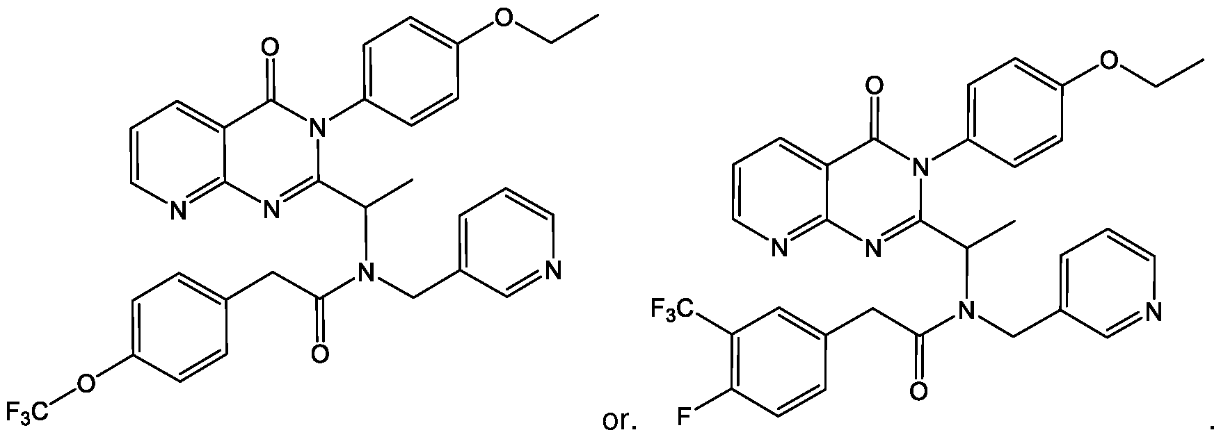
[162] The CXCR3 chemokine receptor antagonist may be present as the (R)-enantiomer, (S)- enantiomer or a mixture thereof. The CXCR3 chemokine receptor antagonist may be present as the (R)-enantiomer.
[163] The CXCR3 chemokine receptor antagonist may have the following structure, or may be a pharmaceutically acceptable salt, solvate or derivative thereof:

[164] As used herein the term "treatment", refers to any and all uses which remedy a disease state or symptoms, prevent the establishment of disease, or otherwise prevent, hinder, retard, or reverse the progression of disease or other undesirable symptoms in any way whatsoever. [165] One skilled in the art would be able to determine effective, non-toxic dosage levels of the
CXCR3 chemokine receptor antagonist and an administration pattern which would be suitable for treating osteoporosis to which the CXCR3 chemokine receptor antagonist is applicable.
[166] Further, it will be apparent to one of ordinary skill in the art that the optimal course of treatment, such as the number of doses of the CXCR3 chemokine receptor antagonist given per day for a defined number of days, can be ascertained using convention course of treatment determination tests.
[167] The CXCR3 chemokine receptor antagonist can be administered, or is to be administered, at a dosage level in the range of about 0.001 mg to about 100 mg, about 0.001 mg to about 0.01 mg, about 0.001 mg to about 0.1 mg, about 0.001 mg to about 1 mg, about 0.001 mg to about 10 mg, about 0.01 to about 0.1 mg, about 0.01 mg to about 1 mg, about 0.1 mg to about 10 mg, about 0.01 to about 100 mg, about 0.1 mg to about 1 mg, about 0.1 mg to about 10 mg, about 0.1 mg to about 100 mg, about 1 mg to about 10 mg, about 1 mg to about 100 mg or about 10 mg to about 100 mg per kg patient body weight per day.
[168] The CXCR3 chemokine receptor antagonist can be administered, or is to be administered, at a dosage level in the range of about 0.01 mg/kg to about 25 mg/kg per day, about 0.05 mg/kg to about 10 mg/kg per day, about 0.1 mg/kg to about 5 mg/kg per day, about 0.005 mg/kg to about 0.05 mg/kg per day, about 0.05 mg/kg to about 0.5 mg/kg per day, or about 0.5 mg/kg to about 5 mg/kg per day.
[169] In one example, the CXCR3 chemokine receptor antagonist is to be administered in single or multiple doses. In one example, the CXCR3 chemokine receptor antagonist is to be administered in a single, double, triple or quadruple dose. In another example, the CXCR3 chemokine receptor antagonist can be, or is to be, administered at an interval of, but not limited to, hourly, daily, twice daily, thrice daily, 4 times a day, every second day, every third day, every
fourth day, every fifth day, every sixth day, weekly, biweekly, bimonthly, monthly, or combinations thereof.
[170] Examples of convenient modes of administration include, but are not limited to, injection (subcutaneous, intravenous, etc.), oral administration, inhalation, transdermal application, topical creams or gels or powders, or rectal administration. Depending on the route of administration, the CXCR3 chemokine receptor antagonist can be coated, for example, with a material to protect the antagonist from the action of enzymes, acids and other natural conditions which may inactivate the therapeutic activity of the antagonist. In one example, the CXCR3 chemokine receptor antagonist can also be administered parenterally or intraperitoneally.
[171] In one example, the CXCR3 chemokine receptor antagonist is to be administered orally.
[172] The CXCR3 chemokine receptor antagonist can be administered orally, for example, with an inert diluent or an assimilable edible carrier. The CXCR3 chemokine receptor antagonist and other ingredients can also be enclosed in a hard or soft shell gelatin capsule, compressed into tablets, or incorporated directly into an individual's diet. For oral therapeutic administration, the CXCR3 chemokine receptor antagonist can be incorporated with excipients and used in the form of ingestible tablets, buccal tablets, troches, capsules, elixirs, suspensions, syrups, wafers, and the like.
[173] The oral administration can be in the form of tablets containing about 1 mg to about 1500 mg, about 1 mg to about 2 mg, about 1 mg to about 5 mg, about 1 mg to about 10 mg, about 1 mg to about 20 mg, about 1 mg to about 50 mg, about 1 mg to about 100 mg, about 1 mg to about 200 mg, about 1 mg to about 500 mg, about 2 mg to about 5 mg, about 2 mg to about 10 mg, about 2 mg to about 20 mg, about 2 mg to about 50 mg, about 2 mg to about 100 mg, about 2 mg to about 200 mg, about 2 mg to about 500 mg, about 2 mg to about 1500 mg, about 5 mg to about 10 mg, about 5 mg to about 20 mg, about 5 mg to about 50 mg, about 5 mg to about 100 mg, about 5 mg to about 200 mg, about 5 mg to about 500 mg, about 5 mg to about 1500 mg, about 10 mg to about 20 mg, about 10 mg to about 50 mg, about 10 mg to about 100 mg, about 10 mg to about 200 mg, about 10 mg to about 500 mg, about 10 mg to about 1500 mg, about 20 mg to about 50 mg, about 20 mg to about 100 mg, about 20 mg to about 200 mg, about 20 mg to about 500 mg, about 20 mg to about 1500 mg, about 50 mg to about 100 mg, about 50 mg to about 200 mg, about 50 mg to about 500 mg, about 50 mg to about 1500 mg, about 100 mg to about 200 mg, about 100 mg to about 500 mg, about 100 mg to about 1500 mg, about 200 mg to about 500 mg, about 200 mg to about 1500 mg or about 500 mg to about 1500 mg of the CXCR3 chemokine receptor antagonist.
[174] The tablet can contain 1 mg, 5 mg, 10 mg, 15 mg, 20 mg, 25 mg, 50 mg, 75 mg, 100 mg, 150 mg, 200 mg, 250 mg, 300 mg, 400 mg, 500 mg, 600 mg, 750 mg, 800 mg, 900 mg, 1000 mg, 1200 mg or 1500 mg of the CXCR3 chemokine receptor antagonist.
[175] When used for the treatment or prevention of osteoporosis, the CXCR3 chemokine receptor antagonist can be administered alone. Alternatively, the CXCR3 chemokine receptor antagonist can be administered as a pharmaceutical, veterinarial, or industrial formulation which comprises at least one compound according to the invention. The CXCR3 chemokine receptor antagonist can also be present as suitable salts, including pharmaceutically acceptable salts.
[176] In one example, the medicament, as disclosed herein, can further comprise a pharmaceutically acceptable excipient.
[177] The language "pharmaceutically acceptable excipient" is intended to include, but is not limited to, solvents, dispersion media, coatings, anti-bacterial and anti-fungal agents, isotonic and absorption delaying agents, and the like. The use of such media and agents for pharmaceutically active substances is well known in the art. Except insofar as any conventional media or agent is incompatible with the CXCR3 chemokine receptor antagonist, use thereof in the therapeutic compositions and methods of treatment and prophylaxis is contemplated. Supplementary active compounds may also be incorporated. It is especially advantageous to formulate parenteral compositions in dosage unit form for ease of administration and uniformity of dosage. "Dosage unit form" as used herein refers to physically discrete units suited as unitary dosages for the individual to be treated; each unit containing a predetermined quantity of compound(s) is calculated to produce the desired therapeutic effect in association with the required pharmaceutical excipient. The CXCR3 chemokine receptor antagonist may be formulated for convenient and effective administration in effective amounts with a suitable pharmaceutically acceptable excipient in an acceptable dosage unit. In the case of compositions containing supplementary active ingredients, the dosages are determined by reference to the usual dose and manner of administration of the said ingredients
[178] The excipient can be selected from, but is not limited to, agents such as gum tragacanth, acacia, corn starch or gelatin; excipients such as dicalcium phosphate; a disintegrating agent such as corn starch, potato starch, alginic acid and the like; a lubricant such as magnesium stearate; and a sweetening agent such as sucrose, lactose or saccharin or a flavouring agent such as peppermint, oil of wintergreen, or cherry flavouring. When the dosage unit form is a capsule, it may contain, in addition to materials of the above type, a liquid carrier. Various other materials may be present as coatings or to otherwise modify the physical form of the dosage unit. For instance, tablets, pills, or capsules may be coated with shellac, sugar or both. A syrup or elixir can contain the analogue, sucrose as a sweetening agent, methyl and propylparabens as preservatives, a dye and flavouring such as cherry or orange flavour. Of course, any material used in preparing any dosage unit form should be pharmaceutically pure and substantially non toxic in the amounts employed. In addition, the analogue may be incorporated into sustained- release preparations and formulations.
[179] In one example, the excipient is an orally administrable excipient.
[180] Also included in the scope of this disclosure are delayed release formulations.
[181] The CXCR3 chemokine receptor antagonist can also be administered, for example, in the form of a “prodrug”. As used herein, the term “prodrug” refers to an inactive form of a compound which is transformed in vivo to the active form. Suitable prodrugs include, but are not limited to, esters, phosphonate and esters of the active form of the compound.
[182] The CXCR3 chemokine receptor antagonist of the invention can be used in combination with other known treatments for osteoporosis. Combinations of active agents, including the CXCR3 chemokine receptor antagonist, can be synergistic.
[183] The subject can be, but is not limited to, an animal that is as risk or is suffering osteoporosis. In one example, the animal is a human.
[184] Also disclosed is a CXCR3 chemokine receptor antagonist for use in the treatment of a disease disclosed herein. In one example, the CXCR3 chemokine receptor antagonist disclosed herein is for use in the treatment of osteoporosis.
[185] Also disclosed herein is the use of the CXCR3 chemokine receptor antagonist in the manufacture of a medicament for treating a disease disclosed herein. In one example, the CXCR3 chemokine receptor antagonist disclosed herein is used in the manufacture of a medicament for treating osteoporosis.
[186] In one example, there is disclosed a method for treating an osteoclast deficiency in a patient by administering an (ectopic) chemokine Cxcl9l ligand.
[187] In one example, the chemokine Cxcl9l ligand can or is to be administered to a bone target region requiring osteoclast recruitment.
[188] The osteoclast deficiency can be but is not limited to target site of lethal excessive bone formation, ectopic mineralization and high bone mass.
[189] In one example, the high bone mass is be caused by fibrodysplasia ossificans progressiva (FOP).
[190] In another example, there is disclosed an (ectopic) chemokine Cxcl9 ligand for use in the treatment of osteoclast deficiency.
[191] In one example, there is disclosed the use of an (ectopic) chemokine Cxcl9l ligand in the manufacture of a medicament for the treatment of osteoclast deficiency.
[192] Non-limiting examples of the disclosure and a comparative example will be further described in greater detail by reference to specific examples, which should not be construed as in any way limiting the scope of the invention.
Up-Regulation of cxcl9l in Medaka Osteoblasts Under Osteoporotic Conditions.
[193] To identify factors involved in osteoblast-osteoclast cell communication, RNAseq analysis in a medaka in vivo osteoporosis model was performed. In this model, heat shock induction of transgenic Rankl expression at 9-day post-fertilization (dpf) leads to ectopic differentiation of osteoclasts and excessive resorption of mineralized matrix of the vertebral bodies. Subsequently, at 2-day post-heat shock (dphs), col10a1- positive osteoblast progenitors accumulate at lesion sites, differentiate into premature osteoblasts, and contribute to remineralization of the lesioned matrix. To identify factors responsible for recruiting osteoclast and osteoblast progenitor cells to bone lesion sites, osteoblast progenitors ( col10a1 ), premature osteoblasts ( osx ), and osteoclasts ( ctsk ) from Rankl-induced medaka larvae were purified by fluorescence activated cell sorting
(FACS) during onset (at 10 and 12 dpf) and repair (at 15 dpf) of osteoporotic bone lesions and performed RNA sequencing (Fig. 1).
[194] For FACS purification, 15 to 60 larvae were used per sample, leading to 10,000 to 270,000 isolated cells depending on strain and time point. Three independent biological repeats were sequenced per sample. Rankl:HSE:cfp nontransgenic siblings after heat shock were used as control. Bioinformatic analysis revealed that 45 genes were significantly up-regulated in osteoblast progenitors ( col10a1 cells; baseMean > 10; FC > 2; P < 0.05) 1 day after Rankl induction, and 13 of these genes were also up-regulated in premature osteoblasts (osx cells; Fig. 2). [195] Among the genes up-regulated in col10a1 cells, several genes with previously known functions in bone homeostasis, such as igfbp5, mmp9, mmp13 (collagenase 3), and tnfa were noted (complete gene lists in Tables 1 to 4).
[196] Table 1 : Genes exclusively up-regulated in col 10al cells or osx cells at 10dpf, or commonly up-regulated in both cell types.


co

[197] Table 2: To up-regulated genes in coll 0a1 cells at 10 dpf (log2 FC > 1 ; base Mean >=10; adj p-value < 0.05)


[198] Table 3: Top up-regulated genes in osx cells at 10 dpf (log2 FC >1 ; baseMean >= 10; adj p-value < 0.05).



[199] Table 4: Top regulated genes in ctsk cells at 12 dpf (log2 FC <1/>1 ; baseMean >= 10; adj p-value < 0.05).


Cxcr3.2 Is Expressed in a Subset of Macrophages that Differentiates into Osteoclasts upon Rankl Induction.
[201] To address the function of cxcl9l and cxcr3.2 in bone homeostasis, the dynamics of cxcr3.2- positive macrophages after Rankl induction were first determined. For this, a reporter line that expresses GFP under control of the medaka cxcr3.2 promoter (formerly named cxcr3a) was used. cxcr3.2\ GFP fish were crossed to a line that expresses mCherry in macrophages under control of the medaka mpegl promoter (mpepTmCherry), as well as an osx:mCherry line that expresses mCherry in osteoblasts under control of an osx/sp7 promoter. Any expression of the cxcr3.2 reporter in osteoblasts was not detected but tight interactions of recruited cxcr3.2 cells with osx-expressing osteoblasts was (Fig. 7, blue arrows (702)). On the other hand, control larvae without Rankl induction exhibited a significant portion of mpegl macrophages that co-expressed the cxcr3.2 reporter in the aorta-gonad-mesonephros (AGM) region, but only a few cells were detectable in the vertebral column (Fig. 8A, white arrow (802)). This changed dramatically after Rankl induction: the number of mpegl macrophages in the vertebral centra increased significantly at 1 dphs, with the vast majority of them being cxcr3.2- positive (Fig. 8A and Fig. 9A). At 2 dphs, the number of mpeg1/cxcr3.2 double-positive macrophages remained constant, but expression of cxcr3.2 in these macrophages declined (Fig. 8A, Fig. 9A and Fig. 9B). Simultaneously, the number of ctek-positive osteoclasts in the vertebral centra increased after Rankl induction (Fig. 8B), and the recruited cxcr3.2- positive cells started expressing cte/omCherry (Fig. 8B, Fig. 8C and Fig. 9C, white arrows (806, 808)). The expression of cxcr3.2\ GFP in osteoclasts decreased as they matured (Fig. 8C). These data show that cxcr3.2 is expressed in a subset of mpegl macrophages that becomes activated after Rankl induction and is recruited to the vertebral column. There macrophages differentiate into osteoclasts that gradually lose their cxcr3.2 expression.
Macrophage Recruitment Is Impaired Under Cxcr3.2-Deficient Conditions.
[202] To determine whether Cxcr3.2 is required for macrophage activation and directed migration, medaka cxcr3.2 mutants were generated by CRISPR/Cas9 (Fig. 10). Homozygous mutants developed normally without obvious malformations (Fig. 11 A and Fig. 11 B). Next, time- lapse confocal imaging was used to track the dynamics of macrophages under cxcr3.2- deficient conditions. In cxcr3.2 mutants without Rankl induction, macrophages were less migratory than in their wild-type siblings, which was evident from a significant reduction in directed displacement and speed of individually tracked cells (Fig. 11 B, Fig. 12 and Fig. 13). Upon Rankl induction, speed and migration distance were increased in mutants but were still significantly lower compared to wild-type cells (Fig. 11 B, Fig. 12, Fig. 13 and Fig. 14). Although mutant macrophages increased their motility, they mainly persisted in the aorta-gonad-mesonephros (AGM) and showed reduced directionality toward the centra of the vertebral column (Fig. 11 B, Fig. 12 and Fig. 14; quantification in Fig. 13F). This shows that a Cxcr3.2 deficiency affects the general migratory behaviour of macrophages and particularly interferes with their directed migration from the AGM to mineralized matrix in the vertebral column.
[203] Furthermore, after Rankl induction, macrophage to osteoclast differentiation was strongly impaired in cxcr3.2 mutants, and no ctek-positive osteoclasts were detectable at 2 dphs, when wild-type siblings showed abundant osteoclasts (Fig. 11 B and Fig. 14). Consequently, in the absence of ectopic osteoclasts, the integrity of mineralized matrix in vertebral bodies including neural and hemal arches was intact in cxcr3.2 mutants at 3 dphs, similar to the situation in Rankl- larvae (Fig. 15). This is also consistent with the observation that osteoblasts formed normally in cxcr3.2 mutants (Fig. 7).
[204] Together, the data suggest that the chemokine receptor Cxcr3.2 is required for macrophage activation upon Rankl induction to recruit them to the mineralized matrix of the vertebral bodies, where they differentiate into osteoclasts. Importantly, other macrophage functions such as bacterial phagocytosis appeared not affected by cxcr3.2 deficiency. Escherichia coli injected into larvae were efficiently cleared by recruited cxcr3.2- deficient macrophages, similar to the situation in heterozygous and wild-type siblings. Thus, Cxcr3.2 is thought to be required, particularly for those macrophages that are destined to differentiate into osteoclasts.
Chemical Inhibition of Cxcr3.2 Blocks Osteoclast Formation and Protects Bone Integrity.
[205] NBI-74330 and AMG487 are two structurally related antagonists of human CXCR3, with slightly different binding affinities. They have been pharmacologically tested previously in the context of atherosclerotic plaque formation and psoriasis. To test whether human CXCR3 inhibitors interfere with Cxcr3.2 function in medaka and block osteoclast formation, cte/i:GFP/mpegT:mCherry//a/7/i/:HSE:c/p transgenic medaka were treated with different doses of these antagonists. Upon Rankl induction, control larvae treated with DMSO showed macrophage accumulation and osteoclast formation in neural arches and centra at 1 dphs (Fig. 16 and Fig. 17,
box denoted by reference numeral (1706), and osteoclast numbers increased at 2 dphs (Fig. 16 and Fig. 18C). In NBI-74330-treated larvae at 1 dphs, the number of initially recruited macrophages was not significantly different from controls (Fig. 16 and Fig. 18A); however, fewer macrophages were detectable in the region of the vertebral arches (Fig. 17, arrows denoted by reference numeral (1712)). In contrast, at 2 dphs, when macrophage numbers increased strongly in controls, they remained at significantly lower levels in NBI-74330-treated larvae (Fig. 18A). Importantly, the total number of macrophages, including those in the AGM and tail region, was not significantly altered (Fig. 18B). Also, osteoclast formation was strongly suppressed in NBI- 74330-treated larvae (Fig. 16, Fig. 17 and Fig. 18C). As a consequence, treatment with NBI- 74330 protected bone integrity upon Rankl induction (Fig. 17). In DMSO controls, Rankl induction resulted in a resorption of vertebral neural arches and a severe loss of mineralized matrix in the centra (Fig. 17). In contrast, NBI-74330-treated larvae showed a significant reduction of mineralization defects and were almost indistinguishable from Rankl-negative larvae (Fig. 17). Similar results were also obtained with AMG487 (Fig. 19). In conclusion, chemical inhibitor experiments confirmed the findings in cxcr3.2 mutants that Cxcr3.2 is required for macrophage recruitment and osteoclast formation upon Rankl induction.
[206] Thus, in view of the above data showing that genes related to CXC chemokines are up- regulated in osteoblast progenitors upon Rankl induction, CXC chemokines and their receptors, specifically CXCR3 were considered as therapeutic targets. In one example, there is disclosed the use of a CXCR3 chemokine receptor antagonist in the manufacture of a medicament for the treatment of osteoporosis. In another example, there is disclosed a method for treating osteoporosis, the method comprising the step of administering to a subject in need thereof a CXCR3 chemokine receptor antagonist. In yet another example, there is disclosed a CXCR3 chemokine receptor antagonist for use in the treatment of osteoporosis.
Osteoblast-Derived Cxcl9l Triggers Macrophage Recruitment and Osteoclast Differentiation in the Absence of Rankl.
[207] Chemokine receptors control cell migratory behaviour in response to gradients of chemokine ligands. To address the source and nature of the chemokine responsible for macrophage recruitment to bone, (in other words, in order to confirm whether the ligand Cxcl9l, up-regulated in osteoblast progenitors upon Rankl induction, is involved in this process), the role of Cxcl9l was investigated. For this, cxcl9l mutants were generated by CRISPR/Cas9 (Fig. 9) and macrophage dynamics was tracked by time-lapse imaging. Without Rankl induction, no changes in macrophage distribution were observed in cxcl9l mutants (Fig. 22). After Rankl induction, the number of macrophages at 1 dphs recruited to the vertebral column was not significantly reduced (Fig. 23A and Fig. 23B). However, osteoclast formation was severely reduced at 2 dphs, and only single ctsk- positive osteoclasts were detectable in the vertebral column of cxcl9l mutants, while wild-type siblings had abundant osteoclasts (Fig. 24A and Fig. 24B). At 3 dphs, the severity of Rankl-induced mineralized matrix lesions, evident as resorbed neural arches and the presence
of nonmineralized lesions in centra of wild-type siblings, became significantly alleviated in Rankl- induced cxcl9l mutants (Fig. 25A, Fig. 25B and Fig. 26). Importantly, osteoblast numbers and distribution were not affected in cxcl9l mutants (Fig. 27). Together, these data show that osteoblast-derived Cxcl9l is required for osteoclast formation, but may be dispensable for macrophage recruitment.
[208] Upon Rankl induction, cxcl9l is up-regulated in osteoblasts. It was therefore tested whether osteoblasts are the sole source for Cxcl9l required for osteoclast recruitment. As cxcl9l was up-regulated in col10a1 osteoblast progenitors, as well as in premature osx-positive osteoblasts (Tables 2 and 4), an established osx:mCherry-NTRo line was used to genetically ablate premature osteoblasts using a Nitroreductase (NTRo) approach as described herein. The formation of ctsk osteoclasts after Rankl induction was subsequently quantified. Metronidazole (Mtz) treatment of osx!mCherry-NTRo/ran/i/!HSE!c/p/cte/oGFP transgenic medaka at 9 dpf for 24 hours resulted in an almost complete ablation of osteoblasts from the vertebral column under both Rankl- and Rankl+ conditions (Fig. 28A and Fig. 28B, arrowheads denoted by reference numeral (2802), and Fig. 29). Upon Rankl induction at 10 dpf, the number of ctsk osteoclasts was strongly reduced (by approximately 90%), while control treated larvae without ablation showed normal osteoclast induction (Fig. 28B and Fig. 29). It was shown that the integrity of mineralized matrix was maintained in osteoblast-ablated larvae at 3 dphs, further confirming the absence of osteoclast activity in these fish (Fig. 29). These ablation experiments show that osteoblasts are the exclusive source of signals required for ectopic osteoclast formation upon Rankl induction.
[209] Finally, to test whether Cxcl9l alone is sufficient to trigger macrophage recruitment in the absence of Rankl induction, Cxcl9l was ectopically expressed in premature osteoblasts using a transient transgenic approach. To this end, an osxpromoter construct driving expression of Cxcl9l fused to EGFP or mCherry via a self-cleaving p2a peptide was injected into medaka embryos (Fig. 20 and Fig. 21). Larvae with mosaic osx\cxcl9l- p2a-mCherry or osx:cxc/9/-p2a-EGFP expression in osteoblasts on the vertebral column surface were selected for analysis at 10 to 14 dpf (Fig. 20B and Fig. 21 B and Fig. 22B and Fig. 22C). A considerable accumulation of mpegT. mCherry labelled macrophages was noted directly adjacent to the cxc/9/-p2a-EGFP labelled cells, while the remaining vertebral column showed macrophages at a similarly low extent as non-injected controls (Fig. 20B; for quantification, Fig. 22B). In a second approach, it was analysed whether ctsk- positive osteoclasts can form under conditions of ectopic Cxcl9l expression. Non-injected control larvae at 12 dpf did not show ctsk osteoclasts in the vertebral column under Rankl- conditions (Fig. 21 B, Top). In contrast, larvae ectopically expressing Cxcl9l exhibited several ctsk- positive cells in the vertebral bodies (19 ctsk cells in 6 analysed larvae; Fig. 21 B). Ten of the 19 identified ctsk cells were found positioned close to Cxcl9l-producing cells, while 9 cells were found farther away (Fig. 22C). This shows that Cxcl9l is sufficient to induce macrophage migration and differentiation into osteoclasts, even in the absence of Rankl expression. Together, the results demonstrate that Cxcl9l is up-regulated in osteoblasts upon
Rankl induction and mediates Rankl effects by controlling osteoclast recruitment and differentiation at the mineralized bone matrix.
Medaka cxcl9l Encodes the Ancestral Gene of Human CXCL9.
[210] The human genome encodes more than 40 chemokine ligands and 20 chemokine receptors. In contrast, the repertoire in teleost fish is much smaller. This indicates an expansion of chemokines and their cognate receptors in the tetrapod lineage related to additional and lineage-specific new functions of the more advanced immune system.
[211] In the current medaka genome assembly, the cxcl transcript identified by the RNAseq analysis is annotated as C-X-C motif chemokine 2. However, phylogenetic and synteny analysis revealed that this gene is the common precursor of two CXCL gene clusters on human chromosome 4, including CXCL 1, 2, 3, 4a and b (syn PF4 and PF4V1 ), 5, 6, 7 (syn PPBP ), and 8at 4q13.3 and CXCL 9, 10, 11, and 73at 4q21.1. Such clusters evolve by local gene duplications and subsequent functional divergence (neofunctionalization or subfunction partition). The highest amino acid (aa) identity of OlaCxcl9l to human CXCL9 suggests that human CXCL9 shares the ancestral function with the identified medaka gene.
[212] In mice, CXCL9 is produced in osteoblasts and functions as a bone angiostatic and osteogenic factor. In addition, cell culture studies have shown that blocking CXCL9 or CXCL10 function in vitro impairs differentiation of osteoclast progenitors. However, their role for bone homeostasis in vivo is unclear. In humans, recent genome-wide association studies identified CXCL9 as a potential candidate locus for periodontitis, an inflammatory disease that destroys tissues surrounding and supporting teeth, subsequently leading to bone loss. However, whether and how CXCL9 acts to recruit osteoclasts to sites of bone resorption remained unknown.
Cxcl9l and Rankl Signalling.
[213] It was found that upon Rankl induction, cxcl9l was up-regulated in col10a1 osteoblast progenitors. It is presently unclear whether this reflects a direct effect of Rankl activating a Rank receptor on col10a1 cells or happened indirectly as a consequence of factors released from activated osteoclasts. Consistent with the latter, several up-regulated transcripts in Rankl-induced osteoclasts were identified, including cxcr3.2, indicating that factors released from Rankl-induced osteoclasts indirectly trigger up-regulation of cxcl9l in osteoblasts. On the other hand, however, it is known that, at least in mammals, the Rank receptor is expressed in mesenchymal stem cells (MSCs), which can differentiate into osteoblasts, as well as in osteoclast progenitors. In MSCs, forward Rank signalling blocks their differentiation into osteoblasts. Subsequently, Rankl- activated osteoclasts produce extracellular vesicles containing Rank receptors that bind to Rankl on MSCs and thereby induce reverse Rankl signalling to allow osteoblast differentiation. Hence, in mammals, Rankl has two major origins, from MSCs/osteoblast progenitors to block osteoblastogenesis and from osteocytes to promote osteoclastogenesis. Medaka have acellular
bone that completely lacks osteocytes but contains col10a1- positive osteoblast progenitors that differentiate into osx expressing premature osteoblasts. It is hypothesized that Rankl forward signalling also occurs in medaka osteoblast progenitors. Transgenic Rankl expression might therefore activate Rank receptors on osteoblast progenitors to produce and release Cxcl9l. Cxcl9l then forms a chemokine gradient to attract macrophages to mineralized matrix where they differentiate into osteoclasts (Fig. 30). Whether these osteoclasts produce vesicular Rank to later promote osteoblast differentiation, similar to in mammals, remains to be investigated.
The Chemokine Receptor Cxcr3.2 Marks a Subset of Macrophages Destined to Become Osteoclasts.
[214] It was found that cxcl9l and cxcr3.2 are expressed in medaka osteoblast progenitors and osteoclasts, respectively. Without Rankl induction, no osteoclasts appear in the vertebral column of medaka larvae at the analysed stages (9 to 12 dpf), but ctek-positive cells are present in head and fins. The up-regulation of cxcr3.2 transcription detected in FAC-sorted ctsk cells after Rankl induction was thus thought to imply a de novo formation of osteoclasts in the vertebral column, rather than cxcr3.2 up-regulation in pre-existing osteoclasts. This was confirmed by time-lapse analysis of a cxcr3.2\ GFP reporter, which was initially expressed in a subset of mpegl- positive/cte/f-negative macrophages. Later, the reporter persisted in differentiating osteoclasts that became ctsk- positive, but eventually, cxcr3.2 expression diminished in maturing osteoclasts. Together, the data indicates that Cxcr3.2 is expressed in macrophages that are destined to become osteoclasts. Once osteoclasts have reached their destination and become resorptive, cxcr3.2 is down-regulated. Importantly, Cxcr3.2 is not required for macrophage migration or recruitment to clear inflammatory insult. Hence, without being bound by theory, it is thought that this receptor marks a distinct subset of macrophages, which differentiates into osteoclasts at mineralized bone matrix.
Cxcl9l Drives Progenitor Recruitment to Sites of Bone Resorption in Medaka.
[215] Factors that control progenitor cell recruitment to bone matrix under physiological or pathological conditions have gained increasing interest, mainly for therapeutic reasons. Various inflammatory cytokines and chemokines have been shown to promote osteoclastogenesis and modulate bone remodeling. Chemokines, including but not limited to CXCL10, are mainly produced by immune cells, and are implicated in inflammatory bone loss such as, for example, in rheumatoid arthritis or bone metastasis. In the latter, chemokines activate osteoclasts to enhance bone resorption and facilitate colonization of bone marrow by metastasizing cancer cells. Expression in osteoblasts, on the other hand, has been reported for several chemokines (such as, but not limited to, CCL2, CCL3, CXCL2, and CXCL12, among others) and linked, e.g., to tooth eruption, as well as fracture healing and bone repair. However, while the immunomodulatory effects of chemokines are well characterized, the physiological roles of osteoblast-derived chemokines are much less understood. Consistent with reports in mammals, the data shown here shows that medaka osteoblast progenitors produce several chemokines, but only Cxcl9l was
found to be significantly up-regulated upon Rankl induction in vivo. Importantly, ectopic expression of Cxcl9l was sufficient to drive osteoclast progenitor recruitment in larvae, even in the absence of Rankl induction. This indicates that at least in larvae, Cxcl9l acts in a paracrine manner and independently, or downstream, of Rankl to recruit osteoclasts to bone matrix. It is of note that a considerable number of induced osteoclasts were detected at a distance from ectopic Cxcl9l-producing cells. Without being bound by theory, it is thought that this is simply a reflection of the migratory dynamics of newly formed osteoclasts in medaka. Alternatively, it cannot be excluded that Cxcl9l controls osteoblasts in an autocrine manner, and that consequently, other osteoblast-derived factors act at a distance to trigger osteoclast differentiation remotely.
[216] The role of CXCL9-CXCR3 signalling in immune cell activation and migration in development and pathological processes has been well documented. It has, however, also been shown that CXCR3 acts independently of CXCL9, via, for example, CXCL4, CXCL10, or CXCL11 . Likewise, CXCL9 can interact with other receptors than CXCR3, such as, but not limited to, CCR3. Here, the finding that cxcl9l and cxcr3.2 medaka mutants share a similar osteoclast phenotype indicates that Cxcl9l acts through Cxcr3.2 to control osteoclast recruitment to bone matrix. In contrast to cxcr3.2 mutants, however, cxcl9l mutants showed normal macrophage distribution upon Rankl induction, indicating that Cxcl9l is dispensable for cell recruitment. Without being bound by theory, it is thought that this is due to the presence of additional chemokines produced by col10a1 cells, as described above. It is of note that deletion of cxcl9l blocked differentiation of macrophages into osteoclasts almost entirely, indicating an essential role in this process. Together with the fact that transgene-driven ectopic expression of cxcl9l promoted macrophage recruitment, this indicates that Cxcl9l is a chemotactic cue recognized by cxcr3.2- positive macrophages.
[217] It is of note that cxcr3.2- deficient macrophages exhibit increased motility upon Rankl induction, but failed to directionally migrate toward bone matrix. It is thought that macrophages, in this case, were directly stimulated by Rankl binding to Rank receptors on these cells, which triggered dynamic migratory behaviour in the absence of a chemotactic guidance cue. At present, however, it cannot be excluded that Rankl-induced expression of other chemokines that bound to receptors other than Cxcr3.2 and stimulated cell movement, albeit not directed toward bone matrix.
[218] Without Rankl induction, the cxcl9l and cxcr3.2 mutants generated in the experiments disclosed herein developed apparently normally with no obvious signs of osteoclast deficiency or osteopetrosis. Without being bound by theory, it is thus thought that Cxcl9l and Cxcr3.2 are especially important under pathological conditions of severe Rankl overexpression, such as seen in, for example, rheumatoid arthritis and osteoporosis. Under non-pathological conditions, other chemokines and their receptors are thought to play a role in recruiting osteoclast precursors.
Chemical Inhibition of Cxcr3.2 Protects Bone Integrity in Medaka
[214] Fish models, such as medaka and zebrafish, are ideally suited for drug screening. Testable compounds can be directly added to small volumes of medium, and skeletal defects can be efficiently analysed by live imaging in the translucent larvae. In the past, several chemical inhibitors for chemokine receptors have been developed and clinically tested, especially in the context of cancer, immune deficiencies, and inflammation. These include, but are not limited to, inhibitors for CXCR3. NBI-74330 and AMG487 are two exemplary, structurally-related CXCR3 antagonists that have been tested in preclinical and clinical trials. AMG487 entered a phase II clinical trial for psoriasis that was stopped for lack of efficacy. In an LPS-induced periodontitis mouse model, treatment with AMG487 resulted in a significant reduction of bone loss and decreased osteoclast numbers after LPS injections. Therefore, the role and efficacy of CXCR3 inhibitors in an osteoporosis context remained unclear. In the osteoporosis setting in medaka, NBI-74330 and AMG487 efficiently blocked osteoclast formation upon Rankl induction. The in vivo experiments thus indicated that the chemokine-controlled process of osteoclast recruitment is druggable, and that CXCR3 inhibitors can be used to modulate bone homeostasis.
EXPERIMENTAL SECTION
[215] Materials and Methods
[216] Table 5: Table of oligomers
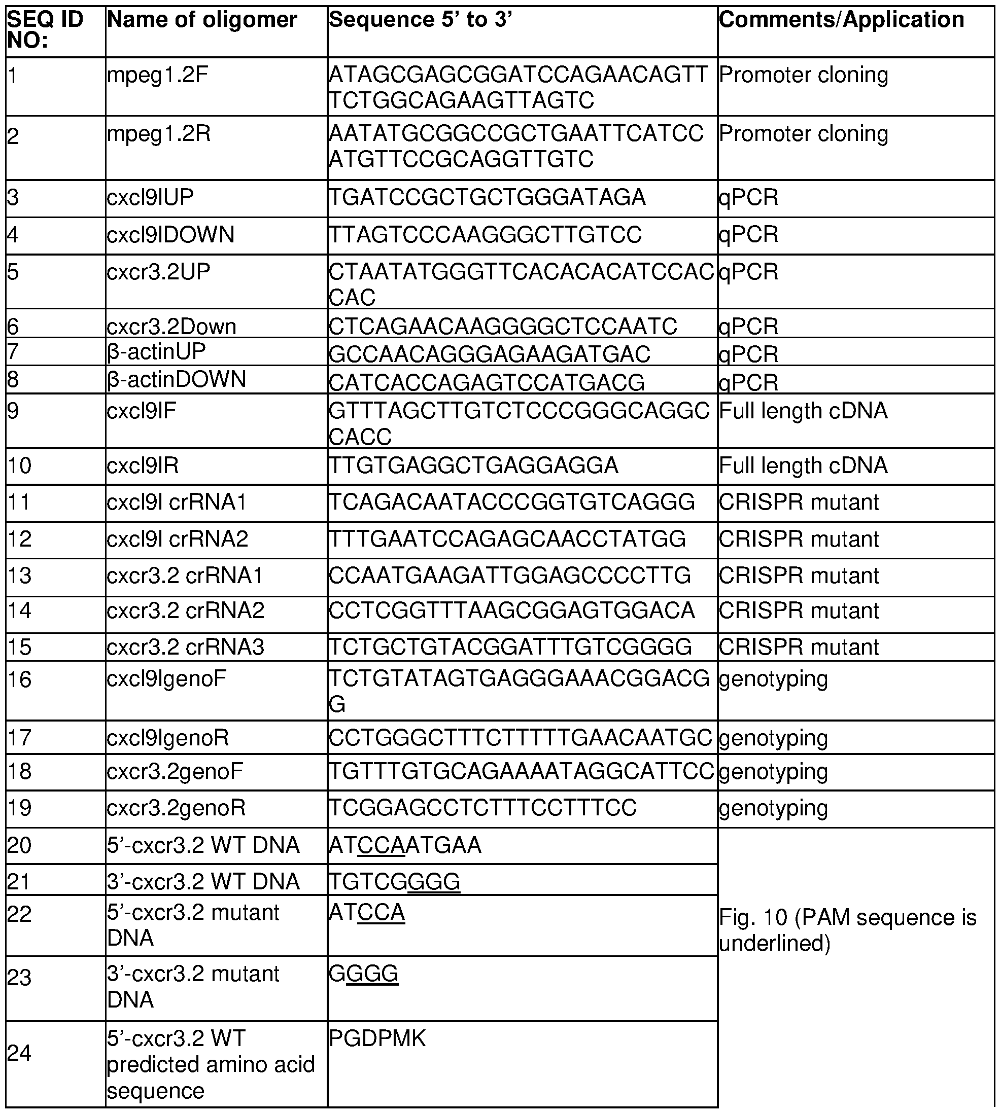

[217] Transgenic and Mutant Fish Lines. All experiments were performed according to protocols approved by the Institutional Animal Care and Use Committee of the National University of Singapore (protocol numbers R14-293, R18-0562, and BR15-0119). For generation of mpeg1\ mCherry transgenic medaka, a 2.1 -kb sequence of the mpegl promoter, including the endogenous ATG, was amplified from genomic DNA of wild-type medaka using primers mpeg1.2F and mpeg1.2R (Table 5). The PCR product was digested using Not! and BamHI, and ligated in frame to farnesylated mCherry in a pl-Scel plasmid. The plasmid was then injected into one-cell stage medaka embryos, together with l-Scel meganuclease, to obtain stable lines. Cxcr3.2\ EGFP transgenic medaka were generated using a cxcr3.2\ GFP expression plasmid kindly provided by Dr. Baubak Bajoghli (University of Tuebingen, Germany) and described in Aghaallaei et al. (DOI: 10.1073/pnas.1000467107). Briefly, the 5'-upstream region of the cxcr3.2 gene, nt -4 to nt -1880 from ATG start codon, i.e., nt 18,395,178-18,397,057 of chromosome 16, was amplified and inserted into the vector pSgfp containing a gfp sequence that is flanked by l-Scel sites. This plasmid was injected into one-cell stage medaka embryos together
with l-Scel meganuclease to obtain stable lines. To generate osx\cxcl9l- p2a-mCherry and osx:cxc/9/-p2a-EGFP plasmids for Cxcl9l overexpression, the medaka osx promoter was cloned upstream of the cxcl9l coding sequence amplified from medaka cDNA using primers cxcl9IF and cxcl9IR (Table 5). For osx\cxcl9l- p2a-mCherry, the cxcl9l stop codon was replaced with p2a- mCherry (amplified from plasmid col10a1\ creERT2-p2a-mCherry). For osx:cxc/9/-p2a-EGFP, mCherry from osx\cxcl9l- p2a-mCherry was replaced with EGFP. The plasmids were injected into one-cell stage medaka embryos together with l-Scel meganuclease. All other transgenic lines have been described before and are available from the Medaka Stock Center, Laboratory of Bioresource, National Institute for Basic Biology, Okazaki, Japan. Staging of larvae, screening, and live imaging of transgenic medaka were performed by methods as described below. For CRISPR/Cas9 genome editing, guide RNAs targeting cxcl9l and cxcr3.2( Table 5) were designed using CRISPR/Cas9 target online prediction (CCTop), and generated by Integrated DNA Technologies (IDT) as CRISPR RNA (crRNA). crRNA (36 ng/pL each) was injected into one-cell stage medaka embryos together with tracrRNA (67 ng/pL, IDT) and Cas9 nuclease (0.25 pg/pL, IDT). Primers used for genotyping (cxcl9lgenoF/R and cxcr3.2F/R) are listed in Table 5.
[218] RNA-Seq Library Preparation of Fluorescence-Activated Cell Sorting (FACS) Purified Bone Cells and Sequencing. RNA libraries were prepared from FACS-purified osteoblast progenitors (col10a1- positive), premature osteoblasts ( osx/sp7 ), and osteoclasts ( ctsk ) as described previously. Briefly, medaka Rankl+ ( rankhHSExfp ) and Rank!- control larvae expressing osteoblast (col10a1\ nuGFP; osx:mCherry) or osteoclast (cte/cmCherry) reporters were heat-shocked at 9 days post-fertilisation (“dpf”) for 2 hours at 39 °C to induce Rankl expression. Larvae were dissociated for FACS using a collagenase/trypsin-based protocol for purification of osteoblasts at 10 dpf ( col10a1 , osx) or 15 dpf ( col10a1 ) and osteoclasts (ctsk) at 10 or 12 dpf. RNA was isolated using a total RNA isolation reagent (TRIzol)-based method and purified using the PureLink RNA Micro Kit (Invitrogen) following manufacturer’s instructions. cDNA was synthesized from 1 to 10 ng total RNA using the SMARTer Ultra Low Input RNA for lllumina Sequencing Kit, and the Advantage2 polymerase mix (Clontech) for second strand synthesis. cDNA was analysed using an Agilent 2100 BioAnalyzer and a High Sensitivity DNA Chip. A Covaris AFA system was used to produce short cDNA fragments of 100 to 300 bp, and DNA concentration was determined using a Qubit dsDNA HS Assay Kit and Qubit2.0 Fluorometer. cDNA libraries were generated using the NEBNext Ultra DNA Library Prep Kit for lllumina and NEBNext Multiplex Oligos for lllumina (Index primer set 1 ). After adapter ligation, DNA was PCR- amplified with NEBNext high-fidelity 2x PCR master mix using Index primers (primers 1 to 12). Libraries were multiplexed, and paired-end sequencing (75 to 100 bp read lengths) was performed on an lllumina HiSeq platform at the Genome Institute of Singapore with sequencing depths between 50 and 100 million reads.
[219] Bioinformatics Analysis. For differential gene expression analysis, reads were aligned to the Japanese medaka Hd-rR strain genome (ASM223467v1 ) using spliced transcripts alignment to a reference (STAR; quantMode GeneCounts). Differentially expressed genes were
detected using the Bioconductor/R package DESeq2. For further analyses, a gene was considered to be differentially expressed at a baseMean > 10, log2FC > 1 , and adjusted p-value < 0.05. For functional clustering of genes involved in chemokine signalling, the Database for Annotation, Visualization and Integrated Discovery was used based on human homologs. To infer homology of medaka genes, orthology and paralogy relationships were established by using the BLAST/BLAT tools of ENSEMBL with default parameters and conserved synteny analysis with GENOMICUS version 96.01 .
[220] Quantitative PCR (RT/qPCR). RNA was isolated from FACS purified osteoblast progenitors, osteoblasts, and osteoclasts derived from 15 to 20 larvae per sample using the PurelinkTM RNA micro kit (Invitrogen) as described previously. Briefly, control and Rankl-induced larvae were dissociated using a collagenase/trypsin-based protocol and submitted for FACS purification. Sorted cells were collected directly into tubes containing 400 mI of TRIzol for cell lysis. RNA was then isolated using the PureLink RNA Micro Kit (Invitrogen) following the manufacturer’s instructions. cDNA was synthesized and amplified using the PreAmp and Reverse Transcription Master Mix Kit (Fluidigm). qPCR was performed with PowerUp SYBR Green Master Mix (Applied Biosystems). Primers for qPCR are listed in Table 5; b-actin was used for normalization. Comparisons of transcript levels in Rankl-expressing relative to heat-shocked Rankl-negative larvae were performed with three biological replicates and three technical replicates each. Samples for RNAseq analysis and qPCR validation were obtained from independent experiments. Data were analysed using Bio-Rad CFX Maestro 1.0 software and Student’s t tests were performed for statistical analysis.
[221 ] Genetic Ablation of Osteoblasts. Nitroreductase-mediated cell ablation was performed on transgenic lines. Briefly, osteoblasts were ablated by treating osx:mCherry-NTRo larvae with 8 mM metronidazole (Mtz) for 24 hours at 9 dpf. Mtz-treated osx:mCherry larvae were used as controls.
[222] CXCR3 Inhibitor Treatment. The CXCR3 inhibitors AMG487 and NBI-74330 (Tocris Biosciences) were dissolved in DMSO and 100% ethanol to make 10 and 30 mM stock solutions, respectively. Stock solutions were added to 50% hydroxypropyl- -cyclodextrin (Sigma) solution, vortexed vigorously, and further diluted with sterile water and 30% Danieau’s solution [19.3 mM NaCI, 0.23 mM KCI, 0.13 mM MgS04, 0.2 mM Ca(NOs)2, 1 .7 mM Hepes, pH 7.0] to 20 and 30 mM working concentrations, respectively. AMG487 solution was injected into the yolk of medaka larvae at 3 hours prior to heat shock. Thereafter, larvae were kept in fish medium containing 20 mM AMG487 for the course of the experiment. For NBI-74330, larvae were kept in fish medium containing 30 pM NBI-74330 for 3 hours prior to heat shock, changed to the pure fish medium during heat shock, and then reverted to drug solution for the course of the experiment. Drug solutions were freshly made and changed daily.
[223] Bone Staining. For live bone staining, larvae were incubated in 0.01% Calcein (Sigma) in the dark for 1 hour, washed with fish medium twice, and mounted for imaging. Alizarin Red
bone staining was performed as previously described Briefly, embryos at 3 days post heat-shock were fixed in 4% paraformaldehyde, washed three times with PBS plus Tween-20 (PBST), incubated for 15 min in 0.5% potassium hydroxide (KOH), then transferred to 0.001% Alizarin Red solution in 0.5% KOH for at least 4 hours with gentle shaking. Samples were then washed for 2 hours with 0.5% KOH, followed by depigmentation with 2% H2O2 in 0.5% KOH for 1 hour. Embryos were washed two times with 0.5% KOH and mounted in 100% glycerol for imaging.
[224] Imaging. For live fluorescence imaging, medaka larvae were anesthetized with 0.016% Tricaine (MS222, Sigma), mounted in 1.2% low melting point agarose on a glass-bottom Petri dish, and imaged using an Olympus FluoView FV3000 confocal microscope or a Nikon SMZ18 stereomicroscope equipped with the NIS-Elements BR 3.0 software. Time-lapse imaging was performed with an Olympus FluoView FV3000 confocal microscope. To image fixed Alizarin Red stained samples, a Nikon Eclipse 90i upright microscope equipped with NIS-Elements BR 3.0 software was used. Images and time-lapse movies were processed with Fiji ImageJ, Bitplane Imaris, and Adobe Photoshop. [225] Quantification of Cells and Statistical Analysis. Cell numbers (mpegV. mCherry, cte/oGFP, ctsk\ mCherry, and cxcr3.2\ GFP) were quantified as previously described with slight modifications. Specifically, using Fiji, fluorescent images were converted to greyscale and thresholded, and the area of a cell (averaged from 15 cells from three larvae), as well as the total area of cells in a selected region of interest, were obtained. Cell numbers were determined by dividing the total area of cells by the average cell area. ANOVA with multiple comparisons, Mann- Whitney U, and Student’s t tests were conducted in Prism (Graphpad) for statistical analysis. Details are indicated in corresponding figure legends.
[226] Bacterial Infection. Non-pathogenic E. coli K12 strains expressing a GFP reporter were prepared. Briefly, bacteria were cultured overnight to stationary-phase and centrifuged for 1 minute at 13,000rpm. The pellet was washed twice with sterile PBS and then resuspended in sterile PBS at 2x10⁹-4x10⁹ CFU/ml and kept on ice for injection. Bacterial suspensions were microinjected into the muscle of 10 dpf larvae at roughly 2,500 CFU per larva.
Claims
1 . A method of treating osteoporosis, the method comprising the step of administering to a subject in need thereof a CXCR3 chemokine receptor antagonist.
2. The method according to claim 1 , wherein the CXCR3 chemokine receptor antagonist is a small molecule antagonist, having a molecular weight of less than 900 Da.
3. The method according to claim 1 or 2, wherein the CXCR3 chemokine receptor antagonist has a backbone structure which comprises quinazolinone or azaquinazolinone.
4. The method according to any one of the preceding claims, wherein the CXCR3 chemokine receptor antagonist has the following formula (I) or is a pharmaceutically acceptable salt, solvate or derivative thereof:
 wherein A1, A2 and A3 are independently CH or N;
R1 is -CN, optionally substituted alkyl or optionally substituted heteroalkyl;
R2 and R3 are independently an optionally substituted alkyl, optionally substituted heteroalkyl, optionally substituted alkylsulfonyl, optionally substituted heterocycloalkyl, optionally substituted heterocycloalkylheteroalkyl, optionally substituted arylalkyl, optionally substituted arylheteroalkyl, optionally substituted heteroarylalkyl, or optionally substituted heteroarylheteroalkyl.
5. The method according to claim 4, wherein A1 and A2 are CH and A3 is N.
6. The method according to claim 4 or 5, wherein R1 is heteroalkyl, preferably ethoxy.
7. The method according to any one of claims 4 to 6, wherein R2 is heteroarylalkyl, preferably
3-methylpyridyl.
8. The method according to any one of claims 4 to 7, wherein R3 is substituted arylalkyl, preferably substituted benzyl.
9. The method according to any one of claims 4 to 8, wherein the CXCR3 chemokine receptor antagonist has the following formula (II) or is a pharmaceutically acceptable salt, solvate or derivative thereof:
 wherein
R4 is an optionally substituted alkyl;
R5, R6 and R7 are independently halogen, optionally substituted alkyl halide or optionally substituted ether; and
Z is N+-0- or N.
10. The method according to claim 9, wherein R4 is ethyl.
11 . The method according to claim 9 or 10, wherein R5 is -CF3, R6 is F and R7 is H or R5 is H, R6 is -0-CF3 and R7 is H.
12. The method according to any one of the preceding claims, wherein the CXCR3 chemokine receptor antagonist has the following structure:

13. The method according to any one of the preceding claims, wherein the CXCR3 chemokine 5 receptor antagonist is administered at a dosage level in the range of about 0.001 mg to 100 mg per kg patient body weight per day.
14. The method according to claim 13, wherein the CXCR3 chemokine receptor antagonist is administered in single or multiple doses.
15. The method according to claim 13 or 14 wherein the CXCR3 chemokine receptor
10 antagonist is administered at a dosage level in the range of about 0.01 mg/kg to about 25 mg/kg per day, about 0.05 mg/kg to about 10 mg/kg per day, about 0.1 mg/kg to about 5 mg/kg per day, about 0.005 mg/kg to about 0.05 mg/kg per day, about 0.05 mg/kg to about 0.5 mg/kg per day, or about 0.5 mg/kg to about 5 mg/kg per day.
16. The method according to any one of the preceding claims, wherein the CXCR3 chemokine 15 receptor antagonist is administered orally.
17. The method according to claim 16, wherein the oral administration is in the form of tablets containing 1 .0 mg to 1500 mg of the CXCR3 chemokine receptor antagonist, or the tablet contains 1 mg, 5 mg, 10 mg, 15 mg, 20 mg, 25 mg, 50 mg, 75 mg, 100 mg, 150 mg, 200 mg, 250 mg, 300
mg, 400 mg, 500 mg, 600 mg, 750 mg, 800 mg, 900 mg, 1000 mg, 1200 mg or 1500 mg of the CXCR3 chemokine receptor antagonist.
18. A CXCR3 chemokine receptor antagonist for use in the treatment of osteoporosis.
19. Use of a CXCR3 chemokine receptor antagonist in the manufacture of a medicament for the treatment of osteoporosis.
20. The Use according to claim 19, wherein the medicament further comprises a pharmaceutically acceptable excipient.
Applications Claiming Priority (2)
| Application Number | Priority Date | Filing Date | Title |
|---|---|---|---|
| SG10202002624U | 2020-03-20 | ||
| SG10202002624U | 2020-03-20 |
Publications (1)
| Publication Number | Publication Date |
|---|---|
| WO2021188055A1 true WO2021188055A1 (en) | 2021-09-23 |
Family
ID=77772216
Family Applications (1)
| Application Number | Title | Priority Date | Filing Date |
|---|---|---|---|
| PCT/SG2021/050147 Ceased WO2021188055A1 (en) | 2020-03-20 | 2021-03-19 | Modulation of cxcr3 chemokine signalling for osteoporosis therapy |
Country Status (1)
| Country | Link |
|---|---|
| WO (1) | WO2021188055A1 (en) |
-
2021
- 2021-03-19 WO PCT/SG2021/050147 patent/WO2021188055A1/en not_active Ceased
Non-Patent Citations (4)
| Title |
|---|
| HIYARI, S. ET AL.: "Genomewide Association Study Identifies Cxcl Family Members as Partial Mediators of LPS-Induced Periodontitis", JOURNAL OF BONE AND MINERAL RESEARC H, vol. 33, no. 8, 10 April 2018 (2018-04-10), pages 1450 - 1463, XP055859310, [retrieved on 20210609], DOI: 10.1002/JBMR.3440 * |
| HUANG, B. ET AL.: "Osteoblasts secrete Cxcl9 to regulate angiogenesis in bone", NATURE COMMUNICATIONS, vol. 7, no. 13885, 14 December 2016 (2016-12-14), pages 1 - 13, XP055859313, [retrieved on 20210609], DOI: 10.1038/NCOMMS13885 * |
| LIU, Z. ET AL.: "Increased Osteoblastic Cxcl9 Contributes to the Uncoupled Bone Formation and Resorption in Postmenopausal Osteoporosis.", CLINICAL INTERVENTIONS IN AGING, vol. 2020, no. 15, 20 July 2020 (2020-07-20), pages 1201 - 1212, XP055859320, [retrieved on 20210609], DOI: 10.2147/CIA.S254885 * |
| PHAN, Q. T. ET AL.: "Cxcl9l and Cxcr3.2 regulate recruitment of osteoclast progenitors to bone matrix in a medaka osteoporosis model", PNAS, vol. 117, no. 32, 27 July 2020 (2020-07-27), pages 19276 - 19286, XP055859318, [retrieved on 20210609], DOI: 10.1073/PNAS. 20060931 17 * |
Similar Documents
| Publication | Publication Date | Title |
|---|---|---|
| JP6952097B2 (en) | NAT10 modulator for treating or preventing laminopathy, aging and cancer | |
| RU2677276C2 (en) | Methods of treating cancer | |
| JP2023109922A (en) | how to treat cancer | |
| EP3630774B1 (en) | 5-5 fused rings as c5a inhibitors | |
| JP2009520700A (en) | Compositions and methods for modulating gated ion channels | |
| BR112017016392B1 (en) | THERAPEUTIC AGENT FOR BILIARY DUCT CANCER | |
| KR20180035894A (en) | Beta-substituted beta-amino acids and analogs as chemotherapeutic agents and uses thereof | |
| CA2906194A1 (en) | Benzimidazole derivatives and uses thereof | |
| EP3700911B1 (en) | Novel macrocyclic derivatives, process for preparing same and pharmaceutical compositions containing same | |
| JP2021506893A (en) | Diaryl Substituted 6,5-Fused Ring Compound as C5aR Inhibitor | |
| JP2016533364A (en) | How to treat cancer | |
| EP3700891A1 (en) | Aromatic sulfonamide derivatives for the treatment of ischemic stroke | |
| JP2021522201A (en) | Benpyrazole compounds as RHO kinase inhibitors | |
| CN108366997A (en) | Methods of treating hyperalgesia | |
| WO2022240966A1 (en) | Compounds and methods for yap/tead modulation and indications therefor | |
| HUT62576A (en) | Process for producing substituted imidazolylpropenoic acid derivatives and pharmaceutical compositions comprising such compounds | |
| WO2010005148A2 (en) | Novel isoindolinone derivatives having inhibitory activity against t-type calcium channel and method for preparation thereof | |
| HUT62578A (en) | Process for producing cyclic group-substituted imidazolylpropenoic acid derivatives and pharmaceutical compositions comprising such compounds | |
| CN102325768A (en) | Compounds and methods for the treatment of pain and other diseases | |
| US9018231B2 (en) | Sulfone compounds as 5-HT6 receptor ligands | |
| JP7737125B2 (en) | Small molecule inhibitors of shared epitope-calreticulin interactions and methods of use | |
| KR102639231B1 (en) | Substituted phenyl sulfonyl phenyl triazole thione and uses thereof | |
| HUT62577A (en) | Process for producing imidazolylpropenoic acid derivatives and pharmaceutical compositions comprising same | |
| Cai et al. | Design, synthesis and activity evaluation of Hedgehog inhibitor Itraconazole derivatives in A549 cells | |
| JP4216635B2 (en) | Nitrogen-containing compound, production method and use thereof |
Legal Events
| Date | Code | Title | Description |
|---|---|---|---|
| 121 | Ep: the epo has been informed by wipo that ep was designated in this application |
Ref document number: 21770878 Country of ref document: EP Kind code of ref document: A1 |
|
| NENP | Non-entry into the national phase |
Ref country code: DE |
|
| 122 | Ep: pct application non-entry in european phase |
Ref document number: 21770878 Country of ref document: EP Kind code of ref document: A1 |