WO2021172477A1 - 文書作成支援装置、方法およびプログラム - Google Patents
文書作成支援装置、方法およびプログラム Download PDFInfo
- Publication number
- WO2021172477A1 WO2021172477A1 PCT/JP2021/007207 JP2021007207W WO2021172477A1 WO 2021172477 A1 WO2021172477 A1 WO 2021172477A1 JP 2021007207 W JP2021007207 W JP 2021007207W WO 2021172477 A1 WO2021172477 A1 WO 2021172477A1
- Authority
- WO
- WIPO (PCT)
- Prior art keywords
- interpretation report
- image
- diagnostic
- diagnostic guideline
- guideline
- Prior art date
- Legal status (The legal status is an assumption and is not a legal conclusion. Google has not performed a legal analysis and makes no representation as to the accuracy of the status listed.)
- Ceased
Links
Images


Classifications
-
- A—HUMAN NECESSITIES
- A61—MEDICAL OR VETERINARY SCIENCE; HYGIENE
- A61B—DIAGNOSIS; SURGERY; IDENTIFICATION
- A61B5/00—Measuring for diagnostic purposes; Identification of persons
-
- G—PHYSICS
- G16—INFORMATION AND COMMUNICATION TECHNOLOGY [ICT] SPECIALLY ADAPTED FOR SPECIFIC APPLICATION FIELDS
- G16H—HEALTHCARE INFORMATICS, i.e. INFORMATION AND COMMUNICATION TECHNOLOGY [ICT] SPECIALLY ADAPTED FOR THE HANDLING OR PROCESSING OF MEDICAL OR HEALTHCARE DATA
- G16H15/00—ICT specially adapted for medical reports, e.g. generation or transmission thereof
-
- G—PHYSICS
- G16—INFORMATION AND COMMUNICATION TECHNOLOGY [ICT] SPECIALLY ADAPTED FOR SPECIFIC APPLICATION FIELDS
- G16H—HEALTHCARE INFORMATICS, i.e. INFORMATION AND COMMUNICATION TECHNOLOGY [ICT] SPECIALLY ADAPTED FOR THE HANDLING OR PROCESSING OF MEDICAL OR HEALTHCARE DATA
- G16H30/00—ICT specially adapted for the handling or processing of medical images
Definitions
- This disclosure relates to a document creation support device, method, and program that support the creation of documents such as interpretation reports.
- CT Computer Tomography
- MRI Magnetic Resonance Imaging
- medical images are analyzed by CAD (Computer-Aided Diagnosis) using a learning model that has been machine-learned by deep learning, etc., and the shape, density, and position of structures of interest such as abnormal shadow candidates included in the medical images. It is also practiced to discriminate properties such as size and size, and obtain these as analysis results.
- the analysis result acquired by CAD is associated with the test information such as the patient name, gender, age, and the modality from which the medical image was acquired, and is stored in the database.
- the medical image and the analysis result are transmitted to the terminal of the image interpreting doctor who interprets the medical image.
- the image interpreting doctor interprets the medical image by referring to the transmitted medical image and the analysis result on his / her terminal, and creates an image interpretation report.
- JP-A-2019-153250 a learning model in which machine learning such as a recurrent neural network is trained so as to generate a sentence from characters representing input property information is used.
- Medical texts (hereinafter referred to as medical texts) are generated.
- Medical practice guidelines are easy-to-understand, easy-to-understand information on the basis and procedure of medical care such as disease prevention, diagnosis, treatment, and prognosis, with the aim of assisting appropriate diagnosis and treatment in the medical field. It is a summary guideline.
- a diagnostic guideline database that associates the information expected to be obtained in the medical examination with the information used for diagnosing the disease, and accepts the input of the information actually obtained in the medical examination and obtained it.
- a method has been proposed in which information used for diagnosing a disease is extracted from a diagnostic guideline database based on information related to medical examination and displayed (see Japanese Patent Application Laid-Open No. 2005-110944).
- Diagnostic guidelines are diverse and constantly revised for each disease. For this reason, it is a burden for the interpreting doctor to remember all the diagnostic guidelines, and the interpretation report may be described with reference to the latest version of the diagnostic guidelines. However, the work of creating an interpretation report while comparing the diagnostic guideline and the interpretation report places a heavy burden on the interpretation doctor.
- This disclosure was made in view of the above circumstances, and aims to reduce the burden on the operator who creates an interpretation report by referring to the diagnostic guidelines.
- the document creation support device includes at least one processor.
- the processor identifies the corresponding part corresponding to the item included in the diagnostic guideline for the disease described in the interpretation report.
- the interpretation report and the diagnostic guideline are configured to be displayed on the display by associating the corresponding part in the interpretation report with the items in the diagnostic guideline.
- the processor may be configured to store the associated result.
- the processor analyzes the medical image to derive the analysis result corresponding to the item of the diagnostic guideline, and generates an interpretation report based on the analysis result.
- the analysis result may be used to identify the part corresponding to the item of the diagnostic guideline in the interpretation report.
- the processor may be configured to specify and display an item that is not included in the interpretation report in the diagnostic guideline.
- the diagnostic guideline includes information on the stage of the disease.
- the processor may be configured to identify stage information for diagnostic guidelines in interpretation reports.
- the stage information may be the stage information based on the TNM classification.
- the document creation support method identifies the corresponding part corresponding to the item included in the diagnostic guideline for the disease described in the image interpretation report in the image interpretation report showing the image interpretation result.
- the interpretation report and the diagnostic guideline are displayed on the display by associating the corresponding part in the interpretation report with the items in the diagnostic guideline.
- Functional configuration diagram of the document creation support device according to this embodiment Diagram for explaining an example of property information The figure which shows the schematic structure of the recurrent neural network Diagram showing the input medical image and the interpretation report generated from the medical image Diagram showing diagnostic guidelines for lung cancer Diagram showing diagnostic guidelines for lung cancer Diagram showing the display screen of the interpretation report and diagnostic guidelines Diagram showing the display screen of the interpretation report and diagnostic guidelines Diagram showing the display screen of the interpretation report and diagnostic guidelines Diagram showing the display screen of the interpretation report and diagnostic guidelines Flowchart showing processing performed in this embodiment Diagram showing other examples of interpretation report and diagnostic guideline display screens Diagram showing other examples of interpretation report and diagnostic guideline display screens
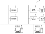
- FIG. 1 is a diagram showing a schematic configuration of the medical information system 1.
- the medical information system 1 shown in FIG. 1 is based on an inspection order from a doctor in a clinical department using a known ordering system, photographs of a part to be inspected of a subject, storage of a medical image acquired by the imaging, and an interpretation doctor. It is a system for interpreting medical images and creating an interpretation report, and for viewing the interpretation report by the doctor of the requesting clinical department and observing the details of the medical image to be interpreted.
- the medical information system 1 includes a plurality of imaging devices 2, a plurality of image interpretation WS (WorkStation) 3 which are image interpretation terminals, a medical care WS 4, an image server 5, and an image database (hereinafter, image DB (DataBase)).
- the report server 7 and the report database (hereinafter referred to as the report DB) 8 are connected to each other via a wired or wireless network 10 so as to be able to communicate with each other.
- Each device is a computer on which an application program for functioning as a component of the medical information system 1 is installed.
- the application program is recorded and distributed on a recording medium such as a DVD (Digital Versatile Disc) and a CD-ROM (Compact Disc Read Only Memory), and is installed on a computer from the recording medium.
- a recording medium such as a DVD (Digital Versatile Disc) and a CD-ROM (Compact Disc Read Only Memory)
- it is stored in the storage device of the server computer connected to the network 10 or in the network storage in a state of being accessible from the outside, and is downloaded and installed in the computer upon request.
- the photographing device 2 is a device (modality) that generates a medical image representing the diagnosis target part by photographing the part to be diagnosed of the subject. Specifically, it is a simple X-ray imaging apparatus, a CT apparatus, an MRI apparatus, a PET (Positron Emission Tomography) apparatus, and the like.
- the medical image generated by the imaging device 2 is transmitted to the image server 5 and stored in the image DB 6.
- the image interpretation WS3 is a computer used by, for example, an image interpretation doctor in a radiology department to interpret a medical image and create an image interpretation report, and includes a document creation support device 20 according to the present embodiment.
- a request for viewing a medical image to the image server 5 various image processing on the medical image received from the image server 5, a display of the medical image, an input acceptance of a finding sentence related to the medical image, and the like are performed.
- analysis processing for medical images and input findings support for creating an interpretation report based on the analysis results, a request for registration and viewing of an interpretation report for the report server 7, and an interpretation received from the report server 7 are performed.
- the report is displayed.
- the clinical WS4 is a computer used by doctors in clinical departments for detailed observation of images, viewing of interpretation reports, creation of electronic medical records, etc., and is a processing device, a display device such as a display, and an input device such as a keyboard and a mouse. Consists of.
- an image viewing request is made to the image server 5
- an image received from the image server 5 is displayed
- an image interpretation report viewing request is made to the report server 7
- an image interpretation report received from the report server 7 is displayed.
- the image server 5 is a general-purpose computer in which a software program that provides a database management system (DataBase Management System: DBMS) function is installed. Further, the image server 5 includes a storage in which the image DB 6 is configured. This storage may be a hard disk device connected by the image server 5 and the data bus, or a disk device connected to NAS (Network Attached Storage) and SAN (Storage Area Network) connected to the network 10. It may be.
- NAS Network Attached Storage
- SAN Storage Area Network
- the image data and incidental information of the medical image acquired by the imaging device 2 are registered in the image DB 6.
- the incidental information includes, for example, an image ID (identification) for identifying an individual medical image, a patient ID for identifying a subject, an examination ID for identifying an examination, and a unique ID assigned to each medical image ( UID: unique identification), examination date when the medical image was generated, examination time, type of imaging device used in the examination to acquire the medical image, patient information such as patient name, age, gender, examination site (imaging) Includes information such as site), imaging information (imaging protocol, imaging sequence, imaging method, imaging conditions, use of contrast medium, etc.), series number or collection number when multiple medical images are acquired in one examination. ..
- the image server 5 when the image server 5 receives the viewing request from the image interpretation WS3 and the medical examination WS4 via the network 10, the image server 5 searches for the medical image registered in the image DB 6, and uses the searched medical image as the requesting image interpretation WS3 and the medical examination. Send to WS4.
- the report server 7 incorporates a software program that provides the functions of a database management system to a general-purpose computer.
- the report server 7 receives the image interpretation report registration request from the image interpretation WS3, the report server 7 prepares the image interpretation report in a database format and registers the image interpretation report in the report DB 8.
- the image interpretation report includes, for example, a medical image to be interpreted, an image ID for identifying the medical image, an image interpretation doctor ID for identifying the image interpretation doctor who performed the image interpretation, a lesion name, a lesion position information, and a medical image including a specific area. It may include information for access and information such as property information.
- the report server 7 when the report server 7 receives a viewing request for the interpretation report from the interpretation WS3 and the medical treatment WS4 via the network 10, the report server 7 searches for the interpretation report registered in the report DB 8 and uses the searched interpretation report as the requester's interpretation. It is transmitted to WS3 and medical treatment WS4.
- the medical image is a three-dimensional CT image composed of a plurality of tomographic images with the diagnosis target as the lung, and the CT image is interpreted by the interpretation WS3 to obtain an abnormal shadow contained in the lung.
- An interpretation report shall be created.
- the medical image is not limited to the CT image, and any medical image such as an MRI image and a simple two-dimensional image acquired by a simple X-ray imaging device can be used.
- Network 10 is a wired or wireless local area network that connects various devices in the hospital.
- the network 10 may be configured such that the local area networks of each hospital are connected to each other by the Internet or a dedicated line.
- FIG. 2 illustrates the hardware configuration of the document creation support device according to the present embodiment.
- the document creation support device 20 includes a CPU (Central Processing Unit) 11, a non-volatile storage 13, and a memory 16 as a temporary storage area.
- the document creation support device 20 includes a display 14 such as a liquid crystal display, an input device 15 such as a keyboard and a mouse, and a network I / F (InterFace) 17 connected to the network 10.
- the CPU 11, the storage 13, the display 14, the input device 15, the memory 16, and the network I / F 17 are connected to the bus 18.
- the CPU 11 is an example of the processor in the present disclosure.
- the storage 13 is realized by an HDD (Hard Disk Drive), an SSD (Solid State Drive), a flash memory, or the like.
- the document creation support program 12 is stored in the storage 13 as a storage medium.
- the CPU 11 reads the document creation support program from the storage 13, expands it into the memory 16, and executes the expanded document creation support program 12.
- FIG. 3 is a diagram showing a functional configuration of the document creation support device according to the present embodiment.
- the document creation support device 20 includes an acquisition unit 21, a sentence generation unit 22, a specific unit 23, a display control unit 24, a storage control unit 25, and a communication unit 26.
- the CPU 11 executes the document creation support program, the CPU 11 functions as an acquisition unit 21, a sentence generation unit 22, a specific unit 23, a display control unit 24, a storage control unit 25, and a communication unit 26.
- the acquisition unit 21 acquires a medical image for creating an image interpretation report from the image server 5 in response to an instruction from the input device 15 by the image interpretation doctor who is the operator.
- diagnostic guidelines for lung cancer which is a target disease, are also obtained from the image server 5.
- the acquired medical image and diagnostic guideline are stored in the storage 13.
- the sentence generation unit 22 derives the analysis result by analyzing the medical image, and generates an interpretation report based on the analysis result. For this purpose, the sentence generation unit 22 discriminates the abnormal shadow candidate in the medical image, and discriminates the properties of the discriminated abnormal shadow candidate for each of the plurality of predetermined property items. Examples of property items identified for abnormal shadows are the location of the abnormal shadow, the size of the abnormal shadow, the shape of the border (clear and irregular), the type of absorption value (full and pleural), the presence or absence of spicula, and the mass. These include nodules, pleural contact, pleural infiltration, pleural infiltration, cavities, and calcification.
- the sentence generation unit 22 has a learning model in which machine learning is performed so as to discriminate the properties of abnormal shadow candidates from the medical image.
- the learning model uses teacher data to determine, for example, whether or not each pixel (voxel) in a medical image represents an abnormal shadow candidate, and if it is an abnormal shadow candidate, to determine its properties. It consists of a convolutional neural network (CNN (Convolutional Neural Network)) that has undergone deep learning.
- CNN Convolutional Neural Network
- the learning model is learned by machine learning using, for example, a plurality of combinations of a medical image including an abnormal shadow and a property label representing the property of the abnormal shadow as teacher data.
- the learning model takes a medical image as an input, and outputs a property score derived for each property item in the abnormal shadow included in the input medical image.
- the property score is a score indicating the prominence of the property for each property item.
- the property score takes, for example, a value of 0 or more and 1 or less, and the larger the value of the property score, the more remarkable the property.
- the property score for "presence or absence of spicula”, which is one of the property items of abnormal shadow is 0.5 or more
- the property for "presence or absence of spicula" of abnormal shadow is "with spicula (positive)”.
- the property score for "presence or absence of spicula” is, for example, less than 0.5, it is specified that the property for the presence or absence of spicula in abnormal shadow is "no spicula (negative)”.
- the threshold value 0.5 used for the property determination is merely an example, and is set to an appropriate value for each property item.
- an arbitrary learning model such as a support vector machine (SVM (Support Vector Machine)) can be used in addition to a convolutional neural network. Further, the learning model for detecting the abnormal shadow candidate from the medical image and the learning model for detecting the property information of the abnormal shadow candidate may be constructed separately.
- SVM Support Vector Machine
- FIG. 4 is a diagram for explaining an example of the property information derived by the sentence generation unit 22.
- the properties for each property item are "left pulmonary subpleural", “4.2 cm”, “irregular”, “enriched type”, and so on. They are “with spicula”, “mass”, “with pleural contact”, “with pleural infiltration”, “without pleural infiltration”, “without cavities” and “without calcification”.
- + is given when “yes” and-is given when there is no.
- the sentence generation unit 22 generates a finding sentence using the derived property information.
- the sentence generation unit 22 is composed of a learning model in which learning is performed so as to generate a sentence from the input property information.
- a learning model for example, a recurrent neural network can be used.
- FIG. 5 is a diagram showing a schematic configuration of a recurrent neural network.
- the recurrent neural network 40 includes an encoder 41 and a decoder 42.
- the property information derived by the sentence generation unit 22 is input to the encoder 41. For example, property information of "left pulmonary subpleuralis", “4.2 cm”, “Spicula +” and “mass" is input to the encoder 41.
- the decoder 42 is learned so as to document the character information, and generates a sentence from the input property information. Specifically, from the above-mentioned property information of "left pulmonary subpleura”, “4.2 cm”, “spicula +” and “mass”, "a 4.2 cm diameter mass having spicula under the left pulmonary pleura is recognized. Will be generated. " In FIG. 5, "EOS” indicates the end of the sentence (End Of Sentence).
- the recurrent neural network 40 learns the encoder 41 and the decoder 42 using a large amount of teacher data composed of a combination of the property information and the finding sentence. Be built.
- the generated text shown in FIG. 5 represents the findings about the lung nodule, and is generated by learning the learning model by inputting the property information of the lung nodule.
- the sentence generation unit 22 uses these learning models to generate an interpretation report including at least one finding sentence.
- FIG. 6 is a diagram showing an image interpretation report including an input medical image and a finding sentence generated from the medical image.
- the input medical image G0 As shown in FIG. 6, regarding the input medical image G0, "a solid nodule with a diameter of 17 mm is observed in the upper lobe S2 of the right lung, there is a spicula on the margin, and an image of pleural invagination is accompanied. Bronchial translucency image is observed. No lymphadenopathy is observed. There is no intrapulmonary metastasis. ”Interpretation report 35 has been generated.
- the acquisition unit 21 acquires the diagnostic guideline for lung cancer and stores it in the storage 13.
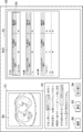
- 7 and 8 are diagrams showing diagnostic guidelines for lung cancer. Note that FIG. 7 shows the stage of lung cancer based on the TNM classification, and FIG. 8 shows the T factor, N factor, and M factor shown in the stage.
- stage 1 stage I
- stage II stage II
- stage III stage IV
- T factor representing cancer size and infiltration
- N factor representing lymph node metastasis
- M factor representing distant metastasis
- each factor is classified and defined in a plurality of stages.
- factor T is classified into four stages, T1 to T4, according to the size of the cancer and the state of infiltration.
- T1 and T2 are classified more finely, such as T1a, T1b, T2a, and T2b, respectively.
- N factor is classified into three stages of N1 to N3.
- N0 has no lymph node metastasis.
- M factor is classified into two stages, M0 when there is no metastasis and M1 when there is metastasis.
- M1 is classified more finely like M1a and M1b, respectively.
- the identification unit 23 identifies the corresponding part corresponding to the item included in the diagnostic guideline for the disease described in the interpretation report.
- the specific unit 23 has a learning model 23A in which machine learning is performed so as to specify a portion corresponding to an item included in the diagnostic guideline as a corresponding portion in the input sentence.
- the learning model 23A uses teacher data to perform deep learning so that when a convolutional report is input, the input convolutional report discriminates words and phrases related to items included in the diagnostic guideline as corresponding parts. It consists of a convolutional neural network (CNN).
- CNN convolutional neural network
- the teacher data for learning the learning model 23A is associated with words and phrases included in the items of the diagnostic guideline and sentences using the words and phrases included in the items of the diagnostic guideline.
- the items of the diagnostic guideline include "the maximum diameter of the tumor is 2 cm or less” as T1a of T factor, "no regional lymph node metastasis” as N0 of N factor, and M0 of M factor. Includes “no distant metastasis”.
- the words “diameter”, “lymph node”, “no metastasis”, and “no distant metastasis” included in the items of the diagnostic guideline, and "nodule with a diameter of 15 mm" are recognized. Is not admitted.
- the learning model 23A is constructed by learning a neural network using a large number of such teacher data. As a result, when the interpretation report is input, the learning model 23A outputs the corresponding portion corresponding to the item included in the diagnostic guideline for the disease described in the interpretation report. Further, when the specifying unit 23 specifies the corresponding part in the interpretation report, the specifying unit 23 specifies the item corresponding to the corresponding part in the diagnostic guideline.
- any learning model such as a support vector machine and a recurrent neural network can be used.
- the specific unit 23 is not limited to the one that specifies the corresponding portion by the learning model 23A.
- the corresponding portion may be specified by searching the interpretation report using the size of the mass and the items included in the diagnostic guideline as keywords.
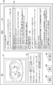
- FIG. 9 is a diagram showing a display screen of an interpretation report and a diagnostic guideline.
- the display screen 50 has an image display area 51, an interpretation report display area 52, and a diagnostic guideline display area 53.
- a switching button 54, a correction button 55, and a confirmation button 56 are displayed below the interpretation report display area 52.
- the tomographic image S0 included in the medical image G0 for which the interpretation report is generated is displayed.
- the interpretation report display area 52 the interpretation report generated by the sentence generation unit 22 is displayed.
- the corresponding portion specified by the specific unit 23 is highlighted.
- the three corresponding parts of the interpretation report "17 mm in diameter”, "no lymphadenopathy” and “no intrapulmonary metastasis”, are surrounded by a frame. It is highlighted by. It should be noted that the highlighting of these three corresponding portions has different modes. In FIGS.
- the diagnostic guidelines 60A to 60C are displayed in the diagnostic guideline display area 53. Since the contents of the diagnostic guidelines 60A to 60C are diverse, the contents of the diagnostic guideline displayed in the diagnostic guideline display area 53 can be changed by selecting the switching button 54. Specifically, FIG. 9 shows diagnostic guidelines 60A for factors T and N. Further, FIG. 10 shows a diagnostic guideline 60B for the M factor. Further, in FIG. 11, the diagnostic guideline 60C for the stage is displayed. Instead of the switching display of the diagnostic guideline, the entire diagnostic guideline may be displayed so that it can be referred to by scrolling.
- the item of T1a corresponding to "diameter 17 mm” in the interpretation report is highlighted.
- the highlighting of the item of T1a is shown by surrounding it with a solid line frame 61 similar to the corresponding portion of “diameter 17 mm” in the interpretation report.
- the item N0 corresponding to "No lymphadenopathy is observed” in the interpretation report is also highlighted.
- the highlighting of the item N0 is shown by enclosing it in a broken line frame 62 similar to the corresponding part of “No lymphadenopathy is observed” in the interpretation report.
- the item of M0 corresponding to "There is no intrapulmonary metastasis" in the interpretation report is highlighted.
- the highlighting of the item M0 is shown by enclosing it in the frame 63 of the alternate long and short dash line similar to the corresponding part of “No intrapulmonary metastasis” in the interpretation report.
- the diagnostic guideline 60C shown in FIG. 11 there are three corresponding parts in the interpretation report, "diameter 17 mm", “no lymphadenopathy” and “no intrapulmonary metastasis”, and these.
- the stage derived from the item corresponding to the corresponding part is highlighted.
- the IA period is highlighted.
- the highlighting is shown by surrounding it with a solid line frame 64.
- the highlighting of items in the diagnostic guidelines is not limited to the addition of frames. Instead of adding a frame, the characters may be highlighted or the character color may be changed.
- the operator can compare and confirm the interpretation report and the diagnostic guideline on the display screen 50. If necessary, the interpretation report displayed in the interpretation report display area 52 can be modified by input from the input device 15 by selecting the correction button 55. When the confirmation or correction of the interpretation report is completed, the operator selects the confirmation button 56.
- the specific unit 23 again identifies the corresponding part corresponding to the item included in the diagnostic guideline in the modified interpretation report. In this case, if the corresponding part is added or deleted, the item in the diagnostic guideline is also added or deleted accordingly.
- the storage control unit 25 highlights the image interpretation report in which the corresponding portion is highlighted, the tomographic image S0 included in the medical image G0 referred to when generating the image interpretation report, and the items by selecting the confirmation button 56 by the operator.
- the diagnostic guideline that has been obtained is also stored in the storage 13.
- the communication unit 26 combines the image interpretation report in which the corresponding part is highlighted, the tomographic image S0 included in the medical image G0 referred to when generating the image interpretation report, and the diagnostic guideline in which the items are highlighted, and the network I / Transfer to the report server 7 via F17.
- the report server 7 also stores the image interpretation report in which the corresponding portion is highlighted, the medical image G0 referred to when generating the image interpretation report, and the diagnostic guideline in which the items are highlighted.
- FIG. 12 is a flowchart showing the processing performed in the first embodiment. It is assumed that the medical image G0 and the diagnostic guideline to be read are acquired from the image server 5 by the acquisition unit 21 and stored in the storage 13. The process is started when the interpretation doctor gives an instruction to create the interpretation report, and the sentence generation unit 22 derives the analysis result by analyzing the medical image G0 and generates the interpretation report based on the analysis result ( Step ST1). Next, the identification unit 23 identifies in the image interpretation report the corresponding part corresponding to the item included in the diagnostic guideline for the disease described in the image interpretation report (step ST2).
- the display control unit 24 associates the corresponding portion corresponding to the item of the diagnostic guideline in the image interpretation report with the item in the diagnostic guideline, and displays the image interpretation report and the diagnostic guideline on the display 14 (step ST3).
- the operator switches the diagnostic guideline displayed in the diagnostic guideline display area 53 on the display screen 50, and modifies the interpretation report displayed in the text display area 51 if necessary.
- the storage control unit 25 starts monitoring whether or not the confirmation button 56 is selected (step ST4), and when step ST4 is affirmed, the storage control unit 25 receives an image interpretation report in which the corresponding portion is highlighted.
- the tomographic image S0 included in the medical image G0 referred to when generating the interpretation report and the diagnostic guideline in which the item is highlighted are also saved in the storage 13 (save the interpretation report, etc .; step ST5).
- the communication unit 26 reports via the network I / F17 together with the image interpretation report in which the corresponding portion is highlighted, the tomographic image S0 referred to when generating the image interpretation report, and the diagnostic guideline in which the items are highlighted. Transfer to the server 7 (transfer of interpretation report, etc .; step ST6), and end the process.
- the corresponding part corresponding to the item included in the diagnostic guideline for the disease described in the image interpretation report is specified, and the above corresponding part in the image interpretation report and the item in the diagnostic guideline are specified.
- the interpretation report and the diagnostic guideline are displayed on the display 14 in association with the above. Therefore, the work of creating the interpretation report can be efficiently performed while comparing the diagnostic guideline and the interpretation report. Therefore, according to the present embodiment, it is possible to reduce the burden on the operator who creates the interpretation report by referring to the diagnostic guideline.
- the image interpretation report with the corresponding part highlighted, the tomographic image S0 included in the medical image G0 referred to when creating the image interpretation report, and the diagnostic guideline with the item highlighted are saved or transferred together. Therefore, the attending physician of the patient can refer to the correspondence between the interpretation report and the diagnostic guideline, and as a result, the diagnosis can be made efficiently.
- the interpretation report states, "A solid nodule with a diameter of 17 mm is found in the upper lobe S2 of the right lung, and there is a spicula on the margin, accompanied by a pleural invagination image.
- the interpretation report states, "A solid nodule with a diameter of 17 mm is found in the upper lobe S2 of the right lung, and there is a spicula on the margin, accompanied by a pleural invagination image.
- No intrapulmonary metastasis There is no description in the interpretation report regarding items related to lymph node metastasis.
- “lymph node metastasis (N factor)” is highlighted in the diagnostic guideline 60A.
- the highlighting is shown by surrounding it with a solid line frame 65.
- the sentence generation unit 22 analyzes the medical image G0 and generates an interpretation report, but the present invention is not limited to this.
- the operator may input the image interpretation report using the input device 15 in the image interpretation report display area 52 of the display screen 50.
- the specifying unit 23 may specify the corresponding part corresponding to the item of the diagnostic guideline in the interpretation report every time a sentence of the findings included in the interpretation report is described, and the input of the interpretation report is completed. Therefore, the corresponding portion may be specified. In this case, the sentence generation unit 22 shown in FIG. 3 is unnecessary.
- the diagnostic guideline is stored in the image server 5, but the present invention is not limited to this.
- a dedicated server for storing the diagnostic guideline may be provided in the medical information system 1 and the diagnostic guideline may be acquired from this dedicated server.
- the document creation support device 20 generates an interpretation report, but the present invention is not limited to this.
- the acquisition unit 21 acquires the image interpretation report from the report server 7, the specific unit 23 identifies the corresponding part of the acquired image interpretation report corresponding to the item of the diagnostic guideline, and the acquired image interpretation report is combined with the diagnostic guideline. It may be displayed.
- the technique of the present disclosure is applied when creating an image interpretation report using a medical image with the diagnosis target as the lung, but the diagnosis target is not limited to the lung.
- diagnosis target is not limited to the lung.
- any part of the human body such as the heart, liver, brain, and limbs can be diagnosed.
- the diagnostic guideline corresponding to the part to be diagnosed may be acquired, and the corresponding part corresponding to the item of the diagnostic guideline in the interpretation report may be specified.
- a processing unit that executes various processes such as an acquisition unit 21, a sentence generation unit 22, a specific unit 23, a display control unit 24, a storage control unit 25, and a communication unit 26.
- various processors processors shown below can be used.
- the various processors include a CPU, which is a general-purpose processor that executes software (program) and functions as various processing units, and a circuit after manufacturing an FPGA (Field Programmable Gate Array) or the like.
- Dedicated electricity which is a processor with a circuit configuration specially designed to execute specific processing such as programmable logic device (PLD), ASIC (Application Specific Integrated Circuit), which is a processor whose configuration can be changed. Circuits and the like are included.
- One processing unit may be composed of one of these various processors, or a combination of two or more processors of the same type or different types (for example, a combination of a plurality of FPGAs or a combination of a CPU and an FPGA). ) May be configured. Further, a plurality of processing units may be configured by one processor.
- one processor is configured by combining one or more CPUs and software. There is a form in which this processor functions as a plurality of processing units.
- SoC System On Chip
- the various processing units are configured by using one or more of the above-mentioned various processors as a hardware structure.
- circuitry in which circuit elements such as semiconductor elements are combined can be used.
Landscapes
- Health & Medical Sciences (AREA)
- Public Health (AREA)
- Engineering & Computer Science (AREA)
- Life Sciences & Earth Sciences (AREA)
- General Health & Medical Sciences (AREA)
- Medical Informatics (AREA)
- Primary Health Care (AREA)
- Epidemiology (AREA)
- Biomedical Technology (AREA)
- Physics & Mathematics (AREA)
- Radiology & Medical Imaging (AREA)
- Biophysics (AREA)
- Pathology (AREA)
- Nuclear Medicine, Radiotherapy & Molecular Imaging (AREA)
- Heart & Thoracic Surgery (AREA)
- Molecular Biology (AREA)
- Surgery (AREA)
- Animal Behavior & Ethology (AREA)
- Veterinary Medicine (AREA)
- Medical Treatment And Welfare Office Work (AREA)
- Measuring And Recording Apparatus For Diagnosis (AREA)
Priority Applications (1)
| Application Number | Priority Date | Filing Date | Title |
|---|---|---|---|
| JP2022503727A JP7368592B2 (ja) | 2020-02-25 | 2021-02-25 | 文書作成支援装置、方法およびプログラム |
Applications Claiming Priority (2)
| Application Number | Priority Date | Filing Date | Title |
|---|---|---|---|
| JP2020029705 | 2020-02-25 | ||
| JP2020-029705 | 2020-02-25 |
Publications (1)
| Publication Number | Publication Date |
|---|---|
| WO2021172477A1 true WO2021172477A1 (ja) | 2021-09-02 |
Family
ID=77491699
Family Applications (1)
| Application Number | Title | Priority Date | Filing Date |
|---|---|---|---|
| PCT/JP2021/007207 Ceased WO2021172477A1 (ja) | 2020-02-25 | 2021-02-25 | 文書作成支援装置、方法およびプログラム |
Country Status (2)
| Country | Link |
|---|---|
| JP (1) | JP7368592B2 (https=) |
| WO (1) | WO2021172477A1 (https=) |
Citations (5)
| Publication number | Priority date | Publication date | Assignee | Title |
|---|---|---|---|---|
| JP2006260318A (ja) * | 2005-03-18 | 2006-09-28 | Hitachi Medical Corp | 読影レポート入力支援方法及び読影レポート入力支援システム |
| JP2015097127A (ja) * | 2015-02-10 | 2015-05-21 | キヤノン株式会社 | 診断支援装置、診断支援装置の制御方法、プログラム及び記憶媒体 |
| JP2018147275A (ja) * | 2017-03-07 | 2018-09-20 | 株式会社ジェイマックシステム | 治療実績評価支援装置、治療実績評価支援方法、および治療実績評価支援プログラム |
| JP2018532209A (ja) * | 2015-11-05 | 2018-11-01 | コーニンクレッカ フィリップス エヌ ヴェKoninklijke Philips N.V. | 偶発的知見についての長期的健康患者プロファイル |
| JP2019149005A (ja) * | 2018-02-27 | 2019-09-05 | 富士フイルム株式会社 | 医療文書作成支援装置、方法およびプログラム |
Family Cites Families (1)
| Publication number | Priority date | Publication date | Assignee | Title |
|---|---|---|---|---|
| JP2005190055A (ja) | 2003-12-25 | 2005-07-14 | Japan Council For Quality Health Care | 医療情報提供システム、医療情報提供システム用管理装置及び医療情報提供システム用管理装置のプログラム |
-
2021
- 2021-02-25 WO PCT/JP2021/007207 patent/WO2021172477A1/ja not_active Ceased
- 2021-02-25 JP JP2022503727A patent/JP7368592B2/ja active Active
Patent Citations (5)
| Publication number | Priority date | Publication date | Assignee | Title |
|---|---|---|---|---|
| JP2006260318A (ja) * | 2005-03-18 | 2006-09-28 | Hitachi Medical Corp | 読影レポート入力支援方法及び読影レポート入力支援システム |
| JP2015097127A (ja) * | 2015-02-10 | 2015-05-21 | キヤノン株式会社 | 診断支援装置、診断支援装置の制御方法、プログラム及び記憶媒体 |
| JP2018532209A (ja) * | 2015-11-05 | 2018-11-01 | コーニンクレッカ フィリップス エヌ ヴェKoninklijke Philips N.V. | 偶発的知見についての長期的健康患者プロファイル |
| JP2018147275A (ja) * | 2017-03-07 | 2018-09-20 | 株式会社ジェイマックシステム | 治療実績評価支援装置、治療実績評価支援方法、および治療実績評価支援プログラム |
| JP2019149005A (ja) * | 2018-02-27 | 2019-09-05 | 富士フイルム株式会社 | 医療文書作成支援装置、方法およびプログラム |
Also Published As
| Publication number | Publication date |
|---|---|
| JP7368592B2 (ja) | 2023-10-24 |
| JPWO2021172477A1 (https=) | 2021-09-02 |
Similar Documents
| Publication | Publication Date | Title |
|---|---|---|
| JP7618003B2 (ja) | 文書作成支援装置、方法およびプログラム | |
| JP2019153250A (ja) | 医療文書作成支援装置、方法およびプログラム | |
| WO2022215530A1 (ja) | 医用画像装置、医用画像方法、及び医用画像プログラム | |
| US20230005580A1 (en) | Document creation support apparatus, method, and program | |
| US20190279408A1 (en) | Medical image processing apparatus, medical image processing method, and medical image processing program | |
| US20230281810A1 (en) | Image display apparatus, method, and program | |
| US12527528B2 (en) | Image display apparatus, method, and program | |
| WO2020209382A1 (ja) | 医療文書作成装置、方法およびプログラム | |
| US11923069B2 (en) | Medical document creation support apparatus, method and program, learned model, and learning apparatus, method and program | |
| JP2024009342A (ja) | 文書作成支援装置、方法およびプログラム | |
| US12211600B2 (en) | Information processing apparatus, information processing method, and information processing program | |
| WO2021177312A1 (ja) | 情報保存装置、方法およびプログラム、並びに解析記録生成装置、方法およびプログラム | |
| WO2021107098A1 (ja) | 文書作成支援装置、文書作成支援方法及び文書作成支援プログラム | |
| JPWO2020129385A1 (ja) | 医療文書作成支援装置、方法およびプログラム | |
| WO2021177357A1 (ja) | 情報処理装置、情報処理方法及び情報処理プログラム | |
| JPWO2019193982A1 (ja) | 医療文書作成支援装置、医療文書作成支援方法、及び医療文書作成支援プログラム | |
| WO2021107099A1 (ja) | 文書作成支援装置、文書作成支援方法及びプログラム | |
| WO2020202822A1 (ja) | 医療文書作成支援装置、方法およびプログラム | |
| WO2022196106A1 (ja) | 文書作成装置、方法およびプログラム | |
| JP7299314B2 (ja) | 医療文書作成装置、方法およびプログラム、学習装置、方法およびプログラム、並びに学習済みモデル | |
| WO2021193548A1 (ja) | 文書作成支援装置、方法およびプログラム | |
| WO2023054645A1 (ja) | 情報処理装置、情報処理方法及び情報処理プログラム | |
| WO2022230641A1 (ja) | 文書作成支援装置、文書作成支援方法、及び文書作成支援プログラム | |
| WO2022196105A1 (ja) | 情報管理装置、方法およびプログラム、並びに情報処理装置、方法およびプログラム | |
| WO2021167018A1 (ja) | 情報処理装置、情報処理方法及び情報処理プログラム |
Legal Events
| Date | Code | Title | Description |
|---|---|---|---|
| 121 | Ep: the epo has been informed by wipo that ep was designated in this application |
Ref document number: 21759995 Country of ref document: EP Kind code of ref document: A1 |
|
| ENP | Entry into the national phase |
Ref document number: 2022503727 Country of ref document: JP Kind code of ref document: A |
|
| NENP | Non-entry into the national phase |
Ref country code: DE |
|
| 122 | Ep: pct application non-entry in european phase |
Ref document number: 21759995 Country of ref document: EP Kind code of ref document: A1 |