WO2021048852A1 - Methods of treating breast cancer - Google Patents
Methods of treating breast cancer Download PDFInfo
- Publication number
- WO2021048852A1 WO2021048852A1 PCT/IL2020/050993 IL2020050993W WO2021048852A1 WO 2021048852 A1 WO2021048852 A1 WO 2021048852A1 IL 2020050993 W IL2020050993 W IL 2020050993W WO 2021048852 A1 WO2021048852 A1 WO 2021048852A1
- Authority
- WO
- WIPO (PCT)
- Prior art keywords
- combination
- inhibitor
- ferroptosis
- agents
- tnbc
- Prior art date
- Legal status (The legal status is an assumption and is not a legal conclusion. Google has not performed a legal analysis and makes no representation as to the accuracy of the status listed.)
- Ceased
Links
Classifications
-
- A—HUMAN NECESSITIES
- A61—MEDICAL OR VETERINARY SCIENCE; HYGIENE
- A61K—PREPARATIONS FOR MEDICAL, DENTAL OR TOILETRY PURPOSES
- A61K45/00—Medicinal preparations containing active ingredients not provided for in groups A61K31/00 - A61K41/00
- A61K45/06—Mixtures of active ingredients without chemical characterisation, e.g. antiphlogistics and cardiaca
-
- A—HUMAN NECESSITIES
- A61—MEDICAL OR VETERINARY SCIENCE; HYGIENE
- A61K—PREPARATIONS FOR MEDICAL, DENTAL OR TOILETRY PURPOSES
- A61K31/00—Medicinal preparations containing organic active ingredients
- A61K31/16—Amides, e.g. hydroxamic acids
- A61K31/17—Amides, e.g. hydroxamic acids having the group >N—C(O)—N< or >N—C(S)—N<, e.g. urea, thiourea, carmustine
-
- A—HUMAN NECESSITIES
- A61—MEDICAL OR VETERINARY SCIENCE; HYGIENE
- A61K—PREPARATIONS FOR MEDICAL, DENTAL OR TOILETRY PURPOSES
- A61K31/00—Medicinal preparations containing organic active ingredients
- A61K31/33—Heterocyclic compounds
- A61K31/395—Heterocyclic compounds having nitrogen as a ring hetero atom, e.g. guanethidine or rifamycins
- A61K31/55—Heterocyclic compounds having nitrogen as a ring hetero atom, e.g. guanethidine or rifamycins having seven-membered rings, e.g. azelastine, pentylenetetrazole
- A61K31/551—Heterocyclic compounds having nitrogen as a ring hetero atom, e.g. guanethidine or rifamycins having seven-membered rings, e.g. azelastine, pentylenetetrazole having two nitrogen atoms, e.g. dilazep
-
- A—HUMAN NECESSITIES
- A61—MEDICAL OR VETERINARY SCIENCE; HYGIENE
- A61K—PREPARATIONS FOR MEDICAL, DENTAL OR TOILETRY PURPOSES
- A61K31/00—Medicinal preparations containing organic active ingredients
- A61K31/69—Boron compounds
-
- A—HUMAN NECESSITIES
- A61—MEDICAL OR VETERINARY SCIENCE; HYGIENE
- A61K—PREPARATIONS FOR MEDICAL, DENTAL OR TOILETRY PURPOSES
- A61K38/00—Medicinal preparations containing peptides
-
- A—HUMAN NECESSITIES
- A61—MEDICAL OR VETERINARY SCIENCE; HYGIENE
- A61K—PREPARATIONS FOR MEDICAL, DENTAL OR TOILETRY PURPOSES
- A61K38/00—Medicinal preparations containing peptides
- A61K38/04—Peptides having up to 20 amino acids in a fully defined sequence; Derivatives thereof
- A61K38/05—Dipeptides
-
- A—HUMAN NECESSITIES
- A61—MEDICAL OR VETERINARY SCIENCE; HYGIENE
- A61P—SPECIFIC THERAPEUTIC ACTIVITY OF CHEMICAL COMPOUNDS OR MEDICINAL PREPARATIONS
- A61P35/00—Antineoplastic agents
-
- A—HUMAN NECESSITIES
- A61—MEDICAL OR VETERINARY SCIENCE; HYGIENE
- A61P—SPECIFIC THERAPEUTIC ACTIVITY OF CHEMICAL COMPOUNDS OR MEDICINAL PREPARATIONS
- A61P35/00—Antineoplastic agents
- A61P35/04—Antineoplastic agents specific for metastasis
Definitions
- the present invention in some embodiments thereof, relates to methods of treating breast cancer and, more particularly, but not exclusively, to treating triple negative breast cancer.
- Triple-negative breast cancer is a highly aggressive breast cancer subtype, defined by the absence of both estrogen and progesterone receptors as well as of HER2 amplification, and characterized by high heterogeneity and lack of effective treatment or therapeutic targets.
- ferroptosis is an iron-dependent and reactive oxygen species (ROS)-reliant cell death with characteristics mainly of cytological changes, including decreased or vanished mitochondria cristae, a ruptured outer mitochondrial membrane, and a condensed mitochondrial membrane.
- ROS reactive oxygen species
- ferroptosis could be triggered by diverse physiological conditions and pathological stresses in humans and animals.
- Ferroptosis is gradually accepted as an adaptive feature to eliminate the malignant cells. It plays a pivotal role in the depression of tumorigenesis by removing the cells that are deficient in key nutrients in the environment or damaged by infection or ambient stress.
- cancer cells are under continuous oxidative stress with an extraordinar balance between thiols and catalytic iron, ferroptosis does not often happen in the cancer development. The underlying molecular mechanisms remain poorly understood.
- TNBC triple negative breast cancer
- BET Bromodomain and Extra-Terminal motif
- composition comprising a pharmaceutically acceptable carrier and as active agents:
- BET Bromodomain and Extra-Terminal motif
- a ferroptosis-inducing agent for use in the treatment of triple negative breast cancer (TNBC), with the proviso that the ferroptosis-inducing agent is not RAS-selective lethal 3 (RSL3).
- TNBC triple negative breast cancer
- the agent which downregulates the amount and/or activity of CXCRl/2 receptor is an anti-CXCRl antibody or an anti-CXCR2 antibody.
- the agent which downregulates the amount and/or activity of CXCRl/2 receptor is a dual CXCRl/2 antagonist.
- the dual CXCRl/2 antagonist is selected from the group consisting of navarixin, ladarixin, reparixin, reparixin L-lysine salt, DF2755A, CXCL8 fragment comprising amino acids 3-74 and substitutions K11R/G31P (G31P), DF2162, and SCH-479833.
- the BET inhibitor is a small molecule inhibitor.
- the small molecule inhibitor is selected from the group consisting of CPI-0610, DUAL946, GSK525762, I-BET151, JQ1, OTX015, PFI-1, RVX-208, RVX2135 and TEN-010.
- the small molecule inhibitor is OTX015.
- TNBC a Bromodomain and Extra-Terminal motif (BET) inhibitor; and (ii) an agent which downregulates the amount and/or activity of CXCRl/2 receptor, the TNBC is mesenchymal or mesenchymal stem-like TNBC.
- BET Bromodomain and Extra-Terminal motif
- the proteasome inhibitor is an epoxyketone or a boronate.
- the proteasome inhibitor is a boronate.
- the inhibitor is bortezomib or ixazomib.
- the proteasome inhibitor is an epoxyketone. According to embodiments of the present invention, the proteasome inhibitor is carfilzomib or a fluorinated carfilzomib analog.
- the combination is formulated in a single composition.
- the combination is formulated in separate compositions.
- the BET inhibitor is a small molecule inhibitor.
- the small molecule inhibitor is selected from the group consisting of CPI-0610, DUAL946, GSK525762, I-BET151, JQ1, OTX015, PFI-1, RVX-208, RVX2135 and TEN-010.
- the small molecule inhibitor is OTX015.
- the agent which downregulates the amount and/or activity of CXCRl/2 receptor is an anti-CXCRl antibody or an anti-CXCR2 antibody.
- the agent which downregulates the amount and/or activity of CXCRl/2 receptor is a dual CXCRl/2 antagonist.
- the dual CXCRl/2 antagonist is selected from the group consisting of navarixin, ladarixin, reparixin, reparixin L-lysine salt, DF2755A, CXCL8 fragment comprising amino acids 3-74 and substitutions K11R/G31P (G31P), DF2162, and SCH-479833.
- the agent is a combination of at least two agents.
- the combination of at least two agents are formulated in a single composition.
- the combination of at least two agents are formulated in separate compositions.
- the combination of at least two agents comprise:
- the BET inhibitor is a small molecule inhibitor.
- the BET inhibitor is selected from the group consisting of OTX015, CPI-0610, DUAL946, GSK525762, I-BET151, JQ1, PFI-1, RVX-208, RVX2135 and TEN-010. According to embodiments of the present invention, the BET inhibitor is OTX015.
- the agent is a class I inducer.
- the class I inducer is selected from the group consisting of erastin, imidazole ketone erastin (IKE), piperazine erastin (PE), sulfasalazine and sorafmib. According to embodiments of the present invention, the class I inducer is selected from the group consisting of IKE, PE and sorafmib.
- the agent is a class III inducer.
- the class III inducer is selected from the group consisting of ferroptosis inducer 56 (FIN56) and caspase-independent lethal 56 (CIL56).
- the proteasome inhibitor is an epoxyketone or a boronate. According to embodiments of the present invention, the proteasome inhibitor is a boronate.
- the inhibitor is bortezomib or ixazomib.
- the proteasome inhibitor is an epoxyketone.
- the proteasome inhibitor is carfilzomib or a fluorinated carfilzomib analog.
- FIGs. 1 A-E Synthetic lethal combinations therapies for TNBC subtypes.
- M/MSL mesenchymal cell lines
- BL basal-like
- M/MSL mesenchymal
- BL basal-like specific
- FIGs. 2A-E. In vitro validation of drug combinations targeting BET.
- A.B Effects of BET and CXCR2 antagonists (A) or BET and proteasome inhibitors (B) on cell viability.
- D.E Dose-response curves of single agents and drug combinations in two selected cell lines treated with varying concentrations of JQ1 and SB225022 (D) or JQ1 and Bortezomib (BTZ) (E) for 72 hr. IC 50 values were calculated from the dose-response curves.
- Combination index (Cl) was calculated by the CompuSyn software with the Chou-Talalay equation using multiple doses and response points. Cl values for three different indicated fractions affected (FA) are shown.
- FIGs. 3 A-K In vivo validation of drug combinations targeting BET.
- mice and combination had no significant effects on body weight (D). Shown are mean values ⁇ SD of mice body weight from the four different treatment groups measured at the indicated time points.
- FIGs. 4A-I Inhibition of BET and the proteasome triggers ferroptosis cell death.
- Staurosporine 150 nM for 16 hr was used as a positive control.
- the cells were treated with JQ1 (0.05 mM) and SB225022 (0.05 pM), for 24 hr, lysed and assessed by Western Blot (WB) for the indicated proteins.
- the combination had no effect on the basal-lines (MDA-468, HCC1143 and HCC1937) shown in the right side.
- Ferroptosis inhibitors rescue cell death induced by JQ1 and Bortezomib combination.
- the indicated cell lines were pretreated with the indicated cell death inhibitors (Z-VAD-fmk, apoptosis; Necrostatin-1, necrosis; 3-MA- autophagy; Ferrostatin-1, Glutathione and 2,2- bipyridyl, ferroptosis) for 1 hr and then for additional 72 hr in the absence or presence of JQ1 (0.05 pM) and BTZ (2 nM).
- Cell viability was measured by CellTiter-Blue assay and is presented as percent of control untreated cells.
- the effects of the death inhibitors on death pathways specific inducers is shown in Figure 14C as positive controls. The results are mean values of three independent experiments.
- TNBC cell lines were treated with JQ1 (0.05 pM) and BTZ (2 nM) for 72 hr in the absence or the presence of deferoxamine (DFO) (100 pM) or 2,2'-dipyridyl (10 pM) and cell viability was assessed by crystal violet staining.
- DFO deferoxamine
- 2,2'-dipyridyl 10 pM
- E Representative confocal images of the indicated TNBC cell lines and the luminal T47D cells stained with Cll-BODIPY.
- the cells were treated with cumene hydroperoxide (CH) (100 pM) for 3 hr as a positive control, and either with JQ1 (0.05 pM) plus SB225022 (0.05 pM), or with JQ1 (0.05 pM) and BTZ (2 nM) for 16 hr.
- Lipid peroxidation was assessed by Cl 1- BODIPY (10 pM) staining as described in Methods. Scale bars, 10 pm.
- TNBC TNBC
- non-TNBC cell lines green
- JQ1 0.05 pM
- BTZ BTZ (2 nM)
- CH cumene hydroperoxide
- GSH glutathione
- Lipid peroxidation was assessed as described in Methods and shown as ratio between fluorescence emissions at 510 nm (green) to 590 nm (red). The results are mean values of three independent experiments.
- TNBC and non-TNBC (MCF7, SKBR3, T47D, BT474) cells were incubated with JQ1 (0.05 mM) and BTZ (2 nM) for 16 hr and total iron (G) and reduced glutathione (H) levels were measured as described methods. The results are mean values of three independent experiments.
- FIGs. 5A-E BET and proteasome inhibition strongly affects GPX4 level and transcription of key ferroptotic genes.
- B Effects of BET and proteasome inhibition on the level of GPX4 protein.
- the indicated TNBC cell lines were treated with JQ1 (0.05 pM), BTZ (2 nM) or both for 24 hr.
- Levels of GPX4 protein was assessed by WB. Intensities of GPX4 bands were quantified, normalized, and presented as fold of control in the bar graph.
- TNBC cell lines were treated with JQ1 (0.05 pM), BTZ (2 nM) or both for 24 hr.
- GPX4 mRNA levels were assessed by qPCR. Shown are mean values ⁇ SD of three independent experiments.
- TNBC cell lines were treated with JQ1 (0.05 pM) and BTZ (2 nM) for 24 hr and mRNA levels of the indicated genes were assessed by qPCR. The results are reported as fold of control. Shown are mean values of three independent experiments. Expression of actin was used as housekeeping gene to normalize the expression of each sample.
- FIGs. 6A-G TNBC are vulnerable to ferroptosis and enriched in ferroptosis signature.
- B GSEA plot of normalized enrichment score (NES) of ferroptosis pathway for basal versus non-basal patients in the TCGA dataset. Enrichment is significant, p-value/FDR ⁇ 0.001.
- C GSEA enrichment plot of ferroptosis pathway for TNBC versus non-TNBC cell lines in the CCLE dataset. Enrichment is significant, p-value/FDR ⁇ 0.001.
- D-E Box plots showing the expression of SLC40A1 (D) and ACSL4 (E) in breast cancer patients grouped by PAM50. The differences between the basal patients and any other PAM50 group are significant (/-test, P-value ⁇ 0.001).
- F Heatmap of normalized expression of the Ferroptosis gene signature (KEGG- 40 genes) in breast cancer patients from the TCGA dataset. Patients (in columns) are arranged by unsupervised clustering of gene expression. Bar at the top of the heatmap indicates the subtype of each patient (PAM50). Ferroptotic genes which also belong to the iron metabolism and glutathione signature are marked.
- G Levels of ferroptosis proteins and BRD4 in TNBC and non-TNBC cell lines. The expression levels of the indicated proteins were assessed by WB and bands intensities were quantified by imageJ software. The relative expression in TNBC compared with to non-TNBC is shown in the box plots.
- FIGs. 7A-D Immunohistochemical analysis of ferroptotic proteins in breast cancer samples
- A-D Representative images of IHC analysis of breast cancer specimens from TNBC and Non-TNBC patients immunostained with antibody against GPX4 (A), Ferroportin (FPN) (B), GSS (C) and transferrin receptor (TFRC) (D). Approximately 65 breast cancer tissues were immunostained for each protein and staining intensity was scored as described in Methods. The H score of TNBC patients relative to non-TNBC is shown in the upper graphs. Percentage of tumors with low intensity scores is shown in the middle graphs along with representative images, while percentage of tumors with high intensity scores are shown in the lower panels together with representative images. Overall view of the entire section is shown as an insert for each image. Scale bar, 50 mih. Unpaired two-tailed /-test was used to compare difference between Id- scores of TNBC and Non-TNBC patients. Fisher’s exact test applied to compare % tumor sections with high or low protein expression between TNBC and Non-TNBC groups.
- FIGs. 8A-B Ferroptosis, iron and glutathione pathways in TNBC and non-TNBC.
- the ferroptosis pathway adapted from the KEGG ferroptosis pathway (KEGG map hsa04216) was converged with the trans-sulfuration pathway and pentose phosphate pathway to illustrate the relative expression of each gene in basal relative to non-basal tumors. Colors indicate the fold change between basal and non-basal patients from the TCGA dataset and the actual numbers are labeled in grey.
- FIGs. 9A-E Screen related measurements.
- ISLE-significance scores estimate the strength of clinically-relevant synthetic lethal interactions in basal-like breast tumor (330 patients) is shown for each drug pair (yellow).
- the fraction affected (FA; fraction of cells affected by the drugs) of each drug pairs was calculated from the screen results for each particular subtype (M/MSL, BL, all subtype) as described in Methods.
- FIGs. 10A-C Expression of BRD4 and CXCR2 ligands in breast cancer and influence of BRD4 on CXCR2 inhibition.
- A Normalized log2 expression of BRD4 in the 56 breast cancer cell lines of the Cancer Cell Line Encyclopedia (CCLE, Broad). The difference between the TNBC and non-TNBC lines is significant (/-test, p-value ⁇ 0.01).
- BRD4 knockdown sensitizes mesenchymal TNBC to CXCR2 antagonist.
- Cell viability of the indicated mesenchymal cell lines, control and BRD4 knockdown (KD) was measured 72 h after treatment with SB225022 (0.1 mM) by CellTiter-blue assay. Shown are mean values ⁇ SD from two independent experiments in duplicates.
- FIGs. 11A-F Effects of BET and/or CXCR2 inhibition on cell viability.
- FIGs. 12A-G Effects of BET and/or proteasome or PARP inhibition on TNBC cell viability.
- A-C Dose-response matrix for JQ1 and Bortezomib combination in HCC70 (A), MDA- MB-231 (B) and 4T1 (C) cells was measured by MTT assay, 72 hr after drugs treatment.
- FIGs. 13A-I Calibration of in vivo studies and drugs administration protocols.
- E-G Effects of OTX015 and Bortezomib on 4T1 allograft and MDA-MB-231 xenograft tumors.
- Tumor volume (E, G) were measured every 2 days, (Significance was calculated by /-test, ***p-value ⁇ 0.001, **p- valueO.Ol, *p-value ⁇ 0.05), as well as body weight (F).
- FIGs. 14A-I Cell death related assays in response to drugs combinations.
- A.B Representative images of Annexin V staining in response to either JQ1 (0.05 mM) and SB225022 (0.05 pM) in mesenchymal TNBC cell lines (A), or to JQ1 (0.05 pM) and BTZ (2 nM) (B) in the indicated breast cancer lines for 24 hr. Scale bar, 10 pm C.
- Apoptosis was induced by Staurosporine (150 nM for 16 hr) and rescued by pre treatment (1 hr) with caspase inhibitor (Z-VAD.fmk,10 pM).
- Necrosis was induced by treatment with z-VAD.fmk (20 pM) for 30 min and then with TNF-a (20 ng/ml) +ABT-737 (200 nM) for 8 hr and was rescued by 1 hr pretreatment of Necrostatin-1 (20 pM).
- Autophagy was induced by rapamycine (200 nM) and rescued by pretreatment of cells for 1 hr with 3-MA (ImM).
- Ferroptosis was induced by erastin (0.5- 1 pM) and rescued by pretreatment with Ferrostatin-1 (5 pM) for 2 hr.
- D.E Viability of indicated cell lines treated with JQ1 (0.05 pM) and BTZ (2 nM) for 72 hr, in the absence or presence of either DFO (1 mM), 2,2'-Bipyridine (100 pM), FER1 (5 pM), and/or GSH (1 mM). Cell viability was assessed by MTT assay and presented as percent of control (D), or by crystal violet staining (E). Results shown in D are mean values of three independent experiments.
- F Monitoring necrosis by pMLKL S358.
- the indicated cells were treated with either JQ1 (0.05 mM) + BTZ (2 nM) or pretreated with z-VAD.fmk (20 mM) for 30 min and then with TNF- a (20 ng/ml) +ABT-737 (200 nM) for 8 hr to induce necrosis and level of pMLKL S358 was examined by WB.
- Lipid peroxidation was assessed by TBARS assay in response to BET and proteasome inhibition.
- the indicated TNBC cell lines were incubated either with JQ1 and BTZ or with erastin (0.25 pM) as a positive control for 16 hr.
- Cells were lysed and lipid peroxidation was assessed by TBARS assay as described in Methods. Shown are fold changes compared to untreated controls. The results are mean values ⁇ SD of two repeats.
- FIGs. 15A-D BET inhibition increases BRD4 transcription.
- B.C. GPX4 transcription levels in response to BET inhibition (0.05 mM) for the indicated time period (B) or in BRD4 knockdown cells (C) was assessed by qPCR. Shown are mean values ⁇ SD of two independent experiments.
- FIGs. 16A-C Expression of ferroptosis genes in TNBC versus non-TNBC.
- A.B Normalized log2 expression of SLC40A1 (A) and ACSL4 (B) genes in TNBC and non-TNBC cell lines of the CCLE. The differences between the TNBC and non-TNBC lines are significant (/-test, p-value ⁇ 0.05)
- the present invention in some embodiments thereof, relates to methods of treating breast cancer and, more particularly, but not exclusively, to treating triple negative breast cancer.
- Ferroptosis is an iron-driven cell death pathway mediated by lipid peroxidation and regulated by lipid, glutathione and iron metabolism.
- the present inventors have now identified a potent combination therapy exploiting a unique intrinsic susceptibility of TNBC to ferroptosis.
- the present inventors identified clinically relevant combination therapies of high efficacy and low toxicity (Figure 9E).
- Two drug combinations targeting the BET (Bromodomain and ExtraTerminal) family were further explored. The first combination is of BET and CXCR2 co-targeting, and is specific for mesenchymal TNBC (Figure 2A) and induces apoptotic cell death (Figure 14A).
- the second combination is of BET and proteasome co-targeting. This was shown to be effective for all TNBC subtypes (Figure 2B) triggering ferroptotic cell death (Figures 4B,C). Ferroptosis was associated with increased cellular iron levels (Figure 4G) and reduced glutathione levels (Figure 4H), concomitant with a robust reduction of GPX4 level ( Figures 5B-C), downregulation of NRF2 ( Figure 5E) and several key glutathione biosynthesis genes.
- a ferroptosis- inducing agent for use in the treatment of triple negative breast cancer (TNBC), with the proviso that the ferroptosis-inducing agent is not RAS-selective lethal 3 (RSL3).
- TNBC triple negative breast cancer
- the phrase "triple negative breast cancer” as used herein refers to a breast cancer characterized by lack of estrogen receptor (ER), progesterone receptor (PR), and lack of overexpression or amplification of Her2neu.
- a tumor is negative for expression of ER or PR if fewer than 1% of the cells tested are positive for ER or PR, as measured by immunohistochemistry, and if the Her2 gene is not expressed (for example, amplification is not detected by FISH).
- Triple negative breast cancer is clinically characterized as more aggressive and less responsive to standard treatment and is associated with poorer overall patient prognosis. It is diagnosed more frequently in younger women and in women with BRCA1 mutations.
- the triple negative breast cancer includes subtypes of triple negative breast cancer such as basal-like type 1 (BL1), basal-like type 2 (BL2), immunomodulatory (IM), mesenchymal (M), mesenchymal stem-like (MSL), and luminal androgen receptor (LAR) subtypes.
- a triple negative breast cancer is AR+; i.e., it contains cells that express detectable androgen receptors as detected by immunohistochemistry, ligand binding, or other methods known in the art.
- a triple negative breast cancer is AR-.
- the term “ferroptosis” refers to an iron-dependent and reactive oxygen species (ROS)- reliant cell death. Typically, ferroptosis causes cytological changes in the cell, including decreased or vanished mitochondria cristae, a ruptured outer mitochondrial membrane, and a condensed mitochondrial membrane.
- ROS reactive oxygen species
- the ferroptosis inducing agent is a single agent, including but not limited to class I ferroptosis inducers, class III ferroptosis inducers and class IV ferroptosis inducers.
- the inducer is not a class II ferroptosis inducing agent.
- ferroptosis inducing agents examples are presented in Table 1 herein below.
- BSO buthionine sulfoximine
- CCI carbon tetrachloride
- CIL56 caspase- independent lethal 56
- CoQlO coenzyme Q10
- DPI diverse pharmacological inhibitor
- FIN56 ferroptosis inducer 56
- FIN02 ferroptosis inducer endoperoxide
- GPX glutathione peroxidase 4
- GSH glutathione
- IKE imidazole ketone erastin
- ML 162 Molecular Libraries 162
- PE piperazine erastin
- SQS squalene synthase.
- the class I inducer is selected from the group consisting of erastin, imidazole ketone erastin (IKE), piperazine erastin (PE), sulfasalazine and sorafmib.
- the class I inducer is selected from the group consisting of IKE, PE and sorafmib.
- the agent is a class III inducer, including but not limited to ferroptosis inducer 56 (FIN56) and caspase-independent lethal 56 (CIL56).
- FIN56 ferroptosis inducer 56
- CIL56 caspase-independent lethal 56
- the present invention contemplates the use of a combination of agents (2, 3, or 4 agents) which together bring about ferroptosis.
- the combination of agents may be formulated in a single formulation or may be provided to the subject individually, as further described herein below.
- TNBC An example of a particular combination of agents which may be used to treat TNBC is a BET inhibitor and a proteasome inhibitor, each of which is described in full herein below.
- BET inhibitor refers to an agent that inhibits the binding of BET family bromodomains to acetylated lysine residues.
- BET family bromodomains it is meant a polypeptide comprising two bromodomains and an extraterminal (ET) domain or a fragment thereof having transcriptional regulatory activity or acetylated lysine binding activity.
- Exemplary BET family members include BRD2, BRD3, BRD4 and BRDT which are described more fully in WO 2011/143669.
- the BET inhibitor binds to at least one BET family member.
- the BET inhibitor binds specifically to one BET family member over another BET family member.
- the inhibitor is specific for the BET family of acetyl-lysine recognition motifs, including BRD2, BRD3, BRD4 and BRDT.
- the BET inhibitor has a Kd of less than 200 nM for each of these BET family members and a Kd of more than 1000 nM for other BET family members.
- the BET inhibitor has a Kd of less than 1000 nM, for the corresponding BET family member and preferably less than 100 nM for the corresponding BET family member.
- BET inhibitors are disclosed in US Application 2012/0208800 and International Applications WO201105484 and W02006/032470 (SmithKline Beecham Corporation), WO/2009/084693, which are each incorporated herein in their entirety. Such compounds can be prepared by methods described therein.
- the BET inhibitor according to the invention targets at least BRD4.
- the BET inhibitor is a small molecule agent.
- the Brd4 inhibitor of embodiments of the present invention may be monovalent of bivalent.
- Monovalent Brd4 inhibitors bind to each bromodomain of Brd4 protein separately.
- bivalent Brd4 inhibitors are capable of engaging both bromodomains simultaneously within Brd4.
- Exemplary monovalent Brd4 inhibitors include triazoloazepine derivatives, isoxazole derivatives, pyridine derivatives, tetrahydroquinoline derivatives, triazolopyrazine derivatives, 4- acyl pyrrole derivatives, 2-thiazolidinone derivatives and others.
- small molecule agents include, but are not limited to BET762, TEN- 010, CPI-203, PLX51107, INCB0543294, ABBV-075, BI 894999, LY29002, AZD5153, BMS- 986158, RVX8, CPI-0610, DUAL946, GSK525762, I-BET151, JQ1, OTX015, PFI-1, RVX- 208, RVX2135, and preferably OTX015.
- BRD4 inhibitors are disclosed in Duan et al., Med. Chem. Commun., 2018, 9, pages 1779-1802, the contents of which are incorporated herein by reference.
- the BRD4 inhibitor selectively degrades BRD4.
- the selective degrader may be a proteolysis targeted chimera (PROTAC), examples of which are provided in Duan et al, Med. Chem. Commun., 2018, 9, pages 1779-1802.
- PROTAC proteolysis targeted chimera
- the BET can also be inhibited using nucleic acid agents as further described herein below.
- proteasome inhibitors include those agents that inhibit at least one of the activities of a proteasome subunit or a proteasome complex, such as inhibition of an enzymatic activity.
- Other proteasome inhibitors include those agents that inhibit formation or interaction of active proteasome complexes.
- the proteasome inhibitor covalently binds with the active site of the proteasome.
- the proteasome inhibitor binds non-covalently with the active site of the proteasome. According to another embodiment, the inhibitor binds with at least 10 fold higher affinity to a specific subunit of the proteasome, than to another subunit of the proteasome.
- the proteasome inhibitor has an IC50 of the anti-proteasome inhibitory activity of 5 mM or less, more preferably of 1 mM or less.
- the proteasome inhibitor has an IC50 of about 10 nM in TNBC cell lines.
- the proteasome inhibitor has a Kd of less than 1000 nM, for proteasome and preferably less than 100 nM for the proteasome.
- BET4 inhibitor e.g. BRD4 inhibitor
- bortezomib a potent, selective, and reversible proteasome inhibitor which targets the 26S proteasome complex and inhibits its function.
- Another proteasome inhibitor useful in the present combination is a structural analogue of the microbial natural product epoxomicin, now known as carfilzomib (also called PR-171). Carfilzomib selectively inhibits the CTL activity of the 20S proteasome with minimal cross reactivity to other proteasome classes.
- a BET inhibitor is used in combination with a proteasome inhibitor, especially bortezomib, ixazomib and carfilzomib.
- the proteasome inhibitors useful in the present method also include a number and variety of clinically advanced or marketed compounds such as bortezomib sold as Velcade R TM (PS-341), carfilzomib sold as Kyprolis R TM (PR 171), ixazomib (MLN-9708/2238), delanzomib (CEP- 18770), oprozomib (ONX-0912, PR- 047) and marizomib (NPI-0052, salinosporamide A).
- Proteasome inhibitors useful in the present method use and combination thus include, as a class, a variety of boron-containing peptide-based structures, i.e., the peptidic boronic acids that include bortezomib, ixazomib, and delanzomib, and numerous analogs.
- Proteasome inhibitors useful in the present method, use and combination also include, as a class, a variety of peptide epoxyketones that include carfilzomib, and oprozomib, and numerous analogs.
- proteasome inhibitors useful in the present method, use and combination include lactacystin, disulfiram, expoxomicin, G132, b-hydroxy b-methylbutyrate, epigallocatechin-3-gallate, MLN9708, and CD P-18770.
- the BET inhibitor is used in combination with bortezomib, having the structure:
- bortezomib is marketed under the trademark Velcade R TM and is provided as a lyophilized powder for intravenous injection. It is a reversible inhibitor with a b5> b 1 inhibition profile.
- Established dosing is 1.3 mg/m2 with 2 intravenous administrations on days 1, 4, 8 and 11 of a 21 day cycle. It can be used in combination with doxorubicin and dexamethasone, or in combination with thalidomide, melphalan, prednisone, cyclophosphamide and other agents such as etoposide. It can be used in this same manner for purposes of the present disclosure, although cooperation/interaction with the BET inhibitor should permit the use of a reduced bortezomib dose or dosing frequency.
- Another boron-containing compound useful the present combination is ixazomib, an orally-available proteasome inhibitor sold as Ninlaro R TM. It inhibits proteasome subunit b type-5. It has the following structure (and is the R-enantiomer):
- proteasome inhibitor useful in the present combination belongs to the structural family of Formula I shown below:
- R 1 is selected from morpholinyl, 1,4-oxazepanyl, thiomorpholinyl, 1,4-thiazepanyl, 1 ,4-thiazepanyl- 1 -oxide, 1 ,4-thiazepanyl- 1 , 1 -dioxide, 1 ,4-thiazinanyl- 1 -oxide, 1 ,4-thiazinanyl- 1,1-dioxide, aziridinyl, azetidinyl, pyrrolidinyl, piperazinyl, 1,4-diazepanyl, thiazolyl, isothiazolyl, oxazolyl, isooxazolyl, thiophenyl, furanyl, 1,2,4-triazolyl, pyridyl, pyrazinyl, pyrimidinyl and 1,2,4-triazinyl, wherein R 1 is optionally substituted with Ci-4alkyl
- X is absent or Ci-4alkylene
- R 2 , R 3 and R 4 are each independently selected from Ci-6alkyl, Ci-4alkylene-phenyl, Ci- 4alkylene-0 — CH3, C.sub.l-4alkylene-0 — CFFF, Ci-4alkylene-0 — CHF2 and Ci-4alkylene- O — CF 3 , wherein at least one of R 2 , R 3 and R 4 is Ci-4alkylene-0 — CFFF, Ci-4alkylene-0 — CHF 2 or Ci-4alkylene-0 — CF 3 ; and R 5 is Ci- 6 alkyl.
- a preferred such compound is the following compound:
- the drug combination can include the epoxyketone- based proteasome inhibitor known as carfilzomib having the structure of Formula III shown below:
- Ill Carfilzomib interferes with the chymotrypsin-like activity of the 20S proteasome that degrades unwanted cellular proteins, causing a build-up of polyubiquinated proteins, which may lead to apoptosis, cycle arrest, and tumor growth inhibition.
- This tetrapeptide epoxyketone (also an epoxomicin analog) is marketed as Kyprolis R TM.
- the active ingredient is formulated as monotherapy for a 10- minute infusion and is started at 20 mg/m2 during the first cycle on days 1 and 2. If this dose is tolerated, the dose is increased to 27 mg/m2 for the remaining cycles.
- R 1 is selected from morpholinyl, 1,4-oxazepanyl, thiomorpholinyl, 1,4- thiazepanyl, 1,4-thiazepanyl-l -oxide, 1,4-thiazepanyl- 1,1 -dioxide, 1,4-thiazinanyl-l -oxide, 1,4- thiazinanyl- 1,1 -dioxide, aziridinyl, azetidinyl, pyrrolidinyl, piperazinyl and 1,4-diazepanyl;
- X is Ci-4alkylene
- R 2 , R 3 , R 4 and R 5 are each independently selected from the group consisting of Ci- 6 alkyl, Ci-4alkylene-phenyl, Ci- 4 alkylene-0 — CFFF, Ci- 4 alkylene-0 — CHF 2 and Ci- 4 alkylene-0— CF 3 , wherein at least one of R 2 , R 3 , R 4 and R 5 is Ci- 4 alkylene-0— CFFF, Ci- 4 alkylene-0— CHF 2 or Ci- 4alkylene-0— CF3; and
- R 6 is Ci- 6 alkyl.
- the drug combination comprises a species of fluorinated carfilzomib analogs of formula V: V
- BET inhibitor combinations can include such proteasome inhibitors as the natural product lactacystin, disulfiram, epigallocatechin-3-gallate, epoxomicin, G132, and b- hydroxy b -methylbutyrate (a proteasome inhibitor in human skeletal muscle).
- BET inhibitor can be used in combination with a proteasome inhibitor that is an aldehyde (IPSI-001), or a compound that targets ubiquitin E3 ligase such as a cis-imidazoline (nutline-3 and RO5045337 and RO5503781) and a Smac peptide mimetic (LCL161), or an IAP anti-sense termed AEG 35156.
- IPSI-001 aldehyde
- a compound that targets ubiquitin E3 ligase such as a cis-imidazoline (nutline-3 and RO5045337 and RO5503781) and a Smac peptide mimetic (LCL
- the proteasome inhibitor can also be a compound that targets 19S proteasome particularly, such as the quinoline-based ubistatins, and a bis-nitrobenzylidene- piperodinone. Still other compounds useful as proteasome inhibitors include P5091, P22077 as well as WP-1130 which all target DUBs (deubiquitinases).
- Each agent included in the combination can be formulated separately for use in combination.
- the drugs are said to be used "in combination" when, in a recipient of both drugs, the effect of one drug enhances or at least influences the effect of the other drug.
- the two agents in the combination cooperate to provide an effect on target cells that is greater than the effect of either drug alone.
- This benefit manifests as a statistically significant improvement in a given parameter of target cell fitness or vitality.
- a benefit in TNBC cells when a given combination of a BET inhibitor and proteasome inhibitor is used could be a statistically significant decrease in the number of living cancer cells (hence a depletion), relative to non-treatment, or a decrease in the number or size of cancer cells or tumours, or an improvement in the endogenous location or distribution of any particular tumour type.
- the improvement resulting from treatment with the drug combination can manifest as an effect that is at least additive and desirably synergistic, relative to results obtained when only a single agent is used.
- each drug in the combination can be formulated as it would be for monotherapy, in terms of dosage size and form and regimen.
- the synergy resulting from their combined use may permit the use of somewhat reduced dosage sizes or frequencies, as would be revealed in an appropriately controlled clinical trial.
- Another combination useful for treating cancer is:
- cancer which can be treated in accordance with the present teachings include, but are not limited to, carcinoma, adenocarcinoma, lung cancer, liver cancer, colorectal cancer, brain, head and neck cancer (e.g., neuro/glioblastoma), breast cancer, ovarian cancer, transitional cell carcinoma of the bladder, prostate cancer, oral squamous cell carcinoma, bone sarcoma, biliary tract cancer such as gallbladder carcinoma (GBC), kidney cancer and pancreatic cancer.
- the cancer is triple negative breast cancer, and more specifically a mesenchymal or mesenchymal stem-like TNBC.
- Agent which downregulates the amount and/or activity of CXCRl/2 receptor include those that are selective towards CXCR1 receptor, CXCR2 receptor or those that downregulates the amount and/or activity of both the CXCR1 and CXCR2 receptor.
- the agent is selective towards CXCR1 over CSCR2.
- CXC receptors are chemokine receptors that bind and respond to cytokines of the CXC chemokine family. Typically, they are members of G protein-linked receptors, also known as seven transmembrane (7-TM) proteins, because they are characterized with spanning the cell membrane seven times.
- CXCR1 and CXCR2 are closely related receptors that recognize CXC chemokines that possess an E-L-R amino acid motif immediately adjacent to their CXC motif.
- CXCL8 also known as interleukin-8
- CXCL6 can both bind CXCR1 in humans, while all other ELR- positive chemokines, such as CXCL1 to CXCL7 bind only CXCR2. They are both expressed on the surface of neutrophils in mammals.
- the agent which downregulates the amount and/or activity of CXCRl/2 receptor is an anti-CXCRl antibody or an anti-CXCR2 antibody (i.e. inhibitory antibodies).
- the agent which downregulates the amount and/or activity of CXCRl/2 receptor is an antibody directed against the natural ligand of these receptors (i.e. IL- 18).
- antibody as used herein includes intact molecules as well as functional fragments thereof, such as Fab, F(ab')2, and Fv that are capable of binding to macrophages.
- These functional antibody fragments are defined as follows: (1) Fab, the fragment which contains a monovalent antigen-binding fragment of an antibody molecule, can be produced by digestion of whole antibody with the enzyme papain to yield an intact light chain and a portion of one heavy chain; (2) Fab', the fragment of an antibody molecule that can be obtained by treating whole antibody with pepsin, followed by reduction, to yield an intact light chain and a portion of the heavy chain; two Fab' fragments are obtained per antibody molecule; (3) (Fab')2, the fragment of the antibody that can be obtained by treating whole antibody with the enzyme pepsin without subsequent reduction; F(ab')2 is a dimer of two Fab' fragments held together by two disulfide bonds; (4) Fv, defined as a genetically engineered fragment containing the variable region of the light chain and the variable region of the
- CDR complementarity-determining region
- VH VH
- CDR H2 or H2 CDR H3 or H3
- VL VL
- the identity of the amino acid residues in a particular antibody that make up a variable region or a CDR can be determined using methods well known in the art and include methods such as sequence variability as defined by Rabat et al. (See, e.g., Rabat et ak, 1992, Sequences of Proteins of Immunological Interest, 5th ed., Public Health Service, NIH, Washington D.C.), location of the structural loop regions as defined by Chothia et al. (see, e.g., Chothia et ak, Nature 342:877-883, 1989.), a compromise between Rabat and Chothia using Oxford Molecular's AbM antibody modeling software (now Accelrys, see, Martin et ak, 1989, Proc.
- variable regions and CDRs may refer to variable regions and CDRs defined by any approach known in the art, including combinations of approaches.
- variable regions and CDRs refer to variable regions and CDRs defined by the IMGT approach.
- the antibody is a recombinant antibody.
- recombinant antibody refers an antibody produced by recombinant DNA techniques, i.e., produced from cells transformed by an exogenous DNA construct encoding the antibody.
- the antibody is a monoclonal antibody.
- haptens can be coupled to antigenically neutral carriers such as keyhole limpet hemocyanin (KLH) or serum albumin (e.g., bovine serum albumin (BSA)) carriers (see, for example, US. Pat. Nos. 5,189,178 and 5,239,078).
- KLH keyhole limpet hemocyanin
- BSA bovine serum albumin
- Coupling a hapten to a carrier can be effected using methods well known in the art. For example, direct coupling to amino groups can be effected and optionally followed by reduction of the imino linkage formed.
- the carrier can be coupled using condensing agents such as dicyclohexyl carbodiimide or other carbodiimide dehydrating agents.
- Condensing agents such as dicyclohexyl carbodiimide or other carbodiimide dehydrating agents.
- Linker compounds can also be used to effect the coupling; both homobifunctional and heterobifunctional linkers are available from Pierce Chemical Company, Rockford, Illinois, USA.
- the resulting immunogenic complex can then be injected into suitable mammalian subjects such as mice, rabbits, and others. Suitable protocols involve repeated injection of the immunogen in the presence of adjuvants according to a schedule designed to boost production of antibodies in the serum.
- the titers of the immune serum can readily be measured using immunoassay procedures which are well known in the art.
- Antibody fragments according to some embodiments of the invention can be prepared by proteolytic hydrolysis of the antibody or by expression in E. coli or mammalian cells (e.g. Chinese hamster ovary cell culture or other protein expression systems) of DNA encoding the fragment.
- Antibody fragments can be obtained by pepsin or papain digestion of whole antibodies by conventional methods.
- antibody fragments can be produced by enzymatic cleavage of antibodies with pepsin to provide a 5S fragment denoted F(ab')2.
- This fragment can be further cleaved using a thiol reducing agent, and optionally a blocking group for the sulfhydryl groups resulting from cleavage of disulfide linkages, to produce 3.5S Fab' monovalent fragments.
- an enzymatic cleavage using pepsin produces two monovalent Fab' fragments and an Fc fragment directly.
- Fv fragments comprise an association of VH and VL chains. This association may be noncovalent, as described in Inbar et al. [Proc. Nat'l Acad. Sci. USA 69:2659-62 (19720] Alternatively, the variable chains can be linked by an intermolecular disulfide bond or cross- linked by chemicals such as glutaraldehyde. Preferably, the Fv fragments comprise VH and VL chains connected by a peptide linker.
- sFv single-chain antigen binding proteins
- the structural gene is inserted into an expression vector, which is subsequently introduced into a host cell such as E. coli.
- the recombinant host cells synthesize a single polypeptide chain with a linker peptide bridging the two V domains.
- Methods for producing sFvs are described, for example, by [Whitlow and Filpula, Methods 2: 97- 105 (1991); Bird et al., Science 242:423-426 (1988); Pack et al., Bio/Technology 11:1271-77 (1993); and U.S. Pat. No. 4,946,778, which is hereby incorporated by reference in its entirety.
- an antibody fragment is a peptide coding for a single complementarity- determining region (CDR).
- CDR peptides (“minimal recognition units") can be obtained by constructing genes encoding the CDR of an antibody of interest. Such genes are prepared, for example, by using the polymerase chain reaction to synthesize the variable region from RNA of antibody-producing cells. See, for example, Larrick and Fry [Methods, 2: 106-10 (1991)].
- antibodies of the present invention are preferably at least bivalent (e.g., of the IgG subtype) or more (e.g., of the IgM subtype). It will be appreciated that monovalent antibodies may be used however measures should be taken to assemble these to larger complexes such as by using secondary antibodies (or using other cross-linkers which are well known in the art). According to specific embodiments the antibodies are from IgGl subtype.
- antibody is a humanized or partially humanized antibody.
- Humanized forms of non-human (e.g., murine) antibodies are chimeric molecules of immunoglobulins, immunoglobulin chains or fragments thereof (such as Fv, Fab, Fab', F(ab').sub.2 or other antigen-binding subsequences of antibodies) which contain minimal sequence derived from non-human immunoglobulin.
- Humanized antibodies include human immunoglobulins (recipient antibody) in which residues form a complementary determining region (CDR) of the recipient are replaced by residues from a CDR of a non-human species (donor antibody) such as mouse, rat or rabbit having the desired specificity, affinity and capacity.
- CDR complementary determining region
- donor antibody such as mouse, rat or rabbit having the desired specificity, affinity and capacity.
- Fv framework residues of the human immunoglobulin are replaced by corresponding non-human residues.
- Humanized antibodies may also comprise residues which are found neither in the recipient antibody nor in the imported CDR or framework sequences.
- the humanized antibody will comprise substantially all of at least one, and typically two, variable domains, in which all or substantially all of the CDR regions correspond to those of a non-human immunoglobulin and all or substantially all of the FR regions are those of a human immunoglobulin consensus sequence.
- the humanized antibody optimally also will comprise at least a portion of an immunoglobulin constant region (Fc), typically that of a human immunoglobulin [Jones et ak, Nature, 321:522-525 (1986); Riechmann et ah, Nature, 332:323- 329 (1988); and Presta, Curr. Op. Struct. Biol., 2:593-596 (1992)].
- Fc immunoglobulin constant region
- a humanized antibody has one or more amino acid residues introduced into it from a source which is non-human. These non-human amino acid residues are often referred to as import residues, which are typically taken from an import variable domain. Humanization can be essentially performed following the method of Winter and co-workers [Jones et ak, Nature, 321:522-525 (1986); Riechmann et ak, Nature 332:323-327 (1988); Verhoeyen et ak, Science, 239:1534-1536 (1988)], by substituting rodent CDRs or CDR sequences for the corresponding sequences of a human antibody.
- humanized antibodies are chimeric antibodies (U.S. Pat. No. 4,816,567), wherein substantially less than an intact human variable domain has been substituted by the corresponding sequence from a non-human species.
- humanized antibodies are typically human antibodies in which some CDR residues and possibly some FR residues are substituted by residues from analogous sites in rodent antibodies.
- Human antibodies can also be produced using various techniques known in the art, including phage display libraries [Hoogenboom and Winter, J. Mol. Biol., 227:381 (1991); Marks et ah, J. Mol. Biol., 222:581 (1991)].
- the techniques of Cole et al. and Boerner et al. are also available for the preparation of human monoclonal antibodies (Cole et al., Monoclonal Antibodies and Cancer Therapy, Alan R. Liss, p. 77 (1985) and Boerner et al., J. Immunol., 147(l):86-95 (1991)].
- human antibodies can be made by introduction of human immunoglobulin loci into transgenic animals, e.g., mice in which the endogenous immunoglobulin genes have been partially or completely inactivated. Upon challenge, human antibody production is observed, which closely resembles that seen in humans in all respects, including gene rearrangement, assembly, and antibody repertoire. This approach is described, for example, in U.S. Pat. Nos.
- the antibodies can be mono-specific (i.e., binding a distinct antigen) or multi-specific (i.e. binding at least two different epitopes, e.g., bi-specific or tri-specific).
- the antibody is a mono-specific antibody.
- the antibody is bi-specific antibody.
- the antibody is a tri-specific antibody.
- the antibody is a multi-specific antibody.
- Exemplary antibodies directed against CXCR1 are disclosed in Ginestier C. et al., J Clin
- Exemplary antibodies directed against IL-18 are disclosed for example in US Patent Application No. 20190248883, the contents of which is incorporated herein by reference.
- the agent which downregulates the amount and/or activity of CXCRl/2 receptor is a dual CXCRl/2 antagonist.
- the phrases “dual CXCRl/2 antagonist” refers to an agent that can block activation of both CXCR1 and CXCR2 receptors.
- the dual CXCRl/2 antagonist may be a small molecule, or a biological molecule (e.g., antibody).
- the CXCRl/2 antagonist would exhibit IC50 values for CXCR1 and CXCR2 receptors, wherein the ratio of IC.sub.50 values is less than 30, preferably less than 20, more preferably less than 10, and most preferably less than 5.
- Dual CXCRl/2 antagonists include, but are not limited to, SX-682, SX-576, SX-517, navarixin (PubChem CID 11281445; SCH-527123), ladarixin (PubChem CID 11372270; meraxin, DF2156A), reparixin (PubChem CID 9838712; repertaxin, DF1681B), reparixin L- lysine salt (PubChem CID 9932389), DF2755A, CXCL8 fragment comprising amino acids 3-74 and substitutions K11R/G31P (G31P), DF2162 (PubChem CID 11289471) and SCH-479833.
- the dual CXCRl/2 antagonist is selected from the group consisting of navarixin, ladarixin, reparixin, reparixin L-lysine salt, DF2755A, CXCL8 fragment comprising amino acids 3-74 and substitutions K11R/G31P (G31P), DF2162, and SCH- 479833.
- Down-regulation of CXCR1, CXCR2 or BET at the nucleic acid level may be effected using a nucleic acid agent, having a nucleic acid backbone, DNA, RNA, mimetics thereof or a combination of same.
- the nucleic acid agent may be encoded from a DNA molecule or provided to the cell per se.
- RNA silencing refers to a group of regulatory mechanisms [e.g. RNA interference (RNAi), transcriptional gene silencing (TGS), post-transcriptional gene silencing (PTGS), quelling, co-suppression, and translational repression] mediated by RNA molecules which result in the inhibition or "silencing" of the expression of a corresponding protein-coding gene.
- RNA silencing has been observed in many types of organisms, including plants, animals, and fungi.
- RNA silencing agent refers to an RNA which is capable of specifically inhibiting or “silencing" the expression of a target gene.
- the RNA silencing agent is capable of preventing complete processing (e.g, the full translation and/or expression) of an mRNA molecule through a post-transcriptional silencing mechanism.
- RNA silencing agents include non-coding RNA molecules, for example RNA duplexes comprising paired strands, as well as precursor RNAs from which such small non-coding RNAs can be generated.
- Exemplary RNA silencing agents include dsRNAs such as siRNAs, miRNAs and shRNAs.
- the RNA silencing agent is capable of inducing RNA interference.
- the RNA silencing agent is capable of mediating translational repression.
- the RNA silencing agent is specific to the target RNA (e.g., CXCR1, CXCR2 or BET) and does not cross inhibit or silence other targets or a splice variant which exhibits 99% or less global homology to the target gene, e.g., less than 98%, 97%, 96%, 95%, 94%, 93%, 92%, 91%, 90%, 89%, 88%, 87%, 86%, 85%, 84%, 83%, 82%, 81% global homology to the target gene; as determined by PCR, Western blot, Immunohistochemistry and/or flow cytometry.
- RNA interference refers to the process of sequence-specific post-transcriptional gene silencing in animals mediated by short interfering RNAs (siRNAs).
- exemplary polynucleotide agents contemplated by the present invention include miRNAs, antisense RNAs and agents which introduce nucleic acid alterations to a gene of interest (using CRISPR system).
- combination therapy compounds may be administered by the same route of administration (e.g. intrapulmonary, oral, enteral, etc.) that the described compounds are administered.
- the compounds for use in combination therapy with the herein described compounds may be administered by a different route of administration.
- the first member of the combination can be administered immediately prior to (or after) the second member of the combination, on the same day as, one day before (or after), one week before (or after), one month before (or after), or two months before (or after) the second member of the combination, and the like.
- the first member of the combination and the second member of the combination can be administered concomitantly, that is, where the administering for each of these reagents can occur at time intervals that partially or fully overlap each other.
- the first member of the combination and the second member of the combination can be administered during time intervals that do not overlap each other.
- the first and second members of the combination are typically provided in combined amounts to achieve therapeutic, prophylactic and/or pain palliative effectiveness. This amount will evidently depend upon the particular compound selected for use, the nature and number of the other treatment modality, the condition(s) to be treated, prevented and/or palliated, the species, age, sex, weight, health and prognosis of the subject, the mode of administration, effectiveness of targeting, residence time, mode of clearance, type and severity of side effects of the pharmaceutical composition and upon many other factors which will be evident to those of skill in the art.
- the first member of the combination will be used at a level at which therapeutic, prophylactic and/or pain palliating effectiveness in combination with the second member of the combination will be observed.
- the first and/or second member of the combination may be administered at a gold standard dosing as a single agent, below a gold standard dosing as a single agent or above a gold standard dosing as a single agent.
- the first and/or second member of the combination is administered below gold standard dosing as a single agent.
- the term “gold standard dosing” refers to the dosing which is recommended by a regulatory agency (e.g., FDA), for a given tumor at a given stage.
- the first and/or second member of the combination is administered at a dose that does not exert at least one side effect which is associated with the gold standard dosing.
- the amount of the first and/or second member of the combination is below the minimum dose required for therapeutic, prophylactic and/or pain palliative effectiveness when used as a single therapy (e.g. 10-99%, preferably 25 to 75% of that minimum dose). This allows for reduction of the side effects caused by the first and/or second member, but the therapy is rendered effective because the combinations are effective overall.
- the present inventors contemplate that the amount of time over which the agents are administered may be reduced and/or the frequency of dosing may also be reduced.
- the first member and the second member of the combination are synergistic with respect to their dosages. That is to say that the effect provided by the compound of the present invention is greater than would be anticipated from the additive effects of the first and second member when used separately.
- the first member of the combination of the present invention and the second member of the combination are synergistic with respect to their side effects. That is to say that the side-effects caused by the first member in combination with the second member are less than would be anticipated when the equivalent therapeutic effect is provided by either the first member or by the second member when used separately.
- a pharmaceutical composition comprising a pharmaceutically acceptable carrier and:
- a "pharmaceutical composition” refers to a preparation of one or more of the active ingredients described herein with other chemical components such as physiologically suitable carriers and excipients.
- the purpose of a pharmaceutical composition is to facilitate administration of a compound to an organism.
- the term "active ingredient” refers to the agent accountable for the intended biological effect (e.g., BET inhibitor, CXCRl/2 inhibitor).
- physiologically acceptable carrier and “pharmaceutically acceptable carrier”, which may be used interchangeably, refer to a carrier or a diluent that does not cause significant irritation to an organism and does not abrogate the biological activity and properties of the administered compound. An adjuvant is included under these phrases.
- excipient refers to an inert substance added to a pharmaceutical composition to further facilitate administration of an active ingredient.
- excipients include calcium carbonate, calcium phosphate, various sugars and types of starch, cellulose derivatives, gelatin, vegetable oils, and polyethylene glycols.
- Suitable routes of administration may, for example, include oral, rectal, transmucosal, especially transnasal, intestinal, or parenteral delivery, including intramuscular, subcutaneous, and intramedullary injections, as well as intrathecal, direct intraventricular, intravenous, intraperitoneal, intranasal, or intraocular injections.
- oral, rectal, transmucosal, especially transnasal, intestinal, or parenteral delivery including intramuscular, subcutaneous, and intramedullary injections, as well as intrathecal, direct intraventricular, intravenous, intraperitoneal, intranasal, or intraocular injections.
- one may administer the pharmaceutical composition in a local rather than systemic manner, for example, via injection of the pharmaceutical composition directly into a tissue region of a patient.
- compositions of the present invention may be manufactured by processes well known in the art, e.g., by means of conventional mixing, dissolving, granulating, dragee making, levigating, emulsifying, encapsulating, entrapping, or lyophilizing processes.
- compositions for use in accordance with the present invention thus may be formulated in conventional manner using one or more physiologically acceptable carriers comprising excipients and auxiliaries, which facilitate processing of the active ingredients into preparations that can be used pharmaceutically. Proper formulation is dependent upon the route of administration chosen.
- the active ingredients of the pharmaceutical composition may be formulated in aqueous solutions, preferably in physiologically compatible buffers such as Hank’s solution, Ringer’s solution, or physiological salt buffer.
- physiologically compatible buffers such as Hank’s solution, Ringer’s solution, or physiological salt buffer.
- penetrants appropriate to the barrier to be permeated are used in the formulation. Such penetrants are generally known in the art.
- the pharmaceutical composition can be formulated readily by combining the active compounds with pharmaceutically acceptable carriers well known in the art.
- Such carriers enable the pharmaceutical composition to be formulated as tablets, pills, dragees, capsules, liquids, gels, syrups, slurries, suspensions, and the like, for oral ingestion by a patient.
- Pharmacological preparations for oral use can be made using a solid excipient, optionally grinding the resulting mixture, and processing the mixture of granules, after adding suitable auxiliaries as desired, to obtain tablets or dragee cores.
- Suitable excipients are, in particular, fillers such as sugars, including lactose, sucrose, mannitol, or sorbitol; cellulose preparations such as, for example, maize starch, wheat starch, rice starch, potato starch, gelatin, gum tragacanth, methyl cellulose, hydroxypropylmethyl-cellulose, and sodium carbomethylcellulose; and/or physiologically acceptable polymers such as polyvinylpyrrolidone (PVP).
- disintegrating agents such as cross-linked polyvinyl pyrrolidone, agar, or alginic acid or a salt thereof, such as sodium alginate, may be added.
- Dragee cores are provided with suitable coatings.
- suitable coatings For this purpose, concentrated sugar solutions may be used which may optionally contain gum arabic, talc, polyvinyl pyrrolidone, carbopol gel, polyethylene glycol, titanium dioxide, lacquer solutions, and suitable organic solvents or solvent mixtures.
- Dyestuffs or pigments may be added to the tablets or dragee coatings for identification or to characterize different combinations of active compound doses.
- compositions that can be used orally include push-fit capsules made of gelatin, as well as soft, sealed capsules made of gelatin and a plasticizer, such as glycerol or sorbitol.
- the push-fit capsules may contain the active ingredients in admixture with filler such as lactose, binders such as starches, lubricants such as talc or magnesium stearate, and, optionally, stabilizers.
- the active ingredients may be dissolved or suspended in suitable liquids, such as fatty oils, liquid paraffin, or liquid polyethylene glycols.
- stabilizers may be added. All formulations for oral administration should be in dosages suitable for the chosen route of administration.
- compositions may take the form of tablets or lozenges formulated in conventional manner.
- the active ingredients for use according to the present invention are conveniently delivered in the form of an aerosol spray presentation from a pressurized pack or a nebulizer with the use of a suitable propellant, e.g., dichlorodifluoromethane, trichlorofluoromethane, dichloro-tetrafluoroethane, or carbon dioxide.
- a suitable propellant e.g., dichlorodifluoromethane, trichlorofluoromethane, dichloro-tetrafluoroethane, or carbon dioxide.
- the dosage may be determined by providing a valve to deliver a metered amount.
- Capsules and cartridges of, for example, gelatin for use in a dispenser may be formulated containing a powder mix of the compound and a suitable powder base, such as lactose or starch.
- compositions described herein may be formulated for parenteral administration, e.g., by bolus injection or continuous infusion.
- Formulations for injection may be presented in unit dosage form, e.g., in ampoules or in multidose containers with, optionally, an added preservative.
- the compositions may be suspensions, solutions, or emulsions in oily or aqueous vehicles, and may contain formulatory agents such as suspending, stabilizing, and/or dispersing agents.
- compositions for parenteral administration include aqueous solutions of the active preparation in water-soluble form. Additionally, suspensions of the active ingredients may be prepared as appropriate oily or water-based injection suspensions. Suitable lipophilic solvents or vehicles include fatty oils such as sesame oil, or synthetic fatty acid esters such as ethyl oleate, triglycerides, or liposomes. Aqueous injection suspensions may contain substances that increase the viscosity of the suspension, such as sodium carboxymethyl cellulose, sorbitol, or dextran. Optionally, the suspension may also contain suitable stabilizers or agents that increase the solubility of the active ingredients, to allow for the preparation of highly concentrated solutions.
- the active ingredient may be in powder form for constitution with a suitable vehicle, e.g., a sterile, pyrogen-free, water-based solution, before use.
- a suitable vehicle e.g., a sterile, pyrogen-free, water-based solution
- the pharmaceutical composition of the present invention may also be formulated in rectal compositions such as suppositories or retention enemas, using, for example, conventional suppository bases such as cocoa butter or other glycerides.
- compositions suitable for use in the context of the present invention include compositions wherein the active ingredients are contained in an amount effective to achieve the intended purpose. More specifically, a "therapeutically effective amount” means an amount of active ingredients (e.g., a nucleic acid construct) effective to prevent, alleviate, or ameliorate symptoms of a disorder (e.g., ischemia) or prolong the survival of the subject being treated. Determination of a therapeutically effective amount is well within the capability of those skilled in the art, especially in light of the detailed disclosure provided herein.
- the dosage or the therapeutically effective amount can be estimated initially from in vitro and cell culture assays.
- a dose can be formulated in animal models to achieve a desired concentration or titer. Such information can be used to more accurately determine useful doses in humans.
- the therapeutically effective dose of each of the agents in the combined treatment may be for example less than 50 %, 40 %, 30 %, 20 % or even less than 10 % the of the FDA approved dose.
- Toxicity and therapeutic efficacy of the active ingredients described herein can be determined by standard pharmaceutical procedures in vitro, in cell cultures or experimental animals.
- the data obtained from these in vitro and cell culture assays and animal studies can be used in formulating a range of dosage for use in human.
- the dosage may vary depending upon the dosage form employed and the route of administration utilized. The exact formulation, route of administration, and dosage can be chosen by the individual physician in view of the patient's condition. (See, e.g., Fingl, E. et al. (1975), "The Pharmacological Basis of Therapeutics," Ch. 1,
- Dosage amount and administration intervals may be adjusted individually to provide sufficient plasma or brain levels of the active ingredient to induce or suppress the biological effect (i.e., minimally effective concentration, MEC).
- MEC minimally effective concentration
- the MEC will vary for each preparation, but can be estimated from in vitro data. Dosages necessary to achieve the MEC will depend on individual characteristics and route of administration. Detection assays can be used to determine plasma concentrations. Depending on the severity and responsiveness of the condition to be treated, dosing can be of a single or a plurality of administrations (e.g., weekly or bi-weekly administrations) with course of treatment lasting from several days to several weeks, or until cure is effected or diminution of the disease state is achieved.
- administrations e.g., weekly or bi-weekly administrations
- compositions to be administered will, of course, be dependent on the subject being treated, the severity of the affliction, the manner of administration, the judgment of the prescribing physician, etc.
- Suitable cells for use in animal models and in vitro analyses include but are not limited to BT-474; MDA-MB-468, MDA-MB-231, BT20, BT549, HCC1937, HCC1143, SUM159PT, Hs578T.
- compositions of the present invention may, if desired, be presented in a pack or dispenser device, such as an FDA-approved kit, which may contain one or more unit dosage forms containing the active ingredient.
- a kit comprising the isolated polypeptide (e.g., having the CDRs of NG33 and optionally other antibodies as described herein) and optionally a pharmaceutical agent, as described herein.
- the pack may, for example, comprise metal or plastic foil, such as a blister pack.
- the pack or dispenser device may be accompanied by instructions for administration.
- the pack or dispenser device may also be accompanied by a notice in a form prescribed by a governmental agency regulating the manufacture, use, or sale of pharmaceuticals, which notice is reflective of approval by the agency of the form of the compositions for human or veterinary administration.
- a notice for example, may include labeling approved by the U.S. Food and Drug Administration for prescription drugs or of an approved product insert.
- Compositions comprising a preparation of the invention formulated in a pharmaceutically acceptable carrier may also be prepared, placed in an appropriate container, and labeled for treatment of an indicated condition, as further detailed above.
- the combination therapy of the present invention can be administered along with analgesics, chemotherapeutic agents (e.g., anthracyclins), radiotherapeutic agents, hormonal therapy and other treatment regimens (e.g., surgery) which are well known in the art.
- analgesics e.g., anthracyclins
- radiotherapeutic agents e.g., radiotherapeutic agents
- hormonal therapy e.g., surgery
- compositions, method or structure may include additional ingredients, steps and/or parts, but only if the additional ingredients, steps and/or parts do not materially alter the basic and novel characteristics of the claimed composition, method or structure.
- a compound or “at least one compound” may include a plurality of compounds, including mixtures thereof.
- range format is merely for convenience and brevity and should not be construed as an inflexible limitation on the scope of the invention. Accordingly, the description of a range should be considered to have specifically disclosed all the possible subranges as well as individual numerical values within that range. For example, description of a range such as from 1 to 6 should be considered to have specifically disclosed subranges such as from 1 to 3, from 1 to 4, from 1 to 5, from 2 to 4, from 2 to 6, from 3 to 6 etc., as well as individual numbers within that range, for example, 1, 2, 3, 4, 5, and 6. This applies regardless of the breadth of the range.
- a numerical range is indicated herein, it is meant to include any cited numeral (fractional or integral) within the indicated range.
- the phrases “ranging/ranges between” a first indicate number and a second indicate number and “ranging/ranges from” a first indicate number “to” a second indicate number are used herein interchangeably and are meant to include the first and second indicated numbers and all the fractional and integral numerals therebetween.
- method refers to manners, means, techniques and procedures for accomplishing a given task including, but not limited to, those manners, means, techniques and procedures either known to, or readily developed from known manners, means, techniques and procedures by practitioners of the chemical, pharmacological, biological, biochemical and medical arts.
- the term “treating” includes abrogating, substantially inhibiting, slowing or reversing the progression of a condition, substantially ameliorating clinical or aesthetical symptoms of a condition or substantially preventing the appearance of clinical or aesthetical symptoms of a condition. It is appreciated that certain features of the invention, which are, for clarity, described in the context of separate embodiments, may also be provided in combination in a single embodiment. Conversely, various features of the invention, which are, for brevity, described in the context of a single embodiment, may also be provided separately or in any suitable subcombination or as suitable in any other described embodiment of the invention. Certain features described in the context of various embodiments are not to be considered essential features of those embodiments, unless the embodiment is inoperative without those elements.
- the small molecule inhibitors used in the screen were purchased from different sources.
- the following drugs were purchased from Selleck Chemicals: mTOR inhibitor (RadOO, SI 120), SRC inhibitor (Dasatinib, S 1021 ), MEK inhibitor (GSK1120212B, S2673), CDK4/6 inhibitor (PD0332991, SI 116), BCL2 inhibitor (ABT737, SI 002), EGFR inhibitor (Neratinib, S2150), PI3K inhibitor (GDC-0941, SI 065) and PARP inhibitor (BMN673, S7048).
- Stat3 inhibitor (Stattic, 573099) was purchased form Calbiochem.
- Necrostatin-1 N9037
- Erastin E7781
- Staurosporine S5921
- Rapamycin 553210
- Ferrostatin-1 SML0583
- reduced L-Glutathione G6013
- 2,2'- Bipyridyl D216305
- Thiazolyl Blue Tetrazolium Bromide M5655
- Antibodies The following antibodies were purchased from Cell Signaling Technologies (USA): rabbit anti-MLKL (14993), rabbit anti-phoshpho Ser358 MLKL (74921), rabbit anti-cleaved Caspase-3 (9664), rabbit anti-PARP antibody (9542).
- Mouse anti- a-tubulin (T6074) and rabbit anti-ferritin heavy chain (F5012) were supplied by Sigma Aldrich.
- Rabbit anti-GPX4 (abl25066, clone EPNCIR 144) was purchased from Abeam Inc (USA), and rabbit anti-BRD4 (A700-004, clone BL-149-2H5) was from Bethyl Laboratories (USA).
- Mouse anti-Ferroportin (NBP1-21502) was from Novus Biologicals.
- Mouse anti- ACSL4 (sc-365230, clone F4) and GSS (sc-166882) were obtained from Santa Cruz Biotechnology, mouse anti-transferrin receptor (13-6800, clone H68.4) was from Invitrogen.
- Cell culture All the breast cancer cell lines, immortalized normal breast epithelial cell line MCF10A and human embryonic kidney (HEK-293T) cells were originally obtained from American type culture collection (ATCC) USA. HCC38 was kindly obtained from Maire Virginie (Institut Curie, Research Centre, Paris, France; 2014).
- MDA-MB-468, HCC1143, HCC1937, HCC70, HCC38, MDA-MB-231, BT549, SUM159, and Hs578T cells were grown in RPMI, while HEK293T cells were grown in DMEM (Gibco BRL, USA). Unless otherwise indicated, all cell lines were cultured in medium containing 10% fetal bovine serum (FBS) (Gibco BRL, USA) and penicillin/streptomycin. L-glutamine (2mM) was added to Hs578T and HCC38 medium, and 1.5 g/L sodium bicarbonate, 10 mM Hepes, and 1.0 mM sodium pyruvate to HCC38 medium.
- FBS fetal bovine serum
- L-glutamine (2mM) was added to Hs578T and HCC38 medium, and 1.5 g/L sodium bicarbonate, 10 mM Hepes, and 1.0 mM sodium pyruvate to HCC38
- BT20 cells were grown in Eagle’s Minimum Essential Medium (MEM-Eagle’s) plus 1 mM sodium pyruvate and 2 mM L-glutamine.
- MCF10A cells were grown in DMEM:F12 (1:1) medium supplemented with EGF (20 ng/ml, PeproTech), insulin (10 pg/ml, Sigma), cholera toxin (100 ng/ml, Sigma), hydrocortisone (1 pg/ml, Sigma) and 5% heat-inactivated horse serum. Unless otherwise indicated, all cell lines were cultured in medium containing 10% fetal bovine serum (FBS) (Gibco BRL, USA) and penicillin/ streptomycin. Cells were cultured at 37°C in a humidified incubator of 5% C02.
- FBS fetal bovine serum
- Drug screen Cell viability assays and Drug synergy: To evaluate the effects of the 17 selected small molecule inhibitors used in the screen (Figure 9A) on viability of the 13 human cell lines, dose response experiments were carried out using 7 drug concentrations applied at 3- fold serial dilution within a range of 13 nM to 10 pM. Cells in 96-well plates were treated with the described drugs doses or with DMSO as control for 72 hr, and cell viability was measured by MTT assays as previously described. Cell viability was presented as percent of control and dose response curve were generated by the nonlinear regression method using GraphPad Prism version v.5.0 (GraphPad Software Inc, La Jolla, CA, USA) curve fitting analysis applying drug concentrations at LoglO Molar scale. IC50 values were calculated from the curves for each drug and each cell line.
- the drug screen was performed in 96-well plates and cell viability was determined by CellTiter-Glo (Promega), 72 hr after adding the drugs according to manufacturer's instructions. Luminescence was measure by the PHERAStar Plus plate reader (BMG Labtech, Germany), where indicated, CellTiter-Blue (Promega). Changes in cell viability are presented as ratio of viable cells between drug treated and the respective mock-treated control cells. Synergy of drug pairs was calculated by the CalcuSyn software using the Chou-Talalay equation. CI ⁇ 0.5 indicates synergism, and CI>0.5 indicates no synergism. For crystal violet staining, the cells were washed with PBS and then fixed and stained with 0.2% crystal violet solution in 4% formalin for 10 min. The cells were washed with distilled water, dried, and scanned using HP Scanjet G4010.
- RNA extraction and real-time PCR Total RNA from cell lines and mouse tumor tissues was extracted and purified using Tri Reagent (Sigma Inc,). RNA (2 pg) was reverse-transcribed into cDNA using the Applied BiosystemsTM High-Capacity cDNA Reverse Transcription Kit with oligo (dT) primers according to manufacturer’s instructions.
- Real-time PCR analysis was performed on an ABI StepOnePlus 7500 Real-time PCR system (Applied Biosystems; Invitrogen) using Fast SYBR Green I as a fluorescent dye, according to the manufacturer’s guidelines.
- Real-time PCR primers were designed using the
- Primer express software of Applied Biosystems (Invitrogen) and were calibrated before use. The primer sequences are listed in the Table 2 below.
- Annexin V Staining Cells in 24-well plates were treated with drugs as indicated. For Annexin V staining, cells were washed twice with Cell Staining Buffer (Cat. No. 420201, BioLegend, CA) and then incubated in Annexin V Binding Buffer (Cat. No. 422201, BioLegend, CA) for 5 mins at room temperature (RT). PE Annexin V (Cat. No. 640908, BioLegend, CA) was diluted 1:20 in Annexin V Binding Buffer and incubated with the cells for 15 min at RT in the dark.
- Cell Staining Buffer Cat. No. 420201, BioLegend, CA
- Annexin V Binding Buffer Cat. No. 422201, BioLegend, CA
- Annexin V Binding Buffer washed twice with Annexin V Binding Buffer, stained with 20 mM Hoechst 33342 (Sigma) in Annexin V Binding Buffer, washed again in fresh Annexin V Binding Buffer, and analyzed by an Axio Imager 2 microscope (Zeiss) using appropriate filters.
- I mm uno bio tting Total protein from cell lines were extracted in lysis buffer containing 0.2% Triton-X-100, 50 mM Hepes pH 7.5, 100 mM NaCl, 1 mM MgCE, 50 mM NaF, 0.5 mM NaVCE, 20 mM b-glycerophosphate, 1 mM phenylmethyl sulphonyl fluoride, 10 pg ml/1 leupeptin and 10 pg ml/1 aprotinin.
- Triton-X-100 50 mM Hepes pH 7.5, 100 mM NaCl, 1 mM MgCE, 50 mM NaF, 0.5 mM NaVCE, 20 mM b-glycerophosphate, 1 mM phenylmethyl sulphonyl fluoride, 10 pg ml/1 leupeptin and 10 pg ml/1 aprotinin.
- Immunohistochemistry, image analysis and scoring Formalin-fixed tumor sections derived from mouse allografts as well as from breast cancer patients were analyzed by immunohistochemistry. In brief, anonymized breast cancer tissue samples were collected from patients undergoing surgical resection. Samples were characterized according to standard pathology including oestrogen receptor, progesterone receptor and HER2 status.
- Immunohistochemistry was performed on 3-pm serial sections from formalin-fixed, paraffin-embedded tumor sections.
- the sections were deparaffmized (10 min at 60 °C, and then 15 min immersion in xylene), rehydrated (incubation in 100, 96 and 70% ethanol, 10 min each), washed (PBS for 3 min, then distilled water, 2 x 3 min) and then incubated for 30 min in methanol/HiCE (0.9%) solution to quench endogenous peroxidases.
- Antigen retrieval was performed by microwave irradiation for 10 min in citric acid (pH 6.1 for anti- BRD4 and -GSS antibodies) or in Tris-EDTA buffer (10 mM Tris-HCl, 1 mM EDTA, pH 9.0 for anti-GPX4, anti-ferroportin, anti-Transferrin receptor (TFRC) antibodies). Following 1.5 hour blocking (20% normal horse serum (NHS) and 0.2% Triton-X-100 (0.5% for anti-BRD4) in PBS), tissue sections were immunostained with antibodies to BRD4 (1:400), GPX4 (1:400), GSS (1:150), FPN (1:150) and TFRC (1:300). Negative controls were treated similarly without primary antibody. Immunostained slides were counter stained with hematoxylin (Sigma).
- Immunohistochemically stained sections were digitally scanned at 200 magnification using a Panoramic 250 Flash III scanner (3DHISTECH). The images were visualized and analyzed using CaseViewer software (3DHISTECH Ltd). The staining intensity of each section was scored as 0 (no staining), 1+ (weak staining), 2+ (moderate staining), or 3+ (strong staining) and percentage of positive cells was determined from at least 5 different random regions of tumor sections to define reactivity extension values. The H-scores for tumor tissues were calculated by multiplying the staining intensity and reactivity extension values (range, 0-300). Tumor grades and subtyped were evaluated according to clinical and pathological data.
- MDA-MB-231 cells (2xl0 6 per mouse) expressing GFP-luciferase were implanted bilaterally into the fat pads of the 4th inguinal mammary gland of 6-weeks-old female athymic nude-Foxnlnu mice.
- Tumor volumes were measured every two days by digital Vernier caliper and calculated according the (width 2 x length/2) formula. Tumor progression was monitored using a Xenogen IVIS Spectrum in vivo bioluminescence. Mice were anesthetized by isoflurane inhalation and then D-luciferin (100 zl; 7.5 mg/ml; Regis Technologies) was injected IP. Bioluminescence images were acquired within 10 min after injection by the CCD camera of IVIS instrument with the Living Image 3.0 software (Xenogen Caliper Life Sciences). Imaging times ranged from 1 to 60 sec, depending on output intensity. At the end of the study, mice were sacrificed, tumors were excised and tumor weights were measured.
- mice were randomized into four groups and treated with vehicle (Nandini), OTX015 (daily orally, 25 mg/kg), Bortezomib (IP, 0.25 mg/kg prepared in PBS, every 4 th day), or combination of OTX015+ Bortezomib. Animal body weight was measured every 2 days and tumor progression was monitored as described above.
- ROS Measurements Levels of intra cellular reactive oxygen species (ROS) were measured by the cell-permeable dye CM-LLDCFDA (5-(and-6)-chloromethyl-2',7'- dichlorodihydrofluorescein diacetate, acetyl ester) (Invitrogen, LSC6827). Cells were cultured in 96-well plates for 16 h and treated with drugs as indicated. To assess ROS level cells were loaded with 10 pmol/L CM-FLDCFDA for 30 minutes at 37°C in the dark.
- CM-FLDCFDA Fluorescence of CM-FLDCFDA was measured at an excitation wavelength of 495 nm and an emission wavelength of 527 nm by a fluorescence microplate reader (Molecular Devices, Sunnyvale, CA, USA). The data is presented as percentage of ROS relative to untreated controls. The values were normalized to number of living cells in each well. Cells treated with 50 mM of tetrabutyl hydrogen peroxide (TBHP) for 30 min were used as positive control. To detect intracellular ROS by live cell imaging, cells were grown in 24 well plates and treated as described above. Florescence images were taken from three different areas of the 24 well plate using appropriate filters and the LSM800 confocal microscope.
- Glutathione Measurements The level of GSH was measured using a Glutathione Colorimetric Assay Kit (Cat. No. K261, BioVision, CA, USA). Cells (6-wells) were treated with drugs as indicated, lysed in GSH assay buffer and GSH levels were measured according to the manufacturer's instructions Colourimetric signals were measured by absorbance at 405 nm using a microplate reader. The glutathione concentration was calculated using a standard curve. The values were then normalized to total protein quantity per lysate samples. Results were expressed as Means ⁇ SD of folds changes relative to untreated controls.
- Total cellular iron was measured using the colorimetric QuantiChromTM Iron Assay Kit (DIFE-250, BioAssay Systems, CA). In brief, cells cultured in 6-well plates were washed with PBS, and lysed in 50 m ⁇ lysis buffer containing 1% Triton X- 100. Cell lysates were centrifuged at 15,000 rpm for 10 min, and supernatants were used to measure total iron labile (Fe 2+ and Fe 3+ ) following the manufacturer’s instructions. The method utilizes a chromogen that forms a blue colored complex specifically with Fe 2+ , thus can also be used to measure level of reduced Fe 3+ to Fe 2+ to monitor total iron. Colourimetric signals were measured by absorbance at 590 nm using a microplate reader (Company). Results were normalized to protein content in cell lysates and expressed as Mean ⁇ SD of folds changes relative to untreated controls.
- Lipid Peroxidation The Image-iT Lipid Peroxidation Kit (Cat. No. C10445, Molecular Probes) was used to detect lipid peroxidation in live cells utilizing the BODIPY 581/591 Cll fluorescent reporter. In brief, cells were plated in High-Content Imaging Glass
- Spheroids were generated using a liquid overlay cultivation technique, employing a non-adherent U-shaped 96-well plates, pre-coated with 1% (w/v) agar in PBS (60 m ⁇ /well). Single cell suspension of 4T1 cells (7> ⁇ 10 3 cells in IOOmI medium) was loaded into each well. Optimal 3D structures were enhanced by microplate centrifugation at 1000 g for 5 min. Plates were then incubated for 3 days at 37°C, 5% CO2,
- the pipeline ISLE (Lee 2018) was used to identify the clinically-relevant synthetic lethal (SL) gene pairs by mining METABRIC breast cancer patient tumor data (Nature 486.7403 (2012): 346) (330 Basal tumors).
- ISLE estimates the strength of the candidate SL interaction called the ISLE significance-score (Lee 2018).
- the ISLE significance-score is computed between the drug target pairs of the corresponding drugs.
- the sum value is chosen as the ISLE significance score (before normalization).
- the significance scores are computed for all drug combinations, the rank is taken and normalized by number of total combinations to compute the ISLE significance score.
- the targets of the drugs are illustrated in Figure 6A.
- the Spearman correlation between ISLE significance scores is taken and the effectiveness measured across all combination is determined.
- the drug combinations are divided into two classes of top 20 and bottom 20 percentiles based on their measure of effectiveness. They were then and compared with the ISLE significance scores.
- the ROC-AUC (accuracy) values of predicting effectiveness using ISLE-significance scores was then computed.
- GSEA gene set enrichment analysis
- GSEA software Broad institute
- Genes were ranked according to the signal to noise ratio of each gene between basal and non-basal patients (TCGA dataset), or between TNBC versus non-TNBC cell lines (CCLE dataset).
- TCGA dataset basal and non-basal patients
- CCLE dataset TNBC versus non-TNBC cell lines
- NES normalized enrichment score
- a drug cocktail screen to identify potent combination therapies for TNBC subtypes To identify potent drug combinations for different molecular subtypes of TNBC, a unique high throughput screen (HTS) was designed which was reliant on the concept of synthetic lethality.
- the HTS was carried out using 12 different human TNBC cell lines of different molecular subtypes, a human normal-like mammary cell line (MCF10A) and 17 selected pharmacological inhibitors.
- the 17 inhibitors target predominant cancer-related pathways, DNA damage checkpoints, proteasome or heat shock proteins (Figure 9 A), and were proposed to inhibit TNBC growth in different preclinical settings.
- pairwise drug combinations were applied at very low doses to identify highly potent synergistic combinations with minimal toxicity.
- IC 50 values of each individual drug in the 13 human cell lines was calculated from cell viability curves using serial dilutions and cell viability assay (CellTiter-Glo viability assay) (Table 3). Low doses ( ⁇ 5- 10-fold lower concentration than the apparent IC50 values) of each drug were then applied and minimal ( ⁇ 25%) effects on cell viability were confirmed ( Figure 9B, C. Table 2). Subsequently, 122 drug pairs were applied at the examined low doses (Table 4), and their effects on cell viability was assessed (CellTiter-Glo). Table 4
- BRD4 transcripts are significantly enriched in basal-like breast cancer patients ( Figure ID) as well as in TNBC cell lines relative to other breast cancer lines ( Figure 10A), as indicated by analysis of TCGA breast cancer dataset (RNA-seq, PAM50) and the Cancer Cell Line Encyclopedia (CCLE) dataset.
- immunohistochemical (IHC) analysis of 67 breast cancer sections showed that BRD4 protein is significantly high in TNBC compare to non-TNBC patients ( Figure IE), indicating that both the mRNA and protein levels of BRD4 are relatively high in TNBC, implying that BRD4 could be a particular useful target.
- the murine breast cancer cell line 4T1 which is considered as a TNBC line [Miller, 1983, Invasion & metastasis 3, 22-31] was affected by the two BET combinations (JQ1+BTZ and JQ1+ SB225022), either in 2D ( Figure 2D, Figure 11C,F, Figure 12C,F) or in 3D spheroids ( Figure 2C).
- OTX015 was applied orally once a day at 25 mg/kg (a 2-fold lower concentration than previously used) [Vazquez, 2017, Oncotarget 8, 7598-7613]
- SB225022 was administered intraperitoneally (IP) at 5mg/kg, ⁇ 2-fold lower than previous studies [Devapatla, 2015, PLoS One 10, e0139237, doi:10.1371/joumal. pone.0139237]
- IP intraperitoneally
- Drugs administration scheme to MDA-MB-231 xenograft mice models is described in Figure 13H.
- OTX015 or SB225022 applied as a single agent had partial effects on tumor growth with only -15% tumor growth inhibition (TGI) ( Figures 3A-C).
- JQ1+ BTZ combination was examined. Necrosis was induced by a combination of TNFa, Smac mimetic, and zVAD-fmk and monitored by MLKL (mixed lineage kinase domain-like protein) phosphorylation at Ser-358. As shown in Figure 14F, the JQ1+BTZ combination had slight or no effect on MLKL phosphorylation, suggesting that necrosis is not the death-induced pathway.
- Ferroptosis an iron-dependent program cell death driven by lipid peroxidation, was monitored by cellular lipid peroxidation levels applying the BODIPY 581/591 Cll dye.
- JQ1+BTZ combination modulates key regulators of ferroptosis, including cellular iron and glutathione levels to induce lipid peroxidation selectively in TNBC cell lines.
- GSS Glutathione synthetase
- GCLM the modifier subunit of (yGCL) g-glutamylcysteine ligase
- GCLC the catalytic subunit of yGCL
- GSR glutathione-disulfide reductase
- GSH glutathione disulfide
- NRF2 the master regulator of the cellular antioxidant response, are consistent with reduced levels of glutathione and increased levels of cellular ROS ( Figure 41, 14G).
- Elevated ROS levels may induce upregulation of HOMX1 (heme oxygenase- 1) and NQOl (NADPH quinone dehydrogenase 1) antioxidant protective enzymes [Mou, 2019 Journal of hematology & oncology 12, 34, doi:10.1186/sl3045-019-0720-y]
- ALOX15 which was strongly upregulated in M/MSL-cell lines, is involved in peroxidation of polyunsaturated fatty acids and thus accelerates ferroptosis [Shintoku, 2017, Cancer science 108, 2187-2194, doi: 10.1111/cas.13380].
- Other proteins/enzymes were distinctly modified between the different cell lines. Nevertheless, it appears that GPX4 and key enzymes/proteins regulating glutathione levels are markedly affected by the JQ1+BTZ combination, leading to ferroptotic cell death.
- TNBC are enriched in ferroptosis-gene signature and vulnerable to ferroptosis inducers
- the present inventors examined the susceptibility of 10 TNBC and 4 non-TNBC cell lines to a few ferroptosis inducers, including Fin56 and Erastin. As shown in Figure 6A, Fin56 and Erastin substantially reduced viability of all TNBC cell lines, but marginally affected non-TNBC cell lines at the indicated doses.
- AUCs Area under the viability curves (AUCs) of multiple repeats using 16 different breast cancer cell lines indicated that TNBCs are significantly more susceptible to ferroptosis compared to non-TNBC cell lines ( Figure 16C).
- ferroptosis is regulated by additional proteins involved in intracellular glutathione homeostasis as well as in iron and lipid metabolism [Hirschhorn, 2019, Free radical biology & medicine 133, 130-143; Conrad, 2018, Genes & development 32, 602-619] and is characterized by a specific gene signature of 40 genes (PathCards, www(dot)pathcards(dot)genecards(dot)org/).
- the expression profile of ferroptotic genes from this gene signature was examined in TNBC versus non-TNBC cell lines and in clinical samples of different breast cancer subtypes (PAM50 classification).
- a significant enrichment of ferroptosis signature was found in the basal relative to other breast cancers patients (FDR q-value ⁇ 0.001) ( Figure 6B).
- Significant enrichment of ferroptosis was also obtained in the TNBC versus non-TNBC cell lines by CCLE dataset analysis (FDR q-value ⁇ 0.01; Figure 6C).
- Ferroportin SLC40A1
- ASCL4 acyl-CoA synthetase long-chain family member 4
- the present inventors examined whether ferroptotic proteins are also differentially expressed in TNBC.
- the levels of six selected proteins in multiple breast cancer (BC) cell lines was analyzed by WB.
- the levels of ferroportin, GPX4, GSS and FTH1 were significantly lower in TNBC relative to non-TNBC cell lines, whereas the levels of the iron importer Transferrin receptor (TfR) was higher in TNBC.
- ACSL4 was also significantly higher in TNBC cell lines, consistent with its gene expression pattern (Figure 6G, Figure 16B).
- GSEA Gene set enrichment analysis
- glutathione and iron metabolism in TNBC a functional interaction network of differentially expressed genes in basal- like tumors relative to other breast cancer subtypes (fold-change >1.2) was built using the GeneMania Cytoscape [Warde-Farley, 2010, Nucleic acids research 38, W214-220] The network incorporates co-expression, genetic and physical interactions and shared protein domains, and it consists of 23 differentially expressed ferroptotic genes of which 4 overlap with the glutathione pathway and 9 with the iron metabolism pathway.
Landscapes
- Health & Medical Sciences (AREA)
- Life Sciences & Earth Sciences (AREA)
- Veterinary Medicine (AREA)
- Chemical & Material Sciences (AREA)
- Public Health (AREA)
- General Health & Medical Sciences (AREA)
- Medicinal Chemistry (AREA)
- Pharmacology & Pharmacy (AREA)
- Animal Behavior & Ethology (AREA)
- Epidemiology (AREA)
- Bioinformatics & Cheminformatics (AREA)
- Proteomics, Peptides & Aminoacids (AREA)
- Immunology (AREA)
- Engineering & Computer Science (AREA)
- Chemical Kinetics & Catalysis (AREA)
- General Chemical & Material Sciences (AREA)
- Nuclear Medicine, Radiotherapy & Molecular Imaging (AREA)
- Organic Chemistry (AREA)
- Gastroenterology & Hepatology (AREA)
- Oncology (AREA)
- Medicines That Contain Protein Lipid Enzymes And Other Medicines (AREA)
- Pharmaceuticals Containing Other Organic And Inorganic Compounds (AREA)
Abstract
A combination of agents for use in the treatment of triple negative breast cancer (TNBC) is disclosed. The combination of agents comprise: (i) a Bromodomain and Extra-Terminal motif (BET) inhibitor; and (ϋ) a proteasome inhibitor or an agent which downregulates the amount and/or activity of CXCRl/2 receptor.
Description
METHODS OF TREATING BREAST CANCER
REPLATED APPLICATION/S
This application claims the benefit of priority of Israel Patent Application No. 269295 filed on 11 September 2019, the contents of which are incorporated herein by reference in their entirety. In addition, this application claims the benefit of US Patent Application No. 62/927,818 filed 30 October 2019.
SEQUENCE LISTING STATEMENT
The ASCII file, entitled 83897 Sequence Listing.txt, created on 9 September 2020, comprising 7,356 bytes, submitted concurrently with the filing of this application is incorporated herein by reference.
FIELD AND BACKGROUND OF THE INVENTION
The present invention, in some embodiments thereof, relates to methods of treating breast cancer and, more particularly, but not exclusively, to treating triple negative breast cancer.
Triple-negative breast cancer is a highly aggressive breast cancer subtype, defined by the absence of both estrogen and progesterone receptors as well as of HER2 amplification, and characterized by high heterogeneity and lack of effective treatment or therapeutic targets.
Unlike autophagy and apoptosis, ferroptosis is an iron-dependent and reactive oxygen species (ROS)-reliant cell death with characteristics mainly of cytological changes, including decreased or vanished mitochondria cristae, a ruptured outer mitochondrial membrane, and a condensed mitochondrial membrane. These cell abnormalities resulted from the loss of selective permeability of plasma membrane due to intense membrane lipid peroxidation and the occurrence of oxidative stress.
Researches indicated that ferroptosis could be triggered by diverse physiological conditions and pathological stresses in humans and animals. Ferroptosis is gradually accepted as an adaptive feature to eliminate the malignant cells. It plays a pivotal role in the depression of tumorigenesis by removing the cells that are deficient in key nutrients in the environment or damaged by infection or ambient stress. Studies have shown that the classic oxidative stress pathway was an important causative factor to induce ferroptosis. Although cancer cells are under continuous oxidative stress with an exquisite balance between thiols and catalytic iron,
ferroptosis does not often happen in the cancer development. The underlying molecular mechanisms remain poorly understood.
Background art includes Mou et al., Journal of Hematology & Oncology, 2019 (29 March 2019) and Doll et al., Nature Chemical Biology, Vol. 13, January 2017, pages 91-98. SUMMARY OF THE INVENTION
According to an aspect of the present invention, there is provided a combination of agents for use in the treatment of triple negative breast cancer (TNBC), wherein the combination of agents comprise:
(i) a Bromodomain and Extra-Terminal motif (BET) inhibitor; and (ii) a proteasome inhibitor or an agent which downregulates the amount and/or activity of CXCRl/2 receptor.
According to an aspect of the present invention, there is provided a pharmaceutical composition comprising a pharmaceutically acceptable carrier and as active agents:
(i) a Bromodomain and Extra-Terminal motif (BET) inhibitor; and (ii) an agent which downregulates the amount and/or activity of CXCRl/2 receptor.
According to an aspect of the present invention, there is provided a ferroptosis-inducing agent for use in the treatment of triple negative breast cancer (TNBC), with the proviso that the ferroptosis-inducing agent is not RAS-selective lethal 3 (RSL3).
According to embodiments of the present invention, the agent which downregulates the amount and/or activity of CXCRl/2 receptor is an anti-CXCRl antibody or an anti-CXCR2 antibody.
According to embodiments of the present invention, the agent which downregulates the amount and/or activity of CXCRl/2 receptor is a dual CXCRl/2 antagonist.
According to embodiments of the present invention, the dual CXCRl/2 antagonist is selected from the group consisting of navarixin, ladarixin, reparixin, reparixin L-lysine salt, DF2755A, CXCL8 fragment comprising amino acids 3-74 and substitutions K11R/G31P (G31P), DF2162, and SCH-479833.
According to embodiments of the present invention, the BET inhibitor is a small molecule inhibitor. According to embodiments of the present invention, the small molecule inhibitor is selected from the group consisting of CPI-0610, DUAL946, GSK525762, I-BET151, JQ1, OTX015, PFI-1, RVX-208, RVX2135 and TEN-010.
According to embodiments of the present invention, the small molecule inhibitor is OTX015.
According to embodiments of the present invention, when the combination is
(i) a Bromodomain and Extra-Terminal motif (BET) inhibitor; and (ii) an agent which downregulates the amount and/or activity of CXCRl/2 receptor, the TNBC is mesenchymal or mesenchymal stem-like TNBC.
According to embodiments of the present invention, the proteasome inhibitor is an epoxyketone or a boronate.
According to embodiments of the present invention, the proteasome inhibitor is a boronate.
According to embodiments of the present invention, the inhibitor is bortezomib or ixazomib.
According to embodiments of the present invention, the proteasome inhibitor is an epoxyketone. According to embodiments of the present invention, the proteasome inhibitor is carfilzomib or a fluorinated carfilzomib analog.
According to embodiments of the present invention, the combination is formulated in a single composition.
According to embodiments of the present invention, the combination is formulated in separate compositions.
According to embodiments of the present invention, the BET inhibitor is a small molecule inhibitor.
According to embodiments of the present invention, the small molecule inhibitor is selected from the group consisting of CPI-0610, DUAL946, GSK525762, I-BET151, JQ1, OTX015, PFI-1, RVX-208, RVX2135 and TEN-010.
According to embodiments of the present invention, the small molecule inhibitor is OTX015.
According to embodiments of the present invention, the agent which downregulates the amount and/or activity of CXCRl/2 receptor is an anti-CXCRl antibody or an anti-CXCR2 antibody.
According to embodiments of the present invention, the agent which downregulates the amount and/or activity of CXCRl/2 receptor is a dual CXCRl/2 antagonist.
According to embodiments of the present invention, the dual CXCRl/2 antagonist is selected from the group consisting of navarixin, ladarixin, reparixin, reparixin L-lysine salt, DF2755A, CXCL8 fragment comprising amino acids 3-74 and substitutions K11R/G31P (G31P), DF2162, and SCH-479833. According to embodiments of the present invention, the agent is a combination of at least two agents.
According to embodiments of the present invention, the combination of at least two agents are formulated in a single composition.
According to embodiments of the present invention, the combination of at least two agents are formulated in separate compositions.
According to embodiments of the present invention, the combination of at least two agents comprise:
(i) a BET inhibitor; and
(ii) a proteasome inhibitor. According to embodiments of the present invention, the BET inhibitor is a small molecule inhibitor.
According to embodiments of the present invention, the BET inhibitor is selected from the group consisting of OTX015, CPI-0610, DUAL946, GSK525762, I-BET151, JQ1, PFI-1, RVX-208, RVX2135 and TEN-010. According to embodiments of the present invention, the BET inhibitor is OTX015.
According to embodiments of the present invention, the agent is a class I inducer.
According to embodiments of the present invention, the class I inducer is selected from the group consisting of erastin, imidazole ketone erastin (IKE), piperazine erastin (PE), sulfasalazine and sorafmib. According to embodiments of the present invention, the class I inducer is selected from the group consisting of IKE, PE and sorafmib.
According to embodiments of the present invention, the agent is a class III inducer.
According to embodiments of the present invention, the class III inducer is selected from the group consisting of ferroptosis inducer 56 (FIN56) and caspase-independent lethal 56 (CIL56).
According to embodiments of the present invention, the proteasome inhibitor is an epoxyketone or a boronate.
According to embodiments of the present invention, the proteasome inhibitor is a boronate.
According to embodiments of the present invention, the inhibitor is bortezomib or ixazomib. According to embodiments of the present invention, the proteasome inhibitor is an epoxyketone.
According to embodiments of the present invention, the proteasome inhibitor is carfilzomib or a fluorinated carfilzomib analog.
Unless otherwise defined, all technical and/or scientific terms used herein have the same meaning as commonly understood by one of ordinary skill in the art to which the invention pertains. Although methods and materials similar or equivalent to those described herein can be used in the practice or testing of embodiments of the invention, exemplary methods and/or materials are described below. In case of conflict, the patent specification, including definitions, will control. In addition, the materials, methods, and examples are illustrative only and are not intended to be necessarily limiting.
BRIEF DESCRIPTION OF THE SEVERAL VIEWS OF THE DRAWINGS
Some embodiments of the invention are herein described, by way of example only, with reference to the accompanying drawings. With specific reference now to the drawings in detail, it is stressed that the particulars shown are by way of example and for purposes of illustrative discussion of embodiments of the invention. In this regard, the description taken with the drawings makes apparent to those skilled in the art how embodiments of the invention may be practiced.
In the drawings: FIGs. 1 A-E: Synthetic lethal combinations therapies for TNBC subtypes.
A. Heatmap representation of drugs screen results. Inhibitors of the indicated target pairs were applied at very low doses (-5-10 fold lower than IC50 as a single agent, for most cases) and cell viability of the 12 TNBC cell lines and MCF10A cells was measured 72 hr later by CellTiter- Glo. Effects on cell viability were calculated as percent of control untreated cells and are marked by color codes; blue represents no/low effects, yellow strong effects and color intensity represents potency. Drug pairs that reduced cell viability (>75%) across several cell lines were clustered together according to the molecular subtypes of cell lines. Pairs were defined that were potent for mesenchymal cell lines (M/MSL, labeled in green), basal-like (BL, labeled in blue) and
those that were potent in all subtypes but had low or no effect on MCF10A ((<20%) viability. Combinations that were toxic for all cell lines were also found (lower panel). Similar results were obtained in 3 experiments.
B. Venn diagram showing the distribution of hits obtained in the drug combinations screen. The mesenchymal (M/MSL) specific, basal-like specific (BL), and the generic that were effective in all subtypes.
C. Scatter plot between ISLE significance scores and drug combination effectiveness. Spearman correlation and p-value are shown. Basal breast cancer data (330 patients; METABRIC) is used in the ISLE pipeline.
D. Box plots showing the expression of BRD4 in breast cancer patients grouped by PAM50. The difference between the basal patients and any other PAM50 group is significant (I- test, P-value<0.0001).
E. Immunohistochemical analysis of BRD4 expression in TNBC and non-TNBC tumors. Paraffin embedded sections of breast cancer tissues were immunostained for BRD4, and staining intensity was scored as described in Methods. Shown are representative images of BRD4 staining in TNBC and non-TNBC tissues along with a column scatter plot of the H-score distribution among the TNBC (n=40) and non-TNBC (n=27) specimens. Unpaired two-tailed student’s /-test indicates a significant difference between the groups.
FIGs. 2A-E.: In vitro validation of drug combinations targeting BET.
A.B. Effects of BET and CXCR2 antagonists (A) or BET and proteasome inhibitors (B) on cell viability. The indicated TNBC cell lines as well as the luminal breast cancer (MCF7) and normal-like (MCF10A) lines were treated with increasing doses of drugs for 72 hr and stained with crystal violet. Shown are representative pictures of reproducible effects from 2-3 independent experiments.
C. Effects of drug combinations on spheroids growth. Representative images (upper panels) and viability assay (CellTiter-Blue, bar graph) of day-15 4T1 spheroids (n=6 spheroids). Shown are control untreated, and treated spheroids with single agents at the indicated doses and with the drugs combinations. Scale bar, 100 pm.
D.E. Dose-response curves of single agents and drug combinations in two selected cell lines treated with varying concentrations of JQ1 and SB225022 (D) or JQ1 and Bortezomib (BTZ) (E) for 72 hr. IC50 values were calculated from the dose-response curves. Combination index (Cl) was calculated by the CompuSyn software with the Chou-Talalay equation using
multiple doses and response points. Cl values for three different indicated fractions affected (FA) are shown.
FIGs. 3 A-K: In vivo validation of drug combinations targeting BET.
A-D. Effects of BET and CXCR2 inhibition on tumor growth. Shown are representative bioluminescence imaging of xenograft tumors (A) at day 41 after bilateral implantation of MDA- MB-231 into the mammary fat pad of Foxnlnu athymic nude mice. Mice were randomized into four groups (n = 9/group) at day 23, and then treated with either vehicle, OTX015 (oral 25mg/kg), SB225022 (IP 5mg/kg, 5 days a week) or drugs combination. The effects of single drugs and drugs combination on tumor size at day 41 is shown in the picture of the excised tumors (B), Scale bar, 1 cm, and effects on tumor weight (C) are shown in the column scatter graph. The mean values are labeled. Statistical significances were calculated by /-test, ***p- value<0.001, *p-value<0.05. The different drug treatments (singles and combination) had no significant effects on body weight (D). Shown are mean values ± SD of mice body weight from the four different treatment groups measured at the indicated time points.
E-G. Effects of BET and proteasome inhibition on tumor growth. Shown are representative bioluminescence imaging of 4T1 allograft mice (E) at day 31 after implantation of 4T1 cells into the mammary fat pad of BALB/c mice. Mice were randomized into four groups (n = 10-12/group) at day 13, and then treated with either vehicle, OTX015 (oral 25mg/kg), Bortezomib (IP 0.25mg/kg, every 4th day) or drugs combination. The effects of single drugs and drugs combination on tumor size at day 31 is shown in the picture of the excised tumors (F), Scale bar,l cm, and effects on tumor weight are shown in the graph (G). Statistical significances were calculated by /-test, ***p-value<0.001.
H. The different drugs had no significant effects on body weight. Data shown are the mean ± SD from (n= 10, 11, 11, 12) mice at each time point.
I. Tumor volume curves of 4T1 allograft mice treated with single drugs or drugs combination. Tumor volume of mice treated with drugs combination was significantly smaller from control vehicle treated mice. ***p-value<0.001, calculated by Wilcoxon test.
J. Effects of drugs withdrawal at day 31 on tumor growth was measured for 55 days. Data shown are the mean ± SD from 6 tumors at each time point. **p-value<0.01, *p- value<0.05, calculated by Wilcoxon test.
K. Kaplan-Meier survival curves (n=ll for control and continued treatment, n=8 for continued treatment) show the effects of continued treatment or withdrawal of OTX015 and
Bortezomib (at day 31) on animal survival for 80 days. Differences in survival were determined using the log-rank test of Kaplan-Meier.
FIGs. 4A-I: Inhibition of BET and the proteasome triggers ferroptosis cell death.
A. Effects of BET and CXCR2 inhibition on PARP and Caspase-3 cleavage in the indicated cell lines. Staurosporine (150 nM for 16 hr) was used as a positive control. The cells were treated with JQ1 (0.05 mM) and SB225022 (0.05 pM), for 24 hr, lysed and assessed by Western Blot (WB) for the indicated proteins. The combination had no effect on the basal-lines (MDA-468, HCC1143 and HCC1937) shown in the right side.
B. Effects of BET and proteasome inhibition on PARP cleavage in the indicated cell lines. The cells were treated with JQ1 (0.05 pM) and BTZ (2 nM) for 24 hr, lysed and assessed by Western Blot (WB) for the indicated proteins.
C. Ferroptosis inhibitors rescue cell death induced by JQ1 and Bortezomib combination. The indicated cell lines were pretreated with the indicated cell death inhibitors (Z-VAD-fmk, apoptosis; Necrostatin-1, necrosis; 3-MA- autophagy; Ferrostatin-1, Glutathione and 2,2- bipyridyl, ferroptosis) for 1 hr and then for additional 72 hr in the absence or presence of JQ1 (0.05 pM) and BTZ (2 nM). Cell viability was measured by CellTiter-Blue assay and is presented as percent of control untreated cells. The effects of the death inhibitors on death pathways specific inducers is shown in Figure 14C as positive controls. The results are mean values of three independent experiments.
D. Iron chelators rescued cell death induced by JQ1 and Bortezomib treatment. The indicated TNBC cell lines were treated with JQ1 (0.05 pM) and BTZ (2 nM) for 72 hr in the absence or the presence of deferoxamine (DFO) (100 pM) or 2,2'-dipyridyl (10 pM) and cell viability was assessed by crystal violet staining.
E. Representative confocal images of the indicated TNBC cell lines and the luminal T47D cells stained with Cll-BODIPY. The cells were treated with cumene hydroperoxide (CH) (100 pM) for 3 hr as a positive control, and either with JQ1 (0.05 pM) plus SB225022 (0.05 pM), or with JQ1 (0.05 pM) and BTZ (2 nM) for 16 hr. Lipid peroxidation was assessed by Cl 1- BODIPY (10 pM) staining as described in Methods. Scale bars, 10 pm.
F. Assessment of lipid peroxidation in breast cancer cells in response to BET and proteasome inhibition. TNBC (gray) and non-TNBC cell lines (green) were incubated with JQ1 (0.05 pM) and BTZ (2 nM) for 16 hr, or with cumene hydroperoxide (CH) (100 pM) for 3 hr as a positive control. Where indicated glutathione (GSH; 1 mM) was applied 1 hr before drugs treatment and remained through the incubation time. Lipid peroxidation was assessed as
described in Methods and shown as ratio between fluorescence emissions at 510 nm (green) to 590 nm (red). The results are mean values of three independent experiments.
G.H. Inhibition of BET and the proteasome increased iron levels (G) and decreased glutathione levels (H) in TNBC cell lines. TNBC and non-TNBC (MCF7, SKBR3, T47D, BT474) cells were incubated with JQ1 (0.05 mM) and BTZ (2 nM) for 16 hr and total iron (G) and reduced glutathione (H) levels were measured as described methods. The results are mean values of three independent experiments.
I. Inhibition of BET and the proteasome increased ROS in TNBC cells. The indicated breast cancer cells were treated with JQ1 (0.05 pM) and BTZ (2 nM) for 12 hr, or with Tert- butyl hydrogen peroxide (TBHP; 100 pM) for 2 hr. Where indicated GSH (1 mM) was applied 1 hr before drugs treatment. Cells were then incubated with CM-FEDCFDA to measure ROS as described in Methods. Results are expressed as folds of control in at least three independent experiments.
FIGs. 5A-E: BET and proteasome inhibition strongly affects GPX4 level and transcription of key ferroptotic genes.
A. A box plot showing the expression of GPX4 in breast cancer patients grouped by PAM50. The differences between the basal-like patients and any other PAM50 groups are significant (/-test, P-value<0.001).
B. Effects of BET and proteasome inhibition on the level of GPX4 protein. The indicated TNBC cell lines were treated with JQ1 (0.05 pM), BTZ (2 nM) or both for 24 hr. Levels of GPX4 protein was assessed by WB. Intensities of GPX4 bands were quantified, normalized, and presented as fold of control in the bar graph.
C. Effects of BET and proteasome inhibition on the level of GPX4 transcript levels. The indicated TNBC cell lines were treated with JQ1 (0.05 pM), BTZ (2 nM) or both for 24 hr. GPX4 mRNA levels were assessed by qPCR. Shown are mean values ± SD of three independent experiments.
D. Effects of BET and proteasome inhibition on level of GPX4 protein and transcript in 4T1 tumors. Shown are representative IHC staining of 4T1 tumors treated with OTX015 (25 mg/kg per day, oral), BTZ (0.25 mg/kg, every 4th day, IP) or both to detect protein expression. GPX4 mRNA levels were evaluated by qPCR.
E. Effects of BET and proteasome inhibition on level of GPX4 and additional key ferroptosis genes. The indicated TNBC cell lines were treated with JQ1 (0.05 pM) and BTZ (2 nM) for 24 hr and mRNA levels of the indicated genes were assessed by qPCR. The results are
reported as fold of control. Shown are mean values of three independent experiments. Expression of actin was used as housekeeping gene to normalize the expression of each sample.
FIGs. 6A-G: TNBC are vulnerable to ferroptosis and enriched in ferroptosis signature.
A. Cell viability of the indicated TNBC and non-TNBC cell lines in response to increasing concentrations of FIN56 or erastin. The indicated cells were treated with FIN56 or
Erastin for 72 hr and cell viability was measured by MTT assays. Shown are mean values ± SD of three independent experiments.
B. GSEA plot of normalized enrichment score (NES) of ferroptosis pathway for basal versus non-basal patients in the TCGA dataset. Enrichment is significant, p-value/FDR<0.001. C. GSEA enrichment plot of ferroptosis pathway for TNBC versus non-TNBC cell lines in the CCLE dataset. Enrichment is significant, p-value/FDR<0.001.
D-E. Box plots showing the expression of SLC40A1 (D) and ACSL4 (E) in breast cancer patients grouped by PAM50. The differences between the basal patients and any other PAM50 group are significant (/-test, P-value<0.001). F. Heatmap of normalized expression of the Ferroptosis gene signature (KEGG- 40 genes) in breast cancer patients from the TCGA dataset. Patients (in columns) are arranged by unsupervised clustering of gene expression. Bar at the top of the heatmap indicates the subtype of each patient (PAM50). Ferroptotic genes which also belong to the iron metabolism and glutathione signature are marked. G. Levels of ferroptosis proteins and BRD4 in TNBC and non-TNBC cell lines. The expression levels of the indicated proteins were assessed by WB and bands intensities were quantified by imageJ software. The relative expression in TNBC compared with to non-TNBC is shown in the box plots.
FIGs. 7A-D: Immunohistochemical analysis of ferroptotic proteins in breast cancer samples
A-D. Representative images of IHC analysis of breast cancer specimens from TNBC and Non-TNBC patients immunostained with antibody against GPX4 (A), Ferroportin (FPN) (B), GSS (C) and transferrin receptor (TFRC) (D). Approximately 65 breast cancer tissues were immunostained for each protein and staining intensity was scored as described in Methods. The H score of TNBC patients relative to non-TNBC is shown in the upper graphs. Percentage of tumors with low intensity scores is shown in the middle graphs along with representative images, while percentage of tumors with high intensity scores are shown in the lower panels together with representative images. Overall view of the entire section is shown as an insert for each
image. Scale bar, 50 mih. Unpaired two-tailed /-test was used to compare difference between Id- scores of TNBC and Non-TNBC patients. Fisher’s exact test applied to compare % tumor sections with high or low protein expression between TNBC and Non-TNBC groups.
FIGs. 8A-B: Ferroptosis, iron and glutathione pathways in TNBC and non-TNBC.
A. The ferroptosis pathway, adapted from the KEGG ferroptosis pathway (KEGG map hsa04216) was converged with the trans-sulfuration pathway and pentose phosphate pathway to illustrate the relative expression of each gene in basal relative to non-basal tumors. Colors indicate the fold change between basal and non-basal patients from the TCGA dataset and the actual numbers are labeled in grey.
B. Interaction network between genes belonging to the ferroptosis, iron metabolism and glutathione pathways (some genes belong to the intersections of more than one pathway). Nodes color indicates gene expression fold change in basal versus non-basal patients from the TCGA dataset. Edges color indicates type of interaction.
FIGs. 9A-E: Screen related measurements.
A. Drugs used in the screen and their corresponding targets.
B. Ratio between the apparent IC50 of each drug in the 13 cell lines and the low doses used in the screen. Red dots indicate the few cases where the doses in the screen were above the IC50.
C. 3D plot of cell viability assay using CellTiter-Glo 72 hr after treatment with the 17 different drugs applied at the low doses. Low doses of single agents had minor effects (0-25%) on cell viability. Results are means ± SD of 3 experiments.
D. Validation of several hits from primary screen by crystal violet staining. The indicated cell lines (TNBC: MDA-231, SUM159 and HCC70, Luminal A -MCF7 and normal like- MCF10A) were treated with different concentrations of drugs for 72 hr and then stained with crystal violet. Shown are representative pictures of 2-3 independent experiments.
E. ISLE-significance scores estimate the strength of clinically-relevant synthetic lethal interactions in basal-like breast tumor (330 patients) is shown for each drug pair (yellow). The fraction affected (FA; fraction of cells affected by the drugs) of each drug pairs was calculated from the screen results for each particular subtype (M/MSL, BL, all subtype) as described in Methods.
FIGs. 10A-C: Expression of BRD4 and CXCR2 ligands in breast cancer and influence of BRD4 on CXCR2 inhibition.
A. Normalized log2 expression of BRD4 in the 56 breast cancer cell lines of the Cancer Cell Line Encyclopedia (CCLE, Broad). The difference between the TNBC and non-TNBC lines is significant (/-test, p-value<0.01).
B. Fold changes in IL8, IL6 and CXCL1 gene expression in basal versus non-basal breast cancer patients (TCGA). For all three genes the difference is significant (/-test, p-value<0.01).
C. BRD4 knockdown sensitizes mesenchymal TNBC to CXCR2 antagonist. Cell viability of the indicated mesenchymal cell lines, control and BRD4 knockdown (KD), was measured 72 h after treatment with SB225022 (0.1 mM) by CellTiter-blue assay. Shown are mean values ± SD from two independent experiments in duplicates. FIGs. 11A-F: Effects of BET and/or CXCR2 inhibition on cell viability.
A-C. Dose-response matrix for JQ1 and SB225022 combination in MDA-MB-231 (A), SUM159 (B), and 4T1 (C) cells was measured by MTT assay, 72 hr after treatment with the indicated concentrations of drugs.
D-F. Dose-response curves of JQ1, SB225022, and their combination at the indicated doses. Viability of MDA-MB-231 (D), SUM159 (E), and 4T1 (F) cells was assessed by MTT assay. The effects of SB225022 on the IC50 of JQ1 and vice versa are shown in the bar graphs.
FIGs. 12A-G: Effects of BET and/or proteasome or PARP inhibition on TNBC cell viability.
A-C. Dose-response matrix for JQ1 and Bortezomib combination in HCC70 (A), MDA- MB-231 (B) and 4T1 (C) cells was measured by MTT assay, 72 hr after drugs treatment.
D-F. The effects the indicated doses of Bortezomib on IC50 of JQ1 and vice versa in HCC70 (D), MDA-MB-231 (E) and 4T1 (F) cells are shown in the bar graphs.
G. Effect of either BET and/or PARP or BET and/or proteasome inhibition on the fraction effected (FA) of the indicated TNBC cell lines. The cells were treated with the indicated drug doses either as single agents or in combination and cell viability was measured 72 hr later by MTT assay. The fraction affected was calculated and are labeled by colored code (red-high, blue-low).
FIGs. 13A-I: Calibration of in vivo studies and drugs administration protocols.
A. Cell viability in response to OTX015 and Bortezomib (BTZ) or OTX015 and SB225022 in the indicated cell lines was measured by MTT assay 72 hr after drugs treatment with the indicated drug concentrations. Cell viability is presented as fold of control (%).
B. Effects of JQ1 and OTX015 on viability of 4T1 cells. The effects of JQ1 and OTX015, as single agents or in combination with CXCR2 inhibitor (SB225022) or Bortezomib (BTZ) at the indicated doses on cell viability were assessed by crystal violet staining.
C-D. Dose response of drugs combination in orthotopic 4T1 tumors. Orthotopic implantation of 4T1 cells and drugs administration were performed as deciphered in C. Tumors weights in response to the specified drug doses are shown in the graph together with a picture of excised tumors. (n=3, bilateral tumors) ***p-value<0.001, calculated by /-test.
E-G. Effects of OTX015 and Bortezomib on 4T1 allograft and MDA-MB-231 xenograft tumors. OTX015 and Bortezomib (BTZ) were applied at the indicated doses 13 days after orthotopic implantation of 4T1 cells in BALB/c mice (E, F) or 20 days after implantation of MDA-MB-231 cells in nude mice (n=3, bilateral tumors) (G). Tumor volume (E, G) were measured every 2 days, (Significance was calculated by /-test, ***p-value<0.001, **p- valueO.Ol, *p-value<0.05), as well as body weight (F).
H.I. Tumor implantation and drugs administration schemes for the tumor xenograft mice model in nude mice (H) and the syngeneic mice model (I).
FIGs. 14A-I: Cell death related assays in response to drugs combinations.
A.B. Representative images of Annexin V staining in response to either JQ1 (0.05 mM) and SB225022 (0.05 pM) in mesenchymal TNBC cell lines (A), or to JQ1 (0.05 pM) and BTZ (2 nM) (B) in the indicated breast cancer lines for 24 hr. Scale bar, 10 pm C. Rescue of cell death induced by the indicated death inducers and corresponding inhibitors. All inhibitors were added before and during treatments with the different death inducers. Apoptosis was induced by Staurosporine (150 nM for 16 hr) and rescued by pre treatment (1 hr) with caspase inhibitor (Z-VAD.fmk,10 pM). Necrosis was induced by treatment with z-VAD.fmk (20 pM) for 30 min and then with TNF-a (20 ng/ml) +ABT-737 (200 nM) for 8 hr and was rescued by 1 hr pretreatment of Necrostatin-1 (20 pM). Autophagy was induced by rapamycine (200 nM) and rescued by pretreatment of cells for 1 hr with 3-MA (ImM). Ferroptosis was induced by erastin (0.5- 1 pM) and rescued by pretreatment with Ferrostatin-1 (5 pM) for 2 hr.
D.E. Viability of indicated cell lines treated with JQ1 (0.05 pM) and BTZ (2 nM) for 72 hr, in the absence or presence of either DFO (1 mM), 2,2'-Bipyridine (100 pM), FER1 (5 pM), and/or GSH (1 mM). Cell viability was assessed by MTT assay and presented as percent of control (D), or by crystal violet staining (E). Results shown in D are mean values of three independent experiments.
F. Monitoring necrosis by pMLKL S358. The indicated cells were treated with either JQ1 (0.05 mM) + BTZ (2 nM) or pretreated with z-VAD.fmk (20 mM) for 30 min and then with TNF- a (20 ng/ml) +ABT-737 (200 nM) for 8 hr to induce necrosis and level of pMLKL S358 was examined by WB.
G. Representative images of CM-FLDCFDA fluorescence in the indicated cell lines treated with either JQ1 (0.05 mM), BTZ (2 nM) or both for 12 hr. Scale bar, 50 pm.
H. Rescue of cell death induced by BTZ and/or JQ1 across the indicated doses. Liproxstatin 1 (1 pM) or z-VAD.FMK (20 pM) were added 1 hr before and during JQ1 and/or BTZ treatment. Viability of MDA-MB-468 cells was measured 72 hr later by MTT. The relative rescue of cell death by liproxstatin 1 and z-VAD was calculated as percent of untreated control cells. Shown are representative results of two similar repeats. Ferroptosis or apoptosis associated ranges are marked in blue or green, respectively. ND- No Death.
I. Lipid peroxidation was assessed by TBARS assay in response to BET and proteasome inhibition. The indicated TNBC cell lines were incubated either with JQ1 and BTZ or with erastin (0.25 pM) as a positive control for 16 hr. Cells were lysed and lipid peroxidation was assessed by TBARS assay as described in Methods. Shown are fold changes compared to untreated controls. The results are mean values ± SD of two repeats.
FIGs. 15A-D: BET inhibition increases BRD4 transcription.
A. Effects of BET inhibition on level of GPX4 protein. The indicated cell lines were treated with JQ1 (0.05 and 0.25 mM) for 24 hr. Cell were lysed and GPX4 level was assessed by WB.
B.C. GPX4 transcription levels in response to BET inhibition (0.05 mM) for the indicated time period (B) or in BRD4 knockdown cells (C) was assessed by qPCR. Shown are mean values ± SD of two independent experiments.
D. List of ferroptotic related genes used for qPCR analysis.
FIGs. 16A-C: Expression of ferroptosis genes in TNBC versus non-TNBC.
A.B. Normalized log2 expression of SLC40A1 (A) and ACSL4 (B) genes in TNBC and non-TNBC cell lines of the CCLE. The differences between the TNBC and non-TNBC lines are significant (/-test, p-value<0.05)
C. Area under the viability curves shown in Fig 7A, and further repeats with additional cell lines (total 10 TNBC and 6 non-TNBC, 3-7 repeats per cell lines). AUCs were measured using the PharmacoGx package in R. The differences between the TNBC and non-TNBC lines are significant (/-test, pvalue<0.01).
DESCRIPTION OF SPECIFIC EMBODIMENTS OF THE INVENTION
The present invention, in some embodiments thereof, relates to methods of treating breast cancer and, more particularly, but not exclusively, to treating triple negative breast cancer.
Before explaining at least one embodiment of the invention in detail, it is to be understood that the invention is not necessarily limited in its application to the details set forth in the following description or exemplified by the Examples. The invention is capable of other embodiments or of being practiced or carried out in various ways.
Ferroptosis is an iron-driven cell death pathway mediated by lipid peroxidation and regulated by lipid, glutathione and iron metabolism. The present inventors have now identified a potent combination therapy exploiting a unique intrinsic susceptibility of TNBC to ferroptosis. Using a drug cocktail screen reliant on synthetic lethal interactions, the present inventors identified clinically relevant combination therapies of high efficacy and low toxicity (Figure 9E). Two drug combinations targeting the BET (Bromodomain and ExtraTerminal) family were further explored. The first combination is of BET and CXCR2 co-targeting, and is specific for mesenchymal TNBC (Figure 2A) and induces apoptotic cell death (Figure 14A). The second combination is of BET and proteasome co-targeting. This was shown to be effective for all TNBC subtypes (Figure 2B) triggering ferroptotic cell death (Figures 4B,C). Ferroptosis was associated with increased cellular iron levels (Figure 4G) and reduced glutathione levels (Figure 4H), concomitant with a robust reduction of GPX4 level (Figures 5B-C), downregulation of NRF2 (Figure 5E) and several key glutathione biosynthesis genes. Further functional studies and extensive analysis of clinical datasets and breast cancer specimens revealed a unique vulnerability of TNBC to ferroptosis inducers, enrichment of ferroptosis gene signature, and differential expression of key proteins that increase labile iron and polyunsaturated fatty acids and decrease glutathione levels (Figures 5B-C). Collectively these findings uncovered a unique intrinsic metabolic state of TNBC converging iron and glutathione, and suggest that ferroptosis- inducing agents could be particularly effective for TNBC patients.
Overall, the present studies highlight a unique intrinsic metabolic signature of TNBC and demonstrate that TNBC are highly sensitive to ferroptosis, thus unveiling the lipid-peroxidase pathway as a TNBC druggable target. Thus, according to a first aspect of the present invention, there is provided a ferroptosis- inducing agent for use in the treatment of triple negative breast cancer (TNBC), with the proviso that the ferroptosis-inducing agent is not RAS-selective lethal 3 (RSL3).
The phrase "triple negative breast cancer" as used herein refers to a breast cancer characterized by lack of estrogen receptor (ER), progesterone receptor (PR), and lack of overexpression or amplification of Her2neu. A tumor is negative for expression of ER or PR if fewer than 1% of the cells tested are positive for ER or PR, as measured by immunohistochemistry, and if the Her2 gene is not expressed (for example, amplification is not detected by FISH). Triple negative breast cancer is clinically characterized as more aggressive and less responsive to standard treatment and is associated with poorer overall patient prognosis. It is diagnosed more frequently in younger women and in women with BRCA1 mutations.
The triple negative breast cancer includes subtypes of triple negative breast cancer such as basal-like type 1 (BL1), basal-like type 2 (BL2), immunomodulatory (IM), mesenchymal (M), mesenchymal stem-like (MSL), and luminal androgen receptor (LAR) subtypes. In some embodiments a triple negative breast cancer is AR+; i.e., it contains cells that express detectable androgen receptors as detected by immunohistochemistry, ligand binding, or other methods known in the art. In other embodiments a triple negative breast cancer is AR-. The term “ferroptosis” refers to an iron-dependent and reactive oxygen species (ROS)- reliant cell death. Typically, ferroptosis causes cytological changes in the cell, including decreased or vanished mitochondria cristae, a ruptured outer mitochondrial membrane, and a condensed mitochondrial membrane.
In one embodiment, the ferroptosis inducing agent is a single agent, including but not limited to class I ferroptosis inducers, class III ferroptosis inducers and class IV ferroptosis inducers.
In one embodiment, the inducer is not a class II ferroptosis inducing agent.
Examples of ferroptosis inducing agents are presented in Table 1 herein below.
Table 1
 Abbreviations: BSO, buthionine sulfoximine; CCI4, carbon tetrachloride; CIL56, caspase- independent lethal 56; CoQlO, coenzyme Q10; DPI, diverse pharmacological inhibitor; FIN56, ferroptosis inducer 56; FIN02, ferroptosis inducer endoperoxide; GPX4, glutathione peroxidase 4; GSH, glutathione; IKE, imidazole ketone erastin; ML 162, Molecular Libraries 162; PE, piperazine erastin; SQS, squalene synthase.
According to a particular embodiment, the class I inducer is selected from the group consisting of erastin, imidazole ketone erastin (IKE), piperazine erastin (PE), sulfasalazine and sorafmib.
According to a more specific embodiment, the class I inducer is selected from the group consisting of IKE, PE and sorafmib.
In another embodiment, the agent is a class III inducer, including but not limited to ferroptosis inducer 56 (FIN56) and caspase-independent lethal 56 (CIL56).
The present invention contemplates the use of a combination of agents (2, 3, or 4 agents) which together bring about ferroptosis. The combination of agents may be formulated in a single formulation or may be provided to the subject individually, as further described herein below.
An example of a particular combination of agents which may be used to treat TNBC is a BET inhibitor and a proteasome inhibitor, each of which is described in full herein below.
BET inhibitor:
The term "BET inhibitor” refers to an agent that inhibits the binding of BET family bromodomains to acetylated lysine residues. By "BET family bromodomains" it is meant a polypeptide comprising two bromodomains and an extraterminal (ET) domain or a fragment thereof having transcriptional regulatory activity or acetylated lysine binding activity. Exemplary BET family members include BRD2, BRD3, BRD4 and BRDT which are described more fully in WO 2011/143669.
According to a particular embodiment, the BET inhibitor binds to at least one BET family member.
According to another particular embodiment, the BET inhibitor binds specifically to one BET family member over another BET family member.
In one example, the inhibitor is specific for the BET family of acetyl-lysine recognition motifs, including BRD2, BRD3, BRD4 and BRDT. Thus, for example the BET inhibitor has a Kd of less than 200 nM for each of these BET family members and a Kd of more than 1000 nM for other BET family members.
According to a specific embodiment, the BET inhibitor has a Kd of less than 1000 nM, for the corresponding BET family member and preferably less than 100 nM for the corresponding BET family member.
BET inhibitors are disclosed in US Application 2012/0208800 and International Applications WO201105484 and W02006/032470 (SmithKline Beecham Corporation), WO/2009/084693, which are each incorporated herein in their entirety. Such compounds can be prepared by methods described therein.
Advantageously, the BET inhibitor according to the invention targets at least BRD4.
In a particular embodiment, the BET inhibitor is a small molecule agent.
The Brd4 inhibitor of embodiments of the present invention may be monovalent of bivalent. Monovalent Brd4 inhibitors bind to each bromodomain of Brd4 protein separately. In contrast, bivalent Brd4 inhibitors are capable of engaging both bromodomains simultaneously within Brd4.
Exemplary monovalent Brd4 inhibitors include triazoloazepine derivatives, isoxazole derivatives, pyridine derivatives, tetrahydroquinoline derivatives, triazolopyrazine derivatives, 4- acyl pyrrole derivatives, 2-thiazolidinone derivatives and others.
Examples of such small molecule agents include, but are not limited to BET762, TEN- 010, CPI-203, PLX51107, INCB0543294, ABBV-075, BI 894999, LY29002, AZD5153, BMS- 986158, RVX8, CPI-0610, DUAL946, GSK525762, I-BET151, JQ1, OTX015, PFI-1, RVX- 208, RVX2135, and preferably OTX015.
Other BRD4 inhibitors are disclosed in Duan et al., Med. Chem. Commun., 2018, 9, pages 1779-1802, the contents of which are incorporated herein by reference.
In another embodiment, the BRD4 inhibitor selectively degrades BRD4. The selective degrader may be a proteolysis targeted chimera (PROTAC), examples of which are provided in Duan et al, Med. Chem. Commun., 2018, 9, pages 1779-1802.
The BET can also be inhibited using nucleic acid agents as further described herein below.
Proteasome inhibitor:
Proteasome inhibitors include those agents that inhibit at least one of the activities of a proteasome subunit or a proteasome complex, such as inhibition of an enzymatic activity. Other proteasome inhibitors include those agents that inhibit formation or interaction of active proteasome complexes.
According to a specific embodiment, the proteasome inhibitor covalently binds with the active site of the proteasome.
According to another embodiment, the proteasome inhibitor binds non-covalently with the active site of the proteasome. According to another embodiment, the inhibitor binds with at least 10 fold higher affinity to a specific subunit of the proteasome, than to another subunit of the proteasome.
Preferably the proteasome inhibitor has an IC50 of the anti-proteasome inhibitory activity of 5 mM or less, more preferably of 1 mM or less. According to a specific embodiment, the proteasome inhibitor has an IC50 of about 10 nM in TNBC cell lines. According to a specific embodiment, the proteasome inhibitor has a Kd of less than 1000 nM, for proteasome and preferably less than 100 nM for the proteasome.
Useful in combination with a BET inhibitor (e.g. BRD4 inhibitor) is the first-in-class proteasome inhibitor, bortezomib, a potent, selective, and reversible proteasome inhibitor which targets the 26S proteasome complex and inhibits its function. Another proteasome inhibitor useful in the present combination is a structural analogue of the microbial natural product epoxomicin, now known as carfilzomib (also called PR-171). Carfilzomib selectively inhibits the CTL activity of the 20S proteasome with minimal cross reactivity to other proteasome classes.
Thus, in the present method, a BET inhibitor is used in combination with a proteasome inhibitor, especially bortezomib, ixazomib and carfilzomib. The proteasome inhibitors useful in the present method also include a number and variety of clinically advanced or marketed compounds such as bortezomib sold as VelcadeR™ (PS-341), carfilzomib sold as KyprolisR™ (PR 171), ixazomib (MLN-9708/2238), delanzomib (CEP- 18770), oprozomib (ONX-0912, PR- 047) and marizomib (NPI-0052, salinosporamide A). Proteasome inhibitors useful in the present method, use and combination thus include, as a class, a variety of boron-containing peptide-based structures, i.e., the peptidic boronic acids that include bortezomib, ixazomib, and delanzomib, and numerous analogs.
Proteasome inhibitors useful in the present method, use and combination also include, as a class, a variety of peptide epoxyketones that include carfilzomib, and oprozomib, and numerous analogs.
Still other proteasome inhibitors useful in the present method, use and combination include lactacystin, disulfiram, expoxomicin, G132, b-hydroxy b-methylbutyrate, epigallocatechin-3-gallate, MLN9708, and CD P-18770.
In a specific embodiment of the present method, use and combination, the BET inhibitor is used in combination with bortezomib, having the structure:
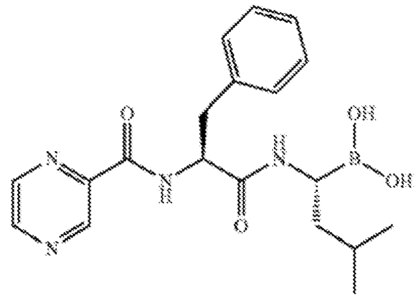 As noted, bortezomib is marketed under the trademark VelcadeR™ and is provided as a lyophilized powder for intravenous injection. It is a reversible inhibitor with a b5> b 1 inhibition profile. Established dosing is 1.3 mg/m2 with 2 intravenous administrations on days 1, 4, 8 and 11 of a 21 day cycle. It can be used in combination with doxorubicin and dexamethasone, or in combination with thalidomide, melphalan, prednisone, cyclophosphamide and other agents such as etoposide. It can be used in this same manner for purposes of the present disclosure, although cooperation/interaction with the BET inhibitor should permit the use of a reduced bortezomib dose or dosing frequency.
Another boron-containing compound useful the present combination is ixazomib, an orally-available proteasome inhibitor sold as NinlaroR™. It inhibits proteasome subunit b type-5. It has the following structure (and is the R-enantiomer):
Another proteasome inhibitor useful in the present combination belongs to the structural family of Formula I shown below:
I
 wherein: R1 is selected from morpholinyl, 1,4-oxazepanyl, thiomorpholinyl, 1,4-thiazepanyl, 1 ,4-thiazepanyl- 1 -oxide, 1 ,4-thiazepanyl- 1 , 1 -dioxide, 1 ,4-thiazinanyl- 1 -oxide, 1 ,4-thiazinanyl- 1,1-dioxide, aziridinyl, azetidinyl, pyrrolidinyl, piperazinyl, 1,4-diazepanyl, thiazolyl, isothiazolyl, oxazolyl, isooxazolyl, thiophenyl, furanyl, 1,2,4-triazolyl, pyridyl, pyrazinyl, pyrimidinyl and 1,2,4-triazinyl, wherein R1 is optionally substituted with Ci-4alkyl;
X is absent or Ci-4alkylene;
R2, R3 and R4 are each independently selected from Ci-6alkyl, Ci-4alkylene-phenyl, Ci- 4alkylene-0 — CH3, C.sub.l-4alkylene-0 — CFFF, Ci-4alkylene-0 — CHF2 and Ci-4alkylene-
O — CF3, wherein at least one of R2, R3 and R4 is Ci-4alkylene-0 — CFFF, Ci-4alkylene-0 — CHF2 or Ci-4alkylene-0 — CF3; and R5is Ci-6 alkyl.
In embodiments, a preferred such compound is the following compound:
1!

Instead of bortezomib or in addition thereto, the drug combination can include the epoxyketone- based proteasome inhibitor known as carfilzomib having the structure of Formula III shown below:
Ill
 Carfilzomib interferes with the chymotrypsin-like activity of the 20S proteasome that degrades unwanted cellular proteins, causing a build-up of polyubiquinated proteins, which may lead to apoptosis, cycle arrest, and tumor growth inhibition. This tetrapeptide epoxyketone (also an epoxomicin analog) is marketed as KyprolisR™. In this marketed form, i.e., a form also useful in the present combination, the active ingredient is formulated as monotherapy for a 10- minute infusion and is started at 20 mg/m2 during the first cycle on days 1 and 2. If this dose is tolerated, the dose is increased to 27 mg/m2 for the remaining cycles.
Potent analogs of carfilzomib have more recently been described in WO2014/026282. These fluorinated analogs have the general structure of Formula IV shown below.
IV
 wherein: R1 is selected from morpholinyl, 1,4-oxazepanyl, thiomorpholinyl, 1,4- thiazepanyl, 1,4-thiazepanyl-l -oxide, 1,4-thiazepanyl- 1,1 -dioxide, 1,4-thiazinanyl-l -oxide, 1,4- thiazinanyl- 1,1 -dioxide, aziridinyl, azetidinyl, pyrrolidinyl, piperazinyl and 1,4-diazepanyl;
X is Ci-4alkylene;
R2, R3, R4 and R5 are each independently selected from the group consisting of Ci-6alkyl, Ci-4alkylene-phenyl, Ci-4alkylene-0 — CFFF, Ci-4alkylene-0 — CHF2 and Ci-4alkylene-0— CF3, wherein at least one of R2, R3, R4 and R5 is Ci-4alkylene-0— CFFF, Ci-4alkylene-0— CHF2 or Ci- 4alkylene-0— CF3; and
R6 is Ci-6alkyl.
In embodiments of the present disclosure, the drug combination comprises a species of fluorinated carfilzomib analogs of formula V:
V

Still other BET inhibitor combinations can include such proteasome inhibitors as the natural product lactacystin, disulfiram, epigallocatechin-3-gallate, epoxomicin, G132, and b- hydroxy b -methylbutyrate (a proteasome inhibitor in human skeletal muscle). Also, BET inhibitor can be used in combination with a proteasome inhibitor that is an aldehyde (IPSI-001), or a compound that targets ubiquitin E3 ligase such as a cis-imidazoline (nutline-3 and RO5045337 and RO5503781) and a Smac peptide mimetic (LCL161), or an IAP anti-sense termed AEG 35156. The proteasome inhibitor can also be a compound that targets 19S proteasome particularly, such as the quinoline-based ubistatins, and a bis-nitrobenzylidene- piperodinone. Still other compounds useful as proteasome inhibitors include P5091, P22077 as well as WP-1130 which all target DUBs (deubiquitinases).
Each agent included in the combination can be formulated separately for use in combination. The drugs are said to be used "in combination" when, in a recipient of both drugs, the effect of one drug enhances or at least influences the effect of the other drug.
The two agents in the combination cooperate to provide an effect on target cells that is greater than the effect of either drug alone. This benefit manifests as a statistically significant improvement in a given parameter of target cell fitness or vitality. For instance, a benefit in TNBC cells when a given combination of a BET inhibitor and proteasome inhibitor is used could be a statistically significant decrease in the number of living cancer cells (hence a depletion), relative to non-treatment, or a decrease in the number or size of cancer cells or tumours, or an improvement in the endogenous location or distribution of any particular tumour type. In embodiments, the improvement resulting from treatment with the drug combination can manifest as an effect that is at least additive and desirably synergistic, relative to results obtained when only a single agent is used.
In use, each drug in the combination can be formulated as it would be for monotherapy, in terms of dosage size and form and regimen. In this regard, the synergy resulting from their combined use may permit the use of somewhat reduced dosage sizes or frequencies, as would be revealed in an appropriately controlled clinical trial.
Another combination useful for treating cancer is:
(i) a Bromodomain and Extra-Terminal motif (BET) inhibitor; and
(ii) an agent which downregulates the amount and/or activity of chemokine (C-X-C motif) receptor 1 or 2 (CXCRl/2) receptor.
BET inhibitors have been described herein above.
Examples of cancer which can be treated in accordance with the present teachings include, but are not limited to, carcinoma, adenocarcinoma, lung cancer, liver cancer, colorectal cancer, brain, head and neck cancer (e.g., neuro/glioblastoma), breast cancer, ovarian cancer, transitional cell carcinoma of the bladder, prostate cancer, oral squamous cell carcinoma, bone sarcoma, biliary tract cancer such as gallbladder carcinoma (GBC), kidney cancer and pancreatic cancer.
According to a particular embodiment, the cancer is triple negative breast cancer, and more specifically a mesenchymal or mesenchymal stem-like TNBC.
Agent which downregulates the amount and/or activity of CXCRl/2 receptor include those that are selective towards CXCR1 receptor, CXCR2 receptor or those that downregulates the amount and/or activity of both the CXCR1 and CXCR2 receptor.
According to a particular embodiment, the agent is selective towards CXCR1 over CSCR2.
CXC receptors (CXCR) are chemokine receptors that bind and respond to cytokines of the CXC chemokine family. Typically, they are members of G protein-linked receptors, also known as seven transmembrane (7-TM) proteins, because they are characterized with spanning the cell membrane seven times.
CXCR1 and CXCR2 are closely related receptors that recognize CXC chemokines that possess an E-L-R amino acid motif immediately adjacent to their CXC motif. CXCL8 (otherwise known as interleukin-8) and CXCL6 can both bind CXCR1 in humans, while all other ELR- positive chemokines, such as CXCL1 to CXCL7 bind only CXCR2. They are both expressed on the surface of neutrophils in mammals.
In one embodiment, the agent which downregulates the amount and/or activity of CXCRl/2 receptor is an anti-CXCRl antibody or an anti-CXCR2 antibody (i.e. inhibitory antibodies).
In another embodiment, the agent which downregulates the amount and/or activity of CXCRl/2 receptor is an antibody directed against the natural ligand of these receptors (i.e. IL- 18).
The term "antibody" as used herein includes intact molecules as well as functional fragments thereof, such as Fab, F(ab')2, and Fv that are capable of binding to macrophages. These functional antibody fragments are defined as follows: (1) Fab, the fragment which contains a monovalent antigen-binding fragment of an antibody molecule, can be produced by digestion of whole antibody with the enzyme papain to yield an intact light chain and a portion of one heavy chain; (2) Fab', the fragment of an antibody molecule that can be obtained by treating whole antibody with pepsin, followed by reduction, to yield an intact light chain and a portion of the heavy chain; two Fab' fragments are obtained per antibody molecule; (3) (Fab')2, the fragment of the antibody that can be obtained by treating whole antibody with the enzyme pepsin without subsequent reduction; F(ab')2 is a dimer of two Fab' fragments held together by two disulfide bonds; (4) Fv, defined as a genetically engineered fragment containing the variable region of the light chain and the variable region of the heavy chain expressed as two chains; and (5) Single chain antibody ("SCA"), a genetically engineered molecule containing the variable region of the light chain and the variable region of the heavy chain, linked by a suitable polypeptide linker as a genetically fused single chain molecule.
As used herein, the terms "complementarity-determining region" or "CDR" are used interchangeably to refer to the antigen binding regions found within the variable region of the heavy and light chain polypeptides. Generally, antibodies comprise three CDRs in each of the VH (CDR HI or HI; CDR H2 or H2; and CDR H3 or H3) and three in each of the VL (CDR LI or LI; CDR L2 or L2; and CDR L3 or L3).
The identity of the amino acid residues in a particular antibody that make up a variable region or a CDR can be determined using methods well known in the art and include methods such as sequence variability as defined by Rabat et al. (See, e.g., Rabat et ak, 1992, Sequences of Proteins of Immunological Interest, 5th ed., Public Health Service, NIH, Washington D.C.), location of the structural loop regions as defined by Chothia et al. (see, e.g., Chothia et ak, Nature 342:877-883, 1989.), a compromise between Rabat and Chothia using Oxford Molecular's AbM antibody modeling software (now Accelrys, see, Martin et ak, 1989, Proc. Natl
Acad Sci USA. 86:9268; and world wide web site www(dot)bioinf-org(dot)uk/abs), available complex crystal structures as defined by the contact definition (see MacCallum et al., J. Mol. Biol. 262:732-745, 1996) and the "conformational definition" (see, e.g., Makabe et al., Journal of Biological Chemistry, 283:1156-1166, 2008).
As used herein, the “variable regions” and "CDRs" may refer to variable regions and CDRs defined by any approach known in the art, including combinations of approaches.
According to a specific embodiment, the “variable regions” and "CDRs" refer to variable regions and CDRs defined by the IMGT approach.
Methods of producing polyclonal and monoclonal antibodies as well as fragments thereof are well known in the art (See for example, Harlow and Lane, Antibodies: A Laboratory Manual, Cold Spring Harbor Laboratory, New York, 1988, incorporated herein by reference).
According to specific embodiments the antibody is a recombinant antibody.
As used herein, the term “recombinant antibody” refers an antibody produced by recombinant DNA techniques, i.e., produced from cells transformed by an exogenous DNA construct encoding the antibody.
According to specific embodiments the antibody is a monoclonal antibody.
In cases where target antigens are too small to elicit an adequate immunogenic response when generating antibodies in vivo, such antigens (referred to as "haptens") can be coupled to antigenically neutral carriers such as keyhole limpet hemocyanin (KLH) or serum albumin (e.g., bovine serum albumin (BSA)) carriers (see, for example, US. Pat. Nos. 5,189,178 and 5,239,078). Coupling a hapten to a carrier can be effected using methods well known in the art. For example, direct coupling to amino groups can be effected and optionally followed by reduction of the imino linkage formed. Alternatively, the carrier can be coupled using condensing agents such as dicyclohexyl carbodiimide or other carbodiimide dehydrating agents. Linker compounds can also be used to effect the coupling; both homobifunctional and heterobifunctional linkers are available from Pierce Chemical Company, Rockford, Illinois, USA. The resulting immunogenic complex can then be injected into suitable mammalian subjects such as mice, rabbits, and others. Suitable protocols involve repeated injection of the immunogen in the presence of adjuvants according to a schedule designed to boost production of antibodies in the serum. The titers of the immune serum can readily be measured using immunoassay procedures which are well known in the art.
The antisera obtained can be used directly or monoclonal antibodies may be obtained, as described hereinabove. Antibody fragments according to some embodiments of the invention can
be prepared by proteolytic hydrolysis of the antibody or by expression in E. coli or mammalian cells (e.g. Chinese hamster ovary cell culture or other protein expression systems) of DNA encoding the fragment.
Antibody fragments can be obtained by pepsin or papain digestion of whole antibodies by conventional methods. For example, antibody fragments can be produced by enzymatic cleavage of antibodies with pepsin to provide a 5S fragment denoted F(ab')2. This fragment can be further cleaved using a thiol reducing agent, and optionally a blocking group for the sulfhydryl groups resulting from cleavage of disulfide linkages, to produce 3.5S Fab' monovalent fragments. Alternatively, an enzymatic cleavage using pepsin produces two monovalent Fab' fragments and an Fc fragment directly. These methods are described, for example, by Goldenberg, U.S. Pat. Nos. 4,036,945 and 4,331,647, and references contained therein, which patents are hereby incorporated by reference in their entirety. See also Porter, R. R. [Biochem. J. 73: 119-126 (1959)]. Other methods of cleaving antibodies, such as separation of heavy chains to form monovalent light-heavy chain fragments, further cleavage of fragments, or other enzymatic, chemical, or genetic techniques may also be used, so long as the fragments bind to the antigen that is recognized by the intact antibody.
Fv fragments comprise an association of VH and VL chains. This association may be noncovalent, as described in Inbar et al. [Proc. Nat'l Acad. Sci. USA 69:2659-62 (19720] Alternatively, the variable chains can be linked by an intermolecular disulfide bond or cross- linked by chemicals such as glutaraldehyde. Preferably, the Fv fragments comprise VH and VL chains connected by a peptide linker. These single-chain antigen binding proteins (sFv) are prepared by constructing a structural gene comprising DNA sequences encoding the VH and VL domains connected by an oligonucleotide. The structural gene is inserted into an expression vector, which is subsequently introduced into a host cell such as E. coli. The recombinant host cells synthesize a single polypeptide chain with a linker peptide bridging the two V domains. Methods for producing sFvs are described, for example, by [Whitlow and Filpula, Methods 2: 97- 105 (1991); Bird et al., Science 242:423-426 (1988); Pack et al., Bio/Technology 11:1271-77 (1993); and U.S. Pat. No. 4,946,778, which is hereby incorporated by reference in its entirety.
Another form of an antibody fragment is a peptide coding for a single complementarity- determining region (CDR). CDR peptides ("minimal recognition units") can be obtained by constructing genes encoding the CDR of an antibody of interest. Such genes are prepared, for example, by using the polymerase chain reaction to synthesize the variable region from RNA of antibody-producing cells. See, for example, Larrick and Fry [Methods, 2: 106-10 (1991)].
Thus, antibodies of the present invention are preferably at least bivalent (e.g., of the IgG subtype) or more (e.g., of the IgM subtype). It will be appreciated that monovalent antibodies may be used however measures should be taken to assemble these to larger complexes such as by using secondary antibodies (or using other cross-linkers which are well known in the art). According to specific embodiments the antibodies are from IgGl subtype.
According to specific embodiments antibody is a humanized or partially humanized antibody.
Humanized forms of non-human (e.g., murine) antibodies are chimeric molecules of immunoglobulins, immunoglobulin chains or fragments thereof (such as Fv, Fab, Fab', F(ab').sub.2 or other antigen-binding subsequences of antibodies) which contain minimal sequence derived from non-human immunoglobulin. Humanized antibodies include human immunoglobulins (recipient antibody) in which residues form a complementary determining region (CDR) of the recipient are replaced by residues from a CDR of a non-human species (donor antibody) such as mouse, rat or rabbit having the desired specificity, affinity and capacity. In some instances, Fv framework residues of the human immunoglobulin are replaced by corresponding non-human residues. Humanized antibodies may also comprise residues which are found neither in the recipient antibody nor in the imported CDR or framework sequences. In general, the humanized antibody will comprise substantially all of at least one, and typically two, variable domains, in which all or substantially all of the CDR regions correspond to those of a non-human immunoglobulin and all or substantially all of the FR regions are those of a human immunoglobulin consensus sequence. The humanized antibody optimally also will comprise at least a portion of an immunoglobulin constant region (Fc), typically that of a human immunoglobulin [Jones et ak, Nature, 321:522-525 (1986); Riechmann et ah, Nature, 332:323- 329 (1988); and Presta, Curr. Op. Struct. Biol., 2:593-596 (1992)].
Methods for humanizing non-human antibodies are well known in the art. Generally, a humanized antibody has one or more amino acid residues introduced into it from a source which is non-human. These non-human amino acid residues are often referred to as import residues, which are typically taken from an import variable domain. Humanization can be essentially performed following the method of Winter and co-workers [Jones et ak, Nature, 321:522-525 (1986); Riechmann et ak, Nature 332:323-327 (1988); Verhoeyen et ak, Science, 239:1534-1536 (1988)], by substituting rodent CDRs or CDR sequences for the corresponding sequences of a human antibody. Accordingly, such humanized antibodies are chimeric antibodies (U.S. Pat. No. 4,816,567), wherein substantially less than an intact human variable domain has been substituted
by the corresponding sequence from a non-human species. In practice, humanized antibodies are typically human antibodies in which some CDR residues and possibly some FR residues are substituted by residues from analogous sites in rodent antibodies.
Human antibodies can also be produced using various techniques known in the art, including phage display libraries [Hoogenboom and Winter, J. Mol. Biol., 227:381 (1991); Marks et ah, J. Mol. Biol., 222:581 (1991)]. The techniques of Cole et al. and Boerner et al. are also available for the preparation of human monoclonal antibodies (Cole et al., Monoclonal Antibodies and Cancer Therapy, Alan R. Liss, p. 77 (1985) and Boerner et al., J. Immunol., 147(l):86-95 (1991)]. Similarly, human antibodies can be made by introduction of human immunoglobulin loci into transgenic animals, e.g., mice in which the endogenous immunoglobulin genes have been partially or completely inactivated. Upon challenge, human antibody production is observed, which closely resembles that seen in humans in all respects, including gene rearrangement, assembly, and antibody repertoire. This approach is described, for example, in U.S. Pat. Nos. 5,545,807; 5,545,806; 5,569,825; 5,625,126; 5,633,425; 5,661,016, and in the following scientific publications: Marks et al., Bio/Technology 10,: 779-783 (1992); Lonberg et al., Nature 368: 856-859 (1994); Morrison, Nature 368 812-13 (1994); Fishwild et al., Nature Biotechnology 14, 845-51 (1996); Neuberger, Nature Biotechnology 14: 826 (1996); and Lonberg and Huszar, Intern. Rev. Immunol. 13: 65-93 (1995).
The antibodies can be mono-specific (i.e., binding a distinct antigen) or multi-specific (i.e. binding at least two different epitopes, e.g., bi-specific or tri-specific).
According to specific embodiments, the antibody is a mono-specific antibody.
According to specific embodiments, the antibody is bi-specific antibody.
According to specific embodiments, the antibody is a tri-specific antibody.
According to other specific embodiments, the antibody is a multi-specific antibody. Exemplary antibodies directed against CXCR1 are disclosed in Ginestier C. et al., J Clin
Invest. (2010) 120:485-97.
Exemplary antibodies directed against IL-18 are disclosed for example in US Patent Application No. 20190248883, the contents of which is incorporated herein by reference.
In another embodiment, the agent which downregulates the amount and/or activity of CXCRl/2 receptor is a dual CXCRl/2 antagonist.
As used herein, the phrases “dual CXCRl/2 antagonist” refers to an agent that can block activation of both CXCR1 and CXCR2 receptors. The dual CXCRl/2 antagonist may be a small molecule, or a biological molecule (e.g., antibody). In one embodiment, the CXCRl/2 antagonist
would exhibit IC50 values for CXCR1 and CXCR2 receptors, wherein the ratio of IC.sub.50 values is less than 30, preferably less than 20, more preferably less than 10, and most preferably less than 5. Dual CXCRl/2 antagonists include, but are not limited to, SX-682, SX-576, SX-517, navarixin (PubChem CID 11281445; SCH-527123), ladarixin (PubChem CID 11372270; meraxin, DF2156A), reparixin (PubChem CID 9838712; repertaxin, DF1681B), reparixin L- lysine salt (PubChem CID 9932389), DF2755A, CXCL8 fragment comprising amino acids 3-74 and substitutions K11R/G31P (G31P), DF2162 (PubChem CID 11289471) and SCH-479833.
According to a particular embodiment, the dual CXCRl/2 antagonist is selected from the group consisting of navarixin, ladarixin, reparixin, reparixin L-lysine salt, DF2755A, CXCL8 fragment comprising amino acids 3-74 and substitutions K11R/G31P (G31P), DF2162, and SCH- 479833.
Down-regulation of CXCR1, CXCR2 or BET at the nucleic acid level may be effected using a nucleic acid agent, having a nucleic acid backbone, DNA, RNA, mimetics thereof or a combination of same. The nucleic acid agent may be encoded from a DNA molecule or provided to the cell per se.
Thus, downregulation of the above mentioned targets can be achieved by RNA silencing. As used herein, the phrase "RNA silencing" refers to a group of regulatory mechanisms [e.g. RNA interference (RNAi), transcriptional gene silencing (TGS), post-transcriptional gene silencing (PTGS), quelling, co-suppression, and translational repression] mediated by RNA molecules which result in the inhibition or "silencing" of the expression of a corresponding protein-coding gene. RNA silencing has been observed in many types of organisms, including plants, animals, and fungi.
As used herein, the term "RNA silencing agent" refers to an RNA which is capable of specifically inhibiting or "silencing" the expression of a target gene. In certain embodiments, the RNA silencing agent is capable of preventing complete processing (e.g, the full translation and/or expression) of an mRNA molecule through a post-transcriptional silencing mechanism. RNA silencing agents include non-coding RNA molecules, for example RNA duplexes comprising paired strands, as well as precursor RNAs from which such small non-coding RNAs can be generated. Exemplary RNA silencing agents include dsRNAs such as siRNAs, miRNAs and shRNAs.
In one embodiment, the RNA silencing agent is capable of inducing RNA interference.
In another embodiment, the RNA silencing agent is capable of mediating translational repression.
According to an embodiment of the invention, the RNA silencing agent is specific to the target RNA (e.g., CXCR1, CXCR2 or BET) and does not cross inhibit or silence other targets or a splice variant which exhibits 99% or less global homology to the target gene, e.g., less than 98%, 97%, 96%, 95%, 94%, 93%, 92%, 91%, 90%, 89%, 88%, 87%, 86%, 85%, 84%, 83%, 82%, 81% global homology to the target gene; as determined by PCR, Western blot, Immunohistochemistry and/or flow cytometry.
RNA interference refers to the process of sequence-specific post-transcriptional gene silencing in animals mediated by short interfering RNAs (siRNAs).
Other exemplary polynucleotide agents contemplated by the present invention include miRNAs, antisense RNAs and agents which introduce nucleic acid alterations to a gene of interest (using CRISPR system).
In the context of any of the combination therapies described herein above, combination therapy compounds may be administered by the same route of administration (e.g. intrapulmonary, oral, enteral, etc.) that the described compounds are administered. In the alternative, the compounds for use in combination therapy with the herein described compounds may be administered by a different route of administration.
The first member of the combination can be administered immediately prior to (or after) the second member of the combination, on the same day as, one day before (or after), one week before (or after), one month before (or after), or two months before (or after) the second member of the combination, and the like.
The first member of the combination and the second member of the combination can be administered concomitantly, that is, where the administering for each of these reagents can occur at time intervals that partially or fully overlap each other. The first member of the combination and the second member of the combination can be administered during time intervals that do not overlap each other. For example, the first member of the combination can be administered within the time frame of t=0 to 1 hours, while the second member of the combination can be administered within the time frame of t=l to 2 hours. Also, the first member of the combination can be administered within the time frame of t=0 to 1 hours, while the second member of the combination can be administered somewhere within the time frame of t=2-3 hours, t=3-4 hours, t=4-5 hours, t=5-6 hours, t=6-7 hours, t=7-8 hours, t=8-9 hours, t=9-10 hours, and the like. Moreover, the TK receptor inhibitor can be administered somewhere in the time frame of t=minus 2-3 hours, t=minus 3-4 hours, t=minus 4-5 hours, t=5-6 minus hours, t=minus 6-7 hours, t=minus 7-8 hours, t=minus 8-9 hours, t=minus 9-10 hours.
The first and second members of the combination are typically provided in combined amounts to achieve therapeutic, prophylactic and/or pain palliative effectiveness. This amount will evidently depend upon the particular compound selected for use, the nature and number of the other treatment modality, the condition(s) to be treated, prevented and/or palliated, the species, age, sex, weight, health and prognosis of the subject, the mode of administration, effectiveness of targeting, residence time, mode of clearance, type and severity of side effects of the pharmaceutical composition and upon many other factors which will be evident to those of skill in the art. The first member of the combination will be used at a level at which therapeutic, prophylactic and/or pain palliating effectiveness in combination with the second member of the combination will be observed.
The first and/or second member of the combination may be administered at a gold standard dosing as a single agent, below a gold standard dosing as a single agent or above a gold standard dosing as a single agent.
According to specific embodiments, the first and/or second member of the combination is administered below gold standard dosing as a single agent.
As used herein the term “gold standard dosing” refers to the dosing which is recommended by a regulatory agency (e.g., FDA), for a given tumor at a given stage.
According to other specific embodiments the first and/or second member of the combination is administered at a dose that does not exert at least one side effect which is associated with the gold standard dosing.
Thus, in one preferred embodiment, the amount of the first and/or second member of the combination is below the minimum dose required for therapeutic, prophylactic and/or pain palliative effectiveness when used as a single therapy (e.g. 10-99%, preferably 25 to 75% of that minimum dose). This allows for reduction of the side effects caused by the first and/or second member, but the therapy is rendered effective because the combinations are effective overall.
As well as lowering the dose of the first and/or second member of the combination, the present inventors contemplate that the amount of time over which the agents are administered may be reduced and/or the frequency of dosing may also be reduced.
In one preferred aspect of the present invention, the first member and the second member of the combination are synergistic with respect to their dosages. That is to say that the effect provided by the compound of the present invention is greater than would be anticipated from the additive effects of the first and second member when used separately. In an alternative but equally preferred embodiment, the first member of the combination of the present invention and
the second member of the combination are synergistic with respect to their side effects. That is to say that the side-effects caused by the first member in combination with the second member are less than would be anticipated when the equivalent therapeutic effect is provided by either the first member or by the second member when used separately. According to another aspect of the present invention there is provided a pharmaceutical composition comprising a pharmaceutically acceptable carrier and:
(i) a Bromodomain and Extra-Terminal motif (BET) inhibitor; and
(ii) an agent which downregulates the amount and/or activity of CXCRl/2 receptor.
As used herein, a "pharmaceutical composition" refers to a preparation of one or more of the active ingredients described herein with other chemical components such as physiologically suitable carriers and excipients. The purpose of a pharmaceutical composition is to facilitate administration of a compound to an organism.
As used herein, the term "active ingredient" refers to the agent accountable for the intended biological effect (e.g., BET inhibitor, CXCRl/2 inhibitor). Hereinafter, the phrases "physiologically acceptable carrier" and "pharmaceutically acceptable carrier", which may be used interchangeably, refer to a carrier or a diluent that does not cause significant irritation to an organism and does not abrogate the biological activity and properties of the administered compound. An adjuvant is included under these phrases.
Herein, the term "excipient" refers to an inert substance added to a pharmaceutical composition to further facilitate administration of an active ingredient. Examples, without limitation, of excipients include calcium carbonate, calcium phosphate, various sugars and types of starch, cellulose derivatives, gelatin, vegetable oils, and polyethylene glycols.
Techniques for formulation and administration of drugs may be found in the latest edition of “Remington’s Pharmaceutical Sciences,” Mack Publishing Co., Easton, PA, which is herein fully incorporated by reference.
Suitable routes of administration may, for example, include oral, rectal, transmucosal, especially transnasal, intestinal, or parenteral delivery, including intramuscular, subcutaneous, and intramedullary injections, as well as intrathecal, direct intraventricular, intravenous, intraperitoneal, intranasal, or intraocular injections. Alternately, one may administer the pharmaceutical composition in a local rather than systemic manner, for example, via injection of the pharmaceutical composition directly into a tissue region of a patient.
Pharmaceutical compositions of the present invention may be manufactured by processes well known in the art, e.g., by means of conventional mixing, dissolving, granulating, dragee making, levigating, emulsifying, encapsulating, entrapping, or lyophilizing processes.
Pharmaceutical compositions for use in accordance with the present invention thus may be formulated in conventional manner using one or more physiologically acceptable carriers comprising excipients and auxiliaries, which facilitate processing of the active ingredients into preparations that can be used pharmaceutically. Proper formulation is dependent upon the route of administration chosen.
For injection, the active ingredients of the pharmaceutical composition may be formulated in aqueous solutions, preferably in physiologically compatible buffers such as Hank’s solution, Ringer’s solution, or physiological salt buffer. For transmucosal administration, penetrants appropriate to the barrier to be permeated are used in the formulation. Such penetrants are generally known in the art.
For oral administration, the pharmaceutical composition can be formulated readily by combining the active compounds with pharmaceutically acceptable carriers well known in the art. Such carriers enable the pharmaceutical composition to be formulated as tablets, pills, dragees, capsules, liquids, gels, syrups, slurries, suspensions, and the like, for oral ingestion by a patient. Pharmacological preparations for oral use can be made using a solid excipient, optionally grinding the resulting mixture, and processing the mixture of granules, after adding suitable auxiliaries as desired, to obtain tablets or dragee cores. Suitable excipients are, in particular, fillers such as sugars, including lactose, sucrose, mannitol, or sorbitol; cellulose preparations such as, for example, maize starch, wheat starch, rice starch, potato starch, gelatin, gum tragacanth, methyl cellulose, hydroxypropylmethyl-cellulose, and sodium carbomethylcellulose; and/or physiologically acceptable polymers such as polyvinylpyrrolidone (PVP). If desired, disintegrating agents, such as cross-linked polyvinyl pyrrolidone, agar, or alginic acid or a salt thereof, such as sodium alginate, may be added.
Dragee cores are provided with suitable coatings. For this purpose, concentrated sugar solutions may be used which may optionally contain gum arabic, talc, polyvinyl pyrrolidone, carbopol gel, polyethylene glycol, titanium dioxide, lacquer solutions, and suitable organic solvents or solvent mixtures. Dyestuffs or pigments may be added to the tablets or dragee coatings for identification or to characterize different combinations of active compound doses.
Pharmaceutical compositions that can be used orally include push-fit capsules made of gelatin, as well as soft, sealed capsules made of gelatin and a plasticizer, such as glycerol or
sorbitol. The push-fit capsules may contain the active ingredients in admixture with filler such as lactose, binders such as starches, lubricants such as talc or magnesium stearate, and, optionally, stabilizers. In soft capsules, the active ingredients may be dissolved or suspended in suitable liquids, such as fatty oils, liquid paraffin, or liquid polyethylene glycols. In addition, stabilizers may be added. All formulations for oral administration should be in dosages suitable for the chosen route of administration.
For buccal administration, the compositions may take the form of tablets or lozenges formulated in conventional manner.
For administration by nasal inhalation, the active ingredients for use according to the present invention are conveniently delivered in the form of an aerosol spray presentation from a pressurized pack or a nebulizer with the use of a suitable propellant, e.g., dichlorodifluoromethane, trichlorofluoromethane, dichloro-tetrafluoroethane, or carbon dioxide. In the case of a pressurized aerosol, the dosage may be determined by providing a valve to deliver a metered amount. Capsules and cartridges of, for example, gelatin for use in a dispenser may be formulated containing a powder mix of the compound and a suitable powder base, such as lactose or starch.
The pharmaceutical composition described herein may be formulated for parenteral administration, e.g., by bolus injection or continuous infusion. Formulations for injection may be presented in unit dosage form, e.g., in ampoules or in multidose containers with, optionally, an added preservative. The compositions may be suspensions, solutions, or emulsions in oily or aqueous vehicles, and may contain formulatory agents such as suspending, stabilizing, and/or dispersing agents.
Pharmaceutical compositions for parenteral administration include aqueous solutions of the active preparation in water-soluble form. Additionally, suspensions of the active ingredients may be prepared as appropriate oily or water-based injection suspensions. Suitable lipophilic solvents or vehicles include fatty oils such as sesame oil, or synthetic fatty acid esters such as ethyl oleate, triglycerides, or liposomes. Aqueous injection suspensions may contain substances that increase the viscosity of the suspension, such as sodium carboxymethyl cellulose, sorbitol, or dextran. Optionally, the suspension may also contain suitable stabilizers or agents that increase the solubility of the active ingredients, to allow for the preparation of highly concentrated solutions.
Alternatively, the active ingredient may be in powder form for constitution with a suitable vehicle, e.g., a sterile, pyrogen-free, water-based solution, before use.
The pharmaceutical composition of the present invention may also be formulated in rectal compositions such as suppositories or retention enemas, using, for example, conventional suppository bases such as cocoa butter or other glycerides.
Pharmaceutical compositions suitable for use in the context of the present invention include compositions wherein the active ingredients are contained in an amount effective to achieve the intended purpose. More specifically, a "therapeutically effective amount" means an amount of active ingredients (e.g., a nucleic acid construct) effective to prevent, alleviate, or ameliorate symptoms of a disorder (e.g., ischemia) or prolong the survival of the subject being treated. Determination of a therapeutically effective amount is well within the capability of those skilled in the art, especially in light of the detailed disclosure provided herein.
For any preparation used in the methods of the invention, the dosage or the therapeutically effective amount can be estimated initially from in vitro and cell culture assays. For example, a dose can be formulated in animal models to achieve a desired concentration or titer. Such information can be used to more accurately determine useful doses in humans. Since administration of the disclosed combination is expected to produce improved results over the administration of single agents, the therapeutically effective dose of each of the agents in the combined treatment may be for example less than 50 %, 40 %, 30 %, 20 % or even less than 10 % the of the FDA approved dose. Toxicity and therapeutic efficacy of the active ingredients described herein can be determined by standard pharmaceutical procedures in vitro, in cell cultures or experimental animals. The data obtained from these in vitro and cell culture assays and animal studies can be used in formulating a range of dosage for use in human. The dosage may vary depending upon the dosage form employed and the route of administration utilized. The exact formulation, route of administration, and dosage can be chosen by the individual physician in view of the patient's condition. (See, e.g., Fingl, E. et al. (1975), "The Pharmacological Basis of Therapeutics," Ch. 1,
R·1·)
Dosage amount and administration intervals may be adjusted individually to provide sufficient plasma or brain levels of the active ingredient to induce or suppress the biological effect (i.e., minimally effective concentration, MEC). The MEC will vary for each preparation, but can be estimated from in vitro data. Dosages necessary to achieve the MEC will depend on individual characteristics and route of administration. Detection assays can be used to determine plasma concentrations.
Depending on the severity and responsiveness of the condition to be treated, dosing can be of a single or a plurality of administrations (e.g., weekly or bi-weekly administrations) with course of treatment lasting from several days to several weeks, or until cure is effected or diminution of the disease state is achieved.
The amount of a composition to be administered will, of course, be dependent on the subject being treated, the severity of the affliction, the manner of administration, the judgment of the prescribing physician, etc.
Suitable cells for use in animal models and in vitro analyses include but are not limited to BT-474; MDA-MB-468, MDA-MB-231, BT20, BT549, HCC1937, HCC1143, SUM159PT, Hs578T.
Compositions of the present invention may, if desired, be presented in a pack or dispenser device, such as an FDA-approved kit, which may contain one or more unit dosage forms containing the active ingredient. According to a specific embodiment, there is provided a kit comprising the isolated polypeptide (e.g., having the CDRs of NG33 and optionally other antibodies as described herein) and optionally a pharmaceutical agent, as described herein. The pack may, for example, comprise metal or plastic foil, such as a blister pack. The pack or dispenser device may be accompanied by instructions for administration. The pack or dispenser device may also be accompanied by a notice in a form prescribed by a governmental agency regulating the manufacture, use, or sale of pharmaceuticals, which notice is reflective of approval by the agency of the form of the compositions for human or veterinary administration. Such notice, for example, may include labeling approved by the U.S. Food and Drug Administration for prescription drugs or of an approved product insert. Compositions comprising a preparation of the invention formulated in a pharmaceutically acceptable carrier may also be prepared, placed in an appropriate container, and labeled for treatment of an indicated condition, as further detailed above.
The combination therapy of the present invention can be administered along with analgesics, chemotherapeutic agents (e.g., anthracyclins), radiotherapeutic agents, hormonal therapy and other treatment regimens (e.g., surgery) which are well known in the art.
It is expected that during the life of a patent maturing from this application many relevant protease inhibitors, BET inhibitors and CXCRl/2 inhibitors will be developed and the scope of these terms are intended to include all such new technologies a priori.
As used herein the term “about” refers to ± 10 %
The terms "comprises", "comprising", "includes", "including", “having” and their conjugates mean "including but not limited to".
The term “consisting of’ means “including and limited to”.
The term "consisting essentially of means that the composition, method or structure may include additional ingredients, steps and/or parts, but only if the additional ingredients, steps and/or parts do not materially alter the basic and novel characteristics of the claimed composition, method or structure.
As used herein, the singular form "a", "an" and "the" include plural references unless the context clearly dictates otherwise. For example, the term "a compound" or "at least one compound" may include a plurality of compounds, including mixtures thereof.
Throughout this application, various embodiments of this invention may be presented in a range format. It should be understood that the description in range format is merely for convenience and brevity and should not be construed as an inflexible limitation on the scope of the invention. Accordingly, the description of a range should be considered to have specifically disclosed all the possible subranges as well as individual numerical values within that range. For example, description of a range such as from 1 to 6 should be considered to have specifically disclosed subranges such as from 1 to 3, from 1 to 4, from 1 to 5, from 2 to 4, from 2 to 6, from 3 to 6 etc., as well as individual numbers within that range, for example, 1, 2, 3, 4, 5, and 6. This applies regardless of the breadth of the range.
Whenever a numerical range is indicated herein, it is meant to include any cited numeral (fractional or integral) within the indicated range. The phrases “ranging/ranges between” a first indicate number and a second indicate number and “ranging/ranges from” a first indicate number “to” a second indicate number are used herein interchangeably and are meant to include the first and second indicated numbers and all the fractional and integral numerals therebetween.
As used herein the term "method" refers to manners, means, techniques and procedures for accomplishing a given task including, but not limited to, those manners, means, techniques and procedures either known to, or readily developed from known manners, means, techniques and procedures by practitioners of the chemical, pharmacological, biological, biochemical and medical arts.
As used herein, the term “treating” includes abrogating, substantially inhibiting, slowing or reversing the progression of a condition, substantially ameliorating clinical or aesthetical symptoms of a condition or substantially preventing the appearance of clinical or aesthetical symptoms of a condition.
It is appreciated that certain features of the invention, which are, for clarity, described in the context of separate embodiments, may also be provided in combination in a single embodiment. Conversely, various features of the invention, which are, for brevity, described in the context of a single embodiment, may also be provided separately or in any suitable subcombination or as suitable in any other described embodiment of the invention. Certain features described in the context of various embodiments are not to be considered essential features of those embodiments, unless the embodiment is inoperative without those elements.
Various embodiments and aspects of the present invention as delineated hereinabove and as claimed in the claims section below find experimental support in the following examples.
EXAMPLES
Reference is now made to the following examples, which together with the above descriptions illustrate some embodiments of the invention in a non limiting fashion.
Generally, the nomenclature used herein and the laboratory procedures utilized in the present invention include molecular, biochemical, microbiological and recombinant DNA techniques. Such techniques are thoroughly explained in the literature. See, for example, "Molecular Cloning: A laboratory Manual" Sambrook et al., (1989); "Current Protocols in Molecular Biology" Volumes I-III Ausubel, R. M., ed. (1994); Ausubel et al., "Current Protocols in Molecular Biology", John Wiley and Sons, Baltimore, Maryland (1989); Perbal, "A Practical Guide to Molecular Cloning", John Wiley & Sons, New York (1988); Watson et al., "Recombinant DNA", Scientific American Books, New York; Birren et al. (eds) "Genome Analysis: A Laboratory Manual Series", Vols. 1-4, Cold Spring Harbor Laboratory Press, New York (1998); methodologies as set forth in U.S. Pat. Nos. 4,666,828; 4,683,202; 4,801,531; 5,192,659 and 5,272,057; "Cell Biology: A Laboratory Handbook", Volumes I-III Cellis, J. E., ed. (1994); "Culture of Animal Cells - A Manual of Basic Technique" by Freshney, Wiley-Liss, N. Y. (1994), Third Edition; "Current Protocols in Immunology" Volumes I-III Coligan J. E., ed. (1994); Stites et al. (eds), "Basic and Clinical Immunology" (8th Edition), Appleton & Lange, Norwalk, CT (1994); Mishell and Shiigi (eds), "Selected Methods in Cellular Immunology", W. H. Freeman and Co., New York (1980); available immunoassays are extensively described in the patent and scientific literature, see, for example, U.S. Pat. Nos. 3,791,932; 3,839,153; 3,850,752; 3,850,578; 3,853,987; 3,867,517; 3,879,262; 3,901,654; 3,935,074; 3,984,533; 3,996,345; 4,034,074; 4,098,876; 4,879,219; 5,011,771 and 5,281,521; "Oligonucleotide Synthesis" Gait, M. J., ed. (1984); “Nucleic Acid Hybridization" Hames, B. D., and Higgins S. J., eds. (1985);
"Transcription and Translation" Hames, B. D., and Higgins S. L, eds. (1984); "Animal Cell Culture" Freshney, R. T, ed. (1986); "Immobilized Cells and Enzymes" IRL Press, (1986); "A Practical Guide to Molecular Cloning" Perbal, B., (1984) and "Methods in Enzymology" Vol. 1- 317, Academic Press; "PCR Protocols: A Guide To Methods And Applications", Academic Press, San Diego, CA (1990); Marshak et al., "Strategies for Protein Purification and Characterization - A Laboratory Course Manual" CSHL Press (1996); all of which are incorporated by reference as if fully set forth herein. Other general references are provided throughout this document. The procedures therein are believed to be well known in the art and are provided for the convenience of the reader. All the information contained therein is incorporated herein by reference.
MATERIALS AND METHODS
Chemicals and Drugs: The small molecule inhibitors used in the screen were purchased from different sources. The following drugs were purchased from Selleck Chemicals: mTOR inhibitor (RadOO, SI 120), SRC inhibitor (Dasatinib, S 1021 ), MEK inhibitor (GSK1120212B, S2673), CDK4/6 inhibitor (PD0332991, SI 116), BCL2 inhibitor (ABT737, SI 002), EGFR inhibitor (Neratinib, S2150), PI3K inhibitor (GDC-0941, SI 065) and PARP inhibitor (BMN673, S7048). Stat3 inhibitor (Stattic, 573099) was purchased form Calbiochem. Bortezomib and JQ1 were purchased from AdooQ BioScience (CA, USA). Fak inhibitor (PF-573228), CXCR2 inhibitor, (SB225022), HSP90 inhibitor (17-DMAG) and NF- KB inhibitor (PDTC) were from Sigma. Jak inhibitor (Ruxolitinib) was from Santa Cruz Biotechnology, cMet inhibitor (EMD 1214063) was from MedChemExpress. OTX015 was purchased from Changzhou Medi-tech Bioscientific Co (China). The apoptotic inhibitor Z- VAD-FMK (sc-3067), and autophagy inhibitor 3-MA (sc-205596) were purchased from Santa Cruz Biotechnology. Necrostatin-1 (N9037), Erastin (E7781), Staurosporine (S5921), Rapamycin (553210), Ferrostatin-1 (SML0583), reduced L-Glutathione (G6013)’, 2,2'- Bipyridyl (D216305) and Thiazolyl Blue Tetrazolium Bromide (MTT) (M5655) were from Sigma. DFO
Antibodies: The following antibodies were purchased from Cell Signaling Technologies (USA): rabbit anti-MLKL (14993), rabbit anti-phoshpho Ser358 MLKL (74921), rabbit anti-cleaved Caspase-3 (9664), rabbit anti-PARP antibody (9542). Mouse anti- a-tubulin (T6074) and rabbit anti-ferritin heavy chain (F5012) were supplied by Sigma Aldrich. Rabbit anti-GPX4 (abl25066, clone EPNCIR 144) was purchased from Abeam Inc
(USA), and rabbit anti-BRD4 (A700-004, clone BL-149-2H5) was from Bethyl Laboratories (USA). Rabbit anti-Ferroportin (NBP1-21502) was from Novus Biologicals. Mouse anti- ACSL4 (sc-365230, clone F4) and GSS (sc-166882) were obtained from Santa Cruz Biotechnology, mouse anti-transferrin receptor (13-6800, clone H68.4) was from Invitrogen. Cell culture: All the breast cancer cell lines, immortalized normal breast epithelial cell line MCF10A and human embryonic kidney (HEK-293T) cells were originally obtained from American type culture collection (ATCC) USA. HCC38 was kindly obtained from Maire Virginie (Institut Curie, Research Centre, Paris, France; 2014). MDA-MB-468, HCC1143, HCC1937, HCC70, HCC38, MDA-MB-231, BT549, SUM159, and Hs578T cells were grown in RPMI, while HEK293T cells were grown in DMEM (Gibco BRL, USA). Unless otherwise indicated, all cell lines were cultured in medium containing 10% fetal bovine serum (FBS) (Gibco BRL, USA) and penicillin/streptomycin. L-glutamine (2mM) was added to Hs578T and HCC38 medium, and 1.5 g/L sodium bicarbonate, 10 mM Hepes, and 1.0 mM sodium pyruvate to HCC38 medium. BT20 cells were grown in Eagle’s Minimum Essential Medium (MEM-Eagle’s) plus 1 mM sodium pyruvate and 2 mM L-glutamine. MCF10A cells were grown in DMEM:F12 (1:1) medium supplemented with EGF (20 ng/ml, PeproTech), insulin (10 pg/ml, Sigma), cholera toxin (100 ng/ml, Sigma), hydrocortisone (1 pg/ml, Sigma) and 5% heat-inactivated horse serum. Unless otherwise indicated, all cell lines were cultured in medium containing 10% fetal bovine serum (FBS) (Gibco BRL, USA) and penicillin/ streptomycin. Cells were cultured at 37°C in a humidified incubator of 5% C02.
Cell lines were routinely (once a month) checked for Mycoplasma using a commercially available kit. shRNA lentivirus infection: Lentivirus production and infection were conducted essentially as previously described (Kim, S. et ah, EMBO Rep. 14, 891-899 (2013). Infected cells (control and BRD4 shRNA) were grown in selection medium containing 1 pg/ml puromycin for 72 h. Knockdown of BRD4 was assessed by Western Blotting.
Drug screen, Cell viability assays and Drug synergy: To evaluate the effects of the 17 selected small molecule inhibitors used in the screen (Figure 9A) on viability of the 13 human cell lines, dose response experiments were carried out using 7 drug concentrations applied at 3- fold serial dilution within a range of 13 nM to 10 pM. Cells in 96-well plates were treated with the described drugs doses or with DMSO as control for 72 hr, and cell viability was measured by MTT assays as previously described. Cell viability was presented as percent of control and dose response curve were generated by the nonlinear regression method using GraphPad Prism
version v.5.0 (GraphPad Software Inc, La Jolla, CA, USA) curve fitting analysis applying drug concentrations at LoglO Molar scale. IC50 values were calculated from the curves for each drug and each cell line.
The drug screen was performed in 96-well plates and cell viability was determined by CellTiter-Glo (Promega), 72 hr after adding the drugs according to manufacturer's instructions. Luminescence was measure by the PHERAStar Plus plate reader (BMG Labtech, Germany), where indicated, CellTiter-Blue (Promega). Changes in cell viability are presented as ratio of viable cells between drug treated and the respective mock-treated control cells. Synergy of drug pairs was calculated by the CalcuSyn software using the Chou-Talalay equation. CI<0.5 indicates synergism, and CI>0.5 indicates no synergism. For crystal violet staining, the cells were washed with PBS and then fixed and stained with 0.2% crystal violet solution in 4% formalin for 10 min. The cells were washed with distilled water, dried, and scanned using HP Scanjet G4010.
RNA extraction and real-time PCR: Total RNA from cell lines and mouse tumor tissues was extracted and purified using Tri Reagent (Sigma Inc,). RNA (2 pg) was reverse-transcribed into cDNA using the Applied Biosystems™ High-Capacity cDNA Reverse Transcription Kit with oligo (dT) primers according to manufacturer’s instructions.
Real-time PCR analysis was performed on an ABI StepOnePlus 7500 Real-time PCR system (Applied Biosystems; Invitrogen) using Fast SYBR Green I as a fluorescent dye, according to the manufacturer’s guidelines. Real-time PCR primers were designed using the
Primer express software of Applied Biosystems (Invitrogen) and were calibrated before use. The primer sequences are listed in the Table 2 below.
Table 2


Annexin V Staining : Cells in 24-well plates were treated with drugs as indicated. For Annexin V staining, cells were washed twice with Cell Staining Buffer (Cat. No. 420201, BioLegend, CA) and then incubated in Annexin V Binding Buffer (Cat. No. 422201, BioLegend,
CA) for 5 mins at room temperature (RT). PE Annexin V (Cat. No. 640908, BioLegend, CA) was diluted 1:20 in Annexin V Binding Buffer and incubated with the cells for 15 min at RT in the dark. Cells were then washed twice with Annexin V Binding Buffer, stained with 20 mM Hoechst 33342 (Sigma) in Annexin V Binding Buffer, washed again in fresh Annexin V Binding Buffer, and analyzed by an Axio Imager 2 microscope (Zeiss) using appropriate filters.
I mm uno bio tting: Total protein from cell lines were extracted in lysis buffer containing 0.2% Triton-X-100, 50 mM Hepes pH 7.5, 100 mM NaCl, 1 mM MgCE, 50 mM NaF, 0.5 mM NaVCE, 20 mM b-glycerophosphate, 1 mM phenylmethyl sulphonyl fluoride, 10 pg ml/1 leupeptin and 10 pg ml/1 aprotinin. For lysis of tumor tissues 1% Triton-X-100 was used in the same buffer. Cell lysates were centrifuged at 14,000 rpm for 15 min at 4 °C and protein concentration of the supernatants was determined by Bradford assay (Bio-Rad, Hercules, CA). Equal amount of protein (60-80 pg) were analysed by SDS-polyacrylamide gel electrophoresis and Western Blotting (WB) as previously described (Ref). Primary antibodies for cleaved PARP and Caspase-3, pMLKL, MLKL and Transferrin receptor were diluted 1:00, anti-BRD4, Ferroportin, GPX4 and ferritin antibodies were diluted 1:2000 and anti-GSS and ACSL4 were diluted 1:500, whereas a-Tubulin antibody was diluted 1:10,000. Densitometric analysis was performed using the ImageJ software (NIH, USA), and intensity of each band was normalized to its respective a-Tubulin loading.
Immunohistochemistry, image analysis and scoring: Formalin-fixed tumor sections derived from mouse allografts as well as from breast cancer patients were analyzed by immunohistochemistry. In brief, anonymized breast cancer tissue samples were collected from patients undergoing surgical resection. Samples were characterized according to standard pathology including oestrogen receptor, progesterone receptor and HER2 status.
Immunohistochemistry was performed on 3-pm serial sections from formalin-fixed, paraffin-embedded tumor sections. In brief, the sections were deparaffmized (10 min at 60 °C, and then 15 min immersion in xylene), rehydrated (incubation in 100, 96 and 70% ethanol, 10 min each), washed (PBS for 3 min, then distilled water, 2 x 3 min) and then incubated for 30 min in methanol/HiCE (0.9%) solution to quench endogenous peroxidases. Antigen retrieval was performed by microwave irradiation for 10 min in citric acid (pH 6.1 for anti- BRD4 and -GSS antibodies) or in Tris-EDTA buffer (10 mM Tris-HCl, 1 mM EDTA, pH 9.0 for anti-GPX4, anti-ferroportin, anti-Transferrin receptor (TFRC) antibodies). Following 1.5 hour blocking (20% normal horse serum (NHS) and 0.2% Triton-X-100 (0.5% for anti-BRD4) in PBS), tissue sections were immunostained with antibodies to BRD4 (1:400), GPX4 (1:400),
GSS (1:150), FPN (1:150) and TFRC (1:300). Negative controls were treated similarly without primary antibody. Immunostained slides were counter stained with hematoxylin (Sigma).
Immunohistochemically stained sections were digitally scanned at 200 magnification using a Panoramic 250 Flash III scanner (3DHISTECH). The images were visualized and analyzed using CaseViewer software (3DHISTECH Ltd). The staining intensity of each section was scored as 0 (no staining), 1+ (weak staining), 2+ (moderate staining), or 3+ (strong staining) and percentage of positive cells was determined from at least 5 different random regions of tumor sections to define reactivity extension values. The H-scores for tumor tissues were calculated by multiplying the staining intensity and reactivity extension values (range, 0-300). Tumor grades and subtyped were evaluated according to clinical and pathological data. Statistics were performed using unpaired two-tailed /-test to compare difference between El- scores of TNBC and non-TNBC patients. Fisher’s exact test applied to compare % tumor in high or low scores where H-score from 0-150 was considered as low and from 15-300 were assigned as high.
In vivo Drug Studies: For mouse xenograft models, MDA-MB-231 cells (2xl06 per mouse) expressing GFP-luciferase were implanted bilaterally into the fat pads of the 4th inguinal mammary gland of 6-weeks-old female athymic nude-Foxnlnu mice. Twenty-three days later mice were randomized into four groups (n = 10 mice/group) and treated with vehicle (Nandini), OTX015 (daily through oral gavage, 25 mg/kg; OTX015 (100 mg/ml in DMSO) was diluted in solution containing 2% DMSO, 30% Polyethylglycol (PEG)-300 and 5% Tween-80), SB225022 (intraperitoneal (IP), 5 days a week, 5mg/kg prepared in PBS), or with combination of OTX015 plus SB225022. Mice were treated for 18 days and mouse body weight were measured every 3 days. Tumor volumes (mm3) were measured every two days by digital Vernier caliper and calculated according the (width2 x length/2) formula. Tumor progression was monitored using a Xenogen IVIS Spectrum in vivo bioluminescence. Mice were anesthetized by isoflurane inhalation and then D-luciferin (100 zl; 7.5 mg/ml; Regis Technologies) was injected IP. Bioluminescence images were acquired within 10 min after injection by the CCD camera of IVIS instrument with the Living Image 3.0 software (Xenogen Caliper Life Sciences). Imaging times ranged from 1 to 60 sec, depending on output intensity. At the end of the study, mice were sacrificed, tumors were excised and tumor weights were measured. Tumor samples were preserved in either liquid nitrogen or 10% buffered formalin for further analysis. Similar to the xenograft models, syngeneic tumor models were established by bilateral implantation of the
murine 4T1 cells (GFP-luciferase expressed, 1.5xl05 per mouse) into the 4th inguinal mammary gland of BALB/c mice. Two weeks later, mice were randomized into four groups and treated with vehicle (Nandini), OTX015 (daily orally, 25 mg/kg), Bortezomib (IP, 0.25 mg/kg prepared in PBS, every 4th day), or combination of OTX015+ Bortezomib. Animal body weight was measured every 2 days and tumor progression was monitored as described above.
ROS Measurements: Levels of intra cellular reactive oxygen species (ROS) were measured by the cell-permeable dye CM-LLDCFDA (5-(and-6)-chloromethyl-2',7'- dichlorodihydrofluorescein diacetate, acetyl ester) (Invitrogen, LSC6827). Cells were cultured in 96-well plates for 16 h and treated with drugs as indicated. To assess ROS level cells were loaded with 10 pmol/L CM-FLDCFDA for 30 minutes at 37°C in the dark. Fluorescence of CM-FLDCFDA was measured at an excitation wavelength of 495 nm and an emission wavelength of 527 nm by a fluorescence microplate reader (Molecular Devices, Sunnyvale, CA, USA). The data is presented as percentage of ROS relative to untreated controls. The values were normalized to number of living cells in each well. Cells treated with 50 mM of tetrabutyl hydrogen peroxide (TBHP) for 30 min were used as positive control. To detect intracellular ROS by live cell imaging, cells were grown in 24 well plates and treated as described above. Florescence images were taken from three different areas of the 24 well plate using appropriate filters and the LSM800 confocal microscope.
Glutathione Measurements: The level of GSH was measured using a Glutathione Colorimetric Assay Kit (Cat. No. K261, BioVision, CA, USA). Cells (6-wells) were treated with drugs as indicated, lysed in GSH assay buffer and GSH levels were measured according to the manufacturer's instructions Colourimetric signals were measured by absorbance at 405 nm using a microplate reader. The glutathione concentration was calculated using a standard curve. The values were then normalized to total protein quantity per lysate samples. Results were expressed as Means ± SD of folds changes relative to untreated controls.
Iron Measurements: Total cellular iron was measured using the colorimetric QuantiChromTM Iron Assay Kit (DIFE-250, BioAssay Systems, CA). In brief, cells cultured in 6-well plates were washed with PBS, and lysed in 50 mΐ lysis buffer containing 1% Triton X- 100. Cell lysates were centrifuged at 15,000 rpm for 10 min, and supernatants were used to measure total iron labile (Fe2+ and Fe3+) following the manufacturer’s instructions. The method utilizes a chromogen that forms a blue colored complex specifically with Fe2+, thus can also be used to measure level of reduced Fe3+ to Fe2+ to monitor total iron. Colourimetric signals were measured by absorbance at 590 nm using a microplate reader (Company). Results were
normalized to protein content in cell lysates and expressed as Mean ± SD of folds changes relative to untreated controls.
Lipid Peroxidation: The Image-iT Lipid Peroxidation Kit (Cat. No. C10445, Molecular Probes) was used to detect lipid peroxidation in live cells utilizing the BODIPY 581/591 Cll fluorescent reporter. In brief, cells were plated in High-Content Imaging Glass
Bottom 96 well Microplates (Corning) for 16 hours and then treated with drugs as indicated. The lipid peroxidation sensor Cl 1-BOD IPY (581/591) (10 mM) was added for 30 min. Cells were then washed and fluorescence was measured at two wavelengths; one at excitation/emission of 581/590 nm (Texas Red filter set) for the reduced dye, and the other at excitation/emission of 488/510 nm (traditional FITC filter set) for the oxidized dye using Infinite 200 PRO Tecan microplate reader (Tecan Inc, Switzerland). The red to green fluorescence intensity ratio was used to measure lipid peroxidation. Results are presented as Mean ± SD of folds changes relative to untreated controls. For live cell imaging, the cells were treated as described above and fluorescence images were acquired using laser confocal microscope (LSM800, Zeiss) and the ZEN Imaging Software with the above-mentioned excitation and emission wavelengths.
Generation of 3D spheroids: Spheroids were generated using a liquid overlay cultivation technique, employing a non-adherent U-shaped 96-well plates, pre-coated with 1% (w/v) agar in PBS (60 mΐ/well). Single cell suspension of 4T1 cells (7><103 cells in IOOmI medium) was loaded into each well. Optimal 3D structures were enhanced by microplate centrifugation at 1000 g for 5 min. Plates were then incubated for 3 days at 37°C, 5% CO2,
90% humidity to form a single spheroid per well. Three-day-old spheroids were treated with drugs as indicated, and then incubated for additional 15 days with drugs (replaced every 3 days). Bright field pictures of spheroids were acquired by an inverted microscope at 5X magnification. At the end of the experiment, spheroid viability was measured by CellTiter-
Blue viability assay (Promega) according to manufacturer’s instructions. Results were expressed as % of cell viability compared to untreated controls.
Synthetic lethal analysis: The pipeline ISLE (Lee 2018) was used to identify the clinically-relevant synthetic lethal (SL) gene pairs by mining METABRIC breast cancer patient tumor data (Nature 486.7403 (2012): 346) (330 Basal tumors). ISLE estimates the strength of the candidate SL interaction called the ISLE significance-score (Lee 2018). For every drug combination, the ISLE significance-score is computed between the drug target pairs of the corresponding drugs. For drugs that target multiple targets, multiple SL pairs are obtained
between two drugs, and the sum value is chosen as the ISLE significance score (before normalization). Once the significance scores are computed for all drug combinations, the rank is taken and normalized by number of total combinations to compute the ISLE significance score.
The targets of the drugs are illustrated in Figure 6A. For each drug combination, effectiveness (effectiveness = 100 - percent viability) is computed as the median value of effectiveness across all 13 cell lines. The Spearman correlation between ISLE significance scores is taken and the effectiveness measured across all combination is determined. The drug combinations are divided into two classes of top 20 and bottom 20 percentiles based on their measure of effectiveness. They were then and compared with the ISLE significance scores. The ROC-AUC (accuracy) values of predicting effectiveness using ISLE-significance scores was then computed.
Bioinformatic analysis:
Datasets: Gene expression analysis of breast cancer patients was performed based on The Cancer Genome Atlas (TCGA) breast cancer dataset (version 2017-10-13), which includes 142 basal patients, 434 LumA, 194 LumB, 67 HER2 and 119 normal-like patients (determined by PAM50 based on the RNAseq). Results were verified in the METABRIC dataset, which includes 199 basal, 218 HER2, 675 LumA, 460 LumB, and 140 normal-like patients. Gene expression data of breast cancer cell lines was taken from the 8-11-2017 version of the Cancer Cell Line Encyclopedia (CCLE, Broad), which includes 23 TNBC lines and 33 non-TNBC lines. All data is log2 transformed prior to analysis.
Gene set enrichment analysis: GSEA (gene set enrichment analysis) was performed using GSEA software (Broad institute). Genes were ranked according to the signal to noise ratio of each gene between basal and non-basal patients (TCGA dataset), or between TNBC versus non-TNBC cell lines (CCLE dataset). The over-representation of certain gene signatures in these ranked gene lists is given by the normalized enrichment score (NES).
Dataset analysis: For comparisons of gene expression in the CCLE dataset, gene expression values (log2) were normalized across all breast cancer lines (so that the average expression is 0 and the standard deviation is 1). For the TCGA dataset, gene expression was not normalized. One-way ANOVA between groups of patients or cell lines, based on their PAM50, was performed using Partek Genomic Suite (Partek, MO). Step-up p-value (FDR) below 0.05 is considered significant. Heatmaps of signature gene expression in patient from the TCGA dataset was performed using Partek Genomic Suite. In all heatmaps patients were clustered unsupervised, and the partition of the PAM50 phenotype to different clusters was visualized.
Interaction network between genes of the ferroptosis, iron metabolism and glutathione pathways were generated in Cytoscape software using the GeneMania application.
Statistical analysis: Data are presented as mean ± SD. To compare between experimental groups, we used Student’s t-test (two sided) for most experiments, Wilcoxon test (ranked, two sided) for tumor volumes in-vivo (Figures 3A-K) and Fisher’s exact test for IHC staining (Fig 7A-D). For dataset analysis, one-way ANOVA was used to compare the means of different PAM50 groups. T-test was used to evaluate the means between basal and each one of the other PAM50 groups. The reported p-values associated with dataset analysis (TCGA or CCLE) are p- values corrected for false discovery rate, measured by the step-up method. p-value<0.05 is considered significant.
RESULTS
A drug cocktail screen to identify potent combination therapies for TNBC subtypes To identify potent drug combinations for different molecular subtypes of TNBC, a unique high throughput screen (HTS) was designed which was reliant on the concept of synthetic lethality. The HTS was carried out using 12 different human TNBC cell lines of different molecular subtypes, a human normal-like mammary cell line (MCF10A) and 17 selected pharmacological inhibitors. The 17 inhibitors target predominant cancer-related pathways, DNA damage checkpoints, proteasome or heat shock proteins (Figure 9 A), and were proposed to inhibit TNBC growth in different preclinical settings. In brief, pairwise drug combinations were applied at very low doses to identify highly potent synergistic combinations with minimal toxicity. The IC50 values of each individual drug in the 13 human cell lines was calculated from cell viability curves using serial dilutions and cell viability assay (CellTiter-Glo viability assay) (Table 3).


 Low doses (~5- 10-fold lower concentration than the apparent IC50 values) of each drug were then applied and minimal (<25%) effects on cell viability were confirmed (Figure 9B, C. Table 2). Subsequently, 122 drug pairs were applied at the examined low doses (Table 4), and their effects on cell viability was assessed (CellTiter-Glo). Table 4
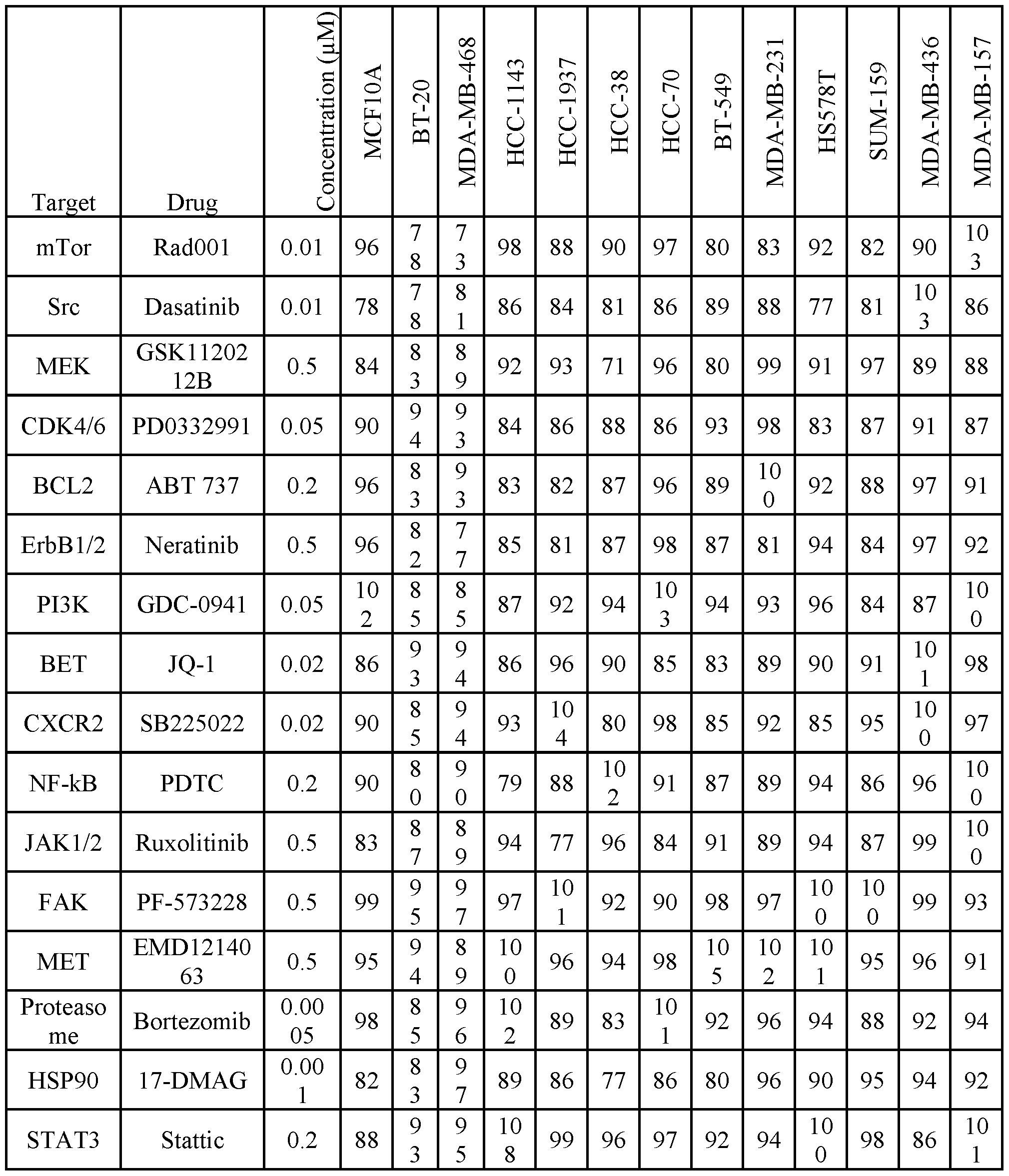

Drug combinations were tested in triplicates, and potent combinations were defined as those inducing at least 75% death in TNBC lines, with minimal (<20%) or no effect on MCF10A cell viability. Out of the 106 different combination pairs that were tested 27 preferentially affected TNBC (Figure 1A). Among them, 13 were effective in all the TNBC cell lines, whereas 9 were preferentially potent for mesenchymal/mesenchymal stem-like (M/MSL)-subtype and 5 for basal-like TNBC cell lines (Figure IB). Potent combinations were further tested by different cell viability assays (Figure 9D, Figures 2A-E), and their synergy was evaluated by calculating the Combination Index (Cl) using the Chou-Talalay method [Chou, 2010, Cancer Res 70, 440- 446]
To prioritize clinically promising synergistic combinations, an unbiased computational analysis was applied to identify clinically relevant synthetic lethality (ISLE). It was hypothesized that a drug combination could be synergistic if their corresponding drug targets form a synthetic lethal pair. Datasets analysis of 330 basal-like breast cancer patients [Shu, 2016, Nature 529, 413-417] was used to predict clinically-relevant synthetic lethality interactions (cSLi) between all possible gene pairs of the 17 target genes of the applied drugs (Methods). To estimate the strength of SL interaction, the ISLE significance score [Lee, 2018, Nat Commun 9, 2546, doi:10.1038/s41467-018-04647-l] was used (Figure 9E). Higher scores represent high probability to be synergistic. This approach highly scored the experimental potent combinations (Figure 1C, 9E), and revealed a significant correlation between the ISLE-significance scores and the present drug combinations screen (Spearman’s correlation = 0.43, P = 0.0265, Figure 1C, Methods). Highly effective combinations could be predicted using ISLE-significance scores with an accuracy (ROC-AUC) of 85.71% (Methods).
In light of the ISLE scoring (Figure 9E), the relative toxicity of the different combinations (Figure 1A, 9D), and the high sensitivity of TNBC to BET (Bromodomain and ExtraTerminal) inhibitor, two potent drug combinations were selected targeting the epigenetic readers of the BET family. One combination was generic for all TNBC subtypes, using JQ1 and the proteasome inhibitor Bortezomib (BTZ), while the second was an M/MSL-specific combination of JQ1 and the CXCR2 receptor antagonist SB225022. JQ1 is thienodiazepine-based small molecule inhibitor, which competitively inhibits the binding of bromodomains to acetylated histones, and thus the activity of BRD4. Importantly, BRD4 transcripts are significantly enriched in basal-like breast cancer patients (Figure ID) as
well as in TNBC cell lines relative to other breast cancer lines (Figure 10A), as indicated by analysis of TCGA breast cancer dataset (RNA-seq, PAM50) and the Cancer Cell Line Encyclopedia (CCLE) dataset. Furthermore, immunohistochemical (IHC) analysis of 67 breast cancer sections showed that BRD4 protein is significantly high in TNBC compare to non-TNBC patients (Figure IE), indicating that both the mRNA and protein levels of BRD4 are relatively high in TNBC, implying that BRD4 could be a particular useful target.
Synthetic lethal combinations targeting BET in TNBC subtypes
Among the generic highly potent combinations that were obtained in the present screen, dual inhibition of BET and the proteasome appeared to be a promising choice. Previous siRNA screen suggested that proteasomal targeting could be beneficial for basal-like TNBC patients and the ISLE scoring (Figure 9E) predicted high clinical relevance of BET and proteasome co targeting. Likewise, the chemokines IL8, IL6 and CXCL1, which bind the CXCRl/2 receptors and implicated in TNBC growth and metastasis, are enriched in TNBC compared to other breast cancer subtypes (Figure 10B) implying that inhibition of CXCR2 could be beneficial for TNBC patients of the M/MSL subgroup.
The efficacy, specificity, and toxicity of BET and CXCR2 inhibition ( JQ 1 +CXCR2in/ SB225022) in M/MSL TNBC, and of BET and the proteasome (JQ1+BTZ) in all TNBC subtypes was then analyzed. Cell viability assays across multiple breast cancer cell lines utilizing increasing drug doses showed high susceptibility of human M/MSL to BET and CXCR2 inhibition (Figure 2A), consistent with the screen results. The basal-like TNBC, the non- tumorigenic MCF10A and the luminal MCF7 breast cancer cell line were not affected by this drugs combination at the applied doses, demonstrating its low toxicity and high selectivity. Furthermore, knockdown of BRD4 expression by shRNA sensitized M/MSL TNBC cell lines to CXCR2 inhibition (Figure IOC). The JQ1+ SB225022 combination markedly reduced the IC50 values (4.5-40 fold) of individual drugs in human mesenchymal cell lines (Figure 11D,E), and dose-response curves for JQ1 and/or SB225022 revealed high efficacy of drugs combination (Figure 2D, Figures 11 A, B, D, E) with strong synergy (combination index < 0.5) (Figure 2D). Importantly, the murine breast cancer cell line 4T1, which is considered as a TNBC line [Miller, 1983, Invasion & metastasis 3, 22-31] was affected by the two BET combinations (JQ1+BTZ and JQ1+ SB225022), either in 2D (Figure 2D, Figure 11C,F, Figure 12C,F) or in 3D spheroids (Figure 2C).
In contrast to BET and CXCR2 targeting, inhibition of BET and the proteasome (JQ1+BTZ) was efficient in all TNBC cell lines (Figure 2B). The potency and synergy of
JQ1+BTZ combination is reflected by the remarkable reduced IC50 values for each individual drug in the presence of the second (Figure 2E, Figures 12A-F), and by the calculated Cl values (Figure 2E) shown for representative cell lines. Importantly, inhibition of BET with the proteasome was more effective than other combinations with BET inhibitor, including BET and PARP inhibition (Figure 11G). Collectively, by applying different cell-based assays to monitor cell viability in response to single agents and drug combinations, the present inventors showed high efficacy and strong synergy of BET inhibition and CXCR2 in M/MSL TNBC, and of BET and the proteasome in all TNBC irrespective of their molecular subtype.
To further demonstrate the potency of BET+CXCR2 and BET+ Proteasome targeting, orthotopic tumor models in immunocompromised and immunocompetent mice, respectively were established. To inhibit BET, the bromodomain inhibitor OTX015 (MK-8628) was used due to the short half-life of JQ1 in vivo. OTX015 was very similar to JQ1 in vitro , either as a single agent or in combination with SB225022 or bortezomib (Figure 13A,B). First, dose responses were calibrated for both drug combinations in preliminary pilot experiments using syngeneic orthotopic 4T1 tumor models (Figure 13C-F) and MDA-MB-231 xenografts in nude mice (Figure 13G).
Accordingly, OTX015 was applied orally once a day at 25 mg/kg (a 2-fold lower concentration than previously used) [Vazquez, 2017, Oncotarget 8, 7598-7613], whereas SB225022 was administered intraperitoneally (IP) at 5mg/kg, ~2-fold lower than previous studies [Devapatla, 2015, PLoS One 10, e0139237, doi:10.1371/joumal. pone.0139237] Drugs administration scheme to MDA-MB-231 xenograft mice models is described in Figure 13H. As shown, OTX015 or SB225022 applied as a single agent had partial effects on tumor growth with only -15% tumor growth inhibition (TGI) (Figures 3A-C). In contrast, the OTX015+ SB225022 combination was very potent, drastically reducing tumor size (Figure 3A-C, 13D), with a TGI close to 100%. The drug combination had minimal toxicity at the applied doses, as indicated by body weight (Figure 3D). Collectively, these data show the high efficacy of BET and CXCR2 inhibition compared to single agent treatments.
For the generic combination of OTX015 plus BTZ, orthotopic syngeneic mice models using the 4T1 cells were used. Low doses of the two drugs (OTX015: 25 mg/kg/day, were applied, orally every day; BTZ: 0.25mg/kg, IP every 3 days, Figure 131) and the effects of single and combined treatments on tumor growth over time were examined. Results clearly showed the profound and significant inhibition of drug combinations on tumor growth compared with single drugs treatments (Figure 3E-G). The drug combination had minimal toxicity at the applied doses,
as indicated by body weight (Figure 3H). Importantly, the strong inhibitory effects of OTX015 plus BTZ combination sustained for at least 55 days and persisted for at least 21 days after drug withdrawal (Figure 3J, K). Kaplan Meier survival curves indicate that OTX015 plus BTZ combination significantly prolonged mice survival for more than 80 days (Figure 3K). Remarkably, survival was prolonged even after drug withdrawal at day 31. Together these results suggest that co-targeting of BET and the proteasome applied at low doses could be a promising therapeutic approach for TNBC.
BET and proteasome co-targeting robustly triggers ferroptotic cell death
Next, possible mechanisms underlying the potency of the two drug combinations targeting BET were explored. It was found that inhibition of BET with CXCR2 induced apoptotic cell death as demonstrated by annexin V staining (Figure 14A), and by PARP and Caspase 3 cleavage (Figure 4A). In contrast, concurrent targeting of BET and the proteasome, neither induced PARP cleavage nor Annexin 5-positive staining (Figure 4B, 14B), implying that a nonapoptotic pathway is involved. To systematically define which death pathway mediates the effect of BET and proteasome targeting, specific inhibitors and characteristic assays for the four major cell-death pathways were tested, including apoptosis, necrosis, autophagy and ferroptosis. As seen in Figure 4C, the pan-caspase inhibitor zVAD-FMK, as well as Necrostatin-1 and 3- methyladenine (3MA), which inhibit apoptosis, necrosis, and autophagic cell death, respectively, only marginally rescued cell death in response to JQ1 + BTZ treatment, but efficiently rescued cell death in response to their own pathway specific inducers (Figure 11C). In contrast, glutathione, the iron chelator 2,2-bipyridyl (2,2-BP), and the antioxidant ferrostatin-1, which are known to inhibit ferroptosis, could effectively rescue cell death induced by JQ1 + BTZ as assessed by different cell viability assays in multiple TNBC cell lines (Figures 4C,D, Figure 14D,E). These results strongly suggest that the JQ1+BTZ combination induces ferroptosis cell death. Ferroptosis was obtained at a specific range of low doses of JQ1 + BTZ based on liproxstatin-1 rescue experiment (Figure 14H), consistent with their SL interactions. These results strongly suggest that the JQ1 + BTZ combination applied at low doses induces ferroptosis cell death.
To further corroborate these results, the effects of JQ1+ BTZ combination on necrotic and ferroptotic markers were examined. Necrosis was induced by a combination of TNFa, Smac mimetic, and zVAD-fmk and monitored by MLKL (mixed lineage kinase domain-like protein) phosphorylation at Ser-358. As shown in Figure 14F, the JQ1+BTZ combination had slight or no effect on MLKL phosphorylation, suggesting that necrosis is not the death-induced pathway.
Ferroptosis, an iron-dependent program cell death driven by lipid peroxidation, was monitored by cellular lipid peroxidation levels applying the BODIPY 581/591 Cll dye. The emission of the BODIPY dye shifts from 590 nm (red) to 510 nm (green) upon oxidation. As shown in Figure 14F, in contrast to JQ1 + SB225022 combination, the JQ1+BTZ combination markedly increased Cll-BODIPY oxidation in TNBC cell lines, as demonstrated by the representative fluorescent images and by the quantitative analysis of the 590/510 nm (red/green) fluorescence ratio (Figure 4F). Importantly, induction of lipid peroxidation by JQ1+BTZ combination was observed only in TNBC and not in the luminal breast cancer cell lines, and was rescued by glutathione pre-treatment in most of the examined TNBC lines (Figure 4F). These results strongly indicate that TNBC cells are selectively more susceptible to ferroptosis induced by the JQ1+BTZ combination.
As iron is required for lipid peroxide accumulation and ferroptosis execution [Stockwell, 2017, Cell 171, 273-285] the present inventors examined the effect of JQ1+ BTZ co-treatment on cellular iron levels using a colorimetric assay (QuantiChrom iron assay kit, Bioassay). As shown in Figure 4G, the JQ1+BTZ combination increased the levels of cellular iron selectively in TNBC cell lines and not in the examined luminal cell lines. Concomitantly, a significant decrease in cellular glutathione levels (Figure 4H) and an increase in reactive oxidative species (ROS) was observed as demonstrated by CM-FFDCFDA fluorescent images (Figure 14G) and quantified by fluorescence measurements (488 nm) (Figure 41). Importantly, pre-treatment with glutathione reduced the effect of JQ1+BTZ on both FFDCFDA fluorescence (Figure 41, 14G) and cell death (Figure 4C, Figure 14E).
Induction of lipid peroxidation by the JQ1 + BTZ combination was observed only in TNBC and not in the luminal breast cancer cell lines, was rescued by GSH pretreatment in most of the examined TNBC lines (Figure 4F), and was further validated by the TBARS (thiobarbituric acid reactive substances) assay (Figure 141). These results strongly indicate that TNBC cells are selectively more susceptible to ferroptosis induced by the JQ1 + BTZ combination.
Collectively, these observations indicate that the JQ1+BTZ combination modulates key regulators of ferroptosis, including cellular iron and glutathione levels to induce lipid peroxidation selectively in TNBC cell lines.
BET and proteasome co-targeting modulates GPX4 and ferroptosis associated genes in TNBC Previous studies suggested that glutathione levels are reduced in TNBC [Beatty, 2018, Molecular cancer therapeutics 17, 264-275] As glutathione is a necessary cofactor of glutathione
peroxidase 4 (GPX4), a major regulator of ferroptosis [Yang, 2014, Cell 156, 317-331], the present inventors examined its mRNA expression levels across 956 breast cancer and normal like samples from TCGA dataset. As shown in Figure 5A, GPX4 level was significantly lower in basal breast cancer compare to normal or other breast cancer subtypes. They then examined the influence of JQ1+BTZ combination on the protein and mRNA levels of GPX4 in multiple TNBC cell lines using Western blotting (WB) (Figure 5B) and qPCR analysis (Figure 5C).
As shown, both single JQ1 and BTZ treatments increased GPX4 protein levels in all the examined TNBC cell lines (up to ~4.5-fold; Figure 5B, 15 A). In contrast a striking reduction of GPX4 protein levels in cells treated with JQ1+BTZ combination was found (Figure 5B). Importantly, the profound effects of JQ1 or JQ1+BTZ combination on GPX4 protein level were attributed to changes of its mRNA levels (Figure 5C, Figure 15B,C); JQ1 or BRD4 knockdown enhanced GPX4 transcription (Figure 5C, Figure 15C), while JQ1+BTZ combination suppressed GPX4 transcription. Notably, the effects of BET and/or proteasome inhibition on GPX4 mRNA and protein levels were also observed in 4T1 tumors of mice treated with OTX015 and/or BTZ, as shown by immunohistochemistry staining of tumor sections and by qPCR (Figure 5D).
The profound effects of JQ1+BTZ combination on GPX4 expression led the present inventors to examine its influence on other ferroptosis-associated genes. They selected 15 key ferroptosis related genes (Figure 5E, 15D), and assessed their transcripts levels in response to JQ1+BTZ combination in multiple TNBC cell lines. As shown in Figure 5E, in addition to the strong downregulation of GPX4 , downregulation of NRF2 (Nuclear factor E2-related factor 2) was observed in almost all the TNBC cell lines. GSS (Glutathione synthetase), GCLM (the modifier subunit of (yGCL) g-glutamylcysteine ligase), GCLC (the catalytic subunit of yGCL) and GSR (glutathione-disulfide reductase) were also reduced in most of the TNBC cell lines.
Gamma-GCL and GSS are two critical GSH biosynthesis enzymes in mammalian cells [Hao, 2018, Oncology letters 15, 5405-5411] whereas GSR catalyzes the reduction of glutathione disulfide (GSSG) to GSH. The reduced expression of these enzymes in response to JQ1+BTZ combination is consistent with the reduced levels of cellular glutathione (Figure 4H) and ferroptosis cell death. Likewise, low levels of NRF2, the master regulator of the cellular antioxidant response, are consistent with reduced levels of glutathione and increased levels of cellular ROS (Figure 41, 14G). Elevated ROS levels may induce upregulation of HOMX1 (heme oxygenase- 1) and NQOl (NADPH quinone dehydrogenase 1) antioxidant protective enzymes [Mou, 2019 Journal of hematology & oncology 12, 34, doi:10.1186/sl3045-019-0720-y] ALOX15, which was strongly upregulated in M/MSL-cell lines, is involved in peroxidation of
polyunsaturated fatty acids and thus accelerates ferroptosis [Shintoku, 2017, Cancer science 108, 2187-2194, doi: 10.1111/cas.13380]. Other proteins/enzymes were distinctly modified between the different cell lines. Nevertheless, it appears that GPX4 and key enzymes/proteins regulating glutathione levels are markedly affected by the JQ1+BTZ combination, leading to ferroptotic cell death.
TNBC are enriched in ferroptosis-gene signature and vulnerable to ferroptosis inducers
Thus far, the present findings indicate that TNBC, compared to other breast cancers, are more susceptible to ferroptotic cell- death induced by the JQ1+BTZ combination (Figures 4A-I), and that JQ1+BTZ treatment affects the expression level of GPX4 and several additional ferroptosis-associated genes (Figure 5B-E). GPX4 levels were shown to be significantly lower in TNBC patients (Figure 5A), and a previous report suggested that glutathione levels are particularly low in basal -like breast cancers [Beatty, 2018, Molecular cancer therapeutics 17, 264-275] These observations imply that TNBCs are more susceptible to ferroptosis, and thus, could be more vulnerable to ferroptosis-inducing agents. To explore this possibility, the present inventors examined the susceptibility of 10 TNBC and 4 non-TNBC cell lines to a few ferroptosis inducers, including Fin56 and Erastin. As shown in Figure 6A, Fin56 and Erastin substantially reduced viability of all TNBC cell lines, but marginally affected non-TNBC cell lines at the indicated doses.
Area under the viability curves (AUCs) of multiple repeats using 16 different breast cancer cell lines indicated that TNBCs are significantly more susceptible to ferroptosis compared to non-TNBC cell lines (Figure 16C).
Although these inhibitory effects could be attributed to the inherent low levels of GPX4 in TNBC (Figure 4A), ferroptosis is regulated by additional proteins involved in intracellular glutathione homeostasis as well as in iron and lipid metabolism [Hirschhorn, 2019, Free radical biology & medicine 133, 130-143; Conrad, 2018, Genes & development 32, 602-619] and is characterized by a specific gene signature of 40 genes (PathCards, www(dot)pathcards(dot)genecards(dot)org/).
The expression profile of ferroptotic genes from this gene signature was examined in TNBC versus non-TNBC cell lines and in clinical samples of different breast cancer subtypes (PAM50 classification). A Gene Set Enrichment Analysis (GSEA) of differentially regulated genes in basal versus non-basal breast cancer samples was performed using the TCGA datasets (n=956). A significant enrichment of ferroptosis signature was found in the basal relative to other breast cancers patients (FDR q-value< 0.001) (Figure 6B). Significant enrichment of
ferroptosis was also obtained in the TNBC versus non-TNBC cell lines by CCLE dataset analysis (FDR q-value< 0.01; Figure 6C). Among the different ferroptotic genes, Ferroportin (SLC40A1) and ASCL4 (acyl-CoA synthetase long-chain family member 4) displayed the most prominent differences between TNBC and non-TNBC cell lines. Ferroprotin is the major cellular iron exporter, while ACSL4 plays a key role in arachidonic acid (AA) metabolism and is essential for ferroptosis execution [Doll, 2017, Nat Chem Biol 13, 91-98] The relative expression of these two genes in each of the 56 CCLE breast cancer cell lines is shown in Figure 16A,B. Significant low levels of Ferroprotin and relatively high levels of ACSL4 transcripts were also obtained in basal-like breast cancer compared to other breast cancer subtypes (PAM50) (Figure 6D,E). Total 31 genes out of the 40 ferroptotic genes were significantly differentially expressed (p-value<0.05) in basal versus non-basal tumors, of which 12 were downregulated and 19 were upregulated. Unsupervised hierarchical clustering of 956 breast cancer tissues (142 basal, 434 LumA, 194 LumB, 67 HER2, 119 normal-like) showed that basal patients form a unique cluster separated from the non-basal patients (Figure 6F). These observations further strengthen the association of ferroptosis with basal-like tumors relative to other breast cancer subtypes, and highlight the clinical relevance of this pathway for TNBC.
As a complementary approach to the transcriptomic analysis described above, the present inventors examined whether ferroptotic proteins are also differentially expressed in TNBC. The levels of six selected proteins in multiple breast cancer (BC) cell lines was analyzed by WB. As shown in Figure 6G, the levels of ferroportin, GPX4, GSS and FTH1 were significantly lower in TNBC relative to non-TNBC cell lines, whereas the levels of the iron importer Transferrin receptor (TfR) was higher in TNBC. ACSL4 was also significantly higher in TNBC cell lines, consistent with its gene expression pattern (Figure 6G, Figure 16B).
To further corroborate the link between ferroptosis and TNBC and to highlight its clinical relevance, four key ferroptotic proteins were selected and their expression level was evaluated by immunohistochemistry in clinical samples of TNBC and non-TNBC patients. Approximately 70 (66-74) human breast tumor sections were immunostained for GPX4, GSS, ferroportin and transferrin receptor. The sections were evaluated semi quantitatively and their calculated H- scores were plotted in columns scatter graphs. The present inventors also calculated how many tumors with high and low staining intensity belong to TNBC relative to non-TNBC. As shown in Figure 7A-D, while Transferrin receptor staining was significantly higher in TNBC patients, immunostaining for GPX4, GSS and Ferroportin showed significantly lower expression levels in TNBC sections.
These results are consistent with the mRNA expression profiles from TCGA dataset, and strongly suggest that TNBC distinctly express key proteins that regulate ferroptosis. The high Transferrin receptor and low Ferroprotein, which mediate iron import and export, respectively, should increase the labile iron [Pinnix, 2010, Science translational medicine 2, 43ra56, doi:10.1126/scisignal.3001127], while low GPX4 and GSS should reduce the antioxidation defense capacity and increase the probability of lipid peroxidation and consequently facilitate ferroptosis.
In light of the analysis described above, the differential expression data of ferroptotic genes in basal-like breast cancer was incorporated into the KEGG ferroptosis pathway. The trans-sulfuration pathway, which converts the sulfur atom of methionine into the sulfur atom of cysteine [Hayano, 2016, Cell death and differentiation 23, 270-278], and the pentose phosphate pathway (Figure 8A) were included. This illustration highlights the substantial low level of ferroportin in basal-like tumors concomitant with the reduced levels of glutathione biosynthesis enzymes and of GPX4, and collectively suggest that TNBC are inherently prone to have high labile iron, low glutathione antioxidant defense capacity, and high polyunsaturated fatty acids. Convergence of glutathione and iron metabolism gene signatures with ferroptosis in TNBC
As ferroptosis is regulated by intersected pathways controlling iron and glutathione metabolism [Stockwell, 2017, Cell 171, 273-285], the present inventors examined the enrichments of these two pathways in TNBC cell lines and basal-like tumors using the CCLE and TCGA datasets, respectively. Gene set enrichment analysis (GSEA) of CCLE dataset revealed a significant enrichment of iron metabolism in TNBS relative to non-TNBC cell lines. Although enrichment of the entire pathways in basal-like breast cancers was not observed, unsupervised hierarchical clustering of 128 genes (out of 194 total) of the iron metabolism gene signature (KEGG) as well as 126 genes (out of 188 genes) of the glutathione gene signature formed a basal-like specific clustering, distinct from other breast cancer subtypes. Thus, the majority of the iron- and the glutathione-associated genes pathways are deferentially expressed in TNBC relative to other breast cancers. The combined clustering of the three gene signatures (ferroptosis, glutathione and iron metabolism) of the top 240 differentially expressed genes demonstrates the unique clustering of basal-like tumors relative to the other breast cancer subtypes or normal-like breast tissues. Collectively, these analyses suggest that TNBCs possess a unique metabolic signature of iron and glutathione homeostasis that enhance their susceptibility to ferroptosis.
To further demonstrate the interaction between ferroptosis, glutathione and iron metabolism in TNBC, a functional interaction network of differentially expressed genes in basal- like tumors relative to other breast cancer subtypes (fold-change >1.2) was built using the GeneMania Cytoscape [Warde-Farley, 2010, Nucleic acids research 38, W214-220] The network incorporates co-expression, genetic and physical interactions and shared protein domains, and it consists of 23 differentially expressed ferroptotic genes of which 4 overlap with the glutathione pathway and 9 with the iron metabolism pathway. Out of the 126 and 128 differentially expressed genes of glutathione and iron metabolism signatures, respectively, 24 were overlapped (10 upregulated, 14 downregulated) (Figure 8B). Importantly, among the ferroptosis-associated genes, the glutamate/cystine antiporter (SLC7A11), ACSL4 and ferroprotein, had the highest number of total interactions (genetic and physical), demonstrating that key ferroptotic proteins are tightly interconnected within the constructed network and differential expressed in basal-like tumors.
Although the invention has been described in conjunction with specific embodiments thereof, it is evident that many alternatives, modifications and variations will be apparent to those skilled in the art. Accordingly, it is intended to embrace all such alternatives, modifications and variations that fall within the spirit and broad scope of the appended claims.
All publications, patents and patent applications mentioned in this specification are herein incorporated in their entirety by reference into the specification, to the same extent as if each individual publication, patent or patent application was specifically and individually indicated to be incorporated herein by reference. In addition, citation or identification of any reference in this application shall not be construed as an admission that such reference is available as prior art to the present invention. To the extent that section headings are used, they should not be construed as necessarily limiting. In Addition, any priority document(s) of this application is/are hereby incorporated by reference in its/their entirety.
In addition, any priority document(s) of this application is/are hereby incorporated herein by reference in its/their entirety.
Claims
1. A combination of agents for use in the treatment of triple negative breast cancer (TNBC), wherein the combination of agents comprise:
(i) a Bromodomain and Extra-Terminal motif (BET) inhibitor; and
(ii) a proteasome inhibitor or an agent which downregulates the amount and/or activity of CXCRl/2 receptor.
2. The combination of agents of claim 1, wherein said agent which downregulates the amount and/or activity of CXCRl/2 receptor is an anti-CXCRl antibody or an anti-CXCR2 antibody.
3. The combination of agents of claim 1, wherein said agent which downregulates the amount and/or activity of CXCRl/2 receptor is a dual CXCRl/2 antagonist.
4. The combination of agents of claim 3, wherein said dual CXCRl/2 antagonist is selected from the group consisting of navarixin, ladarixin, reparixin, reparixin L-lysine salt, DF2755A, CXCL8 fragment comprising amino acids 3-74 and substitutions K11R/G31P (G31P), DF2162, and SCH-479833.
5. The combination of agents of any one of claims 1-4, wherein said BET inhibitor is a small molecule inhibitor.
6. The combination of agents of claim 5, wherein said small molecule inhibitor is selected from the group consisting of CPI-0610, DUAL946, GSK525762, I-BET151, JQ1, OTX015, PFI-1, RVX-208, RVX2135 and TEN-010.
7. The combination of agents of claims 5 or 6, wherein said small molecule inhibitor is OTX015.
8. The combination of agents of any one of claims 1-7, wherein when said combination is
(i) a Bromodomain and Extra-Terminal motif (BET) inhibitor; and
(ii) an agent which downregulates the amount and/or activity of CXCRl/2 receptor, the TNBC is mesenchymal or mesenchymal stem-like TNBC.
9. The combination of agents of any one of claims 1-7, wherein said proteasome inhibitor is an epoxyketone or a boronate.
10. The combination of agents of claim 9, wherein said proteasome inhibitor is a boronate.
11. The combination of agents of claim 10, wherein the inhibitor is bortezomib or ixazomib.
12. The combination of agents of claim 9, wherein said proteasome inhibitor is an epoxyketone.
13. The combination of agents of claim 12, wherein said proteasome inhibitor is carfilzomib or a fluorinated carfilzomib analog.
14. The combination of agents of any one of claims 1-13, being formulated in a single composition.
15. The combination of agents of any one of claims 1-13, being formulated in separate compositions.
16. A pharmaceutical composition comprising a pharmaceutically acceptable carrier and as active agents:
(i) a Bromodomain and Extra-Terminal motif (BET) inhibitor; and
(ii) an agent which downregulates the amount and/or activity of CXCRl/2 receptor.
17. The pharmaceutical composition of claim 16, wherein said BET inhibitor is a small molecule inhibitor.
18. The pharmaceutical composition of claim 17, wherein said small molecule inhibitor is selected from the group consisting of CPI-0610, DUAL946, GSK525762, I-BET151, JQ1, OTX015, PFI-1, RVX-208, RVX2135 and TEN-010.
19. The pharmaceutical composition of claims 17 or 18, wherein said small molecule inhibitor is OTX015.
20. The pharmaceutical composition of any one of claims 16-19, wherein said agent which downregulates the amount and/or activity of CXCRl/2 receptor is an anti-CXCRl antibody or an anti-CXCR2 antibody.
21. The pharmaceutical composition of any one of claims 16-19, wherein said agent which downregulates the amount and/or activity of CXCRl/2 receptor is a dual CXCRl/2 antagonist.
22. The pharmaceutical composition of claim 21, wherein said dual CXCRl/2 antagonist is selected from the group consisting of navarixin, ladarixin, reparixin, reparixin L- lysine salt, DF2755A, CXCL8 fragment comprising amino acids 3-74 and substitutions K11R/G31P (G31P), DF2162, and SCH-479833.
23. A ferroptosis-inducing agent for use in the treatment of triple negative breast cancer (TNBC), with the proviso that the ferroptosis-inducing agent is not RAS-selective lethal 3 (RSL3).
24. The ferroptosis-inducing agent of claim 23, wherein the agent is a combination of at least two agents.
25. The ferroptosis-inducing agent of claim 24, wherein said combination of at least two agents are formulated in a single composition.
26. The ferroptosis-inducing agent of claim 24, wherein said combination of at least two agents are formulated in separate compositions.
27. The ferroptosis-inducing agent of any one of claims 24-26, wherein the combination of at least two agents comprise:
(i) a BET inhibitor; and
(ii) a proteasome inhibitor.
28. The ferroptosis-inducing agent of claim 27, wherein said BET inhibitor is a small molecule inhibitor.
29. The ferroptosis-inducing agent of claim 28, wherein said BET inhibitor is selected from the group consisting of OTX015, CPI-0610, DUAL946, GSK525762, I-BET151, JQ1, PFI- 1, RVX-208, RVX2135 and TEN-010.
30. The ferroptosis-inducing agent of claim 28, wherein said BET inhibitor is OTX015.
31. The ferroptosis-inducing agent of any one of claims 23-30, wherein the agent is a class I inducer.
32. The ferroptosis-inducing agent of claim 31, wherein said class I inducer is selected from the group consisting of erastin, imidazole ketone erastin (IKE), piperazine erastin (PE), sulfasalazine and sorafmib.
33. The ferroptosis-inducing agent of claim 32, wherein said class I inducer is selected from the group consisting of IKE, PE and sorafmib.
34. The ferroptosis-inducing agent of claim 23, wherein the agent is a class III inducer.
35. The ferroptosis-inducing agent of claim 34, wherein said class III inducer is selected from the group consisting of ferroptosis inducer 56 (FIN56) and caspase-independent lethal 56 (CIL56).
36. The ferroptosis-inducing agent of any one of claims 27-35, wherein said proteasome inhibitor is an epoxyketone or a boronate.
37. The ferroptosis-inducing agent of claim 36, wherein said proteasome inhibitor is a boronate.
38. The ferroptosis-inducing agent of claim 37, wherein the inhibitor is bortezomib or ixazomib.
39. The ferroptosis-inducing agent of claim 36, wherein said proteasome inhibitor is an epoxy ketone.
40. The ferroptosis-inducing agent of claim 39, wherein said proteasome inhibitor is carfilzomib or a fluorinated carfilzomib analog.
Applications Claiming Priority (4)
| Application Number | Priority Date | Filing Date | Title |
|---|---|---|---|
| IL269295A IL269295A (en) | 2019-09-11 | 2019-09-11 | Methods of treating breast cancer |
| IL269295 | 2019-09-11 | ||
| US201962927818P | 2019-10-30 | 2019-10-30 | |
| US62/927,818 | 2019-10-30 |
Publications (1)
| Publication Number | Publication Date |
|---|---|
| WO2021048852A1 true WO2021048852A1 (en) | 2021-03-18 |
Family
ID=74866182
Family Applications (1)
| Application Number | Title | Priority Date | Filing Date |
|---|---|---|---|
| PCT/IL2020/050993 Ceased WO2021048852A1 (en) | 2019-09-11 | 2020-09-10 | Methods of treating breast cancer |
Country Status (1)
| Country | Link |
|---|---|
| WO (1) | WO2021048852A1 (en) |
Cited By (2)
| Publication number | Priority date | Publication date | Assignee | Title |
|---|---|---|---|---|
| CN116036087A (en) * | 2022-12-26 | 2023-05-02 | 中国人民解放军空军军医大学 | Use of ferroptosis inhibitor in preparation of medicament for repairing damaged liver |
| CN118760892A (en) * | 2024-09-05 | 2024-10-11 | 温州市中心医院 | Ferroptosis score determination method, system and device |
Citations (40)
| Publication number | Priority date | Publication date | Assignee | Title |
|---|---|---|---|---|
| US3791932A (en) | 1971-02-10 | 1974-02-12 | Akzona Inc | Process for the demonstration and determination of reaction components having specific binding affinity for each other |
| US3839153A (en) | 1970-12-28 | 1974-10-01 | Akzona Inc | Process for the detection and determination of specific binding proteins and their corresponding bindable substances |
| US3850752A (en) | 1970-11-10 | 1974-11-26 | Akzona Inc | Process for the demonstration and determination of low molecular compounds and of proteins capable of binding these compounds specifically |
| US3850578A (en) | 1973-03-12 | 1974-11-26 | H Mcconnell | Process for assaying for biologically active molecules |
| US3853987A (en) | 1971-09-01 | 1974-12-10 | W Dreyer | Immunological reagent and radioimmuno assay |
| US3867517A (en) | 1971-12-21 | 1975-02-18 | Abbott Lab | Direct radioimmunoassay for antigens and their antibodies |
| US3879262A (en) | 1972-05-11 | 1975-04-22 | Akzona Inc | Detection and determination of haptens |
| US3901654A (en) | 1971-06-21 | 1975-08-26 | Biological Developments | Receptor assays of biologically active compounds employing biologically specific receptors |
| US3935074A (en) | 1973-12-17 | 1976-01-27 | Syva Company | Antibody steric hindrance immunoassay with two antibodies |
| US3984533A (en) | 1975-11-13 | 1976-10-05 | General Electric Company | Electrophoretic method of detecting antigen-antibody reaction |
| US3996345A (en) | 1974-08-12 | 1976-12-07 | Syva Company | Fluorescence quenching with immunological pairs in immunoassays |
| US4034074A (en) | 1974-09-19 | 1977-07-05 | The Board Of Trustees Of Leland Stanford Junior University | Universal reagent 2-site immunoradiometric assay using labelled anti (IgG) |
| US4036945A (en) | 1976-05-03 | 1977-07-19 | The Massachusetts General Hospital | Composition and method for determining the size and location of myocardial infarcts |
| US4098876A (en) | 1976-10-26 | 1978-07-04 | Corning Glass Works | Reverse sandwich immunoassay |
| US4331647A (en) | 1980-03-03 | 1982-05-25 | Goldenberg Milton David | Tumor localization and therapy with labeled antibody fragments specific to tumor-associated markers |
| US4666828A (en) | 1984-08-15 | 1987-05-19 | The General Hospital Corporation | Test for Huntington's disease |
| US4683202A (en) | 1985-03-28 | 1987-07-28 | Cetus Corporation | Process for amplifying nucleic acid sequences |
| US4801531A (en) | 1985-04-17 | 1989-01-31 | Biotechnology Research Partners, Ltd. | Apo AI/CIII genomic polymorphisms predictive of atherosclerosis |
| US4816567A (en) | 1983-04-08 | 1989-03-28 | Genentech, Inc. | Recombinant immunoglobin preparations |
| US4879219A (en) | 1980-09-19 | 1989-11-07 | General Hospital Corporation | Immunoassay utilizing monoclonal high affinity IgM antibodies |
| US4946778A (en) | 1987-09-21 | 1990-08-07 | Genex Corporation | Single polypeptide chain binding molecules |
| US5011771A (en) | 1984-04-12 | 1991-04-30 | The General Hospital Corporation | Multiepitopic immunometric assay |
| US5189178A (en) | 1990-11-21 | 1993-02-23 | Galardy Richard E | Matrix metalloprotease inhibitors |
| US5192659A (en) | 1989-08-25 | 1993-03-09 | Genetype Ag | Intron sequence analysis method for detection of adjacent and remote locus alleles as haplotypes |
| US5239078A (en) | 1990-11-21 | 1993-08-24 | Glycomed Incorporated | Matrix metalloprotease inhibitors |
| US5272057A (en) | 1988-10-14 | 1993-12-21 | Georgetown University | Method of detecting a predisposition to cancer by the use of restriction fragment length polymorphism of the gene for human poly (ADP-ribose) polymerase |
| US5281521A (en) | 1992-07-20 | 1994-01-25 | The Trustees Of The University Of Pennsylvania | Modified avidin-biotin technique |
| US5545806A (en) | 1990-08-29 | 1996-08-13 | Genpharm International, Inc. | Ransgenic non-human animals for producing heterologous antibodies |
| US5545807A (en) | 1988-10-12 | 1996-08-13 | The Babraham Institute | Production of antibodies from transgenic animals |
| US5569825A (en) | 1990-08-29 | 1996-10-29 | Genpharm International | Transgenic non-human animals capable of producing heterologous antibodies of various isotypes |
| US5625126A (en) | 1990-08-29 | 1997-04-29 | Genpharm International, Inc. | Transgenic non-human animals for producing heterologous antibodies |
| US5633425A (en) | 1990-08-29 | 1997-05-27 | Genpharm International, Inc. | Transgenic non-human animals capable of producing heterologous antibodies |
| US5661016A (en) | 1990-08-29 | 1997-08-26 | Genpharm International Inc. | Transgenic non-human animals capable of producing heterologous antibodies of various isotypes |
| WO2006032470A1 (en) | 2004-09-21 | 2006-03-30 | Smithkline Beecham Corporation | Compounds which increase apolipoprotein a-1 production and uses thereof in medicine |
| WO2009084693A1 (en) | 2007-12-28 | 2009-07-09 | Mitsubishi Tanabe Pharma Corporation | Antitumor agent |
| WO2011005484A2 (en) | 2009-06-24 | 2011-01-13 | New York University | High speed low power magnetic devices based on current induced spin-momentum transfer |
| WO2011143669A2 (en) | 2010-05-14 | 2011-11-17 | Dana-Farber Cancer Institute, Inc | Compositions and methods for treating neoplasia, inflammatory disease and other disorders |
| US20120208800A1 (en) | 2009-11-05 | 2012-08-16 | Chun-Wa Chung | Bromodomain Inhibitors For Treating Autoimmune And Inflammatory Diseases |
| WO2014026282A1 (en) | 2012-08-14 | 2014-02-20 | Abdelmalik Slassi | Fluorinated epoxyketone-based compounds and uses thereof as proteasome inhibitors |
| US20190248883A1 (en) | 2002-12-16 | 2019-08-15 | Cormorant Pharmaceuticals Ab | Human monoclonal antibodies against interleukin 8 (il-8) |
-
2020
- 2020-09-10 WO PCT/IL2020/050993 patent/WO2021048852A1/en not_active Ceased
Patent Citations (41)
| Publication number | Priority date | Publication date | Assignee | Title |
|---|---|---|---|---|
| US3850752A (en) | 1970-11-10 | 1974-11-26 | Akzona Inc | Process for the demonstration and determination of low molecular compounds and of proteins capable of binding these compounds specifically |
| US3839153A (en) | 1970-12-28 | 1974-10-01 | Akzona Inc | Process for the detection and determination of specific binding proteins and their corresponding bindable substances |
| US3791932A (en) | 1971-02-10 | 1974-02-12 | Akzona Inc | Process for the demonstration and determination of reaction components having specific binding affinity for each other |
| US3901654A (en) | 1971-06-21 | 1975-08-26 | Biological Developments | Receptor assays of biologically active compounds employing biologically specific receptors |
| US3853987A (en) | 1971-09-01 | 1974-12-10 | W Dreyer | Immunological reagent and radioimmuno assay |
| US3867517A (en) | 1971-12-21 | 1975-02-18 | Abbott Lab | Direct radioimmunoassay for antigens and their antibodies |
| US3879262A (en) | 1972-05-11 | 1975-04-22 | Akzona Inc | Detection and determination of haptens |
| US3850578A (en) | 1973-03-12 | 1974-11-26 | H Mcconnell | Process for assaying for biologically active molecules |
| US3935074A (en) | 1973-12-17 | 1976-01-27 | Syva Company | Antibody steric hindrance immunoassay with two antibodies |
| US3996345A (en) | 1974-08-12 | 1976-12-07 | Syva Company | Fluorescence quenching with immunological pairs in immunoassays |
| US4034074A (en) | 1974-09-19 | 1977-07-05 | The Board Of Trustees Of Leland Stanford Junior University | Universal reagent 2-site immunoradiometric assay using labelled anti (IgG) |
| US3984533A (en) | 1975-11-13 | 1976-10-05 | General Electric Company | Electrophoretic method of detecting antigen-antibody reaction |
| US4036945A (en) | 1976-05-03 | 1977-07-19 | The Massachusetts General Hospital | Composition and method for determining the size and location of myocardial infarcts |
| US4098876A (en) | 1976-10-26 | 1978-07-04 | Corning Glass Works | Reverse sandwich immunoassay |
| US4331647A (en) | 1980-03-03 | 1982-05-25 | Goldenberg Milton David | Tumor localization and therapy with labeled antibody fragments specific to tumor-associated markers |
| US4879219A (en) | 1980-09-19 | 1989-11-07 | General Hospital Corporation | Immunoassay utilizing monoclonal high affinity IgM antibodies |
| US4816567A (en) | 1983-04-08 | 1989-03-28 | Genentech, Inc. | Recombinant immunoglobin preparations |
| US5011771A (en) | 1984-04-12 | 1991-04-30 | The General Hospital Corporation | Multiepitopic immunometric assay |
| US4666828A (en) | 1984-08-15 | 1987-05-19 | The General Hospital Corporation | Test for Huntington's disease |
| US4683202A (en) | 1985-03-28 | 1987-07-28 | Cetus Corporation | Process for amplifying nucleic acid sequences |
| US4683202B1 (en) | 1985-03-28 | 1990-11-27 | Cetus Corp | |
| US4801531A (en) | 1985-04-17 | 1989-01-31 | Biotechnology Research Partners, Ltd. | Apo AI/CIII genomic polymorphisms predictive of atherosclerosis |
| US4946778A (en) | 1987-09-21 | 1990-08-07 | Genex Corporation | Single polypeptide chain binding molecules |
| US5545807A (en) | 1988-10-12 | 1996-08-13 | The Babraham Institute | Production of antibodies from transgenic animals |
| US5272057A (en) | 1988-10-14 | 1993-12-21 | Georgetown University | Method of detecting a predisposition to cancer by the use of restriction fragment length polymorphism of the gene for human poly (ADP-ribose) polymerase |
| US5192659A (en) | 1989-08-25 | 1993-03-09 | Genetype Ag | Intron sequence analysis method for detection of adjacent and remote locus alleles as haplotypes |
| US5661016A (en) | 1990-08-29 | 1997-08-26 | Genpharm International Inc. | Transgenic non-human animals capable of producing heterologous antibodies of various isotypes |
| US5545806A (en) | 1990-08-29 | 1996-08-13 | Genpharm International, Inc. | Ransgenic non-human animals for producing heterologous antibodies |
| US5569825A (en) | 1990-08-29 | 1996-10-29 | Genpharm International | Transgenic non-human animals capable of producing heterologous antibodies of various isotypes |
| US5625126A (en) | 1990-08-29 | 1997-04-29 | Genpharm International, Inc. | Transgenic non-human animals for producing heterologous antibodies |
| US5633425A (en) | 1990-08-29 | 1997-05-27 | Genpharm International, Inc. | Transgenic non-human animals capable of producing heterologous antibodies |
| US5239078A (en) | 1990-11-21 | 1993-08-24 | Glycomed Incorporated | Matrix metalloprotease inhibitors |
| US5189178A (en) | 1990-11-21 | 1993-02-23 | Galardy Richard E | Matrix metalloprotease inhibitors |
| US5281521A (en) | 1992-07-20 | 1994-01-25 | The Trustees Of The University Of Pennsylvania | Modified avidin-biotin technique |
| US20190248883A1 (en) | 2002-12-16 | 2019-08-15 | Cormorant Pharmaceuticals Ab | Human monoclonal antibodies against interleukin 8 (il-8) |
| WO2006032470A1 (en) | 2004-09-21 | 2006-03-30 | Smithkline Beecham Corporation | Compounds which increase apolipoprotein a-1 production and uses thereof in medicine |
| WO2009084693A1 (en) | 2007-12-28 | 2009-07-09 | Mitsubishi Tanabe Pharma Corporation | Antitumor agent |
| WO2011005484A2 (en) | 2009-06-24 | 2011-01-13 | New York University | High speed low power magnetic devices based on current induced spin-momentum transfer |
| US20120208800A1 (en) | 2009-11-05 | 2012-08-16 | Chun-Wa Chung | Bromodomain Inhibitors For Treating Autoimmune And Inflammatory Diseases |
| WO2011143669A2 (en) | 2010-05-14 | 2011-11-17 | Dana-Farber Cancer Institute, Inc | Compositions and methods for treating neoplasia, inflammatory disease and other disorders |
| WO2014026282A1 (en) | 2012-08-14 | 2014-02-20 | Abdelmalik Slassi | Fluorinated epoxyketone-based compounds and uses thereof as proteasome inhibitors |
Non-Patent Citations (63)
| Title |
|---|
| "Current Protocols in Immunology", vol. 1, 3, 1994, APPLETON & LANGE |
| "Immobilized Cells and Enzymes", 1986, IRL PRESS |
| "PCR Protocols: A Guide To Methods And Applications", vol. 1, 1990, ACADEMIC PRESS, pages: 317 |
| "Selected Methods in Cellular Immunology", 1980, W. H. FREEMAN AND CO. |
| "Transcription and Translation", 1984 |
| AUSUBEL ET AL.: "Current Protocols in Molecular Biology", vol. 1, 3, 1989, JOHN WILEY AND SONS |
| BEATTY, MOLECULAR CANCER THERAPEUTICS, vol. 17, 2018, pages 264 - 275 |
| BIRREN ET AL.: "Genome Analysis: A Laboratory Manual Series", vol. 1-4, 1998, COLD SPRING HARBOR LABORATORY PRESS |
| BOERNER ET AL., J. IMMUNOL., vol. 147, no. 1, 1991, pages 86 - 95 |
| CHOTHIA ET AL., NATURE, vol. 342, 1989, pages 877 - 883 |
| CHOU, CANCER RES, vol. 70, 2010, pages 440 - 446 |
| COLE ET AL.: "Monoclonal Antibodies and Cancer Therapy", 1985, ALAN R. LISS, pages: 77 |
| CONRAD, GENES & DEVELOPMENT, vol. 32, 2018, pages 602 - 619 |
| DEVAPATLA, PLOS ONE, vol. 10, 2015, pages e0139237 |
| DOLL ET AL., NATURE CHEMICAL BIOLOGY, vol. 13, January 2017 (2017-01-01), pages 91 - 98 |
| DOLL, NAT CHEM BIOL, vol. 13, 2017, pages 91 - 98 |
| DUAN ET AL., MED. CHEM. COMMUN., vol. 9, 2018, pages 1779 - 1802 |
| FERRI ELENA ET AL: "Bromodomains: Structure, function and pharmacology of inhibition", BIOCHEMICAL PHARMACOLOGY, ELSEVIER, US, vol. 106, 18 December 2015 (2015-12-18), pages 1 - 18, XP029462841, ISSN: 0006-2952, DOI: 10.1016/J.BCP.2015.12.005 * |
| FINGL, E. ET AL., THE PHARMACOLOGICAL BASIS OF THERAPEUTICS, 1975 |
| FISHWILD ET AL., NATURE BIOTECHNOLOGY, vol. 14, 1996, pages 826 - 51 |
| FRESHNEY: "Cell Biology: A Laboratory Handbook", vol. 1, 3, 1994, WILEY-LISS, article "Culture of Animal Cells" |
| GINESTIER C. ET AL., J CLIN INVEST, vol. 120, 2010, pages 485 - 97 |
| HAO, ONCOLOGY LETTERS, vol. 15, 2018, pages 5405 - 5411 |
| HAOCHEN YU ET AL: "Sulfasalazine-induced ferroptosis in breast cancer cells is reduced by the inhibitory effect of estrogen receptor on the transferrin receptor", ONCOLOGY REPORTS, 6 June 2019 (2019-06-06), XP055770148, ISSN: 1021-335X, DOI: 10.3892/or.2019.7189 * |
| HAYANO, CELL DEATH AND DIFFERENTIATION, vol. 23, 2016, pages 270 - 278 |
| HIRSCHHORN, FREE RADICAL BIOLOGY & MEDICINE, vol. 133, 2019, pages 130 - 143 |
| HOOGENBOOMWINTER, J. MOL. BIOL., vol. 222, 1991, pages 581 |
| INBAR ET AL., PROC. NAT'L ACAD. SCI. USA, vol. 69, pages 2659 - 62 |
| JONES ET AL., NATURE, vol. 321, 1986, pages 522 - 525 |
| KIM, S. ET AL., EMBO REP, vol. 14, 2013, pages 891 - 899 |
| LEE, NAT COMMUN, vol. 9, 2018, pages 2546 |
| LONBERG ET AL., NATURE, vol. 368, 1994, pages 812 - 859 |
| LONBERGHUSZAR, INTERN. REV. IMMUNOL., vol. 13, 1995, pages 65 - 93 |
| MACCALLUM ET AL., J. MOL. BIOL., vol. 262, 1996, pages 732 - 745 |
| MAHALAKSHMI RAMADOSS ET AL: "Targeting the cancer epigenome: synergistic therapy with bromodomain inhibitors", DRUG DISCOVERY TODAY, vol. 23, no. 1, 22 September 2017 (2017-09-22), AMSTERDAM, NL, pages 76 - 89, XP055594332, ISSN: 1359-6446, DOI: 10.1016/j.drudis.2017.09.011 * |
| MAKABE ET AL., JOURNAL OF BIOLOGICAL CHEMISTRY, vol. 283, 2008, pages 1156 - 1166 |
| MARKS ET AL., BIO/TECHNOLOGY, vol. 10, 1992, pages 779 - 783 |
| MARSHAK ET AL.: "Strategies for Protein Purification and Characterization - A Laboratory Course Manual", 1996, CSHL PRESS |
| MARTIN ET AL., PROC. NATL ACAD SCI USA., vol. 86, 1989, pages 9268 |
| MATTHEW B. SIEGEL ET AL: "Small molecule inhibitor screen identifies synergistic activity of the bromodomain inhibitor CPI203 and bortezomib in drug resistant myeloma", ONCOTARGET, vol. 6, no. 22, 7 August 2015 (2015-08-07), United States, pages 18921 - 18932, XP055769949, ISSN: 1949-2553, DOI: 10.18632/oncotarget.4214 * |
| MILLER, INVASION & METASTASIS, vol. 3, 1983, pages 22 - 31 |
| MONTSERRAT PÉREZ-SALVIA ET AL: "Bromodomain inhibitors and cancer therapy: From structures to applications", EPIGENETICS, vol. 12, no. 5, 2 December 2016 (2016-12-02), US, pages 323 - 339, XP055452716, ISSN: 1559-2294, DOI: 10.1080/15592294.2016.1265710 * |
| MOU ET AL., JOURNAL OF HEMATOLOGY & ONCOLOGY, 29 March 2019 (2019-03-29) |
| MOU, JOURNAL OF HEMATOLOGY & ONCOLOGY, vol. 12, 2019, pages 34 |
| NATURE, vol. 486, no. 7403, 2012, pages 346 |
| PACK ET AL., BIO/TECHNOLOGY, vol. 11, 1993, pages 1271 - 77 |
| PERBAL, B.: "A Practical Guide to Molecular Cloning", 1984, JOHN WILEY & SONS |
| PINNIX, SCIENCE TRANSLATIONAL MEDICINE, vol. 2, 2010, pages 43ra56 |
| PORTER, R. R., BIOCHEM. J., vol. 73, 1959, pages 119 - 126 |
| PRESTA, CURR. OP. STRUCT. BIOL., vol. 2, 1992, pages 593 - 596 |
| RIECHMANN ET AL., NATURE, vol. 332, 1988, pages 323 - 327 |
| SAMBROOK ET AL., MOLECULAR CLONING: A LABORATORY MANUAL, 1989 |
| SEBASTIAN DOLL ET AL: "ACSL4 dictates ferroptosis sensitivity by shaping cellular lipid composition", NATURE CHEMICAL BIOLOGY, 14 November 2016 (2016-11-14), New York, XP055326923, ISSN: 1552-4450, DOI: 10.1038/nchembio.2239 * |
| SHINTOKU, CANCER SCIENCE, vol. 108, 2017, pages 2187 - 2194 |
| SHU, NATURE, vol. 529, 2016, pages 413 - 417 |
| STOCKWELL, CELL, vol. 171, 2017, pages 273 - 285 |
| VAZQUEZ, ONCOTARGET, vol. 8, 2017, pages 7598 - 7613 |
| VERHOEYEN ET AL., SCIENCE, vol. 239, 1988, pages 1534 - 1536 |
| WARDE-FARLEY, NUCLEIC ACIDS RESEARCH, vol. 38, 2010, pages W214 - 220 |
| WATSON ET AL.: "Recombinant DNA", SCIENTIFIC AMERICAN BOOKS |
| WHITLOWFILPULA, METHODS, vol. 2, 1991, pages 106 - 105 |
| YANG, CELL, vol. 156, 2014, pages 317 - 331 |
| YANHUA MOU ET AL: "Ferroptosis, a new form of cell death: opportunities and challenges in cancer", JOURNAL OF HEMATOLOGY & ONCOLOGY, vol. 12, no. 1, 29 March 2019 (2019-03-29), XP055769811, DOI: 10.1186/s13045-019-0720-y * |
Cited By (3)
| Publication number | Priority date | Publication date | Assignee | Title |
|---|---|---|---|---|
| CN116036087A (en) * | 2022-12-26 | 2023-05-02 | 中国人民解放军空军军医大学 | Use of ferroptosis inhibitor in preparation of medicament for repairing damaged liver |
| CN116036087B (en) * | 2022-12-26 | 2023-09-05 | 中国人民解放军空军军医大学 | Use of iron death inhibitor in preparing medicine for repairing damaged liver |
| CN118760892A (en) * | 2024-09-05 | 2024-10-11 | 温州市中心医院 | Ferroptosis score determination method, system and device |
Similar Documents
| Publication | Publication Date | Title |
|---|---|---|
| Wang et al. | METTL3-m6A-EGFR-axis drives lenvatinib resistance in hepatocellular carcinoma | |
| Shen et al. | PARPi treatment enhances radiotherapy-induced ferroptosis and antitumor immune responses via the cGAS signaling pathway in colorectal cancer | |
| Ahmmed et al. | Rg3 inhibits gemcitabine‐induced lung cancer cell invasiveness through ROS‐dependent, NF‐κB‐and HIF‐1α‐mediated downregulation of PTX3 | |
| Lin et al. | Metformin sensitizes anticancer effect of dasatinib in head and neck squamous cell carcinoma cells through AMPK-dependent ER stress | |
| Jeong et al. | Quercetin‐induced ubiquitination and down‐regulation of Her‐2/neu | |
| Díaz et al. | The BET bromodomain inhibitor CPI203 improves lenalidomide and dexamethasone activity in in vitro and in vivo models of multiple myeloma by blockade of Ikaros and MYC signaling | |
| Tarantelli et al. | Copanlisib synergizes with conventional and targeted agents including venetoclax in B-and T-cell lymphoma models | |
| Wu et al. | FK228 augmented temozolomide sensitivity in human glioma cells by blocking PI3K/AKT/mTOR signal pathways | |
| TW201032796A (en) | Treatment of lung cancer with a PARP inhibitor in combination with a growth factor inhibitor | |
| Park et al. | Isolinderalactone suppresses human glioblastoma growth and angiogenic activity in 3D microfluidic chip and in vivo mouse models | |
| EP3898675A1 (en) | Use of il-1 beta antibodies in the treatment or prevention of myelodysplastic syndrome | |
| Zhang et al. | VE-822 upregulates the deubiquitinase OTUD1 to stabilize FHL1 to inhibit the progression of lung adenocarcinoma | |
| Gao et al. | Synergism of FAK and ROS1 inhibitors in the treatment of CDH1-deficient cancers mediated by FAK-YAP signaling | |
| EP3314025A1 (en) | Apobec3b mutagenesis and immunotherapy | |
| Xu et al. | Inhibitory effects of everolimus in combination with paclitaxel on adriamycin-resistant breast cancer cell line MDA-MB-231 | |
| Liu et al. | The AURKA inhibitor alters the immune microenvironment and enhances targeting B7-H3 immunotherapy in glioblastoma | |
| US10206920B2 (en) | Pharmaceutical composition for treating cancer and a method of using the same | |
| Ma et al. | Blockade of de novo pyrimidine biosynthesis triggers autophagic degradation of oncoprotein FLT3-ITD in acute myeloid leukemia | |
| WO2021099558A1 (en) | Combination therapies | |
| Pasello et al. | Targeting glutathione‐S transferase enzymes in musculoskeletal sarcomas: a promising therapeutic strategy | |
| Dent et al. | Enhanced signaling via ERBB3/PI3K plays a compensatory survival role in pancreatic tumor cells exposed to [neratinib+ valproate] | |
| Wang et al. | Morin inhibits ubiquitination degradation of BCL-2 associated agonist of cell death and synergizes with BCL-2 inhibitor in gastric cancer cells | |
| Tang et al. | Palmatine attenuates MYH9 mediated nuclear localization of AURKA to induce G2/M phase arrest in colorectal cancer cells | |
| Guo et al. | Molecular mechanism by which RRM2-inhibitor (cholagogue osalmid) plus bafilomycin A1 cause autophagic cell death in multiple myeloma | |
| Jiang et al. | MLN4924 Suppresses head and neck squamous cell carcinoma progression by inactivating the mTOR signaling pathway via the NEDD8/CUL4/TSC2 axis |
Legal Events
| Date | Code | Title | Description |
|---|---|---|---|
| 121 | Ep: the epo has been informed by wipo that ep was designated in this application |
Ref document number: 20781634 Country of ref document: EP Kind code of ref document: A1 |
|
| NENP | Non-entry into the national phase |
Ref country code: DE |
|
| 122 | Ep: pct application non-entry in european phase |
Ref document number: 20781634 Country of ref document: EP Kind code of ref document: A1 |