WO2020254405A1 - Predicting age using dna methylation signatures - Google Patents
Predicting age using dna methylation signatures Download PDFInfo
- Publication number
- WO2020254405A1 WO2020254405A1 PCT/EP2020/066764 EP2020066764W WO2020254405A1 WO 2020254405 A1 WO2020254405 A1 WO 2020254405A1 EP 2020066764 W EP2020066764 W EP 2020066764W WO 2020254405 A1 WO2020254405 A1 WO 2020254405A1
- Authority
- WO
- WIPO (PCT)
- Prior art keywords
- cpgs
- age
- methylation
- dna
- listed
- Prior art date
- Legal status (The legal status is an assumption and is not a legal conclusion. Google has not performed a legal analysis and makes no representation as to the accuracy of the status listed.)
- Ceased
Links
Classifications
-
- C—CHEMISTRY; METALLURGY
- C12—BIOCHEMISTRY; BEER; SPIRITS; WINE; VINEGAR; MICROBIOLOGY; ENZYMOLOGY; MUTATION OR GENETIC ENGINEERING
- C12Q—MEASURING OR TESTING PROCESSES INVOLVING ENZYMES, NUCLEIC ACIDS OR MICROORGANISMS; COMPOSITIONS OR TEST PAPERS THEREFOR; PROCESSES OF PREPARING SUCH COMPOSITIONS; CONDITION-RESPONSIVE CONTROL IN MICROBIOLOGICAL OR ENZYMOLOGICAL PROCESSES
- C12Q1/00—Measuring or testing processes involving enzymes, nucleic acids or microorganisms; Compositions therefor; Processes of preparing such compositions
- C12Q1/68—Measuring or testing processes involving enzymes, nucleic acids or microorganisms; Compositions therefor; Processes of preparing such compositions involving nucleic acids
- C12Q1/6876—Nucleic acid products used in the analysis of nucleic acids, e.g. primers or probes
- C12Q1/6883—Nucleic acid products used in the analysis of nucleic acids, e.g. primers or probes for diseases caused by alterations of genetic material
-
- C—CHEMISTRY; METALLURGY
- C12—BIOCHEMISTRY; BEER; SPIRITS; WINE; VINEGAR; MICROBIOLOGY; ENZYMOLOGY; MUTATION OR GENETIC ENGINEERING
- C12Q—MEASURING OR TESTING PROCESSES INVOLVING ENZYMES, NUCLEIC ACIDS OR MICROORGANISMS; COMPOSITIONS OR TEST PAPERS THEREFOR; PROCESSES OF PREPARING SUCH COMPOSITIONS; CONDITION-RESPONSIVE CONTROL IN MICROBIOLOGICAL OR ENZYMOLOGICAL PROCESSES
- C12Q1/00—Measuring or testing processes involving enzymes, nucleic acids or microorganisms; Compositions therefor; Processes of preparing such compositions
- C12Q1/68—Measuring or testing processes involving enzymes, nucleic acids or microorganisms; Compositions therefor; Processes of preparing such compositions involving nucleic acids
- C12Q1/6876—Nucleic acid products used in the analysis of nucleic acids, e.g. primers or probes
- C12Q1/6881—Nucleic acid products used in the analysis of nucleic acids, e.g. primers or probes for tissue or cell typing, e.g. human leukocyte antigen [HLA] probes
-
- C—CHEMISTRY; METALLURGY
- C12—BIOCHEMISTRY; BEER; SPIRITS; WINE; VINEGAR; MICROBIOLOGY; ENZYMOLOGY; MUTATION OR GENETIC ENGINEERING
- C12Q—MEASURING OR TESTING PROCESSES INVOLVING ENZYMES, NUCLEIC ACIDS OR MICROORGANISMS; COMPOSITIONS OR TEST PAPERS THEREFOR; PROCESSES OF PREPARING SUCH COMPOSITIONS; CONDITION-RESPONSIVE CONTROL IN MICROBIOLOGICAL OR ENZYMOLOGICAL PROCESSES
- C12Q2600/00—Oligonucleotides characterized by their use
- C12Q2600/154—Methylation markers
-
- C—CHEMISTRY; METALLURGY
- C12—BIOCHEMISTRY; BEER; SPIRITS; WINE; VINEGAR; MICROBIOLOGY; ENZYMOLOGY; MUTATION OR GENETIC ENGINEERING
- C12Q—MEASURING OR TESTING PROCESSES INVOLVING ENZYMES, NUCLEIC ACIDS OR MICROORGANISMS; COMPOSITIONS OR TEST PAPERS THEREFOR; PROCESSES OF PREPARING SUCH COMPOSITIONS; CONDITION-RESPONSIVE CONTROL IN MICROBIOLOGICAL OR ENZYMOLOGICAL PROCESSES
- C12Q2600/00—Oligonucleotides characterized by their use
- C12Q2600/156—Polymorphic or mutational markers
-
- C—CHEMISTRY; METALLURGY
- C12—BIOCHEMISTRY; BEER; SPIRITS; WINE; VINEGAR; MICROBIOLOGY; ENZYMOLOGY; MUTATION OR GENETIC ENGINEERING
- C12Q—MEASURING OR TESTING PROCESSES INVOLVING ENZYMES, NUCLEIC ACIDS OR MICROORGANISMS; COMPOSITIONS OR TEST PAPERS THEREFOR; PROCESSES OF PREPARING SUCH COMPOSITIONS; CONDITION-RESPONSIVE CONTROL IN MICROBIOLOGICAL OR ENZYMOLOGICAL PROCESSES
- C12Q2600/00—Oligonucleotides characterized by their use
- C12Q2600/158—Expression markers
Definitions
- the present invention relates to biomarkers for predicting age of a subject.
- methods and kits to predict the unknown or uncertain age of a subject are presented based on age-related increase of methylation of CpGs.
- predictors of biological age has recently been increasing, possibly mirroring the availability of increasingly powerful analytical methods in the field of molecular biology.
- Different types of predictors are summarized in Jylhava et al. 2017 (EBioMedicine 21:29-36) and include epigenetic clocks, telomere length (replicative ageing), transcriptomic predictors, proteomic predictors, metabolomics predictors, and composite biomarker predictors.
- WO2018146482 refers to a number of murine age-related CpGs. Reynolds et al. 2014 (Nat Commun 5:536) focused on age-related variations in the methylome of monocytes and T-cells. WO2018027228 relies on CpG methylation levels in cytotoxic T-cells. Horvath 2013 (Genome Biol 14:R115) identified an ageing clock of 353 CpGs derived from 51 different tissue/cell types.
- Clinical applications include the use of DNA methylation age as an indicator of biological age (wherein biological age not always correlates with chronological age - the mean absolute deviation from chronological age (abbreviated as MAD) with the current epigenetic biomarker panels appears to vary between 3 and 3.5 years), as an indicator of life expectancy (Marioni et al. 2015, Genome Biol 16:25), and in forensic applications.
- MAD mean absolute deviation from chronological age
- the invention therefore in one aspect relates to methods for detecting the methylation status of a set of age-associated CpGs from a subject, comprising the steps of:
- the set of age-associated CpGs is comprising at least 3 CpGs, wherein the at least 3 CpGs are chosen from the CpGs listed in Table 3, from the CpGs of the differentially methylated regions listed in Table 2, and/or from the CpGs listed in Table 1.
- Such methods can further comprise a step wherein the DNA is isolation from the biological sample.
- the biological sample referred to above is liquid biopsy sample.
- the age- associated CpGs referred to above are age-associated CpGs previously identified in kidney biopsies.
- Any of the above methods can comprise a further step further comprising predicting the age of the subject by correlating the methylation status detected for the set of age-associated CpGs with a range of pre-determined and age-correlated reference methylation statuses of the same set of age-associated CpGs.
- the invention further relates to uses of a set of age-associated CpGs in any above-described method according to the invention, wherein the set of age-associated CpGs is comprising at least 3 CpGs, wherein the at least 3 CpGs are chosen from the CpGs listed in Table 3, from the CpGs of the differentially methylated regions listed in Table 2, and/or from the CpGs listed in Table 1; and wherein the set of age- associated CpGs is comprising at most 1000 CpGs.
- kits comprising oligonucleotides to detect the DNA methylation status on a set of age-associated CpGs from a subject, wherein the set of CpGs is comprising at least 3 CpGs, wherein the at least 3 CpGs are chosen from the CpGs listed in Table 3, from the CpGs of the differentially methylated regions listed in Table 2, and/or from the CpGs listed in Table 1; and wherein the kit is comprising oligonucleotides to detect DNA methylation in at most 1000 CpGs.
- such kits find use for predicting the age of a subject from the DNA methylation status detected for the set of age-associated CpGs from the subject.
- FIGURE 3 Top canonical pathways and top upstream regulators among the genes with a differentially methylated region upon ageing, left for the implantation cohort (based on 5445 DMRs), right for the post-reperfusion cohort (based on 10 274 DMRs). The significance levels are depicted on the y-axis. In the boxes, the number of genes with significant age-associated differentially methylated regions in the pathways are presented as percentage and ratio, respectively.
- FIGURE 4 Top canonical pathways and top upstream regulators among the genes whose promoters were either hyper- or hypomethylated upon ageing in the implantation cohort. The significance levels are depicted on the y-axis. In the boxes, the number of genes with significant age-associated hyper- or hypomethylated promoters in the different pathways are presented as percentage and ratio, respectively.
- Liquid biopsies taken from the blood comprise cell-free DNA (cfDNA) from different sources and therefore is increasingly studied as source of biomarkers.
- donor DNA including donor cfDNA
- cfDNA is detectable in the blood of the allograft recipient for a long time after kidney transplantation (Rutkowska et al. 2007, Ann Transplant 12:12-14; Knight et al. 2019, Transplantation 103:273-283), it is very plausible that e.g. blood comprises autologous kidney cfDNA.
- Methylation of cfDNA of tumor origin is being studies e.g. for purposes of detecting cancer (e.g. Nunes et al.
- the said difference will be reflected in liquid biopsies.
- the kidney is characterized by the highest levels of hydroxymethylation across organs (Bachman et al. 2014, Nat Chem 6:1049-1055). These high levels of 5-hydroxymethylation render the kidney more prone to DNA hypermethylation.
- the kidney therefore represents a unique organ to study methylation-associated aging processes.
- Age-associated methylation of CpGs (initially identified in kidney biopsies) in liquid biopsies therewith is an ideal source of reliable age-predictive markers.
- Liquid biopsies from a kidney can be taken by collecting e.g. blood or urine leaving the kidney, or by collecting urine; such liquid biopsies comprising DNA shedded from cells in the kidney.
- the invention therefore in one aspect relates to methods for determining, detecting, measuring, assaying or assessing the methylation status of a set of age-associated CpGs from a subject, comprising the steps of:
- the set of age-associated CpGs is comprising at least 3 CpGs, wherein the at least 3 CpGs are chosen from the CpGs listed in Table 3 or 6, from the CpGs of the differentially methylated regions listed in Table 2 or 5, and/or from the CpGs listed in Table 1 or 4.
- the above set of age-associated CpGs is chosen from Tables 4, 5, and/or 6.
- the set of age-associated CpGs are at least 3 CpGs of one of the differentially methylated region listed in Table 2 or 5.
- the set of age-associated CpGs are at least 1 CpG of each of at least 2, 3, 4 or 5 differentially methylated regions listed in Table 2 or 5.
- the set of age-associated CpGs are at least 2, 3, 4 or 5 CpGs of each of at least 2, 3, 4 or 5 differentially methylated regions listed in Table 2 or 5.
- Such methods can further comprise a step wherein biological sample is obtained from a subject, and/or wherein the DNA is isolation from the biological sample.
- the biological sample referred to above is a solid kidney biopsy sample, or is a liquid biopsy sample such as a blood or urine sample.
- the blood sample is collected as blood leaving the kidney.
- the DNA can be isolated from blood, serum or plasma.
- the DNA can be total DNA, or can be DNA not associated with cells (such as white blood cells), or cell-free DNA (cfDNA).
- the age-associated CpGs referred to above are age-associated CpGs previously identified in kidney biopsies. In another embodiment, the age-associated CpGs referred to above are kidney-specific age-associated CpGs.
- any of the above methods can comprise a further step comprising determining, measuring, detecting, assaying, assessing or predicting the age of the subject by correlating the methylation status detected for the set of age-associated CpGs with a range of pre-determined and age-correlated reference methylation statuses of the same set of age-associated CpGs.
- methods for predicting the age of a subject comprising the steps of: obtaining DNA from a biological sample from a subject;
- the set of age-associated CpGs is comprising at least 3 CpGs, wherein the at least 3 CpGs are chosen from the CpGs listed in Table 3 or 6, from the CpGs of the differentially methylated regions listed in Table 2 or 5, and/or from the CpGs listed in Table 1 or 4.
- correlating the methylation status detected for the set of age-associated CpGs with a range of pre-determined and age- correlated reference methylation statuses of the same set of age-associated CpGs is added as a further step.
- the above set of age-associated CpGs is chosen from Tables 4, 5, and/or 6. .
- the set of age-associated CpGs are at least 3 CpGs of one of the differentially methylated region listed in Table 2 or 5.
- the set of age-associated CpGs are at least 1 CpG of each of at least 2, 3, 4 or 5 differentially methylated regions listed in Table 2 or 5.
- the set of age-associated CpGs are at least 2, 3, 4 or 5 CpGs of each of at least 2, 3, 4 or 5 differentially methylated regions listed in Table 2 or 5.
- Any of the above methods can comprise a further step comprising determining, detecting, measuring assaying or assessing the methylation status of any of the biomarkers identified in any of Hannum et al. 2013 (Mol Cell 49:359-367 - and W02014075083), Weidner et al. 2014 (Genome Biol 15:R24), Florath et al. 2014 (Hum Mol Genet 23:1186-1201), Zbiec-Piekarska et al. 2015 (Forensic Sci Int Genet 14:161- 167), Zbiec-Piekarska et al. 2015 (Forensic Sci Int Genet 17:173-179), Park et al. 2016 (Forensic Sci Int Genet 23:64-70), Horvath 2013 (Genome Biol 14: R115), Reynolds et al. 2014 (Nat Comm 5:536), and/or WO2018027228.
- Any of the above methods can comprise a further step comprising determining, detecting, assaying or assessing any of telomere length, transcriptomic predictors, proteomic predictors, metabolomics predictors, and composite biomarker predictors, such as referred to in Jylhava et al. 2017 (EBioMedicine 21:29-36).
- CpG is an abbreviation for 5'-cytosine-phosphate-guanine-3'.
- the frequency of occurrence of CpGs in the human genome is less than 25% of the expected frequency, CpGs tend to cluster in "CpG islands".
- One possible definition of a CpG island refers to a region of at least 200 bp in length with a GC-content of more than 50%, and with an observed-to-expected CpG ratio of more than 60%.
- the observed CpG obviously is the actual number of CpG occurrences within the delineated CpG island.
- the expected number of CpGs can be calculated as ([C]x[G])/sequence length (Gardiner- Garden et al.
- CpGs (as listed in Tables 1 and 4, and 3 and 5) or differentially methylated regions (as listed in Table 2 and 5) were defined by their respective positions on the indicated chromosomes as annotated in the Genome Reference Consortium Human Hgl9 Build #37 assembly. Retrieving the actual nucleic acid sequence from the indicated allocation on the indicated chromosome is known to the skilled person, and the actual nucleic acid sequence can be retrieved e.g. by using a genome browser (e.g. https://Renome.ucsc.edu/ or https://www.ncbi.nlm.nih.Rov/Renome/).
- the sequence "ATCGATGT” is retrieved - positions 92050720 (see column “pos” in Table 1)-92050721 herein correspond to the CpG sequence (bold, italic, underlined in the retrieved sequence) of cg03036557.
- DNA methylation in particular methylation on a (set of) CpG(s) or methylation of a (set of) CpGs, is the attachment of a methyl group to the cytosine located in a (set of) CpG dinucleotide(s), creating a (set of) 5-methylcytosine(s) (5mC).
- CpG dinucleotides (CpGs) tend to cluster in so-called CpG islands, and when they are methylated this correlates with transcriptional silencing of the affected gene.
- DNA methylation represents a relatively stable but reversible epigenetic mark (Bachman et al. 2014, Nat Chem 6:1049- 1055).
- TET ten-eleven translocation
- the methylation status of an age-associated CpG herein is referred to as the level of methylation on/of a CpG that correlates with age.
- the level of methylation on/of most of the age-associated CpGs referred to herein is increasing (also referred to as hypermethylated) with increasing age.
- the level of methylation is decreasing (also referred to as hypomethylated) with increasing age.
- a solid biopsy is normally comprising cells or tissue (such as a kidney or renal biopsy) whereas a liquid biopsy is comprising any bodily fluid. More in particular, a liquid biopsy is comprising blood, serum or plasma, or is derived from blood, serum or plasma. In view of the origin of the age-associated CpGs, urine is likewise envisaged in the current invention as liquid biopsy.
- a correlation can be made between the methylation status detected for the set of age-associated CpGs from the subject with a range of pre-determined and age-correlated reference methylation statuses of the same set of age-associated CpGs.
- the methylation status of a set of age-associated CpGs as defined herein is determined for a reference group of subjects with a certain (known) age, and a reference methylation level can be defined for this group (e.g. average methylation level). Such can be done for multiple groups of subjects, each of the groups comprising subjects of the same age.
- the range of pre-determined and age-correlated reference methylation statuses/levels of a given set of age-associated CpGs can for instance be plotted versus age, and the methylation status/level detected for the set of age-associated CpGs from the subject with unknown or uncertain can be identified on the said plot, allowing the age of that subject to be determined, detected, measured, assayed, or assessed.
- the invention further relates to uses of a set of age-associated CpGs in any above-described method according to the invention, wherein the set of age-associated CpGs is comprising at least 3 CpGs, wherein the at least 3 CpGs are chosen from the CpGs listed in Table 3 or 6, from the CpGs of the differentially methylated regions listed in Table 2 or 5, and/or from the CpGs listed in Table 1 or 4; and wherein the set of age-associated CpGs is comprising at most 1000 CpGs.
- the above set of age- associated CpGs is chosen from Tables 4, 5, and/or 6.
- the set of age-associated CpGs are at least 3 CpGs of one of the differentially methylated region listed in Table 2 or 5. In a further embodiment, the set of age-associated CpGs are at least 1 CpG of each of at least 2, 3, 4 or 5 differentially methylated regions listed in Table 2 or 5. In yet a further embodiment, the set of age-associated CpGs are at least 2, 3, 4 or 5 CpGs of each of at least 2, 3, 4 or 5 differentially methylated regions listed in Table
- kits such a diagnostic kits, comprising tools to detect, determine, measure, assess or assay methylation on/of (sets of) (age-associated) CpGs subject of the invention.
- tools are oligonucleotides capable of detecting, determining, measuring, assessing or assaying DNA methylation on/of (sets of) (age-associated) CpGs of the invention; other reagents are, however, not excluded from being part of the kit.
- Oligonucleotides for instance are primers and/or probes (one or more of them optionally provided on any type of solid support; and one or more of the primers or probes provided may comprise any type of detectable label) targeting the CpGs of the intended set of CpGs.
- a further reagent part of the kit may be one or more of a bisulfite reagent, an artificially generated methylation standard, a methylation-dependent restriction enzyme, a methylation- sensitive restriction enzyme, and/or PCR reagents.
- the kit may also comprise an insert or leaflet with instructions on how to operate the kit.
- the kit may further comprise a computer-readable medium that causes a computer to compare methylation levels of the selected CpG loci to one or more control or reference profiles (as discussed above).
- the computer readable medium obtains the control or reference profiles (as discussed above).
- kits comprising oligonucleotides to detect the DNA methylation status on a set of age-associated CpGs from a subject, wherein the set of CpGs is comprising at least 3 CpGs, wherein the at least 3 CpGs are chosen from the CpGs listed in Table 3 or 6, from the CpGs of the differentially methylated regions listed in Table 2 or 5, and/or from the CpGs listed in Table 1 or 4; and wherein the kit is comprising oligonucleotides to detect DNA methylation in at most 1000 CpGs.
- the above set of age-associated CpGs is chosen from Tables 4, 5, and/or 6.
- the set of age-associated CpGs are at least 3 CpGs of one of the differentially methylated region listed in Table 2 or 5. In a further embodiment, the set of age-associated CpGs are at least 1 CpG of each of at least 2, 3, 4 or 5 differentially methylated regions listed in Table 2 or 5. In yet a further embodiment, the set of age-associated CpGs are at least 2, 3, 4 or 5 CpGs of each of at least 2, 3, 4 or 5 differentially methylated regions listed in Table 2 or 5. In particular, such kits find use for predicting the age of a subject from the DNA methylation status detected for the set of age-associated CpGs from the subject.
- oligonucleotides capable of detecting, determining, measuring, assessing or assaying DNA methylation are used in allele-specific amplification or primer extension methods. These reactions typically involve use of primers that are designed to specifically target a polymorphism (such as the cytosine or thymidine of a CpG after bisulfite conversion) via a mismatch at the 3'-end of a primer. The presence of a mismatch effects the ability of a polymerase to extend a primer when the polymerase lacks error-correcting activity. If the 3'-terminus is mismatched, the extension is impeded.
- primers that are designed to specifically target a polymorphism (such as the cytosine or thymidine of a CpG after bisulfite conversion) via a mismatch at the 3'-end of a primer.
- the presence of a mismatch effects the ability of a polymerase to extend a primer when the polymerase lacks error-correcting activity. If
- the oligonucleotide is used in conjunction with a second primer in an amplification reaction.
- the second primer hybridizes at a site up- or downstream/in the vicinity of the CpG of interest. Amplification proceeds from the two primers leading to a detectable product signifying the particular allelic form is present.
- oligonucleotides capable of detecting, determining, measuring, assessing or assaying DNA methylation are used as allele-specific probes (e.g. designed to discriminate between cytosine or thymidine of a CpG after bisulfite conversion); such probes usually incorporate a label detectable in some way (many variations are known and available to the skilled person).
- kits can comprise further components for determining, detecting, measuring assaying or assessing the methylation status of any of the biomarkers identified in any of Hannum et al. 2013 (Mol Cell 49:359-367 - and W02014075083), Weidner et al. 2014 (Genome Biol 15:R24), Florath et al. 2014 (Hum Mol Genet 23:1186-1201), Zbiec-Piekarska et al. 2015 (Forensic Sci Int Genet 14:161-167), Zbiec- Piekarska et al. 2015 (Forensic Sci Int Genet 17:173-179), Park et al.
- kits can comprise further components for determining, detecting, assaying or assessing any of telomere length, transcriptomic predictors, proteomic predictors, metabolomics predictors, and composite biomarker predictors, such as referred to in Jylhava et al. 2017 (EBioMedicine 21:29-36).
- the sets of CpGs referred to therein are comprising at least 4 CpGs, at least 5 CpGs, at least 6 CpGs, at least 7 CpGs, at least 8 CpGs, at least 9 CpGs, at least 10 CpGs, at least 11 CpGs, at least 12 CpGs, at least 13 CpGs, at least 14 CpGs, at least 15 CpGs, at least 16 CpGs, at least 17 CpGs, at least 18 CpGs, at least 19 CpGs, at least 20 CpGs; or are comprising between 4 and 10000 CpGs, between 4 and 7500 CpGs, between 4 and 5000 CpGs, between 4 and 4000 CpGs, between 4 and 3000 CpGs, between 4 and 2000 CpGs, between 4 and 1000 CpGs, between 4 and 900 Cp
- the detection, determination, measurement, assaying or assessment of the methylation on/of a set of CpGs in the DNA of a biological sample is at least 4 CpGs, at least 5 CpGs, at least 6 CpGs, at least 7 CpGs, at least 8 CpGs, at least 9 CpGs, at least 10 CpGs, at least 11 CpGs, at least 12 CpGs, at least 13 CpGs, at least 14 CpGs, at least 15 CpGs, at least 16 CpGs, at least 17 CpGs, at least 18 CpGs, at least 19 CpGs, at least 20 CpGs; or are comprising between 4 and 10000 CpGs, between 4 and 7500 CpGs, between 4 and 5000 CpGs, between 4 and 4000 C
- the detection, determination, measurement, assessing, or assaying of the methylation on/of a set of (age-associated) CpGs in the DNA of a biological sample is involving extraction of the DNA from the biological sample.
- DNA can be cell-free DNA (cfDNA).
- the detection, determination, measurement, assaying or assessment of the methylation on/of a set of (age-associated) CpGs in the DNA of a biological sample is involving treatment of the DNA with bisulfite and further, optionally, amplifying the bisulfite-treated genomic DNA with primers specific for each of CpGs in the set of CpGs.
- the methylation on/of a set of (age-associated) CpGs in the DNA of a biological sample can be detected, determined, measured, assayed or assessed by methylation-specific PCR, quantitative methylation- specific PCR, methylation-sensitive DNA restriction enzyme analysis, quantitative bisulfite pyrosequencing, bisulfite genomic sequencing PCR, TAB-seq, TAPS, RRBS or cf-RRBS.
- the detection, determination, measurement, assaying or assessment of the methylation on/of a set of (age-associated) CpGs in the DNA of a biological sample is involving extraction of the DNA or cfDNA from the biological sample, and/or treatment of the DNA with bisulfite, and/or methylation-specific PCR, quantitative methylation-specific PCR, methylation-sensitive DNA restriction enzyme analysis, quantitative bisulfite pyrosequencing, bisulfite genomic sequencing PCR, TAB-seq, TAPS, RRBS or cf-
- the methylation detected, determined, measured, assayed or assessed on/of age-associated CpGs of the DNA of a biological sample according to any of the methods described hereinabove is referred to also as DNA methylation level.
- the terms “determining”, “detecting”, “measuring,” “assessing,” and “assaying” are used interchangeably and include both quantitative and qualitative determinations.
- Differences in DNA methylation levels / CpG methylation levels can be compared between samples.
- An increase in the DNA methylation level can for instance refer to a value that is at least 10% higher, at least 20 % higher, or at least 30 % higher, at least 40% higher, at least 50 % higher, at least 60% higher, at least 70 % higher, at least 80 % higher, at least 90 % higher, or more than 100 % higher, or at least 2-fold, or at least 3-fold, or more than 4-fold higher than the methylation level of the reference value of methylation (as long as methylation on/of the same DNA methylation sites/same CpGs are compared).
- the DNA methylation level can alternatively be used to calculate a methylation score (MS), which is compared to one or more control MS values.
- MS methylation score
- a "methylation score”, “DNA methylation score”, “risk score”, or “methylation score”, as used interchangeably herein, may be developed and/or calculated via several formulas, and is based in the methylation level or value of a number of CpGs.
- One example of a method for MS calculation is provided by Ahmad et al. 2016 (Oncotarget 7:71833) being developed from the multivariate Cox model. Another MS calculation method as used herein is explained further herein).
- MS methylation score
- n 4, 5, 6, 7, 8, 9, 10, 11, 12, 13, 14, 15, 16, 17, 18, 19, 20, 21, 22, 23, 24, 25, 26, 27, 28, 29 or more (see hereinabove).
- sample pretreatment involves enzyme digestion (relying on restriction enzymes sensitive or insensitive to methylated nucleotides), affinity enrichment (involving e.g. chromatin immunoprecipitation, antibodies specific for 5MeC, methyl-binding proteins), sodium bisulfite treatment (converting an epigenetic difference into a genetic difference) followed by analytical steps (locus-specific analysis, gel-based analysis, array-based analysis, next-generation sequencing- based analysis) optionally combined in a comprehensible matrix of assays.
- enzyme digestion relying on restriction enzymes sensitive or insensitive to methylated nucleotides
- affinity enrichment involving e.g. chromatin immunoprecipitation, antibodies specific for 5MeC, methyl-binding proteins
- sodium bisulfite treatment converting an epigenetic difference into a genetic difference
- analytical steps locus-specific analysis, gel-based analysis, array-based analysis, next-generation sequencing- based analysis
- Laird 2010 is providing a plethora of bioinformatic resources useful in DNA methylation analysis which can be applied by the skilled person as guiding principles, when wishing to analyze the methylation status of up to about 100 CpGs in a sample, with assays such as MethyLight, EpiTYPER, MSP, COBRA, Pyrosequencing, Southern blot and Sanger BS appearing to be the most suitable assays.
- assays such as MethyLight, EpiTYPER, MSP, COBRA, Pyrosequencing, Southern blot and Sanger BS appearing to be the most suitable assays.
- This guidance does, however, not take into account that assays with higher coverage can be adapted towards lower coverage.
- design of custom DNA methylation profiling assays covering up to 96 or up to 384 individual regions is possible e.g.
- Another such adaptation for instance is enrichment of genome fractions comprising methylation regions of interest which is possible by e.g. hybridization with bait sequences. Such enrichment may occur before bisulfite conversion (e.g. customized version of the SureSelect Human Methyl-Seq from Agilent) or after bisulfite conversion (e.g. customized version of the SeqCap Epi CpGiant Enrichment Kit from Roche). Such targeted enrichment can be considered as a further modification/simplification of RRBS (Reduced Representation Bisulfite Sequencing).
- bisulfite reagent refers to a reagent comprising in some embodiments bisulfite (or bisulphite), disulfite (or disulphite), hydrogen sulfite (or hydrogen sulphite), or combinations thereof to distinguish between methylated and unmethylated cytidines, e.g., in CpG dinucleotide sequences.
- Methods of bisulfite conversion/treatment/reaction are known in the art (e.g. W02005038051).
- the bisulfite treatment can e.g. be conducted in the presence of denaturing solvents (e.g.
- the bisulfite reaction may be carried out in the presence of scavengers such as but not limited to chromane derivatives.
- the bisulfite conversion can be carried out at a reaction temperature between 30°C and 70°C, whereby the temperature may be increased to over 85°C for short times.
- the bisulfite treated DNA may be purified prior to the quantification.
- This may be conducted by any means known in the art, such as but not limited to ultrafiltration, e.g., by means of Microcon columns (Millipore).
- Bisulfite modifications to DNA may be detected according to methods known in the art, for example, using sequencing or detection probes which are capable of discerning the presence of a cytosine or uracil residue at the CpG site.
- sequencing or detection probes which are capable of discerning the presence of a cytosine or uracil residue at the CpG site.
- the choice of specific DNA methylation analysis methods depends on the purpose and nature of the analysis, and is for example outlined in Kurdyukov and Bullock (2016, Biology 5: 3).
- the MethyLight assay is a high-throughput quantitative or semi-quantitative methylation assay that utilizes fluorescence-based real-time PCR (e.g., TaqMan") that requires no further manipulations after the PCR step (Eads et al. 2000, Nucleic Acids Res 28:e32). Briefly, the MethyLight process begins with a mixed sample of genomic DNA that is converted, in a sodium bisulfite reaction, to a mixed pool of methylation- dependent sequence differences according to standard procedures (the bisulfite process converts unmethylated cytosine residues to uracil).
- fluorescence-based real-time PCR e.g., TaqMan
- Fluorescence-based PCR is then performed in a "biased" reaction, e.g., with PCR primers that overlap known CpG dinucleotides. Sequence discrimination occurs at the level of the amplification process, at the level of the probe detection process, or at both levels.
- An unbiased control for the amount of input DNA is provided by a reaction in which neither the primers, nor the probe, overlie any CpG dinucleotides.
- a qualitative test for genomic methylation is achieved by probing the biased PCR pool with either control oligonucleotides that do not cover known methylation sites or with oligonucleotides covering potential methylation sites.
- the EpiTYPER assay involves many steps including gene-specific amplification of bisulfite-converted genomic DNA, in vitro transcription of the amplified DNA, uranil-specific cleavage of transcribed RNA, and MALDI-TOF analysis of the RNA fragments.
- the EpiTYPER software finally distinguishes between methylated and non-methylated cytosine in the genomic DNA.
- Methylation-specific PCR refers to the methylation assay as described by Herman et al. 1996 (Proc Natl Acad Sci USA 93:9821-9826), and by US 5,786,146. MSP (methylation-specific PCR) allows for assessing the methylation status of virtually any group of CpG sites within a CpG island, independent of the use of methylation-sensitive restriction enzymes. Briefly, DNA is modified by sodium bisulfite, which converts unmethylated, but not methylated cytosines, to uracil, and the products are subsequently amplified with primers specific for methylated versus unmethylated DNA.
- MSP requires only small quantities of DNA, is sensitive to 0.1% methylated alleles of a given CpG island locus, and can be performed on DNA extracted from paraffin-embedded samples.
- MSP primer pairs contain at least one primer that hybridizes to a bisulfite treated CpG dinucleotide. Therefore, the sequence of said primers comprises at least one CpG dinucleotide.
- MSP primers specific for non- methylated DNA contain a "T" at the position of the C position in the CpG. Variations of MSP include Methylation-sensitive Single Nucleotide Primer Extension (Ms-SNuPE; Gonzalgo & Jones 1997, Nucleic Acids Res 25:2529-2531).
- COBRA Combined Bisulfite Restriction Analysis
- PCR amplification of the bisulfite converted DNA is then performed using primers specific for the CpG islands of interest, followed by restriction endonuclease digestion, gel electrophoresis, and detection using specific, labeled hybridization probes.
- Methylation levels in the original DNA sample are represented by the relative amounts of digested and undigested PCR product in a linearly quantitative fashion across a wide spectrum of DNA methylation levels.
- this technique can be reliably applied to DNA obtained from microdissected paraffin- embedded tissue samples.
- Sanger BS is the original way of analysis of bisulfite-treated DNA: gel electrophoresis-based Sanger sequencing of cloned PCR products from single loci (Frommer et al. 1992, Proc Natl Acad Sci USA 89:1827-1831).
- a technique such as pyrosequencing is similar to Sanger BS and obviates the need of gel electrophoresis; it, however, requires other specialized equipment (e.g. Pyromark instrument). Sequencing approaches are still applied, especially with the emergence of next-generation sequencing (NGS) platforms.
- NGS next-generation sequencing
- HM HeavyMethyl
- MCA Methylated CpG Island Amplification
- RRBS Reduced Representation Bisulfite Sequencing
- Quantitative Allele-specific Real-time Target and Signal amplification Quantitative Allele-specific Real-time Target and Signal amplification
- Bisulfite reagents convert unmethylated cytosine moieties in DNA into uracil moieties.
- Drawbacks of such bisulfite reagents are DNA degradation (although perhaps only relevant for long DNA molecules) and lack of complete conversion.
- Other methods to convert unmethylated cytosine to uracil include TET- assisted bisulfite sequencing (TAB-Seq; involving ten-eleven translocation (TET) enzyme; Yu et al. 2012, Cell 149:1368-1380) and oxidative bisulfite sequencing (oxBS; involving potassium perruthenate; Booth et al. 2012, Science 336:934-937).
- An alternative method relies on conversion of 5-methyl-cytosine (5mC) and 5-hydroxy-methyl-cytosine (5hmC) to dihydrouracil (DHU), leaving unmethylated cytosines unaffected.
- Such method is known as ten-eleven translocation (TET)-assisted pyridine borane sequencing or TAPS.
- TET ten-eleven translocation
- 5mC and 5hmC are oxidized by TET enzymes, resulting in conversion to 5-carboxyl-cytosine (5caC).
- 5caC moieties are then reduced by pyridine borane or 2-picoline borane, resulting in conversion to DHU.
- DHU is converted to thymine (methylated cytosine to thymine conversion) in the duplicated or amplified DNA or RNA.
- Selective conversion of 5mC (and not 5hmC) to DHU is possible by protecting 5hmC from TET-oxidation by means of adding a glucose to 5hmC (to produce 5gmC) by means of a beta-glucosyltransferase (method referred to as TARdb); selective conversion of 5hmC (and not 5mC) is possible by oxidizing 5hmC by means of potassium perruthenate to produce 5-formyl-cytosine (5fmC) and subsequent borane reduction to convert 5fmC to DHU (method referred to as chemical- assisted pyridine borane sequencing or CAPS) (Liu et al. 2019, Nat Biotechnol 37:424-429). Subject
- a “subject”, or “patient”, for the purpose of this invention relates to any organism such as a vertebrate, particularly any mammal, including both a human and another mammal, e.g., an animal such as a rodent, a rabbit, a cow, a sheep, a horse, a dog, a cat, a lama, a pig, or a non-human primate (e.g., a monkey).
- the subject is a human, a rat or a non-human primate.
- the subject is a human.
- a subject is a subject with or suspected of having a disease or disorder, or an injury, also designated “patient” herein.
- EXAMPLE 1 Age-related methylation of CpGs in DNA of kidney biopsies.
- Genome-wide DNA methylation profiling was performed on a cohort of 95 kidney biopsies, obtained prior to kidney transplantation, immediately before implantation: 82 from brain-dead donors and 13 from living donors. Kidney transplants were selected to provide a wide range of donor age, ranging from 16 to 73 years old (average 49 ⁇ 15 years). This implantation cohort was used as a discovery cohort for the association between renal ageing and DNA methylation. In addition, a second, independent cohort of 67 kidney transplant biopsies was selected to validate the findings from the discovery cohort: 58 from brain-dead donors and 9 from living donors. These validation-set biopsies were obtained immediately after implantation and reperfusion during the transplant procedure.
- donor age ranged widely from 16 to 79 years old (average 49 ⁇ 16 years). All transplant biopsies were selected from our Biobank, where biopsies are performed at implantation, post-reperfusion, 3, 12 and 24 months after transplant in each kidney transplant recipient at the University Hospitals Leuven (Naesens et al. 2015, J Am Soc Nephrol 27:281-292). No left and right kidney transplants from the same donor were included. Immunosuppressive therapy consisted of tacrolimus, mycophenolate mofetil and corticosteroids tapering. Based on results of protocol-specified transplant biopsies at 3 months post-transplant, corticosteroids are discontinued or continued at a low dose.
- Glomerulosclerosis was present in 41.2% of biopsies at the time of transplant, and 51.7% of biopsies after one year (41.4% gsl, 10.3% gs2).
- Arteriosclerosis prevalence increased from 16.2% to 62.7% at one year after transplant (cvl 33.9%, cv2 25.4%, cv3 3.4%).
- Results were corrected for multiple testing by Benjamini-Flochberg correction, and a false discovery rate (FDR) ⁇ 5% was considered as significant.
- Flyper- versus hypomethylation events were compared using binomial tests. Based on the CpG-site specific results, we searched for significantly differentially methylated regions upon age (consisting of several CpG sites associated with age), by combining p-values from nearby sites, using the comb-p pipeline (Pedersen et al. 2012, Bioinformatics 28:2986-2988). Differentially methylated regions were considered significant when their P-value adjusted for multiple testing correction (Sidak correction) was below 0.05.
- Regions were considered to be hypermethylated, respectively hypomethylated upon age when at least 70% of their CpG sites were hypermethylated, respectively hypomethylated with age.
- Differentially methylated regions were annotated according to genes based on overlap using the Ensembl genome database (GRCh37). Promoters were defined as regions starting 1500 base pairs before the transcription start site and ending 500 base pairs after.
- Pathway analysis was performed using Ingenuity Pathway Analysis (IPA). As too many differentially methylated regions were significant using the FDR 0.05 threshold to enable Ingenuity Pathway Analysis, a threshold of 0.0001 was used.
- IPA Ingenuity Pathway Analysis
- the DNA methylation level of all age-associated CpGs were individually correlated to the histology scores and to reduced allograft function (defined as an estimated glomerular filtration rate (eGFR) below 45 mg/ml/1.73m 2 calculated by the MDRD formula (Poggio et al. 2006, Am J Transplant 6:100-108) using linear and logistic regression, respectively, adjusted for donor gender.
- eGFR estimated glomerular filtration rate
- DNA demethylation is initiated by ten-eleven translocation (TET) enzymes that convert 5-methylcytosine (5mC) to 5-hydroxymethylcytosine (5hmC) (Williams et al. 2011, Nature 473:343-348). These enzymes are ubiquitously expressed in adult cells, including the kidney where 5hmC is particularly abundant (Bachman et al. 2014, Nature Chem 6:1049-1055).
- TET ten-eleven translocation
- Wnt-/beta-catenin signaling pathway genes with a hypermethylated region in their promoter 18 are considered inhibitory, i.e. counteracting the Wnt-/beta-catenin pathway, including the dickkopf Wnt signaling inhibitors (DKK), several SOX transcription factors, Wnt inhibitory factor 1 (WIFI), secreted frizzled related protein 2 (SFRP2), and retinoic acid receptor alfa and beta (RARA and RARB).
- DKK dickkopf Wnt signaling inhibitors
- WIFI Wnt inhibitory factor 1
- SFRP2 secreted frizzled related protein 2
- RARA and RARB retinoic acid receptor alfa and beta
- genes with hypomethylated promoters were enriched for inflammatory and immunological pathways, such as TN FR2 signaling and TNTR1 signaling (including the genes: TNF receptor associated factor 2 (TRAF2), NFKB inhibitor epsilon (NFKBIE), and TRAF family member associated NFKB activator (TANK)), and hypoxia signaling and induction of apoptosis (Figure 4).
- TN FR2 signaling and TNTR1 signaling including the genes: TNF receptor associated factor 2 (TRAF2), NFKB inhibitor epsilon (NFKBIE), and TRAF family member associated NFKB activator (TANK)
- TNFR2 signaling and TNTR1 signaling including the genes: TNF receptor associated factor 2 (TRAF2), NFKB inhibitor epsilon (NFKBIE), and TRAF family member associated NFKB activator (TANK)
- TANK TRAF family member associated NFKB activator
- IGF1 insulin-like growth factor-1
- Figure 4 a key regulator of longevity and ageing
- This study provides the first kidney-specific study of age-associated epigenetic alterations.
- the observed DNA methylation changes in the ageing kidney were quite substantial, as 11.5% of the CpG sites assessed were significantly altered, which is much more than the previously described 0.05 to 4% of CpG sites previously described for other organs (Bacos et al. 2016, Nat Commun 7:11089; Hernandez et al. 2011, Hum Mol Genet 20:1164-1172).
- This difference can possibly be attributed to the fact that kidney cells are differentiated and generally non-proliferative, which enables the progressive accumulation of these epigenetic changes.
- Most of the observed changes involved DNA hypermethylation, not only in gene promoters and CpG islands, but also outside of these regions.
- kidney is characterized by the highest levels of hydroxymethylation across organs (Bachman et al. 2014, Nat Chem 6:1049-1055). These high levels of 5-hydroxymethylation might render the kidney more prone to DNA hypermethylation upon reduced TET activity. The kidney therefore also represents a unique organ to study methylation-associated aging processes.
- SOX transcription factors are also involved in the regulation of embryonic development and cell fate. Moreover, inhibition of SOX2 has been linked to activation of apoptosis. Hypermethylation also preferentially occurred in genes involved in stem cell pluripotency, such as BMP7, several frizzled class receptors, and transcription factors such SOX2 and TCF3.
- EXAMPLE 2 Age-related methylation of kidney CpGs in DNA of liquid biopsies.
- the top 50 (having the most significant p-value) from the 92 778 differentially methylated CpG sites determined in the DNA of kidney biopsies and correlating with age of the kidney donor is represented in Table 1.
- Thousand (1000) CpGs from the said 92 778 differentially methylated CpG sites were selected for analysis in liquid biopsies. These 1000 CpGs are listed in Table 3; a refined subset thereof is listed in Table 6.
- Blood samples are chosen as liquid biopsy samples. Blood samples are collected from a population of humans with varying age. From these blood samples, or from the serum or plasma therefrom, DNA (total and/or cell-free DNA) is isolated. The isolated DNA is analysed for the presence of the kidney-specific CpGs, in particular for the presence of methylated kidney-specific CpGs (as listed in Table 3 or 6), and a correlation is made with the age of the blood sample donor.
Landscapes
- Chemical & Material Sciences (AREA)
- Life Sciences & Earth Sciences (AREA)
- Proteomics, Peptides & Aminoacids (AREA)
- Health & Medical Sciences (AREA)
- Organic Chemistry (AREA)
- Zoology (AREA)
- Wood Science & Technology (AREA)
- Engineering & Computer Science (AREA)
- Analytical Chemistry (AREA)
- Immunology (AREA)
- Genetics & Genomics (AREA)
- Biotechnology (AREA)
- Molecular Biology (AREA)
- Microbiology (AREA)
- Biophysics (AREA)
- Biochemistry (AREA)
- Bioinformatics & Cheminformatics (AREA)
- General Engineering & Computer Science (AREA)
- General Health & Medical Sciences (AREA)
- Physics & Mathematics (AREA)
- Cell Biology (AREA)
- Pathology (AREA)
- Measuring Or Testing Involving Enzymes Or Micro-Organisms (AREA)
Abstract
The present invention relates to biomarkers for predicting age of a subject. In particular, methods and kits to predict the unknown or uncertain age of a subject are presented based on age-related increase of methylation of CpGs.
Description
PREDICTING AGE USING DNA M ETHYLATION SIGNATURES
FIELD OF THE INVENTION
The present invention relates to biomarkers for predicting age of a subject. In particular, methods and kits to predict the unknown or uncertain age of a subject are presented based on age-related increase of methylation of CpGs.
BACKGROUND
The search for predictors of biological age has recently been increasing, possibly mirroring the availability of increasingly powerful analytical methods in the field of molecular biology. Different types of predictors are summarized in Jylhava et al. 2017 (EBioMedicine 21:29-36) and include epigenetic clocks, telomere length (replicative ageing), transcriptomic predictors, proteomic predictors, metabolomics predictors, and composite biomarker predictors.
When zooming in on epigenetic biomarkers, these were reviewed in e.g. Jung et al. 2017 (BMB Rep 50:546-553). Several tissue sources have already been assessed for the presence of age-predictive epigenetic markers. These include blood (Hannum et al. 2013, Mol Cell 49:359-367 - and W02014075083; Weidner et al. 2014, Genome Biol 15:R24; Florath et al. 2014, Hum Mol Genet 23:1186- 1201) including for forensic purposes (Zbiec-Piekarska et al. 2015, Forensic Sci Int Genet 14:161-167 and Forensic Sci Int Genet 17:173-179; Park et al. 2016, Forensic Sci Int Genet 23:64-70), saliva (Bocklandt et al. 2011, PLoS One 8:e67378; Hong et al. 2017, Forensic Sci Int Genet 29:118-125), buccal swab samples (Eipel et al. 2016, Aging 8:1034-1048), semen (Lee et al. 2015, Forensic Sci Int Genet 19: 28-34), and teeth (Bekaert et al. 2015, Epigenetics 10:922-930; Giuliani et al. 2016, Am J Phys Anthropol 159:585- 595). WO2018146482 refers to a number of murine age-related CpGs. Reynolds et al. 2014 (Nat Commun 5:536) focused on age-related variations in the methylome of monocytes and T-cells. WO2018027228 relies on CpG methylation levels in cytotoxic T-cells. Horvath 2013 (Genome Biol 14:R115) identified an ageing clock of 353 CpGs derived from 51 different tissue/cell types.
Clinical applications include the use of DNA methylation age as an indicator of biological age (wherein biological age not always correlates with chronological age - the mean absolute deviation from chronological age (abbreviated as MAD) with the current epigenetic biomarker panels appears to vary between 3 and 3.5 years), as an indicator of life expectancy (Marioni et al. 2015, Genome Biol 16:25), and in forensic applications.
SUMMARY OF THE INVENTION
The invention therefore in one aspect relates to methods for detecting the methylation status of a set of age-associated CpGs from a subject, comprising the steps of:
- obtaining a biological sample from a subject;
- detecting the methylation status of a set of age-associated CpGs in the DNA of the sample;
wherein the set of age-associated CpGs is comprising at least 3 CpGs, wherein the at least 3 CpGs are chosen from the CpGs listed in Table 3, from the CpGs of the differentially methylated regions listed in Table 2, and/or from the CpGs listed in Table 1.
Such methods can further comprise a step wherein the DNA is isolation from the biological sample. In particular the biological sample referred to above is liquid biopsy sample. In one embodiment, the age- associated CpGs referred to above are age-associated CpGs previously identified in kidney biopsies.
Any of the above methods can comprise a further step further comprising predicting the age of the subject by correlating the methylation status detected for the set of age-associated CpGs with a range of pre-determined and age-correlated reference methylation statuses of the same set of age-associated CpGs.
The invention further relates to uses of a set of age-associated CpGs in any above-described method according to the invention, wherein the set of age-associated CpGs is comprising at least 3 CpGs, wherein the at least 3 CpGs are chosen from the CpGs listed in Table 3, from the CpGs of the differentially methylated regions listed in Table 2, and/or from the CpGs listed in Table 1; and wherein the set of age- associated CpGs is comprising at most 1000 CpGs.
The invention yet further relates to kits comprising oligonucleotides to detect the DNA methylation status on a set of age-associated CpGs from a subject, wherein the set of CpGs is comprising at least 3 CpGs, wherein the at least 3 CpGs are chosen from the CpGs listed in Table 3, from the CpGs of the differentially methylated regions listed in Table 2, and/or from the CpGs listed in Table 1; and wherein the kit is comprising oligonucleotides to detect DNA methylation in at most 1000 CpGs. In particular, such kits find use for predicting the age of a subject from the DNA methylation status detected for the set of age-associated CpGs from the subject.
DESCRIPTION OF THE FIGURES
The drawings described are only schematic and are non-limiting. In the drawings, the size of some of the elements may be exaggerated and not drawn on scale for illustrative purposes.
FIGURE 1. Manhattan plot showing genome-wide logarithmic P-values of the association between DNA methylation at individual CpGs (n=803 663) across the renal genome and age, adjusted for gender, cold ischemia time and type of donation. The dotted line represents the P-value at the FDR value of 0.05. FIGURE 2. Volcano plot showing logarithmic P-values of changes in methylation at individual CpGs (n=803 663) with increase in age, as measured in 95 renal biopsies. Peaks gaining (to the right of the middle vertical dotted line) and losing (to the left of the middle vertical dotted line) methylation are highlighted at FDR <0.05 and P<0.05 (between horizontal dotted lines).
FIGURE 3. Top canonical pathways and top upstream regulators among the genes with a differentially methylated region upon ageing, left for the implantation cohort (based on 5445 DMRs), right for the post-reperfusion cohort (based on 10 274 DMRs). The significance levels are depicted on the y-axis. In the boxes, the number of genes with significant age-associated differentially methylated regions in the pathways are presented as percentage and ratio, respectively.
FIGURE 4. Top canonical pathways and top upstream regulators among the genes whose promoters were either hyper- or hypomethylated upon ageing in the implantation cohort. The significance levels are depicted on the y-axis. In the boxes, the number of genes with significant age-associated hyper- or hypomethylated promoters in the different pathways are presented as percentage and ratio, respectively.
DETAILED DESCRIPTION TO THE INVENTION
The present invention will be described with respect to particular aspects and embodiments and with reference to certain drawings, but the invention is not limited thereto but only by the claims. Any reference signs in the claims shall not be construed as limiting the scope. Of course, it is to be understood that not necessarily all aspects or advantages may be achieved in accordance with any particular embodiment of the invention. Thus, for example those skilled in the art will recognize that the invention may be embodied or carried out in a manner that achieves or optimizes one advantage or group of advantages as taught herein without necessarily achieving other aspects or advantages as may be taught or suggested herein.
Where an indefinite or definite article is used when referring to a singular noun e.g. "a" or "an", "the", this includes a plural of that noun unless something else is specifically stated. Where the term
"comprising" is used in the present description and claims, it does not exclude other elements or steps.
Furthermore, the terms first, second, third and the like in the description and in the claims, are used for distinguishing between similar elements and not necessarily for describing a sequential or chronological order. It is to be understood that the terms so used are interchangeable under appropriate circumstances and that the embodiments, of the invention described herein are capable of operation in
other sequences than described or illustrated herein. The following terms or definitions are provided solely to aid in the understanding of the invention. Unless specifically defined herein, all terms used herein have the same meaning as they would to one skilled in the art of the present invention. Practitioners are particularly directed to Sambrook et al., Molecular Cloning: A Laboratory Manual, 4th ed., Cold Spring Harbor Press, Plainsview, New York (2012); and Ausubel et al., Current Protocols in Molecular Biology (Supplement 114), John Wiley & Sons, New York (2016), for definitions and terms of the art. The definitions provided herein should not be construed to have a scope less than understood by a person of ordinary skill in the art.
Although it is known that DNA methylation levels change with age in various organs, changes in kidney DNA methylation and its correlation with age has not yet been studied. In work leading to the present invention, genome-wide DNA methylation changes (in >800 000 CpG sites) were profiled in 95 renal biopsies obtained prior to kidney transplantation from donors aged 16 to 73 years. Donor age associated significantly with methylation of 92 778 CpGs (FDR<0.05), corresponding to 10 285 differentially methylated regions. Using an independent cohort of 67 biopsies, these findings were independently validated.
Liquid biopsies taken from the blood comprise cell-free DNA (cfDNA) from different sources and therefore is increasingly studied as source of biomarkers. As for instance donor DNA, including donor cfDNA, is detectable in the blood of the allograft recipient for a long time after kidney transplantation (Rutkowska et al. 2007, Ann Transplant 12:12-14; Knight et al. 2019, Transplantation 103:273-283), it is very plausible that e.g. blood comprises autologous kidney cfDNA. Methylation of cfDNA of tumor origin is being studies e.g. for purposes of detecting cancer (e.g. Nunes et al. 2018, Cancers 10:357), and, as indicated above, epigenetic biomarkers, albeit not kidney-specific, have been identified as predictors of age. The observed DNA methylation changes in the ageing kidney as identified herein (in solid biopsies) are substantial, as 11.5% of the CpG sites assessed were significantly altered, which is much more than the previously described 0.05 to 4% of CpG sites previously described for other organs (Bacos et al. 2016, Nat Commun 7:11089; Hernandez et al. 2011, Hum Mol Genet 20:1164-1172). This difference can possibly be attributed to the fact that kidney cells are differentiated and generally non-proliferative, which enables the progressive accumulation of these epigenetic changes. Independent of the mechanism underlying this difference, the said difference will be reflected in liquid biopsies. Furthermore, apart from the brain (from which cfDNA is normally found in the cerebrospinal fluid and not in the blood), the kidney is characterized by the highest levels of hydroxymethylation across organs (Bachman et al. 2014, Nat Chem 6:1049-1055). These high levels of 5-hydroxymethylation render the kidney more prone to DNA hypermethylation. The kidney therefore represents a unique organ to study
methylation-associated aging processes. Age-associated methylation of CpGs (initially identified in kidney biopsies) in liquid biopsies therewith is an ideal source of reliable age-predictive markers. Liquid biopsies from a kidney can be taken by collecting e.g. blood or urine leaving the kidney, or by collecting urine; such liquid biopsies comprising DNA shedded from cells in the kidney.
The invention therefore in one aspect relates to methods for determining, detecting, measuring, assaying or assessing the methylation status of a set of age-associated CpGs from a subject, comprising the steps of:
- obtaining or isolating DNA from a biological sample from a subject;
- determining, detecting, measuring, assaying or assessing the methylation status of a set of age- associated CpGs in the DNA of the sample;
wherein the set of age-associated CpGs is comprising at least 3 CpGs, wherein the at least 3 CpGs are chosen from the CpGs listed in Table 3 or 6, from the CpGs of the differentially methylated regions listed in Table 2 or 5, and/or from the CpGs listed in Table 1 or 4. In particular, the above set of age-associated CpGs is chosen from Tables 4, 5, and/or 6. In one embodiment, the set of age-associated CpGs are at least 3 CpGs of one of the differentially methylated region listed in Table 2 or 5. In a further embodiment, the set of age-associated CpGs are at least 1 CpG of each of at least 2, 3, 4 or 5 differentially methylated regions listed in Table 2 or 5. In yet a further embodiment, the set of age-associated CpGs are at least 2, 3, 4 or 5 CpGs of each of at least 2, 3, 4 or 5 differentially methylated regions listed in Table 2 or 5.
Such methods can further comprise a step wherein biological sample is obtained from a subject, and/or wherein the DNA is isolation from the biological sample. In particular the biological sample referred to above is a solid kidney biopsy sample, or is a liquid biopsy sample such as a blood or urine sample. In particular, the blood sample is collected as blood leaving the kidney. In case of blood, the DNA can be isolated from blood, serum or plasma. The DNA can be total DNA, or can be DNA not associated with cells (such as white blood cells), or cell-free DNA (cfDNA).
In one embodiment, the age-associated CpGs referred to above are age-associated CpGs previously identified in kidney biopsies. In another embodiment, the age-associated CpGs referred to above are kidney-specific age-associated CpGs.
Any of the above methods can comprise a further step comprising determining, measuring, detecting, assaying, assessing or predicting the age of the subject by correlating the methylation status detected for the set of age-associated CpGs with a range of pre-determined and age-correlated reference methylation statuses of the same set of age-associated CpGs. In other words, methods for predicting the age of a subject are envisaged, comprising the steps of:
obtaining DNA from a biological sample from a subject;
detecting the methylation status of a set of age-associated CpGs in the DNA of the sample; wherein the set of age-associated CpGs is comprising at least 3 CpGs, wherein the at least 3 CpGs are chosen from the CpGs listed in Table 3 or 6, from the CpGs of the differentially methylated regions listed in Table 2 or 5, and/or from the CpGs listed in Table 1 or 4. In a particular embodiment, correlating the methylation status detected for the set of age-associated CpGs with a range of pre-determined and age- correlated reference methylation statuses of the same set of age-associated CpGs is added as a further step. In particular, the above set of age-associated CpGs is chosen from Tables 4, 5, and/or 6. . In one embodiment, the set of age-associated CpGs are at least 3 CpGs of one of the differentially methylated region listed in Table 2 or 5. In a further embodiment, the set of age-associated CpGs are at least 1 CpG of each of at least 2, 3, 4 or 5 differentially methylated regions listed in Table 2 or 5. In yet a further embodiment, the set of age-associated CpGs are at least 2, 3, 4 or 5 CpGs of each of at least 2, 3, 4 or 5 differentially methylated regions listed in Table 2 or 5.
Any of the above methods can comprise a further step comprising determining, detecting, measuring assaying or assessing the methylation status of any of the biomarkers identified in any of Hannum et al. 2013 (Mol Cell 49:359-367 - and W02014075083), Weidner et al. 2014 (Genome Biol 15:R24), Florath et al. 2014 (Hum Mol Genet 23:1186-1201), Zbiec-Piekarska et al. 2015 (Forensic Sci Int Genet 14:161- 167), Zbiec-Piekarska et al. 2015 (Forensic Sci Int Genet 17:173-179), Park et al. 2016 (Forensic Sci Int Genet 23:64-70), Horvath 2013 (Genome Biol 14: R115), Reynolds et al. 2014 (Nat Comm 5:536), and/or WO2018027228.
Any of the above methods can comprise a further step comprising determining, detecting, assaying or assessing any of telomere length, transcriptomic predictors, proteomic predictors, metabolomics predictors, and composite biomarker predictors, such as referred to in Jylhava et al. 2017 (EBioMedicine 21:29-36).
The annotation "CpG" is an abbreviation for 5'-cytosine-phosphate-guanine-3'. Although the frequency of occurrence of CpGs in the human genome is less than 25% of the expected frequency, CpGs tend to cluster in "CpG islands". One possible definition of a CpG island refers to a region of at least 200 bp in length with a GC-content of more than 50%, and with an observed-to-expected CpG ratio of more than 60%. Herein the observed CpG obviously is the actual number of CpG occurrences within the delineated CpG island. The expected number of CpGs can be calculated as ([C]x[G])/sequence length (Gardiner- Garden et al. 1987, J Mol Biol 196:261-282) or as (([C]+[G])/2)2/sequence length (Saxonov et al. 2006, PNAS 103:1412-1417), wherein [C] and [G] are the number of cytosines and guanines, respectively, in
the delineated CpG island. As synonym for CpG island, reference is sometimes made to differentially methylated region or DMR.
All of the CpGs (as listed in Tables 1 and 4, and 3 and 5) or differentially methylated regions (as listed in Table 2 and 5) were defined by their respective positions on the indicated chromosomes as annotated in the Genome Reference Consortium Human Hgl9 Build #37 assembly. Retrieving the actual nucleic acid sequence from the indicated allocation on the indicated chromosome is known to the skilled person, and the actual nucleic acid sequence can be retrieved e.g. by using a genome browser (e.g. https://Renome.ucsc.edu/ or https://www.ncbi.nlm.nih.Rov/Renome/). For Example, when using the Genome Browser available via https://Renome.ucsc.edu/. by selecting as Human Assembly "Feb.2009(GRCh37/hgl9)" (i.e. the Human Assembly as relied on in the Examples, see Example 1.1.4), and by querying the Position/Search Term "chrl3:92050718-92050725" (i.e. region of chromosome 13 that should comprise the first listed CpG, cg03036557, of Table 1), the sequence "ATCGATGT" is retrieved - positions 92050720 (see column "pos" in Table 1)-92050721 herein correspond to the CpG sequence (bold, italic, underlined in the retrieved sequence) of cg03036557.
"DNA methylation", in particular methylation on a (set of) CpG(s) or methylation of a (set of) CpGs, is the attachment of a methyl group to the cytosine located in a (set of) CpG dinucleotide(s), creating a (set of) 5-methylcytosine(s) (5mC). CpG dinucleotides (CpGs) tend to cluster in so-called CpG islands, and when they are methylated this correlates with transcriptional silencing of the affected gene. DNA methylation represents a relatively stable but reversible epigenetic mark (Bachman et al. 2014, Nat Chem 6:1049- 1055). Its removal can be initiated by ten-eleven translocation (TET) enzymes, which convert 5mC to 5- hydroxymethylcytosine (5hmC) in an oxygen-dependent manner (Williams et al. 2011, Nature 473:343- 348). Recently, it was demonstrated that tumor hypoxia reduces TET activity, leading to the accumulation of 5mC and loss of 5hmC (Thienpont et al. 2016, Nature 537:63-68).
The methylation status of an age-associated CpG herein is referred to as the level of methylation on/of a CpG that correlates with age. In particular, the level of methylation on/of most of the age-associated CpGs referred to herein is increasing (also referred to as hypermethylated) with increasing age. For some of the age-associated CpGs, the level of methylation is decreasing (also referred to as hypomethylated) with increasing age.
Assays for determining DNA methylation as well as methodologies for scoring DNA methylation levels (and changes therein) will be discussed in more detail further herein.
A solid biopsy is normally comprising cells or tissue (such as a kidney or renal biopsy) whereas a liquid biopsy is comprising any bodily fluid. More in particular, a liquid biopsy is comprising blood, serum or
plasma, or is derived from blood, serum or plasma. In view of the origin of the age-associated CpGs, urine is likewise envisaged in the current invention as liquid biopsy.
For purposes of determining, detecting, measuring, assaying, or assessing the age of the subject, a correlation can be made between the methylation status detected for the set of age-associated CpGs from the subject with a range of pre-determined and age-correlated reference methylation statuses of the same set of age-associated CpGs. As such, the methylation status of a set of age-associated CpGs as defined herein is determined for a reference group of subjects with a certain (known) age, and a reference methylation level can be defined for this group (e.g. average methylation level). Such can be done for multiple groups of subjects, each of the groups comprising subjects of the same age. This results in a range of pre-determined and age-correlated reference methylation statuses of/for the set of age- associated CpGs. When wanting to determine, detect, measure, assay, or assess the (unknown or uncertain) age of a given subject, a correlation can be made between the methylation status detected for the set of age-associated CpGs from that subject with the range of pre-determined and age- correlated reference methylation statuses of the same set of age-associated CpGs, thus determining, detecting, measuring, assaying, or assessing the age of the subject. The range of pre-determined and age-correlated reference methylation statuses/levels of a given set of age-associated CpGs can for instance be plotted versus age, and the methylation status/level detected for the set of age-associated CpGs from the subject with unknown or uncertain can be identified on the said plot, allowing the age of that subject to be determined, detected, measured, assayed, or assessed.
The invention further relates to uses of a set of age-associated CpGs in any above-described method according to the invention, wherein the set of age-associated CpGs is comprising at least 3 CpGs, wherein the at least 3 CpGs are chosen from the CpGs listed in Table 3 or 6, from the CpGs of the differentially methylated regions listed in Table 2 or 5, and/or from the CpGs listed in Table 1 or 4; and wherein the set of age-associated CpGs is comprising at most 1000 CpGs. In particular, the above set of age- associated CpGs is chosen from Tables 4, 5, and/or 6. In one embodiment, the set of age-associated CpGs are at least 3 CpGs of one of the differentially methylated region listed in Table 2 or 5. In a further embodiment, the set of age-associated CpGs are at least 1 CpG of each of at least 2, 3, 4 or 5 differentially methylated regions listed in Table 2 or 5. In yet a further embodiment, the set of age-associated CpGs are at least 2, 3, 4 or 5 CpGs of each of at least 2, 3, 4 or 5 differentially methylated regions listed in Table
2 or 5.
The invention further relates to kits, such a diagnostic kits, comprising tools to detect, determine, measure, assess or assay methylation on/of (sets of) (age-associated) CpGs subject of the invention. In particular such tools are oligonucleotides capable of detecting, determining, measuring, assessing or assaying DNA methylation on/of (sets of) (age-associated) CpGs of the invention; other reagents are, however, not excluded from being part of the kit. Oligonucleotides for instance are primers and/or probes (one or more of them optionally provided on any type of solid support; and one or more of the primers or probes provided may comprise any type of detectable label) targeting the CpGs of the intended set of CpGs. A further reagent part of the kit may be one or more of a bisulfite reagent, an artificially generated methylation standard, a methylation-dependent restriction enzyme, a methylation- sensitive restriction enzyme, and/or PCR reagents. The kit may also comprise an insert or leaflet with instructions on how to operate the kit. The kit may further comprise a computer-readable medium that causes a computer to compare methylation levels of the selected CpG loci to one or more control or reference profiles (as discussed above). In an embodiment, the computer readable medium obtains the control or reference profiles (as discussed above).
More in particular, such kits are kits comprising oligonucleotides to detect the DNA methylation status on a set of age-associated CpGs from a subject, wherein the set of CpGs is comprising at least 3 CpGs, wherein the at least 3 CpGs are chosen from the CpGs listed in Table 3 or 6, from the CpGs of the differentially methylated regions listed in Table 2 or 5, and/or from the CpGs listed in Table 1 or 4; and wherein the kit is comprising oligonucleotides to detect DNA methylation in at most 1000 CpGs. In particular, the above set of age-associated CpGs is chosen from Tables 4, 5, and/or 6. In one embodiment, the set of age-associated CpGs are at least 3 CpGs of one of the differentially methylated region listed in Table 2 or 5. In a further embodiment, the set of age-associated CpGs are at least 1 CpG of each of at least 2, 3, 4 or 5 differentially methylated regions listed in Table 2 or 5. In yet a further embodiment, the set of age-associated CpGs are at least 2, 3, 4 or 5 CpGs of each of at least 2, 3, 4 or 5 differentially methylated regions listed in Table 2 or 5. In particular, such kits find use for predicting the age of a subject from the DNA methylation status detected for the set of age-associated CpGs from the subject.
In one particular embodiment, oligonucleotides capable of detecting, determining, measuring, assessing or assaying DNA methylation are used in allele-specific amplification or primer extension methods. These reactions typically involve use of primers that are designed to specifically target a polymorphism (such as the cytosine or thymidine of a CpG after bisulfite conversion) via a mismatch at the 3'-end of a primer. The presence of a mismatch effects the ability of a polymerase to extend a primer when the polymerase lacks error-correcting activity. If the 3'-terminus is mismatched, the extension is impeded. In some
embodiments, the oligonucleotide is used in conjunction with a second primer in an amplification reaction. The second primer hybridizes at a site up- or downstream/in the vicinity of the CpG of interest. Amplification proceeds from the two primers leading to a detectable product signifying the particular allelic form is present. In a further particular embodiment, oligonucleotides capable of detecting, determining, measuring, assessing or assaying DNA methylation are used as allele-specific probes (e.g. designed to discriminate between cytosine or thymidine of a CpG after bisulfite conversion); such probes usually incorporate a label detectable in some way (many variations are known and available to the skilled person).
Any of such kits can comprise further components for determining, detecting, measuring assaying or assessing the methylation status of any of the biomarkers identified in any of Hannum et al. 2013 (Mol Cell 49:359-367 - and W02014075083), Weidner et al. 2014 (Genome Biol 15:R24), Florath et al. 2014 (Hum Mol Genet 23:1186-1201), Zbiec-Piekarska et al. 2015 (Forensic Sci Int Genet 14:161-167), Zbiec- Piekarska et al. 2015 (Forensic Sci Int Genet 17:173-179), Park et al. 2016 (Forensic Sci Int Genet 23:64- 70) ), Horvath 2013 (Genome Biol 14: R115), Reynolds et al. 2014 (Nat Comm 5:536), and/or WO2018027228. Any of such kits can comprise further components for determining, detecting, assaying or assessing any of telomere length, transcriptomic predictors, proteomic predictors, metabolomics predictors, and composite biomarker predictors, such as referred to in Jylhava et al. 2017 (EBioMedicine 21:29-36).
In a particular embodiment to all of the methods, uses and kits of the invention as outlined hereinabove, the sets of CpGs referred to therein are comprising at least 4 CpGs, at least 5 CpGs, at least 6 CpGs, at least 7 CpGs, at least 8 CpGs, at least 9 CpGs, at least 10 CpGs, at least 11 CpGs, at least 12 CpGs, at least 13 CpGs, at least 14 CpGs, at least 15 CpGs, at least 16 CpGs, at least 17 CpGs, at least 18 CpGs, at least 19 CpGs, at least 20 CpGs; or are comprising between 4 and 10000 CpGs, between 4 and 7500 CpGs, between 4 and 5000 CpGs, between 4 and 4000 CpGs, between 4 and 3000 CpGs, between 4 and 2000 CpGs, between 4 and 1000 CpGs, between 4 and 900 CpGs, between 4 and 800 CpGs, between 4 and 700 CpGs, between 4 and 600 CpGs, between 4 and 500 CpGs, between 4 and 400 CpGs, between 4 and 300 CpGs, between 4 and 200 CpGs, between 4 and 100 CpGs, between 4 and 90 CpGs, between 4 and 80 CpGs, between 4 and 70 CpGs, between 4 and 60 CpGs, between 4 and 50 CpGs, between 4 and 40 CpGs, between 4 and 30 CpGs, between 4 and 20 CpGs, or between 4 and 10 CpGs; or a most 10000 CpGs, at most 7500 CpGs, at most 5000 CpGs, at most 4000 CpGs, at most 3000 CpGs, at most 2000 CpGs, at most 1000 CpGs, at most 900 CpGs, at most 800 CpGs, at most 700 CpGs, at most 600 CpGs, at most 500 CpGs, at most 400 CpGs, at most 300 CpGs, at most 200 CpGs, at most 100 CpGs, at most 90
CpGs, at most 80 CpGs, at most 70 CpGs, at most 60 CpGs, at most 50 CpGs, at most 40 CpGs, at most 30 CpGs, at most 20 CpGs, or at most 10 CpGs.
In a particular embodiment to all of the methods, uses and kits of the invention as outlined hereinabove, the detection, determination, measurement, assaying or assessment of the methylation on/of a set of CpGs in the DNA of a biological sample, the total number of CpGs in the set of CpGs is at least 4 CpGs, at least 5 CpGs, at least 6 CpGs, at least 7 CpGs, at least 8 CpGs, at least 9 CpGs, at least 10 CpGs, at least 11 CpGs, at least 12 CpGs, at least 13 CpGs, at least 14 CpGs, at least 15 CpGs, at least 16 CpGs, at least 17 CpGs, at least 18 CpGs, at least 19 CpGs, at least 20 CpGs; or are comprising between 4 and 10000 CpGs, between 4 and 7500 CpGs, between 4 and 5000 CpGs, between 4 and 4000 CpGs, between 4 and 3000 CpGs, between 4 and 2000 CpGs, between 4 and 1000 CpGs, between 4 and 900 CpGs, between 4 and 800 CpGs, between 4 and 700 CpGs, between 4 and 600 CpGs, between 4 and 500 CpGs, between 4 and 400 CpGs, between 4 and 300 CpGs, between 4 and 200 CpGs, between 4 and 100 CpGs, between 4 and 90 CpGs, between 4 and 80 CpGs, between 4 and 70 CpGs, between 4 and 60 CpGs, between 4 and 50 CpGs, between 4 and 40 CpGs, between 4 and 30 CpGs, between 4 and 20 CpGs, or between 4 and 10 CpGs; or a most 10000 CpGs, at most 7500 CpGs, at most 5000 CpGs, at most 4000 CpGs, at most 3000 CpGs, at most 2000 CpGs, at most 1000 CpGs, at most 900 CpGs, at most 800 CpGs, at most 700 CpGs, at most 600 CpGs, at most 500 CpGs, at most 400 CpGs, at most 300 CpGs, at most 200 CpGs, at most 100 CpGs, at most 90 CpGs, at most 80 CpGs, at most 70 CpGs, at most 60 CpGs, at most 50 CpGs, at most 40 CpGs, at most 30 CpGs, at most 20 CpGs, or at most 10 CpGs.
In a particular embodiment to all of the methods, uses and kits of the invention as outlined hereinabove, the detection, determination, measurement, assessing, or assaying of the methylation on/of a set of (age-associated) CpGs in the DNA of a biological sample is involving extraction of the DNA from the biological sample. Such DNA can be cell-free DNA (cfDNA).
In a particular embodiment to all of the methods, uses and kits of the invention as outlined hereinabove, the detection, determination, measurement, assaying or assessment of the methylation on/of a set of (age-associated) CpGs in the DNA of a biological sample is involving treatment of the DNA with bisulfite and further, optionally, amplifying the bisulfite-treated genomic DNA with primers specific for each of CpGs in the set of CpGs.
In a particular embodiment to all of the methods, uses and kits of the invention as outlined hereinabove, the methylation on/of a set of (age-associated) CpGs in the DNA of a biological sample can be detected, determined, measured, assayed or assessed by methylation-specific PCR, quantitative methylation- specific PCR, methylation-sensitive DNA restriction enzyme analysis, quantitative bisulfite pyrosequencing, bisulfite genomic sequencing PCR, TAB-seq, TAPS, RRBS or cf-RRBS.
In a particular embodiment to all of the methods, uses and kits of the invention as outlined hereinabove, the detection, determination, measurement, assaying or assessment of the methylation on/of a set of (age-associated) CpGs in the DNA of a biological sample is involving extraction of the DNA or cfDNA from the biological sample, and/or treatment of the DNA with bisulfite, and/or methylation-specific PCR, quantitative methylation-specific PCR, methylation-sensitive DNA restriction enzyme analysis, quantitative bisulfite pyrosequencing, bisulfite genomic sequencing PCR, TAB-seq, TAPS, RRBS or cf-
RRBS.
DNA methylation level
Although sequences in the human genome =other than CpG are prone to DNA methylation such as CpA and CpT (see Ramsahoye 2000, Proc Natl Acad Sci USA 97:5237-5242; Salmon and Kaye 1970, Biochim Biophys Acta 204:340-351; Grafstrom 1985, Nucleic Acids Res 13:2827-2842; Nyce 1986, Nucleic Acids Res 14:4353-4367; Woodcock 1987, Biochem Biophys Res Commun 145:888-894), the methylation state is typically determined in CpG sequences. The methylation detected, determined, measured, assayed or assessed on/of age-associated CpGs of the DNA of a biological sample according to any of the methods described hereinabove is referred to also as DNA methylation level. The terms "determining", "detecting", "measuring," "assessing," and "assaying" are used interchangeably and include both quantitative and qualitative determinations.
Differences in DNA methylation levels / CpG methylation levels can be compared between samples. An increase in the DNA methylation level can for instance refer to a value that is at least 10% higher, at least 20 % higher, or at least 30 % higher, at least 40% higher, at least 50 % higher, at least 60% higher, at least 70 % higher, at least 80 % higher, at least 90 % higher, or more than 100 % higher, or at least 2-fold, or at least 3-fold, or more than 4-fold higher than the methylation level of the reference value of methylation (as long as methylation on/of the same DNA methylation sites/same CpGs are compared). The DNA methylation level can alternatively be used to calculate a methylation score (MS), which is compared to one or more control MS values. A "methylation score", "DNA methylation score", "risk score", or "methylation score", as used interchangeably herein, may be developed and/or calculated via several formulas, and is based in the methylation level or value of a number of CpGs. One example of a method for MS calculation is provided by Ahmad et al. 2016 (Oncotarget 7:71833) being developed from the multivariate Cox model. Another MS calculation method as used herein is explained further herein). Once the reference MS is obtained for a range of age-associated CpGs (see above for reference age- associated methylation status or level, and a range thereof), the prediction of the age of a subject is dependent on the MS obtained for that subject and comparison of said MS to the range of reference MSs. One possible formula for calculating a methylation score (MS) can be defined as: MS= intercept +
clRl+ c2(¾2 + c3(¾3 + ··· + cnRn. Herein "cl", "c2", etc. consist of a pre-determined coefficient or validated pre-determined coefficient (cl, c2, c3, c4, cn) and "l¾l", "f¾2", etc. consist of the methylation b values (see further). The MS can be calculated for n age-associated CpGs wherein n is the actual number of age- associated CpGs. For instance, n = 4, 5, 6, 7, 8, 9, 10, 11, 12, 13, 14, 15, 16, 17, 18, 19, 20, 21, 22, 23, 24, 25, 26, 27, 28, 29 or more (see hereinabove).
As a further alternative allowing comparison of DNA or CpG methylation levels, the methylation b values (as an estimate of methylation level using the ratio of intensities between methylated and unmethylated alleles, b values range between 0 and 1, with b=0 being unmethylated and b=1 being fully methylated), can be used.
A person skilled in the art will be aware of applicable formulas and models for implementation and development of the MS of the present method of the invention.
DNA methylation assays
Assays for DNA methylation analysis have been reviewed by e.g. Laird 2010 (Nat Rev Genet 11:191-203). The main principles of possible sample pretreatment involve enzyme digestion (relying on restriction enzymes sensitive or insensitive to methylated nucleotides), affinity enrichment (involving e.g. chromatin immunoprecipitation, antibodies specific for 5MeC, methyl-binding proteins), sodium bisulfite treatment (converting an epigenetic difference into a genetic difference) followed by analytical steps (locus-specific analysis, gel-based analysis, array-based analysis, next-generation sequencing- based analysis) optionally combined in a comprehensible matrix of assays. Laird 2010 is providing a plethora of bioinformatic resources useful in DNA methylation analysis which can be applied by the skilled person as guiding principles, when wishing to analyze the methylation status of up to about 100 CpGs in a sample, with assays such as MethyLight, EpiTYPER, MSP, COBRA, Pyrosequencing, Southern blot and Sanger BS appearing to be the most suitable assays. This guidance does, however, not take into account that assays with higher coverage can be adapted towards lower coverage. For example, design of custom DNA methylation profiling assays covering up to 96 or up to 384 individual regions is possible e.g. by using the VeraCode" technology provided by lllumina" (compared to the 450K DNA methylation array covering approximately 480000 individual CpGs). Another such adaptation for instance is enrichment of genome fractions comprising methylation regions of interest which is possible by e.g. hybridization with bait sequences. Such enrichment may occur before bisulfite conversion (e.g. customized version of the SureSelect Human Methyl-Seq from Agilent) or after bisulfite conversion (e.g. customized version of the SeqCap Epi CpGiant Enrichment Kit from Roche). Such targeted enrichment
can be considered as a further modification/simplification of RRBS (Reduced Representation Bisulfite Sequencing).
As used herein, the term "bisulfite reagent" refers to a reagent comprising in some embodiments bisulfite (or bisulphite), disulfite (or disulphite), hydrogen sulfite (or hydrogen sulphite), or combinations thereof to distinguish between methylated and unmethylated cytidines, e.g., in CpG dinucleotide sequences. Methods of bisulfite conversion/treatment/reaction are known in the art (e.g. W02005038051). The bisulfite treatment can e.g. be conducted in the presence of denaturing solvents (e.g. in concentrations between 1 % and 35 % (v/v)) such as but not limited to n-alkylenglycol or diethylene glycol dimethyl ether (DME), or in the presence of dioxane or dioxane derivatives. The bisulfite reaction may be carried out in the presence of scavengers such as but not limited to chromane derivatives. The bisulfite conversion can be carried out at a reaction temperature between 30°C and 70°C, whereby the temperature may be increased to over 85°C for short times. The bisulfite treated DNA may be purified prior to the quantification. This may be conducted by any means known in the art, such as but not limited to ultrafiltration, e.g., by means of Microcon columns (Millipore). Bisulfite modifications to DNA may be detected according to methods known in the art, for example, using sequencing or detection probes which are capable of discerning the presence of a cytosine or uracil residue at the CpG site. The choice of specific DNA methylation analysis methods depends on the purpose and nature of the analysis, and is for example outlined in Kurdyukov and Bullock (2016, Biology 5: 3).
The MethyLight assay is a high-throughput quantitative or semi-quantitative methylation assay that utilizes fluorescence-based real-time PCR (e.g., TaqMan") that requires no further manipulations after the PCR step (Eads et al. 2000, Nucleic Acids Res 28:e32). Briefly, the MethyLight process begins with a mixed sample of genomic DNA that is converted, in a sodium bisulfite reaction, to a mixed pool of methylation- dependent sequence differences according to standard procedures (the bisulfite process converts unmethylated cytosine residues to uracil). Fluorescence-based PCR is then performed in a "biased" reaction, e.g., with PCR primers that overlap known CpG dinucleotides. Sequence discrimination occurs at the level of the amplification process, at the level of the probe detection process, or at both levels. An unbiased control for the amount of input DNA is provided by a reaction in which neither the primers, nor the probe, overlie any CpG dinucleotides. Alternatively, a qualitative test for genomic methylation is achieved by probing the biased PCR pool with either control oligonucleotides that do not cover known methylation sites or with oligonucleotides covering potential methylation sites.
The EpiTYPER assay involves many steps including gene-specific amplification of bisulfite-converted genomic DNA, in vitro transcription of the amplified DNA, uranil-specific cleavage of transcribed RNA,
and MALDI-TOF analysis of the RNA fragments. The EpiTYPER software finally distinguishes between methylated and non-methylated cytosine in the genomic DNA.
Methylation-specific PCR (MSP) refers to the methylation assay as described by Herman et al. 1996 (Proc Natl Acad Sci USA 93:9821-9826), and by US 5,786,146. MSP (methylation-specific PCR) allows for assessing the methylation status of virtually any group of CpG sites within a CpG island, independent of the use of methylation-sensitive restriction enzymes. Briefly, DNA is modified by sodium bisulfite, which converts unmethylated, but not methylated cytosines, to uracil, and the products are subsequently amplified with primers specific for methylated versus unmethylated DNA. MSP requires only small quantities of DNA, is sensitive to 0.1% methylated alleles of a given CpG island locus, and can be performed on DNA extracted from paraffin-embedded samples. MSP primer pairs contain at least one primer that hybridizes to a bisulfite treated CpG dinucleotide. Therefore, the sequence of said primers comprises at least one CpG dinucleotide. MSP primers specific for non- methylated DNA contain a "T" at the position of the C position in the CpG. Variations of MSP include Methylation-sensitive Single Nucleotide Primer Extension (Ms-SNuPE; Gonzalgo & Jones 1997, Nucleic Acids Res 25:2529-2531). Another variation, however including restriction enzyme digestion instead of bisulfite modification as sample pretreatment, is Methylation- Sensitive Arbitrarily-Primed Polymerase Chain Reaction (MS AP- PCR; Gonzalgo et al. 1997, Cancer Research 57:594-599).
Combined Bisulfite Restriction Analysis (COBRA) refers to the methylation assay described by Xiong & Laird 1997 (Nucleic Acids Res 25:2532-2534). COBRA analysis is a quantitative methylation assay useful for determining DNA methylation levels at specific loci in small amounts of genomic DNA. Briefly, restriction enzyme digestion is used to reveal methylation- dependent sequence differences in PCR products of sodium bisulfite-treated DNA. Methylation-dependent sequence differences are first introduced into the genomic DNA by bisulfite treatment. PCR amplification of the bisulfite converted DNA is then performed using primers specific for the CpG islands of interest, followed by restriction endonuclease digestion, gel electrophoresis, and detection using specific, labeled hybridization probes. Methylation levels in the original DNA sample are represented by the relative amounts of digested and undigested PCR product in a linearly quantitative fashion across a wide spectrum of DNA methylation levels. In addition, this technique can be reliably applied to DNA obtained from microdissected paraffin- embedded tissue samples.
Sanger BS is the original way of analysis of bisulfite-treated DNA: gel electrophoresis-based Sanger sequencing of cloned PCR products from single loci (Frommer et al. 1992, Proc Natl Acad Sci USA 89:1827-1831). A technique such as pyrosequencing is similar to Sanger BS and obviates the need of gel electrophoresis; it, however, requires other specialized equipment (e.g. Pyromark instrument). Sequencing approaches are still applied, especially with the emergence of next-generation sequencing
(NGS) platforms. Southern blot analysis of DNA methylation depends on methyl-sensitive restriction enzymes (e.g. Moore 2001, Methods Mol Biol 181:193-201).
Other assays to determine CpG methylation include the HeavyMethyl (HM) assay (Cottrell et al. 2004, Nucleic Acids Res 32, elO; WO2004113567), Methylated CpG Island Amplification (MCA; Toyota et al. 1999, Cancer Res 59:2307-12; WO 00/26401), Reduced Representation Bisulfite Sequencing (RRBS; e.g. Meissner et al. 2005, Nucleic Acids Res 33: 5868-5877), Quantitative Allele-specific Real-time Target and Signal amplification (QuARTS; e.g. W02012067830), and assays described in Laird et al. 2010 (Nat Rev Genet 11:191-203) and in Kurdyukov & Bullock 2016 (Biology 5(1), pii: E3). Tailored to determine CpG methylation in cfDNA are for instance the cf-RRBS method (De Koker et al. 2019, bioRxiv:663195, doi: http://dx.doi.org/10.1101/663195; WO 2017/162754; Van Paemel et al. 2019, bioRxiv:795047, doi: https://doi.org/10.1101/795047). RRBS methods provide an acceptable balance between genome wide coverage and accurate quantification of the methylation status and this at an affordable cost. Other methods tailored to analysis of methylation in cfDNA are described in W02019006269 and US20100240549A1.
Bisulfite reagents convert unmethylated cytosine moieties in DNA into uracil moieties. Drawbacks of such bisulfite reagents are DNA degradation (although perhaps only relevant for long DNA molecules) and lack of complete conversion. Other methods to convert unmethylated cytosine to uracil include TET- assisted bisulfite sequencing (TAB-Seq; involving ten-eleven translocation (TET) enzyme; Yu et al. 2012, Cell 149:1368-1380) and oxidative bisulfite sequencing (oxBS; involving potassium perruthenate; Booth et al. 2012, Science 336:934-937).
An alternative method relies on conversion of 5-methyl-cytosine (5mC) and 5-hydroxy-methyl-cytosine (5hmC) to dihydrouracil (DHU), leaving unmethylated cytosines unaffected. Such method is known as ten-eleven translocation (TET)-assisted pyridine borane sequencing or TAPS. First, 5mC and 5hmC are oxidized by TET enzymes, resulting in conversion to 5-carboxyl-cytosine (5caC). 5caC moieties are then reduced by pyridine borane or 2-picoline borane, resulting in conversion to DHU. Upon duplication or amplification, DHU is converted to thymine (methylated cytosine to thymine conversion) in the duplicated or amplified DNA or RNA. Selective conversion of 5mC (and not 5hmC) to DHU is possible by protecting 5hmC from TET-oxidation by means of adding a glucose to 5hmC (to produce 5gmC) by means of a beta-glucosyltransferase (method referred to as TARdb); selective conversion of 5hmC (and not 5mC) is possible by oxidizing 5hmC by means of potassium perruthenate to produce 5-formyl-cytosine (5fmC) and subsequent borane reduction to convert 5fmC to DHU (method referred to as chemical- assisted pyridine borane sequencing or CAPS) (Liu et al. 2019, Nat Biotechnol 37:424-429).
Subject
A "subject", or "patient", for the purpose of this invention, relates to any organism such as a vertebrate, particularly any mammal, including both a human and another mammal, e.g., an animal such as a rodent, a rabbit, a cow, a sheep, a horse, a dog, a cat, a lama, a pig, or a non-human primate (e.g., a monkey). In one embodiment, the subject is a human, a rat or a non-human primate. Preferably, the subject is a human. In one embodiment, a subject is a subject with or suspected of having a disease or disorder, or an injury, also designated "patient" herein.
It is to be understood that although particular embodiments, specific configurations as well as materials and/or molecules, have been discussed herein for engineered cells and methods according to the present invention, various changes or modifications in form and detail may be made without departing from the scope of this invention. The following examples are provided to better illustrate particular embodiments, and they should not be considered limiting the application. The application is limited only by the claims.
EXAMPLES
EXAMPLE 1. Age-related methylation of CpGs in DNA of kidney biopsies.
1.1. METHODS
1.1.1. Study design and patients
Genome-wide DNA methylation profiling was performed on a cohort of 95 kidney biopsies, obtained prior to kidney transplantation, immediately before implantation: 82 from brain-dead donors and 13 from living donors. Kidney transplants were selected to provide a wide range of donor age, ranging from 16 to 73 years old (average 49 ± 15 years). This implantation cohort was used as a discovery cohort for the association between renal ageing and DNA methylation. In addition, a second, independent cohort of 67 kidney transplant biopsies was selected to validate the findings from the discovery cohort: 58 from brain-dead donors and 9 from living donors. These validation-set biopsies were obtained immediately after implantation and reperfusion during the transplant procedure. Also here, donor age ranged widely from 16 to 79 years old (average 49 ± 16 years). All transplant biopsies were selected from our Biobank, where biopsies are performed at implantation, post-reperfusion, 3, 12 and 24 months after transplant in each kidney transplant recipient at the University Hospitals Leuven (Naesens et al. 2015, J Am Soc Nephrol 27:281-292). No left and right kidney transplants from the same donor were included. Immunosuppressive therapy consisted of tacrolimus, mycophenolate mofetil and corticosteroids
tapering. Based on results of protocol-specified transplant biopsies at 3 months post-transplant, corticosteroids are discontinued or continued at a low dose. No biopsies for cause ("indication biopsies") performed at the time of transplant dysfunction, were included in this study. All transplant recipients gave written informed consent as part of this Biobank, which was approved by the local ethical committee (S53364). The biopsies from brain-dead donor kidneys were also profiled for our previous study on ischemia-associated DNA methylation changes during kidney transplantation (Heylen et al. 2018, J Am Soc Nephrol 29:1566-1576).
1.1.2. Epigenome-wide analyses
Genomic DNA was extracted from all biopsies using Allprep DNA/RNA/miRNA Universal kit (Qiagen, Hilden, Germany). For genome-wide methylation analysis, DNA was bisulphite converted using EZ DNA Methylation kit (Zymo Research, Irvine, California, USA) and subsequently probed for DNA methylation levels using the Infinium MethylationEPIC Beadchips (lllumina, San Diego, CA, USA). These chips target methylation at single-nucleotide resolution at around 850 000 CpG sites across the genome, covering 99% of genes in the Reference Sequence database (Pidsley et al. 2016, Genome Biol 17:208). For the validation cohort, Infinium FlumanMethylation450 arrays (lllumina, San Diego, CA, USA) were used, that target methylation at single-nucleotide resolution at around 450 000 CpG sites across the genome. Quality control consisted of: removal of probes for which any sample did not pass a 0.01 detection P- value threshold, bead cut-off of 0.05, and removal of probes on sex chromosomes. Raw data were normalised using BMIQ using the ChAMP pipeline (Morris et al. 2014, Bioinformatics 30:428-430), and batch corrected using Combat embedded in the ChAMP pipeline. In addition, batch effect was prevented by distributing samples of different ages among all batches. Methylation levels (beta-values) were logarithmically transformed to M-values for all statistical tests. Coefficients in the graphs are based on beta-values to permit its interpretation.
1.1.3. Clinical and histological data
Clinical data of both donors and recipients were collected in electronic clinical patient charts. Post transplant data were collected during routine clinical follow-up of the transplant recipients. Transplant biopsies were scored by one pathologist (EL) according to the revised Banff criteria (Sis et al. 2010, Am J Transplant 10:464-471). For this study, we focused on the typical age-associated lesions, at the time of implantation, as well as at one year after transplant: interstitial fibrosis (Banff "ci" score), tubular atrophy (Banff "ct" score), intimal thickening (Banff "cv" score), and glomerulosclerosis. For the latter, the total number of glomeruli in each biopsy, and the number of globally sclerosed glomeruli, were calculated separately. Only biopsies with >10 glomeruli (A quality) were included for evaluation of glomerulosclerosis. 41.1% of deceased renal transplant biopsies had some degree of interstitial fibrosis at the time of transplant. At one year after transplant, this number increased to 62.7% (cil 42.4%, ci2
15.3%, ci3 5%). Tubular atrophy prevalence increased from 58.6% to 94.9% after one year (ctl 83.1%, ct2 11.8%). Glomerulosclerosis was present in 41.2% of biopsies at the time of transplant, and 51.7% of biopsies after one year (41.4% gsl, 10.3% gs2). Arteriosclerosis prevalence increased from 16.2% to 62.7% at one year after transplant (cvl 33.9%, cv2 25.4%, cv3 3.4%).
1.1.4. Statistical analyses
All statistical analyses were performed using RStudio (version 0.99). The effect of age on DNA methylation was examined for all CpGs individually using linear regression adjusted for donor gender, cold ischemia time and type of donation (deceased versus living). Since only 2 out of 95 donors from the implantation cohort had diabetes mellitus and none of them had glomerulosclerosis at baseline, we did not correct for donor diabetes. For this, we used the CpGassoc package for R (Barfield et al.2012, Bioinformatics 28:1280-1281). For the postreperfusion cohort, also anastomotic warm ischemia time was included in the multivariable model, as these biopsies experienced additional ischemia during implantation. Results were corrected for multiple testing by Benjamini-Flochberg correction, and a false discovery rate (FDR)<5% was considered as significant. Flyper- versus hypomethylation events were compared using binomial tests. Based on the CpG-site specific results, we searched for significantly differentially methylated regions upon age (consisting of several CpG sites associated with age), by combining p-values from nearby sites, using the comb-p pipeline (Pedersen et al. 2012, Bioinformatics 28:2986-2988). Differentially methylated regions were considered significant when their P-value adjusted for multiple testing correction (Sidak correction) was below 0.05. Regions were considered to be hypermethylated, respectively hypomethylated upon age when at least 70% of their CpG sites were hypermethylated, respectively hypomethylated with age. Differentially methylated regions were annotated according to genes based on overlap using the Ensembl genome database (GRCh37). Promoters were defined as regions starting 1500 base pairs before the transcription start site and ending 500 base pairs after. Pathway analysis was performed using Ingenuity Pathway Analysis (IPA). As too many differentially methylated regions were significant using the FDR 0.05 threshold to enable Ingenuity Pathway Analysis, a threshold of 0.0001 was used. To assess whether CpG sites measured on the methylation arrays are not biased towards genes involved in age-related processes, we performed additional Ingenuity Pathway Analyses by assigning a p-value of 0.01 and 1 to all differentially methylated regions that we detected. However, in none of these analyses age-related pathways were ranked high (in the top 10).
The DNA methylation level of all age-associated CpGs were individually correlated to the histology scores and to reduced allograft function (defined as an estimated glomerular filtration rate (eGFR) below 45
mg/ml/1.73m2 calculated by the MDRD formula (Poggio et al. 2006, Am J Transplant 6:100-108) using linear and logistic regression, respectively, adjusted for donor gender.
We also investigated whether the DNA methylation changes upon ageing occurred preferentially in genes associated with a specific functional anatomical unit of the kidney. For the glomerulus, we used the human renal glomerulus-enriched gene expression dataset published by Lindenmeyer et al, which is based on microarray analysis of microdissected glomeruli and tubulointerstitial specimen (Lindenmeyer et al. 2010, PloS one 5:ell545). The authors did not publish the tubulointerstitial geneset, and no other study on the transcriptome of microdissected human kidneys was found. Therefore, we used the GUDMAP database, defining the markers of the renal proximal tubules and the renal interstitium, respectively. The human homologue genes of the described mouse markers were used.
1.2. RESULTS
1.2.1. Genome-wide changes in DNA methylation upon ageing
To investigate DNA methylation changes at the genome-wide level in the kidney, we profiled 95 renal biopsies obtained prior to kidney transplantation. We hereafter refer to this cohort of 95 biopsies as the implantation cohort. Donor age ranged from 16 to 73 years (49 ± 15), 49 (60%) donors were male and 13 (14%) were living donors. We used Infinium Methylation EPIC Beadchips (lllumina, San Diego, CA, USA) to measure DNA methylation of ~850 000 CpG sites across the genome, covering 99% of genes in the Reference Sequence database (Pidsley et al. 2016, Genome Biol 17:208). After quality control, normalization and batch correction, we correlated age with DNA methylation for each individual CpG using linear regression adjusted for donor gender, cold ischemia time and donor type (deceased versus living). This revealed a significant linear association (FDR<0.05) between donor age and the extent of methylation for 92 778 out of 803 663 CpG sites (11.5%). The top 50 from these 92 778 CpG sites is represented in Table 1, and a refined subset thereof is represented in Table 4. A Manhattan plot of the 92 778 sites shows how they were distributed throughout the genome with significance levels up to 2.38xl037 (Figure 1).
Of the 92 778 CpG sites, significantly more CpG sites were hypermethylated with increasing donor age: 68647 (74.0%) hypermethylated versus 24 131 (26.0%) hypomethylated CpG sites (binomial test P<lxl0 15) (Figure 2). Per decade increase in donor age, DNA methylation increased by 0.9% for hypermethylated regions, but decreased by 1.1% for hypomethylated regions. For CpGs located inside gene promoters (24 267 or 26.2% of the CpGs), this deviation towards age-associated hypermethylation was even more pronounced, with 20270 (83.5%) CpGs being hypermethylated and 3 997 (16.5%) being hypomethylated
(binomial test P<lxl0 15). The shift towards hypermethylation in gene promoters is consistent with the
epigenetic drift model proposed in previous studies on other tissues (Jones et al. 2015, Aging Cell 14:924- 932). Although less striking, there was still a trend towards hypermethylation upon ageing outside the CpG island context, with 25 542 of 43 648 CpGs in open sea context (58.5%) showing hypermethylation.
1.2.2. Loss of DNA hydroxymethylation triggers age-associated hypermethylation
DNA demethylation is initiated by ten-eleven translocation (TET) enzymes that convert 5-methylcytosine (5mC) to 5-hydroxymethylcytosine (5hmC) (Williams et al. 2011, Nature 473:343-348). These enzymes are ubiquitously expressed in adult cells, including the kidney where 5hmC is particularly abundant (Bachman et al. 2014, Nature Chem 6:1049-1055). To determine whether age-related kidney hypermethylation is perhaps due to a decrease in DNA demethylation, we profiled 5hmC genome-wide in 6 renal biopsies of the implantation cohort that were also profiled for methylation. We selected 3 biopsies from donors aged 25 years or less, and 3 biopsies from donors aged 65 years or more. Most sites hypermethylated in old versus young kidneys (P<0.05) exhibited a decrease in DNA hydroxymethylation (7 290 of 7 809 sites, 93.4%), suggesting that reduced DNA demethylation underlies the increase in DNA methylation in aged kidneys. To assess whether this decrease in DNA hydroxymethylation upon ageing was due to reduced TET expression, we determined TET1, TET2 and TET3 transcription in deceased donor biopsies prior to transplantation. There was however no correlation between donor age and TET1, TET2, or TET3 gene expression (P>0.05 for each correlation). Donor age also did not correlate with expression of any of the DNA methylating enzymes (DNMT1, DNMT3A and DNMT3B) (P>0.05 for each correlation).
1.2.3. Ageing and DNA hypermethylation of Wnt-signaling pathway genes
To determine which genes were predominantly affected by methylation changes upon renal ageing, we assessed the 92 778 CpGs as differentially methylated regions (DMRs), whereby a DMR was defined as nearby located CpGs demonstrating the same age-associated methylation changes while adjusting for donor gender, type of donation and cold ischemia time. Overall, 57 343 regions were differentially methylated upon ageing, of which 10 285 surpassed a Sidak multiple testing corrected P-value of 0.05, with 5 445 highly significant DMRs surpassing a Sidak multiple testing corrected P-value of 0.0001. The top 99 from these 5 445 DMRs is represented in Table 2, and a refined subset thereof is represented in Table 5. When assigning these 5 445 highly significant DMRs to an individual gene and verifying whether they were enriched in specific pathways, we found that the top-enriched canonical pathway was the Wnt/beta-catenin signaling pathway (P=1.8xl0 12; 62.3% overlap), which is involved in cellular proliferation and renal fibrosis (Figure 3, left panel) (Edeling et al. 2016, Nat Rev Nephrol 12: 426-439). We also eliminated the possibility that enrichment for the Wnt/catenin pathway was the result from a bias in the CpGs selected on the arrays (see methods).
As DNA methylation changes affecting gene promoters are often associated with gene expression changes (with hypermethylation reducing, and hypomethylation inducing gene expression), we specifically analyzed genes with a hyper- or hypomethylated region in their promoter (2 721 hypermethylated regions inside promoters versus 251 hypomethylated regions). Pathway analysis of the genes with a hypermethylated promoter (n= 2 570, not shown) revealed that the Wnt-/beta-catenin signaling pathway, cAMP mediated signaling, G-protein coupled receptor signaling and embryonic stem cell pluripotency were among the top enriched pathways (Figure 4). Of the 38 Wnt-/beta-catenin signaling pathway genes with a hypermethylated region in their promoter, 18 are considered inhibitory, i.e. counteracting the Wnt-/beta-catenin pathway, including the dickkopf Wnt signaling inhibitors (DKK), several SOX transcription factors, Wnt inhibitory factor 1 (WIFI), secreted frizzled related protein 2 (SFRP2), and retinoic acid receptor alfa and beta (RARA and RARB).
In contrast, genes with hypomethylated promoters (n= 162, not shown) were enriched for inflammatory and immunological pathways, such as TN FR2 signaling and TNTR1 signaling (including the genes: TNF receptor associated factor 2 (TRAF2), NFKB inhibitor epsilon (NFKBIE), and TRAF family member associated NFKB activator (TANK)), and hypoxia signaling and induction of apoptosis (Figure 4). Other, less enriched pathways include the Thl pathway (P=5.83xl03; 3.1% overlap), death receptor signaling (P=1.29xl0 2; 3.4% overlap), I L17A signaling in fibroblasts (P=1.65xl0 2; 5.7% overlap), Thl and Th2 activation pathway (P=1.79xl0 2; 2.2% overlap), I L-6 pathway (P=3.13xl0 2; 2.5% overlap) and autophagy (P=3.21xl0 2; 4.0% overlap). Interestingly, the top upstream regulator of genes with hypomethylated regions in their promoter was insulin-like growth factor-1 (IGF1) (P<0.001) (Figure 4), a key regulator of longevity and ageing (Russell et al. 2007, Nat Rev Mol Cell Biol 8: 681-691).
To independently confirm these observations, we associated DNA methylation with donor age in an independent validation cohort of 67 kidney biopsies obtained after reperfusion (post-reperfusion cohort). Mean donor age in this cohort was 49 ± 16 years, 41 (61.2%) donors were male and 9 (13.4%) biopsies were from living donors. In this cohort, methylation levels of 64 336 CpGs (out of 435 162 (14.8%) CpGs profiled by Infinium 450K arrays) were independently associated with age at FDR<0.05. Again, older age induced more hyper- than hypomethylation (57 236 (90.0%) versus 7 100 (10.0%); Chi- square test P<lxl0 15)), and the top enriched pathway among genes with a DMR upon ageing (multiple testing corrected P<0.0001) was the Wnt/beta-catenin pathway (Figure 3), demonstrating the robustness of these findings.
1.2.4. Discussion
This study provides the first kidney-specific study of age-associated epigenetic alterations. The observed DNA methylation changes in the ageing kidney were quite substantial, as 11.5% of the CpG sites assessed were significantly altered, which is much more than the previously described 0.05 to 4% of CpG sites
previously described for other organs (Bacos et al. 2016, Nat Commun 7:11089; Hernandez et al. 2011, Hum Mol Genet 20:1164-1172). This difference can possibly be attributed to the fact that kidney cells are differentiated and generally non-proliferative, which enables the progressive accumulation of these epigenetic changes. Most of the observed changes involved DNA hypermethylation, not only in gene promoters and CpG islands, but also outside of these regions. This contrasts with studies in other tissues where CpG sites outside of gene promoters and CpG islands exhibited profound DNA demethylation (Jones et al. 2015, Aging Cell 14:924-932). Interestingly, this age-induced hypermethylation was accompanied by loss of DNA hydroxymethylation, suggesting that reduced activity of the TET demethylation enzymes drives these changes. Interestingly, TET and DNMT expression did not correlate with age, which suggests that other factors contribute to the reduction in DNA hydroxymethylation. Possibly, reduced TET activity could be attributed to increased oxidative stress of the aged kidney, which is known to inhibit TET activity (Hommos et al. 2017, J Am Soc Nephrol 28: 2838-2844). Such hypothesis is consistent with our previous study, in which we show that oxygen shortage during ischemia also reduces TET activity and subsequent hydroxymethylation, leading to increased DNA methylation of the kidney during kidney transplantation (Heylen et al. 2018, J Am Soc Nephrol 29:1566-1576). The effects of ageing that we describe here could, however, not be attributed to cold ischemia time, as all of our statistical analyses were adjusted for cold ischemia time or the type of donation (as living donor kidneys are characterized by very little ischemia compared to deceased donors), indicating that the effect of ageing on DNA methylation is independent of ischemia. Overall, this suggests that we are the first to couple age-associated increases in DNA methylation to decreased hydroxymethylation. Interestingly, apart from the brain, the kidney is characterized by the highest levels of hydroxymethylation across organs (Bachman et al. 2014, Nat Chem 6:1049-1055). These high levels of 5-hydroxymethylation might render the kidney more prone to DNA hypermethylation upon reduced TET activity. The kidney therefore also represents a unique organ to study methylation-associated aging processes.
Several studies have described DNA methylation changes upon ageing in various organs (Hannum et al. 2013, Mol Cell 49:359-367; Horvath 2013, Genome Biol 14:R115), but until now it has remained elusive which genes are affected and whether this has functional implications in these organs (Sen et al. 2016, Cell 166:822-839). Interestingly, the cellular functions that are affected by ageing in the kidney, such as decreased epithelial cell proliferation, increased susceptibility to apoptosis, deteriorated stem cell function and activation of inflammatory cells (Schmitt & Cantley 2008, Am J Physiol-Renal Physiol 294:F1265-F1272), were all enriched in the pathways that we observed to be affected by methylation upon ageing. This suggests that age-associated epigenetic changes causally underlie the age-associated functional changes. Interestingly, age-associated hypermethylation of gene promoters was most
strongly observed in genes involved in the Wnt-catenin signaling pathway. It is well-established that activation of this pathway in ageing mice leads to reduced progenitor cell activation and increased fibrosis (Liu et al. 2007, Science 317:803-806; Brack et al. 2007, Science 317:807-810). Hypermethylation of this pathway upon ageing, associated with reduced gene expression, seems to be in contrast with the age-associated activation of this pathway. However, many of these hypermethylated genes are inhibitors of this pathway, or downregulated upon pathway activation. These include several dickkopf Wnt signaling inhibitors (DKK), SOX transcription factors, Wnt inhibitory factor 1 (WIFI), secreted frizzled related protein 2 (SFRP2), and retinoic acid receptor alfa and beta (RARA and RARB). SOX transcription factors are also involved in the regulation of embryonic development and cell fate. Moreover, inhibition of SOX2 has been linked to activation of apoptosis. Hypermethylation also preferentially occurred in genes involved in stem cell pluripotency, such as BMP7, several frizzled class receptors, and transcription factors such SOX2 and TCF3.
EXAMPLE 2. Age-related methylation of kidney CpGs in DNA of liquid biopsies.
As indicated in Example 1, the top 50 (having the most significant p-value) from the 92 778 differentially methylated CpG sites determined in the DNA of kidney biopsies and correlating with age of the kidney donor is represented in Table 1. Thousand (1000) CpGs from the said 92 778 differentially methylated CpG sites were selected for analysis in liquid biopsies. These 1000 CpGs are listed in Table 3; a refined subset thereof is listed in Table 6.
Blood samples are chosen as liquid biopsy samples. Blood samples are collected from a population of humans with varying age. From these blood samples, or from the serum or plasma therefrom, DNA (total and/or cell-free DNA) is isolated. The isolated DNA is analysed for the presence of the kidney-specific CpGs, in particular for the presence of methylated kidney-specific CpGs (as listed in Table 3 or 6), and a correlation is made with the age of the blood sample donor.
TABLE 1. DNA methylation changes at the genome-wide level in the kidney. Top 50 differentially methylated CpG sites (out of 92 778) with significant linear association (FDR<0.05) between age of kidney donor and the extent of methylation in a kidney biopsy.


TABLE 3. Top 1000 of the differentially methylated CpG sites (out of 92 778) with significant linear association (FDR<0.05) between age of kidney donor and the extent of methylation in a kidney biopsy.


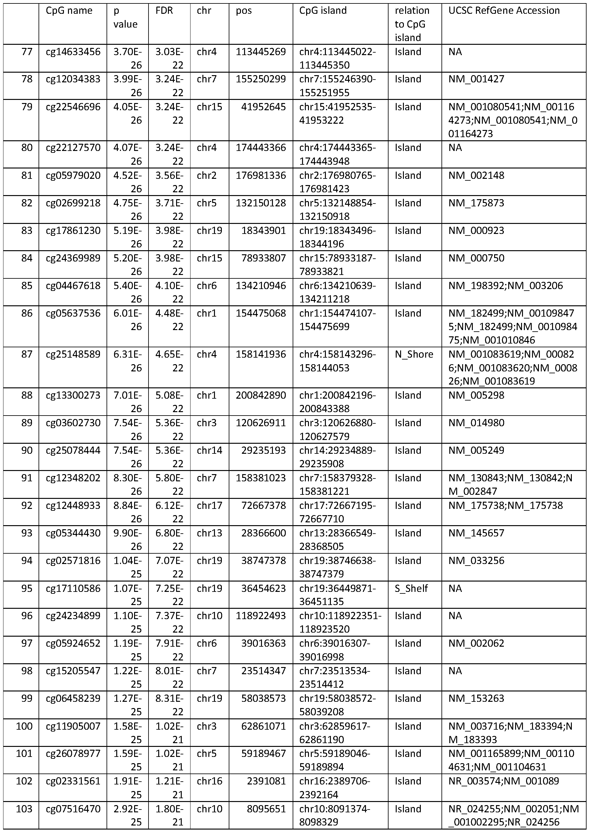


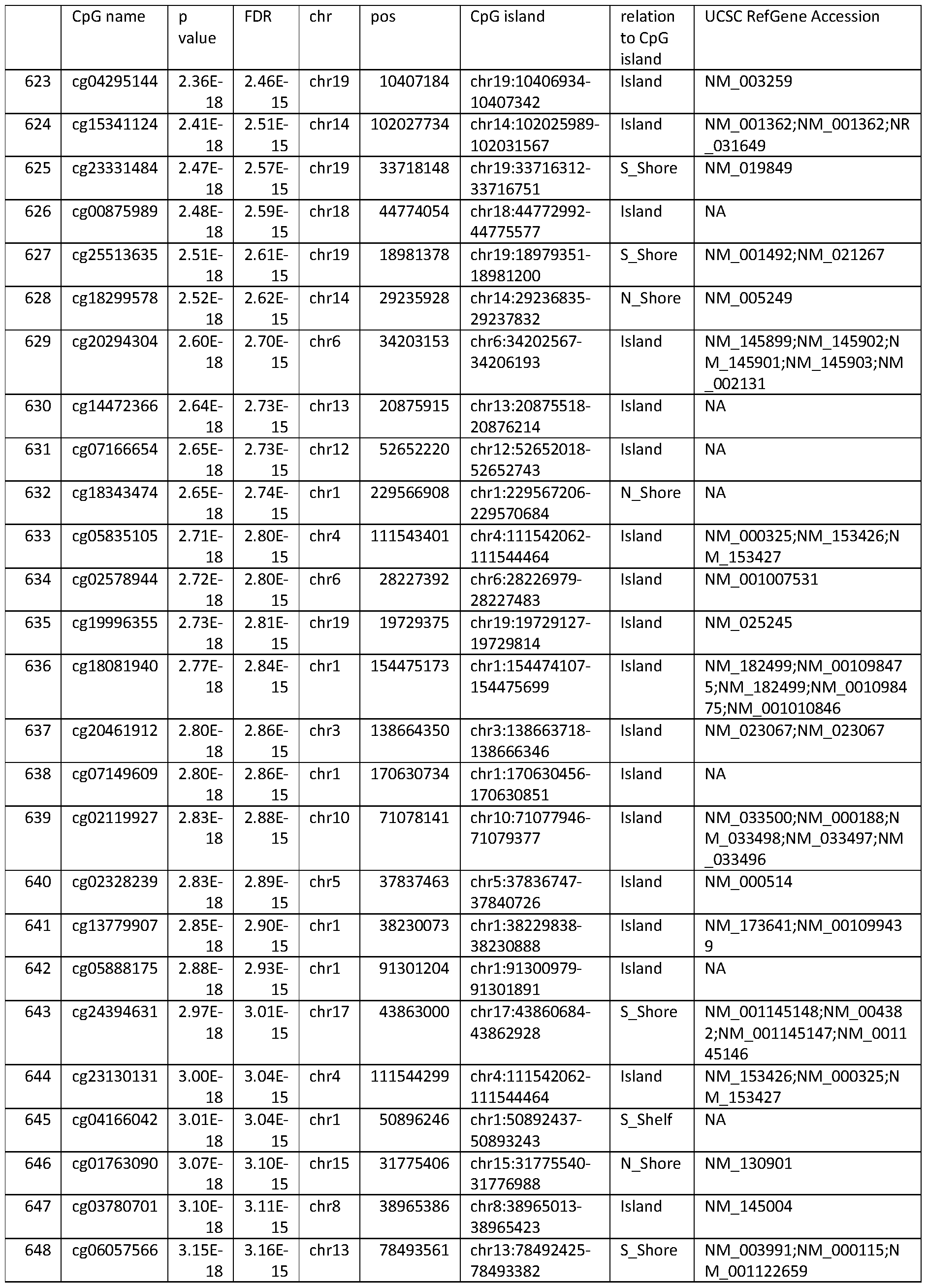


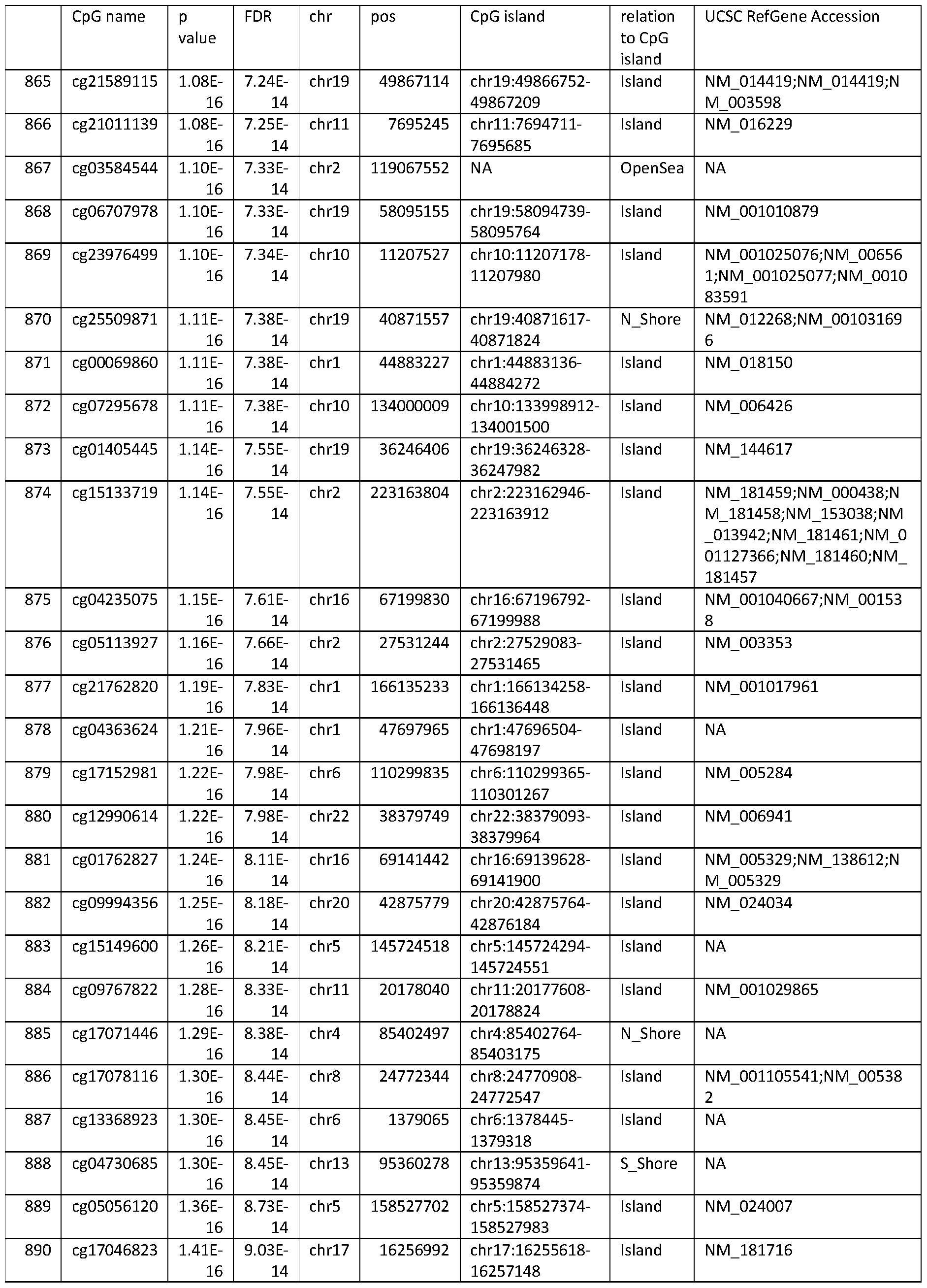


TABLE 4. DNA methylation changes at the genome-wide level in the kidney. Subset of Table 1.


TABLE 5. DNA methylation changes at the genome-wide level in the kidney. Subset of Table 2.


TABLE 6. Subset of Table 3.


Claims
1. A method for detecting the methylation status of a set of age-associated CpGs from a subject, comprising the steps of:
obtaining DNA from a biological sample from a subject;
detecting the methylation status of a set of age-associated CpGs in the DNA of the sample; wherein the set of age-associated CpGs is comprising at least 3 CpGs, wherein the at least 3 CpGs are chosen from the CpGs listed in Table 3 or 6, from the CpGs of the differentially methylated regions listed in Table 2 or 5, and/or from the CpGs listed in Table 1 or 4.
2. A method for predicting the age of a subject, comprising the steps of:
obtaining DNA from a biological sample from a subject;
detecting the methylation status of a set of age-associated CpGs in the DNA of the sample; wherein the set of age-associated CpGs is comprising at least 3 CpGs, wherein the at least 3 CpGs are chosen from the CpGs listed in Table 3 or 6, from the CpGs of the differentially methylated regions listed in Table 2 or 5, and/or from the CpGs listed in Table 1 or 4.
3. The method according to claim 1 or 2 wherein the biological sample is a solid kidney biopsy sample, or is a liquid biopsy sample.
4. The method according to claim 3 wherein the liquid biopsy sample is blood or urine.
5. The method according to any one of claim 1 to 4 wherein the age-associated CpGs have been identified as age-associated CpGs in kidney biopsies.
6. The method according to any one of claims 1 to 5 further comprising predicting the age of the subject by correlating the methylation status detected for the set of age-associated CpGs with a range of pre-determined and age-correlated reference methylation statuses of the same set of age- associated CpGs.
7. Use of a set of age-associated CpGs in a method according to any one of claims 1 to 6, wherein the set of age-associated CpGs is comprising at least 3 CpGs, wherein the at least 3 CpGs are chosen from the CpGs listed in Table 3 or 6, from the CpGs of the differentially methylated regions listed in Table 2 or 5, and/or from the CpGs listed in Table 1 or 4; and wherein the set of age-associated CpGs is comprising at most 1000 CpGs.
8. A kit comprising oligonucleotides to detect the DNA methylation status on a set of age-associated CpGs from a subject, wherein the set of CpGs is comprising at least 3 CpGs, wherein the at least 3 CpGs are chosen from the CpGs listed in Table 3 or 6, from the CpGs of the differentially methylated regions listed in Table 2 or 5, and/or from the CpGs listed in Table 1 or 4; and wherein the kit is comprising oligonucleotides to detect DNA methylation in at most 1000 CpGs.
9. Use of a kit according to claim 8 for predicting the age of a subject from the DNA methylation status detected for the set of age-associated CpGs from the subject.
Applications Claiming Priority (2)
| Application Number | Priority Date | Filing Date | Title |
|---|---|---|---|
| GB1908624.8 | 2019-06-17 | ||
| GBGB1908624.8A GB201908624D0 (en) | 2019-06-17 | 2019-06-17 | Predicting age using dna methylation signatures |
Publications (1)
| Publication Number | Publication Date |
|---|---|
| WO2020254405A1 true WO2020254405A1 (en) | 2020-12-24 |
Family
ID=67253916
Family Applications (1)
| Application Number | Title | Priority Date | Filing Date |
|---|---|---|---|
| PCT/EP2020/066764 Ceased WO2020254405A1 (en) | 2019-06-17 | 2020-06-17 | Predicting age using dna methylation signatures |
Country Status (2)
| Country | Link |
|---|---|
| GB (1) | GB201908624D0 (en) |
| WO (1) | WO2020254405A1 (en) |
Cited By (8)
| Publication number | Priority date | Publication date | Assignee | Title |
|---|---|---|---|---|
| WO2022165247A1 (en) * | 2021-01-29 | 2022-08-04 | Mayo Foundation For Medical Education And Research | Detecting the presence or absence of multiple types of cancer |
| WO2023007165A1 (en) * | 2021-07-28 | 2023-02-02 | Ucl Business Ltd | Methods for assessing epigenetic age and cancer risk |
| WO2024026075A1 (en) * | 2022-07-28 | 2024-02-01 | Grail, Llc | Methylation-based age prediction as feature for cancer classification |
| WO2024254548A1 (en) * | 2023-06-09 | 2024-12-12 | Grail, Llc | Methylation-based biological sex prediction |
| CN120032713A (en) * | 2025-04-22 | 2025-05-23 | 深圳市睿法生物科技有限公司 | A method for constructing a biological age prediction model based on DNA methylation |
| US12319969B2 (en) | 2015-03-27 | 2025-06-03 | Exact Sciences Corporation | Detecting esophageal disorders |
| US12601011B2 (en) | 2015-08-31 | 2026-04-14 | Mayo Foundation For Medical Education And Research | Detecting gastric neoplasm |
| US12606873B2 (en) | 2013-03-14 | 2026-04-21 | Mayo Foundation For Medical Education And Research | Detecting neoplasm |
Citations (13)
| Publication number | Priority date | Publication date | Assignee | Title |
|---|---|---|---|---|
| US5786146A (en) | 1996-06-03 | 1998-07-28 | The Johns Hopkins University School Of Medicine | Method of detection of methylated nucleic acid using agents which modify unmethylated cytosine and distinguishing modified methylated and non-methylated nucleic acids |
| WO2000026401A1 (en) | 1998-11-03 | 2000-05-11 | The Johns Hopkins University School Of Medicine | METHYLATED CpG ISLAND AMPLIFICATION (MCA) |
| WO2004113567A2 (en) | 2003-06-24 | 2004-12-29 | Epigenomics Ag | Improved heavymethyl assay for the methylation analysis of the gstpi gene |
| WO2005038051A2 (en) | 2003-10-09 | 2005-04-28 | Epigenomics Ag | Improved bisulfite conversion of dna |
| US20100240549A1 (en) | 2007-06-22 | 2010-09-23 | The Trustees Of Collumbia University In The City O | Specific amplification of tumor specific dna sequences |
| WO2012067830A2 (en) | 2010-11-15 | 2012-05-24 | Exact Sciences Corporation | Methylation assay |
| WO2014075083A1 (en) | 2012-11-09 | 2014-05-15 | The Regents Of The University Of California | Methods for predicting age and identifying agents that induce or inhibit premature aging |
| WO2017162754A1 (en) | 2016-03-22 | 2017-09-28 | Vib Vzw | Means and methods for amplifying nucleotide sequences |
| WO2018027228A1 (en) | 2016-08-05 | 2018-02-08 | The Regents Of The University Of California | Dna methylation based predictor of mortality |
| WO2018146482A1 (en) | 2017-02-09 | 2018-08-16 | Babraham Institute | Novel age calculation method |
| WO2018150042A1 (en) * | 2017-02-20 | 2018-08-23 | Deutsches Krebsforschungszentrum | Dna methylation signatures for determining a survival probability |
| WO2018229032A1 (en) * | 2017-06-12 | 2018-12-20 | Beiersdorf Aktiengesellschaft | Age determination of human individual |
| WO2019006269A1 (en) | 2017-06-30 | 2019-01-03 | The Regents Of The University Of California | Methods and systems for evaluating dna methylation in cell-free dna |
-
2019
- 2019-06-17 GB GBGB1908624.8A patent/GB201908624D0/en not_active Ceased
-
2020
- 2020-06-17 WO PCT/EP2020/066764 patent/WO2020254405A1/en not_active Ceased
Patent Citations (13)
| Publication number | Priority date | Publication date | Assignee | Title |
|---|---|---|---|---|
| US5786146A (en) | 1996-06-03 | 1998-07-28 | The Johns Hopkins University School Of Medicine | Method of detection of methylated nucleic acid using agents which modify unmethylated cytosine and distinguishing modified methylated and non-methylated nucleic acids |
| WO2000026401A1 (en) | 1998-11-03 | 2000-05-11 | The Johns Hopkins University School Of Medicine | METHYLATED CpG ISLAND AMPLIFICATION (MCA) |
| WO2004113567A2 (en) | 2003-06-24 | 2004-12-29 | Epigenomics Ag | Improved heavymethyl assay for the methylation analysis of the gstpi gene |
| WO2005038051A2 (en) | 2003-10-09 | 2005-04-28 | Epigenomics Ag | Improved bisulfite conversion of dna |
| US20100240549A1 (en) | 2007-06-22 | 2010-09-23 | The Trustees Of Collumbia University In The City O | Specific amplification of tumor specific dna sequences |
| WO2012067830A2 (en) | 2010-11-15 | 2012-05-24 | Exact Sciences Corporation | Methylation assay |
| WO2014075083A1 (en) | 2012-11-09 | 2014-05-15 | The Regents Of The University Of California | Methods for predicting age and identifying agents that induce or inhibit premature aging |
| WO2017162754A1 (en) | 2016-03-22 | 2017-09-28 | Vib Vzw | Means and methods for amplifying nucleotide sequences |
| WO2018027228A1 (en) | 2016-08-05 | 2018-02-08 | The Regents Of The University Of California | Dna methylation based predictor of mortality |
| WO2018146482A1 (en) | 2017-02-09 | 2018-08-16 | Babraham Institute | Novel age calculation method |
| WO2018150042A1 (en) * | 2017-02-20 | 2018-08-23 | Deutsches Krebsforschungszentrum | Dna methylation signatures for determining a survival probability |
| WO2018229032A1 (en) * | 2017-06-12 | 2018-12-20 | Beiersdorf Aktiengesellschaft | Age determination of human individual |
| WO2019006269A1 (en) | 2017-06-30 | 2019-01-03 | The Regents Of The University Of California | Methods and systems for evaluating dna methylation in cell-free dna |
Non-Patent Citations (69)
| Title |
|---|
| AHMAD ET AL., ONCOTARGET, vol. 7, 2016, pages 71833 |
| BACHMAN ET AL., NAT CHEM, vol. 6, 2014, pages 1049 - 1055 |
| BACHMAN ET AL., NATURE CHEM, vol. 6, 2014, pages 1049 - 1055 |
| BACOS ET AL., NAT COMMUN, vol. 7, 2016, pages 11089 |
| BEKAERT ET AL., EPIGENETICS, vol. 10, 2015, pages 922 - 930 |
| BOCKLANDT ET AL., PLOS ONE, vol. 8, 2011, pages e67378 |
| BOOTH ET AL., SCIENCE, vol. 336, 2012, pages 934 - 937 |
| BRACK ET AL., SCIENCE, vol. 317, 2007, pages 807 - 810 |
| COTTRELL ET AL., NUCLEIC ACIDS RES, vol. 32, 2004 |
| EADS ET AL., NUCLEIC ACIDS RES, vol. 28, 2000 |
| EDELING ET AL., NAT REV NEPHROL, vol. 12, 2016, pages 426 - 439 |
| EIPEL ET AL., AGING, vol. 8, 2016, pages 1034 - 1048 |
| FIELD ADAM E ET AL: "DNA Methylation Clocks in Aging: Categories, Causes, and Consequences", MOLECULAR CELL, vol. 71, no. 6, 20 September 2018 (2018-09-20), ELSEVIER, AMSTERDAM, NL, pages 882 - 895, XP085481275, ISSN: 1097-2765, DOI: 10.1016/J.MOLCEL.2018.08.008 * |
| FLORATH ET AL., HUM MOL GENET, vol. 23, 2014, pages 1186 - 1201 |
| FROMMER ET AL., PROC NATL ACAD SCI USA, vol. 89, 1992, pages 1827 - 1831 |
| GARDINER-GARDEN ET AL., J MOL BIOL, vol. 196, 1987, pages 261 - 282 |
| GIULIANI ET AL., AM J PHYS ANTHROPOL, vol. 159, 2016, pages 585 - 595 |
| GONZALGO ET AL., CANCER RESEARCH, vol. 57, 1997, pages 594 - 599 |
| GONZALGOJONES, NUCLEIC ACIDS RES, vol. 25, 1997, pages 2532 - 2534 |
| GRAFSTROM, NUCLEIC ACIDS RES, vol. 13, 1985, pages 2827 - 2842 |
| HANNUM ET AL., MOL CELL, vol. 49, 2013, pages 359 - 367 |
| HERMAN ET AL., PROC NATL ACAD SCI USA, vol. 93, 1996, pages 9821 - 9826 |
| HERNANDEZ ET AL., HUM MOL GENET, vol. 20, 2011, pages 1164 - 1172 |
| HEYLEN ET AL., J AM SOC NEPHROL, vol. 29, 2018, pages 1566 - 1576 |
| HEYLEN LINE ET AL: "FP334 THE RENAL EPIGENETIC CLOCK IS A BETTER SURROGATE FOR KIDNEY AGE THAN TELOMERE ATTRITION", NEPHROLOGY DIALYSIS TRANSPLANTATION, vol. 34, no. Suppl. 1, 1 June 2019 (2019-06-01), & 56TH CONGRESS OF THE EUROPEAN-RENAL-ASSOCIATION (ERA)-EUROPEAN-DIALYSIS-AND-TRANSPLANT-ASSOCIATION (EDTA) - BURDEN, ACCESS AND DISPARITIES IN KIDNEY DISEASE; BUDAPEST, HUNGARY; JUNE 13 -16, 2019, XP002797868, ISSN: 0931-0509, DOI: 10.1093/ndt/gfz106.FP334 * |
| HOMMOS ET AL., J AM SOC NEPHROL, vol. 28, 2017, pages 2838 - 2844 |
| HONG ET AL., FORENSIC SCI INT GENET, vol. 29, 2017, pages 118 - 125 |
| HORVATH, GENOME BIOL, vol. 14, 2013, pages R115 |
| I. FLORATH ET AL: "Cross-sectional and longitudinal changes in DNA methylation with age: an epigenome-wide analysis revealing over 60 novel age-associated CpG sites", HUMAN MOLECULAR GENETICS, vol. 23, no. 5, 26 October 2013 (2013-10-26), pages 1186 - 1201, XP055254769, ISSN: 0964-6906, DOI: 10.1093/hmg/ddt531 * |
| JONES ET AL., AGING CELL, vol. 14, 2015, pages 924 - 932 |
| JUNG ET AL., BMB REP, vol. 50, 2017, pages 546 - 553 |
| JYLHAVA ET AL., EBIOMEDICINE, vol. 21, 2017, pages 29 - 36 |
| KNIGHT ET AL., TRANSPLANTATION, vol. 103, 2019, pages 273 - 283 |
| KURDYUKOVBULLOCK, BIOLOGY, vol. 5, no. 1, 2016, pages E3 |
| L REYNOLDS ET AL: "Age-related variations in the methylome associated with gene expression in human monocytes and T cells - Supplementary Table 1", NATURE COMMUNICATIONS, 18 November 2014 (2014-11-18), XP055428477, Retrieved from the Internet <URL:http://www.nature.com/articles/ncomms6366> [retrieved on 20171124] * |
| L. HEYLEN ET AL: "The Emerging Role of DNA Methylation in Kidney Transplantation: A Perspective", AMERICAN JOURNAL OF TRANSPLANTATION, vol. 16, no. 4, 18 January 2016 (2016-01-18), DK, pages 1070 - 1078, XP055509983, ISSN: 1600-6135, DOI: 10.1111/ajt.13585 * |
| LAIRD ET AL., NAT REV GENET, vol. 11, 2010, pages 191 - 203 |
| LINDENMEYER ET AL., PLOS ONE, vol. 5, 2010, pages ell545 |
| LINDSAY M. REYNOLDS ET AL: "Age-related variations in the methylome associated with gene expression in human monocytes and T cells", NATURE COMMUNICATIONS, vol. 5, 18 November 2014 (2014-11-18), pages 5366, XP055428472, DOI: 10.1038/ncomms6366 * |
| LIU ET AL., NAT BIOTECHNOL, vol. 37, 2019, pages 424 - 429 |
| MARIONI ET AL., GENOME BIOL, vol. 16, 2015, pages 25 |
| MEISSNER ET AL., NUCLEIC ACIDS RES, vol. 33, 2005, pages 5868 - 5877 |
| MOORE, METHODS MOL BIOL, vol. 181, 2001, pages 193 - 201 |
| MORRIS ET AL., BIOINFORMATICS, vol. 30, 2014, pages 428 - 430 |
| NAESENS ET AL., J AM SOC NEPHROL, vol. 27, 2015, pages 281 - 292 |
| NUNES ET AL., CANCERS, vol. 10, 2018, pages 357 |
| NYCE, NUCLEIC ACIDS RES, vol. 14, 1986, pages 4353 - 4367 |
| PARK ET AL., FORENSIC SCI INT GENET, vol. 23, 2016, pages 173 - 179 |
| PEDERSEN ET AL., BIOINFORMATICS, vol. 28, 2012, pages 2986 - 2988 |
| PIDSLEY ET AL., GENOME BIOL, vol. 17, 2016, pages 208 |
| POGGIO ET AL., AM J TRANSPLANT, vol. 6, 2006, pages 100 - 108 |
| RAMSAHOYE, PROC NATL ACAD SCI USA, vol. 97, 2000, pages 5237 - 5242 |
| REYNOLDS ET AL., NAT COMM, vol. 5, 2014, pages 536 |
| REYNOLDS ET AL., NAT COMMUN, vol. 5, 2014, pages 536 |
| RUSSELL ET AL., NAT REV MOL CELL BIOL, vol. 8, 2007, pages 681 - 691 |
| RUTKOWSKA ET AL., ANN TRANSPLANT, vol. 12, 2007, pages 12 - 14 |
| SALMONKAYE, BIOCHIM BIOPHYS ACTA, vol. 204, 1970, pages 340 - 351 |
| SAXONOV ET AL., PNAS, vol. 103, 2006, pages 1412 - 1417 |
| SCHMITTCANTLEY, AM J PHYSIOL-RENAL PHYSIOL, vol. 294, 2008, pages F1265 - F1272 |
| SEN ET AL., CELL, vol. 166, 2016, pages 822 - 839 |
| SIS ET AL., AM J TRANSPLANT, vol. 10, 2010, pages 464 - 471 |
| STEVE HORVATH: "DNA methylation age of human tissues and cell types", GENOME BIOLOGY, BIOMED CENTRAL LTD, vol. 14, no. 10, 21 October 2013 (2013-10-21), pages R115, XP021165700, ISSN: 1465-6906, DOI: 10.1186/GB-2013-14-10-R115 * |
| THIENPONT ET AL., NATURE, vol. 537, 2016, pages 63 - 68 |
| TOYOTA ET AL., CANCER RES, vol. 59, 1999, pages 2307 - 12 |
| WEIDNER ET AL., GENOME BIOL, vol. 15, 2014, pages R24 |
| WILLIAMS ET AL., NATURE, vol. 473, 2011, pages 343 - 348 |
| WOODCOCK, BIOCHEM BIOPHYS RES COMMUN, vol. 145, 1987, pages 888 - 894 |
| YU ET AL., CELL, vol. 149, 2012, pages 1368 - 1380 |
| ZBIEC-PIEKARSKA ET AL., FORENSIC SCI INT GENET, vol. 17, 2015, pages 173 - 179 |
Cited By (8)
| Publication number | Priority date | Publication date | Assignee | Title |
|---|---|---|---|---|
| US12606873B2 (en) | 2013-03-14 | 2026-04-21 | Mayo Foundation For Medical Education And Research | Detecting neoplasm |
| US12319969B2 (en) | 2015-03-27 | 2025-06-03 | Exact Sciences Corporation | Detecting esophageal disorders |
| US12601011B2 (en) | 2015-08-31 | 2026-04-14 | Mayo Foundation For Medical Education And Research | Detecting gastric neoplasm |
| WO2022165247A1 (en) * | 2021-01-29 | 2022-08-04 | Mayo Foundation For Medical Education And Research | Detecting the presence or absence of multiple types of cancer |
| WO2023007165A1 (en) * | 2021-07-28 | 2023-02-02 | Ucl Business Ltd | Methods for assessing epigenetic age and cancer risk |
| WO2024026075A1 (en) * | 2022-07-28 | 2024-02-01 | Grail, Llc | Methylation-based age prediction as feature for cancer classification |
| WO2024254548A1 (en) * | 2023-06-09 | 2024-12-12 | Grail, Llc | Methylation-based biological sex prediction |
| CN120032713A (en) * | 2025-04-22 | 2025-05-23 | 深圳市睿法生物科技有限公司 | A method for constructing a biological age prediction model based on DNA methylation |
Also Published As
| Publication number | Publication date |
|---|---|
| GB201908624D0 (en) | 2019-07-31 |
Similar Documents
| Publication | Publication Date | Title |
|---|---|---|
| JP7757338B2 (en) | Breast cancer detection | |
| WO2020254405A1 (en) | Predicting age using dna methylation signatures | |
| JP7512278B2 (en) | Characterization of methylated DNA, RNA, and proteins in the detection of lung tumors | |
| US11384401B2 (en) | Detecting gastrointestinal neoplasms | |
| AU2020254760B2 (en) | Detecting pancreatic ductal adenocarcinoma in plasma | |
| AU2018229294B2 (en) | Detecting prostate cancer | |
| KR102648965B1 (en) | Hepatocellular carcinoma detection | |
| Godler et al. | Methylation of novel markers of fragile X alleles is inversely correlated with FMRP expression and FMR1 activation ratio | |
| KR20230154433A (en) | Detection of cervical cancer | |
| EP2977467B1 (en) | Method, use of marker, and determination device for obtaining information on plural types of cancers | |
| US20220333198A1 (en) | Predicting Chronic Allograft Injury Through Age-Related DNA Methylation |
Legal Events
| Date | Code | Title | Description |
|---|---|---|---|
| 121 | Ep: the epo has been informed by wipo that ep was designated in this application |
Ref document number: 20732242 Country of ref document: EP Kind code of ref document: A1 |
|
| NENP | Non-entry into the national phase |
Ref country code: DE |
|
| 122 | Ep: pct application non-entry in european phase |
Ref document number: 20732242 Country of ref document: EP Kind code of ref document: A1 |