WO2020239947A1 - Prognosis method of leukemia - Google Patents
Prognosis method of leukemia Download PDFInfo
- Publication number
- WO2020239947A1 WO2020239947A1 PCT/EP2020/064909 EP2020064909W WO2020239947A1 WO 2020239947 A1 WO2020239947 A1 WO 2020239947A1 EP 2020064909 W EP2020064909 W EP 2020064909W WO 2020239947 A1 WO2020239947 A1 WO 2020239947A1
- Authority
- WO
- WIPO (PCT)
- Prior art keywords
- proteins
- seq
- leukemic
- sumoylation
- ubiquitination
- Prior art date
- Legal status (The legal status is an assumption and is not a legal conclusion. Google has not performed a legal analysis and makes no representation as to the accuracy of the status listed.)
- Ceased
Links
Images






Classifications
-
- C—CHEMISTRY; METALLURGY
- C12—BIOCHEMISTRY; BEER; SPIRITS; WINE; VINEGAR; MICROBIOLOGY; ENZYMOLOGY; MUTATION OR GENETIC ENGINEERING
- C12Q—MEASURING OR TESTING PROCESSES INVOLVING ENZYMES, NUCLEIC ACIDS OR MICROORGANISMS; COMPOSITIONS OR TEST PAPERS THEREFOR; PROCESSES OF PREPARING SUCH COMPOSITIONS; CONDITION-RESPONSIVE CONTROL IN MICROBIOLOGICAL OR ENZYMOLOGICAL PROCESSES
- C12Q1/00—Measuring or testing processes involving enzymes, nucleic acids or microorganisms; Compositions therefor; Processes of preparing such compositions
- C12Q1/34—Measuring or testing processes involving enzymes, nucleic acids or microorganisms; Compositions therefor; Processes of preparing such compositions involving hydrolase
-
- G—PHYSICS
- G01—MEASURING; TESTING
- G01N—INVESTIGATING OR ANALYSING MATERIALS BY DETERMINING THEIR CHEMICAL OR PHYSICAL PROPERTIES
- G01N33/00—Investigating or analysing materials by specific methods not covered by groups G01N1/00 - G01N31/00
- G01N33/48—Biological material, e.g. blood, urine; Haemocytometers
- G01N33/50—Chemical analysis of biological material, e.g. blood, urine; Testing involving biospecific ligand binding methods; Immunological testing
- G01N33/53—Immunoassay; Biospecific binding assay; Materials therefor
- G01N33/575—Immunoassay; Biospecific binding assay; Materials therefor for cancer
- G01N33/57505—Immunoassay; Biospecific binding assay; Materials therefor for cancer of the blood, e.g. leukaemia
-
- G—PHYSICS
- G01—MEASURING; TESTING
- G01N—INVESTIGATING OR ANALYSING MATERIALS BY DETERMINING THEIR CHEMICAL OR PHYSICAL PROPERTIES
- G01N33/00—Investigating or analysing materials by specific methods not covered by groups G01N1/00 - G01N31/00
- G01N33/48—Biological material, e.g. blood, urine; Haemocytometers
- G01N33/50—Chemical analysis of biological material, e.g. blood, urine; Testing involving biospecific ligand binding methods; Immunological testing
- G01N33/53—Immunoassay; Biospecific binding assay; Materials therefor
- G01N33/575—Immunoassay; Biospecific binding assay; Materials therefor for cancer
- G01N33/5758—Immunoassay; Biospecific binding assay; Materials therefor for cancer involving compounds serving as markers for tumours, cancers or neoplasias, e.g. cellular determinants, receptors, heat shock/stress proteins, A-protein, oligosaccharides or metabolites
Definitions
- the invention relates to a prognosis of leukemia, in particular a prognosis of resistance to drug that can be used to treat leukemic patients.
- the inventors identified a modifomic signature based on protein post-translational modifications by Ubiquitin and the Ubiquitin-like SUMO polypeptides that may allow rapid discrimination of chemosensitive versus chemoresistant Acute Myeloid Leukemia (AML) at diagnosis.
- AML Acute Myeloid Leukemia
- One aim of the invention is to provide a method that allows the practitioner to determine the best therapy to carry out faced to a patient.
- Another aim of the invention is to provide kits that could help the practitioner’s choice when intending to provide the best therapy to the patients.
- the invention relates to a method for, or of, determining, preferably in vitro , drug-resistance of cells of a leukemic sample obtained from a patient afflicted by a leukemia, said method comprising:
- said at least 4 proteins correspond to the proteins consisting essentially or consisting of the sequences SEQ ID NO: 1 to 4, wherein said 23 proteins correspond to the proteins consisting essentially or consisting of the sequences SEQ ID NO: 1 to 23, and wherein said group of 122 proteins corresponds to the proteins consisting essentially or consisting of the sequences SEQ ID NO: 1 to 122,
- said reference level corresponding to the mean of the level of modification by ubiquitination and/or sumoylation of each at the least 4 proteins of several, possibly independent, leukemic cells that are sensitive to said drug;
- the inventors identified a group of 122 proteins that constitute group of proteins that are modified by ubiquitin/SUMO proteins and said modification being informative regarding the resistance to a drug. More particularly, the information which is important in the variation of the modification of said proteins by SUMO or ubiquitin.
- the group of 122 proteins according to the invention consists of the following proteins:
- DCUN1D1 BC009478.1 IOH12273 DCUN1D1 SEQ ID NO: 8 chromosome 9 open reading frame 71 (C9orf71) NM_153237.1 IOH22672 C9orf71 SEQ ID NO: 9 polymerase (RNA) III (DNA directed) polypeptide K, 12.3 kDa (POLR3K) NM_016310.2 IOH12571 POLR3K SEQ ID NO: 10 ring finger protein 34 (RNF34), transcript variant 2 NM_025126.2 IOH6675 RNF34 SEQ ID NO: 11 zinc finger, MYM-type 5 (ZMYM5) BC007048.1 IOH7230 ZMYM5 SEQ ID NO: 12 arrestin domain containing 1 (ARRDC1) NM_152285.1 IOH21698 ARRDC1 SEQ ID NO: 13 hepatocyte growth factor-regulated tyrosine kinase substrate (HGS) NM_004712.3 IOH4952 HGS SEQ ID NO: 9
- RAD23A NM_005053.2 IOH22983 RAD23A SEQ ID NO: 80 Leukocyte immunoglobulin-like receptor, subfamily A (with TM domain), member 4 (LILRA4), mRNA NM_012276.3 IOH43377 LILRA4 SEQ ID NO: 81 Potassium voltage-gated channel, shaker-related subfamily, member 4 (KCNA4), mRNA NM_002233.1 IOH29649 KCNA4 SEQ ID NO: 82 rhomboid domain containing 1 (RHBDD1) BC027900.1 IOH27306 RHBDD1 SEQ ID NO: 83 RAD18 homolog (S.
- the first step intends to evaluate the modification of at least the proteins consisting of SEQ ID NO: 1, SEQ ID NO: 2, SEQ ID NO: 3 and SEQ ID NO: 4, by ubiquitin or SUMO, or ubiquitin and SUMO proteins, in order to obtain a value of modification by one or two of said Ubiquitin and SUMO proteins.
- Such modifications by ubiquitin or SUMO can be easily evaluated by immunoblotting assays using specific antibodies directed respectively against Ubiquitin or SUMO proteins. These proteins are the markers of the activity of the enzymes involved in the Ubiquitin/Sumo pathways, which are present in the leukemic samples.
- the at the least 4 proteins chosen among the group of 23 proteins, said group of 23 proteins belonging to a set of 122 proteins, are contacted with the leukemic samples such that the deregulation of the Ubiquitin/Sumo pathways in the leukemic sample is detected by variations in the levels of modification by ubiquitination or sumoylation of said at least 23 proteins.
- the proteins which are modified by ubiquitin and/or SUMO proteins will therefore interact with antibodies directed against ubiquitin and/or SUMO proteins to form a molecular complex.
- the complexes can be detected by secondary antibodies that recognize (and interact with) constant part FC chain of said antibodies directed against ubiquitin and/or SUMO proteins.
- Said complex can be identified when the secondary antibodies are labeled with reporter molecule, such as fluorescent protein, peroxidase, fluorescent dyes etc...
- reporter molecule such as fluorescent protein, peroxidase, fluorescent dyes etc...
- Extracts are finally centrifuged twice (16000 g) at 4°C for 20 min and supernatants were aliquoted, flash-frozen and kept at -80 °C until use.
- SUMO-1-, SUMO-2- and Ubiquitin vinyl sulfones (0.5 ⁇ M each) are added to 10 ⁇ L of cellular extracts and incubated at 4 °C for 15 min.
- Beads are washed twice for 5 min with PBS containing 0.05 % Tween-20 and 0.5 % SDS and 3 times for 5 min with PBS containing 0.05 % Tween-20. They are then incubated with 1 ⁇ g/mL of anti-SUMO-1 (21C7) and anti-Flag antibodies for 1 hr under agitation at room temperature. After washing in PBS containing 0.05% Tween-20 for 5 min, they are incubated for 30 min at room temperature with anti-mouse Alexa Fluor 488- and anti-rabbit Alexa Fluor 405 antibodies in 100 ⁇ L of PBS containing 0.05 % Tween-20. Beads are again washed for 5 min with PBS containing 0.05 % Tween-20. They are then resuspended in 200 ⁇ L PBS and flow-cytometry-analysed.
- the detection of the above-mentioned complexes allows to establish a value of modification which corresponds to the Ubiquitin/SUMO modification status of said at least 4 proteins for each leukemic samples of unknown response to said drug.
- step a In order to normalize the value calculated in the step a), it is also calculated the value of the modification by ubiquitin and SUMO of said at least 4 proteins in leukemic cells that are known to be sensitive to said drug. These cells correspond to the reference cells.
- Drug sensitivity of said control cells can be determined experimentally, or is known either by nature (the cells were selected to be sensitive to said drug), or can be obtained from patient afflicted by a leukemia that was successfully treated with said drug.
- Said value can be established for each protein as VRi, wherein i corresponds to the SEQ ID of said at least 4 proteins.
- Ri VCi/VRi, wherein i corresponds to the SEQ ID of said at least 4 proteins.
- the ratio R1, R2, R3 and R4 are calculated. If R1, R2, R3 and R4 are between 0.8 and 1,25, then the leukemic cells are considered to be sensitive to said drug.
- the ratio can represent an increase of the ubiquitin/SUMO modification in the leukemic cells resistant to said drug compared to the control leukemic cells that are sensitive to said drug.
- an increase of the modification by ubiquitin or SUMO can represent the hallmark of resistance.
- the ratio can also represent a decrease of the ubiquitin/SUMO modification in the leukemic cells resistant to said drug compared to the control leukemic cells that are sensitive to said drug.
- a decrease of the modification by ubiquitin or SUMO can represent the hallmark of resistance.
- the invention relates to the method as defined above, wherein
- the inventors identified that the protein as set forth in SEQ ID NO: 1 is only modified by SUMO protein. Therefore, the modification that can be evaluated for this protein corresponds only to an increase or a decrease of the amount of SUMO protein.
- the inventors also identified that the protein as set forth in SEQ ID NO: 2 is only modified by ubiquitin protein. Therefore, the modification that can be evaluated for this protein corresponds only to an increase or a decrease of the amount of ubiquitin protein.
- the invention relates to the method as previously defined, wherein if the modification of the protein as set forth in SEQ ID NO: 2 or 3 or 4 is an ubiquitination, and if the ratio is higher than 1.25, then leukemic sample will be resistant to said drug.
- the proteins as depicted in SEQ ID NO: 2, 3 and 4 are modified by ubiquitin. Therefore, the evaluation of the variation of ubiquitination of at least one of these proteins gives information regarding the resistance to said drug.
- the invention relates to the method as defined above, wherein if the modification of the protein as set forth in SEQ ID NO: 1 or 3 or 4 is a sumoylation, and if the ratio is higher than 1.25, then leukemic sample will be resistant to said drug.
- the invention relates to the method as defined above, comprising a step a) of evaluating, in said leukemic sample, the level of modification by ubiquitination and/or sumoylation of each protein of said group of 23 proteins, said group of 23 proteins belonging to a set of 122 proteins.
- the inventors identified that if a group of 23 proteins is studied, i.e. if the variation of the amount of ubiquitin and/or SUMO of at least said 23 proteins represented by the proteins as set forth in SEQ ID NO: 1 to 23, the evaluation of the drug resistance or sensitivity is higher.
- an advantageous embodiment of the invention relates to a method for, or of, determining, preferably in vitro , drug-resistance of cells of a leukemic sample obtained from a patient afflicted by a leukemia, said method comprising:
- said at least 23 proteins correspond to the proteins consisting essentially or consisting of the sequences SEQ ID NO: 1 to 23, and wherein said group of 122 proteins corresponds to the proteins consisting essentially or consisting of the sequences SEQ ID NO: 1 to 122,
- said reference level corresponding to the mean of the level of modification by ubiquitination and/or sumoylation of each at the least 23 proteins of several, possibly independent, leukemic cells that are sensitive to said drug;
- SEQ ID NO: 2 the proteins consisting essentially or consisting of the respective sequences as set forth in SEQ ID NO: 2, SEQ ID NO: 6, SEQ ID NO: 8, SEQ ID NO: 10, SEQ ID NO: 17 to 19, SEQ ID NO: 21 and SEQ ID NO: 23 are modified by Ubiquitination, and
- SEQ ID NO: 3 and 4 the proteins consisting essentially or consisting of the respective sequences as set forth in SEQ ID NO: 3 and 4, SEQ ID NO: 7, SEQ ID NO: 11, SEQ ID NO: 13, SEQ ID NO: 16 and SEQ ID NO: 22 are modified by Ubiquitination and Sumoylation.
- the invention relates to the method as defined above, comprising a step a) of evaluating, in said leukemic sample, the level of modification by ubiquitination and/or sumoylation of each protein of said set of 122 proteins.
- the inventors identified that the evaluation of ubiquitination/SUMOylation of the 122 proteins as depicted in SEQ ID NO: 1 to 122 gives the best results in determining said drug resistance.
- the invention relates advantageously to a method for, or of, determining, preferably in vitro , drug-resistance of cells of a leukemic sample obtained from a patient afflicted by a leukemia, said method comprising:
- said reference level corresponding to the mean of the level of modification by ubiquitination and/or sumoylation of each 122 proteins of several, possibly independent, leukemic cells that are sensitive to said drug;
- the invention relates to the method as defined above, wherein said drug is chosen from anthracycline and/or cytarabine.
- the invention relates to the method as defined above, wherein said leukemia is an acute myeloid leukemia.
- the invention relates to a method for, or of, the prognosis, preferably in vitro, of a leukemia in a sample of a patient comprising the following steps:
- said at least 4 proteins corresponds to the proteins consisting essentially or consisting of the sequences SEQ ID NO: 1 to 4
- said 23 proteins corresponds to the proteins consisting essentially or consisting of the sequences SEQ ID NO: 1 to 23
- said group of 122 proteins corresponds to the proteins consisting essentially or consisting of the sequences SEQ ID NO: 1 to 122
- the leukemia is an acute myeloid leukemia.
- the invention also relates to a kit comprising:
- the invention relates to the kit as defined above, wherein said determined drug is anthracyclin and/or cytarabine.
- the invention relates advantageously to a kit comprising:
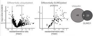
- UbL biomarkers of AML chemoresistance The level of modification of the proteins modified by Ubiquitin (left panel) or SUMO-1 (central panel) selected in C was compared between the parental HL-60 and U937 and their resistant counterparts.
- Right panel Venn diagram with proteins showing a significant p-value in both Wilcoxon signed-rank test and one sample t -test and a ratio between resistant and parental cells higher than 1.25 or lower than 0.8.
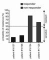
- Figure 11 represents a graph showing the probability of acute myeloid leukemias sensitivity/resistance using a score based on the use of a genetic algorithm and a linear discriminant analysis. This score was used to predict the probability of resistance for the U937 and HL60 cell lines. Cells were considered sensitive to chemotherapy if the probability to belong to the group of resistant cells was below 50% and resistant if over 50%.
- Figure 14 represents the quantification of the modifications.
- background cell extracts supplemented with NEM to inhibit UbL conjugation activities
- the inventors first developed cellular models of resistance to Ara-C or DNR using two reference chemosensitive AML cell lines, HL-60 and U937. Generated Ara-C- (Ara-C-R) and DNR-resistant (DNR-R) sublines showed significantly higher IC 50 ( Figure 1 ). To identify UbL substrate biomarkers of AML chemoresistance, the inventors then resorted to the Protoarrays from Life Technologies, which display more than 9000 recombinant human proteins spotted in duplicate on a nitrocellulose-coated slide. Such arrays have already been used successfully to identify substrates of certain E3 Ubiquitin ligases using either total cell extracts or recombinant enzymes.
- the inventors then identified the proteins, which were differentially modified by the cell extracts from the resistant cell lines compared to the parental ones.
- a first analysis was performed by comparing the data for all Ara-C-R or DNR-R cells with those from parental HL60 and U937 cells. This led to the identification of 52 proteins differentially modified by Ubiquitin and 27 proteins differentially modified by SUMO-1 ( Figure 4 ).
- the inventors then performed a second analysis in which the data for each cell line and each resistance (DNR and Ara-C) were considered separately.
- the inventors selected 23 of the 122 proteins that showed both a high level of modification and the most robust signal differences between sensitive and resistant cell lines ( proteins of sequences SEQ ID NO: 1 to SEQ ID NO: 23 ).
- the inventors used them to generate a UbL score aiming at predicting patients' response to chemotherapy. Using this score, they could predict whether AML cell line were sensitive or resistant to DNR or Ara-C in 16 cases out of 18 tested (6 cell lines, 3 replicates) ( Figure 11 ).
- they resorted to bone marrow aspirates from 4 patients, all of them being of the rather immature M1 subtype in the FAB classification.
- the inventors cloned 15 proteins of the signature (SEQ ID NO: 1, SEQ ID NO: 2, SEQ ID NO: 3, SEQ ID NO: 4, SEQ ID NO: 9, SEQ ID NO: 10, SEQ ID NO: 11, SEQ ID NO: 12, SEQ ID NO: 13, SEQ ID NO: 14, SEQ ID NO: 15, SEQ ID NO: 16, SEQ ID NO: 17, SEQ ID NO: 18 and SEQ ID NO: 19 ) in frame with GST, produced them in bacteria and coupled them to differently colored XMap beads.
- the coupled beads were then multiplexed (up to 10 different beads per tube) and incubated with extracts from parental-, Ara-C-R- or DNR-R HL60 cells before flow cytometry analysis using anti- SUMO-1 or Flag tag to quantify modification by SUMO and Ubiquitin, respectively ( Figure 7 ). If ubiquitylation was detected on all of the proteins tested, this was unfortunately not the case for SUMOylation. A most likely explanation is that Ubiquitin can make long polyUbiquitin chains, facilitating its detection on protein substrates, whereas SUMO cannot.
- the inventors work identifies a new class of modifomic biomarkers that might be used at diagnosis, in combination with other biomarkers and clinical data, to help clinicians predict AML patient response to anthracyclines and cytarabine.
- the assay the inventors have miniaturized was shown adapted to patient sample analysis, paving the way to novel clinical developments.
- HL-60 and U937 cells were cultured at 37 °C in RPMI medium supplemented with 10 % fetal bovine serum (FBS) and streptomycin/penicillin in the presence of 5 % CO 2 . Both cell lines were authenticated by the ATCC using Short-Tandem-Repeat analysis. All cells were regularly tested negative for mycoplasma. After thawing, cells were passaged at a density of 3.10 5 /ml every 2-3 days for no more than 10 passages. U937 and HL-60 cells resistant to Ara-C (cytarabine) and DNR were generated by culture in the presence of increasing drug concentrations (up to 0.1 ⁇ M for Ara-C and 0.03 ⁇ M for DNR) for 2-3 months.
- FBS fetal bovine serum
- Cells were seeded at a concentration of 2.10 5 /mL in RPMI medium complemented with 0.1, 1, 10, 50, 100 or 250 ⁇ M of Ara-C (Sigma-Aldrich) or 0.01, 0.05, 0.1, 0.5, 1 or 10 ⁇ M of daunorubicin (Sigma-Aldrich). Viability was measured 24 hrs later using the MTS assay from Promega following the manufacturer’s protocol. IC 50 were calculated using the GraphPad PRISM software.
- Cells grown at a 5-8.10 5 /mL density were spun down (300 g) at 4 °C for 5 min and washed once with PBS. After pellet resuspension in 1 mL of PBS, they were centrifuged again (16,000 g) at 4 °C for 5 min.
- Pellets were resuspended and incubated at 4 °C for 30 min in a hypotonic buffer (20 mM HEPES pH 7.5, 1.5 mM MgCl 2 , 5 mM KCl, 1 mM DTT and 1 mg/L of aprotinine, leupeptin and pepstatin) in a volume of either (i) 100 ⁇ L per 50.10 6 cells to generate concentrated extracts used for ProtoArray probing or (ii) 25 ⁇ L per 2.10 6 cells in the case of extracts used in XMap bead-based flow cytometry assays.
- a hypotonic buffer (20 mM HEPES pH 7.5, 1.5 mM MgCl 2 , 5 mM KCl, 1 mM DTT and 1 mg/L of aprotinine, leupeptin and pepstatin
- Cell lysis was achieved through 4 freezing/thawing cycles using liquid nitrogen and DNA was sheared owing to 10 passages through a 20-1/2G needle. Extracts were finally centrifuged twice (16000 g) at 4°C for 20 min and supernatants were aliquoted, flash-frozen and kept at -80 °C until use.
- the extracts were then supplemented with home-made recombinant SUMO-1 and SUMO-2 (15 ⁇ M, produced as previously described), 30 ⁇ M Flag-ubiquitin (Boston Biochem), 15 ⁇ M NEDD8 (Boston Biochem), 0.1% Tween-20, and 1 mM ATP Immediately after, they were laid on ProtoArray slides, which were covered with a coverslip and incubated at 30 °C for 1 hr in a humidified atmosphere. Arrays were washed 3 times for 5 min with a washing buffer (PBS pH 7.4, 0.1 % Tween 20, 1X Synthetic Block) supplemented with 0.5 % SDS and, then, twice for 5 min with only the washing buffer.
- a washing buffer PBS pH 7.4, 0.1 % Tween 20, 1X Synthetic Block
- Arrays were washed 5 times for 5 min with the washing buffer, once with H 2 O and, finally, dried by centrifugation before fluorescence scanning using the Innoscan 710 device from Innopsys. The arrays were then stripped with 200 mM Glycine, 0.5% SDS, pH 2.2 for 20 min and probed again with anti-SUMO-2 (8A2 from the Developmental Studies Hybridoma Bank) and anti-Nedd8 (Y297, Abcam) antibodies. However, some signals were remaining from the first hybridization. To avoid false positive, the inventors thus decided to consider only the signals obtained for SUMO-1 and Ubiquitin.
- Measured fluorescence intensities were associated to the corresponding protein ID according to their coordinates on the arrays using the Mapix software (Innopsys). Intensities were processed using the PAA R package. In brief, (i) duplicated protein spot intensities were averaged using the Load GPR function and (ii) the background was corrected using the Background Correct function. Fluorescence intensities within the same experiment were normalized by quantiles using the Normalize Array function. Finally, intensities were normalized between the different experiments using the Batch Adjust function.
- the inventors To identify the proteins modified by Ubiquitin or SUMO-1 on the ProtoArrays, the inventors first had to filter proteins proteins displaying signal significantly higher in UbL conjugation-permissive- than those in UbL conjugation non-permissive conjugation conditions (i.e. control conditions using NEM-treated extracts). With regard to the statistical test adapted to the exploitation of the inventors’ results, the classical Student t-test was not recommended, as variances could be very different between UbL conjugation permissive and non-permissive conditions. The inventors therefore selected proteins with significant p-values (lower than 0.05) in both the parametrical Welch- and the non-parametrical Wilcoxon-Mann-Whitney (WMW) test. Then, proteins having an averaged normalized fluorescence intensity value less than 800 were filtered out in order to obtain a list of robustly modified proteins.
- WMW non-parametrical Wilcoxon-Mann-Whitney
- the inventors decided to work on ratios between the parental and drug-resistant cell samples for each protein.
- the inventors thus compared, for each of the modified proteins (NEM-filtering), the ratios between sensitive and resistant cells using both a Wilcoxon signed-rank test and a one sample t -test.
- This analysis was first performed on all arrays (both cell lines and drugs combined). In this case, the sample size being large, the inventors considered as differentially modified all proteins showing p-values ⁇ 0.05 in both tests.
- the sample size being smaller, the inventors considered as differentially modified the proteins showing p-values ⁇ 0.05 in the one sample t-test.
- cDNA encoding for the proteins of interest were recovered from the Ultimate ORF library (Thermofisher) and cloned in the bacterial expression vector pGGWA vector using the Gateway technology according to manufacturer’s protocol (Life Technologies). Constructs were then transformed in BL21 (DE3) E. coli strain. Protein production was induced with 1 mM isopropyl ⁇ -D-1-thiogalactopyranoside (IPTG) for 6 hrs in exponentially growing bacteria at 25°C.
- IPTG isopropyl ⁇ -D-1-thiogalactopyranoside
- Bacterial pellets were resuspended in 50 mM Tris-HCl pH 8.6 containing 500 mM NaCl and 50 mM MgSO 4 , and flash-frozen in liquid N 2 . After thawing, bacterial suspensions were supplemented with 1 mg/mL lysozyme (Sigma-Aldrich), 8 mM ⁇ -mercaptoethanol, 1 mg/L aprotinin, leupeptin and pepstatin and incubated at 4°C for 1 hr. Bacterial debris were spun down (100 000 g for 1 hr).
- the extract was then bound to Glutathion agarose beads (Generon) equilibrated in Tris 50 mM pH 8.6, NaCl 500 mM, MgSO4 50 mM, 8 mM ⁇ -mercaptoethanol, 1 mg/L aprotinin, leupeptin, pepstatin.
- the column was then extensively washed with Tris 50 mM pH 8.6, NaCl 150 mM, MgSO4 50 mM, 8 mM ⁇ -mercaptoethanol, 1 mg/L aprotinin, leupeptin, pepstatin and eluted by addition of 20 mM reduced glutathione (Sigma-Aldrich).
- Beads were then washed in PBS containing 500 mM NaCl and incubated with 7 ⁇ g of recombinant protein to be coupled in 100 ⁇ L PBS at room temperature for 2 hrs. They were then washed twice with PBS containing 0.1 % BSA, 0.02 % Tween 20, 0.05 % sodium azide and 500 mM NaCland stored at 4 °C in PBS containing 0.1 % BSA, 0.02 % Tween-20, 0.05 % sodium azide.
- SUMO-1-, SUMO-2- and Ubiquitin vinyl sulfones (0.5 ⁇ M each) were added to diluted cellular extracts (10 ⁇ L), which were incubated at 4 °C for 15 min. Control extracts were also incubated with 50 mM NEM.
- the inventors then added to the extract 10 3 protein-coupled XMap beads contained in 10 ⁇ L of a reaction buffer containing 20 mM HEPES pH 7.3, 110 mM KOAc, 2 mM Mg(OAc) 2 , 0.05 % Tween-20, 0.5 mM EGTA, 0.2 mg/mL ovalbumine, 1 mM DTT, 1 mg/L aprotinin, leupeptin and pepstatin, 1 mM ATP, 30 ⁇ M Flag-ubiquitin, 15 ⁇ M SUMO-1 and 15 ⁇ M SUMO-2. Reaction were performed at 30 °C for 45 min.
- Beads were washed twice for 5 min with PBS containing 0.05 % Tween-20 and 0.5 % SDS and 3 times for 5 min with PBS containing 0.05 % Tween-20. They were then incubated with 1 ⁇ g/mL of anti-SUMO-1 (21C7) and anti-Flag antibodies for 1 hr under agitation at room temperature. After washing in PBS containing 0.05% Tween-20 for 5 min, they were incubated for 30 min at room temperature with anti-mouse Alexa Fluor 488- and anti-rabbit Alexa Fluor 405 antibodies in 100 ⁇ L of PBS containing 0.05 % Tween-20.
- Age* Age at diagnosis
- Blasts* Blasts at diagnosis in bone marrow
- Blasts** Blasts 30 days after diagnosis in bone marrow
Landscapes
- Health & Medical Sciences (AREA)
- Life Sciences & Earth Sciences (AREA)
- Engineering & Computer Science (AREA)
- Chemical & Material Sciences (AREA)
- Immunology (AREA)
- Molecular Biology (AREA)
- Biomedical Technology (AREA)
- Hematology (AREA)
- Urology & Nephrology (AREA)
- Physics & Mathematics (AREA)
- Organic Chemistry (AREA)
- Microbiology (AREA)
- General Health & Medical Sciences (AREA)
- Biochemistry (AREA)
- Biotechnology (AREA)
- Analytical Chemistry (AREA)
- Cell Biology (AREA)
- Food Science & Technology (AREA)
- General Physics & Mathematics (AREA)
- Pathology (AREA)
- Proteomics, Peptides & Aminoacids (AREA)
- Medicinal Chemistry (AREA)
- Wood Science & Technology (AREA)
- Zoology (AREA)
- Biophysics (AREA)
- Bioinformatics & Cheminformatics (AREA)
- General Engineering & Computer Science (AREA)
- Genetics & Genomics (AREA)
- Measuring Or Testing Involving Enzymes Or Micro-Organisms (AREA)
Abstract
The invention relates to a method for determining in vitro drug-resistance of cells of a leukemic sample, said method comprising, a step of evaluating, in cells of said leukemic sample, the level of modification by ubiquitination or sumoylation of each protein of at the least 4 proteins chosen among a group of 23 proteins, said group of 23 proteins belonging to a set of 122 proteins.
Description
The invention relates to a prognosis of leukemia, in particular a prognosis of resistance to drug that can be used to treat leukemic patients.
Being able to predict cancer response to therapies is critical to increase response rates and limit their toxicity and, when necessary, change them.
In this context, the inventors identified a modifomic signature based on protein post-translational modifications by Ubiquitin and the Ubiquitin-like SUMO polypeptides that may allow rapid discrimination of chemosensitive versus chemoresistant Acute Myeloid Leukemia (AML) at diagnosis.
Most AML patients first receive an induction chemotherapy that has not significantly changed for the past 40 years (Dombret
H,
Gardin
C. Blood 2016; 127: 53–61). It consists of two genotoxics, one anthracyclin (daunorubicin-DNR or idarubicin-IDA) and cytarabine (Ara-C). Unfortunately, a significant fraction of patients (20-30%) do not respond to this treatment and, among those achieving complete remission, relapse rates are high (5-years survival of 40% in patients below 60 and 20% in older ones - Estey
EH. Am J
Hematol
2012; 87: 89–99.).
To date, no prognosis tool implementable at diagnosis is available to predict AML response to chemotherapies. This is detrimental for at least two reasons: (i) unnecessary treatment of chemoresistant patients with antracyclins that have life-threatening cardiotoxic effects and (ii) loss of time before redirecting patients to novel therapies (FLT3, IDH1/2, Blc2 inhibitors…) and/or enrolling them in clinical trials for new molecules.
Transcriptomic signatures are now used to better stratify patients and adapt treatments in several cancers. However, even though various transcriptomic signatures have been defined for AMLs, none of them is sufficiently reliable to be used in clinical practice. Interestingly, proteomic signatures, in particular those based on mass spectrometry, are emerging as promising alternatives. Even closer to the biological functions deregulated in cancer, « modifomic » analyses, which monitor the activities of enzymes involved in the post-translational modifications of proteins, open new perspectives in cancer prognosis/diagnosis.
So there is a need to provide a prediction of cancer response to therapies, in order to ameliorate patient outcome, and to avoid the drawbacks known in the art.
One aim of the invention is to provide a method that allows the practitioner to determine the best therapy to carry out faced to a patient.
Another aim of the invention is to provide kits that could help the practitioner’s choice when intending to provide the best therapy to the patients.
For this purpose, the invention relates to a method for, or of, determining, preferably in vitro, drug-resistance of cells of a leukemic sample obtained from a patient afflicted by a leukemia, said method comprising:
- a step of evaluating, in cells of said leukemic sample, the level of modification by ubiquitination or sumoylation of each protein of at the least 4 proteins chosen among a group of 23 proteins, said group of 23 proteins belonging to a set of 122 proteins;
wherein said at least 4 proteins correspond to the proteins consisting essentially or consisting of the sequences SEQ ID NO: 1 to 4, wherein said 23 proteins correspond to the proteins consisting essentially or consisting of the sequences SEQ ID NO: 1 to 23, and wherein said group of 122 proteins corresponds to the proteins consisting essentially or consisting of the sequences SEQ ID NO: 1 to 122,
- a step of comparing the level of modification by ubiquitination and/or sumoylation of each said at the least 4 proteins of cells in said leukemic sample corresponding to a reference level, said reference level being the level of modification by ubiquitination and/or sumoylation of each at the least 4 proteins of leukemic cells that are sensitive to said drug in order to obtain a ratio of the level in the leukemic sample compared to the reference level of a sample of sensitive cells for each of said at least 4 proteins
said reference level corresponding to the mean of the level of modification by ubiquitination and/or sumoylation of each at the least 4 proteins of several, possibly independent, leukemic cells that are sensitive to said drug; and
- a step of determining that
- if the ratio for at least one of said 4 proteins is higher than 1.25 or lower than 0.8, then leukemic sample will be resistant to said drug, and
- if the ratio for each of said at least 4 proteins is comprised from 0.8 to 1.25, then leukemic sample will be sensitive to said drug.
The invention is based on the unexpected observation made by the inventors that the evaluation of modifications by the enzymes belonging to the ubiquitin/Sumo pathways of a specific and determined group of 122 proteins, and in particular at least 4 of them, gives significant information regarding a resistance of a tumor to a determined drug.
As exposed in the Example below, the inventors identified a group of 122 proteins that constitute group of proteins that are modified by ubiquitin/SUMO proteins and said modification being informative regarding the resistance to a drug. More particularly, the information which is important in the variation of the modification of said proteins by SUMO or ubiquitin.
The group of 122 proteins according to the invention consists of the following proteins:
| Name |
Accession
.Number |
Ulitmate
.
ORF.ID. .. Catalog. Number |
Gene.
Symbol |
SEQ ID |
| ataxin 3 (ATXN3) | BC033711.1 | IOH21762 | ATXN3 | SEQI ID NO: 1 |
| sequestosome 1 (SQSTM1) | BC001874.1 | IOH4836 | SQSTM1 | SEQ ID NO: 2 |
| UBA domain containing 1 (UBADC1) | BC004967.1 | IOH4830 | UBADC1 | SEQ ID NO: 3 |
| signal transducing adaptor molecule (SH3 domain and ITAM motif) 1 (STAM) | BC030586.2 | IOH22241 | STAM | SEQ ID NO: 4 |
| Protein BEAN | XM_375359.1 | IOH43383 | BEAN | SEQ ID NO: 5 |
| Optineurin | NM_001008211.1 | IOH57143 | OPTN | SEQ ID NO: 6 |
| Nuclear |
BC025930.1 | IOH57231 | NPLOC4 | SEQ ID NO: 7 |
| DCN1, defective in |
BC009478.1 | IOH12273 | DCUN1D1 | SEQ ID NO: 8 |
| |
NM_153237.1 | IOH22672 | C9orf71 | SEQ ID NO: 9 |
| polymerase (RNA) III (DNA directed) polypeptide K, 12.3 kDa (POLR3K) | NM_016310.2 | IOH12571 | POLR3K | SEQ ID NO: 10 |
| ring finger protein 34 (RNF34), |
NM_025126.2 | IOH6675 | RNF34 | SEQ ID NO: 11 |
| zinc finger, MYM-type 5 (ZMYM5) | BC007048.1 | IOH7230 | ZMYM5 | SEQ ID NO: 12 |
| arrestin domain containing 1 (ARRDC1) | NM_152285.1 | IOH21698 | ARRDC1 | SEQ ID NO: 13 |
| hepatocyte growth factor-regulated tyrosine kinase substrate (HGS) | NM_004712.3 | IOH4952 | HGS | SEQ ID NO: 14 |
| tripartite motif-containing 39 (TRIM39), |
NM_172016.1 | IOH5913 | TRIM39 | SEQ ID NO: 15 |
| DnaJ (Hsp40) homolog, subfamily B, member 2 (DNAJB2) | BC047056.1 | IOH27319 | DNAJB2 | SEQ ID NO: 16 |
| phospholipid scramblase 1 (PLSCR1) | NM_021105.1 | IOH13995 | PLSCR1 | SEQ ID NO: 17 |
| huntingtin interacting protein 2 (HIP2) | NM_005339.3 | IOH13913 | HIP2 | SEQ ID NO: 18 |
| NIMA (never in mitosis gene a)- related kinase 10 (NEK10) | BC045758.1 | IOH26432 | NEK10 | SEQ ID NO: 19 |
| ring finger protein 11 (RNF11) | NM_014372.1 | IOH11235 | RNF11 | SEQ ID NO: 20 |
| Isocitrate dehydrogenase [NADP] cytoplasmic | NM_005896.2 | IOH11942 | IDH1 | SEQ ID NO: 21 |
| Ubiquitin-conjugating enzyme E2 D2 | NM_003339.2 | IOH58973 | UBE2D2 | SEQ ID NO: 22 |
| FAS-associated |
NM_007051.2 | IOH5135 | FAF1 | SEQ ID NO: 23 |
| ubiquitin-activating enzyme E1 (UBE1), |
NM_003334.2 | IOH22897 | UBE1 | SEQ ID NO: 24 |
| C-C motif chemokine 21 | NM_002989.2 | PHC1475 | 6CKINE | SEQ ID NO: 25 |
| kinesin family member 3C (KIF3C) | NM_002254.5 | IOH42589 | KIF3C | SEQ ID NO: 26 |
| HSPB (heat shock 27kDa) associated protein 1 (HSPBAP1) | NM_024610.2 | IOH13320 | HSPBAP1 | SEQ ID NO: 27 |
| |
BC023982.1 | IOH14039 | C5orf32 | SEQ ID NO: 28 |
| SMAD family member 1 (SMAD1), |
NM_005900.1 | IOH4970 | SMAD1 | SEQ ID NO: 29 |
| lysozyme-like 1 (LYZL1) | NM_032517.3 | IOH10985 | LYZL1 | SEQ ID NO: 30 |
| Uncharacterized protein C4orf32 | BC022534.1 | IOH10832 | C4orf32 | SEQ ID NO: 31 |
| SCO cytochrome oxidase deficient homolog 1 (yeast) (SCO1), nuclear gene encoding mitochondrial protein | NM_004589.1 | IOH10823 | SCO1 | SEQ ID NO: 32 |
| tetraspanin 9 (TSPAN9) | NM_031285.1 | IOH22841 | TSPAN9 | SEQ ID NO: 33 |
| OPN / SPP1 Protein (His Tag) | NP_001035147.1 | 10352-H08H-25 | SPP1 | SEQ ID NO: 34 |
| Immunoglobulin iota chain | BC017922.1 | IOH12726 | VPREB1 | SEQ ID NO: 35 |
| Uncharacterized serine/threonine-protein kinase SgK494 | NM_144610.1 | IOH60533 | FLJ25006 | SEQ ID NO: 36 |
| tripartite motif-containing 52 (TRIM52) | NM_032765.1 | IOH7015 | TRIM52 | SEQ ID NO: 37 |
| Thyroid hormone receptor beta | NM_000461.3 | IOH29857 | LBD | SEQ ID NO: 38 |
| UBX domain containing 8 (UBXD8) | NM_014613.1 | IOH40633 | UBXD8 | SEQ ID NO: 39 |
| protein phosphatase 2A, regulatory subunit B' (PR 53) (PPP2R4), |
NM_178002.1 | IOH45891 | PPP2R4 | SEQ ID NO: 40 |
| glycine-N-acyltransferase-like 2 (GLYATL2) | BC016789.1 | IOH13602 | GLYATL2 | SEQ ID NO: 41 |
| Transcriptional adapter 2-alpha | BC001172.1 | IOH4467 | TADA2L | SEQ ID NO: 42 |
| phosphoserine aminotransferase 1 (PSAT1), |
NM_058179.1 | IOH4946 | PSAT1 | SEQ ID NO: 43 |
| hypothetical protein MGC52498 (MGC52498) | NM_182621.1 | IOH28035 | MGC52498 | SEQ ID NO: 44 |
| AKT interacting protein (AKTIP), |
NM_022476.1 | IOH3844 | AKTIP | SEQ ID NO: 45 |
| ubiquitin-conjugating enzyme E2D 1 (UBC4/5 homolog, yeast) (UBE2D1) | NM_003338.2 | IOH7482 | UBE2D1 | SEQ ID NO: 46 |
| Ubiquitin-like protein 7 | NM_032907.3 | IOH56973 | UBL7 | SEQ ID NO: 47 |
| RIO kinase 3 (yeast) (RIOK3) | NM_003831.1 | IOH20968 | RIOK3 | SEQ ID NO: 48 |
| phospholipase A2, group IVD (cytosolic) (PLA2G4D) | NM_178034.1 | IOH21503 | PLA2G4D | SEQ ID NO: 49 |
| glycoprotein, synaptic 2 (GPSN2) | NM_138501.3 | IOH4169 | GPSN2 | SEQ ID NO: 50 |
| capping protein (actin filament) muscle Z-line, beta (CAPZB) | BC008095.1 | IOH3312 | CAPZB | SEQ ID NO: 51 |
| Ubiquitin-conjugating enzyme E2 D3 | BC066917.1 | IOH61482 | UBE2D3 | SEQ ID NO: 52 |
| OTU domain, ubiquitin aldehyde binding 2 (OTUB2) | BC000208.1 | IOH4373 | OTUB2 | SEQ ID NO: 53 |
| polymerase (RNA) II (DNA directed) polypeptide H (POLR2H) | NM_006232.2 | IOH4626 | POLR2H | SEQ ID NO: 54 |
| ubiquilin 1 (UBQLN1), |
NM_053067.1 | IOH40632 | UBQLN1 | SEQ ID NO: 55 |
| GRIP1-associated |
NM_020137.3 | IOH52884 | GRIPAP1 | SEQ ID NO: 56 |
| ribosomal protein S17 (RPS17) | NM_001021.2 | IOH27847 | RPS17 | SEQ ID NO: 57 |
| ubiquitin-conjugating enzyme E2 variant 1 (UBE2V1) | BC000468.1 | IOH3618 | UBE2V1 | SEQ ID NO: 58 |
| adaptor protein, phosphotyrosine interaction, PH domain and leucine zipper containing 1 (APPL1) | NM_012096.1 | IOH11629 | APPL1 | SEQ ID NO: 59 |
| MOB1, Mps One Binder kinase activator-like 2C (yeast) (MOBKL2C), |
NM_201403.1 | IOH42184 | MOBKL2C | SEQ ID NO: 60 |
| UBX domain containing 1 (UBXD1) | NM_025241.1 | IOH41143 | UBXD1 | SEQ ID NO: 61 |
| ubiquitin-conjugating enzyme E2E 2 (UBC4/5 homolog, yeast) (UBE2E2) | NM_152653.1 | IOH13176 | UBE2E2 | SEQ ID NO: 62 |
| ankyrin repeat domain 37 (ANKRD37) | NM_181726.1 | IOH43299 | ANKRD37 | SEQ ID NO: 63 |
| |
NM_000690.1 | IOH4924 | ALDH2 | SEQ ID NO: 64 |
| lysozyme-like 2 (LYZL2) | BC066294.1 | IOH40058 | LYZL2 | SEQ ID NO: 65 |
| Uncharacterized protein C12orf65 | BC020885.1 | IOH14357 | C12orf65 | SEQ ID NO: 66 |
| tetraspanin 1 (TSPAN1) | NM_005727.2 | IOH6907 | TSPAN1 | SEQ ID NO: 67 |
| spleen tyrosine kinase (SYK) | NM_003177.3 | PV3857 | SYK | SEQ ID NO: 68 |
| WD repeat domain 88 (WDR88) | BC031227.1 | IOH21600 | WDR88 | SEQ ID NO: 69 |
| |
BC053733.1 | IOH28787 | C2orf34 | SEQ ID NO: 70 |
| EP300-interacting inhibitor of |
BC027612.2 | IOH11844 | EID3 | SEQ ID NO: 71 |
| cysteine and histidine-rich domain (CHORD)-containing 1 (CHORDC1) | BC017789.1 | IOH13662 | CHORDC1 | SEQ ID NO: 72 |
| acyl-CoA synthetase medium-chain family member 3 (ACSM3), |
NM_005622.3 | IOH42133 | ACSM3 | SEQ ID NO: 73 |
| ubiquitin-conjugating enzyme E2W (putative) (UBE2W) | BC010900.1 | IOH12898 | UBE2W | SEQ ID NO: 74 |
| PREDICTED: Homo sapiens hypothetical protein LOC152024 (LOC152024) | XM_379183.1 | IOH43202 | LOC152024 | SEQ ID NO: 75 |
| Gap junction beta-2 protein | BC071703.1 | IOH61781 | GJB2 | SEQ ID NO: 76 |
| Exocyst complex component 5 (EXOC5), mRNA | NM_006544.3 | IOH55643 | EXOC5 | SEQ ID NO: 77 |
| |
BC009016.1 | IOH3326 | C1QC | SEQ ID NO: 78 |
| ankyrin repeat and sterile alpha motif domain containing 4B (ANKS4B) | NM_145865.1 | IOH35802 | ANKS4B | SEQ ID NO: 79 |
| RAD23 homolog A (S. cerevisiae) (RAD23A) | NM_005053.2 | IOH22983 | RAD23A | SEQ ID NO: 80 |
| Leukocyte immunoglobulin-like receptor, subfamily A (with TM domain), member 4 (LILRA4), mRNA | NM_012276.3 | IOH43377 | LILRA4 | SEQ ID NO: 81 |
| Potassium voltage-gated channel, shaker-related subfamily, member 4 (KCNA4), mRNA | NM_002233.1 | IOH29649 | KCNA4 | SEQ ID NO: 82 |
| rhomboid domain containing 1 (RHBDD1) | BC027900.1 | IOH27306 | RHBDD1 | SEQ ID NO: 83 |
| RAD18 homolog (S. cerevisiae) (RAD18) | NM_020165.2 | IOH3060 | RAD18 | SEQ ID NO: 84 |
| inversin (INVS) | BC006370.2 | IOH6467 | INVS | SEQ ID NO: 85 |
| |
NM_024294.1 | IOH5168 | C6orf106 | SEQ ID NO: 86 |
| Cbl-interacting protein Sts-1 (STS-1) | NM_032873.3 | IOH6812 | STS-1 | SEQ ID NO: 87 |
| Rho GTPase activating protein 4 (ARHGAP4) | BC052303.1 | IOH28113 | ARHGAP4 | SEQ ID NO: 88 |
| |
NM_005319.3 | IOH5275 | HIST1H1C | SEQ ID NO: 89 |
| chromosome 13 open reading frame 24 (C13orf24) | BC051911.1 | IOH27047 | C13orf24 | SEQ ID NO: 90 |
| myosin IXA (MYO9A) | BC060886.1 | IOH29219 | MYO9A | SEQ ID NO: 91 |
| Cell division cycle 2- |
NM_003718.2 | PMX_10184 | CDC2L5 | SEQ ID NO: 92 |
| Putative phosphatidylinositol 4-kinase alpha-like protein P2 | BC020225.1 | IOH13323 | LOC220686 | SEQ ID NO: 93 |
| Inhibitor of nuclear factor kappa-B kinase subunit epsilon | NM_014002.1 | -- | IKBKE | SEQ ID NO: 94 |
| Myosin |
BC002778.1 | IOH5313 | MYLC2PL | SEQ ID NO: 95 |
| splicing factor 3b, |
BC007610.1 | IOH6911 | SF3B2 | SEQ ID NO: 96 |
| parathymosin (PTMS) | BC017025.1 | IOH9960 | PTMS | SEQ ID NO: 97 |
| Follistatin (FST), transcript variant FST317, mRNA | NM_006350.2 | IOH59070 | FST | SEQ ID NO: 98 |
| a serine/threonine kinase | NM_004958.2 | PV4753 | FRAP1 (mTOR) | SEQ ID NO: 99 |
| similar to 60S ribosomal protein L21 (LOC402176) | NM_001011538.1 | IOH39826 | LOC402176 | SEQ ID NO: 100 |
| SDF1B Recombinant Human Protein | NP_000600.1 | PHC1361 | SDF1B | SEQ ID NO: 101 |
| SUMO1 activating enzyme subunit 2 (SAE2) | NM_005499.2 | IOH5085 | SAE2 | SEQ ID NO: 102 |
| Transmembrane protein 207 | NM_207316.1 | IOH45939 | UNQ846 | SEQ ID NO: 103 |
| Chromosome 18 open reading frame 1 (C18orf1), transcript variant c2, mRNA | NM_001003675.1 | IOH53461 | LDLRAD4 | SEQ ID NO: 104 |
| ubiquitin specific peptidase 5 (isopeptidase T) (USP5) | BC005139.2 | IOH4709 | USP5 | SEQ ID NO: 105 |
| Prolyl 4-hydroxylase subunit alpha-2 | NM_001017973.1 | IOH26049 | P4HA2 | SEQ ID NO: 106 |
| |
BC070129.1 | IOH63021 | TPI1 | SEQ ID NO: 107 |
| Rho-related BTB domain containing 1 (RHOBTB1), |
NM_014836.3 | IOH27118 | RHOBTB1 | SEQ ID NO: 108 |
| KIAA0247 (KIAA0247) | NM_014734.2 | IOH39813 | KIAA0247 | SEQ ID NO: 109 |
| transmembrane protein 174 (TMEM174) | NM_153217.1 | IOH13241 | TMEM174 | SEQ ID NO: 110 |
| zinc finger, AN1-type domain 5 (ZFAND5), transcript variant c | NM_006007.1 | IOH14289 | ZFAND5 | SEQ ID NO: 111 |
| ring finger protein 128 (RNF128), |
NM_194463.1 | IOH39866 | RNF128 | SEQ ID NO: 112 |
| TOM1- |
NM_001033551.1 | IOH26533 | TOM1L2 | SEQ ID NO: 113 |
| Spermine oxidase | NM_019025.2 | IOH41563 | SMOX | SEQ ID NO: 114 |
| ubiquitin-conjugating enzyme E2D 3 (UBC4/5 homolog, yeast) (UBE2D3), |
NM_181892.1 | IOH43701 | UBE2D3 | SEQ ID NO: 115 |
| Ubiquilin-4 | BC006410.1 | IOH6249 | UBQLN4 | SEQ ID NO: 116 |
| ring finger protein 130 (RNF130) | BC017100.1 | IOH12953 | RNF130 | SEQ ID NO: 117 |
| proteasome (prosome, macropain) 26S subunit, non-ATPase, 4 (PSMD4) | NM_002810.1 | IOH3462 | PSMD4 | SEQ ID NO: 118 |
| F-box/WD repeat-containing |
BC026213.1 | IOH11042 | FBXW11 | SEQ ID NO: 119 |
| ring finger protein 34 (RNF34), |
NM_194271.1 | IOH41584 | RNF34 | SEQ ID NO: 120 |
| zinc finger, CCHC domain containing 12 (ZCCHC12) | BC036572.2 | IOH28725 | ZCCHC12 | SEQ ID NO: 121 |
| GTP-binding nuclear protein Ran | NM_006325.2 | IOH10063 | RAN | SEQ ID NO: 122 |
In the method according to the invention, the first step intends to evaluate the modification of at least the proteins consisting of SEQ ID NO: 1, SEQ ID NO: 2, SEQ ID NO: 3 and SEQ ID NO: 4, by ubiquitin or SUMO, or ubiquitin and SUMO proteins, in order to obtain a value of modification by one or two of said Ubiquitin and SUMO proteins.
In the method mentioned above, it can be evaluated the variation of the modifications by SUMO and/or ubiquitin of 4 proteins consisting of SEQ ID NO: 1, SEQ ID NO: 2, SEQ ID NO: 3 and SEQ ID NO: 4, or any group of at least 4 proteins, i.e. 4, 5, 6, 7, 8, 9, 10, 11, 12, 13, 14, 15, 16, 17, 18, 19, 20, 21, 22, 23, 24, 25, 26, 27, 28, 29, 30, 31, 32, 33, 34, 35, 36, 37, 38, 39, 40, 41, 42, 43, 44, 45, 46, 47, 48, 49, 50, 51, 52, 53, 54, 55, 56, 57, 58, 59, 60, 61, 62, 63, 64, 65, 66, 67, 68, 69, 70, 71, 72, 73, 74, 75, 76, 77, 78, 79, 80, 81, 82, 83, 84, 85, 86, 87, 88, 89, 90, 91, 92, 93, 94, 95, 96, 97, 98, 99, 100, 101, 102, 103, 104, 105, 106, 107, 108, 109, 110, 111, 112, 113, 114, 115, 116, 117, 118, 119, 120, 121 or 122 protein, provided that said group comprises at least the proteins consisting of SEQ ID NO: 1, SEQ ID NO: 2, SEQ ID NO: 3 and SEQ ID NO: 4.
Such modifications by ubiquitin or SUMO can be easily evaluated by immunoblotting assays using specific antibodies directed respectively against Ubiquitin or SUMO proteins. these proteins are the markers of the activity of the enzymes involved in the Ubiquitin/Sumo pathways, which are present in the leukemic samples.
The at the least 4 proteins chosen among the group of 23 proteins, said group of 23 proteins belonging to a set of 122 proteins, are contacted with the leukemic samples such that the deregulation of the Ubiquitin/Sumo pathways in the leukemic sample is detected by variations in the levels of modification by ubiquitination or sumoylation of said at least 23 proteins.
For instance, the proteins which are modified by ubiquitin and/or SUMO proteins will therefore interact with antibodies directed against ubiquitin and/or SUMO proteins to form a molecular complex. The complexes can be detected by secondary antibodies that recognize (and interact with) constant part FC chain of said antibodies directed against ubiquitin and/or SUMO proteins.
Said complex can be identified when the secondary antibodies are labeled with reporter molecule, such as fluorescent protein, peroxidase, fluorescent dyes etc…
The skilled person knows how to quantify the complexes protein/anti ubiquitin-SUMO/ anti-antibodies, in particular by using flow cytometers.
An advantageous method is disclosed (XMap bead-based assay) in the Example below. 2.106 cells are resuspended and incubated at 4 °C for 30 min in 25 µL of a hypotonic buffer (20 mM HEPES pH 7.5, 1.5 mM MgCl2, 5 mM KCl, 1 mM DTT and 1 mg/L of aprotinine, leupeptin and pepstatin). Cell lysis is achieved through 4 freezing/thawing cycles using liquid nitrogen and DNA was sheared owing to 10 passages through a 20-1/2G needle. Extracts are finally centrifuged twice (16000 g) at 4°C for 20 min and supernatants were aliquoted, flash-frozen and kept at -80 °C until use. SUMO-1-, SUMO-2- and Ubiquitin vinyl sulfones (0.5 µM each) are added to 10 µL of cellular extracts and incubated at 4 °C for 15 min. XMap beads (Luminex) coupled to the proteins mentioned above (one protein per bead) are then added to the extract in 10µL of a reaction buffer containing 20 mM HEPES pH 7.3, 110 mM KOAc, 2 mM Mg(OAc)2, 0.05 % Tween-20, 0.5 mM EGTA, 0.2 mg/mL ovalbumine, 1 mM DTT, 1 mg/L aprotinin, leupeptin and pepstatin, 1 mM ATP, 30 µM Flag-ubiquitin, 15 µM SUMO-1 and 15 µM SUMO-2. Reaction are performed at 30 °C for 45 min. Beads are washed twice for 5 min with PBS containing 0.05 % Tween-20 and 0.5 % SDS and 3 times for 5 min with PBS containing 0.05 % Tween-20. They are then incubated with 1 µg/mL of anti-SUMO-1 (21C7) and anti-Flag antibodies for 1 hr under agitation at room temperature. After washing in PBS containing 0.05% Tween-20 for 5 min, they are incubated for 30 min at room temperature with anti-mouse Alexa Fluor 488- and anti-rabbit Alexa Fluor 405 antibodies in 100 µL of PBS containing 0.05 % Tween-20. Beads are again washed for 5 min with PBS containing 0.05 % Tween-20. They are then resuspended in 200 µL PBS and flow-cytometry-analysed.
The detection of the above-mentioned complexes allows to establish a value of modification which corresponds to the Ubiquitin/SUMO modification status of said at least 4 proteins for each leukemic samples of unknown response to said drug.
The value is calculated for each of said proteins and can be noted as VCi, wherein i corresponds to the SEQ ID of said at least 4 proteins.
Thus, when only the modification status of said 4 proteins is evaluated, 4 values are established:
- a value for SEQ ID NO: 1 – VC1;
- a value for SEQ ID NO: 2 – VC2;
- a value for SEQ ID NO: 3 – VC3; and
- a value for SEQ ID NO: 4 – VC4.
In order to normalize the value calculated in the step a), it is also calculated the value of the modification by ubiquitin and SUMO of said at least 4 proteins in leukemic cells that are known to be sensitive to said drug. These cells correspond to the reference cells.
Drug sensitivity of said control cells can be determined experimentally, or is known either by nature (the cells were selected to be sensitive to said drug), or can be obtained from patient afflicted by a leukemia that was successfully treated with said drug.
It is advantageous that the value VRi be the mean of VRi obtained in many cells, in order to intend to avoid any variability. It is also advantageous to use the same control cells to calculate the VRi for each of said at least 4 proteins, in order to have a homogenous reference.
Said value can be established for each protein as VRi, wherein i corresponds to the SEQ ID of said at least 4 proteins.
Thus, when only the modification status of said 4 proteins is evaluated, 4 references values are established:
- a value for SEQ ID NO: 1 – VR1;
- a value for SEQ ID NO: 2 – VR2;
- a value for SEQ ID NO: 3 – VR3; and
- a value for SEQ ID NO: 4 – VR4.
It is therefore established a ratio Ri for each protein such that Ri = VCi/VRi, wherein i corresponds to the SEQ ID of said at least 4 proteins.
When the ratio are established, it is possible to determine that
- if all the ratio Ri, calculated for each of said at least 4 proteins, are comprised between 0.8 and 1,25, thus the leukemic cells that are studied are considered to be sensitive to said drug; and
- if at least one ration Ri is higher than 1.25, or lower to 0.8, then the leukemic cells that are studied are considered to be resistant to said drug.
When considering only the 4 proteins of SEQ ID NO: 1 to SEQ ID NO: 4, the ratio R1, R2, R3 and R4 are calculated. If R1, R2, R3 and R4 are between 0.8 and 1,25, then the leukemic cells are considered to be sensitive to said drug.
On the contrary, when at least R1, or R2, or R3 or R4 is higher than 1.25, or lower to 0.8, then the leukemic cells are considered to be resistant to said drug.
As mentioned above, the ratio can represent an increase of the ubiquitin/SUMO modification in the leukemic cells resistant to said drug compared to the control leukemic cells that are sensitive to said drug. In such a case, an increase of the modification by ubiquitin or SUMO can represent the hallmark of resistance. The ratio can also represent a decrease of the ubiquitin/SUMO modification in the leukemic cells resistant to said drug compared to the control leukemic cells that are sensitive to said drug. In such a case, a decrease of the modification by ubiquitin or SUMO can represent the hallmark of resistance.
Advantageously, the invention relates to the method as defined above, wherein
- the protein consisting essentially or consisting of the sequence as set forth in SEQ ID NO: 1 (ATXN3) is modified by Sumoylation,
- the protein consisting essentially or consisting of the sequence as set forth in SEQ ID NO: 2 (SQSTM1) is modified by Ubiquitination, and
- the proteins consisting essentially or consisting of the respective sequences as set forth in SEQ ID NO: 3 (UBADC1) and SEQ ID NO: 4 (STAM) are modified by Ubiquitination and Sumoylation.
The inventors identified that the protein as set forth in SEQ ID NO: 1 is only modified by SUMO protein. Therefore, the modification that can be evaluated for this protein corresponds only to an increase or a decrease of the amount of SUMO protein.
The inventors also identified that the protein as set forth in SEQ ID NO: 2 is only modified by ubiquitin protein. Therefore, the modification that can be evaluated for this protein corresponds only to an increase or a decrease of the amount of ubiquitin protein.
The inventors also identified that both proteins as set forth in SEQ ID NO: 3 and 4 are modified by both ubiquitin and SUMO proteins.
Advantageously, the invention relates to the method as previously defined, wherein if the modification of the protein as set forth in SEQ ID NO: 2 or 3 or 4 is an ubiquitination, and if the ratio is higher than 1.25, then leukemic sample will be resistant to said drug.
As mentioned above, the proteins as depicted in SEQ ID NO: 2, 3 and 4, are modified by ubiquitin. Therefore, the evaluation of the variation of ubiquitination of at least one of these proteins gives information regarding the resistance to said drug.
More advantageously, the invention relates to the method as defined above, wherein if the modification of the protein as set forth in SEQ ID NO: 1 or 3 or 4 is a sumoylation, and if the ratio is higher than 1.25, then leukemic sample will be resistant to said drug.
As mentioned above, the proteins as depicted in SEQ ID NO: 1, 3 and 4, are modified by SUMO. Therefore, the evaluation of the variation of SUMO of at least one of these proteins gives information regarding the resistance to said drug.
More advantageously, the invention relates to the method as defined above, comprising a step a) of evaluating, in said leukemic sample, the level of modification by ubiquitination and/or sumoylation of each protein of said group of 23 proteins, said group of 23 proteins belonging to a set of 122 proteins.
Advantageously, the inventors identified that if a group of 23 proteins is studied, i.e. if the variation of the amount of ubiquitin and/or SUMO of at least said 23 proteins represented by the proteins as set forth in SEQ ID NO: 1 to 23, the evaluation of the drug resistance or sensitivity is higher.
Therefore, an advantageous embodiment of the invention relates to a method for, or of, determining, preferably in vitro, drug-resistance of cells of a leukemic sample obtained from a patient afflicted by a leukemia, said method comprising:
- a step of evaluating, from cells of said leukemic sample, the level of modification by ubiquitination or sumoylation of each protein of at the least 23 proteins, said group of 23 proteins belonging to a set of 122 proteins;
wherein said at least 23 proteins correspond to the proteins consisting essentially or consisting of the sequences SEQ ID NO: 1 to 23, and wherein said group of 122 proteins corresponds to the proteins consisting essentially or consisting of the sequences SEQ ID NO: 1 to 122,
- a step of comparing the level of modification by ubiquitination and/or sumoylation of each said at the least 23 proteins of cells in said leukemic sample corresponding to a reference level, said reference level being the level of modification by ubiquitination and/or sumoylation of each at the least 23 proteins of leukemic cells that are sensitive to said drug in order to obtain a ratio of the level in the leukemic sample compared to the reference level of a sample of sensitive cells for each of said at least 23 proteins
said reference level corresponding to the mean of the level of modification by ubiquitination and/or sumoylation of each at the least 23 proteins of several, possibly independent, leukemic cells that are sensitive to said drug; and
- a step of determining that
- if the ratio for at least one of said 23 proteins is higher than 1.25 or lower than 0.8, then leukemic sample will be resistant to said drug, and
- if the ratio for each of said at least 23 proteins is comprised from 0.8 to 1.25, then leukemic sample will be sensitive to said drug.
Advantageously, the invention relates to the method as defined above, wherein
- the proteins consisting essentially or consisting of the respective sequences as set forth in SEQ ID NO: 1, SEQ ID NO: 5, SEQ ID NO: 9, SEQ ID NO: 12, SEQ ID NO: 14, SEQ ID NO: 15 and SEQ ID NO: 20 are modified by Sumoylation,
- the proteins consisting essentially or consisting of the respective sequences as set forth in SEQ ID NO: 2, SEQ ID NO: 6, SEQ ID NO: 8, SEQ ID NO: 10, SEQ ID NO: 17 to 19, SEQ ID NO: 21 and SEQ ID NO: 23 are modified by Ubiquitination, and
- the proteins consisting essentially or consisting of the respective sequences as set forth in SEQ ID NO: 3 and 4, SEQ ID NO: 7, SEQ ID NO: 11, SEQ ID NO: 13, SEQ ID NO: 16 and SEQ ID NO: 22 are modified by Ubiquitination and Sumoylation.
More advantageously, the invention relates to the method as defined above, comprising a step a) of evaluating, in said leukemic sample, the level of modification by ubiquitination and/or sumoylation of each protein of said set of 122 proteins.
More advantageously, the inventors identified that the evaluation of ubiquitination/SUMOylation of the 122 proteins as depicted in SEQ ID NO: 1 to 122 gives the best results in determining said drug resistance.
Thus, the invention relates advantageously to a method for, or of, determining, preferably in vitro, drug-resistance of cells of a leukemic sample obtained from a patient afflicted by a leukemia, said method comprising:
- a step of evaluating, from cells of said leukemic sample, the level of modification by ubiquitination or sumoylation of each protein a set of 122 proteins, wherein said group of 122 proteins corresponds to the proteins consisting essentially or consisting of the sequences SEQ ID NO: 1 to 122,
- a step of comparing the level of modification by ubiquitination and/or sumoylation of each said 122 proteins of cells in said leukemic sample corresponding to a reference level, said reference level being the level of modification by ubiquitination and/or sumoylation of each 122 proteins of leukemic cells that are sensitive to said drug in order to obtain a ratio of the level in the leukemic sample compared to the reference level of a sample of sensitive cells for each of said 122 proteins
said reference level corresponding to the mean of the level of modification by ubiquitination and/or sumoylation of each 122 proteins of several, possibly independent, leukemic cells that are sensitive to said drug; and
- a step of determining that
- if the ratio for at least one of said 122 proteins is higher than 1.25 or lower than 0.8, then leukemic sample will be resistant to said drug, and
- if the ratio for each of said 122 proteins is comprised from 0.8 to 1.25, then leukemic sample will be sensitive to said drug.
More advantageously, the invention relates to the method as defined above, wherein said drug is chosen from anthracycline and/or cytarabine.
The anthracyclines are one of the most effective anticancer treatments ever developed and are effective against more types of cancer than any other class of chemotherapeutic agents. These drugs act mainly by intercalating with DNA and interfering with DNA metabolism and RNA production.
Cytarabine, also known as cytosine arabinoside (ara-C), is a chemotherapy medication used to treat main of the blood cancer, and acts by blocking the function of DNA polymerase.
More advantageously, the invention relates to the method as defined above, wherein said leukemia is an acute myeloid leukemia.
The invention relates to a method for, or of, the prognosis, preferably in vitro, of a leukemia in a sample of a patient comprising the following steps:
- a step of evaluating, from cells of said leukemic sample, the level of modification by ubiquitination or sumoylation of each protein of at the least 4 proteins chosen among a group of 23 proteins, said group of 23 proteins belonging to a set of 122 proteins;
wherein said at least 4 proteins corresponds to the proteins consisting essentially or consisting of the sequences SEQ ID NO: 1 to 4, wherein said 23 proteins corresponds to the proteins consisting essentially or consisting of the sequences SEQ ID NO: 1 to 23, and wherein said group of 122 proteins corresponds to the proteins consisting essentially or consisting of the sequences SEQ ID NO: 1 to 122,
- a step of comparing the level of modification by ubiquitination and/or sumoylation of each said at the least 4 proteins in cells of said leukemic sample to the level of modification by ubiquitination and/or sumoylation of each at the least 4 proteins, in leukemic samples that are sensitive to said drugs, in order to obtain a ratio of the level in the leukemic sample compared to a panel of drug-sensitive-leukemic samples for each of at least 4 proteins; and
- a step of determining that if the ratio of each of said at least 4 proteins is comprised from 0.8 to 1.25, then the patient could be treated with anthracyclin and/or cytarabine.
Advantageously, the invention relates to the method defined above, wherein in step c) if the ratio is lower than 0.8 or higher to 1.25 for at least one of said at least four proteins, then the patient should be treated with FLT3, IDH1/2, Blc2 proteins inhibitors or enrolled in a clinical trial for new molecules.
Advantageously, the leukemia is an acute myeloid leukemia.
The invention also relates to a kit comprising:
- at the least 4 proteins chosen among a group of 23 proteins, said group of 23 proteins belonging to a set of 122 proteins, wherein said at least 4 proteins corresponds to the proteins consisting essentially or consisting of the sequences SEQ ID NO: 1 to 4, wherein said 23 proteins corresponds to the proteins consisting essentially or consisting of the sequences SEQ ID NO: 1 to 23, and wherein said group of 122 proteins corresponds to the proteins consisting essentially or consisting of the sequences SEQ ID NO: 1 to 122,
- at least one antibody directed against Ubiquitin and at least one antibody directed against SUMO, and
- means for detecting the ubiquitination and/or sumoylation of said at least 4 proteins.
Advantageously, the invention relates to the kit as defined above, further comprising leukemic cells, or extracts of leukemic cells that are sensitive to a determined drug.
Advantageously, the invention relates to the kit as defined above, wherein said determined drug is anthracyclin and/or cytarabine.
In other words, the invention relates advantageously to a kit comprising:
- at the least 4 proteins chosen among a group of 23 proteins, said group of 23 proteins belonging to a set of 122 proteins, wherein said at least 4 proteins corresponds to the proteins consisting essentially or consisting of the sequences SEQ ID NO: 1 to 4, wherein said 23 proteins corresponds to the proteins consisting essentially or consisting of the sequences SEQ ID NO: 1 to 23, and wherein said group of 122 proteins corresponds to the proteins consisting essentially or consisting of the sequences SEQ ID NO: 1 to 122,
- at least one antibody directed against Ubiquitin and at least one antibody directed against SUMO, and
- means for detecting the ubiquitination and/or sumoylation of said at least 4 proteins,
- and possibly further comprising leukemic cells, or extracts of leukemic cells that are sensitive a drug, possibly resistant to anthracyclin and/or cytarabine
Ubiquitin family proteins (collectively hereafter called UbLs) are peptitic post-translational modifiers. The best-characterized ones are Ubiquitin and SUMO-1 to -3. They are covalently and reversibly conjugated via isopeptide bond to the ε-side chain of lysines from thousands of proteins to regulate their function and fate. They are covalently and reversibly conjugated to lateral chains of lysines from thousands of proteins, of which they regulate the function and fate. Conjugation involves dedicated enzymatic cascades comprising an E1 UbL-activating enzyme (2 for Ubiquitin, 1 for SUMO), E2 UbL-conjugating enzymes (46 for Ubiquitin and 1 for SUMO) and several E3 factors (~700 for Ubiquitin and ~15 for SUMO). UbLs are involved in most cellular functions and are dysregulated in various pathologies, including cancer. In particular, the inventors have recently shown that the SUMO pathway plays an important role in AML response to standard chemotherapies (Bossis et al. 2014) and in the resistance of non-Acute Promyelocytic Leukemia AMLs to differentiation therapies by retinoic acid (Baik et al. 2018). Moreover, various E3 Ubiquitin ligases have been linked to AML chemoresistance. This is, for example, the case of IAPs (Inhibitors of Apoptosis) whose (i) overexpression is associated with poor prognosis and (ii) inhibition by "smacs mimetics" is currently being tested in clinical trials in various cancers, including AMLs. Enzymes involved in UbL conjugation appearing frequently associated with AML resistance to treatments, the inventors have asked here whether assaying the deregulations of their activity might define a new class of easily implementable biomarkers of AML response to chemotherapies.
To this aim, the inventors first developed cellular models of resistance to Ara-C or DNR using two reference chemosensitive AML cell lines, HL-60 and U937. Generated Ara-C- (Ara-C-R) and DNR-resistant (DNR-R) sublines showed significantly higher IC50 (Figure 1). To identify UbL substrate biomarkers of AML chemoresistance, the inventors then resorted to the Protoarrays from Life Technologies, which display more than 9000 recombinant human proteins spotted in duplicate on a nitrocellulose-coated slide. Such arrays have already been used successfully to identify substrates of certain E3 Ubiquitin ligases using either total cell extracts or recombinant enzymes. Identification of Ub- and SUMO-1 substrates and quantification of their modification were achieved by array scanning after incubation with, first, antibodies directed to either SUMO-1 or the Flag-tag epitope present on exogenous Ubiquitin and, then, fluorescent secondary antibodies (Figure 2). Three independent experiments were performed for all cell lines (24 arrays in total). Signals were corrected for background and normalized using the PAA package (Turewicz et al. 2016). Two different statistical analyses (Welch and Wilcoxon-Mann-Whitney) were then performed (Figure 2). This led to the identification of 988 ubiquitylated and 83 SUMOylated proteins (Figure 3).
The inventors then identified the proteins, which were differentially modified by the cell extracts from the resistant cell lines compared to the parental ones. A first analysis was performed by comparing the data for all Ara-C-R or DNR-R cells with those from parental HL60 and U937 cells. This led to the identification of 52 proteins differentially modified by Ubiquitin and 27 proteins differentially modified by SUMO-1 (Figure 4). The inventors then performed a second analysis in which the data for each cell line and each resistance (DNR and Ara-C) were considered separately. Although the sample size for each condition was lower, and thus the statistical significances lower than in the first analysis, the inventors identified 66 proteins for Ubiquitin and 15 for SUMO-1, which are differentially modified in at least one of the resistant cell lines compared to its parental counterpart (Figure 5). Altogether, the compilation of the inventors’ data identified a modifomic signature of AML chemoresistance comprising 122 Ubiquitin/SUMO substrate proteins (proteins of sequences SEQ ID NO: 1 to SEQ ID NO: 122). An ontology analysis showed that these are principally involved in the Ubiquitin-proteasome pathway, stress responses, DNA damage repair and autophagy, which are all processes often dysregulated in chemoresistant cancer cells (Figure 6).
To further validate the signature, in particular on patient samples, the inventors selected 23 of the 122 proteins that showed both a high level of modification and the most robust signal differences between sensitive and resistant cell lines (proteins of sequences SEQ ID NO: 1 to SEQ ID NO:
23). The inventors used them to generate a UbL score aiming at predicting patients' response to chemotherapy. Using this score, they could predict whether AML cell line were sensitive or resistant to DNR or Ara-C in 16 cases out of 18 tested (6 cell lines, 3 replicates) (Figure
11). To determine if it could also be used to predict AML patients' response to these drugs, they resorted to bone marrow aspirates from 4 patients, all of them being of the rather immature M1 subtype in the FAB classification. Two of these patients were sensitive to induction chemotherapy (anthracycline + cytarabine) and two were refractory. These samples were used to prepare extracts and to probe Protoarrays as described above for cell lines. Interestingly, the score could predict the response to chemotherapy of all of the 4 patients tested (Figure 12).
If Protoarrays are useful to identify modifomic signatures, they are not adapted for easy clinical diagnoses, as they require large numbers of cells, are expensive and difficult to standardize. Therefore, to prove on patient samples the prognosis value of the biomarkers the inventors identified, the inventors developed a miniaturized flow cytometry assay based on the use of Luminex XMap beads, which are color-coded magnetic beads. The inventors cloned 15 proteins of the signature (SEQ ID NO: 1, SEQ ID NO: 2, SEQ ID NO: 3, SEQ ID NO: 4, SEQ ID NO: 9, SEQ ID NO: 10, SEQ ID NO: 11, SEQ ID NO: 12, SEQ ID NO: 13, SEQ ID NO: 14, SEQ ID NO: 15, SEQ ID NO: 16, SEQ ID NO: 17, SEQ ID NO: 18 and SEQ ID NO: 19 ) in frame with GST, produced them in bacteria and coupled them to differently colored XMap beads. The coupled beads were then multiplexed (up to 10 different beads per tube) and incubated with extracts from parental-, Ara-C-R- or DNR-R HL60 cells before flow cytometry analysis using anti- SUMO-1 or Flag tag to quantify modification by SUMO and Ubiquitin, respectively (Figure 7). If ubiquitylation was detected on all of the proteins tested, this was unfortunately not the case for SUMOylation. A most likely explanation is that Ubiquitin can make long polyUbiquitin chains, facilitating its detection on protein substrates, whereas SUMO cannot.
Of the 10 proteins tested, four proteins (UBADC1, STAM, SQSTM1, and HIP2) showed significant differences in their ubiquitylation levels between parental, Ara-C– and/or DNR-resistant U937 (Figure 1
3
and
Figure
1
4). Six out of the 15 tested proteins showed significant differences in ubiquitylation levels between parental and DNR-resistant HL-60 cells (Figures 8 and 9). The fact that not all proteins identified on the Protoarrays were validated in the XMap bead-based assay might be due to differences in their conformation and/or differences in pre-existing post-translational modifications, as they were produced in insect cells in the former case and bacteria in the latter one.
Finally, the inventors tested 13 of the UbL signature proteins with cell extracts from bone marrow aspirates of patients who responded, or not, to induction chemotherapy. Four of these proteins were found to be significantly ubiquitylated using patient cell extracts. Interestingly, these proteins showed a high level of ubiquitylation in 3/5 patients who did not respond to the induction chemotherapy but only in 2/12 patients who responded (Figure 10 and Table
2). This means that the ubiquitylation of these 4 proteins, and probably others from the signature, might serve as therapeutic response biomarkers in AML.
To further validate the use of the bead-based assay, the inventors used cell extracts prepared from bone marrow aspirates from 37 patients who responded, or not, to induction chemotherapy. They found that cell extracts obtained from patients who did not respond to induction chemotherapy resulted in higher levels of in vitro ubiquitylation of STAM, UBADC1, or SQSTM1 compared with those from patients who responded (Figure 15). 6 of 10 refractory patients showed high ubiquitylation levels for at least one of the biomarkers. In most cases, not all the biomarkers had high ubiquitylation levels, suggesting that the combination of biomarkers has higher predictive value than each biomarker alone. Altogether, this suggested that the ubiquitylation signature proteins, comprising among them STAM, UBADC1, and SQSTM1, might serve as a therapeutic response biomarker in AML.
In conclusion, the inventors’ work identifies a new class of modifomic biomarkers that might be used at diagnosis, in combination with other biomarkers and clinical data, to help clinicians predict AML patient response to anthracyclines and cytarabine. Importantly, the assay the inventors have miniaturized was shown adapted to patient sample analysis, paving the way to novel clinical developments.
HL-60 and U937 cells (DSMZ, Germany) were cultured at 37 °C in RPMI medium supplemented with 10 % fetal bovine serum (FBS) and streptomycin/penicillin in the presence of 5 % CO2. Both cell lines were authenticated by the ATCC using Short-Tandem-Repeat analysis. All cells were regularly tested negative for mycoplasma. After thawing, cells were passaged at a density of 3.105/ml every 2-3 days for no more than 10 passages. U937 and HL-60 cells resistant to Ara-C (cytarabine) and DNR were generated by culture in the presence of increasing drug concentrations (up to 0.1 µM for Ara-C and 0.03 µM for DNR) for 2-3 months.
Patient bone marrow aspirates were collected after obtaining written informed consents from patients under the frame of the declaration of Helsinki and after approval by the institutional “Sud Méditerranée 1” Ethical Committee (ref 2013-A00260-45; HemoDiag collection). Fresh leukocytes were purified by density-based centrifugation using Histopaque 1077 (Sigma-Aldrich) and used immediately for extract preparation. Detailed characteristics and treatments of the patients involved in this study are provided in Table
2.
Cells were seeded at a concentration of 2.105/mL in RPMI medium complemented with 0.1, 1, 10, 50, 100 or 250 µM of Ara-C (Sigma-Aldrich) or 0.01, 0.05, 0.1, 0.5, 1 or 10 µM of daunorubicin (Sigma-Aldrich). Viability was measured 24 hrs later using the MTS assay from Promega following the manufacturer’s protocol. IC50 were calculated using the GraphPad PRISM software.
Cells grown at a 5-8.105/mL density were spun down (300 g) at 4 °C for 5 min and washed once with PBS. After pellet resuspension in 1 mL of PBS, they were centrifuged again (16,000 g) at 4 °C for 5 min. Pellets were resuspended and incubated at 4 °C for 30 min in a hypotonic buffer (20 mM HEPES pH 7.5, 1.5 mM MgCl2, 5 mM KCl, 1 mM DTT and 1 mg/L of aprotinine, leupeptin and pepstatin) in a volume of either (i) 100 µL per 50.106 cells to generate concentrated extracts used for ProtoArray probing or (ii) 25 µL per 2.106 cells in the case of extracts used in XMap bead-based flow cytometry assays. Cell lysis was achieved through 4 freezing/thawing cycles using liquid nitrogen and DNA was sheared owing to 10 passages through a 20-1/2G needle. Extracts were finally centrifuged twice (16000 g) at 4°C for 20 min and supernatants were aliquoted, flash-frozen and kept at -80 °C until use.
Human protein arrays (ProtoArrays from Life Technologies) were equilibrated at 4 °C for 15 min and, then, saturated at 4 °C for 1 hr with the Protoarray-Blocking Buffer (Life Technologies) containing both the Synthetic Block from Life Technologies and 1 mM DTT. To probe ProtoArrays for protein modification by UbLs, cell extracts (concentrated) were first supplemented with 5 µM Ubiquitin-Vinyl sulfone, 2.5 µM SUMO1-Vinyl sulfone, 2.5 µM de SUMO2-Vinyl sulfone and 2.5 µM NEDD8-Vinyl sulfone (Boston Biochem) to inhibit UbL-deconjugating enzymes. Control extracts were also incubated with 50 mM N-Ethyl Maleimide (NEM; Sigma-Aldrich) to inhibit any UbL conjugaion. The extracts were then supplemented with home-made recombinant SUMO-1 and SUMO-2 (15 µM, produced as previously described), 30 µM Flag-ubiquitin (Boston Biochem), 15 µM NEDD8 (Boston Biochem), 0.1% Tween-20, and 1 mM ATP Immediately after, they were laid on ProtoArray slides, which were covered with a coverslip and incubated at 30 °C for 1 hr in a humidified atmosphere. Arrays were washed 3 times for 5 min with a washing buffer (PBS pH 7.4, 0.1 % Tween 20, 1X Synthetic Block) supplemented with 0.5 % SDS and, then, twice for 5 min with only the washing buffer. Next, they were incubated under agitation (50 rpm) at 4°C for 1 hr with an anti-Flag- (M2 antiserum from SIGMA) and an anti-SUMO-1 (21C7 from the Developmental Studies Hybridoma Bank) antibody at a 1 µg/mL concentration in the washing buffer. Then, they were washed 5 times for 5 min in the washing buffer and incubated at 4 °C for 90 minutes with Alexa Fluor 647-labelled anti-mouse- and Alexa Fluor 546-labelled anti-rabbit (Thermo Fisher) antibodies at a 0.5 µg/mL concentration. Arrays were washed 5 times for 5 min with the washing buffer, once with H2O and, finally, dried by centrifugation before fluorescence scanning using the Innoscan 710 device from Innopsys. The arrays were then stripped with 200 mM Glycine, 0.5% SDS, pH 2.2 for 20 min and probed again with anti-SUMO-2 (8A2 from the Developmental Studies Hybridoma Bank) and anti-Nedd8 (Y297, Abcam) antibodies. However, some signals were remaining from the first hybridization. To avoid false positive, the inventors thus decided to consider only the signals obtained for SUMO-1 and Ubiquitin.
Measured fluorescence intensities were associated to the corresponding protein ID according to their coordinates on the arrays using the Mapix software (Innopsys). Intensities were processed using the PAA R package. In brief, (i) duplicated protein spot intensities were averaged using the Load GPR function and (ii) the background was corrected using the Background Correct function. Fluorescence intensities within the same experiment were normalized by quantiles using the Normalize Array function. Finally, intensities were normalized between the different experiments using the Batch Adjust function.
To identify the proteins modified by Ubiquitin or SUMO-1 on the ProtoArrays, the inventors first had to filter proteins proteins displaying signal significantly higher in UbL conjugation-permissive- than those in UbL conjugation non-permissive conjugation conditions (i.e. control conditions using NEM-treated extracts). With regard to the statistical test adapted to the exploitation of the inventors’ results, the classical Student t-test was not recommended, as variances could be very different between UbL conjugation permissive and non-permissive conditions. The inventors therefore selected proteins with significant p-values (lower than 0.05) in both the parametrical Welch- and the non-parametrical Wilcoxon-Mann-Whitney (WMW) test. Then, proteins having an averaged normalized fluorescence intensity value less than 800 were filtered out in order to obtain a list of robustly modified proteins.
Identification of proteins differentially UbL-modified between chemosensitive and chemoresistant cells
As the fluorescence intensity signals obtained cannot be considered are precisely quantitative and to work on more robust values, the inventors decided to work on ratios between the parental and drug-resistant cell samples for each protein. The inventors thus compared, for each of the modified proteins (NEM-filtering), the ratios between sensitive and resistant cells using both a Wilcoxon signed-rank test and a one sample t-test. This analysis was first performed on all arrays (both cell lines and drugs combined). In this case, the sample size being large, the inventors considered as differentially modified all proteins showing p-values<0.05 in both tests. For the analysis on the individual cell lines (U937 and HL-60) and drugs (Ara-C and DNR), the sample size being smaller, the inventors considered as differentially modified the proteins showing p-values<0.05 in the one sample t-test.
Generation of a predictive score using a Genetic Algorithm and Linear Discriminant Analysis
The R package GA, providing a genetic algorithm (GA), was used. The aim was to determine the right number of variables in order to create a parsimonious predictive model. GAs are mathematical models inspired by Charles Darwin’s model of natural selection. The natural selection preserves only the fittest individuals over the different generations. An evolutionary algorithm improves the selection over time and allows the best solution to emerge from the best of prior solutions. The selected features were then tested with linear discriminant analysis (LDA) using the R package MASS. This mathematical method uses a linear combination of all variables to assign observations to target classes. To this aim, it creates a decision rule based on the n available variables (score = 1 + β2 + …. + ) by maximizing the between-class variability and minimizing the within-class one. A cross-validation step that separates the observations in two groups (training dataset on which the model is established and test dataset on which the model is validated) was performed to get more robust results.
cDNA encoding for the proteins of interest were recovered from the Ultimate ORF library (Thermofisher) and cloned in the bacterial expression vector pGGWA vector using the Gateway technology according to manufacturer’s protocol (Life Technologies). Constructs were then transformed in BL21 (DE3) E. coli strain. Protein production was induced with 1 mM isopropyl β-D-1-thiogalactopyranoside (IPTG) for 6 hrs in exponentially growing bacteria at 25°C.
Bacterial pellets were resuspended in 50 mM Tris-HCl pH 8.6 containing 500 mM NaCl and 50 mM MgSO4, and flash-frozen in liquid N2. After thawing, bacterial suspensions were supplemented with 1 mg/mL lysozyme (Sigma-Aldrich), 8 mM β-mercaptoethanol, 1 mg/L aprotinin, leupeptin and pepstatin and incubated at 4°C for 1 hr. Bacterial debris were spun down (100 000 g for 1 hr). The extract was then bound to Glutathion agarose beads (Generon) equilibrated in Tris 50 mM pH 8.6, NaCl 500 mM, MgSO4 50 mM, 8 mM β-mercaptoethanol, 1 mg/L aprotinin, leupeptin, pepstatin. The column was then extensively washed with Tris 50 mM pH 8.6, NaCl 150 mM, MgSO4 50 mM, 8 mM β-mercaptoethanol, 1 mg/L aprotinin, leupeptin, pepstatin and eluted by addition of 20 mM reduced glutathione (Sigma-Aldrich).
2.105 magnetic XMap beads (low concentration) from Luminex were transferred to a low binding microtube (Eppendorf) and washed using 500 mM NaCl. They were then resuspended in 50 µL of 50 mM MES pH 6.1 and incubated in the presence of 5 mg/mL 1-ethyl-3-(3-dimethylaminopropyl)carbodiimide hydrochloride (EDC, Pierce) and 5 mg/mL Sulfo-NHS (Pierce) at room temperature for 20 min. Beads were then washed in PBS containing 500 mM NaCl and incubated with 7 µg of recombinant protein to be coupled in 100 µL PBS at room temperature for 2 hrs. They were then washed twice with PBS containing 0.1 % BSA, 0.02 % Tween 20, 0.05 % sodium azide and 500 mM NaCland stored at 4 °C in PBS containing 0.1 % BSA, 0.02 % Tween-20, 0.05 % sodium azide.
SUMO-1-, SUMO-2- and Ubiquitin vinyl sulfones (0.5 µM each) were added to diluted cellular extracts (10 µL), which were incubated at 4 °C for 15 min. Control extracts were also incubated with 50 mM NEM. The inventors then added to the extract 103 protein-coupled XMap beads contained in 10µL of a reaction buffer containing 20 mM HEPES pH 7.3, 110 mM KOAc, 2 mM Mg(OAc)2, 0.05 % Tween-20, 0.5 mM EGTA, 0.2 mg/mL ovalbumine, 1 mM DTT, 1 mg/L aprotinin, leupeptin and pepstatin, 1 mM ATP, 30 µM Flag-ubiquitin, 15 µM SUMO-1 and 15 µM SUMO-2. Reaction were performed at 30 °C for 45 min. Beads were washed twice for 5 min with PBS containing 0.05 % Tween-20 and 0.5 % SDS and 3 times for 5 min with PBS containing 0.05 % Tween-20. They were then incubated with 1 µg/mL of anti-SUMO-1 (21C7) and anti-Flag antibodies for 1 hr under agitation at room temperature. After washing in PBS containing 0.05% Tween-20 for 5 min, they were incubated for 30 min at room temperature with anti-mouse Alexa Fluor 488- and anti-rabbit Alexa Fluor 405 antibodies in 100 µL of PBS containing 0.05 % Tween-20. Beads were again washed for 5 min with PBS containing 0.05 % Tween-20. They were then resuspended in 200 µL PBS and flow-cytometry-analysed using the LSR Fortessa device from BD Biosciences. Results were analysed using the FlowJow software.
Table 2 : Age* = Age at diagnosis; Blasts* = Blasts at diagnosis in bone marrow;
Blasts** = Blasts 30 days after diagnosis in bone marrow


Claims (14)
- A method for determining in vitro drug-resistance of cells of a leukemic sample obtained from a patient afflicted by a leukemia, said method comprising:
- a step of evaluating, from cells of said leukemic sample, the level of modification by ubiquitination or sumoylation of each protein of at the least 4 proteins chosen among a group of 23 proteins, said group of 23 proteins belonging to a set of 122 proteins;
- a step of comparing the level of modification by ubiquitination and/or sumoylation of each said at the least 4 proteins of cells in said leukemic sample corresponding to a reference level, said reference level being the level of modification by ubiquitination and/or sumoylation of each at the least 4 proteins of leukemic cells that are sensitive to said drug in order to obtain a ratio of the level in the leukemic sample compared to the reference level of a sample of sensitive cells for each of said at least 4 proteins
- a step of determining that
- if the ratio for at least one of said 4 proteins is higher than 1.25 or lower than 0.8, then leukemic sample will be resistant to said drug, and
- if the ratio for each of said at least 4 proteins is comprised from 0.8 to 1.25, then leukemic sample will be sensitive to said drug.
- The method according to claim 1, wherein
- the protein consisting essentially or consisting of the sequence as set forth in SEQ ID NO: 1 is modified by Sumoylation,
- the protein consisting essentially or consisting of the sequence as set forth in SEQ ID NO: 2 is modified by Ubiquitination, and
- the proteins consisting essentially or consisting of the respective sequences as set forth in SEQ ID NO: 3 and SEQ ID NO: 4 are modified by Ubiquitination and Sumoylation. - The method according to claim 1 or 2, wherein if the modification of the protein as set forth in SEQ ID NO: 2 or 3 or 4 is an ubiquitination, and if the ratio is higher than 1.25, then leukemic sample will be resistant to said drug.
- The method according to claim 1 or 2, wherein if the modification of the protein as set forth in SEQ ID NO: 1 or 3 or 4 is a sumoylation, and if the ratio is higher than 1.25, then leukemic sample will be resistant to said drug.
- The method according to anyone of claims 1 to 4, comprising a step a) of evaluating, in said leukemic sample, the level of modification by ubiquitination and/or sumoylation of each protein of said group of 23 proteins, said group of 23 proteins belonging to a set of 122 proteins.
- The method according to anyone of claims 1 to 5, wherein
- the proteins consisting essentially or consisting of the respective sequences as set forth in SEQ ID NO: 1, SEQ ID NO: 5, SEQ ID NO: 9, SEQ ID NO: 12, SEQ ID NO: 14, SEQ ID NO: 15 and SEQ ID NO: 20 are modified by Sumoylation,
- the proteins consisting essentially or consisting of the respective sequences as set forth in SEQ ID NO: 2, SEQ ID NO: 6, SEQ ID NO: 8, SEQ ID NO: 10, SEQ ID NO: 17 to 19, SEQ ID NO: 21 and SEQ ID NO: 23 are modified by Ubiquitination, and
- the proteins consisting essentially or consisting of the respective sequences as set forth in SEQ ID NO: 3 and 4, SEQ ID NO: 7, SEQ ID NO: 11, SEQ ID NO: 13, SEQ ID NO: 16 and SEQ ID NO: 22 are modified by Ubiquitination and Sumoylation. - The method according to anyone of claims 1 to 6, comprising a step a) of evaluating, in said leukemic sample, the level of modification by ubiquitination and/or sumoylation of each protein of said set of 122 proteins.
- The method according to anyone of claims 1 to 7, wherein said drug is chosen from anthracyclin and/or cytarabine.
- The method according to anyone of claims 1 to 8, wherein said leukemia is an acute myeloid leukemia.
- A method for the in vitro prognosis of a leukemia in a sample of a patient comprising the following steps:
- a step of evaluating, from cells of said leukemic sample, the level of modification by ubiquitination or sumoylation of each protein of at the least 4 proteins chosen among a group of 23 proteins, said group of 23 proteins belonging to a set of 122 proteins;
- a step of comparing the level of modification by ubiquitination and/or sumoylation of each said at the least 4 proteins in cells of said leukemic sample to the level of modification by ubiquitination and/or sumoylation of each at the least 4 proteins, in leukemic samples that are sensitive to said drugs, in order to obtain a ratio of the level in the leukemic sample compared to a panel of drug-sensitive-leukemic samples for each of at least 4 proteins; and
- a step of determining that if the ratio of each of said at least 4 proteins is comprised from 0.8 to 1.25, then the patient could be treated with anthracyclin and/or cytarabine.
- The method according to claim 10, wherein in step c) if the ratio is lower than 0.8 or higher to 1.25 for at least one of said at least four proteins, then the patient should be treated with FLT3, IDH1/2, Blc2 proteins inhibitors or enrolled in a clinical trial for new molecules.
- A kit comprising:
- at the least 4 proteins chosen among a group of 23 proteins, said group of 23 proteins belonging to a set of 122 proteins, wherein said at least 4 proteins corresponds to the proteins consisting essentially or consisting of the sequences SEQ ID NO: 1 to 4, wherein said 23 proteins corresponds to the proteins consisting essentially or consisting of the sequences SEQ ID NO: 1 to 23, and wherein said group of 122 proteins corresponds to the proteins consisting essentially or consisting of the sequences SEQ ID NO: 1 to 122,
- at least one antibody directed against Ubiquitin and at least one antibody directed against SUMO, and
- means for detecting the ubiquitination and/or sumoylation of said at least 4 proteins.
- The kit according to claim 12, further comprising leukemic cells, or extracts of leukemic cells that are sensitive to a determined drug.
- The kit according to claim 14, wherein said determined drug is anthracyclin and/or cytarabine.
Applications Claiming Priority (2)
| Application Number | Priority Date | Filing Date | Title |
|---|---|---|---|
| EP19305688.4 | 2019-05-29 | ||
| EP19305688 | 2019-05-29 |
Publications (1)
| Publication Number | Publication Date |
|---|---|
| WO2020239947A1 true WO2020239947A1 (en) | 2020-12-03 |
Family
ID=67003362
Family Applications (1)
| Application Number | Title | Priority Date | Filing Date |
|---|---|---|---|
| PCT/EP2020/064909 Ceased WO2020239947A1 (en) | 2019-05-29 | 2020-05-28 | Prognosis method of leukemia |
Country Status (1)
| Country | Link |
|---|---|
| WO (1) | WO2020239947A1 (en) |
Cited By (2)
| Publication number | Priority date | Publication date | Assignee | Title |
|---|---|---|---|---|
| WO2022129180A1 (en) * | 2020-12-15 | 2022-06-23 | Centre National De La Recherche Scientifique | Sqstm1 and its use in cancer therapy |
| WO2024249881A3 (en) * | 2023-06-02 | 2025-05-01 | BIOMILQ, Inc. | Osteopontin compositions and methods of use |
Citations (10)
| Publication number | Priority date | Publication date | Assignee | Title |
|---|---|---|---|---|
| WO2001097830A1 (en) * | 2000-06-22 | 2001-12-27 | Mount Sinai School Of Medicine | Modification of mdm2 activity |
| US20110053198A1 (en) * | 2009-08-25 | 2011-03-03 | Quest Diagnostics Investments Incorporated | Ubiquitin proteasome system profiling and the use thereof in clinical applications for proliferative hematological disorders |
| US20120302815A1 (en) * | 2011-02-18 | 2012-11-29 | Yuan Chen | Multi-valent poly-ubl chain inhibitors and methods of use |
| WO2015101618A1 (en) * | 2013-12-30 | 2015-07-09 | INSERM (Institut National de la Santé et de la Recherche Médicale) | Combination of an arsenic compound and at least one retinoid for treating acute myeloid leukemia |
| WO2015179436A1 (en) * | 2014-05-19 | 2015-11-26 | Sanford-Burnham Medical Research Institute | Inflammation therapy using mekk3 inhibitors or blocking peptides |
| WO2016082192A1 (en) * | 2014-11-28 | 2016-06-02 | 中国医学科学院药物研究所 | Polypeptide capable of specifically binding with trb3 protein, screening method therefor, and identification and use thereof |
| WO2016141121A1 (en) * | 2015-03-03 | 2016-09-09 | Advaxis, Inc. | Listeria-based compositions comprising a peptide minigene expression system and methods of use thereof |
| WO2016160819A1 (en) * | 2015-03-30 | 2016-10-06 | Trustees Of Dartmouth College | Method for the treatment of nf1-or ras-associated disorders |
| US20170281632A1 (en) * | 2016-04-04 | 2017-10-05 | Loxo Oncology, Inc. | Methods of treating pediatric cancers |
| US20190040131A1 (en) * | 2017-08-03 | 2019-02-07 | Alector Llc | Anti-cd33 antibodies and methods of use thereof |
-
2020
- 2020-05-28 WO PCT/EP2020/064909 patent/WO2020239947A1/en not_active Ceased
Patent Citations (10)
| Publication number | Priority date | Publication date | Assignee | Title |
|---|---|---|---|---|
| WO2001097830A1 (en) * | 2000-06-22 | 2001-12-27 | Mount Sinai School Of Medicine | Modification of mdm2 activity |
| US20110053198A1 (en) * | 2009-08-25 | 2011-03-03 | Quest Diagnostics Investments Incorporated | Ubiquitin proteasome system profiling and the use thereof in clinical applications for proliferative hematological disorders |
| US20120302815A1 (en) * | 2011-02-18 | 2012-11-29 | Yuan Chen | Multi-valent poly-ubl chain inhibitors and methods of use |
| WO2015101618A1 (en) * | 2013-12-30 | 2015-07-09 | INSERM (Institut National de la Santé et de la Recherche Médicale) | Combination of an arsenic compound and at least one retinoid for treating acute myeloid leukemia |
| WO2015179436A1 (en) * | 2014-05-19 | 2015-11-26 | Sanford-Burnham Medical Research Institute | Inflammation therapy using mekk3 inhibitors or blocking peptides |
| WO2016082192A1 (en) * | 2014-11-28 | 2016-06-02 | 中国医学科学院药物研究所 | Polypeptide capable of specifically binding with trb3 protein, screening method therefor, and identification and use thereof |
| WO2016141121A1 (en) * | 2015-03-03 | 2016-09-09 | Advaxis, Inc. | Listeria-based compositions comprising a peptide minigene expression system and methods of use thereof |
| WO2016160819A1 (en) * | 2015-03-30 | 2016-10-06 | Trustees Of Dartmouth College | Method for the treatment of nf1-or ras-associated disorders |
| US20170281632A1 (en) * | 2016-04-04 | 2017-10-05 | Loxo Oncology, Inc. | Methods of treating pediatric cancers |
| US20190040131A1 (en) * | 2017-08-03 | 2019-02-07 | Alector Llc | Anti-cd33 antibodies and methods of use thereof |
Non-Patent Citations (12)
| Title |
|---|
| CATHERINE M. GUZZO ET AL: "Expanding SUMO and ubiquitin-mediated signaling through hybrid SUMO-ubiquitin chains and their receptors", CELL CYCLE, vol. 12, no. 7, 1 April 2013 (2013-04-01), US, pages 1015 - 1017, XP055646296, ISSN: 1538-4101, DOI: 10.4161/cc.24332 * |
| COSTANZO ANTONELLA DI ET AL: "The HDAC inhibitor SAHA regulates CBX2 stability via a SUMO-triggered ubiquitin-mediated pathway in leukemia", ONCOGENE, NATURE PUBLISHING GROUP UK, LONDON, vol. 37, no. 19, 22 February 2018 (2018-02-22), pages 2559 - 2572, XP036604829, ISSN: 0950-9232, [retrieved on 20180222], DOI: 10.1038/S41388-018-0143-1 * |
| DIJIONG WU ET AL: "Autophagy and Ubiquitin-Mediated Proteolytic Degradation of PML/Rar[alpha] Fusion Protein in Matrine-Induced Differentiation Sensitivity Recovery of ATRA-Resistant APL (NB4-LR1) Cells: in Vitro and in Vivo Studies", CELLULAR PHYSIOLOGY AND BIOCHEMISTRY., vol. 48, no. 6, 1 January 2018 (2018-01-01), CH, pages 2286 - 2301, XP055647006, ISSN: 1015-8987, DOI: 10.1159/000492646 * |
| DOMBRET HGARDIN C, BLOOD, vol. 127, 2016, pages 53 - 61 |
| ESTEY EH, AM J HEMATOL, vol. 87, 2012, pages 89 - 99 |
| HAYEON BAIK ET AL: "Targeting the SUMO Pathway Primes All- trans Retinoic Acid-Induced Differentiation of Nonpromyelocytic Acute Myeloid Leukemias", CANCER RESEARCH, vol. 78, no. 10, 27 February 2018 (2018-02-27), US, pages 2601 - 2613, XP055646642, ISSN: 0008-5472, DOI: 10.1158/0008-5472.CAN-17-3361 * |
| LINLIN WAN ET AL: "Roles of Post-translational Modifications in Spinocerebellar Ataxias", FRONTIERS IN CELLULAR NEUROSCIENCE, vol. 12, 19 September 2018 (2018-09-19), XP055646293, DOI: 10.3389/fncel.2018.00290 * |
| MARK VARGAS: "Structural and Functional Studies on the Ubiquitin-Specific Protease Family", 9 November 2011 (2011-11-09), XP055647158, Retrieved from the Internet <URL:https://repub.eur.nl/pub/26844/111109_Vargas,%20Mark%20Patrick%20Alexander.pdf> * |
| MATHIAS BOULANGER ET AL: "The SUMO Pathway in Hematomalignancies and Their Response to Therapies", INTERNATIONAL JOURNAL OF MOLECULAR SCIENCES, vol. 20, no. 16, 9 August 2019 (2019-08-09), pages 3895, XP055646376, DOI: 10.3390/ijms20163895 * |
| N YUAN ET AL: "Autophagy collaborates with ubiquitination to downregulate oncoprotein E2A/Pbx1 in B-cell acute lymphoblastic leukemia", BLOOD CANCER JOURNAL, vol. 5, no. 1, 1 January 2015 (2015-01-01), pages e274 - e274, XP055647136, DOI: 10.1038/bcj.2014.96 * |
| PIERRE GÂTEL ET AL: "Summary blurb", BIORXIV, 31 October 2019 (2019-10-31), XP055646291, Retrieved from the Internet <URL:https://www.biorxiv.org/content/biorxiv/early/2019/10/31/825182.full-text.pdf> DOI: 10.1101/825182 * |
| YUNDEOK KIM ET AL: "Induction of cytosine arabinoside-resistant human myeloid leukemia cell death through autophagy regulation by hydroxychloroquine", BIOMEDICINE AND PHARMACOTHERAPY., vol. 73, 1 July 2015 (2015-07-01), FR, pages 87 - 96, XP055647084, ISSN: 0753-3322, DOI: 10.1016/j.biopha.2015.05.012 * |
Cited By (2)
| Publication number | Priority date | Publication date | Assignee | Title |
|---|---|---|---|---|
| WO2022129180A1 (en) * | 2020-12-15 | 2022-06-23 | Centre National De La Recherche Scientifique | Sqstm1 and its use in cancer therapy |
| WO2024249881A3 (en) * | 2023-06-02 | 2025-05-01 | BIOMILQ, Inc. | Osteopontin compositions and methods of use |
Similar Documents
| Publication | Publication Date | Title |
|---|---|---|
| EP2362942B1 (en) | Biomarkers | |
| WO2021076036A1 (en) | Apparatuses and methods for detection of pancreatic cancer | |
| KR102289278B1 (en) | Biomarker panel for diagnosis of pancreatic cancer and its use | |
| Goo et al. | Advances in proteomic prostate cancer biomarker discovery | |
| US10267802B2 (en) | Methods of prognosis and diagnosis of ovarian cancer | |
| WO2012009382A2 (en) | Molecular indicators of bladder cancer prognosis and prediction of treatment response | |
| WO2020239947A1 (en) | Prognosis method of leukemia | |
| CN108138238A (en) | For detecting the composition of cancer of pancreas and method | |
| AU2017278305B2 (en) | Methods for detection, staging, and surveillance of colorectal adenomas and carcinomas | |
| CN108414765B (en) | A method for the diagnosis and treatment of Alzheimer's disease using G72 protein and SLC7A11 mRNA as biomarkers | |
| US20180224456A1 (en) | Method for determining a subject's probability to suffer from pancreatic cancer | |
| KR20130116433A (en) | Markers for diagnosing pancreatic cancer and its use | |
| KR20180106889A (en) | Biomarker for predicting therapeutic response or prognosis of triple negative breast cancer to chemotherapeutic agents | |
| US20150338412A1 (en) | Composition for diagnosis of lung cancer and diagnosis kit for lung cancer | |
| CN116490776A (en) | Method for detecting and treating ovarian cancer | |
| Volm et al. | Prediction of drug sensitivity and resistance of cancer by protein expression profiling | |
| CN113444796A (en) | Biomarkers associated with lung cancer and their use in diagnosing cancer | |
| Jin et al. | Detection of NPM1 Mutations in Acute Myeloid Leukemia by using Drop-Off Droplet Digital PCR and its Clinical Application. | |
| WO2019149736A1 (en) | Method to predict the need for therapy of patients suffering from chronic lymphocytic leukemia | |
| US20250116676A1 (en) | Phosphopeptide/phosphoprotein signature predicting basal-like breast cancer recurrence | |
| CN104630380B (en) | Carbonic anhydrase IV application in preparation adenocarcinoma of lung diagnostic preparation | |
| Chen et al. | Investigating the diagnostic significance of serum NRCAM in small cell lung cancer | |
| CN120809255A (en) | Model for predicting non-small cell lung cancer immunotherapy applicability and application thereof | |
| Papagiannopoulos et al. | Comparison of proteomic approaches in autoinflammatory disease classification | |
| CN119685471A (en) | Application of IL1RL1 in prediction irAEs |
Legal Events
| Date | Code | Title | Description |
|---|---|---|---|
| 121 | Ep: the epo has been informed by wipo that ep was designated in this application |
Ref document number: 20727684 Country of ref document: EP Kind code of ref document: A1 |
|
| NENP | Non-entry into the national phase |
Ref country code: DE |
|
| 122 | Ep: pct application non-entry in european phase |
Ref document number: 20727684 Country of ref document: EP Kind code of ref document: A1 |