WO2020193403A1 - Methods for diagnosing ovarian cancer - Google Patents
Methods for diagnosing ovarian cancer Download PDFInfo
- Publication number
- WO2020193403A1 WO2020193403A1 PCT/EP2020/057764 EP2020057764W WO2020193403A1 WO 2020193403 A1 WO2020193403 A1 WO 2020193403A1 EP 2020057764 W EP2020057764 W EP 2020057764W WO 2020193403 A1 WO2020193403 A1 WO 2020193403A1
- Authority
- WO
- WIPO (PCT)
- Prior art keywords
- cells
- antibody specifically
- specifically binding
- mmdsc
- myeloid
- Prior art date
- Legal status (The legal status is an assumption and is not a legal conclusion. Google has not performed a legal analysis and makes no representation as to the accuracy of the status listed.)
- Ceased
Links
Classifications
-
- G—PHYSICS
- G01—MEASURING; TESTING
- G01N—INVESTIGATING OR ANALYSING MATERIALS BY DETERMINING THEIR CHEMICAL OR PHYSICAL PROPERTIES
- G01N33/00—Investigating or analysing materials by specific methods not covered by groups G01N1/00 - G01N31/00
- G01N33/48—Biological material, e.g. blood, urine; Haemocytometers
- G01N33/50—Chemical analysis of biological material, e.g. blood, urine; Testing involving biospecific ligand binding methods; Immunological testing
- G01N33/53—Immunoassay; Biospecific binding assay; Materials therefor
- G01N33/575—Immunoassay; Biospecific binding assay; Materials therefor for cancer
- G01N33/57545—Immunoassay; Biospecific binding assay; Materials therefor for cancer of the ovaries
-
- G—PHYSICS
- G01—MEASURING; TESTING
- G01N—INVESTIGATING OR ANALYSING MATERIALS BY DETERMINING THEIR CHEMICAL OR PHYSICAL PROPERTIES
- G01N2800/00—Detection or diagnosis of diseases
- G01N2800/50—Determining the risk of developing a disease
Definitions
- the invention relates to diagnostic methods and tools in ovarian cancer diagnosis.
- the invention relates to methods wherein malignant ovarian cancer is detected based on a profile of cells of the immune system.
- Ovarian cancer has the fifth highest mortality rate among women diagnosed with cancer in Europe. It is a silent killer, metastasizing throughout the abdomen before causing symptoms. Consequently, 63% of patients are detected at FIGO stage III or IV, leading to a poor prognosis, with a median survival of 36-53 months (stage III) and 20 months (stage IV).
- stage III high- grade serous ovarian cancer
- stage IV stage IV
- the vast majority of women are diagnosed with high- grade serous ovarian cancer (HGSOC) subtype. Cornerstone of the therapy consists of debulking surgery in combination with platin-based (neo)-adjuvant chemotherapy. If the tumour relapses within six months after initial treatment with platin-derived chemotherapy (platin-resistant), prognosis is very poor.
- the gold standard in the diagnosis of ovarian pathology is transvaginal ultrasonography to preoperatively assess the nature of the ovarian pathology.
- RMI Risk of Malignancy Index
- More recently developed tools include the Risk of Ovarian Malignancy Algorithm (ROMA) [Moore RG et at. (2009) Gynecol Oncol 112(1), 40-46] and models from the International Ovarian Tumour Analysis (IOTA) consortium.
- IOTA aims to develop and validate optimal diagnostic models based on large multi-centre datasets using a standardized ultrasound examination protocol, terms, and definitions [Timmerman D et a/. (2000) Ultrasound Obstet Gynecol 16(5), 500-505; 11. Van Calster B et at. (2014) BMJ. 349, g5920; Van Holsbeke C et al. (2009) Clin Cancer Res 15(2), 684-691; Timmerman D et a/. (2008) Ultrasound Obstet Gynecol 31(6), 681-690].
- the main IOTA diagnostic methods are the ADNEX model and the Simple Rules classification system.
- the IOTA methods are currently being included in national/regional guidelines, for example the Royal College of Obstetricians and Gynaecologists in the UK has included the Simple Rules in their Green Top guidelines on the assessment and management of ovarian masses in premenopausal women (Kaloo PD et al. Management of suspected ovarian masses in premenopausal women [Green top guideline 62). RCOG, November 2011].
- IOTA methods Although advances have been made in the diagnosis using IOTA methods, there are still shortcomings. Despite good overall performance, the current models still face problems with a percentage of ovarian cysts, which are hard to categorize.
- tumour cells will be eliminated. In some cases, elimination is not entirely successful and an equilibrium phase is established. Neoplastic cells are still in place, but dormant. During this process, tumour cells can transform and consequently escape immune control. The immune system fails to control and tumour proliferation cannot be stopped. Immunosuppressive cells are attracted towards the tumour (e.g. MDSC (myeloid derived suppressor cells), Treg (regulatory T cells), M2 macrophages) and will even promote tumour growth. The exact mechanism is probably different and specific for each tumour.
- MDSC myeloid derived suppressor cells
- Treg regulatory T cells
- M2 macrophages M2 macrophages
- TOC ovarian cancer
- BOT borderline ovarian tumour
- PFS progression free survival
- NA not applicable
- NK natural killer cell
- NKT NK T cell
- mMDSC monocytic myeloid derived suppressor cell
- Treg regulatory T cells
- the innate immune system was the subject of three very recent studies by Qu et at. (2016) cited above, Wu et al. (2017) cited above and Chatterjee et a/. (2017) cited above, showing its increased presence in the circulation of ovaria n cancer patients. Borderline tumours (BOT) are underrepresented in these liquid biopsy studies, even though BOTs constitute one of the pitfa lls in the diagnostic work up of ovarian pathology. Only 2 studies included BOT (Winckler et al. (2015) cited above and Chatterjee et al. (2017) cited above) . Lutgendorf et al. cited above compared benign cysts with invasive ovarian cancer and found a reduced number of NK in ovarian cancer.
- the present invention describes variation of a broad range of innate and adaptive immune cells in blood samples of patients with benign cysts, borderline tumours and invasive cancer, and compare these to values in age-matched healthy controls.
- the present invention further discloses the prognostic potential of circulating immune cells and indicates that immune cells are of use in diagnostic a models.
- PBMC peripheral blood mononuclear cells
- a correct preoperative diagnosis of ovarian tumours allows correct referral and optimal treatment.
- the prior art diagnostic methods are facing their boundaries.
- the current study demonstrates that immunosuppressive innate immune cells can discriminate between benign and malignant ovarian tumours. This tool provides future support in the diagnostic decision making processes.
- the immune profile shows the presence or absence of key players within immunosuppression and can guide the use of immunotherapeutic drugs, tailored according to the immunological status of the patient.
- An in vitro method of distinguishing, in a patient diagnosed with an ovarian tumour, between a benign ovarian cyst and malignant ovarian cancer determining in a blood sample the amount of at least NK cells, total myeloid cells, myeloid-derived suppressor cells (MDSC), monocytic myeloid derived suppressor cells (mMDSC), and PDL1 positive monocytic myeloid derived suppressor cells (mMDSC-PDLl), and optionally a further cell type of the adaptive immune system, such as activated regulatory T cells (Treg),
- the probability of malignancy for a patient with a adnexal tumour is estimated based on the following regression model in general :
- the explanatory variables in the linear predictor (z) are the measurements for mMDSC, mMDSC-PDLl, total myeloid cells, MDSC and NK.
- the cell surface binding agents are fluorescent labelled antibodies. Detection typically is performed using FACS cell sorting. Different variant and/or fragments are known in the art which can be used as alternatives of antibodies. Equally, apart from fluorescent labels, other labels and corresponding techniques can be used such as further detectable antigens, GFP labels, enzymes, nucleotides, quantum dots.
- NK cells are determined using at least an antibody specifically binding CD3 and an antibody specifically CD16_56,
- MDSC myeloid-derived suppressor cells
- mMDSC monocytic myeloid derived suppressor cells
- the amount of PDL1 positive monocytic myeloid derived suppressor cells is determined using at least an antibody specifically binding CD14 and an antibody specifically binding PDL1.
- the method according to statement 1 further comprising determining in a blood sample the amount of a further cell type of the adaptive immune system.
- said further cell types are activated regulatory T cells (Treg).
- a kit comprising reagents for the detection of NK cells, myeloid-derived suppressor cells (MDSC), monocytic myeloid derived suppressor cells (mMDSC), and PDL1 positive monocytic myeloid derived suppressor cells (mMDSC-PDLl).
- MDSC myeloid-derived suppressor cells
- mMDSC monocytic myeloid derived suppressor cells
- mMDSC-PDLl PDL1 positive monocytic myeloid derived suppressor cells
- the kit can contain antibodies for 1, 2, 3, 4, 5 additional antigens.
- additional antigen can be an antigen occurring on all cells or a subpopulation of cells.
- additional antigen can equally a negative control, i.e. an antigen occurring on a specific cell type other NK cells, total myeloid cells, myeloid-derived suppressor cells (MDSC), monocytic myeloid derived suppressor cells (mMDSC), PDL1 positive monocytic myeloid derived suppressor cells (mMDSC-PDLl).
- MDSC myeloid-derived suppressor cells
- mMDSC monocytic myeloid derived suppressor cells
- mMDSC-PDLl PDL1 positive monocytic myeloid derived suppressor cells
- the kit can optionally further comprising an antibody specifically binding CD45 and/or a marker to discriminate between living and death cells.
- the kit further can comprise instructions such as a formulate to convert cell numbers and ratios of cells numbers in a probability of an ovarian cancer patient having or not malignant ovarian cancer.
- a kit comprising reagents for the detection of NK cells, myeloid-derived suppressor cells (MDSC), monocytic myeloid derived suppressor cells (mMDSC), and PDL1 positive monocytic myeloid derived suppressor cells (mMDSC-PDLl), wherein the kit comprising antibodies against at most 8, 9, 10, 11, 12, 13 different antigens, including :
- kit comprising reagents for the detection of NK cells myeloid-derived suppressor cells (MDSC), monocytic myeloid derived suppressor cells (mMDSC), and PDL1 positive monocytic myeloid derived suppressor cells (mMDSC-PDLl) in an in vitro method for distinguishing, in a patient diagnosed with ovarian tumour, between a benign ovarian cyst and malignant ovarian cancer.
- MDSC myeloid-derived suppressor cells
- mMDSC monocytic myeloid derived suppressor cells
- mMDSC-PDLl PDL1 positive monocytic myeloid derived suppressor cells
- kit comprising antibodies specifically binding against CD3, CD16_56, CDl lb, HLA-DR, CD14 and PDL1.
- NK natural killer cell
- CD cluster of differentiation
- MDSC myeloid derived suppressor cells
- mMDSC monocytic MDSC
- gMDSC granulocytic MDSC
- Treg regulatory T cells
- PD(L)1 programmed death (ligand) 1
- HLA human leucocyte antigen
- BOT borderline
- the plot shows the optimism-corrected AUC through the backward elimination process (starting with a full model including 19 immune cells on the left, and eliminating cells one by one until one is left).
- the table shows in which order cells are eliminated, and the optimism-corrected AUC at each step.
- Figure 4 Effects of age
- A. Age-matched healthy controls vs patients with benign cysts.
- Figure 6 Variability in the selection of the immune cells and mean step of exclusion.
- A. The immune cells are shown in a boxplot in order of exclusion from the model based on the original dataset, with the cell excluded at first shown on the left. Due to bootstrapping, it is also possible to visualize the variability in the selection of immune cells by looking at the order in which they were excluded from the model. The mean step of exclusion over all the bootstrap samples was also computed. The order in which the immune cells are excluded, displays quite some variability across the bootstrap samples.
- B Examining the last six immune cells from the model (Treg- CD69, NK, MDSC, total myeloid cells, mMDSC-PDLl and mMDSC), it seems that they are mainly dropped at the end of the selection procedure. This is also reflected in a slightly higher mean step of exclusion.
- Immune cells are presented in boxplots.
- Figure 9 Comparison between high-grade serous ovarian cancer and non-high- grade serous ovarian cancer.
- NK natural killer cell
- CD cluster of differentiation
- MDSC myeloid derived suppressor cells
- mMDSC monocytic MDSC
- gMDSC granulocytic MDSC
- Treg regulatory T cells
- PD(L)1 programmed death (ligand) 1
- HLA human leucocyte antigen.
- ovarian tumour in the context of the present invention refers to a mass situated on the ovary of a female patient, diagnosed by ultrasound.
- “Benign ovarian cyst” in the context of the present invention refers to an ovarian tumour that - after surgical resection - is classified as benign by a recognized histopathologist.
- Malignant ovarian cancer in the context of the present invention refers to an ovarian tumour that - after surgical resection - is classified as malignant by a recognized histopathologist.
- Myeloid cells refers to Cells arising from the myeloid lineage, in contrast to the lymphoid lineage. This cell type is characterised by specific characteristics in cell size and granularity.
- activated Treg Treg-CD69 +
- This cell type is characterised by CD45 + CD3 + CD4 + CD25 + CD127 low CD69 + .
- NK cells refers to Natural killer cells. This cell type is characterised by CD45 + CD3 CD16_CD56 + .
- MDSC cells refers to Myeloid derived suppressor cells. This cell type is characterised by CDllb + HLA-DR .
- mMDSC monocytic MDSC cells
- mMDSC-PDLl + (mMDSC positive for PDL1) cells” as used in the present invention refers to Monocytic MDSC that are positive for programmed death ligand 1. This cell type is characterised by CDl lb + HLA-DR-CD14 + CD15 PDLl + .
- the present invention explores differences in both the innate and the adaptive immune system in the blood of patients with an ovarian tumour, compares this with healthy controls
- the present invention discloses methods for immune profiling, which matches better the clinical reality of usually widespread metastatic ovarian cancer, compared to the prior art using intratumoural assessments of the immune system.
- the present invention relates to an immune profile relying on MDSC to discriminate between benign and malignant ovarian cysts. From a clinical point of view, this comparison is most relevant.
- the adaptive immune system CD8, Treg
- the patient cohorts studied in the present invention clearly indicate a role for the innate immune system.
- Advanced stage ovarian cancer displayed higher immunosuppression compared to early stage ovarian cancer.
- Both adaptive and innate immunosuppressive cells increase with higher stages of the disease. This is only a univariable analysis and the limited sample size prevents from correcting for known influencers like histology and grade.
- the adaptive immune system seems to be influenced, with an increase of both effector and suppressor T cells in case of HGSOC ( Figure 9).
- gMDSC on the other hand seem to behave oppositely, which is not a surprise since both cell types are technically calculated from the same background (i.e. CDl lb + cells and one cell cannot be both monocytic and granulocytic) [Bronte V et al. (2016) Nat Commun. 7, 12150]. Moreover, gMDSC are the most susceptible to freezing and thawing [Kotsakis A et al. (2012) J Immunol Methods 381(1-2), 14-22], are difficult to discriminate on FACS from neutrophils, tend to be less immunosuppressive and are often questioned about their ontogenesis [Kiss M et al. (2016) Cell Immunol 330, 188-201].
- the present invention describes both the innate and the adaptive immune system within the total range of ovarian pathology, at the systemic level. Immune profiling based on MDSC reaches an AUC of 0.858, discriminating between benign and malignant cysts. In contrast to the prevailing thought, the present data suggest an important role for the innate immune system in diagnosis and prognosis of ovarian tumours.
- OV- IMM-2014 recruits only primary invasive ovarian cancer patients and follows them for 10 years, starting from the moment of diagnosis.
- TRANS-IOTA Translational-International Ovarian Tumour Analysis
- This pathology can either be a benign cyst, a borderline ovarian tumour, an invasive ovarian cancer or a metastatic tumour on the ovary.
- Healthy controls of 40 years or older, age-matched with the patients displaying a benign cyst have been included consecutively, after transvaginal ultrasound, demonstrating two normal ovaries. Healthy controls, consulting the hospital for non- ovarian related gynaecological complaints, have been included at the gynaecology outpatient clinic of the University Hospitals Leuven.
- Exclusion criteria for all included women are presence of or active therapy for non-ovarian cancer at the moment of inclusion, presence of immune disease, treatment with immunomodulators, pregnancy, age below 18 years, surgery of the suspected mass elsewhere prior to inclusion, infectious serology (HIV, HepB, HepC).
- PBMC Peripheral blood mononuclear cells
- PBMC peripheral blood mononuclear cells
- Zombie Yellow (Biolegend, San Diego, California, USA - ref 423104) was used to exclude dead cells from the analysis. Analysis of samples was performed using FlowJo Software (FlowJo, LLC, Ashland, Oregon, USA - RRID:SCR_008520). Samples were excluded from statistical analysis if the technical control sample differed significantly from the standard (z-score > 2). 19 immune cell parameters were considered as predictors.
- NK natural killer cell
- CD4 + , CD4 + CD69 + , CD4 + PD1 + programmeed cell death 1
- CD8 + , CD8 + CD69 + , CD8 + PD1 + , Treg, Treg-CD69 + , Treg-PD1 + , Treg- CD152 + , Treg-HLA-DR + were evaluated relative to the number of live CD45 + cells.
- Total MDSC were evaluated relative to the number of live cells.
- mMDSC monocytic MDSC
- mMDSC-PDLl + programmeed death ligand
- gMDSC granulocytic MDSC
- gMDSC-PDLl + were used as relative numbers to total MDSC.
- Immune cells were log-transformed (with base 2) for all statistical analyses, but descriptive statistics and box plots were based on original values. To correlate immune cells with age, Spearman correlations were used in the healthy control group. To compare the different patient groups (benign cysts versus healthy controls, benign cysts versus malignant tumours, BOT versus stage I/II invasive cancer, stage I/II versus stage III/IV invasive cancer), the area under the receiver operating characteristic curve (AUC), with 95% confidence interval (Cl) was calculated, based on the logit transform method [Qin G & Hotilovac L. (2008) Stat Methods Med Res 17(2), 207-221]. An AUC of 0.5 indicates no discrimination between groups, an AUC of 1 indicates perfect discrimination. In addition, to investigate which cells might be most interesting, a ridge logistic regression was applied with backward elimination using the apparent AUC as selection criterion [Steyerberg EW et al. (2001) Clin Epidemiol 54(8), 774-781].
- NK natural killer cell
- CD cluster of differentiation
- MDSC myeloid derived suppressor cells
- mMDSC monocytic MDSC
- gMDSC granulocytic MDSC
- Treg regulatory T cells
- PD(L)1 programmed death (ligand)l
- HLA human leucocyte antigen
- gMDSC was the cell type most susceptible to decay (38). Median age was 57, 50, 47 and 66 years for healthy controls, patients with benign cysts, patients with BOT and invasive cancer, respectively ( Figure 4, B). There were no strong correlations between age and the presence of immune cells (Figure 4, C) (39). Overall, 11% of all immune cell values were missing in patients (287/2717), per immune cell parameter this varied between 4% and 22% (Table 4).
- NK natural killer cell
- CD cluster of differentiation
- MDSC myeloid derived suppressor cells
- mMDSC monocytic MDSC
- gMDSC granulocytic MDSC
- Treg regulatory T cells
- PD(L)1 programmed death (ligand)l
- HLA human leucocyte antigen In healthy controls, 12% (64/513) of all immune cell values were missing.
- Tumour tissue was available for CD8 and Foxp3 staining from 15 primary tumour at diagnosis and 22 metastases at diagnosis. In six patients, matching primary and metastatic samples were available. MDSC dominate the immune profile, discriminating benign from malignant ovarian tumours
- AUC area under the curve; Cl: confidence interval; NK: natural killer cell; CD: cluster of differentiation; MDSC: myeloid derived suppressor cells; mMDSC: monocytic MDSC; gMDSC: granulocytic MDSC; Treg: regulatory T cells; PD(L)1: programmed death (ligand)l; HLA: human leucocyte antigen; HGSOC: hFigh grade serous ovarian cancer AUCs > 0.70 were observed for NK (AUC 0.70), mMDSC (AUC 0.71), and gMDSC- PDL1 + (AUC 0.76). NK and mMDSC values were higher in malignant vs benign tumours, gMDSC-PDLl + values were lower ( Figure 5).
- AUC values ranged between 0.50 and 0.74, with 15/19 values ⁇ 0.70 (Table 5). For most parameters, values were lower in patients with a benign cyst versus healthy controls ( Figure 3).
- Four types of immune cells displayed AUCs above 0.7: activated CD69 + and suppressive PD1 + CD8 + , activated CD69 + CD4 + T cells and CD152 + Treg.
- a visual representation of the comparison between the two groups is depicted in Figure 2.
- the largest AUC values are in the increase of immunosuppression (symbolized here by activated CD69 + Tregs (AUC 0.72), mMDSC-PDLl + (AUC 0.74) and gMDSC-PDLl + cells (AUC 0.88)) and an increase in NK cells (AUC 0.72) in advanced stage ovarian cancer (Table 5, Figure 7).
Landscapes
- Health & Medical Sciences (AREA)
- Life Sciences & Earth Sciences (AREA)
- Immunology (AREA)
- Engineering & Computer Science (AREA)
- Molecular Biology (AREA)
- Biomedical Technology (AREA)
- Chemical & Material Sciences (AREA)
- Hematology (AREA)
- Urology & Nephrology (AREA)
- Biotechnology (AREA)
- Microbiology (AREA)
- Cell Biology (AREA)
- Food Science & Technology (AREA)
- Medicinal Chemistry (AREA)
- Physics & Mathematics (AREA)
- Analytical Chemistry (AREA)
- Biochemistry (AREA)
- General Health & Medical Sciences (AREA)
- General Physics & Mathematics (AREA)
- Pathology (AREA)
- Investigating Or Analysing Biological Materials (AREA)
- Measuring Or Testing Involving Enzymes Or Micro-Organisms (AREA)
Abstract
The invention relates to in vitro methods of distinguishing, in a patient diagnosed with an ovarian tumour, between a benign ovarian cyst and malignant ovarian cancer, based on determining in a blood sample the amount of at least NK cells, total myeloid cells, myeloid-derived suppressor cells (MDSC), monocytic myeloid derived suppressor cells (mMDSC), and PDL1 positive monocytic myeloid derived suppressor cells (mMDSC-PDL1). The invention further relates to kits comprising reagents for the detection of NK cells, myeloid-derived suppressor cells (MDSC), monocytic myeloid derived suppressor cells (mMDSC), and PDL1 positive monocytic myeloid derived suppressor cells (mMDSC-PDL1).
Description
Methods for diagnosing ovarian cancer
Field of the invention
The invention relates to diagnostic methods and tools in ovarian cancer diagnosis. The invention relates to methods wherein malignant ovarian cancer is detected based on a profile of cells of the immune system.
Background of the invention
Ovarian cancer has the fifth highest mortality rate among women diagnosed with cancer in Europe. It is a silent killer, metastasizing throughout the abdomen before causing symptoms. Consequently, 63% of patients are detected at FIGO stage III or IV, leading to a poor prognosis, with a median survival of 36-53 months (stage III) and 20 months (stage IV). The vast majority of women are diagnosed with high- grade serous ovarian cancer (HGSOC) subtype. Cornerstone of the therapy consists of debulking surgery in combination with platin-based (neo)-adjuvant chemotherapy. If the tumour relapses within six months after initial treatment with platin-derived chemotherapy (platin-resistant), prognosis is very poor.
Survival can be ameliorated in three ways: 1/ screening; 2/ search for new therapeutic approaches; 3/ optimization of the diagnosis. So far, screening has proven to be unsuccessful in ovarian cancer. The search for new therapies is certainly needed and currently, this is still the pillar with the fastest advances. Advances in the field of diagnosis are limited in ovarian cancer research. Nevertheless, earlier and more accurate diagnosis can - in theory - improve outcome of patients. Tumour biopsies are invasive, often painful and sometimes even better not to obtain (for example in suspected stage I ovarian cancer to avoid soiling). Moreover, one biopsy may not represent the heterogeneity of the widespread tumour, therefore the search for new biomarkers in blood (liquid biopsies) is high on the agenda.
The gold standard in the diagnosis of ovarian pathology is transvaginal ultrasonography to preoperatively assess the nature of the ovarian pathology. Historically, the most frequently used tool to asses adnexal masses is the Risk of Malignancy Index (RMI), a scoring system developed 25 years ago on a single centre dataset [Jacobs I et at. (1990) BJOG 97( 10), 922-929]. More recently developed tools include the Risk of Ovarian Malignancy Algorithm (ROMA) [Moore RG et at. (2009) Gynecol Oncol 112(1), 40-46] and models from the International Ovarian Tumour Analysis (IOTA) consortium. IOTA aims to develop and validate optimal diagnostic models based on large multi-centre datasets using a standardized ultrasound examination protocol, terms, and definitions [Timmerman D et a/. (2000)
Ultrasound Obstet Gynecol 16(5), 500-505; 11. Van Calster B et at. (2014) BMJ. 349, g5920; Van Holsbeke C et al. (2009) Clin Cancer Res 15(2), 684-691; Timmerman D et a/. (2008) Ultrasound Obstet Gynecol 31(6), 681-690]. The main IOTA diagnostic methods are the ADNEX model and the Simple Rules classification system. All these tools use clinical (demographic, ultrasound) information and/or biomarker levels (mainly CA-125 and HE4 (Human Epididymis protein 4)) to evaluate the risk of ovarian malignancy. A recent systematic review and meta-analysis, including RMI, ROMA, IOTA, and 15 other approaches, has shown that the IOTA models currently have the best performance [Kaijser J et al. (2014) Hum Reprod Update 20(3), 449-462]. LR2 (using a risk cut-off of 10%) exhibited a sensitivity of 92% and a specificity of 83%, Simple Rules achieved 93% sensitivity and 81% specificity. The IOTA methods are currently being included in national/regional guidelines, for example the Royal College of Obstetricians and Gynaecologists in the UK has included the Simple Rules in their Green Top guidelines on the assessment and management of ovarian masses in premenopausal women (Kaloo PD et al. Management of suspected ovarian masses in premenopausal women [Green top guideline 62). RCOG, November 2011]. Although advances have been made in the diagnosis using IOTA methods, there are still shortcomings. Despite good overall performance, the current models still face problems with a percentage of ovarian cysts, which are hard to categorize.
It is well established that the immune system is an important player in the onset and development of cancer [Schreiber RD et al. (2011) Science 331(6024), 1565-1570]. Based on both the adaptive and innate immune response, tumour cells will be eliminated. In some cases, elimination is not entirely successful and an equilibrium phase is established. Neoplastic cells are still in place, but dormant. During this process, tumour cells can transform and consequently escape immune control. The immune system fails to control and tumour proliferation cannot be stopped. Immunosuppressive cells are attracted towards the tumour (e.g. MDSC (myeloid derived suppressor cells), Treg (regulatory T cells), M2 macrophages) and will even promote tumour growth. The exact mechanism is probably different and specific for each tumour.
Knowledge of the immune system in ovarian cancer is, so far, mainly limited to its intratumoural behaviour at diagnosis. It is known that the presence of intratumoural T cells improves survival [Zhang L et al. (2003) N Engl J Med 348(3), 203-213], that the presence of Treg decreases overall survival independent of stage [Curiel TJ et al. (2004) Nat Med 10(9), 942-949] and that an increasing ratio of CD8+ TIL/Treg
[Sato E et a/. (2005) Proc Natl Acad Sci 102(51), 18538-18543]. improves overall survival. Many studies have been performed in the years following these three key publications dating from 2003-2005, the majority of them confirming that tumour infiltration by adaptive immune cells are predictive factors for survival in ovarian cancer. Studies on the immune system in blood (as a liquid biopsy) are rare and contradictory and, in general, they mainly focus on the adaptive immune system, as shown in Table 1.
Table 1 literature overview


TOC: ovarian cancer; BOT: borderline ovarian tumour; PFS: progression free survival; NA: not applicable; NK: natural killer cell; NKT: NK T cell; mMDSC: monocytic myeloid derived suppressor cell; Treg: regulatory T cells
The innate immune system was the subject of three very recent studies by Qu et at. (2016) cited above, Wu et al. (2017) cited above and Chatterjee et a/. (2017) cited above, showing its increased presence in the circulation of ovaria n cancer patients. Borderline tumours (BOT) are underrepresented in these liquid biopsy studies, even though BOTs constitute one of the pitfa lls in the diagnostic work up of ovarian pathology. Only 2 studies included BOT (Winckler et al. (2015) cited above and Chatterjee et al. (2017) cited above) . Lutgendorf et al. cited above compared benign cysts with invasive ovarian cancer and found a reduced number of NK in ovarian cancer. Cannioto et al. cited above noticed a significant higher number of Treg in patients with ovarian cancer (71 patients), compared to patients with benign cysts (nearly 200 patients) . Wu et al. , cited above, studied mMDSC comparing healthy controls to malignant cases.
Brissot et al. (2014) Blood, 124(21), 3781 disclose antibodies for cell surface antigens of i.a . cytotoxic NK cells in acute lymphoblastic leukaemia .
Horn et al. (2017) Oncotarget 8(35), 57964-57980 disclose antibodies for cell surface antigens for the detection of i .a . T cells, NKT cells and PDL1+ tumour cells.
Summary of the invention
The present invention describes variation of a broad range of innate and adaptive immune cells in blood samples of patients with benign cysts, borderline tumours and invasive cancer, and compare these to values in age-matched healthy controls. The present invention further discloses the prognostic potential of circulating immune cells and indicates that immune cells are of use in diagnostic a models.
Disclosed herein is an exploratory prospective cohort study, whereby peripheral blood mononuclear cells (PBMC) were collected in 143 women, including 62 patients with benign cysts, 13 with borderline tumour, 41 with invasive ovarian cancer and 27 age- matched healthy controls. Immune profile analyses based on the presence of CD4, CD8, NK (natural killer cells), MDSC (myeloid derived suppressor cells) and Treg (regulatory T cells) was performed by FACS (Fluorescence Activated Cell Sorting). In a multivariable analysis, six immune cells (activated Treg (Treg-CD69+), NK, MDSC, mMDSC (monocytic MDSC) and mMDSC-PDLl+ (mMDSC positive for PDL1) and total myeloid cells) were selected as independent predictors of malignancy, with an optimism-corrected AUC of 0.858. In contrast, a profile based on CD8 and Treg cells, the current standard in ovarian cancer immunology, resulted in an AUC of 0.639. The present immune profile in blood suggests an involvement of innate immunosuppression driven by MDSC in the development of ovarian cancer. This finding contributes to clinical diagnostic management of patients and in their selection for immunotherapy.
A correct preoperative diagnosis of ovarian tumours allows correct referral and optimal treatment. The prior art diagnostic methods are facing their boundaries. The current study demonstrates that immunosuppressive innate immune cells can discriminate between benign and malignant ovarian tumours. This tool provides future support in the diagnostic decision making processes. The immune profile shows the presence or absence of key players within immunosuppression and can guide the use of immunotherapeutic drugs, tailored according to the immunological status of the patient.
The invention is further summarised in the following statements:
1. An in vitro method of distinguishing, in a patient diagnosed with an ovarian tumour, between a benign ovarian cyst and malignant ovarian cancer, determining in a blood sample the amount of at least NK cells, total myeloid cells, myeloid-derived suppressor cells (MDSC), monocytic myeloid derived suppressor cells (mMDSC), and PDL1 positive monocytic myeloid derived suppressor cells
(mMDSC-PDLl), and optionally a further cell type of the adaptive immune system, such as activated regulatory T cells (Treg),
calculating, based on the amount of said cells, a combined score representing said amount of said cells,
wherein surpassing a threshold value of said combined score is indicative of malignant ovarian cancer.
2. The method according to statement 1, wherein the combined score is calculated using a logistic regression model. Other classifying algorithms known in the art are (diagonal) linear or quadratic discriminant analysis (LDA, QDA, DLDA, DQDA), perceptron, shrunken centroids regularized discriminant analysis (RDA), random forests (RF), neural networks (NN), Bayesian networks, hidden Markov models, support vector machines (SVM), generalized partial least squares (GPLS), partitioning around medoids (PAM), self-organizing maps (SOM), recursive partitioning and regression trees, K-nearest neighbour classifiers (K-NN), fuzzy classifiers, bagging, boosting, and naive Bayes.
In a specific embodiment of the methods of the present invention the probability of malignancy for a patient with a adnexal tumour (TT) is estimated based on the following regression model in general :
Logit (TT) = In (p/1-p) = z = bo + bi mMDSC+ b2 mMDSC-PDLl + b3 total myeloid + b MDSC +b5 NK with TT = P(y= l)
And more specifically using the following regression model :
Logit (TT) = In (p/1-p) = z = 3.003 + 0.944 mMDSC+ 0.492 mMDSC-PDLl + 0.324 total myeloid + 0.279 MDSC + 0.376 NK with n = P(y= l).
In this model the explanatory variables in the linear predictor (z) are the measurements for mMDSC, mMDSC-PDLl, total myeloid cells, MDSC and NK. Optionally the probability of malignancy is equal to p = l/(l+e z).
3. The method according to statement 1 or 2, wherein the blood sample is a sample of peripheral blood mononuclear cell (PBMC).
4. The method according to statement 3, wherein said blood sample comprises at least 1 million, at least 2.5 million, at least 3.5 million or at least 5 million PBMC.
5. The method according to statement 3 or 4, wherein the PBMC sample is a frozen sample. The assay can be equally performed using fresh cells.
6. The method according to any one of statements 1 to 5, wherein the sample is divided into fractions and one or more cell types are determined in a dedicated fraction. Each cell type can be detected in a dedicated fraction, but depending on the
type of cell markers being used a single fraction can be used for determining different cell types.
7. The method according to any one of statements 1 to 6, wherein the amount of each of T cells, Tregs and NK cells is represented as the ratio of the number of cells of said cell type/ the number of viable CD45+ cells in said sample, and wherein the amount of each of mMDSC, gMDSC, mMDSC-PDLl+ , gMDSC-PDLl+ is represented as the ratio of said cell type/ the total amount of MDSC, and wherein the amount of myeloid cells is represented as the total amount of myeloid cells in the sample typically as based on FSC/SCC during FACS acquisition.
8. The method according to any one of statement 1 to 7, wherein amount of living cells is determined in the sample of PBMC cells.
9. The method according to any one of statements 1 to 8, wherein the amount of cells, with the exception of total myeloid cell amount, is determined via cell type specific cell surface binding agents.
10. The method according to any one o statements 1 to 9, wherein the cell surface binding agents are fluorescent labelled antibodies. Detection typically is performed using FACS cell sorting. Different variant and/or fragments are known in the art which can be used as alternatives of antibodies. Equally, apart from fluorescent labels, other labels and corresponding techniques can be used such as further detectable antigens, GFP labels, enzymes, nucleotides, quantum dots.
11. The method according to any one of statements 1 to 10,
-wherein the amount of NK cells is determined using at least an antibody specifically binding CD3 and an antibody specifically CD16_56,
-wherein the amount of myeloid-derived suppressor cells (MDSC) is determined using at least an antibody specifically binding CDllb and at least an antibody specifically binding HLA-DR
-wherein the amount of monocytic myeloid derived suppressor cells (mMDSC) is determined using at least an antibody specifically binding CD14 additionally to the MDSC markers,
and
-wherein the amount of PDL1 positive monocytic myeloid derived suppressor cells (mMDSC-PDLl) is determined using at least an antibody specifically binding CD14 and an antibody specifically binding PDL1.
Other antibodies binding to these cell types, and allowing to discriminate between the different cell types can be equally used.
The method according to statement 1, further comprising determining in a blood sample the amount of a further cell type of the adaptive immune system.
The method according to statement 12, wherein said further cell types are activated regulatory T cells (Treg).
12. A kit comprising reagents for the detection of NK cells, myeloid-derived suppressor cells (MDSC), monocytic myeloid derived suppressor cells (mMDSC), and PDL1 positive monocytic myeloid derived suppressor cells (mMDSC-PDLl).
13. The kit according to statement 14, wherein said reagents are cell surface binding antibodies.
14 The kit according to statement 12 or 13, wherein said reagents are fluorescent labelled cell surface binding antibodies.
15 The kit according to any one of statements 12 to 14, comprising :
an antibody specifically binding CD3,
an antibody specifically binding CD16_56,
an antibody specifically binding CDllb,
an antibody specifically binding HLA-DR,
an antibody specifically binding CD14, and
an antibody specifically binding PDL1.
Apart from antibodies against the above cited 6 antigens, the kit can contain antibodies for 1, 2, 3, 4, 5 additional antigens. Such additional antigen can be an antigen occurring on all cells or a subpopulation of cells. Such antigen can equally a negative control, i.e. an antigen occurring on a specific cell type other NK cells, total myeloid cells, myeloid-derived suppressor cells (MDSC), monocytic myeloid derived suppressor cells (mMDSC), PDL1 positive monocytic myeloid derived suppressor cells (mMDSC-PDLl).
The kit can optionally further comprising an antibody specifically binding CD45 and/or a marker to discriminate between living and death cells.
The kit further can comprise instructions such as a formulate to convert cell numbers and ratios of cells numbers in a probability of an ovarian cancer patient having or not malignant ovarian cancer.
16. A kit comprising reagents for the detection of NK cells, myeloid-derived suppressor cells (MDSC), monocytic myeloid derived suppressor cells (mMDSC), and PDL1 positive monocytic myeloid derived suppressor cells (mMDSC-PDLl), wherein the kit comprising antibodies against at most 8, 9, 10, 11, 12, 13 different antigens, including :
an antibody specifically binding CD3,
an antibody specifically binding CD16_56,
an antibody specifically binding CDl lb,
an antibody specifically binding HLA-DR
an antibody specifically binding CD14,
and
an antibody specifically binding PDL1.
17. Use of kit comprising reagents for the detection of NK cells myeloid-derived suppressor cells (MDSC), monocytic myeloid derived suppressor cells (mMDSC), and PDL1 positive monocytic myeloid derived suppressor cells (mMDSC-PDLl) in an in vitro method for distinguishing, in a patient diagnosed with ovarian tumour, between a benign ovarian cyst and malignant ovarian cancer.
Use according to statement 17 wherein the kit comprising antibodies specifically binding against CD3, CD16_56, CDl lb, HLA-DR, CD14 and PDL1.
DETAILED DESCRIPTION
Legend to figures
Figure 1. Distribution of samples
Pie chart showing the histological and FIGO (International Federation of Gynecology and Obstetrics) stage distribution among the different patient groups: A. benign tumours (n=62), B. borderline tumours (n = 13), C. invasive tumours (n=41) Combinations: endometrioma + mucinous cystadenofibroma (1); serous cystadenoma + fibroma (1); fibroma + endometrioma (1); Others: Leydig cell (1), hydrosalpinx (1), adeno-acanthofibroma (1), struma ovarii (1), fibrothecoma (1); MMMT: Malignant Mixed Mullerian tumour
Figure 2. Immune cells in different patient groups and in healthy controls
Boxplot visualisation of the distribution of the studied 19 immune parameters throughout the different histologies in comparison with healthy controls. Each dot represents one patient.
Abbreviations: NK: natural killer cell; CD: cluster of differentiation; MDSC: myeloid derived suppressor cells; mMDSC: monocytic MDSC; gMDSC: granulocytic MDSC; Treg : regulatory T cells; PD(L)1 : programmed death (ligand) 1; HLA: human leucocyte antigen; BOT: borderline
Figure 3. Results of the multivariable ridge logistic regression with backward elimination.
The plot shows the optimism-corrected AUC through the backward elimination process (starting with a full model including 19 immune cells on the left, and eliminating cells one by one until one is left). The table shows in which order cells are eliminated, and the optimism-corrected AUC at each step.
Figure 4. Effects of age
Effects of age. A. Age-matched healthy controls vs patients with benign cysts. B. Distribution of patients, according to age, in the different categories (healthy controls, benign cysts, BOT (borderline tumours) and invasive cancer). C. Correlation between immune cells and age.
Figure 5. Immune changes between benign cysts and malignant tumours
Boxplot visualization of immune changes between patients with a benign cyst versus patients with a malignant tumour.
Figure 6. Variability in the selection of the immune cells and mean step of exclusion. A. The immune cells are shown in a boxplot in order of exclusion from the model based on the original dataset, with the cell excluded at first shown on the left. Due to bootstrapping, it is also possible to visualize the variability in the selection of immune cells by looking at the order in which they were excluded from the model. The mean step of exclusion over all the bootstrap samples was also computed. The order in which the immune cells are excluded, displays quite some variability across the bootstrap samples. B. Examining the last six immune cells from the model (Treg- CD69, NK, MDSC, total myeloid cells, mMDSC-PDLl and mMDSC), it seems that they are mainly dropped at the end of the selection procedure. This is also reflected in a slightly higher mean step of exclusion.
Figure 7. Relevance of immune cells in discriminating borderline from stage I-II invasive tumours
Relevance of immune cells in discriminating BOT from stage I-II invasive ovarian cancer. Immune cells are presented in boxplots.
Figure 8. Increase of immunosuppression in late stage ovarian cancer
Relevance of immune cells in discriminating stage I-II invasive ovarian cancer from stage III-IV. Immune cells are presented in boxplots.
Figure 9. Comparison between high-grade serous ovarian cancer and non-high- grade serous ovarian cancer.
Relevance of immune cells in discriminating high grade serous ovarian cancer (HGSOC) from non-high grade serous ovarian cancer (non-HGSOC). Immune cells are presented in boxplots.
Abbreviations NK: natural killer cell; CD: cluster of differentiation; MDSC: myeloid derived suppressor cells; mMDSC: monocytic MDSC; gMDSC: granulocytic MDSC; Treg : regulatory T cells; PD(L)1 : programmed death (ligand) 1; HLA: human leucocyte antigen.
"ovarian tumour" in the context of the present invention refers to a mass situated on the ovary of a female patient, diagnosed by ultrasound.
"Benign ovarian cyst" in the context of the present invention refers to an ovarian tumour that - after surgical resection - is classified as benign by a recognized histopathologist.
"Malignant ovarian cancer" in the context of the present invention refers to an ovarian tumour that - after surgical resection - is classified as malignant by a recognized histopathologist.
Myeloid cells" as used in the present invention refers to Cells arising from the myeloid lineage, in contrast to the lymphoid lineage. This cell type is characterised by specific characteristics in cell size and granularity.
"activated Treg (Treg-CD69+" as used in the present invention refers to regulatory T cells that have an activated status). This cell type is characterised by CD45+CD3+CD4+CD25+CD127lowCD69+.
"NK cells" as as used in the present invention refers to Natural killer cells. This cell type is characterised by CD45+CD3 CD16_CD56+.
"MDSC cells" as used in the present invention refers to Myeloid derived suppressor cells. This cell type is characterised by CDllb+HLA-DR .
"mMDSC (monocytic MDSC) cells "as used in the present invention refers to The monocytic subtype of MDSC. This cell type is characterised by CDl lb+HLA-DR CD14+CD15 .
"mMDSC-PDLl+ (mMDSC positive for PDL1) cells" as used in the present invention refers to Monocytic MDSC that are positive for programmed death ligand 1. This cell type is characterised by CDl lb+HLA-DR-CD14+CD15 PDLl+.
The present invention explores differences in both the innate and the adaptive immune system in the blood of patients with an ovarian tumour, compares this with healthy controls The present invention discloses methods for immune profiling, which matches better the clinical reality of usually widespread metastatic ovarian cancer, compared to the prior art using intratumoural assessments of the immune system. The present invention relates to an immune profile relying on MDSC to discriminate between benign and malignant ovarian cysts. From a clinical point of view, this comparison is most relevant. In contrast to the focus on the adaptive immune system (CD8, Treg) in ovarian cancer since 2003, the patient cohorts studied in the present invention clearly indicate a role for the innate immune system. Taking into account the MDSC and activated Treg, an optimism corrected AUC of 0.858 was achieved in contrast to an AUC of 0.639 that would be based on the prior art mainstream (CD8
and Treg). Because of the heterogeneity of the benign patient group in the prior art study, it is currently not possible to reliably discriminate between histological subtypes.
Comparing borderline tumours with stage I/II invasive cancers is probably one of the foremost important issues for most gynaecologic oncologists. Currently, many diagnostic tools face problems discriminating these two pathologies preoperatively, which demand a different therapeutic approach and will have a very different prognostic impact on the patient. Also for the preoperative counselling of patients, this is an important issue. However, the problem with research focusing on this issue is that both are rare and therefore it is difficult to obtain large series for statistical analysis. Also in the present series, both groups were small (BOT n = 13; stage I/II n=7). Results therefore have to be interpreted with caution. The most prominent observation was the different behaviour of Tregs, which seem to be increased in BOT compared to early stage ovarian cancer.
Advanced stage ovarian cancer displayed higher immunosuppression compared to early stage ovarian cancer. Both adaptive and innate immunosuppressive cells increase with higher stages of the disease. This is only a univariable analysis and the limited sample size prevents from correcting for known influencers like histology and grade. However, comparing non-HGSOC with HGSOC patients, mainly the adaptive immune system seems to be influenced, with an increase of both effector and suppressor T cells in case of HGSOC (Figure 9).
Comparing the results with the existing literature (Table 1), it can be noted that the majority of papers compare the cases (mainly malignant tumours) with healthy controls. This is a theoretical comparison, with little real clinical impact, since in the first step a tool is needed to discriminate benign from malignant adnexal masses (borderline and invasive) and borderline from invasive ovarian tumours. In the present series, an increase in mMDSC in patients with malignant tumours was observed, and could attribute an important role to this cell subtype in discriminating between benign and malignant cysts.
More and more evidence shows the importance of the intra- and intertumour heterogeneity and the more than likely sampling error when only looking into tumour biopsies for clinical decision making [Swanton C. (2012) Cancer Res. 72(19), 4875- 4882]. Diagnosis based on liquid biopsies can create a window for cancer analysis. It is non-invasive, easy accessible and can be repeated multiple times. In the present study both tumour tissue and blood has been investigated, by standardizing the procedure on FACS, by limiting the people involved in the analysis and by excluding
patients with immune diseases, infections or those taking immune modulators. In the present results, the adaptive immune system was compared both in blood and tumour tissue and an inverse correlation was found between the two in case of CD8+ T cells. The present results point towards a prominent role for the innate immunosuppression, with a leading role for the MDSC, as was also shown in other cancers [Okla K et al. (2018) Crit Rev Clin Lab Sci. 55(6), 376-407] but only once in ovarian cancer [Wu et al. (2017), cited above]. MDSC were introduced some 10 years ago and evidence is mounting about their role in cancer [Veglia F et al. (2018) Nat Immunol. 19(2), 108-119]. In the present study, the myeloid lineage seems to have a role in discriminating benign cysts from malignant counterparts and play a role in the prognosis of ovarian cancer patients. gMDSC on the other hand seem to behave oppositely, which is not a surprise since both cell types are technically calculated from the same background (i.e. CDl lb+ cells and one cell cannot be both monocytic and granulocytic) [Bronte V et al. (2016) Nat Commun. 7, 12150]. Moreover, gMDSC are the most susceptible to freezing and thawing [Kotsakis A et al. (2012) J Immunol Methods 381(1-2), 14-22], are difficult to discriminate on FACS from neutrophils, tend to be less immunosuppressive and are often questioned about their ontogenesis [Kiss M et al. (2018) Cell Immunol 330, 188-201]. A practical issue resulting from this probably important effect of the innate immune system on progression of ovarian cancer is that the current immunotherapies in ovarian cancer - which are mainly focusing on immune checkpoint inhibition - are most likely insufficient to reverse immunosuppression. This might explain the disappointing results of checkpoint inhibitors in ovarian cancer compared to other tumour types [Hamanishi J et al. (2015) J Clin Oncol 33(34), 4015-4022].
The present invention describes both the innate and the adaptive immune system within the total range of ovarian pathology, at the systemic level. Immune profiling based on MDSC reaches an AUC of 0.858, discriminating between benign and malignant cysts. In contrast to the prevailing thought, the present data suggest an important role for the innate immune system in diagnosis and prognosis of ovarian tumours.
EXAMPLES
Materials and Methods
Study design
For this exploratory prospective cohort study, patients, planned for surgical removal of an ovarian mass, have been consecutively enrolled in two prospective studies (OV- IMM-2014 and TRANS-IOTA) at the University Hospitals Leuven (Belgium) between
June 2014 and February 2017. Studies have been approved by the local Ethics Committee (s50887, s51375, s56311 and S59207). OV-IMM-2014 recruits only primary invasive ovarian cancer patients and follows them for 10 years, starting from the moment of diagnosis. TRANS-IOTA (Translational-International Ovarian Tumour Analysis) consecutively recruits patients with ovarian masses, diagnosed with transvaginal ultrasound. This pathology can either be a benign cyst, a borderline ovarian tumour, an invasive ovarian cancer or a metastatic tumour on the ovary. Healthy controls of 40 years or older, age-matched with the patients displaying a benign cyst, have been included consecutively, after transvaginal ultrasound, demonstrating two normal ovaries. Healthy controls, consulting the hospital for non- ovarian related gynaecological complaints, have been included at the gynaecology outpatient clinic of the University Hospitals Leuven. Exclusion criteria for all included women are presence of or active therapy for non-ovarian cancer at the moment of inclusion, presence of immune disease, treatment with immunomodulators, pregnancy, age below 18 years, surgery of the suspected mass elsewhere prior to inclusion, infectious serology (HIV, HepB, HepC).
Patient samples
A blood sample (Vacuette NH Sodium Heparin tube, BD) was taken at diagnosis, both in OV-IMM-2014 and TRANS-IOTA patients. Peripheral blood mononuclear cells (PBMC) were isolated using ficoll density gradient centrifugation. After counting, PBMC were frozen using a slow freeze protocol (max -l°C/minute) until -80°C and then transferred to liquid nitrogen for storage. In addition, available tumour tissue from the primary tumour and/or metastases (resected for diagnostic purposes or at the time of debulking surgery) was used for immunohistochemistry.
FACS staining and analysis
PBMC were defrosted in batches. A total of 11 patients were surface stained simultaneously, using a 96-well plate. In addition, a technical control sample was added to each plate as twelfth sample. After staining, PBMC were acquired using LSRFortessa Cell Analyser (BD Biosciences, Franklin Lakes, New Jersey, USA). Cells were surface stained using a nine parameter colour panel, according to manufacturer's protocol using three separate panels as shown in table 2.
Table 2. FACS surface stain panel. BD bioscience Franklin Lakes, New Jersey, USA -
A:T cell panel

B: Treg cell panel

C: MDSC panel

Zombie Yellow (Biolegend, San Diego, California, USA - ref 423104) was used to exclude dead cells from the analysis. Analysis of samples was performed using FlowJo Software (FlowJo, LLC, Ashland, Oregon, USA - RRID:SCR_008520). Samples were excluded from statistical analysis if the technical control sample differed significantly from the standard (z-score > 2). 19 immune cell parameters were considered as predictors. NK (natural killer cell), CD4+, CD4+CD69+, CD4+PD1+ (programmed cell death 1), CD8+, CD8+CD69+, CD8+PD1+, Treg, Treg-CD69+, Treg-PD1+, Treg-
CD152+, Treg-HLA-DR+ were evaluated relative to the number of live CD45+ cells. Total MDSC were evaluated relative to the number of live cells. mMDSC (monocytic MDSC), mMDSC-PDLl+ (programmed death ligand), gMDSC (granulocytic MDSC) and gMDSC-PDLl+ were used as relative numbers to total MDSC. Total myeloid cells were based on forward and side scatter, as also described in Welters et al. (2016), cited above. Furthermore, in analogy to immunohistochemical analysis, also the CD8+/Treg ratio was evaluated. The created acronyms (e.g. Treg-PD1+) indicate the positivity of a specific surface marker (PD1) within that cell type (Treg).
Statistical analysis
Immune cells were log-transformed (with base 2) for all statistical analyses, but descriptive statistics and box plots were based on original values. To correlate immune cells with age, Spearman correlations were used in the healthy control group. To compare the different patient groups (benign cysts versus healthy controls, benign cysts versus malignant tumours, BOT versus stage I/II invasive cancer, stage I/II versus stage III/IV invasive cancer), the area under the receiver operating characteristic curve (AUC), with 95% confidence interval (Cl) was calculated, based on the logit transform method [Qin G & Hotilovac L. (2008) Stat Methods Med Res 17(2), 207-221]. An AUC of 0.5 indicates no discrimination between groups, an AUC of 1 indicates perfect discrimination. In addition, to investigate which cells might be most interesting, a ridge logistic regression was applied with backward elimination using the apparent AUC as selection criterion [Steyerberg EW et al. (2001) Clin Epidemiol 54(8), 774-781].
To obtain the optimism-corrected AUC, 100 bootstrap samples were generated that have the same sample size as the original dataset. These bootstrap samples were constructed by randomly selecting patients from the original data. These patients were selected "with replacement", meaning that a patient can be selected more than once or not at all for a given bootstrap sample. On each bootstrap sample, ridge regression with backward elimination (selection criterion: AUC) was applied. The difference between the AUC of the model based on the bootstrap sample and the apparent AUC of the model on the original dataset with the same number of variables (= optimism) was computed. Over all the bootstrap samples, median optimism for each step in the selection procedure was calculated. This median optimism was subtracted from the corresponding apparent AUC, which results in the optimism- corrected AUC.
Univariable Cox proportional hazards analysis with Firth bias correction was applied Missing values were encountered for the immune cells due to technical error of the FACS machine or too high interanalysis variability of the technical control sample (z- score > 2). It is assumed that this can be classified as 'missing completely at random' (MCAR) (36). For univariable analysis, it was decided to continue with available data. For the multivariable analysis to predict whether tumours where benign or malignant, missing values were imputed to avoid that too many patients had to be excluded (36). Single stochastic imputation was performed using the chained equations method (MICE) (37). The multivariable analysis was performed on this completed dataset. All statistical analyses are performed in R version 3.4.1, using packages auroc, logistf, penalized, mice, and coxphf.
Results
Immune cell characteristics
Descriptive statistics of the immune cell parameters are displayed in Table 3 and Figure 2.
Table 3. Median values immune cells across all subsets of patients and healthy controls
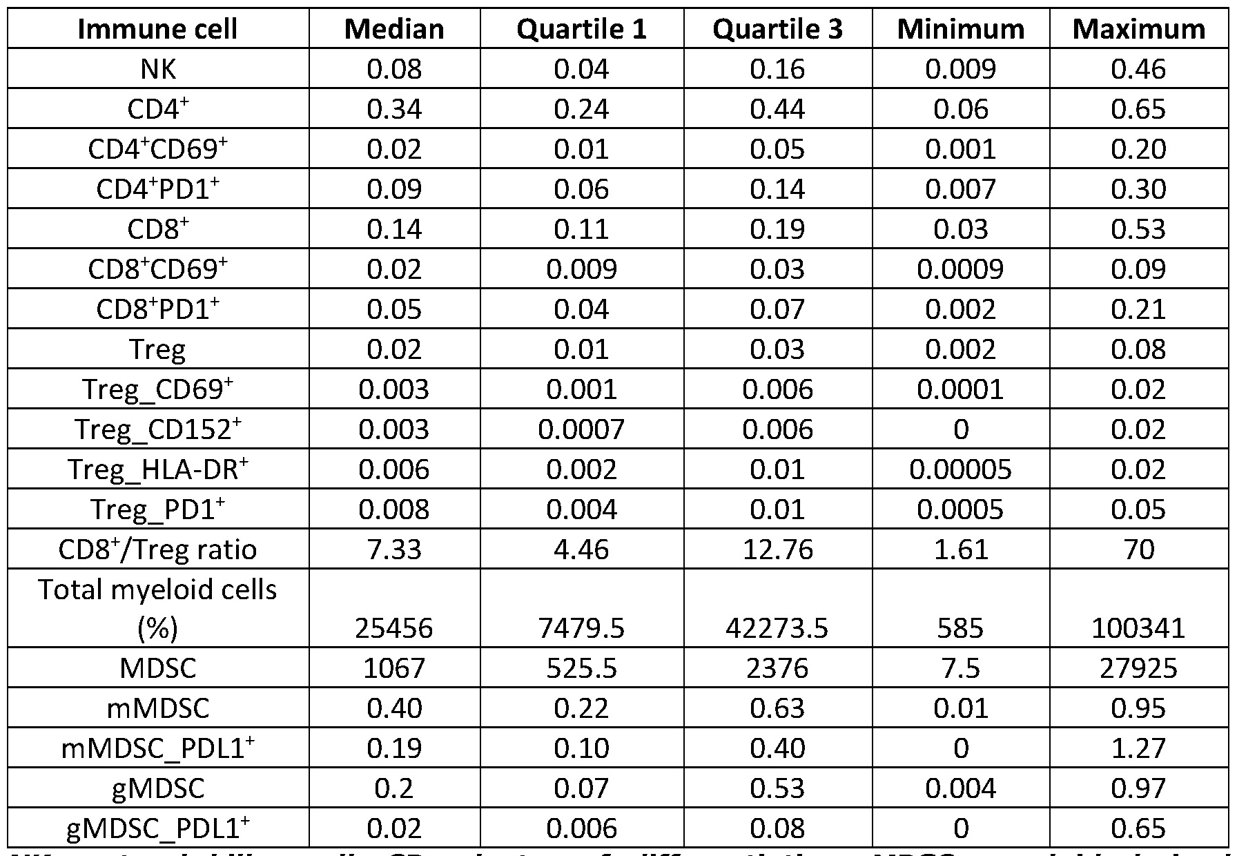
NK: natural killer cell; CD: cluster of differentiation; MDSC: myeloid derived suppressor cells; mMDSC: monocytic MDSC; gMDSC: granulocytic MDSC; Treg: regulatory T cells; PD(L)1: programmed death (ligand)l; HLA: human leucocyte antigen
In concordance to literature, gMDSC was the cell type most susceptible to decay (38). Median age was 57, 50, 47 and 66 years for healthy controls, patients with benign cysts, patients with BOT and invasive cancer, respectively (Figure 4, B). There were no strong correlations between age and the presence of immune cells (Figure 4, C) (39). Overall, 11% of all immune cell values were missing in patients (287/2717), per immune cell parameter this varied between 4% and 22% (Table 4).
Table 4. Missing values

NK: natural killer cell; CD: cluster of differentiation; MDSC: myeloid derived suppressor cells; mMDSC: monocytic MDSC; gMDSC: granulocytic MDSC; Treg: regulatory T cells; PD(L)1: programmed death (ligand)l; HLA: human leucocyte antigen In healthy controls, 12% (64/513) of all immune cell values were missing.
Tumour tissue was available for CD8 and Foxp3 staining from 15 primary tumour at diagnosis and 22 metastases at diagnosis. In six patients, matching primary and metastatic samples were available. MDSC dominate the immune profile, discriminating benign from malignant ovarian tumours
Univariable AUCs varied between 0.53 and 0.76 (Table 5).
Table 5. Statistical changes in immune cells between groups

AUC: area under the curve; Cl: confidence interval; NK: natural killer cell; CD: cluster of differentiation; MDSC: myeloid derived suppressor cells; mMDSC: monocytic MDSC; gMDSC: granulocytic MDSC; Treg: regulatory T cells; PD(L)1: programmed death (ligand)l; HLA: human leucocyte antigen; HGSOC: hFigh grade serous ovarian cancer
AUCs > 0.70 were observed for NK (AUC 0.70), mMDSC (AUC 0.71), and gMDSC- PDL1+ (AUC 0.76). NK and mMDSC values were higher in malignant vs benign tumours, gMDSC-PDLl+ values were lower (Figure 5).
Based on ridge logistic regression, the combination of all 19 markers resulted in a corrected AUC of 0.842 (Figure 3 and 6). Based on the backward elimination best corrected AUC was obtained using 10 markers: NK, MDSC, mMDSC, mMDSC-PDLl+, total myeloid cells, activated Treg (CD69+), activated CD8 T cells, HLA-DR positive Treg, CD8 and gMDSC (corrected AUC 0.864). The backward ridge regression curve started to bend at the level of activated Treg, implying that the most important discriminating roles are reserved for activated Treg, NK, MDSC, mMDSC, mMDSC- PDL1+ and total myeloid cells. This panel of six is dominated by MDSC driven immunosuppression, resulting in an optimism corrected AUC of 0.858. Of note, an immune profile in blood based on the current standard in ovarian cancer immunology, CD8 and Treg, would result in an AUC of 0.639.
Healthy controls versus patients with benign ovarian cysts
AUC values ranged between 0.50 and 0.74, with 15/19 values < 0.70 (Table 5). For most parameters, values were lower in patients with a benign cyst versus healthy controls (Figure 3). Four types of immune cells displayed AUCs above 0.7: activated CD69+ and suppressive PD1+ CD8+, activated CD69+ CD4+ T cells and CD152+ Treg. A visual representation of the comparison between the two groups is depicted in Figure 2.
Patients with borderline ovarian tumour versus stage I and II invasive ovarian cancer Despite wide confidence intervals due to low sample size, it was observed that 17/19 cell types had higher median values in BOT (n= 13) compared to stage I-II invasive ovarian cancer (n=7) (Figure 6). AUCs ranged between 0.50 and 0.97, and were > 0.90 for CD4+ T cells, Treg-CD69+, Treg-HLA-DR+, Treg-PD1+ and gMDSC-PDLl+ (Table 5).
Comparison between early and advanced stage of invasive ovarian cancer
AUCs for early (stage I-II) (n=7) versus late stage ovarian cancer (stage III-IV) (n=34) ranged between 0.51 and 0.88. The largest AUC values are in the increase of immunosuppression (symbolized here by activated CD69+ Tregs (AUC 0.72), mMDSC-PDLl+ (AUC 0.74) and gMDSC-PDLl+ cells (AUC 0.88)) and an increase in NK cells (AUC 0.72) in advanced stage ovarian cancer (Table 5, Figure 7).
Comparison between non-HGSOC versus HGSOC ovarian cancer
Histology and tumour grade in ovarian cancer are known to influence clinical behaviour. AUC > 0.7 was obtained for 10 immune cells, of which PD1+ CD8+ cells
had the highest potential to discriminate between non-HGSOC (n=8) compared to HGSOC (n=33) (Figure 9).
Claims
1. An in vitro method of distinguishing, in a patient diagnosed with an ovarian tumour, between a benign ovarian cyst and malignant ovarian cancer, comprising the steps of:
-determining in a blood sample the amount of at least NK cells, total myeloid cells, myeloid-derived suppressor cells (MDSC), monocytic myeloid derived suppressor cells (mMDSC), and PDL1 positive monocytic myeloid derived suppressor cells (mMDSC-PDLl),
-calculating, based on the amount of said cells, a combined score representing said amount of said cells,
-wherein surpassing a threshold value of said combined score is indicative of malignant ovarian cancer.
2. The method according to claim 1, wherein the combined score is calculated using a logistic regression model.
3. The method according to claim 1 or 2, wherein the blood sample is a sample of peripheral blood mononuclear cell (PBMC).
4. The method according to claim 3, wherein the PBMC sample is a frozen sample.
5. The method according to any one of claims 1 to 4, wherein the sample is divided into fractions and one or more cell types are determined in a dedicated fraction.
6. The method according to any one of claims 1 to 5,
-wherein the amount of each of T cells, Tregs and Nk cells is represented as the ratio of the number of cells of cell said cell type/ the number of viable
CD45+ cells in said sample,
-wherein the amount of each of mMDSC, gMDSC, mMDSC-PDLl+ , gMDSC- PDL1+ is represented as the ratio of said cell type/ the total amount of MDSC, and
-wherein the amount of myeloid cells is represented as the total amount of myeloid cells in the sample.
7. The method according to any one of claim 1 to 6, wherein amount of living cells is determined in the sample of PBMC cells.
8. The method according to any one of claims 1 to 7, wherein the amount of cells, with the exception of total myeloid cell amount, is determined via cell type specific cell surface binding agents.
9. The method according to any one of claims 1 to 8, wherein the cell surface binding agents are fluorescent labelled antibodies.
10. The method according to any one of claims 1 to 9,
-wherein the amount of NK cells is determined using at least an antibody specifically binding CD3 and an antibody specifically binding CD16_56, -wherein the amount of myeloid-derived suppressor cells (MDSC) is determined using at least an antibody specifically binding CDllb and an antibody specifically binding HLA-DR
-wherein the amount of monocytic myeloid derived suppressor cells (mMDSC) is determined using at least an antibody specifically binding CD14, and
-wherein the amount of PDL1 positive monocytic myeloid derived suppressor cells (mMDSC-PDLl) is determined using at least an antibody specifically binding CD14 and an antibody specifically binding PDL1.
11. The method according to any one of claims 1 to 10, further comprising determining in a blood sample the amount of a further cell type of the adaptive immune system, such as activated regulatory T cells (Treg).
12. A kit comprising reagents for the detection of NK cells, myeloid-derived suppressor cells (MDSC), monocytic myeloid derived suppressor cells (mMDSC), and PDL1 positive monocytic myeloid derived suppressor cells
(mMDSC-PDLl), wherein the kit comprising antibodies against at most 10 different antigens, including :
an antibody specifically binding CD3,
an antibody specifically binding CD16_56,
an antibody specifically binding CDllb,
an antibody specifically binding HLA-DR
an antibody specifically binding CD14,
and
an antibody specifically binding PDL1.
13. The kit according to claim 12, wherein said reagents are cell surface binding antibodies, typically fluorescent labelled cell surface binding antibodies.
14. Use of kit comprising reagents for the detection of NK cells myeloid-derived suppressor cells (MDSC), monocytic myeloid derived suppressor cells (mMDSC), and PDL1 positive monocytic myeloid derived suppressor cells (mMDSC-PDLl) in an in vitro method for distinguishing, in a patient diagnosed with ovarian tumour, between a benign ovarian cyst and malignant ovarian cancer.
15. The use according to claim 14, wherein the kit comprises:
an antibody specifically binding CD3,
an antibody specifically binding CD16_56,
an antibody specifically binding CDl lb,
an antibody specifically binding HLA-DR,
an antibody specifically binding CD14, and
an antibody specifically binding PDL1.
Applications Claiming Priority (2)
| Application Number | Priority Date | Filing Date | Title |
|---|---|---|---|
| EP19164643 | 2019-03-22 | ||
| EP19164643.9 | 2019-03-22 |
Publications (1)
| Publication Number | Publication Date |
|---|---|
| WO2020193403A1 true WO2020193403A1 (en) | 2020-10-01 |
Family
ID=65904348
Family Applications (1)
| Application Number | Title | Priority Date | Filing Date |
|---|---|---|---|
| PCT/EP2020/057764 Ceased WO2020193403A1 (en) | 2019-03-22 | 2020-03-20 | Methods for diagnosing ovarian cancer |
Country Status (1)
| Country | Link |
|---|---|
| WO (1) | WO2020193403A1 (en) |
Citations (2)
| Publication number | Priority date | Publication date | Assignee | Title |
|---|---|---|---|---|
| WO2010148145A1 (en) * | 2009-06-16 | 2010-12-23 | Fred Hutchinson Cancer Research Center | Methods and kits for detecting ovarian cancer from blood |
| WO2013023994A1 (en) * | 2011-08-12 | 2013-02-21 | Pronota N.V. | New biomarker for the classification of ovarian tumours |
-
2020
- 2020-03-20 WO PCT/EP2020/057764 patent/WO2020193403A1/en not_active Ceased
Patent Citations (2)
| Publication number | Priority date | Publication date | Assignee | Title |
|---|---|---|---|---|
| WO2010148145A1 (en) * | 2009-06-16 | 2010-12-23 | Fred Hutchinson Cancer Research Center | Methods and kits for detecting ovarian cancer from blood |
| WO2013023994A1 (en) * | 2011-08-12 | 2013-02-21 | Pronota N.V. | New biomarker for the classification of ovarian tumours |
Non-Patent Citations (25)
| Title |
|---|
| BRISSOT ET AL., BLOOD, vol. 124, no. 21, 2014, pages 3781 |
| BRONTE V ET AL., NAT COMMUN., vol. 7, 2016, pages 12150 |
| COOSEMANS A ET AL: "New biomarkers in the diagnosis of ovarian pathology", INTERNATIONAL JOURNAL OF GYNECOLOGICAL CANCER 20171101 LIPPINCOTT WILLIAMS AND WILKINS NLD, vol. 27, no. Supplement 4, 1 November 2017 (2017-11-01), XP009515636, ISSN: 1525-1438 * |
| CURIEL TJ ET AL., NAT MED, vol. 10, no. 9, 2004, pages 942 - 949 |
| EOLIA BRISSOT ET AL: "T Cell Exhaustion and Downregulation of Cytotoxic NK Cells - an Immune Escape Mechanism in Adult Acute Lymphoblastic Leukemia", BLOOD, VOLUME 124, NUMBER 21, 6 December 2014 (2014-12-06), XP055617300, Retrieved from the Internet <URL:http://www.bloodjournal.org/content/124/21/3781> [retrieved on 20190902] * |
| HAMANISHI J ET AL., J CLIN ONCOL, vol. 33, no. 34, 2015, pages 4015 - 4022 |
| HORN ET AL., ONCOTARGET, vol. 8, no. 35, 2017, pages 57964 - 57980 |
| JACOBS I ET AL., BJOG, vol. 97, no. 10, 1990, pages 922 - 929 |
| KAIJSER J ET AL., HUM REPROD UPDATE, vol. 20, no. 3, 2014, pages 449 - 462 |
| KISS M ET AL., CELL IMMUNOL, vol. 330, 2018, pages 188 - 201 |
| KOTSAKIS A ET AL., J IMMUNOL METHODS, vol. 381, no. 1-2, 2012, pages 14 - 22 |
| LUCAS A. HORN ET AL: "CD3xPDL1 bi-specific T cell engager (BiTE) simultaneously activates T cells and NKT cells, kills PDL1<sup>+</sup> tumor cells, and extends the survival of tumor-bearing humanized mice", ONCOTARGET, vol. 8, no. 35, 29 August 2017 (2017-08-29), pages 57964 - 57980, XP055471264, DOI: 10.18632/oncotarget.19865 * |
| MOORE RG ET AL., GYNECOL ONCOL, vol. 112, no. 1, 2009, pages 40 - 46 |
| OKLA K ET AL., CRIT REV CLIN LAB SCI., vol. 55, no. 6, 2018, pages 376 - 407 |
| QIN GHOTILOVAC L, STAT METHODS MED RES, vol. 17, no. 2, 2008, pages 207 - 221 |
| SATO E ET AL., PROC NATL ACAD SCI, vol. 102, no. 51, 2005, pages 18538 - 18543 |
| SCHREIBER RD ET AL., SCIENCE, vol. 331, no. 6024, 2011, pages 1565 - 1570 |
| STEYERBERG EW ET AL., CLIN EPIDEMIOL, vol. 54, no. 8, 2001, pages 774 - 781 |
| SWANTON C., CANCER RES., vol. 72, no. 19, 2012, pages 4875 - 4882 |
| TIMMERMAN D ET AL., ULTRASOUND OBSTET GYNECOL, vol. 16, no. 5, 2000, pages 500 - 505 |
| TIMMERMAN D ET AL., ULTRASOUND OBSTET GYNECOL, vol. 31, no. 6, 2008, pages 681 - 690 |
| VAN CALSTER B ET AL., BMJ, vol. 349, 2014, pages g5920 |
| VAN HOLSBEKE C ET AL., CLIN CANCER RES, vol. 15, no. 2, 2009, pages 684 - 691 |
| VEGLIA F ET AL., NAT IMMUNOL., vol. 19, no. 2, 2018, pages 108 - 119 |
| ZHANG L ET AL., N ENGL J MED, vol. 348, no. 3, 2003, pages 203 - 213 |
Similar Documents
| Publication | Publication Date | Title |
|---|---|---|
| Romagnolo et al. | HE4, CA125 and risk of ovarian malignancy algorithm (ROMA) as diagnostic tools for ovarian cancer in patients with a pelvic mass: An Italian multicenter study | |
| Hwang et al. | Prevalence and heterogeneity of PD-L1 expression by 22C3 assay in routine population-based and reflexive clinical testing in lung cancer | |
| Pearl et al. | Prognostic analysis of invasive circulating tumor cells (iCTCs) in epithelial ovarian cancer | |
| Yang et al. | Prognostic value of Ki-67 in solid pseudopapillary tumor of the pancreas: Huashan experience and systematic review of the literature | |
| Escobar-Hoyos et al. | Keratin 17 in premalignant and malignant squamous lesions of the cervix: proteomic discovery and immunohistochemical validation as a diagnostic and prognostic biomarker | |
| Szubert et al. | External validation of the IOTA ADNEX model performed by two independent gynecologic centers | |
| Yamagami et al. | Immunofluorescence-detected infiltration of CD4+ FOXP3+ regulatory T cells is relevant to the prognosis of patients with endometrial cancer | |
| Pearl et al. | Treatment monitoring of patients with epithelial ovarian cancer using invasive circulating tumor cells (iCTCs) | |
| Jeong et al. | The relationship between the extent of extraprostatic extension and survival following radical prostatectomy | |
| Hecking et al. | Tumoral PD-L1 expression defines a subgroup of poor-prognosis vulvar carcinomas with non-viral etiology | |
| Googe et al. | Preferentially expressed antigen in melanoma immunostaining in a series of melanocytic neoplasms | |
| Bernardi et al. | Prognostic significance of CD24 and claudin-7 immunoexpression in ductal invasive breast cancer | |
| Guo et al. | A preoperative risk model for early recurrence after radical resection may facilitate initial treatment decisions concerning the use of neoadjuvant therapy for patients with pancreatic ductal adenocarcinoma | |
| Shih et al. | Accuracy of frozen section diagnosis of ovarian borderline tumor | |
| Grenache et al. | Clinical performance of two multi-marker blood tests for predicting malignancy in women with an adnexal mass | |
| Coosemans et al. | Myeloid-derived suppressor cells at diagnosis may discriminate between benign and malignant ovarian tumors | |
| Ozgul et al. | Oncological outcomes of stage II endometrial cancer: a retrospective analysis of 250 cases | |
| Sozen et al. | Clinicopathologic and survival analyses of synchronous primary endometrial and epithelial ovarian cancers | |
| Minar et al. | Comparison of the Copenhagen Index versus ROMA for the preoperative assessment of women with ovarian tumors | |
| Kim et al. | Basaloid carcinoma of the lung: a really dismal histologic variant? | |
| Mueller et al. | International study of primary mucinous ovarian carcinomas managed at tertiary medical centers | |
| Luo et al. | Diagnostic performance of CA125, HE4, ROMA, and CPH‐I in identifying primary ovarian cancer | |
| Dewar et al. | Investigating racial differences in clinical and pathological features of prostate cancer in South African men | |
| Hoogendam et al. | Detection of cervical cancer recurrence during follow-up: a multivariable comparison of 9 frequently investigated serum biomarkers | |
| El Sharouni et al. | High discordance rate in assessing sentinel node positivity in cutaneous melanoma: expert review may reduce unjustified adjuvant treatment |
Legal Events
| Date | Code | Title | Description |
|---|---|---|---|
| 121 | Ep: the epo has been informed by wipo that ep was designated in this application |
Ref document number: 20711607 Country of ref document: EP Kind code of ref document: A1 |
|
| NENP | Non-entry into the national phase |
Ref country code: DE |
|
| 122 | Ep: pct application non-entry in european phase |
Ref document number: 20711607 Country of ref document: EP Kind code of ref document: A1 |