WO2020183188A1 - Nucleic acid amplification methods - Google Patents
Nucleic acid amplification methods Download PDFInfo
- Publication number
- WO2020183188A1 WO2020183188A1 PCT/GB2020/050635 GB2020050635W WO2020183188A1 WO 2020183188 A1 WO2020183188 A1 WO 2020183188A1 GB 2020050635 W GB2020050635 W GB 2020050635W WO 2020183188 A1 WO2020183188 A1 WO 2020183188A1
- Authority
- WO
- WIPO (PCT)
- Prior art keywords
- adapter
- oligonucleotide
- adapter oligonucleotide
- template dna
- dna
- Prior art date
- Legal status (The legal status is an assumption and is not a legal conclusion. Google has not performed a legal analysis and makes no representation as to the accuracy of the status listed.)
- Ceased
Links
Classifications
-
- C—CHEMISTRY; METALLURGY
- C12—BIOCHEMISTRY; BEER; SPIRITS; WINE; VINEGAR; MICROBIOLOGY; ENZYMOLOGY; MUTATION OR GENETIC ENGINEERING
- C12Q—MEASURING OR TESTING PROCESSES INVOLVING ENZYMES, NUCLEIC ACIDS OR MICROORGANISMS; COMPOSITIONS OR TEST PAPERS THEREFOR; PROCESSES OF PREPARING SUCH COMPOSITIONS; CONDITION-RESPONSIVE CONTROL IN MICROBIOLOGICAL OR ENZYMOLOGICAL PROCESSES
- C12Q1/00—Measuring or testing processes involving enzymes, nucleic acids or microorganisms; Compositions therefor; Processes of preparing such compositions
- C12Q1/68—Measuring or testing processes involving enzymes, nucleic acids or microorganisms; Compositions therefor; Processes of preparing such compositions involving nucleic acids
- C12Q1/6844—Nucleic acid amplification reactions
- C12Q1/6865—Promoter-based amplification, e.g. nucleic acid sequence amplification [NASBA], self-sustained sequence replication [3SR] or transcription-based amplification system [TAS]
-
- C—CHEMISTRY; METALLURGY
- C12—BIOCHEMISTRY; BEER; SPIRITS; WINE; VINEGAR; MICROBIOLOGY; ENZYMOLOGY; MUTATION OR GENETIC ENGINEERING
- C12Q—MEASURING OR TESTING PROCESSES INVOLVING ENZYMES, NUCLEIC ACIDS OR MICROORGANISMS; COMPOSITIONS OR TEST PAPERS THEREFOR; PROCESSES OF PREPARING SUCH COMPOSITIONS; CONDITION-RESPONSIVE CONTROL IN MICROBIOLOGICAL OR ENZYMOLOGICAL PROCESSES
- C12Q1/00—Measuring or testing processes involving enzymes, nucleic acids or microorganisms; Compositions therefor; Processes of preparing such compositions
- C12Q1/68—Measuring or testing processes involving enzymes, nucleic acids or microorganisms; Compositions therefor; Processes of preparing such compositions involving nucleic acids
- C12Q1/6844—Nucleic acid amplification reactions
- C12Q1/6853—Nucleic acid amplification reactions using modified primers or templates
- C12Q1/6855—Ligating adaptors
Definitions
- the present invention relates to improved, reliable and highly specific methods of amplifying DNA to produce an aRNA or DNA product, utilising novel adapter oligonucleotides, which can be used to identify genetic variations in biological samples.
- NGS Next Generation Sequencing
- circulating cell-free DNA has provided opportunities to analyse genetic materials from blood samples, without the risks typically associated with more invasive sampling methods.
- tumour DNA circulating tumour DNA
- the ctDNA may be analysed by sequencing cfDNA to detect sequence variants that are known to be specifically associated with various types of tumours or cfDNA copy numbers to detect the presence of tumours and their progression.
- CTCs circulating tumour cells
- a method for producing an aRNA product from template DNA comprising:
- a secondary oligonucleotide comprising an RNA polymerase promoter sequence, wherein the sequence of the secondary oligonucleotide is complementary to the sequence of the adapter oligonucleotide;
- the method of the present invention provides a number of advantages. Compared to other methods used to amplify template DNA the approach is simpler, more flexible and can provide increased sensitivity and specificity making it more suitable where input DNA is limited (for example in the analysis of cfDNA and single cells).
- aRNA as used herein is used to refer to amplified RNA produced by an RNA polymerase from the template DNA of the present invention.
- the method of the present invention comprises providing an adapter oligonucleotide comprising a RNA polymerase promoter sequence and transcribing template DNA by introducing a RNA polymerase.
- RNA polymerase there are many RNA polymerases which could be utilised in the method of the present invention, such as T3 RNA polymerase, SP6 RNA polymerase and T7 RNA polymerase.
- the RNA polymerase may comprise a T7 RNA polymerase and the RNA polymerase promoter sequence may comprise a T7 RNA polymerase promoter sequence.
- the present inventors have shown that utilising a T7 RNA polymerase and associated promoter sequence results in a highly efficient, sensitive and specific method.
- the RNA polymerase promoter sequence in the adapter oligonucleotide may comprise SEQ ID N0.1 or a fragment or variant thereof.
- the T7 RNA polymerase promoter sequence is: ATTATGCTGAGTGATATCCC (SEQ ID NO. 1).
- the RNA polymerase promoter sequence in the adapter oligonucleotide consists of SEQ I D NO.1.
- the RNA polymerase promoter sequence in the secondary oligonucleotide may comprise SEQ ID NO.2 or a fragment or variant thereof.
- the Reverse T7 RNA polymerase promoter sequence is: TAATACGACTCACTATAGGG (SEQ ID NO. 2).
- the RNA polymerase promoter sequence in the secondary oligonucleotide consists of SEQ ID NO.2.
- fragment or variant as used herein is used in its broadest sense to refer to a fragment or variant of the sequence disclosed which retains its function, i.e. in this case, the ability to act as an RNA polymerase promoter sequence thereby allowing an RNA polymerase to bind.
- the fragment or variant comprises at least 80%, 85%, 90%, 95%, 98% or 99% sequence identity with SEQ ID N0.1 or 2.
- the fragment or variant has 100% sequence identity at positions 7, 8, 9, 10 and 11 of SEQ ID N0.1 , as shown underlined below:
- the fragment or variant has 100% sequence identity at positions 7, 8, 9, 10 and 11 of SEQ ID NO:2, as shown underlined below: TAATACGACTCACTATAGGG
- the sequence of the secondary oligonucleotide is complementary to the sequence of the adapter oligonucleotide.
- This sequence complementarity may be by way of the RNA polymerase promoter sequences present in the adapter oligonucleotide and secondary oligonucleotide respectively.
- the RNA polymerase promoter sequences in the adapter oligonucleotide and secondary oligonucleotide may be complementary or at least partially complementary in sequence.
- the RNA polymerase promoter sequence in the adapter oligonucleotide comprises SEQ ID NO:1 or a fragment or variant thereof and the RNA polymerase promoter sequence in the secondary oligonucleotide comprises SEQ ID NO:2 or a fragment or variant thereof.
- the secondary and adapter oligonucleotides may comprise further complementary sequences, in addition to the RNA polymerase promoter sequences.
- Step ii) of the method of the present invention comprises ligating the 5’ end of the adapter oligonucleotide to the 3’ end of the template DNA to produce an adapter oligonucleotide- template DNA molecule.
- the adapter oligonucleotide is configured so that a 5’ end of the adapter oligonucleotide can be ligated to a 3’ end of the template DNA.
- the adapter oligonucleotide may be configured so that a 5’ end of the adapter oligonucleotide can be ligated to a 3’ end of the template DNA.
- the adapter oligonucleotide may comprise a T-tail.
- the template DNA may comprise an A-tail, facilitating adapter oligonucleotide and template DNA annealing (due to the corresponding A- and T-tails) such that they are in proximity for subsequent ligation.
- the adapter oligonucleotide may comprise a 5’ sequence which is complementary to a 3’ sequence of the corresponding DNA template.
- the DNA template may have been treated with a restriction enzyme resulting in an overhanging sequence.
- the 5’ end of the adapter oligonucleotide may have a sequence corresponding to the overhanding sequence of the DNA template such that the adapter oligonucleotide and template DNA can anneal such that they are in proximity for subsequent ligation.
- step ii) may comprise ligating the 5’ end of the adapter oligonucleotide to the 3’ end of the template DNA using a DNA ligase.
- a DNA ligase suitable of ligating the 5’ end of the adapter oligonucleotide to the 3’ end of the template DNA may be used.
- the DNA ligase may comprise a bacterial, bacteriophage (e.g. T4 DNA ligase) or mammalian DNA ligase (e.g. DNA ligase I, II, III or IV), for example.
- the DNA ligase may comprise a T4 DNA ligase, for example KAPA-hyper DNA ligase, NEB Quick Ligation Kit, NEB T4 DNA Ligase, NEB Blunt/TA Ligase Master Mix or NEBNext® UltraTM II Ligation Module.
- T4 DNA ligase for example KAPA-hyper DNA ligase, NEB Quick Ligation Kit, NEB T4 DNA Ligase, NEB Blunt/TA Ligase Master Mix or NEBNext® UltraTM II Ligation Module.
- the DNA ligase may comprise KAPA- hyper DNA ligase.
- the present inventors found that the use of KAPA-hyper DNA ligase is particularly effective in the method of the present invention (see for example, Figure 4).
- the design of the adapter oligonucleotide allows ligation of the 5’ end of the adapter oligonucleotide to the 3’ end of the template DNA. This results in a contiguous molecule incorporating the RNA polymerase promoter at the 3’ end and the template DNA at the 5’ end. Due to the orientation of the adapter oligonucleotide-template DNA molecule there is no need to convert to a dsDNA molecule for the RNA polymerisation step since the ligated adapter oligonucleotide-template DNA molecule can be utilised as a template directly by the RNA polymerase.
- ligation of the 5’ end of the adapter oligonucleotide to the 3’ end of the template DNA takes place between around 14 and 22°C, preferably between around 16 and 22°C, preferably at 20°C.
- the method may further comprise dephosphorylating the template DNA, preferably prior to the ligation of step ii) of the present invention.
- a 5’ phosphate of the template DNA is removed by treatment with a phosphatase enzyme.
- any suitable phosphatase enzyme could be utilised, for example Shrimp Alkaline Phosphatase, Antarctic Phosphatase, Calf Intestinal Alkaline Phosphatase or FastAP Thermosensitive Alkaline Phosphatase.
- the phosphatase comprises a heat labile phosphatase such as FastAP Thermosensitive Alkaline Phosphatase.
- the present inventors have found that the removal of the 5’ phosphate from the template DNA reduces DNA self ligation and also reduces or blocks ligation of the 3’ end of the adapter to the 5’ of the template DNA. It is highly advantageous to reduce the ligation of the 3’ end of the adapter to the 5' of the template DNA since this can result in circular ligation products which are not compatible with subsequent steps leading to NGS library generation.
- the use of a dephosphorylation step is in contrast to current NGS PCR amplification approaches which incorporate phosphorylation of template DNA. Instead, the present invention preferably makes use of dephosphorylated template DNA which results in aRNA production.
- the present invention preferably makes use of dephosphorylated target DNA which results in improved aRNA production.
- the method comprises a multiplex method.
- a multiplex method may utilise sample bar codes allowing for the multiplexing of tens to thousands of samples following the ligation of the adapter oligonucleotide and the template DNA.
- the method may further comprises a restriction enzyme digestion of the template DNA prior to the ligation of step ii).
- restriction enzymes are suitable to perform the restriction enzyme digestion which are known in the art.
- the restriction enzyme may produce“blunt ends” or“sticky ends”.
- Restriction enzymes suitable to produce blunt ends may be selected from MLyl, BsuRI, Haelll, EcoRV, Smal.
- Restriction enzymes suitable to produce sticky ends may be selected from NIAIII, Hsp92ll, Hinl ll, Mspl. Wherein sticky ends are produced by the restriction enzymes the target DNA may then undergo end repair and/or addition of an A tail.
- a restriction enzyme which produces blunt ends may be used, more preferably MLyl or BsuRI may be used.
- Step v) of the method of the present invention comprises transcribing the template DNA by introducing an RNA polymerase to the adapter oligonucleotide-template DNA molecule, to generate an aRNA product.
- an RNA polymerase As described above, there are many RNA polymerases which could be utilised in the method of the present invention, such as T3 RNA polymerase, SP6 RNA polymerase and T7 RNA polymerase. However, in a preferred embodiment, the RNA polymerase may comprise a T7 RNA polymerase.
- RNA polymerase By introducing an RNA polymerase to the adapter oligonucleotide-template DNA molecule to transcribe the template DNA, linear amplification of each original DNA strand is achieved. Unlike PCR approaches, which require an initially potentially error prone polymerisation step prior to amplification, around 1000 aRNA copies are made directly from each input strand, providing a means to reduce sequencing errors and increase specificity and sensitivity when detecting rare mutations in DNA mixtures.
- the method further comprises the step of:
- the method may further comprise the steps of: vi) converting the aRNA product to cDNA; and
- the method of the present invention may be a method for amplifying template DNA.
- the present invention therefore also provides a method for amplifying template DNA, the method comprising: i) providing template DNA and an adapter oligonucleotide comprising an RNA polymerase promoter sequence, wherein the adapter oligonucleotide is configured so that a 5’ end of the adapter oligonucleotide can be ligated to a 3’ end of the template DNA;
- a secondary oligonucleotide comprising an RNA polymerase promoter sequence, wherein the sequence of the secondary oligonucleotide is complementary to the sequence of the adapter oligonucleotide;
- cDNA refers to complementary DNA and is used to refer to DNA synthesised from single-stranded RNA, for example in a reaction catalysed by the enzyme reverse transcriptase.
- Step vi) may first comprise ligating a priming site to the aRNA followed by reverse transcription using the priming site and a reverse transcriptase.
- step vi) may comprise utilising reverse transcriptase and random oligonucleotide primers or utilising reverse transcriptase and target specific oligonucleotide primers.
- any suitable reverse transcriptase enzyme could be utilised, for example the bacterial ret reverse transcriptase, the retroviral reverse transcriptases including RSV reverse transcriptase from Rous sarcoma virus, HIV-1 reverse transcriptase from human immunodeficiency virus type 1 , MMLV reverse transcriptase from Moloney murine leukemia virus and AMV reverse transcriptase from avian myeloblastosis virus.
- the reverse transcriptase comprises of an optimised commercial cloned reverse transcriptase such as the mutant MMLV Superscript IV.
- the method to convert the aRNA to cDNA may be performed in a number of ways. Conversion of aRNA to cDNA may be performed via ligation of a priming site to the aRNA. The ligation may be followed by reverse transcription. Conversion of aRNA to cDNA may be performed via single stranded ligation of a priming site to the aRNA. The single stranded ligation may ligate a unique NGS primer to the target aRNA. Conversion of aRNA to cDNA may be performed via splinted double stranded ligation to aRNA. The splinted double stranded ligation may ligate a unique NGS primer to the target aRNA. Conversion of aRNA to cDNA may be performed using random NGS primer. Conversion of aRNA to cDNA may be performed using target specific NGS primer.
- Step vii) may comprise amplification in a next generation sequencing (NGS) protocol.
- NGS next generation sequencing
- the present invention as defined above is compatible with all of the current NGS platforms including systems which rely on clonal amplification of adapter-ligated libraries such as lllumina and Ion Torrent as well as single molecule sequencing platforms such as Pacific Bioscience and Oxford Nanopore.
- Both optics-based sequencers which use visible light, collecting photons from arrays to form a visual image of bases incorporated during polymerization (e.g. lllumina) and semiconductor sequencing which rely on the detection of hydrogen ions released during the polymerisation of DNA (e.g. Ion Torrent) rely on the inclusion of specific adapter sequences which can be readily incorporated into the method of the present invention.
- single molecule sequencing platforms such as Pacific Bioscience and Oxford Nanopore rely on addition of sequences that facilitate sequencing and these can also be readily incorporated into the method of the present invention.
- the method can be used with both capture based NGS, where specific sequences are enriched following generation of a genome wide library, and direct PCR amplicon NGS.
- the method of the present invention includes providing an adapter oligonucleotide comprising an RNA polymerase promoter sequence, wherein the adapter oligonucleotide is configured so that a 5’ end of the adapter oligonucleotide can be ligated to a 3’ end of the template DNA.
- the 5’ end of the adapter oligonucleotide is ligated to the 3’ end of the template DNA to produce an adapter oligonucleotide-template DNA molecule and a secondary oligonucleotide is then annealed to the adapter oligonucleotide-template DNA molecule.
- the adapter oligonucleotide comprises complementary regions which are capable of annealing to one another to form a secondary structure, preferably a circular or hairpin loop structure.
- a secondary structure preferably a circular or hairpin loop structure.
- the skilled person will appreciate how such an adapter could be designed.
- the use of a circular or hairpin loop structure simplifies the method of the present invention in that by using an adapter with such a structure, it is not necessary to determine the optimum molar balance which becomes necessary when multi molecule adapters are utilised.
- step iv) of the method may further comprise the step of denaturing the adapter oligonucleotide prior to annealing the secondary oligonucleotide to the adapter oligonucleotide.
- the adapter oligonucleotide may comprise a number of additional sequence features which may be useful in further analysing the target DNA which has been amplified, such as molecular bar code, sample bar codes, a NGS sequence and/or a T tail.
- the adapter oligonucleotide may further comprise a single region which is complementary, or partially complementary, to the stick ends produced by the restriction enzyme digestion.
- the adapter oligonucleotide may further comprise a molecular barcode sequence.
- a molecular bar code sequence may comprise, for example, a sequence of random bases, for example, 5, 6, 7, 8, 9 or 10 bases.
- the molecular barcode sequence may include 6-8 bases. The molecular barcode provides a unique bar code for each adapter oligonucleotide that in turn will be incorporated into each adapter oligonucleotide-template DNA molecule.
- RNA polymerisation of each adapter oligonucleotide-template DNA molecule will produce aRNA molecules each harbouring the same molecular bar code sequence as well as the same input DNA sequence enabling bioinformatics identification of all aRNA molecules originating from each single initial ligation event.
- a consensus sequence can be built up that avoids identifying errors present in single aRNA molecules which may arise due to polymerase error. This will therefore increase the specificity and sensitivity which is particularly important for detecting rare mutations in DNA mixtures.
- molecular barcodes along with a linear amplification step the error obtained using NGS is expected to be reduced by one to three orders of magnitude.
- the adapter oligonucleotide may further comprise a sample barcode.
- a sample barcode may comprise, for example, a sequence of random bases, for example, 5, 6, 7, 8, 9 or 10 bases.
- each adapter may comprise a unique sample bar code which is ligated to a sample from a particular source. Following ligation of the adapter oligonucleotide and the template DNA, samples could be combined for subsequent steps, thereby improving efficiency. Post-sequencing information, for example, could be identified for each sample bioinformatically, through identifying the sample barcode.
- the adapter oligonucleotide may further comprise a NGS sequence.
- the adapter oligonucleotide may further comprise a T-tail.
- the template DNA may comprise an A-tail, encouraging the adapter oligonucleotide and template DNA to anneal (due to the corresponding A- and T-tails) such that they are in proximity for subsequent ligation.
- the template DNA is double-stranded.
- the adapter oligonucleotide may only anneal to a single strand of said double-stranded DNA.
- the adapter oligonucleotide may further comprise an overhang of oligonucleotides. This may be given the designation NNNNNNNN.
- the overhang may be positioned after one of the complementary regions which are capable of annealing to one another.
- the overhang may comprise a random sequence of oligonucleotides, or the sequence may be selected to be complementary to the target DNA.
- the overhang may be 5’ or 3’.
- the adapter oligonucleotide forms a circular or hairpin secondary structure and comprises an overhang.
- the oligonucleotide overhang may comprise about 6 to about 12 oligonucleotides, preferably about 8 to about 10 oligonucleotides, more preferably about 8 oligonucleotides.
- This format of the adapter oligonucleotide may be particular suitable for use in single strand ligation.
- the template DNA may be derived from a biological sample.
- the biological sample may comprise blood.
- the template DNA may comprise cfDNA, gDNA and/or single cell DNA.
- suitable commercial kits are available for obtaining template DNA, including cfDNA, gDNA or single cell DNA.
- suitable kits include for example QIAamp DNA Blood Mini Kit (Qiagen), QIAamp circulating nucleic acid kit (Qiagen) and MagMAX Cell-free DNA Isolation Kit (Thermo Fisher Scientific).
- the template DNA is double-stranded DNA or single-stranded DNA.
- Ligation to single-stranded DNA can incorporate improved ligation conditions including use of optimised ligase (Gansauge and Meyer, 2013) or primer design incorporating ligation guides or splinter oligonucleotides (Gansauge et al, 2017; Kwok et al, 2013).
- an adapter oligonucleotide for producing an aRNA product from a DNA template, the adapter oligonucleotide comprising an RNA polymerase promoter sequence, wherein the adapter oligonucleotide is configured so that a 5’ end of the adapter oligonucleotide can be ligated to a 3’ end of a template DNA.
- an adapter oligonucleotide for amplifying a DNA template, the adapter oligonucleotide comprising an RNA polymerase promoter sequence, wherein the adapter oligonucleotide is configured so that a 5’ end of the adapter oligonucleotide can be ligated to a 3’ end of a template DNA.
- the adapter of the present invention may be for use in the method of the present invention, as defined above.
- the RNA polymerase promoter sequence may comprise a T7 RNA polymerase promoter sequence, and such a sequence may comprise SEQ ID N0.1 or a fragment or variant thereof.
- the RNA polymerase promoter sequence may consist of SEQ ID N0.1.
- the adapter may comprise complementary regions which are capable of annealing to one another to form circular or hairpin loop structure.
- the adapter may comprise a number of additional sequence features which may be useful in further analysing the target DNA which has been amplified, such as molecular bar code, sample bar codes, a NGS sequence and/or a T tail.
- the adapter may be in a buffered (or other) solution so as to enable storage and transportation of the adapter.
- a buffered (or other) solution so as to enable storage and transportation of the adapter.
- the skilled person will appreciate suitable solutions in which the adapter may be present.
- the adapter may be in vial or other vessel suitable for storage, transportation and mixing with other reagents if desired.
- the adapter is for use as a research reagent/tool.
- kits for producing an aRNA product from a DNA template comprising:
- an adapter oligonucleotide comprising an RNA polymerase promoter sequence, wherein the adapter oligonucleotide is configured so that a 5’ end of the adapter oligonucleotide can be ligated to a 3’ end of a template DNA;
- a secondary oligonucleotide comprising an RNA polymerase promoter sequence, wherein the sequence of the secondary oligonucleotide is complementary to the sequence of the adapter oligonucleotide.
- kit for amplifying a DNA template comprising:
- an adapter oligonucleotide comprising an RNA polymerase promoter sequence, wherein the adapter oligonucleotide is configured so that a 5’ end of the adapter oligonucleotide can be ligated to a 3’ end of a template DNA; and ii) a secondary oligonucleotide comprising a RNA polymerase promoter sequence, wherein the sequence of the secondary oligonucleotide is complementary to the sequence of the adapter oligonucleotide.
- the RNA polymerase promoter sequence in the adapter oligonucleotide comprises a T7 RNA polymerase promoter sequence with a sequence comprising SEQ ID N0.1 or a fragment or variant thereof.
- the RNA polymerase promoter sequence in the adapter oligonucleotide consists of SEQ ID N0.1.
- the RNA polymerase promoter sequence in the secondary oligonucleotide comprises a T7 RNA polymerase promoter sequence with a sequence comprising SEQ ID NO.2 or a fragment or variant thereof.
- the RNA polymerase promoter sequence in the adapter oligonucleotide consists of SEQ ID NO.2.
- the adapter oligonucleotide of the kit may comprise complementary regions which are capable of annealing to one another to form circular or hairpin loop structure as described above in relation to the method of the present invention.
- the adapter oligonucleotide of the kit may comprise a number of additional sequence features which may be useful in further analysing the target DNA which has been amplified, such as molecular bar code, sample bar codes, a NGS sequence and/or a T tail as described above in relation to the method of the present invention.
- the adapter oligonucleotide and/or secondary oligonucleotide of the kit may be in a buffered solution.
- the skilled person will appreciate suitable solutions in which the adapter may be present.
- a method for detecting a genetic variant or genetic signature in template DNA comprising amplifying template DNA in the method as herein above described and analysing the amplified cDNA for the genetic variation or signature.
- the method of the present invention can be used to identify a wide range of genetic signatures including the presence of specific point mutations, mutational signature and structural alterations including copy number gains and losses.
- the information generated can be used to identify mutations linked to specific therapy, monitor disease, predict clinical outcomes and response to therapy. It is also envisaged that the invention will be particularly suited to identifying differences in DNA methylation that are related to the detection of tumour DNA in patient blood samples. Differences in DNA methylation may comprise differences in total methylation or DNA methylation patterns or signatures (such as described in Shen, S.Y., et. ai, (2016) and Shina. A. A. I. et. ai, (2018)).
- the method of the present invention can be used to identify the presence, variations, or signatures, of ctDNA in the blood of cancer patients (or patients who are at a high risk of developing cancer or having a reoccurrence of cancer) and can also be used to identify the presence, variation or signature of circulating tumour cells (CTCs).
- the method can be used to detect sequence variants that are known to be specifically associated with various types of tumours or harbour copy number patterns linked to the presence of tumours and their progression.
- a method of preparing template DNA for amplification comprising dephosphorylating the template DNA.
- dephosphorylating the template DNA is achieved using a phosphatase enzyme.
- the method preferably removes the 5’ phosphate of the template DNA.
- the phosphatase enzyme may comprise one or more selected from the following: Shrimp Alkaline Phosphatase; Antarctic Phosphatase; Calf Intestinal Alkaline Phosphatase; and/or or FastAP Thermosensitive Alkaline Phosphatase.
- the phosphatase enzyme comprises a heat labile phosphatase such as FastAP Thermosensitive Alkaline Phosphatase.
- a phosphatase enzyme for dephosphorylating template DNA prior to amplification or modification prior to amplification.
- the use will preferably be to remove the 5’ phosphate of the template DNA.
- the phosphatase enzyme may comprise one or more selected from the following: Shrimp Alkaline Phosphatase; Antarctic Phosphatase; Calf Intestinal Alkaline Phosphatase; and/or or FastAP Thermosensitive Alkaline Phosphatase.
- the phosphatase comprises a heat labile phosphatase such as FastAP Thermosensitive Alkaline Phosphatase.
- a method for amplifying template DNA comprising: i) providing template DNA and an adapter oligonucleotide, wherein the adapter oligonucleotide is configured so that a 5’ end of the adapter oligonucleotide can be ligated to a 3’ end of the template DNA;
- the method may further comprise transcribing the template DNA by providing a RNA polymerase promoter sequence in the adapter oligonucleotide sequence and introducing an RNA polymerase to the adapter oligonucleotide-template DNA molecule under conditions so to generate an aRNA product, and to converting the aRNA product to cDNA and subsequently amplifying the cDNA.
- dephosphorylating the template aRNA or DNA is achieved using a phosphatase enzyme.
- the method preferably removes the 5’ phosphate of the template aRNA or DNA.
- the phosphatase enzyme may comprise one or more selected from the following: Shrimp Alkaline Phosphatase; Antarctic Phosphatase; Calf Intestinal Alkaline Phosphatase; and/or or FastAP Thermosensitive Alkaline Phosphatase.
- the phosphatase enzyme comprises a heat labile phosphatase such as FastAP Thermosensitive Alkaline Phosphatase.
- Figure 1 is a schematic diagram for amplifying cfDNA as described in Example 1 ;
- Figure 2 is a schematic diagram for amplifying genomic and circulating cell-free DNA as described in Example 2 (where steps 4-6 are carried out as depicted in Figure 1);
- Figure 3 is a schematic diagram for amplifying genomic and circulating cell-free DNA as described in Example 3, where steps 4-6 are carried out as depicted in Figure 1 ;
- Figure 4 shows the result of the cfDNA ligation kit selection experiments as described in Example 4 for a dsDNA cfDNA T7 library preparation comparing NEB quick, NEB ultra and KAPA ligases, where Figure 3A shows a photograph of a electrophoretic gel and Figure 4B is a graph;
- Figure 5 shows the result of the cfDNA ligation optimisation experiments as described in Example 5, where the experiments were conducted between 4 - 16°C and where Figure 5A shows a photograph of a electrophoretic gel and Figure 5B is a graph;
- Figure 6 shows the result of the cfDNA ligation optimisation experiments as described in Example 6, where the experiments were conducted between 12 - 20°C and where Figure 6A shows a photograph of a electrophoretic gel and Figure 6B is a graph;
- Figure 7 shows the result of the cfDNA ligation optimisation experiments as described in Example 7, where Figure 7A shows a photograph of a electrophoretic gel illustrating the results of the KAPA ligase T7 library preparation and Figure 7B is a graph;
- Figure 8 shows the result of NGS analysis of HNV and cancer patient cfDNA as described in Example 8, where Figure 8A shows a photograph of a electrophoretic gel and Figure 8B shows plots of genome wide copy number changes;
- Figure 9 shows that the present DNA amplification method is compatible with DNA methylation analysis.
- Figure 9A shows a photograph of an electrophoretic gel showing index PCR products from the present method, lanes 2 and 3 show input (no enrichment control) pool 1 and 2 (In1 and In2) and lanes 3 and 4 show methylation-enriched pool 1 and 2 (Me1 and Me2).
- Figure 9B shows the results of sample bar code demultiplexing;
- Figure 10 shows that the present DNA amplification method is compatible with DNA methylation analysis and delivers improved results compared to a non-UMI-based NGS library preparation method.
- Figure 10A shows Methylation Enrichment QC which demonstrates greater enrichment using the present method (T7 pool and individual) compared to non-UMI based method (current method).
- Figure 10B shows Methylation background QC and demonstrates a reduction in background noise with the present method (T7 pool and individual) compared to non-UMI based method (current method). More than double the number of reads in the non-UMI based method (current method) do not contain a CpG.
- Figure 10C shows PCA plot separates tumour and normal samples.
- Figure 10D shows HOXA9 tumour vs. normal samples and demonstrates that the present method can identify tumour-specific methylation;
- Figure 11 shows comparative data of the present DNA amplification method (T7) vs a commercial amplification method using UMI NGS adapters (IDT).
- Figure 11A shows analysis of the methylation enrichment, the present method shows improvement in enrichment over the commercial method.
- Figure 11 B shows analysis of the methylation background, the present method shows a significant reduction in the amount of background compared to the commercial method.
- Figure 11C shows that the present method has enhanced sensitivity over the commercial method;
- Figure 12 shows amplification performed on DNA digested using three restriction enzymes, MLyl, BsuRI and NIAIII. Both MLyl and BsuRI produce blunt ends and NIAIII produces a 3’ overhang.
- Figure 12A shows the restriction sites recognised by MLyl, BsuRI and NIAIII and a photograph of an electrophoretic gel of the DNA post digestion with each of the restriction enzymes individually.
- Figure 12B shows a photograph of an electrophoretic gel of the digested DNA after performing amplification using the present method.
- Figure 12C shows a schematic of the method for amplifying genomic and single cell gDNA comprising the steps of: Step 1 : prepare gDNA by utilising a MLyl or BsuRI restriction enzyme digest, resulting in blunt end DNA fragments, which are then A-tailed. Step 2: anneal the oligonucleotide so as to form a circular adapter. Step 3: ligates the circular adapter to target DNA;
- Figure 13 shows the results of the in-silico folding of the adapter using Quickfold software. This demonstrates the complementary regions of the adapter are capable of self-annealing and forming circular secondary structure;
- Figure 14 demonstrates the suitability of pooling multiple samples after ligation. Pooled NGS library samples were sequenced and then demultiplexed;
- Figure 15 shows conversion of aRNA to cDNA.
- Figure 15A demonstrates the conversion of aRNA to cDNA using the ligation approach or a random primer approach.
- Figure 15B shows optimisation of the conversion of aRNA to cDNA using the ligation approach or a random primer approach.
- For the ligation approach one sample has been treated with phosphatase and one sample has not been treated with phosphatase prior to the ligation step.
- For the random primer approach one sample has used 100 mM primer and one sample has used 50 pM primer.
- Figure 15 C and D show schematics of the conversion of aRNA to cDNA using the ligation approach (15C) or a random primer approach (15D); and
- Figure 16 shows amplification of cfDNA.
- Figure 16A shows amplification of cfDNA using the present method with different input amounts of cfDNA.
- Figures 16B and C show RNaseP qPCR quantification and RNaseP qPCR fold increase in aRNA relative to input DNA respectively.
- Step 1 prepare target cfDNA to be A-tailed cfDNA and dephosphorylated.
- Step 2 utilise a unique oligonucleotide sequence to form self-annealing circular adapter.
- Step 3 ligates the circular adapter to target DNA. Ligation only occurs to one strand so as to enable separate independent sequencing of both upper and lower strands of the DNA.
- Step 4 denatures and anneals a T7 RNA polymerase complementary oligonucleotide. This step does not require a 2nd polymerisation step to produce dsDNA and is therefore simpler and more efficient than prior art DNA polymerase amplification.
- Step 5 employs a T7 RNA polymerase reaction on the complementary oligonucleotide and amplify the strand.
- all amplified aRNA products are generated by producing up to 1 ,000 aRNA copies of each original unaltered strand.
- Step 6 converts the aRNA into cDNA in preparation for sequencing. It will be apparent to the skilled person that this step will be dependent upon which sequencing platform will be utilised and would typically be selected from one of the following outlined below:
- Step 6a converts aRNA to cDNA via ligation to aRNA. This has the benefit of preserving the original fragment length, but does involve an extra possible inefficient step in the process.
- Step 6b converts aRNA to cDNA using random NGS primer. This has the benefit of being simple and efficient, but the original fragment length is lost.
- Step 6c converts aRNA to cDNA using target specific NGS primer. This has the benefit of being highly specific, but is limited to the target.
- Example 2 Method for amplifying genomic and circulating cell-free DNA
- a method for amplifying genomic and circulating cell-free DNA is schematically shown in Figure 2 and has the following steps 1 - 3:
- Step 1 prepare gDNA by utilising a Hsp92ll restriction enzyme digest.
- Step 2 anneal the oligonucleotide so as to form a circular adapter.
- Step 3 ligates the circular adapter to target DNA. Since the adapter T7_U1_ Hsp92ll3c3 contains only 3 of the 4 bases of the overhang, this reduces primer dimer. Furthermore, adapter T7_U1_ Hsp92ll2c3 contains only 2 of the 4 bases of the overhang - further reducing primer dimer.
- the method may additionally comprise steps 4 - 6 (not shown):
- Step 4 denature and anneal the T7 RNA polymerase complementary oligonucleotide.
- Step 5 employs a T7 RNA polymerase reaction on the complementary oligonucleotide and amplify the strand.
- Step 6 converts the aRNA into cDNA in preparation for sequencing. It will be apparent to the skilled person that this step will be dependent upon which sequencing platform will be utilised and would typically be selected from one of the following outlined below:
- Step 6a converts aRNA to cDNA via ligation to aRNA. This has the benefit of preserving the original fragment length, but does involve an extra possible inefficient step in the process.
- Step 6b converts aRNA to cDNA using random NGS primer. This has the benefit of being simple and efficient, but the original fragment length is lost.
- Step 6c converts aRNA to cDNA using target specific NGS primer. This has the benefit of being highly specific, but is limited to the target.
- FIG. 15C shows step 6a(i) involves adding a priming site to aRNA via ligation to U2 oligonucleotide to ligate U2 ligation adapter to aRNA.
- the next steps would be the conversion of aRNA to full length cDNA using a primer complementary to U2 oligo and the formation of a NGS library with full length cDNA.
- FIG. 15C shows step 6a(ii) involves converting aRNA to cDNA via improved ligation to aRNA.
- a U2 ligation adapter is ligated to aRNA.
- the rationale is that the NN adapter promotes annealing to aRNA thereby improving efficiency of ligation compared to U2 oligonucleotide alone (as set out in step 6a above).
- the next steps would be the conversion of aRNA to full length cDNA using primer complementary to U2 oligonucleotide and the formation of a NGS Library with full length cDNA.
- FIG. 15D shows step 6b(i) involves preparing cDNA from aRNA using a U2 random primer. aRNA is converted to cDNA in a reverse transcriptase reaction. The next steps would be the formation of a NGS Library direct with cDNA from the reverse transcriptase reaction.
- FIG. 15D shows step 6b(ii) involves promoting full length reverse transcriptase products using a random primer adapter.
- aRNA is converted to cDNA in a reverse transcriptase reaction.
- the rationale is that the U2 Random Primer adapter promotes priming from the end of each aRNA by sterically blocking/reducing internal priming and this will provide more full length cDNA than U2 Random Primer alone (as set out in step 6c above).
- the next steps would be the formation of a NGS library direct with cDNA from the reverse transcriptase reaction.
- Example 3 Method for amplifying single stranded genomic and circulating cell-free DNA
- a method for amplifying single stranded genomic and circulating cell-free DNA, according to the present invention, is schematically shown in Figure 3 and has the following steps:
- Step 1 prepare single stranded DNA by denaturation (heat or chemical).
- Step 2 anneal the oligonucleotide so as to form a circular adapter.
- Step 3 anneal and ligate the circular adapter to target DNA.
- Step 4 denature and anneal a T7 RNA polymerase complementary oligonucleotide. This step does not require a 2nd polymerisation step to produce dsDNA and is therefore simpler and more efficient than prior art DNA polymerase amplification.
- Step 5 employs a T7 RNA polymerase reaction on the complementary oligonucleotide and amplify the strand. In this step, all amplified aRNA products are generated by producing up to 1 ,000 aRNA copies of each original unaltered strand.
- Step 6 converts the aRNA into cDNA in preparation for sequencing. It will be apparent to the skilled person that this step will be dependent upon which sequencing platform will be utilised and would typically be selected from one of the following outlined below:
- Step 6a converts aRNA to cDNA via ligation to aRNA. This has the benefit of preserving the original fragment length, but does involve an extra possible inefficient step in the process.
- Step 6b converts aRNA to cDNA using random NGS primer. This has the benefit of being simple and efficient, but the original fragment length is lost.
- Step 6c converts aRNA to cDNA using target specific NGS primer. This has the benefit of being highly specific, but is limited to the target.
- the experiments also showed clear aRNA products from cfDNA with qPCR indicating 25-80x amplification (not shown) and NTC shows primer aRNA products overlapping in size with cfDNA aRNA products.
- Step 1 was to polish and add a dA tail to the DNA.
- PCR tubes all samples were adjusted to 35 mI with nuclease-free water. To each 35 mI of sample the following products were added (NOTE: defrost and place the reagents on ice, then prepare the tailing reaction on the bench at room temp):
- Step 2 was to ligate the adapter to tailed DNA under the following conditions (NOTE: When Step 1 is running prepare T7dT_U1_Lcf1 oligo):
- a ligation master mix was then prepared on ice as followed:
- Step 3 was to clean-up post ligation:
- T7_prim1 oligo was prepared as follows:
- Step 5 was the T7 aRNA production.
- a T7 master mix was prepared as follows:
- T7 master mix 12.5 mI of the T7 master mix was then dispensed into six PCR tubes and the tubes were placed into the thermocycler on hold at 37°C. The master mix was left in the block for 1 min to reach 37°C. Then 12.1 mI of the pre-warmed T7 master mix was transferred to each tube containing the annealed T7_Prim1/ligation product, which is already on hold at 37°C. 2.4 mI of T7 polymerase was then added to each tube and mixed by pipetting 10 times.
- a T7 run off positive control reaction was prepared as follows:
- the positive control tubes were then placed on the block on hold at 37°C and left to run overnight with the other reactions.
- Step 6 involved the clean-up post aRNA production.
- An Ampure bead clean was performed as follows (exclude the positive control):
- Table 10 10 pi of eluate was transferred to a fresh PCR tube and placed on ice. In a QC step, 1 pi of purified aRNA was run on a gel to assess output (along with 5 mI of 1 :100 diluted positive control reaction).
- Step 7 was to convert aRNA to cDNA.
- the following oligonucleotide was prepared:
- the oligonucleotide was then mixed with the aRNA on ice at the following concentrations/volumes:
- the denaturing and annealing were performed on a thermocycler using the following conditions:
- thermocycler The mixture was then placed on a thermocycler and run using the following programme:
- Step 8 involved the clean-up post aRNA RT.
- An Ampure bead clean-up was performed as follows:
- Step 9 involves the NGS PCR.
- a NGS PCR master mix was prepared as follows:
- Step 10 involved the clean-up post NGS PCR. Ampure bead clean was performed as follows:
- Step 11 involved NGS. Libraries were quantified using Kapa qPCR according to FRM-94. Libraries were pooled in equimolar or amounts to generate a composite library sample. Composite library was quantified using KAPA qPCR according to FRM-94. The composite library was denatured and diluted to 12.5 pM and supplemented with 10% PhiX (2 pM final cone.). The NGS run was performed on the Miseq using Miseq Kit (300 cycles) and paired end sequencing.
- Table 19 below and Figure 6 shows the results of a T7 cfDNA Library which was run on an electrophoretic gel and shows very high banding with a library prep loading of 25 ng, but good banding too at 5.845 ng.
- Step 1 end repair and dA-tailing
- sample set is a dilution series of sheared gDNA from SCLC cell line H446 diluted in sheared gDNA from HNV PBMCs as shown below in Table 20.
- Step 2 Dephosphorylation of the DNA samples was performed using Therma AP fast phosphatase. To each sample 5mI_ of AP fast phosphatase (1 U/pL) was added. Following mixing the samples were placed into PCR block and the following conditions applied (as detailed below in Table 23):
- Step 3 preparation of the T7dT_U1_LcfX oligo hairpin for ligation
- the annealed T7 oligo and subsequently the master mix were held on ice until the start of ligation.
- the ligation master mix was prepared as follows (as detailed in Table 26):
- T7dT_U1_Lcf oligo 2.2 mM was added to the dephosphorylated DNA and mixed thoroughly. Then 31.5 mI_ of master mix was added to dephosphorylated DNA, and mixed. Ligations were placed into the PCR machine running at 20 °C overnight, with heated lid off.
- Step 5 preparation of the sample pool is shown below in Table 27.
- Table 28 45mI_ of eluate was used for methylation pull down and 5 mI_ of the eluate was used as a no- methylation enrichment control.
- Methylated DNA enrichment was performed as per the manufacturer’s protocol.
- MBD2-Fc was prebound to Protein A magnetic beads.
- a mix of protein and beads was prepared containing the following ingredients (as detailed in Table 29 below):
- Capture of the methylated CpG DNA was performed using the components as detailed in Table 30.
- Step 7 T7 amplification of methylation-enriched tumour DNA to aRNA was performed according to Table 31 below.
- Table 33 For T7_prim1 dilution to 0.8 mM from 100pM stock. Dilute 1 :10 in TE to make 10 pM dilution, then 2pl of the 1 :10 dilution (10 pM) and 23pL H20.
- Step 12 ssLigation
- RT oligo was prepared as follows. RT_U2P_primer (100 mM) (GACGTGTGCTCTTCCGATCT) was used.
- RT_U2P_primer 100 mM was added to each adapter-ligated aRNA sample. The samples were then denatured and annealed using the following conditions (as detailed in Table 44).
- Table 44 The RT master mix was prepared as follows (as detailed in Table 45):
- Step 15 indexing PCR
- the master mix for the indexing PCR was prepared as follows (as detailed in Table 48 below).
- Table 48 17.5mI_ of the PCR master mix was added to 30mI_ of purified RT reactions. 2.5mI_ of the indexing oligo was added. Then the following PCR conditions were applied (as detailed in Table 49):
- Example 10 Comparison of methylation enriched libraries prepared by the present method and a commercially available method
- the adapter ligated DNA was enriched for methylated DNA using the EpiMark Methylated DNA Enrichment Kit (New England BioLabs Inc.). Methylation enriched DNA was amplified in an NGS PCR, which added NGS adapters and a sample index. Samples were pooled and quantified by KAPA qPCR, and then run on an lllumina NextSeq instrument. Individual methylation enriched NGS libraries were prepared for each sample using the T7 method described in the present invention. In addition, samples were also pooled after the first ligation of the T7 circular adapter and this pool of samples was enriched for methylated DNA and NGS libraries generated using the T7 method described in the present invention.
- Figure 10 A and B show that the T7 method results in a much higher enrichment of methylated DNA as well as reducing the background of non-methylated DNA (reads that do not contain a CpG). Comparison of the T7 method run on a pool of samples compared to individual samples shows that pooling the samples results in more uniform data.
- Figure 10C shows that tumour and matched normal samples can be separated based on the methylation patterns determined by the T7 method.
- Figure 10D shows that the method is picking out known tumour-specific methylation, that is not detected in the matched normal tissue sample.
- Example 11 Further comparison of methylation enriched libraries prepared by the present method and a commercially available method
- the dilution series was 100% cell line, 5% cell line, 2.5% cell line, 1.0% cell line, 0.1 % cell line, 0.01 % cell line, 0.001% cell line and 0% cell line (100% HNV PBMC)
- the commercial UMI based NGS library preparation method started with end repair and A tailing of the DNA, followed by ligation of the xGen Dual Index UMI Adapters from IDT (Integrated DNA Technologies) using KAPA-Hyper DNA ligase and KAPA-Hyper ligation buffer.
- the adapter ligated DNA was pooled and enriched for methylated DNA using the EpiMark Methylated DNA Enrichment Kit (New England BioLabs Inc.). Methylation enriched DNA was amplified in an NGS PCR.
- FIG. 11 A and B show that the T7 method results in a slightly higher enrichment of methylated DNA, but greatly reduces the background of non-methylated DNA (reads that do not contain a CpG).
- Figure 11C shows that in windows (of 200bp) that are methylated in the cell line but not in the HNV PBMCs, there are more reads in the T7 method at lower dilutions of the cell line, indicating that the T7 method is more sensitive than the commercial UMI adapters.
- Example 14 Formation of circular adapter using various buffers.
- TE buffer Invitrogen, Fisher Scientific
- TNE buffer Tris 100mM, NaCI 500mM and EDTA 10mM
- JENA buffer 5 Jena Bioscience
- the TNE buffer and JENA buffer 5 were also tested with increasing amounts of PEG present in the reaction.
- aRNA products formed following T7 RNA transcription were analysed by agarose gel and showed that the hairpin formation works in all buffer conditions (data not shown).
- Table 52 below shows the number of copies of aRNA produced under each condition as quantified by RNase P qPCR.
- aRNA was generated as described in steps 1-6 of Example 8.
- the following ligation master mix was prepared according to the conditions outlined in Table 55 below;
- Table 57 The components outlined in Table 57 were denatured and annealed using the conditions outlined in Table 58 below.
- the RT reaction master mix was prepared as outlined in Table 59.
- Table 59 8mI_ of RT reaction master mix was added to the aRNA/U2P_primer mix, followed by 1 mI_ of Superscript IV. Samples were placed in the PCR block and the conditions outlined in Table 60 were applied
- the RT reaction products were cleaned up using Ampure bead clean up using the conditions outlined in Table 61. Products were eluted in low EDTA TE buffer.
- Step 10 Indexing PCR
- the indexing PCR master mix was prepared using the conditions outlined in Table 62.
- Samples were then clean-up using Ampure bead clean up and products eluted in low EDTA TE buffer. Samples were stored before quantification and NGS.
- Example 17 Amplification performed with various amounts of cfDNA.
- T7RNA_P T7 RNA Polymerase promoter sequence
- AGATCGGAAGAGCACACGTC modified with a phosphate group at 5' and a C3 spacer (blocker) at the 3' U2P RT 1 (SEQ ID N0.6)
Landscapes
- Chemical & Material Sciences (AREA)
- Life Sciences & Earth Sciences (AREA)
- Organic Chemistry (AREA)
- Engineering & Computer Science (AREA)
- Zoology (AREA)
- Wood Science & Technology (AREA)
- Proteomics, Peptides & Aminoacids (AREA)
- Health & Medical Sciences (AREA)
- Biophysics (AREA)
- Chemical Kinetics & Catalysis (AREA)
- Immunology (AREA)
- Microbiology (AREA)
- Molecular Biology (AREA)
- Analytical Chemistry (AREA)
- Physics & Mathematics (AREA)
- Biotechnology (AREA)
- Biochemistry (AREA)
- Bioinformatics & Cheminformatics (AREA)
- General Engineering & Computer Science (AREA)
- General Health & Medical Sciences (AREA)
- Genetics & Genomics (AREA)
- Measuring Or Testing Involving Enzymes Or Micro-Organisms (AREA)
Abstract
The present invention relates to a method for amplifying template DNA, the method comprising: i) providing template DNA and an adapter oligonucleotide comprising an RNA polymerase promoter sequence, wherein the adapter oligonucleotide is configured so that a 5' end of the adapter oligonucleotide can be ligated to a 3' end of the template DNA; ii) ligating the 5' end of the adapter oligonucleotide to the 3' end of the template DNA to produce an adapter oligonucleotide-template DNA molecule; iii) providing a secondary oligonucleotide comprising an RNA polymerase promoter sequence, wherein the sequence of the secondary oligonucleotide is complementary to the sequence of the adapter oligonucleotide; iv) annealing the secondary oligonucleotide to the adapter oligonucleotide- template DNA molecule; and v) transcribing the template DNA by introducing an RNA polymerase to the adapter oligonucleotide-template DNA molecule, to generate an aRNA product. The method of the present invention may further comprise vi) converting the aRNA product to cDNA; and vii) amplifying the cDNA. The invention also relates to adapter oligonucleotides, kits and methods of analysing genetic variations.
Description
NUCLEIC ACID AMPLIFICATION METHODS
Field of the Invention
The present invention relates to improved, reliable and highly specific methods of amplifying DNA to produce an aRNA or DNA product, utilising novel adapter oligonucleotides, which can be used to identify genetic variations in biological samples.
Background to the Invention
The progress of Next Generation Sequencing (NGS) now enables the sequencing of entire genomes in a greatly reduced time frame.
The discovery of circulating cell-free DNA (cfDNA) has provided opportunities to analyse genetic materials from blood samples, without the risks typically associated with more invasive sampling methods.
Furthermore, small quantities of circulating tumour DNA (ctDNA) originating from tumours, especially malignant tumours (or cancers), can be found among the cfDNA in the blood of cancer patients. The ctDNA may be analysed by sequencing cfDNA to detect sequence variants that are known to be specifically associated with various types of tumours or cfDNA copy numbers to detect the presence of tumours and their progression. In addition to ctDNA the blood of cancer patients can also contain circulating tumour cells (CTCs) which may also be analysed by sequencing CTC DNA to detect sequence variants that are known to be specifically associated with various types of tumours or harbour copy number patterns linked to the presence of tumours and their progression.
However, there are a number of limitations of current NGS approaches for use in a clinical setting, especially for the early detection of cancer. For example, many current NGS protocols do not provide sufficient sensitivity needed for the limited levels of ctDNA generally available. Additionally, lack of specificity can occur due to inherent issues with the DNA polymerase chain reaction currently employed in combination with NGS as errors can be introduced during amplification. These limitations underlie the continuing need for methods that would improve specificity, sensitivity, and applicability, to reliably analyse and identify genetic cancer markers which can be used in a clinical setting.
It is an object of the present invention to obviate or mitigate one or more of the abovementioned problems. It is a further object of the present invention to provide improved methods and reagents for the identification and analysis of cfDNA. It is also desirable if such improved methods and reagents are compatible with a number of NGS platforms and equipment.
Summary of the Invention
In accordance with a first aspect of the present invention, there is provided a method for producing an aRNA product from template DNA, the method comprising:
i) providing template DNA and an adapter oligonucleotide comprising an RNA polymerase promoter sequence, wherein the adapter oligonucleotide is configured so that a 5’ end of the adapter oligonucleotide can be ligated to a 3’ end of the template DNA;
ii) ligating the 5’ end of the adapter oligonucleotide to the 3’ end of the template DNA to produce an adapter oligonucleotide-template DNA molecule;
iii) providing a secondary oligonucleotide comprising an RNA polymerase promoter sequence, wherein the sequence of the secondary oligonucleotide is complementary to the sequence of the adapter oligonucleotide;
iv) annealing the secondary oligonucleotide to the adapter oligonucleotide- template DNA molecule; and
v) transcribing the template DNA by introducing an RNA polymerase to the adapter oligonucleotide-template DNA molecule, to generate an aRNA product.
The method of the present invention provides a number of advantages. Compared to other methods used to amplify template DNA the approach is simpler, more flexible and can provide increased sensitivity and specificity making it more suitable where input DNA is limited (for example in the analysis of cfDNA and single cells).
The term aRNA as used herein is used to refer to amplified RNA produced by an RNA polymerase from the template DNA of the present invention.
RNA polymerase/RNA polymerase promoter sequence
The method of the present invention comprises providing an adapter oligonucleotide comprising a RNA polymerase promoter sequence and transcribing template DNA by introducing a RNA polymerase. As will be appreciated by the skilled person, there are many
RNA polymerases which could be utilised in the method of the present invention, such as T3 RNA polymerase, SP6 RNA polymerase and T7 RNA polymerase. However, in a preferred embodiment, the RNA polymerase may comprise a T7 RNA polymerase and the RNA polymerase promoter sequence may comprise a T7 RNA polymerase promoter sequence. The present inventors have shown that utilising a T7 RNA polymerase and associated promoter sequence results in a highly efficient, sensitive and specific method.
In embodiments of the present invention, the RNA polymerase promoter sequence in the adapter oligonucleotide may comprise SEQ ID N0.1 or a fragment or variant thereof. The T7 RNA polymerase promoter sequence is: ATTATGCTGAGTGATATCCC (SEQ ID NO. 1).
In embodiments, the RNA polymerase promoter sequence in the adapter oligonucleotide consists of SEQ I D NO.1.
In embodiments, the RNA polymerase promoter sequence in the secondary oligonucleotide may comprise SEQ ID NO.2 or a fragment or variant thereof. The Reverse T7 RNA polymerase promoter sequence is: TAATACGACTCACTATAGGG (SEQ ID NO. 2).
In embodiments, the RNA polymerase promoter sequence in the secondary oligonucleotide consists of SEQ ID NO.2.
The phrase“fragment or variant” as used herein is used in its broadest sense to refer to a fragment or variant of the sequence disclosed which retains its function, i.e. in this case, the ability to act as an RNA polymerase promoter sequence thereby allowing an RNA polymerase to bind.
Preferably, the fragment or variant comprises at least 80%, 85%, 90%, 95%, 98% or 99% sequence identity with SEQ ID N0.1 or 2.
In embodiments of the present invention, the fragment or variant has 100% sequence identity at positions 7, 8, 9, 10 and 11 of SEQ ID N0.1 , as shown underlined below:
ATTATGCTGAGTGATATCCC
In embodiments of the present invention, the fragment or variant has 100% sequence identity at positions 7, 8, 9, 10 and 11 of SEQ ID NO:2, as shown underlined below:
TAATACGACTCACTATAGGG
In the method of the present invention, the sequence of the secondary oligonucleotide is complementary to the sequence of the adapter oligonucleotide. This sequence complementarity may be by way of the RNA polymerase promoter sequences present in the adapter oligonucleotide and secondary oligonucleotide respectively. The RNA polymerase promoter sequences in the adapter oligonucleotide and secondary oligonucleotide may be complementary or at least partially complementary in sequence. For example, in embodiments, the RNA polymerase promoter sequence in the adapter oligonucleotide comprises SEQ ID NO:1 or a fragment or variant thereof and the RNA polymerase promoter sequence in the secondary oligonucleotide comprises SEQ ID NO:2 or a fragment or variant thereof. These sequences are complementary in that they can anneal to each other in step iv) of the method of the present invention.
The secondary and adapter oligonucleotides may comprise further complementary sequences, in addition to the RNA polymerase promoter sequences.
Step ii) - Ligation
Step ii) of the method of the present invention comprises ligating the 5’ end of the adapter oligonucleotide to the 3’ end of the template DNA to produce an adapter oligonucleotide- template DNA molecule. In the present invention, the adapter oligonucleotide is configured so that a 5’ end of the adapter oligonucleotide can be ligated to a 3’ end of the template DNA. The skilled person will appreciate that there are numerous ways in which the adapter oligonucleotide may be configured so that a 5’ end of the adapter oligonucleotide can be ligated to a 3’ end of the template DNA. For example, in embodiments, the adapter oligonucleotide may comprise a T-tail. In such embodiments, the template DNA may comprise an A-tail, facilitating adapter oligonucleotide and template DNA annealing (due to the corresponding A- and T-tails) such that they are in proximity for subsequent ligation. Alternatively, the adapter oligonucleotide may comprise a 5’ sequence which is complementary to a 3’ sequence of the corresponding DNA template. For example, the DNA template may have been treated with a restriction enzyme resulting in an overhanging sequence. In such embodiments, the 5’ end of the adapter oligonucleotide may have a sequence corresponding to the overhanding sequence of the DNA template such that the
adapter oligonucleotide and template DNA can anneal such that they are in proximity for subsequent ligation.
In embodiments, step ii) may comprise ligating the 5’ end of the adapter oligonucleotide to the 3’ end of the template DNA using a DNA ligase. The skilled person will appreciate that any DNA ligase suitable of ligating the 5’ end of the adapter oligonucleotide to the 3’ end of the template DNA may be used. The DNA ligase may comprise a bacterial, bacteriophage (e.g. T4 DNA ligase) or mammalian DNA ligase (e.g. DNA ligase I, II, III or IV), for example.
In embodiments of the present invention, the DNA ligase may comprise a T4 DNA ligase, for example KAPA-hyper DNA ligase, NEB Quick Ligation Kit, NEB T4 DNA Ligase, NEB Blunt/TA Ligase Master Mix or NEBNext® Ultra™ II Ligation Module.
In preferred embodiments of the present invention, the DNA ligase may comprise KAPA- hyper DNA ligase. The present inventors found that the use of KAPA-hyper DNA ligase is particularly effective in the method of the present invention (see for example, Figure 4).
As discussed above, the design of the adapter oligonucleotide allows ligation of the 5’ end of the adapter oligonucleotide to the 3’ end of the template DNA. This results in a contiguous molecule incorporating the RNA polymerase promoter at the 3’ end and the template DNA at the 5’ end. Due to the orientation of the adapter oligonucleotide-template DNA molecule there is no need to convert to a dsDNA molecule for the RNA polymerisation step since the ligated adapter oligonucleotide-template DNA molecule can be utilised as a template directly by the RNA polymerase.
Furthermore, unlike PCR based methods which require ligation to both 3’ and 5’ ends of template DNA, there are amplified products from each ligated end of the DNA template using the present method, regardless of whether ligation has also occurred at the opposite end. This is particularly advantageous when dealing with sub-optimal ligation brought about by poor input DNA, enzyme deterioration or the presence of inhibitors.
In embodiments of the present invention, ligation of the 5’ end of the adapter oligonucleotide to the 3’ end of the template DNA takes place between around 14 and 22°C, preferably between around 16 and 22°C, preferably at 20°C. The inventors have shown that particularly favourable results are achieved when the ligation step takes place at this temperature.
ln embodiments of the present invention, the method may further comprise dephosphorylating the template DNA, preferably prior to the ligation of step ii) of the present invention. In embodiments, a 5’ phosphate of the template DNA is removed by treatment with a phosphatase enzyme.
As will be appreciated by the skilled person, any suitable phosphatase enzyme could be utilised, for example Shrimp Alkaline Phosphatase, Antarctic Phosphatase, Calf Intestinal Alkaline Phosphatase or FastAP Thermosensitive Alkaline Phosphatase. However, preferably the phosphatase comprises a heat labile phosphatase such as FastAP Thermosensitive Alkaline Phosphatase.
The present inventors have found that the removal of the 5’ phosphate from the template DNA reduces DNA self ligation and also reduces or blocks ligation of the 3’ end of the adapter to the 5’ of the template DNA. It is highly advantageous to reduce the ligation of the 3’ end of the adapter to the 5' of the template DNA since this can result in circular ligation products which are not compatible with subsequent steps leading to NGS library generation. The use of a dephosphorylation step is in contrast to current NGS PCR amplification approaches which incorporate phosphorylation of template DNA. Instead, the present invention preferably makes use of dephosphorylated template DNA which results in aRNA production.
This is in contrast to current NGS PCR amplification approaches which incorporate phosphorylation of input target DNA. Instead, the present invention preferably makes use of dephosphorylated target DNA which results in improved aRNA production.
In certain aspects of the present invention, the method comprises a multiplex method. Such a multiplex method may utilise sample bar codes allowing for the multiplexing of tens to thousands of samples following the ligation of the adapter oligonucleotide and the template DNA.
The method may further comprises a restriction enzyme digestion of the template DNA prior to the ligation of step ii). Various restriction enzymes are suitable to perform the restriction enzyme digestion which are known in the art. The restriction enzyme may produce“blunt ends” or“sticky ends”. Restriction enzymes suitable to produce blunt ends may be selected from MLyl, BsuRI, Haelll, EcoRV, Smal. Restriction enzymes suitable to produce sticky
ends may be selected from NIAIII, Hsp92ll, Hinl ll, Mspl. Wherein sticky ends are produced by the restriction enzymes the target DNA may then undergo end repair and/or addition of an A tail. In a preferred embodiment a restriction enzyme which produces blunt ends may be used, more preferably MLyl or BsuRI may be used.
Transcribing the DNA
Step v) of the method of the present invention comprises transcribing the template DNA by introducing an RNA polymerase to the adapter oligonucleotide-template DNA molecule, to generate an aRNA product. As described above, there are many RNA polymerases which could be utilised in the method of the present invention, such as T3 RNA polymerase, SP6 RNA polymerase and T7 RNA polymerase. However, in a preferred embodiment, the RNA polymerase may comprise a T7 RNA polymerase.
By introducing an RNA polymerase to the adapter oligonucleotide-template DNA molecule to transcribe the template DNA, linear amplification of each original DNA strand is achieved. Unlike PCR approaches, which require an initially potentially error prone polymerisation step prior to amplification, around 1000 aRNA copies are made directly from each input strand, providing a means to reduce sequencing errors and increase specificity and sensitivity when detecting rare mutations in DNA mixtures.
Further steps - amplification of DNA
In embodiments of the present invention, the method further comprises the step of:
vi) converting the aRNA product to cDNA.
In embodiments of the present invention, the method may further comprise the steps of: vi) converting the aRNA product to cDNA; and
vii) amplifying the cDNA.
In such embodiments the method of the present invention may be a method for amplifying template DNA.
The present invention therefore also provides a method for amplifying template DNA, the method comprising:
i) providing template DNA and an adapter oligonucleotide comprising an RNA polymerase promoter sequence, wherein the adapter oligonucleotide is configured so that a 5’ end of the adapter oligonucleotide can be ligated to a 3’ end of the template DNA;
ii) ligating the 5’ end of the adapter oligonucleotide to the 3’ end of the template DNA to produce an adapter oligonucleotide-template DNA molecule;
iii) providing a secondary oligonucleotide comprising an RNA polymerase promoter sequence, wherein the sequence of the secondary oligonucleotide is complementary to the sequence of the adapter oligonucleotide;
iv) annealing the secondary oligonucleotide to the adapter oligonucleotide- template DNA molecule; and
v) transcribing the template DNA by introducing an RNA polymerase to the adapter oligonucleotide-template DNA molecule, to generate an aRNA product;
vi) converting the aRNA product to cDNA; and
vii) amplifying the cDNA.
As will be appreciated by the skilled person, the term cDNA refers to complementary DNA and is used to refer to DNA synthesised from single-stranded RNA, for example in a reaction catalysed by the enzyme reverse transcriptase.
Step vi) may first comprise ligating a priming site to the aRNA followed by reverse transcription using the priming site and a reverse transcriptase. Alternatively, step vi) may comprise utilising reverse transcriptase and random oligonucleotide primers or utilising reverse transcriptase and target specific oligonucleotide primers. As will be appreciated by the skilled person, any suitable reverse transcriptase enzyme could be utilised, for example the bacterial ret reverse transcriptase, the retroviral reverse transcriptases including RSV reverse transcriptase from Rous sarcoma virus, HIV-1 reverse transcriptase from human immunodeficiency virus type 1 , MMLV reverse transcriptase from Moloney murine leukemia virus and AMV reverse transcriptase from avian myeloblastosis virus. However, preferably the reverse transcriptase comprises of an optimised commercial cloned reverse transcriptase such as the mutant MMLV Superscript IV.
The method to convert the aRNA to cDNA may be performed in a number of ways. Conversion of aRNA to cDNA may be performed via ligation of a priming site to the aRNA. The ligation may be followed by reverse transcription. Conversion of aRNA to cDNA may be performed via single stranded ligation of a priming site to the aRNA. The single stranded ligation may ligate a unique NGS primer to the target aRNA. Conversion of aRNA to cDNA
may be performed via splinted double stranded ligation to aRNA. The splinted double stranded ligation may ligate a unique NGS primer to the target aRNA. Conversion of aRNA to cDNA may be performed using random NGS primer. Conversion of aRNA to cDNA may be performed using target specific NGS primer.
Step vii) may comprise amplification in a next generation sequencing (NGS) protocol. Such a NGS protocol will be largely dependent upon the NGS platform chosen and advantageously, the method of the present invention can be used or adapted for use in all of the current mainstream NGS platforms and machines.
The present invention as defined above is compatible with all of the current NGS platforms including systems which rely on clonal amplification of adapter-ligated libraries such as lllumina and Ion Torrent as well as single molecule sequencing platforms such as Pacific Bioscience and Oxford Nanopore. Both optics-based sequencers which use visible light, collecting photons from arrays to form a visual image of bases incorporated during polymerization (e.g. lllumina) and semiconductor sequencing which rely on the detection of hydrogen ions released during the polymerisation of DNA (e.g. Ion Torrent) rely on the inclusion of specific adapter sequences which can be readily incorporated into the method of the present invention. Similarly, single molecule sequencing platforms such as Pacific Bioscience and Oxford Nanopore rely on addition of sequences that facilitate sequencing and these can also be readily incorporated into the method of the present invention.
In addition to the suitability of the method of the present invention for the current range of commercial NGS platforms, as described above, the method can be used with both capture based NGS, where specific sequences are enriched following generation of a genome wide library, and direct PCR amplicon NGS.
Adapter oligonucleotide
The method of the present invention includes providing an adapter oligonucleotide comprising an RNA polymerase promoter sequence, wherein the adapter oligonucleotide is configured so that a 5’ end of the adapter oligonucleotide can be ligated to a 3’ end of the template DNA. The 5’ end of the adapter oligonucleotide is ligated to the 3’ end of the template DNA to produce an adapter oligonucleotide-template DNA molecule and a secondary oligonucleotide is then annealed to the adapter oligonucleotide-template DNA molecule.
Preferably, the adapter oligonucleotide comprises complementary regions which are capable of annealing to one another to form a secondary structure, preferably a circular or hairpin loop structure. The skilled person will appreciate how such an adapter could be designed. The use of a circular or hairpin loop structure simplifies the method of the present invention in that by using an adapter with such a structure, it is not necessary to determine the optimum molar balance which becomes necessary when multi molecule adapters are utilised.
In embodiments in which the adapter forms a secondary structure, step iv) of the method may further comprise the step of denaturing the adapter oligonucleotide prior to annealing the secondary oligonucleotide to the adapter oligonucleotide.
The adapter oligonucleotide may comprise a number of additional sequence features which may be useful in further analysing the target DNA which has been amplified, such as molecular bar code, sample bar codes, a NGS sequence and/or a T tail.
The adapter oligonucleotide may further comprise a single region which is complementary, or partially complementary, to the stick ends produced by the restriction enzyme digestion.
In embodiments of the present invention the adapter oligonucleotide may further comprise a molecular barcode sequence. A molecular bar code sequence may comprise, for example, a sequence of random bases, for example, 5, 6, 7, 8, 9 or 10 bases. In embodiments, the molecular barcode sequence may include 6-8 bases. The molecular barcode provides a unique bar code for each adapter oligonucleotide that in turn will be incorporated into each adapter oligonucleotide-template DNA molecule. RNA polymerisation of each adapter oligonucleotide-template DNA molecule will produce aRNA molecules each harbouring the same molecular bar code sequence as well as the same input DNA sequence enabling bioinformatics identification of all aRNA molecules originating from each single initial ligation event. By identifying cohorts of aRNA molecules originating from the same starting ligation product a consensus sequence can be built up that avoids identifying errors present in single aRNA molecules which may arise due to polymerase error. This will therefore increase the specificity and sensitivity which is particularly important for detecting rare mutations in DNA mixtures. By utilising molecular barcodes along with a linear amplification step the error obtained using NGS is expected to be reduced by one to three orders of magnitude.
In embodiments of the present invention the adapter oligonucleotide may further comprise a sample barcode. A sample barcode may comprise, for example, a sequence of random bases, for example, 5, 6, 7, 8, 9 or 10 bases. The incorporation of a sample bar code allows multiplexing of tens to thousands of samples following the ligation of the adapter oligonucleotide and the template DNA. For example, each adapter may comprise a unique sample bar code which is ligated to a sample from a particular source. Following ligation of the adapter oligonucleotide and the template DNA, samples could be combined for subsequent steps, thereby improving efficiency. Post-sequencing information, for example, could be identified for each sample bioinformatically, through identifying the sample barcode.
In embodiments of the present invention the adapter oligonucleotide may further comprise a NGS sequence.
In embodiments of the present invention the adapter oligonucleotide may further comprise a T-tail. As described above, in such embodiments, the template DNA may comprise an A-tail, encouraging the adapter oligonucleotide and template DNA to anneal (due to the corresponding A- and T-tails) such that they are in proximity for subsequent ligation.
In embodiments of the present invention, the template DNA is double-stranded. In embodiments of the present invention, the adapter oligonucleotide may only anneal to a single strand of said double-stranded DNA.
Unlike PCR based methods which require ligation to both the 3’ and 5’ ends of the input DNA, there are amplified products from each ligated end using the method of the present invention, regardless of whether ligation has occurred at both ends.
Wherein the adapter oligonucleotide forms secondary structure the adapter oligonucleotide may further comprise an overhang of oligonucleotides. This may be given the designation NNNNNNNN. The overhang may be positioned after one of the complementary regions which are capable of annealing to one another. The overhang may comprise a random sequence of oligonucleotides, or the sequence may be selected to be complementary to the target DNA. The overhang may be 5’ or 3’. In an embodiment the adapter oligonucleotide forms a circular or hairpin secondary structure and comprises an overhang. The oligonucleotide overhang may comprise about 6 to about 12 oligonucleotides, preferably about 8 to about 10 oligonucleotides, more preferably about 8 oligonucleotides. This format of the adapter oligonucleotide may be particular suitable for use in single strand ligation.
Template DNA
The skilled person will appreciate that the method of the present invention may have a multitude of applications and is especially applicable in the clinical or medical research fields.
The template DNA may be derived from a biological sample. The biological sample may comprise blood. The template DNA may comprise cfDNA, gDNA and/or single cell DNA.
The skilled person will appreciate how such template DNA can be obtained. For example, suitable commercial kits are available for obtaining template DNA, including cfDNA, gDNA or single cell DNA. Suitable kits include for example QIAamp DNA Blood Mini Kit (Qiagen), QIAamp circulating nucleic acid kit (Qiagen) and MagMAX Cell-free DNA Isolation Kit (Thermo Fisher Scientific).
In embodiments of the present invention, the template DNA is double-stranded DNA or single-stranded DNA. Ligation to single-stranded DNA can incorporate improved ligation conditions including use of optimised ligase (Gansauge and Meyer, 2013) or primer design incorporating ligation guides or splinter oligonucleotides (Gansauge et al, 2017; Kwok et al, 2013).
Other aspects of the invention
In accordance with a further aspect of the present invention, there is provided an adapter oligonucleotide for producing an aRNA product from a DNA template, the adapter oligonucleotide comprising an RNA polymerase promoter sequence, wherein the adapter oligonucleotide is configured so that a 5’ end of the adapter oligonucleotide can be ligated to a 3’ end of a template DNA.
There is also provided an adapter oligonucleotide for amplifying a DNA template, the adapter oligonucleotide comprising an RNA polymerase promoter sequence, wherein the adapter oligonucleotide is configured so that a 5’ end of the adapter oligonucleotide can be ligated to a 3’ end of a template DNA.
The adapter of the present invention may be for use in the method of the present invention, as defined above.
The RNA polymerase promoter sequence may comprise a T7 RNA polymerase promoter sequence, and such a sequence may comprise SEQ ID N0.1 or a fragment or variant thereof. In embodiments, the RNA polymerase promoter sequence may consist of SEQ ID N0.1.
The adapter may comprise complementary regions which are capable of annealing to one another to form circular or hairpin loop structure.
The adapter may comprise a number of additional sequence features which may be useful in further analysing the target DNA which has been amplified, such as molecular bar code, sample bar codes, a NGS sequence and/or a T tail.
The adapter may be in a buffered (or other) solution so as to enable storage and transportation of the adapter. The skilled person will appreciate suitable solutions in which the adapter may be present. Furthermore, the adapter may be in vial or other vessel suitable for storage, transportation and mixing with other reagents if desired.
Preferably, the adapter is for use as a research reagent/tool.
In accordance with a further aspect of the present invention, there is provided a kit for producing an aRNA product from a DNA template, the kit comprising:
i) an adapter oligonucleotide comprising an RNA polymerase promoter sequence, wherein the adapter oligonucleotide is configured so that a 5’ end of the adapter oligonucleotide can be ligated to a 3’ end of a template DNA; and
ii) a secondary oligonucleotide comprising an RNA polymerase promoter sequence, wherein the sequence of the secondary oligonucleotide is complementary to the sequence of the adapter oligonucleotide.
There is also provided a kit for amplifying a DNA template, the kit comprising:
i) an adapter oligonucleotide comprising an RNA polymerase promoter sequence, wherein the adapter oligonucleotide is configured so that a 5’ end of the adapter oligonucleotide can be ligated to a 3’ end of a template DNA; and
ii) a secondary oligonucleotide comprising a RNA polymerase promoter sequence, wherein the sequence of the secondary oligonucleotide is complementary to the sequence of the adapter oligonucleotide.
Preferably, the RNA polymerase promoter sequence in the adapter oligonucleotide comprises a T7 RNA polymerase promoter sequence with a sequence comprising SEQ ID N0.1 or a fragment or variant thereof. In embodiments, the RNA polymerase promoter sequence in the adapter oligonucleotide consists of SEQ ID N0.1.
Preferably, the RNA polymerase promoter sequence in the secondary oligonucleotide comprises a T7 RNA polymerase promoter sequence with a sequence comprising SEQ ID NO.2 or a fragment or variant thereof. In embodiments, the RNA polymerase promoter sequence in the adapter oligonucleotide consists of SEQ ID NO.2.
The adapter oligonucleotide of the kit may comprise complementary regions which are capable of annealing to one another to form circular or hairpin loop structure as described above in relation to the method of the present invention.
The adapter oligonucleotide of the kit may comprise a number of additional sequence features which may be useful in further analysing the target DNA which has been amplified, such as molecular bar code, sample bar codes, a NGS sequence and/or a T tail as described above in relation to the method of the present invention.
The adapter oligonucleotide and/or secondary oligonucleotide of the kit may be in a buffered solution. The skilled person will appreciate suitable solutions in which the adapter may be present.
In accordance with a further aspect of the present invention, there is provided a method for detecting a genetic variant or genetic signature in template DNA, comprising amplifying template DNA in the method as herein above described and analysing the amplified cDNA for the genetic variation or signature.
The method of the present invention can be used to identify a wide range of genetic signatures including the presence of specific point mutations, mutational signature and structural alterations including copy number gains and losses. The information generated can be used to identify mutations linked to specific therapy, monitor disease, predict clinical
outcomes and response to therapy. It is also envisaged that the invention will be particularly suited to identifying differences in DNA methylation that are related to the detection of tumour DNA in patient blood samples. Differences in DNA methylation may comprise differences in total methylation or DNA methylation patterns or signatures (such as described in Shen, S.Y., et. ai, (2018) and Shina. A. A. I. et. ai, (2018)).
The method of the present invention can be used to identify the presence, variations, or signatures, of ctDNA in the blood of cancer patients (or patients who are at a high risk of developing cancer or having a reoccurrence of cancer) and can also be used to identify the presence, variation or signature of circulating tumour cells (CTCs). The method can be used to detect sequence variants that are known to be specifically associated with various types of tumours or harbour copy number patterns linked to the presence of tumours and their progression.
In accordance with a further aspect of the present invention, there is provided a method of preparing template DNA for amplification, wherein the method comprises dephosphorylating the template DNA. Preferably, dephosphorylating the template DNA is achieved using a phosphatase enzyme. The method preferably removes the 5’ phosphate of the template DNA. The phosphatase enzyme may comprise one or more selected from the following: Shrimp Alkaline Phosphatase; Antarctic Phosphatase; Calf Intestinal Alkaline Phosphatase; and/or or FastAP Thermosensitive Alkaline Phosphatase. Preferably the phosphatase enzyme comprises a heat labile phosphatase such as FastAP Thermosensitive Alkaline Phosphatase.
In accordance with a related further aspect of the present invention, there is provided use of a phosphatase enzyme for dephosphorylating template DNA prior to amplification or modification prior to amplification. The use will preferably be to remove the 5’ phosphate of the template DNA. The phosphatase enzyme may comprise one or more selected from the following: Shrimp Alkaline Phosphatase; Antarctic Phosphatase; Calf Intestinal Alkaline Phosphatase; and/or or FastAP Thermosensitive Alkaline Phosphatase. Preferably the phosphatase comprises a heat labile phosphatase such as FastAP Thermosensitive Alkaline Phosphatase.
In accordance with a yet further related aspect of the present invention, there is provided a method for amplifying template DNA, the method comprising:
i) providing template DNA and an adapter oligonucleotide, wherein the adapter oligonucleotide is configured so that a 5’ end of the adapter oligonucleotide can be ligated to a 3’ end of the template DNA;
ii) dephosphorylating the template DNA;
iii) ligating the 5’ end of the adapter oligonucleotide to the 3’ end of the template DNA to produce an adapter oligonucleotide-template DNA molecule; and
iv) amplifying the adapter oligonucleotide-template DNA molecule.
Prior to step iv), the method may further comprise transcribing the template DNA by providing a RNA polymerase promoter sequence in the adapter oligonucleotide sequence and introducing an RNA polymerase to the adapter oligonucleotide-template DNA molecule under conditions so to generate an aRNA product, and to converting the aRNA product to cDNA and subsequently amplifying the cDNA.
Preferably, dephosphorylating the template aRNA or DNA is achieved using a phosphatase enzyme. The method preferably removes the 5’ phosphate of the template aRNA or DNA. The phosphatase enzyme may comprise one or more selected from the following: Shrimp Alkaline Phosphatase; Antarctic Phosphatase; Calf Intestinal Alkaline Phosphatase; and/or or FastAP Thermosensitive Alkaline Phosphatase. Preferably the phosphatase enzyme comprises a heat labile phosphatase such as FastAP Thermosensitive Alkaline Phosphatase.
The described and illustrated embodiments are to be considered as illustrative and not restrictive in character, it being understood that only the preferred embodiments have been shown and described and that all changes and modifications that come within the scope of the inventions as defined in the claims are desired to be protected.
The optional features set out herein may be used either individually or in combination with each other where appropriate and particularly in the combinations as set out in the accompanying claims. The optional features for each aspect or exemplary embodiment of the invention as set out herein are also to be read as applicable to any other aspect or exemplary embodiments of the invention, where appropriate. In other words, the skilled person reading this specification should consider the optional features for each exemplary embodiment of the invention as interchangeable and combinable between different exemplary embodiments.
It should be understood that while the use of words such as “preferable”, “preferably”, “preferred” or“more preferred” in the description suggest that a feature so described may be desirable, it may nevertheless not be necessary and embodiments lacking such a feature may be contemplated as within the scope of the invention as defined in the appended claims. In relation to the claims, it is intended that when words such as“a,”“an,” or“at least one,” are used to preface a feature there is no intention to limit the claim to only one such feature unless specifically stated to the contrary in the claim.
Detailed Description of the Invention
The present invention will now be further described with reference to the following figures which show:
Figure 1 is a schematic diagram for amplifying cfDNA as described in Example 1 ;
Figure 2 is a schematic diagram for amplifying genomic and circulating cell-free DNA as described in Example 2 (where steps 4-6 are carried out as depicted in Figure 1);
Figure 3 is a schematic diagram for amplifying genomic and circulating cell-free DNA as described in Example 3, where steps 4-6 are carried out as depicted in Figure 1 ;
Figure 4 shows the result of the cfDNA ligation kit selection experiments as described in Example 4 for a dsDNA cfDNA T7 library preparation comparing NEB quick, NEB ultra and KAPA ligases, where Figure 3A shows a photograph of a electrophoretic gel and Figure 4B is a graph;
Figure 5 shows the result of the cfDNA ligation optimisation experiments as described in Example 5, where the experiments were conducted between 4 - 16°C and where Figure 5A shows a photograph of a electrophoretic gel and Figure 5B is a graph;
Figure 6 shows the result of the cfDNA ligation optimisation experiments as described in Example 6, where the experiments were conducted between 12 - 20°C and where Figure 6A shows a photograph of a electrophoretic gel and Figure 6B is a graph;
Figure 7 shows the result of the cfDNA ligation optimisation experiments as described in Example 7, where Figure 7A shows a photograph of a electrophoretic gel illustrating the results of the KAPA ligase T7 library preparation and Figure 7B is a graph;
Figure 8 shows the result of NGS analysis of HNV and cancer patient cfDNA as described in Example 8, where Figure 8A shows a photograph of a electrophoretic gel and Figure 8B shows plots of genome wide copy number changes;
Figure 9 shows that the present DNA amplification method is compatible with DNA methylation analysis. Figure 9A shows a photograph of an electrophoretic gel showing index PCR products from the present method, lanes 2 and 3 show input (no enrichment control) pool 1 and 2 (In1 and In2) and lanes 3 and 4 show methylation-enriched pool 1 and 2 (Me1 and Me2). Figure 9B shows the results of sample bar code demultiplexing;
Figure 10 shows that the present DNA amplification method is compatible with DNA methylation analysis and delivers improved results compared to a non-UMI-based NGS library preparation method. Figure 10A shows Methylation Enrichment QC which demonstrates greater enrichment using the present method (T7 pool and individual) compared to non-UMI based method (current method). Figure 10B shows Methylation background QC and demonstrates a reduction in background noise with the present method (T7 pool and individual) compared to non-UMI based method (current method). More than double the number of reads in the non-UMI based method (current method) do not contain a CpG. Figure 10C shows PCA plot separates tumour and normal samples. Figure 10D shows HOXA9 tumour vs. normal samples and demonstrates that the present method can identify tumour-specific methylation;
Figure 11 shows comparative data of the present DNA amplification method (T7) vs a commercial amplification method using UMI NGS adapters (IDT). Figure 11A shows analysis of the methylation enrichment, the present method shows improvement in enrichment over the commercial method. Figure 11 B shows analysis of the methylation background, the present method shows a significant reduction in the amount of background compared to the commercial method. Figure 11C shows that the present method has enhanced sensitivity over the commercial method;
Figure 12 shows amplification performed on DNA digested using three restriction enzymes, MLyl, BsuRI and NIAIII. Both MLyl and BsuRI produce blunt ends and NIAIII produces a 3’
overhang. Figure 12A shows the restriction sites recognised by MLyl, BsuRI and NIAIII and a photograph of an electrophoretic gel of the DNA post digestion with each of the restriction enzymes individually. Figure 12B shows a photograph of an electrophoretic gel of the digested DNA after performing amplification using the present method. Figure 12C shows a schematic of the method for amplifying genomic and single cell gDNA comprising the steps of: Step 1 : prepare gDNA by utilising a MLyl or BsuRI restriction enzyme digest, resulting in blunt end DNA fragments, which are then A-tailed. Step 2: anneal the oligonucleotide so as to form a circular adapter. Step 3: ligates the circular adapter to target DNA;
Figure 13 shows the results of the in-silico folding of the adapter using Quickfold software. This demonstrates the complementary regions of the adapter are capable of self-annealing and forming circular secondary structure;
Figure 14 demonstrates the suitability of pooling multiple samples after ligation. Pooled NGS library samples were sequenced and then demultiplexed;
Figure 15 shows conversion of aRNA to cDNA. Figure 15A demonstrates the conversion of aRNA to cDNA using the ligation approach or a random primer approach. Figure 15B shows optimisation of the conversion of aRNA to cDNA using the ligation approach or a random primer approach. For the ligation approach one sample has been treated with phosphatase and one sample has not been treated with phosphatase prior to the ligation step. For the random primer approach one sample has used 100 mM primer and one sample has used 50 pM primer. Figure 15 C and D show schematics of the conversion of aRNA to cDNA using the ligation approach (15C) or a random primer approach (15D); and
Figure 16 shows amplification of cfDNA. Figure 16A shows amplification of cfDNA using the present method with different input amounts of cfDNA. Figures 16B and C show RNaseP qPCR quantification and RNaseP qPCR fold increase in aRNA relative to input DNA respectively.
Example 1 - Method for amplifying cfDNA
The method for amplifying cfDNA, in accordance with the present invention, is schematically shown in Figure 1 and has the following steps:
Step 1 : prepare target cfDNA to be A-tailed cfDNA and dephosphorylated.
Step 2: utilise a unique oligonucleotide sequence to form self-annealing circular adapter.
Step 3: ligates the circular adapter to target DNA. Ligation only occurs to one strand so as to enable separate independent sequencing of both upper and lower strands of the DNA.
Step 4: denatures and anneals a T7 RNA polymerase complementary oligonucleotide. This step does not require a 2nd polymerisation step to produce dsDNA and is therefore simpler and more efficient than prior art DNA polymerase amplification.
Step 5: employs a T7 RNA polymerase reaction on the complementary oligonucleotide and amplify the strand. In this step, all amplified aRNA products are generated by producing up to 1 ,000 aRNA copies of each original unaltered strand.
Step 6: converts the aRNA into cDNA in preparation for sequencing. It will be apparent to the skilled person that this step will be dependent upon which sequencing platform will be utilised and would typically be selected from one of the following outlined below:
Step 6a: converts aRNA to cDNA via ligation to aRNA. This has the benefit of preserving the original fragment length, but does involve an extra possible inefficient step in the process.
Step 6b: converts aRNA to cDNA using random NGS primer. This has the benefit of being simple and efficient, but the original fragment length is lost.
Step 6c: converts aRNA to cDNA using target specific NGS primer. This has the benefit of being highly specific, but is limited to the target.
Example 2 - Method for amplifying genomic and circulating cell-free DNA
A method for amplifying genomic and circulating cell-free DNA, according to the present invention, is schematically shown in Figure 2 and has the following steps 1 - 3:
Step 1 : prepare gDNA by utilising a Hsp92ll restriction enzyme digest.
Step 2: anneal the oligonucleotide so as to form a circular adapter.
Step 3: ligates the circular adapter to target DNA. Since the adapter T7_U1_ Hsp92ll3c3 contains only 3 of the 4 bases of the overhang, this reduces primer dimer. Furthermore, adapter T7_U1_ Hsp92ll2c3 contains only 2 of the 4 bases of the overhang - further reducing primer dimer.
The method may additionally comprise steps 4 - 6 (not shown):
Step 4: denature and anneal the T7 RNA polymerase complementary oligonucleotide.
Step 5: employs a T7 RNA polymerase reaction on the complementary oligonucleotide and amplify the strand.
Step 6: converts the aRNA into cDNA in preparation for sequencing. It will be apparent to the skilled person that this step will be dependent upon which sequencing platform will be utilised and would typically be selected from one of the following outlined below:
Step 6a: converts aRNA to cDNA via ligation to aRNA. This has the benefit of preserving the original fragment length, but does involve an extra possible inefficient step in the process.
Step 6b: converts aRNA to cDNA using random NGS primer. This has the benefit of being simple and efficient, but the original fragment length is lost.
Step 6c: converts aRNA to cDNA using target specific NGS primer. This has the benefit of being highly specific, but is limited to the target.
Further methods for converting aRNA into cDNA in preparation for sequencing are shown in Figures 15C and 15D.
Figure 15C shows step 6a(i) involves adding a priming site to aRNA via ligation to U2 oligonucleotide to ligate U2 ligation adapter to aRNA. The next steps would be the conversion of aRNA to full length cDNA using a primer complementary to U2 oligo and the formation of a NGS library with full length cDNA.
Figure 15C shows step 6a(ii) involves converting aRNA to cDNA via improved ligation to aRNA. A U2 ligation adapter is ligated to aRNA. The rationale is that the NN adapter
promotes annealing to aRNA thereby improving efficiency of ligation compared to U2 oligonucleotide alone (as set out in step 6a above). The next steps would be the conversion of aRNA to full length cDNA using primer complementary to U2 oligonucleotide and the formation of a NGS Library with full length cDNA.
Figure 15D shows step 6b(i) involves preparing cDNA from aRNA using a U2 random primer. aRNA is converted to cDNA in a reverse transcriptase reaction. The next steps would be the formation of a NGS Library direct with cDNA from the reverse transcriptase reaction.
Figure 15D shows step 6b(ii) involves promoting full length reverse transcriptase products using a random primer adapter. aRNA is converted to cDNA in a reverse transcriptase reaction. The rationale is that the U2 Random Primer adapter promotes priming from the end of each aRNA by sterically blocking/reducing internal priming and this will provide more full length cDNA than U2 Random Primer alone (as set out in step 6c above). The next steps would be the formation of a NGS library direct with cDNA from the reverse transcriptase reaction.
Example 3 - Method for amplifying single stranded genomic and circulating cell-free DNA
A method for amplifying single stranded genomic and circulating cell-free DNA, according to the present invention, is schematically shown in Figure 3 and has the following steps:
Step 1 : prepare single stranded DNA by denaturation (heat or chemical).
Step 2: anneal the oligonucleotide so as to form a circular adapter.
Step 3: anneal and ligate the circular adapter to target DNA.
Step 4: denature and anneal a T7 RNA polymerase complementary oligonucleotide. This step does not require a 2nd polymerisation step to produce dsDNA and is therefore simpler and more efficient than prior art DNA polymerase amplification.
Step 5: employs a T7 RNA polymerase reaction on the complementary oligonucleotide and amplify the strand. In this step, all amplified aRNA products are generated by producing up to 1 ,000 aRNA copies of each original unaltered strand.
Step 6: converts the aRNA into cDNA in preparation for sequencing. It will be apparent to the skilled person that this step will be dependent upon which sequencing platform will be utilised and would typically be selected from one of the following outlined below:
Step 6a: converts aRNA to cDNA via ligation to aRNA. This has the benefit of preserving the original fragment length, but does involve an extra possible inefficient step in the process.
Step 6b: converts aRNA to cDNA using random NGS primer. This has the benefit of being simple and efficient, but the original fragment length is lost.
Step 6c: converts aRNA to cDNA using target specific NGS primer. This has the benefit of being highly specific, but is limited to the target.
Example 4 - cfDNA ligation kit selection
Experiments were conducted to assess the suitability and efficiency of NEB Quick Ligation Kit (New England BioLabs Inc.), NEB Ultra Ligation Kit (New England BioLabs, Inc.) and KAPA Ligation Kit (Kapa Biosystems) for the amplification of cfDNA.
In the experiments, 10 ng cfDNA were used in each reaction with 0.03 ng max carried through to qPCR. 0.03 ng was loaded as input cfDNA. As shown in Figure 3A and 3B, the approximate amplification produced by the kits were as follows: Quick 40x; Ultra 10x; and Kapa 80x. Clear aRNA products from cfDNA with qPCR indicating 25-80x amplification was seen (not shown) and NTC shows primer aRNA products overlapping in size with cfDNA aRNA products.
Example 5 - cfDNA ligation optimisation conducted between 4 - 16°C
Experiments were conducted to optimise cfDNA ligation conducted between 4 - 16°C assessed using the KAPA Ligation Kit.
Figure 5A and 5B show the results of the experiments shows the fold change relative to input of: 4°C overnight ligation = 4.9; 10°C overnight ligation = 12.1 ; and 16°C overnight ligation = 26.5. The experiments also showed clear aRNA products from cfDNA with qPCR indicating 25-80x amplification (not shown) and NTC shows primer aRNA products overlapping in size with cfDNA aRNA products.
Example 6 - cfDNA ligation optimisation conducted between 12 - 20°C
Experiments were conducted to optimise cfDNA ligation conducted between 12 - 20°C. As shown in Figures 6A and 6B, 20°C gave best results and low NTC. Example 7 - cfDNA ligation optimisation inclusion of phosphatase treatment of cfDNA
Experiments were conducted to optimise cfDNA ligation by examining the effect of phosphatase treatment of the input cfDNA. For 2 of the 4 samples, phosphatase (5 mI FastAP Thermosensitive Alkaline Phosphatase (1 U/mI)) was added to 42 mI polished and A-tailed cfDNA (Example 1 , step 1) and all 4 samples were subjected to cfDNA ligation conducted at 20°C using the KAPA Ligation Kit. As shown in Figures 7 A and 7B phosphatase treatment of the input cfDNA resulted in improved aRNA production with >300x amplification seen in the phosphatase treated samples.
Example 8 - T7 cfDNA Library Preparation Method
Experiments were conducted to produce a T7 cfDNA library.
Step 1 was to polish and add a dA tail to the DNA. In PCR tubes, all samples were adjusted to 35 mI with nuclease-free water. To each 35 mI of sample the following products were added (NOTE: defrost and place the reagents on ice, then prepare the tailing reaction on the bench at room temp):

Table 1
Following mixing, the samples were place into PCR block and the following applied:

Table 2
No clean up between tailing and ligation was necessary.
Step 2 was to ligate the adapter to tailed DNA under the following conditions (NOTE: When Step 1 is running prepare T7dT_U1_Lcf1 oligo):

Table 3
Followed by annealing the entire 50 mI Dilution 2:

Table 3
(NOTE: Annealed T7 oligo, and subsequently master mix, maintained at 4°C or on ice until start of ligation)
A ligation master mix was then prepared on ice as followed:

Table 4
35 mI of the ligation master mix was then added to the 42 mI of tailed DNA and incubated at 20°C overnight. Step 3 was to clean-up post ligation:

Table 5
8.5 mI_ of eluate was transferred to fresh a PCR tube and either processed immediately or stored at -20°C.
Step 4 was to anneal to produce dsDNA T7 promoter. The T7_prim1 oligo was prepared as follows:

Table 6 1.2 pi of Dilution 2 (0.8 mM) was added to the 8.5 mI of purified ligation product, placed into a thermocycler and run using the following programme:

Table 7
Step 5 was the T7 aRNA production. On wet ice a T7 master mix was prepared as follows:

Table 8
12.5 mI of the T7 master mix was then dispensed into six PCR tubes and the tubes were placed into the thermocycler on hold at 37°C. The master mix was left in the block for 1 min to reach 37°C. Then 12.1 mI of the pre-warmed T7 master mix was transferred to each tube containing the annealed T7_Prim1/ligation product, which is already on hold at 37°C. 2.4 mI of T7 polymerase was then added to each tube and mixed by pipetting 10 times.
A T7 run off positive control reaction was prepared as follows:


Table 9
The positive control tubes were then placed on the block on hold at 37°C and left to run overnight with the other reactions.
Step 6 involved the clean-up post aRNA production. An Ampure bead clean was performed as follows (exclude the positive control):

Table 10 10 pi of eluate was transferred to a fresh PCR tube and placed on ice. In a QC step, 1 pi of purified aRNA was run on a gel to assess output (along with 5 mI of 1 :100 diluted positive control reaction).
Step 7 was to convert aRNA to cDNA. The following oligonucleotide was prepared:
U2P_NNv1 100 mM GACGTGTGCTCTTCCGATCTN NNNNNNNN
The oligonucleotide was then mixed with the aRNA on ice at the following concentrations/volumes:

Table 11
The denaturing and annealing were performed on a thermocycler using the following conditions:

Table 12 The RT master mix was then prepared and put on ice:


Table 13
19 mI of RT master mix was added to the 10 mI of RNA/U2P_NNv1 mix. Then 1 mI of Superscript IV was added to each reaction and mixed by pipetting 10 times.
The mixture was then placed on a thermocycler and run using the following programme:

Table 14
Step 8 involved the clean-up post aRNA RT. An Ampure bead clean-up was performed as follows:

Table 15
20 mI of eluate was transferred to fresh a PCR tube and placed on ice. Step 9 involves the NGS PCR. A NGS PCR master mix was prepared as follows:

Table 16 (*From Kapa HiFi Hot Start PCR Kit with dTNPs, **From NEBNext Multiplex Oligos for lllumina index primers)
27.5 mI of NGS PCR mastermix and 2.5 mI of appropriate indexing oligo was added to 20 mI of purified RT reaction and the protocol below followed:

Table 17 Step 10 involved the clean-up post NGS PCR. Ampure bead clean was performed as follows:

Table 18
30 mI of eluate containing each NGS library was transferred into a PCR tube and stored at - 20°C.
Step 11 involved NGS. Libraries were quantified using Kapa qPCR according to FRM-94. Libraries were pooled in equimolar or amounts to generate a composite library sample. Composite library was quantified using KAPA qPCR according to FRM-94. The composite library was denatured and diluted to 12.5 pM and supplemented with 10% PhiX (2 pM final cone.). The NGS run was performed on the Miseq using Miseq Kit (300 cycles) and paired end sequencing.
Table 19 below and Figure 6 shows the results of a T7 cfDNA Library which was run on an electrophoretic gel and shows very high banding with a library prep loading of 25 ng, but good banding too at 5.845 ng.


Table 19
It will be appreciated that numerous modifications to the above described method may be made without departing from the scope of the invention as defined in the appended claims.
Example 9 - production of methylation enriched T7 gDNA library
Experiments were conducted to produce a methylation enriched T7 gDNA library. The agarose gel in Figure 9A shows the NGS library product for a methylation enriched library and a no enrichment control for the same set of samples. Six samples were pooled to generate the methylation enriched T7 gDNA library. The results of demultiplexing of the NGS reads based on the sample barcode present in the circular adapter are shown in Figure 9B. The protocol for generating the methylation enriched T7 gDNA library is as follows:
Step 1 end repair and dA-tailing
End repair and dA-tailing was performed followed by ligations performed in duplicate to create two pools. 'Sample set’ is a dilution series of sheared gDNA from SCLC cell line H446 diluted in sheared gDNA from HNV PBMCs as shown below in Table 20.

Table 20
To each sample the following products were added as detailed in Table 21 below:

Following mixing, the samples were place into PCR block and the following conditions applied (as detailed in Table 22 below):

Table 22
Step 2 dephosphorylation Dephosphorylation of the DNA samples was performed using Therma AP fast phosphatase. To each sample 5mI_ of AP fast phosphatase (1 U/pL) was added. Following mixing the samples were placed into PCR block and the following conditions applied (as detailed below in Table 23):

Table 23
Step 3 preparation of the T7dT_U1_LcfX oligo hairpin for ligation
The preparation of the T7dT_U1_LcfX oligo hairpin for ligation was in accordance with values detailed below in Table 24.

Table 24
All of dilution 2 was used for annealing using the following conditions (as detailed in Table 25 below):

Table 25
The annealed T7 oligo and subsequently the master mix were held on ice until the start of ligation.
Step 4 ligation
The ligation master mix was prepared as follows (as detailed in Table 26):

Table 26
On ice, 3.5 pl_ of appropriate T7dT_U1_Lcf oligo (2.2 mM) was added to the dephosphorylated DNA and mixed thoroughly. Then 31.5 mI_ of master mix was added to dephosphorylated DNA, and mixed. Ligations were placed into the PCR machine running at 20 °C overnight, with heated lid off.
Step 5 preparation of the sample pool is shown below in Table 27.

Table 27
Clean-up of the pooled samples was performed post ligation. Bead clean-up was performed, and samples were eluted in low EDTA buffer as detailed below in Table 28:

Table 28
45mI_ of eluate was used for methylation pull down and 5 mI_ of the eluate was used as a no- methylation enrichment control.
Step 6 methylated DNA enrichment
Methylated DNA enrichment was performed as per the manufacturer’s protocol.
MBD2-Fc was prebound to Protein A magnetic beads. For each sample a mix of protein and beads was prepared containing the following ingredients (as detailed in Table 29 below):

Table 29
- Resuspend the beads by pipetting up and down
- Incubate at room temperature for 15 minutes with occasional gentle shaking
- Add 950 pL 1X Bind/Wash Buffer
- Incubate at room temperature for 3 minutes with occasional gentle shaking
- Place on magnetic rack for 2 minutes
- Remove supernatant
- Add 950 pL 1X Bind/Wash Buffer
- Incubate at room temperature for 3 minutes with occasional gentle shaking
- Place on magnetic rack for 2 minutes
- Remove supernatant, spin for 5 s and place back on the magnet to remove the residual supernatant
- Remove the tube from the rack and add 11 uL of 1X Bind/Wash Buffer
Capture of the methylated CpG DNA was performed using the components as detailed in Table 30.

Table 30
Wash off unbound DNA;
- Place on magnetic rack for 3 minutes
- Remove supernatant and save as non-captured DNA fraction
- Add 950 pL 1X Bind/Wash Buffer
- Incubate at room temperature for 3 minutes with occasional gentle shaking
- Place on magnetic rack for 2 minutes
- Remove supernatant
- Add 950 pL 1X Bind/Wash Buffer
- Incubate at room temperature for 3 minutes with occasional gentle shaking
- Place on magnetic rack for 2 minutes
- Remove supernatant
- Add 950 pL 1X Bind/Wash Buffer
- Incubate at room temperature for 3 minutes with occasional gentle shaking
- Place on magnetic rack for 2 minutes
- Remove supernatant, spin for 5 s and place back on the magnet to remove the residual supernatant
Elute captured methylated CpG DNA
- Add 37 pL DNase-free water to washed beads and mix by pipetting up and down 10 times
- Incubate in heat block at 65 °C for 15 minutes, with frequent mixing
- Spin down for 5 seconds in microcentrifuge
- Place on magnetic rack for 3 minutes
- Remove 34 pL supernatant, place in PCR tubes and save as enriched methyl CpG containing DNA fraction
- Use all 34 pL in the T7 amplification
Step 7 T7 amplification of methylation-enriched tumour DNA to aRNA was performed according to Table 31 below.

Table 31
Step 8 Annealing
For Me-cap samples the conditions detailed in Table 32 were used.

Table 32
For input samples the conditions detailed in Table 33 were used.

Table 33 For T7_prim1 dilution to 0.8 mM from 100pM stock. Dilute 1 :10 in TE to make 10 pM dilution, then 2pl of the 1 :10 dilution (10 pM) and 23pL H20.
Step 9 T7 run off
For Me-cap samples the components detailed in Table 34 were used.

Table 34
For each reaction, add 50 pi of mastermix to a PCR tube and place on the thermocycler at 37 °C for 1 min. Transfer 48.5 pL of warm master mix to 38.8 pL of Priml/ligation product that is already on hold at 37 °C. Add 9.7 pL T7 Polymerase to each tube. Include a positive control as indicated below in Table 35.

Table 35 Bead clean-up was then performed, and samples were eluted in low EDTA TE buffer as detailed below in Table 36:

Table 36
For input samples the components in Table 37 were utilised.

Table 37
For each reaction, add 12.5 pi of master mix to a PCR tube and place on the thermocycler at 37 °C for 1 min. Transfer 12.1 mI_ of warm master mix to 9.7 mI_ of Priml/ligation product that is already on hold at 37 °C. Add 2.4 mI_ T7 Polymerase to each tube. Include a positive control as indicated below in Table 38.

Table 38 Bead clean-up was then performed, and samples were eluted in low EDTA TE buffer in the quantities detailed in Table 39.

Table 39 Step 10 ssLigation, RT and Index PCR
Sample Summary is detailed in Table 40 below.

Table 40 Step 11 dephosphorylation of aRNA
8mI_ of each aRNA sample was combined with 1 mI_ of AP fast phosphatase and placed into PCR block and the conditions detailed in Table 41 below applied.

Table 41
Step 12 ssLigation
20mI_ of a 10mM solution of Adapter 5'Adenyl-U2-C3sp was prepared. The following ligation master mix was also prepared (as detailed in Table 42):

Table 42
1 mI_ of adapter was added to each aRNA sample followed by 10mI_ of ligation master mix. Ligation was then performed at 16°C overnight. Step 13 ligation reaction clean up
Clean up of the ligation products was performed using Ampure bead clean up according to the quantities detailed in Table 43:

Table 43
Step 14 RT reaction
The RT oligo was prepared as follows. RT_U2P_primer (100 mM) (GACGTGTGCTCTTCCGATCT) was used.
1 pL of RT_U2P_primer (100 mM) was added to each adapter-ligated aRNA sample. The samples were then denatured and annealed using the following conditions (as detailed in Table 44).

Table 44
The RT master mix was prepared as follows (as detailed in Table 45):
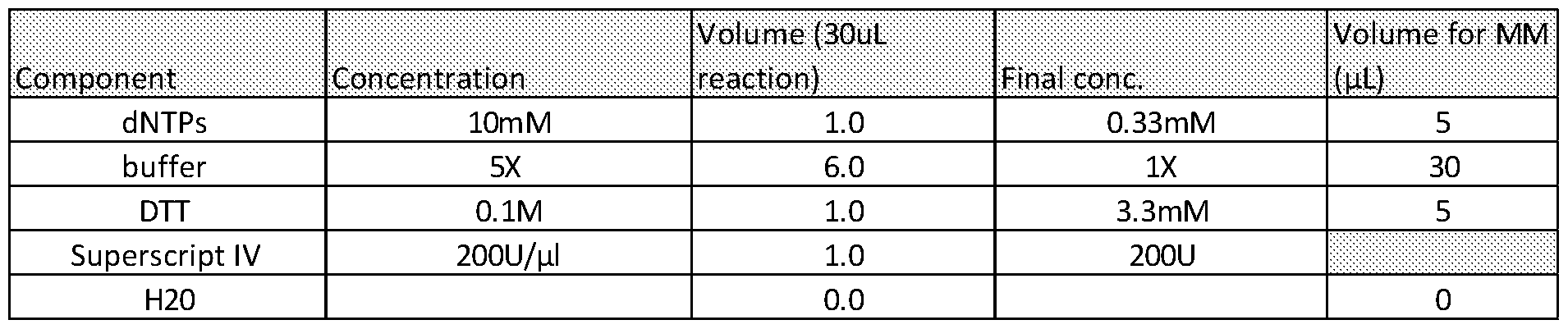
Table 45
1 mI_ of Superscript IV was added to the samples and placed in the PCR block under the following conditions (as detailed in Table 46):

Table 46
Clean up of the samples was then performed using Ampure bead clean up. Products were eluted in low EDTA TE buffer in quantities detailed in Table 47 below.

Table 47
Step 15 indexing PCR
The master mix for the indexing PCR was prepared as follows (as detailed in Table 48 below).

Table 48
17.5mI_ of the PCR master mix was added to 30mI_ of purified RT reactions. 2.5mI_ of the indexing oligo was added. Then the following PCR conditions were applied (as detailed in Table 49):

Table 49
Clean up of the product from the index PCR reaction was then performed using Ampure bead clean up. Products were eluted in low EDTA TE buffer in quantities detailed in Table 50 below.

Table 50
Samples were stored for KAPA qPCR quantification and NGS.
Example 10: Comparison of methylation enriched libraries prepared by the present method and a commercially available method
Experiments were conducted to compare the methylation enriched libraries generated by the present method (T7 method) with a commercial non-UMI based NGS library preparation method (current method). The commercial method comprises end repair and A tailing of the DNA, followed by ligation of the circular adapter from the NEBNext Multiplex Oligos for lllumina Index Primers kit (New England BioLabs Inc.) using KAPA-Hyper DNA ligase and KAPA-Hyper ligation buffer. The circular adapter is opened using USER enzyme digestion from the NEBNext Multiplex Oligos for lllumina Index Primers kit (New England BioLabs Inc.). The adapter ligated DNA was enriched for methylated DNA using the EpiMark Methylated DNA Enrichment Kit (New England BioLabs Inc.). Methylation enriched DNA was amplified in an NGS PCR, which added NGS adapters and a sample index. Samples were pooled and quantified by KAPA qPCR, and then run on an lllumina NextSeq instrument. Individual methylation enriched NGS libraries were prepared for each sample using the T7 method described in the present invention. In addition, samples were also pooled after the first ligation of the T7 circular adapter and this pool of samples was enriched for methylated
DNA and NGS libraries generated using the T7 method described in the present invention. Figure 10 A and B show that the T7 method results in a much higher enrichment of methylated DNA as well as reducing the background of non-methylated DNA (reads that do not contain a CpG). Comparison of the T7 method run on a pool of samples compared to individual samples shows that pooling the samples results in more uniform data. Figure 10C shows that tumour and matched normal samples can be separated based on the methylation patterns determined by the T7 method. Figure 10D shows that the method is picking out known tumour-specific methylation, that is not detected in the matched normal tissue sample.
Example 11 - Further comparison of methylation enriched libraries prepared by the present method and a commercially available method
Experiments were conducted to compare the methylation enriched libraries generated by the T7 method described in the present invention with a commercial UMI based NGS library preparation method. The samples used were a dilution series of sheared H446 cell line DNA diluted in sheared gDNA from healthy normal volunteer PBMCs. The dilution series was 100% cell line, 5% cell line, 2.5% cell line, 1.0% cell line, 0.1 % cell line, 0.01 % cell line, 0.001% cell line and 0% cell line (100% HNV PBMC) Briefly the commercial UMI based NGS library preparation method started with end repair and A tailing of the DNA, followed by ligation of the xGen Dual Index UMI Adapters from IDT (Integrated DNA Technologies) using KAPA-Hyper DNA ligase and KAPA-Hyper ligation buffer. The adapter ligated DNA was pooled and enriched for methylated DNA using the EpiMark Methylated DNA Enrichment Kit (New England BioLabs Inc.). Methylation enriched DNA was amplified in an NGS PCR. Samples were quantified by KAPA qPCR, and then run on an lllumina NextSeq instrument. Figure 11 A and B show that the T7 method results in a slightly higher enrichment of methylated DNA, but greatly reduces the background of non-methylated DNA (reads that do not contain a CpG). Figure 11C shows that in windows (of 200bp) that are methylated in the cell line but not in the HNV PBMCs, there are more reads in the T7 method at lower dilutions of the cell line, indicating that the T7 method is more sensitive than the commercial UMI adapters.
Example 12 - Fragmentation of DNA by restriction enzyme digest prior to ligation with circular adapter
Experiments were performed to assess the suitability of fragmenting DNA by restriction enzyme digest prior to ligation to the circular adapter. As outlined in the table in Figure 12A, two restriction enzymes which result in blunt end DNA fragments were tested, MLyl and BsuRI. A third restriction enzyme, Nialll, which results in an overhang of 4 nucleotides at the 3’ end of the fragment (was also tested. The agarose gel in Figure 12A shows that all three
restriction enzymes result in fragmentation of the high molecular weight gDNA. The circular adapter was ligated to the digested DNA products and aRNA products generated. The agarose gel in Figure 12B shows that DNA digested by the two blunt end restriction enzymes (BsuRI and MLyl) give the most aRNA product.
Example 13 - In-silico folding of the adapter.
In-silico folding of the circular adapter with 50 different barcodes was performed using Quikfold software. Table 51 below demonstrates the folding of the hairpin adapter oligonucleotide is unaffected by the sequence of the barcode.


Table 51
Example 14 - Formation of circular adapter using various buffers.
Experiments were conducted to assess the formation of the circular adapter in various buffers. The buffers tested were TE buffer (Invitrogen, Fisher Scientific), TNE buffer (Tris 100mM, NaCI 500mM and EDTA 10mM) and JENA buffer 5 (Jena Bioscience). The TNE buffer and JENA buffer 5 were also tested with increasing amounts of PEG present in the reaction. aRNA products formed following T7 RNA transcription were analysed by agarose gel and showed that the hairpin formation works in all buffer conditions (data not shown). Table 52 below shows the number of copies of aRNA produced under each condition as quantified by RNase P qPCR.

Table 52
Example 15 - Pooling of multiple samples post ligation
Experiments were conducted to assess the suitability of pooling multiple samples after ligation of the circular adapter each containing an individual sample barcode. In total circular adapters containing 38 different sample barcodes were ligated to cfDNA samples. These
were pooled and NGS libraries were prepared and sequenced on an lllumina MiSeq instrument. Samples were demultiplexed and the plot in Figure 14 shows the distribution of sequencing reads across the 38 samples.
Example 16 - Optimisation of addition of second NGS adapter to aRNA
Experiments were conducted to optimise the addition of the second NGS adapter to aRNA comparing the four approaches described in Figure 15C and 15D. For the single strand ligation approach (6a(i)), a 5’-phosphorylated NGS adapter was ligated to aRNA using T4 RNA ligase (New England BioLabs Inc.). This was compared to a second T4 RNA ligase, mutant T4 RNA Ligase 2, truncated KQ (New England BioLabs Inc.), that was used to ligate a 5’-adenylated NGS adapter to aRNA. Also compared was the necessity for a bead-based clean up step after the ligation step, before converting to cDNA by RT using a primer complementary to the ligated NGS adapter (U2P_RT_1). A splint double strand ligation (Figure 15C part 6aii) was tested with and without the presence of 12.5% PEG-8000. The RT with random primer method (Figure 1 step 6b and Figure 15D step 6b) was tested with (Figure 15D step 6b(ii)) and without (Figure 15D step 6b(i)) the presence of double stranded primer to guide annealing to the end of the aRNA molecule. Finally, all cDNA products from each method were amplified in an index PCR, which added the final NGS adapter sequences and a library index. The agarose gel in Figure 15A shows that the best conditions were the use of the mutant T4 RNA Ligase 2, truncated KQ (New England BioLabs Inc.) with a post ligation clean up step.
Experiments were conducted to compare methods for the addition of the second NGS adapter (Figure 1 step 6). Single strand ligation using mutant T4 RNA Ligase 2, truncated KQ (New England BioLabs Inc.) to ligate a 5’-adenylated NGS adapter was performed with and without phosphatase treatment of the aRNA prior to ligation. The RT with random primer method (Figure 1 step 6b) was tested with two different concentrations of random primer, 100 mM and 50 pM. Single strand ligation with phosphatase treatment of the aRNA are the best conditions as shown in the agarose gel on Figure 15A.
The protocol for optimised single strand ligation of aRNA is as follows;
aRNA was generated as described in steps 1-6 of Example 8.
Samples were prepared as shown below in Table 53

Table 53
Step 7 - dephosphorylation of aRNA
8mI_ of AC-T7-003_aRNA_was combined with 1 mI_ of AP fast phosphatase (1 U/pL) and placed on the PCR block using the conditions detailed in Table 54 below;

Table 54
Step 8 - ssligation
A 1 mM solution of the adapter oligonucleotide 5'Adenyl-U2-C3sp (AGATCGGAAGAGCACACGTC, 5' modification = adenylation, 3' modification = C3 spacer) was prepared. The following ligation master mix was prepared according to the conditions outlined in Table 55 below;

Table 55
1 mI_ of adapter was added to each sample followed by 10mI_ of ligation mastermix. Ligation was then performed at 16°C overnight.
Clean-up of the ligation products was performed by Ampure bead clean up according to the conditions in Table 56. Products were eluted in a low EDTA TE buffer

Table 56
Step 9 - preparation of RT oligo
The components were combined as outlined in Table 57.

Table 57
The components outlined in Table 57 were denatured and annealed using the conditions outlined in Table 58 below.

Table 58
The RT reaction master mix was prepared as outlined in Table 59.

Table 59 8mI_ of RT reaction master mix was added to the aRNA/U2P_primer mix, followed by 1 mI_ of Superscript IV. Samples were placed in the PCR block and the conditions outlined in Table 60 were applied

Table 60
The RT reaction products were cleaned up using Ampure bead clean up using the conditions outlined in Table 61. Products were eluted in low EDTA TE buffer.

Table 61
Step 10 - Indexing PCR
The indexing PCR master mix was prepared using the conditions outlined in Table 62.

17.5mI_ of the master mix was added to 30mI_ of the purified RT reactions. 2.5mI_ of the indexing oligo was added and the sampled placed in the PCR block. The conditions outlined in Table 63 were applied.

Table 63
Samples were then clean-up using Ampure bead clean up and products eluted in low EDTA TE buffer. Samples were stored before quantification and NGS.
Example 17 - Amplification performed with various amounts of cfDNA.
Experiments were conducted to assess the efficiency of the method on a range of cfDNA input amounts from 2 ng to 25 ng. Figures 16A-C show that aRNA is produced at all inputs of cfDNA. The fold change of aRNA relative to the input DNA shows that the method is as efficient even to low levels of cfDNA.
The forgoing embodiments are not intended to limit the scope of the protection afforded by the claims, but rather to describe examples of how the invention may be put into practice.
Sequences
The following sequences were used in the experiments undertaken by the present inventors:
Key
T7RNA_P T7 RNA Polymerase promoter sequence
MBC Molecular Bar Code
SBC Sample Bar Code
U1 P Partial Unique NGS Sequence 1
U2 Unique NGS Sequence 2
T7dT L)1 Lcf1 (SEQ ID N0.3)
S’phos sec us uie rmmj*

GGCCGG TAATC ACTAGGTC N AG AT C GG AAG AGCGT CGT GC 6 C C T ATAOTG AGTCGT A i J¾CC GGCC ? 3’€3 spacer
T7dT U1 Lcf1 Reverse (SEQ ID NO.4)
3’ T7RNAJ» U1P SIC SBC

TCCGGCCATATGCTGAGTGATATCC iCGTGC TGC G G A GGO TAG AS H N M N H CTCG ATC ACT
AATGGCCGG 5y
U2P Liq1 (SEQ ID NO.5)
5’ U2 3’
AGATCGGAAGAGCACACGTC modified with a phosphate group at 5' and a C3 spacer (blocker) at the 3'
U2P RT 1 (SEQ ID N0.6)
5’ U2 3’
GACGTGTGCTCTTCCGATCT
U2P RT 1 Reversed (SEQ ID N0.7)
TCTAGCCTTCTCGTGTGCAG
3’ U2 5’
T7 U1 Hsp92M02c3 (SEQ ID N0.8)
• Designed anneal to Hsp92ll digested DNA
• Contains 2 of the 4 bases in the overhang - prevents primer dimer
• Can order without 3’C3 spacer
5rPhos sac M C U1F rmmj*

AGGCCGGTAATCACTAeCTC NNNN N N AGATCGG AAGAGCGTCGTGCCCCTAT AGTGAGTCGTA
TT CCGGCCrCA 3 'C3 spacer
T7 U1 Hsp92M03c3 (SEQ ID NO.9)
• Designed anneal to Hsp92ll digested DNA
• Contains 3 of the 4 bases in the overhang - prevents primer dimer
• Can order without 3’C3 spacer

AGGCCGGTAATCACTAGCTC NNN AGATCGGAAGAGCGTCGTGCCCCTATAGTGAGTCGTA
TTACCGGCC fCAT 3'C3 spacer
lllumina Universal (SEQ ID NO.10)

REVERSE OF lllumina Index (1 ) (SEQ ID N0.12)

TC TAG€ CTFC TCGTGTGC AGAC TTGAGG TC AG TG TAGTGC T LL ACC A T C S G ALL A ALL PC G A AC
Tailed - cfDNA T7 Primers and Process
T7dT U1 Lcf1 (SEQ ID N0.13)

GGCCGGTAATCACTAGCTC NNMMN N AG ATC GG AAG AG CGT CGT GCCCCTAT AG TGAGTCGTA
TTACCGGCCT 3'C3 spacer
T7dT U1 Lcf1 Reverse (SEQ ID N0.14)

CCGGCCATTATGCTGAGTGATATCCCCGTSCTSCSAGAAGGCTAGA NN NNCTCGATCACT
AATGGCCGG S'
T7dT U1 Lcf1 cfDNA Ligation Product (SEQ ID NQ.15)

GGCCGGTAATACGACTCACTATAGGGGCACGACGCTCTTCCGATCT
T7dT U1 Lcf1 cfDNA Predicted aRNA (SEQ ID NQ.17)

Using U2P_Lig1
5' U1P nee sec

GGGGCACGACGCTCTTCC GATCTN N M N NG AGCTAGTG ATTACCGGCC TXXXXXXXXXXXXXXX
XXXXX AG A?C GCA AG ACC AC AC GTC 3' * U2F_Ligl
Ligated aRNA 1st RT Product (SEQ ID NO.19)
Using U2P_RT_1
3’ M C SB
L.
 A
A
r
CCCCGTGCT6C L6 AAG CTA6 AN N N N N NCTCG ATCAC TAATGGCCGG AZZZ2ZZZ2ZZZ2ZZ2 ZZZZZ7CTAGCCTTCTCG7GTGCAG 5
V. J
~Ύ~
U2P
NGS PCR 1st Cycle on RT Product (SEQ ID NO.20)
Using lllumina Universal
YΈL m

G AA. TGATACGGCGACCACCGAGATCTACACTCTTTCCCTACACGACGCTCTTCCGATCT NNNN

NGS PCR 2nd Cycle on 1st Cycle Product
Using lllumina Index (SEQ ID NO.21 )

Reverse (SEQ ID N0.22)
5’ PHI U1 »€
JL AAAT(SATACGGOGAOC-.ACC¾A¾ATCTACACTCTTTCCCTACACGACGCTGTTOCGATCT^ hS?<i

• Designed anneal to Hsp92ll digested DNA
• Contains 3 of the 4 bases in the overhang - prevents primer dimer
• Can order without 3’C3 spacer

Gansauge, M. T., and Meyer, M. (2013). Single-stranded DNA library preparation for the sequencing of ancient or damaged DNA. Nature Protocols 8, 737-748.
Gansauge, M. T., Gerber, T., Glocke, I., Korlevic, P., Lippik, L, Nagel, S., Riehl, L. M., Schmidt, A., and Meyer, M. (2017). Single-stranded DNA library preparation from highly degraded DNA using T4 DNA ligase. Nucleic Acids Research 45, e79.
Kwok, C. K., Ding, Y., Sherlock, M. E., Assmann, S. M., and Bevilacqua, P. C. (2013). A hybridization-based approach for quantitative and low-bias single-stranded DNA ligation. Analytical Biochemistry 435, 181-186.
Shen, S.Y., Singhania, R., Fehringer, G., Chakravarthy, A., Roehrl, M.H.A., Chadwick, D., Zuzarte, P.C., Borgida, A., Wang, T.T., Li, T., Kis, O., Zhao, Z., Spreafico, A., Medina, T., Wang, Y., Roulois, D., Ettayebi, I., Chen, Z., Chow, S., Murphy, T., Arruda, A., O’Kane, G.M., Liu, J., Mansour, M., McPherson, J.D., O’Brien, J., Leighl, N., Bedard, P.L., Fleshner, N., Liu, G., Minden, M.D., Gallinger, S., Goldenberg, A., Pugh, T.J., Hoffman, M.M., Bratman, S.V., Rayjean, Hung, J. and Carvalho, D.D. Sensitive tumour detection and classification using plasma cell-free DNA methylomes. (2018) Nature 563, 579-583.
Shina. A. A. I., Carrascosa, L.G., Liang, Z., Grewal, Y.S, Wardiana, A., Shiddiky, M.J.A., Gardiner, R.A., Samaratunga, H., Gandhi, M.K., Scott, R.J., Korbie, D. and Trau, M. (2018). Epigenetically reprogrammed methylation landscape drives the DNA self-assembly and serves as a universal cancer biomarker. Nature Comms 9, 4915, 1-13.
Claims
1. A method for amplifying template DNA, the method comprising: i) providing template DNA and an adapter oligonucleotide comprising an RNA polymerase promoter sequence, wherein the adapter oligonucleotide is configured so that a 5’ end of the adapter oligonucleotide can be ligated to a 3’ end of the template DNA; ii) ligating the 5’ end of the adapter oligonucleotide to the 3’ end of the template DNA to produce an adapter oligonucleotide-template DNA molecule; iii) providing a secondary oligonucleotide comprising an RNA polymerase promoter sequence, wherein the sequence of the secondary oligonucleotide is complementary to the sequence of the adapter oligonucleotide; iv) annealing the secondary oligonucleotide to the adapter oligonucleotide- template DNA molecule; and v) transcribing the template DNA by introducing an RNA polymerase to the adapter oligonucleotide-template DNA molecule, to generate an aRNA product.
2. The method according to claim 1 wherein the method further comprises the steps of: vi) converting the aRNA product to cDNA; and vii) amplifying the cDNA.
3. The method according to either claim 1 or claim 2, wherein step vi) first comprises ligating a priming site to the aRNA.
4. The method according to either claim 1 or claim 2, wherein step vi) comprises utilising reverse transcriptase and random oligonucleotide primers.
5. The method according to either claim 1 or claim 2, wherein step vi) comprises utilising reverse transcriptase and target specific oligonucleotide primers.
6. The method according to any one of claims 2-5, wherein step vii) comprises amplification in a next generation sequencing (NGS) protocol.
7. The method according to any preceding claim wherein the RNA polymerase promoter sequence comprises a T7 RNA polymerase promoter sequence.
8. The method according to any preceding claim, wherein the RNA promoter sequence in the adapter oligonucleotide comprises SEQ ID NO:1 or a fragment or variant thereof.
9. The method according to any preceding claim wherein the RNA promoter sequence in the secondary oligonucleotide comprises SEQ ID NO:2 or a fragment or variant thereof.
10. The method according to any preceding claim wherein the method further comprises dephosphorylating the template DNA prior to the ligation of step ii).
11. The method according to claim 10, wherein a 5’ phosphate of the template DNA is removed by treatment with a phosphatase enzyme.
12. The method according to claims 1-9 wherein the method further comprises a restriction enzyme digestion of the template DNA prior to the ligation of step ii).
13. The method according to any preceding claim wherein step ii) comprises ligating the 5’ end of the adapter oligonucleotide to the 3’ end of the template DNA using a DNA ligase.
14. The method according to claim 13 wherein the DNA ligase comprises KAPA-hyper DNA ligase.
15. The method according to any preceding claim, wherein the adapter oligonucleotide comprises complementary regions which are capable of annealing to one another to form circular or hairpin loop structure.
16. The method according to claim 15, wherein prior to step iv), the method further comprises the step of denaturing the adapter oligonucleotide prior to annealing the secondary oligonucleotide to the adapter oligonucleotide.
17. The method according to any preceding claim, wherein the adapter oligonucleotide comprises a molecular bar code sequence.
18. The method according to any preceding claim, wherein the adapter oligonucleotide comprises a sample bar code.
19. The method according to any preceding claim, wherein the adapter oligonucleotide comprises a NGS sequence.
20. The method according to any preceding claim, wherein the adapter oligonucleotide comprises a T tail.
21. The method according to any one of claims 12 to 20, wherein the adapter oligonucleotide further comprises a single region which is complementary to the stick ends produced by the restriction enzyme digestion.
22. The method according to any one of claims 12 to 20, wherein the adapter oligonucleotide further comprise a single region which is partially complementary to the stick ends produced by the restriction enzyme digestion.
23. The method according to any preceding claim, wherein the template DNA is derived from a biological sample, optionally blood.
24. The method according to any preceding claim, wherein the template DNA comprises cfDNA and/or ctDNA.
25. The method according to any one of claims 1 to 19, wherein the template DNA comprises gDNA.
26. The method according to any one of claims 1 to 19, wherein the template DNA comprises single cell DNA.
27. An adapter oligonucleotide for producing an aRNA product from a DNA template, the adapter oligonucleotide comprising an RNA polymerase promoter sequence, wherein the adapter oligonucleotide is configured so that a 5’ end of the adapter oligonucleotide can be ligated to the 3’ end of a template DNA.
28. The adapter according to claim 27, wherein the RNA polymerase promoter sequence comprises a T7 RNA polymerase promoter sequence.
29. The adapter according to either claim 27 or 28, wherein the RNA promoter sequence in the adapter oligonucleotide comprises SEQ ID NO:1 or a fragment or variant thereof
30. The adapter according to any of claims 27-29, wherein the adapter oligonucleotide comprises complementary regions which are capable of annealing to one another to form circular or hairpin loop structure.
31. The adapter according to any one of claims 27-30, wherein the adapter oligonucleotide further comprises a molecular bar code sequence.
32. The adapter according to any one of claims 27-31 , wherein the adapter further comprises a sample bar code.
33. The adapter according to any one of claims 27-32, wherein the adapter oligonucleotide further comprises a NGS sequence.
34. The adapter according to any one of claims 27-33, wherein the adapter oligonucleotide further comprises a T tail.
35. The adapter according to any one of claims 30-34, wherein the adapter further comprises an overhang comprising 6 to 12 oligonucleotides.
36. A kit for producing an aRNA product from a DNA template, the kit comprising: i) an adapter oligonucleotide comprising an RNA polymerase promoter sequence, wherein the adapter oligonucleotide is configured so that a 5’ end of the adapter oligonucleotide can be ligated to a 3’ end of a template DNA; and
ii) a secondary oligonucleotide comprising an RNA polymerase promoter sequence, wherein the sequence of the secondary oligonucleotide is complementary to the sequence of the adapter oligonucleotide.
37. The kit according to claim 36 wherein the RNA polymerase promoter sequence comprises a T7 RNA polymerase promoter sequence.
38. The kit according to either claim 36 or 37, wherein the RNA promoter sequence in the adapter oligonucleotide comprises SEQ ID N0.1 or a fragment or variant thereof.
39. The kit according to any one of claims 36-38 wherein the RNA promoter sequence in the secondary oligonucleotide comprises SEQ ID NO.2 or a fragment or variant thereof.
40. The kit according to any one of claims 36-39, wherein the adapter oligonucleotide comprises complementary regions which are capable of annealing to one another to form circular or hairpin loop structure.
41. The kit according to any one of claims 36-40, wherein the adapter oligonucleotide further comprises a molecular bar code sequence.
42. The kit according to any one of claims 36-41 , wherein the adapter further comprises a sample bar code.
43. The kit according to any one of claims 36-42, wherein the adapter oligonucleotide further comprises a NGS sequence.
44. The kit according to any one of claims 36-43, wherein the adapter oligonucleotide further comprises a T tail.
45. A method for detecting a genetic variant or genetic signature in template DNA, comprising amplifying template DNA in the method according to any one of claims 1- 23 and analysing the amplified cDNA for the genetic variation or signature.
46. The method according to claim 45 wherein the genetic variation or signature comprises one or more of the following: DNA methylation, tumour-assocated mutations or sequence copy numbers.
47. Use of a phosphatase enzyme for dephosphorylating template DNA prior to amplification, or modification, prior to amplification.
48. Use according to claim 47, wherein the phosphatase enzyme is for removing the 5’ phosphate of the template DNA.
49. Use according to either claim 47 or 48, wherein phosphatase enzyme is selected from one or more of the following: Shrimp Alkaline Phosphatase; Antarctic Phosphatase; Calf Intestinal Alkaline Phosphatase; and/or or FastAP Thermosensitive Alkaline Phosphatase.
Applications Claiming Priority (2)
| Application Number | Priority Date | Filing Date | Title |
|---|---|---|---|
| GBGB1903391.9A GB201903391D0 (en) | 2019-03-12 | 2019-03-12 | Nucleic acid amplification methods |
| GB1903391.9 | 2019-03-12 |
Publications (1)
| Publication Number | Publication Date |
|---|---|
| WO2020183188A1 true WO2020183188A1 (en) | 2020-09-17 |
Family
ID=66380247
Family Applications (1)
| Application Number | Title | Priority Date | Filing Date |
|---|---|---|---|
| PCT/GB2020/050635 Ceased WO2020183188A1 (en) | 2019-03-12 | 2020-03-12 | Nucleic acid amplification methods |
Country Status (2)
| Country | Link |
|---|---|
| GB (1) | GB201903391D0 (en) |
| WO (1) | WO2020183188A1 (en) |
Cited By (1)
| Publication number | Priority date | Publication date | Assignee | Title |
|---|---|---|---|---|
| CN113549674A (en) * | 2021-04-22 | 2021-10-26 | 福建和瑞基因科技有限公司 | Method for detecting target sequence integration and mutation in sample, design method of primer thereof and kit |
Citations (7)
| Publication number | Priority date | Publication date | Assignee | Title |
|---|---|---|---|---|
| WO1991018115A1 (en) * | 1990-05-16 | 1991-11-28 | Life Technologies, Inc. | Promoter ligation activated transcription amplification of nucleic acid sequences |
| WO1992018521A1 (en) * | 1991-04-10 | 1992-10-29 | Life Technologies, Inc. | Method for amplifying and altering an rna sequence |
| WO1997047762A1 (en) * | 1996-06-14 | 1997-12-18 | Sarnoff Corporation | Method for amplifying a polynucleotide |
| US20030104432A1 (en) * | 2001-07-27 | 2003-06-05 | The Regents Of The University Of California | Methods of amplifying sense strand RNA |
| WO2004101749A2 (en) * | 2003-05-09 | 2004-11-25 | Genisphere, Inc. | Methods for amplification of nucleic acid sequences using staggered ligation |
| WO2015112949A2 (en) * | 2014-01-27 | 2015-07-30 | ArcherDX, Inc. | Isothermal methods and related compositions for preparing nucleic acids |
| WO2015117040A1 (en) * | 2014-01-31 | 2015-08-06 | Swift Biosciences, Inc. | Improved methods for processing dna substrates |
-
2019
- 2019-03-12 GB GBGB1903391.9A patent/GB201903391D0/en not_active Ceased
-
2020
- 2020-03-12 WO PCT/GB2020/050635 patent/WO2020183188A1/en not_active Ceased
Patent Citations (7)
| Publication number | Priority date | Publication date | Assignee | Title |
|---|---|---|---|---|
| WO1991018115A1 (en) * | 1990-05-16 | 1991-11-28 | Life Technologies, Inc. | Promoter ligation activated transcription amplification of nucleic acid sequences |
| WO1992018521A1 (en) * | 1991-04-10 | 1992-10-29 | Life Technologies, Inc. | Method for amplifying and altering an rna sequence |
| WO1997047762A1 (en) * | 1996-06-14 | 1997-12-18 | Sarnoff Corporation | Method for amplifying a polynucleotide |
| US20030104432A1 (en) * | 2001-07-27 | 2003-06-05 | The Regents Of The University Of California | Methods of amplifying sense strand RNA |
| WO2004101749A2 (en) * | 2003-05-09 | 2004-11-25 | Genisphere, Inc. | Methods for amplification of nucleic acid sequences using staggered ligation |
| WO2015112949A2 (en) * | 2014-01-27 | 2015-07-30 | ArcherDX, Inc. | Isothermal methods and related compositions for preparing nucleic acids |
| WO2015117040A1 (en) * | 2014-01-31 | 2015-08-06 | Swift Biosciences, Inc. | Improved methods for processing dna substrates |
Non-Patent Citations (6)
| Title |
|---|
| GANSAUGE, M. T.GERBER, T.GLOCKE, I.KORLEVIC, P.LIPPIK, L.NAGEL, S.RIEHL, L. M.SCHMIDT, A.MEYER, M.: "Single-stranded DNA library preparation from highly degraded DNA using T4 DNA ligase", NUCLEIC ACIDS RESEARCH, vol. 45, 2017, pages e79 |
| GANSAUGE, M. T.MEYER, M.: "Single-stranded DNA library preparation for the sequencing of ancient or damaged DNA", NATURE PROTOCOLS, vol. 8, 2013, pages 737 - 748, XP055550541, DOI: 10.1038/nprot.2013.038 |
| KWOK, C. K.DING, Y.SHERLOCK, M. E.ASSMANN, S. M.BEVILACQUA, P. C.: "A hybridization-based approach for quantitative and low-bias single-stranded DNA ligation", ANALYTICAL BIOCHEMISTRY, vol. 435, 2013, pages 181 - 186, XP028988413, DOI: 10.1016/j.ab.2013.01.008 |
| MORGANE BOONE ET AL: "Capturing the 'ome': the expanding molecular toolbox for RNA and DNA library construction", NUCLEIC ACIDS RESEARCH, vol. 46, no. 6, 5 March 2018 (2018-03-05), pages 2701 - 2721, XP055681576, ISSN: 0305-1048, DOI: 10.1093/nar/gky167 * |
| SHEN, S.Y.SINGHANIA, R.FEHRINGER, G.CHAKRAVARTHY, A.ROEHRL, M.H.A.CHADWICK, D.ZUZARTE, P.C.BORGIDA, A.WANG, T.T.LI, T.: "Sensitive tumour detection and classification using plasma cell-free DNA methylomes", NATURE, vol. 563, 2018, pages 579 - 583, XP036867481, DOI: 10.1038/s41586-018-0703-0 |
| SHINA. A. A. I.CARRASCOSA, L.G.LIANG, Z.GREWAL, Y.SWARDIANA, A.SHIDDIKY, M.J.A.GARDINER, R.A.SAMARATUNGA, H.GANDHI, M.K.SCOTT, R.J: "Epigenetically reprogrammed methylation landscape drives the DNA self-assembly and serves as a universal cancer biomarker", NATURE COMMS, vol. 9, no. 4915, 2018, pages 1 - 13 |
Cited By (2)
| Publication number | Priority date | Publication date | Assignee | Title |
|---|---|---|---|---|
| CN113549674A (en) * | 2021-04-22 | 2021-10-26 | 福建和瑞基因科技有限公司 | Method for detecting target sequence integration and mutation in sample, design method of primer thereof and kit |
| CN113549674B (en) * | 2021-04-22 | 2023-11-03 | 福建和瑞基因科技有限公司 | Method for detecting integration and mutation of target sequence in sample, and design method and kit of primer of method |
Also Published As
| Publication number | Publication date |
|---|---|
| GB201903391D0 (en) | 2019-04-24 |
Similar Documents
| Publication | Publication Date | Title |
|---|---|---|
| US12577606B2 (en) | Gene target region enrichment method and kit | |
| EP2914745B1 (en) | Barcoding nucleic acids | |
| CN111712580B (en) | Method and kit for amplifying double-stranded DNA | |
| US20160304948A1 (en) | Methods for selectively amplifying and tagging nucleic acids | |
| JP2018516569A (en) | Methods and related compositions and kits for next generation genome walking | |
| CN105121664A (en) | Methods of sequencing nucleic acids in mixtures and compositions related thereto | |
| US12297492B2 (en) | Amplification of single stranded DNA | |
| EP3607065A1 (en) | Method and kit for constructing nucleic acid library | |
| AU2016255570A1 (en) | Compositions and methods for constructing strand specific cDNA libraries | |
| JP7212224B2 (en) | Methods for target-specific RNA transcription of DNA sequences | |
| CN112941147B (en) | High-fidelity target gene library construction method and kit thereof | |
| WO2020183188A1 (en) | Nucleic acid amplification methods | |
| US20180291369A1 (en) | Error-proof nucleic acid library construction method and kit | |
| CN112680796A (en) | Target gene enrichment and library construction method | |
| CN115896243A (en) | Ultrasensitive reverse transcription method based on mixed enzyme system and its application | |
| US20210115435A1 (en) | Error-proof nucleic acid library construction method | |
| CN114144188B (en) | Method for amplifying and detecting RNA fragments | |
| US12577616B2 (en) | Multiplex method of preparing a sequencing library | |
| CN121065310A (en) | Method for barcoding molecules in single cell experiments | |
| WO2015199939A1 (en) | Compositions and methods for amplifying and determining nucleic acid sequences | |
| HK40052623A (en) | Nuclease-based rna depletion |
Legal Events
| Date | Code | Title | Description |
|---|---|---|---|
| 121 | Ep: the epo has been informed by wipo that ep was designated in this application |
Ref document number: 20712670 Country of ref document: EP Kind code of ref document: A1 |
|
| NENP | Non-entry into the national phase |
Ref country code: DE |
|
| 122 | Ep: pct application non-entry in european phase |
Ref document number: 20712670 Country of ref document: EP Kind code of ref document: A1 |