WO2020081833A1 - Nano-satellite complexes - Google Patents
Nano-satellite complexes Download PDFInfo
- Publication number
- WO2020081833A1 WO2020081833A1 PCT/US2019/056765 US2019056765W WO2020081833A1 WO 2020081833 A1 WO2020081833 A1 WO 2020081833A1 US 2019056765 W US2019056765 W US 2019056765W WO 2020081833 A1 WO2020081833 A1 WO 2020081833A1
- Authority
- WO
- WIPO (PCT)
- Prior art keywords
- composition
- satellite
- her2
- particles
- satellite particles
- Prior art date
- Legal status (The legal status is an assumption and is not a legal conclusion. Google has not performed a legal analysis and makes no representation as to the accuracy of the status listed.)
- Ceased
Links
Classifications
-
- A—HUMAN NECESSITIES
- A61—MEDICAL OR VETERINARY SCIENCE; HYGIENE
- A61K—PREPARATIONS FOR MEDICAL, DENTAL OR TOILETRY PURPOSES
- A61K47/00—Medicinal preparations characterised by the non-active ingredients used, e.g. carriers or inert additives; Targeting or modifying agents chemically bound to the active ingredient
- A61K47/50—Medicinal preparations characterised by the non-active ingredients used, e.g. carriers or inert additives; Targeting or modifying agents chemically bound to the active ingredient the non-active ingredient being chemically bound to the active ingredient, e.g. polymer-drug conjugates
- A61K47/69—Medicinal preparations characterised by the non-active ingredients used, e.g. carriers or inert additives; Targeting or modifying agents chemically bound to the active ingredient the non-active ingredient being chemically bound to the active ingredient, e.g. polymer-drug conjugates the conjugate being characterised by physical or galenical forms, e.g. emulsion, particle, inclusion complex, stent or kit
- A61K47/6921—Medicinal preparations characterised by the non-active ingredients used, e.g. carriers or inert additives; Targeting or modifying agents chemically bound to the active ingredient the non-active ingredient being chemically bound to the active ingredient, e.g. polymer-drug conjugates the conjugate being characterised by physical or galenical forms, e.g. emulsion, particle, inclusion complex, stent or kit the form being a particulate, a powder, an adsorbate, a bead or a sphere
- A61K47/6927—Medicinal preparations characterised by the non-active ingredients used, e.g. carriers or inert additives; Targeting or modifying agents chemically bound to the active ingredient the non-active ingredient being chemically bound to the active ingredient, e.g. polymer-drug conjugates the conjugate being characterised by physical or galenical forms, e.g. emulsion, particle, inclusion complex, stent or kit the form being a particulate, a powder, an adsorbate, a bead or a sphere the form being a solid microparticle having no hollow or gas-filled cores
- A61K47/6929—Medicinal preparations characterised by the non-active ingredients used, e.g. carriers or inert additives; Targeting or modifying agents chemically bound to the active ingredient the non-active ingredient being chemically bound to the active ingredient, e.g. polymer-drug conjugates the conjugate being characterised by physical or galenical forms, e.g. emulsion, particle, inclusion complex, stent or kit the form being a particulate, a powder, an adsorbate, a bead or a sphere the form being a solid microparticle having no hollow or gas-filled cores the form being a nanoparticle, e.g. an immuno-nanoparticle
-
- A—HUMAN NECESSITIES
- A61—MEDICAL OR VETERINARY SCIENCE; HYGIENE
- A61K—PREPARATIONS FOR MEDICAL, DENTAL OR TOILETRY PURPOSES
- A61K38/00—Medicinal preparations containing peptides
- A61K38/16—Peptides having more than 20 amino acids; Gastrins; Somatostatins; Melanotropins; Derivatives thereof
- A61K38/17—Peptides having more than 20 amino acids; Gastrins; Somatostatins; Melanotropins; Derivatives thereof from animals; from humans
- A61K38/19—Cytokines; Lymphokines; Interferons
- A61K38/191—Tumor necrosis factors [TNF], e.g. lymphotoxin [LT], i.e. TNF-beta
-
- A—HUMAN NECESSITIES
- A61—MEDICAL OR VETERINARY SCIENCE; HYGIENE
- A61K—PREPARATIONS FOR MEDICAL, DENTAL OR TOILETRY PURPOSES
- A61K39/00—Medicinal preparations containing antigens or antibodies
- A61K39/0005—Vertebrate antigens
- A61K39/0011—Cancer antigens
-
- A—HUMAN NECESSITIES
- A61—MEDICAL OR VETERINARY SCIENCE; HYGIENE
- A61K—PREPARATIONS FOR MEDICAL, DENTAL OR TOILETRY PURPOSES
- A61K39/00—Medicinal preparations containing antigens or antibodies
- A61K39/0005—Vertebrate antigens
- A61K39/0011—Cancer antigens
- A61K39/001102—Receptors, cell surface antigens or cell surface determinants
- A61K39/001103—Receptors for growth factors
- A61K39/001106—Her-2/neu/ErbB2, Her-3/ErbB3 or Her 4/ErbB4
-
- A—HUMAN NECESSITIES
- A61—MEDICAL OR VETERINARY SCIENCE; HYGIENE
- A61K—PREPARATIONS FOR MEDICAL, DENTAL OR TOILETRY PURPOSES
- A61K39/00—Medicinal preparations containing antigens or antibodies
- A61K39/12—Viral antigens
- A61K39/295—Polyvalent viral antigens; Mixtures of viral and bacterial antigens
-
- A—HUMAN NECESSITIES
- A61—MEDICAL OR VETERINARY SCIENCE; HYGIENE
- A61K—PREPARATIONS FOR MEDICAL, DENTAL OR TOILETRY PURPOSES
- A61K39/00—Medicinal preparations containing antigens or antibodies
- A61K39/39—Medicinal preparations containing antigens or antibodies characterised by the immunostimulating additives, e.g. chemical adjuvants
-
- A—HUMAN NECESSITIES
- A61—MEDICAL OR VETERINARY SCIENCE; HYGIENE
- A61K—PREPARATIONS FOR MEDICAL, DENTAL OR TOILETRY PURPOSES
- A61K40/00—Cellular immunotherapy
- A61K40/10—Cellular immunotherapy characterised by the cell type used
- A61K40/11—T-cells, e.g. tumour infiltrating lymphocytes [TIL] or regulatory T [Treg] cells; Lymphokine-activated killer [LAK] cells
-
- A—HUMAN NECESSITIES
- A61—MEDICAL OR VETERINARY SCIENCE; HYGIENE
- A61K—PREPARATIONS FOR MEDICAL, DENTAL OR TOILETRY PURPOSES
- A61K40/00—Cellular immunotherapy
- A61K40/10—Cellular immunotherapy characterised by the cell type used
- A61K40/13—B-cells
-
- A—HUMAN NECESSITIES
- A61—MEDICAL OR VETERINARY SCIENCE; HYGIENE
- A61K—PREPARATIONS FOR MEDICAL, DENTAL OR TOILETRY PURPOSES
- A61K40/00—Cellular immunotherapy
- A61K40/20—Cellular immunotherapy characterised by the effect or the function of the cells
- A61K40/24—Antigen-presenting cells [APC]
-
- A—HUMAN NECESSITIES
- A61—MEDICAL OR VETERINARY SCIENCE; HYGIENE
- A61K—PREPARATIONS FOR MEDICAL, DENTAL OR TOILETRY PURPOSES
- A61K40/00—Cellular immunotherapy
- A61K40/40—Cellular immunotherapy characterised by antigens that are targeted or presented by cells of the immune system
- A61K40/41—Vertebrate antigens
- A61K40/42—Cancer antigens
- A61K40/4202—Receptors, cell surface antigens or cell surface determinants
- A61K40/4203—Receptors for growth factors
- A61K40/4205—Her-2/neu/ErbB2, Her-3/ErbB3 or Her 4/ ErbB4
-
- A—HUMAN NECESSITIES
- A61—MEDICAL OR VETERINARY SCIENCE; HYGIENE
- A61K—PREPARATIONS FOR MEDICAL, DENTAL OR TOILETRY PURPOSES
- A61K45/00—Medicinal preparations containing active ingredients not provided for in groups A61K31/00 - A61K41/00
- A61K45/06—Mixtures of active ingredients without chemical characterisation, e.g. antiphlogistics and cardiaca
-
- A—HUMAN NECESSITIES
- A61—MEDICAL OR VETERINARY SCIENCE; HYGIENE
- A61K—PREPARATIONS FOR MEDICAL, DENTAL OR TOILETRY PURPOSES
- A61K47/00—Medicinal preparations characterised by the non-active ingredients used, e.g. carriers or inert additives; Targeting or modifying agents chemically bound to the active ingredient
- A61K47/02—Inorganic compounds
-
- A—HUMAN NECESSITIES
- A61—MEDICAL OR VETERINARY SCIENCE; HYGIENE
- A61K—PREPARATIONS FOR MEDICAL, DENTAL OR TOILETRY PURPOSES
- A61K47/00—Medicinal preparations characterised by the non-active ingredients used, e.g. carriers or inert additives; Targeting or modifying agents chemically bound to the active ingredient
- A61K47/50—Medicinal preparations characterised by the non-active ingredients used, e.g. carriers or inert additives; Targeting or modifying agents chemically bound to the active ingredient the non-active ingredient being chemically bound to the active ingredient, e.g. polymer-drug conjugates
- A61K47/51—Medicinal preparations characterised by the non-active ingredients used, e.g. carriers or inert additives; Targeting or modifying agents chemically bound to the active ingredient the non-active ingredient being chemically bound to the active ingredient, e.g. polymer-drug conjugates the non-active ingredient being a modifying agent
- A61K47/62—Medicinal preparations characterised by the non-active ingredients used, e.g. carriers or inert additives; Targeting or modifying agents chemically bound to the active ingredient the non-active ingredient being chemically bound to the active ingredient, e.g. polymer-drug conjugates the non-active ingredient being a modifying agent the modifying agent being a protein, peptide or polyamino acid
-
- A—HUMAN NECESSITIES
- A61—MEDICAL OR VETERINARY SCIENCE; HYGIENE
- A61K—PREPARATIONS FOR MEDICAL, DENTAL OR TOILETRY PURPOSES
- A61K47/00—Medicinal preparations characterised by the non-active ingredients used, e.g. carriers or inert additives; Targeting or modifying agents chemically bound to the active ingredient
- A61K47/50—Medicinal preparations characterised by the non-active ingredients used, e.g. carriers or inert additives; Targeting or modifying agents chemically bound to the active ingredient the non-active ingredient being chemically bound to the active ingredient, e.g. polymer-drug conjugates
- A61K47/51—Medicinal preparations characterised by the non-active ingredients used, e.g. carriers or inert additives; Targeting or modifying agents chemically bound to the active ingredient the non-active ingredient being chemically bound to the active ingredient, e.g. polymer-drug conjugates the non-active ingredient being a modifying agent
- A61K47/62—Medicinal preparations characterised by the non-active ingredients used, e.g. carriers or inert additives; Targeting or modifying agents chemically bound to the active ingredient the non-active ingredient being chemically bound to the active ingredient, e.g. polymer-drug conjugates the non-active ingredient being a modifying agent the modifying agent being a protein, peptide or polyamino acid
- A61K47/64—Drug-peptide, drug-protein or drug-polyamino acid conjugates, i.e. the modifying agent being a peptide, protein or polyamino acid which is covalently bonded or complexed to a therapeutically active agent
- A61K47/646—Drug-peptide, drug-protein or drug-polyamino acid conjugates, i.e. the modifying agent being a peptide, protein or polyamino acid which is covalently bonded or complexed to a therapeutically active agent the entire peptide or protein drug conjugate elicits an immune response, e.g. conjugate vaccines
-
- A—HUMAN NECESSITIES
- A61—MEDICAL OR VETERINARY SCIENCE; HYGIENE
- A61K—PREPARATIONS FOR MEDICAL, DENTAL OR TOILETRY PURPOSES
- A61K47/00—Medicinal preparations characterised by the non-active ingredients used, e.g. carriers or inert additives; Targeting or modifying agents chemically bound to the active ingredient
- A61K47/50—Medicinal preparations characterised by the non-active ingredients used, e.g. carriers or inert additives; Targeting or modifying agents chemically bound to the active ingredient the non-active ingredient being chemically bound to the active ingredient, e.g. polymer-drug conjugates
- A61K47/69—Medicinal preparations characterised by the non-active ingredients used, e.g. carriers or inert additives; Targeting or modifying agents chemically bound to the active ingredient the non-active ingredient being chemically bound to the active ingredient, e.g. polymer-drug conjugates the conjugate being characterised by physical or galenical forms, e.g. emulsion, particle, inclusion complex, stent or kit
- A61K47/6921—Medicinal preparations characterised by the non-active ingredients used, e.g. carriers or inert additives; Targeting or modifying agents chemically bound to the active ingredient the non-active ingredient being chemically bound to the active ingredient, e.g. polymer-drug conjugates the conjugate being characterised by physical or galenical forms, e.g. emulsion, particle, inclusion complex, stent or kit the form being a particulate, a powder, an adsorbate, a bead or a sphere
- A61K47/6923—Medicinal preparations characterised by the non-active ingredients used, e.g. carriers or inert additives; Targeting or modifying agents chemically bound to the active ingredient the non-active ingredient being chemically bound to the active ingredient, e.g. polymer-drug conjugates the conjugate being characterised by physical or galenical forms, e.g. emulsion, particle, inclusion complex, stent or kit the form being a particulate, a powder, an adsorbate, a bead or a sphere the form being an inorganic particle, e.g. ceramic particles, silica particles, ferrite or synsorb
-
- A—HUMAN NECESSITIES
- A61—MEDICAL OR VETERINARY SCIENCE; HYGIENE
- A61K—PREPARATIONS FOR MEDICAL, DENTAL OR TOILETRY PURPOSES
- A61K9/00—Medicinal preparations characterised by special physical form
- A61K9/14—Particulate form, e.g. powders, Processes for size reducing of pure drugs or the resulting products, Pure drug nanoparticles
- A61K9/16—Agglomerates; Granulates; Microbeadlets ; Microspheres; Pellets; Solid products obtained by spray drying, spray freeze drying, spray congealing,(multiple) emulsion solvent evaporation or extraction
- A61K9/167—Agglomerates; Granulates; Microbeadlets ; Microspheres; Pellets; Solid products obtained by spray drying, spray freeze drying, spray congealing,(multiple) emulsion solvent evaporation or extraction with an outer layer or coating comprising drug; with chemically bound drugs or non-active substances on their surface
-
- A—HUMAN NECESSITIES
- A61—MEDICAL OR VETERINARY SCIENCE; HYGIENE
- A61P—SPECIFIC THERAPEUTIC ACTIVITY OF CHEMICAL COMPOUNDS OR MEDICINAL PREPARATIONS
- A61P35/00—Antineoplastic agents
-
- A—HUMAN NECESSITIES
- A61—MEDICAL OR VETERINARY SCIENCE; HYGIENE
- A61K—PREPARATIONS FOR MEDICAL, DENTAL OR TOILETRY PURPOSES
- A61K39/00—Medicinal preparations containing antigens or antibodies
- A61K2039/555—Medicinal preparations containing antigens or antibodies characterised by a specific combination antigen/adjuvant
- A61K2039/55511—Organic adjuvants
- A61K2039/55555—Liposomes; Vesicles, e.g. nanoparticles; Spheres, e.g. nanospheres; Polymers
-
- A—HUMAN NECESSITIES
- A61—MEDICAL OR VETERINARY SCIENCE; HYGIENE
- A61K—PREPARATIONS FOR MEDICAL, DENTAL OR TOILETRY PURPOSES
- A61K39/00—Medicinal preparations containing antigens or antibodies
- A61K2039/555—Medicinal preparations containing antigens or antibodies characterised by a specific combination antigen/adjuvant
- A61K2039/55511—Organic adjuvants
- A61K2039/55561—CpG containing adjuvants; Oligonucleotide containing adjuvants
-
- A—HUMAN NECESSITIES
- A61—MEDICAL OR VETERINARY SCIENCE; HYGIENE
- A61K—PREPARATIONS FOR MEDICAL, DENTAL OR TOILETRY PURPOSES
- A61K39/00—Medicinal preparations containing antigens or antibodies
- A61K2039/57—Medicinal preparations containing antigens or antibodies characterised by the type of response, e.g. Th1, Th2
- A61K2039/575—Medicinal preparations containing antigens or antibodies characterised by the type of response, e.g. Th1, Th2 humoral response
-
- A—HUMAN NECESSITIES
- A61—MEDICAL OR VETERINARY SCIENCE; HYGIENE
- A61K—PREPARATIONS FOR MEDICAL, DENTAL OR TOILETRY PURPOSES
- A61K39/00—Medicinal preparations containing antigens or antibodies
- A61K2039/60—Medicinal preparations containing antigens or antibodies characteristics by the carrier linked to the antigen
-
- A—HUMAN NECESSITIES
- A61—MEDICAL OR VETERINARY SCIENCE; HYGIENE
- A61K—PREPARATIONS FOR MEDICAL, DENTAL OR TOILETRY PURPOSES
- A61K39/00—Medicinal preparations containing antigens or antibodies
- A61K2039/62—Medicinal preparations containing antigens or antibodies characterised by the link between antigen and carrier
- A61K2039/627—Medicinal preparations containing antigens or antibodies characterised by the link between antigen and carrier characterised by the linker
-
- A—HUMAN NECESSITIES
- A61—MEDICAL OR VETERINARY SCIENCE; HYGIENE
- A61K—PREPARATIONS FOR MEDICAL, DENTAL OR TOILETRY PURPOSES
- A61K39/00—Medicinal preparations containing antigens or antibodies
- A61K2039/64—Medicinal preparations containing antigens or antibodies characterised by the architecture of the carrier-antigen complex, e.g. repetition of carrier-antigen units
-
- A—HUMAN NECESSITIES
- A61—MEDICAL OR VETERINARY SCIENCE; HYGIENE
- A61K—PREPARATIONS FOR MEDICAL, DENTAL OR TOILETRY PURPOSES
- A61K2239/00—Indexing codes associated with cellular immunotherapy of group A61K40/00
- A61K2239/46—Indexing codes associated with cellular immunotherapy of group A61K40/00 characterised by the cancer treated
- A61K2239/49—Breast
-
- B—PERFORMING OPERATIONS; TRANSPORTING
- B82—NANOTECHNOLOGY
- B82Y—SPECIFIC USES OR APPLICATIONS OF NANOSTRUCTURES; MEASUREMENT OR ANALYSIS OF NANOSTRUCTURES; MANUFACTURE OR TREATMENT OF NANOSTRUCTURES
- B82Y5/00—Nanobiotechnology or nanomedicine, e.g. protein engineering or drug delivery
Definitions
- the present invention provides methods, compositions, systems, and kits comprising nano-satellite complexes comprising: a core nanoparticle complex comprising a
- biocompatible coating surrounding a nanoparticle core 3-25 satellite particles attached to, or absorbed to, said biocompatible coating; a plurality of antigenic peptides conjugated to, or absorbed to, said satellite particles; and at least one additional property: i) a weight-to-weight ratio of all of the satellite particles to the nanoparticle core of 10-40%; a diameter of each of the satellite particles is 2-20 nm; iii) the satellite particles are present at density of 500-20,000 or 15,00-30,000 per square micron; iv) the plurality of antigenic peptides is 100-4000 antigenic peptides; v) 10-300 of the plurality of the antigenic peptides are present on each of the satellite particles; and/or vi) the average distance between each of the satellite particles is 5-20 nm.
- Viruses are known to be tremendously efficient delivery vehicles, mediators of cellular uptake and efficacious immunological agents. As such, it has become desirable to utilize viruses and viral properties in wide variety of biotechnology and medicinal applications. However, traditional live attenuated or inactivated viruses remain too dangerous to be employed in this way. To address this concern, virus-like particles have emerged.
- Virus-like particles are protein-based nanoparticles that are composed of viral capsid proteins that self-assemble into geometrically rigid nanostructures that directly resemble viral structure and confirmation without the viral genome. Thus, viral-like particles are considered a viable and safe alternative to traditional viruses.
- numerous obvious disadvantages of virus-like particle technology remain including reliance on protein self-assembly, difficult manufacturing, limited application versatility and significant anti carrier responses that limits re-dosing potential in-vivo.
- viral mimicking nanoparticles are rationally designed and engineered based on an understanding of viral physical and chemical material properties.
- the viral material properties most commonly utilized to inform the design of viral mimicking nanoparticles include: particle size, particle shape, charge, hydrophobicity, antigen display, antigen organization, antigen density and surface topography. While many advances have been made in the design, engineering and application of viral-mimicking nanoparticles, no one generally applicable nanoparticle system has emerged.
- the present invention provides methods, compositions, systems, and kits comprising nano-satellite complexes comprising: a core nanoparticle complex comprising a
- the biocompatible coating surrounding a nanoparticle core 3-25 satellite particles attached to, or absorbed to, said biocompatible coating; a plurality of antigenic peptides conjugated to, or absorbed to, said satellite particles; and at least one additional property: i) a weight-to-weight ratio of all of the satellite particles to the nanoparticle core of 10-40%; a diameter of each of the satellite particles is 2-20 nm; iii) the satellite particles are present at density of 500-20,000 or 15,00-30,000 per square micron; iv) the plurality of antigenic peptides is 100-4000 antigenic peptides; v) 10-300 of the plurality of the antigenic peptides are present on each of the satellite particles; and/or vi) the average distance between each of the satellite particles is 5-20 nm.
- the diameter of the nano-satellite complex is about 20-70 nm (e.g., about 25 nm, about 40-50 nm, or about 60 nm).
- compositions comprising: a nano- satellite complex, wherein the nano-satellite complex comprises: a) a core nanoparticle complex comprising a biocompatible coating surrounding a nanoparticle core; b) 3-25 satellite particles (e.g., 3 ... 7 ... 13 ... 17 ... 21 ...
- the nano-satellite complex comprises at least one (e.g., 1 , 2, 3, 4, 5, or 6) of the following properties: i) wherein the weight-to-weight ratio of all of the satellite particles to the nanoparticle core is 10-40% (e.g., 10% ... 20% ... 30% ... or 40%); ii) wherein the diameter of each of the satellite particles is 2-20 nm (e.g., 2 ... 5 ... 8 . . . 13 ...
- each of the satellite particles is 5-20 nm (e.g., 5.0 ...
- each of said satellite particles is 1-5 nm (e.g., about 1, 2, 3, 4, or 5 nm); iii) wherein said satellite particles are present at density of 15,00-30,000 per square micron (e.g., 15,000 ... 18,000 ... 21,000 ... 25,000 ... 28,000 ... or 30,000); iv) wherein said plurality of antigenic peptides is 1500-3000 antigenic peptides (e.g., about 1500 ... 1900 ... 2200 ... 2300 ... 2600 ... 3000); v) wherein 100-400 (e.g., 100 ... 200 ... 250 ... 300 ...
- kits for eliciting an immune response in a subject comprising: administering to a subject the composition as described herein such that antibodies to the antigenic peptides, or haptens, are generated.
- the subject is a human.
- the subject is an animal (e.g., dog, cat, pig, horse, etc.).
- the methods further comprise taking a sample from the subject, and purifying at some of the antibodies from the sample.
- no adjuvant is administered as part of the composition or otherwise.
- the subject is administering a type I interferon agonist agent, either in the composition or separately.
- the subject is administering an immune checkpoint inhibitor, either in the composition or separately.
- the antigenic peptides comprise B-Cell epitopes, or T-cell epitopes, or both (see, e.g., Table 4 or Table 1, Table 2, or Table 3).
- the nanosatellite complex does not generate detectable non-specific antibody against said nano-satellite complex in the subject.
- the nanosatellite complex homes to a lymph node of said subject (e.g., at a level equal to a virus).
- the nanosatellite complex homes to a B-cell zone or T-cell zone of a lymph node of said subject.
- the nanosatellite complex is taken up by subcapsular sinus macrophages in said subject at a rate equal to a virus.
- the satellite particles comprise gold.
- the core nanoparticle comprises Fe304.
- the biocompatible coating comprises polysiloxane.
- the nanoparticle core comprises Fe Or.
- the biocompatible coating comprises polysiloxane, and the at least one satellite particle comprises a plurality of satellite particles composed of gold.
- the 3-25 satellite particles is 10-15 satellite particles.
- the at least one property is wherein the weight-to-weight ratio of all of the satellite particles to the nanoparticle core is 10-40% (e.g., about 30%). In additional embodiments, the weight-to-weight ratio of all of the satellite particles to the nanoparticle core is 25-35%. In certain embodiments, the weight-to-weight ratio of all of the satellite particles to the nanoparticle core is 29-31%.
- the at least one property is wherein the diameter of each of the satellite particles is 2-20 nm. In certain embodiments, the diameter of each of the satellite particles is 5-15 nm. In further embodiments, the diameter of each of the satellite particles is 4-6 nm.
- the at least one property is wherein the satellite particles are present at density of 500-20,000 per square micron. In other embodiments, the satellite particles are present at a density of 13,000 to 17,000 per square micron.
- the at least one property is wherein the plurality of antigenic peptides is 100-4000 antigenic peptides or 100-4000 haptens. In other embodiments, the plurality of antigenic peptides is 1500-2500 antigenic peptides, or wherein the plurality of haptens is 1500-2500.
- the at least one property is wherein 10-300 of the plurality of the antigenic peptides, or haptens, are present on each of the satellite particles. In further embodiments, 225-275 of the plurality of antigenic peptides, or haptens, are present on each of the satellite particles.
- the at least one property is wherein the average distance between each of the satellite particles is 5-20 nm. In certain embodiments, the average distance between each of the satellite particles is 6-8 nm.
- the at least one property is at least two or three of the properties. In some embodiments, the at least one property is at least four or five of the properties. In additional embodiments, the at least one property is all six of the properties.
- the antigenic peptide comprises: i) a neoantigenic determinant, ii) at least one epitope from a tumor antigen, iii) at least one epitope from a viral oncoprotein, iv) a least one epitope from an infectious virus, v) at least one epitope from a parasite, or vi) at least one epitope from an infectious bacteria.
- the compositions, systems, and kits further comprise a physiologically compatible aqueous solution and/or cancer cells and/or antigen presenting cells.
- the plurality of antigenic peptides are not uniformly distributed on the satellite particles.
- the nano-satellite complex is a diameter of 50-100 nm (e.g., 55-65 nm).
- the surface of the nano-satellite complex is negatively charged (e.g., -10 to -20 mV).
- the core nanoparticle has a diameter of 10-25 nm (e.g., 15-20 nm).
- the composition further comprises a type I interferon agonist agent.
- the type I interferon agonist agent is electrostatically attracted to, or absorbed to, i) the antigenic peptides or haptens, ii) the plurality of satellite particles, and/or iii) the core nanoparticle.
- the compositions are adjuvant- free.
- the compositions further comprise an immune checkpoint inhibitor.
- the antigenic peptide comprises at least one neoantigenic determinant, including, for example, an oncogenic viral antigenic determinant.
- the antigenic peptides comprise at least one epitope from a tumor antigen, including a viral oncoprotein.
- the antigenic peptide comprises a least one epitope from an infectious virus, at least one epitope from a parasite, and/or at least one epitope from an infectious bacteria. Suitable antigens from viruses, parasites, and bacteria for immunizing subject (e.g., human subjects) are well known in the art (see, e.g., Tables 2 and 3).
- Additional antigens are in development for vaccines including, for example: Adenovirus vaccine, Coxsackie B virus vaccine, Cytomegalovirus vaccine, Dengue vaccine, Eastern Equine encephalitis virus vaccine, Ebola vaccine, Enterovirus 71 vaccine, Epstein- Barr vaccine, Hepatitis C vaccine, HIV vaccine, HTLV-l T-lymphotropic leukemia vaccine, Marburg virus disease vaccine; Norovirus vaccine; Respiratory syncytial virus vaccine; Severe acute respiratory syndrome (SARS) vaccine; West Nile virus vaccine; Zika fever; Caries vaccine; Ehrlichiosis vaccine; Leprosy vaccine; Lyme disease vaccine;
- SARS Severe acute respiratory syndrome
- Staphylococcus aureus vaccine Streptococcus pyogenes vaccine
- Syphilis vaccine Staphylococcus aureus vaccine
- Tularemia vaccine Yersinia pestis vaccine; Malaria vaccine; Schistosomiasis vaccine;
- Chagas disease vaccine Hookworm vaccine; Onchocerciasis river blindness vaccine for humans; Trypanosomiasis vaccine; and Visceral leishmaniasis vaccine.
- the methods of administering the nano-satellite complexes herein to a subject kills at least some cancer cells and/or modulates antigen-specific immune response in the subject.
- the cancer cells are from a type of cancer selected from the group consisting of: head and neck squamous-cell carcinoma (HNSCC), HPV-positive cancer, odontogenic tumors, bladder cancer, breast cancer, cervical cancer, colorectal cancer, leukemia, melanoma, non small lung cell cancer (NSCLC), ovarian cancer, pancreatic cancer, and prostate cancer.
- the cancer cells are part of a tumor in the subject.
- the tumor is a hypo-immunogenic“cold” tumor, which is characterized by insufficient elicitation of tumor-specific immunity and resistance to immunogenic cytotoxicity.
- the nano-satellite complexes can be also used as a photothermal agent and/or an MRI contrast agent.
- the type I interferon agonist agent comprises activators of a type I interferon signaling adaptor protein, stimulator of interferon genes (STING), which include cyclic dinucleotides selected from c-di-GMP, c-di-AMP, and cGAMP, or its analogs.
- STING stimulator of interferon genes
- the STING agonist agent is selected from the group consisting of: c- di-IMP, c-di-UMP, and 5,6-dimethylxanthenone-4-acetic acid (DMXAA), 2’3’-cGAM(PS)2 (Rp/Sp), and 2’3’-c-di-AM(PS)2 (Rp,Rp).
- the type I interferon agonist agent comprises a Toll-like Receptor (TLR) family protein agonist, such as TLR9 agonist CpG.
- TLR Toll-like Receptor
- the kits, compositions, and systems further comprise a physiologically compatible aqueous solution and/or cancer cell lysates.
- the subject is a human or other mammal.
- the methods comprise combining the aforementioned nanosatellite complex with the administration of an immune checkpoint inhibitor agent to the subject.
- These immune checkpoint inhibitors may include monoclonal antibodies, such as anti-PD-Ll, anti- CLTA-4, or anti-PD-l.
- the immune check-point inhibitor agent is selected from: YERVOY (ipilimumab), KEYTRUDA (pembrolizumab), OPDIVO
- TECENTRIQ atezolizumab
- the core comprises a material selected from: near-infrared photothermal agent material and MRI contrast agent material
- the at least one satellite particle comprises near-infrared photothermal agent material, MRI contrast agent material, and near-infrared optical dye material.
- the nanoparticle core comprises a material that is selected from the group consisting of: FesCri, silicon, gold, copper, and carbon.
- the at least one satellite particle comprises a material is selected from the group consisting of: gold sulfide (A S). copper sulfide (C S). carbon nanotubes, and graphene.
- there is no shell surrounding the core but instead, there are the one or more satellite particles are clearly visible as discrete particles (e.g., as view by a tunneling electron microscope).
- the nanoparticle core comprises FeiCri, and/or biocompatible coating comprises polysiloxane, and/or the at least one satellite particle comprises a plurality of satellite particles composed of gold.
- the core particle has a diameter of 15-20 nm.
- the satellite particles have an average diameter of 2-6 nm.
- the core particle is spherical or cubical in shape.
- the core nanoparticle comprises a first type of material is selected from the group consisting of: FesOy silicon, gold, copper, and carbon.
- the first type of material comprises FeiCri.
- the FeiOr is highly crystallized and has an X-ray diffraction (XRD) pattern where the brightest diffraction ring is from the 440 plane.
- the FeiOr has a preferred lattice orientation along the 400 and 440 XRD diffraction planes.
- the satellite particle comprise a second type of material that is selected from the group consisting of: gold, gold sulfide (A S). copper, copper sulfide (C S).
- the second type of material comprises gold sulfide (AU 2 S).
- the near-infrared optical dye material is selected from the group consisting of: IR820, ICG, and 5, aminolevulinic acid (5-ALA).
- the present invention is not limited by the shape of the core or the satellite particle. Examples of shapes include, but are not limited to, spherical, cubic, rod shaped, disc shaped, etc.
- each of the satellite particles has a size between 0.5 nm and 25 nm in diameter (e.g.., 0.5 ... 1.5 ... 10 ... 15 ... 20 ... 23 ... and 25 nm). In further embodiments, the satellite particles have a size between 2 nm and 7 nm in diameter (e.g., about 5 nm or about 2-4 nm). In further embodiments, the nanoparticle core has a size between 35 and 100 nm in diameter. In further embodiments, the nano-satellite complex is present in the composition at a concentration of between 1.0 and 5.0 mg/mL (e.g., 1.0 ... 3.3 .. and 5.0 mg/ml).
- the biocompatible coating comprises a material selected from the group consisting of: human serum albumin (HSA), polyethylene glycol, triblock copolymer, PEO-b-PPO-b-PEO (F121), PEO-b-PVP, glucosylated
- the biocompatible coating is functionalized with thiol groups or amine groups.
- siloxane molecules like (3-Mercaptopropyl) trimethoxysilane (MPTMS) to produce thiol groups or (3- Aminopropyl)triethoxysilane to produce amine groups on nanoparticle surfaces to functionalize polymer coated nanoparticles.
- the administering the nano-satellite complexes to a subject generates a plurality of core-satellite nanocomposite-impregnated cancer cells in the subject.
- the methods comprise: subjecting the subject to photothermal therapy and/or imaging, wherein the photothermal therapy: A) comprises the use of a treatment device that emits electromagnetic radiation, and B) causes at least a portion of the core-satellite nanocomposite-impregnated cancer cells to be damaged or killed; and wherein the imaging: A) comprises the use of an imaging device configured for MRI/NMR detection and/or optical detection, and B) causes at least a portion of the core-satellite nanocomposite- impregnated cancer cells to be visualized ex-vivo.
- FIG 1 top shows a simplified schematic for generating an exemplary inorganic virus-like nanoparticle (IVLN) blank and IVLN -peptide complex.
- Figure 1 bottom, shows a simplified schematic for generating lipid-coated iron-oxide nanoparticle (Lipid-IONP), which are used as controls in Example 1.
- IVLN inorganic virus-like nanoparticle
- Lipid-IONP lipid-coated iron-oxide nanoparticle
- Figure 2a shows the IVLN formulation conditions and loading efficiency of satellite particles on iron-oxide nanoparticle cores.
- Figure 2b shows TEM imaging of the IVLN at different formulation conditions.
- Figure 2c shows mathematical modeling of distance between satellites at different formulation conditions.
- Figure 2d shows mathematical modeling of satellite density on nanoparticle surfaces at different formulation conditions.
- Figure 2e shows peptide loading on IVLN surfaces and loading specificity to satellites.
- Figure 2f shows the volume-weighted hydrodynamic particle distribution of IVLN at different stages of the formulation.
- Figure 3 shows results of Example 1 for antigen-specific antibody production in mice using IVLN-peptides.
- Figure 3a shows the experimental timeline and immunization schedule.
- Figure 3b shows the antigen-specific IgG titers at different IVLN formulation conditions and peptide densities 10 days after boost 1.
- Figure 3c shows the antibody quantification and antigen-specific IgG titers for IVLN versus soluble and nanoparticle-type controls.
- Figure 4a shows the delivery efficiency and kinetics of nanoparticles to the lymph node quantified as percent of initial iron dose based on ex-vivo quantification using ICP-MS.
- Figure 4b shows the semi-quantitative analysis of peptide delivery to lymph nodes at 3 hours based on ex-vivo IVIS imaging.
- Figure 4c shows the distribution of nanoparticles to lymphocytes and antigen-presenting cells in the lymph node at 3 hours in-vivo.
- Figure 4d shows the cellular uptake of nanoparticles in-vitro.
- Figure 5 shows the amino acid sequence of ERBB2/HER2 protein (SEQ ID NO:4), with identified T cell epitopes or HLA ligands are highlighted in gray shading, as provided by TANTIGEN, the Tumor T-cell Antigen Database.
- Figure 6 shows an exemplary nano-satellite complex, with various exemplary parameters labelled.
- Figure 7 shows, in certain embodiments, inorganic viral-like nanosatellites (IVLNs) have three important features that resemble the spiky antigen peplomer of virus: Spiky antigen cluster topography, optimal distance (5 nm) between antigen clusters, and localized high antigen density on the spike.
- IVLN-HER2 peptide functionalized inorganic virus-like nanoparticles
- A Schematic representation of the step-wise production of peptide functionalized inorganic virus-like nanoparticles (IVLN-HER2) by the (1) self- assembly of AuNPs to polymer-coated IONP surfaces via the gold-siloxane interaction (IVLN) followed by the (2) conjugation of terminal cysteine-modified HER2 peptide to IVLN via gold-thiol bond.
- B Gold nanoparticle (AuNP) loading per iron-oxide
- C STEM HAADF images of IVLN at increasing AuNP/IONP ratios from 0-30%; scale-bar: 0% wt condition (50 nm); scale-bar: 5-30% wt conditions (20 nm).
- C-insert STEM image of single IVLN.
- D Distances between AuNPs on IVLN surfaces as calculated by mathematical modeling (Fig S2).
- E AuNP density (per unit area) on IVLN surfaces as compared to the known antigen density on viral capsids as calculated by mathematical modeling.
- Figure 8 shows data from Example 2 which showed that the tested IVLN-HER2 Enhanced antigen-specific antibody production.
- A Animal study immunization and analytical sampling timeline.
- B Quantification of non-specific total IgG and antigen- specific antibody titers (IgG, IgGl and IgG2a) from the serum of BALB/c mice at day 38 and at 5 pg HER2 peptide + 10 pg cGAMP as adjuvant.
- Figure 9 shows results from Example 2, including results with an IVLN with the following properties: antigen clusters (14 clusters), distance between antigen clusters (5-6 nm), and localized antigen density (2000 peptides/IVLN, -150 peptides/ AuNP) for generation of HER2-specific IgG.
- A Immunization scheme in mice.
- Statistical comparisons are based on one-way ANOVA, followed by post hoc Tukey’s pairwise comparisons. The asterisks denote statistical significance at the level of * p ⁇ 0.05, ** p ⁇ 0.01, *** p ⁇ 0.001.
- ANOVA analysis of variance
- SE standard error.
- Figure 10 shows the IVLN-HER2 tested in Example 2 increased 6-fold higher Ag- specific B cell activation and GC formation vs. IONP-HER2.
- A Representative FACS plots for the gating strategy of HER2-specific B-cells using B-cell receptor tetramer staining, identified as the CDl9 + Tetramer+ population.
- B Quantification of the percentage of HER2- specific B-cells of total viable cells induced 10 days after the primary immunization at 50 pg HER2 peptide dose + 10 pg cGAMP as adjuvant; data represent mean ⁇ SE, n > 3.
- FIG 11 shows results of Example 2 which shows a CyTOF analysis of immune cells reveals that IVLN-HER2 promoted Tfh-dependent B cell activation in the lymph node.
- A, B Global analysis using SPADE unsupervised clustering analysis. Nodes contain cells with similar marker expression. Nodes are colored based on whether the relative number of cells within that node is higher (blue) or lower (red) in IVLN-HER2 samples in comparison with IONP-HER2 or HER2.
- Figure 12 shows results from Example 2 that shows IVLN-HER2 improved lymph node delivery and B cell zone distribution in comparison with IONP-HER2.
- Figure 13 shows results from Example 2 that show IVLN-HER2 induced HER2- specific antibody has function to inhibit HER2+ cancer.
- A Animal study immunization and HER2 + breast cancer (D2F2/E2) tumor inoculation timeline.
- D2F2/E2 Tumor volume growth curves for D2F2/E2 tumors subcutaneously implanted into the flank of B ALB/c mice at 250,000 cells per mouse treated with 50 pg HER2 peptide dose + 10 pg cGAMP.
- C Tumor volume growth curves for D2F2/E2 tumors subcutaneously implanted into the flank of B ALB/c mice at 250,000 cells per mouse treated with 5 pg HER2 peptide dose + 10 pg cGAMP.
- the present invention provides methods, compositions, systems, and kits comprising nano-satellite complexes comprising: a core nanoparticle complex comprising a
- the biocompatible coating surrounding a nanoparticle core; 3-25 (or 2-35) satellite particles attached to, or absorbed to, said biocompatible coating; a plurality of antigenic peptides (or haptens with carrier) conjugated to, or absorbed to, said satellite particles; and at least one additional property: i) a weight-to-weight ratio of all of the satellite particles to the nanoparticle core of 10-40%; a diameter of each of the satellite particles is 2-20 nm; iii) the satellite particles are present at density of 500-20,000 or 15,00-30,000 per square micron; iv) the plurality of antigenic peptides is 100-4000 antigenic peptides; v) 10-300 of the plurality of the antigenic peptides are present on each of the satellite particles; and/or vi) the average distance between each of the satellite particles is 5-20 nm.
- the nanosatellite complexes have a viral-like topology with virus-like antigen patch distances and a 3D patch topology.
- Viruses are natures most efficient delivery vehicles and efficacious immunological agents. As such, for decades now, viral material properties have been inspirational to nanoparticle design and engineering. These so-called viral mimicking nanoparticles have the potential to be widely exploited for applications including drug delivery, molecular imaging, cancer immunotherapy and genetic transfections. However, to date, this potential has been limited by a selective material property approach to viral mimicry. Here, we demonstrate that a holistic approach to viral mimicking nanoparticle design is vital for functional efficacy. Specifically, in some embodiments, described herein are nano-satellite complexes that, in comparison to traditional nanoparticle systems, has unique surface roughness, epitope organization and epitope density.
- the nano-satellite complexes herein employ a hybrid Fe@Au core/satellite nanoparticle with a poly(siloxane) containing diblock copolymer coated iron- oxide nanoparticle core (e.g., IONP, 15-20 nm) that anchors a controlled quantity of gold nanoparticles (e.g., AuNP, 2-3 nm) to the surface.
- a poly(siloxane) containing diblock copolymer coated iron- oxide nanoparticle core e.g., IONP, 15-20 nm
- gold nanoparticles e.g., AuNP, 2-3 nm
- the nano-satellite nanoparticle complexes herein incorporates more biologically relevant surface topography, as well as antigen display at spatially defined and locally high density that is akin to the geometric rigidity of viruses.
- the nano-satellite nanoparticles herein can be harnessed for a myriad of biological applications, including use in the context of B-cell immunity. In this context, these unique material properties would manifest as enhanced antigen-presenting cell uptake and B-cell immunity due to improved B-cell receptor crosslinking.
- the results generated indicate that nano-satellite complexes can be successfully prepared as approximately 60 nm particles hydrodynamically with 10-15 AuNPs per IONP core, which correlates to a less than 7.5 nm distance between AuNPs that is ideal for B-cell receptor crosslinking. Additionally, the nano-satellite complexes herein can be prepared with about 2,000 peptides per particle with specific localization to AuNPs.
- a B-cell antigen and/or T-cell antigen is employed.
- at least a portion of a human tumor-associated antigen is employed.
- human tumor-associated antigens include differentiation antigens (such as melanocyte differentiation antigens), mutational antigens (such as p53), overexpressed cellular antigens (such as HER2), viral antigens (such as human
- cancer/testis (CT) antigens that are expressed in germ cells of the testis and ovary but are silent in normal somatic cells (such as MAGE and NY-ESO-l).
- antigens from bacteria or viruses are employed.
- the antigen is provided from the TANTIGEN web site that provide a comprehensive database of tumor T cell antigens (See, Olson et al, Cancer Immunol Immunother. 2017 Mar 9, which is herein incorporated by reference in its entirety). Table 1 below provides a list of antigens, at least a portion of which may be employed with the nano-satellite complexes provided herein.
- the TANTIGEN web site may be used to select portions of a particular antigen (see, "http://” followed by " projects. met- hilab.org/tadb/index.php").
- the TANTIGEN web site shows the amino acid sequence for this antigen, providing highlighted short antigenic regions of this antigen that are immunogenic (as shown in Figure 5, for the TANTIGEN accession number "AgOOOOOl").
- One may employ one or more of the highlighted regions of this antigen in the complexes described herein.
- the same procedure may be employed with any of the antigens listed in Tables 1 and 4 using the TANTIGEN web site or similar resource.
- ongoing cancer deep sequencing provides new tools for additional neoantigen discovery which may be employed with the present disclosure.
- nano-satellite complex and/or the serum albumin carrier-antigen- adjuvant complex are not limited by the specific sequences of the antigenic peptides. Both systems provide methods, compositions, and kits to specifically modulate the additional neoantigen-targeted immune response.
- MRP3 protein 3
- the antigen employed in the complexes described herein is from a human oncogenic or tumor virus.
- HTLV-l adult T-cell leukemia (ATL), HPV (cervical cancer, skin cancer in patients with epidermodysplasia verruciformis (EV), head and neck cancers, and other anogenital cancers); HHV-8 (Kaposi’s sarcoma (KS), primary effusion lymphoma, and Castleman’s disease), EBV (Burkitt’s Lymphoma (BL), nasopharyngeal carcinoma (NPC), MCPyV (Merkel Cell Carcinoma), post-transplant lymphomas, and Hodgkin’s disease), HBV, and HCV (hepatocellular carcinoma (HCC)).
- ATL adult T-cell leukemia
- HPV cervical cancer, skin cancer in patients with epidermodysplasia verruciformis (EV), head and neck cancers, and other anogenital cancers
- HHV-8 Kaposi’s sarcoma (KS), primary effusion lymphoma, and Castleman
- viruses with possible roles in human malignancies include: simian vacuolating virus 40 (SV40) (brain cancer, bone cancer, and mesothelioma), BK virus (BKV) (prostate cancer), JC virus (JCV) (brain cancer), human endogenous retroviruses (HERVs) (germ cell tumors, breast cancer, ovarian cancer, and melanoma), human mammary tumor virus (HMTV) (breast cancer), and (vi) Torque teno virus (TTV) (gastrointestinal cancer, lung cancer, breast cancer, and myeloma).
- SV40 simian vacuolating virus 40
- BKV BK virus
- JCV prostate cancer
- JCV JC virus
- HERVs human endogenous retroviruses
- HMTV human mammary tumor virus
- TTV Torque teno virus
- antigens from viruses or bacteria are employed with the nano-satellite complexes described herein. Such antigens are well known in the art.
- viruses Table 2
- bacteria Table 3
- Haemophilus influenzae type B (Hib) Epiglottitis, meningitis, pneumonia
- CDDDPESFDGDPASNTAPLQPEQLQ SEQ ID NO: 1
- Biotin- PESFDGDPASNTAPLQPEQLQ SEQ ID NO:2
- CDDDPESFDGDPASNTAPLQPEQLQGGGK (SEQ ID NO: 3)) were custom synthesized.
- 30 nm iron-oxide nanoparticles cores stabilized by oleic acid in chloroform were purchased from Ocean Nanotech.
- DSPE-PEG(2000) and DSPE-PEG(2000)maleimide were obtained from Avanti Polar Lipids.
- 2 3 -cGAMP was acquired from InvivoGen.
- Fluorescamine was purchased from MP Biomedicals.
- Sulfo-Cy5.5 NHS ester was acquired from Lumiprobe.
- Microvette 500 Z-Gel serum collection vials with clotting factor were obtained from Sarstedt.
- Matrigel Basement Membrane Matrix was purchased from Coming.
- Gold and iron ICP standards were purchased from Fluka Analytical.
- mice All animal experiments were conducted according to the protocols approved by the University of Michigan Committee on Use and Care of Animals (UCUCA). BALB/c mice ages 5-7 weeks were purchased from Charles River Labs.
- D2F2/E2 cells were cultured in complete DMEM high glucose supplemented with 10% NCTC 109 media, 1% L-glutamine, 1% MEMs non-essential amino acids, 0.5% sodium pyruvate, 2.5% sodium bicarbonate, 1% pen/strep, 5% cosmic calf serum, 5% fetal bovine serum, 500 pg/mL Geneticin and 50 mM 2-mercaptoethanol.
- RAW264.7 macrophages were cultured in complete RPMI-1640 media supplemented with 10% fetal bovine serum, 1% L-glutamine, 1% MEMs non-essential amino acids, 1% sodium pyruvate and 1% pen/strep.
- DC2.4 dendritic cells were cultured in RPMI-1640 media supplemented with 10% fetal bovine serum, 1% L-glutamine, 1% MEMs non-essential amino acids, 1% HEPES buffered solution, 1% pen/strep and 50 mM 2-mercaptoethanol.
- IVLN Inorganic Virus-Like Nanoparticles
- IVLN was formulate as follows.
- AuNPs were synthesized by using sodium sulfide (Na 2 S) as the reducing reagent.
- Au in the form of chloroauric acid (HAuCU) was prepared to a concentration of 100 mM as a stock solution and was diluted to 2.0 mM before use.
- Na 2 S (50mM) was prepared and aged in the dark for 40-48 h prior to use and was diluted to 1.0 mM before use.
- the volume ratio ofNa 2 S to HAuCU w as varied from 2.5/1.0 to 3.0/1.0. UV/Vis spectra were recorded to monitor the reaction. Without specification, the reaction with a volume ratio of 3.0/1.0 was chosen to use in the following steps. General methods of such synthesis are found in Chen et al, ACS Appl. Mater. Interfaces 2015, 7, 12814-12823, herein incorporated by reference.
- IVLN Inorganic Virus-Like Nanoparticles
- IVLN formulations were imaged by transmission electron microscopy (TEM) using the JEOL 3011 High Resolution Electron Microscope.
- the true particle size of AuNPs, IONPs and IVLNs was quantified using ImageJ software.
- the volume-weighted hydrodynamic particle size, polydispersity index and zeta-potential of all formulations in milliQ water at 25°C was evaluated with the Malvern Zetasizer Nano-ZS using dynamic light scattering and phase analysis light scattering, respectively.
- Lipid-Coated Iron-Oxide Nanoparticle Formulations (Lipid-IONP). Lipid-coated iron- oxide nanoparticles were prepared based on previously reported methods for thin-film hydration with minor modifications. 10 mg of DSPE-PEG(2000)-maleimide was added to 1 mg of 30-nm iron-oxide nanoparticle cores stabilized by oleic acid in chloroform as gently mixed. The resulting solution was subjected to solvent rotary evaporation to remove all chloroform and form a thin film. Simultaneously, this film and 100 mM PBS, pH 7.4 were heated to 75 °C in an oven. Upon reaching temperature, hot PBS was rapidly added to the film and mixed immediately and vigorously to facilitate thin film hydration. The resulting nanoparticle solution was stored at 4°C to promote lipid self-assembly. Free phospholipid were removed by magnetic separation overnight at 4°C using the a magnetic separator device.
- Lipid-IONP-HER2 and IVLN-HER2 Formulations were conjugated to both Lipid-IONP and IVLN through thiol-mediated chemistries. Specifically, Lipid-IONP - HER2 was formulated via maleimide chemistry and IVLN-HER2 was formulated via the gold-thiol linkage. HER2 peptide was added to Lipid-IONP at l.5x weight ratio excess in milliQ and incubated overnight at 4°C. HER2 peptide was added to IVLN-HER2 at 5x weight ratio excess in milliQ and incubated overnight at 4°C. Both materials were purified either by magnetic separation overnight at 4°C using a magnetic separator device, or by centrifugal separation at 10,000 x g for 30 minutes at 4°C.
- mice were immunized with the equivalent of 50 pg of HER2 peptide plus 10 pg of cGAMP regardless of formulation type.
- mice were boosted twice at two-week intervals with 50% of the original dosage for both antigen and adjuvant (day 14 and 28).
- blood was collected by submandibular puncture 10 days after each immunization (day 10, 24 and 38). Serum was separated from whole blood by centrifugal separation at 10,000 x g for 5 minutes at 25°C using the Microvette 500 Ser-Gel collection vessels with clotting activator.
- Enzyme-Linked Immunosorbent Assay Absolution quantification of total IgG and total IgM antibody analysis was performed using the mouse uncoated total IgG and total IgM ELISA kits based on protocols provided by ThermoFisher. Antigen-specific IgG, IgGl and IgG2a antibody titers were quantified based on previously established protocols for indirect ELISA with minor modifications. Specifically, HER2 peptides (200 pL, 100 ug/mL in 100 mM carbonate buffer, pH 9.4) were chemically conjugated to ELISA plates through the terminal amine group utilizing Nunc Immobilizer Amino immunoassay plates by overnight incubation with exposure to light at room temperature.
- ELISA plates were washed three times with 100 mM PBS, pH 7.4 with 2% Tween-20. Subsequently, ELISA plates were blocked overnight at 4°C with 300 pL of ELISA blocker (Pierce Protein-Free PBS Blocking Buffer). Following blocking, the ELISA plates were washed 3x. Serum samples containing primary antibodies were serially diluted (l0 1 -10 8 fold) using 100 mM PBS, pH 7.4 containing 10% ELISA blocker reagent and added to each well at 200 pL total for 2 hour incubation at room temperature. Following sample addition, the ELISA plates were washed 3x.
- resulting cell pellets were re-suspend in 1 mL of PBS, cell counted and then digested in 1 mL aqua regia (1 :3 molar ratio nitric acid: hydrochloric acid) for analysis by ICP-MS.
- mice subcutaneously in the left hock with either Lipid-IONP or IVLN at 200 pg total Fe per mouse.
- mice were sacrificed and lymph nodes of interest were dissected for ex-vivo analysis by flow cytometry. Lymph nodes were dissociated by mechanical methods to prepare single cell suspensions. Single cell suspensions of lymph node cells were stained for analysis by flow cytometry using the MoFlo Astrios flow cytometer. The first panel was for viable cells, B-cells (B220 + ), subcapsular sinus macrophages (CDl69 + CDl lb + ), dendritic cells (CD1 lc + ) and nanoparticle positive cells (Cy5.5). Flow cytometry data was analyzed by FCS express.
- Antigen-Specific B-cell and Germinal Center Flow Cytometry Mice were immunized as previously introduced. At day 24 and day 38, mice were sacrificed and spleens and lymph nodes were dissected for ex-vivo analysis by flow cytometry. Antigen-specific B-cell analysis was accomplished using tetramer staining. HER2/neu peptide tetramers were prepared by mixture of biotin-labeled HER2 peptide with Alexa Fluor 647 labeled streptavidin at a 4: 1 molar ratio at room temperature for 1 hour without further purification.
- Antigen-specific B- cell population were identified as either memory B-cells (B220 + CD38 + Tetramer + ) or plasma cells (B220 CDl38 + Tetramer + ) using flow cytometry. Germinal center B-cell populations were identified using the following markers CD 19, IgD, GL7 and CD95.
- mice Sixty days after the primary immunization, mice were inoculated with 500,000 D2F2/E2 cells subcutaneously in the right flank. D2F2/E2 cells were prepared at 5e6 cells/mL in 100 pL and mixed at equal volume with Matrigel matrix. Tumor size was quantified by caliper measurements every 7 days. Tumor volumes were calculated using the following equation:
- End points were determined by using the End-Stage Illness Scoring System; mice receiving an End-Stage Illness Score greater than 6 were euthanized by CO2 asphyxiation.
- the IVLN is formulated by the self-assembly of two separately prepared nanoparticle systems, an iron-oxide nanoparticle core and gold nanoparticle satellites, through the association of hydrolyzed siloxane groups and gold ( Figure 1).
- the iron-oxide nanoparticle (IONP) core was synthesized by thermal decomposition to produce a ⁇ l5-nm spherical core stabilized by oleic acid in chloroform. To achieve aqueous stabilization, the IONP core was coated with a polysiloxane/PEG diblock copolymer (IONP-polymer).
- ultra-small gold nanoparticles with ⁇ 3 nm size were prepared using a modified self-assembly method by reduction of chloroauric acid in aged sodium sulfide. Following synthesis, AuNPs are added to the polymer-coated IONP cores in solution at defined weight ratios.
- ICP-MS inductively coupled plasma mass spectrometry
- TEM imaging confirmed that by controlling the initial loading ratio of AuNP to IONP- polymer cores on a per weight basis it is possible to yield IVLNs with variable gold nanoparticle surface density and viral-like character (Figure 2B-insert).
- IONP-polymer core and AuNP diameters were quantified to be 15.9 ⁇ 1.3 nm and 2.3 ⁇ 0.4 nm, respectively.
- TEM imaging was further utilized to estimate the AuNP loading per IONP-polymer core. Specifically, 10%, 20% and 30% weight Au conditions yielded IVLNs with 4 ⁇ 2, 9 ⁇ 3 and 13 ⁇ 5 AuNPs per IONP-polymer core, respectively.
- IVLNs can be formulated with a minimum average distance of 6.75 nm between AuNPs - a preferred distance for B-cell receptor crosslinking (Figure 2C).
- the number of AuNPs per unit area was determined to be approximately 12,500-17,000 AuNPs per square micron - a value that compares favorable with the antigen density reported for viral-like particles (e.g. Hepatitis B Virus) ( Figure 2D).
- the capacity for and mechanism of peptide loading in this system we next evaluated the capacity for and mechanism of peptide loading in this system.
- the peptide of interest in these studies is a human HER2/neu-specific peptide that, based on previously published works, contains a B-cell epitope with an overlapping CD4 helper T-cell epitope.
- a cysteine containing terminal flank was added to facilitate facile loading to the IVLN via the Au-S linkage (CDDD-PESFDGDPASNTAPLQPEQLQ, SEQ ID NO: l).
- the capacity for peptide conjugation to the IVLN was quantified utilizing a modified fluorescamine peptide assay.
- Peptide loading was evaluated under three separate IVLN formulation conditions: 0%, 10% and 30% Au to Fe final weight loading ratio (wt/wt Au/Fe).
- the IVLN-peptide’ s material properties to determine if the material was suitable for in-vivo applications and appropriately aligned with viral-like properties (Figure 2F).
- the IONP- polymer core of the IVLN was shown to have a 51 ⁇ 2 nm volume-weighted hydrodynamic particle size by dynamic light scattering (DLS) with a 0.15 ⁇ 0.03 polydispersity index (PDI).
- the zeta-potential of this material in milliQ water at pH 7 was determined to be -7 ⁇ 4 mV.
- the IVLN-Blank was shown to have a 55 ⁇ 2 nm particle size, 0.20 ⁇ 0.05 PDI and a -16 ⁇ 4 mV zeta-potential before peptide loading.
- the IVLN-Peptide was shown to have a 60 ⁇ 4 nm particle size, 0.20 ⁇ 0.05 PDI and a -17 ⁇ 1 mV zeta-potential.
- the IVLN-peptide was determined to have optimal material properties for in-vivo applications. Moreover, these properties were deemed acceptably within the design criteria for viral mimicking nanoparticles, which includes particle size between 20-300 nm and negative overall surface charge.
- IVLN Inorganic Virus-Like Nanoparticle
- Viral mimicking nanoparticles have been utilized in a wide-range of in-vitro and in- vivo applications, but the one application that viral-like material properties are very good for is B-cell activation for antigen-specific antibody production. Accordingly, based on the establish viral-like material properties of IVLN-peptides.
- BALB/c mice (6-8 weeks old) were immunized with 50 pg of HER2 peptide plus 10 pg of cGAMP as adjuvant at day 0 and boosted once 14 days later. Mice were bled, and serum was collected for analysis 10-days following every administration (Figure 3 A).
- Lipid-IONPs have similar material properties in terms of hydrodynamic particle size (69 ⁇ 1 nm), PDI (0.20 ⁇ 0.01 nm), and maximum peptide number per particle (2323 ⁇ 394 peptides per IVLN).
- PDI 0.20 ⁇ 0.01 nm
- maximum peptide number per particle 2323 ⁇ 394 peptides per IVLN.
- Lipid-IONPs have smooth PEGylated surfaces with homogeneous peptide distribution.
- mice were immunized with 50 pg of HER2 peptide plus 10 pg of cGAMP as adjuvant at day 0 and boosted at day 14.
- 10 days post-boost 1 serum was analyzed for total IgM, total IgG, antigen-specific IgG and the antigen-specific IgG isotypes, IgGl and IgG2a ( Figure 3C).
- lymph nodes are ideal target sites for immune activation. Specifically, the lymph nodes are primary sites for B-cell activation and the formation of germinal centers that are ultimately responsible for initiating antigen-specific IgG antibody production. In the context of viral mimicry, the lymph nodes are known to be critically important in the effort to combat and control viral dissemination throughout the body.
- subcapsular sinus macrophages are a highly specialized phenotype of macrophage that is responsible for viral uptake and direct presentation to B-cells to promote directed viral clearance via antigen-specific antibody production.
- studying lymph node delivery and immune cell interactions within the lymph node is essential in the evaluation of the mechanism of viral mimicking nanoparticle functionality.
- Nanoparticle delivery kinetics to the lymph nodes was determined using ICP-MS quantification of Fe and Au in excised lymph nodes based on previously established protocols. From this analysis, it was determined that while the tmax of both IVLN-HER2 and Lipid-IONP-HER2 was 3 hours post-administration, the percent of initial nanoparticle dose delivered was 5.1 ⁇ 1.6% and 1.9 ⁇ 0.8% (p ⁇ 0.05) for IVLN-HER2 and Lipid-IONP-HER2, respectively (Figure 4A).
- IVLN-HER2 was shown to have a 2.8-fold increase (p ⁇ 0.05) in overall exposure compared to Lipid-IONP-HER2 based on AUC.
- the retention of nanoparticles within the lymph node was an estimated 65% for IVLN-HER2 as compared to 48% for Lipid-IONP-HER2.
- peptide delivery to the lymph node at 3 hours was validated through semi-quantitative analysis of fluorescent intensity using IVIS imaging of excised popliteal and inguinal lymph nodes ( Figure 4B).
- IVIS imaging revealed that IVLN-HER2 led to a 4.3-fold improvement in peptide delivery as compared to both the Lipid-IONP-HER2 and soluble HER2 peptide (p ⁇ 0.00l; p ⁇ 0.00l), which have no statistically significant difference in delivery (p>0.99).
- lymph node antigen-presenting cells subcapsular sinus macrophages and dendritic cells
- lymphocytes B-cells and T-cells
- Viral-like characteristics are ideal material properties for the rational design and engineering of the delivery vehicles, immunostimulatory agents and cellular uptake vectors urgent needed for the advancement of nanotechnology in biotechnology and medical applications. Accordingly, here we report the development and evaluation of an alternative to viral-like particles with a more holistic approach to viral mimicry material design - inorganic virus-like nanoparticles (IVLN) and IVLN-peptides.
- the IVLNs are composed of a hybrid Au@Fe core-satellite type nanoparticle system, which utilizes a l6-nm polysiloxane containing diblock polymer coated iron-oxide nanoparticle core (IONP- polymer) with 2.5-nm gold nanoparticle satellites (AuNP).
- IVLNs can be produced with variable surface topography, antigen density and antigen spatial resolution. Moreover, the IVLN has optimal particle size, shape and surface charge for efficient lymph node delivery and retention. As such, these properties inform the viral-like character and functional potential of IVLNs.
- any quantifiable differences in antibody production by these two structures could be attributed to increasingly viral-like character.
- this viral-like character led to enhanced antigen-specific antibody production as a result of greater delivery to and retention in the lymph nodes due to improved immune cell uptake, which promoted a significant increase in overall B-cell activation and germinal center formation.
- This Examples describes the production and use of virus-like nanoparticles to produce antibodies.
- IVLNs In order to achieve the viral-like structures, we engineered IVLNs, using a controllable and robust self-assembly process, to resemble spiky peplomers of virus, which have spiky antigen cluster topography, a certain distance between antigen clusters, and localized high antigen density on the spike.
- IVLNs In order to test IVLNs for viral functional mimicry, we evaluated the IVLNs to activate antigen-specific B cells and durable antigen-specific antibody response.
- Three important viral like functions of IVLNs were evaluated: (1) antigen delivery efficiency and B cell zone uptake in the secondary lymph nodes; (2) antigen specific B cell activation by different density and spatial arrangements of antigen on the IVLN surface; and (3) follicular T helper cell activation in the Germinal center for B cell activation and durable antibody response.
- the durable function of antigen specific antibody was evaluated in vivo to inhibit HER2 cancer growth.
- Iron oxide (III) FeO(OH), hydrated, catalyst grade, 30-50 mesh
- oleic acid technical grade, 90%
- ammonium iron (II) sulfate hexahydrate ACS reagent, 99%
- l-octadecene technical grade, 90%
- anhydrous tetrahydrofuran THF, 99.8%
- carbon disulfide 99.9%
- magnesium turnings >99.5%
- 2-chloro-2-phenylacetyl chloride CP AC, 90%
- poly(ethylene oxide) monomethyl ether PEO
- anhydrous dioxane 99.8%
- dimethylformamide DMF, 99.9%
- dimethyl sulfoxide DMSO, 99.9%
- o- phenanthroline monohydrate ACS reagent, 99%
- hydroquinone ACS reagent, 99%, sodium sulfide, chloroauric acid, nitric acid (ACS reagent, 70%), and
- HRP conjugated goat anti-mouse IgG secondary antibody Zombie UV fixable viability kit, FITC anti-mouse CD 19, PE/Dazzle 594 anti-mouse IgD, Alexa Fluor 647 anti-mouse/house GL7 antigen, Brilliant Violet 421 and PE/Dazzle 594 anti mouse/human CD45R/B220, FITC anti-mouse CD95, Brilliant Violet 421 anti-mouse/human CD1 lb, FITC anti-mouse CD169 and PE goat anti-mouse IgG secondary antibody were purchased from BioLegend.
- HER2 peptides CDDDPESFDGDPASNTAPLQPEQLQ (SEQ ID NO: l), Biotin-PESFDGDPASNTAPLQPEQLQ (SEQ ID NO: 2),
- CDDDPESFDGDPASNTAPLQPEQLQGGGK SEQ ID NO: 3 were custom synthesized by LifeTein. Iron-oxide nanoparticles (30 nm) stabilized by oleic acid in chloroform were purchased from Ocean Nanotech. DSPE-PEG (2000) and DSPE-PEG (2000)-maleimide were obtained from Avanti Polar Lipids. 2 3 -cGAMP was acquired from InvivoGen.
- Fluorescamine was purchased from MP Biomedicals. Sulfo-Cy5.5 NHS ester was acquired from Lumiprobe. Microvette 500 Z-Gel serum collection vials with clotting factor were obtained from Sarstedt. Matrigel Basement Membrane Matrix was purchased from Coming. Gold and iron ICP standards were purchased from Fluka Analytical.
- D2F2/E2 cells ((83)) were cultured in complete DMEM high glucose supplemented with 10% NCTC 109 media, 1% L-glutamine, 1% MEMs non-essential amino acids, 0.5% sodium pyruvate, 2.5% sodium bicarbonate, 1% pen/strep, 5% cosmic calf serum, 5% fetal bovine serum, 500 pg/mL Geneticin and 50 mM 2-mercaptoethanol.
- RAW264.7 macrophages were cultured in complete RPMI-1640 media supplemented with 10% fetal bovine serum, 1% L-glutamine, 1% MEMs non-essential amino acids, 1% sodium pyruvate and 1% pen/strep.
- Primary B-cells were cultured in RPMI-1640 media
- IVLN inorganic virus-like nanoparticles
- the IVLN was formulated generally as in Example 1.
- the final Au to Fe ratio of the formulated IVLN was quantified by inductively coupled plasma mass spectrometry (ICP-MS) using a Perkin-Elmer Nexion 2000 based on previously reported protocols modified from analysis by ICP-OES (78).
- IVLN formulations were imaged by s scanning transmission (electron microscopy (STEM) using a JEOL 21 OOF with a CEOS probe corrector.
- STEM s scanning transmission
- the true particle size of AuNPs, IONPs and IVLNs was quantified using ImageJ software.
- volume-weighted hydrodynamic particle size, polydispersity index and zeta-potential of all formulations in milliQ water at 25 °C was evaluated with the Malvern Zetasizer Nano-ZS using dynamic light scattering and phase analysis light scattering, respectively.
- Lipid-coated iron-oxide nanoparticles were prepared as follows. DSPE-PEG (2000)- maleimide (10 mg) was added to 1 mg of 30-nm iron-oxide nanoparticles stabilized by oleic acid in chloroform as gently mixed. The resulting solution was subjected to solvent rotary evaporation to remove all chloroform and form a thin film. Simultaneously, this film and 100 mM PBS, pH 7.4 were heated to 75 °C in an oven. Upon reaching temperature, hot PBS was rapidly added to the film and mixed immediately and vigorously to facilitate thin film hydration. The resulting nanoparticle solution was stored at 4 °C to promote lipid self- assembly. Free phospholipid was removed by magnetic separation overnight at 4 °C using the EasySep magnetic separator device (StemCell).
- HER2 peptides were conjugated to both IONP and IVLN through thiol-mediated chemistries. Specifically, IONP-HER2 was formulated via maleimide chemistry and IVLN- HER2 was formulated via the gold-thiol linkage. HER2 peptide was added to IONP at l.5x weight ratio excess in milliQ and incubated overnight at 4 °C. HER2 peptide was added to IVLN-HER2 at 5x weight ratio excess in milliQ and incubated overnight at 4 °C. Both materials were purified either by magnetic separation overnight at 4 °C using magnetic separation, or by centrifugal separation at 10,000 x g for 30 minutes at 4 °C.
- Peptide loading was determined using fluorescent quantification using a modified fluorescamine peptide quantification assay in the presence of nanoparticles (Ex/Em: 390/465 nm, Biotek Cytation 5 )(86). Quantification was performed using a standard curve with increasing peptide concentration with standardized concentration of nanoparticles (IONP or IVLN) to account for quenching effects.
- mice were immunized with the equivalent of 50 pg or 5 pg of HER2 peptide plus 10 pg of cGAMP regardless of formulation type. Subsequently, at day 14, mice were boosted twice at two-week intervals with 50% of the original dosage for both antigen and adjuvant (day 14 and 28).
- serum antibody titers blood was collected by submandibular puncture 10 days after each immunization (day 10, 24 and 38). Serum was separated from whole blood by centrifugal separation at 10,000 x g for 5 minutes at 25 °C using the Microvette 500 Ser-Gel collection vessels with clotting activator.
- ELISA plates were washed three times with 100 mM PBS, pH 7.4 with 2% Tween-20. Subsequently, ELISA plates were blocked overnight at 4 °C with 300 pL of ELISA blocker (Pierce Protein- Free PBS Blocking Buffer). Following blocking, the ELISA plates were washed 3x. Serum samples containing primary antibodies were serially diluted (10 1 — 10 8 fold) using 100 mM PBS, pH 7.4 containing 10% ELISA blocker reagent and added to each well at 200 pL total for 2 hour incubation at room temperature. Following sample addition, the ELISA plates were washed 3x.
- mice were injected subcutaneously in the left hock with either IONP or IVLN at a dose of 200 pg Fe per mouse. At the designated time intervals, mice were sacrificed and lymph nodes of interest were dissected for ex-vivo analysis. The extent of nanoparticle delivery to the lymph nodes was quantified using ICP-MS based on previously reported protocols (77).
- lysine terminally modified HER2 peptides were chemically conjugated to sulfo-Cy5.5 NHS Ester. This conjugation was carried out at a 5-fold molar excess of sulfo-Cy5.5 NHS Ester to HER2 peptide.
- IONP-HER2-Cy5.5 and IVLN-HER2-Cy5.5 were subjected to Cy5.5
- mice were injected as previously stated. After 3 hours, mice were sacrificed and lymph nodes of interest were dissected for ex-vivo analysis by IVIS imaging. IVIS imaging was utilized for semi-quantification of peptide delivery in terms of radiant efficiency.
- IVLN-HER-Cy5.5 and IONP-HER2-Cy5.5 were injected subcutaneously in the left hock with either Lipid-IONP or IVLN at 200 pg total Fe per mouse.
- mice were sacrificed and lymph nodes of interest were dissected for ex-vivo analysis by flow cytometry. Lymph nodes were dissociated by mechanical methods to prepare single cell suspensions. Single cell suspensions of lymph node cells were stained for analysis by flow cytometry using the MoFlo Astrios flow cytometer.
- Viable cells were identified as either B-cells (B220 + ) or subcapsular sinus macrophages (CD 169 hlgh CD l l b 1 ) and evaluated for positive nanoparticle interactions (Cy5.5). Flow cytometry data was analyzed by FCS express. In-vitro cell uptake
- IVLN-HER2 and IONP-HER2 cellular uptakes were evaluated in RAW264.7 macrophages, dendritic cells (DC 2.4), and primary B-cells isolated from murine spleens using an EasySep Mouse B-cell isolation kit. Nanoparticle samples were incubated at 50 pg/mL Fe with cells for 18 hours in blank RPMI media at 37 °C, 5% CCh/95% air atmosphere and approximately 85% relative humidity. After 18 hours, cells were lifted by cell scraping and washed thrice with PBS.
- resulting cell pellets were re-suspended in 1 mL of PBS, cell counted and then digested in 1 mL aqua regia (1:3 molar ratio nitric acid: hydrochloric acid) for analysis by ICP-MS.
- mice were immunized as previously introduced. At day 10, mice were sacrificed and lymph nodes were dissected for ex-vivo analysis by flow cytometry. Antigen-specific B-cell analysis was accomplished using tetramer staining based on previously established protocols with minor modifications (63). HER2/neu peptide tetramers were prepared by mixture of biotin-labeled HER2 peptide with Alexa Fluor 647 labeled streptavidin at a 4: 1 molar ratio at room temperature for 1 hour without further purification. Antigen-specific B-cell population was identified using CD19, and the HER2-peptide tetramer using flow cytometry. Germinal center B-cell populations were identified using the following markers B220, IgD, GL7 and CD95 (B220 + IgD low GL7 + CD95 + ).
- IVLNs inorganic viral-like nanoparticles
- the IONP was synthesized by thermal decomposition to produce a l5-nm spherical core stabilized by oleic acid in chloroform. To achieve aqueous stabilization, the IONP was coated with a poly(siloxane) and poly(ethylene glycol) containing di-block co-polymer based on procedures previously reported (51).
- the ultra-small gold nanoparticles or satellites (AuNPs) with ⁇ 2-3 nm sizes were prepared using a modified precipitation method by reduction of chloroauric acid in aged sodium sulfide (53).
- the AuNP solution was added to an IONP solution at defined weight ratios and incubated overnight at 4 °C to allow for self- assembly of IVLNs. To control the number of viral-like spiky structures on IVLN from 4-14 (Fig 7B, 7C), the ratios of AuNPs to IONPs were adjusted to 10%, 20% and 30%
- AuNP/IONP as measured by ICP-MS (54, 55), yielded IVLNs with 4 ⁇ 2, 9 ⁇ 3 and 13 ⁇ 5 AuNPs per IVLN (Fig 7B).
- the viral-like structure of IVLN was confirmed by scanning transmission electron microscopy (STEM) (Fig 7C).
- STEM scanning transmission electron microscopy
- HAADF high-angle annular dark-field
- IVLNs conjugated with non-capsid antigen peptides resemble spiky peplomer structure of a virus with three viral-like features
- spiky antigen cluster topography In order to mimic antigen structure similar to peplomers of virus, three features are employed: spiky antigen cluster topography, optimal distance (5 nm) between antigen clusters, and localized high antigen density on the spike.
- the spiky antigen cluster topography was achieved by conjugating antigen peptides only to the spike AuNPs of IVLNs, but not on the polymer of IONP core.
- CDDD-PESFDGDPASNTAPLQPEQLQ-(GGK) 56-58.
- HER2 B cell epitopes as a proof of concept study to study viral like structure and functional mimicry of IVLNs since the in vivo model is readily available to test antibody function by monitoring tumor growth without need a biosafety level 4 lab.
- IVLNs have a 50-60 nm particle size, 0.2 PDI and a -16 mV zeta-potential (Fig 7H).
- IVLN-HER2 were shown to be stable between 12 and 24 hours.
- IONP-HER2 lipid-coated IONP with similar size, charge, peptide density (2323 ⁇ 394 peptides per IONP, but a uniform antigen distribution) on the surface was generated as a control (Fig. 7G, 71).
- IONP-HER2 has a 30-nm IONP core and a functionalized DSPE- PEG (2000)-maleimide shell that facilitates facile peptide conjugation.
- IONP-HER2 has similar material properties in terms of volume-weighted hydrodynamic particle size (68 ⁇ 5 nm), PDI (0.22 ⁇ 0.02) and maximal number per particle (2323 ⁇ 394 peptides per IONP) (Fig. 7G). Therefore, the side-by-side comparison of IVLN-HER2 and IONP-HER2 would offer valuable insights into the role of viral mimicry function.
- IVLN-HER2 enhanced 7 to 18-fold HER2-specific antibody production vs. INOP-HER2
- IVLNs -14 spiky antigen clusters, distance between two antigen clusters 5-6 nm, -2000 peptides/IVLN, 150 peptides/AuNP
- IVLN-HER2 and IONP-HER2 have similar size under TEM (30 nm) and DLS (65 nm).
- HER2 peptides in all groups were used for immunization of BALB/c mice (5 ug, or 50 ug peptide, 10 pg cGAMP as adjuvant) at day 0 and boosted twice at 14-day intervals (Fig 8A). Two doses (5 ug and 50 ug) of HER2 were used for immunization. Complete serum analysis was performed after two booster immunizations (day 38) because this time point was determined to be most responsive and therefore most relevant.
- Antibody responses were analyzed using ELISA after two booster immunizations (day 38) for total IgM, total IgG, HER2-specific IgG and the HER2-specific IgG isotypes (IgGl and IgG2a).
- IVLN-HER2 At a low dose, IVLN-HER2 (5 ug) generated an 8-fold higher HER2-specific IgG titer, an l8-fold higher HER2-specific IgGl titer, and a l3-fold higher HER2-specific IgG2a titer as compared to IONP-HER2. Moreover, IVLN-HER2 yielded a l4-fold higher Ag-specific IgG titer, a 7-fold higher Ag-specific IgGl titer, and a 14-fold higher Ag-specific IgG2a titer as compared to soluble HER2 peptide (Fig 8B).
- IVLN-HER2 (50 ug) enhanced a 12-fold higher antigen- specific IgG titer, an 8-fold higher antigen-specific IgGl titer, and a 14-fold higher antigen- specific IgG2a titer as compared to soluble HER2 peptide.
- IVLN-HER2 (50 ug) enhanced 4 to 5-fold higher antigen-specific IgG titer, a 3-fold higher antigen-specific IgGl titer, and a 5-fold higher antigen-specific IgG2a titer (Fig 8C).
- No statistically significant difference in total IgM and total IgG antibody production was observed between any treatment groups. Overall, these data indicate that the viral-like properties of IVLNs are more efficient for antigen-specific antibody production.
- the spiky antigen cluster numbers, distance between two antigen clusters, and localized antigen density on IVLNs affect their ability to produce antigen-specific antibody
- Viral -like features are important for B-cell activation through multivalent B-cell receptor crosslinking (9, 40, 41), which includes different numbers of spiky antigen clusters, different distances between clusters, and different localized antigen density (2, 6, 13, 57, 60-62). Therefore, we tested if different IVLN-HER2 viral-mimic features influence anti-HER2 antibody production in BALB/c mice.
- IVLNs 14 antigen clusters with distance between clusters is ⁇ 5 nm, high density 150 peptides/cluster
- IVLNs generated 4-fold higher titer of antigen-specific antibody than IVLNs (with 4 antigen clusters with distance between clusters is 15 nm, low antigen density 30 peptides/cluster) although same HER2 antigen dose was used in immunization (Fig 9B).
- IVLN-HER2 increased 6-fold higher antigen-specific B cell activation and GC formation vs. IONP-HER2
- antigen-specific B cell activation in the germinal center is required (9, 40, 41). Therefore, we tested the effect of IVLN-HER2 on both GC formation and antigen-specific B-cells in the draining lymph nodes of immunized B ALB/c mice.
- HER2-specific B-cells in immune response in lymph nodes was measured utilizing fluorescently-labeled streptavidin HER2 peptide tetramer staining( ⁇ 53) (Fig 10A). Antigen-specific B-cells were identified as double positive for CD 19 and the HER2 peptide tetramer.
- IVLN-HER2 resulted in a 2.6-fold and an 8-fold increase in the GC formation in comparison with IONP-HER2 and HER2 peptide immunized groups (Fig 10D).
- Fig 10D the 3% antigen specific B cells activation and more than 17% Germinal enter formation are rarely seen by any other delivery systems.
- CyTOF analysis of immune cells reveals that IVLN-HER2 enhanced Tfh-dependent B cell activation in the lymph node
- GC germinal center
- PC long lived plasma cells
- CyTOF analysis was used to evaluate the T cell dependent B cell activation by evaluating immune cells in the lymph nodes after immunization, which include macrophage, dendritic cells, B cell, CD4+, CD8+ T cells, NK cells in the lymph nodes and spleen using 40-makers with heavy medal labeled antibodies. (66-68).
- IVLN-HER increased GC, plasma B cells, and follicular T cells (Fig 11 A, 11B) in the lymph node, but it showed no significance other changes in the immune cells in lymph node and spleen.
- Detail analysis revealed that IVLN- HER2 stimulated more germinal center B cells (CD19+/GL7+ or B220+/GL7+) (Fig 11C),
- T follicular helper cells (Tfh) (CD4+/CXCR5+/PD-1+) (Fig 11D), and plasma cells (PC) (Fig HE) in comparison with IONP-HER2 immunized group, which is critical for antibody secretion.
- Tfh T follicular helper cells
- PC plasma cells
- IVLN-HER2 improved lymph node delivery efficiency and B cell zone uptake in comparison with IONP-HER2
- Efficient antigen delivery to lymph node is pre-requisite for effective B cell activation and antibody responses.
- IVLN-HER2 delivery efficiency and retention in the lymph nodes (69, 70).
- IVLN-HER2 can be specifically targeted to B-cell zones since the lymph nodes are primary sites for B-cell activation and the formation of germinal centers that are ultimately responsible for initiating antigen-specific IgG antibody production (71-73).
- IVLNs had viral- like cellular distribution patterns within lymph nodes. As sites with dense populations of antigen-presenting cells and lymphocytes, the lymph nodes are known to be critically important in viral sequestration and directed immune activation (74, 75).
- subcapsular sinus macrophages are a highly specialized phenotype of macrophage that is responsible for viral uptake and direct presentation to B-cells to promote directed viral clearance via antigen-specific antibody production (75, 76).
- the delivery efficiency and retention of IVLN-HER2 in the lymph nodes in comparison with IONP-HRR2 was evaluated using two different methods (69, 70): ICP-MS
- IVIS imaging revealed that IVLN-HER2 led to a 4.3-fold improvement in lymph node delivery as compared to both the IONP-HER2 and soluble HER2 peptide, where IONP- HER2 and HER2 have no statistically significant difference in delivery.
- IVLN-HER2 had viral-like distributions within the lymph node, especially in subcapsular sinus macrophage and B-cell populations, as compared to IONP-HER2 (72, 73, 76).
- Fluorescently labeled IVLN-HER2 were injected by subcutaneous hock immunization and flow cytometry was applied 3 hours post-administration to identify IVLN-HER2 or IONP-HER2 positive cells of different phenotypes.
- Subcapsular sinus macrophages where identified as CDl ltVCDlri ⁇ 8 * 1 double-positive and B-cells were identified as B220 1 (76. 80).
- IVLN-HER2 improved subcapsular sinus macrophage uptake by 1.7-fold and B-cell uptake by 3.4-fold (Fig. 12C) in comparison with INOP-HER2.
- IVLN-HER2 and IONP-HER2 were confirmed in-vitro in RAW 264.7 macrophages and primary B-cells isolated from murine spleens. Compared to the IONP-HER2 control group, IVLN-HER2 improved cellular uptake by 3-fold in macrophages and 2-fold in B-cells (Fig. 12D). Taken together, these data suggest that viral structural mimicry of IVLNs improved lymph node delivery efficiency and preferred B cell zone distribution in the lymph node.
- HER2 antigen we used a well-known B cell epitope of HER2 antigen so that we can easily test the function of the induced antibody in an established model by monitoring the tumor growth after IVLN-HER2 immunization in vivo.
- the HER2 peptide on IVLNs is a B cell epitope to produce pertuzumab (Perjeta 11 )(56). which is currently used to treat HER2+ breast cancer in human( ⁇ S7, 82). Therefore, we employed IVLN-HER2 as a vaccine for its in-vivo prophylactic efficacy to prevent tumor growth of HER2 breast cancer xenograft model (D2F2/E2 murine breast cancer with high human HER2 expression) (83). The prophylactic tumor inhibition was initiated by subcutaneous flank inoculation with 2.5xl0 5 cells per mouse at day 49 following a primary immunization plus three additional booster
- FIG. 13A IVLN-HER2 immunization significantly inhibited tumor growth over 6-weeks at a dose of 50 pg (125 ⁇ 239 mm 2 vs. 1843 ⁇ 661 mm 2 , p ⁇ 0.001) (Fig. 13B) and 5 pg (583 ⁇ 392 mm 2 vs. 1843 ⁇ 661 mm 2 , p ⁇ 0.001) (Fig. 13C), which was superior than INOP-HER2 and HER2 peptide only group.
- prophylactic anti-cancer efficacy appears to be directly correlated not only to the specificity of these endogenous antibodies to the D2F2/E2 cell line, but also to the titer of antigen- specific antibody.
- B cell immunity against viral capsid protein antigens on the virus surface is highly desired to prevent infections.
- virus like structure of the capsid antigens on the inactivated/live attenuated virus and virus like particles (VLPs) using virus capsid proteins is highly effective to active B cell immunity against virial infections (1-4).
- B cell immunity against non-capsid protein antigens is also desired in three other scenarios to against bacteria toxin of deadly bacteria infection, oncogenic proteins of cancers, and peptide antigens for antibody production (20, 21).
- B cell vaccine against bacterial toxin is highly desired for prevention of deadly bacteria infection such as C.
- B cell immunity against oncogenic antigen may have potential benefit in prevention/treatment of cancers.
- HER2 B cells vaccines are currently in clinical trials although there is still debate for the benefits/risks of B cell activation in cancers (23, 24).
- efficient antibody production against various peptide antigens is highly desired in disease detection/treatment (25).
- the efficiency of these peptide antigens to generate antibody is low and they can only generate low titer of antibody in a short term.
- the current strategy to enhance B cell immunity against non-capsid antigens is to use nanodelivery system to mimic viral like structures.
- most nanodelivery systems do not have true virus like structures that are inefficient to activate B cell immunity.
- the nanodelivery system without viral like structure is superior than soluble peptides for B cell immunity, they only able to activate low levels of antigen-specific B cells (less than 1- 3%) and have short lived antibody responses (35-38).
- the inorganic virus like nanoparticles (IVLNs) herein with HER2 peptides generated more than 17% antigen specific B cells in a follicular T helper cell dependent manner.
- HER2 B cell epitopes to study viral like structure and functional mimicry of IVLNs since the in vivo model is readily available to test antibody function by monitoring tumor growth without need a biosafety level 4 lab.
- the same principle can be applied to activate B cell immunity for other applications, such as antibody production against peptides, or B cell immunity against bacterial toxins of C. Anthracis (Anthrax) and C. Botulinum.
- J. J. Treanor et al A novel intramuscular bivalent norovirus virus-like particle vaccine candidate— reactogenicity, safety, and immunogenicity in a phase 1 trial in healthy adults. J Infect Dis 210, 1763-1771 (2014).
- VLP virus-like particle
- nanocomposites applications as tags, entrapment matrix and in water purification. J Mater Chem A 1, 2022-2029 (2013).
- VLP Virus-like particle
- VLPs Viral Capsid-Derived Virus-Like Particles
Landscapes
- Health & Medical Sciences (AREA)
- Life Sciences & Earth Sciences (AREA)
- Animal Behavior & Ethology (AREA)
- General Health & Medical Sciences (AREA)
- Public Health (AREA)
- Veterinary Medicine (AREA)
- Epidemiology (AREA)
- Chemical & Material Sciences (AREA)
- Pharmacology & Pharmacy (AREA)
- Medicinal Chemistry (AREA)
- Engineering & Computer Science (AREA)
- Bioinformatics & Cheminformatics (AREA)
- Immunology (AREA)
- Microbiology (AREA)
- Mycology (AREA)
- Proteomics, Peptides & Aminoacids (AREA)
- Oncology (AREA)
- Virology (AREA)
- General Chemical & Material Sciences (AREA)
- Molecular Biology (AREA)
- Chemical Kinetics & Catalysis (AREA)
- Nanotechnology (AREA)
- Inorganic Chemistry (AREA)
- Cell Biology (AREA)
- Gastroenterology & Hepatology (AREA)
- Nuclear Medicine, Radiotherapy & Molecular Imaging (AREA)
- Organic Chemistry (AREA)
- Zoology (AREA)
- Ceramic Engineering (AREA)
- Peptides Or Proteins (AREA)
- Medicinal Preparation (AREA)
- Medicines Containing Antibodies Or Antigens For Use As Internal Diagnostic Agents (AREA)
Abstract
The present invention provides methods, compositions, systems, and kits comprising nano-satellite complexes comprising: a core nanoparticle complex comprising a biocompatible coating surrounding a nanoparticle core; 3-25 satellite particles attached to, or absorbed to, said biocompatible coating; a plurality of antigenic peptides conjugated to, or absorbed to, said satellite particles; and at least one additional property: i) a weight-to-weight ratio of all of the satellite particles to the nanoparticle core of 10-40%; a diameter of each of the satellite particles is 2-20 nm; iii) the satellite particles are present at density of 500-20,000 or 15,00-30,000 per square micron; iv) the plurality of antigenic peptides is 100-4000 antigenic peptides; v) 10-300 of the plurality of the antigenic peptides are present on each of the satellite particles; and/or vi) the average distance between each of the satellite particles is 5-20 nm.
Description
NANO-SATELLITE COMPLEXES
The present application claims priority to U.S. Provisional application serial number 62/746,755, filed October 17, 2018, which is herein incorporated by reference in its entirety.
FIELD OF THE INVENTION
The present invention provides methods, compositions, systems, and kits comprising nano-satellite complexes comprising: a core nanoparticle complex comprising a
biocompatible coating surrounding a nanoparticle core; 3-25 satellite particles attached to, or absorbed to, said biocompatible coating; a plurality of antigenic peptides conjugated to, or absorbed to, said satellite particles; and at least one additional property: i) a weight-to-weight ratio of all of the satellite particles to the nanoparticle core of 10-40%; a diameter of each of the satellite particles is 2-20 nm; iii) the satellite particles are present at density of 500-20,000 or 15,00-30,000 per square micron; iv) the plurality of antigenic peptides is 100-4000 antigenic peptides; v) 10-300 of the plurality of the antigenic peptides are present on each of the satellite particles; and/or vi) the average distance between each of the satellite particles is 5-20 nm.
BACKGROUND
Viruses are known to be tremendously efficient delivery vehicles, mediators of cellular uptake and efficacious immunological agents. As such, it has become desirable to utilize viruses and viral properties in wide variety of biotechnology and medicinal applications. However, traditional live attenuated or inactivated viruses remain too dangerous to be employed in this way. To address this concern, virus-like particles have emerged.
Virus-like particles are protein-based nanoparticles that are composed of viral capsid proteins that self-assemble into geometrically rigid nanostructures that directly resemble viral structure and confirmation without the viral genome. Thus, viral-like particles are considered a viable and safe alternative to traditional viruses. Despite this advantage however, numerous obvious disadvantages of virus-like particle technology remain including reliance on protein self-assembly, difficult manufacturing, limited application versatility and significant anti carrier responses that limits re-dosing potential in-vivo. As a result of these challenges, there has been increasing interest in the development of alternative nanoparticle systems that are viral inspired.
These so-called, viral mimicking nanoparticles are rationally designed and engineered based on an understanding of viral physical and chemical material properties. The viral material properties most commonly utilized to inform the design of viral mimicking nanoparticles include: particle size, particle shape, charge, hydrophobicity, antigen display, antigen organization, antigen density and surface topography. While many advances have been made in the design, engineering and application of viral-mimicking nanoparticles, no one generally applicable nanoparticle system has emerged.
SUMMARY OF THE INVENTION
The present invention provides methods, compositions, systems, and kits comprising nano-satellite complexes comprising: a core nanoparticle complex comprising a
biocompatible coating surrounding a nanoparticle core; 3-25 satellite particles attached to, or absorbed to, said biocompatible coating; a plurality of antigenic peptides conjugated to, or absorbed to, said satellite particles; and at least one additional property: i) a weight-to-weight ratio of all of the satellite particles to the nanoparticle core of 10-40%; a diameter of each of the satellite particles is 2-20 nm; iii) the satellite particles are present at density of 500-20,000 or 15,00-30,000 per square micron; iv) the plurality of antigenic peptides is 100-4000 antigenic peptides; v) 10-300 of the plurality of the antigenic peptides are present on each of the satellite particles; and/or vi) the average distance between each of the satellite particles is 5-20 nm. In certain embodiments, the diameter of the nano-satellite complex is about 20-70 nm (e.g., about 25 nm, about 40-50 nm, or about 60 nm).
In certain embodiments, provided herein are compositions comprising: a nano- satellite complex, wherein the nano-satellite complex comprises: a) a core nanoparticle complex comprising a biocompatible coating surrounding a nanoparticle core; b) 3-25 satellite particles (e.g., 3 ... 7 ... 13 ... 17 ... 21 ... or 25) attached to, or absorbed to, the biocompatible coating; c) a plurality of antigenic peptides (e.g., from Table 4 or Table 1, Table 2, or Table 3), or a plurality of haptens, conjugated to, or absorbed to, the satellite particles; and d) wherein the nano-satellite complex comprises at least one (e.g., 1 , 2, 3, 4, 5, or 6) of the following properties: i) wherein the weight-to-weight ratio of all of the satellite particles to the nanoparticle core is 10-40% (e.g., 10% ... 20% ... 30% ... or 40%); ii) wherein the diameter of each of the satellite particles is 2-20 nm (e.g., 2 ... 5 ... 8 . . . 13 ...
17 ... 20 nm); iii) wherein the satellite particles are present at density of 500-20,000 per square micron (e.g., 500 ... 1000 .. 4000 ... 8000 ... 13,000 ... 17,000 ... or 20,000 per square micron); iv) wherein the plurality of antigenic peptides, or plurality of haptens, is 100-
4000 antigenic peptides (e.g., 100 ... 500 ... 1000 ... 2000 ... 3000 ... or 4000); v) wherein 10-300 (e.g., 10 ... 40 ... 100 ... 175 ... 225 ... or 300) of the plurality of the antigenic peptides, or of the plurality of haptens, are present on each of the satellite particles; and vi) wherein the average distance between each of the satellite particles is 5-20 nm (e.g., 5.0 ...
6.5 ... 7.5 ... 10 ... 13 ... 17 ... or 20 nm).
In certain embodiments, provided herein are compositions, kits, and systems comprising: a nano-satellite complex, wherein said nano-satellite complex comprises: a) a core nanoparticle complex comprising a biocompatible coating surrounding a nanoparticle core (e.g., wherein the nanoparticle core is about 12-18 nm in diameter); b) 10-20 satellite particles attached to, or absorbed to, said biocompatible coating; c) a plurality of antigenic peptides (e.g., from Table 4 or Table 1, Table 2, or Table 3) conjugated to, or absorbed to, said satellite particles; and d) wherein said nano-satellite complex comprises at least one of the following properties: i) wherein the weight-to-weight ratio of all of said satellite particles to said nanoparticle core is 10-40% (e.g., 10% ... 20% ... 30% ... 40%) : ii) wherein the diameter of each of said satellite particles is 1-5 nm (e.g., about 1, 2, 3, 4, or 5 nm); iii) wherein said satellite particles are present at density of 15,00-30,000 per square micron (e.g., 15,000 ... 18,000 ... 21,000 ... 25,000 ... 28,000 ... or 30,000); iv) wherein said plurality of antigenic peptides is 1500-3000 antigenic peptides (e.g., about 1500 ... 1900 ... 2200 ... 2300 ... 2600 ... 3000); v) wherein 100-400 (e.g., 100 ... 200 ... 250 ... 300 ... 400) of said plurality of said antigenic peptides are present on each of said satellite particles; and vi) wherein the average distance between each of said satellite particles is 4-7 nm (e.g., 4.0 ... 5.0 ... 5.2 ... 5.9 ... 6.1 ... or 7.0 nm).
In some embodiments, provided herein are kits and systems comprising: a) a core nanoparticle complex comprising a biocompatible coating surrounding a nanoparticle core; b) 3-25 satellite particles configured to be attached to, or absorbed to, the biocompatible coating; c) a plurality of antigenic peptides, or a plurality of haptens, configured to be conjugated to, or absorbed to, the satellite particles; and d) at least one of the following: i) wherein the weight-to-weight ratio of all of the satellite particles to the nanoparticle core is 10-40%; ii) wherein the diameter of each of the satellite particles is 2-20 nm; and iii) wherein the plurality of antigenic peptides, or plurality of haptens, is 100-4000 antigenic peptides.
In particular embodiments, provided herein are methods of eliciting an immune response in a subject comprising: administering to a subject the composition as described herein such that antibodies to the antigenic peptides, or haptens, are generated. In certain embodiments, the subject is a human. In other embodiments, the subject is an animal (e.g.,
dog, cat, pig, horse, etc.). In additional embodiments, the methods further comprise taking a sample from the subject, and purifying at some of the antibodies from the sample. In additional embodiments, no adjuvant is administered as part of the composition or otherwise. In some embodiments, the subject is administering a type I interferon agonist agent, either in the composition or separately. In further embodiments, the subject is administering an immune checkpoint inhibitor, either in the composition or separately. In certain
embodiments, the antigenic peptides comprise B-Cell epitopes, or T-cell epitopes, or both (see, e.g., Table 4 or Table 1, Table 2, or Table 3). In other embodiments, the nanosatellite complex does not generate detectable non-specific antibody against said nano-satellite complex in the subject. In further embodiments, the nanosatellite complex homes to a lymph node of said subject (e.g., at a level equal to a virus). In further embodiments, the nanosatellite complex homes to a B-cell zone or T-cell zone of a lymph node of said subject. In further embodiments, the nanosatellite complex is taken up by subcapsular sinus macrophages in said subject at a rate equal to a virus.
In certain embodiments, the satellite particles comprise gold. In other embodiments, the core nanoparticle comprises Fe304. In some embodiments, the biocompatible coating comprises polysiloxane. In other embodiments, the nanoparticle core comprises Fe Or. the biocompatible coating comprises polysiloxane, and the at least one satellite particle comprises a plurality of satellite particles composed of gold.
In some embodiments, the 3-25 satellite particles is 10-15 satellite particles. In other embodiments, the at least one property is wherein the weight-to-weight ratio of all of the satellite particles to the nanoparticle core is 10-40% (e.g., about 30%). In additional embodiments, the weight-to-weight ratio of all of the satellite particles to the nanoparticle core is 25-35%. In certain embodiments, the weight-to-weight ratio of all of the satellite particles to the nanoparticle core is 29-31%.
In some embodiments, the at least one property is wherein the diameter of each of the satellite particles is 2-20 nm. In certain embodiments, the diameter of each of the satellite particles is 5-15 nm. In further embodiments, the diameter of each of the satellite particles is 4-6 nm.
In particular embodiments, the at least one property is wherein the satellite particles are present at density of 500-20,000 per square micron. In other embodiments, the satellite particles are present at a density of 13,000 to 17,000 per square micron.
In some embodiments, the at least one property is wherein the plurality of antigenic peptides is 100-4000 antigenic peptides or 100-4000 haptens. In other embodiments, the
plurality of antigenic peptides is 1500-2500 antigenic peptides, or wherein the plurality of haptens is 1500-2500.
In other embodiments, the at least one property is wherein 10-300 of the plurality of the antigenic peptides, or haptens, are present on each of the satellite particles. In further embodiments, 225-275 of the plurality of antigenic peptides, or haptens, are present on each of the satellite particles.
In some embodiments, the at least one property is wherein the average distance between each of the satellite particles is 5-20 nm. In certain embodiments, the average distance between each of the satellite particles is 6-8 nm.
In further embodiments, the at least one property is at least two or three of the properties. In some embodiments, the at least one property is at least four or five of the properties. In additional embodiments, the at least one property is all six of the properties.
In certain embodiments, the antigenic peptide comprises: i) a neoantigenic determinant, ii) at least one epitope from a tumor antigen, iii) at least one epitope from a viral oncoprotein, iv) a least one epitope from an infectious virus, v) at least one epitope from a parasite, or vi) at least one epitope from an infectious bacteria. In further embodiments, the compositions, systems, and kits further comprise a physiologically compatible aqueous solution and/or cancer cells and/or antigen presenting cells.
In some embodiments, the plurality of antigenic peptides are not uniformly distributed on the satellite particles. In other embodiments, the the nano-satellite complex is a diameter of 50-100 nm (e.g., 55-65 nm). In further embodiments, the surface of the nano-satellite complex is negatively charged (e.g., -10 to -20 mV). In other embodiments, the core nanoparticle has a diameter of 10-25 nm (e.g., 15-20 nm).
In some embodiments, the composition further comprises a type I interferon agonist agent. In other embodiments, the type I interferon agonist agent is electrostatically attracted to, or absorbed to, i) the antigenic peptides or haptens, ii) the plurality of satellite particles, and/or iii) the core nanoparticle. In additional embodiments, the compositions are adjuvant- free. In additional embodiments, the compositions further comprise an immune checkpoint inhibitor.
In further embodiments, the antigenic peptide comprises at least one neoantigenic determinant, including, for example, an oncogenic viral antigenic determinant. In some embodiments, the antigenic peptides comprise at least one epitope from a tumor antigen, including a viral oncoprotein. In certain embodiments, the antigenic peptide comprises a least one epitope from an infectious virus, at least one epitope from a parasite, and/or at least
one epitope from an infectious bacteria. Suitable antigens from viruses, parasites, and bacteria for immunizing subject (e.g., human subjects) are well known in the art (see, e.g., Tables 2 and 3). Additional antigens are in development for vaccines including, for example: Adenovirus vaccine, Coxsackie B virus vaccine, Cytomegalovirus vaccine, Dengue vaccine, Eastern Equine encephalitis virus vaccine, Ebola vaccine, Enterovirus 71 vaccine, Epstein- Barr vaccine, Hepatitis C vaccine, HIV vaccine, HTLV-l T-lymphotropic leukemia vaccine, Marburg virus disease vaccine; Norovirus vaccine; Respiratory syncytial virus vaccine; Severe acute respiratory syndrome (SARS) vaccine; West Nile virus vaccine; Zika fever; Caries vaccine; Ehrlichiosis vaccine; Leprosy vaccine; Lyme disease vaccine;
Staphylococcus aureus vaccine; Streptococcus pyogenes vaccine; Syphilis vaccine;
Tularemia vaccine; Yersinia pestis vaccine; Malaria vaccine; Schistosomiasis vaccine;
Chagas disease vaccine; Hookworm vaccine; Onchocerciasis river blindness vaccine for humans; Trypanosomiasis vaccine; and Visceral leishmaniasis vaccine.
In certain embodiments, the methods of administering the nano-satellite complexes herein to a subject kills at least some cancer cells and/or modulates antigen-specific immune response in the subject. In further embodiments, the cancer cells are from a type of cancer selected from the group consisting of: head and neck squamous-cell carcinoma (HNSCC), HPV-positive cancer, odontogenic tumors, bladder cancer, breast cancer, cervical cancer, colorectal cancer, leukemia, melanoma, non small lung cell cancer (NSCLC), ovarian cancer, pancreatic cancer, and prostate cancer. In additional embodiments, the cancer cells are part of a tumor in the subject. In further embodiments, the tumor is a hypo-immunogenic“cold” tumor, which is characterized by insufficient elicitation of tumor-specific immunity and resistance to immunogenic cytotoxicity.
In certain embodiments, the nano-satellite complexes can be also used as a photothermal agent and/or an MRI contrast agent.
In certain embodiments, the type I interferon agonist agent comprises activators of a type I interferon signaling adaptor protein, stimulator of interferon genes (STING), which include cyclic dinucleotides selected from c-di-GMP, c-di-AMP, and cGAMP, or its analogs. In other embodiments, the STING agonist agent is selected from the group consisting of: c- di-IMP, c-di-UMP, and 5,6-dimethylxanthenone-4-acetic acid (DMXAA), 2’3’-cGAM(PS)2 (Rp/Sp), and 2’3’-c-di-AM(PS)2 (Rp,Rp). In other embodiments, the type I interferon agonist agent comprises a Toll-like Receptor (TLR) family protein agonist, such as TLR9 agonist CpG. In particular embodiments, the kits, compositions, and systems further comprise a physiologically compatible aqueous solution and/or cancer cell lysates.
In certain embodiments, the subject is a human or other mammal. In some embodiments, the methods comprise combining the aforementioned nanosatellite complex with the administration of an immune checkpoint inhibitor agent to the subject. These immune checkpoint inhibitors may include monoclonal antibodies, such as anti-PD-Ll, anti- CLTA-4, or anti-PD-l. In further embodiments, the immune check-point inhibitor agent is selected from: YERVOY (ipilimumab), KEYTRUDA (pembrolizumab), OPDIVO
(nivolumab), and TECENTRIQ (atezolizumab).
In some embodiments, the core comprises a material selected from: near-infrared photothermal agent material and MRI contrast agent material, and the at least one satellite particle comprises near-infrared photothermal agent material, MRI contrast agent material, and near-infrared optical dye material. In additional embodiments, the nanoparticle core comprises a material that is selected from the group consisting of: FesCri, silicon, gold, copper, and carbon. In some embodiments, the at least one satellite particle comprises a material is selected from the group consisting of: gold sulfide (A S). copper sulfide (C S). carbon nanotubes, and graphene. In certain embodiments, there is no shell surrounding the core, but instead, there are the one or more satellite particles are clearly visible as discrete particles (e.g., as view by a tunneling electron microscope).
In embodiments, the nanoparticle core comprises FeiCri, and/or biocompatible coating comprises polysiloxane, and/or the at least one satellite particle comprises a plurality of satellite particles composed of gold. In certain embodiments, the core particle has a diameter of 15-20 nm. In other embodiments, the satellite particles have an average diameter of 2-6 nm. In particular embodiments, the core particle is spherical or cubical in shape.
In further embodiments, the core nanoparticle comprises a first type of material is selected from the group consisting of: FesOy silicon, gold, copper, and carbon. In particular embodiments, the first type of material comprises FeiCri. In additional embodiments, the FeiOr is highly crystallized and has an X-ray diffraction (XRD) pattern where the brightest diffraction ring is from the 440 plane. In further embodiments, the FeiOr has a preferred lattice orientation along the 400 and 440 XRD diffraction planes. In other embodiments, the satellite particle comprise a second type of material that is selected from the group consisting of: gold, gold sulfide (A S). copper, copper sulfide (C S). carbon, carbon nanotubes, and graphene. In certain embodiments, the second type of material comprises gold sulfide (AU2S). In other embodiments, the near-infrared optical dye material is selected from the group consisting of: IR820, ICG, and 5, aminolevulinic acid (5-ALA). The present invention
is not limited by the shape of the core or the satellite particle. Examples of shapes include, but are not limited to, spherical, cubic, rod shaped, disc shaped, etc.
In some embodiments, each of the satellite particles has a size between 0.5 nm and 25 nm in diameter (e.g.., 0.5 ... 1.5 ... 10 ... 15 ... 20 ... 23 ... and 25 nm). In further embodiments, the satellite particles have a size between 2 nm and 7 nm in diameter (e.g., about 5 nm or about 2-4 nm). In further embodiments, the nanoparticle core has a size between 35 and 100 nm in diameter. In further embodiments, the nano-satellite complex is present in the composition at a concentration of between 1.0 and 5.0 mg/mL (e.g., 1.0 ... 3.3 .. and 5.0 mg/ml). In other embodiments, the biocompatible coating comprises a material selected from the group consisting of: human serum albumin (HSA), polyethylene glycol, triblock copolymer, PEO-b-PPO-b-PEO (F121), PEO-b-PVP, glucosylated
poly(pentafluorostyrene), chitosan, silica, and gum Arabic, gluconic acid, lactobionic acid, polyacrylic acid, apatite, and Casein. In additional embodiments, the biocompatible coating is functionalized with thiol groups or amine groups. In particular, one can use siloxane molecules like (3-Mercaptopropyl) trimethoxysilane (MPTMS) to produce thiol groups or (3- Aminopropyl)triethoxysilane to produce amine groups on nanoparticle surfaces to functionalize polymer coated nanoparticles.
In some embodiments, the administering the nano-satellite complexes to a subject generates a plurality of core-satellite nanocomposite-impregnated cancer cells in the subject. In further embodiments, the methods comprise: subjecting the subject to photothermal therapy and/or imaging, wherein the photothermal therapy: A) comprises the use of a treatment device that emits electromagnetic radiation, and B) causes at least a portion of the core-satellite nanocomposite-impregnated cancer cells to be damaged or killed; and wherein the imaging: A) comprises the use of an imaging device configured for MRI/NMR detection and/or optical detection, and B) causes at least a portion of the core-satellite nanocomposite- impregnated cancer cells to be visualized ex-vivo.
DESCRIPTION OF THE FIGURES
The patent or application file contains at least one drawing executed in color. Copies of this patent or patent application publication with color drawings will be provided by the Office upon request and payment of the necessary fee.
Figure 1, top shows a simplified schematic for generating an exemplary inorganic virus-like nanoparticle (IVLN) blank and IVLN -peptide complex. Figure 1, bottom, shows a
simplified schematic for generating lipid-coated iron-oxide nanoparticle (Lipid-IONP), which are used as controls in Example 1.
Figure 2a shows the IVLN formulation conditions and loading efficiency of satellite particles on iron-oxide nanoparticle cores. Figure 2b shows TEM imaging of the IVLN at different formulation conditions. Figure 2c shows mathematical modeling of distance between satellites at different formulation conditions. Figure 2d shows mathematical modeling of satellite density on nanoparticle surfaces at different formulation conditions. Figure 2e shows peptide loading on IVLN surfaces and loading specificity to satellites.
Figure 2f shows the volume-weighted hydrodynamic particle distribution of IVLN at different stages of the formulation.
Figure 3 shows results of Example 1 for antigen-specific antibody production in mice using IVLN-peptides. Figure 3a shows the experimental timeline and immunization schedule. Figure 3b shows the antigen-specific IgG titers at different IVLN formulation conditions and peptide densities 10 days after boost 1. Figure 3c shows the antibody quantification and antigen-specific IgG titers for IVLN versus soluble and nanoparticle-type controls.
Figure 4a shows the delivery efficiency and kinetics of nanoparticles to the lymph node quantified as percent of initial iron dose based on ex-vivo quantification using ICP-MS. Figure 4b shows the semi-quantitative analysis of peptide delivery to lymph nodes at 3 hours based on ex-vivo IVIS imaging. Figure 4c shows the distribution of nanoparticles to lymphocytes and antigen-presenting cells in the lymph node at 3 hours in-vivo. Figure 4d shows the cellular uptake of nanoparticles in-vitro.
Figure 5 shows the amino acid sequence of ERBB2/HER2 protein (SEQ ID NO:4), with identified T cell epitopes or HLA ligands are highlighted in gray shading, as provided by TANTIGEN, the Tumor T-cell Antigen Database.
Figure 6 shows an exemplary nano-satellite complex, with various exemplary parameters labelled.
Figure 7 shows, in certain embodiments, inorganic viral-like nanosatellites (IVLNs) have three important features that resemble the spiky antigen peplomer of virus: Spiky antigen cluster topography, optimal distance (5 nm) between antigen clusters, and localized high antigen density on the spike. (A) Schematic representation of the step-wise production of peptide functionalized inorganic virus-like nanoparticles (IVLN-HER2) by the (1) self- assembly of AuNPs to polymer-coated IONP surfaces via the gold-siloxane interaction (IVLN) followed by the (2) conjugation of terminal cysteine-modified HER2 peptide to
IVLN via gold-thiol bond. (B) Gold nanoparticle (AuNP) loading per iron-oxide
nanoparticles core (IONP) measured by ICP-MS. Data represent mean ± SD, n > 6; curve is fit using linear regression model, R2 = 0.998 ,p < 0.001. (C) STEM HAADF images of IVLN at increasing AuNP/IONP ratios from 0-30%; scale-bar: 0% wt condition (50 nm); scale-bar: 5-30% wt conditions (20 nm). (C-insert) STEM image of single IVLN. (D) Distances between AuNPs on IVLN surfaces as calculated by mathematical modeling (Fig S2). (E) AuNP density (per unit area) on IVLN surfaces as compared to the known antigen density on viral capsids as calculated by mathematical modeling. (F) Peptide loading on IVLNs with variable AuNP (0 AuNPs - black; 4 AuNPs - blue; 12 AuNPs - red) as determined by a modified fluorescamine fluorescent detection assay; data represent mean ± SD, n = 3; curve is fit using linear regression model, (0 AuNPs: R2 = 0.904 ,p < 0.01; 4 AuNPs: R2 = 0.962 , p < 0.01; 12 AuNPs: R2 = 0.977, < 0.001). (G) Peptide loading on lipid-coated iron-oxide nanoparticles (IONP-HER2) data represent mean ± SD, n = 3; curve is fit using linear regression model (R2 = 0.989 ,p < 0.001). (H) Diagram and volume-weighted particle size of IVLN-HER2 by dynamic light scatering (DLS). (I) Diagram and volume weighted particle size of lipid-coated iron-oxide nanoparti cle-HER2 (IONP-HER2).
Figure 8 shows data from Example 2 which showed that the tested IVLN-HER2 Enhanced antigen-specific antibody production. (A) Animal study immunization and analytical sampling timeline. (B) Quantification of non-specific total IgG and antigen- specific antibody titers (IgG, IgGl and IgG2a) from the serum of BALB/c mice at day 38 and at 5 pg HER2 peptide + 10 pg cGAMP as adjuvant. (C) Quantification of non-specific total IgG and antigen-specific antibody titers (IgG, IgGl and IgG2a) from the serum of BALB/c mice at day 38 and at 50 pg HER2 peptide + 10 pg cGAMP as adjuvant; data represent mean ± SE, n = 5. Data represent mean ± SE, n = 5. Statistical comparisons are based on one-way ANOVA, followed by post hoc Tukey’s pairwise comparisons. The asterisks denote statistical significance at the level of * p < 0.05, ** p < 0.01, *** p < 0.001. ANOVA, analysis of variance; SE, standard error; n.s., no statistical significance.
Figure 9 shows results from Example 2, including results with an IVLN with the following properties: antigen clusters (14 clusters), distance between antigen clusters (5-6 nm), and localized antigen density (2000 peptides/IVLN, -150 peptides/ AuNP) for generation of HER2-specific IgG. (A) Immunization scheme in mice. (B) Quantification of antigen-specific IgG antibodies by ELISA represented as antibody titer; data represent mean ± SE, n = 5. Statistical comparisons are based on one-way ANOVA, followed by post hoc
Tukey’s pairwise comparisons. The asterisks denote statistical significance at the level of * p < 0.05, ** p < 0.01, *** p < 0.001. ANOVA, analysis of variance; SE, standard error.
Figure 10 shows the IVLN-HER2 tested in Example 2 increased 6-fold higher Ag- specific B cell activation and GC formation vs. IONP-HER2. (A) Representative FACS plots for the gating strategy of HER2-specific B-cells using B-cell receptor tetramer staining, identified as the CDl9+Tetramer+ population. (B) Quantification of the percentage of HER2- specific B-cells of total viable cells induced 10 days after the primary immunization at 50 pg HER2 peptide dose + 10 pg cGAMP as adjuvant; data represent mean ± SE, n > 3. (C)
Representative FACS plots for the gating strategy of GC cells. GC cells were identified as the B220+IgDiow population that was double-positive for mature GC cell marker CD95 and GL-7. (D) Quantification of percentage of GC type cells of the total B220+ B-cell population induced 10 days after the primary immunization at 50 pg HER2 peptide dose + 10 pg cGAMP as adjuvant; data represent mean ± SE, n > 3. Statistical comparisons are based on one-way ANOVA, followed by post hoc Tukey’s pairwise comparisons. The asterisks denote statistical significance at the level of * p < 0.05, *** p < 0.001. ANOVA, analysis of variance; SE, standard error.
Figure 11 shows results of Example 2 which shows a CyTOF analysis of immune cells reveals that IVLN-HER2 promoted Tfh-dependent B cell activation in the lymph node. CyTOF analysis of immune cells in the lymph node 38 days after first immunization (10 days after second boost). (A, B) Global analysis using SPADE unsupervised clustering analysis. Nodes contain cells with similar marker expression. Nodes are colored based on whether the relative number of cells within that node is higher (blue) or lower (red) in IVLN-HER2 samples in comparison with IONP-HER2 or HER2. (D) The frequencies of germinal center B cells (CD19+/GL7+ or B220+/GL7+) in the lymph node in mice immunized with INLN- HER2, INOP-HER2, and HER2 peptide alone. (D) The frequencies of CD4+ T follicular helper T cells (CD4+/CXCR5+/PD-1+) in the lymph nodes of the mice immunized with INLN-HER2, INOP-HER2, and HER2 peptide. (E) The frequencies of plasma cells in the lymph nodes of the mice immunized with INLN-HER2, INOP-HER2, and HER2 peptide. (50 ug HER2 Peptide, 10 ug cGAMP).
Figure 12 shows results from Example 2 that shows IVLN-HER2 improved lymph node delivery and B cell zone distribution in comparison with IONP-HER2. (A)
Quantification of nanoparticle delivery to lymph nodes (popliteal + inguinal) ipsilateral to the administration site at designated time intervals represented as the percentage of initial iron- oxide delivered using ICP-MS; data represent mean ± SE, n = 3. (B) Representative ex-vivo
IVIS fluorescence images and semi-quantitative analysis (popliteal (top) + inguinal (bottom)) of peptide delivery to lymph nodes acquired 3 hours after administration of Cy5.5-labeled soluble HER2 peptide, IONP-HER2-Cy5.5 and IVLN-HER2-Cy5.5 (Ex/Em = 675/720 nm, exposure = 0.5 s). The color bar represents mean radiant efficiency (p/s/cm2/sr)/(pW/cm2); data represent mean ± SD, n = 3. (C) Quantification of in-vivo nanoparticle distribution to specific immune cell populations in the lymph nodes as identified by flow cytometry (Cy5.5- labeled nanoparticles); Subcapsular sinus macrophages are identified as CDl lb+CDl69hlgh; B-cells are identified as B220+; data represent mean ± SD, n = 3. (D) Quantification of in- vitro cell uptake of nanoparticles in RAW264.7 macrophages and murine primary B-cells by ICP-MS quantification of total Fe standardized by cell count (pg Fe per cell). Data represent mean ± SD, n = 3. Statistical comparisons are based on one-way ANOVA, followed by post hoc Tukey’s pairwise comparisons or by Student’s unpaired T-test. The asterisks denote statistical significance at the level of ** p < 0.01, *** p < 0.001. ANOVA, analysis of variance; SD, standard deviation; SE, standard error.
Figure 13 shows results from Example 2 that show IVLN-HER2 induced HER2- specific antibody has function to inhibit HER2+ cancer. (A) Animal study immunization and HER2+ breast cancer (D2F2/E2) tumor inoculation timeline. (B) Tumor volume growth curves for D2F2/E2 tumors subcutaneously implanted into the flank of B ALB/c mice at 250,000 cells per mouse treated with 50 pg HER2 peptide dose + 10 pg cGAMP. (C) Tumor volume growth curves for D2F2/E2 tumors subcutaneously implanted into the flank of B ALB/c mice at 250,000 cells per mouse treated with 5 pg HER2 peptide dose + 10 pg cGAMP. Data represent mean ± SE, n = 5. Statistical comparisons are based on one-way ANOVA, followed by post hoc Tukey’s pairwise comparisons. The asterisks denote statistical significance at the level of * p < 0.05, ** p < 0.01, *** p < 0.001. ANOVA, analysis of variance; SE, standard error.
DETAILED DESCRIPTION
The present invention provides methods, compositions, systems, and kits comprising nano-satellite complexes comprising: a core nanoparticle complex comprising a
biocompatible coating surrounding a nanoparticle core; 3-25 (or 2-35) satellite particles attached to, or absorbed to, said biocompatible coating; a plurality of antigenic peptides (or haptens with carrier) conjugated to, or absorbed to, said satellite particles; and at least one additional property: i) a weight-to-weight ratio of all of the satellite particles to the nanoparticle core of 10-40%; a diameter of each of the satellite particles is 2-20 nm; iii) the
satellite particles are present at density of 500-20,000 or 15,00-30,000 per square micron; iv) the plurality of antigenic peptides is 100-4000 antigenic peptides; v) 10-300 of the plurality of the antigenic peptides are present on each of the satellite particles; and/or vi) the average distance between each of the satellite particles is 5-20 nm. In certain embodiments, the nanosatellite complexes have a viral-like topology with virus-like antigen patch distances and a 3D patch topology.
Viruses are natures most efficient delivery vehicles and efficacious immunological agents. As such, for decades now, viral material properties have been inspirational to nanoparticle design and engineering. These so-called viral mimicking nanoparticles have the potential to be widely exploited for applications including drug delivery, molecular imaging, cancer immunotherapy and genetic transfections. However, to date, this potential has been limited by a selective material property approach to viral mimicry. Here, we demonstrate that a holistic approach to viral mimicking nanoparticle design is vital for functional efficacy. Specifically, in some embodiments, described herein are nano-satellite complexes that, in comparison to traditional nanoparticle systems, has unique surface roughness, epitope organization and epitope density. Work conducted during developments of embodiments, herein, it was found that, in the context of B-cell immunity and lymph node delivery, these nanoparticles features resulted in 18.5-fold improvement in antigen-specific IgG antibody production in a mouse model (see Example 1). Mechanistically, it was shown that this significant improvement in antibody production is the result of a 3-fold improvement in lymph node delivery and 2 to 3-fold higher retention with relevant immune cell populations, which facilitates an increase in B-cell activation and germinal center formation, respectively.
In certain embodiments, the nano-satellite complexes herein employ a hybrid Fe@Au core/satellite nanoparticle with a poly(siloxane) containing diblock copolymer coated iron- oxide nanoparticle core (e.g., IONP, 15-20 nm) that anchors a controlled quantity of gold nanoparticles (e.g., AuNP, 2-3 nm) to the surface. Through these gold nanoparticles, terminally cysteine modified peptides are conjugated at defined quantities and densities utilizing the Au-S bond.
While the present disclosure is not limited to any particular mechanism, it is believed that, compared to traditional viral mimicking nanoparticle systems, the nano-satellite nanoparticle complexes herein incorporates more biologically relevant surface topography, as well as antigen display at spatially defined and locally high density that is akin to the geometric rigidity of viruses. The nano-satellite nanoparticles herein can be harnessed for a myriad of biological applications, including use in the context of B-cell immunity. In this
context, these unique material properties would manifest as enhanced antigen-presenting cell uptake and B-cell immunity due to improved B-cell receptor crosslinking.
In work conducted during development of embodiments, herein, the results generated (in Example 1) indicate that nano-satellite complexes can be successfully prepared as approximately 60 nm particles hydrodynamically with 10-15 AuNPs per IONP core, which correlates to a less than 7.5 nm distance between AuNPs that is ideal for B-cell receptor crosslinking. Additionally, the nano-satellite complexes herein can be prepared with about 2,000 peptides per particle with specific localization to AuNPs.
The present disclosure is not limited by the type of antigen that is used with in the nano-satellite complexes. In certain embodiments, a B-cell antigen and/or T-cell antigen is employed. In certain embodiments, at least a portion of a human tumor-associated antigen is employed. Examples of human tumor-associated antigens (TAAs) include differentiation antigens (such as melanocyte differentiation antigens), mutational antigens (such as p53), overexpressed cellular antigens (such as HER2), viral antigens (such as human
papillomavirus proteins), and cancer/testis (CT) antigens that are expressed in germ cells of the testis and ovary but are silent in normal somatic cells (such as MAGE and NY-ESO-l).
In other embodiments, antigens from bacteria or viruses are employed.
In certain embodiments, the antigen is provided from the TANTIGEN web site that provide a comprehensive database of tumor T cell antigens (See, Olson et al, Cancer Immunol Immunother. 2017 Mar 9, which is herein incorporated by reference in its entirety). Table 1 below provides a list of antigens, at least a portion of which may be employed with the nano-satellite complexes provided herein. The TANTIGEN web site may be used to select portions of a particular antigen (see, "http://" followed by " projects. met- hilab.org/tadb/index.php"). For example, with regard to the ERBB2/HER2 antigen, the TANTIGEN web site shows the amino acid sequence for this antigen, providing highlighted short antigenic regions of this antigen that are immunogenic (as shown in Figure 5, for the TANTIGEN accession number "AgOOOOOl"). One may employ one or more of the highlighted regions of this antigen in the complexes described herein. The same procedure may be employed with any of the antigens listed in Tables 1 and 4 using the TANTIGEN web site or similar resource. In further embodiments, ongoing cancer deep sequencing provides new tools for additional neoantigen discovery which may be employed with the present disclosure. The nano-satellite complex and/or the serum albumin carrier-antigen- adjuvant complex are not limited by the specific sequences of the antigenic peptides. Both
systems provide methods, compositions, and kits to specifically modulate the additional neoantigen-targeted immune response.
TABLE 1


IAKAP13
(Lbc) oncoproptein
|

PSCA Prostate stem cell antigen |
RI IA RHAMM/CD 168
ACPP : Prostatic acid phosphatase |


C l SI I Cathepsin H |
Multidrug resistance-associated | jABCC3
protein 3 (MRP3) |
 I ilk fat globule membrane |
|MFGE8
protein BA46 (lactadherin) |
; I
!TPBG 5T4 oncofetal antigen §

jXAGEl XAGE antigen |
J ¾
Oncofetal Ag immature laminin |
!RPSA
receptor (OFA-iLR) |
 In certain embodiments, the antigen employed in the complexes described herein is from a human oncogenic or tumor virus. Viruses that are associated with human
malignancies include: HTLV-l (adult T-cell leukemia (ATL), HPV (cervical cancer, skin cancer in patients with epidermodysplasia verruciformis (EV), head and neck cancers, and other anogenital cancers); HHV-8 (Kaposi’s sarcoma (KS), primary effusion lymphoma, and Castleman’s disease), EBV (Burkitt’s Lymphoma (BL), nasopharyngeal carcinoma (NPC), MCPyV (Merkel Cell Carcinoma), post-transplant lymphomas, and Hodgkin’s disease), HBV, and HCV (hepatocellular carcinoma (HCC)). Additionally, viruses with possible roles in human malignancies include: simian vacuolating virus 40 (SV40) (brain cancer, bone cancer, and mesothelioma), BK virus (BKV) (prostate cancer), JC virus (JCV) (brain cancer), human endogenous retroviruses (HERVs) (germ cell tumors, breast cancer, ovarian cancer, and melanoma), human mammary tumor virus (HMTV) (breast cancer), and (vi) Torque teno virus (TTV) (gastrointestinal cancer, lung cancer, breast cancer, and myeloma).
In certain embodiments, antigens from viruses or bacteria are employed with the nano-satellite complexes described herein. Such antigens are well known in the art.
Examples of viruses (Table 2) and bacteria (Table 3) that are the source of such well-known antigens are provided below.
Table 2 - Viral diseases


Haemophilus influenzae type B (Hib) Epiglottitis, meningitis, pneumonia
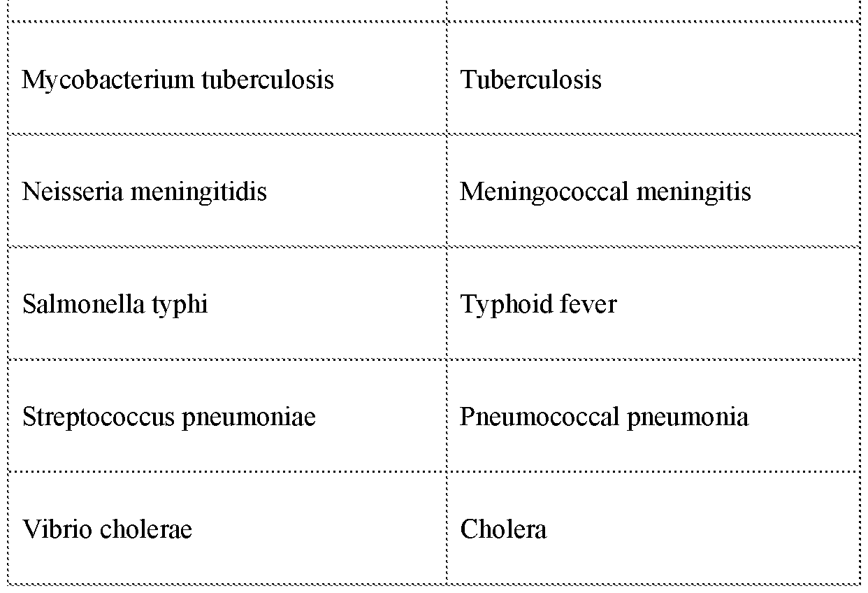
EXAMPLES EXAMPLE 1
Virus-Like Nanoparticles for Antigen-Specific Antibody Production
This Examples describes the production and use of virus-like nanoparticles to produce antibodies. Materials: All reagents were used as obtained from commercial sources without further purification. Iron oxide (III) (FeO(OH), hydrated, catalyst grade, 30-50 mesh), oleic acid (technical grade, 90%), l-octadecene (technical grade, 90%), anhydrous tetrahydrofuran (THF, 99.8%), sodium sulfide, chloroauric acid, ammonium iron (II) sulfate hexahydrate (Fe(NH4)2(S04)2.6H20, ACS reagent, 99%), nitric acid (ACS reagent, 70%), and
hydrochloric acid (ACS reagent, 37%) were purchased from Sigma-Aldrich. Mouse uncoated IgG and IgM Total ELISA Ready-SET-Go! Kits, l-Step Ultra TMB-ELISA substrate solution, HRP-conjugated goat anti-mouse IgGl secondary antibody, HRP-conjugated goat anti-mouse IgG2a secondary antibody, Nunc Immobilizer Amino 96-well ELISA plates, BupH carbonate bicarbonate buffer packs (coating buffer), Pierce protein free PBS tween blocking buffer, 20x PBS-tween wash buffer, Geneticin (G418) selective antibiotic,
Invitrogen eBioscience fixable viability dye eFluor 780, and Molecular Probes streptavidin Alexa Fluor 647 conjugate were obtained from Thermo Fisher Scientific. HRP conjugated goat anti-mouse IgG secondary antibody, Zombie UV fixable viability kit, FITC anti-mouse CD19, PE/Dazzle 594 anti-mouse CD38, Brilliant Violet 421 anti-mouse CD138, PE/Dazzle 594 anti-mouse IgD, Alexa Fluor 647 anti-mouse/house GL7 antigen, Brilliant Violet 421 and PE/Dazzle 594 anti-mouse/human CD45R/B220, FITC anti-mouse CD95, Brilliant Violet 421 anti-mouse/human CDl lb, FITC anti-mouse CD169, PE/Dazzle 594 anti-mouse CD1 lc were purchased from BioLegend. HER2 peptides
(CDDDPESFDGDPASNTAPLQPEQLQ (SEQ ID NO: l), Biotin- PESFDGDPASNTAPLQPEQLQ (SEQ ID NO:2),
CDDDPESFDGDPASNTAPLQPEQLQGGGK (SEQ ID NO: 3)) were custom synthesized. 30 nm iron-oxide nanoparticles cores stabilized by oleic acid in chloroform were purchased from Ocean Nanotech. DSPE-PEG(2000) and DSPE-PEG(2000)maleimide were obtained from Avanti Polar Lipids. 2 3 -cGAMP was acquired from InvivoGen. Fluorescamine was purchased from MP Biomedicals. Sulfo-Cy5.5 NHS ester was acquired from Lumiprobe. Microvette 500 Z-Gel serum collection vials with clotting factor were obtained from Sarstedt. Matrigel Basement Membrane Matrix was purchased from Coming. Gold and iron ICP standards were purchased from Fluka Analytical.
Mice. All animal experiments were conducted according to the protocols approved by the University of Michigan Committee on Use and Care of Animals (UCUCA). BALB/c mice ages 5-7 weeks were purchased from Charles River Labs.
Cells. All cells were maintained at 37C, 5% CCh/95% air atmosphere and approximately 85% relative humidity. D2F2/E2 cells were cultured in complete DMEM high glucose supplemented with 10% NCTC 109 media, 1% L-glutamine, 1% MEMs non-essential amino acids, 0.5% sodium pyruvate, 2.5% sodium bicarbonate, 1% pen/strep, 5% cosmic calf serum, 5% fetal bovine serum, 500 pg/mL Geneticin and 50 mM 2-mercaptoethanol.
RAW264.7 macrophages were cultured in complete RPMI-1640 media supplemented with 10% fetal bovine serum, 1% L-glutamine, 1% MEMs non-essential amino acids, 1% sodium pyruvate and 1% pen/strep. DC2.4 dendritic cells were cultured in RPMI-1640 media supplemented with 10% fetal bovine serum, 1% L-glutamine, 1% MEMs non-essential amino acids, 1% HEPES buffered solution, 1% pen/strep and 50 mM 2-mercaptoethanol.
Formulation and Characterization of Inorganic Virus-Like Nanoparticles (IVLN). The
IVLN was formulate as follows.
Synthesis of IONPs Coated with Polysiloxane-Containing Diblock Copolymer. Spherical IONPs (15 nm in diameter) were synthesized in organic solvent by thermal decomposition. Cubic IONPs (25 nm in edge length) were also synthesized. Diblock copolymer (PEO-&- P PS) was synthesized by the reversible addition of fragmentation chain transfer (RAFT) polymerization. The preparation of the polymer-coated MNPs with either single or clustered core was performed. The IONP iron concentration was determined using o-phenanthroline (ACS reagent, 99%) after digestion with hydrochloric acid (ACS reagent, 37%). General methods of such synthesis are found in Chen et al., ACS Appl. Mater. Interfaces 2015, 7, 12814-12823, herein incorporated by reference.
Synthesis of AuNPs. AuNPs were synthesized by using sodium sulfide (Na2S) as the reducing reagent. Gold in the form of chloroauric acid (HAuCU) was prepared to a concentration of 100 mM as a stock solution and was diluted to 2.0 mM before use. Na2S (50mM) was prepared and aged in the dark for 40-48 h prior to use and was diluted to 1.0 mM before use. The volume ratio ofNa2S to HAuCU w as varied from 2.5/1.0 to 3.0/1.0. UV/Vis spectra were recorded to monitor the reaction. Without specification, the reaction with a volume ratio of 3.0/1.0 was chosen to use in the following steps. General methods of such synthesis are found in Chen et al, ACS Appl. Mater. Interfaces 2015, 7, 12814-12823, herein incorporated by reference.
Synthesis of Inorganic Virus-Like Nanoparticles (IVLN). IVLNs were made by incubating AuNPs with polymer-coated IONPs at 4 °C. In a typical experiment, two milligram Fe of IONPs (0.5 mL) was mixed with AuNP solution 6 mL of for spherical IONPs and 4 mL for cubic IONPs, respectively, if without specification. The formed IVLNs were purified by magnet to remove unbound AuNPs after overnight incubation.
The final Au:Fe ratio of the formulated IVLN was quantified by inductively coupled plasma mass spectrometry using a Perkin-Elmer Nexion 2000 based on established protocols. IVLN formulations were imaged by transmission electron microscopy (TEM) using the JEOL 3011 High Resolution Electron Microscope. The true particle size of AuNPs, IONPs and IVLNs was quantified using ImageJ software. The volume-weighted hydrodynamic particle size, polydispersity index and zeta-potential of all formulations in milliQ water at 25°C was evaluated with the Malvern Zetasizer Nano-ZS using dynamic light scattering and phase analysis light scattering, respectively.
Lipid-Coated Iron-Oxide Nanoparticle Formulations (Lipid-IONP). Lipid-coated iron- oxide nanoparticles were prepared based on previously reported methods for thin-film hydration with minor modifications. 10 mg of DSPE-PEG(2000)-maleimide was added to 1 mg of 30-nm iron-oxide nanoparticle cores stabilized by oleic acid in chloroform as gently mixed. The resulting solution was subjected to solvent rotary evaporation to remove all chloroform and form a thin film. Simultaneously, this film and 100 mM PBS, pH 7.4 were heated to 75 °C in an oven. Upon reaching temperature, hot PBS was rapidly added to the film and mixed immediately and vigorously to facilitate thin film hydration. The resulting nanoparticle solution was stored at 4°C to promote lipid self-assembly. Free phospholipid were removed by magnetic separation overnight at 4°C using the a magnetic separator device.
Lipid-IONP-HER2 and IVLN-HER2 Formulations. HER2 peptides were conjugated to both Lipid-IONP and IVLN through thiol-mediated chemistries. Specifically, Lipid-IONP - HER2 was formulated via maleimide chemistry and IVLN-HER2 was formulated via the gold-thiol linkage. HER2 peptide was added to Lipid-IONP at l.5x weight ratio excess in milliQ and incubated overnight at 4°C. HER2 peptide was added to IVLN-HER2 at 5x weight ratio excess in milliQ and incubated overnight at 4°C. Both materials were purified either by magnetic separation overnight at 4°C using a magnetic separator device, or by centrifugal separation at 10,000 x g for 30 minutes at 4°C.
Immunizations and Serum Collection. At day 0, mice were immunized with the equivalent of 50 pg of HER2 peptide plus 10 pg of cGAMP regardless of formulation type.
Subsequently, at day 14, mice were boosted twice at two-week intervals with 50% of the original dosage for both antigen and adjuvant (day 14 and 28). To evaluate serum antibody
titers, blood was collected by submandibular puncture 10 days after each immunization (day 10, 24 and 38). Serum was separated from whole blood by centrifugal separation at 10,000 x g for 5 minutes at 25°C using the Microvette 500 Ser-Gel collection vessels with clotting activator.
Enzyme-Linked Immunosorbent Assay (ELISA). Absolution quantification of total IgG and total IgM antibody analysis was performed using the mouse uncoated total IgG and total IgM ELISA kits based on protocols provided by ThermoFisher. Antigen-specific IgG, IgGl and IgG2a antibody titers were quantified based on previously established protocols for indirect ELISA with minor modifications. Specifically, HER2 peptides (200 pL, 100 ug/mL in 100 mM carbonate buffer, pH 9.4) were chemically conjugated to ELISA plates through the terminal amine group utilizing Nunc Immobilizer Amino immunoassay plates by overnight incubation with exposure to light at room temperature. Following overnight incubation, ELISA plates were washed three times with 100 mM PBS, pH 7.4 with 2% Tween-20. Subsequently, ELISA plates were blocked overnight at 4°C with 300 pL of ELISA blocker (Pierce Protein-Free PBS Blocking Buffer). Following blocking, the ELISA plates were washed 3x. Serum samples containing primary antibodies were serially diluted (l01-108 fold) using 100 mM PBS, pH 7.4 containing 10% ELISA blocker reagent and added to each well at 200 pL total for 2 hour incubation at room temperature. Following sample addition, the ELISA plates were washed 3x. 500-fold diluted anti-IgG-HRP, anti-IgGl -HRP, or anti-IgG2a-HRP was added at 100 pL to each well and incubated for 1 hour at room temperature. After 1 hour, the ELISA plates were washed 5x. Next, 100 pL of 1 -Step Ultra TMB Substrate Solution was added to each well and allowed to incubate and develop color for 15-20 minutes at room temperature with gentle agitation. After 15-20 minutes, color development was stopped by the addition of 100 pL of 100 mM sulfuric acid. Colorimetric development was quantified by absorbance spectroscopy at 450 nm using a BioTek Cytation 5. Antibody titers were determined by any absorbance signal at a given dilution factor that was greater than the PBS control absorbance signal plus 3x standard deviations.
Quantification of Nanoparticle Delivery to Lymph Nodes in-vivo. Mice were injected subcutaneously in the left hock with either Lipid-IONP or IVLN-peptide at 200 pg total Fe per mouse. At the designated time intervals, mice were sacrificed, and lymph nodes of interest were dissected for ex-vivo analysis. The extent of nanoparticle delivery to the lymph nodes was quantified using ICP-MS based on previously reported protocols.
Quantification of Peptide Delivery to Lymph Nodes in-vivo. To facilitate quantification of peptide delivery to lymph nodes, lysine terminally modified HER2 peptides were chemically conjugated to sulfo-Cy5.5 NHS Ester. This conjugation was carried out at a 5-fold molar excess of sulfo-Cy5.5 NHS Ester to HER2 peptide. IONP-HER2-Cy5.5 and IVLN-HER2- Cy5.5 were subjected to Cy5.5 functionalization after initial peptide conjugation was completed in order to enable facile purification of excess fluorescent dye by magnetic separation. Subsequent to Cy5.5 functionalization, mice were injected as previously stated. After 3 hours, mice were sacrificed and lymph nodes of interest were dissected for ex-vivo analysis by IVIS imaging. IVIS imaging was utilized for semi-quantification of peptide delivery in terms of radiant efficiency.
In-vitro Cell Uptake. IVLN-HER2 and IONP-HER2 cellular uptakes was evaluated in RAW264.7 macrophages, DC2.4 dendritic cells and primary B-cells isolated from murine spleens using an EasySep Mouse B-cell isolation kit. Nanoparticle samples were incubated at 50 pg/mL Fe with cells for 18 hours in blank RPMI media at 37C, 5% CO /95% air atmosphere and approximately 85% relative humidity. After 18 hours, cells were lifted by cell scraping and washed thrice with phosphate-buffered saline (PBS). Following the wash steps, resulting cell pellets were re-suspend in 1 mL of PBS, cell counted and then digested in 1 mL aqua regia (1 :3 molar ratio nitric acid: hydrochloric acid) for analysis by ICP-MS.
In-vivo Cell Uptake. IVLN-HER-Cy5.5 and IONP-HER2-Cy5.5 were injected
subcutaneously in the left hock with either Lipid-IONP or IVLN at 200 pg total Fe per mouse. At 3 hours and 24 hours, mice were sacrificed and lymph nodes of interest were dissected for ex-vivo analysis by flow cytometry. Lymph nodes were dissociated by mechanical methods to prepare single cell suspensions. Single cell suspensions of lymph node cells were stained for analysis by flow cytometry using the MoFlo Astrios flow cytometer. The first panel was for viable cells, B-cells (B220+), subcapsular sinus macrophages (CDl69+CDl lb+), dendritic cells (CD1 lc+) and nanoparticle positive cells (Cy5.5). Flow cytometry data was analyzed by FCS express.
Antigen-Specific B-cell and Germinal Center Flow Cytometry. Mice were immunized as previously introduced. At day 24 and day 38, mice were sacrificed and spleens and lymph nodes were dissected for ex-vivo analysis by flow cytometry. Antigen-specific B-cell analysis
was accomplished using tetramer staining. HER2/neu peptide tetramers were prepared by mixture of biotin-labeled HER2 peptide with Alexa Fluor 647 labeled streptavidin at a 4: 1 molar ratio at room temperature for 1 hour without further purification. Antigen-specific B- cell population were identified as either memory B-cells (B220+CD38+Tetramer+) or plasma cells (B220 CDl38+Tetramer+) using flow cytometry. Germinal center B-cell populations were identified using the following markers CD 19, IgD, GL7 and CD95.
Tumor Studies. Sixty days after the primary immunization, mice were inoculated with 500,000 D2F2/E2 cells subcutaneously in the right flank. D2F2/E2 cells were prepared at 5e6 cells/mL in 100 pL and mixed at equal volume with Matrigel matrix. Tumor size was quantified by caliper measurements every 7 days. Tumor volumes were calculated using the following equation:

End points were determined by using the End-Stage Illness Scoring System; mice receiving an End-Stage Illness Score greater than 6 were euthanized by CO2 asphyxiation.
Statistics. Data are expressed as mean ± standard deviation (SD), unless otherwise specified Comparisons between two groups were made using the unpaired Student’s t- test. Means of multiple groups were compared with the one-way analysis of variance (ANOVA), followed by post hoc Tukey’s pairwise comparisons. All probability values are two-sided, and values of p < 0.05 were considered statistically significant. Statistical analyses were carried out using the GraphPad Prism 7 software package.
RESULTS
Inorganic Virus-Like Nanoparticle tlVLN) Formulation and Viral Properties
The IVLN is formulated by the self-assembly of two separately prepared nanoparticle systems, an iron-oxide nanoparticle core and gold nanoparticle satellites, through the association of hydrolyzed siloxane groups and gold (Figure 1). The iron-oxide nanoparticle (IONP) core was synthesized by thermal decomposition to produce a ~l5-nm spherical core
stabilized by oleic acid in chloroform. To achieve aqueous stabilization, the IONP core was coated with a polysiloxane/PEG diblock copolymer (IONP-polymer). Separately, ultra-small gold nanoparticles (AuNP) with ~3 nm size were prepared using a modified self-assembly method by reduction of chloroauric acid in aged sodium sulfide. Following synthesis, AuNPs are added to the polymer-coated IONP cores in solution at defined weight ratios.
To quantify the extent of AuNP loading per IONP core following self-assembly, inductively coupled plasma mass spectrometry (ICP-MS) was used. With ICP-MS, it was determined that the average loading efficiency on a per weight basis was 73 ± 7% with a linear dependence (R2 = 0.997) (Figure 2A). Notably, above an initial loading ratio of 50% weight Au, destabilization of nanoparticles in solution was observed, and was therefore not the focus of further research. To provide visual confirmation of AuNP and IONP-polymer self-assembly to form the IVLN, transmission electron microscopy (TEM) was performed (Figure 2B).
TEM imaging confirmed that by controlling the initial loading ratio of AuNP to IONP- polymer cores on a per weight basis it is possible to yield IVLNs with variable gold nanoparticle surface density and viral-like character (Figure 2B-insert). IONP-polymer core and AuNP diameters were quantified to be 15.9 ± 1.3 nm and 2.3 ± 0.4 nm, respectively.
TEM imaging was further utilized to estimate the AuNP loading per IONP-polymer core. Specifically, 10%, 20% and 30% weight Au conditions yielded IVLNs with 4 ± 2, 9 ± 3 and 13 ± 5 AuNPs per IONP-polymer core, respectively.
Following ICP-MS quantification and TEM visualization of IVLN formulations, elementary mathematical modeling was performed in order to determine the structural relevance of IVLN as compared to virus-like particles. Given the crystalline nature of AuNPs and IONPs, it is feasible to determine the number of particles of a determined particle size for a given weight of either Au or Fe, respectively. Based on the ICP-MS measurements performed in Figure 2A and the particle size confirmation by TEM in Figure 2B, it is therefore possible to estimate the average distance between AuNPs on the IONP surface, as well as the average number of AuNPs per unit area on IONP surfaces. From this analysis, it was determined that depending on the initial weight loading ratio of Au to Fe, IVLNs can be formulated with a minimum average distance of 6.75 nm between AuNPs - a preferred distance for B-cell receptor crosslinking (Figure 2C). In addition to AuNP spatial distribution, at this same initial loading ratio, the number of AuNPs per unit area was determined to be approximately 12,500-17,000 AuNPs per square micron - a value that compares favorable with the antigen density reported for viral-like particles (e.g. Hepatitis B Virus) (Figure 2D).
To further evaluate the viral-like potential of the IVLN, we next evaluated the capacity for and mechanism of peptide loading in this system. The peptide of interest in these studies is a human HER2/neu-specific peptide that, based on previously published works, contains a B-cell epitope with an overlapping CD4 helper T-cell epitope. In addition to these functional epitopes, a cysteine containing terminal flank was added to facilitate facile loading to the IVLN via the Au-S linkage (CDDD-PESFDGDPASNTAPLQPEQLQ, SEQ ID NO: l). The capacity for peptide conjugation to the IVLN was quantified utilizing a modified fluorescamine peptide assay. Peptide loading was evaluated under three separate IVLN formulation conditions: 0%, 10% and 30% Au to Fe final weight loading ratio (wt/wt Au/Fe). In terms of AuNPs per IONP-polymer core, these values approximately translate to 0 AuNPs, 4 AuNPs and 10 AuNPs per IONP-polymer core, respectively. Peptide conjugation was performed in water at 4°C overnight with subsequent purification by centrifugal separations. Fluorescent peptide quantification analysis revealed that, at maximum peptide loading conditions, the 0%, 10% and 30% wt/wt formulations were loaded with 232 ± 73, 888 ± 42 and 1954 ± 157 peptides per IVLN, respectively (Figure 2E). When these same values were standardized by total AuNPs it was determined that maximum peptide loading per AuNP was 227 ± 5. Additionally, this analysis revealed a positive correlation between peptide loading and AuNP number (R = 0.95). Taken together, these results suggest although there is low- level non-specific physical association to the core (-12% under maximum loading conditions), peptide loading is AuNP dependent. Thus, peptide conjugation to IVLN surface is AuNP localized, which indicates that the IVLN-peptide is characterized by heterogeneous and patchy peptide distribution. This patterned antigen display is viral-like in nature and can not be reproduced by traditional nanoparticle systems that employ homogeneous antigen distribution on their surfaces.
After the evaluation of peptide loading, we next assessed the IVLN-peptide’ s material properties to determine if the material was suitable for in-vivo applications and appropriately aligned with viral-like properties (Figure 2F). Before incubation with AuNPs, the IONP- polymer core of the IVLN was shown to have a 51 ± 2 nm volume-weighted hydrodynamic particle size by dynamic light scattering (DLS) with a 0.15 ± 0.03 polydispersity index (PDI). In addition, the zeta-potential of this material in milliQ water at pH 7 was determined to be -7 ± 4 mV. Following formulation at a final weight loading ratio of 30% wt/wt Au/Fe, the IVLN-Blank was shown to have a 55 ± 2 nm particle size, 0.20 ± 0.05 PDI and a -16 ± 4 mV zeta-potential before peptide loading. After peptide loading, the IVLN-Peptide was shown to have a 60 ± 4 nm particle size, 0.20 ± 0.05 PDI and a -17 ± 1 mV zeta-potential. Taken
together, the IVLN-peptide was determined to have optimal material properties for in-vivo applications. Moreover, these properties were deemed acceptably within the design criteria for viral mimicking nanoparticles, which includes particle size between 20-300 nm and negative overall surface charge.
Inorganic Virus-Like Nanoparticle (IVLN) for Antigen-Specific Antibody Production in Mice
Viral mimicking nanoparticles have been utilized in a wide-range of in-vitro and in- vivo applications, but the one application that viral-like material properties are very good for is B-cell activation for antigen-specific antibody production. Accordingly, based on the establish viral-like material properties of IVLN-peptides. In this study, BALB/c mice (6-8 weeks old) were immunized with 50 pg of HER2 peptide plus 10 pg of cGAMP as adjuvant at day 0 and boosted once 14 days later. Mice were bled, and serum was collected for analysis 10-days following every administration (Figure 3 A).
Based on an understanding of the requirement of B-cell receptor crosslinking for B- cell activation and germinal center formation, we first asked what the role of AuNP quantity and spatial distribution on IVLN surfaces at saturated peptide loading was. To answer this question, we evaluated the titer of antigen-specific IgG antibodies produced in mice after a booster immunization with IVLN at 10%, 20% and 30% wt/wt Au/Fe final weight loading ratios by indirect ELISA. From this analysis, it was determined that by day 24 the production of antigen-specific IgG, in terms of median antibody titers, was 7,500, 12,500 and 32,500 for 10%, 20% and 30% wt/wt formulations, respectively (Figure 3B). This initial result suggested that increasing AuNP quantity on IVLN surfaces improved antibody production. Presumably, this improvement was the result of reduced AuNP spacing (10% wt = -11.25 nm; 20% wt = - 8.05 nm; 30% wt = 6.25 nm) to facilitate more efficient B-cell receptor crosslinking.
Subsequent to the evaluation of AuNP surface density, we questioned what the role of peptide conjugation density on IVLNs was for a given Au/Fe weight loading ratio. Peptide density has been positively connected to increased B-cell activation with potential for dose sparing by numerous previous reports. Notably, peptide density negatively correlated to reduction of antibody specificity. For the 30% wt ratio, low density peptide on IVLN surfaces yielded a median antigen-specific IgG antibody titer of 6,500, while high density peptide on IVLN surface yielded a median titer of 32,500 (Figure 3B). This trend was also observed at the 20% wt ratio condition, which produced median titers of 1,300 and 12,500 for low density
and high density, respectively. However, this trend did not translate to the 10% wt ratio condition. Presumably, this is due to a greater than 10 nm spacing between AuNPs on IVLN surfaces. Based on the above analysis, it was determined that higher peptide density and higher number of AuNPs per IVLN surface were generally preferred for antigen-specific antibody production due to the increasingly viral-like nature of the material. Significantly, peptide density was demonstrated to improve antibody titers without a loss of antigen- specificity. Based on these results, the 30% wt ratio plus high-density peptide condition was employed in all assays moving forward and referred to simply as IVLN or IVLN-HER2.
To effectively evaluate the significance of viral-like character for the application of antigen-specific antibody production, we next asked how the IVLN would perform as directly compared to a traditional nanoparticle system. For this comparison, we utilized a lipid-coated iron-oxide nanoparticle (Lipid-IONP) as a control (see Figure 1). This nanoparticle has a 30- nm iron-oxide nanoparticle core and a functionalized DSPE-PEG(2000)-maleimide shell that facilitates facile peptide conjugation. Significantly, Lipid-IONPs have similar material properties in terms of hydrodynamic particle size (69 ± 1 nm), PDI (0.20 ± 0.01 nm), and maximum peptide number per particle (2323 ± 394 peptides per IVLN). However, as a traditional nanoparticle system, Lipid-IONPs have smooth PEGylated surfaces with homogeneous peptide distribution. Taken together, we believe that the side-by-side comparison of IVLNs and Lipid-IONPs would offer valuable insights into the role of viral mimicry for in-vivo functionality.
As before, BALB/c mice were immunized with 50 pg of HER2 peptide plus 10 pg of cGAMP as adjuvant at day 0 and boosted at day 14. At day 24, 10 days post-boost 1, serum was analyzed for total IgM, total IgG, antigen-specific IgG and the antigen-specific IgG isotypes, IgGl and IgG2a (Figure 3C). At this time point, no statistically significant difference in total IgM antibody production were observed (p = 0.09), however a significant increase in total IgG was observed for both Lipid-IONP -HER2 and IVLN-HER2 as compared to PBS alone and soluble HER2 peptide treated mice. More specifically, an approximately 7- fold and 3-fold increase in total IgG was observed for both the Lipid-IONP -HER2 and IVLN- HER2 samples as compared to PBS alone (pO.OOl; pO.OOl) and soluble HER2 (p<0.0l; pO.Ol), respectively. No statistically significant difference between Lipid-IONP-HER2 and IVLN-HER2 was observed for total IgG (p = 0.98). However, when antigen-specific IgG was evaluated a dramatic difference between IVLN-HER2 and Lipid-IONP -HER2 was observed. Namely, IVLN-HER2 was determined to have an 18.5-fold higher antigen-specific IgG titer (39,500 vs. 2,140; pO.OOl) a l5-fold higher antigen-specific IgGl titer (9,600 vs. 640;
r<0.001) and a 4.5-fold higher antigen-specific IgG2a titer (5,760 vs. 1,280; p<0.05) as compared to Lipid-IONP-HER2. Moreover, IVLN-HER2 yielded an 9-fold higher antigen- specific IgG titer (39,500 vs. 4,300; p<0.00l), a 48-fold higher antigen-specific IgGl titer (9,600 vs. 200; p<0.00l) and a 72-fold higher antigen-specific IgG2a titer (5,760 vs. 80; p<0.0l) as directly compared to soluble HER2 peptide.
Lymph Node Delivery and Nanoparticle Distribution
Upon establishing the significant enhancement of antigen-specific antibodies for the IVLN-HER2 as compared traditional nanoparticle and soluble controls, we next set out to answer why. To begin the mechanistic evaluation of the role of viral mimicking properties for antibody production, we first evaluated the ability of these materials to be delivered and retained with the lymph nodes. As sites with dense populations of antigen-presenting cells and lymphocytes, the lymph nodes are ideal target sites for immune activation. Specifically, the lymph nodes are primary sites for B-cell activation and the formation of germinal centers that are ultimately responsible for initiating antigen-specific IgG antibody production. In the context of viral mimicry, the lymph nodes are known to be critically important in the effort to combat and control viral dissemination throughout the body. This functionality is the result of unique physiological features that have been developed for viral recognition and viral- specific immune activation. For example, subcapsular sinus macrophages are a highly specialized phenotype of macrophage that is responsible for viral uptake and direct presentation to B-cells to promote directed viral clearance via antigen-specific antibody production. Thus, studying lymph node delivery and immune cell interactions within the lymph node is essential in the evaluation of the mechanism of viral mimicking nanoparticle functionality.
Delivery to the lymph nodes was quantified for both nanoparticle delivery and peptide delivery following administration by subcutaneous hock immunization, which was determined to be the most efficient delivery pathway. Nanoparticle delivery kinetics to the lymph nodes (popliteal and inguinal) was determined using ICP-MS quantification of Fe and Au in excised lymph nodes based on previously established protocols. From this analysis, it was determined that while the tmax of both IVLN-HER2 and Lipid-IONP-HER2 was 3 hours post-administration, the percent of initial nanoparticle dose delivered was 5.1 ± 1.6% and 1.9 ± 0.8% (p<0.05) for IVLN-HER2 and Lipid-IONP-HER2, respectively (Figure 4A). Over 72 hours, IVLN-HER2 was shown to have a 2.8-fold increase (p<0.05) in overall exposure compared to Lipid-IONP-HER2 based on AUC. In addition to the nearly 3-fold increase in
delivery observed, the retention of nanoparticles within the lymph node was an estimated 65% for IVLN-HER2 as compared to 48% for Lipid-IONP-HER2. Beyond the direct quantification of nanoparticle delivery by raw elemental analysis, peptide delivery to the lymph node at 3 hours was validated through semi-quantitative analysis of fluorescent intensity using IVIS imaging of excised popliteal and inguinal lymph nodes (Figure 4B).
IVIS imaging revealed that IVLN-HER2 led to a 4.3-fold improvement in peptide delivery as compared to both the Lipid-IONP-HER2 and soluble HER2 peptide (p<0.00l; p<0.00l), which have no statistically significant difference in delivery (p>0.99).
Subsequent to the quantification of lymph node delivery, we next asked where IVLN and IONP distributed within the lymph node at a cellular level. To answer this question, fluorescently labeled nanoparticles were delivered to lymph nodes and flow cytometry was applied in order to identify the percentage of nanoparticle-positive cells out of total cells of different phenotypes. More specifically, lymph node antigen-presenting cells (subcapsular sinus macrophages and dendritic cells) and lymphocytes (B-cells and T-cells) were identified and assessed for nanoparticle uptake. This analysis revealed that after 3-hours and relative to the Lipid-IONP-HER2, IVLN-HER2 improved subcapsular sinus macrophage uptake by 1.7- fold (86.7 ± 2.4% vs. 52.2 ± 2.8%, pO.OOl), dendritic cell uptake by 1.8-fold (75.2 ± 1.9% vs. 41.1 ± 5.8%, pO.OOl), and B-cell uptake by 3.4-fold (63.9 ± 1.1% vs. 19.7 ± 2.2%, pO.OOl), (Figure 4C).
Lastly, we asked what the mechanism for improved cellular uptake was. Based on previous studies, nanoparticle surface topography has been positively correlated with improved cellular uptake. Given the rough surface topography of the IVLN as compared to the smooth PEGylated surfaces of the Lipid-IONP, we hypothesized the cellular uptake is dependent on the extent of AuNP loading on IVLN surfaces. To test this hypothesis, cellular uptake was evaluated in-vitro using RAW264.7 macrophages, DC2.4 dendritic cells and primary B-cells isolated from murine spleens. From this analysis it was determined that, regardless of cell type, cellular uptake was determined by the extent of AuNP loading on IVLN surfaces and matched an exponential function (RAW264.7, R2 = 0.902; DC2.4, R2 = 0.893; B-cells, R2 = 0.918). More specifically, as compared to the IONP -polymer core only treatment, IVLN-HER2 at 30% wt/wt loading ratio improved cellular uptake by 6-fold (30.3 ± 1.1 pg Fe/cell vs. 4.7 ± 0.9 pg Fe/cell, pO.OOl), 4.5-fold (12.5 ± 1.4 pg Fe/cell vs. 2.9 ± 0.9 pg Fe/cell, pO.OOl), and 4-fold (2.0 ± 0.4 pg Fe/cell vs. 0.5 ± 0.2 pg Fe/cell, pO.OOl), for RAW264.7 macrophages, DC2.4 dendritic cells and primary B-cells, respectively. Notably, as directly compared to the Lipid-IONP -HER2 control group, IVLN-HER2 improved cellular
uptake by 3-fold (30.3 ± 1.1 pg Fe/cell vs. 11.8 ± 1.3 pg Fe/cell, pO.OOl), 3-fold (12.5 ± 1.4 pg Fe/cell vs. 3.9 ± 1.9 pg Fe/cell, pO.Ol), and 2-fold (2.0 ± 0.4 pg Fe/cell vs. 0.8 ± 0.4 pg Fe/cell, p<0.0l), for RAW264.7 macrophages, DC2.4 dendritic cells and primary B-cells, respectively.
Viral-like characteristics are ideal material properties for the rational design and engineering of the delivery vehicles, immunostimulatory agents and cellular uptake vectors desperately needed for the advancement of nanotechnology in biotechnology and medical applications. Accordingly, here we report the development and evaluation of an alternative to viral-like particles with a more holistic approach to viral mimicry material design - inorganic virus-like nanoparticles (IVLN) and IVLN-peptides. In certain embodiments, the IVLNs are composed of a hybrid Au@Fe core-satellite type nanoparticle system, which utilizes a l6-nm polysiloxane containing diblock polymer coated iron-oxide nanoparticle core (IONP- polymer) with 2.5-nm gold nanoparticle satellites (AuNP). Based upon the formulation conditions, IVLNs can be produced with variable surface topography, antigen density and antigen spatial resolution. Moreover, the IVLN has optimal particle size, shape and surface charge for efficient lymph node delivery and retention. As such, these properties inform the viral-like character and functional potential of IVLNs.
Our results showed that viral-like character is truly significant for B-cell activation and antigen-specific antibody production. Specifically, by manipulating the spatial distribution of AuNPs on IVLN surface and the peptide density regional confined to those AuNPs, it was possible to increase the median antigen-specific IgG antibody titer from 6,300 to 32,500 - a 5-fold increase. This result was further corroborated by direct comparison of IVLNs to Lipid-IONPs. Significantly, as compared to Lipid-IONP-HER2, IVLN-HER2 was determined to have 18.5-fold, l5-fold and 4.5-fold higher antigen-specific IgG, IgGl and IgG2a titers, respectively. With highly comparable hydrodynamic particle size, surface charge, core shape and peptide loading per unit nanoparticle, any quantifiable differences in antibody production by these two structures could be attributed to increasingly viral-like character. Mechanistically, this viral-like character led to enhanced antigen-specific antibody production as a result of greater delivery to and retention in the lymph nodes due to improved immune cell uptake, which promoted a significant increase in overall B-cell activation and germinal center formation.
EXAMPLE 2
Virus-Like Nanoparticles for Antigen-Specific Antibody Production
This Examples describes the production and use of virus-like nanoparticles to produce antibodies. In order to achieve the viral-like structures, we engineered IVLNs, using a controllable and robust self-assembly process, to resemble spiky peplomers of virus, which have spiky antigen cluster topography, a certain distance between antigen clusters, and localized high antigen density on the spike.
In order to test IVLNs for viral functional mimicry, we evaluated the IVLNs to activate antigen-specific B cells and durable antigen-specific antibody response. We selected a well-known HER2 B cell epitope with overlap CD4 T cell epitope since the in vivo model for evaluation of durable antibody response is readily available by monitoring HER2 tumor growth without need of a biological safety level four lab. Three important viral like functions of IVLNs were evaluated: (1) antigen delivery efficiency and B cell zone uptake in the secondary lymph nodes; (2) antigen specific B cell activation by different density and spatial arrangements of antigen on the IVLN surface; and (3) follicular T helper cell activation in the Germinal center for B cell activation and durable antibody response. The durable function of antigen specific antibody was evaluated in vivo to inhibit HER2 cancer growth.
Materials and Methods
Materials
All reagents were used as obtained from commercial sources without further purification, except for g-Methacryl oxypropyltrimethoxysilane (98%) that was purified by distillation under reduced pressure and 2,2-Azobis(isobutyronitrile) (98%) that was purified by recrystallization in ethanol. Iron oxide (III) (FeO(OH), hydrated, catalyst grade, 30-50 mesh), oleic acid (technical grade, 90%), ammonium iron (II) sulfate hexahydrate (ACS reagent, 99%), l-octadecene (technical grade, 90%), anhydrous tetrahydrofuran (THF, 99.8%), carbon disulfide (99.9%), magnesium turnings (>99.5%), 2-chloro-2-phenylacetyl chloride (CP AC, 90%), poly(ethylene oxide) monomethyl ether (PEO), anhydrous dioxane (99.8%), dimethylformamide (DMF, 99.9%), dimethyl sulfoxide (DMSO, 99.9%), o- phenanthroline monohydrate (ACS reagent, 99%), hydroquinone (ACS reagent, 99%, sodium sulfide, chloroauric acid, nitric acid (ACS reagent, 70%), and hydrochloric acid (ACS reagent, 37%) were purchased from Sigma-Aldrich. Mouse uncoated IgG and IgM Total ELISA Kits, l-Step Ultra TMB-ELISA substrate solution, HRP-conjugated goat anti-mouse
IgGl secondary antibody, HRP-conjugated goat anti-mouse IgG2a secondary antibody, Nunc Immobilizer Amino 96-well ELISA plates, BupH carbonate bicarbonate buffer packs (coating buffer), Pierce protein free PBS-tween blocking buffer, 20x PBS-tween wash buffer, Geneticin (G418) selective antibiotic, Invitrogen eBioscience fixable viability dye eFluor 780, and Molecular Probes streptavidin Alexa Fluor 647 conjugate were obtained from Thermo Fisher Scientific. HRP conjugated goat anti-mouse IgG secondary antibody, Zombie UV fixable viability kit, FITC anti-mouse CD 19, PE/Dazzle 594 anti-mouse IgD, Alexa Fluor 647 anti-mouse/house GL7 antigen, Brilliant Violet 421 and PE/Dazzle 594 anti mouse/human CD45R/B220, FITC anti-mouse CD95, Brilliant Violet 421 anti-mouse/human CD1 lb, FITC anti-mouse CD169 and PE goat anti-mouse IgG secondary antibody were purchased from BioLegend. HER2 peptides (CDDDPESFDGDPASNTAPLQPEQLQ (SEQ ID NO: l), Biotin-PESFDGDPASNTAPLQPEQLQ (SEQ ID NO: 2),
CDDDPESFDGDPASNTAPLQPEQLQGGGK, SEQ ID NO: 3) were custom synthesized by LifeTein. Iron-oxide nanoparticles (30 nm) stabilized by oleic acid in chloroform were purchased from Ocean Nanotech. DSPE-PEG (2000) and DSPE-PEG (2000)-maleimide were obtained from Avanti Polar Lipids. 2 3 -cGAMP was acquired from InvivoGen.
Fluorescamine was purchased from MP Biomedicals. Sulfo-Cy5.5 NHS ester was acquired from Lumiprobe. Microvette 500 Z-Gel serum collection vials with clotting factor were obtained from Sarstedt. Matrigel Basement Membrane Matrix was purchased from Coming. Gold and iron ICP standards were purchased from Fluka Analytical.
Mice
All animal experiments were conducted according to the protocols approved by the University of Michigan Committee on Use and Care of Animals (UCUCA). BALB/c mice ages 5-7 weeks were purchased from Charles River Labs.
Cells
All cells were maintained at 37 °C, 5% CO /95% air atmosphere and approximately 85% relative humidity. D2F2/E2 cells ((83)) were cultured in complete DMEM high glucose supplemented with 10% NCTC 109 media, 1% L-glutamine, 1% MEMs non-essential amino acids, 0.5% sodium pyruvate, 2.5% sodium bicarbonate, 1% pen/strep, 5% cosmic calf serum, 5% fetal bovine serum, 500 pg/mL Geneticin and 50 mM 2-mercaptoethanol.
RAW264.7 macrophages were cultured in complete RPMI-1640 media supplemented with 10% fetal bovine serum, 1% L-glutamine, 1% MEMs non-essential amino acids, 1% sodium
pyruvate and 1% pen/strep. Primary B-cells were cultured in RPMI-1640 media
supplemented with 10% heat-inactivated fetal bovine serum.
Formulation and characterization of inorganic virus-like nanoparticles (IVLN)
The IVLN was formulated generally as in Example 1. The final Au to Fe ratio of the formulated IVLN was quantified by inductively coupled plasma mass spectrometry (ICP-MS) using a Perkin-Elmer Nexion 2000 based on previously reported protocols modified from analysis by ICP-OES (78). IVLN formulations were imaged by s scanning transmission (electron microscopy (STEM) using a JEOL 21 OOF with a CEOS probe corrector. The true particle size of AuNPs, IONPs and IVLNs was quantified using ImageJ software. The volume-weighted hydrodynamic particle size, polydispersity index and zeta-potential of all formulations in milliQ water at 25 °C was evaluated with the Malvern Zetasizer Nano-ZS using dynamic light scattering and phase analysis light scattering, respectively.
Lipid-coated iron-oxide nanoparticle formulations (IONP)
Lipid-coated iron-oxide nanoparticles were prepared as follows. DSPE-PEG (2000)- maleimide (10 mg) was added to 1 mg of 30-nm iron-oxide nanoparticles stabilized by oleic acid in chloroform as gently mixed. The resulting solution was subjected to solvent rotary evaporation to remove all chloroform and form a thin film. Simultaneously, this film and 100 mM PBS, pH 7.4 were heated to 75 °C in an oven. Upon reaching temperature, hot PBS was rapidly added to the film and mixed immediately and vigorously to facilitate thin film hydration. The resulting nanoparticle solution was stored at 4 °C to promote lipid self- assembly. Free phospholipid was removed by magnetic separation overnight at 4 °C using the EasySep magnetic separator device (StemCell).
IONP-HER2 and IVLN-HER2 formulations
HER2 peptides were conjugated to both IONP and IVLN through thiol-mediated chemistries. Specifically, IONP-HER2 was formulated via maleimide chemistry and IVLN- HER2 was formulated via the gold-thiol linkage. HER2 peptide was added to IONP at l.5x weight ratio excess in milliQ and incubated overnight at 4 °C. HER2 peptide was added to IVLN-HER2 at 5x weight ratio excess in milliQ and incubated overnight at 4 °C. Both materials were purified either by magnetic separation overnight at 4 °C using magnetic separation, or by centrifugal separation at 10,000 x g for 30 minutes at 4 °C. Peptide loading was determined using fluorescent quantification using a modified fluorescamine peptide
quantification assay in the presence of nanoparticles (Ex/Em: 390/465 nm, Biotek Cytation 5 )(86). Quantification was performed using a standard curve with increasing peptide concentration with standardized concentration of nanoparticles (IONP or IVLN) to account for quenching effects.
Immunizations and serum collection
At day 0, mice were immunized with the equivalent of 50 pg or 5 pg of HER2 peptide plus 10 pg of cGAMP regardless of formulation type. Subsequently, at day 14, mice were boosted twice at two-week intervals with 50% of the original dosage for both antigen and adjuvant (day 14 and 28). To evaluate serum antibody titers, blood was collected by submandibular puncture 10 days after each immunization (day 10, 24 and 38). Serum was separated from whole blood by centrifugal separation at 10,000 x g for 5 minutes at 25 °C using the Microvette 500 Ser-Gel collection vessels with clotting activator.
Enzyme-linked immunosorbent assay (ELISA)
Absolution quantification of total IgG and total IgM antibody analysis was performed using the mouse uncoated total IgG and total IgM ELISA kits based on manufacturer recommended protocols (Thermo Fisher). Antigen-specific IgG, IgGl and IgG2a antibody titers were quantified based on previously established protocols for indirect ELISA with minor modifications (87). Specifically, HER2 peptides (200 pL, 100 pg/mL in 100 mM carbonate buffer, pH 9.4) were chemically conjugated to ELISA plates through the terminal amine group utilizing Nunc Immobilizer Amino immunoassay plates by overnight incubation with exposure to light at room temperature. Following overnight incubation, ELISA plates were washed three times with 100 mM PBS, pH 7.4 with 2% Tween-20. Subsequently, ELISA plates were blocked overnight at 4 °C with 300 pL of ELISA blocker (Pierce Protein- Free PBS Blocking Buffer). Following blocking, the ELISA plates were washed 3x. Serum samples containing primary antibodies were serially diluted (101— 108 fold) using 100 mM PBS, pH 7.4 containing 10% ELISA blocker reagent and added to each well at 200 pL total for 2 hour incubation at room temperature. Following sample addition, the ELISA plates were washed 3x. 500-fold diluted anti-IgG-HRP, anti-IgGl -HRP, or anti-IgG2a-HRP was added at 100 pL to each well and incubated for 1 hour at room temperature. After 1 hour, the ELISA plates were washed 5x. Next, 100 pL of 1 -Step Ultra TMB Substrate Solution was added to each well and allowed to incubate and develop color for 15-20 minutes at room temperature with gentle agitation. After 15-20 minutes, color development was stopped by
the addition of 100 pL of 100 mM sulfuric acid. Colorimetric development was quantified by absorbance spectroscopy at 450 nm using the BioTek Cytation 5 plate reader. Antibody titers were determined by any absorbance signal at a given dilution factor that was greater than the PBS control absorbance signal plus three standard deviations^#).
Quantification of nanoparticle delivery to lymph nodes in-vivo
Mice were injected subcutaneously in the left hock with either IONP or IVLN at a dose of 200 pg Fe per mouse. At the designated time intervals, mice were sacrificed and lymph nodes of interest were dissected for ex-vivo analysis. The extent of nanoparticle delivery to the lymph nodes was quantified using ICP-MS based on previously reported protocols (77).
Quantification of peptide delivery to lymph nodes in-vivo
To facilitate quantification of peptide delivery to lymph nodes, lysine terminally modified HER2 peptides were chemically conjugated to sulfo-Cy5.5 NHS Ester. This conjugation was carried out at a 5-fold molar excess of sulfo-Cy5.5 NHS Ester to HER2 peptide. IONP-HER2-Cy5.5 and IVLN-HER2-Cy5.5 were subjected to Cy5.5
functionalization after initial peptide conjugation was completed in order to enable facile purification of excess fluorescent dye by magnetic separation. Subsequent to Cy5.5 functionalization, mice were injected as previously stated. After 3 hours, mice were sacrificed and lymph nodes of interest were dissected for ex-vivo analysis by IVIS imaging. IVIS imaging was utilized for semi-quantification of peptide delivery in terms of radiant efficiency.
In-vivo cell uptake
IVLN-HER-Cy5.5 and IONP-HER2-Cy5.5 were injected subcutaneously in the left hock with either Lipid-IONP or IVLN at 200 pg total Fe per mouse. At 3 hours and 24 hours, mice were sacrificed and lymph nodes of interest were dissected for ex-vivo analysis by flow cytometry. Lymph nodes were dissociated by mechanical methods to prepare single cell suspensions. Single cell suspensions of lymph node cells were stained for analysis by flow cytometry using the MoFlo Astrios flow cytometer. Viable cells (Zombie UV) were identified as either B-cells (B220+) or subcapsular sinus macrophages (CD 169hlghCD l l b1 ) and evaluated for positive nanoparticle interactions (Cy5.5). Flow cytometry data was analyzed by FCS express.
In-vitro cell uptake
IVLN-HER2 and IONP-HER2 cellular uptakes were evaluated in RAW264.7 macrophages, dendritic cells (DC 2.4), and primary B-cells isolated from murine spleens using an EasySep Mouse B-cell isolation kit. Nanoparticle samples were incubated at 50 pg/mL Fe with cells for 18 hours in blank RPMI media at 37 °C, 5% CCh/95% air atmosphere and approximately 85% relative humidity. After 18 hours, cells were lifted by cell scraping and washed thrice with PBS. Following the wash steps, resulting cell pellets were re-suspended in 1 mL of PBS, cell counted and then digested in 1 mL aqua regia (1:3 molar ratio nitric acid: hydrochloric acid) for analysis by ICP-MS.
Mass cytometry (CyTOF) to analyze all immune cells
Fixed and frozen cell suspensions were thawed on ice. Samples were stained and prepared for CyTOF analysis as previously described (89, 90), using an optimized cocktail of 40 metal-conjugated antibodies designed to identify major and minor immune cell subsets in lymph nodes. Following acquisition on a CyTOF II (Fluidigm, San Francisco, CA), samples were normalized to internal bead standards. Cell subsets were identified by gating using FlowJo software. Global analysis using SPADE were performed for unsupervised clustering analysis based on the expression of marked genes in different subset of immune cells.
Antigen-specific B-cell and germinal center flow cytometry
Mice were immunized as previously introduced. At day 10, mice were sacrificed and lymph nodes were dissected for ex-vivo analysis by flow cytometry. Antigen-specific B-cell analysis was accomplished using tetramer staining based on previously established protocols with minor modifications (63). HER2/neu peptide tetramers were prepared by mixture of biotin-labeled HER2 peptide with Alexa Fluor 647 labeled streptavidin at a 4: 1 molar ratio at room temperature for 1 hour without further purification. Antigen-specific B-cell population was identified using CD19, and the HER2-peptide tetramer using flow cytometry. Germinal center B-cell populations were identified using the following markers B220, IgD, GL7 and CD95 (B220+IgDlowGL7+CD95+).
Tumor studies
Forty-nine days after the primary immunization, mice were inoculated with 2.5 c 105 D2F2/E2 cells subcutaneously in the right flank. D2F2/E2 cells were prepared at 2.5 c 106
cells/mL in 100 pL and mixed at equal volume with Matrigel matrix. Tumor size was quantified by caliper measurements every 7 days. Tumor volumes were calculated by volume = (width)2xlength/2. End points were determined by using the End-Stage Illness Scoring System; mice receiving an End-Stage Illness Score greater than 6 were euthanized by CCh asphyxiation.
Statistics
Data are expressed as mean ± standard deviation (SD), unless otherwise specified. Comparisons between two groups were made using the unpaired Student’s t- test. Means of multiple groups were compared with the one-way analysis of variance (ANOVA), followed by post hoc Tukey’s pairwise comparisons. All probability values are two-sided, and values of p < 0.05 were considered statistically significant. Statistical analyses were carried out using the GraphPad Prism 7 software package.
Results
Engineer inorganic viral-like nanoparticles (IVLNs) to mimic viral-like spiky structure
We have developed a controllable and robust process to manufacture IVLNs using a self-assembly process. To achieve viral-like spiky topography, the AuNPs (2 nm) were attached onto the surface of IONP (15 nm) to produce IVLN (Fig 7A). The attachment of AuNP and IONP was achieved by self-assembly from the interaction between reactive AuNP surfaces and free siloxane moieties present in the polymer used to coat the IONP. This process is controlled and robust for large-scale manufacture.
The IONP was synthesized by thermal decomposition to produce a l5-nm spherical core stabilized by oleic acid in chloroform. To achieve aqueous stabilization, the IONP was coated with a poly(siloxane) and poly(ethylene glycol) containing di-block co-polymer based on procedures previously reported (51). The ultra-small gold nanoparticles or satellites (AuNPs) with ~2-3 nm sizes were prepared using a modified precipitation method by reduction of chloroauric acid in aged sodium sulfide (53). The AuNP solution was added to an IONP solution at defined weight ratios and incubated overnight at 4 °C to allow for self- assembly of IVLNs. To control the number of viral-like spiky structures on IVLN from 4-14 (Fig 7B, 7C), the ratios of AuNPs to IONPs were adjusted to 10%, 20% and 30%
AuNP/IONP, as measured by ICP-MS (54, 55), yielded IVLNs with 4 ± 2, 9 ± 3 and 13 ± 5 AuNPs per IVLN (Fig 7B). The viral-like structure of IVLN was confirmed by scanning
transmission electron microscopy (STEM) (Fig 7C). The high-angle annular dark-field (HAADF) image of single IVLN (with 14 AuNPs) showed close resemblances of viral-like structure (Fig 8C).
IVLNs conjugated with non-capsid antigen peptides resemble spiky peplomer structure of a virus with three viral-like features
In order to mimic antigen structure similar to peplomers of virus, three features are employed: spiky antigen cluster topography, optimal distance (5 nm) between antigen clusters, and localized high antigen density on the spike.
The spiky antigen cluster topography was achieved by conjugating antigen peptides only to the spike AuNPs of IVLNs, but not on the polymer of IONP core. We used a non- viral- capsid and well-known HER2 B cell epitope with an overlapping CD4 T-cell epitope (CDDD-PESFDGDPASNTAPLQPEQLQ-(GGK) (56-58). We selected HER2 B cell epitopes as a proof of concept study to study viral like structure and functional mimicry of IVLNs since the in vivo model is readily available to test antibody function by monitoring tumor growth without need a biosafety level 4 lab. The conjugation of HER2 peptides (with cysteine at the N-terminus) was achieved only to the spiky AuNP through S-Au reaction on IVLN, but not on the adjacent polymer coating on INOP (Fig 7F, 1H). Polymer-coated IONP alone without AuNPs were used as control. High levels of peptide conjugation were observed for IVLNs with AuNPs, but not on the IONP core alone (Fig 7F, black symbols).
Additionally, a positive correlation between peptide loading and AuNP number were observed (R = 0.95). These results suggest that, although there is a low-level non-specific association of HER2 peptides to INOP core (-12%), peptide conjugation is AuNP dependent, which achieved antigen spiky cluster topography on IVLN; these viral mimicry feature cannot be achieved by traditional nanoparticles that only have uniform Antigen distribution on their surface (Fig 71).
To control the distance between two spiky antigen clusters at 5-10 nm, which is an ideal distance for B cell receptor (BCR) cross-linking and activation (47, 59), we adjusted the number of AuNP at 14 on the IVLN surfaces using different ratios of AuNP/IONP, which produced distance between 5.1-6.3 nm (Fig 7D).
To control the highly localized antigen density on the spike, we conjugated different amount of HER2 peptides on IVLN with 0%, 10% and 30% AuNP/IONP ratios, which correspond to 0, 4, 13 AuNP on the IVLN surface, respectively (Fig 7F). The peptide loadings were 232 ± 73, 888 ± 42 and 1954 ± 157 peptides per IVLN (227 ± 5 peptides per
AuNP, Fig 7F). Therefore, IVLN achieved an density of 20,000-25,000 antigen clusters per m2, which is comparable to the antigen density reported for VLPs(9) (e.g. Hepatitis B Virus, ~ 20,000 antigens) (Fig 7E).
To mimic the optimal viral size and surface charge (32, 34), IVLNs have a 50-60 nm particle size, 0.2 PDI and a -16 mV zeta-potential (Fig 7H). In addition, under in-vivo relevant serum conditions, IVLN-HER2 were shown to be stable between 12 and 24 hours.
To effectively evaluate these three features of viral-mimicry structures of IVLNs, a traditional lipid-coated IONP (IONP-HER2) with similar size, charge, peptide density (2323 ± 394 peptides per IONP, but a uniform antigen distribution) on the surface was generated as a control (Fig. 7G, 71). IONP-HER2 has a 30-nm IONP core and a functionalized DSPE- PEG (2000)-maleimide shell that facilitates facile peptide conjugation. IONP-HER2 has similar material properties in terms of volume-weighted hydrodynamic particle size (68 ± 5 nm), PDI (0.22 ± 0.02) and maximal number per particle (2323 ± 394 peptides per IONP) (Fig. 7G). Therefore, the side-by-side comparison of IVLN-HER2 and IONP-HER2 would offer valuable insights into the role of viral mimicry function.
IVLN-HER2 enhanced 7 to 18-fold HER2-specific antibody production vs. INOP-HER2
Viral mimicking nanoparticles have been utilized in a wide-range of in-vitro and in-vivo applications, but the most significant application of viral-like structure and function is to activate B-cells for antigen-specific antibody production (2, 6, 13, 57, 60-62). Therefore, we first tested if the IVLN-HER2 induced antigen-specific IgG production in-vivo against a non capsid oncogenic human HER2-specific peptide (HER2).
The optimized IVLNs (-14 spiky antigen clusters, distance between two antigen clusters 5-6 nm, -2000 peptides/IVLN, 150 peptides/AuNP) were used to immunized BALB/c mice to produce HER2-specific antibody, in comparison with a traditional lipid-coated IONP- HER2 with similar HER2 peptide density (-2000 peptides /INOP, uniform antigen distribution). IVLN-HER2 and IONP-HER2 have similar size under TEM (30 nm) and DLS (65 nm). The same dose of HER2 peptides in all groups were used for immunization of BALB/c mice (5 ug, or 50 ug peptide, 10 pg cGAMP as adjuvant) at day 0 and boosted twice at 14-day intervals (Fig 8A). Two doses (5 ug and 50 ug) of HER2 were used for immunization. Complete serum analysis was performed after two booster immunizations (day 38) because this time point was determined to be most responsive and therefore most relevant. Antibody responses were analyzed using ELISA after two booster immunizations
(day 38) for total IgM, total IgG, HER2-specific IgG and the HER2-specific IgG isotypes (IgGl and IgG2a).
At a low dose, IVLN-HER2 (5 ug) generated an 8-fold higher HER2-specific IgG titer, an l8-fold higher HER2-specific IgGl titer, and a l3-fold higher HER2-specific IgG2a titer as compared to IONP-HER2. Moreover, IVLN-HER2 yielded a l4-fold higher Ag-specific IgG titer, a 7-fold higher Ag-specific IgGl titer, and a 14-fold higher Ag-specific IgG2a titer as compared to soluble HER2 peptide (Fig 8B).
Similarly, at a high dose (50 ug), IVLN-HER2 (50 ug) enhanced a 12-fold higher antigen- specific IgG titer, an 8-fold higher antigen-specific IgGl titer, and a 14-fold higher antigen- specific IgG2a titer as compared to soluble HER2 peptide. In comparison with INOP-HER2, IVLN-HER2 (50 ug) enhanced 4 to 5-fold higher antigen-specific IgG titer, a 3-fold higher antigen-specific IgGl titer, and a 5-fold higher antigen-specific IgG2a titer (Fig 8C). No statistically significant difference in total IgM and total IgG antibody production was observed between any treatment groups. Overall, these data indicate that the viral-like properties of IVLNs are more efficient for antigen-specific antibody production.
The spiky antigen cluster numbers, distance between two antigen clusters, and localized antigen density on IVLNs affect their ability to produce antigen-specific antibody
Viral -like features are important for B-cell activation through multivalent B-cell receptor crosslinking (9, 40, 41), which includes different numbers of spiky antigen clusters, different distances between clusters, and different localized antigen density (2, 6, 13, 57, 60-62). Therefore, we tested if different IVLN-HER2 viral-mimic features influence anti-HER2 antibody production in BALB/c mice. We immunized mice at day 0 and boosted once at day 14 using IVLN-HER2 with different spiky cluster numbers and distance but using the same amount of HER2 peptides (50 pg HER2 peptide, 10 pg cGAMP as adjuvant) (Fig 9A).
We first evaluated the effect of the numbers of spiky antigen clusters and distance between antigen clusters on HER2-specific IgG antibody production using two different AuNP/IONP ratios: 10% and 30%, which corresponds with AuNP numbers on IONP ratios from 4, 14, and distances between AuNPs is -15 and -5 nm. The IVLNs (with 14 antigen cluster and optimal distances between clusters is 5 nm) produced 6-fold higher HER2- specific IgG tilters (64,500) compared to IVLN (with 4 antigen clusters, 15 nm distance between clusters) that generated HER2-specific IgG titer at 10,540 (Fig 9B) although same dose of HER2 antigen was used in immunization.
Subsequently, we evaluated the effect of localized antigen density on IVLN on HER2- specific antibody production. IVLNs (14 antigen clusters with distance between clusters is ~5 nm, high density 150 peptides/cluster) generated 4-fold higher titer of antigen-specific antibody than IVLNs (with 4 antigen clusters with distance between clusters is 15 nm, low antigen density 30 peptides/cluster) although same HER2 antigen dose was used in immunization (Fig 9B). The data suggest that the number of antigen clusters (14 clusters in this Example), distance between antigen clusters (5-6 nm in this Example), and localized antigen density (2000 peptides/IVLN, -150 peptides/AuNP in this Example) generated high HER2-specific antibody.
IVLN-HER2 increased 6-fold higher antigen-specific B cell activation and GC formation vs. IONP-HER2
In order to generate high antibody response, antigen-specific B cell activation in the germinal center is required (9, 40, 41). Therefore, we tested the effect of IVLN-HER2 on both GC formation and antigen-specific B-cells in the draining lymph nodes of immunized B ALB/c mice. HER2-specific B-cells in immune response in lymph nodes was measured utilizing fluorescently-labeled streptavidin HER2 peptide tetramer staining(<53) (Fig 10A). Antigen-specific B-cells were identified as double positive for CD 19 and the HER2 peptide tetramer.
The data showed that IVLN-HER2 produced 6-fold higher HER2-specific B-cells in the lymph node (3%) in comparison with IONP-HER2 immunized group (Fig 10B). No differences in the HER-specific B-cells were detected in IONP-HER2 vs. HER2 peptide immunized mice. In addition, we also assessed GC formation at 10 days (peak of GC response) after the primary immunization with IVLN-HER2, IONP-HER2 and soluble HER2 peptide by flow cytometry analysis. GC B cell were identified as B220+IgDiOw cells that are double-positive for CD95 and GL-7 markers (64, 65) (Fig 10C). IVLN-HER2 resulted in a 2.6-fold and an 8-fold increase in the GC formation in comparison with IONP-HER2 and HER2 peptide immunized groups (Fig 10D). Very importantly, the 3% antigen specific B cells activation and more than 17% Germinal enter formation are rarely seen by any other delivery systems. These data suggest that the viral mimicking features of IVLN-HER2 uniquely enhanced antigen-specific B cells in comparison with traditional nanodelivery systems.
CyTOF analysis of immune cells reveals that IVLN-HER2 enhanced Tfh-dependent B cell activation in the lymph node
In the germinal center (GC), B cell activation needs the interaction with follicular T cells to generate long lived plasma cells (PC), which produces long-term durable antibody response. Therefore, CyTOF analysis was used to evaluate the T cell dependent B cell activation by evaluating immune cells in the lymph nodes after immunization, which include macrophage, dendritic cells, B cell, CD4+, CD8+ T cells, NK cells in the lymph nodes and spleen using 40-makers with heavy medal labeled antibodies. (66-68).
Global analysis using SPADE showed that IVLN-HER increased GC, plasma B cells, and follicular T cells (Fig 11 A, 11B) in the lymph node, but it showed no significance other changes in the immune cells in lymph node and spleen. Detail analysis revealed that IVLN- HER2 stimulated more germinal center B cells (CD19+/GL7+ or B220+/GL7+) (Fig 11C),
T follicular helper cells (Tfh) (CD4+/CXCR5+/PD-1+) (Fig 11D), and plasma cells (PC) (Fig HE) in comparison with IONP-HER2 immunized group, which is critical for antibody secretion. These data provide strong evidence that IVLN-HER2 induced Tfh-dependent B cell activation in the GC of the lymph nodes, explaining why IVLN-HER2 produced higher tilter antigen-specific antibody.
IVLN-HER2 improved lymph node delivery efficiency and B cell zone uptake in comparison with IONP-HER2
Efficient antigen delivery to lymph node is pre-requisite for effective B cell activation and antibody responses. We first evaluated IVLN-HER2 delivery efficiency and retention in the lymph nodes (69, 70). Second, within the lymph node, we determined if IVLN-HER2 can be specifically targeted to B-cell zones since the lymph nodes are primary sites for B-cell activation and the formation of germinal centers that are ultimately responsible for initiating antigen-specific IgG antibody production (71-73). Third, we also tested if IVLNs had viral- like cellular distribution patterns within lymph nodes. As sites with dense populations of antigen-presenting cells and lymphocytes, the lymph nodes are known to be critically important in viral sequestration and directed immune activation (74, 75). This functionality is the result of unique physiological features that have been developed for viral recognition and viral-specific immune activation. For example, subcapsular sinus macrophages are a highly specialized phenotype of macrophage that is responsible for viral uptake and direct presentation to B-cells to promote directed viral clearance via antigen-specific antibody production (75, 76).
The delivery efficiency and retention of IVLN-HER2 in the lymph nodes in comparison with IONP-HRR2 was evaluated using two different methods (69, 70): ICP-MS
quantification of Fe in excised lymph node (77, 78) and IVIS imaging of fluorescent labeled IVLN-HER2 peptide (79). The W of both IVLN-HER2 and IONP-HER2 was 3 hours post administration that suggests rapid direct trafficking to the lymph nodes. Over 48 hours, IVLN-HER2 has a 3.5-fold higher in overall exposure vs. IONP-HER2 based on area under the curve (AUC) (Fig 12A). In addition, fluorescently labeled peptides were also used to monitor the lymph node delivery for IVLN-HER2, IONP-HER2 and soluble HER2 peptides at 3 hours using IVIS imaging of excised popliteal and inguinal lymph nodes (Fig. 12B).
IVIS imaging revealed that IVLN-HER2 led to a 4.3-fold improvement in lymph node delivery as compared to both the IONP-HER2 and soluble HER2 peptide, where IONP- HER2 and HER2 have no statistically significant difference in delivery.
Subsequently, we tested if IVLN-HER2 had viral-like distributions within the lymph node, especially in subcapsular sinus macrophage and B-cell populations, as compared to IONP-HER2 (72, 73, 76). Fluorescently labeled IVLN-HER2 were injected by subcutaneous hock immunization and flow cytometry was applied 3 hours post-administration to identify IVLN-HER2 or IONP-HER2 positive cells of different phenotypes. Subcapsular sinus macrophages where identified as CDl ltVCDlri^8*1 double-positive and B-cells were identified as B2201 (76. 80). IVLN-HER2 improved subcapsular sinus macrophage uptake by 1.7-fold and B-cell uptake by 3.4-fold (Fig. 12C) in comparison with INOP-HER2.
The intracellular uptake of IVLN-HER2 and IONP-HER2 was confirmed in-vitro in RAW 264.7 macrophages and primary B-cells isolated from murine spleens. Compared to the IONP-HER2 control group, IVLN-HER2 improved cellular uptake by 3-fold in macrophages and 2-fold in B-cells (Fig. 12D). Taken together, these data suggest that viral structural mimicry of IVLNs improved lymph node delivery efficiency and preferred B cell zone distribution in the lymph node.
IVLN-HER2 induced antigen-specific antibody with durable function
We used a well-known B cell epitope of HER2 antigen so that we can easily test the function of the induced antibody in an established model by monitoring the tumor growth after IVLN-HER2 immunization in vivo. The HER2 peptide on IVLNs is a B cell epitope to produce pertuzumab (Perjeta11 )(56). which is currently used to treat HER2+ breast cancer in human(<S7, 82). Therefore, we employed IVLN-HER2 as a vaccine for its in-vivo prophylactic efficacy to prevent tumor growth of HER2 breast cancer xenograft model
(D2F2/E2 murine breast cancer with high human HER2 expression) (83). The prophylactic tumor inhibition was initiated by subcutaneous flank inoculation with 2.5xl05 cells per mouse at day 49 following a primary immunization plus three additional booster
administrations at l4-day intervals (Fig 13A). IVLN-HER2 immunization significantly inhibited tumor growth over 6-weeks at a dose of 50 pg (125 ± 239 mm2 vs. 1843 ± 661 mm2, p < 0.001) (Fig. 13B) and 5 pg (583 ± 392 mm2 vs. 1843 ± 661 mm2, p < 0.001) (Fig. 13C), which was superior than INOP-HER2 and HER2 peptide only group. In addition, the prophylactic anti-cancer efficacy appears to be directly correlated not only to the specificity of these endogenous antibodies to the D2F2/E2 cell line, but also to the titer of antigen- specific antibody. These data suggest that the antigen specific antibody by IVLN-HER2 has durable function in vivo.
B cell immunity against viral capsid protein antigens on the virus surface is highly desired to prevent infections. In such case, virus like structure of the capsid antigens on the inactivated/live attenuated virus and virus like particles (VLPs) using virus capsid proteins is highly effective to active B cell immunity against virial infections (1-4). However, B cell immunity against non-capsid protein antigens is also desired in three other scenarios to against bacteria toxin of deadly bacteria infection, oncogenic proteins of cancers, and peptide antigens for antibody production (20, 21). However, it is very difficult to make virus like particles using these non-capsid antigens and thus activate B cell immunity. B cell vaccine against bacterial toxin is highly desired for prevention of deadly bacteria infection such as C. Anthracis (Anthrax) and C. Botulinum (20, 21). These bacterial toxin B cell vaccines usually use toxoid as antigens to boost neutralizing antibody (20, 21). The successful bacterial toxin vaccines are currently used against Tetanus and Diphtheria. However, the safety and efficacy for bacterial toxoid B cell vaccines of C. Anthracis (Anthrax) and C. Botulinum are two major concerns (20, 21). Due to highly toxic nature of these two toxins, it is preferably to use peptides antigens of these toxoids as vaccines (22). However, the peptide antigens are very inefficient to boost B cell immunity using nanoparticle delivery system without a virus structure mimicry. In addition, B cell immunity against oncogenic antigen may have potential benefit in prevention/treatment of cancers. For instance, several HER2 B cells vaccines are currently in clinical trials although there is still debate for the benefits/risks of B cell activation in cancers (23, 24). Furthermore, efficient antibody production against various peptide antigens is highly desired in disease detection/treatment (25). However, the efficiency
of these peptide antigens to generate antibody is low and they can only generate low titer of antibody in a short term.
The current strategy to enhance B cell immunity against non-capsid antigens is to use nanodelivery system to mimic viral like structures. However, most nanodelivery systems do not have true virus like structures that are inefficient to activate B cell immunity. Although the nanodelivery system without viral like structure is superior than soluble peptides for B cell immunity, they only able to activate low levels of antigen-specific B cells (less than 1- 3%) and have short lived antibody responses (35-38). In contrast, the inorganic virus like nanoparticles (IVLNs) herein with HER2 peptides generated more than 17% antigen specific B cells in a follicular T helper cell dependent manner. These features induced durable antibody response to inhibit HER2 tumor growth in vivo. We selected HER2 B cell epitopes to study viral like structure and functional mimicry of IVLNs since the in vivo model is readily available to test antibody function by monitoring tumor growth without need a biosafety level 4 lab. However, the same principle can be applied to activate B cell immunity for other applications, such as antibody production against peptides, or B cell immunity against bacterial toxins of C. Anthracis (Anthrax) and C. Botulinum.
REFERENCES
1. B. D. Hill, A. Zak, E. Khera, F. Wen, Engineering Virus-like Particles for Antigen and Drug Delivery. Curr Protein Pept Sci 19, 112-127 (2018).
2. E. M. Plummer, M. Manchester, Viral nanoparticles and virus-like particles:
platforms for contemporary vaccine design. Wiley Interdiscip Rev Nanomed Nanobiotechnol
3. 174-196 (2011).
3. J. K. Pokorski, N. F. Steinmetz, The art of engineering viral nanoparticles. Mol Pharm 8, 29-43 (2011).
4. A. Zeltins, Construction and characterization of virus-like particles: a review. Mol Biotechnol 53, 92-107 (2013).
5. P. Roy, R. Noad, Virus-like particles as a vaccine delivery system: myths and facts. Hum Vaccin 4, 5-12 (2008).
6. M. G. Masavuli, D. K. Wijesundara, J. Torresi, E. J. Gowans, B. Grubor-Bauk, Preclinical Development and Production of Virus-Like Particles As Vaccine Candidates for Hepatitis C. Front Microbiol 8, (2017).
7. J. W. Wang, R. B. S. Roden, Virus-like particles for the prevention of human papillomavirus-associated malignancies. Expert Review of Vaccines 12, 129-141 (2013).
8. W. A. Rodriguez-Limas, K. Sekar, K. E. J. Tyo, Virus-like particles: the future of microbial factories and cell-free systems as platforms for vaccine development. Curr. Opin. Biotechnol. 24, 1089-1093 (2013).
9. W. Cheng, The Density Code for the Development of a Vaccine? J Pharm Sci 105, 3223-3232 (2016).
10. S. K. Pierce, W. Liu, The tipping points in the initiation of B cell signalling: how small changes make big differences. Nat Rev Immunol 10, 767-777 (2010).
11. S. E. Olsson et al., Induction of immune memory following administration of a prophylactic quadrivalent human papillomavirus (HPV) types 6/11/16/18 Ll virus-like particle (VLP) vaccine. Vaccine 25, 4931-4939 (2007).
12. L. Shi et al, GARDASIL: prophylactic human papillomavirus vaccine development— from bench top to bed-side. Clin Pharmacol Ther 81, 259-264 (2007).
13. J. T. Schiller, D. R. Lowy, Understanding and learning from the success of prophylactic human papillomavirus vaccines. Nat Rev Microbiol 10, 681-692 (2012).
14. B. Chackerian, D. R. Lowy, J. T. Schiller, Conjugation of a self-antigen to papillomavirus-like particles allows for efficient induction of protective autoantibodies. J. Clin. Invest. 108, 415-423 (2001).
15. A. Roldao, M. C. Mellado, L. R. Castilho, M. J. Carrondo, P. M. Alves, Virus-like particles in vaccine development. Expert Rev Vaccines 9, 1149-1176 (2010).
16. L. J. Chang et al, Safety and tolerability of chikungunya virus-like particle vaccine in healthy adults: a phase 1 dose-escalation trial. Lancet 384, 2046-2052 (2014).
17. J. J. Treanor et al, A novel intramuscular bivalent norovirus virus-like particle vaccine candidate— reactogenicity, safety, and immunogenicity in a phase 1 trial in healthy adults. J Infect Dis 210, 1763-1771 (2014).
18. N. Landry et al, Preclinical and clinical development of plant-made virus-like particle vaccine against avian H5N1 influenza. PLoS One 5, el5559 (2010).
19. M. O. Mohsen, L. Zha, G. Cabral -Miranda, M. F. Bachmann, Major findings and recent advances in virus-like particle (VLP)-based vaccines. Semin Immunol 34, 123-132 (2017).
20. L. M. Zarebski et al, Analysis of epitope information related to Bacillus anthracis and Clostridium botulinum. Expert Rev Vaccines 7, 55-74 (2008).
21. S. R. Crowe et al, Select human anthrax protective antigen epitope-specific antibodies provide protection from lethal toxin challenge. J Infect Dis 202, 251-260 (2010).
22. M. L. Nguyen et al., Sequential B-cell epitopes of Bacillus anthracis lethal factor bind lethal toxin-neutralizing antibodies. Infect Immun 77, 162-169 (2009).
23. T. Bekaii-Saab et al, Phase I Immunotherapy Trial with Two Chimeric HER-2 B-Cell Peptide Vaccines Emulsified in Montanide ISA 720VG and Nor-MDP Adjuvant in Patients with Advanced Solid Tumors. Clin Cancer Res 25, 3495-3507 (2019).
24. H. Miyako et al, Antitumor effect of new HER2 peptide vaccination based on B cell epitope. Anticancer Res 31, 3361-3368 (2011).
25. B. S. Lee, J. S. Huang, L. P. Jayathilaka, J. Lee, S. Gupta, Antibody Production with Synthetic Peptides. Methods Mol Biol 1474, 25-47 (2016).
26. J. Fuenmayor, F. Godia, L. Cervera, Production of virus-like particles for vaccines. New Biotechnol 39, 174-180 (2017).
27. L. A. Palomares, O. T. Ramirez, Challenges for the production of virus-like particles in insect cells: The case of rotavirus -like particles. Biochem Eng J 45, 158-167 (2009).
28. M. J. McCluskie et al, The effect of preexisting anti-carrier immunity on subsequent responses to CRM197 or Qb-VLP conjugate vaccines. Immunopharmacol Immunotoxicol 38, 184-196 (2016).
29. A. Jegerlehner et al., Carrier induced epitopic suppression of antibody responses induced by virus-like particles is a dynamic phenomenon caused by carrier-specific antibodies. Vaccine 28, 5503-5512 (2010).
30. A. Palladini et al, Virus-like particle display of HER2 induces potent anti-cancer responses. Oncoimmunology 7, el408749 (2018).
31. S. Chattopadhyay, J. Y. Chen, H. W. Chen, C. J. Hu, Nanoparticle Vaccines Adopting Virus-like Features for Enhanced Immune Potentiation. Nanotheranostics 1, 244-260 (2017).
32. M. F. Bachmann, G. T. Jennings, Vaccine delivery: a matter of size, geometry, kinetics and molecular patterns. Nat Rev Immunol 10, 787-796 (2010).
33. G. Spohn, M. F. Bachmann, Exploiting viral properties for the rational design of modem vaccines. Expert Rev Vaccines 7, 43-54 (2008).
34. M. Somiya, Q. Liu, S. Kuroda, Current Progress of Virus-mimi eking Nanocarriers for Drug Delivery. Nanotheranostics 1, 415-429 (2017).
35. C. Lee et al, Rabies Virus -Inspired Silica-Coated Gold Nanorods as a Photothermal Therapeutic Platform for Treating Brain Tumors. Adv Mater 29, (2017).
36. C. Lee et al, Virus-mimetic polymer nanoparticles displaying hemagglutinin as an adjuvant-free influenza vaccine. Biomaterials 183, 234-242 (2018).
37. J. Ingale et al., High-Density Array of Well-Ordered HIV-l Spikes on Synthetic Liposomal Nanoparticles Efficiently Activate B Cells. Cell Rep 15, 1986-1999 (2016).
38. V. Riitho et al, Design and evaluation of the immunogenicity and efficacy of a biomimetic particulate formulation of viral antigens. Sci Rep 7, 13743 (2017).
39. W. Cheng, The Density Code for the Development of a Vaccine? J. Pharm. Sci. 105, 3223-3232 (2016).
40. M. F. Bachmann et al, The influence of antigen organization on B cell
responsiveness. Science 262, 1448-1451 (1993).
41. H. J. Hinton, A. Jegerlehner, M. F. Bachmann, Pattern recognition by B cells: the role of antigen repetitiveness versus Toll-like receptors. Curr Top Microbiol Immunol 319, 1-15 (2008).
42. S. A. Oracki, J. A. Walker, M. L. Hibbs, L. M. Corcoran, D. M. Tarlinton, Plasma cell development and survival. Immunol Rev 237, 140-159 (2010).
43. K. A. Pape, J. J. Taylor, R. W. Maul, P. J. Gearhart, M. K. Jenkins, in Science.
(2011), vol. 331, pp. 1203-1207.
44. M. McHeyzer-Williams, S. Okitsu, N. Wang, L. McHeyzer-Williams, Molecular programming of B cell memory. Nat Rev Immunol 12, 24-34 (2012).
45. R. Brink, T. G. Phan, D. Paus, T. D. Chan, Visualizing the effects of antigen affinity on T-dependent B-cell differentiation. Immunol Cell Biol 86, 31-39 (2008).
46. L. J. McHeyzer-Williams, M. G. McHeyzer-Williams, Antigen-specific memory B cell development. Annu Rev Immunol 23, 487-513 (2005).
47. P. D. Minor, Live attenuated vaccines: Historical successes and current challenges. Virology 479-480, 379-392 (2015).
48. L. A. VanBlargan, L. Goo, T. C. Pierson, Deconstructing the Antiviral Neutralizing- Antibody Response: Implications for Vaccine Development and Immunity. Microbiol Mol Biol Rev 80, 989-1010 (2016).
49. N. Nayerossadat, T. Maedeh, P. A. Ali, Viral and nonviral delivery systems for gene delivery. Adv Biomed Res 1, 27 (2012).
50. Y. S. Tan et al, Mitigating SOX2-potentiated Immune Escape of Head and Neck Squamous Cell Carcinoma with a STING-inducing Nanosatellite Vaccine. Clin Cancer Res 24, 4242-4255 (2018).
51. H. W. Chen et al, Facile Fabrication ofNear-Infrared-Resonant and Magnetic Resonance Imaging-Capable Nanomediators for Photothermal Therapy. Acs Appl Mater Inter 7, 12814-12823 (2015).
52. H. W. Chen et al, Highly crystallized iron oxide nanoparticles as effective and biodegradable mediators for photothermal cancer therapy. J Mater Chem B 2, 757-765 (2014).
53. A. M. Gobin, E. M. Watkins, E. Quevedo, V. L. Colvin, J. L. West, Near-infrared- resonant gold/gold sulfide nanoparticles as a photothermal cancer therapeutic agent. Small 6, 745-752 (2010).
54. Y. Lu, L. Wang, D. Chen, G. Wang, Determination of the concentration and the average number of gold atoms in a gold nanoparticle by osmotic pressure. Langmuir 28, 9282-9287 (2012).
55. M. Kokate, K. Garadkar, A. Gole, One pot synthesis of magnetite-silica
nanocomposites: applications as tags, entrapment matrix and in water purification. J Mater Chem A 1, 2022-2029 (2013).
56. J. Rockberg, J. M. Schwenk, M. Uhlen, Discovery of epitopes for targeting the human epidermal growth factor receptor 2 (HER2) with antibodies. Mol Oncol 3, 238-247 (2009).
57. S. Shukla, A. M. Wen, U. Commandeur, N. F. Steinmetz, Presentation of HER2 epitopes using a filamentous plant virus-based vaccination platform. J Mater Chem B 2, 6249-6258 (2014).
58. J. Jasinska et al, Inhibition of tumor cell growth by antibodies induced after vaccination with peptides derived from the extracellular domain of Her-2/neu. Int J Cancer 107, 976-983 (2003).
59. S. K. Pierce, W. L. Liu, The tipping points in the initiation of B cell signalling: how small changes make big differences. Nat Rev Immunol 10, 767-777 (2010).
60. C. Balmelli et al, Nasal immunization of mice with human papillomavirus type 16 virus-like particles elicits neutralizing antibodies in mucosal secretions. J Virol 72, 8220- 8229 (1998).
61. H. Elsayed et al., Intrastructural Help: Harnessing T Helper Cells Induced by Licensed Vaccines for Improvement of HIV Env Antibody Responses to Virus-Like Particle Vaccines. J Virol 92, (2018).
62. Y. T. Lee et al, Intranasal vaccination with M2e5x virus-like particles induces humoral and cellular immune responses conferring cross-protection against heterosubtypic influenza viruses. PLoS One 13, e0l90868 (2018).
63. B. Franz, K. F. May, Jr., G. Dranoff, K. Wucherpfennig, Ex vivo characterization and isolation of rare memory B cells with antigen tetramers. Blood 118, 348-357 (2011).
64. M. N. E. Forsell, L. Kvastad, S. K. Sedimbi, J. Andersson, M. C. I. Karlsson, Regulation of Subunit-Specific Germinal Center B Cell Responses to the HIV-l Envelope Glycoproteins by Antibody-Mediated Feedback. Front Immunol 8, 738 (2017).
65. C. Chen et al, Uhrfl regulates germinal center B cell expansion and affinity maturation to control viral infection. J Exp Med 215, 1437-1448 (2018).
66. L. Galluzzi, T. Yamazaki, S. Demaria, Heavy Metal to Rock the Immune Infiltrate. Trends Immunol 38, 539-541 (2017).
67. S. Chevrier et al, An Immune Atlas of Clear Cell Renal Cell Carcinoma. Cell 169, 736-749 e718 (2017).
68. Y. Lavin et al, Innate Immune Landscape in Early Lung Adenocarcinoma by Paired Single-Cell Analyses. Cell 169, 750-765 e7l7 (2017).
69. H. H. Tam et al, Sustained antigen availability during germinal center initiation enhances antibody responses to vaccination. Proc Natl Acad Sci U S A 113, E6639-E6648 (2016).
70. R. Cubas et al., Virus-like particle (VLP) lymphatic trafficking and immune response generation after immunization by different routes. J Immunother 32, 118-128 (2009).
71. N. S. De Silva, U. Klein, Dynamics of B cells in germinal centres. Nat Rev Immunol 15, 137-148 (2015).
72. Y. R. Carrasco, F. D. Batista, B cells acquire particulate antigen in a macrophage-rich area at the boundary between the follicle and the subcapsular sinus of the lymph node. Immunity 27, 160-171 (2007).
73. M. O. Mohsen, A. C. Gomes, M. Vogel, M. F. Bachmann, Interaction of Viral Capsid-Derived Virus-Like Particles (VLPs) with the Innate Immune System. Vaccines (Basel) 6, (2018).
74. C. P. Loo et al., Lymphatic Vessels Balance Viral Dissemination and Immune Activation following Cutaneous Viral Infection. Cell Rep 20, 3176-3187 (2017).
75. T. Junt et al, Subcapsular sinus macrophages in lymph nodes clear lymph-borne viruses and present them to antiviral B cells. Nature 450, 110-114 (2007).
76. M. Kuka, M. Iannacone, The role of lymph node sinus macrophages in host defense. Ann N Y Acad Sci 1319, 38-46 (2014).
77. B. Chertok, A. J. Cole, A. E. David, V. C. Yang, Comparison of electron spin resonance spectroscopy and inductively-coupled plasma optical emission spectroscopy for biodistribution analysis of iron-oxide nanoparticles. Mol Pharm 7, 375-385 (2010).
78. R. M. Clauson, M. Chen, L. M. Scheetz, B. Berg, B. Chertok, Size-Controlled Iron Oxide Nanoplatforms with Lipidoid-Stabilized Shells for Efficient Magnetic Resonance Imaging-Trackable Lymph Node Targeting and High-Capacity Biomolecule Display. ACS Appl Mater Interfaces 10, 20281-20295 (2018).
79. T. Kamala, Hock immunization: a humane alternative to mouse footpad injections. J Immunol Methods 328, 204-214 (2007).
80. E. E. Gray, J. G. Cyster, Lymph node macrophages. J Innate Immun 4, 424-436 (2012).
81. A. Ocana, E. Amir, A. Pandiella, Dual targeting of HER2 -positive breast cancer with trastuzumab emtansine and pertuzumab: understanding clinical trial results. Oncotarget 9, 31915-31919 (2018).
82. S. M. Swain et al, Pertuzumab, trastuzumab, and docetaxel in HER2-positive metastatic breast cancer. N Engl J Med 372, 724-734 (2015).
83. P. J. Whittington et al., Her-2 DNA versus cell vaccine: immunogenicity and anti tumor activity. Cancer Immunol Immunother 58, 759-767 (2009).
84. S. Tong, S. Hou, B. Ren, Z. Zheng, G. Bao, Self-assembly of phospholipid-PEG coating on nanoparticles through dual solvent exchange. Nano Lett 11, 3720-3726 (2011).
85. E. V. Shtykova et al., Structure and properties of iron oxide nanoparticles
encapsulated by phospholipids with poly(ethylene glycol) tails. J Phys Chem C 111, 18078- 18086 (2007).
86. S. Udenfriend et al, Fluorescamine: a reagent for assay of amino acids, peptides, proteins, and primary amines in the picomole range. Science 178, 871-872 (1972).
87. A. B. Sanchez et al, A general process for the development of peptide-based immunoassays for monoclonal antibodies. Cancer Chemother Pharmacol 66, 919-925 (2010).
88. A. M. van Loon, J. van der Veen, Enzyme-linked immunosorbent assay for quantitation of toxoplasma antibodies in human sera. J Clin Pathol 33, 635-639 (1980).
89. M. Mingueneau et al, Single-cell mass cytometry of TCR signaling: Amplification of small initial differences results in low ERK activation in NOD mice. P Natl Acad Sci USA 111, 16466-16471 (2014).
90. A. C. Billi et al, The female-biased factor VGLL3 drives cutaneous and systemic autoimmunity. JCI Insight 4, (2019).
All publications and patents mentioned in the present application are herein incorporated by reference. Various modification and variation of the described methods and
compositions of the invention will be apparent to those skilled in the art without departing from the scope and spirit of the invention. Although the invention has been described in connection with specific preferred embodiments, it should be understood that the invention as claimed should not be unduly limited to such specific embodiments. Indeed, various modifications of the described modes for carrying out the invention that are obvious to those skilled in the relevant fields are intended to be within the scope of the following claims.
TABLE 4


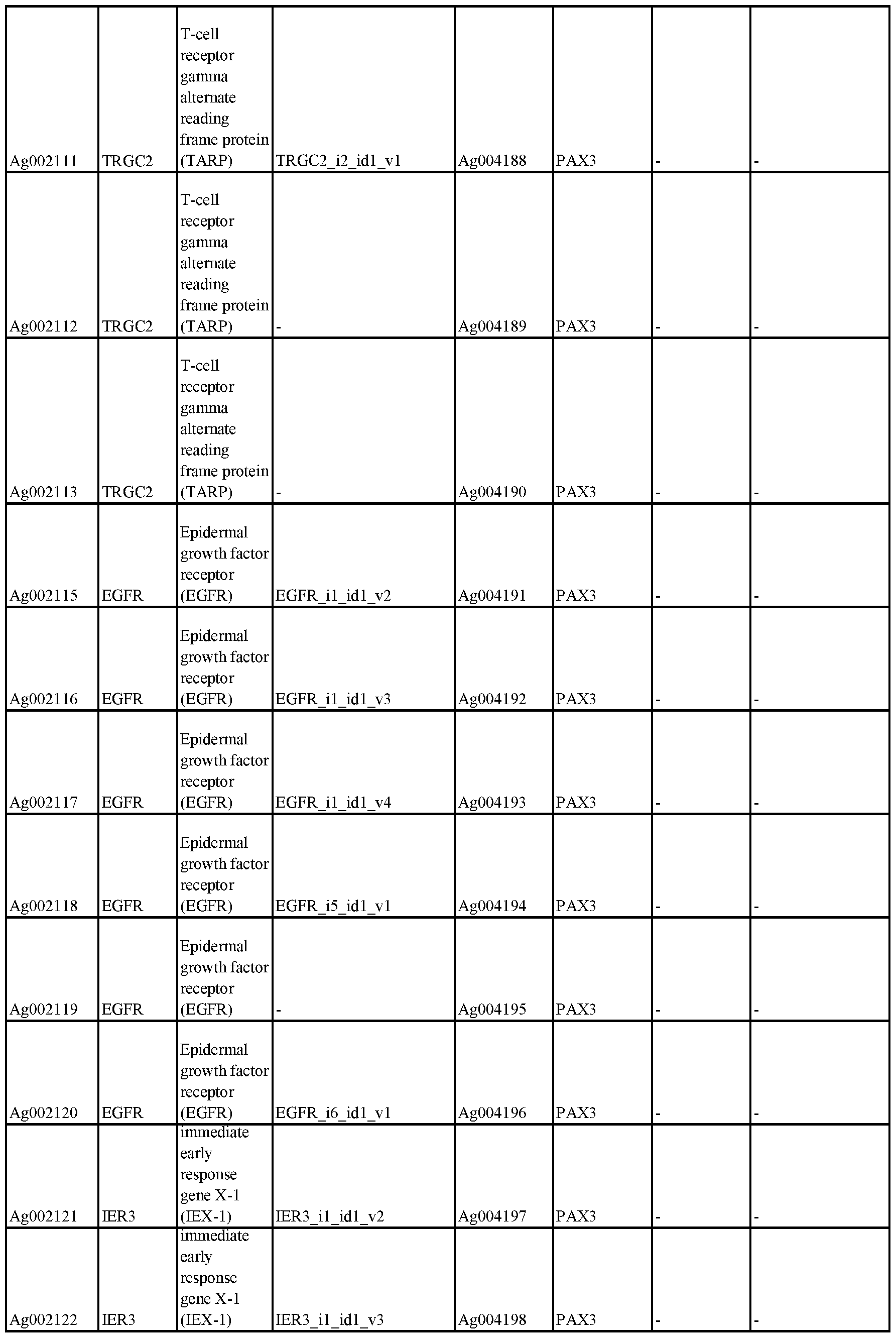


Claims
We claim:
1. A composition comprising: a nano-satellite complex, wherein said nano-satellite complex comprises:
a) a core nanoparticle complex comprising a biocompatible coating surrounding a nanoparticle core;
b) 3-25 satellite particles atached to, or absorbed to, said biocompatible coating; c) a plurality of antigenic peptides conjugated to, or absorbed to, said satellite particles; and
d) wherein said nano-satellite complex comprises at least one of the following properties:
i) wherein the weight-to-weight ratio of all of said satellite particles to said nanoparticle core is 10-40%;
ii) wherein the diameter of each of said satellite particles is 2-20 nm; iii) wherein said plurality of antigenic peptides is 100-4000 antigenic peptides;
iv) wherein 10-300 of said plurality of said antigenic peptides are present on each of said satellite particles; and
v) wherein the average distance between each of said satellite particles is 5-20 nm.
2. A composition comprising: a nano-satellite complex, wherein said nano-satellite complex comprises:
a) a core nanoparticle complex comprising a biocompatible coating surrounding a nanoparticle core;
b) 10-20 satellite particles attached to, or absorbed to, said biocompatible coating;
c) a plurality of antigenic peptides conjugated to, or absorbed to, said satellite particles; and
d) wherein said nano-satellite complex comprises at least one of the following properties:
i) wherein the weight-to-weight ratio of all of said satellite particles to said nanoparticle core is 10-40%;
ii) wherein the diameter of each of said satellite particles is 1-5 nm; iii) wherein said satellite particles are present at density of 15,00-30,000 per square micron;
iv) wherein said plurality of antigenic peptides is 1500-3000 antigenic peptides;
v) wherein 100-400 of said plurality of said antigenic peptides are present on each of said satellite particles; and
vi) wherein the average distance between each of said satellite particles is
4-7 nm.
3. The composition of claims 1-2, wherein said core nanoparticle comprises Fe304 and/or said satellite particles comprise gold.
4. The composition of claims 1-2, wherein said biocompatible coating comprises polysiloxane.
5. The composition of claims 1-2, wherein said nanoparticle core comprises Fe304, said biocompatible coating comprises polysiloxane, and said at least one satellite particle comprises a plurality of satellite particles composed of gold.
6. The composition of claims 1, wherein said 3-25 satellite particles is 10-15 satellite particles.
7. The composition of claims 1-2, wherein said at least one property is wherein said weight-to-weight ratio of all of said satellite particles to said nanoparticle core is 10-40%.
8. The composition of claim 7, wherein said weight-to-weight ratio of all of said satellite particles to said nanoparticle core is 25-35%.
9. The composition of claim 7, wherein said weight-to-weight ratio of all of said satellite particles to said nanoparticle core is 29-31%.
10. The composition of claim 1, wherein said at least one property is wherein said diameter of each of said satellite particles is 2-20 nm.
11. The composition of claim 10, wherein said diameter of each of said satellite particles is 5-15 nm.
12. The composition of claim 10, wherein said diameter of each of said satellite particles is 4-6 nm.
13. The composition of claim 1, wherein said at least one property is wherein said satellite particles are present at density of 500-20,000 per square micron.
14. The composition of claim 13, wherein said satellite particles are present at a density of 13,000 to 17,000 per square micron.
15. The composition of claim 1, wherein said at least one property is wherein said plurality of antigenic peptides is 100-4000 antigenic peptides.
16. The composition of claim 15, wherein said plurality of antigenic peptides is 1500- 2500 antigenic peptides.
17. The composition of claim 1, wherein said at least one property is wherein 10-300 of said plurality of said antigenic peptides are present on each of said satellite particles.
18. The composition of claim 17, wherein 225-275 of said plurality of antigenic peptides are present on each of said satellite particles.
19. The composition of claim 1, wherein said at least one property is wherein the average distance between each of said satellite particles is 5-20 nm.
20. The composition of claim 1, wherein said average distance between each of said satellite particles is 5-7 nm.
21. The composition of claims 1-2, wherein said at least one property is at least two of said properties.
22. The composition of claims 1-2, wherein said at least one property is at least four of said properties.
23. The composition of claims 1-2, wherein said at least one property is at least five of said properties.
24. The composition of claims 1-2, wherein said antigenic peptide comprises: i) a neoantigenic determinant, ii) at least one epitope from a tumor antigen, iii) at least one epitope from a viral oncoprotein, iv) a least one epitope from an infectious virus, v) at least one epitope from a parasite, or vi) at least one epitope from an infectious bacteria.
25. The composition of claims 1-2, further comprising a physiologically compatible aqueous solution.
26. The composition of claims 1-2, further comprising cancer cells or antigen presenting cells.
27. The composition of claims 1-2, wherein said plurality of antigenic peptides are not uniformly distributed on said satellite particles.
28. The composition of claims 1-2, wherein said nano-satellite complex is a diameter of 50-100 nm.
29. The composition of claim 28, wherein said diameter is 55-65 nm.
30. The composition of claims 1-2, wherein the surface of said nano-satellite complex is negatively charged.
31. The composition of claims 1-2, wherein said core nanoparticle has a diameter of 10- 25 nm.
32. The composition of claim 31, wherein said diameter is 15-20 nm.
33. The composition of claims 1-2, wherein said composition further comprises a type I interferon agonist agent.
34. The composition of claim 33, wherein said type I interferon agonist agent is electrostatically attracted to, or absorbed to, i) said antigenic peptides, ii) said plurality of satellite particles, and/or iii) said core nanoparticle.
35. The composition of claims 1-2, wherein said composition is adjuvant-free.
36. The composition of claims 1-2, further comprising an immune checkpoint inhibitor.
37. A method of eliciting an immune response in a subject comprising: administering to a subject said composition of any of claims 1-36 such that antibodies to said antigenic peptides are generated.
38. The method of claim 37, wherein said subject is a human.
39. The method of claim 37, wherein said subject is an animal.
40. The method of claim 37, further comprising taking a sample from said subject, and purifying at least some of said antibodies from said sample.
41. The method of claim 37, wherein no adjuvant is administered as part of said composition or otherwise.
42. The method of claim 37, wherein said subject is administering a type I interferon agonist agent, either in said composition or separately.
43. The method of claim 37, wherein said subject is administering an immune checkpoint inhibitor, either in said composition or separately.
45. The method of claim 37, wherein said antigenic peptides comprise B-Cell epitopes.
46. The method of claim 37, wherein said nanosatellite complex does not generate detectable non-specific antibody against said nano-satellite complex.
47. The method of claim 37, wherein said nanosatellite complex homes to a lymph node of said subject.
48. The method of claim 37, wherein said nanosatellite complex homes to a B-cell zone or T-cell zone of a lymph node of said subject.
49. The method of claim 37, wherein said nanosatellite complex is taken up by subcapsular sinus macrophages in said subject at a rate equal to a virus.
Priority Applications (2)
| Application Number | Priority Date | Filing Date | Title |
|---|---|---|---|
| CN201980081692.8A CN113226365B (en) | 2018-10-17 | 2019-10-17 | Nanosatellite complexes |
| US17/232,751 US12226491B2 (en) | 2018-10-17 | 2021-04-16 | Nano-satellite complexes |
Applications Claiming Priority (2)
| Application Number | Priority Date | Filing Date | Title |
|---|---|---|---|
| US201862746755P | 2018-10-17 | 2018-10-17 | |
| US62/746,755 | 2018-10-17 |
Related Child Applications (1)
| Application Number | Title | Priority Date | Filing Date |
|---|---|---|---|
| US17/232,751 Continuation-In-Part US12226491B2 (en) | 2018-10-17 | 2021-04-16 | Nano-satellite complexes |
Publications (1)
| Publication Number | Publication Date |
|---|---|
| WO2020081833A1 true WO2020081833A1 (en) | 2020-04-23 |
Family
ID=70284786
Family Applications (1)
| Application Number | Title | Priority Date | Filing Date |
|---|---|---|---|
| PCT/US2019/056765 Ceased WO2020081833A1 (en) | 2018-10-17 | 2019-10-17 | Nano-satellite complexes |
Country Status (3)
| Country | Link |
|---|---|
| US (1) | US12226491B2 (en) |
| CN (1) | CN113226365B (en) |
| WO (1) | WO2020081833A1 (en) |
Families Citing this family (2)
| Publication number | Priority date | Publication date | Assignee | Title |
|---|---|---|---|---|
| KR102660710B1 (en) * | 2021-12-30 | 2024-04-24 | 고려대학교 산학협력단 | Nanosatellite-substrate composite and method for controlling adhesion and differentiation of stem cells using the same |
| CN114802807B (en) * | 2022-04-20 | 2024-07-26 | 中国人民解放军国防科技大学 | Chemical molecular satellite capable of deconstruction and deformation |
Citations (2)
| Publication number | Priority date | Publication date | Assignee | Title |
|---|---|---|---|---|
| US20140086828A1 (en) * | 2010-05-28 | 2014-03-27 | Aaron E. Foster | Modified gold nanoparticles for therapy |
| US20150065858A1 (en) * | 2013-09-05 | 2015-03-05 | The Regents Of The University Of Michigan | Core-satellite nanocomposites for mri and photothermal therapy |
-
2019
- 2019-10-17 WO PCT/US2019/056765 patent/WO2020081833A1/en not_active Ceased
- 2019-10-17 CN CN201980081692.8A patent/CN113226365B/en active Active
-
2021
- 2021-04-16 US US17/232,751 patent/US12226491B2/en active Active
Patent Citations (2)
| Publication number | Priority date | Publication date | Assignee | Title |
|---|---|---|---|---|
| US20140086828A1 (en) * | 2010-05-28 | 2014-03-27 | Aaron E. Foster | Modified gold nanoparticles for therapy |
| US20150065858A1 (en) * | 2013-09-05 | 2015-03-05 | The Regents Of The University Of Michigan | Core-satellite nanocomposites for mri and photothermal therapy |
Also Published As
| Publication number | Publication date |
|---|---|
| CN113226365B (en) | 2025-08-19 |
| CN113226365A (en) | 2021-08-06 |
| US12226491B2 (en) | 2025-02-18 |
| US20210346478A1 (en) | 2021-11-11 |
Similar Documents
| Publication | Publication Date | Title |
|---|---|---|
| Zhang et al. | Metal‐organic‐framework‐based vaccine platforms for enhanced systemic immune and memory response | |
| CN112791181B (en) | Manganese nanoadjuvant, preparation method and application thereof | |
| Quach et al. | Size-dependent neutralizing activity of gold nanoparticle-based subunit vaccine against dengue virus | |
| Diaz-Arévalo et al. | Nanoparticle-based vaccines: opportunities and limitations | |
| Temchura et al. | Targeting and activation of antigen-specific B-cells by calcium phosphate nanoparticles loaded with protein antigen | |
| Zilker et al. | Nanoparticle-based B-cell targeting vaccines: Tailoring of humoral immune responses by functionalization with different TLR-ligands | |
| Liu et al. | Functional nanomaterials can optimize the efficacy of vaccines | |
| Lamalle-Bernard et al. | Coadsorption of HIV-1 p24 and gp120 proteins to surfactant-free anionic PLA nanoparticles preserves antigenicity and immunogenicity | |
| Battigelli et al. | Carbon nanomaterials as new tools for immunotherapeutic applications | |
| Abbaraju et al. | Asymmetric mesoporous silica nanoparticles as potent and safe immunoadjuvants provoke high immune responses | |
| Choi et al. | Protein cage nanoparticles as delivery nanoplatforms | |
| Xiang et al. | Delivery of DNA vaccines: an overview on the use of biodegradable polymeric and magnetic nanoparticles | |
| Geary et al. | Diaminosulfide based polymer microparticles as cancer vaccine delivery systems | |
| Avila et al. | Gene delivery and immunomodulatory effects of plasmid DNA associated with Branched Amphiphilic Peptide Capsules | |
| JP6228967B2 (en) | Antigenic compositions and methods | |
| Lee et al. | Virus-mimetic polymer nanoparticles displaying hemagglutinin as an adjuvant-free influenza vaccine | |
| US20050175584A1 (en) | Functionalized colloidal metal compositions and methods | |
| CN114939159A (en) | Construction and application of virus antigen and adjuvant-loaded multistage targeting vector | |
| Gupta et al. | Adjuvants in micro‐to nanoscale: current state and future direction | |
| US12226491B2 (en) | Nano-satellite complexes | |
| WO2025148984A1 (en) | Nanovaccine delivery system, preparation method therefor, and use thereof | |
| Damm et al. | Modulation of immune responses to liposomal vaccines by intrastructural help | |
| Li et al. | Cell membrane adhesive n-hexadecyl choline phosphate as vaccine delivery systems for anticancer immunotherapy | |
| CN110354271A (en) | One kind is based on including peptide-mediated nano-carrier modular platform and its construction method and application | |
| US20250295762A1 (en) | Composite bioactive compositions and applications thereof |
Legal Events
| Date | Code | Title | Description |
|---|---|---|---|
| 121 | Ep: the epo has been informed by wipo that ep was designated in this application |
Ref document number: 19873538 Country of ref document: EP Kind code of ref document: A1 |
|
| NENP | Non-entry into the national phase |
Ref country code: DE |
|
| 122 | Ep: pct application non-entry in european phase |
Ref document number: 19873538 Country of ref document: EP Kind code of ref document: A1 |
|
| WWG | Wipo information: grant in national office |
Ref document number: 201980081692.8 Country of ref document: CN |