WO2020076511A1 - Target validation and profiling of the rna targets of small molecules - Google Patents
Target validation and profiling of the rna targets of small molecules Download PDFInfo
- Publication number
- WO2020076511A1 WO2020076511A1 PCT/US2019/053179 US2019053179W WO2020076511A1 WO 2020076511 A1 WO2020076511 A1 WO 2020076511A1 US 2019053179 W US2019053179 W US 2019053179W WO 2020076511 A1 WO2020076511 A1 WO 2020076511A1
- Authority
- WO
- WIPO (PCT)
- Prior art keywords
- rna
- bleomycin
- binding
- compound
- mir
- Prior art date
- Legal status (The legal status is an assumption and is not a legal conclusion. Google has not performed a legal analysis and makes no representation as to the accuracy of the status listed.)
- Ceased
Links
Classifications
-
- A—HUMAN NECESSITIES
- A61—MEDICAL OR VETERINARY SCIENCE; HYGIENE
- A61K—PREPARATIONS FOR MEDICAL, DENTAL OR TOILETRY PURPOSES
- A61K47/00—Medicinal preparations characterised by the non-active ingredients used, e.g. carriers or inert additives; Targeting or modifying agents chemically bound to the active ingredient
- A61K47/50—Medicinal preparations characterised by the non-active ingredients used, e.g. carriers or inert additives; Targeting or modifying agents chemically bound to the active ingredient the non-active ingredient being chemically bound to the active ingredient, e.g. polymer-drug conjugates
- A61K47/51—Medicinal preparations characterised by the non-active ingredients used, e.g. carriers or inert additives; Targeting or modifying agents chemically bound to the active ingredient the non-active ingredient being chemically bound to the active ingredient, e.g. polymer-drug conjugates the non-active ingredient being a modifying agent
- A61K47/54—Medicinal preparations characterised by the non-active ingredients used, e.g. carriers or inert additives; Targeting or modifying agents chemically bound to the active ingredient the non-active ingredient being chemically bound to the active ingredient, e.g. polymer-drug conjugates the non-active ingredient being a modifying agent the modifying agent being an organic compound
- A61K47/55—Medicinal preparations characterised by the non-active ingredients used, e.g. carriers or inert additives; Targeting or modifying agents chemically bound to the active ingredient the non-active ingredient being chemically bound to the active ingredient, e.g. polymer-drug conjugates the non-active ingredient being a modifying agent the modifying agent being an organic compound the modifying agent being also a pharmacologically or therapeutically active agent, i.e. the entire conjugate being a codrug
-
- A—HUMAN NECESSITIES
- A61—MEDICAL OR VETERINARY SCIENCE; HYGIENE
- A61K—PREPARATIONS FOR MEDICAL, DENTAL OR TOILETRY PURPOSES
- A61K31/00—Medicinal preparations containing organic active ingredients
- A61K31/70—Carbohydrates; Sugars; Derivatives thereof
- A61K31/7028—Compounds having saccharide radicals attached to non-saccharide compounds by glycosidic linkages
- A61K31/7034—Compounds having saccharide radicals attached to non-saccharide compounds by glycosidic linkages attached to a carbocyclic compound, e.g. phloridzin
- A61K31/704—Compounds having saccharide radicals attached to non-saccharide compounds by glycosidic linkages attached to a carbocyclic compound, e.g. phloridzin attached to a condensed carbocyclic ring system, e.g. sennosides, thiocolchicosides, escin, daunorubicin
-
- A—HUMAN NECESSITIES
- A61—MEDICAL OR VETERINARY SCIENCE; HYGIENE
- A61K—PREPARATIONS FOR MEDICAL, DENTAL OR TOILETRY PURPOSES
- A61K47/00—Medicinal preparations characterised by the non-active ingredients used, e.g. carriers or inert additives; Targeting or modifying agents chemically bound to the active ingredient
- A61K47/50—Medicinal preparations characterised by the non-active ingredients used, e.g. carriers or inert additives; Targeting or modifying agents chemically bound to the active ingredient the non-active ingredient being chemically bound to the active ingredient, e.g. polymer-drug conjugates
- A61K47/51—Medicinal preparations characterised by the non-active ingredients used, e.g. carriers or inert additives; Targeting or modifying agents chemically bound to the active ingredient the non-active ingredient being chemically bound to the active ingredient, e.g. polymer-drug conjugates the non-active ingredient being a modifying agent
- A61K47/54—Medicinal preparations characterised by the non-active ingredients used, e.g. carriers or inert additives; Targeting or modifying agents chemically bound to the active ingredient the non-active ingredient being chemically bound to the active ingredient, e.g. polymer-drug conjugates the non-active ingredient being a modifying agent the modifying agent being an organic compound
-
- C—CHEMISTRY; METALLURGY
- C12—BIOCHEMISTRY; BEER; SPIRITS; WINE; VINEGAR; MICROBIOLOGY; ENZYMOLOGY; MUTATION OR GENETIC ENGINEERING
- C12N—MICROORGANISMS OR ENZYMES; COMPOSITIONS THEREOF; PROPAGATING, PRESERVING, OR MAINTAINING MICROORGANISMS; MUTATION OR GENETIC ENGINEERING; CULTURE MEDIA
- C12N15/00—Mutation or genetic engineering; DNA or RNA concerning genetic engineering, vectors, e.g. plasmids, or their isolation, preparation or purification; Use of hosts therefor
- C12N15/09—Recombinant DNA-technology
- C12N15/11—DNA or RNA fragments; Modified forms thereof; Non-coding nucleic acids having a biological activity
- C12N15/111—General methods applicable to biologically active non-coding nucleic acids
-
- C—CHEMISTRY; METALLURGY
- C12—BIOCHEMISTRY; BEER; SPIRITS; WINE; VINEGAR; MICROBIOLOGY; ENZYMOLOGY; MUTATION OR GENETIC ENGINEERING
- C12N—MICROORGANISMS OR ENZYMES; COMPOSITIONS THEREOF; PROPAGATING, PRESERVING, OR MAINTAINING MICROORGANISMS; MUTATION OR GENETIC ENGINEERING; CULTURE MEDIA
- C12N2310/00—Structure or type of the nucleic acid
- C12N2310/10—Type of nucleic acid
- C12N2310/14—Type of nucleic acid interfering nucleic acids [NA]
- C12N2310/141—MicroRNAs, miRNAs
-
- C—CHEMISTRY; METALLURGY
- C12—BIOCHEMISTRY; BEER; SPIRITS; WINE; VINEGAR; MICROBIOLOGY; ENZYMOLOGY; MUTATION OR GENETIC ENGINEERING
- C12N—MICROORGANISMS OR ENZYMES; COMPOSITIONS THEREOF; PROPAGATING, PRESERVING, OR MAINTAINING MICROORGANISMS; MUTATION OR GENETIC ENGINEERING; CULTURE MEDIA
- C12N2320/00—Applications; Uses
- C12N2320/10—Applications; Uses in screening processes
Definitions
- RNAs have diverse functions, particularly those that are non-coding 1 .
- Small molecule targeting of RNA has been challenging, except for RNAs that fold into highly three-dimensional (protein-like) structures such as ribosomes and riboswitches 2-4 . More recently, it has been shown that pre-mRNAs in complexes can be targeted and stabilized with small molecules 5-6 .
- Most RNAs do not have highly complex long-range folds but do have extensive two-dimensional (secondary) structure that that could be targeted with small molecules 7-8 . Indeed, small molecules have been discovered that bind microRNAs (miRNAs) and inhibit their biogenesis 9-11 .
- miRNAs microRNAs
- the invention is directed, in various embodiments, to a method of mapping an RNA binding site of an RNA-binding small molecule, the binding site being disposed within an RNA sequence library, comprising contacting the RNA of the sequence library and an effective amount of a compound comprising a conjugate of an RNA-cleaving moiety and an RNA-binding small molecule to cleave the RNA of the sequence library comprising the binding site to provide a cleaved RNA fragment;
- the methods disclosed herein comprise contacting a library of RNA sequences and an effective amount of a compound comprising a conjugate of an RNA- cleaving moiety and an RNA-binding small molecule to cleave the RNA of the library at a binding site of the compound to the RNA to provide a cleaved RNA fragment; and amplifying and sequencing the cleaved RNA fragment, thereby identifying the site of binding of the small RNA-binding molecule to the RNA of the library which is associated with the site of cleavage.
- the RNA-binding small molecule can comprise an N-methyl- piperazinyl-bis-benzimidazole group.
- the conjugate of the RNA-binding molecule can comprise bleomycin, for instance bleomycin A5, as the RNA-cleaving moiety.
- the RNA-binding molecule can be Targaprimir-96.
- a method of the invention comprises precisely targeting cellular destruction of an oncogenic non-coding RNA precursor, comprising contacting a cell expressing the non-coding RNA precursor and an effective amount of the compound.
- the oncogenic non-coding RNA precursor can comprise oncogenic primary microRNA-96 (pri-miR-96). More specifically, the compound can be a covalent conjugate of Targaprimir-96 and bleomycin A5.
- a methods comprise enhancing expression of FOXOl protein in breast cancer cells, comprising contacting the cells with an effective amount of the conjugate.
- the contacting can be via administration to a human patient.
- the conjugate can be a covalent conjugate of Targaprimir-96 and bleomycin A5.
- a method of the invention can comprise triggering apoptosis in triple negative breast cancer cells, comprising contacting the cells with an effective amount of the conjugate.
- the breast cancer cells can be present in a human patient.
- the conjugate can be a covalent conjugate of Targaprimir-96 and bleomycin A5.
- methods comprise treating triple negative breast cancer, comprising administering to a patient afflicted therewith an effective dose of the compound.
- the breast cancer can comprise expression of oncogenic primary microRNA-96 (pri-miR-96).
- the compound can be a covalent conjugate of Targaprimir-96 and bleomycin A5.
- the RNA sequence library can comprise a transcriptome.
- the transcriptome can be viral, or can be mammalian, or can be bacterial.
- the RNA sequence library can comprise one or more of synthetic, semi-synthetic, or natural RNA.
- the RNA sequence library can comprise the genome of an RNA virus.
- a method of the invention can be carried out in vitro, or can be carried out in living cells, e.g., in virally- or bacterially-infected cells.
- a set of RNA sequences and a set of compounds comprising candidate RNA-binding small molecules can be assayed in a 2- dimensional parallel array.
- the invention further provides a compound comprising a conjugate of an RNA- cleaving moiety and an RNA-binding small molecule.
- the RNA-cleaving moiety can comprise bleomycin.
- the RNA-binding small molecule can comprise N-methyl-piperazinyl- bis-benzimidazole group, e.g., Targaprimir-96.
- Pri-miR-96 is oncogenic and suppresses apoptosis in cancer cells via repression of the pro-apoptotic transcription factor Forkhead box protein Ol (FOXOl).
- Compound 1 was designed via Infoma and selectively targets pri-miR-96.
- Compound 2 is a version of 1 conjugated to bleomycin A5 while compound 3 is a version of 2 that lacks RNA-binding modules.
- Compound 1 inhibited the production of mature miR-96, de-repressed a downstream target, pro-apoptotic transcription factor FOXOl, and triggered apoptosis.
- FIG. 1 DNA cleavage by 2 and 3 in vitro and in cells.
- A studying the DNA cleaving activity of 2 and 3 in vitro. Appending RNA-binding modules onto the bleomycin core (affording 2) decreased the amount of DNA cleavage as compared to 3, which lacks RNA- binding modules.
- B Visualization and quantification of DNA damage in MDA-MB-231 cells treated with 500 nM 1, 2 or 3 for 12 h. Data are expressed as mean ⁇ s.e.m. (n 3 3). * p ⁇ 0.05, as determined by a two-tailed Student t test by comparison to untreated DNA (A) or cells (B).
- FIG. 3 Studying the effect of 1 and 2 on mature miR-96 and pri-miR-96 levels and on miRNA-mediated biology.
- A the effect of 1 and 2 on mature miR-96 levels in MDA-MB- 231 TNBC cells.
- B the effect of 1 and 2 on pri-miR-96 levels in MDA-MB-231 cells.
- Infoma enabled the design of a small molecule (Targaprimir-96, 1, Fig. 1A) that selectively targets the Drosha endonuclease- processing site of oncogenic primary microRNA-96 (pri-miR-96) 13 .
- MiRNAs are non-coding RNAs that play pervasive roles in biology, and their aberrant expression or mutation can be causative of disease. They are initially produced as precursors (pri-miRNA) that are processed by the nuclease Drosha followed by translocation to the cytoplasm as precursor microRNAs (pre-miRNAs).
- Pre-miRNAs are cleaved by the cytoplasmic nuclease Dicer to produce mature miRNAs that bind to the 3’ untranslated regions (UTRs) of mRNAs and repress translation.
- TNBC triple negative breast cancer
- bleomycin A5 was conjugated to 1, affording 2 (Fig. 1).
- Bleomycin a natural product used for treatment of cancer, cleaves DNA 14, 15 but also cleaves RNA, as pioneered by the Hecht group 16, 17 Bleomycin contains four domains: (i) a metal ion-binding domain that activates O 2 and leads to nucleic acid cleavage 18-21 ; (ii) a DNA- binding domain, which affects cleavage efficiency 22 ; (iii) a linker region between the metal ion-binding and DNA-binding domains, which also affects cleavage efficiency 23-27 ; and (iv) a carbohydrate domain which facilitates cellular uptake 20 .
- bleomycin A5 as the cationic dimethyl sulfonium in the C -terminal DNA-binding domain has been replaced with a butyl- 1,4-diamine side chain.
- This modification serves two purposes: (i) facile conjugation of the terminal primary amine to RNA-binding modules containing carboxylates. Notably, acylation of the butyl-1, 4-diamine side chain (cationic) with a small molecule affords an uncharged linkage; and (ii) reduction of DNA binding affinity 28-30 as the cationic side chain, known to drive binding to DNA, has been acylated and is no longer charged.
- RNA-binding module is therefore likely to alter bleomycin A5’s binding and cleavage preferences toward RNA, as we have observed in the selective cleavage of expanded r(CUG) repeats 32 .
- bleomycin can cleave AU pairs in RNA 33 , and indeed AU pairs are present adjacent to pri-miR-96’s Drosha site.
- conjugation of bleomycin to compound 1 could provide a selective cleaving small molecule, provided that the bleomycin is positioned towards these AU pairs.
- pri-miR-96 can be cleaved to a greater extent by 2 than DNA is cleaved within a concentration window; that is, the targets that are cleaved by bleomycin can be attenuated by addition of an RNA-binding module at mid-nanomolar concentrations both in vitro and in cells, vide infra.
- miR-96 suppresses apoptosis by silencing the production of pro- apoptotic transcription factor FOXOl 32 .
- inhibition of miR-96 by 2 should increase the amount ofFOXOl and trigger apoptosis.
- the amount of FOXOl protein in MDA- MB-231 cells was increased by ⁇ 1.8-fold when treated with 500 nM of 2 (Fig. 3D).
- the effect of 2 on phenotype (suppression of apoptosis) 12, 13, 32 was then assessed by using both Annexin/PI staining and Caspase assays.
- 2 induced apoptosis in MDA-MB-231 cells Fig.
- RNA RNA
- RhoSNAP small molecule nucleic acid profiling by cleavage applied to RNA
- RNA binding sites within an RNA The most common method to identify small molecule binding sites within an RNA is to monitor sites of protection from nuclease cleavage or reaction with chemical modification reagents. Indeed, this approach identified sites in the ribosome that bound antibiotics 39, 40 . However, some binding sites can be silent due to lack of reactivity with a chemical modifier and can require long residence times of the small molecule to prevent reactivity (irreversible). Thus, careful tuning of the experimental conditions is often necessary. Although laborious, these types of experiments are invaluable to validate or identify the target(s) of small molecules, which is essential to establish a compound’s mode of action.
- Ribo-SNAP-Map RNA target and binding site within the RNA
- Ribo-SNAP-Map To implement Ribo-SNAP-Map, we developed a procedure to enrich the partial cleavage products of pri-miR-96 (Fig. 4B) using a gene specific forward primer and a universal reverse primer in an RT-qPCR experiment 41 . Gel analysis showed a new band at ca. 130 base pairs only when cells were exposed to 2, not to 1 or 3. Sequencing analysis confirmed that the cleavage sites were proximal to the predicted and in vitro mapped binding sites for 2 (Fig. 4C). Molecular modeling of 2 binding with pri-miR-96 also showed that 2 positions the cleaving moiety towards the AU sequence that is cleaved rather than other regions in the RNA that are distant from the ligand’s binding site.
- Ribo-SNAP-Map can indeed be used to map binding sites in cells.
- AU pairs proximal to a small molecule’s binding site is not requisite for selective cleavage of bleomycin A5 conjugates, as observed for r(CUG) repeats 31 .
- nuclease mimics 43 or recruit endogenous nucleases to an RNA target.
- applicability can be limited because of the necessity of light to penetrate cells and tissue.
- Compounds that act as nuclease mimics interact with an expanded repeating RNA and have a mixed mode of inhibition (transcriptional inhibition, inhibition of protein binding, and cleavage) 43 .
- expanded repeating RNAs are atypical targets; due to its repeating nature and hence multiple small molecule binding sites, inefficient cleavage could afford a significant biological effect.
- RNA targeting chimeras More recently, we developed an approach named Ribonuclease targeting chimeras (RIBOTACs) to recruit endogenous RNase L to cleave a desired RNA target 44 .
- RIBOTACs Ribonuclease targeting chimeras
- cleavage patterns on the RNA target using these methods can be complex and may not be proximal to the binding site.
- cleavage with 2 is proximal and not complex, allowing straightforward identification of RNA sequences nearby ligand binding sites.
- RNAs can be engendered with antisense-like properties in cells using chimeric compounds comprised of a selective RNA-binding small molecule and bleomycinA5 as a cleaving moiety.
- these studies and others suggest that: (i) the targets cleaved by bleomycin can be tuned by conjugation to an RNA-binding small molecule; (ii) these capacities are likely programmable; and (iii) the ability to cleave RNAs with small molecules could expand the target scope of ligands that modulate the biology of RNA, akin to the revolution that PROTACs 45 initiated in the protein targeting field.
- Most RNAs’ biology may not be affected by simple binding and engendering a small molecule with the ability to cleave will likely expand the number of RNAs that can be targeted with organic compounds.
- Bhattacharyya A., Furia, B., Risher, N., Sheedy, J., Kong, R., Ma, J., Turpoff, A., Lee, C. S., Zhang, X., Moon, Y. C., Trifillis, P., Welch, E. M., Colacino, J. M., Babiak, J., Almstead, N. G, Peltz, S. W., Eng, L. A., Chen, K. S., Mull, J. L., Lynes, M. S., Rubin, L. L., Fontoura, P., Santarelli, L., Haehnke, D., McCarthy, K.
- ENA preparation The single-stranded DNA template (5’-
- PCR amplification was performed in l x PCR Buffer (10 mM Tris, pH 9.0, SO mM KC1, and 0.1% (v/v) Triton X-100), 2 mM T7 promoter forward primer (5’- GGCCGGATCCTAATACGACTCACTATAGGGTGGCCGATTTTGGC-3’) (SEQ.
- the resulting double stranded DNA template was transcribed by T7 RNA polymerase in 1 x Transcription Buffer (40 mM Tris HC1, pH 8.1, 1 mM spermidine, 0.001% (v/v) Triton X- 100 and 10 mM DTT) containing 2.25 mM of each rNTP and 5 mM MgCl 2 at 37 °C overnight.
- the RNA product was purified on a denaturing 15% polyacrylamide gel, and concentrations were determined by UV absorbance at 260 nm.
- RNA cleavage and primer extension The pri-miR-96 RNA (1 pM) was folded as previously described 2 by heating at 65 °C for 5 min and slowly cooling to room temperature. Different concentrations of 2 or 3 (5, 50, 500 nM) were pre-activated by the addition of 1 eq Fe 2+ and added to the folded RNA in a total volume of 20 mL. Next, 1 eq Fe 2+ was added 30 min and then 60 min later, and the reaction mixtures were incubated at 37 °C overnight.
- Loading Buffer 95% formaldehyde, 50 mM EDTA, 0.05% (w/v) bromophenol blue, 0.05% (w/v) xylene cyanol
- DNA cleavage in vitro Different concentrations of 2 or 3 (5, 50, 500, 5000 and 10000 nM) were pre-activated by the addition of 1 eq Fe 2+ and then 2 mL of a plasmid encoding GFP (150 ng/mL) was added in a final volume of 20 mL. An additional equivalent of Fe 2+ was added 30 min and then 60 min later, and the reaction mixture was incubated at 37 °C overnight. The reaction mixture was loaded on 0.8% agarose gel with 6xGel Loading Dye, Purple (NEB) and stained with ethidium bromide for 10 min.
- MDA-MB-231 cells were grown in a glass bottom 96-well plate and treated with compound for 24 h. Cells were washed with 1 c DPBS three times and then fixed with 100 mL of 4% paraformaldehyde for 10 min at 37 °C. Cells were washed with 1 x DPBS three times and then with 0.1% Triton X-100 in 1 x DPBS three times for 5 min at 37 °C.
- RT-qPCR of mIRNAs Cells (-70% confiuency) were treated with various concentrations of compound for 24 h. Total RNA was extracted using a Quick-RNA Miniprep Kit (Zymo Research) per the manufacturer’s protocol. Approximately 200 ng of total RNA, as determined by Nanodrop, was used for reverse transcription using a miScript II RT Kit (Qiagen) per the manufacturer’s protocol. RT-qPCR was performed on a 7900HT Fast Real Time PCR System (Applied Biosystem) using Power SYBR Green Master Mix (Applied Biosystems). All primers were purchased from IDT and listed in Table SI. The primers for unbiased miRNA profiling were purchased from Eurofins Genomics in a 384- well plate. The expression levels of miRNAs were normalized to U6 small nuclear RNA. 5
- the membrane was stripped using lx Stripping Buffer (200 mM glycine, pH 2.2 and 0.1% SDS) followed by washing in 1 x TBST.
- the membrane was blocked and probed for b-actin similarly using 1:5000 b-actin primary antibody (Cell Signaling Technology) in 1 x TBST containing 5% milk at room temperature for 1 h.
- the membrane was washed with lx TBST and incubated with 1:10,000 antirabbit IgG horseradish-peroxidase secondary antibody conjugate (Cell Signaling Technology) in 1 x TBS for 1 h at room temperature b- actin protein expression was quantified using SuperSignal West Pico Chemiluminescent Substrate (Pierce Biotechnology) per the manufacturer’s protocol.
- the fold change of FOXOl expression was calculated by normalizing FOXOl band intensity to b-actin band intensity.
- Caspase assay Cells in 96-well plates (-60% confluency) were treated with various concentrations of compound for 48 h. In the case of miR-96 overexpression, cells were transfected with a plasmid encoding pri-miR-96 with Lipofectamine 2000 (Thermo Fisher Scientific) per the manufacturer’s protocol, split into 96-well plates, and treated with compounds for 48 h.
- MCF 10A cells were cultured in Dulbecco’s modified eagle medium/F12 (DMEM/F12) supplemented with 20 ng/mL epidermal growth factor (EGF), 0.5 mg/mL hydrocortisone, cholera toxin 0.1 mg/mL, bovine insulin 10 mg/mL, 1%
- Annexin V/PI Assay Cells in 6-well plates (-60% confluency) were incubated with 2 or 3 for 48 h. As a positive control, cells were treated with 10 mM camptothecin for 24 h. The cells were detached from the surface by using accutase and washed twice with ice-cold 1 c DPBS and then three times with 1 x Annexin Binding Buffer (50 mM Hepes (pH 7.4), 700 mM NaCl and 12.5 mM CaCl 2 ). The cells were suspended in 100 mL lx Annexin Binding Buffer containing 5 mL Annexin V-APC (eBioscience).
- RNAComposer http://macomposer.cs.put.poznan.pl/
- the RNA- binding small molecule was energy minimized using MacroModel (Schrodinger, LLC, NY) and placed proximal to the RNA. Energy minimization of the RNA-small molecule complex was performed until the gradient of energy was less than 0.01 kcal/mol/A. The bleomycin A5 moiety was then manually added and allowed flexibility during an additional energy minimization to model the binding of 2 to pri-miR-96. Energy minimization was performed until the gradient of energy was less than 0.01 kcal/mol/A. The conformation with lowest energy is shown in Figure S8.
- Ribo-SNAP-Map 7-8 Cells were grown in 100 mm dishes to -70% confluency and treated with 1 (control), 2, or 3 (control) for 6 h. Total RNA was then extracted by treatment with TRIzol (ThermoFisher Scientific) and quantified by Nanodrop. Approximately 10 mg of total RNA was used for reverse transcription with a pri-miR-96 specific primer (5’- C AGACGTGTGCTCTTCCGATCTCGC AGCTGCGGGTCCT-3’ ; (SEQ. ID NO:27) 2 pmol) using Superscript IP (SSIII; Life Technologies).
- RNAClean XP beads (Beckman Coulter; 1.8 volumes of beads and 3 volumes of
- the purified cDNA was ligated with a 3’ adapter
- T4 RNA ligase 1 (/5Phos/AGATCGGAAGAGCGTCGTGTAG/3Bio/)(SEQ. ID NO:28) by T4 RNA ligase 1 (New England BioLabs; NEB) following the manufacturer’s recommended protocol (2 mL 10x T4 RNA ligase buffer, 1 mL of 1 mM ATP, 10 mL 50% PEG 8000, 5 mL cDNA, 1 mL of 20 mM ssDNA adaptor, and 1 mL of T4 RNA ligase). Then, the cDNA ligated to the adaptor was purified with RNAClean XP beads as described above.
- PCR amplification was performed with the ligated cDNA by using Phusion polymerase (NEB) with cycles of 98 °C for 20 s, 64 °C for 20 s and 72 °C for 90 s and the following primers: forward - (5’- CAGACGTGTGCTCTTCCGATC-3’) (SEQ. ID NO:29); reverse - (5’- CTACACGACGCTCTTCCGATCT-3’) (SEQ. ID NO:30).
- the PCR products were then 5’- end labeled with 32 P as described above and separated on a denaturing 15% polyacrylamide gel. A 100 bp ladder (NEB) was used as a marker.
- PCR products were separated on a denaturing 15% polyacrylamide gel, and the target band was excised from gel and ethanol precipitated.
- the purified DNA was ligated into a vector by using NEB’s PCR Cloning Kit per the manufacturer’s protocol. Antibiotic-resistant colonies were selected and subjected to Sanger sequencing by Genewiz.
- the peptoid in the resin was then treated with a solution of 2 eq Hoechst carboxylate (synthesized as previously described) 9 , 2 eq DIC and 2 eq DIPEA (N,N'-diisopropylethylamine) in 10 mL DMF.
- the reaction mixture was shaken at room temperature for 2 h and washed with 10 mL DMF three and then 10 mL dichloromethane (DCM) three times.
- the product was cleavage from resin by adding 10 mL 30% trifluoroacetic acid (TFA) in DCM and shaken 30 min at room temperature.
- TFA trifluoroacetic acid
- the resulting yellow solid was directly treated with a solution of 1.1 eq azide 2 , 0.2 eq Cu(I) catalyst and 2 eq DIEA in 2 mL DMF.
- the reaction mixture was kept at 65 °C overnight, and the reaction process was monitored by MALDI-TOF MS. After the starting material was no longer detectable, the product was precipitated with excess amounts of ether and dissolved in 50% methanol in water with 0.1%TFA.
- HPLC purification was performed with a linear gradient from 0% to 100% B (methanol or acetonitrile + 0.1%TFA) in A (water + 0.1%TFA) over 60 min and a flow rate of 5 mL/min. The pure fractions were collected, and the solvent was concentrated under vacuum.
- the resin was washed with 10 mL DMF three times and 5 eq of bromoacetic acid in 10 mL DMF solution and 5 eq DIC were added.
- the resin was shaken at room temperature for 30 min followed by addition of 5 eq propylamine in 10 mL DMF.
- the reaction mixture was shaken at room temperature for 2 h.
- the bromoacetic acid coupling step and propylamine step were repeated.
- the peptoid on the resin was then treated with a solution of 2 eq Hoechst carboxylate, 2 eq DIC and 2 eq DIPEA in 10 mL DMF solution.
- the reaction mixture was shaken at room temperature for 2 h and washed with 10 mL DMF three times and then 10 mL DCM three times.
- the product was cleaved from resin by adding 10 mL of 30% TFA in DCM and shaking the resin for 30 min at room temperature.
- the elutate was concentrated under vacuum, and the product was precipitated with an excess amount of ether.
- the resulting solid was directly treated with a solution of 1.1 eq azide, 0.2 eq Cu(I) catalyst and 2 eq DIEA in 2 mL DMF.
- the reaction mixture was kept 65 °C overnight, and the reaction process was monitored by MALDI MS. After the starting material was no longer detectable, the product was precipitated with an excess amount of ether.
- the Bleomycin A5 coupling reaction was performed by adding 2 eq HOAt (1- Hydroxy-7-azabenzotri azole), 2 eq HATU (hexafluorophosphate azabenzotri azole tetramethyl uranium), 5 eq DIEA, and 2 eq Copper-bleomycin A5.
- the reaction mixture was shaken at room temperature overnight and the product was precipitated with an excess amount of ether.
- the solid was dissolved in 50% acetonitrile in water with 0.1%TFA and subjected to HPLC purification. After injection of the solution, the column was washed with 50 mM EDTA (pH 6.7) for 30 min to remove the copper ion and then washed with water for another 30 min.
- the resin was washed with 10 mL DMF three times followed by addition of 5 eq of bromoacetic acid in 10 mL DMF solution and 5 eq DIC.
- the resin was shaken at room temperature for 30 min and then 5 eq propylamine in 10 mL DMF were added. After shaking the mixture at room temperature for 2 h, the bromoacetic acid coupling step and propylamine step were repeated.
- the peptoid on the resin was then treated with a solution of 2 eq acetic anhydride (Ac 2 O) and 2 eq DIPEA in 10 mL DMF.
- the reaction mixture was shaken at room temperature for 30 min and washed with 10 mL DMF three times and then 10 mL DCM three times.
- the product was cleaved from the resin by adding 10 mL 30% TFA in DCM and shaking at room temperature for 30 min.
- the eluate was concentrated under vacuum, and the product was precipitated with excess amount of ether.
- the Bleomycin A5 coupling reaction was performed by adding 2 eq HO At, 2 eq HATU, 5 eq DIEA and 2 eq Copper-Bleomycin A5.
- the reaction mixture was shaken at room temperature overnight, and the product was precipitated with an excess amount of ether.
- the solid was dissolved in 50% acetonitrile in water with 0. l%TFA and subjected to HPLC purification. After injection of the solution, the column was washed with 50 mM EDTA (pH 6.7) for 30 min to remove the copper ion and then washed with water for another 30 min. Then the target product was separated with a linear gradient from 0% to 100% B (acetonitrile +0.
Landscapes
- Health & Medical Sciences (AREA)
- Engineering & Computer Science (AREA)
- Life Sciences & Earth Sciences (AREA)
- Bioinformatics & Cheminformatics (AREA)
- Chemical & Material Sciences (AREA)
- Genetics & Genomics (AREA)
- Biomedical Technology (AREA)
- General Health & Medical Sciences (AREA)
- Molecular Biology (AREA)
- Epidemiology (AREA)
- General Engineering & Computer Science (AREA)
- Veterinary Medicine (AREA)
- Animal Behavior & Ethology (AREA)
- Pharmacology & Pharmacy (AREA)
- Medicinal Chemistry (AREA)
- Organic Chemistry (AREA)
- Zoology (AREA)
- Biotechnology (AREA)
- Public Health (AREA)
- Wood Science & Technology (AREA)
- Microbiology (AREA)
- Plant Pathology (AREA)
- Biophysics (AREA)
- Biochemistry (AREA)
- Physics & Mathematics (AREA)
- Measuring Or Testing Involving Enzymes Or Micro-Organisms (AREA)
- Pharmaceuticals Containing Other Organic And Inorganic Compounds (AREA)
Abstract
A method for the precise cellular destruction of an oncogenic non-coding RNA with a RNA-binding small molecule conjugated with bleomycin A5 is described. The method affords reversal of phenotype. Bleomycin A5 was coupled to an RNA-binding molecule that selectively binds the microRNA-96 hairpin precursor (pri-miR-96). By coupling of bleomycin A5's free amine to the RNA-binding molecule, its affinity for binding to pri-miR- 96 is >100-fold stronger than to DNA. The conjugate compound selectively cleaves pri-miR- 96 in triple negative breast cancer (TNBC) cells. Selective cleavage of pri-miR-96 enhances expression of FOXO1 protein, a pro-apoptotic transcription factor that miR-96 silences, and triggers apoptosis in TNBC cells. No effects were observed in healthy breast epithelial cells. This method provides programmable control for targeting RNA through the selection of an RNA-binding molecule/bleomycin A5 conjugate and provides a facile method of mapping the cellular binding sites of an RNA-binding molecule.
Description
Target Validation and Profiling of the RNA Targets of Small Molecules
STATEMENT OF GOVERNMENT SUPPORT
This invention was made with government support under grant number GM097455 awarded by the National Institutes of Health. The government has certain rights in the invention.
BACKGROUND
ENCODE showed that over 70% of our genome is transcribed into RNA1. These RNAs have diverse functions, particularly those that are non-coding1. Small molecule targeting of RNA, however, has been challenging, except for RNAs that fold into highly three-dimensional (protein-like) structures such as ribosomes and riboswitches2-4. More recently, it has been shown that pre-mRNAs in complexes can be targeted and stabilized with small molecules5-6. Most RNAs, however, do not have highly complex long-range folds but do have extensive two-dimensional (secondary) structure that that could be targeted with small molecules7-8. Indeed, small molecules have been discovered that bind microRNAs (miRNAs) and inhibit their biogenesis9-11.
SUMMARY
The invention is directed, in various embodiments, to a method of mapping an RNA binding site of an RNA-binding small molecule, the binding site being disposed within an RNA sequence library, comprising contacting the RNA of the sequence library and an effective amount of a compound comprising a conjugate of an RNA-cleaving moiety and an RNA-binding small molecule to cleave the RNA of the sequence library comprising the binding site to provide a cleaved RNA fragment;
then, amplifying and sequencing the cleaved RNA fragment, thereby identifying the site of binding within the RNA sequence library of the small RNA-binding molecule associated with the site of cleavage;
to identify the sequence of the RNA binding site of the small RNA-binding molecule.
In various cases, the methods disclosed herein comprise contacting a library of RNA sequences and an effective amount of a compound comprising a conjugate of an RNA- cleaving moiety and an RNA-binding small molecule to cleave the RNA of the library at a binding site of the compound to the RNA to provide a cleaved RNA fragment; and amplifying and sequencing the cleaved RNA fragment, thereby identifying the site of binding
of the small RNA-binding molecule to the RNA of the library which is associated with the site of cleavage.
For example, the RNA-binding small molecule can comprise an N-methyl- piperazinyl-bis-benzimidazole group. For example, the conjugate of the RNA-binding molecule can comprise bleomycin, for instance bleomycin A5, as the RNA-cleaving moiety. In some embodiments, the RNA-binding molecule can be Targaprimir-96.
In various embodiments, a method of the invention comprises precisely targeting cellular destruction of an oncogenic non-coding RNA precursor, comprising contacting a cell expressing the non-coding RNA precursor and an effective amount of the compound. For instance, the oncogenic non-coding RNA precursor can comprise oncogenic primary microRNA-96 (pri-miR-96). More specifically, the compound can be a covalent conjugate of Targaprimir-96 and bleomycin A5.
In various embodiments, a methods comprise enhancing expression of FOXOl protein in breast cancer cells, comprising contacting the cells with an effective amount of the conjugate. For example, the contacting can be via administration to a human patient. More specifically, the conjugate can be a covalent conjugate of Targaprimir-96 and bleomycin A5.
In various embodiments, a method of the invention can comprise triggering apoptosis in triple negative breast cancer cells, comprising contacting the cells with an effective amount of the conjugate. The breast cancer cells can be present in a human patient. The conjugate can be a covalent conjugate of Targaprimir-96 and bleomycin A5.
In various embodiments, methods comprise treating triple negative breast cancer, comprising administering to a patient afflicted therewith an effective dose of the compound. For example, the breast cancer can comprise expression of oncogenic primary microRNA-96 (pri-miR-96). The compound can be a covalent conjugate of Targaprimir-96 and bleomycin A5.
In various embodiments, the RNA sequence library can comprise a transcriptome.
For example, the transcriptome can be viral, or can be mammalian, or can be bacterial. The RNA sequence library can comprise one or more of synthetic, semi-synthetic, or natural RNA. The RNA sequence library can comprise the genome of an RNA virus.
In various embodiments, a method of the invention can be carried out in vitro, or can be carried out in living cells, e.g., in virally- or bacterially-infected cells.
In various embodiments of a method of the invention, a set of RNA sequences and a set of compounds comprising candidate RNA-binding small molecules can be assayed in a 2- dimensional parallel array.
The invention further provides a compound comprising a conjugate of an RNA- cleaving moiety and an RNA-binding small molecule. The RNA-cleaving moiety can comprise bleomycin. The RNA-binding small molecule can comprise N-methyl-piperazinyl- bis-benzimidazole group, e.g., Targaprimir-96.
BRIEF DESCRIPTION OF THE FIGURES
Figure 1. Pri-miR-96 is oncogenic and suppresses apoptosis in cancer cells via repression of the pro-apoptotic transcription factor Forkhead box protein Ol (FOXOl).
(A) structures of the compounds used in these studies. Compound 1 was designed via Infoma and selectively targets pri-miR-96. Compound 2 is a version of 1 conjugated to bleomycin A5 while compound 3 is a version of 2 that lacks RNA-binding modules. (B) secondary structure of pri-miR-96 and the miR-96-FOXOl pathway. Compound binding sites are indicated in the secondary structure, and mature miR-96 is indicated in red lettering. The red "X” indicates inhibition of Drosha processing by a small molecule; likewise the red arrow indicates that inhibition of pri-miR-96 processing inhibits repression of FOXOl and hence breast cancer. Compound 1 inhibited the production of mature miR-96, de-repressed a downstream target, pro-apoptotic transcription factor FOXOl, and triggered apoptosis.
Figure 2. DNA cleavage by 2 and 3 in vitro and in cells. (A) studying the DNA cleaving activity of 2 and 3 in vitro. Appending RNA-binding modules onto the bleomycin core (affording 2) decreased the amount of DNA cleavage as compared to 3, which lacks RNA- binding modules. (B) Visualization and quantification of DNA damage in MDA-MB-231 cells treated with 500 nM 1, 2 or 3 for 12 h. Data are expressed as mean ± s.e.m. (n ³ 3). * p<0.05, as determined by a two-tailed Student t test by comparison to untreated DNA (A) or cells (B).
Figure 3. Studying the effect of 1 and 2 on mature miR-96 and pri-miR-96 levels and on miRNA-mediated biology. (A) the effect of 1 and 2 on mature miR-96 levels in MDA-MB- 231 TNBC cells. (B) the effect of 1 and 2 on pri-miR-96 levels in MDA-MB-231 cells. As expected based on their modes of action, 1 (simple binding) increased pri-miR-96 levels, while 2 (cleavage) reduced them (C) co-addition of increasing concentrations of 1 (5 to 500 nM) and a constant concentration of 2 (500 nM) to MDA-MB-231 cells increased levels of pri-miR-96, diminishing the cleaving capacity of 2 as expected. (D) effect of 2 on expression of FOXOl protein, a direct target of miR-96, as determined by Western blot. (E) effect of 2 on apoptosis in MDA-MB-231 cells (red), MDA-MB-231 cells that overexpress pri-miR-96 via a plasmid (green), and in MCF-10a healthy breast cells (blue), as determined by Caspase
assays. Data are expressed as mean ± s.e.m. (n > 3). *p<0.05, **p<0.0l, as measured by a two-tailed Student t test by comparison to untreated cells.
Figure 4. An unbiased miRNA-profiling approach shows that 2 most significantly affects mature miR-96 levels in MDA-MB-231 cells and amplification of cleavage products identifies the small molecule binding site within pri-miR-96. (A) Volcano plot for profiling the effect of 2 on all expressed miRNAs in MDA-MB-231 cells; miR-96 is the most affected. (B) a scheme of the amplification approach to identify small molecule binding sites via cleavage. (C) Top, representative Sanger sequencing results from cDNA of the cleaved RNA. The cleavage site is indicated with a red box. Bottom, analysis of several clones reveals three cleavage sites (indicated with a box and arrow in the pri-miR-96 secondary structure); 40% of reads stop at the first C (5’); ~30% of reads stop at the A; and ~30% of reads stop the second C (3’). Data are expressed as mean ± s.e.m. (n > 3).
DETAILED DESCRIPTION
To provide rational approaches to target RNA with small molecules, we developed a sequence-based approach dubbed Infoma12 In particular, Infoma enabled the design of a small molecule (Targaprimir-96, 1, Fig. 1A) that selectively targets the Drosha endonuclease- processing site of oncogenic primary microRNA-96 (pri-miR-96)13. MiRNAs are non-coding RNAs that play pervasive roles in biology, and their aberrant expression or mutation can be causative of disease. They are initially produced as precursors (pri-miRNA) that are processed by the nuclease Drosha followed by translocation to the cytoplasm as precursor microRNAs (pre-miRNAs). Pre-miRNAs are cleaved by the cytoplasmic nuclease Dicer to produce mature miRNAs that bind to the 3’ untranslated regions (UTRs) of mRNAs and repress translation. Application of 1 to triple negative breast cancer (TNBC) cells inhibited the production of mature miR-96, de-repressed pro-apoptotic transcription factor Forkhead box protein 01 (F0X01) that the miRNA repressed, and triggered apoptosis (Fig. 1B)13.
To expand the functional repertoire of small molecules that target RNA from simple binding to selective cleavage, bleomycin A5 was conjugated to 1, affording 2 (Fig. 1).
Bleomycin, a natural product used for treatment of cancer, cleaves DNA14, 15 but also cleaves RNA, as pioneered by the Hecht group16, 17 Bleomycin contains four domains: (i) a metal ion-binding domain that activates O2 and leads to nucleic acid cleavage 18-21 ; (ii) a DNA- binding domain, which affects cleavage efficiency22; (iii) a linker region between the metal ion-binding and DNA-binding domains, which also affects cleavage efficiency23-27; and (iv) a carbohydrate domain which facilitates cellular uptake20. We chose bleomycin A5 as the
cationic dimethyl sulfonium in the C -terminal DNA-binding domain has been replaced with a butyl- 1,4-diamine side chain. This modification serves two purposes: (i) facile conjugation of the terminal primary amine to RNA-binding modules containing carboxylates. Notably, acylation of the butyl-1, 4-diamine side chain (cationic) with a small molecule affords an uncharged linkage; and (ii) reduction of DNA binding affinity28-30 as the cationic side chain, known to drive binding to DNA, has been acylated and is no longer charged. In addition, it has been shown that increasing the size and hydrophobicity of bleomycin A5’s butyl- 1,4- diamine side chain further decreases DNA binding affinity and the extent of cleavage28-30. Conjugation of an RNA-binding module is therefore likely to alter bleomycin A5’s binding and cleavage preferences toward RNA, as we have observed in the selective cleavage of expanded r(CUG) repeats32.
The secondary structure of oncogenic pri-miR-9632 was analyzed to determine if it might be a suitable target for bleomycin-mediated cleavage (Fig. IB). Previously, it has been shown that bleomycin can cleave AU pairs in RNA33, and indeed AU pairs are present adjacent to pri-miR-96’s Drosha site. Thus, conjugation of bleomycin to compound 1 could provide a selective cleaving small molecule, provided that the bleomycin is positioned towards these AU pairs. As previous studies have shown that the 3,5-di-tert-butylbenzyl benzimidazole module in 1 binds the lxl UU internal loop in the Drosha site (teal oval; Fig. I)12, conjugation of bleomycin A5 to 1 (2; Fig. 1 A) places the cleaving module near the neighboring AU pairs. Control compound 3 (Fig. 1 A), which lacks the RNA-binding modules, was also synthesized.
The sites of cleavage by 2 and 3 were studied in vitro by using primer extension with a radioactively labeled primer after reaction. In the presence of 2 and Fe2+, a site of selective cleavage was observed adjacent to the Drosha site, as predicted; that is, cleavage at this site was not observed when pri-miR-96 was treated with Fe2+ alone or 3 and Fe2+. To further assess the ability of 2 and 3 to cleave nucleic acids, they were tested for cleaving DNA. As shown in Fig. 2A, 2 cleaved DNA with 5-fold lower efficiency than 3 at concentrations >
500 nM, as calculated by comparing the percentage of DNA plasmid cleaved by both compounds (p < 0.05). That is, conjugation of bleomycin to an RNA binder significantly reduced its ability to cleave DNA in vitro, as expected based on previous reports30. To confirm our in vitro cleavage results, we measured the affinities of 1, 2, 3 and bleomycin A5 for pri-miR-96 and DNA by microscale thermophoresis (MST)34, 35 (Fig. 2B). In agreement with in vitro cleavage studies, 1 and 2 bound avidly to pri-miR-96 with Kds of 39 ± 18 nM
and 64 ± 11 nM, respectively, while saturable binding to 3 and bleomycin A5 was not observed (Kd > 30 mM). In contrast, no saturable binding of 1, 2, or 3 to DNA was observed (Kd > 30 mM); however, bleomycin AS bound DNA with a Kd of ~1 mM. Collectively, these results indicate that modification of bleomycin A5’s side chain, whether by an RNA-binding module or a peptoid linker, greatly reduced its affinity for DNA as expected30 and that 2 selectively bound pri-miR-96 in the low nM concentration range.
This phenomenon was also observed in cells, as studied by assessing DNA damage using an antibody for gamma H2A histone family, member X (g-H2A.C)36, a marker for DNA double stranded breaks visualized as nuclear foci. In agreement with in vitro DNA cleavage (Fig. 2A), 3 caused ~2.3-fold more DNA damage than 2 (500 nM of 2 or 3; Fig. 2C), as calculated by comparing the average foci number per cell for each compound. Collectively, these data show that pri-miR-96 can be cleaved to a greater extent by 2 than DNA is cleaved within a concentration window; that is, the targets that are cleaved by bleomycin can be attenuated by addition of an RNA-binding module at mid-nanomolar concentrations both in vitro and in cells, vide infra.
Next, the effect of 2 on pri-miR-96 and mature miR-96 levels in MDA-MB-231 TNBC cells was measured via RT-qPCR To study the cleaving effects of 2, the compound was first complexed with Fe2+ (1 equivalent), diluted into growth medium, and then added to cells. While both compounds reduced mature miR-96 levels (Fig. 3A), 1 increased level of pri-miR-96 while 2 decreased them (Fig. 3B), as expected based on their designed mode of action, simple binding and cleavage, respectively. When 2 is prepared in the absence of Fe2+, no statistically significant effect was observed on pri-miR-96 levels; mature miR-96 levels were reduced upon treatment with 500 nM compound as expected based on 2’s binding properties (p < 0.05). These data suggest that the cleavage of pri-miR-96 contributes to the downregulation of mature miR-96. Control compound 3 had no effect on mature or pri-miR- 96 levels. To further confirm 2’s mode of action, a competition cleavage experiment was completed in which increasing concentrations of 1 and a constant concentration of 2 were delivered to MDA-MB-231 cells. Both compounds show similar levels of cell permeability at 500 nM as determined by flow cytometry. Results show that the cleavagecaused by 2 is effectively competed off when 1 was added (Fig. 3C). Each of these results supports the hypothesis that 2 is targeting pri-miR-96 for destruction.
In cancer cells, miR-96 suppresses apoptosis by silencing the production of pro- apoptotic transcription factor FOXOl32. Thus, inhibition of miR-96 by 2 should increase the
amount ofFOXOl and trigger apoptosis. Indeed, the amount of FOXOl protein in MDA- MB-231 cells was increased by ~1.8-fold when treated with 500 nM of 2 (Fig. 3D). The effect of 2 on phenotype (suppression of apoptosis)12, 13, 32 was then assessed by using both Annexin/PI staining and Caspase assays. Importantly, 2 induced apoptosis in MDA-MB-231 cells (Fig. 3E (red)) and had no effect on MCF-10a healthy breast epithelial cells in which pri-miR-96 is not expressed in measurable amounts (Fig. 3E, blue). Further, the apoptotic effect of 2 was reduced in cells in which pri-miR-96 was overexpressed from a plasmid (Fig. 3E, green). We also measured the effect of 2 on other miRNAs, including miR-10b, which was previously shown to be a target of bleomycin A533, oncogenic miR-2137, and all other miRNAs predicted to target the FOXOl 3’ untranslated region (UTR) by TargetScan38. None of these targets was affected.
One of the beauties of antisense is that the oligonucleotide’s on- and off-targets can be inferred by depletion of an RNA’s levels. To determine whether 2 can be used in target profiling studies akin to antisense, an unbiased profiling experiment (RiboSNAP; small molecule nucleic acid profiling by cleavage applied to RNA) on the 349 miRNAs expressed in MDA-MB-231 cells was completed. The data from these studies are presented as a volcano plot, a logarithmic plot of fold change vs. statistical significance (Fig. 4A).
Importantly, these studies show: (i) miR-96 levels were affected to the greatest extent and were the most statistically significant, illustrating 2’s remarkable selectivity. This result is of great interest considering that 2’s mode of action is RNA cleavage. Evidently, conjugation of bleomycin A5 to 1 does not alter 1’s selectivity13; (ii) small molecule-bleomycin conjugates can be used in cellular target profiling studies; and (iii) the RNA targets deaved in cells by bleomycin can be precisely programmed by conjugation to a selective RNA small molecule binder. One challenge in developing chemical probes targeting RNA has been the perception that compounds cannot be selective, and these studies suggest that small molecules, even those that cleave, can be selective for an RNA target.
The most common method to identify small molecule binding sites within an RNA is to monitor sites of protection from nuclease cleavage or reaction with chemical modification reagents. Indeed, this approach identified sites in the ribosome that bound antibiotics39, 40. However, some binding sites can be silent due to lack of reactivity with a chemical modifier and can require long residence times of the small molecule to prevent reactivity (irreversible). Thus, careful tuning of the experimental conditions is often necessary. Although laborious, these types of experiments are invaluable to validate or identify the target(s) of small molecules, which is essential to establish a compound’s mode of action.
We posited that analyzing the cleavage footprints of 2 from RNA harvested from treated cells could identify the precise binding site, an approach we named Ribo-SNAP-Map. If cleavage sites could be amplified, both the small molecule’s RNA target (Ribo-SNAP) and the binding site within the RNA (Ribo-SNAP-Map) could be identified quickly after compound treatment.
To implement Ribo-SNAP-Map, we developed a procedure to enrich the partial cleavage products of pri-miR-96 (Fig. 4B) using a gene specific forward primer and a universal reverse primer in an RT-qPCR experiment41. Gel analysis showed a new band at ca. 130 base pairs only when cells were exposed to 2, not to 1 or 3. Sequencing analysis confirmed that the cleavage sites were proximal to the predicted and in vitro mapped binding sites for 2 (Fig. 4C). Molecular modeling of 2 binding with pri-miR-96 also showed that 2 positions the cleaving moiety towards the AU sequence that is cleaved rather than other regions in the RNA that are distant from the ligand’s binding site. Thus, Ribo-SNAP-Map can indeed be used to map binding sites in cells. Notably, AU pairs proximal to a small molecule’s binding site is not requisite for selective cleavage of bleomycin A5 conjugates, as observed for r(CUG) repeats31.
Previous studies have provided small molecules that cleave RNA by using light42, are nuclease mimics43, or recruit endogenous nucleases to an RNA target. In the first approach, applicability can be limited because of the necessity of light to penetrate cells and tissue. Compounds that act as nuclease mimics interact with an expanded repeating RNA and have a mixed mode of inhibition (transcriptional inhibition, inhibition of protein binding, and cleavage)43. Notably, expanded repeating RNAs are atypical targets; due to its repeating nature and hence multiple small molecule binding sites, inefficient cleavage could afford a significant biological effect. More recently, we developed an approach named Ribonuclease targeting chimeras (RIBOTACs) to recruit endogenous RNase L to cleave a desired RNA target44. However, cleavage patterns on the RNA target using these methods can be complex and may not be proximal to the binding site. In contrast, cleavage with 2 is proximal and not complex, allowing straightforward identification of RNA sequences nearby ligand binding sites.
Herein, we showed that small molecules can be engendered with antisense-like properties in cells using chimeric compounds comprised of a selective RNA-binding small molecule and bleomycinA5 as a cleaving moiety. Indeed, these studies and others suggest that: (i) the targets cleaved by bleomycin can be tuned by conjugation to an RNA-binding small molecule; (ii) these capacities are likely programmable; and (iii) the ability to cleave
RNAs with small molecules could expand the target scope of ligands that modulate the biology of RNA, akin to the revolution that PROTACs45 initiated in the protein targeting field. Most RNAs’ biology may not be affected by simple binding and engendering a small molecule with the ability to cleave will likely expand the number of RNAs that can be targeted with organic compounds.
Documents Cited
1. (2012) An integrated encyclopedia of DNA elements in the human genome. Nature 489, 57-74.
2. Tenson, T., and Mankin, A. (2006) Antibiotics and the ribosome. Mol. Microbiol. 59, 1664-1677.
3. Howe, J. A., Wang, H., Fischmann, T. O., Balibar, C. I, Xiao, L., Galgoci, A. M., Malinvemi, J. C., Mayhood, T., Villafania, A., Nahvi, A., Murgolo, N., Barbieri, C. M., Mann, P. A., Carr, D., Xia, E., Zuck, P., Riley, D., Painter, R. E., Walker, S. S., Sherborne,
B., de Jesus, R., Pan, W., Plotkin, M. A., Wu, J., Rindgen, D., Cummings, J., Garlisi, C. G., Zhang, R., Sheth, P. R., Gill, C. J., Tang, H., and Roemer, T. (2015) Selective small-molecule inhibition of an RNA structural element. Nature 526, 672-677.
4. Ratmeyer, L. S., Vinayak, R., Zon, G., and Wilson, W. D. (1992) An ethidium analogue that binds with high specificity to a base-bulged duplex from the TAR RNA region of the HIV-I genome. J. Med. Chem. 35, 966-968.
5. Naryshkin, N. A., Weetall, M., Dakka, A., Narasimhan, J., Zhao, X., Feng, Z., Ling, K. K., Karp, G. M., Qi, H., Woll, M. G., Chen, G., Zhang, N., Gabbeta, V., Vazirani, P.,
Bhattacharyya, A., Furia, B., Risher, N., Sheedy, J., Kong, R., Ma, J., Turpoff, A., Lee, C. S., Zhang, X., Moon, Y. C., Trifillis, P., Welch, E. M., Colacino, J. M., Babiak, J., Almstead, N. G, Peltz, S. W., Eng, L. A., Chen, K. S., Mull, J. L., Lynes, M. S., Rubin, L. L., Fontoura, P., Santarelli, L., Haehnke, D., McCarthy, K. D., Schmucki, R., Ebeling, M., Sivaramakrishnan, M., Ko, C. P., Paushkin, S. V., Ratni, H., Gerlach, L, Ghosh, A., and Metzger, F. (2014) Motor neuron disease. SMN2 splicing modifiers improve motor function and longevity in mice with spinal muscular atrophy. Science 345, 688-693.
6. Palacino, J., Swalley, S. E., Song, C., Cheung, A. K., Shu, L., Zhang, X., Van Hoosear, M., Shin, Y., Chin, D. N., Keller, C. G., Beibel, M., Renaud, N. A., Smith, T. M., Salcius, M.,
Shi, X., Hild, M., Servais, R., Jain, M., Deng, L., Bullock, C., McLellan, M., Schuierer, S., Murphy, L., Blommers, M. J., Blaustein, C., Berenshteyn, F., Lacoste, A., Thomas, J. R., Roma, G., Michaud, G. A., Tseng, B. S., Porter, J. A., Myer, V. E., Tallarico, J. A., Hamann, L. G., Curtis, D., Fishman, M. C., Dietrich, W. F., Dales, N. A., and Sivasankaran, R. (2015)
SMN2 splice modulators enhance Ul-pre-mRNA association and rescue SMA mice. Nat. Chem. Biol. 77, 511-517.
7. Lu, Z., and Chang, H. Y. (2016) Decoding the RNA structurome. Curr. Opin. Struct. Biol. 36, 142-148.
8. Bevilacqua, P. C., Ritchey, L. E., Su, Z., and Assmann, S. M. (2016) Genome-wide analysis of RNA secondary structure. Ann. Rev. Genet. 50, 235-266.
9. Im, K., Song, I, Han, Y. T., Lee, S., Kang, S., Hwang, K. W., Min, H., and Min, K. H. (2017) Identification of aminosulfonylarylisoxazole as microRNA-3l regulators. PLoS One 12, eOl 82331.
10. Yan, H., Bhattarai, U., Song, Y., and Liang, F. S. (2018) Design, synthesis and activity of light deactivatable microRNA inhibitor. Bioorg. Chem. 80, 492-497.
11. Yan, H., Bhattarai, U., Guo, Z. F., and Liang, F. S. (2017) Regulating miRNA-2l biogenesis by bifunctional small molecules. J. Am. Chem. Soc. 139, 4987-4990.
12. Velagapudi, S. P., Gallo, S. M., and Disney, M. D. (2014) Sequence-based design of bioactive small molecules that target precursor microRNAs. Nat. Chem. Biol. 10, 291-297.
13. Velagapudi, S. P., Cameron, M. D., Haga, C. L., Rosenberg, L. H., Lafitte, M., Duckett, D. R., Phinney, D. G., and Disney, M. D. (2016) Design of a small molecule against an oncogenic noncoding RNA. Proc. Natl. Acad. Sci. U. S. A. 113, 5898-5903.
14. Sugiyama, H., Kilkuskie, R. E., Chang, L. H., Ma, L. T., Hecht, S. M., Vandermarel, G. A., and Vanboom, J. H. (1986) DNA strand scission by bleomycin - catalytic cleavage and strand selectivity. J. Am. Chem. Soc. 108, 3852-3854.
15. Sugiyama, H., Kilkuskie, R. E., Hecht, S. M., Vandermarel, G. A., and Vanboom, J. H. (1985) An efficient, site-specific DNA target for bleomycin. J. Am. Chem. Soc. 107, 7765- 7767.
16. Carter, B. J., de Vroom, E., Long, E. C., van der Marel, G. A., van Boom, J. H., and Hecht, S. M. (1990) Site-specific cleavage of RNA by Fe(II). bleomycin. Proc. Natl. Acad.
Sci. U. S. A. 87, 9373-9377.
17. Abraham, A. T., Lin, J. J., Newton, D. L., Rybak, S., and Hecht, S. M. (2003) RNA cleavage and inhibition of protein synthesis by bleomycin. Chem. Biol. 10, 45-52.
18. Burger, R. M. (1998) Cleavage of Nucleic Acids by Bleomycin. Chem. Rev. 98, 1153- 1170.
19. Hecht, S. M. (1986) The chemistry of activated bleomycin. Acc. Chem. Res. 19, 383-391.
20. Kane, S. A., and Hecht, S. M. (1994) Polynucleotide recognition and degradation by bleomycin. Prog. Nucleic Acid Res. Mol. Biol. 49, 313-352.
21. Stubbe, J., and Kozarich, J. W. (1987) Mechanisms of bleomycin-induced DNA degradation. Chem. Revi. 87, 1107-1136.
22. Berry, D. E., Chang, L. H., and Hecht, S. M. (1985) DNA damage and growth inhibition in cultured human cells by bleomycin congeners. Biochemistry 24, 3207-3214.
23. Boger, D. L., Colletti, S. L., Honda, T., and Menezes, R. F. (1994) Total synthesis of Bleomycin A2 and related agents. 1. Synthesis and DNA binding properties of the extended C-terminus: tripeptide S, tetrapeptide S, pentapeptide S, and related agents. J. Am. Chem.
Soc. 116, 5607-5618.
24. Boger, D. L., Colletti, S. L., Teramoto, S., Ramsey, T. M., and Zhou, J. (1995) Synthesis of key analogs of bleomycin A2 that permit a systematic evaluation of the linker region: identification of an exceptionally prominent role for the L-threonine substituent. Bioorg.
Med. Chem. 3, 1281-1295.
25. Boger, D. L., Ramsey, T. M., Cai, H., Hoehn, S. T., and Stubbe, J. (1998) Definition of the effect and role of the Bleomycin A2 valerate substituents: preorganization of a rigid, compact conformation implicated in sequence-selective DNA cleavage. J. Am. Chem. Soc. 120, 9149-9158.
26. Otsuka, M., Masuda, T., Haupt, A., Ohno, M., Shiraki, T., Sugiura, Y., and Maeda, K. (1990) Synthetic studies on antitumor antibiotic, bleomycin. 27. Man-designed bleomycin with altered sequence specificity in DNA cleavage. J. Am. Chem. Soc. 112, 838-845.
27. Owa, T., Haupt, A., Otsuka, M., Kobayashi, S., Tomioka, N., Itai, A., Ohno, M., Shiraki, T., Uesugi, M., Sugiura, Y., and Maeda, K. (1992) Man-designed bleomycins: significance of the binding sites as enzyme models and of the stereochemistry of the linker moiety.
Tetrahedron 48, 1193-1208.
28. Ma, Q., Xu, Z., Schroeder, B. R., Sun, W., Wei, F., Hashimoto, S., Konishi, K.,
Leitheiser, C. J., and Hecht, S. M. (2007) Biochemical evaluation of a l08-member deglycobleomycin library: viability of a selection strategy for identifying bleomycin analogues with altered properties. J. Am. Chem. Soc. 129, 12439-12452.
29. Thomas, C. J., Chizhov, A. O., Leitheiser, C. J., Rishel, M. J., Konishi, K., Tao, Z. F., and Hecht, S. M. (2002) Solid-phase synthesis of bleomycin A(5) and three monosaccharide analogues: exploring the role of the carbohydrate moiety in RNA cleavage. J. Am. Chem.
Soc. 124, 12926-12927.
30. Xu, Z. D., Wang, M., Xiao, S. L., Liu, C. L., and Yang, M. (2003) Synthesis, biological evaluation and DNA binding properties of novel bleomycin analogues. Bioorg. Med. Chem. Lett. 13, 2595-2599.
31. Rzuczek, S. G., Colgan, L. A., Nakai, Y., Cameron, M. D., Furling, D., Yasuda, R., and Disney, M. D. (2017) Precise small-molecule recognition of a toxic CUG RNA repeat expansion. Nat. Chem. Biol. 13, 188-193.
32. Guttilla, I. K., and White, B. A. (2009) Coordinate regulation of FOXOl by miR-27a, miR-96, and miR-l82 in breast cancer cells. J. Biol. Chem. 284, 23204-23216.
33. Angelbello, A. J., and Disney, M. D. (2018) Bleomycin can cleave an oncogenic noncoding RNA. Chembiochem 19, 43-47.
34. Moon, M. H., Hibmire, T. A., Sanders, A. M., and Schneekloth, J. S., Jr. (2018)
Measuring RNA-Ligand Interactions with Microscale Thermophoresis. Biochemistry 57, 4638-4643.
35. Jerabek-Willemsen, M., Wienken, C. J., Braun, D., Baaske, P., and Duhr, S. (2011) Molecular interaction studies using microscale thermophoresis. Assay Drug Dev. Technol. 9, 342-353.
36. Lane, S. I. R., Morgan, S. L., Wu, T., Collins, J. K., Merriman, J. A., Ellnati, E., Turner,
J. M., and Jones, K. T. (2017) DNA damage induces a kinetochore-based ATM/ATR- independent SAC arrest unique to the first meiotic division in mouse oocytes. Development 144 , 3475-3486.
37. Esquela-Kerscher, A., and Slack, F. J. (2006) Oncomirs - microRNAs with a role in cancer. Nat. Rev. Cancer 6, 259-269.
38. Agarwal, V., Bell, G. W., Nam, J. W., and Bartel, D. P. (2015) Predicting effective microRNA target sites in mammalian mRNAs. Elife 4, e05005.
39. Stem, S., Moazed, D., and Noller, H. F. (1988) Structural analysis of RNA using chemical and enzymatic probing monitored by primer extension. Methods Enzymol. 164, 481- 489.
40. Moazed, D., and Noller, H. F. (1987) Interaction of antibiotics with functional sites in 16S ribosomal-RNA. Nature 327, 389-394.
41. Kwok, C. K., Ding, Y., Tang, Y., Assmann, S. M., and Bevilacqua, P. C. (2013)
Determination of in vivo RNA structure in low-abundance transcripts. Nat. Commun. 4,
2971.
42. Guan, L., and Disney, M. D. (2013) Small-molecule-mediated cleavage of RNA in living cells. Angew. Chem. Int. Ed. Engl. 52, 1462-1465.
43. Nguyen, L., Luu, L. M., Peng, S., Serrano, J. F., Chan, H. Y., and Zimmerman, S. C. (2015) Rationally designed small molecules that target both the DNA and RNA causing myotonic dystrophy type 1. J. Am. Chem. Soc. 137, 14180-14189.
44. Costales, M. G., Matsumoto, Y., Velagapudi, S. P., and Disney, M. D. (2018) Small molecule targeted recruitment of a nuclease to RNA. J. Am. Chem. Soc. 140, 6741-6744.
45. Gu, S., Cui, D., Chen, X., Xiong, X., and Zhao, Y. (2018) PROTACs: an emerging targeting technique for protein degradation in drug discovery. Bioessays 40, el 700247.
Table 1. Sequences of primers used in this study.

Experimental Procedures
ENA preparation: The single-stranded DNA template (5’-
GGGTGGCCGATTTTGGCACTAGCACATTTTTGCTTGTGTCTCTCCGCTCTGAGCAA TCATGTGCAGTGCCAATATGGGAAA-3’) (SEQ. ID NO:24) for PCR amplification was purchased from Integrated DNA Technologies, Inc. (IDT) and used without further purification. PCR amplification was performed in l x PCR Buffer (10 mM Tris, pH 9.0, SO mM KC1, and 0.1% (v/v) Triton X-100), 2 mM T7 promoter forward primer (5’- GGCCGGATCCTAATACGACTCACTATAGGGTGGCCGATTTTGGC-3’) (SEQ. ID NO:25), 2 mM reverse primer (5’ -TTTCCC ATATTGGC A-3’ ) (SEQ. ID NO:26), 4.25 mM MgCl2, 330 mM dNTPs, and 1 mL of Taq DNA polymerase in a 300 mL reaction. The cycling conditions used for PCR were 95 °C for 30 s, 55 °C for 30 s, and 72 °C for 1 min. The resulting double stranded DNA template was transcribed by T7 RNA polymerase in 1 x Transcription Buffer (40 mM Tris HC1, pH 8.1, 1 mM spermidine, 0.001% (v/v) Triton X- 100 and 10 mM DTT) containing 2.25 mM of each rNTP and 5 mM MgCl2 at 37 °C overnight. The RNA product was purified on a denaturing 15% polyacrylamide gel, and concentrations were determined by UV absorbance at 260 nm.
In vitro RNA cleavage and primer extension: The pri-miR-96 RNA (1 pM) was folded as previously described2 by heating at 65 °C for 5 min and slowly cooling to room temperature. Different concentrations of 2 or 3 (5, 50, 500 nM) were pre-activated by the addition of 1 eq Fe2+ and added to the folded RNA in a total volume of 20 mL. Next, 1 eq Fe2+ was added 30 min and then 60 min later, and the reaction mixtures were incubated at 37 °C overnight.3 After ethanol precipitation and quantification by Nanodrop, reverse transcription was performed by using Superscript™ IP Reverse Transcriptase (ThermoFisher Scientific) per the manufacturer’s protocol using 5’ 32P-labeled primer (-10,000 counts). The A, T, G and C sequencing ladders were generated by using a ratio of ddNTP/dNTP of 3:1. The RNA was digested by the addition of RNase A and RNase H and incubated at 37 °C for 30 min. Then, an equal volume of Loading Buffer (95% formaldehyde, 50 mM EDTA, 0.05% (w/v) bromophenol blue, 0.05% (w/v) xylene cyanol) was added to each reaction. The final mixture was resolved on a denaturing 15% polyacrylamide gel.
DNA cleavage in vitro : Different concentrations of 2 or 3 (5, 50, 500, 5000 and 10000 nM) were pre-activated by the addition of 1 eq Fe2+ and then 2 mL of a plasmid encoding GFP (150 ng/mL) was added in a final volume of 20 mL. An additional equivalent of Fe2+ was
added 30 min and then 60 min later, and the reaction mixture was incubated at 37 °C overnight. The reaction mixture was loaded on 0.8% agarose gel with 6xGel Loading Dye, Purple (NEB) and stained with ethidium bromide for 10 min.
Visualization of DNA damage: MDA-MB-231 cells were grown in a glass bottom 96-well plate and treated with compound for 24 h. Cells were washed with 1 c DPBS three times and then fixed with 100 mL of 4% paraformaldehyde for 10 min at 37 °C. Cells were washed with 1 x DPBS three times and then with 0.1% Triton X-100 in 1 x DPBS three times for 5 min at 37 °C. Cells were then incubated with a 1:500 dilution of anti-gH2AX (Abeam)4 at 37 °C for 1 h, washed three times with 0.1% Triton X-100 in lx DPBS for 5 min at 37 °C, and incubated with a 1:200 dilution of goat anti-mouse IgG-DyLight 488 conjugate (Thermo Scientific) at 37 °C for 1 h. After washing the cells with 0.1% Triton in 1 x DPBS and twice with 1 x DPBS for 5 min at 37 °C, nuclei were stained with DAPI (100 mL of 1 mg/mL), and cells were imaged.
RT-qPCR of mIRNAs: Cells (-70% confiuency) were treated with various concentrations of compound for 24 h. Total RNA was extracted using a Quick-RNA Miniprep Kit (Zymo Research) per the manufacturer’s protocol. Approximately 200 ng of total RNA, as determined by Nanodrop, was used for reverse transcription using a miScript II RT Kit (Qiagen) per the manufacturer’s protocol. RT-qPCR was performed on a 7900HT Fast Real Time PCR System (Applied Biosystem) using Power SYBR Green Master Mix (Applied Biosystems). All primers were purchased from IDT and listed in Table SI. The primers for unbiased miRNA profiling were purchased from Eurofins Genomics in a 384- well plate. The expression levels of miRNAs were normalized to U6 small nuclear RNA.5
Western blotting: Cells in 6-well plates (-70% confiuency) were treated with 500 nM of 2 for 48 h. Total protein was extracted using M-PER Mammalian Protein Extraction Reagent (Pierce Biotechnology) following the manufacturer’s protocol and quantified using a Micro BCA Protein Assay Kit (Pierce Biotechnology). Approximately 20 mg total protein was separated on a 10% SDS-poly acrylamide gel, and then transferred to a PVDF membrane. The membrane was washed with 1 x Tris-buffered saline (TBS) and then blocked in 5% milk in lx TEST (lx TBS containing 0.1% Tween-20) for 1 h at room temperature. After incubation in 1:1000 FOXOl primary antibody (Cell Signaling Technology) in lx TBST containing 5% milk overnight at 4 °C, the membrane was washed with lx TBST and incubated with 1:2000 anti-rabbit IgG horseradish-peroxidase secondary antibody conjugate (Cell Signaling Technology) in 1 xTBS for 1 h at room temperature. The membrane was washed with 1 x TBST and protein expression was quantified using SuperSignal West Pico Chemiluminescent
Substrate (Pierce Biotechnology) per the manufacturer’s protocol. To quantify b-actin expression, the membrane was stripped using lx Stripping Buffer (200 mM glycine, pH 2.2 and 0.1% SDS) followed by washing in 1 x TBST. The membrane was blocked and probed for b-actin similarly using 1:5000 b-actin primary antibody (Cell Signaling Technology) in 1 x TBST containing 5% milk at room temperature for 1 h. The membrane was washed with lx TBST and incubated with 1:10,000 antirabbit IgG horseradish-peroxidase secondary antibody conjugate (Cell Signaling Technology) in 1 x TBS for 1 h at room temperature b- actin protein expression was quantified using SuperSignal West Pico Chemiluminescent Substrate (Pierce Biotechnology) per the manufacturer’s protocol. The fold change of FOXOl expression was calculated by normalizing FOXOl band intensity to b-actin band intensity.
Caspase assay: Cells in 96-well plates (-60% confluency) were treated with various concentrations of compound for 48 h. In the case of miR-96 overexpression, cells were transfected with a plasmid encoding pri-miR-96 with Lipofectamine 2000 (Thermo Fisher Scientific) per the manufacturer’s protocol, split into 96-well plates, and treated with compounds for 48 h. MCF 10A cells were cultured in Dulbecco’s modified eagle medium/F12 (DMEM/F12) supplemented with 20 ng/mL epidermal growth factor (EGF), 0.5 mg/mL hydrocortisone, cholera toxin 0.1 mg/mL, bovine insulin 10 mg/mL, 1%
penicillin/streptomycin and 10% FBS (complete growth medium). Caspase-3/7 activities were measured by using Caspase-Glo® 3/7 Assay Systems (Promega) per the manufacturer’s protocol. Luminescence of compound-treated wells was normalized to untreated cells, and the fold change of caspase activities was calculated.
Annexin V/PI Assay: Cells in 6-well plates (-60% confluency) were incubated with 2 or 3 for 48 h. As a positive control, cells were treated with 10 mM camptothecin for 24 h. The cells were detached from the surface by using accutase and washed twice with ice-cold 1 c DPBS and then three times with 1 x Annexin Binding Buffer (50 mM Hepes (pH 7.4), 700 mM NaCl and 12.5 mM CaCl2). The cells were suspended in 100 mL lx Annexin Binding Buffer containing 5 mL Annexin V-APC (eBioscience). The cells were incubated for 10 min at room temperature followed by washing with 1 x Annexin Binding Buffer. The cells were then stained with 1 mg/mL propidium iodide in 300 mL of lx Annexin Binding Buffer for 15 min at room temperature. Flow cytometry was performed using a BD LSRH instrument (BD Biosciences). For data analysis, the appropriate quadrant was assigned, and the early and late apoptosis percentages, as shown by Annexin V and PI staining, were calculated.
Molecular modeling: Modeling of the binding of 2 to pri-miR-96 was generated as previously reported6. Briefly, we first modeled the binding of 1 to pri-miR-96. The target RNA structure was modeled in RNAComposer (http://macomposer.cs.put.poznan.pl/) using the secondary structure predicted with ViennaRNA (http://ma.tbi. univie.ac.at/). The RNA- binding small molecule was energy minimized using MacroModel (Schrodinger, LLC, NY) and placed proximal to the RNA. Energy minimization of the RNA-small molecule complex was performed until the gradient of energy was less than 0.01 kcal/mol/A. The bleomycin A5 moiety was then manually added and allowed flexibility during an additional energy minimization to model the binding of 2 to pri-miR-96. Energy minimization was performed until the gradient of energy was less than 0.01 kcal/mol/A. The conformation with lowest energy is shown in Figure S8.
Ribo-SNAP-Map7-8: Cells were grown in 100 mm dishes to -70% confluency and treated with 1 (control), 2, or 3 (control) for 6 h. Total RNA was then extracted by treatment with TRIzol (ThermoFisher Scientific) and quantified by Nanodrop. Approximately 10 mg of total RNA was used for reverse transcription with a pri-miR-96 specific primer (5’- C AGACGTGTGCTCTTCCGATCTCGC AGCTGCGGGTCCT-3’ ; (SEQ. ID NO:27) 2 pmol) using Superscript IP (SSIII; Life Technologies). 10 mg RNA with 2 pmol of gene- specific primer and 1 mL 10 mM dNTP Mix in total 13 mL was kept at 65°C for 5 min and in ice for 5 min. Then 4 mL 5X First-Strand Buffer, 1 mL 0.1 M DTT, 1 mL RNaseOUT and 1 mL Superscript™ PI RT were added and incubated at 50°C for 1 h and then 85°C for 10 min. After digesting the RNA with RNase A and RNase H, the cDNA was purified by using RNAClean XP beads (Beckman Coulter; 1.8 volumes of beads and 3 volumes of
isopropanol).
The purified cDNA was ligated with a 3’ adapter
(/5Phos/AGATCGGAAGAGCGTCGTGTAG/3Bio/)(SEQ. ID NO:28) by T4 RNA ligase 1 (New England BioLabs; NEB) following the manufacturer’s recommended protocol (2 mL 10x T4 RNA ligase buffer, 1 mL of 1 mM ATP, 10 mL 50% PEG 8000, 5 mL cDNA, 1 mL of 20 mM ssDNA adaptor, and 1 mL of T4 RNA ligase). Then, the cDNA ligated to the adaptor was purified with RNAClean XP beads as described above. PCR amplification was performed with the ligated cDNA by using Phusion polymerase (NEB) with cycles of 98 °C for 20 s, 64 °C for 20 s and 72 °C for 90 s and the following primers: forward - (5’- CAGACGTGTGCTCTTCCGATC-3’) (SEQ. ID NO:29); reverse - (5’- CTACACGACGCTCTTCCGATCT-3’) (SEQ. ID NO:30). The PCR products were then 5’-
end labeled with 32P as described above and separated on a denaturing 15% polyacrylamide gel. A 100 bp ladder (NEB) was used as a marker. For Sanger sequencing, the PCR products were separated on a denaturing 15% polyacrylamide gel, and the target band was excised from gel and ethanol precipitated. The purified DNA was ligated into a vector by using NEB’s PCR Cloning Kit per the manufacturer’s protocol. Antibiotic-resistant colonies were selected and subjected to Sanger sequencing by Genewiz.
Scheme 1. Synthetic route for 2 and 3:

20
SUBSTITUTE SHEET (RULE 26)
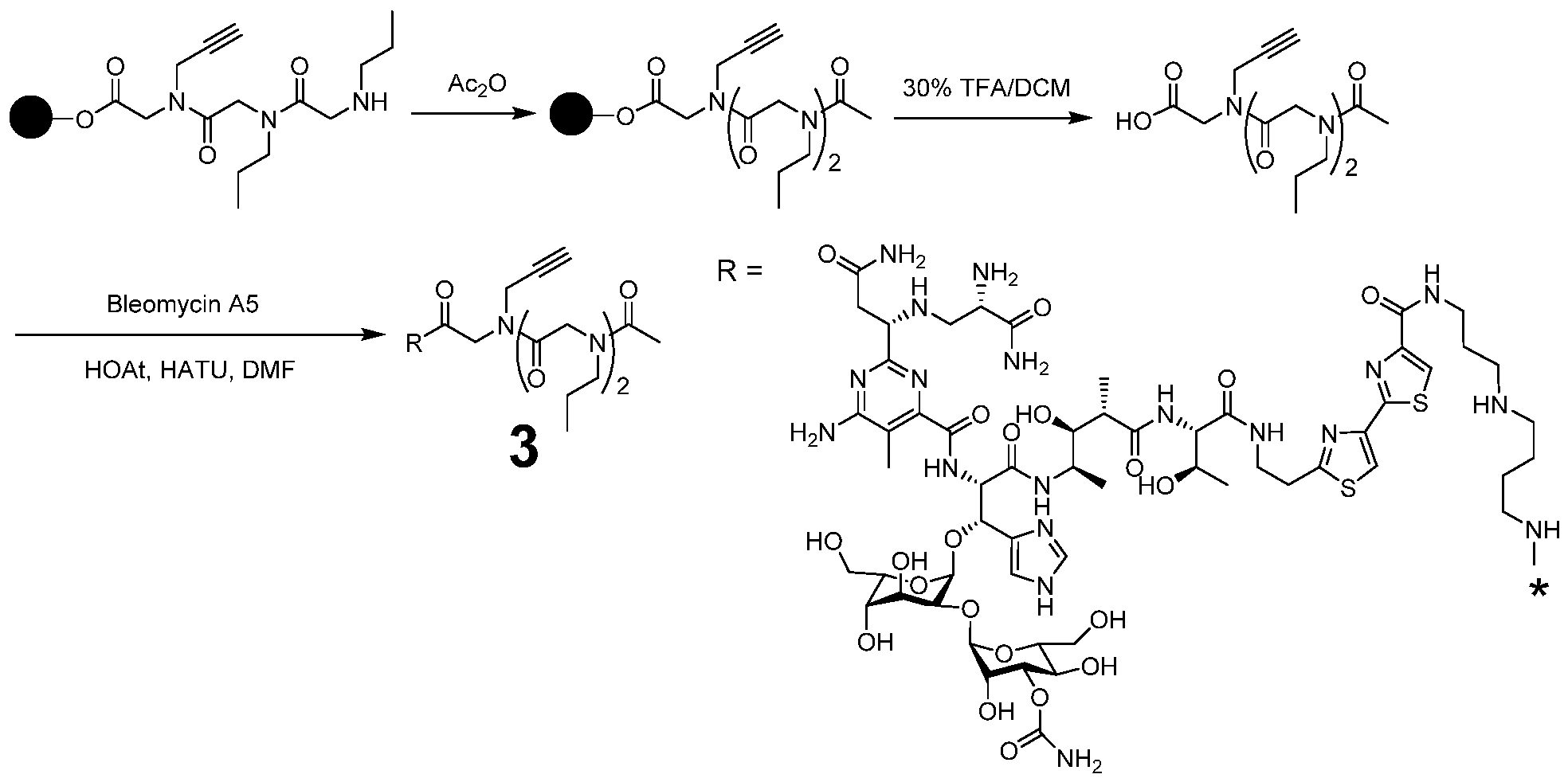
Synthesis of compound 1: Compound 1 was synthesized as previously described.5 Briefly, Rink amide resin (500 mg, 345 mmol) with a substitution level of 0.69 mmol/g was shaken inN,N'd-imethyl formamide (DMF) for 5 min and then deprotected with 20% piperidine in DMF (10 mL, 2x 30 min) at room temperature. The resin was washed with 10 mL DMF three times. Next, 5 eq of bromoacetic acid in 10 mL DMF solution and 5 eq of DIC (N,N'- diisopropylcarbodiimide) were added, and the resin was shaken at room temperature for 30 min. After washing with 10 mL DMF three times, 5 eq of propargylamine in 10 mL DMF was added, and the reaction mixture was shaken at room temperature for 2 h. The bromoacetic acid coupling step was repeated and 5 eq propylamine in 10 mL DMF was added, and the reaction mixture was shaken at room temperature for 2 h. The bromoacetic acid coupling step and propylamine step were repeated. The peptoid in the resin was then treated with a solution of 2 eq Hoechst carboxylate (synthesized as previously described)9, 2 eq DIC and 2 eq DIPEA (N,N'-diisopropylethylamine) in 10 mL DMF. The reaction mixture was shaken at room temperature for 2 h and washed with 10 mL DMF three and then 10 mL dichloromethane (DCM) three times. The product was cleavage from resin by adding 10 mL 30% trifluoroacetic acid (TFA) in DCM and shaken 30 min at room temperature. The eluate was concentrated under vacuum, and the product was precipitated with excess amounts of ether.
The resulting yellow solid was directly treated with a solution of 1.1 eq azide2, 0.2 eq Cu(I) catalyst and 2 eq DIEA in 2 mL DMF. The reaction mixture was kept at 65 °C overnight, and the reaction process was monitored by MALDI-TOF MS. After the starting material was no longer detectable, the product was precipitated with excess amounts of ether and dissolved in 50% methanol in water with 0.1%TFA. HPLC purification was performed with a linear gradient from 0% to 100% B (methanol or acetonitrile + 0.1%TFA) in A (water + 0.1%TFA) over 60 min and a flow rate of 5 mL/min. The pure fractions were collected, and the solvent was concentrated under vacuum. Excess amounts of ether were added, and the resulting solid was obtained as product. Purity was evaluated on a reverse phase Waters Symmetry CIS 5 pm 4.6 x 150 mm column at room temperature with a flow rate of 1 mL/min and a linear gradient of 0-100% B in A. Absorbance was monitored at 254 nm and 345 nm. Mass spectra were recorded on a 4800 plus MALDI-TOF/TOF analyzer. MALDI: [M+H]+ calculated: 1391.8179; [M+H]+ observed: 1391.9153.
Synthesis of compound 2: Chlorotrityl resin (500 mg, 580 mmol) was activated in 1 M HC1 in 10 mL DCM for 10 min at room temperature, followed by washing with DCM three times and DMF three times. Next, 5 eq of bromoacetic acid and 5 eq DIPEA in 10 mL DMF were
added, and the resin shaken at room temperature for 30 min. After washing with 10 mL DMF three times, 5 eq propargylamine in 10 mL DMF were added, and the reaction mixture was shaken at room temperature for 2 h. The resin was washed with 10 mL DMF three times and 5 eq of bromoacetic acid in 10 mL DMF solution and 5 eq DIC were added. The resin was shaken at room temperature for 30 min followed by addition of 5 eq propylamine in 10 mL DMF. The reaction mixture was shaken at room temperature for 2 h. The bromoacetic acid coupling step and propylamine step were repeated. The peptoid on the resin was then treated with a solution of 2 eq Hoechst carboxylate, 2 eq DIC and 2 eq DIPEA in 10 mL DMF solution. The reaction mixture was shaken at room temperature for 2 h and washed with 10 mL DMF three times and then 10 mL DCM three times. The product was cleaved from resin by adding 10 mL of 30% TFA in DCM and shaking the resin for 30 min at room temperature. The elutate was concentrated under vacuum, and the product was precipitated with an excess amount of ether.
The resulting solid was directly treated with a solution of 1.1 eq azide, 0.2 eq Cu(I) catalyst and 2 eq DIEA in 2 mL DMF. The reaction mixture was kept 65 °C overnight, and the reaction process was monitored by MALDI MS. After the starting material was no longer detectable, the product was precipitated with an excess amount of ether.
The Bleomycin A5 coupling reaction was performed by adding 2 eq HOAt (1- Hydroxy-7-azabenzotri azole), 2 eq HATU (hexafluorophosphate azabenzotri azole tetramethyl uranium), 5 eq DIEA, and 2 eq Copper-bleomycin A5. The reaction mixture was shaken at room temperature overnight and the product was precipitated with an excess amount of ether. The solid was dissolved in 50% acetonitrile in water with 0.1%TFA and subjected to HPLC purification. After injection of the solution, the column was washed with 50 mM EDTA (pH 6.7) for 30 min to remove the copper ion and then washed with water for another 30 min. Then the target product was separated with a linear gradient from 0% to 100% B (acetonitrile +0.1%TFA) in A (water +0.1%TFA) over 60 min and a flow rate of 5 mL/min. The pure product was obtained and characterized as described above. MALDI: [M+H]+ calculated: 1775.7767; [M+H]+ observed: 1775.4158.
Synthesis of compound 3: Chlorotrityl resin (500 mg, 580 mmol) was activated in 1 M HC1 in 10 mL DCM for 10 min at room temperature, followed by washing with DCM three times and DMF three times. Then, 5 eq bromoacetic acid and 5 eq DIPEA in 10 mL of DM were added, and the resin was shaken at room temperature for 30 min. After washing with 10 mL DMFthree times, 5 eq propargylamine in 10 mL DMF was added, and the reaction mixture was shaken at room temperature for 2 h. The resin was washed with 10 mL DMF three times
followed by addition of 5 eq of bromoacetic acid in 10 mL DMF solution and 5 eq DIC. The resin was shaken at room temperature for 30 min and then 5 eq propylamine in 10 mL DMF were added. After shaking the mixture at room temperature for 2 h, the bromoacetic acid coupling step and propylamine step were repeated. The peptoid on the resin was then treated with a solution of 2 eq acetic anhydride (Ac2O) and 2 eq DIPEA in 10 mL DMF. The reaction mixture was shaken at room temperature for 30 min and washed with 10 mL DMF three times and then 10 mL DCM three times. The product was cleaved from the resin by adding 10 mL 30% TFA in DCM and shaking at room temperature for 30 min. The eluate was concentrated under vacuum, and the product was precipitated with excess amount of ether.
The Bleomycin A5 coupling reaction was performed by adding 2 eq HO At, 2 eq HATU, 5 eq DIEA and 2 eq Copper-Bleomycin A5. The reaction mixture was shaken at room temperature overnight, and the product was precipitated with an excess amount of ether. The solid was dissolved in 50% acetonitrile in water with 0. l%TFA and subjected to HPLC purification. After injection of the solution, the column was washed with 50 mM EDTA (pH 6.7) for 30 min to remove the copper ion and then washed with water for another 30 min. Then the target product was separated with a linear gradient from 0% to 100% B (acetonitrile +0. l%TFA) in A (water +0. l%TFA) over 60 min and a flow rate of 5 mL/min. The pure product was obtained and characterized as described above. MALDI: [M+H]+ calculated: 1928.7767; [M+H]+ observed: 1928.8219.
Documents Cited in Examples Section
1. Jerabek-Willemsen, M., Wienken, C. L, Braun, D., Baaske, P., and Duhr, S. (2011) Molecular interaction studies using microscale thermophoresis. Assay Drug De.v Technol. 9, 342-353.
2. Agarwal, V., Bell, G. W., Nam, J. W., and Bartel, D. P. (2015) Predicting effective microRNA target sites in mammalian mRNAs. Elife 4, e05005.
3. Velagapudi, S. P., Gallo, S. M., and Disney, M. D. (2014) Sequence-based design of bioactive small molecules that target precursor microRNAs. Nat. Chem. Biol. 10, 291-297.
4. Angelbello, A. J., and Disney, M. D. (2018) Bleomycin can cleave an oncogenic noncoding RNA. Chembiochem 19, 43-47.
5. Lane, S. I. R., Morgan, S. L., Wu, T., Collins, J. K., Merriman, J. A., Ellnati, E., Turner, J. M., and Jones, K. T. (2017) DNA damage induces a kinetochore-based ATM/ATR-
independent SAC arrest unique to the first meiotic division in mouse oocytes. Development 144, 3475-3486.
6. Velagapudi, S. P., Cameron, M. D., Haga, C. L., Rosenberg, L. H., Lafitte, M, Duckett, D. R., Phinney, D. G., and Disney, M. D. (2016) Design of a small molecule against an oncogenic noncoding RNA. Proc. Natl. Acad. Sci. U. S. A. 113, 5898-5903.
7. Costales, M. G, Matsumoto, Y., Velagapudi, S. P., and Disney, M. D. (2018) Small molecule targeted recruitment of a nuclease to RNA. J. Am. Chem. Soc. 140, 6741-6744.
8. Fang, R., Moss, W. N., Rutenberg-Schoenberg, M., and Simon, M. D. (2015) Probing Xist RNA structure in cells using targeted structure-seq. PLoS Genet. 11, el005668.
9. Sexton, A. N., Wang, P. Y., Rutenberg-Schoenberg, M., and Simon, M. D. (2017) Interpreting reverse transcriptase termination and mutation events for greater insight into the chemical probing of RNA. Biochemistry 56, 4713-4721.
10. Pushechnikov, A., Lee, M. M., Childs-Disney, J. L., Sobczak, K., French, J. M.,
Thornton, C. A., and Disney, M. D. (2009) Rational design of ligands targeting triplet repeating transcripts that cause RNA dominant disease: application to myotonic muscular dystrophy type 1 and spinocerebellar ataxia type 3. J. Am. Chem. Soc. 131, 9767-9779.
All patents and publications referred to herein are incorporated by reference herein to the same extent as if each individual publication was specifically and individually indicated to be incorporated by reference in its entirety.
Claims
1. A method comprising
contacting a library of RNA sequences and an effective amount of a compound comprising a conjugate of an RNA-cleaving moiety and an RNA-binding small molecule to cleave the RNA of the library at a binding site of the compound to the RNA to provide a cleaved RNA fragment; and
amplifying and sequencing the cleaved RNA fragment, thereby identifying the site of binding of the small RNA-binding molecule to the RNA of the library which is associated with the site of cleavage.
2. The method of claim 1, wherein the RNA-binding small molecule comprises an N- methyl-piperazinyl-bis-benzimidazole group.
3. The method of claim 1 or 2, wherein the RNA-cleaving moiety comprises bleomycin.
4. The method of claim 3, wherein the bleomycin is bleomycin A5.
5. The method of claim 2, 3, or 4, wherein the RNA-binding small molecule is
Targaprimir-96.
6. The method of any one of claims 1 to 5, wherein the RNA sequence is from an oncogenic non-coding RNA precursor, and the compound binds to the oncogenic non-coding RNA precursor.
7. The method of claim 6, further comprising contacting a cell expressing the non coding RNA precursor with an effective amount of the compound.
8. The method of claim 6 or 7, wherein the oncogenic non-coding RNA precursor comprises oncogenic primary microRNA-96 (pri-miR-96).
9. The method of any one of claims 6 to 8, wherein the compound is a covalent conjugate of Targaprimir-96 and bleomycin A5.
10. The method of claim 1, wherein the RNA sequence of the library is from FOXOl.
11. The method of claim 10, further comprising enhancing expression of FOXOl protein in breast cancer cells by contacting the cells with an effective amount of the compound.
12. The method of claim 11, wherein the contacting comprises administering to a human patient.
13. The method of any one of claims 10 to 12, wherein the compound is a covalent conjugate of Targaprimir-96 and bleomycin A5.
14. A method of triggering apoptosis in triple negative breast cancer cells, comprising contacting the triple negative breast cancer cells with an effective amount of the compound identified in the method of claim 1.
15. The method of claim 14, wherein the contacting comprises administering to a human patient.
16. The method of claim 14 or 15, wherein the compound is a covalent conjugate of Targaprimir-96 and bleomycin A5.
17. A method of treating triple negative breast cancer, comprising administering to a patient afflicted therewith an effective dose of a compound identified in the method of claim 1
18. The method of claim 17, wherein the breast cancer comprises expression of oncogenic primary microRNA-96 (pri-miR-96).
19. The method of claim 17 or 18, wherein the compound is a covalent conjugate of Targaprimir-96 and bleomycin A5.
20 The method of claim 1, wherein the RNA sequence library comprises a transcriptome.
21. The method of claim 20, wherein the transcriptome is viral.
22. The method of claim 20, wherein the transcriptome is mammalian.
23. The method of claim 20, wherein the transcriptome is bacterial.
24. The method of claim 1, wherein the RNA sequence library comprises one or more of synthetic, semi-synthetic, or natural RNA.
25. The method of claim 1, wherein the RNA sequence library comprises the genome of an RNA virus.
26. The method of claim 1 carried out in vitro.
27. The method of claim 1 carried out in living cells.
28. The method of claim 27, wherein the cells are virally- or bacterially-infected cells.
29. The method of claim 1, wherein a set of RNA sequences and a set of compounds comprising candidate RNA-binding small molecules are assayed in a 2-dimensional parallel array.
30. A compound comprising a conjugate of an RNA-cleaving moiety and a RNA-binding small molecule.
31. The compound of claim 30, wherein the RNA-cleaving moiety comprises bleomycin.
32. The compound of claim 30 or 31, wherein the RNA-binding small molecule comprises N-methyl-piperazinyl-bis-benzimidazole group.
33. The compound of claim 32, wherein the RNA-binding small molecule is Targaprimir- 96.
Priority Applications (1)
| Application Number | Priority Date | Filing Date | Title |
|---|---|---|---|
| US17/284,297 US20210379188A1 (en) | 2018-10-10 | 2019-09-26 | Target validation and profiling of the rna targets of small molecules |
Applications Claiming Priority (2)
| Application Number | Priority Date | Filing Date | Title |
|---|---|---|---|
| US201862743996P | 2018-10-10 | 2018-10-10 | |
| US62/743,996 | 2018-10-10 |
Publications (1)
| Publication Number | Publication Date |
|---|---|
| WO2020076511A1 true WO2020076511A1 (en) | 2020-04-16 |
Family
ID=70164776
Family Applications (1)
| Application Number | Title | Priority Date | Filing Date |
|---|---|---|---|
| PCT/US2019/053179 Ceased WO2020076511A1 (en) | 2018-10-10 | 2019-09-26 | Target validation and profiling of the rna targets of small molecules |
Country Status (2)
| Country | Link |
|---|---|
| US (1) | US20210379188A1 (en) |
| WO (1) | WO2020076511A1 (en) |
Cited By (2)
| Publication number | Priority date | Publication date | Assignee | Title |
|---|---|---|---|---|
| US12527788B2 (en) | 2017-02-17 | 2026-01-20 | University Of Florida Research Foundation, Incorporated | Defining RNA-small molecule affinity landscapes enables design of a small molecule inhibitor of an oncogenic non-coding RNA |
| US12617757B2 (en) | 2020-10-29 | 2026-05-05 | University Of Florida Research Foundation, Incorporated | Compounds and modules for inhibition of pre-miR-21 and their use in treatment of certain cancers |
Citations (1)
| Publication number | Priority date | Publication date | Assignee | Title |
|---|---|---|---|---|
| WO2018098297A1 (en) * | 2016-11-28 | 2018-05-31 | The Scripps Research Institute | Precise small molecule recognition of a toxic cug rna repeat expansion |
Family Cites Families (1)
| Publication number | Priority date | Publication date | Assignee | Title |
|---|---|---|---|---|
| US9719191B2 (en) * | 2006-11-29 | 2017-08-01 | The Research Foundation For The State University Of New York | Methods for identifying ligands that target nucleic acid molecules and nucleic acid structural motifs |
-
2019
- 2019-09-26 US US17/284,297 patent/US20210379188A1/en active Pending
- 2019-09-26 WO PCT/US2019/053179 patent/WO2020076511A1/en not_active Ceased
Patent Citations (1)
| Publication number | Priority date | Publication date | Assignee | Title |
|---|---|---|---|---|
| WO2018098297A1 (en) * | 2016-11-28 | 2018-05-31 | The Scripps Research Institute | Precise small molecule recognition of a toxic cug rna repeat expansion |
Non-Patent Citations (1)
| Title |
|---|
| VELAGAPUDI ET AL.: "Design of a small molecule against an oncogenic noncoding RNA", PROC NAT ACAD SCI, vol. 113, no. 21, 24 May 2016 (2016-05-24), pages 5898 - 5903, XP055536461, DOI: 10.1073/pnas.1523975113 * |
Cited By (2)
| Publication number | Priority date | Publication date | Assignee | Title |
|---|---|---|---|---|
| US12527788B2 (en) | 2017-02-17 | 2026-01-20 | University Of Florida Research Foundation, Incorporated | Defining RNA-small molecule affinity landscapes enables design of a small molecule inhibitor of an oncogenic non-coding RNA |
| US12617757B2 (en) | 2020-10-29 | 2026-05-05 | University Of Florida Research Foundation, Incorporated | Compounds and modules for inhibition of pre-miR-21 and their use in treatment of certain cancers |
Also Published As
| Publication number | Publication date |
|---|---|
| US20210379188A1 (en) | 2021-12-09 |
Similar Documents
| Publication | Publication Date | Title |
|---|---|---|
| Velagapudi et al. | Approved anti-cancer drugs target oncogenic non-coding RNAs | |
| EP3380613B1 (en) | Tracking and manipulating cellular rna via nuclear delivery of crispr/cas9 | |
| EP3030548B1 (en) | Transcriptome-wide design of selective, bioactive small molecules targeting rna | |
| Brognara et al. | High levels of apoptosis are induced in human glioma cell lines by co-administration of peptide nucleic acids targeting miR-221 and miR-222 | |
| Bose et al. | Selective inhibition of miR-21 by phage display screened peptide | |
| Amato et al. | Exploitation of a very small peptide nucleic acid as a new inhibitor of miR‐509‐3p involved in the regulation of cystic fibrosis disease‐gene expression | |
| US9586944B2 (en) | Specific targeting of RNA expanded repeat sequences | |
| WO2016011080A2 (en) | Crispr/cas transcriptional modulation | |
| Nahar et al. | Potent inhibition of miR-27a by neomycin–bisbenzimidazole conjugates | |
| Avitabile et al. | Targeting pre-miRNA by peptide nucleic acids: a new strategy to interfere in the miRNA maturation | |
| US20210102200A1 (en) | Small molecule targeted recruitment of a nuclease to rna | |
| Endoh et al. | Triple-Helical Binding of Peptide Nucleic Acid Inhibits Maturation of Endogenous MicroRNA-197 | |
| Fabbri et al. | miRNA therapeutics: delivery and biological activity of peptide nucleic acids targeting miRNAs | |
| US20250051762A1 (en) | Platform using dna-encoded small molecule libraries and rna selection to design small molecules that target rna | |
| US20210379188A1 (en) | Target validation and profiling of the rna targets of small molecules | |
| US20230149554A1 (en) | Targeted degradation of the oncogenic microrna 17-92 cluster by structure-targeting ligands | |
| US20230041228A1 (en) | Small Molecule Inhibition of Micro-RNA-210 Reprograms an Oncogenic Hypoxic Circuit | |
| WO2019246120A2 (en) | Microarray and method of identifying interactions between compounds and rna | |
| US20230416247A1 (en) | Drug-like molecules and methods for the therapeutic targeting of microrna-21 | |
| WO2022047148A1 (en) | Conversion of a biologically silent mirna binding small molecule to an mirna degrader | |
| Swart | Analysis and effect of small-molecules targeting pre-microRNA structures and synthetic efforts toward a novel scaffold for RNA targeting | |
| Rogowski | Identification and characterization of non-coding RNAs and their associated proteins involved in cellular stress responses | |
| Talic | Human RNA Methyltransferase METTL16 Affects Multiple Cell Processes Through Differential Function of Its Domains | |
| 奥村翔 | Development of antisense oligonucleotides for suppressing breast cancer cell proliferation and the system for evaluating drug response of cardiomyocytes | |
| CN107446012A (en) | A kind of tumour fluorescence developer and preparation and application |
Legal Events
| Date | Code | Title | Description |
|---|---|---|---|
| 121 | Ep: the epo has been informed by wipo that ep was designated in this application |
Ref document number: 19870826 Country of ref document: EP Kind code of ref document: A1 |
|
| NENP | Non-entry into the national phase |
Ref country code: DE |
|
| 122 | Ep: pct application non-entry in european phase |
Ref document number: 19870826 Country of ref document: EP Kind code of ref document: A1 |