WO2020021740A1 - 細胞外電子移動作用に関与する酵素及びその利用 - Google Patents
細胞外電子移動作用に関与する酵素及びその利用 Download PDFInfo
- Publication number
- WO2020021740A1 WO2020021740A1 PCT/JP2019/003606 JP2019003606W WO2020021740A1 WO 2020021740 A1 WO2020021740 A1 WO 2020021740A1 JP 2019003606 W JP2019003606 W JP 2019003606W WO 2020021740 A1 WO2020021740 A1 WO 2020021740A1
- Authority
- WO
- WIPO (PCT)
- Prior art keywords
- protein
- electron
- electron transfer
- nucleic acid
- aptamer
- Prior art date
- Legal status (The legal status is an assumption and is not a legal conclusion. Google has not performed a legal analysis and makes no representation as to the accuracy of the status listed.)
- Ceased
Links
Images

Classifications
-
- C—CHEMISTRY; METALLURGY
- C07—ORGANIC CHEMISTRY
- C07K—PEPTIDES
- C07K14/00—Peptides having more than 20 amino acids; Gastrins; Somatostatins; Melanotropins; Derivatives thereof
- C07K14/195—Peptides having more than 20 amino acids; Gastrins; Somatostatins; Melanotropins; Derivatives thereof from bacteria
-
- C—CHEMISTRY; METALLURGY
- C07—ORGANIC CHEMISTRY
- C07K—PEPTIDES
- C07K16/00—Immunoglobulins [IG], e.g. monoclonal or polyclonal antibodies
- C07K16/18—Immunoglobulins [IG], e.g. monoclonal or polyclonal antibodies against material from animals or humans
- C07K16/28—Immunoglobulins [IG], e.g. monoclonal or polyclonal antibodies against material from animals or humans against receptors, cell surface antigens or cell surface determinants
-
- C—CHEMISTRY; METALLURGY
- C07—ORGANIC CHEMISTRY
- C07K—PEPTIDES
- C07K16/00—Immunoglobulins [IG], e.g. monoclonal or polyclonal antibodies
- C07K16/40—Immunoglobulins [IG], e.g. monoclonal or polyclonal antibodies against enzymes
-
- C—CHEMISTRY; METALLURGY
- C12—BIOCHEMISTRY; BEER; SPIRITS; WINE; VINEGAR; MICROBIOLOGY; ENZYMOLOGY; MUTATION OR GENETIC ENGINEERING
- C12N—MICROORGANISMS OR ENZYMES; COMPOSITIONS THEREOF; PROPAGATING, PRESERVING, OR MAINTAINING MICROORGANISMS; MUTATION OR GENETIC ENGINEERING; CULTURE MEDIA
- C12N15/00—Mutation or genetic engineering; DNA or RNA concerning genetic engineering, vectors, e.g. plasmids, or their isolation, preparation or purification; Use of hosts therefor
- C12N15/09—Recombinant DNA-technology
- C12N15/11—DNA or RNA fragments; Modified forms thereof; Non-coding nucleic acids having a biological activity
- C12N15/113—Non-coding nucleic acids modulating the expression of genes, e.g. antisense oligonucleotides; Antisense DNA or RNA; Triplex- forming oligonucleotides; Catalytic nucleic acids, e.g. ribozymes; Nucleic acids used in co-suppression or gene silencing
-
- C—CHEMISTRY; METALLURGY
- C12—BIOCHEMISTRY; BEER; SPIRITS; WINE; VINEGAR; MICROBIOLOGY; ENZYMOLOGY; MUTATION OR GENETIC ENGINEERING
- C12N—MICROORGANISMS OR ENZYMES; COMPOSITIONS THEREOF; PROPAGATING, PRESERVING, OR MAINTAINING MICROORGANISMS; MUTATION OR GENETIC ENGINEERING; CULTURE MEDIA
- C12N15/00—Mutation or genetic engineering; DNA or RNA concerning genetic engineering, vectors, e.g. plasmids, or their isolation, preparation or purification; Use of hosts therefor
- C12N15/09—Recombinant DNA-technology
- C12N15/11—DNA or RNA fragments; Modified forms thereof; Non-coding nucleic acids having a biological activity
- C12N15/115—Aptamers, i.e. nucleic acids binding a target molecule specifically and with high affinity without hybridising therewith ; Nucleic acids binding to non-nucleic acids, e.g. aptamers
-
- C—CHEMISTRY; METALLURGY
- C12—BIOCHEMISTRY; BEER; SPIRITS; WINE; VINEGAR; MICROBIOLOGY; ENZYMOLOGY; MUTATION OR GENETIC ENGINEERING
- C12N—MICROORGANISMS OR ENZYMES; COMPOSITIONS THEREOF; PROPAGATING, PRESERVING, OR MAINTAINING MICROORGANISMS; MUTATION OR GENETIC ENGINEERING; CULTURE MEDIA
- C12N15/00—Mutation or genetic engineering; DNA or RNA concerning genetic engineering, vectors, e.g. plasmids, or their isolation, preparation or purification; Use of hosts therefor
- C12N15/09—Recombinant DNA-technology
- C12N15/63—Introduction of foreign genetic material using vectors; Vectors; Use of hosts therefor; Regulation of expression
-
- C—CHEMISTRY; METALLURGY
- C12—BIOCHEMISTRY; BEER; SPIRITS; WINE; VINEGAR; MICROBIOLOGY; ENZYMOLOGY; MUTATION OR GENETIC ENGINEERING
- C12N—MICROORGANISMS OR ENZYMES; COMPOSITIONS THEREOF; PROPAGATING, PRESERVING, OR MAINTAINING MICROORGANISMS; MUTATION OR GENETIC ENGINEERING; CULTURE MEDIA
- C12N5/00—Undifferentiated human, animal or plant cells, e.g. cell lines; Tissues; Cultivation or maintenance thereof; Culture media therefor
- C12N5/10—Cells modified by introduction of foreign genetic material
-
- C—CHEMISTRY; METALLURGY
- C12—BIOCHEMISTRY; BEER; SPIRITS; WINE; VINEGAR; MICROBIOLOGY; ENZYMOLOGY; MUTATION OR GENETIC ENGINEERING
- C12P—FERMENTATION OR ENZYME-USING PROCESSES TO SYNTHESISE A DESIRED CHEMICAL COMPOUND OR COMPOSITION OR TO SEPARATE OPTICAL ISOMERS FROM A RACEMIC MIXTURE
- C12P21/00—Preparation of peptides or proteins
- C12P21/02—Preparation of peptides or proteins having a known sequence of two or more amino acids, e.g. glutathione
-
- C—CHEMISTRY; METALLURGY
- C23—COATING METALLIC MATERIAL; COATING MATERIAL WITH METALLIC MATERIAL; CHEMICAL SURFACE TREATMENT; DIFFUSION TREATMENT OF METALLIC MATERIAL; COATING BY VACUUM EVAPORATION, BY SPUTTERING, BY ION IMPLANTATION OR BY CHEMICAL VAPOUR DEPOSITION, IN GENERAL; INHIBITING CORROSION OF METALLIC MATERIAL OR INCRUSTATION IN GENERAL
- C23F—NON-MECHANICAL REMOVAL OF METALLIC MATERIAL FROM SURFACE; INHIBITING CORROSION OF METALLIC MATERIAL OR INCRUSTATION IN GENERAL; MULTI-STEP PROCESSES FOR SURFACE TREATMENT OF METALLIC MATERIAL INVOLVING AT LEAST ONE PROCESS PROVIDED FOR IN CLASS C23 AND AT LEAST ONE PROCESS COVERED BY SUBCLASS C21D OR C22F OR CLASS C25
- C23F11/00—Inhibiting corrosion of metallic material by applying inhibitors to the surface in danger of corrosion or adding them to the corrosive agent
- C23F11/08—Inhibiting corrosion of metallic material by applying inhibitors to the surface in danger of corrosion or adding them to the corrosive agent in other liquids
- C23F11/10—Inhibiting corrosion of metallic material by applying inhibitors to the surface in danger of corrosion or adding them to the corrosive agent in other liquids using organic inhibitors
- C23F11/14—Nitrogen-containing compounds
-
- C—CHEMISTRY; METALLURGY
- C23—COATING METALLIC MATERIAL; COATING MATERIAL WITH METALLIC MATERIAL; CHEMICAL SURFACE TREATMENT; DIFFUSION TREATMENT OF METALLIC MATERIAL; COATING BY VACUUM EVAPORATION, BY SPUTTERING, BY ION IMPLANTATION OR BY CHEMICAL VAPOUR DEPOSITION, IN GENERAL; INHIBITING CORROSION OF METALLIC MATERIAL OR INCRUSTATION IN GENERAL
- C23F—NON-MECHANICAL REMOVAL OF METALLIC MATERIAL FROM SURFACE; INHIBITING CORROSION OF METALLIC MATERIAL OR INCRUSTATION IN GENERAL; MULTI-STEP PROCESSES FOR SURFACE TREATMENT OF METALLIC MATERIAL INVOLVING AT LEAST ONE PROCESS PROVIDED FOR IN CLASS C23 AND AT LEAST ONE PROCESS COVERED BY SUBCLASS C21D OR C22F OR CLASS C25
- C23F11/00—Inhibiting corrosion of metallic material by applying inhibitors to the surface in danger of corrosion or adding them to the corrosive agent
- C23F11/08—Inhibiting corrosion of metallic material by applying inhibitors to the surface in danger of corrosion or adding them to the corrosive agent in other liquids
- C23F11/10—Inhibiting corrosion of metallic material by applying inhibitors to the surface in danger of corrosion or adding them to the corrosive agent in other liquids using organic inhibitors
- C23F11/167—Phosphorus-containing compounds
Definitions
- the present invention relates to an enzyme involved in extracellular electron transfer, and a method for preventing corrosion of a metal material targeting the enzyme.
- Non-Patent Documents 1-3 Microbial reduction of oxidized sulfur species (OSS), such as SO 4 2 ⁇ , SO 3 2 ⁇ , S 2 O 3 2 ⁇ , S 0 , Sn 2 ⁇ , along with the oxidation of the organic substrate is caused by organic anoxic anaerobic It is one of the main mechanisms of energy production under the ocean (Non-Patent Documents 1-3). Hydrogen (H 2 ) formed through radioactive decomposition of water and mineral-water reactions is considered to be the main energy source of organic depleted environments ranging from shallow marine sediments to the deep crust, which accounts for almost half of the ocean. (Non-Patent Documents 4-6).
- OSS oxidized sulfur species
- Non-Patent Documents 7 and 8 The microbial energy requirements of vegetative bacteria found in underground environments were still unknown (Non-Patent Document 9). From ubiquitous and abundance of sulfur respiratory bacteria in the underground environment, sulfide addition to the rare H 2 and organic matrix minerals (MnS, FeS, FeS 2) or to use a rich solid substrate, such as an energy source Elucidating how and how to use it, especially considering the significant imbalance between H 2 (n- ⁇ M) and sulphate (several mM) in shallow sediments, It was thought to enhance understanding of ecosystems isolated from the sun's rays.
- MnS, FeS, FeS 2 rare H 2 and organic matrix minerals
- severe mM sulphate
- the present inventors have analyzed the cell membrane of sulfate-reducing bacteria that grow using iron as an electron source in detail using bioinformatics, biochemistry, electrochemistry, and microscopy to analyze the electron uptake mechanism. As a result, we succeeded in finding a new enzyme group that has a different structure from the membrane enzymes involved in electron extraction that were known so far. The present inventors have confirmed that electrons are extracted from the electrode only when the enzyme is expressed in a large amount, and that iron corrosion is caused by bacteria directly extracting electrons from iron by the action of this enzyme. The present invention has been completed for the first time.
- the present invention specifically relates to the inventions described in the following (1) to (22): (1) an amino acid sequence according to one sequence selected from SEQ ID NOs: 2, 4, 6, 8, 10, 12, 14, 16 and 18; SEQ ID NOs: 2, 4, 6, 8, 10, 12, 14 , 16 and 18; an amino acid sequence having at least 80% identity to the amino acid sequence described in one sequence selected from SEQ ID NOs: 2, 4, 6, 8, 10, 12, 14, 16, and 18; An amino acid sequence in which 1 to 20 amino acids have been substituted, deleted, inserted, or added to the amino acid sequence described in one sequence; or SEQ ID NO: 2, 4, 6, 8, 10 , 12, 14, 16, and 18, a cell surface protein having an amino acid sequence having a homology score of 200 or more with the amino acid sequence described in one sequence selected from the group consisting of: (2) The protein according to (1), which has an extracellular electron transfer effect.
- the protein according to (1) or (2) which is a cytochrome enzyme or a ⁇ -propeller protein.
- the base sequence described in one sequence selected from SEQ ID NOs: 1, 3, 5, 7, 9, 11, 13, 15, and 17, or SEQ ID NOs: 1, 3, 5, 7, 9, 11, The nucleic acid molecule according to (4), which has a base sequence that binds under stringent conditions to a nucleic acid having a sequence complementary to the base sequence described in one of the sequences selected from the group consisting of, 13, 15, and 17.
- a vector comprising the nucleic acid molecule according to (4) or (5).
- a host cell having the vector according to (6).
- a composition comprising the antibody according to (10) or an immunoreactive fragment thereof, an aptamer, an antisense nucleic acid, or dsRNA.
- a method for determining a protein that causes corrosion of a metal material comprising identifying an electron transfer enzyme from a cell surface protein of a bacterium present on the surface of the metal material.
- the method according to (12), wherein the identification of the electron transfer enzyme is performed by determining a protein having an electron transfer reaction center.
- the selection of the electron transfer enzyme has 80% or more identity with the amino acid sequence described in one sequence selected from SEQ ID NOs: 2, 4, 6, 8, 10, 12, 14, 16, and 18.
- a method for producing a corrosion inhibitor comprising producing an antisense nucleic acid, dsRNA, or aptamer.
- Identifying an electron transferase from a cell surface protein of a bacterium present on the surface of a metal material by the method of (12) to (14), and determining the determined antibody binding to the electron transferase or its immunoreactivity A method for producing a corrosion inhibitor, comprising producing an antisense nucleic acid, dsRNA, or aptamer that binds to a fragment or an aptamer, or a gene encoding the electron transfer enzyme.
- a method for preventing corrosion of a metal material which comprises inhibiting the function or expression of the electron transfer enzyme in bacteria existing on the surface of the metal material.
- the electron transfer enzyme is the enzyme according to any one of claims 1 to 3.
- (22) identifying an electron transfer enzyme from bacteria existing on the surface of the metal material; and A method for preventing corrosion of a metal material, comprising inhibiting the function or expression of an electron transfer enzyme in bacteria existing on the surface of the identified metal material.
- (23) A method for preventing corrosion of a metal material installed in an external environment, Collecting a sample containing bacteria having cell surface enzymes from the external environment; Identifying cell surface electron transfer enzymes present in the sample; Preparing an antibody that binds to the enzyme or an immunoreactive fragment or aptamer thereof, or an antisense nucleic acid, dsRNA, or aptamer that binds to a gene encoding the enzyme; A method for preventing corrosion of a metal material, comprising flowing the antibody, aptamer, antisense nucleic acid, or dsRNA through a pipe.
- the present invention relates to proteins involved in extracellular electron transfer.
- the protein of the present invention is present in the cell membrane (particularly, the outer membrane (OM)) because it is involved in electron extraction from outside the cell.
- the term “cell surface protein” means that the protein is present in the cell membrane, and may be present outside, inside, or through the cell membrane. That is, the term “cell surface protein” does not necessarily require exposure to the cell surface, but also includes, for example, proteins located on the inner membrane (IM) side of the OM. Further, the cell surface protein may be a protein having at least a signal peptide necessary for localization on the cell surface and / or near the cell membrane.
- the protein of the present invention has an amino acid sequence described in any one sequence selected from SEQ ID NOs: 2, 4, 6, 8, 10, 12, 14, 16, and 18. When these sequences include a signal sequence, the protein of the present invention may have an amino acid sequence that does not include the signal sequence. Alternatively, the protein of the present invention has an amino acid sequence having 80% or more identity with the amino acid sequence described in one sequence selected from SEQ ID NOs: 2, 4, 6, 8, 10, 12, 14, 16, and 18. May be provided.
- the amino acid sequence identity refers to the ratio (%) of the number of amino acids of the same type in the range of amino acid sequences to be compared between two types of proteins, for example, using a known program such as BLAST or FASTA. Can be determined.
- the identity of the above-mentioned protein of the present invention may be greater than 80% or more, for example, 85% or more, 90% or more, 95% or more, 98% or more, or 99% or more. It may be the same.
- the protein of the present invention may have 1 to 20, 1 to 10 amino acids in the amino acid sequence described in one sequence selected from SEQ ID NOs: 2, 4, 6, 8, 10, 12, 14, 16 and 18. , 1 to 8, 1 to 5, 1 to 3 amino acids may be substituted, deleted, inserted or added.
- the protein of the present invention may be an amino acid having a homology score of 200 or more with the amino acid sequence described by Blast in one sequence selected from SEQ ID NOs: 2, 4, 6, 8, 10, 12, 14, 16, and 18. It may be an array.
- the homology score is obtained by aligning the amino acid sequence described in one sequence selected from SEQ ID NOs: 2, 4, 6, 8, 10, 12, 14, 16 and 18 with the amino acid sequence of the target protein,
- the score of each amino acid to be obtained can be obtained from a score matrix and calculated as a sum thereof (refer to http://www.gsic.techtech.ac.jp/supercon/supercon2004-e/alignmentE.html).
- the homology score can be determined using a known BLAST program.
- As the score matrix BLOSUM62, PAM32, and the like are known, and in this specification, BLOSUM62 is preferable.
- the protein of the present invention has an extracellular electron transfer effect.
- extracellular electron transfer action means an action in which a cell having the protein comes into contact with a solid surface capable of donating electrons to directly extract electrons from the solid surface.
- To determine whether a protein has extracellular electron transfer activity add cells expressing the protein to a reactor where the electrode is the only electron donor, and measure the cathode current density after 30 minutes to 24 hours. Can be determined. If the cathodic current density increases, the protein is determined to have extracellular electron transfer activity.
- the protein of the present invention is a cytochrome enzyme, a ⁇ -propeller protein, or a porin protein.
- the cytochrome enzyme is a kind of heme protein containing heme iron having a redox function, and cytochromes a, b, c, d, f, and o are known (Chem. @ Rev. (2014) 114 ( 8): 4366-4469). Therefore, when a certain protein is a protein having a heme binding motif (CXXCH, CXXXCH, CXXXXXCH, or CXXXXXXCH; C is cysteine and H is histidine) and is a protein having electron transfer ability, Can be determined to be.
- ⁇ -propeller protein is a protein having a three-dimensional structure in which four to eight blade-shaped structures made of ⁇ -sheets are conically surrounded around a central axis. Whether a certain protein is a ⁇ -propeller protein can be determined by three-dimensional structure analysis or three-dimensional structure simulation.
- Porin protein is a transmembrane protein containing a ⁇ -barrel structure with an inverse equilibrium ⁇ -sheet. Porin proteins are also known to form complexes with cytochrome proteins and participate in extracellular electron transfer (Marcus @ J. @ Edwards et al., J. Biol.Chem.published @ online @ April @ 10, $ 2018).
- Whether or not a certain protein is a porin protein can be determined by three-dimensional structure analysis or three-dimensional structure simulation.
- these cytochrome enzymes, ⁇ -propeller proteins, or porin proteins are proteins present in the cell membrane (preferably the outer membrane).
- the ⁇ -propeller protein and / or the porin protein exerts an extracellular electron transfer effect by forming a complex with the cytochrome enzyme, it is preferable that the ⁇ -propeller protein and / or the porin protein be complexed with the cytochrome enzyme. Forming the body.
- the present invention relates to a nucleic acid molecule having a base sequence encoding the protein.
- the nucleic acid molecule of the present invention may have another sequence as long as it has a base sequence encoding the protein.
- the nucleic acid molecule of the present invention may be a base sequence described in one sequence selected from SEQ ID NOs: 1, 3, 5, 7, 9, 11, 13, 15, and 17, or It has a base sequence that binds under stringent conditions to a nucleic acid having a sequence complementary to the base sequence described in one sequence selected from 5, 7, 9, 11, 13, 15, and 17.
- “hybridize under stringent conditions” means to hybridize under hybridization conditions commonly used by those skilled in the art.
- hybridization conditions are 6 ⁇ SSC (0.9 M NaCl, 0.09 M trisodium citrate) or 6 ⁇ SSPE (3 M NaCl, 0.2 M NaH 2 PO 4 , 20 mM EDTA ⁇ 2Na, pH 7.4).
- the conditions may be such that hybridization is carried out at 42 ° C. in medium and then washing at 42 ° C. with 0.5 ⁇ SSC.
- the present invention further relates to a vector containing the nucleic acid molecule.
- the vector is not particularly limited as long as it is a vector capable of introducing the nucleic acid molecule into a host cell, and may be any plasmid vector, a retrovirus vector, an adenovirus vector, a lentivirus vector, an adenovirus vector, depending on the host cell.
- a viral vector such as an associated virus (AAV) vector can be used.
- the present invention also includes a host cell having the vector.
- a host cell having the vector.
- cells suitable for expressing the target protein such as bacteria, yeast, animal cells, insect cells, and plant cells, can be appropriately selected.
- the present invention relates to a substance that inhibits the function or expression of the protein or the nucleic acid molecule (hereinafter, referred to as “inhibitor”).
- the invention relates to an antibody or an immunoreactive fragment thereof, or an aptamer, which binds to said protein, or an antisense nucleic acid molecule, dsRNA, or aptamer, which binds to said nucleic acid molecule.
- These inhibitors preferably have an action of directly or indirectly inhibiting the extracellular electron transfer action of the protein of the present invention.
- the present invention includes compositions containing such inhibitors, which compositions can be used as metal corrosion inhibitors. Iron can be mentioned as a metal.
- the corrosion prevention method targeting proteins according to the present invention can prevent iron corrosion and the like without using a chemical having a large effect on the environment such as a bactericide.
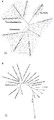
- FIG. 2 is a diagram showing the distribution of homologous extracellular cytochromes of ferrophilus ⁇ IS5.
- A shows a phylogenetic tree obtained by 16S @ ribosomal RNA (rRNA) sequence alignment.
- rRNA ribosomal RNA
- B D. which are encoded by the DFE_450 and DFE_464 genes of E. ferrophilus IS5, and their homologs (identified using the NCBI blastp, max score> 200); sulfurreducens @ PCA's OmcB, OmcE, OmcS and OmcZ, S.
- IM Inner membrane
- CBB Coomassie Brilliant Blue
- 2 shows the protein profile of the (OM) fraction.
- OM heme positive bands
- Non-specific heme staining detected a minor band corresponding to ferroxidase (DFE — 1154, 14.1 kDa, ⁇ ).
- DFE ferroxidase
- B Lactate starvation (red) and sufficiency (blue) normalized by OM protein concentration.
- 3 shows an absorption spectrum of an OM fraction extracted from ferrophilus IS5 cells. Solid and dotted lines show spectra measured under reducing and oxidizing conditions, respectively.
- Light A graph showing a comparison of the solelet peak absorption at 419 nm.
- C D.
- FIG. 4 is a graph showing the expression of seven multi-heme cytochromes and two ⁇ -propeller proteins (hatched bars) located in close proximity in the ferrophilus IS5 genome by lactate-starved and lactate-sufficient cells.
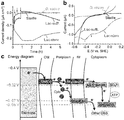
- Intact D. 3 shows the results of in vivo electrochemical measurement using ferrophilus IS5 cells.
- (A) Graph showing the results of cathodic current versus time measurements performed in an anaerobic reactor with an indium-tin doped oxide electrode maintained at -0.4 V (vs. SHE) as the only electron donor. is there. Lactic acid starvation added to the reactor ferrophilus IS5 cells, lactic acid sufficiency. ferrophilus IS5 cells, H 2 -consumable.
- vacuolatum and sterile medium are indicated by red, blue, pink and black lines, respectively. Arrows indicate when cells or sterile media were added to the reactor.
- B shows a linear sweep voltammogram measured after current generation in panel a.
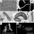
- C the redox potential of outer membrane cytochromes (OMCs) that can produce NADH and / or reduced menaquinone (MQH 2 ) promotes the reduction of OSS and intermediates such as adenosine phosphosulfate (APS); 3 shows an energy diagram of an extracellular electron uptake model of the IS5 strain. Cyt represents periplasmic C-type cytochrome. Lactate-starved D. 1 shows micrographs of ferrophilus IS5 cells and their nanowires.
- the scale bars of a and b represent 10 ⁇ m and 500 nm, respectively.
- (D and e) Show the results of DAB staining positive by addition of H 2 O 2 .
- the scale bar corresponds to 500 nm for c and d and represents 50 nm for e.
- the bamboo-like nanowire structure was clearly visible only when DAB staining was positive.
- the protein of the present invention can be obtained by culturing a host cell having the vector of the present invention in a medium suitable for the host cell under culture conditions suitable for expression of the protein by the host cell to express the protein. Can be manufactured.
- the present invention also relates to a method for determining a protein that causes corrosion of a metal material, including identifying an electron transfer enzyme from a cell surface protein of a bacterium present on the surface of a metal material as a protein that causes corrosion of the metal material.
- Information on the cell surface protein of the bacterium present on the surface of the metal material can be obtained by culturing the bacterium and determining the base sequence of the gene encoding the protein in the culture.
- Whether or not a sequenced protein is an electron transfer enzyme can be determined based on whether or not it has an electron transfer reaction center.
- the electron transfer reaction center is not particularly limited, but includes, for example, a heme binding domain, an iron ion cluster, and the like.
- the electron transfer enzyme is preferably a cell surface protein or a membrane protein (preferably, an outer membrane protein). Therefore, in one embodiment, the present invention comprises identifying a protein that is a membrane protein and an electron transfer enzyme from a cell surface protein of a bacterium present on the surface of a metal material as a protein that causes corrosion of the metal material.
- the present invention relates to a method for determining a causative protein of a metal material. Whether or not the candidate protein is a membrane protein (or outer membrane protein) can be analyzed based on the nucleotide sequence information of the obtained gene.
- the electron transfer enzyme may be an amino acid sequence described in one sequence selected from SEQ ID NOs: 2, 4, 6, 8, 10, 12, 14, 16 and 18, or Jing @ Liu et al., Chemical Reviews (2014). ) 114: 4366-4469 may be performed by selecting a gene encoding a protein having an amino acid sequence having 80% or more identity with the protein disclosed in J. Am.
- a protein having an amino acid sequence having 80% or more identity can be obtained by performing such "conservative amino acid substitution" on the original amino acid sequence.
- Such conservative amino acid substitutions include substitutions between amino acids having an aliphatic side chain such as glycine, alanine, valine, leucine, and isoleucine; substitutions between amino acids having an aliphatic hydroxyl side chain such as serine and threonine; Substitution between amino acids having amide-containing side chains such as asparagine and glutamine; substitution between amino acids having aromatic side chains such as phenylalanine, tyrosine, histidine, and tryptophan; and basic side chains such as lysine, arginine, and histidine.
- the present invention relates to an antibody or an immunoreactive fragment or aptamer thereof, which binds to an electron transferase determined as a causative protein of a metal material by the above method, or a gene encoding the electron transferase.
- the present invention relates to a method for producing a corrosion inhibitor, which comprises producing an antisense nucleic acid, dsRNA, or an aptamer.
- the present invention provides a method for identifying an electron transferase from a cell surface protein of a bacterium present on the surface of a metal material by the method, and an antibody, an immunoreactive fragment or an aptamer thereof, which binds to the determined electron transferase Or a method for producing a corrosion inhibitor, comprising producing an antisense nucleic acid, dsRNA, or aptamer that binds to a gene encoding the electron transfer enzyme.
- the antibody of the present invention may be a polyclonal antibody or a monoclonal antibody. Further, the antibody of the present invention may be an antibody of human, mouse, rat, hamster, guinea pig, rabbit, dog, monkey, sheep, goat, camel, chicken, duck, etc., and preferably, hybridoma is produced.
- the antibody is preferably an animal antibody, more preferably a mouse, rat or rabbit antibody.
- the immunoglobulin class of the antibody of the present invention is not particularly limited, and may be any immunoglobulin class (isotype) of IgG, IgM, IgA, IgE, IgD, or IgY, and is preferably IgG.
- the antibody of the present invention is an IgG, any subclass (IgG1, IgG2, IgG3, or IgG4) may be used. Further, the antibody of the present invention may be monospecific, bispecific (bispecific antibody), or trispecific (trispecific antibody) (for example, WO1991 / 003493).
- the antibodies of the present invention also include immunoreactive fragments of the antibodies.
- immunoreactive fragment of an antibody refers to a protein or peptide containing a part (partial fragment) of an antibody, and a protein that retains the effect of the antibody on the antigen (immunoreactivity / binding property).
- the term "aptamer” refers to a nucleic acid molecule or peptide that specifically binds to a specific molecule.
- the aptamer may be RNA or DNA.
- the form of the nucleic acid may be double-stranded or single-stranded.
- the length of the aptamer is not particularly limited as long as it can specifically bind to the target molecule, and is, for example, 10 to 200 nucleotides, preferably 10 to 100 nucleotides, more preferably 15 to 80 nucleotides, and still more preferably 15 to 50 nucleotides. Of nucleotides.
- Aptamers can be selected using methods well known to those skilled in the art, and include, for example, the SELEX method (Systematic Evolution ⁇ of ⁇ ligands ⁇ by ⁇ Exponential ⁇ Enrichment) (Turk, C. and ⁇ Gold, L. (1990) Science. 510) can be used.
- SELEX method Systematic Evolution ⁇ of ⁇ ligands ⁇ by ⁇ Exponential ⁇ Enrichment
- antisense nucleic acid refers to a nucleic acid molecule having a sequence complementary to a target sequence, and may be DNA or RNA.
- the antisense nucleic acid does not need to be 100% complementary to the target sequence, and may contain a non-complementary base as long as it can specifically hybridize under the above-mentioned stringent conditions.
- antisense nucleic acids When introduced into cells, antisense nucleic acids bind to target sequences and inhibit transcription, RNA processing, translation, or stability.
- antisense nucleic acids include those having a polynucleotide mimetic and a modified backbone in addition to the antisense polynucleotide. Such an antisense nucleic acid can be appropriately designed and manufactured (for example, chemically synthesized) using a method well known to those skilled in the art based on the target sequence information.
- DsRNA is a RNA that has a sequence complementary to a target sequence at least in part by RNA interference (RNAi), and degrades the mRNA by binding to the mRNA having the target sequence, thereby degrading the target sequence.
- the dsRNA includes siRNA (short @ ferring @ RNA) and shRNA (short @ hairpin @ RNA).
- the dsRNA does not need to have 100% homology with the target sequence as long as it suppresses target gene expression.
- the dsRNA may be partially substituted with DNA for stabilization or other purposes.
- the siRNA is preferably a double-stranded RNA having 21 to 23 bases.
- siRNA can be obtained by methods well known to those skilled in the art, for example, as an analog of chemically synthesized or naturally occurring RNA.
- shRNA is an RNA short strand having a hairpin turn structure.
- the shRNA can be obtained by a method well known to those skilled in the art, for example, by chemical synthesis or by introducing a shRNA-encoding gene into a cell and expressing it.
- the present invention relates to a method for preventing corrosion of a metal material, comprising inhibiting the function or expression of an electron transfer enzyme in a bacterium present on the surface of the metal material.
- the electron transfer enzyme may be the protein of the present invention or a protein appropriately determined by the above method.
- Inhibition of the function or expression of an electron transferase includes an antibody that binds to the electron transferase or an immunoreactive fragment or aptamer thereof, or an antisense nucleic acid, dsRNA, or aptamer that binds to a gene encoding the electron transferase. This can be performed by bringing the composition into contact with the surface of the metal material.
- the present invention provides a method for inhibiting the corrosion of a metal material, which comprises identifying an electron transfer enzyme from bacteria present on the surface of the metal material by the above method, and inhibiting the function or expression of the identified electron transfer enzyme. Including prevention methods.
- the metallic material may preferably be in the external environment, for example in water, such as fresh water or seawater.
- the present invention relates to a method for preventing corrosion of a metal material installed in an external environment, comprising collecting a sample containing bacteria having cell surface enzymes from the external environment, Identifying an electron transfer enzyme, preparing an antibody that binds to the enzyme or an immunoreactive fragment or aptamer thereof, or preparing an antisense nucleic acid, siRNA, or aptamer that binds to a gene encoding the enzyme; Includes a method for preventing corrosion of metallic materials, comprising flowing an aptamer, antisense nucleic acid, or siRNA through a pipe.
- the present invention provides a metal material having a negative potential of -0.32 V ⁇ vs. SHE or more (ie, -0.32V@vs. SHE, or a value larger in the positive direction than that, for example, -0.2, -0.1, 0, 0.1, and 0.2V@vs. SHE) Etc.) or -0.32V@vs.
- the present invention relates to a method for preventing corrosion of a metal material, including inhibiting the function or expression of an electron transfer enzyme in a bacterium existing on the surface of a metal material by stopping the metabolism that pulls out electrons of the bacterium above SHE to stop the metabolism.
- V vs.SHE means an electrode potential based on a standard hydrogen electrode.
- the potential of the negative potential metal material is set to ⁇ 0. .32V @ vs. SHE or more or -0.32V@vs. By raising it above SHE, it is possible to stop the metabolism that pulls out electrons of bacteria.
- the potential of the metal material can be measured with a reference electrode. For example, in the case of the inside of a pipe, it can be measured with a silver-silver chloride electrode.
- the measurement of the potential is performed with a silver-silver chloride electrode, converted into the value of a standard hydrogen electrode, and V vs.
- the value of SHE can be obtained.
- the measurement of the potential can be performed simultaneously while adjusting the potential using a potentiostat.
- the adjustment of the potential can be appropriately performed using a means capable of applying a voltage to a metal material such as a potentiostat.
- D. ferrophilus IS5 (DSM No. 15579) was precultured in butyl rubber stoppered vials containing 100 ml of DSMZ195c medium at 28 ° C. under CO 2 / N 2 (20:80, v / v) oxygen-free conditions. After 5 days, the culture contained a small amount of black iron sulfide precipitant. A portion of the cell suspension is diluted 10-fold with fresh DSMZ195c medium supplied with 21 or 110 mM lactate, and further cultured under the above conditions for 5 days to produce a lactate-starved state or a lactate-sufficient state, respectively. I let it. C.
- Desulfobacterium vacuolatum (JCM 12295) was cultured under the same conditions as in ferrophilus IS5. Shewanella oneidensis MR-1 was obtained by resuspending cells collected from an isolated colony with a platinum loop in an LB medium, culturing aerobically, and then culturing with shaking vigorously at 30 ° C. for 15 hours. The culture was centrifuged at 5000 ⁇ g for 10 minutes, the resulting pellet was washed, and at a modified concentration of electron donor and acceptor (60 mM lactate and 1 mM fumarate), Gorby et al.
- the first contig was 3,702,182 base pairs (bp), the average coverage was 135.36 times, the second contig was 64,963 bp, and the average coverage was 37.77 times.
- the ends of both contigs were deduplicated and circularized to give a single 3,677,055 bp circular chromosome and a 43,052 bp circular plasmid.
- Genome annotation was performed with MiGAP (H. Sugawara et al., Paper presented at the 20th Int. Conf. Genome Informatics 1-2, Kanagawa, Japan, 2009).
- the chromosome had 3,375 RNA coding sequences and 62 tRNAs.
- the genome was searched for the heme binding motif CXnCH (n 2-5) for the identification of heme binding proteins.
- RNA was prepared using the NucleoSpin RNA kit (TakarBio). RRNA was removed using a Ribo-Zero Magnetic Kit (Gram-negative bacteria) and a nucleic acid molecule library was prepared according to the manufacturer's guidelines for sequencing using the TruSeq Stranded mRNA Library Prep Kit (Illumina). RNA quality was confirmed with an Agilent 2100 Bioanalyzer (Agilent Technologies, Santa Clara, CA, USA). The c-nucleic acid molecule library was sequenced on a HiSeq 2500 instrument (Illumina), generating between 51 million and 54 million 100 bp pair-end reads per library. The quality-controlled sequence read results were obtained using Tophat (C.
- Ferrophilus IS5 was mapped to the draft genome, giving a minimum mapping identity of 97.4%.
- sequence reading results were quantified using FPKM metrics. FPKM values were further normalized by the average of two housekeeping genes, adenylate kinase (adk) and recombinant protein A (recA).
- Lactic acid starvation (1.58 g) and lactate sufficiency (2.25 g)
- a 2 L cell culture of ferrophilus IS5 cells was centrifuged at 7197 g at 4 ° C. for 20 minutes to collect a wet pellet. The cells are washed, resuspended in 10 mM Tris-HCl buffer (pH 8) and treated as described in Myers and Myers (CR Myers et al., J. Bacteriol. 174, 3429-3438 (1992)). , EDTA-lysozyme-Brij 58 method. Until a pellet was no longer formed, centrifugation was repeated at 7197 ⁇ g at 4 ° C.
- the membrane pellet was resuspended in 0.5 ml of 10 mM Na + -HEPES (pH 7.5) buffer and 35% to 55% prepared in the same buffer. (Wt / wt), and centrifuged at 28,500 rpm (RCFmax 82,000 ⁇ g) at 4 ° C.
- IM and OM fractions were separated on different sucrose gradients.
- the membrane fraction was carefully transferred to a 1.5 ml tube using a pipette.
- the OM fraction was further purified by washing with 2% Triton X-100 for 10 minutes at room temperature to dissolve the attached IM, and then using a Hitachi type S110AT-2115 rotor at 51,200 rpm (RCFmax 150,000). ⁇ g), and centrifuged at 4 ° C. for 2 hours to pelletize.
- IM and OM Separation of IM and OM was confirmed by subjecting to sodium dodecyl sulfate (SDS) -polyacrylamide gel electrophoresis (PAGE) to protein staining with GelCode Blue Safe Protein Stain (Thermo Scientific). .
- SDS sodium dodecyl sulfate
- PAGE polyacrylamide gel electrophoresis
- the digested peptide was analyzed by nano-liquid chromatography-tandem mass spectrometry (LC-MS / MS) using a Q Exactive mass spectrometer (Thermo Fisher Scientific).
- Peptides were prepared from 0% to 35% buffer B (100% acetonitrile and 0.1%) using a nano ESI spray column (75 ⁇ m [ID] ⁇ 100 mm [L], NTCC analysis column C18, 3 ⁇ m, Nikkyo Technologies).
- a linear gradient of formic acid was separated at a flow rate of 300 nL / min for 10 minutes (Easy nLC; Thermo Fisher Scientific).
- the mass spectrometer was operated in positive ion mode and MS and MS / MS spectra were acquired using the data-dependent TOP10 method. Predicted in-house using a local MASCOT server (version 2.5, Matrix Sciences; parameters: Peptide Mass Tolerance, ⁇ 15 ppm; fragment mass tolerance, ⁇ 20 mmu; and Max Missed Leaves, 3, equipment type, ESI-TRAP) D.
- the MS / MS spectrum was searched against the ferrophilus IS5 60229 ORF database (3431 coding sequence in genome and plasmid; 1089117 residues).
- Electrochemical measurements were performed in a three-electrode reactor held in a single COY anaerobic chamber filled with 100% N 2.
- Indium tin oxide (ITO) (SPD Laboratory, Inc., Japan) grown on a glass substrate by spray pyrolysis was used as a working electrode (resistance 8 ⁇ / square, thickness 1.1 mm, surface area 3.1 cm 2 ). , Placed at the bottom of the reactor.
- Platinum wire and Ag / AgCl (sat.KCl) were used as counter and reference electrodes, respectively.
- the salt medium having the following composition was used as an electrolyte: 457mM NaCl, 47mM MgCl 2, 7.0mM KCl, 5.0mM NaHCO 3, 1.0mM CaCl 2, 1.0mM K 2 HPO 4, 1.0mM NH 4 Cl, 25 mM Na + -HEPES (pH 7.5), 1 mL of tungstic selenite solution (12.5 mM NaOH, 11.4 ⁇ M Na 2 SeO 3 .5H 2 O, 12.1 ⁇ M Na 2 WO 4 .2H 2) O), and 1 ml of a trace element solution SL-10 (described in DSMZ medium 320).
- the salt medium 21 mM Na 2 SO 4 as an electron acceptor and 1 mM acetate as a carbon source were added, and no vitamin or reducing reagent was added.
- the salt medium was sterilized by passing through a 0.22 ⁇ m filter and then degassed by purging with 100% N 2 for 15 minutes. A total of 4.5 ml of sterile oxygen-free salt solution was added as electrolyte to the electrochemical reactor. During the electrochemical measurements, the reactor was operated without stirring.
- Strain IS5 and D. vacuolatum cells were harvested from 50 ml of culture by centrifugation, resuspended in 0.5 ml of anoxic medium and then added to the reactor to a final OD600nm of 0.1 and 0.3.
- the ITO electrode was removed from the reactor, fixed with 2.5% glutaraldehyde, and then washed by immersing the electrode three times in 50 mM Na + -HEPES (pH 7.4).
- the washed samples were dehydrated with a 25%, 50%, 75%, 90% and 100% ethanol gradient in the same buffer, exchanged for t-butanol three times and then lyophilized under vacuum. Dried samples were coated with evaporated platinum and then viewed using a Keyence VE-9800 microscope.
- Lactate starvation and lactate sufficiency in 50 ml of cell culture ferrophilus IS5 cells were harvested by centrifugation at 5000 xg for 8 minutes and immediately fixed on ice in a degassed solution containing 2% paraformaldehyde and 2.5% glutaraldehyde. After fixation, all operations were performed in 2 ml Eppendorf tubes. After gentle resuspension in 1.5 ml of 50 mM Na + -HEPES (pH 7.4, 35 g / L NaCl), it was washed five times by centrifugation (5000 ⁇ g, 4 minutes). According to the method described by McGlynn et al.
- Lactate-starved D. ferrophilus IS5 cells were fixed with a degassed 2% glutaraldehyde solution, and each was protein-specific as described by Pirbadian et al. (S. Pirbadian et al., Proc. Natl. Acad. Sci. USA 111, 12883-12888 (2014)). And a membrane-specific fluorescent dye, NanoOrange reagent and FM4-64FX (ThermoFisher Scientific). Each fluorescence was observed under a x100 oil immersion objective with a Leica DFC450C epi-fluorescence microscope equipped with a FITC filter and a TXR filter, respectively.
- the present inventors have proposed D.S.
- the entire 3.7 Mbp circular genome of ferrophilus IS5 was sequenced and 26 genes encoding multiheme cytochromes containing at least four heme binding motifs were identified. They are essential for the formation of cytochrome complexes (OM cytochromes, OMCs) that penetrate the direct electron transport pathway, OM, and for the reduction of extracellular solids in the model iron-reducing bacteria Shewanella oneidenissis and Geobacter sulfurreducens. . G.
- FIG. The gene encoding the conductive nanofilaments of S.
- the present inventors performed both OM extraction and transcriptome analysis of cells cultured in a medium having a limited lactic acid content, thereby obtaining a D.D. It was confirmed that ferrophilus IS5 highly expresses OMC. Lactate was completely consumed 5 days after seeding, and the crude membrane fraction was clearly separated into the inner and outer membranes (FIG. 2a). The UV-vis absorption spectrum of the extracted OM fraction showed Soret and Q band absorption characteristic of oxidized and reduced c-type cytochromes (FIG. 2B, left panel). Analysis of the spectral data shows that lactate-starved IS5 cells have at least 10 times more OMC than cells cultured under lactate-sufficient conditions where 30 mM or more of lactate remained after 5 days of culture.
- lactic acid-starved D It was further confirmed by electrochemical measurements that ferrophilus IS5 cells were able to extract electrons from the electrode surface with significantly higher efficiency than cells with sufficient lactate.
- ITO indium-tin-doped oxide
- ITO indium-tin-doped oxide
- the doubling time of IS5 cells is about 13.5 hours under anaerobic growth conditions containing lactate, but after 3 days the number of lactate-starved IS5 cells on the ITO electrode has not increased significantly, while the cathodic current of IS5 cells has not increased. Generation continued. This suggests that the electrode provides much less energy to IS5 cells than lactate.
- the potential dependence of the sulphate reduction current measured by linear sweep (LS) voltammetry after cathodic current measurement indicates that sulphate reduction is initiated at -0.3 V in lactate-starved IS5 cells and reaches a maximum at a potential of -0.42 V. (Fig. 3b). In contrast, lactate sufficiency IS5, H 2 consumption D.
- Another peak observed in LS voltammetry of lactate-starved cells at -0.1 V can be assigned to the reduction of biosynthetic iron sulfide, which is the peak current after 2 hours in lactate-starved IS5 cells.
- the reason for the slight decrease in current after generation is explained by the fact that the production of iron sulfide can inhibit direct contact between cells and the electrode surface (FIG. 3A).
- the threshold electrode potential for -0.3 V sulfate reduction is similar to the redox potential of NADH, but more positive than ferredoxin, which indicates that NADH transfers electrons from periplasmic cytochrome to cytoplasmic adenosine phosphosulfate reductase.
- Lactic acid-starved IS5 cells formed a monolayer biofilm on the electrode by scanning electron microscopy observation of the electrode surface after the electrochemical measurement (FIG. 4a). This is similar to many bacterial strains with extracellular electron transfer capacity, consistent with the concept that electron uptake occurs via OMCs. Furthermore, nanofilament structures were observed to extend between cells and from cells to the electrode surface (FIG. 4b). S. Similar nanowires produced by oneidensis MR-1 are available from OM (S. Pirbadian et al., Proc. Natl. Acad. Sci. USA 111, 12883-12888 (2014); YA Gorby et al., Proc. Natl. Acad. Sci. USA 103, 11358-11363 (2006)), it is considered that cytochromes detected in the OM fraction of the IS5 strain are also localized in the observed nanowires.
- the ferrofilus IS5 nanowire resembles a bamboo-like structure with a diameter of 30-50 nm and is clearly visible only with positive DAB staining, suggesting a high cytochrome coverage on the nanowire surface.
- IS5 nanowires are available from slightly thinner than that of S. oneidensis, but both show the same segment structure and strong cytochrome positive staining, and the IS5 nanowires also have It has been suggested to play a role in electron transfer as proposed in oneidensis.
- G.A. sulfurreducens nanowires had a diameter of about 7 nm and exhibited both weakly positive and negative DAB staining. These differences are described by S.M.
- DFE_449 and DFE_461 are periplasmic decahemocytochromes in the OM MtrCAB cytochrome complex and are important for metal reduction of Schwanella species (AS Beliaev et al., Mol. Microbiol. 39, 722-730 (2001); C.
- Fe 2+ oxidation Ferrooxidans also reported that bacterial oxidation of sulfide minerals under anaerobic conditions is considered to be indirect oxidation of Fe 2+ ions released from iron sulfide (KL Straub et al., Appl. Environ.Microb.62, 1458-1460 (1996); CJ Jorgensen et al., Environ.Sci.Technol.43,4851-4857 (2009)), which is different from the direct electron uptake model shown in the present invention.
- the protein of the present invention is involved in electron extraction from the outside of the cell, by inhibiting the expression or function of the protein of the present invention, it is possible to inhibit events based on electron extraction in bacteria, and Corrosion and the like can be prevented or prevented.
Landscapes
- Health & Medical Sciences (AREA)
- Chemical & Material Sciences (AREA)
- Life Sciences & Earth Sciences (AREA)
- Genetics & Genomics (AREA)
- Organic Chemistry (AREA)
- Engineering & Computer Science (AREA)
- Wood Science & Technology (AREA)
- Biomedical Technology (AREA)
- Zoology (AREA)
- Molecular Biology (AREA)
- Biotechnology (AREA)
- Bioinformatics & Cheminformatics (AREA)
- Biochemistry (AREA)
- General Engineering & Computer Science (AREA)
- General Health & Medical Sciences (AREA)
- Biophysics (AREA)
- Microbiology (AREA)
- Proteomics, Peptides & Aminoacids (AREA)
- Plant Pathology (AREA)
- Physics & Mathematics (AREA)
- Immunology (AREA)
- Medicinal Chemistry (AREA)
- Mechanical Engineering (AREA)
- Materials Engineering (AREA)
- Metallurgy (AREA)
- Gastroenterology & Hepatology (AREA)
- Chemical Kinetics & Catalysis (AREA)
- General Chemical & Material Sciences (AREA)
- Cell Biology (AREA)
- Peptides Or Proteins (AREA)
- Micro-Organisms Or Cultivation Processes Thereof (AREA)
- Preventing Corrosion Or Incrustation Of Metals (AREA)
- Enzymes And Modification Thereof (AREA)
- Measuring Or Testing Involving Enzymes Or Micro-Organisms (AREA)
- Preparation Of Compounds By Using Micro-Organisms (AREA)
Abstract
従来,金属腐食の原因は,鉄が硫酸還元菌の作る硫化水素に電子を奪われることにより,硫化鉄に変化するためと考えられてきた。しかし,硫化鉄が鉄表面を覆って鉄が硫化水素と接触しなくなった後も腐食が進行することが知られており,この原因については不明であった。本発明者らは,鉄を電子源として増殖する硫酸還元菌の細胞膜を生物情報学,生化学,電気化学および顕微鏡法を用いて詳細に分析し電子取り込み機構を解析した。その結果,これまで知られていた電子を引き抜きに関与する膜酵素とは構造が異なる酵素群を新規に見出すことに成功した。本発明者らは,該酵素が多く発現しているときのみ電極から電子が引き抜かれていることを確認し,鉄腐食は細菌がこの酵素の働きにより鉄から電子を直接引き抜くことにより生じることを初めて明らかにし,本発明を完成させた。
Description
本発明は,細胞外電子移動作用に関与する酵素,及び当該酵素を標的とする金属材料の腐食の防止方法に関する。
石油採掘用パイプラインが鉄腐食により破断すると,石油流出など大きな事故につながるため,腐食の原因を明らかにして,効率的に腐食を防ぐことが試みられている。従来,腐食の原因は,鉄が硫酸還元菌の作る硫化水素に電子を奪われることにより,硫化鉄に変化するためと考えられてきた。しかし,硫化鉄が鉄表面を覆って鉄が硫化水素と接触しなくなった後も腐食が進行することが知られており,この原因については不明であった。
有機基質の酸化に伴うSO4
2-,SO3
2-,S2O3
2-,S0,Sn2-などの酸化された硫黄種(OSS)の微生物による還元は,有機貧酸素嫌気性海洋地下におけるエネルギー生産の主なメカニズムの1つである(非特許文献1-3)。水の放射性分解や鉱物-水反応を介して形成される水素(H2)は,浅海の堆積物から海洋のほぼ半分を占める深い地殻にまで及ぶ有機枯渇環境の主要なエネルギー源であると考えられている(非特許文献4-6)。しかし,H2が地下の硫黄呼吸を行う細菌集団を維持するのに十分であるかどうかは不明である(非特許文献7,8)。また,地下環境で見出された栄養細菌の微生物エネルギー要求は依然として不明であった(非特許文献9)。地下の環境における硫黄呼吸細菌の遍在性と豊富さから,希少なH2や有機基質に加えて硫化鉱物(MnS,FeS,FeS2)のような豊富な固体基質をエネルギー源として利用するかどうか,またそれをどのように利用するかを解明することは,特に,浅い堆積物中のH2(n~μM)と硫酸塩(数mM)との間の顕著な不均衡を考慮すると,太陽光線から隔離された生態系の理解を深めると考えられていた。
海洋硫酸還元細菌(SRB)が元素状鉄から直接電子取り込みを行うモデルがDinhらに(非特許文献10)によって最初に提案された。彼らは鉄顆粒を唯一の電子供与体として供給された海洋底質からSRBを単離した。また,最近の研究は,導電性フィラメントを介したSRBによる直接的な電子の取り込みは,海洋堆積物中のメタン資化性古細菌および難培養SRBの共培養物におけるメタンの嫌気性酸化に重要であることを示唆している(非特許文献11-13)。このような細胞外電子輸送は潜在的に固体鉱物を電子源として使用することを可能にするため,微生物のOSSの還元反応に伴う鉱物からの電子の取り込みは,H2および有機物が限定された堆積物中で起こる可能性がある。しかし,提唱されている電子取り込みプロセスの生物学的メカニズムおよびエネルギー論は明らかとはなっておらず,エネルギー制限された海洋堆積物における電子取り込み機構の分布および重要性は依然として不明であった。
G.Muyzerら,Nat.Rev.Microbiol.6,441-454(2008).
M.Mussmannら,Environ.Microbiol.7,405-418(2005).
K.Ravenschlagら,Appl.Environ.Microb.66,3592-3602(2000).
T.M.Hoehlerら,Geochim.Cosmochim.Ac.62,1745-1756(1998).
S.D’Hondtら,Proc.Natl.Acad.Sci.USA106,11651-11656(2009).
D.Chivianら,Science322,275-278(2008).
R.T.Andersonら,Science281,976-977(1998).
F.Inagakiら,Science349,420-424(2015).
T.M.Hoehlerら,Nat.Rev.Microbiol.11,83-94(2013).
H.T.Dinhら,Nature427,829-832(2004).
S.E.McGlynnら,Nature526,531-U146(2015).
S.Schellerら,Science351,703-707(2016).
G.Wegenerら,Nature526,587-U315(2015).
本発明者らは,鉄を電子源として増殖する硫酸還元菌の細胞膜を生物情報学,生化学,電気化学および顕微鏡法を用いて詳細に分析し電子取り込み機構を解析した。その結果,これまで知られていた電子を引き抜きに関与する膜酵素とは構造が異なる酵素群を新規に見出すことに成功した。本発明者らは,該酵素が多く発現しているときのみ電極から電子が引き抜かれていることを確認し,鉄腐食は細菌がこの酵素の働きにより鉄から電子を直接引き抜くことにより生じることを初めて明らかにし,本発明を完成させた。
よって,具体的には本発明は,以下の(1)~(22)に記載の発明に関する:
(1) 配列番号2,4,6,8,10,12,14,16及び18から選択される1つの配列に記載のアミノ酸配列;配列番号2,4,6,8,10,12,14,16及び18から選択される1つの配列に記載のアミノ酸配列と80%以上の同一性を有するアミノ酸配列;配列番号2,4,6,8,10,12,14,16及び18から選択される1つの配列に記載のアミノ酸配列において,1~20個のアミノ酸が置換され,欠失し,挿入され,又は付加されたアミノ酸配列;あるいは,Blastにより配列番号2,4,6,8,10,12,14,16及び18から選択される1つの配列に記載のアミノ酸配列と相同性スコア200以上を示すアミノ酸配列を有する細胞表面タンパク質。
(2) 細胞外電子移動作用を有する,(1)に記載のタンパク質。
(3) シトクロム酵素又はβプロペラタンパク質である,(1)又は(2)に記載のタンパク質。
(4) (1)~(3)のいずれか1項に記載のタンパク質をコードする塩基配列を有する核酸分子。
(5) 配列番号1,3,5,7,9,11,13,15及び17から選択される1つの配列に記載の塩基配列,又は,配列番号1,3,5,7,9,11,13,15及び17から選択される1つの配列に記載の塩基配列と相補的な配列を有する核酸とストリンジェントな条件下で結合する塩基配列を有する,(4)に記載の核酸分子。
(6) (4)又は(5)に記載の核酸分子を含むベクター。
(7) (6)に記載のベクターを有する宿主細胞。
(8) (7)に記載の宿主細胞を培養して(1)~(3)のいずれか1項に記載のタンパク質を発現させることを含む,(1)~(3)のいずれか1項に記載のタンパク質の製造方法。
(9) (1)~(3)のいずれか1項に記載のタンパク質に結合する,抗体若しくはその免疫反応性断片,又はアプタマー,あるいは,(4)又は(5)に記載の核酸分子と結合するアンチセンス核酸,dsRNA,又はアプタマー。
(10) (1)~(3)のいずれか1項に記載のタンパク質の細胞外電子移動作用を阻害する作用を有する,(9)に記載の抗体若しくはその免疫反応性断片,アプタマー,アンチセンス核酸,又はdsRNA。
(11) (10)に記載の抗体若しくはその免疫反応性断片,アプタマー,アンチセンス核酸,又はdsRNAを含有する組成物。
(12) 金属材料の表面に存在する菌の細胞表面タンパク質から電子伝達酵素を同定することを含む,金属材料の腐食原因タンパク質の決定方法。
(13) 前記電子伝達酵素の同定が,電子伝達反応中心を有するタンパク質を決定することにより行われる,(12)に記載の方法。
(14) 前記電子伝達酵素の選択が,配列番号2,4,6,8,10,12,14,16及び18から選択される1つの配列に記載のアミノ酸配列と80%以上の同一性を有するアミノ酸配列を有するタンパク質を選択することにより行われる,(12)に記載の方法。
(15) (12)~(14)の方法により金属材料の腐食原因タンパク質として決定された電子伝達酵素に結合する抗体若しくはその免疫反応性断片又はアプタマー,あるいは該電子伝達酵素をコードする遺伝子と結合するアンチセンス核酸,dsRNA,又はアプタマーを製造することを含む,腐食防止剤の製造方法。
(16) (12)~(14)の方法により金属材料の表面に存在する菌の細胞表面タンパク質から電子伝達酵素を同定すること,及び
決定された電子伝達酵素に結合する抗体若しくはその免疫反応性断片又はアプタマー,あるいは該電子伝達酵素をコードする遺伝子と結合するアンチセンス核酸,dsRNA,又はアプタマーを製造することを含む,腐食防止剤の製造方法。
(17) 金属材料の表面に存在する菌における当該電子伝達酵素の機能又は発現を阻害することを含む,金属材料の腐食の防止方法。
(18) 電子伝達酵素が請求項1~3のいずれか1項に記載の酵素である,(17)に記載の方法。
(19) 当該電子伝達酵素の機能又は発現を阻害が,負の電位を持つ金属材料の電位を-0.32V vs.SHEより正の電位にして細菌の電子を引く抜く代謝を止めることにより行われる,(17)に記載の方法。
(20) (11)に記載の組成物を金属材料の表面に接触させることを含む,(18)に記載の方法。
(21) (15)又は(16)に記載の製造方法により製造された抗体又はアプタマーを金属材料の表面に接触させることを含む,(17)に記載の方法。
(22) 金属材料の表面に存在する菌から電子伝達酵素を同定すること,及び,
同定された金属材料の表面に存在する菌における電子伝達酵素の機能又は発現を阻害することを含む,金属材料の腐食の防止方法。
(23) 外部環境に設置された金属材料の腐食防止方法であって,
外部環境から細胞表面酵素をもつ細菌を含む試料を採取すること,
試料中に存在する細胞表面電子伝達酵素を同定すること,
前記酵素に結合する抗体又はその免疫反応断性片又はアプタマー,あるいは前記酵素をコードする遺伝子と結合するアンチセンス核酸,dsRNA,又はアプタマーを調製すること,
前記抗体,アプタマー,アンチセンス核酸,又はdsRNAを配管に流通させることを有する,金属材料の防食方法。
一態様において,本発明は細胞外電子移動作用に関与するタンパク質に関する。本発明のタンパク質は,細胞外からの電子引き抜きに関与することから,細胞膜(特には外膜(OM))に存在する。本明細書において,「細胞表面タンパク質」とは,当該タンパク質が細胞膜に存在することを意味し,細胞膜の外側,内側または細胞膜を貫通する,のいずれの状態で存在していてもよい。すなわち,「細胞表面タンパク質」とは,細胞の表面に露出することを必須とするものではなく,たとえば,OMの内膜(IM)側に位置するタンパク質も含む。また,細胞表面タンパク質は,細胞表面,及び/又は,細胞膜近傍に局在するために必要なシグナルペプチドを少なくとも有するタンパク質であってもよい。
本発明のタンパク質は,配列番号2,4,6,8,10,12,14,16及び18から選択されるいずれか1つの配列に記載のアミノ酸配列を有する。なお,これらの配列がシグナル配列を含む場合,本発明のタンパク質は当該シグナル配列を含まないアミノ酸配列を有して板もよい。あるいは,本発明のタンパク質は,配列番号2,4,6,8,10,12,14,16及び18から選択される1つの配列に記載のアミノ酸配列と80%以上の同一性を有するアミノ酸配列を有してもよい。アミノ酸配列の同一性は,2種類のタンパク質間において,比較対象とするアミノ酸配列範囲における種類が同一なアミノ酸数の割合(%)を意味し,例えば,BLAST,FASTA等の公知のプログラムを用いて決定することができる。上述の本発明のタンパク質が有する同一性としては,80%以上よりも高い同一性であってもよく,例えば,85%以上,90%以上,95%以上,98%以上,又は99%以上の同一性であってもよい。
あるいは,本発明のタンパク質は,配列番号2,4,6,8,10,12,14,16及び18から選択される1つの配列に記載のアミノ酸配列において,1~20個,1~10個,1~8個,1~5個,1~3個のアミノ酸が置換され,欠失し,挿入され,又は付加されたアミノ酸配列を有していてもよい。
あるいは,本発明のタンパク質は,Blastにより配列番号2,4,6,8,10,12,14,16及び18から選択される1つの配列に記載のアミノ酸配列と相同性スコア200以上を示すアミノ酸配列であってもよい。相同性スコアは,配列番号2,4,6,8,10,12,14,16及び18から選択される1つの配列に記載のアミノ酸配列と目的のタンパク質のアミノ酸配列をアラインメントし,比較対象とする各アミノ酸の点数をスコアマトリックスから求め,その総和として計算することができる(http://www.gsic.titech.ac.jp/supercon/supercon2004-e/alignmentE.html参照)。例えば,相同性スコアは,公知のBLASTプログラムを用いて決定することができる。スコアマトリックスとしては,BLOSUM62やPAM32などが知られており,本明細書において好ましくはBLOSUM62である。
本発明のタンパク質は,細胞外電子移動作用を有する。本明細書において,「細胞外電子移動作用」とは,当該タンパク質を有する細胞が電子供与可能な固体表面に接触することにより,当該固体表面から直接電子を引き抜く作用を意味する。あるタンパク質が細胞外電子移動作用を有するか否かは,当該タンパク質を発現させた細胞を電極が唯一の電子供与体である反応器に加え,30分~24時間後の陰極電流密度を測定することにより決定することができる。陰極電流密度が増加した場合には,当該タンパク質は細胞外電子移動作用を有すると決定される。
好ましくは,本発明のタンパク質は,シトクロム酵素,βプロペラタンパク質,又はポリンタンパク質である。シトクロム酵素は,酸化還元機能を持つヘム鉄を含有する,ヘムタンパク質の一種であり,シトクロムa,b,c,d,f及びoなどが知られている(Chem. Rev.(2014)114(8):4366-4469)。よって,あるタンパク質が,ヘム結合モチーフ(CXXCH,CXXXCH,CXXXXCH,又はCXXXXXCH;Cはシステイン,Hはヒスチジン)を有するタンパク質であり,かつ,電子伝達能を有するタンパク質である場合には,シトクロム酵素であると決定することができる。また,βプロペラタンパク質は,βシートでできた羽根状の構造が4~8枚中央の軸の周りを円錐状に取り囲んだ立体構造を有するタンパク質である。あるタンパク質がβプロペラタンパク質であるか否かは,立体構造解析又は立体構造シミュレーションにより,判別することができる。ポリンタンパク質は,逆平衡βシートによるβバレル構造を含む膜貫通タンパク質である。ポリンタンパク質もシトクロムタンパク質と複合体を形成して細胞外電子移動に関与することが知られている(Marcus J. Edwardsら,J.Biol.Chem.published online April 10, 2018)。あるタンパク質がポリンタンパク質であるか否かは,立体構造解析又は立体構造シミュレーションにより,判別することができる。好ましくは,これらのシトクロム酵素,βプロペラタンパク質,又はポリンタンパク質は,細胞膜(好ましくは,外膜)に存在するタンパク質である。また,βプロペラタンパク質及び/又はポリンタンパク質は,シトクロム酵素と複合体を形成することにより,細胞外電子移動作用を発揮することから,好ましくは,βプロペラタンパク質及び/又はポリンタンパク質はシトクロム酵素と複合体を形成している。
別の態様において,本発明は前記タンパク質をコードする塩基配列を有する核酸分子に関する。本発明の核酸分子は,前記タンパク質をコードする塩基配列を有する限り,他の配列を有していてもよい。一例として,本発明の核酸分子は,配列番号1,3,5,7,9,11,13,15及び17から選択される1つの配列に記載の塩基配列,又は,配列番号1,3,5,7,9,11,13,15及び17から選択される1つの配列に記載の塩基配列と相補的な配列を有する核酸とストリンジェントな条件下で結合する塩基配列を有する。本明細書において,ストリンジェントな条件下でハイブリダイズするとは,当業者に通常用いられるハイブリダイゼーション条件でハイブリダイズすることを意味する。例えば,Molecular Cloning,a Laboratory Mannual,Fourth Edition,Cold Spring Harbor Laboratory Press(2012)又は,Current Protocols in Molecular Biology, Wiley Online Library等に記載の方法によりハイブリダイズするか否かを決定することができる。例えば,ハイブリダイズの条件は,6×SSC(0.9M NaCl,0.09M クエン酸三ナトリウム)または6×SSPE(3M NaCl,0.2M NaH2PO4,20mM EDTA・2Na,pH7.4)中42℃でハイブリダイズさせ,その後42℃で0.5×SSCで洗浄する条件であってもよい。
さらに,本発明は前記核酸分子を含むベクターに関する。当該ベクターは,前記核酸分子を宿主細胞に導入可能なベクターであれば特に限定されるものではなく,宿主細胞に応じて任意のプラスミドベクター,並びにレトロウイルスベクター,アデノウイルスベクター,レンチウイルスベクター,アデノ随伴ウイルス(AAV)ベクターなどのウイルスベクターを用いることができる。
本発明は前記ベクターを有する宿主細胞も包含する。宿主細胞としては,細菌,酵母,動物細胞,昆虫細胞,植物細胞等,目的のタンパク質の発現に適した細胞を適宜選択することができる。
別の態様において,本発明は前記タンパク質や前記核酸分子の機能又は発現を阻害する物質(本明細書において,「阻害物質」という)に関する。特には,本発明は,前記タンパク質に結合する,抗体若しくはその免疫反応性断片,又はアプタマー,あるいは,前記核酸分子と結合するアンチセンス核酸分子,dsRNA,又はアプタマーに関する。これらの阻害物質は,好ましくは,直接的又は間接的に本発明のタンパク質の細胞外電子移動作用を阻害する作用を有する。本発明は,このような阻害物質を含有する組成物を包含し,当該組成物は金属腐食防止剤として使用することができる。金属としては,鉄を挙げることができる。
本発明のタンパク質を標的とする腐食防止方法は,殺菌剤などの環境への影響が大きい薬剤を使用することなく,鉄腐食などを防止することができる。
本発明のタンパク質は,本発明のベクターを有する宿主細胞を,当該宿主細胞に適した培地中,当該宿主細胞による当該タンパク質の発現に適した培養条件下で培養して当該タンパク質を発現させることにより製造することができる。
また,本発明は,金属材料の表面に存在する菌の細胞表面タンパク質から電子伝達酵素を金属材料の腐食原因タンパク質であると同定することを含む,金属材料の腐食原因タンパク質の決定方法に関する。金属材料の表面に存在する菌の細胞表面タンパク質に関する情報は,当該菌を培養し,培養物中のタンパク質をコードする遺伝子の塩基配列を決定することにより得ることができる。配列決定されたタンパク質が,電子伝達酵素であるか否かは,電子伝達反応中心を有するか否かにより決定することができる。本明細書において,電子伝達反応中心としては,特に制限されないが,例えば,ヘム結合ドメイン,及び,鉄イオンクラスター等を含む。また,前記電子伝達酵素は,好ましくは,細胞表面タンパク質,又は膜タンパク質(好ましくは,外膜タンパク質)である。よって,一態様において,本発明は,金属材料の表面に存在する菌の細胞表面タンパク質から膜タンパク質でありかつ電子伝達酵素であるタンパク質を金属材料の腐食原因タンパク質であると同定することを含む,金属材料の腐食原因タンパク質の決定方法に関する。候補タンパク質が膜タンパク質(又は外膜タンパク質)であるか否かは,得られた遺伝子の塩基配列情報から,解析することができる。一例として,前記電子伝達酵素は,配列番号2,4,6,8,10,12,14,16及び18から選択される1つの配列に記載のアミノ酸配列,又はJing Liuら,Chemical Reviews(2014)114:4366-4469に開示されたタンパク質と80%以上の同一性を有するアミノ酸配列を有するタンパク質をコードする遺伝子を選択することにより行ってもよい。特に,タンパク質を構成する一部のアミノ酸を,当該アミノ酸と類似する性質(電荷や親/疎水性)のアミノ酸と置換されてもその構造や活性を維持しやすいことが知られている。80%以上の同一性を有するアミノ酸配列を有するタンパク質は,オリジナルのアミノ酸配列に対してこのような「保存的アミノ酸置換」を行うことにより得ることができる。2つまたはそれより多いアミノ酸配列が保存的置換により互いに異なる場合,%配列同一性または類似性の程度は,置換の保存的特性を修正するために上向きに調節してもよい(Pearson, MethodsMol. Biol. 243:307-31(1994)参照)。このような保存的アミノ酸置換としては,グリシン,アラニン,バリン,ロイシン,およびイソロイシンなどの脂肪族側鎖を有するアミノ酸間の置換,セリンおよびトレオニンなどの脂肪族ヒドロキシル側鎖を有するアミノ酸間の置換,アスパラギンおよびグルタミンなどのアミド含有側鎖を有するアミノ酸間の置換,フェニルアラニン,チロシン,ヒスチジン,およびトリプトファンなどの芳香族側鎖を有するアミノ酸間の置換,リジン,アルギニン,およびヒスチジンなどの塩基性側鎖を有するアミノ酸間の置換,アスパラギン酸およびグルタミン酸などの酸性側鎖を有するアミノ酸間の置換,システインおよびメチオニンなどの硫黄含有側鎖を有するアミノ酸間の置換,グリシン,アスパラギン,グルタミン,セリン,スレオニン,チロシン,システイン,およびトリプトファンなどの非荷電の極性側鎖を有するアミノ酸間の置換,アラニン,バリン,ロイシン,イソロイシン,プロリン,フェニルアラニン,およびメチオニンなどの非極性側鎖を有するアミノ酸間の置換,スレオニン,バリン,およびイソロイシンなどのβ分岐側鎖を有するアミノ酸間の置換が含まれ,例えば,バリン-ロイシン-イソロイシン,フェニルアラニン-チロシン,リジン-アルギニン,アラニン-バリン,グルタミン酸塩-アスパラギン酸塩,およびアスパラギン-グルタミン間の置換を挙げることができる。
別の態様において,本発明は,前記方法により金属材料の腐食原因タンパク質として決定された電子伝達酵素に結合する抗体若しくはその免疫反応性断片又はアプタマー,あるいは該電子伝達酵素をコードする遺伝子と結合するアンチセンス核酸,dsRNA,又はアプタマーを製造することを含む,腐食防止剤の製造方法に関する。よって,本発明は,前記方法により金属材料の表面に存在する菌の細胞表面タンパク質から電子伝達酵素を同定すること,及び,決定された電子伝達酵素に結合する抗体若しくはその免疫反応性断片又はアプタマー,あるいは該電子伝達酵素をコードする遺伝子と結合するアンチセンス核酸,dsRNA,又はアプタマーを製造することを含む,腐食防止剤の製造方法を含む。
本発明の抗体は,ポリクローナル抗体であっても,モノクローナル抗体であってもよい。更に,本発明の抗体は,ヒト,マウス,ラット,ハムスター,モルモット,ラビット,イヌ,サル,ヒツジ,ヤギ,ラクダ,ニワトリ,アヒル等の抗体であってよく,好ましくは,ハイブリドーマを作製することができる動物の抗体であり,より好ましくはマウス,ラット又はウサギの抗体である。本発明の抗体のイムノグロブリンクラスは特に限定されるものではなく,IgG,IgM,IgA,IgE,IgD,又はIgYのいずれのイムノグロブリンクラス(アイソタイプ)であってもよく,好ましくはIgGである。また,本発明の抗体がIgGの場合,いずれのサブクラス(IgG1,IgG2,IgG3,又はIgG4)であってもよい。また,本発明の抗体は,モノスペシフィック,バイスペシフィック(二重特異性抗体),トリスペシフィック(三重特異性抗体)(例えば,WO1991/003493号)であってもよい。また,本発明の抗体は,抗体の免疫反応性断片を含む。本明細書において,「抗体の免疫反応性断片」とは,抗体の一部分(部分断片)を含むタンパク質又はペプチドであって,抗体の抗原への作用(免疫反応性・結合性)を保持するタンパク質又はペプチドを意味し,例えば,F(ab’)2,Fab’,Fab,Fab3,一本鎖Fv(以下,「scFv」という),(タンデム)バイスペシフィック一本鎖Fv(sc(Fv)2),一本鎖トリプルボディ,ナノボディ,ダイバレントVHH,ペンタバレントVHH,ミニボディ,(二本鎖)ダイアボディ,タンデムダイアボディ,バイスペシフィックトリボディ,バイスペシフィックバイボディ,デュアルアフィニティリターゲティング分子(DART),トリアボディ(又はトリボディ),テトラボディ(又は[sc(Fv)2]2),若しくは(scFv-SA)4)ジスルフィド結合Fv(以下,「dsFv」という),コンパクトIgG,重鎖抗体,又はそれらの重合体を挙げることができる。
本明細書において「アプタマー」とは,特定の分子と特異的に結合する核酸分子又はペプチドを意味する。アプタマーが核酸である場合,RNAであってもDNAであってもよい。核酸の形態は二本鎖であっても一本鎖であってもよい。アプタマーの長さは標的分子に特異的に結合することができれば特に限定されないが,例えば,10~200ヌクレオチド,好ましくは,10~100ヌクレオチド,より好ましくは15~80ヌクレオチド,さらに好ましくは15~50ヌクレオチドのものである。アプタマーは,当業者において周知の方法を用いて選択することができ,例えば,SELEX法(Systematic Evolution of Ligands by Exponential Enrichment)(Tuerk,C. and Gold,L.(1990)Science,249:505-510)を用いることができる。
本明細書において,「アンチセンス核酸」とは,標的とする配列に相補的な配列を有する核酸分子のことであり,DNAであってもRNAであってもよい。また,アンチセンス核酸は標的配列と100%相補的である必要はなく,上述のストリンジェントな条件下で特異的にハイブリダイズすることができれば,相補的でない塩基を含んでいてもよい。アンチセンス核酸は細胞に導入されると,標的配列に結合し,転写,RNAのプロセッシング,翻訳又は安定性を阻害する。また,アンチセンス核酸は,アンチセンスポリヌクレオチドの他に,ポリヌクレオチドミメティックス,変異された骨格(modified back bone)を備えるものを含む。このようなアンチセンス核酸は,標的配列情報を基に,当業者周知に方法を利用して適宜設計及び製造(例えば,化学合成)することができる。
「dsRNA」とは,RNA干渉(RNAi)により,少なくとも1部において標的配列と相補的な配列を有し,標的配列を有するmRNAと結合することにより当該mRNAを分解して,それにより標的配列の翻訳(発現)を抑制する二本鎖RNA構造を含むRNAのことである。dsRNAは,siRNA(short interfering RNA)及びshRNA(short hairpin RNA)を含む。dsRNAは,標的遺伝子発現を抑制する限り,標的配列と100%の相同性を備える必要はない。また,dsRNAは,安定化その他の目的で,その一部がDNAに置換されていてもよい。siRNAとして,好ましくは,21~23塩基を備える二本鎖RNAである。siRNAは,当業者周知の方法,例えば,化学合成又は自然発生RNAのアナログとして得ることができる。shRNAは,ヘアピンターン構造をとるRNA短鎖である。shRNAは当業者周知の方法,例えば,化学合成又はshRNAをコードする遺伝子を細胞に導入し,発現させることにより得ることができる。
別の態様において,本発明は,金属材料の表面に存在する菌における電子伝達酵素の機能又は発現を阻害することを含む,金属材料の腐食の防止方法に関する。電子伝達酵素は,本発明のタンパク質であってもよいし,前記方法により適宜決定されたタンパク質であってもよい。電子伝達酵素の機能又は発現の阻害は,電子伝達酵素に結合する抗体若しくはその免疫反応性断片又はアプタマー,あるいは該電子伝達酵素をコードする遺伝子と結合するアンチセンス核酸,dsRNA,又はアプタマーを含有する組成物を金属材料の表面に接触させることにより行うことができる。よって,本発明は,前記方法により金属材料の表面に存在する菌から電子伝達酵素を同定すること,及び,同定された電子伝達酵素の機能又は発現を阻害することを含む,金属材料の腐食の防止方法を含む。金属材料は,好ましくは外部環境,たとえば,淡水又は海水などの水中に存在していてもよい。よって,別の態様において,本発明は,外部環境に設置された金属材料の腐食防止方法であって,外部環境から細胞表面酵素もつ細菌を含む試料を採取すること,試料中に存在する細胞表面電子伝達酵素を同定すること,前記酵素に結合する抗体又はその免疫反応断性片又はアプタマー,あるいは前記酵素をコードする遺伝子と結合するアンチセンス核酸,siRNA,又はアプタマーを調製すること,前記抗体,アプタマー,アンチセンス核酸,又はsiRNAを配管に流通させることを有する,金属材料の防食方法を含む。
また,別の態様において,本発明は負の電位を持つ金属材料の電位を-0.32V vs.SHE以上(すなわち,-0.32V vs.SHEと同じかそれよりも正の方向に大きい値,例えば,-0.2,-0.1,0,0.1,及び0.2V vs.SHEなど)として,又は-0.32V vs.SHEよりも上げて細菌の電子を引く抜く代謝を止めることにより,金属材料の表面に存在する菌における電子伝達酵素の機能又は発現を阻害することを含む,金属材料の腐食の防止方法に関する。ここで,「V vs.SHE」とは,標準水素電極を基準とした電極電位を意味する。本発明者らの見出したところによれば,0.3V付近のエネルギーが電子を引き抜く代謝を動かすのに最低限必要となるエネルギーであることから,負の電位を持つ金属材料の電位を-0.32V vs.SHE以上又は-0.32V vs.SHEよりも上げることにより,細菌の電子を引く抜く代謝を止めることができる。金属材料の電位の測定は,参照電極で測定することができ,例えば,配管の内部の場合,銀-塩化銀電極により測定することができる。この場合、電位の測定は銀-塩化銀電極で行い,標準水素電極の値に変換してV vs.SHEの値を得ることができる。また,電位の測定は,ポテンショスタットを用いて電位の調整を行いながら同時に行うこともできる。電位の調整は,ポテンショスタットなどの金属材料に電圧をかけることができる手段を用いて適宜行うことができる。
以下,実施例に基づいて本発明をより具体的に説明する。ただし,本発明はこれらの実施例に限定されるものではない。なお,本願全体を通して引用される全文献は参照によりそのまま本願に組み込まれる。
(微生物の培養)
D.ferrophilus IS5(DSM番号15579)は,CO2/N2(20:80,v/v)の無酸素条件下,28℃で100mlのDSMZ195c培地を含むブチルゴム栓付きバイアル中で前培養した。5日後,培養物には少量の黒色硫化鉄沈殿剤が含まれていた。細胞懸濁液の一部を,21または110mMの乳酸塩を供給された新鮮なDSMZ195c培地で10倍に希釈し,さらに上記条件下で5日間培養して乳酸飢餓状態または乳酸充足状態をそれぞれ生じさせた。乳酸塩の代わりに15mMフマル酸塩を唯一の電子供与体として添加したことを除いて,D.ferrophilus IS5と同じ条件を用いて,Desulfobacterium vacuolatum(JCM 12295)を培養した。Shewanella oneidensis MR-1は,単離されたコロニーから白金耳で採取した細胞をLB培地に再懸濁して好気的に培養した後,30℃で15時間激しく振とう培養した。培養物を5000×gで10分間遠心分離し,得られたペレットを洗浄し,電子供与体および受容体(60mMの乳酸塩および1mMのフマル酸塩)の改変された濃度で,Gorbyら(Proc. Natl. Acad. Sci. USA 103, 11358-11363 (2006))によって記載された無酸素化学的に規定された培地3mlに再懸濁した。細胞培養物(OD600nm 1.5)を,100%N2環境下のブチルゴム栓付きバイアル5mlに入れ,攪拌せずに30℃で10時間さらに培養した。10.0mM NH4Cl,1.0mM K2HPO4,0.5mM MgCl2,0.5mM CaCl2,5.0mM NaHCO3,10mM Na+-HEPES(pH 7.0),0.5g yeast extract,及びそれぞれ電子供与体および受容体として10mM 酢酸ナトリウム及び40mM フマル酸ナトリウムを1L中に含有する無酸素性PS培地でGeobacter sulfurreducens PCA(ATCC 51573)を培養した。30℃で5日培養した後,細胞培養物を定常期に達するまでさらに3日間24℃で培養した。大腸菌は,単離されたコロニーから白金耳で採取した細胞をLB培地に再懸濁して好気的に培養した後,37℃で激しく振とうしながら12時間培養した。
D.ferrophilus IS5(DSM番号15579)は,CO2/N2(20:80,v/v)の無酸素条件下,28℃で100mlのDSMZ195c培地を含むブチルゴム栓付きバイアル中で前培養した。5日後,培養物には少量の黒色硫化鉄沈殿剤が含まれていた。細胞懸濁液の一部を,21または110mMの乳酸塩を供給された新鮮なDSMZ195c培地で10倍に希釈し,さらに上記条件下で5日間培養して乳酸飢餓状態または乳酸充足状態をそれぞれ生じさせた。乳酸塩の代わりに15mMフマル酸塩を唯一の電子供与体として添加したことを除いて,D.ferrophilus IS5と同じ条件を用いて,Desulfobacterium vacuolatum(JCM 12295)を培養した。Shewanella oneidensis MR-1は,単離されたコロニーから白金耳で採取した細胞をLB培地に再懸濁して好気的に培養した後,30℃で15時間激しく振とう培養した。培養物を5000×gで10分間遠心分離し,得られたペレットを洗浄し,電子供与体および受容体(60mMの乳酸塩および1mMのフマル酸塩)の改変された濃度で,Gorbyら(Proc. Natl. Acad. Sci. USA 103, 11358-11363 (2006))によって記載された無酸素化学的に規定された培地3mlに再懸濁した。細胞培養物(OD600nm 1.5)を,100%N2環境下のブチルゴム栓付きバイアル5mlに入れ,攪拌せずに30℃で10時間さらに培養した。10.0mM NH4Cl,1.0mM K2HPO4,0.5mM MgCl2,0.5mM CaCl2,5.0mM NaHCO3,10mM Na+-HEPES(pH 7.0),0.5g yeast extract,及びそれぞれ電子供与体および受容体として10mM 酢酸ナトリウム及び40mM フマル酸ナトリウムを1L中に含有する無酸素性PS培地でGeobacter sulfurreducens PCA(ATCC 51573)を培養した。30℃で5日培養した後,細胞培養物を定常期に達するまでさらに3日間24℃で培養した。大腸菌は,単離されたコロニーから白金耳で採取した細胞をLB培地に再懸濁して好気的に培養した後,37℃で激しく振とうしながら12時間培養した。
(ゲノム配列解析と解析)
D.ferrophilus IS5の核酸分子をNucleobond AXG Column Kit(TakaraBio)を用いて新鮮な細胞ペレットから抽出した。単一分子リアルタイム(SMRT)細胞シークエンスのために,PacBio RSIIシーケンサー(Pacific Bioscience)上に15kb挿入ライブラリーを構築した。11kbpの平均サイズを有する合計57,299個のインサートが完全に配列決定された。SMRT分析(バージョン2.3,Pacific Bioscience)で実施されたHGAPプロトコルを使用して,D.ferrophilus IS5ゲノムを構築した。生成された出力は2つの円形コンティグから成っていた。最初のコンティグは,3,702,182塩基対(bp)であり,平均カバレッジは135.36倍であり,第2コンティグは64,963bpであり,平均のカバレッジは37.77倍であった。両方のコンティグの末端の重複を除去し,環状化して,単一の3,677,055bpの環状染色体および43,052bpの環状プラスミドを得た。ゲノムアノテーションをMiGAP(H.Sugawaraら,paper presented at the 20th Int. Conf.Genome Informatics 1-2,Kanagawa,Japan,2009)で行ったところ,染色体に3,375個のコード配列,62個のtRNA遺伝子,および6個のrRNA遺伝子を含んでおり,プラスミド中に56個のコード配列を含んでいた。ヘム結合タンパク質の同定のために,ゲノムをヘム結合モチーフCXnCH(n= 2~5)について検索した。同定されたヘムタンパク質の細胞内の位置は,Psortb(N.Y.Yuら,Bioinformatics 26,1608-1615(2010))を用いて予測した。
D.ferrophilus IS5の核酸分子をNucleobond AXG Column Kit(TakaraBio)を用いて新鮮な細胞ペレットから抽出した。単一分子リアルタイム(SMRT)細胞シークエンスのために,PacBio RSIIシーケンサー(Pacific Bioscience)上に15kb挿入ライブラリーを構築した。11kbpの平均サイズを有する合計57,299個のインサートが完全に配列決定された。SMRT分析(バージョン2.3,Pacific Bioscience)で実施されたHGAPプロトコルを使用して,D.ferrophilus IS5ゲノムを構築した。生成された出力は2つの円形コンティグから成っていた。最初のコンティグは,3,702,182塩基対(bp)であり,平均カバレッジは135.36倍であり,第2コンティグは64,963bpであり,平均のカバレッジは37.77倍であった。両方のコンティグの末端の重複を除去し,環状化して,単一の3,677,055bpの環状染色体および43,052bpの環状プラスミドを得た。ゲノムアノテーションをMiGAP(H.Sugawaraら,paper presented at the 20th Int. Conf.Genome Informatics 1-2,Kanagawa,Japan,2009)で行ったところ,染色体に3,375個のコード配列,62個のtRNA遺伝子,および6個のrRNA遺伝子を含んでおり,プラスミド中に56個のコード配列を含んでいた。ヘム結合タンパク質の同定のために,ゲノムをヘム結合モチーフCXnCH(n= 2~5)について検索した。同定されたヘムタンパク質の細胞内の位置は,Psortb(N.Y.Yuら,Bioinformatics 26,1608-1615(2010))を用いて予測した。
(トランスクリプトーム解析)
NucleoSpin RNAキット(TakarBio)を用いて全RNAを調製した。Ribo-Zero Magnetic Kit(グラム陰性細菌)を用いてrRNAを除去し,TruSeq Stranded mRNA Library Prep Kit(イルミナ)を用いたシーケンシングのために核酸分子ライブラリーをメーカーのガイドラインに従って調製した。RNA品質は,Agilent 2100 Bioanalyzer(Agilent Technologies,Santa Clara,CA,USA)で確認した。c核酸分子ライブラリーをHiSeq 2500装置(Illumina)でライブラリー当たり5100万~5400万個の100bpのpair-end readsを生成して配列決定した。品質管理された配列読み取り結果を,TopHat(C.Trapnellら,Bioinformatics,25,1105-1111(2009);D.Kimら, Genome Biol. 14, (2013).)を用いてD. ferrophilus IS5のドラフトゲノムにマッピングし,97.4%の最小マッピング同一性を得た。乳酸飢餓または乳酸充足状態で培養されたIS5株細胞の相対的発現パターンを比較するため,FPKM測定基準を用いて配列読み取り結果を定量した。FPKM値は,アデニル酸キナーゼ(adk)および組換えタンパク質A(recA)の2つのハウスキーピング遺伝子の平均によってさらに正規化した。
NucleoSpin RNAキット(TakarBio)を用いて全RNAを調製した。Ribo-Zero Magnetic Kit(グラム陰性細菌)を用いてrRNAを除去し,TruSeq Stranded mRNA Library Prep Kit(イルミナ)を用いたシーケンシングのために核酸分子ライブラリーをメーカーのガイドラインに従って調製した。RNA品質は,Agilent 2100 Bioanalyzer(Agilent Technologies,Santa Clara,CA,USA)で確認した。c核酸分子ライブラリーをHiSeq 2500装置(Illumina)でライブラリー当たり5100万~5400万個の100bpのpair-end readsを生成して配列決定した。品質管理された配列読み取り結果を,TopHat(C.Trapnellら,Bioinformatics,25,1105-1111(2009);D.Kimら, Genome Biol. 14, (2013).)を用いてD. ferrophilus IS5のドラフトゲノムにマッピングし,97.4%の最小マッピング同一性を得た。乳酸飢餓または乳酸充足状態で培養されたIS5株細胞の相対的発現パターンを比較するため,FPKM測定基準を用いて配列読み取り結果を定量した。FPKM値は,アデニル酸キナーゼ(adk)および組換えタンパク質A(recA)の2つのハウスキーピング遺伝子の平均によってさらに正規化した。
(膜画分抽出)
乳酸飢餓状態(1.58g)および乳酸充足状態(2.25g)のD.ferrophilus IS5細胞の2Lの細胞培養物を7197g,4℃で20分間遠心分離し,湿潤ペレットを回収した。細胞を洗浄し,10mM Tris-HCl緩衝液(pH8)に再懸濁して,MyersおよびMyers(C.R.Myersら, J.Bacteriol.174,3429-3438(1992))に記載された方法で,EDTA-リゾチーム-Brij58方法を用いて破壊した。ペレットが形成されなくなるまで,7197×g,4℃で10分間の遠心分離を繰り返し,破片および破砕されていない細胞を除去した。上清を超遠心分離チューブに移し,日立タイプのS50A-2130ローターを用いて,45,900rpm(RCFmax 177,000×g),4℃で2時間遠心分離して膜混合物をペレット化した。内膜(IM)と外膜(OM)を分離するため,膜ペレットを,0.5mlの10mM Na+-HEPES(pH7.5)バッファーに再懸濁し,同バッファーで調製した35%~55%(wt/wt)のスクロース勾配に添加し,日立タイプのS50ST-2069ローターで28,500rpm(RCFmax 82,000×g),4℃で17時間遠心分離した。IMおよびOM画分は異なるスクロース勾配に分離された。膜画分をピペットを用いて1.5mlチューブに注意深く移した。OM画分を室温で2%Triton X-100で10分間洗浄して付着したIMを溶解することによりさらに精製し,その後日立タイプのS110AT-2115ローターを用いて,51,200rpm(RCFmax 150,000×g),4℃で2時間遠心してペレット化した。IMおよびOMの分離は,ドデシル硫酸ナトリウム(SDS)- ポリアクリルアミドゲル電気泳動(PAGE)にかけたものを,GelCode Blue Safe Protein Stain(サーモサイエンティフィック社(Thermo Scientific))でタンパク質染色して確認した。
乳酸飢餓状態(1.58g)および乳酸充足状態(2.25g)のD.ferrophilus IS5細胞の2Lの細胞培養物を7197g,4℃で20分間遠心分離し,湿潤ペレットを回収した。細胞を洗浄し,10mM Tris-HCl緩衝液(pH8)に再懸濁して,MyersおよびMyers(C.R.Myersら, J.Bacteriol.174,3429-3438(1992))に記載された方法で,EDTA-リゾチーム-Brij58方法を用いて破壊した。ペレットが形成されなくなるまで,7197×g,4℃で10分間の遠心分離を繰り返し,破片および破砕されていない細胞を除去した。上清を超遠心分離チューブに移し,日立タイプのS50A-2130ローターを用いて,45,900rpm(RCFmax 177,000×g),4℃で2時間遠心分離して膜混合物をペレット化した。内膜(IM)と外膜(OM)を分離するため,膜ペレットを,0.5mlの10mM Na+-HEPES(pH7.5)バッファーに再懸濁し,同バッファーで調製した35%~55%(wt/wt)のスクロース勾配に添加し,日立タイプのS50ST-2069ローターで28,500rpm(RCFmax 82,000×g),4℃で17時間遠心分離した。IMおよびOM画分は異なるスクロース勾配に分離された。膜画分をピペットを用いて1.5mlチューブに注意深く移した。OM画分を室温で2%Triton X-100で10分間洗浄して付着したIMを溶解することによりさらに精製し,その後日立タイプのS110AT-2115ローターを用いて,51,200rpm(RCFmax 150,000×g),4℃で2時間遠心してペレット化した。IMおよびOMの分離は,ドデシル硫酸ナトリウム(SDS)- ポリアクリルアミドゲル電気泳動(PAGE)にかけたものを,GelCode Blue Safe Protein Stain(サーモサイエンティフィック社(Thermo Scientific))でタンパク質染色して確認した。
(抽出されたOM画分のスペクトル分析)
乳酸飢餓細胞および乳酸充足細胞から抽出されたOM画分を含む溶液中のタンパク質濃度は,それぞれ852.1μg/mlおよび5021.0μg/ml(乳酸飢餓細胞の約6倍)であった。次いで,Shimadzu UV Probe MPC-2200を用いて,乳酸飢餓細胞及び乳酸充足細胞のOM画分を,それぞれ0.34mg(0.4ml×852.1μg/ml)および2.0mg(0.4ml×5021.0μg/ml)を用いてUV-vis吸収スペクトルを測定し,c型シトクロムのOM分画への局在を同定した(光路,2mm;スリット幅,5.0nm;スキャン速度,培地)。吸収スペクトルは,室温で空気酸化および亜ジチオン酸還元条件下で測定した。ヘム含有量の定量的比較のために,吸収スペクトルのベースラインを差し引き,419nmでのSoretピーク強度を得て,341μgの同じタンパク質で正規化した。
乳酸飢餓細胞および乳酸充足細胞から抽出されたOM画分を含む溶液中のタンパク質濃度は,それぞれ852.1μg/mlおよび5021.0μg/ml(乳酸飢餓細胞の約6倍)であった。次いで,Shimadzu UV Probe MPC-2200を用いて,乳酸飢餓細胞及び乳酸充足細胞のOM画分を,それぞれ0.34mg(0.4ml×852.1μg/ml)および2.0mg(0.4ml×5021.0μg/ml)を用いてUV-vis吸収スペクトルを測定し,c型シトクロムのOM分画への局在を同定した(光路,2mm;スリット幅,5.0nm;スキャン速度,培地)。吸収スペクトルは,室温で空気酸化および亜ジチオン酸還元条件下で測定した。ヘム含有量の定量的比較のために,吸収スペクトルのベースラインを差し引き,419nmでのSoretピーク強度を得て,341μgの同じタンパク質で正規化した。
(OMシトクロムをコードする遺伝子の同定)
OMシトクロムをコードする遺伝子を同定するため,高いシトクロム含量を有する乳酸飢餓状態のIS5細胞から単離したOM画分を電気泳動し,次にThomasら(P.E.Thomasら,Anal. Biochem.75,168-176(1976))に記載されたテトラメチルベンジジン-H2O2法を用いてヘム染色した。 5つの優性ヘム陽性バンドを切り出し,ゲル中,37℃でトリプシン(TPCK処理,Worthington Biochemical Co.)を用いて一晩分解した。消化されたペプチドは,Q Exactive質量分析計(Thermo Fisher Scientific)を用いて,ナノ液体クロマトグラフィー-タンデム質量分析(LC-MS/MS)によって分析した。ペプチドは,ナノESIスプレーカラム(75μm[ID]×100mm[L],NTCC分析カラムC18,3μm,Nikkyo Technos)を用いて,0%~35%の緩衝液B(100%アセトニトリルおよび0.1%ギ酸)の直線勾配を流速300nL/分で10分間かけて分離した(Easy nLC;Thermo Fisher Scientific)。質量分析計を陽イオンモードで操作し,データ依存TOP10法を用いてMSおよびMS/MSスペクトルを取得した。ローカルMASCOTサーバ(バージョン2.5,Matrix Sciences;パラメータ:Peptide Mass Tolerance,±15ppm;フラグメント質量トレランス,±20mmu;およびMax Missed Cleavages,3;機器タイプ,ESI-TRAP)を用いて,インハウスで予測されたD.ferrophilus IS5 60229 ORFデータベース(ゲノムおよびプラスミド中の3431コード配列;1089117残基)に対してMS/MSスペクトルを検索した。
OMシトクロムをコードする遺伝子を同定するため,高いシトクロム含量を有する乳酸飢餓状態のIS5細胞から単離したOM画分を電気泳動し,次にThomasら(P.E.Thomasら,Anal. Biochem.75,168-176(1976))に記載されたテトラメチルベンジジン-H2O2法を用いてヘム染色した。 5つの優性ヘム陽性バンドを切り出し,ゲル中,37℃でトリプシン(TPCK処理,Worthington Biochemical Co.)を用いて一晩分解した。消化されたペプチドは,Q Exactive質量分析計(Thermo Fisher Scientific)を用いて,ナノ液体クロマトグラフィー-タンデム質量分析(LC-MS/MS)によって分析した。ペプチドは,ナノESIスプレーカラム(75μm[ID]×100mm[L],NTCC分析カラムC18,3μm,Nikkyo Technos)を用いて,0%~35%の緩衝液B(100%アセトニトリルおよび0.1%ギ酸)の直線勾配を流速300nL/分で10分間かけて分離した(Easy nLC;Thermo Fisher Scientific)。質量分析計を陽イオンモードで操作し,データ依存TOP10法を用いてMSおよびMS/MSスペクトルを取得した。ローカルMASCOTサーバ(バージョン2.5,Matrix Sciences;パラメータ:Peptide Mass Tolerance,±15ppm;フラグメント質量トレランス,±20mmu;およびMax Missed Cleavages,3;機器タイプ,ESI-TRAP)を用いて,インハウスで予測されたD.ferrophilus IS5 60229 ORFデータベース(ゲノムおよびプラスミド中の3431コード配列;1089117残基)に対してMS/MSスペクトルを検索した。
(系統樹の構築)
D.ferrophilus IS5の細胞外シトクロムに対する相同タンパク質の検索のために,IS5株のDFE_450およびDFE_464によってコードされるタンパク質配列を,blastpアルゴリズムを用いたNCBI非重複データベースの質問配列として使用した。200以上の最大blastスコアを有するタンパク質は,高度に同一であるとみなされた。同一タンパク質のアミノ酸配列をNCBIタンパク質データベースから収集した。16S rRNA系統樹の構築のため,NCBIヌクレオチドデータベースから16S rRNA配列を収集し,MUSCLE(R.C.Edgar,Nucleic Acids Res.32,1792-1797(2004))を用いてアラインし,MEGA 7(S.Kumarら,Mol.Biol.Evol.33,1870-1874(2016))を用いて隣接結合法(N.Saitouら,Mol.Biol.Evol.4,406-425(1987))により分析した。タンパク質系統樹の構築のために,信頼性の低いカラムを削除したアミノ酸配列(89.2%の残基が残存)を,GUIDANCE 2を用いてアラインし(I.Selaら,Nucleic Acids Res.43,W7-14(2015)),MEGA 7(S.Kumarら,(2016)前掲)を用いて最大尤度法で分析した。
D.ferrophilus IS5の細胞外シトクロムに対する相同タンパク質の検索のために,IS5株のDFE_450およびDFE_464によってコードされるタンパク質配列を,blastpアルゴリズムを用いたNCBI非重複データベースの質問配列として使用した。200以上の最大blastスコアを有するタンパク質は,高度に同一であるとみなされた。同一タンパク質のアミノ酸配列をNCBIタンパク質データベースから収集した。16S rRNA系統樹の構築のため,NCBIヌクレオチドデータベースから16S rRNA配列を収集し,MUSCLE(R.C.Edgar,Nucleic Acids Res.32,1792-1797(2004))を用いてアラインし,MEGA 7(S.Kumarら,Mol.Biol.Evol.33,1870-1874(2016))を用いて隣接結合法(N.Saitouら,Mol.Biol.Evol.4,406-425(1987))により分析した。タンパク質系統樹の構築のために,信頼性の低いカラムを削除したアミノ酸配列(89.2%の残基が残存)を,GUIDANCE 2を用いてアラインし(I.Selaら,Nucleic Acids Res.43,W7-14(2015)),MEGA 7(S.Kumarら,(2016)前掲)を用いて最大尤度法で分析した。
(全細胞電気化学分析)
電気化学的測定は,100%N2を充填した単一のCOY嫌気性チャンバー内に保持された3電極反応器で行った。スプレー熱分解によりガラス基板上に成長させた酸化インジウムスズ(ITO)(SPD Laboratory,Inc.,Japan)を作用電極(抵抗8Ω/square,厚さ1.1mm,表面積3.1cm2)として使用し,反応器の底に設置した。白金線およびAg/AgCl(sat.KCl)をそれぞれカウンター電極および参照電極として使用した。次の組成を有する塩培地を電解質として使用した:457mM NaCl,47mM MgCl2,7.0mM KCl,5.0mM NaHCO3,1.0mM CaCl2,1.0mM K2HPO4,1.0mM NH4Cl,25mM Na+-HEPES(pH7.5),1mLの亜セレン酸タングステン酸塩溶液(12.5mM NaOH,11.4μM Na2SeO3・5H2O,12.1μM Na2WO4・2H2O),および1mlの微量元素溶液SL-10(DSMZ培地320に記載されている)。塩培地には,電子受容体として21mM Na2SO4,炭素源として1mM酢酸塩を添加し,ビタミンまたは還元試薬は添加しなかった。塩培地を0.22μmフィルターに通過させて滅菌し,次いで100%N2で15分間パージすることにより脱気した。合計4.5mlの滅菌無酸素塩溶液を電解質として電気化学反応器に加えた。電気化学的測定の間,反応器は攪拌せずに操作した。菌株IS5およびD.vacuolatum細胞は,50mlの培養物から遠心分離により回収し,0.5mlの無酸素塩培地に再懸濁し,次いで最終OD600nmが0.1および0.3となるように反応器に添加した。自動分極システム(VMP3,Bio Logic Company,フランス)を用いて,クロノアンペロメトリー,リニアスイープボルタンメトリー(LSV)および示差パルスボルタンメトリー(DPV)を測定した。DPVは,5.0mVのパルス増分,50mVのパルス振幅,300msのパルス幅,および5.0秒のパルス周期の条件下で測定した。LSVは0.1mV/sの走査速度で測定した。電位走査は負の方向に行った。
電気化学的測定は,100%N2を充填した単一のCOY嫌気性チャンバー内に保持された3電極反応器で行った。スプレー熱分解によりガラス基板上に成長させた酸化インジウムスズ(ITO)(SPD Laboratory,Inc.,Japan)を作用電極(抵抗8Ω/square,厚さ1.1mm,表面積3.1cm2)として使用し,反応器の底に設置した。白金線およびAg/AgCl(sat.KCl)をそれぞれカウンター電極および参照電極として使用した。次の組成を有する塩培地を電解質として使用した:457mM NaCl,47mM MgCl2,7.0mM KCl,5.0mM NaHCO3,1.0mM CaCl2,1.0mM K2HPO4,1.0mM NH4Cl,25mM Na+-HEPES(pH7.5),1mLの亜セレン酸タングステン酸塩溶液(12.5mM NaOH,11.4μM Na2SeO3・5H2O,12.1μM Na2WO4・2H2O),および1mlの微量元素溶液SL-10(DSMZ培地320に記載されている)。塩培地には,電子受容体として21mM Na2SO4,炭素源として1mM酢酸塩を添加し,ビタミンまたは還元試薬は添加しなかった。塩培地を0.22μmフィルターに通過させて滅菌し,次いで100%N2で15分間パージすることにより脱気した。合計4.5mlの滅菌無酸素塩溶液を電解質として電気化学反応器に加えた。電気化学的測定の間,反応器は攪拌せずに操作した。菌株IS5およびD.vacuolatum細胞は,50mlの培養物から遠心分離により回収し,0.5mlの無酸素塩培地に再懸濁し,次いで最終OD600nmが0.1および0.3となるように反応器に添加した。自動分極システム(VMP3,Bio Logic Company,フランス)を用いて,クロノアンペロメトリー,リニアスイープボルタンメトリー(LSV)および示差パルスボルタンメトリー(DPV)を測定した。DPVは,5.0mVのパルス増分,50mVのパルス振幅,300msのパルス幅,および5.0秒のパルス周期の条件下で測定した。LSVは0.1mV/sの走査速度で測定した。電位走査は負の方向に行った。
(走査電子顕微鏡法)
電気化学的測定を行った後,ITO電極を反応器から取り出し,2.5%グルタルアルデヒドで固定し,次いで電極を50mM Na+-HEPES(pH7.4)に3回浸漬することによって洗浄した。洗浄した試料を,同じ緩衝液中の25%,50%,75%,90%および100%エタノール勾配で脱水し,t-ブタノールに3回交換し,次いで真空下で凍結乾燥した。乾燥試料を蒸発白金で被覆し,次にKeyence VE-9800顕微鏡を用いて観察した。
電気化学的測定を行った後,ITO電極を反応器から取り出し,2.5%グルタルアルデヒドで固定し,次いで電極を50mM Na+-HEPES(pH7.4)に3回浸漬することによって洗浄した。洗浄した試料を,同じ緩衝液中の25%,50%,75%,90%および100%エタノール勾配で脱水し,t-ブタノールに3回交換し,次いで真空下で凍結乾燥した。乾燥試料を蒸発白金で被覆し,次にKeyence VE-9800顕微鏡を用いて観察した。
(透過電子顕微鏡法)
50mlの細胞培養物中の乳酸飢餓及び乳酸充足D.ferrophilus IS5細胞を,5000×gで8分間遠心分離することにより回収し,直ちに2%パラホルムアルデヒドおよび2.5%グルタルアルデヒドを含有する脱気溶液中,氷上で固定した。固定後,すべての操作を2mlエッペンドルフチューブで行った。1.5mlの50mM Na+-HEPES(pH7.4,35g/L NaCl)中に穏やかに再懸濁した後,遠心分離(5000×g,4分)することにより5回洗浄した。McGlynnら(S.E.McGlynnら,Nature 526,531-U146(2015))記載の方法に従い,シトクロム反応性の3,3’-ジアミノベンジジン(DAB)-H2O2染色,OsO4染色,および樹脂包埋を逐次行った。膜小胞を可視化するため,コンビナトリアル重金属染色を行い膜のコントラストを強調した。Deerinckら(T.J.Deerinckら,NCMIR methods for 3D EM: a new protocol for preparation of biological specimens for serial block face scanning electron microscopy. Available at https://ncmir.ucsd.edu/sbem-protocol(2010))が記述した方法を若干修正し,哺乳動物組織およびカコジル酸緩衝液の代わりに,それぞれ細菌細胞および50mM Na+-HEPES(pH7.4,35g/L NaCl)を用いて初期細胞洗浄を行った。得られた樹脂ブロックをダイヤモンドナイフ(DiATOME,Ultra35°)で80nmに切断し,浮遊切片を銅マイクログリッド(Nishin EM)に載せた。薄い切片を調べ,80kVで操作されるJEM-1400顕微鏡を用いて画像化した。
50mlの細胞培養物中の乳酸飢餓及び乳酸充足D.ferrophilus IS5細胞を,5000×gで8分間遠心分離することにより回収し,直ちに2%パラホルムアルデヒドおよび2.5%グルタルアルデヒドを含有する脱気溶液中,氷上で固定した。固定後,すべての操作を2mlエッペンドルフチューブで行った。1.5mlの50mM Na+-HEPES(pH7.4,35g/L NaCl)中に穏やかに再懸濁した後,遠心分離(5000×g,4分)することにより5回洗浄した。McGlynnら(S.E.McGlynnら,Nature 526,531-U146(2015))記載の方法に従い,シトクロム反応性の3,3’-ジアミノベンジジン(DAB)-H2O2染色,OsO4染色,および樹脂包埋を逐次行った。膜小胞を可視化するため,コンビナトリアル重金属染色を行い膜のコントラストを強調した。Deerinckら(T.J.Deerinckら,NCMIR methods for 3D EM: a new protocol for preparation of biological specimens for serial block face scanning electron microscopy. Available at https://ncmir.ucsd.edu/sbem-protocol(2010))が記述した方法を若干修正し,哺乳動物組織およびカコジル酸緩衝液の代わりに,それぞれ細菌細胞および50mM Na+-HEPES(pH7.4,35g/L NaCl)を用いて初期細胞洗浄を行った。得られた樹脂ブロックをダイヤモンドナイフ(DiATOME,Ultra35°)で80nmに切断し,浮遊切片を銅マイクログリッド(Nishin EM)に載せた。薄い切片を調べ,80kVで操作されるJEM-1400顕微鏡を用いて画像化した。
(蛍光顕微鏡法)
乳酸飢餓状態のD.ferrophilus IS5細胞を,脱気した2%グルタルアルデヒド溶液で固定し,Pirbadianら(S.Pirbadianら,Proc.Natl.Acad.Sci.USA 111,12883-12888(2014))の記載に従って,それぞれタンパク質特異的および膜特異的蛍光色素である,NanoOrange試薬およびFM4-64FX(ThermoFisher Scientific)で染色した。それぞれの蛍光をFITCフィルターおよびTXRフィルターをそれぞれ装備したLeica DFC450C落射蛍光顕微鏡で×100油浸対物レンズの下で観察した。
乳酸飢餓状態のD.ferrophilus IS5細胞を,脱気した2%グルタルアルデヒド溶液で固定し,Pirbadianら(S.Pirbadianら,Proc.Natl.Acad.Sci.USA 111,12883-12888(2014))の記載に従って,それぞれタンパク質特異的および膜特異的蛍光色素である,NanoOrange試薬およびFM4-64FX(ThermoFisher Scientific)で染色した。それぞれの蛍光をFITCフィルターおよびTXRフィルターをそれぞれ装備したLeica DFC450C落射蛍光顕微鏡で×100油浸対物レンズの下で観察した。
(実施例1)D.ferrophilus IS5の外膜ヘムタンパク質の同定
SRBにおける電子取り込みプロセスは,鉄還元細菌(D.E.Rossら,PLoS ONE 6,e16649(2011);A.Okamotoら,Angew. Chem.Int.Ed.53,10988-10991(2014);L.A.ShiらEnv.Microbiol.Rep.1,220-227(2009))による細胞外電子伝達中に起こるように,外膜(OM)ヘムタンパク質またはナノフィラメント(H.Venzlaffら,Corros.Sci.66,88-96(2013);D.Enningら,Appl.Environ.Microb.80,1226-1236(2014))を必須の構成とすると推測されている。本発明者らは,D.ferrophilus IS5の3.7Mbp環状ゲノム全体を配列決定し,少なくとも4つのヘム結合モチーフを含むマルチヘムシトクロムをコードする26の遺伝子を同定した。これらは直接的な電子伝達経路であるOMを貫通するシトクロム複合体(OMシトクロム,OMCs)の形成と,モデル鉄還元細菌であるShewanella oneidensisおよびGeobacter sulfurreducensにおける,細胞外固形分の減少に必須である。G.sulfurreducensの導電性ナノフィラメントをコードする遺伝子はIS5株のゲノムには見出されなかったが,遺伝子DFE_450およびDFE_464によってコードされるマルチヘムシトクロムは細胞外であると予測され,したがって,D.ferrophilus IS5における細胞外電子取り込みの需要な構成要素であると考えられた。NCBI非重複タンパク質データベースを検索し,DFE_450およびDFE_464にコードされているものと相同なOMCsは,SO4 2-,SO3 2-,S2O3 2-,S0,及びSn2-を減少させる,プロテオバクテリア門,サーモデスルフォバクテリア門およびアクウィフェクス門の堆積細菌の培養物および未培養物の間で広く分布していた(図1a)。注目すべきことに,D.ferrophilus IS5 OMCのホモログは,S.oneidensis,G.sulfurreducensおよびAcidithiobacillus ferroxidansのOMCとは明確に配列が相違していた(図1b)。これらの保存されたOMCsがOSS呼吸細菌に広く存在することから,この特徴不明瞭な微生物群は,可溶性電子供与体が制限される条件下で還元硫化物鉱物からOMCを介して電子を抽出できることが示唆された。
SRBにおける電子取り込みプロセスは,鉄還元細菌(D.E.Rossら,PLoS ONE 6,e16649(2011);A.Okamotoら,Angew. Chem.Int.Ed.53,10988-10991(2014);L.A.ShiらEnv.Microbiol.Rep.1,220-227(2009))による細胞外電子伝達中に起こるように,外膜(OM)ヘムタンパク質またはナノフィラメント(H.Venzlaffら,Corros.Sci.66,88-96(2013);D.Enningら,Appl.Environ.Microb.80,1226-1236(2014))を必須の構成とすると推測されている。本発明者らは,D.ferrophilus IS5の3.7Mbp環状ゲノム全体を配列決定し,少なくとも4つのヘム結合モチーフを含むマルチヘムシトクロムをコードする26の遺伝子を同定した。これらは直接的な電子伝達経路であるOMを貫通するシトクロム複合体(OMシトクロム,OMCs)の形成と,モデル鉄還元細菌であるShewanella oneidensisおよびGeobacter sulfurreducensにおける,細胞外固形分の減少に必須である。G.sulfurreducensの導電性ナノフィラメントをコードする遺伝子はIS5株のゲノムには見出されなかったが,遺伝子DFE_450およびDFE_464によってコードされるマルチヘムシトクロムは細胞外であると予測され,したがって,D.ferrophilus IS5における細胞外電子取り込みの需要な構成要素であると考えられた。NCBI非重複タンパク質データベースを検索し,DFE_450およびDFE_464にコードされているものと相同なOMCsは,SO4 2-,SO3 2-,S2O3 2-,S0,及びSn2-を減少させる,プロテオバクテリア門,サーモデスルフォバクテリア門およびアクウィフェクス門の堆積細菌の培養物および未培養物の間で広く分布していた(図1a)。注目すべきことに,D.ferrophilus IS5 OMCのホモログは,S.oneidensis,G.sulfurreducensおよびAcidithiobacillus ferroxidansのOMCとは明確に配列が相違していた(図1b)。これらの保存されたOMCsがOSS呼吸細菌に広く存在することから,この特徴不明瞭な微生物群は,可溶性電子供与体が制限される条件下で還元硫化物鉱物からOMCを介して電子を抽出できることが示唆された。
そこで,本発明者らは,乳酸含量が限定された培地で培養された細胞のOM抽出およびトランスクリプトーム解析の両方を行うことにより,可溶性電子供与体が制限された条件下において,D.ferrophilus IS5がOMCを高度に発現することを確認した。播種5日後には乳酸は完全に消費され,粗膜画分は内膜と外膜に明らかに分離された(図2a)。抽出されたOM画分のUV-vis吸収スペクトルは,酸化および還元c型シトクロムに特徴的なSoretおよびQバンド吸収を示した(図2B左図)。スペクトルデータの解析により,乳酸飢餓状態のIS5細胞は,5日間の培養後に30mM以上の乳酸塩が残存していた乳酸充足条件下で培養された細胞より少なくとも10倍のOMCを有することが示された(図2B右図)。D.ferrophilus IS5 OMCsをコードする遺伝子を解明するために,乳酸飢餓細胞の電気泳動OM画分は,ヘム染色(3,3’,5,5’-テトラメチルベンジジン-H2O2)で処理してOMCsを可視化し,次いでヘム陽性バンド中のタンパク質のアミノ酸配列を決定した。このアプローチを用いて,それぞれ12および14のヘム結合部位を有するシトクロムをコードするDFE_449およびDFE_461の遺伝子産物を検出した(図2a)。ヘム陽性バンドには他のシトクロムは検出されなかったが,トランスクリプトーム解析では,DFE_450およびDFE_464によりコードされる2つの細胞外シトクロムを含む他のシトクロムが乳酸飢餓条件下で過剰発現していることが明らかになった(図2c)。これにより,IS5細胞は電子供与体飢餓状態となると,電子摂取のためのOMCの発現を促進することが示唆された。
本発明者らは,乳酸飢餓状態のD.ferrophilus IS5細胞が,乳酸塩が十分な状態の細胞と比較して著しく高い効率で電極表面から電子を抽出できることを電気化学的測定によりさらに確認した。H2の発生を防ぐために-0.4V(標準水素電極対比)に保たれたインジウム-錫ドープ酸化物(ITO)電極を備えた3電極嫌気性反応器を用いて(X.Dengら,Electrochemistry 83,529-531(2015)),硫酸塩還元を伴う細胞外電子取り込みによって生成された電流を測定した。乳酸飢餓状態のD.ferrophilus IS5細胞を電極が唯一の電子供与体として働く反応器に導入すると,陰極電流密度は直ちに増加し,1時間以内に>0.2μAcm-2まで増加した(図3a)。対照的に,H2を消費するSRB Desulfobacterium vacuolatumまたは滅菌培地を反応器に導入すると,検出された電流増加は無視できるほどであった。乳酸塩充足D.ferrophilus IS5細胞を添加しても陰極電流は急激には増加せず,わずかな電流増加およびバックグラウンド非ファラデー性電流の変化があった。これにより,細胞培養中の乳酸塩濃度がD.ferrophilus IS5細胞の電子取り込み能力を変化させることが示された。IS5細胞の倍増時間は乳酸塩を含む嫌気的増殖条件下で約13.5時間であるが,3日後のITO電極上の乳酸飢餓IS5細胞の数は有意に増加しないまま,IS5細胞の陰極電流の生成が継続した。これは電極が乳酸塩よりもはるかに少ない量のエネルギーをIS5細胞に提供することを示唆している。陰極電流測定後にリニアスイープ(LS)ボルタンメトリーによって測定された硫酸還元電流の潜在的依存性は,乳酸飢餓状態のIS5細胞において硫酸還元が-0.3Vから開始され,-0.42Vの電位で最大化されたことを示した(図3b)。対照的に,乳酸塩充足IS5,H2消費D.vacuolatumまたは滅菌培地のLSボルタンメトリーでは硫酸塩還元の有意な電流は観察されなかった。観察された-0.3Vの硫酸塩還元の開始電位は,示差パルスボルタンメトリーから求めた乳酸飢餓状態のD.ferrophilus IS5のレドックスプロファイルと一致した。
特に,-0.1Vでの乳酸飢餓細胞のLSボルタンメトリーで観察された別のピークは,生合成された硫化鉄の還元に割り当てることができ,これは,乳酸飢餓IS5細胞において2時間後にピーク電流発生後わずかに電流が減少する原因が,硫化鉄の生成が細胞と電極表面との間の直接接触を阻害し得るためと説明するものである(図3A)。-0.3Vの硫酸塩還元の閾値電極電位は,NADHの酸化還元電位に類似しているが,フェレドキシンよりも陽性であり,このことはNADHがペリプラズムのシトクロムから細胞質アデノシンホスホサルフェートレダクターゼに電子を受け取る一次電子輸送成分として役立つことを示唆している(図3c)。これらの結果は,同定された電子取り込みメカニズムが,硫酸塩還元プロセスを駆動するために,電子の形態のエネルギーのほぼ最小量を必要とすることをさらに示唆している。他のOSS(SO3
2-,S2O3
2-,S0およびSn2-)に電子を供与する最も重要な薬剤(例えばメナキノン)の生成も,NADHと同等またはそれ以上の正の酸化還元電位で起こるので,ここで同定されたOMCの新しい群は,海洋堆積物を含む電子供与体に限られた環境において特に,OSS呼吸細菌の微生物エネルギー獲得プロセスの重要な構成要素となりうる。さらに,電子取り込みと結びついたIS5の成長は,電極上では無視できる程度であったが,代謝を行うために最小限ではあるが十分なエネルギー要求は,地下の貧食環境で観察された非常に遅い成長速度に関連し得る(T.M.Hoehlerら,Nat.Rev.Microbiol.11,83-94(2013))。
電気化学的測定後の電極表面の走査型電子顕微鏡観察により,乳酸飢餓状態のIS5細胞は電極上に単一層のバイオフィルムを形成した(図4a)。これは,細胞外電子伝達能を有する多くの細菌株と同様であり,電子取り込みがOMCsを介して起こるという概念に合致した。さらに,ナノフィラメント構造が細胞間及び細胞から電極表面まで伸びるように観察された(図4b)。S.oneidensis MR-1によって産生された同様のナノワイヤはOM(S.Pirbadianら,Proc. Natl.Acad.Sci.USA 111,12883-12888(2014);Y.A.Gorbyら,Proc.Natl.Acad.Sci.USA 103,11358-11363(2006))の伸長であることが示されていることから,IS5株のOM画分で検出されたシトクロムも観察されたナノワイヤに局在すると考えられる。
OMCについて観察されたように,乳酸塩の制限に応答して,D.ferrophilus IS5細胞でナノワイヤ様構造の形成が誘導された。ナノワイヤ上のOMCの分布を調べるため,チトクローム反応性の3,3’-ジアミノベンジジン(DAB)-H2O2染色を施した細胞の薄切片について透過型電子顕微鏡検査を行ったところ,ヘム鉄の中心はOsO4に対して高い結合親和性を示しながらDABポリマーの形成を触媒した。陽性染色対照として,ナノワイヤ形成を促進する条件下で増殖させたS.oneidensisおよびG.sulfurreducens細胞を使用した(Y.A.Gorbyら,Proc.Natl.Acad.Sci.USA 103,11358-11363(2006);G.Regueraら,Nature 435,1098-1101(2005))。OMCsの発現分析と一致するように,乳酸飢餓IS5細胞(図4c~e)の外縁は,乳酸充足細胞の約2倍の強度で染色された。S.oneidensisおよびG.sulfurreducens細胞で同様の染色強度が観察された一方,陰性染色対照として使用したOMCがない大腸菌細胞では,膜領域と比較して細胞内でより強い染色を示した。D.ferrophilus IS5のナノワイヤは30~50nmの直径を有する竹様構造に似ており,陽性DAB染色でのみ明瞭に見ることができ,ナノワイヤ表面上のシトクロムの高い被覆率を示唆している。IS5ナノワイヤは,S.oneidensisのものよりわずかに薄いが,両方とも同一のセグメント構造および強力なシトクロム陽性染色を示し,IS5ナノワイヤもS.oneidensisにおいて提唱されているような電子移動の役割を果たすことが示唆された。対照的に,G.sulfurreducensナノワイヤは約7nmの直径を有し,弱陽性と陰性の両方のDAB染色を示した。これらの相違は,S.oneidensisのナノワイヤがOM(S.Pirbadianら,Proc.Natl.Acad.Sci.USA 111,12883-12888(2014))の延長であることを示唆する以前の構造モデルと一致しており,一方でG.sulfurreducensのものは主にIV型ピリンPilA(G.Regueraら,Nature 435,1098-1101(2005))からなっている。
D.ferrophilus IS5のナノワイヤの組成をより詳細に調べるために,乳酸飢餓状態の細胞を,それぞれタンパク質特異的蛍光色素及び膜特異的蛍光色素である,NanoOrangeおよびFM4-64FXで染色した(図4f及びg)。タンパク質と膜の両方について染色されたIS5株のナノワイヤの蛍光顕微鏡観察により,ナノワイヤが細胞膜の延長であり,S.oneidensisのナノワイヤ(S.Pirbadianら,(2014)前掲)と同一であることが明らかとなった。透過型電子顕微鏡で観察された竹様構造と合わせて,IS5株およびS.oneidensisのナノワイヤは,数十ナノメートルサイズのOM小胞の整列したアラインメントであると考えられた。透過型電子顕微鏡において膜のコントラストを強調する重金属染色を行ったところ,乳酸飢餓状態のIS5細胞における膜小胞の分泌およびそれらのアラインメントを観察した。よって,抽出された乳酸飢餓状態のIS5細胞のOM画分もナノワイヤを含む可能性が高いことが示された。これらのデータは,D.ferrophilus IS5のOMCsおよびナノワイヤが細胞外固体からの電子取り込みを仲介することを示唆している。
いくつかの鉄還元細菌株で確認されているように,同定されたマルチヘムシトクロムは,D.ferrophilus IS5のOMを貫通するタンパク質複合体を形成し得ると考えられた。抽出されたOM画分中に検出されたDFE_449およびDFE_461によってコードされるシトクロムがペリプラズムにあると予測され,これらの遺伝子は細胞外シトクロムをコードすると推定されるDFE_450およびDFE_464に近接したゲノムに位置するので,DFE_449およびDFE_461は,OM MtrCABシトクロム複合体中のペリプラズムデカヘムシトクロムであり,シュワネラ種の金属還元に重要な(A.S.Beliaevら,Mol.Microbiol.39,722-730(2001);C.Reyesら,J. Bacteriol. 194, 5840-5847 (2012).),S.oneidensis MtrAと同様の機能を果たすOM結合ペリプラズムシトクロムをコードしていると考えられた。トランスクリプトーム解析から,鉄還元細菌でβバレルタンパク質についてみられるように(R.S.Hartshorneら,Proc.Natl.Acad.Sci.USA 106,22169-22174(2009)),タンパク質-タンパク質相互作用部位として機能し,OMCを安定化させることができる3つの他のシトクロムおよび2つのNHL-リピートβ-プロペラタンパク質を含むタンパク質をコードする遺伝子DFE_448~451およびDFE_461~465の同等の発現レベルが明らかとなった(図2c)。また,同種のβ-プロペラタンパク質も,多数の海底堆積物中のOSS-呼吸細菌で同定され,潜在的なOMC複合体がこの微生物群の間で広く保存され得ることが示唆された。同様の電子摂取は,IS5(図1b)とは異なるOMC(Cyc2)を有する(T. Ishiiら,Front.Microbiol.6,(2015);A.Yarzabalら,J.Bacteriol.184,1502-1502(2002))Fe2+酸化A.ferrooxidansでも報告されているが,嫌気性条件下での硫化物鉱物の細菌酸化は硫化鉄から放出されたFe2+イオンの間接的酸化であると考えられており(K.L.Straubら,Appl.Environ.Microb. 62,1458-1460(1996); C.J.Jorgensenら,Environ.Sci.Technol.43,4851-4857(2009)),本発明が示した直接電子摂取モデルとは異なる。
メタン古細菌と混合培養した様々な難培養SRB株においてD.ferrophilus IS5のOMCsと相同なメタンの嫌気的酸化を行うOMCsをコードする遺伝子が同定されているように(図1),メタン資化性古細菌とのコンソーシアムを形成する様々な(図1)において同定されたので,観察されたD.ferrophilus IS5 OMCsの電子取り込み能力は,SRBがOMCまたはナノワイヤを介して古細菌細胞から電子を直接受け取ることができるという,最近提案されたモデルを支持している(S.E.McGlynnら,(2015)前掲; S.Schellerら,Science 351,703-707(2016);G.Wegenerら,Nature 526,587-U315(2015))。本電気化学的データによれば,メタン酸化コンソーシアムにおけるSRBは,-0.3Vよりも負の電位を有する電子を必要とし,これはメタン栄養性古細菌からの電子によって2,7-AQDS(-0.185Vの酸化還元電位)が減少する(S.Schellerら,(2016)前掲)ことと合致する。同定されたクレードのOMCs及びこれらのヘムタンパク質が介在する電子移動過程のエネルギー論に関連するこれらの知見により,可溶性エネルギー源の限られた条件下での堆積物中のOSS誘発微生物の微生物エネルギー産生のみならず,SRBを含有する合成学的コンソーシアムにおける種間電子伝達に関する理解を深めると期待される。
本発明のタンパク質は,細胞外からの電子引き抜きに関与することから,本発明のタンパク質の発現または機能を阻害することにより,細菌の電子引き抜きに基づく事象を阻害することができ,それにより,鉄腐食などを予防または阻止することができる。
Claims (23)
- 配列番号2,4,6,8,10,12,14,16及び18から選択される1つの配列に記載のアミノ酸配列;配列番号2,4,6,8,10,12,14,16及び18から選択される1つの配列に記載のアミノ酸配列と80%以上の同一性を有するアミノ酸配列;配列番号2,4,6,8,10,12,14,16及び18から選択される1つの配列に記載のアミノ酸配列において,1~20個のアミノ酸が置換され,欠失し,挿入され,又は付加されたアミノ酸配列;あるいは,Blastにより配列番号2,4,6,8,10,12,14,16及び18から選択される1つの配列に記載のアミノ酸配列と相同性スコア200以上を示すアミノ酸配列を有する細胞表面タンパク質。
- 細胞外電子移動作用を有する,請求項1に記載のタンパク質。
- シトクロム酵素又はβプロペラタンパク質である,請求項1又は請求項2に記載のタンパク質。
- 請求項1~請求項3のいずれか1項に記載のタンパク質をコードする塩基配列を有する核酸分子。
- 配列番号1,3,5,7,9,11,13,15及び17から選択される1つの配列に記載の塩基配列,又は,配列番号1,3,5,7,9,11,13,15及び17から選択される1つの配列に記載の塩基配列と相補的な配列を有する核酸とストリンジェントな条件下で結合する塩基配列を有する,請求項4に記載の核酸分子。
- 請求項4又は請求項5に記載の核酸分子を含むベクター。
- 請求項6に記載のベクターを有する宿主細胞。
- 請求項7に記載の宿主細胞を培養して請求項1~請求項3のいずれか1項に記載のタンパク質を発現させることを含む,請求項1~請求項3のいずれか1項に記載のタンパク質の製造方法。
- 請求項1~請求項3のいずれか1項に記載のタンパク質に結合する,抗体若しくはその免疫反応性断片,又はアプタマー,あるいは,請求項4又は請求項5に記載の核酸分子と結合するアンチセンス核酸,dsRNA,又はアプタマー。
- 請求項1~請求項3のいずれか1項に記載のタンパク質の細胞外電子移動作用を阻害する作用を有する,請求項9に記載の抗体若しくはその免疫反応性断片,アプタマー,アンチセンス核酸,又はdsRNA。
- 請求項10に記載の抗体若しくはその免疫反応性断片,アプタマー,アンチセンス核酸,又はdsRNAを含有する組成物。
- 金属材料の表面に存在する菌の細胞表面タンパク質から電子伝達酵素を同定することを含む,金属材料の腐食原因タンパク質の決定方法。
- 前記電子伝達酵素の同定が,電子伝達反応中心を有するタンパク質を決定することにより行われる,請求項12に記載の方法。
- 前記電子伝達酵素の選択が,配列番号2,4,6,8,10,12,14,16及び18から選択される1つの配列に記載のアミノ酸配列と80%以上の同一性を有するアミノ酸配列を有するタンパク質を選択することにより行われる,請求項12に記載の方法。
- 請求項12~14の方法により金属材料の腐食原因タンパク質として決定された電子伝達酵素に結合する抗体若しくはその免疫反応性断片又はアプタマー,あるいは該電子伝達酵素をコードする遺伝子と結合するアンチセンス核酸,dsRNA,又はアプタマーを製造することを含む,腐食防止剤の製造方法。
- 請求項12~14の方法により金属材料の表面に存在する菌の細胞表面タンパク質から電子伝達酵素を同定すること,及び
決定された電子伝達酵素に結合する抗体若しくはその免疫反応性断片又はアプタマー,あるいは該電子伝達酵素をコードする遺伝子と結合するアンチセンス核酸,dsRNA,又はアプタマーを製造することを含む,腐食防止剤の製造方法。 - 金属材料の表面に存在する菌における当該電子伝達酵素の機能又は発現を阻害することを含む,金属材料の腐食の防止方法。
- 電子伝達酵素が請求項1~3のいずれか1項に記載のタンパク質である,請求項17に記載の方法。
- 当該電子伝達酵素の機能又は発現を阻害が,負の電位を持つ金属材料の電位を-0.32V vs.SHE以上にして細菌の電子を引く抜く代謝を止めることにより行われる,請求項17に記載の方法。
- 請求項11に記載の組成物を金属材料の表面に接触させることを含む,請求項18に記載の方法。
- 請求項15又は請求項16に記載の製造方法により製造された抗体又はアプタマーを金属材料の表面に接触させることを含む,請求項17に記載の方法。
- 金属材料の表面に存在する菌から電子伝達酵素を同定すること,及び,
同定された金属材料の表面に存在する菌における電子伝達酵素の機能又は発現を阻害することを含む,金属材料の腐食の防止方法。 - 外部環境に設置された金属材料の腐食防止方法であって,
外部環境から細胞表面酵素をもつ細菌を含む試料を採取すること,
該試料中に存在する細胞表面電子伝達酵素を同定すること,
前記細胞表面電子伝達酵素に結合する抗体又はその免疫反応断性片又はアプタマー,あるいは前記細胞表面電子伝達酵素をコードする遺伝子と結合するアンチセンス核酸,dsRNA,又はアプタマーを調製すること,
前記抗体,アプタマー,アンチセンス核酸,又はdsRNAを配管に流通させることを有する,金属材料の防食方法。
Priority Applications (1)
| Application Number | Priority Date | Filing Date | Title |
|---|---|---|---|
| JP2020532139A JP7066224B2 (ja) | 2018-07-26 | 2019-02-01 | 細胞外電子移動作用に関与する酵素及びその利用 |
Applications Claiming Priority (2)
| Application Number | Priority Date | Filing Date | Title |
|---|---|---|---|
| JP2018140309 | 2018-07-26 | ||
| JP2018-140309 | 2018-07-26 |
Publications (1)
| Publication Number | Publication Date |
|---|---|
| WO2020021740A1 true WO2020021740A1 (ja) | 2020-01-30 |
Family
ID=69181433
Family Applications (1)
| Application Number | Title | Priority Date | Filing Date |
|---|---|---|---|
| PCT/JP2019/003606 Ceased WO2020021740A1 (ja) | 2018-07-26 | 2019-02-01 | 細胞外電子移動作用に関与する酵素及びその利用 |
Country Status (2)
| Country | Link |
|---|---|
| JP (1) | JP7066224B2 (ja) |
| WO (1) | WO2020021740A1 (ja) |
Cited By (4)
| Publication number | Priority date | Publication date | Assignee | Title |
|---|---|---|---|---|
| JP2022024481A (ja) * | 2020-07-28 | 2022-02-09 | 国立研究開発法人物質・材料研究機構 | 電気細菌回収用の複合粒子、電気細菌回収用の複合粒子の製造方法、及び、電気細菌の回収方法 |
| JP2022135720A (ja) * | 2021-03-05 | 2022-09-15 | 国立大学法人東海国立大学機構 | 細胞外電子伝達材 |
| CN115725490A (zh) * | 2022-10-19 | 2023-03-03 | 天津大学 | 一种合成与分泌高效电子传递载体吩嗪-1-羧酸的重组希瓦氏菌株构建方法及用途 |
| CN115141787B (zh) * | 2022-06-08 | 2023-08-15 | 青岛农业大学 | 一种潜艇微生物传感器及其制备方法和应用 |
-
2019
- 2019-02-01 JP JP2020532139A patent/JP7066224B2/ja active Active
- 2019-02-01 WO PCT/JP2019/003606 patent/WO2020021740A1/ja not_active Ceased
Non-Patent Citations (11)
| Title |
|---|
| "Iron-Corroding Bacteria Shown to Possess Enzymes Enabling them to Extract Electrons from Extracellular Solids: Discovery May Facilitate the Development of Environmental-Friendly Anti- biocorrosion Measures, Such as Enzyme-Targeted Chemicals", NATIONAL INSTITUTE FOR MATERIALS SCIENCE, 14 February 2018 (2018-02-14), XP055681614, Retrieved from the Internet <URL:https://www.nims.go.jp/news/press/2018/02/hdfqf1000009hdu9-att/p201802170.pdf> [retrieved on 20190415] * |
| DENG, X. ET AL.: "2N1410: Uniquely small outer membrane cytochrome-c as a possible electron carrier for direct electron uptake in a sulfate reducing bacterium", PROCEEDINGS OF THE 53RD ANNUAL MEETING BIOPHYSICS, vol. 55, no. Suppl.1-2, August 2015 (2015-08-01), pages S218, XP055681652, DOI: 10.2142/biophys.55.1 * |
| DENG, X. ET AL.: "Electrode potential dependency of single- cell activity identifies the energetics of slow microbial electron uptake process", FRONTIERS IN MICROBIOLOGY, vol. 9, 2744, 13 November 2018 (2018-11-13), pages 1 - 8, XP055681655, DOI: 10.3389/fmicb.2018.02744 * |
| DENG, X. ET AL.: "Electron extraction from an extracellular electrode by Desulfovibrio ferrophilus strain IS 5 without using hydrogen as an electron carrier", ELECTROCHEMISTRY, vol. 83, no. 7, 5 July 2015 (2015-07-05), pages 529 - 5531, XP055681616 * |
| DENG, X. ET AL.: "Iron corrosive sulfate reducing bacteria uptake extracellular electrons via outer membrane c-type cytochromes", ECS MEETING ABSTRACTS, VOLUME MA2016-02, K01-BIOENGINEERING BASED ON ELECTROCHEMISTRY. IOP SCIENCE, vol. 44, 4 October 2016 (2016-10-04), XP055681622, Retrieved from the Internet <URL:http://ma.ecsdl.org/content/MA2016-02/44/3245> [retrieved on 20190415] * |
| DENG, X. ET AL.: "Multi-heme cytochromes provide a pathway for survival in energy-limited environments", SCIENCE ADVANCES, vol. 4, no. 2, eaao5682, 16 February 2018 (2018-02-16), pages 1 - 8, XP055681615 * |
| DENG, XIAO ET AL.: "Desulfovibrio ferrophilus strain IS 5 extracts electrons from solid compounds by outer membrane c-type cytochromes", THE 95TH ANNUAL MEETING OF THE CHEMICAL SOCIETY OF JAPAN, 11 March 2015 (2015-03-11), pages 853 * |
| DENG, XIAO ET AL.: "OE-25: Electron uptake of sulfate reducing bacteria by using outer-membrane cytochromes", 30TH JSME & 7TH JTK SYMPOSIUM, vol. 30, 6 October 2015 (2015-10-06), XP055681625, Retrieved from the Internet <URL:http://www.microbial-ecology.jp/meeting/?p=2716> [retrieved on 20190415] * |
| INOUE, K. ET AL.: "Purification and characterization of OmcZ, an outer-surface, octaheme c-type cytochrome essential for optimal current production by Geobacter sulfurreducens", A PPLIED AND ENVIRONMENTAL MICROBIOLOGY, vol. 76, no. 12, June 2010 (2010-06-01), pages 3999 - 4007, XP002743740, DOI: 10.1128/AEM.00027-10 * |
| MYERS, J. M. ET AL.: "Isolation and sequence of omcA, a gene encoding a decaheme outer membrane cytochrome c of Shewanella putrefaciens MR-1, and detection of omcA homologs in other strains of S. putrefaciens", BIOCHIMICA ET BIOPHYSICA ACTA, vol. 1373, 1998, pages 237 - 251, XP055681641 * |
| OKAMOTO, A. ET AL.: "Cation-limited kinetic model for microbial extracellular electron transport via an outer membrane cytochrome C complex", BIOPHYSICS AND PHYSICOBIOLOGY, vol. 13, 2016, pages 71 - 76, XP055681618 * |
Cited By (6)
| Publication number | Priority date | Publication date | Assignee | Title |
|---|---|---|---|---|
| JP2022024481A (ja) * | 2020-07-28 | 2022-02-09 | 国立研究開発法人物質・材料研究機構 | 電気細菌回収用の複合粒子、電気細菌回収用の複合粒子の製造方法、及び、電気細菌の回収方法 |
| JP7477869B2 (ja) | 2020-07-28 | 2024-05-02 | 国立研究開発法人物質・材料研究機構 | 電気細菌回収用の複合粒子、電気細菌回収用の複合粒子の製造方法、及び、電気細菌の回収方法 |
| JP2022135720A (ja) * | 2021-03-05 | 2022-09-15 | 国立大学法人東海国立大学機構 | 細胞外電子伝達材 |
| JP7750500B2 (ja) | 2021-03-05 | 2025-10-07 | 国立大学法人東海国立大学機構 | 細胞外電子伝達材 |
| CN115141787B (zh) * | 2022-06-08 | 2023-08-15 | 青岛农业大学 | 一种潜艇微生物传感器及其制备方法和应用 |
| CN115725490A (zh) * | 2022-10-19 | 2023-03-03 | 天津大学 | 一种合成与分泌高效电子传递载体吩嗪-1-羧酸的重组希瓦氏菌株构建方法及用途 |
Also Published As
| Publication number | Publication date |
|---|---|
| JPWO2020021740A1 (ja) | 2021-08-02 |
| JP7066224B2 (ja) | 2022-05-13 |
Similar Documents
| Publication | Publication Date | Title |
|---|---|---|
| Zhang et al. | Multi-heme cytochrome-mediated extracellular electron transfer by the anaerobic methanotroph ‘Candidatus Methanoperedens nitroreducens’ | |
| Deng et al. | Multi-heme cytochromes provide a pathway for survival in energy-limited environments | |
| Rollefson et al. | Identification of an extracellular polysaccharide network essential for cytochrome anchoring and biofilm formation in Geobacter sulfurreducens | |
| KR102457147B1 (ko) | 나노 세공 rna 특징 규명을 위한 방법 | |
| JP7066224B2 (ja) | 細胞外電子移動作用に関与する酵素及びその利用 | |
| AU2018320870B2 (en) | RNA targeting methods and compositions | |
| Dugar et al. | The CsrA-FliW network controls polar localization of the dual-function flagellin mRNA in Campylobacter jejuni | |
| Ishikawa et al. | AtaA, a new member of the trimeric autotransporter adhesins from Acinetobacter sp. Tol 5 mediating high adhesiveness to various abiotic surfaces | |
| Hernández-Eligio et al. | Global transcriptional analysis of Geobacter sulfurreducens under palladium reducing conditions reveals new key cytochromes involved | |
| CN103620038A (zh) | 转录终止子序列 | |
| Lee et al. | Enhanced calcium carbonate-biofilm complex formation by alkali-generating Lysinibacillus boronitolerans YS11 and alkaliphilic Bacillus sp. AK13 | |
| Liu et al. | Molecular evidence for the adaptive evolution of Geobacter sulfurreducens to perform dissimilatory iron reduction in natural environments | |
| Xu et al. | Enhanced performance and mechanism study of microbial electrolysis cells using Fe nanoparticle-decorated anodes | |
| Barbosa et al. | Directed growth and fusion of membrane-wall microdomains requires CASP-mediated inhibition and displacement of secretory foci | |
| Zhang et al. | Cell growth and protein expression of Shewanella oneidensis in biofilms and hydrogel-entrapped cultures | |
| JP7668465B2 (ja) | 改変シアノバクテリア | |
| Richter et al. | Significance of a posttranslational modification of the PilA protein of Geobacter sulfurreducens for surface attachment, biofilm formation, and growth on insoluble extracellular electron acceptors | |
| Ueta et al. | Ribosomal protein L31 in Escherichia coli contributes to ribosome subunit association and translation, whereas short L31 cleaved by protease 7 reduces both activities | |
| US20230340437A1 (en) | Modified nucleases | |
| Zvi-Kedem et al. | Metabolic handoffs between multiple symbionts may benefit the deep-sea bathymodioline mussels | |
| Fujitani et al. | A periplasmic lanthanide mediator, lanmodulin, in Methylobacterium aquaticum strain 22A | |
| Feng et al. | Mitochondrial nucleoid in cardiac homeostasis: bidirectional signaling of mitochondria and nucleus in cardiac diseases | |
| CN110117657A (zh) | 环状RNA hsa_circ_0004872在胃癌诊断中的应用 | |
| Ciancio Casalini et al. | Manganese oxidation counteracts the deleterious effect of low temperatures on biofilm formation in Pseudomonas sp. MOB-449 | |
| Awal et al. | Functional expression of foreign magnetosome genes in the alphaproteobacterium Magnetospirillum gryphiswaldense |
Legal Events
| Date | Code | Title | Description |
|---|---|---|---|
| 121 | Ep: the epo has been informed by wipo that ep was designated in this application |
Ref document number: 19839901 Country of ref document: EP Kind code of ref document: A1 |
|
| ENP | Entry into the national phase |
Ref document number: 2020532139 Country of ref document: JP Kind code of ref document: A |
|
| NENP | Non-entry into the national phase |
Ref country code: DE |
|
| 122 | Ep: pct application non-entry in european phase |
Ref document number: 19839901 Country of ref document: EP Kind code of ref document: A1 |