WO2019022142A1 - 生体分子濃度の電気化学的測定方法 - Google Patents
生体分子濃度の電気化学的測定方法 Download PDFInfo
- Publication number
- WO2019022142A1 WO2019022142A1 PCT/JP2018/027908 JP2018027908W WO2019022142A1 WO 2019022142 A1 WO2019022142 A1 WO 2019022142A1 JP 2018027908 W JP2018027908 W JP 2018027908W WO 2019022142 A1 WO2019022142 A1 WO 2019022142A1
- Authority
- WO
- WIPO (PCT)
- Prior art keywords
- silver
- concentration
- electrode
- specific binding
- binding substance
- Prior art date
- Legal status (The legal status is an assumption and is not a legal conclusion. Google has not performed a legal analysis and makes no representation as to the accuracy of the status listed.)
- Ceased
Links
Images


Classifications
-
- G—PHYSICS
- G01—MEASURING; TESTING
- G01N—INVESTIGATING OR ANALYSING MATERIALS BY DETERMINING THEIR CHEMICAL OR PHYSICAL PROPERTIES
- G01N27/00—Investigating or analysing materials by the use of electric, electrochemical, or magnetic means
- G01N27/26—Investigating or analysing materials by the use of electric, electrochemical, or magnetic means by investigating electrochemical variables; by using electrolysis or electrophoresis
- G01N27/416—Systems
-
- G—PHYSICS
- G01—MEASURING; TESTING
- G01N—INVESTIGATING OR ANALYSING MATERIALS BY DETERMINING THEIR CHEMICAL OR PHYSICAL PROPERTIES
- G01N27/00—Investigating or analysing materials by the use of electric, electrochemical, or magnetic means
- G01N27/26—Investigating or analysing materials by the use of electric, electrochemical, or magnetic means by investigating electrochemical variables; by using electrolysis or electrophoresis
- G01N27/416—Systems
- G01N27/48—Systems using polarography, i.e. measuring changes in current under a slowly-varying voltage
-
- G—PHYSICS
- G01—MEASURING; TESTING
- G01N—INVESTIGATING OR ANALYSING MATERIALS BY DETERMINING THEIR CHEMICAL OR PHYSICAL PROPERTIES
- G01N33/00—Investigating or analysing materials by specific methods not covered by groups G01N1/00 - G01N31/00
- G01N33/48—Biological material, e.g. blood, urine; Haemocytometers
- G01N33/50—Chemical analysis of biological material, e.g. blood, urine; Testing involving biospecific ligand binding methods; Immunological testing
- G01N33/53—Immunoassay; Biospecific binding assay; Materials therefor
- G01N33/543—Immunoassay; Biospecific binding assay; Materials therefor with an insoluble carrier for immobilising immunochemicals
Definitions
- the present invention relates to a method of electrochemically measuring a biomolecular concentration, a kit and a device for carrying out the method.
- the concentration of a biomolecule such as a protein is used as an indicator (biomarker) or the like for measuring a specific symptom or the state of an organism.
- An immunoassay with high specificity is widely used as a method for quantifying such biomolecules.
- studies focusing on optical methods UV, fluorescence, luminescence
- on-site such as immunochromatography But technology that can be measured easily and quickly has also progressed.
- the immunochromatography method is excellent in simplicity and rapidity and provides a semi-quantitative visual result, it is used for on-site measurement such as pregnancy diagnostic test and influenza test.
- the electrochemical method is characterized by converting and outputting a change corresponding to the concentration of an objective component in a solution as an electrical (for example, current) signal on an electrode, and is most suitable for a simple and high sensitivity target.
- the present inventors have recently studied a method of measuring the concentration of endotoxin by an electrochemical method, which causes an exothermic action when mixed in blood even in a small amount (ng / mL), based on this electrochemical method.
- the concentration of endotoxin present is in the range of pg / mL to ng / mL, and it has been difficult to measure the exact concentration by conventional electrochemical methods.
- the present inventors proposed the new detection method shown below for the purpose of the detection sensitivity improvement of the density
- a biological molecule eg, a disease marker antigen, a hormone, a cytokine, etc.
- the method of Patent Document 1 can not measure such an extremely small amount (for example, 10 pg / mL or less) of biomolecules.
- further improvement in detection sensitivity is required.
- the biggest problem for that purpose is how to improve the heavy metal ion concentration in the detection system for one target molecule (improvement of response amplification rate).
- Non-Patent Document 2 Non-Patent Document 3
- the conversion stripping method is well-known as an example using stripping reaction of silver (patent document 3, patent document 4). This method prepares two cells of a cell for performing an electrode reaction of biomolecules and a cell for performing stripping, converts the electrode reaction of biomolecules into a stripping reaction of silver by establishing conduction between the cells, and determines But the equipment is complex.
- the present inventors have used a method of depositing silver produced by an enzyme metalization reaction of silver ions on an electrode as a means of improving heavy metal ion concentration in a detection system for one molecule of interest.
- electrochemically measuring the amount of ions it has been found that high sensitivity measurement of a target biomolecule can be performed, and the present invention has been completed.
- One embodiment of the present invention is a method capable of measuring an extremely small amount of biomolecule concentration more easily and with higher sensitivity than conventional immunochromatography method and electrochemical measurement, and a biomolecule concentration measuring kit for carrying out this method And provide devices.
- a method for measuring the concentration of biomolecules in a sample comprising (A) Silver ion is allowed to act on a complex of a biological molecule and a specific binding substance to the biological molecule, which is a horseradish peroxidase (HRP) modified specific binding substance, and an enzyme reaction of HRP and silver ion Process to (B) generating silver by the enzyme reaction and depositing the generated silver on an electrode, and (c) electrochemically measuring the amount of silver ions in the deposited silver, the biomolecule Measuring the concentration.
- HRP horseradish peroxidase
- [7] The method according to any one of [1] to [6], wherein the size of the electrode is 1 to 100 mm 2 .
- [8] The method according to any one of [1] to [7], wherein the enzyme reaction in step (a) is performed in the presence of an oxidizing agent and a reducing agent.
- the oxidizing agent is hydrogen peroxide
- the reducing agent is at least one selected from iron ion (Fe 2+ ) and hydroquinone.
- step (a) The enzyme reaction of step (a) is carried out in a buffer of pH 3.0 to 7.0, and the electrochemical measurement in step (c) is carried out in a buffer of pH 4.0 to 5.0
- the oxidizing agent is hydrogen peroxide
- the reducing agent is at least one selected from iron ion (Fe 2+ ) and hydroquinone.
- the reagent further comprises an oxidizing agent and a reducing agent.
- a biomolecular concentration measuring device comprising: means for applying voltage to the electrode; and means for measuring current.
- HRP horseradish peroxidase
- a biomolecular concentration measuring device comprising a cell, means for applying voltage to electrodes and means for measuring current.
- the present invention has one or more of the following effects. (1) It is possible to measure the concentration of biomolecules with higher sensitivity than the conventional immunochromatography method or electrochemical measurement. In particular, according to the method of the present invention, it is possible to measure a very small amount (for example, 1 to 10 pg / mL) of biomolecule concentration. (2) It is possible to measure a minute amount of biomolecule concentration by a simple electrochemical measurement without the need for complicated instruments or optical methods (UV, fluorescence, light emission).
- FIG. 1 shows a square wave voltammogram in Example 1.
- FIG. (A) shows the square wave voltammogram in Example 2
- (b) shows the relationship between various antigen concentration and response current value. The relationship between the antigen concentration and response current value which were obtained in Example 3 is shown.
- One aspect of the present invention is a method of measuring the concentration of biomolecules contained in a sample by an electrochemical method. That is, one aspect of the present invention relates to a method of measuring the concentration of biomolecules in a sample.
- a silver ion is allowed to act on a complex of (a) a biomolecule and a specific binding substance to the biomolecule, which is a horseradish peroxidase (HRP) -modified specific binding substance, thereby causing HRP and silver ion to act.
- HRP horseradish peroxidase
- B generating silver by the enzymatic reaction and depositing the generated silver on an electrode, and (c) electrochemically measuring the amount of silver ions in the deposited silver.
- the method includes the step of measuring the biomolecule concentration by measuring.
- the present invention uses silver ions as heavy metal ions in the detection system, and utilizes an enzyme metallization reaction of silver ions with HRP as a means for improving the heavy metal ion concentration in the detection system. That is, the silver concentration for one biomolecule to be detected is increased through the enzyme reaction (amplification effect). Since the amount of deposited silver correlates with the amount of biomolecules in the sample, the concentration of biomolecules in the sample is measured by electrochemically measuring the amount of silver deposited on the electrode be able to. In the method of the present invention, it is possible to control the generation amount of silver by an enzyme reaction (improvement of response amplification rate) and to perform high sensitivity detection (high S / N ratio) by stripping reaction of silver on an electrode. It enables highly sensitive measurement of the target biomolecule. The principle of measuring the concentration of a trace amount of biomolecules according to the present invention will be described below.
- the silver concentration in the sample is quantified by inducing and depositing the generated silver to a working electrode capable of electrochemical measurement of silver.
- a working electrode capable of electrochemical measurement of silver.
- the electrochemical measurement of silver for example, the same method as the anodic stripping voltammetry (ASV) method is used.
- the general ASV method is an analysis method in which metal ions are once reduced and deposited on an electrode, and the oxidation current value obtained when oxidizing the deposited metal is detected.
- the difference between the method of the present invention and the general ASV detection method of metal ions is that the metal ions are not reduced and deposited on the working electrode by applying a potential.
- silver is produced by an enzyme metallization reaction and the produced silver is deposited on the working electrode.
- the amount of deposited silver can be measured by measuring the oxidation current value obtained when oxidizing the deposited silver in the same manner as the ASV method. Since the amount of deposited silver correlates with the amount of biomolecules in the sample, the amount of silver generated based on the known biomolecule weight and the amount of HRP captured accordingly (more specifically when oxidizing silver If a calibration curve is created between the flowing oxidation current value) and the oxidation current value of silver is measured, it becomes possible to know the amount of biomolecules in the sample based on the calibration curve.
- the ASV method is an analysis method which is inexpensive and can miniaturize the measuring device while being a highly sensitive analysis method, and is an analysis method suitable for on-site analysis.
- the method of the present invention while having the advantages of such ASV method, it is possible to achieve lower detection limit than general ASV method due to amplification effect by enzyme reaction.
- the amount of silver is measured by measuring the current flowing to the electrode basically generated by the exchange of electrons to grasp the biomolecular weight.
- This measured current is subjected to the process of formation (amplification) of silver by an enzyme reaction and reduction deposition (concentration) on the electrode surface of silver. Therefore, it is characterized in that even if the amount of captured biomolecules is very small, the biomolecular weight can be measured with high sensitivity by the amplification effect of silver formation by the enzyme reaction and the deposition (concentration) effect of silver on the electrode surface .
- the silver formation reaction is carried out in the coexistence of HRP and a reducing agent and an oxidizing agent.
- HRP hydrogen peroxide which is an oxidant
- hydroquinone which is a reducing agent
- the reaction can proceed.
- an oxidation current is given.
- the oxidation current at this time is approximately proportional to the amount of silver deposited on the electrode surface.
- the amount of deposited silver correlates with the amount of enzyme involved in the enzyme reaction (the amount of HRP), which amount (the amount of HRP) corresponds to the amount of biomolecules in the sample. Therefore, as the amount of silver deposited on the electrode surface increases with an increase in the concentration of biomolecules in the sample, the obtained current also increases. In other words, the amount (concentration) of biomolecules present in the sample can be grasped from the degree of increase in current.
- a calibration curve is created between a known biomolecular weight and the oxidation current value of silver, an enzyme reaction is performed under the same enzyme reaction conditions as in the calibration curve creation, and the oxidation current value of deposited silver is measured.
- This makes it possible to know the amount of biomolecules in the sample based on the calibration curve. Therefore, according to the method of the present invention, it is possible to quantify the concentration of biomolecules in a sample by simple electrochemical measurement without requiring complicated instruments or optical methods (UV, fluorescence, luminescence).
- the quantification of biomolecule concentration is based on this series of reactions: amplification and formation of silver (enhancement of silver concentration for one biomolecule to be detected) and reduction deposition of silver on the working electrode (enrichment) by enzyme metallization reaction It is possible to improve the detection limit of the measurement concentration (for example, about 10000 times improvement compared to the immunochromatography method) which could not be realized by conventional immunoassay methods including immunochromatography. .
- the detection limit concentration of the biomolecule is in the range of 1 pg / mL to 10 pg / mL.
- the measurement method of the present invention is not limited to the measurement of a very small amount of biomolecule concentration, and for example, for the measurement of a biomolecule concentration in the range of 1 pg / mL to 1000 ng / mL (preferably 1 pg / mL to 100 ng / mL). Is also applicable.
- a specific binding substance for biomolecules is used as a capture agent for biomolecules.
- a biomolecule 1 is bound to a specific binding substance 2 for the biomolecule 1 to form a complex 10.
- Specific binding substance 2 is modified with HRP.
- a specific binding substance that forms a complex with a biomolecule is also referred to as a "first specific binding substance”.
- the specific binding substance to be used for immobilizing a biomolecule on a carrier which will be described later, is also referred to as a "second specific binding substance”.
- the method of forming the complex 10 is not particularly limited.
- the first specific binding substance 2 is added to the sample containing the biomolecule 1 so that the biomolecule 1 is captured by the first specific binding substance 2 and then hosed to the first specific binding substance 2
- the complex 10 is obtained by modifying radish peroxidase (HRP).
- HRP radish peroxidase
- the biomolecule 1 is captured by the first specific binding substance 2 depending on the amount thereof, and a complex 10 is formed.
- HRP is modified in advance to the first specific binding substance 2, and this is added to the sample containing the biomolecule 1 so that the biomolecule 1 is captured on the first specific binding substance 2 You may get a body 10.
- a carrier to which a specific binding substance (second specific binding substance) for biomolecules is immobilized by, for example, chemical crosslinking can be used as a capture material for biomolecules.
- the complex 10 is immobilized on the carrier 6 via the second specific binding substance 5 for the biomolecule 1.
- the specific binding substance eg biotin-labeled antibody
- the HRP modified antibody It is possible to concentrate by trapping on the surface of the carrier, and to facilitate the washing operation in each step.
- the shape of the carrier 6 is not limited as long as it can immobilize the specific binding substance, and may be spherical or amorphous particles or fine particles, and may be fibrous, membranous, flat or columnar.
- the carrier may be porous or nonporous. In view of availability, microparticles for immunoassays, magnetic beads, non-magnetic beads, spherical or monolithic chromatography carriers, filters, fibers and the like can be preferably used.
- a binding agent can be used to immobilize the specific binding substance on the surface of the carrier (activated surface, carboxylated surface, hydroxylated surface, etc.).
- binders are cyanogen bromide, succinimide, aldehyde, tosyl chloride, avidin-biotin, streptavidin-biotin, thiols, photocrosslinking agents, epoxides and maleimides.
- the antibody can be attached to the substrate by chemically crosslinking free amino groups on the antibody with reactive side chains present in the substrate.
- biomolecule 1 (complex 10) on carrier 6, for example, a sample containing biomolecule 1 is dropped or added to carrier 6 on which second specific binding substance 5 is immobilized. After the biomolecule 1 is captured on the carrier 6 via the second specific binding substance 5, the first specific binding substance 2 is brought into contact and the biomolecule 1 on the carrier 6 is A method of causing the specific binding substance 2 to capture can be mentioned. At this time, the first specific binding substance 2 is captured depending on the amount of biomolecule 1 captured on the carrier 6. Subsequently, as shown in FIG. 1B, the carrier 6 on which the first specific binding substance 2 is captured is modified with horseradish peroxidase (also referred to simply as “HRP” in the present specification), as shown in FIG. 1B.
- HRP horseradish peroxidase
- the complex 10 is a complex 10 composed of 1 and a first specific binding substance 2 bound to the biomolecule 1 and modified with HRP, and a carrier 6 via the second specific binding substance 5
- the complex 10 immobilized on is obtained.
- the modification with HRP may be performed before the binding of the first specific binding substance 2 to the biomolecule 1.
- the biomolecule 1 may be captured (bound) using a first specific binding substance 2 which is previously labeled with HRP.
- formation of complex 10 and HRP modification are performed in solution.
- the sample is a solution, use the sample as it is or dilute it with a buffer such as Tris buffer saline (TBS), phosphate buffer saline (PBS), HEPES buffer, etc. be able to.
- a buffer such as Tris buffer saline (TBS), phosphate buffer saline (PBS), HEPES buffer, etc.
- TBS Tris buffer saline
- PBS phosphate buffer saline
- HEPES buffer etc.
- Methods of modification with HRP are well known to those skilled in the art.
- HRP can be added to the first specific binding substance 2 via a binder such as cyanogen bromide, succinimide, aldehyde, tosyl chloride, avidin-biotin, streptavidin-biotin, thiol, photocrosslinking agent, epoxide and maleimide. It can be modified. For example, by using a biotin-labeled antibody as the first specific binding substance 2 and acting on the avidin or streptavidin-labeled HRP, the binding of avidin-biotin or streptavidin-biotin makes the first specific binding substance 2 possible. HRP can be modified to the specific binding substance 2.
- a binder such as cyanogen bromide, succinimide, aldehyde, tosyl chloride, avidin-biotin, streptavidin-biotin, thiol, photocrosslinking agent, epoxide and maleimide. It can be modified.
- a biotin-labeled antibody as the first
- Tris buffer saline TBS
- PBS phosphate buffer saline
- HEPES HEPES buffer
- washing solution such as a buffer solution to remove excess (unbound) specific binding substance (2, 5), carrier 6, and / or HRP and the like.
- a non-ionic surfactant such as polysorbate 20 (Tween 20), and a blocking agent such as BSA or skim milk are added to the washing solution, as necessary.
- the concentration of the specific binding substance (2,5) and HRP that act on biomolecules is several tens to several hundreds ⁇ g / mL (eg 10 to 900 ⁇ g / mL) mL, preferably 50 to 500 ⁇ g / mL) is convenient, and the concentration of HRP is advantageously several ng / mL (eg 1 to 10 ng / mL, preferably 1 to 5 ng / mL).
- concentration of the specific binding substance and HRP is too high, physically adsorbed excess of the specific binding substance and HRP remain in the system even after the washing process, and amplification of the current response by HRP not bound to biomolecules May give. With such concentrations, the effect of HRP not bound to a biomolecule can be reduced or avoided, and a clear current response derived from the biomolecule to be measured can be provided, which is preferable.
- step (a) enzymatic reaction of HRP and silver ion (Ag + ) in the complex 10 is achieved by causing silver ion (Ag + ) to act on the complex 10 Occurs (step (a)).
- the enzyme reaction produces silver 4 and the produced silver 4 is deposited on the electrode 3 (step (b)).
- the enzyme reaction in step (a) is carried out in the presence of an oxidizing agent (for example hydrogen peroxide) and a reducing agent (for example iron ion (Fe 2+ ) and / or hydroquinone) from the viewpoint of the efficiency of the reduction reaction of silver ions Is preferred.
- step (a) is carried out in solution.
- Tris buffer saline TBS
- PBS phosphate buffer saline
- HEPES buffer up to the previous step of the enzyme reaction of step (a) in terms of antibody and HRP function maintenance
- a buffer eg, a concentration of 10 to 100 mmol / L
- controlled pH eg, pH 6 to 8
- chloride ion if chloride ion is present in the solution, it may react with silver ion and silver chloride (AgCl) may be precipitated, and it should be carried out with a chloride ion free buffer solution preferable.
- step (a) and step (b) are performed by adding a reagent containing silver ion (Ag + ) to the buffer containing complex 10 and dropping the obtained solution on the electrode Is preferred.
- the concentration of silver ion in the reaction system in step (a) is not particularly limited as long as the reaction with HRP proceeds, but is preferably 1 to 50 mmol / L in terms of efficient enzyme metallization reaction. More preferably, it is 1 to 30 mmol / L, and still more preferably 1 to 10 mmol / L.
- the surface of electrode 3 on which silver 4 has been deposited is washed to remove excess (unreacted) silver (ions) and / or complexes 10 etc. are removed.
- the time of the enzyme reaction is not particularly limited, and is appropriately set according to the concentration of the substance to be measured and the concentration of the antibody and HRP used. One example is 5 to 15 minutes.
- step (c) the concentration of biomolecule 1 in the sample is measured by electrochemically measuring the amount of silver ions (Ag + ) in the deposited silver 4 (step (c)).
- step (c) is carried out in solution.
- a buffer and pH that can perform silver electrochemical measurements with high sensitivity is preferred.
- buffers eg, a concentration of 10 to 100 mmol / L
- weakly acidic eg, pH 4.0 to 5.0, preferably about 5.0
- acetate buffer e.g, citrate buffer and the like are preferable.
- the electrochemical measurement is preferably performed under the condition that the concentration of silver ions in the reaction system is 1 to 100 ng / mL.
- An electrode on which silver 4 is deposited is inserted as a working electrode into a pH-controlled buffer, and combined with an appropriate counter electrode and a reference electrode to perform an electrochemical measurement.
- a positive voltage for example, 0 to 300 mV vs. silver silver halide electrode
- an oxidation current based on the oxidation reaction of silver flows.
- the electrochemical measurement method is not particularly limited as long as it can measure the oxidation current of silver.
- techniques such as square wave voltammetry, cyclic voltammetry, differential pulse voltammetry, normal pulse voltammetry, alternating current voltammetry and the like can be appropriately used.
- a material of the electrode (working electrode) utilized in this invention For example, a carbon-type material, a gold electrode, etc. are mentioned. Among them, a carbon electrode made of a carbon-based material is advantageously used in view of the fact that silver is finally deposited on the electrode material and then the deposited silver needs to be subjected to an electrode reaction.
- a carbon thin film disclosed in Patent Document 1 or the like (a carbon thin film having a wide potential window and a low noise current sufficient to measure silver ions by which silver ions are easily deposited and measured based on an oxidation elution reaction) It can be mentioned.
- a boron-doped diamond thin film, a conductive diamond-like carbon thin film, a sputtered carbon thin film or a graphite thin film can be used.
- the carbon thin film should have more sp 2 carbon bonds, and in order to achieve lower concentration of silver measurement, a nanocarbon electrode having a flat electrode surface with low noise current from the electrode is preferable.
- the bonding ratio of sp 3 bonds to sp 2 bonds in the carbon thin film can be controlled, for example, by adjusting the acceleration voltage of the irradiated ions in the sputtering method.
- the proportion of sp 2 bonds can be measured by X-ray photoelectron spectroscopy (XPS).
- XPS X-ray photoelectron spectroscopy
- specific preferred nanocarbon electrodes are carbons formed by the sputtering method (for example, electron cyclotron resonance (ECR) sputtering method, RF sputtering method, DC sputtering method, or ion beam sputtering method) described in Japanese Patent No. 4513093. It is a carbon electrode composed of a thin film.
- the average surface roughness (Ra) of the electrode is preferably 0.05 to 5 nm.
- the average surface roughness (Ra) refers to an arithmetic average roughness (Ra) defined in JIS B 0601: 2013.
- the average surface roughness (Ra) is more preferably 0.1 to 5 nm, still more preferably 0.1 to 2.0 nm.
- the electrode is preferably such that low noise (generally at current density, for example, 100 nA to 10 ⁇ A / cm 2 ) is achieved.
- the electrode size is appropriately determined according to the range of the current to be measured. Electrode size, for example, preferably 1 ⁇ 100 mm 2, more preferably 3 ⁇ 50 mm 2, more preferably 3 ⁇ 10 mm 2. For example, when measuring a minute amount (1 to 10 pg / mL) of biomolecules, it is usually convenient to measure at several ⁇ A to several hundreds ⁇ A, in which case the size of the above electrode is preferred.
- the shape of the electrode is not particularly limited, and may be circular, elliptical, polygonal or the like.
- the thickness of the electrode is not particularly limited, but a thin film shape is preferable. For example, about 30 nm to about 200 nm is preferable from the viewpoint of securing sufficient conductivity and producing an extremely minute electrode.
- a specific binding substance is a substance having the ability to specifically bind to a biological molecule, for example, an antibody or a fragment thereof, a sugar chain or glycoconjugate, DNA, RNA, an oligonucleotide or the like that binds to a target biomolecule.
- Nucleic acids such as polynucleotides or derivatives thereof (nucleic acid aptamers), peptides (peptide aptamers), proteins, enzymes, synthetic molecules and the like can be used.
- the first specific binding substance and the second specific binding substance may be the same or different.
- the reagent containing silver ions (Ag + ) (agent for generating silver) is not particularly limited as long as it contains silver ions (Ag + ).
- a reagent containing silver ion, a reducing agent (for example, iron ion (Fe 2+ ), hydroquinone and the like) and an oxidizing agent (for example, hydrogen peroxide) is preferable.
- a reducing agent for example, iron ion (Fe 2+ ), hydroquinone and the like
- an oxidizing agent for example, hydrogen peroxide
- the reagent is preferably in the form of a solution, and a solution composition (eg, silver ion (eg, concentration 1 to 10 mmol / L)), hydrogen peroxide (eg, concentration) capable of efficiently producing silver by an enzyme reaction of silver ion and HRP 10 to 30 mmol / L), and 0.1 mol / L citrate buffer (eg, pH 3 to 5) containing hydroquinone (for example, a concentration of 10 to 30 mmol / L), which is stable and inexpensive is conveniently used.
- a solution composition eg, silver ion (eg, concentration 1 to 10 mmol / L)
- hydrogen peroxide eg, concentration
- hydroquinone for example, a concentration of 10 to 30 mmol / L
- Ru Specifically, EnzMet manufactured by Nanoprobe can be used as appropriate.
- the Trichophyton antigen is the biomolecule to be measured
- the Anti-onychomycosis antibody is the first specific binding substance and the second specific binding substance
- the magnetic particle is the carrier. is there.
- the antibody is immobilized on the carrier by binding of biotin modified to the antibody to streptavidin on the magnetic particle that is the carrier.
- the hexagonal symbols in FIG. 1C indicate streptavidin sites on streptavidin-modified magnetic microparticles, the symbol “Ag + ” indicates silver ion, and the symbol “Ag 0 ” indicates silver.
- an onychomycosis antigen as a biomolecule and a sputtered carbon thin film as a working electrode.
- streptavidin-modified magnetic microparticles (diameter 37 to 100 ⁇ m, manufactured by GE healthcare) are prepared (1), and the magnetic microparticles are allowed to act on a biotin-labeled anti-onychomycosis antibody (biotin-labeled antibody)
- biotin-labeled antibody biotin-labeled antibody
- anti-onychomycosis antibody-immobilized magnetic microparticles are obtained (2).
- the onychomycosis antigen sample is dropped here, and the onychomycosis antigen is captured on the surface of the magnetic particle (3).
- the biotin-labeled antibody is brought into contact again to capture the biotin-labeled antibody on the onychomycosis antigen on the magnetic particle (4).
- this antibody is allowed to react with streptavidin-labeled HRP, and the antibody is subjected to HRP modification (5).
- HRP corresponding to the amount of antigen is captured.
- a reagent containing silver ion is added to this (6), and HRP on a carrier and enzyme metallizing reaction are caused to produce silver.
- the generated silver is induced and deposited on the surface of a sputtered carbon electrode which is a working electrode capable of electrochemical measurement thereof (7).
- the working electrode obtained by performing such treatment is added to a suitable buffer solution, and combined with a suitable counter electrode and a reference electrode to perform electrochemical measurement, according to the amount of Onychomycosis antigen.
- the oxidation current value of the formed and deposited silver is obtained (8). From the oxidation current value of silver, the amount (concentration) of the onychomycosis antigen present can be grasped.
- steps (1) to (5) they are immobilized on magnetic microparticles via a biotin-labeled antibody, and are trapped on the surface of the microparticle carrier in the formation of a complex of HRP-modified biotin-labeled antibody and Trichophyton antigen
- concentrations of the biotin-labeled antibody and HRP are allowed to act on the obtained Trichophyton antigen, but as a concentration that can give a clear current response derived from the biomolecule to be measured, the biotin-labeled antibody is several 10 to several It is convenient for HRP to be several ng / mL (for example, 1 to 10 ng / mL) / mL (for example, 10 to 900 ⁇ g / mL).
- the use of the buffer solution which can measure the electrochemical measurement of silver to be used highly sensitively is preferable.
- a weakly acidic pH 5.0 acetate buffer having a concentration of 10 to 100 mmol / L is advantageously used.
- an example in which a combination of an antibody as a specific binding substance and a sputter carbon electrode as a working electrode has been described, but of course, a nucleic acid such as an aptamer and a sputter carbon electrode, or an antibody and a boron doped diamond electrode, etc. It may be a combination of other working electrodes and specific binding substances capable of electrochemical measurement of
- the biomolecule to be measured (target substance) in the present invention is not particularly limited, and examples thereof include antigens, proteins, peptides, enzymes, amino acid modifications, protein domains, protein motifs, nucleic acid molecules, Nucleic acid sequences, DNA, RNA, mRNA, cDNA, metabolites, carbohydrates, nucleic acid motifs, etc. may be mentioned. Among them, it is useful to detect the presence of extremely small concentrations (1 to 10 pg / mL), for example, disease marker antigens, hormones, cytokines and the like. Of course, the endotoxin targeted by Patent Document 1 is also the object of the method of the present invention when the measurement of the extremely small concentration is required.
- a biomolecule and a specific binding substance to a biomolecule include an antigen and an antibody or fragment thereof that binds to it, a sugar chain or complex carbohydrate and a lectin that binds to it, a lectin and a sugar chain that binds to it Or complex carbohydrates, hormones or cytokines and receptors that bind to them, receptors and hormones or cytokines that bind to them, proteins and nucleic acid aptamers or peptide aptamers that bind to them, enzymes and substrates that bind to them, substrates and enzymes that bind to them, Biotin and avidin or streptavidin, avidin or streptavidin and biotin, IgG and protein A or protein G, protein A or protein G and IgG, first nucleic acid and second nucleic acid (hybridizing) that binds to it (first hybrid) Core of The nucleic acid is a can be) or the like including a sequence complementary to
- any sample from any source can be used.
- samples containing biomolecules include cell samples, tissue samples, cell extracts, components or fractions purified from another sample, environmental samples, culture samples, body fluids, biopsy samples, etc. .
- a biomolecular concentration measurement kit comprises at least a carrier such as microparticles or a substrate on which a specific binding substance for biomolecules is immobilized, HRP or a specific binding substance labeled with HRP, a reagent for enzyme metallizing reaction of silver ion, Further, it is characterized in that it has a buffer for measuring silver.
- the reagent is preferably a reagent containing silver ion, a reducing agent (for example, iron ion (Fe 2+ ), hydroquinone and the like) and an oxidizing agent (for example, hydrogen peroxide).
- a member or reagent for constructing a measurement system such as a washing solution, pH adjuster, current measuring device, pump for flow cell construction, a procedure for measuring a concentration of biomolecules, a standard for quantifying a concentration of biomolecules (calibration
- the measurement kit may be further combined with at least one selected from the description etc. describing the line and the like.
- Still another embodiment of the present invention includes an electrochemical cell provided with a carrier and an electrode on which a specific binding substance to a biomolecule is immobilized, a means for applying a voltage to the electrode, and a current measuring means.
- the present invention provides a biomolecule concentration measuring device characterized by Furthermore, HRP or HRP labeled specific binding substance, silver ion, reagent for enzyme metallization reaction of silver ion, buffer for measuring silver, washing solution, pH adjuster, current measuring device, pump for flow cell construction Alternatively, at least one selected from members, reagents and the like for constructing the measurement system of the present invention may be further combined to form a measurement device.
- the measurement method, the measurement kit and the measurement device of the present invention as described above are useful for various analysis because they can easily measure the concentration of biomolecules in a sample.
- it is used in the field related to the early diagnosis of diseases in humans and animals or the quality control analysis of medicines such as injection solutions and dialysate.
- influenza virus antigens and cytokines exhibit physiological activity even at very low concentrations, it is very important to ensure high-precision quantitativeness in very low concentration ranges.
- the measurement method of the present invention is extremely effective in measuring these extremely small amounts of biomolecules, and the measurement kit and the measurement device of the present invention can be used regardless of the skill of the user.
- the concentration of biomolecules can be measured.
- the specific form in that case is not specifically limited to the said measurement kit, For example, you may use as a detector for flow injection analysis.
- room temperature usually refers to about 10 ° C to about 35 ° C. % Indicates mass percent unless otherwise stated.
- Example 1 Measurement of Silver Ion at Sputtered Carbon Thin Film Electrode
- a sputtered carbon thin film electrode (average surface roughness (Ra): 0.1 nm, thickness: 40 nm) deposited on a silicon wafer is used as a working electrode. It was.
- average surface roughness (Ra) is an atomic force microscope (AFM; Atomic Force Microscope) (apparatus name: SPI4000, SII NanoTechnology, Inc; measurement mode: dynamic force AFM mode; sweep speed: 0.28 Hz (256 ⁇ 256) The measurement was carried out in a room temperature atmosphere using a pixel).
- AFM Atomic Force Microscope
- sputtered carbon electrode 1 a sputtered carbon thin film electrode having a known electrode area
- This thin film electrode is inserted into 50 mmol / L acetate buffer (pH 5.0), and a reference electrode (silver-silver chloride: Ag / AgCl) which is salt-bridged to potentiostat (CHI Instruments, ALS 1240B) ) And a counter electrode (platinum: Pt).
- Example 2 Measurement of the concentration of Trichophyton rubella antigen: Streptavidin-modified magnetic microparticles (diameter 37 to 100 ⁇ m, manufactured by GE healthcare) are dispersed well, and 10 ⁇ L of them are weighed into a microtube, and the surface of the magnetic microparticles is physiologically buffered in Tris buffer After washing with magnetic saline (TBS) and magnetic separation, a biotin-labeled anti-onychomycosis antibody (TBS solution, 0.04 mg / mL) was allowed to act for 30 minutes.
- TBS magnetic saline
- TBS solution 0.04 mg / mL
- the surface of the magnetic fine particle was washed with TBS and magnetically separated in order to remove the unbound biotin-labeled antibody, to obtain an anti-onychomycosis antibody immobilized magnetic fine particle.
- 300 ⁇ L of an onychomycosis antigen of different concentration is dropped here, shaken at room temperature for 60 minutes to adsorb the antigen, and then the magnetic microparticle surface is washed with a predetermined washing solution (phosphate buffered saline solution to remove unbound antigen)
- the plate was washed and magnetically separated with (PBS) + 0.05% Tween 20 + 1% Block Ace.
- a biotin-labeled anti-onychomycosis antibody (PBS + 0.05% Tween 20 + 1% BLockAce, 0.04 mg / mL) is again brought into contact for 60 minutes to capture the biotin-labeled antibody on the onychomycosis antigen on the magnetic particles.
- a predetermined washing solution PBS + 0.05% Tween 20 + 1% BLockAce
- this antibody was reacted with streptavidin-labeled HRP (PBS + 1% BlockAce, 1.25 ng / mL) for 30 minutes to subject the antibody to HRP modification.
- streptavidin-labeled HRP PBS + 1% BlockAce, 1.25 ng / mL
- HRP modification streptavidin-labeled HRP
- a complex of Trichophyton antigen immobilized on magnetic fine particles via anti-onychomycosis antibody and HRP-modified anti-onychomycosis antibody was obtained.
- the magnetic microparticle surface was washed with a washing solution (PBS + 0.05% Tween 20 + 1% Block Ace) and magnetically separated to remove HRP not bound to the anti-onychomycosis antibody.
- the chloride ion was removed by further washing / magnetic separation with a chloride ion-free phosphate buffer (concentration 50 mmol / L, pH 7.0).
- a reagent (EnzMet, manufactured by Nanoprobe) containing 30 ⁇ L of silver ion, hydrogen peroxide and hydroquinone in citric acid buffer is added to this, well suspended, and the suspension of all magnetic particles is immediately sputtered on the carbon electrode surface.
- the solution was dropped on the top and subjected to an enzyme metalization reaction for 15 minutes to generate silver. After the reaction, the electrode surface was washed with a phosphate buffer solution free of chloride ions to remove magnetic fine particles and the like, to obtain a silver-deposited working electrode.
- the sputtered carbon electrode 1 used in Example 1 was used as the sputtered carbon electrode.
- Reference electrode silver-silver chloride: Ag / AgCl
- platinum-silver chloride Ag / AgCl
- counter electrode platinum: Pt
- the measurement results are shown in FIG.
- the horizontal axis represents applied voltage
- the vertical axis represents response current when the antigen concentration is 5 pg / ml. From the measurement results obtained, an increase in the oxidation current was observed around 0.25 V.
- the response current according to this example demonstrates the principle of forming silver on the working electrode and depositing it on the working electrode instead of the reduction deposition.
- the horizontal axis is the antigen concentration
- the vertical axis is the response current value.
- tabaci antigen by immunochromatography was approximately 50 ng / mL, and it was revealed that detection was possible to concentrations as low as 1 / 10,000.
- the antigen concentration in the sample was determined by measuring the oxidation current value of silver deposited under the same enzyme reaction conditions for a sample containing an unknown antigen concentration, using FIG. 3 (b) as a calibration curve. It becomes possible to know.
- Example 3 Measurement of Concentration of Human Interleukin-6 (IL-6) Streptavidin-modified magnetic particles (diameter 37 to 100 ⁇ m, manufactured by GE healthcare) are dispersed well, and 10 ⁇ L are weighed out in a microtube to obtain magnetic particles. The surface was washed with Tris buffer saline (TBS) and magnetically separated, and then a biotin-labeled anti-IL-6 antibody (TBS, 0.01 mg / mL) was allowed to act for 30 minutes. Thereafter, the surface of the magnetic fine particle was washed with TBS and magnetically separated to remove the unbound biotin-labeled antibody, to obtain a biotin-labeled anti-IL-6 antibody immobilized magnetic fine particle.
- TBS Tris buffer saline
- IL-6 300 ⁇ L of IL-6 with different concentrations are dropped here, shaken at room temperature for 60 minutes to adsorb IL-6, and then the surface of the magnetic fine particles is washed with a predetermined washing solution (TBS + 0.05) to remove unbound IL-6. It wash
- TBS + 0.05 a predetermined washing solution
- the surface of the magnetic particle was washed with a predetermined washing solution (PBS + 0.05% Tween 20 + 1% BLockAce) and magnetically separated.
- this antibody was reacted with streptavidin-labeled HRP (PBS + 1% BlockAce, 1.25 ng / mL) for 30 minutes to subject the antibody to HRP modification.
- the magnetic microparticle surface was washed with a washing solution (PBS + 0.05% Tween 20 + 1% BlockAce) and magnetically separated to remove HRP not bound to the biotin-labeled antibody.
- the chloride ion was removed by further washing / magnetic separation with a chloride ion-free phosphate buffer (concentration 50 mmol / L, pH 7.0).
- a chloride ion-free phosphate buffer concentration 50 mmol / L, pH 7.0.
- Add 30 ⁇ L of silver ion, hydrogen peroxide, and a reagent containing hydroquinone in citrate buffer (EnzMet, manufactured by Nanoprobe) to this well, suspend well, and immediately suspend magnetic particles and suspension
- the solution was dropped onto the electrode surface and subjected to an enzyme metalization reaction for 15 minutes to generate silver. After the reaction, the electrode surface was washed to remove magnetic particles and the like, to obtain a silver-deposited working electrode.
- the sputtered carbon electrode 1 used in Example 1 was used as the sputtered carbon electrode.
- Reference electrode silver-silver chloride: Ag / AgCl
- platinum-silver chloride Ag / AgCl
- counter electrode platinum: Pt
- Square wave voltammetric measurement of the working electrode obtained by acting the antigen of each concentration with a frequency of 40 Hz, a potential increase of 2 mV and an amplitude of 25 mV.
- the measurement results are shown in FIG.
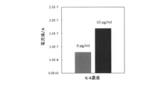
- targeting IL-6 it was estimated that approximately 10 pg / mL could be measured as the detection limit concentration.
- the methods, kits, and devices of the invention are suitable for measuring the concentration of trace amounts of biomolecules in a sample.
Landscapes
- Health & Medical Sciences (AREA)
- Life Sciences & Earth Sciences (AREA)
- Immunology (AREA)
- Chemical & Material Sciences (AREA)
- Molecular Biology (AREA)
- Pathology (AREA)
- Engineering & Computer Science (AREA)
- General Health & Medical Sciences (AREA)
- Biochemistry (AREA)
- Analytical Chemistry (AREA)
- Physics & Mathematics (AREA)
- General Physics & Mathematics (AREA)
- Biomedical Technology (AREA)
- Urology & Nephrology (AREA)
- Chemical Kinetics & Catalysis (AREA)
- Hematology (AREA)
- Electrochemistry (AREA)
- Cell Biology (AREA)
- Medicinal Chemistry (AREA)
- Food Science & Technology (AREA)
- Microbiology (AREA)
- Biotechnology (AREA)
- Measuring Or Testing Involving Enzymes Or Micro-Organisms (AREA)
- Apparatus Associated With Microorganisms And Enzymes (AREA)
Abstract
簡便に微量の生体分子の濃度を測定可能な手段を提供する。 試料中の生体分子濃度を測定する方法であって、 (a)生体分子と当該生体分子に対する特異的結合物質であってホースラディッシュペルオキシダーゼ(HRP)修飾された特異的結合物質との複合体に、銀イオンを作用させてHRPと銀イオンとの酵素反応を行う工程、 (b)前記酵素反応により銀を生成させるとともに当該生成された銀を電極上に堆積させる工程、および (c)前記堆積した銀における銀イオン量を電気化学的に測定することにより、前記生体分子濃度を測定する工程 を含む、方法が提供される。
Description
本発明は、生体分子濃度を電気化学的に測定する方法、この方法を実施するためのキットおよびデバイスに関する。
タンパク質等の生体分子の濃度は、特定の症状や生命体の状態を測定する指標(バイオマーカー)等として利用されている。このような生体分子の定量法としては、特異性が高いイムノアッセイが広く用いられている。イムノアッセイにおける抗原抗体反応を検出する手法としては、感度、迅速性、または簡便性などの視点から、光学手法(UV、蛍光、発光)を主体とした検討が行われ、イムノクロマト法のようなオンサイトでも簡便・迅速に計測できる技術も進歩してきた。実際にイムノクロマト法は簡便性および迅速性に優れ、目視で半定量的な結果が得られるため、妊娠診断検査やインフルエンザ検査などのオンサイト測定に利用されている。しかしながら、イムノクロマト法は酵素増幅ができないため、従来のイムノアッセイ(ELISA法)と比較するとその感度が低くなることが多い。一方、電気化学法によっても電極材料や検出方法の組合せにより、簡便ながら同等以上の感度が得られる可能性が期待されている。電気化学法は、溶液中の目的成分について、その濃度に対応する変化を電極上での電気的(例えば電流)信号として変換、出力することを特徴としており、簡便かつ高感度化の目標に最適の方法である。
本発明者らは、近年、この電気化学的方法に基づき、微量(ng/mL)でも血液中に混入すると発熱作用を引き起こすエンドトキシンの電気化学的手法による濃度測定法を検討してきた。しかしながら、エンドトキシンの存在濃度はpg/mL~ng/mLの領域であり、従来の電気化学的方法では正確な濃度を測定することが困難であった。そこで本発明者らは、エンドトキシンの濃度測定の検出感度向上を目的として、以下に示す新たな検出方法を提案した(特許文献1)。すなわち、(1)第一のエンドトキシン認識分子を固定化した担体に試料を接触させ、試料中のエンドトキシンを担体に捕捉・濃縮する工程、(2)重金属化合物と第二のエンドトキシン認識分子を含むエンドトキシン認識プローブを担体に接触させ、担体上のエンドトキシンにエンドトキシン認識プローブを捕捉する工程、(3)エンドトキシン認識プローブに酸性溶液を反応させ、プローブの重金属化合物から重金属イオンを溶出する工程、および(4)重金属イオンを測定する作用電極を用いて、溶出した重金属イオン量を電気化学的に測定する工程を含み、重金属イオン量を指標として試料中のエンドトキシン濃度を測定することを特徴とするエンドトキシン濃度の電気化学測定法である。この特許文献1の方法では200pg/mL程度の濃度のエンドトキシンを測定することが可能である。すなわち、特許文献1の方法は、エンドトキシンとプローブの微粒子上への濃縮、ならびに重金属イオンの作用電極上への濃縮・測定の過程を経ることで、重金属イオンを低濃度まで安定に定量でき、すなわち高感度なエンドトキシン検出が実現されたものである。
一方、生体中には、その存在が極微量(1~10pg/mL)であっても特定の疾患や生命体の状態の指標となるうる生体分子(例えば、疾病マーカー抗原、ホルモン、サイトカイン類など)が存在する。特許文献1の方法では、このような極微量(例えば10pg/mL以下)の生体分子を測定することはできない。このような極微量の生体分子の測定を可能とするために、さらなる検出感度向上が求められる。そのための最大の課題としては、対象一分子に対する検出系中の重金属イオン濃度を如何に向上させるかという点が挙げられる(応答増幅率の向上)。改善策として、プローブ一分子中に金属錯体部位をより多く導入したプローブ構造とすることが考えられる。これにより原理的には検出系内の重金属イオン濃度が増加するが、反面、対象分子との結合定数の低下が懸念される。
イムノクロマト法においても高感度化が検討されている。従来のイムノクロマト法では、対象分子の標識に金コロイドを利用するが、近年、この金コロイドへの銀の堆積を利用したイムノクロマト法の高感度化の報告が見られる(特許文献2、非特許文献1)。直径約50nmの金コロイドを核として直径10μmの銀粒子にまで成長させ、光学密度(光学濃度)の測定(デンシメトリー)により対象分子の定量を行うものである。従来のイムノクロマト法に比較して1000倍の高感度化に成功している。また、μm間隔で隔離された二つの電極間で対象物質を捕捉し、その後その上に酵素メタリゼーションによって銀を堆積させることで、二電極間が銀によって通電する(コンダクトメトリー)ことを利用したバイオセンサも報告されている(非特許文献2、非特許文献3)。また、銀のストリッピング反応を活用した例として、変換ストリッピング法が公知である(特許文献3、特許文献4)。この方法は、生体分子の電極反応を行うセルおよびストリッピングを行うセルの二つのセルを用意し、セル間の導通を取ることで、生体分子の電極反応を銀のストリッピング反応に変換、定量する方法であるが、装置が複雑である。
富士フイルム研究報告 57(2012)5-10
Nano Letters 5 (2005) 1475-1482
Journal of the American Chemical Society 133 (2011) 3238-3241
試料中の微量の生体分子濃度を簡便に測定することのできる手段が求められている。
本発明者らは、対象一分子に対する検出系中の重金属イオン濃度の向上の手段として、銀イオンの酵素メタリゼーション反応によって生成する銀を電極上に堆積させる手法を用い、この堆積した銀における銀イオン量を電気化学的に測定することで、対象生体分子の高感度測定が実施できることを見出し、本発明を完成させた。
本発明の一形態は、従来のイムノクロマト法や電気化学的測定よりも簡便かつ高感度に極微量の生体分子濃度を測定することのできる方法と、この方法を実施するための生体分子濃度測定キットおよびデバイスを提供する。
本発明の一形態は、従来のイムノクロマト法や電気化学的測定よりも簡便かつ高感度に極微量の生体分子濃度を測定することのできる方法と、この方法を実施するための生体分子濃度測定キットおよびデバイスを提供する。
すなわち、本発明は以下の通りである。
[1] 試料中の生体分子濃度を測定する方法であって、
(a)生体分子と当該生体分子に対する特異的結合物質であってホースラディッシュペルオキシダーゼ(HRP)修飾された特異的結合物質との複合体に、銀イオンを作用させてHRPと銀イオンとの酵素反応を行う工程、
(b)前記酵素反応により銀を生成させるとともに当該生成された銀を電極上に堆積させる工程、および
(c)前記堆積した銀における銀イオン量を電気化学的に測定することにより、前記生体分子濃度を測定する工程
を含む、前記方法。
[2] 前記複合体は、生体分子に対する第二の特異的結合物質を介して担体に固定化されている、[1]に記載の方法。
[3] 生体分子の検出限界濃度が1pg/mLから10pg/mLの範囲である、[1]または[2]に記載の方法。
[4] 前記電極の平均表面粗さ(Ra)が0.05~5nmである、[1]~[3]のいずれかに記載の方法。
[5] 前記電極はナノカーボン電極である、[1]~[4]のいずれかに記載の方法。
[6] 工程(c)において反応系における銀イオンの濃度が1~100ng/mLである、[1]~[5]のいずれかに記載の方法。
[7] 前記電極のサイズが、1~100mm2である、[1]~[6]のいずれかに記載の方法。
[8] 工程(a)における酵素反応は、酸化剤および還元剤の存在下で行われる、[1]~[7]のいずれかに記載の方法。
[9] 前記酸化剤は、過酸化水素であり、前記還元剤は、鉄イオン(Fe2+)およびヒドロキノンから選択される少なくとも一つである、[8]に記載の方法。
[10] 工程(a)の酵素反応は、pH3.0~7.0の緩衝液中で行われ、工程(c)における電気化学的測定は、pH4.0~5.0の緩衝液中で行われる、[1]~[9]のいずれかに記載の方法。
[11] 以下の工程、
(1)生体分子に対する特異的結合物質を固定化した担体に試料を接触させ、試料中の生体分子を担体に捕捉する工程、
(2)特異的結合物質を担体に再度接触させ、担体上の生体分子に特異的結合物質を捕捉する工程、
(3)特異的結合物質にホースラディッシュペルオキシダーゼ(HRP)を修飾する工程、
(4)銀イオンを含む試薬を担体上のHRPと酵素メタリゼーション反応をさせ、銀を生成させる工程、および
(5)生成した銀を電極上に堆積させ、この堆積した銀イオン量を電気化学的に測定する工程、
を含み、銀イオン量を指標として試料中の生体分子濃度を測定することを特徴とする、[1]~[10]のいずれかに記載の方法。
[11-1] 前記試薬が、酸化剤および還元剤をさらに含む、[11]に記載の方法。
[11-2] 前記酸化剤は、過酸化水素であり、前記還元剤は、鉄イオン(Fe2+)およびヒドロキノンから選択される少なくとも一つである、[11-1]に記載の方法。
[12] [1]~[11]のいずれかに記載の方法を実施するためのキットであって、生体分子に対する特異的結合物質を固定化した担体と、HRPと、銀イオンを含み、HRPと酵素メタリゼーション反応を実施可能な試薬と、を含むことを特徴とする生体分子濃度測定キット。
[12-1] 前記試薬が、酸化剤および還元剤をさらに含む、[12]に記載のキット。
[12-2] 前記酸化剤は、過酸化水素であり、前記還元剤は、鉄イオン(Fe2+)およびヒドロキノンから選択される少なくとも一つである、[12-1]に記載のキット。
[13] [1]~[11]のいずれかに記載の方法を実施するためのデバイスであって、生体分子に対する特異的結合物質を固定化した担体および電極を備えた電気化学セルと、電極への電圧印加手段と、電流測定手段と、を含むことを特徴とする生体分子濃度測定デバイス。
[14] 前記生体分子が抗原であり、前記特異的結合物質が前記抗原に結合する抗体またはそのフラグメントである、[1]~[11][11-1]~[11-2]のいずれかに記載の方法、[12][12-1][12-2]のいずれかに記載のキット、[13][13-1][13-2]のいずれかに記載のデバイス。
[1] 試料中の生体分子濃度を測定する方法であって、
(a)生体分子と当該生体分子に対する特異的結合物質であってホースラディッシュペルオキシダーゼ(HRP)修飾された特異的結合物質との複合体に、銀イオンを作用させてHRPと銀イオンとの酵素反応を行う工程、
(b)前記酵素反応により銀を生成させるとともに当該生成された銀を電極上に堆積させる工程、および
(c)前記堆積した銀における銀イオン量を電気化学的に測定することにより、前記生体分子濃度を測定する工程
を含む、前記方法。
[2] 前記複合体は、生体分子に対する第二の特異的結合物質を介して担体に固定化されている、[1]に記載の方法。
[3] 生体分子の検出限界濃度が1pg/mLから10pg/mLの範囲である、[1]または[2]に記載の方法。
[4] 前記電極の平均表面粗さ(Ra)が0.05~5nmである、[1]~[3]のいずれかに記載の方法。
[5] 前記電極はナノカーボン電極である、[1]~[4]のいずれかに記載の方法。
[6] 工程(c)において反応系における銀イオンの濃度が1~100ng/mLである、[1]~[5]のいずれかに記載の方法。
[7] 前記電極のサイズが、1~100mm2である、[1]~[6]のいずれかに記載の方法。
[8] 工程(a)における酵素反応は、酸化剤および還元剤の存在下で行われる、[1]~[7]のいずれかに記載の方法。
[9] 前記酸化剤は、過酸化水素であり、前記還元剤は、鉄イオン(Fe2+)およびヒドロキノンから選択される少なくとも一つである、[8]に記載の方法。
[10] 工程(a)の酵素反応は、pH3.0~7.0の緩衝液中で行われ、工程(c)における電気化学的測定は、pH4.0~5.0の緩衝液中で行われる、[1]~[9]のいずれかに記載の方法。
[11] 以下の工程、
(1)生体分子に対する特異的結合物質を固定化した担体に試料を接触させ、試料中の生体分子を担体に捕捉する工程、
(2)特異的結合物質を担体に再度接触させ、担体上の生体分子に特異的結合物質を捕捉する工程、
(3)特異的結合物質にホースラディッシュペルオキシダーゼ(HRP)を修飾する工程、
(4)銀イオンを含む試薬を担体上のHRPと酵素メタリゼーション反応をさせ、銀を生成させる工程、および
(5)生成した銀を電極上に堆積させ、この堆積した銀イオン量を電気化学的に測定する工程、
を含み、銀イオン量を指標として試料中の生体分子濃度を測定することを特徴とする、[1]~[10]のいずれかに記載の方法。
[11-1] 前記試薬が、酸化剤および還元剤をさらに含む、[11]に記載の方法。
[11-2] 前記酸化剤は、過酸化水素であり、前記還元剤は、鉄イオン(Fe2+)およびヒドロキノンから選択される少なくとも一つである、[11-1]に記載の方法。
[12] [1]~[11]のいずれかに記載の方法を実施するためのキットであって、生体分子に対する特異的結合物質を固定化した担体と、HRPと、銀イオンを含み、HRPと酵素メタリゼーション反応を実施可能な試薬と、を含むことを特徴とする生体分子濃度測定キット。
[12-1] 前記試薬が、酸化剤および還元剤をさらに含む、[12]に記載のキット。
[12-2] 前記酸化剤は、過酸化水素であり、前記還元剤は、鉄イオン(Fe2+)およびヒドロキノンから選択される少なくとも一つである、[12-1]に記載のキット。
[13] [1]~[11]のいずれかに記載の方法を実施するためのデバイスであって、生体分子に対する特異的結合物質を固定化した担体および電極を備えた電気化学セルと、電極への電圧印加手段と、電流測定手段と、を含むことを特徴とする生体分子濃度測定デバイス。
[14] 前記生体分子が抗原であり、前記特異的結合物質が前記抗原に結合する抗体またはそのフラグメントである、[1]~[11][11-1]~[11-2]のいずれかに記載の方法、[12][12-1][12-2]のいずれかに記載のキット、[13][13-1][13-2]のいずれかに記載のデバイス。
[15] 以下の工程、
(1)生体分子に対する特異的結合物質を固定化した担体に試料を接触させ、試料中の生体分子を担体に捕捉・濃縮する工程、
(2)特異的結合物質を担体に再度接触させ、担体上の生体分子に特異的結合物質を捕捉する工程、
(3)特異的結合物質にホースラディッシュペルオキシダーゼ(HRP)を修飾する工程、
(4)銀イオンを含む試薬を担体上のHRPと酵素メタリゼーション反応をさせ、銀を生成させる工程、および
(5)生成した銀を作用電極上に堆積させ、この堆積した銀イオン量を電気化学的に測定する工程、
を含み、銀イオン量を指標として試料中の生体分子濃度を測定することを特徴とする生体分子濃度の測定方法。
[16] 生体分子の検出限界濃度は1pg/mLから10pg/mLの範囲である、[15]に記載の測定方法。
[17] [15]または[16]に記載の生体分子濃度測定方法を実施するためのキットであって、生体分子に対する特異的結合物質を固定化した担体、HRPと銀イオンを含む酵素メタリゼーション反応を実施可能な薬品を含むことを特徴とする生体分子濃度測定キット。
[18] [15]または[16]に記載の第一生体分子濃度測定方法を実施するためのデバイスであって、生体分子に対する特異的結合物質を固定化した担体と作用電極を備えた電気化学セル、電極への電圧印加手段および電流測定手段を含むことを特徴とする生体分子濃度測定デバイス。
(1)生体分子に対する特異的結合物質を固定化した担体に試料を接触させ、試料中の生体分子を担体に捕捉・濃縮する工程、
(2)特異的結合物質を担体に再度接触させ、担体上の生体分子に特異的結合物質を捕捉する工程、
(3)特異的結合物質にホースラディッシュペルオキシダーゼ(HRP)を修飾する工程、
(4)銀イオンを含む試薬を担体上のHRPと酵素メタリゼーション反応をさせ、銀を生成させる工程、および
(5)生成した銀を作用電極上に堆積させ、この堆積した銀イオン量を電気化学的に測定する工程、
を含み、銀イオン量を指標として試料中の生体分子濃度を測定することを特徴とする生体分子濃度の測定方法。
[16] 生体分子の検出限界濃度は1pg/mLから10pg/mLの範囲である、[15]に記載の測定方法。
[17] [15]または[16]に記載の生体分子濃度測定方法を実施するためのキットであって、生体分子に対する特異的結合物質を固定化した担体、HRPと銀イオンを含む酵素メタリゼーション反応を実施可能な薬品を含むことを特徴とする生体分子濃度測定キット。
[18] [15]または[16]に記載の第一生体分子濃度測定方法を実施するためのデバイスであって、生体分子に対する特異的結合物質を固定化した担体と作用電極を備えた電気化学セル、電極への電圧印加手段および電流測定手段を含むことを特徴とする生体分子濃度測定デバイス。
本発明は、以下の一以上の効果を有する。
(1)従来のイムノクロマト法や電気化学的測定よりも高感度に生体分子濃度を測定することが可能である。特に、本発明の方法によれば、極微量(例えば1~10pg/mL)の生体分子濃度の測定が可能である。
(2)複雑な機器や光学手法(UV、蛍光、発光)を必要とすることなく簡便な電気化学的測定により微量の生体分子濃度を測定することが可能である。
(3)生体分子の担体表面上への捕捉による濃縮、および銀の酵素反応による生成、さらには生成した銀の電極上への還元堆積による濃縮という複数の濃縮過程の組合せと、最終的に検出系中の重金属イオンを低濃度まで安定に測定可能な作用電極によって、低濃度の生体分子に対しても電気化学応答を増幅させ、より一層の高感度で測定することが可能になる。
(4)本発明の濃度測定キットおよび濃度測定デバイスの使用により、使用者の熟練度を問わずに、簡便かつ高感度(例えばpg/mLオーダー)で極微量(例えば1~10pg/mL)の生体分子の濃度を測定することができる。
(1)従来のイムノクロマト法や電気化学的測定よりも高感度に生体分子濃度を測定することが可能である。特に、本発明の方法によれば、極微量(例えば1~10pg/mL)の生体分子濃度の測定が可能である。
(2)複雑な機器や光学手法(UV、蛍光、発光)を必要とすることなく簡便な電気化学的測定により微量の生体分子濃度を測定することが可能である。
(3)生体分子の担体表面上への捕捉による濃縮、および銀の酵素反応による生成、さらには生成した銀の電極上への還元堆積による濃縮という複数の濃縮過程の組合せと、最終的に検出系中の重金属イオンを低濃度まで安定に測定可能な作用電極によって、低濃度の生体分子に対しても電気化学応答を増幅させ、より一層の高感度で測定することが可能になる。
(4)本発明の濃度測定キットおよび濃度測定デバイスの使用により、使用者の熟練度を問わずに、簡便かつ高感度(例えばpg/mLオーダー)で極微量(例えば1~10pg/mL)の生体分子の濃度を測定することができる。
本発明の一形態は、電気化学的方法による、試料中に含まれる生体分子の濃度測定方法である。すなわち、本発明の一形態は、試料中の生体分子濃度を測定する方法に関する。該方法は、(a)生体分子と当該生体分子に対する特異的結合物質であってホースラディッシュペルオキシダーゼ(HRP)修飾された特異的結合物質との複合体に、銀イオンを作用させてHRPと銀イオンとの酵素反応を行う工程、(b)前記酵素反応により銀を生成させるとともに当該生成された銀を電極上に堆積させる工程、および(c)前記堆積した銀における銀イオン量を電気化学的に測定することにより、前記生体分子濃度を測定する工程を含む。
本発明は、検出系中の重金属イオンとして銀イオンを使用し、検出系中の重金属イオン濃度の向上の手段として、銀イオンとHRPとの酵素メタリゼーション反応を利用する。すなわち、酵素反応を通じて、検出対象である生体分子一分子に対する銀濃度が高められる(増幅効果)。堆積された銀の量は試料中の生体分子の量に相関していることから、電極上に堆積された銀の量を電気化学的に測定することにより、試料中の生体分子濃度を測定することができる。本発明の方法では、酵素反応による銀の生成量の制御(応答増幅率の向上)と電極上での銀のストリッピング反応による高感度検出(高いS/N比)とが可能であり、結果として対象生体分子の高感度な測定が可能となる。
本発明の極微量の生体分子の濃度測定の原理を以下に説明する。
本発明の極微量の生体分子の濃度測定の原理を以下に説明する。
本形態の方法では、生成した銀を銀の電気化学測定が可能な作用電極へ誘導し堆積させることで、試料中の銀濃度を定量する。ここで、銀の電気化学測定としては、例えば、アノーディックストリッピングボルタンメトリー(ASV)法と同様の方法を用いる。一般的なASV法は金属イオンを電極上に一度還元堆積させ、堆積した金属を酸化させる際に得られる酸化電流値を検出する分析法である。ただし、本発明の方法が一般的な金属イオンのASV検出法と大きく異なる点は、電位印加によって金属イオンを作用電極上に還元堆積させない点である。代わりに、本発明の方法では、酵素メタリゼーション反応によって銀を生成させ、生成させた銀を作用電極上に堆積させる。堆積させた銀の量は、ASV法と同様にして、堆積した銀を酸化させる際に得られる酸化電流値を測定することにより測定することができる。堆積した銀量は試料中の生体分子の量と相関関係にあるため、既知の生体分子量とそれに応じて捕捉されたHRP量に基づき生成した銀量(より具体的には銀を酸化させる際に流れる酸化電流値)との間で検量線を作成し、銀の酸化電流値を測定すれば、該検量線に基づいて試料中の生体分子の量を知ることが可能となる。ASV法は、非常に高感度な分析手法でありながら、安価で測定装置の小型化が可能であり、オンサイト分析に適した分析法である。本発明の方法では、このようなASV法の利点を有しつつ、酵素反応による増幅効果によって一般のASV法よりも低い検出限界を達成することができる。
本発明の方法は、基本的には電子の授受により生ずる、電極に流れる電流を測定することにより銀の量を測定して、生体分子量を把握するものである。この測定電流は銀の酵素反応による生成(増幅)と銀の電極表面への還元堆積(濃縮)過程を経る。したがって、捕捉された生体分子が微量であっても、酵素反応による銀生成の増幅効果と電極表面への銀の堆積(濃縮)効果とにより高感度で生体分子量を測定し得る点に特徴を有する。
具体的には、作用電極上では以下の反応が生ずる。すなわち、HRPが触媒として作用し、銀イオン(Ag+)が還元されて、銀(Ag)が作用電極上に堆積する。
(a)Ag++e→Ag(電極上での酵素メタリゼーション反応)
こうして、HRPを触媒とした銀イオン(Ag+)の還元反応(酵素メタリゼーション反応)が連続的に生じることにより、検出対象である生体分子一分子に対する銀濃度を高めることができ、望ましい増幅効果を達成する。
(a)Ag++e→Ag(電極上での酵素メタリゼーション反応)
こうして、HRPを触媒とした銀イオン(Ag+)の還元反応(酵素メタリゼーション反応)が連続的に生じることにより、検出対象である生体分子一分子に対する銀濃度を高めることができ、望ましい増幅効果を達成する。
好ましい態様では、HRPと還元剤および酸化剤との共存下で、銀生成反応を行う。HRPと還元剤および酸化剤とが共に存在すると、銀イオンの還元反応の効率が向上し得る。例えば、反応系に、HRPと酸化剤である過酸化水素および還元剤であるヒドロキノンとが共存する場合には、HRPが触媒として作用し(特に下記(a2)の反応を触媒する)、以下の反応が進行し得る。
(a1)還元剤(例えばヒドロキノン)+2Ag+→還元剤の酸化物(例えば1,4-ベンゾキノン)+2Ag+2H+
(a2)酸化剤(例えばH2O2)+2H++2e→酸化剤の還元物(例えば2H2O)
上記反応を通じて、HRPを触媒とした銀イオン(Ag+)の連続的な還元反応(HRP量に応じた銀の増幅生成)が効率的に進行し、検出対象である生体分子一分子に対する銀濃度を効率的に高めることができ、一層の増幅効果が達成される。
(a1)還元剤(例えばヒドロキノン)+2Ag+→還元剤の酸化物(例えば1,4-ベンゾキノン)+2Ag+2H+
(a2)酸化剤(例えばH2O2)+2H++2e→酸化剤の還元物(例えば2H2O)
上記反応を通じて、HRPを触媒とした銀イオン(Ag+)の連続的な還元反応(HRP量に応じた銀の増幅生成)が効率的に進行し、検出対象である生体分子一分子に対する銀濃度を効率的に高めることができ、一層の増幅効果が達成される。
次に、作用電極へ正の電圧を印加すると作用電極上に堆積した銀が酸化されて、銀イオンとなる。例えば、pH5.0のもとでASV測定と同様の方法により堆積された銀の酸化反応を行うと(すなわち電極上へ正の電圧を印加すると)、以下の酸化反応が生ずる(銀の脱離反応)。
(b)Ag→Ag++e
この時に、上記反応式(b)中の電子(e)の量、つまり電極における電流は、電極上に堆積した銀の量、したがって、(a)の酵素反応による銀の生成反応に要する生体分子の量(HRP量)、銀イオン濃度、酵素反応時間などの酵素反応条件に依存する。
(b)Ag→Ag++e
この時に、上記反応式(b)中の電子(e)の量、つまり電極における電流は、電極上に堆積した銀の量、したがって、(a)の酵素反応による銀の生成反応に要する生体分子の量(HRP量)、銀イオン濃度、酵素反応時間などの酵素反応条件に依存する。
すなわち、電極表面に濃縮、堆積された銀が脱離反応を起こす際に酸化電流を与える。このときの酸化電流は電極表面に堆積された銀量にほぼ比例する。堆積された銀の量は、酵素反応に関与する酵素量(HRP量)に相関するが、この酵素量(HRP量)は試料中の生体分子の量と対応関係にある。したがって、試料中の生体分子濃度の増加に応じて、電極表面に堆積される銀量が増加するため、得られる電流も増加する。いいかえれば、電流の増加の度合いから、試料中に存在する生体分子の量(濃度)を把握することが出来る。例えば、既知の生体分子量と銀の酸化電流値との間で検量線を作成し、検量線作成の際と同一の酵素反応条件で酵素反応を行い、堆積された銀の酸化電流値を測定することにより、該検量線に基づいて試料中の生体分子の量を知ることが可能となる。したがって、本発明の方法では、複雑な機器や光学手法(UV、蛍光、発光)を必要とすることなく、簡便な電気化学的測定により、試料中の生体分子濃度の定量が可能である。
生体分子濃度の定量は、これら一連の反応、すなわち、酵素メタリゼーション反応による銀の増幅生成(検出対象である生体分子一分子に対する銀濃度向上)と作用電極上への銀の還元堆積(濃縮)によって可能となったものであり、従来のイムノクロマトグラフィを含むイムノアッセイ法では実現不可能であった測定濃度の検出限界の向上(例えばイムノクロマト法と比較して約10000倍の向上)をもたらすものである。本発明の一形態による測定方法において、生体分子の検出限界濃度は、1pg/mLから10pg/mLの範囲である。本発明の一形態によれば、従来のイムノクロマト法や電気化学的測定では困難であった、このような極微量(1~10pg/mL)の生体分子濃度の測定が可能である。
なお、本発明の測定方法は、極微量の生体分子濃度の測定に限られず、例えば、1pg/mL~1000ng/mLの範囲(好ましくは1pg/mL~100ng/mL)の生体分子濃度の測定にも適用可能である。
なお、本発明の測定方法は、極微量の生体分子濃度の測定に限られず、例えば、1pg/mL~1000ng/mLの範囲(好ましくは1pg/mL~100ng/mL)の生体分子濃度の測定にも適用可能である。
以下、図面を用いて本発明の一形態に係る生体分子の濃度の測定方法を詳細に説明する。ただし、本発明は図示した形態に限定されるわけではない。
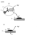
本発明の方法では、生体分子に対する特異的結合物質を生体分子の捕捉材として用いる。図1Aに示すように、生体分子1は、当該生体分子1に対する特異的結合物質2と結合し、複合体10を形成している。特異的結合物質2はHRPで修飾されている。本明細書では、生体分子とともに複合体を構成する特異的結合物質を「第一の特異的結合物質」ともいう。また、後述の、生体分子を担体に固定化するために介在する特異的結合物質を「第二の特異的結合物質」ともいう。
本発明の方法では、生体分子に対する特異的結合物質を生体分子の捕捉材として用いる。図1Aに示すように、生体分子1は、当該生体分子1に対する特異的結合物質2と結合し、複合体10を形成している。特異的結合物質2はHRPで修飾されている。本明細書では、生体分子とともに複合体を構成する特異的結合物質を「第一の特異的結合物質」ともいう。また、後述の、生体分子を担体に固定化するために介在する特異的結合物質を「第二の特異的結合物質」ともいう。
複合体10の形成方法は特に制限されない。例えば、生体分子1を含有する試料に第一の特異的結合物質2を添加して生体分子1を第一の特異的結合物質2に捕捉させ、次いで、第一の特異的結合物質2にホースラディッシュペルオキシダーゼ(HRP)を修飾することにより、複合体10が得られる。この時、生体分子1は、その量に応じて、第一の特異的結合物質2に捕捉され、複合体10が形成される。なお、あらかじめHRP標識した特異的結合物質2を使用してもよい。すなわち、第一の特異的結合物質2にあらかじめHRPを修飾し、これを、生体分子1を含有する試料に添加して生体分子1を第一の特異的結合物質2に捕捉させることにより、複合体10を得てもよい。
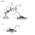
本発明の一実施形態では、生体分子に対する特異的結合物質(第二の特異的結合物質)を例えば化学架橋により固定した担体を生体分子の捕捉材として用いることができる。本実施形態では、図1Bに示すように、複合体10は、生体分子1に対する第二の特異的結合物質5を介して担体6に固定化されている。複合体10を第二の特異的結合物質5を介して担体6に固定化することにより、各工程で溶液中に存在する生体分子、特異的結合物質(例えばビオチン標識抗体)、HRP修飾抗体を担体表面に捕捉することで濃縮ができ、なおかつ各工程での洗浄操作が容易となる。
担体6は、特異的結合物質を固定化する事が出来れば、その形状に制限はなく、球状・不定形の粒子、微粒子でも良く、繊維状、膜状、平板状、柱状でも良い。担体は、多孔質であっても良く、無孔質であっても良い。入手の容易さを考慮すれば、免疫測定用の微粒子、磁気ビーズ、非磁気ビーズ、球状やモノリス状のクロマトグラフィー用担体、フィルター、繊維などが好ましく利用できる。
担体6は、特異的結合物質を固定化する事が出来れば、その形状に制限はなく、球状・不定形の粒子、微粒子でも良く、繊維状、膜状、平板状、柱状でも良い。担体は、多孔質であっても良く、無孔質であっても良い。入手の容易さを考慮すれば、免疫測定用の微粒子、磁気ビーズ、非磁気ビーズ、球状やモノリス状のクロマトグラフィー用担体、フィルター、繊維などが好ましく利用できる。
第二の特異的結合物質5を担体6に固定化する方法は当業者によく知られている。例えば、結合剤を用いて、担体の表面(活性化した表面、カルボキシル化表面、ヒドロキシル化表面など)に特異的結合物質を固定化させることができる。結合剤の例としては、臭化シアン、スクシンイミド、アルデヒド、トシルクロリド、アビジン-ビオチン、ストレプトアビジン-ビオチン、チオール、光架橋化剤、エポキシドおよびマレイミドがある。例えば、抗体は、抗体上の自由なアミノ基を基板内に存在している反応性側鎖と化学的に架橋させることによって基板に結合させることができる。
生体分子1(複合体10)を担体6に捕捉する方法としては、例えば、第二の特異的結合物質5が固定化された担体6に生体分子1を含有する試料を、例えば滴下または添加することにより接触させて生体分子1を第二の特異的結合物質5を介して担体6に捕捉した後に、第一の特異的結合物質2を接触させ、担体6上の生体分子1を第一の特異的結合物質2に捕捉させる方法が挙げられる。この時、担体6へ捕捉された生体分子1の量に応じて、第一の特異的結合物質2が捕捉される。
続いて、この第一の特異的結合物質2が捕捉された担体6に、ホースラディッシュペルオキシダーゼ(本明細書において単に「HRP」ともいう)を修飾することにより、図1Bに示すように、生体分子1と、当該生体分子1に結合しかつHRPで修飾されている第一の特異的結合物質2とから構成される複合体10であって、第二の特異的結合物質5を介して担体6に固定化された複合体10が得られる。
なお、HRPによる修飾は第一の特異的結合物質2と生体分子1との結合前に行ってもよい。例えば、あらかじめHRP標識した第一の特異的結合物質2を用いて生体分子1を捕捉(結合)させてもよい。
続いて、この第一の特異的結合物質2が捕捉された担体6に、ホースラディッシュペルオキシダーゼ(本明細書において単に「HRP」ともいう)を修飾することにより、図1Bに示すように、生体分子1と、当該生体分子1に結合しかつHRPで修飾されている第一の特異的結合物質2とから構成される複合体10であって、第二の特異的結合物質5を介して担体6に固定化された複合体10が得られる。
なお、HRPによる修飾は第一の特異的結合物質2と生体分子1との結合前に行ってもよい。例えば、あらかじめHRP標識した第一の特異的結合物質2を用いて生体分子1を捕捉(結合)させてもよい。
複合体10の形成およびHRP修飾は溶液中で行われることが好ましい。試料が溶液である場合には試料をそのまま用いて、または、Tris緩衝液生理食塩水(TBS)、リン酸緩衝液生理食塩水(PBS)、HEPES緩衝液等の緩衝液等で希釈して用いることができる。試料が溶液でない場合には上記緩衝液等の溶液に試料を分散または溶解させて用いることができる。
HRPによる修飾の方法は当業者によく知られている。例えば、臭化シアン、スクシンイミド、アルデヒド、トシルクロリド、アビジン-ビオチン、ストレプトアビジン-ビオチン、チオール、光架橋化剤、エポキシドおよびマレイミドなどの結合剤を介して第一の特異的結合物質2にHRPを修飾することができる。例えば、第一の特異的結合物質2としてビオチン標識抗体を用い、これに、アビジンまたはストレプトアビジンで標識されたHRPを作用させることで、アビジン-ビオチンまたはストレプトアビジン-ビオチンの結合により、第一の特異的結合物質2にHRPを修飾することができる。
HRPによる修飾の方法は当業者によく知られている。例えば、臭化シアン、スクシンイミド、アルデヒド、トシルクロリド、アビジン-ビオチン、ストレプトアビジン-ビオチン、チオール、光架橋化剤、エポキシドおよびマレイミドなどの結合剤を介して第一の特異的結合物質2にHRPを修飾することができる。例えば、第一の特異的結合物質2としてビオチン標識抗体を用い、これに、アビジンまたはストレプトアビジンで標識されたHRPを作用させることで、アビジン-ビオチンまたはストレプトアビジン-ビオチンの結合により、第一の特異的結合物質2にHRPを修飾することができる。
複合体10の形成、担体6への固定化および/またはHRP修飾の後には、必要に応じて、Tris緩衝液生理食塩水(TBS)、リン酸緩衝液生理食塩水(PBS)、HEPES緩衝液等の緩衝液などの洗浄液を用いて洗浄処理がされ、過剰な(結合していない)特異的結合物質(2,5)、担体6、および/またはHRPなどが除去される。洗浄液には、必要に応じて、ポリソルベート20(Tween20)などの非イオン性界面活性剤、BSAやスキムミルクなどのブロッキング剤が添加される。
生体分子に作用させる特異的結合物質(2,5)およびHRPの濃度に特に制限はないが、特異的結合物質(2,5)の濃度は数10~数100μg/mL(例えば10~900μg/mL、好ましくは50~500μg/mL)が好都合であり、HRPの濃度は数ng/mL程度(例えば1~10ng/mL、好ましくは1~5ng/mL)が好都合である。特異的結合物質およびHRPの濃度が大きすぎると、物理的に吸着した過剰な特異的結合物質およびHRPが洗浄処理後にも系に残存し、生体分子に結合していないHRPによる電流応答の増幅を与える場合がある。このような濃度とすることにより、生体分子に結合していないHRPの影響を低減または回避でき、測定対象である生体分子由来の明瞭な電流応答を与え得ることができるため好ましい。
本発明の方法では、図1Aおよび図1Bに示されるように、複合体10に銀イオン(Ag+)を作用させることで、複合体10中のHRPと銀イオン(Ag+)との酵素反応が生じる(工程(a))。酵素反応により銀4が生成し、当該生成された銀4は電極3上に堆積される(工程(b))。
工程(a)における酵素反応は、銀イオンの還元反応の効率の面から、酸化剤(例えば過酸化水素)および還元剤(例えば鉄イオン(Fe2+)および/またはヒドロキノン)の存在下で行われることが好ましい。
工程(a)は溶液中で行われることが好ましい。
好ましくは、工程(a)の酵素反応の前段階までは、抗体およびHRPの機能維持の点で、Tris緩衝液生理食塩水(TBS)、リン酸緩衝液生理食塩水(PBS)、HEPES緩衝液等のpH(例えばpH6~8)が制御された緩衝液(例えば濃度10~100mmol/L)を用いる。
また、工程(a)の酵素反応では、溶液中に塩素イオンが存在すると銀イオンと反応して塩化銀(AgCl)が析出してしまう可能性があり、塩素イオンフリーの緩衝液で行うことが好ましい。具体的には酵素反応を効率よく進行させる観点で、塩素イオンフリーのクエン酸緩衝液、酢酸緩衝液、リン酸緩衝液(PB)等のpH(例えば、pH3.0~7.0)が制御された緩衝液(例えば濃度10~100mmmol/L)を用いることが好ましい。
また、工程(a)および工程(b)は、複合体10を含む緩衝液に銀イオン(Ag+)を含む試薬を添加し、得られた溶液を電極上に滴下することにより実施されることが好ましい。
工程(a)において反応系における銀イオンの濃度は、HRPとの反応が進行する濃度であれば特に制限されないが、効率的な酵素メタリゼーション反応の点で、好ましくは1~50mmol/Lであり、より好ましくは1~30mmol/Lであり、さらに好ましくは1~10mmol/Lである。
工程(b)の後、工程(c)の前に、必要に応じて、銀4が堆積された電極3の表面を洗浄し、過剰な(未反応の)銀(イオン)および/または複合体10などが除去される。
酵素反応の時間(工程(a)および工程(b)の合計時間)は特に制限されず、測定対象物質の存在濃度および使用する抗体、HRPの濃度により適宜設定される。一例をあげると、5~15分である。
工程(a)における酵素反応は、銀イオンの還元反応の効率の面から、酸化剤(例えば過酸化水素)および還元剤(例えば鉄イオン(Fe2+)および/またはヒドロキノン)の存在下で行われることが好ましい。
工程(a)は溶液中で行われることが好ましい。
好ましくは、工程(a)の酵素反応の前段階までは、抗体およびHRPの機能維持の点で、Tris緩衝液生理食塩水(TBS)、リン酸緩衝液生理食塩水(PBS)、HEPES緩衝液等のpH(例えばpH6~8)が制御された緩衝液(例えば濃度10~100mmol/L)を用いる。
また、工程(a)の酵素反応では、溶液中に塩素イオンが存在すると銀イオンと反応して塩化銀(AgCl)が析出してしまう可能性があり、塩素イオンフリーの緩衝液で行うことが好ましい。具体的には酵素反応を効率よく進行させる観点で、塩素イオンフリーのクエン酸緩衝液、酢酸緩衝液、リン酸緩衝液(PB)等のpH(例えば、pH3.0~7.0)が制御された緩衝液(例えば濃度10~100mmmol/L)を用いることが好ましい。
また、工程(a)および工程(b)は、複合体10を含む緩衝液に銀イオン(Ag+)を含む試薬を添加し、得られた溶液を電極上に滴下することにより実施されることが好ましい。
工程(a)において反応系における銀イオンの濃度は、HRPとの反応が進行する濃度であれば特に制限されないが、効率的な酵素メタリゼーション反応の点で、好ましくは1~50mmol/Lであり、より好ましくは1~30mmol/Lであり、さらに好ましくは1~10mmol/Lである。
工程(b)の後、工程(c)の前に、必要に応じて、銀4が堆積された電極3の表面を洗浄し、過剰な(未反応の)銀(イオン)および/または複合体10などが除去される。
酵素反応の時間(工程(a)および工程(b)の合計時間)は特に制限されず、測定対象物質の存在濃度および使用する抗体、HRPの濃度により適宜設定される。一例をあげると、5~15分である。
続いて、堆積した銀4における銀イオン(Ag+)の量を電気化学的に測定することにより、試料中の生体分子1の濃度が測定される(工程(c))。工程(c)は溶液中で行われることが好ましい。溶液としては、銀の電気化学測定を高感度に実施することのできる緩衝液およびpHの使用が好ましい。例えば、弱酸性(例えばpH4.0~5.0、好ましくはpH5.0程度)の酢酸緩衝液、クエン酸緩衝液等の緩衝液(例えば、濃度10~100mmol/L)が好ましい。また、工程(c)において反応系における銀イオンの濃度が1~100ng/mLとなる条件で電気化学測定を行うことが好ましい。銀4が堆積した電極を作用電極としてpHが制御された緩衝液中に挿入し、適当な対向電極、参照電極と組み合わせて、電気化学測定を行う。銀4が堆積した電極(作用電極)に正の電圧(例えば、0~300mV対銀塩化銀電極)を印加することにより、銀の酸化反応に基づく酸化電流が流れる。
電気化学的測定法としては、銀の酸化電流を測定できる方法であれば、特に制限されない。例えば、矩形波ボルタンメトリー、サイクリックボルタンメトリー、微分パルスボルタンメトリー、ノーマルパルスボルタンメトリー、交流ボルタンメトリー等の手法が適宜使用可能である。
本発明において利用される電極(作用電極)の材質としては特に制限はないが、例えば、カーボン系の材料、金電極、等が挙げられる。中でも、電極素材上に最終的に銀を堆積させ、その後堆積させた銀を電極反応させる必要があるとの観点から、カーボン系の材料からなるカーボン電極が好都合に使用される。例えば、特許文献1などに開示されたカーボン薄膜(銀イオンが堆積しやすく、かつ酸化溶出反応に基づいた電気化学反応によって測定するのに十分な広い電位窓と低いノイズ電流を有するカーボン薄膜)が挙げられる。具体的には、ボロンドープダイヤモンド薄膜、導電性ダイヤモンドライクカーボン薄膜、スパッタカーボン薄膜またはグラファイト薄膜などを利用することができる。さらには、このカーボン薄膜は、sp2カーボン結合が多いほうが良く、しかもより低濃度の銀の測定を達成するためには電極由来のノイズ電流の低い平坦な電極表面を有するナノカーボン電極が好ましい。カーボン薄膜におけるsp3結合とsp2結合との結合比は、例えばスパッタ法における照射イオンの加速電圧を調節することにより制御できる。sp2結合の割合(sp3結合とsp2結合との比率)は、X線光電子分光分析(XPS)により測定することができる。
具体的な好ましいナノカーボン電極の例は、特許4513093号に記載されたスパッタ法(例えば電子サイクロトロン共鳴(ECR)スパッタ法、RFスパッタ法、DCスパッタ法、またはイオンビームスパッタ法)により形成されるカーボン薄膜で構成されたカーボン電極である。
具体的な好ましいナノカーボン電極の例は、特許4513093号に記載されたスパッタ法(例えば電子サイクロトロン共鳴(ECR)スパッタ法、RFスパッタ法、DCスパッタ法、またはイオンビームスパッタ法)により形成されるカーボン薄膜で構成されたカーボン電極である。
ノイズ電流を低減し、高感度を実現するため、電極(作用電極)の平均表面粗さ(Ra)は0.05~5nmであることが好ましい。本明細書において、平均表面粗さ(Ra)はJIS B 0601:2013に規定する算術平均粗さ(Ra)をいう。平均表面粗さ(Ra)はより好ましくは0.1~5nmであり、さらに好ましくは0.1~2.0nmである。
電極は、低ノイズ(一般的に電流密度で、例えば100nA~10μA/cm2)が達成されるものが好ましい。
電極は、低ノイズ(一般的に電流密度で、例えば100nA~10μA/cm2)が達成されるものが好ましい。
電極サイズは、計測する電流の範囲に応じて適宜決定される。電極サイズは、例えば1~100mm2が好ましく、3~50mm2がより好ましく、3~10mm2がさらに好ましい。例えば、微量(1~10pg/mL)の生体分子の測定を行う場合には通常、数μA~数100μAで計測するのが好都合であり、その場合、上記の電極のサイズが好ましい。電極の形状は特に制限されず、円形、楕円形、または多角形等でありうる。
電極の厚さは、特に制限されないが、薄膜形状が好ましい。例えば、30nm~200nm程度が、十分な導電性を確保し極微小な電極を作製するという観点から好ましい。
電極の厚さは、特に制限されないが、薄膜形状が好ましい。例えば、30nm~200nm程度が、十分な導電性を確保し極微小な電極を作製するという観点から好ましい。
特異的結合物質は、生体分子と特異的に結合する能力を持つ物質であり、例えば、対象の生体分子に結合する、抗体またはそのフラグメント、糖鎖または複合糖質、DNA、RNA、オリゴヌクレオチドもしくはポリヌクレオチドまたはその誘導体などの核酸(核酸アプタマー)、ペプチド(ペプチドアプタマー)、タンパク質、酵素、合成分子などを使用することができる。第一の特異的結合物質および第二の特異的結合物質は同一であっても異なっていてもよい。
銀イオン(Ag+)を含む試薬(銀を生成させる薬剤)としては、銀イオン(Ag+)を含むものであれば特に限定されない。好ましくは銀イオンと還元剤(例えば鉄イオン(Fe2+)、ヒドロキノンなど)と酸化剤(例えば、過酸化水素)とを含有する試薬が好ましい。還元剤および酸化剤がHRPと共に存在すると、銀イオンの還元反応の効率が向上し得る。また、試薬は溶液形態が好ましく、銀イオンとHRPとの酵素反応によって効率よく銀を生成させることのできる溶液組成(例えば、銀イオン(例えば濃度1~10mmol/L)、過酸化水素(例えば濃度10~30mmol/L)、およびヒドロキノン(例えば濃度10~30mmol/L)を含む0.1mol/Lクエン酸緩衝液(例えばpH3~5))であり、安定で、安価なものが好都合に利用される。具体的には、Nanoprobe社のEnzMetなどが適宜利用できる。
本発明の一実施形態について、図1Cに基づき詳細に説明する。図1Cに示す形態において、爪白癬菌抗原が測定対象である生体分子であり、抗爪白癬菌抗体が第一の特異的結合物質および第二の特異的結合物質であり、磁気微粒子が担体である。抗体に修飾されたビオチンと担体である磁気粒子上のストレプトアビジンとの結合により抗体が担体に固定化されている。図1C中の六角形のシンボルはストレプトアビジンで修飾された磁気微粒子上のストレプトアビジン部位を示し、記号「Ag+」は銀イオンを示し、記号「Ag0」は銀を示す。ここでは、生体分子として爪白癬菌抗原、作用電極としてスパッタカーボン薄膜を例に説明する。まず、担体として、ストレプトアビジンで修飾された磁気微粒子(直径37~100μm、GE healthcare製)を調製し(1)、この磁気微粒子にビオチン標識した抗爪白癬菌抗体(ビオチン標識抗体)を作用させることで、抗爪白癬菌抗体固定化磁気微粒子を得る(2)。ここに爪白癬菌抗原試料を滴下し、爪白癬菌抗原を磁気微粒子表面に捕捉する(3)。次に、ビオチン標識抗体を、再度接触させ磁気微粒子上の爪白癬菌抗原にビオチン標識抗体を捕捉させる(4)。次にこの抗体にストレプトアビジン標識されたHRPを作用させ、抗体にHRP修飾を行う(5)。この時、抗原の量に応じたHRPが捕捉される。ここへ銀イオンを含む試薬を添加し(6)、担体上のHRPと酵素メタリゼーション反応をさせ、銀を生成させる。生成した銀はこれの電気化学測定が可能な作用電極であるスパッタカーボン電極表面上へ誘導・堆積させる(7)。このような処理を施して得られた作用電極を適当な緩衝液中に添加し、ここに適当な対向電極、参照電極と組み合わせて、電気化学測定を行うことで、爪白癬菌抗原量に応じて生成堆積した銀の酸化電流値が得られる(8)。銀の酸化電流値から、存在する爪白癬菌抗原の量(濃度)を把握することが出来る。
工程(1)~(5)における、ビオチン標識抗体を介して磁気微粒子に固定化され、HRP修飾されたビオチン標識抗体と爪白癬菌抗原との複合体の形成において、微粒子担体表面上に捕捉された爪白癬菌抗原に作用させるビオチン標識抗体およびHRPの濃度に特に制限はないが、測定対象である生体分子由来の明瞭な電流応答を与え得る濃度としては、ビオチン標識抗体は数10~数100μg/mL(例えば10~900μg/mL)、HRPは数ng/mL程度(例えば1~10ng/mL)が好都合である。
また、工程(8)において使用する緩衝液の種類とそのpHについては、使用する銀の電気化学測定を高感度に実施することのできる緩衝液の使用が好ましい。例えば、銀の酸化電流測定を行う場合においては、弱酸性のpH5.0、濃度10~100mmol/Lの酢酸緩衝液が好都合に用いられる。
また、工程(8)において使用する緩衝液の種類とそのpHについては、使用する銀の電気化学測定を高感度に実施することのできる緩衝液の使用が好ましい。例えば、銀の酸化電流測定を行う場合においては、弱酸性のpH5.0、濃度10~100mmol/Lの酢酸緩衝液が好都合に用いられる。
図1Cの実施形態では、特異的結合物質として抗体と作用電極としてスパッタカーボン電極の組合せによる例を説明したが、もちろん、アプタマーなどの核酸とスパッタカーボン電極、あるいは抗体とボロンドープダイヤモンド電極など、銀の電気化学測定が可能な他の作用電極と特異的結合物質の組み合わせでもよい。
また、本発明における測定対象(標的物)となる生体分子は、特に限定されるものではなく、例えば、抗原、タンパク質、ペプチド、酵素、アミノ酸修飾物、タンパク質のドメイン、タンパク質のモチーフ、核酸分子、核酸配列、DNA、RNA、mRNA、cDNA、代謝物、炭水化物、核酸のモチーフなどが挙げられる。中でも、極微量濃度(1~10pg/mL)の存在を検出することが有用な、例えば、疾病マーカー抗原、ホルモン、サイトカイン等である。もちろん、特許文献1が対象としたエンドトキシンもその極微量濃度の測定が必要とされる場合は本発明方法の対象となる。
例えば、生体分子と生体分子に対する特異的結合物質との組合せの具体例としては、抗原とそれに結合する抗体またはそのフラグメント、糖鎖または複合糖質とそれに結合するレクチン、レクチンとそれに結合する糖鎖または複合糖質、ホルモンまたはサイトカインとそれに結合する受容体、受容体とそれに結合するホルモンまたはサイトカイン、タンパク質とそれに結合する核酸アプタマーまたはペプチドアプタマー、酵素とそれに結合する基質、基質とそれに結合する酵素、ビオチンとアビジンまたはストレプトアビジン、アビジンまたはストレプトアビジンとビオチン、IgGとプロテインAまたはプロテインG、プロテインAまたはプロテインGとIgG、第1の核酸とそれに結合する(ハイブリダイズする)第2の核酸(第1の核酸と相補的な配列を含む核酸であってもよい)等があげられる。
本発明の一実施形態において、生体分子が抗原であり、前記特異的結合物質(第一の特異的結合物質および第二の特異的結合物質の少なくとも一つ)が前記抗原に結合する抗体またはそのフラグメントである。
本発明の一実施形態において、生体分子が抗原であり、前記特異的結合物質(第一の特異的結合物質および第二の特異的結合物質の少なくとも一つ)が前記抗原に結合する抗体またはそのフラグメントである。
生体分子を含む試料としては、任意の供給源からの任意のサンプルを用いることができる。例えば、生体分子を含む試料の具体例としては、細胞サンプル、組織サンプル、細胞抽出液、別のサンプルから精製した成分または分画、環境サンプル、培養物サンプル、体液、バイオプシー・サンプルなどが挙げられる。
本発明の他の一形態は、生体分子に対する特異的結合物質を固定化した担体と、HRPと、銀イオンを含み、HRPと酵素メタリゼーション反応を実施可能な試薬と、を含むことを特徴とする生体分子濃度測定キットを提供する。
例えば、生体分子濃度測定キットは、少なくとも生体分子に対する特異的結合物質を固定化した微粒子や基板などの担体、HRPまたはHRP標識した特異的結合物質、銀イオンの酵素メタリゼーション反応のための試薬、さらに銀を測定するための緩衝液を具有していることを特徴としている。試薬は上述したように、銀イオンと還元剤(例えば鉄イオン(Fe2+)、ヒドロキノンなど)と酸化剤(例えば、過酸化水素)とを含有する試薬が好ましい。還元剤および酸化剤がHRPと共に存在すると、銀イオンの還元反応の効率が向上し得る。さらに、洗浄液、pH調整剤、電流測定装置、フローセル構築のポンプ等、測定系を構築するための部材あるいは試薬、生体分子濃度を測定するための手順や生体分子濃度を定量するための基準(検量線など)を記載した説明書等から選択される少なくとも一つをさらに組み合わせて測定キットとしてもよい。
また、本発明のさらに他の一形態は、生体分子に対する特異的結合物質を固定化した担体および電極を備えた電気化学セルと、電極への電圧印加手段と、電流測定手段と、を含むことを特徴とする生体分子濃度測定デバイスを提供する。さらに、HRPまたはHRP標識した特異的結合物質、銀イオン、銀イオンの酵素メタリゼーション反応のための試薬、銀を測定するための緩衝液、洗浄液、pH調整剤、電流測定装置、フローセル構築のポンプ等、本発明の測定系を構築するための部材あるいは試薬等から選択される少なくとも一つをさらに組み合わせて測定デバイスとしてもよい。
例えば、生体分子濃度測定キットは、少なくとも生体分子に対する特異的結合物質を固定化した微粒子や基板などの担体、HRPまたはHRP標識した特異的結合物質、銀イオンの酵素メタリゼーション反応のための試薬、さらに銀を測定するための緩衝液を具有していることを特徴としている。試薬は上述したように、銀イオンと還元剤(例えば鉄イオン(Fe2+)、ヒドロキノンなど)と酸化剤(例えば、過酸化水素)とを含有する試薬が好ましい。還元剤および酸化剤がHRPと共に存在すると、銀イオンの還元反応の効率が向上し得る。さらに、洗浄液、pH調整剤、電流測定装置、フローセル構築のポンプ等、測定系を構築するための部材あるいは試薬、生体分子濃度を測定するための手順や生体分子濃度を定量するための基準(検量線など)を記載した説明書等から選択される少なくとも一つをさらに組み合わせて測定キットとしてもよい。
また、本発明のさらに他の一形態は、生体分子に対する特異的結合物質を固定化した担体および電極を備えた電気化学セルと、電極への電圧印加手段と、電流測定手段と、を含むことを特徴とする生体分子濃度測定デバイスを提供する。さらに、HRPまたはHRP標識した特異的結合物質、銀イオン、銀イオンの酵素メタリゼーション反応のための試薬、銀を測定するための緩衝液、洗浄液、pH調整剤、電流測定装置、フローセル構築のポンプ等、本発明の測定系を構築するための部材あるいは試薬等から選択される少なくとも一つをさらに組み合わせて測定デバイスとしてもよい。
上記のとおりの本発明の測定方法、測定キット、および測定デバイスは試料中の生体分子濃度を簡便に測定することができるものであるため、多様な分析に有用である。例えば、ヒトや動物における疾病の早期診断、あるいは注射液、透析液等の医薬品などの品質管理分析等に係る分野において利用される。例えば、インフルエンザウィルス抗原やサイトカインなどは、極低濃度でも生理活性を示すため、極低濃度域での高精度の定量性の担保は非常に重要である。このような状況の中で、本発明の測定方法は、これら極微量生体分子を測定する上で極めて有効であり、また、本発明の測定キットおよび測定デバイスは、使用者の熟練度を問わずに、生体分子の濃度を測定することができる。その際の具体的な形態は、上記測定キットに特に限定されるものではなく、例えば、フローインジェクション分析用の検出器として使用してもよい。
また、本明細書に用いられる用語は、異なる定義が無い限り、本発明が属する技術の当業者によって広く理解されるのと同じ意味を有する。ここに用いられる用語は、異なる定義が明示されていない限り、本明細書および関連技術分野における意味と整合的な意味を有するものとして解釈されるべきであり、理想化され、または、過度に形式的な意味において解釈されるべきではない。
以下、本発明について実施例および比較例を用いて詳細に説明するが、本発明はこれらの実施例に限定されるものではない。電気化学的な測定法として下記では矩形波ボルタンメトリーによる実施例を示すが、その他にも、サイクリックボルタンメトリー、微分パルスボルタンメトリー、ノーマルパルスボルタンメトリー、交流ボルタンメトリー等の手法が適宜使用可能である。
以下、本発明について実施例を参照して詳述するが、本発明の技術的範囲はこれに限定されるものではない。本明細書において「室温」は通常約10℃から約35℃を示す。%は特記しない限り質量パーセントを示す。
実施例1:スパッタカーボン薄膜電極での銀イオンの測定
作用電極として、シリコンウェハ上に成膜されたスパッタカーボン薄膜電極(平均表面粗さ(Ra):0.1nm、厚さ:40nm)を用いた。なお、平均表面粗さ(Ra)は原子間力顕微鏡(AFM; Atomic Force Microscope)(装置名:SPI4000、SII NanoTechnology, Inc;測定モード:dynamic force AFM mode;掃引速度:0.28Hz(256×256ピクセル))を用い、室温大気中にて測定した。作製されたスパッタカーボン薄膜上に直径2mmの穴を開けた絶縁テープを貼り付けて、電極面積が既知のスパッタカーボン薄膜電極(以下「スパッタカーボン電極1」ともいう;ナノカーボン電極)とした。この薄膜電極を、50mmol/Lの酢酸緩衝液(pH5.0)中に挿入し、ポテンシオスタット(CHIインスツルメンツ社製、ALS1240B)に塩橋を施した参照電極(銀-塩化銀:Ag/AgCl)、対向電極(白金:Pt)とともに接続した。
作用電極として、シリコンウェハ上に成膜されたスパッタカーボン薄膜電極(平均表面粗さ(Ra):0.1nm、厚さ:40nm)を用いた。なお、平均表面粗さ(Ra)は原子間力顕微鏡(AFM; Atomic Force Microscope)(装置名:SPI4000、SII NanoTechnology, Inc;測定モード:dynamic force AFM mode;掃引速度:0.28Hz(256×256ピクセル))を用い、室温大気中にて測定した。作製されたスパッタカーボン薄膜上に直径2mmの穴を開けた絶縁テープを貼り付けて、電極面積が既知のスパッタカーボン薄膜電極(以下「スパッタカーボン電極1」ともいう;ナノカーボン電極)とした。この薄膜電極を、50mmol/Lの酢酸緩衝液(pH5.0)中に挿入し、ポテンシオスタット(CHIインスツルメンツ社製、ALS1240B)に塩橋を施した参照電極(銀-塩化銀:Ag/AgCl)、対向電極(白金:Pt)とともに接続した。
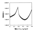
次に、酢酸緩衝液中に銀イオン(Ag+)の濃度が1ng/mLとなるように調整し、還元堆積電位-0.3V、堆積時間240秒に設定して、溶液中の銀イオンを電極表面へ堆積した。続いて、周波数40Hz、電位増加分2mV、振幅25mVに設定して、矩形波ボルタンメトリー測定を行った。測定結果を図2に示した。図2において、横軸は印加電圧、縦軸は銀の酸化溶出に伴う酸化電流である。得られた測定結果より、堆積した銀に由来する酸化電流値が0.19V付近に明確に観測された。この時得られた電流値は42nA(あるいは電流密度で1.3μA/cm2)であり、本実施例で使用したスパッタカーボン薄膜電極で得られる銀の検出限界である。
実施例2:爪白癬菌抗原の濃度測定
ストレプトアビジンが修飾された磁気微粒子(直径37~100μm、GE healthcare製)をよく分散させ、マイクロチューブに10μL秤り取り、磁気微粒子表面をTris緩衝液生理食塩水(TBS)で洗浄・磁気分離した後、ビオチン標識した抗爪白癬菌抗体(TBS溶液、0.04mg/mL)を30分作用させた。その後、未結合のビオチン標識抗体を除去するため磁気微粒子表面をTBSで洗浄・磁気分離し、抗爪白癬菌抗体固定化磁気微粒子を得た。ここに濃度が異なる爪白癬菌抗原を300μL滴下し、室温で60分間振盪し抗原を吸着させた後、未結合の抗原を除去するため磁気微粒子表面を所定の洗浄液(リン酸緩衝液生理食塩水(PBS)+0.05%Tween20+1%BlockAce)で洗浄・磁気分離した。続いてビオチン標識した抗爪白癬菌抗体(PBS+0.05%Tween20+1%BLockAce、0.04mg/mL)を再度60分間接触させ磁気微粒子上の爪白癬菌抗原にビオチン標識抗体を捕捉させた後、未結合のビオチン標識抗体を除去するため磁気微粒子表面を所定の洗浄液(PBS+0.05%Tween20+1%BLockAce)で洗浄・磁気分離した。次にこの抗体にストレプトアビジン標識されたHRP(PBS+1%BlockAce,1.25ng/mL)を30分作用させ、抗体にHRP修飾を行った。こうして、抗爪白癬菌抗体を介して磁気微粒子に固定化された爪白癬菌抗原とHRP修飾された抗爪白癬菌抗体との複合体を得た。磁気微粒子表面を洗浄液(PBS+0.05%Tween20+1%BlockAce)で洗浄・磁気分離し、抗爪白癬菌抗体に結合していないHRPを除去した。ここでさらに塩素イオンフリーのリン酸緩衝液(濃度50mmol/L、pH7.0)で洗浄・磁気分離して塩素イオンを除去した。
ここへ30μLの銀イオン、過酸化水素、およびヒドロキノンをクエン酸緩衝液中に含む試薬(EnzMet、Nanoprobe社製)を添加し、よく懸濁させ、すぐさま磁気微粒子ごと懸濁液をスパッタカーボン電極表面上へ滴下し、15分間酵素メタリゼーション反応を行い、銀を生成させた。反応後電極表面を塩素イオンフリーのリン酸緩衝液で洗浄し、磁気微粒子などを除去し、銀の堆積した作用電極を得た。スパッタカーボン電極としては、実施例1で用いたスパッタカーボン電極1を用いた。
ストレプトアビジンが修飾された磁気微粒子(直径37~100μm、GE healthcare製)をよく分散させ、マイクロチューブに10μL秤り取り、磁気微粒子表面をTris緩衝液生理食塩水(TBS)で洗浄・磁気分離した後、ビオチン標識した抗爪白癬菌抗体(TBS溶液、0.04mg/mL)を30分作用させた。その後、未結合のビオチン標識抗体を除去するため磁気微粒子表面をTBSで洗浄・磁気分離し、抗爪白癬菌抗体固定化磁気微粒子を得た。ここに濃度が異なる爪白癬菌抗原を300μL滴下し、室温で60分間振盪し抗原を吸着させた後、未結合の抗原を除去するため磁気微粒子表面を所定の洗浄液(リン酸緩衝液生理食塩水(PBS)+0.05%Tween20+1%BlockAce)で洗浄・磁気分離した。続いてビオチン標識した抗爪白癬菌抗体(PBS+0.05%Tween20+1%BLockAce、0.04mg/mL)を再度60分間接触させ磁気微粒子上の爪白癬菌抗原にビオチン標識抗体を捕捉させた後、未結合のビオチン標識抗体を除去するため磁気微粒子表面を所定の洗浄液(PBS+0.05%Tween20+1%BLockAce)で洗浄・磁気分離した。次にこの抗体にストレプトアビジン標識されたHRP(PBS+1%BlockAce,1.25ng/mL)を30分作用させ、抗体にHRP修飾を行った。こうして、抗爪白癬菌抗体を介して磁気微粒子に固定化された爪白癬菌抗原とHRP修飾された抗爪白癬菌抗体との複合体を得た。磁気微粒子表面を洗浄液(PBS+0.05%Tween20+1%BlockAce)で洗浄・磁気分離し、抗爪白癬菌抗体に結合していないHRPを除去した。ここでさらに塩素イオンフリーのリン酸緩衝液(濃度50mmol/L、pH7.0)で洗浄・磁気分離して塩素イオンを除去した。
ここへ30μLの銀イオン、過酸化水素、およびヒドロキノンをクエン酸緩衝液中に含む試薬(EnzMet、Nanoprobe社製)を添加し、よく懸濁させ、すぐさま磁気微粒子ごと懸濁液をスパッタカーボン電極表面上へ滴下し、15分間酵素メタリゼーション反応を行い、銀を生成させた。反応後電極表面を塩素イオンフリーのリン酸緩衝液で洗浄し、磁気微粒子などを除去し、銀の堆積した作用電極を得た。スパッタカーボン電極としては、実施例1で用いたスパッタカーボン電極1を用いた。
上述の作用電極を50mmol/Lの酢酸緩衝液(pH5.0)中に挿入し、ポテンシオスタットに塩橋を施した参照電極(銀-塩化銀:Ag/AgCl)、対向電極(白金:Pt)とともに接続し、周波数40Hz、電位増加分2mV、振幅25mVに設定して、各濃度の抗原を作用させて得られた作用電極の矩形波ボルタンメトリー測定を行った。測定結果を図3に示した。図3(a)において、横軸は印加電圧、縦軸は抗原濃度が5pg/mlの場合の応答電流である。得られた測定結果より、0.25V付近に酸化電流値の増加が観測された。この結果は図2の銀の還元堆積による銀の酸化溶出の結果と同じ傾向を示している。従って、本実施例による応答電流は、図1A~Cに示した通り、還元堆積の代わりに、酵素メタリゼーション反応によって銀を生成、作用極上に堆積させる原理を実証するものである。また、図3(b)において、横軸は抗原濃度、縦軸は応答電流値である。図3(b)から明らかなように、抗原濃度に依存した電流増加が認められた。爪白癬菌抗原を対象とした場合には、およそ5pg/mLを検出限界濃度として測定することが可能と見積もられた。爪白癬菌抗原のイムノクロマト法による検出限界は概ね50ng/mLであり、1/10000低い濃度まで検出できることが明らかとなった。
図3(b)を検量線として使用し、未知の抗原濃度を含む試料に対して同一の酵素反応条件のもとで堆積した銀の酸化電流値を測定することで、試料中の抗原濃度を知ることが可能となる。
図3(b)を検量線として使用し、未知の抗原濃度を含む試料に対して同一の酵素反応条件のもとで堆積した銀の酸化電流値を測定することで、試料中の抗原濃度を知ることが可能となる。
実施例3:ヒトインターロイキン-6(IL-6)の濃度測定
ストレプトアビジンが修飾された磁気微粒子(直径37~100μm、GE healthcare製)をよく分散させ、マイクロチューブに10μL秤り取り、磁気微粒子表面をTris緩衝液生理食塩水(TBS)で洗浄・磁気分離した後、ビオチン標識された抗IL-6抗体(TBS、0.01mg/mL)を30分作用させた。その後、未結合のビオチン標識抗体を除去するため磁気微粒子表面をTBSで洗浄・磁気分離し、ビオチン標識抗IL-6抗体固定化磁気微粒子を得た。ここにそれぞれ濃度が異なるIL-6を300μL滴下し、室温で60分間振盪しIL-6を吸着させた後、未結合のIL-6を除去するため磁気微粒子表面を所定の洗浄液(TBS+0.05%Tween20+1%BlockAce)で洗浄・磁気分離した。続いてビオチン標識抗IL-6抗体(TBS+0.05%Tween20+1%BLockAce、0.01mg/mL)を再度180分間接触させ磁気微粒子上のIL-6にビオチン標識抗体を捕捉させた後、未結合のビオチン標識抗体を除去するため磁気微粒子表面を所定の洗浄液(PBS+0.05%Tween20+1%BLockAce)で洗浄・磁気分離した。次にこの抗体にストレプトアビジン標識されたHRP(PBS+1%BlockAce,1.25ng/mL)を30分作用させ、抗体にHRP修飾を行った。磁気微粒子表面を洗浄液(PBS+0.05%Tween20+1%BlockAce)で洗浄し、磁気分離し、ビオチン標識抗体に結合していないHRPを除去した。ここでさらに塩素イオンフリーのリン酸緩衝液(濃度50mmol/L、pH7.0)で洗浄・磁気分離して塩素イオンを除去した。
ここへ30μLの量の銀イオン、過酸化水素、およびヒドロキノンをクエン酸緩衝液中に含む試薬(EnzMet、Nanoprobe社製)を添加し、よく懸濁させ、すぐさま磁気微粒子ごと懸濁液をスパッタカーボン電極表面上へ滴下し、15分間酵素メタリゼーション反応を行い、銀を生成させた。反応後電極表面を洗浄し、磁気微粒子などを除去し、銀の堆積した作用電極を得た。スパッタカーボン電極としては、実施例1で用いたスパッタカーボン電極1を用いた。
ストレプトアビジンが修飾された磁気微粒子(直径37~100μm、GE healthcare製)をよく分散させ、マイクロチューブに10μL秤り取り、磁気微粒子表面をTris緩衝液生理食塩水(TBS)で洗浄・磁気分離した後、ビオチン標識された抗IL-6抗体(TBS、0.01mg/mL)を30分作用させた。その後、未結合のビオチン標識抗体を除去するため磁気微粒子表面をTBSで洗浄・磁気分離し、ビオチン標識抗IL-6抗体固定化磁気微粒子を得た。ここにそれぞれ濃度が異なるIL-6を300μL滴下し、室温で60分間振盪しIL-6を吸着させた後、未結合のIL-6を除去するため磁気微粒子表面を所定の洗浄液(TBS+0.05%Tween20+1%BlockAce)で洗浄・磁気分離した。続いてビオチン標識抗IL-6抗体(TBS+0.05%Tween20+1%BLockAce、0.01mg/mL)を再度180分間接触させ磁気微粒子上のIL-6にビオチン標識抗体を捕捉させた後、未結合のビオチン標識抗体を除去するため磁気微粒子表面を所定の洗浄液(PBS+0.05%Tween20+1%BLockAce)で洗浄・磁気分離した。次にこの抗体にストレプトアビジン標識されたHRP(PBS+1%BlockAce,1.25ng/mL)を30分作用させ、抗体にHRP修飾を行った。磁気微粒子表面を洗浄液(PBS+0.05%Tween20+1%BlockAce)で洗浄し、磁気分離し、ビオチン標識抗体に結合していないHRPを除去した。ここでさらに塩素イオンフリーのリン酸緩衝液(濃度50mmol/L、pH7.0)で洗浄・磁気分離して塩素イオンを除去した。
ここへ30μLの量の銀イオン、過酸化水素、およびヒドロキノンをクエン酸緩衝液中に含む試薬(EnzMet、Nanoprobe社製)を添加し、よく懸濁させ、すぐさま磁気微粒子ごと懸濁液をスパッタカーボン電極表面上へ滴下し、15分間酵素メタリゼーション反応を行い、銀を生成させた。反応後電極表面を洗浄し、磁気微粒子などを除去し、銀の堆積した作用電極を得た。スパッタカーボン電極としては、実施例1で用いたスパッタカーボン電極1を用いた。
上述の作用電極を50mmol/Lの酢酸緩衝液(pH5.0)中に挿入し、ポテンシオスタットに塩橋を施した参照電極(銀-塩化銀:Ag/AgCl)、対向電極(白金:Pt)とともに接続し、周波数40Hz、電位増加分2mV、振幅25mVに設定して、各濃度の抗原を作用させて得られた作用電極の矩形波ボルタンメトリー測定を行った。測定結果を図4に示した。IL-6を対象とした場合には、およそ10pg/mLを検出限界濃度として測定することが可能であると見積もられた。
本発明の範囲は以上の説明に拘束されることはなく、上記例示以外についても、本発明の趣旨を損なわない範囲で適宜変更し実施し得る。なお、本明細書に記載した全ての文献及び刊行物は、その目的にかかわらず参照によりその全体を本明細書に組み込むものとする。また、本明細書は、本願の優先権主張の基礎となる日本国特許出願である特願2017-145772号(2017年7月27日出願)の特許請求の範囲、明細書の開示内容を包含する。
本発明の方法、キット、およびデバイスは、試料中の微量の生体分子の濃度の測定に好適である。
1 生体分子
2 第一の特異的結合物質
3 電極
4 銀
5 第二の特異的結合物質
6 担体
10 複合体
Ag+ 銀イオン
Ag0 銀
HRP ホースラディッシュペルオキシダーゼ
e 電子
2 第一の特異的結合物質
3 電極
4 銀
5 第二の特異的結合物質
6 担体
10 複合体
Ag+ 銀イオン
Ag0 銀
HRP ホースラディッシュペルオキシダーゼ
e 電子
Claims (13)
- 試料中の生体分子濃度を測定する方法であって、
(a)生体分子と当該生体分子に対する特異的結合物質であってホースラディッシュペルオキシダーゼ(HRP)修飾された特異的結合物質との複合体に、銀イオンを作用させてHRPと銀イオンとの酵素反応を行う工程、
(b)前記酵素反応により銀を生成させるとともに当該生成された銀を電極上に堆積させる工程、および
(c)前記堆積した銀における銀イオン量を電気化学的に測定することにより、前記生体分子濃度を測定する工程
を含む、前記方法。 - 前記複合体は、生体分子に対する第二の特異的結合物質を介して担体に固定化されている、請求項1に記載の方法。
- 生体分子の検出限界濃度が1pg/mLから10pg/mLの範囲である、請求項1または2に記載の方法。
- 前記電極の平均表面粗さ(Ra)が0.05~5nmである、請求項1~3のいずれか一項に記載の方法。
- 前記電極はナノカーボン電極である、請求項1~4のいずれか一項に記載の方法。
- 工程(c)において反応系における銀イオンの濃度が1~100ng/mLである、請求項1~5のいずれか一項に記載の方法。
- 前記電極のサイズが、1~100mm2である、請求項1~6のいずれか一項に記載の方法。
- 工程(a)における酵素反応は、酸化剤および還元剤の存在下で行われる、請求項1~7のいずれか一項に記載の方法。
- 前記酸化剤は、過酸化水素であり、前記還元剤は、鉄イオン(Fe2+)およびヒドロキノンから選択される少なくとも一つである、請求項8に記載の方法。
- 工程(a)の酵素反応は、pH3.0~7.0の緩衝液中で行われ、工程(c)における電気化学的測定は、pH4.0~5.0の緩衝液中で行われる、請求項1~9のいずれか一項に記載の方法。
- 以下の工程、
(1)生体分子に対する特異的結合物質を固定化した担体に試料を接触させ、試料中の生体分子を担体に捕捉する工程、
(2)特異的結合物質を担体に再度接触させ、担体上の生体分子に特異的結合物質を捕捉する工程、
(3)特異的結合物質にホースラディッシュペルオキシダーゼ(HRP)を修飾する工程、
(4)銀イオンを含む試薬を担体上のHRPと酵素メタリゼーション反応をさせ、銀を生成させる工程、および
(5)生成した銀を電極上に堆積させ、この堆積した銀イオン量を電気化学的に測定する工程、
を含み、銀イオン量を指標として試料中の生体分子濃度を測定することを特徴とする、請求項1~10のいずれか一項に記載の方法。 - 請求項1~11のいずれか一項に記載の方法を実施するためのキットであって、生体分子に対する特異的結合物質を固定化した担体と、HRPと、銀イオンを含み、HRPと酵素メタリゼーション反応を実施可能な試薬と、を含むことを特徴とする生体分子濃度測定キット。
- 請求項1~11のいずれか一項に記載の方法を実施するためのデバイスであって、生体分子に対する特異的結合物質を固定化した担体および電極を備えた電気化学セルと、電極への電圧印加手段と、電流測定手段と、を含むことを特徴とする生体分子濃度測定デバイス。
Applications Claiming Priority (2)
| Application Number | Priority Date | Filing Date | Title |
|---|---|---|---|
| JP2017145772 | 2017-07-27 | ||
| JP2017-145772 | 2017-07-27 |
Publications (1)
| Publication Number | Publication Date |
|---|---|
| WO2019022142A1 true WO2019022142A1 (ja) | 2019-01-31 |
Family
ID=65040870
Family Applications (1)
| Application Number | Title | Priority Date | Filing Date |
|---|---|---|---|
| PCT/JP2018/027908 Ceased WO2019022142A1 (ja) | 2017-07-27 | 2018-07-25 | 生体分子濃度の電気化学的測定方法 |
Country Status (2)
| Country | Link |
|---|---|
| JP (1) | JP2019028071A (ja) |
| WO (1) | WO2019022142A1 (ja) |
Families Citing this family (2)
| Publication number | Priority date | Publication date | Assignee | Title |
|---|---|---|---|---|
| JP7666616B2 (ja) * | 2021-09-16 | 2025-04-22 | 株式会社デンソー | イオンセンサ、及び被検出物質の検出方法 |
| JP7699846B2 (ja) * | 2023-06-29 | 2025-06-30 | 株式会社イムノセンス | 電気化学的手法を用いた被検物質の測定方法 |
Citations (2)
| Publication number | Priority date | Publication date | Assignee | Title |
|---|---|---|---|---|
| JP2008298654A (ja) * | 2007-05-30 | 2008-12-11 | Ventana Medical Systems Inc | バイオマーカーの多色発色検出 |
| WO2009116534A1 (ja) * | 2008-03-17 | 2009-09-24 | 三菱化学メディエンス株式会社 | 電気的分析方法 |
-
2018
- 2018-07-25 JP JP2018139199A patent/JP2019028071A/ja active Pending
- 2018-07-25 WO PCT/JP2018/027908 patent/WO2019022142A1/ja not_active Ceased
Patent Citations (2)
| Publication number | Priority date | Publication date | Assignee | Title |
|---|---|---|---|---|
| JP2008298654A (ja) * | 2007-05-30 | 2008-12-11 | Ventana Medical Systems Inc | バイオマーカーの多色発色検出 |
| WO2009116534A1 (ja) * | 2008-03-17 | 2009-09-24 | 三菱化学メディエンス株式会社 | 電気的分析方法 |
Non-Patent Citations (2)
| Title |
|---|
| HWANG, S. ET AL.: "Electrochemical Detection of DNA Hybridization Using Biometallization", ANALYTICAL CHEMISTRY, vol. 77, no. 2, 2005, pages 579 - 584, XP055567057 * |
| WEIZMANN, Y. ET AL.: "DNA-CNT Nanowire Networks for DNA Detection", JOURNAL OF THE AMERICAN CHEMICAL SOCIETY, vol. 133, 2011, pages 3238 - 3241, XP055567059 * |
Also Published As
| Publication number | Publication date |
|---|---|
| JP2019028071A (ja) | 2019-02-21 |
Similar Documents
| Publication | Publication Date | Title |
|---|---|---|
| Wu et al. | Enzyme-functionalized silica nanoparticles as sensitive labels in biosensing | |
| Luo et al. | Electrical biosensors and the label free detection of protein disease biomarkers | |
| Hao et al. | Modulating the linker immobilization density on aptameric graphene field effect transistors using an electric field | |
| Wang et al. | Dual amplification strategy for the fabrication of highly sensitive interleukin-6 amperometric immunosensor based on poly-dopamine | |
| Jia et al. | Triple signal amplification using gold nanoparticles, bienzyme and platinum nanoparticles functionalized graphene as enhancers for simultaneous multiple electrochemical immunoassay | |
| Wang et al. | Platinum porous nanoparticles hybrid with metal ions as probes for simultaneous detection of multiplex cancer biomarkers | |
| Liu et al. | Nanomaterial labels in electrochemical immunosensors and immunoassays | |
| Akter et al. | Amplified electrochemical detection of a cancer biomarker by enhanced precipitation using horseradish peroxidase attached on carbon nanotubes | |
| Lai et al. | Ultrasensitive multiplexed immunoassay with electrochemical stripping analysis of silver nanoparticles catalytically deposited by gold nanoparticles and enzymatic reaction | |
| Yu et al. | Carbon nanotube amplification strategies for highly sensitive immunodetection of cancer biomarkers | |
| Zong et al. | Chemiluminescence imaging immunoassay of multiple tumor markers for cancer screening | |
| Lai et al. | Ultrasensitive immunoassay based on electrochemical measurement of enzymatically produced polyaniline | |
| Chen et al. | Successively amplified electrochemical immunoassay based on biocatalytic deposition of silver nanoparticles and silver enhancement | |
| Qu et al. | A novel electrochemical immunosensor based on colabeled silica nanoparticles for determination of total prostate specific antigen in human serum | |
| Sari et al. | The optimization of an electrochemical aptasensor to detect RBD protein S SARS-CoV-2 as a biomarker of COVID-19 using screen-printed carbon electrode/AuNP | |
| Lee et al. | Nanoparticle-enabled multiplexed electrochemical immunoassay for detection of surface proteins on extracellular vesicles | |
| Tang et al. | Nanopore-based single-entity electrochemistry for the label-free monitoring of single-molecule glycoprotein–boronate affinity interaction and its sensing application | |
| Xu et al. | Ultrasensitive electrochemical sensing of Hg2+ based on thymine-Hg2+-thymine interaction and signal amplification of alkaline phosphatase catalyzed silver deposition | |
| Li et al. | A new electrochemical immunosensor for sensitive detection of prion based on Prussian blue analogue | |
| Miao et al. | Voltammetric determination of tumor necrosis factor-α based on the use of an aptamer and magnetic nanoparticles loaded with gold nanoparticles | |
| Han et al. | Electrochemical signal amplification for immunosensor based on 3D interdigitated array electrodes | |
| Madhu et al. | Sensitive electrochemical sensing platform based on Au nanoflower-integrated carbon fiber for detecting interleukin-6 in human serum | |
| Roushani et al. | Layer-by-layer assembly of gold nanoparticles and cysteamine on gold electrode for immunosensing of human chorionic gonadotropin at picogram levels | |
| Wang et al. | Applications of electrochemical biosensors based on functional antibody-modified screen-printed electrodes: a review | |
| Lai et al. | Amplified inhibition of the electrochemical signal of ferrocene by enzyme-functionalized graphene oxide nanoprobe for ultrasensitive immunoassay |
Legal Events
| Date | Code | Title | Description |
|---|---|---|---|
| 121 | Ep: the epo has been informed by wipo that ep was designated in this application |
Ref document number: 18839480 Country of ref document: EP Kind code of ref document: A1 |
|
| NENP | Non-entry into the national phase |
Ref country code: DE |
|
| 122 | Ep: pct application non-entry in european phase |
Ref document number: 18839480 Country of ref document: EP Kind code of ref document: A1 |