WO2018225403A1 - 細胞培養方法、細胞評価方法、および細胞培養装置 - Google Patents
細胞培養方法、細胞評価方法、および細胞培養装置 Download PDFInfo
- Publication number
- WO2018225403A1 WO2018225403A1 PCT/JP2018/016333 JP2018016333W WO2018225403A1 WO 2018225403 A1 WO2018225403 A1 WO 2018225403A1 JP 2018016333 W JP2018016333 W JP 2018016333W WO 2018225403 A1 WO2018225403 A1 WO 2018225403A1
- Authority
- WO
- WIPO (PCT)
- Prior art keywords
- cells
- temperature
- cell
- infrared
- infrared rays
- Prior art date
- Legal status (The legal status is an assumption and is not a legal conclusion. Google has not performed a legal analysis and makes no representation as to the accuracy of the status listed.)
- Ceased
Links
Images
Classifications
-
- C—CHEMISTRY; METALLURGY
- C12—BIOCHEMISTRY; BEER; SPIRITS; WINE; VINEGAR; MICROBIOLOGY; ENZYMOLOGY; MUTATION OR GENETIC ENGINEERING
- C12M—APPARATUS FOR ENZYMOLOGY OR MICROBIOLOGY; APPARATUS FOR CULTURING MICROORGANISMS FOR PRODUCING BIOMASS, FOR GROWING CELLS OR FOR OBTAINING FERMENTATION OR METABOLIC PRODUCTS, i.e. BIOREACTORS OR FERMENTERS
- C12M1/00—Apparatus for enzymology or microbiology
- C12M1/42—Apparatus for the treatment of microorganisms or enzymes with electrical or wave energy, e.g. magnetism, sonic waves
-
- C—CHEMISTRY; METALLURGY
- C12—BIOCHEMISTRY; BEER; SPIRITS; WINE; VINEGAR; MICROBIOLOGY; ENZYMOLOGY; MUTATION OR GENETIC ENGINEERING
- C12M—APPARATUS FOR ENZYMOLOGY OR MICROBIOLOGY; APPARATUS FOR CULTURING MICROORGANISMS FOR PRODUCING BIOMASS, FOR GROWING CELLS OR FOR OBTAINING FERMENTATION OR METABOLIC PRODUCTS, i.e. BIOREACTORS OR FERMENTERS
- C12M3/00—Tissue, human, animal or plant cell, or virus culture apparatus
-
- C—CHEMISTRY; METALLURGY
- C12—BIOCHEMISTRY; BEER; SPIRITS; WINE; VINEGAR; MICROBIOLOGY; ENZYMOLOGY; MUTATION OR GENETIC ENGINEERING
- C12N—MICROORGANISMS OR ENZYMES; COMPOSITIONS THEREOF; PROPAGATING, PRESERVING, OR MAINTAINING MICROORGANISMS; MUTATION OR GENETIC ENGINEERING; CULTURE MEDIA
- C12N13/00—Treatment of microorganisms or enzymes with electrical or wave energy, e.g. magnetism, sonic waves
-
- C—CHEMISTRY; METALLURGY
- C12—BIOCHEMISTRY; BEER; SPIRITS; WINE; VINEGAR; MICROBIOLOGY; ENZYMOLOGY; MUTATION OR GENETIC ENGINEERING
- C12N—MICROORGANISMS OR ENZYMES; COMPOSITIONS THEREOF; PROPAGATING, PRESERVING, OR MAINTAINING MICROORGANISMS; MUTATION OR GENETIC ENGINEERING; CULTURE MEDIA
- C12N15/00—Mutation or genetic engineering; DNA or RNA concerning genetic engineering, vectors, e.g. plasmids, or their isolation, preparation or purification; Use of hosts therefor
- C12N15/09—Recombinant DNA-technology
-
- C—CHEMISTRY; METALLURGY
- C12—BIOCHEMISTRY; BEER; SPIRITS; WINE; VINEGAR; MICROBIOLOGY; ENZYMOLOGY; MUTATION OR GENETIC ENGINEERING
- C12Q—MEASURING OR TESTING PROCESSES INVOLVING ENZYMES, NUCLEIC ACIDS OR MICROORGANISMS; COMPOSITIONS OR TEST PAPERS THEREFOR; PROCESSES OF PREPARING SUCH COMPOSITIONS; CONDITION-RESPONSIVE CONTROL IN MICROBIOLOGICAL OR ENZYMOLOGICAL PROCESSES
- C12Q1/00—Measuring or testing processes involving enzymes, nucleic acids or microorganisms; Compositions therefor; Processes of preparing such compositions
- C12Q1/02—Measuring or testing processes involving enzymes, nucleic acids or microorganisms; Compositions therefor; Processes of preparing such compositions involving viable microorganisms
-
- C—CHEMISTRY; METALLURGY
- C12—BIOCHEMISTRY; BEER; SPIRITS; WINE; VINEGAR; MICROBIOLOGY; ENZYMOLOGY; MUTATION OR GENETIC ENGINEERING
- C12Q—MEASURING OR TESTING PROCESSES INVOLVING ENZYMES, NUCLEIC ACIDS OR MICROORGANISMS; COMPOSITIONS OR TEST PAPERS THEREFOR; PROCESSES OF PREPARING SUCH COMPOSITIONS; CONDITION-RESPONSIVE CONTROL IN MICROBIOLOGICAL OR ENZYMOLOGICAL PROCESSES
- C12Q1/00—Measuring or testing processes involving enzymes, nucleic acids or microorganisms; Compositions therefor; Processes of preparing such compositions
- C12Q1/68—Measuring or testing processes involving enzymes, nucleic acids or microorganisms; Compositions therefor; Processes of preparing such compositions involving nucleic acids
- C12Q1/6806—Preparing nucleic acids for analysis, e.g. for polymerase chain reaction [PCR] assay
Definitions
- the present invention relates to a cell culture method, a cell evaluation method, and a cell culture apparatus.
- Sunlight includes ultraviolet rays (UV), visible rays (VIS), and infrared rays (IR), and many studies have been made on the influence of ultraviolet rays on the skin. In recent years, attention has also been focused on the influence of infrared rays on the skin.
- UV ultraviolet rays
- VIS visible rays
- IR infrared rays
- Japanese Patent Laid-Open No. 2002-10778 discloses a technique for culturing cells while irradiating the cells with far infrared rays using a far infrared emitter. The effect of such infrared irradiation on cells can be evaluated by observing changes in cells irradiated with infrared rays.
- An object of the present invention is to provide a cell culture method, a cell evaluation method, and a cell culture device that can accurately evaluate the influence of infrared rays on cells.
- one embodiment of the present invention is a cell culture method characterized by irradiating the cells with infrared rays while maintaining the temperature of the cells.
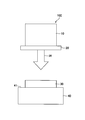
- FIG. 1 is a schematic diagram of a cell culture device 100 according to the present embodiment.
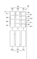
- FIG. 2 is a schematic view of a culture plate constituting a part of the cell culture device according to the present embodiment.
- the cell culture device 100 is a device for culturing cells, and includes a light source 10, an optical filter 20, a culture plate 30, and a temperature control device 40 as shown in FIG.
- the cells cultured in this embodiment are not particularly limited, and may include all cells constituting a living body, and may be differentiated cells or undifferentiated cells. Moreover, the kind of living body is not limited, and any of mammals, birds, fishes, reptiles, and amphibians may be used. In particular, when mammalian cells are used, mouse or human cells are exemplified. For example, when human cells are used, any of fetal, neonatal or adult cells can be used. Specific examples of the cells include, for example, cells constituting the skin (hereinafter also referred to as skin cells) such as fibroblasts, keratinocytes, pigment cells, immune cells, endothelial cells, nerve cells, blood cells, bone marrow. Examples include cells, muscle cells, fat cells, and osteoblasts.
- skin cells such as fibroblasts, keratinocytes, pigment cells, immune cells, endothelial cells, nerve cells, blood cells, bone marrow. Examples include cells, muscle cells, fat cells, and osteo
- the light source 10 has a function of irradiating infrared rays (IR).
- the wavelength of the irradiated infrared ray is, for example, 760 to 1700 nm.
- a light source 10 is not particularly limited, and a light source that generates infrared light such as an infrared lamp, a halogen lamp, or a xenon lamp can be used.
- a xenon lamp that generates light close to sunlight is preferable as the light source 10 from the viewpoint of evaluating the influence of sunlight on the skin.
- Xenon lamps generate not only infrared rays but also ultraviolet rays and visible rays. Therefore, when a xenon lamp is used as the light source 10, as shown in FIG. 1, an optical filter 20 that transmits only infrared light may be set in the light source 10 so that only infrared light is irradiated.
- the optical filter 20 is not particularly limited, and for example, a cut filter that blocks transmission of ultraviolet rays and visible light (hereinafter sometimes referred to as an ultraviolet-visible cut filter) can be used.
- the culture plate 30 is a plate-like member for culturing cells.
- the form of the culture plate 30 is not particularly limited. However, when cells having adhesive ability are used, cells such as collagen are formed on the surface of the culture plate 30 from the viewpoint of improving the fixability of the cells to be cultured (hereinafter referred to as cultured cells).
- cultured cells A culture plate coated with an outer matrix or a culture plate processed with a drug can be used.
- the culture plate 30 is preferably provided with a chamber C as shown in FIG. 2 from the viewpoint that a liquid medium can be used.
- a chamber C can be formed by fitting the slide 31 into the bottom of the well 32 as shown in FIG.
- a plurality of such chambers may be provided from the viewpoint that a plurality of cell samples cultured under the same conditions can be obtained simultaneously (see FIG. 2).
- the mode of providing a plurality of chambers is not limited.
- the plurality of chambers for example, as shown in FIG. 2, slides 31a to 31c are fitted into wells 32a to 32c to form three culture plates, and four chambers are formed in each culture plate.
- a total of 12 chambers C1-C12 can be provided.
- the material of the culture plate 30 is not limited, but is preferably a material having good thermal conductivity from the viewpoint of facilitating cell temperature control.
- a material for example, metal, glass, thermally conductive resin, etc. can be used in addition to plastics used in normal culture.
- the shape of the culture plate is not limited, but from the viewpoint of improving cell temperature control, it is preferable to use a plate-like member having a predetermined thickness, and it is more preferable to use a glass plate.
- the temperature control device 40 includes a placement unit 41 on which the culture plate 30 is placed, and has a function of controlling the surface temperature of the placement unit 41.
- the form of the placement unit 41 is not particularly limited, but the surface of the placement unit 41 is preferably flat so that the controlled surface temperature of the placement unit 41 directly propagates to the culture plate 30.
- control of the surface temperature of the mounting part 41 means that the surface temperature of the mounting part 41 is maintained at a predetermined temperature by heating or cooling.
- a temperature control plate for example, a cool plate or the like having at least a cooling function can be used.
- the temperature control device 40 is arranged so that the culture plate 30 placed on the placement unit 41 is irradiated with infrared rays (IR) with respect to the light source 10.
- the light source 10 is disposed above the temperature control device 40, and infrared rays from the light source 10 are applied to the culture plate 30 placed on the placement unit 41 of the temperature control device 40. (IR) is irradiated.
- the distance between the light source 10 and the temperature control device 40 is preferably such a distance that the cells cultured on the culture plate 30 are irradiated with infrared rays and the surface temperature of the mounting portion 41 can be controlled.
- the distance between the light source 10 and the temperature control device 40 can be set to about 10 cm to 30 cm, for example.
- the cell culture device 100 of the present embodiment it is possible to culture cells while controlling the temperature rise due to infrared irradiation. Therefore, it is possible to distinguish between the influence of infrared rays on cells and the influence of temperature, and it is possible to accurately evaluate the influence of infrared rays on cells.
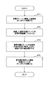
- FIG. 3 is a flowchart showing an example of the cell culture method according to the present embodiment.
- the cells are irradiated with infrared rays while maintaining the temperature of the cells at a predetermined temperature.
- the cell culture apparatus 100 described above can be used for the cell culture method of the present embodiment, the form for carrying out the cell culture method of the present embodiment is not limited to this.
- step ST1 preliminary culture is performed before the cells are irradiated with infrared rays. Specifically, cells are seeded on a culture plate 30 into which a liquid medium has been injected, and cultured at a biologically acceptable temperature.
- the biologically acceptable temperature is preferably about 10 to 50 ° C., for example, when an experiment to reproduce the temperature in the atmospheric environment is performed. Further, when an experiment for reproducing the body temperature of the human body is performed, the temperature is preferably about 30 to 42 ° C. Furthermore, when an experiment for reproducing the normal body temperature of the human body is performed, the temperature is preferably about 36 to 38 ° C. In this embodiment, in step ST1, the cells seeded on the culture plate are cultured at 36 to 38 ° C.
- the culture time is not particularly limited and can be appropriately changed according to the purpose. Moreover, it is preferable to use a human skin cell (for example, fibroblast etc.) as a cultured cell.
- a human skin cell for example, fibroblast etc.
- a chamber slide whose surface is coated with collagen can be used as the culture plate 30.
- a chamber slide is a slide comprising one or more chambers.
- the chamber slide is provided with a plurality of chambers C (chambers C1 to C12) by fitting the slide 31 into the bottom of the well 32. Further, a liquid medium is injected into each chamber C.
- step ST2 the cells cultured in step ST1 are set in the temperature control device 40 (step ST2). Specifically, after the cells are preliminarily cultured on the culture plate 30 in step ST1, the culture medium in the chamber slide of the culture plate 30 is removed, and the culture plate 30 in which a buffer solution is injected into each well of the chamber slide is used. It installs in the mounting part 41 of the temperature control apparatus 40 set to predetermined temperature.
- the above-described temperature control plate can be used as the temperature control device 40.
- the cells in the chamber slide can be controlled to a predetermined temperature even when the temperature varies due to infrared irradiation.
- the predetermined temperature is not particularly limited.
- the liquid temperature in the chamber slide is set to 40 ° C. preferable.
- the temperature rise of infrared rays is not assumed (when the temperature of the skin surface is assumed)
- the liquid temperature in the chamber slide can be set to 33 ° C.
- the temperature control device 40 can control to a temperature set so that the temperature does not increase due to infrared irradiation (hereinafter referred to as a set temperature).
- the method for controlling the set temperature is not particularly limited. For example, the temperature of the chamber slide is measured, the method of adjusting the set temperature based on the measured solution temperature, the temperature of the bottom of the chamber slide is measured, and the measured slide bottom The method of adjusting to preset temperature based on the temperature of this is mentioned.
- step ST3 infrared (IR) irradiation can be performed from the light source 10 as shown in FIG.
- the light source 10 As mentioned above, it is preferable to use the xenon lamp which generate
- the amount of irradiation with infrared rays can be controlled to about 10 to 1200 J / cm 2 depending on the purpose of the experiment.
- solar simulators light source devices that artificially generate artificial sunlight
- UV-visible cut filters Infrared light having a wavelength of about 760 to 1700 nm is measured under conditions using the filter 20
- the irradiation amount of the infrared light is preferably controlled to about 100 to 600 J / cm 2 , more preferably about 240 to 350 J / cm 2. 2 to control.
- the cells irradiated with infrared rays in step ST3 are further cultured in step ST4 while remaining in the culture plate 30. Specifically, after the infrared irradiation is completed, the buffer solution in the culture plate 30 is removed, a liquid medium is newly injected into the chamber slide, and the culture is performed at a biologically acceptable temperature.
- the biologically acceptable temperature is preferably about 10 to 50 ° C., for example, when an experiment to reproduce the temperature in the atmospheric environment is performed.
- the temperature is preferably about 30 to 42 ° C.
- the temperature is preferably about 36 to 38 ° C.
- cultivation time is not specifically limited, According to the objective, it can change suitably. Thereby, a sample for evaluating cultured cells can be prepared.
- the cells are cultured according to the flow from step ST1 to step ST4.
- a cell culture method it is possible to culture cells while controlling the temperature rise due to infrared irradiation. Therefore, it is possible to distinguish between the influence of infrared rays on cells and the influence of temperature, and it is possible to accurately evaluate the influence of infrared rays on cells.
- the cell evaluation method according to the present embodiment is a cell evaluation method for evaluating cells cultured by the above-described cell culture method.
- “evaluating cells” means confirming the influence of infrared rays on the cells.
- the cell evaluation method for confirming the influence of infrared rays is not particularly limited. For example, cell morphology observation, cell migration and adhesion ability evaluation, expression evaluation of specific genes and proteins, enzymes Conventional biological techniques such as activity measurement evaluation can be used.
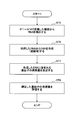
- FIG. 4 is a flowchart showing an example of the cell evaluation method according to the present embodiment.
- RNA is extracted from cells cultured by the cell culture method described above, DNA is synthesized from the RNA, the expression level of the gene contained in the DNA is measured, and the measured gene expression is measured. Evaluate the amount.
- RNA is first extracted from the cells cultured in the above-described steps ST1 to ST4 (step ST5).
- the cells to be evaluated are not particularly limited, but can be cells cultured by the above-described cell culture method.
- the extraction method of RNA is not specifically limited, For example, a commercially available RNA extraction kit can be used.
- DNA is synthesized from the extracted RNA (step ST6).
- extracted RNA is reverse-transcribed to synthesize DNA (cDNA) complementary to RNA.
- cDNA DNA complementary to RNA.
- the method for synthesizing DNA is not particularly limited, and for example, a commercially available reverse transcription reaction kit can be used.
- step ST7 the expression level of the gene contained in the DNA is measured for the DNA synthesized in step ST6.
- the type of gene to be measured is arbitrary, and a gene capable of evaluating the influence of infrared rays on cells may be appropriately selected.
- the method for measuring the expression level is not particularly limited, and various gene expression analysis methods can be used depending on conditions. For example, when analyzing the expression of each isolated gene, techniques such as Northern hybridization and RT-PCR can be used. Moreover, when exhaustively analyzing genes to be expressed, a technique such as microarray can be used. In addition, the scale of the gene expression level is not particularly limited, and a scale corresponding to the measurement method may be used.
- the method includes, for example, Western blotting on the cells cultured in step ST4 of the above-described cell culture method and the culture supernatant thereof.
- a technique such as ELISA (Enzyme-Linked Immuno Sorbent Assay) can be used.
- cells can be comprehensively evaluated by using a technique such as proteome analysis.
- step ST8 the expression level of the gene measured in step ST7 is evaluated. Specifically, the gene expression of a cell is evaluated from the gene expression level.
- the evaluation method is not particularly limited, but the following method can be employed.
- the gene expression level when not irradiated and the gene expression level when irradiated with infrared rays while maintaining the cell temperature at about 33 ° C. are respectively compared. If there is a change in the expression level of each gene, it is evaluated that the cell is affected.
- DMEM Dulbecco's Modification of Eagle's Medium
- FBS fetal bovine serum
- preculture fetal bovine serum
- the chamber slide seeded with the human fibroblasts was preliminarily cultured (hereinafter referred to as preculture) for 3 nights in an environment of 37 ° C. and 5% CO 2 (see step ST1 in FIG. 3).
- the temperature at the bottom of the chamber slide was measured with a digital thermometer, and the measured temperature was taken as the liquid temperature in the chamber slide.
- the liquid temperature in the chamber slide thus measured is regarded as the cell temperature, and the temperature of the cell is controlled by controlling the set temperature of the temperature control plate based on the measured liquid temperature (step of FIG. 3). (See ST2).
- Infrared irradiation For infrared irradiation, a UV-visible cut filter (product name “RG695 filter” manufactured by SCHOTT) was installed in a solar simulator (product name “MODEL91192-1000” manufactured by ORIEL) equipped with a xenon lamp. The wavelength of infrared rays for irradiation was in the range of 760 nm to 1700 nm, and the irradiation time was 60 minutes. The infrared irradiation amount converted under these conditions was about 300 J / cm 2 .
- Infrared irradiation was performed under the following four conditions. (1) Infrared light is not irradiated while the liquid temperature in the chamber slide is maintained at 33 ° C. In addition, 33 degreeC assumes the surface temperature of the human skin in the environment where infrared rays are not irradiated. In addition, an environment in which infrared rays are not irradiated is created by covering the chamber slide with aluminum foil. (2) Irradiate infrared rays while maintaining the temperature of the liquid in the chamber slide at 40 ° C. (3) Do not irradiate infrared rays while maintaining the liquid temperature in the chamber slide at 40 ° C.
- 40 degreeC assumes the surface temperature of the human skin in the environment irradiated with infrared rays. (4) Irradiate infrared rays while maintaining the liquid temperature in the chamber slide at 33 ° C. (see step ST3 in FIG. 3).
- PBS (-) was removed, replaced with 500 ⁇ L of DMEM containing 5% FBS, and cultured for 24 hours in an environment of 37 ° C. and 5% CO 2 (see step ST4 in FIG. 3).
- RNA extraction kit trade name “RNeasy Mini kit”, manufactured by QIAGEN
- gene expression was evaluated by the microarray method using the extracted RNA (see steps ST6 to ST8 in FIG. 4).
- the microarray method is one of gene expression analysis methods and is a method for comprehensively analyzing gene information.
- a plurality of DNA probes cDNA or oligonucleotide
- a target synthesized by reverse transcription of RNA extracted from a cell which is fluorescently labeled with cDNA or cRNA
- hybridization a target synthesized by reverse transcription of RNA extracted from a cell
- DNA which is fluorescently labeled with cDNA or cRNA
- labeled cDNA was synthesized from the extracted total RNA using a labeling kit (Low Input Quick Amp Labeling Kit, manufactured by Agilent).
- the synthesized cDNA was hybridized using a hybridization kit (Gene Expression Hybridization Kit, manufactured by Agilent), and then gene expression data was read using a microarray scanner manufactured by Agilent.
- the gene expression level under the above-mentioned infrared irradiation conditions (1) to (4) is measured, and the infrared irradiation conditions (2) to (4) with respect to the gene expression level under the infrared irradiation conditions (1)
- the gene expression level ratio (hereinafter referred to as expression level ratio) was determined.
- the gene expression of the cells was evaluated by analyzing the gene expression change from the obtained expression level ratio.
Landscapes
- Chemical & Material Sciences (AREA)
- Life Sciences & Earth Sciences (AREA)
- Health & Medical Sciences (AREA)
- Engineering & Computer Science (AREA)
- Organic Chemistry (AREA)
- Wood Science & Technology (AREA)
- Zoology (AREA)
- Bioinformatics & Cheminformatics (AREA)
- Genetics & Genomics (AREA)
- Biotechnology (AREA)
- General Engineering & Computer Science (AREA)
- Biochemistry (AREA)
- Biomedical Technology (AREA)
- General Health & Medical Sciences (AREA)
- Microbiology (AREA)
- Proteomics, Peptides & Aminoacids (AREA)
- Molecular Biology (AREA)
- Analytical Chemistry (AREA)
- Physics & Mathematics (AREA)
- Biophysics (AREA)
- Sustainable Development (AREA)
- Immunology (AREA)
- Cell Biology (AREA)
- Plant Pathology (AREA)
- Virology (AREA)
- Chemical Kinetics & Catalysis (AREA)
- Medicinal Chemistry (AREA)
- Measuring Or Testing Involving Enzymes Or Micro-Organisms (AREA)
- Apparatus Associated With Microorganisms And Enzymes (AREA)
- Micro-Organisms Or Cultivation Processes Thereof (AREA)
Abstract
細胞の温度を維持しながら、前記細胞に赤外線を照射することを特徴とする、細胞培養方法。
Description
本発明は、細胞培養方法、細胞評価方法、および細胞培養装置に関する。
従来から、太陽光による皮膚への影響に関して、様々な研究が行われている。太陽光には、紫外線(UV)、可視光線(VIS)、赤外線(IR)が含まれており、このうち紫外線の皮膚への影響に関する研究が従来から数多くなされている。また、近年では赤外線の皮膚への影響についても注目が集まっている。
例えば、特開2002-10778号公報には、遠赤外線放射体により遠赤外線を細胞に照射しながら、細胞を培養する技術が開示されている。このような赤外線照射の細胞に対する影響は、赤外線が照射された細胞の変化を観察すること等により評価することができる。
しかしながら、細胞に赤外線が照射されると細胞の温度が上昇するため、細胞の変化が、赤外線によるものなのか、温度によるものなのかを区別することが困難である。そのため、従来の技術では、赤外線の細胞に対する影響を正確に評価することができない。
本発明の目的は、赤外線による細胞への影響を正確に評価することができる細胞培養方法、細胞評価方法、および細胞培養装置を提供することにある。
上記課題を解決するために、本発明の一形態は、細胞の温度を維持しながら、前記細胞に赤外線を照射することを特徴とする、細胞培養方法である。
本発明の一形態によれば、赤外線による細胞への影響を正確に評価することができる。
以下、本発明の実施の形態について、図面を参照しながら説明する。なお、各図面において、特に説明がない限り、同一の又は対応する構成については同一の符号を付して説明を省略する場合がある。
<細胞培養装置>
図1は、本実施形態に係る細胞培養装置100の概略図である。図2は、本実施形態に係る細胞培養装置の一部を構成する培養プレートの概略図である。細胞培養装置100は、細胞を培養する装置であり、図1に示すように、光源10、光学フィルタ20、培養プレート30、温度制御装置40を有する。
図1は、本実施形態に係る細胞培養装置100の概略図である。図2は、本実施形態に係る細胞培養装置の一部を構成する培養プレートの概略図である。細胞培養装置100は、細胞を培養する装置であり、図1に示すように、光源10、光学フィルタ20、培養プレート30、温度制御装置40を有する。
本実施形態で培養される細胞は、特に限定されず、生体を構成する全ての細胞を含み、分化した細胞でも、未分化の細胞でもよい。また、生体の種類は限定されず、哺乳類、鳥類、魚類、爬虫類、両生類の何れでもよい。中でも、哺乳類の細胞を用いる場合は、マウス又はヒトの細胞が挙げられる。例えば、ヒトの細胞を用いる場合、胎児、新生児又は成人の何れの細胞を用いることができる。細胞の具体例としては、例えば、線維芽細胞、角化細胞、色素細胞、免疫細胞、内皮細胞等の皮膚を構成する細胞(以下、皮膚細胞という場合がある)、神経細胞、血球細胞、骨髄細胞、筋肉細胞、脂肪細胞、骨芽細胞などが挙げられる。
光源10は、赤外線(IR)を照射する機能を有する。照射される赤外線の波長は、例えば760~1700nmである。このような光源10は、特に限定されず、赤外線ランプ、ハロゲンランプ、キセノンランプ等の赤外線を発生する光源を用いることができる。特に、太陽光が皮膚に与える影響等を評価する観点から、太陽光に近い光を発生させるキセノンランプが光源10として好ましい。
なお、キセノンランプは、赤外線のほか、紫外線、可視光線も発生する。そのため、光源10としてキセノンランプを用いる場合は、図1に示すように、赤外線のみ透過させる光学フィルタ20を光源10にセットして、赤外線のみを照射するようにしてもよい。このような光学フィルタ20としては、特に限定されないが、例えば、紫外線および可視光線の透過を遮断するカットフィルタ(以下、紫外可視カットフィルタという場合がある)を用いることができる。
培養プレート30は、細胞を培養する板状部材である。培養プレート30の形態は、特に限定されないが、接着能を有する細胞を用いる場合は、培養する細胞(以下、培養細胞という)の定着性を高める観点から、培養プレート30の表面にコラーゲン等の細胞外マトリックスがコーティングされた培養プレートや薬剤加工がされた培養プレートを用いることができる。
また、培養プレート30は、液体の培地を使用できる観点から、図2に示すように、チャンバーCを備えるものが好ましい。このようなチャンバーCは、図2に示すように、スライド31をウエル32の底部に嵌め込むことにより形成することができる。また、同じ条件で培養した細胞の試料を同時に複数得られる観点から、このようなチャンバーを複数設けても良い(図2参照)。
複数のチャンバーを設ける態様は、限定されない。複数のチャンバーとしては、例えば、図2に示すように、ウエル32a~32cにスライド31a~31cを嵌め込んで、3つの培養プレートを形成し、各培養プレートに4つチャンバーを構成することにより、合計12つのチャンバーC1~C12を設けることができる。
また、培養プレート30の材質は限定されないが、細胞の温度制御を容易にする観点から、熱伝導性が良好な材質であることが好ましい。このような材質としては、例えば、通常の培養で使用されるプラスチックの他、金属、ガラス、熱伝導性樹脂等を用いることができる。また、培養プレートの形状は、限定されないが、細胞の温度制御を向上させる観点から、所定の厚みを有する板状部材を用いるのが好ましく、中でもガラスプレートを用いるのがより好ましい。
温度制御装置40は、培養プレート30を載置する載置部41を備え、該載置部41の表面温度を制御する機能を有する。載置部41の形態は、特に限定されないが、制御された載置部41の表面温度が培養プレート30に直接伝播するように、載置部41の表面は、平坦であることが好ましい。
また、載置部41の表面温度の制御は、加熱または冷却により載置部41の表面温度を所定の温度に維持することを意味する。このような温度制御により、例えば、赤外線照射により細胞の温度が上昇する環境でも、赤外線照射による細胞の温度上昇を抑制することができる。温度制御装置40としては、例えば、冷却機能を少なくとも備える温度制御プレート(例えば、クールプレート等)を用いることができる。
温度制御装置40は、図1に示すように、光源10に対して、載置部41に載置された培養プレート30に赤外線(IR)が照射されるように配置されている。本実施形態では、図1に示すように、光源10が温度制御装置40の上方に配置され、温度制御装置40の載置部41に載置された培養プレート30に対して光源10からの赤外線(IR)が照射される位置関係になっている。
また、光源10と温度制御装置40との距離は、培養プレート30で培養される細胞に赤外線が照射され、かつ載置部41の表面温度が制御できる距離であることが好ましい。このような光源10と温度制御装置40との距離は、例えば約10cm~30cmとすることができる。
本実施形態の細胞培養装置100を用いることにより、赤外線照射による温度上昇を制御しながら細胞を培養することができる。そのため、細胞に対する赤外線の影響と温度の影響とを区別することができ、赤外線による細胞への影響を正確に評価することができる。
<細胞培養方法>
図3は、本実施形態に係る細胞培養方法の一例を示すフローチャートである。本実施形態の細胞培養方法では、細胞の温度を所定の温度に維持しながら、細胞に赤外線を照射する。本実施形態の細胞培養方法には、上述の細胞培養装置100を用いることができるが、本実施形態の細胞培養方法を実施する形態は、これに限定されない。
図3は、本実施形態に係る細胞培養方法の一例を示すフローチャートである。本実施形態の細胞培養方法では、細胞の温度を所定の温度に維持しながら、細胞に赤外線を照射する。本実施形態の細胞培養方法には、上述の細胞培養装置100を用いることができるが、本実施形態の細胞培養方法を実施する形態は、これに限定されない。
以下、本実施形態の細胞培養方法に上述の細胞培養装置100を用いる場合について説明する。本実施形態の細胞培養方法では、まず、ステップST1で、細胞に赤外線を照射する前に、予備的な培養を行う。具体的には、液体培地が注入された培養プレート30に細胞を播種し、これを生物学的に許容できる温度で培養を行う。
生物学的に許容できる温度としては、例えば、大気環境下の温度を再現する実験を行う場合は約10~50℃とすることが好ましい。また、人体の体温を再現する実験を行う場合は約30~42℃とすることが好ましい。さらに、人体の平常時の体温を再現する実験を行う場合は約36~38℃とすることが好ましい。本実施形態では、ステップST1において、培養プレートに播種した細胞を36~38℃で培養する。
なお、培養時間は特に限定されず、目的に応じて適宜変更できる。また、培養細胞としては、ヒトの皮膚細胞(例えば、線維芽細胞等)を使用することが好ましい。
ステップST1において、培養プレート30には、表面にコラーゲンがコーティングされたチャンバースライドを用いることができる。チャンバースライドは、1以上のチャンバーを備えるスライドである。本実施形態では、図2に示すように、チャンバースライドには、スライド31をウエル32の底部に嵌め込むことにより複数のチャンバーC(チャンバーC1~C12)が設けられている。また、各チャンバーCには、液体培地が注入されている。
次に、ステップST1で培養した細胞を温度制御装置40にセットする(ステップST2)。具体的には、ステップST1において培養プレート30で細胞を予備的に培養した後、培養プレート30のチャンバースライド内の培地を除去し、チャンバースライドの各ウエルに緩衝液を注入した培養プレート30を、所定の温度に設定した温度制御装置40の載置部41に設置する。
温度制御装置40としては、上述の温度制御プレートを用いることができる。これにより、チャンバースライド内の細胞は、赤外線照射により温度が変動する場合でも所定の温度に制御することができる。所定の温度は、特に限定されないが、例えば、赤外線照射による温度上昇により上昇後の温度が約40℃と想定される場合は、チャンバースライド内の液温が40℃になるように設定するのが好ましい。また、赤外線の温度上昇を想定しない場合(皮膚表面の温度を想定する場合)は、チャンバースライド内の液温が33℃になるように設定することができる。
次に、ステップST3では、細胞が培養された培養プレート30の温度を、上述の所定の温度に維持しながら、細胞に赤外線を照射する。ここで、温度制御装置40は、赤外線の照射により温度が上昇しないように設定された温度(以下、設定温度という)に制御することができる。設定温度の制御方法は、特に限定されないが、例えばチャンバースライドの液温を測定し、測定した液温に基づいて設定温度に調整する手法、チャンバースライドの底部の温度を測定し、測定したスライド底部の温度に基づいて設定温度に調整する手法が挙げられる。
ステップST3において、赤外線(IR)の照射は、図1に示すように、光源10から行うことができる。光源10としては、特に限定されないが、上述のように、太陽光に近い光を発生するキセノンランプを用いるのが好ましい。さらに、紫外線および可視光線をカットする光学フィルタ20を、光源10にセットすることにより、赤外線のみを照射することができる(図1参照)。
赤外線の照射量は、実験の目的に応じて、約10~1200J/cm2の照射量に制御することができる。また、日中の太陽光下で人体が暴露されうる赤外線を再現する場合においては、キセノンランプを搭載したソーラシミュレーター(擬似的な太陽光を人工的に発生させる光源装置)および紫外可視カットフィルタ(フィルタ20)を用いた条件下で、約760~1700nmの波長の赤外線を測定し、赤外線の照射量を、好ましくは約100~600J/cm2に制御し、より好ましくは約240~350J/cm2に制御する。
ステップST3で赤外線が照射された細胞(または赤外線を浴びた細胞)を、培養プレート30のまま、ステップST4で、さらに培養する。具体的には、赤外線照射が終了した後、培養プレート30内の緩衝液を除去し、チャンバースライドに液体培地を新たに注入し、生物学的に許容できる温度で培養を行う。
生物学的に許容できる温度は、例えば、大気環境下の温度を再現する実験を行う場合は、約10~50℃とすることが好ましい。また、人体の体温を再現する実験を行う場合は、約30~42℃とすることが好ましい。さらに、人体の平常時の体温を再現する実験を行う場合は、約36~38℃とすることが好ましい。なお、培養時間は特に限定されず、目的に応じて適宜変更できる。これにより、培養した細胞を評価するための試料を調製することができる。
このステップST1からステップST4までのフローにより、細胞が培養される。このような細胞培養方法を用いることにより、赤外線照射による温度上昇を制御しながら細胞を培養することができる。そのため、細胞に対する赤外線の影響と温度の影響とを区別することができ、赤外線による細胞への影響を正確に評価することができる。
<細胞評価方法>
本実施形態に係る細胞評価方法は、上述の細胞培養方法により培養した細胞を評価する細胞評価方法である。ここで「細胞を評価する」とは、細胞に対する赤外線の影響を確認することを示す。なお、赤外線の影響を確認するための細胞評価方法は、特に限定されるものではないが、例えば、細胞の形態観察、細胞の遊走や接着能の評価、特定の遺伝子やタンパク質の発現評価、酵素活性測定評価等の通常の生物学的手法を用いることができる。
本実施形態に係る細胞評価方法は、上述の細胞培養方法により培養した細胞を評価する細胞評価方法である。ここで「細胞を評価する」とは、細胞に対する赤外線の影響を確認することを示す。なお、赤外線の影響を確認するための細胞評価方法は、特に限定されるものではないが、例えば、細胞の形態観察、細胞の遊走や接着能の評価、特定の遺伝子やタンパク質の発現評価、酵素活性測定評価等の通常の生物学的手法を用いることができる。
図4は、本実施形態に係る細胞評価方法の一例を示すフローチャートである。本実施形態の細胞評価方法では、上述した細胞培養方法により培養した細胞からRNAを抽出し、該RNAからDNAを合成し、該DNAに含まれる遺伝子の発現量を測定し、測定した遺伝子の発現量を評価する。
本実施形態では、まず上述のステップST1~ST4で培養した細胞からRNAを抽出する(ステップST5)。評価の対象とする細胞は、特に限定されないが、上述の細胞培養方法により培養した細胞を対象とすることができる。また、RNAの抽出方法は、特に限定されず、例えば市販のRNA抽出キットを用いることができる。
次に、抽出したRNAからDNAを合成する(ステップST6)。DNAの合成は、抽出したRNAを逆転写することにより、RNAに対して相補的なDNA(cDNA)を合成するものである。DNAを合成する方法は、特に限定されず、例えば市販の逆転写反応キットを用いることができる。
ステップST7では、ステップST6で合成したDNAについて、DNAに含まれる遺伝子の発現量を測定する。測定する遺伝子の種類は任意であり、細胞に対する赤外線の影響を評価することができる遺伝子を適宜選択すればよい。
発現量の測定方法は、特に限定されず、条件に応じて種々の遺伝子発現解析方法を用いることができる。例えば、単離された個々の遺伝子の発現を解析する場合は、ノーザンハイブリダイゼーション、RT-PCR等の手法を用いることができる。また、発現する遺伝子を網羅的に解析する場合は、マイクロアレイ等の手法を用いることができる。なお、遺伝子の発現量の尺度は、特に限定されず、測定方法に応じた尺度を用いればよい。
例えば、タンパク質発現を評価することにより、細胞に対する赤外線の影響を確認する場合、その方法としては、例えば、上述した細胞培養方法のステップST4で培養した細胞やその培養上清に対して、ウェスタンブロッティング、ELISA(Enzyme-Linked Immuno Sorbent Assay)等の手法を用いることができる。また、その他の方法としては、プロテオーム解析等の手法を用いることで、網羅的に細胞を評価することもできる。
ステップST8では、ステップST7で測定した遺伝子の発現量を評価する。具体的には、遺伝子の発現量から、細胞の遺伝子発現を評価する。評価方法は、特に限定されないが、次のような方法を採用することができる。
まず、細胞の温度をヒトの皮膚の表面温度(約33℃)に制御して赤外線を照射しない場合の遺伝子発現量を基準とする。この基準に対して、細胞の温度を約40℃に制御しながら赤外線を照射した場合の各遺伝子発現量、赤外線照射による温度上昇を想定して細胞の温度を約40℃に制御し赤外線を照射しない場合の遺伝子発現量、細胞の温度を約33℃に維持しながら赤外線を照射した場合の遺伝子発現量を、それぞれ比較する。そして、各遺伝子発現量に変化があった場合、細胞が影響を受けたと評価する。
このような細胞評価方法を用いることにより、細胞に対する赤外線の影響と温度の影響とを区別することができ、赤外線による細胞への影響を正確に評価することができる。
以下、本実施形態について、さらに実験例を用いて具体的に説明する。なお、以下において、「%」は、特に断りのない限り、体積基準である。各種の試験および評価は、下記の方法にしたがって行った。
[予備培養]
正常ヒト線維芽細胞を、10%のウシ胎児血清(FBS:Fetal Bovine Serum)を含有するダルベッコ改変イーグル培地(DMEM:Dulbecco´s Modification of Eagle´s Medium)に分散し、4.0×10⁴cells/mLにした。これをチャンバースライド(製品名「BioCoatコラーゲンI カルチャースライド 4ウエル」、Corning社製)の各ウエルに500μLずつ播種した。このヒト線維芽細胞を播種したチャンバースライドを、37℃、5%CO2の環境下で3晩、予備的に培養(以下、予備培養という)した(図3のステップST1参照)。
正常ヒト線維芽細胞を、10%のウシ胎児血清(FBS:Fetal Bovine Serum)を含有するダルベッコ改変イーグル培地(DMEM:Dulbecco´s Modification of Eagle´s Medium)に分散し、4.0×10⁴cells/mLにした。これをチャンバースライド(製品名「BioCoatコラーゲンI カルチャースライド 4ウエル」、Corning社製)の各ウエルに500μLずつ播種した。このヒト線維芽細胞を播種したチャンバースライドを、37℃、5%CO2の環境下で3晩、予備的に培養(以下、予備培養という)した(図3のステップST1参照)。
[細胞の温度制御]
予備培養した後、チャンバースライド内の培地を除去し、PBS(-)(リン酸緩衝生理食塩水 Phosphate Buffered Saline)500μLをチャンバースライドの各ウエルに加える。これを各温度に設定した温度制御プレート上に載せた。
予備培養した後、チャンバースライド内の培地を除去し、PBS(-)(リン酸緩衝生理食塩水 Phosphate Buffered Saline)500μLをチャンバースライドの各ウエルに加える。これを各温度に設定した温度制御プレート上に載せた。
なお、チャンバースライドの底部の温度を、デジタル温度計で測定し、該測定した温度をチャンバースライド内の液温とした。このように測定したチャンバースライド内の液温を細胞の温度とみなして、この測定した液温に基づいて温度制御プレートの設定温度を制御することにより、細胞の温度を制御した(図3のステップST2参照)。
[赤外線照射]
赤外線の照射は、キセノンランプを搭載したソーラシミュレーター(製品名「MODEL91192-1000」ORIEL社製)に紫外可視カットフィルタ(製品名「RG695フィルタ」SCHOTT社製)を設置した。照射する赤外線の波長は760nm~1700nmの範囲であり、照射時間は60分とした。この条件で換算された赤外線の照射量は、約300J/cm2であった。
赤外線の照射は、キセノンランプを搭載したソーラシミュレーター(製品名「MODEL91192-1000」ORIEL社製)に紫外可視カットフィルタ(製品名「RG695フィルタ」SCHOTT社製)を設置した。照射する赤外線の波長は760nm~1700nmの範囲であり、照射時間は60分とした。この条件で換算された赤外線の照射量は、約300J/cm2であった。
また、赤外線の照射は、以下の4つの条件でそれぞれ行った。
(1)チャンバースライド内の液温を33℃に維持したまま、赤外線は照射しない。なお、33℃は、赤外線が照射されない環境におけるヒトの皮膚の表面温度を想定している。また、赤外線を照射しない環境は、チャンバースライドに蓋をしてアルミホイルで覆うことにより作る。
(2)チャンバースライド内の液温を40℃に維持しながら赤外線を照射する。
(3)チャンバースライド内の液温を40℃に維持したまま、赤外線は照射しない。なお、40℃は、赤外線が照射された環境におけるヒトの皮膚の表面温度を想定している。
(4)チャンバースライド内の液温を33℃に維持しながら赤外線を照射する(図3のステップST3参照)。
(1)チャンバースライド内の液温を33℃に維持したまま、赤外線は照射しない。なお、33℃は、赤外線が照射されない環境におけるヒトの皮膚の表面温度を想定している。また、赤外線を照射しない環境は、チャンバースライドに蓋をしてアルミホイルで覆うことにより作る。
(2)チャンバースライド内の液温を40℃に維持しながら赤外線を照射する。
(3)チャンバースライド内の液温を40℃に維持したまま、赤外線は照射しない。なお、40℃は、赤外線が照射された環境におけるヒトの皮膚の表面温度を想定している。
(4)チャンバースライド内の液温を33℃に維持しながら赤外線を照射する(図3のステップST3参照)。
赤外線を照射した後、PBS(-)を除去し、5%FBS含有DMEM500μLと交換し、37℃、5%CO2の環境下で24時間培養した(図3のステップST4参照)。
[遺伝子発現の評価]
赤外線照射を行った細胞について、遺伝子発現の評価を行った。まず、赤外線照射を経た細胞から、RNA抽出キット(商品名「RNeasy Mini kit」、QIAGEN社製)を用いてRNAを抽出した(図4のステップST5参照)。次いで、抽出したRNAを用い、マイクロアレイ法により、遺伝子発現を評価した(図4のステップST6~ST8参照)。
赤外線照射を行った細胞について、遺伝子発現の評価を行った。まず、赤外線照射を経た細胞から、RNA抽出キット(商品名「RNeasy Mini kit」、QIAGEN社製)を用いてRNAを抽出した(図4のステップST5参照)。次いで、抽出したRNAを用い、マイクロアレイ法により、遺伝子発現を評価した(図4のステップST6~ST8参照)。
なお、マイクロアレイ法は、遺伝子発現解析法の一つであり、遺伝子情報を網羅的に解析する方法である。このマイクロアレイ法では、基板上に複数のDNAプローブ(cDNAまたはオリゴヌクレオチド)を配列し、細胞から抽出したRNAを逆転写して合成したターゲット(cDNAあるいはcRNAに蛍光標識したもの)をハイブリダイズ(以下、ハイブリダイゼーションともいう)させる。そして、DNAプローブと蛍光標識したターゲットが結合したスポットを検出することにより、細胞内で発現している遺伝子情報を網羅的に解析することができる。
本実施形態では、Agilent社推奨のプロトコールに則した処理を行った後、抽出した全RNAからラベル化キット(Low Input Quick Amp Labeling Kit、Agilent社製)を用いてラベル化cDNAを合成した。合成したcDNAを、ハイブリダイゼーションキット(Gene Expression Hybridization Kit、Agilent社製)を用いてハイブリダイゼーションした後、Agilent社製のマイクロアレイスキャナにより遺伝子発現データを読み取った。
読み取った遺伝子発現データに基づいて、上述の赤外線照射条件(1)~(4)における遺伝子発現量を測定し、赤外線照射条件(1)の遺伝子発現量に対する赤外線照射条件(2)~(4)の遺伝子発現量の比(以下、発現量比という)を求めた。得られた発現量比から、遺伝子の発現変化を解析することにより、細胞の遺伝子発現を評価した。
以下、実験例について説明する。
[実験例1]
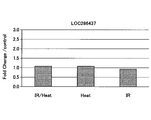
上記の赤外線照射を経て培養された細胞から抽出された全RNAを用い、マイクロアレイ法により発現データが得られた遺伝子のうち、LOC286437遺伝子を対象として、遺伝子の発現量を測定し、発現変化を評価した。測定および解析結果を表1及び図5に示す。
上記の赤外線照射を経て培養された細胞から抽出された全RNAを用い、マイクロアレイ法により発現データが得られた遺伝子のうち、LOC286437遺伝子を対象として、遺伝子の発現量を測定し、発現変化を評価した。測定および解析結果を表1及び図5に示す。
[実験例2]
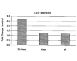
マイクロアレイ法においてLOC286437遺伝子の代わりにLOC101929709遺伝子を対象とした以外は、実験例1と同様に、遺伝子の発現量を測定し、発現変化を評価した。測定および解析結果を表1及び図6に示す。
マイクロアレイ法においてLOC286437遺伝子の代わりにLOC101929709遺伝子を対象とした以外は、実験例1と同様に、遺伝子の発現量を測定し、発現変化を評価した。測定および解析結果を表1及び図6に示す。
[実験例3]
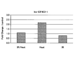
マイクロアレイ法においてLOC286437遺伝子の代わりにlnc-EIF4E3-1遺伝子を対象とした以外は、実験例1と同様に、遺伝子の発現量を測定し、発現変化を評価した。測定および解析結果を表1及び図7に示す。
マイクロアレイ法においてLOC286437遺伝子の代わりにlnc-EIF4E3-1遺伝子を対象とした以外は、実験例1と同様に、遺伝子の発現量を測定し、発現変化を評価した。測定および解析結果を表1及び図7に示す。
[実験例4]
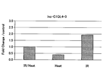
マイクロアレイ法においてLOC286437遺伝子の代わりにlnc-C1QL4-3遺伝子を対象とした以外は、実験例1と同様に、遺伝子の発現量を測定し、発現変化を評価した。測定および解析結果を表1及び図8に示す。
マイクロアレイ法においてLOC286437遺伝子の代わりにlnc-C1QL4-3遺伝子を対象とした以外は、実験例1と同様に、遺伝子の発現量を測定し、発現変化を評価した。測定および解析結果を表1及び図8に示す。

表1及び図5より、実験例1では、温度上昇を制御せずに赤外線を照射した場合(図5の左側のグラフ、赤外線照射条件(2)参照)、赤外線を照射せずに温度上昇させた場合(図5の中央のグラフ、赤外線照射条件(3)参照)、および温度上昇を制御しながら赤外線を照射した場合(図5の右側のグラフ、赤外線照射条件(4)参照)のいずれも、大きな発現変化は確認されなかった。すなわち、実験例1では、赤外線照射のみの場合、温度上昇のみ場合、いずれも、細胞が殆ど影響を受けないことを示している(図5参照)。
表1及び図6より、実験例2では、温度上昇を制御せずに赤外線を照射した場合(図6の左側のグラフ、赤外線照射条件(2)参照)に、大きな発現変化が確認され、赤外線を照射せずに温度上昇させた場合(図6の中央のグラフ、赤外線照射条件(3)参照)、および温度上昇を制御しながら赤外線を照射した場合(図6の右側のグラフ、赤外線照射条件(4)参照)に、大きな発現変化は確認されなかった。すなわち、実験例2では、細胞が赤外線照射のみ、または温度上昇のみでは殆ど影響を受けないが、赤外線照射と温度上昇の両方が組み合わされた場合のみ細胞が影響を受けることを示している(図6参照)。
表1及び図7より、実験例3では、温度上昇を制御せずに赤外線を照射した場合(図7の左側のグラフ、赤外線照射条件(2)参照)、および温度上昇を制御しながら赤外線を照射した場合(図7の右側のグラフ、赤外線照射条件(4)参照)に、大きな発現変化が確認されず、赤外線を照射せずに温度上昇させた場合(図7の中央のグラフ、赤外線照射条件(3)参照)に大きな発現変化が確認された。すなわち、実験例3では、細胞が赤外線照射のみ、または赤外線照射と温度上昇の両方が組み合わされた場合、細胞は殆ど影響を受けないが、温度上昇のみの場合に細胞が影響を受けることを示している(図7参照)。
表1及び図8より、実験例4では、温度上昇を制御せずに赤外線を照射した場合(図8の左側のグラフ、赤外線照射条件(2)参照)に、大きな発現変化は確認されず、赤外線を照射せずに温度上昇させた場合(図8の中央のグラフ、赤外線照射条件(3)参照)に、遺伝子発現を減弱する発現変化が確認され、温度上昇を制御しながら赤外線を照射した場合(図8の右側のグラフ、赤外線照射条件(4)参照)に、遺伝子発現を亢進する大きな発現変化が確認された。すなわち、実験例4では、赤外線照射のみ、または温度上昇のみのいずれも細胞が影響を受け、また赤外線照射のみの場合(発現亢進)と温度上昇のみの場合(発現減弱)とで発現変化の挙動が異なり、さらに、赤外線照射のみの場合に細胞への影響が大きいことを示している(図8参照)。
これらの結果から、本実施形態の細胞培養方法を行うことにより、赤外線照射による温度上昇を制御しながら細胞を培養できることが判った。また、本実施形態の細胞評価方法を行うことにより、細胞に対する赤外線照射の影響と赤外線照射による温度上昇の影響を区別することができ、赤外線による細胞への影響を正確に評価できることが判った。
以上、本発明の実施形態について実験例を挙げて説明したが、本発明は特定の実施形態、実験例に限定されるものではなく、請求の範囲に記載された発明の範囲内において、種々の変形、変更が可能である。
本国際出願は、2017年6月7日に出願された日本国特許出願2017-112896号に基づく優先権を主張するものであり、その全内容をここに援用する。
100 細胞培養装置
10 光源
20 光学フィルタ
30 培養プレート
40 温度制御装置
41 載置部
10 光源
20 光学フィルタ
30 培養プレート
40 温度制御装置
41 載置部
Claims (6)
- 細胞の温度を維持しながら、前記細胞に赤外線を照射することを特徴とする、細胞培養方法。
- 前記細胞が皮膚を構成する細胞である、請求項1に記載の細胞培養方法。
- 前記赤外線の照射量が10~1200J/cm2である、請求項1または2に記載の細胞培養方法。
- 請求項1乃至3のいずれか1項に記載の細胞培養方法により培養した細胞を評価する細胞評価方法。
- 赤外線を照射する光源と、
細胞を培養するプレートと、
前記プレートを載置する載置部を備え、前記載置部の表面温度を制御する温度制御装置とを有し、
前記温度制御装置は、前記光源に対して、前記載置部に載置された前記プレートに前記赤外線が照射されるように配置されている、細胞培養装置。 - 前記プレートは、ガラスプレートである、請求項5に記載の細胞培養装置。
Priority Applications (1)
| Application Number | Priority Date | Filing Date | Title |
|---|---|---|---|
| JP2019523386A JP7286538B2 (ja) | 2017-06-07 | 2018-04-20 | 細胞培養方法、細胞評価方法、および細胞培養装置 |
Applications Claiming Priority (2)
| Application Number | Priority Date | Filing Date | Title |
|---|---|---|---|
| JP2017-112896 | 2017-06-07 | ||
| JP2017112896 | 2017-06-07 |
Publications (1)
| Publication Number | Publication Date |
|---|---|
| WO2018225403A1 true WO2018225403A1 (ja) | 2018-12-13 |
Family
ID=64566136
Family Applications (1)
| Application Number | Title | Priority Date | Filing Date |
|---|---|---|---|
| PCT/JP2018/016333 Ceased WO2018225403A1 (ja) | 2017-06-07 | 2018-04-20 | 細胞培養方法、細胞評価方法、および細胞培養装置 |
Country Status (2)
| Country | Link |
|---|---|
| JP (1) | JP7286538B2 (ja) |
| WO (1) | WO2018225403A1 (ja) |
Citations (5)
| Publication number | Priority date | Publication date | Assignee | Title |
|---|---|---|---|---|
| JPH0491781A (ja) * | 1990-08-03 | 1992-03-25 | Hitachi Ltd | 光合成生物の培養方法および培養装置 |
| JP2002010778A (ja) * | 2000-06-29 | 2002-01-15 | Misato Kk | 細胞の増殖促進方法 |
| JP2002330754A (ja) * | 2001-05-11 | 2002-11-19 | Comet Kk | 細胞不活化方法、及び細胞不活化装置、並びに細胞培養方法 |
| JP2004305137A (ja) * | 2003-04-09 | 2004-11-04 | Tomoyasu Ishikawa | インキュベータおよび細胞培養方法 |
| JP2012100599A (ja) * | 2010-11-11 | 2012-05-31 | Yohei Tanaka | 幹細胞活性化装置 |
Family Cites Families (1)
| Publication number | Priority date | Publication date | Assignee | Title |
|---|---|---|---|---|
| JP2004318017A (ja) | 2003-04-21 | 2004-11-11 | Olympus Corp | 培養観察方法及びそれに用いる倒立顕微鏡 |
-
2018
- 2018-04-20 WO PCT/JP2018/016333 patent/WO2018225403A1/ja not_active Ceased
- 2018-04-20 JP JP2019523386A patent/JP7286538B2/ja active Active
Patent Citations (5)
| Publication number | Priority date | Publication date | Assignee | Title |
|---|---|---|---|---|
| JPH0491781A (ja) * | 1990-08-03 | 1992-03-25 | Hitachi Ltd | 光合成生物の培養方法および培養装置 |
| JP2002010778A (ja) * | 2000-06-29 | 2002-01-15 | Misato Kk | 細胞の増殖促進方法 |
| JP2002330754A (ja) * | 2001-05-11 | 2002-11-19 | Comet Kk | 細胞不活化方法、及び細胞不活化装置、並びに細胞培養方法 |
| JP2004305137A (ja) * | 2003-04-09 | 2004-11-04 | Tomoyasu Ishikawa | インキュベータおよび細胞培養方法 |
| JP2012100599A (ja) * | 2010-11-11 | 2012-05-31 | Yohei Tanaka | 幹細胞活性化装置 |
Non-Patent Citations (1)
| Title |
|---|
| TAKENAKA, YUKO ET AL.: "Basic study on the effect of near-infrared ray on skin by using a newly developed irradiation device", JAPANESE COSMETIC SCIENCE SOCIETY, vol. 38, no. 3, 2014, pages 172 , 173 * |
Also Published As
| Publication number | Publication date |
|---|---|
| JPWO2018225403A1 (ja) | 2020-04-09 |
| JP7286538B2 (ja) | 2023-06-05 |
Similar Documents
| Publication | Publication Date | Title |
|---|---|---|
| Luddi et al. | Organoids of human endometrium: a powerful in vitro model for the endometrium-embryo cross-talk at the implantation site | |
| Liu et al. | A protocol for isolation and identification and comparative characterization of primary osteoblasts from mouse and rat calvaria | |
| Ai et al. | DNA methylation profile is associated with the osteogenic potential of three distinct human odontogenic stem cells | |
| Beachley et al. | Tissue matrix arrays for high-throughput screening and systems analysis of cell function | |
| Phan et al. | Genotoxic stress regulates expression of the proto-oncogene Bcl6 in germinal center B cells | |
| Löwa et al. | Alternatives to animal testing in basic and preclinical research of atopic dermatitis | |
| Liouta et al. | DNA methylation as a diagnostic, prognostic, and predictive biomarker in head and neck cancer | |
| Torrens-Mas et al. | Organoids: an emerging tool to study aging signature across human tissues. modeling aging with patient-derived organoids | |
| Nguyen et al. | Identification of a predictive gene expression signature of cervical lymph node metastasis in oral squamous cell carcinoma | |
| Wang et al. | Nerve growth factor and tyrosine kinase A in human salivary adenoid cystic carcinoma: expression patterns and effects on in vitro invasive behavior | |
| Hogstrom et al. | Simultaneous isolation of hormone receptor–positive breast cancer organoids and fibroblasts reveals stroma-mediated resistance mechanisms | |
| Miller et al. | Biomimetic cardiac tissue culture model (CTCM) to emulate cardiac physiology and pathophysiology ex vivo | |
| Yu et al. | Fluid shear stress induces the secretion of monocyte chemoattractant protein‐1 in cultured human umbilical vein endothelial cells | |
| Dong et al. | Downregulation of ROR2 promotes dental pulp stem cell senescence by inhibiting STK4‐FOXO1/SMS1 axis in sphingomyelin biosynthesis | |
| Duś-Szachniewicz et al. | Physiological hypoxia (physioxia) impairs the early adhesion of single lymphoma cell to marrow stromal cell and extracellular matrix. Optical tweezers study | |
| Xiao et al. | circRNA_0058097 promotes tension‐induced degeneration of endplate chondrocytes by regulating HDAC4 expression through sponge adsorption of miR‐365a‐5p | |
| Sato et al. | Masticatory muscle tendon‐aponeurosis hyperplasia exhibits heterotopic calcification in tendons | |
| Payr et al. | Effect of donor age and 3D-cultivation on osteogenic differentiation capacity of adipose-derived mesenchymal stem cells | |
| Xin-Yi et al. | Organoids in cancer therapies: a comprehensive review | |
| Kim et al. | Patient-derived organoids recapitulate pathological intrinsic and phenotypic features of fibrous dysplasia | |
| Gruber et al. | Cell shape and gene expression in human intervertebral disc cells: in vitro tissue engineering studies | |
| Ferrao Blanco et al. | Distinct stromal cell populations define the B-cell acute lymphoblastic leukemia microenvironment: ACUTE LYMPHOBLASTIC LEUKEMIA | |
| Pawar et al. | Glucose and serum deprivation led to altered proliferation, differentiation potential and AMPK activation in stem cells from human deciduous tooth | |
| WO2012166630A2 (en) | In vitro test system to evaluate xenobiotics as immune-modulators of drug transport and metabolism in human hepatocytes | |
| Youhanna et al. | Primary Human Tissue Models for Metabolic Dysfunction‐Associated Liver Disease‐toward Streamlining Drug Discovery with Patient‐Derived Assays |
Legal Events
| Date | Code | Title | Description |
|---|---|---|---|
| 121 | Ep: the epo has been informed by wipo that ep was designated in this application |
Ref document number: 18813624 Country of ref document: EP Kind code of ref document: A1 |
|
| ENP | Entry into the national phase |
Ref document number: 2019523386 Country of ref document: JP Kind code of ref document: A |
|
| NENP | Non-entry into the national phase |
Ref country code: DE |
|
| 122 | Ep: pct application non-entry in european phase |
Ref document number: 18813624 Country of ref document: EP Kind code of ref document: A1 |