WO2018215855A1 - Automated fundus image processing techniques for glaucoma prescreening - Google Patents
Automated fundus image processing techniques for glaucoma prescreening Download PDFInfo
- Publication number
- WO2018215855A1 WO2018215855A1 PCT/IB2018/053030 IB2018053030W WO2018215855A1 WO 2018215855 A1 WO2018215855 A1 WO 2018215855A1 IB 2018053030 W IB2018053030 W IB 2018053030W WO 2018215855 A1 WO2018215855 A1 WO 2018215855A1
- Authority
- WO
- WIPO (PCT)
- Prior art keywords
- disc
- glaucoma
- circular
- segmentation
- outlining
- Prior art date
- Legal status (The legal status is an assumption and is not a legal conclusion. Google has not performed a legal analysis and makes no representation as to the accuracy of the status listed.)
- Ceased
Links
Classifications
-
- G—PHYSICS
- G16—INFORMATION AND COMMUNICATION TECHNOLOGY [ICT] SPECIALLY ADAPTED FOR SPECIFIC APPLICATION FIELDS
- G16H—HEALTHCARE INFORMATICS, i.e. INFORMATION AND COMMUNICATION TECHNOLOGY [ICT] SPECIALLY ADAPTED FOR THE HANDLING OR PROCESSING OF MEDICAL OR HEALTHCARE DATA
- G16H30/00—ICT specially adapted for the handling or processing of medical images
- G16H30/40—ICT specially adapted for the handling or processing of medical images for processing medical images, e.g. editing
-
- G—PHYSICS
- G06—COMPUTING OR CALCULATING; COUNTING
- G06T—IMAGE DATA PROCESSING OR GENERATION, IN GENERAL
- G06T7/00—Image analysis
- G06T7/10—Segmentation; Edge detection
- G06T7/12—Edge-based segmentation
-
- G—PHYSICS
- G06—COMPUTING OR CALCULATING; COUNTING
- G06T—IMAGE DATA PROCESSING OR GENERATION, IN GENERAL
- G06T7/00—Image analysis
- G06T7/10—Segmentation; Edge detection
- G06T7/149—Segmentation; Edge detection involving deformable models, e.g. active contour models
-
- G—PHYSICS
- G16—INFORMATION AND COMMUNICATION TECHNOLOGY [ICT] SPECIALLY ADAPTED FOR SPECIFIC APPLICATION FIELDS
- G16H—HEALTHCARE INFORMATICS, i.e. INFORMATION AND COMMUNICATION TECHNOLOGY [ICT] SPECIALLY ADAPTED FOR THE HANDLING OR PROCESSING OF MEDICAL OR HEALTHCARE DATA
- G16H50/00—ICT specially adapted for medical diagnosis, medical simulation or medical data mining; ICT specially adapted for detecting, monitoring or modelling epidemics or pandemics
- G16H50/20—ICT specially adapted for medical diagnosis, medical simulation or medical data mining; ICT specially adapted for detecting, monitoring or modelling epidemics or pandemics for computer-aided diagnosis, e.g. based on medical expert systems
-
- G—PHYSICS
- G06—COMPUTING OR CALCULATING; COUNTING
- G06T—IMAGE DATA PROCESSING OR GENERATION, IN GENERAL
- G06T2207/00—Indexing scheme for image analysis or image enhancement
- G06T2207/30—Subject of image; Context of image processing
- G06T2207/30004—Biomedical image processing
- G06T2207/30041—Eye; Retina; Ophthalmic
Definitions
- the present disclosure relates to the field of image processing for healthcare.More particularly, the present disclosure relates to processing of fundus image for glaucoma prescreening.
- glaucoma is the second leading cause of blindness in the world, the first being cataract. It is an optic neuropathy - a condition that causes damage to eye's optic nerve - and is a chronic and irreversible eye disease in which the nerve that connects the eye to the brain (optic nerve) is progressively damaged. Patients with early glaucoma do not have visual symptoms. Progression of the disease results in loss of peripheral vision, so patients may complain of "tunnel vision" (only being able to see the centre). Advanced glaucoma is associated with total blindness. Total number affected by glaucoma in 2010 was 60 million and is estimated to be over 80 million by 2020. The problem is even more significant in Asia, as Asians account for approximately half of the world's glaucoma cases and going to get worse because it is a condition of aging, and Asian countries have aging population.
- Glaucoma is caused due to high intraocular fluid pressure created by the abnormal production or drainage of the aqueous humor circulating between the cornea and the lens. As intraocular pressure within the eye increases, it damages the sensitive nerve fibers and causes them to die. As the nerve fibers die, they develop a cup-like shape in the Optic Disc(OD) of the eye. If the pressure remains high for a long time, it can damage the optic nerve and result in permanent vision loss called glaucoma. Glaucoma is asymptomatic until the late stages of the disease. Glaucoma is not curable and untreated glaucoma leads to permanent damage of the optic nerve leading to tunnel vision, which, over time can cause permanent blindness and vision lost cannot be restored. Almost every one of us is at risk for glaucoma. Therefore, a periodic careful and comprehensive eye examination is essential for early diagnosis of glaucoma.
- the OD in the eye is the small blind spot on the surface of the retina.
- the blind spot is called so because there are no receptors in this part of the retina. It is the point where the fibers of the retina leave the eye and become part of the optic nerve.
- the OD has three distinct regions - a central white depression called the Optic Cup (OC), a peripheral ring shaped region called the neuroretinal rim, and the optic nerves. The major regions of the OD are shown in FIG. 1. Millions of nerve fibers run from retina to the optic nerve. The fibers meet at the OD.
- glaucoma results in the alteration of OD topography, commonly known as cupping, and associated visual field loss, it is diagnosed and treated based upon the structural appearance of the OD.
- OD evaluation using optic nerve imaging is an important test that an ophthalmologist would use to diagnose and monitor glaucoma.
- the widest application of OD imaging is in glaucoma management.
- Various forms of imaging such as Heidelberg Retinal Tomography (HRT), Scanning Laser Polarimetry (SLP), and Optical Coherence Tomography (OCT) permit quantitative measurement of OD and Retinal Nerve Fiber Layer (RNFL) structure.
- OCT detects damage of the RNFL.
- HRT determines changes in the topography of the optic nerve.
- fundus imaging devices from Topcon, Zeiss, Canon, Forus Health Pvt. Ltd., Bosch Eye Care Solutions, and Fundus-On-Phone (FOP) devices from Peek Vision (Peek retina), Remidio innovative Solutions Pvt. Ltd., and Volk iNview permit quick and effortless capture of fundus images, but do not offer any software solution as for as glaucoma assessment is concerned.
- CDR Cup-to-Disc Ratio
- RDR Rim-to- Disc Ratio
- DDLS Disc-Damage- Likelihood-Scale
- ISNT Superior, Nasal, and Temporal
- CDR/RDR/ISNT rim widths are the important and popular parameters used by ophthalmologists in glaucoma diagnosis. Therefore, objective recording of the appearance of the OD and OC and their outlining, and also determination of CDR/RDR/ISNT rim widths as shown in FIG. 2 is essential to monitor a patient with glaucoma or suspect of having the disease. At present, ophthalmologists manually outline the OD and OC, which is subjective. Determination of CDR/RDR and ISNT rim widths is difficult as ophthalmologists are not consistent at accurately outlining OD and OC, or measure ISNT rim widths.
- OD segmentation In OD segmentation, histograms, and center surround statistics are used to classify each super pixel as belonging to the disc or not. Deformable curve models are used to fine-tune the disc boundary. For OC segmentation, in addition to the histograms and center surround statistics, the location information is also included.
- the United States patent application no. 20150124216 proposes systems and methods for assessing glaucoma loss using optical coherence topography.
- Another United States patent application no. US20150161785 Al proposes a method for automatically processing an image including thejunction between a cornea and an iris.
- US5233517 proposesearly glaucoma detection by a system and a method of fast Fourier transform analysis of digitized eye fundus images.
- the numbers expressing quantities of ingredients, properties such as concentration, reaction conditions, and so forth, used to describe and claim certain embodiments of the invention are to be understood as being modified in some instances by the term "about.” Accordingly, in some embodiments, the numerical parameters set forth in the written description and attached claims are approximations that can vary depending upon the desired properties sought to be obtained by a particular embodiment. In some embodiments, the numerical parameters should be construed in light of the number of reported significant digits and by applying ordinary rounding techniques. Notwithstanding that the numerical ranges and parameters setting forth the broad scope of some embodiments of the invention are approximations, the numerical values set forth in the specific examples are reported as precisely as practicable. The numerical values presented in some embodiments of the invention may contain certain errors necessarily resulting from the standard deviation found in their respective testing measurements.
- a general object of the present disclosure is to provide a means for periodic, careful and comprehensive eye examination for early diagnosis of glaucoma so that a timely treatment of glaucoma can be started to prevent partial or full blindness among masses.
- An object of the present disclosure is to provide an automatic method that is fast and reliable for prescreening of population for glaucoma.
- An object of the present disclosure is to provide a method to automatically screen fundus images of the eye and identify potentially glaucomatous conditions with high accuracy.
- Another object of the present disclosure is to provide an image processing technique and an associated software tool that is repeatable, easy to use, and takes a few seconds per patient for glaucoma prescreening.
- Another object of the present disclosure is to provide an image processing technique and an associated software tool thatcan be operated by minimally trained personnel or semi-skilled personnel.
- Another object of the present disclosure is to provide an image processing technique and an associated software tool that can be used alongside tabletop, handheld fundus cameras, and FOPs, empowering smaller clinics in rural areas to make effective use of the tool.
- Yet another object of the present disclosure is to provide a method for processing fundus images and carrying out severity grading of glaucoma based on CDR/RDR.
- Still another object of the present disclosure is to provide a method that implements ISNT rule to check whether the normal eyes show a characteristic pattern of inferior > superior > nasal > temporal rim widths to differentiate them from glaucomatous eyes.
- aspects of the present disclosure relate to a method for optimizing circles and ellipses to objects or regions in an image using one or moreActive Discs (AD).
- AD Active Discs
- embodiments explained herein pertain to fundus image processing for identifying potentially glaucomatous eye conditions.
- the disclosed method is fully automatic and can carry out severity grading of glaucoma based on CDR/RDR and ISNT rule bymeasurement of Inferior, Superior, Nasal, and Temporal neuroretinalrim width.
- the disclosed method involvessegmentation and outlining ofcircularand/or elliptical regions/objects in an image such as a medical image for example a fundus image containing OD and OC.
- the segmented and outlined objects/regions can, thereafter, be used for further diagnosis such as for glaucoma prescreening.
- segmentation of the circular and/or elliptical regions/objects such as OD and OC in the image is based on the notion of AD, which can comprise a pair of dynamic concentric inner and outer discs as template.
- the Active Disc is made to evolve from a specified initialization towards the boundary of the object such as OD based on a pre-defined energy criterion such as by minimizing a local disc energy function.
- the AD can be initialized automatically using matched filtering technique. This is essential because improper manual initialization can often lead to convergence issues resulting in anomalous segmentation.
- the OD shape is slightly vertically oval, with the vertical diameter being about 7% longer than the horizontal one.
- disclosed method uses circular and elliptical AD for OD segmentation.
- the AD can change shape from circle to ellipse when evolving based on a predefined energy criterion to segment the OD.
- design of the circular disc template can consist of two concentric circles centered at the origin, followed by scaling, translation, rotation, and optimization of parameters.
- the disc energy can be a normalized contrast function, which is a measure of pixel intensities inside the annulus of the disc in comparison with those inside the inner disc. Optimization can be achieved using accelerated gradient descent and Green's theorem.
- OD/OC segmentation firstly, the pixels representing OD/OC region are clustered by Otsu's multilevel thresholding algorithm and then it is segmented and outlined by the AD.
- a circular AD can be used to perform coarse segmentation and to improve accuracy an elliptical AD can be used for finer segmentation. The initialization used for OD can also be used to outline the OC region.
- Cup-to-Disc Ratio (CDR) and Rim-to-Disc Ratio (RDR) can be measured from the segmented the OD and OC.
- the CDR value can be compared against the existing International Classification of Diseases (ICD) rules and the RDR against the new Disc-Damage- Likelihood-Scale (DDLS), to finally assist in diagnosing the progression of glaucoma.
- ICD International Classification of Diseases
- DDLS Disc-Damage- Likelihood-Scale
- an image processing software has been developed as part of the invention that can categorize the condition as normal, mild, moderate, or severely glaucomatous in accordance with internationally accepted guidelines.
- the developed software incorporates a check for the pattern of rim width using Inferior > Superior > Nasal> Temporal (ISNT) rule to differentiate normal fundus from glaucomatous fundus.
- ISNT Temporal
- the proposed method has been implemented using Java as an ImageJ plugin.
- An associated batch-processing ImageJ macro to handle a database for automated glaucoma assessment has also been implemented.
- Android and iOS based smartphone apps for glaucoma prescreening have also been implemented.
- the tool is repeatable, easy to use, provides quantitative analysis, and takes only few seconds per image.
- the proposed algorithm has achieved full automation since it does not require any manual intervention.
- the software implementation can be used alongside tabletop, handheld fundus cameras, and fundus-on-phone (FOP) devices.
- the implementation is readily available for integration into any existing fundus camera, iOSdevices or Android devices. Any of these devices could be converted to a fundus imager with the help of an addon device.
- the disclosed method for glaucoma prescreening has been validatedon publicly available databases such as Messidor, Drions-DB, and Drishti-GS, as well as fundus image databases obtained from Forus Health Pvt. Ltd., Bosch Eye Care Solutions, and Remidio innovative Solutions Pvt. Ltd., Bengaluru, India.
- the algorithm performance is compared vis-a-vis clinician outlining as reference and for quantitative comparison, sensitivity, specificity, percentage accuracy, Jaccard and Dice similarity measures has been used.
- FIG. 1 illustrates typical fundus images showing major regions of the OD.
- FIG. 2 illustrates typical fundus images showingvarious parameters for glaucoma assessment as known in the art.
- FIG. 3 illustratesexemplary (a) disc template along with changing shape of ADfrom (b) circular to (c) oval shaped discin accordance with an embodiment of the present disclosure.
- FIG. 4 illustrates an exemplary image of (a) an OD with an optimal (b) circular and (c) elliptical AD fit on the OD along with regional energies in accordance with an embodiment of the present disclosure.
- FIG. 5 illustrates exemplary images of (al) -(a2): oval shaped OD, (bl)-(b2): circular AD fit on the OD and (cl)-(c2): elliptical AD fit on optic disc in accordance with an embodiment of the present disclosure.
- FIG. 6 illustrates exemplaryimages of (al) - (a2): OD, (bl) - (b2): result of matched filtering and (cl) - (c2): initialization of AD in accordance with an embodiment of the present disclosure.
- FIG. 7 illustrates exemplary images of (al) - (a2): OD, (bl) - (b2): first, (cl) - (c2): second, (dl) - (d2): third and (el) - (e2): fourth level thresholdingin accordance with an embodiment of the present disclosure.
- FIG. 8 illustrates exemplary (al) - (a6): input fundus images, (bl) - (b6): OD segmentation due to circular followed by elliptical AD, (cl) - (c6): OC segmentation due to circular AD in accordance with an embodiment of the present disclosure.
- FIG. 9 illustrates an exemplary (al) - (el): fundus images with OD and OC outlined by AD (Thick contours) and expert ophthalmologist (Thin contours) in accordance with an embodiment of the present disclosure.
- FIG. 10 illustrates an exemplary (al)-(a3): Input Fundus images, (bl)-(b3): OD segmentation due to AD, (cl)-(c3): OC segmentation due to AD, (dl)-(d3): narrowest rim- width in red color and (el)-(e3): inferior, superior, nasal, and temporal rim widths in white, red, yellow, and black colors in accordance with an embodiment of the present disclosure.
- FIG. 11 illustrates an exemplary arrangement of two non-concentric circles one inside the otherrepresenting circular approximation of the OC and the OD area respectively in accordance with an embodiment of the present disclosure.
- FIG. 12 illustrates an exemplary arrangement of two non-concentric circles one inside the other representing circular approximation of the OC and the OD areaand computation of minimum rim-width in accordance with an embodiment of the present disclosure.
- FIG. 13 illustrates an exemplary flow diagram for CDR based glaucoma assessment method in accordance with an embodiment of the present disclosure.
- FIG. 14 illustrates an exemplary flow diagram for RDR based glaucoma assessment method in accordance with an embodiment of the present disclosure.
- FIG. 15 illustrates an exemplary flow diagram for ISNT based glaucoma assessment method in accordance with an embodiment of the present disclosure.
- Embodiments of the present disclosure relate to fundus image processing for identifying potentially glaucomatous eye conditions.
- the disclosure provides a method that can be implemented through software in combination with hardware to provide a fully automatic tool to screen fundus images for glaucomatous condition based on ISNT rule by measurement of inferior, superior, nasal, and temporal neuroretinalrim widths, andcarry out severity grading of glaucoma based on RDR.
- the disclosed method carries outsegmentation and outlining ofOD and OC in the fundus images based on notion of AD, which can comprise a pair of dynamic concentric inner and outer discs as template.
- the active disc can be made to evolve from a specified initialization towards the boundary of the OD based on a pre-defined energy criterion such as by minimizing a local disc energy function.
- the disclosed method can involve steps of (a) automatic initialization of the AD using matched filtering technique wherein it over comes deficiency of manual initialization which can often be improper and lead to convergence issues resulting in anomalous segmentation; (b) designing of a circular disc template, which can consist of two concentric circles centered at the origin; (c) scaling, translation, rotation, and optimization of parameters of the designed template, wherein the disc is made to evolve from a specified initialization towards the boundary of the OD/OC by minimizing a local energy function.
- the disc energy can be a normalized contrast function, which is a measure of pixel intensities inside the annulus of the disc. Further, optimization can be achieved using accelerated gradient descent technique and Green's theorem.
- choosing the parameters of the affine transformation appropriately either circular shaped or elliptical shaped AD can be obtained to account for slightly vertically oval shape of the OD with the vertical diameter being about 7% longer than the horizontal one.
- the pixels representing OD/OC region can first be clustered by Otsu's multilevel thresholding algorithmand then segmented and outlined by the AD.
- the coarse segmentation of the OD is done by the circular AD and to improve the accuracy, the fine segmentation is done by the elliptical AD.
- the OC segmentation is done by the circular AD.
- the method can further comprise step of checking for the pattern of rim width and using inferior > superior > nasal > temporal (ISNT) rule to differentiate normal fundus from glaucomatous fundus and thereby screen those with glaucomatous condition.
- ISNT inferior > superior > nasal > temporal
- the method can further comprise step of measurement ofCup-to-Disc Ratio (CDR) and Rim-to-Disc Ratio (RDR) from the segmented the OD and OC.
- CDR Cup-to-Disc Ratio
- RDR Rim-to-Disc Ratio
- the CDR value can be compared against the existing International Classification of Diseases (ICD) rules and the RDR against the new Disc-Damage- Likelihood-Scale (DDLS), to finally assist in diagnosing the progression of glaucoma.
- ICD International Classification of Diseases
- DDLS Disc-Damage- Likelihood-Scale
- Optic disc boundary can be extracted from multi-thresholded red channel of color fundus image using AD.
- the reason for selecting red channel is twofold: firstly weakening of blood vessels and optic nerve occlusions by itself compared to gray scale version of color fundus image, and secondly nature of proposed AD algorithm is to look for bright regions to converge.
- the AD evolves without facing any obstacles and lock on to the boundary of OD easily.
- Active disc is the one comprising a pair of concentric inner and outer discs. It is dynamic and evolves based on a pre-defined energy criterion and changes shape from circle to ellipse to segment the OD. The main steps are the following: design of the disc, design ofthe energy functional and the use of Green's theorem to achieve computational savings. In this Section, we provide the mathematical formulation for generating AD.
- the OD shape is slightly vertically oval, with the vertical diameter being about 7% longer than the horizontal one.
- An example of such a circular template is shown in FIG. 3 (a).
- the factor 1/ ⁇ /2 is used to make the area of the inner disc equal to that of the annulus, which ensures that the AD is inert in regions of constant intensity.
- Accelerated gradient descent technique requires the partial derivatives of the energy function. Since the integrals are two-dimensional and the contours are closed, one could compute the partial derivatives using Green's theorem. Use of Green's theorem simplifies the computation of the partial derivatives and makes the algorithm cost-effective and fast. In our case, we need partial derivatives of theenergy function with respect to the parameters ⁇ , ⁇ , ⁇ , x c , and y c . The integralsin (3) are analogous, therefore, it is sufficient to show the calculations for one ofthem. Applying Green's theorem to E 2 gives
- FIGs.5(bl)-(b2) and 5(cl)-(c2) show segmentation and outlining of OD by circular and elliptical AD, respectively.
- Matched filtering is apowerful technique for detecting objects of interest in images in whichthe object information is known a priori. It maximizes the detection signal-to-noise ratio if the object of interest in the image and the template are identical.
- the filter template is designed to comprise two concentric circles for which the weights of the inner circle, outer circle, and annulus are considered as positive, negative, and zero, respectively. The given image is then correlated with the template.
- the matched filtering operation can be carried out directly in the spatial domain.
- the matched filter response at (x p , y p ) is given by: wherew , y) is the filter template.
- the peak in the cross-correlation corresponds to thebest match between the image and the template and is taken as the location for initialization the AD.
- the result of matched filtering and subsequent initialization of the AD is shown in FIG. 6.
- the OC is a form of OD depression peculiar to glaucoma. This is an eye disorder thatconcems damage to the optic nerve. Left untreated, the optic nerve could become permanently damaged, and the patient could go totally blind. Ophthalmologists or medical professionals specialized in eye care, like optometrists and orthoptists can diagnose this condition by relying on a CDR ratio. This means they have to figure out the OC' s size in relation to the total diameter of the OD. In this work, we use Otsu's multilevel thresholding algorithm to classify and cluster the pixels corresponding to OC, and then determine the size of the OC using circular AD.
- Otsu's multilevel thresholding algorithm assumesthe image as M classes of pixels and calculates optimum thresholds separating classessuch that the intra- class variance is minimum.
- intra-class variance is the weightedsum of variances of M classes.
- f represent the 2Z)grayscale intensity function,and contains N pixels with graylevels 0 to (L - 1).
- Jbe the number of pixels withgraylevelz.
- the probability of graylevelz is given by,
- Ci ⁇ 1, 2, ...Ji ⁇ , C 2 : ⁇ ti+1, ..., t 2 ⁇ md C M : ⁇ t M -i+l, -,L ⁇ -
- FIG. 8(al)-(a6) The segmentation and outlining is done by AD method and are compared with the manual outlines provided by an expert ophthalmologist.
- FIGs.8(bl)-(b6) and (cl)- (c6) show OD and OC segmentation using elliptical AD and circular AD, respectively.
- Jaccard and Dice indices, sensitivity, specificity, and accuracy are the comparison criteria typically used in medical image segmentation. They are based on the definition of true positives (TP), false positives (FP), false negatives (FN), and true negatives (TN).
- Sensitivity TP/(TP+FN) measures the ovelap, but can be equal to one for a poor segmentation much bigger than the ground truth.
- Specificity TN/(TN+FP) is therefore the necessary counterpart of the sensitivity, but it can be equal to one for a very poor segmentation that does not detect the object of interest at all.
- Jaccard similarity index (J) is the most intuitive ratio between the intersection and union.
- Dice similarity index (A) is slightly different, but more frequently used and one can be deduced from the other.
- quantitative comparison of the performance of the proposed method and clinician outlining is done using J andA . They are given by: for 0 ⁇ J ⁇ 1 and 0 ⁇ A ⁇ I .
- a and M represent the optic disc region segmented by the proposed method and clinician respectively.
- ⁇ n Mde notes the intersection between ⁇ and and it reveals the common optic disc region in both A and M.
- a U M and it reveals optic disc region which are in either segmentation.
- Most of the techniques proposed in the literature for glaucoma analysis have considered only one locally obtained database.
- the novelty of the proposed technique is in handling many databases which contain fundus images acquired in different conditions.
- the proposed glaucoma analysis tool has been validated on publicly available databases like Messidor, Drishti-GS, and Drions-DB.
- the proposed tool has also been validated on locally obtained fundus image databases from ForusHealth Pvt. Ltd, Bosch Eye Care Solutions, and Remidio innovative Solutions Pvt. Ltd.
- the proposed method gave satisfactory segmentation results on fundus images from allsix databases.
- FIG. 9 pictoriallyillustratescomparison of performance of the AD with that of the clinician outlining.
- Thin green and blue contours represent manual outlining of OD and OC, respectively.
- Thick green and blue contours show algorithm outlining of OD and OC, respectively.
- FIG. 10 illustrates green line joining the centers of the OD and OC with the points on the OD and OC contours where the cup is sloping towards the OD boundary.
- a red line mark overlaid on the green line shows the narrowest possible rim width. It was observed that the OD and OC segmentation and outlining is accurate, with meaningful narrowest possible rim width marked on the cup notching area. A high similarity was obtained between clinician and outlining by the proposed method.
- Glaucoma severity refers to the presence and extensiveness of the disease in the retina of the eye. It is objectively evaluated through diagnostic testing like fundus imaging and physiological examination of the impaired OD in the form of cupping. The amount of cupping in the OD can be measured, and the corresponding severity level of the glaucoma can be determined.
- glaucoma severity information can be contrasted with symptom assessment by fully automated CDR measurement technique based on ICD-9 ruleswhich is an internationally accepted guideline on glaucoma diagnosis. The proposed method has been validated on fundus images from eight databases. Based on the CDR, proposed method categorizes the glaucomatous condition as normal, mild, moderate, or severe as given in Table 1 below in accordance with ICD-9 rules.
- the smallest possible neuroretinal rim-width is an important parameter for disc- damagelikelihood- scale (DDLS) based glaucoma stage analysis.
- the problem of finding the smallest possible neuro-retinal rim-width can be formulated as finding the smallest possible distance between the two non-concentric circles one inside the other as depicted in FIG. 11. Circles with centers 01 and 02 represents the circular approximation of the OC and the OD area, respectively.
- the line 0102 joins the two centers of the inner and outer circles. Extending the line 01-02 upto the outer periphery of the outer circle intersects the inner and outer circle at points P and Q, respectively.
- the line segment PQ is the smallest possible distance between the two non-concentric circles one inside the other along the radial direction. Now, we need to prove that the line segment PQ is the smallest possible distance. In this proof, without loss of generality, it is assumed that the outer circle is centered at theorigin, and the inner circle is centered along the X-axis. All the calculations are done with respect to the first quadrant of the Cartesian plane. The respective figure used for the formulation is shown in FIG. 12. Let Y ⁇ and r 2 represent the radius of the inner and outer circles, respectively.
- glaucomatous conditions can be categorizedinto eight stages as given in Table 2 below: Rim-to-Disc Ratio(RDR)
- Table 2 Glaucomatous condition classification based on narrowest rim width or RDR
- the proposed method classifies the glaucomatous condition as normal, mild, moderate, or severe in accordance an internationally accepted guideline on DDLS based glaucoma diagnosis.
- the proposed method has been validated on fundus images from eight databases.
- the ISNT rule states that a healthy OD has a characteristic pattern of decreasing order of inferior > superior > nasal > temporal neuroretinal rim widths and the glaucomatous ODs violate this quantitative pattern.
- the utility of the ISNT rule has been proved in clinical practice, but it is considered to be subjective.
- the inferior, superior, nasal, and temporal rim widths can be measured automatically and used to determine whether the determined values obey the quantitative pattern of the ISNT rule. If the ISNT rule holds for an OD, it is treated as normal otherwise glaucomatous.
- FIG. 13 illustrates an exemplary flow diagram for CDR based glaucoma assessment method 1300 in accordance with an embodiment of the present disclosure.
- the method 1300 for CDR based glaucoma assessment can at step 1302 take a color fundus image as input; At step 1304 red channel can be extracted; At step 13062-stage or 3 -stage multi-threshold region can be extracted; At step 1308 an Active Disc can be initialized using matched filtering technique; at step 1310 coarse segmentation and outlining of Optic Disc can be done by circular AD; at step 1312 fine segmentation and outlining of the ODcan be done by elliptical AD;.
- step 1314 vertical length of OD can be printed; at step 1316 color fundus image can be again taken as input for Optic Cup segmentation; at step 1318 green channel can be extracted from the fundus image; at step 1320 4-stage multi -threshold region can be extracted; at step 1322 an Active Disc can be initialized using ODcenter and radius; at step 1324 OC can be segmented and outlined by the Active Disc; at step 1326 vertical length of the segmented and outlined OD can be printed; at step 1328 Cup-to-Disc Ratio can be computed and glaucomatous condition printed based on International Classification of Diseases-9 rules.
- FIG. 14 illustrates an exemplary flow diagram for RDR based glaucoma assessment method 1400 in accordance with an embodiment of the present disclosure.
- Steps 1402 to 1412 pertaining to OD segmentation and outlining; and steps 1414 to 1422 pertaining to OC segmentation and outlining can correspond to and same as steps 1302 to 1312 and 1316 to 1324 of method 1300.
- the method 1400 can at step 1424 involve determining narrowest rim width and at step 1426 compute rim-to-disc ratio (RDR) and print glaucomatouscondition based on Disc-Damage- Likelihood-Scale (DDLS).
- RDR rim-to-disc ratio
- DDLS Disc-Damage- Likelihood-Scale
- FIG. 15 illustrates an exemplary flow diagram for ISNT based glaucoma assessment method 1500 in accordance with an embodiment of the present disclosure.
- Steps 1502 to 1512 pertaining to OD segmentation and outlining; and steps 1514 to 1522 pertaining to OC segmentation and outlining can correspond to and be same as steps 1302 to 1312 and 1316 to 1324 of method 1300.
- the method 1500 can at step 1524 involve determining Inferior, Superior, Nasal, and Temporal rim widths and based on determined values, at step 1526 checking whether rim widths follow ISNT rule.
- the proposed method has been implemented using Java as an ImageJ plugin.
- An associated batch-processing ImageJ macro to handle a database for automated glaucoma assessment has also been implemented.
- Android and iOS based smartphone apps for glaucoma prescreening has also been implemented.
- the tool is repeatable, easy to use, provides quantitative analysis, and takes only few seconds per image.
- the proposed algorithm has achieved full automation since it does not require any manual intervention.
- the software implementation can be used alongside tabletop, handheld fundus cameras, and fundus-on-phone (FOP) devices.
- the implementation is readily available for integration into any existing fundus camera, iOSdevices or Android devices. Any of these devices could be converted to a fundus imager with the help of an addon device.
- any one or more of the methods 1300, 1400 and 1600 can be used either individually or in combination to screen fundus images for identifying potentially glaucomatous eye conditions and carry out severity grading of glaucoma based on CDR.
- the present disclosure provides an automatic method that is fast and reliable for periodic, careful and comprehensive eye examination for prescreening of population and thus early diagnosis of glaucoma that can enable timely treatment of glaucoma to prevent partial or full blindness among masses.
- the tool can automatically screen large number of fundus images and identify potentially glaucomatous conditions with high accuracy.
- the present disclosure provides a means for periodic,careful and comprehensive eye examination for early diagnosis of glaucoma so that a timely treatment of glaucoma can be started to prevent partial or full blindness among masses.
- the present disclosure provides an automatic method that is fast and reliable for prescreening of population for glaucoma.
- the present disclosure provides a method to automatically screen fundus images of the eye and identify potentially glaucomatous conditions with high accuracy.
- the present disclosure provides an image processing technique and an associated software tool that is repeatable, easy to use, and takes a few seconds per patient for glaucoma prescreening.
- the present disclosure provides an image processing technique and an associated software tool that can be operated by minimally trained personnel or semi-skilled personnel.
- the present disclosure provides an image processing technique and an associated software tool that can be used alongside tabletop, handheld fundus cameras, and FOPs, empowering smaller clinics in rural areas to make effective use of the tool.
- the present disclosure provides a method for processing fundus image and carrying out severity grading of glaucoma based on CDR/RDR.
- the present disclosure provides a method for severity grading of glaucoma based on measurement of inferior, superior, nasal, and temporal neuroretinalrim width.
- the present disclosure provides a method that implements ISNT rule to check whether the normal eyes show a characteristic pattern of inferior > superior > nasal > temporal rim widths to differentiate them from glaucomatous eyes.
Landscapes
- Engineering & Computer Science (AREA)
- Health & Medical Sciences (AREA)
- Public Health (AREA)
- Medical Informatics (AREA)
- Theoretical Computer Science (AREA)
- Primary Health Care (AREA)
- Biomedical Technology (AREA)
- Computer Vision & Pattern Recognition (AREA)
- Epidemiology (AREA)
- General Health & Medical Sciences (AREA)
- Physics & Mathematics (AREA)
- General Physics & Mathematics (AREA)
- Radiology & Medical Imaging (AREA)
- Nuclear Medicine, Radiotherapy & Molecular Imaging (AREA)
- Data Mining & Analysis (AREA)
- Databases & Information Systems (AREA)
- Pathology (AREA)
- Software Systems (AREA)
- Eye Examination Apparatus (AREA)
Abstract
Aspects of the present disclosure relate to a method for optimizing circles and ellipses to objects or regions in an image using an Active Disc (AD).In particular, embodiments explained herein pertain to fundus image processing for identifying potentially glaucomatous eye conditions. The Optic Disc (OD) and Optic Cup (OC) in the fundus image are segmented and outlined based on the notion of AD, which comprises a pair of dynamic concentric inner and outer discs as template. The AD is made to evolve from a specified initialization towards the boundary of the OD based on a pre-defined energy criterion such as by minimizing a local disc energy function. The disclosed method is fully automatic and can carry out severity grading of glaucoma based on CDR/RDR and ISNT rule by measurement of inferior, superior, nasal, and temporal neuroretinalrim width.
Description
AUTOMATED FUNDUS IMAGE PROCESSING TECHNIQUES FOR GLAUCOMA
PRESCREENING
TECHNICAL FIELD
[0001] The present disclosure relates to the field of image processing for healthcare.More particularly, the present disclosure relates to processing of fundus image for glaucoma prescreening.
BACKGROUND
[0002] Background description includes information that may be useful in understanding the present invention. It is not an admission that any of the information provided herein is prior art or relevant to the presently claimed invention, or that any publication specifically or implicitly referenced is prior art.
[0003] According to the World Health Organization (WHO) glaucoma is the second leading cause of blindness in the world, the first being cataract. It is an optic neuropathy - a condition that causes damage to eye's optic nerve - and is a chronic and irreversible eye disease in which the nerve that connects the eye to the brain (optic nerve) is progressively damaged. Patients with early glaucoma do not have visual symptoms. Progression of the disease results in loss of peripheral vision, so patients may complain of "tunnel vision" (only being able to see the centre). Advanced glaucoma is associated with total blindness. Total number affected by glaucoma in 2010 was 60 million and is estimated to be over 80 million by 2020. The problem is even more significant in Asia, as Asians account for approximately half of the world's glaucoma cases and going to get worse because it is a condition of aging, and Asian countries have aging population.
[0004] Glaucoma is caused due to high intraocular fluid pressure created by the abnormal production or drainage of the aqueous humor circulating between the cornea and the lens. As intraocular pressure within the eye increases, it damages the sensitive nerve fibers and causes them to die. As the nerve fibers die, they develop a cup-like shape in the Optic Disc(OD) of the eye. If the pressure remains high for a long time, it can damage the optic nerve and result in permanent vision loss called glaucoma. Glaucoma is asymptomatic until the late stages of the disease. Glaucoma is not curable and untreated glaucoma leads to permanent damage of the optic nerve leading to tunnel vision, which, over time can cause permanent blindness and vision lost cannot be restored. Almost every one of us is at risk for glaucoma. Therefore, a
periodic careful and comprehensive eye examination is essential for early diagnosis of glaucoma.
[0005] The OD in the eye is the small blind spot on the surface of the retina. The blind spot is called so because there are no receptors in this part of the retina. It is the point where the fibers of the retina leave the eye and become part of the optic nerve. The OD has three distinct regions - a central white depression called the Optic Cup (OC), a peripheral ring shaped region called the neuroretinal rim, and the optic nerves. The major regions of the OD are shown in FIG. 1. Millions of nerve fibers run from retina to the optic nerve. The fibers meet at the OD.
[0006] Since glaucoma results in the alteration of OD topography, commonly known as cupping, and associated visual field loss, it is diagnosed and treated based upon the structural appearance of the OD. OD evaluation using optic nerve imaging is an important test that an ophthalmologist would use to diagnose and monitor glaucoma. In fact, the widest application of OD imaging is in glaucoma management. Various forms of imaging such as Heidelberg Retinal Tomography (HRT), Scanning Laser Polarimetry (SLP), and Optical Coherence Tomography (OCT) permit quantitative measurement of OD and Retinal Nerve Fiber Layer (RNFL) structure. OCT detects damage of the RNFL. HRT determines changes in the topography of the optic nerve. In addition, fundus imaging devices from Topcon, Zeiss, Canon, Forus Health Pvt. Ltd., Bosch Eye Care Solutions, and Fundus-On-Phone (FOP) devices from Peek Vision (Peek retina), Remidio Innovative Solutions Pvt. Ltd., and Volk iNview permit quick and effortless capture of fundus images, but do not offer any software solution as for as glaucoma assessment is concerned.
[0007] To recognize and follow-up glaucoma progression, it is important to assess the size and shape of the OD, OC, and neuroretinal rim. Various parameters have been proposed in previous studies to assess the size and shape of the OD, OC and neuroretinal rim, and recognizeglaucoma progression. For example, Cup-to-Disc Ratio (CDR) proposed by Armaly et al., Rim-to- Disc Ratio (RDR) based on Disc-Damage- Likelihood-Scale (DDLS) proposed by Spaeth et al., and Inferior, Superior, Nasal, and Temporal (ISNT)rim widthsrule proposed by Jonas et al. are the important and popular parameters used by ophthalmologists in glaucoma diagnosis. Therefore, objective recording of the appearance of the OD and OC and their outlining, and also determination of CDR/RDR/ISNT rim widths as shown in FIG. 2 is essential to monitor a patient with glaucoma or suspect of having the disease. At present, ophthalmologists manually outline the OD and OC, which is subjective. Determination of
CDR/RDR and ISNT rim widths is difficult as ophthalmologists are not consistent at accurately outlining OD and OC, or measure ISNT rim widths.
[0008] Hence, it is essential to estimate the CDR/RDR and ISNT rim widths via clinical or automated image processing techniques. Segmentation of both OD and OC, and determination of CDR/RDR is crucial in the assessment of glaucoma. Few automatic glaucoma assessment methods have been presented in the literature. Cheng et al. in their paper titled" Superpixe I classification based optic disc and optic cup segmentation for glaucoma screening " (Published in IEEE Transactions on Medical Imaging, vol. 32, no. 6, pp. 1019-1032, 2013) proposed OD and OC segmentation using super-pixel classification for glaucoma screening. In OD segmentation, histograms, and center surround statistics are used to classify each super pixel as belonging to the disc or not. Deformable curve models are used to fine-tune the disc boundary. For OC segmentation, in addition to the histograms and center surround statistics, the location information is also included.
[0009] Xu et al. in their paper titled" Optic disk feature extraction via modified deformable model technique for glaucoma analysis" (Published in Pattern Recognition, vol. 40, no. 7, pp. 2063 - 2076, 2007) proposed an algorithm to segment OD using deformable snakes, which deform to the location with minimum energy, and then self-cluster into edge- point group and uncertain-point group. The same method is also used to detect the OC boundary.
[0010] Joshi et al. in their paper titled "Optic disk and cup boundarydetection using regional information" (in Proceedings of IEEE Intl. Symposium on Biomedical Imaging: From Nano to Macro (ISBI), 2010, pp. 948-951) used OD and OC boundaries to estimate the relevant disk parameters. A deformable model guided by regional statistics detects OD boundary. OC boundary detection is based on the appearance of pallor in Lab color space and the expected OC symmetry.
[0011] In another method, Joshi et al. in their paper titled "Optic disk and cup segmentation from monocular color retinal images for glaucoma assessment' (Published in IEEE Transactions on Medical Imaging, vol. 30, no. 6, pp. 1192-1205, 2011) proposed a novel region based active contour model to segment the OD. The OC segmentation is done by the method discussed in in their paper titled "Vessel bend-based cup segmentation in retinal images" (in Proceedings of Intl. Conference on Pattern Recognition (ICPR), 2010, pp. 2536-2539).
[0012] Chan et al. in their paper titled "Diagnostic performance of the ISNT rule for glaucoma based on the Heidelberg retinal tomograph" (Published in Translational Vision
Science and Technology, vol. 2(5):2, pp. 1-10, 2013) determined the accuracy and diagnostic performance of the ISNT rule for glaucoma based on the HRT. The ISNT rule was originally proposed by Jonas et al. in their paper titled "Optic disc, cup and neuroretinal rimsize, configuraton, and correlations in normal eyes" (Published in Invest. Ophthalmol. Vis. Sci., vol.29, pp. 1151-1158, 1988). It states that a healthy OD has a characteristic pattern of decreasing order of inferior > superior > nasal > temporal neuro-retinal rim widths and the glaucomatous ODs violate this quantitative pattern. The utility of the ISNT rule has been proved in clinical practice.
[0013] Besides above cited literature in the field of glaucoma detection a number of patents/patent applications exist that relate to different aspects of glaucoma detection. For example, the United States patent no. US6966650 B2 proposes a system and method for automated, electrophysiological assessment of visual function in glaucoma suspects and patients using visual evoked potentials measured in response to periodic stimuli presented to the patients.
[0014] The United States patent application no. 20150124216 proposes systems and methods for assessing glaucoma loss using optical coherence topography. Another United States patent application no. US20150161785 Al proposes a method for automatically processing an image including thejunction between a cornea and an iris.
[0015] The United States patent no. US5233517 proposesearly glaucoma detection by a system and a method of fast Fourier transform analysis of digitized eye fundus images.
[0016] However, none of the proposed methods disclose automatic severity grading of glaucoma based on CDR/RDR or ISNT rule. In particular, none of them uses narrowest rim width or RDR for severity grading of glaucoma. Besides this, existing technologies also suffer from other limitations. For example, they are not fast and reliable, and do not have easy-to-use software for OD analysis in the context of glaucoma. Currently, analysis of the fundus images and the measurement of the CDR/RDR/ISNT is done manually, which is laborious, time consuming, subjective, and takes up numerous clinical hours in an already overburdened healthcare system. The outlines may also vary due to fatigue, and clinically significant variations may exist because of the differences in how individuals draw the OD and OC boundaries.
[0017] There is therefore need in the art to have an automated fundus image processing technique that can besides overcoming shortcomings of the conventional technologies can provide severity grading of glaucoma based on measurement of inferior, superior, nasal, and temporal neuroretinalrim width and implementation of ISNT rule to check whether the
normal eyes show a characteristic pattern of inferior > superior > nasal > temporal rim widths to differentiate them from glaucomatous eyes.
[0018] All publications herein are incorporated by reference to the same extent as if each individual publication or patent application were specifically and individually indicated to be incorporated by reference. Where a definition or use of a term in an incorporated reference is inconsistent or contrary to the definition of that term provided herein, the definition of that term provided herein applies and the definition of that term in the reference does not apply.
[0019] In some embodiments, the numbers expressing quantities of ingredients, properties such as concentration, reaction conditions, and so forth, used to describe and claim certain embodiments of the invention are to be understood as being modified in some instances by the term "about." Accordingly, in some embodiments, the numerical parameters set forth in the written description and attached claims are approximations that can vary depending upon the desired properties sought to be obtained by a particular embodiment. In some embodiments, the numerical parameters should be construed in light of the number of reported significant digits and by applying ordinary rounding techniques. Notwithstanding that the numerical ranges and parameters setting forth the broad scope of some embodiments of the invention are approximations, the numerical values set forth in the specific examples are reported as precisely as practicable. The numerical values presented in some embodiments of the invention may contain certain errors necessarily resulting from the standard deviation found in their respective testing measurements.
[0020] As used in the description herein and throughout the claims that follow, the meaning of "a," "an," and "the" includes plural reference unless the context clearly dictates otherwise. Also, as used in the description herein, the meaning of "in" includes "in" and "on" unless the context clearly dictates otherwise.
[0021] The recitation of ranges of values herein is merely intended to serve as a shorthand method of referring individually to each separate value falling within the range. Unless otherwise indicated herein, each individual value is incorporated into the specification as if it were individually recited herein. All methods described herein can be performed in any suitable order unless otherwise indicated herein or otherwise clearly contradicted by context. The use of any and all examples, or exemplary language (e.g. "such as") provided with respect to certain embodiments herein is intended merely to better illuminate the invention and does not pose a limitation on the scope of the invention otherwise claimed. No language in the specification should be construed as indicating any non-claimed element essential to the practice of the invention.
[0022] Groupings of alternative elements or embodiments of the invention disclosed herein are not to be construed as limitations. Each group member can be referred to and claimed individually or in any combination with other members of the group or other elements found herein. One or more members of a group can be included in, or deleted from, a group for reasons of convenience and/or patentability. When any such inclusion or deletion occurs, the specification is herein deemed to contain the group as modified thus fulfilling the written description of all Markush groups used in the appended claims.
OBJECTS OF THE INVENTION
[0023] A general object of the present disclosure is to provide a means for periodic, careful and comprehensive eye examination for early diagnosis of glaucoma so that a timely treatment of glaucoma can be started to prevent partial or full blindness among masses.
[0024] An object of the present disclosure is to provide an automatic method that is fast and reliable for prescreening of population for glaucoma.
[0025] An object of the present disclosure is to provide a method to automatically screen fundus images of the eye and identify potentially glaucomatous conditions with high accuracy.
[0026] Another object of the present disclosure is to provide an image processing technique and an associated software tool that is repeatable, easy to use, and takes a few seconds per patient for glaucoma prescreening.
[0027] Another object of the present disclosure is to provide an image processing technique and an associated software tool thatcan be operated by minimally trained personnel or semi-skilled personnel.
[0028] Another object of the present disclosure is to provide an image processing technique and an associated software tool that can be used alongside tabletop, handheld fundus cameras, and FOPs, empowering smaller clinics in rural areas to make effective use of the tool.
[0029] Yet another object of the present disclosure is to provide a method for processing fundus images and carrying out severity grading of glaucoma based on CDR/RDR.
[0030] Still another object of the present disclosure is to provide a method that implements ISNT rule to check whether the normal eyes show a characteristic pattern of inferior > superior > nasal > temporal rim widths to differentiate them from glaucomatous eyes.
SUMMARY
[0031] Aspects of the present disclosure relate to a method for optimizing circles and ellipses to objects or regions in an image using one or moreActive Discs (AD). In particular, embodiments explained herein pertain to fundus image processing for identifying potentially glaucomatous eye conditions. In an aspect the disclosed method is fully automatic and can carry out severity grading of glaucoma based on CDR/RDR and ISNT rule bymeasurement of Inferior, Superior, Nasal, and Temporal neuroretinalrim width.
[0032] It is to be appreciated that though various embodiments have been explained herein with reference to fundus image processing for identifying potentially glaucomatous eye conditions, they can with suitable modifications be applied to any other image with suitable modifications as would be evident to those skilled in the art and all such applications are well within the scope of the present disclosure. For example, medical images can be processed using the present disclosure for iris segmentation, carotid artery segmentation, cataract outlining and detection, outlining cross-sections of tubular structures, etc.
[0033] In an embodiment, the disclosed method involvessegmentation and outlining ofcircularand/or elliptical regions/objects in an image such as a medical image for example a fundus image containing OD and OC.The segmented and outlined objects/regions can, thereafter, be used for further diagnosis such as for glaucoma prescreening. In an aspect, segmentation of the circular and/or elliptical regions/objects such as OD and OC in the image is based on the notion of AD, which can comprise a pair of dynamic concentric inner and outer discs as template. The Active Disc is made to evolve from a specified initialization towards the boundary of the object such as OD based on a pre-defined energy criterion such as by minimizing a local disc energy function.
[0034] In an aspect, the AD can be initialized automatically using matched filtering technique. This is essential because improper manual initialization can often lead to convergence issues resulting in anomalous segmentation. In normal eyes, the OD shape is slightly vertically oval, with the vertical diameter being about 7% longer than the horizontal one. Hence, disclosed method uses circular and elliptical AD for OD segmentation. In an embodiment, the AD can change shape from circle to ellipse when evolving based on a predefined energy criterion to segment the OD.
[0035] In an aspect, design of the circular disc template can consist of two concentric circles centered at the origin, followed by scaling, translation, rotation, and optimization of parameters. By choosing the parameters of the affine transformationappropriately, either circular shaped or elliptical shaped AD can be obtained.
[0036] In an aspect, the disc energy can be a normalized contrast function, which is a measure of pixel intensities inside the annulus of the disc in comparison with those inside the inner disc. Optimization can be achieved using accelerated gradient descent and Green's theorem. In OD/OC segmentation, firstly, the pixels representing OD/OC region are clustered by Otsu's multilevel thresholding algorithm and then it is segmented and outlined by the AD. In an aspect, a circular AD can be used to perform coarse segmentation and to improve accuracy an elliptical AD can be used for finer segmentation. The initialization used for OD can also be used to outline the OC region.
[0037] In an aspect, after segmentation of the OD and OC, Cup-to-Disc Ratio (CDR) and Rim-to-Disc Ratio (RDR) can be measured from the segmented the OD and OC. The CDR value can be compared against the existing International Classification of Diseases (ICD) rules and the RDR against the new Disc-Damage- Likelihood-Scale (DDLS), to finally assist in diagnosing the progression of glaucoma.
[0038] In an aspect, an image processing software has been developed as part of the invention that can categorize the condition as normal, mild, moderate, or severely glaucomatous in accordance with internationally accepted guidelines. In addition, the developed software incorporates a check for the pattern of rim width using Inferior > Superior > Nasal> Temporal (ISNT) rule to differentiate normal fundus from glaucomatous fundus.
[0039] In an aspect, to automate the process, the proposed methodhas been implemented using Java as an ImageJ plugin. An associated batch-processing ImageJ macro to handle a database for automated glaucoma assessment has also been implemented. In addition to ImageJ plugin and batch processing macro, Android and iOS based smartphone apps for glaucoma prescreening have also been implementedThe tool is repeatable, easy to use, provides quantitative analysis, and takes only few seconds per image. The proposed algorithm has achieved full automation since it does not require any manual intervention.
[0040] In an aspect, the software implementation can be used alongside tabletop, handheld fundus cameras, and fundus-on-phone (FOP) devices. The implementation is readily available for integration into any existing fundus camera, iOSdevices or Android devices. Any of these devices could be converted to a fundus imager with the help of an addon device.
[0041] In an aspect, the disclosed method for glaucoma prescreening has been validatedon publicly available databases such as Messidor, Drions-DB, and Drishti-GS, as well as fundus image databases obtained from Forus Health Pvt. Ltd., Bosch Eye Care
Solutions, and Remidio Innovative Solutions Pvt. Ltd., Bengaluru, India. The algorithm performance is compared vis-a-vis clinician outlining as reference and for quantitative comparison, sensitivity, specificity, percentage accuracy, Jaccard and Dice similarity measures has been used.
[0042] Various objects, features, aspects and advantages of the inventive subject matter will become more apparent from the following detailed description of preferred embodiments, along with the accompanying drawing figures in which like numerals represent like components.
BRIEF DESCRIPTION OF THE DRAWINGS
[0043] The accompanying drawings are included to provide a further understanding of the present disclosure, and are incorporated in and constitute a part of this specification. The drawings illustrate exemplary embodiments of the present disclosure and, together with the description, serve to explain the principles of the present disclosure.
[0044] FIG. 1 illustrates typical fundus images showing major regions of the OD.
[0045] FIG. 2 illustrates typical fundus images showingvarious parameters for glaucoma assessment as known in the art.
[0046] FIG. 3 illustratesexemplary (a) disc template along with changing shape of ADfrom (b) circular to (c) oval shaped discin accordance with an embodiment of the present disclosure.
[0047] FIG. 4 illustrates an exemplary image of (a) an OD with an optimal (b) circular and (c) elliptical AD fit on the OD along with regional energies in accordance with an embodiment of the present disclosure.
[0048] FIG. 5 illustrates exemplary images of (al) -(a2): oval shaped OD, (bl)-(b2): circular AD fit on the OD and (cl)-(c2): elliptical AD fit on optic disc in accordance with an embodiment of the present disclosure.
[0049] FIG. 6 illustrates exemplaryimages of (al) - (a2): OD, (bl) - (b2): result of matched filtering and (cl) - (c2): initialization of AD in accordance with an embodiment of the present disclosure.
[0050] FIG. 7 illustrates exemplary images of (al) - (a2): OD, (bl) - (b2): first, (cl) - (c2): second, (dl) - (d2): third and (el) - (e2): fourth level thresholdingin accordance with an embodiment of the present disclosure.
[0051] FIG. 8 illustrates exemplary (al) - (a6): input fundus images, (bl) - (b6): OD segmentation due to circular followed by elliptical AD, (cl) - (c6): OC segmentation due to circular AD in accordance with an embodiment of the present disclosure.
[0052] FIG. 9 illustrates an exemplary (al) - (el): fundus images with OD and OC outlined by AD (Thick contours) and expert ophthalmologist (Thin contours) in accordance with an embodiment of the present disclosure.
[0053] FIG. 10 illustrates an exemplary (al)-(a3): Input Fundus images, (bl)-(b3): OD segmentation due to AD, (cl)-(c3): OC segmentation due to AD, (dl)-(d3): narrowest rim- width in red color and (el)-(e3): inferior, superior, nasal, and temporal rim widths in white, red, yellow, and black colors in accordance with an embodiment of the present disclosure.
[0054] FIG. 11 illustrates an exemplary arrangement of two non-concentric circles one inside the otherrepresenting circular approximation of the OC and the OD area respectively in accordance with an embodiment of the present disclosure.
[0055] FIG. 12 illustrates an exemplary arrangement of two non-concentric circles one inside the other representing circular approximation of the OC and the OD areaand computation of minimum rim-width in accordance with an embodiment of the present disclosure.
[0056] FIG. 13 illustrates an exemplary flow diagram for CDR based glaucoma assessment method in accordance with an embodiment of the present disclosure.
[0057] FIG. 14 illustrates an exemplary flow diagram for RDR based glaucoma assessment method in accordance with an embodiment of the present disclosure.
[0058] FIG. 15 illustrates an exemplary flow diagram for ISNT based glaucoma assessment method in accordance with an embodiment of the present disclosure.
DETAILED DESCRIPTION
[0059] The following is a detailed description of embodiments of the disclosure depicted in the accompanying drawings. The embodiments are in such detail as to clearly communicate the disclosure. However, the amount of detail offered is not intended to limit the anticipated variations of embodiments; on the contrary, the intention is to cover all modifications, equivalents, and alternatives falling within the spirit and scope of the present disclosure as defined by the appended claims.
[0060] Each of the appended claims defines a separate invention, which for infringement purposes is recognized as including equivalents to the various elements or limitations specified in the claims. Depending on the context, all references below to the "invention"
may in some cases refer to certain specific embodiments only. In other cases it will be recognized that references to the "invention" will refer to subject matter recited in one or more, but not necessarily all, of the claims.
[0061] Various terms as used herein are shown below. To the extent a term used in a claim is not defined below, it should be given the broadest definition persons in the pertinent art have given that term as reflected in printed publications and issued patents at the time of filing.
[0062] Embodiments of the present disclosure relate to fundus image processing for identifying potentially glaucomatous eye conditions. In an aspect the disclosure provides a method that can be implemented through software in combination with hardware to provide a fully automatic tool to screen fundus images for glaucomatous condition based on ISNT rule by measurement of inferior, superior, nasal, and temporal neuroretinalrim widths, andcarry out severity grading of glaucoma based on RDR.
[0063] In an aspect, the disclosed method carries outsegmentation and outlining ofOD and OC in the fundus images based on notion of AD, which can comprise a pair of dynamic concentric inner and outer discs as template. The active disc can be made to evolve from a specified initialization towards the boundary of the OD based on a pre-defined energy criterion such as by minimizing a local disc energy function.
[0064] In an aspect, the disclosed method can involve steps of (a) automatic initialization of the AD using matched filtering technique wherein it over comes deficiency of manual initialization which can often be improper and lead to convergence issues resulting in anomalous segmentation; (b) designing of a circular disc template, which can consist of two concentric circles centered at the origin; (c) scaling, translation, rotation, and optimization of parameters of the designed template, wherein the disc is made to evolve from a specified initialization towards the boundary of the OD/OC by minimizing a local energy function. In an aspect, the disc energy can be a normalized contrast function, which is a measure of pixel intensities inside the annulus of the disc. Further, optimization can be achieved using accelerated gradient descent technique and Green's theorem. In an aspect, choosing the parameters of the affine transformation appropriately, either circular shaped or elliptical shaped AD can be obtained to account for slightly vertically oval shape of the OD with the vertical diameter being about 7% longer than the horizontal one.
[0065] In an aspect, during segmentation the pixels representing OD/OC region can first be clustered by Otsu's multilevel thresholding algorithmand then segmented and outlined by the AD. The coarse segmentation of the OD is done by the circular AD and to improve the
accuracy, the fine segmentation is done by the elliptical AD. The OC segmentation is done by the circular AD.
[0066] In an aspect, the method can further comprise step of checking for the pattern of rim width and using inferior > superior > nasal > temporal (ISNT) rule to differentiate normal fundus from glaucomatous fundus and thereby screen those with glaucomatous condition.
[0067] In an aspect, the method can further comprise step of measurement ofCup-to-Disc Ratio (CDR) and Rim-to-Disc Ratio (RDR) from the segmented the OD and OC. The CDR value can be compared against the existing International Classification of Diseases (ICD) rules and the RDR against the new Disc-Damage- Likelihood-Scale (DDLS), to finally assist in diagnosing the progression of glaucoma.
SEGMENTATION OF OD
[0068] An OD localization and segmentation methodology using circular and elliptical AD ispresented here. Optic disc boundary can be extracted from multi-thresholded red channel of color fundus image using AD. The reason for selecting red channel is twofold: firstly weakening of blood vessels and optic nerve occlusions by itself compared to gray scale version of color fundus image, and secondly nature of proposed AD algorithm is to look for bright regions to converge. The AD evolves without facing any obstacles and lock on to the boundary of OD easily.
[0069] Active disc is the one comprising a pair of concentric inner and outer discs. It is dynamic and evolves based on a pre-defined energy criterion and changes shape from circle to ellipse to segment the OD. The main steps are the following: design of the disc, design ofthe energy functional and the use of Green's theorem to achieve computational savings. In this Section, we provide the mathematical formulation for generating AD.
A. Active disc formulation
[0070] In normal eyes, the OD shape is slightly vertically oval, with the vertical diameter being about 7% longer than the horizontal one. Hence, we use circular and elliptical AD for OD segmentation.
[.Disc design
[0071] Parameterization of the AD which has concentric outer and inner discs can bedone as follows:
 for i = 1, 2, and t ε (0, 2π], where r, for i = 1, 2 represents the radius of theouter and inner discs which are set to 1 and 1/V2, respectively. An example of such a circular template is shown in FIG. 3 (a). The factor 1/ Λ/2 is used to make the area of the inner disc equal to that of the annulus, which ensures that the AD is inert in regions of constant intensity. For simplicity of notation, we replace (χ,(ί), }>i(t))and (Xfi), YjftJJwh (Xj, }¾ and ¾ Yj), respectively The concentric discs with scaling and translation are given by

[0072] Computation of the disc energy uses a normalized local contrast function. Thecontrast function used, considers the area occupied by the inner disc as the foreground and the annular region as the background. For the disc initialized on an image f(X, Y ), let Rj and R2 be the regions enclosed by the outer and inner discsrespectively. We define the disc energy in terms of normalized contrast functionas,
 where/ii, and E2 are the energies of outer and inner discs, respectively. f(X, Y )is the coordinate-axes-transformed image of fix, y) and is given by:

Minimizing the energy E of the annulus, the AD locks on to the OD, which is brighter than its immediate surroundings. Normalization ensures that the area occupied by the inner disc is minimum, thus giving the best fit outline. An example of an optimal circular and elliptical AD with corresponding regional energies indicated is shown in FIG. 4 (b) and (c), respectively. iii. Optimization
[0073] We perform optimization using Nesterov's accelerated gradient descent technique, which is a first-order optimization algorithm to find local minimum. In this technique, the step value ynin every iteration is proportional to the negative of the gradient of the function. One starts with an initial guess P0 = P-i e Rn for a local minimum of E[P0], and repeat for n = 1, 2, 3 · · · such that,
¾ Q " > E[Pi\ > E!¾] - - -
Where,

[0074] Accelerated gradient descent technique requires the partial derivatives of the energy function. Since the integrals are two-dimensional and the contours are closed, one
could compute the partial derivatives using Green's theorem. Use of Green's theorem simplifies the computation of the partial derivatives and makes the algorithm cost-effective and fast. In our case, we need partial derivatives of theenergy function with respect to the parameters Α,Β, Θ, xc, and yc. The integralsin (3) are analogous, therefore, it is sufficient to show the calculations for one ofthem. Applying Green's theorem to E2 gives

Where,
is a function οΐ(Χ, Y ), which are functions of the parameters of the disc. The partial derivative of E2 with respect to A is given by,
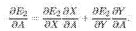
Substituting (4) in (5), we get that

If we substitute A = B = R and Θ = 0 in the combined formulation in (2), we can restrict AD to be circular. Then (6) will become:

The partial derivative of the energy Ej with respect to Rcan be found in a similar way.
The partial derivative of the energy with respect to R is:

The partial derivatives of the energy with respect to the coordinates of the center of the disc are:

[0075] We are motivated to design elliptical AD by the fact that the OD shape is not exactly a circle. But, it is slightly vertically oval, with the vertical diameter being about 7% longer than the horizontal one. It is evident in fundus images with slightly vertically oval OD as shown in FIGs 5(al)-(a2). A considerable improvement in the accuracy of segmentation of OD can be seen in switching from circular to elliptical AD. FIGs.5(bl)-(b2) and 5(cl)-(c2) show segmentation and outlining of OD by circular and elliptical AD, respectively.
[0076] Now, deriving equations for shape changing AD from circle to ellipse: from (6), we get that:

Substituting for x and dy in (10), we get:

Partial derivative of Ej with respect to ^4can be found in a similar way. It is given by,

Partial derivative of E with respect to ^4can be found as below:

Substituting (11) and (12) in (13) and simplifying, we get
8 :::: ¾( sm ' ^^Ai^ ■■· ' ■
Similarly, one can find the partial derivatives of the energy E with respect to B, Θ, Xc, and "cand are given by:

B. AUTOMATIC INITIALIZATION
[0077] Automatic initialization of the AD is an important step in the robust detection of OD in retinal fundus images and is achieved by matched filtering technique. Matched filtering is apowerful technique for detecting objects of interest in images in whichthe object information is known a priori. It maximizes the detection signal-to-noise ratio if the object of interest in the image and the template are identical. In the present case, the filter template is designed to comprise two concentric circles for which the weights of the inner circle, outer circle, and annulus are considered as positive, negative, and zero, respectively. The given image is then correlated with the template. Since the cross-correlation operation is equivalent to convolving the input image with a conjugated flipped version of the template, the matched filtering operation can be carried out directly in the spatial domain. The matched filter response at (xp, yp) is given by:
 wherew , y) is the filter template. The peak in the cross-correlation corresponds to thebest match between the image and the template and is taken as the location for
initialization the AD. The result of matched filtering and subsequent initialization of the AD is shown in FIG. 6. One could also use a cropped optic disc of the same scale and size as a template for matched filtering.
wherew , y) is the filter template. The peak in the cross-correlation corresponds to thebest match between the image and the template and is taken as the location for
initialization the AD. The result of matched filtering and subsequent initialization of the AD is shown in FIG. 6. One could also use a cropped optic disc of the same scale and size as a template for matched filtering.
SEGMENTATION OF OPTIC CUP
[0078] The OC is a form of OD depression peculiar to glaucoma. This is an eye disorder thatconcems damage to the optic nerve. Left untreated, the optic nerve could become permanently damaged, and the patient could go totally blind. Ophthalmologists or medical professionals specialized in eye care, like optometrists and orthoptists can diagnose this condition by relying on a CDR ratio. This means they have to figure out the OC' s size in relation to the total diameter of the OD. In this work, we use Otsu's multilevel thresholding algorithm to classify and cluster the pixels corresponding to OC, and then determine the size of the OC using circular AD. Otsu's multilevel thresholding algorithm assumesthe image as M classes of pixels and calculates optimum thresholds separating classessuch that the intra- class variance is minimum. Here, intra-class varianceis the weightedsum of variances of M classes. Let f represent the 2Z)grayscale intensity function,and contains N pixels with graylevels 0 to (L - 1). Let Jbe the number of pixels withgraylevelz. The probability of graylevelz is given by,

Here, (M - 1) thresholds {tj, t2, tM-j}, divide the original image into M classes:
Ci: { 1, 2, ...Ji}, C2: {ti+1, ..., t2} md CM: {tM-i+l, -,L} - The optimal thresholds
can be chosen by maximizing the inter-class variance
 where,

With

EXPERIMENTAL RESULTS
[0079] In this section, we examine the performance of the proposed technique on multiple retinalfundus images. Experiments are done on Mac OS X 2.7 GHz, Intel Core i5 using Java-ImageJ. An ImageJ plugin and a batch-processing macro is developed to implement automatic segmentation and outlining of the OD and OC and to handle any number of fundus images for glaucoma analysis, respectively. R-channel and G-channel components of color fundus images are used for processing, and optimization of the ODand OC segmentation, respectively. The evolution of the AD, final segmentation, andoutlining is shown on the original color image. Fundus images from threepublicly available and three locally obtained data sets are used for evaluation. The localization of OD and OC is done by matched filtering technique. The segmentation and outlining is done by AD method and are compared with the manual outlines provided by an expert ophthalmologist. We show
simulation results on randomly selected retinal fundus images in FIG. 8(al)-(a6) from different datasets. FIGs.8(bl)-(b6) and (cl)- (c6) show OD and OC segmentation using elliptical AD and circular AD, respectively.
[0080] Jaccard and Dice indices, sensitivity, specificity, and accuracy are the comparison criteria typically used in medical image segmentation. They are based on the definition of true positives (TP), false positives (FP), false negatives (FN), and true negatives (TN). Sensitivity = TP/(TP+FN) measures the ovelap, but can be equal to one for a poor segmentation much bigger than the ground truth. Specificity = TN/(TN+FP) is therefore the necessary counterpart of the sensitivity, but it can be equal to one for a very poor segmentation that does not detect the object of interest at all. Jaccard similarity index (J) is the most intuitive ratio between the intersection and union. Dice similarity index (A) is slightly different, but more frequently used and one can be deduced from the other. In an embodiment, quantitative comparison of the performance of the proposed method and clinician outlining is done using J andA . They are given by:
 for 0 < J < 1 and 0 <A≤ I . A and M represent the optic disc region segmented by the proposed method and clinician respectively.^ n Mdenotes the intersection between^ and and it reveals the common optic disc region in both A and M. The union between A and is denoted by A U M and it reveals optic disc region which are in either segmentation. Most of the techniques proposed in the literature for glaucoma analysis have considered only one locally obtained database.
[0081] The novelty of the proposed technique is in handling many databases which contain fundus images acquired in different conditions. The proposed glaucoma analysis tool has been validated on publicly available databases like Messidor, Drishti-GS, and Drions-DB. The proposed tool has also been validated on locally obtained fundus image databases from ForusHealth Pvt. Ltd, Bosch Eye Care Solutions, and Remidio Innovative Solutions Pvt. Ltd. The proposed method gave satisfactory segmentation results on fundus images from allsix databases.
[0082] FIG. 9 pictoriallyillustratescomparison of performance of the AD with that of the clinician outlining. Thin green and blue contours represent manual outlining of OD and OC, respectively. Thick green and blue contours show algorithm outlining of OD and OC, respectively. In FIG. 10, illustrates green line joining the centers of the OD and OC with the
points on the OD and OC contours where the cup is sloping towards the OD boundary. A red line mark overlaid on the green line shows the narrowest possible rim width. It was observed that the OD and OC segmentation and outlining is accurate, with meaningful narrowest possible rim width marked on the cup notching area. A high similarity was obtained between clinician and outlining by the proposed method.
SEVERITY ASSESSMENT OF GLAUCOMA USING CDR
[0083] Glaucoma severity refers to the presence and extensiveness of the disease in the retina of the eye. It is objectively evaluated through diagnostic testing like fundus imaging and physiological examination of the impaired OD in the form of cupping. The amount of cupping in the OD can be measured, and the corresponding severity level of the glaucoma can be determined. In an embodiment of the present disclosure, glaucoma severity information can be contrasted with symptom assessment by fully automated CDR measurement technique based on ICD-9 ruleswhich is an internationally accepted guideline on glaucoma diagnosis. The proposed method has been validated on fundus images from eight databases. Based on the CDR, proposed method categorizes the glaucomatous condition as normal, mild, moderate, or severe as given in Table 1 below in accordance with ICD-9 rules.

Table \:Glaucomatous condition classification based on CDR using ICD-9 rules.
SEVERITY ASSESSMENT OF GLAUCOMA USING RDR BASED ON DDLS
[0084] The smallest possible neuroretinal rim-width is an important parameter for disc- damagelikelihood- scale (DDLS) based glaucoma stage analysis. The problem of finding the smallest possible neuro-retinal rim-width can be formulated as finding the smallest possible distance between the two non-concentric circles one inside the other as depicted in FIG. 11. Circles with centers 01 and 02 represents the circular approximation of the OC and the OD area, respectively. The line 0102 joins the two centers of the inner and outer circles. Extending the line 01-02 upto the outer periphery of the outer circle intersects the inner and outer circle at points P and Q, respectively. The line segment PQ is the smallest possible distance between the two non-concentric circles one inside the other along the radial direction. Now, we need to prove that the line segment PQ is the smallest possible distance. In this proof, without loss of generality, it is assumed that the outer circle is centered at theorigin, and the inner circle is centered along the X-axis. All the calculations are done with respect to the first quadrant of the Cartesian plane. The respective figure used for the formulation is shown in FIG. 12. Let Y\ and r2 represent the radius of the inner and outer circles, respectively. The points P and Qbe any two arbitrary points on the periphery of inner and outer circles, θι and Θ = (θι +θ2) be the angles created by the points P and Q with respect to the origin 02 along X-axis, Θ be the angle created by point P with respect to the center Ox of the inner circle along X-axis. Let d be the distance between two centers Ox and <¾ along X- axis, and / be the smallest possible distance between thetwo non-concentric circles one inside the other along the radial direction. Here, one hasto prove that PQ>1 and PQ =1 if and only if,
[0085] From FIG. 12,
1

The coordinates of points P and Oare given

The Euclidian distance between points P and Q is

Squaring and simplifying equation (15) we get,
I PQ f^. I(d c<« k■■ m s0] i fec al
When dj =θ2 = Θ =0, X-axis passes through the centers of the inner and outer circles and cuts the periphery of the circles so that PQ = I, which is the smallest possible distance between the two circles. Let us introduce a cost function^7 = PQ2 from equation (17). To prove PQ = / is the minimum distance between the two circles one has to prove that the Jacobian ofi is equal to zero, and the Hessian ofi is positive definite.
That is
·.·.·.·.· § d ¾L ¾ « > 0

Equations (19) and (20) prove that PQ = I is the minima when
Now,considering the Hessian of the cost function £f ,


When Θ = 0 = 0,

Now, checking for trace and determinantofV2
=) with r2> ri equation (21) is satisfied.
From equation (22) we get,
=) (r2 - n - d) > 0
=) l > O.andV2 g^is positive definite.
Therefore with
proves/ is minimum. That is PQ = /is minimum when θ = Θ = 0.
Based on the RDR or narrowest rim width, glaucomatous conditions can be categorizedinto eight stages as given in Table 2 below:
Rim-to-Disc Ratio(RDR)
Stage Number Glaucomatous condition
Range
0 0.3 < RDR < 0.4 Normal
1 0.2< RDR < 0.29 Mild
2 0.1 <RDR < 0.19 Moderate
3 - 7 0.01< RDR < 0.1 Severe
Table 2:Glaucomatous condition classification based on narrowest rim width or RDR
[0086] The proposed method classifies the glaucomatous condition as normal, mild, moderate, or severe in accordance an internationally accepted guideline on DDLS based glaucoma diagnosis. The proposed method has been validated on fundus images from eight databases.
ASSESSMENT OF GLAUCOMA USING ISNT RULE
[0087] The ISNT rule states that a healthy OD has a characteristic pattern of decreasing order of inferior > superior > nasal > temporal neuroretinal rim widths and the glaucomatous ODs violate this quantitative pattern. The utility of the ISNT rule has been proved in clinical practice, but it is considered to be subjective. In the proposed method, after segmentation of the OD and OC by the proposed AD method the inferior, superior, nasal, and temporal rim widths (Euclidean distance between corresponding OD and OC coordinates) can be measured automatically and used to determine whether the determined values obey the quantitative pattern of the ISNT rule. If the ISNT rule holds for an OD, it is treated as normal otherwise glaucomatous.
[0088] FIG. 13 illustrates an exemplary flow diagram for CDR based glaucoma assessment method 1300 in accordance with an embodiment of the present disclosure. The method 1300 for CDR based glaucoma assessment can at step 1302 take a color fundus image as input; At step 1304 red channel can be extracted; At step 13062-stage or 3 -stage multi-threshold region can be extracted; At step 1308 an Active Disc can be initialized using matched filtering technique; at step 1310 coarse segmentation and outlining of Optic Disc can
be done by circular AD; at step 1312 fine segmentation and outlining of the ODcan be done by elliptical AD;. at step 1314 vertical length of OD can be printed; at step 1316 color fundus image can be again taken as input for Optic Cup segmentation; at step 1318 green channel can be extracted from the fundus image; at step 1320 4-stage multi -threshold region can be extracted; at step 1322 an Active Disc can be initialized using ODcenter and radius; at step 1324 OC can be segmented and outlined by the Active Disc; at step 1326 vertical length of the segmented and outlined OD can be printed; at step 1328 Cup-to-Disc Ratio can be computed and glaucomatous condition printed based on International Classification of Diseases-9 rules.
[0089] FIG. 14 illustrates an exemplary flow diagram for RDR based glaucoma assessment method 1400 in accordance with an embodiment of the present disclosure. Steps 1402 to 1412 pertaining to OD segmentation and outlining; and steps 1414 to 1422 pertaining to OC segmentation and outlining can correspond to and same as steps 1302 to 1312 and 1316 to 1324 of method 1300. After segmentation and outlining of the OD and OC the method 1400 can at step 1424 involve determining narrowest rim width and at step 1426 compute rim-to-disc ratio (RDR) and print glaucomatouscondition based on Disc-Damage- Likelihood-Scale (DDLS).
[0090] FIG. 15 illustrates an exemplary flow diagram for ISNT based glaucoma assessment method 1500 in accordance with an embodiment of the present disclosure. Steps 1502 to 1512 pertaining to OD segmentation and outlining; and steps 1514 to 1522 pertaining to OC segmentation and outlining can correspond to and be same as steps 1302 to 1312 and 1316 to 1324 of method 1300. After segmentation and outlining of the OD and OC the method 1500 can at step 1524 involve determining Inferior, Superior, Nasal, and Temporal rim widths and based on determined values, at step 1526 checking whether rim widths follow ISNT rule.
[0091] In an aspect, to automate the process, the proposed methodhas been implemented using Java as an ImageJ plugin. An associated batch-processing ImageJ macro to handle a database for automated glaucoma assessment has also been implemented. In addition to ImageJ plugin and batch processing macro, Android and iOS based smartphone apps for glaucoma prescreening has also been implemented. The tool is repeatable, easy to use, provides quantitative analysis, and takes only few seconds per image. The proposed algorithm has achieved full automation since it does not require any manual intervention.
[0092] In an aspect, the software implementation can be used alongside tabletop, handheld fundus cameras, and fundus-on-phone (FOP) devices. The implementation is
readily available for integration into any existing fundus camera, iOSdevices or Android devices. Any of these devices could be converted to a fundus imager with the help of an addon device.
[0093] In an aspect any one or more of the methods 1300, 1400 and 1600 can be used either individually or in combination to screen fundus images for identifying potentially glaucomatous eye conditions and carry out severity grading of glaucoma based on CDR.
[0094] Thus the present disclosure provides an automatic method that is fast and reliable for periodic, careful and comprehensive eye examination for prescreening of population and thus early diagnosis of glaucoma that can enable timely treatment of glaucoma to prevent partial or full blindness among masses. The tool can automatically screen large number of fundus images and identify potentially glaucomatous conditions with high accuracy.
[0095] While the foregoing describes various embodiments of the invention, other and further embodiments of the invention may be devised without departing from the basic scope thereof. The scope of the invention is determined by the claims that follow. The invention is not limited to the described embodiments, versions or examples, which are included to enable a person having ordinary skill in the art to make and use the invention when combined with information and knowledge available to the person having ordinary skill in the art.
ADVANTAGES OF THE INVENTION
[0096] The present disclosure provides a means for periodic,careful and comprehensive eye examination for early diagnosis of glaucoma so that a timely treatment of glaucoma can be started to prevent partial or full blindness among masses.
[0097] The present disclosure provides an automatic method that is fast and reliable for prescreening of population for glaucoma.
[0098] The present disclosure provides a method to automatically screen fundus images of the eye and identify potentially glaucomatous conditions with high accuracy.
[0099] The present disclosure provides an image processing technique and an associated software tool that is repeatable, easy to use, and takes a few seconds per patient for glaucoma prescreening.
[00100] The present disclosure provides an image processing technique and an associated software tool that can be operated by minimally trained personnel or semi-skilled personnel.
[00101] The present disclosure provides an image processing technique and an associated software tool that can be used alongside tabletop, handheld fundus cameras, and FOPs, empowering smaller clinics in rural areas to make effective use of the tool.
[00102] The present disclosure provides a method for processing fundus image and carrying out severity grading of glaucoma based on CDR/RDR.
[00103] The present disclosure provides a method for severity grading of glaucoma based on measurement of inferior, superior, nasal, and temporal neuroretinalrim width.
[00104] The present disclosure provides a method that implements ISNT rule to check whether the normal eyes show a characteristic pattern of inferior > superior > nasal > temporal rim widths to differentiate them from glaucomatous eyes.
Claims
1. A method for segmenting and outlining one or more circular and elliptical regions in an image, the method comprising steps of:
obtaining multi-stage, multi-threshold region representing the circular and elliptical regions;
initializing one or more Active Discs(ADs) and thereafter evolving the one or more ADs towards boundary of the one or more circular and elliptical regions;
coarse segmentation and outlining of the one or more circles and ellipses using a circular AD; and
fine segmentation and outlining of the one or more circles and ellipses by an elliptical AD.
2. The method of claim 1, wherein the initializing of the one or more AD is automatic using matched filter technique.
3. The method of claim 1, wherein the one or more AD is evolved towards boundary of the one or more circular and elliptical regions by optimizing its parameters based on a pre-defined energy criterion such as by minimizing a local disc energy function.
4. The method of claim 3, wherein the one or more AD changes shape from circle to ellipse when evolving based on the pre-defined energy criterion.
5. The method of claim3, wherein the AD optimization is done using an accelerated gradient decent.
6. The method of claim4, wherein during optimization the energy is computed using Green's theorem.
7. The method of claim4, wherein the one or more circular and elliptical regions are one or more of Optic Disc, Optic Cup, Iris, carotid artery, cataract, cross-sections of tubular structures.
8. A method for screening fundus images forglaucomatous conditions and severity grading, the method comprising steps of: extracting red channel from the fundus images; obtaining the multi-stage, multi-threshold region representing Optic Disc
(OD); initializing a first circular Active Disc(AD) using matched filter technique;
segmentating and outlining of the OD using the first circular AD and evolving it to an elliptical AD;
extracting green channel from the fundus image;
getting the 4-stage multi-threshold region;
initializing a second AD using segmented OD center coordinates and radius; segmenting and outlining of OC using the second AD.
9. The method of claim 8, wherein the fundus images are colour fundus images.
10. The method of claim 8, wherein the method further comprises step of determining vertical lengths of the segmented and outlined OD and OC, and using the determined vertical lengths of the OD and OC to compute the Cup-to-Disc Ratio and providing severity grading of glaucomatous condition based on International Classification of Diseases(ICD)-9 rules.
11. The method of claim 8, wherein the method determines Cup-to-Disc Ratio indirectly based on areas of estimated OD and OC.
12. The method of claim8, wherein the method further comprises step of determining narrowest rim width, and using the determined narrowest rim width to compute Rim- to-Disc Ratio (RDR), and providing severity grading of glaucomatous condition based on Disc-Damage- Likelihood-Scale (DDLS) rules.
13. The method of claim 8, wherein the method further comprises step of determining inferior, superior, nasal, and temporal rim widths, and providing a glaucomatous/non- glaucomatous decision based on determined inferior, superior, nasal, and temporal rim widths using ISNT rule.
14. The method of claim 8, wherein the method further comprises step of optimizing the first and the second AD using accelerated gradient descent.
15. The method of claim 8, wherein during the optimization of the first AD and the second AD the energy is computed using Green's theorem.
16. The method of claim 8, wherein the first AD and the second AD are nonconcentric circles and ellipses.
Applications Claiming Priority (2)
| Application Number | Priority Date | Filing Date | Title |
|---|---|---|---|
| IN201741017981 | 2017-05-23 | ||
| IN201741017981 | 2017-05-23 |
Publications (1)
| Publication Number | Publication Date |
|---|---|
| WO2018215855A1 true WO2018215855A1 (en) | 2018-11-29 |
Family
ID=64395556
Family Applications (1)
| Application Number | Title | Priority Date | Filing Date |
|---|---|---|---|
| PCT/IB2018/053030 Ceased WO2018215855A1 (en) | 2017-05-23 | 2018-05-02 | Automated fundus image processing techniques for glaucoma prescreening |
Country Status (1)
| Country | Link |
|---|---|
| WO (1) | WO2018215855A1 (en) |
Cited By (8)
| Publication number | Priority date | Publication date | Assignee | Title |
|---|---|---|---|---|
| CN109658423A (en) * | 2018-12-07 | 2019-04-19 | 中南大学 | A kind of optic disk optic cup automatic division method of colour eyeground figure |
| CN110516579A (en) * | 2019-08-21 | 2019-11-29 | 北京至真互联网技术有限公司 | Hand-held fundus camera photographic method and device, equipment and storage medium |
| CN111528790A (en) * | 2020-04-21 | 2020-08-14 | 丁涛 | Image capturing device, vision data processing method and fundus data processing method |
| CN111863241A (en) * | 2020-07-10 | 2020-10-30 | 北京化工大学 | A fundus photography classification system based on ensemble deep learning |
| CN111986202A (en) * | 2020-10-26 | 2020-11-24 | 平安科技(深圳)有限公司 | Glaucoma auxiliary diagnosis device, method and storage medium |
| CN113012093A (en) * | 2019-12-04 | 2021-06-22 | 深圳硅基智能科技有限公司 | Training method and training system for glaucoma image feature extraction |
| CN113450341A (en) * | 2021-07-16 | 2021-09-28 | 依未科技(北京)有限公司 | Image processing method and device, computer readable storage medium and electronic device |
| CN115018799A (en) * | 2022-06-15 | 2022-09-06 | 潍坊眼科医院有限责任公司 | Fundus image quality optimization method based on deep learning |
-
2018
- 2018-05-02 WO PCT/IB2018/053030 patent/WO2018215855A1/en not_active Ceased
Non-Patent Citations (2)
| Title |
|---|
| JAGADISH NAYAK ET AL.: "Automated Diagnosis of Glaucoma Using Digital Fundus Images", J MED SYST, vol. 33, 2009, pages 337 - 346, XP019750754 * |
| KUMAR, J. R. HARISH ET AL.: "Active discs for automated optic disc segmentation", IEEE GLOBAL CONFERENCE ON SIGNAL AND INFORMATION PROCESSING 2015, 2015, pages 2,3, XP032871651 * |
Cited By (17)
| Publication number | Priority date | Publication date | Assignee | Title |
|---|---|---|---|---|
| CN109658423A (en) * | 2018-12-07 | 2019-04-19 | 中南大学 | A kind of optic disk optic cup automatic division method of colour eyeground figure |
| CN109658423B (en) * | 2018-12-07 | 2022-12-09 | 中南大学 | Automatic optic disk cup segmentation method for color fundus picture |
| CN110516579B (en) * | 2019-08-21 | 2021-09-07 | 北京至真互联网技术有限公司 | Handheld fundus camera photographing method and device, device and storage medium |
| CN110516579A (en) * | 2019-08-21 | 2019-11-29 | 北京至真互联网技术有限公司 | Hand-held fundus camera photographic method and device, equipment and storage medium |
| CN113012093A (en) * | 2019-12-04 | 2021-06-22 | 深圳硅基智能科技有限公司 | Training method and training system for glaucoma image feature extraction |
| CN113012093B (en) * | 2019-12-04 | 2023-12-12 | 深圳硅基智能科技有限公司 | Training method and training system for glaucoma image feature extraction |
| CN111528790A (en) * | 2020-04-21 | 2020-08-14 | 丁涛 | Image capturing device, vision data processing method and fundus data processing method |
| CN111528790B (en) * | 2020-04-21 | 2023-08-04 | 张姬娟 | Image capturing device, vision data processing method and fundus data processing method |
| CN111863241A (en) * | 2020-07-10 | 2020-10-30 | 北京化工大学 | A fundus photography classification system based on ensemble deep learning |
| CN111863241B (en) * | 2020-07-10 | 2023-06-30 | 北京化工大学 | A Fundus Photo Classification System Based on Integrated Deep Learning |
| CN111986202B (en) * | 2020-10-26 | 2021-02-05 | 平安科技(深圳)有限公司 | Glaucoma auxiliary diagnosis device, method and storage medium |
| WO2021159811A1 (en) * | 2020-10-26 | 2021-08-19 | 平安科技(深圳)有限公司 | Auxiliary diagnostic apparatus and method for glaucoma, and storage medium |
| CN111986202A (en) * | 2020-10-26 | 2020-11-24 | 平安科技(深圳)有限公司 | Glaucoma auxiliary diagnosis device, method and storage medium |
| US11978215B2 (en) | 2020-10-26 | 2024-05-07 | Ping An Technology (Shenzhen) Co., Ltd. | Device and method for glaucoma auxiliary diagnosis, and storage medium |
| CN113450341A (en) * | 2021-07-16 | 2021-09-28 | 依未科技(北京)有限公司 | Image processing method and device, computer readable storage medium and electronic device |
| CN115018799A (en) * | 2022-06-15 | 2022-09-06 | 潍坊眼科医院有限责任公司 | Fundus image quality optimization method based on deep learning |
| CN115018799B (en) * | 2022-06-15 | 2022-11-11 | 潍坊眼科医院有限责任公司 | Fundus image quality optimization method based on deep learning |
Similar Documents
| Publication | Publication Date | Title |
|---|---|---|
| WO2018215855A1 (en) | Automated fundus image processing techniques for glaucoma prescreening | |
| US11704791B2 (en) | Multivariate and multi-resolution retinal image anomaly detection system | |
| Dutta et al. | Glaucoma detection by segmenting the super pixels from fundus colour retinal images | |
| Seoud et al. | Red lesion detection using dynamic shape features for diabetic retinopathy screening | |
| Akram et al. | Automated detection of dark and bright lesions in retinal images for early detection of diabetic retinopathy | |
| Fatima Bokhari et al. | Fundus image segmentation and feature extraction for the detection of glaucoma: A new approach | |
| US9107617B2 (en) | Obtaining data for automatic glaucoma screening, and screening and diagnostic techniques and systems using the data | |
| Agarwal et al. | A novel approach to detect glaucoma in retinal fundus images using cup-disk and rim-disk ratio | |
| Hassan et al. | Automated retinal edema detection from fundus and optical coherence tomography scans | |
| Soomro et al. | Automatic retinal vessel extraction algorithm | |
| Das et al. | Diagnosis of glaucoma using CDR and NRR area in retina images | |
| Hassan et al. | Fully automated detection, grading and 3D modeling of maculopathy from OCT volumes | |
| WO2010071597A1 (en) | A method and system for determining the position of an optic cup boundary | |
| US20130222767A1 (en) | Methods and systems for detecting peripapillary atrophy | |
| Brancati et al. | Automatic segmentation of pigment deposits in retinal fundus images of Retinitis Pigmentosa | |
| Wisaeng | Retinal blood vessel segmentation using density-based fuzzy C-means clustering and vessel neighborhood connected component | |
| Dutta et al. | Automatic evaluation and predictive analysis of optic nerve head for the detection of glaucoma | |
| Sarhan et al. | Approaches for early detection of glaucoma using retinal images: A performance analysis | |
| Kumar et al. | Detection of non-proliferative diabetic retinopathy using GUI | |
| Lotankar et al. | Glaucoma Screening using Digital Fundus Image through Optic Disc and Cup Segmentation | |
| Listyalina et al. | Automated localisation of optic disc in retinal colour fundus image for assisting in the diagnosis of glaucoma | |
| Pandey et al. | Automatic detection of red lesions in diabetic retinopathy using shape based extraction technique in fundus image | |
| Bhatkalkar et al. | Feature extraction for early detection of macular hole and glaucoma in fundus images | |
| KHAING et al. | Optic disc segmentation applied to retinal images with glaucoma, diabetic retinopathy, and optic disc edema | |
| Issac et al. | Automated localization of optic nerve head in a fundus image using computer vision techniques |
Legal Events
| Date | Code | Title | Description |
|---|---|---|---|
| 121 | Ep: the epo has been informed by wipo that ep was designated in this application |
Ref document number: 18806028 Country of ref document: EP Kind code of ref document: A1 |
|
| NENP | Non-entry into the national phase |
Ref country code: DE |
|
| 122 | Ep: pct application non-entry in european phase |
Ref document number: 18806028 Country of ref document: EP Kind code of ref document: A1 |