WO2018094300A1 - Anti-gitr antigen-binding proteins and methods of use thereof - Google Patents
Anti-gitr antigen-binding proteins and methods of use thereof Download PDFInfo
- Publication number
- WO2018094300A1 WO2018094300A1 PCT/US2017/062443 US2017062443W WO2018094300A1 WO 2018094300 A1 WO2018094300 A1 WO 2018094300A1 US 2017062443 W US2017062443 W US 2017062443W WO 2018094300 A1 WO2018094300 A1 WO 2018094300A1
- Authority
- WO
- WIPO (PCT)
- Prior art keywords
- seq
- abp
- cdr
- antibody
- sequence
- Prior art date
Links
Classifications
-
- C—CHEMISTRY; METALLURGY
- C07—ORGANIC CHEMISTRY
- C07K—PEPTIDES
- C07K16/00—Immunoglobulins [IGs], e.g. monoclonal or polyclonal antibodies
- C07K16/18—Immunoglobulins [IGs], e.g. monoclonal or polyclonal antibodies against material from animals or humans
- C07K16/28—Immunoglobulins [IGs], e.g. monoclonal or polyclonal antibodies against material from animals or humans against receptors, cell surface antigens or cell surface determinants
- C07K16/2878—Immunoglobulins [IGs], e.g. monoclonal or polyclonal antibodies against material from animals or humans against receptors, cell surface antigens or cell surface determinants against the NGF-receptor/TNF-receptor superfamily, e.g. CD27, CD30, CD40, CD95
-
- A—HUMAN NECESSITIES
- A61—MEDICAL OR VETERINARY SCIENCE; HYGIENE
- A61P—SPECIFIC THERAPEUTIC ACTIVITY OF CHEMICAL COMPOUNDS OR MEDICINAL PREPARATIONS
- A61P31/00—Antiinfectives, i.e. antibiotics, antiseptics, chemotherapeutics
- A61P31/12—Antivirals
-
- A—HUMAN NECESSITIES
- A61—MEDICAL OR VETERINARY SCIENCE; HYGIENE
- A61P—SPECIFIC THERAPEUTIC ACTIVITY OF CHEMICAL COMPOUNDS OR MEDICINAL PREPARATIONS
- A61P35/00—Antineoplastic agents
-
- A—HUMAN NECESSITIES
- A61—MEDICAL OR VETERINARY SCIENCE; HYGIENE
- A61P—SPECIFIC THERAPEUTIC ACTIVITY OF CHEMICAL COMPOUNDS OR MEDICINAL PREPARATIONS
- A61P35/00—Antineoplastic agents
- A61P35/02—Antineoplastic agents specific for leukemia
-
- A—HUMAN NECESSITIES
- A61—MEDICAL OR VETERINARY SCIENCE; HYGIENE
- A61P—SPECIFIC THERAPEUTIC ACTIVITY OF CHEMICAL COMPOUNDS OR MEDICINAL PREPARATIONS
- A61P37/00—Drugs for immunological or allergic disorders
- A61P37/02—Immunomodulators
- A61P37/04—Immunostimulants
-
- A—HUMAN NECESSITIES
- A61—MEDICAL OR VETERINARY SCIENCE; HYGIENE
- A61P—SPECIFIC THERAPEUTIC ACTIVITY OF CHEMICAL COMPOUNDS OR MEDICINAL PREPARATIONS
- A61P43/00—Drugs for specific purposes, not provided for in groups A61P1/00-A61P41/00
-
- C—CHEMISTRY; METALLURGY
- C07—ORGANIC CHEMISTRY
- C07K—PEPTIDES
- C07K16/00—Immunoglobulins [IGs], e.g. monoclonal or polyclonal antibodies
- C07K16/18—Immunoglobulins [IGs], e.g. monoclonal or polyclonal antibodies against material from animals or humans
- C07K16/28—Immunoglobulins [IGs], e.g. monoclonal or polyclonal antibodies against material from animals or humans against receptors, cell surface antigens or cell surface determinants
- C07K16/2875—Immunoglobulins [IGs], e.g. monoclonal or polyclonal antibodies against material from animals or humans against receptors, cell surface antigens or cell surface determinants against the NGF/TNF superfamily, e.g. CD70, CD95L, CD153, CD154
-
- A—HUMAN NECESSITIES
- A61—MEDICAL OR VETERINARY SCIENCE; HYGIENE
- A61K—PREPARATIONS FOR MEDICAL, DENTAL OR TOILETRY PURPOSES
- A61K39/00—Medicinal preparations containing antigens or antibodies
- A61K2039/505—Medicinal preparations containing antigens or antibodies comprising antibodies
-
- C—CHEMISTRY; METALLURGY
- C07—ORGANIC CHEMISTRY
- C07K—PEPTIDES
- C07K2317/00—Immunoglobulins specific features
- C07K2317/30—Immunoglobulins specific features characterized by aspects of specificity or valency
- C07K2317/34—Identification of a linear epitope shorter than 20 amino acid residues or of a conformational epitope defined by amino acid residues
-
- C—CHEMISTRY; METALLURGY
- C07—ORGANIC CHEMISTRY
- C07K—PEPTIDES
- C07K2317/00—Immunoglobulins specific features
- C07K2317/30—Immunoglobulins specific features characterized by aspects of specificity or valency
- C07K2317/35—Valency
-
- C—CHEMISTRY; METALLURGY
- C07—ORGANIC CHEMISTRY
- C07K—PEPTIDES
- C07K2317/00—Immunoglobulins specific features
- C07K2317/50—Immunoglobulins specific features characterized by immunoglobulin fragments
- C07K2317/51—Complete heavy chain or Fd fragment, i.e. VH + CH1
-
- C—CHEMISTRY; METALLURGY
- C07—ORGANIC CHEMISTRY
- C07K—PEPTIDES
- C07K2317/00—Immunoglobulins specific features
- C07K2317/50—Immunoglobulins specific features characterized by immunoglobulin fragments
- C07K2317/515—Complete light chain, i.e. VL + CL
-
- C—CHEMISTRY; METALLURGY
- C07—ORGANIC CHEMISTRY
- C07K—PEPTIDES
- C07K2317/00—Immunoglobulins specific features
- C07K2317/50—Immunoglobulins specific features characterized by immunoglobulin fragments
- C07K2317/55—Fab or Fab'
-
- C—CHEMISTRY; METALLURGY
- C07—ORGANIC CHEMISTRY
- C07K—PEPTIDES
- C07K2317/00—Immunoglobulins specific features
- C07K2317/50—Immunoglobulins specific features characterized by immunoglobulin fragments
- C07K2317/56—Immunoglobulins specific features characterized by immunoglobulin fragments variable (Fv) region, i.e. VH and/or VL
- C07K2317/565—Complementarity determining region [CDR]
-
- C—CHEMISTRY; METALLURGY
- C07—ORGANIC CHEMISTRY
- C07K—PEPTIDES
- C07K2317/00—Immunoglobulins specific features
- C07K2317/70—Immunoglobulins specific features characterized by effect upon binding to a cell or to an antigen
- C07K2317/75—Agonist effect on antigen
-
- C—CHEMISTRY; METALLURGY
- C07—ORGANIC CHEMISTRY
- C07K—PEPTIDES
- C07K2317/00—Immunoglobulins specific features
- C07K2317/90—Immunoglobulins specific features characterized by (pharmaco)kinetic aspects or by stability of the immunoglobulin
- C07K2317/92—Affinity (KD), association rate (Ka), dissociation rate (Kd) or EC50 value
Definitions
- ABSPs antigen-binding proteins
- GITR glucocorticoid-induced tumor necrosis factor receptor-related protein
- compositions comprising such ABPs, including pharmaceutical compositions, diagnostic compositions, and kits.
- methods of making GITR ABPs, and methods of using GITR ABPs for example, for therapeutic purposes, diagnostic purposes, and research purposes.
- GITR is a member of the tumor necrosis factor receptor (TNFR) superfamily. GITR is expressed in many cells of the innate and adaptive immune system, and membrane surface expression is increased in activated T cells. See Hanabuchib et al., Blood, 2006, 107:3617- 3623; and Nocentini et al., Eur. J. Immunol, 2005, 35: 1016-1022; each of which is incorporated by reference in its entirety. GITR is activated by GITR ligand (GITRL).
- GITRL GITR ligand
- Agonism of GITR has a co-stimulatory effect on effector T cells. See Schaer et al., Curr.
- GITR agonists have been proposed as therapeutic agents for cancer therapy. See Schaer et al., supra; Melero et al., Clin Cancer Res., 2013, 19: 1044-1053; Cohen et al., J. Clin. Oncol, 2007, 25:3058; Cohen et al., PLoS One, 2010, 5:el0436; Nocentini et al., Br. J.
- ABPs that specifically bind GITR and methods of using such ABPs.
- ABSP isolated multivalent antigen binding protein
- GITR human GITR
- SEQ ID NO: 1 a CDR-H3 having the sequence X1X2X3X4X5RGYGDYGGHHAFDI, wherein Xi is A or V, X 2 is H, D, L, or R, X 3 is E or D, X 4 is R, N, S, or A, and X 5 is V, D or G (SEQ ID NO: 141);
- a CDR-H2 having the sequence X1IX2X3SGX4TYYNPSLKS, wherein Xi is G, L, or S, X 2 is Y, A, or V, X 3 is E, Y or H, and X 4 is S or K (SEQ ID NO: 142);
- a CDR-H1 having the sequence X1SISSX2X3X4X5WX6, wherein Xi is Y or G, X2 is G
- the ABP of (a) comprises a VH sequence of SEQ ID NO: 9 and a VL sequence of SEQ ID NO: 10; in another embodiment, the ABP of (b) comprises a VH sequence of SEQ ID NO: 19 and a VL sequence of SEQ ID NO:20; in another embodiment, the ABP of (c) comprises a VH sequence of SEQ ID NO:26 and a VL sequence of SEQ ID NO:27; in another embodiment, the ABP of (d) comprises a VH sequence of SEQ ID NO:26 or SEQ ID NO:34 and a V L sequence of SEQ ID NO:35; in another embodiment, the ABP of (e) comprises a VH sequence of SEQ ID NO:26 and a VL sequence of SEQ ID NO:40; in another embodiment, the ABP of (f) comprises a VH sequence of SEQ ID NO:44 and a VL sequence of SEQ ID NO:45; in another embodiment, the ABP of (g) comprises a V H sequence
- the ABP of (b) comprises a heavy chain of SEQ ID NO:7 and a light chain of SEQ ID NO:8; In another embodiment, the ABP of (b) comprises a heavy chain of SEQ ID NO: 17 and a light chain of SEQ ID NO: 18. In another embodiment, the ABP of (c) comprises a heavy chain of SEQ ID NO:24 and a light chain of SEQ ID NO:25. In another embodiment, the ABP of (d) comprises (i) a heavy chain of SEQ ID NO:32 and a light chain of SEQ ID NO: 33, or (ii) a heavy chain of SEQ ID NO: 37 and a light chain of SEQ ID NO:33.
- the ABP of (e) comprises (i) a heavy chain of SEQ ID NO:38 and a light chain of SEQ ID NO:39.
- the ABP of (f) comprises a heavy chain of SEQ ID NO: 42 and a light chain of SEQ ID NO: 43;
- the ABP of (g) comprises (i) a heavy chain of SEQ ID NO: 51 and a light chain of SEQ ID NO:52;
- the ABP of (h) comprises (i) a heavy chain of SEQ ID NO:57 and a light chain of SEQ ID NO: 8;
- the ABP of (i) comprises (i) a heavy chain of SEQ ID NO: 114 and a light chain of SEQ ID NO: 8, or (ii) a heavy chain of SEQ ID NO: 120 and a light chain of SEQ ID NO: 8; or (iii) a heavy chain of SEQ ID NO: 122 and a light chain of SEQ ID NO: 8; or in another
- the ABP comprises a heavy chain of SEQ ID NO:7 and a light chain of SEQ ID NO:8; or a heavy chain of SEQ ID NO: 17 and a light chain of SEQ ID NO: 18; or a heavy chain of SEQ ID NO:24 and a light chain of SEQ ID NO:25; a heavy chain of SEQ ID NO:32 and a light chain of SEQ ID NO:33, or (ii) a heavy chain of SEQ ID NO:37 and a light chain of SEQ ID NO:33; a heavy chain of SEQ ID NO:38 and a light chain of SEQ ID NO:39; a heavy chain of SEQ ID NO:42 and a light chain of SEQ ID NO:43; a heavy chain of SEQ ID NO:51 and a light chain of SEQ ID NO:52; a heavy chain of SEQ ID NO:57 and a light chain of SEQ ID NO: 8; a heavy chain of SEQ ID NO: 114 and a light chain of SEQ ID NO:
- an isolated multivalent antigen binding protein that specifically binds human GITR (hGITR; SEQ ID NO: 1), comprising the following six CDR sequences: (a) a CDR-H3 having the sequence set forth in SEQ ID NO:66; (b) a CDR-H2 having the sequence set forth in SEQ ID NO:65; (c) a CDR-H1 having the sequence set forth in SEQ ID NO:64; (d) a CDR-L3 having the sequence set forth in SEQ ID NO:69; (e) a CDR-L2 having the sequence set forth in SEQ ID NO:68; and (f) a CDR- Ll having the sequence set forth in SEQ ID NO: 67.
- the ABP comprises a VH sequence of SEQ ID NO:62 and a VL sequence of SEQ ID NO: 63; a VH sequence of SEQ ID NO: 70 and a VL sequence of SEQ ID NO:63; or a V H sequence of SEQ ID NO:97 and a V L sequence of SEQ ID NO:63.
- the ABP comprises (i) a heavy chain of SEQ ID NO: 171 and a light chain of SEQ ID NO: 172; a heavy chain of SEQ ID NO: 173 and a light chain of SEQ ID NO: 174; a heavy chain sequence of SEQ ID NO: 106 and a light chain sequence of SEQ ID NO: 107; or (ii) a heavy chain sequence of SEQ ID NO: 116 and a light chain sequence of SEQ ID NO: 107.
- an isolated multivalent antigen binding protein that specifically binds human GITR (hGITR; SEQ ID NO: 1), comprising the following six CDR sequences: (a) a CDR-H3 having the sequence set forth in SEQ ID NO: 75; (b) a CDR-H2 having the sequence set forth in SEQ ID NO:74; (c) a CDR-H1 having the sequence set forth in SEQ ID NO: 73; (d) a CDR-L3 having the sequence set forth in SEQ ID NO:78; (e) a CDR-L2 having the sequence set forth in SEQ ID NO:77; and (f) a CDR- Ll having the sequence set forth in SEQ ID NO: 75.
- the ABP comprises a VH sequence of SEQ ID NO:71 and a VL sequence of SEQ ID NO:72; or a VH sequence of SEQ ID NO:98 and a VL sequence of SEQ ID NO:72.
- the ABP comprises a heavy chain of SEQ ID NO: 173 and a light chain of SEQ ID NO: 109; or the ABP comprises (i) a heavy chain of SEQ ID NO: 108 and a light chain of SEQ ID NO: 109; or (ii) a heavy chain of SEQ ID NO: 117 and a light chain of SEQ ID NO: 109.
- an isolated multivalent antigen binding protein that specifically binds human GITR (hGITR; SEQ ID NO: 1), comprising the following six CDR sequences: (a) a CDR-H3 having the sequence set forth in SEQ ID NO:83; (b) a CDR-H2 having the sequence set forth in (i) SEQ ID NO:82, or (ii) SEQ ID NO: 100; (c) a CDR-H1 having the sequence set forth in SEQ ID NO:81; (d) a CDR-L3 having the sequence set forth in SEQ ID NO:86; (e) a CDR-L2 having the sequence set forth in SEQ ID NO:85; and (f) a CDR-L1 having the sequence set forth in SEQ ID NO:84.
- hGITR human GITR
- SEQ ID NO: 1 comprising the following six CDR sequences: (a) a CDR-H3 having the sequence set forth in SEQ ID NO:83; (b) a CDR-H2
- the ABP comprises the CDR-H2 sequence of (b)(i) of the above paragraph and a VH sequence of SEQ ID NO:79 and a VL sequence of SEQ ID NO:80. In another embodiment, the ABP comprises the CDR-H2 sequence of (b)(i) of the above paragraph and a VH sequence of SEQ ID NO: 87 and a VL sequence of SEQ ID NO: 80. In another embodiment, the ABP comprises the CDR-H2 sequence of (b)(i) of the above paragraph and a VH sequence of SEQ ID NO: 88 and a VL sequence of SEQ ID NO: 80. In another embodiment, the ABP comprises the CDR-H2 sequence of (b)(ii) of the above paragraph and a VH sequence of SEQ ID NO:99 and a VL sequence of SEQ ID NO:80.
- the ABP comprises a heavy chain of SEQ ID NO: 174 and a light chain of SEQ ID NO: 111; In another embodiment, the ABP comprises a heavy chain of SEQ ID NO: 175 and a light chain of SEQ ID NO: 111; in another embodiment, the ABP comprises a heavy chain of SEQ ID NO: 176 and a light chain of SEQ ID NO: 111; In another embodiment, the ABP comprises a heavy chain of SEQ ID NO: 110 and a light chain of SEQ ID NO: 111; or (ii) a heavy chain of SEQ ID NO: 118 and a light chain of SEQ ID NO: 111.
- an isolated multivalent antigen binding protein that specifically binds human GITR (hGITR; SEQ ID NO: 1), comprising the following six CDR sequences: (a) a CDR-H3 having the sequence set forth in SEQ ID NO: 93; (b) a CDR-H2 having the sequence GIIPIFGEAQYAQX iFX 2 G, wherein Xi is K or R, and X 2 is Q or R (SEQ ID NO:215); (c) a CDR-H1 having the sequence set forth in SEQ ID NO:91; (d) a CDR-L3 having the sequence set forth in SEQ ID NO:94; (d) a CDR-L2 having the sequence set forth in SEQ ID NO: 85; and (e) a CDR-L1 having the sequence set forth in SEQ ID NO: 84.
- the ABP comprises: a CDR-H2 of SEQ ID NO:92; a CDR-H2 of SEQ ID NO:96; or a CDR-H2 of SEQ ID NO: 102.
- the ABP comprises a VH sequence of SEQ ID NO: 89 and a VL sequence of SEQ ID NO:90. In another embodiment, the ABP comprises a VH sequence of SEQ ID NO: 95 and a VL sequence of SEQ ID NO: 90. In another embodiment, the ABP comprises a VH sequence of SEQ ID NO: 101 and a VL sequence of SEQ ID NO:90.
- the ABP comprises a heavy chain of SEQ ID NO: 177 and a light chain of SEQ ID NO: 113. In another embodiment, the ABP comprises a heavy chain of SEQ ID NO: 178 and a light chain of SEQ ID NO: 113. In another embodiment, the ABP comprises a heavy chain of SEQ ID NO: 112 and a light chain of SEQ ID NO: 113; or (ii) a heavy chain of SEQ ID NO: 119 and a light chain of SEQ ID NO: 113.
- an isolated multivalent antigen binding protein that specifically binds human GITR (hGITR; SEQ ID NO: 1), comprising the following six CDR sequences: (a) a CDR-H3 having the sequence set forth in SEQ ID NO: 134; (b) a CDR-H2 having the sequence set forth in SEQ ID NO: 133;a CDR-H1 having the sequence set forth in SEQ ID NO: 132; (c) a CDR-L3 having the sequence set forth in SEQ ID NO: 135; (d) a CDR-L2 having the sequence set forth in SEQ ID NO:68; and (e) a CDR-L1 having the sequence set forth in SEQ ID NO: 67.
- the ABP comprises (i) a VH sequence of SEQ ID NO: 126 and a VL sequence of SEQ ID NO: 128; or (ii) a V H sequence of SEQ ID NO: 127 and a V L sequence of SEQ ID NO: 128.
- the ABP comprises (i) a heavy chain of SEQ ID NO: 124 and a light chain of SEQ ID NO: 125; or (ii) a heavy chain of SEQ ID NO: 136 and a light chain of SEQ ID NO: 125.
- an isolated multivalent antigen binding protein that specifically binds human GITR (hGITR; SEQ ID NO: 1), comprising: (a) a CDR-H3 having at least about 60%, at least about 70%, at least about 80%, at least about 90%, or at least about 95% identity to a CDR-H3 of a V H region selected from SEQ ID NOs: 9, 19, 26, 34, 44, 58, 62, 70, 71, 79, 87, 88, 89, 95, 97, 98, 99, 101, 104, 105, 126, and 127; (b) a CDR-H2 having at least about 60%, at least about 70%, at least about 80%, at least about 90%, or at least about 95% identity to a CDR-H2 of a V H region selected from SEQ ID NOs: 9, 19, 26, 34, 44, 58, 62, 70, 71, 79, 87, 88, 89, 95, 97
- the CDR-H3, CDR-H2, CDR-H1, CDR-L3, CDR-L2, and CDR- Ll are each identified according to a numbering scheme selected from the Kabat numbering scheme, the Chothia numbering scheme, or the IMGT numbering scheme.
- the CDR-H1 is identified as defined by both the Chothia and Kabat numbering schemes, inclusive of the boundaries of both numbering schemes.
- the CDR-H3 comprises a CDR-H3 selected from SEQ ID NOs:
- the CDR-H2 comprises a CDR-H3 selected from SEQ ID NOs: 12, 22, 29, 47, 60, 65, 74, 82, 92, 96, 100, 102, 130, and 133, or a variant thereof having 1, 2, or 3 amino acid substitutions.
- the CDR-H1 comprises a CDR-H1 selected from SEQ ID NOs: 11, 21, 28, 46, 59, 64, 73, 81, 91, 129, 132, or a variant thereof having 1 or 2 amino acid substitutions.
- the CDR-L3 comprises a CDR-L3 selected from SEQ ID NOs: 16, 17, 56, 69, 78, 86, 94, 135, or a variant thereof having 1 or 2 amino acid substitutions.
- the CDR-L2 comprises a CDR-L2 selected from SEQ ID NOs: 15, 31, 41, 50, 55, 68, 77, and 85, or a variant thereof having 1 amino acid substitution.
- the CDR-L1 comprises a CDR-L1 selected from SEQ ID NOs: 14, 36, 49, 54, 67, 76, and 84, or a variant thereof having 1 or 2 amino acid substitutions.
- the amino acid substitutions are conservative amino acid substitutions.
- the ABP (a) competes for binding to GITR with an antibody selected from ABP1, ABP2, ABP3, ABP4, ABP5, ABP6, ABP7, ABP8, ABP9, ABP10, ABP11, ABP 12, ABP13, ABP 14, ABP15, ABP 16, ABP 17, ABP 18, ABP 19, ABP20, ABP21, ABP22, ABP23, ABP24, ABP25, ABP26, ABP27, ABP28, ABP29, ABP30, ABP31, ABP32, ABP33, and ABP34, each as provided in Appendix A of this disclosure; or (b) has at least three antigen-binding domains that specifically bind an epitope on GITR or (c) has at least three antigen-binding domains that specifically bind a single epitope on GITR; or (d) has at least four antigen-binding domains that specifically bind an epitope on GITR; or
- the GITR is selected from hGITR (SEQ ID NO: 1), hGITR-T43R (SEQ ID NO: 2), cGITR (SEQ ID NO: 3), mGITR (SEQ ID NO: 4), and combinations thereof
- the ABP (a) specifically binds cynomolgus monkey GITR (cGITR; SEQ ID NO: 3); (b) binds murine GITR (mGITR; SEQ ID NO: 4) with an affinity lower (as indicated by higher KD) than the affinity of the ABP for hGITR, or does not bind mGITR; or (c) is capable of any combination of (a) - (b).
- the ABP (a) specifically binds cGITR (SEQ ID NO: 3); (b) binds mGITR (SEQ ID NO: 4) with an affinity lower (as indicated by higher KD) than the affinity of the ABP for hGITR and cGITR; and (c) enhances binding of GITRL to GITR.
- an ABP that competes for binding to GITR with an ABP of any one of claims 1-26, wherein the ABP: (a) specifically binds cGITR (SEQ ID NO: 3); (b) binds mGITR (SEQ ID NO: 4) with an affinity lower (as indicated by higher KD) than the affinity of the ABP for hGITR and cGITR; and (c) enhances binding of GITRL to GITR.
- the ABP comprises an antibody.
- the antibody is a monoclonal antibody.
- the antibody is selected from a human antibody, a humanized antibody or a chimeric antibody.
- the ABP is multivalent.
- the ABP comprises an antibody fragment.
- the ABP comprises an alternative scaffold.
- the ABP comprises an immunoglobulin constant region.
- the ABP comprises heavy chain constant region of a class selected from IgA, IgD, IgE, IgG, or IgM.
- the ABP comprises a heavy chain constant region of the class IgG and a subclass selected from IgG4, IgGl, IgG2, or IgG3.
- At least one Fab is fused to a C- terminus of an Fc domain of an IgG.
- the ABP further comprises at least one linker.
- the IgG is an IgG4.
- the IgG is an IgGl .
- at least one Fab is fused to an N-terminus of an Fc domain of an IgG.
- the at least one Fab is at least two Fabs.
- the at least one Fab is at least three Fabs.
- the at least one Fab is at least four Fabs.
- two Fabs are independently fused to an N-terminus of the IgG.
- two Fabs are independently fused to a C-terminus of the IgG.
- a Fab is attached to each N-terminus of the IgG, a linker is attached to each said Fab, and a Fab is attached to each linker.
- a Fab is attached to each C-terminus of the IgG, a linker is attached to each said Fab, and a Fab is attached to each linker.
- each linker comprises SEQ ID NO: 5.
- each linker comprises SEQ ID NO: 6.
- the ABP comprises a common light chain antibody, an antibody with a knobs-into-holes modification, an scFv attached to an IgG, a Fab attached to an IgG, a diabody, a tetravalent bispecific antibody, a DVD-IgTM, a DARTTM, a DuoBody®, a CovX-Body, an Fcab antibody, a TandAb®, a tandem Fab, a ZybodyTM, or combinations thereof.
- the ABP binds more than one GITR molecule.
- the ABP is independent of GITRL binding.
- the ABP enhances binding of GITRL to GITR by at least about 10%, at least about 20%, at least about 30%, at least about 40%, at least about 50%, at least about 60%, at least about 70%, at least about 80%, or at least about 90%. In another embodiment, the ABP enhances binding of GITRL to GITR by at least about 50%.
- the target cell is selected from an effector T cell, a regulatory T cell, a natural killer (NK) cell, a natural killer T (NKT) cell, a dendritic cell, and a B cell.
- the target cell is an effector T cell selected from a helper (CD4+) T cell, a cytotoxic (CD8+) T cell, and combinations thereof.
- the target cell is a regulatory T cell selected from a CD4+CD25+Foxp3+ regulatory T cell, a CD8+CD25+ regulatory T cell, and combinations thereof.
- the tissue is a tumor.
- the KD of the first antigen-binding domain for hGITR (SEQ ID NO: 1) or hGITR-T43R (SEQ ID NO: 2) is less than about 20 nM. In one embodiment, the KD of the first antigen-binding domain for cGITR (SEQ ID NO: 3) is less than about 200 nM. In another embodiment, the KD of the second antigen- binding domain for hGITR (SEQ ID NO: 1) or hGITR-T43R (SEQ ID NO: 2) is less than about 100 nM. In another embodiment, the KD of the second antigen-binding domain for cGITR (SEQ ID NO: 3) is less than about 1 ⁇ .
- the ABP comprises an Fc domain with reduced effector function when compared to an IgGl Fc domain. In another embodiment, the ABP comprises an aglycosylated Fc domain. In another embodiment, the ABP comprises an IgGl Fc domain with an alanine at one or more of positions 234, 235, 265, and 297.
- the GITR is expressed on the surface of a target cell.
- the ABP multimerizes GITR expressed on the surface of a target cell.
- the ABP multimerizes 2, 3, 4, 5, 6, 7, 8, 9, 10, 11, or 12 GITR molecules.
- the ABP specifically binds an epitope of human GITR (hGITR SEQ ID NO: 1) and is capable of binding one or more residues from the group consisting of R56, C58, R59, D60, Y61, P62, E64, E65, C66, and C67.
- the ABP comprises an
- the immunoglobulin comprising at least two different (i.e., have different sequences and/or bind to different residues) heavy chain variable regions each paired with a common light chain variable region.
- the common light chain variable region forms a distinct antigen-binding domain with each of the two different heavy chain variable regions.
- the ABP comprises a first VH variable domain having SEQ ID NO: 189, a second VH variable domain having SEQ ID NO:215, and a common variable light chain having SEQ ID NO: 190.
- the ABP comprises a first VH variable domain having SEQ ID NO: 199, a second VH variable domain having SEQ ID NO:216, and a common variable light chain having SEQ ID NO:200.
- kits comprising an ABP of any of the above aspects or set forth in Appendix A, and instructions for use of the ABP.
- the ABP is lyophilized.
- the kit further comprises a fluid for reconstitution of the lyophilized ABP.
- an ABP of the invention is an antibody or antigen-binding fragment thereof which has undergone posttranslational modification.
- an antibody or antigen binding fragment thereof which have undergone posttranslational modification include an antibody or antigen-binding fragments thereof which have undergone pyroglutamylation at the N terminal of the heavy chain variable region and or deletion of lysine at the C terminal of the heavy chain. It is known in the art that such posttranslational modification due to pyroglutamylation at the terminal and deletion of lysine at the C terminal does not have any influence on the activity of the antibody or fragment thereof (Analytical Biochemistry, 2006, Vol. 348, p. 24-39, incorporated by reference in its entirety).
- the ABP comprises a polypeptide sequence having a pyroglutamate (pE) residue at its N-terminus.
- the ABP comprises a VH sequence in which an N-terminal Q is substituted with pE.
- the ABP comprises a VL sequence in which an N-terminal E is substituted with pE.
- the ABP comprises a heavy chain sequence in which an N-terminal Q is substituted with pE.
- the ABP comprises a light chain sequence in which an N-terminal E is substituted with pE.
- the ABP is for use as a medicament.
- the ABP is for use in the treatment of a cancer or viral infection.
- the ABP is for use in the treatment of a cancer, wherein the cancer is selected from a solid tumor and a hematological tumor.
- an isolated polynucleotide encoding an ABP of any of the above aspects or set forth in Appendix A, a VH thereof, a VL thereof, a light chain thereof, a heavy chain thereof or an antigen-binding portion thereof.
- a vector comprising the polynucleotide of the above aspect.
- a host cell comprising the polynucleotide or the vector of any of the above aspects.
- the host cell is selected from a bacterial cell, a fungal cell, and a mammalian cell.
- the host cell is selected from an E. coli cell, a Saccharomyces cerevisiae cell, and a CHO cell.
- a pharmaceutical composition comprising an ABP of any of the above aspects or set forth in Appendix A, and a pharmaceutically acceptable excipient.
- the amount of the ABP in the pharmaceutical composition is sufficient to (a) reduce the suppression of effector T cells by regulatory T cells; (b) activate effector T cells; (c) reduce the number of regulatory T cells in a tissue or systemically; (d) induce or enhance proliferation of effector T cells; (e) inhibit the rate of tumor growth; (f) induce tumor regression; or (g) combinations thereof, in a subject.
- the pharmaceutical composition is for use as a medicament, e.g., for use in the treatment of a cancer or a viral infection.
- the pharmaceutical composition is for use in the treatment of a cancer, wherein the cancer is selected from a solid tumor and a hematological tumor.
- the amount of the ABP in the pharmaceutical composition is sufficient to (a) reduce the suppression of effector T cells by regulatory T cells; (b) activate effector T cells; (c) reduce the number of regulatory T cells in a tissue or systemically; (d) induce or enhance proliferation of effector T cells; (e) inhibit the rate of tumor growth; (f) induce tumor regression; or (g) combinations thereof, in a subject.
- a method of treating or preventing a disease or condition in a subject in need thereof comprising administering to the subject an effective amount of an ABP of any of the above aspects or set forth in Appendix A, or a pharmaceutical composition thereof.
- a method of method of increasing activation of immune cells in a subject comprising administering to the subject an effective amount of an ABP of any of the above aspects or set forth in Appendix A, or a pharmaceutical composition thereof.
- disease or condition is a cancer.
- the method induces or enhances an immune response to a cancer-associated antigen.
- the ABP is administered in an amount sufficient to (a) reduce the suppression of effector T cells by regulatory T cells; (b) activate effector T cells; (c) reduce the number of regulatory T cells in a tissue or systemically; (d) induce or enhance proliferation of effector T cells; (e) inhibit the rate of tumor growth; (f) induce tumor regression; or (g) combinations thereof.
- the cancer is a solid cancer.
- the cancer is a hematological cancer.
- the method further comprises administering one or more additional therapeutic agents.
- the additional therapeutic agent is selected from radiation, a cytotoxic agent, a chemotherapeutic agent, a cytostatic agent, an anti-hormonal agent, an EGFR inhibitor, an immunostimulatory agent, an anti-angiogenic agent, and combinations thereof.
- the additional therapeutic agent is an immunostimulatory agent.
- the immunostimulatory agent comprises an agent that blocks signaling of an inhibitory receptor expressed by an immune cell or a ligand thereof.
- the inhibitory receptor expressed by an immune cell or ligand thereof is selected from CTLA-4, PD-1, PD-L1, NRP1, LAG-3, Tim3, ⁇ , neuritin, BTLA, KIR, and combinations thereof.
- the additional therapeutic agent is selected from radiation, a cytotoxic agent, a chemotherapeutic agent, a cytostatic agent, an anti-hormonal agent, an EGFR inhibitor, an immunostimulatory agent, an anti-angiogenic agent, and combinations thereof.
- the additional therapeutic agent is an immunostimulatory agent.
- the immunostimulatory agent comprises an agonist to a stimulatory receptor expressed by an immune cell.
- the stimulatory receptor expressed by an immune cell is selected from OX40, ICOS, CD27, CD28, 4-1BB, CD40, and combinations thereof.
- the immunostimulatory agent comprises a cytokine.
- the cytokine is selected from IL-2, IL-5, IL-7, IL-12, IL-15, IL-21, and combinations thereof.
- the immunostimulatory agent comprises an oncolytic virus.
- the oncolytic virus is selected from herpes simplex virus, vesicular stomatitis virus, adenovirus, Newcastle disease virus, vaccinia virus, a maraba virus, and combinations thereof.
- the stimulatory receptor expressed by an immune cell is selected from OX40, ICOS, CD27, CD28, 4-1BB, CD40, and combinations thereof.
- the immunostimulatory agent comprises a cytokine.
- the cytokine is selected from IL-2
- the immunostimulatory agent comprises a T cell expressing a chimeric antigen receptor.
- the immunostimulatory agent comprises a bi- or multi-specific T cell directed antibody.
- the immunostimulatory agent comprises an anti-TGF- ⁇ antibody, a TGF- ⁇ trap, or a combination thereof.
- the immunostimulatory agent comprises a vaccine to a cancer-associated antigen.
- the method further comprises administering one or more additional therapeutic agents to the subject.
- the additional therapeutic agent is an agonist to a stimulatory receptor of an immune cell, and the stimulatory receptor of an immune cell is selected from OX40, CD2, CD27, CDS, ICAM- 1, LFA-1 (CDl la/CD18), ICOS (CD278), 4-1BB (CD137), CD28, CD30, CD40, BAFFR, HVEM, CD7, LIGHT, NKG2C, SLAMF7, NKp80, CD160, B7-H3, CD83 ligand, and combinations thereof.
- the additional therapeutic agent is a cytokine selected from IL-2, IL-5, IL-7, IL-12, IL-15, IL-21, and combinations thereof.
- the additional therapeutic agent is an oncolytic virus selected from herpes simplex virus, vesicular stomatitis virus, adenovirus, Newcastle disease virus, vaccinia virus, a maraba virus, and combinations thereof.
- the additional therapeutic agent is formulated in the same pharmaceutical composition as the ABP. In one embodiment, the additional therapeutic agent is formulated in a different
- the additional therapeutic agent is administered prior to administering the ABP. In one embodiment, the additional therapeutic agent is administered after administering the ABP. In one embodiment, the additional therapeutic agent is administered contemporaneously with the ABP.
- an isolated multivalent antigen binding protein that specifically binds human GITR (hGITR; SEQ ID NO: 1), wherein the ABP competes for binding with one or more of ABP1, ABP2, ABP3, ABP4, ABP5, ABP6, ABP7, ABP8, ABP9, ABP10, ABP11, ABP12, ABP13, ABP14, ABP15, ABP16, ABP17, ABP18, ABP19, ABP20, ABP21, ABP22, ABP23, ABP24, ABP25, ABP26, ABP27, ABP28, ABP29, ABP30, ABP31, ABP32, ABP33, and ABP34, ABP50, ABP51, ABP52, ABP53, ABP54, ABP55, ABP56, ABP57, ABP62, ABP63, ABP64, ABP65, ABP66, ABP67, ABP68, ABP67, ABP68, ABP
- an anti-human GITR antibody or an antigen-binding fragment thereof comprising four heavy chain variable regions and four light chain variable regions, wherein the heavy chain variable region comprises a CDR-H3 consisting of SEQ ID NO: 13, a CDR-H2 consisting of SEQ ID NO: 12 and a CDR-H1 consisting of SEQ ID NO: 11; and the light chain variable region comprises a CDR-L3 consisting of SEQ ID NO: 16, a CDR-L2 consisting of SEQ ID NO: 15, and a CDR-L1 consisting of SEQ ID NO: 14; and the one heavy chain variable region and the one light chain variable region constitute one antigen-binding site, and the antibody or the antigen-binding fragment comprises four antigen-binding sites.
- the anti-human GITR antibody or the antigen-binding fragment thereof comprises four heavy chain variable regions and four light chain variable regions, in which the heavy chain variable region consists of SEQ ID NO: 9, the light chain variable region consists of SEQ ID NO: 10, and the one heavy chain variable region and the one light chain variable region constitute one antigen-binding site, and the antibody or the antigen-binding fragment comprises four antigen-binding sites.
- the anti-human GITR antibody or the antigen-binding fragment thereof comprises four heavy chain variable regions and four light chain variable regions, in which the heavy chain variable region consists of SEQ ID NO: 9, wherein Q at position 1 of the sequence is modified to pyroglutamate, the light chain variable region consists of SEQ ID NO: 10, and the one heavy chain variable region and the one light chain variable region constitute one antigen-binding site, and the antibody or the antigen-binding fragment comprises four antigen-binding sites.
- the anti-human GITR antibody comprises two heavy chains and four light chains, each heavy chain comprises two structures consisting of a heavy chain variable region and a CHI region, a CH2 region, and a CH3 region, and the C terminus of one of the structures is linked to the N terminus of the other structure through a linker, and each light chain comprises a light chain variable region and a light chain constant region (left panel in FIG. IB).
- the anti-human GITR antibody comprises two heavy chains and four light chains, the heavy chains each comprising a first heavy chain variable region and a first CHI region, a linker, a second heavy chain variable region, a second CHI region, a CH2 region, and a CH3 region, and each light chain comprises a light chain variable region and a light chain constant region (left panel in FIG. IB).
- the anti-human GITR antibody comprises two heavy chains and four light chains; each heavy chain comprises two structures consisting of a heavy chain variable region comprising a CDR-H3 consisting of SEQ ID NO: 13, a CDR-H2 consisting of SEQ ID NO: 12 and a CDR-H1 consisting of SEQ ID NO: 11 and a CHI region, a CH2 region, and a CH3 region, and the carboxy terminus (C terminus) of one of the structures is linked to the amino terminus (N terminus) of the other structure through a linker; and each light chain comprises a light chain variable region comprising a CDR-L3 consisting of SEQ ID NO: 16, a CDR-L2 consisting of SEQ ID NO: 15, and a CDR-L1 consisting of SEQ ID NO: 14.
- the anti-human GITR antibody comprises two heavy chains and four light chains; each heavy chain comprising a first heavy chain variable region and a second heavy chain variable region, each comprising a CDR-H3 consisting of SEQ ID NO: 13, a CDR-H2 consisting of SEQ ID NO: 12 and a CDR-H1 consisting of SEQ ID NO: 11; a first CHI region, a linker, a second CHI region, a CH2 region, and a CH3 region; and each light chain comprises a light chain variable region comprising a CDR-L3 consisting of SEQ ID NO: 16, a CDR-L2 consisting of SEQ ID NO: 15, and a CDR-L1 consisting of SEQ ID NO: 14.
- the anti-human GITR antibody comprising two heavy chains and four light chains, in which each heavy chain comprises two structures consisting of a heavy chain variable region of SEQ ID NO: 9 and a CHI region, a CH2 region, and a CH3 region, and the C terminus of one of the structures is linked to the N terminus of the other structure through a linker, and each light chain comprises a light chain variable region of SEQ ID NO: 10, and a light chain constant region.
- the anti-human GITR antibody comprises two heavy chains and four light chains, each heavy chain comprising a first heavy chain variable region and a second heavy chain variable region, each as set forth as SEQ ID NO: 9; a first CHI region, a linker, a second CHI region, a CH2 region, and a CH3 region; and wherein each light chain comprises a light chain variable region of SEQ ID NO: 10, and a light chain constant region.
- the anti-human GITR antibody comprising two heavy chains and four light chains, in which each heavy chain comprises two structures consisting of a heavy chain variable region of SEQ ID NO: 9 and a CHI region, a CH2 region, and a CH3 region, and the C terminus of one of the structures is linked to the N terminus of the other structure through a linker, wherein Q at position 1 of the sequence is modified to pyroglutamate, and each light chain comprises a light chain variable region of SEQ ID NO: 10, and a light chain constant region.
- the anti-human GITR antibody comprises two heavy chains and four light chains, each heavy chain comprising a first heavy chain variable region and a second heavy chain variable region, each as set forth as SEQ ID NO: 9; a first CHI region, a linker, a second CHI region, a CH2 region, and a CH3 region; wherein Q at position 1 of the sequence is modified to pyroglutamate, and each light chain comprises a light chain variable region of SEQ ID NO: 10, and a light chain constant region.
- the anti-human GITR antibody comprises two heavy chains, each having two variable regions, of SEQ ID NO: 7 and four light chains of SEQ ID NO: 8.
- the anti-human GITR antibody comprises two heavy chains, each having two variable regions, of SEQ ID NO: 7, wherein the Q at position 1 of the sequence is modified to pyroglutamate and four light chains of SEQ ID NO: 8.
- the anti-human GITR antibody comprises two heavy chains, each having two variable regions, consisting of the amino acid sequence ranging from Q at position 1 to G at position 686 of SEQ ID NO: 7, and four light chains of SEQ ID NO: 8.
- the anti-human GITR antibody comprises two heavy chains consisting of the amino acid sequence ranging from Q at position 1 to G at position 686 of SEQ ID NO: 7, wherein the Q is modified to pyroglutamate, and four light chains of SEQ ID NO: 8.
- FIG. 1 provides a schematic illustration of a mechanism of action of certain
- FIG. 1 A shows three GITR molecules (one labeled 101) embedded in a cell membrane (102).
- a cartoon of a traditional format antibody is shown binding just two GITR molecules (103).
- FIG. IB shows a cartoon of the tetravalent monospecific (TM) format antibodies disclosed herein: the left panel (105) shows a TM comprising two N-terminal IgGl Fabs, with a C-terminal IgG4 S228P antibody; the right panel (106) shows a TM comprising two C-terminal IgGl Fabs and an N-terminal IgG4 S228P antibody.
- the antigen binding domains are illustrated by open circles (107).
- FIG. 1C shows a cartoon of the tetravalent bispecific format antibodies disclosed herein: the left panel (105) shows a bispecific antibody comprising two N- terminal IgGl Fabs, with a C-terminal IgG4 S228P antibody; the right panel (106) shows a bispecific antibody comprising two C-terminal IgGl Fabs and an N-terminal IgG4 S228P antibody.
- the antigen binding domains having specificity for two non-overlapping epitope specificities are illustrated by open circles (107 and 108).
- FIG. ID shows multimerization of GITR following binding of three illustrative tetravalent monospecific (TM) format ABPs. Such multimerization is expected to agonize GITR signaling, as described elsewhere in this disclosure.
- FIG. 2 is a graph showing exemplary results of determination of KD by OCTET for N-terminal Fab format ABPs 1-8.
- FIG. 3 is a series of graphs showing the results of FACS analysis demonstrating binding of an exemplary panel of N-Terminal Fab TM format ABPs to CD4+ ( Figure 3 A) and CD8+ ( Figure 3B) T cells.
- the distribution of CD4+ and CD8+ is shown in the top left panel of each Figure.
- an anti-GITR positive control and anti-human IgG4 isotype control, ABP9, ABP2, ABP1, and ABP7.
- On the bottom row is shown ABP8, ABP3, ABP4, ABP5, ABP6, ABP23, and ABP24.
- the percentage of positively stained IgG4+ CD4/8+ cells is indicated; MRI is indicated in brackets..
- FIG. 4 is a series of graphs showing the activity of eight optimized agonist antibodies in the N-terminal Fab TM format.
- HT1080 cells that stably express human (left panels) or cynomolgus monkey (right panels) GITR were then incubated with ABP1 (FIG. 4A), ABP2 (FIG. 4B), ABP3 (FIG. 4C), ABP4 (FIG. 4D), ABP5 (FIG. 4E), ABP6 (FIG. 4F), ABP7 (FIG. 4G), and ABP8 (FIG 4H) (shown as circles in the FIGs), and IL-8 induction was measured.
- GITRL was used as a control (squares).
- a table of EC50 values is shown on the bottom of each panel of each FIG.
- FIG. 5 is a series of graphs comparing the agonist activity in GITR-expressing
- FIG. 5A shows ABP43 (squares), ABP23 (circles), ABP24 (triangles), and ABP29 (open circles), ABP30 (open triangles), ABP31 (open circles), and ABP32 (open triangles), all in comparison to GITRL (+ sign).
- FIG. 5B shows ABP19 (N-terminal Fab, triangles) and ABP25 (C terminal Fab, upside down triangles). GITRL is shown as plus signs.
- FIG. 5C shows ABP21 (N-terminal Fab, triangles) and ABP27 (C terminal Fab, upside down triangles).
- FIG. 5D shows ABP20 (N-terminal Fab, triangles) and ABP26 (C terminal Fab, upside down triangles). GITRL is shown as plus signs.
- FIG. 5E shows ABP22 (N-terminal Fab, triangles) and ABP28 (C terminal Fab, upside down triangles). GITRL is shown as plus signs.
- IgG4 control is shown as X sign in each figure. [76]
- FIG. 6 shows the results of ECso determination for ABP33 and ABP34 in the
- ABP33 tetravalent version combining the IgG4 ABP61 with the IgGl Fab of ABP58 on the N-terminus
- ABP34 tetravalent version combining the IgG4 of ABP61 with the IgGl Fab of ABP58 on the C-terminus
- the bispecific tetravalent antibodies both had superior EC50, as measured by IL-8 induction, when compared to the individual bivalent antibodies used to construct the bispecific tetravalent antibodies.
- FIG. 7 shows the results of EC50 determination in a Jurkat T cell assay as described for the ⁇ 080 cells above.
- Optimized N terminal Fab TM format ABPs were compared to GITRL for their ability to agonize GITR, as measured by IL-8 production.
- FIG. 7A-7H show ABPs 1-8, respectively.
- FIG. 8A shows FACS analysis from triplicate quantifications of T cells isolated from two human donors, and shows the percentage of GITR+ CD4+ cells (left) and CD8+ cells (right) at various time points, +/- stimulation with PHA.
- FIGs 8B- 8M show results of treatment of T-blasts from 4 different donors treated with controls or ABPs and the resultant IL-2 production (data normalized to levels of IL-2 production achieved in control media).
- the top row from left to right is FACS measurement of the ABP binding to CD4+ cells, IL-2 production in cells from Donor 1, IL-2 production in cells from Donor 2.
- the bottom row from left to right, is FACS measurement of the ABP binding to CD8+ cells, IL-2 production in cells from Donor 3, IL-2 production in cells from Donor 4.
- 8B IgG4 isotype control
- 8C SEC4 antibody
- 8D IgG4 TM format negative control
- 8E ABP9 (TM Format IgG4 non-optimized parent of ABPs 1-8).
- 8F ABP1; 8G: ABP2; 8H: ABP3; 81: ABP4; 8J: ABP5; 8K:ABP6; 8L: ABP7; 8M: ABP8.
- FIG. 9A-9H is a series of graphs showing a comparison of the activity of the
- HT1080 cells that stably express human (left panel) or cynomolgus (right panel) were then treated with each N-terminal Fab TM ABP and corresponding IgGl ABP as follows: ABP1 IgG4 TM /ABP35 IgGl (FIG. 9A), ABP2 IgG4 TM /ABP36 IgGl (FIG. 9B), ABP3 IgG4 TM /ABP37 IgGl (FIG.
- ABP4 IgG4 TM /ABP38 IgGl (FIG. 9D), ABP5 IgG4 TM /ABP38 P45L IgGl (FIG. 9E), ABP6 IgG4 TM /ABP39 IgGl (FIG. 9F), ABP7 IgG4 TM /ABP40 IgGl (FIG. 9G) and ABP8 IgG4 TM /ABP41 IgGl (FIG. 9H) and IgG4 as a positive control.
- FIG. 10 is a graph showing EC50 data in the HT1080 assay as described comparing a non-TM parental antibody with corresponding TM format versions.
- ABP43 diamonds
- ABP9 IgG4 N-terminal Fab, squares
- ABP10 IgG4 C-terminal Fab, circles.
- IL-8 induction by GITRL positive control
- IL-8 production is shown in pg/mL.
- FIG. 11A is a graph showing induction of IL-8 by representative benchmark
- FIGs 11B-11I show comparison of ABPs 1-8, respectively, with SEC4 and SEC9.
- TM ABPs are indicated with circles, SEC4 with squares and SEC9 with triangles.
- IL-8 production is shown in pg/mL.
- FIG. 12 is two graphs showing cytokine production in stimulated isolated human NSCLC adenocarcinoma cells after treatment with TM-format ABP1 alone or in combination with pembrolizumab.
- Cells were either unstimulated controls, or were stimulated with ⁇ g mL aCD3 (Soluble) +2 ⁇ g mL aCD28 (Soluble) + IL-2 (50ng/mL); cells either received no immunotherapy treatment (for assessment of checkpoint protein levels), pembrolizumab (l( ⁇ g/mL), TM format ABP control ⁇ g/mL), ABP1 ⁇ g/mL), or ABP1 + pembrolizumab.
- FIG. 13 is a series of graphs showing GITR clustering and internalization after
- GITR internalization was measured in CD4+ cells (FIGs 13A-E) and CD8+ cells (Figures 13F-J) from either Donor 1 ( Figures 13A-B, 13F-G) or Donor 2 ( Figures 13C-D, 13H-I). Cells were treated with either ABP1 TM format antibody or ABP35 standard bivalent antibody. As can be observed, incubation with either ABP1 or ABP35 inhibits the subsequent staining with ABPlDylight650 ( Figures 13A, 13C, 13F, 13H) but only incubation with ABP1 induces GITR internalization as measured by staining with non-competitive clone 108-17 ( Figures 13B, 13D, 13G, 131 ).
- FIG. 14 shows production of cytokine (IL-2) from activated T- blasts from two healthy human donors (Donor 1 and Donor 2, FIG. 14A and 14B, respectively) after treatment with anti-GITR IgG4 TM format antibody ABP1, its IgGl format counterpart ABP35, and IgG4 TM format and IgGl isotype controls.
- IL-2 cytokine
- FIG. 14C shows the same data for ABP1 as in FIG. 14A and 14B with a calculation of the EC50 determination in each donor.
- FIG. 15 shows production of cytokine (IL-2) from activated T blasts from two
- FIGS 15C (Donor 1) and 15D (Donor 2) show the same data for ABP1 as in 15A and 15B with a calculation of the EC5 0 determination in each donor.
- a multispecific ABP “comprising a diabody” includes a multispecific ABP “consisting of a diabody” and a multispecific ABP “consisting essentially of a diabody.”
- the term “about” indicates and encompasses an indicated value and a range above and below that value. In certain embodiments, the term “about” indicates the designated value ⁇ 10%, ⁇ 5%, or ⁇ 1%. In certain embodiments, the term “about” indicates the designated value ⁇ one standard deviation of that value.
- GITR GITR protein
- GITR antigen are used interchangeably herein to refer to human GITR, or any variants (e.g., splice variants and allelic variants), isoforms, and species homologs of human GITR that are naturally expressed by cells, or that are expressed by cells transfected with a gitr gene.
- the GITR protein is a GITR protein naturally expressed by a primate (e.g., a monkey or a human), a rodent (e.g., a mouse or a rat), a dog, a camel, a cat, a cow, a goat, a horse, or a sheep.
- the GITR protein is human GITR (hGITR; SEQ ID NO: 1). In some aspects, the GITR protein is a human GITR T43R variant (hGITR-T43R; SEQ ID NO: 2). In some aspects, the GITR protein comprises the extracellular domain of hGITR, located at positions 26 - 162 of SEQ ID NOs: 1-2. In some aspects, the GITR protein is a cynomolgus monkey GITR (cGITR; SEQ ID NO: 3). In some aspects, the GITR protein comprises the extracellular domain of cGITR, located at positions 20 - 156 of SEQ ID NOs: 3.
- the GITR protein is a murine GITR (mGITR; SEQ ID NO: 4). In some aspects, the GITR protein comprises the extracellular domain of mGITR, located at positions 20 - 153 of SEQ ID NOs: 4. In some aspects, the GITR protein is a full-length or unprocessed GITR protein. In some aspects, the GITR protein is a truncated or processed GITR protein produced by post-translational modification.
- GITR is also known by a variety of synonyms, including tumor necrosis factor receptor superfamily, member 18 (TNFRSF18); AITR, glucocorticoid-induced TNFR-related protein; activation-inducible TNFR family receptor; TNF receptor superfamily activation-inducible protein; CD357; and GITR-D.
- TNFRSF18 tumor necrosis factor receptor superfamily, member 18
- AITR glucocorticoid-induced TNFR-related protein
- activation-inducible TNFR family receptor activation-inducible TNFR family receptor
- TNF receptor superfamily activation-inducible protein CD357
- GITR-D tumor necrosis factor receptor superfamily activation-inducible protein
- immunoglobulin refers to a class of structurally related proteins generally comprising two pairs of polypeptide chains: one pair of light (L) chains and one pair of heavy (H) chains. In an "intact immunoglobulin,” all four of these chains are
- each heavy chain typically comprises a heavy chain variable region (VH) and a heavy chain constant region (CH).
- the heavy chain constant region typically comprises three domains, abbreviated CHI, Cm, and Cm.
- Each light chain typically comprises a light chain variable region (VL) and a light chain constant region.
- the light chain constant region typically comprises one domain, abbreviated CL.
- antigen-binding protein refers to a protein comprising one or more antigen-binding domains that specifically bind to an antigen or epitope.
- the antigen-binding domain binds the antigen or epitope with specificity and affinity similar to that of naturally occurring antibodies.
- the ABP comprises, consists of, or consists essentially of an antibody.
- the ABP comprises, consists of, or consists essentially of an antibody fragment.
- the ABP comprises, consists of, or consists essentially of an alternative scaffold.
- a “GITR ABP,” “anti-GITR ABP,” or “GITR-specific ABP” is an ABP, as provided herein, which specifically binds to the antigen GITR.
- the ABP binds the extracellular domain of GITR.
- a GITR ABP provided herein binds to an epitope of GITR that is conserved between or among GITR proteins from different species.
- antibody is used herein in its broadest sense and includes certain types of immunoglobulin molecules comprising one or more antigen-binding domains that specifically bind to an antigen or epitope.
- An antibody specifically includes intact antibodies (e.g., intact immunoglobulins), antibody fragments, and multi-specific antibodies.
- an antigen-binding domain is an antigen-binding domain formed by a VH -VL dimer.
- An antibody is one type of ABP.
- alternative scaffold refers to a molecule in which one or more regions may be diversified to produce one or more antigen-binding domains that specifically bind to an antigen or epitope.
- the antigen-binding domain binds the antigen or epitope with specificity and affinity similar to that of naturally occurring antibodies.
- Exemplary alternative scaffolds include those derived from fibronectin (e.g., AdnectinsTM), the ⁇ -sandwich (e.g., iMab), lipocalin (e.g., Anticalins ® ), EETI-II/AGRP, BPTI/LACI-Dl/rn-D2 (e.g., Kunitz domains), thioredoxin peptide aptamers, protein A (e.g., Affibody ® ), ankyrin repeats (e.g., DARPins), gamma-B-crystallin/ubiquitin (e.g., Affilins), CTLD3 (e.g., Tetranectins), Fynomers, and (LDLR-A module) (e.g., Avimers).
- fibronectin e.g., AdnectinsTM
- the ⁇ -sandwich e.g., iMab
- An alternative scaffold is one type of ABP.
- antigen-binding domain means the portion of an ABP that is capable of specifically binding to an antigen or epitope.
- full length antibody “intact antibody,” and “whole antibody” are used herein interchangeably to refer to an antibody having a structure substantially similar to a naturally occurring antibody structure and having heavy chains that comprise an Fc region.
- Fc region means the C-terminal region of an immunoglobulin heavy
- the Fc region may be a naturally occurring Fc region, or an Fc region modified as described elsewhere in this disclosure.
- the VH and VL regions may be further subdivided into regions of hypervariability C3 ⁇ 4ypervariable regions (HVRs);" also called “complementarity determining regions” (CDRs)) interspersed with regions that are more conserved.
- the more conserved regions are called framework regions (FRs).
- Each VH and VL generally comprises three CDRs and four FRs, arranged in the following order (from N-terminus to C-terminus): FR1 - CDR1 - FR2 - CDR2 - FR3 - CDR3 - FR4.
- the CDRs are involved in antigen binding, and influence antigen specificity and binding affinity of the antibody. See Kabat et al.,
- the light chain from any vertebrate species can be assigned to one of two types, called kappa ( ⁇ ) and lambda ( ⁇ ), based on the sequence of its constant domain.
- the heavy chain from any vertebrate species can be assigned to one of five different classes (or isotypes): IgA, IgD, IgE, IgG, and IgM. These classes are also designated ⁇ , ⁇ , ⁇ , ⁇ , and ⁇ , respectively.
- the IgG and IgA classes are further divided into subclasses on the basis of differences in sequence and function. Humans express the following subclasses: IgGl, IgG2, IgG3, IgG4, IgAl, and IgA2.
- amino acid sequence boundaries of a CDR can be determined by one of skill in the art using any of a number of known numbering schemes, including those described by Kabat et al., supra ("Kabat” numbering scheme); Al-Lazikani et al., 1997, J. Mol. Biol, 273:927-948 ("Chothia” numbering scheme); MacCallum et al., 1996, J. Mol. Biol.
- Table 1 provides the positions of CDR-L1, CDR-L2, CDR-L3, CDR-H1, CDR-H2, and CDR-H3 as identified by the Kabat and Chothia schemes.
- residue numbering is provided using both the Kabat and Chothia numbering schemes.
- CDR-H3 is sometimes referred to herein as either Kabat or Chothia. However, this is not intended to imply differences in sequence where they do not exist, and one of skill in the art can readily confirm whether the sequences are the same or different by examining the sequences.
- CDRs may be assigned, for example, using antibody numbering software, such as Abnum, available at www.bioinf.org.uk/abs/abnum/, and described in Abhinandan and Martin, Immunology, 2008, 45:3832-3839, incorporated by reference in its entirety.
- EU numbering scheme is generally used when referring to a residue in an antibody heavy chain constant region (e.g., as reported in Kabat et al., supra). Unless stated otherwise, the EU numbering scheme is used to refer to residues in antibody heavy chain constant regions described herein.
- an "antibody fragment” or “antigen-binding fragment” comprises a portion of an intact antibody, such as the antigen-binding or variable region of an intact antibody.
- Antibody fragments include, for example, Fv fragments, Fab fragments, F(ab')2 fragments, Fab' fragments, scFv (sFv) fragments, and scFv-Fc fragments.
- Fv fragments comprise a non-covalently-linked dimer of one heavy chain variable domain and one light chain variable domain.
- Fab fragments comprise, in addition to the heavy and light chain variable domains, the constant domain of the light chain and the first constant domain (CHI) of the heavy chain.
- Fab fragments may be generated, for example, by recombinant methods or by papain digestion of a full-length antibody.
- F(ab')2 fragments contain two Fab' fragments joined, near the hinge region, by disulfide bonds.
- F(ab')2 fragments may be generated, for example, by recombinant methods or by pepsin digestion of an intact antibody.
- the F(ab') fragments can be dissociated, for example, by treatment with ⁇ -mercaptoethanol.
- “Single-chain Fv” or “sFv” or “scFv” antibody fragments comprise a VH domain and a VL domain in a single polypeptide chain.
- the VH and VL are generally linked by a peptide linker.
- the linker is a (GGGGS) n (SEQ ID NO: 5).
- the linker is GGGGSGGGGSGGGGS (SEQ ID NO:6).
- n 1, 2, 3, 4, 5, or 6. See Antibodies from Escherichia coli. In Rosenberg M. & Moore G.P. (Eds.), The Pharmacology of Monoclonal Antibodies vol. 113 (pp. 269-315). Springer- Verlag, New York, incorporated by reference in its entirety.
- scFv-Fc fragments comprise an scFv attached to an Fc domain.
- an Fc domain may be attached to the C-terminal of the scFv.
- the Fc domain may follow the VH or VL, depending on the orientation of the variable domains in the scFv (i.e., VH -VL or VL -VH ). Any suitable Fc domain known in the art or described herein may be used.
- the Fc domain comprises an IgG4 Fc domain.
- single domain antibody refers to a molecule in which one variable domain of an antibody specifically binds to an antigen without the presence of the other variable domain.
- Single domain antibodies, and fragments thereof, are described in Arabida
- a "multispecific ABP” is an ABP that comprises two or more different antigen- binding domains that collectively specifically bind two or more different epitopes.
- the two or more different epitopes may be epitopes on the same antigen (e.g., a single GITR molecule expressed by a cell) or on different antigens (e.g., different GITR molecules expressed by the same cell).
- a multi-specific ABP binds two different epitopes (i.e., a "bispecific ABP”).
- a multi-specific ABP binds three different epitopes (i.e., a "trispecific ABP").
- a multi-specific ABP binds four different epitopes (i.e., a "quadspecific ABP”). In some aspects, a multi-specific ABP binds six different epitopes (i.e., a "quintspecific ABP”). In some aspects, a multi-specific ABP binds 6, 7, 8, or more different epitopes. Each binding specificity may be present in any suitable valency. Examples of multispecific ABPs are provided elsewhere in this disclosure.
- a "monospecific ABP” is an ABP that comprises a binding site that specifically binds to a single epitope.
- An example of a monospecific ABP is a naturally occurring IgG molecule which, while divalent, recognizes the same epitope at each antigen-binding domain.
- the binding specificity may be present in any suitable valency.
- the term "monoclonal antibody” refers to an antibody from a population of antibodies
- substantially homogeneous antibodies A population of substantially homogeneous antibodies comprises antibodies that are substantially similar and that bind the same epitope(s), except for variants that may normally arise during production of the monoclonal antibody. Such variants are generally present in only minor amounts.
- a monoclonal antibody is typically obtained by a process that includes the selection of a single antibody from a plurality of antibodies.
- the selection process can be the selection of a unique clone from a plurality of clones, such as a pool of hybridoma clones, phage clones, yeast clones, bacterial clones, or other recombinant DNA clones.
- the selected antibody can be further altered, for example, to improve affinity for the target ("affinity maturation"), to humanize the antibody, to improve its production in cell culture, and/or to reduce its immunogenicity in a subject.
- chimeric antibody refers to an antibody in which a portion of the heavy and/or light chain is derived from a particular source or species, while the remainder of the heavy and or light chain is derived from a different source or species.
- humanized forms of non-human antibodies are chimeric antibodies that contain minimal sequence derived from the non-human antibody.
- a humanized antibody is generally a human antibody (recipient antibody) in which residues from one or more CDRs are replaced by residues from one or more CDRs of a non-human antibody (donor antibody).
- the donor antibody can be any suitable non-human antibody, such as a mouse, rat, rabbit, chicken, or non-human primate antibody having a desired specificity, affinity, or biological effect.
- selected framework region residues of the recipient antibody are replaced by the corresponding framework region residues from the donor antibody.
- Humanized antibodies may also comprise residues that are not found in either the recipient antibody or the donor antibody. Such modifications may be made to further refine antibody function.
- a "human antibody” is one which possesses an amino acid sequence corresponding to that of an antibody produced by a human or a human cell, or derived from a non-human source that utilizes a human antibody repertoire or human antibody-encoding sequences (e.g., obtained from human sources or designed de novo). Human antibodies specifically exclude humanized antibodies.
- an "isolated ABP” or “isolated nucleic acid” is an ABP or nucleic acid that has been separated and or recovered from a component of its natural environment. Components of the natural environment may include enzymes, hormones, and other proteinaceous or nonproteinaceous materials.
- an isolated ABP is purified to a degree sufficient to obtain at least 15 residues of N-terminal or internal amino acid sequence, for example by use of a spinning cup sequenator.
- an isolated ABP is purified to homogeneity by gel electrophoresis (e.g., SDS-PAGE) under reducing or nonreducing conditions, with detection by Coomassie® blue or silver stain.
- An isolated ABP includes an ABP in situ within recombinant cells, since at least one component of the ABP's natural environment is not present.
- an isolated ABP or isolated nucleic acid is prepared by at least one purification step.
- an isolated ABP or isolated nucleic acid is purified to at least 80%, 85%, 90%, 95%, or 99% by weight.
- an isolated ABP or isolated nucleic acid is purified to at least 80%, 85%, 90%, 95%, or 99% by volume.
- an isolated ABP or isolated nucleic acid is provided as a solution comprising at least 85%, 90%, 95%, 98%, 99% to 100% ABP or nucleic acid by weight.
- an isolated ABP or isolated nucleic acid is provided as a solution comprising at least 85%, 90%, 95%, 98%, 99% to 100% ABP or nucleic acid by volume.
- Affinity refers to the strength of the sum total of non-covalent interactions between a single binding site of a molecule (e.g., an ABP) and its binding partner (e.g., an antigen or epitope).
- affinity refers to intrinsic binding affinity, which reflects a 1: 1 interaction between members of a binding pair (e.g., ABP and antigen or epitope).
- the affinity of a molecule X for its partner Y can be represented by the dissociation equilibrium constant (KD).
- KD dissociation equilibrium constant
- the kinetic components that contribute to the dissociation equilibrium constant are described in more detail below.
- Affinity can be measured by common methods known in the art, including those described herein. Affinity can be determined, for example, using surface plasmon resonance (SPR) technology (e.g., BIACORE ® ) or biolayer interferometry (e.g., FORTEBIO ® ).
- binding With regard to the binding of an ABP to a target molecule, the terms “bind,” “specific binding,” “specifically binds to,” “specific for,” “selectively binds,” and “selective for” a particular antigen (e.g., a polypeptide target) or an epitope on a particular antigen mean binding that is measurably different from a non-specific or non-selective interaction (e.g., with a non-target molecule). Specific binding can be measured, for example, by measuring binding to a target molecule and comparing it to binding to a non-target molecule. Specific binding can also be determined by competition with a control molecule that mimics the epitope recognized on the target molecule.
- a particular antigen e.g., a polypeptide target
- an epitope on a particular antigen mean binding that is measurably different from a non-specific or non-selective interaction (e.g., with a non-target molecule).
- the affinity of a GITR ABP for a non-target molecule is less than about 50% of the affinity for GITR. In some aspects, the affinity of a GITR ABP for a non-target molecule is less than about 40% of the affinity for GITR. In some aspects, the affinity of a GITR ABP for a non-target molecule is less than about 30% of the affinity for GITR. In some aspects, the affinity of a GITR ABP for a non-target molecule is less than about 20% of the affinity for GITR.
- the affinity of a GITR ABP for a non-target molecule is less than about 10% of the affinity for GITR. In some aspects, the affinity of a GITR ABP for a non-target molecule is less than about 1% of the affinity for GITR. In some aspects, the affinity of a GITR ABP for a non-target molecule is less than about 0.1% of the affinity for GITR.
- This value is also referred to as the k 0 ff value.
- KD ka/k a .
- KA k a /ka.
- An "affinity matured" ABP is one with one or more alterations (e.g., in one or more CDRs or FRs) that result in an improvement in the affinity of the ABP for its antigen, compared to a parent ABP which does not possess the alteration(s).
- an affinity matured ABP has nanomolar or picomolar affinity for the target antigen.
- Affinity matured ABPs may be produced using a variety of methods known in the art. For example, Marks et al. ⁇ Bio/Technology, 1992, 10:779-783, incorporated by reference in its entirety) describes affinity maturation by VH and VL domain shuffling.
- Random mutagenesis of CDR and or framework residues is described by, for example, Barbas et al. (Proc. Nat. Acad. Sci. U.S.A., 1994, 91:3809-3813); Schier et al., Gene, 1995, 169: 147-155; Yelton et al., J. Immunol., 1995, 155: 1994-2004; Jackson et al., J. Immunol., 1995, 154:3310-33199; and Hawkins et al, J. Mol. Biol, 1992, 226:889-896; each of which is incorporated by reference in its entirety.
- An “immunoconjugate” is an ABP conjugated to one or more heterologous
- Antibody effector functions refer to those biological activities mediated by the Fc region of an antibody, which activities may vary depending on the antibody isotype. Examples of antibody effector functions include Clq binding to activate complement dependent cytotoxicity (CDC), Fc receptor binding to activate antibody-dependent cellular cytotoxicity (ADCC), and antibody dependent cellular phagocytosis (ADCP).
- CDC complement dependent cytotoxicity
- ADCC antibody-dependent cellular cytotoxicity
- ADCP antibody dependent cellular phagocytosis
- the term "competes with” or “cross-competes with” indicates that the two or more ABPs compete for binding to an antigen (e.g., GITR).
- GITR is coated on a surface and contacted with a first GITR ABP, after which a second GITR ABP is added.
- a first GITR ABP is coated on a surface and contacted with GITR, and then a second GITR ABP is added. If the presence of the first GITR ABP reduces binding of the second GITR ABP, in either assay, then the ABPs compete.
- the term "competes with” also includes combinations of ABPs where one ABP reduces binding of another ABP, but where no competition is observed when the ABPs are added in the reverse order.
- the first and second ABPs inhibit binding of each other, regardless of the order in which they are added.
- one ABP reduces binding of another ABP to its antigen by at least 50%, at least 60%, at least 70%, at least 80%, or at least 90%.
- epitope means a portion of an antigen the specifically binds to an ABP.
- Epitopes frequently consist of surface-accessible amino acid residues and or sugar side chains and may have specific three dimensional structural characteristics, as well as specific charge characteristics. Conformational and non-conformational epitopes are distinguished in that the binding to the former but not the latter may be lost in the presence of denaturing solvents.
- An epitope may comprise amino acid residues that are directly involved in the binding, and other amino acid residues, which are not directly involved in the binding.
- the epitope to which an ABP binds can be determined using known techniques for epitope determination such as, for example, testing for ABP binding to GITR variants with different point-mutations, or to chimeric GITR variants.
- a “conservative substitution” or a “conservative amino acid substitution,” refers to the substitution an amino acid with a chemically or functionally similar amino acid.
- Conservative substitution tables providing similar amino acids are well known in the art.
- the groups of amino acids provided in Tables 2-4 are, in some embodiments, considered conservative substitutions for one another.
- amino acid refers to the twenty common naturally occurring amino acids.
- Naturally occurring amino acids include alanine (Ala; A), arginine (Arg; R), asparagine (Asn; N), aspartic acid (Asp; D), cysteine (Cys; C); glutamic acid (Glu; E), glutamine (Gin; Q), Glycine (Gly; G); histidine (His; H), isoleucine (He; I), leucine (Leu; L), lysine (Lys; K), methionine (Met; M), phenylalanine (Phe; F), proline (Pro; P), serine (Ser; S), threonine (Thr; T), tryptophan (Trp; W), tyrosine (Tyr; Y), and valine (Val; V).
- vector refers to a nucleic acid molecule capable of
- the term includes the vector as a self-replicating nucleic acid structure as well as the vector incorporated into the genome of a host cell into which it has been introduced. Certain vectors are capable of directing the expression of nucleic acids to which they are operatively linked. Such vectors are referred to herein as "expression vectors.”
- Host cells include “transformants” (or “transformed cells”) and “transfectants” (or “transfected cells”), which each include the primary transformed or transfected cell and progeny derived therefrom.
- Such progeny may not be completely identical in nucleic acid content to a parent cell, and may contain mutations.
- treating refers to clinical intervention in an attempt to alter the natural course of a disease or condition in a subject in need thereof. Treatment can be performed both for prophylaxis and during the course of clinical pathology. Desirable effects of treatment include preventing occurrence or recurrence of disease, alleviation of symptoms, diminishment of any direct or indirect pathological consequences of the disease, preventing metastasis, decreasing the rate of disease progression, amelioration or palliation of the disease state, and remission or improved prognosis.
- the term "therapeutically effective amount” or “effective amount” refers to an amount of an ABP or pharmaceutical composition provided herein that, when administered to a subject, is effective to treat a disease or disorder.
- the term "subject” means a mammalian subject. Exemplary subjects include humans, monkeys, dogs, cats, mice, rats, cows, horses, camels, goats, rabbits, and sheep. In certain embodiments, the subject is a human. In some embodiments the subject has a disease or condition that can be treated with an ABP provided herein. In some aspects, the disease or condition is a cancer.
- kits are used to refer to instructions customarily included in commercial packages of therapeutic or diagnostic products (e.g., kits) that contain information about the indications, usage, dosage, administration, combination therapy, contraindications and or warnings concerning the use of such therapeutic or diagnostic products.
- cytotoxic agent refers to a substance that inhibits or
- a "chemotherapeutic agent” refers to a chemical compound useful in the treatment of cancer.
- Chemotherapeutic agents include "anti-hormonal agents” or “endocrine therapeutics” which act to regulate, reduce, block, or inhibit the effects of hormones that can promote the growth of cancer.
- cytostatic agent refers to a compound or composition which arrests
- a cytostatic agent is an agent that reduces the percentage of cells in S phase. In some embodiments, a cytostatic agent reduces the percentage of cells in S phase by at least about 20%, at least about 40%, at least about 60%, or at least about 80%.
- tumor refers to all neoplastic cell growth and proliferation, whether
- cancer malignant or benign, and all pre-cancerous and cancerous cells and tissues.
- cancer cancer
- cancer cancer
- cancer cancer
- cancer cancer
- cancer cancer
- cancer cancer
- cancer cancer
- cancer cancer
- cancer cancer
- cancer cancer
- cancer cancer
- cancer cancer
- cancer cancer
- cancer cancer
- cancer cancer
- cancer cancer
- cancer cancer
- cancer cancer
- cancer cancer
- cancer cancer
- cancer cancer
- cancer cancer
- cancer cancer
- cancer cancer
- cancer cancer
- composition refers to a preparation which is in such form as to permit the biological activity of an active ingredient contained therein to be effective in treating a subject, and which contains no additional components which are unacceptably toxic to the subject.
- modulate and “modulation” refer to reducing or inhibiting or
- GITR signaling activity 20-fold, 50-fold, 100-fold, or greater in a recited variable, such as GITR signaling activity.
- a recited variable such as (a) a number of regulatory T cells and or (b) the symptoms of a disease or condition, such as the presence or size of metastases or the size of a primary tumor.
- An "agonist” is an entity, such as an ABP provided herein, that binds to and agonizes a receptor.
- antiagonize refers to the inhibition of receptor signaling to inhibit a
- an "antagonist” is an entity, such as an ABP, that binds to and antagonizes a receptor.
- Multimers include "homo-multimers,” which are assemblies formed from multiple units of the same entity, or “hetero-multimers,” which are assemblies comprising at least one unit of a first entity and at least one unit of a second entity.
- the term multimerize refers to the assembly of multiple GITR molecules expressed on the surface of a cell that is induced, for example, by binding of an ABP provided herein or by GITRL. Such multimerization is associated with the activation of GITR signaling. See Nocentini et al., Br. J. Pharmacol, 2012, 165:2089-2099, incorporated by reference in its entirety.
- effector T cell includes T helper (i.e., CD4+) cells and cytotoxic (i.e., CD8+) T cells.
- CD4+ effector T cells contribute to the development of several immunologic processes, including maturation of B cells into plasma cells and memory B cells, and activation of cytotoxic T cells and macrophages.
- CD8+ effector T cells destroy virus-infected cells and tumor cells. See Seder and Ahmed, Nature Immunol, 2003, 4:835- 842, incorporated by reference in its entirety, for additional information on effector T cells.
- regulatory T cell includes cells that regulate immunological tolerance, for example, by suppressing effector T cells.
- the regulatory T cell has a CD4+CD25+Foxp3+ phenotype.
- the regulatory T cell has a CD8+CD25+ phenotype. See Nocentini et al., Br. J. Pharmacol, 2012, 165:2089-2099, incorporated by reference in its entirety, for additional information on regulatory T cells expressing GITR.
- dendritic cell refers to a professional antigen-presenting cell capable of activating a naive T cell and stimulating growth and differentiation of a B cell.
- the GITR is hGITR (SEQ ID NO: 1). In some aspects, the GITR is hGITR-T43R (SEQ ID NO: 2). In some aspects, the GITR is cGITR (SEQ ID NO: 3). In some embodiments, the GITR is mGITR (SEQ ID NO: 4).
- the ABPs provided herein specifically bind to hGITR (SEQ ID NO: 1), hGITR-T43R (SEQ ID NO: 2), cGITR (SEQ ID NO: 3), and mGITR (SEQ ID NO: 4). In some embodiments, the ABPs provided herein specifically bind to hGITR (SEQ ID NO: 1), hGITR-T43R (SEQ ID NO: 2), and cGITR (SEQ ID NO: 3). In some embodiments, the ABPs provided herein specifically bind to hGITR (SEQ ID NO: 1) and hGITR-T43R (SEQ ID NO: 2). In some embodiments, the ABPs provided herein do not bind mGITR (SEQ ID NO: 4).
- the ABPs provided herein specifically bind to the extracellular domain of GITR.
- the GITR may be expressed on the surface of any suitable target cell.
- the target cell is an effector T cell. In some embodiments, the target cell is a regulatory T cell. In some embodiments, the target cell is a natural killer (NK) cell. In some embodiments, the target cell is a natural killer T (NKT) cell. In some embodiments, the target cell is a dendritic cell. In some aspects, the dendritic cell is a plasmacytoid dendritic cell. In some embodiments, the target cell is a B cell. In some aspects, the B cell is a plasma cell. See Nocentini et al., Br. J. Pharmacol., 2012, 165:2089-2099, incorporated by reference in its entirety.
- the ABPs provided herein specifically bind to a GITR
- the ABPs provided herein specifically bind to a GITR
- the multimer comprises two GITR molecules. In some aspects, the multimer comprises three GITR molecules. In some aspects, the multimer comprises four GITR molecules. In some aspects, the multimer comprises five GITR molecules. In some aspects, the multimer comprises six GITR molecules. In some aspects, the multimer comprises more than six GITR molecules.
- the ABPs provided herein comprise, consist of, or consist essentially of an immunoglobulin molecule.
- the immunoglobulin molecule comprises, consists of, or consists essentially of an antibody.
- the ABPs provided herein comprise a light chain.
- the light chain is a kappa light chain.
- the light chain is a lambda light chain.
- the ABPs provided herein comprise a heavy chain.
- the heavy chain is an IgA.
- the heavy chain is an IgD.
- the heavy chain is an IgE.
- the heavy chain is an IgG.
- the heavy chain is an IgM.
- the heavy chain is an IgGl.
- the heavy chain is an IgG2.
- the heavy chain is an IgG3.
- the heavy chain is an IgG4.
- the heavy chain is an IgAl. In some aspects, the heavy chain is an IgA2.
- the ABPs provided herein comprise, consist of, or consist essentially of an antibody fragment.
- the antibody fragment is an Fv fragment.
- the antibody fragment is a Fab fragment.
- the antibody fragment is a F(ab')2 fragment.
- the antibody fragment is a Fab' fragment.
- the antibody fragment is an scFv (sFv) fragment.
- the antibody fragment is an scFv-Fc fragment.
- the antibody fragment is a fragment of a single domain antibody.
- the ABPs provided herein are monoclonal antibodies. In some embodiments, the ABPs provided herein are polyclonal antibodies.
- the ABPs provided herein comprise, consist of, or consist essentially of chimeric antibodies. In some embodiments, the ABPs provided herein comprise, consist of, or consist essentially of humanized antibodies. In some embodiments, the ABPs provided herein comprise, consist of, or consist essentially of human antibodies.
- the ABPs provided herein comprise, consist of, or consist essentially of affinity matured ABPs.
- the ABPs are affinity matured ABPs derived from an ABP provided herein.
- the ABPs provided herein comprise, consist of, or consist essentially of an alternative scaffold. Any suitable alternative scaffold may be used.
- the ABPs provided herein comprise, consist of, or consist essentially of an alternative scaffold selected from an AdnectinTM, an iMab, an Anticalin ® , an ⁇ - II AGRP, a Kunitz domain, a thioredoxin peptide aptamer, an Affibody ® , a DARPin, an Affilin, a Tetranectin, a Fynomer, and an Avimer.
- an ABP provided herein inhibits binding of GITR to GITRL.
- the ABP inhibits binding of GITR to GITRL by at least about 50%. In some aspects, the ABP inhibits binding of GITR to GITRL by at least about 75%. In some aspects, the ABP inhibits binding of GITR to GITRL by at least about 90%. In some aspects, the ABP inhibits binding of GITR to GITRL by at least about 95%.
- the ABPs provided herein are monospecific ABPs.
- the monospecific ABPs bind to the same epitope on two or more different GITR molecules.
- the monospecific ABPs bind to the same epitope on two GITR molecules.
- the monospecific ABPs bind to the same epitope on three GITR molecules.
- the monospecific ABPs bind to the same epitope on four GITR molecules.
- the monospecific ABPs bind to the same epitope on five GITR molecules.
- the monospecific ABPs bind to the same epitope on six GITR molecules.
- the monospecific ABPs bind to the same epitope on more than six GITR molecules.
- the monospecific ABPs provided herein are multivalent.
- the term "multivalent" refers to an antibody with, e.g., more than two binding regions (i.e., that comprise VHand VL regions).
- the monospecific ABPs are bivalent.
- the monospecific ABPs are trivalent.
- the monospecific ABPs are tetravalent.
- the monospecific ABPs are pentavalent.
- the monospecific ABPs are hexavalent.
- the monospecific ABPs are septivalent.
- the monospecific ABPs are octavalent.
- the monospecific multivalent ABPs disclosed herein are N-(173] in some embodiments.
- the ABPs provided herein are multispecific ABPs.
- the multispecific ABPs bind to two or more epitopes on two or more different GITR molecules. In some aspects, the multispecific ABPs bind to two or more epitopes on two GITR molecules. In some aspects, the multispecific ABPs bind to two or more epitopes on three GITR molecules. In some aspects, the multispecific ABPs bind to two or more epitopes on four GITR molecules. In some aspects, the multispecific ABPs bind to two or more epitopes on five GITR molecules. In some aspects, the multispecific ABPs bind to two or more epitopes on six GITR molecules.
- the multispecific ABPs bind to two or more epitopes on seven GITR molecules. In some aspects, the multispecific ABPs bind to two or more epitopes on eight GITR molecules. In some aspects, the multispecific ABPs bind to two or more epitopes on nine GITR molecules. In some aspects, the multispecific ABPs bind to two or more epitopes on ten GITR molecules. In some aspects, the multispecific ABPs bind to two or more epitopes on eleven GITR molecules. In some aspects, the multispecific ABPs bind to two or more epitopes on twelve GITR molecules. In some aspects, the multispecific ABPs bind to two or more epitopes on more than twelve GITR molecules.
- the multispecific ABPs provided herein may bind any suitable number of epitopes on GITR. In some aspects, the multispecific ABPs bind two epitopes on GITR. In some aspects, the multispecific ABPs bind three epitopes on GITR. In some aspects, the multispecific ABPs bind four epitopes on GITR. In some aspects, the multispecific ABPs bind five epitopes on GITR. In some aspects, the multispecific ABPs bind six epitopes on GITR. In some aspects, the multispecific ABPs bind seven epitopes on GITR. In some aspects, the multispecific ABPs bind eight epitopes on GITR.
- the multispecific ABPs bind nine epitopes on GITR. In some aspects, the multispecific ABPs bind ten epitopes on GITR. In some aspects, the multispecific ABPs bind eleven epitopes on GITR. In some aspects, the multispecific ABPs bind twelve epitopes on GITR. In some aspects, the multispecific ABPs bind more than twelve epitopes on GITR.
- a multispecific ABP provided herein binds at least two different epitopes on at least two different GITR molecules. In some aspects, a multispecific ABP provided herein binds at least three different epitopes on at least three different GITR molecules. In some aspects, a multispecific ABP provided herein binds at least four different epitopes on at least four different GITR molecules. In some aspects, a multispecific ABP provided herein binds at least five different epitopes on at least five different GITR molecules.
- a multispecific ABP provided herein comprises a first antigen- binding domain that specifically binds a first epitope on GITR and a second antigen- binding domain that specifically binds a second epitope on GITR, wherein the first epitope and the second epitope are different.
- the multispecific ABP further comprises one or more additional antigen-binding domains that specifically bind to one or more additional epitopes on GITR, wherein each of the additional epitopes are different from the epitopes bound by the first antigen-binding domain, the second antigen-binding domain, or any other further antigen-binding domains of the ABP.
- a multispecific ABP provided herein binds to an epitope on a GITR molecule and an epitope on another molecule that is not GITR.
- Any suitable non- GITR molecule may be bound by an ABP provided herein.
- the non-GITR molecule is another member of the tumor necrosis factor receptor superfamily (TNFRSF).
- the other member of the TNFRSF is selected from CD27, CD40, EDA2R, EDAR, FAS, LTBR, NGFR, RELT, TNFRSF 1 A, TNFRSF IB, TNFRSF4, TNFRSF6B, TNFRSF8, TNFRSF9, TNFRSF 1 OA, TNFRSF 1 OB, TNFRSF IOC, TNFRSF 10D, TFNRSFl lA, TNFRSFl lB, TNFRSF 12A, TNFRSF 13B, TNFRSF 13 C, TNFRSF 14, TNFRSF 17, TNFRSF 18, TNFRSF 19, TNFRSF21, and TNFRSF25.
- the multispecific ABP comprises an immunoglobulin
- the common light chain variable region forms a distinct antigen-binding domain with each of the two different heavy chain variable regions.
- the multivalent ABPs disclosed herein comprise an
- immunoglobulin comprising an antibody or fragment thereof attached to one or more of the N- or C-termini of the heavy or light chains of such immunoglobulin. See, e.g., U.S. Patent No. 8,722,859 and Coloma and Morrison, Nature Biotechnol., 1997, 15: 159-163, each incorporated by reference in its entirety.
- ABP comprises a tetravalent bispecific antibody.
- ABP comprises a tetravalent monospecific (TM) antibody.
- the multivalent ABP comprises a hybrid immunoglobulin comprising at least two different heavy chain variable regions and at least two different light chain variable regions. See Milstein and Cuello, Nature, 1983, 305:537-540; and Staerz and Bevan, Proc. Natl. Acad. Sci. USA, 1986, 83: 1453-1457; each of which is incorporated by reference in its entirety.
- the multivalent ABP comprises immunoglobulin chains with alterations to reduce the formation of side products that do not have multispecificity.
- the ABPs comprise one or more "knobs-into-holes" modifications as described in U.S. Pat. No. 5,731,168, incorporated by reference in its entirety.
- the multivalent ABP comprises immunoglobulin chains with one or more electrostatic modifications to promote the assembly of Fc hetero-multimers. See WO 2009/089004, incorporated by reference in its entirety.
- the multivalent ABP comprises a bispecific single chain
- the multivalent ABP comprises a heavy chain variable domain and a light chain variable domain, or a single domain antibody VHH domain, connected by a polypeptide linker, where the length of the linker is selected to promote assembly of multivalent ABPs with the desired multispecificity.
- monospecific scFvs generally form when a heavy chain V residue prevents pairing of heavy and light chain variable domains on the same polypeptide chain, thereby allowing pairing of heavy and light chain variable domains from one chain with the complementary domains on another chain.
- the resulting ABPs therefore have multispecificity, with the specificity of each binding site contributed by more than one polypeptide chain.
- Polypeptide chains comprising heavy and light chain variable domains that are joined by linkers between 3 and 12 amino acid residues form predominantly dimers (termed diabodies). With linkers between 0 and 2 amino acid residues, trimers (termed triabodies) and tetramers (termed tetrabodies) are favored. However, the exact type of oligomerization appears to depend on the amino acid residue composition and the order of the variable domain in each polypeptide chain (e.g., VH-linker-VL vs. VL-linker-VH), in addition to the linker length. A skilled person can select the appropriate linker length based on the desired multispecificity.
- the monospecific or multispecific ABP comprises a diabody.
- the monospecific or multispecific ABP comprises atriabody. See Todorovska et al., J. Immunol. Methods, 2001, 248:47-66, incorporated by reference in its entirety.
- the monospecific or multispecific ABP comprises atetrabody. See id., incorporated by reference in its entirety.
- the multispecific ABP comprises a trispecific F(ab')3
- the monospecific or multispecific ABP comprises a cross- linked antibody. See U.S. Patent No. 4,676,980; Brennan et al., Science, 1985, 229:81-83; Staerz, et al. Nature, 1985, 314:628-631; and EP 0453082; each of which is incorporated by reference in its entirety.
- the monospecific or multispecific ABP comprises antigen- binding domains assembled by leucine zippers. See Kostelny et al., J. Immunol, 1992, 148: 1547-1553, incorporated by reference in its entirety.
- the monospecific or multispecific ABP comprises
- the complementary protein domains comprise an anchoring domain (AD) and a dimerization and docking domain (DDD).
- AD and DDD bind to each other and thereby enable assembly of multispecific ABP structures via the "dock and lock" (DNL) approach.
- ABPs of many specificities may be assembled, including bispecific ABPs, trispecific ABPs, tetraspecific ABPs, pentaspecific ABPs, and hexaspecific ABPs.
- Multispecific ABPs comprising complementary protein domains are described, for example, in U.S. Pat. Nos. 7,521,056; 7,550,143; 7,534,866; and 7,527,787; each of which is incorporated by reference in its entirety.
- the monospecific or multispecific ABP comprises a hybrid of an antibody molecule and a non-antibody molecule with specificity for GITR or another target. See WO 93/08829, incorporated by reference in its entirety, for examples of such ABPs.
- the non-antibody molecule is GITRL.
- the monospecific or multispecific ABP comprises a dual
- DAF action Fab
- the multispecific ABP comprises an antibody formed by
- the monospecific or multispecific ABP comprises a DVD- IgTM.
- a DVD-IgTM is a dual variable domain immunoglobulin that can bind to two or more antigens. DVD-IgsTMare described in U.S. Pat. No. 7,612,181, incorporated by reference in its entirety.
- the monospecific or multispecific ABP comprises a DARTTM.
- the multispecific ABP comprises a DuoBody ® .
- DuoBodies ® are described in Labrijn et al., Proc. Natl. Acad. Sci. USA, 2013, 110:5145-5150; Gramer et al., mAbs, 2013, 5:962-972; and Labrijn et al., Nature Protocols, 2014, 9:2450-2463; each of which is incorporated by reference in its entirety.
- the monospecific or multispecific ABP disclosed herein are the monospecific or multispecific ABP disclosed herein.
- the attachment can be covalent or non-covalent. When the attachment is covalent, it may be in the form of a fusion protein or via a chemical linker.
- Illustrative examples of multispecific ABPs comprising antibody fragments attached to other antibodies include tetravalent bispecific antibodies, where an scFv is fused to the C-terminus of the CH3 from an IgG. See Coloma and Morrison, Nature Biotechnol., 1997, 15: 159-163.
- Other examples include antibodies in which a Fab molecule is attached to the constant region of an immunoglobulin. See Miler et al., J. Immunol, 2003, 170:4854-4861, incorporated by reference in its entirety. Any suitable fragment may be used, including any of the fragments described herein or known in the art.
- the monospecific or multispecific ABP comprises a CovX-
- CovX-Bodies are described, for example, in Doppalapudi et al., Proc. Natl. Acad.
- the monospecific or multispecific ABP comprises an Fcab antibody, where one or more antigen-binding domains are introduced into an Fc region.
- the monospecific or multispecific ABP comprises an TandAb ® antibody.
- TandAb ® antibodies are described in Kipriyanov et al., J. Mol. Biol, 1999,
- the multispecific ABP comprises a tandem Fab. Tandem Fabs are described in WO 2015/103072, incorporated by reference in its entirety. [203] In some embodiments, the multispecific ABP comprises a Zybody . Zybodies are described in LaFleur et al., mAbs, 2013, 5:208-218, incorporated by reference in its entirety.
- the ABPs provided herein multimerize GITR expressed on the surface of a target cell.
- the ABPs provided herein may be designed, based on their valency and specificity, to multimerize any suitable number of GITR molecules.
- the ABPs provided herein multimerize two GITR molecules.
- the ABPs provided herein multimerize three GITR molecules. In some embodiments, the ABPs provided herein multimerize four GITR molecules. In some embodiments, the ABPs provided herein multimerize five GITR molecules. In some embodiments, the ABPs provided herein multimerize six GITR molecules. In some embodiments, the ABPs provided herein multimerize seven GITR molecules. In some embodiments, the ABPs provided herein multimerize eight GITR molecules. In some embodiments, the ABPs provided herein multimerize nine GITR molecules. In some embodiments, the ABPs provided herein multimerize ten GITR molecules. In some embodiments, the ABPs provided herein multimerize eleven GITR molecules. In some embodiments, the ABPs provided herein multimerize twelve GITR molecules.
- the ABPs provided herein multimerize at least two GITR molecules. In some embodiments, the ABPs provided herein multimerize at least three GITR molecules. In some embodiments, the ABPs provided herein multimerize at least four GITR molecules. In some embodiments, the ABPs provided herein multimerize at least five GITR molecules. In some embodiments, the ABPs provided herein multimerize at least six GITR molecules. In some embodiments, the ABPs provided herein multimerize at least seven GITR molecules. In some embodiments, the ABPs provided herein multimerize at least eight GITR molecules. In some embodiments, the ABPs provided herein multimerize at least nine GITR molecules.
- the ABPs provided herein multimerize at least ten GITR molecules. In some embodiments, the ABPs provided herein multimerize at least eleven GITR molecules. In some embodiments, the ABPs provided herein multimerize at least twelve GITR molecules.
- the ABPs provided herein multimerize two to twelve GITR molecules. In some embodiments, the ABPs provided herein multimerize three to ten GITR molecules. In some embodiments, the ABPs provided herein multimerize three to six GITR molecules. In some embodiments, the ABPs provided herein multimerize three to five GITR molecules. In some embodiments, the ABPs provided herein multimerize three to four GITR molecules. 1.4. GITR Agonism
- the ABPs provided herein agonize GITR upon binding. Such agonism can result from the multimerization of GITR by the ABPs as described elsewhere in this disclosure. See FIG. 1.
- agonism of GITR by an ABP provided herein results in
- agonism of GITR results in modulation of ⁇ activity or stability in a target cell.
- agonism of GITR by an ABP provided herein results in
- the components of the MAPK pathway that are activated by an ABP provided herein include one or more of p38, JNK, and ERK. See Nocentini et al., Proc. Natl. Acad. Set USA, 1997, 94:6216-6221; Ronchetti et al., Eur. J. Immunol, 2004, 34:613-622; and Esparza et al., J. Immunol, 2005, 174:7869-7874; each of which is incorporated by reference in its entirety.
- agonism of GITR by an ABP provided herein results in
- agonism of GITR by an ABP provided herein increases the proliferation, survival, and/or function of an effector T cell.
- the effector T cell is a CD4+ effector T cell.
- the effector T cell is a CD8+ effector T cell.
- agonism of GITR by an ABP provided herein abrogates
- the regulatory T cell is a CD4+CD25+Foxp3+ regulatory T cell. In some aspects, the regulatory T cell is a CD8+CD25+ regulatory T cell.
- agonism of GITR by an ABP provided herein alters the
- the frequency of regulatory T cells is decreased. In some aspects, the frequency of regulatory T cells is reduced in a particular tissue. In some aspects, the intratumoral accumulation of regulatory T cells is decreased, resulting in a more favorable ratio of effector T cells to regulatory T cells, and enhancing CD8+ T cell activity. See Cohen et al., PLoS One, 2010, 5:el0436.
- agonism of GITR by an ABP provided herein increases the activity of a natural killer (NK) cell. In some embodiments, agonism of GITR by an ABP provided herein increases the activity of an antigen presenting cell. In some embodiments, agonism of GITR by an ABP provided herein increases the activity of a dendritic cell. In some embodiments, agonism of GITR by an ABP provided herein increases the activity of a B cell.
- NK natural killer
- agonism of GITR by an ABP provided herein increases the activity of an antigen presenting cell. In some embodiments, agonism of GITR by an ABP provided herein increases the activity of a dendritic cell. In some embodiments, agonism of GITR by an ABP provided herein increases the activity of a B cell.
- agonism of GITR by an ABP provided herein results in an enhancement of an immune response. In some embodiments, agonism of GITR by an ABP provided herein results in the delay of onset of a tumor. In some embodiments, agonism of GITR by an ABP provided herein results in the reduction of the size of a tumor. In some embodiments, agonism of GITR by an ABP provided herein results in a reduction in the number of metastases.
- agonism of GITR by a multivalent monospecific ABP is a multivalent monospecific ABP
- a tetravalent monospecific ABP provided herein has a greater maximum amount of agonism than a bivalent monospecific antibody.
- the multispecific ABPs provided herein are more potent agonists of GITR than mixtures of the corresponding monospecific ABPs.
- a multispecific ABP provided herein comprises two different epitope specificities (e.g., A and B) then, in some embodiments, the agonism of GITR by such multispecific ABP is greater than the agonism of GITR by a mixture of two monospecific ABPs that each comprise one of the two specificities (e.g., A or B).
- the additional specificities of a multispecific ABP provided herein yield a synergistic (i.e., greater than additive) increase in potency when compared to mixtures of monospecific ABPs each having only one of the specificities of the multispecific ABP.
- the affinity of an ABP provided herein for GITR as indicated by KD is less than about 10 "5 M, less than about 10 "6 M, less than about 10 "7 M, less than about 10 "8 M, less than about 10 "9 M, less than about 10 "10 M, less than about 10 "11 M, or less than about 10 "12 M. In some embodiments, the affinity of the ABP is between about 10 "7 M and 10 "12 M. In some embodiments, the affinity of the ABP is between about 10 "7 M and 10 "11 M. In some embodiments, the affinity of the ABP is between about 10 "7 M and 10 "10 M. In some embodiments, the affinity of the ABP is between about 10 "7 M and 10 "9 M.
- the affinity of the ABP is between about 10 "7 M and 10 "8 M. In some embodiments, the affinity of the ABP is between about 10 "8 M and 10 "12 M. In some embodiments, the affinity of the ABP is between about 10 "8 M and 10 "11 M. In some embodiments, the affinity of the ABP is between about 10 "9 M and 10 "11 M. In some embodiments, the affinity of the ABP is between about 10 "10 M and 10 "11 M.
- the ABPs provided herein specifically bind to hGITR (SEQ ID NO: 1) with a KD of and to cGITR with a KD of ⁇ 10 . In some embodiments, the ABPs provided herein specifically bind to hGITR (SEQ ID NO: 1) with a K D of X and to cGITR with a KD of ⁇ 5 . In some embodiments, the ABPs provided herein specifically bind to hGITR (SEQ ID NO: 1) with a K D of and to cGITR with a K D of ⁇ 2X. In some aspects, is any KD described in this disclosure. In some aspects, X is 0.01 nM, 0.1 nM, 1 nM, 10 nM, 20 nM, 50 nM, or 100 nM.
- SPR analysis utilizes a BIACORE ® instrument.
- the antigen is immobilized on a carboxymethylated dextran biosensor chip (CM4 or CM5) and contacted with an ABP provided herein. Association and dissociation rate constants may be calculated using the BIAevaluation ® software and a one-to-one
- the assay is performed at 25°C. In some aspects, the assay is performed at 37°C.
- KD, k a , and ka are determined using biolayer interferometry (BLI). Any suitable BLI method may be used.
- the BLI analysis utilizes a FORTEBIO ® instrument.
- an anti-human IgG Fc capture (AHC) biosensor is used to capture ABPs onto the surface of a sensor. Subsequently, association of the ABP and antigen is monitored by contacting the immobilized ABP with different concentrations of GITR. Dissociation of the antigen and ABP is then measured in a buffer without GITR. Association and dissociation rate constants are calculated using the kinetic modules of the FORTEBIO ® Analysis Software. In some aspects, the assay is performed at 30°C.
- KD may be determined by a radiolabeled antigen-binding assay, as described in Chen et al. J. Mol. Biol., 1999, 293:865-881, incorporated by reference in its entirety.
- an ABP provided herein may be altered to increase, decrease or eliminate the extent to which it is glycosylated. Glycosylation of polypeptides is typically either "N-linked” or "O-linked.”
- N-linked glycosylation refers to the attachment of a carbohydrate moiety to the side chain of an asparagine residue.
- the tripeptide sequences asparagine-X-serine and asparagine-X-threonine, where X is any amino acid except proline, are the recognition sequences for enzymatic attachment of the carbohydrate moiety to the asparagine side chain.
- X is any amino acid except proline
- O-linked glycosylation refers to the attachment of one of the sugars N- acetylgalactosamine, galactose, or xylose to a hydroxyamino acid, most commonly serine or threonine, although 5-hydroxyproline or 5-hydroxylysine may also be used.
- Addition or deletion of N-linked glycosylation sites to or from an ABP provided herein may be accomplished by altering the amino acid sequence such that one or more of the above-described tripeptide sequences is created or removed.
- Addition or deletion of O- linked glycosylation sites may be accomplished by addition, deletion, or substitution of one or more serine or threonine residues in or to (as the case may be) the sequence of an ABP.
- an ABP provided herein comprises a glycosylation motif that is different from a naturally occurring ABP. Any suitable naturally occurring glycosylation motif can be modified in the ABPs provided herein.
- the structural and glycosylation properties of immunoglobulins, for example, are known in the art and summarized, for example, in Schroeder and Cavacini, J. Allergy Clin. Immunol, 2010, 125:S41-52, incorporated by reference in its entirety.
- an ABP provided herein comprises an IgGl Fc region with modification to the oligosaccharide attached to asparagine 297 (Asn 297).
- Naturally occurring IgGl antibodies produced by mammalian cells typically comprise a branched, biantennary oligosaccharide that is generally attached by an N-linkage to Asn 297 of the CH2 domain of the Fc region. See Wright et al., TIBTECH, 1997, 15:26-32, incorporated by reference in its entirety.
- the oligosaccharide attached to Asn 297 may include various carbohydrates such as mannose, N-acetyl glucosamine (GlcNAc), galactose, and sialic acid, as well as a fucose attached to a GlcNAc in the "stem" of the biantennary oligosaccharide structure.
- various carbohydrates such as mannose, N-acetyl glucosamine (GlcNAc), galactose, and sialic acid, as well as a fucose attached to a GlcNAc in the "stem" of the biantennary oligosaccharide structure.
- the oligosaccharide attached to Asn 297 is modified to create ABPs having altered ADCC. In some embodiments, the oligosaccharide is altered to improve ADCC. In some embodiments, the oligosaccharide is altered to reduce ADCC.
- an ABP provided herein comprises an IgGl domain with reduced fucose content at position Asn 297 compared to a naturally occurring IgGl domain.
- Fc domains are known to have improved ADCC. See Shields et al., J. Biol. Chem., 2002, 277:26733-26740, incorporated by reference in its entirety.
- such ABPs do not comprise any fucose at position Asn 297.
- the amount of fucose may be determined using any suitable method, for example as described in WO 2008/077546, incorporated by reference in its entirety.
- an ABP provided herein comprises a bisected oligosaccharide, such as a biantennary oligosaccharide attached to the Fc region of the ABP that is bisected by GlcNAc.
- a bisected oligosaccharide such as a biantennary oligosaccharide attached to the Fc region of the ABP that is bisected by GlcNAc.
- Such ABP variants may have reduced fucosylation and or improved ADCC function. Examples of such ABP variants are described, for example, in WO 2003/011878; U.S. Pat. No. 6,602,684; and U.S. Pat. Pub. No. 2005/0123546; each of which is incorporated by reference in its entirety.
- an ABP provided herein comprises an Fc region with at least one galactose residue in the oligosaccharide attached to the Fc region.
- Such ABP variants may have improved CDC function. Examples of such ABP variants are described, for example, in WO 1997/30087; WO 1998/58964; and WO 1999/22764; each of which his incorporated by reference in its entirety.
- Examples of cell lines capable of producing defucosylated ABPs include Led 3 CHO cells, which are deficient in protein fucosylation (see Ripka et al., Arch. Biochem.
- an ABP provided herein is an aglycosylated ABP.
- aglycosylated ABP can be produced using any method known in the art or described herein.
- an aglycosylated ABP is produced by modifying the ABP to remove all glycosylation sites.
- the glycosylation sites are removed only from the Fc region of the ABP.
- an aglycosylated ABP is produced by expressing the ABP in an organism that is not capable of glycosylation, such as E. coli, or by expressing the ABP in a cell-free reaction mixture.
- an ABP provided herein has a constant region with reduced effector function compared to a native IgGl antibody.
- the affinity of a constant region of an Fc region of an ABP provided herein for Fc receptor is less than the affinity of a native IgGl constant region for such Fc receptor.
- an ABP provided herein comprises an Fc region with one or more amino acid substitutions, insertions, or deletions in comparison to a naturally occurring Fc region.
- substitutions, insertions, or deletions yield ABPs with altered stability, glycosylation, or other characteristics.
- substitutions, insertions, or deletions yield aglycosylated ABPs.
- the Fc region of an ABP provided herein is modified to yield an ABP with altered affinity for an Fc receptor, or an ABP that is more immunologically inert.
- the ABP variants provided herein possess some, but not all, effector functions. Such ABPs may be useful, for example, when the half-life of the ABP is important in vivo, but when certain effector functions (e.g., complement activation and ADCC) are unnecessary or deleterious.
- the Fc region of an ABP provided herein is a human IgG4 Fc region comprising the hinge stabilizing mutation S228P or L235E.
- the IgG4 Fc region comprises the hinge stabilizing mutations S228P and L235E. See Aalberse et al., Immunology, 2002, 105:9-19, incorporated by reference in its entirety.
- the IgG4 Fc region comprises one or more of the following mutations: E233P, F234V, and L235A. See Armour et al., Mol. Immunol, 2003, 40:585-593, incorporated by reference in its entirety.
- the IgG4 Fc region comprises a deletion at position G236.
- the Fc region of an ABP provided herein is a human IgGl Fc region comprising one or more mutations to reduce Fc receptor binding.
- the one or more mutations are in residues selected from S228 (e.g., S228A), L234 (e.g., L234A), L235 (e.g., L235A), D265 (e.g., D265A), and N297 (e.g., N297A).
- the ABP comprises a PVA236 mutation.
- PVA236 means that the amino acid sequence ELLG, from amino acid position 233 to 236 of IgGl or EFLG of IgG4, is replaced by PVA. See U.S. Pat. Pub. No. 2013/0065277, incorporated by reference in its entirety.
- the Fc region of an ABP provided herein is modified as
- the Fc region of an ABP provided herein is a human IgG2 Fc region comprising one or more of mutations A330S and P331S.
- the Fc region of an ABP provided herein has an amino acid substitution at one or more positions selected from 238, 265, 269, 270, 297, 327 and 329. See U.S. Pat. No. 6,737,056, incorporated by reference in its entirety. Such Fc mutants include Fc mutants with substitutions at two or more of amino acid positions 265, 269, 270, 297 and 327, including the so-called "DANA" Fc mutant with substitution of residues 265 and 297 with alanine. See U.S. Pat. No. 7,332,581, incorporated by reference in its entirety.
- the ABP comprises an alanine at amino acid position 265.
- the ABP comprises an alanine at amino acid position 297.
- an ABP provided herein comprises an Fc region with one or more amino acid substitutions which improve ADCC, such as a substitution at one or more of positions 298, 333, and 334 of the Fc region.
- an ABP provided herein comprises an Fc region with one or more amino acid substitutions at positions 239, 332, and 330, as described in Lazar et al., Proc. Natl. Acad. Sci. USA, 2006,103:4005- 4010, incorporated by reference in its entirety.
- an ABP provided herein comprises one or more alterations that improves or diminishes Clq binding and or CDC. See U.S. Pat. No. 6,194,551; WO 99/51642; and Idusogie et al., J. Immunol, 2000, 164:4178-4184; each of which is incorporated by reference in its entirety.
- an ABP provided herein comprises one or more alterations to increase half-life.
- ABPs with increased half-lives and improved binding to the neonatal Fc receptor (FcRn) are described, for example, in Hinton et al., J. Immunol, 2006, 176:346- 356; and U.S. Pat. Pub. No. 2005/0014934; each of which is incorporated by reference in its entirety.
- Such Fc variants include those with substitutions at one or more of Fc region residues: 238, 250, 256, 265, 272, 286, 303, 305, 307, 311, 312, 314, 317, 340, 356, 360, 362, 376, 378, 380, 382, 413, 424, 428, and 434 of an IgG.
- an ABP provided herein comprises one or more Fc region variants as described in U.S. Pat. Nos. 7,371,826 5,648,260, and 5,624,821; Duncan and Winter, Nature, 1988, 322:738-740; and WO 94/29351; each of which is incorporated by reference in its entirety.
- cysteine engineered ABPs also known as "thioMAbs," in which one or more residues of the ABP are substituted with cysteine residues.
- the substituted residues occur at solvent accessible sites of the ABP.
- reactive thiol groups are introduced at solvent accessible sites of the ABP and may be used to conjugate the ABP to other moieties, such as drug moieties or linker-drug moieties, for example, to create an immunoconjugate.
- any one or more of the following residues may be substituted with cysteine: V205 of the light chain; Al 18 of the heavy chain Fc region; and S400 of the heavy chain Fc region.
- Cysteine engineered ABPs may be generated as described, for example, in U.S. Pat. No. 7,521,541, which is incorporated by reference in its entirety.
- an ABP provided herein is derivatized by conjugation with a polymer. Any suitable polymer may be conjugated to the ABP.
- the polymer is a water soluble polymer.
- water soluble polymers include polyethylene glycol (PEG), copolymers of ethylene glycol/propylene glycol, carboxymethylcellulose, dextran, polyvinyl alcohol, polyvinyl pyrrolidone, poly-l,3-dioxolane, poly-l,3,6-trioxane, ethylene/maleic anhydride copolymer, polyaminoacids (either homopolymers or random copolymers), poly(n-vinyl pyrrolidone)-co-polyethylene glycol, propropylene glycol homopolymers, polypropylene oxide/ethylene oxide co-polymers, polyoxyethylated polyols (e.g., glycerol), polyvinyl alcohol, and mixtures thereof.
- polyethylene glycol propionaldehyde may have advantages in manufacturing due to its stability in water.
- the polymer may be of any molecular weight, and may be branched or unbranched.
- the number of polymers attached to each ABP may vary, and if more than one polymer is attached, they may be the same polymer or different polymers. In general, the number and or type of polymers used for derivatization can be determined based on considerations including the particular properties or functions of the ABP to be improved and the intended use of the ABP.
- the ABPs provided herein are conjugated to one or more therapeutic agents.
- Any suitable therapeutic agent may be conjugated to the ABP.
- Exemplary therapeutic agents include cytokines, chemokines, and other agents that induce a desired T cell activity, such as GITRL, OX40L, 4-1BBL, TNF-alpha, IL-2, IL-15 fusion, CXCL9, CXCL10, IL-10 trap, IL-27 trap, and IL-35 trap. Cytokine traps and their use are known in the art and described, for example, in Economides et al., Nature Medicine, 2003, 9:47-52, incorporated by reference in its entirety.
- the GITR antigen used for isolation of the ABPs provided herein may be intact GITR or a fragment of GITR.
- the GITR antigen may be in the form of an isolated protein or a protein expressed on the surface of a cell.
- the GITR antigen is a non-naturally occurring variant of
- GITR such as a GITR protein having an amino acid sequence or post-translational modification that does not occur in nature.
- the GITR antigen is truncated by removal of, for example, intracellular or membrane-spanning sequences, or signal sequences.
- the GITR antigen is fused at its C-terminus to a human IgGl Fc domain or a polyhistidine tag.
- Monoclonal antibodies may be obtained, for example, using the hybridoma method first described by Kohler et al., Nature, 1975, 256:495-497 (incorporated by reference in its entirety), and or by recombinant DNA methods (see e.g., U.S. Patent No. 4,816,567, incorporated by reference in its entirety). Monoclonal antibodies may also be obtained, for example, using phage or yeast-based libraries. See e.g., U.S. Patent Nos. 8,258,082 and 8,691,730, each of which is incorporated by reference in its entirety.
- lymphocytes that produce or are capable of producing antibodies that will specifically bind to the protein used for immunization.
- lymphocytes may be immunized in vitro. Lymphocytes are then fused with myeloma cells using a suitable fusing agent, such as polyethylene glycol, to form a hybridoma cell.
- a suitable fusing agent such as polyethylene glycol
- the hybridoma cells are seeded and grown in a suitable culture medium that contains one or more substances that inhibit the growth or survival of the unfused, parental myeloma cells.
- a suitable culture medium that contains one or more substances that inhibit the growth or survival of the unfused, parental myeloma cells.
- the culture medium for the hybridomas typically will include hypoxanthine, aminopterin, and thymidine (FLAT medium), which substances prevent the growth of HGPRT-deficient cells.
- Useful myeloma cells are those that fuse efficiently, support stable high-level
- preferred myeloma cell lines are murine myeloma lines, such as those derived from MOP-21 and MC-11 mouse tumors (available from the Salk Institute Cell Distribution Center, San Diego, CA), and SP-2 or X63-Ag8-653 cells (available from the American Type Culture Collection, Rockville, MD).
- Human myeloma and mouse-human heteromyeloma cell lines also have been described for the production of human monoclonal antibodies. See e.g., Kozbor, J. Immunol, 1984, 133:3001, incorporated by reference in its entirety.
- hybridoma cells After the identification of hybridoma cells that produce antibodies of the desired specificity, affinity, and or biological activity, selected clones may be subcloned by limiting dilution procedures and grown by standard methods. See Goding, supra. Suitable culture media for this purpose include, for example, D-MEM or RPMI-1640 medium. In addition, the hybridoma cells may be grown in vivo as ascites tumors in an animal.
- DNA encoding the monoclonal antibodies may be readily isolated and sequenced using conventional procedures (e.g., by using oligonucleotide probes that are capable of binding specifically to genes encoding the heavy and light chains of the monoclonal antibodies).
- the hybridoma cells can serve as a useful source of DNA encoding antibodies with the desired properties.
- the DNA may be placed into expression vectors, which are then transfected into host cells such as bacteria (e.g., E. coli), yeast (e.g., Saccharomyces or Pichia sp.), COS cells, Chinese hamster ovary (CHO) cells, or myeloma cells that do not otherwise produce antibody, to produce the monoclonal antibodies.
- a chimeric antibody is made by using recombinant techniques to combine a non-human variable region (e.g., a variable region derived from a mouse, rat, hamster, rabbit, or non- human primate, such as a monkey) with a human constant region.
- a non-human variable region e.g., a variable region derived from a mouse, rat, hamster, rabbit, or non- human primate, such as a monkey
- Humanized antibodies may be generated by replacing most, or all, of the structural portions of a non-human monoclonal antibody with corresponding human antibody sequences. Consequently, a hybrid molecule is generated in which only the antigen- specific variable, or CDR, is composed of non-human sequence.
- Methods to obtain humanized antibodies include those described in, for example, Winter and Milstein,
- Human antibodies can be generated by a variety of techniques known in the art, for example by using transgenic animals (e.g., humanized mice). See, e.g., Jakobovits et al., Proc. Natl. Acad. Sci. U.S.A., 1993, 90:2551; Jakobovits et al., Nature, 1993, 362:255-258; Bruggermann et al., Year in Immuno. , 1993, 7:33; and U.S. Patent Nos. 5,591,669, 5,589,369 and 5,545,807; each of which is incorporated by reference in its entirety.
- Human antibodies can also be derived from phage-display libraries (see e.g., Hoogenboom et al., J. Mol. Biol, 1991, 227:381-388; Marks et al., J. Mol. Biol, 1991, 222:581-597; and U.S. Pat. Nos. 5,565,332 and 5,573,905; each of which is incorporated by reference in its entirety). Human antibodies may also be generated by in vitro activated B cells (see e.g., U.S. Patent. Nos. 5,567,610 and 5,229,275, each of which is incorporated by reference in its entirety). Human antibodies may also be derived from yeast-based libraries (see e.g., U.S. Patent No. 8,691,730, incorporated by reference in its entirety).
- the antibody fragments provided herein may be made by any suitable method,
- Suitable methods include recombinant techniques and proteolytic digestion of whole antibodies.
- Illustrative methods of making antibody fragments are described, for example, in Hudson et al., Nat. Med., 2003, 9: 129-134, incorporated by reference in its entirety.
- Methods of making scFv antibodies are described, for example, in Pluckthun, in The Pharmacology of Monoclonal Antibodies, vol. 113, Rosenburg and Moore eds., Springer- Verlag, New York, pp. 269-315 (1994); WO 93/16185; and U.S. Pat. Nos. 5,571,894 and 5,587,458; each of which is incorporated by reference in its entirety.
- the alternative scaffolds provided herein may be made by any suitable method, including the illustrative methods described herein or those known in the art.
- methods of preparing AdnectinsTM are described in Emanuel et al., mAbs, 2011, 3:38-48, incorporated by reference in its entirety.
- Methods of preparing iMabs are described in U.S. Pat. Pub. No. 2003/0215914, incorporated by reference in its entirety.
- Methods of preparing Anticalins ® are described in Vogt and Skerra, Chem. Biochem., 2004, 5: 191-199, incorporated by reference in its entirety.
- Methods of preparing Kunitz domains are described in Wagner et al., Biochem. & Biophys.
- the multispecific or multivalent monospecific ABPs provided herein may be made by any suitable method, including the illustrative methods described herein or those known in the art. Methods of making common light chain antibodies are described in Merchant et al., Nature Biotechnol., 1998, 16:677-681, incorporated by reference in its entirety.
- a method for producing an anti-human GITR antibody or an antigen-binding fragment thereof comprising culturing host cell(s) selected from the group consisting of (a) to (c) below to express a tetravalent anti-human GITR antibody or an antigen-binding fragment thereof: (a) a host cell transformed with an expression vector comprising a polynucleotide comprising a base sequence encoding the heavy chain variable region of the anti-human GITR antibody or the antigen-binding fragment thereof provided herein and a polynucleotide comprising a base sequence encoding the light chain variable region of the antibody or the antigen-binding fragment thereof; (b) a host cell transformed with an expression vector comprising a polynucleotide comprising a base sequence encoding the heavy chain variable region of the anti-human GITR antibody or the antigen-binding fragment thereof provided herein and an expression vector comprising a polynucleotide comprising a polynucleotide compris
- a method for producing an anti-human GITR antibody comprising culturing host cell(s) selected from the group consisting of (a) to (c) below to express an anti-human GITR antibody: (a) a host cell transformed with an expression vector comprising a polynucleotide comprising a base sequence encoding the heavy chain of the anti-human GITR antibody provided herein and a polynucleotide comprising a base sequence encoding the light chain of the antibody; (b) a host cell transformed with an expression vector comprising a polynucleotide comprising a base sequence encoding the heavy chain of the anti-human GITR antibody provided herein and an expression vector comprising a polynucleotide comprising a base sequence encoding the light chain of the antibody; and (c) a host cell transformed with an expression vector comprising a polynucleotide comprising a base sequence encoding the heavy chain of the anti-human GITR
- an ABP provided herein is an affinity matured variant of a parent ABP, which may be generated, for example, using phage display-based affinity maturation techniques. Briefly, one or more CDR residues may be mutated and the variant ABPs, or portions thereof, displayed on phage and screened for affinity. Such alterations may be made in CDR "hotspots," or residues encoded by codons that undergo mutation at high frequency during the somatic maturation process (see Chowdhury, Methods Mol. Biol, 2008, 207: 179-196, incorporated by reference in its entirety), and or residues that contact the antigen.
- affinity maturation can be used for altering or introducing species binding, i.e., an anti-mouse antibody may be engineered to bind to human and cynomolgus monkey versions of the same target antigen, etc.
- CDR residues e.g., 4-6 residues at a time
- CDR residues involved in antigen binding may be specifically identified, for example, using alanine scanning mutagenesis or modeling.
- CDR-H3 and CDR-L3 in particular are often targeted for mutation.
- the introduction of diversity into the variable regions and or CDRs can be used to produce a secondary library.
- the secondary library is then screened to identify ABP variants with improved affinity.
- Affinity maturation by constructing and reselecting from secondary libraries has been described, for example, in Hoogenboom et al., Methods in Molecular Biology, 2001, 178: 1-37, incorporated by reference in its entirety.
- nucleic acids encoding GITR ABPs
- vectors comprising the nucleic acids
- host cells comprising the vectors and nucleic acids, as well as recombinant techniques for the production of the ABPs.
- a polynucleotide comprising a base sequence encoding the heavy chain variable region of the anti-human GITR antibody or the antigen-binding fragment thereof provided herein.
- a polynucleotide comprising a base sequence encoding the light chain variable region of the anti-human GTIR antibody or the antigen-binding fragment thereof provided herein.
- a polynucleotide comprising a base sequence encoding the heavy chain of the anti-human GITR antibody provided herein.
- a polynucleotide comprising a base sequence encoding the light chain of the anti- human GTIR antibody provided herein.
- the nucleic acid(s) encoding it may be isolated and inserted into a replicable vector for further cloning (i.e., amplification of the DNA) or expression.
- the nucleic acid may be produced by homologous recombination, for example as described in U.S. Patent No. 5,204,244, incorporated by reference in its entirety.
- an expression vector comprising (a) a polynucleotide comprising a base sequence encoding the heavy chain variable region of the anti-human GITR antibody or the antigen-binding fragment thereof provided herein and or (b) a polynucleotide comprising a base sequence encoding the light chain variable region of the anti-human GITR antibody or the antigen-binding fragment thereof.
- an expression vector comprising (a) a polynucleotide comprising a base sequence encoding the heavy chain of the anti-human GITR antibody provided herein and or (b) a polynucleotide comprising a base sequence encoding the light chain of the anti-human GITR antibody.
- the vector components generally include one or more of the following: a signal sequence, an origin of replication, one or more marker genes, an enhancer element, a promoter, and a transcription termination sequence, for example as described in U.S. Patent No. 5,534,615, incorporated by reference in its entirety.
- Suitable host cells are provided below. These host cells are not meant to be limiting, and any suitable host cell may be used to produce the ABPs provided herein.
- polynucleotide comprising a base sequence encoding the heavy chain of the anti-human GITR antibody provided herein; and (d) a host cell transformed with an expression vector comprising a polynucleotide comprising a base sequence encoding the light chain of the anti-human GITR antibody provided herein.
- Suitable host cells include any prokaryotic (e.g., bacterial), lower eukaryotic (e.g., yeast), or higher eukaryotic (e.g., mammalian) cells.
- prokaryotic e.g., bacterial
- lower eukaryotic e.g., yeast
- higher eukaryotic e.g., mammalian
- Suitable prokaryotes include eubacteria, such as Gram-negative or Gram-positive organisms, for example,
- Enterobacteriaceae such as Escherichia (E. coli), Enterobacter, Erwinia, Klebsiella, Proteus, Salmonella (S. typhimurium), Serratia (S. marcescans), Shigella, Bacilli (B. subtilis and B. licheniformis), Pseudomonas (P. aeruginosa), and Streptomyces .
- E. coli 294 is E. coli 294, although other strains such as E. coli B, E. coli XI 776, and E. coli W3110 are also suitable.
- eukaryotic microbes such as filamentous fungi or yeast are also suitable cloning or expression hosts for GITR ABP -encoding vectors.
- Saccharomyces cerevisiae or common baker's yeast, is a commonly used lower eukaryotic host microorganism.
- Schizosaccharomyces pombe Kluyveromyces (K. lactis, K. fragilis, K. bulgaricus K. wickeramii, K. waltii, K. drosophilarum, K. thermotolerans, and K.
- Useful mammalian host cells include COS-7 cells, HEK293 cells; baby hamster kidney (BHK) cells; Chinese hamster ovary (CHO); mouse Sertoli cells; African green monkey kidney cells (VERO-76), and the like.
- the host cells used to produce the GITR ABP of this invention may be cultured in a variety of media.
- Commercially available media such as, for example, Ham's F10, Minimal Essential Medium (MEM), RPMI-1640, and Dulbecco's Modified Eagle's Medium (DMEM) are suitable for culturing the host cells.
- MEM Minimal Essential Medium
- RPMI-1640 RPMI-1640
- DMEM Dulbecco's Modified Eagle's Medium
- any of these media may be supplemented as necessary with hormones and/or other growth factors (such as insulin, transferrin, or epidermal growth factor), salts (such as sodium chloride, calcium, magnesium, and phosphate), buffers (such as HEPES), nucleotides (such as adenosine and thymidine), antibiotics, trace elements (defined as inorganic compounds usually present at final concentrations in the micromolar range), and glucose or an equivalent energy source. Any other necessary supplements may also be included at appropriate concentrations that would be known to those skilled in the art.
- growth factors such as insulin, transferrin, or epidermal growth factor
- salts such as sodium chloride, calcium, magnesium, and phosphate
- buffers such as HEPES
- nucleotides such as adenosine and thymidine
- antibiotics such as adenosine and thymidine
- trace elements defined as inorganic compounds usually present at final concentrations in the micromolar range
- glucose or an equivalent energy source
- the culture conditions such as temperature, pH, and the like, are those previously used with the host cell selected for expression, and will be apparent to the ordinarily skilled artisan.
- the ABP can be produced intracellularly, in the periplasmic space, or directly secreted into the medium. If the ABP is produced intracellularly, as a first step, the particulate debris, either host cells or lysed fragments, is removed, for example, by centrifugation or ultrafiltration. For example, Carter et al.
- cell paste is thawed in the presence of sodium acetate (pH 3.5), EDTA, and phenylmethylsulfonylfluoride (PMSF) over about 30 min. Cell debris can be removed by centrifugation.
- sodium acetate pH 3.5
- EDTA EDTA
- PMSF phenylmethylsulfonylfluoride
- the ABP is produced in a cell-free system.
- the cell-free system is an in vitro transcription and translation system as described in Yin et al., mAbs, 2012, 4:217-225, incorporated by reference in its entirety.
- the cell-free system utilizes a cell-free extract from a eukaryotic cell or from a prokaryotic cell.
- the prokaryotic cell is E. coli.
- Cell-free expression of the ABP may be useful, for example, where the ABP accumulates in a cell as an insoluble aggregate, or where yields from periplasmic expression are low.
- a protein concentration filter for example, an Amicon ® or Millipore ® Pellcon ® ultrafiltration unit.
- a protease inhibitor such as PMSF may be included in any of the foregoing steps to inhibit proteolysis and antibiotics may be included to prevent the growth of adventitious contaminants.
- the ABP composition prepared from the cells can be purified using, for example, hydroxylapatite chromatography, gel electrophoresis, dialysis, and affinity
- protein A as an affinity ligand depends on the species and isotype of any immunoglobulin Fc domain that is present in the ABP.
- Protein A can be used to purify ABPs that comprise human ⁇ , ⁇ 2, or ⁇ 4 heavy chains (Lindmark et al., J. Immunol. Meth, 1983, 62: 1-13, incorporated by reference in its entirety).
- Protein G is useful for all mouse isotypes and for human ⁇ 3 (Guss et al., EMBO J., 1986, 5: 1567-1575, incorporated by reference in its entirety).
- the matrix to which the affinity ligand is attached is most often agarose, but other matrices are available.
- Mechanically stable matrices such as controlled pore glass or poly(styrenedivinyl)benzene allow for faster flow rates and shorter processing times than can be achieved with agarose.
- the ABP comprises a Cm domain
- the BakerBond ABX ® resin is useful for purification.
- the mixture comprising the ABP of interest and contaminants may be subjected to low pH hydrophobic interaction chromatography using an elution buffer at a pH between about 2.5 to about 4.5, generally performed at low salt concentrations (e.g., from about 0 to about 0.25 M salt).
- Specific antigen-binding activity of the ABPs provided herein may be evaluated by any suitable method, including using SPR, BLI, and RIA, as described elsewhere in this disclosure. Additionally, antigen-binding activity may be evaluated by ELISA assays and Western blot assays.
- the epitope is determined by peptide competition. In some embodiments, the epitope is determined by mass spectrometry. In some embodiments, the epitope is determined by crystallography.
- the ABPs provided herein are screened to identify or
- the assay measures the amount of a cytokine secreted by an effector T cell after contacting the effector T cell with an ABP provided herein.
- the cytokine is selected from IL-2Ra, IL-2, IL-8, IFNy, and combinations thereof.
- the cytokine is selected from sCD40L, VEGF, TNF- ⁇ , TNF-a, TGF-a, RANTES, PDGF-AB BB, PDGF-AA, ⁇ - ⁇ , MIP-la, MDC (CCL22), MCP-3, MCP-1, IP-10, IL-17A, IL-15, IL-13, IL-12 (p70), IL-12 (p40), IL-10, IL-9, IL-8, IL-7, IL-6, IL-5, IL-4, IL-3, IL-2, IL-2Ra, IL-1RA, IL- ⁇ , IL-la, IFNy, IFNa2, GRO, GM-CSF, G-CSF, fractalkine, Flt-3 ligand, FGF-2, eotaxin, EGF, and combinations thereof.
- the effector cells are co-stimulated with an agonist of CD3, to promote the secretion of cytokines by the effector cell.
- the CD3 agonist is provided at a submaximal level.
- functional assays are used such as the HT1080 or Jurkat cell- based assays described in more detail in Example 2. Additional assays are described in Wyzgol et al., J Immunol 2009; 183: 1851-1861, incorporated herein by reference.
- such assays may measure the proliferation of an effector T cell after contacting the effector T cell with an ABP provided herein.
- proliferation of the effector T cell is measured by dilution of a dye (e.g., carboxyfluorescein diacetate succinimidyl ester; CFSE), by tritiated thymidine uptake, by luminescent cell viability assays, or by other assays known in the art.
- a dye e.g., carboxyfluorescein diacetate succinimidyl ester; CFSE
- such assays may measure the differentiation, cytokine production, viability (e.g., survival), proliferation, or suppressive activity of a regulatory T cell after contacting the regulatory T cell with an ABP provided herein.
- such assays may measure the cytotoxic activity of an NK cell after contacting the NK cell with an ABP provided herein.
- the cytotoxic activity of the NK cell is measured using a cytotoxicity assay that quantifies NK-mediated killing of target cells (e.g., a K562 cell line). See Jang et al., Ann. Clin. Lab. Sci., 2012, 42:42-49, incorporated by reference in its entirety.
- Effector function of the ABPs provided herein may be evaluated using a variety of in vitro and in vivo assays known in the art, including those described in Ravetch and Kinet, Annu. Rev. Immunol, 1991, 9:457-492; U.S. Pat. Nos. 5,500,362, 5,821,337; Hellstrom et al., Proc. Nat'lAcad. Sci. USA, 1986, 83:7059-7063; Hellstrom et al., Proc. Nat'lAcad. Sci. USA, 1985, 82: 1499-1502; Bruggemann et al., J. Exp. Med., 1987, 166: 1351-1361; Clynes et al., Proc.
- the ABPs provided herein can be formulated in any appropriate pharmaceutical composition and administered by any suitable route of administration.
- Suitable routes of administration include, but are not limited to, the intraarterial, intradermal, intramuscular, intraperitoneal, intravenous, nasal, parenteral, pulmonary, and subcutaneous routes.
- a pharmaceutical composition comprising plural kinds of anti-human GITR antibodies or antigen-binding fragments thereof provided herein.
- the pharmaceutical composition comprises an antibody or an antigen-binding fragment thereof, which does not undergo posttranslational modification and an antibody or an antigen-binding fragment thereof derived from posttranslational modification of the antibody or the antigen-binding fragment thereof.
- the pharmaceutical composition comprises at least two kinds of anti-human GITR antibodies selected from (l) to (4): (l) an anti-human GITR antibody comprises two heavy chains consisting of SEQ ID NO: 7 and four light chains consisting of SEQ ID NO: 8; (2) an anti-human GITR antibody comprises two heavy chains consisting of SEQ ID NO: 7, wherein the Q at position 1 is modified to pyroglutamate and four light chains consisting of SEQ ID NO: 8; (3) an anti-human GITR antibody comprises two heavy chains consisting of the amino acid sequence ranging from Q at position 1 to G at position 686 of SEQ ID NO: 7, and four light chains consisting of SEQ ID NO: 8; and (4) an anti-human GITR antibody comprises two heavy chains consisting of the amino acid sequence ranging from Q at position 1 to G at position 686 of SEQ ID NO: 7, wherein the Q at position 1 is modified to pyroglutamate and four light chains of SEQ ID NO: 8.
- the pharmaceutical composition comprises an anti-human GITR antibody comprises two heavy chains consisting of SEQ ID NO: 7 and four light chains consisting of SEQ ID NO: 8; and an anti-human GITR antibody comprises two heavy chains consisting of the amino acid sequence ranging from Q at position 1 to G at position 686 of SEQ ID NO: 7, wherein the Q at position 1 is modified to pyroglutamate and four light chains of SEQ ID NO: 8, and a pharmaceutically acceptable excipient.
- the pharmaceutical composition may comprise one or more pharmaceutical
- excipients Any suitable pharmaceutical excipient may be used, and one of ordinary skill in the art is capable of selecting suitable pharmaceutical excipients. Accordingly, the pharmaceutical excipients provided below are intended to be illustrative, and not limiting. Additional pharmaceutical excipients include, for example, those described in the
- the pharmaceutical composition comprises an anti-foaming agent.
- Any suitable anti-foaming agent may be used.
- the anti-foaming agent is selected from an alcohol, an ether, an oil, a wax, a silicone, a surfactant, and combinations thereof.
- the anti-foaming agent is selected from a mineral oil, a vegetable oil, ethylene bis stearamide, a paraffin wax, an ester wax, a fatty alcohol wax, a long chain fatty alcohol, a fatty acid soap, a fatty acid ester, a silicon glycol, a fluorosilicone, a polyethylene glycol-polypropylene glycol copolymer,
- the pharmaceutical composition comprises a cosolvent.
- cosolvents include ethanol, poly(ethylene) glycol, butylene glycol, dimethylacetamide, glycerin, propylene glycol, and combinations thereof.
- the pharmaceutical composition comprises a buffer.
- buffers include acetate, borate, carbonate, lactate, malate, phosphate, citrate, hydroxide, diethanolamine, monoethanolamine, glycine, methionine, guar gum, monosodium glutamate, and combinations thereof.
- the pharmaceutical composition comprises a carrier or filler.
- carriers or fillers include lactose, maltodextrin, mannitol, sorbitol, chitosan, stearic acid, xanthan gum, guar gum, and combinations thereof.
- the pharmaceutical composition comprises a surfactant.
- surfactants include c?-alpha tocopherol, benzalkonium chloride, benzethonium chloride, cetrimide, cetylpyridinium chloride, docusate sodium, glyceryl behenate, glyceryl monooleate, lauric acid, macrogol 15 hydroxystearate, myristyl alcohol, phospholipids, polyoxyethylene alkyl ethers, polyoxyethylene sorbitan fatty acid esters, polyoxyethylene stearates, polyoxylglycerides, sodium lauryl sulfate, sorbitan esters, vitamin E polyethylene(glycol) succinate, and combinations thereof.
- the pharmaceutical composition comprises an anti-caking agent.
- anti-caking agents include calcium phosphate (tribasic), hydroxymethyl cellulose, hydroxypropyl cellulose, magnesium oxide, and combinations thereof.
- excipients that may be used with the pharmaceutical compositions include, for example, albumin, antioxidants, antibacterial agents, antifungal agents, bioabsorbable polymers, chelating agents, controlled release agents, diluents, dispersing agents, dissolution enhancers, emulsifying agents, gelling agents, ointment bases, penetration enhancers, preservatives, solubilizing agents, solvents, stabilizing agents, sugars, and combinations thereof. Specific examples of each of these agents are described, for example, in the Handbook of Pharmaceutical Excipients, Rowe et al. (Eds.) 6th Ed.
- the pharmaceutical composition comprises a solvent.
- the solvent is saline solution, such as a sterile isotonic saline solution or dextrose solution.
- the solvent is water for injection.
- the pharmaceutical compositions are in a particulate form, such as a microparticle or a nanoparticle.
- Microparticles and nanoparticles may be formed from any suitable material, such as a polymer or a lipid.
- microparticles or nanoparticles are micelles, liposomes, or polymersomes.
- anhydrous pharmaceutical compositions and dosage forms comprising an ABP, since water can facilitate the degradation of some ABPs.
- Anhydrous pharmaceutical compositions and dosage forms provided herein can be prepared using anhydrous or low moisture containing ingredients and low moisture or low humidity conditions.
- Pharmaceutical compositions and dosage forms that comprise lactose and at least one active ingredient that comprises a primary or secondary amine can be anhydrous if substantial contact with moisture and or humidity during manufacturing, packaging, and or storage is expected.
- An anhydrous pharmaceutical composition should be prepared and stored such that its anhydrous nature is maintained. Accordingly, anhydrous compositions can be packaged using materials known to prevent exposure to water such that they can be included in suitable formulary kits. Examples of suitable packaging include, but are not limited to, hermetically sealed foils, plastics, unit dose containers (e.g., vials), blister packs, and strip packs.
- the ABPs provided herein are formulated as parenteral
- Parenteral dosage forms can be administered to subjects by various routes including, but not limited to, subcutaneous, intravenous (including infusions and bolus injections), intramuscular, and intraarterial. Because their administration typically bypasses subjects' natural defenses against contaminants, parenteral dosage forms are typically, sterile or capable of being sterilized prior to administration to a subject. Examples of parenteral dosage forms include, but are not limited to, solutions ready for injection, dry (e.g., lyophilized) products ready to be dissolved or suspended in a pharmaceutically acceptable vehicle for injection, suspensions ready for injection, and emulsions.
- Suitable vehicles that can be used to provide parenteral dosage forms are well known to those skilled in the art. Examples include, but are not limited to: Water for Injection USP; aqueous vehicles such as, but not limited to, Sodium Chloride Injection, Ringer's Injection, Dextrose Injection, Dextrose and Sodium Chloride Injection, and Lactated Ringer's Injection; water miscible vehicles such as, but not limited to, ethyl alcohol, polyethylene glycol, and polypropylene glycol; and non-aqueous vehicles such as, but not limited to, corn oil, cottonseed oil, peanut oil, sesame oil, ethyl oleate, isopropyl myristate, and benzyl benzoate.
- aqueous vehicles such as, but not limited to, Sodium Chloride Injection, Ringer's Injection, Dextrose Injection, Dextrose and Sodium Chloride Injection, and Lactated Ringer's Injection
- Excipients that increase the solubility of one or more of the ABPs disclosed herein can also be incorporated into the parenteral dosage forms.
- the parenteral dosage form is lyophilized.
- lyophilized formulations are described, for example, in U.S. Pat. Nos. 6,267,958 and
- the doctor will determine the posology which he considers most appropriate according to a preventive or curative treatment and according to the age, weight, condition and other factors specific to the subject to be treated.
- compositions provided herein is a pharmaceutical composition or a single unit dosage form.
- Pharmaceutical compositions and single unit dosage forms provided herein comprise a prophylactically or therapeutically effective amount of one or more prophylactic or therapeutic ABPs.
- the amount of the ABP or composition which will be effective in the prevention or treatment of a disorder or one or more symptoms thereof will vary with the nature and severity of the disease or condition, and the route by which the ABP is administered.
- the frequency and dosage will also vary according to factors specific for each subject depending on the specific therapy (e.g., therapeutic or prophylactic agents) administered, the severity of the disorder, disease, or condition, the route of administration, as well as age, body, weight, response, and the past medical history of the subject.
- Effective doses may be extrapolated from dose-response curves derived from in vitro or animal model test systems.
- exemplary doses of a composition include milligram or microgram amounts of the ABP per kilogram of subject or sample weight (e.g., about 100 micrograms per kilogram to about 25 milligrams per kilogram, or about 100 micrograms per kilogram to about 10 milligrams per kilogram).
- the dosage of the ABP provided herein, based on weight of the ABP, administered to prevent, treat, manage, or ameliorate a disorder, or one or more symptoms thereof in a subject is 0.1 mg/kg, 1 mg/kg, 2 mg/kg, 3 mg/kg, 4 mg/kg, 5 mg/kg, 6 mg/kg, 10 mg/kg, 15 mg/kg, 20 mg/kg, 25 mg/kg, 30 mg/kg, or 40 mg/kg or more of a subject's body weight.
- the dose can be administered according to a suitable schedule, for example, weekly, once every two weeks, once every three weeks, or once every four weeks.
- an antibody to be administered once every three or four weeks may be administered at a higher dose than the antibody administered every one or two weeks.
- a loading dose is administered that is higher than the maintenance dose administered thereafter. It may be necessary to use dosages of the ABP outside the ranges disclosed herein in some cases, as will be apparent to those of ordinary skill in the art.
- the clinician or treating physician will know how and when to interrupt, adjust, or terminate therapy in conjunction with subject response.
- treatment or prevention can be initiated with one or more loading doses of an ABP or composition provided herein followed by one or more maintenance doses.
- a dose of an ABP or composition provided herein can be administered to achieve a steady-state concentration of the ABP in blood or serum of the subject.
- the steady-state concentration can be determined by measurement according to techniques available to those of skill or can be based on the physical characteristics of the subject such as height, weight and age.
- administration of the same composition may be repeated and the administrations may be separated by at least 1 day, 2 days, 3 days, 5 days, 10 days, 15 days, 30 days, 45 days, 2 months, 75 days, 3 months, or 6 months.
- an ABP provided herein may optionally be administered with one or more additional agents useful to prevent or treat a disease or disorder.
- the effective amount of such additional agents may depend on the amount of ABP present in the formulation, the type of disorder or treatment, and the other factors known in the art or described herein.
- the ABPs of the invention are administered to a patient.
- the ABPs of the invention may be administered to a human intravenously as a bolus or by continuous infusion over a period of time, by intramuscular, intraperitoneal, intra-cerebrospinal, subcutaneous, intraarticular, intrasynovial, intrathecal, or intratumoral routes.
- the ABPs also are suitably administered by peritumoral, intralesional, or perilesional routes, to exert local as well as systemic therapeutic effects.
- the intraperitoneal route may be particularly useful, for example, in the treatment of ovarian tumors.
- ABPs provided herein may be useful for the treatment of any disease or
- the disease or condition is a disease or condition that can benefit from treatment with an anti-GITR ABP.
- the disease or condition is a tumor.
- the disease or condition is a cell proliferative disorder.
- the disease or condition is a cancer.
- the ABPs provided herein are provided for use as a
- the ABPs provided herein are provided for use in the manufacture or preparation of a medicament.
- the medicament is for the treatment of a disease or condition that can benefit from an anti-GITR ABP.
- the disease or condition is a tumor.
- the disease or condition is a cell proliferative disorder.
- the disease or condition is a cancer.
- Any suitable cancer may be treated with the ABPs provided herein.
- Illustrative cancer may be treated with the ABPs provided herein.
- suitable cancers include, for example, acute lymphoblastic leukemia (ALL), acute myeloid leukemia (AML), adrenocortical carcinoma, anal cancer, appendix cancer, astrocytoma, basal cell carcinoma, brain tumor, bile duct cancer, bladder cancer, bone cancer, breast cancer, bronchial tumor, carcinoma of unknown primary origin, cardiac tumor, cervical cancer, chordoma, colon cancer, colorectal cancer, craniopharyngioma, ductal carcinoma, embryonal tumor, endometrial cancer, ependymoma, esophageal cancer,
- ALL acute lymphoblastic leukemia
- AML acute myeloid leukemia
- adrenocortical carcinoma anal cancer
- appendix cancer astrocytoma
- basal cell carcinoma brain tumor
- bile duct cancer bladder cancer
- bone cancer breast cancer
- breast cancer bronchial tumor
- carcinoma of unknown primary origin cardiac tumor
- cervical cancer chordoma
- esthesioneuroblastoma fibrous histiocytoma, Ewing sarcoma, eye cancer, germ cell tumor, gallbladder cancer, gastric cancer, gastrointestinal carcinoid tumor, gastrointestinal stromal tumor, gestational trophoblastic disease, glioma, head and neck cancer, hepatocellular cancer, histiocytosis, Hodgkin lymphoma, hypopharyngeal cancer, intraocular melanoma, islet cell tumor, Kaposi sarcoma, kidney cancer, Langerhans cell histiocytosis, laryngeal cancer, lip and oral cavity cancer, liver cancer, lobular carcinoma in situ, lung cancer, macroglobulinemia, malignant fibrous histiocytoma, melanoma, Merkel cell carcinoma, mesothelioma, metastatic squamous neck cancer with occult primary, midline tract carcinoma involving NUT gene, mouth cancer, multiple endocrine neoplasia
- myelodysplastic/myeloproliferative neoplasm nasal cavity and par nasal sinus cancer, nasopharyngeal cancer, neuroblastoma, non-small cell lung cancer, oropharyngeal cancer, osteosarcoma, ovarian cancer, pancreatic cancer, papillomatosis, paraganglioma, parathyroid cancer, penile cancer, pharyngeal cancer, pheochromocytomas, pituitary tumor, pleuropulmonary blastoma, primary central nervous system lymphoma, prostate cancer, rectal cancer, renal cell cancer, renal pelvis and ureter cancer, retinoblastoma, rhabdoid tumor, salivary gland cancer, Sezary syndrome, skin cancer, small cell lung cancer, small intestine cancer, soft tissue sarcoma, spinal cord tumor, stomach cancer, T- cell lymphoma, teratoid tumor, testicular cancer, throat cancer, thymoma and thymic carcinoma, thyroid cancer
- provided herein is a method of treating a disease or condition in a subject in need thereof by administering an effective amount of an ABP provided herein to the subject.
- the disease or condition is a cancer.
- provided herein is a method of multimerizing GITR in a target cell of a subject in need thereof by administering an effective amount of an ABP provided herein to the subject.
- provided herein is a method of agonizing GITR in a target cell of a subject in need thereof by administering an effective amount of an ABP provided herein to the subject.
- agonism of GITR by an ABP provided herein results in increased secretion of IL-2Ra, IL-2, IL-8, and/or IFNy by a target cell.
- provided herein is a method of modulating NF- ⁇ activity in a target cell of a subject in need thereof by administering an effective amount of an ABP provided herein to the subject.
- provided herein is a method of modulating degradation of ⁇ in a target cell of a subject in need thereof by administering an effective amount of an ABP provided herein to the subject.
- provided herein is a method of activating the MAPK pathway in a target cell of a subject in need thereof by administering an effective amount of an ABP provided herein to the subject.
- the components of the MAPK pathway that are activated by an ABP provided herein include one or more of p38, JNK, and ERK.
- provided herein is a method of increasing the proliferation, survival, and or function of an effector T cell in a subject in need thereof by administering an effective amount of an ABP provided herein to the subject.
- the effector T cell is a CD4+ effector T cell.
- the effector T cell is a CD8+ effector T cell.
- provided herein is a method of abrogating suppression of an effector T cell by a regulatory T cell in a subject in need thereof by administering an effective amount of an ABP provided herein to the subject.
- the regulatory T cell is a CD4+CD25+Foxp3+ regulatory T cell.
- the regulatory T cell is a CD8+CD25+ regulatory T cell.
- the frequency of regulatory T cells is decreased. In some aspects, the frequency of regulatory T cells is reduced in a particular tissue. In some aspects, the intratumoral accumulation of regulatory T cells is decreased, resulting in a more favorable ratio of effector T cells to regulatory T cells, and enhancing CD8+ T cell activity.
- NK natural killer
- provided herein is a method of increasing the activity of a dendritic cell in a subject in need thereof by administering an effective amount of an ABP provided herein to the subject.
- provided herein is a method of increasing the activity of a B cell in a subject in need thereof by administering an effective amount of an ABP provided herein to the subject.
- provided herein is a method of enhancing an immune response in a subject in need thereof by administering an effective amount of an ABP provided herein to the subject.
- provided herein is a method delaying the onset of a tumor in a subject in need thereof by administering an effective amount of an ABP provided herein to the subject.
- provided herein is a method of delaying the onset of a cancer in a subject in need thereof by administering an effective amount of an ABP provided herein to the subject.
- provided herein is a method of reducing the size of a tumor in a subject in need thereof by administering an effective amount of an ABP provided herein to the subject.
- an ABP provided herein is administered with at least one additional therapeutic agent.
- Any suitable additional therapeutic agent may be administered with an ABP provided herein.
- the additional therapeutic agent is selected from radiation, a cytotoxic agent, a chemotherapeutic agent, a cytostatic agent, an anti-hormonal agent, an EGFR inhibitor, an immunostimulatory agent, an anti- angiogenic agent, and combinations thereof.
- the additional therapeutic agent comprises an
- the immunostimulatory agent is an agent that blocks signaling of an inhibitory receptor of an immune cell, or a ligand thereof.
- the inhibitory receptor or ligand is selected from CTLA-4, PD-1, PD-L1, NRP-1, LAG-3, Tim3, ⁇ , neuritin, BTLA, KIR, and combinations thereof.
- the agent is selected from pembrolizumab (anti-PD-1), nivolumab (anti-PD-1), atezolizumab (anti- PD-L1), ipilimumab (anti-CTLA-4), and combinations thereof.
- the immunostimulatory agent is an agonist of a co-stimulatory receptor of an immune cell.
- the co-stimulatory receptor is selected from OX40, ICOS, CD27, CD28, 4- IBB, or CD40.
- the agonist is an antibody.
- the immunostimulatory agent is a cytokine.
- the cytokine is selected from IL-2, IL-5, IL-7, IL-12, IL-15, IL-21, and combinations thereof
- the immunostimulatory agent is an oncolytic virus.
- the oncolytic virus is selected from a herpes simplex virus, a vesicular stomatitis virus, an adenovirus, a Newcastle disease virus, a vaccinia virus, and a maraba virus.
- the immunostimulatory agent is a T cell with a chimeric antigen receptor (CAR-T cell). In some embodiments, the immunostimulatory agent is a bi- or multi-specific T cell directed antibody. In some embodiments, the
- the immunostimulatory agent is an anti-TGF- ⁇ antibody. In some embodiments, the immunostimulatory agent is a TGF- ⁇ trap.
- the additional therapeutic agent is a vaccine to a tumor
- the tumor antigen is a tumor antigen that is overexpressed in comparison its expression levels in normal tissue.
- the tumor antigen is selected from cancer testis antigen, differentiation antigen, NY-ESO-1, MAGE-A1, MART, and combinations thereof
- additional therapeutic agents include a taxane (e.g., paclitaxel or docetaxel); a platinum agent (e.g., carboplatin, oxaliplatin, and or cisplatin); a topoisomerase inhibitor (e.g., irinotecan, topotecan, etoposide, and or mitoxantrone); folinic acid (e.g., leucovorin); or a nucleoside metabolic inhibitor (e.g., fluorouracil, capecitabine, and or gemcitabine).
- the additional therapeutic agent is folinic acid, 5 -fluorouracil, and or oxaliplatin.
- the additional therapeutic agent is 5 -fluorouracil and irinotecan. In some embodiments, the additional therapeutic agent is a taxane and a platinum agent. In some embodiments, the additional therapeutic agent is paclitaxel and carboplatin. In some embodiments, the additional therapeutic agent is pemetrexate. In some embodiments, the additional therapeutic agent is a targeted therapeutic such as an EGFR, RAF or MEK-targeted agent.
- the additional therapeutic agent may be administered by any suitable means.
- an ABP provided herein and the additional therapeutic agent are included in the same pharmaceutical composition.
- an ABP provided herein and the additional therapeutic agent are included in different pharmaceutical compositions.
- administration of the ABP can occur prior to, simultaneously, and or following, administration of the additional therapeutic agent.
- administration of an ABP provided herein and the additional therapeutic agent occur within about one month of each other.
- administration of an ABP provided herein and the additional therapeutic agent occur within about one week of each other.
- administration of an ABP provided herein and the additional therapeutic agent occur within about one day of each other.
- administration of an ABP provided herein and the additional therapeutic agent occur within about twelve hours of each other.
- administration of an ABP provided herein and the additional therapeutic agent occur within about one hour of each other.
- Such methods may be used, for example, to predict and evaluate responsiveness to treatment with an ABP provided herein.
- a blood sample is obtained from a subject and the fraction of cells expressing GITR is determined.
- the relative amount of GITR expressed by such cells is determined.
- the fraction of cells expressing GITR and the relative amount of GITR expressed by such cells can be determined by any suitable method.
- flow cytometry is used to make such measurements.
- fluorescence assisted cell sorting FACS is used to make such measurement. See Li et al., J. Autoimmunity, 2003, 21:83-92 for methods of evaluating expression of GITR in peripheral blood.
- kits comprising the ABPs provided herein.
- the kits may be used for the treatment, prevention, and or diagnosis of a disease or disorder, as described herein.
- the kit comprises a container and a label or package insert on or associated with the container.
- Suitable containers include, for example, bottles, vials, syringes, and IV solution bags.
- the containers may be formed from a variety of materials, such as glass or plastic.
- the container holds a composition that is by itself, or when combined with another composition, effective for treating, preventing and or diagnosing a disease or disorder.
- the container may have a sterile access port. For example, if the container is an intravenous solution bag or a vial, it may have a port that can be pierced by a needle. At least one active agent in the composition is an ABP provided herein.
- the label or package insert indicates that the composition is used for treating the selected condition.
- the kit comprises (a) a first container with a first composition contained therein, wherein the first composition comprises an ABP provided herein; and (b) a second container with a second composition contained therein, wherein the second composition comprises a further therapeutic agent.
- the kit in this embodiment of the invention may further comprise a package insert indicating that the compositions can be used to treat a particular condition.
- kit may further comprise a second (or third)
- kits comprising a pharmaceutically-acceptable excipient.
- the excipient is a buffer.
- the kit may further include other materials desirable from a commercial and user standpoint, including filters, needles, and syringes.
- Embodiment 1 An ABP that binds specifically to a human GITR and/or a human GITR complex and is capable of at least one of the following: a) cross-competes with GITRL for binding to GITR; b) can be internalized into a human cell; c) inhibits suppression of an effector T cell; d) inhibits regulatory T cell inhibition of effector T cells; e) decreases the number of regulatory T cells in tissues or in circulation; f) activates an effector T cell; g) associates GITR into a GITR complex; h) modulates an activity of a human GITR and or a GITR complex.
- Embodiment 2 The ABP of embodiment 1, wherein the ABP has one or more of the following characteristics: a) is a monoclonal antibody; b) is a human antibody, a humanized antibody, or a chimeric antibody; c) is a multispecific or multivalent antibody, e.g., a tetravalent antibody; d) comprises a at least one Fab at the N-terminus or the C- terminus; e) is of the IgGl, IgG2, IgG3, or the IgG4 type; f) is an antigen-binding antibody fragment; g) is a Fab fragment, a Fab' fragment, a F(ab')2 fragment, or an Fv fragment; h) is a bispecific antibody, a diabody, a single chain antibody, a single domain antibody, a VH domain antibody, or a nanobody.
- a monoclonal antibody b) is a human antibody, a humanized antibody, or a
- Embodiment 3 The ABP of embodiment 1, wherein the ABP has one or more of the following characteristics: a) binds to a human GITR polypeptide of SEQ ID NOs: 1-2 or a variant thereof, or as otherwise provided herein with a KD of less than about 20 nM; or b) binds to a cyno GITR polypeptide of SEQ ID NO: 3 or a variant thereof, or as otherwise provided herein, with a KD of less than about 200 nM.
- Embodiment 4 The ABP of embodiment 1, comprising a first antigen-binding domain that specifically binds to a first antibody recognition domain on human GITR and a second antigen-binding domain that specifically binds to a second antibody recognition domain on human GITR, wherein the first antibody recognition domain and the second antibody recognition domain are not identical.
- Embodiment 5 The ABP of embodiment 4, comprising a bispecific binding protein in a format selected from the group consisting of a DVD-IgTM molecule, a BiTe ® molecule, a DARTTM molecule, a DuoBody® molecule, a scFv/diabody-IgG molecule, a cross-over multispecific molecule, a 2-in-l bispecific molecule, a knob-in-hole multispecific molecule, a Fab+IgG molecule, a CovX-Body molecule, an affibody molecule, a scFv/diabody-CH2/CH3 bispecific molecule, a IgG-non-Ig protein scaffold-based multispecific molecule, a Fynomer ® molecule, a FcabTM molecule, a TandAb®, a
- ZybodyTM and a scFV/diabody linked to normal human protein like human serum albumin-bispecific molecule.
- Embodiment 6 The ABP of embodiment 4, wherein the first antigen-binding
- the second antigen-binding domain has a KD of less than about 100 nM.
- Embodiment 7 An ABP that competes or is capable of competing for binding to human GITR with a reference ABP, wherein the reference ABP is the ABP of embodiment 1.
- Embodiment 8 The ABP of embodiment 7, wherein the ABP and the reference antibody cross-compete or are capable of cross-competing for binding to a human GITR.
- Embodiment 9 The ABP of embodiment 1, comprising a heavy chain constant region comprising a human heavy chain constant region or fragment or a variant thereof, wherein the constant region variant comprises up to 20 modified amino acid substitutions, wherein from 0 to up to 20 modified amino acid substitutions are conservative amino acid substitutions.
- Embodiment 10 The ABP of embodiment 1, that competes or is capable of
- Embodiment 11 The ABP of embodiment 1, that is capable of activating GITR signaling in a ligand-independent manner.
- Embodiment 12 The ABP of embodiment 1, that is capable of enhancing ligand- dependent binding of GITR and GITRL.
- Embodiment 13 A pharmaceutical composition comprising an ABP of any one of embodiments 1-12 in a pharmaceutically acceptable carrier.
- Embodiment 14 A bispecific antibody comprising a first antigen-binding domain that specifically binds to a first antibody recognition domain on human GITR and a second antigen-binding domain that specifically binds to a second antibody recognition domain on human GITR, wherein the first antibody recognition domain and the second antibody recognition domain are not identical.
- Embodiment 15 The bispecific antibody of embodiment 14, wherein the first
- antibody recognition domain and the second antibody recognition domain are present in the extracellular domain of human GITR.
- Embodiment 16 The bispecific antibody of embodiment 14, wherein the first
- antibody recognition domain and the second antibody recognition domain are capable of associating at least two human GITR proteins into a functional complex.
- Embodiment 17 A complexing ABP comprising a first antigen-binding domain that specifically binds to a first antibody recognition domain on a human GITR protein or a human GITR complex comprising at least two GITR proteins, and is capable of at least one of the following: a) cross-competes with GITRL for binding to GITR b) can be internalized into a human cell; c) inhibits suppression of an effector T cell; d) inhibits regulatory T cell inhibition of effector T cells; e) decreases the number of regulatory T cells in tissues or in circulation; f) activates an effector T cell; g) associates GITR into a GITR complex; h) modulates an activity of a human GITR and or a GITR complex.
- Embodiment 18 An isolated nucleic acid encoding an ABP according to any one of embodiments 1 to 12 or a bispecific antibody according to any one of embodiments 14-16 or the complexing ABP of embodiment 17.
- Embodiment 19 An expression vector comprising the nucleic acid according to embodiment 18.
- Embodiment 20 A prokaryotic or eukaryotic host cell comprising a vector of
- Embodiment 21 A method for the production of a recombinant protein comprising the steps of expressing a nucleic acid according to embodiment 18 in a prokaryotic or eukaryotic host cell and recovering said protein from said cell or the cell culture supernatant.
- Embodiment 22 A method for treatment of a subject suffering from cancer or from an inflammatory disease, comprising the step of administering to the subject a pharmaceutical composition comprising an ABP according to any one of embodiments 1 to 12 or a bispecific antibody according to any one of embodiments 14-16 or the complexing ABP of embodiment 17.
- Embodiment 23 A method for inducing or enhancing an immune response in a subject, comprising the step of administering to the subject a pharmaceutical composition comprising an ABP according to any one of embodiments 1 to 12 or a bispecific antibody according to any one of embodiments 14-16 or the complexing ABP of embodiment 17, wherein the immune response is generated against a tumor antigen.
- Embodiment 24 The method of embodiment 23, wherein the ABP, bispecific
- antibody or the complexing ABP is administered in an amount sufficient to achieve one or more of the following in the subject: a) reduce regulatory T cells suppression of activity of effector T cells; b) decrease levels of regulatory T cells; c) activation of effector T cells; d) induce or enhance effector T cell proliferation; e) inhibit tumor growth; and f) induce tumor regression.
- Embodiment 25 The method of embodiment 22, wherein the cancer is a solid
- Embodiment 26 The method of embodiment 22, wherein the cancer is a
- Embodiment 27 The method of any one of embodiments 22-26, wherein the method further comprises one or more of the following a) administering chemotherapy; b) administering radiation therapy; or c) administering one or more additional therapeutic agents.
- Embodiment 28 The method of embodiment 27, wherein the additional therapeutic agent comprises an immunostimulatory agent.
- Embodiment 29 The method of embodiment 27, wherein the immunostimulatory agent comprises an antagonist to an inhibitory receptor of an immune cell.
- Embodiment 30 The method of embodiment 29, wherein the inhibitory receptor comprises CTLA-4, PD-1, PD-L1, LAG-3, Tim3, TIGIT, neuritin, BTLA or KIR, or a functional fragment thereof.
- Embodiment 31 The method of embodiment 27, wherein the immunostimulatory agent comprises an agonist of co-stimulatory receptor of an immune cell, or a functional fragment thereof.
- Embodiment 32 The method of embodiment 31, wherein the co-stimulatory
- receptor comprises OX40, ICOS, CD27, CD28, 4-1BB, or CD40.
- Embodiment 33 The method of embodiment 27, wherein the immunostimulatory agent comprises a cytokine.
- Embodiment 34 The method of embodiment 27, wherein the cytokine comprises
- IL-2 IL-2, IL-5, IL-7, IL-12, IL-15 or IL-21.
- Embodiment 35 The method of embodiment 27, wherein the immunostimulatory agent comprises an oncolytic virus.
- Embodiment 36 The method of embodiment 35, wherein the oncolytic virus
- a Herpes simplex virus comprises a Herpes simplex virus, a Vesicular stomatitis virus, an adenovirus, a Newcastle disease virus, a vaccinia virus, or a maraba virus.
- Embodiment 37 The method of embodiment 27, wherein the immunostimulatory agent comprises a chimeric antigen engineered T cell.
- Embodiment 38 The method of embodiment 27, wherein the immunostimulatory agent comprises a bi- or multi-specific T cell directed antibody.
- Embodiment 39 The method of embodiment 27, wherein the additional therapeutic agent comprises an anti-TGF- ⁇ antibody or a TGF- ⁇ receptor trap.
- Embodiment 40 The method of any one of embodiments 22-39, wherein
- administration of the pharmaceutical composition results in induction or enhancement of proliferation of a T-effector cell, or modulation of I-kappaB and or NF- ⁇ in the T cell, or modulation of GITR activity in the T cell, or T cell receptor induced signaling in a T- effector cell, or a combination thereof.
- Embodiment 41 A method of screening for test compounds comprising an ABP according to any one of embodiments 1 to 12 that are capable of inhibiting the interaction of GITRL with GITR complex comprising the steps of: contacting a sample containing GITRL and GITR complex with the compound; and determining whether the interaction of GITRL with GITR complex in the sample is decreased relative to the interaction of GITRL with GITR complex in a sample not contacted with the compound, whereby a decrease in the interaction of GITRL with GITR in the sample contacted with the compound identifies the compound as one that inhibits the interaction of GITRL with GITR complex.
- Antigens were biotinylated using the EZ-Link® Sulfo-NHS-Biotinylation Kit from Pierce. Goat F(ab')2 anti-huma kappa-FITC (LC-FITC), ExtrAvidin®-PE (EA-PE) and Streptavidin-AF633 (SA-633) were obtained from Southern Biotech, Sigma, and Molecular Probes, respectively.
- Streptavidin MicroBeads® and MACS LC separation columns were purchased from Miltenyi Biotec. Goat anti-human IgG-PE (Human-PE) was obtained from Southern Biotech.
- the cell pellet was resuspended in 20 mL wash buffer, and Streptavidin MicroBeads® (500 ul) were added to the yeast and incubated for 15 min at 4°C.
- Streptavidin MicroBeads® 500 ul
- the yeast were pelleted, resuspended in 20 mL wash buffer, and loaded onto a Miltenyi LS column. After the 20 mL were loaded, the column was washed 3 times with 3 ml wash buffer. The column was then removed from the magnetic field, and the yeast were eluted with 5 mL of growth media and then grown overnight. The following rounds of selection were performed using flow cytometry.
- Light chain batch diversification protocol Heavy chain plasmids from a naive selection output were extracted from the yeast via smash and grab, propagated in and subsequently purified from E.coli, and transformed into a light chain library with a diversity of 5 x 10 6 . Selections were performed with one round of MACS and four rounds of FACS employing the same conditions as the naive discovery.
- Light chain diversification The heavy chain variable region of a single antibody was amplified via PGR and transformed, along with the heavy chain expression vector, into a light chain library with a diversity of 5 x 10 6 . Selections were performed with one round of MACS and three rounds of FACS employing the same conditions as the naive discovery. For each FACS round the libraries were looked at for PSR binding, species cross-reactivity', and affinity pressure, and sorting was performed in order to obtai a population with the desired characteristics.
- CDRH1 and CDRH2 selection The CDRH3 of a single antibody was recombined into a premade library with CDRH1 and CDRH2 variants of a diversity of 1 x 10 8 and selections were performed with one round of MACS and four rounds of FACS as described in the naive discovery.
- the libraries were looked at for PSR binding, species cross-reactivity, and affinity pressure, and sorting was performed in order to obtain a population with the desired characteristics.
- affinity pressures were applied by preincubating the biotinylated antigen with parental IgG for 30 minutes and then applying that precomplexed mixture to the yeast library for a length of time which would allow the selection to reach an equilibrium. The higher affinity antibodies were then able to be sorted.
- oligos Oligonucleotides (oligos) were ordered from IDT which comprised the CDRH3 as well as a flanking region on either side of the CDRH3. Amino acid positions in the CDRH3 -encoding portion of the oligo were variegated by NNK diversity. The CDRH3 oligos were double-stranded using primers which annealed to the flanking region of the CDRH3. The remaining part of the heavy chain variable region was mutagenized via error prone PCR in order to introduce additional diversity' in non-CDR3 regions of the heavy chain.
- the library was then created by transforming the double stranded CDRH3 oligo, the mutagenized remainder of the heavy chain variable region, and the heavy chain expression vector into yeast already containing the light chain plasmid of the parent. Selections were performed similar to previous cycles using FACS sorting for four rounds. For each FACS round, the libraries were looked at for PSR binding, species cross-reactivity, and affinity pressure, and sorting was performed in order to obtain a population with the desired characteristics. Affinity pressures for these selections were performed as described above in the CDRHl and CDRH2 selection.
- CDRLl, CDRL2, and CDRL3 selection Oligos were ordered from IDT which comprised the CDRL3 as well as a flanking region on either side of the CDRL3. Amino acid positions in the CDRL3 -encoding portion of the oligo were variegated by NNK diversity.
- the CDRL3 oligos were double-stranded using primers which annealed to the flanking region of the CDRL3. These double- stranded CDRL3 oligos were then recombined into a premade library with CDRLl and CDRL2 variants of a diversity of 3 x 10 3 and selections were performed with one round of MACS and four rounds of FACS as described in the naive discovery.
- Yeast clones were grown to saturation and then induced for 48 h at 30°C with shaking. After induction, yeast cells were pelleted and the supernatants were harvested for purification. IgGs were purified using a Protein A column and eluted with acetic acid, pH 2.0. Fab fragments were generated by papain digestion and purified over KappaSelect® (GE Healthcare LifeSciences).
- ForteBio® affinity measurements were performed on an Octet® RED384 generally as previously described (see, e.g., Estep et al, High throughput solution-based measurement of antibody- antigen affinity and epitope binning. Mabs 5(2), 270-278 (2013)). Briefly, ForteBio affinity measurements were performed by loading IgGs on-line onto AHQ sensors. Sensors were equilibrated off-line in assay buffer for 30 min and then monitored on-line for 60 seconds for baseline establishment. Sensors with loaded IgGs were exposed to 100 nM antigen for 3 minutes, and afterwards were transferred to assay buffer for 3 min for off-rate measurement. For monovalent affinity assessment Fabs were used instead of IgGs.
- the unbiotinylated Fc fusion antigen was loaded on-line onto the AHQ sensors. Sensors were equilibrated off-line in assay buffer for 30 min and then monitored on-line for 60 seconds for baseline establishment. Sensors with loaded antigen were exposed to 200 nM Fab for 3 minutes, and afterwards they were transferred to assay buffer for 3 min for off-rate measurement. All kinetics were analyzed using the 1 : 1 binding model. ForteBio Epitope Binning/Ligand Blocking
- Epitope binning/ligand blocking was performed using a standard sandwich format cross- blocking assay. Control anti-target IgG was loaded onto AHQ sensors and unoccupied Fc-binding sites on the sensor were blocked with an irrelevant human IgGl antibody. The sensors were then exposed to 100 nM target antigen followed by a second anti-target antibody or ligand. Additional binding by the second antibody or ligand after antigen association indicates an unoccupied epitope (non-competitor), while no binding indicates epitope blocking (competitor or ligand blocking).
- a TSKgel® SuperSW mAb HTP column (22855) was used for fast SEC analysis of mammalian produced mAbs at 0.4 mL/min with a cycle time of 6 min/run. 200 mM Sodium
- the GITR ABPs were evaluated for their ability to bind recombinant hGITR (SEQ ID NO: 1), hGITR-T43R (SEQ ID NO: 2), cGITR (SEQ ID NO: 3), and mGITR (SEQ ID NO: 4) using a FORTEBIO ® OCTET instrument.
- the assay was performed at 30°C, using 1 x Kinetics Buffer (ForteBio, Inc.) as an assay buffer.
- Anti-human IgG Fc Capture (AHC) biosensors (ForteBio, Inc.) were used to capture GITR ABPs onto the sensors. The sensors were equilibrated in assay buffer for 600 seconds before the assay.
- Baseline was established by dipping the sensors into 1 x assay buffer for 60 seconds. ABPs were loaded onto the sensors by dipping the sensors into ABP solution for 300 seconds. Baseline was established by dipping the sensors into 1x assay buffer for 120 seconds. The sensors were quenched by dipping into 200 ug/ml human IgG for 300 seconds to prevent non-specific binding of GITR-Fc antigens to the sensor. Baseline was established by dipping the sensors into 1 x assay buffer for 60 seconds. Next, association was monitored for 300 seconds in 25 nM of recombinant antigens, and dissociation was followed for 1200 seconds in buffer alone. KD was determined using the kinetic function of the FORTEBIO ® Analysis Software using a 1: 1 binding model. Results are shown in Table 5.
- GITR ABPs were evaluated for their ability to bind recombinant hGITR and cGITR (SEQ ID NO: 3) using a FORTEBIO ® Octet RED384 instrument generally as previously described (see, e.g., Estep et al, High throughput solution-based measurement of antibody-antigen affinity and epitope binning. Mabs 5(2), 270-278 (2013)).
- AHQ biosensors (ForteBio, Inc.) were used to capture GITR ABPs onto the sensors. The sensors were quenched by dipping into an unrelated human IgGl antibody to prevent non-specific binding of GITR-Fc antigens to the sensor.
- the sensors were equilibrated in assay buffer for 30 minutes. Baseline was established by dipping the sensors into assay buffer for 60 seconds. Association was monitored for 180 seconds in 100 nM of recombinant antigens, and dissociation was followed for 180 seconds in buffer alone. KD was determined using the kinetic function of the FORTEBIO ® Analysis Software using a 1: 1 binding model. Results are shown in Table 6.
- GITR ABPs were evaluated for their ability to bind recombinant hGITR (SEQ ID NO: 1), hGITR-T43R (SEQ ID NO: 2), and cGITR (SEQ ID NO: 3) using a FORTEBIO ® OCTET instrument.
- the assay was performed at 30°C, using l x Kinetics Buffer (ForteBio, Inc.) as an assay buffer.
- Anti- human IgG Fc Capture (AHC) biosensors (ForteBio, Inc.) were used to capture GITR ABPs onto the sensors.
- the sensors were saturated in assay buffer for 600 seconds before the assay. Baseline was established by dipping the sensors into 1 x assay buffer for 60 seconds. ABPs were loaded onto the sensors by dipping the sensors into ABP solution for 300 seconds. Baseline was established by dipping the sensors into 1 x assay buffer for 120 seconds. Next, association was monitored for 300 seconds in various concentrations of recombinant antigens (50 nM to 0.78 nM, 2-fold dilutions in assay buffer), and dissociation was followed for 600 or 1200 seconds in buffer alone. KD was determined using the kinetic function of the FORTEBIO ® Analysis Software using a 1 : 1 binding model. Results of determination of KD by OCTET are shown in FIG.2 and in Table 7.
- the KDS for human GITR are each less than InM, and the KDS for cynomolgus GITR are within 15-fold of that of human.
- the TM format antibodies bind the T43R SNP variant of human GITR.
- PBMCs Peripheral Blood Mononuclear Cells
- PHA phytohemagglutinin
- T cells were collected and washed and then incubated at 4°C with one of eight TM format GITR ABPs or a human isotype control antibody. After washing, cells were incubated at 4°C with fluorochrome conjugated anti-human CD4 antibody and anti-human CD8 antibody as well as fluorochrome-conjugated anti-human IgG to detect bound anti- human GITR ABPs.
- fluorochrome conjugated anti-human CD4 antibody and anti-human CD8 antibody as well as fluorochrome-conjugated anti-human IgG to detect bound anti- human GITR ABPs.
- the percentage of GITR+ CD4 and CD8 cells as well as the mean fluorescence intensity (MFI) is determined by flow cytometry.
- FIG. 3 A shows CD4+ cells and Figure 3B shows CD8+ cells.
- an anti-GITR positive control and anti-human IgG4 isotype control, and N-terminal Fab TM format ABPs ABP9, ABP2, ABP1, and ABP7.
- GITR transfecting HT1080, CHO, or 293T cells with the respective (human, cynomolgus or murine) GITR. Binding to cynomolgus GITR is further evaluated using primary cynomolgus T cells and the HSC-F T cell line, each after stimulation with anti-CD3 antibody, to increase GITR expression.
- GITR ABPs were tested for their ability to agonize GITR.
- HT1080 cells that stably express human or cynomolgus GITR were contacted with TM format GITR ABPs or trimeric human GITRL.
- the amount of IL-8 secreted by the cells was evaluated by an ELISA assay.
- Increased IL-8 secretion corresponds to an increase in GITR agonism.
- Results for N-terminal Fab TM format antibodies 1-8 are shown in FIG. 4. Shown are the IL-8 output based on antibody or ligand concentration for ABP1 (FIG. 4A), ABP2 (FIG.
- FIG. 5 A shows ABP43 (squares), ABP23 (circles), ABP24 (triangles), and ABP29 (open circles), ABP30 (open triangles), ABP31 (open circles), and ABP32 (open triangles), all in comparison to GITRL (+ sign).
- FIG. 5B shows ABP 19 (N-terminal Fab, triangles) and ABP25 (C terminal Fab, upside down triangles). GITRL is shown as plus signs.
- FIG. 5C shows ABP21 (N-terminal Fab, triangles) and ABP27 (C terminal Fab, upside down triangles). GITRL is shown as plus signs.
- FIG. 5D shows ABP20 (N-terminal Fab, triangles) and ABP26 (C terminal Fab, upside down triangles). GITRL is shown as plus signs.
- FIG. 5E shows ABP22 (N-terminal Fab, triangles) and ABP28 (C terminal Fab, upside down triangles). GITRL is shown as plus signs.
- the N-terminal Fab format ABPs tended to induce more IL-8 than their C-terminal Fab counterparts.
- FIG. 10 shows an additional comparison of ABP43 (non-optimized IgG4 format) with two corresponding TM format ABPs where the IgGl Fab counterpart of ABP43 is added to the N or C terminus of the ABP43.
- the N-terminal Fab TM format ABP9 squares
- IL-8 had the best EC50
- Both TM format ABPs had better activity than the non-TM format parental ABP as well as GITRL.
- IL-8 induction by GITRL positive control
- IL-8 production is shown in pg/mL.
- IgGl N297A versions of H1H2 VH optimized antibodies are provided and shown below in Table 12 and their agonistic activity was determined in the HT1080 assay, as described above, is indicated. These antibodies are suitable to be made into TM format.
- a similar assay was performed in Jurkat cells, a T-cell based cell line, engineered to express human GITR and an NF- ⁇ luciferase reporter (Promega®). Eight optimized, N- terminal Fab TM format antibodies, ABPs 1-8, were compared to GITRL for their ability to induce IL-8 in T cells. As shown in FIG. 7A-H (for ABPs 1-8) and Table 13, all eight ABPs were better than GITRL at inducing cytokine production.
- the GITR ABPs are further tested for their ability to deliver a co- stimulatory signal to CD4+CD25- effector T cells in conjunction with TCR signaling via anti-CD3 antibody.
- Increased T cell activation is measured by quantifying cell proliferation, and or measuring the increased production and secretion of cytokines including IFN-gamma using an ELISA assay.
- the GITR ABPs are further analyzed to evaluate their ability to prevent the suppression of effector T cells by regulatory T cells.
- Human CD4+ T cells are isolated using a human CD4+ T cell isolation kit (Miltenyi #130-096). Regulatory T cells are further enriched using human CD25 MicroBeads® II (Miltenyi #130-092-983) following the manufacturer's instructions. The CD25 depleted CD4+ T cells are used as effector T cells in the suppression assay. Beads conjugated to anti-human CD2, CD3, and CD28 antibodies are used to activate the effector T cells in the assay (Treg Suppression Inspector; a.k.a.
- T cell activation beads Miltenyi #130-092-909.
- 50,000 each of regulatory T cells and effector T cells are seeded in each well and incubated with an equal number of T cell activation beads.
- the mixtures of cells and beads are contacted with different concentrations of GITR ABP for five days.
- Proliferation of effector T cells is measured by pulsing the cell cultures with tritiated thymidine during the last 16 hours of culture, washing, and measuring the amount of radioactivity incorporated into the cells.
- T-blasts Pre-stimulated T cells (T-blasts) were used to assess the activity of eight N-Terminal Fab TM format antibodies (ABPs 1-8).
- PBMCs are stimulated with PHA for 5-7 days in order to expand the cells and induce expression of GITR. Cells are washed and frozen prior to the setup of the functional assay.
- FIG. 8A shows the percentage of GITR+ CD4+ cells (left) and CD8+ cells (right) in cells from two human donors at various time points, +/- stimulation with PHA.
- FIGs 8B-8M show results of treatment of T-blasts from 4 different donors treated with controls or ABPs and the resultant IL-2 production.
- the top row from left to right is FACS measurement of ABO binding to CD4+ cells, IL-2 production in cells from Donor 1, IL-2 production in cells from Donor 2.
- the bottom row from left to right, is FACS measurement of ABP binding to CD8+ cells, IL-2 production in cells from Donor 3, IL-2 production in cells from Donor 4.
- 8B IgG4 isotype control
- 8C SEC4 antibody
- 8D IgG4 TM format control
- 8E non-TM IgG4 non-optimized lineage parent of ABPs 1-8 (ABP9).
- 8F ABP1; 8G: ABP2; 8H: ABP3; 81: ABP4; 8J: ABP5; 8K: ABP6; 8L: ABP7; 8M:ABP8.
- T-blasts from different donors varied in their response to treatment with ABPs 1-8; however, treatment with all eight TM format ABPs had agonistic activity at most concentrations tested, as measured by production of IL-2 and as compared to control antibodies.
- FIG. 4 shows a comparison of the N-terminal Fab TM format ABPs 1-8 (IgG4 S228P with N-terminal IgGl Fab, "IgG4 TM") and the corresponding non-TM format IgGl (IgGl N297, "IgGl”) ABP, as well GITRL as a positive control and IgG4 negative control (GITRL and IgG4 run on a separate plate from the ABPs).
- IL-8 induction as a function of ABP concentration is shown for ABP 1 (FIG. 9A), ABP 2 (FIG. 9B), ABP3 (FIG. 9C), ABP4 (FIG. 9D), ABP5 (FIG. 9E), ABP6 (FIG. 9F), ABP7 (FIG. 9G), and ABP8 (FIG. 9H).
- Induction of IL-8 by GITRL is shown as circles, by TM format antibodies as triangles, by parental IgGl antibodies as diamonds, and by IgG4 control antibodies as squares.
- a table of EC50 values is shown on the bottom of each panel of each FIG.
- GITR blockade was evaluated using a FORTEBIO ® OCTET instrument. The analysis was performed at 30°C using l x Kinetics Buffer (ForteBio, Inc.) as assay buffer. Anti-human IgG Fc Capture (AHC) biosensors (ForteBio, Inc.) are used to capture human GITR ABPs onto the sensors. Sensors are saturated in assay buffer for 300 seconds before the assay. ABPs were loaded onto sensors by dipping the sensors into ABP supernatant solution for 300 seconds. Baseline was established by dipping the sensors into 1 x assay buffer for 200 seconds. Next, association of recombinant GITR was monitored for 180 seconds. The ability of recombinant GITRL to then associate with the ABP /GITR complex was the determined by dipping the sensors in GITRL for 180 seconds.
- AHC Anti-human IgG Fc Capture
- GITR blockade was evaluated according to the methods above using ABPs 1-8. All eight ABPs were capable of blocking GITRL binding.
- GITR ligand GITRL, R&D Systems #6987-GL-CF binding to GITR
- staining buffer e.g., phosphate buffered saline with 0.5% bovine serum albumin
- 4 nM of HA-tagged human GITRL is added and incubated at 4°C for another 30 min.
- the cells are then washed twice with staining buffer, and incubated with anti-HA PE antibody (Miltenyi #130-092-257) in staining buffer at 4°C for 30 min.
- the cells are then washed twice with staining buffer and fixed with 2% paraformaldehyde in PBS for flow cytometry analysis.
- a decrease in the amount of PE staining indicates that the ABP blocks the GITR- GITRL interaction.
- multispecific GITR agonistic ABPs comprises a common light chain antibody.
- ABPs were identified that bind two distinct epitopes on GITR. These two ABPs share the same light chain.
- ABP61 is an agonistic antibody and ABP59 has less agonistic activity but does not compete with ABP61 for binding to GITR.
- Multivalent multispecific antibodies with common light chains were produced by transfecting a HEK- 293 host cell with vectors encoding two heavy chains and the single common light chain.
- the first exemplary ABP disclosed herein with a common light chain is ABP33, which has an IgG4 heavy chain (ABP61) with N-terminal Fab (from ABP58 (IgGl counterpart of ABP59)), and the full-length sequence SEQ ID NO: 124 (HQ and SEQ ID NO: 125 (LC).
- the second exemplary ABP disclosed herein with a common light chain is ABP34, which has a IgG4 heavy chain (ABP61) with C-terminal Fab (from ABP58 (IgGl counterpart of ABP59), and the full-length sequence SEQ ID NO: 136 (HQ and SEQ ID NO: 125 (LQ.
- Each multispecific ABP binds two distinct epitopes on GITR.
- the ABPs are
- FIG. 6 shows the results of EC50 determination for ABP33 and ABP34 in the HT1080 assay as described above in Example 2.
- ABP33 tetravalent version combining the IgG4 ABP61 with the IgGl Fab of ABP58 [IgGl counterpart of ABP59] on the N-terminus
- ABP34 tetravalent version combining the IgG4 of ABP61 with the IgGl Fab of ABP58 [IgGl counterpart of ABP59] on the C-terminus
- bivalent ABPs 59 and 61 IgG4 S228P
- N-terminal Fab TM-format antibodies were tested against two known anti-GITR antibodies, SEC4 and SEC9 in HT1080 cells that were engineered to stably express human GITR. Cultured cells were treated for six hours with a range of concentrations of two benchmark agonist antibodies SEC4 ("35E6" formatted to have mouse variable regions with human IgG4 S228P/kappa regions, orange diamonds) and SEC9 (humanized 6C8 N62Q IgGl N297A). SEC4 is described, e.g., in U.S. Patent No. 8,709,424; variable regions are found in SEQ ID NOs 1 and 12. SEC9 is described, e.g., in U.S.
- Patent No. 7,812,135 full length sequences are set forth in SEQ ID NOs 58 and 63.
- a bivalent antibody dose of ⁇ g/ml is equivalent to 6.67nM; a tetravalent antibody dose of 1 ⁇ g/ml is equivalent to 4 nM.
- FIG. 11A shows a comparison of hGITRL to SEC4 (diamonds) and SEC9 (circles), as well as an IgG4 negative control (closed triangles), an IgGl negative control (open triangles), and trimeric human GITR ligand ('3 ⁇ 4GITRL", squares) as a positive control.
- GITRL had a better EC50 (inset) and max induction compared to both SEC4 and SEC9.
- SEC4 was also tested as described above in the T-blast primary cell assay. As shown in FIG. 8, induction of IL-2 in primary cells by SEC4 was less for all four donors than that of the TM format antibodies.
- Example 5 Anti-GITR ABPs increase production of cytokines in patient Tumor Infiltrating Lymphocytes and GITR+ T cells
- Dissociated human tumor samples were purchased from Conversant Bio. Samples were NSCLC adenocarcinoma isolated from a 75-year-old male, a previous smoker, with Stage la disease, prior to any treatment. The sample was quickly thawed, then restimulated with several conditions, with and without immunotherapies as follows: cells were either unstimulated controls, or were stimulated with ⁇ g mL aCD3 (Soluble) +2 ⁇ g mL aCD28 (Soluble) + IL-2 (50ng/mL); cells either received no immunotherapy treatment (for assessment of checkpoint protein levels), pembrolizumab (l( ⁇ g/mL), TM format ABP control ⁇ g/mL), ABP1 ⁇ g/mL), or ABP1 + pembrolizumab.
- brefeldin A an inhibitor of cytokine secretion
- Residues considered part of the binding epitope demonstrated reduced or no binding (e.g., a KD more than 3-fold poorer than that of binding to wild type human GITR).
- Alanine substitution at residues C58, R59, Y61, E64, C67 and an aspartic acid substitution at position C66 resulted in no binding, while alanine substitution at residues R56, D60, P62 or E65 resulted in reduced binding.
- Example 8 Incubation of activated T cells with TM Format ABP1 or its conventional format parent ABP35
- Activated T blasts were generated by stimulating freshly isolated PBMCs from two healthy donors for 7 days with PHA (final cone. 10 ⁇ g ml) and IL-2 (final cone. 4 ng/ml, added only during last 24h) at 37°C & 5% C0 2 .
- Activated CD4+/CD8+ T blasts were seeded at 2xl0 5 per well in 96-well U bottom plates. Wells were treated with medium alone, recombinant GITR-Ligand (10 ⁇ g ml, R&D Systems, Cat# 6987-GL-025/CF), ABP1 (-250 kDa), WgG4 TM Format isotype control, ABP35 ( ⁇ 150kDa), or hlgGl standard format isotype control at nine doses each: 10 ⁇ g mL, 2 ⁇ g mL, 0.4 ⁇ g mL, 80 ng/mL, 16 ng/mL, 3.2 ng/mL, 0.64 ng mL, 0.13 ng/mL, and 0.026 ng/mL.
- Antibody-mediated clustering promotes endocytosis and signaling of TNF receptor superfamily members such as GITR.
- TNF receptor superfamily members such as GITR.
- ABP1 and ABP35 as well as the isotype controls
- the activated T blasts were first stained for FACS sorting. Fc receptors were blocked with human TruStain® FcX for 10 minutes at room temperature. Cells were incubated with fluorescent-conjugated antibodies for 30 minutes at 4°C as in Table 14. Cells were then washed 2X with FACS buffer, fixed in paraformaldehyde for 30 minutes in the dark, washed again, and resuspended in 200 ⁇ FACS buffer and acquired on a BD Fortessa instrument.
- GITR internalization was measured in CD4+ cells ( Figures 13A-E) and CD8+ cells ( Figures 13F-J) from either Donor 1 ( Figures 13A-B, 13F-G) or Donor 2 ( Figures 13C-D, 13H-I).
- Cells were treated with either ABP1 TM format antibody or ABP35 standard bivalent antibody.
- incubation with either ABP1 or ABP35 inhibits the subsequent staining with ABPlDylight650 ( Figures 13A, 13C, 13F, 13H) but only incubation with ABP1 induces GITR internalization as measured by staining with noncompetitive clone 108-17 ( Figures 13B, 13D, 13G, 131).
- a determination of the EC50 of ABP1 on cells from both donors is shown in Figure 13E (CD4+ cells) and Figure 13J (CD8+ cells).
- GITR-Ligand 10 ⁇ g ml, R&D Systems, Cat# 6987-GL-025/CF
- ABP1, MgG4 TM Format isotype control
- ABP35 or hlgGl standard format isotype control at nine doses each: 10 ⁇ g mL, 2 ⁇ g mL, 0.4 ⁇ g mL, 80 ng/mL, 16 ng/mL, 3.2 ng/mL, 0.64 ng/mL, 0.13 ng/mL, and 0.026 ng/mL.
- Example 9 Cytokine production in activated T cells treated with ABP1 in comparison with two benchmark antibodies, SEC4 and SEC9
- Activated T blasts were prepared as described in Example 8, and used to compare the activity of ABP1 with benchmark anti-GITR antibodies SEC4 and SEC9. Activated T blasts were generated by stimulating freshly isolated PBMCs from two healthy donors for 7 days with PHA (final cone. 10 ⁇ g ml) and IL-2 (final cone. 4 ng/ml, added only during last 24h) at 37°C & 5% C0 2
- IsoTM hIgG4 TM Format isotype control
- hlgGl standard format isotype control hIgG4 standard format isotype control
- hIgG4 standard format isotype control at nine doses each: 10 ⁇ g mL, 2 ⁇ g mL, 0.4 ⁇ g mL, 80 ng/mL, 16 ng/mL, 3.2 ng/mL, 0.64 ng/mL, 0.13 ng/mL, and 0.026 ng/mL.
- IL-2 production from activated T cells is shown in Figure 15A (Donor 1) and 15B (Donor 2).
- the corresponding EC50 is shown in Figure 15C (Donor 1) and 15D (Donor 2).
Abstract
Description


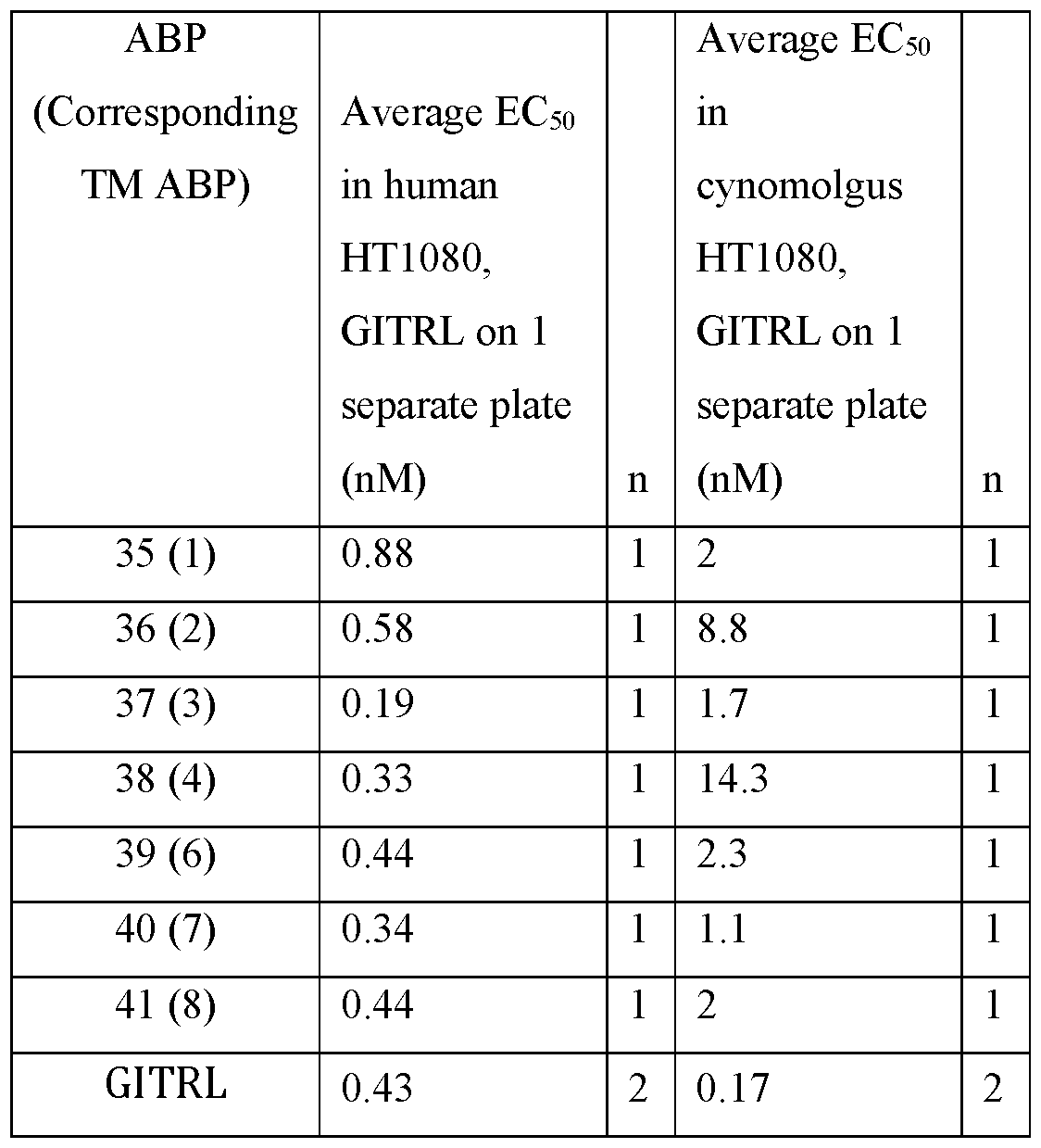


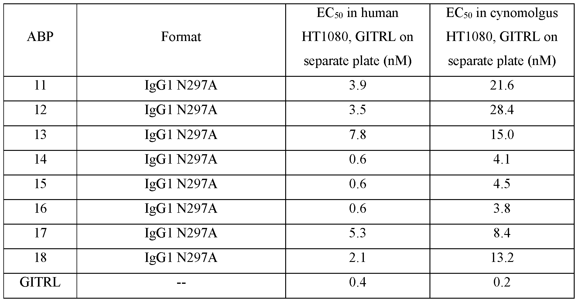


Claims
Priority Applications (17)
| Application Number | Priority Date | Filing Date | Title |
|---|---|---|---|
| BR112019010128A BR112019010128A2 (en) | 2016-11-19 | 2017-11-19 | anti-gitr antigen binding proteins and methods of use thereof |
| AU2017362721A AU2017362721A1 (en) | 2016-11-19 | 2017-11-19 | Anti-GITR antigen-binding proteins and methods of use thereof |
| UAA201906793A UA126389C2 (en) | 2016-11-19 | 2017-11-19 | Anti-gitr antigen-binding proteins and methods of use thereof |
| KR1020197017116A KR102574071B1 (en) | 2016-11-19 | 2017-11-19 | Anti-GITR antigen-binding proteins and methods of use thereof |
| MX2019005821A MX2019005821A (en) | 2016-11-19 | 2017-11-19 | Anti-gitr antigen-binding proteins and methods of use thereof. |
| RU2019118800A RU2795625C2 (en) | 2016-11-19 | 2017-11-19 | Antigen-binding proteins against gitr and methods of their use |
| JP2019527171A JP2019537449A (en) | 2016-11-19 | 2017-11-19 | Anti-GITR antigen binding proteins and methods of use |
| EP17817953.7A EP3541845A1 (en) | 2016-11-19 | 2017-11-19 | Anti-gitr antigen-binding proteins and methods of use thereof |
| CA3042727A CA3042727A1 (en) | 2016-11-19 | 2017-11-19 | Anti-gitr antigen-binding proteins and methods of use thereof |
| CN201780071351.3A CN110248960A (en) | 2016-11-19 | 2017-11-19 | Anti- GITR antigen-binding proteins and its application method |
| TW106140182A TWI783953B (en) | 2016-11-19 | 2017-11-20 | Anti-gitr antigen-binding proteins and methods of use thereof |
| US15/900,168 US10988545B2 (en) | 2016-11-19 | 2018-02-20 | Anti-GITR antigen-binding proteins and methods of use thereof |
| ZA2019/02939A ZA201902939B (en) | 2016-11-19 | 2019-05-10 | Anti-gitr antigen-binding proteins and methods of use thereof |
| PH12019501108A PH12019501108A1 (en) | 2016-11-19 | 2019-05-17 | Anti-gitr anti-binding protein and methods of use thereof |
| IL266720A IL266720A (en) | 2016-11-19 | 2019-05-19 | Anti-gitr antigen-binding proteins and methods of use thereof |
| CONC2019/0006424A CO2019006424A2 (en) | 2016-11-19 | 2019-06-19 | Anti-gitr antigen binding proteins and methods of use thereof |
| US17/197,547 US20210269542A1 (en) | 2016-11-19 | 2021-03-10 | Anti-gitr antigen-binding proteins and methods of use thereof |
Applications Claiming Priority (4)
| Application Number | Priority Date | Filing Date | Title |
|---|---|---|---|
| US201662497428P | 2016-11-19 | 2016-11-19 | |
| US62/497,428 | 2016-11-19 | ||
| US201762448644P | 2017-01-20 | 2017-01-20 | |
| US62/448,644 | 2017-01-20 |
Related Child Applications (1)
| Application Number | Title | Priority Date | Filing Date |
|---|---|---|---|
| US15/900,168 Continuation US10988545B2 (en) | 2016-11-19 | 2018-02-20 | Anti-GITR antigen-binding proteins and methods of use thereof |
Publications (2)
| Publication Number | Publication Date |
|---|---|
| WO2018094300A1 true WO2018094300A1 (en) | 2018-05-24 |
| WO2018094300A8 WO2018094300A8 (en) | 2019-05-02 |
Family
ID=62146165
Family Applications (1)
| Application Number | Title | Priority Date | Filing Date |
|---|---|---|---|
| PCT/US2017/062443 WO2018094300A1 (en) | 2016-11-19 | 2017-11-19 | Anti-gitr antigen-binding proteins and methods of use thereof |
Country Status (17)
| Country | Link |
|---|---|
| US (2) | US10988545B2 (en) |
| EP (1) | EP3541845A1 (en) |
| JP (1) | JP2019537449A (en) |
| KR (1) | KR102574071B1 (en) |
| CN (1) | CN110248960A (en) |
| AU (1) | AU2017362721A1 (en) |
| BR (1) | BR112019010128A2 (en) |
| CO (1) | CO2019006424A2 (en) |
| IL (1) | IL266720A (en) |
| JO (1) | JOP20190100A1 (en) |
| MA (1) | MA46844A (en) |
| MX (1) | MX2019005821A (en) |
| PH (1) | PH12019501108A1 (en) |
| TW (1) | TWI783953B (en) |
| UA (1) | UA126389C2 (en) |
| WO (1) | WO2018094300A1 (en) |
| ZA (1) | ZA201902939B (en) |
Cited By (1)
| Publication number | Priority date | Publication date | Assignee | Title |
|---|---|---|---|---|
| WO2021252327A2 (en) | 2020-06-08 | 2021-12-16 | University Of Washington | Antibody-bound nanoparticles |
Families Citing this family (5)
| Publication number | Priority date | Publication date | Assignee | Title |
|---|---|---|---|---|
| KR102239781B1 (en) * | 2019-04-08 | 2021-04-13 | 주식회사 녹십자 | Bispecific antibody that specifically binds to gpnmb and cd3 and use thereof |
| US10994021B1 (en) * | 2020-04-11 | 2021-05-04 | Bliss Biopharmaceutical (Hangzhou) Co., Ltd. | Tetravalent antibody-drug conjugates and use thereof |
| KR102475255B1 (en) * | 2021-05-10 | 2022-12-09 | 메디맵바이오 주식회사 | Anti-GITR antibodies and uses thereof |
| WO2023094507A1 (en) * | 2021-11-24 | 2023-06-01 | Formycon Ag | Improved ace2 fusion proteins |
| WO2023175614A1 (en) | 2022-03-15 | 2023-09-21 | Yeda Research And Development Co. Ltd. | Anti glucocorticoid-induced tnfr-related (gitr) protein antibodies and uses thereof |
Citations (87)
| Publication number | Priority date | Publication date | Assignee | Title |
|---|---|---|---|---|
| US4560655A (en) | 1982-12-16 | 1985-12-24 | Immunex Corporation | Serum-free cell culture medium and process for making same |
| WO1987000195A1 (en) | 1985-06-28 | 1987-01-15 | Celltech Limited | Animal cell culture |
| US4657866A (en) | 1982-12-21 | 1987-04-14 | Sudhir Kumar | Serum-free, synthetic, completely chemically defined tissue culture media |
| US4676980A (en) | 1985-09-23 | 1987-06-30 | The United States Of America As Represented By The Secretary Of The Department Of Health And Human Services | Target specific cross-linked heteroantibodies |
| US4767704A (en) | 1983-10-07 | 1988-08-30 | Columbia University In The City Of New York | Protein-free culture medium |
| US4816567A (en) | 1983-04-08 | 1989-03-28 | Genentech, Inc. | Recombinant immunoglobin preparations |
| WO1990003430A1 (en) | 1988-09-23 | 1990-04-05 | Cetus Corporation | Cell culture medium for enhanced cell growth, culture longevity and product expression |
| US4927762A (en) | 1986-04-01 | 1990-05-22 | Cell Enterprises, Inc. | Cell culture medium with antioxidant |
| US4946778A (en) | 1987-09-21 | 1990-08-07 | Genex Corporation | Single polypeptide chain binding molecules |
| EP0453082A1 (en) | 1990-03-09 | 1991-10-23 | Hybritech Incorporated | Trifunctional antibody-like compounds as a combined diagnostic and therapeutic agent |
| US5122469A (en) | 1990-10-03 | 1992-06-16 | Genentech, Inc. | Method for culturing Chinese hamster ovary cells to improve production of recombinant proteins |
| US5132405A (en) | 1987-05-21 | 1992-07-21 | Creative Biomolecules, Inc. | Biosynthetic antibody binding sites |
| US5204244A (en) | 1987-10-27 | 1993-04-20 | Oncogen | Production of chimeric antibodies by homologous recombination |
| WO1993008829A1 (en) | 1991-11-04 | 1993-05-13 | The Regents Of The University Of California | Compositions that mediate killing of hiv-infected cells |
| US5229275A (en) | 1990-04-26 | 1993-07-20 | Akzo N.V. | In-vitro method for producing antigen-specific human monoclonal antibodies |
| WO1993016185A2 (en) | 1992-02-06 | 1993-08-19 | Creative Biomolecules, Inc. | Biosynthetic binding protein for cancer marker |
| WO1994029351A2 (en) | 1993-06-16 | 1994-12-22 | Celltech Limited | Antibodies |
| US5500362A (en) | 1987-01-08 | 1996-03-19 | Xoma Corporation | Chimeric antibody with specificity to human B cell surface antigen |
| US5534615A (en) | 1994-04-25 | 1996-07-09 | Genentech, Inc. | Cardiac hypertrophy factor and uses therefor |
| US5545807A (en) | 1988-10-12 | 1996-08-13 | The Babraham Institute | Production of antibodies from transgenic animals |
| US5565332A (en) | 1991-09-23 | 1996-10-15 | Medical Research Council | Production of chimeric antibodies - a combinatorial approach |
| US5567610A (en) | 1986-09-04 | 1996-10-22 | Bioinvent International Ab | Method of producing human monoclonal antibodies and kit therefor |
| US5571894A (en) | 1991-02-05 | 1996-11-05 | Ciba-Geigy Corporation | Recombinant antibodies specific for a growth factor receptor |
| US5573905A (en) | 1992-03-30 | 1996-11-12 | The Scripps Research Institute | Encoded combinatorial chemical libraries |
| US5585089A (en) | 1988-12-28 | 1996-12-17 | Protein Design Labs, Inc. | Humanized immunoglobulins |
| US5587458A (en) | 1991-10-07 | 1996-12-24 | Aronex Pharmaceuticals, Inc. | Anti-erbB-2 antibodies, combinations thereof, and therapeutic and diagnostic uses thereof |
| US5589369A (en) | 1992-02-11 | 1996-12-31 | Cell Genesys Inc. | Cells homozygous for disrupted target loci |
| US5591669A (en) | 1988-12-05 | 1997-01-07 | Genpharm International, Inc. | Transgenic mice depleted in a mature lymphocytic cell-type |
| US5624821A (en) | 1987-03-18 | 1997-04-29 | Scotgen Biopharmaceuticals Incorporated | Antibodies with altered effector functions |
| WO1997030087A1 (en) | 1996-02-16 | 1997-08-21 | Glaxo Group Limited | Preparation of glycosylated antibodies |
| US5731168A (en) | 1995-03-01 | 1998-03-24 | Genentech, Inc. | Method for making heteromultimeric polypeptides |
| US5821337A (en) | 1991-06-14 | 1998-10-13 | Genentech, Inc. | Immunoglobulin variants |
| WO1998058964A1 (en) | 1997-06-24 | 1998-12-30 | Genentech, Inc. | Methods and compositions for galactosylated glycoproteins |
| WO1999022764A1 (en) | 1997-10-31 | 1999-05-14 | Genentech, Inc. | Methods and compositions comprising glycoprotein glycoforms |
| WO1999051642A1 (en) | 1998-04-02 | 1999-10-14 | Genentech, Inc. | Antibody variants and fragments thereof |
| WO1999058572A1 (en) | 1998-05-08 | 1999-11-18 | Cambridge University Technical Services Limited | Binding molecules derived from immunoglobulins which do not trigger complement mediated lysis |
| US6171586B1 (en) | 1997-06-13 | 2001-01-09 | Genentech, Inc. | Antibody formulation |
| US6194551B1 (en) | 1998-04-02 | 2001-02-27 | Genentech, Inc. | Polypeptide variants |
| US6267958B1 (en) | 1995-07-27 | 2001-07-31 | Genentech, Inc. | Protein formulation |
| US20010029246A1 (en) | 1997-05-30 | 2001-10-11 | O'reilly Michael S. | Angiostatin protein |
| US20020031140A1 (en) | 2000-09-14 | 2002-03-14 | Alcatel | Method and system for enhancing channel capacity in a point to multipoint radio communications system having different kinds of terminals |
| US20020164328A1 (en) | 2000-10-06 | 2002-11-07 | Toyohide Shinkawa | Process for purifying antibody |
| WO2003011878A2 (en) | 2001-08-03 | 2003-02-13 | Glycart Biotechnology Ag | Antibody glycosylation variants having increased antibody-dependent cellular cytotoxicity |
| US20030084570A1 (en) | 2001-11-02 | 2003-05-08 | Beaver Danny D. | Forming serpentine heat exchangers from spine fin tubing |
| US20030085119A1 (en) | 2001-11-02 | 2003-05-08 | Innovative Technology Licensing, Llc | Semiconductor wafer plating cathode assembly |
| US20030115614A1 (en) | 2000-10-06 | 2003-06-19 | Yutaka Kanda | Antibody composition-producing cell |
| US6602684B1 (en) | 1998-04-20 | 2003-08-05 | Glycart Biotechnology Ag | Glycosylation engineering of antibodies for improving antibody-dependent cellular cytotoxicity |
| US20030157108A1 (en) | 2001-10-25 | 2003-08-21 | Genentech, Inc. | Glycoprotein compositions |
| WO2003085107A1 (en) | 2002-04-09 | 2003-10-16 | Kyowa Hakko Kogyo Co., Ltd. | Cells with modified genome |
| US20030215914A1 (en) | 2001-12-10 | 2003-11-20 | Erwin Houtzager | Structure for presenting desired peptide sequences |
| US20040093621A1 (en) | 2001-12-25 | 2004-05-13 | Kyowa Hakko Kogyo Co., Ltd | Antibody composition which specifically binds to CD20 |
| US6737056B1 (en) | 1999-01-15 | 2004-05-18 | Genentech, Inc. | Polypeptide variants with altered effector function |
| US20040109865A1 (en) | 2002-04-09 | 2004-06-10 | Kyowa Hakko Kogyo Co., Ltd. | Antibody composition-containing medicament |
| US20040110282A1 (en) | 2002-04-09 | 2004-06-10 | Kyowa Hakko Kogyo Co., Ltd. | Cells in which activity of the protein involved in transportation of GDP-fucose is reduced or lost |
| US20040132140A1 (en) | 2002-04-09 | 2004-07-08 | Kyowa Hakko Kogyo Co., Ltd. | Production process for antibody composition |
| WO2004056312A2 (en) | 2002-12-16 | 2004-07-08 | Genentech, Inc. | Immunoglobulin variants and uses thereof |
| WO2004107618A2 (en) * | 2003-05-23 | 2004-12-09 | Wyeth | Gitr ligand and gitr ligand-related molecules and antibodies and uses thereof |
| US20050014934A1 (en) | 2002-10-15 | 2005-01-20 | Hinton Paul R. | Alteration of FcRn binding affinities or serum half-lives of antibodies by mutagenesis |
| US20050035586A1 (en) | 2003-08-12 | 2005-02-17 | Martin Samuel V. | Magnetically pulse welded underbody |
| US20050035778A1 (en) | 2003-08-06 | 2005-02-17 | Taiwan Semiconductor Manufacturing Co., Ltd. | Flip-over alignment station for probe needle adjustment |
| US20050053742A1 (en) | 2003-09-08 | 2005-03-10 | Nissan Motor Co., Ltd. | Resin tube |
| US20050123546A1 (en) | 2003-11-05 | 2005-06-09 | Glycart Biotechnology Ag | Antigen binding molecules with increased Fc receptor binding affinity and effector function |
| WO2005100402A1 (en) | 2004-04-13 | 2005-10-27 | F.Hoffmann-La Roche Ag | Anti-p-selectin antibodies |
| WO2006029879A2 (en) | 2004-09-17 | 2006-03-23 | F.Hoffmann-La Roche Ag | Anti-ox40l antibodies |
| WO2006044908A2 (en) | 2004-10-20 | 2006-04-27 | Genentech, Inc. | Antibody formulation in histidine-acetate buffer |
| US20070098719A1 (en) | 2005-03-25 | 2007-05-03 | Tolerrx, Inc. | GITR binding molecules and uses therefor |
| US20080069820A1 (en) | 2006-08-30 | 2008-03-20 | Genentech, Inc. | Multispecific antibodies |
| US7371826B2 (en) | 1999-01-15 | 2008-05-13 | Genentech, Inc. | Polypeptide variants with altered effector function |
| WO2008077546A1 (en) | 2006-12-22 | 2008-07-03 | F. Hoffmann-La Roche Ag | Antibodies against insulin-like growth factor i receptor and uses thereof |
| WO2009036379A2 (en) | 2007-09-14 | 2009-03-19 | Adimab, Inc. | Rationally designed, synthetic antibody libraries and uses therefor |
| US7521056B2 (en) | 2005-04-06 | 2009-04-21 | Ibc Pharmaceuticals, Inc. | Stably tethered structures of defined compositions with multiple functions or binding specificities |
| US7521541B2 (en) | 2004-09-23 | 2009-04-21 | Genetech Inc. | Cysteine engineered antibodies and conjugates |
| US7527787B2 (en) | 2005-10-19 | 2009-05-05 | Ibc Pharmaceuticals, Inc. | Multivalent immunoglobulin-based bioactive assemblies |
| US7534866B2 (en) | 2005-10-19 | 2009-05-19 | Ibc Pharmaceuticals, Inc. | Methods and compositions for generating bioactive assemblies of increased complexity and uses |
| US7550143B2 (en) | 2005-04-06 | 2009-06-23 | Ibc Pharmaceuticals, Inc. | Methods for generating stably linked complexes composed of homodimers, homotetramers or dimers of dimers and uses |
| WO2009089004A1 (en) | 2008-01-07 | 2009-07-16 | Amgen Inc. | Method for making antibody fc-heterodimeric molecules using electrostatic steering effects |
| US7612181B2 (en) | 2005-08-19 | 2009-11-03 | Abbott Laboratories | Dual variable domain immunoglobulin and uses thereof |
| WO2010105256A1 (en) | 2009-03-13 | 2010-09-16 | Adimab, Inc. | Rationally designed, synthetic antibody libraries and uses therefor |
| WO2012009568A2 (en) | 2010-07-16 | 2012-01-19 | Adimab, Llc | Antibody libraries |
| US8258082B2 (en) | 2000-12-18 | 2012-09-04 | Dyax Corp. | Focused libraries of genetic packages |
| US20130065277A1 (en) | 2009-12-22 | 2013-03-14 | Hubert Kettenberger | Sequence dependent aggregation |
| US8709424B2 (en) | 2009-09-03 | 2014-04-29 | Merck Sharp & Dohme Corp. | Anti-GITR antibodies |
| US8722859B2 (en) | 2000-04-11 | 2014-05-13 | Genentech, Inc. | Multivalent antibodies and uses therefor |
| WO2015103072A1 (en) | 2013-12-30 | 2015-07-09 | Epimab Biotherapeutics | Fabs-in-tandem immunoglobulin and uses thereof |
| WO2015184099A1 (en) * | 2014-05-28 | 2015-12-03 | 4-Antibody Ag | Anti-gitr antibodies and methods of use thereof |
| WO2016110267A1 (en) * | 2015-01-08 | 2016-07-14 | 苏州康宁杰瑞生物科技有限公司 | Bispecific antibody or antibody mixture with common light chains |
| WO2017096179A1 (en) * | 2015-12-02 | 2017-06-08 | Agenus Inc. | Antibodies and methods of use thereof |
Family Cites Families (16)
| Publication number | Priority date | Publication date | Assignee | Title |
|---|---|---|---|---|
| US6503184B1 (en) | 1997-10-21 | 2003-01-07 | Human Genome Sciences, Inc. | Human tumor necrosis factor receptor-like proteins TR11, TR11SV1 and TR11SV2 |
| US20030133936A1 (en) | 2001-07-12 | 2003-07-17 | Byrne Michael Chapman | CD25markers and uses thereof |
| AUPS054702A0 (en) | 2002-02-14 | 2002-03-07 | Immunaid Pty Ltd | Cancer therapy |
| CA2509495A1 (en) | 2002-12-20 | 2004-07-15 | Biogen Idec Ma Inc. | Lymphotoxin beta receptor agents in combination with chemotherapeutic agents |
| WO2005007190A1 (en) | 2003-07-11 | 2005-01-27 | Schering Corporation | Agonists or antagonists of the clucocorticoid-induced tumour necrosis factor receptor (gitr) or its ligand for the treatment of immune disorders, infections and cancer |
| WO2005007150A2 (en) | 2003-07-17 | 2005-01-27 | Bar-Ilan University | Method for inhibiting protein synthesis in sperm |
| WO2006083289A2 (en) | 2004-06-04 | 2006-08-10 | Duke University | Methods and compositions for enhancement of immunity by in vivo depletion of immunosuppressive cell activity |
| US20090285834A1 (en) | 2005-11-24 | 2009-11-19 | Dainippon Sumitomo Pharma Co., Ltd. | Novel memory ctl induction potentiator |
| JP5932217B2 (en) | 2007-07-12 | 2016-06-08 | ジーアイティーアール, インコーポレイテッド | Combination therapy using GITR binding molecules |
| US20130058937A1 (en) * | 2011-08-23 | 2013-03-07 | Johannes Auer | Bispecific antigen binding molecules |
| KR101981873B1 (en) | 2011-11-28 | 2019-05-23 | 메르크 파텐트 게엠베하 | Anti-pd-l1 antibodies and uses thereof |
| BR112014024282A2 (en) | 2012-03-28 | 2017-08-08 | Sanofi Sa | bradykinin b1 receptor binding antibodies |
| CN103217489B (en) * | 2013-01-15 | 2016-03-09 | 珠海市丽珠单抗生物技术有限公司 | A kind ofly measure the glycosylation of sample in protein purification technological process and the method for end modified situation |
| UY35517A (en) * | 2013-04-04 | 2014-10-31 | Mabxience S A | A PROCEDURE TO INCREASE THE FORMATION OF PYROGLUTAMIC ACID OF A PROTEIN |
| CA2921805C (en) * | 2013-08-22 | 2023-03-07 | Acceleron Pharma, Inc. | Tgf-beta receptor type ii variants and uses thereof |
| US10202590B2 (en) * | 2013-09-05 | 2019-02-12 | Vib Vzw | Cells producing Fc-containing molecules having altered glycosylation patterns and methods and use thereof |
-
2017
- 2017-06-16 JO JOP/2019/0100A patent/JOP20190100A1/en unknown
- 2017-11-19 EP EP17817953.7A patent/EP3541845A1/en active Pending
- 2017-11-19 WO PCT/US2017/062443 patent/WO2018094300A1/en unknown
- 2017-11-19 AU AU2017362721A patent/AU2017362721A1/en active Pending
- 2017-11-19 UA UAA201906793A patent/UA126389C2/en unknown
- 2017-11-19 KR KR1020197017116A patent/KR102574071B1/en active IP Right Grant
- 2017-11-19 JP JP2019527171A patent/JP2019537449A/en active Pending
- 2017-11-19 MX MX2019005821A patent/MX2019005821A/en unknown
- 2017-11-19 BR BR112019010128A patent/BR112019010128A2/en unknown
- 2017-11-19 CN CN201780071351.3A patent/CN110248960A/en active Pending
- 2017-11-19 MA MA046844A patent/MA46844A/en unknown
- 2017-11-20 TW TW106140182A patent/TWI783953B/en active
-
2018
- 2018-02-20 US US15/900,168 patent/US10988545B2/en active Active
-
2019
- 2019-05-10 ZA ZA2019/02939A patent/ZA201902939B/en unknown
- 2019-05-17 PH PH12019501108A patent/PH12019501108A1/en unknown
- 2019-05-19 IL IL266720A patent/IL266720A/en unknown
- 2019-06-19 CO CONC2019/0006424A patent/CO2019006424A2/en unknown
-
2021
- 2021-03-10 US US17/197,547 patent/US20210269542A1/en active Pending
Patent Citations (95)
| Publication number | Priority date | Publication date | Assignee | Title |
|---|---|---|---|---|
| US4560655A (en) | 1982-12-16 | 1985-12-24 | Immunex Corporation | Serum-free cell culture medium and process for making same |
| US4657866A (en) | 1982-12-21 | 1987-04-14 | Sudhir Kumar | Serum-free, synthetic, completely chemically defined tissue culture media |
| US4816567A (en) | 1983-04-08 | 1989-03-28 | Genentech, Inc. | Recombinant immunoglobin preparations |
| US4767704A (en) | 1983-10-07 | 1988-08-30 | Columbia University In The City Of New York | Protein-free culture medium |
| WO1987000195A1 (en) | 1985-06-28 | 1987-01-15 | Celltech Limited | Animal cell culture |
| US4676980A (en) | 1985-09-23 | 1987-06-30 | The United States Of America As Represented By The Secretary Of The Department Of Health And Human Services | Target specific cross-linked heteroantibodies |
| US4927762A (en) | 1986-04-01 | 1990-05-22 | Cell Enterprises, Inc. | Cell culture medium with antioxidant |
| US5567610A (en) | 1986-09-04 | 1996-10-22 | Bioinvent International Ab | Method of producing human monoclonal antibodies and kit therefor |
| US5500362A (en) | 1987-01-08 | 1996-03-19 | Xoma Corporation | Chimeric antibody with specificity to human B cell surface antigen |
| US5624821A (en) | 1987-03-18 | 1997-04-29 | Scotgen Biopharmaceuticals Incorporated | Antibodies with altered effector functions |
| US5648260A (en) | 1987-03-18 | 1997-07-15 | Scotgen Biopharmaceuticals Incorporated | DNA encoding antibodies with altered effector functions |
| US5132405A (en) | 1987-05-21 | 1992-07-21 | Creative Biomolecules, Inc. | Biosynthetic antibody binding sites |
| US4946778A (en) | 1987-09-21 | 1990-08-07 | Genex Corporation | Single polypeptide chain binding molecules |
| US5204244A (en) | 1987-10-27 | 1993-04-20 | Oncogen | Production of chimeric antibodies by homologous recombination |
| WO1990003430A1 (en) | 1988-09-23 | 1990-04-05 | Cetus Corporation | Cell culture medium for enhanced cell growth, culture longevity and product expression |
| US5545807A (en) | 1988-10-12 | 1996-08-13 | The Babraham Institute | Production of antibodies from transgenic animals |
| US5591669A (en) | 1988-12-05 | 1997-01-07 | Genpharm International, Inc. | Transgenic mice depleted in a mature lymphocytic cell-type |
| US6180370B1 (en) | 1988-12-28 | 2001-01-30 | Protein Design Labs, Inc. | Humanized immunoglobulins and methods of making the same |
| US5693761A (en) | 1988-12-28 | 1997-12-02 | Protein Design Labs, Inc. | Polynucleotides encoding improved humanized immunoglobulins |
| US5693762A (en) | 1988-12-28 | 1997-12-02 | Protein Design Labs, Inc. | Humanized immunoglobulins |
| US5585089A (en) | 1988-12-28 | 1996-12-17 | Protein Design Labs, Inc. | Humanized immunoglobulins |
| EP0453082A1 (en) | 1990-03-09 | 1991-10-23 | Hybritech Incorporated | Trifunctional antibody-like compounds as a combined diagnostic and therapeutic agent |
| US5229275A (en) | 1990-04-26 | 1993-07-20 | Akzo N.V. | In-vitro method for producing antigen-specific human monoclonal antibodies |
| US5122469A (en) | 1990-10-03 | 1992-06-16 | Genentech, Inc. | Method for culturing Chinese hamster ovary cells to improve production of recombinant proteins |
| US5571894A (en) | 1991-02-05 | 1996-11-05 | Ciba-Geigy Corporation | Recombinant antibodies specific for a growth factor receptor |
| US5821337A (en) | 1991-06-14 | 1998-10-13 | Genentech, Inc. | Immunoglobulin variants |
| US5565332A (en) | 1991-09-23 | 1996-10-15 | Medical Research Council | Production of chimeric antibodies - a combinatorial approach |
| US5587458A (en) | 1991-10-07 | 1996-12-24 | Aronex Pharmaceuticals, Inc. | Anti-erbB-2 antibodies, combinations thereof, and therapeutic and diagnostic uses thereof |
| WO1993008829A1 (en) | 1991-11-04 | 1993-05-13 | The Regents Of The University Of California | Compositions that mediate killing of hiv-infected cells |
| WO1993016185A2 (en) | 1992-02-06 | 1993-08-19 | Creative Biomolecules, Inc. | Biosynthetic binding protein for cancer marker |
| US5589369A (en) | 1992-02-11 | 1996-12-31 | Cell Genesys Inc. | Cells homozygous for disrupted target loci |
| US5573905A (en) | 1992-03-30 | 1996-11-12 | The Scripps Research Institute | Encoded combinatorial chemical libraries |
| WO1994029351A2 (en) | 1993-06-16 | 1994-12-22 | Celltech Limited | Antibodies |
| US5534615A (en) | 1994-04-25 | 1996-07-09 | Genentech, Inc. | Cardiac hypertrophy factor and uses therefor |
| US5731168A (en) | 1995-03-01 | 1998-03-24 | Genentech, Inc. | Method for making heteromultimeric polypeptides |
| US6267958B1 (en) | 1995-07-27 | 2001-07-31 | Genentech, Inc. | Protein formulation |
| WO1997030087A1 (en) | 1996-02-16 | 1997-08-21 | Glaxo Group Limited | Preparation of glycosylated antibodies |
| US20010029246A1 (en) | 1997-05-30 | 2001-10-11 | O'reilly Michael S. | Angiostatin protein |
| US6171586B1 (en) | 1997-06-13 | 2001-01-09 | Genentech, Inc. | Antibody formulation |
| WO1998058964A1 (en) | 1997-06-24 | 1998-12-30 | Genentech, Inc. | Methods and compositions for galactosylated glycoproteins |
| WO1999022764A1 (en) | 1997-10-31 | 1999-05-14 | Genentech, Inc. | Methods and compositions comprising glycoprotein glycoforms |
| WO1999051642A1 (en) | 1998-04-02 | 1999-10-14 | Genentech, Inc. | Antibody variants and fragments thereof |
| US6194551B1 (en) | 1998-04-02 | 2001-02-27 | Genentech, Inc. | Polypeptide variants |
| US6602684B1 (en) | 1998-04-20 | 2003-08-05 | Glycart Biotechnology Ag | Glycosylation engineering of antibodies for improving antibody-dependent cellular cytotoxicity |
| WO1999058572A1 (en) | 1998-05-08 | 1999-11-18 | Cambridge University Technical Services Limited | Binding molecules derived from immunoglobulins which do not trigger complement mediated lysis |
| US6737056B1 (en) | 1999-01-15 | 2004-05-18 | Genentech, Inc. | Polypeptide variants with altered effector function |
| US7332581B2 (en) | 1999-01-15 | 2008-02-19 | Genentech, Inc. | Polypeptide variants with altered effector function |
| US7371826B2 (en) | 1999-01-15 | 2008-05-13 | Genentech, Inc. | Polypeptide variants with altered effector function |
| US8722859B2 (en) | 2000-04-11 | 2014-05-13 | Genentech, Inc. | Multivalent antibodies and uses therefor |
| US20020031140A1 (en) | 2000-09-14 | 2002-03-14 | Alcatel | Method and system for enhancing channel capacity in a point to multipoint radio communications system having different kinds of terminals |
| US20020164328A1 (en) | 2000-10-06 | 2002-11-07 | Toyohide Shinkawa | Process for purifying antibody |
| US20030115614A1 (en) | 2000-10-06 | 2003-06-19 | Yutaka Kanda | Antibody composition-producing cell |
| US8258082B2 (en) | 2000-12-18 | 2012-09-04 | Dyax Corp. | Focused libraries of genetic packages |
| WO2003011878A2 (en) | 2001-08-03 | 2003-02-13 | Glycart Biotechnology Ag | Antibody glycosylation variants having increased antibody-dependent cellular cytotoxicity |
| US20030157108A1 (en) | 2001-10-25 | 2003-08-21 | Genentech, Inc. | Glycoprotein compositions |
| US20030085119A1 (en) | 2001-11-02 | 2003-05-08 | Innovative Technology Licensing, Llc | Semiconductor wafer plating cathode assembly |
| US20030084570A1 (en) | 2001-11-02 | 2003-05-08 | Beaver Danny D. | Forming serpentine heat exchangers from spine fin tubing |
| US20030215914A1 (en) | 2001-12-10 | 2003-11-20 | Erwin Houtzager | Structure for presenting desired peptide sequences |
| US20040093621A1 (en) | 2001-12-25 | 2004-05-13 | Kyowa Hakko Kogyo Co., Ltd | Antibody composition which specifically binds to CD20 |
| US20040109865A1 (en) | 2002-04-09 | 2004-06-10 | Kyowa Hakko Kogyo Co., Ltd. | Antibody composition-containing medicament |
| US20040132140A1 (en) | 2002-04-09 | 2004-07-08 | Kyowa Hakko Kogyo Co., Ltd. | Production process for antibody composition |
| US20040110282A1 (en) | 2002-04-09 | 2004-06-10 | Kyowa Hakko Kogyo Co., Ltd. | Cells in which activity of the protein involved in transportation of GDP-fucose is reduced or lost |
| US20040110704A1 (en) | 2002-04-09 | 2004-06-10 | Kyowa Hakko Kogyo Co., Ltd. | Cells of which genome is modified |
| WO2003085107A1 (en) | 2002-04-09 | 2003-10-16 | Kyowa Hakko Kogyo Co., Ltd. | Cells with modified genome |
| US20050014934A1 (en) | 2002-10-15 | 2005-01-20 | Hinton Paul R. | Alteration of FcRn binding affinities or serum half-lives of antibodies by mutagenesis |
| WO2004056312A2 (en) | 2002-12-16 | 2004-07-08 | Genentech, Inc. | Immunoglobulin variants and uses thereof |
| WO2004107618A2 (en) * | 2003-05-23 | 2004-12-09 | Wyeth | Gitr ligand and gitr ligand-related molecules and antibodies and uses thereof |
| US20050035778A1 (en) | 2003-08-06 | 2005-02-17 | Taiwan Semiconductor Manufacturing Co., Ltd. | Flip-over alignment station for probe needle adjustment |
| US20050035586A1 (en) | 2003-08-12 | 2005-02-17 | Martin Samuel V. | Magnetically pulse welded underbody |
| US20050053742A1 (en) | 2003-09-08 | 2005-03-10 | Nissan Motor Co., Ltd. | Resin tube |
| US20050123546A1 (en) | 2003-11-05 | 2005-06-09 | Glycart Biotechnology Ag | Antigen binding molecules with increased Fc receptor binding affinity and effector function |
| WO2005100402A1 (en) | 2004-04-13 | 2005-10-27 | F.Hoffmann-La Roche Ag | Anti-p-selectin antibodies |
| WO2006029879A2 (en) | 2004-09-17 | 2006-03-23 | F.Hoffmann-La Roche Ag | Anti-ox40l antibodies |
| US7521541B2 (en) | 2004-09-23 | 2009-04-21 | Genetech Inc. | Cysteine engineered antibodies and conjugates |
| WO2006044908A2 (en) | 2004-10-20 | 2006-04-27 | Genentech, Inc. | Antibody formulation in histidine-acetate buffer |
| US7812135B2 (en) | 2005-03-25 | 2010-10-12 | Tolerrx, Inc. | GITR-binding antibodies |
| US20070098719A1 (en) | 2005-03-25 | 2007-05-03 | Tolerrx, Inc. | GITR binding molecules and uses therefor |
| US7521056B2 (en) | 2005-04-06 | 2009-04-21 | Ibc Pharmaceuticals, Inc. | Stably tethered structures of defined compositions with multiple functions or binding specificities |
| US7550143B2 (en) | 2005-04-06 | 2009-06-23 | Ibc Pharmaceuticals, Inc. | Methods for generating stably linked complexes composed of homodimers, homotetramers or dimers of dimers and uses |
| US7612181B2 (en) | 2005-08-19 | 2009-11-03 | Abbott Laboratories | Dual variable domain immunoglobulin and uses thereof |
| US7527787B2 (en) | 2005-10-19 | 2009-05-05 | Ibc Pharmaceuticals, Inc. | Multivalent immunoglobulin-based bioactive assemblies |
| US7534866B2 (en) | 2005-10-19 | 2009-05-19 | Ibc Pharmaceuticals, Inc. | Methods and compositions for generating bioactive assemblies of increased complexity and uses |
| US20080069820A1 (en) | 2006-08-30 | 2008-03-20 | Genentech, Inc. | Multispecific antibodies |
| WO2008077546A1 (en) | 2006-12-22 | 2008-07-03 | F. Hoffmann-La Roche Ag | Antibodies against insulin-like growth factor i receptor and uses thereof |
| US8691730B2 (en) | 2007-09-14 | 2014-04-08 | Adimab, Llc | Rationally designed, synthetic antibody libraries and uses therefor |
| WO2009036379A2 (en) | 2007-09-14 | 2009-03-19 | Adimab, Inc. | Rationally designed, synthetic antibody libraries and uses therefor |
| WO2009089004A1 (en) | 2008-01-07 | 2009-07-16 | Amgen Inc. | Method for making antibody fc-heterodimeric molecules using electrostatic steering effects |
| WO2010105256A1 (en) | 2009-03-13 | 2010-09-16 | Adimab, Inc. | Rationally designed, synthetic antibody libraries and uses therefor |
| US8709424B2 (en) | 2009-09-03 | 2014-04-29 | Merck Sharp & Dohme Corp. | Anti-GITR antibodies |
| US20130065277A1 (en) | 2009-12-22 | 2013-03-14 | Hubert Kettenberger | Sequence dependent aggregation |
| WO2012009568A2 (en) | 2010-07-16 | 2012-01-19 | Adimab, Llc | Antibody libraries |
| WO2015103072A1 (en) | 2013-12-30 | 2015-07-09 | Epimab Biotherapeutics | Fabs-in-tandem immunoglobulin and uses thereof |
| WO2015184099A1 (en) * | 2014-05-28 | 2015-12-03 | 4-Antibody Ag | Anti-gitr antibodies and methods of use thereof |
| WO2016110267A1 (en) * | 2015-01-08 | 2016-07-14 | 苏州康宁杰瑞生物科技有限公司 | Bispecific antibody or antibody mixture with common light chains |
| WO2017096179A1 (en) * | 2015-12-02 | 2017-06-08 | Agenus Inc. | Antibodies and methods of use thereof |
Non-Patent Citations (130)
| Title |
|---|
| "Handbook of Pharmaceutical Excipients", 2009 |
| "Handbook of Pharmaceutical Excipients", 2009, THE PHARMACEUTICAL PRESS |
| "The Pharmacology of Monoclonal Antibodies", vol. 113, pages: 269 - 315 |
| AALBERSE ET AL., IMMUNOLOGY, vol. 105, 2002, pages 9 - 19 |
| ABHINANDAN; MARTIN, IMMUNOLOGY, vol. 45, 2008, pages 3832 - 3839 |
| AL-LAZIKANI ET AL., J. MOL. BIOL., vol. 273, 1997, pages 927 - 948 |
| ANALYTICAL BIOCHEMISTRY, vol. 348, 2006, pages 24 - 39 |
| ARABI GHAHROUDI ET AL., FEBSLETTERS, vol. 414, 1998, pages 521 - 526 |
| ARMOUR ET AL., EUR. J. IMMUNOL., vol. 29, 1999, pages 2613 - 2624 |
| ARMOUR ET AL., MOL. IMMUNOL., vol. 40, 2003, pages 585 - 593 |
| BARBAS ET AL., PROC. NAT. ACAD. SCI. U.S.A., vol. 91, 1994, pages 3809 - 3813 |
| BARNES ET AL., ANAL. BIOCHEM., vol. 102, 1980, pages 255 |
| BINZ ET AL., NAT. BIOTECHNOL., vol. 23, 2005, pages 1257 - 1268 |
| BRENNAN ET AL., SCIENCE, vol. 229, 1985, pages 81 - 83 |
| BRUGGEMANN ET AL., J. EXP. MED., vol. 166, 1987, pages 1351 - 1361 |
| BRUGGERMANN ET AL., YEAR IN IMMUNO., vol. 7, 1993, pages 33 |
| CARLRING ET AL., PLOS ONE, vol. 6, 2011, pages e22533 |
| CARTER ET AL., BIOLTECHNOLOGY, vol. 10, 1992, pages 163 - 167 |
| CHEN ET AL., J. MOL. BIOL., vol. 293, 1999, pages 865 - 881 |
| CHOWDHURY, METHODS MOL. BIOL., vol. 207, 2008, pages 179 - 196 |
| CLYNES ET AL., PROC. NAT'L ACAD. SCI. USA, vol. 95, 1998, pages 652 - 656 |
| COHEN ET AL., J. CLIN. ONCOL., vol. 25, 2007, pages 3058 |
| COHEN ET AL., PLOS ONE, vol. 5, 2010, pages e 10436 |
| COHEN ET AL., PLOS ONE, vol. 5, 2010, pages el0436 |
| COLOMA; MORRISON, NATURE BIOTECHNOL., vol. 15, 1997, pages 159 - 163 |
| CRAGG ET AL., BLOOD, vol. 101, 2003, pages 1045 - 1052 |
| CRAGG ET AL., BLOOD, vol. 103, 2004, pages 2738 - 2743 |
| CREIGHTON: "Proteins: Structures and Molecular Properties", 1993, W. H. FREEMAN & CO., NEW YORK |
| DAVID A SCHAER ET AL: "Modulation of GITR for cancer immunotherapy", CURRENT OPINION IN IMMUNOLOGY, vol. 24, no. 2, 1 April 2012 (2012-04-01), pages 217 - 224, XP055150991, ISSN: 0952-7915, DOI: 10.1016/j.coi.2011.12.011 * |
| DOPPALAPUDI ET AL., PROC. NATL. ACAD. SCI. USA, vol. 107, 2010, pages 22611 - 22616 |
| DUNCAN; WINTER, NATURE, vol. 322, 1988, pages 738 - 740 |
| EBERSBACH ET AL., J. MOL. BIOL., vol. 372, 2007, pages 172 - 185 |
| ECONOMIDES ET AL., NATURE MEDICINE, vol. 9, 2003, pages 47 - 52 |
| EMANUEL ET AL., MABS, vol. 3, 2011, pages 38 - 48 |
| ESPARZA ET AL., J. IMMUNOL., vol. 174, 2005, pages 7869 - 7874 |
| ESTEP ET AL.: "High throughput solution-based measurement of antibody-antigen affinity and epitope binning", MABS, vol. 5, no. 2, 2013, pages 270 - 278, XP055105281, DOI: doi:10.4161/mabs.23049 |
| FERNANDEZ, CURR. OPINION IN BIOTECH., vol. 15, 2004, pages 364 - 373 |
| GAZZANO-SANTORO ET AL., J. IMMUNOL. METHODS, vol. 202, 1996, pages 163 - 171 |
| GEYER; BRENT, METH. ENZYMOL., vol. 328, 2000, pages 171 - 208 |
| GIUSEPPE NOCENTINI ET AL: "Pharmacological modulation of GITRL/GITR system: therapeutic perspectives", BRITISH JOURNAL OF PHARMACOLOGY, vol. 165, no. 7, 9 March 2012 (2012-03-09), pages 2089 - 2099, XP055150987, ISSN: 0007-1188, DOI: 10.1111/j.1476-5381.2011.01753.x * |
| GODING J.W.: "Monoclonal Antibodies: Principles and Practice", 1986, ACADEMIC PRESS |
| GRAMER ET AL., MABS, vol. 5, 2013, pages 962 - 972 |
| GRAVERSEN ET AL., J. BIOL. CHEM., vol. 275, 2000, pages 37390 - 37396 |
| GRUBER ET AL., J. IMMUNOL., vol. 152, 1994, pages 5368 - 5374 |
| GUSS ET AL., EMBO J, vol. 5, 1986, pages 1567 - 1575 |
| HAM ET AL., METH. ENZ., vol. 58, 1979, pages 44 |
| HANABUCHIB ET AL., BLOOD, vol. 107, 2006, pages 3617 - 3623 |
| HARLOW; LANE: "Antibodies: A Laboratory Manual", 1988, COLD SPRING HARBOR LABORATORY |
| HAWKINS ET AL., J. MOL. BIOL., vol. 226, 1992, pages 889 - 896 |
| HELLSTROM ET AL., PROC. NAT'L ACAD. SCI. USA, vol. 83, 1986, pages 7059 - 7063 |
| HELLSTROM ET AL., PROC. NAT'LACAD. SCI. USA, vol. 82, 1985, pages 1499 - 1502 |
| HINTON ET AL., J. IMMUNOL., vol. 176, 2006, pages 346 - 356 |
| HOLLINGER ET AL., PROC. NATL. ACAD. SCI. USA, vol. 90, 1993, pages 6444 - 6448 |
| HONEGGE; PLIICKTHUN, J. MOL. BIOL., vol. 309, 2001, pages 657 - 70 |
| HOOGENBOOM ET AL., J. MOL. BIOL., vol. 227, 1991, pages 381 - 388 |
| HOOGENBOOM ET AL., METHODS IN MOLECULAR BIOLOGY, vol. 178, 2001, pages 1 - 37 |
| HUDSON ET AL., NAT. MED., vol. 9, 2003, pages 129 - 134 |
| IDUSOGIE ET AL., J. IMMUNOL., vol. 164, 2000, pages 4178 - 4184 |
| JACKSON ET AL., J. IMMUNOL., vol. 154, 1995, pages 3310 - 33199 |
| JAKOBOVITS ET AL., NATURE, vol. 362, 1993, pages 255 - 258 |
| JAKOBOVITS ET AL., PROC. NATL. ACAD. SCI. U.S.A., vol. 90, 1993, pages 2551 |
| JANG ET AL., ANN. CLIN. LAB. SCI., vol. 42, 2012, pages 42 - 49 |
| JONES ET AL., NATURE, vol. 321, 1986, pages 522 - 525 |
| JOURNAL OF PHARMACEUTICAL SCIENCES, vol. 97, 2008, pages 2426 - 2447 |
| KANDA ET AL., BIOTECHNOL. BIOENG., vol. 94, 2006, pages 680 - 688 |
| KIPRIYANOV ET AL., J. MOL. BIOL., vol. 293, 1999, pages 41 - 56 |
| KO K ET AL: "TREATMENT OF ADVANCED TUMORS WITH AGONISTIC ANTI-GITR MAB AND ITS EFFECTS ON TUMOR-INFILTRATING FOXP3(+)CD25(+)CD4(+) REGULATORY T CELLS", THE JOURNAL OF EXPERIMENTAL MEDICINE, ROCKEFELLER UNIVERSITY PRESS, US, vol. 202, no. 7, 3 October 2005 (2005-10-03), pages 885 - 891, XP009076415, ISSN: 0022-1007, DOI: 10.1084/JEM.20050940 * |
| KOHLER ET AL., NATURE, vol. 256, 1975, pages 495 - 497 |
| KOSTELNY ET AL., J. IMMUNOL., vol. 148, 1992, pages 1547 - 1553 |
| KOZBOR, J. IMMUNOL., vol. 133, 1984, pages 3001 |
| LABRIJN ET AL., NATURE PROTOCOLS, vol. 9, 2014, pages 2450 - 2463 |
| LABRIJN ET AL., PROC. NATL. ACAD. SCI. USA, vol. 110, 2013, pages 5145 - 5150 |
| LAFLEUR ET AL., MABS, vol. 5, 2013, pages 208 - 218 |
| LAZAR ET AL., PROC. NATL. ACAD. SCI. USA, vol. 103, 2006, pages 4005 - 4010 |
| LEFRANC ET AL., DEV. COMP. IMMUNOL., vol. 27, 2003, pages 55 - 77 |
| LI ET AL., J. AUTOIMMUNITY, vol. 21, 2003, pages 83 - 92 |
| LINDMARK ET AL., J. IMMUNOL. METH., vol. 62, 1983, pages 1 - 13 |
| MACCALLUM ET AL., J. MOL. BIOL., vol. 262, 1996, pages 732 - 745 |
| MARKS ET AL., BIO/TECHNOLOGY, vol. 10, 1992, pages 779 - 783 |
| MARKS ET AL., J. MOL. BIOL., vol. 222, 1991, pages 581 - 597 |
| MELERO ET AL., CLIN CANCER RES., vol. 19, 2013, pages 1044 - 1053 |
| MERCHANT ET AL., NATURE BIOTECHNOL., vol. 16, 1998, pages 677 - 681 |
| MILER ET AL., J. IMMUNOL., vol. 170, 2003, pages 4854 - 4861 |
| MILSTEIN; CUELLO, NATURE, vol. 305, 1983, pages 537 - 540 |
| MOORE ET AL., BLOOD, vol. 117, 2011, pages 454 - 451 |
| MORRIS: "Methods in Molecular Biology", vol. 66, 1996, HUMANA PRESS, article "Epitope Mapping Protocols" |
| MORRISON ET AL., PROC. NATL. ACAD. SCI. USA, vol. 81, 1984, pages 6851 - 6855 |
| MUYLDERMANS ET AL., TRENDS IN BIOCHEM. SCI., vol. 26, 2001, pages 230 - 245 |
| NOCENTINI ET AL., BR. J. PHARMACOL., vol. 165, 2012, pages 2089 - 2099 |
| NOCENTINI ET AL., EUR. J. IMMUNOL., vol. 35, 2005, pages 1016 - 1022 |
| NOCENTINI ET AL., PROC. NATL. ACAD. SCI. USA, vol. 94, 1997, pages 6216 - 6221 |
| OKAZAKI ET AL., J. MOL. BIOL., vol. 336, 2004, pages 1239 - 1249 |
| PEDS, vol. 26.10, 2013, pages 663 - 70 |
| PETKOVA ET AL., INT'L. IMMUNOL., vol. 18, 2006, pages 1759 - 1769 |
| PHILIP O SCUMPIA ET AL: "Treatment with GITR agonistic antibody corrects adaptive immune dysfunction in sepsis", 9 August 2007 (2007-08-09), XP055455291, Retrieved from the Internet <URL:http://www.bloodjournal.org/content/110/10/3673.full.pdf> [retrieved on 20180228], DOI: 10.1182/blood-2007- * |
| PLIICKTHUN: "The Pharmacology of Monoclonal Antibodies", vol. 113, 1994, SPRINGER-VERLAG, pages: 269 - 315 |
| PRESTA, CURR. OP. STRUCT. BIOL., vol. 2, 1992, pages 593 - 596 |
| QUEEN ET AL., PROC. NATL. ACAD. SCI. U.S.A., vol. 86, 1989, pages 10029 - 10033 |
| RADER ET AL., PROC. NAT. ACAD. SCI. U.S.A., vol. 95, 1998, pages 8910 - 8915 |
| RAVETCH; KINET, ANNU. REV. IMMUNOL., vol. 9, 1991, pages 457 - 492 |
| RIECHMANN ET AL., NATURE, vol. 332, 1988, pages 323 - 329 |
| RIPKA ET AL., ARCH. BIOCHEM. BIOPHYS., vol. 249, 1986, pages 533 - 545 |
| RONCHETTI ET AL., EUR. J. IMMUNOL., vol. 34, 2004, pages 613 - 622 |
| SCHAE ET AL., CURR. OPIN. IMMUNOL., vol. 24, 2012, pages 217 - 224 |
| SCHIER ET AL., GENE, vol. 169, 1995, pages 147 - 155 |
| SCHROEDER; CAVACIN, J. ALLERGY CLIN. IMMUNOL., vol. 125, 2010, pages 41 - 52 |
| SCHROEDER; CAVACINI, J. ALLERGY CLIN. IMMUNOL., vol. 125, 2010, pages 41 - 52 |
| SEDER; AHMED, NATURE IMMUNOL., vol. 4, 2003, pages 835 - 842 |
| SHIELDS ET AL., J. BIOL. CHEM., vol. 277, 2002, pages 26733 - 26740 |
| SIEGEL ET AL.: "High efficiency recovery and epitope-specific sorting of an scFv yeast display library", J IMMUNOL METHODS, vol. 286, no. 1-2, 2004, pages 141 - 153, XP004503454, DOI: doi:10.1016/j.jim.2004.01.005 |
| SILACCI ET AL., J. BIOL. CHEM., vol. 289, 2014, pages 14392 - 14398 |
| SILVERMAN ET AL., NATURE BIOTECH., vol. 23, 2005, pages 1556 - 1561 |
| SKERRA, CURRENT OPIN. IN BIOTECH., vol. 18, 2007, pages 295 - 304 |
| STAERZ ET AL., NATURE, vol. 314, 1985, pages 628 - 631 |
| STAERZ; BEVAN, PROC. NATL. ACAD. SCI. USA, vol. 83, 1986, pages 1453 - 1457 |
| STEINBERGER ET AL., J. BIOL. CHEM., vol. 275, 2000, pages 36073 - 36078 |
| TODOROVSKA ET AL., J. IMMUNOL. METHODS, vol. 248, 2001, pages 47 - 66 |
| TRAUNECKER ET AL., EMBO J., vol. 10, 1991, pages 3655 - 3659 |
| TUTT ET AL., J. IMMUNOL., vol. 147, 1991, pages 60 - 69 |
| VOGT; SKERRA, CHEM. BIOCHEM., vol. 5, 2004, pages 191 - 199 |
| WAGNER ET AL., BIOCHEM. & BIOPHYS. RES. COMM., vol. 186, 1992, pages 118 - 1145 |
| WINTER; MILSTEIN, NATURE, vol. 349, 1991, pages 293 - 299 |
| WOZNIAK-KNOPP ET AL., PROTEIN ENG. DES. SEL., vol. 23, 2010, pages 289 - 297 |
| WRIGHT ET AL., TIBTECH, vol. 15, 1997, pages 26 - 32 |
| WYZGOL ET AL., JLMMUNOL, vol. 183, 2009, pages 1851 - 1861 |
| YAMANE-OHNUKI ET AL., BIOTECH. BIOENG., vol. 87, 2004, pages 614 - 622 |
| YELTON ET AL., J. IMMUNOL., vol. 155, 1995, pages 1994 - 2004 |
| YIN ET AL., MABS, vol. 4, 2012, pages 217 - 225 |
| ZAHND ET AL., J. MOL. BIOL., vol. 369, 2007, pages 1015 - 1028 |
| ZHUKOVSKY ET AL., BLOOD, vol. 122, 2013, pages 5116 |
Cited By (1)
| Publication number | Priority date | Publication date | Assignee | Title |
|---|---|---|---|---|
| WO2021252327A2 (en) | 2020-06-08 | 2021-12-16 | University Of Washington | Antibody-bound nanoparticles |
Also Published As
| Publication number | Publication date |
|---|---|
| RU2019118800A (en) | 2020-12-21 |
| UA126389C2 (en) | 2022-09-28 |
| TW201819416A (en) | 2018-06-01 |
| BR112019010128A2 (en) | 2019-10-08 |
| WO2018094300A8 (en) | 2019-05-02 |
| EP3541845A1 (en) | 2019-09-25 |
| MA46844A (en) | 2019-09-25 |
| TWI783953B (en) | 2022-11-21 |
| KR102574071B1 (en) | 2023-09-06 |
| RU2019118800A3 (en) | 2021-03-23 |
| PH12019501108A1 (en) | 2019-07-29 |
| US20180208665A1 (en) | 2018-07-26 |
| ZA201902939B (en) | 2022-08-31 |
| US20210269542A1 (en) | 2021-09-02 |
| MX2019005821A (en) | 2019-10-07 |
| KR20190095298A (en) | 2019-08-14 |
| JOP20190100A1 (en) | 2019-05-01 |
| CO2019006424A2 (en) | 2019-08-20 |
| CN110248960A (en) | 2019-09-17 |
| JP2019537449A (en) | 2019-12-26 |
| IL266720A (en) | 2019-07-31 |
| AU2017362721A1 (en) | 2019-05-23 |
| US10988545B2 (en) | 2021-04-27 |
Similar Documents
| Publication | Publication Date | Title |
|---|---|---|
| US11590224B2 (en) | Anti-TIGIT antigen-binding proteins and methods of uses thereof | |
| US11186644B2 (en) | Anti-neuropilin antigen-binding proteins and methods of use thereof | |
| JP7335164B2 (en) | ANTI-TIGIT ANTIGEN-BINDING PROTEIN AND METHOD OF USE THEREOF | |
| US20210269542A1 (en) | Anti-gitr antigen-binding proteins and methods of use thereof | |
| CA3042727A1 (en) | Anti-gitr antigen-binding proteins and methods of use thereof | |
| RU2795625C2 (en) | Antigen-binding proteins against gitr and methods of their use | |
| EA045991B1 (en) | ANTIGEN-BINDING PROTEINS AGAINST NEUROPILIN AND METHODS OF THEIR APPLICATION |
Legal Events
| Date | Code | Title | Description |
|---|---|---|---|
| 121 | Ep: the epo has been informed by wipo that ep was designated in this application |
Ref document number: 17817953 Country of ref document: EP Kind code of ref document: A1 |
|
| ENP | Entry into the national phase |
Ref document number: 3042727 Country of ref document: CA |
|
| ENP | Entry into the national phase |
Ref document number: 2019527171 Country of ref document: JP Kind code of ref document: A |
|
| NENP | Non-entry into the national phase |
Ref country code: DE |
|
| ENP | Entry into the national phase |
Ref document number: 2017362721 Country of ref document: AU Date of ref document: 20171119 Kind code of ref document: A |
|
| REG | Reference to national code |
Ref country code: BR Ref legal event code: B01A Ref document number: 112019010128 Country of ref document: BR |
|
| ENP | Entry into the national phase |
Ref document number: 20197017116 Country of ref document: KR Kind code of ref document: A |
|
| ENP | Entry into the national phase |
Ref document number: 2017817953 Country of ref document: EP Effective date: 20190619 |
|
| ENP | Entry into the national phase |
Ref document number: 112019010128 Country of ref document: BR Kind code of ref document: A2 Effective date: 20190517 |