WO2018013963A1 - Dna methylation signatures associated with childhood trauma and methods involving same - Google Patents
Dna methylation signatures associated with childhood trauma and methods involving same Download PDFInfo
- Publication number
- WO2018013963A1 WO2018013963A1 PCT/US2017/042201 US2017042201W WO2018013963A1 WO 2018013963 A1 WO2018013963 A1 WO 2018013963A1 US 2017042201 W US2017042201 W US 2017042201W WO 2018013963 A1 WO2018013963 A1 WO 2018013963A1
- Authority
- WO
- WIPO (PCT)
- Prior art keywords
- methylation
- loci
- degree
- biological sample
- listed
- Prior art date
- Legal status (The legal status is an assumption and is not a legal conclusion. Google has not performed a legal analysis and makes no representation as to the accuracy of the status listed.)
- Ceased
Links
Classifications
-
- C—CHEMISTRY; METALLURGY
- C12—BIOCHEMISTRY; BEER; SPIRITS; WINE; VINEGAR; MICROBIOLOGY; ENZYMOLOGY; MUTATION OR GENETIC ENGINEERING
- C12Q—MEASURING OR TESTING PROCESSES INVOLVING ENZYMES, NUCLEIC ACIDS OR MICROORGANISMS; COMPOSITIONS OR TEST PAPERS THEREFOR; PROCESSES OF PREPARING SUCH COMPOSITIONS; CONDITION-RESPONSIVE CONTROL IN MICROBIOLOGICAL OR ENZYMOLOGICAL PROCESSES
- C12Q1/00—Measuring or testing processes involving enzymes, nucleic acids or microorganisms; Compositions therefor; Processes of preparing such compositions
- C12Q1/68—Measuring or testing processes involving enzymes, nucleic acids or microorganisms; Compositions therefor; Processes of preparing such compositions involving nucleic acids
-
- C—CHEMISTRY; METALLURGY
- C12—BIOCHEMISTRY; BEER; SPIRITS; WINE; VINEGAR; MICROBIOLOGY; ENZYMOLOGY; MUTATION OR GENETIC ENGINEERING
- C12Q—MEASURING OR TESTING PROCESSES INVOLVING ENZYMES, NUCLEIC ACIDS OR MICROORGANISMS; COMPOSITIONS OR TEST PAPERS THEREFOR; PROCESSES OF PREPARING SUCH COMPOSITIONS; CONDITION-RESPONSIVE CONTROL IN MICROBIOLOGICAL OR ENZYMOLOGICAL PROCESSES
- C12Q1/00—Measuring or testing processes involving enzymes, nucleic acids or microorganisms; Compositions therefor; Processes of preparing such compositions
- C12Q1/68—Measuring or testing processes involving enzymes, nucleic acids or microorganisms; Compositions therefor; Processes of preparing such compositions involving nucleic acids
- C12Q1/6876—Nucleic acid products used in the analysis of nucleic acids, e.g. primers or probes
- C12Q1/6883—Nucleic acid products used in the analysis of nucleic acids, e.g. primers or probes for diseases caused by alterations of genetic material
-
- C—CHEMISTRY; METALLURGY
- C12—BIOCHEMISTRY; BEER; SPIRITS; WINE; VINEGAR; MICROBIOLOGY; ENZYMOLOGY; MUTATION OR GENETIC ENGINEERING
- C12Q—MEASURING OR TESTING PROCESSES INVOLVING ENZYMES, NUCLEIC ACIDS OR MICROORGANISMS; COMPOSITIONS OR TEST PAPERS THEREFOR; PROCESSES OF PREPARING SUCH COMPOSITIONS; CONDITION-RESPONSIVE CONTROL IN MICROBIOLOGICAL OR ENZYMOLOGICAL PROCESSES
- C12Q2600/00—Oligonucleotides characterized by their use
- C12Q2600/154—Methylation markers
Definitions
- this disclosure describes epigenetic changes, particularly DNA methylation, that may occur in a traumatized individual and methods of detecting those changes.
- this disclosure describes a method that includes obtaining a biological sample from an individual, extracting DNA from cells in the sample, and measuring the degree of methylation of the extracted DNA at one or more specified loci.
- the method includes determining whether the degree of methylation at one or more of the specified loci is outside of a normal limit.
- the method includes identifying the individual as having been subjected to a traumatic event. The individual may be identified as having been subjected to a traumatic event if one or more of the specified loci exhibits a degree of methylation outside of a normal limit.
- the individual is a child.
- the method may be used to follow an individual over time. In such embodiments, the method may be repeated at a second time point. For example, the method may include identifying the individual as having been subjected to trauma between collection of a first biological sample and collection of a second biological sample if the second biological sample exhibits an alteration in the degree of DNA methylation compared to the first biological sample. Additionally or alternatively, the method may include treating the individual with a therapy between collection of a first biological sample and collection of a second biological sample. In some embodiments, the therapy is effective to reverse methylation at one or more loci.
- the method includes measuring the degree of methylation at a plurality of loci.
- the one or more loci include one or more CpG sites.
- FIG. 1 An exemplary experimental design.
- FIG. 2 Exemplary subject enrollment. For subjects in rows 1 to 6, Cortisol measurements were taken, and methylation was determined (because the subjects exhibited the most reliable Cortisol measurements). For rows 7-10, only Cortisol measurements were taken for the subject.
- Hierarchical clustering is by similarity between these sites across samples (dendrogram above hear map). Note the subject 6: follow-up, when Cortisol is high, is on the left of the heat map, showing strong difference between all other samples, including intake (lane 6 from the left on the heat map) for this same subject. This difference indicates that Cortisol alone is not responsible for the flip in methylation of these promoter sites, since subjects 4 and 5 had high Cortisol at intake but did not display this pattern.
- FIG. 5 shows exemplary data comparing DNA methylation profiles of abuse.
- Left panel Methylation values for all 850,000 sites demonstrate minor variability across all individuals. Red indicates low methylation, grey is intermediate, and blue is representative of higher methylation.
- Middle panel Methylation values for the 1000 most variable promoter-associated sites including two more subjects at two time points. Three samples demonstrate a similar flip in the
- FIG. 6 Gene ontology enrichment for the top 2000 promoter-associated sites. Ten categories reached significance. X-axis indicates number of sites involved in this pathway.
- FIG. 7 Pathway analysis of enriched gene ontology (GO) terms. Percent change in this instance is referring to delta beta (difference between beta values, compared between subjects). Green indicates hypomethylation in better group (hypermethylation in worse group). Red indicates hypermethylation in better group (hypomethylation in worse group). Statistically significant differences between delta beta were compared across 850,000 sites the top 2000 sites with the highest probability of having a significant difference between unmethylated and methylated sites for each sample were selected. Six subjects were measured before and after therapy in duplicate, and two subjects were measured at two different time points also run in duplicate for a total of 32 measurements of 850,000 sites each. Genes with highest probability of being methylated and highest probability of being unmethylated in each individual were ranked across samples producing the heat map.
- delta beta difference between beta values, compared between subjects. Green indicates hypomethylation in better group (hypermethylation in worse group). Red indicates hypermethylation in better group (hypomethylation in worse group).
- Statistically significant differences between delta beta were compared across 850,000
- FIG. 8 A-B. Exemplary micrographs of saliva smears. To determine the types of cells in saliva, saliva was collected from 6 healthy volunteers either directly onto a glass slide (FIG. 8A) or first into ORAGE E DNA collection cups which contain a buffer to preserve the DNA and then onto a glass slide (FIG. 8B). Slides were stained with hematoxylin and eosin according to normal pathology procedures. Slides were reviewed and types of cells counted. As shown in FIG. 8A, some saliva had abundant keratinocytes (solid arrow) and few WBCs (dashed arrow); other samples displayed the opposite ratio of cells. As shown in FIG. 8B, after treatment with the buffer, keratinocytes appeared ghosted and lacked nuclei. Some small round blue dots remaining represent un-extracted nuclei.
- This disclosure describes epigenetic changes, particularly DNA methylation, that occur in traumatized individuals and methods of detecting those changes. Accordingly, this disclosure describes a method of measuring the DNA methylation in a biological sample obtained from an individual at one or more specified loci. In some embodiments, the method includes identifying individuals who have been exposed to a traumatic experience (including, for example abuse) and/or treating individuals who have been exposed to a traumatic experience.
- a traumatic experience including, for example abuse
- Childhood maltreatment is a major risk factor for poor physical outcomes in adulthood such as, for example, diabetes, cardiovascular disease, depression, and/or other cognitive dysfunction.
- Epigenetic modifications have become an increasingly popular mechanism to explain the relationship between adverse childhood experiences (ACE) and poor health outcomes later in life.
- ACE adverse childhood experiences
- DNA methylation is a commonly studied epigenetic modification since DNA methylation influences gene expression and is involved in cell differentiation during ACE.
- DNA methylation may alter gene expression without changing the genetic sequence, providing a potential therapeutic target for reversible modifications.
- the human genome is predicted to have over five million dynamic methylation sites, indicating that 20% of the methylation sites present in the genome may be altered by environmental and developmental influences. These dynamic sites are of particular interest in understanding the mechanism underlying the association between childhood maltreatment and poor health in adulthood because they may be potential therapeutic targets since methylation changes of these sites could be reversed.
- Therapies that target methylation sites include drugs that alter DNA methylation in immune cells and/or in the brain. Some drugs may be already approved for use in treating other diseases. Exemplary drugs include, for example, valproic acid, 5-azocyidine, propranolol, inhibitors/activators of the norepinephrine system (for example, an alpha- 1 blocker such as, for example, prazosin), or drugs approved for treating post-traumatic stress disorder (PTSD) and/or anxiety disorders in adults.
- PTSD post-traumatic stress disorder
- a change in expression of a gene may be the result of a change in the methylation of a regulatory sequence that is operably linked to the gene so that the methylation status of the regulatory sequence influences expression of the gene. If so, then global methylation patterns may reveal a signature that reflects a history that includes exposure to a traumatic experience.
- the methylation sites that are associated with the signature for example, the sites that are altered by childhood trauma— may be re-altered using appropriate therapy.
- the methylation changes were analyzed using a whole genome approach implemented by the Illumina Methyl ationEPIC 850K BeadChip that interrogates over 850,000 methylation sites across the genome.
- the array-based approach provides single-base resolution and allows one to recognize patterns across the genome in sites that have already been shown to be involved in regulation of gene expression such as promoters and enhancers. This array also allows one to identify intergenic regions that are involved in psychiatric disorders such as bipolar and schizophrenia.
- a traumatic experience may include abuse including, for example, a wide variety of maltreatment types such as physical, sexual, and emotional abuse
- the analysis adopted an unbiased data-driven approach to allow the data to explain differences in children with different trauma histories (FIG. 3).
- the Cortisol patterns appeared to be most related to service planning area (FIG. 4). Although Cortisol had some correlation with behavioral measures, they were moderate and insignificant.
- Methylation was more informative of trauma effects. Methylation patterns for one individual in particular were opposite for a subset of 2,000 promoter-associated CpG sites. Methylation sites were enriched for genes involved in homeostasis as well as platelet activation and other inter-related pathways. While the overall methylation pattern was not drastically different from others, the methylation differences of these sites were extreme, demonstrating an almost complete reversal in methylation—that is, an increase in sites that had low methylation and a decrease in sites that had high methylation (FIG. 5). Samples from this child exhibited altered methylation at follow-up was the only child that also exhibited a dramatic increase in Cortisol at follow-up.
- the methods described herein include obtaining or having obtained at least one biological sample from an individual.
- the individual is preferably a human.
- the individual is preferably a child.
- a child includes an individual under the age of 18.
- the child is preferably between the ages of 2 and 18.
- an additional (for example, second, third, fourth, etc.) biological sample may be obtained from the same individual.
- the additional biological sample may be obtained at an additional (for example, second, third, fourth, etc.) time point.
- the additional time point(s) may be a day, a plurality of days, a month, a plurality of months, a year, or a plurality of years after the first biological sample is collected.
- the additional biological sample may be collected after the traumatic experience has occurred.
- a traumatic experience may include, abuse including, for example, physical abuse, emotional abuse, sexual abuse, and neglect.
- a traumatic experience may additionally or alternatively include accidental trauma (for example, car accidents or other accidental injuries) or witnessing another individual undergoing abuse (for example, witnessing domestic violence).
- a biological sample may include any suitable sample that includes cells from an individual.
- a biological sample may include whole blood, semen, saliva, tears, urine, fecal material, sweat, buccal smears, skin, hair, and biopsy samples of organs and muscle.
- the biological sample may be obtained by any suitable means.
- the biological sample may be stored in a collection device.
- Collection devices suitable for use in the present invention include devices known in the art for collecting and/or storing a biological sample of an individual from which nucleic acids and/or polypeptides (including, for example DNA) may be isolated. Suitable collection devices include, for example, specimen cups, swabs, glass slides, test tubes, lancets, ORAGENE DNA collection cups, VACUTAINER tubes, and kits.
- the methods described herein may include extracting or having extracted DNA from at least a portion of the cells included in the biological sample.
- the DNA may be extracted
- Extracting DNA preferably preserves the methylation status of the DNA present in the cells of the biological sample.
- the biological sample may include at least one of a keratinocyte and a white blood cell.
- the keratinocyte may include a buccal keratinocyte.
- Saliva contains both white blood cells and buccal keratinocytes, in different proportions in different samples (see FIG. 8). Moreover, saliva may be more easily obtained from children than blood. Saliva collection is more acceptable to many children than blood collection. Further, it is easier for investigators to obtain permission from offices for protection of human subjects from risk, such as Institutional Review Boards (IRB), to collect saliva for experimental procedures.
- IRS Institutional Review Boards
- the methods described herein may include measuring or having measured the degree of methylation of the extracted DNA at one or more loci.
- the one or more loci include one or more CpG sites (also referred to as CG sites or CpG methylation sites).
- the cytosine in the CpG dinucleotide may preferably be methylated to form 5-methylcytosine.
- each of the one or more loci includes one or more CpG sites.
- each of the one or more loci includes one CpG sites.
- the degree of methylation of the DNA extracted from cells present in saliva may be measured.
- methylation patterns obtained from saliva may need to be corrected for relative numbers of and/or DNA of
- keratinocytes versus white blood cells. Stressed children may have scant saliva and thus produce more keratinocytes than non-stressed children, since fear and anxiety in children produce a severe dry mouth which alters the cellular composition of saliva. Hence the cell composition of the saliva may need to be accounted for in the analysis of methylation sites, since keratinocytes and white blood cells may have widely different methylation patterns.
- the one or more loci exhibiting methylation patterns may preferably be selected such that no calculation of keratinocyte versus white blood cell numbers and/or DNA is required.
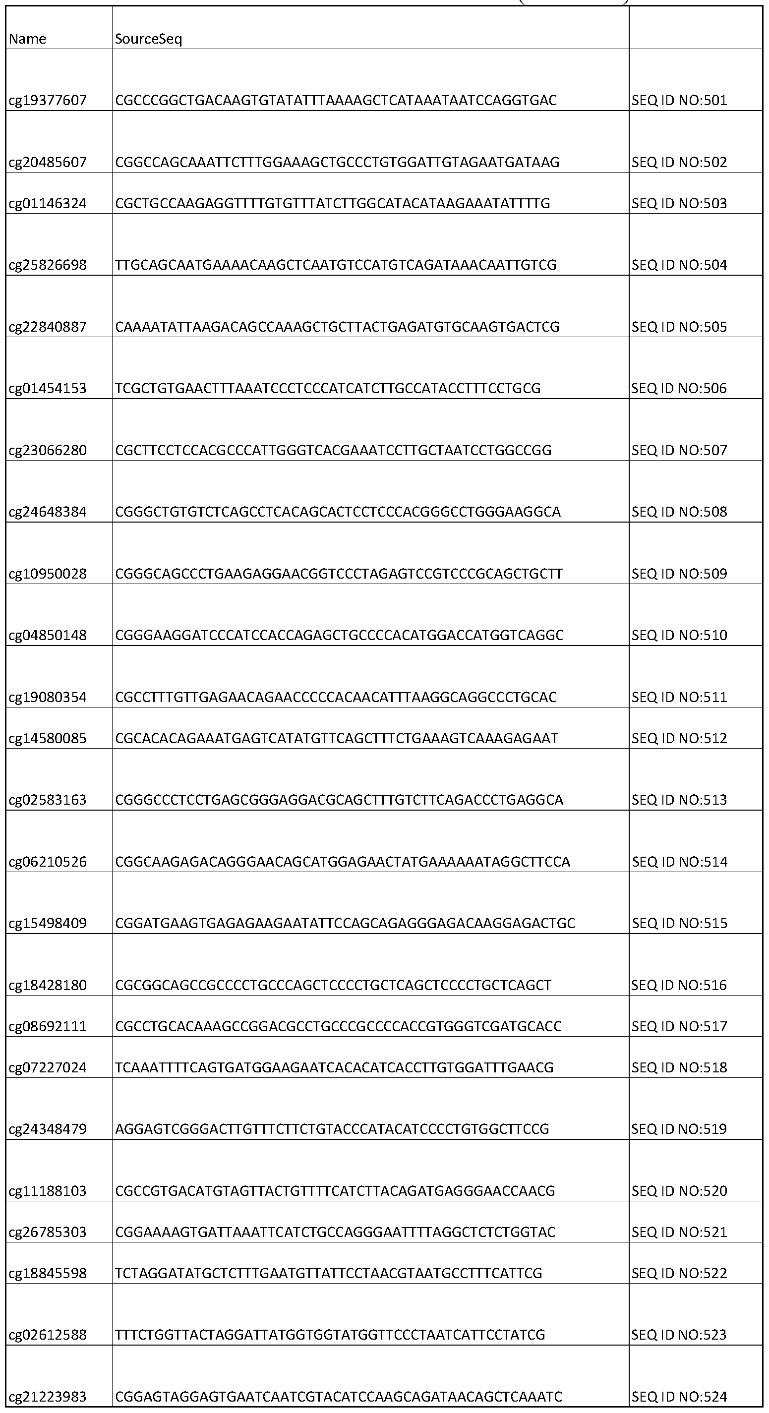
- measuring or having measured the degree of methylation of the extracted DNA preferably includes measuring the degree of methylation at one or more loci listed in Table 1. Without wishing to be bound by theory, it is believed that using the methylation patterns exhibited by the loci of Table 1 does not require a calculation of keratinocyte numbers versus white blood cell numbers.
- DNA methylation - including the degree of methylation - may be measured by any suitable means.
- DNA methylation may be identified using methyl -(MeD IP) pull- downs, bisulfite conversion, reduced representation bisulfite sequencing (RBBS) followed by DNA sequencing and/or high-density array hybridization.
- a high-density array may be used.
- a high-density array may include, for example, an Infinium
- DNA methylation data may be analyzed using any suitable methodology known to those having skill in the art. For example, in some embodiments, DNA methylation data may be analyzed using RnBeads (available from the Max Planck Institut Informatik on the world wide web at rnbeads.mpi-inf.mpg.de/).
- the method includes determining if one or more loci exhibit a degree of methylation is outside of a normal limit. Whether a degree of methylation is outside of a normal limit for each loci may be calculated by a person having ordinary skill in the art based on a reference range.
- the reference range may be determined by the level of methylation most prevalent in a reference group taken from the general (for example, total) population. In some embodiments, the reference range may be defined as the interval between which 95% of values of a reference group fall into.
- whether the degree of methylation of a loci is outside of the normal limit may be calculated with reference to the mean difference ("mean.dif ') values provided in Table 1.
- mean difference mean.dif '
- a degree of methylation at a locus may be outside of a normal limit when the absolute value of the degree of methylation at the locus is at least as great as the absolute value of the mean difference for the same locus listed in Table 1.
- the absolute values of mean difference provided in Table 1 range from 0.007 to 0.63, and negative values in this column of Table 1 indicate loci in which more methylation was observed (on average) in individuals with increased Cortisol compared to control individuals, as further discussed in the Examples.
- the normal limit for a locus may be calculated with reference to a normal control.
- a normal control may include an individual having similar characteristics to the individual (e.g., age, sex, etc.) from which the biological sample was obtained but known not to have been subject to a traumatic experience.
- a normal control may preferably include a population of individuals taken from the general (for example, total) population.
- the degree of methylation at the locus may be altered from the degree of methylation for the same locus in a normal control. In some embodiments, the degree of methylation may be altered by at least 1%, at least 2%, at least 3%, at least 4%, at least 5%, at least 10%, at least 15%, at least 20%, at least 25%, at least 30%, at least 35%, or at least 40% from the degree of methylation for the same locus in a normal control.
- loci if one or more loci exhibit a degree of methylation outside of a normal limit an individual may be identified as having been subjected to a traumatic experience.
- the method preferably includes determining if a plurality of loci exhibit a degree of methylation outside of a normal limit.
- the method may include measuring the degree of methylation at 2 or more loci, at least 10 loci, at least 20 loci, at least 30 loci, at least 40 loci, at least 50 loci, at least 100 loci, at least 150 loci, at least 200 loci, at least 225 loci, or at least 250 loci.
- the loci are preferably selected from the loci listed in Table 1.
- the degree of methylation may preferably be measured at 2 or more loci, at least 10 loci, at least 20 loci, at least 30 loci, at least 40 loci, at least 50 loci, at least 100 loci, at least 150 loci, at least 200 loci, at least 225 loci, or at each or the loci listed in Table 1.
- an individual may be identified as having been subjected to a traumatic experience if at least 10 loci, at least 20 loci, at least 30 loci, at least 40 loci, at least 50 loci, at least 100 loci, at least 150 loci, at least 200 loci, at least 225 loci, or each of the loci of Table 1 exhibit a degree of methylation outside of a normal limit.
- the method may include measuring the degree of methylation at loci from at least ten genes, at least 20 genes, at least 30 genes, at least 40 genes, or at least 50 genes.
- the method may include measuring the degree of methylation at loci having an absolute value of the mean difference ("mean.diff column) as listed in Table 1 of at least 0.01, at least 0.02, at least 0.03, at least 0.04, at least 0.05, at least 0.1, at least 0.15, at least 0.2, at least 0.25, at least 0.3, at least 0.35, at least 0.4, at least 0.45, or at least 0.5.
- mean.diff column an absolute value of the mean difference
- the method may include measuring the degree of methylation at loci having a degree of methylation that is altered by at least 1%, at least 2%, at least 3%, at least 4%, at least 5%, at least 10%, at least 15%, at least 20%, at least 25%, at least 30%, at least 35%, or at least 40% from the degree of methylation for the same locus in a normal control.
- the method may include measuring the degree of methylation at loci that have been previously correlated with clinical history including a traumatic experience. In some embodiments, the method may include measuring the degree of methylation at loci that have been correlated with other biomarkers known to be altered in response to a traumatic experiment. Correlating the degree of methylation with the clinical history and/or other biomarkers may improve accuracy of the diagnosis of a biological response to a traumatic experience.
- the degree of methylation at an increased number of loci may be desirable to measure the degree of methylation at an increased number of loci to prevent an erroneous conclusion that the individual has not experienced trauma.
- the degree of methylation of the extracted DNA of the samples may be compared.
- the method when the method includes obtaining an additional (e.g., second) biological sample from the individual at an additional (e.g., second) time point, the degree of methylation of the extracted DNA of the biological sample at one or more loci listed in Table 1 may be compared with the degree of methylation of the extracted DNA of an additional biological sample at the same one or more loci listed in Table 1.
- an additional biological sample can be obtained after an individual has been subjected to or is suspected of having been subjected to a traumatic experience.
- the method may further include determining if the degree of methylation is altered in the additional biological sample compared to the degree of methylation at the same locus in a previous biological sample.
- the absolute values of the degree of methylation one or more loci may be compared. In some embodiments, if the degree of methylation for one or more loci listed in Table 1 is altered in the additional biological sample, the individual may be identified as having been subjected to a traumatic experience between collections of the biological samples. In some embodiments, the method may further include treating the individual with a therapy effective to reverse the methylation of the DNA at one or more loci. In some
- the therapy may be effective to reverse the methylation at one or more loci where the degree of methylation is outside of a normal limit. For example, when the degree of methylation is increased at a locus to a level outside of a normal limit, reversing the methylation may include decreasing the degree of methylation at that locus; when the degree of methylation is decreased at a locus to a level outside of a normal limit, reversing the methylation may include increasing the degree of methylation at that locus.
- the therapy may involve administering an effective amount of a drug that may reverse the abnormal DNA methylation patterns that are associated with exposure to a traumatic experience.
- the therapy may alter the degree of methylation at one or more loci listed in Table 1 by at least 1%, at least 5%, at least 10%, at least 20%, at least 30%, at least 40%, at least 50%, at least 60%, at least 70%, at least 80%, or at least 90%.
- methylation status may be measured before, during, and/or after a therapy is administered to determine if the therapy is effective. Measuring methylation status at such different time points may be an effective method to follow efficacy of a therapy.
- the methods disclosed herein may include obtaining a biological sample from an individual before a therapy and an additional (e.g., a second) biological sample from the same individual at a time point after treating the individual with the therapy (e.g., a second time point).
- the method may further include comparing the degree of methylation of the extracted DNA of a biological sample taken before the therapy with the degree of methylation of the extracted DNA of the biological sample taken after the therapy at the same one or more loci and determining if the degree of methylation for one or more loci (including, for example, the loci listed in Table 1) is altered.
- the steps may be conducted in any feasible order. And, as appropriate, any combination of two or more steps may be conducted simultaneously.
- Protocol 1 Analyzing methylation and Cortisol changes with Trauma-Focused Cognitive Behavioral therapy (TF-CBT).
- ECBI Eyeberg Child Behavior Inventory
- TCYC Trauma Symptom Checklist for Young Children
- YOQ Youth Outcome Questionnaire
- UCLA PTSD RI UCLA PTSD Reaction Index
- Protocol 2 Changes in methylation patterns in children exposed to severe traumatic experiences.
- Genomic DNA from both types of collection was concentrated and amounts assessed by Qubit 2.0 Fluorometer using the Qubit dsDNA High Sensitivity Assay.
- Illumina Methyl ationEPIC BeadChip (Illumina, San Diego, CA). DNA from every sample was bisulfite converted and run in duplicate. Genomic DNA (500-1000 ng) was bisulfite converted using the Zymo EZ DNA methylation kit (Zymo Research, Irvine, CA) according to the manufacturer's recommendations. After recovery, 8 ⁇ of each bisulfite converted DNA sample further processed using the Illumina FFPE Restoration Solution (Illumina, San Diego, CA) as specified by the manufacturer. The Restoration Solution repairs degraded DNAs for use in genome-scale genotyping and DNA methylation assay platforms. The entire restored sample was then used as a substrate for the Illumina HM450 BeadArrays, as recommended by the manufacturer and previously described (Bibikova et al., 2012. Genomics 98(4):288-295).
- each sample was whole genome amplified (WGA) and then enzymatically fragmented. Samples were then hybridized overnight to an eight sample BeadArray, in which the WGA-DNA molecules anneal to locus-specific DNA oligomers linked to individual bead types.
- the oligomer probe designs follow the Infinium I and II chemistries, in which base extension follows hybridization to a locus-specific oligomer. With respect to the Infinium I probes, there are two different bead types for each locus, one with an oligomer that anneals specifically to the methylated version of the locus, while the other oligomer anneals to the unmethylated version of the locus.
- the probes terminate complementary to the interrogated CpG site for methylated loci, or complementary to the TpG for unmethylated alleles.
- a matched oligomer-template DNA molecule hybrid will allow for the incorporation of a labeled nucleotide immediately upstream (5') to the interrogated CpG (or TpG) site.
- primer extension will not occur.
- Adenine and thymine nucleotides are labeled with cy5 (red), while cytosine nucleotides are labeled with cy3 (green). No insertion of guanine nucleotides occurs in Infinium I assays.
- the identity of the dye is representative of the nucleotide adjacent to the CpG dinucleotide.
- the methylation discrimination is derived from separate measurements from the two different types of beads present for each locus. For some loci, both measurements will be cy3, and for others both will be cy5.
- the Infinium type II chemistry is a true two-color system.
- a matched oligomer-template DNA molecule hybrid will allow for the incorporation of a labeled nucleotide at the interrogated C or T of the CpG site.
- Adenine nucleotides labeled with cy5 red
- guanine nucleotides labeled with cy3 green
- CpG methylated
- BeadArrays are scanned and the raw signal intensities are extracted from the *.IDAT files using the R package minfi.
- the intensities are corrected for background fluorescence and red-green dye-bias using the methods previously described by (Triche et al., 2013. Nucleic Acids Res 41(7):e90).
- the beta value is calculated as (M/(M+U)), in which M and U refer to the (pre-processed) mean methylated and unmethylated probe signal intensities, respectively. Measurements in which the fluorescent intensity is not statistically significantly above background signal (detection p value > 0.05) are removed from the data set.
- probes that overlap with known SNPs as well as repetitive elements are masked prior to data analyses. Specifically, all MethylationEPIC probes that overlap with any common SNPs.
- Data preprocessing was done in R using packages adapted for MethylationEPIC data.
- Data was background corrected using noob (normal-exponential convolution using out-of-band probes) from the R package minfi, which takes the known out-of-band probes present on the array that provide signals in the opposite color channel and uses normal-exponential convolution to subtract the background average intensity (from the out-of-band probes) from the foreground intensity.
- the data was normalized using dasen in wateRmelon (Illumina, San Diego, CA).
- Principle component analysis identified components that were significantly correlated with traits in our data, primarily sentrix ID and time point. Components relating to minor variance in the data included age and sex thus we carried out a covariate analysis to remove effects due to these traits. Covariates included age and sex. Sentrix ID was also adjusted for to remove batch effects from chip differences. Service planning area was included as a covariate in the pilot study only because we did not have information about this variable for the longitudinal study.
- Differential analysis was used to compare time points within subject. Differential analysis was also carried out to compare individuals based on level of abuse. Differential methylation between groups was analyzed in promoter regions, which are assigned as the regions from 1.5 kb upstream to 0.5 kb downstream of the transcription start site according to Ensembl annotations. The statistical tool Limma was used to determine differentially methylated positions (DMPs). False Discovery Rate (FDR) correction was performed at the region level for promoters to adjust p-values.
- DMPs differentially methylated positions
- FDR False Discovery Rate
Landscapes
- Chemical & Material Sciences (AREA)
- Life Sciences & Earth Sciences (AREA)
- Organic Chemistry (AREA)
- Health & Medical Sciences (AREA)
- Proteomics, Peptides & Aminoacids (AREA)
- Engineering & Computer Science (AREA)
- Zoology (AREA)
- Wood Science & Technology (AREA)
- Analytical Chemistry (AREA)
- Genetics & Genomics (AREA)
- Bioinformatics & Cheminformatics (AREA)
- Immunology (AREA)
- Microbiology (AREA)
- Molecular Biology (AREA)
- Physics & Mathematics (AREA)
- Biotechnology (AREA)
- Biochemistry (AREA)
- Biophysics (AREA)
- General Engineering & Computer Science (AREA)
- General Health & Medical Sciences (AREA)
- Pathology (AREA)
- Measuring Or Testing Involving Enzymes Or Micro-Organisms (AREA)
Abstract
A method includes obtaining a biological sample from an individual, extracting DNA from cells in the sample, measuring the degree of methylation of the extracted DNA at one or more specified loci, and identifying if at least one locus exhibits an alteration in methylation or a degree of methylation that is outside of a normal limit. The method may further include determining whether an individual was exposed to abuse and/or trauma. In some cases, the method further includes treating the individual with a therapy effective to reverse the differential methylation of the DNA.
Description
DNA METHYLATION SIGNATURES ASSOCIATED WITH CHILDHOOD TRAUMA AND
METHODS INVOLVING SAME
CONTINUING APPLICATION DATA
This application claims the benefit of U.S. Provisional Application Serial No.
62/362,903, filed July 15, 2016, which is incorporated by reference herein.
GOVERNMENT FUNDING
This invention was made with government support under MH096093 awarded by the National Institutes of Health. The government has certain rights in the invention. SEQUENCE LISTING
This application contains a Sequence Listing electronically submitted to the United States Patent and Trademark Office via EFS-Web as an ASCII text file entitled "310- 01130201_ST25.txt" having a size of 168 kilobytes and created on July 12, 2017. Due to the electronic filing of the Sequence Listing, the electronically submitted Sequence Listing serves as both the paper copy required by 37 CFR §1.821(c) and the CRF required by §1.821(e). The information contained in the Sequence Listing is incorporated by reference herein.
SUMMARY
This disclosure describes epigenetic changes, particularly DNA methylation, that may occur in a traumatized individual and methods of detecting those changes. In one aspect, this disclosure describes a method that includes obtaining a biological sample from an individual, extracting DNA from cells in the sample, and measuring the degree of methylation of the extracted DNA at one or more specified loci. In some embodiments, the method includes determining whether the degree of methylation at one or more of the specified loci is outside of a normal limit. In some embodiments, the method includes identifying the individual as having been subjected to a traumatic event. The individual may be identified as having been subjected to a traumatic event if one or more of the specified loci exhibits a degree of methylation outside of a normal limit. In some embodiments, the individual is a child.
In some cases, the method may be used to follow an individual over time. In such embodiments, the method may be repeated at a second time point. For example, the method may include identifying the individual as having been subjected to trauma between collection of a first biological sample and collection of a second biological sample if the second biological sample exhibits an alteration in the degree of DNA methylation compared to the first biological sample. Additionally or alternatively, the method may include treating the individual with a therapy between collection of a first biological sample and collection of a second biological sample. In some embodiments, the therapy is effective to reverse methylation at one or more loci.
In some embodiments, the method includes measuring the degree of methylation at a plurality of loci. In some embodiments, the one or more loci include one or more CpG sites.
The above summary is not intended to describe each disclosed embodiment or every implementation of the present invention. The description that follows more particularly exemplifies illustrative embodiments. In several places throughout the application, guidance is provided through lists of examples, which examples can be used in various combinations. In each instance, the recited list serves only as a representative group and should not be interpreted as an exclusive list.
BRIEF DESCRIPTION OF THE FIGURES
The patent or application file contains drawings executed in color. Copies of this patent or patent application publication with color drawings will be provided by the Office upon request and payment of the necessary fee.
FIG. 1. An exemplary experimental design.
FIG. 2. Exemplary subject enrollment. For subjects in rows 1 to 6, Cortisol measurements were taken, and methylation was determined (because the subjects exhibited the most reliable Cortisol measurements). For rows 7-10, only Cortisol measurements were taken for the subject.
FIG. 3. Exemplary behavioral data. Subjects 2, 4, and 5 exhibited no difference from intake to follow-up in traumatic event; Subjects 1, 3, and 6 exhibited difference in trauma from intake to follow-up; Boxes outlined with dotted lines = either no trauma or traumatic event was not directed toward individual; Boxes including circles = trauma included the individual.
FIG. 4(A-B). Cortisol levels at intake and after therapy (FIG. 4A) and methylation patterns for the same subjects (FIG. 4B). Cortisol was extracted from 3 cm of hair at each time point and measured by ELISA. A heat map of methylation sites from a dataset of 850,000 sites showing the 1000 most significantly different (between methylated and non-methylated) sites in promoter/enhancer regions. Hierarchical clustering is by similarity between these sites across samples (dendrogram above hear map). Note the subject 6: follow-up, when Cortisol is high, is on the left of the heat map, showing strong difference between all other samples, including intake (lane 6 from the left on the heat map) for this same subject. This difference indicates that Cortisol alone is not responsible for the flip in methylation of these promoter sites, since subjects 4 and 5 had high Cortisol at intake but did not display this pattern.
FIG. 5 shows exemplary data comparing DNA methylation profiles of abuse. Left panel: Methylation values for all 850,000 sites demonstrate minor variability across all individuals. Red indicates low methylation, grey is intermediate, and blue is representative of higher methylation. Middle panel: Methylation values for the 1000 most variable promoter-associated sites including two more subjects at two time points. Three samples demonstrate a similar flip in the
methylation of these sites (left three lanes). These samples are exemplary of individuals with higher instance of abuse at that time point. Right panel: Heat map comparing only the three individuals with the most drastic change in methylation for the most variable promoter- associated sites.
FIG. 6. Gene ontology enrichment for the top 2000 promoter-associated sites. Ten categories reached significance. X-axis indicates number of sites involved in this pathway.
FIG. 7. Pathway analysis of enriched gene ontology (GO) terms. Percent change in this instance is referring to delta beta (difference between beta values, compared between subjects). Green indicates hypomethylation in better group (hypermethylation in worse group). Red indicates hypermethylation in better group (hypomethylation in worse group). Statistically significant differences between delta beta were compared across 850,000 sites the top 2000 sites with the highest probability of having a significant difference between unmethylated and methylated sites for each sample were selected. Six subjects were measured before and after therapy in duplicate, and two subjects were measured at two different time points also run in duplicate for a total of 32 measurements of 850,000 sites each. Genes with highest probability of
being methylated and highest probability of being unmethylated in each individual were ranked across samples producing the heat map.
FIG. 8 (A-B). Exemplary micrographs of saliva smears. To determine the types of cells in saliva, saliva was collected from 6 healthy volunteers either directly onto a glass slide (FIG. 8A) or first into ORAGE E DNA collection cups which contain a buffer to preserve the DNA and then onto a glass slide (FIG. 8B). Slides were stained with hematoxylin and eosin according to normal pathology procedures. Slides were reviewed and types of cells counted. As shown in FIG. 8A, some saliva had abundant keratinocytes (solid arrow) and few WBCs (dashed arrow); other samples displayed the opposite ratio of cells. As shown in FIG. 8B, after treatment with the buffer, keratinocytes appeared ghosted and lacked nuclei. Some small round blue dots remaining represent un-extracted nuclei.
DETAILED DESCRIPTION OF ILLUSTRATIVE EMBODEVIENTS
This disclosure describes epigenetic changes, particularly DNA methylation, that occur in traumatized individuals and methods of detecting those changes. Accordingly, this disclosure describes a method of measuring the DNA methylation in a biological sample obtained from an individual at one or more specified loci. In some embodiments, the method includes identifying individuals who have been exposed to a traumatic experience (including, for example abuse) and/or treating individuals who have been exposed to a traumatic experience.
Childhood maltreatment is a major risk factor for poor physical outcomes in adulthood such as, for example, diabetes, cardiovascular disease, depression, and/or other cognitive dysfunction. Epigenetic modifications have become an increasingly popular mechanism to explain the relationship between adverse childhood experiences (ACE) and poor health outcomes later in life. DNA methylation is a commonly studied epigenetic modification since DNA methylation influences gene expression and is involved in cell differentiation during
development. DNA methylation may alter gene expression without changing the genetic sequence, providing a potential therapeutic target for reversible modifications. The human genome is predicted to have over five million dynamic methylation sites, indicating that 20% of the methylation sites present in the genome may be altered by environmental and developmental influences. These dynamic sites are of particular interest in understanding the mechanism underlying the association between childhood maltreatment and poor health in adulthood
because they may be potential therapeutic targets since methylation changes of these sites could be reversed.
Therapies that target methylation sites include drugs that alter DNA methylation in immune cells and/or in the brain. Some drugs may be already approved for use in treating other diseases. Exemplary drugs include, for example, valproic acid, 5-azocyidine, propranolol, inhibitors/activators of the norepinephrine system (for example, an alpha- 1 blocker such as, for example, prazosin), or drugs approved for treating post-traumatic stress disorder (PTSD) and/or anxiety disorders in adults.
It may be that no single methylation site is responsible for driving all of the negative phenotypes seen in children that have been exposed to a traumatic experience. However, it may be that expression of many related genes is changed in tandem, leading to the disruption seen later in life in these individuals. A change in expression of a gene may be the result of a change in the methylation of a regulatory sequence that is operably linked to the gene so that the methylation status of the regulatory sequence influences expression of the gene. If so, then global methylation patterns may reveal a signature that reflects a history that includes exposure to a traumatic experience. The methylation sites that are associated with the signature— for example, the sites that are altered by childhood trauma— may be re-altered using appropriate therapy.
Significant changes in the methylation levels of 2000 promoter sites occurred in samples obtained from individuals subjected to abuse as reported in the PTSD Eyberg survey. Similar methylation data from archived saliva samples from children with documented abuse validated the initial data. So, too, did a comparison of the methylation patterns described herein with published findings in adults reporting abuse in childhood. All of these analyses produced the same signature pattern: Elevated methylation of 1000 sites, and decreased methylation in another 1000 sites. An ontology analysis revealed that the more significantly changed sites are in genes for hemostasis and immunological processes. This subset of sites may be used to design tests to detect the impact of early life abuse, and to develop and follow therapeutic interventions.
The methylation changes were analyzed using a whole genome approach implemented by the Illumina Methyl ationEPIC 850K BeadChip that interrogates over 850,000 methylation sites across the genome. The array-based approach provides single-base resolution and allows one to recognize patterns across the genome in sites that have already been shown to be involved in regulation of gene expression such as promoters and enhancers. This array also allows one to
identify intergenic regions that are involved in psychiatric disorders such as bipolar and schizophrenia.
Since a traumatic experience may include abuse including, for example, a wide variety of maltreatment types such as physical, sexual, and emotional abuse, the analysis adopted an unbiased data-driven approach to allow the data to explain differences in children with different trauma histories (FIG. 3). The Cortisol patterns appeared to be most related to service planning area (FIG. 4). Although Cortisol had some correlation with behavioral measures, they were moderate and insignificant.
Methylation, however, was more informative of trauma effects. Methylation patterns for one individual in particular were opposite for a subset of 2,000 promoter-associated CpG sites. Methylation sites were enriched for genes involved in homeostasis as well as platelet activation and other inter-related pathways. While the overall methylation pattern was not drastically different from others, the methylation differences of these sites were extreme, demonstrating an almost complete reversal in methylation— that is, an increase in sites that had low methylation and a decrease in sites that had high methylation (FIG. 5). Samples from this child exhibited altered methylation at follow-up was the only child that also exhibited a dramatic increase in Cortisol at follow-up. Behavioral analysis confirmed that this child had experienced worsening trauma throughout her time in therapy. At intake, she was a witness to domestic violence, but at all time points following, she was a victim of domestic violence. Two other children, while less extreme, demonstrated differing patterns of methylation from intake to follow-up at similar sites. Neither of these children reported trauma at intake on their behavioral records, but did report trauma for at least one later time point during therapy.
Gene ontology (GO) enrichment analysis was computed for the 2000 most variable promoter sites. Of these 2000 sites, 817 mapped to specific genes based on EntrezID. These sites were significantly (p<0.05 BH corrected) enriched in 10 categories (FIG. 6 and FIG. 7).
To validate this observed methylation patterns, results with previous publications on methylation changes in victims of child abuse. Suderman et al. {Proceedings of the National Academy of Sciences of the United States of America 2013, 110 (14), E1246) report a similar pattern in methylation for a subset of sites for adult males who were victims of abuse as children. Since this pattern appears to prevail into adulthood, saliva samples from a longitudinal study that followed children over a 15-year period from childhood into early adulthood were compared to
the pilot study data described above. The pattern for the two individuals was nearly identical for the most variable sites seen in the pilot study.
In one aspect, the methods described herein include obtaining or having obtained at least one biological sample from an individual. In some embodiments, the individual is preferably a human. In some embodiments, the individual is preferably a child. As used herein, a child includes an individual under the age of 18. In some embodiments, the child is preferably between the ages of 2 and 18. In some embodiments, an additional (for example, second, third, fourth, etc.) biological sample may be obtained from the same individual. In some embodiments, the additional biological sample may be obtained at an additional (for example, second, third, fourth, etc.) time point. For example, the additional time point(s) may be a day, a plurality of days, a month, a plurality of months, a year, or a plurality of years after the first biological sample is collected. In some embodiments, when the individual is suspected of having been subject to a traumatic experience after the collection of the first sample, the additional biological sample may be collected after the traumatic experience has occurred. A traumatic experience may include, abuse including, for example, physical abuse, emotional abuse, sexual abuse, and neglect. A traumatic experience may additionally or alternatively include accidental trauma (for example, car accidents or other accidental injuries) or witnessing another individual undergoing abuse (for example, witnessing domestic violence).
A biological sample may include any suitable sample that includes cells from an individual. For example, a biological sample may include whole blood, semen, saliva, tears, urine, fecal material, sweat, buccal smears, skin, hair, and biopsy samples of organs and muscle.
The biological sample may be obtained by any suitable means. In some embodiments, the biological sample may be stored in a collection device. Collection devices suitable for use in the present invention include devices known in the art for collecting and/or storing a biological sample of an individual from which nucleic acids and/or polypeptides (including, for example DNA) may be isolated. Suitable collection devices include, for example, specimen cups, swabs, glass slides, test tubes, lancets, ORAGENE DNA collection cups, VACUTAINER tubes, and kits.
The methods described herein may include extracting or having extracted DNA from at least a portion of the cells included in the biological sample. The DNA may be extracted
(including, for example, isolated and purified) by any suitable means known to those having skill
in the art. Extracting DNA preferably preserves the methylation status of the DNA present in the cells of the biological sample.
In some embodiments, including, for example, when the biological sample includes saliva, the biological sample may include at least one of a keratinocyte and a white blood cell. In some embodiments, the keratinocyte may include a buccal keratinocyte. Saliva contains both white blood cells and buccal keratinocytes, in different proportions in different samples (see FIG. 8). Moreover, saliva may be more easily obtained from children than blood. Saliva collection is more acceptable to many children than blood collection. Further, it is easier for investigators to obtain permission from offices for protection of human subjects from risk, such as Institutional Review Boards (IRB), to collect saliva for experimental procedures.
The methods described herein may include measuring or having measured the degree of methylation of the extracted DNA at one or more loci. In some embodiments, the one or more loci include one or more CpG sites (also referred to as CG sites or CpG methylation sites). The cytosine in the CpG dinucleotide may preferably be methylated to form 5-methylcytosine. In some embodiments, each of the one or more loci includes one or more CpG sites. In some embodiments, each of the one or more loci includes one CpG sites.
For example, in some embodiments, the degree of methylation of the DNA extracted from cells present in saliva may be measured. In some embodiments, methylation patterns obtained from saliva may need to be corrected for relative numbers of and/or DNA of
keratinocytes versus white blood cells. Stressed children may have scant saliva and thus produce more keratinocytes than non-stressed children, since fear and anxiety in children produce a severe dry mouth which alters the cellular composition of saliva. Hence the cell composition of the saliva may need to be accounted for in the analysis of methylation sites, since keratinocytes and white blood cells may have widely different methylation patterns.
In some embodiments, the one or more loci exhibiting methylation patterns may preferably be selected such that no calculation of keratinocyte versus white blood cell numbers and/or DNA is required.
In some embodiments, measuring or having measured the degree of methylation of the extracted DNA preferably includes measuring the degree of methylation at one or more loci listed in Table 1. Without wishing to be bound by theory, it is believed that using the methylation
patterns exhibited by the loci of Table 1 does not require a calculation of keratinocyte numbers versus white blood cell numbers.
DNA methylation - including the degree of methylation - may be measured by any suitable means. For example, DNA methylation may be identified using methyl -(MeD IP) pull- downs, bisulfite conversion, reduced representation bisulfite sequencing (RBBS) followed by DNA sequencing and/or high-density array hybridization. In some embodiments, a high-density array may be used. A high-density array may include, for example, an Infinium
HumanMethylation450 BeadChip Kit (Illumina, San Diego, CA), an Infinium MethylationEPIC Kit (Illumina, San Diego, CA), etc. DNA methylation data may be analyzed using any suitable methodology known to those having skill in the art. For example, in some embodiments, DNA methylation data may be analyzed using RnBeads (available from the Max Planck Institut Informatik on the world wide web at rnbeads.mpi-inf.mpg.de/).
In some embodiments, the method includes determining if one or more loci exhibit a degree of methylation is outside of a normal limit. Whether a degree of methylation is outside of a normal limit for each loci may be calculated by a person having ordinary skill in the art based on a reference range. The reference range may be determined by the level of methylation most prevalent in a reference group taken from the general (for example, total) population. In some embodiments, the reference range may be defined as the interval between which 95% of values of a reference group fall into.
In some embodiments, whether the degree of methylation of a loci is outside of the normal limit may be calculated with reference to the mean difference ("mean.dif ') values provided in Table 1. For example, a degree of methylation at a locus may be outside of a normal limit when the absolute value of the degree of methylation at the locus is at least as great as the absolute value of the mean difference for the same locus listed in Table 1. The absolute values of mean difference provided in Table 1 range from 0.007 to 0.63, and negative values in this column of Table 1 indicate loci in which more methylation was observed (on average) in individuals with increased Cortisol compared to control individuals, as further discussed in the Examples.
In some embodiments, the normal limit for a locus may be calculated with reference to a normal control. In some embodiments, a normal control may include an individual having similar characteristics to the individual (e.g., age, sex, etc.) from which the biological sample
was obtained but known not to have been subject to a traumatic experience. In some embodiments, a normal control may preferably include a population of individuals taken from the general (for example, total) population.
In some embodiments, the degree of methylation at the locus may be altered from the degree of methylation for the same locus in a normal control. In some embodiments, the degree of methylation may be altered by at least 1%, at least 2%, at least 3%, at least 4%, at least 5%, at least 10%, at least 15%, at least 20%, at least 25%, at least 30%, at least 35%, or at least 40% from the degree of methylation for the same locus in a normal control.
In some embodiments, if one or more loci exhibit a degree of methylation outside of a normal limit an individual may be identified as having been subjected to a traumatic experience.
In some embodiments, the method preferably includes determining if a plurality of loci exhibit a degree of methylation outside of a normal limit. For example, the method may include measuring the degree of methylation at 2 or more loci, at least 10 loci, at least 20 loci, at least 30 loci, at least 40 loci, at least 50 loci, at least 100 loci, at least 150 loci, at least 200 loci, at least 225 loci, or at least 250 loci. In some embodiments, the loci are preferably selected from the loci listed in Table 1. In some embodiments, the degree of methylation may preferably be measured at 2 or more loci, at least 10 loci, at least 20 loci, at least 30 loci, at least 40 loci, at least 50 loci, at least 100 loci, at least 150 loci, at least 200 loci, at least 225 loci, or at each or the loci listed in Table 1.
In some embodiments, an individual may be identified as having been subjected to a traumatic experience if at least 10 loci, at least 20 loci, at least 30 loci, at least 40 loci, at least 50 loci, at least 100 loci, at least 150 loci, at least 200 loci, at least 225 loci, or each of the loci of Table 1 exhibit a degree of methylation outside of a normal limit.
In some embodiments, the method may include measuring the degree of methylation at loci from at least ten genes, at least 20 genes, at least 30 genes, at least 40 genes, or at least 50 genes.
In some embodiments, the method may include measuring the degree of methylation at loci having an absolute value of the mean difference ("mean.diff column) as listed in Table 1 of at least 0.01, at least 0.02, at least 0.03, at least 0.04, at least 0.05, at least 0.1, at least 0.15, at least 0.2, at least 0.25, at least 0.3, at least 0.35, at least 0.4, at least 0.45, or at least 0.5.
In some embodiments, the method may include measuring the degree of methylation at loci having a degree of methylation that is altered by at least 1%, at least 2%, at least 3%, at least 4%, at least 5%, at least 10%, at least 15%, at least 20%, at least 25%, at least 30%, at least 35%, or at least 40% from the degree of methylation for the same locus in a normal control.
In some embodiments, the method may include measuring the degree of methylation at loci that have been previously correlated with clinical history including a traumatic experience. In some embodiments, the method may include measuring the degree of methylation at loci that have been correlated with other biomarkers known to be altered in response to a traumatic experiment. Correlating the degree of methylation with the clinical history and/or other biomarkers may improve accuracy of the diagnosis of a biological response to a traumatic experience.
In some embodiments, including, for example, when the type of traumatic experience an individual may have been subjected to is unknown, it may be desirable to measure the degree of methylation at an increased number of loci to prevent an erroneous conclusion that the individual has not experienced trauma.
In some embodiments, including for example, when more than one biological sample has been obtained from an individual, the degree of methylation of the extracted DNA of the samples may be compared.
For example, when the method includes obtaining an additional (e.g., second) biological sample from the individual at an additional (e.g., second) time point, the degree of methylation of the extracted DNA of the biological sample at one or more loci listed in Table 1 may be compared with the degree of methylation of the extracted DNA of an additional biological sample at the same one or more loci listed in Table 1. In some embodiments, an additional biological sample can be obtained after an individual has been subjected to or is suspected of having been subjected to a traumatic experience. In some embodiments, the method may further include determining if the degree of methylation is altered in the additional biological sample compared to the degree of methylation at the same locus in a previous biological sample. In some embodiments, the absolute values of the degree of methylation one or more loci may be compared. In some embodiments, if the degree of methylation for one or more loci listed in Table 1 is altered in the additional biological sample, the individual may be identified as having been subjected to a traumatic experience between collections of the biological samples.
In some embodiments, the method may further include treating the individual with a therapy effective to reverse the methylation of the DNA at one or more loci. In some
embodiments, the therapy may be effective to reverse the methylation at one or more loci where the degree of methylation is outside of a normal limit. For example, when the degree of methylation is increased at a locus to a level outside of a normal limit, reversing the methylation may include decreasing the degree of methylation at that locus; when the degree of methylation is decreased at a locus to a level outside of a normal limit, reversing the methylation may include increasing the degree of methylation at that locus.
In some embodiments, the therapy may involve administering an effective amount of a drug that may reverse the abnormal DNA methylation patterns that are associated with exposure to a traumatic experience.
In some embodiments, the therapy may alter the degree of methylation at one or more loci listed in Table 1 by at least 1%, at least 5%, at least 10%, at least 20%, at least 30%, at least 40%, at least 50%, at least 60%, at least 70%, at least 80%, or at least 90%.
In some embodiments, methylation status may be measured before, during, and/or after a therapy is administered to determine if the therapy is effective. Measuring methylation status at such different time points may be an effective method to follow efficacy of a therapy.
The methods disclosed herein may include obtaining a biological sample from an individual before a therapy and an additional (e.g., a second) biological sample from the same individual at a time point after treating the individual with the therapy (e.g., a second time point). The method may further include comparing the degree of methylation of the extracted DNA of a biological sample taken before the therapy with the degree of methylation of the extracted DNA of the biological sample taken after the therapy at the same one or more loci and determining if the degree of methylation for one or more loci (including, for example, the loci listed in Table 1) is altered.
In the preceding description and following claims, the term "and/or" means one or all of the listed elements or a combination of any two or more of the listed elements; the terms
"comprises," "comprising," and variations thereof are to be construed as open ended— that is, additional elements or steps are optional and may or may not be present; unless otherwise specified, "a," "an," "the," and "at least one" are used interchangeably and mean one or more
than one; and the recitations of numerical ranges by endpoints include all numbers subsumed within that range (for example, 1 to 5 includes 1, 1.5, 2, 2.75, 3, 3.80, 4, 5, etc.).
In the preceding description, particular embodiments may be described in isolation for clarity. Unless otherwise expressly specified that the features of a particular embodiment are incompatible with the features of another embodiment, certain embodiments may include a combination of compatible features described herein in connection with one or more
embodiments.
For any method disclosed herein that includes discrete steps, the steps may be conducted in any feasible order. And, as appropriate, any combination of two or more steps may be conducted simultaneously.
The present invention is illustrated by the following examples. It is to be understood that the particular examples, materials, amounts, and procedures are to be interpreted broadly in accordance with the scope and spirit of the invention as set forth herein. EXAMPLES Methods:
Protocol 1 : Analyzing methylation and Cortisol changes with Trauma-Focused Cognitive Behavioral therapy (TF-CBT).
Saliva was collected for DNA methylation analysis, and hair was collected for Cortisol analysis from children referred by Los Angeles County Child Protective Services. These children were referred to Children's institute Inc. for recruitment into a Trauma-Focused Cognitive Behavioral Therapy program (TF-CBT), and they were followed for 6-8 months throughout the therapy as shown in FIG. 1. Saliva was sampled at four time points (intake, midpoint, post- treatment, and 1-month follow-up) (FIG. 1). Hair was sampled at two time points (intake and 1- month follow-up) to allow growth in between. Saliva was collected using DNA Genotek OGR- 250 collection cups. DNA was extracted and purified from the saliva samples using the PrepIT- L2P protocol and followed with RNase double-digestion to ensure purified DNA absent of RNA contamination. Three centimeters of hair were ground in a ball-grinder and Cortisol was extracted using. Six children had reliable Cortisol measurements at both intake and 1 -month follow-up, thus, DNA methylation analysis was carried out for only these six children at both intake and 1-
month follow-up. Psychosocial data was collected for these children at all four time points. Behavioral measures included Eyeberg Child Behavior Inventory (ECBI), Trauma Symptom Checklist for Young Children (TSCYC), Youth Outcome Questionnaire (YOQ), and the UCLA PTSD Reaction Index (UCLA PTSD RI).
Protocol 2: Changes in methylation patterns in children exposed to severe traumatic experiences.
In the first study, children (n=10) referred by LA County for trauma-focused behavioral therapy because of reported trauma were enrolled. Six children completed the therapy and donated sufficient biospecimens for analysis. Whole saliva was obtained by spitting into a collection vesicle (ORAGE E saliva collection), stored at room temperature, and DNA extracted with a DNeasy kit (Qiagen, USA).
In addition, a longitudinal study was performed to assess the effects of trauma from childhood into early adulthood. Saliva was collected at two time points, from entry (average age
= 11 years) to conclusion of the study (average 18 years). Whole saliva was collected by passive drool into 2-mL cryovials (Salimetrics, Inc., Carlsbad, CA). Samples were stored at -80°C after collection. DNA was purified from saliva using an adapted protocol from QIAmp DNA Mini Kit
(Qiagen, USA).
Genomic DNA from both types of collection was concentrated and amounts assessed by Qubit 2.0 Fluorometer using the Qubit dsDNA High Sensitivity Assay.
Behavioral data was collected throughout this study by survey instruments from caregivers. Only behavioral data similar to the pilot study was used for comparing two individuals at two time points.
Microarray Analysis:
Purified DNA was sent to Keck Medical School Epigenome Center for analysis with the
Illumina Methyl ationEPIC BeadChip (Illumina, San Diego, CA). DNA from every sample was bisulfite converted and run in duplicate. Genomic DNA (500-1000 ng) was bisulfite converted using the Zymo EZ DNA methylation kit (Zymo Research, Irvine, CA) according to the manufacturer's recommendations. After recovery, 8 μΐ of each bisulfite converted DNA sample further processed using the Illumina FFPE Restoration Solution (Illumina, San Diego, CA) as specified by the manufacturer. The Restoration Solution repairs degraded DNAs for use in
genome-scale genotyping and DNA methylation assay platforms. The entire restored sample was then used as a substrate for the Illumina HM450 BeadArrays, as recommended by the manufacturer and previously described (Bibikova et al., 2012. Genomics 98(4):288-295).
Specifically, each sample was whole genome amplified (WGA) and then enzymatically fragmented. Samples were then hybridized overnight to an eight sample BeadArray, in which the WGA-DNA molecules anneal to locus-specific DNA oligomers linked to individual bead types. The oligomer probe designs follow the Infinium I and II chemistries, in which base extension follows hybridization to a locus-specific oligomer. With respect to the Infinium I probes, there are two different bead types for each locus, one with an oligomer that anneals specifically to the methylated version of the locus, while the other oligomer anneals to the unmethylated version of the locus. The probes terminate complementary to the interrogated CpG site for methylated loci, or complementary to the TpG for unmethylated alleles. A matched oligomer-template DNA molecule hybrid will allow for the incorporation of a labeled nucleotide immediately upstream (5') to the interrogated CpG (or TpG) site. However, if the probe and template are mismatched, then primer extension will not occur. Adenine and thymine nucleotides are labeled with cy5 (red), while cytosine nucleotides are labeled with cy3 (green). No insertion of guanine nucleotides occurs in Infinium I assays. Of note, the identity of the dye is representative of the nucleotide adjacent to the CpG dinucleotide. The methylation discrimination is derived from separate measurements from the two different types of beads present for each locus. For some loci, both measurements will be cy3, and for others both will be cy5. The Infinium type II chemistry is a true two-color system. A matched oligomer-template DNA molecule hybrid will allow for the incorporation of a labeled nucleotide at the interrogated C or T of the CpG site. Adenine nucleotides labeled with cy5 (red) are incorporated across from unmethylated (TpG) sites, while guanine nucleotides labeled with cy3 (green) are incorporated across from
methylated (CpG) sites.
After the chemistry steps, BeadArrays are scanned and the raw signal intensities are extracted from the *.IDAT files using the R package minfi. The intensities are corrected for background fluorescence and red-green dye-bias using the methods previously described by (Triche et al., 2013. Nucleic Acids Res 41(7):e90). The beta value is calculated as (M/(M+U)), in which M and U refer to the (pre-processed) mean methylated and unmethylated probe signal intensities, respectively. Measurements in which the fluorescent intensity is not statistically
significantly above background signal (detection p value > 0.05) are removed from the data set. In addition, probes that overlap with known SNPs as well as repetitive elements are masked prior to data analyses. Specifically, all MethylationEPIC probes that overlap with any common SNPs. Statistical Analysis
Data preprocessing was done in R using packages adapted for MethylationEPIC data. Data was background corrected using noob (normal-exponential convolution using out-of-band probes) from the R package minfi, which takes the known out-of-band probes present on the array that provide signals in the opposite color channel and uses normal-exponential convolution to subtract the background average intensity (from the out-of-band probes) from the foreground intensity. The data was normalized using dasen in wateRmelon (Illumina, San Diego, CA).
Quality control was performed using RnBeads (Max-Planck-Institut Informatik, Saarbriicken, Germany), and no samples were removed due to poor quality. All replicates had a pearson correlation greater than 0.99 indicating high reproducibility of the bead chip. Filtering was performed in RnBeads to remove those probes that have a detection p-value greater than 0.05, are located on sex chromosomes, are in non-CG context, or overlapped any SNPs. Differential methylation analysis was carried out in RnBeads to look for differentially methylated sites and regions. Surrogate variable analysis was conducted to account for any underlying variables significantly contributing to the variance seen between time points. No surrogate variables were identified. Principle component analysis identified components that were significantly correlated with traits in our data, primarily sentrix ID and time point. Components relating to minor variance in the data included age and sex thus we carried out a covariate analysis to remove effects due to these traits. Covariates included age and sex. Sentrix ID was also adjusted for to remove batch effects from chip differences. Service planning area was included as a covariate in the pilot study only because we did not have information about this variable for the longitudinal study.
Differential analysis was used to compare time points within subject. Differential analysis was also carried out to compare individuals based on level of abuse. Differential methylation between groups was analyzed in promoter regions, which are assigned as the regions from 1.5 kb upstream to 0.5 kb downstream of the transcription start site according to Ensembl annotations. The statistical tool Limma was used to determine differentially methylated positions (DMPs).
False Discovery Rate (FDR) correction was performed at the region level for promoters to adjust p-values.
The complete disclosure of all patents, patent applications, and publications, and electronically available material (including, for instance, nucleotide sequence submissions in, for example, GenBank and RefSeq, and amino acid sequence submissions in, for example,
SwissProt, PIR, PRF, PDB, and translations from annotated coding regions in GenBank and RefSeq) cited herein are incorporated by reference in their entirety. In the event that any inconsistency exists between the disclosure of the present application and the disclosure(s) of any document incorporated herein by reference, the disclosure of the present application shall govern. The foregoing detailed description and examples have been given for clarity of understanding only. No unnecessary limitations are to be understood therefrom. The invention is not limited to the exact details shown and described, for variations obvious to one skilled in the art will be included within the invention defined by the claims.
Unless otherwise indicated, all numbers expressing quantities of components, molecular weights, and so forth used in the specification and claims are to be understood as being modified in all instances by the term "about." Accordingly, unless otherwise indicated to the contrary, the numerical parameters set forth in the specification and claims are
approximations that may vary depending upon the desired properties sought to be obtained by the present invention. At the very least, and not as an attempt to limit the doctrine of equivalents to the scope of the claims, each numerical parameter should at least be construed in light of the number of reported significant digits and by applying ordinary rounding techniques.
Notwithstanding that the numerical ranges and parameters setting forth the broad scope of the invention are approximations, the numerical values set forth in the specific examples are reported as precisely as possible. All numerical values, however, inherently contain a range necessarily resulting from the standard deviation found in their respective testing measurements.
All headings are for the convenience of the reader and should not be used to limit the meaning of the text that follows the heading, unless so specified.
Table 1


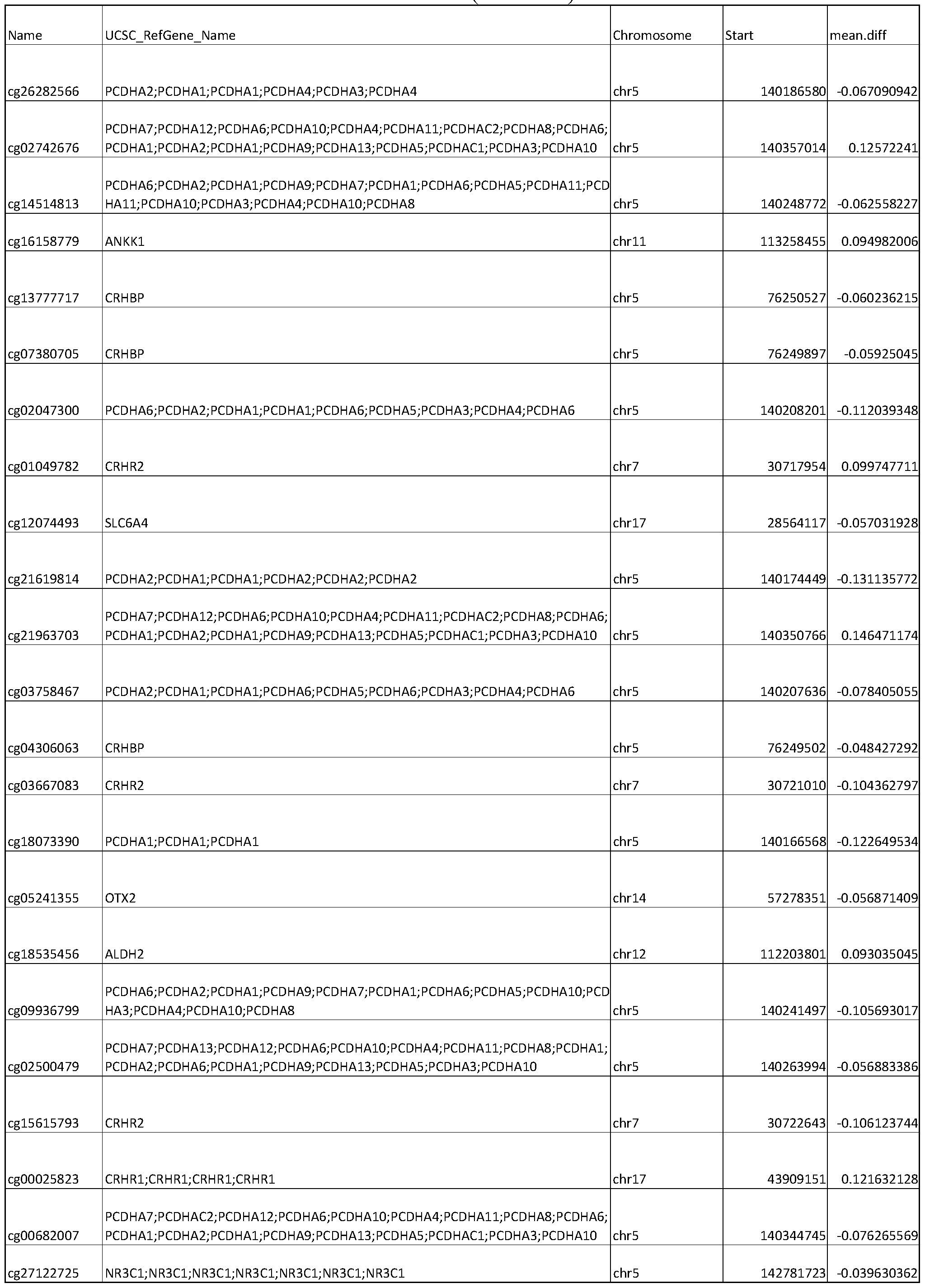


Table 1 (continued)


Table 1 (continued)


Table 1 (continued)
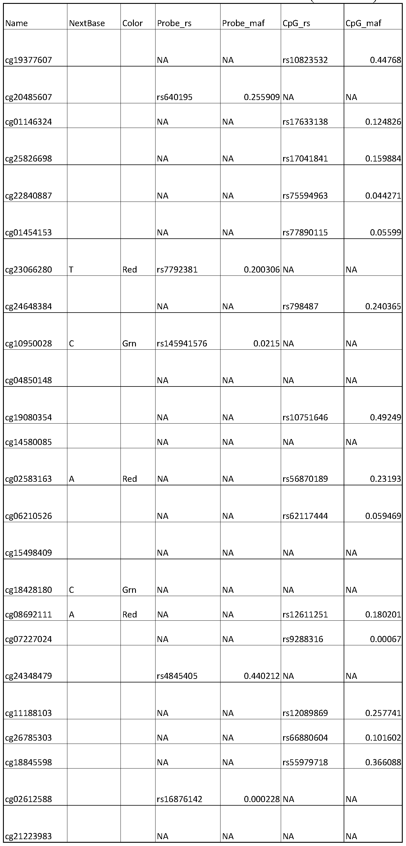


Table 1 (continued)


Table 1 (continued)


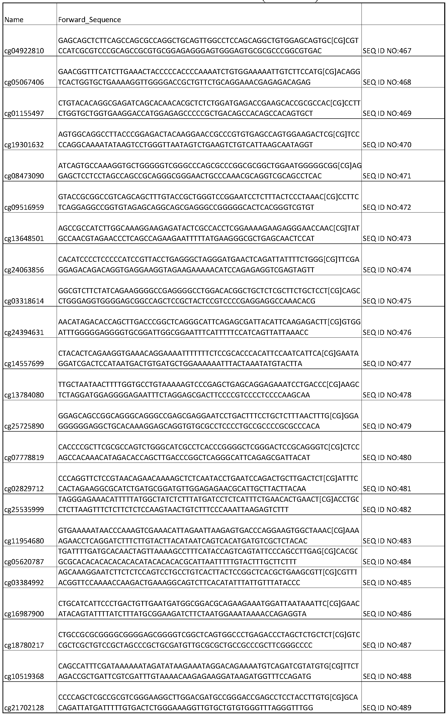

Table 1 (continued)


Claims
1. A method comprising:
obtaining a biological sample from an individual, the sample comprising cells;
extracting DNA from at least a portion of the cells, the extracted DNA comprising a degree of methylation at one or more loci listed in Table 1;
measuring the degree of methylation of the extracted DNA at one or more loci listed in Table 1; and
identifying the individual as having been subjected to a traumatic experience if the degree of methylation at one or more loci listed in Table 1 is outside of a normal limit.
2. The method of claim 1, wherein exhibiting a degree of methylation at one or more loci listed in Table 1 that is outside a normal limit comprises exhibiting a degree of methylation at a locus, wherein the absolute value of the degree of methylation at the locus is at least as great as the absolute value of the mean difference for the same locus listed in Table 1.
3. The method of claim 1, wherein exhibiting a degree of methylation at one or more loci listed in Table 1 that is outside a normal limit comprises exhibiting a degree of methylation at a locus, wherein the degree of methylation at the locus is altered from the degree of methylation for the same locus in a normal control.
4. The method of any preceding claim, the method further comprising:
obtaining a second biological sample from the individual at a second time point;
extracting DNA from cells of the second biological sample, the extracted DNA comprising a degree of methylation at one or more loci listed in Table 1;
measuring the degree of methylation of the extracted DNA of the second biological sample at one or more loci listed in Table 1;
comparing the degree of methylation of the extracted DNA of the biological sample at one or more loci listed in Table 1 with the degree of methylation of the extracted DNA of the second biological sample at the same one or more loci listed in Table 1; and
identifying the individual as having been subjected to a traumatic experience between collection of the biological sample and collection of the second biological sample if the degree of methylation for one or more loci listed in Table 1 is altered in the second biological sample.
5. The method of any preceding claim further comprising treating the individual with a therapy effective to reverse the methylation at one or more loci listed in Table 1.
6. The method of any preceding claim wherein the degree of methylation is measured at a plurality of loci.
7. The method of any preceding claim wherein the biological sample comprises saliva.
8. The method of any preceding claim wherein the biological sample comprises at least one of a keratinocyte and a white blood cell.
9. The method of claim 8, wherein the keratinocyte is a buccal keratinocyte.
10. The method of any preceding claim wherein a traumatic experience comprises at least one of physical abuse, emotional abuse, sexual abuse, and neglect.
11. The method of any preceding claim wherein the one or more loci comprises at least ten loci listed in Table 1.
12. The method of claim 11, wherein the one or more loci comprises at least 50 loci listed in Table 1.
13. The method of claim 12, wherein the one or more loci comprises at least 100 loci listed in Table 1.
14. The method of claim 13, wherein the one or more loci comprises at least 150 loci listed in Table 1.
15. The method of claim 14, wherein the one or more loci comprises at least 200 loci listed in Table 1.
16. The method of claim 15, wherein the one or more loci comprises each of the loci listed in Table 1.
17. The method of any preceding claim wherein the one or more loci comprises loci from at least ten genes.
18. The method of claim 17, wherein the one or more loci comprises loci from at least 30 genes.
19. The method of claim 18, wherein the one or more loci comprises loci from at least 50 genes.
20. The method of any preceding claim wherein the one or more loci comprises loci having an absolute value of the mean difference as listed in Table 1 of at least 0.1.
21. The method of claim 20, wherein the one or more loci comprises loci having an absolute value of the mean difference as listed in Table 1 of at least 0.3.
22. The method of any preceding claim wherein the one or more loci comprises loci having an absolute value of the mean difference as listed in Table 1 of at least 0.4.
23. A method comprising:
obtaining or having obtained a biological sample from an individual, the sample comprising cells;
extracting or having extracted DNA from at least a portion of the cells, the extracted DNA comprising a degree of methylation at one or more loci listed in Table 1;
measuring or having measured the degree of methylation of the extracted DNA at one or more loci listed in Table 1; and
determining whether the degree of methylation at one or more loci listed in Table 1 is outside of a normal limit.
24. The method of claim 23, wherein exhibiting a degree of methylation at one or more loci listed in Table 1 that is outside a normal limit comprises exhibiting a degree of methylation at a locus, wherein the absolute value of the degree of methylation at the locus is at least as great as the absolute value of the mean difference for the same locus listed in Table 1.
25. The method of claim 23, wherein exhibiting a degree of methylation at one or more loci listed in Table 1 that is outside a normal limit comprises exhibiting a degree of methylation at a locus, wherein the degree of methylation at the locus is altered from the degree of methylation for the same locus in a normal control.
26. The method of claim 23, the method further comprising:
obtaining or having obtained a second biological sample from the individual at a second time point;
extracting or having extracted DNA from cells of the second biological sample;
measuring or having measured the degree of methylation of the extracted DNA of the second biological sample at one or more loci listed in Table 1; and
comparing the degree of methylation for one or more loci of the biological sample with the degree of methylation for the same one or more loci of the second biological sample;
determining if the degree of methylation for one or more loci listed in Table 1 is altered in the second biological sample.
27. The method of claim 26, wherein the method further comprises treating the individual with a therapy after obtaining the first biological sample and before obtaining the second biological sample.
28. The method of claim 26, wherein the individual has been subjected to or is suspected of having been subjected to a traumatic experience between collection of the biological sample and collection of the second biological sample.
29. A method comprising:
obtaining or having obtained a first biological sample from an individual at a first time point and obtaining or having obtained a second biological sample from the same individual at a second time point, the first and second biological samples comprising cells;
extracting or having extracted DNA from at least a portion of the cells of the first biological sample, the extracted DNA comprising a degree of methylation at one or more loci listed in Table 1;
extracting or having extracted DNA from at least a portion of the cells of the second biological sample, the extracted DNA comprising a degree of methylation at one or more loci listed in Table 1;
measuring or having measured the degree of methylation of the extracted DNA at one or more loci listed in Table 1; and
comparing the degree of methylation of the extracted DNA of the first biological sample at one or more loci listed in Table 1 with the degree of methylation of the extracted DNA of the second biological sample at the same one or more loci listed in Table 1; and
determining if the degree of methylation for one or more loci listed in Table 1 is altered in the second biological sample.
30. The method of claim 29, wherein the method further comprises treating the individual with a therapy between obtaining the first biological sample and the second biological sample.
31. The method of claim 29, wherein the individual has been subjected to or is suspected of having been subjected to a traumatic experience between collection of the biological sample and collection of the second biological sample.
32. The method of any preceding claim wherein the one or more loci comprise one or more CpG sites.
Priority Applications (1)
| Application Number | Priority Date | Filing Date | Title |
|---|---|---|---|
| US16/317,923 US20190264287A1 (en) | 2016-07-15 | 2017-07-14 | Dna methylation signatures associated with childhood trauma and methods involving same |
Applications Claiming Priority (2)
| Application Number | Priority Date | Filing Date | Title |
|---|---|---|---|
| US201662362903P | 2016-07-15 | 2016-07-15 | |
| US62/362,903 | 2016-07-15 |
Publications (1)
| Publication Number | Publication Date |
|---|---|
| WO2018013963A1 true WO2018013963A1 (en) | 2018-01-18 |
Family
ID=60953408
Family Applications (1)
| Application Number | Title | Priority Date | Filing Date |
|---|---|---|---|
| PCT/US2017/042201 Ceased WO2018013963A1 (en) | 2016-07-15 | 2017-07-14 | Dna methylation signatures associated with childhood trauma and methods involving same |
Country Status (2)
| Country | Link |
|---|---|
| US (1) | US20190264287A1 (en) |
| WO (1) | WO2018013963A1 (en) |
Citations (1)
| Publication number | Priority date | Publication date | Assignee | Title |
|---|---|---|---|---|
| WO2016057485A1 (en) * | 2014-10-06 | 2016-04-14 | The Johns Hopkins University | A dna methylation and genotype specific biomarker for predicting post-traumatic stress disorder |
-
2017
- 2017-07-14 US US16/317,923 patent/US20190264287A1/en not_active Abandoned
- 2017-07-14 WO PCT/US2017/042201 patent/WO2018013963A1/en not_active Ceased
Patent Citations (1)
| Publication number | Priority date | Publication date | Assignee | Title |
|---|---|---|---|---|
| WO2016057485A1 (en) * | 2014-10-06 | 2016-04-14 | The Johns Hopkins University | A dna methylation and genotype specific biomarker for predicting post-traumatic stress disorder |
Non-Patent Citations (1)
| Title |
|---|
| PERROUD N. ET AL.: "Increased methylation of glucocorticoid receptor gene (NR3C1) in adults with a history of childhood maltreatment: a link with the severity and type of trauma", TRANSLATIONAL PSYCHIATRY, vol. 1, no. e59, 2011, pages 19, XP055452866 * |
Also Published As
| Publication number | Publication date |
|---|---|
| US20190264287A1 (en) | 2019-08-29 |
Similar Documents
| Publication | Publication Date | Title |
|---|---|---|
| Dobrowolny et al. | A longitudinal study defined circulating microRNAs as reliable biomarkers for disease prognosis and progression in ALS human patients | |
| Nardone et al. | DNA methylation analysis of the autistic brain reveals multiple dysregulated biological pathways | |
| Córdova-Palomera et al. | Genome-wide methylation study on depression: differential methylation and variable methylation in monozygotic twins | |
| Konradi et al. | Molecular evidence for mitochondrial dysfunction in bipolar disorder | |
| Richards et al. | Schizophrenia susceptibility alleles are enriched for alleles that affect gene expression in adult human brain | |
| CA2769002C (en) | Vh4 codon signature for multiple sclerosis | |
| US11884980B2 (en) | Method for detection of traumatic brain injury | |
| KR101718940B1 (en) | Epigenetic early diagnostic composition for Alzheimer's disease or mild cognitive impairment | |
| US20160222468A1 (en) | Diagnosis, prognosis and treatment of glioblastoma multiforme | |
| US20250376730A1 (en) | Dna methylation and genotype specific biomarker for predicting post-traumatic stress disorder | |
| Senousy et al. | LncRNA GAS5 and miR-137 polymorphisms and expression are associated with multiple sclerosis risk: mechanistic insights and potential clinical impact | |
| EP2982986B1 (en) | Method for manufacturing gastric cancer prognosis prediction model | |
| Chakraborty et al. | Dysregulation of DGCR6 and DGCR6L: psychopathological outcomes in chromosome 22q11. 2 deletion syndrome | |
| Butler et al. | Currently recognized genes for schizophrenia: high‐resolution chromosome ideogram representation | |
| US20220380850A1 (en) | Salivary biomarkers of brain injury | |
| CN117417997B (en) | A genomic biomarker panel for diagnosing keratoconus | |
| US7335474B2 (en) | Methods and systems for identifying predisposition to the placebo effect | |
| US20130317006A1 (en) | Use of polymorphisms for identifying individuals at risk of developing autism | |
| WO2019215085A1 (en) | Method for predicting the risk of late-onset alzheimer's diseases | |
| EP3247798A1 (en) | Identification of epigenetic biomarkers in the saliva of children with autism spectrum disorder | |
| US20180328912A1 (en) | Methods of matching a urine sample to a subject and clinical uses thereof | |
| WO2018013963A1 (en) | Dna methylation signatures associated with childhood trauma and methods involving same | |
| WO2008010082A2 (en) | Diagnostic method for fibromyalgia (fms) or chronic fatigue syndrome (cfs) | |
| KR102348688B1 (en) | SNP markers for diagnosing Cold Hands/Feet Syndrome and use thereof | |
| US20050255498A1 (en) | APOC1 genetic markers associated with age of onset of Alzheimer's Disease |
Legal Events
| Date | Code | Title | Description |
|---|---|---|---|
| 121 | Ep: the epo has been informed by wipo that ep was designated in this application |
Ref document number: 17828556 Country of ref document: EP Kind code of ref document: A1 |
|
| NENP | Non-entry into the national phase |
Ref country code: DE |
|
| 32PN | Ep: public notification in the ep bulletin as address of the adressee cannot be established |
Free format text: NOTING OF LOSS OF RIGHTS PURSUANT TO RULE 112(1) EPC (EPO FORM 1205A DATED 16/05/2019) |
|
| 122 | Ep: pct application non-entry in european phase |
Ref document number: 17828556 Country of ref document: EP Kind code of ref document: A1 |