WO2017213209A1 - 組換えベクター、該ベクターで形質転換された微生物を利用した一酸化窒素消去を抑制する化合物のスクリーニング方法及び細胞内一酸化窒素濃度の測定方法 - Google Patents
組換えベクター、該ベクターで形質転換された微生物を利用した一酸化窒素消去を抑制する化合物のスクリーニング方法及び細胞内一酸化窒素濃度の測定方法 Download PDFInfo
- Publication number
- WO2017213209A1 WO2017213209A1 PCT/JP2017/021261 JP2017021261W WO2017213209A1 WO 2017213209 A1 WO2017213209 A1 WO 2017213209A1 JP 2017021261 W JP2017021261 W JP 2017021261W WO 2017213209 A1 WO2017213209 A1 WO 2017213209A1
- Authority
- WO
- WIPO (PCT)
- Prior art keywords
- microorganism
- gene encoding
- concentration
- protein
- recombinant vector
- Prior art date
- Legal status (The legal status is an assumption and is not a legal conclusion. Google has not performed a legal analysis and makes no representation as to the accuracy of the status listed.)
- Ceased
Links
Images



Classifications
-
- C—CHEMISTRY; METALLURGY
- C12—BIOCHEMISTRY; BEER; SPIRITS; WINE; VINEGAR; MICROBIOLOGY; ENZYMOLOGY; MUTATION OR GENETIC ENGINEERING
- C12N—MICROORGANISMS OR ENZYMES; COMPOSITIONS THEREOF; PROPAGATING, PRESERVING, OR MAINTAINING MICROORGANISMS; MUTATION OR GENETIC ENGINEERING; CULTURE MEDIA
- C12N15/00—Mutation or genetic engineering; DNA or RNA concerning genetic engineering, vectors, e.g. plasmids, or their isolation, preparation or purification; Use of hosts therefor
- C12N15/09—Recombinant DNA-technology
-
- C—CHEMISTRY; METALLURGY
- C12—BIOCHEMISTRY; BEER; SPIRITS; WINE; VINEGAR; MICROBIOLOGY; ENZYMOLOGY; MUTATION OR GENETIC ENGINEERING
- C12Q—MEASURING OR TESTING PROCESSES INVOLVING ENZYMES, NUCLEIC ACIDS OR MICROORGANISMS; COMPOSITIONS OR TEST PAPERS THEREFOR; PROCESSES OF PREPARING SUCH COMPOSITIONS; CONDITION-RESPONSIVE CONTROL IN MICROBIOLOGICAL OR ENZYMOLOGICAL PROCESSES
- C12Q1/00—Measuring or testing processes involving enzymes, nucleic acids or microorganisms; Compositions therefor; Processes of preparing such compositions
- C12Q1/02—Measuring or testing processes involving enzymes, nucleic acids or microorganisms; Compositions therefor; Processes of preparing such compositions involving viable microorganisms
-
- G—PHYSICS
- G01—MEASURING; TESTING
- G01N—INVESTIGATING OR ANALYSING MATERIALS BY DETERMINING THEIR CHEMICAL OR PHYSICAL PROPERTIES
- G01N21/00—Investigating or analysing materials by the use of optical means, i.e. using sub-millimetre waves, infrared, visible or ultraviolet light
- G01N21/75—Systems in which material is subjected to a chemical reaction, the progress or the result of the reaction being investigated
- G01N21/76—Chemiluminescence; Bioluminescence
-
- G—PHYSICS
- G01—MEASURING; TESTING
- G01N—INVESTIGATING OR ANALYSING MATERIALS BY DETERMINING THEIR CHEMICAL OR PHYSICAL PROPERTIES
- G01N21/00—Investigating or analysing materials by the use of optical means, i.e. using sub-millimetre waves, infrared, visible or ultraviolet light
- G01N21/75—Systems in which material is subjected to a chemical reaction, the progress or the result of the reaction being investigated
- G01N21/77—Systems in which material is subjected to a chemical reaction, the progress or the result of the reaction being investigated by observing the effect on a chemical indicator
- G01N21/78—Systems in which material is subjected to a chemical reaction, the progress or the result of the reaction being investigated by observing the effect on a chemical indicator producing a change of colour
-
- G—PHYSICS
- G01—MEASURING; TESTING
- G01N—INVESTIGATING OR ANALYSING MATERIALS BY DETERMINING THEIR CHEMICAL OR PHYSICAL PROPERTIES
- G01N33/00—Investigating or analysing materials by specific methods not covered by groups G01N1/00 - G01N31/00
- G01N33/48—Biological material, e.g. blood, urine; Haemocytometers
- G01N33/50—Chemical analysis of biological material, e.g. blood, urine; Testing involving biospecific ligand binding methods; Immunological testing
Definitions
- the present invention relates to a recombinant vector, a method for screening a compound that suppresses nitric oxide elimination in a microbial cell transformed with the vector, and a method for measuring the intracellular nitric oxide concentration.
- Phagocytic cells macrophages, neutrophils, etc.
- NO nitric Oxide
- ROS reactive oxygen species
- microorganisms that undergo phagocytosis have a mechanism for eliminating NO and ROS in microbial cells and are used for self-protection.
- NO is an important molecule that has toxic effects such as cytotoxicity and carcinogenicity on the living body and also exhibits various physiological actions in the cardiovascular system and the central nervous system, the amount of NO production in the living body Attempts to increase can lead to unexpected or undesirable side effects.
- a compound that attenuates the self-protection mechanism of microorganisms against NO can be an infectious disease therapeutic agent that can avoid undesirable effects in the living body and improve the microbicidal efficiency of NO produced by phagocytic cells. Expected.
- Non-Patent Document 1 provides E. coli transformed with an expression plasmid having a gene encoding the protein NorR and a ⁇ -galactosidase gene downstream of the promoter region where transcription is regulated thereby. Since this E. coli changes the production amount of ⁇ -galactosidase according to the exposed NO concentration, the microbial intracellular NO concentration can be measured using ⁇ -galactosidase activity as an index.
- the method for measuring the NO concentration described in Non-Patent Document 1 has limitations in sensitivity and dynamic range in order to measure the color produced by the enzyme reaction with a spectrophotometer.
- Patent Document 1 uses, as an index, the luminescence intensity according to the exposed NO concentration using a gene encoding a photoprotein instead of the ⁇ -galactosidase gene in Non-Patent Document 1.
- a method for measuring microbial intracellular NO concentration is provided.
- measurement of luminescence intensity has enabled measurement with high sensitivity and a wide dynamic range, there still remains a problem that fluctuation of the number of microbial cells due to growth of transformed microorganisms during the measurement affects the luminescence intensity. ing.
- the present invention provides a method for screening for a compound that attenuates the self-protection mechanism of microorganisms, which retains the merit in the measurement of luminescence intensity and can evaluate a large number of test compounds more easily and quickly. It is intended.
- the present inventors have found that the above problem can be solved by transforming a microorganism with an expression vector having a gene encoding a bioluminescent protein and a gene encoding a fluorescent protein on one vector.
- Each invention was completed.
- a recombinant vector comprising a gene encoding a NorR protein, a gene encoding a bioluminescent protein under the control of the nor operon promoter, and a gene encoding a fluorescent protein under the control of a constitutive promoter.
- a microorganism comprising the recombinant vector according to (1) or (2).
- a method for screening a compound that suppresses NO elimination in a cell (6) The screening method according to (5), wherein the microorganism used in step (a) is a microorganism exhibiting a fluorescence intensity equal to or higher than a predetermined reference value. (7) The screening method according to (5) or (6), wherein the concentration of NO donor in the solution in step (a) is a concentration that gives a NO concentration of 5 nM to 10 ⁇ M in the solution. (8) The screening method according to any one of (5) to (7), wherein the microorganism is a microorganism selected from the group consisting of Escherichia coli, Salmonella and Shigella.
- step (10) The measuring method according to (9), wherein the microorganism used in step (e) is a microorganism exhibiting a fluorescence intensity that is equal to or higher than a predetermined reference value.
- the microorganism is a microorganism selected from the group consisting of Escherichia coli, Salmonella and Shigella.
- the NO concentration in microbial cells can be measured easily, rapidly, with high sensitivity, and in a wide dynamic range including a physiological concentration range of nM level. It becomes possible to evaluate a test compound in a short time.
- cfu colony forming unit
- FIGS. 4 (A) to (D) is a graph showing the luminescence intensity at a NO donor concentration of 0 to 5.0 ⁇ M for E. coli transformed with pRPLG1 shown in FIGS. 4 (A) to (D). It is a graph which shows the time-dependent change of specific luminescence intensity when colon_bacillus
- the first aspect of the present invention is under the control of a gene encoding a NorR protein (hereinafter referred to as gene norR), a gene encoding a bioluminescent protein under the control of the nor operon promoter, and a constitutive promoter.
- gene norR a gene encoding a NorR protein
- the present invention relates to a recombinant vector containing a gene encoding a fluorescent protein. This recombinant vector can be used to prepare a transformed microorganism capable of detecting NO present outside a microorganism cell or measuring the concentration of intracellular NO.
- the recombinant vector according to the first aspect of the present invention includes a gene norR encoding a NorR protein so that it can be expressed.
- the NorR protein is a ⁇ 54-dependent transcriptional activator derived from Escherichia coli, and induces the expression of the gene norVW encoding NADH-dependent NO reductase involved in elimination of intracellular NO in E. coli by binding to NO ( Nicholas P. Tucker, J. Biol. Chem., 283, NO.2, pp. 908-918, 2008).
- the gene norR can be amplified and used by PCR using an E. coli genomic DNA as a template and an appropriate primer DNA designed based on the base sequence registered as BAE76786 in the EMBL / GenBank / DDBJ database. it can.
- An example of gene norR cloning is described in Patent Document 1.
- the recombinant vector according to the first aspect of the present invention includes a gene encoding a bioluminescent protein placed under the control of the nor operon promoter.
- the nor operon promoter (also referred to as the norR-norV promoter) is a promoter base sequence derived from E. coli that induces transcription of the gene norVW through interaction with the NorR protein bound to NO.
- the nor operon promoter should be amplified and used by PCR using an E. coli genomic DNA as a template and an appropriate primer DNA designed based on the base sequence registered as BAE76786 in the EMBL / GenBank / DDBJ database. Can do.
- An example of cloning of the nor operon promoter is described in Patent Document 1.
- Bioluminescent protein is a protein that emits light by a chemical reaction with a luminescent substrate.
- bioluminescent proteins and genes encoding the same have been reported so far, and examples thereof include luxCDABE gene, firefly luciferase gene, or Renilla luciferase gene.
- the bioluminescent protein used in the present invention is preferably a bioluminescent protein that emits light without the addition of an external luminescent substrate, that is, a bioluminescent protein that emits light only with a substrate normally present in a microbial cell.
- a gene encoding a bioluminescent protein that is preferable in the present invention is the luxCDABE gene, which is a group of bioluminescent genes of the luminescent bacterium Photorhabdus luminescens.
- the photoprotein luxCDABE encoded by the gene does not require the addition of a synthetic substrate, and has the property of self-luminescence based on the ATP produced by the microorganism itself. Continuous, non-destructive and real-time luminescence Enable measurement.
- Each base sequence of the gene encoding the bioluminescent protein described above is known, and it is amplified by using a suitable genetic resource as a template by a PCR method using an appropriate primer DNA designed based on the base sequence. It can be used, or is available in a form incorporated into a commercial vector.
- the luxCDABE gene can be used after being amplified by a PCR method using an appropriate primer DNA designed on the basis of a known base sequence using, for example, the genomic DNA of Photorhabdus luminescens GTC00819 as a template.
- An example of cloning of the luxCDABE gene is described in Patent Document 1.
- the recombinant vector according to the first aspect of the present invention includes a gene encoding a fluorescent protein so that it can be constitutively expressed.
- the gene encoding the fluorescent protein is preferably placed under the control of a constitutive promoter, ie a promoter that constitutively induces transcription of the gene in the host microorganism.
- the fluorescent protein that can be used in the present invention is not particularly limited, and is a green fluorescent protein (Green Fluorescent Protein, GFP) such as TurboGFP, AcGFP, TagGFP, Azami-Green, ZsGreen, EmGFP, EGFP, GFP2, HyPer, and EBFP.
- Green Fluorescent Protein Green Fluorescent Protein
- GFP Green Fluorescent Protein
- TurboGFP AcGFP
- TagGFP Azami-Green
- ZsGreen ZsGreen
- EmGFP EmGFP
- EGFP GFP2, HyPer
- EBFP blue Fluorescent Proten, BFP
- ECFP Blue Fluorescent Proten, BFP
- TagCFP AmCyan
- MidoriishiCyan etc.
- Cyan Fluorescent Protein Cyan Fluorescent Protein (Cyan Fluorescent Protein, CFP), TurboRFP, DsRed-ExpressR, DsRed-ExpressR, DsRed-ExpressR, DsRed-Express Fluorescent protein (Red Fluorescent Protein, RFP), yellow fluorescent proteins such as TagYFP, EYFP, Venus, PhiYFP, PhiYFP-m, TurboYFP, ZsYellow, mBana, etc. , JRed, KillerRed, mCherry, HcRed, KeimaRed mRsbury, mPlum, and other fluorescent proteins that emit fluorescence of infrared light.
- Preferred fluorescent proteins are GFP, YFP, RFP and the like.
- the fluorescent protein is selected so that its fluorescence spectrum is different from the emission spectrum of the bioluminescent protein so that it can be separated and detected, and its excitation wavelength does not overlap with the emission wavelength of the bioluminescent protein. Is desirable.
- blue to ultraviolet light can cause cytotoxicity and further cause NO to be released from inside the cell, which may affect the accuracy of NO concentration measurement. Therefore, it is more desirable to select a fluorescent protein whose excitation wavelength is on the long wavelength side.
- Each base sequence of the gene encoding the fluorescent protein exemplified above is known, and is amplified by using a suitable genetic resource as a template by PCR using an appropriate primer DNA designed based on the base sequence. Or can be obtained in a form incorporated into a commercially available vector.
- the promoter that can be used in the present invention is not particularly limited, but is preferably a constitutive promoter that can constitutively induce transcription of a gene placed downstream thereof.
- a constitutive promoter is not a promoter that exhibits transcription-inducing activity only under special physiological conditions or in the presence of a specific inducer, but is constitutively active during the life of a microorganism, in particular stable transcription.
- a promoter indicating Such a promoter can be appropriately selected depending on the host microorganism. Examples of Escherichia coli include lac promoter and trc promoter.
- the recombinant vector according to the first aspect of the present invention comprises the gene norR, the nor operon promoter, a gene encoding a bioluminescent protein, a constitutive promoter, and a gene encoding a fluorescent protein, which are cloned by appropriate methods, respectively. It can be produced by incorporating it into an expression vector suitable for the microorganism to be converted.
- the expression vector is not particularly limited as long as it can transform a microorganism to be transformed, and examples thereof include pUC vectors, pACYC vectors, and pSC101 vectors.
- the recombinant vector according to the first aspect of the present invention is placed under the control of the plasmid pRPL3 (gene norR and the nor operon promoter described in Patent Document 1 using a conventional gene recombination technique. It is possible to manufacture by incorporating a gene encoding a fluorescent protein into a NO sensor plasmid having a luxCDABE gene.
- the second aspect of the present invention is a microorganism containing the recombinant vector according to the first aspect (hereinafter also referred to as NO sensor microorganism).
- NO sensor microorganism There is no restriction
- microorganisms include Escherichia, Salmonella, Shigella, Yersinia, Klebsiella, Vibrio, Pseudomonas, Pseudomonas, Examples include the genus Citrobacter, the genus Proteus, and the genus Serratia.
- Preferred microorganisms are Escherichia, Salmonella, Shigella and Pseudomonas, and particularly preferred microorganisms include Escherichia coli, particularly pathogenic Escherichia coli such as enterohemorrhagic Escherichia coli, Salmonella Typhimurium et al., Shigella ( Shigella sonnei et al.), But is not limited thereto.
- Transformation of the above-described microorganisms using the recombinant vector according to the first aspect can be easily performed by those skilled in the art based on known transformation methods for each microorganism.
- One preferred method is electroporation.
- the transformed microorganism may be selected using a selection gene that can be separately incorporated into the expression vector of the present invention, such as an antibiotic resistance gene.
- the microorganism according to the second aspect of the present invention emits light when NO is present in the cells. Since NO in the external environment of cells enters the cells non-specifically, the microorganism of the present invention can be used as a NO sensor microorganism that can detect and quantify NO in the external environment. it can.
- the luminescence intensity of the microorganism of the present invention has a property that it changes in response to a change in NO concentration, particularly a change in physiological NO concentration at the nM level. Therefore, the microorganism of the present invention can also be used as a NO sensor microorganism for monitoring to measure a change in NO concentration, particularly a change with time.
- the NO sensor microorganism of the present invention is characterized in that it emits fluorescence different from bioluminescence, and the emission intensity and fluorescence intensity both vary according to the number of microorganism cells.
- the specific luminescence intensity is generally calculated using the cfu measured by the absorbance or the dilution method as shown by RLU / cfu.
- the dilution method has problems such as labor and time. In particular, when testing the activity of suppressing NO elimination in microbial cells for a large number of test compounds, measurement of the number of microbial cells by a dilution method or the like is practically unusable because it takes too much time and time.
- the present invention instead of actually measuring the number of microbial cells by a dilution method or the like, it is possible to easily and quickly correct the luminescence intensity by using the fluorescence intensity instead of the microbial cell number, and to obtain the specific luminescence intensity.
- Such convenience and rapidity are extremely important characteristics in evaluating the activity of suppressing NO elimination in microbial cells for a large number of test compounds and in measuring NO concentration over time.
- the third aspect of the present invention includes the following steps (a) to (d): (A) Transformed with a recombinant vector comprising a gene encoding a NorR protein, a gene encoding a bioluminescent protein under the control of the nor operon promoter, and a gene encoding a fluorescent protein under the control of a constitutive promoter Incubating a solution comprising the converted microorganism, test compound and NO donor; (B) measuring the luminescence intensity of the bioluminescent protein and the fluorescence intensity of the fluorescent protein; (C) calculating a specific luminescence intensity corrected with fluorescence intensity, and (d) selecting a test compound that suppresses a decrease in specific luminescence intensity compared to a control solution not containing the test compound.
- the present invention relates to a method for screening a compound that suppresses NO elimination.
- each gene in step (a), the recombinant vector containing them, and the microorganism transformed with the vector are as described in the first and second aspects.
- the method according to the third aspect of the present invention includes the step (a) of incubating a solution containing the microorganism, the test compound and the NO donor.
- the solution is not particularly limited as long as the microorganism can survive during the incubation, and examples thereof include water, an appropriate buffer solution, and a medium.
- NO donor is a substance that generates NO after being taken up in solution or into cells.
- NO donors that can be used are not particularly limited.
- Sodium nitroprusside Sodium Nitroplusside, SNP
- DEA-NONOate Dithylamine NONOate
- S-nitrosoglutathione N-Nitrosoglutathione
- PROLI-NONOate V-PYRLRO-NO-
- -Commercial products such as arginine and nitrite may be mentioned.
- the NO concentration in the solution is within a concentration range in which the upper limit is the maximum concentration at which the transformed microorganism can survive even during incubation, and the lower limit is the minimum concentration at which the bioluminescence intensity depending on the NO concentration can be measured. In particular, it is preferably a concentration that can provide a physiological intracellular NO concentration of nM level.
- An example of the NO concentration in the solution is approximately 5 nM to 10 ⁇ M, preferably 10 nM to 10 ⁇ M.
- the NO exposure amount per bacterial cell is approximately 0.1 to 100 amol, preferably 1 to 100 amol.
- the NO donor concentration in the solvent for providing such NO concentration varies depending on the type of NO donor.
- it may be about 0.1 ⁇ M to 50 ⁇ M, preferably 1 ⁇ M to 10 ⁇ M.
- the incubation time may be appropriately set in the range of approximately 10 minutes to 180 minutes, preferably 30 minutes to 120 minutes.
- the microorganisms used in the step (a) are microorganisms that exhibit a fluorescence intensity that is equal to or higher than a predetermined reference value.
- a predetermined reference value As shown in the examples described below, the present inventors have found that the performance as a NO sensor fluctuates under the influence of growth conditions and the external environment even if the microbial strain is taxonomically identical, and relatively It has been found that microorganisms showing good NO reactivity even in a low NO concentration region show high fluorescence intensity. In particular, when screening is performed under conditions that simulate a physiological environment with a low NO concentration of 5 nM to 1000 nM, it is important to select microorganisms showing good NO reactivity in advance and use them as NO sensor microorganisms.
- a microorganism exhibiting a fluorescence intensity equal to or higher than a predetermined reference value is allowed to have a desired NO reactivity, that is, a desired NO concentration range. It is preferable to select and use as a NO sensor microorganism exhibiting linear NO reactivity.
- the reference value of the fluorescence intensity is, for example, the fluorescence intensity and NO reactivity (bioluminescence generated by NO) of a target recombinant microorganism prepared under various conditions, and the fluorescence of the microorganism having the desired NO reactivity. It can be set by determining the lower limit of the intensity.
- the NO sensor microorganism selection method for selecting microorganisms exhibiting desired NO reactivity with reference to a predetermined reference value also constitutes one aspect of the present invention. According to such a selection method, for example, if the fluorescence intensity emitted by a microorganism prepared under a certain condition is equal to or higher than a reference value, the microorganism prepared under the condition is determined to have a desired reactivity as a NO sensor microorganism. Can be selected.
- the method according to the third aspect of the present invention includes the step (b) of measuring the luminescence intensity of the bioluminescent protein and the fluorescence intensity of the fluorescent protein.
- the emission intensity and the fluorescence intensity can be measured by known methods.
- the emission intensity is a commercially available device such as GLOMAX 20/20 luminometer (Promega), Synergy 2 (BioTek), CentroXS3 LB960 (Berthold), and the fluorescence intensity is TECAN infinite F200 PRO (TECAN), Gemini XPS (Molecular XPS). ), MTP-601 (Hitachi), FLUOstar Omega (BMG Labtech), and other commercially available equipment.
- the method according to the third aspect of the present invention includes the step (c) of calculating the specific emission intensity corrected with the fluorescence intensity.
- Step (c) is a step of calculating the specific luminescence intensity by correcting the luminescence intensity measured at an appropriate time after the start of incubation in step (a) with the fluorescence intensity measured at the same time.
- the transformed microorganism according to the present invention has a proportional relationship between the number of cells and the fluorescence intensity within a certain range, so that the emission intensity is corrected using the fluorescence intensity as it is. can do.
- the method according to the third aspect of the present invention includes a step (d) of selecting a test compound that suppresses a decrease in specific luminescence intensity as compared with a control solution not containing a test compound.
- Step (d) is a step in which a test compound is evaluated and selected.
- the specific luminescence intensity when a control solution containing a transformed microorganism and an NO donor but no test compound is incubated as a control, and compared with this control.
- a test compound that suppresses a decrease in specific luminescence intensity is selected as an object in screening. In the control, the intracellular NO concentration is lowered by the self-protection mechanism against NO possessed by the transformed microorganism, and the specific luminescence intensity is correspondingly reduced.
- the test compound inhibits or attenuates the mechanism, the decrease in specific emission intensity is smaller than that in the control, that is, the decrease in specific emission intensity is suppressed.
- the actual selection operation in the step (d) is performed by selecting a compound having an increased specific luminescence intensity as compared with the control.
- Microorganisms originally have a mechanism to adjust the microbial intracellular NO concentration to an acceptable concentration range. Therefore, when the decrease in the microbial intracellular NO concentration is suppressed by the presence of the test compound, the test compound keeps the microbial intracellular NO concentration high by suppressing or inhibiting the mechanism of the microorganism, and as a result. It is expected to contribute to the elimination of microorganisms by NO produced by macrophages and the like.
- the fourth aspect of the present invention is the following steps (e) to (g): (E) Transformed with a recombinant vector comprising a gene encoding a NorR protein, a gene encoding a bioluminescent protein under the control of the nor operon promoter, and a gene encoding a fluorescent protein under the control of a constitutive promoter Measuring the luminescence intensity of the bioluminescent protein of the converted microorganism and the fluorescence intensity of the fluorescent protein;
- the present invention relates to a method for measuring NO concentration in a microbial cell, comprising (f) calculating specific luminescence intensity corrected with fluorescence intensity, and (g) calculating intracellular NO concentration corresponding to the specific luminescence intensity.
- each gene in the step (e), a recombinant vector containing them, a microorganism transformed with the vector, the measurement of the luminescence intensity of the bioluminescent protein and the fluorescence intensity of the fluorescent protein, the specific luminescence intensity in the step (f) The calculation is as described in the first to third aspects.
- the microorganism in the step (e) is appropriately prepared according to the purpose of measuring the intracellular NO concentration.
- the NO sensor microorganism is placed in the environment, and the step (e) may be performed using the NO sensor microorganism.
- stimulation should be prepared and process (e) should be performed using this.
- the calculation of the intracellular NO concentration in the step (g) is performed by referring to a calibration curve representing the relationship between the intracellular NO concentration of the sensor microorganism and the specific luminescence intensity prepared in advance, thereby calculating the specific luminescence calculated in the step (f). This is done by determining the intracellular NO concentration corresponding to the intensity.
- Example 1 1) Construction of recombinant vector Using the plasmid pRPL3 described in Patent Document 1 as a template, the lac promoter + GFP gene amplified by PCR from the pAcGFP1 plasmid was cloned into the linear vector prepared by PCR amplification by the In-Fusion method, The recombinant vector pRPLG1 of the present invention was prepared. The structure of pRPLG1 is shown in FIG. This recombinant vector has a luxCDABE gene whose expression level changes depending on the NO concentration, and a GFP gene whose expression is constantly induced by the lac promoter.
- DEA-NONOate was added to final concentrations of 0.3, 0.7, 1.0, 3.0, and 5.0 ⁇ M, and the emission intensity (RLU) and GFP fluorescence were maintained at 37 ° C. Intensity (GFP) was measured. The number of microorganisms (cfu) of the same sample was measured by a dilution method.
- FIG. 4 shows the result of cfu as the horizontal axis and GFP fluorescence intensity as the vertical axis.
- A is tEC pre-cultured in LB / Car medium for 19 hours
- B is tEC that was allowed to stand for 2 days after pre-culture
- C is tEC that was subjected to secondary culture for 5 hours
- D Corresponds to tEC after 2 hours of secondary culture.
- TEC ((A) and (B)) in which the GFP fluorescence intensity exceeds a certain value (for example, 2300, broken line in FIG. 4) shows a concentration-dependent specific emission intensity in the NO donor concentration range of 0 to 0.7 ⁇ M. It was confirmed.
- the subcultured tEC ((C) and (D)) has a GFP emission intensity of 2300 or less and a concentration dependency in the NO donor concentration range of 0 to 0.7 ⁇ M as (A) and (B). It was confirmed that it was inferior in comparison.
- the GFP fluorescence intensity can be used as an index for determining the NO reactivity of the NO sensor microorganism, particularly in a low NO concentration range.
- microorganisms that have reached a stationary phase or have been left for several hours to several days after that have a low ability to grow are compared with microorganisms that have recovered their ability to grow by secondary culture, for example, in the logarithmic phase of secondary culture.
- good NO reactivity was exhibited even in a low NO concentration range, and that it could be used as a preferred microorganism in the method of the present invention.
- Example 2 Confirmation of Inhibitory Effect of NO Reductase Inhibitor on Intracellular NO Concentration Reduction
- a tEC suspension was prepared in the same manner as in Example 1 3).
- DEA NONOate was added to the final concentration of 3.0 ⁇ M or 0 ⁇ M (negative control), and econazole (Sigma-Aldrich), which is known to inhibit the activity of flavohemoglobin, a NO-eliminating enzyme of Escherichia coli, was added.
- Luminescence intensity (RLU) and fluorescence intensity (GFP) of GFP were measured over time while maintaining at 37 ° C. for 60 minutes after adding to a final concentration of 3.0 ⁇ M or 0 ⁇ M (control).
- Fig. 6 shows the change in emission intensity over time. As shown in FIG. 6, it was confirmed that the decrease in specific luminescence intensity was suppressed in tEC to which econazole was added as compared with the control. In the negative control, substantially no luminescence was observed.
- the decrease in specific luminescence intensity that is, the decrease in NO concentration in the microorganism
- the decrease in NO concentration in the microorganism can be confirmed in about 30 minutes after adding the NO donor, and a wide range including physiological NO concentrations at the nM level. It became clear that suppression of reduction can be confirmed in the NO concentration range.
- the present invention has industrial applicability as a method capable of screening for compounds that attenuate the self-protection mechanism of microorganisms against NO and providing pharmaceutical candidate compounds.
Landscapes
- Chemical & Material Sciences (AREA)
- Life Sciences & Earth Sciences (AREA)
- Health & Medical Sciences (AREA)
- Engineering & Computer Science (AREA)
- Physics & Mathematics (AREA)
- Organic Chemistry (AREA)
- Biochemistry (AREA)
- General Health & Medical Sciences (AREA)
- Immunology (AREA)
- Molecular Biology (AREA)
- Analytical Chemistry (AREA)
- Genetics & Genomics (AREA)
- Zoology (AREA)
- Biomedical Technology (AREA)
- Biotechnology (AREA)
- Wood Science & Technology (AREA)
- Bioinformatics & Cheminformatics (AREA)
- General Physics & Mathematics (AREA)
- Microbiology (AREA)
- General Engineering & Computer Science (AREA)
- Pathology (AREA)
- Plasma & Fusion (AREA)
- Hematology (AREA)
- Urology & Nephrology (AREA)
- Biophysics (AREA)
- Proteomics, Peptides & Aminoacids (AREA)
- Chemical Kinetics & Catalysis (AREA)
- Medicinal Chemistry (AREA)
- Cell Biology (AREA)
- Food Science & Technology (AREA)
- Plant Pathology (AREA)
- Measuring Or Testing Involving Enzymes Or Micro-Organisms (AREA)
- Micro-Organisms Or Cultivation Processes Thereof (AREA)
- Investigating Or Analysing Biological Materials (AREA)
- Investigating Or Analysing Materials By The Use Of Chemical Reactions (AREA)
Abstract
【課題】 本発明は、簡便かつ迅速に多数の試験化合物を同時並行で評価することのできる、微生物が持つNOに対する自己防御機構を減弱させる化合物をスクリーニングする方法を提供することを目的とする。 【解決手段】 本発明は、NorRタンパク質をコードする遺伝子と、norオペロンプロモーターの制御下にある生物発光タンパク質をコードする遺伝子と、構成的プロモーターの制御下にある蛍光タンパク質をコードする遺伝子とを含む組換えベクター、及び該ベクターで形質転換した微生物を用いた、NOに対する自己防御機構を減弱させる化合物をスクリーニングする方法に関する。本発明によれば、簡便かつ迅速に、高感度かつnMレベルの生理的な濃度範囲を含む広いダイナミックレンジで微生物細胞内のNO濃度変化を測定することができ、多数の試験化合物を短時間で評価することが可能となる。
Description
本発明は、組換えベクター、該ベクターで形質転換された微生物細胞内の一酸化窒素消去を抑制する化合物をスクリーニングする方法及び細胞内一酸化窒素濃度の測定方法に関する。
微生物の感染に対する生体防御の一つに、マクロファージが関与する細胞性免疫が挙げられる。細胞性免疫において中心的に作用する貪食細胞(マクロファージ及び好中球など)は、一酸化窒素(Nitric Oxide;NO)又は活性酸素種(ROS)を産生し、貪食後の殺菌に活用している。その一方、貪食を受ける微生物は、NOやROSを微生物細胞内で消去する機構を有し、自己防御に活用している。
微生物の排除のために生体のNO産生量を増大させようとすることは、自然な発想である。しかしながらNOは、生体に対しても細胞障害性及び発癌性等の毒性作用を持ち、さらに心血管系及び中枢神経系において様々な生理作用を示す重要な分子であることから、生体のNO産生量を増大させる試みは、予期しない又は望ましくない副次的効果をもたらすおそれがある。
このため、微生物のNOに対する自己防御機構を減弱させる化合物は、生体における望ましくない影響を回避し、貪食細胞が産生するNOによる殺微生物効率を向上させることが可能な感染症治療薬となり得るものと期待されている。
微生物のNOに対する自己防御機構を減弱させる化合物を取得するには、微生物細胞内、特に病原性微生物細胞内のNO濃度を簡便に測定する技術が必要である。
これまでに、NO濃度に依存して活性化する大腸菌タンパク質NorRの発現を利用した微生物細胞内のNO濃度を測定する技術が報告されている。例えば非特許文献1は、タンパク質NorRをコードする遺伝子とそれによって転写が調節されるプロモーター領域の下流にβ-ガラクトシダーゼ遺伝子とを有する発現プラスミドで形質転換させた大腸菌を提供している。この大腸菌は、曝露されたNO濃度に応じてβ-ガラクトシダーゼ産生量を変化させるので、β-ガラクトシダーゼ活性を指標として微生物細胞内NO濃度を測定することができる。しかし、非特許文献1に記載のNO濃度の測定法は、酵素反応により生じる発色を分光光度計で測定するために、感度及びダイナミックレンジに限界がある。
酵素反応の利用に代わる方法として、特許文献1は、非特許文献1におけるβ-ガラクトシダーゼ遺伝子に代えて発光タンパク質をコードする遺伝子を用いた、曝露されたNO濃度に応じた発光強度を指標とした微生物細胞内NO濃度を測定する方法を提供している。しかし、発光強度の測定は高感度及び広いダイナミックレンジでの測定を可能としたものの、測定中に形質転換微生物が増殖することによる微生物細胞数の変動が発光強度に影響を与えるという問題が依然として残っている。
M.I.Hutchings et al.,J.Bacteriol.,Vol.184,No.16,pp.4640-4643
本発明は、発光強度の測定におけるメリットを保持し、より簡便かつ迅速に多数の試験化合物を評価することのできる、微生物が持つNOに対する自己防御機構を減弱させる化合物をスクリーニングする方法を提供することを目的とするものである。
本発明者らは、一つのベクター上に生物発光タンパク質をコードする遺伝子と蛍光タンパク質をコードする遺伝子とを有する発現ベクターで微生物を形質転換することで上記課題を解決することができることを見出し、下記の各発明を完成した。
(1)NorRタンパク質をコードする遺伝子と、norオペロンプロモーターの制御下にある生物発光タンパク質をコードする遺伝子と、構成的プロモーターの制御下にある蛍光タンパク質をコードする遺伝子とを含む組換えベクター。
(2)生物発光タンパク質をコードする遺伝子がluxCDABE遺伝子である、(1)に記載の組換えベクター。
(3)(1)又は(2)に記載の組換えベクターを含む微生物。
(4)微生物が大腸菌、サルモネラ及び赤痢菌よりなる群から選択される微生物である、(3)に記載の微生物。
(5)以下の工程(a)~(d):
(a)NorRタンパク質をコードする遺伝子と、norオペロンプロモーターの制御下にある生物発光タンパク質をコードする遺伝子と、構成的プロモーターの制御下にある蛍光タンパク質をコードする遺伝子とを含む組換えベクターで形質転換した微生物、試験化合物及びNOドナーを含む溶液をインキュベートする工程、
(b)生物発光タンパク質の発光強度と蛍光タンパク質の蛍光強度とを測定する工程、
(c)蛍光強度で補正した比発光強度を算出する工程、並びに
(d)試験化合物を含まない対照溶液と比較して比発光強度の減少を抑制した試験化合物を選択する工程
を含む、微生物細胞内のNO消去を抑制する化合物のスクリーニング方法。
(6)工程(a)において使用する微生物が、予め定められた基準値以上の蛍光強度を示す微生物である、(5)に記載のスクリーニング方法。
(7)工程(a)における溶液中のNOドナーの濃度が、溶液中で5nM~10μMのNO濃度を与える濃度である、(5)又は(6)に記載のスクリーニング方法。
(8)前記微生物が、大腸菌、サルモネラ及び赤痢菌よりなる群から選択される微生物である、(5)~(7)のいずれかに記載のスクリーニング方法。
(9)以下の工程(e)~(g):
(e)NorRタンパク質をコードする遺伝子と、norオペロンプロモーターの制御下にある生物発光タンパク質をコードする遺伝子と、構成的プロモーターの制御下にある蛍光タンパク質をコードする遺伝子とを含む組換えベクターで形質転換した微生物の生物発光タンパク質の発光強度と蛍光タンパク質の蛍光強度とを測定する工程、
(f)蛍光強度で補正した比発光強度を算出する工程、及び
(g)比発光強度に対応する細胞内NO濃度を算出する工程
を含む、微生物細胞内のNO濃度を測定する方法。
(10)工程(e)において使用する微生物が、予め定められた基準値以上の蛍光強度を示す微生物である、(9)に記載の測定方法。
(11)前記微生物が、大腸菌、サルモネラ及び赤痢菌よりなる群から選択される微生物である、(9)又は(10)に記載の測定方法。
(2)生物発光タンパク質をコードする遺伝子がluxCDABE遺伝子である、(1)に記載の組換えベクター。
(3)(1)又は(2)に記載の組換えベクターを含む微生物。
(4)微生物が大腸菌、サルモネラ及び赤痢菌よりなる群から選択される微生物である、(3)に記載の微生物。
(5)以下の工程(a)~(d):
(a)NorRタンパク質をコードする遺伝子と、norオペロンプロモーターの制御下にある生物発光タンパク質をコードする遺伝子と、構成的プロモーターの制御下にある蛍光タンパク質をコードする遺伝子とを含む組換えベクターで形質転換した微生物、試験化合物及びNOドナーを含む溶液をインキュベートする工程、
(b)生物発光タンパク質の発光強度と蛍光タンパク質の蛍光強度とを測定する工程、
(c)蛍光強度で補正した比発光強度を算出する工程、並びに
(d)試験化合物を含まない対照溶液と比較して比発光強度の減少を抑制した試験化合物を選択する工程
を含む、微生物細胞内のNO消去を抑制する化合物のスクリーニング方法。
(6)工程(a)において使用する微生物が、予め定められた基準値以上の蛍光強度を示す微生物である、(5)に記載のスクリーニング方法。
(7)工程(a)における溶液中のNOドナーの濃度が、溶液中で5nM~10μMのNO濃度を与える濃度である、(5)又は(6)に記載のスクリーニング方法。
(8)前記微生物が、大腸菌、サルモネラ及び赤痢菌よりなる群から選択される微生物である、(5)~(7)のいずれかに記載のスクリーニング方法。
(9)以下の工程(e)~(g):
(e)NorRタンパク質をコードする遺伝子と、norオペロンプロモーターの制御下にある生物発光タンパク質をコードする遺伝子と、構成的プロモーターの制御下にある蛍光タンパク質をコードする遺伝子とを含む組換えベクターで形質転換した微生物の生物発光タンパク質の発光強度と蛍光タンパク質の蛍光強度とを測定する工程、
(f)蛍光強度で補正した比発光強度を算出する工程、及び
(g)比発光強度に対応する細胞内NO濃度を算出する工程
を含む、微生物細胞内のNO濃度を測定する方法。
(10)工程(e)において使用する微生物が、予め定められた基準値以上の蛍光強度を示す微生物である、(9)に記載の測定方法。
(11)前記微生物が、大腸菌、サルモネラ及び赤痢菌よりなる群から選択される微生物である、(9)又は(10)に記載の測定方法。
本発明によれば、従来技術と比較して、簡便、迅速、高感度に、かつnMレベルの生理的な濃度範囲を含む広いダイナミックレンジで微生物細胞内NO濃度を測定することができ、多数の試験化合物を短時間で評価することが可能となる。
本発明の第1の態様は、NorRタンパク質をコードする遺伝子(以下、遺伝子norRと表す)と、norオペロンプロモーターの制御下にある生物発光タンパク質をコードする遺伝子と、構成的プロモーターの制御下にある蛍光タンパク質をコードする遺伝子とを含む組換えベクターに関する。この組換えベクターは、微生物細胞外に存在するNOの検出、または細胞内NOの濃度を測定することができる形質転換微生物を調製するために用いることができる。
本発明の第1の態様である組換えベクターは、NorRタンパク質をコードする遺伝子norRを発現可能に含む。NorRタンパク質は、大腸菌由来のσ54依存性転写活性因子であり、NOと結合することで、大腸菌における細胞内NOの消去に関与するNADH依存性NO還元酵素をコードする遺伝子norVWの発現を誘導する(Nicholas P. Tucker,J.Biol.Chem.,283,NO.2,pp.908-918,2008)。
遺伝子norRは、大腸菌のゲノムDNAをテンプレートとして、EMBL/GenBank/DDBJ databaseにBAE76786として登録されている塩基配列を基に設計される適当なプライマーDNAを用いたPCR法により増幅させ、利用することができる。また、遺伝子norRのクローニングの例は、特許文献1に記載されている。
本発明の第1の態様である組換えベクターは、norオペロンプロモーターの制御下に置かれた生物発光タンパク質をコードする遺伝子を含む。norオペロンプロモーター(norR-norVプロモーターとも称される)は、NOと結合したNorRタンパク質との相互作用によって遺伝子norVWの転写を誘導する、大腸菌由来のプロモーター塩基配列である。
norオペロンプロモーターは、大腸菌のゲノムDNAをテンプレートとして、EMBL/GenBank/DDBJ databaseにBAE76786として登録されている塩基配列を基に設計される適当なプライマーDNAを用いたPCR法により増幅させ、利用することができる。またnorオペロンプロモーターのクローニングの例は、特許文献1に記載されている。
生物発光タンパク質は、発光基質との化学反応により発光するタンパク質である。かかる生物発光タンパク質及びこれをコードする遺伝子はこれまでに多数報告されており、luxCDABE遺伝子、ホタルルシフェラーゼ遺伝子又はウミシイタケルシフェラーゼ遺伝子などを例として挙げることができる。本発明において用いられる生物発光タンパク質は、好ましくは、外的な発光基質の添加がなくとも発光する生物発光タンパク質、すなわち微生物細胞内に通常存在する基質のみで発光する生物発光タンパク質である。
本発明において好ましい生物発光タンパク質をコードする遺伝子は、発光細菌Photorhabdus luminescensの生物発光遺伝子群であるluxCDABE遺伝子である。当該遺伝子にコードされる発光タンパク質luxCDABEは、合成基質の添加を必要とせず、微生物が自ら生産するATPを基に自己発光するという性質を有しており、継続的、非破壊的かつリアルタイムな発光測定を可能とする。
上述の生物発光タンパク質をコードする遺伝子の各塩基配列はいずれも公知であり、その塩基配列を基に設計される適当なプライマーDNAを用いたPCR法により、適当な遺伝資源を鋳型として増幅させて利用することができるか、又は市販ベクターに組み込まれた形態でも入手可能である。luxCDABE遺伝子は、例えばPhotorhabdus luminescens GTC00819のゲノムDNAをテンプレートとして、公知の塩基配列を基に設計される適当なプライマーDNAを用いたPCR法により増幅させ、利用することができる。luxCDABE遺伝子のクローニングの例は、特許文献1に記載されている。
norオペロンプロモーターの制御下に発光タンパク質をコードする遺伝子を置く作業は、当業者であれば一般的な遺伝子組換え手法を用いて行うことができる。
本発明の第1の態様である組換えベクターは、蛍光タンパク質をコードする遺伝子を構成的に発現可能に含む。蛍光タンパク質をコードする遺伝子は、好ましくは構成的プロモーターすなわち宿主微生物内で恒常的に遺伝子の転写を誘導するプロモーターの制御下に置かれる。
本発明で利用可能な蛍光タンパク質は特に限定されず、TurboGFP、AcGFP、TagGFP、Azami-Green、ZsGreen、EmGFP、EGFP、GFP2、HyPer等の緑色蛍光タンパク質(Green Fluorescent Protein, GFP)、EBFP等の青色蛍光タンパク質(Blue Fluorescent Proten、BFP)、ECFP、TagCFP、AmCyan、MidoriishiCyan等のシアン蛍光タンパク質(Cyan Fluorescent Protein、CFP)、TurboRFP、DsRed-Express、DsRed2、TagRFP、DsRed-Monomer、AsRed2、mStrawberry等の赤色蛍光タンパク質(Red Fluorescent Protein、RFP)、TagYFP、EYFP、Venus、PhiYFP、PhiYFP-m、TurboYFP、ZsYellow、mBanana等の黄色蛍光タンパク質(Yellow Fluorescent Protein、YFP)、KusabiraOrange、mOrange等の橙色蛍光タンパク質、TurboFP602、mRFP1、JRed、KillerRed、mCherry、HcRed、KeimaRed mRasberry、mPlum等の赤外光の蛍光を発する蛍光タンパク質などが挙げられる。好ましい蛍光タンパク質は、GFP、YFP、RFPなどである。
なお、後述のスクリーニング方法又はNO濃度測定方法において生物発光タンパク質からの発光強度と蛍光タンパク質からの蛍光強度を並行して測定する場合、生物発光タンパク質からの発光が蛍光タンパク質を意図せず励起し、NO検出の精度を低下させることがある。これを回避するため、蛍光タンパク質は、その蛍光スペクトルが生物発光タンパク質の発光スペクトルと分離検出可能な程度に異なるように、かつその励起波長が生物発光タンパク質の発光波長と重複しないように、選択されるのが望ましい。また、青~紫外域の光は、細胞毒性を引き起こし得ること、さらに細胞内からNOを遊離させる原因ともなることから、NO濃度測定の精度に影響を及ぼす可能性がある。したがって、励起波長が長波長側にある蛍光タンパク質を選択することがより望ましい。
上に例示した蛍光タンパク質をコードする遺伝子の各塩基配列はいずれも公知であり、その塩基配列を基に設計される適当なプライマーDNAを用いたPCR法により、適当な遺伝資源を鋳型として増幅させて利用することができるか、又は市販ベクターに組み込まれた形態でも入手可能である。
蛍光タンパク質をコードする遺伝子の転写は、その上流に置かれるプロモーターによって制御される。本発明で利用可能なプロモーターは特に制限はないが、その下流に置かれた遺伝子の転写を恒常的に誘導することのできる構成的プロモーターであることが好ましい。構成的プロモーターとは、特別な生理的条件下又は特定の誘導物質の存在下でのみ転写誘導活性を示すプロモーターではなく、微生物が生存する間において恒常的に、特に安定的な転写を誘導する活性を示すプロモーターをいう。かかるプロモーターは宿主微生物に応じて適宜選択することができ、例えば大腸菌ではlacプロモーター、trcプロモーターなどを挙げることができる。
本発明の第1の態様である組換えベクターは、上記遺伝子norR、norオペロンプロモーター、生物発光タンパク質をコードする遺伝子、構成的プロモーター及び蛍光タンパク質をコードする遺伝子をそれぞれ適当な方法でクローニングし、形質転換しようとする微生物に適した発現ベクターに組み込むことで製造することができる。
発現ベクターは、形質転換対象である微生物を形質転換することができるベクターであれば特に制限はなく、例えばpUC系ベクター、pACYC系ベクター、pSC101系ベクターが挙げられる。
また、本発明の第1の態様である組換えベクターは、通常の遺伝子組換え手法を用いて、特許文献1に記載されているプラスミドpRPL3(遺伝子norRと、norオペロンプロモーターの制御下に置かれたluxCDABE遺伝子とを有するNOセンサープラスミド)に蛍光タンパク質をコードする遺伝子を組み込むことで、製造することができる。
本発明の第2の態様は、第1の態様である組換えベクターを含む微生物である(以下、NOセンサー微生物とも呼ばれる)。微生物の種類には特に制限はなく、第1の態様である組換えベクターにより形質転換が可能で、かつ発光タンパク質及び蛍光タンパク質がいずれも発現可能なものであればよい。そのような微生物としては、エシェリヒア(Escherichia)属、サルモネラ(Salmonella)属、赤痢菌(Shigella)属、エルシニア(Yersinia)属、クレブシエラ(Klebsiella)属、ビブリオ(Vibrio)属、シュードモナス(Pseudomonas)属、シトロバクター(Citrobacter)属、プロテウス(Proteus)属、セラチア(Serratia)属等を挙げることができる。
好ましい微生物は、エシェリヒア属、サルモネラ属、シゲラ属及びシュードモナス属であり、特に好ましい微生物としては、大腸菌(Escherichia coli)特に腸管出血性大腸菌などの病原性大腸菌、サルモネラ(Salmonella Typhimurium他)、赤痢菌(Shigella sonnei他)などを挙げることができるが、これらには限定されない。
第1の態様である組換えベクターを用いた上記微生物の形質転換は、当業者であれば各微生物について公知の形質転換方法に基づいて容易に行うことができる。好ましい方法の一つは、エレクトロポレーション法である。なお形質転換された微生物は、本発明の発現ベクターに別途組み込むことができる選抜用遺伝子、例えば抗生物質耐性遺伝子などを利用して選抜すればよい。
本発明の第2の態様である微生物は、細胞内にNOが存在すると発光を生ずる。細胞の外的環境にあるNOは非特異的に細胞内に入り込むので、本発明の微生物は外的環境にあるNOを検知したり定量したりすることのできる、NOセンサー微生物として利用することができる。
また、本発明の微生物の発光強度はNO濃度の変化、特にnMレベルの生理的なNO濃度の変化にも応じて変化するという性質を有する。そのため、本発明の微生物は、NO濃度変化、特に経時的変化を測定するモニタリングのためのNOセンサー微生物としても利用することができる。
本発明のNOセンサー微生物は、生物発光とは別の蛍光を発し、かつ発光強度及び蛍光強度はいずれも微生物細胞数に応じて変動するという特徴を有する。微生物細胞数の変動の影響を排除した発光強度の変化を調べるには、測定中の微生物細胞数で発光強度を補正した比発光強度(specific luminescence)を算出する必要がある。通常、比発光強度はRLU/cfuで表されるように、微生物細胞数を吸光度又は希釈法等によって実測されるcfuを用いて算出されるのが一般的であるが、吸光度は誤差が大きく正確性が劣る、また希釈法は手間と時間を要する等の問題を有する。特に多数の試験化合物について微生物細胞内のNO消去を抑制する活性を試験する場合において、希釈法等による微生物細胞数の計測は、手間と時間がかかり過ぎるために、事実上利用不可能である。
本発明は、微生物細胞数を希釈法等によって実測する代わりに、蛍光強度を微生物細胞数に代えて利用することで簡便かつ迅速に発光強度の補正を行い、比発光強度を求めることができる、という利点を有する。かかる簡便性及び迅速性は、多数の試験化合物について微生物細胞内のNO消去を抑制する活性を評価する場合において、及び経時的にNO濃度を測定する場合において、きわめて重要な特徴である。
本発明の第3の態様は、以下の工程(a)~(d):
(a)NorRタンパク質をコードする遺伝子と、norオペロンプロモーターの制御下にある生物発光タンパク質をコードする遺伝子と、構成的プロモーターの制御下にある蛍光タンパク質をコードする遺伝子とを含む組換えベクターで形質転換した微生物、試験化合物及びNOドナーを含む溶液をインキュベートする工程、
(b)生物発光タンパク質の発光強度と蛍光タンパク質の蛍光強度とを測定する工程、
(c)蛍光強度で補正した比発光強度を算出する工程、並びに
(d)試験化合物を含まない対照溶液と比較して比発光強度の減少を抑制した試験化合物を選択する工程
を含む、微生物細胞内のNO消去を抑制する化合物のスクリーニング方法に関する。
(a)NorRタンパク質をコードする遺伝子と、norオペロンプロモーターの制御下にある生物発光タンパク質をコードする遺伝子と、構成的プロモーターの制御下にある蛍光タンパク質をコードする遺伝子とを含む組換えベクターで形質転換した微生物、試験化合物及びNOドナーを含む溶液をインキュベートする工程、
(b)生物発光タンパク質の発光強度と蛍光タンパク質の蛍光強度とを測定する工程、
(c)蛍光強度で補正した比発光強度を算出する工程、並びに
(d)試験化合物を含まない対照溶液と比較して比発光強度の減少を抑制した試験化合物を選択する工程
を含む、微生物細胞内のNO消去を抑制する化合物のスクリーニング方法に関する。
ここで、工程(a)における各遺伝子、これらを含む組換えベクター及び当該ベクターで形質転換した微生物については、前記第1の態様及び第2の態様で説明したとおりである。
本発明の第3の態様にかかる方法は、前記微生物、試験化合物及びNOドナーを含む溶液をインキュベートする工程(a)を含む。溶液は、インキュベートの間も微生物が生存することができるものであれば特に制限はなく、水、適当な緩衝液、培地などを挙げることができる。
NOドナーは、溶液中で、又は細胞内に取り込まれた後にNOを発生させる物質である。利用可能なNOドナーには特に制限はなく、ニトロプルシドナトリウム(Sodium Nitroprusside、SNP)、DEA-NONOate(Diethylamine NONOate)、S-ニトロソグルタチオン(N-Nitrosoglutathione)、PROLI-NONOate、V-PYRRO-NONOate、L-アルギニン及び亜硝酸塩などの市販品を挙げることができる。
工程(a)において、溶液中のNO濃度は、インキュベート中も形質転換微生物が生存し得る最大濃度を上限とし、NO濃度に依存する生物発光強度が測定し得る最小濃度を下限とする濃度範囲内にあればよく、特にnMレベルの生理的な細胞内NO濃度を与えることのできる濃度であることが好ましい。溶液中のNO濃度の一例は、概ね5nM~10μMであり、好ましくは10nM~10μMである。溶液中のNO濃度を上記の範囲とすることで、菌体あたりのNO暴露量は概ね0.1~100amol、好ましくは1~100amolとなる。
かかるNO濃度を与えるための溶媒中のNOドナー濃度は、NOドナーの種類毎に異なり、例えばDEA-NONOateを用いた場合には、概ね0.1μM~50μM、好ましくは1μM~10μMであればよい。またインキュベート時間は、概ね10分~180分、好ましくは30分~120分の範囲で適宜設定すればよい。
なお、工程(a)で使用する微生物は、予め定められた基準値以上の蛍光強度を示す微生物であることが好ましい。後述の実施例において示すように、本発明者らは、分類学的に同一の微生物株であっても生育条件や外部環境の影響を受けてNOセンサーとしての性能が変動すること、及び比較的低いNO濃度域においても良好なNO反応性を示す微生物は高い蛍光強度を示すことを見出している。特に5nM~1000nMといったNO濃度が低い生理的環境を模した条件下でスクリーニングを行う場合、良好なNO反応性を示す微生物を予め選抜してNOセンサー微生物として用いることが重要である。
したがって、本発明の第3の態様にかかる方法においては、その目的に応じて、予め定められた基準値以上の蛍光強度を示す微生物を、所望のNO反応性を有する、すなわち所望のNO濃度範囲において線形的なNO反応性を示すNOセンサー微生物として選抜して使用することが好ましい。蛍光強度の基準値は、例えば、各種条件下で調製した対象の組換え微生物について、その蛍光強度及びNO反応性(NOにより生じる生物発光)を測定し、所望のNO反応性を有する微生物の蛍光強度の下限値を決定することにより、設定することができる。
予め定められた基準値を参照して所望のNO反応性を示す微生物を選抜するNOセンサー微生物の選抜方法もまた、本発明の一態様を構成する。かかる選抜方法によると、例えば、ある条件下で調製した微生物の発する蛍光強度が基準値以上であれば、その条件下で調製した微生物はNOセンサー微生物として所望の反応性を有するものと判定して選抜することができる。
本発明の第3の態様にかかる方法は、生物発光タンパク質の発光強度と蛍光タンパク質の蛍光強度とを測定する工程(b)を含む。発光強度及び蛍光強度は、それぞれ公知の方法で測定することができる。例えば、発光強度はGLOMAX 20/20ルミノメーター(Promega)、Synergy2(BioTek)、CentroXS3 LB960(Berthold)などの市販機器を用いて、また蛍光強度はTECAN infinite F200 PRO(TECAN)、Gemini XPS(Molecular Device)、MTP-601(Hitachi)、FLUOstar Omega(BMG Labtech)などの市販機器を用いて測定することが可能である。
本発明の第3の態様にかかる方法は、蛍光強度で補正した比発光強度を算出する工程(c)を含む。工程(c)は、工程(a)のインキュベート開始後の適当な時間において測定される発光強度を同じ時間に測定される蛍光強度で補正して、比発光強度を算出する工程である。後の実施例に示すように、本発明にかかる形質転換微生物は、その細胞数と蛍光強度との間に一定の範囲で比例関係が成立するので、蛍光強度をそのまま利用して発光強度を補正することができる。
本発明の第3の態様にかかる方法は、試験化合物を含まない対照溶液と比較して比発光強度の減少を抑制した試験化合物を選択する工程(d)を含む。工程(d)は、試験化合物を評価選択する工程であり、形質転換微生物とNOドナーを含むが試験化合物を含まない対照溶液をインキュベートしたときの比発光強度をコントロールとし、このコントロールと比較して比発光強度の減少を抑制した試験化合物を、スクリーニングにおける目的物として選択するものである。コントロールでは、形質転換微生物が持つNOに対する自己防御機構によって細胞内NO濃度が低下するので、これに対応して比発光強度は減少する。一方、試験化合物が前記機構を阻害したり減弱させたりするものであれば、コントロールと比較して比発光強度の減少幅がより小さくなる、すなわち比発光強度の減少が抑制されることになる。なお、工程(d)における実際の選択作業は、コントロールと比較して比発光強度を増加させた化合物を選択する形で行われる。
微生物には元来、微生物細胞内NO濃度を許容される濃度範囲へと調節する機構が備わっている。そのため、試験化合物の存在によって微生物細胞内NO濃度の減少が抑制されたときは、その試験化合物は、当該微生物の前記機構を抑制又は阻害等して微生物細胞内NO濃度を高く保ち、結果的にマクロファージ等が産生するNOによる微生物の排除に資するものと期待される。
本発明の第4の態様は、以下の工程(e)~(g):
(e)NorRタンパク質をコードする遺伝子と、norオペロンプロモーターの制御下にある生物発光タンパク質をコードする遺伝子と、構成的プロモーターの制御下にある蛍光タンパク質をコードする遺伝子とを含む組換えベクターで形質転換した微生物の生物発光タンパク質の発光強度と蛍光タンパク質の蛍光強度とを測定する工程、
(f)蛍光強度で補正した比発光強度を算出する工程、及び
(g)比発光強度に対応する細胞内NO濃度を算出する工程
を含む、微生物細胞内のNO濃度を測定する方法に関する。
(e)NorRタンパク質をコードする遺伝子と、norオペロンプロモーターの制御下にある生物発光タンパク質をコードする遺伝子と、構成的プロモーターの制御下にある蛍光タンパク質をコードする遺伝子とを含む組換えベクターで形質転換した微生物の生物発光タンパク質の発光強度と蛍光タンパク質の蛍光強度とを測定する工程、
(f)蛍光強度で補正した比発光強度を算出する工程、及び
(g)比発光強度に対応する細胞内NO濃度を算出する工程
を含む、微生物細胞内のNO濃度を測定する方法に関する。
ここで、工程(e)における各遺伝子、これらを含む組換えベクター、当該ベクターで形質転換した微生物、生物発光タンパク質の発光強度及び蛍光タンパク質の蛍光強度の測定、工程(f)における比発光強度の算出については、前記第1から第3の態様で説明したとおりである。
工程(e)における微生物は、細胞内NO濃度測定の目的に応じて適宜調製される。例えば、細胞内NO濃度の測定を通じて細胞外環境のNO濃度を推定したい場合は、かかる環境中にNOセンサー微生物を置き、これを用いて工程(e)を行えばよい。また、刺激に応じた微生物内NO濃度の変動をモニタリングしたい場合は、かかる刺激を付与したNOセンサー微生物を調製し、これを用いて工程(e)を行えばよい。
工程(g)における細胞内NO濃度の算出は、予め用意したセンサー微生物の細胞内NO濃度と比発光強度との関係を表す検量線を参照することで、工程(f)で算出された比発光強度に対応する細胞内NO濃度を決定することにより行われる。
以下、実施例を用いて本発明を詳述するが、本発明は以下の実施例に限定されるものではない。
<実施例1>
1)組換えベクターの構築
特許文献1に記載のプラスミドpRPL3を鋳型として、PCR増幅にて調製した線状ベクターにpAcGFP1プラスミドからPCRで増幅したlacプロモーター+GFP遺伝子をIn-Fusion法によってクローニングして、本発明の組換えベクターpRPLG1を作成した。pRPLG1の構造を図1に示す。この組換えベクターは、NO濃度に依存して発現量が変化するluxCDABE遺伝子と、lacプロモーターによって発現が恒常的に誘導されるGFP遺伝子とを有する。
1)組換えベクターの構築
特許文献1に記載のプラスミドpRPL3を鋳型として、PCR増幅にて調製した線状ベクターにpAcGFP1プラスミドからPCRで増幅したlacプロモーター+GFP遺伝子をIn-Fusion法によってクローニングして、本発明の組換えベクターpRPLG1を作成した。pRPLG1の構造を図1に示す。この組換えベクターは、NO濃度に依存して発現量が変化するluxCDABE遺伝子と、lacプロモーターによって発現が恒常的に誘導されるGFP遺伝子とを有する。
2)形質転換体における蛍光強度と微生物細胞数との相関
pRPLG1をエレクトロポレーション法で大腸菌BL21株に導入し、アンピシリン耐性を指標として形質転換体(tECと表す)を得た。カルベニシリンを100μg/mlになるように添加したLB培地(LB/Car培地)中でtECを37℃で一晩培養した。細胞懸濁液を100~3276800倍まで系列希釈してサンプルとし、微生物数(cfu)を希釈法で測定し、同時にサンプリング時のGFPの蛍光をTECAN infinite F200 PRO(TECAN、Ex/EM=465nm/510nm)を用いて測定した。
pRPLG1をエレクトロポレーション法で大腸菌BL21株に導入し、アンピシリン耐性を指標として形質転換体(tECと表す)を得た。カルベニシリンを100μg/mlになるように添加したLB培地(LB/Car培地)中でtECを37℃で一晩培養した。細胞懸濁液を100~3276800倍まで系列希釈してサンプルとし、微生物数(cfu)を希釈法で測定し、同時にサンプリング時のGFPの蛍光をTECAN infinite F200 PRO(TECAN、Ex/EM=465nm/510nm)を用いて測定した。
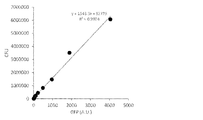
cfuと蛍光強度との相関を図2に示す。蛍光強度は、1.5×10⁴~6×10⁶の範囲でcfuと直線関係を示すことが確認された。この結果は、大腸菌の分裂周期を約30分と仮定した場合、約4時間の連続した測定が可能であることを意味する。なお、cfuの値はサンプリング後1晩経ってからコロニー数を実測して得られたものであるのに対して、蛍光強度はサンプリングと同時に測定されたものである。
3)形質転換体のNO濃度依存的な発光強度
pRPLG1をエレクトロポレーション法でサルモネラ(Salmonella typhimurium 0126株)及び赤痢菌(Shigella sonnei 3116株)それぞれに導入し、アンピシリン耐性を指標として形質転換体(tST及びtSSとそれぞれ表す)を得た。
pRPLG1をエレクトロポレーション法でサルモネラ(Salmonella typhimurium 0126株)及び赤痢菌(Shigella sonnei 3116株)それぞれに導入し、アンピシリン耐性を指標として形質転換体(tST及びtSSとそれぞれ表す)を得た。
tST、tSS及び前記2)で得たtECをLB/Car培地で一晩培養後、5mMグルコース及び10mM MOPSを含む生理食塩水(pH8.0)にOD600=0.1となるように各形質転換体を懸濁した。ここに、NOドナーであるPROLI-NONOate(Cayman Chemical)又はDEA-NONOate(Cayman Chemical)を1μM又は10μMとなるように添加して37℃で30分間保温した後、発光強度(RLU)とGFPの蛍光強度(GFP)を測定し、比発光強度を算出した。発光強度はGLOMAX 20/20ルミノメーター(Promega)を用いて、蛍光強度はTECAN infinite F200 PRO(TECAN、Ex/EM=465nm/510nm)を用いて測定した。
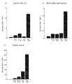
結果を図3に示す。各形質転換体はいずれもNOドナー濃度に比例した比発光強度を与えることが確認され、特に赤痢菌の比発光強度は他の菌と比べて非常に高かった。また、より高い比発光強度が得られるDEA-NONOateが本発明において好ましいNOドナーであることが確認された。
4)形質転換体における蛍光強度及びNO反応性の評価
LB/Car培地中でtECを37℃で19時間前培養し、前培養液の一部を新たなLB/Car培地に植え継ぎ、37℃で振盪培養(二次培養)を行った。また、前培養液の別の一部を室温で静置した。前培養、二次培養、前培養の静置それぞれにおいて経時的に培養液を採取し、回収したtECを5mMグルコース及び10mM MOPSを含む生理食塩水(pH8.0)にOD600=0.1となるように懸濁した。ここにDEA-NONOateを最終濃度が0.3、0.7、1.0、3.0、5.0μMとなるように添加し、37℃で保持しながら発光強度(RLU)とGFPの蛍光強度(GFP)を測定した。同一サンプルについて微生物数(cfu)を希釈法で測定した。
LB/Car培地中でtECを37℃で19時間前培養し、前培養液の一部を新たなLB/Car培地に植え継ぎ、37℃で振盪培養(二次培養)を行った。また、前培養液の別の一部を室温で静置した。前培養、二次培養、前培養の静置それぞれにおいて経時的に培養液を採取し、回収したtECを5mMグルコース及び10mM MOPSを含む生理食塩水(pH8.0)にOD600=0.1となるように懸濁した。ここにDEA-NONOateを最終濃度が0.3、0.7、1.0、3.0、5.0μMとなるように添加し、37℃で保持しながら発光強度(RLU)とGFPの蛍光強度(GFP)を測定した。同一サンプルについて微生物数(cfu)を希釈法で測定した。
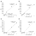
cfuを横軸、GFP蛍光強度を縦軸にとった結果を図4に、図4中の(A)~(D)の各プロットに対応する試料におけるDEA-NONOate添加量と発光強度との関係を図5に示す。ここで、(A)はLB/Car培地中で19時間前培養したtEC、(B)は前培養後に2日間静置したtEC、(C)は二次培養を5時間行ったtEC、(D)は二次培養を2時間行ったtECに相当する。
GFP蛍光強度が一定値(例えば2300、図4中の破線)を超えるtEC((A)及び(B))は、0~0.7μMというNOドナー濃度範囲において濃度依存的な比発光強度を示すことが確認された。一方、二次培養したtEC((C)及び(D))は、GFP発光強度が2300以下であり、かつ0~0.7μMというNOドナー濃度範囲における濃度依存性が(A)(B)と比較して劣ることが確認された。
この結果から、GFP蛍光強度は、特に低NO濃度域におけるNOセンサー微生物のNO反応性を判定する指標として利用可能であることが示された。また、定常期に至った、あるいはそこからさらに数時間~数日間放置された増殖能が低い微生物は、二次培養などによって増殖能を回復した、例えば二次培養における対数期にある微生物と比較して、低いNO濃度範囲においても良好なNO反応性を示し、本発明の方法において好ましい微生物として利用することができるものと推察された。
<実施例2>NO還元酵素阻害剤の細胞内NO濃度減少抑制効果の確認
実施例1の3)と同様にtECの懸濁液を調製した。ここにDEA NONOateを最終濃度が3.0μM又は0μM(ネガティブコントロール)となるように添加し、さらに大腸菌のNO消去系酵素であるフラボヘモグロビンの活性阻害が知られているエコナゾール(Sigma-Aldrich)を最終濃度が3.0μM又は0μM(コントロール)となるように加えてから60分間、37℃で保持しながら発光強度(RLU)とGFPの蛍光強度(GFP)を経時的に測定した。
実施例1の3)と同様にtECの懸濁液を調製した。ここにDEA NONOateを最終濃度が3.0μM又は0μM(ネガティブコントロール)となるように添加し、さらに大腸菌のNO消去系酵素であるフラボヘモグロビンの活性阻害が知られているエコナゾール(Sigma-Aldrich)を最終濃度が3.0μM又は0μM(コントロール)となるように加えてから60分間、37℃で保持しながら発光強度(RLU)とGFPの蛍光強度(GFP)を経時的に測定した。
発光強度の経時変化を図6に示す。図6に示されるように、エコナゾールが添加されたtECでは、コントロールと比較して比発光強度の減少が抑制されていることが確認された。ネガティブコントロールでは実質的に発光は観察されなかった。
このように、本方法によれば、比発光強度の減少抑制すなわち微生物内のNO濃度減少の抑制がNOドナーを加えてから30分程度で確認できること、及びnMレベルの生理的NO濃度を含む幅広いNO濃度範囲で減少抑制が確認できることが明らかとなった。
本発明の方法を利用することで、試験化合物の微生物内NO濃度の減少抑制効果を、多数同時に、簡便に、迅速に、高感度かつ広範囲のダイナミックレンジで評価することができることが明らかにされた。
本発明は、微生物が持つNOに対する自己防御機構を減弱させる化合物をスクリーニングし、医薬候補化合物を提供することができる方法として、産業上の利用可能性を有する。
Claims (11)
- NorRタンパク質をコードする遺伝子と、norオペロンプロモーターの制御下にある生物発光タンパク質をコードする遺伝子と、構成的プロモーターの制御下にある蛍光タンパク質をコードする遺伝子とを含む組換えベクター。
- 生物発光タンパク質をコードする遺伝子がluxCDABE遺伝子である、請求項1に記載の組換えベクター。
- 請求項1又は2に記載の組換えベクターを含む微生物。
- 微生物が大腸菌、サルモネラ及び赤痢菌よりなる群から選択される微生物である、請求項3に記載の微生物。
- 以下の工程(a)~(d):
(a)NorRタンパク質をコードする遺伝子と、norオペロンプロモーターの制御下にある生物発光タンパク質をコードする遺伝子と、構成的プロモーターの制御下にある蛍光タンパク質をコードする遺伝子とを含む組換えベクターで形質転換した微生物、試験化合物及びNOドナーを含む溶液をインキュベートする工程、
(b)生物発光タンパク質の発光強度と蛍光タンパク質の蛍光強度とを測定する工程、
(c)蛍光強度で補正した比発光強度を算出する工程、並びに
(d)試験化合物を含まない対照溶液と比較して比発光強度の減少を抑制した試験化合物を選択する工程
を含む、微生物細胞内のNO消去を抑制する化合物のスクリーニング方法。 - 工程(a)において使用する微生物が、予め定められた基準値以上の蛍光強度を示す微生物である、請求項5に記載のスクリーニング方法。
- 工程(a)における溶液中のNOドナーの濃度が溶液中で5nM~50μMのNO濃度を与える濃度である、請求項5又は6に記載のスクリーニング方法。
- 前記微生物が、大腸菌、サルモネラ及び赤痢菌よりなる群から選択される微生物である、請求項5~7のいずれかに記載のスクリーニング方法。
- 以下の工程(e)~(g):
(e)NorRタンパク質をコードする遺伝子と、norオペロンプロモーターの制御下にある生物発光タンパク質をコードする遺伝子と、構成的プロモーターの制御下にある蛍光タンパク質をコードする遺伝子とを含む組換えベクターで形質転換した微生物の生物発光タンパク質の発光強度と蛍光タンパク質の蛍光強度とを測定する工程、
(f)蛍光強度で補正した比発光強度を算出する工程、及び
(g)比発光強度に対応する細胞内NO濃度を算出する工程
を含む、微生物細胞内のNO濃度を測定する方法。 - 工程(e)において使用する微生物が、予め定められた基準値以上の蛍光強度を示す微生物である、請求項9に記載の測定方法。
- 前記微生物が、大腸菌、サルモネラ及び赤痢菌よりなる群から選択される微生物である、請求項9又は10に記載の測定方法。
Applications Claiming Priority (2)
| Application Number | Priority Date | Filing Date | Title |
|---|---|---|---|
| JP2016-114134 | 2016-06-08 | ||
| JP2016114134A JP2017216944A (ja) | 2016-06-08 | 2016-06-08 | 組換えベクター、該ベクターで形質転換された微生物を利用した一酸化窒素消去を抑制する化合物のスクリーニング方法及び細胞内一酸化窒素濃度の測定方法 |
Publications (1)
| Publication Number | Publication Date |
|---|---|
| WO2017213209A1 true WO2017213209A1 (ja) | 2017-12-14 |
Family
ID=60578708
Family Applications (1)
| Application Number | Title | Priority Date | Filing Date |
|---|---|---|---|
| PCT/JP2017/021261 Ceased WO2017213209A1 (ja) | 2016-06-08 | 2017-06-08 | 組換えベクター、該ベクターで形質転換された微生物を利用した一酸化窒素消去を抑制する化合物のスクリーニング方法及び細胞内一酸化窒素濃度の測定方法 |
Country Status (2)
| Country | Link |
|---|---|
| JP (1) | JP2017216944A (ja) |
| WO (1) | WO2017213209A1 (ja) |
Citations (2)
| Publication number | Priority date | Publication date | Assignee | Title |
|---|---|---|---|---|
| JP2008035820A (ja) * | 2006-08-09 | 2008-02-21 | Daikin Ind Ltd | 改良されたプロモーターアッセイ法 |
| JP2012231716A (ja) * | 2011-04-28 | 2012-11-29 | Chiba Univ | 一酸化窒素を探知するための形質転換用組換えベクター、およびこれを用いた一酸化窒素センサ細胞 |
-
2016
- 2016-06-08 JP JP2016114134A patent/JP2017216944A/ja active Pending
-
2017
- 2017-06-08 WO PCT/JP2017/021261 patent/WO2017213209A1/ja not_active Ceased
Patent Citations (2)
| Publication number | Priority date | Publication date | Assignee | Title |
|---|---|---|---|---|
| JP2008035820A (ja) * | 2006-08-09 | 2008-02-21 | Daikin Ind Ltd | 改良されたプロモーターアッセイ法 |
| JP2012231716A (ja) * | 2011-04-28 | 2012-11-29 | Chiba Univ | 一酸化窒素を探知するための形質転換用組換えベクター、およびこれを用いた一酸化窒素センサ細胞 |
Also Published As
| Publication number | Publication date |
|---|---|
| JP2017216944A (ja) | 2017-12-14 |
Similar Documents
| Publication | Publication Date | Title |
|---|---|---|
| Wilks et al. | pH of the cytoplasm and periplasm of Escherichia coli: rapid measurement by green fluorescent protein fluorimetry | |
| Kostrzynska et al. | Green fluorescent protein-based biosensor for detecting SOS-inducing activity of genotoxic compounds | |
| Kim et al. | Coordination of division and development influences complex multicellular behavior in Agrobacterium tumefaciens | |
| Uhlich et al. | Growth media and temperature effects on biofilm formation by serotype O157: H7 and non-O157 Shiga toxin-producing Escherichia coli | |
| Massai et al. | A multitask biosensor for micro-volumetric detection of N-3-oxo-dodecanoyl-homoserine lactone quorum sensing signal | |
| van Der Heijden et al. | Exploring the redox balance inside gram-negative bacteria with redox-sensitive GFP | |
| Atosuo et al. | Escherichia coli K‐12 (pEGFPluxABCDEamp): a tool for analysis of bacterial killing by antibacterial agents and human complement activities on a real‐time basis | |
| Jia et al. | Gene circuit engineering to improve the performance of a whole-cell lead biosensor | |
| Sharma et al. | Simple and rapid method to determine antimycobacterial potency of compounds by using autoluminescent Mycobacterium tuberculosis | |
| Pawar et al. | Novel genetic tools to tackle c‐di‐GMP‐dependent signalling in Pseudomonas aeruginosa | |
| Utratna et al. | Development and optimization of an EGFP-based reporter for measuring the general stress response in Listeria monocytogenes | |
| Hochstrasser et al. | The Legionella Lqs-LvbR regulatory network controls temperature-dependent growth onset and bacterial cell density | |
| Fan et al. | Cascaded amplifying circuit enables sensitive detection of fungal pathogens | |
| Robinson et al. | Application of bacterial bioluminescence to assess the efficacy of fast-acting biocides | |
| Gupta et al. | Fast-growing, non-infectious and intracellularly surviving drug-resistant Mycobacterium aurum: a model for high-throughput antituberculosis drug screening | |
| Shah et al. | Bioluminescence‐based measurement of viability of Pseudomonas aeruginosa ATCC 9027 harbouring plasmid‐based lux genes under the control of constitutive promoters | |
| Peng et al. | Activation of metabolic and stress responses during subtoxic expression of the type I toxin hok in Erwinia amylovora | |
| Deng et al. | Genetic engineering of circularly permuted yellow fluorescent protein reveals intracellular acidification in response to nitric oxide stimuli | |
| WO2017213209A1 (ja) | 組換えベクター、該ベクターで形質転換された微生物を利用した一酸化窒素消去を抑制する化合物のスクリーニング方法及び細胞内一酸化窒素濃度の測定方法 | |
| Deng et al. | Construction of a dual fluorescence whole-cell biosensor to detect N-acyl homoserine lactones | |
| Welham et al. | Mathematical model of the Lux luminescence system in the terrestrial bacterium Photorhabdus luminescens | |
| Cui et al. | Bioluminescence resonance energy transfer system for measuring dynamic protein-protein interactions in bacteria | |
| Hever et al. | A dual‐color bacterial reporter strain for the detection of toxic and genotoxic effects | |
| JP6037259B2 (ja) | 一酸化窒素を探知するための形質転換用組換えベクター、およびこれを用いた一酸化窒素センサ細胞 | |
| Kramer et al. | Impact of pulsed light on cellular activity of Salmonella enterica |
Legal Events
| Date | Code | Title | Description |
|---|---|---|---|
| 121 | Ep: the epo has been informed by wipo that ep was designated in this application |
Ref document number: 17810382 Country of ref document: EP Kind code of ref document: A1 |
|
| NENP | Non-entry into the national phase |
Ref country code: DE |
|
| 122 | Ep: pct application non-entry in european phase |
Ref document number: 17810382 Country of ref document: EP Kind code of ref document: A1 |