WO2017161206A1 - Conjugates containing conditionally active antibodies or antigen-binding fragments thereof, and methods of use - Google Patents
Conjugates containing conditionally active antibodies or antigen-binding fragments thereof, and methods of use Download PDFInfo
- Publication number
- WO2017161206A1 WO2017161206A1 PCT/US2017/022840 US2017022840W WO2017161206A1 WO 2017161206 A1 WO2017161206 A1 WO 2017161206A1 US 2017022840 W US2017022840 W US 2017022840W WO 2017161206 A1 WO2017161206 A1 WO 2017161206A1
- Authority
- WO
- WIPO (PCT)
- Prior art keywords
- seq
- set forth
- conjugate
- heavy chain
- chain set
- Prior art date
Links
- 230000027455 binding Effects 0.000 title claims abstract description 430
- 239000012634 fragment Substances 0.000 title claims abstract description 360
- 239000000427 antigen Substances 0.000 title claims abstract description 317
- 108091007433 antigens Proteins 0.000 title claims abstract description 316
- 102000036639 antigens Human genes 0.000 title claims abstract description 316
- 238000000034 method Methods 0.000 title claims abstract description 72
- 239000003795 chemical substances by application Substances 0.000 claims abstract description 102
- 238000011282 treatment Methods 0.000 claims abstract description 26
- 235000001014 amino acid Nutrition 0.000 claims description 625
- 150000001413 amino acids Chemical class 0.000 claims description 598
- 206010028980 Neoplasm Diseases 0.000 claims description 145
- 210000004027 cell Anatomy 0.000 claims description 140
- 229960005395 cetuximab Drugs 0.000 claims description 114
- 230000000694 effects Effects 0.000 claims description 113
- 102000001301 EGF receptor Human genes 0.000 claims description 112
- 108060006698 EGF receptor Proteins 0.000 claims description 112
- 108090000765 processed proteins & peptides Proteins 0.000 claims description 92
- 108010093470 monomethyl auristatin E Proteins 0.000 claims description 66
- IEDXPSOJFSVCKU-HOKPPMCLSA-N [4-[[(2S)-5-(carbamoylamino)-2-[[(2S)-2-[6-(2,5-dioxopyrrolidin-1-yl)hexanoylamino]-3-methylbutanoyl]amino]pentanoyl]amino]phenyl]methyl N-[(2S)-1-[[(2S)-1-[[(3R,4S,5S)-1-[(2S)-2-[(1R,2R)-3-[[(1S,2R)-1-hydroxy-1-phenylpropan-2-yl]amino]-1-methoxy-2-methyl-3-oxopropyl]pyrrolidin-1-yl]-3-methoxy-5-methyl-1-oxoheptan-4-yl]-methylamino]-3-methyl-1-oxobutan-2-yl]amino]-3-methyl-1-oxobutan-2-yl]-N-methylcarbamate Chemical compound CC[C@H](C)[C@@H]([C@@H](CC(=O)N1CCC[C@H]1[C@H](OC)[C@@H](C)C(=O)N[C@H](C)[C@@H](O)c1ccccc1)OC)N(C)C(=O)[C@@H](NC(=O)[C@H](C(C)C)N(C)C(=O)OCc1ccc(NC(=O)[C@H](CCCNC(N)=O)NC(=O)[C@@H](NC(=O)CCCCCN2C(=O)CCC2=O)C(C)C)cc1)C(C)C IEDXPSOJFSVCKU-HOKPPMCLSA-N 0.000 claims description 64
- 125000005647 linker group Chemical group 0.000 claims description 63
- 102000004169 proteins and genes Human genes 0.000 claims description 53
- 108090000623 proteins and genes Proteins 0.000 claims description 53
- FWMNVWWHGCHHJJ-SKKKGAJSSA-N 4-amino-1-[(2r)-6-amino-2-[[(2r)-2-[[(2r)-2-[[(2r)-2-amino-3-phenylpropanoyl]amino]-3-phenylpropanoyl]amino]-4-methylpentanoyl]amino]hexanoyl]piperidine-4-carboxylic acid Chemical compound C([C@H](C(=O)N[C@H](CC(C)C)C(=O)N[C@H](CCCCN)C(=O)N1CCC(N)(CC1)C(O)=O)NC(=O)[C@H](N)CC=1C=CC=CC=1)C1=CC=CC=C1 FWMNVWWHGCHHJJ-SKKKGAJSSA-N 0.000 claims description 48
- 235000018102 proteins Nutrition 0.000 claims description 48
- 125000003275 alpha amino acid group Chemical group 0.000 claims description 36
- 239000003053 toxin Substances 0.000 claims description 35
- 231100000765 toxin Toxicity 0.000 claims description 35
- 108700012359 toxins Proteins 0.000 claims description 35
- 108010087819 Fc receptors Proteins 0.000 claims description 33
- 102000009109 Fc receptors Human genes 0.000 claims description 33
- NFGXHKASABOEEW-UHFFFAOYSA-N 1-methylethyl 11-methoxy-3,7,11-trimethyl-2,4-dodecadienoate Chemical compound COC(C)(C)CCCC(C)CC=CC(C)=CC(=O)OC(C)C NFGXHKASABOEEW-UHFFFAOYSA-N 0.000 claims description 30
- 125000000151 cysteine group Chemical group N[C@@H](CS)C(=O)* 0.000 claims description 29
- -1 2-pyridylmethyl- Chemical group 0.000 claims description 28
- 230000003993 interaction Effects 0.000 claims description 28
- 239000008194 pharmaceutical composition Substances 0.000 claims description 27
- 230000001225 therapeutic effect Effects 0.000 claims description 25
- AOJJSUZBOXZQNB-TZSSRYMLSA-N Doxorubicin Chemical compound O([C@H]1C[C@@](O)(CC=2C(O)=C3C(=O)C=4C=CC=C(C=4C(=O)C3=C(O)C=21)OC)C(=O)CO)[C@H]1C[C@H](N)[C@H](O)[C@H](C)O1 AOJJSUZBOXZQNB-TZSSRYMLSA-N 0.000 claims description 24
- 239000003112 inhibitor Substances 0.000 claims description 21
- 125000002496 methyl group Chemical group [H]C([H])([H])* 0.000 claims description 21
- RJURFGZVJUQBHK-UHFFFAOYSA-N actinomycin D Natural products CC1OC(=O)C(C(C)C)N(C)C(=O)CN(C)C(=O)C2CCCN2C(=O)C(C(C)C)NC(=O)C1NC(=O)C1=C(N)C(=O)C(C)=C2OC(C(C)=CC=C3C(=O)NC4C(=O)NC(C(N5CCCC5C(=O)N(C)CC(=O)N(C)C(C(C)C)C(=O)OC4C)=O)C(C)C)=C3N=C21 RJURFGZVJUQBHK-UHFFFAOYSA-N 0.000 claims description 20
- MFRNYXJJRJQHNW-DEMKXPNLSA-N (2s)-2-[[(2r,3r)-3-methoxy-3-[(2s)-1-[(3r,4s,5s)-3-methoxy-5-methyl-4-[methyl-[(2s)-3-methyl-2-[[(2s)-3-methyl-2-(methylamino)butanoyl]amino]butanoyl]amino]heptanoyl]pyrrolidin-2-yl]-2-methylpropanoyl]amino]-3-phenylpropanoic acid Chemical compound CN[C@@H](C(C)C)C(=O)N[C@@H](C(C)C)C(=O)N(C)[C@@H]([C@@H](C)CC)[C@H](OC)CC(=O)N1CCC[C@H]1[C@H](OC)[C@@H](C)C(=O)N[C@H](C(O)=O)CC1=CC=CC=C1 MFRNYXJJRJQHNW-DEMKXPNLSA-N 0.000 claims description 19
- 108010044540 auristatin Proteins 0.000 claims description 19
- 229920001223 polyethylene glycol Polymers 0.000 claims description 18
- 102200148733 rs116840794 Human genes 0.000 claims description 18
- 230000001988 toxicity Effects 0.000 claims description 18
- 231100000419 toxicity Toxicity 0.000 claims description 18
- 108010059074 monomethylauristatin F Proteins 0.000 claims description 16
- CFCUWKMKBJTWLW-BKHRDMLASA-N mithramycin Chemical compound O([C@@H]1C[C@@H](O[C@H](C)[C@H]1O)OC=1C=C2C=C3C[C@H]([C@@H](C(=O)C3=C(O)C2=C(O)C=1C)O[C@@H]1O[C@H](C)[C@@H](O)[C@H](O[C@@H]2O[C@H](C)[C@H](O)[C@H](O[C@@H]3O[C@H](C)[C@@H](O)[C@@](C)(O)C3)C2)C1)[C@H](OC)C(=O)[C@@H](O)[C@@H](C)O)[C@H]1C[C@@H](O)[C@H](O)[C@@H](C)O1 CFCUWKMKBJTWLW-BKHRDMLASA-N 0.000 claims description 15
- 229960003171 plicamycin Drugs 0.000 claims description 15
- NWIBSHFKIJFRCO-WUDYKRTCSA-N Mytomycin Chemical compound C1N2C(C(C(C)=C(N)C3=O)=O)=C3[C@@H](COC(N)=O)[C@@]2(OC)[C@@H]2[C@H]1N2 NWIBSHFKIJFRCO-WUDYKRTCSA-N 0.000 claims description 14
- 239000003814 drug Substances 0.000 claims description 14
- 229920000642 polymer Polymers 0.000 claims description 13
- 206010009944 Colon cancer Diseases 0.000 claims description 12
- 208000001333 Colorectal Neoplasms Diseases 0.000 claims description 12
- 239000002246 antineoplastic agent Substances 0.000 claims description 12
- 229960004857 mitomycin Drugs 0.000 claims description 12
- WHUUTDBJXJRKMK-UHFFFAOYSA-N Glutamic acid Natural products OC(=O)C(N)CCC(O)=O WHUUTDBJXJRKMK-UHFFFAOYSA-N 0.000 claims description 11
- VGQOVCHZGQWAOI-UHFFFAOYSA-N UNPD55612 Natural products N1C(O)C2CC(C=CC(N)=O)=CN2C(=O)C2=CC=C(C)C(O)=C12 VGQOVCHZGQWAOI-UHFFFAOYSA-N 0.000 claims description 11
- VGQOVCHZGQWAOI-HYUHUPJXSA-N anthramycin Chemical compound N1[C@@H](O)[C@@H]2CC(\C=C\C(N)=O)=CN2C(=O)C2=CC=C(C)C(O)=C12 VGQOVCHZGQWAOI-HYUHUPJXSA-N 0.000 claims description 11
- 230000001404 mediated effect Effects 0.000 claims description 11
- 230000002829 reductive effect Effects 0.000 claims description 11
- IAKHMKGGTNLKSZ-INIZCTEOSA-N (S)-colchicine Chemical compound C1([C@@H](NC(C)=O)CC2)=CC(=O)C(OC)=CC=C1C1=C2C=C(OC)C(OC)=C1OC IAKHMKGGTNLKSZ-INIZCTEOSA-N 0.000 claims description 10
- VPFUWHKTPYPNGT-UHFFFAOYSA-N 3-(3,4-dihydroxyphenyl)-1-(5-hydroxy-2,2-dimethylchromen-6-yl)propan-1-one Chemical compound OC1=C2C=CC(C)(C)OC2=CC=C1C(=O)CCC1=CC=C(O)C(O)=C1 VPFUWHKTPYPNGT-UHFFFAOYSA-N 0.000 claims description 10
- STQGQHZAVUOBTE-UHFFFAOYSA-N 7-Cyan-hept-2t-en-4,6-diinsaeure Natural products C1=2C(O)=C3C(=O)C=4C(OC)=CC=CC=4C(=O)C3=C(O)C=2CC(O)(C(C)=O)CC1OC1CC(N)C(O)C(C)O1 STQGQHZAVUOBTE-UHFFFAOYSA-N 0.000 claims description 10
- 108010092160 Dactinomycin Proteins 0.000 claims description 10
- 229930192392 Mitomycin Natural products 0.000 claims description 10
- 102000006382 Ribonucleases Human genes 0.000 claims description 10
- 108010083644 Ribonucleases Proteins 0.000 claims description 10
- 102220531209 Uncharacterized protein KIAA2012_F27R_mutation Human genes 0.000 claims description 10
- RJURFGZVJUQBHK-IIXSONLDSA-N actinomycin D Chemical compound C[C@H]1OC(=O)[C@H](C(C)C)N(C)C(=O)CN(C)C(=O)[C@@H]2CCCN2C(=O)[C@@H](C(C)C)NC(=O)[C@H]1NC(=O)C1=C(N)C(=O)C(C)=C2OC(C(C)=CC=C3C(=O)N[C@@H]4C(=O)N[C@@H](C(N5CCC[C@H]5C(=O)N(C)CC(=O)N(C)[C@@H](C(C)C)C(=O)O[C@@H]4C)=O)C(C)C)=C3N=C21 RJURFGZVJUQBHK-IIXSONLDSA-N 0.000 claims description 10
- 201000011510 cancer Diseases 0.000 claims description 10
- 229960000640 dactinomycin Drugs 0.000 claims description 10
- STQGQHZAVUOBTE-VGBVRHCVSA-N daunorubicin Chemical compound O([C@H]1C[C@@](O)(CC=2C(O)=C3C(=O)C=4C=CC=C(C=4C(=O)C3=C(O)C=21)OC)C(C)=O)[C@H]1C[C@H](N)[C@H](O)[C@H](C)O1 STQGQHZAVUOBTE-VGBVRHCVSA-N 0.000 claims description 10
- 229960000975 daunorubicin Drugs 0.000 claims description 10
- CFCUWKMKBJTWLW-UHFFFAOYSA-N deoliosyl-3C-alpha-L-digitoxosyl-MTM Natural products CC=1C(O)=C2C(O)=C3C(=O)C(OC4OC(C)C(O)C(OC5OC(C)C(O)C(OC6OC(C)C(O)C(C)(O)C6)C5)C4)C(C(OC)C(=O)C(O)C(C)O)CC3=CC2=CC=1OC(OC(C)C1O)CC1OC1CC(O)C(O)C(C)O1 CFCUWKMKBJTWLW-UHFFFAOYSA-N 0.000 claims description 10
- 229960004679 doxorubicin Drugs 0.000 claims description 10
- KKZJGLLVHKMTCM-UHFFFAOYSA-N mitoxantrone Chemical compound O=C1C2=C(O)C=CC(O)=C2C(=O)C2=C1C(NCCNCCO)=CC=C2NCCNCCO KKZJGLLVHKMTCM-UHFFFAOYSA-N 0.000 claims description 10
- 229960001156 mitoxantrone Drugs 0.000 claims description 10
- 108010010621 modeccin Proteins 0.000 claims description 10
- AQHHHDLHHXJYJD-UHFFFAOYSA-N propranolol Chemical compound C1=CC=C2C(OCC(O)CNC(C)C)=CC=CC2=C1 AQHHHDLHHXJYJD-UHFFFAOYSA-N 0.000 claims description 10
- RXWNCPJZOCPEPQ-NVWDDTSBSA-N puromycin Chemical compound C1=CC(OC)=CC=C1C[C@H](N)C(=O)N[C@H]1[C@@H](O)[C@H](N2C3=NC=NC(=C3N=C2)N(C)C)O[C@@H]1CO RXWNCPJZOCPEPQ-NVWDDTSBSA-N 0.000 claims description 10
- UOWVMDUEMSNCAV-WYENRQIDSA-N rachelmycin Chemical compound C1([C@]23C[C@@H]2CN1C(=O)C=1NC=2C(OC)=C(O)C4=C(C=2C=1)CCN4C(=O)C1=CC=2C=4CCN(C=4C(O)=C(C=2N1)OC)C(N)=O)=CC(=O)C1=C3C(C)=CN1 UOWVMDUEMSNCAV-WYENRQIDSA-N 0.000 claims description 10
- GHASVSINZRGABV-UHFFFAOYSA-N Fluorouracil Chemical compound FC1=CNC(=O)NC1=O GHASVSINZRGABV-UHFFFAOYSA-N 0.000 claims description 9
- 231100000433 cytotoxic Toxicity 0.000 claims description 9
- 229940127089 cytotoxic agent Drugs 0.000 claims description 9
- 230000001472 cytotoxic effect Effects 0.000 claims description 9
- 229960002949 fluorouracil Drugs 0.000 claims description 9
- 235000013922 glutamic acid Nutrition 0.000 claims description 9
- 239000004220 glutamic acid Substances 0.000 claims description 9
- UWKQSNNFCGGAFS-XIFFEERXSA-N irinotecan Chemical compound C1=C2C(CC)=C3CN(C(C4=C([C@@](C(=O)OC4)(O)CC)C=4)=O)C=4C3=NC2=CC=C1OC(=O)N(CC1)CCC1N1CCCCC1 UWKQSNNFCGGAFS-XIFFEERXSA-N 0.000 claims description 9
- 208000002154 non-small cell lung carcinoma Diseases 0.000 claims description 9
- 208000029729 tumor suppressor gene on chromosome 11 Diseases 0.000 claims description 9
- 102220623751 Phospholipid hydroperoxide glutathione peroxidase_S74D_mutation Human genes 0.000 claims description 8
- 229940100198 alkylating agent Drugs 0.000 claims description 8
- 239000002168 alkylating agent Substances 0.000 claims description 8
- HXCHCVDVKSCDHU-LULTVBGHSA-N calicheamicin Chemical compound C1[C@H](OC)[C@@H](NCC)CO[C@H]1O[C@H]1[C@H](O[C@@H]2C\3=C(NC(=O)OC)C(=O)C[C@](C/3=C/CSSSC)(O)C#C\C=C/C#C2)O[C@H](C)[C@@H](NO[C@@H]2O[C@H](C)[C@@H](SC(=O)C=3C(=C(OC)C(O[C@H]4[C@@H]([C@H](OC)[C@@H](O)[C@H](C)O4)O)=C(I)C=3C)OC)[C@@H](O)C2)[C@@H]1O HXCHCVDVKSCDHU-LULTVBGHSA-N 0.000 claims description 8
- 229930195731 calicheamicin Natural products 0.000 claims description 8
- 239000000203 mixture Substances 0.000 claims description 8
- 125000006850 spacer group Chemical group 0.000 claims description 8
- 206010027476 Metastases Diseases 0.000 claims description 7
- 229930012538 Paclitaxel Natural products 0.000 claims description 7
- 239000002202 Polyethylene glycol Substances 0.000 claims description 7
- 239000003242 anti bacterial agent Substances 0.000 claims description 7
- 230000000340 anti-metabolite Effects 0.000 claims description 7
- 229940100197 antimetabolite Drugs 0.000 claims description 7
- 239000002256 antimetabolite Substances 0.000 claims description 7
- 230000003115 biocidal effect Effects 0.000 claims description 7
- 150000001875 compounds Chemical class 0.000 claims description 7
- 125000004435 hydrogen atom Chemical group [H]* 0.000 claims description 7
- 229960004768 irinotecan Drugs 0.000 claims description 7
- WKPWGQKGSOKKOO-RSFHAFMBSA-N maytansine Chemical compound CO[C@@H]([C@@]1(O)C[C@](OC(=O)N1)([C@H]([C@@H]1O[C@@]1(C)[C@@H](OC(=O)[C@H](C)N(C)C(C)=O)CC(=O)N1C)C)[H])\C=C\C=C(C)\CC2=CC(OC)=C(Cl)C1=C2 WKPWGQKGSOKKOO-RSFHAFMBSA-N 0.000 claims description 7
- ANZJBCHSOXCCRQ-FKUXLPTCSA-N mertansine Chemical compound CO[C@@H]([C@@]1(O)C[C@H](OC(=O)N1)[C@@H](C)[C@@H]1O[C@@]1(C)[C@@H](OC(=O)[C@H](C)N(C)C(=O)CCS)CC(=O)N1C)\C=C\C=C(C)\CC2=CC(OC)=C(Cl)C1=C2 ANZJBCHSOXCCRQ-FKUXLPTCSA-N 0.000 claims description 7
- 229960001592 paclitaxel Drugs 0.000 claims description 7
- RCINICONZNJXQF-MZXODVADSA-N taxol Chemical compound O([C@@H]1[C@@]2(C[C@@H](C(C)=C(C2(C)C)[C@H](C([C@]2(C)[C@@H](O)C[C@H]3OC[C@]3([C@H]21)OC(C)=O)=O)OC(=O)C)OC(=O)[C@H](O)[C@@H](NC(=O)C=1C=CC=CC=1)C=1C=CC=CC=1)O)C(=O)C1=CC=CC=C1 RCINICONZNJXQF-MZXODVADSA-N 0.000 claims description 7
- FDKXTQMXEQVLRF-ZHACJKMWSA-N (E)-dacarbazine Chemical compound CN(C)\N=N\c1[nH]cnc1C(N)=O FDKXTQMXEQVLRF-ZHACJKMWSA-N 0.000 claims description 6
- 102220628482 G protein-regulated inducer of neurite outgrowth 2_T100P_mutation Human genes 0.000 claims description 6
- 150000003057 platinum Chemical class 0.000 claims description 6
- 102000005962 receptors Human genes 0.000 claims description 6
- 108020003175 receptors Proteins 0.000 claims description 6
- 102220098157 rs200530606 Human genes 0.000 claims description 6
- 229940124597 therapeutic agent Drugs 0.000 claims description 6
- 239000003981 vehicle Substances 0.000 claims description 6
- FBUTXZSKZCQABC-UHFFFAOYSA-N 2-amino-1-methyl-7h-purine-6-thione Chemical compound S=C1N(C)C(N)=NC2=C1NC=N2 FBUTXZSKZCQABC-UHFFFAOYSA-N 0.000 claims description 5
- 108010066676 Abrin Proteins 0.000 claims description 5
- 108700032558 Aspergillus restrictus MITF Proteins 0.000 claims description 5
- 101000669426 Aspergillus restrictus Ribonuclease mitogillin Proteins 0.000 claims description 5
- 108010006654 Bleomycin Proteins 0.000 claims description 5
- 229960005532 CC-1065 Drugs 0.000 claims description 5
- 108010049048 Cholera Toxin Proteins 0.000 claims description 5
- 102000009016 Cholera Toxin Human genes 0.000 claims description 5
- 108700032819 Croton tiglium crotin II Proteins 0.000 claims description 5
- 102000004127 Cytokines Human genes 0.000 claims description 5
- 108090000695 Cytokines Proteins 0.000 claims description 5
- 102000007260 Deoxyribonuclease I Human genes 0.000 claims description 5
- 108010008532 Deoxyribonuclease I Proteins 0.000 claims description 5
- 108010053187 Diphtheria Toxin Proteins 0.000 claims description 5
- 102000016607 Diphtheria Toxin Human genes 0.000 claims description 5
- OFDNQWIFNXBECV-UHFFFAOYSA-N Dolastatin 10 Natural products CC(C)C(N(C)C)C(=O)NC(C(C)C)C(=O)N(C)C(C(C)CC)C(OC)CC(=O)N1CCCC1C(OC)C(C)C(=O)NC(C=1SC=CN=1)CC1=CC=CC=C1 OFDNQWIFNXBECV-UHFFFAOYSA-N 0.000 claims description 5
- LQKSHSFQQRCAFW-UHFFFAOYSA-N Dolastatin 15 Natural products COC1=CC(=O)N(C(=O)C(OC(=O)C2N(CCC2)C(=O)C2N(CCC2)C(=O)C(C(C)C)N(C)C(=O)C(NC(=O)C(C(C)C)N(C)C)C(C)C)C(C)C)C1CC1=CC=CC=C1 LQKSHSFQQRCAFW-UHFFFAOYSA-N 0.000 claims description 5
- AZVARJHZBXHUSO-UHFFFAOYSA-N Duocarmycin A Natural products COC1=C(OC)C(OC)=C2NC(C(=O)N3CC4CC44C5=C(C(C=C43)=O)NC(C5=O)(C)C(=O)OC)=CC2=C1 AZVARJHZBXHUSO-UHFFFAOYSA-N 0.000 claims description 5
- VQNATVDKACXKTF-UHFFFAOYSA-N Duocarmycin SA Natural products COC1=C(OC)C(OC)=C2NC(C(=O)N3C4=CC(=O)C5=C(C64CC6C3)C=C(N5)C(=O)OC)=CC2=C1 VQNATVDKACXKTF-UHFFFAOYSA-N 0.000 claims description 5
- MBYXEBXZARTUSS-QLWBXOBMSA-N Emetamine Natural products O(C)c1c(OC)cc2c(c(C[C@@H]3[C@H](CC)CN4[C@H](c5c(cc(OC)c(OC)c5)CC4)C3)ncc2)c1 MBYXEBXZARTUSS-QLWBXOBMSA-N 0.000 claims description 5
- 108700004714 Gelonium multiflorum GEL Proteins 0.000 claims description 5
- 244000068988 Glycine max Species 0.000 claims description 5
- 235000010469 Glycine max Nutrition 0.000 claims description 5
- 108010026389 Gramicidin Proteins 0.000 claims description 5
- XDXDZDZNSLXDNA-TZNDIEGXSA-N Idarubicin Chemical compound C1[C@H](N)[C@H](O)[C@H](C)O[C@H]1O[C@@H]1C2=C(O)C(C(=O)C3=CC=CC=C3C3=O)=C3C(O)=C2C[C@@](O)(C(C)=O)C1 XDXDZDZNSLXDNA-TZNDIEGXSA-N 0.000 claims description 5
- XDXDZDZNSLXDNA-UHFFFAOYSA-N Idarubicin Natural products C1C(N)C(O)C(C)OC1OC1C2=C(O)C(C(=O)C3=CC=CC=C3C3=O)=C3C(O)=C2CC(O)(C(C)=O)C1 XDXDZDZNSLXDNA-UHFFFAOYSA-N 0.000 claims description 5
- FBOZXECLQNJBKD-ZDUSSCGKSA-N L-methotrexate Chemical compound C=1N=C2N=C(N)N=C(N)C2=NC=1CN(C)C1=CC=C(C(=O)N[C@@H](CCC(O)=O)C(O)=O)C=C1 FBOZXECLQNJBKD-ZDUSSCGKSA-N 0.000 claims description 5
- NNJVILVZKWQKPM-UHFFFAOYSA-N Lidocaine Chemical compound CCN(CC)CC(=O)NC1=C(C)C=CC=C1C NNJVILVZKWQKPM-UHFFFAOYSA-N 0.000 claims description 5
- 229930126263 Maytansine Natural products 0.000 claims description 5
- 244000302512 Momordica charantia Species 0.000 claims description 5
- 235000009811 Momordica charantia Nutrition 0.000 claims description 5
- 108010081690 Pertussis Toxin Proteins 0.000 claims description 5
- 240000007643 Phytolacca americana Species 0.000 claims description 5
- 235000009074 Phytolacca americana Nutrition 0.000 claims description 5
- 229940124158 Protease/peptidase inhibitor Drugs 0.000 claims description 5
- 101000762949 Pseudomonas aeruginosa (strain ATCC 15692 / DSM 22644 / CIP 104116 / JCM 14847 / LMG 12228 / 1C / PRS 101 / PAO1) Exotoxin A Proteins 0.000 claims description 5
- 108010039491 Ricin Proteins 0.000 claims description 5
- AUVVAXYIELKVAI-UHFFFAOYSA-N SJ000285215 Natural products N1CCC2=CC(OC)=C(OC)C=C2C1CC1CC2C3=CC(OC)=C(OC)C=C3CCN2CC1CC AUVVAXYIELKVAI-UHFFFAOYSA-N 0.000 claims description 5
- 108010084592 Saporins Proteins 0.000 claims description 5
- 108010079723 Shiga Toxin Proteins 0.000 claims description 5
- 108010055044 Tetanus Toxin Proteins 0.000 claims description 5
- GBOGMAARMMDZGR-UHFFFAOYSA-N UNPD149280 Natural products N1C(=O)C23OC(=O)C=CC(O)CCCC(C)CC=CC3C(O)C(=C)C(C)C2C1CC1=CC=CC=C1 GBOGMAARMMDZGR-UHFFFAOYSA-N 0.000 claims description 5
- 240000001866 Vernicia fordii Species 0.000 claims description 5
- JXLYSJRDGCGARV-WWYNWVTFSA-N Vinblastine Natural products O=C(O[C@H]1[C@](O)(C(=O)OC)[C@@H]2N(C)c3c(cc(c(OC)c3)[C@]3(C(=O)OC)c4[nH]c5c(c4CCN4C[C@](O)(CC)C[C@H](C3)C4)cccc5)[C@@]32[C@H]2[C@@]1(CC)C=CCN2CC3)C JXLYSJRDGCGARV-WWYNWVTFSA-N 0.000 claims description 5
- LQKSHSFQQRCAFW-CCVNJFHASA-N [(2s)-1-[(2s)-2-benzyl-3-methoxy-5-oxo-2h-pyrrol-1-yl]-3-methyl-1-oxobutan-2-yl] (2s)-1-[(2s)-1-[(2s)-2-[[(2s)-2-[[(2s)-2-(dimethylamino)-3-methylbutanoyl]amino]-3-methylbutanoyl]-methylamino]-3-methylbutanoyl]pyrrolidine-2-carbonyl]pyrrolidine-2-carboxyl Chemical compound C([C@@H]1N(C(=O)C=C1OC)C(=O)[C@@H](OC(=O)[C@H]1N(CCC1)C(=O)[C@H]1N(CCC1)C(=O)[C@H](C(C)C)N(C)C(=O)[C@@H](NC(=O)[C@H](C(C)C)N(C)C)C(C)C)C(C)C)C1=CC=CC=C1 LQKSHSFQQRCAFW-CCVNJFHASA-N 0.000 claims description 5
- 108010001818 alpha-sarcin Proteins 0.000 claims description 5
- MWPLVEDNUUSJAV-UHFFFAOYSA-N anthracene Chemical compound C1=CC=CC2=CC3=CC=CC=C3C=C21 MWPLVEDNUUSJAV-UHFFFAOYSA-N 0.000 claims description 5
- 229940049706 benzodiazepine Drugs 0.000 claims description 5
- 229960001561 bleomycin Drugs 0.000 claims description 5
- OYVAGSVQBOHSSS-UAPAGMARSA-O bleomycin A2 Chemical compound N([C@H](C(=O)N[C@H](C)[C@@H](O)[C@H](C)C(=O)N[C@@H]([C@H](O)C)C(=O)NCCC=1SC=C(N=1)C=1SC=C(N=1)C(=O)NCCC[S+](C)C)[C@@H](O[C@H]1[C@H]([C@@H](O)[C@H](O)[C@H](CO)O1)O[C@@H]1[C@H]([C@@H](OC(N)=O)[C@H](O)[C@@H](CO)O1)O)C=1N=CNC=1)C(=O)C1=NC([C@H](CC(N)=O)NC[C@H](N)C(N)=O)=NC(N)=C1C OYVAGSVQBOHSSS-UAPAGMARSA-O 0.000 claims description 5
- RSIHSRDYCUFFLA-DYKIIFRCSA-N boldenone Chemical compound O=C1C=C[C@]2(C)[C@H]3CC[C@](C)([C@H](CC4)O)[C@@H]4[C@@H]3CCC2=C1 RSIHSRDYCUFFLA-DYKIIFRCSA-N 0.000 claims description 5
- NDAYQJDHGXTBJL-MWWSRJDJSA-N chembl557217 Chemical compound C1=CC=C2C(C[C@H](NC(=O)[C@@H](CC(C)C)NC(=O)[C@H](CC=3C4=CC=CC=C4NC=3)NC(=O)[C@@H](CC(C)C)NC(=O)[C@H](CC=3C4=CC=CC=C4NC=3)NC(=O)[C@@H](CC(C)C)NC(=O)[C@H](CC=3C4=CC=CC=C4NC=3)NC(=O)[C@@H](C(C)C)NC(=O)[C@H](C(C)C)NC(=O)[C@@H](C(C)C)NC(=O)[C@H](C)NC(=O)[C@H](NC(=O)CNC(=O)[C@@H](NC=O)C(C)C)CC(C)C)C(=O)NCCO)=CNC2=C1 NDAYQJDHGXTBJL-MWWSRJDJSA-N 0.000 claims description 5
- 229960001338 colchicine Drugs 0.000 claims description 5
- GBOGMAARMMDZGR-TYHYBEHESA-N cytochalasin B Chemical compound C([C@H]1[C@@H]2[C@@H](C([C@@H](O)[C@@H]3/C=C/C[C@H](C)CCC[C@@H](O)/C=C/C(=O)O[C@@]23C(=O)N1)=C)C)C1=CC=CC=C1 GBOGMAARMMDZGR-TYHYBEHESA-N 0.000 claims description 5
- GBOGMAARMMDZGR-JREHFAHYSA-N cytochalasin B Natural products C[C@H]1CCC[C@@H](O)C=CC(=O)O[C@@]23[C@H](C=CC1)[C@H](O)C(=C)[C@@H](C)[C@@H]2[C@H](Cc4ccccc4)NC3=O GBOGMAARMMDZGR-JREHFAHYSA-N 0.000 claims description 5
- RSIHSRDYCUFFLA-UHFFFAOYSA-N dehydrotestosterone Natural products O=C1C=CC2(C)C3CCC(C)(C(CC4)O)C4C3CCC2=C1 RSIHSRDYCUFFLA-UHFFFAOYSA-N 0.000 claims description 5
- 229930191339 dianthin Natural products 0.000 claims description 5
- OFDNQWIFNXBECV-VFSYNPLYSA-N dolastatin 10 Chemical compound CC(C)[C@H](N(C)C)C(=O)N[C@@H](C(C)C)C(=O)N(C)[C@@H]([C@@H](C)CC)[C@H](OC)CC(=O)N1CCC[C@H]1[C@H](OC)[C@@H](C)C(=O)N[C@H](C=1SC=CN=1)CC1=CC=CC=C1 OFDNQWIFNXBECV-VFSYNPLYSA-N 0.000 claims description 5
- 108010045524 dolastatin 10 Proteins 0.000 claims description 5
- 108010045552 dolastatin 15 Proteins 0.000 claims description 5
- VQNATVDKACXKTF-XELLLNAOSA-N duocarmycin Chemical compound COC1=C(OC)C(OC)=C2NC(C(=O)N3C4=CC(=O)C5=C([C@@]64C[C@@H]6C3)C=C(N5)C(=O)OC)=CC2=C1 VQNATVDKACXKTF-XELLLNAOSA-N 0.000 claims description 5
- 229960005519 duocarmycin A Drugs 0.000 claims description 5
- 229960005510 duocarmycin SA Drugs 0.000 claims description 5
- AUVVAXYIELKVAI-CKBKHPSWSA-N emetine Chemical compound N1CCC2=CC(OC)=C(OC)C=C2[C@H]1C[C@H]1C[C@H]2C3=CC(OC)=C(OC)C=C3CCN2C[C@@H]1CC AUVVAXYIELKVAI-CKBKHPSWSA-N 0.000 claims description 5
- 229960002694 emetine Drugs 0.000 claims description 5
- AUVVAXYIELKVAI-UWBTVBNJSA-N emetine Natural products N1CCC2=CC(OC)=C(OC)C=C2[C@H]1C[C@H]1C[C@H]2C3=CC(OC)=C(OC)C=C3CCN2C[C@H]1CC AUVVAXYIELKVAI-UWBTVBNJSA-N 0.000 claims description 5
- 108010028531 enomycin Proteins 0.000 claims description 5
- 231100000655 enterotoxin Toxicity 0.000 claims description 5
- 229960005542 ethidium bromide Drugs 0.000 claims description 5
- ZMMJGEGLRURXTF-UHFFFAOYSA-N ethidium bromide Chemical compound [Br-].C12=CC(N)=CC=C2C2=CC=C(N)C=C2[N+](CC)=C1C1=CC=CC=C1 ZMMJGEGLRURXTF-UHFFFAOYSA-N 0.000 claims description 5
- VJJPUSNTGOMMGY-MRVIYFEKSA-N etoposide Chemical compound COC1=C(O)C(OC)=CC([C@@H]2C3=CC=4OCOC=4C=C3[C@@H](O[C@H]3[C@@H]([C@@H](O)[C@@H]4O[C@H](C)OC[C@H]4O3)O)[C@@H]3[C@@H]2C(OC3)=O)=C1 VJJPUSNTGOMMGY-MRVIYFEKSA-N 0.000 claims description 5
- 229960005420 etoposide Drugs 0.000 claims description 5
- 239000003862 glucocorticoid Substances 0.000 claims description 5
- 229910052739 hydrogen Inorganic materials 0.000 claims description 5
- 229960000908 idarubicin Drugs 0.000 claims description 5
- 229960004194 lidocaine Drugs 0.000 claims description 5
- 230000002101 lytic effect Effects 0.000 claims description 5
- 229960000485 methotrexate Drugs 0.000 claims description 5
- AZVARJHZBXHUSO-DZQVEHCYSA-N methyl (1R,4R,12S)-4-methyl-3,7-dioxo-10-(5,6,7-trimethoxy-1H-indole-2-carbonyl)-5,10-diazatetracyclo[7.4.0.01,12.02,6]trideca-2(6),8-diene-4-carboxylate Chemical compound COC1=C(OC)C(OC)=C2NC(C(=O)N3C[C@H]4C[C@]44C5=C(C(C=C43)=O)N[C@@](C5=O)(C)C(=O)OC)=CC2=C1 AZVARJHZBXHUSO-DZQVEHCYSA-N 0.000 claims description 5
- 229950000911 mitogillin Drugs 0.000 claims description 5
- 239000000137 peptide hydrolase inhibitor Substances 0.000 claims description 5
- 108010076042 phenomycin Proteins 0.000 claims description 5
- 108700028325 pokeweed antiviral Proteins 0.000 claims description 5
- MFDFERRIHVXMIY-UHFFFAOYSA-N procaine Chemical compound CCN(CC)CCOC(=O)C1=CC=C(N)C=C1 MFDFERRIHVXMIY-UHFFFAOYSA-N 0.000 claims description 5
- 229960004919 procaine Drugs 0.000 claims description 5
- 229960003712 propranolol Drugs 0.000 claims description 5
- 229950010131 puromycin Drugs 0.000 claims description 5
- NRUKOCRGYNPUPR-QBPJDGROSA-N teniposide Chemical compound COC1=C(O)C(OC)=CC([C@@H]2C3=CC=4OCOC=4C=C3[C@@H](O[C@H]3[C@@H]([C@@H](O)[C@@H]4O[C@@H](OC[C@H]4O3)C=3SC=CC=3)O)[C@@H]3[C@@H]2C(OC3)=O)=C1 NRUKOCRGYNPUPR-QBPJDGROSA-N 0.000 claims description 5
- 229960001278 teniposide Drugs 0.000 claims description 5
- 229940118376 tetanus toxin Drugs 0.000 claims description 5
- 229960002372 tetracaine Drugs 0.000 claims description 5
- GKCBAIGFKIBETG-UHFFFAOYSA-N tetracaine Chemical compound CCCCNC1=CC=C(C(=O)OCCN(C)C)C=C1 GKCBAIGFKIBETG-UHFFFAOYSA-N 0.000 claims description 5
- 229960003048 vinblastine Drugs 0.000 claims description 5
- JXLYSJRDGCGARV-XQKSVPLYSA-N vincaleukoblastine Chemical compound C([C@@H](C[C@]1(C(=O)OC)C=2C(=CC3=C([C@]45[C@H]([C@@]([C@H](OC(C)=O)[C@]6(CC)C=CCN([C@H]56)CC4)(O)C(=O)OC)N3C)C=2)OC)C[C@@](C2)(O)CC)N2CCC2=C1NC1=CC=CC=C21 JXLYSJRDGCGARV-XQKSVPLYSA-N 0.000 claims description 5
- OGWKCGZFUXNPDA-XQKSVPLYSA-N vincristine Chemical compound C([N@]1C[C@@H](C[C@]2(C(=O)OC)C=3C(=CC4=C([C@]56[C@H]([C@@]([C@H](OC(C)=O)[C@]7(CC)C=CCN([C@H]67)CC5)(O)C(=O)OC)N4C=O)C=3)OC)C[C@@](C1)(O)CC)CC1=C2NC2=CC=CC=C12 OGWKCGZFUXNPDA-XQKSVPLYSA-N 0.000 claims description 5
- 229960004528 vincristine Drugs 0.000 claims description 5
- OGWKCGZFUXNPDA-UHFFFAOYSA-N vincristine Natural products C1C(CC)(O)CC(CC2(C(=O)OC)C=3C(=CC4=C(C56C(C(C(OC(C)=O)C7(CC)C=CCN(C67)CC5)(O)C(=O)OC)N4C=O)C=3)OC)CN1CCC1=C2NC2=CC=CC=C12 OGWKCGZFUXNPDA-UHFFFAOYSA-N 0.000 claims description 5
- 102220490683 4-hydroxybenzoate polyprenyltransferase, mitochondrial_F63S_mutation Human genes 0.000 claims description 4
- 102220615147 Calcyphosin_S25T_mutation Human genes 0.000 claims description 4
- 102220471134 Carcinoembryonic antigen-related cell adhesion molecule 5_F63R_mutation Human genes 0.000 claims description 4
- DLGOEMSEDOSKAD-UHFFFAOYSA-N Carmustine Chemical compound ClCCNC(=O)N(N=O)CCCl DLGOEMSEDOSKAD-UHFFFAOYSA-N 0.000 claims description 4
- 102220642503 Cation channel sperm-associated protein 4_Q77R_mutation Human genes 0.000 claims description 4
- 102220627697 Cytochrome b-c1 complex subunit 7_F63V_mutation Human genes 0.000 claims description 4
- 102220588900 Histone H3-like centromeric protein A_S68Q_mutation Human genes 0.000 claims description 4
- 102220470132 Homeobox-containing protein 1_N31T_mutation Human genes 0.000 claims description 4
- 102220497580 Leukotriene B4 receptor 1_G54P_mutation Human genes 0.000 claims description 4
- GQYIWUVLTXOXAJ-UHFFFAOYSA-N Lomustine Chemical compound ClCCN(N=O)C(=O)NC1CCCCC1 GQYIWUVLTXOXAJ-UHFFFAOYSA-N 0.000 claims description 4
- 102220466383 PRA1 family protein 2_S76V_mutation Human genes 0.000 claims description 4
- 102220491279 Peripheral myelin protein 22_S76I_mutation Human genes 0.000 claims description 4
- 102220560352 Potassium voltage-gated channel subfamily E member 2_K75H_mutation Human genes 0.000 claims description 4
- 102220501206 Scaffold attachment factor B1_S25G_mutation Human genes 0.000 claims description 4
- 230000000259 anti-tumor effect Effects 0.000 claims description 4
- 102220403077 c.82A>T Human genes 0.000 claims description 4
- 229960004562 carboplatin Drugs 0.000 claims description 4
- DQLATGHUWYMOKM-UHFFFAOYSA-L cisplatin Chemical compound N[Pt](N)(Cl)Cl DQLATGHUWYMOKM-UHFFFAOYSA-L 0.000 claims description 4
- 229960004316 cisplatin Drugs 0.000 claims description 4
- LYCAIKOWRPUZTN-UHFFFAOYSA-N ethylene glycol Natural products OCCO LYCAIKOWRPUZTN-UHFFFAOYSA-N 0.000 claims description 4
- SDUQYLNIPVEERB-QPPQHZFASA-N gemcitabine Chemical compound O=C1N=C(N)C=CN1[C@H]1C(F)(F)[C@H](O)[C@@H](CO)O1 SDUQYLNIPVEERB-QPPQHZFASA-N 0.000 claims description 4
- 229960005277 gemcitabine Drugs 0.000 claims description 4
- 201000010536 head and neck cancer Diseases 0.000 claims description 4
- 208000014829 head and neck neoplasm Diseases 0.000 claims description 4
- 230000003834 intracellular effect Effects 0.000 claims description 4
- 102220065976 rs145474820 Human genes 0.000 claims description 4
- 102200085096 rs1555383892 Human genes 0.000 claims description 4
- 102200043501 rs1800450 Human genes 0.000 claims description 4
- 102220007498 rs33929459 Human genes 0.000 claims description 4
- 102220289586 rs33929459 Human genes 0.000 claims description 4
- 102200082869 rs36006214 Human genes 0.000 claims description 4
- 102220065945 rs376818200 Human genes 0.000 claims description 4
- 102220176154 rs767304567 Human genes 0.000 claims description 4
- 102220120843 rs886042674 Human genes 0.000 claims description 4
- 210000004881 tumor cell Anatomy 0.000 claims description 4
- WYWHKKSPHMUBEB-UHFFFAOYSA-N 6-Mercaptoguanine Natural products N1C(N)=NC(=S)C2=C1N=CN2 WYWHKKSPHMUBEB-UHFFFAOYSA-N 0.000 claims description 3
- 206010005003 Bladder cancer Diseases 0.000 claims description 3
- 206010006187 Breast cancer Diseases 0.000 claims description 3
- 208000026310 Breast neoplasm Diseases 0.000 claims description 3
- 206010033128 Ovarian cancer Diseases 0.000 claims description 3
- 206010061535 Ovarian neoplasm Diseases 0.000 claims description 3
- 206010061902 Pancreatic neoplasm Diseases 0.000 claims description 3
- 208000007097 Urinary Bladder Neoplasms Diseases 0.000 claims description 3
- 229960002173 citrulline Drugs 0.000 claims description 3
- 208000015486 malignant pancreatic neoplasm Diseases 0.000 claims description 3
- 230000009401 metastasis Effects 0.000 claims description 3
- 201000002528 pancreatic cancer Diseases 0.000 claims description 3
- 208000008443 pancreatic carcinoma Diseases 0.000 claims description 3
- GLVAUDGFNGKCSF-UHFFFAOYSA-N purine-6-thione Natural products S=C1NC=NC2=C1NC=N2 GLVAUDGFNGKCSF-UHFFFAOYSA-N 0.000 claims description 3
- 201000005112 urinary bladder cancer Diseases 0.000 claims description 3
- 125000004178 (C1-C4) alkyl group Chemical group 0.000 claims description 2
- VSNHCAURESNICA-NJFSPNSNSA-N 1-oxidanylurea Chemical compound N[14C](=O)NO VSNHCAURESNICA-NJFSPNSNSA-N 0.000 claims description 2
- 102000015790 Asparaginase Human genes 0.000 claims description 2
- 108010024976 Asparaginase Proteins 0.000 claims description 2
- COVZYZSDYWQREU-UHFFFAOYSA-N Busulfan Chemical compound CS(=O)(=O)OCCCCOS(C)(=O)=O COVZYZSDYWQREU-UHFFFAOYSA-N 0.000 claims description 2
- PTOAARAWEBMLNO-KVQBGUIXSA-N Cladribine Chemical compound C1=NC=2C(N)=NC(Cl)=NC=2N1[C@H]1C[C@H](O)[C@@H](CO)O1 PTOAARAWEBMLNO-KVQBGUIXSA-N 0.000 claims description 2
- CMSMOCZEIVJLDB-UHFFFAOYSA-N Cyclophosphamide Chemical compound ClCCN(CCCl)P1(=O)NCCCO1 CMSMOCZEIVJLDB-UHFFFAOYSA-N 0.000 claims description 2
- UHDGCWIWMRVCDJ-CCXZUQQUSA-N Cytarabine Chemical compound O=C1N=C(N)C=CN1[C@H]1[C@@H](O)[C@H](O)[C@@H](CO)O1 UHDGCWIWMRVCDJ-CCXZUQQUSA-N 0.000 claims description 2
- VFKZTMPDYBFSTM-KVTDHHQDSA-N Mitobronitol Chemical compound BrC[C@@H](O)[C@@H](O)[C@H](O)[C@H](O)CBr VFKZTMPDYBFSTM-KVTDHHQDSA-N 0.000 claims description 2
- ZSJLQEPLLKMAKR-UHFFFAOYSA-N Streptozotocin Natural products O=NN(C)C(=O)NC1C(O)OC(CO)C(O)C1O ZSJLQEPLLKMAKR-UHFFFAOYSA-N 0.000 claims description 2
- FOCVUCIESVLUNU-UHFFFAOYSA-N Thiotepa Chemical compound C1CN1P(N1CC1)(=S)N1CC1 FOCVUCIESVLUNU-UHFFFAOYSA-N 0.000 claims description 2
- PVNFMCBFDPTNQI-UIBOPQHZSA-N [(1S,2R,5S,6S,16E,18E,20R,21S)-11-chloro-21-hydroxy-12,20-dimethoxy-2,5,9,16-tetramethyl-8,23-dioxo-4,24-dioxa-9,22-diazatetracyclo[19.3.1.110,14.03,5]hexacosa-10,12,14(26),16,18-pentaen-6-yl] acetate [(1S,2R,5S,6S,16E,18E,20R,21S)-11-chloro-21-hydroxy-12,20-dimethoxy-2,5,9,16-tetramethyl-8,23-dioxo-4,24-dioxa-9,22-diazatetracyclo[19.3.1.110,14.03,5]hexacosa-10,12,14(26),16,18-pentaen-6-yl] 3-methylbutanoate [(1S,2R,5S,6S,16E,18E,20R,21S)-11-chloro-21-hydroxy-12,20-dimethoxy-2,5,9,16-tetramethyl-8,23-dioxo-4,24-dioxa-9,22-diazatetracyclo[19.3.1.110,14.03,5]hexacosa-10,12,14(26),16,18-pentaen-6-yl] 2-methylpropanoate [(1S,2R,5S,6S,16E,18E,20R,21S)-11-chloro-21-hydroxy-12,20-dimethoxy-2,5,9,16-tetramethyl-8,23-dioxo-4,24-dioxa-9,22-diazatetracyclo[19.3.1.110,14.03,5]hexacosa-10,12,14(26),16,18-pentaen-6-yl] propanoate Chemical compound CO[C@@H]1\C=C\C=C(C)\Cc2cc(OC)c(Cl)c(c2)N(C)C(=O)C[C@H](OC(C)=O)[C@]2(C)OC2[C@H](C)[C@@H]2C[C@@]1(O)NC(=O)O2.CCC(=O)O[C@H]1CC(=O)N(C)c2cc(C\C(C)=C\C=C\[C@@H](OC)[C@@]3(O)C[C@H](OC(=O)N3)[C@@H](C)C3O[C@@]13C)cc(OC)c2Cl.CO[C@@H]1\C=C\C=C(C)\Cc2cc(OC)c(Cl)c(c2)N(C)C(=O)C[C@H](OC(=O)C(C)C)[C@]2(C)OC2[C@H](C)[C@@H]2C[C@@]1(O)NC(=O)O2.CO[C@@H]1\C=C\C=C(C)\Cc2cc(OC)c(Cl)c(c2)N(C)C(=O)C[C@H](OC(=O)CC(C)C)[C@]2(C)OC2[C@H](C)[C@@H]2C[C@@]1(O)NC(=O)O2 PVNFMCBFDPTNQI-UIBOPQHZSA-N 0.000 claims description 2
- 125000000217 alkyl group Chemical group 0.000 claims description 2
- 125000002947 alkylene group Chemical group 0.000 claims description 2
- 229960003272 asparaginase Drugs 0.000 claims description 2
- DCXYFEDJOCDNAF-UHFFFAOYSA-M asparaginate Chemical compound [O-]C(=O)C(N)CC(N)=O DCXYFEDJOCDNAF-UHFFFAOYSA-M 0.000 claims description 2
- 229960002092 busulfan Drugs 0.000 claims description 2
- 229960005243 carmustine Drugs 0.000 claims description 2
- 229960004630 chlorambucil Drugs 0.000 claims description 2
- JCKYGMPEJWAADB-UHFFFAOYSA-N chlorambucil Chemical compound OC(=O)CCCC1=CC=C(N(CCCl)CCCl)C=C1 JCKYGMPEJWAADB-UHFFFAOYSA-N 0.000 claims description 2
- 229960002436 cladribine Drugs 0.000 claims description 2
- 229960004397 cyclophosphamide Drugs 0.000 claims description 2
- 235000018417 cysteine Nutrition 0.000 claims description 2
- 229960000684 cytarabine Drugs 0.000 claims description 2
- 229960003901 dacarbazine Drugs 0.000 claims description 2
- 238000001514 detection method Methods 0.000 claims description 2
- 229960000390 fludarabine Drugs 0.000 claims description 2
- GIUYCYHIANZCFB-FJFJXFQQSA-N fludarabine phosphate Chemical compound C1=NC=2C(N)=NC(F)=NC=2N1[C@@H]1O[C@H](COP(O)(O)=O)[C@@H](O)[C@@H]1O GIUYCYHIANZCFB-FJFJXFQQSA-N 0.000 claims description 2
- 125000002887 hydroxy group Chemical group [H]O* 0.000 claims description 2
- 238000002372 labelling Methods 0.000 claims description 2
- 229960002247 lomustine Drugs 0.000 claims description 2
- 229960004961 mechlorethamine Drugs 0.000 claims description 2
- HAWPXGHAZFHHAD-UHFFFAOYSA-N mechlorethamine Chemical compound ClCCN(C)CCCl HAWPXGHAZFHHAD-UHFFFAOYSA-N 0.000 claims description 2
- SGDBTWWWUNNDEQ-LBPRGKRZSA-N melphalan Chemical compound OC(=O)[C@@H](N)CC1=CC=C(N(CCCl)CCCl)C=C1 SGDBTWWWUNNDEQ-LBPRGKRZSA-N 0.000 claims description 2
- 229960001924 melphalan Drugs 0.000 claims description 2
- 229960001428 mercaptopurine Drugs 0.000 claims description 2
- 229960005485 mitobronitol Drugs 0.000 claims description 2
- 238000012544 monitoring process Methods 0.000 claims description 2
- CPTBDICYNRMXFX-UHFFFAOYSA-N procarbazine Chemical compound CNNCC1=CC=C(C(=O)NC(C)C)C=C1 CPTBDICYNRMXFX-UHFFFAOYSA-N 0.000 claims description 2
- 229960000624 procarbazine Drugs 0.000 claims description 2
- 229960001052 streptozocin Drugs 0.000 claims description 2
- ZSJLQEPLLKMAKR-GKHCUFPYSA-N streptozocin Chemical compound O=NN(C)C(=O)N[C@H]1[C@@H](O)O[C@H](CO)[C@@H](O)[C@@H]1O ZSJLQEPLLKMAKR-GKHCUFPYSA-N 0.000 claims description 2
- 125000003107 substituted aryl group Chemical group 0.000 claims description 2
- 229960001196 thiotepa Drugs 0.000 claims description 2
- 229960003087 tioguanine Drugs 0.000 claims description 2
- 230000003442 weekly effect Effects 0.000 claims description 2
- 102000000844 Cell Surface Receptors Human genes 0.000 claims 13
- 108010001857 Cell Surface Receptors Proteins 0.000 claims 13
- 125000001797 benzyl group Chemical group [H]C1=C([H])C([H])=C(C([H])=C1[H])C([H])([H])* 0.000 claims 12
- RWRDLPDLKQPQOW-UHFFFAOYSA-N tetrahydropyrrole Substances C1CCNC1 RWRDLPDLKQPQOW-UHFFFAOYSA-N 0.000 claims 10
- WOWDZACBATWTAU-FEFUEGSOSA-N (2s)-2-[[(2s)-2-(dimethylamino)-3-methylbutanoyl]amino]-n-[(3r,4s,5s)-1-[(2s)-2-[(1r,2r)-3-[[(1s,2r)-1-hydroxy-1-phenylpropan-2-yl]amino]-1-methoxy-2-methyl-3-oxopropyl]pyrrolidin-1-yl]-3-methoxy-5-methyl-1-oxoheptan-4-yl]-n,3-dimethylbutanamide Chemical compound CC(C)[C@H](N(C)C)C(=O)N[C@@H](C(C)C)C(=O)N(C)[C@@H]([C@@H](C)CC)[C@H](OC)CC(=O)N1CCC[C@H]1[C@H](OC)[C@@H](C)C(=O)N[C@H](C)[C@@H](O)C1=CC=CC=C1 WOWDZACBATWTAU-FEFUEGSOSA-N 0.000 claims 6
- 125000003277 amino group Chemical group 0.000 claims 6
- 125000001449 isopropyl group Chemical group [H]C([H])([H])C([H])(*)C([H])([H])[H] 0.000 claims 6
- 125000001997 phenyl group Chemical group [H]C1=C([H])C([H])=C(*)C([H])=C1[H] 0.000 claims 6
- 102220494796 Photoreceptor-specific nuclear receptor_R97H_mutation Human genes 0.000 claims 5
- 229960005558 mertansine Drugs 0.000 claims 5
- XXJGBENTLXFVFI-UHFFFAOYSA-N 1-amino-methylene Chemical compound N[CH2] XXJGBENTLXFVFI-UHFFFAOYSA-N 0.000 claims 4
- 125000003143 4-hydroxybenzyl group Chemical group [H]C([*])([H])C1=C([H])C([H])=C(O[H])C([H])=C1[H] 0.000 claims 4
- 125000000113 cyclohexyl group Chemical group [H]C1([H])C([H])([H])C([H])([H])C([H])(*)C([H])([H])C1([H])[H] 0.000 claims 4
- 125000000959 isobutyl group Chemical group [H]C([H])([H])C([H])(C([H])([H])[H])C([H])([H])* 0.000 claims 4
- 229920001451 polypropylene glycol Polymers 0.000 claims 4
- 102220217100 rs201097255 Human genes 0.000 claims 4
- 102220342298 rs777367316 Human genes 0.000 claims 4
- 125000002914 sec-butyl group Chemical group [H]C([H])([H])C([H])([H])C([H])(*)C([H])([H])[H] 0.000 claims 4
- NDMPLJNOPCLANR-UHFFFAOYSA-N 3,4-dihydroxy-15-(4-hydroxy-18-methoxycarbonyl-5,18-seco-ibogamin-18-yl)-16-methoxy-1-methyl-6,7-didehydro-aspidospermidine-3-carboxylic acid methyl ester Natural products C1C(CC)(O)CC(CC2(C(=O)OC)C=3C(=CC4=C(C56C(C(C(O)C7(CC)C=CCN(C67)CC5)(O)C(=O)OC)N4C)C=3)OC)CN1CCC1=C2NC2=CC=CC=C12 NDMPLJNOPCLANR-UHFFFAOYSA-N 0.000 claims 3
- 231100000699 Bacterial toxin Toxicity 0.000 claims 3
- 229920002307 Dextran Polymers 0.000 claims 3
- 102000004190 Enzymes Human genes 0.000 claims 3
- 108090000790 Enzymes Proteins 0.000 claims 3
- 102400001370 Galanin Human genes 0.000 claims 3
- 101800002068 Galanin Proteins 0.000 claims 3
- JRZJKWGQFNTSRN-UHFFFAOYSA-N Geldanamycin Natural products C1C(C)CC(OC)C(O)C(C)C=C(C)C(OC(N)=O)C(OC)CCC=C(C)C(=O)NC2=CC(=O)C(OC)=C1C2=O JRZJKWGQFNTSRN-UHFFFAOYSA-N 0.000 claims 3
- 101710204212 Neocarzinostatin Proteins 0.000 claims 3
- 231100000742 Plant toxin Toxicity 0.000 claims 3
- 239000004372 Polyvinyl alcohol Substances 0.000 claims 3
- 229940122803 Vinca alkaloid Drugs 0.000 claims 3
- LJFFDOBFKICLHN-IXWHRVGISA-N [(1S,2R,3S,5S,6S,16E,18E,20R,21S)-11-chloro-21-hydroxy-12,20-dimethoxy-2,5,9,16-tetramethyl-8,23-dioxo-4,24-dioxa-9,22-diazatetracyclo[19.3.1.110,14.03,5]hexacosa-10,12,14(26),16,18-pentaen-6-yl] (2S)-2-[methyl(4-sulfanylpentanoyl)amino]propanoate Chemical compound CO[C@@H]([C@@]1(O)C[C@H](OC(=O)N1)[C@@H](C)[C@@H]1O[C@@]1(C)[C@@H](OC(=O)[C@H](C)N(C)C(=O)CCC(C)S)CC(=O)N1C)\C=C\C=C(C)\CC2=CC(OC)=C(Cl)C1=C2 LJFFDOBFKICLHN-IXWHRVGISA-N 0.000 claims 3
- 239000000688 bacterial toxin Substances 0.000 claims 3
- 230000000593 degrading effect Effects 0.000 claims 3
- AMRJKAQTDDKMCE-UHFFFAOYSA-N dolastatin Chemical compound CC(C)C(N(C)C)C(=O)NC(C(C)C)C(=O)N(C)C(C(C)C)C(OC)CC(=O)N1CCCC1C(OC)C(C)C(=O)NC(C=1SC=CN=1)CC1=CC=CC=C1 AMRJKAQTDDKMCE-UHFFFAOYSA-N 0.000 claims 3
- 229930188854 dolastatin Natural products 0.000 claims 3
- 229940088598 enzyme Drugs 0.000 claims 3
- QTQAWLPCGQOSGP-GBTDJJJQSA-N geldanamycin Chemical compound N1C(=O)\C(C)=C/C=C\[C@@H](OC)[C@H](OC(N)=O)\C(C)=C/[C@@H](C)[C@@H](O)[C@H](OC)C[C@@H](C)CC2=C(OC)C(=O)C=C1C2=O QTQAWLPCGQOSGP-GBTDJJJQSA-N 0.000 claims 3
- 229920002674 hyaluronan Polymers 0.000 claims 3
- KIUKXJAPPMFGSW-MNSSHETKSA-N hyaluronan Chemical compound CC(=O)N[C@H]1[C@H](O)O[C@H](CO)[C@@H](O)C1O[C@H]1[C@H](O)[C@@H](O)[C@H](O[C@H]2[C@@H](C(O[C@H]3[C@@H]([C@@H](O)[C@H](O)[C@H](O3)C(O)=O)O)[C@H](O)[C@@H](CO)O2)NC(C)=O)[C@@H](C(O)=O)O1 KIUKXJAPPMFGSW-MNSSHETKSA-N 0.000 claims 3
- 229940099552 hyaluronan Drugs 0.000 claims 3
- SLZIZIJTGAYEKK-CIJSCKBQSA-N molport-023-220-247 Chemical compound C([C@@H](C(=O)N[C@@H](C)C(=O)N[C@@H]([C@@H](C)CC)C(=O)N[C@@H](CC(O)=O)C(=O)N[C@@H](CC(N)=O)C(=O)N[C@@H](CC=1N=CNC=1)C(=O)N[C@@H](CCCNC(N)=N)C(=O)N[C@@H](CO)C(=O)N[C@@H](CC=1C=CC=CC=1)C(=O)N[C@@H](CC=1N=CNC=1)C(=O)N[C@@H](CC(O)=O)C(=O)N[C@@H](CCCCN)C(=O)N[C@@H](CC=1C=CC(O)=CC=1)C(=O)NCC(=O)N[C@@H](CC(C)C)C(=O)N[C@@H](C)C(N)=O)NC(=O)[C@H]1N(CCC1)C(=O)CNC(=O)[C@H](CC(C)C)NC(=O)[C@H](CC(C)C)NC(=O)[C@H](CC=1C=CC(O)=CC=1)NC(=O)CNC(=O)[C@H](C)NC(=O)[C@H](CO)NC(=O)[C@H](CC(N)=O)NC(=O)[C@H](CC(C)C)NC(=O)[C@@H](NC(=O)[C@H](CC=1C2=CC=CC=C2NC=1)NC(=O)CN)[C@@H](C)O)C1=CNC=N1 SLZIZIJTGAYEKK-CIJSCKBQSA-N 0.000 claims 3
- DASWEROEPLKSEI-UIJRFTGLSA-N monomethyl auristatin e Chemical compound CN[C@@H](C(C)C)C(=O)N[C@@H](C(C)C)C(=O)N(C)[C@@H]([C@@H](C)CC)[C@H](OC)CC(=O)N1CCC[C@H]1[C@H](OC)[C@@H](C)C(=O)N[C@H](C)[C@@H](O)C1=CC=CC=C1 DASWEROEPLKSEI-UIJRFTGLSA-N 0.000 claims 3
- QZGIWPZCWHMVQL-UIYAJPBUSA-N neocarzinostatin chromophore Chemical compound O1[C@H](C)[C@H](O)[C@H](O)[C@@H](NC)[C@H]1O[C@@H]1C/2=C/C#C[C@H]3O[C@@]3([C@@H]3OC(=O)OC3)C#CC\2=C[C@H]1OC(=O)C1=C(O)C=CC2=C(C)C=C(OC)C=C12 QZGIWPZCWHMVQL-UIYAJPBUSA-N 0.000 claims 3
- 239000003123 plant toxin Substances 0.000 claims 3
- 229920000233 poly(alkylene oxides) Polymers 0.000 claims 3
- 229920002451 polyvinyl alcohol Polymers 0.000 claims 3
- 235000019422 polyvinyl alcohol Nutrition 0.000 claims 3
- 229960004355 vindesine Drugs 0.000 claims 3
- UGGWPQSBPIFKDZ-KOTLKJBCSA-N vindesine Chemical compound C([C@@H](C[C@]1(C(=O)OC)C=2C(=CC3=C([C@]45[C@H]([C@@]([C@H](O)[C@]6(CC)C=CCN([C@H]56)CC4)(O)C(N)=O)N3C)C=2)OC)C[C@@](C2)(O)CC)N2CCC2=C1N=C1[C]2C=CC=C1 UGGWPQSBPIFKDZ-KOTLKJBCSA-N 0.000 claims 3
- JFCFGYGEYRIEBE-YVLHJLIDSA-N wob38vs2ni Chemical compound CO[C@@H]([C@@]1(O)C[C@H](OC(=O)N1)[C@@H](C)[C@@H]1O[C@@]1(C)[C@@H](OC(=O)[C@H](C)N(C)C(=O)CCC(C)(C)S)CC(=O)N1C)\C=C\C=C(C)\CC2=CC(OC)=C(Cl)C1=C2 JFCFGYGEYRIEBE-YVLHJLIDSA-N 0.000 claims 3
- 229950009268 zinostatin Drugs 0.000 claims 3
- 102220468054 Brother of CDO_N73Q_mutation Human genes 0.000 claims 2
- 102220615149 Calcyphosin_S25D_mutation Human genes 0.000 claims 2
- 229920001661 Chitosan Polymers 0.000 claims 2
- 229920002527 Glycogen Polymers 0.000 claims 2
- ZDXPYRJPNDTMRX-VKHMYHEASA-N L-glutamine Chemical compound OC(=O)[C@@H](N)CCC(N)=O ZDXPYRJPNDTMRX-VKHMYHEASA-N 0.000 claims 2
- 102220582369 Mannose-binding protein C_S25A_mutation Human genes 0.000 claims 2
- 229930182556 Polyacetal Natural products 0.000 claims 2
- 229920001710 Polyorthoester Polymers 0.000 claims 2
- 229920002472 Starch Polymers 0.000 claims 2
- 102220475707 Zinc finger protein 473_S74G_mutation Human genes 0.000 claims 2
- 210000001124 body fluid Anatomy 0.000 claims 2
- 239000010839 body fluid Substances 0.000 claims 2
- 125000003178 carboxy group Chemical group [H]OC(*)=O 0.000 claims 2
- 229920002678 cellulose Polymers 0.000 claims 2
- 235000010980 cellulose Nutrition 0.000 claims 2
- 229920001577 copolymer Polymers 0.000 claims 2
- JJCFRYNCJDLXIK-UHFFFAOYSA-N cyproheptadine Chemical compound C1CN(C)CCC1=C1C2=CC=CC=C2C=CC2=CC=CC=C21 JJCFRYNCJDLXIK-UHFFFAOYSA-N 0.000 claims 2
- 238000003745 diagnosis Methods 0.000 claims 2
- 102000034287 fluorescent proteins Human genes 0.000 claims 2
- 108091006047 fluorescent proteins Proteins 0.000 claims 2
- 229940096919 glycogen Drugs 0.000 claims 2
- 229910052736 halogen Inorganic materials 0.000 claims 2
- 150000002367 halogens Chemical class 0.000 claims 2
- 210000004882 non-tumor cell Anatomy 0.000 claims 2
- 229920001308 poly(aminoacid) Polymers 0.000 claims 2
- 239000002745 poly(ortho ester) Substances 0.000 claims 2
- 229920001515 polyalkylene glycol Polymers 0.000 claims 2
- 229920000728 polyester Polymers 0.000 claims 2
- 229920006324 polyoxymethylene Polymers 0.000 claims 2
- 229920000036 polyvinylpyrrolidone Polymers 0.000 claims 2
- 239000001267 polyvinylpyrrolidone Substances 0.000 claims 2
- 235000013855 polyvinylpyrrolidone Nutrition 0.000 claims 2
- 102200036772 rs10281879 Human genes 0.000 claims 2
- 102200156947 rs121964888 Human genes 0.000 claims 2
- 102220069182 rs143036820 Human genes 0.000 claims 2
- 102200086548 rs192907309 Human genes 0.000 claims 2
- 102220250911 rs61758066 Human genes 0.000 claims 2
- 150000003839 salts Chemical group 0.000 claims 2
- 235000019698 starch Nutrition 0.000 claims 2
- 125000004169 (C1-C6) alkyl group Chemical group 0.000 claims 1
- PYSRRFNXTXNWCD-UHFFFAOYSA-N 3-(2-phenylethenyl)furan-2,5-dione Chemical compound O=C1OC(=O)C(C=CC=2C=CC=CC=2)=C1 PYSRRFNXTXNWCD-UHFFFAOYSA-N 0.000 claims 1
- ZCYVEMRRCGMTRW-UHFFFAOYSA-N 7553-56-2 Chemical compound [I] ZCYVEMRRCGMTRW-UHFFFAOYSA-N 0.000 claims 1
- 229920000936 Agarose Polymers 0.000 claims 1
- 108010088751 Albumins Proteins 0.000 claims 1
- 102000009027 Albumins Human genes 0.000 claims 1
- WKBOTKDWSSQWDR-UHFFFAOYSA-N Bromine atom Chemical compound [Br] WKBOTKDWSSQWDR-UHFFFAOYSA-N 0.000 claims 1
- 229920002134 Carboxymethyl cellulose Polymers 0.000 claims 1
- 229920002101 Chitin Polymers 0.000 claims 1
- ZAMOUSCENKQFHK-UHFFFAOYSA-N Chlorine atom Chemical compound [Cl] ZAMOUSCENKQFHK-UHFFFAOYSA-N 0.000 claims 1
- 229920001353 Dextrin Polymers 0.000 claims 1
- 239000004375 Dextrin Substances 0.000 claims 1
- 239000001856 Ethyl cellulose Substances 0.000 claims 1
- ZZSNKZQZMQGXPY-UHFFFAOYSA-N Ethyl cellulose Chemical compound CCOCC1OC(OC)C(OCC)C(OCC)C1OC1C(O)C(O)C(OC)C(CO)O1 ZZSNKZQZMQGXPY-UHFFFAOYSA-N 0.000 claims 1
- IAYPIBMASNFSPL-UHFFFAOYSA-N Ethylene oxide Chemical compound C1CO1 IAYPIBMASNFSPL-UHFFFAOYSA-N 0.000 claims 1
- 229920002907 Guar gum Polymers 0.000 claims 1
- HTTJABKRGRZYRN-UHFFFAOYSA-N Heparin Chemical compound OC1C(NC(=O)C)C(O)OC(COS(O)(=O)=O)C1OC1C(OS(O)(=O)=O)C(O)C(OC2C(C(OS(O)(=O)=O)C(OC3C(C(O)C(O)C(O3)C(O)=O)OS(O)(=O)=O)C(CO)O2)NS(O)(=O)=O)C(C(O)=O)O1 HTTJABKRGRZYRN-UHFFFAOYSA-N 0.000 claims 1
- 108010003272 Hyaluronate lyase Proteins 0.000 claims 1
- 102100021102 Hyaluronidase PH-20 Human genes 0.000 claims 1
- 102000001974 Hyaluronidases Human genes 0.000 claims 1
- 229920000663 Hydroxyethyl cellulose Polymers 0.000 claims 1
- 239000004354 Hydroxyethyl cellulose Substances 0.000 claims 1
- 229920001612 Hydroxyethyl starch Polymers 0.000 claims 1
- 229920002153 Hydroxypropyl cellulose Polymers 0.000 claims 1
- 229920001202 Inulin Polymers 0.000 claims 1
- 150000008575 L-amino acids Chemical class 0.000 claims 1
- 206010058467 Lung neoplasm malignant Diseases 0.000 claims 1
- 239000004952 Polyamide Substances 0.000 claims 1
- GOOHAUXETOMSMM-UHFFFAOYSA-N Propylene oxide Chemical compound CC1CO1 GOOHAUXETOMSMM-UHFFFAOYSA-N 0.000 claims 1
- 239000004373 Pullulan Substances 0.000 claims 1
- 229920001218 Pullulan Polymers 0.000 claims 1
- 208000015634 Rectal Neoplasms Diseases 0.000 claims 1
- 101150055528 SPAM1 gene Proteins 0.000 claims 1
- 229920000147 Styrene maleic anhydride Polymers 0.000 claims 1
- 239000002253 acid Substances 0.000 claims 1
- 235000010443 alginic acid Nutrition 0.000 claims 1
- 239000000783 alginic acid Substances 0.000 claims 1
- 229920000615 alginic acid Polymers 0.000 claims 1
- 229960001126 alginic acid Drugs 0.000 claims 1
- 150000004781 alginic acids Chemical class 0.000 claims 1
- 125000002877 alkyl aryl group Chemical group 0.000 claims 1
- 125000005037 alkyl phenyl group Chemical group 0.000 claims 1
- 125000003118 aryl group Chemical group 0.000 claims 1
- 125000004104 aryloxy group Chemical group 0.000 claims 1
- VSRXQHXAPYXROS-UHFFFAOYSA-N azanide;cyclobutane-1,1-dicarboxylic acid;platinum(2+) Chemical compound [NH2-].[NH2-].[Pt+2].OC(=O)C1(C(O)=O)CCC1 VSRXQHXAPYXROS-UHFFFAOYSA-N 0.000 claims 1
- 229920001400 block copolymer Polymers 0.000 claims 1
- GDTBXPJZTBHREO-UHFFFAOYSA-N bromine Substances BrBr GDTBXPJZTBHREO-UHFFFAOYSA-N 0.000 claims 1
- 229910052794 bromium Inorganic materials 0.000 claims 1
- 239000001768 carboxy methyl cellulose Substances 0.000 claims 1
- 235000010948 carboxy methyl cellulose Nutrition 0.000 claims 1
- 229920003064 carboxyethyl cellulose Polymers 0.000 claims 1
- 239000008112 carboxymethyl-cellulose Substances 0.000 claims 1
- 235000010418 carrageenan Nutrition 0.000 claims 1
- 229920001525 carrageenan Polymers 0.000 claims 1
- 239000000679 carrageenan Substances 0.000 claims 1
- 229940113118 carrageenan Drugs 0.000 claims 1
- 239000001913 cellulose Substances 0.000 claims 1
- 229910052801 chlorine Inorganic materials 0.000 claims 1
- 239000000460 chlorine Substances 0.000 claims 1
- 235000019425 dextrin Nutrition 0.000 claims 1
- 238000010790 dilution Methods 0.000 claims 1
- 239000012895 dilution Substances 0.000 claims 1
- DQJJMWZRDSGUJP-UHFFFAOYSA-N ethenoxyethene;furan-2,5-dione Chemical compound C=COC=C.O=C1OC(=O)C=C1 DQJJMWZRDSGUJP-UHFFFAOYSA-N 0.000 claims 1
- 229920001249 ethyl cellulose Polymers 0.000 claims 1
- 235000019325 ethyl cellulose Nutrition 0.000 claims 1
- 150000002334 glycols Chemical class 0.000 claims 1
- 239000000665 guar gum Substances 0.000 claims 1
- 235000010417 guar gum Nutrition 0.000 claims 1
- 229960002154 guar gum Drugs 0.000 claims 1
- 229920000669 heparin Polymers 0.000 claims 1
- 229960002897 heparin Drugs 0.000 claims 1
- 229920001519 homopolymer Polymers 0.000 claims 1
- 229960002773 hyaluronidase Drugs 0.000 claims 1
- 239000001257 hydrogen Substances 0.000 claims 1
- 230000003301 hydrolyzing effect Effects 0.000 claims 1
- WGCNASOHLSPBMP-UHFFFAOYSA-N hydroxyacetaldehyde Natural products OCC=O WGCNASOHLSPBMP-UHFFFAOYSA-N 0.000 claims 1
- 235000019447 hydroxyethyl cellulose Nutrition 0.000 claims 1
- 239000001863 hydroxypropyl cellulose Substances 0.000 claims 1
- 235000010977 hydroxypropyl cellulose Nutrition 0.000 claims 1
- 235000013828 hydroxypropyl starch Nutrition 0.000 claims 1
- 229940029339 inulin Drugs 0.000 claims 1
- JYJIGFIDKWBXDU-MNNPPOADSA-N inulin Chemical compound O[C@H]1[C@H](O)[C@@H](CO)O[C@@]1(CO)OC[C@]1(OC[C@]2(OC[C@]3(OC[C@]4(OC[C@]5(OC[C@]6(OC[C@]7(OC[C@]8(OC[C@]9(OC[C@]%10(OC[C@]%11(OC[C@]%12(OC[C@]%13(OC[C@]%14(OC[C@]%15(OC[C@]%16(OC[C@]%17(OC[C@]%18(OC[C@]%19(OC[C@]%20(OC[C@]%21(OC[C@]%22(OC[C@]%23(OC[C@]%24(OC[C@]%25(OC[C@]%26(OC[C@]%27(OC[C@]%28(OC[C@]%29(OC[C@]%30(OC[C@]%31(OC[C@]%32(OC[C@]%33(OC[C@]%34(OC[C@]%35(OC[C@]%36(O[C@@H]%37[C@@H]([C@@H](O)[C@H](O)[C@@H](CO)O%37)O)[C@H]([C@H](O)[C@@H](CO)O%36)O)[C@H]([C@H](O)[C@@H](CO)O%35)O)[C@H]([C@H](O)[C@@H](CO)O%34)O)[C@H]([C@H](O)[C@@H](CO)O%33)O)[C@H]([C@H](O)[C@@H](CO)O%32)O)[C@H]([C@H](O)[C@@H](CO)O%31)O)[C@H]([C@H](O)[C@@H](CO)O%30)O)[C@H]([C@H](O)[C@@H](CO)O%29)O)[C@H]([C@H](O)[C@@H](CO)O%28)O)[C@H]([C@H](O)[C@@H](CO)O%27)O)[C@H]([C@H](O)[C@@H](CO)O%26)O)[C@H]([C@H](O)[C@@H](CO)O%25)O)[C@H]([C@H](O)[C@@H](CO)O%24)O)[C@H]([C@H](O)[C@@H](CO)O%23)O)[C@H]([C@H](O)[C@@H](CO)O%22)O)[C@H]([C@H](O)[C@@H](CO)O%21)O)[C@H]([C@H](O)[C@@H](CO)O%20)O)[C@H]([C@H](O)[C@@H](CO)O%19)O)[C@H]([C@H](O)[C@@H](CO)O%18)O)[C@H]([C@H](O)[C@@H](CO)O%17)O)[C@H]([C@H](O)[C@@H](CO)O%16)O)[C@H]([C@H](O)[C@@H](CO)O%15)O)[C@H]([C@H](O)[C@@H](CO)O%14)O)[C@H]([C@H](O)[C@@H](CO)O%13)O)[C@H]([C@H](O)[C@@H](CO)O%12)O)[C@H]([C@H](O)[C@@H](CO)O%11)O)[C@H]([C@H](O)[C@@H](CO)O%10)O)[C@H]([C@H](O)[C@@H](CO)O9)O)[C@H]([C@H](O)[C@@H](CO)O8)O)[C@H]([C@H](O)[C@@H](CO)O7)O)[C@H]([C@H](O)[C@@H](CO)O6)O)[C@H]([C@H](O)[C@@H](CO)O5)O)[C@H]([C@H](O)[C@@H](CO)O4)O)[C@H]([C@H](O)[C@@H](CO)O3)O)[C@H]([C@H](O)[C@@H](CO)O2)O)[C@@H](O)[C@H](O)[C@@H](CO)O1 JYJIGFIDKWBXDU-MNNPPOADSA-N 0.000 claims 1
- 229910052740 iodine Inorganic materials 0.000 claims 1
- 239000011630 iodine Substances 0.000 claims 1
- 125000000468 ketone group Chemical group 0.000 claims 1
- 201000005202 lung cancer Diseases 0.000 claims 1
- 208000020816 lung neoplasm Diseases 0.000 claims 1
- JABGXPCRNXUENL-UHFFFAOYSA-N mercaptopurine Chemical compound S=C1N=CNC2=NC=N[C]12 JABGXPCRNXUENL-UHFFFAOYSA-N 0.000 claims 1
- 125000000956 methoxy group Chemical group [H]C([H])([H])O* 0.000 claims 1
- 229920000609 methyl cellulose Polymers 0.000 claims 1
- 239000001923 methylcellulose Substances 0.000 claims 1
- 235000010981 methylcellulose Nutrition 0.000 claims 1
- XSXHWVKGUXMUQE-UHFFFAOYSA-N osmium dioxide Inorganic materials O=[Os]=O XSXHWVKGUXMUQE-UHFFFAOYSA-N 0.000 claims 1
- 229920001277 pectin Polymers 0.000 claims 1
- 239000001814 pectin Substances 0.000 claims 1
- 235000010987 pectin Nutrition 0.000 claims 1
- 229960000292 pectin Drugs 0.000 claims 1
- 125000003170 phenylsulfonyl group Chemical group C1(=CC=CC=C1)S(=O)(=O)* 0.000 claims 1
- BASFCYQUMIYNBI-UHFFFAOYSA-N platinum Chemical group [Pt] BASFCYQUMIYNBI-UHFFFAOYSA-N 0.000 claims 1
- 229920000765 poly(2-oxazolines) Polymers 0.000 claims 1
- 229920001584 poly(acrylomorpholines) Polymers 0.000 claims 1
- 229920002401 polyacrylamide Polymers 0.000 claims 1
- 229920000058 polyacrylate Polymers 0.000 claims 1
- 229920002647 polyamide Polymers 0.000 claims 1
- 229920000515 polycarbonate Polymers 0.000 claims 1
- 239000004417 polycarbonate Substances 0.000 claims 1
- 229920005646 polycarboxylate Polymers 0.000 claims 1
- 229920000193 polymethacrylate Polymers 0.000 claims 1
- 235000019423 pullulan Nutrition 0.000 claims 1
- 229920005604 random copolymer Polymers 0.000 claims 1
- 206010038038 rectal cancer Diseases 0.000 claims 1
- 201000001275 rectum cancer Diseases 0.000 claims 1
- 239000008107 starch Substances 0.000 claims 1
- 125000001424 substituent group Chemical group 0.000 claims 1
- MNRILEROXIRVNJ-UHFFFAOYSA-N tioguanine Chemical compound N1C(N)=NC(=S)C2=NC=N[C]21 MNRILEROXIRVNJ-UHFFFAOYSA-N 0.000 claims 1
- 125000002088 tosyl group Chemical group [H]C1=C([H])C(=C([H])C([H])=C1C([H])([H])[H])S(*)(=O)=O 0.000 claims 1
- 229920001285 xanthan gum Polymers 0.000 claims 1
- 239000000230 xanthan gum Substances 0.000 claims 1
- 235000010493 xanthan gum Nutrition 0.000 claims 1
- 229940082509 xanthan gum Drugs 0.000 claims 1
- UHVMMEOXYDMDKI-JKYCWFKZSA-L zinc;1-(5-cyanopyridin-2-yl)-3-[(1s,2s)-2-(6-fluoro-2-hydroxy-3-propanoylphenyl)cyclopropyl]urea;diacetate Chemical compound [Zn+2].CC([O-])=O.CC([O-])=O.CCC(=O)C1=CC=C(F)C([C@H]2[C@H](C2)NC(=O)NC=2N=CC(=CC=2)C#N)=C1O UHVMMEOXYDMDKI-JKYCWFKZSA-L 0.000 claims 1
- 239000000562 conjugate Substances 0.000 abstract description 140
- 229940049595 antibody-drug conjugate Drugs 0.000 abstract description 118
- 239000000611 antibody drug conjugate Substances 0.000 abstract description 117
- 229940024606 amino acid Drugs 0.000 description 516
- 229920001184 polypeptide Polymers 0.000 description 72
- 102000004196 processed proteins & peptides Human genes 0.000 description 72
- 230000000875 corresponding effect Effects 0.000 description 50
- 108010047041 Complementarity Determining Regions Proteins 0.000 description 49
- 125000000539 amino acid group Chemical group 0.000 description 26
- 230000004048 modification Effects 0.000 description 26
- 238000012986 modification Methods 0.000 description 26
- 230000005764 inhibitory process Effects 0.000 description 25
- JVTAAEKCZFNVCJ-UHFFFAOYSA-N lactic acid Chemical compound CC(O)C(O)=O JVTAAEKCZFNVCJ-UHFFFAOYSA-N 0.000 description 24
- JVTAAEKCZFNVCJ-UHFFFAOYSA-M Lactate Chemical compound CC(O)C([O-])=O JVTAAEKCZFNVCJ-UHFFFAOYSA-M 0.000 description 23
- 108091003079 Bovine Serum Albumin Proteins 0.000 description 18
- 230000010261 cell growth Effects 0.000 description 18
- 229940127121 immunoconjugate Drugs 0.000 description 17
- 229940116977 epidermal growth factor Drugs 0.000 description 16
- 238000001727 in vivo Methods 0.000 description 16
- 230000004044 response Effects 0.000 description 16
- 230000004083 survival effect Effects 0.000 description 16
- 102000008394 Immunoglobulin Fragments Human genes 0.000 description 15
- 108010021625 Immunoglobulin Fragments Proteins 0.000 description 15
- 238000003556 assay Methods 0.000 description 15
- 239000012091 fetal bovine serum Substances 0.000 description 15
- 125000003729 nucleotide group Chemical group 0.000 description 15
- 238000002965 ELISA Methods 0.000 description 14
- 230000008859 change Effects 0.000 description 14
- 150000007523 nucleic acids Chemical class 0.000 description 14
- 102000039446 nucleic acids Human genes 0.000 description 14
- 108020004707 nucleic acids Proteins 0.000 description 14
- 210000002966 serum Anatomy 0.000 description 14
- 102000009465 Growth Factor Receptors Human genes 0.000 description 13
- 108010009202 Growth Factor Receptors Proteins 0.000 description 13
- 230000017066 negative regulation of growth Effects 0.000 description 13
- 239000002773 nucleotide Substances 0.000 description 13
- 210000003491 skin Anatomy 0.000 description 13
- 230000002378 acidificating effect Effects 0.000 description 12
- 239000004310 lactic acid Substances 0.000 description 12
- 235000014655 lactic acid Nutrition 0.000 description 12
- 210000001519 tissue Anatomy 0.000 description 12
- 238000010494 dissociation reaction Methods 0.000 description 11
- 108060003951 Immunoglobulin Proteins 0.000 description 10
- 230000002411 adverse Effects 0.000 description 10
- 239000010432 diamond Substances 0.000 description 10
- 230000005593 dissociations Effects 0.000 description 10
- 102000018358 immunoglobulin Human genes 0.000 description 10
- 238000012217 deletion Methods 0.000 description 9
- 230000037430 deletion Effects 0.000 description 9
- 230000001965 increasing effect Effects 0.000 description 9
- 239000003446 ligand Substances 0.000 description 9
- WHUUTDBJXJRKMK-VKHMYHEASA-N L-glutamic acid Chemical compound OC(=O)[C@@H](N)CCC(O)=O WHUUTDBJXJRKMK-VKHMYHEASA-N 0.000 description 8
- 239000000872 buffer Substances 0.000 description 8
- 230000009036 growth inhibition Effects 0.000 description 8
- 210000002510 keratinocyte Anatomy 0.000 description 8
- 238000002198 surface plasmon resonance spectroscopy Methods 0.000 description 8
- VBEQCZHXXJYVRD-GACYYNSASA-N uroanthelone Chemical compound C([C@@H](C(=O)N[C@H](C(=O)N[C@@H](CS)C(=O)N[C@@H](CC(N)=O)C(=O)N[C@@H](CS)C(=O)N[C@H](C(=O)N[C@@H]([C@@H](C)CC)C(=O)NCC(=O)N[C@@H](CC=1C=CC(O)=CC=1)C(=O)N[C@@H](CO)C(=O)NCC(=O)N[C@@H](CC(O)=O)C(=O)N[C@@H](CCCNC(N)=N)C(=O)N[C@@H](CS)C(=O)N[C@@H](CCC(N)=O)C(=O)N[C@@H]([C@@H](C)O)C(=O)N[C@@H](CCCNC(N)=N)C(=O)N[C@@H](CC(O)=O)C(=O)N[C@@H](CC(C)C)C(=O)N[C@@H](CCCNC(N)=N)C(=O)N[C@@H](CC=1C2=CC=CC=C2NC=1)C(=O)N[C@@H](CC=1C2=CC=CC=C2NC=1)C(=O)N[C@@H](CCC(O)=O)C(=O)N[C@@H](CC(C)C)C(=O)N[C@@H](CCCNC(N)=N)C(O)=O)C(C)C)[C@@H](C)O)NC(=O)[C@H](CO)NC(=O)[C@H](CC(O)=O)NC(=O)[C@H](CC(C)C)NC(=O)[C@H](CO)NC(=O)[C@H](CCC(O)=O)NC(=O)[C@@H](NC(=O)[C@H](CC=1NC=NC=1)NC(=O)[C@H](CCSC)NC(=O)[C@H](CS)NC(=O)[C@@H](NC(=O)CNC(=O)CNC(=O)[C@H](CC(N)=O)NC(=O)[C@H](CC(C)C)NC(=O)[C@H](CS)NC(=O)[C@H](CC=1C=CC(O)=CC=1)NC(=O)CNC(=O)[C@H](CC(O)=O)NC(=O)[C@H](CC=1C=CC(O)=CC=1)NC(=O)[C@H](CO)NC(=O)[C@H](CO)NC(=O)[C@H]1N(CCC1)C(=O)[C@H](CS)NC(=O)CNC(=O)[C@H]1N(CCC1)C(=O)[C@H](CC=1C=CC(O)=CC=1)NC(=O)[C@H](CO)NC(=O)[C@@H](N)CC(N)=O)C(C)C)[C@@H](C)CC)C1=CC=C(O)C=C1 VBEQCZHXXJYVRD-GACYYNSASA-N 0.000 description 8
- 108010054477 Immunoglobulin Fab Fragments Proteins 0.000 description 7
- 102000001706 Immunoglobulin Fab Fragments Human genes 0.000 description 7
- 241000282567 Macaca fascicularis Species 0.000 description 7
- 201000010099 disease Diseases 0.000 description 7
- 208000037265 diseases, disorders, signs and symptoms Diseases 0.000 description 7
- 230000006870 function Effects 0.000 description 7
- 229960002989 glutamic acid Drugs 0.000 description 7
- 238000000338 in vitro Methods 0.000 description 7
- 125000000325 methylidene group Chemical group [H]C([H])=* 0.000 description 7
- 230000007935 neutral effect Effects 0.000 description 7
- 102400001368 Epidermal growth factor Human genes 0.000 description 6
- 101800003838 Epidermal growth factor Proteins 0.000 description 6
- 101000984753 Homo sapiens Serine/threonine-protein kinase B-raf Proteins 0.000 description 6
- 102100027103 Serine/threonine-protein kinase B-raf Human genes 0.000 description 6
- 238000011123 anti-EGFR therapy Methods 0.000 description 6
- 230000010056 antibody-dependent cellular cytotoxicity Effects 0.000 description 6
- 230000001976 improved effect Effects 0.000 description 6
- 230000035772 mutation Effects 0.000 description 6
- YUOCYTRGANSSRY-UHFFFAOYSA-N pyrrolo[2,3-i][1,2]benzodiazepine Chemical compound C1=CN=NC2=C3C=CN=C3C=CC2=C1 YUOCYTRGANSSRY-UHFFFAOYSA-N 0.000 description 6
- 102220645386 Immunoglobulin J chain_V24E_mutation Human genes 0.000 description 5
- 241000699670 Mus sp. Species 0.000 description 5
- 208000000102 Squamous Cell Carcinoma of Head and Neck Diseases 0.000 description 5
- 238000006243 chemical reaction Methods 0.000 description 5
- 229940079593 drug Drugs 0.000 description 5
- 239000012636 effector Substances 0.000 description 5
- 201000000459 head and neck squamous cell carcinoma Diseases 0.000 description 5
- 230000036541 health Effects 0.000 description 5
- 238000003780 insertion Methods 0.000 description 5
- 230000037431 insertion Effects 0.000 description 5
- 239000013598 vector Substances 0.000 description 5
- 102100030708 GTPase KRas Human genes 0.000 description 4
- 101000851181 Homo sapiens Epidermal growth factor receptor Proteins 0.000 description 4
- 101000584612 Homo sapiens GTPase KRas Proteins 0.000 description 4
- OUYCCCASQSFEME-QMMMGPOBSA-N L-tyrosine Chemical compound OC(=O)[C@@H](N)CC1=CC=C(O)C=C1 OUYCCCASQSFEME-QMMMGPOBSA-N 0.000 description 4
- 241001529936 Murinae Species 0.000 description 4
- 238000004458 analytical method Methods 0.000 description 4
- 102220414063 c.73A>T Human genes 0.000 description 4
- 230000002255 enzymatic effect Effects 0.000 description 4
- 230000001900 immune effect Effects 0.000 description 4
- 238000003018 immunoassay Methods 0.000 description 4
- 239000010410 layer Substances 0.000 description 4
- 230000000670 limiting effect Effects 0.000 description 4
- 230000026731 phosphorylation Effects 0.000 description 4
- 238000006366 phosphorylation reaction Methods 0.000 description 4
- 238000000159 protein binding assay Methods 0.000 description 4
- 102220042393 rs116605307 Human genes 0.000 description 4
- 238000006467 substitution reaction Methods 0.000 description 4
- 241000699666 Mus <mouse, genus> Species 0.000 description 3
- 208000003721 Triple Negative Breast Neoplasms Diseases 0.000 description 3
- 230000002159 abnormal effect Effects 0.000 description 3
- 230000004913 activation Effects 0.000 description 3
- 230000009830 antibody antigen interaction Effects 0.000 description 3
- 229940098773 bovine serum albumin Drugs 0.000 description 3
- YAYRGNWWLMLWJE-UHFFFAOYSA-L carboplatin Chemical compound O=C1O[Pt](N)(N)OC(=O)C11CCC1 YAYRGNWWLMLWJE-UHFFFAOYSA-L 0.000 description 3
- 239000003153 chemical reaction reagent Substances 0.000 description 3
- 208000006990 cholangiocarcinoma Diseases 0.000 description 3
- 230000021615 conjugation Effects 0.000 description 3
- 238000006471 dimerization reaction Methods 0.000 description 3
- LOKCTEFSRHRXRJ-UHFFFAOYSA-I dipotassium trisodium dihydrogen phosphate hydrogen phosphate dichloride Chemical compound P(=O)(O)(O)[O-].[K+].P(=O)(O)([O-])[O-].[Na+].[Na+].[Cl-].[K+].[Cl-].[Na+] LOKCTEFSRHRXRJ-UHFFFAOYSA-I 0.000 description 3
- 231100000673 dose–response relationship Toxicity 0.000 description 3
- 238000009472 formulation Methods 0.000 description 3
- 230000012010 growth Effects 0.000 description 3
- 102000045108 human EGFR Human genes 0.000 description 3
- 238000000111 isothermal titration calorimetry Methods 0.000 description 3
- 238000004519 manufacturing process Methods 0.000 description 3
- 239000002953 phosphate buffered saline Substances 0.000 description 3
- 238000003127 radioimmunoassay Methods 0.000 description 3
- 230000019491 signal transduction Effects 0.000 description 3
- 231100000438 skin toxicity Toxicity 0.000 description 3
- 238000003860 storage Methods 0.000 description 3
- 238000002560 therapeutic procedure Methods 0.000 description 3
- 231100000027 toxicology Toxicity 0.000 description 3
- 208000022679 triple-negative breast carcinoma Diseases 0.000 description 3
- 230000004614 tumor growth Effects 0.000 description 3
- OUYCCCASQSFEME-UHFFFAOYSA-N tyrosine Natural products OC(=O)C(N)CC1=CC=C(O)C=C1 OUYCCCASQSFEME-UHFFFAOYSA-N 0.000 description 3
- 229960004441 tyrosine Drugs 0.000 description 3
- 208000023275 Autoimmune disease Diseases 0.000 description 2
- GAGWJHPBXLXJQN-UORFTKCHSA-N Capecitabine Chemical compound C1=C(F)C(NC(=O)OCCCCC)=NC(=O)N1[C@H]1[C@H](O)[C@H](O)[C@@H](C)O1 GAGWJHPBXLXJQN-UORFTKCHSA-N 0.000 description 2
- 230000004568 DNA-binding Effects 0.000 description 2
- BWGNESOTFCXPMA-UHFFFAOYSA-N Dihydrogen disulfide Chemical compound SS BWGNESOTFCXPMA-UHFFFAOYSA-N 0.000 description 2
- 231100000491 EC50 Toxicity 0.000 description 2
- 108010008177 Fd immunoglobulins Proteins 0.000 description 2
- 108010067060 Immunoglobulin Variable Region Proteins 0.000 description 2
- 102000017727 Immunoglobulin Variable Region Human genes 0.000 description 2
- 208000008839 Kidney Neoplasms Diseases 0.000 description 2
- 102220477826 Laforin_S25A_mutation Human genes 0.000 description 2
- ZDZOTLJHXYCWBA-VCVYQWHSSA-N N-debenzoyl-N-(tert-butoxycarbonyl)-10-deacetyltaxol Chemical compound O([C@H]1[C@H]2[C@@](C([C@H](O)C3=C(C)[C@@H](OC(=O)[C@H](O)[C@@H](NC(=O)OC(C)(C)C)C=4C=CC=CC=4)C[C@]1(O)C3(C)C)=O)(C)[C@@H](O)C[C@H]1OC[C@]12OC(=O)C)C(=O)C1=CC=CC=C1 ZDZOTLJHXYCWBA-VCVYQWHSSA-N 0.000 description 2
- 102220493747 Paired box protein Pax-6_I29S_mutation Human genes 0.000 description 2
- 108091000080 Phosphotransferase Proteins 0.000 description 2
- 206010060862 Prostate cancer Diseases 0.000 description 2
- 208000000236 Prostatic Neoplasms Diseases 0.000 description 2
- LCTONWCANYUPML-UHFFFAOYSA-M Pyruvate Chemical compound CC(=O)C([O-])=O LCTONWCANYUPML-UHFFFAOYSA-M 0.000 description 2
- 102220630906 Receptor-interacting serine/threonine-protein kinase 1_S25D_mutation Human genes 0.000 description 2
- 206010038389 Renal cancer Diseases 0.000 description 2
- 102220490428 S-adenosylhomocysteine hydrolase-like protein 1_S74G_mutation Human genes 0.000 description 2
- 238000012300 Sequence Analysis Methods 0.000 description 2
- 208000005718 Stomach Neoplasms Diseases 0.000 description 2
- 102220518795 Transcription factor Jun_N73Q_mutation Human genes 0.000 description 2
- 102220476111 Vacuolar protein sorting-associated protein 33A_S25L_mutation Human genes 0.000 description 2
- 239000013543 active substance Substances 0.000 description 2
- 238000007792 addition Methods 0.000 description 2
- 229940009456 adriamycin Drugs 0.000 description 2
- 230000002152 alkylating effect Effects 0.000 description 2
- 230000006229 amino acid addition Effects 0.000 description 2
- 238000010171 animal model Methods 0.000 description 2
- 230000008901 benefit Effects 0.000 description 2
- 230000004071 biological effect Effects 0.000 description 2
- 230000037396 body weight Effects 0.000 description 2
- 102220419550 c.160G>A Human genes 0.000 description 2
- 238000004422 calculation algorithm Methods 0.000 description 2
- 229940088954 camptosar Drugs 0.000 description 2
- 238000000423 cell based assay Methods 0.000 description 2
- 238000004132 cross linking Methods 0.000 description 2
- 230000009089 cytolysis Effects 0.000 description 2
- 230000007423 decrease Effects 0.000 description 2
- 230000003247 decreasing effect Effects 0.000 description 2
- 230000001419 dependent effect Effects 0.000 description 2
- 230000007682 dermal toxicity Effects 0.000 description 2
- 230000029087 digestion Effects 0.000 description 2
- 239000000539 dimer Substances 0.000 description 2
- 229960003668 docetaxel Drugs 0.000 description 2
- 229940121647 egfr inhibitor Drugs 0.000 description 2
- 125000006575 electron-withdrawing group Chemical group 0.000 description 2
- 229940120655 eloxatin Drugs 0.000 description 2
- 230000001747 exhibiting effect Effects 0.000 description 2
- 238000000684 flow cytometry Methods 0.000 description 2
- 239000012530 fluid Substances 0.000 description 2
- 230000005714 functional activity Effects 0.000 description 2
- 206010017758 gastric cancer Diseases 0.000 description 2
- 210000001035 gastrointestinal tract Anatomy 0.000 description 2
- 230000005847 immunogenicity Effects 0.000 description 2
- 208000015181 infectious disease Diseases 0.000 description 2
- 201000010982 kidney cancer Diseases 0.000 description 2
- 201000007270 liver cancer Diseases 0.000 description 2
- 208000014018 liver neoplasm Diseases 0.000 description 2
- 210000002540 macrophage Anatomy 0.000 description 2
- VNWKTOKETHGBQD-UHFFFAOYSA-N methane Chemical compound C VNWKTOKETHGBQD-UHFFFAOYSA-N 0.000 description 2
- 210000000440 neutrophil Anatomy 0.000 description 2
- 229950010203 nimotuzumab Drugs 0.000 description 2
- 229960001756 oxaliplatin Drugs 0.000 description 2
- DWAFYCQODLXJNR-BNTLRKBRSA-L oxaliplatin Chemical compound O1C(=O)C(=O)O[Pt]11N[C@@H]2CCCC[C@H]2N1 DWAFYCQODLXJNR-BNTLRKBRSA-L 0.000 description 2
- 229960001972 panitumumab Drugs 0.000 description 2
- 238000004091 panning Methods 0.000 description 2
- 244000052769 pathogen Species 0.000 description 2
- 239000000546 pharmaceutical excipient Substances 0.000 description 2
- 102000020233 phosphotransferase Human genes 0.000 description 2
- 230000002797 proteolythic effect Effects 0.000 description 2
- 230000002685 pulmonary effect Effects 0.000 description 2
- 229940076788 pyruvate Drugs 0.000 description 2
- 238000010188 recombinant method Methods 0.000 description 2
- 230000000717 retained effect Effects 0.000 description 2
- 102220082500 rs146678221 Human genes 0.000 description 2
- 102220215642 rs200950545 Human genes 0.000 description 2
- 102220088066 rs869025362 Human genes 0.000 description 2
- 150000003384 small molecules Chemical class 0.000 description 2
- 201000011549 stomach cancer Diseases 0.000 description 2
- 238000012360 testing method Methods 0.000 description 2
- 230000000699 topical effect Effects 0.000 description 2
- 231100000041 toxicology testing Toxicity 0.000 description 2
- 229940053867 xeloda Drugs 0.000 description 2
- OMRPLUKQNWNZAV-CONSDPRKSA-N (6as)-3-[3-[[(6as)-2-methoxy-8-(4-methoxyphenyl)-11-oxo-6a,7-dihydropyrrolo[2,1-c][1,4]benzodiazepin-3-yl]oxy]propoxy]-8-(4-aminophenyl)-2-methoxy-6a,7-dihydropyrrolo[2,1-c][1,4]benzodiazepin-11-one Chemical compound C1=CC(OC)=CC=C1C1=CN2C(=O)C3=CC(OC)=C(OCCCOC=4C(=CC=5C(=O)N6C=C(C[C@H]6C=NC=5C=4)C=4C=CC(N)=CC=4)OC)C=C3N=C[C@@H]2C1 OMRPLUKQNWNZAV-CONSDPRKSA-N 0.000 description 1
- GYUDGZRJHSDPLH-KJQCOJPZSA-N (6as,8e)-8-ethylidene-7,9-dihydro-6ah-pyrrolo[2,1-c][1,4]benzodiazepin-11-one Chemical compound C1=NC2=CC=CC=C2C(=O)N2CC(=C/C)/C[C@H]21 GYUDGZRJHSDPLH-KJQCOJPZSA-N 0.000 description 1
- JEZZKSQFJNWDCY-NSIKDUERSA-N (8z)-2-[3,4-dihydroxy-4,6-dimethyl-5-(methylamino)oxan-2-yl]oxy-8-propylidene-7,9-dihydro-6ah-pyrrolo[2,1-c][1,4]benzodiazepin-11-one Chemical compound C1=C2C(=O)N3CC(=C/CC)\CC3C=NC2=CC=C1OC1OC(C)C(NC)C(C)(O)C1O JEZZKSQFJNWDCY-NSIKDUERSA-N 0.000 description 1
- UQVNRKBFAXNOGA-OHLDGCSVSA-N (Z)-tomaymycin Chemical compound CO[C@H]1NC2=CC(O)=C(OC)C=C2C(=O)N2C\C(=C/C)C[C@@H]12 UQVNRKBFAXNOGA-OHLDGCSVSA-N 0.000 description 1
- OQMYRVPMCIOFHL-GCOHUWJYSA-N (e)-3-[(6r)-6-hydroxy-4-methoxy-11-oxo-5,6,6a,7-tetrahydropyrrolo[2,1-c][1,4]benzodiazepin-8-yl]-n,n-dimethylprop-2-enamide Chemical compound N1[C@H](O)C2CC(\C=C\C(=O)N(C)C)=CN2C(=O)C2=C1C(OC)=CC=C2 OQMYRVPMCIOFHL-GCOHUWJYSA-N 0.000 description 1
- 108091032973 (ribonucleotides)n+m Proteins 0.000 description 1
- 102000040650 (ribonucleotides)n+m Human genes 0.000 description 1
- FXMOIYLVKOALHC-UHFFFAOYSA-N 3,9-dihydroxy-2-methoxy-6a,7,8,9-tetrahydropyrrolo[2,1-c][1,4]benzodiazepin-11-one Chemical class N1=CC2CCC(O)N2C(=O)C2=C1C=C(O)C(OC)=C2 FXMOIYLVKOALHC-UHFFFAOYSA-N 0.000 description 1
- 230000010773 Antigen Neutralization Effects 0.000 description 1
- 102400001242 Betacellulin Human genes 0.000 description 1
- 101800001382 Betacellulin Proteins 0.000 description 1
- 241000282693 Cercopithecidae Species 0.000 description 1
- 229930184471 Chicamycin Natural products 0.000 description 1
- 208000035473 Communicable disease Diseases 0.000 description 1
- 108020004635 Complementary DNA Proteins 0.000 description 1
- 241000557626 Corvus corax Species 0.000 description 1
- 101150039808 Egfr gene Proteins 0.000 description 1
- 241000196324 Embryophyta Species 0.000 description 1
- 208000010201 Exanthema Diseases 0.000 description 1
- 102100039788 GTPase NRas Human genes 0.000 description 1
- BCCRXDTUTZHDEU-VKHMYHEASA-N Gly-Ser Chemical group NCC(=O)N[C@@H](CO)C(O)=O BCCRXDTUTZHDEU-VKHMYHEASA-N 0.000 description 1
- 102000003886 Glycoproteins Human genes 0.000 description 1
- 108090000288 Glycoproteins Proteins 0.000 description 1
- 241000238631 Hexapoda Species 0.000 description 1
- 101100118545 Holotrichia diomphalia EGF-like gene Proteins 0.000 description 1
- 101000744505 Homo sapiens GTPase NRas Proteins 0.000 description 1
- 102000003839 Human Proteins Human genes 0.000 description 1
- 108090000144 Human Proteins Proteins 0.000 description 1
- 108091006905 Human Serum Albumin Proteins 0.000 description 1
- 102000008100 Human Serum Albumin Human genes 0.000 description 1
- 206010021027 Hypomagnesaemia Diseases 0.000 description 1
- 206010021143 Hypoxia Diseases 0.000 description 1
- 102000006496 Immunoglobulin Heavy Chains Human genes 0.000 description 1
- 108010019476 Immunoglobulin Heavy Chains Proteins 0.000 description 1
- 206010069755 K-ras gene mutation Diseases 0.000 description 1
- CKLJMWTZIZZHCS-REOHCLBHSA-N L-aspartic acid Chemical compound OC(=O)[C@@H](N)CC(O)=O CKLJMWTZIZZHCS-REOHCLBHSA-N 0.000 description 1
- 102100020870 La-related protein 6 Human genes 0.000 description 1
- 108050008265 La-related protein 6 Proteins 0.000 description 1
- PEEHTFAAVSWFBL-UHFFFAOYSA-N Maleimide Chemical compound O=C1NC(=O)C=C1 PEEHTFAAVSWFBL-UHFFFAOYSA-N 0.000 description 1
- 241000124008 Mammalia Species 0.000 description 1
- 229930184247 Mazethramycin Natural products 0.000 description 1
- 229940122255 Microtubule inhibitor Drugs 0.000 description 1
- 241000699660 Mus musculus Species 0.000 description 1
- 108091007491 NSP3 Papain-like protease domains Proteins 0.000 description 1
- 108091028043 Nucleic acid sequence Proteins 0.000 description 1
- 108090000526 Papain Proteins 0.000 description 1
- 102000057297 Pepsin A Human genes 0.000 description 1
- 108090000284 Pepsin A Proteins 0.000 description 1
- 101800001442 Peptide pr Proteins 0.000 description 1
- 229930187104 Porothramycin Natural products 0.000 description 1
- 239000004365 Protease Substances 0.000 description 1
- 108010029485 Protein Isoforms Proteins 0.000 description 1
- 102000001708 Protein Isoforms Human genes 0.000 description 1
- 102100032702 Protein jagged-1 Human genes 0.000 description 1
- 102000004022 Protein-Tyrosine Kinases Human genes 0.000 description 1
- 108090000412 Protein-Tyrosine Kinases Proteins 0.000 description 1
- GYUDGZRJHSDPLH-UHFFFAOYSA-N Prothracarcin Natural products C1=NC2=CC=CC=C2C(=O)N2CC(=CC)CC21 GYUDGZRJHSDPLH-UHFFFAOYSA-N 0.000 description 1
- 108020004511 Recombinant DNA Proteins 0.000 description 1
- 240000004808 Saccharomyces cerevisiae Species 0.000 description 1
- 235000014680 Saccharomyces cerevisiae Nutrition 0.000 description 1
- 108010071390 Serum Albumin Proteins 0.000 description 1
- 102000007562 Serum Albumin Human genes 0.000 description 1
- JEZZKSQFJNWDCY-UHFFFAOYSA-N Sibanomicin Natural products C1=C2C(=O)N3CC(=CCC)CC3C=NC2=CC=C1OC1OC(C)C(NC)C(C)(O)C1O JEZZKSQFJNWDCY-UHFFFAOYSA-N 0.000 description 1
- 206010059516 Skin toxicity Diseases 0.000 description 1
- 102220568937 Ubiquitin thioesterase OTUB2_S56H_mutation Human genes 0.000 description 1
- 230000010530 Virus Neutralization Effects 0.000 description 1
- SSMFXCDUJJPFBJ-UWJYBYFXSA-N abbeymycin Chemical compound CO[C@@H]1NC2=CC=CC=C2C(=O)N2C[C@@H](O)C[C@@H]12 SSMFXCDUJJPFBJ-UWJYBYFXSA-N 0.000 description 1
- 230000001594 aberrant effect Effects 0.000 description 1
- 230000003213 activating effect Effects 0.000 description 1
- 239000000443 aerosol Substances 0.000 description 1
- 238000013019 agitation Methods 0.000 description 1
- 125000004450 alkenylene group Chemical group 0.000 description 1
- 230000004075 alteration Effects 0.000 description 1
- 230000006023 anti-tumor response Effects 0.000 description 1
- 229940045719 antineoplastic alkylating agent nitrosoureas Drugs 0.000 description 1
- 229960005261 aspartic acid Drugs 0.000 description 1
- 235000003704 aspartic acid Nutrition 0.000 description 1
- 230000002238 attenuated effect Effects 0.000 description 1
- 210000003719 b-lymphocyte Anatomy 0.000 description 1
- OQFSQFPPLPISGP-UHFFFAOYSA-N beta-carboxyaspartic acid Natural products OC(=O)C(N)C(C(O)=O)C(O)=O OQFSQFPPLPISGP-UHFFFAOYSA-N 0.000 description 1
- 150000001720 carbohydrates Chemical class 0.000 description 1
- 235000014633 carbohydrates Nutrition 0.000 description 1
- 239000000969 carrier Substances 0.000 description 1
- 238000002648 combination therapy Methods 0.000 description 1
- 231100000026 common toxicity Toxicity 0.000 description 1
- 230000006957 competitive inhibition Effects 0.000 description 1
- 238000007796 conventional method Methods 0.000 description 1
- 230000002596 correlated effect Effects 0.000 description 1
- 239000002254 cytotoxic agent Substances 0.000 description 1
- 230000010013 cytotoxic mechanism Effects 0.000 description 1
- 230000003013 cytotoxicity Effects 0.000 description 1
- 231100000135 cytotoxicity Toxicity 0.000 description 1
- 239000002619 cytotoxin Substances 0.000 description 1
- 230000002939 deleterious effect Effects 0.000 description 1
- 238000013461 design Methods 0.000 description 1
- 238000000502 dialysis Methods 0.000 description 1
- 208000018459 dissociative disease Diseases 0.000 description 1
- 239000003534 dna topoisomerase inhibitor Substances 0.000 description 1
- 239000003937 drug carrier Substances 0.000 description 1
- 230000002500 effect on skin Effects 0.000 description 1
- 102000052116 epidermal growth factor receptor activity proteins Human genes 0.000 description 1
- 229940082789 erbitux Drugs 0.000 description 1
- 210000003527 eukaryotic cell Anatomy 0.000 description 1
- 201000005884 exanthem Diseases 0.000 description 1
- 210000003722 extracellular fluid Anatomy 0.000 description 1
- 210000003953 foreskin Anatomy 0.000 description 1
- 239000000499 gel Substances 0.000 description 1
- 230000014509 gene expression Effects 0.000 description 1
- 125000000291 glutamic acid group Chemical group N[C@@H](CCC(O)=O)C(=O)* 0.000 description 1
- 125000003827 glycol group Chemical group 0.000 description 1
- 230000009931 harmful effect Effects 0.000 description 1
- 210000003958 hematopoietic stem cell Anatomy 0.000 description 1
- 239000000833 heterodimer Substances 0.000 description 1
- 239000000710 homodimer Substances 0.000 description 1
- 230000002209 hydrophobic effect Effects 0.000 description 1
- 230000003463 hyperproliferative effect Effects 0.000 description 1
- 230000007954 hypoxia Effects 0.000 description 1
- 210000002865 immune cell Anatomy 0.000 description 1
- 238000010166 immunofluorescence Methods 0.000 description 1
- 229940072221 immunoglobulins Drugs 0.000 description 1
- 238000003364 immunohistochemistry Methods 0.000 description 1
- 238000000099 in vitro assay Methods 0.000 description 1
- 238000005462 in vivo assay Methods 0.000 description 1
- 238000010348 incorporation Methods 0.000 description 1
- 208000027866 inflammatory disease Diseases 0.000 description 1
- 230000002401 inhibitory effect Effects 0.000 description 1
- 238000001990 intravenous administration Methods 0.000 description 1
- 230000009545 invasion Effects 0.000 description 1
- 230000002427 irreversible effect Effects 0.000 description 1
- 238000005304 joining Methods 0.000 description 1
- 231100001231 less toxic Toxicity 0.000 description 1
- 150000002632 lipids Chemical class 0.000 description 1
- 239000007788 liquid Substances 0.000 description 1
- 230000004807 localization Effects 0.000 description 1
- 230000014759 maintenance of location Effects 0.000 description 1
- 210000004962 mammalian cell Anatomy 0.000 description 1
- 239000003550 marker Substances 0.000 description 1
- 239000000463 material Substances 0.000 description 1
- 239000011159 matrix material Substances 0.000 description 1
- PRRWHBNSRJELFG-GQCTYLIASA-N mazethramycin Chemical compound OC1NC2=C(O)C(C)=CC=C2C(=O)N2C=C(/C=C/C(=O)NC)CC21 PRRWHBNSRJELFG-GQCTYLIASA-N 0.000 description 1
- 239000002609 medium Substances 0.000 description 1
- 230000004060 metabolic process Effects 0.000 description 1
- 239000002207 metabolite Substances 0.000 description 1
- 230000001394 metastastic effect Effects 0.000 description 1
- 206010061289 metastatic neoplasm Diseases 0.000 description 1
- 238000000386 microscopy Methods 0.000 description 1
- 231100000782 microtubule inhibitor Toxicity 0.000 description 1
- 239000000178 monomer Substances 0.000 description 1
- 230000004899 motility Effects 0.000 description 1
- 229930188317 neothramycin Natural products 0.000 description 1
- 239000012038 nucleophile Substances 0.000 description 1
- 238000011580 nude mouse model Methods 0.000 description 1
- 239000002674 ointment Substances 0.000 description 1
- 238000011275 oncology therapy Methods 0.000 description 1
- 230000003287 optical effect Effects 0.000 description 1
- 230000008520 organization Effects 0.000 description 1
- 230000002018 overexpression Effects 0.000 description 1
- 229940055729 papain Drugs 0.000 description 1
- 235000019834 papain Nutrition 0.000 description 1
- 230000001717 pathogenic effect Effects 0.000 description 1
- 230000037361 pathway Effects 0.000 description 1
- 230000035515 penetration Effects 0.000 description 1
- 229940111202 pepsin Drugs 0.000 description 1
- 230000003285 pharmacodynamic effect Effects 0.000 description 1
- 239000006187 pill Substances 0.000 description 1
- 102000040430 polynucleotide Human genes 0.000 description 1
- 108091033319 polynucleotide Proteins 0.000 description 1
- 239000002157 polynucleotide Substances 0.000 description 1
- 231100000683 possible toxicity Toxicity 0.000 description 1
- 239000000843 powder Substances 0.000 description 1
- 238000002360 preparation method Methods 0.000 description 1
- 150000003141 primary amines Chemical class 0.000 description 1
- 230000008569 process Effects 0.000 description 1
- 210000001236 prokaryotic cell Anatomy 0.000 description 1
- 230000009465 prokaryotic expression Effects 0.000 description 1
- 230000035755 proliferation Effects 0.000 description 1
- 230000017854 proteolysis Effects 0.000 description 1
- 238000000746 purification Methods 0.000 description 1
- 230000005855 radiation Effects 0.000 description 1
- 206010037844 rash Diseases 0.000 description 1
- 238000011897 real-time detection Methods 0.000 description 1
- 102000027426 receptor tyrosine kinases Human genes 0.000 description 1
- 108091008598 receptor tyrosine kinases Proteins 0.000 description 1
- 230000000306 recurrent effect Effects 0.000 description 1
- 230000009467 reduction Effects 0.000 description 1
- 238000006722 reduction reaction Methods 0.000 description 1
- 231100000916 relative toxicity Toxicity 0.000 description 1
- 230000002441 reversible effect Effects 0.000 description 1
- 102200111205 rs104893618 Human genes 0.000 description 1
- 102200096026 rs104894064 Human genes 0.000 description 1
- 102220042982 rs114302881 Human genes 0.000 description 1
- 102200054153 rs121908015 Human genes 0.000 description 1
- 239000000523 sample Substances 0.000 description 1
- 230000003248 secreting effect Effects 0.000 description 1
- RAGFPHFDFVNLCG-INYQBOQCSA-N sibiromycin Chemical compound O[C@@H]1[C@@](O)(C)[C@@H](NC)[C@H](C)O[C@H]1OC(C(=C1O)C)=CC(C2=O)=C1N[C@H](O)[C@H]1N2C=C(\C=C\C)C1 RAGFPHFDFVNLCG-INYQBOQCSA-N 0.000 description 1
- RAGFPHFDFVNLCG-UHFFFAOYSA-N sibiromycin Natural products OC1C(O)(C)C(NC)C(C)OC1OC(C(=C1O)C)=CC(C2=O)=C1NC(O)C1N2C=C(C=CC)C1 RAGFPHFDFVNLCG-UHFFFAOYSA-N 0.000 description 1
- 230000011664 signaling Effects 0.000 description 1
- 241000894007 species Species 0.000 description 1
- 238000007920 subcutaneous administration Methods 0.000 description 1
- 239000000126 substance Substances 0.000 description 1
- 239000000758 substrate Substances 0.000 description 1
- 229910052717 sulfur Inorganic materials 0.000 description 1
- 125000004434 sulfur atom Chemical group 0.000 description 1
- 239000000725 suspension Substances 0.000 description 1
- 238000013268 sustained release Methods 0.000 description 1
- 239000012730 sustained-release form Substances 0.000 description 1
- 238000007910 systemic administration Methods 0.000 description 1
- 230000009885 systemic effect Effects 0.000 description 1
- 239000003826 tablet Substances 0.000 description 1
- 230000008685 targeting Effects 0.000 description 1
- 125000003396 thiol group Chemical class [H]S* 0.000 description 1
- 229940044693 topoisomerase inhibitor Drugs 0.000 description 1
Classifications
-
- A—HUMAN NECESSITIES
- A61—MEDICAL OR VETERINARY SCIENCE; HYGIENE
- A61K—PREPARATIONS FOR MEDICAL, DENTAL OR TOILETRY PURPOSES
- A61K47/00—Medicinal preparations characterised by the non-active ingredients used, e.g. carriers or inert additives; Targeting or modifying agents chemically bound to the active ingredient
- A61K47/50—Medicinal preparations characterised by the non-active ingredients used, e.g. carriers or inert additives; Targeting or modifying agents chemically bound to the active ingredient the non-active ingredient being chemically bound to the active ingredient, e.g. polymer-drug conjugates
- A61K47/51—Medicinal preparations characterised by the non-active ingredients used, e.g. carriers or inert additives; Targeting or modifying agents chemically bound to the active ingredient the non-active ingredient being chemically bound to the active ingredient, e.g. polymer-drug conjugates the non-active ingredient being a modifying agent
- A61K47/68—Medicinal preparations characterised by the non-active ingredients used, e.g. carriers or inert additives; Targeting or modifying agents chemically bound to the active ingredient the non-active ingredient being chemically bound to the active ingredient, e.g. polymer-drug conjugates the non-active ingredient being a modifying agent the modifying agent being an antibody, an immunoglobulin or a fragment thereof, e.g. an Fc-fragment
- A61K47/6835—Medicinal preparations characterised by the non-active ingredients used, e.g. carriers or inert additives; Targeting or modifying agents chemically bound to the active ingredient the non-active ingredient being chemically bound to the active ingredient, e.g. polymer-drug conjugates the non-active ingredient being a modifying agent the modifying agent being an antibody, an immunoglobulin or a fragment thereof, e.g. an Fc-fragment the modifying agent being an antibody or an immunoglobulin bearing at least one antigen-binding site
- A61K47/6851—Medicinal preparations characterised by the non-active ingredients used, e.g. carriers or inert additives; Targeting or modifying agents chemically bound to the active ingredient the non-active ingredient being chemically bound to the active ingredient, e.g. polymer-drug conjugates the non-active ingredient being a modifying agent the modifying agent being an antibody, an immunoglobulin or a fragment thereof, e.g. an Fc-fragment the modifying agent being an antibody or an immunoglobulin bearing at least one antigen-binding site the antibody targeting a determinant of a tumour cell
-
- A—HUMAN NECESSITIES
- A61—MEDICAL OR VETERINARY SCIENCE; HYGIENE
- A61K—PREPARATIONS FOR MEDICAL, DENTAL OR TOILETRY PURPOSES
- A61K47/00—Medicinal preparations characterised by the non-active ingredients used, e.g. carriers or inert additives; Targeting or modifying agents chemically bound to the active ingredient
- A61K47/50—Medicinal preparations characterised by the non-active ingredients used, e.g. carriers or inert additives; Targeting or modifying agents chemically bound to the active ingredient the non-active ingredient being chemically bound to the active ingredient, e.g. polymer-drug conjugates
- A61K47/51—Medicinal preparations characterised by the non-active ingredients used, e.g. carriers or inert additives; Targeting or modifying agents chemically bound to the active ingredient the non-active ingredient being chemically bound to the active ingredient, e.g. polymer-drug conjugates the non-active ingredient being a modifying agent
- A61K47/68—Medicinal preparations characterised by the non-active ingredients used, e.g. carriers or inert additives; Targeting or modifying agents chemically bound to the active ingredient the non-active ingredient being chemically bound to the active ingredient, e.g. polymer-drug conjugates the non-active ingredient being a modifying agent the modifying agent being an antibody, an immunoglobulin or a fragment thereof, e.g. an Fc-fragment
- A61K47/6801—Drug-antibody or immunoglobulin conjugates defined by the pharmacologically or therapeutically active agent
- A61K47/6803—Drugs conjugated to an antibody or immunoglobulin, e.g. cisplatin-antibody conjugates
- A61K47/6811—Drugs conjugated to an antibody or immunoglobulin, e.g. cisplatin-antibody conjugates the drug being a protein or peptide, e.g. transferrin or bleomycin
-
- A—HUMAN NECESSITIES
- A61—MEDICAL OR VETERINARY SCIENCE; HYGIENE
- A61K—PREPARATIONS FOR MEDICAL, DENTAL OR TOILETRY PURPOSES
- A61K47/00—Medicinal preparations characterised by the non-active ingredients used, e.g. carriers or inert additives; Targeting or modifying agents chemically bound to the active ingredient
- A61K47/50—Medicinal preparations characterised by the non-active ingredients used, e.g. carriers or inert additives; Targeting or modifying agents chemically bound to the active ingredient the non-active ingredient being chemically bound to the active ingredient, e.g. polymer-drug conjugates
- A61K47/51—Medicinal preparations characterised by the non-active ingredients used, e.g. carriers or inert additives; Targeting or modifying agents chemically bound to the active ingredient the non-active ingredient being chemically bound to the active ingredient, e.g. polymer-drug conjugates the non-active ingredient being a modifying agent
- A61K47/68—Medicinal preparations characterised by the non-active ingredients used, e.g. carriers or inert additives; Targeting or modifying agents chemically bound to the active ingredient the non-active ingredient being chemically bound to the active ingredient, e.g. polymer-drug conjugates the non-active ingredient being a modifying agent the modifying agent being an antibody, an immunoglobulin or a fragment thereof, e.g. an Fc-fragment
- A61K47/6835—Medicinal preparations characterised by the non-active ingredients used, e.g. carriers or inert additives; Targeting or modifying agents chemically bound to the active ingredient the non-active ingredient being chemically bound to the active ingredient, e.g. polymer-drug conjugates the non-active ingredient being a modifying agent the modifying agent being an antibody, an immunoglobulin or a fragment thereof, e.g. an Fc-fragment the modifying agent being an antibody or an immunoglobulin bearing at least one antigen-binding site
- A61K47/6851—Medicinal preparations characterised by the non-active ingredients used, e.g. carriers or inert additives; Targeting or modifying agents chemically bound to the active ingredient the non-active ingredient being chemically bound to the active ingredient, e.g. polymer-drug conjugates the non-active ingredient being a modifying agent the modifying agent being an antibody, an immunoglobulin or a fragment thereof, e.g. an Fc-fragment the modifying agent being an antibody or an immunoglobulin bearing at least one antigen-binding site the antibody targeting a determinant of a tumour cell
- A61K47/6855—Medicinal preparations characterised by the non-active ingredients used, e.g. carriers or inert additives; Targeting or modifying agents chemically bound to the active ingredient the non-active ingredient being chemically bound to the active ingredient, e.g. polymer-drug conjugates the non-active ingredient being a modifying agent the modifying agent being an antibody, an immunoglobulin or a fragment thereof, e.g. an Fc-fragment the modifying agent being an antibody or an immunoglobulin bearing at least one antigen-binding site the antibody targeting a determinant of a tumour cell the tumour determinant being from breast cancer cell
-
- A—HUMAN NECESSITIES
- A61—MEDICAL OR VETERINARY SCIENCE; HYGIENE
- A61K—PREPARATIONS FOR MEDICAL, DENTAL OR TOILETRY PURPOSES
- A61K47/00—Medicinal preparations characterised by the non-active ingredients used, e.g. carriers or inert additives; Targeting or modifying agents chemically bound to the active ingredient
- A61K47/50—Medicinal preparations characterised by the non-active ingredients used, e.g. carriers or inert additives; Targeting or modifying agents chemically bound to the active ingredient the non-active ingredient being chemically bound to the active ingredient, e.g. polymer-drug conjugates
- A61K47/51—Medicinal preparations characterised by the non-active ingredients used, e.g. carriers or inert additives; Targeting or modifying agents chemically bound to the active ingredient the non-active ingredient being chemically bound to the active ingredient, e.g. polymer-drug conjugates the non-active ingredient being a modifying agent
- A61K47/68—Medicinal preparations characterised by the non-active ingredients used, e.g. carriers or inert additives; Targeting or modifying agents chemically bound to the active ingredient the non-active ingredient being chemically bound to the active ingredient, e.g. polymer-drug conjugates the non-active ingredient being a modifying agent the modifying agent being an antibody, an immunoglobulin or a fragment thereof, e.g. an Fc-fragment
- A61K47/6835—Medicinal preparations characterised by the non-active ingredients used, e.g. carriers or inert additives; Targeting or modifying agents chemically bound to the active ingredient the non-active ingredient being chemically bound to the active ingredient, e.g. polymer-drug conjugates the non-active ingredient being a modifying agent the modifying agent being an antibody, an immunoglobulin or a fragment thereof, e.g. an Fc-fragment the modifying agent being an antibody or an immunoglobulin bearing at least one antigen-binding site
- A61K47/6851—Medicinal preparations characterised by the non-active ingredients used, e.g. carriers or inert additives; Targeting or modifying agents chemically bound to the active ingredient the non-active ingredient being chemically bound to the active ingredient, e.g. polymer-drug conjugates the non-active ingredient being a modifying agent the modifying agent being an antibody, an immunoglobulin or a fragment thereof, e.g. an Fc-fragment the modifying agent being an antibody or an immunoglobulin bearing at least one antigen-binding site the antibody targeting a determinant of a tumour cell
- A61K47/6857—Medicinal preparations characterised by the non-active ingredients used, e.g. carriers or inert additives; Targeting or modifying agents chemically bound to the active ingredient the non-active ingredient being chemically bound to the active ingredient, e.g. polymer-drug conjugates the non-active ingredient being a modifying agent the modifying agent being an antibody, an immunoglobulin or a fragment thereof, e.g. an Fc-fragment the modifying agent being an antibody or an immunoglobulin bearing at least one antigen-binding site the antibody targeting a determinant of a tumour cell the tumour determinant being from lung cancer cell
-
- A—HUMAN NECESSITIES
- A61—MEDICAL OR VETERINARY SCIENCE; HYGIENE
- A61K—PREPARATIONS FOR MEDICAL, DENTAL OR TOILETRY PURPOSES
- A61K47/00—Medicinal preparations characterised by the non-active ingredients used, e.g. carriers or inert additives; Targeting or modifying agents chemically bound to the active ingredient
- A61K47/50—Medicinal preparations characterised by the non-active ingredients used, e.g. carriers or inert additives; Targeting or modifying agents chemically bound to the active ingredient the non-active ingredient being chemically bound to the active ingredient, e.g. polymer-drug conjugates
- A61K47/51—Medicinal preparations characterised by the non-active ingredients used, e.g. carriers or inert additives; Targeting or modifying agents chemically bound to the active ingredient the non-active ingredient being chemically bound to the active ingredient, e.g. polymer-drug conjugates the non-active ingredient being a modifying agent
- A61K47/68—Medicinal preparations characterised by the non-active ingredients used, e.g. carriers or inert additives; Targeting or modifying agents chemically bound to the active ingredient the non-active ingredient being chemically bound to the active ingredient, e.g. polymer-drug conjugates the non-active ingredient being a modifying agent the modifying agent being an antibody, an immunoglobulin or a fragment thereof, e.g. an Fc-fragment
- A61K47/6835—Medicinal preparations characterised by the non-active ingredients used, e.g. carriers or inert additives; Targeting or modifying agents chemically bound to the active ingredient the non-active ingredient being chemically bound to the active ingredient, e.g. polymer-drug conjugates the non-active ingredient being a modifying agent the modifying agent being an antibody, an immunoglobulin or a fragment thereof, e.g. an Fc-fragment the modifying agent being an antibody or an immunoglobulin bearing at least one antigen-binding site
- A61K47/6851—Medicinal preparations characterised by the non-active ingredients used, e.g. carriers or inert additives; Targeting or modifying agents chemically bound to the active ingredient the non-active ingredient being chemically bound to the active ingredient, e.g. polymer-drug conjugates the non-active ingredient being a modifying agent the modifying agent being an antibody, an immunoglobulin or a fragment thereof, e.g. an Fc-fragment the modifying agent being an antibody or an immunoglobulin bearing at least one antigen-binding site the antibody targeting a determinant of a tumour cell
- A61K47/6859—Medicinal preparations characterised by the non-active ingredients used, e.g. carriers or inert additives; Targeting or modifying agents chemically bound to the active ingredient the non-active ingredient being chemically bound to the active ingredient, e.g. polymer-drug conjugates the non-active ingredient being a modifying agent the modifying agent being an antibody, an immunoglobulin or a fragment thereof, e.g. an Fc-fragment the modifying agent being an antibody or an immunoglobulin bearing at least one antigen-binding site the antibody targeting a determinant of a tumour cell the tumour determinant being from liver or pancreas cancer cell
-
- A—HUMAN NECESSITIES
- A61—MEDICAL OR VETERINARY SCIENCE; HYGIENE
- A61K—PREPARATIONS FOR MEDICAL, DENTAL OR TOILETRY PURPOSES
- A61K47/00—Medicinal preparations characterised by the non-active ingredients used, e.g. carriers or inert additives; Targeting or modifying agents chemically bound to the active ingredient
- A61K47/50—Medicinal preparations characterised by the non-active ingredients used, e.g. carriers or inert additives; Targeting or modifying agents chemically bound to the active ingredient the non-active ingredient being chemically bound to the active ingredient, e.g. polymer-drug conjugates
- A61K47/51—Medicinal preparations characterised by the non-active ingredients used, e.g. carriers or inert additives; Targeting or modifying agents chemically bound to the active ingredient the non-active ingredient being chemically bound to the active ingredient, e.g. polymer-drug conjugates the non-active ingredient being a modifying agent
- A61K47/68—Medicinal preparations characterised by the non-active ingredients used, e.g. carriers or inert additives; Targeting or modifying agents chemically bound to the active ingredient the non-active ingredient being chemically bound to the active ingredient, e.g. polymer-drug conjugates the non-active ingredient being a modifying agent the modifying agent being an antibody, an immunoglobulin or a fragment thereof, e.g. an Fc-fragment
- A61K47/6835—Medicinal preparations characterised by the non-active ingredients used, e.g. carriers or inert additives; Targeting or modifying agents chemically bound to the active ingredient the non-active ingredient being chemically bound to the active ingredient, e.g. polymer-drug conjugates the non-active ingredient being a modifying agent the modifying agent being an antibody, an immunoglobulin or a fragment thereof, e.g. an Fc-fragment the modifying agent being an antibody or an immunoglobulin bearing at least one antigen-binding site
- A61K47/6851—Medicinal preparations characterised by the non-active ingredients used, e.g. carriers or inert additives; Targeting or modifying agents chemically bound to the active ingredient the non-active ingredient being chemically bound to the active ingredient, e.g. polymer-drug conjugates the non-active ingredient being a modifying agent the modifying agent being an antibody, an immunoglobulin or a fragment thereof, e.g. an Fc-fragment the modifying agent being an antibody or an immunoglobulin bearing at least one antigen-binding site the antibody targeting a determinant of a tumour cell
- A61K47/6861—Medicinal preparations characterised by the non-active ingredients used, e.g. carriers or inert additives; Targeting or modifying agents chemically bound to the active ingredient the non-active ingredient being chemically bound to the active ingredient, e.g. polymer-drug conjugates the non-active ingredient being a modifying agent the modifying agent being an antibody, an immunoglobulin or a fragment thereof, e.g. an Fc-fragment the modifying agent being an antibody or an immunoglobulin bearing at least one antigen-binding site the antibody targeting a determinant of a tumour cell the tumour determinant being from kidney or bladder cancer cell
-
- A—HUMAN NECESSITIES
- A61—MEDICAL OR VETERINARY SCIENCE; HYGIENE
- A61K—PREPARATIONS FOR MEDICAL, DENTAL OR TOILETRY PURPOSES
- A61K47/00—Medicinal preparations characterised by the non-active ingredients used, e.g. carriers or inert additives; Targeting or modifying agents chemically bound to the active ingredient
- A61K47/50—Medicinal preparations characterised by the non-active ingredients used, e.g. carriers or inert additives; Targeting or modifying agents chemically bound to the active ingredient the non-active ingredient being chemically bound to the active ingredient, e.g. polymer-drug conjugates
- A61K47/51—Medicinal preparations characterised by the non-active ingredients used, e.g. carriers or inert additives; Targeting or modifying agents chemically bound to the active ingredient the non-active ingredient being chemically bound to the active ingredient, e.g. polymer-drug conjugates the non-active ingredient being a modifying agent
- A61K47/68—Medicinal preparations characterised by the non-active ingredients used, e.g. carriers or inert additives; Targeting or modifying agents chemically bound to the active ingredient the non-active ingredient being chemically bound to the active ingredient, e.g. polymer-drug conjugates the non-active ingredient being a modifying agent the modifying agent being an antibody, an immunoglobulin or a fragment thereof, e.g. an Fc-fragment
- A61K47/6835—Medicinal preparations characterised by the non-active ingredients used, e.g. carriers or inert additives; Targeting or modifying agents chemically bound to the active ingredient the non-active ingredient being chemically bound to the active ingredient, e.g. polymer-drug conjugates the non-active ingredient being a modifying agent the modifying agent being an antibody, an immunoglobulin or a fragment thereof, e.g. an Fc-fragment the modifying agent being an antibody or an immunoglobulin bearing at least one antigen-binding site
- A61K47/6851—Medicinal preparations characterised by the non-active ingredients used, e.g. carriers or inert additives; Targeting or modifying agents chemically bound to the active ingredient the non-active ingredient being chemically bound to the active ingredient, e.g. polymer-drug conjugates the non-active ingredient being a modifying agent the modifying agent being an antibody, an immunoglobulin or a fragment thereof, e.g. an Fc-fragment the modifying agent being an antibody or an immunoglobulin bearing at least one antigen-binding site the antibody targeting a determinant of a tumour cell
- A61K47/6863—Medicinal preparations characterised by the non-active ingredients used, e.g. carriers or inert additives; Targeting or modifying agents chemically bound to the active ingredient the non-active ingredient being chemically bound to the active ingredient, e.g. polymer-drug conjugates the non-active ingredient being a modifying agent the modifying agent being an antibody, an immunoglobulin or a fragment thereof, e.g. an Fc-fragment the modifying agent being an antibody or an immunoglobulin bearing at least one antigen-binding site the antibody targeting a determinant of a tumour cell the tumour determinant being from stomach or intestines cancer cell
-
- A—HUMAN NECESSITIES
- A61—MEDICAL OR VETERINARY SCIENCE; HYGIENE
- A61K—PREPARATIONS FOR MEDICAL, DENTAL OR TOILETRY PURPOSES
- A61K47/00—Medicinal preparations characterised by the non-active ingredients used, e.g. carriers or inert additives; Targeting or modifying agents chemically bound to the active ingredient
- A61K47/50—Medicinal preparations characterised by the non-active ingredients used, e.g. carriers or inert additives; Targeting or modifying agents chemically bound to the active ingredient the non-active ingredient being chemically bound to the active ingredient, e.g. polymer-drug conjugates
- A61K47/51—Medicinal preparations characterised by the non-active ingredients used, e.g. carriers or inert additives; Targeting or modifying agents chemically bound to the active ingredient the non-active ingredient being chemically bound to the active ingredient, e.g. polymer-drug conjugates the non-active ingredient being a modifying agent
- A61K47/68—Medicinal preparations characterised by the non-active ingredients used, e.g. carriers or inert additives; Targeting or modifying agents chemically bound to the active ingredient the non-active ingredient being chemically bound to the active ingredient, e.g. polymer-drug conjugates the non-active ingredient being a modifying agent the modifying agent being an antibody, an immunoglobulin or a fragment thereof, e.g. an Fc-fragment
- A61K47/6835—Medicinal preparations characterised by the non-active ingredients used, e.g. carriers or inert additives; Targeting or modifying agents chemically bound to the active ingredient the non-active ingredient being chemically bound to the active ingredient, e.g. polymer-drug conjugates the non-active ingredient being a modifying agent the modifying agent being an antibody, an immunoglobulin or a fragment thereof, e.g. an Fc-fragment the modifying agent being an antibody or an immunoglobulin bearing at least one antigen-binding site
- A61K47/6851—Medicinal preparations characterised by the non-active ingredients used, e.g. carriers or inert additives; Targeting or modifying agents chemically bound to the active ingredient the non-active ingredient being chemically bound to the active ingredient, e.g. polymer-drug conjugates the non-active ingredient being a modifying agent the modifying agent being an antibody, an immunoglobulin or a fragment thereof, e.g. an Fc-fragment the modifying agent being an antibody or an immunoglobulin bearing at least one antigen-binding site the antibody targeting a determinant of a tumour cell
- A61K47/6865—Medicinal preparations characterised by the non-active ingredients used, e.g. carriers or inert additives; Targeting or modifying agents chemically bound to the active ingredient the non-active ingredient being chemically bound to the active ingredient, e.g. polymer-drug conjugates the non-active ingredient being a modifying agent the modifying agent being an antibody, an immunoglobulin or a fragment thereof, e.g. an Fc-fragment the modifying agent being an antibody or an immunoglobulin bearing at least one antigen-binding site the antibody targeting a determinant of a tumour cell the tumour determinant being from skin, nerves or brain cancer cell
-
- A—HUMAN NECESSITIES
- A61—MEDICAL OR VETERINARY SCIENCE; HYGIENE
- A61K—PREPARATIONS FOR MEDICAL, DENTAL OR TOILETRY PURPOSES
- A61K47/00—Medicinal preparations characterised by the non-active ingredients used, e.g. carriers or inert additives; Targeting or modifying agents chemically bound to the active ingredient
- A61K47/50—Medicinal preparations characterised by the non-active ingredients used, e.g. carriers or inert additives; Targeting or modifying agents chemically bound to the active ingredient the non-active ingredient being chemically bound to the active ingredient, e.g. polymer-drug conjugates
- A61K47/51—Medicinal preparations characterised by the non-active ingredients used, e.g. carriers or inert additives; Targeting or modifying agents chemically bound to the active ingredient the non-active ingredient being chemically bound to the active ingredient, e.g. polymer-drug conjugates the non-active ingredient being a modifying agent
- A61K47/68—Medicinal preparations characterised by the non-active ingredients used, e.g. carriers or inert additives; Targeting or modifying agents chemically bound to the active ingredient the non-active ingredient being chemically bound to the active ingredient, e.g. polymer-drug conjugates the non-active ingredient being a modifying agent the modifying agent being an antibody, an immunoglobulin or a fragment thereof, e.g. an Fc-fragment
- A61K47/6835—Medicinal preparations characterised by the non-active ingredients used, e.g. carriers or inert additives; Targeting or modifying agents chemically bound to the active ingredient the non-active ingredient being chemically bound to the active ingredient, e.g. polymer-drug conjugates the non-active ingredient being a modifying agent the modifying agent being an antibody, an immunoglobulin or a fragment thereof, e.g. an Fc-fragment the modifying agent being an antibody or an immunoglobulin bearing at least one antigen-binding site
- A61K47/6851—Medicinal preparations characterised by the non-active ingredients used, e.g. carriers or inert additives; Targeting or modifying agents chemically bound to the active ingredient the non-active ingredient being chemically bound to the active ingredient, e.g. polymer-drug conjugates the non-active ingredient being a modifying agent the modifying agent being an antibody, an immunoglobulin or a fragment thereof, e.g. an Fc-fragment the modifying agent being an antibody or an immunoglobulin bearing at least one antigen-binding site the antibody targeting a determinant of a tumour cell
- A61K47/6869—Medicinal preparations characterised by the non-active ingredients used, e.g. carriers or inert additives; Targeting or modifying agents chemically bound to the active ingredient the non-active ingredient being chemically bound to the active ingredient, e.g. polymer-drug conjugates the non-active ingredient being a modifying agent the modifying agent being an antibody, an immunoglobulin or a fragment thereof, e.g. an Fc-fragment the modifying agent being an antibody or an immunoglobulin bearing at least one antigen-binding site the antibody targeting a determinant of a tumour cell the tumour determinant being from a cell of the reproductive system: ovaria, uterus, testes, prostate
-
- A—HUMAN NECESSITIES
- A61—MEDICAL OR VETERINARY SCIENCE; HYGIENE
- A61K—PREPARATIONS FOR MEDICAL, DENTAL OR TOILETRY PURPOSES
- A61K47/00—Medicinal preparations characterised by the non-active ingredients used, e.g. carriers or inert additives; Targeting or modifying agents chemically bound to the active ingredient
- A61K47/50—Medicinal preparations characterised by the non-active ingredients used, e.g. carriers or inert additives; Targeting or modifying agents chemically bound to the active ingredient the non-active ingredient being chemically bound to the active ingredient, e.g. polymer-drug conjugates
- A61K47/51—Medicinal preparations characterised by the non-active ingredients used, e.g. carriers or inert additives; Targeting or modifying agents chemically bound to the active ingredient the non-active ingredient being chemically bound to the active ingredient, e.g. polymer-drug conjugates the non-active ingredient being a modifying agent
- A61K47/68—Medicinal preparations characterised by the non-active ingredients used, e.g. carriers or inert additives; Targeting or modifying agents chemically bound to the active ingredient the non-active ingredient being chemically bound to the active ingredient, e.g. polymer-drug conjugates the non-active ingredient being a modifying agent the modifying agent being an antibody, an immunoglobulin or a fragment thereof, e.g. an Fc-fragment
- A61K47/6889—Conjugates wherein the antibody being the modifying agent and wherein the linker, binder or spacer confers particular properties to the conjugates, e.g. peptidic enzyme-labile linkers or acid-labile linkers, providing for an acid-labile immuno conjugate wherein the drug may be released from its antibody conjugated part in an acidic, e.g. tumoural or environment
-
- A—HUMAN NECESSITIES
- A61—MEDICAL OR VETERINARY SCIENCE; HYGIENE
- A61K—PREPARATIONS FOR MEDICAL, DENTAL OR TOILETRY PURPOSES
- A61K47/00—Medicinal preparations characterised by the non-active ingredients used, e.g. carriers or inert additives; Targeting or modifying agents chemically bound to the active ingredient
- A61K47/50—Medicinal preparations characterised by the non-active ingredients used, e.g. carriers or inert additives; Targeting or modifying agents chemically bound to the active ingredient the non-active ingredient being chemically bound to the active ingredient, e.g. polymer-drug conjugates
- A61K47/69—Medicinal preparations characterised by the non-active ingredients used, e.g. carriers or inert additives; Targeting or modifying agents chemically bound to the active ingredient the non-active ingredient being chemically bound to the active ingredient, e.g. polymer-drug conjugates the conjugate being characterised by physical or galenical forms, e.g. emulsion, particle, inclusion complex, stent or kit
- A61K47/6921—Medicinal preparations characterised by the non-active ingredients used, e.g. carriers or inert additives; Targeting or modifying agents chemically bound to the active ingredient the non-active ingredient being chemically bound to the active ingredient, e.g. polymer-drug conjugates the conjugate being characterised by physical or galenical forms, e.g. emulsion, particle, inclusion complex, stent or kit the form being a particulate, a powder, an adsorbate, a bead or a sphere
- A61K47/6927—Medicinal preparations characterised by the non-active ingredients used, e.g. carriers or inert additives; Targeting or modifying agents chemically bound to the active ingredient the non-active ingredient being chemically bound to the active ingredient, e.g. polymer-drug conjugates the conjugate being characterised by physical or galenical forms, e.g. emulsion, particle, inclusion complex, stent or kit the form being a particulate, a powder, an adsorbate, a bead or a sphere the form being a solid microparticle having no hollow or gas-filled cores
- A61K47/6929—Medicinal preparations characterised by the non-active ingredients used, e.g. carriers or inert additives; Targeting or modifying agents chemically bound to the active ingredient the non-active ingredient being chemically bound to the active ingredient, e.g. polymer-drug conjugates the conjugate being characterised by physical or galenical forms, e.g. emulsion, particle, inclusion complex, stent or kit the form being a particulate, a powder, an adsorbate, a bead or a sphere the form being a solid microparticle having no hollow or gas-filled cores the form being a nanoparticle, e.g. an immuno-nanoparticle
- A61K47/6931—Medicinal preparations characterised by the non-active ingredients used, e.g. carriers or inert additives; Targeting or modifying agents chemically bound to the active ingredient the non-active ingredient being chemically bound to the active ingredient, e.g. polymer-drug conjugates the conjugate being characterised by physical or galenical forms, e.g. emulsion, particle, inclusion complex, stent or kit the form being a particulate, a powder, an adsorbate, a bead or a sphere the form being a solid microparticle having no hollow or gas-filled cores the form being a nanoparticle, e.g. an immuno-nanoparticle the material constituting the nanoparticle being a polymer
- A61K47/6935—Medicinal preparations characterised by the non-active ingredients used, e.g. carriers or inert additives; Targeting or modifying agents chemically bound to the active ingredient the non-active ingredient being chemically bound to the active ingredient, e.g. polymer-drug conjugates the conjugate being characterised by physical or galenical forms, e.g. emulsion, particle, inclusion complex, stent or kit the form being a particulate, a powder, an adsorbate, a bead or a sphere the form being a solid microparticle having no hollow or gas-filled cores the form being a nanoparticle, e.g. an immuno-nanoparticle the material constituting the nanoparticle being a polymer the polymer being obtained otherwise than by reactions involving carbon to carbon unsaturated bonds, e.g. polyesters, polyamides or polyglycerol
Definitions
- conjugates that contain a conditionally active antibody and/or antigen-binding fragments thereof, including antibody-drug conjugates (ADCs), comprising conditionally active anti-epidermal growth factor receptor (EGFR) antibodies, a linker and a targeted agent for delivery. Also provided are methods of treatment and uses of conjugates.
- ADCs antibody-drug conjugates
- EGFR epidermal growth factor receptor
- Anti-EGFR antibodies are used in the clinical setting to treat and diagnose human diseases, for example cancer.
- Exemplary therapeutic antibodies include Cetuximab, which is an anti-EGFR antibody for the treatment of recurrent or metastatic head and neck cancer, colorectal cancer and other diseases and conditions. It is used in the treatment of other diseases and conditions involving overexpression of EGFR and/or aberrant signaling or activation of EGFR.
- Conditionally active anti- EGFR antibodies have higher relative activity in a tumor microenvironment compared to the environment of normal tissues and cells that express EGFR (see, published US applications US2014-0170159 and US2015-0071923, and International PCT
- immunoconjugates that contain conditionally-active antibody/antigen-binding fragments thereof with improved properties, including increased efficacy and lower toxicity and side effects.
- the immunoconjugate also referred to as antibody-drug conjugates (ADCs) deliver a drug to the cells with which the conditionally active antibody interacts.
- ADCs antibody-drug conjugates
- the immunoconjugates provided herein include linkers that link the conditionally active antibody or antigen-binding fragment thereof to the targeted agent, such as a drug or toxin.
- the linkers as described herein confer advantageous properties on the ADCs.
- the resulting immunoconjugates exhibit improved properties, including, increased efficacy and lower toxicity and fewer side effects than prior immunoconjugates containing the same conditionally active antibodies or antigen- binding fragments thereof.
- Exemplary of these are immunoconjugates that contain anti-EGFR conditionally active antibodies and antigen-binding fragments thereof.
- An exemplary immunoconjugate contains a conditionally active anti-EGFR antibody or an antigen binding fragment thereof, a linker and a targeted agent for delivery.
- methods of treatment and uses of the conditionally-active anti-EGFR antibody, antigen-binding fragments thereof and conjugates thereof including antibody-drug conjugates (ADCs).
- conjugates that contain an anti-EGFR antibody and a linker that confers, on the conjugate, reduced antibody-dependent cell-mediated
- FcRs Fc receptors
- NK Natural Killer
- macrophages cytotoxic cells that express Fc receptors
- conjugates such as the conjugate, designated hY104E-PT2- vcMMAE, which contain a conditionally active cetuximab or antigen -binding fragment thereof, linked to a linker that contains a PEG side chain, which interferes with Fc receptor binding, and linked to an auristatin, such as MMAE.
- An advantage of the immunoconjugate provided herein that includes a conditionally active antibody or antigen-binding fragment thereof linked via a linker to a toxin is that they deliver a toxin or inhibitor to the targeted cell and effectively inhibit or kill the cell without relying on any signaling pathway induced by binding of the antibody (or fragment thereof) to the cell. Upon binding, the immunoconjugate is internalized, thereby internalizing the linked targeted agent into the cell, which acts independently of the signaling pathway.
- cancers with downstream activating KRAS or BRAF mutations in the EGFR pathway are resistant to existing anti-EGFR targeting agents such as cetuximab.
- anti-epidermal growth factor receptor (EGFR) antibodies can bind to EGFR in healthy cells and tissue, thus limiting the dosage or subjects for administration.
- Conditionally-active anti-EGFR antibody-drug conjugates (ADCs) can be effective against KRAS or BRAF mutated tumors by virtue of the cytotoxic mechanism of the target agent, a cytotoxic drug warhead.
- conditionally-active anti-EGFR antibodies can eliminate the known dermal toxicity associated with anti-EGFR therapy, and mitigate potential toxicities associated with treatment with an anti-EGFR ADC.
- Conditionally active anti-EGFR antibodies can specifically exhibits increased binding to EGFR in particular conditions such as the tumor microenvironment (TME).
- TEE tumor microenvironment
- conditionally-active immunoconjugates containing a conditionally active antibody or antigen-binding fragment thereof linked to a targeted agent, such as an active agent including a therapeutic or a detectable moiety or compound or both.
- a targeted agent such as an active agent including a therapeutic or a detectable moiety or compound or both.
- ADCs anti-EGFR antibody-drug conjugates
- DAR drug to antibody ratio
- anti-EGFR antibodies and ADCs provided herein demonstrate reduced binding to healthy tissues, such as skin tissues, as demonstrated by reduced binding to human donor foreskins grafted onto nude mice, while binding to human A431 tumor xenografts to an extent similar to cetuximab.
- conjugates formed by a bis-alkylating conjugation linker, which, upon reaction, covalently re-bridges the inter-chain disulfide bonds of the antibody, such as a conditionally active anti-EGFR antibody, and a targeted agent to be delivered into the environment of a particular condition and/or a particular cell expressing EGFR.
- a bis-alkylating conjugation linker which, upon reaction, covalently re-bridges the inter-chain disulfide bonds of the antibody, such as a conditionally active anti-EGFR antibody, and a targeted agent to be delivered into the environment of a particular condition and/or a particular cell expressing EGFR.
- conditionally active anti-EGFR antibody a valine-citrulline-p- aminobenzyloxycarbonyl cleavable moiety connected to a targeted agent, and a short linear PEG (24 ethylene glycol units) in a side-chain configuration.
- the targeted agent is MMAE.
- An exemplary ADC provided herein show a nearly homogenous drug:antibody ratio (DAR) of 4 (>99.7%).
- An exemplary ADC provided herein was rapidly internalized by human tumor cells grown in vitro over 4 hours, similar to the rate of internalization of the unconjugated conditional anti-EGFR antibody.
- the immunoconjugates including the ADCs
- the ADCs are less toxic than those that lack the portion.
- the ADCs are more efficacious by virtue of the linker, including the E, portion.
- an exemplary ADC provided herein was evaluated using two human EGFR-overexpressing (EGFR+) tumor models, MDA-MB-231M (triple-negative breast cancer, KRAS G13D ) and HT-29 (colorectal cancer, BRAF V600E ).
- the ADC was dosed at 5, 10, and 15 mg/kg weekly, intravenously.
- a clear dose dependent anti-tumor response was observed with complete tumor regression at the 15 mg/kg dose in both models.
- These tumor models were resistant to treatment by cetuximab.
- ADCs provided herein show therapeutic efficacy in treating tumors that are not responsive to existing anti-EGFR therapy.
- an exemplary ADC provided herein was shown to be substantially more efficacious than existing anti-EGFR ADCs.
- the ADCs provided herein exhibit reduced toxicity compared to existing therapies such as anti-EGFR therapy and anti-EGFR ADCs.
- An exemplary ADC provided herein was well-tolerated at 2.5 mg/kg and 8 mg/kg in a cynomolgus monkey, and showed substantially fewer dermal findings compared to other anti- EGFR ADCs. No adverse findings were observed.
- the ADCs provided herein also are highly stable.
- An exemplary ADC provided herein showed a high degree of circulating stability in cynomolgus monkeys, and exhibited substantially higher in vivo stability compared to other anti-EGFR ADCs, such as an ADC conjugated using maleimide chemistry.
- the immunoconjugates provided herein are antibody drug conjugates (ADCs) that have improved tolerability by virtue of significantly attenuated binding to Fc receptors (FcRs), particularly Fc RII-III. gamma) Crosslinking Fc R on effector cells activates cells to secrete cytotoxic molecules that mediate antibody-dependent cell-mediated cytotoxicity (ADCC).
- ADCC occurs when nonspecific cytotoxic cells that express Fc receptors (FcRs) (e.g., Natural Killer (NK) cells, neutrophils, and macrophages) recognize bound antibody on a target cell and subsequently cause lysis of the target cell.
- FcRs Fc receptors
- NK Natural Killer
- Crosslinking Fc R on effector cells activates such cells to secrete cytotoxic molecules.
- Exemplary ADCs that contain the linkers described herein that include the PEG side chain, exhibit the following properties:
- ADCs provided herein have substantially fewer adverse effects and lower toxicity than prior ADCs. Lack of binding to these Fc receptors, particularly the Fc RII-III.
- antibody conjugates that contain a targeted agent attached to a conditionally active antibody (including antigen-binding fragments thereof) through a linker, where the linker includes in branched position a moiety that
- anti-EGFR antibody or antigen-binding fragments conjugates provided herein are stable and efficacious therapeutic agents that exhibit with reduced adverse effects and toxicity and can be used in subjects resistant to existing anti-EGFR therapy.
- the antibodies and fragments are conditionally active anti-EGFR antibodies.
- ADC antibody-drug conjugate
- the antibodies and antigen binding fragments thereof include an amino acid replacement compared to the anti-EGFR antibody cetuximab or antigen-binding fragment thereof and other cetuximab variants and antigen-binding fragments.
- the antibodies contain an amino acid replacement in the variable heavy chain
- modified anti-EGFR antibodies specifically bind to EGFR antigen (e.g., human EGFR) and soluble fragments thereof and exhibit greater activity (binding affinity) under conditions of acidic pH, such as is present in a tumor microenvironment, than under conditions of neutral pH, such as exists in non-tumor tissue, such as that which exists in the basal layer of the skin, which has a neutral pH of a about 7 to 7.2.
- modified cetuximab antibodies and fragments thereof that contain the amino acid replacement glutamic acid at a position corresponding to position 104. These include cetuximab, and modified variants of cetuximab and fragments thereof that contain additional modifications.
- Anti-EGFR antibodies are employed as anti-tumor therapeutics because they bind to EGFR receptors and inhibit ligand binding, thereby preventing EGFR- mediated activities that occur upon ligand binding. As a result, such antibodies can inhibit or treat tumors. Because tissues, other than tumors, such as tissues in the skin, also express EGFRs, the anti-EGFR antibodies also inhibit activities of these receptors, thereby causing undesirable side-effects.
- the antibodies provided herein exhibit pH-selective binding activity, such that EGFR binding activity is reduced at neutral pH (e.g., pH 7.0 to 7.4) compared to antibodies that do not exhibit pH- selective binding activity and/or compared to EGFR binding activity under acidic pH conditions. By virtue of the pH-selective activity, the anti-EGFR antibodies provided produce fewer or lesser undesirable side-effects and/or exhibit improved efficacy in a treated subject by virtue of the ability to administer higher doses.
- the unmodified anti-EGFR antibodies or antigen-binding fragments thereof or conjugates thereof, to which the amino acid replacement is made contains a variable heavy chain set forth in SEQ ID NO: 2 or 7, or a sequence of amino acids that exhibits at least 70% sequence identity to SEQ ID NO: 2 or 7; and a variable light chain set forth in SEQ ID NO: 4, 9 or 11 or a sequence of amino acids that exhibits at least 70% sequence identity to SEQ ID NO: 4, 9 or 11.
- the unmodified anti-EGFR antibodies or antigen-binding fragments thereof or conjugates thereof contains a variable heavy chain that exhibits at least 75%, 76%, 77%, 78%, 79%, 80%, 81%, 82%, 83%, 84%, 85%, 86%, 87%, 88%, 89%, 90%, 91%, 92%, 93%, 94%, 95%, 96%, 97%, 98%, or 99% sequence identity to the amino acid sequence set forth in SEQ ID NO: 2 or 7; and/or a variable light chain that exhibits at least 75%, 76%, 77%, 78%, 79%, 80%, 81%, 82%, 83%, 84%, 85%, 86%, 87%, 88%, 89%, 90%, 91%, 92%, 93%, 94%, 95%, 96%, 97%, 98%, or 99% sequence identity to the amino acid sequence set forth in SEQ ID NO: 4, 9 or 11.
- the unmodified anti-EGFR antibody, or antigen-binding fragment, in which the glutamic acid substitution is made contains a variable heavy chain set forth in SEQ ID NO: 2 and a variable light chain set forth in SEQ ID NO: 4.
- the unmodified antibody, or antigen-binding fragment thereof contains a variable heavy chain set forth in SEQ ID NO: 7 and a variable light chain set forth in SEQ ID NO: 9 or 11.
- the unmodified anti-EGFR antibody provided to which the amino acid replacement is made is a humanized variant of cetuximab.
- the humanized unmodified cetuximab can have a variable heavy chain set forth in SEQ ID NO: 14 and variable light chain set forth in SEQ ID NO: 15; or can have a variable heavy chain set forth in SEQ ID NO: 16 and a variable light chain set forth in SEQ ID NO: 17.
- modified anti-EGFR antibodies or antigen-binding fragments thereof or conjugates thereof, of any of the above examples can have a variable heavy chain that exhibits at least 70%, 75%, 76%, 77%, 78%, 79%, 80%, 81%, 82%, 83%, 84%, or 85% sequence identity to SEQ ID NO: 2 or 7.
- modified anti-EGFR antibodies or antigen- binding fragments thereof or conjugates thereof that contain an amino acid replacement(s) in a variable heavy chain of an unmodified anti-EGFR antibodies or antigen-binding fragments thereof or conjugates thereof, corresponding to
- the modified anti-EGFR antibody specifically binds epidermal growth factor receptor (EGFR) or a soluble fragment thereof;
- the unmodified anti-EGFR antibody is cetuximab, an antigen-binding fragment thereof or a variant thereof, specifically binds to EGFR and does not already contain the amino acid replacement; and corresponding amino acid positions are identified by alignment of the variable heavy chain of the antibody with the variable heavy chain set forth in SEQ ID NO: 2 or 7.
- anti-EGFR antibodies include antibodies described herein (see, also Published International PCT application WO 2013/134743). Included are conditionally active anti-EGFR antibodies, such as an modified anti-EGFR antibody, or antigen-binding fragment thereof that contains a replacement with aspartic acid at a position corresponding to position 104 (Y104E) in the variable heavy chain of the unmodified antibody, where:
- the modified anti-EGFR antibody or antigen-binding fragment thereof comprises a variable heavy (VH) chain and a variable light (VL) chain, or a portion thereof that is sufficient to bind EGFR antigen, whereby the VH alone or both the VH and VL is modified;
- VH variable heavy
- VL variable light
- the portion thereof is sufficient to form an antigen binding site and contains the Y104D amino acid replacement
- the modified anti-EGFR antibody, or antigen-binding fragment thereof specifically binds to epidermal growth factor receptor (EGFR) or a soluble fragment thereof, and contains up to 1, 2, 3, 4, 5, 6, 7, 8, 9, 10 or 11 amino acid replacements, including Y104E, in the unmodified antibody or antigen-binding fragment;
- EGFR epidermal growth factor receptor
- the unmodified anti-EGFR antibody is selected from among:
- cetuximab comprising (a) a variable heavy chain set forth in SEQ ID NO:3 and a variable light chain set forth in SEQ ID NO:4 or 10, or (b) a heavy chain set forth in SEQ ID NO:1 and a light chain set forth in SEQ ID NO:2, or an antigen-binding fragment thereof;
- an antibody comprising a heavy chain set forth in SEQ ID NO:8 and a light chain set forth in SEQ ID NO:9, or an antigen-binding fragment thereof;
- a Fab fragment comprising a heavy chain set forth in SEQ ID NO:5 and a light chain set forth in SEQ ID NO:2, or an antigen-binding fragment thereof;
- the modified anti-EGFR antibody, or antigen-binding fragment thereof exhibits a ratio of binding activity to human epidermal growth factor receptor (EGFR) or a soluble fragment thereof under conditions in a tumor environment compared to under conditions in a non-tumor environment of at least 1.5 or 2.0, whereby the anti-EGFR antibody or fragment thereof is conditionally active under conditions in the tumor microenvironment, where conditions in a tumor environment comprise one or both of pH between 6.0 to 6.5 or lactate concentration between 10 mM to 20 mM, and protein concentration of 10 mg/mL to 50 mg/mL; and conditions in a non-tumor environment comprise one or both of pH between 7.0 to 7.8, such as 7.4, or lactate concentration between 0.5 mM to 5 mM, and protein concentration of 10 mg/mL to 50 mg/mL.
- EGFR human epidermal growth factor receptor
- any of the modified anti-EGFR antibodies or antigen-binding fragments thereof or conjugates thereof, provided herein can be a full-length antibody, or can be an antigen-binding fragment selected from among a Fab, Fab', F(ab') 2 , single-chain Fv (scFv), Fv, dsFv, diabody, Fd and Fd' fragment.
- the antigen-binding fragment is a Fab or scFv.
- modified anti-EGFR antibodies or antigen-binding fragments thereof or conjugates thereof that has a variable heavy (VH) chain with the sequence of amino acids set forth in SEQ ID NO: 74 or 75, or a sequence of amino acids that exhibits at least 85% sequence identity to SEQ ID NO: 74 or 75; and a variable light (VL) chain containing the sequence of amino acids set forth in SEQ ID NO: 4, 9 or 11, or a sequence of amino acids that exhibits at least 85% or at least 95% sequence identity to SEQ ID NO: 4, 9 or 11.
- VH variable heavy
- VL variable light
- the unmodified cetuximab antibody, or antigen-binding fragment thereof has a variable heavy chain set forth in SEQ ID NO: 2 and a variable light chain set forth in SEQ ID NO: 4, and is modified to generate a modified anti-EGFR antibodies or antigen-binding fragments thereof or conjugates thereof, with a variable heavy (VH) chain containing the sequence of amino acids set forth in SEQ ID NO: 75, or a sequence of amino acids that exhibits at least 85% sequence identity to SEQ ID NO: 75; and a variable light (VL) chain containing the sequence of amino acids set forth in SEQ ID NO: 4, or a sequence of amino acids that exhibits at least 85% or at least 95% sequence identity to SEQ ID NO: 4.
- VH variable heavy
- VL variable light
- an unmodified cetuximab antibody, or antigen-binding fragment thereof has a variable heavy chain set forth in SEQ ID NO: 7 and a variable light chain set forth in SEQ ID NO: 9, and the modified anti-EGFR antibodies or antigen- binding fragments thereof or conjugates thereof has a variable heavy (VH) chain containing the sequence of amino acids set forth in SEQ ID NO: 74, or a sequence of amino acids that exhibits at least 85%, sequence identity to SEQ ID NO: 74 and a variable light (VL) chain containing the sequence of amino acids set forth in SEQ ID NO: 9, or a sequence of amino acids that exhibits at least 85% sequence identity to SEQ ID NO: 9.
- VH variable heavy
- VL variable light
- the unmodified cetuximab antibody, or antigen- binding fragment thereof has a variable heavy chain set forth in SEQ ID NO: 7 and a variable light chain set forth in SEQ ID NO: 11 and the modified anti-EGFR antibody or antigen-binding fragment thereof has a variable heavy (VH) chain containing the sequence of amino acids set forth in SEQ ID NO: 74, or a sequence of amino acids that exhibits at least 85% sequence identity to SEQ ID NO: 74; and a variable light (VL) chain containing the sequence of amino acids set forth in SEQ ID NO: 11, or a sequence of amino acids that exhibits at least 85% sequence identity to SEQ ID NO: 11.
- VH variable heavy
- VL variable light
- the modified anti-EGFR antibodies or antigen- binding fragments thereof is a full-length IgG antibody that has a heavy chain variable domain set forth in either SEQ ID NO: 74 or 75 and a heavy chain constant region set forth in amino acids 120-449 of SEQ ID NO: 72, or a variant thereof that exhibits at least 85% sequence identity to amino acids 120-449 of SEQ ID NO: 72; and a light chain variable domain set forth in any of SEQ ID NOS: 4, 9 or 11 and a constant region set forth in amino acids 108-213 of SEQ ID NO: 3 or 10 or a variant thereof that exhibits at least 85% sequence identity thereto or a constant region set forth in amino acids 108-214 of SEQ ID NO: 8 or 13, or a variant thereof that exhibits at least 85% sequence identity thereto.
- the full-length heavy chain has the sequence set forth in SEQ ID NO: 72, or a variant thereof that exhibits at least 85% sequence identity thereto, and a full-length light chain set forth in any of SEQ ID NOS: 3, 8, 10 or 13, or a variant thereof that exhibits at least 85% sequence identity thereto.
- any of the modified anti-EGFR antibodies or antigen-binding fragments thereof or conjugates thereof, provided can contain one or more additional amino acid replacement(s) in the variable heavy chain, compared to the unmodified antibody.
- additional modifications include amino acid replacements corresponding to amino acid replacement(s) T023K, T023H, T023R, T023A, T023C, T023E, T023G, T023I, T023M, T023N, T023P, T023S, T023V, T023W, T023L, V024R, V024A, V024F, V024G, V024I, V024M, V024P, V024S, V024T, V024L, V024E, S025H, S025R, S025A, S025C, S025D, S025E, S025F, S025
- modified anti-EGFR antibodies or antigen- binding fragments thereof or conjugates thereof contains one or more additional amino acid replacement(s) in a variable heavy chain of the unmodified antibody corresponding to amino acid replacement(s) V24I, V24L, V24E, S25C, S25G, S25I, S25M, S25V, S25Q, S25T, S25L, S25H, S25R, S25A, S25D, F27R, S28C, L29H, T30F, N31H, N31I, N31T, N31V, Y32T, V50L, S53G, G54D, G54S, G54R, G54C, G54P, D58M, Y59E, F63R, F63C, F63G, F63M, F63V, F63P, F63S, T64N, T64V, L67G, S68F, S68Q, D72K, D72L, D72P, D72
- the modified anti-EGFR antibodies or antigen-binding fragments thereof or conjugates thereof can contain one or more amino acid replacement(s) in the variable heavy chain of the unmodified antibody corresponding to amino acid replacement(s) V24E, S25C, S25V, F27R, T30F, S53G, D72L, R97H, and/or Q111P.
- Non-limiting examples of modified anti-EGFR antibodies or antigen-binding fragments thereof or conjugates thereof, which contain additional modifications include anti-EGFR antibodies or antigen-binding fragments thereof or conjugates thereof, with the amino acid replacements HC-Y104E/HC-Q111P; HC-S25C/HC- Y104E; HC-Y104E/LC-I29S; HC-Y104E/HC-Q111P/LC-I29S; HC-S53G/HC- Y104E; HC-S53G/HC-Y104E/HC-Q111P; HC-S25V/HC-Y104E; HC-S25V/HC- Y104E/HC-Q111P; HC-S25V/HC-S53G/HC-Y104E; HC-S25V/HC-S53G/HC-Y104E/HC-Q111P; HC-T30F/HC-Y104E; HC-T30F/HC-Y104E/HC-Q111
- an unmodified cetuximab antibody or antigen-binding fragment thereof with a variable heavy chain set forth in SEQ ID NO: 2 and a variable light chain set forth in SEQ ID NO: 4 can be modified to generate a modified anti-EGFR antibody or antigen-binding fragment, as provided herein, that contains a variable heavy (VH) chain having the sequence of amino acids set forth in SEQ ID NO: 80, 84, 87, 90, 93, 96, 99, 102, 105, 108, 111, 114, 117, 120, or 123, or a sequence of amino acids that exhibits at least 85% sequence identity to any of SEQ ID NOS: 80, 84, 87, 90, 93, 96, 99, 102, 105, 108, 111, 114, 117, 120, or 123; and a variable light (VL) chain with the sequence of amino acids set forth in SEQ ID NO: 4, or a sequence of amino acids that exhibits at least 85% sequence identity to SEQ
- an unmodified cetuximab antibody or antigen-binding fragment thereof with a variable heavy chain set forth in SEQ ID NO: 7 and a variable light chain set forth in SEQ ID NO: 9 can be modified to generate a modified anti- EGFR antibody or antigen-binding fragment, as provided herein, that contains a variable heavy (VH) chain having the sequence of amino acids set forth in SEQ ID NO: 77, 80, 83, 86, 89, 92, 95, 98, 101, 104, 107, 110, 113, 116, 119, or 122, or a sequence of amino acids that exhibits at least 85% sequence identity to any of SEQ ID NOS: 77, 80, 83, 86, 89, 92, 95, 98, 101, 104, 107, 110, 113, 116, 119, or 122; and a variable light (VL) chain having the sequence of amino acids set forth in SEQ ID NO: 9, or a sequence of amino acids that exhibits at
- an unmodified cetuximab antibody or antigen-binding fragment thereof with a variable heavy chain set forth in SEQ ID NO: 7 and a variable light chain set forth in SEQ ID NO: 11 can be modified to generate a modified anti- EGFR antibody or antigen-binding fragment, as provided herein, that contains a variable heavy (VH) chain having the sequence of amino acids set forth SEQ ID NO: 77, 80, 83, 86, 89, 92, 95, 98, 101, 104, 107, 110, 113, 116, 119, or 122, or a sequence of amino acids that exhibits at least 85% sequence identity to any of SEQ ID NOS: 77, 80, 83, 86, 89, 92, 95, 98, 101, 104, 107, 110, 113, 116, 119, or 122; and a variable light (VL) chain having the sequence of amino acids set forth in SEQ ID NO: 11, or a sequence of amino acids that exhibits at least
- modified anti-EGFR antibodies or antigen-binding fragments thereof or conjugates thereof can contain a variable heavy (VH) chain having the sequence of amino acids set forth in SEQ ID NO: 74, 77 or 104, or a sequence of amino acids that exhibits at least 85% sequence identity to any of SEQ ID NOS: 74, 77 or 104; and a variable light (VL) chain having the sequence of amino acids set forth in any of SEQ ID NOS: 4, 9 or 11, or a sequence of amino acids that exhibits at least 85% sequence identity to any of SEQ ID NOS: 4, 9 or 11.
- VH variable heavy
- VL variable light
- the modified anti-EGFR antibodies or antigen-binding fragments thereof or conjugates thereof is a full-length antibody.
- modified antibodies can have a heavy chain constant region as set forth in amino acids 120-449 of any of SEQ ID NOS: 76, 79, 82, 85, 88, 91, 94, 97, 100, 103, 106, 109, 112, 115, 118, or 121, or a variant thereof that exhibits at least 85% sequence identity to amino acids 120-449 of any of SEQ ID NOS: 76, 79, 82, 85, 88, 91, 94, 97, 100, 103, 106, 109, 112, 115, 118, or 121; and a light chain constant region as set forth in amino acids 108-213 of SEQ ID NO: 3 or 10 or a variant thereof that exhibits at least 85% sequence identity thereto or a constant region set forth in amino acids 108-214 of SEQ ID NO: 8 or 13, or a variant thereof that exhibit
- such modified antibodies can have a full-length heavy chain with the sequence of amino acids set forth in any of SEQ ID NOS: 76, 79, 82, 85, 88, 91, 94, 97, 100, 103, 106, 109, 112, 115, 118, or 121, or a variant thereof that exhibits at least 85% sequence identity thereto, and a full-length light chain with the sequence of amino acids set forth in any of SEQ ID NOS: 3, 8, 10 or 13, or a variant thereof that exhibits at least 85% sequence identity thereto.
- any of the above exemplary modified anti-EGFR antibodies or antigen- binding fragments can further contain one or more amino acid replacement(s) in the variable light chain of the unmodified antibody corresponding to amino acid replacement(s) D001W, I002C, I002V, I002W, L003D, L003F, L003G, L003S, L003T, L003V, L003W, L003Y, L003R, L004C, L004E, L004F, L004I, L004P, L004S, L004T, L004V, L004W, L004K, L004H, L004R, T005A, T005C, T005D, T005E, T005F, T005G, T005N, T005S, T005W, T005L, T005K, T005H, T005R, T005P, R024A, R024C
- the modified anti-EGFR antibodies or antigen-binding fragments thereof or conjugates thereof contains an amino acid replacement(s) in the variable light chain of the unmodified antibody corresponding to amino acid replacement(s) L4C, L4F, L4V, T5P, R24G, I29S, S56H and/or N91V with reference to SEQ ID NO: 4, wherein corresponding amino acid positions are identified by alignment of the variable light chain of the antibody with the variable light chain set forth in SEQ ID NO: 4.
- the modified anti-EGFR antibodies or antigen-binding fragments thereof or conjugates thereof contains an amino acid replacement in the variable light chain of the unmodified antibody corresponding to amino acid replacement I29S with reference to SEQ ID NO: 4. Examples of such antibodies include those where the amino acid replacements are HC-Y104E/LC-I29S or HC-Y104E/HC-Q111P/LC-I29S.
- modified anti-EGFR antibodies or antigen-binding fragments thereof or conjugates thereof, provided herein, which contain a modified variable heavy chain and a modified variable light chain can contain a modified variable heavy (VH) chain having the sequence of amino acids set forth in any of SEQ ID NO: 74, 75, 77, 78, 80, 81, 83, 84, 86, 87, 89, 90, 92, 93, 95, 96, 98, 99, 101, 102, 104, 105, 107, 108, 110, 111, 113, 114, 116, 117, 119, 120, 122, or 123, or a sequence of amino acids that exhibits at least 85% sequence identity to any of SEQ ID NOS: 74, 75, 77, 78, 80, 81, 83, 84, 86, 87, 89, 90, 92, 93, 95, 96, 98, 99, 101, 102, 104, 105, 107, 108,
- variable heavy and light chains of an unmodified cetuximab antibody, or antigen-binding fragment thereof, as set forth in SEQ ID NOS: 2 and 4, respectively can be modified to generate a modified anti-EGFR antibodies or antigen- binding fragments thereof or conjugates thereof, that has a modified variable heavy (VH) chain having the sequence of amino acids set forth in SEQ ID NO: 81, 84, 87, 90, 93, 96, 99, 102, 105, 108, 111, 114, 117, 120, or 123, or a sequence of amino acids that exhibits at least 85% sequence identity to any of SEQ ID NOS: 81, 84, 87, 90, 93, 96, 99, 102, 105, 108, 111, 114, 117, 120, or 123; and a modified variable light (VL) chain containing the sequence of amino acids set forth in SEQ ID NO: 126, or a sequence of amino acids that exhibits at least 85% sequence identity to S
- variable heavy and light chains of an unmodified cetuximab antibody, or antigen-binding fragment thereof, as set forth in SEQ ID NOS: 7 and 9, respectively can be modified to generate a modified anti-EGFR antibodies or antigen-binding fragments thereof or conjugates thereof, that has a modified variable heavy (VH) chain that has the sequence of amino acids set forth in SEQ ID NO: 77, 80, 83, 86, 89, 92, 95, 98, 101, 104, 107, 110, 113, 116, 119, or 122, or a sequence of amino acids that exhibits at least 85% sequence identity to any of SEQ ID NOS: 77, 80, 83, 86, 89, 92, 95, 98, 101, 104, 107, 110, 113, 116, 119, or 122; and a modified variable light (VL) chain that has the sequence of amino acids set forth in SEQ ID NO: 125, or a sequence of amino acids that exhibits at
- variable heavy and light chains of an unmodified cetuximab antibody, or antigen-binding fragment thereof, as set forth in SEQ ID NOS: 7 and 11, respectively can be modified to generate a modified anti-EGFR antibodies or antigen-binding fragments thereof or conjugates thereof, that has a modified variable heavy (VH) chain that has the sequence of amino acids set forth in SEQ ID NO: 77, 80, 83, 86, 89, 92, 95, 98, 101, 104, 107, 110, 113, 116, 119, or 122, or a sequence of amino acids that exhibits at least 85% sequence identity to any of SEQ ID NOS: 77, 80, 83, 86, 89, 92, 95, 98, 101, 104, 107, 110, 113, 116, 119, or 122; and a modified variable light (VL) chain that has the sequence of amino acids set forth in SEQ ID NO: 127, or a sequence of amino acids that exhibits at
- the modified anti-EGFR antibodies are full length IgG antibodies, that have a modified heavy chain variable region set forth in any of SEQ ID NOS: 74, 75, 77, 78, 80, 81, 83, 84, 86, 87, 89, 90, 92, 93, 95, 96, 98, 99, 101, 102, 104, 105, 107, 108, 110, 111, 113, 114, 116, 117, 119, 120, 122, or 123, or a sequence of amino acids that exhibits at least 85% sequence identity to any of SEQ ID NOS: 74, 75, 77, 78, 80, 81, 83, 84, 86, 87, 89, 90, 92, 93, 95, 96, 98, 99, 101, 102, 104, 105, 107, 108, 110, 111, 113, 114, 116, 117, 119, 120, 122, or 123 and
- the full-length IgG antibodies can have a full-length modified heavy chain set forth in any of SEQ ID NOS: 74, 75, 77, 78, 80, 81, 83, 84, 86, 87, 89, 90, 92, 93, 95, 96, 98, 99, 101, 102, 104, 105, 107, 108, 110, 111, 113, 114, 116, 117, 119, 120, 122, or 123, or a sequence of amino acids that exhibits at least 85% sequence identity to any of SEQ ID NOS: 74, 75, 77, 78, 80, 81, 83, 84, 86, 87, 89, 90, 92, 93, 95, 96, 98, 99, 101, 102, 104, 105, 107, 108, 110, 111, 113, 114, 116, 117, 119, 120, 122, or 123; and a full-length light chain
- any of the exemplary modified anti-EGFR antibodies provided herein above can be further modified so that they are humanized.
- the humanized antibodies, or antigen-binding fragments, provided herein can contain a variable heavy chain that exhibits between 65% and 85% sequence identity to the variable heavy chain set forth in SEQ ID NO: 2 or 7; and a variable light chain that exhibits between 65% and 85% sequence identity to the variable light chain set forth in SEQ ID NO: 4.
- Such humanized, modified anti-EGFR antibodies, or antigen-bind fragments thereof can contain the amino acid replacement with glutamic acid (E) at a position corresponding to position 104 of SEQ ID NO: 2 or 7.
- Exemplary humanized and modified anti-EGFR antibodies or antigen-binding fragments thereof or conjugates thereof, provided herein have a sequence of amino acids containing the variable heavy chain set forth in SEQ ID NO: 61 or 63 or a sequence of amino acids that exhibits at least 85% sequence identity to SEQ ID NO: 61 or 63, and the variable light chain set forth in SEQ ID NO: 183, 184 or 186 or a sequence of amino acids that exhibits at least 85% sequence identity to SEQ ID NO: 183, 184 or 186.
- humanized antibody can be prepared by retaining the hypervariable loop or CDR regions of the known antibody of another species (e.g., cetuximab, which is a chimeric mouse/human monoclonal antibody), thereby retaining its binding specificity, and replacing other regions with human sequences that reduce immunogenicity.
- cetuximab which is a chimeric mouse/human monoclonal antibody
- the unmodified anti-EGFR antibody or antigen-binding fragment thereof having a variable heavy chain set forth in SEQ ID NO: 2 and a variable light chain set forth in SEQ ID NO: 4, is humanized and modified to generate an anti-EGFR antibody or antigen-binding fragment that has a variable heavy chain set forth in SEQ ID NO: 63 or a sequence of amino acids that exhibits at least 85% sequence identity to SEQ ID NO: 63, and a variable light chain set forth in SEQ ID NO: 184 or a sequence of amino acids that exhibits at least 85% sequence identity to SEQ ID NO: 184.
- the unmodified anti-EGFR antibody or antigen-binding fragment thereof having a variable heavy chain set forth in SEQ ID NO: 7 and a variable light chain set forth in SEQ ID NO: 9, is humanized and modified to generate an anti-EGFR antibody or antigen-binding fragment that has a variable heavy chain set forth in SEQ ID NO: 61 or a sequence of amino acids that exhibits at least 85% sequence identity to SEQ ID NO: 61, and a variable light chain set forth in SEQ ID NO: 183 or a sequence of amino acids that exhibits at least 85% sequence identity to SEQ ID NO: 183.
- the unmodified anti-EGFR antibody or antigen-binding fragment thereof having a variable heavy chain set forth in SEQ ID NO: 7 and a variable light chain set forth in SEQ ID NO: 11, is humanized and modified to generate an anti-EGFR antibody or antigen-binding fragment that has a variable heavy chain set forth in SEQ ID NO: 61 or a sequence of amino acids that exhibits at least 85% sequence identity to SEQ ID NO: 61, and a variable light chain set forth in SEQ ID NO: 186 or a sequence of amino acids that exhibits at least 85% sequence identity to SEQ ID NO: 186.
- the humanized, modified anti-EGFR antibodies or antigen- binding fragments thereof or conjugates thereof is a full-length IgG antibody, which has a heavy chain having the sequence of amino acids set forth in SEQ ID NO: 59 or a sequence of amino acids that exhibits at least 85% sequence identity to SEQ ID NO: 59, and a light chain with a sequence of amino acids set forth in SEQ ID NO: 181 or a sequence of amino acids that exhibits at least 85% sequence identity to SEQ ID NO: 181.
- any of the humanized, modified anti-EGFR antibodies or antigen-binding fragments thereof or conjugates thereof, described herein above can contain additional modifications, such as one or more amino acid replacement(s) in the variable heavy chain corresponding to amino acid replacement(s) selected from among T023K, T023H, T023R, T023A, T023C, T023E, T023G, T023I, T023M, T023N, T023P, T023S, T023V, T023W, T023L, V024R, V024A, V024F, V024G, V024I, V024M, V024P, V024S, V024T, V024L, V024E, S025H, S025R, S025A, S025C, S025D, S025E, S025F, S025G, S025I, S025M, S025P, S025Q, S
- amino acid replacement(s) in a variable light chain of the unmodified antibody corresponding to amino acid replacement(s) D001W, I002C, I002V, I002W, L003D, L003F, L003G, L003S, L003T, L003V, L003W, L003Y, L003R, L004C, L004E, L004F, L004I, L004P, L004S, L004T, L004V, L004W, L004K, L004H, L004R, T005A, T005C, T005D, T005E, T005F, T005G, T005N, T005S, T005W, T005L, T005K, T005H, T005R, T005P, R024A, R024C, R024F, R024L, R024M, R024S, R024
- variable light chain of the antibody with the variable light chain set forth in SEQ ID NO: 4.
- any of the modified anti-EGFR antibodies or antigen-binding fragments thereof or conjugates thereof can further contain an amino acid replacement(s) in a variable heavy chain of the unmodified antibody corresponding to amino acid replacement V24I, V24L, V24E, S25C, S25G, S25I, S25M, S25V, S25Q, S25T, S25L, S25H, S25R, S25A, S25D, F27R, S28C, L29H, T30F, N31H, N31I, N31T, N31V, Y32T, V50L, S53G, G54D, G54S, G54R, G54C, G54P, D58M, Y59E, F63R, F63C, F63G, F63M, F63V, F63P, F63S, T64N, T64V, L67G, S68F, S68Q, D72K, D72L, D72P, D72M, D72W, N73
- the humanized, modified anti-EGFR antibodies or antigen-binding fragments thereof or conjugates thereof, provided herein, contain one or more further amino acid replacement(s) in the variable heavy chain corresponding to amino acid replacement (s) V24E, S25C, S25V, F27R, T30F, S53G, D72L, R97H, and Q111P of the unmodified antibody.
- a humanized, modified anti- EGFR antibody or antigen fragment described herein can contain the amino acid replacements in the variable heavy chain or full-length heavy chain corresponding to HC-Y104E/HC-Q111P; HC-S25C/HC-Y104E; HC-Y104E/LC-I29S; HC-Y104E/HC- Q111P/LC-I29S; HC-S53G/HC-Y104E; HC-S53G/HC-Y104E/HC-Q111P; HC- S25V/HC-Y104E; HC-S25V/HC-Y104E/HC-Q111P; HC-S25V/HC-S53G/HC- Y104E; HC-S25V/HC-S53G/HC-Y104E/HC-Q111P; HC-T30F/HC-Y104E; HC- T30F/HC-Y104E/HC-Q111P; HC-T30F/HC-S53G/HC-
- the humanized, modified anti-EGFR antibodies or antigen-binding fragments thereof or conjugates thereof contains the amino acid replacements HC-Y104E/HC-Q111P or HC-T30F/HC-Y104E/HC-Q111P.
- variable heavy chain set forth in SEQ ID NO: 131 or 133 or a sequence of amino acids that exhibits at least 85% sequence identity to SEQ ID NO: 131 or 133 and the variable light chain set forth in SEQ ID NO: 155, 156 or 158 or a sequence of amino acids that exhibits at least 85% sequence identity to SEQ ID NO: 155, 156 or 158;
- variable heavy chain set forth in SEQ ID NO: 131 or 133 or a sequence of amino acids that exhibits at least 85% sequence identity to SEQ ID NO: 131 or 133 and the variable light chain set forth in SEQ ID NO: 162, 163 or 165 or a sequence of amino acids that exhibits at least 85% sequence identity to SEQ ID NO: 162, 163 or 165;
- variable heavy chain set forth in SEQ ID NO: 131 or 133 or a sequence of amino acids that exhibits at least 85% sequence identity to SEQ ID NO: 131 or 133 and the variable light chain set forth in SEQ ID NO: 169, 170 or 172 or a sequence of amino acids that exhibits at least 85% sequence identity to SEQ ID NO: 169, 170 or 172;
- variable heavy chain set forth in SEQ ID NO: 137 or 139 or a sequence of amino acids that exhibits at least 85% sequence identity to SEQ ID NO: 137 or 139 and the variable light chain set forth in SEQ ID NO: 183, 184 or 186 or a sequence of amino acids that exhibits at least 85% sequence identity to SEQ ID NO: 183, 184 or 186;
- variable heavy chain set forth in SEQ ID NO: 131 or 133 or a sequence of amino acids that exhibits at least 85% sequence identity to SEQ ID NO: 131 or 133 and the variable light chain set forth in SEQ ID NO: 190, 191 or 193 or a sequence of amino acids that exhibits at least 85% sequence identity to SEQ ID NO: 190, 191 or 193;
- variable heavy chain set forth in SEQ ID NO: 143 or 145 or a sequence of amino acids that exhibits at least 85% sequence identity to SEQ ID NO: 143 or 145 and the variable light chain set forth in SEQ ID NO: 183, 184 or 186 or a sequence of amino acids that exhibits at least 85% sequence identity to SEQ ID NO: 183, 184 or 186;
- variable heavy chain set forth in SEQ ID NO: 149 or 151 or a sequence of amino acids that exhibits at least 85% sequence identity to SEQ ID NO: 149 or 151 and the variable light chain set forth in SEQ ID NO: 197, 198 or 200 or a sequence of amino acids that exhibits at least 85% sequence identity to SEQ ID NO: 197, 198 or 200;
- variable heavy chain set forth in SEQ ID NO: 143 or 145 or a sequence of amino acids that exhibits at least 85% sequence identity to SEQ ID NO: 143 or 145 and the variable light chain set forth in SEQ ID NO: 197, 198 or 200 or a sequence of amino acids that exhibits at least 85% sequence identity to SEQ ID NO: 197, 198 or 200;
- variable heavy chain set forth in SEQ ID NO: 149 or 151 or a sequence of amino acids that exhibits at least 85% sequence identity to SEQ ID NO: 149 or 151 and the variable light chain set forth in SEQ ID NO: 204, 205 or 207 or a sequence of amino acids that exhibits at least 85% sequence identity to SEQ ID NO: 204, 205 or 207;
- variable heavy chain set forth in SEQ ID NO: 143 or 145 or a sequence of amino acids that exhibits at least 85% sequence identity to SEQ ID NO: 143 or 145 and the variable light chain set forth in SEQ ID NO: 204, 205 or 207 or a sequence of amino acids that exhibits at least 85% sequence identity to SEQ ID NO: 204, 205 or 207;
- variable heavy chain set forth in SEQ ID NO: 223 or 225 or a sequence of amino acids that exhibits at least 85% sequence identity to SEQ ID NO: 223 or 225 and the variable light chain set forth in SEQ ID NO: 260, 261 or 263 or a sequence of amino acids that exhibits at least 85% sequence identity to SEQ ID NO: 260, 261 or 263;
- variable heavy chain set forth in SEQ ID NO: 235 or 237 or a sequence of amino acids that exhibits at least 85% sequence identity to SEQ ID NO: 235 or 237 and the variable light chain set forth in SEQ ID NO: 267, 268 or 270 or a sequence of amino acids that exhibits at least 85% sequence identity to SEQ ID NO: 267, 268 or 270;
- variable heavy chain set forth in SEQ ID NO: 241 or 243 or a sequence of amino acids that exhibits at least 85% sequence identity to SEQ ID NO: 241 or 243 and the variable light chain set forth in SEQ ID NO: 274, 275 or 277 or a sequence of amino acids that exhibits at least 85% sequence identity to SEQ ID NO: 274, 275 or 277;
- variable heavy chain set forth in SEQ ID NO: 223 or 225 or a sequence of amino acids that exhibits at least 85% sequence identity to SEQ ID NO: 223 or 225 and the variable light chain set forth in SEQ ID NO: 274, 275 or 277 or a sequence of amino acids that exhibits at least 85% sequence identity to SEQ ID NO: 274, 275 or 277;
- variable heavy chain set forth in SEQ ID NO: 235 or 237 or a sequence of amino acids that exhibits at least 85% sequence identity to SEQ ID NO: 235 or 237 and the variable light chain set forth in SEQ ID NO: 281, 282 or 284 or a sequence of amino acids that exhibits at least 85% sequence identity to SEQ ID NO: 281, 282 or 284;
- variable heavy chain set forth in SEQ ID NO: 247 or 249 or a sequence of amino acids that exhibits at least 85% sequence identity to SEQ ID NO: 247 or 249 and the variable light chain set forth in SEQ ID NO: 281, 282 or 284 or a sequence of amino acids that exhibits at least 85% sequence identity to SEQ ID NO: 281, 282 or 284;
- variable heavy chain set forth in SEQ ID NO: 235 or 237 or a sequence of amino acids that exhibits at least 85% sequence identity to SEQ ID NO: 235 or 237 and the variable light chain set forth in SEQ ID NO: 288, 289 or 291 or a sequence of amino acids that exhibits at least 85% sequence identity to SEQ ID NO: 288, 289 or 291;
- variable heavy chain set forth in SEQ ID NO: 223 or 225 or a sequence of amino acids that exhibits at least 85% sequence identity to SEQ ID NO: 223 or 225 and the variable light chain set forth in SEQ ID NO: 288, 289 or 291 or a sequence of amino acids that exhibits at least 85% sequence identity to SEQ ID NO: 288, 289 or 291;
- variable heavy chain set forth in SEQ ID NO: 235 or 237 or a sequence of amino acids that exhibits at least 85% sequence identity to SEQ ID NO: 235 or 237 and the variable light chain set forth in SEQ ID NO: 295, 296 or 298 or a sequence of amino acids that exhibits at least 85% sequence identity to SEQ ID NO: 295, 296 or 298;
- variable heavy chain set forth in SEQ ID NO: 247 or 249 or a sequence of amino acids that exhibits at least 85% sequence identity to SEQ ID NO: 247 or 249 and the variable light chain set forth in SEQ ID NO: 302, 303 or 305 or a sequence of amino acids that exhibits at least 85% sequence identity to SEQ ID NO: 302, 303 or 305;
- an unmodified anti-EGFR antibody or antigen-binding fragment thereof that has a variable heavy chain set forth in SEQ ID NO: 2 and a variable light chain set forth in SEQ ID NO: 4, is humanized and modified to generate a modified anti-EGFR antibodies or antigen-binding fragments thereof or conjugates thereof, that contains:
- variable heavy chain set forth in SEQ ID NO: 133 or a sequence of amino acids that exhibits at least 85% sequence identity to SEQ ID NO: 133 and the variable light chain set forth in SEQ ID NO: 156 or a sequence of amino acids that exhibits at least 85% sequence identity to SEQ ID NO: 156;
- variable heavy chain set forth in SEQ ID NO: 133 or a sequence of amino acids that exhibits at least 85% sequence identity to SEQ ID NO: 133 and the variable light chain set forth in SEQ ID NO: 170 or a sequence of amino acids that exhibits at least 85% sequence identity to SEQ ID NO: 170;
- variable heavy chain set forth in SEQ ID NO: 133 or a sequence of amino acids that exhibits at least 85% sequence identity to SEQ ID NO: 133 and the variable light chain set forth in SEQ ID NO: 191 or a sequence of amino acids that exhibits at least 85% sequence identity to SEQ ID NO: 191;
- variable heavy chain set forth in SEQ ID NO: 145 or a sequence of amino acids that exhibits at least 85% sequence identity to SEQ ID NO: 145 and the variable light chain set forth in SEQ ID NO: 184 or a sequence of amino acids that exhibits at least 85% sequence identity to SEQ ID NO: 184;
- variable heavy chain set forth in SEQ ID NO: 145 or a sequence of amino acids that exhibits at least 85% sequence identity to SEQ ID NO: 145 and the variable light chain set forth in SEQ ID NO: 205 or a sequence of amino acids that exhibits at least 85% sequence identity to SEQ ID NO: 205;
- variable heavy chain set forth in SEQ ID NO: 225 or a sequence of amino acids that exhibits at least 85% sequence identity to SEQ ID NO: 225 and the variable light chain set forth in SEQ ID NO: 261 or a sequence of amino acids that exhibits at least 85% sequence identity to SEQ ID NO: 261;
- variable heavy chain set forth in SEQ ID NO: 237 or a sequence of amino acids that exhibits at least 85% sequence identity to SEQ ID NO: 237 and the variable light chain set forth in SEQ ID NO: 282 or a sequence of amino acids that exhibits at least 85% sequence identity to SEQ ID NO: 282;
- variable heavy chain set forth in SEQ ID NO: 225 or a sequence of amino acids that exhibits at least 85% sequence identity to SEQ ID NO: 225 and the variable light chain set forth in SEQ ID NO: 282 or a sequence of amino acids that exhibits at least 85% sequence identity to SEQ ID NO: 282;
- variable heavy chain set forth in SEQ ID NO: 225 or a sequence of amino acids that exhibits at least 85% sequence identity to SEQ ID NO: 225 and the variable light chain set forth in SEQ ID NO: 289 or a sequence of amino acids that exhibits at least 85% sequence identity to SEQ ID NO: 289;
- variable heavy chain set forth in SEQ ID NO: 237 or a sequence of amino acids that exhibits at least 85% sequence identity to SEQ ID NO: 237 and the variable light chain set forth in SEQ ID NO: 296 or a sequence of amino acids that exhibits at least 85% sequence identity to SEQ ID NO: 296;
- an unmodified anti-EGFR antibody or antigen-binding fragment thereof that has a variable heavy chain set forth in SEQ ID NO: 7 and a variable light chain set forth in SEQ ID NO: 9, is humanized and modified to generate a modified anti-EGFR antibodies or antigen-binding fragments thereof or conjugates thereof, that contains:
- variable heavy chain set forth in SEQ ID NO: 131 or a sequence of amino acids that exhibits at least 85% sequence identity to SEQ ID NO: 131 and the variable light chain set forth in SEQ ID NO: 169 or a sequence of amino acids that exhibits at least 85% sequence identity to SEQ ID NO: 169;
- variable heavy chain set forth in SEQ ID NO: 131 or a sequence of amino acids that exhibits at least 85% sequence identity to SEQ ID NO: 131 and the variable light chain set forth in SEQ ID NO: 190 or a sequence of amino acids that exhibits at least 85% sequence identity to SEQ ID NO: 190;
- variable heavy chain set forth in SEQ ID NO: 143 or a sequence of amino acids that exhibits at least 85% sequence identity to SEQ ID NO: 143 and the variable light chain set forth in SEQ ID NO: 183 or a sequence of amino acids that exhibits at least 85% sequence identity to SEQ ID NO: 183;
- variable heavy chain set forth in SEQ ID NO: 149 or a sequence of amino acids that exhibits at least 85% sequence identity to SEQ ID NO: 149 and the variable light chain set forth in SEQ ID NO: 197 or a sequence of amino acids that exhibits at least 85% sequence identity to SEQ ID NO: 197;
- variable heavy chain set forth in SEQ ID NO: 143 or a sequence of amino acids that exhibits at least 85% sequence identity to SEQ ID NO: 143 and the variable light chain set forth in SEQ ID NO: 204 or a sequence of amino acids that exhibits at least 85% sequence identity to SEQ ID NO: 204;
- variable heavy chain set forth in SEQ ID NO: 211 or a sequence of amino acids that exhibits at least 85% sequence identity to SEQ ID NO: 211 and the variable light chain set forth in SEQ ID NO: 253 or a sequence of amino acids that exhibits at least 85% sequence identity to SEQ ID NO: 253;
- variable heavy chain set forth in SEQ ID NO: 217 or a sequence of amino acids that exhibits at least 85% sequence identity to SEQ ID NO: 217 and the variable light chain set forth in SEQ ID NO: 253 or a sequence of amino acids that exhibits at least 85% sequence identity to SEQ ID NO: 253;
- variable heavy chain set forth in SEQ ID NO: 229 or a sequence of amino acids that exhibits at least 85% sequence identity to SEQ ID NO: 229 and the variable light chain set forth in SEQ ID NO: 260 or a sequence of amino acids that exhibits at least 85% sequence identity to SEQ ID NO: 260;
- variable heavy chain set forth in SEQ ID NO: 235 or a sequence of amino acids that exhibits at least 85% sequence identity to SEQ ID NO: 235 and the variable light chain set forth in SEQ ID NO: 267 or a sequence of amino acids that exhibits at least 85% sequence identity to SEQ ID NO: 267;
- variable heavy chain set forth in SEQ ID NO: 229 or a sequence of amino acids that exhibits at least 85% sequence identity to SEQ ID NO: 229 and the variable light chain set forth in SEQ ID NO: 274 or a sequence of amino acids that exhibits at least 85% sequence identity to SEQ ID NO: 274;
- variable heavy chain set forth in SEQ ID NO: 235 or a sequence of amino acids that exhibits at least 85% sequence identity to SEQ ID NO: 235 and the variable light chain set forth in SEQ ID NO: 281 or a sequence of amino acids that exhibits at least 85% sequence identity to SEQ ID NO: 281;
- variable heavy chain set forth in SEQ ID NO: 247 or a sequence of amino acids that exhibits at least 85% sequence identity to SEQ ID NO: 247 and the variable light chain set forth in SEQ ID NO: 281 or a sequence of amino acids that exhibits at least 85% sequence identity to SEQ ID NO: 281;
- variable heavy chain set forth in SEQ ID NO: 229 or a sequence of amino acids that exhibits at least 85% sequence identity to SEQ ID NO: 229 and the variable light chain set forth in SEQ ID NO: 281 or a sequence of amino acids that exhibits at least 85% sequence identity to SEQ ID NO: 281;
- variable heavy chain set forth in SEQ ID NO: 235 or a sequence of amino acids that exhibits at least 85% sequence identity to SEQ ID NO: 235
- variable heavy chain set forth in SEQ ID NO: 247 or a sequence of amino acids that exhibits at least 85% sequence identity to SEQ ID NO: 247 and the variable light chain set forth in SEQ ID NO: 288 or a sequence of amino acids that exhibits at least 85% sequence identity to SEQ ID NO: 288;
- variable heavy chain set forth in SEQ ID NO: 229 or a sequence of amino acids that exhibits at least 85% sequence identity to SEQ ID NO: 229 and the variable light chain set forth in SEQ ID NO: 288 or a sequence of amino acids that exhibits at least 85% sequence identity to SEQ ID NO: 288;
- variable heavy chain set forth in SEQ ID NO: 235 or a sequence of amino acids that exhibits at least 85% sequence identity to SEQ ID NO: 235 and the variable light chain set forth in SEQ ID NO: 295 or a sequence of amino acids that exhibits at least 85% sequence identity to SEQ ID NO: 295;
- variable heavy chain set forth in SEQ ID NO: 247 or a sequence of amino acids that exhibits at least 85% sequence identity to SEQ ID NO: 247 and the variable light chain set forth in SEQ ID NO: 302 or a sequence of amino acids that exhibits at least 85% sequence identity to SEQ ID NO: 302;
- variable heavy chain set forth in SEQ ID NO: 211 or a sequence of amino acids that exhibits at least 85% sequence identity to SEQ ID NO: 211 and the variable light chain set forth in SEQ ID NO: 302 or a sequence of amino acids that exhibits at least 85% sequence identity to SEQ ID NO: 302;
- variable heavy chain set forth in SEQ ID NO: 211 or a sequence of amino acids that exhibits at least 85% sequence identity to SEQ ID NO: 211 and the variable light chain set forth in SEQ ID NO: 281 or a sequence of amino acids that exhibits at least 85% sequence identity to SEQ ID NO: 281; or
- variable heavy chain set forth in SEQ ID NO: 211 or a sequence of amino acids that exhibits at least 85% sequence identity to SEQ ID NO: 211 and the variable light chain set forth in SEQ ID NO: 288 or a sequence of amino acids that exhibits at least 85% sequence identity to SEQ ID NO: 288.
- an unmodified anti-EGFR antibody or antigen-binding fragment thereof that has a variable heavy chain set forth in SEQ ID NO: 7 and a variable light chain set forth in SEQ ID NO: 11, is humanized and modified to generate a modified anti-EGFR antibodies or antigen-binding fragments thereof or conjugates thereof, that contains:
- variable heavy chain set forth in SEQ ID NO: 143 or a sequence of amino acids that exhibits at least 85% sequence identity to SEQ ID NO: 143 and the variable light chain set forth in SEQ ID NO: 186 or a sequence of amino acids that exhibits at least 85% sequence identity to SEQ ID NO: 186;
- variable heavy chain set forth in SEQ ID NO: 149 or a sequence of amino acids that exhibits at least 85% sequence identity to SEQ ID NO: 149 and the variable light chain set forth in SEQ ID NO: 200 or a sequence of amino acids that exhibits at least 85% sequence identity to SEQ ID NO: 200;
- variable heavy chain set forth in SEQ ID NO: 143 or a sequence of amino acids that exhibits at least 85% sequence identity to SEQ ID NO: 143
- variable light chain set forth in SEQ ID NO: 200 or a sequence of amino acids that exhibits at least 85% sequence identity to SEQ ID NO: 200
- variable heavy chain set forth in SEQ ID NO: 149 or a sequence of amino acids that exhibits at least 85% sequence identity to SEQ ID NO: 149
- variable light chain set forth in SEQ ID NO: 207 or a sequence of amino acids that exhibits at least 85% sequence identity to SEQ ID NO: 207;
- variable heavy chain set forth in SEQ ID NO: 143 or a sequence of amino acids that exhibits at least 85% sequence identity to SEQ ID NO: 143 and the variable light chain set forth in SEQ ID NO: 207 or a sequence of amino acids that exhibits at least 85% sequence identity to SEQ ID NO: 207;
- variable heavy chain set forth in SEQ ID NO: 211 or a sequence of amino acids that exhibits at least 85% sequence identity to SEQ ID NO: 211 and the variable light chain set forth in SEQ ID NO: 256 or a sequence of amino acids that exhibits at least 85% sequence identity to SEQ ID NO: 256;
- variable heavy chain set forth in SEQ ID NO: 229 or a sequence of amino acids that exhibits at least 85% sequence identity to SEQ ID NO: 229 and the variable light chain set forth in SEQ ID NO: 263 or a sequence of amino acids that exhibits at least 85% sequence identity to SEQ ID NO: 263;
- variable heavy chain set forth in SEQ ID NO: 229 or a sequence of amino acids that exhibits at least 85% sequence identity to SEQ ID NO: 229 and the variable light chain set forth in SEQ ID NO: 277 or a sequence of amino acids that exhibits at least 85% sequence identity to SEQ ID NO: 277;
- variable heavy chain set forth in SEQ ID NO: 235 or a sequence of amino acids that exhibits at least 85% sequence identity to SEQ ID NO: 235 and the variable light chain set forth in SEQ ID NO: 284 or a sequence of amino acids that exhibits at least 85% sequence identity to SEQ ID NO: 284;
- variable heavy chain set forth in SEQ ID NO: 247 or a sequence of amino acids that exhibits at least 85% sequence identity to SEQ ID NO: 247 and the variable light chain set forth in SEQ ID NO: 284 or a sequence of amino acids that exhibits at least 85% sequence identity to SEQ ID NO: 284;
- variable heavy chain set forth in SEQ ID NO: 229 or a sequence of amino acids that exhibits at least 85% sequence identity to SEQ ID NO: 229 and the variable light chain set forth in SEQ ID NO: 284 or a sequence of amino acids that exhibits at least 85% sequence identity to SEQ ID NO: 284;
- variable heavy chain set forth in SEQ ID NO: 235 or a sequence of amino acids that exhibits at least 85% sequence identity to SEQ ID NO: 235 and the variable light chain set forth in SEQ ID NO: 291 or a sequence of amino acids that exhibits at least 85% sequence identity to SEQ ID NO: 291;
- variable heavy chain set forth in SEQ ID NO: 229 or a sequence of amino acids that exhibits at least 85% sequence identity to SEQ ID NO: 229 and the variable light chain set forth in SEQ ID NO: 291 or a sequence of amino acids that exhibits at least 85% sequence identity to SEQ ID NO: 291;
- variable heavy chain set forth in SEQ ID NO: 235 or a sequence of amino acids that exhibits at least 85% sequence identity to SEQ ID NO: 235 and the variable light chain set forth in SEQ ID NO: 298 or a sequence of amino acids that exhibits at least 85% sequence identity to SEQ ID NO: 298;
- variable heavy chain set forth in SEQ ID NO: 247 or a sequence of amino acids that exhibits at least 85% sequence identity to SEQ ID NO: 247 and the variable light chain set forth in SEQ ID NO: 305 or a sequence of amino acids that exhibits at least 85% sequence identity to SEQ ID NO: 305;
- variable heavy chain set forth in SEQ ID NO: 211 or a sequence of amino acids that exhibits at least 85% sequence identity to SEQ ID NO: 211 and the variable light chain set forth in SEQ ID NO: 284 or a sequence of amino acids that exhibits at least 85% sequence identity to SEQ ID NO: 284; or
- variable heavy chain set forth in SEQ ID NO: 211 or a sequence of amino acids that exhibits at least 85% sequence identity to SEQ ID NO: 211 and the variable light chain set forth in SEQ ID NO: 291 or a sequence of amino acids that exhibits at least 85% sequence identity to SEQ ID NO: 291.
- modified anti-EGFR antibodies or antigen- binding fragments thereof are any humanized, modified anti-EGFR antibodies or antigen-binding fragments thereof, that exhibit at least 86%, 87%, 88%, 89%, 90%, 91%, 92%, 93%, 94%, 95%, 96%, 97%, 98%, 99% or more sequence identity to any of the humanized, modified anti-EGFR antibodies described herein above. Sequence identity can be determined using global alignment with or without gaps.
- the humanized, modified anti-EGFR antibodies or antigen- binding fragments thereof is a full-length antibody that contains:
- conjugate designated hY104E-PT2-vcMMAE, and methods of treatment of cancers by administering this conjugate, and uses of this conjugate for treatment of cancers.
- This conjugate contains the humanized Y104E antibody linked to the PT2 linker that contains the PEG side chain and conjugated via valine-citrulline (vc) to the toxin MMAE.
- the antibody or antigen binding fragment can exhibit a ratio of binding activity for EGFR of greater than 1.0 in the presence of one or both of a pH that is pH 6.0 to 6.5, inclusive, an/or a lactate concentration of 15 mM to 20 mM, inclusive, compared to in the presence of one or both of or about pH 7.4 and/or a lactate concentration of or about 1 mM, when measured under the same conditions except for the difference in pH and lactate concentration.
- the modified anti-EGFR antibody exhibits a ratio of binding activity for EGFR of greater than 1.0 in the presence of a pH that is pH 6.0 to 6.5, inclusive, compared to in the presence of or about pH 7.4, when measured under the same conditions except for the difference in pH.
- the ratio of binding activity can be at least 1.1, 1.2, 1.3, 1.4, 1.5, 1.6, 1.7, 1.8, 1.9, 2.0, 2.5, 3.0, 4.0, 4.5, 5.0, 6.0.7.0, 8.0, 9.0, 10.0, 15.0, 20.0, 25.0, 30.0, 35.0, 40.0, 50.0 or greater.
- the ratio of binding activity is at least 3.0, 4.0, 5.0, 6.0.7.0, 8.0, 9.0, 10.0, 15.0, 20.0, 25.0, 30.0, 35.0, 40.0, 50.0 or greater.
- the binding affinity of the modified anti-EGFR antibodies or antigen-binding fragments thereof or conjugates thereof provided herein is measured in terms of the dissociation constant (K d ) for binding EGFR or a soluble fragment thereof.
- the modified anti-EGFR antibodies or antigen- binding fragments thereof or conjugates thereof provided herein can have a binding affinity (K d ) for EGFR that is less than 1 x10 -8 M, 5 x 10 -9 M, 1 x10 -9 M, 5 x 10 -10 M, 1 x 10 -10 M, 5 x 10 -11 M, 1 x 10 -11 M or less under conditions that include one or both of acidic pH 6.0 to 6.5, inclusive, and 15 mM to 20 mM lactate, inclusive; and/or a K d for EGFR that is greater than 1 x10 -8 M, 1 x10 -7 M, 1 x10 -6 M, or greater under conditions that include one or both of or about pH 7.4 and 1 mM lactate, inclusive.
- K d binding affinity
- the binding affinity of the modified anti-EGFR antibodies or antigen-binding fragments thereof or conjugates thereof provided herein is measured in terms of half-maximal effective concentration (EC 50 ).
- the modified anti-EGFR antibodies or antigen-binding fragments thereof or conjugates thereof exhibits binding activity with an EC 50 for binding EGFR, or a soluble fragment thereof, that is less than 10 mM, 5 mM, 4 mM, 3 mM, 2 mM, 1 mM or less under conditions that include one or both of acidic pH (pH 6.0 to 6.5, inclusive) and/or 15 mM to 20 mM lactate, inclusive; and/or an EC 50 for binding EGFR, or a soluble fragment thereof, that is greater than 5 mM, 10 mM, 15 mM, 20 mM, 25 mM, 30 mM, 40 mM, 50 mM, 60 mM or greater.
- the binding activity of the modified anti-EGFR antibodies or antigen-binding fragments thereof or conjugates thereof can be measured in the presence of a protein concentration that is at least 12 mg/mL, 15 mg/mL, 20 mg/mL, 25 mg/mL, 30 mg/mL, 35 mg/mL, 40 mg/mL, 45 mg/mL or 50 mg/mL, which for example, can be provided in serum, such as human serum, or as a serum albumin, such as human serum albumin.
- the protein is provided in serum and binding assays to test the binding activity of the modified anti-EGFR antibodies or antigen- binding fragments thereof or conjugates thereof provided herein are performed in the presence of 20% (vol/vol) to 90% (vol/vol) serum, such as 20% (vol/vol) to 50% (vol/vol) or 20% (vol/vol) to 40% (vol/vol) serum.
- binding assays are performed in the presence of 25% (vol/vol) serum or about 25% (vol/vol) serum, such as human serum.
- variable heavy chain of the modified anti-EGFR antibodies or antigen- binding fragments thereof or conjugates thereof can contain one or more amino acid replacements compared to the amino acid sequence of an unmodified anti-EGFR antibody, including 1 to 50 amino acid replacements, such as 1 to 40, 1 to 30, 1 to 20, 1 to 10 or 1 to 5 amino acid replacements compared to the unmodified variable heavy chain, such as the unmodified variable heavy chain set forth in SEQ ID NO: 2 or 7.
- anti-EGFR antibodies or antigen-binding fragments thereof or conjugates thereof provided herein can be isolated or purified after production.
- conjugates containing any of the anti-EGFR antibodies or antigen-binding fragments thereof or conjugates thereof, or antigen-binding fragments thereof, provided herein, linked directly or indirectly to a targeted agent.
- Such conjugates contain the anti-EGFR antibody or antigen-binding fragment thereof that binds to EGFR (Ab), one or more targeted agent, and an optional a linker (L) for linking the Ab to the targeted agent.
- linker L
- conjugates of the conditionally active antibody or antigen- binding fragment thereof with a therapeutic agent such as an auristatin
- the conjugate contains protein or peptide bonding portion (a linker), which as described herein is a conditionally active antibody or antigen-binding fragment thereof, and the linker includes a polyethylene glycol portion.
- the linker (the protein or peptide bondin ortion has the general formula: (A)
- Pr conditionally active antibody or antigen-binding fragment thereof each Nu represents a nucleophile present in or attached to the protein or peptide, each of A and B independently represents a C 1-4 alkylene or alkenylene chain, and W' represents an electron withdrawing group or a group obtained by reduction of an electron withdrawing group; and in which the polyethylene glycol portion is or includes a pendant polyethylene glycol (PEG) chain which has a terminal end group of formula - CH 2 CH 2 OR in which R represents a hydrogen atom, an alkyl group, or an optionally substituted aryl group.
- the pendant PEG chain has a number average molecular weight of up to 75,000 g/mole.
- the PEG chain contains from 2 to 50 polyethylene glycol units.
- conjugates that include such linker with the recited element exhibit reduced antibody-dependent cell-mediated cytotoxicity (ADCC) because the conjugates have reduced or no binding to Fc receptors (FcRs), particularly Fc RII-III, which mediates such effects.
- ADCC antibody-dependent cell-mediated cytotoxicity
- FcRs Fc receptors
- Fc RII-III Fc receptors
- linkers and the linkers described herein confer advantageous properties on conjugates that contain a conditionally active antibody or antigen-binding fragment thereof.
- R represents a hydrogen atom or a C 1-4 alkyl group.
- each Nu represents a sulfur atom present in a cysteine residue in the protein or peptide Pr.
- protein or peptide bonding portion the linker has the formula:
- the conjugate which includes the group: ( ) (Vb), where F' represents the protein or peptide bonding portion of formula I.
- F represents the protein or peptide bonding portion of formula I.
- linkers and preparation are described in International PCT application No. PCT/GB2015/052953, published as WO 2016/063006. See below for detailed description of linkers that have the requisite property of reducing or eliminating antibody-dependent cell- mediated cytotoxicity mediated through interaction with Fc receptors (FcRs), particularly Fc RII-III.
- the targeted agent of the conjugate can be a protein, peptide, nucleic acid or small molecule.
- the targeted agent is a therapeutic moiety, such as a cytotoxic moiety, a radioisotope, a chemotherapeutic agent, a lytic peptide or a cytokine, and in particular examples is an auristatin, such as MMAE.
- Exemplary therapeutic moieties which can be conjugated to any of the modified anti-EGFR antibodies or antigen-binding fragments thereof or conjugates thereof, provided herein include, taxol; cytochalasin B; gramicidin D; ethidium bromide; emetine; mitomycin; etoposide; teniposide; vincristine; vinblastine; colchicine; doxorubicin; daunorubicin; dihydroxy anthracin dione; maytansine or an analog or derivative thereof; an auristatin or a functional peptide analog or derivative thereof; dolastatin 10 or 15 or an analog thereof; irinotecan or an analog thereof; mitoxantrone; mithramycin;
- actinomycin D 1-dehydrotestosterone; a glucocorticoid; procaine; tetracaine;
- lidocaine propranolol; puromycin; calicheamicin or an analog or derivative thereof; an antimetabolite; an alkylating agent; a platinum derivative; duocarmycin A, duocarmycin SA, rachelmycin (CC-1065), or an analog or derivative thereof; an antibiotic; a pyrrolo[2,l-c][l, 4]-benzodiazepine (PBD); a toxin; ribonuclease (RNase); DNase I, Staphylococcal enterotoxin A; and pokeweed antiviral protein.
- PBD pyrrolo[2,l-c][l, 4]-benzodiazepine
- RNase ribonuclease
- DNase I Staphylococcal enterotoxin A
- pokeweed antiviral protein pokeweed antiviral protein.
- the therapeutic moiety is a maytansine derivative that is a maytansinoid, such as ansamitocin or mertansine (DM1); an auristatin or a functional peptide analog or derivative thereof, such as monomethyl auristatin E (MMAE) or F (MMAF); an antimetabolite, such as methotrexate, 6-mercaptopurine, 6-thioguanine, cytarabine, fludarabine, 5-fluorouracil, decarbazine, hydroxyurea, asparaginase, gemcitabine, or cladribine; alkylating agent, such as mechlorethamine, thiotepa, chlorambucil, melphalan, carmustine (BCNU), lomustine (CCNU), cyclophosphamide, busulfan, dibromomannitol, streptozotocin, dacarbazine (DTIC), procarbazine and mitomycin C
- exemplary conjugates also include PBD dimers, including dimers containing a bridge that links the monomer PBD units of the dimer.
- the PBD dimer can be a homodimer or a heterodimer.
- the antibody and targeted agent of the conjugate are linked directly.
- the antibody and targeted agent of the conjugate are joined via a linker.
- the linker can be a peptide, a polypeptide or a chemical linker, which can be cleavable or non-cleavable.
- the linker can be conjugated to the antibody by several means. For example, the linker can be conjugated to one or more free thiols on the antibody, or to one or more primary amines on the antibody.
- nucleic acid molecule(s) that encode, such as those encoding the heavy chain of any of the anti-EGFR antibodies or antigen-binding fragments thereof or conjugates thereof, provided herein.
- vectors that contain nucleic acid molecules that encode any of the anti-EGFR antibodies or antigen-binding fragments thereof, or heavy chains or light chains provided herein and cells, such as prokaryotic or eukaryotic cells that contain the vectors provided herein that contain nucleic acid molecules that encode any of the anti-EGFR antibodies or antigen-binding fragments thereof, or heavy chains or light chains provided herein.
- Methods are also provided herein for making a modified anti-EGFR antibodies or antigen-binding fragments thereof or conjugates thereof, provided herein, by expressing the heavy chain or light chain encoded from a vector or vectors provided herein encoding the heavy chain and the light chain in a suitable host cell and recovering the antibody.
- chemotherapeutic agent or anti-cancer agent.
- the agent can be selected from among alkylating agents, nitrosoureas, topoisomerase inhibitors, and antibodies.
- the chemotherapeutic agent is irinotecan, oxaliplatin, 5-fluorouracil (5-FU), Xeloda, Camptosar, Eloxatin, Adriamycin, paclitaxel, docetaxel, Cisplatin, gemcitabine or carboplatin.
- a chemotherapeutic agent is an additional anti-EGFR antibody or antigen-binding fragment thereof that differs from the first antibody.
- the additional anti-EGFR antibody is selected from among cetuximab, panitumumab, nimotuzumab, and antigen-binding fragments thereof or variants thereof.
- kits that include a modified anti-EGFR antibody or antigen-binding fragment provided herein, or a combination provided herein, in one or more containers, and instructions for use.
- compositions that include any of the modified anti-EGFR antibodies or antigen-binding fragments thereof or conjugates thereof provided herein and a pharmaceutically acceptable carrier or excipient.
- a pharmaceutical composition provided herein can be formulated as a gel, ointment, liquid, suspension, aerosol, tablet, pill, powder or lyophile, and/or can be formulated for systemic, parenteral, topical, oral, mucosal, intranasal, subcutaneous, aerosolized, intravenous, bronchial, pulmonary, vaginal, vulvovaginal, esophageal, or
- a pharmaceutical composition provided herein can be formulated for single dosage administration or for multiple dosage administration.
- a pharmaceutical composition provided herein is a sustained release formulation.
- a condition responsive to treatment with an anti-EGFR antibody in a subject including administering to the subject a pharmaceutically effective amount of a pharmaceutical composition provided herein.
- conditions that are responsive to treatment with an anti-EGFR antibody include a tumor, such as a solid tumor, cancer or metastasis, particularly when the tumor expresses EGFR.
- the condition responsive to treatment with an anti-EGFR antibody is head and neck cancer, non-small cell lung cancer or colorectal cancer.
- a subject to be treated has a tumor that does not have a marker, such as a mutation in KRAS, NRAS or BRAF, that confers resistance to anti-EGFR therapy.
- a subject can have a KRAS mutation-negative epidermal growth factor receptor (EGFR)-expressing colorectal cancer.
- EGFR epidermal growth factor receptor
- the subject for treatment can be a mammal, such as a human.
- the subject can be treated by topical, parenteral, local, or systemic administration of a pharmaceutical composition provided herein.
- the pharmaceutical composition can be administered intranasally, intramuscularly, intradermally, intraperitoneally, intravenously, subcutaneously, orally, or by pulmonary administration.
- the methods of treating a condition responsive to treatment with an anti- EGFR antibody in a subject provided herein can also include administration of one or more anticancer agents or treatments, such as irinotecan, oxaliplatin, 5-fluorouracil (5-FU), Xeloda, Camptosar, Eloxatin, Adriamycin, paclitaxel, docetaxel, Cisplatin, gemcitabine, carboplatin and radiation, or include administration of one or more additional anti-EGFR antibodies or antigen-binding fragments thereof or conjugates thereof, such as cetuximab, panitumumab, nimotuzumab, and antigen-binding fragments thereof.
- anticancer agents or treatments such as irinotecan, oxaliplatin, 5-fluorouracil (5-FU), Xeloda, Camptosar, Eloxatin, Adriamycin, paclitaxel, docetaxel, Cisplatin, gemcitabine, carbo
- the pharmaceutical composition and the anticancer agent can be formulated as a single composition or as separate compositions, and the pharmaceutical composition and the anticancer agent can be administered
- the antibody can be administered at a dosage of about or 0.1 mg/kg to about or 100 mg/kg, such as, for example, about or
- compositions that can be formulated as a medicament for treating a condition responsive to treatment with an anti-EGFR antibody in a subject, and uses of pharmaceutical compositions for treating a condition responsive to treatment with an anti-EGFR antibody in a subject.
- Such pharmaceutical compositions or uses can be applied to a tumor, such as a tumor that is a solid tumor and/or expresses EGFR, cancer or metastasis.
- the condition to be treated by the pharmaceutical composition or use provided herein is head and neck cancer, non-small cell lung cancer or colorectal cancer.
- FIGURE 1 depicts alignments of exemplary heavy and light chains of cetuximab in the art.
- Figure 1A-B depicts alignment of: the heavy chain amino acid sequence set forth in SEQ ID NO: 5, which contains a heavy chain variable domain (V H ) set forth in SEQ ID NO: 2 and the heavy chain constant domain (C H ) set forth in SEQ ID NO: 21; the heavy chain sequence set forth in SEQ ID NO: 6, which contains a V H set forth in SEQ ID NO: 7 and a C H set forth in SEQ ID NO: 22; the heavy chain sequence set forth in SEQ ID NO: 12, which contain the V H set forth in SEQ ID NO: 2 and the C H set forth in SEQ ID NO: 23; and the heavy chain sequence set forth in SEQ ID NO: 1, which contains the V H set forth in SEQ ID NO: 2 and the C H set forth in SEQ ID NO: 20.
- the heavy chain variable domain (V H ), three complementarity determining regions (CDRs) of the heavy chain (V H CDR 1, V H CDR 2, and V H CDR 3), the three subdomains of the heavy chain constant domain (C H 1, C H 2, and C H 3) and the hinge region residues are indicated by arrows labeled with each of the regions or domains.
- Figure 1C depicts the alignment of: the light chain sequence set forth in SEQ ID NO: 3, which contains the light chain variable domain (V L ) set forth in SEQ ID NO: 4 and the light chain constant domain (C L ) set forth in SEQ ID NO: 33; the light chain sequence set forth in SEQ ID NO: 10, which contains the V L set forth in SEQ ID NO: 11 and the C L set forth in SEQ ID NO: 33; the light chain sequence set forth in SEQ ID NO: 13, which contains the V L set forth in SEQ ID NO: 4 and the C L set forth in SEQ ID NO: 34; and the light chain sequence set forth in SEQ ID NO: 8, which contains the V L set forth in SEQ ID NO: 9 and the C L set forth in SEQ ID NO: 34.
- the light chain variable domain (V L ), three complementarity determining regions (CDRs) of the light chain (V L CDR 1, V L CDR 2, and V L CDR 3), and the light chain constant domain (C L ) are indicated by arrows labeled with each of the regions or domains.
- a "*" means that the aligned residues are identical
- a ":” means that aligned residues are not identical, but are similar and contain conservative amino acids residues at the aligned position
- a ".” means that the aligned residues are similar and contain semi- conservative amino acid residues at the aligned position.
- the exemplary, non- limiting, position for amino acid replacements corresponding to position 104 is indicated by highlighting.
- FIGURE 2 depicts alignments to identify corresponding residues between and among aligned antibodies.
- Figure 2A depicts the alignment of the heavy chain variable domains set forth in SEQ ID NO: 2 and 7 with the heavy chain variable domain of an exemplary unmodified anti-EGFR antibody designated H225 set forth in SEQ ID NO: 14.
- Figure 2B depicts the alignment of the heavy chain variable domains set forth in SEQ ID NO: 2 and 7 with the heavy chain variable domain of an exemplary unmodified anti-EGFR antibody designated Hu225 set forth in SEQ ID NO: 16.
- the three subdomains of the heavy chain constant domain (C H 1, C H 2, and C H 3) are indicated by arrows labeled with each of the domains.
- Figure 2C depicts the alignment of the light chain variable domains set forth in SEQ ID NO: 4, 9 and 11 with the light chain variable domain of an exemplary unmodified anti-EGFR antibody designated H225 set forth in SEQ ID NO: 15.
- Figure 2D depicts the alignment of the light chain variable domains set forth in SEQ ID NO: 4, 9 and 11, with the light chain variable domain of the exemplary unmodified anti-EGFR antibody designated Hu225 set forth in SEQ ID NO: 17.
- the three complementarity determining regions (CDRs) of the light chain (V L CDR 1, V L CDR 2, and V L CDR 3) are indicated by arrows labeled with each of the domains.
- a "*" means that the aligned residues are identical
- a ":” means that aligned residues are not identical, but are similar and contain conservative amino acids residues at the aligned position
- a ".” means that the aligned residues are similar and contain semi-conservative amino acid residues at the aligned position.
- Exemplary, non- limiting, corresponding positions for amino acid replacements are indicated by highlighting.
- FIGURE 3 (A-C). Inhibition of EGF antigen induced phosphorylation of EGFR.
- Figure 3 depicts inhibition of EGFR phosphorylation by Cetuximab and the HC- Y104D modified anti-EGFR antibody.
- Figure 3A depicts inhibition of EGF-induced phosphorylation of A431 cells.
- Figure 3B depicts the dose-dependent inhibitory effects with the concentration of phosphorylated EGFR plotted against the
- FIGURE 4 (A-B). Cell growth inhibition of Human adult keratinocytes or Human neonatal keratinocytes in the presence of Cetuximab or modified HC- Y104D anti-EGFR antibody.
- Figure 4 depicts the growth of Human adult keratinocytes or Human neonatal keratinocytes with Cetuximab or HC-Y104D modified anti-EGFR antibody.
- Figure 4A depicts growth of Human adult keratinocytes with Cetuximab or HC-Y104D modified anti-EGFR antibody.
- Figure 4B depicts growth of Human neonatal Keratinocytes with Cetuximab or HC-Y104D modified anti-EGFR antibody.
- FIGURE 5 In vivo animal model of administered Cetuximab or modified HC- Y104D anti-EGFR antibody.
- Figure 5 depicts inhibition of tumor growth in a mouse xenograft tumor model by Cetuximab and the HC-Y104D modified anti-EGFR antibody.
- FIGURE 6 Difference in tumor and skin binding between Cetuximab and modified HC-Y104D anti-EGFR antibody.
- Figure 6 depicts the ratio of DL755- labeled Cetuximab and modified HC-Y104D antibody binding of xenograft tumors to human skin grafts over a 7-day time course, following administration of a single i.v. dose of antibody.
- FIGURE 7 Fc R types expressed on various types of immune cells.
- FIGURE 8 Changes in the average body weight in Cynomolgus Monkeys administered anti-EGFR ADCs.
- Figure 8 depicts the changes in body weight in Cynomolgus Monkeys that were administered anti-EGFR ADCs: Cetuximab-PT2- vcMMAE, Cetuximab-PT3-vcMMAE, hY104E-PT2-vcMMAE, hY104E-PT3- vcMMAE , and hY104E-vcMMAE, at 2.5 and/or 8 mg/kg doses, as described in Example 37.
- the control line is labeled as (1); 2.5 mg/kg Cetuximab-PT2-vcMMAE line is labeled as (2); 2.5 mg/kg Cetuximab-PT3-vcMMAE line is labeled as (3); 2.5 mg/kg hY104E-PT2-vcMMAE line is labeled as (4); 2.5 mg/kg hY104E-PT3- vcMMAE line is labeled as (5); 8 mg/kg HALO-vcMMAE (SGEN) line is labeled as (6); 8 mg/kg Cetuximab-PT2-vcMMAE line is labeled as (7); 8 mg/kg Cetuximab- PT3-vcMMAE line is labeled as (8); 8 mg/kg hY104E-PT2-vcMMAE line is labeled as (9); 8 mg/kg hY104E-PT3-vcMMAE line is labeled as (10).
- FIGURE 9 Heatmap of abnormal skin color observations in various parts in Cynomolgus Monkeys administered anti-EGFR ADCs.
- Figure 9 depicts a heatmap of the percentages of Cynomolgus monkeys with abnormal skin color observation in each group administered anti-EGFR ADCs: Cetuximab-PT2,
- Lighter to darker color scales indicate 0, 33%, 50%, 67% or 100% of the groups having abnormal skin color observations.
- FIGURE 10 (A-B). Heatmap of overall toxicology study and specific toxicology findings in Cynomolgus Monkeys administered anti-EGFR ADCs.
- Figure 10A depicts a heatmap of overall toxicology study findings, based on different categories, for monkeys administered Cetuximab-PT2, Cetuximab-PT3, hY104E-PT2-vcMMAE, hY104E-PT3-vcMMAE, and hY104E-vcMMAE, at 2.5 and/or 8 mg/kg doses, as described in Example 37. Categories with high number of findings is scored as 2, medium number of findings is scored as 1, and no or very low findings is scored as 0.
- FIGURE 10B depicts a heatmap of specific toxicology findings.
- FIGURES 10 show that hY104E-PT2-vcMMAE has a better toxicology profile than hY104E-PT3-vcMMAE and conjugate with SGEN linker, and a conjugate containing cetuximab.
- (a) in Figure 10B designates toxicities associated with anti-EGFR therapies, and (b) designates toxicity associated with auristatin-based/microtubule inhibitor therapies
- FIGURE 11 (A-G). Assessing pH-Dependent Activity of Y104D, Humanized Y104D (D-h), Y104E (E-h), Y104E-ADC (E-h-PT2-MMAE; hY104E-PT2- vcMMAE) and control Antibodies in different Matrices.
- Figures 11A-G show the results from a pH sensitive ELISA, as described in Example 38. The average OD values of the reactions were calculated for each sample and plotted with respect to the antibody concentration using the 4 Parameter Logistic nonlinear regression model using software GraphPad Prism 7. The average OD values for flag-tagged Wild-type (cWT), Y104D (cY104D), humanized Y104D (hY104D), humanized Y104E
- FIGURE 12 (A-F). Cell Growth Inhibition (CGI) by hY104E-PT2-vcMMAE, control ADCs and cetuximab.
- Figures 12A-F show the cell growth inhibition (CGI) curves for six Head and Neck Squamous Cell Carcinoma (HNSCC) cell lines, all grown in media containing 10% fetal bovine serum, as described in Example 39. The mean ⁇ Standard Deviation with log (inhibitor) vs.
- CGI Cell Growth Inhibition
- HNSCC Head and Neck Squamous Cell Carcinoma
- FIGURE 13 (A-G). Inhibition of Growth of Pancreatic Cancer Cells by Anti- EGFR ADCs and Antibodies.
- Figures 13A-G show the cell growth inhibition (CGI) curves for seven pancreatic cancer cell lines, all grown in media containing 10% fetal bovine serum, except the HPAC cells which contained 5% fetal bovine serum, obtained from the ATCC, as described in Example 40. The mean ⁇ Standard Deviation with log (inhibitor) vs.
- FIGURE 14 (A-E). Inhibition of Growth of Antibodies in Non-Small Cell Lung Cancer (NSCLC) Cells by Anti-EGFR ADCs and Antibodies.
- Figures 14A-E show the cell growth inhibition (CGI) curves for five NSCLC Cell Lines with EGFR Mutation/Deletion or BRAF Mutation, all grown in media containing 10% fetal bovine serum, as described in Example 41. The mean ⁇ Standard Deviation with log (inhibitor) vs.
- FIGURE 15 (A-G). Inhibition of Growth of Antibodies in Non-Small Cell Lung Cancer (NSCLC) Cells by Anti-EGFR ADCs and Antibodies.
- Figures 15A-G show the cell growth inhibition (CGI) curves for seven NSCLC Cell Lines with EGFR Mutation/Deletion or BRAF Mutation, all grown in media containing 10% fetal bovine serum, as described in Example 41. The mean ⁇ Standard Deviation with log (inhibitor) vs.
- FIGURE 16 (A-H). Inhibition of Growth of Bladder Cancer Cells by Anti-EGFR ADCs and Antibodies.
- Figures 16A-H show the cell growth inhibition (CGI) curves for eight bladder cancer cell lines, all grown in media containing 10% fetal bovine serum, as described in Example 42. The mean ⁇ Standard Deviation with log
- FIGURE 17 (A-H). Inhibition of Growth of Colorectal Cancer (CRC) Cells by Anti-EGFR ADCs and Antibodies.
- Figures 17A-H show the cell growth inhibition (CGI) curves for eight CRC cell lines, all grown in media containing 10% fetal bovine serum, as described in Example 43. The mean ⁇ Standard Deviation with log
- FIGURE 18 (A-E). Inhibition of Growth of Colorectal Cancer (CRC) Cells by Anti-EGFR ADCs and Antibodies.
- Figures 18A-E show the cell growth inhibition (CGI) curves for five CRC cell lines, all grown in media containing 10% fetal bovine serum, as described in Example 43. The mean ⁇ Standard Deviation with log
- FIGURE 19 (A-C). Inhibition of Growth of Prostate Cancer Cells by Anti-EGFR ADCs and Antibodies.
- Figures 19A-C show the cell growth inhibition (CGI) curves for three prostate cancer cell lines, all grown in media containing 10% fetal bovine serum, as described in Example 44. The mean ⁇ Standard Deviation with log
- FIGURE 20 (A-D). Inhibition of Growth of Kidney Cancer Cells by Anti-EGFR ADCs and Antibodies.
- Figures 20A-D show the cell growth inhibition (CGI) curves for four kidney cancer cell lines, all grown in media containing 10% fetal bovine serum, as described in Example 45. The mean ⁇ Standard Deviation with log
- FIGURE 21 (A-E). Inhibition of Growth of Gastric Cancer Cells by Anti-EGFR ADCs and Antibodies.
- Figures 21A-E show the cell growth inhibition (CGI) curves for five gastric cancer cell lines, all grown in media containing 10% fetal bovine serum, as described in Example 46. The mean ⁇ Standard Deviation with log (inhibitor) vs.
- Figures 22 A-G show the cell growth inhibition (CGI) curves for seven breast cancer cell lines, all grown in media containing 10% fetal bovine serum, as described in Example 47.
- CGI cell growth inhibition
- FIGURE 23 (A-C). Inhibition of Growth of Cholangiocarcinoma Cells by Anti- EGFR ADCs and Antibodies.
- Figures 23 A-C show the cell growth inhibition (CGI) curves for three Cholangiocarcinoma cell lines, all grown in media containing 10% fetal bovine serum, as described in Example 48. The mean ⁇ Standard Deviation with log (inhibitor) vs.
- FIGURE 24 (A-D). Inhibition of Growth of Ovarian Cancer Cells by Anti-EGFR ADCs and Antibodies.
- Figures 24 A-D show the cell growth inhibition (CGI) curves for four ovarian cancer cell lines, all grown in media containing 10% fetal bovine serum, as described in Example 49. The mean ⁇ Standard Deviation with log
- Figures 25 A-B show the cell growth inhibition (CGI) curves for two liver cancer cell lines, all grown in media containing 10% fetal bovine serum, as described in Example 50.
- FIGURE 26 Inhibition of Growth of vulvar epidermoid A431 Cells by Anti- EGFR ADCs and Antibodies.
- Figures 26 show the cell growth inhibition (CGI) curves for the Vulvar Epidermoid Cell Line A431, as described in Example 51. The mean ⁇ Standard Deviation with log (inhibitor) vs.
- FIGURE 27 Growth Inhibition (TGI) of CTG-0505 Tumors by hY104E-PT2- vcMMAE.
- Figure 27 shows the average tumor volume in the CTG-0505 PDX tumor graft model through study day 51, as described in Example 52.
- the average tumor volume for control (0 mg/kg/dose) (circles), the cognate isotype control (IgG-PT2- MMAE) at 3.0 mg/kg/dose (squares), and the conjugate (ADC form) of humanized Y104E conjugated to MMAE (hY104E-PT2-vcMMAE) at 0.1 mg/kg/dose (triangles), 0.3 mg/kg/dose (upside down triangles), 1.0 mg/kg/dose (diamonds), and 3.0 mg/kg/dose (stars) was calculated.
- FIGURE 28 Growth Inhibition of FaDu HNSCC Xenograft Tumors by hY104E-PT2-vcMMAE.
- Figure 28 shows the average tumor volume in the FaDu (WT) xenograft tumor model through study day 15, as described in Example 52.
- the average tumor volume for control (0 mg/kg/dose) (circles), the cognate isotype control (IgG-PT2-MMAE) at 3.0 mg/kg/dose (squares), and the conjugate (ADC form) of humanized Y104E conjugated to MMAE (hY104E-PT2-vcMMAE) at 0.3 mg/kg/dose (upside down triangles), 1.0 mg/kg/dose (diamonds), and 3.0 mg/kg/dose (stars) was calculated.
- FIGURE 29 Survival Assessment in HY104E-PT2-VCMMAE in the FaDu HNSCC Xenograft Tumor Models.
- Figure 29 shows the average median survival time of all treated mice relative to vehicle control in the FaDu (WT) xenograft tumor model, as described in Example 52l.
- FIGURE 30 Regression of FaDu HNSCC Xenograft Tumors by hY104E-PT2- vcMMAE.
- Figure 30 shows the percent tumor volume change in the FaDu (WT) xenograft tumor mode, as described in Example 52l.
- the percent tumor volume change for control (0 mg/kg/dose) (first set of bars), the cognate isotype control (IgG- PT2-MMAE) at 3.0 mg/kg/dose (second set of bars), and the conjugate (ADC form) of humanized Y104E conjugated to MMAE (hY104E-PT2-vcMMAE) at 0.3 mg/kg/dose (third set of bars), 1.0 mg/kg/dose (fourth set of bars), and 3.0 mg/kg/dose (fifth set of bars) was calculated.
- FIGURE 31 Growth Inhibition of BxPC-3 Xenograft Tumors by hY104E-PT2- vcMMAE.
- Figure 31 shows the average tumor volume in the murine BxPC-3 (WT) xenograft tumor model through study day 29, as described in Example 53.
- the average tumor volume for control (0 mg/kg/dose) (circles), the cognate isotype control (IgG-PT2-MMAE) at 3.0 mg/kg/dose (squares), and the conjugate (ADC form) of humanized Y104E conjugated to MMAE (hY104E-PT2-vcMMAE) at 0.3 mg/kg/dose (triangles), 1.0 mg/kg/dose (upside down triangles), and 3.0 mg/kg/dose (diamonds) was calculated.
- FIGURE 32 Survival Assessment in BxPC-3 Xenograft Tumor Model.
- Figure 32 shows the average median survival time of all treated mice relative to vehicle control in the BxPC-3 (WT) xenograft tumor model, as described in Example 53.
- FIGURE 33 Regression of BxPC-3 Xenograft Tumors by hY104E-PT2- vcMMAE.
- Figure 33 shows the percent tumor volume change in the BxPC-3 (WT) xenograft tumor mode, as described in Example 53.
- the percent tumor volume change for control (0 mg/kg/dose) (first set of bars), the cognate isotype control (IgG- PT2-MMAE) at 3.0 mg/kg/dose (second set of bars), and the conjugate (ADC form) of humanized Y104E conjugated to MMAE (hY104E-PT2-vcMMAE) at 0.3 mg/kg/dose (third set of bars), 1.0 mg/kg/dose (fourth set of bars), and 3.0 mg/kg/dose (fifth set of bars) was calculated.
- FIGURE 34 Growth Inhibition of AsPC-1 (KRASmut) Xenograft Tumors by hY104E-PT2-vcMMAE.
- Figure 34 shows the average tumor volume in the AsPC-1 (KRASmut) xenograft tumor model through study day 29, as described in Example 53.
- the average tumor volume for control (0 mg/kg/dose) (circles), the cognate isotype control (IgG-PT2-MMAE) at 3.0 mg/kg/dose (squares), and the conjugate (ADC form) of humanized Y104E conjugated to MMAE (hY104E-PT2-vcMMAE) at 0.3 mg/kg/dose (upside down triangles), 1.0 mg/kg/dose (diamonds), and 3.0 mg/kg/dose (stars) was calculated.
- FIGURE 35 Survival Assessment in AsPC-1 (KRASmut) Xenograft Tumor Model.
- Figure 35 shows the average median survival time of all treated mice relative to vehicle control in the AsPC-1 (KRASmut) xenograft tumor mode, as described in Example 53l.
- the survival time for control (0 mg/kg/dose) solid line
- the cognate isotype control IgG-PT2-MMAE
- the conjugate ADC form
- humanized Y104E conjugated to MMAE hY104E-PT2-vcMMAE
- FIGURE 36 Regression of AsPC-1 (KRASmut) Xenograft Tumors by hY104E- PT2-vcMMAE.
- Figure 36 shows the percent tumor volume change in the AsPC-1 (KRASmut) xenograft tumor model, as described in Example 53.
- the percent tumor volume change for control (0 mg/kg/dose) (first set of bars), the cognate isotype control (IgG-PT2-MMAE) at 3.0 mg/kg/dose (second set of bars), and the conjugate (ADC form) of humanized Y104E conjugated to MMAE (hY104E-PT2-vcMMAE) at 0.3 mg/kg/dose (third set of bars), 1.0 mg/kg/dose (fourth set of bars), and 3.0 mg/kg/dose (fifth set of bars) was calculated.
- FIGURE 37 Growth Inhibition of CTG-0828 (KRAS mut ) Tumors by hY104E- PT2-vcMMAE.
- Figure 37 shows the average tumor volume in the CTG-0828 (KRASmut) xenograft tumor model through study day 59, as described in Example 54.
- the average tumor volume for control (0 mg/kg/dose) (circles), the cognate isotype control (IgG-PT2-MMAE) at 3.0 mg/kg/dose (squares), and the conjugate (ADC form) of humanized Y104E conjugated to MMAE (hY104E-PT2-vcMMAE) at 0.1 mg/kg/dose (triangles), 0.3 mg/kg/dose (upside down triangles), 1.0 mg/kg/dose (diamonds), and 3.0 mg/kg/dose (stars) was calculated.
- FIGURE 38 Growth Inhibition of CTG-0117 (WT) Tumors by hY104E-PT2- vcMMAE.
- Figure 38 shows the average tumor volume in the CTG-0117 (WT) xenograft tumor model as described in Example 55. The average tumor volume for control (0 mg/kg/dose) (diamonds) through study day 52, and the conjugate (ADC form) of humanized Y104E conjugated to MMAE (hY104E-PT2-vcMMAE) at 2.5 mg/kg/dose (squares), through study day 62,was calculated.
- FIGURE 39 Growth Inhibition of CTG-0652 (BRAF mut ) Tumors by hY104E- PT2-vcMMAE.
- Figure 39 shows the average tumor volume in the CTG-0652 (BRAFmut) xenograft tumor model, as described in Example 55. The average tumor volume for control (0 mg/kg/dose) (diamonds) through study day 21, and the conjugate (ADC form) of humanized Y104E conjugated to MMAE (hY104E-PT2- vcMMAE) at 2.5 mg/kg/dose (squares) through study day 52 was calculated.
- FIGURE 40 Growth Inhibition of MDA-MB-231 xenograft Tumors by hY104E- PT2-vcMMAE.
- Figure 40 shows the average tumor volume in the MDA-MB-231 xenograft tumor model through study day 22, as described in Example 56.
- FIGURE 41 Survival Assessment in MDA-MB-231 TNBC Xenograft Tumor Model.
- Figure 41 shows the average median survival time of all treated mice relative to vehicle control in the MDA-MB-231 xenograft tumor model, as described in Example 56.
- the survival time for control (0 mg/kg/dose) solid line
- the conjugate (ADC form) of humanized Y104E conjugated to MMAE at 0.1 mg/kg/dose (dashes), 0.3 mg/kg/dose (dots), 1.0 mg/kg/dose (dash- dot-dash-dot, etc.), and 3.0 mg/kg/dose (dot-dot-dash-dot-dot-dash, etc.) was calculated.
- FIGURE 42 Regression of MDA-MB-231 TNBC Xenograft Tumors by hY104E- PT2-vcMMAE.
- Figure 42 shows the percent tumor volume change in the MDA-MB- 231 (KRASmut, BRAFmut) xenograft tumor model through study day 22, as described in Example 56. The percent tumor volume change for control (0
- mg/kg/dose (first set of bars), the cognate isotype control (IgG-PT2-MMAE) at 3.0 mg/kg/dose (second set of bars), and the conjugate (ADC form) of humanized Y104E conjugated to MMAE (hY104E-PT2-vcMMAE) at 0.3 mg/kg/dose (third set of bars), 1.0 mg/kg/dose (fourth set of bars), and 3.0 mg/kg/dose (fifth set of bars) was calculated.
- IgG-PT2-MMAE cognate isotype control
- ADC form conjugate of humanized Y104E conjugated to MMAE
- FIGURE 43 Tumor Growth Inhibition (TGI) of graduates Oncology
- TumorGraftTM Patient Derived Xenograft Tumors Representing Human
- FIG. 43 shows the average tumor volume in the graduates Oncology TumorGraftTM Patient Derived Xenograft Model, as described in Example 57.
- FIGURE 44 Tumor Growth Inhibition (TGI) of A431 Epidermoid Xenograft Tumors by hY104E-PT2-vcMMAE.
- Figure 44 shows the average tumor volume in the murine A431 (WT) xenograft tumor model, as described in Example 58.
- FIGURE 45 Survival Assessment in the A431 Epidermoid Xenograft Tumor Model.
- Figure 45 shows the average median survival time of all treated mice relative to vehicle control in the murine A431 (WT) xenograft tumor model, as described in Example 58.
- the survival time for control (0 mg/kg/dose) solid line
- the conjugate (ADC form) of humanized Y104E conjugated to MMAE at 0.3 mg/kg/dose (dashes), 1.0 mg/kg/dose (dots), 2.0 mg/kg/dose (dash- dot-dash-dot, etc.), and 3.0 mg/kg/dose (dot-dot-dash-dot-dot-dash, etc.) was calculated.
- FIGURE 46 Regression of A431 Epidermoid Xenograft Tumors by hY104E- PT2-vcMMAE.
- Figure 46 shows the percent tumor volume change in the murine A431 (WT) xenograft tumor model, as described in Example 58.
- the percent tumor volume change for control (0 mg/kg/dose) (first set of bars), the cognate isotype control (IgG-PT2-MMAE) at 3.0 mg/kg/dose (second set of bars), and the conjugate (ADC form) of humanized Y104E conjugated to MMAE (hY104E-PT2-vcMMAE) at 0.3 mg/kg/dose (third set of bars), 1.0 mg/kg/dose (fourth set of bars), and 3.0 mg/kg/dose (fifth set of bars) was calculated.
- conditionally active protein e.g., antibody
- a conditionally active protein is more active in one environment, particularly one in vivo environment, compared to a second environment.
- a conditionally active protein exhibits selective activity (e.g., binding activity) in one environment compared to another environment.
- a conditionally active protein exhibits pH-selective activity, and is more active, under conditions that include one or both of pH 6.0 to 6.5, inclusive, and/or 10 mM to 20 mM lactate, inclusive, such as exists in a tumor environment, than under conditions that include one or both of pH of 7.0 to 7.4, inclusive, and/or 0.5 mM to 5 mM lactate (e.g., 1 mM), inclusive such as exists in a non-tumor environment, such as in the skin, GI tract or other non-tumor environment.
- conditionally active protein is a protein that exhibits selective activity, and is more active, in a tumor microenvironment than in a non-tumor microenvironment, such as the skin, GI tract or other non-tumor environment.
- Conditional activity can be manifested in vivo or in vitro.
- conditional activity exists in vivo if the activity (e.g., binding activity) in a tumor environment is greater than a non-tumor environment, for example the ratio of activity in the tumor environment compared to the non-tumor microenvironment is at least 1.1, 1.2, 1.3, 1.4, 1.5, 1.6, 1.7, 1.8, 1.9, 2.0, 2.5, 3.0, 3.5, 4.0, 4.5, 5.0, 6.0, 7.0, 8.0, 9.0, 10.0, 11.0, 12.0, 13.0, 14.0, 15.0, 16.0, 17.0, 18.0, 19.0, 20.0, 25.0, 30.0, 35.0, 40.0, 45.0, 50.0 or more.
- particular antibodies, and conjugates are designated as follows:
- PT1, PT2, PT3 and SGEN refer to linkers described herein
- MMAE is the toxin as described herein.
- PT2 as wells as other linkers described herein that include a pendant group that reduces interaction with Fc receptors (FcRs), particularly Fc RII-III, are linkers that confer advantageous properties on Fc receptors (FcRs), particularly Fc RII-III, are linkers that confer advantageous properties on
- conditionally active antibody or antigen-binding fragment thereof such as modified humanized cetuximab, and antigen binding fragments thereof, that contains the replacement Y104E in the heavy chain.
- a therapeutic agent that has“conditional activity in a tumor microenvironment,” or is“conditionally active in a tumor microenvironment,” or variations thereof, is a therapeutic agent, such as a modified anti-EGFR antibody provided herein, that is more active as a therapeutic in a tumor microenvironment than in a non-tumor microenvironment (e.g., a healthy or non-diseased tissue or cell, such as the basal layer of the skin).
- a non-tumor microenvironment e.g., a healthy or non-diseased tissue or cell, such as the basal layer of the skin.
- pH-selective activity refers to a protein (e.g., an antibody) that is more active under conditions that include, or in the presence of, acidic pH (e.g., pH 6.0 to 6.5, and optionally elevated lactate levels, e.g., 10 mM to 20 mM) than in an environment of neutral pH (e.g., pH 7.0 to 7.4, and optionally normal lactate concentrations, e.g., 0.5 mM to 5 mM). pH-selective activity can be manifested in vivo or in vitro.
- acidic pH e.g., pH 6.0 to 6.5
- lactate levels e.g. 10 mM to 20 mM
- neutral pH e.g., pH 7.0 to 7.4
- pH-selective activity can be manifested in vivo or in vitro.
- pH-selective activity exists if the activity (e.g., binding activity) is greater under acidic conditions (e.g., pH 6.0 to 6.5 and/or 10 mM to 20 mM lactate) than under neutral conditions (e.g., pH 7.0 to 7.4 and/or 0.5 mM to 5 mM lactate).
- acidic conditions e.g., pH 6.0 to 6.5 and/or 10 mM to 20 mM lactate
- neutral conditions e.g., pH 7.0 to 7.4 and/or 0.5 mM to 5 mM lactate
- pH-selective activity exists if the ratio of activity under acidic conditions to neutral conditions is at least 1.1, 1.2, 1.3, 1.4, 1.5, 1.6, 1.7, 1.8, 1.9, 2.0, 2.5, 3.0, 3.5, 4.0, 4.5, 5.0, 6.0, 7.0, 8.0, 9.0, 10.0, 11.0, 12.0, 13.0, 14.0, 15.0, 16.0, 17.0, 18.0, 19.0, 20.0, 25.0, 30.0, 35.0, 40.0, 45.0, 50.0 or more.
- condition that simulate” a diseased or non-diseased microenvironment refer to in vitro or in vivo assay conditions that correspond to a condition or conditions that exist in the environment in vivo. For example, if a microenvironment is characterized by low or acidic pH, then conditions that simulate the microenvironment include buffer or assay conditions that have a low or acidic pH.
- conditions that exist in a tumor microenvironment include conditions that exist therein compared to a non-tumor microenvironment (e.g., a healthy or non-diseased cell or tissue). Conditions that exist in a tumor
- microenvironment include increased vascularization, hypoxia, low pH, increased lactate concentration, increased pyruvate concentration, increased interstitial fluid pressure and altered metabolites or metabolism indicative of a tumor.
- a condition that exists in a tumor microenvironment is low pH, i.e., pH less than 7.4, typically between or about between 5.6 and 6.8, such as less than or about or pH 5.6, 5.7, 5.8, 5.9, 6.0, 6.1, 6.2, 6.3, 6.4, 6.5, 6.6, 6.7, or 6.8.
- a condition that exists in a tumor microenvironment also can include a high lactate concentration at or about between 5 mM to 20 mM lactic acid, for example 10 mM to 20 mM lactic acid such as 15 mM to 18 mM, and in particular at least or at least about or 16 mM, 16.5 mM, 16.7 mM or 17 mM lactic acid.
- conditions that exist in a non-tumor microenvironment include a condition or conditions that are not present in a tumor microenvironment.
- the conditions or condition is the corresponding property or characteristic that is present in a tumor microenvironment and non-tumor
- a condition that exists in a non-tumor microenvironment is a pH from about 7.0 to about 7.8, such as at least or about or pH 7.1, 7.2, 7.3, 7.4, 7.5, 7.6, 7.7 or 7.8.
- the pH is a neutral pH of between or about between 7.0 to 7.4, such as or about pH 7.4.
- a condition that exists in a non-tumor microenvironment also includes a lactate concentration that is 0.5 to 5 mM lactate, such as, for example 0.5 mM to 4 mM lactate, for example about or 0.5, 1, 2, 3, 4, or 5 mM lactate.
- “low pH” or “acidic pH” refers to a pH ranging from about 5.6 to about 6.8, such as less than or about or pH 5.6, 5.7, 5.8, 5.9, 6.0, 6.1, 6.2, 6.3, 6.4, 6.5, 6.6, 6.7, or 6.8.
- a low pH or acidic pH is between 6.0 to 6.5, inclusive, such as or about pH 6.0 or pH 6.5.
- epidermal growth factor receptor (EGFR; Uniprot Accession No. P00533 and set forth in SEQ ID NO: 43) refers to a tyrosine kinase growth factor receptor that is a member of the ErbB family of receptor tyrosine kinases and that is bound and activated by ligands such as epidermal growth factor (EGF), as well as other endogenous EGF-like ligands including TGF- -binding EGF (HB-EGF) and betacellulin.
- EGF epidermal growth factor
- HB-EGF TGF- -binding EGF
- EGFR Upon activation, EGFR is involved in signaling cascades important for cell growth, proliferation, survival and motility. In addition to their presence on a tumor cells, epidermal growth factor receptors are ubiquitous, distributed randomly on the surface of normal cells, excluding hematopoietic cells and cells of epidermal origin. For example, EGFR is expressed on skin keratinocytes.
- ratio of activity with reference to binding activity of a modified anti-EGFR antibody or antigen-binding fragment thereof refers to the relation of binding activity to EGFR antigen (e.g., human EGFR or soluble fragment thereof) under a first set of conditions that include one or both of pH 6.0 to 6.5, inclusive, and lactate concentration between 15 mM to 20 mM, inclusive, compared to under a second set of conditions that include one or both of pH about or 7.4 and lactate concentration of about or 1 mM. It is expressed by the quotient of the division of the activity at the first condition by the activity at the second condition, as long as the activity positively correlates with the binding activity.
- EGFR antigen e.g., human EGFR or soluble fragment thereof
- binding activity is provided as a measure that negatively correlates with binding activity (e.g., EC 50 or K D ).
- the ratio of activity is expressed first as the inverse of the binding activity under both set of conditions, and then as the quotient of the division of the inverse of the activity at the first condition by the activity at the second condition. It is understood that in determining binding activity and the ratio of binding activity, the binding activity under the first and second condition is measured under the same assay conditions, except for the difference in pH and/or lactic acid concentration. A ratio of binding activity of >1 indicates that binding activity is greater or higher under the first set of conditions than under the second set of conditions.
- anti-EGFR antibody refers to any antibody that specifically binds to epidermal growth factor receptor (EGFR) or a soluble fragment thereof and blocks the binding of ligands to EGFR, thereby resulting in competitive inhibition of EGFR and inhibition of EGFR activation.
- EGFR epidermal growth factor receptor
- anti-EGFR antibodies are EGFR inhibitors.
- Reference to anti-EGFR antibodies herein include a full-length antibody and antigen-binding fragments thereof that specifically bind to EGFR.
- an epidermal growth factor receptor (EGFR) antigen refers to a tyrosine growth factor receptor that is bound by ligands such as epidermal growth factor (EGF).
- EGFR includes human and non-human proteins.
- EGFR antigen includes human EGFR, which is a 170 kDa Type I glycoprotein that has the sequence of amino acids set forth in SEQ ID NO: 43 (see e.g., Uniprot Accession No. P00533).
- a soluble EGFR refers to soluble EGFR isoforms (sEGFR) that lack the transmembrane or intracellular domain.
- a soluble EGFR includes proteins that include only the extracellular domain (ECD) portion of EGFR.
- ECD extracellular domain
- An exemplary soluble EGFR contains only the ECD of EGFR set forth in SEQ ID NO: 1
- a soluble EGFR also can include proteins that are linked, directly or indirectly, to other domains or regions of other proteins.
- cetuximab (225, also known and marketed as Erbitux) refers to an anti-EGFR antibody that is a chimeric (mouse/human) monoclonal antibody that specifically binds EGFR and is an EGFR inhibitor. Cetuximab is reported to be composed of 4 polypeptide chains, including 2 identical heavy chains of 449 amino acids each (e.g., set forth in SEQ ID NO: 12), and 2 identical light chains of 214 amino acids each (e.g., set forth in SEQ ID NO: 13) (see IMGT Acc. No.7906).
- variable regions corresponding to the variable regions of M225 are set forth as amino acid residues 1-119 of SEQ ID NO: 12 (variable heavy chain, set forth in SEQ ID NO: 2) and as amino acid residues 1-107 of SEQ ID NO: 13 (variable light chain, set forth as SEQ ID NO: 4).
- C225 contains a human IgG1 heavy chain constant region set forth as amino acid residues 120-449 of SEQ ID NO: 12 (set forth in SEQ ID NO: 23) containing human constant domains C H 1-C H 2-hinge-C H 3, including C H 1 (amino acid residues 120-217 of SEQ ID NO: 12), a hinge region (amino acid residues 218-232 of SEQ ID NO: 12), C H 2 (amino acid residues 233-342 of SEQ ID NO: 12) and C H 3 (amino acid residues 343- light chain constant region set forth as amino acid residues 108-213 of SEQ ID NO: 13 (set forth as SEQ ID NO: 34).
- cetuximab herein also refers to antibodies reported in the literature that that differ by only a few amino acids (see, e.g., U.S. Patent No.7,060,808; published U.S. Patent Appl. No. US 20110117110; U.S. Patent Publ. No. US 20130266579; International PCT Publication No.
- reference to cetuximab herein also includes the sequence of amino acids set forth in SEQ ID NOS: 1 (heavy chain) and 3 (light chain); SEQ ID NOS: 5 (heavy chain) and 3 (light chain); SEQ ID NOS: 6 (heavy chain) and 8 (light chain); or SEQ ID NOS: 6 (heavy chain) and 10 (light chain).
- Reference to Cetuximab herein can include: (1) An antibody or an antigen-binding fragment thereof comprising a variable heavy chain set forth in SEQ ID NO:2 or 7 and a variable light chain set forth in SEQ ID NOS: 4, 9 or 11, or
- CDRs complementarity determining regions
- the CDRs of cetuximab include, V H CDR 1 (amino acid residues to 31-35, according to Kabat definition, of SEQ ID NO: 2 or 7, set forth in SEQ ID NO: 35); V H CDR 2 (amino acid residues 50-65 of SEQ ID NO: 2 or 7, set forth in SEQ ID NO: 36); V H CDR 3 (amino acid residues 98-108 of SEQ ID NO: 2 or 7, set forth in SEQ ID NO: 37); V L CDR 1 (amino acid residues 24-34 of SEQ ID NO: 4, 9 or 11, set forth in SEQ ID NO: 38); V L CDR 2 (amino acid residues 50-56 of SEQ ID NO: 4, 9 or 11, set forth in SEQ ID NO: 39); and V L CDR 3 (amino acid residues
- an antigen-binding fragment of cetuximab refers to an antibody derived from cetuximab but that is less than the full length of cetuximab but contains at least a portion of the variable region of the antibody sufficient to form an antigen binding site (e.g., one or more CDRs) and thus retains the binding specificity and/or activity of cetuximab.
- the variable region of the cetuximab heavy chain is set forth in SEQ ID NO: 2 or 7, which corresponds to amino acids 1-119 of SEQ ID NO: 1, 5, 6, or 12.
- variable region of the cetuximab light chain is set forth in SEQ ID NO: 4, 9, or 11, which corresponds to amino acids 1-107 of SEQ ID NO: 3, 8, 10 or 13 (see Figure 1A or 1B and Table 6).
- exemplary antigen-binding fragments of cetuximab include antibodies that contain the sequence of amino acids set forth in SEQ ID NO: 2 (variable heavy chain) and the sequence of amino acids set forth in SEQ ID NO: 4 (variable light chain), antibodies that contain the sequence of amino acids set forth in SEQ ID NO: 7 (variable heavy chain) and the sequence of amino acids set forth in SEQ ID NO: 9 (variable light chain), antibodies that contain the sequence of amino acids set forth in SEQ ID NO: 7 (variable heavy chain) and the sequence of amino acids set forth in SEQ ID NO: 11 (variable light chain) or a portion of the variable heavy or light chain sufficient to bind to antigen.
- an exemplary antigen-binding fragment of cetuximab is a Fab antibody that contains the sequence of amino acids set forth in SEQ ID NO: 2 or 7 and that includes a CH1 region of an IgG1 antibody set forth in any of SEQ ID NOS: 23 or CH1 region of other reported IgG1 set forth in any of SEQ ID NOS: 19-22 (VH-CH1) and SEQ ID NO: 3 (light chain VH-CL).
- an“unmodified antibody” refers to a starting polypeptide heavy and light chain or fragment thereof that is selected for modification as provided herein. Modifications include amino acid insertions, deletions and replacements.
- the starting target polypeptide can be a wild-type or reference form of an antibody, which is a predominant reference polypeptide to which activity is assessed. For example, cetuximab is a predominant or reference polypeptide for modification herein.
- the unmodified or starting target antibody can be altered or mutated, such that it differs from a predominant or reference form of the antibody, but is nonetheless referred to herein as a starting unmodified target protein relative to the subsequently modified polypeptides produced herein (e.g., antigen-binding fragments or variants of cetuximab).
- a starting unmodified target protein relative to the subsequently modified polypeptides produced herein (e.g., antigen-binding fragments or variants of cetuximab).
- existing proteins known in the art that have been modified to have a desired increase or decrease in a particular activity or property compared to an unmodified reference protein can be selected and used as the starting unmodified target protein.
- a protein that has been modified from a predominant or reference form by one or more single amino acid changes and possesses either an increase or decrease in a desired property, such as reduced immunogenicity, can be a target protein, referred to herein as unmodified, for further modification of either the same or a different property.
- Exemplary reference or unmodified anti-EGFR antibodies are full length anti-EGFR antibody polypeptides set forth in SEQ ID NOS: 1 (Heavy Chain) and 3 (Light Chain), SEQ ID NOS: 5 (Heavy Chain) and 3 (Light Chain), SEQ ID NOS: 12 (Heavy Chain) and 13 (Light Chain), or SEQ ID NOS: 6 (Heavy Chain) and 8 (Light Chain), SEQ ID NOS: 6 (Heavy Chain) and 10 (Light Chain); or antigen-binding fragments thereof.
- antigen-binding fragments include anti-EGFR antibody fragments that contain the polypeptide set forth in SEQ ID NOS: 2 (variable Heavy Chain) and 4 (variable light chain), SEQ ID NOS: 7 (variable Heavy Chain) and 9 (variable light chain), or SEQ ID NO: 7 (variable heavy chain) and SEQ ID NO: 11 (variable light chain).
- An unmodified or reference antibody also includes antibody variants thereof that exhibit heavy or light chains or portions thereof that exhibit at least 68%, 69%, 70%, 75%, 80%, 85%, 86%, 87%, 88%, 89%, 90%, 91%, 92%, 93%, 94%, 95%, 96%, 97%, 98%, or 99% sequence identity thereto to any of the recited SEQ ID NOS, whereby the resulting antibody specifically binds EGFR.
- modified anti-EGFR antibody or“variant anti-EGFR antibody” refers to an anti-EGFR antibody that contains at least one amino acid addition, deletion or replacement as described herein in its sequence of amino acids compared to a reference or unmodified anti-EGFR antibody.
- the at least one amino acid replacement is replacement with glutamic acid (E) in the variable heavy chain at a position corresponding to position 104 with reference to SEQ ID NO: 2 or 7.
- a modified anti-EGFR antibody can contain additional modifications (e.g., amino acid replacements).
- a modified anti-EGFR antibody can have up to 150 amino acid replacements, as long as the resulting modified anti-EGFR antibody exhibits binding to EGFR.
- a modified anti- EGFR antibody contains 1, 2, 3, 4, 5, 6, 7, 8, 9, 10, 11, 12, 13, 14, 15, 16, 17, 18, 19, 20, 21, 22, 23, 24, 25, 26, 27, 28, 29, 30, 31, 32, 33, 34, 35, 36, 37, 38, 39, 40, 41, 42, 43, 44, 45, 46, 47, 48, 49, or 50 amino acid replacements compared to an unmodified antibody. It is understood that a modified anti-EGFR antibody also can include any one or more other modifications, in addition to at least one amino acid addition, deletion or replacement as described herein.
- variable heavy chain variable heavy chain or both means that an antibody contains one or more modifications in the variable heavy chain and one or more modifications in the variable light chain of the antibody.
- a“modification” is in reference to modification of a sequence of amino acids of a polypeptide or a sequence of nucleotides in a nucleic acid molecule and includes deletions, insertions, and replacements of amino acids or nucleotides, respectively.
- Methods of modifying a polypeptide are routine to those of skill in the art, such as by using recombinant DNA methodologies.
- deletion when referring to a nucleic acid or polypeptide sequence, refers to the deletion of one or more nucleotides or amino acids compared to a sequence, such as a target polynucleotide or polypeptide or a native or wild-type sequence.
- insertion when referring to a nucleic acid or amino acid sequence, describes the inclusion of one or more additional nucleotides or amino acids, within a target, native, wild-type or other related sequence.
- a nucleic acid molecule that contains one or more insertions compared to a wild-type sequence contains one or more additional nucleotides within the linear length of the sequence.
- additionals to nucleic acid and amino acid sequences describe addition of nucleotides or amino acids onto either termini compared to another sequence.
- substitution or“replacement” refers to the replacing of one or more nucleotides or amino acids in a native, target, wild-type or other nucleic acid or polypeptide sequence with an alternative nucleotide or amino acid, without changing the length (as described in numbers of residues) of the molecule.
- substitutions in a molecule does not change the number of amino acid residues or nucleotides of the molecule.
- Amino acid replacements compared to a particular polypeptide can be expressed in terms of the number of the amino acid residue along the length of the polypeptide sequence.
- a modified polypeptide having a modification in the amino acid at the 104 th position of the amino acid sequence that is a substitution/replacement of Tyrosine (Tyr; Y) with glutamic acid (Glu; E) can be expressed as Y104E, Tyr104Glu, or 104E.
- Y104 can be used to indicate that the amino acid at the modified 104 th position is a tyrosine.
- modifications are in a heavy chain (HC) or light chain (LC) of an antibody, modifications also can be denoted by reference to HC- or LC- to indicate the chain of the polypeptide that is altered.
- nucleotides or amino acid positions“correspond to” nucleotides or amino acid positions in a disclosed sequence refers to nucleotides or amino acid positions identified upon alignment with the disclosed sequence to maximize identity using a standard alignment algorithm, such as the GAP algorithm.
- residues for modification provided herein are with reference to amino acid positions set forth in the variable heavy chain set forth in SEQ ID NO: 2 or 7 and the variable light chain set forth in SEQ ID NO: 4, 9 or 11.
- corresponding residues can be determined by alignment of a reference heavy chain sequence, or portion thereof, with the sequence set forth in SEQ ID NO: 2 or 7 (e.g., Figure 2A or 2B) and/or by alignment of a reference light chain sequence, or portion thereof, with the sequence set forth in SEQ ID NO: 4, 9 or 11 (e.g., Figure 2C or 2D).
- aligning the sequences one skilled in the art can identify corresponding residues, for example, using conserved and identical amino acid residues as guides.
- alignment of a sequence refers to the use of homology to align two or more sequences of nucleotides or amino acids. Typically, two or more sequences that are related by 50% or more identity are aligned.
- An aligned set of sequences refers to 2 or more sequences that are aligned at corresponding positions and can include aligning sequences derived from RNAs, such as ESTs and other cDNAs, aligned with genomic DNA sequence.
- Related or variant polypeptides or nucleic acid molecules can be aligned by any method known to those of skill in the art. Such methods typically maximize matches, and include methods, such as using manual alignments and by using the numerous alignment programs available (e.g., BLASTP) and others known to those of skill in the art.
- one skilled in the art can identify analogous portions or positions, using conserved and identical amino acid residues as guides. Further, one skilled in the art also can employ conserved amino acid or nucleotide residues as guides to find corresponding amino acid or nucleotide residues between and among human and non-human sequences. Corresponding positions also can be based on structural alignments, for example by using computer simulated alignments of protein structure. In other instances, corresponding regions can be identified. One skilled in the art also can employ conserved amino acid residues as guides to find corresponding amino acid residues between and among human and non-human sequences.
- proteins are“compared under the same conditions” means that different proteins are treated identically or substantially identically such that any one or more conditions that can influence the activity or properties of a protein or agent are not varied or not substantially varied between the test agents.
- any one or more conditions such as amount or concentration of the polypeptide; presence, including amount, of excipients, carriers or other components in a formulation other than the active agent (e.g., anti-EGFR antibody); temperature; pH; time of storage; storage vessel;
- properties of storage e.g., agitation
- other conditions associated with exposure or use are identical or substantially identical between and among the compared polypeptides.
- an“adverse effect,” or“side effect” or“adverse event,” or “adverse side effect” refers to a harmful, deleterious and/or undesired effect associated with administering a therapeutic agent.
- side effects associated with administration of an anti-EGFR antibody, such as cetuximab are known to one of skill in the art and described herein.
- Such side effects include, for example, dermatological or dermal toxicity such as rash.
- Side effects or adverse effects are graded on toxicity, and various toxicity scales exist providing definitions for each grade. Examples of such scales are toxicity scales of the National Cancer Institute Common Toxicity Criteria version 2.0, the World Health Organization or Common Terminology Criteria for Adverse Events (CTCAE) scale.
- Grade 1 mild side effects
- Grade 2 moderate side effects
- Grade 3 severe side effects
- Grade 4 life threatening or disabling side-effects
- Grade 5 fatal. Assigning grades of severity is within the skill of an experienced physician or other health care professional.
- a“property” of a polypeptide refers to any property exhibited by a polypeptide, including, but not limited to, binding specificity, structural configuration or conformation, protein stability, resistance to proteolysis, conformational stability, thermal tolerance, and tolerance to pH conditions. Changes in properties can alter an“activity” of the polypeptide. For example, a change in the binding specificity of the antibody polypeptide can alter the ability to bind an antigen, and/or various binding activities, such as affinity or avidity, or in vivo activities of the polypeptide.
- an“activity” or a“functional activity” of a polypeptide refers to any activity exhibited by the polypeptide. Such activities can be empirically determined. Exemplary activities include, but are not limited to, ability to interact with a biomolecule, for example, through antigen-binding, DNA binding, ligand binding, or dimerization, enzymatic activity, for example, kinase activity or proteolytic activity.
- activities include, but are not limited to, the ability to specifically bind a particular antigen, affinity of antigen-binding (e.g., high or low affinity), avidity of antigen- binding (e.g., high or low avidity), on-rate, off-rate, effector functions, such as the ability to promote antigen neutralization or clearance, virus neutralization, and in vivo activities, such as the ability to prevent infection or invasion of a pathogen, or to promote clearance, or to penetrate a particular tissue or fluid or cell in the body.
- affinity of antigen-binding e.g., high or low affinity
- avidity of antigen- binding e.g., high or low avidity
- effector functions such as the ability to promote antigen neutralization or clearance, virus neutralization
- in vivo activities such as the ability to prevent infection or invasion of a pathogen, or to promote clearance, or to penetrate a particular tissue or fluid or cell in the body.
- Activity can be assessed in vitro or in vivo using recognized assays, such as ELISA, flow cytometry, surface plasmon resonance or equivalent assays to measure on- or off-rate, immunohistochemistry and immunofluorescence histology and microscopy, cell-based assays, flow cytometry and binding assays (e.g., panning assays).
- recognized assays such as ELISA, flow cytometry, surface plasmon resonance or equivalent assays to measure on- or off-rate, immunohistochemistry and immunofluorescence histology and microscopy, cell-based assays, flow cytometry and binding assays (e.g., panning assays).
- activities can be assessed by measuring binding affinities, avidities, and/or binding coefficients (e.g., for on-/off-rates), and other activities in vitro or by measuring various effects in vivo, such as immune effects, e.g., antigen clearance; penetration or localization of the antibody into tissues; protection from disease, e.g., infection; serum or other fluid antibody titers; or other assays that are well-known in the art.
- the results of such assays that indicate that a polypeptide exhibits an activity can be correlated to activity of the polypeptide in vivo, in which in vivo activity can be referred to as therapeutic activity, or biological activity.
- Activity of a modified polypeptide can be any level of percentage of activity of the unmodified polypeptide, including but not limited to, 1% of the activity, 2%, 3%, 4%, 5%, 10%, 20%, 30%, 40%, 50%, 60%, 70%, 80%, 90%, 91%, 92%, 93%, 94%, 95%, 96%, 97%, 98%, 99%, 100%, 200%, 300%, 400%, 500%, or more of activity compared to the unmodified polypeptide.
- Assays to determine functionality or activity of modified (or variant) antibodies are well-known in the art.
- binding refers to the participation of a molecule in any attractive interaction with another molecule, resulting in a stable association in which the two molecules are in close proximity to one another.
- Binding includes, but is not limited to, non-covalent bonds, covalent bonds (such as reversible and irreversible covalent bonds), and includes interactions between molecules such as, but not limited to, proteins, nucleic acids, carbohydrates, lipids, and small molecules, such as chemical compounds including drugs. Exemplary bonds are antibody-antigen interactions and receptor-ligand interactions.
- bind refers to the specific recognition of the antigen by the antibody, through cognate antibody-antigen interaction, at antibody combining sites. Binding also can include association of multiple chains of a polypeptide, such as antibody chains which interact through disulfide bonds.
- binding activity refers to characteristics of a molecule, e.g., a polypeptide, relating to whether or not, and how, it binds one or more binding partners. Binding activities include the ability to bind the binding partner(s), the affinity with which it binds to the binding partner (e.g., high affinity), the avidity with which it binds to the binding partner, the strength of the bond with the binding partner and/or specificity for binding with the binding partner.
- affinity or“binding affinity” describes the strength of the interaction between two or more molecules, such as binding partners, typically the strength of the noncovalent interactions between two binding partners.
- the affinity of an antibody or antigen-binding fragment thereof for an antigen epitope is the measure of the strength of the total noncovalent interactions between a single antibody combining site and the epitope. Low-affinity antibody-antigen interaction is weak, and the molecules tend to dissociate rapidly, while high affinity antibody-antigen-binding is strong and the molecules remain bound for a longer amount of time.
- Binding affinity can be determined in terms of binding kinetics, such as measuring rates of association (k a or k on ) and/or dissociation (k d or k off ), half maximal effective concentration (EC 50 ) values, and/or thermodynamic data (e.g.
- EC 50 also called the apparent K d , is the concentration (e.g., ng/mL) of antibody, where 50% of the maximal binding is observed to a fixed amount of antigen.
- concentration e.g., ng/mL
- EC 50 values are determined from sigmoidal dose-response curves, where the EC 50 is the concentration at the inflection point.
- a high antibody affinity for its substrate correlates with a low EC 50 value and a low affinity corresponds to a high EC 50 value.
- Affinity constants can be determined by standard kinetic
- affinity constant refers to an association constant (K a ) used to measure the affinity of an antibody for an antigen. The higher the affinity constant the greater the affinity of the antibody for the antigen. Affinity constants are expressed in units of reciprocal molarity (i.e., M -1 ) and can be calculated from the rate constant for the association-dissociation reaction as measured by standard kinetic methodology for antibody reactions (e.g., immunoassays, surface plasmon resonance, or other kinetic interaction assays known in the art). The binding affinity of an antibody also can be expressed as a dissociation constant, or K d .
- an affinity constant also can be represented by the K d .
- Affinity constants can be determined by standard kinetic methodology for antibody reactions, for example, immunoassays, surface plasmon resonance (SPR) (Rich and Myszka (2000) Curr. Opin. Biotechnol 11:54; Englebienne (1998) Analyst.123:1599), isothermal titration calorimetry (ITC) or other kinetic interaction assays known in the art (see, e.g., Paul, ed., Fundamental Immunology, 2nd ed., Raven Press, New York, pages 332-336 (1989); see also U.S. Pat.
- high binding affinity means that the antibody specifically binds to a target protein with an EC 50 that is less than about 10 ng/mL, 9 ng/mL, 8 ng/mL, 7 ng/mL, 6 ng/mL, 5 ng/mL, 3 ng/mL, 2 ng/mL, 1 ng/mL or less.
- High binding affinity also can be characterized by an equilibrium dissociation constant (K d ) of 10 -6 M or lower, such as 10 -7 M, 10 -8 M, 10 -10 M, 10 -11 M or 10 -12 M or lower.
- K a equilibrium association constant
- high binding affinity is generally associated with K a values of greater than or equal to about 10 6 M -1 , greater than or equal to about 10 7 M -1 , greater than or equal to about 10 8 M -1 , or greater than or equal to about 10 9 M -1 , 10 10 M -1 , 10 11 M -1 or 10 12 M -1 .
- Affinity can be estimated empirically or affinities can be determined comparatively, e.g., by comparing the affinity of two or more antibodies for a particular antigen, for example, by calculating pairwise ratios of the affinities of the antibodies tested.
- affinity data can be analyzed, for example, by the method of Scatchard et al., Ann N.Y. Acad. Sci., 51:660 (1949) or by curve fitting analysis, for example, using a 4
- antibody avidity refers to the strength of multiple interactions between a multivalent antibody and its cognate antigen, such as with antibodies containing multiple binding sites associated with an antigen with repeating epitopes or an epitope array.
- a high avidity antibody has a higher strength of such interactions compared to a low avidity antibody.
- “exhibits at least one activity” or“retains at least one activity” refers to the activity exhibited by a modified polypeptide, such as a variant antibody or other therapeutic polypeptide (e.g., a modified anti-EGFR antibody or antigen- binding fragment thereof), compared to the target or unmodified polypeptide, that does not contain the modification.
- a modified, or variant, polypeptide that retains an activity of a target polypeptide can exhibit improved activity, decreased activity, or maintain the activity of the unmodified polypeptide.
- a modified, or variant, polypeptide can retain an activity that is increased compared to a target or unmodified polypeptide.
- a modified, or variant, polypeptide can retain an activity that is decreased compared to an unmodified or target polypeptide.
- Activity of a modified, or variant, polypeptide can be any level of percentage of activity of the unmodified or target polypeptide, including but not limited to, 1% of the activity, 2%, 3%, 4%, 5%, 10%, 20%, 30%, 40%, 50%, 60%, 70%, 80%, 90%, 91%, 92%, 93%, 94%, 95%, 96%, 97%, 98%, 99%, 100%, 200%, 300%, 400%, 500%, or more activity compared to the unmodified or target polypeptide.
- the change in activity is at least about 2 times, 3 times, 4 times, 5 times, 6 times, 7 times, 8 times, 9 times, 10 times, 20 times, 30 times, 40 times, 50 times, 60 times, 70 times, 80 times, 90 times, 100 times, 200 times, 300 times, 400 times, 500 times, 600 times, 700 times, 800 times, 900 times, 1000 times, or more times greater than unmodified or target polypeptide.
- Assays for retention of an activity depend on the activity to be retained. Such assays can be performed in vitro or in vivo. Activity can be measured, for example, using assays known in the art and described in the Examples below for activities, such as, but not limited to, ELISA and panning assays. Activities of a modified, or variant, polypeptide compared to an unmodified or target polypeptide also can be assessed in terms of an in vivo therapeutic or biological activity or result following administration of the polypeptide.
- “increased activity” with reference to a modified anti-EGFR antibody means that, when tested under the same conditions, the modified anti-EGFR antibody exhibits greater activity compared to an unmodified anti-EGFR antibody not containing the amino acid replacement(s).
- a modified anti-EGFR antibody exhibits at least or about at least 110%, 120%, 130%, 140%, 150%, 160%, 170%, 180%, 190%, 200%, 250%, 300%, 400%, 500%, 600%, 700%, 800%, 900%, 1000% or more of the activity of the unmodified or reference anti-EGFR antibody.
- association constant (K a ) or dissociation constant (Kd) is within about 1 to 100 fold or 1 to 10 fold of that of the reference antibody (1-100 fold greater affinity or 1-100 fold less affinity, or any numerical value or range or value within such ranges, than the reference antibody).
- association constant (K a ) or dissociation constant (K d ) means that the K a , K d or EC 50 is within about 5 to 5000 fold greater or less than the K a , K d or EC 50 , of the reference antibody (5-5000 fold greater or 5-5000 fold less than the reference antibody).
- an antibody that immunospecifically binds (or that specifically binds) to EGFR is one that binds to EGFR with an affinity constant K a of about or 1 ⁇ 10 7 M -1 or 1x 10 8 M -1 or greater (or a dissociation constant (K d ) of 1x 10 -7 M or 1 ⁇ 10 -8 M or less).
- Antibodies or antigen-binding fragments that immunospecifically bind to a particular antigen can be identified, for example, by immunoassays, such as radioimmunoassays (RIA), enzyme-linked immunosorbent assays (ELISAs), surface plasmon resonance, or other techniques known to those of skill in the art.
- immunoassays such as radioimmunoassays (RIA), enzyme-linked immunosorbent assays (ELISAs), surface plasmon resonance, or other techniques known to those of skill in the art.
- surface plasmon resonance refers to an optical phenomenon that allows for the analysis of real-time interactions by detection of alterations in protein concentrations within a biosensor matrix, for example, using the BIAcore system (GE Healthcare Life Sciences).
- antibody refers to immunoglobulins and immunoglobulin fragments, whether natural or partially or wholly synthetically, such as recombinantly, produced, including any fragment thereof containing at least a portion of the variable heavy chain and light region of the immunoglobulin molecule that is sufficient to form an antigen binding site and, when assembled, to specifically bind antigen.
- an antibody includes any protein having a binding domain that is homologous or substantially homologous to an immunoglobulin antigen-binding domain (antibody combining site).
- an antibody refers to an antibody that contains two heavy chains (which can be denoted H and H’) and two light chains (which can be denoted L and L’), where each heavy chain can be a full-length immunoglobulin heavy chain or a portion thereof sufficient to form an antigen binding site (e.g., heavy chains include, but are not limited to, VH chains, VH-CH1 chains and VH-CH1-CH2- CH3 chains), and each light chain can be a full-length light chain or a portion thereof sufficient to form an antigen binding site (e.g., light chains include, but are not limited to, VL chains and VL-CL chains).
- antibodies typically include all or at least a portion of the variable heavy (VH) chain and/or the variable light (VL) chain.
- the antibody also can include all or a portion of the constant region.
- antibody includes full-length antibodies and portions thereof including antibody fragments, such as anti-EGFR antibody fragments.
- Antibody fragments include, but are not limited to, Fab fragments, Fab' fragments, F(ab') 2 fragments, Fv fragments, disulfide-linked Fvs (dsFv), Fd fragments, Fd' fragments, single-chain Fvs (scFv), single-chain Fabs (scFab), diabodies, anti-idiotypic (anti-Id) antibodies, or antigen-binding fragments of any of the above.
- Fab fragments include, but are not limited to, Fab fragments, Fab' fragments, F(ab') 2 fragments, Fv fragments, disulfide-linked Fvs (dsFv), Fd fragments, Fd' fragments, single-chain Fvs (scFv), single-chain Fabs (scFab), diabodies, anti-idiotypic (anti-Id) antibodies, or antigen-
- Antibody also includes synthetic antibodies, recombinantly produced antibodies, multispecific antibodies (e.g., bispecific antibodies), human antibodies, non-human antibodies, humanized antibodies, chimeric antibodies, and intrabodies.
- Antibodies provided herein include members of any immunoglobulin class (e.g., IgG, IgM, IgD, IgE, IgA and IgY), any subclass (e.g., IgG1, IgG2, IgG3, IgG4, IgA1 and IgA2) or sub-subclass (e.g., IgG2a and IgG2b).
- immunoglobulin class e.g., IgG, IgM, IgD, IgE, IgA and IgY
- any subclass e.g., IgG1, IgG2, IgG3, IgG4, IgA1 and IgA2
- sub-subclass e.g., IgG2a and IgG2b.
- a form of an antibody refers to a particular structure of an antibody.
- Antibodies herein include full length antibodies and portions thereof, such as, for example, a Fab fragment or other antibody fragment.
- a Fab is a particular form of an antibody.
- a“corresponding form” of an antibody means that when comparing a property or activity of two antibodies, the property is compared using the same form of the antibody. For example, if it is stated that an antibody has less activity compared to the activity of the corresponding form of a first antibody, that means that a particular form, such as a Fab of that antibody, has less activity compared to the Fab form of the first antibody.
- a full-length antibody is an antibody having two full-length heavy chains (e.g., VH-CH1-CH2-CH3 or VH-CH1-CH2-CH3-CH4) and two full- length light chains (VL-CL) and hinge regions, such as human antibodies produced by antibody secreting B cells and antibodies with the same domains that are produced synthetically.
- antibody fragment or antibody portion refers to any portion of a full-length antibody that is less than full length but contains at least a portion of the variable region of the antibody sufficient to form an antigen binding site (e.g., one or more CDRs) and thus retains the binding specificity and/or an activity of the full- length antibody; antibody fragments include antibody derivatives produced by enzymatic treatment of full-length antibodies, as well as synthetically, e.g., recombinantly produced derivatives.
- antibody fragments include, but are not limited to, Fab, Fab', F(ab) 2 , single-chain Fvs (scFv), Fv, dsFv, diabody, Fd and Fd fragments (see, for example, Methods in Molecular Biology, Vol 207: Recombinant Antibodies for Cancer Therapy Methods and Protocols (2003); Chapter 1; p 3-25, Kipriyanov).
- the fragment can include multiple chains linked together, such as by disulfide bridges and/or by peptide linkers.
- An antibody fragment generally contains at least about 50 amino acids and typically at least 200 amino acids.
- an Fv antibody fragment is composed of one variable heavy domain (V H ) and one variable light (V L ) domain linked by noncovalent interactions.
- a dsFv refers to an Fv with an engineered intermolecular disulfide bond, which stabilizes the V H -V L pair.
- an Fd fragment is a fragment of an antibody containing a variable domain (V H ) and one constant region domain (C H 1) of an antibody heavy chain.
- a Fab fragment is an antibody fragment that results from digestion of a full-length immunoglobulin with papain, or a fragment having the same structure that is produced synthetically, e.g., by recombinant methods.
- a Fab fragment contains a light chain (containing a V L and C L ) and another chain containing a variable domain of a heavy chain (V H ) and one constant region domain of the heavy chain (C H 1).
- a F(ab') 2 fragment is an antibody fragment that results from digestion of an immunoglobulin with pepsin at pH 4.0-4.5, or a fragment having the same structure that is produced synthetically, e.g., by recombinant methods.
- the F(ab') 2 fragment essentially contains two Fab fragments where each heavy chain portion contains an additional few amino acids, including cysteine residues that form disulfide linkages joining the two fragments.
- a Fab' fragment is a fragment containing one half (one heavy chain and one light chain) of the F(ab') 2 fragment.
- an Fd’ fragment is a fragment of an antibody containing one heavy chain portion of a F(ab') 2 fragment.
- an Fv’ fragment is a fragment containing only the V H and V L domains of an antibody molecule.
- hsFv refers to antibody fragments in which the constant domains normally present in a Fab fragment have been substituted with a
- an scFv fragment refers to an antibody fragment that contains a variable light chain (V L ) and variable heavy chain (V H ), covalently connected by a polypeptide linker in any order.
- the linker is of a length such that the two variable domains are bridged without substantial interference.
- Exemplary linkers are (Gly- Ser) n residues with some Glu or Lys residues dispersed throughout to increase solubility.
- diabodies are dimeric scFv; diabodies typically have shorter peptide linkers than scFvs, and preferentially dimerize.
- a polypeptide“domain” is a part of a polypeptide (a sequence of three or more, generally 5, 10 or more amino acids) that is structurally and/or functionally distinguishable or definable.
- An exemplary polypeptide domain is a part of the polypeptide that can form an independently folded structure within a polypeptide made up of one or more structural motifs (e.g., combinations of alpha helices and/or beta strands connected by loop regions) and/or that is recognized by a particular functional activity, such as enzymatic activity, dimerization or antigen- binding.
- a polypeptide can have one or more, typically more than one, distinct domains.
- the polypeptide can have one or more structural domains and one or more functional domains.
- a single polypeptide domain can be distinguished based on structure and function.
- a domain can encompass a contiguous linear sequence of amino acids.
- a domain can encompass a plurality of non- contiguous amino acid portions, which are non-contiguous along the linear sequence of amino acids of the polypeptide.
- a polypeptide contains a plurality of domains.
- each heavy chain and each light chain of an antibody molecule contains a plurality of immunoglobulin (Ig) domains, each about 110 amino acids in length.
- Ig immunoglobulin
- a functional region of a polypeptide is a region of the polypeptide that contains at least one functional domain (which imparts a particular function, such as an ability to interact with a biomolecule, for example, through antigen-binding, DNA binding, ligand binding, or dimerization, or by enzymatic activity, for example, kinase activity or proteolytic activity);
- exemplary functional regions of polypeptides are antibody domains, such as V H , V L , C H , C L , and portions thereof, such as CDRs, including CDR1, CDR2 and CDR3, or antigen-binding portions, such as antibody combining sites.
- a structural region of a polypeptide is a region of the polypeptide that contains at least one structural domain.
- an Ig domain is a domain, recognized as such by those in the art, that is distinguished by a structure, called the Immunoglobulin (Ig) fold, which contains two beta-pleated sheets, each containing anti-parallel beta strands of amino acids connected by loops. The two beta sheets in the Ig fold are sandwiched together by hydrophobic interactions and a conserved intra-chain disulfide bond.
- Individual immunoglobulin domains within an antibody chain further can be distinguished based on function. For example, a light chain contains one variable region domain (VL) and one constant region domain (CL), while a heavy chain contains one variable region domain (VH) and three or four constant region domains (CH). Each VL, CL, VH, and CH domain is an example of an immunoglobulin domain.
- variable domain with reference to an antibody is a specific Ig domain of an antibody heavy or light chain that contains a sequence of amino acids that varies among different antibodies.
- Each light chain and each heavy chain has one variable region domain (VL and VH).
- the variable domains provide antigen specificity, and thus are responsible for antigen recognition.
- Each variable region contains CDRs that are part of the antigen binding site domain and framework regions (FRs).
- variable region domain contains three CDRs, named CDR1, CDR2, and CDR3.
- the three CDRs are non-contiguous along the linear amino acid sequence, but are proximate in the folded polypeptide.
- the CDRs are located within the loops that join the parallel strands of the beta sheets of the variable domain.
- “antigen-binding domain,”“antigen-binding site,”“antigen combining site” and“antibody combining site” are used synonymously to refer to a domain within an antibody that recognizes and physically interacts with the cognate antigen.
- a native conventional full-length antibody molecule has two conventional antigen-binding sites, each containing portions of a heavy chain variable region and portions of a light chain variable region.
- a conventional antigen-binding site contains the loops that connect the anti-parallel beta strands within the variable region domains.
- the antigen combining sites can contain other portions of the variable region domains.
- Each conventional antigen-binding site contains three hypervariable regions from the heavy chain and three hypervariable regions from the light chain. The hypervariable regions also are called complementarity-determining regions (CDRs).
- portion thereof with reference to an antibody heavy or light chain or variable heavy or light chain refers to a contiguous portion thereof that is sufficient to form an antigen binding site such that, when assembled into an antibody containing a heavy and light chain, it contains at least 1 or 2, typically 3, 4, 5 or all 6 CDRs of the variable heavy (VH) and variable light (VL) chains sufficient to retain at least a portion of the binding specificity of the corresponding full-length antibody containing all 6 CDRs.
- VH variable heavy
- VL variable light chains
- framework regions are the domains within the antibody variable region domains that are located within the beta sheets; the FR regions are comparatively more conserved, in terms of their amino acid sequences, than the hypervariable regions.
- a constant region domain is a domain in an antibody heavy or light chain that contains a sequence of amino acids that is comparatively more conserved among antibodies than the variable region domain.
- Each light chain has a single light chain constant region (CL) domain and each heavy chain contains one or more heavy chain constant region (CH) domains, which include, CH1, CH2, CH3 and CH4.
- CH1 and CL domains extend the Fab arm of the antibody molecule, thus contributing to the interaction with antigen and rotation of the antibody arms.
- Antibody constant regions can serve effector functions, such as, but not limited to, clearance of antigens, pathogens and toxins to which the antibody specifically binds, e.g., through interactions with various cells, biomolecules and tissues.
- Kabat numbering refers to the index numbering of the IgG1 Kabat antibody (see e.g., Kabat, E.A. et al. (1991) Sequences of Proteins of
- CDR-LI corresponds to residues L24-L34
- CDR-L2 corresponds to residues L50-L56
- CDR-L3 corresponds to residues L89-L97
- CDR-H1 corresponds to residues H31– H35, 35a or 35b depending on the length
- CDR-H2 corresponds to residues H50-H65
- CDR-H3 corresponds to residues H95-H102.
- Table 1 and Table 2 set forth
- EU numbering or“EU index” refer to the numbering scheme of the EU antibody described in Edelman et al., Proc Natl. Acad. Sci. USA 63 (1969) 78-85.“EU index as in Kabat” refers to EU index numbering of the human IgG1 Kabat antibody as set forth in Kabat, E.A. et al. (1991) Sequences of Proteins of Immunological Interest, Fifth Edition, U.S. Department of Health and Human
- EU numbering or EU numbering as in Kabat are frequently used by those of skill in the art to number amino acid residues of the Fc regions of the light and heavy antibody chains. For example, one of skill in the art can identify regions of the constant region using EU numbering.
- the CL domain corresponds to residues L108-L216 according to Kabat numbering or L108- L214 according to EU numbering.
- CH1 corresponds to residues 118-215 (EU numbering) or 114-223 (Kabat numbering); CH2 corresponds to residues 231-340 (EU numbering) or 244-360 (Kabat numbering); CH3 corresponds to residues 341- 446 (EU numbering) or 361-478 (Kabat numbering) domain corresponds to; CDR-L2 corresponds to residues L50-L56; CDR-L3 corresponds to residues L89-L97; CDR- H1 corresponds to residues H31-H35, 35a or 35b depending on the length; CDR-H2 corresponds to residues H50-H65; and CDR-H3 corresponds to residues H95-H102.
- Table 1 and Table 2 set forth corresponding residues using Kabat and EU numbering for the exemplary antibody cetuximab.
- the top row (bold) sets forth the amino acid residue number; the second row (bold) provides the 1-letter code for the amino acid residue at the position indicated by the number in the top row; the third row (italic) indicates the corresponding Kabat number according to Kabat numbering; and the fourth row (not-bold, not-italic) indicates the corresponding EU index number according to EU numbering.
- antibody hinge region or“hinge region” refers to a polypeptide region that exists naturally in the heavy chain of the gamma, delta and alpha antibody isotypes, between the C H 1 and C H 2 domains that has no homology with the other antibody domains. This region is rich in proline residues and gives the IgG, IgD and IgA antibodies flexibility, allowing the two“arms” (each containing one antibody combining site) of the Fab portion to be mobile, assuming various angles with respect to one another as they bind antigen. This flexibility allows the Fab arms to move in order to align the antibody combining sites to interact with epitopes on cell surfaces or other antigens.
- the synthetically produced antibody fragments contain one or more hinge regions, for example, to promote stability via interactions between two antibody chains. Hinge regions are examples of dimerization domains.
- antibody fragments e.g., Fab, F(ab'), F(ab') 2 , single-chain Fv (scFv), Fv, dsFv, diabody, Fd and Fd' fragments
- Such fragments can be derived by a variety of methods known in the art, including, but not limited to, enzymatic cleavage, chemical crosslinking, recombinant means or combinations thereof.
- the derived antibody fragment shares the identical or substantially identical heavy chain variable region (V H ) and light chain variable region (V L ) of the parent antibody, such that the antibody fragment and the parent antibody bind the same epitope.
- a“parent antibody” or“source antibody” refers the to an antibody from which an antibody fragment (e.g., Fab, F(ab'), F(ab) 2 , single-chain Fv (scFv), Fv, dsFv, diabody, Fd and Fd' fragments) is derived.
- epitopic determinants refers to any antigenic determinant on an antigen to which the paratope of an antibody binds.
- Epitopic determinants typically contain chemically active surface groupings of molecules such as amino acids or sugar side chains and typically have specific three dimensional structural
- humanized antibodies refer to antibodies that are modified to include "human" sequences of amino acids so that administration to a human does not provoke an immune response.
- a humanized antibody typically contains
- complementarity determining regions CDRs or hypervariable loops
- Methods for preparation of such antibodies are known.
- DNA encoding a monoclonal antibody can be altered by recombinant DNA techniques to encode an antibody in which the amino acid composition of the non-variable regions is based on human antibodies.
- Methods for identifying such regions are known, including computer programs, which are designed for identifying the variable and non-variable regions of immunoglobulins.
- the humanized antibody will contain substantially all of at least one, and typically two, variable domains, in which all or substantially all of the hypervariable loops (e.g., CDRs) correspond to those of a non-human
- immunoglobulin and all or substantially all of the FRs are those of a human immunoglobulin sequence.
- the humanized antibody optionally also will contain at least a portion of an immunoglobulin constant region (Fc), typically that of a human immunoglobulin.
- Fc immunoglobulin constant region
- a multimerization domain refers to a sequence of amino acids that promotes stable interaction of a polypeptide molecule with one or more additional polypeptide molecules, each containing a complementary multimerization domain, which can be the same or a different multimerization domain to form a stable multimer with the first domain.
- a polypeptide is joined directly or indirectly to the multimerization domain.
- Exemplary multimerization domains include the immunoglobulin sequences or portions thereof, leucine zippers, hydrophobic regions, hydrophilic regions, and compatible protein-protein interaction domains.
- the multimerization domain can be an immunoglobulin constant region or domain, such as, for example, the Fc domain or portions thereof from IgG, including IgG1, IgG2, IgG3 or IgG4 subtypes, IgA, IgE, IgD and IgM and modified forms thereof.
- dimerization domains are multimerization domains that facilitate interaction between two polypeptide sequences (such as, but not limited to, antibody chains). Dimerization domains include, but are not limited to, an amino acid sequence containing a cysteine residue that facilitates formation of a disulfide bond between two polypeptide sequences, such as all or part of a full-length antibody hinge region, or one or more dimerization sequences, which are sequences of amino acids known to promote interaction between polypeptides (e.g., leucine zippers, GCN4 zippers).
- Fc or“Fc region” or“Fc domain” refers to a polypeptide containing the constant region of an antibody heavy chain, excluding the first constant region immunoglobulin domain.
- Fc refers to the last two constant region immunoglobulin domains of IgA, IgD, and IgE, or the last three constant region immunoglobulin domains of IgE and IgM.
- an Fc domain can include all or part of the flexible hinge N-terminal to these domains.
- IgA and IgM Fc can include the J chain.
- Fc contains immunoglobulin
- the boundaries of the Fc region can vary, but typically, include at least part of the hinge region.
- Fc also includes any allelic or species variant or any variant or modified form, such as any variant or modified form that alters the binding to an FcR or alters an Fc-mediated effector function.
- Fc chimera refers to a chimeric polypeptide in which one or more polypeptides is linked, directly or indirectly, to an Fc region or a derivative thereof. Typically, an Fc chimera combines the Fc region of an immunoglobulin with another polypeptide. Derivatives of or modified Fc polypeptides are known to those of skill in the art.
- a chimeric polypeptide refers to a polypeptide that contains portions from at least two different polypeptides or from two non-contiguous portions of a single polypeptide.
- a chimeric polypeptide generally includes a sequence of amino acid residues from all or part of one polypeptide and a sequence of amino acids from all or part of another different polypeptide.
- the two portions can be linked directly or indirectly and can be linked via peptide bonds, other covalent bonds or other non-covalent interactions of sufficient strength to maintain the integrity of a substantial portion of the chimeric polypeptide under equilibrium conditions and physiologic conditions, such as in isotonic pH 7 buffered saline.
- a fusion protein is a polypeptide engineered to contain sequences of amino acids corresponding to two distinct polypeptides, which are joined together, such as by expressing the fusion protein from a vector containing two nucleic acids, encoding the two polypeptides, in close proximity, e.g., adjacent, to one another along the length of the vector.
- a fusion protein refers to a chimeric protein containing two, or portions from two, or more proteins or peptides that are linked directly or indirectly via peptide bonds. The two molecules can be adjacent in the construct or separated by a linker, or spacer polypeptide.
- “linker” or“spacer” peptide refers to short sequences of amino acids that join two polypeptide sequences (or nucleic acid encoding such an amino acid sequence).
- “Peptide linker” refers to the short sequence of amino acids joining the two polypeptide sequences.
- Exemplary of polypeptide linkers are linkers joining a peptide transduction domain to an antibody or linkers joining two antibody chains in a synthetic antibody fragment such as an scFv fragment. Linkers are well-known and any known linkers can be used in the provided methods.
- Exemplary polypeptide linkers include (Gly-Ser) n amino acid sequences, with some Glu or Lys residues dispersed throughout to increase solubility. Other exemplary linkers are described herein; any of these and other known linkers can be used with the provided
- compositions and methods are compositions and methods.
- a“tag” or an“epitope tag” refers to a sequence of amino acids, typically added to the N- or C- terminus of a polypeptide, such as an antibody provided herein. The inclusion of tags fused to a polypeptide can facilitate
- a tag or tag polypeptide refers to a polypeptide that has enough residues to provide an epitope recognized by an antibody or can serve for detection or purification, yet is short enough such that it does not interfere with activity of the polypeptide to which it is linked.
- the tag polypeptide typically is sufficiently unique so that an antibody that specifically binds thereto does not substantially cross-react with epitopes in the polypeptide to which it is linked.
- Suitable tag polypeptides generally have at least 5 or 6 amino acid residues and usually between about 8-50 amino acid residues, typically between 9-30 residues.
- the tags can be linked to one or more chimeric polypeptides in a multimer and permit detection of the multimer or its recovery from a sample or mixture.
- Such tags are well-known and can be readily synthesized and designed.
- Exemplary tag polypeptides include those used for affinity purification and include, FLAG tags, His tags, the influenza hemagglutinin (HA) tag polypeptide and its antibody 12CA5, (Field et al. (1988) Mol. Cell.
- a label or detectable moiety is a detectable marker (e.g., a fluorescent molecule, chemiluminescent molecule, a bioluminescent molecule, a contrast agent (e.g., a metal), a radionuclide, a chromophore, a detectable peptide, or an enzyme that catalyzes the formation of a detectable product) that can be attached or linked directly or indirectly to a molecule (e.g., an antibody or antigen-binding fragment thereof, such as an anti-EGFR antibody or antigen-binding fragment thereof provided herein) or associated therewith and can be detected in vivo and/or in vitro.
- a detectable marker e.g., a fluorescent molecule, chemiluminescent molecule, a bioluminescent molecule, a contrast agent (e.g., a metal), a radionuclide, a chromophore, a detectable peptide, or an enzyme that cataly
- the detection method can be any method known in the art, including known in vivo and/or in vitro methods of detection (e.g., imaging by visual inspection, magnetic resonance (MR) spectroscopy, ultrasound signal, X-ray, gamma ray spectroscopy (e.g., positron emission tomography (PET) scanning, single-photon emission computed tomography (SPECT)), fluorescence spectroscopy or absorption).
- MR magnetic resonance
- PET positron emission tomography
- SPECT single-photon emission computed tomography
- Indirect detection refers to measurement of a physical phenomenon, such as energy or particle emission or absorption, of an atom, molecule or composition that binds directly or indirectly to the detectable moiety (e.g., detection of a labeled secondary antibody or antigen-binding fragment thereof that binds to a primary antibody (e.g., an anti-EGFR antibody or antigen-binding fragment thereof provided herein)).
- a physical phenomenon such as energy or particle emission or absorption
- nucleic acid refers to at least two linked nucleotides or nucleotide derivatives, including a deoxyribonucleic acid (DNA) and a ribonucleic acid (RNA), joined together, typically by phosphodiester linkages. Also included in the term“nucleic acid” are analogs of nucleic acids such as peptide nucleic acid (PNA), phosphorothioate DNA, and other such analogs and derivatives or
- Nucleic acids also include DNA and RNA derivatives containing, for example, a nucleotide analog or a "backbone" bond other than a phosphodiester bond, for example, a phosphotriester bond, a phosphoramidate bond, a phosphorothioate bond, a thioester bond, or a peptide bond (peptide nucleic acid).
- the term also includes, as equivalents, derivatives, variants and analogs of either RNA or DNA made from nucleotide analogs, single (sense or antisense) and double- stranded nucleic acids.
- Deoxyribonucleotides include deoxyadenosine,
- RNA deoxycytidine, deoxyguanosine and deoxythymidine.
- uracil base is uridine.
- an isolated nucleic acid molecule is one which is separated from other nucleic acid molecules which are present in the natural source of the nucleic acid molecule.
- An "isolated" nucleic acid molecule such as a cDNA molecule, can be substantially free of other cellular material, or culture medium when produced by recombinant techniques, or substantially free of chemical precursors or other chemicals when chemically synthesized.
- Exemplary isolated nucleic acid molecules provided herein include isolated nucleic acid molecules encoding an antibody or antigen-binding fragments provided.
- nucleic acid encoding a leader peptide can be operably linked to nucleic acid encoding a polypeptide, whereby the nucleic acids can be transcribed and translated to express a functional fusion protein, wherein the leader peptide effects secretion of the fusion polypeptide.
- the nucleic acid encoding a first polypeptide is operably linked to nucleic acid encoding a second polypeptide and the nucleic acids are transcribed as a single mRNA transcript, but translation of the mRNA transcript can result in one of two polypeptides being expressed.
- an amber stop codon can be located between the nucleic acid encoding the first polypeptide and the nucleic acid encoding the second polypeptide, such that, when introduced into a partial amber suppressor cell, the resulting single mRNA transcript can be translated to produce either a fusion protein containing the first and second polypeptides, or can be translated to produce only the first polypeptide.
- a promoter can be operably linked to nucleic acid encoding a polypeptide, whereby the promoter regulates or mediates the transcription of the nucleic acid.
- “synthetic,” with reference to, for example, a synthetic nucleic acid molecule or a synthetic gene or a synthetic peptide refers to a nucleic acid molecule or polypeptide molecule that is produced by recombinant methods and/or by chemical synthesis methods.
- polypeptide refers to two or more amino acids covalently joined.
- polypeptide andprotein are used interchangeably herein.
- a“peptide” refers to a polypeptide that is from 2 to about or 40 amino acids in length.
- an“amino acid” is an organic compound containing an amino group and a carboxylic acid group.
- a polypeptide contains two or more amino acids.
- amino acids contained in the antibodies provided include the twenty naturally-occurring amino acids (Table 3), non-natural amino acids, and amino acid analogs (e.g. -carbon has a side chain).
- the amino acids, which occur in the various amino acid sequences of polypeptides appearing herein are identified according to their well-known, three- letter or one-letter abbreviations (see Table 3).
- the nucleotides, which occur in the various nucleic acid molecules and fragments, are designated with the standard single- letter designations used routinely in the art.
- amino acid residue refers to an amino acid formed upon chemical digestion (hydrolysis) of a polypeptide at its peptide linkages.
- the amino acid residues described herein are generally in the“L” isomeric form. Residues in the “D” isomeric form can be substituted for any L-amino acid residue, as long as the desired functional property is retained by the polypeptide.
- NH 2 refers to the free amino group present at the amino terminus of a polypeptide.
- COOH refers to the free carboxy group present at the carboxyl terminus of a polypeptide.
- amino acid residues represented herein by a formula have a left to right orientation in the conventional direction of amino-terminus to carboxyl- terminus.
- the phrase“amino acid residue” is defined to include the amino acids listed in the Table of Correspondence (Table 3), modified, non-natural and unusual amino acids.
- a dash at the beginning or end of an amino acid residue sequence indicates a peptide bond to a further sequence of one or more amino acid residues or to an amino-terminal group such as NH 2 or to a carboxyl-terminal group such as COOH.
- Suitable conservative substitutions of amino acids are known to those of skill in the art and generally can be made without altering a biological activity of a resulting molecule.
- Those of skill in the art recognize that, in general, single amino acid substitutions in non-essential regions of a polypeptide do not substantially alter biological activity (see, e.g., Watson et al., Molecular Biology of the Gene, 4th Edition, 1987, The Benjamin/Cummings Pub. co., p.224).
- “naturally occurring amino acids” refer to the 20 L-amino acids that occur in polypeptides.
- non-natural amino acid refers to an organic compound that has a structure similar to a natural amino acid but has been modified structurally to mimic the structure and reactivity of a natural amino acid.
- Non- naturally occurring amino acids thus include, for example, amino acids or analogs of amino acids other than the 20 naturally occurring amino acids and include, but are not limited to, the D-stereoisomers of amino acids.
- non-natural amino acids are known to those of skill in the art, and include, but are not limited to, 2- Aminoadipic acid (Aad), 3-Aminoadipic acid (bAad), -alanine/ -Amino-propionic acid (Bala), 2-Aminobutyric acid (Abu), 4-Aminobutyric acid/piperidinic acid (4Abu), 6-Aminocaproic acid (Acp), 2-Aminoheptanoic acid (Ahe), 2- Aminoisobutyric acid (Aib), 3-Aminoisobutyric acid (Baib), 2-Aminopimelic acid (Apm), 2,4-Diaminobutyric acid (Dbu), Desmosine (Des), 2,2'-Diaminopimelic acid (Dpm), 2,3-Diaminopropionic acid (Dpr), N-Ethylglycine (EtGly), N-Ethylasparagine (EtAsn),
- DNA construct is a single or double stranded, linear or circular DNA molecule that contains segments of DNA combined and juxtaposed in a manner not found in nature.
- DNA constructs exist as a result of human manipulation, and include clones and other copies of manipulated molecules.
- a DNA segment is a portion of a larger DNA molecule having specified attributes.
- a DNA segment encoding a specified polypeptide is a portion of a longer DNA molecule, such as a plasmid or plasmid fragment, which, when read from the 5’ to 3’ direction, encodes the sequence of amino acids of the specified polypeptide.
- polynucleotide means a single- or double-stranded polymer of deoxyribonucleotides or ribonucleotide bases read from the 5’ to the 3’ end.
- Polynucleotides include RNA and DNA, and can be isolated from natural sources, synthesized in vitro, or prepared from a combination of natural and synthetic molecules.
- the length of a polynucleotide molecule is given herein in terms of nucleotides (abbreviated“nt”) or base pairs (abbreviated“bp”).
- nucleotides is used for single- and double-stranded molecules where the context permits.
- double-stranded molecules When the term is applied to double-stranded molecules it is used to denote overall length and will be understood to be equivalent to the term base pairs. It will be recognized by those skilled in the art that the two strands of a double-stranded polynucleotide can differ slightly in length and that the ends thereof can be staggered; thus all nucleotides within a double-stranded polynucleotide molecule cannot be paired. Such unpaired ends will, in general, not exceed 20 nucleotides in length.
- production by recombinant means by using recombinant DNA methods means the use of the well-known methods of molecular biology for expressing proteins encoded by cloned DNA.
- expression refers to the process by which polypeptides are produced by transcription and translation of polynucleotides.
- the level of expression of a polypeptide can be assessed using any method known in art, including, for example, methods of determining the amount of the polypeptide produced from the host cell. Such methods can include, but are not limited to, quantitation of the polypeptide in the cell lysate by ELISA, Coomassie blue staining following gel electrophoresis, Lowry protein assay and Bradford protein assay.
- a“host cell” is a cell that is used to receive, maintain, reproduce and/or amplify a vector.
- a host cell also can be used to express the polypeptide encoded by the vector.
- the nucleic acid contained in the vector is replicated when the host cell divides, thereby amplifying the nucleic acids.
- a“vector” is a replicable nucleic acid from which one or more heterologous proteins, can be expressed when the vector is transformed into an appropriate host cell.
- Reference to a vector includes those vectors into which a nucleic acid encoding a polypeptide or fragment thereof can be introduced, typically by restriction digest and ligation.
- Reference to a vector also includes those vectors that contain nucleic acid encoding a polypeptide, such as a modified anti-EGFR antibody. The vector is used to introduce the nucleic acid encoding the polypeptide into the host cell for amplification of the nucleic acid or for expression/display of the polypeptide encoded by the nucleic acid.
- the vectors typically remain episomal, but can be designed to effect integration of a gene or portion thereof into a chromosome of the genome.
- vectors that are artificial chromosomes such as yeast artificial chromosomes and mammalian artificial chromosomes. Selection and use of such vehicles are well-known to those of skill in the art.
- a vector also includes “virus vectors” or“viral vectors.” Viral vectors are engineered viruses that are operatively linked to exogenous genes to transfer (as vehicles or shuttles) the exogenous genes into cells.
- an“expression vector” includes vectors capable of expressing DNA that is operatively linked with regulatory sequences, such as promoter regions, that are capable of effecting expression of such DNA fragments. Such additional segments can include promoter and terminator sequences, and optionally can include one or more origins of replication, one or more selectable markers, an enhancer, a polyadenylation signal, and the like. Expression vectors are generally derived from plasmid or viral DNA, or can contain elements of both. Thus, an expression vector refers to a recombinant DNA or RNA construct, such as a plasmid, a phage, recombinant virus or other vector that, upon introduction into an appropriate host cell, results in expression of the cloned DNA. Appropriate expression vectors are well- known to those of skill in the art and include those that are replicable in eukaryotic cells and/or prokaryotic cells and those that remain episomal or those which integrate into the host cell genome.
- primary sequence refers to the sequence of amino acid residues in a polypeptide or the sequence of nucleotides in a nucleic acid molecule.
- sequence identity refers to the number of identical or similar amino acids or nucleotide bases in a comparison between a test and a reference poly- peptide or polynucleotide. Sequence identity can be determined by sequence alignment of nucleic acid or protein sequences to identify regions of similarity or identity. For purposes herein, sequence identity is generally determined by alignment to identify identical residues. The alignment can be local or global. Matches, mismatches and gaps can be identified between compared sequences. Gaps are null amino acids or nucleotides inserted between the residues of aligned sequences so that identical or similar characters are aligned. Generally, there can be internal and terminal gaps. When using gap penalties, sequence identity can be determined with no penalty for end gaps (e.g., terminal gaps are not penalized). Alternatively, sequence identity can be determined without taking into account gaps as the number of identical positions/length of the total aligned sequence x 100.
- a“global alignment” is an alignment that aligns two sequences from beginning to end, aligning each letter in each sequence only once. An alignment is produced, regardless of whether or not there is similarity or identity between the sequences. For example, 50% sequence identity based on“global alignment” means that in an alignment of the full sequence of two compared sequences each of 100 nucleotides in length, 50% of the residues are the same. It is understood that global alignment also can be used in determining sequence identity even when the length of the aligned sequences is not the same. The differences in the terminal ends of the sequences will be taken into account in determining sequence identity, unless the“no penalty for end gaps” is selected.
- a global alignment is used on sequences that share significant similarity over most of their length.
- Exemplary algorithms for performing global alignment include the Needleman-Wunsch algorithm (Needleman et al. J. Mol. Biol.48: 443 (1970).
- Exemplary programs for performing global alignment are publicly available and include the Global Sequence Alignment Tool available at the National Center for Biotechnology Information (NCBI) website (ncbi.nlm.nih.gov/), and the program available at
- a“local alignment” is an alignment that aligns two sequence, but only aligns those portions of the sequences that share similarity or identity.
- a local alignment determines if sub-segments of one sequence are present in another sequence. If there is no similarity, no alignment will be returned.
- Local alignment algorithms include BLAST or Smith-Waterman algorithm (Adv. Appl. Math.2: 482 (1981)). For example, 50% sequence identity based on“local alignment” means that in an alignment of the full sequence of two compared sequences of any length, a region of similarity or identity of 100 nucleotides in length has 50% of the residues that are the same in the region of similarity or identity.
- sequence identity can be determined by standard alignment algorithm programs used with default gap penalties established by each supplier.
- Default parameters for the GAP program can include: (1) a unary comparison matrix (containing a value of 1 for identities and 0 for non-identities) and the weighted comparison matrix of Gribskov et al. Nucl. Acids Res.14: 6745 (1986), as described by Schwartz and Dayhoff, eds., Atlas of Protein Sequence and Structure, National Biomedical Research Foundation, pp.353-358 (1979); (2) a penalty of 3.0 for each gap and an additional 0.10 penalty for each symbol in each gap; and (3) no penalty for end gaps.
- nucleic acid molecules have nucleotide sequences or any two polypeptides have amino acid sequences that are at least 80%, 85%, 90%, 95%, 96%, 97%, 98% or 99% "identical,” or other similar variations reciting a percent identity, can be determined using known computer algorithms based on local or global alignment (see e.g.,
- sequence identity is determined using computer algorithms based on global alignment, such as the Needleman-Wunsch Global Sequence Alignment tool available from NCBI/BLAST
- the full-length sequence of each of the compared polypeptides or nucleotides is aligned across the full-length of each sequence in a global alignment. Local alignment also can be used when the sequences being compared are substantially the same length.
- the term "identity” represents a comparison or alignment between a test and a reference polypeptide or polynucleotide.
- “at least 90% identical to” refers to percent identities from 90 to 100% relative to the reference polypeptide or polynucleotide. Identity at a level of 90% or more is indicative of the fact that, assuming for exemplification purposes a test and reference polypeptide or polynucleotide length of 100 amino acids or nucleotides are compared, no more than 10% (i.e., 10 out of 100) of amino acids or nucleotides in the test polypeptide or polynucleotide differ from those of the reference polypeptide.
- Similar comparisons can be made between a test and reference polynucleotides. Such differences can be represented as point mutations randomly distributed over the entire length of an amino acid sequence or they can be clustered in one or more locations of varying length up to the maximum allowable, e.g., 10/100 amino acid difference (approximately 90% identity). Differences also can be due to deletions or truncations of amino acid residues. Differences are defined as nucleic acid or amino acid substitutions, insertions or deletions. Depending on the length of the compared sequences, at the level of homologies or identities above about 85-90%, the result can be independent of the program and gap parameters set; such high levels of identity can be assessed readily, often without relying on software.
- a disulfide bond (also called an S-S bond or a disulfide bridge) is a single covalent bond derived from the coupling of thiol groups. Disulfide bonds in proteins are formed between the thiol groups of cysteine residues, and stabilize interactions between polypeptide domains, such as antibody domains.
- “coupled” or“conjugated” means attached via a covalent or noncovalent interaction.
- the phrase“conjugated to an antibody” or“linked to an antibody” or grammatical variations thereof, when referring to the attachment of a moiety to an antibody or antigen-binding fragment thereof, such as a diagnostic or therapeutic moiety, means that the moiety is attached to the antibody or antigen- binding fragment thereof by any known means for linking peptides, such as, for example, by production of fusion protein by recombinant means or post- translationally by chemical means.
- Conjugation can employ any of a variety of linking agents to effect conjugation, including, but not limited to, peptide or compound linkers or chemical cross-linking agents.
- auristatin drug moiety refers to the substructure of an antibody-drug-conjugate that has the structure of an aurastin derivative.
- Aurastins are a class of synthetic molecules that interfere with microtubule dynmaics, GTP hydrolysis and nuclear and cellular division.
- Exemplary auristatin embodiments include N-terminally and C-terminally linked monomethylauristatin drug moieties MMAE and MMAF (Senter et al. (2004)“Proceedings of the American Association for Cancer Research,” Volume 45, Abstract Number 623, and presented Mar.28, 2004; U.S. Publication No.2011/0020343). The synthesis and structure of exemplary auristatin derivatives are described in U.S.
- Maytansinoid drug moiety means the substructure of an antibody-drug conjugate that has the structure of a maytansine compound.
- Maytansine was first isolated from the east African shrub Maytenus serrata (U.S. Pat. No.3,896,111). Subsequently, it was discovered that certain microbes also produce maytansinoids, such as maytansinol and C-3 maytansinol esters (U.S. Pat. No.
- A“free cysteine amino acid” refers to a cysteine amino acid residue that has a thiol functional group (—SH), and is not paired as an intramolecular or intermolecular disulfide bridge. It can be engineered into a parent antibody.
- —SH thiol functional group
- “Linker”,“Linker Unit”, or“link” means a peptide or chemical moiety containing a chain of atoms that covalently attaches an antibody to a drug moiety or therapeutic moiety.
- Antibody-dependent cell-mediated cytotoxicity and “ADCC” refer to a cell-mediated reaction in which nonspecific cytotoxic cells that express Fc receptors (FcRs) (e.g., Natural Killer (NK) cells, neutrophils, and macrophages) recognize bound antibody on a target cell and subsequently cause lysis
- FcRs Fc receptors
- NK Natural Killer
- hematopoietic cells see, e.g.,Table 3 on page 464 of Ravetch and Kinet, (1991) Annu. Rev. Immunol, 9:457-92, which summarizes such cells).
- an in vitro ADCC assay can be performed (U.S. Pat. No. 5,500,362; U.S. Pat. No.5,821,337).
- Useful effector cells for such assays include peripheral blood mononuclear cells (PBMC) and Natural Killer (NK) cells.
- ADCC activity of the molecule of interest can be assessed in vivo, e.g., in an animal model such as that disclosed in Clynes et al (1998) PNAS (USA), 95:652-656.
- therapeutic activity refers to the in vivo activity of a therapeutic polypeptide.
- the therapeutic activity is the activity that is associated with treatment of a disease or condition.
- the therapeutic activity of an anti-EGFR antibody includes inhibitory activities on EGFR
- Therapeutic activity of a modified polypeptide can be any level of percentage of therapeutic activity of the unmodified polypeptide, including but not limited to, 1% of the activity, 2%, 3%, 4%, 5%, 10%, 20%, 30%, 40%, 50%, 60%, 70%, 80%, 90%, 91%, 92%, 93%, 94%, 95%, 96%, 97%, 98%, 99%, 100%, 200%, 300%, 400%, 500%, or more of therapeutic activity compared to the unmodified polypeptide.
- the term“assessing” is intended to include quantitative and qualitative determination in the sense of obtaining an absolute value for the activity of a protein, such as a modified anti-EGFR antibody, or an antigen binding fragment thereof, present in the sample, and also of obtaining an index, ratio, percentage, visual, or other value indicative of the level of the activity. Assessment can be direct or indirect.
- disease or disorder refers to a pathological condition in an organism resulting from cause or condition including, but not limited to, infections, acquired conditions, genetic conditions, and characterized by identifiable symptoms.
- EGFR-associated disease or condition or“conditions responsive to treatment with an anti-EGFR antibody,” refers to any disease or condition that is associated with or caused by aberrant EGFR signaling or
- EGFR-associated diseases or conditions or conditions responsive to treatment with an anti-EGFR antibody include cancers, such as, but not limited to, colorectal cancer, squamous cell cancer of the head and neck and non-small-cell lung cancer.
- treating means that the subject’s symptoms are partially or totally alleviated, or remain static following treatment. Hence treatment encompasses prophylaxis, therapy and/or cure.
- Prophylaxis refers to prevention of a potential disease and/or a prevention of worsening of symptoms or progression of a disease. Treatment also encompasses any pharmaceutical use of any antibody or antigen-binding fragment thereof provided or compositions provided herein.
- prevention or prophylaxis, and grammatically equivalent forms thereof, refers to methods in which the risk of developing a disease or condition is reduced.
- a“pharmaceutically effective agent” includes any therapeutic agent or bioactive agents, including, but not limited to, for example, anesthetics, vasoconstrictors, dispersing agents, and conventional therapeutic drugs, including small molecule drugs and therapeutic proteins.
- a“therapeutic effect” means an effect resulting from treatment of a subject that alters, typically improves or ameliorates, the symptoms of a disease or condition or that cures a disease or condition.
- a“therapeutically effective amount” or a“therapeutically effective dose” refers to the quantity of an agent, compound, material, or composition containing a compound that is at least sufficient to produce a therapeutic effect following administration to a subject. Hence, it is the quantity necessary for preventing, curing, ameliorating, arresting or partially arresting a symptom of a disease or disorder.
- therapeutic efficacy refers to the ability of an agent, compound, material, or composition containing a compound to produce a therapeutic effect in a subject to whom the an agent, compound, material, or composition containing a compound has been administered.
- a“prophylactically effective amount” or a“prophylactically effective dose” refers to the quantity of an agent, compound, material, or composition containing a compound that when administered to a subject, will have the intended prophylactic effect, e.g., preventing or delaying the onset, or reoccurrence, of disease or symptoms, reducing the likelihood of the onset, or reoccurrence, of disease or symptoms, or reducing the incidence of viral infection.
- the full prophylactic effect does not necessarily occur by administration of one dose, and can occur only after administration of a series of doses.
- a prophylactically effective amount can be administered in one or more administrations.
- amelioration of the symptoms of a particular disease or disorder by a treatment refers to any lessening, whether permanent or temporary, lasting or transient, of the symptoms that can be attributed to or associated with administration of the composition or therapeutic.
- Prodrug is a precursor or derivative form of a
- an“anti-cancer agent” refers to any agent that is destructive or toxic to malignant cells and tissues.
- anti-cancer agents include agents that kill cancer cells or otherwise inhibit or impair the growth of tumors or cancer cells.
- exemplary anti-cancer agents are chemotherapeutic agents.
- an“anti-angiogenic agent” or“angiogenesis inhibitor” is a compound that blocks, or interferes with, the development of blood vessels.
- a "hyperproliferative disease” is a condition caused by excessive growth of non-cancer cells that express a member of the EGFR family of receptors.
- the term "subject" refers to an animal, including a mammal, such as a human being.
- a patient refers to a human subject.
- animal includes any animal, such as, but not limited to, primates including humans, gorillas and monkeys; rodents, such as mice and rats; fowl, such as chickens; ruminants, such as goats, cows, deer, sheep; pigs and other animals.
- rodents such as mice and rats
- fowl such as chickens
- ruminants such as goats, cows, deer, sheep
- pigs and other animals Non-human animals exclude humans as the contemplated animal.
- the polypeptides provided herein are from any source, animal, plant, prokaryotic and fungal. Most polypeptides are of animal origin, including mammalian origin.
- composition refers to any mixture. It can be a solution, suspension, liquid, powder, paste, aqueous, non-aqueous or any combination thereof.
- a stabilizing agent refers to compound added to the
- agents that prevent proteins from degradation from other components in the compositions include amino acids, amino acid derivatives, amines, sugars, polyols, salts and buffers, surfactants, inhibitors or substrates and other agents as described herein.
- a“combination” refers to any association between or among two or more items.
- the combination can be two or more separate items, such as two compositions or two collections, a mixture thereof, such as a single mixture of the two or more items, or any variation thereof.
- the elements of a combination are generally functionally associated or related.
- combination therapy refers to administration of two or more different therapeutics, such as an anti-EGFR antibody (or antigen binding fragment thereof) and one or more therapeutics.
- the different therapeutic agents can be provided and administered separately, sequentially, intermittently, or can be provided in a single composition.
- kits are packaged combinations that optionally includes other elements, such as additional reagents and instructions for use of the combination or elements thereof, for a purpose including, but not limited to, activation,
- a“unit dose form” refers to physically discrete units suitable for human and animal subjects and packaged individually as is known in the art.
- a“single dosage formulation” refers to a formulation for direct administration.
- a multi-dose formulation refers to a formulation that contains multiple doses of a therapeutic agent and that can be directly administered to provide several single doses of the therapeutic agent. The doses can be administered over the course of minutes, hours, weeks, days or months. Multidose formulations can allow dose adjustment, dose-pooling and/or dose-splitting. Because multi-dose
- formulations are used over time, they generally contain one or more preservatives to prevent microbial growth.
- an“article of manufacture” is a product that is made and sold. As used throughout this application, the term is intended to encompass any of the compositions provided herein contained in articles of packaging.
- Fluids refers to any composition that can flow. Fluids thus encompass compositions that are in the form of semi-solids, pastes, solutions, aqueous mixtures, gels, lotions, creams and other such compositions.
- an isolated or purified polypeptide or protein e.g., an isolated antibody or antigen-binding fragment thereof
- biologically-active portion thereof e.g., an isolated antigen-binding fragment
- an isolated or purified polypeptide or protein is substantially free of cellular material or other contaminating proteins from the cell or tissue from which the protein is derived, or substantially free from chemical precursors or other chemicals when chemically synthesized.
- Preparations can be determined to be substantially free if they appear free of readily detectable impurities as determined by standard methods of analysis, such as thin layer chromatography (TLC), gel electrophoresis and high performance liquid chromatography (HPLC), used by those of skill in the art to assess such purity, or sufficiently pure such that further purification does not detectably alter the physical and chemical properties, such as enzymatic and biological activities, of the substance.
- Methods for purification of the compounds to produce substantially chemically pure compounds are known to those of skill in the art.
- a substantially chemically pure compound can be a mixture of stereoisomers. In such instances, further purification might increase the specific activity of the compound.
- a “cellular extract” or“lysate” refers to a preparation or fraction which is made from a lysed or disrupted cell.
- a“control” refers to a sample that is substantially identical to the test sample, except that it is not treated with a test parameter, or, if it is a plasma sample, it can be from a normal volunteer not affected with the condition of interest.
- a control also can be an internal control.
- polypeptide comprising“an immunoglobulin domain” includes polypeptides with one or a plurality of immunoglobulin domains.
- ranges and amounts can be expressed as“about” a particular value or range. About also includes the exact amount. Hence“about 5 amino acids” means“about 5 amino acids” and also“5 amino acids.”
- an optionally variant portion means that the portion is variant or non-variant.
- a bis-thiol alkylating reagent refers to a reactive group that reacts with two cysteines in or near the hinge region of an IgG antibody.
- the resulting linkage is referred to herein as a disulfide bridge unit (or bridge unit or disulfide bridge-forming unit).
- a bis-thiol alkylating reagent reacts with the following cysteines in an IgG1 antibody: 1) Light chain (LC) position C214 (C214 by Kabat numbering, C214 by EU numbering) and Heavy chain (HC) position C222 (C233 by Kabat numbering, C220 by EU numbering) (2 pairs of cysteines per antibody); 2) HC C228 and HC C228 (C239 by Kabat numbering, C226 by EU numbering) (2 cysteines per antibody); and 3) HC C231 and HC C231 (C242 by Kabat numbering, C229 by EU numbering) (2 cysteines per antibody).
- interchain disulfide bonds refer to the pairs of disulfide bonds between the heavy chain and light chain of an antibody, or between two different heavy chains of an antibody.
- the interchain disulfide bonds are formed at: 1) Light chain (LC) position C214 (C214 by Kabat numbering, C214 by EU numbering) and Heavy chain (HC) position C222 (C233 by Kabat numbering, C220 by EU numbering) (2 pairs of cysteines per antibody); 2) HC C228 and HC C228 (C239 by Kabat numbering, C226 by EU numbering) (2 cysteines per antibody); and 3) HC C231 and HC C231 (C242 by Kabat numbering, C229 by EU numbering) (2 cysteines per antibody).
- intrachain disulfide bonds refer to disulfide bonds that are formed within one light chain or one heavy chain of an antibody.
- an IgG1 antibody molecule has 12 intrachain disulfide bonds, 2 on each of the light chains, and 4 on each of the heavy chains.
- EGFR anti-epidermal growth factor receptor
- Anti-EGFR antibodies are known and approved for various indications, including metastatic colorectal cancer (mCRC), squamous cell carcinoma of the head and neck (SCCHN) and non- small cell lung cancer (NSCLC), pancreatic cancer, breast cancer, gastric cancer, ovarian cancer, rectal cancer, bladder cancer, and other solid tumors.
- Anti-EGFR antibodies include, but are not limited to, Erbitux® (cetuximab, C225 or IMC-C225), 11F8 by Zhu (WO 2005/090407), EMD 72000 (matuzumab), VectibixTM
- anti-EGFR antibodies are associated with significant and characteristic adverse events including skin toxicities and digestive disturbances (including nausea, vomiting, diarrhea), that often lead to interruption of dosing and discontinuation of treatment.
- EGFR is highly expressed in pre- keratinocytes and basal cells of the skin. Blockade of EGFR signaling in the skin precursors by anti-EGFR antibodies leads to skin precursor growth inhibition, apoptosis and inflammation. This can result in skin toxicity, such as a rash and other skin lesions.
- existing anti-EGFR antibodies e.g., cetuximab, panitumumab
- skin lesions can include rash with itchy erythematous follicular papules that can evolve into pustules.
- anti-EGFR antibodies As a therapeutic, the activity of anti-EGFR antibodies is principally targeted to the tumor environment, which exhibits an acidic pH and elevated lactate levels, e.g., between 10-15 mM lactate. In contrast, the dermis, which is where many side effects are localized, exhibits a neutral pH and normal lactate levels. It is found herein that side effects can be reduced by providing antibodies that exhibit increased activity at targeted disease tissue, such as the tumor, but decreased activity at non-disease tissues or organs, in particular tissue sites (e.g., basal layer of skin or dermis) associated with adverse events. The differences in conditions that characterize solid tumors, such as low pH and hypoxia, can be leveraged to provide antibodies that are more active in the diseased microenvironment of the tumor. Hence, provided herein are modified anti-EGFR antibodies that are conditionally active in the tumor microenvironment and exhibit altered activity or increased activity under conditions present in the tumor microenvironment compared to normal tissues. Also provided herein are
- conditionally active anti-EGFR antibody conjugated to a linker and a targeting agent are conditionally active anti-EGFR antibody conjugated to a linker and a targeting agent.
- the antibodies provided herein are more active at low pH and/or high lactate, than at neutral pH or low lactate. As a consequence of this altered activity, subjects treated with the antibodies have fewer and/or reduced side effects.
- modified anti-EGFR antibodies containing an amino acid replacement in the variable heavy chain with a negatively charged amino acid e.g., Asp or Glu
- Modified anti-EGFR antibodies containing the amino acid replacement to Glu (E) are shown herein to exhibit substantially weaker or lower binding activity than antibodies containing the amino acid replacement Asp (D) at neutral pH (e.g., pH 7.4).
- modified anti-EGFR antibodies provided herein containing an amino acid replacement in the variable heavy chain with the negatively charged amino acid Glu (E) at a position corresponding to position 104 with reference to the variable heavy chain set forth in SEQ ID NO: 2 or 7 (e.g., Y104E) exhibit improved acidic pH-binding selectivity, and thereby improved tumor-targeted selectivity where activity is desired.
- modified anti-EGFR antibodies also can exhibit increased activity, for example binding activity, at increased lactate concentrations, such as at concentrations between 15 and 20 mM lactate.
- the anti-EGFR antibodies provided herein bind with increased activity, such as binding activity, at both reduced pH (e.g., acidic pH 6.0 to 6.5, inclusive) and elevated lactate levels (e.g., 15 mM to 20 mM lactate).
- reduced pH e.g., acidic pH 6.0 to 6.5, inclusive
- lactate levels e.g., 15 mM to 20 mM lactate.
- the anti-EGFR antibodies and conjugates thereof provided herein exhibit altered activity such that they confer reduced or fewer side effects when administered.
- Epidermal growth factor receptor (EGFR; also known as receptor tyrosine- protein kinase erbB-1, ErbB-1, HER1) (Uniprot Accession No. P00533; SEQ ID NO: 43) is a 170 kDa Type I glycoprotein.
- EGFR is a member of the ErbB family of receptor tyrosine kinases, which includes HER2/c-neu (ErbB-2), Her3 (ErbB-3) and Her4 (ErbB-4).
- EGFR exists on cell surfaces and contains three domains, including an extracellular ligand-binding domain, an intracellular tyrosine kinase domain and a transmembrane lipophilic segment.
- epidermal growth factor receptors are ubiquitous, distributed randomly on the surface of normal cells, excluding hematopoietic cells and cells of epidermal origin.
- EGFR is a tyrosine kinase growth factor receptor involved in signaling cascades important for cell growth, proliferation, survival and motility.
- EGFR activity is stimulated or activated by binding of endogenous ligands such as epidermal growth factor (EGF), as well as other endogenous EGF-like ligands including TGF- amphiregulin, heparin-binding EGF (HB-EGF) and betacellulin.
- EGF epidermal growth factor
- HB-EGF heparin-binding EGF
- betacellulin betacellulin.
- ligand binding the ligand-EGFR complex undergoes dimerization and internalization into the cell.
- EGFR can homodimerize with other monomeric EGFR molecules, or alternatively, heterodimerize with another HER receptor, such as HER2, ErbB-3 or ErbB-4.
- EGFR dimerization leads to autophosphorylation of tyrosine residues in the cytoplasmic tail of EGFR and activates intrinsic intracellular protein-tyrosine kinase activity.
- the EGFR phosphotyrosine residues act as docking sites for downstream effectors such as adaptor molecules and enzymes leading to initiation of a variety of signal transduction pathways, including mitogen-activated protein kinase (MAPK), Akt/phosphatidylinositol-3-OH kinase (PI3K) and c-Jun N-terminal kinases (JNK), thereby regulating a variety of mitogenic mechanisms involved in DNA synthesis, cell proliferation, cell migration, cell survival and cell adhesion.
- mitogen-activated protein kinase MAPK
- PI3K Akt/phosphatidylinositol-3-OH kinase
- JNK c-Jun N-terminal kinases
- EGFR is important in regulating cell survival and apoptosis, angiogenesis, cell motility and metastasis (Herbst et al. (2001) Expert Opin. Biol. Ther.1(4):719-732).
- EGFR activation is associated with significant upregulation of secretion of vascular endothelial growth factor, a stimulator of tumor angiogenesis (Petit at al. (1997) Am J Pathol 151:1523-1530).
- Aberrant EGFR signaling and EGFR overexpression have been observed in various cancers and are correlated with poor prognosis and elevated risk of invasive or metastatic disease (Herbst et al. (2001) Expert Opin. Biol. Ther. 1(4):719-732).
- EGFR is an attractive target for anti-cancer therapeutics.
- Anti-EGFR antibodies bind EGFR, thereby inhibiting the binding of ligands, such as EGF, to the extracellular ligand binding domain of EGF and preventing receptor dimerization, autophosphorylation, and resulting signal transduction events.
- ligands such as EGF
- anti-EGFR antibodies can be effective therapeutics by blocking EGFR-mediated cell signaling and cell growth.
- Anti-EGFR antibodies are known in the art and many are in clinical development or approved for treatment of cancer. Cetuximab, marketed by ImClone under the trade name Erbitux ® , is described in U.S. Pat. Nos.4,943,533 and 7,060,808, including the humanized form.
- Panitumumab marketed by Abgenix under the trade name Vectibix, is described in U.S. Pat. No.6,235,883.
- Zalutumumab (HuMax-EGFr), developed by Genmab, is described in WO 02/100348 and WO 2004/056847.
- Cetuximab, Panitumumab, and Zalutumumab bind the same epitope on EGFR.
- Further monoclonal anti-EGFR antibodies include, but are not limited to, Nimotuzumab (TheraCIM hR3; U.S. Pat. No.5,891,996 and U.S. Pat. No.6,506,883); ICR62 (The Institute of Cancer
- Anti-EGFR antibodies cannot distinguish between EGF receptors on the surface of cancer cells and normal cells, and general inhibition of EGFR signaling can result in adverse side effects.
- EGFR is widely distributed throughout epithelial tissues, and treatments employing many EGFR inhibitors exhibit skin toxicity (Herbst and Hong (2002) Seminars in Oncology 29(5) Suppl.14: 18-30).
- EGFR is expressed in basal keratinocytes and can stimulate epidermal growth, inhibit differentiation, and accelerate wound healing (Lacouture and Melosky (2007) Skin Therapy Lett.12, 1–5; Nanney et al. (1990) J. Invest. Dermatol
- telangiectasia elicits an immune response in about 5-15% of patients, with some patients reporting severe anaphylactic reactions (Chung et al. (2008) N Engl J Med 358:1109-1117).
- These hypersensitivity reactions have been linked to galactose-alpha-1,3-galactose oligosaccharides on cetuximab that induce the production of IgG antibodies (Chung et al. (2008) N Engl J Med 358:1109- 1117).
- side effects include pulmonary toxicities, including dyspnea, cough, wheezing, pneumonia, hypoxemia, respiratory insufficiency/failure, pulmonary embolus, pleural effusion and non-specific respiratory disorders (Hoag et al. (2009) J Experimental & Clinical Cancer Research 28:113).
- Other side effects include fever, chills, asthenia/malaise, mucosal surface problems, nausea, gastrointestinal problems, abdominal pain, headache and hypomagnesemia (Eng (2009) Nat Rev Clin Oncol 6:207-218; Fakih and Vincent, (2010) Curr. Oncol.17(S1):S18-S30; Int. Pat. No. WO2011059762).
- the modified anti-EGFR antibodies and conjugates thereof provided herein exhibit selectivity for binding to tumor cells compared to non-tumor cell targets, such as basal keratinocytes and other basal cells.
- the modified anti-EGFR antibodies and conjugates thereof can result in reduced side effects when administered to patients compared to currently available anti-EGFR antibodies or ADCs, including eliminating, minimizing or reducing systemic side effects, including dermal toxicities, while retaining their ability to block EGFR signaling. They also permit dosings to achieve increased efficacy compared to existing therapeutics.
- Immunoglobulin class therapeutic antibodies for the treatment of cancer of have gained widespread use as neutralizing agents, immune modulators, and carriers of cytotoxic agents (antibody-drug conjugates).
- the immunomodulatory function of are effectors of cytotoxic IgG activity including inflammatory monocytes, macrophages, neutrophils, NK cells, dendritic cells, resident monocytes and B cells.
- ADCC -dependent cellular cytotoxicity
- CDC complement-dependent cytotoxicity
- the cells that can mediate ADCC are non-specific cytotoxic cells such as natural killer cells, macrophages, tyrosine-based activation motif (ITAM) is phosphorylated, which triggers the activation of the effector cell and the secretion of various molecules (lytic enzymes, perforin, granzymes, tumor necrosis factor (TNF)) that mediate the destruction of the target cell.
- TNF tumor necrosis factor
- MAC membrane attack complex
- Antibodies including therapeutic antibodies, also regulate the immune system through the Fc receptors (FcRs).
- FcRs are key immune regulatory receptors connecting the antibody mediated (humoral) immune response to cellular effector functions. Receptors for all classes of immunoglobulins have been identified, including Fc R (IgG), Fc RI (IgE), Fc RI (IgA), Fc R (IgM) and Fc R (IgD).
- Fc R IgG
- Fc RI Fc RI
- IgA Fc RI
- Fc R IgM
- Fc R IgD
- human IgGs three classes of FcRs are expressed in various nonspecific cytotoxic immune cells: CD64 (Fc RI), CD32 (Fc RIIa, Fc RIIb and Fc RIIc) and CD16 (Fc RIIIa and Fc RIIIb).
- Fc Rs on the surface of effector cells bind to the Fc region of an IgG which itself is bound to a receptor, e.g EGFR, on a target cell.
- a signalling pathway is triggered which results in the secretion of signaling and effector molecules, such as lytic enzymes, perforin, granzymes and tumour necrosis factor, which mediate in the destruction of the target cell that expresses the antibody-specific receptor (e.g., EGFR).
- Therapeutic antibodies function in several different mechanisms, including as cytotoxic antibodies able to deplete malignant or autoantibody producing cells, or as immunomodulatory antibodies, which can either directly trigger tumor lysis by inducing proapoptotic signaling pathways via cell surface receptors of the tumor necrosis factor receptor (TNFR) family, including CD95 (FAS) or the death receptors (DR) 4 and 5, or induce strong adaptive immune responses by triggering superior CD40-dependent dendritic cell (DC) activation.
- TNFR tumor necrosis factor receptor
- FAS tumor necrosis factor receptor
- DR death receptors
- ADCC effector function varies for different IgG subtypes. In general, ADCC effector function is high for human IgG1 and IgG3, and low for IgG2 and IgG4.
- Fc RI are high affinity receptors (nanomolar range K D ) while Fc RII and Fc RIII are low to intermediate affinity (micromolar range K D ).
- the family of canonical Fc Rs has been grouped include three activating which vary in their affinity for specific IgG subclasses; and one inhibitory receptor,
- NK killer cells and memory T cells, and on liver endothelial cells (Nimmerjahn et al. (2015) Trends in Immunology 36(6):325-336).
- Fc Rs typically bind to IgG asymmetrically across the hinge and upper C H 2 region (see, e.g., Ramsland et al. (2011) J. Immunol.187(6):3208-3217; PDB
- the strength of binding of the IgG Fcs to the Fc Rs can determine the level of effector functions and ADCC by the immune cells.
- polymorphisms of Fc RIIa high or low responder polymorphisms, R134 or H134 alleles, respectively
- Fc RIIa high or low responder polymorphisms, R134 or H134 alleles, respectively
- ADCC and effector functions through the FcRs can contribute to side effects or adverse effects of treatment with antibodies.
- ADCC and effector functions through the FcRs can contribute to side effects or adverse effects of treatment with antibodies.
- immunomodulatory antibodies that can trigger binding of Fc Rs can have strong side effects, including, including severe liver toxicity or induction of a cytokine storm (Ramsland et al. (2011) J. Immunol.187(6):3208-3217).
- T-DM1 Trastuzumab Emtansine
- ADC FDA- approved antibody-drug conjugate
- Thrombocytopenia was the dose-limiting toxicity in the phase I study, and the most commonly reported grade >3 adverse event in the phase III clinical trial.
- Ex vivo laboratory experiments showed that T-DM1 -induced thrombocytopenia was due to T-DM1 inhibiting megakaryocyte differentiation from hematopoietic stem cells in a manner mediated by the DM1 warhead, which is internalized by megakaryocytes in a Her2-independent, FcyRIIa-dependent manner (Uppal et al. (2015) Clin Cancer Res. 21(1): 123-133).
- the FcRs While binding of the Fc to the FcRs are advantageous to trigger cytotoxic killing of infected or malignant cells, the FcRs also can trigger killing of immune cells that express the receptors that particular ADC can bind to and internalize the ADC. Thus, undesired internalization through the binding of administered ADC to FcRs expressed in effector cells, can result in dose-limiting toxicity for the administered ADC. Further, binding of the FcRs also can trigger killing of healthy cells that express the receptors that particular antibodies can bind to.
- healthy skin cells that express the EGFR receptor can exhibit to ADCC upon the administration of an anti-EGFR antibody, such as cetuximab, which can also bind through its Fc portion to FcRs in effector cells and trigger cytotoxicity of the skin cells.
- an anti-EGFR antibody such as cetuximab
- Reduction of the Fc of the administered antibody binding to FcRs expressed on effector immune cells can reduce the side effects or adverse effects of treatment using anti-EGFR antibodies or conjugates thereof.
- Presence of conjugates that attenuate FcR binding while maintaining cytotoxicity only to target cells, such as tumor cells or malignant cells can increase the efficacy of the ADC but reduce the side effects.
- ADCs antibody-drug conjugates
- Clq which modulates CDC, binds to a site on the IgGl Fc domain that overlaps the binding site for FcyRs. While an increase in binding of Fc to FcyRs increases effector function, an increase in binding of Fc of ADCs that have a cytotoxic warhead can result in cytotoxicity to immune cells, including inflammatory monocytes, macrophages, neutrophils, K cells, dendritic cells, resident monocytes and B cells, which express the FcyRs. As for the case of T-DMl, binding of ADCs to FcyRs can result in increased side effects or adverse effects, including megakaryocyte toxicity and thrombocytopenia.
- the FcyRs are members of the immunoglobulin superfamily, having an IgG-binding a-chain with an extracellular portion composed of either two (FcyRII and FcyRIII) or three (FcyRI) Ig-like domains.
- FcyRI and FcyRIII also have accessory protein chains (y and ⁇ ) associated with the a-chain that function in signal transduction. Mapping of the FcyR binding site on the human IgG show that FcyRs bind to the lower hinge region of the IgG such as at residues 233-239 (EU).
- Conjugation at the reduced cysteine residues of the hinge region for example at 1) Light chain (LC) position C214 (C214 by Kabat numbering, C214 by EU numbering) and Heavy chain (HC) position C222 (C233 by Kabat numbering, C220 by EU numbering) (2 pairs of cysteines per antibody); 2) HC C228 and HC C228 (C239 by Kabat numbering, C226 by EU numbering) (2 cysteines per antibody); and 3) HC C231 and HC C231 (C242 by Kabat numbering, C229 by EU numbering) (2 cysteines per antibody), allows attachment of other moieties of sufficient length to sterically hinder and attenuate the binding of the Fc to the FcyRs.
- LC Light chain
- HC Heavy chain
- Conjugation with a synthetic polymer of a particular length can effect reduced binding of the Fc portion of the ADC to the FcyR.
- a synthetic polymer of a particular length such as polyethylene glycol (PEG, straight or branched), of 10 to 40 carbons in length, other synthetic polymers, or peptide spacers of the same length
- PEG polyethylene glycol
- the hY104E-PT2-vcMMAE ADC provided herein which contains 24-unit PEG repeat extended moiety, which is approximately 70 A in length (24 x (C-C-O) bond length repeats), shows significantly attenuated binding to FcyRIIa, FcyRIIb, FcyRIIIa 158V, and FcyRIIIa 158F, compared to the unconjugated hY104E antibody and conjugated hY104E-PT3-vcMMAE, which lacks a PEG side chain.
- the extended moiety such as the 24 unit PEG moiety in hY104E-PT2-vcMMAE, interferes with binding of the conjugate to FcyRIIa, FcyRIIb, FcyRIIIa 158V, FcyRIIIa 158F.
- the extended moiety such as the 24 unit PEG moiety in hY104E-PT2-vcMMAE, interferes with binding of the conjugate to FcyRIIa, FcyRIIb, FcyRIIIa 158V, FcyRIIIa 158F.
- modified anti-EGFR antibodies include antibodies that are modified (e.g., contain amino acid replacement with a Glu (E) at a position corresponding to position 104 in the variable heavy chain) compared to the anti-EGFR antibody Cetuximab, antigen-binding fragments thereof or variants thereof (e.g., a humanized form of cetuximab, e.g., Hu225 or H225).
- Cetuximab also known as C225 or IMC-C225
- Cetuximab is a mouse/human chimeric IgGl monoclonal antibody that binds to human epidermal growth factor receptor. Cetuximab was derived from
- M225 which was identified using EGFR from human A431 epidermoid carcinoma cells as an immunogen (Gill et al. (1984) J Biol Chem 259:7755-7760; Sato et al, (1983) MolBiolMed 1 :511-529; Masui et al, (1984) Cancer Res 44: 1002-1007; Kawamoto et al. (1983) Proc Natl Acad Sci USA 80: 1337-1341). M225 inhibits binding of the epidermal growth factor to the EGF receptor and is an antagonist of in vivo EGF-stimulated tyrosine kinase activity. (Gill et al. (1984) J Biol Chem
- Cetuximab is a full-length mouse/human chimeric IgG1 antibody.
- a full- length antibody contains four polypeptide chains, two identical heavy (H) chains (each usually containing about 440 amino acids) and two identical light (L) chains (each containing about 220 amino acids).
- the light chains exist in two distinct forms called kappa ( ) and lambda ( ).
- Each chain is organized into a series of domains organized as immunoglobulin (Ig) domains.
- An Ig domain is characterized by a structure called the Ig fold, which contains two beta-pleated sheets, each containing anti-parallel beta strands connected by loops. The two beta sheets in the Ig fold are sandwiched together by hydrophobic interactions and a conserved intra-chain disulfide bond.
- the plurality of Ig domains in the antibody chains are organized into variable (V) and constant (C) region domains.
- variable domains confer antigen-specificity to the antibody through three portions called complementarity determining regions (CDRs) or hypervariable (HV) regions.
- CDRs complementarity determining regions
- HV hypervariable regions.
- the CDR regions are precisely defined and universally numbered in antibodies (see e.g., Kabat, E.A. et al. (1991) Sequences of Proteins of Immunological Interest, Fifth Edition, U.S. Department of Health and Human Services, NIH
- constant region promotes activation of complement and effector cells.
- constant regions are precisely defined and universally numbered in antibodies using EU index and Kabat numbering schemes (see e.g., Kabat, E.A. et al. (1991) Sequences of Proteins of Immunological Interest, Fifth Edition, U.S.
- Light chains have two domains, corresponding to the C region (C L ) and the V region (V L ).
- Heavy chains have four domains, the V region (V H ) and three or four domains in the C region (C H 1, C H 2, C H 3 and C H 4), and, in some cases, hinge region.
- Each heavy chain is linked to a light chain by a disulfide bond, and the two heavy chains are linked to each other by disulfide bonds. Linkage of the heavy chains is mediated by a flexible region of the heavy chain, known as the hinge region.
- the hinge region of the antibody is a polypeptide region that exists naturally in the heavy chain of the gamma, delta and alpha antibody isotypes, between the C H 1 and C H 2 domains that has no homology with the other antibody domains.
- This region is rich in proline residues and gives the IgG, IgD and IgA antibodies flexibility, allowing the two“arms” (each containing one antibody combining site) of the Fab portion to be mobile, assuming various angles with respect to one another as they bind antigen. This flexibility allows the Fab arms to move in order to align the antibody combining sites to interact with epitopes on cell surfaces or other antigens. Two interchain disulfide bonds within the hinge region stabilize the interaction between the two heavy chains.
- IgG1, IgG2, IgG3 and IgG4 contain different configuration of disulfide bonds.
- Each IgG contains a total of 12 intra-chain disulfide bonds; each disulfide bond is associated with an individual IgG domain.
- the two heavy chains are connected in the hinge region by a variable number of disulfide bonds: 2 for IgG1 and IgG4, 4 for IgG2 and 11 for IgG3.
- the light chain of the IgG1 is connected to the heavy chain by a disulfide bond between the last cysteine residue of the light chain and the fifth cysteine residue of the heavy chain: Light chain (LC) position C214 (C214 by Kabat numbering, C214 by EU numbering) to Heavy chain (HC) position C222 (C233 by Kabat numbering, C220 by EU numbering).
- LC Light chain
- HC Heavy chain
- the light chain is linked to the heavy chain by a disulfide bond between the last cysteine residue of the light chain and the third cysteine residue of the heavy chain.
- the level of solvent exposure is different between intra-chain and inter-chain disulfide bonds.
- Cysteine residues that form inter-chain disulfide bonds are located in the hinge region in IgG1.
- the third cysteine residue of the heavy chain in IgG2, IgG3 and IgG4 is located between the interface of V H and C H 1 domains Therefore, inter-chain disulfide bonds are highly solvent exposed.
- intra-chain disulfide bonds are buried between the two layers of anti-parallel-sheet structures within each domain and are not solvent exposed (see, e.g., Liu and May (2012) mAbs 4(1):17-23).
- Solvent exposed cysteine residues e.g., inter-chain disulfide bonds of IgG1 antibodies such as Cetuximab and variants and derivatives thereof) are considered more reactive than non-exposed cysteine residues.
- Cetuximab (also called C225) is a human-mouse chimeric antibody that contains variable regions from mouse monoclonal antibody 225 (M225) and a human IgG1 constant region.
- Antibody M225 is described in U.S. Patent No.4,943,533, and can be produced from the hybridoma cell line deposited with the American Type Culture Collection (ATCC) as Accession Number HB 11935.
- ATCC American Type Culture Collection
- the chimeric form was developed to replace the non-human constant region of M225 with the human IgG1 constant region (see e.g., Prewett et al. (1996) J. Immunother. Emphasis Tumor Immunol., 19:419-27).
- C225 is commercially known as Erbitux® (cetuximab) and is marketed by ImClone and Bristol-Myers Squibb in the United States, and elsewhere by Merck KgaA.
- Erbitux® was approved by the FDA in March 2006 for use in combination with radiation therapy for treating squamous cell carcinoma of the head and neck (SCCHN) or as a single agent in patients who have had prior platinum-based therapy.
- Erbitux® is also indicated for treatment of metastatic colon cancer in combination with irinotecan (Camptosar®), a DNA topoisomerase blocker.
- Cetuximab is reported to be composed of 4 polypeptide chains, including 2 identical heavy chains of 449 amino acids each (e.g., set forth in SEQ ID NO: 12), and 2 identical light chains of 214 amino acids each (e.g., set forth in SEQ ID NO: 13) (see IMGT Acc. No.7906).
- the variable regions, corresponding to the variable regions of M225, are set forth as amino acid residues 1-119 of SEQ ID NO: 12 (variable heavy chain, set forth in SEQ ID NO: 2) and as amino acid residues 1-107 of SEQ ID NO: 13 (variable light chain, set forth as SEQ ID NO: 4).
- C225 contains a human IgG1 heavy chain constant region set forth as amino acid residues 120-449 of SEQ ID NO: 12 (set forth in SEQ ID NO: 23) containing human constant domains C H 1-C H 2-hinge-C H 3, including C H 1 (amino acid residues 120-217 of SEQ ID NO: 12), a hinge region (amino acid residues 218-232 of SEQ ID NO: 12), C H 2 (amino acid residues 233-342 of SEQ ID NO: 12) and C H 3 (amino acid residues 343-449 of SEQ ID NO:
- Table 6 sets forth exemplary reference Cetuximab sequences that differ in only a few amino acid residues in non-CDR regions of the heavy chain and/or light chain (see also Figure 1A and 1B).
- the heavy chain is composed of a mouse variable domain (V H , amino acid residues 1-119 of SEQ ID NO: 1, 5, 6 or 12, set forth in SEQ ID NO: 2 or 7) and a light chain composed of a mouse variable domain (V L , amino acid residues 1-107 of SEQ ID NO: 3, 8, 10 or 13, set forth in SEQ ID NO: 4, 9 or 11).
- the CDRs of cetuximab include, V H CDR 1 (amino acid residues to 31-35, according to Kabat definition, of SEQ ID NO: 2 or 7, set forth in SEQ ID NO: 35); V H CDR 2 (amino acid residues 50- 65 of SEQ ID NO: 2 or 7, set forth in SEQ ID NO: 36); V H CDR 3 (amino acid residues 98-108 of SEQ ID NO: 2 or 7, set forth in SEQ ID NO: 37); V L CDR 1 (amino acid residues 24-34 of SEQ ID NO: 4, 9 or 11, set forth in SEQ ID NO: 38); V L CDR 2 (amino acid residues 50-56 of SEQ ID NO: 4, 9 or 11, set forth in SEQ ID NO: 39); and V L CDR 3 (amino acid residues 89-97 of SEQ ID NO: 4, 9 or 11, set forth in SEQ ID NO: 40), see e.g., U.S. Publ. No. US20110117110.
- H225 which contains a variable heavy chain having the sequence of amino acids set forth in SEQ ID NO: 14 and a variable light chain having the sequence of amino acids set forth in SEQ ID NO: 15.
- Hu225 Another humanized variant, designated Hu225, is described in U.S. Published Appl. No. US 2011/0117110, which is an antibody that contains a variable heavy chain having the sequence of amino acids set forth in SEQ ID NO: 16 and a variable light chain having the sequence of amino acids set forth in SEQ ID NO: 17.
- the CDRs of the humanized variants are identical to M225 and to the C225 and other reported cetuximab antibodies as described above. These humanized antibodies exhibit reduced immunogenicity as compared to cetuximab.
- the humanized variants of cetuximab can be full- length antibodies or can be antigen-bi 2 , Fab, Fv, rIgG, and scFv fragments.
- the humanized antibodies can possess any immunoglobulin isotype or class (e.g., IgG, IgM, IgD, IgE, IgA and IgY), any subclass (e.g., IgG1, IgG2, IgG3, IgG4, IgA1 and IgA2) or sub- subclass (e.g., IgG2a and IgG2b).
- immunoglobulin isotype or class e.g., IgG, IgM, IgD, IgE, IgA and IgY
- any subclass e.g., IgG1, IgG2, IgG3, IgG4, IgA1 and IgA2
- sub- subclass e.g., IgG2a and IgG2b.
- cetuximab variants also have been described and are known in the art, which exhibit altered properties or activities (see, e.g., U.S. Patent Nos.7,657,380, 7,930,107, 7,060,808 and 7,723,484, Published U.S. Application.Nos. 2011014822, 2005142133, 2011117110, International PCT Publication Nos. WO2012/003995, WO2010/080463, WO2012/020059, WO2008/152537, and Lippow et al. (2007) Nat Biotechnol.25(10):1171-1176).
- Cetuximab specifically binds to EGFR.
- the crystal structure of cetuximab Fab bound to the extracellular domain of the EGFR (sEGFR) has been determined (Li et al., (2005) Cancer Cell 7:301-311).
- Cetuximab binds to domain III of the epidermal growth factor receptor (amino acids 310-514 of SEQ ID NO: 43), with an epitope that partially overlaps with the natural ligand epidermal growth factor.
- Residues L27 Gln, L50 Tyr, L94 Trp (e.g., with reference to the variable region set forth in SEQ ID NO: 4) and H52 Trp, H58 Asp, H101 Tyr, H102 Tyr, H103 Asp and H104 Tyr (e.g., with reference to the variable region set forth in SEQ ID NO: 2) of cetuximab make contacts with domain III of sEGFR.
- the light chain of cetuximab binds to the C- terminal domain of EGFR, with V L CDR 1 residue L27 Gln of cetuximab binding to residue N473 of sEGFR.
- V H CDR 3 residue H102 Tyr protrudes into a hydrophobic pocket on the surface of a large
- V H CDR 2 and V H CDR 3 lie over the hydrophobic pocket, anchored by side chain to side chain hydrogen bonds between H52 Trp and S418 of sEGFR and H104 Tyr and S468 of sEGFR, side chain to main chain interactions between H54 Gly and H103 Asp carbonyl oxygens and sEGFR S440 and R353, and indirect hydrogen bonds between H56 Asn and S418 and Q384 of sEGFR.
- the variable heavy chain of cetuximab sterically blocks domain I thereby preventing domain II from adopting a conformation necessary for dimerization.
- Cetuximab binds to the extracellular domain of EGFR on both normal and tumor cells preventing ligand binding and subsequent activation (Li et al., (2005) Cancer Cell 7:301-311; Singh et al., (2007) Drugs 67(17):2585-2607). Cetuximab competitively inhibits the binding of epidermal growth factor and transforming growth factor alpha (TGF-alpha) preventing cell growth and metastatic spread. That is, binding of cetuximab blocks phosphorylation and activation of tyrosine-receptor kinases, resulting in inhibition of cell growth, induction of apoptosis, decreased matrix metalloprotease secretion and reduced vascular endothelial growth factor production.
- TGF-alpha transforming growth factor alpha
- Cetuximab also can induce an antitumor effect through inhibition of angiogenesis. Cetuximab inhibits expression of VEGF, IL-8 and bFGF in the highly metastatic human TCC 253JB-V cells in a dose-dependent manner and decreases microvessel density (Perrotte et al. (1999), Clin. Cancer Res., 5:257-264). Cetuximab can down-regulate VEGF expression in tumor cells in vitro and in vivo (Petit et al. (1997), Am. J. Pathol., 151:1523-1530; Prewett et al. (1998), Clin. Cancer Res.
- Cetuximab is also involved in complement activation and antibody-dependent cellular cytotoxicity (ADCC) and receptor internalization.
- ADCC antibody-dependent cellular cytotoxicity
- cetuximab or panitumumab do not respond to the anti-EGFR therapy.
- Cetuximab or panitumumab administered alone as monotherapy is effective only in 10% to 20% of metastatic colorectal cancers (mCRCs) (see, e.g., Bardelli and Siena (2010) J. Clin. Oncol.28(7):1254-1261).
- Kirsten rat sarcoma viral oncogene homolog (KRAS) mutation status is predictive of response to cetuximab therapy in colorectal cancer (Van Cutsem et al. (2008) J. Clin. Oncol 26 (May 20 suppl): Abstract 2).
- oncogenic NRAS, BRAF, PIK3CA and PTEN mutations also are shown to be negative predictive factors for response to anti-EGFR therapy in colorectal cancers (see, e.g., Therkildsen et al. (2014) Acta Oncol.53(7):852-864; Hasovits et al. (2013) Transl Gastrointest Cancer 2(1):1-3).
- KRAS is a GTPase with a role in a number of signal transduction pathways. Mutations in the gene which encodes KRAS, present in over 25% of colorectal cancers, is predictive of diminished response of anti-EGFR therapy. Thus, existing anti-EGFR therapy such as cetuximab (Erbitux) and panitumumab (Vectibix) are indicated for colorectal cancers that do not have a mutation (wild-type) in KRAS.
- Erbitux cetuximab
- Vectibix panitumumab
- the anti-EGFR antibody or antigen-binding fragment thereof or conjugates thereof provided herein can be used for treatment of tumors that have mutations predictive of diminished response to existing anti-EGFR antibody therapy, such as tumors that have a mutation in KRAS or BRAF.
- the modified anti- EGFR antibodies and conjugates thereof provided herein can be effective in treatment of tumors that are not responsive to existing anti-EGFR therapeutics, such as cetuximab or panitumumab.
- modified anti-EGFR antibodies or antigen binding fragments thereof or conjugates thereof that contain an amino acid replacement with glutamic acid (Glu, E) at a position corresponding to position 104 (designated 104E) of the variable domain of the heavy chain of an anti-EGFR antibody with reference to SEQ ID NO: 2 or 7.
- a position corresponding to position 104 in an unmodified anti- EGFR antibody can be determined by alignment of the variable heavy chain with the variable heavy chain set forth in SEQ ID NO: 2 or 7 (see, e.g., Figure 2).
- anti-EGFR antibodies or antigen binding fragments thereof or conjugates thereof that contain a corresponding replacement to the conservative amino acid aspartic acid (D) at a position corresponding to position 104 (designated 104D) of the variable domain of the heavy chain of an anti-EGFR antibody with reference to SEQ ID NO: 2 or 7.
- the binding activity of the modified anti-EGFR antibodies or antigen-binding fragments thereof or conjugates thereof provided herein is greater under conditions that include one or both of acidic pH of from 6.0 to 6.5, inclusive, and a lactate concentration of 15 mM to 20 mM, inclusive compared to under conditions that include one or both of neutral pH of or about 7.4 and a lactate concentration of or about 1 mM.
- the ratio of binding activity under conditions that include one or both of pH 6.0 to 6.5 and 15 mM to 20 mM lactate versus binding activity under conditions that include one or both of or about pH 7.4 and/or of or about 1 mM lactate can be at least or greater than 1.1, 1.2, 1.3, 1.4, 1.5, 1.6, 1.7, 1.8, 1.9, 2.0, 2.5, 3.0, 3.5, 4.0, 4.5, 5.0, 6.0, 7.0, 8.0, 9.0, 10.0, 11.0, 12.0, 13.0, 14.0, 15.0, 20.0, 25.0, 30.0, 35.0, 40.0, 45.0, 50.0 or more.
- the modified anti- EGFR antibodies or antigen-binding fragments thereof or conjugates thereof provided herein can exhibit the altered binding activity in the presence of physiologic concentrations of protein (e.g., 25% serum).
- the antibodies and conjugates thereof provided herein can exhibit tumor selective EGFR binding activity, whereby binding activity is greater under conditions that exist in a tumor microenvironment compared to conditions that exist in a non-tumor microenvironment.
- the modified anti-EGFR antibody, or antigen-binding fragment thereof, or conjugates thereof provided herein minimally contain a variable heavy chain and a variable light chain, or a portion thereof that is sufficient to bind EGFR antigen (e.g., human EGFR), or a soluble fragment thereof, when assembled into an antibody, whereby at least the variable heavy chain is modified by replacement with 104E or 104D.
- EGFR antigen e.g., human EGFR
- the resulting modified anti-EGFR antibodies or antigen-binding fragments thereof or conjugates thereof can be full-length IgG (e.g., IgG1) antibodies, or can be fragments thereof, for example, a Fab, Fab', F(ab’) 2 , single-chain Fv (scFv), Fv, dsFv, diabody, Fd and Fd’ fragments. Further, the resulting modified anti-EGFR antibodies or antigen-binding fragments thereof or conjugates thereof can contain a domain other than IgG1.
- the modified anti-EGFR antibody provided herein can contain only an amino acid replacement 104E, or a corresponding replacement to the conservative amino acid aspartic acid (104D), in the variable heavy chain compared to the unmodified anti-EGFR antibody.
- additional amino acid replacements or modifications in one or both of the heavy chain or light chain can be included in the anti-EGFR antibodies or antigen-binding fragments thereof or conjugates thereof provided herein.
- modified anti-EGFR antibodies or antigen-binding fragments thereof or conjugates thereof provided herein can contain at least or 1, 2, 3, 4, 5, 6, 7, 8, 9, 10, 11, 12, 13, 14, 15, 16, 17, 18, 19, 20, or more modified positions compared to the anti-EGFR antibody not containing the modification.
- the modified anti-EGFR antibody contains an amino acid replacement 104E, or corresponding conservative amino acid replacement, compared to the unmodified anti-EGFR antibody, and exhibits greater binding activity under conditions that include one or both of acidic pH of from 6.0 to 6.5, inclusive, and/or a lactate concentration of 15 mM to 20 mM, inclusive, compared to under conditions that include one or both of neutral pH of or about 7.4 and/or a lactate concentration of or of about 1 mM.
- the unmodified anti-EGFR antibody can be a cetuximab antibody, antigen- binding fragment thereof or variant thereof.
- Exemplary unmodified anti-EGFR antibodies or antigen-binding fragments thereof or conjugates thereof to which the amino acid replacement(s) herein can be made, including amino acid replacement 104E include, but are not limited to, an anti-EGFR cetuximab antibody or antigen- binding fragment or variant thereof that contains a heavy chain set forth in any of SEQ ID NOS: 1, 2, 5, 6, 7, 12, 14 or 16, or an antigen-binding fragment or variant thereof containing at least 75%, 80%, 81%, 82%, 83%, 84%, 85%, 86%, 87%, 88%, 89%, 90%, 91%, 92%, 93%, 94%, 95%, 96%, 97%, 98%, 99% or more sequence identity to any of SEQ ID NOS: 1, 2, 5, 6, 7, 12, 14 or 16.
- an unmodified anti-EGFR antibody can contain a sequence of amino acids including a variable heavy chain (VH) set forth in SEQ ID NO: 2 and variable light chain (VL) set forth in SEQ ID NO: 4, a VH set forth in SEQ ID NO: 7 and a VL set forth in SEQ ID NO: 9, a VH set forth in SEQ ID NO: 7 and a VL set forth in SEQ ID NO: 11, a VH set forth in SEQ ID NO: 14 or a VL set forth in SEQ ID NO: 15, or a VH set forth in SEQ ID NO: 16 or a VL set forth in SEQ ID NO: 17, or variant thereof that contains a variable heavy and/or variable light chain that exhibits least 75%, 80%, 81%, 82%, 83%, 84%, 85%, 86%, 86%, 88%, 89%, 90%, 91%, 92%, 93%, 94%, 95%, 96%, 97%, 98%, 99% sequence identity to one or both of the variable heavy chain (
- the unmodified anti-EGFR antibody can be a full-length antibody or antigen-binding fragment thereof.
- the unmodified anti- EGFR antibody can contain any of the VH or VL regions above and a constant region of the heavy and light chain including a heavy chain set forth in SEQ ID NO: 1 and a light chain set forth in SEQ ID NO: 3, a heavy chain set forth in SEQ ID NO: 5 and a light chain set forth in SEQ ID NO: 3, a heavy chain set forth in SEQ ID NO: 12 and a light chain set forth in SEQ ID NO: 13, a heavy chain set forth in SEQ ID NO: 6 and a light chain set forth in SEQ ID NO: 8 or a heavy chain set forth in SEQ ID NO: 6 and a light chain set forth in SEQ ID NO: 10, or can be an antigen-binding fragment of the full-length antibody or variant thereof that contains a heavy and/or light chain that exhibits least 75%, 80%, 81%, 82%, 83%, 84%, 85%, 86%
- modified anti- EGFR antibodies or antigen binding fragments thereof or conjugates thereof provided herein can contain a variable heavy chain with the amino acid replacement Y104E, where the tyrosine (Y) at a position corresponding to position 104 is replaced with E.
- the amino acid residue that is modified (e.g., replaced) at the position corresponding to position 104 is a conservative residue or a semi- conservative amino acid residue to the amino acid set forth in SEQ ID NO: 2 or 7.
- variable heavy chain of the wild-type cetuximab antibody set forth in SEQ ID NO: 2 or 7
- variable light of the wild-type cetuximab antibody chain set forth in SEQ ID NO: 4.
- variable heavy chain including 104E
- variable light chain in another anti- EGFR antibody by identifying the corresponding amino acid residue in the variable heavy chain or variable light chain of the unmodified anti-EGFR antibody by alignment of the anti-EGFR antibody heavy chain or light chain with the reference anti-EGFR variable heavy chain set forth in SEQ ID NO: 2 or 7 or variable light chain set forth in SEQ ID NO: 4.
- Figure 2A and 2B depict alignment of the heavy chain of exemplary anti-EGFR antibodies or antigen-binding fragments thereof or conjugates thereof with SEQ ID NO: 2 and 7
- Figure 2C and 2D depict alignment of the light chain of exemplary anti-EGFR antibodies or antigen-binding fragments thereof or conjugates thereof with SEQ ID NO: 4, 9 or 11.
- the corresponding amino acid residue at the replaced position can be any amino acid residue, and need not be identical to the residues set forth in SEQ ID NO: 2 or 7 or SEQ ID NO: 4.
- the corresponding amino acid residue at the replaced position can be any amino acid residue, and need not be identical to the residues set forth in SEQ ID NO: 2 or 7 or SEQ ID NO: 4.
- the corresponding amino acid residue at the replaced position can be any amino acid residue, and need not be identical to the residues set forth in SEQ ID NO: 2 or 7 or SEQ ID NO: 4.
- the amino acid residue at the replaced position can be any amino acid residue, and need not be identical to the residues set forth in
- corresponding amino acid residue identified by alignment with residues in SEQ ID NO: 2 or 7 or SEQ ID NO: 4 is an amino acid residue that is identical to SEQ ID NO: 2 or 7 or SEQ ID NO: 4, or is a conservative or semi-conservative amino acid residue thereto (see e.g., Figure 2).
- the residue at the position corresponding to position 104 is a Tyr (Y) in SEQ ID NOS: 2 and 7.
- the corresponding residue in an unmodified anti-EGFR antibody that is replaced by glutamic acid (E), i.e., corresponding to Y104E in SEQ ID NO: 2 or 7, can be a conservative amino acid residue, such as tryptophan (Trp, W104E) or phenylalanine (Phe, F104E) (see Table 4).
- the exemplary replacements provided herein can be made at the corresponding residue in an anti-EGFR antibody heavy chain or light chain, such as in the variable region of the heavy chain or light chain, as long as the replacement is different than the amino acid that exists in the unmodified form of the anti-EGFR antibody heavy chain or light chain. Based on this description and the description elsewhere herein, it is within the level of one of skill in the art to generate a modified anti-EGFR antibody or conjugates thereof containing any one or more of the described mutations, and test each for a property or activity as described herein.
- modified anti-EGFR antibodies or antigen-binding fragments thereof or conjugates thereof provided herein can exhibit greater or increased binding activity to EGFR antigen (e.g., human EGFR or soluble form thereof) under conditions that include an acidic pH from 6.0 to 6.5, inclusive, and/or a weaker binding under conditions that include a neutral pH of 7.4 compared to the corresponding form of the unmodified anti-EGFR antibody, such as compared to the corresponding form of a wildtype cetuximab containing a heavy chain variable domain sequence of amino acids set forth in SEQ ID NO: 2 or 7.
- EGFR antigen e.g., human EGFR or soluble form thereof
- a weaker binding under conditions that include a neutral pH of 7.4 compared to the corresponding form of the unmodified anti-EGFR antibody, such as compared to the corresponding form of a wildtype cetuximab containing a heavy chain variable domain sequence of amino acids set forth in SEQ ID NO: 2 or 7.
- the modified anti-EGFR antibodies or antigen-binding fragments thereof or conjugates thereof provided herein exhibit weaker binding activity to EGFR antigen or soluble fragment thereof (e.g., human EGFR or soluble form thereof) at neutral pH of 7.4 compared to the corresponding form of the unmodified anti-EGFR antibody, such as a compared to the corresponding form of a wildtype cetuximab containing a heavy chain variable domain sequence of amino acids set forth in SEQ ID NO: 2 or 7.
- the antibodies provided herein retain or exhibit similar or increased binding activity at pH 6.0 to pH 6.5, inclusive, compared to binding activity of the unmodified anti-EGFR antibody under the same conditions, but exhibit decreased binding activity at neutral pH of about pH 7.4, such as less than 20%, 30%, 40%, 50%, 60%, 70%, 80%, 90%, 95% binding activity at pH 7.4, than the corresponding form of the unmodified anti-EGFR antibody.
- the modified anti-EGFR antibodies or antigen-binding fragments thereof or conjugates thereof provided herein exhibit at least or about at least 2-fold, 3-fold, 4-fold, 5-fold, 6-fold, 7-fold, 8-fold, 9-fold, 10-fold, 11-fold, 12- fold, 13-fold, 14-fold, 15-fold, 20-fold, 25-fold, 30-fold, 40-fold, 50-fold or more weaker binding activity at neutral pH of 7.0 to 7.4, inclusive, compared to the corresponding form of the unmodified anti-EGFR antibody.
- the binding activity to an EGFR antigen can be determined or assessed based on any methods known to a person of skill in the art to assess binding of an antibody, or antigen-binding fragment, to EGFR (e.g., human EGFR). Examples of such assays are described in Section E. Such assays include, but are not limited to, solid phase-binding assay such as an
- the binding activity can be measured or represented as a detectable signal (e.g., spectrophotometric measurement or fluorescent measurement of binding), the concentration of half-maximal binding (EC 50 ) or a kinetic measure of binding (e.g., dissociation constant, K d , association constant K a , off-rate or other kinetic parameter of binding affinity).
- a detectable signal e.g., spectrophotometric measurement or fluorescent measurement of binding
- concentration of half-maximal binding EC 50
- a kinetic measure of binding e.g., dissociation constant, K d , association constant K a , off-rate or other kinetic parameter of binding affinity.
- a higher binding activity can be represented in some instances by a higher value and a weaker binding activity can be represented by a lower value (e.g., when binding activity is represented as the K A or when represented as a measurement of binding signal). In other instances, a higher binding activity can be represented as a lower value and a weaker binding activity can be measured as a higher value (e.g., when binding activity is represented as the KD or off-rate).
- a ratio of binding activity of at least or greater than 1.1, 1.2, 1.3, 1.4, 1.5, 1.6, 1.7, 1.8, 1.9, 2.0, 2.5, 3.0, 3.5, 4.0, 4.5, 5.0, 6.0, 7.0, 8.0, 9.0, 10.0, 11.0, 12.0, 13.0, 14.0, 15.0, 20.0, 25.0, 30.0, 35.0, 40.0, 45.0, 50.0 or more means that the modified anti-EGFR antibody exhibits the fold-difference higher binding activity (e.g., higher or tighter binding affinity) for EGFR antigen (e.g., human EGFR or a soluble fragment thereof) under conditions that include one or both of pH 6.0 to 6.5 and/or 15 mM to 20 mM lactate than under conditions that include pH 7.4 and 1 mM lactate.
- EGFR antigen e.g., human EGFR or a soluble fragment thereof
- a ratio of binding activity of at least 2.0 means that there is at least 2-fold tighter affinity
- a ratio of binding activity of at least 3.0 means that there is at least 3-fold tighter affinity
- a ratio of binding activity of at least 4.0 means that there is at least 4-fold tighter affinity
- a ratio of binding activity of at least 5.0 means that there is at least 5-fold tighter affinity
- a ratio of binding activity of at least 10.0 means that there is at least 10-fold tighter affinity.
- a ratio of binding activity of at least 2.0 means that the antibody exhibits an off-rate that is at least 2 times slower
- a ratio of binding activity of at least 3.0 means that the antibody exhibits an off-rate that is at least 3 times slower
- a ratio of binding activity of at least 4.0 means that the antibody exhibits an off-rate that is at least 4 times slower
- a ratio of binding activity of at least 5.0 means that the antibody exhibits an off-rate that is at least 5 times slower
- a ratio of binding activity of at least 10.0 means that the antibody exhibits an off-rate that is at least 10 times slower.
- binding activity when binding activity is measured as an EC 50 , K D , a higher binding activity (e.g., tighter binding affinity) is represented by a lower concentration, such that a ratio of binding activity at pH 6.0 to 6.5 and/or 15 mM to 20 mM lactate versus pH 7.4, 1 mM lactate is represented as the quotient of the inverse of the EC 50 or K d at pH 6.0 to 6.5 and/or 15 mM to 20 mM lactate versus the inverse of the EC 50 or K D at pH 7.4, 1 mM lactate.
- the ratio of binding activity of an antibody that is measured to have an EC 50 of 4 mM at pH 6.0 to 6.5 and 15 mM to 20 mM lactate and an EC 50 of 16 mM at pH 7.4, 1 mM lactate is 4.0 (1 ⁇ 4 / 1/16).
- the modified anti-EGFR antibodies or antigen-binding fragments thereof or conjugates thereof, or antigen-binding fragments provided herein, typically have a dissociation constant (K D ) for binding EGFR (e.g., human EGFR) or a soluble fragment thereof that is less than 1 x10 -8 M, 5 x 10 -9 M, 1 x10 -9 M, 5 x 10 -10 M, 1 x 10- 10 M, 5 x 10 -11 M, 1 x 10 -11 M or less under conditions that include acidic pH 6.0 to 6.5, inclusive, and/or 15 mM to 20 mM lactate.
- K D dissociation constant
- the modified anti-EGFR antibodies or antigen-binding fragments thereof or conjugates thereof provided herein typically have an association constant (K A ) for binding EGFR (e.g., human EGFR) or a soluble fragment thereof that is greater than 1 x10 8 M -1 , 5 x 10 9 M -1 , 1 x10 9 M -1 , 5 x 10 10 M -1 , 1 x 10 10 M -1 , 5 x 10 11 M -1 , 1 x 10 11 M -1 or more under conditions that include acidic pH 6.0 to 6.5, inclusive and/or 15 mM to 20 mM lactate.
- K A association constant
- the modified anti-EGFR antibodies or antigen-binding fragments thereof or conjugates thereof typically have an EC 50 for binding EGFR (e.g., human EGFR), or a soluble fragment thereof, that is less than 10 mM, 5 mM, 4 mM, 3 mM, 2 mM, 1 mM or less under conditions that include acidic pH 6.0 to 6.5, inclusive, and/or 15 mM to 20 mM lactate.
- EGFR e.g., human EGFR
- a soluble fragment thereof that is less than 10 mM, 5 mM, 4 mM, 3 mM, 2 mM, 1 mM or less under conditions that include acidic pH 6.0 to 6.5, inclusive, and/or 15 mM to 20 mM lactate.
- the anti-EGFR antibodies or antigen-binding fragments thereof or conjugates thereof provided herein exhibit at least a 1.5-fold, 2- fold, 2.5-fold, 3-fold, 4-fold, 5-fold, 6-fold, 7-fold, 8-fold, 9-fold, 10-fold or more decrease in binding affinity (e.g., K d or EC 50 ) for EGFR antigen (e.g., human EGFR or soluble fragment) at pH 7.4, 1 mM lactate while retaining comparable binding to EGFR at pH 6.0 to 6.5, inclusive, 16.6 mM lactate, and hence exhibit a greater ratio of binding activity (e.g., higher affinity or tighter affinity binding) at pH 6.0 to 6.5, inclusive, and/or 15 mM to 20 mM lactate compared to pH 7.4, 1 mM lactate of at least or greater than 1.1, 1.2, 1.3, 1.4, 1.5, 1.6, 1.7, 1.8, 1.9, 2.0, 2.5, 3.0, 3.5, 4.0, 4.5,
- the antibodies exhibit increased binding selectivity or activity for EGFR antigen in a tumor microenvironment than in a non-tumor microenvironment (e.g., basal layer of the skin).
- a tumor microenvironment e.g., basal layer of the skin.
- An altered pH microenvironment is the most common microenvironment found in tumor microenvironments (see e.g., Fogh Andersen et al. (1995) Clin.
- the dermis In contrast to the tumor microenvironment, the dermis, where many side effects that result from administration of anti-EGFR antibodies or antigen- binding fragments thereof or conjugates thereof are localized, exhibits a neutral pH (e.g., pH 7.4) and normal lactate levels (e.g., 0.5 M to 2 mM).
- a neutral pH e.g., pH 7.4
- normal lactate levels e.g., 0.5 M to 2 mM
- modified anti-EGFR antibodies or antigen-binding fragments thereof or conjugates thereof provided herein exhibit the ratio of activity in the presence of physiological levels of protein.
- physiological levels of protein In an in vivo or physiological
- the interstitial protein concentration (such as albumin) is anywhere from 20-50% of plasma. Serum contains about 60-80 g/L protein, and various tissues have been demonstrated to contain 12 mg/mL to 40 mg/mL interstitial protein (see, e.g., Aukland and Reed (1993) Physiological Reviews, 73:1-78).
- the modified anti-EGFR antibodies or antigen-binding fragments thereof or conjugates thereof provided herein can exhibit the ratio of binding activity in the presence of 10 mg/mL to 50 mg/mL protein, such as at least at least 12 mg/mL to 40 mg/mL protein (e.g., at least 12 mg/mL, 15 mg/mL, 20 mg/mL, 25 mg/mL, 30 mg/mL, 35 mg/mL or 40 mg/mL protein), which, for example, can be provided in serum, such as human serum, or as a serum albumin, such as human serum albumin, or other protein that does not interact with the antibody or receptor or otherwise directly alter antibody-receptor interactions.
- 10 mg/mL to 50 mg/mL protein such as at least at least 12 mg/mL to 40 mg/mL protein (e.g., at least 12 mg/mL, 15 mg/mL, 20 mg/mL, 25 mg/mL, 30 mg/mL, 35 mg/mL or 40 mg/mL protein), which
- modified anti-EGFR antibodies or antigen-binding fragments thereof or conjugates thereof provided herein can exhibit the ratio of binding activity in the presence of 20% to 50% serum (vol/vol), such as 20% to 50% human serum, such as at least 20%, 25%, 30%, 35%, 40%, 45% or 50% serum (vol/vol).
- vol/vol 20% to 50% serum
- human serum such as at least 20%, 25%, 30%, 35%
- the modified anti-EGFR antibodies or antigen-binding fragments thereof or conjugates thereof provided herein exhibit greater binding activity to an EGFR antigen (e.g., human EGFR) in a tumor microenvironment than a non-diseased or non-tumor microenvironment environment, such as those found in the skin or basal layer of the skin.
- an EGFR antigen e.g., human EGFR
- the modified anti-EGFR antibodies or antigen-binding fragments thereof or conjugates thereof provided herein exhibit selective activity against tumors, and reduced binding activity to cells in non-tumor
- the modified anti-EGFR antibodies or antigen binding fragments thereof or conjugates thereof, provided herein confer reduced or fewer side effects when administered to subjects.
- the modified anti-EGFR antibodies or antigen-binding fragments thereof or conjugates thereof provided herein can exhibit increased inhibitory activity against EGFR in a tumor microenvironment compared to a non-diseased environment.
- inhibitory activities include, but are not limited to, inhibition of ligand-induced phosphorylation, dimerization and/or cell growth.
- antibodies provided herein exhibit tumor growth inhibition when administered in vivo to a subject having a tumor, such as a solid tumor. Tumor growth can be inhibited 30%, 40%, 50%, 60%, 70%, 80%, 90% or more compared to the growth of tumors in the absence of administered antibody.
- the functional activity of the modified anti- EGFR antibodies or antigen-binding fragments thereof or conjugates thereof provided herein can be less than, similar to or greater than existing anti-EGFR therapies, such as therapies with cetuximab, when assessed in tumor models, provided the activity in non-diseased tissues is reduced. Reduced activity is demonstrated, for example, by decreased incidence or severity of a skin rash.
- the provided anti-EGFR antibodies or antigen binding fragments thereof or conjugates thereof exhibit reduced dermal toxicity. Dermal toxicity, such as skin rash, can be assessed by standard assays known to one of skill in the art and described herein.
- the anti- EGFR antibodies or antigen binding fragments thereof or conjugates thereof, provided herein exhibit at least a 1.5-fold, 2-fold, 2.5-fold, 3-fold, 4-fold, 5-fold, or more decreased rash, such as assessed in a primate model.
- modified anti-EGFR antibodies or antigen-binding fragments thereof or conjugates thereof provided herein can be produced by standard recombinant DNA techniques known to one of skill in the art. Any method known in the art to effect mutation of any one or more amino acids in a target protein can be employed.
- nucleic acid molecules encoding a heavy chain or light chain of an anti-EGFR antibody can be subjected to mutagenesis, such as random mutagenesis of the encoding nucleic acid, error-prone PCR, site-directed mutagenesis, overlap PCR, gene shuffling, or other recombinant methods.
- the nucleic acid encoding the anti-EGFR antibodies or antigen-binding fragments thereof or conjugates thereof can then be introduced into a host cell to be expressed heterologously.
- modified anti-EGFR antibodies or antigen-binding fragments thereof or conjugates thereof, as provided herein, are described below.
- modified anti-EGFR antibodies containing an amino acid replacement glutamic acid (E) at position 104 (104E) of an unmodified anti-EGFR antibody with reference to positions set forth in SEQ ID NO: 2 or 7.
- Further modifications e.g., amino acid replacement
- amino acid replacement such as any described elsewhere herein below, can be incorporated into the heavy chain and/or light chain of anti-EGFR antibodies and EGFR-binding fragments, in addition to the 104E amino acid replacement, as long as the resulting modified anti-EGFR antibody or antigen-binding fragment thereof exhibits greater binding activity under conditions that include acidic pH of from 6.0 to 6.5, inclusive, and/or a lactate concentration of 15 mM to 20 mM, inclusive, compared to under conditions that include neutral pH of or about 7.4, and/or 1 mM lactate concentration.
- the further modifications can be in the variable heavy chain and/or variable light chain of the antibody or antigen-binding fragment thereof. Further modifications also can be made to an anti-EGFR antibody that also contains other modifications, including modifications in the variable regions of the antibody and modifications in the constant regions of the antibody, for example, in the C H 1, hinge, C H 2, C H 3 or C L regions.
- a 104E anti-EGFR antibody or antigen-binding fragment thereof can be further modified by humanization, as long as the resulting modified anti-EGFR antibody or antigen- binding fragment thereof exhibits greater binding activity under conditions that include acidic pH of from 6.0 to 6.5, inclusive, and/or a lactate concentration of 15 mM to 20 mM, inclusive, compared to under conditions that include neutral pH of or about 7.4 and/or a lactate concentration of or about 1 mM.
- the amino acid replacement(s), including amino acid replacement 104E, can be made in an unmodified anti-EGFR antibody containing: a variable heavy chain having a sequence of amino acids set forth in SEQ ID NO: 2 and a variable light chain having a sequence set forth in SEQ ID NO: 4, a variable heavy chain having the sequence of amino set forth in SEQ ID NO: 7 and a variable light chain having the sequence of amino acids set forth in SEQ ID NO: 9, or a variable heavy chain having the sequence of amino set forth in SEQ ID NO: 7 and a variable light chain having the sequence of amino acids set forth in SEQ ID NO: 11, or in an unmodified anti-EGFR antibody that contains a variant of the variable heavy chain set forth in SEQ ID NO: 2 or 7 and/or contains a variant of the light chain set forth in SEQ ID NO: 4, 9 or 11 that exhibit at least 65%, 70%, 75%, 80%, 85%, 86%, 87%, 88%, 89%, 90%, 91%, 92%, 93%, 94%, 95%,
- the amino acid replacement can be made in a humanized cetuximab antibody containing: a variable heavy chain having the sequence of amino acids set forth in SEQ ID NO: 14 and a variable light chain having the sequence of amino acids set forth in SEQ ID NO: 15, or a variable heavy chain having the sequence of amino acids set forth in SEQ ID NO: 16 or a variable light chain having the sequence of amino acids set forth in SEQ ID NO: 17, or in sequence variants that exhibit at least 65%, 70%, 75%, 80%, 85%, 86%, 87%, 88%, 89%, 90%, 91%, 92%, 93%, 94%, 95%, 96%, 97%, 98%, 99% or more sequence identity to the variable heavy chain set forth in SEQ ID NO: 14 or 16 and/or the variable light chain set forth in SEQ ID NO: 15 or 17.
- modified anti-EGFR antibodies provided herein can be a full- length antibody or an antigen-binding fragment thereof that contains a sufficient portion of the variable heavy chain or variable light chain to bind antigen when assembled into an antibody, wherein the variable heavy chain at least contains the amino acid replacement 104E. Exemplary of such modified anti-EGFR antibodies are provided below.
- modified anti-EGFR antibodies or antigen-binding fragments thereof can be used in any of the conjugates provided herein, including the antibody- drug conjugates (ADCs).
- ADCs antibody- drug conjugates
- 104E anti-EGFR antibodies or antigen binding fragments thereof that can contain modifications in addition to the 104E amino acid replacement.
- the additional modifications can be single amino acid modifications, such as single amino acid replacements or substitutions, insertions or deletions, or multiple amino acid modifications, such as multiple amino acid replacements, insertions or deletions.
- Exemplary modifications are amino acid replacements, including single or multiple amino acid replacements.
- Modified anti-EGFR antibodies provided herein can contain at least 2, 3, 4, 5, 6, 7, 8, 9, 10, 11, 12, 13, 14, 15, 16, 17, 18, 19, 20, or more modified positions compared to the unmodified anti- EGFR antibody not containing the modification(s).
- the amino acid replacement(s) can be conservative substitution(s), such as those set forth in Table 4, or a non- conservative substitution, such as any described herein.
- modified anti-EGFR antibodies that contain an amino acid replacement of Glu (E) at position 104 (i.e., 104E), and optionally additional modification(s), such as one or more amino acid replacement(s), in a variable heavy chain of an unmodified anti-EGFR antibody (e.g., cetuximab), antigen-binding fragment thereof or variant thereof.
- additional modification(s) such as one or more amino acid replacement(s) in a variable heavy chain of an unmodified anti-EGFR antibody (e.g., cetuximab), antigen-binding fragment thereof or variant thereof.
- the resulting modification(s) can be in a variable heavy chain set forth in SEQ ID NO: 2 or 7, or a variant thereof having at least 75%, 80%, 81%, 82%, 83%, 84%, 85%, 86%, 86%, 88%, 89%, 90%, 91%, 92%, 93%, 94%, 95%, 96%, 97%, 98%, 99% or more sequence identity thereto.
- the resulting modifications can be in an unmodified anti-EGFR antibody containing a variable heavy chain set forth in SEQ ID NO: 14 or SEQ ID NO: 16, or in a variant thereof or portion thereof having at least 75%, 80%, 81%, 82%, 83%, 84%, 85%, 86%, 86%, 88%, 89%, 90%, 91%, 92%, 93%, 94%, 95%, 96%, 97%, 98%, 99% or more sequence identity thereto.
- the modifications also can be in a full-length heavy chain containing any of the above variable heavy chains, such as any set forth in any of SEQ ID NOS: 1, 5, 6, or 12, or in a variant thereof or portion thereof having at least 75%, 80%, 81%, 82%, 83%, 84%, 85%, 86%, 86%, 88%, 89%, 90%, 91%, 92%, 93%, 94%, 95%, 96%, 97%, 98%, 99% or more sequence identity thereto.
- the modification can be in a complementarity determining region (CDR) or in a framework region.
- the modified anti-EGFR antibodies or antigen-binding fragments thereof can contain any one or more amino acid replacements set forth in Table 38.
- the amino acid positions can be replacements at positions corresponding to replacement of Threonine (T) at position 23 (T23), V24, S25, G26, F27, S28, L29, T30, N31, Y32, G33, V34, H35, W36, V50, I51, W52, S53, G54, G55, N56, T57, D58, Y59, N60, T61, P62, F63, T64, S65, R66, L67, S68, I69, N70, K71, D72, N73, S74, K75, S76, Q77, Y93, Y94, R97, A98, L99, T100, Y101, Y102, D103, E105, F106, A107, Y108, W109, G110, Q111 or G112 with reference to the amino acid positions set forth in SEQ ID NO: 2.
- the amino acid residue that is modified (e.g., replaced) at the position corresponding to any of the above positions is a conservative residue or a semi- conservative amino acid residue to the amino acid set forth in SEQ ID NO: 2 or 7 (see e.g., Figure 2A or 2B).
- the amino replacement at the position can be replacement to any other amino acid at the position, as long as the resulting modified anti-EGFR antibody or antigen- binding fragment thereof exhibit specific binding to EGFR antigen (e.g., human EGFR).
- the resulting anti-EGFR antibody,or antigen-binding fragment thereof, containing a further modification exhibits greater binding activity under conditions that include acidic pH of from 6.0 to 6.5, inclusive, and/or a lactate concentration of 15 mM to 20 mM, inclusive, compared to under conditions that include neutral pH of or about 7.4 and/or a lactate concentration of or about 1 mM, such that the ratio of binding activity is greater than 1.0, such as greater than 1.2, 1.3, 1.4, 1.5, 1.6, 1.7, 1.8, 1.9, 2.0, 3.0, 4.0, 5.0, 6.0.7.0, 8.0, 9.0, 10.0, 20.0, 40.0, 30.0, 40.0 or more as described herein above.
- modified anti-EGFR antibodies or antigen-binding fragments thereof provided herein contain a variable heavy chain, or portion thereof, having the amino acid replacement 104E and one or more other amino acid replacement(s) at a position or positions corresponding to 24, 25, 27, 28, 29, 30, 31, 32, 50, 53, 54, 58, 59, 63, 64, 67, 68, 72, 73, 74, 75, 76, 77, 97, 100, 101, 107, 111 with reference to positions set forth in any of SEQ ID NO: 2 or 7.
- the additional replacement(s) can be at positions corresponding to valine (V) at position 24 (V24), S25, F27, S28, L29, T30, N31, Y32, V50, S53, G54, D58, Y59, F63, T64, L67, S68, D72, N73, S74, K75, S76, Q77, R97, T100, Y101, A107, Q111 with reference to the amino acid positions set forth in SEQ ID NO: 2 or 7.
- exemplary modified anti-EGFR antibodies provided herein contain one or more additional amino acid replacement(s) corresponding to heavy chain replacement(s) V24I, V24L, V24E, S25C, S25G, S25I, S25M, S25V, S25Q, S25T, S25L, S25H, S25R, S25A, S25D, F27R, S28C, L29H, T30F, N31H, N31I, N31T, N31V, Y32T, V50L, S53G, G54D, G54S, G54R, G54C, G54P, D58M, Y59E, F63R, F63C, F63G, F63M, F63V, F63P, F63S, T64N, T64V, L67G, S68F, S68Q, D72K, D72L, D72P, D72M, D72W, N73Q, S74H, S74R, S74D, S74G
- exemplary additional modifications provided herein include modification of a heavy chain variable domain of an anti-EGFR antibody or antigen-binding fragment thereof at position(s) corresponding to positions 24, 25, 27, 30, 53, 72, 97 and 111, with reference to the amino acid positions set forth in SEQ ID NO: 2 or 7.
- the additional amino acid positions can be replacements at positions corresponding to valine (V) at position 24 (V24), S25, F27, T30, S53, D72, R97 or Q111 with reference to the amino acid positions set forth in SEQ ID NO: 2 or 7.
- additional amino acid replacements in modified anti-EGFR antibodies include, but are not limited to, replacement of a heavy chain residue with: glutamic acid (E) at a position corresponding to 24; C at a position corresponding to 25; V at a position
- the modified anti-EGFR antibodies provided herein can contain one or more additional amino acid replacement(s), such as 1, 2, 3, 4, 5, 6, 7, 8 or 9 amino acid replacement(s), corresponding to heavy chain replacements of V24E, S25C, S25V, F27R, T30F, S53G, D72L, R97H or Q111P with reference to the sequence of amino acids set forth in SEQ ID NO: 2 or 7.
- additional amino acid replacement(s) such as 1, 2, 3, 4, 5, 6, 7, 8 or 9 amino acid replacement(s)
- the replacements can be made in the corresponding position in another anti-EGFR antibody by alignment therewith with the sequence set forth in SEQ ID NO: 2 or 7 (see, e.g., Figure 2A or 2B), whereby the corresponding position is the aligned position.
- the antibody can contain a heavy chain constant region, or portion thereof.
- the amino acid replacement(s) can be at the corresponding position in a cetuximab heavy chain, or portion thereof, such as set forth in in any of SEQ ID NOS: 1, 2, 5, 6, 7, 12, 14 or 16 or a variant thereof having at least 75%, 80%, 81%, 82%, 83%, 84%, 85%, 86%, 86%, 88%, 89%, 90%, 91%, 92%, 93%, 94%, 95%, 96%, 97%, 98%, 99% or more sequence identity thereto.
- the modified anti-EGFR antibody exhibits greater binding activity under conditions that include acidic pH of from 6.0 to 6.5, inclusive, and/or a lactate concentration of 15 mM to 20 mM, inclusive, compared to under conditions that include neutral pH of or about 7.4 and/or a lactate concentration of or about 1 mM, such that the ratio of binding activity is greater than 1.0, such as greater than 1.2, 1.3, 1.4, 1.5, 1.6, 1.7, 1.8, 1.9, 2.0, 3.0, 4.0, 5.0, 6.0, 7.0, 8.0, 9.0, 10.0, 20.0, 30.0, 40.0, 50.0 or more.
- Non-limiting amino acid replacements in the heavy chain variable domain of an unmodified anti-EGFR antibody or antigen-binding fragment thereof are set forth in Table 8.
- the Table sets forth exemplary heavy chain amino acid sequences designated by a SEQ ID NO. Examples of such modified anti-EGFR antibodies containing the modified heavy chain and a light chain are provided below.
- the modified anti-EGFR, or antigen-binding fragment thereof can contain further additional modifications in the light chain, for example as described in the following subsection (C. l .a.ii), or as a result of humanization of the antibody as described herein, for example as described in subsection C.2.
- modified anti-EGFR antibodies that contain an amino acid replacement of Glu (E) at position 104 (i.e., 104E), and optionally additional modification(s), such as one or more amino acid replacement(s), in a variable light chain of an unmodified anti-EGFR antibody (e.g., cetuximab), antigen-binding fragment thereof, or variant thereof.
- additional modification(s) such as one or more amino acid replacement(s) in a variable light chain of an unmodified anti-EGFR antibody (e.g., cetuximab), antigen-binding fragment thereof, or variant thereof.
- the resulting modification(s) can be in a variable light chain set forth in any of SEQ ID NOS: 4, 9 or 11, or in a variant thereof, having at least 75%, 80%, 81%, 82%, 83%, 84%, 85%, 86%, 86%, 88%, 89%, 90%, 91%, 92%, 93%, 94%, 95%, 96%, 97%, 98%, 99% or more sequence identity thereto.
- the resulting modifications can be in an unmodified anti-EGFR antibody containing a variable light chain set forth in SEQ ID NO: 15 or SEQ ID NO: 17, or in a variant thereof, having at least 75%, 80%, 81%, 82%, 83%, 84%, 85%, 86%, 87%, 88%, 89%, 90%, 91%, 92%, 93%, 94%, 95%, 96%, 97%, 98%, 99% or more sequence identity thereto.
- the modifications also can be in a full-length light chain containing any of the above variable light chains, such as set forth in any of SEQ ID NOS: 3, 8, 10 or 13, or in a variant thereof, having at least 75%, 80%, 81%, 82%, 83%, 84%, 85%, 86%, 86%, 88%, 89%, 90%, 91%, 92%, 93%, 94%, 95%, 96%, 97%, 98%, 99% or more sequence identity thereto.
- the modification(s) can be in a complementarity determining region (CDR) or in a framework region.
- modified anti-EGFR antibodies or antigen- binding fragments thereof, containing a variable heavy chain with the amino acid replacement 104E, and containing at least one amino acid replacement or substitution in the variable light chain, or a portion thereof, at any of positions corresponding to 1, 2, 3, 4, 5, 24, 25, 26, 27, 28, 29, 30, 31, 32, 33, 48, 49, 51, 52, 53, 54, 55, 56, 86, 87, 89, 91, 92, 93, 96, 97, 98, 99 or 100 with reference to the amino acid positions set forth in SEQ ID NO: 4.
- the amino acid positions can be replacements at positions corresponding to replacement of aspartic acid (D) at position 1 (D1), I2, L3, L4, T5, R24, A25, S26, Q27, S28, I29, G30, T31, N32, I33, I48, K49, A51, S52, E53, S54, I55, S56, Y86, Y87, Q89, N91, N92, N93, T96, T97, F98, G99 or A100 with reference to the amino acid positions set forth in SEQ ID NO: 4.
- the amino acid residue that is modified (e.g., replaced) at the position corresponding to any of the above positions is a conservative residue or a semi-conservative amino acid residue to the amino acid set forth in any of SEQ ID NOS: 4.
- the amino replacement at the position can be replacement to any other amino acid at the position, as long as the resulting modified anti-EGFR antibody, or antigen- binding fragment thereof, exhibits specific binding to EGFR antigen (e.g., human EGFR).
- the resulting anti-EGFR antibody, or antigen-binding fragment thereof, containing a further modification exhibits greater binding activity under conditions that include acidic pH of from 6.0 to 6.5, inclusive, and/or a lactate concentration of 15 mM to 20 mM, inclusive, compared to under conditions that include neutral pH of or about pH 7.4, and/or a lactate concentration of or about 1 mM, such that the ratio of binding activity is greater than 1.0, such as greater than 1.2, 1.3, 1.4, 1.5, 1.6, 1.7, 1.8, 1.9, 2.0, 3.0, 4.0, 5.0, 6.0.7.0, 8.0, 9.0, 10.0, 20.0, 40.0, 30.0, 40.0 or more as described herein above.
- modified anti-EGFR antibodies or antigen-binding fragments thereof containing: a variable heavy chain, or portion thereof, with the amino acid replacement 104E; and a variable light chain containing one or more other amino acid replacement(s) in the variable light chain corresponding to replacements set forth in Table 9 with reference to positions set forth in SEQ ID NO: 4.
- exemplary modified anti-EGFR antibodies or antigen-binding fragments thereof provided herein contain: a variable heavy chain or portion thereof having the amino acid replacement 104E; and a variable light chain or portion thereof having one or more amino acid replacements at a position or positions corresponding to 4, 5, 24, 29, 56 or 91 with reference to positions set forth in any of SEQ ID NO: 4.
- the amino acid positions can be a replacement(s) at positions corresponding to replacement of leucine (L) at position 4 (L4), T5, R24, I29, S56 or N91 with reference to the amino acid positions set forth in SEQ ID NO: 4.
- exemplary modified anti-EGFR antibodies provided herein contain one or more amino acid replacements, such as at least 1, 2, 3, 4, 5 or 6 amino acid replacement(s) corresponding to light chain replacement or replacements L4C, L4F, L4V, T5P, R24G, I29S, S56H or N91V.
- the anti-EGFR antibodies provided herein contain an amino acid replacement corresponding to a light chain replacement of I29S in a sequence of amino acids set forth in SEQ ID NO: 4.
- the replacements can be made in the corresponding position in another anti-EGFR antibody by alignment therewith with the sequence set forth in SEQ ID NO: 4, whereby the corresponding position is the aligned position.
- the amino acid replacement(s) can be at the corresponding position in a cetuximab light chain, or portion thereof, such as set forth in in any of SEQ ID NOS: 3, 4, 8-11, 13, 15, or 17, or in a variant thereof, having at least 75%, 80%, 81%, 82%, 83%, 84%, 85%, 86%, 86%, 88%, 89%, 90%, 91%, 92%, 93%, 94%, 95%, 96%, 97%, 98%, 99% or more sequence identity thereto.
- the modified anti-EGFR antibody exhibits greater binding activity under conditions that include acidic pH of from 6.0 to 6.5, inclusive, and/or a lactate concentration of 15 mM to 20 mM, inclusive, compared to under conditions that include neutral pH of or about pH 7.4 and/or a lactate concentration of or about 1 mM, such that the ratio of binding activity is greater than 1.0, such as greater than 1.2, 1.3, 1.4, 1.5, 1.6, 1.7, 1.8, 1.9, 2.0, 3.0, 4.0, 5.0, 6.0, 7.0, 8.0, 9.0, 10.0, 20.0, 30.0, 40.0, 50.0 or more.
- Non-limiting amino acid replacements in the variable light chain, in addition to the replacement 104E in the variable heavy chain, of an unmodified anti-EGFR antibody or antigen-binding fragment thereof are set forth in Table 10.
- Table sets forth exemplary heavy chain amino acid sequences and light chain amino acid sequences designated by a SEQ ID NO. Examples of such modified anti-EGFR antibodies containing a modified heavy chain and modified light chain are provided below.
- the modified anti-EGFR, or antigen-binding fragment thereof can contain further additional modifications in the heavy chain, for example, as described in the subsection above, or as a result of humanization of the antibody as described herein, for example as described in subsection C.2. Further, any of the modification(s) in a heavy chain as described above and any of the modification(s) in a light chain as described herein can be combined in an anti-EGFR antibody, or EGFR-binding fragment thereof.
- modified anti-EGFR antibodies provided herein also can contain one or more other additional modifications in the variable region or constant region of the heavy or light chain.
- additional modifications that can be included in the modified anti-EGFR antibodies provided herein include, but are not limited to, those described in U.S. Pat. Nos.7,657,380, 7,930,107, 7,060,808, 7,723,484, U.S. Pat. Publ. Nos.20110142822, 2005142133, 2011117110,
- Non-limiting examples of exemplary amino acid modifications described in the art that can be included in any anti-EGFR antibody, or antigen binding fragment thereof, provided herein include:
- variants containing amino acid replacement (substitution) in the heavy chain constant regions for example, in the hinge, C H 2 and C H 3 regions, including replacement of proline at position 230 with alanine (P230A), E233D, L234D, L234E, L234N, L234Q, L234T, L234H, L234Y, L234I, L234V, L234F, L235D, L235S, L235N, L235Q, L235T, L235H, L235Y, L235I, L235V, L235F, S239D, S239E, S239N, S239Q, S239F, S239T, S239H, S239Y, V240I, V240A, V240T, V240M, F241W, F241L, F241Y, F241E, F241R, F243W, F243L F243Y, F243R, F243Q, P244H, P245
- modified anti-EGFR antibodies provided herein that contain a replacement of the amino acid corresponding to position 104 with glutamic acid (i.e., 104E) with reference to the heavy chain variable domain set forth in SEQ ID NO: 2 or 7, and optionally one or more further amino acid replacement(s) in the heavy chain or light chain of the antibody, are described below.
- the modified anti-EGFR antibodies provided herein, such as any described herein, minimally contain a modified variable heavy chain and/or modified variable light chain, or portion thereof sufficient to specifically bind EGFR antigen (e.g., human EGFR) when assembled into an antibody.
- the 104E-containing anti-EGFR antibodies can exhibit greater binding activity under conditions that include acidic pH of from 6.0 to 6.5, inclusive, and/or a lactate concentration of 15 mM to 20 mM, inclusive, compared to under conditions that include neutral pH of or about 7.4 and/or a lactate concentration of or about 1 mM, such that the ratio of binding activity is greater than 1.0, such as greater than 1.2, 1.3, 1.4, 1.5, 1.6, 1.7, 1.8, 1.9, 2.0, 3.0, 4.0, 5.0, 6.0, 7.0, 8.0, 9.0, 10.0, 20.0, 30.0, 40.0, 50.0 or more.
- 104E modified anti-EGFR antibodies containing: i) a modified variable heavy chain set forth in any of SEQ ID NOS: 74, 75, 77, 78, 80, 81, 83, 84, 86, 87, 89, 90, 92, 93, 95, 96, 98, 99, 101, 102, 104, 105, 107, 108, 110, 111, 113, 114, 116, 117, 119, 120, 122, or 123, or a sequence that exhibits at least 75%, 80%, 81%, 82%, 83%, 84%, 85%, 86%, 86%, 88%, 89%, 90%, 91%, 92%, 93%, 94%, 95%, 96%, 97%, 98%, 99% or more sequence identity to any of SEQ ID NOS: 74, 75, 77, 78, 80, 81, 83, 84, 86, 87, 89, 90, 92, 93, 95, 96,
- 104E modified anti-EGFR antibodies containing: i) a variable heavy chain set forth in any of SEQ ID NOS: 74, 75, 77, 78, 80, 81, 83, 84, 86, 87, 89, 90, 92, 93, 95, 96, 98, 99, 101, 102, 104, 105, 107, 108, 110, 111, 113, 114, 116, 117, 119, 120, 122, or 123, or a sequence that exhibits at least 75%, 80%, 81%, 82%, 83%, 84%, 85%, 86%, 86%, 88%, 89%, 90%, 91%, 92%, 93%, 94%, 95%, 96%, 97%, 98%, 99% or more sequence identity to any of SEQ ID NOS: 74, 75, 77, 78, 80, 81, 83, 84, 86, 87, 89, 90, 92, 93, 95, 96,
- 104E modified anti-EGFR antibodies containing: i) a variable heavy chain set forth in SEQ ID NO: 74 or 75 or a sequence that exhibits at least 75%, 80%, 81%, 82%, 83%, 84%, 85%, 86%, 86%, 88%, 89%, 90%, 91%, 92%, 93%, 94%, 95%, 96%, 97%, 98%, 99% or more sequence identity to any of SEQ ID NOS: 74 or 75; and ii) a variable light chain set forth in any of SEQ ID NOS: 125-127, or a sequence that exhibits at least 75%, 80%, 81%, 82%, 83%, 84%, 85%, 86%, 87%, 88%, 89%, 90%, 91%, 92%, 93%, 94%, 95%, 96%, 97%, 98%, 99% or more sequence identity to any of SEQ ID NOS: 125- 127.
- 104E modified anti-EGFR antibodies containing additional modifications in both the variable heavy chain and variable light chain
- the anti-EGFR antibody contains: i) a variable heavy chain set forth in SEQ ID NO: 77 or 78, or a sequence that at least 75%, 80%, 81%, 82%, 83%, 84%, 85%, 86%, 86%, 88%, 89%, 90%, 91%, 92%, 93%, 94%, 95%, 96%, 97%, 98%, 99% or more sequence identity to any of SEQ ID NOS: 77 or 78; and ii) a variable light chain set forth in any of SEQ ID NOS: 125-127, or a sequence that exhibits at least 75%, 80%, 81%, 82%, 83%, 84%, 85%, 86%, 86%, 88%, 89%, 90%, 91%, 92%, 93%, 94%, 95%, 96%, 97%, 98%, 99% or more sequence identity to any of
- the modified anti-EGFR antibodies provided herein can be full-length IgG1 antibodies, or other subtype from among IgG2, IgG3 or IgG4.
- the anti- EGFR antibodies can be full-length IgG1 antibodies containing a kappa light chain constant region (set forth in SEQ ID NO: 31 or 33) or an IgG1 heavy chain constant region set forth in any of SEQ ID NOS: 19-23).
- the heavy chain constant region also can be from an Ig class, such as IgG2 (set forth in SEQ ID NO: 24), IgG3 (set forth in SEQ ID NO: 25) or IgG4 (set forth in SEQ ID NO: 26).
- the light chain constant region also can be a human lambda light chain (set forth in SEQ ID NO: 32).
- modified anti-EGFR antibodies that are full- length antibodies containing: i) a heavy chain variable having the sequence of amino acids set forth in any of SEQ ID NOS: 74, 75, 77, 78, 80, 81, 83, 84, 86, 87, 89, 90, 92, 93, 95, 96, 98, 99, 101, 102, 104, 105, 107, 108, 110, 111, 113, 114, 116, 117, 119, 120, 122, or 123, or a sequence that exhibits at least 75%, 80%, 81%, 82%, 83%, 84%, 85%, 86%, 86%, 88%, 89%, 90%, 91%, 92%, 93%, 94%, 95%, 96%, 97%, 98%, 99% or more sequence identity to any of SEQ ID NOS: 74, 75, 77, 78, 80, 81, 83, 84, 86, 87, 89, 90, 92,
- the light chain can contain the sequence of amino acids set forth in any of SEQ ID NOS: 4, 9 or 11 or a sequence that exhibits at least 75%, 80%, 81%, 82%, 83%, 84%, 85%, 86%, 86%, 88%, 89%, 90%, 91%, 92%, 93%, 94%, 95%, 96%, 97%, 98%, 99% or more sequence identity to any of SEQ ID NOS: 4, 9, or 11, further containing a kappa light chain constant region set forth in any of SEQ ID NOS: 31, 33 or 34 or a lambda light chain constant region set forth in SEQ ID NO: 32 or variant thereof.
- the light chain can have the sequence of amino acids set forth in SEQ ID NO: 3, 8, 10 or 13, or a sequence that exhibits at least 75%, 80%, 81%, 82%, 83%, 84%, 85%, 86%, 86%, 88%, 89%, 90%, 91%, 92%, 93%, 94%, 95%, 96%, 97%, 98%, 99% or more sequence identity to any of SEQ ID NOS: 3, 8, 10 or 13.
- the light chain also can be a modified light chain variable domain having the sequence of amino acids set forth in any of SEQ ID NOS: 125-127, or a sequence that exhibits at least 75%, 80%, 81%, 82%, 83%, 84%, 85%, 86%, 86%, 88%, 89%, 90%, 91%, 92%, 93%, 94%, 95%, 96%, 97%, 98%, 99% or more sequence identity to any of SEQ ID NOS: 125-127, further containing a sequence of amino acids corresponding to a kappa light chain constant region set forth in any of SEQ ID NOS: 31, 33 or 34 or a lambda light chain constant region set forth in SEQ ID NO: 32 or variant thereof.
- modified anti-EGFR antibodies that are full- length antibodies containing: i) a heavy chain set forth in any of SEQ ID NOS: 72, 76, 79, 82, 85, 88, 91, 94, 97, 100, 103, 106, 109, 112, 115, 118, or 121, or a sequence that exhibits at least 75%, 80%, 81%, 82%, 83%, 84%, 85%, 86%, 86%, 88%, 89%, 90%, 91%, 92%, 93%, 94%, 95%, 96%, 97%, 98%, 99% or more sequence identity to any of SEQ ID NOS: 72, 76, 79, 82, 85, 88, 91, 94, 97, 100, 103, 106, 109, 112, 115, 118, or 121 containing the amino acid replacement 104E; and ii) a light chain.
- the light chain can have the sequence of amino acids set forth any of SEQ ID NOS: 3, 8, 10 or 13, or a sequence that exhibits at least 75%, 80%, 81%, 82%, 83%, 84%, 85%, 86%, 86%, 88%, 89%, 90%, 91%, 92%, 93%, 94%, 95%, 96%, 97%, 98%, 99% or more sequence identity to any of SEQ ID NOS: 3, 8, 10 or 13.
- the light chain also can have the sequence of amino acids set forth in SEQ ID NO: 124, or a sequence that exhibits at least 75%, 80%, 81%, 82%, 83%, 84%, 85%, 86%, 86%, 88%, 89%, 90%, 91%, 92%, 93%, 94%, 95%, 96%, 97%, 98%, 99% or more sequence identity to SEQ ID NO: 124.
- Modified anti-EGFR antibodies provided herein also include antibody fragments, which are derivatives of full-length antibodies that contain less than the full sequence of the full-length antibodies but retain at least a portion of the specific binding abilities of the full-length antibody, such as the variable portions of the heavy and light chain.
- the antibody fragments also can include antigen-binding portions of an antibody that can be inserted into an antibody framework (e.g., chimeric antibodies) in order to retain the binding affinity of the parent antibody.
- antibody fragments include, but are not limited to, Fab, Fab', F(ab’) 2 , single-chain Fv (scFv), Fv, dsFv, diabody, Fd and Fd’ fragments, and other fragments, including modified fragments (see, for example, Methods in Molecular Biology, Vol.207: Recombinant Antibodies for Cancer Therapy Methods and Protocols (2003); Chapter 1; p 3-25, Kipriyanov).
- Antibody fragments can include multiple chains linked together, such as by disulfide bridges and can be produced recombinantly. Antibody fragments also can contain synthetic linkers, such as peptide linkers, to link two or more domains.
- fragments of antibody molecules can be generated, such as for example, by enzymatic cleavage. For example, upon protease cleavage by papain, a dimer of the heavy chain constant regions, the Fc domain, is cleaved from the two Fab regions (i.e., the portions containing the variable regions). Alternatively, pepsin cleavage can be used to prepare divalent F(ab’) 2 fragments of an antibody.
- Antibody fragments also can be generated synthetically or by recombinant DNA methods.
- Single chain antibodies can be recombinantly engineered by joining a heavy chain variable region (V H ) and light chain variable region (V L ) of a specific antibody.
- V H heavy chain variable region
- V L light chain variable region
- the particular nucleic acid sequences for the variable regions can be cloned by standard molecular biology methods, such as, for example, by polymerase chain reaction (PCR) and other recombination nucleic acid technologies. Methods for producing scFvs are described, for example, by Whitlow and Filpula (1991) Methods, 2: 97-105; Bird et al. (1988) Science 242:423-426; Pack et al. (1993) Bio/Technology 11:1271-77; and U.S.
- Fragments of modified anti-EGFR antibodies provided herein, such as any described herein above, contain: i) a modified variable heavy chain set forth in any of SEQ ID NOS: 74, 75, 77, 78, 80, 81, 83, 84, 86, 87, 89, 90, 92, 93, 95, 96, 98, 99, 101, 102, 104, 105, 107, 108, 110, 111, 113, 114, 116, 117, 119, 120, 122, or 123, or an antigen-binding fragment or variant thereof that exhibits a sequence identity of at least 75%, 80%, 81%, 82%, 83%, 84%, 85%, 86%, 86%, 88%, 89%, 90%, 91%, 92%, 93%, 94%, 95%, 96%, 97%, 98%, 99% or more
- antibody fragments include, but are not limited to, Fab, Fab', F(ab’) 2 , single-chain Fv (scFv), Fv, dsFv, diabody, Fd and Fd’ fragments.
- anti-EGFR antibodies can be Fab fragments (V H -C H 1 and V L -C L ).
- the 104E variable heavy chain regions described above can further contain a heavy chain C H 1 constant region from an IgG1 (e.g.,
- variable light chain regions described above can further contain a kappa light chain constant region set forth in any of SEQ ID NOS: 31, 33 or 34 or a lambda light chain constant region set forth in SEQ ID NO: 32.
- modified anti-EGFR Fab antibodies containing: i) a variable heavy chain set forth in any of SEQ ID NOS: 74, 75, 77, 78, 80, 81, 83, 84, 86, 87, 89, 90, 92, 93, 95, 96, 98, 99, 101, 102, 104, 105, 107, 108, 110, 111, 113, 114, 116, 117, 119, 120, 122, or 123, or a sequence that exhibits at least 75%, 80%, 81%, 82%, 83%, 84%, 85%, 86%, 86%, 88%, 89%, 90%, 91%, 92%, 93%, 94%, 95%, 96%, 97%, 98%, 99% or more sequence identity to any of SEQ ID NOS: 74, 75, 77, 78, 80, 81, 83, 84, 86, 87, 89, 90, 92, 93,
- the modified anti-EGFR antibody is a single chain antibody.
- a single chain antibody can be generated from the antigen-binding domain of any of the anti-EGFR antibodies provided herein.
- Methods for generating single chain antibodies using recombinant techniques are known in the art, such as those described in, for example, Marasco et al. (1993) Proc. Natl. Acad. Sci. USA 90:7889- 7893, Whitlow and Filpula (1991) Methods, 2: 97-105; Bird et al. (1988) Science 242:423-426; Pack et al. (1993) Bio/Technology 11:1271-77; and U.S. Patent Nos. 4,946,778, 5,840,300, 5,667,988, 5,658,727.
- a single chain antibody can contain a light chain variable (V L ) domain or functional region thereof and a heavy chain variable (V H ) domain or functional region thereof of any anti-EGFR antibody or antigen-binding fragment thereof provided herein.
- V L domain or functional region thereof of the single chain antibody contains a complementarity determining region 1 (CDR1), a complementarity determining region 2 (CDR2) and/or a complementarity determining region 3 (CDR3) of an anti-EGFR antibody, or antigen-binding fragment thereof, provided herein.
- the V H domain, or functional region thereof, of the single chain antibody contains a complementarity determining region 1 (CDR1), a complementarity determining region 2 (CDR2) and a complementarity determining region 3 (CDR3) of any anti-EGFR antibody, or antigen-binding fragment thereof, provided herein.
- CDR1 complementarity determining region 1
- CDR2 complementarity determining region 2
- CDR3 complementarity determining region 3
- the single chain antibody further contains a peptide linker.
- a peptide linker can be located between the light chain variable domain (V L ) and the heavy chain variable domain (V H ).
- the single chain antibody can contain a peptide spacer, or linker, between the one or more domains of the antibody.
- the light chain variable domain (V L ) of an antibody can be coupled to a heavy chain variable domain (V H ) via a flexible linker peptide.
- linker peptides are approximately 1-50 amino acids in length.
- the linkers used herein also can increase intracellular availability, serum stability, specificity and solubility or provide increased flexibility or relieve steric hindrance. Linking moieties are described, for example, in Huston et al. (1988) Proc Natl Acad Sci USA 85:5879- 5883, Whitlow et al. (1993) Protein Engineering 6:989-995, and Newton et al., (1996) Biochemistry 35:545-553.
- a peptide linker can include a series of glycine residues (Gly) or Serine (Ser) residues.
- Exemplary polypeptide linkers are peptides having the amino acid sequences (Gly-Ser) n , (Gly m Ser) n or (Ser m Gly) n , in which m is 1 to 6, generally 1 to 4, and typically 2 to 4, and n is 1 to 30, or 1 to 10, and typically 1 to 4, with some glutamic acid (Glu) or lysine (Lys) residues dispersed throughout to increase solubility (see, e.g., International PCT Publication No. WO 96/06641, which provides exemplary linkers for use in conjugates).
- Exemplary peptide linkers include, but are not limited to peptides having the sequence (Gly 4 Ser) 3 (SEQ ID NO: 46),
- GGSSRSSSSGGGGSGGGG (SEQ ID NO: 327), GSGRSGGGGSGGGGS (SEQ ID NO: 328), EGKSSGSGSESKST (SEQ ID NO: 329), EGKSSGSGSESKSTQ (SEQ ID NO: 330), EGKSSGSGSESKVD (SEQ ID NO: 331), GSTSGSGKSSEGKG (SEQ ID NO: 332), KESGSVSSEQLAQFRSLD (SEQ ID NO: 333), and
- ESGSVSSEELAFRSLD (SEQ ID NO: 334).
- Other suitable peptide linkers include any of those described in U.S. Patent No.4,751,180 or 4,935,233, which are hereby incorporated by reference.
- any modified anti-EGFR containing a modified heavy chain and/or modified light chain as provided in subsection C.1 above can be humanized.
- humanization can be performed with reference to any of the anti-EGFR antibodies provided herein that contain a variable heavy chain set forth in SEQ ID NOS: 74, 75, 77, 78, 80, 81, 83, 84, 86, 87, 89, 90, 92, 93, 95, 96, 98, 99, 101, 102, 104, 105, 107, 108, 110, 111, 113, 114, 116, 117, 119, 120, 122, or 123 or a sequence that exhibits at least 75%, 80%, 81%, 82%, 83%, 84%, 85%, 86%, 86%, 88%, 89%, 90%, 91%, 92%, 93%, 94%, 95%, 96%, 97%, 98%, 99% or more
- Antibody humanization can be used to evolve mouse or other non-human antibodies into human antibodies.
- the resulting antibody contains an increase in human sequence and a reduction to an elimination of non-human (e.g., mouse) antibody sequence, while maintaining similar binding affinity and specificity as the starting antibody.
- a humanized or engineered antibody has one or more amino acid residues from a source which is non-human, e.g., but not limited to, mouse, rat, rabbit, non-human primate or other mammal.
- the human amino acid residues are imported thereto, and hence are often referred to as “import” residues, which are typically taken from an“import” variable, constant or other domain of a known human sequence.
- Known human Ig sequences are disclosed, e.g., ncbi.nlm.nih.gov/entrez/query.fcgi; atcc.org/phage/hdb.html;
- biodesign.com/table.asp icnet.uk/axp/facs/davies/links.html;
- biotech.ufl.edu/.about.fccl/protocol.html isac-net.org/sites_geo.html; aximt1.imt.uni- marburg.de/.about.rek/AEPStart.html; baserv.uci.kun.nl/.about.jraats/links1.html; recab.uni-hd.de/immuno.bme.nwvu.edu/; mrc-cpe.cam.ac.uk/imt- doc/public/INTRO.html; ibt.unam.mx/vir/V_mice.html; imgt.cnusc.fr:8104/;
- Antibodies also can optionally be humanized with retention of high affinity for the antigen and other favorable biological properties.
- humanized antibodies can be optionally prepared by a process of analysis of the parental sequences and various conceptual humanized products using three-dimensional models of the parental and humanized sequences. Three-dimensional
- immunoglobulin models are commonly available and are familiar to those skilled in the art.
- Computer programs are available which illustrate and display probable three- dimensional conformational structures of selected candidate immunoglobulin sequences. Inspection of these displays permits analysis of the likely role of the residues in the functioning of the candidate immunoglobulin sequence, i.e., the analysis of residues that influence the ability of the candidate immunoglobulin to bind its antigen.
- FR residues can be selected and combined from the consensus and import sequences so that the desired antibody characteristic, such as increased affinity for the target antigen(s), is achieved.
- the CDR residues are directly and most substantially involved in influencing antigen binding. Hence, the CDR residues are not generally targeted for humanization.
- Humanization or engineering of antibodies can be performed using any known method, such as but not limited to, those described in Jones et al., Nature 321:522 (1986); Riechmann et al., Nature 332:323 (1988); Verhoeyen et al., Science 239:1534 (1988)), Sims et al., J. Immunol.151: 2296 (1993); Chothia and Lesk, J. Mol. Biol.196:901 (1987), Carter et al., Proc. Natl. Acad. Sci. U.S.A.89:4285 (1992); Presta et al., J. Immunol.151:2623 (1993), U.S. Pat. Nos.5,723,323,
- antibody humanization can be performed, for example, by synthesizing a combinatorial library containing the six CDRs of a target antibody to be humanized (e.g., the CDRs of any of the modified anti-EGFR antibodies set forth above) fused in frame to a pool of individual human frameworks.
- a combinatorial library containing the six CDRs of a target antibody to be humanized (e.g., the CDRs of any of the modified anti-EGFR antibodies set forth above) fused in frame to a pool of individual human frameworks.
- the CDRs can be derived from any one or more of the CDRH1 (amino acid residues 26- 35, according to AbM definition, or amino acid residues 31-35, according to Kabat definition), CDRH2 (amino acid residues 50-65) or CDRH3 (amino acid residues 95- 102) set forth in any of SEQ ID NOS: 74, 75, 77, 78, 80, 81, 83, 84, 86, 87, 89, 90, 92, 93, 95, 96, 98, 99, 101, 102, 104, 105, 107, 108, 110, 111, 113, 114, 116, 117, 119, 120, 122, or 123 and/or can be derived from any one or more of the CDRL1 (amino acid residues 24-34), CDRL2 (amino acid residues 50-56) or CDRL3 (amino acid residues 89-97) set forth in any of SEQ ID NOS
- a human framework library that contains genes representative of all known heavy and light chain human germline genes can be utilized.
- the resulting combinatorial libraries can then be screened for conditional binding to antigens of interest as described herein. This approach can allow for the selection of the most favorable combinations of fully human frameworks in terms of maintaining the affinity and conditional binding activity of the parental antibody.
- Humanized antibodies can then be further optimized by a variety of techniques.
- amino acid substitutions or replacements a skilled artisan can make to effect humanization depends on many factors, including those described above. In general, the number of amino acid replacements (substitutions), insertions or deletions for an anti-EGFR antibody, fragment or variant will not be more than 40, 30, 20, 19, 18, 17, 16, 15, 14, 13, 12, 11, 10, 9, 8, 7, 6, 5, 4, 3, 2, 1, such as 1-30 or any range or value therein, as specified herein.
- Amino acids in an anti-EGFR antibody that are essential for function can be identified by methods known in the art, such as site-directed mutagenesis or alanine-scanning mutagenesis (e.g., Ausubel, supra, Chapters 8, 15; Cunningham and Wells, Science 244:1081-1085 (1989)).
- the latter procedure introduces single alanine mutations at every residue in the molecule.
- the resulting mutant molecules are then tested for biological activity, such as, but not limited to binding to EGFR using any of the methods described herein.
- Sites that are critical for antibody binding also can be identified by structural analysis such as crystallization, nuclear magnetic resonance or photoaffinity labeling (Smith, et al., J. Mol. Biol.224:899-904 (1992) and de Vos, et al., Science 255:306-312 (1992)).
- Humanized antibodies provided herein also can be generated based on a known humanized backbone or reference anti-EGFR antibody.
- the known humanized antibodies H225 VH set forth in SEQ ID NO: 14 and VL set forth in SEQ ID NO: 15
- Hu225 VH set forth in SEQ ID NO: 16 or VL set forth in SEQ ID NO: 17
- H225 VH set forth in SEQ ID NO: 14 and VL set forth in SEQ ID NO: 15
- Hu225 VH set forth in SEQ ID NO: 16 or VL set forth in SEQ ID NO: 17
- humanized Cetuximab anti-EGFR antibodies for example H225, with a variable heavy chain set forth in SEQ ID NO: 14 and a variable light chain set forth in SEQ ID NO: 15, and Hu225, with a variable heavy chain set forth in SEQ ID NO: 16 and a variable light chain set forth in SEQ ID NO: 17, can be modified by site directed mutagenesis to yield a humanized 104E (E-h) antibody and variants thereof.
- E-h humanized 104E
- any of the anti-EGFR antibodies described in U.S. Patent Application Serial No.13/815,553 (published as U.S. Patent Pub. Nos.2013-0266579 and US 2014-0170159) and International PCT Application No. PCT/US13/30055 (published as International PCT Publication No. WO 2013/134743), which are incorporated by reference herein, can be used as an unmodified or reference anti- EGFR antibody into which the 104E amino acid replacement, and optionally one or more other amino acid replacement(s), is/are introduced.
- antigen-binding fragment thereof or modified forms thereof can be used in any of the conjugates, including antibody-drug conjugates (ADCs) provided herein.
- ADCs antibody-drug conjugates
- Humanized antibodies that can be used as an unmodified or reference anti-EGFR antibody include, but are not limited to, any of the humanized antibodies containing the amino acid replacement 104D set forth in Table 11 (e.g., designated DP-h1-h10, DP-h12-h14 or FDP-h1-h21).
- exemplary humanized reference or backbone antibodies are the anti-EGFR antibody designated Y104D/Q111P (DP-h07) (e.g., having a heavy chain set forth in SEQ ID NO: 55 and light chain set forth in SEQ ID NO: 181) or the anti-EGFR antibody designated T030F/Y104D/Q111P (FDP-h03) (e.g., having a heavy chain set forth in SEQ ID NO: 65 and light chain set forth in SEQ ID NO: 258) or the anti- EGFR antibody designated Y104D (D-h07) (e.g., having a heavy chain set forth in SEQ ID NO: 57 and a light chain set forth in SEQ ID NO: 181). Any of such unmodified or reference humanized sequences can be subjected to site directed mutagenesis to generate humanized anti-EGFR antibodies containing the amino acid replacement 104E, and optionally one or more other amino acid replacement.
Abstract
Provided herein are conditionally-active anti-EGFR antibodies, antigen- binding fragments thereof and conjugates thereof, including antibody-drug conjugates (ADCs), containing conditionally active anti-EGFR antibodies, a linker and a targeted agent for delivery. Also provided are methods of treatment and uses of the conditionally-active anti-EGFR antibody, antigen-binding fragments thereof and conjugates thereof, including antibody-drug conjugates (ADCs).
Description
CONJUGATES CONTAINING CONDITIONALLY ACTIVE ANTIBODIES OR ANTIGEN-BINDING FRAGMENTS THEREOF, AND
METHODS OF USE RELATED APPLICATIONS
Benefit of priority is claimed to U.S. provisional application Serial No.
62/309,439, filed March 16, 2016, entitled "CONJUGATES CONTAINING
CONDITIONALLY ACTIVE ANTIBODIES OR ANTIGEN-BINDING
FRAGMENTS THEREOF, AND METHODS OF USE, " to Christopher D. Thanos.
This application is related to U.S. Application Serial No.14/485,620, filed September 12, 2014, entitled“MODIFIED ANTI-EPIDERMAL GROWTH FACTOR RECEPTOR ANTIBODIES AND METHODS OF USE THEREOF,” which claims priority to U.S. Provisional Application Serial No.61/960,253 entitled“MODIFIED ANTI-EPIDERMAL GROWTH FACTOR RECEPTOR ANTIBODIES AND
METHODS OF USE THEREOF,” filed September 12, 2013. This application published as US 2015-0071923 A1, on March 12, 2015.
This application also is related to U.S. Application Serial No.13/815,553, filed March 8, 2013, entitled "CONDITIONALLY ACTIVE ANTI-EPIDERMAL
GROWTH FACTOR RECEPTOR ANTIBODIES AND METHODS OF USE THEREOF," which claims priority to U.S. Provisional Application Serial No.
61/685,089, entitled " CONDITIONALLY ACTIVE ANTI-EPIDERMAL GROWTH FACTOR RECEPTOR ANTIBODIES AND METHODS OF USE THEREOF," filed March 8, 2012. This application published as US 2013-0266579 A1, on October 10, 2013, and as US 2014-0170159 A9, on June 19, 2014.
This application also is related to International PCT Application Serial No. PCT/US2014/055526, filed September 12, 2014, which published as International PCT application No. WO 2015/038984 on March 19, 2015, and is entitled
"MODIFIED ANTI-EPIDERMAL GROWTH FACTOR RECEPTOR ANTIBODIES AND METHODS OF USE THEREOF," and which claims priority to U.S. Provisional Application Serial No.61/960,253, entitled " MODIFIED ANTI-EPIDERMAL GROWTH FACTOR RECEPTOR ANTIBODIES AND METHODS OF USE THEREOF," filed September 12, 2013.
This application also is related to International PCT Application Serial No. PCT/US2013/030055, filed March 8, 2013, entitled "CONDITIONALLY ACTIVE
ANTI-EPIDERMAL GROWTH FACTOR RECEPTOR ANTIBODIES AND
METHODS OF USE THEREOF," which published as International PCT application No. WO 2013/134743 on September 12, 2013, and which claims priority to U.S. Provisional Application Serial No.61/685,089, entitled "CONDITIONALLY
ACTIVE ANTI-EPIDERMAL GROWTH FACTOR RECEPTOR ANTIBODIES AND METHODS OF USE THEREOF," filed March 8, 2012.
The subject matter of each of the above-noted applications and publications, including the sequence listings, is incorporated by reference in its entirety.
INCORPORATION BY REFERENCE OF SEQUENCE LISTING FILED ELECTRONICALLY
An electronic version of the Sequence Listing is filed herewith, the contents of which are incorporated by reference in their entirety. The electronic file was created on March 14, 2017 is 778 kilobytes in size, and is titled 3126seqPC1.txt.
FIELD OF THE INVENTION
Provided are conjugates that contain a conditionally active antibody and/or antigen-binding fragments thereof, including antibody-drug conjugates (ADCs), comprising conditionally active anti-epidermal growth factor receptor (EGFR) antibodies, a linker and a targeted agent for delivery. Also provided are methods of treatment and uses of conjugates.
BACKGROUND
Anti-EGFR antibodies are used in the clinical setting to treat and diagnose human diseases, for example cancer. Exemplary therapeutic antibodies include Cetuximab, which is an anti-EGFR antibody for the treatment of recurrent or metastatic head and neck cancer, colorectal cancer and other diseases and conditions. It is used in the treatment of other diseases and conditions involving overexpression of EGFR and/or aberrant signaling or activation of EGFR. Conditionally active anti- EGFR antibodies have higher relative activity in a tumor microenvironment compared to the environment of normal tissues and cells that express EGFR (see, published US applications US2014-0170159 and US2015-0071923, and International PCT
Publication Nos. WO 2013/134743 and WO 2015/038984). These antibodies also can be used in immunoconjugates to target and deliver targeted agents, such as therapeutics and diagnostic moieties to the cells to which the antibodies bind. There is
a need to develop improved immunoconjugates that are effective and result in few or no adverse or undesirable side-effects. Accordingly it is an object herein to provide such immunoconjugates.
SUMMARY
Provided herein are immunoconjugates that contain conditionally-active antibody/antigen-binding fragments thereof with improved properties, including increased efficacy and lower toxicity and side effects. The immunoconjugate also referred to as antibody-drug conjugates (ADCs) deliver a drug to the cells with which the conditionally active antibody interacts. The immunoconjugates provided herein include linkers that link the conditionally active antibody or antigen-binding fragment thereof to the targeted agent, such as a drug or toxin. The linkers as described herein confer advantageous properties on the ADCs. By selecting the linkers that exhibit particular properties, the resulting immunoconjugates exhibit improved properties, including, increased efficacy and lower toxicity and fewer side effects than prior immunoconjugates containing the same conditionally active antibodies or antigen- binding fragments thereof. Exemplary of these are immunoconjugates that contain anti-EGFR conditionally active antibodies and antigen-binding fragments thereof. An exemplary immunoconjugate contains a conditionally active anti-EGFR antibody or an antigen binding fragment thereof, a linker and a targeted agent for delivery. Also provided are methods of treatment and uses of the conditionally-active anti-EGFR antibody, antigen-binding fragments thereof and conjugates thereof, including antibody-drug conjugates (ADCs).
In particular, the conjugates that contain an anti-EGFR antibody and a linker that confers, on the conjugate, reduced antibody-dependent cell-mediated
cytotoxicity in which nonspecific cytotoxic cells that express Fc receptors (FcRs) (e.g., Natural Killer (NK) cells, neutrophils, and macrophages) recognize bound antibody on a target cell and subsequently cause lysis of the target cell. Exemplary of such conjugates is the conjugate, such as the conjugate, designated hY104E-PT2- vcMMAE, which contain a conditionally active cetuximab or antigen -binding fragment thereof, linked to a linker that contains a PEG side chain, which interferes with Fc receptor binding, and linked to an auristatin, such as MMAE.
An advantage of the immunoconjugate provided herein that includes a conditionally active antibody or antigen-binding fragment thereof linked via a linker to a toxin is that they deliver a toxin or inhibitor to the targeted cell and effectively inhibit or kill the cell without relying on any signaling pathway induced by binding of the antibody (or fragment thereof) to the cell. Upon binding, the immunoconjugate is internalized, thereby internalizing the linked targeted agent into the cell, which acts independently of the signaling pathway.
Cancers with downstream activating KRAS or BRAF mutations in the EGFR pathway are resistant to existing anti-EGFR targeting agents such as cetuximab. In addition, anti-epidermal growth factor receptor (EGFR) antibodies can bind to EGFR in healthy cells and tissue, thus limiting the dosage or subjects for administration. Conditionally-active anti-EGFR antibody-drug conjugates (ADCs) can be effective against KRAS or BRAF mutated tumors by virtue of the cytotoxic mechanism of the target agent, a cytotoxic drug warhead. Further, conditionally-active anti-EGFR antibodies can eliminate the known dermal toxicity associated with anti-EGFR therapy, and mitigate potential toxicities associated with treatment with an anti-EGFR ADC. Conditionally active anti-EGFR antibodies can specifically exhibits increased binding to EGFR in particular conditions such as the tumor microenvironment (TME).
Provided herein are conditionally-active immunoconjugates containing a conditionally active antibody or antigen-binding fragment thereof linked to a targeted agent, such as an active agent including a therapeutic or a detectable moiety or compound or both. Exemplary thereof are anti-EGFR antibody-drug conjugates (ADCs) that are efficacious, including against KRAS or BRAF mutated tumors, exhibit reduced toxicity and side effects, exhibit homogenous and stable drug to antibody ratio (DAR) and are stable in vitro and in vivo. The anti-EGFR antibodies and ADCs provided herein demonstrate reduced binding to healthy tissues, such as skin tissues, as demonstrated by reduced binding to human donor foreskins grafted onto nude mice, while binding to human A431 tumor xenografts to an extent similar to cetuximab.
Provided are conjugates formed by a bis-alkylating conjugation linker, which, upon reaction, covalently re-bridges the inter-chain disulfide bonds of the antibody, such as a conditionally active anti-EGFR antibody, and a targeted agent to be
delivered into the environment of a particular condition and/or a particular cell expressing EGFR. For example, provided herein is an ADC containing a
conditionally active anti-EGFR antibody, a valine-citrulline-p- aminobenzyloxycarbonyl cleavable moiety connected to a targeted agent, and a short linear PEG (24 ethylene glycol units) in a side-chain configuration. In some examples, the targeted agent is MMAE. An exemplary ADC provided herein show a nearly homogenous drug:antibody ratio (DAR) of 4 (>99.7%). An exemplary ADC provided herein was rapidly internalized by human tumor cells grown in vitro over 4 hours, similar to the rate of internalization of the unconjugated conditional anti-EGFR antibody. By virtue of the linkage and structure of the linker, include the“E” portion side chain, the immunoconjugates, including the ADCs, are less toxic than those that lack the portion. In addition, the ADCs are more efficacious by virtue of the linker, including the E, portion.
The therapeutic efficacy of an exemplary ADC provided herein was evaluated using two human EGFR-overexpressing (EGFR+) tumor models, MDA-MB-231M (triple-negative breast cancer, KRASG13D) and HT-29 (colorectal cancer, BRAFV600E). The ADC was dosed at 5, 10, and 15 mg/kg weekly, intravenously. A clear dose dependent anti-tumor response was observed with complete tumor regression at the 15 mg/kg dose in both models. These tumor models were resistant to treatment by cetuximab. Thus, ADCs provided herein show therapeutic efficacy in treating tumors that are not responsive to existing anti-EGFR therapy. In addition, an exemplary ADC provided herein was shown to be substantially more efficacious than existing anti-EGFR ADCs.
The ADCs provided herein exhibit reduced toxicity compared to existing therapies such as anti-EGFR therapy and anti-EGFR ADCs. An exemplary ADC provided herein was well-tolerated at 2.5 mg/kg and 8 mg/kg in a cynomolgus monkey, and showed substantially fewer dermal findings compared to other anti- EGFR ADCs. No adverse findings were observed.
The ADCs provided herein also are highly stable. An exemplary ADC provided herein showed a high degree of circulating stability in cynomolgus monkeys, and exhibited substantially higher in vivo stability compared to other anti-EGFR ADCs, such as an ADC conjugated using maleimide chemistry.
The immunoconjugates provided herein are antibody drug conjugates (ADCs) that have improved tolerability by virtue of significantly attenuated binding to Fc receptors (FcRs), particularly Fc RII-III. gamma) Crosslinking Fc R on effector cells activates cells to secrete cytotoxic molecules that mediate antibody-dependent cell-mediated cytotoxicity (ADCC). ADCC occurs when nonspecific cytotoxic cells that express Fc receptors (FcRs) (e.g., Natural Killer (NK) cells, neutrophils, and macrophages) recognize bound antibody on a target cell and subsequently cause lysis of the target cell. Crosslinking Fc R on effector cells activates such cells to secrete cytotoxic molecules.
Exemplary ADCs, that contain the linkers described herein that include the PEG side chain, exhibit
Lack of binding to these Fc receptors, particularly the Fc RII-III, results in fewer adverse side effects mediated by binding to these receptors. Steric hindrance from the polymer moiety in the side-chain configuration can hinder the binding of the ADC to the FcRs, thereby reducing unwanted response and side effects or adverse effects. Thus, ADCs provided herein have substantially fewer adverse effects and lower toxicity than prior ADCs. Lack of binding to these Fc receptors, particularly the Fc RII-III.
Provided herein are antibody conjugates that contain a targeted agent attached to a conditionally active antibody (including antigen-binding fragments thereof) through a linker, where the linker includes in branched position a moiety that
, such as by steric hindrance. (is part of ) the linker.
The anti-EGFR antibody or antigen-binding fragments conjugates provided herein are stable and efficacious therapeutic agents that exhibit with reduced adverse effects and toxicity and can be used in subjects resistant to existing anti-EGFR therapy.
Also described herein are the conditionally active antibodies and antigen- binding fragments that are the antibody portion of the ADC. The antibodies and fragments are conditionally active anti-EGFR antibodies. Provided herein are modified anti-epidermal growth factor receptor (EGFR) antibodies, antigen-binding fragments thereof or conjugates thereof, such as an antibody-drug conjugate (ADC).
In particular, the antibodies and antigen binding fragments thereof include an amino acid replacement compared to the anti-EGFR antibody cetuximab or antigen-binding fragment thereof and other cetuximab variants and antigen-binding fragments. The antibodies contain an amino acid replacement in the variable heavy chain
corresponding to replacement with glutamic acid (E) at the position corresponding to position 104 with reference to amino acid positions set forth in SEQ ID NO: 2 or 7. The modified anti-EGFR antibodies provided herein specifically bind to EGFR antigen (e.g., human EGFR) and soluble fragments thereof and exhibit greater activity (binding affinity) under conditions of acidic pH, such as is present in a tumor microenvironment, than under conditions of neutral pH, such as exists in non-tumor tissue, such as that which exists in the basal layer of the skin, which has a neutral pH of a about 7 to 7.2. Hence provided are modified cetuximab antibodies and fragments thereof that contain the amino acid replacement glutamic acid at a position corresponding to position 104. These include cetuximab, and modified variants of cetuximab and fragments thereof that contain additional modifications.
Anti-EGFR antibodies are employed as anti-tumor therapeutics because they bind to EGFR receptors and inhibit ligand binding, thereby preventing EGFR- mediated activities that occur upon ligand binding. As a result, such antibodies can inhibit or treat tumors. Because tissues, other than tumors, such as tissues in the skin, also express EGFRs, the anti-EGFR antibodies also inhibit activities of these receptors, thereby causing undesirable side-effects. The antibodies provided herein exhibit pH-selective binding activity, such that EGFR binding activity is reduced at neutral pH (e.g., pH 7.0 to 7.4) compared to antibodies that do not exhibit pH- selective binding activity and/or compared to EGFR binding activity under acidic pH conditions. By virtue of the pH-selective activity, the anti-EGFR antibodies provided produce fewer or lesser undesirable side-effects and/or exhibit improved efficacy in a treated subject by virtue of the ability to administer higher doses.
In any of such examples of the modified anti-EGFR antibodies provided herein, the unmodified anti-EGFR antibodies or antigen-binding fragments thereof or conjugates thereof, to which the amino acid replacement is made contains a variable heavy chain set forth in SEQ ID NO: 2 or 7, or a sequence of amino acids that exhibits at least 70% sequence identity to SEQ ID NO: 2 or 7; and a variable light
chain set forth in SEQ ID NO: 4, 9 or 11 or a sequence of amino acids that exhibits at least 70% sequence identity to SEQ ID NO: 4, 9 or 11. For example, the unmodified anti-EGFR antibodies or antigen-binding fragments thereof or conjugates thereof, contains a variable heavy chain that exhibits at least 75%, 76%, 77%, 78%, 79%, 80%, 81%, 82%, 83%, 84%, 85%, 86%, 87%, 88%, 89%, 90%, 91%, 92%, 93%, 94%, 95%, 96%, 97%, 98%, or 99% sequence identity to the amino acid sequence set forth in SEQ ID NO: 2 or 7; and/or a variable light chain that exhibits at least 75%, 76%, 77%, 78%, 79%, 80%, 81%, 82%, 83%, 84%, 85%, 86%, 87%, 88%, 89%, 90%, 91%, 92%, 93%, 94%, 95%, 96%, 97%, 98%, or 99% sequence identity to the amino acid sequence set forth in SEQ ID NO: 4, 9 or 11.
For example, the unmodified anti-EGFR antibody, or antigen-binding fragment, in which the glutamic acid substitution is made, contains a variable heavy chain set forth in SEQ ID NO: 2 and a variable light chain set forth in SEQ ID NO: 4. In some alternatives, the unmodified antibody, or antigen-binding fragment thereof, contains a variable heavy chain set forth in SEQ ID NO: 7 and a variable light chain set forth in SEQ ID NO: 9 or 11.
In any of the above examples, the unmodified anti-EGFR antibody provided to which the amino acid replacement is made is a humanized variant of cetuximab. In such examples, the humanized unmodified cetuximab can have a variable heavy chain set forth in SEQ ID NO: 14 and variable light chain set forth in SEQ ID NO: 15; or can have a variable heavy chain set forth in SEQ ID NO: 16 and a variable light chain set forth in SEQ ID NO: 17.
The modified anti-EGFR antibodies or antigen-binding fragments thereof or conjugates thereof, of any of the above examples can have a variable heavy chain that exhibits at least 70%, 75%, 76%, 77%, 78%, 79%, 80%, 81%, 82%, 83%, 84%, or 85% sequence identity to SEQ ID NO: 2 or 7.
For example, provided herein are modified anti-EGFR antibodies or antigen- binding fragments thereof or conjugates thereof, that contain an amino acid replacement(s) in a variable heavy chain of an unmodified anti-EGFR antibodies or antigen-binding fragments thereof or conjugates thereof, corresponding to
replacement with glutamic acid (E) at a position corresponding to position 104 with reference to amino acid positions set forth in SEQ ID NO: 2 or 7, as long as the
modified anti-EGFR antibody specifically binds epidermal growth factor receptor (EGFR) or a soluble fragment thereof; the unmodified anti-EGFR antibody is cetuximab, an antigen-binding fragment thereof or a variant thereof, specifically binds to EGFR and does not already contain the amino acid replacement; and corresponding amino acid positions are identified by alignment of the variable heavy chain of the antibody with the variable heavy chain set forth in SEQ ID NO: 2 or 7.
Exemplary anti-EGFR antibodies include antibodies described herein (see, also Published International PCT application WO 2013/134743). Included are conditionally active anti-EGFR antibodies, such as an modified anti-EGFR antibody, or antigen-binding fragment thereof that contains a replacement with aspartic acid at a position corresponding to position 104 (Y104E) in the variable heavy chain of the unmodified antibody, where:
the modified anti-EGFR antibody or antigen-binding fragment thereof comprises a variable heavy (VH) chain and a variable light (VL) chain, or a portion thereof that is sufficient to bind EGFR antigen, whereby the VH alone or both the VH and VL is modified;
the portion thereof is sufficient to form an antigen binding site and contains the Y104D amino acid replacement;
corresponding amino acid positions are identified by alignment of the VH chain of the antibody with the VH chain set forth in SEQ ID NO:3;
the modified anti-EGFR antibody, or antigen-binding fragment thereof, specifically binds to epidermal growth factor receptor (EGFR) or a soluble fragment thereof, and contains up to 1, 2, 3, 4, 5, 6, 7, 8, 9, 10 or 11 amino acid replacements, including Y104E, in the unmodified antibody or antigen-binding fragment;
the unmodified anti-EGFR antibody is selected from among:
i) cetuximab, comprising (a) a variable heavy chain set forth in SEQ ID NO:3 and a variable light chain set forth in SEQ ID NO:4 or 10, or (b) a heavy chain set forth in SEQ ID NO:1 and a light chain set forth in SEQ ID NO:2, or an antigen-binding fragment thereof;
ii) an antibody comprising a heavy chain set forth in SEQ ID NO:8 and a light chain set forth in SEQ ID NO:9, or an antigen-binding fragment thereof;
iii) a Fab fragment comprising a heavy chain set forth in SEQ ID NO:5 and a light chain set forth in SEQ ID NO:2, or an antigen-binding fragment thereof; and
iv) a humanized form of i), ii) or iii).
The modified anti-EGFR antibody, or antigen-binding fragment thereof, exhibits a ratio of binding activity to human epidermal growth factor receptor (EGFR) or a soluble fragment thereof under conditions in a tumor environment compared to under conditions in a non-tumor environment of at least 1.5 or 2.0, whereby the anti-EGFR antibody or fragment thereof is conditionally active under conditions in the tumor microenvironment, where conditions in a tumor environment comprise one or both of pH between 6.0 to 6.5 or lactate concentration between 10 mM to 20 mM, and protein concentration of 10 mg/mL to 50 mg/mL; and conditions in a non-tumor environment comprise one or both of pH between 7.0 to 7.8, such as 7.4, or lactate concentration between 0.5 mM to 5 mM, and protein concentration of 10 mg/mL to 50 mg/mL. The skilled person can prepare modified antibodies such that binding to the target, such as EGFR is retained. Where necessary CDRs are not modified or are modified by replacements that do not impair binding to an extent that the antibody cannot be used as a therapeutic.
Any of the modified anti-EGFR antibodies or antigen-binding fragments thereof or conjugates thereof, provided herein can be a full-length antibody, or can be an antigen-binding fragment selected from among a Fab, Fab', F(ab')2, single-chain Fv (scFv), Fv, dsFv, diabody, Fd and Fd' fragment. In some particular examples, the antigen-binding fragment is a Fab or scFv.
Included among the modified anti-EGFR antibodies or antigen-binding fragments thereof or conjugates thereof, provided herein is a modified anti-EGFR antibodies or antigen-binding fragments thereof or conjugates thereof, that has a variable heavy (VH) chain with the sequence of amino acids set forth in SEQ ID NO: 74 or 75, or a sequence of amino acids that exhibits at least 85% sequence identity to SEQ ID NO: 74 or 75; and a variable light (VL) chain containing the sequence of amino acids set forth in SEQ ID NO: 4, 9 or 11, or a sequence of amino acids that exhibits at least 85% or at least 95% sequence identity to SEQ ID NO: 4, 9 or 11. For example, the unmodified cetuximab antibody, or antigen-binding fragment thereof,
has a variable heavy chain set forth in SEQ ID NO: 2 and a variable light chain set forth in SEQ ID NO: 4, and is modified to generate a modified anti-EGFR antibodies or antigen-binding fragments thereof or conjugates thereof, with a variable heavy (VH) chain containing the sequence of amino acids set forth in SEQ ID NO: 75, or a sequence of amino acids that exhibits at least 85% sequence identity to SEQ ID NO: 75; and a variable light (VL) chain containing the sequence of amino acids set forth in SEQ ID NO: 4, or a sequence of amino acids that exhibits at least 85% or at least 95% sequence identity to SEQ ID NO: 4. In other examples, an unmodified cetuximab antibody, or antigen-binding fragment thereof, the unmodified anti-EGFR antibody has a variable heavy chain set forth in SEQ ID NO: 7 and a variable light chain set forth in SEQ ID NO: 9, and the modified anti-EGFR antibodies or antigen- binding fragments thereof or conjugates thereof has a variable heavy (VH) chain containing the sequence of amino acids set forth in SEQ ID NO: 74, or a sequence of amino acids that exhibits at least 85%, sequence identity to SEQ ID NO: 74 and a variable light (VL) chain containing the sequence of amino acids set forth in SEQ ID NO: 9, or a sequence of amino acids that exhibits at least 85% sequence identity to SEQ ID NO: 9. In other examples, the unmodified cetuximab antibody, or antigen- binding fragment thereof, has a variable heavy chain set forth in SEQ ID NO: 7 and a variable light chain set forth in SEQ ID NO: 11 and the modified anti-EGFR antibody or antigen-binding fragment thereof has a variable heavy (VH) chain containing the sequence of amino acids set forth in SEQ ID NO: 74, or a sequence of amino acids that exhibits at least 85% sequence identity to SEQ ID NO: 74; and a variable light (VL) chain containing the sequence of amino acids set forth in SEQ ID NO: 11, or a sequence of amino acids that exhibits at least 85% sequence identity to SEQ ID NO: 11.
In any of the examples herein, the modified anti-EGFR antibodies or antigen- binding fragments thereof, is a full-length IgG antibody that has a heavy chain variable domain set forth in either SEQ ID NO: 74 or 75 and a heavy chain constant region set forth in amino acids 120-449 of SEQ ID NO: 72, or a variant thereof that exhibits at least 85% sequence identity to amino acids 120-449 of SEQ ID NO: 72; and a light chain variable domain set forth in any of SEQ ID NOS: 4, 9 or 11 and a constant region set forth in amino acids 108-213 of SEQ ID NO: 3 or 10 or a variant
thereof that exhibits at least 85% sequence identity thereto or a constant region set forth in amino acids 108-214 of SEQ ID NO: 8 or 13, or a variant thereof that exhibits at least 85% sequence identity thereto. For example, the full-length heavy chain has the sequence set forth in SEQ ID NO: 72, or a variant thereof that exhibits at least 85% sequence identity thereto, and a full-length light chain set forth in any of SEQ ID NOS: 3, 8, 10 or 13, or a variant thereof that exhibits at least 85% sequence identity thereto.
Any of the modified anti-EGFR antibodies or antigen-binding fragments thereof or conjugates thereof, provided can contain one or more additional amino acid replacement(s) in the variable heavy chain, compared to the unmodified antibody. Non-limiting examples of additional modifications (amino acid replacements, insertions and deletions), include amino acid replacements corresponding to amino acid replacement(s) T023K, T023H, T023R, T023A, T023C, T023E, T023G, T023I, T023M, T023N, T023P, T023S, T023V, T023W, T023L, V024R, V024A, V024F, V024G, V024I, V024M, V024P, V024S, V024T, V024L, V024E, S025H, S025R, S025A, S025C, S025D, S025E, S025F, S025G, S025I, S025M, S025P, S025Q, S025T, S025V, S025L, G026H, G026R, G026D, G026F, G026M, G026N, G026P, G026Q, G026S, G026Y, G026L, F027H, F027R, F027A, F027D, F027E, F027G, F027M, F027P, F027Q, F027S, F027T, F027V, F027W, F027Y, F027L, S028K, S028H, S028R, S028A, S028D, S028I, S028M, S028P, S028Q, S028V, S028W, S028L, S028C, L029K, L029H, L029A, L029D, L029G, L029I, L029M, L029N, L029S, L029V, T030H, T030R, T030D, T030G, T030I, T030M, T030N, T030P, T030S, T030V, T030W, T030Y, N031K, N031H, N031D, N031E, N031G, N031I, N031T, N031V, N031L, Y032H, Y032R, Y032C, Y032M, Y032N, Y032T, Y032V, Y032L, G033E, G033M, G033S, G033T, G033Y, V034A, V034C, V034I, V034M, V034P, V034L, H035I, H035Q, W036K, W036A, W036I, W036V, W036Y, V050K, V050H, V050A, V050D, V050E, V050G, V050I, V050N, V050Q, V050T, V050L, I051K, I051H, I051A, I051C, I051E, I051G, I051N, I051Q, I051S, I051V, I051Y, I051L, W052I, W052N, W052Y, S053H, S053R, S053A, S053C, S053G, S053I, S053M, S053P, S053Q, S053L, S053T, S053V, S053Y, G054H, G054R, G054A, G054C, G054D, G054P, G054S, G055H, G055R, G055M, G055S, G055Y, N056K, N056A, N056P, N056S, N056V, N056G, T057H, T057R, T057L, T057A, T057C, T057D,
T057F, T057M, T057N, T057Q, T057W, T057Y, D058L, D058G, D058M, D058N, D058Q, Y059H, Y059R, Y059A, Y059C, Y059D, Y059E, Y059G, Y059I, Y059P, Y059Q, Y059S, Y059T, Y059V, Y059W, N060K, N060A, N060C, N060D, N060F, N060G, N060P, N060Q, N060S, N060T, N060Y, T061N, T061Q, P062G, F063H, F063R, F063L, F063A, F063C, F063D, F063G, F063M, F063N, F063Q, F063S, F063V, F063P, T064R, T064L, T064C, T064F, T064G, T064N, T064Q, T064V, S065H, S065R, S065L, S065C, S065E, S065F, S065G, S065I, S065M, S065N, S065P, S065Q, S065T, S065W, S065Y, R066L, R066A, R066C, R066E, R066F, R066N, R066P, R066Q, R066S, R066T, R066V, R066G, L067A, L067C, L067D, L067E, L067I, L067M, L067Q, L067S, L067T, L067V, L067Y, L067G, S068K, S068H, S068R, S068L, S068C, S068D, S068E, S068F, S068G, S068I, S068N, S068Q, S068T, S068V, I069A, I069C, I069G, I069Y, N070H, N070R, N070L, N070D, N070E, N070F, N070G, N070I, N070P, N070Q, N070S, N070T, N070V, N070Y, K071H, K071R, K071L, K071A, K071C, K071F, K071G, K071Q, K071S, K071T, K071V, K071W, K071Y, D072K, D072H, D072R, D072L, D072A, D072G, D072I, D072M, D072N, D072Q, D072S, D072V, D072W, D072Y, D072P, N073H, N073R, N073L, N073A, N073C, N073G, N073I, N073M, N073P, N073Q, N073S, N073T, N073V, N073W, N073Y, S074K, S074H, S074R, S074L, S074A, S074C, S074D, S074E, S074G, S074I, S074M, S074P, S074T, S074V, S074Y, K075H, K075R, K075L, K075A, K075C, K075E, K075F, K075M, K075Q, K075T, K075V, K075W, K075Y, K075G, K075P, S076H, S076R, S076L, S076A, S076C, S076D, S076E, S076F, S076M, S076P, S076Q, S076T, S076Y, S076I, S076V, Q077H, Q077R, Q077L, Q077A, Q077E, Q077G, Q077I, Q077M, Q077N, Q077S, Q077V, Q077W, Q077Y, Y093H, Y093V, Y093W, Y094R, Y094L, R097H, R097W, A098P, L099N, L099W, T100H, T100L, T100A, T100D, T100I, T100N, T100P, T100Q, T100S, T100V, T100Y, Y101H, Y101E, Y101F, Y101M, Y101W, Y102R, Y102C, Y102D, Y102I, Y102N, Y102W, D103R, D103L, D103A, D103C, D103I, D103P, D103Q, D103Y, E105H, E105T, F106L, F106V, F106W, F106Y, A107K, A107H, A107R, A107L, A107C, A107D, A107E, A107G, A107N, A107S, A107T, A107Y, Y108K, Y108H, Y108R, Y108L, Y108C, Y108F, Y108I, Y108N, Y108S, Y108T, Y108V, Y108W, W109I, W109M, W109Y, G110R, G110A, G110M, G110P, G110T, Q111K, Q111H, Q111R, Q111L, Q111D, Q111E, Q111G, Q111M, Q111P, Q111S,
Q111T, Q111W, Q111Y, Q111V, Q111I, G112A, G112N, G112P, G112S, G112T and/or G112Y, with reference to the amino acid positions set forth in SEQ ID NO: 2 or 7.
In any of the examples herein, modified anti-EGFR antibodies or antigen- binding fragments thereof or conjugates thereof, contains one or more additional amino acid replacement(s) in a variable heavy chain of the unmodified antibody corresponding to amino acid replacement(s) V24I, V24L, V24E, S25C, S25G, S25I, S25M, S25V, S25Q, S25T, S25L, S25H, S25R, S25A, S25D, F27R, S28C, L29H, T30F, N31H, N31I, N31T, N31V, Y32T, V50L, S53G, G54D, G54S, G54R, G54C, G54P, D58M, Y59E, F63R, F63C, F63G, F63M, F63V, F63P, F63S, T64N, T64V, L67G, S68F, S68Q, D72K, D72L, D72P, D72M, D72W, N73Q, S74H, S74R, S74D, S74G, S74Y, K75H, K75G, K75W, K75P, S76I, S76V, Q77R, Q77E, R97H, T100I, T100P, Y101W, Y105V, A107N, Q111I, Q111P, and/or Q111V with reference to SEQ ID NO: 2 or 7. In such examples, the corresponding amino acid positions are identified by alignment of the variable heavy chain of the antibody with the variable heavy chain set forth in SEQ ID NO: 2 or 7.
For example, the modified anti-EGFR antibodies or antigen-binding fragments thereof or conjugates thereof, can contain one or more amino acid replacement(s) in the variable heavy chain of the unmodified antibody corresponding to amino acid replacement(s) V24E, S25C, S25V, F27R, T30F, S53G, D72L, R97H, and/or Q111P. Non-limiting examples of modified anti-EGFR antibodies or antigen-binding fragments thereof or conjugates thereof, which contain additional modifications include anti-EGFR antibodies or antigen-binding fragments thereof or conjugates thereof, with the amino acid replacements HC-Y104E/HC-Q111P; HC-S25C/HC- Y104E; HC-Y104E/LC-I29S; HC-Y104E/HC-Q111P/LC-I29S; HC-S53G/HC- Y104E; HC-S53G/HC-Y104E/HC-Q111P; HC-S25V/HC-Y104E; HC-S25V/HC- Y104E/HC-Q111P; HC-S25V/HC-S53G/HC-Y104E; HC-S25V/HC-S53G/HC- Y104E/HC-Q111P; HC-T30F/HC-Y104E; HC-T30F/HC-Y104E/HC-Q111P; HC- T30F/HC-S53G/HC-Y104E; HC-T30F/HC-S53G/HC-Y104E/HC-Q111P; HC- D72L/HC-Y104E; HC-D72L/HC-Y104E/HC-Q111P; HC-S53G/HC-D72L/HC- Y104E; HC-S53G/HC-D72L/HC-Y104E/HC-Q111P or HC-S25C/HC-Q111P, HC-
V24E/HC-F27R/HC-R97H/HC-Q111P, where HC denotes the modification in the heavy chain of the antibody or antigen binding fragment.
Included among the modified anti-EGFR antibodies and antigen fragments provided herein are those that contain a variable heavy (VH) chain having the sequence of amino acids set forth in SEQ ID NOS: 77, 78, 80, 81, 83, 84, 86, 87, 89, 90, 92, 93, 95, 96, 98, 99, 101, 102, 104, 105, 107, 108, 110, 111, 113, 114, 116, 117, 119, 120, 122, or 123, or a sequence of amino acids that exhibits at least 85% sequence identity to any of SEQ ID NOS: 77, 78, 80, 81, 83, 84, 86, 87, 89, 90, 92, 93, 95, 96, 98, 99, 101, 102, 104, 105, 107, 108, 110, 111, 113, 114, 116, 117, 119, 120, 122, or 123; and a variable light (VL) chain having the sequence of amino acids set forth in SEQ ID NO: 4, 9 or 11, or a sequence of amino acids that exhibits at least 85% sequence identity to SEQ ID NO: 4, 9 or 11.
For example, an unmodified cetuximab antibody or antigen-binding fragment thereof with a variable heavy chain set forth in SEQ ID NO: 2 and a variable light chain set forth in SEQ ID NO: 4 can be modified to generate a modified anti-EGFR antibody or antigen-binding fragment, as provided herein, that contains a variable heavy (VH) chain having the sequence of amino acids set forth in SEQ ID NO: 80, 84, 87, 90, 93, 96, 99, 102, 105, 108, 111, 114, 117, 120, or 123, or a sequence of amino acids that exhibits at least 85% sequence identity to any of SEQ ID NOS: 80, 84, 87, 90, 93, 96, 99, 102, 105, 108, 111, 114, 117, 120, or 123; and a variable light (VL) chain with the sequence of amino acids set forth in SEQ ID NO: 4, or a sequence of amino acids that exhibits at least 85% sequence identity to SEQ ID NO: 4.
In other examples, an unmodified cetuximab antibody or antigen-binding fragment thereof with a variable heavy chain set forth in SEQ ID NO: 7 and a variable light chain set forth in SEQ ID NO: 9 can be modified to generate a modified anti- EGFR antibody or antigen-binding fragment, as provided herein, that contains a variable heavy (VH) chain having the sequence of amino acids set forth in SEQ ID NO: 77, 80, 83, 86, 89, 92, 95, 98, 101, 104, 107, 110, 113, 116, 119, or 122, or a sequence of amino acids that exhibits at least 85% sequence identity to any of SEQ ID NOS: 77, 80, 83, 86, 89, 92, 95, 98, 101, 104, 107, 110, 113, 116, 119, or 122; and a variable light (VL) chain having the sequence of amino acids set forth in SEQ ID
NO: 9, or a sequence of amino acids that exhibits at least 85% sequence identity to SEQ ID NO: 9.
In other examples, an unmodified cetuximab antibody or antigen-binding fragment thereof with a variable heavy chain set forth in SEQ ID NO: 7 and a variable light chain set forth in SEQ ID NO: 11 can be modified to generate a modified anti- EGFR antibody or antigen-binding fragment, as provided herein, that contains a variable heavy (VH) chain having the sequence of amino acids set forth SEQ ID NO: 77, 80, 83, 86, 89, 92, 95, 98, 101, 104, 107, 110, 113, 116, 119, or 122, or a sequence of amino acids that exhibits at least 85% sequence identity to any of SEQ ID NOS: 77, 80, 83, 86, 89, 92, 95, 98, 101, 104, 107, 110, 113, 116, 119, or 122; and a variable light (VL) chain having the sequence of amino acids set forth in SEQ ID NO: 11, or a sequence of amino acids that exhibits at least 85% sequence identity to SEQ ID NO: 11.
Other exemplary modified anti-EGFR antibodies or antigen-binding fragments thereof or conjugates thereof, can contain a variable heavy (VH) chain having the sequence of amino acids set forth in SEQ ID NO: 74, 77 or 104, or a sequence of amino acids that exhibits at least 85% sequence identity to any of SEQ ID NOS: 74, 77 or 104; and a variable light (VL) chain having the sequence of amino acids set forth in any of SEQ ID NOS: 4, 9 or 11, or a sequence of amino acids that exhibits at least 85% sequence identity to any of SEQ ID NOS: 4, 9 or 11.
In some examples, the modified anti-EGFR antibodies or antigen-binding fragments thereof or conjugates thereof, is a full-length antibody. Such modified antibodies can have a heavy chain constant region as set forth in amino acids 120-449 of any of SEQ ID NOS: 76, 79, 82, 85, 88, 91, 94, 97, 100, 103, 106, 109, 112, 115, 118, or 121, or a variant thereof that exhibits at least 85% sequence identity to amino acids 120-449 of any of SEQ ID NOS: 76, 79, 82, 85, 88, 91, 94, 97, 100, 103, 106, 109, 112, 115, 118, or 121; and a light chain constant region as set forth in amino acids 108-213 of SEQ ID NO: 3 or 10 or a variant thereof that exhibits at least 85% sequence identity thereto or a constant region set forth in amino acids 108-214 of SEQ ID NO: 8 or 13, or a variant thereof that exhibits at least 85% sequence identity thereto. For example, such modified antibodies can have a full-length heavy chain with the sequence of amino acids set forth in any of SEQ ID NOS: 76, 79, 82, 85, 88,
91, 94, 97, 100, 103, 106, 109, 112, 115, 118, or 121, or a variant thereof that exhibits at least 85% sequence identity thereto, and a full-length light chain with the sequence of amino acids set forth in any of SEQ ID NOS: 3, 8, 10 or 13, or a variant thereof that exhibits at least 85% sequence identity thereto.
Any of the above exemplary modified anti-EGFR antibodies or antigen- binding fragments can further contain one or more amino acid replacement(s) in the variable light chain of the unmodified antibody corresponding to amino acid replacement(s) D001W, I002C, I002V, I002W, L003D, L003F, L003G, L003S, L003T, L003V, L003W, L003Y, L003R, L004C, L004E, L004F, L004I, L004P, L004S, L004T, L004V, L004W, L004K, L004H, L004R, T005A, T005C, T005D, T005E, T005F, T005G, T005N, T005S, T005W, T005L, T005K, T005H, T005R, T005P, R024A, R024C, R024F, R024L, R024M, R024S, R024W, R024Y, R024G, A025C, A025G, A025L, A025V, S026A, S026C, S026D, S026I, S026M, S026N, S026V, S026W, S026L, S026G, S026H, S026R, Q027A, Q027D, Q027E, Q027F, Q027I, Q027M, Q027N, Q027P, Q027T, S028A, S028D, S028N, S028Q, S028L, S028K, S028H, I029A, I029E, I029F, I029S, I029T, I029R, G030A, G030E, G030F, G030I, G030M, G030P, G030Q, G030S, G030V, G030Y, G030L, G030K, G030H, G030R, T031A, T031F, T031G, T031M, T031S, T031V, T031W, T031L, T031K, T031H, N032G, I033F, I033G, I033M, I033T, I033V, I033H, I048M, I048S, I048L, I048K, K049A, K049E, K049F, K049G, K049N, K049Q, K049S, K049T, K049V, K049Y, K049L, K049H, K049R, A051T, A051L, S052A, S052C, S052D, S052E, S052G, S052I, S052M, S052Q, S052V, S052W, S052R, S052K, E053G, S054M, I055A, I055F, S056G, S056L, S056A, S056C, S056D, S056E, S056F, S056N, S056P, S056Q, S056V, S056W, S056H, S056R, S056K, Y086F, Y086M, Y086H, Y087L, Y087C, Y087D, Y087F, Y087G, Y087I, Y087N, Y087P, Y087S, Y087T, Y087V, Y087W, Y087K, Y087H, Y087R, Q089E, N091L, N091A, N091C, N091I, N091M, N091S, N091T, N091V, N091H, N091R, N092C, N092D, N092L, N092M, N092S, N092T, N092V, N092W, N092Y, N092H, N092K, N092R, N093T, T096L, T096C, T096M, T096V, T097L, T097A, T097D, T097G, T097Q, T097S, T097V, T097K, T097R, F098A, F098M, F098S, F098V, F098Y, G099L, G099D, G099E, G099F, G099I, G099M, G099N, G099S, G099T, G099V, G099K, G099H, Q100C, Q100D, Q100E, Q100F, Q100I, Q100M, Q100N, Q100P, Q100T, Q100V, Q100W, Q100Y, Q100K,
Q100H or Q100R with reference to amino acid positions set forth in SEQ ID NO: 4, wherein corresponding amino acid position are identified by alignment of the variable light chain of the antibody with the variable light chain set forth in SEQ ID NO: 4.
In some examples, the modified anti-EGFR antibodies or antigen-binding fragments thereof or conjugates thereof, contains an amino acid replacement(s) in the variable light chain of the unmodified antibody corresponding to amino acid replacement(s) L4C, L4F, L4V, T5P, R24G, I29S, S56H and/or N91V with reference to SEQ ID NO: 4, wherein corresponding amino acid positions are identified by alignment of the variable light chain of the antibody with the variable light chain set forth in SEQ ID NO: 4. In particular examples, the modified anti-EGFR antibodies or antigen-binding fragments thereof or conjugates thereof, contains an amino acid replacement in the variable light chain of the unmodified antibody corresponding to amino acid replacement I29S with reference to SEQ ID NO: 4. Examples of such antibodies include those where the amino acid replacements are HC-Y104E/LC-I29S or HC-Y104E/HC-Q111P/LC-I29S.
Exemplary modified anti-EGFR antibodies or antigen-binding fragments thereof or conjugates thereof, provided herein, which contain a modified variable heavy chain and a modified variable light chain, can contain a modified variable heavy (VH) chain having the sequence of amino acids set forth in any of SEQ ID NO: 74, 75, 77, 78, 80, 81, 83, 84, 86, 87, 89, 90, 92, 93, 95, 96, 98, 99, 101, 102, 104, 105, 107, 108, 110, 111, 113, 114, 116, 117, 119, 120, 122, or 123, or a sequence of amino acids that exhibits at least 85% sequence identity to any of SEQ ID NOS: 74, 75, 77, 78, 80, 81, 83, 84, 86, 87, 89, 90, 92, 93, 95, 96, 98, 99, 101, 102, 104, 105, 107, 108, 110, 111, 113, 114, 116, 117, 119, 120, 122, or 123; and a modified variable light (VL) chain having the sequence of amino acids set forth in SEQ ID NO: 125, 126, or 127, or a sequence of amino acids that exhibits at least 85% sequence identity to any of SEQ ID NOS: 125, 126 or 127.
For example, the variable heavy and light chains of an unmodified cetuximab antibody, or antigen-binding fragment thereof, as set forth in SEQ ID NOS: 2 and 4, respectively, can be modified to generate a modified anti-EGFR antibodies or antigen- binding fragments thereof or conjugates thereof, that has a modified variable heavy (VH) chain having the sequence of amino acids set forth in SEQ ID NO: 81, 84, 87,
90, 93, 96, 99, 102, 105, 108, 111, 114, 117, 120, or 123, or a sequence of amino acids that exhibits at least 85% sequence identity to any of SEQ ID NOS: 81, 84, 87, 90, 93, 96, 99, 102, 105, 108, 111, 114, 117, 120, or 123; and a modified variable light (VL) chain containing the sequence of amino acids set forth in SEQ ID NO: 126, or a sequence of amino acids that exhibits at least 85% sequence identity to SEQ ID NO: 126.
In other examples, the variable heavy and light chains of an unmodified cetuximab antibody, or antigen-binding fragment thereof, as set forth in SEQ ID NOS: 7 and 9, respectively, can be modified to generate a modified anti-EGFR antibodies or antigen-binding fragments thereof or conjugates thereof, that has a modified variable heavy (VH) chain that has the sequence of amino acids set forth in SEQ ID NO: 77, 80, 83, 86, 89, 92, 95, 98, 101, 104, 107, 110, 113, 116, 119, or 122, or a sequence of amino acids that exhibits at least 85% sequence identity to any of SEQ ID NOS: 77, 80, 83, 86, 89, 92, 95, 98, 101, 104, 107, 110, 113, 116, 119, or 122; and a modified variable light (VL) chain that has the sequence of amino acids set forth in SEQ ID NO: 125, or a sequence of amino acids that exhibits at least 85% sequence identity to SEQ ID NO: 125.
In further examples, the variable heavy and light chains of an unmodified cetuximab antibody, or antigen-binding fragment thereof, as set forth in SEQ ID NOS: 7 and 11, respectively, can be modified to generate a modified anti-EGFR antibodies or antigen-binding fragments thereof or conjugates thereof, that has a modified variable heavy (VH) chain that has the sequence of amino acids set forth in SEQ ID NO: 77, 80, 83, 86, 89, 92, 95, 98, 101, 104, 107, 110, 113, 116, 119, or 122, or a sequence of amino acids that exhibits at least 85% sequence identity to any of SEQ ID NOS: 77, 80, 83, 86, 89, 92, 95, 98, 101, 104, 107, 110, 113, 116, 119, or 122; and a modified variable light (VL) chain that has the sequence of amino acids set forth in SEQ ID NO: 127, or a sequence of amino acids that exhibits at least 85% sequence identity to SEQ ID NO: 127.
In some examples, the modified anti-EGFR antibodies, are full length IgG antibodies, that have a modified heavy chain variable region set forth in any of SEQ ID NOS: 74, 75, 77, 78, 80, 81, 83, 84, 86, 87, 89, 90, 92, 93, 95, 96, 98, 99, 101, 102, 104, 105, 107, 108, 110, 111, 113, 114, 116, 117, 119, 120, 122, or 123, or a sequence
of amino acids that exhibits at least 85% sequence identity to any of SEQ ID NOS: 74, 75, 77, 78, 80, 81, 83, 84, 86, 87, 89, 90, 92, 93, 95, 96, 98, 99, 101, 102, 104, 105, 107, 108, 110, 111, 113, 114, 116, 117, 119, 120, 122, or 123 and a heavy chain constant region set forth in amino acids 120-449 of any of SEQ ID NOS: 72, 76, 79, 82, 85, 88, 91, 94, 97, 100, 103, 106, 109, 112, 115, 118, or 121, or a variant thereof that exhibits at least 85% sequence identity to amino acids 120-449 of any of SEQ ID NOS: 72, 76, 79, 82, 85, 88, 91, 94, 97, 100, 103, 106, 109, 112, 115, 118, or 121; and a modified variable light (VL) chain having the sequence of amino acids set forth in SEQ ID NO: 125, 126, or 127, or a sequence of amino acids that exhibits at least 85% sequence identity to any of SEQ ID NOS: 125, 126 or 127 and a light chain constant region set forth in amino acids 108-214 of SEQ ID NO: 124, or a variant thereof that exhibits at least 85% sequence identity to amino acids 108-214 of SEQ ID NO: 124. For example, the full-length IgG antibodies can have a full-length modified heavy chain set forth in any of SEQ ID NOS: 74, 75, 77, 78, 80, 81, 83, 84, 86, 87, 89, 90, 92, 93, 95, 96, 98, 99, 101, 102, 104, 105, 107, 108, 110, 111, 113, 114, 116, 117, 119, 120, 122, or 123, or a sequence of amino acids that exhibits at least 85% sequence identity to any of SEQ ID NOS: 74, 75, 77, 78, 80, 81, 83, 84, 86, 87, 89, 90, 92, 93, 95, 96, 98, 99, 101, 102, 104, 105, 107, 108, 110, 111, 113, 114, 116, 117, 119, 120, 122, or 123; and a full-length light chain set forth in SEQ ID NO: 124, or a variant thereof that exhibits at least 85% sequence identity to SEQ ID NO: 124.
Any of the exemplary modified anti-EGFR antibodies provided herein above can be further modified so that they are humanized. The humanized antibodies, or antigen-binding fragments, provided herein can contain a variable heavy chain that exhibits between 65% and 85% sequence identity to the variable heavy chain set forth in SEQ ID NO: 2 or 7; and a variable light chain that exhibits between 65% and 85% sequence identity to the variable light chain set forth in SEQ ID NO: 4. Such humanized, modified anti-EGFR antibodies, or antigen-bind fragments thereof, can contain the amino acid replacement with glutamic acid (E) at a position corresponding to position 104 of SEQ ID NO: 2 or 7.
Exemplary humanized and modified anti-EGFR antibodies or antigen-binding fragments thereof or conjugates thereof, provided herein have a sequence of amino acids containing the variable heavy chain set forth in SEQ ID NO: 61 or 63 or a
sequence of amino acids that exhibits at least 85% sequence identity to SEQ ID NO: 61 or 63, and the variable light chain set forth in SEQ ID NO: 183, 184 or 186 or a sequence of amino acids that exhibits at least 85% sequence identity to SEQ ID NO: 183, 184 or 186.
The skilled person can humanize any antibody; many schemes therefor are known, and readily available. Humanization, which alters immunigenicity and not conditional activity, such as pH-dependent conditional activity, is routine and can be applied to any antibody and used to make a multitude of humanized versions of any antibody. For example humanized forms of cetuximab are known and also are described and are provided herein. Those of skill in the art know that humanized antibody variants can be prepared by retaining the hypervariable loop or CDR regions of the known antibody of another species (e.g., cetuximab, which is a chimeric mouse/human monoclonal antibody), thereby retaining its binding specificity, and replacing other regions with human sequences that reduce immunogenicity.
In some examples, the unmodified anti-EGFR antibody or antigen-binding fragment thereof, having a variable heavy chain set forth in SEQ ID NO: 2 and a variable light chain set forth in SEQ ID NO: 4, is humanized and modified to generate an anti-EGFR antibody or antigen-binding fragment that has a variable heavy chain set forth in SEQ ID NO: 63 or a sequence of amino acids that exhibits at least 85% sequence identity to SEQ ID NO: 63, and a variable light chain set forth in SEQ ID NO: 184 or a sequence of amino acids that exhibits at least 85% sequence identity to SEQ ID NO: 184.
In other examples, the unmodified anti-EGFR antibody or antigen-binding fragment thereof, having a variable heavy chain set forth in SEQ ID NO: 7 and a variable light chain set forth in SEQ ID NO: 9, is humanized and modified to generate an anti-EGFR antibody or antigen-binding fragment that has a variable heavy chain set forth in SEQ ID NO: 61 or a sequence of amino acids that exhibits at least 85% sequence identity to SEQ ID NO: 61, and a variable light chain set forth in SEQ ID NO: 183 or a sequence of amino acids that exhibits at least 85% sequence identity to SEQ ID NO: 183.
In further examples, the unmodified anti-EGFR antibody or antigen-binding fragment thereof, having a variable heavy chain set forth in SEQ ID NO: 7 and a
variable light chain set forth in SEQ ID NO: 11, is humanized and modified to generate an anti-EGFR antibody or antigen-binding fragment that has a variable heavy chain set forth in SEQ ID NO: 61 or a sequence of amino acids that exhibits at least 85% sequence identity to SEQ ID NO: 61, and a variable light chain set forth in SEQ ID NO: 186 or a sequence of amino acids that exhibits at least 85% sequence identity to SEQ ID NO: 186.
In some examples, the humanized, modified anti-EGFR antibodies or antigen- binding fragments thereof or conjugates thereof, is a full-length IgG antibody, which has a heavy chain having the sequence of amino acids set forth in SEQ ID NO: 59 or a sequence of amino acids that exhibits at least 85% sequence identity to SEQ ID NO: 59, and a light chain with a sequence of amino acids set forth in SEQ ID NO: 181 or a sequence of amino acids that exhibits at least 85% sequence identity to SEQ ID NO: 181.
Any of the humanized, modified anti-EGFR antibodies or antigen-binding fragments thereof or conjugates thereof, described herein above can contain additional modifications, such as one or more amino acid replacement(s) in the variable heavy chain corresponding to amino acid replacement(s) selected from among T023K, T023H, T023R, T023A, T023C, T023E, T023G, T023I, T023M, T023N, T023P, T023S, T023V, T023W, T023L, V024R, V024A, V024F, V024G, V024I, V024M, V024P, V024S, V024T, V024L, V024E, S025H, S025R, S025A, S025C, S025D, S025E, S025F, S025G, S025I, S025M, S025P, S025Q, S025T, S025V, S025L, G026H, G026R, G026D, G026F, G026M, G026N, G026P, G026Q, G026S, G026Y, G026L, F027H, F027R, F027A, F027D, F027E, F027G, F027M, F027P, F027Q, F027S, F027T, F027V, F027W, F027Y, F027L, S028K, S028H, S028R, S028A, S028D, S028I, S028M, S028P, S028Q, S028V, S028W, S028L, S028C, L029K, L029H, L029A, L029D, L029G, L029I, L029M, L029N, L029S, L029V, T030H, T030R, T030D, T030G, T030I, T030M, T030N, T030P, T030S, T030V, T030W, T030Y, N031K, N031H, N031D, N031E, N031G, N031I, N031T, N031V, N031L, Y032H, Y032R, Y032C, Y032M, Y032N, Y032T, Y032V, Y032L, G033E, G033M, G033S, G033T, G033Y, V034A, V034C, V034I, V034M, V034P, V034L, H035I, H035Q, W036K, W036A, W036I, W036V, W036Y, V050K, V050H, V050A, V050D, V050E, V050G, V050I, V050N, V050Q, V050T, V050L, I051K, I051H, I051A, I051C,
I051E, I051G, I051N, I051Q, I051S, I051V, I051Y, I051L, W052I, W052N, W052Y, S053H, S053R, S053A, S053C, S053G, S053I, S053M, S053P, S053Q, S053L, S053T, S053V, S053Y, G054H, G054R, G054A, G054C, G054D, G054P, G054S, G055H, G055R, G055M, G055S, G055Y, N056K, N056A, N056P, N056S, N056V, N056G, T057H, T057R, T057L, T057A, T057C, T057D, T057F, T057M, T057N, T057Q, T057W, T057Y, D058L, D058G, D058M, D058N, D058Q, Y059H, Y059R, Y059A, Y059C, Y059D, Y059E, Y059G, Y059I, Y059P, Y059Q, Y059S, Y059T, Y059V, Y059W, N060K, N060A, N060C, N060D, N060F, N060G, N060P, N060Q, N060S, N060T, N060Y, T061N, T061Q, P062G, F063H, F063R, F063L, F063A, F063C, F063D, F063G, F063M, F063N, F063Q, F063S, F063V, F063P, T064R, T064L, T064C, T064F, T064G, T064N, T064Q, T064V, S065H, S065R, S065L, S065C, S065E, S065F, S065G, S065I, S065M, S065N, S065P, S065Q, S065T, S065W, S065Y, R066L, R066A, R066C, R066E, R066F, R066N, R066P, R066Q, R066S, R066T, R066V, R066G, L067A, L067C, L067D, L067E, L067I, L067M, L067Q, L067S, L067T, L067V, L067Y, L067G, S068K, S068H, S068R, S068L, S068C, S068D, S068E, S068F, S068G, S068I, S068N, S068Q, S068T, S068V, I069A, I069C, I069G, I069Y, N070H, N070R, N070L, N070D, N070E, N070F, N070G, N070I, N070P, N070Q, N070S, N070T, N070V, N070Y, K071H, K071R, K071L, K071A, K071C, K071F, K071G, K071Q, K071S, K071T, K071V, K071W, K071Y, D072K, D072H, D072R, D072L, D072A, D072G, D072I, D072M, D072N, D072Q, D072S, D072V, D072W, D072Y, D072P, N073H, N073R, N073L, N073A, N073C, N073G, N073I, N073M, N073P, N073Q, N073S, N073T, N073V, N073W, N073Y, S074K, S074H, S074R, S074L, S074A, S074C, S074D, S074E, S074G, S074I, S074M, S074P, S074T, S074V, S074Y, K075H, K075R, K075L, K075A, K075C, K075E, K075F, K075M, K075Q, K075T, K075V, K075W, K075Y, K075G, K075P, S076H, S076R, S076L, S076A, S076C, S076D, S076E, S076F, S076M, S076P, S076Q, S076T, S076Y, S076I, S076V, Q077H, Q077R, Q077L, Q077A, Q077E, Q077G, Q077I, Q077M, Q077N, Q077S, Q077V, Q077W, Q077Y, Y093H, Y093V, Y093W, Y094R, Y094L, R097H, R097W, A098P, L099N, L099W, T100H, T100L, T100A, T100D, T100I, T100N, T100P, T100Q, T100S, T100V, T100Y, Y101H, Y101E, Y101F, Y101M, Y101W, Y102R, Y102C, Y102D, Y102I, Y102N, Y102W, D103R, D103L, D103A, D103C, D103I, D103P, D103Q, D103Y, E105H, E105T,
F106L, F106V, F106W, F106Y, A107K, A107H, A107R, A107L, A107C, A107D, A107E, A107G, A107N, A107S, A107T, A107Y, Y108K, Y108H, Y108R, Y108L, Y108C, Y108F, Y108I, Y108N, Y108S, Y108T, Y108V, Y108W, W109I, W109M, W109Y, G110R, G110A, G110M, G110P, G110T, Q111K, Q111H, Q111R, Q111L, Q111D, Q111E, Q111G, Q111M, Q111P, Q111S, Q111T, Q111W, Q111Y, Q111V, Q111I, G112A, G112N, G112P, G112S, G112T and G112Y, with reference to positions of the unmodified variable heavy chain set forth in SEQ ID NO: 2 or 7; and/or
one or more amino acid replacement(s) in a variable light chain of the unmodified antibody corresponding to amino acid replacement(s) D001W, I002C, I002V, I002W, L003D, L003F, L003G, L003S, L003T, L003V, L003W, L003Y, L003R, L004C, L004E, L004F, L004I, L004P, L004S, L004T, L004V, L004W, L004K, L004H, L004R, T005A, T005C, T005D, T005E, T005F, T005G, T005N, T005S, T005W, T005L, T005K, T005H, T005R, T005P, R024A, R024C, R024F, R024L, R024M, R024S, R024W, R024Y, R024G, A025C, A025G, A025L, A025V, S026A, S026C, S026D, S026I, S026M, S026N, S026V, S026W, S026L, S026G, S026H, S026R, Q027A, Q027D, Q027E, Q027F, Q027I, Q027M, Q027N, Q027P, Q027T, S028A, S028D, S028N, S028Q, S028L, S028K, S028H, I029A, I029E, I029F, I029S, I029T, I029R, G030A, G030E, G030F, G030I, G030M, G030P, G030Q, G030S, G030V, G030Y, G030L, G030K, G030H, G030R, T031A, T031F, T031G, T031M, T031S, T031V, T031W, T031L, T031K, T031H, N032G, I033F, I033G, I033M, I033T, I033V, I033H, I048M, I048S, I048L, I048K, K049A, K049E, K049F, K049G, K049N, K049Q, K049S, K049T, K049V, K049Y, K049L, K049H, K049R, A051T, A051L, S052A, S052C, S052D, S052E, S052G, S052I, S052M, S052Q, S052V, S052W, S052R, S052K, E053G, S054M, I055A, I055F, S056G, S056L, S056A, S056C, S056D, S056E, S056F, S056N, S056P, S056Q, S056V, S056W, S056H, S056R, S056K, Y086F, Y086M, Y086H, Y087L, Y087C, Y087D, Y087F, Y087G, Y087I, Y087N, Y087P, Y087S, Y087T, Y087V, Y087W, Y087K, Y087H, Y087R, Q089E, N091L, N091A, N091C, N091I, N091M, N091S, N091T, N091V, N091H, N091R, N092C, N092D, N092L, N092M, N092S, N092T, N092V, N092W, N092Y, N092H, N092K, N092R, N093T, T096L, T096C, T096M, T096V, T097L, T097A, T097D, T097G, T097Q, T097S, T097V, T097K, T097R, F098A, F098M,
F098S, F098V, F098Y, G099L, G099D, G099E, G099F, G099I, G099M, G099N, G099S, G099T, G099V, G099K, G099H, Q100C, Q100D, Q100E, Q100F, Q100I, Q100M, Q100N, Q100P, Q100T, Q100V, Q100W, Q100Y, Q100K, Q100H or Q100R with reference to amino acid positions set forth in SEQ ID NO: 4, wherein
corresponding amino acid positions are identified by alignment of the variable light chain of the antibody with the variable light chain set forth in SEQ ID NO: 4.
Any of the modified anti-EGFR antibodies or antigen-binding fragments thereof or conjugates thereof, can further contain an amino acid replacement(s) in a variable heavy chain of the unmodified antibody corresponding to amino acid replacement V24I, V24L, V24E, S25C, S25G, S25I, S25M, S25V, S25Q, S25T, S25L, S25H, S25R, S25A, S25D, F27R, S28C, L29H, T30F, N31H, N31I, N31T, N31V, Y32T, V50L, S53G, G54D, G54S, G54R, G54C, G54P, D58M, Y59E, F63R, F63C, F63G, F63M, F63V, F63P, F63S, T64N, T64V, L67G, S68F, S68Q, D72K, D72L, D72P, D72M, D72W, N73Q, S74H, S74R, S74D, S74G, S74Y, K75H, K75G, K75W, K75P, S76I, S76V, Q77R, Q77E, R97H, T100I, T100P, Y101W, Y105V, A107N, Q111I, Q111P, Q111V with reference to SEQ ID NO: 2 or 7, wherein corresponding amino acid positions are identified by alignment of the variable heavy chain of the antibody with the variable heavy chain set forth in SEQ ID NO: 2 or 7.
In any of such examples the humanized, modified anti-EGFR antibodies or antigen-binding fragments thereof or conjugates thereof, provided herein, contain one or more further amino acid replacement(s) in the variable heavy chain corresponding to amino acid replacement (s) V24E, S25C, S25V, F27R, T30F, S53G, D72L, R97H, and Q111P of the unmodified antibody. For example, a humanized, modified anti- EGFR antibody or antigen fragment described herein can contain the amino acid replacements in the variable heavy chain or full-length heavy chain corresponding to HC-Y104E/HC-Q111P; HC-S25C/HC-Y104E; HC-Y104E/LC-I29S; HC-Y104E/HC- Q111P/LC-I29S; HC-S53G/HC-Y104E; HC-S53G/HC-Y104E/HC-Q111P; HC- S25V/HC-Y104E; HC-S25V/HC-Y104E/HC-Q111P; HC-S25V/HC-S53G/HC- Y104E; HC-S25V/HC-S53G/HC-Y104E/HC-Q111P; HC-T30F/HC-Y104E; HC- T30F/HC-Y104E/HC-Q111P; HC-T30F/HC-S53G/HC-Y104E; HC-T30F/HC- S53G/HC-Y104E/HC-Q111P; HC-D72L/HC-Y104E; HC-D72L/HC-Y104E/HC- Q111P; HC-S53G/HC-D72L/HC-Y104E; or HC-S53G/HC-D72L/HC-Y104E/HC-
Q111P. In particular examples, the humanized, modified anti-EGFR antibodies or antigen-binding fragments thereof or conjugates thereof, contains the amino acid replacements HC-Y104E/HC-Q111P or HC-T30F/HC-Y104E/HC-Q111P.
For example, among non-limiting examples of a humanized, modified anti- EGFR antibodies or antigen-binding fragments thereof or conjugates thereof, provided herein is an antibody that contains:
a) the variable heavy chain set forth in SEQ ID NO: 131 or 133 or a sequence of amino acids that exhibits at least 85% sequence identity to SEQ ID NO: 131 or 133, and the variable light chain set forth in SEQ ID NO: 155, 156 or 158 or a sequence of amino acids that exhibits at least 85% sequence identity to SEQ ID NO: 155, 156 or 158;
b) the variable heavy chain set forth in SEQ ID NO: 131 or 133 or a sequence of amino acids that exhibits at least 85% sequence identity to SEQ ID NO: 131 or 133, and the variable light chain set forth in SEQ ID NO: 162, 163 or 165 or a sequence of amino acids that exhibits at least 85% sequence identity to SEQ ID NO: 162, 163 or 165;
c) the variable heavy chain set forth in SEQ ID NO: 137 or 139 or a sequence of amino acids that exhibits at least 85% sequence identity to SEQ ID NO: 137 or 139, and the variable light chain set forth in SEQ ID NO: 155, 156 or 158 or a sequence of amino acids that exhibits at least 85% sequence identity to SEQ ID NO: 155, 156 or 158;
d) the variable heavy chain set forth in SEQ ID NO: 131 or 133 or a sequence of amino acids that exhibits at least 85% sequence identity to SEQ ID NO: 131 or 133, and the variable light chain set forth in SEQ ID NO: 169, 170 or 172 or a sequence of amino acids that exhibits at least 85% sequence identity to SEQ ID NO: 169, 170 or 172;
e) the variable heavy chain set forth in SEQ ID NO: 131 or 133 or a sequence of amino acids that exhibits at least 85% sequence identity to SEQ ID NO: 131 or 133, and the variable light chain set forth in SEQ ID NO: 176, 177 or 179 or a sequence of amino acids that exhibits at least 85% sequence identity to SEQ ID NO: 176, 177 or 179;
f) the variable heavy chain set forth in SEQ ID NO: 131 or 133 or a sequence of amino acids that exhibits at least 85% sequence identity to SEQ ID NO: 131 or 133 and the variable light chain set forth in SEQ ID NO: 183, 184 or 186 or a sequence of amino acids that exhibits at least 85% sequence identity to SEQ ID NO: 183, 184 or 186;
g) the variable heavy chain set forth in SEQ ID NO: 137 or 139 or a sequence of amino acids that exhibits at least 85% sequence identity to SEQ ID NO: 137 or 139, and the variable light chain set forth in SEQ ID NO: 183, 184 or 186 or a sequence of amino acids that exhibits at least 85% sequence identity to SEQ ID NO: 183, 184 or 186;
h) the variable heavy chain set forth in SEQ ID NO: 131 or 133 or a sequence of amino acids that exhibits at least 85% sequence identity to SEQ ID NO: 131 or 133, and the variable light chain set forth in SEQ ID NO: 190, 191 or 193 or a sequence of amino acids that exhibits at least 85% sequence identity to SEQ ID NO: 190, 191 or 193;
i) the variable heavy chain set forth in SEQ ID NO: 143 or 145 or a sequence of amino acids that exhibits at least 85% sequence identity to SEQ ID NO: 143 or 145, and the variable light chain set forth in SEQ ID NO: 183, 184 or 186 or a sequence of amino acids that exhibits at least 85% sequence identity to SEQ ID NO: 183, 184 or 186;
j) the variable heavy chain set forth in SEQ ID NO: 149 or 151 or a sequence of amino acids that exhibits at least 85% sequence identity to SEQ ID NO: 149 or 151, and the variable light chain set forth in SEQ ID NO: 197, 198 or 200 or a sequence of amino acids that exhibits at least 85% sequence identity to SEQ ID NO: 197, 198 or 200;
k) the variable heavy chain set forth in SEQ ID NO: 143 or 145 or a sequence of amino acids that exhibits at least 85% sequence identity to SEQ ID NO: 143 or 145, and the variable light chain set forth in SEQ ID NO: 197, 198 or 200 or a sequence of amino acids that exhibits at least 85% sequence identity to SEQ ID NO: 197, 198 or 200;
l) the variable heavy chain set forth in SEQ ID NO: 149 or 151 or a sequence of amino acids that exhibits at least 85% sequence identity to SEQ ID NO: 149 or
151, and the variable light chain set forth in SEQ ID NO: 204, 205 or 207 or a sequence of amino acids that exhibits at least 85% sequence identity to SEQ ID NO: 204, 205 or 207;
m) the variable heavy chain set forth in SEQ ID NO: 143 or 145 or a sequence of amino acids that exhibits at least 85% sequence identity to SEQ ID NO: 143 or 145, and the variable light chain set forth in SEQ ID NO: 204, 205 or 207 or a sequence of amino acids that exhibits at least 85% sequence identity to SEQ ID NO: 204, 205 or 207;
n) the variable heavy chain set forth in SEQ ID NO: 211 or 213 or a sequence of amino acids that exhibits at least 85% sequence identity to SEQ ID NO: 211 or 213, and the variable light chain set forth in SEQ ID NO: 253, 254 or 256 or a sequence of amino acids that exhibits at least 85% sequence identity to SEQ ID NO: 253, 254 or 256;
o) the variable heavy chain set forth in SEQ ID NO: 217 or 219 or a sequence of amino acids that exhibits at least 85% sequence identity to SEQ ID NO: 217 or 219, and the variable light chain set forth in SEQ ID NO: 253, 254 or 256 or a sequence of amino acids that exhibits at least 85% sequence identity to SEQ ID NO: 253, 254 or 256;
p) the variable heavy chain set forth in SEQ ID NO: 223 or 225 or a sequence of amino acids that exhibits at least 85% sequence identity to SEQ ID NO: 223 or 225, and the variable light chain set forth in SEQ ID NO: 260, 261 or 263 or a sequence of amino acids that exhibits at least 85% sequence identity to SEQ ID NO: 260, 261 or 263;
q) the variable heavy chain set forth in SEQ ID NO: 229 or 231 or a sequence of amino acids that exhibits at least 85% sequence identity to SEQ ID NO: 229 or 231, and the variable light chain set forth in SEQ ID NO: 260, 261 or 263 or a sequence of amino acids that exhibits at least 85% sequence identity to SEQ ID NO: 260, 261 or 263;
r) the variable heavy chain set forth in SEQ ID NO: 235 or 237 or a sequence of amino acids that exhibits at least 85% sequence identity to SEQ ID NO: 235 or 237, and the variable light chain set forth in SEQ ID NO: 267, 268 or 270 or a
sequence of amino acids that exhibits at least 85% sequence identity to SEQ ID NO: 267, 268 or 270;
s) the variable heavy chain set forth in SEQ ID NO: 241 or 243 or a sequence of amino acids that exhibits at least 85% sequence identity to SEQ ID NO: 241 or 243, and the variable light chain set forth in SEQ ID NO: 274, 275 or 277 or a sequence of amino acids that exhibits at least 85% sequence identity to SEQ ID NO: 274, 275 or 277;
t) the variable heavy chain set forth in SEQ ID NO: 223 or 225 or a sequence of amino acids that exhibits at least 85% sequence identity to SEQ ID NO: 223 or 225, and the variable light chain set forth in SEQ ID NO: 274, 275 or 277 or a sequence of amino acids that exhibits at least 85% sequence identity to SEQ ID NO: 274, 275 or 277;
u) the variable heavy chain set forth in SEQ ID NO: 229 or 231 or a sequence of amino acids that exhibits at least 85% sequence identity to SEQ ID NO: 229 or 231, and the variable light chain set forth in SEQ ID NO: 274, 275 or 277 or a sequence of amino acids that exhibits at least 85% sequence identity to SEQ ID NO: 274, 275 or 277;
v) the variable heavy chain set forth in SEQ ID NO: 235 or 237 or a sequence of amino acids that exhibits at least 85% sequence identity to SEQ ID NO: 235 or 237, and the variable light chain set forth in SEQ ID NO: 281, 282 or 284 or a sequence of amino acids that exhibits at least 85% sequence identity to SEQ ID NO: 281, 282 or 284;
w) the variable heavy chain set forth in SEQ ID NO: 247 or 249 or a sequence of amino acids that exhibits at least 85% sequence identity to SEQ ID NO: 247 or 249, and the variable light chain set forth in SEQ ID NO: 281, 282 or 284 or a sequence of amino acids that exhibits at least 85% sequence identity to SEQ ID NO: 281, 282 or 284;
x) the variable heavy chain set forth in SEQ ID NO: 223 or 225 or a sequence of amino acids that exhibits at least 85% sequence identity to SEQ ID NO: 223 or 225, and the variable light chain set forth in SEQ ID NO: 281, 282 or 284 or a sequence of amino acids that exhibits at least 85% sequence identity to SEQ ID NO: 281, 282 or 284;
y) the variable heavy chain set forth in SEQ ID NO: 229 or 231 or a sequence of amino acids that exhibits at least 85% sequence identity to SEQ ID NO: 229 or 231, and the variable light chain set forth in SEQ ID NO: 281, 282 or 284 or a sequence of amino acids that exhibits at least 85% sequence identity to SEQ ID NO: 281, 282 or 284;
z) the variable heavy chain set forth in SEQ ID NO: 235 or 237 or a sequence of amino acids that exhibits at least 85% sequence identity to SEQ ID NO: 235 or 237, and the variable light chain set forth in SEQ ID NO: 288, 289 or 291 or a sequence of amino acids that exhibits at least 85% sequence identity to SEQ ID NO: 288, 289 or 291;
aa) the variable heavy chain set forth in SEQ ID NO: 247 or 249 or a sequence of amino acids that exhibits at least 85% sequence identity to SEQ ID NO: 247 or 249, and the variable light chain set forth in SEQ ID NO: 288, 289 or 291 or a sequence of amino acids that exhibits at least 85% sequence identity to SEQ ID NO: 288, 289 or 291;
bb) the variable heavy chain set forth in SEQ ID NO: 223 or 225 or a sequence of amino acids that exhibits at least 85% sequence identity to SEQ ID NO: 223 or 225, and the variable light chain set forth in SEQ ID NO: 288, 289 or 291 or a sequence of amino acids that exhibits at least 85% sequence identity to SEQ ID NO: 288, 289 or 291;
cc) the variable heavy chain set forth in SEQ ID NO: 229 or 231 or a sequence of amino acids that exhibits at least 85% sequence identity to SEQ ID NO: 229 or 231, and the variable light chain set forth in SEQ ID NO: 288, 289 or 291 or a sequence of amino acids that exhibits at least 85% sequence identity to SEQ ID NO: 288, 289 or 291;
dd) the variable heavy chain set forth in SEQ ID NO: 235 or 237 or a sequence of amino acids that exhibits at least 85% sequence identity to SEQ ID NO: 235 or 237, and the variable light chain set forth in SEQ ID NO: 295, 296 or 298 or a sequence of amino acids that exhibits at least 85% sequence identity to SEQ ID NO: 295, 296 or 298;
ee) the variable heavy chain set forth in SEQ ID NO: 247 or 249 or a sequence of amino acids that exhibits at least 85% sequence identity to SEQ ID NO:
247 or 249, and the variable light chain set forth in SEQ ID NO: 302, 303 or 305 or a sequence of amino acids that exhibits at least 85% sequence identity to SEQ ID NO: 302, 303 or 305;
ff) the variable heavy chain set forth in SEQ ID NO: 211 or 213 or a sequence of amino acids that exhibits at least 85% sequence identity to SEQ ID NO: 211 or 213, and the variable light chain set forth in SEQ ID NO: 302, 303 or 305 or a sequence of amino acids that exhibits at least 85% sequence identity to SEQ ID NO: 302, 303 or 305;
gg) the variable heavy chain set forth in SEQ ID NO: 211 or 213 or a sequence of amino acids that exhibits at least 85% sequence identity to SEQ ID NO: 211 or 213, and the variable light chain set forth in SEQ ID NO: 281, 282 or 284 or a sequence of amino acids that exhibits at least 85% sequence identity to SEQ ID NO: 281, 282 or 284; or
hh) the variable heavy chain set forth in SEQ ID NO: 211 or 213 or a sequence of amino acids that exhibits at least 85% sequence identity to SEQ ID NO: 211 or 213, and the variable light chain set forth in SEQ ID NO: 288, 289 or 291 or a sequence of amino acids that exhibits at least 85% sequence identity to SEQ ID NO: 288, 289 or 291.
In some examples, an unmodified anti-EGFR antibody or antigen-binding fragment thereof, that has a variable heavy chain set forth in SEQ ID NO: 2 and a variable light chain set forth in SEQ ID NO: 4, is humanized and modified to generate a modified anti-EGFR antibodies or antigen-binding fragments thereof or conjugates thereof, that contains:
a) the variable heavy chain set forth in SEQ ID NO: 133 or a sequence of amino acids that exhibits at least 85% sequence identity to SEQ ID NO: 133, and the variable light chain set forth in SEQ ID NO: 156 or a sequence of amino acids that exhibits at least 85% sequence identity to SEQ ID NO: 156;
b) the variable heavy chain set forth in SEQ ID NO: 133 or a sequence of amino acids that exhibits at least 85% sequence identity to SEQ ID NO: 133, and the variable light chain set forth in SEQ ID NO: 163 or a sequence of amino acids that exhibits at least 85% sequence identity to SEQ ID NO: 163;
c) the variable heavy chain set forth in SEQ ID NO: 139 or a sequence of amino acids that exhibits at least 85% sequence identity to SEQ ID NO: 139, and the variable light chain set forth in SEQ ID NO: 156 or a sequence of amino acids that exhibits at least 85% sequence identity to SEQ ID NO: 156;
d) the variable heavy chain set forth in SEQ ID NO: 133 or a sequence of amino acids that exhibits at least 85% sequence identity to SEQ ID NO: 133, and the variable light chain set forth in SEQ ID NO: 170 or a sequence of amino acids that exhibits at least 85% sequence identity to SEQ ID NO: 170;
e) the variable heavy chain set forth in SEQ ID NO: 133 or a sequence of amino acids that exhibits at least 85% sequence identity to SEQ ID NO: 133, and the variable light chain set forth in SEQ ID NO: 177 or a sequence of amino acids that exhibits at least 85% sequence identity to SEQ ID NO: 177;
f) the variable heavy chain set forth in SEQ ID NO: 133 or a sequence of amino acids that exhibits at least 85% sequence identity to SEQ ID NO: 133 and the variable light chain set forth in SEQ ID NO: 184 or a sequence of amino acids that exhibits at least 85% sequence identity to SEQ ID NO: 184;
g) the variable heavy chain set forth in SEQ ID NO: 139 or a sequence of amino acids that exhibits at least 85% sequence identity to SEQ ID NO: 139, and the variable light chain set forth in SEQ ID NO: 184 or a sequence of amino acids that exhibits at least 85% sequence identity to SEQ ID NO: 184;
h) the variable heavy chain set forth in SEQ ID NO: 133 or a sequence of amino acids that exhibits at least 85% sequence identity to SEQ ID NO: 133, and the variable light chain set forth in SEQ ID NO: 191 or a sequence of amino acids that exhibits at least 85% sequence identity to SEQ ID NO: 191;
i) the variable heavy chain set forth in SEQ ID NO: 145 or a sequence of amino acids that exhibits at least 85% sequence identity to SEQ ID NO: 145, and the variable light chain set forth in SEQ ID NO: 184 or a sequence of amino acids that exhibits at least 85% sequence identity to SEQ ID NO: 184;
j) the variable heavy chain set forth in SEQ ID NO: 151 or a sequence of amino acids that exhibits at least 85% sequence identity to SEQ ID NO: 151, and the variable light chain set forth in SEQ ID NO: 198 or a sequence of amino acids that exhibits at least 85% sequence identity to SEQ ID NO: 198;
k) the variable heavy chain set forth in SEQ ID NO: 145 or a sequence of amino acids that exhibits at least 85% sequence identity to SEQ ID NO: 145, and the variable light chain set forth in SEQ ID NO: 198 or a sequence of amino acids that exhibits at least 85% sequence identity to SEQ ID NO: 198;
l) the variable heavy chain set forth in SEQ ID NO: 151 or a sequence of amino acids that exhibits at least 85% sequence identity to SEQ ID NO: 151, and the variable light chain set forth in SEQ ID NO: 205 or a sequence of amino acids that exhibits at least 85% sequence identity to SEQ ID NO: 205;
m) the variable heavy chain set forth in SEQ ID NO: 145 or a sequence of amino acids that exhibits at least 85% sequence identity to SEQ ID NO: 145, and the variable light chain set forth in SEQ ID NO: 205 or a sequence of amino acids that exhibits at least 85% sequence identity to SEQ ID NO: 205;
n) the variable heavy chain set forth in SEQ ID NO: 213 or a sequence of amino acids that exhibits at least 85% sequence identity to SEQ ID NO: 213, and the variable light chain set forth in SEQ ID NO: 254 or a sequence of amino acids that exhibits at least 85% sequence identity to SEQ ID NO: 254;
o) the variable heavy chain set forth in SEQ ID NO: 219 or a sequence of amino acids that exhibits at least 85% sequence identity to SEQ ID NO: 219, and the variable light chain set forth in SEQ ID NO: 254 or a sequence of amino acids that exhibits at least 85% sequence identity to SEQ ID NO: 254;
p) the variable heavy chain set forth in SEQ ID NO: 225 or a sequence of amino acids that exhibits at least 85% sequence identity to SEQ ID NO: 225, and the variable light chain set forth in SEQ ID NO: 261 or a sequence of amino acids that exhibits at least 85% sequence identity to SEQ ID NO: 261;
q) the variable heavy chain set forth in SEQ ID NO: 231 or a sequence of amino acids that exhibits at least 85% sequence identity to SEQ ID NO: 231, and the variable light chain set forth in SEQ ID NO: 261 or a sequence of amino acids that exhibits at least 85% sequence identity to SEQ ID NO: 261;
r) the variable heavy chain set forth in SEQ ID NO: 237 or a sequence of amino acids that exhibits at least 85% sequence identity to SEQ ID NO: 237, and the variable light chain set forth in SEQ ID NO: 268 or a sequence of amino acids that exhibits at least 85% sequence identity to SEQ ID NO: 268;
s) the variable heavy chain set forth in SEQ ID NO: 243 or a sequence of amino acids that exhibits at least 85% sequence identity to SEQ ID NO: 243, and the variable light chain set forth in SEQ ID NO: 275 or a sequence of amino acids that exhibits at least 85% sequence identity to SEQ ID NO: 275;
t) the variable heavy chain set forth in SEQ ID NO: 225 or a sequence of amino acids that exhibits at least 85% sequence identity to SEQ ID NO: 225, and the variable light chain set forth in SEQ ID NO: 275 or a sequence of amino acids that exhibits at least 85% sequence identity to SEQ ID NO: 275;
u) the variable heavy chain set forth in SEQ ID NO: 231 or a sequence of amino acids that exhibits at least 85% sequence identity to SEQ ID NO: 231, and the variable light chain set forth in SEQ ID NO: 275 or a sequence of amino acids that exhibits at least 85% sequence identity to SEQ ID NO: 275;
v) the variable heavy chain set forth in SEQ ID NO: 237 or a sequence of amino acids that exhibits at least 85% sequence identity to SEQ ID NO: 237, and the variable light chain set forth in SEQ ID NO: 282 or a sequence of amino acids that exhibits at least 85% sequence identity to SEQ ID NO: 282;
w) the variable heavy chain set forth in SEQ ID NO: 249 or a sequence of amino acids that exhibits at least 85% sequence identity to SEQ ID NO: 249, and the variable light chain set forth in SEQ ID NO: 282 or a sequence of amino acids that exhibits at least 85% sequence identity to SEQ ID NO: 282;
x) the variable heavy chain set forth in SEQ ID NO: 225 or a sequence of amino acids that exhibits at least 85% sequence identity to SEQ ID NO: 225, and the variable light chain set forth in SEQ ID NO: 282 or a sequence of amino acids that exhibits at least 85% sequence identity to SEQ ID NO: 282;
y) the variable heavy chain set forth in SEQ ID NO: 231 or a sequence of amino acids that exhibits at least 85% sequence identity to SEQ ID NO: 231, and the variable light chain set forth in SEQ ID NO: 282 or a sequence of amino acids that exhibits at least 85% sequence identity to SEQ ID NO: 282;
z) the variable heavy chain set forth in SEQ ID NO: 237 or a sequence of amino acids that exhibits at least 85% sequence identity to SEQ ID NO: 237, and the variable light chain set forth in SEQ ID NO: 289 or a sequence of amino acids that exhibits at least 85% sequence identity to SEQ ID NO: 289;
aa) the variable heavy chain set forth in SEQ ID NO: 249 or a sequence of amino acids that exhibits at least 85% sequence identity to SEQ ID NO: 249, and the variable light chain set forth in SEQ ID NO: 289 or a sequence of amino acids that exhibits at least 85% sequence identity to SEQ ID NO: 289;
bb) the variable heavy chain set forth in SEQ ID NO: 225 or a sequence of amino acids that exhibits at least 85% sequence identity to SEQ ID NO: 225, and the variable light chain set forth in SEQ ID NO: 289 or a sequence of amino acids that exhibits at least 85% sequence identity to SEQ ID NO: 289;
cc) the variable heavy chain set forth in SEQ ID NO: 231 or a sequence of amino acids that exhibits at least 85% sequence identity to SEQ ID NO: 231, and the variable light chain set forth in SEQ ID NO: 289 or a sequence of amino acids that exhibits at least 85% sequence identity to SEQ ID NO: 289;
dd) the variable heavy chain set forth in SEQ ID NO: 237 or a sequence of amino acids that exhibits at least 85% sequence identity to SEQ ID NO: 237, and the variable light chain set forth in SEQ ID NO: 296 or a sequence of amino acids that exhibits at least 85% sequence identity to SEQ ID NO: 296;
ee) the variable heavy chain set forth in SEQ ID NO: 249 or a sequence of amino acids that exhibits at least 85% sequence identity to SEQ ID NO: 249, and the variable light chain set forth in SEQ ID NO: 303 or a sequence of amino acids that exhibits at least 85% sequence identity to SEQ ID NO: 303;
ff) the variable heavy chain set forth in SEQ ID NO: 213 or a sequence of amino acids that exhibits at least 85% sequence identity to SEQ ID NO: 213, and the variable light chain set forth in SEQ ID NO: 303 or a sequence of amino acids that exhibits at least 85% sequence identity to SEQ ID NO: 303;
gg) the variable heavy chain set forth in SEQ ID NO: 213 or a sequence of amino acids that exhibits at least 85% sequence identity to SEQ ID NO: 213, and the variable light chain set forth in SEQ ID NO: 282 or a sequence of amino acids that exhibits at least 85% sequence identity to SEQ ID NO: 282; or
hh) the variable heavy chain set forth in SEQ ID NO: 213 or a sequence of amino acids that exhibits at least 85% sequence identity to SEQ ID NO: 213, and the variable light chain set forth in SEQ ID NO: 289 or a sequence of amino acids that exhibits at least 85% sequence identity to SEQ ID NO: 289.
In other examples, an unmodified anti-EGFR antibody or antigen-binding fragment thereof, that has a variable heavy chain set forth in SEQ ID NO: 7 and a variable light chain set forth in SEQ ID NO: 9, is humanized and modified to generate a modified anti-EGFR antibodies or antigen-binding fragments thereof or conjugates thereof, that contains:
a) the variable heavy chain set forth in SEQ ID NO: 131 or a sequence of amino acids that exhibits at least 85% sequence identity to SEQ ID NO: 131, and the variable light chain set forth in SEQ ID NO: 155 or a sequence of amino acids that exhibits at least 85% sequence identity to SEQ ID NO: 155;
b) the variable heavy chain set forth in SEQ ID NO: 131 or a sequence of amino acids that exhibits at least 85% sequence identity to SEQ ID NO: 131, and the variable light chain set forth in SEQ ID NO: 162, or a sequence of amino acids that exhibits at least 85% sequence identity to SEQ ID NO: 162;
c) the variable heavy chain set forth in SEQ ID NO: 137 or a sequence of amino acids that exhibits at least 85% sequence identity to SEQ ID NO: 137, and the variable light chain set forth in SEQ ID NO: 155 or a sequence of amino acids that exhibits at least 85% sequence identity to SEQ ID NO: 155;
d) the variable heavy chain set forth in SEQ ID NO: 131 or a sequence of amino acids that exhibits at least 85% sequence identity to SEQ ID NO: 131, and the variable light chain set forth in SEQ ID NO: 169 or a sequence of amino acids that exhibits at least 85% sequence identity to SEQ ID NO: 169;
e) the variable heavy chain set forth in SEQ ID NO: 131 or a sequence of amino acids that exhibits at least 85% sequence identity to SEQ ID NO: 131, and the variable light chain set forth in SEQ ID NO: 176 or a sequence of amino acids that exhibits at least 85% sequence identity to SEQ ID NO: 176;
f) the variable heavy chain set forth in SEQ ID NO: 131 or a sequence of amino acids that exhibits at least 85% sequence identity to SEQ ID NO: 131 and the variable light chain set forth in SEQ ID NO: 183 or a sequence of amino acids that exhibits at least 85% sequence identity to SEQ ID NO: 183;
g) the variable heavy chain set forth in SEQ ID NO: 137 or a sequence of amino acids that exhibits at least 85% sequence identity to SEQ ID NO: 137, and the
variable light chain set forth in SEQ ID NO: 183 or a sequence of amino acids that exhibits at least 85% sequence identity to SEQ ID NO: 183;
h) the variable heavy chain set forth in SEQ ID NO: 131 or a sequence of amino acids that exhibits at least 85% sequence identity to SEQ ID NO: 131, and the variable light chain set forth in SEQ ID NO: 190 or a sequence of amino acids that exhibits at least 85% sequence identity to SEQ ID NO: 190;
i) the variable heavy chain set forth in SEQ ID NO: 143 or a sequence of amino acids that exhibits at least 85% sequence identity to SEQ ID NO: 143, and the variable light chain set forth in SEQ ID NO: 183 or a sequence of amino acids that exhibits at least 85% sequence identity to SEQ ID NO: 183;
j) the variable heavy chain set forth in SEQ ID NO: 149 or a sequence of amino acids that exhibits at least 85% sequence identity to SEQ ID NO: 149, and the variable light chain set forth in SEQ ID NO: 197 or a sequence of amino acids that exhibits at least 85% sequence identity to SEQ ID NO: 197;
k) the variable heavy chain set forth in SEQ ID NO: 143 or a sequence of amino acids that exhibits at least 85% sequence identity to SEQ ID NO: 143, and the variable light chain set forth in SEQ ID NO: 197 or a sequence of amino acids that exhibits at least 85% sequence identity to SEQ ID NO: 197;
l) the variable heavy chain set forth in SEQ ID NO: 149 or a sequence of amino acids that exhibits at least 85% sequence identity to SEQ ID NO: 149, and the variable light chain set forth in SEQ ID NO: 204 or a sequence of amino acids that exhibits at least 85% sequence identity to SEQ ID NO: 204;
m) the variable heavy chain set forth in SEQ ID NO: 143 or a sequence of amino acids that exhibits at least 85% sequence identity to SEQ ID NO: 143, and the variable light chain set forth in SEQ ID NO: 204 or a sequence of amino acids that exhibits at least 85% sequence identity to SEQ ID NO: 204;
n) the variable heavy chain set forth in SEQ ID NO: 211 or a sequence of amino acids that exhibits at least 85% sequence identity to SEQ ID NO: 211, and the variable light chain set forth in SEQ ID NO: 253 or a sequence of amino acids that exhibits at least 85% sequence identity to SEQ ID NO: 253;
o) the variable heavy chain set forth in SEQ ID NO: 217 or a sequence of amino acids that exhibits at least 85% sequence identity to SEQ ID NO: 217, and the
variable light chain set forth in SEQ ID NO: 253 or a sequence of amino acids that exhibits at least 85% sequence identity to SEQ ID NO: 253;
p) the variable heavy chain set forth in SEQ ID NO: 223 or a sequence of amino acids that exhibits at least 85% sequence identity to SEQ ID NO: 223, and the variable light chain set forth in SEQ ID NO: 260 or a sequence of amino acids that exhibits at least 85% sequence identity to SEQ ID NO: 260;
q) the variable heavy chain set forth in SEQ ID NO: 229 or a sequence of amino acids that exhibits at least 85% sequence identity to SEQ ID NO: 229, and the variable light chain set forth in SEQ ID NO: 260 or a sequence of amino acids that exhibits at least 85% sequence identity to SEQ ID NO: 260;
r) the variable heavy chain set forth in SEQ ID NO: 235 or a sequence of amino acids that exhibits at least 85% sequence identity to SEQ ID NO: 235, and the variable light chain set forth in SEQ ID NO: 267 or a sequence of amino acids that exhibits at least 85% sequence identity to SEQ ID NO: 267;
s) the variable heavy chain set forth in SEQ ID NO: 241 or a sequence of amino acids that exhibits at least 85% sequence identity to SEQ ID NO: 241, and the variable light chain set forth in SEQ ID NO: 274 or a sequence of amino acids that exhibits at least 85% sequence identity to SEQ ID NO: 274;
t) the variable heavy chain set forth in SEQ ID NO: 223 or a sequence of amino acids that exhibits at least 85% sequence identity to SEQ ID NO: 223, and the variable light chain set forth in SEQ ID NO: 274 or a sequence of amino acids that exhibits at least 85% sequence identity to SEQ ID NO: 274;
u) the variable heavy chain set forth in SEQ ID NO: 229 or a sequence of amino acids that exhibits at least 85% sequence identity to SEQ ID NO: 229, and the variable light chain set forth in SEQ ID NO: 274 or a sequence of amino acids that exhibits at least 85% sequence identity to SEQ ID NO: 274;
v) the variable heavy chain set forth in SEQ ID NO: 235 or a sequence of amino acids that exhibits at least 85% sequence identity to SEQ ID NO: 235, and the variable light chain set forth in SEQ ID NO: 281 or a sequence of amino acids that exhibits at least 85% sequence identity to SEQ ID NO: 281;
w) the variable heavy chain set forth in SEQ ID NO: 247 or a sequence of amino acids that exhibits at least 85% sequence identity to SEQ ID NO: 247, and the
variable light chain set forth in SEQ ID NO: 281 or a sequence of amino acids that exhibits at least 85% sequence identity to SEQ ID NO: 281;
x) the variable heavy chain set forth in SEQ ID NO: 223 or a sequence of amino acids that exhibits at least 85% sequence identity to SEQ ID NO: 223, and the variable light chain set forth in SEQ ID NO: 281 or a sequence of amino acids that exhibits at least 85% sequence identity to SEQ ID NO: 281;
y) the variable heavy chain set forth in SEQ ID NO: 229 or a sequence of amino acids that exhibits at least 85% sequence identity to SEQ ID NO: 229, and the variable light chain set forth in SEQ ID NO: 281 or a sequence of amino acids that exhibits at least 85% sequence identity to SEQ ID NO: 281;
z) the variable heavy chain set forth in SEQ ID NO: 235 or a sequence of amino acids that exhibits at least 85% sequence identity to SEQ ID NO: 235, and the variable light chain set forth in SEQ ID NO: 288 or a sequence of amino acids that exhibits at least 85% sequence identity to SEQ ID NO: 288;
aa) the variable heavy chain set forth in SEQ ID NO: 247 or a sequence of amino acids that exhibits at least 85% sequence identity to SEQ ID NO: 247, and the variable light chain set forth in SEQ ID NO: 288 or a sequence of amino acids that exhibits at least 85% sequence identity to SEQ ID NO: 288;
bb) the variable heavy chain set forth in SEQ ID NO: 223 or a sequence of amino acids that exhibits at least 85% sequence identity to SEQ ID NO: 223, and the variable light chain set forth in SEQ ID NO: 288 or a sequence of amino acids that exhibits at least 85% sequence identity to SEQ ID NO: 288;
cc) the variable heavy chain set forth in SEQ ID NO: 229 or a sequence of amino acids that exhibits at least 85% sequence identity to SEQ ID NO: 229, and the variable light chain set forth in SEQ ID NO: 288 or a sequence of amino acids that exhibits at least 85% sequence identity to SEQ ID NO: 288;
dd) the variable heavy chain set forth in SEQ ID NO: 235 or a sequence of amino acids that exhibits at least 85% sequence identity to SEQ ID NO: 235, and the variable light chain set forth in SEQ ID NO: 295 or a sequence of amino acids that exhibits at least 85% sequence identity to SEQ ID NO: 295;
ee) the variable heavy chain set forth in SEQ ID NO: 247 or a sequence of amino acids that exhibits at least 85% sequence identity to SEQ ID NO: 247, and the
variable light chain set forth in SEQ ID NO: 302 or a sequence of amino acids that exhibits at least 85% sequence identity to SEQ ID NO: 302;
ff) the variable heavy chain set forth in SEQ ID NO: 211 or a sequence of amino acids that exhibits at least 85% sequence identity to SEQ ID NO: 211, and the variable light chain set forth in SEQ ID NO: 302 or a sequence of amino acids that exhibits at least 85% sequence identity to SEQ ID NO: 302;
gg) the variable heavy chain set forth in SEQ ID NO: 211 or a sequence of amino acids that exhibits at least 85% sequence identity to SEQ ID NO: 211, and the variable light chain set forth in SEQ ID NO: 281 or a sequence of amino acids that exhibits at least 85% sequence identity to SEQ ID NO: 281; or
hh) the variable heavy chain set forth in SEQ ID NO: 211 or a sequence of amino acids that exhibits at least 85% sequence identity to SEQ ID NO: 211, and the variable light chain set forth in SEQ ID NO: 288 or a sequence of amino acids that exhibits at least 85% sequence identity to SEQ ID NO: 288.
In further examples, an unmodified anti-EGFR antibody or antigen-binding fragment thereof, that has a variable heavy chain set forth in SEQ ID NO: 7 and a variable light chain set forth in SEQ ID NO: 11, is humanized and modified to generate a modified anti-EGFR antibodies or antigen-binding fragments thereof or conjugates thereof, that contains:
a) the variable heavy chain set forth in SEQ ID NO: 131 or a sequence of amino acids that exhibits at least 85% sequence identity to SEQ ID NO: 131, and the variable light chain set forth in SEQ ID NO: 158 or a sequence of amino acids that exhibits at least 85% sequence identity to SEQ ID NO: 158;
b) the variable heavy chain set forth in SEQ ID NO: 131 or a sequence of amino acids that exhibits at least 85% sequence identity to SEQ ID NO: 131, and the variable light chain set forth in SEQ ID NO: 165, or a sequence of amino acids that exhibits at least 85% sequence identity to SEQ ID NO: 165;
c) the variable heavy chain set forth in SEQ ID NO: 137 or a sequence of amino acids that exhibits at least 85% sequence identity to SEQ ID NO: 137, and the variable light chain set forth in SEQ ID NO: 158 or a sequence of amino acids that exhibits at least 85% sequence identity to SEQ ID NO: 158;
d) the variable heavy chain set forth in SEQ ID NO: 131 or a sequence of amino acids that exhibits at least 85% sequence identity to SEQ ID NO: 131, and the variable light chain set forth in SEQ ID NO: 172 or a sequence of amino acids that exhibits at least 85% sequence identity to SEQ ID NO: 172;
e) the variable heavy chain set forth in SEQ ID NO: 131 or a sequence of amino acids that exhibits at least 85% sequence identity to SEQ ID NO: 131, and the variable light chain set forth in SEQ ID NO: 179 or a sequence of amino acids that exhibits at least 85% sequence identity to SEQ ID NO: 179;
f) the variable heavy chain set forth in SEQ ID NO: 131 or a sequence of amino acids that exhibits at least 85% sequence identity to SEQ ID NO: 131 and the variable light chain set forth in SEQ ID NO: 186 or a sequence of amino acids that exhibits at least 85% sequence identity to SEQ ID NO: 186;
g) the variable heavy chain set forth in SEQ ID NO: 137 or a sequence of amino acids that exhibits at least 85% sequence identity to SEQ ID NO: 137, and the variable light chain set forth in SEQ ID NO: 186 or a sequence of amino acids that exhibits at least 85% sequence identity to SEQ ID NO: 186;
h) the variable heavy chain set forth in SEQ ID NO: 131 or a sequence of amino acids that exhibits at least 85% sequence identity to SEQ ID NO: 131, and the variable light chain set forth in SEQ ID NO: 193 or a sequence of amino acids that exhibits at least 85% sequence identity to SEQ ID NO: 193;
i) the variable heavy chain set forth in SEQ ID NO: 143 or a sequence of amino acids that exhibits at least 85% sequence identity to SEQ ID NO: 143, and the variable light chain set forth in SEQ ID NO: 186 or a sequence of amino acids that exhibits at least 85% sequence identity to SEQ ID NO: 186;
j) the variable heavy chain set forth in SEQ ID NO: 149 or a sequence of amino acids that exhibits at least 85% sequence identity to SEQ ID NO: 149, and the variable light chain set forth in SEQ ID NO: 200 or a sequence of amino acids that exhibits at least 85% sequence identity to SEQ ID NO: 200;
k) the variable heavy chain set forth in SEQ ID NO: 143 or a sequence of amino acids that exhibits at least 85% sequence identity to SEQ ID NO: 143, and the variable light chain set forth in SEQ ID NO: 200 or a sequence of amino acids that exhibits at least 85% sequence identity to SEQ ID NO: 200;
l) the variable heavy chain set forth in SEQ ID NO: 149 or a sequence of amino acids that exhibits at least 85% sequence identity to SEQ ID NO: 149, and the variable light chain set forth in SEQ ID NO: 207 or a sequence of amino acids that exhibits at least 85% sequence identity to SEQ ID NO: 207;
m) the variable heavy chain set forth in SEQ ID NO: 143 or a sequence of amino acids that exhibits at least 85% sequence identity to SEQ ID NO: 143, and the variable light chain set forth in SEQ ID NO: 207 or a sequence of amino acids that exhibits at least 85% sequence identity to SEQ ID NO: 207;
n) the variable heavy chain set forth in SEQ ID NO: 211 or a sequence of amino acids that exhibits at least 85% sequence identity to SEQ ID NO: 211, and the variable light chain set forth in SEQ ID NO: 256 or a sequence of amino acids that exhibits at least 85% sequence identity to SEQ ID NO: 256;
o) the variable heavy chain set forth in SEQ ID NO: 217 or a sequence of amino acids that exhibits at least 85% sequence identity to SEQ ID NO: 217, and the variable light chain set forth in SEQ ID NO: 256 or a sequence of amino acids that exhibits at least 85% sequence identity to SEQ ID NO: 256;
p) the variable heavy chain set forth in SEQ ID NO: 223 or a sequence of amino acids that exhibits at least 85% sequence identity to SEQ ID NO: 223, and the variable light chain set forth in SEQ ID NO: 263 or a sequence of amino acids that exhibits at least 85% sequence identity to SEQ ID NO: 263;
q) the variable heavy chain set forth in SEQ ID NO: 229 or a sequence of amino acids that exhibits at least 85% sequence identity to SEQ ID NO: 229, and the variable light chain set forth in SEQ ID NO: 263 or a sequence of amino acids that exhibits at least 85% sequence identity to SEQ ID NO: 263;
r) the variable heavy chain set forth in SEQ ID NO: 235 or a sequence of amino acids that exhibits at least 85% sequence identity to SEQ ID NO: 235, and the variable light chain set forth in SEQ ID NO: 270 or a sequence of amino acids that exhibits at least 85% sequence identity to SEQ ID NO: 270;
s) the variable heavy chain set forth in SEQ ID NO: 241 or a sequence of amino acids that exhibits at least 85% sequence identity to SEQ ID NO: 241, and the variable light chain set forth in SEQ ID NO: 277 or a sequence of amino acids that exhibits at least 85% sequence identity to SEQ ID NO: 277;
t) the variable heavy chain set forth in SEQ ID NO: 223 or a sequence of amino acids that exhibits at least 85% sequence identity to SEQ ID NO: 223, and the variable light chain set forth in SEQ ID NO: 277 or a sequence of amino acids that exhibits at least 85% sequence identity to SEQ ID NO: 277;
u) the variable heavy chain set forth in SEQ ID NO: 229 or a sequence of amino acids that exhibits at least 85% sequence identity to SEQ ID NO: 229, and the variable light chain set forth in SEQ ID NO: 277 or a sequence of amino acids that exhibits at least 85% sequence identity to SEQ ID NO: 277;
v) the variable heavy chain set forth in SEQ ID NO: 235 or a sequence of amino acids that exhibits at least 85% sequence identity to SEQ ID NO: 235, and the variable light chain set forth in SEQ ID NO: 284 or a sequence of amino acids that exhibits at least 85% sequence identity to SEQ ID NO: 284;
w) the variable heavy chain set forth in SEQ ID NO: 247 or a sequence of amino acids that exhibits at least 85% sequence identity to SEQ ID NO: 247, and the variable light chain set forth in SEQ ID NO: 284 or a sequence of amino acids that exhibits at least 85% sequence identity to SEQ ID NO: 284;
x) the variable heavy chain set forth in SEQ ID NO: 223 or a sequence of amino acids that exhibits at least 85% sequence identity to SEQ ID NO: 223, and the variable light chain set forth in SEQ ID NO: 284 or a sequence of amino acids that exhibits at least 85% sequence identity to SEQ ID NO: 284;
y) the variable heavy chain set forth in SEQ ID NO: 229 or a sequence of amino acids that exhibits at least 85% sequence identity to SEQ ID NO: 229, and the variable light chain set forth in SEQ ID NO: 284 or a sequence of amino acids that exhibits at least 85% sequence identity to SEQ ID NO: 284;
z) the variable heavy chain set forth in SEQ ID NO: 235 or a sequence of amino acids that exhibits at least 85% sequence identity to SEQ ID NO: 235, and the variable light chain set forth in SEQ ID NO: 291 or a sequence of amino acids that exhibits at least 85% sequence identity to SEQ ID NO: 291;
aa) the variable heavy chain set forth in SEQ ID NO: 247 or a sequence of amino acids that exhibits at least 85% sequence identity to SEQ ID NO: 247, and the variable light chain set forth in SEQ ID NO: 291 or a sequence of amino acids that exhibits at least 85% sequence identity to SEQ ID NO: 291;
bb) the variable heavy chain set forth in SEQ ID NO: 223 or a sequence of amino acids that exhibits at least 85% sequence identity to SEQ ID NO: 223, and the variable light chain set forth in SEQ ID NO: 291 or a sequence of amino acids that exhibits at least 85% sequence identity to SEQ ID NO: 291;
cc) the variable heavy chain set forth in SEQ ID NO: 229 or a sequence of amino acids that exhibits at least 85% sequence identity to SEQ ID NO: 229, and the variable light chain set forth in SEQ ID NO: 291 or a sequence of amino acids that exhibits at least 85% sequence identity to SEQ ID NO: 291;
dd) the variable heavy chain set forth in SEQ ID NO: 235 or a sequence of amino acids that exhibits at least 85% sequence identity to SEQ ID NO: 235, and the variable light chain set forth in SEQ ID NO: 298 or a sequence of amino acids that exhibits at least 85% sequence identity to SEQ ID NO: 298;
ee) the variable heavy chain set forth in SEQ ID NO: 247 or a sequence of amino acids that exhibits at least 85% sequence identity to SEQ ID NO: 247, and the variable light chain set forth in SEQ ID NO: 305 or a sequence of amino acids that exhibits at least 85% sequence identity to SEQ ID NO: 305;
ff) the variable heavy chain set forth in SEQ ID NO: 211 or a sequence of amino acids that exhibits at least 85% sequence identity to SEQ ID NO: 211, and the variable light chain set forth in SEQ ID NO: 305 or a sequence of amino acids that exhibits at least 85% sequence identity to SEQ ID NO: 305;
gg) the variable heavy chain set forth in SEQ ID NO: 211 or a sequence of amino acids that exhibits at least 85% sequence identity to SEQ ID NO: 211, and the variable light chain set forth in SEQ ID NO: 284 or a sequence of amino acids that exhibits at least 85% sequence identity to SEQ ID NO: 284; or
hh) the variable heavy chain set forth in SEQ ID NO: 211 or a sequence of amino acids that exhibits at least 85% sequence identity to SEQ ID NO: 211, and the variable light chain set forth in SEQ ID NO: 291 or a sequence of amino acids that exhibits at least 85% sequence identity to SEQ ID NO: 291.
Also included among any of modified anti-EGFR antibodies or antigen- binding fragments thereof, are any humanized, modified anti-EGFR antibodies or antigen-binding fragments thereof, that exhibit at least 86%, 87%, 88%, 89%, 90%, 91%, 92%, 93%, 94%, 95%, 96%, 97%, 98%, 99% or more sequence identity to any
of the humanized, modified anti-EGFR antibodies described herein above. Sequence identity can be determined using global alignment with or without gaps.
In some examples, the humanized, modified anti-EGFR antibodies or antigen- binding fragments thereof, is a full-length antibody that contains:
a) the heavy chain set forth in SEQ ID NO: 129 or a sequence of amino acids that exhibits at least 85% sequence identity to SEQ ID NO: 129, and the light chain set forth in SEQ ID NO: 153 or a sequence of amino acids that exhibits at least 85% sequence identity to SEQ ID NO: 153;
b) the heavy chain set forth in SEQ ID NO: 129 or a sequence of amino acids that exhibits at least 85% sequence identity to SEQ ID NO: 129, and the light chain set forth in SEQ ID NO: 160 or a sequence of amino acids that exhibits at least 85% sequence identity to SEQ ID NO: 160;
c) the heavy chain set forth in SEQ ID NO: 135 or a sequence of amino acids that exhibits at least 85% sequence identity to SEQ ID NO: 135, and the light chain set forth in SEQ ID NO: 153 or a sequence of amino acids that exhibits at least 85% sequence identity to SEQ ID NO: 153;
d) the heavy chain set forth in SEQ ID NO: 129 or a sequence of amino acids that exhibits at least 85% sequence identity to SEQ ID NO: 129, and the light chain set forth in SEQ ID NO: 167 or a sequence of amino acids that exhibits at least 85% sequence identity to SEQ ID NO: 167;
e) the heavy chain set forth in SEQ ID NO: 129 or a sequence of amino acids that exhibits at least 85% sequence identity to SEQ ID NO: 129, and the light chain set forth in SEQ ID NO: 174 or a sequence of amino acids that exhibits at least 85% sequence identity to SEQ ID NO: 174;
f) the heavy chain set forth in SEQ ID NO: 129 or a sequence of amino acids that exhibits at least 85% sequence identity to SEQ ID NO: 129, and the light chain set forth in SEQ ID NO: 181 or a sequence of amino acids that exhibits at least 85% sequence identity to SEQ ID NO: 181;
g) the heavy chain set forth in SEQ ID NO: 135 or a sequence of amino acids that exhibits at least 85% sequence identity to SEQ ID NO: 135, and the light chain set forth in SEQ ID NO: 181 or a sequence of amino acids that exhibits at least 85% sequence identity to SEQ ID NO: 181;
h) the heavy chain set forth in SEQ ID NO: 129 or a sequence of amino acids that exhibits at least 85% sequence identity to SEQ ID NO: 129, and the light chain set forth in SEQ ID NO: 188 or a sequence of amino acids that exhibits at least 85% sequence identity to SEQ ID NO: 188;
i) the heavy chain set forth in SEQ ID NO: 141 or a sequence of amino acids that exhibits at least 85% sequence identity to SEQ ID NO: 141, and the light chain set forth in SEQ ID NO: 181 or a sequence of amino acids that exhibits at least 85% sequence identity to SEQ ID NO: 181;
j) the heavy chain set forth in SEQ ID NO: 147 or a sequence of amino acids that exhibits at least 85% sequence identity to SEQ ID NO: 147, and the light chain set forth in SEQ ID NO: 195 or a sequence of amino acids that exhibits at least 85% sequence identity to SEQ ID NO: 195;
k) the heavy chain set forth in SEQ ID NO: 141 or a sequence of amino acids that exhibits at least 85% sequence identity to SEQ ID NO: 141, and the light chain set forth in SEQ ID NO: 195 or a sequence of amino acids that exhibits at least 85% sequence identity to SEQ ID NO: 195;
l) the heavy chain set forth in SEQ ID NO: 147 or a sequence of amino acids that exhibits at least 85% sequence identity to SEQ ID NO: 147, and the light chain set forth in SEQ ID NO: 202 or a sequence of amino acids that exhibits at least 85% sequence identity to SEQ ID NO: 202;
m) the heavy chain set forth in SEQ ID NO: 141 or a sequence of amino acids that exhibits at least 85% sequence identity to SEQ ID NO: 141, and the light chain set forth in SEQ ID NO: 202 or a sequence of amino acids that exhibits at least 85% sequence identity to SEQ ID NO: 202;
n) the heavy chain set forth in SEQ ID NO: 209 or a sequence of amino acids that exhibits at least 85% sequence identity to SEQ ID NO: 209, and the light chain set forth in SEQ ID NO: 251 or a sequence of amino acids that exhibits at least 85% sequence identity to SEQ ID NO: 251;
o) the heavy chain set forth in SEQ ID NO: 215 or a sequence of amino acids that exhibits at least 85% sequence identity to SEQ ID NO: 215, and the light chain set forth in SEQ ID NO: 251 or a sequence of amino acids that exhibits at least 85% sequence identity to SEQ ID NO: 251;
p) the heavy chain set forth in SEQ ID NO: 221 or a sequence of amino acids that exhibits at least 85% sequence identity to SEQ ID NO: 221, and the light chain set forth in SEQ ID NO: 258 or a sequence of amino acids that exhibits at least 85% sequence identity to SEQ ID NO: 258;
q) the heavy chain set forth in SEQ ID NO: 227 or a sequence of amino acids that exhibits at least 85% sequence identity to SEQ ID NO: 227, and the light chain set forth in SEQ ID NO: 258 or a sequence of amino acids that exhibits at least 85% sequence identity to SEQ ID NO: 258;
r) the heavy chain set forth in SEQ ID NO: 233 or a sequence of amino acids that exhibits at least 85% sequence identity to SEQ ID NO: 233, and the light chain set forth in SEQ ID NO: 265 or a sequence of amino acids that exhibits at least 85% sequence identity to SEQ ID NO: 265;
s) the heavy chain set forth in SEQ ID NO: 239 or a sequence of amino acids that exhibits at least 85% sequence identity to SEQ ID NO: 239, and the light chain set forth in SEQ ID NO: 272 or a sequence of amino acids that exhibits at least 85% sequence identity to SEQ ID NO: 272;
t) the heavy chain set forth in SEQ ID NO: 221 or a sequence of amino acids that exhibits at least 85% sequence identity to SEQ ID NO: 221, and the light chain set forth in SEQ ID NO: 272 or a sequence of amino acids that exhibits at least 85% sequence identity to SEQ ID NO: 272;
u) the heavy chain set forth in SEQ ID NO: 227 or a sequence of amino acids that exhibits at least 85% sequence identity to SEQ ID NO: 227, and the light chain set forth in SEQ ID NO: 272 or a sequence of amino acids that exhibits at least 85% sequence identity to SEQ ID NO: 272;
v) the heavy chain set forth in SEQ ID NO: 233 or a sequence of amino acids that exhibits at least 85% sequence identity to SEQ ID NO: 233, and the light chain set forth in SEQ ID NO: 279 or a sequence of amino acids that exhibits at least 85% sequence identity to SEQ ID NO: 279;
w) the heavy chain set forth in SEQ ID NO: 245 or a sequence of amino acids that exhibits at least 85% sequence identity to SEQ ID NO: 245, and the light chain set forth in SEQ ID NO: 279 or a sequence of amino acids that exhibits at least 85% sequence identity to SEQ ID NO: 279;
x) the heavy chain set forth in SEQ ID NO: 221 or a sequence of amino acids that exhibits at least 85% sequence identity to SEQ ID NO: 221, and the light chain set forth in SEQ ID NO: 279 or a sequence of amino acids that exhibits at least 85% sequence identity to SEQ ID NO: 279;
y) the heavy chain set forth in SEQ ID NO: 227 or a sequence of amino acids that exhibits at least 85% sequence identity to SEQ ID NO: 227, and the light chain set forth in SEQ ID NO: 279 or a sequence of amino acids that exhibits at least 85% sequence identity to SEQ ID NO: 279;
z) the heavy chain set forth in SEQ ID NO: 233 or a sequence of amino acids that exhibits at least 85% sequence identity to SEQ ID NO: 233, and the light chain set forth in SEQ ID NO: 286 or a sequence of amino acids that exhibits at least 85% sequence identity to SEQ ID NO: 286;
aa) the heavy chain set forth in SEQ ID NO: 245 or a sequence of amino acids that exhibits at least 85% sequence identity to SEQ ID NO: 245, and the light chain set forth in SEQ ID NO: 286 or a sequence of amino acids that exhibits at least 85% sequence identity to SEQ ID NO: 286;
bb) the heavy chain set forth in SEQ ID NO: 221 or a sequence of amino acids that exhibits at least 85% sequence identity to SEQ ID NO: 221, and the light chain set forth in SEQ ID NO: 286 or a sequence of amino acids that exhibits at least 85% sequence identity to SEQ ID NO: 286;
cc) the heavy chain set forth in SEQ ID NO: 227 or a sequence of amino acids that exhibits at least 85% sequence identity to SEQ ID NO: 227, and the light chain set forth in SEQ ID NO: 286 or a sequence of amino acids that exhibits at least 85% sequence identity to SEQ ID NO: 286;
dd) the heavy chain set forth in SEQ ID NO: 233 or a sequence of amino acids that exhibits at least 85% sequence identity to SEQ ID NO: 233, and the light chain set forth in SEQ ID NO: 293 or a sequence of amino acids that exhibits at least 85% sequence identity to SEQ ID NO: 293;
ee) the heavy chain set forth in SEQ ID NO: 245 or a sequence of amino acids that exhibits at least 85% sequence identity to SEQ ID NO: 245, and the light chain set forth in SEQ ID NO: 300 or a sequence of amino acids that exhibits at least 85% sequence identity to SEQ ID NO: 300;
ff) the heavy chain set forth in SEQ ID NO: 209 or a sequence of amino acids that exhibits at least 85% sequence identity to SEQ ID NO: 209, and the light chain set forth in SEQ ID NO: 300 or a sequence of amino acids that exhibits at least 85% sequence identity to SEQ ID NO: 300;
gg) the heavy chain set forth in SEQ ID NO: 209 or a sequence of amino acids that exhibits at least 85% sequence identity to SEQ ID NO: 209, and the light chain set forth in SEQ ID NO: 279 or a sequence of amino acids that exhibits at least 85% sequence identity to SEQ ID NO: 279; or
hh) the heavy chain set forth in SEQ ID NO: 209 or a sequence of amino acids that exhibits at least 85% sequence identity to SEQ ID NO: 209, and the light chain set forth in SEQ ID NO: 286 or a sequence of amino acids that exhibits at least 85% sequence identity to SEQ ID NO: 286.
Provided herein is the conjugate designated hY104E-PT2-vcMMAE, and methods of treatment of cancers by administering this conjugate, and uses of this conjugate for treatment of cancers. This conjugate contains the humanized Y104E antibody linked to the PT2 linker that contains the PEG side chain and conjugated via valine-citrulline (vc) to the toxin MMAE.
In any of the examples of modified anti-EGFR antibodies or antigen-binding fragments thereof or conjugates thereof, provided herein, the antibody or antigen binding fragment can exhibit a ratio of binding activity for EGFR of greater than 1.0 in the presence of one or both of a pH that is pH 6.0 to 6.5, inclusive, an/or a lactate concentration of 15 mM to 20 mM, inclusive, compared to in the presence of one or both of or about pH 7.4 and/or a lactate concentration of or about 1 mM, when measured under the same conditions except for the difference in pH and lactate concentration. In some examples, the modified anti-EGFR antibody exhibits a ratio of binding activity for EGFR of greater than 1.0 in the presence of a pH that is pH 6.0 to 6.5, inclusive, compared to in the presence of or about pH 7.4, when measured under the same conditions except for the difference in pH. In such examples, the ratio of binding activity can be at least 1.1, 1.2, 1.3, 1.4, 1.5, 1.6, 1.7, 1.8, 1.9, 2.0, 2.5, 3.0, 4.0, 4.5, 5.0, 6.0.7.0, 8.0, 9.0, 10.0, 15.0, 20.0, 25.0, 30.0, 35.0, 40.0, 50.0 or greater. In particular examples, the ratio of binding activity is at least 3.0, 4.0, 5.0, 6.0.7.0, 8.0, 9.0, 10.0, 15.0, 20.0, 25.0, 30.0, 35.0, 40.0, 50.0 or greater.
In any of the examples herein, the binding affinity of the modified anti-EGFR antibodies or antigen-binding fragments thereof or conjugates thereof provided herein is measured in terms of the dissociation constant (Kd) for binding EGFR or a soluble fragment thereof. In such examples, the modified anti-EGFR antibodies or antigen- binding fragments thereof or conjugates thereof provided herein can have a binding affinity (Kd) for EGFR that is less than 1 x10⁻⁸ M, 5 x 10⁻⁹ M, 1 x10⁻⁹ M, 5 x 10⁻¹⁰ M, 1 x 10⁻¹⁰ M, 5 x 10-11 M, 1 x 10-11 M or less under conditions that include one or both of acidic pH 6.0 to 6.5, inclusive, and 15 mM to 20 mM lactate, inclusive; and/or a Kd for EGFR that is greater than 1 x10⁻⁸ M, 1 x10⁻⁷ M, 1 x10⁻⁶ M, or greater under conditions that include one or both of or about pH 7.4 and 1 mM lactate, inclusive.
In any of the examples herein, the binding affinity of the modified anti-EGFR antibodies or antigen-binding fragments thereof or conjugates thereof provided herein is measured in terms of half-maximal effective concentration (EC50). In such examples, the modified anti-EGFR antibodies or antigen-binding fragments thereof or conjugates thereof, exhibits binding activity with an EC50 for binding EGFR, or a soluble fragment thereof, that is less than 10 mM, 5 mM, 4 mM, 3 mM, 2 mM, 1 mM or less under conditions that include one or both of acidic pH (pH 6.0 to 6.5, inclusive) and/or 15 mM to 20 mM lactate, inclusive; and/or an EC50 for binding EGFR, or a soluble fragment thereof, that is greater than 5 mM, 10 mM, 15 mM, 20 mM, 25 mM, 30 mM, 40 mM, 50 mM, 60 mM or greater.
In any of the examples herein, the binding activity of the modified anti-EGFR antibodies or antigen-binding fragments thereof or conjugates thereof, can be measured in the presence of a protein concentration that is at least 12 mg/mL, 15 mg/mL, 20 mg/mL, 25 mg/mL, 30 mg/mL, 35 mg/mL, 40 mg/mL, 45 mg/mL or 50 mg/mL, which for example, can be provided in serum, such as human serum, or as a serum albumin, such as human serum albumin.
In any of the examples herein, the protein is provided in serum and binding assays to test the binding activity of the modified anti-EGFR antibodies or antigen- binding fragments thereof or conjugates thereof provided herein are performed in the presence of 20% (vol/vol) to 90% (vol/vol) serum, such as 20% (vol/vol) to 50% (vol/vol) or 20% (vol/vol) to 40% (vol/vol) serum. In particular examples, binding
assays are performed in the presence of 25% (vol/vol) serum or about 25% (vol/vol) serum, such as human serum.
The variable heavy chain of the modified anti-EGFR antibodies or antigen- binding fragments thereof or conjugates thereof can contain one or more amino acid replacements compared to the amino acid sequence of an unmodified anti-EGFR antibody, including 1 to 50 amino acid replacements, such as 1 to 40, 1 to 30, 1 to 20, 1 to 10 or 1 to 5 amino acid replacements compared to the unmodified variable heavy chain, such as the unmodified variable heavy chain set forth in SEQ ID NO: 2 or 7.
Any of the anti-EGFR antibodies or antigen-binding fragments thereof or conjugates thereof provided herein can be isolated or purified after production.
Provided herein are conjugates containing any of the anti-EGFR antibodies or antigen-binding fragments thereof or conjugates thereof, or antigen-binding fragments thereof, provided herein, linked directly or indirectly to a targeted agent. Such conjugates contain the anti-EGFR antibody or antigen-binding fragment thereof that binds to EGFR (Ab), one or more targeted agent, and an optional a linker (L) for linking the Ab to the targeted agent. In some examples, there are 1 to 8 targeted agents conjugated to the antibody by 0 to 8 linkers.
Provided are conjugates of the conditionally active antibody or antigen- binding fragment thereof with a therapeutic agent, such as an auristatin, wherein the conjugate contains protein or peptide bonding portion (a linker), which as described herein is a conditionally active antibody or antigen-binding fragment thereof, and the linker includes a polyethylene glycol portion. The linker (the protein or peptide bondin ortion has the general formula:
 (A)
(A)
in which Pr conditionally active antibody or antigen-binding fragment thereof, each Nu represents a nucleophile present in or attached to the protein or peptide, each of A and B independently represents a C1-4 alkylene or alkenylene chain, and W' represents an electron withdrawing group or a group obtained by reduction of an electron withdrawing group; and in which the polyethylene glycol portion is or includes a pendant polyethylene glycol (PEG) chain which has a terminal end group of formula - CH2CH2OR in which R represents a hydrogen atom, an alkyl group, or an optionally
substituted aryl group. In particular, the pendant PEG chain has a number average molecular weight of up to 75,000 g/mole. For example, the PEG chain contains from 2 to 50 polyethylene glycol units.
It is shown and described herein that conjugates that include such linker with the recited element exhibit reduced antibody-dependent cell-mediated cytotoxicity (ADCC) because the conjugates have reduced or no binding to Fc receptors (FcRs), particularly Fc RII-III, which mediates such effects. Hence, such linkers and the linkers described herein, confer advantageous properties on conjugates that contain a conditionally active antibody or antigen-binding fragment thereof.
In some embodiments, R represents a hydrogen atom or a C1-4 alkyl group. In some embodiments, each Nu represents a sulfur atom present in a cysteine residue in the protein or peptide Pr. In some embodiments, protein or peptide bonding portion the linker has the formula:

OH group. In some embodiments, the conjugate which includes the group:
 ( ) (Vb), where F' represents the protein or peptide bonding portion of formula I. Embodiments of the linkers and preparation are described in International PCT application No. PCT/GB2015/052953, published as WO 2016/063006. See below for detailed description of linkers that have the requisite property of reducing or eliminating antibody-dependent cell- mediated cytotoxicity mediated through interaction with Fc receptors (FcRs), particularly Fc RII-III.
The targeted agent of the conjugate can be a protein, peptide, nucleic acid or small molecule. In particular examples, the targeted agent is a therapeutic moiety, such as a cytotoxic moiety, a radioisotope, a chemotherapeutic agent, a lytic peptide or a cytokine, and in particular examples is an auristatin, such as MMAE. Exemplary therapeutic moieties which can be conjugated to any of the modified anti-EGFR antibodies or antigen-binding fragments thereof or conjugates thereof, provided herein include, taxol; cytochalasin B; gramicidin D; ethidium bromide; emetine; mitomycin;
etoposide; teniposide; vincristine; vinblastine; colchicine; doxorubicin; daunorubicin; dihydroxy anthracin dione; maytansine or an analog or derivative thereof; an auristatin or a functional peptide analog or derivative thereof; dolastatin 10 or 15 or an analog thereof; irinotecan or an analog thereof; mitoxantrone; mithramycin;
actinomycin D; 1-dehydrotestosterone; a glucocorticoid; procaine; tetracaine;
lidocaine; propranolol; puromycin; calicheamicin or an analog or derivative thereof; an antimetabolite; an alkylating agent; a platinum derivative; duocarmycin A, duocarmycin SA, rachelmycin (CC-1065), or an analog or derivative thereof; an antibiotic; a pyrrolo[2,l-c][l, 4]-benzodiazepine (PBD); a toxin; ribonuclease (RNase); DNase I, Staphylococcal enterotoxin A; and pokeweed antiviral protein.
In particular examples, the therapeutic moiety is a maytansine derivative that is a maytansinoid, such as ansamitocin or mertansine (DM1); an auristatin or a functional peptide analog or derivative thereof, such as monomethyl auristatin E (MMAE) or F (MMAF); an antimetabolite, such as methotrexate, 6-mercaptopurine, 6-thioguanine, cytarabine, fludarabine, 5-fluorouracil, decarbazine, hydroxyurea, asparaginase, gemcitabine, or cladribine; alkylating agent, such as mechlorethamine, thiotepa, chlorambucil, melphalan, carmustine (BCNU), lomustine (CCNU), cyclophosphamide, busulfan, dibromomannitol, streptozotocin, dacarbazine (DTIC), procarbazine and mitomycin C; a platinum derivative, such as cisplatin or carboplatin; an antibiotic, such as dactinomycin, bleomycin, daunorubicin, doxorubicin, idarubicin, mithramycin, mitomycin, mitoxantrone, plicamycin and anthramycin (AMC); a toxin, such as a diphtheria toxin and active fragments thereof and hybrid molecules, a ricin toxin, cholera toxin, a Shiga-like toxin, LT toxin, C3 toxin, Shiga toxin, pertussis toxin, tetanus toxin, soybean Bowman-Birk protease inhibitor, Pseudomonas exotoxin, alorin, saporin, modeccin, gelanin, abrin A chain, modeccin A chain, alpha-sarcin, Aleurites fordii proteins, dianthin proteins, Phytolacca americana proteins, momordica charantia inhibitor, curcin, crotin, gelonin, mitogillin, restrictocin, phenomycin, and enomycin toxins; or a pyrrolobenzodiazepine (PBD). PBD conjugates can include naturally occurring or synthetic PBDs. Naturally occurring PBDs include abbeymycin, anthramycin, chicamycin, DC-81,
mazethramycin, neothramycins A and B, porothramycin, prothracarcin, sibanomicin (DC- 102), sibiromycin, and tomamycin. Exemplary conjugates also include PBD
dimers, including dimers containing a bridge that links the monomer PBD units of the dimer. The PBD dimer can be a homodimer or a heterodimer.
In some examples, the antibody and targeted agent of the conjugate are linked directly. In other examples, the antibody and targeted agent of the conjugate are joined via a linker. The linker can be a peptide, a polypeptide or a chemical linker, which can be cleavable or non-cleavable. The linker can be conjugated to the antibody by several means. For example, the linker can be conjugated to one or more free thiols on the antibody, or to one or more primary amines on the antibody.
Also provided herein are nucleic acid molecule(s) that encode, such as those encoding the heavy chain of any of the anti-EGFR antibodies or antigen-binding fragments thereof or conjugates thereof, provided herein.
Provided are vectors that contain nucleic acid molecules that encode any of the anti-EGFR antibodies or antigen-binding fragments thereof, or heavy chains or light chains provided herein and cells, such as prokaryotic or eukaryotic cells that contain the vectors provided herein that contain nucleic acid molecules that encode any of the anti-EGFR antibodies or antigen-binding fragments thereof, or heavy chains or light chains provided herein.
Methods are also provided herein for making a modified anti-EGFR antibodies or antigen-binding fragments thereof or conjugates thereof, provided herein, by expressing the heavy chain or light chain encoded from a vector or vectors provided herein encoding the heavy chain and the light chain in a suitable host cell and recovering the antibody.
Provided herein are combinations that include a modified anti-EGFR antibodies or antigen-binding fragments thereof or conjugates thereof provided herein, and a chemotherapeutic agent or anti-cancer agent. The agent can be selected from among alkylating agents, nitrosoureas, topoisomerase inhibitors, and antibodies. In some examples, the chemotherapeutic agent is irinotecan, oxaliplatin, 5-fluorouracil (5-FU), Xeloda, Camptosar, Eloxatin, Adriamycin, paclitaxel, docetaxel, Cisplatin, gemcitabine or carboplatin. In some examples, a chemotherapeutic agent is an additional anti-EGFR antibody or antigen-binding fragment thereof that differs from the first antibody. In some examples, the additional anti-EGFR antibody is selected
from among cetuximab, panitumumab, nimotuzumab, and antigen-binding fragments thereof or variants thereof.
Provided herein are kits that include a modified anti-EGFR antibody or antigen-binding fragment provided herein, or a combination provided herein, in one or more containers, and instructions for use.
Provided herein are pharmaceutical compositions that include any of the modified anti-EGFR antibodies or antigen-binding fragments thereof or conjugates thereof provided herein and a pharmaceutically acceptable carrier or excipient. A pharmaceutical composition provided herein can be formulated as a gel, ointment, liquid, suspension, aerosol, tablet, pill, powder or lyophile, and/or can be formulated for systemic, parenteral, topical, oral, mucosal, intranasal, subcutaneous, aerosolized, intravenous, bronchial, pulmonary, vaginal, vulvovaginal, esophageal, or
oroesophageal administration. A pharmaceutical composition provided herein can be formulated for single dosage administration or for multiple dosage administration. In some examples, a pharmaceutical composition provided herein is a sustained release formulation.
Provided herein are methods of treating a condition responsive to treatment with an anti-EGFR antibody in a subject, including administering to the subject a pharmaceutically effective amount of a pharmaceutical composition provided herein. Examples of conditions that are responsive to treatment with an anti-EGFR antibody include a tumor, such as a solid tumor, cancer or metastasis, particularly when the tumor expresses EGFR.
In some examples, the condition responsive to treatment with an anti-EGFR antibody is head and neck cancer, non-small cell lung cancer or colorectal cancer. In some examples, a subject to be treated has a tumor that does not have a marker, such as a mutation in KRAS, NRAS or BRAF, that confers resistance to anti-EGFR therapy. Thus in some examples, a subject can have a KRAS mutation-negative epidermal growth factor receptor (EGFR)-expressing colorectal cancer.
The subject for treatment can be a mammal, such as a human. The subject can be treated by topical, parenteral, local, or systemic administration of a pharmaceutical composition provided herein. For example, the pharmaceutical composition can be
administered intranasally, intramuscularly, intradermally, intraperitoneally, intravenously, subcutaneously, orally, or by pulmonary administration.
The methods of treating a condition responsive to treatment with an anti- EGFR antibody in a subject provided herein can also include administration of one or more anticancer agents or treatments, such as irinotecan, oxaliplatin, 5-fluorouracil (5-FU), Xeloda, Camptosar, Eloxatin, Adriamycin, paclitaxel, docetaxel, Cisplatin, gemcitabine, carboplatin and radiation, or include administration of one or more additional anti-EGFR antibodies or antigen-binding fragments thereof or conjugates thereof, such as cetuximab, panitumumab, nimotuzumab, and antigen-binding fragments thereof.
In such methods, the pharmaceutical composition and the anticancer agent can be formulated as a single composition or as separate compositions, and the pharmaceutical composition and the anticancer agent can be administered
sequentially, simultaneously or intermittently.
In the methods provided herein, the antibody can be administered at a dosage of about or 0.1 mg/kg to about or 100 mg/kg, such as, for example, about or
0.5 mg/kg to about or 50 mg/kg, about or 5 mg/kg to about or 50 mg/kg, about or 1 mg/kg to about or 20 mg/kg, about or 1 mg/kg to about or 100 mg/kg, about or 10 mg/kg to about or 80 mg/kg, or about or 50 mg/kg to about or 100 mg/kg or more; or at a dosage of about or 0.01 mg/m2 to about or 800 mg/m2 or more, such as for example, about or 0.01 mg/m2, about or 0.1 mg/m2, about or 0.5 mg/m2, about or 1 mg/m2, about or 5 mg/m2, about or 10 mg/m2, about or 15 mg/m2, about or
20 mg/m2, about or 25 mg/m2, about or 30 mg/m2, about or 35 mg/m2, about or 40 mg/m2, about or 45 mg/m2, about or 50 mg/m2, about or 100 mg/m2, about or 150 mg/m2, about or 200 mg/m2, about or 250 mg/m2, about or 300 mg/m2, about or 400 mg/m2, about or 500 mg/m2, about or 600 mg/m2 about or 700 mg/m2.
Also provided herein are pharmaceutical compositions that can be formulated as a medicament for treating a condition responsive to treatment with an anti-EGFR antibody in a subject, and uses of pharmaceutical compositions for treating a condition responsive to treatment with an anti-EGFR antibody in a subject. Such pharmaceutical compositions or uses can be applied to a tumor, such as a tumor that is a solid tumor and/or expresses EGFR, cancer or metastasis. In particular examples,
the condition to be treated by the pharmaceutical composition or use provided herein is head and neck cancer, non-small cell lung cancer or colorectal cancer.
BRIEF DESCRIPTION OF THE FIGURES FIGURE 1 (A-C) depicts alignments of exemplary heavy and light chains of cetuximab in the art. For example, Figure 1A-B depicts alignment of: the heavy chain amino acid sequence set forth in SEQ ID NO: 5, which contains a heavy chain variable domain (VH) set forth in SEQ ID NO: 2 and the heavy chain constant domain (CH) set forth in SEQ ID NO: 21; the heavy chain sequence set forth in SEQ ID NO: 6, which contains a VH set forth in SEQ ID NO: 7 and a CH set forth in SEQ ID NO: 22; the heavy chain sequence set forth in SEQ ID NO: 12, which contain the VH set forth in SEQ ID NO: 2 and the CH set forth in SEQ ID NO: 23; and the heavy chain sequence set forth in SEQ ID NO: 1, which contains the VH set forth in SEQ ID NO: 2 and the CH set forth in SEQ ID NO: 20. The heavy chain variable domain (VH), three complementarity determining regions (CDRs) of the heavy chain (VH CDR 1, VH CDR 2, and VH CDR 3), the three subdomains of the heavy chain constant domain (CH1, CH2, and CH3) and the hinge region residues are indicated by arrows labeled with each of the regions or domains. Figure 1C depicts the alignment of: the light chain sequence set forth in SEQ ID NO: 3, which contains the light chain variable domain (VL) set forth in SEQ ID NO: 4 and the light chain constant domain (CL) set forth in SEQ ID NO: 33; the light chain sequence set forth in SEQ ID NO: 10, which contains the VL set forth in SEQ ID NO: 11 and the CL set forth in SEQ ID NO: 33; the light chain sequence set forth in SEQ ID NO: 13, which contains the VL set forth in SEQ ID NO: 4 and the CL set forth in SEQ ID NO: 34; and the light chain sequence set forth in SEQ ID NO: 8, which contains the VL set forth in SEQ ID NO: 9 and the CL set forth in SEQ ID NO: 34. The light chain variable domain (VL), three complementarity determining regions (CDRs) of the light chain (VL CDR 1, VL CDR 2, and VL CDR 3), and the light chain constant domain (CL) are indicated by arrows labeled with each of the regions or domains. In the depicted alignments, a "*" means that the aligned residues are identical, a ":" means that aligned residues are not identical, but are similar and contain conservative amino acids residues at the aligned position, and a "." means that the aligned residues are similar and contain semi- conservative amino acid residues at the aligned position. The exemplary, non-
limiting, position for amino acid replacements corresponding to position 104 is indicated by highlighting.
FIGURE 2 (A-D) depicts alignments to identify corresponding residues between and among aligned antibodies. For example, Figure 2A depicts the alignment of the heavy chain variable domains set forth in SEQ ID NO: 2 and 7 with the heavy chain variable domain of an exemplary unmodified anti-EGFR antibody designated H225 set forth in SEQ ID NO: 14. Figure 2B depicts the alignment of the heavy chain variable domains set forth in SEQ ID NO: 2 and 7 with the heavy chain variable domain of an exemplary unmodified anti-EGFR antibody designated Hu225 set forth in SEQ ID NO: 16. The three subdomains of the heavy chain constant domain (CH1, CH2, and CH3) are indicated by arrows labeled with each of the domains. Figure 2C depicts the alignment of the light chain variable domains set forth in SEQ ID NO: 4, 9 and 11 with the light chain variable domain of an exemplary unmodified anti-EGFR antibody designated H225 set forth in SEQ ID NO: 15. Figure 2D depicts the alignment of the light chain variable domains set forth in SEQ ID NO: 4, 9 and 11, with the light chain variable domain of the exemplary unmodified anti-EGFR antibody designated Hu225 set forth in SEQ ID NO: 17. The three complementarity determining regions (CDRs) of the light chain (VL CDR 1, VL CDR 2, and VL CDR 3) are indicated by arrows labeled with each of the domains. In the depicted alignments, a "*" means that the aligned residues are identical, a ":" means that aligned residues are not identical, but are similar and contain conservative amino acids residues at the aligned position, and a "." means that the aligned residues are similar and contain semi-conservative amino acid residues at the aligned position. Exemplary, non- limiting, corresponding positions for amino acid replacements are indicated by highlighting.
FIGURE 3 (A-C). Inhibition of EGF antigen induced phosphorylation of EGFR. Figure 3 depicts inhibition of EGFR phosphorylation by Cetuximab and the HC- Y104D modified anti-EGFR antibody. Figure 3A depicts inhibition of EGF-induced phosphorylation of A431 cells. Figure 3B depicts the dose-dependent inhibitory effects with the concentration of phosphorylated EGFR plotted against the
concentration of antibody (Cetuximab or HC-Y104D anti-EGFR antibody). Figure 3C depicts inhibition of EGF-induced phosphorylation of neonatal Keratinocytes.
FIGURE 4 (A-B). Cell growth inhibition of Human adult keratinocytes or Human neonatal keratinocytes in the presence of Cetuximab or modified HC- Y104D anti-EGFR antibody. Figure 4 depicts the growth of Human adult keratinocytes or Human neonatal keratinocytes with Cetuximab or HC-Y104D modified anti-EGFR antibody. Figure 4A depicts growth of Human adult keratinocytes with Cetuximab or HC-Y104D modified anti-EGFR antibody. Figure 4B depicts growth of Human neonatal Keratinocytes with Cetuximab or HC-Y104D modified anti-EGFR antibody.
FIGURE 5. In vivo animal model of administered Cetuximab or modified HC- Y104D anti-EGFR antibody. Figure 5 depicts inhibition of tumor growth in a mouse xenograft tumor model by Cetuximab and the HC-Y104D modified anti-EGFR antibody.
FIGURE 6. Difference in tumor and skin binding between Cetuximab and modified HC-Y104D anti-EGFR antibody. Figure 6 depicts the ratio of DL755- labeled Cetuximab and modified HC-Y104D antibody binding of xenograft tumors to human skin grafts over a 7-day time course, following administration of a single i.v. dose of antibody.
FIGURE 7. Fc R types expressed on various types of immune cells. Figure 7
Nimmerjahn et al. (2015) Trends in Immunology 36(6):325-336.
FIGURE 8. Changes in the average body weight in Cynomolgus Monkeys administered anti-EGFR ADCs. Figure 8 depicts the changes in body weight in Cynomolgus Monkeys that were administered anti-EGFR ADCs: Cetuximab-PT2- vcMMAE, Cetuximab-PT3-vcMMAE, hY104E-PT2-vcMMAE, hY104E-PT3- vcMMAE , and hY104E-vcMMAE, at 2.5 and/or 8 mg/kg doses, as described in Example 37. The control line is labeled as (1); 2.5 mg/kg Cetuximab-PT2-vcMMAE line is labeled as (2); 2.5 mg/kg Cetuximab-PT3-vcMMAE line is labeled as (3); 2.5 mg/kg hY104E-PT2-vcMMAE line is labeled as (4); 2.5 mg/kg hY104E-PT3- vcMMAE line is labeled as (5); 8 mg/kg HALO-vcMMAE (SGEN) line is labeled as (6); 8 mg/kg Cetuximab-PT2-vcMMAE line is labeled as (7); 8 mg/kg Cetuximab- PT3-vcMMAE line is labeled as (8); 8 mg/kg hY104E-PT2-vcMMAE line is labeled
as (9); 8 mg/kg hY104E-PT3-vcMMAE line is labeled as (10).
FIGURE 9. Heatmap of abnormal skin color observations in various parts in Cynomolgus Monkeys administered anti-EGFR ADCs. Figure 9 depicts a heatmap of the percentages of Cynomolgus monkeys with abnormal skin color observation in each group administered anti-EGFR ADCs: Cetuximab-PT2,
Cetuximab-PT3-vcMMAE, hY104E-PT2-vcMMAE, hY104E-PT3-vcMMAE, and hY104E-vcMMAE, at 2.5 and/or 8 mg/kg doses, as described in Example 37. Lighter to darker color scales indicate 0, 33%, 50%, 67% or 100% of the groups having abnormal skin color observations.
FIGURE 10 (A-B). Heatmap of overall toxicology study and specific toxicology findings in Cynomolgus Monkeys administered anti-EGFR ADCs. Figure 10A depicts a heatmap of overall toxicology study findings, based on different categories, for monkeys administered Cetuximab-PT2, Cetuximab-PT3, hY104E-PT2-vcMMAE, hY104E-PT3-vcMMAE, and hY104E-vcMMAE, at 2.5 and/or 8 mg/kg doses, as described in Example 37. Categories with high number of findings is scored as 2, medium number of findings is scored as 1, and no or very low findings is scored as 0. The results show that the hY104E-PT2-vcMMAE ADC has the fewest relative toxicity findings, both at 2.5 mg/kg and 8 mg/kg. FIGURE 10B depicts a heatmap of specific toxicology findings. FIGURES 10 show that hY104E-PT2-vcMMAE has a better toxicology profile than hY104E-PT3-vcMMAE and conjugate with SGEN linker, and a conjugate containing cetuximab. (a) in Figure 10B designates toxicities associated with anti-EGFR therapies, and (b) designates toxicity associated with auristatin-based/microtubule inhibitor therapies
FIGURE 11 (A-G). Assessing pH-Dependent Activity of Y104D, Humanized Y104D (D-h), Y104E (E-h), Y104E-ADC (E-h-PT2-MMAE; hY104E-PT2- vcMMAE) and control Antibodies in different Matrices. Figures 11A-G show the results from a pH sensitive ELISA, as described in Example 38. The average OD values of the reactions were calculated for each sample and plotted with respect to the antibody concentration using the 4 Parameter Logistic nonlinear regression model using software GraphPad Prism 7. The average OD values for flag-tagged Wild-type (cWT), Y104D (cY104D), humanized Y104D (hY104D), humanized Y104E
(hY104E),Y104Y(hWT) and the conjugate (ADC form) of humanized Y104E
conjugated to MMAE (hY104E-PT2-vcMMAE) were calculated. (A) ELISA performed in KRB buffers containing 25% human serum, 17 mM lactic acid at pH 6.0, (B) ELISA performed in KRB buffers containing 25% human serum, 17 mM lactic acid at pH 6.5, (C) ELISA performed in KRB buffers containing 25% human serum, 1 mM lactic acid at pH 7.4, (D) ELISA performed in KRB buffers containing 25% human serum, 17 mM lactic acid at pH 6.0 and pH 6.5, and 25% human serum, 1 mM lactic acid at pH 7.4 for humanized Y104E (hY104E),Y104Y(hWT) and the conjugate (ADC form) of humanized Y104E conjugated to MMAE (hY104E-PT2- vcMMAE), (E) ELISA performed in PBS buffers containing 1% bovine serum albumin, 17 mM lactic acid at pH 6.0, (F) ELISA performed in PBS buffers containing 1% bovine serum albumin, 17 mM lactic acid at pH 6.5, (G) ELISA performed in PBS buffers containing 1% bovine serum albumin, 1 mM lactic acid at pH 7.4.
FIGURE 12 (A-F). Cell Growth Inhibition (CGI) by hY104E-PT2-vcMMAE, control ADCs and cetuximab. Figures 12A-F show the cell growth inhibition (CGI) curves for six Head and Neck Squamous Cell Carcinoma (HNSCC) cell lines, all grown in media containing 10% fetal bovine serum, as described in Example 39. The mean ± Standard Deviation with log (inhibitor) vs. response (three parameter) least square curve fits for cetuximab (empty triangles), the cognate isotype control (IgG- PT2-MMAE) (filled upside down triangles), the conjugate (ADC form) of cetuximab (Cetuximab-PT2-vcMMAE) (empty squares), and the conjugate (ADC form) of humanized Y104E conjugated to MMAE (hY104E-PT2-vcMMAE) (filled circles) was calculated in (A) the FaDu cell line, (B) the CAL33 cell line, (C) the CaL27 cell line, (D) the SCC-15 cell line, (E) the SCC-25 cell line, and (F) the Detroit-562 cell line.
FIGURE 13 (A-G). Inhibition of Growth of Pancreatic Cancer Cells by Anti- EGFR ADCs and Antibodies. Figures 13A-G show the cell growth inhibition (CGI) curves for seven pancreatic cancer cell lines, all grown in media containing 10% fetal bovine serum, except the HPAC cells which contained 5% fetal bovine serum, obtained from the ATCC, as described in Example 40. The mean ± Standard Deviation with log (inhibitor) vs. response (three parameter) least square curve fits for cetuximab (empty triangles), the cognate isotype control (IgG-PT2-MMAE) (filled
upside down triangles), the conjugate (ADC form) of cetuximab (Cetuximab-PT2- MMAE) (empty squares), and the conjugate (ADC form) of humanized Y104E conjugated to MMAE (hY104E-PT2-vcMMAE) (filled circles) was calculated in (A) the BxPC-3 cell line, (B) the AsPC1 cell line, (C) the PANC-1 cell line, (D) the Hs766T cell line, (E) the CFPAC-1 cell line, (F) the Detroit HPAC cell line, and (G) the MIA PaCa-2 cell line.
FIGURE 14 (A-E). Inhibition of Growth of Antibodies in Non-Small Cell Lung Cancer (NSCLC) Cells by Anti-EGFR ADCs and Antibodies. Figures 14A-E show the cell growth inhibition (CGI) curves for five NSCLC Cell Lines with EGFR Mutation/Deletion or BRAF Mutation, all grown in media containing 10% fetal bovine serum, as described in Example 41. The mean ± Standard Deviation with log (inhibitor) vs. response (three parameter) least square curve fits for cetuximab (empty triangles), the cognate isotype control (IgG-PT2-MMAE) (filled upside down triangles), the conjugate (ADC form) of cetuximab (Cetuximab-PT2-MMAE) (empty squares), and the conjugate (ADC form) of humanized Y104E conjugated to MMAE (hY104E-PT2-vcMMAE) (filled circles) was calculated in (A) the NCI-H1975 cell line, (B) the PC-9 cell line, (C) the NCI-H1650 cell line, (D) the HCC827 cell line, and (E) the NCI-H1666 cell line.
FIGURE 15 (A-G). Inhibition of Growth of Antibodies in Non-Small Cell Lung Cancer (NSCLC) Cells by Anti-EGFR ADCs and Antibodies. Figures 15A-G show the cell growth inhibition (CGI) curves for seven NSCLC Cell Lines with EGFR Mutation/Deletion or BRAF Mutation, all grown in media containing 10% fetal bovine serum, as described in Example 41. The mean ± Standard Deviation with log (inhibitor) vs. response (three parameter) least square curve fits for cetuximab (empty triangles), the cognate isotype control (IgG-PT2-MMAE) (filled upside down triangles), the conjugate (ADC form) of cetuximab (Cetuximab-PT2-MMAE) (empty squares), and the conjugate (ADC form) of humanized Y104E conjugated to MMAE (hY104E-PT2-vcMMAE) (filled circles) was calculated in (A) the NCI-H23 cell line, (B) the Calu-1 cell line, (C) the NCI-H727 cell line, (D) the NCI-H1781 cell line, (E) the A549 cell line, (F) the NCI-H226 cell line, and (G) the NCI-H441 cell line.
FIGURE 16 (A-H). Inhibition of Growth of Bladder Cancer Cells by Anti-EGFR ADCs and Antibodies. Figures 16A-H show the cell growth inhibition (CGI) curves
for eight bladder cancer cell lines, all grown in media containing 10% fetal bovine serum, as described in Example 42. The mean ± Standard Deviation with log
(inhibitor) vs. response (three parameter) least square curve fits for cetuximab (empty triangles), the cognate isotype control (IgG-PT2-MMAE) (filled upside down triangles), the conjugate (ADC form) of cetuximab (Cetuximab-PT2-MMAE) (empty squares), and the conjugate (ADC form) of humanized Y104E conjugated to MMAE (hY104E-PT2-vcMMAE) (filled circles) was calculated in (A) the HT-1376 cell line, (B) the HT-1197 cell line, (C) the UM-UC-3 cell line, (D) the RT4 cell line, (E) the J82cell line, (F) the TCCSUP cell line, (G) the T24 cell line, and (H) the UM-UC-5 cell line.
FIGURE 17 (A-H). Inhibition of Growth of Colorectal Cancer (CRC) Cells by Anti-EGFR ADCs and Antibodies. Figures 17A-H show the cell growth inhibition (CGI) curves for eight CRC cell lines, all grown in media containing 10% fetal bovine serum, as described in Example 43. The mean ± Standard Deviation with log
(inhibitor) vs. response (three parameter) least square curve fits for cetuximab (empty triangles), the cognate isotype control (IgG-PT2-MMAE) (filled upside down triangles), the conjugate (ADC form) of cetuximab (Cetuximab-PT2-MMAE) (empty squares), and the conjugate (ADC form) of humanized Y104E conjugated to MMAE (hY104E-PT2-vcMMAE) (filled circles) was calculated in (A) the HT-29 cell line, (B) the SW837 cell line, (C) the DLD-1 cell line, (D) the LoVo cell line, (E) the Ls174T cell line, (F) the HCT-116cell line, (G) the NCI-H747cell line, and (H) the SNU-C2B cell line.
FIGURE 18 (A-E). Inhibition of Growth of Colorectal Cancer (CRC) Cells by Anti-EGFR ADCs and Antibodies. Figures 18A-E show the cell growth inhibition (CGI) curves for five CRC cell lines, all grown in media containing 10% fetal bovine serum, as described in Example 43. The mean ± Standard Deviation with log
(inhibitor) vs. response (three parameter) least square curve fits for cetuximab (empty triangles), the cognate isotype control (IgG-PT2-MMAE) (filled upside down triangles), the conjugate (ADC form) of cetuximab (Cetuximab-PT2-MMAE) (empty squares), and the conjugate (ADC form) of humanized Y104E conjugated to MMAE (hY104E-PT2-vcMMAE) (filled circles) was calculated in (A) the KM20L2 cell line, (B) the SW48 cell line, (C) the COLO205 cell line, (D) the SW620 cell line, and (E)
the SW480 cell line.
FIGURE 19 (A-C). Inhibition of Growth of Prostate Cancer Cells by Anti-EGFR ADCs and Antibodies. Figures 19A-C show the cell growth inhibition (CGI) curves for three prostate cancer cell lines, all grown in media containing 10% fetal bovine serum, as described in Example 44. The mean ± Standard Deviation with log
(inhibitor) vs. response (three parameter) least square curve fits for cetuximab (empty triangles), the cognate isotype control (IgG-PT2-MMAE) (filled upside down triangles), the conjugate (ADC form) of cetuximab (Cetuximab-PT2-MMAE) (empty squares), and the conjugate (ADC form) of humanized Y104E conjugated to MMAE (hY104E-PT2-vcMMAE) (filled circles) was calculated in (A) the PC-3 cell line, (B) the DU145cell line, and (C) the LNCaP cell line.
FIGURE 20 (A-D). Inhibition of Growth of Kidney Cancer Cells by Anti-EGFR ADCs and Antibodies. Figures 20A-D show the cell growth inhibition (CGI) curves for four kidney cancer cell lines, all grown in media containing 10% fetal bovine serum, as described in Example 45. The mean ± Standard Deviation with log
(inhibitor) vs. response (three parameter) least square curve fits for cetuximab (empty triangles), the cognate isotype control (IgG-PT2-MMAE) (filled upside down triangles), the conjugate (ADC form) of cetuximab (Cetuximab-PT2-MMAE) (empty squares), and the conjugate (ADC form) of humanized Y104E conjugated to MMAE (hY104E-PT2-vcMMAE) (filled circles) was calculated in (A) the ACHN cell line, (B) the A498 cell line, (C) the Caki-1 cell line, and (D) the 786-O cell line.
FIGURE 21 (A-E). Inhibition of Growth of Gastric Cancer Cells by Anti-EGFR ADCs and Antibodies. Figures 21A-E show the cell growth inhibition (CGI) curves for five gastric cancer cell lines, all grown in media containing 10% fetal bovine serum, as described in Example 46. The mean ± Standard Deviation with log (inhibitor) vs. response (three parameter) least square curve fits for cetuximab (empty triangles), the cognate isotype control (IgG-PT2-MMAE) (filled upside down triangles), the conjugate (ADC form) of cetuximab (Cetuximab-PT2-MMAE) (empty squares), and the conjugate (ADC form) of humanized Y104E conjugated to MMAE (hY104E- PT2-vcMMAE) (filled circles) was calculated in (A) the Hs746T cell line, (B) the NCI-N87 cell line, (C) the AGS cell line, (D) the SNU-16 cell line, and (E) the SNU- 1 cell line.
FIGURE 22 (A-G). Determining Effect of Anti-EGFR ADCs and Antibodies in Breast Cancer Cells. Figures 22 A-G show the cell growth inhibition (CGI) curves for seven breast cancer cell lines, all grown in media containing 10% fetal bovine serum, as described in Example 47. The mean ± Standard Deviation with log
(inhibitor) vs. response (three parameter) least square curve fits for cetuximab (empty triangles), the cognate isotype control (IgG-PT2-MMAE) (filled upside down triangles), the conjugate (ADC form) of cetuximab (Cetuximab-PT2-MMAE) (empty squares), and the conjugate (ADC form) of humanized Y104E conjugated to MMAE (hY104E-PT2-vcMMAE) (filled circles) was calculated in (A) the MDA-MB-468 cell line, (B) the MDA-MB-231 cell line, (C) the Hs 578T cell line, (D) the BT-20 cell line, (E) the BT-474 cell line, (F) the SK-BR-3 cell line, and (G) the BT-549 cell line. FIGURE 23 (A-C). Inhibition of Growth of Cholangiocarcinoma Cells by Anti- EGFR ADCs and Antibodies. Figures 23 A-C show the cell growth inhibition (CGI) curves for three Cholangiocarcinoma cell lines, all grown in media containing 10% fetal bovine serum, as described in Example 48. The mean ± Standard Deviation with log (inhibitor) vs. response (three parameter) least square curve fits for cetuximab (empty triangles), the cognate isotype control (IgG-PT2-MMAE) (filled upside down triangles), the conjugate (ADC form) of cetuximab (Cetuximab-PT2-MMAE) (empty squares), and the conjugate (ADC form) of humanized Y104E conjugated to MMAE (hY104E-PT2-vcMMAE) (filled circles) was calculated in (A) the HuCCT-1 cell line, (B) the RBE cell line, and (C) the SSP-25 cell line.
FIGURE 24 (A-D). Inhibition of Growth of Ovarian Cancer Cells by Anti-EGFR ADCs and Antibodies. Figures 24 A-D show the cell growth inhibition (CGI) curves for four ovarian cancer cell lines, all grown in media containing 10% fetal bovine serum, as described in Example 49. The mean ± Standard Deviation with log
(inhibitor) vs. response (three parameter) least square curve fits for cetuximab (empty triangles), the cognate isotype control (IgG-PT2-MMAE) (filled upside down triangles), the conjugate (ADC form) of cetuximab (Cetuximab-PT2-MMAE) (empty squares), and the conjugate (ADC form) of humanized Y104E conjugated to MMAE (hY104E-PT2-vcMMAE) (filled circles) was calculated in (A) the CAOV-3 cell line, (B) the OVAR3 cell line, (C) the SKOV-3 cell line, and (D) the CAOV-4 cell line. FIGURE 25 (A-B). Inhibition of Growth of Liver Cancer Cells by Anti-EGFR
ADCs and Antibodies. Figures 25 A-B show the cell growth inhibition (CGI) curves for two liver cancer cell lines, all grown in media containing 10% fetal bovine serum, as described in Example 50. The mean ± Standard Deviation with log (inhibitor) vs. response (three parameter) least square curve fits for cetuximab (empty triangles), the cognate isotype control (IgG-PT2-MMAE) (filled upside down triangles), the conjugate (ADC form) of cetuximab (Cetuximab-PT2-MMAE) (empty squares), and the conjugate (ADC form) of humanized Y104E conjugated to MMAE (hY104E- PT2-vcMMAE) (filled circles) was calculated in (A) the HepG2 cell line, and (B) the SK-HEP-1 cell line.
FIGURE 26. Inhibition of Growth of vulvar epidermoid A431 Cells by Anti- EGFR ADCs and Antibodies. Figures 26 show the cell growth inhibition (CGI) curves for the Vulvar Epidermoid Cell Line A431, as described in Example 51. The mean ± Standard Deviation with log (inhibitor) vs. response (three parameter) least square curve fits for cetuximab (empty triangles), the cognate isotype control (IgG- PT2-MMAE) (filled upside down triangles), the conjugate (ADC form) of cetuximab (Cetuximab-PT2-MMAE) (empty squares), and the conjugate (ADC form) of humanized Y104E conjugated to MMAE (hY104E-PT2-vcMMAE) (filled circles) was calculated.
FIGURE 27. Growth Inhibition (TGI) of CTG-0505 Tumors by hY104E-PT2- vcMMAE. Figure 27 shows the average tumor volume in the CTG-0505 PDX tumor graft model through study day 51, as described in Example 52. The average tumor volume for control (0 mg/kg/dose) (circles), the cognate isotype control (IgG-PT2- MMAE) at 3.0 mg/kg/dose (squares), and the conjugate (ADC form) of humanized Y104E conjugated to MMAE (hY104E-PT2-vcMMAE) at 0.1 mg/kg/dose (triangles), 0.3 mg/kg/dose (upside down triangles), 1.0 mg/kg/dose (diamonds), and 3.0 mg/kg/dose (stars) was calculated.
FIGURE 28. Growth Inhibition of FaDu HNSCC Xenograft Tumors by hY104E-PT2-vcMMAE. Figure 28 shows the average tumor volume in the FaDu (WT) xenograft tumor model through study day 15, as described in Example 52. The average tumor volume for control (0 mg/kg/dose) (circles), the cognate isotype control (IgG-PT2-MMAE) at 3.0 mg/kg/dose (squares), and the conjugate (ADC form) of humanized Y104E conjugated to MMAE (hY104E-PT2-vcMMAE) at 0.3
mg/kg/dose (upside down triangles), 1.0 mg/kg/dose (diamonds), and 3.0 mg/kg/dose (stars) was calculated.
FIGURE 29. Survival Assessment in HY104E-PT2-VCMMAE in the FaDu HNSCC Xenograft Tumor Models. Figure 29 shows the average median survival time of all treated mice relative to vehicle control in the FaDu (WT) xenograft tumor model, as described in Example 52l. The survival time for control (0 mg/kg/dose) (solid line), the cognate isotype control (IgG-PT2-MMAE) at 3.0 mg/kg/dose
(dashes), and the conjugate (ADC form) of humanized Y104E conjugated to MMAE (hY104E-PT2-vcMMAE) at 0.3 mg/kg/dose (dots), 1.0 mg/kg/dose (dash-dot-dash- dot, etc.), and 3.0 mg/kg/dose (dot-dot-dash-dot-dot-dash, etc.) was calculated.
FIGURE 30. Regression of FaDu HNSCC Xenograft Tumors by hY104E-PT2- vcMMAE. Figure 30 shows the percent tumor volume change in the FaDu (WT) xenograft tumor mode, as described in Example 52l. The percent tumor volume change for control (0 mg/kg/dose) (first set of bars), the cognate isotype control (IgG- PT2-MMAE) at 3.0 mg/kg/dose (second set of bars), and the conjugate (ADC form) of humanized Y104E conjugated to MMAE (hY104E-PT2-vcMMAE) at 0.3 mg/kg/dose (third set of bars), 1.0 mg/kg/dose (fourth set of bars), and 3.0 mg/kg/dose (fifth set of bars) was calculated.
FIGURE 31. Growth Inhibition of BxPC-3 Xenograft Tumors by hY104E-PT2- vcMMAE. Figure 31 shows the average tumor volume in the murine BxPC-3 (WT) xenograft tumor model through study day 29, as described in Example 53. The average tumor volume for control (0 mg/kg/dose) (circles), the cognate isotype control (IgG-PT2-MMAE) at 3.0 mg/kg/dose (squares), and the conjugate (ADC form) of humanized Y104E conjugated to MMAE (hY104E-PT2-vcMMAE) at 0.3 mg/kg/dose (triangles), 1.0 mg/kg/dose (upside down triangles), and 3.0 mg/kg/dose (diamonds) was calculated.
FIGURE 32. Survival Assessment in BxPC-3 Xenograft Tumor Model. Figure 32 shows the average median survival time of all treated mice relative to vehicle control in the BxPC-3 (WT) xenograft tumor model, as described in Example 53. The survival time for control (0 mg/kg/dose) (solid line), the cognate isotype control (IgG- PT2-MMAE) at 3.0 mg/kg/dose (dashes), and the conjugate (ADC form) of humanized Y104E conjugated to MMAE (hY104E-PT2-vcMMAE) at 0.3 mg/kg/dose
(dots), 1.0 mg/kg/dose (dash-dot-dash-dot, etc.), and 3.0 mg/kg/dose (dot-dot-dash- dot-dot-dash, etc.) was calculated.
FIGURE 33. Regression of BxPC-3 Xenograft Tumors by hY104E-PT2- vcMMAE. Figure 33 shows the percent tumor volume change in the BxPC-3 (WT) xenograft tumor mode, as described in Example 53. The percent tumor volume change for control (0 mg/kg/dose) (first set of bars), the cognate isotype control (IgG- PT2-MMAE) at 3.0 mg/kg/dose (second set of bars), and the conjugate (ADC form) of humanized Y104E conjugated to MMAE (hY104E-PT2-vcMMAE) at 0.3 mg/kg/dose (third set of bars), 1.0 mg/kg/dose (fourth set of bars), and 3.0 mg/kg/dose (fifth set of bars) was calculated.
FIGURE 34. Growth Inhibition of AsPC-1 (KRASmut) Xenograft Tumors by hY104E-PT2-vcMMAE. Figure 34 shows the average tumor volume in the AsPC-1 (KRASmut) xenograft tumor model through study day 29, as described in Example 53. The average tumor volume for control (0 mg/kg/dose) (circles), the cognate isotype control (IgG-PT2-MMAE) at 3.0 mg/kg/dose (squares), and the conjugate (ADC form) of humanized Y104E conjugated to MMAE (hY104E-PT2-vcMMAE) at 0.3 mg/kg/dose (upside down triangles), 1.0 mg/kg/dose (diamonds), and 3.0 mg/kg/dose (stars) was calculated.
FIGURE 35. Survival Assessment in AsPC-1 (KRASmut) Xenograft Tumor Model. Figure 35 shows the average median survival time of all treated mice relative to vehicle control in the AsPC-1 (KRASmut) xenograft tumor mode, as described in Example 53l. The survival time for control (0 mg/kg/dose) (solid line), the cognate isotype control (IgG-PT2-MMAE) at 3.0 mg/kg/dose (dashes), and the conjugate (ADC form) of humanized Y104E conjugated to MMAE (hY104E-PT2-vcMMAE) at 0.3 mg/kg/dose (dots), 1.0 mg/kg/dose (dash-dot-dash-dot, etc.), and 3.0 mg/kg/dose (dot-dot-dash-dot-dot-dash, etc.) was calculated.
FIGURE 36. Regression of AsPC-1 (KRASmut) Xenograft Tumors by hY104E- PT2-vcMMAE. Figure 36 shows the percent tumor volume change in the AsPC-1 (KRASmut) xenograft tumor model, as described in Example 53. The percent tumor volume change for control (0 mg/kg/dose) (first set of bars), the cognate isotype control (IgG-PT2-MMAE) at 3.0 mg/kg/dose (second set of bars), and the conjugate (ADC form) of humanized Y104E conjugated to MMAE (hY104E-PT2-vcMMAE) at
0.3 mg/kg/dose (third set of bars), 1.0 mg/kg/dose (fourth set of bars), and 3.0 mg/kg/dose (fifth set of bars) was calculated.
FIGURE 37. Growth Inhibition of CTG-0828 (KRASmut) Tumors by hY104E- PT2-vcMMAE. Figure 37 shows the average tumor volume in the CTG-0828 (KRASmut) xenograft tumor model through study day 59, as described in Example 54. The average tumor volume for control (0 mg/kg/dose) (circles), the cognate isotype control (IgG-PT2-MMAE) at 3.0 mg/kg/dose (squares), and the conjugate (ADC form) of humanized Y104E conjugated to MMAE (hY104E-PT2-vcMMAE) at 0.1 mg/kg/dose (triangles), 0.3 mg/kg/dose (upside down triangles), 1.0 mg/kg/dose (diamonds), and 3.0 mg/kg/dose (stars) was calculated.
FIGURE 38. Growth Inhibition of CTG-0117 (WT) Tumors by hY104E-PT2- vcMMAE. Figure 38 shows the average tumor volume in the CTG-0117 (WT) xenograft tumor model as described in Example 55. The average tumor volume for control (0 mg/kg/dose) (diamonds) through study day 52, and the conjugate (ADC form) of humanized Y104E conjugated to MMAE (hY104E-PT2-vcMMAE) at 2.5 mg/kg/dose (squares), through study day 62,was calculated.
FIGURE 39. Growth Inhibition of CTG-0652 (BRAFmut) Tumors by hY104E- PT2-vcMMAE. Figure 39 shows the average tumor volume in the CTG-0652 (BRAFmut) xenograft tumor model, as described in Example 55. The average tumor volume for control (0 mg/kg/dose) (diamonds) through study day 21, and the conjugate (ADC form) of humanized Y104E conjugated to MMAE (hY104E-PT2- vcMMAE) at 2.5 mg/kg/dose (squares) through study day 52 was calculated.
FIGURE 40. Growth Inhibition of MDA-MB-231 xenograft Tumors by hY104E- PT2-vcMMAE. Figure 40 shows the average tumor volume in the MDA-MB-231 xenograft tumor model through study day 22, as described in Example 56. The average tumor volume for control (0 mg/kg/dose) (circles), and the conjugate (ADC form) of humanized Y104E conjugated to MMAE (hY104E-PT2-vcMMAE) at 0.1 mg/kg/dose (squares), 0.3 mg/kg/dose (triangles), 1.0 mg/kg/dose (upside down triangles), and 3.0 mg/kg/dose (diamonds) was calculated.
FIGURE 41. Survival Assessment in MDA-MB-231 TNBC Xenograft Tumor Model. Figure 41 shows the average median survival time of all treated mice relative to vehicle control in the MDA-MB-231 xenograft tumor model, as described in
Example 56. The survival time for control (0 mg/kg/dose) (solid line), and the conjugate (ADC form) of humanized Y104E conjugated to MMAE (hY104E-PT2- vcMMAE) at 0.1 mg/kg/dose (dashes), 0.3 mg/kg/dose (dots), 1.0 mg/kg/dose (dash- dot-dash-dot, etc.), and 3.0 mg/kg/dose (dot-dot-dash-dot-dot-dash, etc.) was calculated.
FIGURE 42. Regression of MDA-MB-231 TNBC Xenograft Tumors by hY104E- PT2-vcMMAE. Figure 42 shows the percent tumor volume change in the MDA-MB- 231 (KRASmut, BRAFmut) xenograft tumor model through study day 22, as described in Example 56. The percent tumor volume change for control (0
mg/kg/dose) (first set of bars), the cognate isotype control (IgG-PT2-MMAE) at 3.0 mg/kg/dose (second set of bars), and the conjugate (ADC form) of humanized Y104E conjugated to MMAE (hY104E-PT2-vcMMAE) at 0.3 mg/kg/dose (third set of bars), 1.0 mg/kg/dose (fourth set of bars), and 3.0 mg/kg/dose (fifth set of bars) was calculated.
FIGURE 43. Tumor Growth Inhibition (TGI) of Champions Oncology
TumorGraft™ Patient Derived Xenograft Tumors Representing Human
Cholangiocarcinoma (CTG-0941) by hY104E-PT2-vcMMAE. Figure 43 shows the average tumor volume in the Champions Oncology TumorGraft™ Patient Derived Xenograft Model, as described in Example 57. The average tumor volume for control (0 mg/kg/dose) (circles) through study day 35, and the conjugate (ADC form) of humanized Y104E conjugated to MMAE (hY104E-PT2-vcMMAE) at 2.5 mg/kg/dose (squares), 5 mg/kg/dose (triangles), 10.0 mg/kg/dose (upside down triangles), and 15 mg/kg/dose (diamonds) through study day 62 was calculated.
FIGURE 44. Tumor Growth Inhibition (TGI) of A431 Epidermoid Xenograft Tumors by hY104E-PT2-vcMMAE. Figure 44 shows the average tumor volume in the murine A431 (WT) xenograft tumor model, as described in Example 58. The average tumor volume for control (0 mg/kg/dose) (circles), and the conjugate (ADC form) of humanized Y104E conjugated to MMAE (hY104E-PT2-vcMMAE) at 0.1 mg/kg/dose (squares), 0.3 mg/kg/dose (triangles), 1.0 mg/kg/dose (upside down triangles), and 3.0 mg/kg/dose (diamonds) was calculated.
FIGURE 45. Survival Assessment in the A431 Epidermoid Xenograft Tumor Model. Figure 45 shows the average median survival time of all treated mice relative
to vehicle control in the murine A431 (WT) xenograft tumor model, as described in Example 58. The survival time for control (0 mg/kg/dose) (solid line), and the conjugate (ADC form) of humanized Y104E conjugated to MMAE (hY104E-PT2- vcMMAE) at 0.3 mg/kg/dose (dashes), 1.0 mg/kg/dose (dots), 2.0 mg/kg/dose (dash- dot-dash-dot, etc.), and 3.0 mg/kg/dose (dot-dot-dash-dot-dot-dash, etc.) was calculated.
FIGURE 46. Regression of A431 Epidermoid Xenograft Tumors by hY104E- PT2-vcMMAE. Figure 46 shows the percent tumor volume change in the murine A431 (WT) xenograft tumor model, as described in Example 58. The percent tumor volume change for control (0 mg/kg/dose) (first set of bars), the cognate isotype control (IgG-PT2-MMAE) at 3.0 mg/kg/dose (second set of bars), and the conjugate (ADC form) of humanized Y104E conjugated to MMAE (hY104E-PT2-vcMMAE) at 0.3 mg/kg/dose (third set of bars), 1.0 mg/kg/dose (fourth set of bars), and 3.0 mg/kg/dose (fifth set of bars) was calculated.
DETAILED DESCRIPTION
Outline
A. DEFINITIONS
B. EGFR AND ANTI-EGFR ANTIBODIES
1. EGFR
2. Anti-EGFR Antibodies and Side Effects
3. Fc Receptors and antibody effector function
4. Design of conjugates that attenuate Fc R binding
5. Cetuximab
a. Structure
b. Function
6. Anti-EGFR Antibodies and Resistance
C. MODIFIED ACTIVE ANTI-EGFR ANTIBODIES WITH ACIDIC pH
SELECTIVITY
1. Modified anti-EGFR antibodies containing Y104E a. Additional Modifications
i. Additional heavy chain modifications
ii. Additional light chain modifications
iii. Other modifications
b. Exemplary 104E modified anti-EGFR antibodies and fragments thereof
2. Humanized anti-EGFR antibodies
3. Anti-EGFR antibodies containing 104D modification 4. Conjugates
a. Antibody (Ab)
i. Unmodified Antibodies
b. Targeted agent (T)
i. Therapeutic moiety
ii. Cell toxin moieties
iii. Nucleic acids for targeted delivery
iv. Labeling agents (detectable moieties) c. Linker (L)
i. Core moiety (Y)
ii. Spacer Moiety (X)
iii. Extended moiety (E)
iv. Disulfide bridge unit (A) d. Exemplary Conjugates
i. Anti-EGFR Antibody-Maytansinoid Conjugates e. Conjugation reagents and methods of producing antibody conjugates
i. Bis-thiol alkylating reagents (A')
ii. Leaving group
iii. Exemplary bis-thiol alkylating reagents iv. Conjugation process
v. Antibody cysteines
f. Other examples
D. METHODS OF PRODUCING ANTI-EGFR ANTIBODIES
1. Generating and producing anti-EGFR antibodies
a. Vectors
b. Cells and expression systems
i. Prokaryotic expression
ii. Yeast
iii. Insects
iv. Mammalian cells
v. Plants
2. Purification
E. METHODS FOR ASSESSING ANTI-EGFR ANTIBODY PROPERTIES AND ACTIVITIES
1. Binding assays
2. Cell based assays
3. Animal models
4. Pharmacokinetics and pharmacodynamics assays
F. PHARMACEUTICAL COMPOSITIONS, FORMULATIONS, KITS, ARTICLES OF MANUFACTURE AND COMBINATIONS
1. Pharmaceutical compositions and formulations
2. Articles of manufacture/kits
3. Combinations
G. THERAPEUTIC USES
1. Exemplary diseases and conditions
a. Cancer
b. Non-cancer hyperproliferative diseases
c. Autoimmune diseases or disorders
d. Inflammatory disorders
e. Infectious diseases
f. Other diseases and conditions
2. Subjects for therapy
a. Selection of subjects overexpressing EGFR b. Selection of subjects exhibiting EGFR-associated polymorphism
c. Identifying subjects exhibiting Anti-EGFR-associated side effects
i. Skin toxicities
ii. Hypomagnesemia
d. Other methods of selecting or identifying subjects for treatment
3. Dosages
4. Routes of administration
5. Combination therapies
H. EXAMPLES
A. DEFINITIONS
Unless defined otherwise, all technical and scientific terms used herein have the same meaning as is commonly understood by one of skill in the art to which the invention(s) belong. All patents, patent applications, published applications and publications, GenBank sequences, databases, websites and other published materials referred to throughout the entire disclosure herein, unless noted otherwise, are incorporated by reference in their entirety. In the event that there are a plurality of definitions for terms herein, those in this section prevail. Where reference is made to a URL or other such identifier or address, it is understood that such identifiers can change and particular information on the internet can come and go, but equivalent information can be found by searching the internet. Reference thereto evidences the availability and public dissemination of such information.
As used herein, a conditionally active protein (e.g., antibody) is more active in one environment, particularly one in vivo environment, compared to a second environment. Hence, a conditionally active protein exhibits selective activity (e.g., binding activity) in one environment compared to another environment. For purposes herein, a conditionally active protein exhibits pH-selective activity, and is more active, under conditions that include one or both of pH 6.0 to 6.5, inclusive, and/or 10 mM to 20 mM lactate, inclusive, such as exists in a tumor environment, than under conditions that include one or both of pH of 7.0 to 7.4, inclusive, and/or 0.5 mM to 5 mM lactate (e.g., 1 mM), inclusive such as exists in a non-tumor environment, such as in the skin, GI tract or other non-tumor environment. Therefore, a conditionally active protein provided herein is a protein that exhibits selective activity, and is more active, in a tumor microenvironment than in a non-tumor microenvironment, such as the skin, GI tract or other non-tumor environment. Conditional activity can be manifested in vivo or in vitro. For example, conditional activity exists in vivo if the activity (e.g., binding activity) in a tumor environment is greater than a non-tumor environment, for example the ratio of activity in the tumor environment compared to the non-tumor microenvironment is at least 1.1, 1.2, 1.3, 1.4, 1.5, 1.6, 1.7, 1.8, 1.9, 2.0, 2.5, 3.0, 3.5, 4.0, 4.5, 5.0, 6.0, 7.0, 8.0, 9.0, 10.0, 11.0, 12.0, 13.0, 14.0, 15.0, 16.0, 17.0, 18.0, 19.0, 20.0, 25.0, 30.0, 35.0, 40.0, 45.0, 50.0 or more.
As used herein, particular antibodies, and conjugates are designated as follows:
Designation Abbreviated designation
Cetuximab-PT1-vcMMAE Cetuximab-PT1
Cetuximab-PT2-vcMMAE Cetuximab-PT2
Cetuximab-PT3-vcMMAE Cetuximab-PT3
hY104E-PT1-vcMMAE hY104E-PT1
hY104E-PT2-vcMMAE hY104E-PT2
hY104E-PT3-vcMMAE hY104E-PT3
hY104E-conventional vcMMAE hY104E-vcMMAE or h104ESGEN. Hence, PT1, PT2, PT3 and SGEN refer to linkers described herein, MMAE is the toxin as described herein. In particular, PT2, as wells as other linkers described herein that include a pendant group that reduces interaction with Fc receptors (FcRs), particularly Fc RII-III, are linkers that confer advantageous properties on
conditionally active antibody or antigen-binding fragment thereof, such as modified humanized cetuximab, and antigen binding fragments thereof, that contains the replacement Y104E in the heavy chain.
As used herein, a therapeutic agent that has“conditional activity in a tumor microenvironment,” or is“conditionally active in a tumor microenvironment,” or variations thereof, is a therapeutic agent, such as a modified anti-EGFR antibody provided herein, that is more active as a therapeutic in a tumor microenvironment than in a non-tumor microenvironment (e.g., a healthy or non-diseased tissue or cell, such as the basal layer of the skin).
As used herein, "pH-selective activity" refers to a protein (e.g., an antibody) that is more active under conditions that include, or in the presence of, acidic pH (e.g., pH 6.0 to 6.5, and optionally elevated lactate levels, e.g., 10 mM to 20 mM) than in an environment of neutral pH (e.g., pH 7.0 to 7.4, and optionally normal lactate concentrations, e.g., 0.5 mM to 5 mM). pH-selective activity can be manifested in vivo or in vitro. pH-selective activity exists if the activity (e.g., binding activity) is greater under acidic conditions (e.g., pH 6.0 to 6.5 and/or 10 mM to 20 mM lactate) than under neutral conditions (e.g., pH 7.0 to 7.4 and/or 0.5 mM to 5 mM lactate). For example, pH-selective activity exists if the ratio of activity under acidic conditions to neutral conditions is at least 1.1, 1.2, 1.3, 1.4, 1.5, 1.6, 1.7, 1.8, 1.9, 2.0,
2.5, 3.0, 3.5, 4.0, 4.5, 5.0, 6.0, 7.0, 8.0, 9.0, 10.0, 11.0, 12.0, 13.0, 14.0, 15.0, 16.0, 17.0, 18.0, 19.0, 20.0, 25.0, 30.0, 35.0, 40.0, 45.0, 50.0 or more.
As used herein,“conditions that simulate” a diseased or non-diseased microenvironment, refer to in vitro or in vivo assay conditions that correspond to a condition or conditions that exist in the environment in vivo. For example, if a microenvironment is characterized by low or acidic pH, then conditions that simulate the microenvironment include buffer or assay conditions that have a low or acidic pH.
As used herein, conditions that exist in a tumor microenvironment include conditions that exist therein compared to a non-tumor microenvironment (e.g., a healthy or non-diseased cell or tissue). Conditions that exist in a tumor
microenvironment include increased vascularization, hypoxia, low pH, increased lactate concentration, increased pyruvate concentration, increased interstitial fluid pressure and altered metabolites or metabolism indicative of a tumor. For example, a condition that exists in a tumor microenvironment is low pH, i.e., pH less than 7.4, typically between or about between 5.6 and 6.8, such as less than or about or pH 5.6, 5.7, 5.8, 5.9, 6.0, 6.1, 6.2, 6.3, 6.4, 6.5, 6.6, 6.7, or 6.8. A condition that exists in a tumor microenvironment also can include a high lactate concentration at or about between 5 mM to 20 mM lactic acid, for example 10 mM to 20 mM lactic acid such as 15 mM to 18 mM, and in particular at least or at least about or 16 mM, 16.5 mM, 16.7 mM or 17 mM lactic acid.
As used herein, conditions that exist in a non-tumor microenvironment include a condition or conditions that are not present in a tumor microenvironment. For purposes herein, the conditions or condition is the corresponding property or characteristic that is present in a tumor microenvironment and non-tumor
environment, such as pH, lactate concentration or pyruvate concentration, but that differs between the two microenvironments. A condition that exists in a non-tumor microenvironment (e.g., basal layer of the skin) is a pH from about 7.0 to about 7.8, such as at least or about or pH 7.1, 7.2, 7.3, 7.4, 7.5, 7.6, 7.7 or 7.8. For example, the pH is a neutral pH of between or about between 7.0 to 7.4, such as or about pH 7.4. A condition that exists in a non-tumor microenvironment (e.g., basal layer of the skin) also includes a lactate concentration that is 0.5 to 5 mM lactate, such as, for example 0.5 mM to 4 mM lactate, for example about or 0.5, 1, 2, 3, 4, or 5 mM lactate.
As used herein,“low pH” or "acidic pH", which are used interchangeably herein, refers to a pH ranging from about 5.6 to about 6.8, such as less than or about or pH 5.6, 5.7, 5.8, 5.9, 6.0, 6.1, 6.2, 6.3, 6.4, 6.5, 6.6, 6.7, or 6.8. For example, a low pH or acidic pH is between 6.0 to 6.5, inclusive, such as or about pH 6.0 or pH 6.5. As used herein, epidermal growth factor receptor (EGFR; Uniprot Accession No. P00533 and set forth in SEQ ID NO: 43) refers to a tyrosine kinase growth factor receptor that is a member of the ErbB family of receptor tyrosine kinases and that is bound and activated by ligands such as epidermal growth factor (EGF), as well as other endogenous EGF-like ligands including TGF- -binding EGF (HB-EGF) and betacellulin. Upon activation, EGFR is involved in signaling cascades important for cell growth, proliferation, survival and motility. In addition to their presence on a tumor cells, epidermal growth factor receptors are ubiquitous, distributed randomly on the surface of normal cells, excluding hematopoietic cells and cells of epidermal origin. For example, EGFR is expressed on skin keratinocytes.
As used herein, ratio of activity with reference to binding activity of a modified anti-EGFR antibody or antigen-binding fragment thereof refers to the relation of binding activity to EGFR antigen (e.g., human EGFR or soluble fragment thereof) under a first set of conditions that include one or both of pH 6.0 to 6.5, inclusive, and lactate concentration between 15 mM to 20 mM, inclusive, compared to under a second set of conditions that include one or both of pH about or 7.4 and lactate concentration of about or 1 mM. It is expressed by the quotient of the division of the activity at the first condition by the activity at the second condition, as long as the activity positively correlates with the binding activity. In some instances herein, binding activity is provided as a measure that negatively correlates with binding activity (e.g., EC50 or KD). In such examples, the ratio of activity is expressed first as the inverse of the binding activity under both set of conditions, and then as the quotient of the division of the inverse of the activity at the first condition by the activity at the second condition. It is understood that in determining binding activity and the ratio of binding activity, the binding activity under the first and second condition is measured under the same assay conditions, except for the difference in pH and/or lactic acid concentration. A ratio of binding activity of >1 indicates that
binding activity is greater or higher under the first set of conditions than under the second set of conditions.
As used herein, anti-EGFR antibody refers to any antibody that specifically binds to epidermal growth factor receptor (EGFR) or a soluble fragment thereof and blocks the binding of ligands to EGFR, thereby resulting in competitive inhibition of EGFR and inhibition of EGFR activation. Hence, anti-EGFR antibodies are EGFR inhibitors. Reference to anti-EGFR antibodies herein include a full-length antibody and antigen-binding fragments thereof that specifically bind to EGFR.
As used herein, an epidermal growth factor receptor (EGFR) antigen refers to a tyrosine growth factor receptor that is bound by ligands such as epidermal growth factor (EGF). EGFR includes human and non-human proteins. In particular, EGFR antigen includes human EGFR, which is a 170 kDa Type I glycoprotein that has the sequence of amino acids set forth in SEQ ID NO: 43 (see e.g., Uniprot Accession No. P00533).
As used herein, a soluble EGFR refers to soluble EGFR isoforms (sEGFR) that lack the transmembrane or intracellular domain. Hence, a soluble EGFR includes proteins that include only the extracellular domain (ECD) portion of EGFR. An exemplary soluble EGFR contains only the ECD of EGFR set forth in SEQ ID
NO: 43 or a portion thereof sufficient to bind EGF, corresponding to amino acid residues 25-645 of SEQ ID NO: 43 or a portion thereof sufficient to bind EGF. A soluble EGFR also can include proteins that are linked, directly or indirectly, to other domains or regions of other proteins.
As used herein, cetuximab (225, also known and marketed as Erbitux) refers to an anti-EGFR antibody that is a chimeric (mouse/human) monoclonal antibody that specifically binds EGFR and is an EGFR inhibitor. Cetuximab is reported to be composed of 4 polypeptide chains, including 2 identical heavy chains of 449 amino acids each (e.g., set forth in SEQ ID NO: 12), and 2 identical light chains of 214 amino acids each (e.g., set forth in SEQ ID NO: 13) (see IMGT Acc. No.7906). The variable regions corresponding to the variable regions of M225 are set forth as amino acid residues 1-119 of SEQ ID NO: 12 (variable heavy chain, set forth in SEQ ID NO: 2) and as amino acid residues 1-107 of SEQ ID NO: 13 (variable light chain, set forth as SEQ ID NO: 4). C225 contains a human IgG1 heavy chain constant region set
forth as amino acid residues 120-449 of SEQ ID NO: 12 (set forth in SEQ ID NO: 23) containing human constant domains CH1-CH2-hinge-CH3, including CH1 (amino acid residues 120-217 of SEQ ID NO: 12), a hinge region (amino acid residues 218-232 of SEQ ID NO: 12), CH2 (amino acid residues 233-342 of SEQ ID NO: 12) and CH3 (amino acid residues 343- light chain constant region set forth as amino acid residues 108-213 of SEQ ID NO: 13 (set forth as SEQ ID NO: 34). Reference to cetuximab herein also refers to antibodies reported in the literature that that differ by only a few amino acids (see, e.g., U.S. Patent No.7,060,808; published U.S. Patent Appl. No. US 20110117110; U.S. Patent Publ. No. US 20130266579; International PCT Publication No.
WO2004085474; GenBank Accession No. CAH61633; DrugBank Acc. No.
DB00002; IMGT Acc. No.7906). Thus, reference to cetuximab herein also includes the sequence of amino acids set forth in SEQ ID NOS: 1 (heavy chain) and 3 (light chain); SEQ ID NOS: 5 (heavy chain) and 3 (light chain); SEQ ID NOS: 6 (heavy chain) and 8 (light chain); or SEQ ID NOS: 6 (heavy chain) and 10 (light chain). Reference to Cetuximab herein can include: (1) An antibody or an antigen-binding fragment thereof comprising a variable heavy chain set forth in SEQ ID NO:2 or 7 and a variable light chain set forth in SEQ ID NOS: 4, 9 or 11, or
(2) An antibody comprising a heavy sequence set forth in any of SEQ ID NOS: 1, 5, 6 or 12 and a light chain sequence set forth in any of SEQ ID NOS: 3, 8, 10 or 13 (Cetuximab and published variants) The cetuximab sequences and corresponding SEQ ID NOS are provided in Figure 1A and 1B and in Table 6.
Reference to cetuximab herein, when so indicated, also includes humanized or other variant derivatives of cetuximab that contain complementarity determining regions (CDRs) identical to cetuximab. The CDRs of cetuximab include, VH CDR 1 (amino acid residues to 31-35, according to Kabat definition, of SEQ ID NO: 2 or 7, set forth in SEQ ID NO: 35); VH CDR 2 (amino acid residues 50-65 of SEQ ID NO: 2 or 7, set forth in SEQ ID NO: 36); VH CDR 3 (amino acid residues 98-108 of SEQ ID NO: 2 or 7, set forth in SEQ ID NO: 37); VL CDR 1 (amino acid residues 24-34 of SEQ ID NO: 4, 9 or 11, set forth in SEQ ID NO: 38); VL CDR 2 (amino acid residues 50-56 of SEQ ID NO: 4, 9 or 11, set forth in SEQ ID NO: 39); and VL CDR 3 (amino
acid residues 89-97 of SEQ ID NO: 4, 9 or 11, set forth in SEQ ID NO: 40), see, e.g., U.S. Publ. No. US 20110117110.
As used herein, an antigen-binding fragment of cetuximab refers to an antibody derived from cetuximab but that is less than the full length of cetuximab but contains at least a portion of the variable region of the antibody sufficient to form an antigen binding site (e.g., one or more CDRs) and thus retains the binding specificity and/or activity of cetuximab. The variable region of the cetuximab heavy chain is set forth in SEQ ID NO: 2 or 7, which corresponds to amino acids 1-119 of SEQ ID NO: 1, 5, 6, or 12. The variable region of the cetuximab light chain is set forth in SEQ ID NO: 4, 9, or 11, which corresponds to amino acids 1-107 of SEQ ID NO: 3, 8, 10 or 13 (see Figure 1A or 1B and Table 6). Thus, exemplary antigen-binding fragments of cetuximab include antibodies that contain the sequence of amino acids set forth in SEQ ID NO: 2 (variable heavy chain) and the sequence of amino acids set forth in SEQ ID NO: 4 (variable light chain), antibodies that contain the sequence of amino acids set forth in SEQ ID NO: 7 (variable heavy chain) and the sequence of amino acids set forth in SEQ ID NO: 9 (variable light chain), antibodies that contain the sequence of amino acids set forth in SEQ ID NO: 7 (variable heavy chain) and the sequence of amino acids set forth in SEQ ID NO: 11 (variable light chain) or a portion of the variable heavy or light chain sufficient to bind to antigen. For example, an exemplary antigen-binding fragment of cetuximab is a Fab antibody that contains the sequence of amino acids set forth in SEQ ID NO: 2 or 7 and that includes a CH1 region of an IgG1 antibody set forth in any of SEQ ID NOS: 23 or CH1 region of other reported IgG1 set forth in any of SEQ ID NOS: 19-22 (VH-CH1) and SEQ ID NO: 3 (light chain VH-CL).
As used herein, an“unmodified antibody” refers to a starting polypeptide heavy and light chain or fragment thereof that is selected for modification as provided herein. Modifications include amino acid insertions, deletions and replacements. The starting target polypeptide can be a wild-type or reference form of an antibody, which is a predominant reference polypeptide to which activity is assessed. For example, cetuximab is a predominant or reference polypeptide for modification herein. The unmodified or starting target antibody can be altered or mutated, such that it differs from a predominant or reference form of the antibody, but is nonetheless referred to
herein as a starting unmodified target protein relative to the subsequently modified polypeptides produced herein (e.g., antigen-binding fragments or variants of cetuximab). Thus, existing proteins known in the art that have been modified to have a desired increase or decrease in a particular activity or property compared to an unmodified reference protein can be selected and used as the starting unmodified target protein.
For example, a protein that has been modified from a predominant or reference form by one or more single amino acid changes and possesses either an increase or decrease in a desired property, such as reduced immunogenicity, can be a target protein, referred to herein as unmodified, for further modification of either the same or a different property. Exemplary reference or unmodified anti-EGFR antibodies are full length anti-EGFR antibody polypeptides set forth in SEQ ID NOS: 1 (Heavy Chain) and 3 (Light Chain), SEQ ID NOS: 5 (Heavy Chain) and 3 (Light Chain), SEQ ID NOS: 12 (Heavy Chain) and 13 (Light Chain), or SEQ ID NOS: 6 (Heavy Chain) and 8 (Light Chain), SEQ ID NOS: 6 (Heavy Chain) and 10 (Light Chain); or antigen-binding fragments thereof. Exemplary antigen-binding fragments include anti-EGFR antibody fragments that contain the polypeptide set forth in SEQ ID NOS: 2 (variable Heavy Chain) and 4 (variable light chain), SEQ ID NOS: 7 (variable Heavy Chain) and 9 (variable light chain), or SEQ ID NO: 7 (variable heavy chain) and SEQ ID NO: 11 (variable light chain). An unmodified or reference antibody also includes antibody variants thereof that exhibit heavy or light chains or portions thereof that exhibit at least 68%, 69%, 70%, 75%, 80%, 85%, 86%, 87%, 88%, 89%, 90%, 91%, 92%, 93%, 94%, 95%, 96%, 97%, 98%, or 99% sequence identity thereto to any of the recited SEQ ID NOS, whereby the resulting antibody specifically binds EGFR.
As used herein,“modified anti-EGFR antibody” or“variant anti-EGFR antibody” refers to an anti-EGFR antibody that contains at least one amino acid addition, deletion or replacement as described herein in its sequence of amino acids compared to a reference or unmodified anti-EGFR antibody. For purposes herein, the at least one amino acid replacement is replacement with glutamic acid (E) in the variable heavy chain at a position corresponding to position 104 with reference to SEQ ID NO: 2 or 7. A modified anti-EGFR antibody can contain additional
modifications (e.g., amino acid replacements). For example, a modified anti-EGFR antibody can have up to 150 amino acid replacements, as long as the resulting modified anti-EGFR antibody exhibits binding to EGFR. Typically, a modified anti- EGFR antibody contains 1, 2, 3, 4, 5, 6, 7, 8, 9, 10, 11, 12, 13, 14, 15, 16, 17, 18, 19, 20, 21, 22, 23, 24, 25, 26, 27, 28, 29, 30, 31, 32, 33, 34, 35, 36, 37, 38, 39, 40, 41, 42, 43, 44, 45, 46, 47, 48, 49, or 50 amino acid replacements compared to an unmodified antibody. It is understood that a modified anti-EGFR antibody also can include any one or more other modifications, in addition to at least one amino acid addition, deletion or replacement as described herein.
As used herein,“both” with reference to modifications in a variable heavy chain, variable light chain or both means that an antibody contains one or more modifications in the variable heavy chain and one or more modifications in the variable light chain of the antibody.
As used herein, a“modification” is in reference to modification of a sequence of amino acids of a polypeptide or a sequence of nucleotides in a nucleic acid molecule and includes deletions, insertions, and replacements of amino acids or nucleotides, respectively. Methods of modifying a polypeptide are routine to those of skill in the art, such as by using recombinant DNA methodologies.
As used herein,“deletion,” when referring to a nucleic acid or polypeptide sequence, refers to the deletion of one or more nucleotides or amino acids compared to a sequence, such as a target polynucleotide or polypeptide or a native or wild-type sequence.
As used herein,“insertion” when referring to a nucleic acid or amino acid sequence, describes the inclusion of one or more additional nucleotides or amino acids, within a target, native, wild-type or other related sequence. Thus, a nucleic acid molecule that contains one or more insertions compared to a wild-type sequence, contains one or more additional nucleotides within the linear length of the sequence. As used herein,“additions,” to nucleic acid and amino acid sequences describe addition of nucleotides or amino acids onto either termini compared to another sequence.
As used herein,“substitution” or“replacement” refers to the replacing of one or more nucleotides or amino acids in a native, target, wild-type or other nucleic acid
or polypeptide sequence with an alternative nucleotide or amino acid, without changing the length (as described in numbers of residues) of the molecule. Thus, one or more substitutions in a molecule does not change the number of amino acid residues or nucleotides of the molecule. Amino acid replacements compared to a particular polypeptide can be expressed in terms of the number of the amino acid residue along the length of the polypeptide sequence. For example, a modified polypeptide having a modification in the amino acid at the 104th position of the amino acid sequence that is a substitution/replacement of Tyrosine (Tyr; Y) with glutamic acid (Glu; E) can be expressed as Y104E, Tyr104Glu, or 104E. Simply Y104 can be used to indicate that the amino acid at the modified 104th position is a tyrosine. For purposes herein, since modifications are in a heavy chain (HC) or light chain (LC) of an antibody, modifications also can be denoted by reference to HC- or LC- to indicate the chain of the polypeptide that is altered.
As used herein,“at a position corresponding to” or recitation that nucleotides or amino acid positions“correspond to” nucleotides or amino acid positions in a disclosed sequence, such as set forth in the Sequence Listing, refers to nucleotides or amino acid positions identified upon alignment with the disclosed sequence to maximize identity using a standard alignment algorithm, such as the GAP algorithm. For purposes herein, residues for modification provided herein are with reference to amino acid positions set forth in the variable heavy chain set forth in SEQ ID NO: 2 or 7 and the variable light chain set forth in SEQ ID NO: 4, 9 or 11. Hence, corresponding residues can be determined by alignment of a reference heavy chain sequence, or portion thereof, with the sequence set forth in SEQ ID NO: 2 or 7 (e.g., Figure 2A or 2B) and/or by alignment of a reference light chain sequence, or portion thereof, with the sequence set forth in SEQ ID NO: 4, 9 or 11 (e.g., Figure 2C or 2D). By aligning the sequences, one skilled in the art can identify corresponding residues, for example, using conserved and identical amino acid residues as guides. In general, to identify corresponding positions, the sequences of amino acids are aligned so that the highest order match is obtained (see, e.g., Computational Molecular Biology, Lesk, A.M., ed., Oxford University Press, New York, 1988; Biocomputing:
Informatics and Genome Projects, Smith, D.W., ed., Academic Press, New York, 1993; Computer Analysis of Sequence Data, Part I, Griffin, A.M., and Griffin, H.G.,
eds., Humana Press, New Jersey, 1994; Sequence Analysis in Molecular Biology, von Heinje, G., Academic Press, 1987; and Sequence Analysis Primer, Gribskov, M. and Devereux, J., eds., M Stockton Press, New York, 1991; Carrillo et al. (1988) SIAM J Applied Math 48:1073). Exemplary alignments are provided in Figure 2A-D and exemplary amino acid replacements based on corresponding aligned residues are set forth in Table 7 and Table 9.
As used herein, alignment of a sequence refers to the use of homology to align two or more sequences of nucleotides or amino acids. Typically, two or more sequences that are related by 50% or more identity are aligned. An aligned set of sequences refers to 2 or more sequences that are aligned at corresponding positions and can include aligning sequences derived from RNAs, such as ESTs and other cDNAs, aligned with genomic DNA sequence. Related or variant polypeptides or nucleic acid molecules can be aligned by any method known to those of skill in the art. Such methods typically maximize matches, and include methods, such as using manual alignments and by using the numerous alignment programs available (e.g., BLASTP) and others known to those of skill in the art. By aligning the sequences of polypeptides or nucleic acids, one skilled in the art can identify analogous portions or positions, using conserved and identical amino acid residues as guides. Further, one skilled in the art also can employ conserved amino acid or nucleotide residues as guides to find corresponding amino acid or nucleotide residues between and among human and non-human sequences. Corresponding positions also can be based on structural alignments, for example by using computer simulated alignments of protein structure. In other instances, corresponding regions can be identified. One skilled in the art also can employ conserved amino acid residues as guides to find corresponding amino acid residues between and among human and non-human sequences.
As used herein, recitation that proteins are“compared under the same conditions” means that different proteins are treated identically or substantially identically such that any one or more conditions that can influence the activity or properties of a protein or agent are not varied or not substantially varied between the test agents. For example, when the activity of a modified anti-EGFR antibody is compared to an unmodified anti-EGFR antibody any one or more conditions such as amount or concentration of the polypeptide; presence, including amount, of
excipients, carriers or other components in a formulation other than the active agent (e.g., anti-EGFR antibody); temperature; pH; time of storage; storage vessel;
properties of storage (e.g., agitation) and/or other conditions associated with exposure or use are identical or substantially identical between and among the compared polypeptides.
As used herein, an“adverse effect,” or“side effect” or“adverse event,” or “adverse side effect” refers to a harmful, deleterious and/or undesired effect associated with administering a therapeutic agent. For example, side effects associated with administration of an anti-EGFR antibody, such as cetuximab are known to one of skill in the art and described herein. Such side effects include, for example, dermatological or dermal toxicity such as rash. Side effects or adverse effects are graded on toxicity, and various toxicity scales exist providing definitions for each grade. Examples of such scales are toxicity scales of the National Cancer Institute Common Toxicity Criteria version 2.0, the World Health Organization or Common Terminology Criteria for Adverse Events (CTCAE) scale. Generally, the scale is as follows: Grade 1 = mild side effects; Grade 2= moderate side effects; Grade 3= severe side effects; Grade 4= life threatening or disabling side-effects; Grade 5= fatal. Assigning grades of severity is within the skill of an experienced physician or other health care professional.
As used herein, a“property” of a polypeptide, such as an antibody, refers to any property exhibited by a polypeptide, including, but not limited to, binding specificity, structural configuration or conformation, protein stability, resistance to proteolysis, conformational stability, thermal tolerance, and tolerance to pH conditions. Changes in properties can alter an“activity” of the polypeptide. For example, a change in the binding specificity of the antibody polypeptide can alter the ability to bind an antigen, and/or various binding activities, such as affinity or avidity, or in vivo activities of the polypeptide.
As used herein, an“activity” or a“functional activity” of a polypeptide, such as an antibody, refers to any activity exhibited by the polypeptide. Such activities can be empirically determined. Exemplary activities include, but are not limited to, ability to interact with a biomolecule, for example, through antigen-binding, DNA binding, ligand binding, or dimerization, enzymatic activity, for example, kinase
activity or proteolytic activity. For an antibody (including antibody fragments), activities include, but are not limited to, the ability to specifically bind a particular antigen, affinity of antigen-binding (e.g., high or low affinity), avidity of antigen- binding (e.g., high or low avidity), on-rate, off-rate, effector functions, such as the ability to promote antigen neutralization or clearance, virus neutralization, and in vivo activities, such as the ability to prevent infection or invasion of a pathogen, or to promote clearance, or to penetrate a particular tissue or fluid or cell in the body.
Activity can be assessed in vitro or in vivo using recognized assays, such as ELISA, flow cytometry, surface plasmon resonance or equivalent assays to measure on- or off-rate, immunohistochemistry and immunofluorescence histology and microscopy, cell-based assays, flow cytometry and binding assays (e.g., panning assays). For example, for an antibody polypeptide, activities can be assessed by measuring binding affinities, avidities, and/or binding coefficients (e.g., for on-/off-rates), and other activities in vitro or by measuring various effects in vivo, such as immune effects, e.g., antigen clearance; penetration or localization of the antibody into tissues; protection from disease, e.g., infection; serum or other fluid antibody titers; or other assays that are well-known in the art. The results of such assays that indicate that a polypeptide exhibits an activity can be correlated to activity of the polypeptide in vivo, in which in vivo activity can be referred to as therapeutic activity, or biological activity. Activity of a modified polypeptide can be any level of percentage of activity of the unmodified polypeptide, including but not limited to, 1% of the activity, 2%, 3%, 4%, 5%, 10%, 20%, 30%, 40%, 50%, 60%, 70%, 80%, 90%, 91%, 92%, 93%, 94%, 95%, 96%, 97%, 98%, 99%, 100%, 200%, 300%, 400%, 500%, or more of activity compared to the unmodified polypeptide. Assays to determine functionality or activity of modified (or variant) antibodies are well-known in the art.
As used herein,“bind,”“bound” or grammatical variations thereof refers to the participation of a molecule in any attractive interaction with another molecule, resulting in a stable association in which the two molecules are in close proximity to one another. Binding includes, but is not limited to, non-covalent bonds, covalent bonds (such as reversible and irreversible covalent bonds), and includes interactions between molecules such as, but not limited to, proteins, nucleic acids, carbohydrates, lipids, and small molecules, such as chemical compounds including drugs.
Exemplary bonds are antibody-antigen interactions and receptor-ligand interactions. When an antibody“binds” a particular antigen, bind refers to the specific recognition of the antigen by the antibody, through cognate antibody-antigen interaction, at antibody combining sites. Binding also can include association of multiple chains of a polypeptide, such as antibody chains which interact through disulfide bonds.
As used herein, binding activity refers to characteristics of a molecule, e.g., a polypeptide, relating to whether or not, and how, it binds one or more binding partners. Binding activities include the ability to bind the binding partner(s), the affinity with which it binds to the binding partner (e.g., high affinity), the avidity with which it binds to the binding partner, the strength of the bond with the binding partner and/or specificity for binding with the binding partner.
As used herein,“affinity” or“binding affinity” describes the strength of the interaction between two or more molecules, such as binding partners, typically the strength of the noncovalent interactions between two binding partners. The affinity of an antibody or antigen-binding fragment thereof for an antigen epitope is the measure of the strength of the total noncovalent interactions between a single antibody combining site and the epitope. Low-affinity antibody-antigen interaction is weak, and the molecules tend to dissociate rapidly, while high affinity antibody-antigen-binding is strong and the molecules remain bound for a longer amount of time. Binding affinity can be determined in terms of binding kinetics, such as measuring rates of association (ka or kon) and/or dissociation (kd or koff), half maximal effective concentration (EC50) values, and/or thermodynamic data (e.g.
entropy (- a) or dissociation (Kd) constants.
EC50, also called the apparent Kd, is the concentration (e.g., ng/mL) of antibody, where 50% of the maximal binding is observed to a fixed amount of antigen. Typically, EC50 values are determined from sigmoidal dose-response curves, where the EC50 is the concentration at the inflection point. A high antibody affinity for its substrate correlates with a low EC50 value and a low affinity corresponds to a high EC50 value. Affinity constants can be determined by standard kinetic
methodology for antibody reactions, for example, immunoassays, such as ELISA, followed by curve-fitting analysis.
As used herein,“affinity constant” refers to an association constant (Ka) used to measure the affinity of an antibody for an antigen. The higher the affinity constant the greater the affinity of the antibody for the antigen. Affinity constants are expressed in units of reciprocal molarity (i.e., M-1) and can be calculated from the rate constant for the association-dissociation reaction as measured by standard kinetic methodology for antibody reactions (e.g., immunoassays, surface plasmon resonance, or other kinetic interaction assays known in the art). The binding affinity of an antibody also can be expressed as a dissociation constant, or Kd. The dissociation constant is the reciprocal of the association constant, Kd = 1/Ka. Hence, an affinity constant also can be represented by the Kd. Affinity constants can be determined by standard kinetic methodology for antibody reactions, for example, immunoassays, surface plasmon resonance (SPR) (Rich and Myszka (2000) Curr. Opin. Biotechnol 11:54; Englebienne (1998) Analyst.123:1599), isothermal titration calorimetry (ITC) or other kinetic interaction assays known in the art (see, e.g., Paul, ed., Fundamental Immunology, 2nd ed., Raven Press, New York, pages 332-336 (1989); see also U.S. Pat. No.7,229,619 for a description of exemplary SPR and ITC methods for calculating the binding affinity of antibodies). Instrumentation and methods for real time detection and monitoring of binding rates are known and are commercially available (e.g., BIAcore 2000, BIAcore AB, Upsala, Sweden and GE Healthcare Life Sciences; Malmqvist (2000) Biochem. Soc. Trans.27:335).
Methods for calculating affinity are well-known, such as methods for determining EC50 values or methods for determining association/dissociation constants. For example, in terms of EC50, high binding affinity means that the antibody specifically binds to a target protein with an EC50 that is less than about 10 ng/mL, 9 ng/mL, 8 ng/mL, 7 ng/mL, 6 ng/mL, 5 ng/mL, 3 ng/mL, 2 ng/mL, 1 ng/mL or less.High binding affinity also can be characterized by an equilibrium dissociation constant (Kd) of 10-6 M or lower, such as 10-7 M, 10-8 M, 10-10 M, 10-11 M or 10-12 M or lower. In terms of equilibrium association constant (Ka), high binding affinity is generally associated with Ka values of greater than or equal to about 106 M-1, greater than or equal to about 107 M-1, greater than or equal to about 108 M-1, or greater than or equal to about 109 M-1, 1010 M-1, 1011 M-1 or 1012 M-1. Affinity can be estimated empirically or affinities can be determined comparatively, e.g., by comparing
the affinity of two or more antibodies for a particular antigen, for example, by calculating pairwise ratios of the affinities of the antibodies tested. For example, such affinities can be readily determined using conventional techniques, such as by ELISA; equilibrium dialysis; surface plasmon resonance; by radioimmunoassay using radiolabeled target antigen; or by another method known to the skilled artisan. The affinity data can be analyzed, for example, by the method of Scatchard et al., Ann N.Y. Acad. Sci., 51:660 (1949) or by curve fitting analysis, for example, using a 4
Parameter Logistic nonlinear regression model using the equation: y = ((A- D)/(1+((x/C)^B))) + D, where A is the minimum asymptote, B is the slope factor, C is the inflection point (EC50), and D is the maximum asymptote.
As used herein, antibody avidity refers to the strength of multiple interactions between a multivalent antibody and its cognate antigen, such as with antibodies containing multiple binding sites associated with an antigen with repeating epitopes or an epitope array. A high avidity antibody has a higher strength of such interactions compared to a low avidity antibody.
As used herein,“exhibits at least one activity” or“retains at least one activity” refers to the activity exhibited by a modified polypeptide, such as a variant antibody or other therapeutic polypeptide (e.g., a modified anti-EGFR antibody or antigen- binding fragment thereof), compared to the target or unmodified polypeptide, that does not contain the modification. A modified, or variant, polypeptide that retains an activity of a target polypeptide can exhibit improved activity, decreased activity, or maintain the activity of the unmodified polypeptide. In some instances, a modified, or variant, polypeptide can retain an activity that is increased compared to a target or unmodified polypeptide. In some cases, a modified, or variant, polypeptide can retain an activity that is decreased compared to an unmodified or target polypeptide.
Activity of a modified, or variant, polypeptide can be any level of percentage of activity of the unmodified or target polypeptide, including but not limited to, 1% of the activity, 2%, 3%, 4%, 5%, 10%, 20%, 30%, 40%, 50%, 60%, 70%, 80%, 90%, 91%, 92%, 93%, 94%, 95%, 96%, 97%, 98%, 99%, 100%, 200%, 300%, 400%, 500%, or more activity compared to the unmodified or target polypeptide. In other embodiments, the change in activity is at least about 2 times, 3 times, 4 times, 5 times, 6 times, 7 times, 8 times, 9 times, 10 times, 20 times, 30 times, 40 times, 50 times, 60
times, 70 times, 80 times, 90 times, 100 times, 200 times, 300 times, 400 times, 500 times, 600 times, 700 times, 800 times, 900 times, 1000 times, or more times greater than unmodified or target polypeptide. Assays for retention of an activity depend on the activity to be retained. Such assays can be performed in vitro or in vivo. Activity can be measured, for example, using assays known in the art and described in the Examples below for activities, such as, but not limited to, ELISA and panning assays. Activities of a modified, or variant, polypeptide compared to an unmodified or target polypeptide also can be assessed in terms of an in vivo therapeutic or biological activity or result following administration of the polypeptide.
As used herein,“increased activity” with reference to a modified anti-EGFR antibody means that, when tested under the same conditions, the modified anti-EGFR antibody exhibits greater activity compared to an unmodified anti-EGFR antibody not containing the amino acid replacement(s). For example, a modified anti-EGFR antibody exhibits at least or about at least 110%, 120%, 130%, 140%, 150%, 160%, 170%, 180%, 190%, 200%, 250%, 300%, 400%, 500%, 600%, 700%, 800%, 900%, 1000% or more of the activity of the unmodified or reference anti-EGFR antibody.
As used herein, the term“the same,” when used in reference to antibody binding affinity, means that the EC50, association constant (Ka) or dissociation constant (Kd) is within about 1 to 100 fold or 1 to 10 fold of that of the reference antibody (1-100 fold greater affinity or 1-100 fold less affinity, or any numerical value or range or value within such ranges, than the reference antibody).
As used herein,“substantially the same” when used in reference to EC50, association constant (Ka) or dissociation constant (Kd), means that the Ka, Kd or EC50 is within about 5 to 5000 fold greater or less than the Ka, Kd or EC50, of the reference antibody (5-5000 fold greater or 5-5000 fold less than the reference antibody).
As used herein,“specifically binds” or“immunospecifically binds” with respect to an antibody or antigen-binding fragment thereof are used interchangeably herein and refer to the ability of the antibody or antigen-binding fragment to form one or more noncovalent bonds with a cognate antigen, by noncovalent interactions between the antibody combining site(s) of the antibody and the antigen. Typically, an antibody that immunospecifically binds (or that specifically binds) to EGFR is one that binds to EGFR with an affinity constant Ka of about or 1×10⁷ M-1 or 1x 10⁸ M-1
or greater (or a dissociation constant (Kd) of 1x 10⁻⁷ M or 1×10⁻⁸ M or less).
Antibodies or antigen-binding fragments that immunospecifically bind to a particular antigen (e.g., EGFR) can be identified, for example, by immunoassays, such as radioimmunoassays (RIA), enzyme-linked immunosorbent assays (ELISAs), surface plasmon resonance, or other techniques known to those of skill in the art.
As used herein, the term "surface plasmon resonance" refers to an optical phenomenon that allows for the analysis of real-time interactions by detection of alterations in protein concentrations within a biosensor matrix, for example, using the BIAcore system (GE Healthcare Life Sciences).
As used herein,“antibody” refers to immunoglobulins and immunoglobulin fragments, whether natural or partially or wholly synthetically, such as recombinantly, produced, including any fragment thereof containing at least a portion of the variable heavy chain and light region of the immunoglobulin molecule that is sufficient to form an antigen binding site and, when assembled, to specifically bind antigen.
Hence, an antibody includes any protein having a binding domain that is homologous or substantially homologous to an immunoglobulin antigen-binding domain (antibody combining site). For example, an antibody refers to an antibody that contains two heavy chains (which can be denoted H and H’) and two light chains (which can be denoted L and L’), where each heavy chain can be a full-length immunoglobulin heavy chain or a portion thereof sufficient to form an antigen binding site (e.g., heavy chains include, but are not limited to, VH chains, VH-CH1 chains and VH-CH1-CH2- CH3 chains), and each light chain can be a full-length light chain or a portion thereof sufficient to form an antigen binding site (e.g., light chains include, but are not limited to, VL chains and VL-CL chains). Each heavy chain (H and H’) pairs with one light chain (L and L’, respectively). Typically, antibodies minimally include all or at least a portion of the variable heavy (VH) chain and/or the variable light (VL) chain. The antibody also can include all or a portion of the constant region.
For purposes herein, the term antibody includes full-length antibodies and portions thereof including antibody fragments, such as anti-EGFR antibody fragments. Antibody fragments, include, but are not limited to, Fab fragments, Fab' fragments, F(ab')2 fragments, Fv fragments, disulfide-linked Fvs (dsFv), Fd fragments, Fd' fragments, single-chain Fvs (scFv), single-chain Fabs (scFab),
diabodies, anti-idiotypic (anti-Id) antibodies, or antigen-binding fragments of any of the above. Antibody also includes synthetic antibodies, recombinantly produced antibodies, multispecific antibodies (e.g., bispecific antibodies), human antibodies, non-human antibodies, humanized antibodies, chimeric antibodies, and intrabodies. Antibodies provided herein include members of any immunoglobulin class (e.g., IgG, IgM, IgD, IgE, IgA and IgY), any subclass (e.g., IgG1, IgG2, IgG3, IgG4, IgA1 and IgA2) or sub-subclass (e.g., IgG2a and IgG2b).
As used herein, a form of an antibody refers to a particular structure of an antibody. Antibodies herein include full length antibodies and portions thereof, such as, for example, a Fab fragment or other antibody fragment. Thus, a Fab is a particular form of an antibody.
As used herein, reference to a“corresponding form” of an antibody means that when comparing a property or activity of two antibodies, the property is compared using the same form of the antibody. For example, if it is stated that an antibody has less activity compared to the activity of the corresponding form of a first antibody, that means that a particular form, such as a Fab of that antibody, has less activity compared to the Fab form of the first antibody.
As used herein, a full-length antibody is an antibody having two full-length heavy chains (e.g., VH-CH1-CH2-CH3 or VH-CH1-CH2-CH3-CH4) and two full- length light chains (VL-CL) and hinge regions, such as human antibodies produced by antibody secreting B cells and antibodies with the same domains that are produced synthetically.
As used herein, antibody fragment or antibody portion refers to any portion of a full-length antibody that is less than full length but contains at least a portion of the variable region of the antibody sufficient to form an antigen binding site (e.g., one or more CDRs) and thus retains the binding specificity and/or an activity of the full- length antibody; antibody fragments include antibody derivatives produced by enzymatic treatment of full-length antibodies, as well as synthetically, e.g., recombinantly produced derivatives. Examples of antibody fragments include, but are not limited to, Fab, Fab', F(ab)2, single-chain Fvs (scFv), Fv, dsFv, diabody, Fd and Fd fragments (see, for example, Methods in Molecular Biology, Vol 207: Recombinant Antibodies for Cancer Therapy Methods and Protocols (2003); Chapter 1; p 3-25,
Kipriyanov). The fragment can include multiple chains linked together, such as by disulfide bridges and/or by peptide linkers. An antibody fragment generally contains at least about 50 amino acids and typically at least 200 amino acids.
As used herein, an Fv antibody fragment is composed of one variable heavy domain (VH) and one variable light (VL) domain linked by noncovalent interactions.
As used herein, a dsFv refers to an Fv with an engineered intermolecular disulfide bond, which stabilizes the VH-VL pair.
As used herein, an Fd fragment is a fragment of an antibody containing a variable domain (VH) and one constant region domain (CH1) of an antibody heavy chain.
As used herein, a Fab fragment is an antibody fragment that results from digestion of a full-length immunoglobulin with papain, or a fragment having the same structure that is produced synthetically, e.g., by recombinant methods. A Fab fragment contains a light chain (containing a VL and CL) and another chain containing a variable domain of a heavy chain (VH) and one constant region domain of the heavy chain (CH1).
As used herein, a F(ab')2 fragment is an antibody fragment that results from digestion of an immunoglobulin with pepsin at pH 4.0-4.5, or a fragment having the same structure that is produced synthetically, e.g., by recombinant methods. The F(ab')2 fragment essentially contains two Fab fragments where each heavy chain portion contains an additional few amino acids, including cysteine residues that form disulfide linkages joining the two fragments.
As used herein, a Fab' fragment is a fragment containing one half (one heavy chain and one light chain) of the F(ab')2 fragment.
As used herein, an Fd’ fragment is a fragment of an antibody containing one heavy chain portion of a F(ab')2 fragment.
As used herein, an Fv’ fragment is a fragment containing only the VH and VL domains of an antibody molecule.
As used herein, hsFv refers to antibody fragments in which the constant domains normally present in a Fab fragment have been substituted with a
heterodimeric coiled-coil domain (see, e.g., Arndt et al. (2001) J Mol Biol.7:312:221- 228).
As used herein, an scFv fragment refers to an antibody fragment that contains a variable light chain (VL) and variable heavy chain (VH), covalently connected by a polypeptide linker in any order. The linker is of a length such that the two variable domains are bridged without substantial interference. Exemplary linkers are (Gly- Ser)n residues with some Glu or Lys residues dispersed throughout to increase solubility.
As used herein, diabodies are dimeric scFv; diabodies typically have shorter peptide linkers than scFvs, and preferentially dimerize.
As used herein, a polypeptide“domain” is a part of a polypeptide (a sequence of three or more, generally 5, 10 or more amino acids) that is structurally and/or functionally distinguishable or definable. An exemplary polypeptide domain is a part of the polypeptide that can form an independently folded structure within a polypeptide made up of one or more structural motifs (e.g., combinations of alpha helices and/or beta strands connected by loop regions) and/or that is recognized by a particular functional activity, such as enzymatic activity, dimerization or antigen- binding. A polypeptide can have one or more, typically more than one, distinct domains. For example, the polypeptide can have one or more structural domains and one or more functional domains. A single polypeptide domain can be distinguished based on structure and function. A domain can encompass a contiguous linear sequence of amino acids. Alternatively, a domain can encompass a plurality of non- contiguous amino acid portions, which are non-contiguous along the linear sequence of amino acids of the polypeptide. Typically, a polypeptide contains a plurality of domains. For example, each heavy chain and each light chain of an antibody molecule contains a plurality of immunoglobulin (Ig) domains, each about 110 amino acids in length. Those of skill in the art are familiar with polypeptide domains and can identify them by virtue of structural and/or functional homology with other such domains. For exemplification herein, definitions are provided, but it is understood that it is well within the skill in the art to recognize particular domains by name. If needed, appropriate software can be employed to identify domains.
As used herein, a functional region of a polypeptide is a region of the polypeptide that contains at least one functional domain (which imparts a particular function, such as an ability to interact with a biomolecule, for example, through
antigen-binding, DNA binding, ligand binding, or dimerization, or by enzymatic activity, for example, kinase activity or proteolytic activity); exemplary functional regions of polypeptides are antibody domains, such as VH, VL, CH, CL, and portions thereof, such as CDRs, including CDR1, CDR2 and CDR3, or antigen-binding portions, such as antibody combining sites.
As used herein, a structural region of a polypeptide is a region of the polypeptide that contains at least one structural domain.
As used herein, an Ig domain is a domain, recognized as such by those in the art, that is distinguished by a structure, called the Immunoglobulin (Ig) fold, which contains two beta-pleated sheets, each containing anti-parallel beta strands of amino acids connected by loops. The two beta sheets in the Ig fold are sandwiched together by hydrophobic interactions and a conserved intra-chain disulfide bond. Individual immunoglobulin domains within an antibody chain further can be distinguished based on function. For example, a light chain contains one variable region domain (VL) and one constant region domain (CL), while a heavy chain contains one variable region domain (VH) and three or four constant region domains (CH). Each VL, CL, VH, and CH domain is an example of an immunoglobulin domain.
As used herein, a variable domain with reference to an antibody is a specific Ig domain of an antibody heavy or light chain that contains a sequence of amino acids that varies among different antibodies. Each light chain and each heavy chain has one variable region domain (VL and VH). The variable domains provide antigen specificity, and thus are responsible for antigen recognition. Each variable region contains CDRs that are part of the antigen binding site domain and framework regions (FRs).
As used herein,“hypervariable region,”“HV,”“complementarity-determining region,”“CDR” and“antibody CDR” are used interchangeably to refer to one of a plurality of portions within each variable region that together form an antigen binding site of an antibody. Each variable region domain contains three CDRs, named CDR1, CDR2, and CDR3. The three CDRs are non-contiguous along the linear amino acid sequence, but are proximate in the folded polypeptide. The CDRs are located within the loops that join the parallel strands of the beta sheets of the variable domain.
As used herein,“antigen-binding domain,”“antigen-binding site,”“antigen combining site” and“antibody combining site” are used synonymously to refer to a domain within an antibody that recognizes and physically interacts with the cognate antigen. A native conventional full-length antibody molecule has two conventional antigen-binding sites, each containing portions of a heavy chain variable region and portions of a light chain variable region. A conventional antigen-binding site contains the loops that connect the anti-parallel beta strands within the variable region domains. The antigen combining sites can contain other portions of the variable region domains. Each conventional antigen-binding site contains three hypervariable regions from the heavy chain and three hypervariable regions from the light chain. The hypervariable regions also are called complementarity-determining regions (CDRs).
As used herein,“portion thereof” with reference to an antibody heavy or light chain or variable heavy or light chain refers to a contiguous portion thereof that is sufficient to form an antigen binding site such that, when assembled into an antibody containing a heavy and light chain, it contains at least 1 or 2, typically 3, 4, 5 or all 6 CDRs of the variable heavy (VH) and variable light (VL) chains sufficient to retain at least a portion of the binding specificity of the corresponding full-length antibody containing all 6 CDRs. Generally, a sufficient antigen binding site requires CDR3 of the heavy chain (CDRH3). It typically further requires the CDR3 of the light chain (CDRL3). As described herein, one of skill in the art knows and can identify the CDRs based on Kabat or Chothia numbering (see e.g., Kabat, E.A. et al. (1991) Sequences of Proteins of Immunological Interest, Fifth Edition, U.S. Department of Health and Human Services, NIH Publication No.91-3242, and Chothia, C. et al. (1987) J. Mol. Biol.196:901-917).
As used herein, framework regions (FRs) are the domains within the antibody variable region domains that are located within the beta sheets; the FR regions are comparatively more conserved, in terms of their amino acid sequences, than the hypervariable regions.
As used herein, a constant region domain is a domain in an antibody heavy or light chain that contains a sequence of amino acids that is comparatively more conserved among antibodies than the variable region domain. Each light chain has a
single light chain constant region (CL) domain and each heavy chain contains one or more heavy chain constant region (CH) domains, which include, CH1, CH2, CH3 and CH4. Full-length IgA, IgD and IgG isotypes contain CH1, CH2, CH3 and a hinge region, while IgE and IgM contain CH1, CH2, CH3 and CH4. CH1 and CL domains extend the Fab arm of the antibody molecule, thus contributing to the interaction with antigen and rotation of the antibody arms. Antibody constant regions can serve effector functions, such as, but not limited to, clearance of antigens, pathogens and toxins to which the antibody specifically binds, e.g., through interactions with various cells, biomolecules and tissues.
As used herein,“Kabat numbering” refers to the index numbering of the IgG1 Kabat antibody (see e.g., Kabat, E.A. et al. (1991) Sequences of Proteins of
Immunological Interest, Fifth Edition, U.S. Department of Health and Human
Services, NIH Publication No.91-3242). For example, based on Kabat numbering, CDR-LI corresponds to residues L24-L34; CDR-L2 corresponds to residues L50-L56; CDR-L3 corresponds to residues L89-L97; CDR-H1 corresponds to residues H31– H35, 35a or 35b depending on the length; CDR-H2 corresponds to residues H50-H65; and CDR-H3 corresponds to residues H95-H102. One of skill in the art can identify regions of the constant region using Kabat. Table 1 and Table 2 set forth
corresponding residues using kabat numbering and EU numbering schemes for the exemplary antibody cetuximab.
As used herein,“EU numbering” or“EU index” refer to the numbering scheme of the EU antibody described in Edelman et al., Proc Natl. Acad. Sci. USA 63 (1969) 78-85.“EU index as in Kabat” refers to EU index numbering of the human IgG1 Kabat antibody as set forth in Kabat, E.A. et al. (1991) Sequences of Proteins of Immunological Interest, Fifth Edition, U.S. Department of Health and Human
Services, NIH Publication No.91-3242. EU numbering or EU numbering as in Kabat are frequently used by those of skill in the art to number amino acid residues of the Fc regions of the light and heavy antibody chains. For example, one of skill in the art can identify regions of the constant region using EU numbering. For example, the CL domain corresponds to residues L108-L216 according to Kabat numbering or L108- L214 according to EU numbering. CH1 corresponds to residues 118-215 (EU numbering) or 114-223 (Kabat numbering); CH2 corresponds to residues 231-340
(EU numbering) or 244-360 (Kabat numbering); CH3 corresponds to residues 341- 446 (EU numbering) or 361-478 (Kabat numbering) domain corresponds to; CDR-L2 corresponds to residues L50-L56; CDR-L3 corresponds to residues L89-L97; CDR- H1 corresponds to residues H31-H35, 35a or 35b depending on the length; CDR-H2 corresponds to residues H50-H65; and CDR-H3 corresponds to residues H95-H102. Table 1 and Table 2 set forth corresponding residues using Kabat and EU numbering for the exemplary antibody cetuximab. The top row (bold) sets forth the amino acid residue number; the second row (bold) provides the 1-letter code for the amino acid residue at the position indicated by the number in the top row; the third row (italic) indicates the corresponding Kabat number according to Kabat numbering; and the fourth row (not-bold, not-italic) indicates the corresponding EU index number according to EU numbering.
Table 1. Kabat and EU Numbering of Cetuximab Light Chain


Table 2. Kabat and EU Numbering of Cetuximab Heavy Chain


As used herein, the phrase“derived from” when referring to antibody fragments derived from another antibody, such as a monoclonal antibody, refers to the engineering of antibody fragments (e.g., Fab, F(ab'), F(ab')2, single-chain Fv (scFv), Fv, dsFv, diabody, Fd and Fd' fragments) that retain the binding specificity of the original antibody. Such fragments can be derived by a variety of methods known in the art, including, but not limited to, enzymatic cleavage, chemical crosslinking, recombinant means or combinations thereof. Generally, the derived antibody fragment shares the identical or substantially identical heavy chain variable region (VH) and light chain variable region (VL) of the parent antibody, such that the antibody fragment and the parent antibody bind the same epitope.
As used herein, a“parent antibody” or“source antibody” refers the to an antibody from which an antibody fragment (e.g., Fab, F(ab'), F(ab)2, single-chain Fv (scFv), Fv, dsFv, diabody, Fd and Fd' fragments) is derived.
As used herein, the term "epitope" refers to any antigenic determinant on an antigen to which the paratope of an antibody binds. Epitopic determinants typically contain chemically active surface groupings of molecules such as amino acids or sugar side chains and typically have specific three dimensional structural
characteristics, as well as specific charge characteristics.
As used herein, humanized antibodies refer to antibodies that are modified to include "human" sequences of amino acids so that administration to a human does not provoke an immune response. A humanized antibody typically contains
complementarity determining regions (CDRs or hypervariable loops) derived from a non-human species immunoglobulin and the remainder of the antibody molecule derived mainly from a human immunoglobulin. Methods for preparation of such antibodies are known. For example, DNA encoding a monoclonal antibody can be altered by recombinant DNA techniques to encode an antibody in which the amino acid composition of the non-variable regions is based on human antibodies. Methods for identifying such regions are known, including computer programs, which are designed for identifying the variable and non-variable regions of immunoglobulins. Hence, in general, the humanized antibody will contain substantially all of at least one, and typically two, variable domains, in which all or substantially all of the hypervariable loops (e.g., CDRs) correspond to those of a non-human
immunoglobulin and all or substantially all of the FRs are those of a human immunoglobulin sequence. The humanized antibody optionally also will contain at least a portion of an immunoglobulin constant region (Fc), typically that of a human immunoglobulin.
As used herein, a multimerization domain refers to a sequence of amino acids that promotes stable interaction of a polypeptide molecule with one or more additional polypeptide molecules, each containing a complementary multimerization domain, which can be the same or a different multimerization domain to form a stable multimer with the first domain. Generally, a polypeptide is joined directly or indirectly to the multimerization domain. Exemplary multimerization domains
include the immunoglobulin sequences or portions thereof, leucine zippers, hydrophobic regions, hydrophilic regions, and compatible protein-protein interaction domains. The multimerization domain, for example, can be an immunoglobulin constant region or domain, such as, for example, the Fc domain or portions thereof from IgG, including IgG1, IgG2, IgG3 or IgG4 subtypes, IgA, IgE, IgD and IgM and modified forms thereof.
As used herein, dimerization domains are multimerization domains that facilitate interaction between two polypeptide sequences (such as, but not limited to, antibody chains). Dimerization domains include, but are not limited to, an amino acid sequence containing a cysteine residue that facilitates formation of a disulfide bond between two polypeptide sequences, such as all or part of a full-length antibody hinge region, or one or more dimerization sequences, which are sequences of amino acids known to promote interaction between polypeptides (e.g., leucine zippers, GCN4 zippers).
As used herein,“Fc” or“Fc region” or“Fc domain” refers to a polypeptide containing the constant region of an antibody heavy chain, excluding the first constant region immunoglobulin domain. Thus, Fc refers to the last two constant region immunoglobulin domains of IgA, IgD, and IgE, or the last three constant region immunoglobulin domains of IgE and IgM. Optionally, an Fc domain can include all or part of the flexible hinge N-terminal to these domains. For IgA and IgM, Fc can include the J chain. For an exemplary Fc domain of IgG, Fc contains immunoglobulin The boundaries of the Fc region can vary, but typically, include at least part of the hinge region. In addition, Fc also includes any allelic or species variant or any variant or modified form, such as any variant or modified form that alters the binding to an FcR or alters an Fc-mediated effector function.
As used herein,“Fc chimera” refers to a chimeric polypeptide in which one or more polypeptides is linked, directly or indirectly, to an Fc region or a derivative thereof. Typically, an Fc chimera combines the Fc region of an immunoglobulin with another polypeptide. Derivatives of or modified Fc polypeptides are known to those of skill in the art.
As used herein, a chimeric polypeptide refers to a polypeptide that contains portions from at least two different polypeptides or from two non-contiguous portions of a single polypeptide. Thus, a chimeric polypeptide generally includes a sequence of amino acid residues from all or part of one polypeptide and a sequence of amino acids from all or part of another different polypeptide. The two portions can be linked directly or indirectly and can be linked via peptide bonds, other covalent bonds or other non-covalent interactions of sufficient strength to maintain the integrity of a substantial portion of the chimeric polypeptide under equilibrium conditions and physiologic conditions, such as in isotonic pH 7 buffered saline.
As used herein, a fusion protein is a polypeptide engineered to contain sequences of amino acids corresponding to two distinct polypeptides, which are joined together, such as by expressing the fusion protein from a vector containing two nucleic acids, encoding the two polypeptides, in close proximity, e.g., adjacent, to one another along the length of the vector. Accordingly, a fusion protein refers to a chimeric protein containing two, or portions from two, or more proteins or peptides that are linked directly or indirectly via peptide bonds. The two molecules can be adjacent in the construct or separated by a linker, or spacer polypeptide.
As used herein,“linker” or“spacer” peptide refers to short sequences of amino acids that join two polypeptide sequences (or nucleic acid encoding such an amino acid sequence).“Peptide linker” refers to the short sequence of amino acids joining the two polypeptide sequences. Exemplary of polypeptide linkers are linkers joining a peptide transduction domain to an antibody or linkers joining two antibody chains in a synthetic antibody fragment such as an scFv fragment. Linkers are well-known and any known linkers can be used in the provided methods. Exemplary polypeptide linkers include (Gly-Ser)n amino acid sequences, with some Glu or Lys residues dispersed throughout to increase solubility. Other exemplary linkers are described herein; any of these and other known linkers can be used with the provided
compositions and methods.
As used herein, a“tag” or an“epitope tag” refers to a sequence of amino acids, typically added to the N- or C- terminus of a polypeptide, such as an antibody provided herein. The inclusion of tags fused to a polypeptide can facilitate
polypeptide purification and/or detection. Typically, a tag or tag polypeptide refers to
a polypeptide that has enough residues to provide an epitope recognized by an antibody or can serve for detection or purification, yet is short enough such that it does not interfere with activity of the polypeptide to which it is linked. The tag polypeptide typically is sufficiently unique so that an antibody that specifically binds thereto does not substantially cross-react with epitopes in the polypeptide to which it is linked.
Suitable tag polypeptides generally have at least 5 or 6 amino acid residues and usually between about 8-50 amino acid residues, typically between 9-30 residues. The tags can be linked to one or more chimeric polypeptides in a multimer and permit detection of the multimer or its recovery from a sample or mixture. Such tags are well-known and can be readily synthesized and designed. Exemplary tag polypeptides include those used for affinity purification and include, FLAG tags, His tags, the influenza hemagglutinin (HA) tag polypeptide and its antibody 12CA5, (Field et al. (1988) Mol. Cell. Biol.8:2159-2165); the c-myc tag and the 8F9, 3C7, 6E10, G4, B7 and 9E10 antibodies thereto (see, e.g., Evan et al. (1985) Molecular and Cellular Biology 5 :3610-3616); and the Herpes Simplex virus glycoprotein D (gD) tag and its antibody (Paborsky et al. (1990) Protein Engineering 3:547-553). An antibody used to detect an epitope-tagged antibody is typically referred to herein as a secondary antibody.
As used herein, a label or detectable moiety is a detectable marker (e.g., a fluorescent molecule, chemiluminescent molecule, a bioluminescent molecule, a contrast agent (e.g., a metal), a radionuclide, a chromophore, a detectable peptide, or an enzyme that catalyzes the formation of a detectable product) that can be attached or linked directly or indirectly to a molecule (e.g., an antibody or antigen-binding fragment thereof, such as an anti-EGFR antibody or antigen-binding fragment thereof provided herein) or associated therewith and can be detected in vivo and/or in vitro. The detection method can be any method known in the art, including known in vivo and/or in vitro methods of detection (e.g., imaging by visual inspection, magnetic resonance (MR) spectroscopy, ultrasound signal, X-ray, gamma ray spectroscopy (e.g., positron emission tomography (PET) scanning, single-photon emission computed tomography (SPECT)), fluorescence spectroscopy or absorption). Indirect detection refers to measurement of a physical phenomenon, such as energy or particle emission or absorption, of an atom, molecule or composition that binds directly or
indirectly to the detectable moiety (e.g., detection of a labeled secondary antibody or antigen-binding fragment thereof that binds to a primary antibody (e.g., an anti-EGFR antibody or antigen-binding fragment thereof provided herein)).
As used herein,“nucleic acid” refers to at least two linked nucleotides or nucleotide derivatives, including a deoxyribonucleic acid (DNA) and a ribonucleic acid (RNA), joined together, typically by phosphodiester linkages. Also included in the term“nucleic acid” are analogs of nucleic acids such as peptide nucleic acid (PNA), phosphorothioate DNA, and other such analogs and derivatives or
combinations thereof. Nucleic acids also include DNA and RNA derivatives containing, for example, a nucleotide analog or a "backbone" bond other than a phosphodiester bond, for example, a phosphotriester bond, a phosphoramidate bond, a phosphorothioate bond, a thioester bond, or a peptide bond (peptide nucleic acid). The term also includes, as equivalents, derivatives, variants and analogs of either RNA or DNA made from nucleotide analogs, single (sense or antisense) and double- stranded nucleic acids. Deoxyribonucleotides include deoxyadenosine,
deoxycytidine, deoxyguanosine and deoxythymidine. For RNA, the uracil base is uridine.
As used herein, an isolated nucleic acid molecule is one which is separated from other nucleic acid molecules which are present in the natural source of the nucleic acid molecule. An "isolated" nucleic acid molecule, such as a cDNA molecule, can be substantially free of other cellular material, or culture medium when produced by recombinant techniques, or substantially free of chemical precursors or other chemicals when chemically synthesized. Exemplary isolated nucleic acid molecules provided herein include isolated nucleic acid molecules encoding an antibody or antigen-binding fragments provided.
As used herein,“operably linked” with reference to nucleic acid sequences, regions, elements or domains means that the nucleic acid regions are functionally related to each other. For example, nucleic acid encoding a leader peptide can be operably linked to nucleic acid encoding a polypeptide, whereby the nucleic acids can be transcribed and translated to express a functional fusion protein, wherein the leader peptide effects secretion of the fusion polypeptide. In some instances, the nucleic acid encoding a first polypeptide (e.g., a leader peptide) is operably linked to nucleic acid
encoding a second polypeptide and the nucleic acids are transcribed as a single mRNA transcript, but translation of the mRNA transcript can result in one of two polypeptides being expressed. For example, an amber stop codon can be located between the nucleic acid encoding the first polypeptide and the nucleic acid encoding the second polypeptide, such that, when introduced into a partial amber suppressor cell, the resulting single mRNA transcript can be translated to produce either a fusion protein containing the first and second polypeptides, or can be translated to produce only the first polypeptide. In another example, a promoter can be operably linked to nucleic acid encoding a polypeptide, whereby the promoter regulates or mediates the transcription of the nucleic acid.
As used herein,“synthetic,” with reference to, for example, a synthetic nucleic acid molecule or a synthetic gene or a synthetic peptide refers to a nucleic acid molecule or polypeptide molecule that is produced by recombinant methods and/or by chemical synthesis methods.
As -amino acids are the
-amino acids found in nature which are incorporated into protein by the specific recognition of the charged tRNA molecule with its cognate mRNA codon in humans.
As used herein,“polypeptide” refers to two or more amino acids covalently joined. The terms“polypeptide” and“protein” are used interchangeably herein.
As used herein, a“peptide” refers to a polypeptide that is from 2 to about or 40 amino acids in length.
As used herein, an“amino acid” is an organic compound containing an amino group and a carboxylic acid group. A polypeptide contains two or more amino acids. For purposes herein, amino acids contained in the antibodies provided include the twenty naturally-occurring amino acids (Table 3), non-natural amino acids, and amino acid analogs (e.g. -carbon has a side chain). As used herein, the amino acids, which occur in the various amino acid sequences of polypeptides appearing herein, are identified according to their well-known, three- letter or one-letter abbreviations (see Table 3). The nucleotides, which occur in the various nucleic acid molecules and fragments, are designated with the standard single- letter designations used routinely in the art.
As used herein,“amino acid residue” refers to an amino acid formed upon chemical digestion (hydrolysis) of a polypeptide at its peptide linkages. The amino acid residues described herein are generally in the“L” isomeric form. Residues in the “D” isomeric form can be substituted for any L-amino acid residue, as long as the desired functional property is retained by the polypeptide. NH2 refers to the free amino group present at the amino terminus of a polypeptide. COOH refers to the free carboxy group present at the carboxyl terminus of a polypeptide. In keeping with standard polypeptide nomenclature described in J. Biol. Chem., 243:3557-59 (1968) and adopted at 37 C.F.R. §§ 1.821 - 1.822, abbreviations for amino acid residues are shown in Table 3:
Table 3. Table of Correspondence

All sequences of amino acid residues represented herein by a formula have a left to right orientation in the conventional direction of amino-terminus to carboxyl- terminus. In addition, the phrase“amino acid residue” is defined to include the amino
acids listed in the Table of Correspondence (Table 3), modified, non-natural and unusual amino acids. Furthermore, a dash at the beginning or end of an amino acid residue sequence indicates a peptide bond to a further sequence of one or more amino acid residues or to an amino-terminal group such as NH2 or to a carboxyl-terminal group such as COOH.
In a peptide or protein, suitable conservative substitutions of amino acids are known to those of skill in the art and generally can be made without altering a biological activity of a resulting molecule. Those of skill in the art recognize that, in general, single amino acid substitutions in non-essential regions of a polypeptide do not substantially alter biological activity (see, e.g., Watson et al., Molecular Biology of the Gene, 4th Edition, 1987, The Benjamin/Cummings Pub. co., p.224).
Such substitutions can be made in accordance with the exemplary substitutions set forth in Table 4 as follows:
Table 4. Exemplary conservative amino acid substitutions

Other substitutions also are permissible and can be determined empirically or in accord with other known conservative or non-conservative substitutions.
As used herein,“naturally occurring amino acids” refer to the 20 L-amino acids that occur in polypeptides.
As used herein, the term "non-natural amino acid" refers to an organic compound that has a structure similar to a natural amino acid but has been modified structurally to mimic the structure and reactivity of a natural amino acid. Non- naturally occurring amino acids thus include, for example, amino acids or analogs of amino acids other than the 20 naturally occurring amino acids and include, but are not limited to, the D-stereoisomers of amino acids. Exemplary non-natural amino acids are known to those of skill in the art, and include, but are not limited to, 2- Aminoadipic acid (Aad), 3-Aminoadipic acid (bAad), -alanine/ -Amino-propionic acid (Bala), 2-Aminobutyric acid (Abu), 4-Aminobutyric acid/piperidinic acid (4Abu), 6-Aminocaproic acid (Acp), 2-Aminoheptanoic acid (Ahe), 2- Aminoisobutyric acid (Aib), 3-Aminoisobutyric acid (Baib), 2-Aminopimelic acid (Apm), 2,4-Diaminobutyric acid (Dbu), Desmosine (Des), 2,2'-Diaminopimelic acid (Dpm), 2,3-Diaminopropionic acid (Dpr), N-Ethylglycine (EtGly), N-Ethylasparagine (EtAsn), Hydroxylysine (Hyl), allo-Hydroxylysine (Ahyl), 3-Hydroxyproline (3Hyp), 4-Hydroxyproline (4Hyp), Isodesmosine (Ide), allo-Isoleucine (Aile), N- Methylglycine, sarcosine (MeGly), N-Methylisoleucine (MeIle), 6-N-Methyllysine (MeLys), N-Methylvaline (MeVal), Norvaline (Nva), Norleucine (Nle), and Ornithine (Orn).
As used herein, a DNA construct is a single or double stranded, linear or circular DNA molecule that contains segments of DNA combined and juxtaposed in a manner not found in nature. DNA constructs exist as a result of human manipulation, and include clones and other copies of manipulated molecules.
As used herein, a DNA segment is a portion of a larger DNA molecule having specified attributes. For example, a DNA segment encoding a specified polypeptide is a portion of a longer DNA molecule, such as a plasmid or plasmid fragment, which, when read from the 5’ to 3’ direction, encodes the sequence of amino acids of the specified polypeptide.
As used herein, the term polynucleotide means a single- or double-stranded polymer of deoxyribonucleotides or ribonucleotide bases read from the 5’ to the 3’ end. Polynucleotides include RNA and DNA, and can be isolated from natural
sources, synthesized in vitro, or prepared from a combination of natural and synthetic molecules. The length of a polynucleotide molecule is given herein in terms of nucleotides (abbreviated“nt”) or base pairs (abbreviated“bp”). The term nucleotides is used for single- and double-stranded molecules where the context permits. When the term is applied to double-stranded molecules it is used to denote overall length and will be understood to be equivalent to the term base pairs. It will be recognized by those skilled in the art that the two strands of a double-stranded polynucleotide can differ slightly in length and that the ends thereof can be staggered; thus all nucleotides within a double-stranded polynucleotide molecule cannot be paired. Such unpaired ends will, in general, not exceed 20 nucleotides in length.
As used herein, production by recombinant means by using recombinant DNA methods means the use of the well-known methods of molecular biology for expressing proteins encoded by cloned DNA.
As used herein,“expression” refers to the process by which polypeptides are produced by transcription and translation of polynucleotides. The level of expression of a polypeptide can be assessed using any method known in art, including, for example, methods of determining the amount of the polypeptide produced from the host cell. Such methods can include, but are not limited to, quantitation of the polypeptide in the cell lysate by ELISA, Coomassie blue staining following gel electrophoresis, Lowry protein assay and Bradford protein assay.
As used herein, a“host cell” is a cell that is used to receive, maintain, reproduce and/or amplify a vector. A host cell also can be used to express the polypeptide encoded by the vector. The nucleic acid contained in the vector is replicated when the host cell divides, thereby amplifying the nucleic acids.
As used herein, a“vector” is a replicable nucleic acid from which one or more heterologous proteins, can be expressed when the vector is transformed into an appropriate host cell. Reference to a vector includes those vectors into which a nucleic acid encoding a polypeptide or fragment thereof can be introduced, typically by restriction digest and ligation. Reference to a vector also includes those vectors that contain nucleic acid encoding a polypeptide, such as a modified anti-EGFR antibody. The vector is used to introduce the nucleic acid encoding the polypeptide into the host cell for amplification of the nucleic acid or for expression/display of the
polypeptide encoded by the nucleic acid. The vectors typically remain episomal, but can be designed to effect integration of a gene or portion thereof into a chromosome of the genome. Also contemplated are vectors that are artificial chromosomes, such as yeast artificial chromosomes and mammalian artificial chromosomes. Selection and use of such vehicles are well-known to those of skill in the art. A vector also includes “virus vectors” or“viral vectors.” Viral vectors are engineered viruses that are operatively linked to exogenous genes to transfer (as vehicles or shuttles) the exogenous genes into cells.
As used herein, an“expression vector” includes vectors capable of expressing DNA that is operatively linked with regulatory sequences, such as promoter regions, that are capable of effecting expression of such DNA fragments. Such additional segments can include promoter and terminator sequences, and optionally can include one or more origins of replication, one or more selectable markers, an enhancer, a polyadenylation signal, and the like. Expression vectors are generally derived from plasmid or viral DNA, or can contain elements of both. Thus, an expression vector refers to a recombinant DNA or RNA construct, such as a plasmid, a phage, recombinant virus or other vector that, upon introduction into an appropriate host cell, results in expression of the cloned DNA. Appropriate expression vectors are well- known to those of skill in the art and include those that are replicable in eukaryotic cells and/or prokaryotic cells and those that remain episomal or those which integrate into the host cell genome.
As used herein,“primary sequence” refers to the sequence of amino acid residues in a polypeptide or the sequence of nucleotides in a nucleic acid molecule.
As used herein,“sequence identity” refers to the number of identical or similar amino acids or nucleotide bases in a comparison between a test and a reference poly- peptide or polynucleotide. Sequence identity can be determined by sequence alignment of nucleic acid or protein sequences to identify regions of similarity or identity. For purposes herein, sequence identity is generally determined by alignment to identify identical residues. The alignment can be local or global. Matches, mismatches and gaps can be identified between compared sequences. Gaps are null amino acids or nucleotides inserted between the residues of aligned sequences so that identical or similar characters are aligned. Generally, there can be internal and
terminal gaps. When using gap penalties, sequence identity can be determined with no penalty for end gaps (e.g., terminal gaps are not penalized). Alternatively, sequence identity can be determined without taking into account gaps as the number of identical positions/length of the total aligned sequence x 100.
As used herein, a“global alignment” is an alignment that aligns two sequences from beginning to end, aligning each letter in each sequence only once. An alignment is produced, regardless of whether or not there is similarity or identity between the sequences. For example, 50% sequence identity based on“global alignment” means that in an alignment of the full sequence of two compared sequences each of 100 nucleotides in length, 50% of the residues are the same. It is understood that global alignment also can be used in determining sequence identity even when the length of the aligned sequences is not the same. The differences in the terminal ends of the sequences will be taken into account in determining sequence identity, unless the“no penalty for end gaps” is selected. Generally, a global alignment is used on sequences that share significant similarity over most of their length. Exemplary algorithms for performing global alignment include the Needleman-Wunsch algorithm (Needleman et al. J. Mol. Biol.48: 443 (1970). Exemplary programs for performing global alignment are publicly available and include the Global Sequence Alignment Tool available at the National Center for Biotechnology Information (NCBI) website (ncbi.nlm.nih.gov/), and the program available at
deepc2.psi.iastate.edu/aat/align/align.html.
As used herein, a“local alignment” is an alignment that aligns two sequence, but only aligns those portions of the sequences that share similarity or identity.
Hence, a local alignment determines if sub-segments of one sequence are present in another sequence. If there is no similarity, no alignment will be returned. Local alignment algorithms include BLAST or Smith-Waterman algorithm (Adv. Appl. Math.2: 482 (1981)). For example, 50% sequence identity based on“local alignment” means that in an alignment of the full sequence of two compared sequences of any length, a region of similarity or identity of 100 nucleotides in length has 50% of the residues that are the same in the region of similarity or identity.
For purposes herein, sequence identity can be determined by standard alignment algorithm programs used with default gap penalties established by each
supplier. Default parameters for the GAP program can include: (1) a unary comparison matrix (containing a value of 1 for identities and 0 for non-identities) and the weighted comparison matrix of Gribskov et al. Nucl. Acids Res.14: 6745 (1986), as described by Schwartz and Dayhoff, eds., Atlas of Protein Sequence and Structure, National Biomedical Research Foundation, pp.353-358 (1979); (2) a penalty of 3.0 for each gap and an additional 0.10 penalty for each symbol in each gap; and (3) no penalty for end gaps. Whether any two nucleic acid molecules have nucleotide sequences or any two polypeptides have amino acid sequences that are at least 80%, 85%, 90%, 95%, 96%, 97%, 98% or 99% "identical," or other similar variations reciting a percent identity, can be determined using known computer algorithms based on local or global alignment (see e.g.,
wikipedia.org/wiki/Sequence_alignment_software, providing links to dozens of known and publicly available alignment databases and programs). Generally, for purposes herein sequence identity is determined using computer algorithms based on global alignment, such as the Needleman-Wunsch Global Sequence Alignment tool available from NCBI/BLAST
(blast.ncbi.nlm.nih.gov/Blast.cgi?CMD=Web&Page_TYPE=BlastHome); LAlign (William Pearson implementing the Huang and Miller algorithm (Adv. Appl. Math. (1991) 12:337-357)); and program from Xiaoqui Huang available at
deepc2.psi.iastate.edu/aat/align/align.html. Typically, the full-length sequence of each of the compared polypeptides or nucleotides is aligned across the full-length of each sequence in a global alignment. Local alignment also can be used when the sequences being compared are substantially the same length.
Therefore, as used herein, the term "identity" represents a comparison or alignment between a test and a reference polypeptide or polynucleotide. In one non- limiting example,“at least 90% identical to” refers to percent identities from 90 to 100% relative to the reference polypeptide or polynucleotide. Identity at a level of 90% or more is indicative of the fact that, assuming for exemplification purposes a test and reference polypeptide or polynucleotide length of 100 amino acids or nucleotides are compared, no more than 10% (i.e., 10 out of 100) of amino acids or nucleotides in the test polypeptide or polynucleotide differ from those of the reference polypeptide. Similar comparisons can be made between a test and reference
polynucleotides. Such differences can be represented as point mutations randomly distributed over the entire length of an amino acid sequence or they can be clustered in one or more locations of varying length up to the maximum allowable, e.g., 10/100 amino acid difference (approximately 90% identity). Differences also can be due to deletions or truncations of amino acid residues. Differences are defined as nucleic acid or amino acid substitutions, insertions or deletions. Depending on the length of the compared sequences, at the level of homologies or identities above about 85-90%, the result can be independent of the program and gap parameters set; such high levels of identity can be assessed readily, often without relying on software.
As used herein, a disulfide bond (also called an S-S bond or a disulfide bridge) is a single covalent bond derived from the coupling of thiol groups. Disulfide bonds in proteins are formed between the thiol groups of cysteine residues, and stabilize interactions between polypeptide domains, such as antibody domains.
As used herein,“coupled” or“conjugated” means attached via a covalent or noncovalent interaction.
As used herein, the phrase“conjugated to an antibody” or“linked to an antibody” or grammatical variations thereof, when referring to the attachment of a moiety to an antibody or antigen-binding fragment thereof, such as a diagnostic or therapeutic moiety, means that the moiety is attached to the antibody or antigen- binding fragment thereof by any known means for linking peptides, such as, for example, by production of fusion protein by recombinant means or post- translationally by chemical means. Conjugation can employ any of a variety of linking agents to effect conjugation, including, but not limited to, peptide or compound linkers or chemical cross-linking agents.
As used herein "auristatin drug moiety" refers to the substructure of an antibody-drug-conjugate that has the structure of an aurastin derivative. Aurastins are a class of synthetic molecules that interfere with microtubule dynmaics, GTP hydrolysis and nuclear and cellular division. Exemplary auristatin embodiments include N-terminally and C-terminally linked monomethylauristatin drug moieties MMAE and MMAF (Senter et al. (2004)“Proceedings of the American Association for Cancer Research,” Volume 45, Abstract Number 623, and presented Mar.28, 2004; U.S. Publication No.2011/0020343). The synthesis and structure of exemplary
auristatin derivatives are described in U.S. Patent Application Publication Nos.2003- 0083263, 2005-0238649 and 2005-0009751; International PCT Publication No. WO 04/010957, International PCT Publication No. WO 02/088172, and U.S. Pat. Nos. 6,323,315; 6,239,104; 6,034,065; 5,780,588; 5,665,860; 5,663,149; 5,635,483;
5,599,902; 5,554,725; 5,530,097; 5,521,284; 5,504,191; 5,410,024; 5,138,036;
5,076,973; 4,986,988; 4,978,744; 4,879,278; 4,816,444; and 4,486,414, each of which is incorporated by reference herein in its entirety.
As used herein,“Maytansinoid drug moiety” means the substructure of an antibody-drug conjugate that has the structure of a maytansine compound.
Maytansine was first isolated from the east African shrub Maytenus serrata (U.S. Pat. No.3,896,111). Subsequently, it was discovered that certain microbes also produce maytansinoids, such as maytansinol and C-3 maytansinol esters (U.S. Pat. No.
4,151,042). Synthetic maytansinol and maytansinol analogs have been reported. See U.S. Pat. Nos.4,137,230; 4,248,870; 4,256,746; 4,260,608; 4,265,814; 4,294,757; 4,307,016; 4,308,268; 4,308,269; 4,309,428; 4,313,946; 4,315,929; 4,317,821;
4,322,348; 4,331,598; 4,361,650; 4,364,866; 4,424,219; 4,450,234; 4,362,663; and 4,371,533, and Kawai et al (1984) Chem. Pharm. Bull.3341-3351).
A“free cysteine amino acid” refers to a cysteine amino acid residue that has a thiol functional group (—SH), and is not paired as an intramolecular or intermolecular disulfide bridge. It can be engineered into a parent antibody.
As used herein,“Linker”,“Linker Unit”, or“link” means a peptide or chemical moiety containing a chain of atoms that covalently attaches an antibody to a drug moiety or therapeutic moiety.
As used herein,“Antibody-dependent cell-mediated cytotoxicity” and “ADCC” refer to a cell-mediated reaction in which nonspecific cytotoxic cells that express Fc receptors (FcRs) (e.g., Natural Killer (NK) cells, neutrophils, and macrophages) recognize bound antibody on a target cell and subsequently cause lysis
hematopoietic cells (see, e.g.,Table 3 on page 464 of Ravetch and Kinet, (1991) Annu. Rev. Immunol, 9:457-92, which summarizes such cells). To assess ADCC activity of a molecule of interest, an in vitro ADCC assay can be performed (U.S. Pat. No.
5,500,362; U.S. Pat. No.5,821,337). Useful effector cells for such assays include peripheral blood mononuclear cells (PBMC) and Natural Killer (NK) cells.
Alternatively, or additionally, ADCC activity of the molecule of interest can be assessed in vivo, e.g., in an animal model such as that disclosed in Clynes et al (1998) PNAS (USA), 95:652-656.
As used herein“therapeutic activity” refers to the in vivo activity of a therapeutic polypeptide. Generally, the therapeutic activity is the activity that is associated with treatment of a disease or condition. For example, the therapeutic activity of an anti-EGFR antibody includes inhibitory activities on EGFR
phosphorylation, signaling and cell growth, and in particular inhibitory activities on tumor cell growth. Therapeutic activity of a modified polypeptide can be any level of percentage of therapeutic activity of the unmodified polypeptide, including but not limited to, 1% of the activity, 2%, 3%, 4%, 5%, 10%, 20%, 30%, 40%, 50%, 60%, 70%, 80%, 90%, 91%, 92%, 93%, 94%, 95%, 96%, 97%, 98%, 99%, 100%, 200%, 300%, 400%, 500%, or more of therapeutic activity compared to the unmodified polypeptide.
As used herein, the term“assessing” is intended to include quantitative and qualitative determination in the sense of obtaining an absolute value for the activity of a protein, such as a modified anti-EGFR antibody, or an antigen binding fragment thereof, present in the sample, and also of obtaining an index, ratio, percentage, visual, or other value indicative of the level of the activity. Assessment can be direct or indirect.
As used herein, "disease or disorder" refers to a pathological condition in an organism resulting from cause or condition including, but not limited to, infections, acquired conditions, genetic conditions, and characterized by identifiable symptoms.
As used herein,“EGFR-associated disease or condition” or“conditions responsive to treatment with an anti-EGFR antibody,” refers to any disease or condition that is associated with or caused by aberrant EGFR signaling or
overexpression of EGFR. Such diseases and conditions are known in the art, and examples of such are described herein. For example, EGFR-associated diseases or conditions or conditions responsive to treatment with an anti-EGFR antibody include
cancers, such as, but not limited to, colorectal cancer, squamous cell cancer of the head and neck and non-small-cell lung cancer.
As used herein, "treating" a subject with a disease or condition means that the subject’s symptoms are partially or totally alleviated, or remain static following treatment. Hence treatment encompasses prophylaxis, therapy and/or cure.
Prophylaxis refers to prevention of a potential disease and/or a prevention of worsening of symptoms or progression of a disease. Treatment also encompasses any pharmaceutical use of any antibody or antigen-binding fragment thereof provided or compositions provided herein.
As used herein,“prevention” or prophylaxis, and grammatically equivalent forms thereof, refers to methods in which the risk of developing a disease or condition is reduced.
As used herein, a“pharmaceutically effective agent” includes any therapeutic agent or bioactive agents, including, but not limited to, for example, anesthetics, vasoconstrictors, dispersing agents, and conventional therapeutic drugs, including small molecule drugs and therapeutic proteins.
As used herein, a“therapeutic effect” means an effect resulting from treatment of a subject that alters, typically improves or ameliorates, the symptoms of a disease or condition or that cures a disease or condition.
As used herein, a“therapeutically effective amount” or a“therapeutically effective dose” refers to the quantity of an agent, compound, material, or composition containing a compound that is at least sufficient to produce a therapeutic effect following administration to a subject. Hence, it is the quantity necessary for preventing, curing, ameliorating, arresting or partially arresting a symptom of a disease or disorder.
As used herein,“therapeutic efficacy” refers to the ability of an agent, compound, material, or composition containing a compound to produce a therapeutic effect in a subject to whom the an agent, compound, material, or composition containing a compound has been administered.
As used herein, a“prophylactically effective amount” or a“prophylactically effective dose” refers to the quantity of an agent, compound, material, or composition containing a compound that when administered to a subject, will have the intended
prophylactic effect, e.g., preventing or delaying the onset, or reoccurrence, of disease or symptoms, reducing the likelihood of the onset, or reoccurrence, of disease or symptoms, or reducing the incidence of viral infection. The full prophylactic effect does not necessarily occur by administration of one dose, and can occur only after administration of a series of doses. Thus, a prophylactically effective amount can be administered in one or more administrations.
As used herein, amelioration of the symptoms of a particular disease or disorder by a treatment, such as by administration of a pharmaceutical composition or other therapeutic, refers to any lessening, whether permanent or temporary, lasting or transient, of the symptoms that can be attributed to or associated with administration of the composition or therapeutic.
As used herein,“Prodrug” is a precursor or derivative form of a
pharmaceutically active substance that is less cytotoxic to tumor cells compared to the parent drug and is capable of being enzymatically activated or converted into the more active parent form (see, e.g., Wilman, 1986, Biochemical Society Transactions, 615th Meeting Belfast, 14:375-382; and Stella et al., "Prodrugs: A Chemical Approach to Targeted Drug Delivery,” Directed Drug Delivery, Borchardt et al., (ed.): 247-267, Humana Press, 1985).
As used herein, an“anti-cancer agent” refers to any agent that is destructive or toxic to malignant cells and tissues. For example, anti-cancer agents include agents that kill cancer cells or otherwise inhibit or impair the growth of tumors or cancer cells. Exemplary anti-cancer agents are chemotherapeutic agents.
As used herein, an“anti-angiogenic agent” or“angiogenesis inhibitor” is a compound that blocks, or interferes with, the development of blood vessels.
As used herein, a "hyperproliferative disease" is a condition caused by excessive growth of non-cancer cells that express a member of the EGFR family of receptors.
As used herein, the term "subject" refers to an animal, including a mammal, such as a human being.
As used herein, a patient refers to a human subject.
As used herein, animal includes any animal, such as, but not limited to, primates including humans, gorillas and monkeys; rodents, such as mice and rats;
fowl, such as chickens; ruminants, such as goats, cows, deer, sheep; pigs and other animals. Non-human animals exclude humans as the contemplated animal. The polypeptides provided herein are from any source, animal, plant, prokaryotic and fungal. Most polypeptides are of animal origin, including mammalian origin.
As used herein, a“composition” refers to any mixture. It can be a solution, suspension, liquid, powder, paste, aqueous, non-aqueous or any combination thereof.
As used herein, a stabilizing agent refers to compound added to the
formulation to protect either the antibody or conjugate, such as under the conditions (e.g. temperature) at which the formulations herein are stored or used. Thus, included are agents that prevent proteins from degradation from other components in the compositions. Exemplary of such agents are amino acids, amino acid derivatives, amines, sugars, polyols, salts and buffers, surfactants, inhibitors or substrates and other agents as described herein.
As used herein, a“combination” refers to any association between or among two or more items. The combination can be two or more separate items, such as two compositions or two collections, a mixture thereof, such as a single mixture of the two or more items, or any variation thereof. The elements of a combination are generally functionally associated or related.
As used herein, combination therapy refers to administration of two or more different therapeutics, such as an anti-EGFR antibody (or antigen binding fragment thereof) and one or more therapeutics. The different therapeutic agents can be provided and administered separately, sequentially, intermittently, or can be provided in a single composition.
As used herein, a kit is a packaged combination that optionally includes other elements, such as additional reagents and instructions for use of the combination or elements thereof, for a purpose including, but not limited to, activation,
administration, diagnosis, and assessment of a biological activity or property.
As used herein, a“unit dose form” refers to physically discrete units suitable for human and animal subjects and packaged individually as is known in the art.
As used herein, a“single dosage formulation” refers to a formulation for direct administration.
As used herein, a multi-dose formulation refers to a formulation that contains multiple doses of a therapeutic agent and that can be directly administered to provide several single doses of the therapeutic agent. The doses can be administered over the course of minutes, hours, weeks, days or months. Multidose formulations can allow dose adjustment, dose-pooling and/or dose-splitting. Because multi-dose
formulations are used over time, they generally contain one or more preservatives to prevent microbial growth.
As used herein, an“article of manufacture” is a product that is made and sold. As used throughout this application, the term is intended to encompass any of the compositions provided herein contained in articles of packaging.
As used herein, a“fluid” refers to any composition that can flow. Fluids thus encompass compositions that are in the form of semi-solids, pastes, solutions, aqueous mixtures, gels, lotions, creams and other such compositions.
As used herein, an isolated or purified polypeptide or protein (e.g., an isolated antibody or antigen-binding fragment thereof) or biologically-active portion thereof (e.g., an isolated antigen-binding fragment) is substantially free of cellular material or other contaminating proteins from the cell or tissue from which the protein is derived, or substantially free from chemical precursors or other chemicals when chemically synthesized. Preparations can be determined to be substantially free if they appear free of readily detectable impurities as determined by standard methods of analysis, such as thin layer chromatography (TLC), gel electrophoresis and high performance liquid chromatography (HPLC), used by those of skill in the art to assess such purity, or sufficiently pure such that further purification does not detectably alter the physical and chemical properties, such as enzymatic and biological activities, of the substance. Methods for purification of the compounds to produce substantially chemically pure compounds are known to those of skill in the art. A substantially chemically pure compound, however, can be a mixture of stereoisomers. In such instances, further purification might increase the specific activity of the compound. As used herein, a “cellular extract” or“lysate” refers to a preparation or fraction which is made from a lysed or disrupted cell.
As used herein, a“control” refers to a sample that is substantially identical to the test sample, except that it is not treated with a test parameter, or, if it is a plasma
sample, it can be from a normal volunteer not affected with the condition of interest. A control also can be an internal control.
As used herein, the singular forms“a,”“an” and“the” include plural referents unless the context clearly dictates otherwise. Thus, for example, reference to a polypeptide, comprising“an immunoglobulin domain” includes polypeptides with one or a plurality of immunoglobulin domains.
As used herein, the term "or" is used to mean "and/or" unless explicitly indicated to refer to alternatives only or the alternatives are mutually exclusive.
As used herein, ranges and amounts can be expressed as“about” a particular value or range. About also includes the exact amount. Hence“about 5 amino acids” means“about 5 amino acids” and also“5 amino acids.”
As used herein,“optional” or“optionally” means that the subsequently described event or circumstance does or does not occur and that the description includes instances where said event or circumstance occurs and instances where it does not. For example, an optionally variant portion means that the portion is variant or non-variant.
As used herein, the abbreviations for any protective groups, amino acids and other compounds, are, unless indicated otherwise, in accord with their common usage, recognized abbreviations, or the IUPAC-IUB Commission on Biochemical
Nomenclature (see, Biochem. (1972) 11(9):1726-1732).
As used herein, where stereochemistry of a chiral center is not specified, that (R)- and (S)-stereochemistry is independently contemplated at every chiral center. For example, the amino acids recited herein, including those incorporated in proteins as peptides, can be in the (S) or (R) configuration.
As used herein, a bis-thiol alkylating reagent refers to a reactive group that reacts with two cysteines in or near the hinge region of an IgG antibody. The resulting linkage is referred to herein as a disulfide bridge unit (or bridge unit or disulfide bridge-forming unit). For example, a bis-thiol alkylating reagent reacts with the following cysteines in an IgG1 antibody: 1) Light chain (LC) position C214 (C214 by Kabat numbering, C214 by EU numbering) and Heavy chain (HC) position C222 (C233 by Kabat numbering, C220 by EU numbering) (2 pairs of cysteines per antibody); 2) HC C228 and HC C228 (C239 by Kabat numbering, C226 by EU
numbering) (2 cysteines per antibody); and 3) HC C231 and HC C231 (C242 by Kabat numbering, C229 by EU numbering) (2 cysteines per antibody).
As used herein, "interchain disulfide bonds" refer to the pairs of disulfide bonds between the heavy chain and light chain of an antibody, or between two different heavy chains of an antibody. For example, for IgG1, the interchain disulfide bonds are formed at: 1) Light chain (LC) position C214 (C214 by Kabat numbering, C214 by EU numbering) and Heavy chain (HC) position C222 (C233 by Kabat numbering, C220 by EU numbering) (2 pairs of cysteines per antibody); 2) HC C228 and HC C228 (C239 by Kabat numbering, C226 by EU numbering) (2 cysteines per antibody); and 3) HC C231 and HC C231 (C242 by Kabat numbering, C229 by EU numbering) (2 cysteines per antibody).
As used herein, "intrachain disulfide bonds" refer to disulfide bonds that are formed within one light chain or one heavy chain of an antibody. For example, an IgG1 antibody molecule has 12 intrachain disulfide bonds, 2 on each of the light chains, and 4 on each of the heavy chains.
For clarity of disclosure, and not by way of limitation, the detailed description is divided into the subsections that follow.
B. EGFR AND ANTI-EGFR ANTIBODIES
Provided herein are anti-epidermal growth factor receptor (EGFR) antibodies that exhibit greater binding activity under acidic pH conditions and/or elevated lactate levels (e.g., present in a tumor microenvironment) than under neutral pH
conditions/normal lactate levels (e.g., present in skin dermis). Anti-EGFR antibodies are known and approved for various indications, including metastatic colorectal cancer (mCRC), squamous cell carcinoma of the head and neck (SCCHN) and non- small cell lung cancer (NSCLC), pancreatic cancer, breast cancer, gastric cancer, ovarian cancer, rectal cancer, bladder cancer, and other solid tumors. Anti-EGFR antibodies include, but are not limited to, Erbitux® (cetuximab, C225 or IMC-C225), 11F8 by Zhu (WO 2005/090407), EMD 72000 (matuzumab), Vectibix™
(panitumumab; ABX-EGF), TheraCIM (nimotuzumab), and Hu-Max-EGFR
(zalutumumab). These antibodies, however, exhibit substantially similar binding activity for EGFR under varied pH conditions so that their activity is not tumor- specific, thereby resulting in unwanted activity at non-target sites such as the skin.
Thus, when administered to subjects, these therapeutic antibodies result in adverse side effects to the subjects (Eng C. (2009) Nat. Rev. Clin. Oncol., 6:207-218). This has limited their use.
For example, anti-EGFR antibodies are associated with significant and characteristic adverse events including skin toxicities and digestive disturbances (including nausea, vomiting, diarrhea), that often lead to interruption of dosing and discontinuation of treatment. For example, EGFR, is highly expressed in pre- keratinocytes and basal cells of the skin. Blockade of EGFR signaling in the skin precursors by anti-EGFR antibodies leads to skin precursor growth inhibition, apoptosis and inflammation. This can result in skin toxicity, such as a rash and other skin lesions. In particular, existing anti-EGFR antibodies (e.g., cetuximab, panitumumab) exhibit high toxicity with up to 80% attributed to skin-related toxicity, including 25% that is Grade 3-4 (Cunningham et al. (2004) NEJM, 351:337). In particular, skin lesions can include rash with itchy erythematous follicular papules that can evolve into pustules.
As a therapeutic, the activity of anti-EGFR antibodies is principally targeted to the tumor environment, which exhibits an acidic pH and elevated lactate levels, e.g., between 10-15 mM lactate. In contrast, the dermis, which is where many side effects are localized, exhibits a neutral pH and normal lactate levels. It is found herein that side effects can be reduced by providing antibodies that exhibit increased activity at targeted disease tissue, such as the tumor, but decreased activity at non-disease tissues or organs, in particular tissue sites (e.g., basal layer of skin or dermis) associated with adverse events. The differences in conditions that characterize solid tumors, such as low pH and hypoxia, can be leveraged to provide antibodies that are more active in the diseased microenvironment of the tumor. Hence, provided herein are modified anti-EGFR antibodies that are conditionally active in the tumor microenvironment and exhibit altered activity or increased activity under conditions present in the tumor microenvironment compared to normal tissues. Also provided herein are
conditionally active anti-EGFR antibody conjugated to a linker and a targeting agent. For example, the antibodies provided herein are more active at low pH and/or high lactate, than at neutral pH or low lactate. As a consequence of this altered activity, subjects treated with the antibodies have fewer and/or reduced side effects.
In particular, it is found that modified anti-EGFR antibodies containing an amino acid replacement in the variable heavy chain with a negatively charged amino acid (e.g., Asp or Glu) at a position corresponding to position 104 with reference to the variable heavy chain set forth in SEQ ID NO: 2 or 7 exhibit increased activity, for example binding activity, at lower or acidic pH, for example, pH 6.0 to 6.5, inclusive, such as the acidic pH environment of the tumor, than at neutral pH (e.g., pH 7.4). Modified anti-EGFR antibodies containing the amino acid replacement to Glu (E), however, are shown herein to exhibit substantially weaker or lower binding activity than antibodies containing the amino acid replacement Asp (D) at neutral pH (e.g., pH 7.4). The presence of an extra–CH2 group that affects the acidity of the molecule can account for this differenc. By virtue of the decreased binding at neutral pH, modified anti-EGFR antibodies provided herein containing an amino acid replacement in the variable heavy chain with the negatively charged amino acid Glu (E) at a position corresponding to position 104 with reference to the variable heavy chain set forth in SEQ ID NO: 2 or 7 (e.g., Y104E) exhibit improved acidic pH-binding selectivity, and thereby improved tumor-targeted selectivity where activity is desired. Such modified anti-EGFR antibodies also can exhibit increased activity, for example binding activity, at increased lactate concentrations, such as at concentrations between 15 and 20 mM lactate. For example, the anti-EGFR antibodies provided herein bind with increased activity, such as binding activity, at both reduced pH (e.g., acidic pH 6.0 to 6.5, inclusive) and elevated lactate levels (e.g., 15 mM to 20 mM lactate). The anti-EGFR antibodies and conjugates thereof provided herein exhibit altered activity such that they confer reduced or fewer side effects when administered.
1. EGFR
Epidermal growth factor receptor (EGFR; also known as receptor tyrosine- protein kinase erbB-1, ErbB-1, HER1) (Uniprot Accession No. P00533; SEQ ID NO: 43) is a 170 kDa Type I glycoprotein. EGFR is a member of the ErbB family of receptor tyrosine kinases, which includes HER2/c-neu (ErbB-2), Her3 (ErbB-3) and Her4 (ErbB-4). EGFR exists on cell surfaces and contains three domains, including an extracellular ligand-binding domain, an intracellular tyrosine kinase domain and a transmembrane lipophilic segment. In addition to their presence on a tumor cells,
epidermal growth factor receptors are ubiquitous, distributed randomly on the surface of normal cells, excluding hematopoietic cells and cells of epidermal origin.
EGFR is a tyrosine kinase growth factor receptor involved in signaling cascades important for cell growth, proliferation, survival and motility. EGFR activity is stimulated or activated by binding of endogenous ligands such as epidermal growth factor (EGF), as well as other endogenous EGF-like ligands including TGF- amphiregulin, heparin-binding EGF (HB-EGF) and betacellulin. Upon ligand binding, the ligand-EGFR complex undergoes dimerization and internalization into the cell. EGFR can homodimerize with other monomeric EGFR molecules, or alternatively, heterodimerize with another HER receptor, such as HER2, ErbB-3 or ErbB-4. EGFR dimerization leads to autophosphorylation of tyrosine residues in the cytoplasmic tail of EGFR and activates intrinsic intracellular protein-tyrosine kinase activity. The EGFR phosphotyrosine residues act as docking sites for downstream effectors such as adaptor molecules and enzymes leading to initiation of a variety of signal transduction pathways, including mitogen-activated protein kinase (MAPK), Akt/phosphatidylinositol-3-OH kinase (PI3K) and c-Jun N-terminal kinases (JNK), thereby regulating a variety of mitogenic mechanisms involved in DNA synthesis, cell proliferation, cell migration, cell survival and cell adhesion.
EGFR is important in regulating cell survival and apoptosis, angiogenesis, cell motility and metastasis (Herbst et al. (2001) Expert Opin. Biol. Ther.1(4):719-732). EGFR activation is associated with significant upregulation of secretion of vascular endothelial growth factor, a stimulator of tumor angiogenesis (Petit at al. (1997) Am J Pathol 151:1523-1530). Aberrant EGFR signaling and EGFR overexpression have been observed in various cancers and are correlated with poor prognosis and elevated risk of invasive or metastatic disease (Herbst et al. (2001) Expert Opin. Biol. Ther. 1(4):719-732). For example, deregulation of EGFRs have been observed in a variety of solid human tumors, including glioma and colon, head and neck, pancreatic, non- small cell lung, breast, renal, ovarian, and bladder carcinomas (Herbst and Hong (2002) Seminars in Oncology 29(5) Suppl.14: 18-30). As such, EGFR is an attractive target for anti-cancer therapeutics.
2. Anti-EGFR Antibodies and Side Effects
Therapeutic agents that target and inhibit aberrant EGFR signaling include anti-EGFR antibodies. Anti-EGFR antibodies bind EGFR, thereby inhibiting the binding of ligands, such as EGF, to the extracellular ligand binding domain of EGF and preventing receptor dimerization, autophosphorylation, and resulting signal transduction events. Hence, anti-EGFR antibodies can be effective therapeutics by blocking EGFR-mediated cell signaling and cell growth. Anti-EGFR antibodies are known in the art and many are in clinical development or approved for treatment of cancer. Cetuximab, marketed by ImClone under the trade name Erbitux®, is described in U.S. Pat. Nos.4,943,533 and 7,060,808, including the humanized form.
Panitumumab, marketed by Abgenix under the trade name Vectibix, is described in U.S. Pat. No.6,235,883. Zalutumumab (HuMax-EGFr), developed by Genmab, is described in WO 02/100348 and WO 2004/056847. Cetuximab, Panitumumab, and Zalutumumab bind the same epitope on EGFR. Further monoclonal anti-EGFR antibodies include, but are not limited to, Nimotuzumab (TheraCIM hR3; U.S. Pat. No.5,891,996 and U.S. Pat. No.6,506,883); ICR62 (The Institute of Cancer
Research; WO 95/20045); mAb806 (Ludwig Institute of Cancer Research; WO 02/092771); and Matuzumab (EMD72000, Merck-Serono; WO 02/66058, WO 92/15683).
Anti-EGFR antibodies, however, cannot distinguish between EGF receptors on the surface of cancer cells and normal cells, and general inhibition of EGFR signaling can result in adverse side effects. For example, EGFR is widely distributed throughout epithelial tissues, and treatments employing many EGFR inhibitors exhibit skin toxicity (Herbst and Hong (2002) Seminars in Oncology 29(5) Suppl.14: 18-30). In human skin, EGFR is expressed in basal keratinocytes and can stimulate epidermal growth, inhibit differentiation, and accelerate wound healing (Lacouture and Melosky (2007) Skin Therapy Lett.12, 1–5; Nanney et al. (1990) J. Invest. Dermatol
94(6):742-748; Lacouture, M.E. (2006) Nat Rev Cancer 6:803-812). Inhibition of EGFR function can impair growth and migration of keratinocytes, and result in inflammatory chemokine expression, resulting in rashes (Lacouture, M.E. (2006) Nat Rev Cancer 6:803-812). Increased apoptosis of keratinocytes upon treatment with EGFR inhibitors is correlated with onset of rash in subjects treated with the EGFR
inhibitors (Lacouture, M.E. (2006) Nat Rev Cancer 6:803-812). Keratinocytes are located in the stratum basale, the deepest layer of the skin, which has a pH between 7.0 and 7.2. The blood vessels in the dermis provide nourishment and waste removal for the epidermis, thus making the epidermis, in particular the stratum basale, most susceptible to systemically circulated anti-EGFR therapies.
The most common side effects associated with anti-EGFR antibodies, such as cetuximab, are dermatologic reactions, which are seen in 45-100% of patients (Li and Perez-Soler (2009) Target Oncol 4:107-119). Common dermatologic reactions include, acneiform rash, papulopustular rash, hair growth abnormalities, dry and itchy skin and periungual inflammation with tenderness (Eng (2009) Nat Rev Clin Oncol 6:207-218; Monti et al. (2007) Int J Biol Markers 22:S53-S61; Saif and Kim (2007) Expert Opin Drug Saf 6:175-182). Additional dermatologic reactions include telangiectasia, hyperpigmentation, pruritus without rash, erythema and oral aphthae (Eng (2009) Nat Rev Clin Oncol 6:207-218). Cetuximab elicits an immune response in about 5-15% of patients, with some patients reporting severe anaphylactic reactions (Chung et al. (2008) N Engl J Med 358:1109-1117). These hypersensitivity reactions have been linked to galactose-alpha-1,3-galactose oligosaccharides on cetuximab that induce the production of IgG antibodies (Chung et al. (2008) N Engl J Med 358:1109- 1117). Further side effects include pulmonary toxicities, including dyspnea, cough, wheezing, pneumonia, hypoxemia, respiratory insufficiency/failure, pulmonary embolus, pleural effusion and non-specific respiratory disorders (Hoag et al. (2009) J Experimental & Clinical Cancer Research 28:113). Other side effects include fever, chills, asthenia/malaise, mucosal surface problems, nausea, gastrointestinal problems, abdominal pain, headache and hypomagnesemia (Eng (2009) Nat Rev Clin Oncol 6:207-218; Fakih and Vincent, (2010) Curr. Oncol.17(S1):S18-S30; Int. Pat. No. WO2011059762).
The modified anti-EGFR antibodies and conjugates thereof provided herein exhibit selectivity for binding to tumor cells compared to non-tumor cell targets, such as basal keratinocytes and other basal cells. Hence, the modified anti-EGFR antibodies and conjugates thereof can result in reduced side effects when administered to patients compared to currently available anti-EGFR antibodies or ADCs, including eliminating, minimizing or reducing systemic side effects, including dermal toxicities,
while retaining their ability to block EGFR signaling. They also permit dosings to achieve increased efficacy compared to existing therapeutics.
3. Fc Receptors and antibody effector function
Immunoglobulin class therapeutic antibodies for the treatment of cancer of have gained widespread use as neutralizing agents, immune modulators, and carriers of cytotoxic agents (antibody-drug conjugates). The immunomodulatory function of are effectors of cytotoxic IgG activity including inflammatory monocytes, macrophages, neutrophils, NK cells, dendritic cells, resident monocytes and B cells.
Binding of -dependent cellular cytotoxicity (ADCC) or complement-dependent cytotoxicity (CDC). For antibody, which is itself specifically bound to a target cell. The cells that can mediate ADCC are non-specific cytotoxic cells such as natural killer cells, macrophages, tyrosine-based activation motif (ITAM) is phosphorylated, which triggers the activation of the effector cell and the secretion of various molecules (lytic enzymes, perforin, granzymes, tumor necrosis factor (TNF)) that mediate the destruction of the target cell. In CDC, the C1q binds the antibody and this binding triggers the complement cascade which leads to the formation of the membrane attack complex (MAC), composed of a complex of five complement proteins (C5b, C6, C7, C8, and C9) at the surface of the target cell, as a result of the classical complement pathway activation. See, e.g.,
http://www.imgt.org/IMGTeducation/IMGTlexique/A/ADCC_and_CDC.html.
Antibodies, including therapeutic antibodies, also regulate the immune system through the Fc receptors (FcRs). FcRs are key immune regulatory receptors connecting the antibody mediated (humoral) immune response to cellular effector functions. Receptors for all classes of immunoglobulins have been identified, including Fc R (IgG), Fc RI (IgE), Fc RI (IgA), Fc R (IgM) and Fc R (IgD).
For human IgGs, three classes of FcRs are expressed in various nonspecific cytotoxic immune cells: CD64 (Fc RI), CD32 (Fc RIIa, Fc RIIb and Fc RIIc) and CD16 (Fc RIIIa and Fc RIIIb). The binding of the Fc portion of IgG antibodies regulate the function of the immune cells. In antibody dependent cellular cytotoxicity (ADCC), Fc Rs on the surface of effector cells (e.g., natural killer (NK) cells, macrophages, monocytes and eosinophils) bind to the Fc region of an IgG which itself is bound to a receptor, e.g EGFR, on a target cell. Upon binding, a signalling pathway is triggered which results in the secretion of signaling and effector molecules, such as lytic enzymes, perforin, granzymes and tumour necrosis factor, which mediate in the destruction of the target cell that expresses the antibody-specific receptor (e.g., EGFR).
Therapeutic antibodies function in several different mechanisms, including as cytotoxic antibodies able to deplete malignant or autoantibody producing cells, or as immunomodulatory antibodies, which can either directly trigger tumor lysis by inducing proapoptotic signaling pathways via cell surface receptors of the tumor necrosis factor receptor (TNFR) family, including CD95 (FAS) or the death receptors (DR) 4 and 5, or induce strong adaptive immune responses by triggering superior CD40-dependent dendritic cell (DC) activation.
The level of ADCC effector function varies for different IgG subtypes. In general, ADCC effector function is high for human IgG1 and IgG3, and low for IgG2 and IgG4. Fc RI are high affinity receptors (nanomolar range KD) while Fc RII and Fc RIII are low to intermediate affinity (micromolar range KD). The primary cells for mediating ADCC, NK cell
Table 5 below (see, also, Figure 7 adapted from Nimmerjahn et al. (2015) Trends in
Immunology 36(6):325-336, and Ravetch and Kinet, (1991) Annu. Rev. Immunol, 9:457-492).


- immune effector cells, including mast cells, basophils, eosinophils, monocytes, macrophages, DCs, and neutrophils, thereby setting a threshold for cell activation. In killer (NK) cells and memory T cells, and on liver endothelial cells (Nimmerjahn et al. (2015) Trends in Immunology 36(6):325-336).
Fc Rs typically bind to IgG asymmetrically across the hinge and upper CH2 region (see, e.g., Ramsland et al. (2011) J. Immunol.187(6):3208-3217; PDB
Accession Nos: 1IGY and 1E4K;
http://www.rcsb.org/pdb/explore.do?structureId=1igy and
http://www.rcsb.org/pdb/explore.do?structureId=1e4k; Sondermann et al. (2000) Nature 406:267-273). The strength of binding of the IgG Fcs to the Fc Rs can determine the level of effector functions and ADCC by the immune cells. For example, polymorphisms of Fc RIIa (high or low responder polymorphisms, R134 or H134 alleles, respectively) are linked to susceptibility to infections, autoimmune diseases, and the efficacy of therapeutic antibodies (see, e.g., Ramsland et al. (2011) J. Immunol.187(6):3208-3217).
The extent of ADCC and effector functions through the FcRs can contribute to side effects or adverse effects of treatment with antibodies. In particular,
immunomodulatory antibodies that can trigger binding of Fc Rs can have strong side effects, including, including severe liver toxicity or induction of a cytokine storm (Ramsland et al. (2011) J. Immunol.187(6):3208-3217). For example, the binding of antibody-drug conjugate Trastuzumab Emtansine (T-DM1; KADCYLA®), an FDA- approved antibody-drug conjugate (ADC) that targets the Her2/ErbB2 receptor, to
Fcylla expressed by megakaryocytes was attributed as a mechanism of T-DM1- induced thrombocytopenia, the dose-limiting toxicity for T-DM1 (Uppal et al. (2015) Clin Cancer Res. 21(1): 123-133). Thrombocytopenia was the dose-limiting toxicity in the phase I study, and the most commonly reported grade >3 adverse event in the phase III clinical trial. Ex vivo laboratory experiments showed that T-DM1 -induced thrombocytopenia was due to T-DM1 inhibiting megakaryocyte differentiation from hematopoietic stem cells in a manner mediated by the DM1 warhead, which is internalized by megakaryocytes in a Her2-independent, FcyRIIa-dependent manner (Uppal et al. (2015) Clin Cancer Res. 21(1): 123-133).
While binding of the Fc to the FcRs are advantageous to trigger cytotoxic killing of infected or malignant cells, the FcRs also can trigger killing of immune cells that express the receptors that particular ADC can bind to and internalize the ADC. Thus, undesired internalization through the binding of administered ADC to FcRs expressed in effector cells, can result in dose-limiting toxicity for the administered ADC. Further, binding of the FcRs also can trigger killing of healthy cells that express the receptors that particular antibodies can bind to. For example, healthy skin cells that express the EGFR receptor can exhibit to ADCC upon the administration of an anti-EGFR antibody, such as cetuximab, which can also bind through its Fc portion to FcRs in effector cells and trigger cytotoxicity of the skin cells. Reduction of the Fc of the administered antibody binding to FcRs expressed on effector immune cells can reduce the side effects or adverse effects of treatment using anti-EGFR antibodies or conjugates thereof. Presence of conjugates that attenuate FcR binding while maintaining cytotoxicity only to target cells, such as tumor cells or malignant cells, can increase the efficacy of the ADC but reduce the side effects. It is understood that any of the antibodies and antibody-drug conjugates (ADCs) provided herein can be modified to reduce binding to FcRs, for increased safety and efficacy.
4. Design of conjugates that attenuate FcyR binding
A structural basis for the binding of FcyRs to the Fc domain of IgGs were shown from experiments including X-ray crystallography and mutagenesis to identify binding hot spots (Nimmerjahn et al. (2015) Trends in Immunology 36(6):325-336; Shields et al. (2001) J Biol Chem. 276(9):6591-6604). Protein engineering
experiments identified mutations on the surface of the IgG Fc domain that enhance
binding to FcyRs, which have been shown to increase effector function (Kellner et al.,
(2014) Methods 65: 105-113). Clq, which modulates CDC, binds to a site on the IgGl Fc domain that overlaps the binding site for FcyRs. While an increase in binding of Fc to FcyRs increases effector function, an increase in binding of Fc of ADCs that have a cytotoxic warhead can result in cytotoxicity to immune cells, including inflammatory monocytes, macrophages, neutrophils, K cells, dendritic cells, resident monocytes and B cells, which express the FcyRs. As for the case of T-DMl, binding of ADCs to FcyRs can result in increased side effects or adverse effects, including megakaryocyte toxicity and thrombocytopenia.
Structurally, the FcyRs are members of the immunoglobulin superfamily, having an IgG-binding a-chain with an extracellular portion composed of either two (FcyRII and FcyRIII) or three (FcyRI) Ig-like domains. FcyRI and FcyRIII also have accessory protein chains (y and ζ) associated with the a-chain that function in signal transduction. Mapping of the FcyR binding site on the human IgG show that FcyRs bind to the lower hinge region of the IgG such as at residues 233-239 (EU
numbering), and at additional broad segments, e.g. G316-K338 for human FcgRI, K274-R301 and Y407-R416 for human FcgRIII. L234-S239, D265-E269, N297- T299 and A327-I332 (EU numbering) of the human IgGl Fc were shown to be involved in binding to FcyRIIIA (Shields et al. (2001) J Biol Chem. 276(9):6591- 6604).
Conjugation at the reduced cysteine residues of the hinge region, for example at 1) Light chain (LC) position C214 (C214 by Kabat numbering, C214 by EU numbering) and Heavy chain (HC) position C222 (C233 by Kabat numbering, C220 by EU numbering) (2 pairs of cysteines per antibody); 2) HC C228 and HC C228 (C239 by Kabat numbering, C226 by EU numbering) (2 cysteines per antibody); and 3) HC C231 and HC C231 (C242 by Kabat numbering, C229 by EU numbering) (2 cysteines per antibody), allows attachment of other moieties of sufficient length to sterically hinder and attenuate the binding of the Fc to the FcyRs. This can reduce the cytotoxicity in vivo, by minimizing internalization by FcyR-expressing immune cells. Reducing binding to FcyRs can restrict the delivery of the cytotoxic payload to its target cell, such as a tumor cell, instead of FcyR-expressing immune cells.
Conjugation with a synthetic polymer of a particular length, such as polyethylene
glycol (PEG, straight or branched), of 10 to 40 carbons in length, other synthetic polymers, or peptide spacers of the same length can effect reduced binding of the Fc portion of the ADC to the FcyR. For example, the hY104E-PT2-vcMMAE ADC provided herein, which contains 24-unit PEG repeat extended moiety, which is approximately 70 A in length (24 x (C-C-O) bond length repeats), shows significantly attenuated binding to FcyRIIa, FcyRIIb, FcyRIIIa 158V, and FcyRIIIa 158F, compared to the unconjugated hY104E antibody and conjugated hY104E-PT3-vcMMAE, which lacks a PEG side chain. There is only approximately 14.4 A of distance between the conjugation sites C228 (C239 by Kabat numbering, C226 by EU numbering) and C231 (C242 by Kabat numbering, C229 by EU numbering) and Trp90 (W90) of FcyR, a residue in the conserved structural binding site for all Fey receptors to IgG (Ramsland et al. (2011) J. Immunol. 187(6):3208-3217). After conjugation to the reduced cysteine residues, the extended moiety, such as the 24 unit PEG moiety in hY104E-PT2-vcMMAE, interferes with binding of the conjugate to FcyRIIa, FcyRIIb, FcyRIIIa 158V, FcyRIIIa 158F. By comparison, in hY104E-PT3-vcMMAE does not have the extended moiety, so the steric hinderance to reduce binding to FcyRs is limited.
5. Cetuximab
Included among the modified anti-EGFR antibodies provided herein are antibodies that are modified (e.g., contain amino acid replacement with a Glu (E) at a position corresponding to position 104 in the variable heavy chain) compared to the anti-EGFR antibody Cetuximab, antigen-binding fragments thereof or variants thereof (e.g., a humanized form of cetuximab, e.g., Hu225 or H225). Cetuximab (also known as C225 or IMC-C225) is a mouse/human chimeric IgGl monoclonal antibody that binds to human epidermal growth factor receptor. Cetuximab was derived from
M225, which was identified using EGFR from human A431 epidermoid carcinoma cells as an immunogen (Gill et al. (1984) J Biol Chem 259:7755-7760; Sato et al, (1983) MolBiolMed 1 :511-529; Masui et al, (1984) Cancer Res 44: 1002-1007; Kawamoto et al. (1983) Proc Natl Acad Sci USA 80: 1337-1341). M225 inhibits binding of the epidermal growth factor to the EGF receptor and is an antagonist of in vivo EGF-stimulated tyrosine kinase activity. (Gill et al. (1984) J Biol Chem
259:7755-7760).
a. Structure
Cetuximab is a full-length mouse/human chimeric IgG1 antibody. A full- length antibody contains four polypeptide chains, two identical heavy (H) chains (each usually containing about 440 amino acids) and two identical light (L) chains (each containing about 220 amino acids). The light chains exist in two distinct forms called kappa ( ) and lambda ( ). Each chain is organized into a series of domains organized as immunoglobulin (Ig) domains. An Ig domain is characterized by a structure called the Ig fold, which contains two beta-pleated sheets, each containing anti-parallel beta strands connected by loops. The two beta sheets in the Ig fold are sandwiched together by hydrophobic interactions and a conserved intra-chain disulfide bond. The plurality of Ig domains in the antibody chains are organized into variable (V) and constant (C) region domains.
The variable domains confer antigen-specificity to the antibody through three portions called complementarity determining regions (CDRs) or hypervariable (HV) regions. The CDR regions are precisely defined and universally numbered in antibodies (see e.g., Kabat, E.A. et al. (1991) Sequences of Proteins of Immunological Interest, Fifth Edition, U.S. Department of Health and Human Services, NIH
Publication No.91-3242, and Chothia, C. et al. (1987) J. Mol. Biol.196:901-917; AbM (Martin et al. (1989) Proc Natl Acad Sci USA 86:9268-9272; Martin et al.
(1991) Methods Enzymol 203:121-153; Pedersen et al. (1992) Immunomethods 1:126). Together, the three heavy chain CDRs and the three light chain CDRs make up an antigen-binding site (antibody combining site) of the antibody, which physically interacts with cognate antigen and provides the specificity of the antibody.
The constant region promotes activation of complement and effector cells. Like CDR regions, constant regions are precisely defined and universally numbered in antibodies using EU index and Kabat numbering schemes (see e.g., Kabat, E.A. et al. (1991) Sequences of Proteins of Immunological Interest, Fifth Edition, U.S.
Department of Health and Human Services, NIH Publication No.91-3242).
Light chains have two domains, corresponding to the C region (CL) and the V region (VL). Heavy chains have four domains, the V region (VH) and three or four domains in the C region (CH1, CH2, CH3 and CH4), and, in some cases, hinge region. Each heavy chain is linked to a light chain by a disulfide bond, and the two heavy
chains are linked to each other by disulfide bonds. Linkage of the heavy chains is mediated by a flexible region of the heavy chain, known as the hinge region.
The hinge region of the antibody is a polypeptide region that exists naturally in the heavy chain of the gamma, delta and alpha antibody isotypes, between the CH1 and CH2 domains that has no homology with the other antibody domains. This region is rich in proline residues and gives the IgG, IgD and IgA antibodies flexibility, allowing the two“arms” (each containing one antibody combining site) of the Fab portion to be mobile, assuming various angles with respect to one another as they bind antigen. This flexibility allows the Fab arms to move in order to align the antibody combining sites to interact with epitopes on cell surfaces or other antigens. Two interchain disulfide bonds within the hinge region stabilize the interaction between the two heavy chains.
Different subclasses of IgG antibodies, IgG1, IgG2, IgG3 and IgG4 contain different configuration of disulfide bonds. Each IgG contains a total of 12 intra-chain disulfide bonds; each disulfide bond is associated with an individual IgG domain. The two heavy chains are connected in the hinge region by a variable number of disulfide bonds: 2 for IgG1 and IgG4, 4 for IgG2 and 11 for IgG3. The light chain of the IgG1 is connected to the heavy chain by a disulfide bond between the last cysteine residue of the light chain and the fifth cysteine residue of the heavy chain: Light chain (LC) position C214 (C214 by Kabat numbering, C214 by EU numbering) to Heavy chain (HC) position C222 (C233 by Kabat numbering, C220 by EU numbering). For IgG2, IgG3 and IgG4, the light chain is linked to the heavy chain by a disulfide bond between the last cysteine residue of the light chain and the third cysteine residue of the heavy chain. The level of solvent exposure is different between intra-chain and inter-chain disulfide bonds. Cysteine residues that form inter-chain disulfide bonds are located in the hinge region in IgG1. The third cysteine residue of the heavy chain in IgG2, IgG3 and IgG4 is located between the interface of VH and CH1 domains Therefore, inter-chain disulfide bonds are highly solvent exposed. In contrast, intra- chain disulfide bonds are buried between the two layers of anti-parallel-sheet structures within each domain and are not solvent exposed (see, e.g., Liu and May (2012) mAbs 4(1):17-23). Solvent exposed cysteine residues (e.g., inter-chain
disulfide bonds of IgG1 antibodies such as Cetuximab and variants and derivatives thereof) are considered more reactive than non-exposed cysteine residues.
Cetuximab (also called C225) is a human-mouse chimeric antibody that contains variable regions from mouse monoclonal antibody 225 (M225) and a human IgG1 constant region. Antibody M225 is described in U.S. Patent No.4,943,533, and can be produced from the hybridoma cell line deposited with the American Type Culture Collection (ATCC) as Accession Number HB 11935. The chimeric form was developed to replace the non-human constant region of M225 with the human IgG1 constant region (see e.g., Prewett et al. (1996) J. Immunother. Emphasis Tumor Immunol., 19:419-27). C225 is commercially known as Erbitux® (cetuximab) and is marketed by ImClone and Bristol-Myers Squibb in the United States, and elsewhere by Merck KgaA. Erbitux® was approved by the FDA in March 2006 for use in combination with radiation therapy for treating squamous cell carcinoma of the head and neck (SCCHN) or as a single agent in patients who have had prior platinum-based therapy. Erbitux® is also indicated for treatment of metastatic colon cancer in combination with irinotecan (Camptosar®), a DNA topoisomerase blocker.
Cetuximab is reported to be composed of 4 polypeptide chains, including 2 identical heavy chains of 449 amino acids each (e.g., set forth in SEQ ID NO: 12), and 2 identical light chains of 214 amino acids each (e.g., set forth in SEQ ID NO: 13) (see IMGT Acc. No.7906). The variable regions, corresponding to the variable regions of M225, are set forth as amino acid residues 1-119 of SEQ ID NO: 12 (variable heavy chain, set forth in SEQ ID NO: 2) and as amino acid residues 1-107 of SEQ ID NO: 13 (variable light chain, set forth as SEQ ID NO: 4). C225 contains a human IgG1 heavy chain constant region set forth as amino acid residues 120-449 of SEQ ID NO: 12 (set forth in SEQ ID NO: 23) containing human constant domains CH1-CH2-hinge-CH3, including CH1 (amino acid residues 120-217 of SEQ ID NO: 12), a hinge region (amino acid residues 218-232 of SEQ ID NO: 12), CH2 (amino acid residues 233-342 of SEQ ID NO: 12) and CH3 (amino acid residues 343-449 of SEQ ID NO:
forth as amino acid residues 108-213 of SEQ ID NO: 13 ( set forth as SEQ ID
NO: 34).
It is understood that some variation exists in reported and generated sequences of Cetuximab, e.g., due to sequencing or cloning artifacts or other variations in the generated sequence. For example, various sequence versions of Cetuximab are described in the literature (see, e.g., U.S. Patent No.7,060,808; U.S. Publ. Nos.
US 2011-0117110 and US 2013-0266579; International Published PCT Appl. No. WO2004085474; GenBank Accession No. CAH61633; DrugBank Acc. No.
DB00002; IMGT Acc. No.7906). Table 6 sets forth exemplary reference Cetuximab sequences that differ in only a few amino acid residues in non-CDR regions of the heavy chain and/or light chain (see also Figure 1A and 1B).
With respect to the exemplary reference sequences set forth in Table 6, the heavy chain is composed of a mouse variable domain (VH, amino acid residues 1-119 of SEQ ID NO: 1, 5, 6 or 12, set forth in SEQ ID NO: 2 or 7) and a light chain composed of a mouse variable domain (VL, amino acid residues 1-107 of SEQ ID NO: 3, 8, 10 or 13, set forth in SEQ ID NO: 4, 9 or 11). The CDRs of cetuximab include, VH CDR 1 (amino acid residues to 31-35, according to Kabat definition, of SEQ ID NO: 2 or 7, set forth in SEQ ID NO: 35); VH CDR 2 (amino acid residues 50- 65 of SEQ ID NO: 2 or 7, set forth in SEQ ID NO: 36); VH CDR 3 (amino acid residues 98-108 of SEQ ID NO: 2 or 7, set forth in SEQ ID NO: 37); VL CDR 1 (amino acid residues 24-34 of SEQ ID NO: 4, 9 or 11, set forth in SEQ ID NO: 38); VL CDR 2 (amino acid residues 50-56 of SEQ ID NO: 4, 9 or 11, set forth in SEQ ID NO: 39); and VL CDR 3 (amino acid residues 89-97 of SEQ ID NO: 4, 9 or 11, set forth in SEQ ID NO: 40), see e.g., U.S. Publ. No. US20110117110.
Humanized versions of cetuximab have been generated in which the variable regions of the murine heavy and light chains have been humanized by amino acid replacements in the framework regions (see Table 6). For example, U.S. Patent No. 7,060,808 describes H225, which contains a variable heavy chain having the sequence of amino acids set forth in SEQ ID NO: 14 and a variable light chain having the sequence of amino acids set forth in SEQ ID NO: 15. Another humanized variant, designated Hu225, is described in U.S. Published Appl. No. US 2011/0117110, which is an antibody that contains a variable heavy chain having the sequence of amino acids set forth in SEQ ID NO: 16 and a variable light chain having the sequence of amino acids set forth in SEQ ID NO: 17. The CDRs of the humanized variants are
identical to M225 and to the C225 and other reported cetuximab antibodies as described above. These humanized antibodies exhibit reduced immunogenicity as compared to cetuximab. As described elsewhere herein with respect to the modified anti-EGFR variants provided herein, the humanized variants of cetuximab can be full- length antibodies or can be antigen-bi 2, Fab, Fv, rIgG, and scFv fragments. As a full-length antibody, the humanized antibodies can possess any immunoglobulin isotype or class (e.g., IgG, IgM, IgD, IgE, IgA and IgY), any subclass (e.g., IgG1, IgG2, IgG3, IgG4, IgA1 and IgA2) or sub- subclass (e.g., IgG2a and IgG2b).
Table 6. Exemplary SEQ ID NOS of heavy chain (HC) and light chain (LC) of
Cetuximab or Cetuximab Derivatives

Other cetuximab variants also have been described and are known in the art, which exhibit altered properties or activities (see, e.g., U.S. Patent Nos.7,657,380, 7,930,107, 7,060,808 and 7,723,484, Published U.S. Application.Nos. 2011014822, 2005142133, 2011117110, International PCT Publication Nos. WO2012/003995, WO2010/080463, WO2012/020059, WO2008/152537, and Lippow et al. (2007) Nat Biotechnol.25(10):1171-1176).
The modifications described herein can be in any cetuximab, antigen-binding fragment or variant thereof, including any known in the art.
b. Function
Cetuximab specifically binds to EGFR. The crystal structure of cetuximab Fab bound to the extracellular domain of the EGFR (sEGFR) has been determined (Li et al., (2005) Cancer Cell 7:301-311). Cetuximab binds to domain III of the epidermal growth factor receptor (amino acids 310-514 of SEQ ID NO: 43), with an
epitope that partially overlaps with the natural ligand epidermal growth factor.
Residues L27Gln, L50Tyr, L94Trp (e.g., with reference to the variable region set forth in SEQ ID NO: 4) and H52Trp, H58Asp, H101Tyr, H102Tyr, H103Asp and H104Tyr (e.g., with reference to the variable region set forth in SEQ ID NO: 2) of cetuximab make contacts with domain III of sEGFR. The light chain of cetuximab binds to the C- terminal domain of EGFR, with VL CDR 1 residue L27Gln of cetuximab binding to residue N473 of sEGFR. VH CDR 3 residue H102Tyr protrudes into a hydrophobic pocket on the surface of a large
glutamine side chains of Q384 and Q408 of sEGFR. VH CDR 2 and VH CDR 3 lie over the hydrophobic pocket, anchored by side chain to side chain hydrogen bonds between H52Trp and S418 of sEGFR and H104Tyr and S468 of sEGFR, side chain to main chain interactions between H54Gly and H103Asp carbonyl oxygens and sEGFR S440 and R353, and indirect hydrogen bonds between H56Asn and S418 and Q384 of sEGFR. In addition to blocking the binding of EGF to sEGFR, the variable heavy chain of cetuximab sterically blocks domain I thereby preventing domain II from adopting a conformation necessary for dimerization.
Cetuximab binds to the extracellular domain of EGFR on both normal and tumor cells preventing ligand binding and subsequent activation (Li et al., (2005) Cancer Cell 7:301-311; Blick et al., (2007) Drugs 67(17):2585-2607). Cetuximab competitively inhibits the binding of epidermal growth factor and transforming growth factor alpha (TGF-alpha) preventing cell growth and metastatic spread. That is, binding of cetuximab blocks phosphorylation and activation of tyrosine-receptor kinases, resulting in inhibition of cell growth, induction of apoptosis, decreased matrix metalloprotease secretion and reduced vascular endothelial growth factor production. Cetuximab also can induce an antitumor effect through inhibition of angiogenesis. Cetuximab inhibits expression of VEGF, IL-8 and bFGF in the highly metastatic human TCC 253JB-V cells in a dose-dependent manner and decreases microvessel density (Perrotte et al. (1999), Clin. Cancer Res., 5:257-264). Cetuximab can down-regulate VEGF expression in tumor cells in vitro and in vivo (Petit et al. (1997), Am. J. Pathol., 151:1523-1530; Prewett et al. (1998), Clin. Cancer Res.
4:2957-2966). Cetuximab is also involved in complement activation and antibody- dependent cellular cytotoxicity (ADCC) and receptor internalization.
6. Anti-EGFR Antibodies and Resistance
Some of the subjects treated with existing anti-EGFR antibodies, including cetuximab or panitumumab, do not respond to the anti-EGFR therapy. Cetuximab or panitumumab administered alone as monotherapy is effective only in 10% to 20% of metastatic colorectal cancers (mCRCs) (see, e.g., Bardelli and Siena (2010) J. Clin. Oncol.28(7):1254-1261).
Kirsten rat sarcoma viral oncogene homolog (KRAS) mutation status is predictive of response to cetuximab therapy in colorectal cancer (Van Cutsem et al. (2008) J. Clin. Oncol 26 (May 20 suppl): Abstract 2). In addition, oncogenic NRAS, BRAF, PIK3CA and PTEN mutations also are shown to be negative predictive factors for response to anti-EGFR therapy in colorectal cancers (see, e.g., Therkildsen et al. (2014) Acta Oncol.53(7):852-864; Hasovits et al. (2013) Transl Gastrointest Cancer 2(1):1-3). Oncogenic activation of EGFR downstream effectors such as KRAS, BRAF, PIK3CA and PTEN render the tumor resistant to anti-EGFR therapy, because such mutations result in constitutive activation of the EGFR signaling pathway, bypassing the inhibited EGFR. KRAS is a GTPase with a role in a number of signal transduction pathways. Mutations in the gene which encodes KRAS, present in over 25% of colorectal cancers, is predictive of diminished response of anti-EGFR therapy. Thus, existing anti-EGFR therapy such as cetuximab (Erbitux) and panitumumab (Vectibix) are indicated for colorectal cancers that do not have a mutation (wild-type) in KRAS.
In addition, subjects treated with anti-EGFR agents or antibodies can also develop secondary resistance during the course of the therapy. Even after an initial response to treatment with cetuximab, secondary resistance can ensue, limiting the clinical benefit. Point mutations or amplification of KRAS are associated with the onset of acquired resistance to anti-EGFR treatment in colorectal cancers (Misale et al. (2012) Nature 486(7404):532-536).
In some examples, the anti-EGFR antibody or antigen-binding fragment thereof or conjugates thereof provided herein can be used for treatment of tumors that have mutations predictive of diminished response to existing anti-EGFR antibody therapy, such as tumors that have a mutation in KRAS or BRAF. The modified anti- EGFR antibodies and conjugates thereof provided herein can be effective in treatment
of tumors that are not responsive to existing anti-EGFR therapeutics, such as cetuximab or panitumumab.
C. MODIFIED ACTIVE ANTI-EGFR ANTIBODIES WITH ACIDIC pH SELECTIVITY
Provided herein are modified anti-EGFR antibodies or antigen binding fragments thereof or conjugates thereof that contain an amino acid replacement with glutamic acid (Glu, E) at a position corresponding to position 104 (designated 104E) of the variable domain of the heavy chain of an anti-EGFR antibody with reference to SEQ ID NO: 2 or 7. A position corresponding to position 104 in an unmodified anti- EGFR antibody can be determined by alignment of the variable heavy chain with the variable heavy chain set forth in SEQ ID NO: 2 or 7 (see, e.g., Figure 2). Also provided herein are anti-EGFR antibodies or antigen binding fragments thereof or conjugates thereof that contain a corresponding replacement to the conservative amino acid aspartic acid (D) at a position corresponding to position 104 (designated 104D) of the variable domain of the heavy chain of an anti-EGFR antibody with reference to SEQ ID NO: 2 or 7.
The modified anti-EGFR antibodies or antigen-binding fragments thereof or conjugates thereof provided herein that contain the amino acid replacement corresponding to 104E specifically bind to EGFR antigen (e.g., human EGFR) or a soluble fragment thereof. The binding activity of the modified anti-EGFR antibodies or antigen-binding fragments thereof or conjugates thereof provided herein is greater under conditions that include one or both of acidic pH of from 6.0 to 6.5, inclusive, and a lactate concentration of 15 mM to 20 mM, inclusive compared to under conditions that include one or both of neutral pH of or about 7.4 and a lactate concentration of or about 1 mM. For example, the ratio of binding activity under conditions that include one or both of pH 6.0 to 6.5 and 15 mM to 20 mM lactate versus binding activity under conditions that include one or both of or about pH 7.4 and/or of or about 1 mM lactate can be at least or greater than 1.1, 1.2, 1.3, 1.4, 1.5, 1.6, 1.7, 1.8, 1.9, 2.0, 2.5, 3.0, 3.5, 4.0, 4.5, 5.0, 6.0, 7.0, 8.0, 9.0, 10.0, 11.0, 12.0, 13.0, 14.0, 15.0, 20.0, 25.0, 30.0, 35.0, 40.0, 45.0, 50.0 or more. The modified anti- EGFR antibodies or antigen-binding fragments thereof or conjugates thereof provided herein can exhibit the altered binding activity in the presence of physiologic
concentrations of protein (e.g., 25% serum). Hence, the antibodies and conjugates thereof provided herein can exhibit tumor selective EGFR binding activity, whereby binding activity is greater under conditions that exist in a tumor microenvironment compared to conditions that exist in a non-tumor microenvironment.
The modified anti-EGFR antibody, or antigen-binding fragment thereof, or conjugates thereof provided herein minimally contain a variable heavy chain and a variable light chain, or a portion thereof that is sufficient to bind EGFR antigen (e.g., human EGFR), or a soluble fragment thereof, when assembled into an antibody, whereby at least the variable heavy chain is modified by replacement with 104E or 104D. The resulting modified anti-EGFR antibodies or antigen-binding fragments thereof or conjugates thereof can be full-length IgG (e.g., IgG1) antibodies, or can be fragments thereof, for example, a Fab, Fab', F(ab’)2, single-chain Fv (scFv), Fv, dsFv, diabody, Fd and Fd’ fragments. Further, the resulting modified anti-EGFR antibodies or antigen-binding fragments thereof or conjugates thereof can contain a domain other than IgG1.
The modified anti-EGFR antibody provided herein can contain only an amino acid replacement 104E, or a corresponding replacement to the conservative amino acid aspartic acid (104D), in the variable heavy chain compared to the unmodified anti-EGFR antibody. In other examples of modified anti-EGFR antibodies or antigen- binding fragments thereof or conjugates thereof provided herein, additional amino acid replacements or modifications in one or both of the heavy chain or light chain can be included in the anti-EGFR antibodies or antigen-binding fragments thereof or conjugates thereof provided herein. For example, modified anti-EGFR antibodies or antigen-binding fragments thereof or conjugates thereof provided herein can contain at least or 1, 2, 3, 4, 5, 6, 7, 8, 9, 10, 11, 12, 13, 14, 15, 16, 17, 18, 19, 20, or more modified positions compared to the anti-EGFR antibody not containing the modification. It is understood that in all examples of the modified anti-EGFR antibodies or antigen-binding fragments thereof or conjugates thereof provided herein, the modified anti-EGFR antibody contains an amino acid replacement 104E, or corresponding conservative amino acid replacement, compared to the unmodified anti-EGFR antibody, and exhibits greater binding activity under conditions that include one or both of acidic pH of from 6.0 to 6.5, inclusive, and/or a lactate
concentration of 15 mM to 20 mM, inclusive, compared to under conditions that include one or both of neutral pH of or about 7.4 and/or a lactate concentration of or of about 1 mM.
The unmodified anti-EGFR antibody can be a cetuximab antibody, antigen- binding fragment thereof or variant thereof. Exemplary unmodified anti-EGFR antibodies or antigen-binding fragments thereof or conjugates thereof to which the amino acid replacement(s) herein can be made, including amino acid replacement 104E, include, but are not limited to, an anti-EGFR cetuximab antibody or antigen- binding fragment or variant thereof that contains a heavy chain set forth in any of SEQ ID NOS: 1, 2, 5, 6, 7, 12, 14 or 16, or an antigen-binding fragment or variant thereof containing at least 75%, 80%, 81%, 82%, 83%, 84%, 85%, 86%, 87%, 88%, 89%, 90%, 91%, 92%, 93%, 94%, 95%, 96%, 97%, 98%, 99% or more sequence identity to any of SEQ ID NOS: 1, 2, 5, 6, 7, 12, 14 or 16. For example, an unmodified anti-EGFR antibody can contain a sequence of amino acids including a variable heavy chain (VH) set forth in SEQ ID NO: 2 and variable light chain (VL) set forth in SEQ ID NO: 4, a VH set forth in SEQ ID NO: 7 and a VL set forth in SEQ ID NO: 9, a VH set forth in SEQ ID NO: 7 and a VL set forth in SEQ ID NO: 11, a VH set forth in SEQ ID NO: 14 or a VL set forth in SEQ ID NO: 15, or a VH set forth in SEQ ID NO: 16 or a VL set forth in SEQ ID NO: 17, or variant thereof that contains a variable heavy and/or variable light chain that exhibits least 75%, 80%, 81%, 82%, 83%, 84%, 85%, 86%, 86%, 88%, 89%, 90%, 91%, 92%, 93%, 94%, 95%, 96%, 97%, 98%, 99% sequence identity to one or both of the variable heavy or light chains SEQ ID NOS. The unmodified anti-EGFR antibody can be a full-length antibody or antigen-binding fragment thereof. For example, the unmodified anti- EGFR antibody can contain any of the VH or VL regions above and a constant region of the heavy and light chain including a heavy chain set forth in SEQ ID NO: 1 and a light chain set forth in SEQ ID NO: 3, a heavy chain set forth in SEQ ID NO: 5 and a light chain set forth in SEQ ID NO: 3, a heavy chain set forth in SEQ ID NO: 12 and a light chain set forth in SEQ ID NO: 13, a heavy chain set forth in SEQ ID NO: 6 and a light chain set forth in SEQ ID NO: 8 or a heavy chain set forth in SEQ ID NO: 6 and a light chain set forth in SEQ ID NO: 10, or can be an antigen-binding fragment of the full-length antibody or variant thereof that contains a heavy and/or
light chain that exhibits least 75%, 80%, 81%, 82%, 83%, 84%, 85%, 86%, 86%, 88%, 89%, 90%, 91%, 92%, 93%, 94%, 95%, 96%, 97%, 98%, 99% to one or both of the heavy or light chains SEQ ID NOS. In any of such examples, modified anti- EGFR antibodies or antigen binding fragments thereof or conjugates thereof provided herein can contain a variable heavy chain with the amino acid replacement Y104E, where the tyrosine (Y) at a position corresponding to position 104 is replaced with E. In some examples, the amino acid residue that is modified (e.g., replaced) at the position corresponding to position 104 is a conservative residue or a semi- conservative amino acid residue to the amino acid set forth in SEQ ID NO: 2 or 7.
For purposes herein, reference to positions and amino acids for modification, including amino acid replacement or replacements, are with reference to the variable heavy chain of the wild-type cetuximab antibody, set forth in SEQ ID NO: 2 or 7, and the variable light of the wild-type cetuximab antibody chain, set forth in SEQ ID NO: 4. It is within the level of one of skill in the art to make any of the modifications in the variable heavy chain (including 104E) or variable light chain in another anti- EGFR antibody by identifying the corresponding amino acid residue in the variable heavy chain or variable light chain of the unmodified anti-EGFR antibody by alignment of the anti-EGFR antibody heavy chain or light chain with the reference anti-EGFR variable heavy chain set forth in SEQ ID NO: 2 or 7 or variable light chain set forth in SEQ ID NO: 4. For example, Figure 2A and 2B depict alignment of the heavy chain of exemplary anti-EGFR antibodies or antigen-binding fragments thereof or conjugates thereof with SEQ ID NO: 2 and 7 and Figure 2C and 2D depict alignment of the light chain of exemplary anti-EGFR antibodies or antigen-binding fragments thereof or conjugates thereof with SEQ ID NO: 4, 9 or 11. For purposes of modification (e.g., amino acid replacement), the corresponding amino acid residue at the replaced position can be any amino acid residue, and need not be identical to the residues set forth in SEQ ID NO: 2 or 7 or SEQ ID NO: 4. Typically, the
corresponding amino acid residue identified by alignment with residues in SEQ ID NO: 2 or 7 or SEQ ID NO: 4 is an amino acid residue that is identical to SEQ ID NO: 2 or 7 or SEQ ID NO: 4, or is a conservative or semi-conservative amino acid residue thereto (see e.g., Figure 2). As an example, the residue at the position corresponding to position 104 is a Tyr (Y) in SEQ ID NOS: 2 and 7. Thus, the corresponding residue
in an unmodified anti-EGFR antibody that is replaced by glutamic acid (E), i.e., corresponding to Y104E in SEQ ID NO: 2 or 7, can be a conservative amino acid residue, such as tryptophan (Trp, W104E) or phenylalanine (Phe, F104E) (see Table 4).
It is also understood that the exemplary replacements provided herein can be made at the corresponding residue in an anti-EGFR antibody heavy chain or light chain, such as in the variable region of the heavy chain or light chain, as long as the replacement is different than the amino acid that exists in the unmodified form of the anti-EGFR antibody heavy chain or light chain. Based on this description and the description elsewhere herein, it is within the level of one of skill in the art to generate a modified anti-EGFR antibody or conjugates thereof containing any one or more of the described mutations, and test each for a property or activity as described herein.
The modified anti-EGFR antibodies or antigen-binding fragments thereof or conjugates thereof provided herein can exhibit greater or increased binding activity to EGFR antigen (e.g., human EGFR or soluble form thereof) under conditions that include an acidic pH from 6.0 to 6.5, inclusive, and/or a weaker binding under conditions that include a neutral pH of 7.4 compared to the corresponding form of the unmodified anti-EGFR antibody, such as compared to the corresponding form of a wildtype cetuximab containing a heavy chain variable domain sequence of amino acids set forth in SEQ ID NO: 2 or 7. Typically, the modified anti-EGFR antibodies or antigen-binding fragments thereof or conjugates thereof provided herein exhibit weaker binding activity to EGFR antigen or soluble fragment thereof (e.g., human EGFR or soluble form thereof) at neutral pH of 7.4 compared to the corresponding form of the unmodified anti-EGFR antibody, such as a compared to the corresponding form of a wildtype cetuximab containing a heavy chain variable domain sequence of amino acids set forth in SEQ ID NO: 2 or 7. In particular examples, the antibodies provided herein retain or exhibit similar or increased binding activity at pH 6.0 to pH 6.5, inclusive, compared to binding activity of the unmodified anti-EGFR antibody under the same conditions, but exhibit decreased binding activity at neutral pH of about pH 7.4, such as less than 20%, 30%, 40%, 50%, 60%, 70%, 80%, 90%, 95% binding activity at pH 7.4, than the corresponding form of the unmodified anti-EGFR antibody. For example, the modified anti-EGFR antibodies or antigen-binding
fragments thereof or conjugates thereof provided herein exhibit at least or about at least 2-fold, 3-fold, 4-fold, 5-fold, 6-fold, 7-fold, 8-fold, 9-fold, 10-fold, 11-fold, 12- fold, 13-fold, 14-fold, 15-fold, 20-fold, 25-fold, 30-fold, 40-fold, 50-fold or more weaker binding activity at neutral pH of 7.0 to 7.4, inclusive, compared to the corresponding form of the unmodified anti-EGFR antibody.
The binding activity to an EGFR antigen (e.g., human EGFR or soluble form thereof) can be determined or assessed based on any methods known to a person of skill in the art to assess binding of an antibody, or antigen-binding fragment, to EGFR (e.g., human EGFR). Examples of such assays are described in Section E. Such assays include, but are not limited to, solid phase-binding assay such as an
immunoassay (e.g., enzyme-linked immunosorbent assay; ELISA) affinity-based biosensor assay (e.g., BIAcore technology), or in vivo binding assays. In such assays, the binding activity can be measured or represented as a detectable signal (e.g., spectrophotometric measurement or fluorescent measurement of binding), the concentration of half-maximal binding (EC50) or a kinetic measure of binding (e.g., dissociation constant, Kd, association constant Ka, off-rate or other kinetic parameter of binding affinity). A skilled artisan understands that, depending on the particular assay used, a higher binding activity can be represented in some instances by a higher value and a weaker binding activity can be represented by a lower value (e.g., when binding activity is represented as the KA or when represented as a measurement of binding signal). In other instances, a higher binding activity can be represented as a lower value and a weaker binding activity can be measured as a higher value (e.g., when binding activity is represented as the KD or off-rate).
For purposes herein, it is understood that a ratio of binding activity of at least or greater than 1.1, 1.2, 1.3, 1.4, 1.5, 1.6, 1.7, 1.8, 1.9, 2.0, 2.5, 3.0, 3.5, 4.0, 4.5, 5.0, 6.0, 7.0, 8.0, 9.0, 10.0, 11.0, 12.0, 13.0, 14.0, 15.0, 20.0, 25.0, 30.0, 35.0, 40.0, 45.0, 50.0 or more means that the modified anti-EGFR antibody exhibits the fold-difference higher binding activity (e.g., higher or tighter binding affinity) for EGFR antigen (e.g., human EGFR or a soluble fragment thereof) under conditions that include one or both of pH 6.0 to 6.5 and/or 15 mM to 20 mM lactate than under conditions that include pH 7.4 and 1 mM lactate. For example, a ratio of binding activity of at least 2.0 means that there is at least 2-fold tighter affinity, a ratio of binding activity of at
least 3.0 means that there is at least 3-fold tighter affinity, a ratio of binding activity of at least 4.0 means that there is at least 4-fold tighter affinity, a ratio of binding activity of at least 5.0 means that there is at least 5-fold tighter affinity, a ratio of binding activity of at least 10.0 means that there is at least 10-fold tighter affinity.
In other examples, a ratio of binding activity of at least 2.0 means that the antibody exhibits an off-rate that is at least 2 times slower, a ratio of binding activity of at least 3.0 means that the antibody exhibits an off-rate that is at least 3 times slower, a ratio of binding activity of at least 4.0 means that the antibody exhibits an off-rate that is at least 4 times slower, a ratio of binding activity of at least 5.0 means that the antibody exhibits an off-rate that is at least 5 times slower, a ratio of binding activity of at least 10.0 means that the antibody exhibits an off-rate that is at least 10 times slower. In such examples, when binding activity is measured as an EC50, KD, a higher binding activity (e.g., tighter binding affinity) is represented by a lower concentration, such that a ratio of binding activity at pH 6.0 to 6.5 and/or 15 mM to 20 mM lactate versus pH 7.4, 1 mM lactate is represented as the quotient of the inverse of the EC50 or Kd at pH 6.0 to 6.5 and/or 15 mM to 20 mM lactate versus the inverse of the EC50 or KD at pH 7.4, 1 mM lactate. As an example, the ratio of binding activity of an antibody that is measured to have an EC50 of 4 mM at pH 6.0 to 6.5 and 15 mM to 20 mM lactate and an EC50 of 16 mM at pH 7.4, 1 mM lactate is 4.0 (¼ / 1/16).
The modified anti-EGFR antibodies or antigen-binding fragments thereof or conjugates thereof, or antigen-binding fragments provided herein, typically have a dissociation constant (KD) for binding EGFR (e.g., human EGFR) or a soluble fragment thereof that is less than 1 x10⁻⁸ M, 5 x 10⁻⁹ M, 1 x10⁻⁹ M, 5 x 10⁻¹⁰ M, 1 x 10- 10 M, 5 x 10-11 M, 1 x 10-11 M or less under conditions that include acidic pH 6.0 to 6.5, inclusive, and/or 15 mM to 20 mM lactate. The modified anti-EGFR antibodies or antigen-binding fragments thereof or conjugates thereof provided herein, typically have an association constant (KA) for binding EGFR (e.g., human EGFR) or a soluble fragment thereof that is greater than 1 x10⁸ M-1, 5 x 10⁹ M-1, 1 x10⁹ M-1, 5 x 10¹⁰ M-1, 1 x 10¹⁰ M-1, 5 x 10¹¹ M-1, 1 x 10¹¹ M-1 or more under conditions that include acidic pH 6.0 to 6.5, inclusive and/or 15 mM to 20 mM lactate. In other examples, the modified anti-EGFR antibodies or antigen-binding fragments thereof or conjugates
thereof, typically have an EC50 for binding EGFR (e.g., human EGFR), or a soluble fragment thereof, that is less than 10 mM, 5 mM, 4 mM, 3 mM, 2 mM, 1 mM or less under conditions that include acidic pH 6.0 to 6.5, inclusive, and/or 15 mM to 20 mM lactate. In particular examples, the anti-EGFR antibodies or antigen-binding fragments thereof or conjugates thereof provided herein exhibit at least a 1.5-fold, 2- fold, 2.5-fold, 3-fold, 4-fold, 5-fold, 6-fold, 7-fold, 8-fold, 9-fold, 10-fold or more decrease in binding affinity (e.g., Kd or EC50) for EGFR antigen (e.g., human EGFR or soluble fragment) at pH 7.4, 1 mM lactate while retaining comparable binding to EGFR at pH 6.0 to 6.5, inclusive, 16.6 mM lactate, and hence exhibit a greater ratio of binding activity (e.g., higher affinity or tighter affinity binding) at pH 6.0 to 6.5, inclusive, and/or 15 mM to 20 mM lactate compared to pH 7.4, 1 mM lactate of at least or greater than 1.1, 1.2, 1.3, 1.4, 1.5, 1.6, 1.7, 1.8, 1.9, 2.0, 2.5, 3.0, 3.5, 4.0, 4.5, 5.0, 6.0, 7.0, 8.0, 9.0, 10.0, 11.0, 12.0, 13.0, 14.0, 15.0, 20.0, 25.0, 30.0, 35.0, 40.0, 45.0, 50.0 or more.
Hence, by virtue of the altered binding activity of the modified anti-EGFR antibodies or antigen-binding fragments thereof or conjugates thereof provided herein, the antibodies exhibit increased binding selectivity or activity for EGFR antigen in a tumor microenvironment than in a non-tumor microenvironment (e.g., basal layer of the skin). An altered pH microenvironment is the most common microenvironment found in tumor microenvironments (see e.g., Fogh Andersen et al. (1995) Clin.
Chem., 41:1522-1525; Bhujwalla et al. (2002) NMR Biomed., 15:114-119; Helmlinger et al. (1997) Nature Med., 3:177; Gerweck and Seetharaman (1996), Cancer Res. 56(6):1194-1198). For example, in many tumors the‘Warburg effect’ creates a microenvironment with a pH ranging from 5.6 to 6.8. Also, elevated lactate levels have been found associated with a variety of tumors including, but not limited to, head and neck, metastatic colorectal cancer, cervical cancer and squamous cell carcinoma (see e.g., Walenta et al. (1997) American Journal of Pathology 150(2): 409-415; Schwickert et al. (1995) Cancer Research 55: 4757-4759; Walenta et al. (2000) Cancer Research 60: 916–921; Guo et al. (2004) J Nucl Med 45: 1334–1339; Mathupala et al. (2007) J Bioenerg Biomembr 39: 73–77; Holroyde et al. (1979) Cancer Research 39: 4900-4904; Schurr and Payne (2007) Neuroscience 147: 613– 619; Quennet et al. (2006) Radiotherapy and Oncology 81: 130–135). In many
tumors, the‘Warburg effect’ creates a microenvironment with lactate concentrations between 10 to 20 mM. In contrast to the tumor microenvironment, the dermis, where many side effects that result from administration of anti-EGFR antibodies or antigen- binding fragments thereof or conjugates thereof are localized, exhibits a neutral pH (e.g., pH 7.4) and normal lactate levels (e.g., 0.5 M to 2 mM).
Generally, the modified anti-EGFR antibodies or antigen-binding fragments thereof or conjugates thereof provided herein exhibit the ratio of activity in the presence of physiological levels of protein. In an in vivo or physiological
environment, the interstitial protein concentration (such as albumin) is anywhere from 20-50% of plasma. Serum contains about 60-80 g/L protein, and various tissues have been demonstrated to contain 12 mg/mL to 40 mg/mL interstitial protein (see, e.g., Aukland and Reed (1993) Physiological Reviews, 73:1-78). Hence, the modified anti-EGFR antibodies or antigen-binding fragments thereof or conjugates thereof provided herein can exhibit the ratio of binding activity in the presence of 10 mg/mL to 50 mg/mL protein, such as at least at least 12 mg/mL to 40 mg/mL protein (e.g., at least 12 mg/mL, 15 mg/mL, 20 mg/mL, 25 mg/mL, 30 mg/mL, 35 mg/mL or 40 mg/mL protein), which, for example, can be provided in serum, such as human serum, or as a serum albumin, such as human serum albumin, or other protein that does not interact with the antibody or receptor or otherwise directly alter antibody-receptor interactions. For example, the modified anti-EGFR antibodies or antigen-binding fragments thereof or conjugates thereof provided herein can exhibit the ratio of binding activity in the presence of 20% to 50% serum (vol/vol), such as 20% to 50% human serum, such as at least 20%, 25%, 30%, 35%, 40%, 45% or 50% serum (vol/vol).
Thus, by virtue of the greater ratio of binding activity under conditions that include one or both of an acidic pH from 6.0 to 6.5, inclusive, and/or 15 mM to 20 mM lactate compared to under conditions that include neutral pH of 7.4, and 1 mM lactate the modified anti-EGFR antibodies or antigen-binding fragments thereof or conjugates thereof provided herein exhibit greater binding activity to an EGFR antigen (e.g., human EGFR) in a tumor microenvironment than a non-diseased or non-tumor microenvironment environment, such as those found in the skin or basal layer of the skin. Thus, the modified anti-EGFR antibodies or antigen-binding
fragments thereof or conjugates thereof provided herein exhibit selective activity against tumors, and reduced binding activity to cells in non-tumor
microenvironments. Such selectivity achieved by their conditional binding activity minimizes the undesired activity on non-tumor cells, such as basal keratinocytes of the skin. Thus, the modified anti-EGFR antibodies or antigen binding fragments thereof or conjugates thereof, provided herein confer reduced or fewer side effects when administered to subjects.
The modified anti-EGFR antibodies or antigen-binding fragments thereof or conjugates thereof provided herein can exhibit increased inhibitory activity against EGFR in a tumor microenvironment compared to a non-diseased environment. Such inhibitory activities include, but are not limited to, inhibition of ligand-induced phosphorylation, dimerization and/or cell growth. As a result of such activities, antibodies provided herein exhibit tumor growth inhibition when administered in vivo to a subject having a tumor, such as a solid tumor. Tumor growth can be inhibited 30%, 40%, 50%, 60%, 70%, 80%, 90% or more compared to the growth of tumors in the absence of administered antibody. The functional activity of the modified anti- EGFR antibodies or antigen-binding fragments thereof or conjugates thereof provided herein can be less than, similar to or greater than existing anti-EGFR therapies, such as therapies with cetuximab, when assessed in tumor models, provided the activity in non-diseased tissues is reduced. Reduced activity is demonstrated, for example, by decreased incidence or severity of a skin rash. For example, the provided anti-EGFR antibodies or antigen binding fragments thereof or conjugates thereof, exhibit reduced dermal toxicity. Dermal toxicity, such as skin rash, can be assessed by standard assays known to one of skill in the art and described herein. For example, the anti- EGFR antibodies or antigen binding fragments thereof or conjugates thereof, provided herein exhibit at least a 1.5-fold, 2-fold, 2.5-fold, 3-fold, 4-fold, 5-fold, or more decreased rash, such as assessed in a primate model.
The modified anti-EGFR antibodies or antigen-binding fragments thereof or conjugates thereof provided herein can be produced by standard recombinant DNA techniques known to one of skill in the art. Any method known in the art to effect mutation of any one or more amino acids in a target protein can be employed.
Methods include standard site-directed or random mutagenesis of encoding nucleic
acid molecules, or solid phase polypeptide synthesis methods. For example, nucleic acid molecules encoding a heavy chain or light chain of an anti-EGFR antibody can be subjected to mutagenesis, such as random mutagenesis of the encoding nucleic acid, error-prone PCR, site-directed mutagenesis, overlap PCR, gene shuffling, or other recombinant methods. The nucleic acid encoding the anti-EGFR antibodies or antigen-binding fragments thereof or conjugates thereof can then be introduced into a host cell to be expressed heterologously. Hence, also provided herein are nucleic acid molecules encoding any of the modified anti-EGFR antibodies or antigen-binding fragments thereof or conjugates thereof provided herein.
Non-limiting examples of modified anti-EGFR antibodies or antigen-binding fragments thereof or conjugates thereof, as provided herein, are described below.
1. Modified anti-EGFR antibodies containing Y104E
Provided herein are modified anti-EGFR antibodies containing an amino acid replacement glutamic acid (E) at position 104 (104E) of an unmodified anti-EGFR antibody with reference to positions set forth in SEQ ID NO: 2 or 7. Further modifications (e.g., amino acid replacement), such as any described elsewhere herein below, can be incorporated into the heavy chain and/or light chain of anti-EGFR antibodies and EGFR-binding fragments, in addition to the 104E amino acid replacement, as long as the resulting modified anti-EGFR antibody or antigen-binding fragment thereof exhibits greater binding activity under conditions that include acidic pH of from 6.0 to 6.5, inclusive, and/or a lactate concentration of 15 mM to 20 mM, inclusive, compared to under conditions that include neutral pH of or about 7.4, and/or 1 mM lactate concentration. The further modifications can be in the variable heavy chain and/or variable light chain of the antibody or antigen-binding fragment thereof. Further modifications also can be made to an anti-EGFR antibody that also contains other modifications, including modifications in the variable regions of the antibody and modifications in the constant regions of the antibody, for example, in the CH1, hinge, CH2, CH3 or CL regions.
Also, it is understood that a 104E anti-EGFR antibody or antigen-binding fragment thereof, including any containing one or more additional modifications in the heavy chain and/or light chain as described herein below, can be further modified by humanization, as long as the resulting modified anti-EGFR antibody or antigen-
binding fragment thereof exhibits greater binding activity under conditions that include acidic pH of from 6.0 to 6.5, inclusive, and/or a lactate concentration of 15 mM to 20 mM, inclusive, compared to under conditions that include neutral pH of or about 7.4 and/or a lactate concentration of or about 1 mM.
The amino acid replacement(s), including amino acid replacement 104E, can be made in an unmodified anti-EGFR antibody containing: a variable heavy chain having a sequence of amino acids set forth in SEQ ID NO: 2 and a variable light chain having a sequence set forth in SEQ ID NO: 4, a variable heavy chain having the sequence of amino set forth in SEQ ID NO: 7 and a variable light chain having the sequence of amino acids set forth in SEQ ID NO: 9, or a variable heavy chain having the sequence of amino set forth in SEQ ID NO: 7 and a variable light chain having the sequence of amino acids set forth in SEQ ID NO: 11, or in an unmodified anti-EGFR antibody that contains a variant of the variable heavy chain set forth in SEQ ID NO: 2 or 7 and/or contains a variant of the light chain set forth in SEQ ID NO: 4, 9 or 11 that exhibit at least 65%, 70%, 75%, 80%, 85%, 86%, 87%, 88%, 89%, 90%, 91%, 92%, 93%, 94%, 95%, 96%, 97%, 98%, 99% or more sequence identity thereto. For example, the amino acid replacement can be made in a humanized cetuximab antibody containing: a variable heavy chain having the sequence of amino acids set forth in SEQ ID NO: 14 and a variable light chain having the sequence of amino acids set forth in SEQ ID NO: 15, or a variable heavy chain having the sequence of amino acids set forth in SEQ ID NO: 16 or a variable light chain having the sequence of amino acids set forth in SEQ ID NO: 17, or in sequence variants that exhibit at least 65%, 70%, 75%, 80%, 85%, 86%, 87%, 88%, 89%, 90%, 91%, 92%, 93%, 94%, 95%, 96%, 97%, 98%, 99% or more sequence identity to the variable heavy chain set forth in SEQ ID NO: 14 or 16 and/or the variable light chain set forth in SEQ ID NO: 15 or 17.
For example, provided herein are modified anti-EGFR antibodies containing an amino acid replacement 104E containing a heavy chain variable domain having the sequence of amino acids set forth in any of SEQ ID NOS: 74 or 75, or a sequence of amino acids that is at least 65%, 70%, 75%, 80%, 85%, 86%, 87%, 88%, 89%, 90%, 91%, 92%, 93%, 94%, 95%, 96%, 97%, 98%, 99% identical to any of SEQ ID NOS: 74 or 75 and contains at least the amino acid replacement 104E; and a light chain
variable domain set forth in any of SEQ ID NOS: 4, 9, 11, 15 or 17, or a sequence of amino acids that is at least 65%, 70%, 75%, 80%, 85%, 86%, 87%, 88%, 89%, 90%, 91%, 92%, 93%, 94%, 95%, 96%, 97%, 98%, 99% identical to any of SEQ ID NOS: 4, 9, 11, 15 or 17. The modified anti-EGFR antibodies provided herein can be a full- length antibody or an antigen-binding fragment thereof that contains a sufficient portion of the variable heavy chain or variable light chain to bind antigen when assembled into an antibody, wherein the variable heavy chain at least contains the amino acid replacement 104E. Exemplary of such modified anti-EGFR antibodies are provided below.
Any of the modified anti-EGFR antibodies or antigen-binding fragments thereof can be used in any of the conjugates provided herein, including the antibody- drug conjugates (ADCs).
a. Additional Modifications
Also provided herein are 104E anti-EGFR antibodies or antigen binding fragments thereof that can contain modifications in addition to the 104E amino acid replacement. The additional modifications can be single amino acid modifications, such as single amino acid replacements or substitutions, insertions or deletions, or multiple amino acid modifications, such as multiple amino acid replacements, insertions or deletions. Exemplary modifications are amino acid replacements, including single or multiple amino acid replacements. Modified anti-EGFR antibodies provided herein can contain at least 2, 3, 4, 5, 6, 7, 8, 9, 10, 11, 12, 13, 14, 15, 16, 17, 18, 19, 20, or more modified positions compared to the unmodified anti- EGFR antibody not containing the modification(s). The amino acid replacement(s) can be conservative substitution(s), such as those set forth in Table 4, or a non- conservative substitution, such as any described herein.
i. Additional Heavy Chain Modifications
Provided herein are modified anti-EGFR antibodies that contain an amino acid replacement of Glu (E) at position 104 (i.e., 104E), and optionally additional modification(s), such as one or more amino acid replacement(s), in a variable heavy chain of an unmodified anti-EGFR antibody (e.g., cetuximab), antigen-binding fragment thereof or variant thereof. The resulting modification(s) can be in a variable heavy chain set forth in SEQ ID NO: 2 or 7, or a variant thereof having at least 75%,
80%, 81%, 82%, 83%, 84%, 85%, 86%, 86%, 88%, 89%, 90%, 91%, 92%, 93%, 94%, 95%, 96%, 97%, 98%, 99% or more sequence identity thereto. For example, the resulting modifications can be in an unmodified anti-EGFR antibody containing a variable heavy chain set forth in SEQ ID NO: 14 or SEQ ID NO: 16, or in a variant thereof or portion thereof having at least 75%, 80%, 81%, 82%, 83%, 84%, 85%, 86%, 86%, 88%, 89%, 90%, 91%, 92%, 93%, 94%, 95%, 96%, 97%, 98%, 99% or more sequence identity thereto. The modifications also can be in a full-length heavy chain containing any of the above variable heavy chains, such as any set forth in any of SEQ ID NOS: 1, 5, 6, or 12, or in a variant thereof or portion thereof having at least 75%, 80%, 81%, 82%, 83%, 84%, 85%, 86%, 86%, 88%, 89%, 90%, 91%, 92%, 93%, 94%, 95%, 96%, 97%, 98%, 99% or more sequence identity thereto. The modification can be in a complementarity determining region (CDR) or in a framework region.
For example, the modified anti-EGFR antibodies or antigen-binding fragments thereof can contain any one or more amino acid replacements set forth in Table 38. In particular, provided herein are modified anti-EGFR antibodies or antigen-binding fragments thereof containing a variable heavy chain, or portion thereof, with the amino acid replacement 104E and one or more other amino acid replacement(s) or substitution(s) at any of positions corresponding to positions 23, 24, 25, 26, 27, 28, 29, 30, 31, 32, 33, 34, 35, 36, 50, 51, 52, 53, 54, 55, 56, 57, 58, 59, 60, 61, 62, 63, 64, 65, 66, 67, 68, 69, 70, 71, 72, 73, 74, 75, 76, 77, 93, 94, 97, 98, 99, 100, 101, 102, 103, 105, 106, 107, 108, 109, 110, 111 or 112 with reference to the amino acid positions set forth in SEQ ID NO: 2 or 7. For example, the amino acid positions can be replacements at positions corresponding to replacement of Threonine (T) at position 23 (T23), V24, S25, G26, F27, S28, L29, T30, N31, Y32, G33, V34, H35, W36, V50, I51, W52, S53, G54, G55, N56, T57, D58, Y59, N60, T61, P62, F63, T64, S65, R66, L67, S68, I69, N70, K71, D72, N73, S74, K75, S76, Q77, Y93, Y94, R97, A98, L99, T100, Y101, Y102, D103, E105, F106, A107, Y108, W109, G110, Q111 or G112 with reference to the amino acid positions set forth in SEQ ID NO: 2. In some examples, the amino acid residue that is modified (e.g., replaced) at the position corresponding to any of the above positions is a conservative residue or a semi-
conservative amino acid residue to the amino acid set forth in SEQ ID NO: 2 or 7 (see e.g., Figure 2A or 2B).
The amino replacement at the position can be replacement to any other amino acid at the position, as long as the resulting modified anti-EGFR antibody or antigen- binding fragment thereof exhibit specific binding to EGFR antigen (e.g., human EGFR). Typically, the resulting anti-EGFR antibody,or antigen-binding fragment thereof, containing a further modification, exhibits greater binding activity under conditions that include acidic pH of from 6.0 to 6.5, inclusive, and/or a lactate concentration of 15 mM to 20 mM, inclusive, compared to under conditions that include neutral pH of or about 7.4 and/or a lactate concentration of or about 1 mM, such that the ratio of binding activity is greater than 1.0, such as greater than 1.2, 1.3, 1.4, 1.5, 1.6, 1.7, 1.8, 1.9, 2.0, 3.0, 4.0, 5.0, 6.0.7.0, 8.0, 9.0, 10.0, 20.0, 40.0, 30.0, 40.0 or more as described herein above.
Provided herein are modified anti-EGFR antibodies or antigen-binding fragments thereof containing a variable heavy chain, or portion thereof, with the amino acid replacement 104E and one or more other amino acid replacement(s) corresponding to replacements set forth in Table 7 with reference to positions set forth in SEQ ID NO: 2 or 7.


For example, modified anti-EGFR antibodies or antigen-binding fragments thereof provided herein contain a variable heavy chain, or portion thereof, having the amino acid replacement 104E and one or more other amino acid replacement(s) at a position or positions corresponding to 24, 25, 27, 28, 29, 30, 31, 32, 50, 53, 54, 58, 59, 63, 64, 67, 68, 72, 73, 74, 75, 76, 77, 97, 100, 101, 107, 111 with reference to positions set forth in any of SEQ ID NO: 2 or 7. For example, the additional replacement(s) can be at positions corresponding to valine (V) at position 24 (V24), S25, F27, S28, L29, T30, N31, Y32, V50, S53, G54, D58, Y59, F63, T64, L67, S68, D72, N73, S74, K75, S76, Q77, R97, T100, Y101, A107, Q111 with reference to the amino acid positions set forth in SEQ ID NO: 2 or 7. For example, exemplary modified anti-EGFR antibodies provided herein contain one or more additional amino acid replacement(s) corresponding to heavy chain replacement(s) V24I, V24L, V24E, S25C, S25G, S25I, S25M, S25V, S25Q, S25T, S25L, S25H, S25R, S25A, S25D, F27R, S28C, L29H, T30F, N31H, N31I, N31T, N31V, Y32T, V50L, S53G, G54D, G54S, G54R, G54C, G54P, D58M, Y59E, F63R, F63C, F63G, F63M, F63V, F63P, F63S, T64N, T64V, L67G, S68F, S68Q, D72K, D72L, D72P, D72M, D72W, N73Q,
S74H, S74R, S74D, S74G, S74Y, K75H, K75G, K75W, K75P, S76I, S76V, Q77R, Q77E, R97H, T100I, T100P, Y101W, Y105V, A107N, Q111I, Q111P, and/or Q111V.
In particular examples, exemplary additional modifications provided herein include modification of a heavy chain variable domain of an anti-EGFR antibody or antigen-binding fragment thereof at position(s) corresponding to positions 24, 25, 27, 30, 53, 72, 97 and 111, with reference to the amino acid positions set forth in SEQ ID NO: 2 or 7. For example, the additional amino acid positions can be replacements at positions corresponding to valine (V) at position 24 (V24), S25, F27, T30, S53, D72, R97 or Q111 with reference to the amino acid positions set forth in SEQ ID NO: 2 or 7. For example, in addition to the replacement 104E, additional amino acid replacements in modified anti-EGFR antibodies provided herein, include, but are not limited to, replacement of a heavy chain residue with: glutamic acid (E) at a position corresponding to 24; C at a position corresponding to 25; V at a position
corresponding to position 25; R at a position corresponding to 27; F at a position corresponding to position 30; G at a position corresponding to position 53; L at a position corresponding to position 72; H at a position corresponding to 97; or P at a position corresponding to 111. For example, the modified anti-EGFR antibodies provided herein can contain one or more additional amino acid replacement(s), such as 1, 2, 3, 4, 5, 6, 7, 8 or 9 amino acid replacement(s), corresponding to heavy chain replacements of V24E, S25C, S25V, F27R, T30F, S53G, D72L, R97H or Q111P with reference to the sequence of amino acids set forth in SEQ ID NO: 2 or 7.
For any of the amino acid replacements in a variable heavy chain provided herein above, it is understood that the replacements can be made in the corresponding position in another anti-EGFR antibody by alignment therewith with the sequence set forth in SEQ ID NO: 2 or 7 (see, e.g., Figure 2A or 2B), whereby the corresponding position is the aligned position. Hence, the antibody can contain a heavy chain constant region, or portion thereof. In particular examples, the amino acid replacement(s) can be at the corresponding position in a cetuximab heavy chain, or portion thereof, such as set forth in in any of SEQ ID NOS: 1, 2, 5, 6, 7, 12, 14 or 16 or a variant thereof having at least 75%, 80%, 81%, 82%, 83%, 84%, 85%, 86%, 86%, 88%, 89%, 90%, 91%, 92%, 93%, 94%, 95%, 96%, 97%, 98%, 99% or more sequence identity thereto. Generally, the modified anti-EGFR antibody exhibits
greater binding activity under conditions that include acidic pH of from 6.0 to 6.5, inclusive, and/or a lactate concentration of 15 mM to 20 mM, inclusive, compared to under conditions that include neutral pH of or about 7.4 and/or a lactate concentration of or about 1 mM, such that the ratio of binding activity is greater than 1.0, such as greater than 1.2, 1.3, 1.4, 1.5, 1.6, 1.7, 1.8, 1.9, 2.0, 3.0, 4.0, 5.0, 6.0, 7.0, 8.0, 9.0, 10.0, 20.0, 30.0, 40.0, 50.0 or more.
Non-limiting amino acid replacements in the heavy chain variable domain of an unmodified anti-EGFR antibody or antigen-binding fragment thereof are set forth in Table 8. The Table sets forth exemplary heavy chain amino acid sequences designated by a SEQ ID NO. Examples of such modified anti-EGFR antibodies containing the modified heavy chain and a light chain are provided below. The modified anti-EGFR, or antigen-binding fragment thereof, can contain further additional modifications in the light chain, for example as described in the following subsection (C. l .a.ii), or as a result of humanization of the antibody as described herein, for example as described in subsection C.2.

Provided herein are modified anti-EGFR antibodies that contain an amino acid replacement of Glu (E) at position 104 (i.e., 104E), and optionally additional modification(s), such as one or more amino acid replacement(s), in a variable light chain of an unmodified anti-EGFR antibody (e.g., cetuximab), antigen-binding fragment thereof, or variant thereof. The resulting modification(s) can be in a variable light chain set forth in any of SEQ ID NOS: 4, 9 or 11, or in a variant thereof, having at least 75%, 80%, 81%, 82%, 83%, 84%, 85%, 86%, 86%, 88%, 89%, 90%, 91%, 92%, 93%, 94%, 95%, 96%, 97%, 98%, 99% or more sequence identity thereto. For example, the resulting modifications can be in an unmodified anti-EGFR antibody containing a variable light chain set forth in SEQ ID NO: 15 or SEQ ID NO: 17, or in a variant thereof, having at least 75%, 80%, 81%, 82%, 83%, 84%, 85%, 86%, 87%, 88%, 89%, 90%, 91%, 92%, 93%, 94%, 95%, 96%, 97%, 98%, 99% or more sequence identity thereto. The modifications also can be in a full-length light chain containing any of the above variable light chains, such as set forth in any of SEQ ID NOS: 3, 8, 10 or 13, or in a variant thereof, having at least 75%, 80%, 81%, 82%, 83%, 84%, 85%, 86%, 86%, 88%, 89%, 90%, 91%, 92%, 93%, 94%, 95%, 96%, 97%, 98%, 99% or more sequence identity thereto. The modification(s) can be in a complementarity determining region (CDR) or in a framework region.
For example, provided herein are modified anti-EGFR antibodies, or antigen- binding fragments thereof, containing a variable heavy chain with the amino acid replacement 104E, and containing at least one amino acid replacement or substitution in the variable light chain, or a portion thereof, at any of positions corresponding to 1, 2, 3, 4, 5, 24, 25, 26, 27, 28, 29, 30, 31, 32, 33, 48, 49, 51, 52, 53, 54, 55, 56, 86, 87, 89, 91, 92, 93, 96, 97, 98, 99 or 100 with reference to the amino acid positions set forth in SEQ ID NO: 4. For example, the amino acid positions can be replacements at positions corresponding to replacement of aspartic acid (D) at position 1 (D1), I2, L3, L4, T5, R24, A25, S26, Q27, S28, I29, G30, T31, N32, I33, I48, K49, A51, S52, E53, S54, I55, S56, Y86, Y87, Q89, N91, N92, N93, T96, T97, F98, G99 or A100 with reference to the amino acid positions set forth in SEQ ID NO: 4. In some examples, the amino acid residue that is modified (e.g., replaced) at the position corresponding
to any of the above positions is a conservative residue or a semi-conservative amino acid residue to the amino acid set forth in any of SEQ ID NOS: 4.
The amino replacement at the position can be replacement to any other amino acid at the position, as long as the resulting modified anti-EGFR antibody, or antigen- binding fragment thereof, exhibits specific binding to EGFR antigen (e.g., human EGFR). Typically, the resulting anti-EGFR antibody, or antigen-binding fragment thereof, containing a further modification, exhibits greater binding activity under conditions that include acidic pH of from 6.0 to 6.5, inclusive, and/or a lactate concentration of 15 mM to 20 mM, inclusive, compared to under conditions that include neutral pH of or about pH 7.4, and/or a lactate concentration of or about 1 mM, such that the ratio of binding activity is greater than 1.0, such as greater than 1.2, 1.3, 1.4, 1.5, 1.6, 1.7, 1.8, 1.9, 2.0, 3.0, 4.0, 5.0, 6.0.7.0, 8.0, 9.0, 10.0, 20.0, 40.0, 30.0, 40.0 or more as described herein above.
Provided herein are modified anti-EGFR antibodies or antigen-binding fragments thereof containing: a variable heavy chain, or portion thereof, with the amino acid replacement 104E; and a variable light chain containing one or more other amino acid replacement(s) in the variable light chain corresponding to replacements set forth in Table 9 with reference to positions set forth in SEQ ID NO: 4.


For example, exemplary modified anti-EGFR antibodies or antigen-binding fragments thereof provided herein contain: a variable heavy chain or portion thereof having the amino acid replacement 104E; and a variable light chain or portion thereof having one or more amino acid replacements at a position or positions corresponding to 4, 5, 24, 29, 56 or 91 with reference to positions set forth in any of SEQ ID NO: 4. For example, the amino acid positions can be a replacement(s) at positions corresponding to replacement of leucine (L) at position 4 (L4), T5, R24, I29, S56 or N91 with reference to the amino acid positions set forth in SEQ ID NO: 4. For example, exemplary modified anti-EGFR antibodies provided herein contain one or more amino acid replacements, such as at least 1, 2, 3, 4, 5 or 6 amino acid replacement(s) corresponding to light chain replacement or replacements L4C, L4F, L4V, T5P, R24G, I29S, S56H or N91V. For example, the anti-EGFR antibodies provided herein contain an amino acid replacement corresponding to a light chain replacement of I29S in a sequence of amino acids set forth in SEQ ID NO: 4.
For any of the amino acid replacements in a variable light chain provided herein above, it is understood that the replacements can be made in the corresponding
position in another anti-EGFR antibody by alignment therewith with the sequence set forth in SEQ ID NO: 4, whereby the corresponding position is the aligned position. In particular examples, the amino acid replacement(s) can be at the corresponding position in a cetuximab light chain, or portion thereof, such as set forth in in any of SEQ ID NOS: 3, 4, 8-11, 13, 15, or 17, or in a variant thereof, having at least 75%, 80%, 81%, 82%, 83%, 84%, 85%, 86%, 86%, 88%, 89%, 90%, 91%, 92%, 93%, 94%, 95%, 96%, 97%, 98%, 99% or more sequence identity thereto. Generally, the modified anti-EGFR antibody exhibits greater binding activity under conditions that include acidic pH of from 6.0 to 6.5, inclusive, and/or a lactate concentration of 15 mM to 20 mM, inclusive, compared to under conditions that include neutral pH of or about pH 7.4 and/or a lactate concentration of or about 1 mM, such that the ratio of binding activity is greater than 1.0, such as greater than 1.2, 1.3, 1.4, 1.5, 1.6, 1.7, 1.8, 1.9, 2.0, 3.0, 4.0, 5.0, 6.0, 7.0, 8.0, 9.0, 10.0, 20.0, 30.0, 40.0, 50.0 or more.
Non-limiting amino acid replacements in the variable light chain, in addition to the replacement 104E in the variable heavy chain, of an unmodified anti-EGFR antibody or antigen-binding fragment thereof are set forth in Table 10. The Table sets forth exemplary heavy chain amino acid sequences and light chain amino acid sequences designated by a SEQ ID NO. Examples of such modified anti-EGFR antibodies containing a modified heavy chain and modified light chain are provided below. The modified anti-EGFR, or antigen-binding fragment thereof, can contain further additional modifications in the heavy chain, for example, as described in the subsection above, or as a result of humanization of the antibody as described herein, for example as described in subsection C.2. Further, any of the modification(s) in a heavy chain as described above and any of the modification(s) in a light chain as described herein can be combined in an anti-EGFR antibody, or EGFR-binding fragment thereof.
Table 10. Exemplary Heavy Chain and Light Chain Combined Amino Acid
Replacements
 iii. Other modifications
Any of the modified anti-EGFR antibodies provided herein also can contain one or more other additional modifications in the variable region or constant region of the heavy or light chain. Examples of other additional modifications that can be included in the modified anti-EGFR antibodies provided herein include, but are not limited to, those described in U.S. Pat. Nos.7,657,380, 7,930,107, 7,060,808, 7,723,484, U.S. Pat. Publ. Nos.20110142822, 2005142133, 2011117110,
20150071923, 20130266579, 20140170159, International PCT Publication Nos. WO2012003995, WO2010080463, WO2012020059, WO2008152537, WO
2015038984, WO 2013134743 and Lippow et al. (2007) Nat Biotechnol.
25(10):1171-1176. Non-limiting examples of exemplary amino acid modifications described in the art that can be included in any anti-EGFR antibody, or antigen binding fragment thereof, provided herein include:
variants containing an amino acid replacement (substitution) in the variable light chain (VL) at one or more positions corresponding to replacement of aspartate at position 1 with glutamate (D1E), D1C, I2T, I2C, L3V, L3T, L3C, L4C, T5C, Q6C, S7C, P8C, V9C, V9A, V9D, V9G, V9P, V9S, I10T, I10S, I10F, I10C, L11Q, L11C, S12A, S12C, V13L, V13M, V13S, V13A, V13C, S14T, S14C, P15V, P15L, P15C, G16K, G16C, E17D, E17K, E17C, R18V, R18K, R18C, V19A, V19T, V19C, S20T, S20C, S20A, F21I, F21L, F21C, S22T, S22C, R24P, A25V, A25S, A25I, A25P, A25T, A25Y, A25C, A25F, A25M, A25L, A25W, S26D, Q27W, Q27E, Q27F, Q27Y, Q27T, Q27H, S28R, S28F, G30Y, G30C, G30H, G30K, G30Q, G30R, G30W, G30F, G30T, G30M, G30S, G30A, T31E, T31V, T31D, T31R, N32H, I33L, H34C, Q38K, R39K, T40P, T40S, N41G, N41D, G42Q, G42K, G42E, S43A, S43P, R45K, K49Y, K49F, Y50G, S53V, S60D, S60A, G64S, G64A, D70E, D70V, F71Y, S74T, N76S, N76T, S77R, S77G, V78L, E79Q, S80P, S80A, E81A, I83F, I83S, I83V, I83A, D85V, D85T, D85I, D85M, Y87S, Q89C, Q89H, Q90C, N91C, N91Q, N91L, N92C, N92L, N92R N92K, N92M, N92Y, N92H, N92E, N92F, N93A, N93D, N93E, N93V, N93K, N93C, W94F, W94Y, P95C, T96C, T96L, T96E, T97C, T97A, T97D, T97E, T97P, T97K, T97N, T97Q, T97I, T97G, T97L, T97H, T97R, T97S, G99A, A100G, A100Q, K103T, L104V and L106I, in the sequence of amino acids set forth in SEQ ID NO: 4;
variants containing an amino acid replacement (substitution) in the variable heavy chain (VH) at positions corresponding to replacement of glutamine at position 1 with glutamic acid (Q1E), Q1C, V2C, Q3T, Q3C, L4C, K5Q, K5V, K5L, K5C, Q6E, Q6C, S7C, G8C, P9A, P9G, P9C, G10V, G10C, L11C, V12C, Q13K, Q13R, Q13C, P14C, S15G, S15T, S15C, Q16G, Q16R, Q16E, Q16C, S17T, S17C, L18C, S19K, S19R, S19T, S19C, I20L, I20C, T21S, T21C, T23A, T23K, T23C, V24A, V24C, S25C, F27G, S28N, S28T, L29I, T30S, T30K, N31V, N31D, N31I, N31T, N32S, Y32R, Y32W, G33A, G33D, G33E, G33Y, V34L, V34N, V34E, V34Q, V34S, V34W, H35S, V37I, S40A, S40P, P41T, G44A, L48V, L48I, G49S, G49A, V50L, V50Q, V50E, V50I, V50Y, V50N, I51G, I51M, I51S, I51Q, I51A, I51C, I51V, W52F, W52Y, W52G, W52T, S53Q, S53T, S53N, S53Y, G54A, G54V, G54L, G54I, G54S, G55D, G55A, G55E, G55H, G55F, N56A, N56G, N56S, N56T, T57A, T57D, T57G, T57S, T57E, T57P, D58Y, D58N, Y59A, Y59C, Y59E, Y59F, Y59G, Y59S, Y59W, T59H, Y59P, Y59Q, N60D, N60A, T61E, T61P, P62S, F63L, F63V, T64K, T64E, T64A, T64N, T64D, S65G, L67F, L67V, S68T, N70S, N70T, K71V, D72E, N73T, S74A, S76N, Q77T, Q77S, V78L, V78F, V78A, F79Y, F79S, F79V, F80L, F80M, K81Q, K81T, K81E, K81Q, M82L, N83T, N83S, S84N, L85M, L85V, Q86R, Q86D, Q86T, S87A, S87P, N88E, N88V, N88G, N88A, N88D, I92T, I92V, A96C, R97C, A98C, L99C, L99E, T100D, T100C, T100A, Y101C, Y101W, Y101A, Y102C, Y102F, Y102A, Y102W, D103E, D103P, D103C, E105C, E105N, E105D, E105Y, F106C, F106D, F106Y, A107C, A107D, Y108C and Y108F, in the sequence of amino acids set forth in SEQ ID NO: 2 or 7; and
variants containing amino acid replacement (substitution) in the heavy chain constant regions, for example, in the hinge, CH2 and CH3 regions, including replacement of proline at position 230 with alanine (P230A), E233D, L234D, L234E, L234N, L234Q, L234T, L234H, L234Y, L234I, L234V, L234F, L235D, L235S, L235N, L235Q, L235T, L235H, L235Y, L235I, L235V, L235F, S239D, S239E, S239N, S239Q, S239F, S239T, S239H, S239Y, V240I, V240A, V240T, V240M, F241W, F241L, F241Y, F241E, F241R, F243W, F243L F243Y, F243R, F243Q, P244H, P245A, P247V, P247G, V262I, V262A, V262T, V262E, V263I, V263A, V263T, V263M, V264L, V264I, V264W, V264T, V264R, V264F, V264M, V264Y, V264E, D265G, D265N, D265Q, D265Y, D265F, D265V, D265I, D265L, D265H,
D265T, V266I, V266A, V266T, V266M, S267Q, S267L, S267T, S267H, S267D, S267N, E269H, E269Y, E269F, E269R, E269T, E269L, E269N, D270Q, D270T, D270H, E272S, E272K, E272I, E272Y, V273I, K274T, K274E, K274R, K274L, K274Y, F275W, N276S, N276E, N276R, N276L, N276Y, Y278T, Y278E, Y278K, Y278W, E283R, Y296E, Y296Q, Y296D, Y296N, Y296S, Y296T, Y296L, Y296I, Y296H, N297S, N297D, N297E, S298H, T299I, T299L, T299A, T299S, T299V, T299H, T299F, T299E, V302I, W313F, E318R, K320T, K320D, K320I, K322T, K322H, V323I, S324T, S324D, S324R, S324I, S324V, S324L, S324Y, N325Q, N325L, N325I, N325D, N325E, N325A, N325T, N325V, N325H, K326L, K326I, K326T, A327N, A327L, A327D, A327T, L328M, L328D, L328E, L328N, L328Q, L328F, L328I, L328V, L328T, L328H, L328A, P329F, A330L, A330Y, A330V, A330I, A330F, A330R, A330H, A330S, A330W, A330M, P331V, P331H, I332D, I332E, I332N, I332Q, I332T, I332H, I332Y, I332A, E333T, E333H, E333I, E333Y, K334I, K334T, K334F, T335D, T335R, T335Y, D221K, D221Y, K222E, K222Y, T223E, T223K, H224E, H224Y, T225E, T225E, T225K, T225W, P227E, P227K, P227Y, P227G, P228E, P228K, P228Y, P228G, P230E, P230Y, P230G, A231E, A231K, A231Y, A231P, A231G, P232E, P232K, P232Y, P232G, E233N, E233Q, E233K, E233R, E233S, E233T, E233H, E233A, E233V, E233L, E233I, E233F, E233M, E233Y, E233W, E233G, L234K, L234R, L234S, L234A, L234M, L234W, L234P, L234G, L235E, L235K, L235R, L235A, L235M, L235W, L235P, L235G, G236D, G236E, G236N, G236Q, G236K, G236R, G236S, G236T, G236H, G236A, G236V, G236L, G236I, G236F, G236M, G236Y, G236W, G236P, G237D, G237E, G237N, G237Q, G237K, G237R, G237S, G237T, G237H, G237V, G237L, G237I, G237F, G237M, G237Y, G237W, G237P, P238D, P238E, P238N, P238Q, P238K, P238R, P238S, P238T, P238H, P238V, P238L, P238I, P238F, P238M, P238Y, P238W, P238G, S239Q, S239K, S239R, S239V, S239L, S239I, S239M, S239W, S239P, S239G, F241D, F241E, F241Y, F243E, K246D, K246E, K246H, K246Y, D249Q, D249H, D249Y, R255E, R255Y, E258S, E258H, E258Y, T260D, T260E, T260H, T260Y, V262E, V262F, V264D, V264E, V264N, V264Q, V264K, V264R, V264S, V264H, V264W, V264P, V264G, D265Q, D265K, D265R, D265S, D265T, D265H, D265V, D265L, D265I, D265F, D265M, D265Y, D265W, D265P, S267E, S267Q, S267K, S267R, S267V, S267L, S267I, S267F, S267M, S267Y, S267W,
S267P, H268D, H268E, H268Q, H268K, H268R, H268T, H268V, H268L, H268I, H268F, H268M, H268W, H268P, H268G, E269K, E269S, E269V, E269I, E269M, E269W, E269P, E269G, D270R, D270S, D270L, D270I, D270F, D270M, D270Y, D270W, D270P, D270G, P271D, P271E, P271N, P271Q, P271K, P271R, P271S, P271T, P271H, P271A, P271V, P271L, P271I, P271F, P271M, P271Y, P271W, P271G, E272D, E272R, E272T, E272H, E272V, E272L, E272F, E272M, E272W, E272P, E272G, K274D, K274N, K274S, K274H, K274V, K274I, K274F, K274M, K274W, K274P, K274G, F275L, N276D, N276T, N276H, N276V, N276I, N276F, N276M, N276W, N276P, N276G, Y278D, Y278N, Y278Q, Y278R, Y278S, Y278H, Y278V, Y278L, Y278I, Y278M, Y278P, Y278G, D280K, D280L, D280W, D280P, D280G, G281D, G281K, G281Y, G281P, V282E, V282K, V282Y, V282P, V282G, E283K, E283H, E283L, E283Y, E283P, E283G, V284E, V284N, V284T, V284L, V284Y, H285D, H285E, H285Q, H285K, H285Y, H285W, N286E, N286Y, N286P, N286G, K288D, K288E, K288Y, K290D, K290N, K290H, K290L, K290W, P291D, P291E, P291Q, P291T, P291H, P291I, P291G, R292D, R292E, R292T, R292Y, E293N, E293R, E293S, E293T, E293H, E293V, E293L, E293I, E293F, E293M, E293Y, E293W, E293P, E293G, E294K, E294R, E294S, E294T, E294H, E294V, E294L, E294I, E294F, E294M, E294Y, E294W, E294P, E294G, Q295D, Q295E, Q295N, Q295R, Q295S, Q295T, Q295H, Q295V, Q295I, Q295F, Q295M, Q295Y, Q295W, Q295P, Q295G, Y296K, Y296R, Y296A, Y296V, Y296M, Y296G, N297Q, N297K, N297R, N297T, N297H, N297V, N297L, N297I, N297F, N297M, N297Y, N297W, N297P, N297G, S298D, S298E, S298Q, S298K, S298R, S298I, S298F, S298M, S298Y, S298W, T299D, T299E, T299N, T299Q, T299K, T299R, T299L, T299F, T299M, T299Y, T299W, T299P, T299G, Y300D, Y300E, Y300N, Y300Q, Y300K, Y300R, Y300S, Y300T, Y300H, Y300A, Y300V, Y300M, Y300W, Y300P, Y300G, R301D, R301E, R301H, R301Y, V303D, V303E, V303Y, S304D, S304N, S304T, S304H, S304L, V305E, V305T, V305Y, K317E, K317Q, E318Q, E318H, E318L, E318Y, K320N, K320S, K320H, K320V, K320L, K320F, K320Y, K320W, K320P, K320G, K322D, K322S, K322V, K322I, K322F, K322Y, K322W, K322P, K322G, S324H, S324F, S324M, S324W, S324P, S324G, N325K, N325R, N325S, N325F, N325M, N325Y, N325W, N325P, N325G, K326P, A327E, A327K, A327R, A327H, A327V, A327I, A327F, A327M, A327Y, A327W, A327P, L328D, L328Q,
L328K, L328R, L328S, L328T, L328V, L328I, L328Y, L328W, L328P, L328G, P329D, P329E, P329N, P329Q, P329K, P329R, P329S, P329T, P329H, P329V, P329L, P329I, P329M, P329Y, P329W, P329G, A330E, A330N, A330T, A330P, A330G, P331D, P331Q, P331R, P331T, P331L, P331I, P331F, P331M, P331Y, P331W, I332K, I332R, I332S, I332V, I332F, I332M, I332W, I332P, I332G, E333L, E333F, E333M, E333P, K334P, T335N, T335S, T335H, T335V, T335L, T335I, T335F, T335M, T335W, T335P, T335G, I336E, I336K, I336Y, S337E, S337N, S337H, S298A, K326A, K326S, K326N, K326Q, K326D, K325E, K326W, K326Y, E333A, E333S, K334A, K334E, Y300I, Y300L, Q295K, E294N, S298N, S298V, S298D, D280H, K290S, D280Q, D280Y, K290G, K290T, K290Y, T250Q, T250E, M428L, M428F, S239D, S239E, S239N, S239Q, S239T, V240I, V240M, V264I, V264T, V264Y, E272Y, K274E, Y278T, N297D, T299A, T299V, T299I, T299H, K326T, L328A, L328H, A330Y, A330L, A330I, I332D, I332E, I332N, and I332Q, according to EU index numbering.
b. Exemplary 104E modified anti-EGFR antibodies and fragments thereof
Exemplary modified anti-EGFR antibodies provided herein that contain a replacement of the amino acid corresponding to position 104 with glutamic acid (i.e., 104E) with reference to the heavy chain variable domain set forth in SEQ ID NO: 2 or 7, and optionally one or more further amino acid replacement(s) in the heavy chain or light chain of the antibody, are described below. The modified anti-EGFR antibodies provided herein, such as any described herein, minimally contain a modified variable heavy chain and/or modified variable light chain, or portion thereof sufficient to specifically bind EGFR antigen (e.g., human EGFR) when assembled into an antibody. The 104E-containing anti-EGFR antibodies can exhibit greater binding activity under conditions that include acidic pH of from 6.0 to 6.5, inclusive, and/or a lactate concentration of 15 mM to 20 mM, inclusive, compared to under conditions that include neutral pH of or about 7.4 and/or a lactate concentration of or about 1 mM, such that the ratio of binding activity is greater than 1.0, such as greater than 1.2, 1.3, 1.4, 1.5, 1.6, 1.7, 1.8, 1.9, 2.0, 3.0, 4.0, 5.0, 6.0, 7.0, 8.0, 9.0, 10.0, 20.0, 30.0, 40.0, 50.0 or more.
For example, provided herein are 104E modified anti-EGFR antibodies containing: i) a modified variable heavy chain set forth in any of SEQ ID NOS: 74, 75, 77, 78, 80, 81, 83, 84, 86, 87, 89, 90, 92, 93, 95, 96, 98, 99, 101, 102, 104, 105, 107, 108, 110, 111, 113, 114, 116, 117, 119, 120, 122, or 123, or a sequence that exhibits at least 75%, 80%, 81%, 82%, 83%, 84%, 85%, 86%, 86%, 88%, 89%, 90%, 91%, 92%, 93%, 94%, 95%, 96%, 97%, 98%, 99% or more sequence identity to any of SEQ ID NOS: 74, 75, 77, 78, 80, 81, 83, 84, 86, 87, 89, 90, 92, 93, 95, 96, 98, 99, 101, 102, 104, 105, 107, 108, 110, 111, 113, 114, 116, 117, 119, 120, 122, 123; and ii) a variable light chain set forth in any of SEQ ID NOS: 4, 9 or 11 or a sequence that exhibits at least 75%, 80%, 81%, 82%, 83%, 84%, 85%, 86%, 86%, 88%, 89%, 90%, 91%, 92%, 93%, 94%, 95%, 96%, 97%, 98%, 99% or more sequence identity to any of SEQ ID NOS: 4, 9, or 11.
In other examples, provided herein are 104E modified anti-EGFR antibodies containing: i) a variable heavy chain set forth in any of SEQ ID NOS: 74, 75, 77, 78, 80, 81, 83, 84, 86, 87, 89, 90, 92, 93, 95, 96, 98, 99, 101, 102, 104, 105, 107, 108, 110, 111, 113, 114, 116, 117, 119, 120, 122, or 123, or a sequence that exhibits at least 75%, 80%, 81%, 82%, 83%, 84%, 85%, 86%, 86%, 88%, 89%, 90%, 91%, 92%, 93%, 94%, 95%, 96%, 97%, 98%, 99% or more sequence identity to any of SEQ ID NOS: 74, 75, 77, 78, 80, 81, 83, 84, 86, 87, 89, 90, 92, 93, 95, 96, 98, 99, 101, 102, 104, 105, 107, 108, 110, 111, 113, 114, 116, 117, 119, 120, 122, 123; and ii) a variable light chain set forth in any of SEQ ID NOS: 125-127, or a sequence that exhibits at least 75%, 80%, 81%, 82%, 83%, 84%, 85%, 86%, 86%, 88%, 89%, 90%, 91%, 92%, 93%, 94%, 95%, 96%, 97%, 98%, 99% or more sequence identity to any of SEQ ID NOS: 125-127.
In particular examples, provided herein are 104E modified anti-EGFR antibodies containing: i) a variable heavy chain set forth in SEQ ID NO: 74 or 75 or a sequence that exhibits at least 75%, 80%, 81%, 82%, 83%, 84%, 85%, 86%, 86%, 88%, 89%, 90%, 91%, 92%, 93%, 94%, 95%, 96%, 97%, 98%, 99% or more sequence identity to any of SEQ ID NOS: 74 or 75; and ii) a variable light chain set forth in any of SEQ ID NOS: 125-127, or a sequence that exhibits at least 75%, 80%, 81%, 82%, 83%, 84%, 85%, 86%, 87%, 88%, 89%, 90%, 91%, 92%, 93%, 94%,
95%, 96%, 97%, 98%, 99% or more sequence identity to any of SEQ ID NOS: 125- 127.
In other particular examples, provided herein are 104E modified anti-EGFR antibodies containing additional modifications in both the variable heavy chain and variable light chain, whereby the anti-EGFR antibody contains: i) a variable heavy chain set forth in SEQ ID NO: 77 or 78, or a sequence that at least 75%, 80%, 81%, 82%, 83%, 84%, 85%, 86%, 86%, 88%, 89%, 90%, 91%, 92%, 93%, 94%, 95%, 96%, 97%, 98%, 99% or more sequence identity to any of SEQ ID NOS: 77 or 78; and ii) a variable light chain set forth in any of SEQ ID NOS: 125-127, or a sequence that exhibits at least 75%, 80%, 81%, 82%, 83%, 84%, 85%, 86%, 86%, 88%, 89%, 90%, 91%, 92%, 93%, 94%, 95%, 96%, 97%, 98%, 99% or more sequence identity to any of SEQ ID NOS: 125-127.
The modified anti-EGFR antibodies provided herein can be full-length IgG1 antibodies, or other subtype from among IgG2, IgG3 or IgG4. For example, the anti- EGFR antibodies can be full-length IgG1 antibodies containing a kappa light chain constant region (set forth in SEQ ID NO: 31 or 33) or an IgG1 heavy chain constant region set forth in any of SEQ ID NOS: 19-23). The heavy chain constant region also can be from an Ig class, such as IgG2 (set forth in SEQ ID NO: 24), IgG3 (set forth in SEQ ID NO: 25) or IgG4 (set forth in SEQ ID NO: 26). The light chain constant region also can be a human lambda light chain (set forth in SEQ ID NO: 32).
For example, provided herein are modified anti-EGFR antibodies that are full- length antibodies containing: i) a heavy chain variable having the sequence of amino acids set forth in any of SEQ ID NOS: 74, 75, 77, 78, 80, 81, 83, 84, 86, 87, 89, 90, 92, 93, 95, 96, 98, 99, 101, 102, 104, 105, 107, 108, 110, 111, 113, 114, 116, 117, 119, 120, 122, or 123, or a sequence that exhibits at least 75%, 80%, 81%, 82%, 83%, 84%, 85%, 86%, 86%, 88%, 89%, 90%, 91%, 92%, 93%, 94%, 95%, 96%, 97%, 98%, 99% or more sequence identity to any of SEQ ID NOS: 74, 75, 77, 78, 80, 81, 83, 84, 86, 87, 89, 90, 92, 93, 95, 96, 98, 99, 101, 102, 104, 105, 107, 108, 110, 111, 113, 114, 116, 117, 119, 120, 122, or 123 containing the amino acid replacement 104E, and further containing the sequence of amino acids corresponding to an IgG1 constant region set forth in any of SEQ ID NOS: 19-23; and ii) a light chain. The light chain can contain the sequence of amino acids set forth in any of SEQ ID NOS:
4, 9 or 11 or a sequence that exhibits at least 75%, 80%, 81%, 82%, 83%, 84%, 85%, 86%, 86%, 88%, 89%, 90%, 91%, 92%, 93%, 94%, 95%, 96%, 97%, 98%, 99% or more sequence identity to any of SEQ ID NOS: 4, 9, or 11, further containing a kappa light chain constant region set forth in any of SEQ ID NOS: 31, 33 or 34 or a lambda light chain constant region set forth in SEQ ID NO: 32 or variant thereof. For example, the light chain can have the sequence of amino acids set forth in SEQ ID NO: 3, 8, 10 or 13, or a sequence that exhibits at least 75%, 80%, 81%, 82%, 83%, 84%, 85%, 86%, 86%, 88%, 89%, 90%, 91%, 92%, 93%, 94%, 95%, 96%, 97%, 98%, 99% or more sequence identity to any of SEQ ID NOS: 3, 8, 10 or 13. The light chain also can be a modified light chain variable domain having the sequence of amino acids set forth in any of SEQ ID NOS: 125-127, or a sequence that exhibits at least 75%, 80%, 81%, 82%, 83%, 84%, 85%, 86%, 86%, 88%, 89%, 90%, 91%, 92%, 93%, 94%, 95%, 96%, 97%, 98%, 99% or more sequence identity to any of SEQ ID NOS: 125-127, further containing a sequence of amino acids corresponding to a kappa light chain constant region set forth in any of SEQ ID NOS: 31, 33 or 34 or a lambda light chain constant region set forth in SEQ ID NO: 32 or variant thereof.
In particular, provided herein are modified anti-EGFR antibodies that are full- length antibodies containing: i) a heavy chain set forth in any of SEQ ID NOS: 72, 76, 79, 82, 85, 88, 91, 94, 97, 100, 103, 106, 109, 112, 115, 118, or 121, or a sequence that exhibits at least 75%, 80%, 81%, 82%, 83%, 84%, 85%, 86%, 86%, 88%, 89%, 90%, 91%, 92%, 93%, 94%, 95%, 96%, 97%, 98%, 99% or more sequence identity to any of SEQ ID NOS: 72, 76, 79, 82, 85, 88, 91, 94, 97, 100, 103, 106, 109, 112, 115, 118, or 121 containing the amino acid replacement 104E; and ii) a light chain. The light chain can have the sequence of amino acids set forth any of SEQ ID NOS: 3, 8, 10 or 13, or a sequence that exhibits at least 75%, 80%, 81%, 82%, 83%, 84%, 85%, 86%, 86%, 88%, 89%, 90%, 91%, 92%, 93%, 94%, 95%, 96%, 97%, 98%, 99% or more sequence identity to any of SEQ ID NOS: 3, 8, 10 or 13. The light chain also can have the sequence of amino acids set forth in SEQ ID NO: 124, or a sequence that exhibits at least 75%, 80%, 81%, 82%, 83%, 84%, 85%, 86%, 86%, 88%, 89%, 90%, 91%, 92%, 93%, 94%, 95%, 96%, 97%, 98%, 99% or more sequence identity to SEQ ID NO: 124.
Modified anti-EGFR antibodies provided herein also include antibody fragments, which are derivatives of full-length antibodies that contain less than the full sequence of the full-length antibodies but retain at least a portion of the specific binding abilities of the full-length antibody, such as the variable portions of the heavy and light chain. The antibody fragments also can include antigen-binding portions of an antibody that can be inserted into an antibody framework (e.g., chimeric antibodies) in order to retain the binding affinity of the parent antibody. Examples of antibody fragments include, but are not limited to, Fab, Fab', F(ab’)2, single-chain Fv (scFv), Fv, dsFv, diabody, Fd and Fd’ fragments, and other fragments, including modified fragments (see, for example, Methods in Molecular Biology, Vol.207: Recombinant Antibodies for Cancer Therapy Methods and Protocols (2003); Chapter 1; p 3-25, Kipriyanov).
Antibody fragments can include multiple chains linked together, such as by disulfide bridges and can be produced recombinantly. Antibody fragments also can contain synthetic linkers, such as peptide linkers, to link two or more domains.
Methods for generating antigen-binding fragments are well-known known in the art and can be used to modify any antibody provided herein. Fragments of antibody molecules can be generated, such as for example, by enzymatic cleavage. For example, upon protease cleavage by papain, a dimer of the heavy chain constant regions, the Fc domain, is cleaved from the two Fab regions (i.e., the portions containing the variable regions). Alternatively, pepsin cleavage can be used to prepare divalent F(ab’)2 fragments of an antibody. Antibody fragments also can be generated synthetically or by recombinant DNA methods.
Single chain antibodies can be recombinantly engineered by joining a heavy chain variable region (VH) and light chain variable region (VL) of a specific antibody. The particular nucleic acid sequences for the variable regions can be cloned by standard molecular biology methods, such as, for example, by polymerase chain reaction (PCR) and other recombination nucleic acid technologies. Methods for producing scFvs are described, for example, by Whitlow and Filpula (1991) Methods, 2: 97-105; Bird et al. (1988) Science 242:423-426; Pack et al. (1993) Bio/Technology 11:1271-77; and U.S. Patent Nos.4,946,778, 5,840,300, 5,667,988, 5,658,727, 5,258,498).
Fragments of modified anti-EGFR antibodies provided herein, such as any described herein above, contain: i) a modified variable heavy chain set forth in any of SEQ ID NOS: 74, 75, 77, 78, 80, 81, 83, 84, 86, 87, 89, 90, 92, 93, 95, 96, 98, 99, 101, 102, 104, 105, 107, 108, 110, 111, 113, 114, 116, 117, 119, 120, 122, or 123, or an antigen-binding fragment or variant thereof that exhibits a sequence identity of at least 75%, 80%, 81%, 82%, 83%, 84%, 85%, 86%, 86%, 88%, 89%, 90%, 91%, 92%, 93%, 94%, 95%, 96%, 97%, 98%, 99% or more to any of SEQ ID NOS: 74, 75, 77, 78, 80, 81, 83, 84, 86, 87, 89, 90, 92, 93, 95, 96, 98, 99, 101, 102, 104, 105, 107, 108, 110, 111, 113, 114, 116, 117, 119, 120, 122, or 123 and contains the amino acid replacement 104E; and ii) a variable light chain domain set forth in any of SEQ ID NOS: 4, 9, 11, 15, 17 or 125-127, or an antigen-binding fragment or variant thereof that exhibits a sequence identity of at least 75%, 80%, 81%, 82%, 83%, 84%, 85%, 86%, 86%, 88%, 89%, 90%, 91%, 92%, 93%, 94%, 95%, 96%, 97%, 98%, 99% or more to any of SEQ ID NOS: 4, 9, 11, 15, 17 or 125-127. For example, examples of antibody fragments include, but are not limited to, Fab, Fab', F(ab’)2, single-chain Fv (scFv), Fv, dsFv, diabody, Fd and Fd’ fragments.
For example, such anti-EGFR antibodies can be Fab fragments (VH-CH1 and VL-CL). In such examples, the 104E variable heavy chain regions described above can further contain a heavy chain CH1 constant region from an IgG1 (e.g.,
corresponding to amino acid residues 1-98 of any of SEQ ID NOS: 19-23) or other subtype or isotype (e.g., corresponding to amino acid residues 1-98 of any of SEQ ID NOS: 24-27). The variable light chain regions described above can further contain a kappa light chain constant region set forth in any of SEQ ID NOS: 31, 33 or 34 or a lambda light chain constant region set forth in SEQ ID NO: 32. For example, provided herein, are modified anti-EGFR Fab antibodies containing: i) a variable heavy chain set forth in any of SEQ ID NOS: 74, 75, 77, 78, 80, 81, 83, 84, 86, 87, 89, 90, 92, 93, 95, 96, 98, 99, 101, 102, 104, 105, 107, 108, 110, 111, 113, 114, 116, 117, 119, 120, 122, or 123, or a sequence that exhibits at least 75%, 80%, 81%, 82%, 83%, 84%, 85%, 86%, 86%, 88%, 89%, 90%, 91%, 92%, 93%, 94%, 95%, 96%, 97%, 98%, 99% or more sequence identity to any of SEQ ID NOS: 74, 75, 77, 78, 80, 81, 83, 84, 86, 87, 89, 90, 92, 93, 95, 96, 98, 99, 101, 102, 104, 105, 107, 108, 110, 111, 113, 114, 116, 117, 119, 120, 122, or 123 containing the amino acid replacement
104E, and further containing the sequence of amino acids corresponding amino acids 1-98 of an IgG1 constant region set forth in any of SEQ ID NOS: 19-23 or variant thereof; and ii) a light chain having the sequence of amino acids set forth any of SEQ ID NOS: 3, 8, 10, 13 or 124, or a sequence that exhibits at least 75%, 80%, 81%, 82%, 83%, 84%, 85%, 86%, 86%, 88%, 89%, 90%, 91%, 92%, 93%, 94%, 95%, 96%, 97%, 98%, 99% or more sequence identity to any of SEQ ID NOS: 3, 8, 10, 13 or 124.
In particular examples, the modified anti-EGFR antibody is a single chain antibody. A single chain antibody can be generated from the antigen-binding domain of any of the anti-EGFR antibodies provided herein. Methods for generating single chain antibodies using recombinant techniques are known in the art, such as those described in, for example, Marasco et al. (1993) Proc. Natl. Acad. Sci. USA 90:7889- 7893, Whitlow and Filpula (1991) Methods, 2: 97-105; Bird et al. (1988) Science 242:423-426; Pack et al. (1993) Bio/Technology 11:1271-77; and U.S. Patent Nos. 4,946,778, 5,840,300, 5,667,988, 5,658,727.
A single chain antibody can contain a light chain variable (VL) domain or functional region thereof and a heavy chain variable (VH) domain or functional region thereof of any anti-EGFR antibody or antigen-binding fragment thereof provided herein. In some examples, the VL domain or functional region thereof of the single chain antibody contains a complementarity determining region 1 (CDR1), a complementarity determining region 2 (CDR2) and/or a complementarity determining region 3 (CDR3) of an anti-EGFR antibody, or antigen-binding fragment thereof, provided herein. In some examples, the VH domain, or functional region thereof, of the single chain antibody contains a complementarity determining region 1 (CDR1), a complementarity determining region 2 (CDR2) and a complementarity determining region 3 (CDR3) of any anti-EGFR antibody, or antigen-binding fragment thereof, provided herein.
In some examples, the single chain antibody further contains a peptide linker. In such examples, a peptide linker can be located between the light chain variable domain (VL) and the heavy chain variable domain (VH). The single chain antibody can contain a peptide spacer, or linker, between the one or more domains of the antibody. For example, the light chain variable domain (VL) of an antibody can be
coupled to a heavy chain variable domain (VH) via a flexible linker peptide.
Generally, linker peptides are approximately 1-50 amino acids in length. The linkers used herein also can increase intracellular availability, serum stability, specificity and solubility or provide increased flexibility or relieve steric hindrance. Linking moieties are described, for example, in Huston et al. (1988) Proc Natl Acad Sci USA 85:5879- 5883, Whitlow et al. (1993) Protein Engineering 6:989-995, and Newton et al., (1996) Biochemistry 35:545-553.
Various peptide linkers are well-known in the art and can be employed in the provided methods. A peptide linker can include a series of glycine residues (Gly) or Serine (Ser) residues. Exemplary polypeptide linkers are peptides having the amino acid sequences (Gly-Ser)n, (GlymSer)n or (SermGly)n, in which m is 1 to 6, generally 1 to 4, and typically 2 to 4, and n is 1 to 30, or 1 to 10, and typically 1 to 4, with some glutamic acid (Glu) or lysine (Lys) residues dispersed throughout to increase solubility (see, e.g., International PCT Publication No. WO 96/06641, which provides exemplary linkers for use in conjugates). Exemplary peptide linkers include, but are not limited to peptides having the sequence (Gly4Ser)3 (SEQ ID NO: 46),
GGSSRSSSSGGGGSGGGG (SEQ ID NO: 327), GSGRSGGGGSGGGGS (SEQ ID NO: 328), EGKSSGSGSESKST (SEQ ID NO: 329), EGKSSGSGSESKSTQ (SEQ ID NO: 330), EGKSSGSGSESKVD (SEQ ID NO: 331), GSTSGSGKSSEGKG (SEQ ID NO: 332), KESGSVSSEQLAQFRSLD (SEQ ID NO: 333), and
ESGSVSSEELAFRSLD (SEQ ID NO: 334). Other suitable peptide linkers include any of those described in U.S. Patent No.4,751,180 or 4,935,233, which are hereby incorporated by reference.
2. Humanized Anti-EGFR Antibodies
Provided herein are human or humanized anti-EGFR antibodies. For example, any modified anti-EGFR containing a modified heavy chain and/or modified light chain as provided in subsection C.1 above, can be humanized. For example, humanization can be performed with reference to any of the anti-EGFR antibodies provided herein that contain a variable heavy chain set forth in SEQ ID NOS: 74, 75, 77, 78, 80, 81, 83, 84, 86, 87, 89, 90, 92, 93, 95, 96, 98, 99, 101, 102, 104, 105, 107, 108, 110, 111, 113, 114, 116, 117, 119, 120, 122, or 123 or a sequence that exhibits at least 75%, 80%, 81%, 82%, 83%, 84%, 85%, 86%, 86%, 88%, 89%, 90%, 91%, 92%,
93%, 94%, 95%, 96%, 97%, 98%, 99% or more sequence identity to any of SEQ ID NOS: 74, 75, 77, 78, 80, 81, 83, 84, 86, 87, 89, 90, 92, 93, 95, 96, 98, 99, 101, 102, 104, 105, 107, 108, 110, 111, 113, 114, 116, 117, 119, 120, 122, or 123, and that contain 104E; and a variable light chain set forth in any of SEQ ID NOS: 4, 9, 11 or 124-127, or a sequence that exhibits at least 75%, 80%, 81%, 82%, 83%, 84%, 85%, 86%, 86%, 88%, 89%, 90%, 91%, 92%, 93%, 94%, 95%, 96%, 97%, 98%, 99% to any of SEQ ID NOS: 4, 9, 11 or 124-127, including any of the exemplary antibodies described above. Methods of humanization are well-known to the skilled artisan. Antibody humanization can be used to evolve mouse or other non-human antibodies into human antibodies. The resulting antibody contains an increase in human sequence and a reduction to an elimination of non-human (e.g., mouse) antibody sequence, while maintaining similar binding affinity and specificity as the starting antibody.
Methods for engineering or humanizing non-human or human antibodies can be used and are well-known in the art. Generally, a humanized or engineered antibody has one or more amino acid residues from a source which is non-human, e.g., but not limited to, mouse, rat, rabbit, non-human primate or other mammal. The human amino acid residues are imported thereto, and hence are often referred to as “import” residues, which are typically taken from an“import” variable, constant or other domain of a known human sequence. Known human Ig sequences are disclosed, e.g., ncbi.nlm.nih.gov/entrez/query.fcgi; atcc.org/phage/hdb.html;
sciquest.com/; www.abcam.com/; antibodyresource.com/onlinecomp.html;
public.iastate.edu/.about.pedro/research_tools.html; mgen.uni- heidelberg.de/SD/IT/IT.html; whfreeman.com/immunology/CH05/kuby05.htm; library.thinkquest.org/12429/Immune/Antibody.html;
hhmi.org/grants/lectures/1996/vlab/; path.cam.ac.uk/.about.mrc7/mikeimages.html; antibodyresource.com/; mcb.harvard.edu/BioLinks/Immunology.html.
immunologylink.com/; pathbox.wustl.edu/.about.hcenter/index.html;
biotech.ufl.edu/.about.hcl/; www.pebio.com/pa/340913/340913.html;
nal.usda.gov/awic/pubs/antibody/; m.ehime-u.ac.jp/.about.yasuhito/Elisa.html;
biodesign.com/table.asp; icnet.uk/axp/facs/davies/links.html;
biotech.ufl.edu/.about.fccl/protocol.html; isac-net.org/sites_geo.html; aximt1.imt.uni-
marburg.de/.about.rek/AEPStart.html; baserv.uci.kun.nl/.about.jraats/links1.html; recab.uni-hd.de/immuno.bme.nwvu.edu/; mrc-cpe.cam.ac.uk/imt- doc/public/INTRO.html; ibt.unam.mx/vir/V_mice.html; imgt.cnusc.fr:8104/;
biochem.ucl.ac.uk/.about.martin/abs/index.html; antibody.bath.ac.uk/;
abgen.cvm.tamu.edu/lab/wwwabgen.html;
unizh.ch/.about.honegger/AHOseminar/Slide01.html;
www.cryst.bbk.ac.uk/.about.ubcg07s/; nimr.mrc.ac.uk/CC/ccaewg/ccaewg.htm;
path.cam.ac.uk/.about.mrc7/humanisation/TAHHP.html;
ibt.unam.mx/vir/structure/stat_aim.html; biosci.missouri.edu/smithgp/index.html; cryst.bioc.cam.ac.uk/.about.fmolina/Web-pages/Pept/spottech.html;
jerini.de/fr_products.htm; patents.ibm.con/ibm.html; Kabat et al. Sequences of Proteins of Immunological Interest, U.S. Dept. Health (1983). Such imported sequences can be used to reduce immunogenicity or reduce, enhance or modify binding, affinity, on-rate, off-rate, avidity, specificity, half-life, or any other suitable characteristic, as known in the art. Generally part or all of the original non-human or human CDR sequences are maintained while the non-human sequences of the variable regions (e.g., framework regions) and constant regions are replaced with human sequences or other amino acids.
Antibodies also can optionally be humanized with retention of high affinity for the antigen and other favorable biological properties. To achieve this goal, humanized antibodies can be optionally prepared by a process of analysis of the parental sequences and various conceptual humanized products using three-dimensional models of the parental and humanized sequences. Three-dimensional
immunoglobulin models are commonly available and are familiar to those skilled in the art. Computer programs are available which illustrate and display probable three- dimensional conformational structures of selected candidate immunoglobulin sequences. Inspection of these displays permits analysis of the likely role of the residues in the functioning of the candidate immunoglobulin sequence, i.e., the analysis of residues that influence the ability of the candidate immunoglobulin to bind its antigen. In this way, FR residues can be selected and combined from the consensus and import sequences so that the desired antibody characteristic, such as increased affinity for the target antigen(s), is achieved.
In general, the CDR residues are directly and most substantially involved in influencing antigen binding. Hence, the CDR residues are not generally targeted for humanization. Humanization or engineering of antibodies can be performed using any known method, such as but not limited to, those described in Jones et al., Nature 321:522 (1986); Riechmann et al., Nature 332:323 (1988); Verhoeyen et al., Science 239:1534 (1988)), Sims et al., J. Immunol.151: 2296 (1993); Chothia and Lesk, J. Mol. Biol.196:901 (1987), Carter et al., Proc. Natl. Acad. Sci. U.S.A.89:4285 (1992); Presta et al., J. Immunol.151:2623 (1993), U.S. Pat. Nos.5,723,323,
5,976,862, 5,824,514, 5,817,483, 5,814,476, 5,763,192, 5,723,323, 5,766,886, 5,714,352, 6,204,023, 6,180,370, 5,693,762, 5,530,101, 5,585,089, 5,225,539;
4,816,567, PCT/: US98/16280, US96/18978, US91/09630, US91/05939,
US94/01234, GB89/01344, GB91/01134, GB92/01755; WO90/14443, WO90/14424, WO90/14430, EP 229246, each entirely incorporated herein by reference, including references cited therein.
For example, antibody humanization can be performed, for example, by synthesizing a combinatorial library containing the six CDRs of a target antibody to be humanized (e.g., the CDRs of any of the modified anti-EGFR antibodies set forth above) fused in frame to a pool of individual human frameworks. For example, the CDRs can be derived from any one or more of the CDRH1 (amino acid residues 26- 35, according to AbM definition, or amino acid residues 31-35, according to Kabat definition), CDRH2 (amino acid residues 50-65) or CDRH3 (amino acid residues 95- 102) set forth in any of SEQ ID NOS: 74, 75, 77, 78, 80, 81, 83, 84, 86, 87, 89, 90, 92, 93, 95, 96, 98, 99, 101, 102, 104, 105, 107, 108, 110, 111, 113, 114, 116, 117, 119, 120, 122, or 123 and/or can be derived from any one or more of the CDRL1 (amino acid residues 24-34), CDRL2 (amino acid residues 50-56) or CDRL3 (amino acid residues 89-97) set forth in any of SEQ ID NOS: 4, 9, 11 or 125-127. A human framework library that contains genes representative of all known heavy and light chain human germline genes can be utilized. The resulting combinatorial libraries can then be screened for conditional binding to antigens of interest as described herein. This approach can allow for the selection of the most favorable combinations of fully human frameworks in terms of maintaining the affinity and conditional binding
activity of the parental antibody. Humanized antibodies can then be further optimized by a variety of techniques.
The number of amino acid substitutions or replacements a skilled artisan can make to effect humanization depends on many factors, including those described above. In general, the number of amino acid replacements (substitutions), insertions or deletions for an anti-EGFR antibody, fragment or variant will not be more than 40, 30, 20, 19, 18, 17, 16, 15, 14, 13, 12, 11, 10, 9, 8, 7, 6, 5, 4, 3, 2, 1, such as 1-30 or any range or value therein, as specified herein. Amino acids in an anti-EGFR antibody that are essential for function can be identified by methods known in the art, such as site-directed mutagenesis or alanine-scanning mutagenesis (e.g., Ausubel, supra, Chapters 8, 15; Cunningham and Wells, Science 244:1081-1085 (1989)). The latter procedure introduces single alanine mutations at every residue in the molecule. The resulting mutant molecules are then tested for biological activity, such as, but not limited to binding to EGFR using any of the methods described herein. Sites that are critical for antibody binding also can be identified by structural analysis such as crystallization, nuclear magnetic resonance or photoaffinity labeling (Smith, et al., J. Mol. Biol.224:899-904 (1992) and de Vos, et al., Science 255:306-312 (1992)).
Humanized antibodies provided herein also can be generated based on a known humanized backbone or reference anti-EGFR antibody. For example, the known humanized antibodies H225 (VH set forth in SEQ ID NO: 14 and VL set forth in SEQ ID NO: 15) or Hu225 (VH set forth in SEQ ID NO: 16 or VL set forth in SEQ ID NO: 17) can be used as an unmodified or reference anti-EGFR antibody into which the 104E amino acid replacement, and optionally one or more other amino acid replacement(s), is/are introduced. For example, humanized Cetuximab anti-EGFR antibodies, for example H225, with a variable heavy chain set forth in SEQ ID NO: 14 and a variable light chain set forth in SEQ ID NO: 15, and Hu225, with a variable heavy chain set forth in SEQ ID NO: 16 and a variable light chain set forth in SEQ ID NO: 17, can be modified by site directed mutagenesis to yield a humanized 104E (E-h) antibody and variants thereof.
In other examples, any of the anti-EGFR antibodies described in U.S. Patent Application Serial No.13/815,553 (published as U.S. Patent Pub. Nos.2013-0266579 and US 2014-0170159) and International PCT Application No. PCT/US13/30055
(published as International PCT Publication No. WO 2013/134743), which are incorporated by reference herein, can be used as an unmodified or reference anti- EGFR antibody into which the 104E amino acid replacement, and optionally one or more other amino acid replacement(s), is/are introduced. Any of the antibodies described in U.S. Patent Pub. Nos.2013-0266579 and 2014-0170159 and
International PCT Publication No. WO 2013/134743, antigen-binding fragment thereof or modified forms thereof can be used in any of the conjugates, including antibody-drug conjugates (ADCs) provided herein. Humanized antibodies that can be used as an unmodified or reference anti-EGFR antibody include, but are not limited to, any of the humanized antibodies containing the amino acid replacement 104D set forth in Table 11 (e.g., designated DP-h1-h10, DP-h12-h14 or FDP-h1-h21). For example, exemplary humanized reference or backbone antibodies are the anti-EGFR antibody designated Y104D/Q111P (DP-h07) (e.g., having a heavy chain set forth in SEQ ID NO: 55 and light chain set forth in SEQ ID NO: 181) or the anti-EGFR antibody designated T030F/Y104D/Q111P (FDP-h03) (e.g., having a heavy chain set forth in SEQ ID NO: 65 and light chain set forth in SEQ ID NO: 258) or the anti- EGFR antibody designated Y104D (D-h07) (e.g., having a heavy chain set forth in SEQ ID NO: 57 and a light chain set forth in SEQ ID NO: 181). Any of such unmodified or reference humanized sequences can be subjected to site directed mutagenesis to generate humanized anti-EGFR antibodies containing the amino acid replacement 104E, and optionally one or more other amino acid replacement.
In other examples, any of the humanized anti-EGFR antibodies described in U.S. Patent Application Serial No.14/485,620 (published as U.S. Patent Pub. No. 2015-0071923) and International PCT Application No. PCT/US2014/055526
(published as International PCT Publication No. WO 2015/038984), which are incorporated by reference herein, can be used as an unmodified or reference anti- EGFR antibody into which optionally one or more other amino acid replacement(s), is/are introduced. Any of the antibodies described in U.S. Patent Pub. Nos.2015- 0071923 and International PCT Publication No. WO 2015/038984, antigen-binding fragment thereof or modified forms thereof can be used in any of the conjugates, including antibody-drug conjugates (ADCs) provided herein.
Non-limiting examples of 104E humanized clones are set forth in Table 11, which sets forth the SEQ ID NO of the heavy and light chains of each clone.


Exemplary humanized anti-EGFR antibodies provided herein include any containing a variable heavy chain (VH) and variable light chain (VL) having a sequence of amino acids set forth as:
the VH set forth in SEQ ID NO: 61 or 63 or a sequence of amino acids that exhibits at least 85% sequence identity to SEQ ID NO: 61 or 63, and the VL set forth in SEQ ID NO: 183, 184 or 186 or a sequence of amino acids that exhibits at least 85% sequence identity to SEQ ID NO: 183, 184 or 186;
the VH set forth in SEQ ID NO: 131 or 133 or a sequence of amino acids that exhibits at least 85% sequence identity to SEQ ID NO: 131 or 133, and the VL set forth in SEQ ID NO: 155, 156 or 158 or a sequence of amino acids that exhibits at least 85% sequence identity to SEQ ID NO: 155, 156 or 158;
the VH set forth in SEQ ID NO: 131 or 133 or a sequence of amino acids that exhibits at least 85% sequence identity to SEQ ID NO: 131 or 133, and the VL set forth in SEQ ID NO: 162, 163 or 165 or a sequence of amino acids that exhibits at least 85% sequence identity to SEQ ID NO: 162, 163 or 165;
the VH set forth in SEQ ID NO: 137 or 139 or a sequence of amino acids that exhibits at least 85% sequence identity to SEQ ID NO: 137 or 139, and the VL set forth in SEQ ID NO: 155, 156 or 158 or a sequence of amino acids that exhibits at least 85% sequence identity to SEQ ID NO: 155, 156 or 158;
the VH set forth in SEQ ID NO: 131 or 133 or a sequence of amino acids that exhibits at least 85% sequence identity to SEQ ID NO: 131 or 133, and the VL set forth in SEQ ID NO: 169, 170 or 172 or a sequence of amino acids that exhibits at least 85% sequence identity to SEQ ID NO: 169, 170 or 172;
the VH set forth in SEQ ID NO: 131 or 133 or a sequence of amino acids that exhibits at least 85% sequence identity to SEQ ID NO: 131, and the VL set forth in SEQ ID NO: 176, 177 or 179 or a sequence of amino acids that exhibits at least 85% sequence identity to SEQ ID NO: 176, 177 or 179;
the VH set forth in SEQ ID NO: 131 or 133 or a sequence of amino acids that exhibits at least 85% sequence identity to SEQ ID NO: 131 or 133 and the VL set forth in SEQ ID NO: 183, 184 or 186 or a sequence of amino acids that exhibits at least 85% sequence identity to SEQ ID NO: 183, 184 or 186;
the VH set forth in SEQ ID NO: 137 or 139 or a sequence of amino acids that exhibits at least 85% sequence identity to SEQ ID NO: 137 or 139, and the VL set forth in SEQ ID NO: 183, 184 or 186 or a sequence of amino acids that exhibits at least 85% sequence identity to SEQ ID NO: 183, 184 or 186;
the VH set forth in SEQ ID NO: 131 or 133 or a sequence of amino acids that exhibits at least 85% sequence identity to SEQ ID NO: 131 or 133, and the VL set forth in SEQ ID NO: 190, 191 or 193 or a sequence of amino acids that exhibits at least 85% sequence identity to SEQ ID NO: 190, 191 or 193;
the VH set forth in SEQ ID NO: 143 or 145 or a sequence of amino acids that exhibits at least 85% sequence identity to SEQ ID NO: 143 or 145, and the VL set forth in SEQ ID NO: 183, 184 or 186 or a sequence of amino acids that exhibits at least 85% sequence identity to SEQ ID NO: 183, 184 or 186;
the VH set forth in SEQ ID NO: 149 or 151 or a sequence of amino acids that exhibits at least 85% sequence identity to SEQ ID NO: 149 or 151, and the VL set forth in SEQ ID NO: 197, 198 or 200 or a sequence of amino acids that exhibits at least 85% sequence identity to SEQ ID NO: 197, 198 or 200;
the VH set forth in SEQ ID NO: 143 or 145 or a sequence of amino acids that exhibits at least 85% sequence identity to SEQ ID NO: 143 or 145, and the VL set forth in SEQ ID NO: 197, 198 or 200 or a sequence of amino acids that exhibits at least 85% sequence identity to SEQ ID NO: 197, 198 or 200;
the VH set forth in SEQ ID NO: 149 or 151 or a sequence of amino acids that exhibits at least 85% sequence identity to SEQ ID NO: 149 or 151, and the VL set forth in SEQ ID NO: 204, 205 or 207 or a sequence of amino acids that exhibits at least 85% sequence identity to SEQ ID NO: 204, 205 or 207;
the VH set forth in SEQ ID NO: 143 or 145 or a sequence of amino acids that exhibits at least 85% sequence identity to SEQ ID NO: 143 or 145, and the VL set forth in SEQ ID NO: 204, 205 or 207 or a sequence of amino acids that exhibits at least 85% sequence identity to SEQ ID NO: 204, 205 or 207;
the VH set forth in SEQ ID NO: 211 or 213 or a sequence of amino acids that exhibits at least 85% sequence identity to SEQ ID NO: 211 or 213, and the VL set forth in SEQ ID NO: 253, 254 or 256 or a sequence of amino acids that exhibits at least 85% sequence identity to SEQ ID NO: 253, 254 or 256;
the VH set forth in SEQ ID NO: 217 or 219 or a sequence of amino acids that exhibits at least 85% sequence identity to SEQ ID NO: 217 or 219, and the VL set forth in SEQ ID NO: 253, 254 or 256 or a sequence of amino acids that exhibits at least 85% sequence identity to SEQ ID NO: 253, 254 or 256;
the VH set forth in SEQ ID NO: 223 or 225 or a sequence of amino acids that exhibits at least 85% sequence identity to SEQ ID NO: 223 or 225, and the VL set forth in SEQ ID NO: 260, 261 or 263 or a sequence of amino acids that exhibits at least 85% sequence identity to SEQ ID NO: 260, 261 or 263;
the VH set forth in SEQ ID NO: 229 or 231 or a sequence of amino acids that exhibits at least 85% sequence identity to SEQ ID NO: 229 or 231, and the VL set forth in SEQ ID NO: 260, 261 or 263 or a sequence of amino acids that exhibits at least 85% sequence identity to SEQ ID NO: 260, 261 or 263;
the VH set forth in SEQ ID NO: 235 or 237 or a sequence of amino acids that exhibits at least 85% sequence identity to SEQ ID NO: 235 or 237, and the VL set forth in SEQ ID NO: 267, 268 or 270 or a sequence of amino acids that exhibits at least 85% sequence identity to SEQ ID NO: 267, 268 or 270;
the VH set forth in SEQ ID NO: 241 or 243 or a sequence of amino acids that exhibits at least 85% sequence identity to SEQ ID NO: 241 or 243, and the VL set forth in SEQ ID NO: 274, 275 or 277 or a sequence of amino acids that exhibits at least 85% sequence identity to SEQ ID NO: 274, 275 or 277;
the VH set forth in SEQ ID NO: 223 or 225 or a sequence of amino acids that exhibits at least 85% sequence identity to SEQ ID NO: 223 or 225, and the VL set forth in SEQ ID NO: 274, 275 or 277 or a sequence of amino acids that exhibits at least 85% sequence identity to SEQ ID NO: 274, 275 or 277;
the VH set forth in SEQ ID NO: 229 or 231 or a sequence of amino acids that exhibits at least 85% sequence identity to SEQ ID NO: 229 or 231, and the VL set forth in SEQ ID NO: 274, 275 or 277 or a sequence of amino acids that exhibits at least 85% sequence identity to SEQ ID NO: 274, 275 or 277;
the VH set forth in SEQ ID NO: 235 or 237 or a sequence of amino acids that exhibits at least 85% sequence identity to SEQ ID NO: 235 or 237, and the VL set forth in SEQ ID NO: 281, 282 or 284 or a sequence of amino acids that exhibits at least 85% sequence identity to SEQ ID NO: 281, 282 or 284;
the VH set forth in SEQ ID NO: 247 or 249 or a sequence of amino acids that exhibits at least 85% sequence identity to SEQ ID NO: 247 or 249, and the VL set forth in SEQ ID NO: 281, 282 or 284 or a sequence of amino acids that exhibits at least 85% sequence identity to SEQ ID NO: 281, 282 or 284;
the VH set forth in SEQ ID NO: 223 or 225 or a sequence of amino acids that exhibits at least 85% sequence identity to SEQ ID NO: 223 or 225, and the VL set forth in SEQ ID NO: 281, 282 or 284 or a sequence of amino acids that exhibits at least 85% sequence identity to SEQ ID NO: 281, 282 or 284;
the VH set forth in SEQ ID NO: 229 or 231 or a sequence of amino acids that exhibits at least 85% sequence identity to SEQ ID NO: 229 or 231, and the VL set forth in SEQ ID NO: 281, 282 or 284 or a sequence of amino acids that exhibits at least 85% sequence identity to SEQ ID NO: 281, 282 or 284;
the VH set forth in SEQ ID NO: 235 or 237 or a sequence of amino acids that exhibits at least 85% sequence identity to SEQ ID NO: 235 or 237, and the VL set forth in SEQ ID NO: 288, 289 or 291 or a sequence of amino acids that exhibits at least 85% sequence identity to SEQ ID NO: 288, 289 or 291;
the VH set forth in SEQ ID NO: 247 or 249 or a sequence of amino acids that exhibits at least 85% sequence identity to SEQ ID NO: 247 or 249, and the VL set forth in SEQ ID NO: 288, 289 or 291 or a sequence of amino acids that exhibits at least 85% sequence identity to SEQ ID NO: 288, 289 or 291;
the VH set forth in SEQ ID NO: 223 or 225 or a sequence of amino acids that exhibits at least 85% sequence identity to SEQ ID NO: 223 or 225, and the VL set forth in SEQ ID NO: 288, 289 or 291 or a sequence of amino acids that exhibits at least 85% sequence identity to SEQ ID NO: 288, 289 or 291;
the VH set forth in SEQ ID NO: 229 or 231 or a sequence of amino acids that exhibits at least 85% sequence identity to SEQ ID NO: 229 or 231, and the VL set forth in SEQ ID NO: 288, 289 or 291 or a sequence of amino acids that exhibits at least 85% sequence identity to SEQ ID NO: 288, 289 or 291;
the VH set forth in SEQ ID NO: 235 or 237 or a sequence of amino acids that exhibits at least 85% sequence identity to SEQ ID NO: 235 or 237, and the VL set forth in SEQ ID NO: 295, 296 or 298 or a sequence of amino acids that exhibits at least 85% sequence identity to SEQ ID NO: 295, 296 or 298;
the VH set forth in SEQ ID NO: 247 or 249 or a sequence of amino acids that exhibits at least 85% sequence identity to SEQ ID NO: 247 or 249, and the VL set forth in SEQ ID NO: 302, 303 or 305 or a sequence of amino acids that exhibits at least 85% sequence identity to SEQ ID NO: 302, 303 or 305;
the VH set forth in SEQ ID NO: 211 or 213 or a sequence of amino acids that exhibits at least 85% sequence identity to SEQ ID NO: 211 or 213, and the VL set forth in SEQ ID NO: 302, 303 or 305 or a sequence of amino acids that exhibits at least 85% sequence identity to SEQ ID NO: 302, 303 or 305;
the VH set forth in SEQ ID NO: 211 or 213 or a sequence of amino acids that exhibits at least 85% sequence identity to SEQ ID NO: 211 or 213, and the VL set forth in SEQ ID NO: 281, 282 or 284 or a sequence of amino acids that exhibits at least 85% sequence identity to SEQ ID NO: 281, 282 or 284; or
the VH set forth in SEQ ID NO: 211 or 213 or a sequence of amino acids that exhibits at least 85% sequence identity to SEQ ID NO: 211 or 213, and the VL set forth in SEQ ID NO: 288, 289 or 291 or a sequence of amino acids that exhibits at least 85% sequence identity to SEQ ID NO: 288, 289 or 291.
Any of the above anti-EGFR antibodies can further contain a heavy chain constant region or light chain constant region, or a portion thereof. The constant region can be any immunoglobulin class (e.g., IgG, IgM, IgD, IgE, IgA and IgY), any subclass (e.g., IgG1, IgG2, IgG3, IgG4, IgA1 and IgA2) or sub-subclass (e.g., IgG2a and IgG2b). In particular examples, the antibodies provided herein can be full-length antibodies further containing a constant region from an IgG1 antibody, or other subtype from among IgG2, IgG3 or IgG4. For example, the anti-EGFR antibodies can be full-length IgG1 antibodies containing a kappa light chain constant region (set forth in SEQ ID NO: 31 or 33) or an IgG1 heavy chain constant region set forth in any of SEQ ID NOS: 19-23). The heavy chain constant region also can be from an Ig class, such as IgG2 (set forth in SEQ ID NO: 24), IgG3 (set forth in SEQ ID NO: 25) or IgG4 (set forth in SEQ ID NO: 26). The light chain constant region also can be a human lambda light chain (set forth in SEQ ID NO: 32).
Exemplary humanized anti-EGFR antibodies provided herein include any containing a heavy and light chain having a sequence of amino acids set forth as:
the heavy chain set forth in SEQ ID NO: 59 or a sequence of amino acids that exhibits at least 85% sequence identity to SEQ ID NO: 59, and the variable light chain set forth in SEQ ID NO: 181 or a sequence of amino acids that exhibits at least 85% sequence identity to SEQ ID NO: 181;
the heavy chain set forth in SEQ ID NO: 129 or a sequence of amino acids that exhibits at least 85% sequence identity to SEQ ID NO: 129, and the variable light chain set forth in SEQ ID NO: 153 or a sequence of amino acids that exhibits at least 85% sequence identity to SEQ ID NO: 153;
the heavy chain set forth in SEQ ID NO: 129 or a sequence of amino acids that exhibits at least 85% sequence identity to SEQ ID NO: 129, and the variable light chain set forth in SEQ ID NO: 160 or a sequence of amino acids that exhibits at least 85% sequence identity to SEQ ID NO: 160;
the heavy chain set forth in SEQ ID NO: 135 or a sequence of amino acids that exhibits at least 85% sequence identity to SEQ ID NO: 135, and the variable light chain set forth in SEQ ID NO: 153 or a sequence of amino acids that exhibits at least 85% sequence identity to SEQ ID NO: 153;
the heavy chain set forth in SEQ ID NO: 129 or a sequence of amino acids that exhibits at least 85% sequence identity to SEQ ID NO: 129, and the variable light chain set forth in SEQ ID NO: 167 or a sequence of amino acids that exhibits at least 85% sequence identity to SEQ ID NO: 167;
the heavy chain set forth in SEQ ID NO: 129 or a sequence of amino acids that exhibits at least 85% sequence identity to SEQ ID NO: 129, and the variable light chain set forth in SEQ ID NO: 174 or a sequence of amino acids that exhibits at least 85% sequence identity to SEQ ID NO: 174;
the heavy chain set forth in SEQ ID NO: 129 or a sequence of amino acids that exhibits at least 85% sequence identity to SEQ ID NO: 129, and the variable light chain set forth in SEQ ID NO: 181 or a sequence of amino acids that exhibits at least 85% sequence identity to SEQ ID NO: 181;
the heavy chain set forth in SEQ ID NO: 135 or a sequence of amino acids that exhibits at least 85% sequence identity to SEQ ID NO: 135, and the variable light chain set forth in SEQ ID NO: 181 or a sequence of amino acids that exhibits at least 85% sequence identity to SEQ ID NO: 181;
the heavy chain set forth in SEQ ID NO: 129 or a sequence of amino acids that exhibits at least 85% sequence identity to SEQ ID NO: 129, and the variable light chain set forth in SEQ ID NO: 188 or a sequence of amino acids that exhibits at least 85% sequence identity to SEQ ID NO: 188;
the heavy chain set forth in SEQ ID NO: 141 or a sequence of amino acids that exhibits at least 85% sequence identity to SEQ ID NO: 141, and the variable light chain set forth in SEQ ID NO: 181 or a sequence of amino acids that exhibits at least 85% sequence identity to SEQ ID NO: 181;
the heavy chain set forth in SEQ ID NO: 147 or a sequence of amino acids that exhibits at least 85% sequence identity to SEQ ID NO: 147, and the variable light chain set forth in SEQ ID NO: 195 or a sequence of amino acids that exhibits at least 85% sequence identity to SEQ ID NO: 195;
the heavy chain set forth in SEQ ID NO: 141 or a sequence of amino acids that exhibits at least 85% sequence identity to SEQ ID NO: 141, and the variable light chain set forth in SEQ ID NO: 195 or a sequence of amino acids that exhibits at least 85% sequence identity to SEQ ID NO: 195;
the heavy chain set forth in SEQ ID NO: 147 or a sequence of amino acids that exhibits at least 85% sequence identity to SEQ ID NO: 147, and the variable light chain set forth in SEQ ID NO: 202 or a sequence of amino acids that exhibits at least 85% sequence identity to SEQ ID NO: 202;
the heavy chain set forth in SEQ ID NO: 141 or a sequence of amino acids that exhibits at least 85% sequence identity to SEQ ID NO: 141, and the variable light chain set forth in SEQ ID NO: 202 or a sequence of amino acids that exhibits at least 85% sequence identity to SEQ ID NO: 202;
the heavy chain set forth in SEQ ID NO: 209 or a sequence of amino acids that exhibits at least 85% sequence identity to SEQ ID NO: 209, and the variable light chain set forth in SEQ ID NO: 251 or a sequence of amino acids that exhibits at least 85% sequence identity to SEQ ID NO: 251;
the heavy chain set forth in SEQ ID NO: 215 or a sequence of amino acids that exhibits at least 85% sequence identity to SEQ ID NO: 215, and the variable light chain set forth in SEQ ID NO: 251 or a sequence of amino acids that exhibits at least 85% sequence identity to SEQ ID NO: 251;
the heavy chain set forth in SEQ ID NO: 221 or a sequence of amino acids that exhibits at least 85% sequence identity to SEQ ID NO: 221, and the variable light chain set forth in SEQ ID NO: 258 or a sequence of amino acids that exhibits at least 85% sequence identity to SEQ ID NO: 258;
the heavy chain set forth in SEQ ID NO: 227 or a sequence of amino acids that exhibits at least 85% sequence identity to SEQ ID NO: 227, and the variable light chain set forth in SEQ ID NO: 258 or a sequence of amino acids that exhibits at least 85% sequence identity to SEQ ID NO: 258;
the heavy chain set forth in SEQ ID NO: 233 or a sequence of amino acids that exhibits at least 85% sequence identity to SEQ ID NO: 233, and the variable light chain set forth in SEQ ID NO: 265 or a sequence of amino acids that exhibits at least 85% sequence identity to SEQ ID NO: 265;
the heavy chain set forth in SEQ ID NO: 239 or a sequence of amino acids that exhibits at least 85% sequence identity to SEQ ID NO: 239, and the variable light chain set forth in SEQ ID NO: 272 or a sequence of amino acids that exhibits at least 85% sequence identity to SEQ ID NO: 272;
the heavy chain set forth in SEQ ID NO: 221 or a sequence of amino acids that exhibits at least 85% sequence identity to SEQ ID NO: 221, and the variable light chain set forth in SEQ ID NO: 272 or a sequence of amino acids that exhibits at least 85% sequence identity to SEQ ID NO: 272;
the heavy chain set forth in SEQ ID NO: 227 or a sequence of amino acids that exhibits at least 85% sequence identity to SEQ ID NO: 227, and the variable light chain set forth in SEQ ID NO: 272 or a sequence of amino acids that exhibits at least 85% sequence identity to SEQ ID NO: 272;
the heavy chain set forth in SEQ ID NO: 233 or a sequence of amino acids that exhibits at least 85% sequence identity to SEQ ID NO: 233, and the variable light chain set forth in SEQ ID NO: 279 or a sequence of amino acids that exhibits at least 85% sequence identity to SEQ ID NO: 279;
the heavy chain set forth in SEQ ID NO: 245 or a sequence of amino acids that exhibits at least 85% sequence identity to SEQ ID NO: 245, and the variable light chain set forth in SEQ ID NO: 279 or a sequence of amino acids that exhibits at least 85% sequence identity to SEQ ID NO: 279;
the heavy chain set forth in SEQ ID NO: 221 or a sequence of amino acids that exhibits at least 85% sequence identity to SEQ ID NO: 221, and the variable light chain set forth in SEQ ID NO: 279 or a sequence of amino acids that exhibits at least 85% sequence identity to SEQ ID NO: 279;
the heavy chain set forth in SEQ ID NO: 227 or a sequence of amino acids that exhibits at least 85% sequence identity to SEQ ID NO: 227, and the variable light chain set forth in SEQ ID NO: 279 or a sequence of amino acids that exhibits at least 85% sequence identity to SEQ ID NO: 279;
the heavy chain set forth in SEQ ID NO: 233 or a sequence of amino acids that exhibits at least 85% sequence identity to SEQ ID NO: 233, and the variable light chain set forth in SEQ ID NO: 286 or a sequence of amino acids that exhibits at least 85% sequence identity to SEQ ID NO: 286;
the heavy chain set forth in SEQ ID NO: 245 or a sequence of amino acids that exhibits at least 85% sequence identity to SEQ ID NO: 245, and the variable light chain set forth in SEQ ID NO: 286 or a sequence of amino acids that exhibits at least 85% sequence identity to SEQ ID NO: 286;
the heavy chain set forth in SEQ ID NO: 221 or a sequence of amino acids that exhibits at least 85% sequence identity to SEQ ID NO: 221, and the variable light chain set forth in SEQ ID NO: 286 or a sequence of amino acids that exhibits at least 85% sequence identity to SEQ ID NO: 286;
the heavy chain set forth in SEQ ID NO: 227 or a sequence of amino acids that exhibits at least 85% sequence identity to SEQ ID NO: 227, and the variable light chain set forth in SEQ ID NO: 286 or a sequence of amino acids that exhibits at least 85% sequence identity to SEQ ID NO: 286;
the heavy chain set forth in SEQ ID NO: 233 or a sequence of amino acids that exhibits at least 85% sequence identity to SEQ ID NO: 233, and the variable light chain set forth in SEQ ID NO: 293 or a sequence of amino acids that exhibits at least 85% sequence identity to SEQ ID NO: 293;
the heavy chain set forth in SEQ ID NO: 245 or a sequence of amino acids that exhibits at least 85% sequence identity to SEQ ID NO: 245, and the variable light chain set forth in SEQ ID NO: 300 or a sequence of amino acids that exhibits at least 85% sequence identity to SEQ ID NO: 300;
the heavy chain set forth in SEQ ID NO: 209 or a sequence of amino acids that exhibits at least 85% sequence identity to SEQ ID NO: 209, and the variable light chain set forth in SEQ ID NO: 300 or a sequence of amino acids that exhibits at least 85% sequence identity to SEQ ID NO: 300;
the heavy chain set forth in SEQ ID NO: 209 or a sequence of amino acids that exhibits at least 85% sequence identity to SEQ ID NO: 209, and the variable light chain set forth in SEQ ID NO: 279 or a sequence of amino acids that exhibits at least 85% sequence identity to SEQ ID NO: 279; or
the heavy chain set forth in SEQ ID NO: 209 or a sequence of amino acids that exhibits at least 85% sequence identity to SEQ ID NO: 209, and the variable light chain set forth in SEQ ID NO: 286 or a sequence of amino acids that exhibits at least 85% sequence identity to SEQ ID NO: 286.
Modified anti-EGFR antibodies provided herein also include antibody fragments, which are derivatives of full-length antibody that contain less than the full sequence of the full-length antibodies but retain at least a portion of the specific binding abilities of the full-length antibody, for example the variable portions of the heavy and light chain. The antibody fragments also can include antigen-binding portions of an antibody that can be inserted into an antibody framework (e.g., chimeric antibodies) in order to retain the binding affinity of the parent antibody. Examples of antibody fragments include, but are not limited to, Fab, Fab', F(ab’)2, single-chain Fv (scFv), Fv, dsFv, diabody, Fd and Fd’ fragments, and other fragments, including modified fragments (see, for example, Methods in Molecular Biology, Vol. 207: Recombinant Antibodies for Cancer Therapy Methods and Protocols (2003); Chapter 1; p 3-25, Kipriyanov). Antibody fragments can include multiple chains linked together, such as by disulfide bridges and can be produced recombinantly. Antibody fragments also can contain synthetic linkers, such as peptide linkers, to link two or more domains. Methods for generating antigen-binding fragments are well- known in the art and can be used to modify any antibody provided herein. Fragments of antibody molecules can be generated, such as for example, by enzymatic cleavage. For example, upon protease cleavage by papain, a dimer of the heavy chain constant regions, the Fc domain, is cleaved from the two Fab regions (i.e., the portions
containing the variable regions). Alternatively, protease cleavage by pepsin can be used to prepare divalent F(ab’)2 fragments of an antibody.
Single chain antibodies can be recombinantly engineered by joining a heavy chain variable region (VH) and light chain variable region (VL) of a specific antibody. The particular nucleic acid sequences for the variable regions can be cloned by standard molecular biology methods, such as, for example, by polymerase chain reaction (PCR) and other recombination nucleic acid technologies. Methods for producing scFvs are described, for example, by Whitlow and Filpula (1991) Methods, 2: 97-105; Bird et al. (1988) Science 242:423-426; Pack et al. (1993) Bio/Technology 11:1271-77; and U.S. Patent Nos.4,946,778, 5,840,300, 5,667,988, 5,658,727, 5,258,498).
Any of the above humanized anti-EGFR antibodies, or antigen-binding fragments, provided herein exhibit greater binding activity under conditions that include acidic pH of from 6.0 to 6.5, inclusive, and/or a lactate concentration of 15 mM to 20 mM, inclusive, compared to under conditions that include neutral pH of or about 7.4 and/or a lactate concentration of or about 1 mM, such that the ratio of binding activity is greater than 1.0, such as greater than 1.2, 1.3, 1.4, 1.5, 1.6, 1.7, 1.8, 1.9, 2.0, 3.0, 4.0, 5.0, 6.0, 7.0, 8.0, 9.0, 10.0, 20.0, 30.0, 40.0, 50.0 or more. Also, typically, any of the above humanized anti-EGFR antibodies, or antigen-binding fragments thereof, also effect significant productivity when produced in mammalian cells, particularly compared to the non-humanized parental antibody. For example, mammalian host cells containing nucleic acid encoding any of the above humanized anti-EGFR antibodies (e.g., those containing a nucleic acid encoding a heavy and light chain as set forth in Table 10) can effect expression of the antibody at a concentration that is greater than or greater than about or that is at least 1 mg/mL, 1.5 mg/mL, 2.0 mg/mL, 2.5 mg/mL, 3.0 mg/mL, 3.5 mg/mL, 4.0 mg/mL, 4.5 mg/mL, 5.0 mg/mL, 5.5 mg/mL, 6.0 mg/mL, 6.5 mg/mL, 7.0 mg/mL, 8.0 mg/mL, 9.0 mg/mL, 10.0 mg/mL or more.
3. Anti-EGFR antibodies containing 104D modification
Also provided herein are modified anti-EGFR antibodies containing an amino acid replacement of aspartic acid (D), at a position corresponding to position 104 (designated 104D) of the variable domain of the heavy chain of an anti-EGFR
antibody with reference to SEQ ID NO: 2 or 7. A position corresponding to position 104 in an unmodified anti-EGFR antibody can be determined by alignment of the variable heavy chain with the variable heavy chain set forth in SEQ ID NO: 2 or 7 (see, e.g., Figure 2). Substitution of glutamic acid (E) at position 104 with aspartic acid (D) is a conservative mutation. Thus, any of the 104E antibodies described herein, can be conservatively mutated to generate a corresponding 104D anti-EGFR antibody. In other examples, any of the anti-EGFR antibodies described in U.S.
Patent Application Serial No.13/815,553 (published as U.S. Patent Pub. Nos.2013- 0266579 and US 2014-0170159) and International PCT Application No.
PCT/US13/30055 (published as International PCT Publication No. WO
2013/134743), which are incorporated by reference herein, can be used as an unmodified or reference anti-EGFR antibody into which the 104E amino acid replacement, and optionally one or more other amino acid replacement(s), is/are introduced. Any of the antibodies described in U.S. Patent Pub. Nos.2013-0266579 and 2014-0170159 and International PCT Publication No. WO 2013/134743, antigen- binding fragment thereof or modified forms thereof can be used in any of the conjugates, including antibody-drug conjugates (ADCs) provided herein.
The modified 104D anti-EGFR antibodies, or antigen-binding fragments thereof, provided herein, minimally contain a variable heavy chain and a variable light chain, or a portion thereof that is sufficient to bind EGFR antigen (e.g., human EGFR), or a soluble fragment thereof, when assembled into an antibody, whereby at least the variable heavy chain is modified by replacement with 104D. The resulting modified anti-EGFR antibodies can be full-length IgG1 antibodies, or can be fragments thereof, for example, a Fab, Fab', F(ab’)2, single-chain Fv (scFv), Fv, dsFv, diabody, Fd and Fd’ fragments. Further, the resulting modified anti-EGFR antibodies can contain a domain other than IgG1.
The 104D modification can be introduced into any anti-EGFR antibody described herein or known in the art, such as an unmodified anti-EGFR antibody (e.g., cetuximab antibody), antigen-binding fragment thereof or variant thereof. Exemplary unmodified anti-EGFR antibodies in which the amino acid replacement(s) herein can be made, include, but are not limited to, an anti-EGFR cetuximab antibody, or antigen-binding fragment or variant thereof, that contains a heavy chain set forth in
any of SEQ ID NOS: 1, 2, 5, 6, 7, 12, 14 or 16, or an antigen-binding fragment or variant thereof containing at least 75%, 80%, 81%, 82%, 83%, 84%, 85%, 86%, 86%, 88%, 89%, 90%, 91%, 92%, 93%, 94%, 95%, 96%, 97%, 98%, 99% or more sequence identity to any of SEQ ID NOS: 1, 2, 5, 6, 7, 12, 14 or 16. For example, an unmodified anti-EGFR antibody can contain a sequence of amino acids including a variable heavy chain (VH) set forth in SEQ ID NO: 2 and variable light chain (VL) set forth in SEQ ID NO: 4, a VH set forth in SEQ ID NO: 7 and a VL set forth in SEQ ID NO: 9, a VH set forth in SEQ ID NO: 7 and a VL set forth in SEQ ID NO: 11, a VH set forth in SEQ ID NO: 14 or a VL set forth in SEQ ID NO: 15, or a VH set forth in SEQ ID NO: 16 or a VL set forth in SEQ ID NO: 17, or variant thereof that contains a variable heavy and/or variable light chain that exhibits least 75%, 80%, 81%, 82%, 83%, 84%, 85%, 86%, 86%, 88%, 89%, 90%, 91%, 92%, 93%, 94%, 95%, 96%, 97%, 98%, 99% sequence identity to one or both of the variable heavy or light chains SEQ ID NOS.
The unmodified anti-EGFR antibody can be a full-length antibody or antigen- binding fragment thereof. For example, the unmodified anti-EGFR antibody can contain any of the VH or VL regions above and a constant region of the heavy and light chain including a heavy chain set forth in SEQ ID NO: 1 and a light chain set forth in SEQ ID NO: 3, a heavy chain set forth in SEQ ID NO: 5 and a light chain set forth in SEQ ID NO: 3, a heavy chain set forth in SEQ ID NO: 12 and a light chain set forth in SEQ ID NO: 13, a heavy chain set forth in SEQ ID NO: 6 and a light chain set forth in SEQ ID NO: 8 or a heavy chain set forth in SEQ ID NO: 6 and a light chain set forth in SEQ ID NO: 10, or can be an antigen-binding fragment of the full-length antibody or variant thereof that contains a heavy and/or light chain that exhibits least 75%, 80%, 81%, 82%, 83%, 84%, 85%, 86%, 86%, 88%, 89%, 90%, 91%, 92%, 93%, 94%, 95%, 96%, 97%, 98%, 99% to one or both of the heavy or light chains SEQ ID NOS. In any of such examples, modified anti-EGFR antibodies or antigen-binding fragments thereof provided herein can contain a variable heavy chain with the amino acid replacement Y104D, where the tyrosine (Y) at a position corresponding to position 104 is replaced with D.
The modified anti-EGFR antibody provided herein can contain only an amino acid replacement 104D in the variable heavy chain compared to the unmodified anti-
EGFR antibody or can contain amino acid replacements or modifications, in addition to 104D, in one or both of the heavy chain or light chain. For example, modified anti- EGFR antibodies provided herein can contain at least or 1, 2, 3, 4, 5, 6, 7, 8, 9, 10, 11, 12, 13, 14, 15, 16, 17, 18, 19, 20, or more modified positions compared to the anti- EGFR antibody not containing the modification. Exemplary additional modifications in the heavy chain or light chain include any set forth in Section C.1 above (e.g., Table 7 and Table 9). It is understood that in all examples of the modified 104D anti- EGFR antibodies provided herein, the modified anti-EGFR antibody contains an amino acid replacement 104D compared to the unmodified anti-EGFR antibody, and exhibits greater binding activity under conditions that include one or both of acidic pH of from 6.0 to 6.5, inclusive, and/or a lactate concentration of 10 mM to 20 mM, inclusive, compared to under conditions that include one or both of neutral pH of or about 7.4 and/or a lactate concentration of or of about 1 mM.
The modified 104D anti-EGFR antibodies provided herein exhibit greater binding activity under conditions that include one or both of acidic pH of from 6.0 to 6.5, inclusive, and/or a lactate concentration of 10 mM to 20 mM, inclusive, compared to under conditions that include one or both of neutral pH of or about 7.4 and/or a lactate concentration of or about 1 mM. For example, the ratio of binding activity under conditions that include one or both of pH 6.0 to 6.5/or and 10 mM to 20 mM lactate versus binding activity under conditions that include one or both of or about pH 7.4 and/or about or 1 mM lactate can be at least or greater than 1.1, 1.2, 1.3, 1.4, 1.5, 1.6, 1.7, 1.8, 1.9, 2.0, 2.5, 3.0, 3.5, 4.0, 4.5, 5.0, 6.0, 7.0, 8.0, 9.0, 10.0, 11.0, 12.0, 13.0, 14.0, 15.0, 20.0, 25.0, 30.0, 35.0, 40.0, 45.0, 50.0 or more. The modified anti-EGFR antibodies provided herein can exhibit the altered binding activity in the presence of physiologic concentrations of protein (e.g., 25% serum). Hence, the 104D antibodies provided herein can exhibit tumor selective EGFR binding activity, whereby binding activity is greater under conditions that exist in a tumor
microenvironment compared to conditions that exist in a non-tumor
microenvironment.
Any antibody described herein can be modified to contain a 104D substitution, including combinatorial mutant antibodies provided herein and humanized antibodies provided herein. The heavy chain and light chain sequences of non-limiting
exemplary 104D modified anti-EGFR antibodies are set forth in Table 12 below. Also provided herein are anti-EGFR antibodies that contain a heavy chain and/or a light chain that contains a sequence of amino acids that exhibits at least 75%, 80%, 85%, 86%, 86%, 88%, 89%, 90%, 91%, 92%, 93%, 94%, 95%, 96%, 97%, 98%, 99% or more sequence identity to any of the SEQ ID NOS of the heavy chain and/or light chain set forth in Table 12, as long as the resulting antibody exhibits greater binding activity under conditions that include one or both of acidic pH of from 6.0 to 6.5, inclusive, and/or a lactate concentration of 10 mM to 20 mM, inclusive, compared to under conditions that include one or both of neutral pH of or about 7.4 and/or a lactate concentration of or about 1 mM.
Table 12. Exemplary 104D Modified Anti-EGFR Antibodies


4. Conjugates
Provided are antibody conjugates, which include antibody drug conjugates (ADCs), containing conditionally active antibodies (or antigen-binding fragments) linked via a linker to a targeted moiety or agent, such as a drug, toxin or detectable moiety. The targeted moiety is targeted to a cell or tissue that is present in an environment in which the antibody is conditionally active and that contains cell
surface receptors to which the antibody specifically binds, such that upon binding the conjugate is internalized. The con u ates have the structure:

where Ab is the conditionally active antibody (or antigen binding fragment thereof), each S is the side chain of a cysteine residue to form a disulfide bridge that covalently joins the linker (L-E) to the antibody and to the targeted agent T. The linker (L-E) prior to reaction with the antibody includes a bis-thiol alkylating reagent (or other such reagent) that reacts with two cysteines in or near the hinge region of an IgG antibody. The cysteines correspond to Light chain (LC) position C214 (C214 by Kabat numbering, C214 by EU numbering) and Heavy chain (HC) position C222 (C233 by Kabat numbering, C220 by EU numbering; 2 pairs of cysteines per antibody); and HC C228 and HC C228 (C239 by Kabat numbering, C226 by EU numbering) (2 cysteines per antibody); and HC C231 and HC C231 (C242 by Kabat numbering, C229 by EU numbering) (2 cysteines per antibody).
The linker includes the linear portion L and a side chain E. L can be any known linker, particularly any used ADCs, that has been modified to include the reactive group that forms the disulfide bridge. The L portion of the L can be any known linker that is then modified to include an E side chain and a reactive group that forms the disulfide bridge with the antibody.
E, referred to herein as an extended moiety that is a side chain, such as a polymer such a PEG or dextran that is of a length such that it fits in the hinge region of the antibody to sterically inhibit or reduce interaction of the antibody with Fc receptors. E is about 50-100 angstroms, such as 60-90, or 65-75 or about 70 angstroms, such as about 10-40 PEG moieties, such as 20-25 PEG moieties or equivalent polymeric monomers. As shown in the disclosure herein, linkage of a conditionally active antibody (or antigen-binding portion thereof) with a linker that forms the disulfide bridge with the cysteine residues and that includes the side chain E that sterically blocks or reduces interaction of the antibody with Fc receptors results in conjugates that exhibits increased efficacy and reduced toxicity compared to conjugates containing linkers that that do not share these properties.
The conjugates are used as therapeutics to inhibit growth of cells or to kill cells or to otherwise alter the metabolism or growth of cells, or to detect cells in tissue and body fluid cells, such as for diagnosis of cancer and detection of tumor cells. The conjugates can be used in combination therapies, such as combination therapy for tumors combined with other drugs, including antibodies, such as checkpoint inhibitors, chemotherapeutics, radiation, surgery and any other such therapy.
Administration of such agents can be by any suitable route, and can be facilitated by agents such as hyaluronan degrading enzymes, including hyaluronidases, including soluble PH20 polypeptides. For systemic administration, the facilitating agents can be modified to increase pharmacological properties, such as serum half-life, by modifying the agents, such as with polymers. Exemplary of such agents is the known agent PEGPH20 (see, e.g., co-owned U.S. Patent Nos.7,767,429, 8,431,380, 7,871,607, U.S. Patent Publication No. US2006/0104968 and European Patent 1858926, and in numerous other patents and publications).
The antibodies in the conjugates are any that specifically bind to proteins on the surface of the cells/tissues that are targeted, and they are antibodies that are modified to bind more effectively (specifically and/or with higher binding affinity) to targeted cells, such as tumor cells, compared to non-targeted cells, as defined herein as conditionally active antibodies. The antibodies, thus, are conditionally active. Exemplary of such antibodies are anti-EGFR antibodies that bind to EGFR present on targeted cells, such as tumor cells, with greater specificity and/or affinity than to EGFR present on normal cells or cells present in environments that are not targeted. Included among the antibodies are modified cetuximab antibodies that include amino acid substitutions, deletions or insertions that render the antibodies conditionally active. Also included are humanized variants. As a result the antibodies are more specific and/or more active on the targeted cells than cetuximab. Exemplary of such antibodies are the antibodies described herein, and also in US Patent Publication Nos. US2014/0170159 and US2015/0071923, and International PCT Publication Nos. WO 2013/134743 and WO 2015/038984. As with respect to all documents to which this application refers, these published applications are incorporated by reference in their entirety. Particularly incorporated herein, are the conditionally active antibodies described and claimed in each of the published documents, including their respective
sequences and the particular mutations that confer the conditional activity. The targeted agents described in the published applications also are incorporated by reference. The linkers that chemically/physically link the targeted agents to the antibodies are described herein and provide conjugates that have improved properties, including improved efficacy, such as anti-tumor efficacy, and/or result in fewer side- effects, including Fc receptor mediated effects. Thus, the conjugates provided herein include the conditionally active anti-EGFR antibodies of these published applications and ADCs containing them, but include linkers that further improve properties of the ADCs.
Thus, provided herein are antibody conjugates containing conditionally active antibodies (including antigen-binding fragments thereof) via chemical or physical interactions to one or more targeted agents. These conjugates contain the following components: antibody (Ab), disulfide bridge unit (A) attached to cysteine residues of the antibody, (targeted agent (T))m, extended moiety (E) and a linker (L) that attaches to the disulfide bridge unit (A), targeted agent (T) and extended moiety (E) and, in some examples are represented by the formula:
Ab-(A)q-(L)r((E)s)-(T)t,
where q, s, and t are independently 1 or more, and each, independently, typically is 1 or is 2, 3, 4, 5 or 6; and where r is 0 or more, and typically is 0 or is 1 or is 2, 3, 4, 5 or 6. It is understood, that reference to an integer generally is an average in a preparation.
As noted above, in some examples, the antibody conjugates are represented by the for
 , wherein:
Ab is a conditionally active antibody, and each S is from a side chain of a cysteine residue present in or attached to the conditionally active antibody;
if not conjugated, the cysteine residues in the antibody form an interchain disulfide bond;
n is an integer that is 1 or more, such as 1 to 6, generally about or at least 1, 2, 3, 4, 5 or 6.
Thus, the conjugates provided herein contain one or more targeted agents linked to conditionally active antibody or antigen binding fragment thereof.
As noted, these conjugates also referred to a immunoconjugates, include antibody-drug conjugates (ADC), are for targeted delivery of any agent whose targeted delivery to cells and tissues, such as tumor cells and tumors, is intended. These include cytotoxic or cytostatic agents, i.e., drugs to kill or inhibit tumor cells expressing EGFR in the treatment of cancer. They also can be used for targeted delivery of detectable moieties for use to detect tumor cells in samples, such as samples of body fluids and solid tumor biopsies.
Such conjugates exhibit selectivity for tumor cells that are desired to be eliminated over non-diseased cells, and thereby do not result in unacceptable levels of toxicity to normal cells. Therefore, the conjugates achieve improved efficacy and reduced toxicity, resulting in more effective antibodies with reduced side effects compared to other such conjugates, including those described in International PCT Publication No. WO 2015/038984. These conjugates are used in methods of diagnosis and treatment of diseases and disorders, such as cancer.
As stated above, the number of targeted agents is designated by the variable t, where m is an integer of 1 or greater. The targeted agent is conjugated to an antibody provided herein by the number of linkers designated by the variable r, where r is 0 or any integer greater than 1. The variables r and t are selected such that the resulting conjugate interacts with the EGFR of target cells, in particular, tumor cells, and the targeted agent is internalized by the target cell. Typically, t is between 1 and 8. r is 0 or more, depending upon the number of linked targeting and targeted agents and/or functions of the linker; r is generally 0 to 4. When more than one targeted agent is present in a conjugate, the targeted agents can be the same or different, and the targeted agents canbe linked to the antibody by the same or by different linkers.
The targeted agents are covalently linked to a conditionally active antibody, such as a modified anti-EGFR antibody, via one or more of the linkers, typically by chemical, such as a covalent bond, or physical association. The linker is linked to the antibody via a bis-alkylated disulfide bridge. The conjugates interact with the EGFR of a target cell such that internalization of the associated targeted agent is effected.
The targeted agents also can be modified to render them suitable for conjugation with the linker. For example, in the case of polypeptide targeted agents, such modifications include, but are not limited to, the introduction of a Cys residue at or near the N-terminus or C-terminus, derivatization to introduce reactive groups, such as thiol groups, and/or addition of sorting signals, such as (Xaa-Asp-Glu-Leu)n (SEQ ID NO:350) where Xaa is Lys or Arg, and n is 1 to 6, such as 1-3, at, such as, the carboxy-terminus of the targeted agent (see, e.g., Seetharam et al. (1991) J. Biol. Chem.266:17376-17381; and Buchner et al. (1992) Anal. Biochem.205:263-270), that direct the targeted agent to the endoplasmic reticulum.
In other examples, the targeted agent can be modified to eliminate one or more cysteine residues, for example, to provide more predictable thiol conjugation at a selected location or locations. Care must be taken to avoid altering specificity of the resulting modified targeted agent, unless such alteration is desired. In all instances, particular modifications can be determined empirically.
The linker, L, is attached to the antibody through covalent bond(s), particularly bis-alkylation of 2 cysteine side chain thiols. The linker can be a peptide or a non-peptide and is selected to alter properties of the conjugate, such as, for example one or more of the specificity, toxicity, solubility, serum stability and/or intracellular availability of the conjugate. For example, the linker is selected to decrease toxicity of the conjugate by creating a steric hindrance that decreases binding to Fc receptor.
Immunomodulatory function of therapeutic IgG antibodies depends on their I, III, and IV) are present on a multitude of immune cells, including inflammatory monocytes, macrophages, neutrophils, NK cells, dendritic cells, resident monocytes, and b-cells.
-dependent cellular cytotoxicity (ADCC) or complement-dependent cytotoxicity (CDC). In ADCC, which is itself specifically bound to a target cell. The cells that can mediate ADCC are nonspecific cytotoxic cells such as natural killer cells, macrophages, monocytes
phosphorylated, which triggers the activation of the effector cell and the secretion of various substances (lytic enzymes, perforin, granzymes, TNF) that mediate the destruction of the target cell. In CDC, the C1q binds the antibody and this binding triggers the complement cascade which leads to the formation of the membrane attack complex (MAC) (C5b to C9) at the surface of the target cell, as a result of the classical pathway complement activation. shown from experiments including X-ray crystallography and mutagenesis to identify binding hot spots (Nimmerjahn et al. (2015) Trends in Immunology 36(6):325-336; Shields et al. (2001) J Biol Chem.276(9):6591-6604). Protein engineering campaigns have been successful in identifying mutations on the surface of the IgG Fc domain (Kellner et al., (2014) Methods 65:105–113). In addition, C1q binding, which modulates CDC, binds to a site on the IgG1 Fc domain that overlaps the binding site Conjugation to the reduced cysteine residues of the hinge region provides the opportunity to attach moietie
to the antibody, by steric hindrance, resulting in less toxicity in vivo. This restricts a conjugate to deliver its targeted agent to its target cell instead of its target cell and g cells. This is accomplished by including the side chain, E, that is a synthetic polymer, such polyethylene glycol (straight or branched PEG), 10 to 40 carbons in length, or peptide spacers of the same size, or other synthetic polymers or natural polymers, such as dextran.
Increasing the length of a linker in a linear configuration in a conjugate decreases efficacy. For example, the conjugate hY104E-PT1-vcMMAE, described herein and in copending published US patent application Publication No.
US2015/0071923, and International PCT Publication No. WO 2015/038984), which contains a 24 unit PEG moiety that is approximately 70 Å in length (24 × (C-C-O) bond length repeats), between the antibody and the cytotoxic drug, is less efficacious than the conjugate hY104E-PT3-vcMMAE, which does not contain the PEG moiety but otherwise is the same as the hY104E-PT1-vcMMAE ADC (see, e.g., Example 34). In the HT29 xenograft tumor model (see Examples herein), mice dosed with 10
mg/kg hY104E-PT3-vcMMAE exhibited strong tumor regression, but mice treated with the hY104E-PT1-vcMMAE conjugate at the same dose did not show any tumor regression. The hY104E-PT3-vcMMAE ADCs were more efficacious for tumor regression compared to the hY104E-PT1-vcMMAE (or hY104E-conventional vcMMAE). These results show that, in the case of the PT1 conjugate, inserting a 24 unit PEG moiety into the linker of the antibody conjugate in a linear configuration decreased efficacy.
As shown and described herein, the PEG side-chain on PT2 significanly attenuates binding to Fc interaction. This side chain, is not, for example, present on PT1. Hence, the conjugate designated hY104E-PT2-vcMMAE, which contains a 24 unit PEG moiety attached to the linker in the branched configuration, is more efficacious for tumor regression compared to the hY104E-PT1-vcMMAE (or hY104E-conventional vcMMAE). In addition, hY104E-PT2-vcMMAE shows 158F, compared to the unconjugated hY104E antibody and conjugated hY104E-PT3- vcMMAE, which lacks a PEG side chain.
The hY104E-PT1-vcMMAE differs from hY104E-PT2-vcMMAE in placing the 24 unit PEG between the antibody and the cytotoxic drug. In the hY104E-PT1- vcMMAE conjugate, the PEG is part of a straight chain linker that connects the antibody to MMAE. In the hY104E-PT2-vcMMAE conjugate, the PEG is branched off the linker connecting the antibody to MMAE. The hY104E-PT2-vcMMAE conjugate is more therapeutically efficacious than the the hY104E-PT1-vcMMAE conjugate (see, e.g., Examples).
These results show that insertion of a moiety into an ADC in a manner that decreases efficacy, but that such decrease in efficacy is not observed with the moiety is attached to the linker by only one point of attachment in a branched configuration.
The conjugation site corresponding to C228 (C239 by Kabat numbering, C226 by EU numbering) and C231 (C242 by Kabat numbering, C229 by EU numbering) in the hinge and Trp90 of , a residue that comprises a conserved structural binding site for all Fcγ receptors to IgG, are approximately 14.4 Å apart. After conjugation to the reduced cysteine residues, the spacer, such as the 24 unit PEG spacer in hY104E-

vcMMAE does not have this long side chain, so its interruption on binding is limited. hY104E-PT1-vcMMAE has this long side chain inserted into its linker, but the hY104E-PT1-vcMMAE conjugate exhibits decreased efficacy. hY104E-PT2- vcMMAE, which has this long side chain attached (i.e., not inserted) to its linker, erapeutic efficacy. PT2 is exemplary of linkers that can exhibit these properties. As described above, PT2 is linked via the disulfide bridge to the antibody to provide a homogenous product, and includes the polymeric side chain E that interferes with the Fc receptor interaction. Those of skill in the art can modify any known linker to posses these properties and to produce containing conditionally active antibodies (including antigen-binding fragments thereof) conjugates thereof.
Thus, provided herein are antibody conjugates that contain a targeted agent (T) attached to a conditionally active antibody (including antigen-binding fragments thereof) through a linker. The linker includes in branched position a moiety that attenuates binding of the antibody c as to not increase the length of the linker. Also provided are reagents for the production of antibody conjugates, methods of producing antibody conjugates and methods of using antibody conjugates.
The antibody conjugates are described in more detail throughout the disclosure herein.
a. Antibody (Ab)
In the examples, the antibody component can be any conditionally active antibody or antigen-binding fragment thereof described herein, such as a conditionally active anti-EGFR antibody described herein. In one example, the antibody component can be a conditionally active antibody, such as a Y104D- or Y104E- variant antibody.
The antibody component can be a full-length antibody or an antigen-binding fragment, such as an Fab, Fab', F(ab')2, single-chain Fv (scFv), Fv, dsFv, diabody, Fd
and Fd' fragment. Reference herein to an "antibody" includes antigen binding fragments thereof.
The conditionally active antibody or antigen-binding fragment thereof can be derivatized or functionalized. For example, the antibody can be conjugated with one or more polymers, such as polyethylene glycol. The antibody can contain a polyhistidine tag. The antibodies can be synthetic antibodies, recombinantly produced antibodies, multispecific antibodies (e.g., bispecific antibodies), human antibodies, non-human antibodies, humanized antibodies, chimeric antibodies, and intrabodies.
The antibodies used in the conjugates include, but are not limited to, the following:
i. Unmodified Antibodies
Unmodified antibodies that are modified as described in US 2014-0170159 and US 2015-0071923, WO 2013/134743 and WO 2015/038984 include cetuximab and variants of cetuximab, including humanized variants. These include antibodies from US 2015-0071923 and WO 2015/038984 with reference to the SEQ ID NOS: in International PCT Publication No. WO 2015/038984 (with the same sequence identifier number as set forth herein) but are not limited to:
(1) An antibody or an antigen-binding fragment thereof comprising a variable heavy chain set forth in SEQ ID NO:2 or 7 and a variable light chain set forth in SEQ ID NOS: 4, 9 or 11 (Cetuximab and published variants)
(2) An antibody comprising a heavy sequence set forth in any of SEQ ID NOS: 1, 5, 6 or 12 and a light chain sequence set forth in any of SEQ ID NOS: 3, 8, 10 or 13 (Cetuximab and published variants).
Exemplary antibodies with these heavy and light chains include, but are not limited to:
i) an antibody comprising the heavy chain sequence set forth in SEQ ID NO:1 and the light chain sequence set forth in SEQ ID NO:3, or an antigen- binding fragment thereof;
ii) an antibody comprising the heavy chain sequence set forth in SEQ ID NO:5 and the light chain sequence set forth in SEQ ID NO:3, or an antigen- binding fragment thereof;
iii) an antibody comprising the heavy chain sequence set forth in SEQ ID NO:12 and the light chain sequence set forth in SEQ ID NO:13, or an antigen- binding fragment thereof;
iv) an antibody comprising the heavy chain sequence set forth in SEQ ID NO:12 and the light chain sequence set forth in SEQ ID NO:3, or an antigen- binding fragment thereof;
v) an antibody comprising the heavy chain sequence set forth in SEQ ID NO:6 and the light chain sequence set forth in SEQ ID NO:8, or an antigen- binding fragment thereof;
vi) an antibody comprising the heavy chain sequence set forth in SEQ ID NO:6 and the light chain sequence set forth in SEQ ID NO:10, or an antigen- binding fragment thereof; and
vii) a humanized form of any of i)-vi)
Humanized Cetuximab Antibodies include, but are not limited to : i) An antibody or an antigen-binding fragment thereof comprising a variable heavy chain set forth in SEQ ID NO:14 and a variable light chain set forth in SEQ ID NO:15;
ii) An antibody or an antigen-binding fragment thereof comprising a variable heavy chain set forth in SEQ ID NO:16 and a variable light chain set forth in SEQ ID NO:17; and
iii) Variations thereof, in which the heavy and light chains are mixed, whereby the CDRs are not changed; i.e., antibody and antigen-binding fragments comprising a variable heavy chain set forth in SEQ ID NO:14 or 16 and a variable light chain set forth in SEQ ID NO: 15 or 17
For purposes herein, these are modified so that they are conditionally active, such as by inclusion of one or more mutations described in WO 2013/134743 and WO 2015/038984 and US 2014-0170159 and US 2015-0071923. These include the mutation corresponding to Y104E, and the additional replacements described in those applications and also herein. Exemplary of these antibodies is the variant with Y104E (with reference to sequence identifiers in International PCT Publication No. WO 2015/038984 and as set forth herein):
Cetuximab variants with Y104E:
1) a variable heavy (VH) chain comprising the sequence of amino acids set forth in SEQ ID NO:74 or 75; and
a variable light (VL) chain comprising the sequence of amino acids set forth in SEQ ID NO:4, 9 or 11.
(Particular antibodies: SEQ ID NO: combinations 75/4; 74/9; 74/11)
2) a heavy chain comprising the sequence of amino acids set forth in SEQ ID NO:72; and
a light chain comprising the sequence of amino acids set forth in any of SEQ ID NOS: 3, 8, 10 or 13.
(Particular antibodies: SEQ ID NO: combinations 72/8)
Cetuximab variants with Y104E/Q111P:
1) a variable heavy (VH) chain comprising the sequence of amino acids set forth in SEQ ID NO:77 or 78; and
a variable light (VL) chain comprising the sequence of amino acids set forth in SEQ ID NO:4, 9 or 11.
2) a heavy chain comprising the sequence of amino acids set forth in SEQ ID NO:76; and
a light chain comprising the sequence of amino acids set forth in any of SEQ ID NOS: 3, 8, 10 or 13.
Particular antibodies with Y104E + Q111P and/or other amino acid replacements:
1) a variable heavy (VH) chain comprising the sequence of amino acids set forth in SEQ ID NOS: 80, 81, 83, 84, 86, 87, 89, 90, 92, 93, 95, 96, 98, 99, 101, 102, 104, 105, 107, 108, 110, 111, 113, 114, 116, 117, 119, 120, 122, 123, 316, 317, 319, 320, 322, 323, 325 or 326; and
a variable light (VL) chain comprising the sequence of amino acids set forth in SEQ ID NO:4, 9 or 11.
2) a heavy chain region set forth in any of SEQ ID NOS: 79, 82, 85, 88, 91, 94, 97, 100, 103, 106, 109, 112, 115, 118, 121, 315, 318, 321 or 324; and
a light chain region set forth in any of SEQ ID NOS: 3, 8, 10 or 13.
(Particular antibodies: each of the above listed heavy chains with light chain of SEQ ID NO: 8– Table 35 in International PCT Publication No. WO 2015/03898).
Antibodies comprising the Light Chain "I29S" substitution
1) a variable heavy (VH) chain comprising the sequence of amino acids set forth in SEQ ID NOS:74, 75, 77, 78, 80, 81, 83, 84, 86, 87, 89, 90, 92, 93, 95, 96, 98, 99, 101, 102, 104, 105, 107, 108, 110, 111, 113, 114, 116, 117, 119, 120, 122, or 123; and
a variable light (VL) chain comprising the sequence of amino acids set forth in SEQ ID NO:125, 126 or 127, where particular antibodies include those with SEQ ID NO: combinations 74 or 75 with 125 or 126 or 127 ("Y104E/I29S") and 77 or 78 with 125 or 126 or 127 ("Y104E/Q111P/I29S").
2) a heavy chain comprising the sequence of amino acids set forth in any of SEQ ID NOS:72, 76, 79, 82, 85, 88, 91, 94, 97, 100, 103, 106, 109, 112, 115, 118, or 121; and a light chain comprising the sequence of amino acids set forth in SEQ ID NO: 124, where particular antibodies those with SEQ ID NO: combinations 72/124 ("Y104E/I29S") and 76/124.
Included are humanized variants that contain the mutation corresponding to Y104E:
1) variable heavy chain set forth in SEQ ID NO: 61 or 63 and the variable light chain set forth in SEQ ID NO: 183, 184 or 186, such as the antibody with SEQ ID NO: combination 61/183– Table 22 in International PCT Publication No. WO 2015/038984.
2) a sequence of amino acids containing the heavy chain set forth in SEQ ID NO: 59, and the light chain set forth in SEQ ID NO: 181, including:
Humanized antibodies that include mutations
corresponding to Y104E/Q111P:
1) variable heavy chain set forth in SEQ ID NO: 137 and the variable light chain set forth in SEQ ID NO: 183, 184 or 186, such as the antibody with SEQ ID NO: combination 137/183– Table 22 in International PCT Publication No. WO 2015/038984.
2) a sequence of amino acids containing the heavy chain set forth in SEQ ID NO: 135 and the light chain set forth in SEQ ID NO: 181.
Humanized variants that include replacements corresponding to Y104E and Q111P and/or other amino acid replacements (particular antibodies from Table 10 in International PCT Publication No. WO 2015/038984):
a) the heavy chain set forth in SEQ ID NO: 129, and the light chain set forth in SEQ ID NO: 153;
b) the heavy chain set forth in SEQ ID NO: 129, and the light chain set forth in SEQ ID NO: 160;
c) the heavy chain set forth in SEQ ID NO: 135, and the light chain set forth in SEQ ID NO: 153;
d) the heavy chain set forth in SEQ ID NO: 129, and the light chain set forth in SEQ ID NO: 167;
e) the heavy chain set forth in SEQ ID NO: 129, and the light chain set forth in SEQ ID NO: 174;
f) the heavy chain set forth in SEQ ID NO: 129, and the light chain set forth in SEQ ID NO: 181;
g) the heavy chain set forth in SEQ ID NO: 135, and the light chain set forth in SEQ ID NO: 181;
h) the heavy chain set forth in SEQ ID NO: 129, and the light chain set forth in SEQ ID NO: 188;
i) the heavy chain set forth in SEQ ID NO: 141, and the light chain set forth in SEQ ID NO: 181;
j) the heavy chain set forth in SEQ ID NO: 147, and the light chain set forth in SEQ ID NO: 195;
k) the heavy chain set forth in SEQ ID NO: 141, and the light chain set forth in SEQ ID NO: 195;
l) the heavy chain set forth in SEQ ID NO: 147, and the light chain set forth in SEQ ID NO: 202;
m) the heavy chain set forth in SEQ ID NO: 141, and the light chain set forth in SEQ ID NO: 202;
n) the heavy chain set forth in SEQ ID NO: 209, and the light chain set forth in SEQ ID NO: 251;
o) the heavy chain set forth in SEQ ID NO: 215, and the light chain set forth in SEQ ID NO: 251;
p) the heavy chain set forth in SEQ ID NO: 221, and the light chain set forth in SEQ ID NO: 258;
q) the heavy chain set forth in SEQ ID NO: 227, and the light chain set forth in SEQ ID NO: 258;
r) the heavy chain set forth in SEQ ID NO: 233, and the light chain set forth in SEQ ID NO: 265;
s) the heavy chain set forth in SEQ ID NO: 239, and the light chain set forth in SEQ ID NO: 272;
t) the heavy chain set forth in SEQ ID NO: 221, and the light chain set forth in SEQ ID NO: 272;
u) the heavy chain set forth in SEQ ID NO: 227, and the light chain set forth in SEQ ID NO: 272;
v) the heavy chain set forth in SEQ ID NO: 233, and the light chain set forth in SEQ ID NO: 279;
w) the heavy chain set forth in SEQ ID NO: 245, and the light chain set forth in SEQ ID NO: 279;
x) the heavy chain set forth in SEQ ID NO: 221, and the light chain set forth in SEQ ID NO: 279;
y) the heavy chain set forth in SEQ ID NO: 227, and the light chain set forth in SEQ ID NO: 279;
z) the heavy chain set forth in SEQ ID NO: 233, and the light chain set forth in SEQ ID NO: 286;
aa) the heavy chain set forth in SEQ ID NO: 245, and the light chain set forth in SEQ ID NO: 286;
bb) the heavy chain set forth in SEQ ID NO: 221, and the light chain set forth in SEQ ID NO: 286;
cc) the heavy chain set forth in SEQ ID NO: 227, and the light chain set forth in SEQ ID NO: 286;
dd) the heavy chain set forth in SEQ ID NO: 233, and the light chain set forth in SEQ ID NO: 293;
ee) the heavy chain set forth in SEQ ID NO: 245, and the light chain set forth in SEQ ID NO: 300;
ff) the heavy chain set forth in SEQ ID NO: 209, and the light chain set forth in SEQ ID NO: 300;
gg) the heavy chain set forth in SEQ ID NO: 209, and the light chain set forth in SEQ ID NO: 279; or
hh) the heavy chain set forth in SEQ ID NO: 209, and the light chain set forth in SEQ ID NO: 286.
As noted above, sequence identifiers in the above exemplary anti-EGFR conditionally active antibodies are with reference to the sequences in Published International PCT Publication No. WO 2015/038984. Additional antibodies, whose sequences are set forth in U.S. Publication No. US2014-0170159 and International PCT Publication No. WO 2013/134743 include (with reference to SEQ ID NOS: from WO 2013/134743), include those with: a variable heavy (VH) chain comprising the sequence of amino acids set forth in SEQ ID NOS: 495, 1062, 1112, 1114-1119 or 1124-1131; and a variable light (VL) chain comprising the sequence of amino acids set forth in SEQ ID NOS:4 or 10 of International PCT Publication No. WO
2013/134743. These SEQ ID NOS: correspond to the SEQ ID NOS: set forth herein, as variable heavy chain comprising the sequence of amino acids set forth in SEQ ID NOS: 70, 404-419; and a variable light chain comprising the sequence of amino acids set forth in SEQ ID NOS: 4 or 11.
b. Targeted agent (T)
The conjugates and reagents provided herein contain one or more targeted agents (T) that is/are delivered by the conditionally active antibody. For example, the targeted agent is a therapeutic, diagnostic or labeling or detectable agent. The targeted agent can be a protein, peptide, nucleic acid, small molecule, therapeutic moiety, or other agent in which targeted delivery is desired to a selected population of tissue or cells in a subject, such as to tumor cells or tumor tissue. Such targeted agents include, but are not limited to, cytotoxic agents, DNA and RNA nucleases, toxins, drugs or other agents.
i. Therapeutic moiety
For example, the targeted agent is a therapeutic moiety, such as a therapeutic moiety described herein. Therapeutic moieties include, but are not limited to, cytotoxic moieties, radioisotopes, chemotherapeutic agents, lytic peptides and cytokines. Exemplary therapeutic moieties include, but are not limited to, taxol; cytochalasin B; gramicidin D; ethidium bromide; emetine; mitomycin; etoposide; teniposide; vincristine; vinblastine; colchicine; doxorubicin; daunorubicin; dihydroxy anthracin dione; maytansine or an analog or derivative thereof; an auristatin or a functional peptide analog or derivative thereof; dolastatin 10 or 15 or an analog thereof; irinotecan or an analog thereof; mitoxantrone; mithramycin; actinomycin D; 1-dehydrotestosterone; a glucocorticoid; procaine; tetracaine; lidocaine; propranolol; puromycin; calicheamicin or an analog or derivative thereof; an antimetabolite; an alkylating agent; a platinum derivative; duocarmycin A, duocarmycin SA, rachelmycin (CC-1065), or an analog or derivative thereof; an antibiotic; a pyrrolo[2,l-c][l,4]-benzodiazepine (PDB); a toxin; ribonuclease (RNase); DNase I, Staphylococcal enterotoxin A; and pokeweed antiviral protein.
(a) Drugs
Targeted agent include drugs, such as chemotherapeutics and toxins. The conjugates generally provide the drugs, as prodrug for delivery to the targeted cell. Such drugs include, but are not limited to, 5-fluorouracil, vinca alkaloids, and antibiotics such as dactinomycin, bleomycin, daunorubicin, doxorubicin, idarubicin, methotrexate, mithramycin, mitomycin, mitoxantrone, plicamycin and anthramycin (AMC), neocarzinostatin and vindesine.
(b) Maytansinoid Drug Moieties
A cytotoxic moiety as a targeted agent in the conjugates include Maytansinoid drug moieties, including those described in U.S. Patent No.8,142,784. Maytansine compounds inhibit cell proliferation by inhibiting the formation of microtubules during mitosis through inhibition of polymerization of the microtubule protein, tubulin (Remillard et al. (1975) Science 189:1002-1005; U.S. Patent No.5,208,020). Maytansine and maytansinoids are highly cytotoxic but their clinical use in cancer therapy has been greatly limited by their severe systemic side-effects primarily attributed to their poor selectivity for tumors. Clinical trials with maytansine had
been discontinued due to serious adverse effects on the central nervous system and gastrointestinal system (Issell et al. (1978) Can. Treatment. Rev.5:199-207).
Maytansinoid drug moieties are attractive drug moieties in antibody-drug conjugates because they are: (i) relatively accessible to prepare by fermentation or chemical modification, derivatization of fermentation products, (ii) amenable to derivatization with functional groups suitable for conjugation through the non- disulfide linkers to antibodies, (iii) stable in plasma, and (iv) effective against a variety of tumor cell lines.
Maytansine compounds suitable for use as maytansinoid drug moieties are well-known in the art, and can be isolated from natural sources according to known methods, produced using genetic engineering techniques (see Yu et al. (2002) PNAS 99:7968-7973), or maytansinol and maytansinol analogs can be prepared synthetically according to known methods.
Exemplary maytansinoid drug moieties include those having a modified aromatic ring, such as: C-19-dechloro (U.S. Patent No.4,256,746) (prepared by lithium aluminum hydride reduction of ansamitocin P2); C-20-hydroxy (or C-20- -19-dechloro (U.S. Patent Nos.4,361,650 and 4,307,016) (prepared by

(prepared by acylation using acyl chlorides); and those having modifications at other positions.
Exemplary maytansinoid drug moieties also include those having modifications such as: C-9-SH, prepared by the reaction of maytansinol with H2S or P2S5 (U.S. Patent No.4,424,219); C-14-alkoxymethyl(demethoxy/CH2 OR)(U.S. Patent No.4,331,598); C-14-hydroxymethyl or acyloxymethyl (CH2OH or CH2OAc) prepared from Nocardia (U.S. Patent No.4,450,234); C-15-hydroxy/acyloxy, prepared by the conversion of maytansinol by Streptomyces (U.S. Patent No.
4,364,866); C-15-methoxy, isolated from Trewia nudiflora (U.S. Patent No.4,313,946 and U.S. Patent No.4,315,929); C-18-N-demethyl, prepared by the demethylation of maytansinol by Streptomyces (U.S. Patent No.4,362,663 and U.S. Patent No.
4,322,348); and 4,5-deoxy, prepared by the titanium trichloride/LAH reduction of maytansinol (U.S. Patent No.4,371,533).
Many positions on maytansine compounds are known to be useful as the linkage position, depending upon the type of link. For example, for forming an ester linkage, the C-3 position having a hydroxyl group, the C-14 position modified with hydroxymethyl, the C-15 position modified with a hydroxyl group and the C-20 position having a hydroxyl group are all suitable.
Maytansinoid drug moieties can be linked to a modified anti-EGFR antibody by direct conjugation or using any of the linkers provided herein. In particular examples, the cytotoxic or drug agent is mertansine, also known as DM1 (N2'- deacetyl-N2'-(3-mercapto-1-oxopropyl)-maytansine). Mertansine can be linked via 4- mercaptovaleric acid. An emtansine conjugate also can be formed with the antibodies herein using the linker 4-(3-mercapto-2,5-dioxo-1-pyrrolidinylmethyl)- cylohexanecarboxylic acid (MCC). Maytansinoid conjugates include those described in International PCT Publication Nos. WO 2014/064423, WO 2014/064424 and WO 2005/007197.
(c) Auristatins and Dolastatins Drug Moieties
A cytotoxic moiety as a targeted agent in the conjugates include auristatins and dolastatins, including those described in U.S. Publication No. US2011/0217321. Dolastatins and auristatins have been shown to interfere with microtubule dynamics, GTP hydrolysis, and nuclear and cellular division (Woyke et al. (2001) Antimicrob. Agents and Chemother.45(12):3580-3584) and have anticancer (U.S. Patent No. 5,663,149) and antifungal activity (Pettit et al. (1998) Antimicrob. Agents Chemother. 42:2961-2965). Further, auristatins are highly potent, synthetic, stable, and amenable to chemical modification to allow for linker attachment (Senter (2009) Curr Opin Chem Biol 13:235-244).
Because auristatins are synthetic, integral structural modifications can be made to significantly alter the properties of the parent drug. For example,
monomethylauristatin F (MMAF) terminates with the amino acid residue
phenylalanine, which impairs cell membrane permeability (Doronina et al., (2006) Bioconjug Chem.17:114-124). Thus, conjugation of MMAF to an ADC can facilitate selective drug uptake by antigen-positive cells (Doronina et al., (2006) Bioconjug Chem.17:114-124; Doronina et al., (2003) Nat Biotechnol.21:778-784).
The dolastatin or auristatin drug moiety can be attached to antibodies through the N (amino) terminus or the C (carboxyl) terminus of the peptidic drug moiety (WO 2002/088172). Exemplary auristatin embodiments include N-terminally and C- terminally linked monomethylauristatin drug moieties MMAE and MMAF (Senter et al. (2004)“Proceedings of the American Association for Cancer Research,”
Volume 45, Abstract Number 623, and presented Mar.28, 2004; U.S. Publication No.2011/0020343).
Dolastatin or auristatin can be linked to a modified anti-EGFR antibody by direct conjugation or using any of the linkers provided herein. In particular examples, dolastatin or auristatin can be linked to an anti-EGFR antibody with a peptide linker, such as valine-citrulline (Val-Cit, designated vc herein).
The dolastatin or auristatin drug moiety can be attached to antibodies through the N (amino) terminus or the C (carboxyl) terminus of the peptidic drug moiety (WO 2002/088172). Exemplary auristatin embodiments include N-terminally and C- terminally linked monomethylauristatin drug moieties MMAE and MMAF (Senter et al. (2004)“Proceedings of the American Association for Cancer Research,”
Volume 45, Abstract Number 623, and presented Mar.28, 2004; U.S. Publication No.2011/0020343).
Dolastatin or auristatin can be linked to a modified anti-EGFR antibody by direct conjugation or using any of the linkers provided herein. In particular examples, dolastatin or auristatin can be linked to an anti-EGFR antibody with a peptide linker, such as valine-citrulline (Val-Cit).
The auristatin in the conjugates can be any described known in the art, including any described in International PCT Publication Nos. WO2012054748, WO 2014/064423, WO 2014/064424 and WO 2005/007197, and U.S. patent publication Nos. US2011/0020343 and 2015/0071923. In one example, the auristatin is monometh l auristatin E MMAE that has the structure:

pharmaceutically acceptable salt form thereof.
In another example, the auristatin is monomethyl auristatin F (MMAF) that
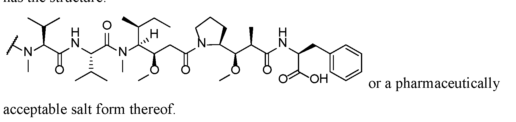
In one example, the targeted agent is (S)-N-((3R,4S,5S)-1-((S)-2-((1R,2R)-3- (((1S,2R)-1-hydroxy-1-phenylpropan-2-yl)amino)-1-methoxy-2-methyl-3- oxopropyl)pyrrolidin-1-yl)-3-methoxy-5-methyl-1-oxoheptan-4-yl)-N,3-dimethyl-2- ((S)-3-methyl-2-(methylamino)butanamido)butanamide and is attached at the N- terminal amino group to the linker.
In one example, the targeted agent is (S)-2-((2R,3R)-3-((S)-1-((3R,4S,5S)-4- ((S)-N,3-dimethyl-2-((S)-3-methyl-2-(methylamino)butanamido)butanamido)-3- methoxy-5-methylheptanoyl)pyrrolidin-2-yl)-3-methoxy-2-methylpropanamido)-3- phenylpropanoic acid and is attached at the N-terminal amino group to the linker.
In some embodiments, T is an auristatin compound that can form a bond with the Linker unit L via a nitrogen atom in the primary or secondary amino group of the auristatin compound, or via an oxygen atom of a hydroxyl or carboxyl group of the auristatin compound.
In embodiments, T is of the Formula (Ib) or a pharmaceutically acceptable salt thereof:
 (Ib), wherein:
R2 is H or C1-8 alkyl;
R3 is H, C1-8 alkyl, C3-8 carbocycle, X4—C3-8 carbocycle, C6-10 aryl, X4—C6-10 aryl, C3-8 heterocycle, or X4—C3-8 heterocycle;
R4 is H, C1-8 alkyl, C3-8 carbocycle, X4—C3-8 carbocycle, C6-10 aryl, X4—C6-10 aryl, C3-8 heterocycle, or X4—C3-8 heterocycle;
R5 is H or methyl; or
R4 and R5 together with the carbon atom to which they are attached form a carbocyclic ring having the formula—(CRaRb)n— wherein each of Ra and Rb independently is H, C1-8 alkyl or C3-8 carbocycle;
R6 is H or C1-8 alkyl;
R7 is H, C1-8 alkyl, C3-8 carbocycle, X4—C3-8 carbocycle, C6-10 aryl, X4— C6-10 aryl, C3-8 heterocycle, or X4—C3-8 heterocycle;
each R8 independently is H, OH, C1-8 alkyl, C3-8 carbocycle or O—(C1-8 alkyl); each X4 independently is C1-10 alkylene or C3-10 cycloalkylene;
R9 is H or C1-8 alkyl;
R18 is—C(R8)2—C(R8)2—C6-10 aryl,—C(R8)2—C(R8)2—(C3-8 heterocycle), —C(R8)2—C(R8)2—(C3-8 carbocycle), or selected from

Z1 is O, S, or NR34;
R31 is H, OH, N(R34)2, C1-8 alkyl, C3-8 carbocycle, O—(C1-8 alkyl), C6-10 aryl, X4—C6-10 aryl, X4—(C3-8 carbocycle), C3-8 heterocycle, X4—(C3-8 heterocycle), C1-8 alkylene-NH2, or (CH2)2SCH3; or R31 is an oxygen atom which forms a carbonyl unit (C=O) with the carbon atom to which it is attached and one hydrogen atom on this carbon atom is replaced by one of the bonds in the (C=O) double bond;
R32 is C6-10-aryl or C3-8 heterocycle;
R33 is H, OH, N(R34)2, C1-8 alkyl, C3-8 carbocycle, O—(C1-8 alkyl), C6-10 aryl, C1-8 alkyl-C6-10 aryl, C1-8 alkyl-(C3-8 carbocycle), C3-8 heterocycle, or C1-8 alkyl-(C3-8 heterocycle);
each R34 independently is H or C1-8 alkyl;
R11 is H, OH, N(R34)2, C1-20 alkyl, C6-10 aryl, C3-8 heterocycle,—(R13O)s— R14;—(R13O)s—CH(R15)2 or—[C(R50R51)]b—R52;
R13 is C2-8 alkyl;
R14 is H or C1-8 alkyl;
R15 is H, COOH,—(CH2)o—N(R16)2,—(CH2)o—SO3H, or—(CH2)o—SO3— C1-8 alkyl;
R16 is H, C1-8 alkyl, or—(CH2)o—COOH;
each of R50 and R51 independently is hydrogen, C1-6 alkyl, C6-10 aryl, hydroxylated C6-10 aryl, polyhydroxylated C6-10 aryl, 5 to 12-membered heterocycle, C3-8 cycloalkyl, hydroxylated C3-8 cycloalkyl, polyhydroxylated C3-8 cycloalkyl or a side chain of a natural or unnatural amino acid;
R52 is OH, NHR53, COOH, R82—C(O)(CH2)c—C(H)(R53)—N(H)(R53), R82— C(O)(CH2)d—(O—CH2—CH2)h—N(H)(R53) or R82—(C(O)—CH(X2)—NH)d—R77; each R53 independently is hydrogen, C1-6 alkyl, C6-10 aryl, C3-8 cycloalkyl, COOH, or COO—C1-6 alkyl;
X2 is a side chain of a natural or unnatural amino acid;
R77 is hydrogen or X2 and NR77 form a nitrogen containing cyclic compound; R82 is NH or oxygen;
n is an integer from 2 to 7;
s is an integer from 0 to 1000;
o is an integer from 0 to 6.
b is an integer from 1 to 6;
c is an integer from 0 to 3;
d is an integer from 1 to 3; and
h is an integer from 1 to 12.
For example, T is a compound of any one of Formulae (XIII) to (XVI) or a pharmaceuticall acce table salt thereof:


Additional examples of auristatin compounds suitable for use in the present invention are described in US 2015/0314008, US 2012/0003247, US 2012/0003248, US2013/0095123, US 2013/0066055, US2013/0122024, US 2013/0157960, WO 2002/088172, WO 2004/010957, WO 2005/0181711, WO 2007/008603, WO
2009/117531, WO 2012/059882, WO 2012/135440, WO 2012/143499, and U.S. Patent Nos.6,884,869, 7,098,308, 7,256,257, 7,423,116, 7,498,298, 7,659,241, 7,829,531, 7,851,437, 7,994,135, 7,965,567, each of which is hereby incorporated by reference in its entirety.
In one example, among the Y104D- or Y104E- variant anti-EGFR antibody conjugates provided herein is a conjugate in which the targeted agent (T) is MMAF and T h

pharmaceutically acceptable salt form thereof.
In another example, among the Y104D- or Y104E-variant anti-EGFR antibody conjugates provided herein is a conjugate in which the targeted agent (T) is MMAE and T has the structure:

pharmaceutically acceptable salt form thereof.
(d) Pyrrolobenzodiazepines (PBDs)
A cytotoxic moiety as a targeted agent in the conjugates include
pyrrolobenzodiazepines (PBDs) (or pyrrolo[2,l-c][l, 4]-benzodiazepines), which are sequence-selective DNA alkylating antibiotics with significant antitumor properties. PBDs have the ability to recognize and bond to specific sequences in DNA; one such sequence is is PuGPu (Purine-Guanine-Purine).PBDs also can bond to PuGPy (Purine-Guanine-Pyrimidine) or PyGPu sequences, over PyGPy sequences.
PBDs can be naturally occurring or synthetic. Naturally occurring PBDs include abbeymycin (Hochlowski, et al., J. Antibiotics, 40, 145-148 (1987)), anthramycin (Leimgruber, et al., J. Am. Chem. Soc, 87, 5793-5795 (1965);
Leimgruber, et al., J. Am. Chem. Soc, 87, 5791 -5793 (1965)), chicamycin (Konishi, et al., J. Antibiotics, 37, 200-206 (1984)), DC-81 (Thurston, et al., Chem. Brit, 26, 767-772 (1990); Bose, et al., Tetrahedron, 48, 751 -758 (1992)), mazethramycin (Kunimoto, et al., J. Antibiotics, 33, 665-667 (1980)), neothramycins A and B
(Takeuchi, et al., J. Antibiotics, 29, 93-96 (1976)), porothramycin (Tsunakawa, et al., J. Antibiotics, 41, 1366-1373 (1988)), prothracarcin (Shimizu, et al, J. Antibiotics, 29, 2492- 2503 (1982); Langley and Thurston, J. Org. Chem., 52, 91-97 (1987)), sibanomicin (DC- 102)(Hara, et al., J. Antibiotics, 41, 702-704 (1988); Itoh, et al., J. Antibiotics, 41, 1281 -1284 (1988)), sibiromycin (Leber, et al., J. Am. Chem. Soc, 110, 2992-2993 (1988)), and tomamycin (Arima, et al., J. Antibiotics, 25, 437-444 (1972)). Synthesis of PBDs and generation of synthetic analogs also have been described (see, e.g., U.S. Patent Nos.6,562,806, 6,608,1926,747,144, and 7,049,311, 7,528,126 ).
PBDs are of the general structure:

Dimers of PBDs also are effective antitumor agents. PBD dimers cover six base pairs instead of three base pairs covered by the PBD monomer. Further, the PBDs in the dimer can bond sequences in the complementary strands of DNA (i.e., an interstrand guanine-guanine cross-link), leading to sequence-selective DNA cross- linking. PBD dimer-induced cross-linking prevents strand separation, thereby preventing DNA replication. This results in cell cycle arrest and apoptosis in the G2/M interface. The increased coverage of PBD dimers, compared to PBD monomers, in addition to DNA cross-linking leads to substantially increased efficacy as anticancer agents.
PBD dimers can be homodimers or heterodimers, and are synthesized by joining the two monomer PBD units together through their C8 positions via a flexible linker. Commonly used linkers include propyldioxy (PBD-C8-O-(CH2)3-O- - -C8-O-(CH2)5-O- - linker, such as the length of the linker, can be selected to target the dimer to specific DNA sequences (Rahman et al., (2011) Nucleic Acids Res.39(13): 5800–5812 and Gregson et al., (2004) J Med Chem 47:1161–1174). Exemplary inter-PBD linkers are described in Bose et al., (1992) J Am Chem Soc.114:4939–4941, Bose et al., (1992) J Chem Soc Chem Commun.14:1518–1520, Thurston et al., (1996) J Org Chem.
61:8141–8147, Gregson et al., (2001) J Med Chem.44:737–748, and Gregson et al., J Med Chem 2004;47:1161–1174. Exemplary PBD dimers have been described in the art (see, e.g., U.S. Patent Nos.6,562,806, 6,608,1926,747,144, 7,049,311, 7,528,126, 7,741,319, 8,592,576 ) and include, but are not limited to, compounds designated DSB-120 (US 7,049,311), DRH-165 (US 7,049,311), ELB21 (Rahman et al., (2011) Nucleic Acids Res.39(13): 5800–5812), SG2000/SJG136 (Rahman et al., (2011) Nucleic Acids Res.39(13): 5800–5812; US 7,049,311), SG2057/DRG16 (Rahman et al., (2011) Nucleic Acids Res.39(13): 5800–5812), SG2202 (US 7,741,319; Hartley et al., (2010) Cancer Res.70(17):6849-6858), SG2285 (Hartley et al., (2010) Cancer Res.70(17):6849-6858), SG3132 (US 20130028919).
PBDs and PBD dimers can be conjugated to any of the antibodies provided herein by any method, including, but not limited to thiol, amine and phenol conjugation. Typically, the PBD or PBD dimer is conjugated to the antibody using a cleavable linker, that is stable in in vivo circulation, such that the PBD or PBD dimer is released from the antibody following cleavage of the linker inside the target cell. In some examples, PBD or PBD dimer can be conjugated to inter-chain cysteines. In some examples, the antibody can be modified to replace amino acid(s) to insert or remove an inter-chain cysteine to facilitate directed thiol linkage of the PBD or PBD dimer.
ii. Cell Toxin Moieties
Cell toxins suitable for use in the methods and compositions include small molecules, such as DNA cleaving agents, and proteinaceous cell toxins, including, but are not limited to, bacterial, fungal, plant, insect, snake and spider toxins.
Toxins include bacterial toxins such as diphtheria toxin, and active fragments thereof and hybrid molecules, plant toxins, such as ricin toxin, small molecule toxins such as geldanamycin, maytansinoids, such as DM1, DM3 and DM4, and
-amanitin, cholera toxin, a Shiga-like toxin, LT toxin, C3 toxin, Shiga toxin, pertussis toxin, tetanus toxin, soybean Bowman-Birk protease inhibitor, Pseudomonas exotoxin, alorin, saporin, modeccin, galanin, abrin A chain, modeccin A chain, alpha-sarcin, Aleurites fordii proteins, dianthin proteins, Phytolacca americana proteins, momordica charantia inhibitor, curcin, crotin, gelonin, mitogillin, restrictocin, phenomycin, and enomycin toxins. The toxins can effect their
cytotoxic and cytostatic activity by mechanisms including tubulin binding, DNA binding, or topoisomerase inhibition.
Exemplary cell toxins contemplated for incorporation in the conjugates provided herein are set forth in Table 13.
Table 13. Exemplary Amino Acid Sequences of Toxins

(a) DNA cleaving agents
Examples of DNA cleaving agents suitable for inclusion as the cell toxin in the chimeric ligand-toxin used in practicing the methods include, but are not limited to, anthraquinone-oligopyrrol-carboxamide, benzimidazole, leinamycin; dynemycin A; enediyne; as well as biologically active analogs or derivatives thereof (i.e., those having a substantially equivalent biological activity). Known analogs and derivatives are disclosed, for examples in Islam et al., J. Med. Chem.342954-61, 1991; Skibo et al., J. Med. Chem.37:78-92, 1994; Behroozi et al., Biochemistry 35:1768-74, 1996; Helissey et al., Anticancer Drug Des.11:527-51, 1996; Unno et al., Chem. Pharm. Bull.45:125-33, 1997; Unno et al., Bioorg. Med. Chem., 5:903-19, 1997; Unno et al., Bioorg. Med. Chem., 5: 883-901, 1997; and Xu et al., Biochemistry 37:1890-7, 1998). Other examples include, but are not limited to, endiyne quinone imines (U. S. Patent No.5,622, 958); 2,2r-bis (2-aminoethyl)-4-4’-bithiazole (Lee et al., Biochem. Mol. Biol. Int.40:151-7, 1996); ellipticine-salen•copper conjugates (Routier et al., Bioconjug. Chem., 8: 789-92, 1997).
(b) Antimetabolites
Examples of antimetabolites useful for inclusion as the cell toxin in the chimeric ligand-toxin include, but are not limited to, 5-fluorouracil, methotrexate, melphalan, daunomycin, doxorubicin, nitrogen mustard and mitomycin c.
(c) Proteinaceous cell toxins
Examples of proteinaceous cell toxins useful for incorporation into the chimeric ligand-toxins used in the methods include, but are not limited to, type one and type two ribosome inactivating proteins (RIP). Useful type one plant RIPs include, but are not limited to, dianthin 30, dianthin 32, lychnin, saporins 1-9, pokeweed activated protein (PAP), PAP II, PAP-R, PAP-S, PAP-C, mapalmin, dodecandrin, bryodin-L, bryodin, Colicin 1 and 2, luffin-A, luffin-B, luffin-S, 19K- protein synthesis inhibitory protein (PSI), 15K-PSI, 9K-PSI, alpha-kirilowin, beta- kirilowin, gelonin, momordin, momordin-II, momordin-Ic, MAP-30, alpha- momorcharin, beta-momorcharin, trichosanthin, TAP-29, trichokirin; barley RIP; flax RIP, tritin, corn RIP, Asparin 1 and 2. Useful type two RIPs include, but are not limited to, volkensin, ricin, nigrin-b, CIP-29, abrin, modeccin, ebulitin-a, ebulitin-β, ebulitin-γ, vircumin, porrectin, as well as the biologically active enzymatic subunits thereof (Stirpe et a/., Bio/Technology 70:405-12, 1992; Pastan et a/., Annu. Rev. Biochem. 61:331-54; Brinkmann and Pastan, Biochim. et Biophys. Acta 1198:27-45, 1994; and Sandvig and Van Deurs, Physiol. Rev. 7(5:949-66, 1996).
(d) Bacterial toxins
Examples of bacterial toxins useful as cell toxins include, but are not limited to, shiga toxin and shiga-like toxins (i.e., toxins that have the same activity or structure), as well as the catalytic subunits and biologically functional fragments thereof. These bacterial toxins also are type two RIPs (Sandvig and Van Deurs, Physiol. Rev. 7(5:949-66, 1996; Armstrong, J. Infect. Dis., 777: 1042-5, 1995; Kim et al, Microbiol. Immunol. 47:805-8, 1997, and Skinner et al. , Microb. Pathog. 24. Will, 1998). Additional examples of useful bacterial toxins include, but are not limited to, Pseudomonas exotoxin and Diphtheria toxin (Pastan et al., Annu. Rev. Biochem. (57:331-54; and Brinkmann and Pastan, Biochim. et Biophys. Acta 1198:17-45, 1994). Truncated forms and mutants of the toxin enzymatic subunits also can be used as a cell toxin moiety (Pastan et al., Annu. Rev. Biochem. (57:331-54; Brinkmann and Pastan, Biochim. et Biophys. Acta 1198:11-45, 1994; Mesri et a/., J Biol. Chem. 265:4853-62, 1993; Skinner et al, Microb. Pathog. 24: 111-11, 1998; and U.S. Patent No. 5,082,927). Other targeted agents include, but are not limited to the more than 34 described Colicin family of RNase toxins which include colicins A, B, D, El -9,
cloacin DF13 and the fungal RNase, a-sarcin (Ogawa et al. Science 283: 2097-100, 1999; Smarda et al, Folia Microbiol (Praha) 43:563-82, 1998; Wool et al, Trends Biochem. Sci., 17: 266-69, 1992).
(e) Porphyrins and other light activated toxins Porphyrins are well-known light activatable toxins that can be readily cross- linked to proteins (see, e.g., U.S. Patent Nos. 5,257,970; 5,252,720; 5,238,940;
5, 192,788; 5, 171,749; 5,149,708; 5,202,317; 5,217,966; 5,053,423; 5, 109,016;
5,087,636; 5,028,594; 5,093,349; 4,968,715; 4,920, 143 and International PCT Publication No. WO 93/02192).
iii. Nucleic acids for targeted delivery
The conjugates provided herein also can be used to deliver nucleic acids to targeted cells. The nucleic acids include DNA intended to modify the genome of a cell and thereby effect genetic therapy, and DNA and RNA for use as antisense, modulatory or inhibitory agents. The nucleic acid also can be modified, including locked nucleic acids (LNA). The nucleic acids include antisense RNA, DNA, ribozymes and other oligonucleotides that are intended to be used as antisense or inhibitory agents. The nucleic acids can also include RNA trafficking signals, such as viral packaging sequences (see, e.g., Sullenger et al. (1994) Science 2(52: 1566-1569). The nucleic acids can also include small RNAs that function as modulatory or inhibitory agents, for example, such as small interfering RNA (siRNA), small hairpin RNA (shRNA), microRNA (miRNA), tiny non-coding RNA (tncRNA), small modulatory RNA (smRNA), and nucleic acids or vectors encoding those RNAs. The nucleic acids also include DNA molecules that encode intact genes or that encode proteins intended to be used in gene therapy.
DNA (or RNA) that can be delivered to a cell to effect genetic therapy includes DNA that encodes tumor-specific cytotoxic molecules, such as tumor necrosis factor, viral antigens and other proteins to render a cell susceptible to anticancer agents, and DNA encoding genes, such as the defective gene (CFTR) associated with cystic fibrosis (see, e.g., International PCT Publication No. WO 93/03709; and Riordan et al. (1989) Science 245: 1066-1073), to replace defective genes. Nucleic acids can also include nucleic acids delivered to a cell to effect targeted editing or modification of the genomic DNA of the cell.
Nucleic acids and oligonucleotides for use as described herein can be synthesized by any method known to those of skill in the art (see, e.g., WO 93/01286 and U.S. Patent Nos.5,218,088; 5,175,269; and 5,109,124). Identification of oligonucleotides and ribozymes for use as antisense agents is well within the skill in the art. Selection of DNA encoding genes for targeted delivery for genetic therapy also is well within the level of skill of those in the art. For example, the desirable properties, lengths and other characteristics of such oligonucleotides are well-known. Antisense oligonucleotides are designed to resist degradation by endogenous nucleolytic enzymes and include, but are not limited to: phosphorothioate,
methylphosphonate, sulfone, sulfate, ketyl, phosphorodithioate, phosphoramidate, phosphate esters, and other such linkages (see, e.g., Agrawal et al. (1987) Tetrahedron Lett.28:3539-3542; Miller et al. (1971) J. Am. Chem. Soc.93:6657-6665; Stec et al. (1985) Tetrahedron Lett.26:2191-2194; Moody et al. (1989) Nucl. Acids Res.
17:4769-4782; Letsinger et al. (1984) Tetrahedron 40:137-143; Eckstein (1985) Annu. Rev. Biochem.54:367-402; Eckstein (1989) Trends Biochem. Sci.14:97-100; Stein (1989) In: Oligodeoxynucleotides. Antisense Inhibitors of Gene Expression, Cohen, ed, Macmillan Press, London, pp.97-117; Jager et al. (1988) Biochemistry 27:7237- 7246).
(a) Antisense nucleotides, including: antisense
oligonucleotides; triplex molecules; dumbbell oligonucleotides; DNA;
extracellular protein binding oligonucleotides; and small nucleotide molecules Antisense nucleotides are oligonucleotides that specifically bind to mRNA that has complementary sequences, thereby preventing translation of the mRNA (see, e.g., U.S. Patent No.5,168,053 to Altman et al. U.S. Patent No.5,190,931 to Inouye, U.S. Patent No.5,135,917 to Burch; U.S. Patent No.5,087,617 to Smith and Clusel et al. (1993) Nucl. Acids Res.21:3405-3411, which describes dumbbell antisense oligonucleotides). Triplex molecules refer to single DNA strands that target duplex DNA and thereby prevent transcription (see, e.g., U.S. Patent No.5,176,996, which describes methods for making synthetic oligonucleotides that bind to target sites on duplex DNA).
(b) Ribozymes
Ribozymes are RNA constructs that specifically cleave messenger RNA. There are at least five classes of ribozymes that are known that are involved in the cleavage and/or ligation of RNA chains. Ribozymes can be targeted to any RNA transcript and can catalytically cleave such transcript (see, e.g., U.S. Patent Nos. 5,272,262; 5,144,019, 5,168,053; 5,180,818; 5,116,742 and 5,093,246, which describe ribozymes and methods for production thereof). Any such ribosome canbe linked to a conditionally active anti-EGFR antibody for delivery to EGFR bearing cells under acidic conditions.
The ribozymes can be delivered to the targeted cells as DNA encoding the ribozyme linked to a eukaryotic promoter, such as a eukaryotic viral promoter, generally a late promoter, such that upon introduction into the nucleus, the ribozyme will be directly transcribed. In such instances, the construct will also include a nuclear translocation sequence, generally as part of the targeting agent or as part of a linker in order to render it suitable for delivering linked nucleic acids to the nucleus.
(c) Nucleic acids encoding therapeutic products for targeted delivery
Among the DNA that encodes therapeutic products contemplated for use is DNA encoding correct copies of anticancer agents, such as tumor necrosis factors, and cytotoxic agents, such as shiga A1 toxin or saporin to EGFR bearing tumor cells. The conjugate can include a nuclear translocation sequence (NTS). If the conjugate is designed such that the targeting agent and linked DNA is cleaved in the cytoplasm, then the NTS can be included in a portion of the linker that remains bound to the DNA, so that, upon internalization, the conjugate will be trafficked to the nucleus. The nuclear translocation sequence (NTS) canbe a heterologous sequence or a canbe derived from the selected chemokine receptor targeting agent. A typical consensus NTS sequence contains an amino-terminal proline or glycine followed by at least three basic residues in an array of seven to nine amino acids (see, e.g., Dang et al. (1989) J. Biol. Chem.264:18019-18023).
(d) Coupling of nucleic acids to proteins To effect chemical conjugation herein, the targeting agent is linked to the nucleic acid either directly or via one or more linkers. Methods for conjugating
nucleic acids, at the 5' ends, 3' ends and elsewhere, to the amino and carboxyl termini and other sites in proteins are known to those of skill in the art (for a review see e.g., Goodchild, (1993) In: Perspectives in Bioconjugate Chemistry, Mears, Ed., American Chemical Society, Washington, D.C. pp. 77-99). For example, proteins have been linked to nucleic acids using ultraviolet irradiation (Sperling et al. (1978) Nucleic Acids Res. 5:2755-2773; Fiser et al. (1975) FEBS Lett. 52:281-283), bifunctional chemicals (Baumert et al. (1978) Eur. J. Biochem. 59:353-359; and Oste et al. (1979) Mol. Gen. Genet. 7(55:81-86), and photochemical cross-linking (Vanin et al. (1981) FEBS Lett. 124:89-92; Rinke et al. (1980) J.Mol.Biol. 737:301-304; Millon et al. (1980) Eur. J. Biochem. 770:485-492).
In particular, the reagents (N-acetyl-N'-(p-glyoxylylbenzolyl)cystamine and 2- iminothiolane have been used to couple DNAto proteins, such as (X2-macroglobulin (a2M) via mixed disulfide formation (see, Cheng et al. (1983) Nucleic Acids Res. 77:659-669). N-acetyl-N'-(p-glyoxylylbenzolyl)cystamine reacts specifically with non-paired guanine residues and, upon reduction, generates a free sulfhydryl group. 2-Iminothiolane reacts with proteins to generate sulfhydryl groups that are then conjugated to the derivatized DNA by an intermolecular disulfide interchange reaction. Any linkage can be used provided that, upon internalization of the conjugate, the targeted nucleic acid is active. Thus, it is expected that cleavage of the linkage can be necessary, although it is contemplated that for some reagents, such as DNA encoding ribozymes linked to promoters or DNA encoding therapeutic agents for delivery to the nucleus, such cleavage may not be necessary.
Thiol linkages readily can be formed using heterobifunctional reagents.
Amines have also been attached to the terminal 5' phosphate of unprotected oligonucleotides or nucleic acids in aqueous solutions by reacting the nucleic acid with a water-soluble carbodiimide, such as l-ethyl-3'[3- dimethylaminopropyljcarbodiimide (EDC) or N-ethyl-N'(3- dimethylaminopropylcarbodiimidehydrochloride (EDCI), in imidazole buffer at pH 6 to produce the 5' phosphorimidazolide. Contacting the 5' phosphorimidazolide with amine-containing molecules and ethyl enediamine, results in stable phosphoramidates (see, e.g., Chu et al. (1983) Nucleic Acids Res. 77:6513-6529; and WO 88/05077). In particular, a solution of DNA is saturated with EDC, at pH 6 and incubated with
agitation at 4 °C overnight. The resulting solution is then buffered to pH 8.5 by adding, for example about 3 volumes of 100 mM citrate buffer, and adding about 5 μg - about 20 of a chemokine receptor targeting agent, and agitating the resulting mixture at 4 °C for about 48 hours. The unreacted protein can be removed from the mixture by column chromatography using, for example, SEPHADEX G75
(Pharmacia) using 0.1 M ammonium carbonate solution, pH 7.0 as an eluting buffer. The isolated conjugate can be lyophilized and stored until used.
U.S. Patent No. 5,237,016 provides methods for preparing nucleotides that are bromacetylated at their 5' termini and reacting the resulting oligonucleotides with thiol groups. Oligonucleotides derivatized at their 5'-termini bromoacetyl groups can be prepared by reacting 5'-aminohexyl-phosphoramidate oligonucleotides with bromoacetic acid-N-hydroxysuccinimide ester as described in U.S. Patent No.
5,237,016. U.S. Patent No. 5,237,016 also describes methods for preparing thiol- derivatized nucleotides, which can then be reacted with thiol groups on the selected growth factor. Briefly, thiol-derivatized nucleotides are prepared using a 5'- phosphorylated nucleotide in two steps: (1) reaction of the phosphate group with imidazole in the presence of a diimide and displacement of the imidazole leaving group with cystamine in one reaction step; and (2) reduction of the disulfide bond of the cystamine linker with dithiothreitol (see, also, Chu et al. (1988) Nucl. Acids Res. 7(5:3671-3691, which describes a similar procedure). The 5'-phosphorylated starting oligonucleotides can be prepared by methods known to those of skill in the art (see, e.g., Maniatis et al. (1982) Molecular Cloning: A Laboratory Manual, Cold Spring Harbor Laboratory, New York, p. 122).
The antisense oligomer or nucleic acid, such as a methylphosphonate oligonucleotide (MP-oligomer), can be derivatized by reaction with SPDP or SMPB. The resulting MP-oligomer can be purified by HPLC and then coupled to the chemokine receptor targeting agent. The MP-oligomer (about 0.1 μΜ) is dissolved in about 40-50 μl of 1 : 1 acetonitrile/water to which phosphate buffer (pH 7.5, final concentration 0.1 M) and a 1 mg MP-oligomer in about 1 mL phosphate buffered saline is added. The reaction is allowed to proceed for about 5-10 hours at room temperature and is then quenched with about 15 μl, 0.1 iodoacetamide. The
conjugates can be purified on heparin sepharose Hi Trap columns (1 mL, Pharmacia) and eluted with a linear or step gradient. The conjugate elutes in 0.6 M NaCl.
iv. Labeling agents (detectable moieties)
For example, the targeted agent is a labeling agent. Labelling agents, which include imaging agents, for example, include but are not limited to, a radionuclide, a fluorescent agent (for example an amine derivatized fluorescent probe such as 5-dimethylaminonaphthalene-1-(N-(2-aminoethyl))sulfonamide-dansyl
ethylenediamine, Oregon Green® 488 cadaverine (catalogue number O-10465, Molecular Probes), dansyl cadaverine, N-(2-aminoethyl)-4-amino-3,6-disulfo-1,8- naphthalimide, dipotassium salt (lucifer yellow ethylenediamine), or rhodamine B ethylenediamine (catalogue number L2424, Molecular Probes), or a thiol derivatized fluorescent probe for example BODIPY® FL L-cystine (catalogue number B-20340, Molecular Probes). Biotin and other such compounds are included.
In some examples, the targeted agent can be any agent to be delivered to the environment where the anti-EGFR antibody is conditionally active. Targeted agent can include a label or a detectable moiety, such as a detectable marker (e.g., a fluorescent molecule, chemiluminescent molecule, a bioluminescent molecule, a contrast agent (e.g., a metal), a radionuclide, a chromophore, a detectable peptide, or an enzyme that catalyzes the formation of a detectable product) that can be attached or linked directly or indirectly to a molecule (e.g., an antibody or antigen-binding fragment thereof, such as an anti-EGFR antibody or antigen-binding fragment thereof provided herein) or associated therewith and can be detected in vivo and/or in vitro. The detection method can be any method known in the art, including known in vivo and/or in vitro methods of detection (e.g., imaging by visual inspection, magnetic resonance (MR) spectroscopy, ultrasound signal, X-ray, gamma ray spectroscopy (e.g., positron emission tomography (PET) scanning, single-photon emission computed tomography (SPECT)), fluorescence spectroscopy or absorption). Indirect detection refers to measurement of a physical phenomenon, such as energy or particle emission or absorption, of an atom, molecule or composition that binds directly or indirectly to the detectable moiety (e.g., detection of a labeled secondary antibody or antigen-binding fragment thereof that binds to a primary antibody (e.g., an anti-EGFR antibody or antigen-binding fragment thereof provided herein)).
c. Linker (L)
The linker (L) is a trifunctional or multifunctional moiety that attaches one or more targeted agent(s) to the antibody to form a conjugate between the antibody and the targeted agent(s). For example, the linker is attached to the disulfide bridge- forming unit (A), the targeted agent (T) and the extended moiety (E), such as, for example, in the following configuration:

With respect to attachment to the antibody, the disulfide bridge-forming unit (A) contains two or more reactive functional groups that bis-alkylate two cysteine side chain thiols in the antibody to form a disulfide bridge, which results in a homogenous and stable product.
The extended moiety (E) is of a size that fits the hinge region of the antibody, thereby inhibiting or limiting interaction with Fc receptors, to thereby inhibit or reduce activities mediated through Fc/FcR interactions. For example, the exemplary linker (designated PT2) includes an extended moiety (E) that contains a PEG having the structure -(CH2CH2O)24-CH3. As a result, the conjugates provided herein exhibit reduced binding to Fc receptors and reduced side-effects.
The linkers also are such that they do not interfere with or reduce to any significant extent internalization of the conjugates via binding to antibody receptors, such as EGFR receptors. For example, the extended moiety (E) is attached to the trifunctional linker L in a branched configuration relative to the linkage between the antibody and the targeted agent. The length, size and/or identity of the extended moiety (E), therefore, can be modified without altering the spacing between the targeted agent (T) and the disulfide bridge-forming unit (A) that bis-alkylates the thiol side chains of two cysteine residues in the conditionally active antibody. Placing the extended moiety at a branched position removes constraints on the relative separation distance between the antibody and the targeted agent.
The overall distance between the antibody and the targeted agent can influence conjugate properties such as, for example, thermostability, antigen binding and FcR binding, as well as self-association and aggregation between the drug payload molecules attached to the antibody (see, e.g., Acchione et al. (2012) mAbs 4:3:362-
372). The extended moiety, which inhibits binding, does not alter the length of the antibody-drug linkage. Thus, in the conjugates provided herein, the extended moiety can be modified without changing the length of the linker between the antibody and the targeted agent. The linker L has the general formula
 , wherein X is a spacer moiety attached to the targeted agent and Y is a trifunctional core moiety attached to the spacer moiety (X), extended moiety (E) and the disulfide bridge unit (A).
Linkers of interest herein include those described in International PCT
Publication No. WO 2014/064423, WO 2014/064424 and WO 2005/007197, and particularly in International PCT application No. PCT/GB2015/052953, published as WO 2016/063006.
Linkers are selected or designed to be sufficiently stable in the extracellular environment so that the antibody conjugate is stable and remains intact, i.e., the antibody remains linked to the targeted agent, before transport or delivery into the target cell. Thus, the linkers are stable outside the target cell and can be cleaved or enable dissociation of the antibody and targeted agent at some efficacious rate once inside the cell. Contemplated linkers will (i) not interfere or not interfere significantly with the specific binding properties of the antibody; (ii) permit intracellular delivery of the conjugate or targeted agent; (iii) remain stable and intact, i.e., not cleaved, until the conjugate has been delivered or transported to its targeted site; and (iv) not interfere with the cytotoxic, cell-killing effect or a cytostatic effect of the targeted agent. Stability of the conjugate can be measured by standard analytical techniques such as mass spectrometry and/or HPLC.
Any of the conjugates provided herein can be prepared using a linker that has by having a reactive functionality for covalent binding to the targeted agent and to the anti-EGFR antibody. Two cysteine thiol groups, e.g., cysteine side chains of the anti- EGFR antibody, form a bond with a functional group of a disulfide bridge-forming unit, linker reagent, targeted agent, targeted agent-linker reagent or targeted agent- linker reagent-disulfide bridge-forming unit, particularly the disulfide bride forming unit (A), to form a bis-alkylated cysteine. Thus, the linker is attached to two cysteine residues that otherwise form a disulfide bond in the antibody. Among the cysteine
residues to which the linker is attached include cysteine residues that form an interchain disulfide bond and cysteine residues that form an intrachain disulfide bond. For example, the interchain disulfide bonds for IgG1 are formed at: 1) Light chain (LC) position C214 (C214 by Kabat numbering, C214 by EU numbering) and Heavy chain (HC) position C222 (C233 by Kabat numbering, C220 by EU numbering) (2 pairs of cysteines per antibody); 2) HC C228 and HC C228 (C239 by Kabat numbering, C226 by EU numbering) (2 cysteines per antibody); and 3) HC C231 and HC C231 (C242 by Kabat numbering, C229 by EU numbering) (2 cysteines per antibody). The linker is attached by bis-alkylation to disulfide bonds that have been reduced to free cysteines. Unlike conjugation using mono-reactive maleimide linkers, in bis-alkylated cysteines the native disulfide bonds are re-bridged as part of the conjugation reaction. This increases the stability of the conjugated antibody, and reduces the number of conjugation sites, since a disulfide bond forms one conjugation site, not two. This also allows homogenous drug-to-antibody-ratio (DAR) due to the stable re-bridging of the disulfide bonds. For example, attachment of a linker to the four (4) accessible interchain disulfide bonds in an IgG1 antibody creates defined homogeneous conjugates with four, rather than eight, conjugation sites. Antibody conjugates formed by bis-alkylation are more stable in the presence of human serum albumin than antibody conjugates formed by maleimide chemistry (Badescu et al. (2014) Bioconjug. Chem.25(6):1124-36; and Kline et al. (2015) Pharm Res.32(11): 3480-3493). To increase the DAR of higher than 4, additional cysteines can be engineered into the mAb, or linkers can contain further branched moieties connected to additional targeting agents.
Such chemical cross-linking reagents, which are useful for attaching two or more functional or biologically active moieties, such as peptides, nucleic acids, drugs, toxins, antibodies, haptens, and reporter groups, are known, and methods have been described for their use in generating conjugates (Hermanson, G. T. (1996)
Bioconjugate Techniques; Academic Press: New York, p234-242). In particular examples of the conjugates provided herein, the linker L is or contains 4-(3-tosyl-2- (tosylmethyl)propanoyl)benzamide-valine-citruline-p-aminobenzyloxycarbonyl.
i. Core moiety (Y)
The core moiety (Y) is a branching element by which the extended moiety is attached to the antibody conjugate. For example, the core moiety (Y) attaches the extended moiety (E) to one or more bridge forming unit(s) (A) and one or more spacer moiety or moieties (X). In some examples, the core moiety (Y) is a
trifunctional amino acid or amino acid residue that forms a bond with the bridge forming unit (A), extended moiety (E) and spacer moiety (X) via the alpha-carbon nitrogen atom, alpha-carbon carbonyl and side chain, in any configuration. In some
examples, the trivalent moiety has the formula
 or
or
In some examples, the trivalent moiety has the formula


methyl, isopropyl, isobutyl, sec-butyl, benzyl, p-hydroxybenzyl, -CH2OH, -CH(OH)CH3, -CH2CH2SCH3, -CH2CONH2, -CH2COOH, -CH2CH2CONH2, -CH2CH2COOH, -(CH2)3NHC(=NH)NH2, -(CH2)3NH2, -(CH2)3NHCOCH3,
-(CH2)3NHCHO, -(CH2)4NHC(=NH)NH2, -(CH2)4NH2, -(CH2)4NHCOCH3,
-(CH2)4NHCHO, -(CH2)3NHCONH2, -(CH2)4NHCONH2, -CH2CH2CH(OH)CH2NH2, 2-pyridylmethyl-, 3-pyridylmethyl-, 4-pyridylmethyl-, phenyl, cyclohexyl, or any one of the followin structures:

In some examples, the core moiety has a formula:

The core moiety can attach to the spacer moiety (X), extended moiety (E) and bridge unit (A) by any of the open valencies indicated. Thus, for example, the core moiety can attach to the spacer moiety (X), extended moiety (E) and bridge forming unit A in an of the followin confi urations:
, ,
The core moiety can include multiple residues described herein. For example, the core moiety can be a peptide composed of any of the amino acids described above. For example, the core moiety is a dipeptide, tripeptide, tetrapeptide, pentapeptide, hexapeptide, heptapeptide, octapeptide, nonapeptide or decapeptide composed of any of the amino acids described above. For example, the core moiety is:

The core moiety can be a dendritic type linker for covalent attachment of more than one targeted agent through a branching, multifunctional linker moiety to an antibody (Sun et al. (2002) Bioorganic & Medicinal Chemistry Letters 12:2213-2215; Sun et al. (2003) Bioorganic & Medicinal Chemistry 11:1761-1768; King et al.
(2002) Tetrahedron Letters 43:1987-1990). Dendritic linkers can increase the molar ratio of targeted agent to antibody, i.e., loading, which can increase the potency of the ADC. Thus, a multitude of targeted agents can be attached through a dendritic linker to only one reactive cysteine thiol group. Exemplary dendritic linker reagents are described in U.S. Patent Publication No.2005/0276812.
ii. Spacer Moiety (X)
A spacer moiety is any chemical moiety that is links the targeted agent (T) to the conjugate such that the targeted agent (T) and the conjugate are chemically coupled (i.e., covalently bonded) to teach other.
In some embodiments, the spacer moiety can contain a degradable group, i.e. it can contain a group which breaks under physiological conditions, separating the targeted agent (T) from the antibody to which it is, or will be, bonded. Alternatively, the spacer moiety is not cleavable under physiological conditions. Where a spacer moiety breaks under physiological conditions, it can be cleavable under intracellular conditions. Where the target is intracellular, the linker can be substantially insensitive to or resistant to cleavage in extracellular conditions (i.e. so that delivery to the intracellular target of a sufficient dose of the therapeutic agent is not prohibited). Where a spacer moiety is biocleavable and/or biodegradable under intracellular conditions, cleavage of the spacer moiety releases the targeted agent from the conjugate in the intracellular environment.
In some embodiments, the spacer moiety contains a biodegradable bond such as an ester or amide bond. In some embodiments, the spacer moiety contains a disulfide linker, acid labile linker, photolabile linker, peptidase labile linker and/or an esterase linker. If the spacer moiety contains a cleavable moiety, the spacer moiety is cleaved under conditions under which the activity and/or stability of the targeted agent is not affected.
In other embodiments, the spacer moiety is pH-sensitive, i.e., sensitive to hydrolysis at certain pH values. Typically, the pH-sensitive spacer moiety is hydrolysable under acidic conditions. For example, an acid-labile spacer moiety that is hydrolysable in the lysosome or endosome is a hydrazone, semicarbazone, thiosemicarbazone, cis-aconitic amide, orthoester, acetal or ketal. Such spacer moieties are relatively stable under neutral pH conditions, such as those in the blood, but are unstable at below pH 5.5 or 5.0, the approximate pH of the lysosome.
In other embodiments, the spacer moiety is photo-labile and is useful at the body surface and in many body cavities that are accessible to light. Furthermore, the spacer moiety can be biocleavable by infrared light which can penetrate tissue.
Accordingly, It is useful for both applications on the body surface and in the tissue.
In some embodiments, the spacer moiety is biocleavable by a cleaving agent that is present in the intracellular environment (e.g., within a lysosome or endosome or caveolea). The linker can be, for example, a peptidyl linker that is cleaved by an intracellular peptidase or protease enzyme, including, but not limited to, a lysosomal or endosomal protease.
In some embodiments, the spacer moiety is cleaved by esterases. Only certain esters can be cleaved by esterases present inside or outside cells. Esters are formed by the condensation of a carboxylic acid and an alcohol. Simple esters are esters produced with simple alcohols, such as aliphatic alcohols, and small cyclic and small aromatic alcohols.
In some examples, the spacer moiety contains a peptide optionally linked to a self-immolative group or a non-self-immolative group.
(a) Peptides
The spacer moiety (X) can contain a peptide containing one or more amino acid units, such as a dipeptide, tripeptide, tetrapeptide, pentapeptide or higher unit peptide, having the formula–(A)a-, where each A is independently selected from among natural and non-natural amino acids. For example, the spacer moiety (X) can have the formula–A1-A2-, where A1 and A2 are independently selected from among natural and non-natural amino acids. It is understood that each of the amino acids recited herein can be in the (S) or (R) configuration.
In another embodiment, each amino acid (A) unit independently is selected from the following amino acids: alanine, arginine, aspartic acid, asparagine, histidine, glycine, glutamic acid, glutamine, phenylalanine, lysine, leucine, serine, tyrosine, threonine, isol -alanine, aminoalkanoic acid, aminoalkyrioic acid, aminoalkanedioic acid, aminobenzoic acid, amino-heterocyclo-alkanoic acid, heterocyclo-carboxylic acid, citrulline (Cit), statine, diaminoalkanoic acid, and derivatives thereof.
In another embodiment, each amino acid (A) unit independently is selected from the following L-(natural) amino acids: alanine, arginine, aspartic acid, asparagine, histidine, glycine, glutamic acid, glutamine, phenylalanine, lysine, leucine, serine, tyrosine, threonine, isoleucine, tryptophan and valine.
The spacer moiety can contain a peptide that is susceptible to enzymatic degradation, for example by cleavage by a protease (e.g., a lysosomal or endosomal protease) or peptidase. The peptide can be enzymatically cleaved by one or more enzymes, including a cancer or tumor-associated protease, to liberate the targeted agent. For example, the peptide has the formula–A1-A2- and A1 and A2, can be independently selected from among alanine, valine, leucine, isoleucine, methionine, phenylalanine, tryptophan, proline and citrulline. For example, the peptide can contain amino acids -A1-A2-, and A1 can be an amino acid that is alanine, valine, leucine, isoleucine, methionine, phenylalanine, tryptophan or proline and A2 can be lysine, lysine protected with acetyl or formyl, arginine, arginine protected with tosyl or nitro groups, histidine, ornithine, ornithine protected with acetyl or formyl or citrulline. In particular examples, A1-A2 is phenylalanine-lysine, valine-citrulline or valine-lysine. In some examples, A1-A2 is valine-citrulline and is cleaved by cathepsin B.
The spacer moiety can be a di-peptide, tri-peptide or tetrapeptide selected from among Phe-Leu, Gly-Phe-Leu-Gly, Val-Ala, Val-Cit and Phe-Lys.
In some embodiments, the spacer moiety contains a peptide represented by the following formulas:
wherein R20 and R21 are as follows:


When R20, R21, R22 or R23 is a moiety other than hydrogen, the carbon atom to which R20, R21, R22 or R23 is attached is chiral. Each carbon atom to which R20, R21, R22 or R23 is attached independently is in the (S) or (R) configuration.
Optionally, all or part of the spacer unit remains bound to the targeted agent after cleavage, for example enzymatic cleavage of an amino acid unit AA, from the antibody drug conjugate. Examples of part of the spacer unit that remains attached to the targeted agent include but are not limited to glycine-glycine and glycine. In some examples, the spacer unit (X) is -Gly-. In other examples, Spacer unit (-X-) is -Gly- Gly-. In some embodiments, the spacer group is not biocleavable and the targeted
agent is released by antibody degradation. See, for example, U.S. Patent No.
7,498,298, which is incorporated by reference herein in its entirety.
Peptides for use in the linkers can be prepared by solid phase or liquid phase synthesis methods (E. Schroder and K. Lubke, The Peptides, volume 1, pp.76-136 (1965) Academic Press) that are well-known in the field of peptide chemistry, including t-BOC chemistry (Geiser et al. "Automation of solid-phase peptide synthesis" in Macromolecular Sequencing and Synthesis, Alan R. Liss, Inc., 1988, pp. 199-218) and Fmoc/HBTU chemistry (Fields, G. and Noble, R. (1990) "Solid phase peptide synthesis utilizing 9-fluorenylmethoxycarbonyl amino acids", Int. J. Peptide Protein Res.35:161-214), on an automated synthesizer such as the Rainin Symphony Peptide Synthesizer (Protein Technologies, Inc.), or Model 433 (Applied Biosystems). Peptide-based linkers offer advantages over linkers that are hydrolytically or reductively labile, since proteolysis is enzymatic, and the enzymes can be selected for preferential expression within tumor cells. The cathepsin B-cleavable peptide linker, valine-citrulline (Val-Cit), and modifications thereof such as maleimidocaproyl- valine-citrulline (mc-vc), phenylalanine-lysine, Ala-Leu-Ala-Ala (SEQ ID NO: 351), other tri/tetrapeptides are exemplary peptide linkers that have been employed in ADCs (Dosio et al., (2010) Toxins 3:848-883; Doronina et al., (2006) Bioconjug Chem.17:114-124; Doronina et al., (2003) Nat Biotechnol.21:778-784; Sanderson et al., (2005) Clin Cancer Res 11:843-852; Ducry and Stump (2010) Bioconjug Chem. 21:5-13). Exemplary non-cleavable peptide linkers include N-methyl-valine-citrulline. Other peptide linkers are described in U.S. Publication No.2011/0020343.
Peptide linkers include those that can be incorporated in fusion proteins and expressed in a host cell, such as E. coli. Such linkers include: enzyme substrates, such as cathepsin B substrate, cathepsin D substrate, trypsin substrate, thrombin substrate, subtilisin substrate, Factor Xa substrate, and enterokinase substrate; linkers that increase solubility, flexibility, and/or intracellular cleavability include linkers, such as (GlymSer)n or (SermGly)n, where m is 1 to 6, such as 1 to 4, such as 2 to 4, and n is 1 to 6, such as 1 to 4, such as 2 to 4 (see, e.g., International PCT Publication No. WO 96/06641, which provides exemplary linkers for use in conjugates). In some embodiments, several linkers can be included in order to take advantage of desired properties of each linker.
Spacers can undergo cyclization upon amide bond hydrolysis, such as substituted and unsubstituted 4-aminobutyric acid amides (Rodrigues et ah (1995) Chemistry Biology 2:223), appropriately substituted bicyclo[2.2.1] and bicyclo[2.2.2] ring systems (Storm et. al (1972). Amer. Chem. Soc.94:5815) and 2- aminophenylpropionic acid amides (Amsberry et. al (1990) Org. Chem.55:5867).
(b) Groups susceptible to hydrolysis
For example, the spacer moiety (X) contains a group that is sensitive to cleavage by hydrolysis. The group can be cleaved under acidic and/or basic conditions. Hydrolytic/acidic conditions may, for example, be those found in endosomes or lysosomes. Examples of groups susceptible to hydrolysis under acidic conditions include hydrazones, semicarbazones, thiosemicarbazones, cis-acotinic amides, orthoesters and ketals. Examples of groups susceptible to hydrolytic
such as,

(c) Groups susceptible to reducing conditions
The spacer moiety can be susceptible to degradation under reducing conditions. For example, it can contain a disulfide group that is cleavable on exposure to biological reducing agents, such as thiols. Examples of disulfide groups include:

(d) Self-immolative group
The spacer moiety (X) optionally includes a self-immolative group attached to the targeted agent. For example, the spacer moiety (X) contains a self-immolative group that will spontaneously separate from the targeted agent moiety if its bond to the peptide is cleaved. For example, the self-immolative group can be a p- aminobenzyl alcohol (PAB). The self-immolative group also can be an aromatic compound that is electronically similar to PAB, such as 2-aminoimidazol-5-methanol derivatives (Hay et al. (1999) Bioorg. Med. Chem. Lett.9:2237) and ortho or para- aminobenzylacetals. Self-immolative groups can be used that undergo cyclization upon amide bond hydrolysis, such as substituted and unsubstituted 4-aminobutyric acid amides (Rodrigues et ah (1995) Chemistry Biology 2:223), appropriately substituted bicyclo[2.2.1] and bicyclo[2.2.2] ring systems (Storm et. al (1972). Amer. Chem. Soc.94:5815) and 2-aminophenylpropionic acid amides (Amsberry et. al (1990) Org. Chem.55:5867).
When the spacer moiety contains a peptide, the N- and/or C-terminus of the peptide can be linked to the targeted agent by a self-immolative group. For example, a spacer moiety (X) can have the formula selected from among:
 , , ,
, , ,

AA is an amino acid or amino acid chain described herein;
each Q is independently hydrogen, C1-8 alkyl, -O-(C1-8 alkyl), -halogen, -nitro or–cyano;
and q is an integer from 0 to 4.
For example, AA has the sequence Phe-Lys, Val-Cit, Val-Lys, Phe-Leu, Gly- Phe-Leu-Gly, Val-Ala, Val-Cit or Phe-Lys or is in structures provided herein.
(e) Exemplary spacer moieties
For example, the spacer moiety (X) has the formula–AAa-Gg- in which each AA is independently an amino acid unit, a is an integer from 0 to 12, inclusive; G is a self-immolative group, and g is an integer from 0 to 2, inclusive. For example, the
O
spacer moiety is
 ,
,

iii. Extended moiety (E)
The conjugates and reagents provided herein include an extended moiety (E) that branches off the linker that connects the targeted agent (T) to the antibody (Ab). For example, the extended moiety is selected that is approximately 70 Angstroms (Å) or less in length (i.e., a length of a 24 unit repeating PEG moiety having the structure (CH2-CH2-O)24-CH3). This is a greater length than the distance between cysteine residues that form interchain disulfide bonds (which, for example, are conjugated in
The antibody/drug conjugates contain a sterically bulky extended moity (i.e., length up to 70 angstroms) branched from the linke
Exemplary of linkers that include a sterically bulky extended moiety are those described in International PCT application No. PCT/GB2015/052953, published as WO 2016/063006.
As described above, modification of a branched extended moiety does not alter the spacing between the antibody and the targeted agent. The Examples provided herein show that attaching a sterically large moiety to the linker of an
e, e.g., the results for the antibody PT2 linker in Example 29).
The extended moiety is selected to inhibit binding of the antibody to Fc receptor, such as, for example, Fey receptor, Fca receptor, Fes receptor or neonatal Fc receptor (FcRn). Crystal structures for the complex between FcR and Fc fragments of human IgG identify structural interactions between FcR and Fc. Because sequence identify and structural configurations between Fc fragments and the Fc region of an intact IgG antibody are conserved, structural homology modeling can identify the suitable length of an extended moiety in an antibody conjugate to prevent or decrease recognition of or by FcR. For example, an extended moiety (E) sterically prevents or inhibit interactions between the antibody conjugate and Fc receptor, but not reduce binding and/or therapeutic activity of the antibody conjugate. In particular examples, the antibody conjugate is an IgG antibody conjugate, and the extended moiety prevents and/or inhibits binding to Fey receptor.
Extended moieties include polyethylene glycol (PEG), peptides,
carbohydrates, polysaccharides, glycans, lipids, synthetic polymers, natural polymers, and nucleic acids. The extended moiety is selected to increase steric bulk of the linker without increasing the length between the antibody and the targeted agent. Thus, for example, the extended moiety is not directly attached to a targeted agent. The extended moiety has only one point of attachment in the antibody conjugate, to the linker. In some examples, E is a polymer, such as PEG (see, e.g., International PCT application No. PCT/GB2015/052953, published as WO 2016/063006).
(a) Polymers
Polymers for use in the conjugates, reagents and methods provided herein include a poly(alkylene glycol), a polyvinylpyrrolidone, a polyacrylate, for example poly(acryloyl morpholine), a polymethacrylate, a polyoxazoline, a polyvinylalcohol, a polyacrylamide or polymethacrylamide, for example polycarboxymethacrylamide, or a UPMA copolymer. Additionally the polymer can be one that is susceptible to enzymatic or hydrolytic degradation. Such polymers, for example, include polyesters, polyacetals, poly(ortho esters), polycarbonates, poly(imino carbonates), and polyamides, such as poly(amino acids). A polymer can be a homopolymer, random copolymer or a structurally defined copolymer such as a block copolymer. For example it can be a copolymer, e.g., a block copolymer, derived from two or more alkylene oxides, or from poly(alkylene oxide) and either a polyester, polyacetal,
poly(ortho ester), or a poly(amino acid). The polymer can be derived from ethylene oxide and propylene oxide blocks. Polyfunctional polymers that can be used include copolymers of divinylether-maleic anhydride and styrene-maleic anhydride.
Naturally occurring polymers can also be used, for example polysaccharides such as chitin, dextran, dextrin, chitosan, starch, cellulose, glycogen, poly(sialylic acid) and derivatives thereof. The polymer can be a peptide or protein. This allows conjugation of one protein, for example an antibody or antibody fragment, to a second protein, for example an enzyme or other active protein. If a polypepeptide containing a catalytic sequence, for example an O-glycan acceptor site for glycosyltransferase, is used, it provides for the incorporation of a substrate or a target for subsequent enzymatic reaction. Polyamino acids such as polyglutamic acid or polyglycine also can be used, as can hybrid polymers derived from natural monomers such as saccharides or amino acids and synthetic monomers such as ethylene oxide or methacrylic acid.
Each polymer in the conjugates, reagents, and methods provided herein can be a hydrophilic or water-soluble, synthetic polymer. Examples of polymers include a poly(alkylene glycol), such as one containing C2 and/or C3 units, and especially a poly(ethylene glycol) (PEG).
A polymer optionally is derivatized or functionalized. Reactive groups can be linked at the polymer terminus or end group, or along the polymer chain through pendent linkers; in such cases, the polymer is for example a polyacrylamide, polymethacrylamide, polyacrylate, polymethacrylate, or a maleic anhydride copolymer. Such functionalized polymers provide further opportunity for preparing multimeric conjugates (i.e., conjugates in which the polymer is conjugated to more than one molecule, such as Y on separate conjugates. If desired, the polymer can be coupled to a solid support using conventional methods.
In examples in which the linker contains two or more polymers, the two or more polymers can be the same or different. Specifically, each extended moiety (E) can contain the same polymer, or a different polymer. For example, each extended moiety (E) representd a linear or branched PEG chain, or one extended moiety (E) can represent a PEG chain and another E can represent a different polymer, for example a PVP or a protein chain. E is typically a side chain can typically is of a length from 10
to 100 Angstroms, such as 20-80, 30-75, 40-75, 50-75, 60-75, 60-70, such as about 65 Angstroms, as determined by bond lengths calculated, for example, using standard software.
Each polymer can contain a single linear chain, or it can have branched morphology composed of many chains either small or large. Generally, a polymer chain is initiated or terminated by a suitable end group, and is connected at the other end of the chain to the linker group L: for example, a PEG chain can have an end group selected from alkoxy, e.g., methoxy, aryloxy, carboxy or hydroxyl. Where the chain is branched, each free branch terminus can carry the end group. When preparing the reagents of the invention, the other end of the polymer E will be reacted with a compound containing the core moiety Y, and the nature of this linkage depends upon chemical convenience. For example, when the polymer is PEG, the terminal—OH group can be reacted with a suitable complementary group on Y. Alternatively, as is well known in the art, the PEG can be converted to the PEG amine, PEG-NH2, which tends to be more reactive than PEG alcohol, before reaction with a suitable complementary group on Y. Thus, some PEG-containing conjugates or reagents described herein include an oxygen atom linking PEG to Y, while others will include an NH group linking PEG to Y, for example R62-(CH2CH2O)m- or R62—
(CH2CH2O)m-1(CH2CH2NH)- in which m is an integer from 1 to 100, in some examples an integer from 10-50, and in further examples an integer from 10-30. The number of ethylene oxide units in the PEG, and in which each R62 is independently selected from among methoxy, aryloxy, carboxy or hydroxyl.
In further examples, PEG-containing conjugates or reagents described herein include an HN group linking PEG to the acid moiety on an amino acid core moiety Y to form an amide bond, for example, Y-C(=O)NH-(CH2CH2O)n-CH3, where C(=O) is the C-terminal carbonyl of an amino acid Y, n is an integer from 1 to 100, in some examples an integer from 10-50, and in further examples an integer from 10-30, such as 10, 11, 12, 13, 14, 15, 16, 17, 18, 19, 20, 21, 22, 23, 24, 25, 26, 27, 28, 29 or 30. In particular examples, n is 24. The extended moiety (E) can be a straight or branched PEG moiety.
Each polymer chain in an extended moiety (E) can have any suitable molecular weight, and each polymer chain in an extended moiety (E) can have the
same or different molecular weight as any other. For example each chain can have a molecular weight of 400 to 1400 Da. When the antibody conjugate is intended to leave the circulation and penetrate tissue, for example for use in the treatment of malignancy, it can be advantageous to use a lower molecular weight polymer in the range up to 30 kDa. For applications where the antibody conjugate is intended to remain in circulation it can be advantageous to use a higher molecular weight polymer, for example in the range of 20-75 kDa.
The polymer to be used is selected so the conjugate is soluble in the solvent medium for its intended use. For biological applications, particularly for diagnostic applications and therapeutic applications for clinical therapeutic administration to a mammal, the conjugate will be soluble in aqueous media.
iv. Disulfide bridge-forming unit (A)
The disulfide bridge-forming unit (or bridge forming unit or bridge unit) (A) attaches the linker L to the antibod Ab . The disulfide bridge-forming unit has the

Ab is a conditionally active antibody,
each S is from a side chain of a cysteine residue present in or attached to the conditionally active antibody;
each R56 independently is selected from among C1-4 alkylene or alkenylene, such as methyl, ethyl, propyl, isopropyl and butyl; and
D is an electron withdrawing group or a group obtained by reduction of an electron withdrawing group.
For example, each R56 is methyl, and the disulfide bridge-forming unit (A) has the

An electron withdrawing group (D), for example, is a keto group >C=O, an
ester group, -O-CO-, a sulfone group,-SO2-,
 or a group obtainable
by reduction of any of the foregoing. In particular examples, the electron
or a group obtainable
by reduction of any of the foregoing. In particular examples, the electron
withdrawing group D is a keto group or a group obtainable by reduction of a keto group, for example an alcohol >CHOH.
For example, exemplary disulfide bridge-forming units have a formula

For example, the bridge forming unit is a non-cleavable moiety or a chemical cross-linking reagent. Exemplary non-cleavable linkers include amide linkers and amide and ester linkages with succinate spacers (Dosio et al., (2010) Toxins 3:848- 883). Exemplary chemical cross-linking linkers include, but are not limited to, SMCC (Succinimidyl-4-(N-maleimidomethyl)cyclohexane-1-carboxylate) and SIAB (Succinimidyl (4-iodoacetyl)aminobenzoate). SMCC is an amine-to-sulfhydryl crosslinker that contains NHS-ester and maleimide reactive groups at opposite ends of a medium-length cyclohexane-stabilized spacer arm. SIAB is a short, NHS-ester and iodoacetyl crosslinker for amine-to-sulfhydryl conjugation. Other exemplary cross- linking reagents include, but are not limited to, thioether linkers, chemically labile hydrazone linkers, 4-mercaptovaleric acid, BMPEO, BMPS, EMCS, GMBS, HBVS, LC-SMCC, MBS, MPBH, SBAP, SIA, SMPB, SMPH, sulfo-EMCS, sulfo-GMBS, sulfo-KMUS, sulfo-MBS, sulfo-SIAB, sulfo-SMCC, and sulfo-SMPB, and SVSB (succinimidyl-(4-vinylsulfone)benzoate), and bis-maleimide reagents, such as DTME, BMB, BMDB, BMH, BMOE, BM(PEO)3, and BM(PEO)4, which are commercially available (Pierce Biotechnology, Inc.). Bis-maleimide reagents allow the attachment of a free thiol group of a cysteine residue of an antibody to a thiol-containing targeted agent, or linker intermediate, in a sequential or concurrent fashion. Other thiol- reactive functional groups, besides maleimide, include iodoacetamide,
bromoacetamide, vinyl pyridine, disulfide, pyridyl disulfide, isocyanate, and isothiocyanate. Other exemplary linkers and methods of use are described in U.S. Publication No.2005/0276812 and in Ducry and Stump (2010) Bioconjug Chem. 21:5-13.
Bridge units optionally can be substituted with groups which modulate solubility or reactivity. For example, a sulfonate substituent can increase water solubility of the reagent and facilitate the coupling reaction between the anti-EGFR Ab and the bridge unit.
Other bridge unit reagents also can be obtained via commercial sources, such as Molecular Biosciences Inc. (Boulder, Colo.), or synthesized in accordance with procedures described in Toki et al. (2002) J. Org. Chem.67:1866-1872; U.S. Patent No.6,214,345; WO 02/088172; U.S.2003130189; U.S.2003096743; WO 03/026577; WO 03/043583; and WO 04/032828. For example, reagents such as DOTA- maleimide (4-maleimidobutyramidobenzyl-DOTA) can be prepared by the reaction of aminobenzyl-DOTA with 4-maleimidobutyric acid (Fluka) activated with
isopropylchloroformate (Aldrich), following the procedure of Axworthy et al. (2000) Proc. Natl. Acad. Sci. USA 97(4):1802-1807). DOTA-maleimide reagents react with the free cysteine amino acids of the cysteine engineered antibodies and provide a metal complexing ligand on the antibody (Lewis et al. (1998) Bioconj. Chem.9:72- 86). Chelating linker labelling reagents such as DOTA-NHS (1,4,7,10- tetraazacyclododecane-1,4,7,10-tetraacetic acid mono (N-hydroxysuccinimide ester) are commercially available (Macrocyclics, Dallas, Tex.).
d. Exemplary Conjugates
Among the conditionally active antibody conjugates provided herein are conjugates that contains a bridge unit attached to a conditionally active antibody, a core moiety (Y) that is an amino acid, such as glutamic acid, a spacer moiety (X) that contains a self-immolative linker and phenylalanine-lysine, valine-citrulline or valine- lysine, a targeted agent that is an auristatin, such as monomethyl auristatin E (MMAE) or monomethyl auristatin F (MMAF) and an extended moiety (E) that is PEG.
Among conditionally active antibody conjugates provided herein are conjugates that have a structure selected from among:

,

 ,
,
E is an extended moiety described herein;
Y is a core moiety described herein;
X is a spacer moiety described herein;
T is a targeted agent described herein;
Ab is a conditionally active antibody, and each S is from a side chain of a cysteine residue present in or attached to the conditionally active antibody;
if not conjugated, the cysteine residues in the antibody form an interchain disulfide bond; and
a is an integer from 1 to 30, in some examples an integer from 1 to 15, and in further examples 1, 2, 3, 4, 5, 6, 7, 8, 9 or 10;
p is an integer from 1 to 100, inclusive, in some examples an integer from 10- 50, inclusive, and in further examples an integer from 10-30, such as 10, 11, 12, 13, 14, 15, 16, 17, 18, 19, 20, 21, 22, 23, 24, 25, 26, 27, 28, 29 or 30, and in particular examples, p is 24;
each Rs is independently selected from among methyl, isopropyl, isobutyl, sec-butyl, benzyl, p-hydroxybenzyl, -CH2OH, -CH(OH)CH3, -CH2CH2SCH3, -CH2CONH2, -CH2COOH, -CH2CH2CONH2, -CH2CH2COOH,
-(CH2)3NHC(=NH)NH2, -(CH2)3NH2, -(CH2)3NHCOCH3, -(CH2)3NHCHO,
-(CH2)4NHC(=NH)NH2, -(CH2)4NH2, -(CH2)4NHCOCH3, -(CH2)4NHCHO,
-(CH2)3NHCONH2, -(CH2)4NHCONH2, -CH2CH2CH(OH)CH2NH2, 2-pyridylmethyl- , 3-pyridylmethyl-, 4-pyridylmethyl-, phenyl, cyclohexyl, or any one of the following structures:

Rs' is Rs in which one hydrogen atom is replaced by a bond to the spacer moiety X.
n is an integer from 1 to 16, inclusive, in some examples 1 to 4, and in further examples 4;
R20 and R21 are as follows:
Table 17.


each S is from a side chain of a cysteine residue present in or attached to the conditionally active antibody;
if not conjugated, the cysteine residues in the antibody form an interchain disulfide bond; and
n is an integer from 2 to 6, inclusive, in some examples 1 to 4, and in further examples 4.
In particular examples, each S represents a cysteine side chain. In further examples, each S represents a cysteine side chain from a solvent accessible disulfide bond from a conditionally active IgG antibody, such as a Y104D- or Y104E-variant anti-EGFR antibody described herein.
In any of such examples, the conjugate has the formula: Ab-[(L)– (targeted agent)]n. The final conjugate contains 2 to 6 auristatin (e.g. MMAE or MMAF) molecules per antibody, such as generally about or at least 2, 3, 4, 5 or 6 and in some examples 4.
i. Anti-EGFR Antibody-Maytansinoid Conjugates
Provided herein are antibody conjugates Y104E- and Y104D- anti-EGFR antibody conjugates containing any of the anti-EGFR antibodies provided herein linked directly or indirectly to a maytansinoid cytotoxic moiety. In such examples, the antibody conjugate has the formula (Ab), (L)q, and (maytansinoid)m, wherein antibody (Ab) is the variant Y104E- or Y104D- anti-EGFR antibody or antigen- binding fragment thereof that binds to EGFR, L is a linker for linking the Ab to the maytansinoid, m is at least 1 (e.g. m is 1 to 8) and q is 0 or more (e.g.0 to 8) as long as the resulting conjugate binds to the EGFR. Typically, the orientation of
components in the conjugate is Ab– [(L)q– (maytansinoid)m], whereby the Ab is linked indirectly to the maytansinoid agent via a linker. In particular examples, m and q are the same, such that the resulting conjugate has the formula Ab– [(L)–
(maytansinoid)]p, where p is from 1 to 6, such as generally at least or about 2, 3, 4, 5 or 6. In some examples, p is 4.
In the examples, the antibody component can be any anti-EGFR antibody described herein, or antigen-binding fragment thereof. In one example, the antibody
component can be a Y104E-variant anti-EGFR antibody, such as any set forth herein. In particular, exemplary of a Y104E-variant anti-EGFR antibody in the maytansinoid- containing conjugates provided herein is an antibody containing the heavy chain set forth in SEQ ID NO: 72 and the light chain set forth in SEQ ID NO: 8, or an antigen- binding fragment thereof that contains the variable heavy chain corresponding to amino acids 1-119 of SEQ ID NO: 72 (i.e., set forth in SEQ ID NO: 74) and the variable light chain corresponding to amino acids 1-107 of SEQ ID NO: 8 (i.e., set forth in SEQ ID NO: 9), or antibodies that contain a heavy chain and/or light chain, or portion thereof, that exhibit at least 75%, 80%, 81%, 82%, 83%, 84%, 85%, 86%, 87%, 88%, 89%, 90%, 91%, 92%, 93%, 94%, 95%, 96%, 97%, 98%, 99% or more sequence identity thereto and that contains the amino acid replacement Y104E in the variable heavy chain, or humanized forms thereof. For example, the Y104E-variant anti-EGFR antibody in the maytansinoid-containing conjugate contains variant Y104E antibody provided herein, or an antigen-binding fragment thereof that contains the variable heavy chain corresponding to amino acids 1-119 of the respective heavy chain and the variable light chain corresponding to amino acids 1-107 of the respective light chain. As an example, the Y104E-variant anti-EGFR antibodies in the maytansinoid-containing conjugate contains a humanized Y104E antibody designated E-h containing the heavy chain set forth in SEQ ID NO: 59 and a light chain set forth in SEQ ID NO: 181, or an antigen-binding fragment thereof that contains the variable heavy chain corresponding to amino acids 1-119 of SEQ ID NO: 59 and a variable light chain corresponding to amino acids 1-107 of SEQ ID NO: 181. The antibody component can be a full-length antibody or an antigen-binding fragment, such as an Fab, Fab', F(ab')2, single-chain Fv (scFv), Fv, dsFv, diabody, Fd and Fd' fragment.
In another example, the antibody component can be a Y104D-variant antibody, such as any set forth herein. In particular, exemplary of a Y104D-variant anti-EGFR antibody in the maytansinoid-containing conjugates provided herein is an antibody containing the heavy chain set forth in SEQ ID NO: 67 and the light chain set forth in SEQ ID NO: 8, or an antigen-binding fragment thereof that contains the variable heavy chain corresponding to amino acids 1-119 of SEQ ID NO: 67 and the variable light chain corresponding to amino acids 1-107 of SEQ ID NO: 8, or antibodies that contain a heavy chain and/or light chain, or portion thereof, that exhibit at least 75%,
80%, 81%, 82%, 83%, 84%, 85%, 86%, 86%, 88%, 89%, 90%, 91%, 92%, 93%, 94%, 95%, 96%, 97%, 98%, 99% or more sequence identity thereto and that contains the amino acid replacement Y104D in the variable heavy chain, or humanized forms thereof. For example, exemplary of Y104D-variant anti-EGFR antibodies in the maytansinoid-containing conjugates provided herein are any set forth in Table 12, or an antigen-binding fragment thereof that contains the variable heavy chain corresponding to amino acids 1-119 of the respective heavy chain and the variable light chain corresponding to amino acids 1-107 of the respective light chain. As an example, the Y104D-variant anti-EGFR antibodies in the maytansinoid-containing conjugate contains a humanized Y104D antibody designated D-h containing the heavy chain set forth in SEQ ID NO: 57 and a light chain set forth in SEQ ID NO: 181, or an antigen-binding fragment thereof that contains the variable heavy chain corresponding to amino acids 1-119 of SEQ ID NO: 57 and a variable light chain corresponding to amino acids 1-107 of SEQ ID NO: 181. The antibody component can be a full-length antibody or an antigen-binding fragment, such as an Fab, Fab', F(ab')2, single-chain Fv (scFv), Fv, dsFv, diabody, Fd and Fd' fragment.
The maytansinoid in the conjugates can be any known in the art, including any described herein. See, e.g., U.S. Patent No.7,097,840 and European Patent
Publication No. EP1928503. Maytansinoids are well known in the art and can be synthesized by known techniques or isolated from natural sources. Suitable maytansinoids are disclosed, for example, in U.S. Patent No.5,208,020 and in the other patents and nonpatent publications referred to hereinabove. In particular, maytansinoids are maytansinol and maytansinol analogs modified in the aromatic ring or at other positions of the maytansinol molecule, such as various maytansinol esters.
Exemplary of a maytansinoid is DM1 having the following structure:

In any of such examples, the linker can be any linker described above in subsection C.4.b. The linker typically is a bifunctional crosslinking agent, and the antibody is modified by reacting the bifunctional crosslinking reagent with the antibody, thereby resulting in the covalent attachment of a linker molecule to the antibody. Exemplary linkers include, but are not limited to, N-succinimidyl-3-(2- pyridyldithio) propionate (SPDP), succinimidyl-4-(N-maleimidomethyl) cyclohexane- 1-carboxylate (SMCC), iminothiolane (IT), bifunctional derivatives of imidoesters (such as dimethyl adipimidate HCL), active esters (such as disuccinimidyl suberate), aldehydes (such as glutaraldehyde), bis-azido compounds (such as bis (p- azidobenzoyl) hexanediamine), bis-diazonium derivatives (such as bis-(p- diazoniumbenzoyl)-ethylenediamine), diisocyanates (such as toluene 2,6- diisocyanate), and bis-active fluorine compounds (such as 1,5-difluoro-2,4- dinitrobenzene). For example, linkers can include N-succinimidyl-3-(2-pyridyldithio) propionate (SPDP) (Carlsson et al., Biochem. J.173:723–737 [1978]) and N- succinimidyl-4-(2-pyridylthio)pentanoate (SPP) to provide for a disulfide linkage. The linker can be a cleavable or non-cleavable linker. Exemplary of a non-cleavable linker succinimidyl-4-(N-maleimidomethyl) cyclohexane-1-carboxylate (SMCC).
For example, among the Y104D- or Y104E- variant anti-EGFR antibody conjugates provided herein is a conjugate in which the targeted agent is DM1 and L- (targeted agent) has the structure:

e. Conjugation reagents and methods of producing antibody conjugates Methods for preparing conjugates are described and exemplified. The skilled artisan can envision methods for their preparation. Typically, reagents that form an antibody conjugate contain, instead of a disulfide bridge, a bis-thiol alkylating reagent that forms a disulfide bridge, by reacting 2 or more, sulfydryl groups of the antibody.
In one example, the sulfhydryl group can be generated by reduction of the intramolecular disulfide bonds of the antibody, including interchain disulfide bonds and/or intrachain disulfide bonds. Sulhydryl groups can be reduced using any reducing agent known in the art, such as, for example, dithiothreitol, mercaptoethanol, or tris(2-carboxyethyl)phosphine using standard methods. In another example, sulfhydryl group can be generated by reaction of an amino group of a lysine moiety of the antibody with 2-iminothiolane (Traut's reagent) or other sulfhydryl generating reagent.
Reactive linkers that can form a bond with a sulfur atom of the antibody are known in the art (see e.g. International PCT Publication Nos. WO 2012/054748, WO 2005/007197, WO 2014/064423 and WO 2014/064424). Linkers that contain the PEG pendant groups that confer reduced antibody-dependent cell-mediated cytotoxicity on the conjugates provided herein and their synthesis are described, for example, in particularly in International PCT application No. PCT/GB2015/052953, published as WO 2016/063006. Known linkers, including commercially available linkers, can be modified to include pendant side chains that confer reduced antibody-
dependent cell-mediated cytotoxicity on the conjugates. The skilled person in view of the disclosure herein regarding the advantageous effects of using such linkers and their knowledge of synthetic chemistry can prepare linkers described herein or modify known linkers.
Thus, in some examples, the reagents for preparing antibody conjugates are represented by the formula:
 wherein:
A' is a bis-thiol alkylating reagent;
L is a linker described above;
T is a targeted agent described above; and
E is an extended moiety described above.
In one example, the functional or reactive moiety of the bridge unit includes those that are broadly selective for thiol groups, such as iodoacetamide, maleimide, vinylsulfone, vinyl pyridines and acrylate and methacrylate esters. Such a thiol selective conjugating moieties can yield a single thioether conjugating bond with the antibody. For example, the bridge can contain a reactive group that is a maleimide. In particular examples, the bridge unit is maleimidocaproyl (MC) or
maleimidopropanoyl (MP).
i. Bis-thiol alkylating reagents (A')
For example, a bis-thiol alkylating reagent can be or can contain a bis-thiol alkylating reagent as described in International PCT Publication No.
WO2005/007197. The bis-thiol alkylating reagent can undergo bis-alkylation to link to both cysteine thiols derived from the reduced disulfide. Once formed, the reagent can undergo interactive Michael and retro-Michael reactions to allow the product to be formed in which two free thiols can re-anneal across a 3-carbon bridge.
For example, the antibody is reacted with a compound having a reactive group with the eneral formula
 wherein
each R56 independently is selected from among C1-4 alkylene or alkenylene, such as methyl, ethyl, propyl, isopropyl and butyl;
D is an electron withdrawing group as described above; and Each Le is selected independently and is a leaving group described below. For example, the antibody is reacted with a compound having a reactive group with the formula:

n is an integer from 0 to 5, inclusive
Le is a leaving group.
When a reagent containing a reactive group reacts with a conditionally active antibody, a first leaving group Le is lost to form in situ a conjugating reagent which reacts with a first cysteine side chain thiol. The second leaving group Le is then lost, and a reaction with a second nucleophile occurs. Thus, for example, in addition to alk latin rea ents containin the reactive rou with the eneral formula
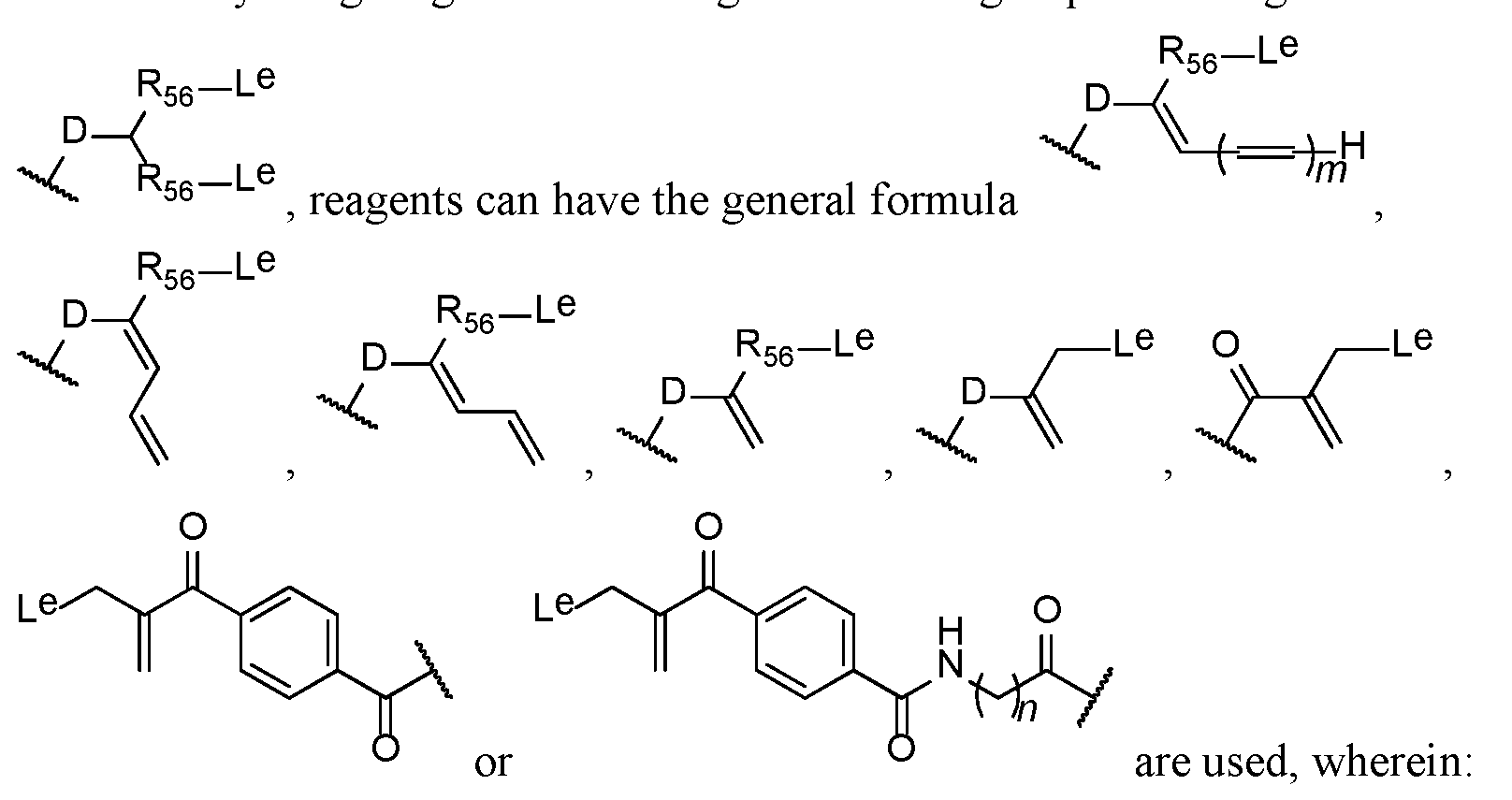
D is an electron withdrawing group as described below;
each R56 independently is selected from among C1-4 alkylene or alkenylene, such as methyl, ethyl, propyl, isopropyl and butyl;
m is 0 to 4; and
n is an integer from 0 to 5, inclusive
each Le independently is a leaving group.
An electron withdrawing group (D), for example, is a keto group >C=O, an
ester group, -O-CO-, a sulfone group,-SO2- or
 .
.
ii. Leaving group
In the reagents provided herein, the leaving group (Le) is any leaving group known in the art. Where two Le's are present, these can be different groups, but generally they are the same group.
For example, a leaving group (Le) is selected from among
—SR, -OR,—SO2R,—OSO2R, -N+RR2R3, halogen, or—OAr, in which: R independently represents a hydrogen atom or an alkyl (such as, C1-6 alkyl), aryl (such as phenyl), or alkyl-aryl (such as C1-6 alkyl-phenyl) group, or is a group that includes a porition–(CH2CH2O)n- in which n is a number of two or more;
each of R2 and R3 independently represents a hydrogen atom, a C1-4 alkyl group, or a group R; and
Ar represents a substituted aryl, for example, phenyl, group, containing at least one electron withdrawing substituent, for example—CN,—NO2,—CO2Ra,—COH, —CH2OH,—CORa,—ORa,—OCORa,—OCO2Ra,—SRa,—SORa,—SO2Ra,— NHCORa, -NRa, -CORa,—NRCORa, -NHCO2Ra,—NRCO2Ra,—NO,—NHOH,— NRaOH,—C N—NHCORa,—C N—NRaCORa,—N+Ra
3,—N+HRa
2,—NH2Ra, halogen, for example fluorine, chlorine, bromine or iodine,—C CRa,—CH CRa
2 and—CH CHRa; and
each Ra independently represents a hydrogen atom or an alkyl (such as C1-6 alkyl), aryl (such as phenyl) or alkyl-aryl (such as C1-6 alkyl-phenyl) group.
In some examples, a leaving group (Le) is selected from among alkyl or aryl sulfonyl groups, such as–SR or -SO2R, wherein R independently represents a hydrogen atom or an alkyl (such as, C1-6 alkyl), aryl (such as phenyl), or alkyl-aryl
(such as C1-6 alkyl-phenyl) group, or is a group that includes–(CH2CH2O)nCH3 or a portion–(CH2CH2O)n- in which n is a number of two or more, such as 7.
In some examples, a leaving group (Le) is selected from among alkyl or aryl sulfonyl groups, such as–SR or -SO2R, wherein R independently represents phenyl, tosyl,–(CH2CH2O)nCH3 or a portion–(CH2CH2O)n- in which n is a number of two or more, such as 7.
In some examples, a leaving group is phenylsulfonyl, such as tosyl.
iii. Exemplary bis-thiol alkylating reagents
In one example, the bis-thiol alkylating reagent contains the structure:

For example, the bis-thiol alkylating reagent can be 4-(3-tosyl-2- (tosylmeth l ro ano l benzamide-PEG having the structure:

In one example, the conjugation reagent contains a structure below, and a method includes reacting a conditionally active antibody with a reagent having the structure below:

, wherein R80 is a bis-sulfone, a bis-vinyl sulfone or a bis-p-tolyl sulfone.
Vinyl sulfones can be prepared by oxidation of 2-halo or 2- hydroxyethylthioethers by sequential or simultaneous elimination and oxidation. Any suitable oxidizing agent known in the art can be used, including H2O2-acetic acid, m-chloroperbenzoic acid (-mCPBA-) or periodic acid–HIO4-. The elimination step can be performed using an organic base, such as triethylamine. See, Lopez-Jaramillo et al., Vinyl Sulfone: A Multi-Purpose Function in Proteomics, Integrative Proteomics, Dr. Hon-Chiu Leung (Ed.) (2012).
In further examples, a method of preparing an antibody conjugate described herein includes reacting a conditionally active antibody with a bis-sulfone conjugation reagent having the structure:

iv. Conjugation process
Conjugating reagents are reacted with a conditionally active antibody to form
-methylene leaving group and a double bond are cross- conjugated with an electron withdrawing function that serves as a Michael activating moiety. If the leaving group is prone to elimination in the cross-functional reagent rather than to direct displacement and the electron-withdrawing group is a suitable activating moiety for the Michael reaction then sequential intramolecular bis- alkylation of the side chains of two Cys residues in an antibody can occur by consecutive Michael and retro Michael reactions. The leaving moiety "masks" a latent conjugated double bond that is not exposed until after the first alkylation has occurred. Bis-alkylation results from sequential and interactive Michael and retro- Michael reactions. The cross-functional alkylating agents can contain multiple bonds conjugated to the double bond or between the leaving group and the electron withdrawing group.
Conjugation reactions can be carried out under known conjugation processes conditions, or under similar conditions to known conjugation processes, including the conditions described in International PCT Publication Nos. WO 2005/007197, WO 2009/047500, WO 2014/064423 and WO 2014/064424. The process can for example be carried out in a solvent or solvent mixture in which all reactants are soluble. For example, the protein can be allowed to react directly with the polymer conjugating reagent in an aqueous reaction medium. For example, a mixture
containing aqueous buffer (80%) and Propylene Glycol (20%), organic solvent the conjugating reagent is solubilized into, is used for conjugation. This reaction medium can also be buffered, depending on the pH requirements of the nucleophile. For example, the pH for the reaction is at least 4.5, such as, for example between about 5.0 and about 8.5, including about 6.0 to 7.5. The reaction conditions will depend upon the specific reactants employed.
Typically, conjugation reaction temperatures are, for example, between 3- when usi conducted in organic media (for example THF, ethyl acetate, acetone) are typically conducted at temperatur reaction can be performed in an aqueous buffer that contains a proportion of organic solvent, for example up to 20% by volume of organic solvent, such as from 5 to 20 % by volume, 10% to 20% by volume or 10% to 15% by volume, of organic solvent. For example, the organic solvent can be an alkylene glycol, such as ethylene glycol or propylene glycol. The conjugation reaction, for example, can be performed in a mixture of propylene glycol (20% v/v) in aqueous buffer (80% v/v).
The antibody can be conjugated using a stoichiometric equivalent or an excess or slight excess of conjugating reagent. Excess conjugating reagent can be removed by any method known in the art, such as, for example by ion exchange
chromatography or HPLC. It is possible for more than one conjugating reagent to be conjugated to a protein. For example, in an antibody that contains four interchain disulfide bonds, it is possible to conjugate four molecules of the reagent per molecule of antibody. Selective reduction of the four interchain disulfide bonds of an IgG is within the ability of the skilled artisan (see, e.g., Willner et al. (1993) Bioconjugate Chem.4, 521-527; and Trail et al. (1993) Science 261, 212-215). In one example, a mixture of DARs, including DARs that are higher than 4 (some DAR5 and some DAR6), results; these can be partially removed during purification by hydrophobic interaction chromatography (HIC).
The conjugation process can be reversible under suitable conditions. In certain applications, the electron withdrawing moiety D is reduced to prevent release
of the conditionally active antibody. The methods provided herein can include an additional optional step of reducing the electron withdrawing group D in the conjugate. Any reducing agent known in the art can be used, including, for example, a borohydride, for example sodium borohydride, sodium cyanoborohydride, potassium borohydride or sodium triacetoxyborohydride, tin(II) chloride, alkoxides such as aluminium alkoxide, and lithium aluminium hydride.
Thus, for example, a moiety D containing a keto group >C=O can be reduced to an alcohol group–CH(OH). An ether group can be obtained by the reaction of a hydroxy group with an etherifying agent; an ester group can be obtained by the reaction of a hydroxy group with an acylating agent; an amine group can be prepared from a ketone by reductive amination; or an amide group can be formed by acylation of an amine. A sulfone can be reduced to a sulfoxide, sulfide or thiol ether.
v. Antibody cysteines
In the methods and conjugates provided herein, one or more of any of the cysteine residues present in the conditionally active antibody can be conjugated. For example, a typical IgG1 has 12 intrachain and 4 interchain disulfide bonds.
Reduction of the disulfide bonds in a typical IgG1 results in 24 and 8 free thiol groups from intrachain and interchain disulfide bonds, respectively. In a further example, the antibody is selectively conjugated to the linker at the cysteine residues resulting from selective reduction of the 4 interchain disulfide bonds.
Prior to conjugation to two sulfur atoms derived from a disulfide bond in the antibody, the disulfide bond can be reduced following which the reduced thiol reacts with the conjugation reagent. The disulfide bond can be reduced, for example, with any reducing agent known in the art. For example, the disulfide bond is reduced with dithiothreitol, mercaptoethanol, or tris-carboxyethylphosphine. Any excess reducing agent optionally can be removed, for example, by any method known in the art, including buffer exchange, before the conjugating reagent is introduced. Conjugation also can be performed without removing excess reducing agent. Conjugation can occur between cysteine residues from the same disulfide bonds or different disulfide bonds or a mixture of the same and different disulfide bonds.
For example, the conditionally active antibody is conjugated at the cysteine residues that form one or more disulfide bonds selected from among the following:
Light chain (LC) position C214 (C214 by Kabat numbering, C214 by EU numbering) to Heavy chain (HC) position C222 (C233 by Kabat numbering, C220 by EU numbering) (2 disulfide bonds per antibody);
HC C228 to HC C228 (C239 by Kabat numbering, C226 by EU numbering); and
HC C231 to HC C231 (C242 by Kabat numbering, C229 by EU numbering). Additional reactions can occur.
Because of symmetry, an IgG molecule can contain two of the same disulfide bonds. For example, an IgG1 antibody contains 2 disulfide bonds formed by LC C214 (C214 by Kabat numbering, C214 by EU numbering) to HC C222 (C233 by Kabat numbering, C220 by EU numbering). Conjugation can occur at either or both of the disulfide bonds.
The drug:antibody ratio of the final conjugated product can be measured by hydrophobic interaction chromatography. A mixture of DARs, including DARs that are higher than 4 (some DAR5 and some DAR6) are produced, and can be removed (or partially removed) during purification by HIC. Antibodies with a drug:antibody (DAR) ratio of 4 can be isolated.
f. Other examples
In some examples, the reactive moiety of the bridge unit is a bifunctional linker that maintains the interchain disulfide bonding of the antibody. For example, the bridge unit in an antibody conjugate can be formed by reacting a conditionally active antibody with a bifunctional pyrrole-2,5-dione- and pyrrolidine-2,5-dione- based linkers as described in published U.S. patent publication No. US2013/0224228. In such an example, reaction of the bifunctional linker with the two cysteines gives a “stapled” dithiosuccinimide or dithiomaleimide antibody conjugate with one linker per disulfide connected through two thioether bonds.
D. METHODS OF PRODUCING ANTI-EGFR ANTIBODIES
1. Generating and Producing Anti-EGFR Antibodies
Anti-EGFR antibodies, such as the modified anti-EGFR antibodies provided herein and anti-EGFR antibodies in any of the conjugates provided herein, can be expressed using standard cell culture and other expression systems known in the art. Prior to use in the methods provided herein, the proteins can be purified.
Alternatively, whole supernatant or diluted supernatant can be used in the methods provided herein. The modified anti-EGFR antibodies provided herein can be produced by recombinant DNA methods that are within the purview of those skilled in the art. DNA encoding a modified anti-EGFR antibody can be synthetically produced or can be readily isolated and sequenced using conventional procedures (e.g., by using oligonucleotide probes that are capable of binding specifically to genes encoding the heavy and light chains of the antibody). For example, any cell source known to produce or express a modified anti-EGFR antibody can serve as a source of such DNA. In another example, once the sequence of the DNA encoding the modified anti-EGFR is determined, nucleic acid sequences can be constructed using gene synthesis techniques.
Further, mutagenesis techniques also can be employed to generate further modified forms of an anti-EGFR antibody. The DNA also can be modified. For example, gene synthesis or routine molecular biology techniques can be used to effect insertion, deletion, addition or replacement of nucleotides. For example, additional nucleotide sequences can be joined to a nucleic acid sequence. In one example linker sequences can be added, such as sequences containing restriction endonuclease sites for the purpose of cloning the antibody gene into a vector, for example, a protein expression vector. Furthermore, additional nucleotide sequences specifying functional DNA elements can be operatively linked to a nucleic acid molecule.
Examples of such sequences include, but are not limited to, promoter sequences designed to facilitate intracellular protein expression, and leader peptide sequences designed to facilitate protein secretion.
It is understood that any of the amino acid sequences provided herein can be reverse-translated, using standard methods commonly used by those skilled in the art, to generate corresponding encoding nucleic acid sequences, which can be cloned into vectors and expressed to generate the antibodies and fragments provided herein.
Anti-EGFR antibodies, such as the modified anti-EGFR antibodies provided herein, can be expressed as full-length proteins or less than full length proteins. For example, antibody fragments can be expressed. Nucleic acid molecules and proteins provided herein can be made by any method known to one of skill in the art. Such procedures are routine and are well-known to the skill artisan. They include routine molecular
biology techniques including gene synthesis, PCR, ligation, cloning, transfection and purification techniques. A description of such procedures is provided below.
Once isolated, the DNA can be placed into expression vectors, which are then transfected into host cells. The choice of vector can depend on the desired application. For example, after insertion of the nucleic acid, the vectors typically are used to transform host cells, for example, to amplify the protein genes for replication and/or expression thereof. In such examples, a vector suitable for high level expression is used.
For expression of antibodies, generally, nucleic acid encoding the heavy chain of an antibody is cloned into a vector and the nucleic acid encoding the light chain of an antibody is cloned into a vector. The genes can be cloned into a single vector for dual expression thereof, or into separate vectors. If desired, the vectors also can contain further sequences encoding additional constant region(s) or hinge regions to generate other antibody forms. The vectors can be transfected and expressed in host cells. Expression can be in any cell expression system known to one of skill in the art. For example, host cells include cells that do not otherwise produce
immunoglobulin protein, to obtain the synthesis of antibodies in the recombinant host cells. For example, host cells include, but are not limited, to simian COS cells, Chinese hamster ovary (CHO) cells, 293FS cells, HEK293-6E cells, NSO cells or other myeloma cells. Other expression vectors and host cells are described herein.
The modified anti-EGFR antibodies provided herein can be generated or expressed as full-length antibodies or as antibodies that are less than full length, including, but not limited to, antigen-binding fragments, such as, for example, Fab, Fab', Fab hinge, F(ab’)2, single-chain Fv (scFv), scFv tandem, Fv, dsFv, scFv hinge, developed for the production of antibody fragments. For example, fragments can be derived via proteolytic digestion of intact antibodies (see, e.g., Morimoto et al. (1992) Journal of Biochemical and Biophysical Methods, 24:107-117; Brennan et al. (1985) Science, 229:81). Alternatively, fragments can be produced directly by recombinant host cells. For example, Fab, Fv and scFv antibody fragments can all be expressed in and secreted from host cells, such as E. coli, thereby facilitating production of large amounts of these fragments. F(ab')2 fragments can be produced by chemically
coupling Fab'-SH fragments (Carter et al. (1992) Bio/Technology, 10:163-167), or they can be isolated directly from recombinant host cell culture. In some examples, the modified anti-EGFR antibody is a single chain Fv fragment (scFv) (e.g.,
WO 93/16185; US Patent Nos.5,571,894 and 5,587,458). Fv and scFv fragments have intact combining sites but are devoid of constant regions; thus, they are suitable for reduced nonspecific binding during in vivo use. scFv fusion proteins can be constructed to attach an effector protein at either the amino- or the carboxy-terminus of an scFv. The antibody fragment also can be a linear antibody (see, e.g., U.S. Patent No.5,641,870). Such linear antibody fragments can be monospecific or bispecific. Other techniques for the production of antibody fragments are known to one of skill in the art.
Upon expression, antibody heavy and light chains, or fragment(s) thereof, pair by interchain disulfide bonds to form a full-length antibody or fragment thereof. For example, for expression of a full-length Ig, sequences encoding the VH-CH1-hinge- CH2-CH3 can be cloned into a first expression vector and sequences encoding the VL- CL domains can be cloned into a second expression vector. Upon co-expression, the full-length heavy and light chains are interlinked by disulfide bonds to generate a full- length antibody. In another example, to generate a Fab, sequences encoding a fragment containing the VH and CH1 regions can be cloned into a first expression vector and sequences encoding the VL-CL domains can be cloned into a second expression vector. Upon co-expression, the heavy chain pairs with a light chain to generate a Fab monomer.
Exemplary sequences that can be inserted into vectors for expression of whole antibodies and antibody fragments include nucleotide sequences which encode the corresponding heavy chain or light chain or fragments of any of the modified anti- EGFR antibodies provided herein. For example, the nucleotide sequences encoding any of the variable heavy chain and variable light chain sequences of any antibody or fragment described herein can be inserted into a suitable expression vector described herein or known to one of skill in the art. Any of the amino acid sequences of the modified anti-EGFR antibodies and EGFR-binding fragments provided herein can be reverse translated (also called back translated) to generate nucleic acid sequences, such as DNA sequences that encode the protein, using standard procedures. For
example, there are several on-line tools are available to convert protein sequences to encoding DNA sequences, such as bioinformatics.org/sms2/rev_trans.html;
biophp.org/minitools/protein_to_dna/demo.php; vivo.colostate.edu/molkit/rtranslate/; ebi.ac.uk/Tools/st/emboss_backtranseq/; molbiol.ru/eng/scripts/01_19.html; and geneinfinity.org/sms/sms_backtranslation.html. Such reverse translated sequences can be inserted into any of the expression vectors provided herein for the expression and production of the provided antibodies or fragments.
In particular, a sequence of nucleotides encoding a modified anti-EGFR antibody with a 104E amino acid replacement has a sequence of nucleotides encoding a variable heavy chain set forth in SEQ ID NO: 73 and a sequence of nucleotides encoding the variable light chain set forth in SEQ ID NO: 51. Other non-limiting examples of sequences of nucleic acids encoding the variable heavy and light chains, which can be inserted into a suitable expression vector, are set forth in Table 11 and include those encoding a variable heavy chain having a sequence of nucleotides set forth in SEQ ID NOS: 60, 62, 130, 132, 136, 138, 142, 144, 148, 150, 210, 212, 216, 218, 222, 224, 228, 230, 234, 236, 240, 242, 246, 248, and any degenerate sequence thereof and those encoding a variable light chain having a sequence of nucleotides set forth in SEQ ID NOS: 154, 157, 161, 164, 168, 171, 175, 178, 182, 185, 189, 192, 196, 199, 203, 206, 252, 255, 259, 262, 266, 269, 273, 276, 280, 283, 287, 290, 294, 297, 301, and 304 and any degenerate sequence thereof.
For purposes herein with respect to expression of anti-EGFR antibodies, such as modified anti-EGFR antibodies, vectors can contain a sequence of nucleotides that encodes a constant region of an antibody operably linked to the nucleic acid sequence encoding the variable region of the antibody. The vector can include the sequence for one or all of a CH1, CH2, hinge, CH3 or CH4 and/or CL. Generally, such as for expression of Fabs, the vector contains the sequence for a CH1 or CL (kappa or lambda light chains). The sequences of constant regions or hinge regions are known to one of skill in the art (see, e.g., U.S. Published Application No.20080248028). Examples of such sequences are provided herein.
All or a portion of the constant region of the heavy chain or light chain also can be inserted or contained in the vector for expression of IgG antibodies or fragments thereof. For example, non-limiting examples include those encoding a full-
length heavy chain having a sequence of nucleotides set forth in SEQ ID NOS: 58, 71, 128, 134, 140, 146, 208, 214, 220, 226, 232, 238, 244, and 396, and any degenerate sequence thereof and those encoding a full-length light chain having a sequence of nucleotides set forth in SEQ ID NOS: 50, 152, 159, 166, 173, 180, 187, 194, 201, 250, 257, 264, 271, 278, 285, 292, 299 and 398, and any degenerate sequence thereof. Non-limiting examples of partial vector sequence that encodes both a full-length heavy chain and a full-length heavy chian and a full-length light chain include the nucleic acid sequence set forth in SEQ ID NO: 399, and any degenerate sequence thereof.
In addition, VH-CH1 and VL-CL sequences can be inserted into a suitable expression vector for expression of Fab molecules. Nucleic acids encoding variable heavy chain and variable light chain domains of an antibody can be expressed in a suitable expression vector, such as a vector encoding for a linker between the variable heavy chain and variable light chain to produce single chain antibodies. Exemplary linkers include the glycine rich flexible linkers (-G4S-)n, where n is a positive integer, such as 1 (SEQ ID NO: 346), 2 (SEQ ID NO: 347), 3 (SEQ ID NO: 46), 4 (SEQ ID NO: 348), 5 (SEQ ID NO: 349), or more.
In other examples, a sequence of nucleotides encoding a modified anti-EGFR antibody with a 104D amino acid replacement has a sequence of nucleotides encoding a variable heavy chain set forth in SEQ ID NO: 68 and a sequence of nucleotides encoding the variable light chain set forth in SEQ ID NO: 51. Other non-limiting examples of sequences of nucleic acids encoding the variable heavy and light chains, which can be inserted into a suitable expression vector include those encoding full- length heavy chain sequence set forth in SEQ ID NOS: 52, 54, 56, 64, 371, 373, 375, 377, 379, 381, 383, 385, and 387, and any degenerate sequence thereof and those encoding a variable light chain having a sequence of nucleotides set forth in SEQ ID NOS: 154, 157, 161, 164, 168, 171, 175, 178, 182, 185, 189, 192, 196, 199, 203, 206, 252, 255, 259, 262, 266, 269, 273, 276, 280, 283, 287, 290, 294, 297, 301, and 304 and any degenerate sequence thereof. Other examples of a sequence of nucleotides encoding a modified anti-EGFR antibody with a 104D amino acid replacement inlcude any set forth in SEQ ID NOS: 1160-1185 and 1187 of WO 2013/134743
(sequence identifiers with reference to those WO 2013/134743), which is incorporated by reference in its entirety.
a. Vectors
Choice of vector can depend on the desired application. Many expression vectors are available and known to those of skill in the art for the expression of anti- EGFR antibodies or portions thereof, such as antigen binding fragments. The choice of an expression vector is influenced by the choice of host expression system. Such selection is well within the level of skill of the skilled artisan. In general, expression vectors can include transcriptional promoters and optionally enhancers, translational signals, and transcriptional and translational termination signals. Expression vectors that are used for stable transformation typically have a selectable marker which allows for selection and maintenance of the transformed cells. In some cases, an origin of replication can be used to amplify the copy number of the vectors in the cells. Vectors also generally can contain additional nucleotide sequences operably linked to the ligated nucleic acid molecule (e.g., His tag, Flag tag). For applications with antibodies, vectors generally include sequences encoding the constant region. Thus, antibodies or portions thereof also can be expressed as protein fusions. For example, a fusion protein can be generated to add additional functionality to a polypeptide. Examples of fusion proteins include, but are not limited to, fusions of a signal sequence, an epitope tag such as for localization, e.g., a His6 tag or a myc tag, or a tag for purification, such as a GST tag, and/or a sequence for directing protein secretion and/or membrane association.
For example, expression of the anti-EGFR antibodies, such as modified anti- EGFR antibodies, can be controlled by any promoter/enhancer known in the art. Suitable bacterial promoters are well-known in the art and described herein below. Other suitable promoters for mammalian cells, yeast cells and insect cells are well- known in the art and some are exemplified below. Selection of the promoter used to direct expression of a heterologous nucleic acid depends on the particular application. Promoters which can be used include but are not limited to eukaryotic expression vectors containing the SV40 early promoter (Bernoist and Chambon, Nature 290:304- 310 (1981)), the promoter contained in the 3’ long terminal repeat of Rous sarcoma virus (Yamamoto et al. Cell 22:787-797 (1980)), the herpes thymidine kinase
promoter (Wagner et al., Proc. Natl. Acad. Sci. USA 78:1441-1445 (1981)), the regulatory sequences of the metallothionein gene (Brinster et al., Nature 296:39-42
-lactamase promoter (Jay et al., (1981) Proc. Natl. Acad. Sci. USA 78:5543) or the tac promoter (DeBoer et al., Proc. Natl. Acad. Sci. USA 80:21-25 (1983)); see also“Gilbert and Villa-Komaroff“Useful Proteins from Recombinant Bacteria,” Scientific American 242:74-94 (1980)); plant expression vectors containing the nopaline synthetase promoter (Herrera-Estrella et al., Nature 303:209-213 (1983)) or the cauliflower mosaic virus 35S RNA promoter (Gardner et al., Nucleic Acids Res.9:2871 (1981)), and the promoter of the photosynthetic enzyme ribulose bisphosphate carboxylase (Herrera-Estrella et al., Nature 310:115-120 (1984)); promoter elements from yeast and other fungi such as the Gal4 promoter, the alcohol dehydrogenase promoter, the phosphoglycerol kinase promoter, the alkaline phosphatase promoter, and the following animal transcriptional control regions that exhibit tissue specificity and have been used in transgenic animals: elastase I gene control region which is active in pancreatic acinar cells (Swift et al., Cell 38:639-646 (1984); Ornitz et al., Cold Spring Harbor Symp. Quant. Biol. 50:399-409 (1986); MacDonald, Hepatology 7:425-515 (1987)); insulin gene control region which is active in pancreatic beta cells (Hanahan et al., Nature 315:115-122 (1985)), immunoglobulin gene control region which is active in lymphoid cells (Grosschedl et al., Cell 38:647-658 (1984); Adams et al., Nature 318:533-538 (1985); Alexander et al., Mol. Cell Biol.7:1436-1444 (1987)), mouse mammary tumor virus control region which is active in testicular, breast, lymphoid and mast cells (Leder et al., Cell 45:485-495 (1986)), albumin gene control region which is active in liver (Pinkert et al., Genes and Devel.1:268-276 (1987)), alpha-fetoprotein gene control region which is active in liver (Krumlauf et al., Mol. Cell. Biol.5:1639-1648 (1985); Hammer et al., Science 235:53-581987)), alpha-1 antitrypsin gene control region which is active in liver (Kelsey et al., Genes and Devel.1:161-171 (1987)), beta globin gene control region which is active in myeloid cells (Magram et al., Nature 315:338-340 (1985); Kollias et al., Cell 46:89-94 (1986)), myelin basic protein gene control region which is active in oligodendrocyte cells of the brain (Readhead et al., Cell 48:703-712 (1987)), myosin light chain-2 gene control region which is active in skeletal muscle (Shani, Nature 314:283-286 (1985)), and gonadotrophic releasing
hormone gene control region which is active in gonadotrophs of the hypothalamus (Mason et al., Science 234:1372-1378 (1986)).
In addition to the promoter, the expression vector typically contains a transcription unit or expression cassette that contains all the additional elements required for the expression of the antibody, or portion thereof, in host cells. A typical expression cassette contains a promoter operably linked to the nucleic acid sequence encoding the protein and signals required for efficient polyadenylation of the transcript, ribosome binding sites and translation termination. Additional elements of the cassette can include enhancers. In addition, the cassette typically contains a transcription termination region downstream of the structural gene to provide for efficient termination. The termination region can be obtained from the same gene as the promoter sequence or can be obtained from different genes.
Some expression systems have markers that provide gene amplification such as thymidine kinase and dihydrofolate reductase. Alternatively, high yield expression systems not involving gene amplification are also suitable, such as using a
baculovirus vector in insect cells, with a nucleic acid sequence encoding a protein under the direction of the polyhedron promoter or other strong baculovirus promoter.
Exemplary expression vectors include any mammalian expression vector such as, for example, pCMV and pCDNA3.1. Other eukaryotic vectors, for example any containing regulatory elements from eukaryotic viruses can be used as eukaryotic expression vectors. These include, for example, SV40 vectors, papilloma virus vectors, and vectors derived from Epstein-Bar virus. Exemplary eukaryotic vectors include pMSG, pAV009/A+, pMT010/A+, pMAMneo-5, baculovirus pDSCE, and any other vector allowing expression of proteins under the direction of the CMV promoter, SV40 early promoter, SV40 late promoter, metallothionein promoter, murine mammary tumor virus promoter, Rous sarcoma virus promoter, polyhedron promoter, or other promoters shown effective for expression in eukaryotes. For bacterial expression, such vectors include pBR322, pUC, pSKF, pET23D, and fusion vectors such as MBP, GST and LacZ.
Any methods known to those of skill in the art for the insertion of DNA fragments into a vector can be used to construct expression vectors containing a nucleic acid encoding a protein or an antibody chain. These methods can include in
vitro recombinant DNA and synthetic techniques and in vivo recombinants (genetic recombination). The insertion into a cloning vector can, for example, be
accomplished by ligating the DNA fragment into a cloning vector which has complementary cohesive termini. If the complementary restriction sites used to fragment the DNA are not present in the cloning vector, the ends of the DNA molecules can be enzymatically modified. Alternatively, any site desired can be produced by ligating nucleotide sequences (linkers) onto the DNA termini; these ligated linkers can contain specific chemically synthesized nucleic acids encoding restriction endonuclease recognition sequences.
In one example, nucleic acid encoding the heavy chain of an antibody, is ligated into a first expression vector and nucleic acid encoding the light chain of an antibody is ligated into a second expression vector. The expression vectors can be the same or different, although generally they are sufficiently compatible to allow comparable protein expression of the heavy and light chains. The first and second expression vectors are generally co-transfected into host cells, typically at a 1:1 ratio. Exemplary vectors include, but are not limited to, p 1HC and p LC (Tiller et al. (2008) J Immunol. Methods, 329:112-24). Other expression vectors include the light chain expression vector pAG4622 and the heavy chain expression vector pAH4604 (Coloma et al. (1992) J Immunol. Methods, 152:89-104). The pAG4622 vector contains the genomic sequence encoding the C- and the gpt selectable marker. The pAH4604 vector contains the hisD selectable
-region domain. In another example, the heavy and light chain can be cloned into a single vector that has expression cassettes for both the heavy and light chain.
In some examples, the vector is a bicistronic vector that contains an internal ribosomal entry site (IRES) between the open reading frames encoding the heavy and light chains. For example, an exemplary vector includes the vector designated pcDNA3-Erbitux-LC-IRES-HC-WT (e.g., SEQ ID NO: 306), where nucleic acid encoding the heavy chain (HC) or light chain (LC) of any of the modified anti-EGFR antibodies provided herein can be substituted in place of the sequences therein.
Examples of such vectors are set forth in any of SEQ ID NOS: 307-314.
b. Cells and Expression Systems
Generally, any cell type that can be engineered to express heterologous DNA and has a secretory pathway is suitable for expression of the modified anti-EGFR antibodies provided herein. Expression hosts include prokaryotic and eukaryotic organisms such as bacterial cells (e.g., E. coli), yeast cells, fungal cells, Archaea, plant cells, insect cells and animal cells including human cells. Expression hosts can differ in their protein production levels as well as the types of post-translational
modifications that are present on the expressed proteins. Further, the choice of expression host is often related to the choice of vector and transcription and translation elements used. For example, the choice of expression host is often, but not always, dependent on the choice of precursor sequence utilized. For example, many heterologous signal sequences can only be expressed in a host cell of the same species (i.e., an insect cell signal sequence is optimally expressed in an insect cell). In contrast, other signal sequences can be used in heterologous hosts such as, for example, the human serum albumin (hHSA) signal sequence which works well in yeast, insect, or mammalian host cells and the tissue plasminogen activator pre/pro sequence which has been demonstrated to be functional in insect and mammalian cells (Tan et al., (2002) Protein Eng.15:337). The choice of expression host can be made based on these and other factors, such as regulatory and safety considerations, production costs and the need and methods for purification. Thus, the vector system must be compatible with the host cell used.
Expression in eukaryotic hosts can include expression in yeasts such as Saccharomyces cerevisiae and Pichia pastoris, insect cells such as Drosophila cells and lepidopteran cells, plants and plant cells such as tobacco, corn, rice, algae, and Lemna. Eukaryotic cells for expression also include mammalian cells lines such as Chinese hamster ovary (CHO) cells or baby hamster kidney (BHK) cells. Eukaryotic expression hosts also include production in transgenic animals, for example, including production in serum, milk and eggs.
Recombinant molecules can be introduced into host cells via, for example, transformation, transfection, infection, electroporation and sonoporation, so that many copies of the gene sequence are generated. Generally, standard transfection methods are used to produce bacterial, mammalian, yeast, or insect cell lines that express large
quantity of antibody chains, which are then purified using standard techniques (see e.g., Colley et al. (1989) J. Biol. Chem., 264:17619-17622; Guide to Protein
Purification, in Methods in Enzymology, vol.182 (Deutscher, ed.), 1990).
Transformation of eukaryotic and prokaryotic cells is performed according to standard techniques (see, e.g., Morrison (1977) J. Bact.132:349-351; Clark-Curtiss and Curtiss (1983) Methods in Enzymology, 101, 347-362). For example, any of the well-known procedures for introducing foreign nucleotide sequences into host cells can be used. These include the use of calcium phosphate transfection, polybrene, protoplast fusion, electroporation, biolistics, liposomes, microinjection, plasma vectors, viral vectors and any other the other well-known methods for introducing cloned genomic DNA, cDNA, synthetic DNA or other foreign genetic material into a host cell. Generally, for purposes of expressing an antibody, host cells are transfected with a first vector encoding at least a VH chain and a second vector encoding at least a VL chain. Thus, it is only necessary that the particular genetic engineering procedure used be capable of successfully introducing at least both genes into the host cell capable of expressing antibody polypeptide, or modified form thereof.
The modified anti-EGFR antibodies, provided herein, can be produced by any method known in the art for protein production including in vitro and in vivo methods such as, for example, the introduction of nucleic acid molecules encoding antibodies into a host cell or host animal and expression from nucleic acid molecules encoding recombined antibodies in vitro. Prokaryotes, especially E. coli, provide a system for producing large amounts of reassembled antibodies or portions thereof, and are particularly desired in applications of expression and purification of proteins.
Transformation of E. coli is a simple and rapid technique well-known to those of skill in the art. E. coli host strains for high throughput expression include, but are not limited to, BL21 (EMD Biosciences) and LMG194 (ATCC). A particular example of such an E. coli host strain is BL21. Vectors for high throughput expression include, but are not limited to, pBR322 and pUC vectors.
i. Prokaryotic Expression
Prokaryotes, especially E. coli, provide a system for producing large amounts of modified anti-EGFR antibodies, or portions thereof. Transformation of E. coli is a simple and rapid technique well-known to those of skill in the art. Expression vectors
for E. coli can contain inducible promoters that are useful for inducing high levels of protein expression and for expressing antibodies that exhibit some toxicity to the host cells. Examples of inducible promoters include the lac promoter, the trp promoter, the hybrid tac promoter, the T7 and SP6 RNA promoters and the temperature regulated L promoter.
Antibodies or portions thereof can be expressed in the cytoplasmic
environment of E. coli. The cytoplasm is a reducing environment and for some antibodies, this can result in the formation of insoluble inclusion bodies. Reducing
-mercaptoethanol and denaturants (e.g., such as guanidine-HCl and urea) can be used to resolubilize the antibodies. An exemplary alternative approach is the expression of recombined antibodies or fragments thereof in the periplasmic space of bacteria which provides an oxidizing environment and chaperonin-like and disulfide isomerases leading to the production of soluble protein. Typically, a leader sequence is fused to the protein to be expressed which directs the protein to the periplasm. The leader is then removed by signal peptidases inside the periplasm. Exemplary pathways to translocate expressed proteins into the periplasm are the Sec pathway, the SRP pathway and the TAT pathway. Examples of periplasmic-targeting leader sequences include the pelB leader from the pectate lyase gene, the StII leader sequence, and the DsbA leader sequence. In some cases, periplasmic expression allows leakage of the expressed protein into the culture medium. The secretion of antibodies allows quick and simple purification from the culture supernatant. Antibodies that are not secreted can be obtained from the periplasm by osmotic lysis. Similar to cytoplasmic expression, in some cases proteins can become insoluble and denaturants and reducing agents can be used to facilitate solubilization and refolding using standard procedures. Temperature of induction and growth also can influence expression levels and solubility. Typically, temperatures between 25 °C and 37 °C are used. Mutations also can be used to increase solubility of expressed proteins. Typically, bacteria produce aglycosylated proteins. Thus, glycosylation can be added in vitro after purification from host cells.
ii. Yeast
Yeasts such as Saccharomyces cerevisiae, Schizosaccharomyces pombe, Yarrowia lipolytica, Kluyveromyces lactis, and Pichia pastoris are useful expression
hosts for recombined antibodies or portions thereof. Yeast can be transformed with episomal replicating vectors or by stable chromosomal integration by homologous recombination. Typically, inducible promoters are used to regulate gene expression. Examples of such promoters include AOX1, GAL1, GAL7, and GAL5 and metallothionein promoters such as CUP1. Expression vectors often include a selectable marker such as LEU2, TRP1, HIS3, and URA3 for selection and maintenance of the transformed DNA. Proteins expressed in yeast are often soluble. Co-expression with chaperonins such as BiP and protein disulfide isomerase can improve expression levels and solubility. Additionally, proteins expressed in yeast can be directed for secretion using secretion signal peptide fusions such as the yeast mating type alpha-factor secretion signal from Saccharomyces cerevisae and fusions with yeast cell surface proteins such as the Aga2p mating adhesion receptor or the Arxula adeninivorans glucoamylase. A protease cleavage site such as for the Kex-2 protease, can be engineered to remove the fused sequences from the expressed polypeptides as they exit the secretion pathway. Yeast also is capable of glycosylation at Asn-X-Ser/Thr motifs.
iii. Insects
Insect cells, particularly using baculovirus expression, are useful for expressing modified anti-EGFR antibodies or portions thereof. Insect cells express high levels of protein and are capable of most of the post-translational modifications used by higher eukaryotes. Baculovirus have a restrictive host range which can improve the safety and reduce regulatory concerns of eukaryotic expression. Typical expression vectors use a promoter for high level expression such as the polyhedrin promoter and p10 promoter of baculovirus. Commonly used baculovirus systems include the baculoviruses such as Autographa californica nuclear polyhedrosis virus (AcNPV), and the Bombyx mori nuclear polyhedrosis virus (BmNPV) and an insect cell line such as Sf9 derived from Spodoptera frugiperda and TN derived from Trichoplusia ni. For high-level expression, the nucleotide sequence of the molecule to be expressed can be fused immediately downstream of the polyhedrin initiation codon of the virus. To generate baculovirus recombinants capable of expressing human antibodies, a dual-expression transfer, such as pAcUW51 (PharMingen) is utilized.
Mammalian secretion signals are accurately processed in insect cells and can be used to secrete the expressed protein into the culture medium.
An alternative expression system in insect cell for expression of the modified anti-EGFR antibodies provided herein is the use of stably transformed cells. Cell lines such as Sf9 derived cells from Spodoptera frugiperda and TN derived cells from Trichoplusia ni can be used for expression. The baculovirus immediate early gene promoter IE1 can be used to induce consistent levels of expression. Typical expression vectors include the pIE1-3 and pI31-4 transfer vectors (Novagen).
Expression vectors are typically maintained by the use of selectable markers such as neomycin and hygromycin.
iv. Mammalian Cells
Mammalian expression systems can be used to express anti-EGFR antibodies, such as modified anti-EGFR antibodies, including antigen-binding fragments thereof. Expression constructs can be transferred to mammalian cells by viral infection such as adenovirus or by direct DNA transfer such as by using liposomes, calcium phosphate, DEAE-dextran and by physical means such as electroporation and microinjection. Expression vectors for mammalian cells typically include an mRNA cap site, a TATA box, a translational initiation sequence (Kozak consensus sequence) and
polyadenylation elements. Such vectors often include transcriptional promoter- enhancers for high-level expression, for example the SV40 promoter-enhancer, the human cytomegalovirus (CMV) promoter and the long terminal repeat of Rous sarcoma virus (RSV). These promoter-enhancers are active in many cell types.
Tissue and cell-type promoters and enhancer regions also can be used for expression. Exemplary promoter/enhancer regions include, but are not limited to, those from genes such as elastase I, insulin, immunoglobulin, mouse mammary tumor virus, albumin, alpha fetoprotein, alpha 1 antitrypsin, beta globin, myelin basic protein, myosin light chain 2, and gonadotropic releasing hormone gene control.
Selectable markers can be used to select for and maintain cells with the expression construct. Examples of selectable marker genes include, but are not limited to, hygromycin B phosphotransferase, adenosine deaminase, xanthine-guanine phosphoribosyl transferase, aminoglycoside phosphotransferase, dihydrofolate reductase and thymidine kinase. Modified anti-EGFR antibodies can be produced, for
example, using a NEOR/G418 system, a dihydrofolate reductase (DHFR) system or a glutamine synthetase (GS) system. The GS system uses joint expression vectors, such as pEE12/pEE6, to express both heavy chain and light chain. Fusion with cell surface signaling molecules such as TCR- RI- in an active state on the cell surface.
Many cell lines are available for mammalian expression including mouse, rat, human, monkey, chicken and hamster cells. Exemplary cell lines include any known in the art or described herein, such as, for example, CHO, Balb/3T3, HeLa, MT2, mouse NS0 (nonsecreting) and other myeloma cell lines, hybridoma and
heterohybridoma cell lines, lymphocytes, fibroblasts, Sp2/0, COS, NIH3T3, HEK293, 293S, 2B8, and HKB cells. Cell lines adapted to serum-free media which facilitates purification of secreted proteins from the cell culture media also are available. One such example is the serum free EBNA-1 cell line (Pham et al., (2003) Biotechnol. Bioeng.84:332-42.)
v. Plants
Transgenic plant cells and plants can be used to express anti-EGFR antibodies, such as modified anti-EGFR antibodies, or a portion thereof described herein.
Expression constructs are typically transferred to plants using direct DNA transfer such as microprojectile bombardment and PEG-mediated transfer into protoplasts, and with agrobacterium-mediated transformation. Expression vectors can include promoter and enhancer sequences, transcriptional termination elements and translational control elements. Expression vectors and transformation techniques are usually divided between dicot hosts, such as Arabidopsis and tobacco, and monocot hosts, such as corn and rice. Examples of plant promoters used for expression include the cauliflower mosaic virus CaMV 35S promoter, the nopaline synthase promoter, the ribose bisphosphate carboxylase promoter and the maize ubiquitin-1 (ubi-1) promoter promoters. Selectable markers such as hygromycin, phosphomannose isomerase and neomycin phosphotransferase are often used to facilitate selection and maintenance of transformed cells. Transformed plant cells can be maintained in culture as cells, aggregates (callus tissue) or regenerated into whole plants.
Transgenic plant cells also can include algae engineered to produce proteases or modified proteases (see for example, Mayfield et al. (2003) PNAS 100:438-442).
Because plants have different glycosylation patterns than mammalian cells, this can influence the choice of protein produced in these hosts.
2. Purification
Anti-EGFR antibodies, such as modified anti-EGFR antibodies and antigen binding portions thereof, can be purified by any procedure known to one of skill in the art or described herein. Proteins can be purified to substantial purity using standard protein purification techniques known in the art including but not limited to, SDS- PAGE, size fraction and size exclusion chromatography, ammonium sulfate precipitation, chelate chromatography, ionic exchange chromatography or column chromatography. For example, antibodies can be purified by column chromatography. An exemplary method for purifying the anti-EGFR antibodies provided herein is column chromatography, wherein a solid support column material is linked to Protein G, a cell surface-associated protein from Streptococcus, that binds immunoglobulins with high affinity. In some examples, the anti-EGFR antibodies can be purified by column chromatography, wherein a solid support column material is linked to Protein A, a cell surface-associated protein from Staphylococcus that binds immunoglobulins, such as IgG antibodies, with high affinity (see, e.g., Liu et al. (2010) MAbs 2(5):480- 499). Other immunoglobulin-binding bacterial proteins that can be used to purify the anti-EGFR antibodies provided herein include Protein A/G, a recombinant fusion protein that combines the IgG binding domains of Protein A and Protein G; and Protein L, a surface protein from Peptostreptococcus (Bjorck (1988) J. Immunol., 140(4):1194–1197; Kastern, et al. (1992) J. Biol. Chem.267(18):12820-12825;
Eliasson et al. (1988) J. Biol. Chem.263:4323-4327).
The anti-EGFR antibodies can be purified to 60%, 70%, 80% purity and typically at least 90%, 91%, 92%, 93%, 94%, 95%, 96%, 97%, 98% or 99% purity. Purity can be assessed by standard methods such as by SDS-PAGE and coomassie staining.
Methods for purification of anti-EGFR antibodies, including antibodies or portions thereof from host cells, depend on the chosen host cells and expression systems. For secreted molecules, proteins are generally purified from the culture media after removing the cells. For intracellular expression, cells can be lysed and the proteins purified from the extract. When transgenic organisms such as transgenic
plants and animals are used for expression, tissues or organs can be used as starting material to make a lysed cell extract. Additionally, transgenic animal production can include the production of polypeptides in milk or eggs, which can be collected, and if necessary further the proteins can be extracted and further purified using standard methods in the art.
When proteins are expressed by transformed bacteria in large amounts, typically after promoter induction, although expression can be constitutive, the polypeptides can form insoluble aggregates. There are several protocols that are suitable for purification of polypeptide inclusion bodies known to one of skill in the art. Numerous variations will be apparent to those of skill in the art.
E. METHODS FOR ASSESSING ANTI-EGFR ANTIBODY
PROPERTIES AND ACTIVITIES
The modified anti-EGFR antibodies, and variants and fragments thereof, provided herein, can be assessed for binding to EGFR antigen (e.g., human EGFR) or soluble fragment thereof. The binding activity can be assessed under conditions that compare the activity of an antibody under conditions of low pH/increased lactate concentrations and neutral pH/physiological lactate concentrations. For example, binding activity can be assessed under conditions of acidic pH (e.g., pH 5.8 to 6.5, such as pH 6.0 to 6.5) and/or increased lactate concentrations (e.g., 10 to 20 mM, such as 15 to 20 mM) and compared to binding activity under conditions of neutral pH (e.g., pH 7.0 to 7.6, such as pH 7.0 to 7.4) and/or physiological lactate concentrations (e.g., 0.5 to 5 mM, such as about 1 mM). Such assays can confirm that the binding activity is greater under conditions that include one or both of acid pH 6.0 to 6.5, inclusive, and/or lactate concentration of 15 mM to 20 mM, inclusive, compared to under conditions that include one or both of neutral pH of or about 7.4 and/or lactate concentration of or about 1 mM.
The assays also can be performed in the presence of physiological
concentrations of protein or serum (e.g., 10 mg/mL to 50 mg/mL protein, such as serum albumin; or 20-50% serum, such as human serum). For example, the anti- EGFR antibodies typically are assessed for activity under a first set of conditions that includes 20-50% serum (vol/vol) or 10-50 mg/mL protein (e.g., serum albumin), and an acidic pH of about between 5.8 to 6.8 and/or elevated lactate levels of 10 mM to 20
mM. For example, the first set of conditions can include at least 25% serum (vol/vol) or 12-40 mg/mL protein (e.g., serum albumin), and an acidic pH of about between 6.0 to 6.5, such as pH 6.0 and/or elevated lactate levels of 15 mM to 20 mM, such as about 16.7 mM. The anti-EGFR antibody also is assessed for activity under a second set of conditions that includes 20-50% serum (vol/vol) or 10-50 mg/mL protein (e.g., serum albumin), and near neutral pH or neutral pH of about between 7.0 to 7.4 and/or a lactate concentration of 0.5 to 5 mM. For example, the second set of conditions includes at least 25% serum (vol/vol) or 12-40 mg/mL protein (e.g., serum albumin), and pH about between 7.2 to 7.4, such as about pH 7.4. and/or lactate concentration of 0.5 mM to 2 mM, such as 1 mM. In some examples, the anti-EGFR antibody also can be assessed for activity under a third set of conditions that can include at least 25% serum (vol/vol) or 12-40 mg/mL protein (e.g., serum albumin), and an acidic pH of about between 6.0 to 6.5, such as pH 6.5 and/or elevated lactate levels of 15 mM to 20 mM, such as about 16.7 mM. In such assays, the amount of added protein to simulate a physiologic environment (e.g., serum protein) is typically the same or substantially the same for all sets of conditions tested, but can vary by ±25% or less from one condition to the other.
Hence, binding activity can be determined in conditions that simulate a physiologic environment or that is a physiologic environment. The binding can be assessed in vitro (e.g., in an immunoassay) or ex vivo or in vivo (e.g., binding to tumor cells or non-tumor cells, such as cells of the skin dermis). The assays provided herein include any assays that can test or assess an activity of a modified anti-EGFR antibody in a detectable or otherwise measurable manner under different pH and/or lactate concentrations, and, optionally, in the presence of physiological concentrations of total protein. The assays provided herein can be developed in a high throughput format in order to assess an activity of numerous anti-EGFR antibodies, for example protein variants, at one time in dual format. For example, in vitro binding assays can be performed using solid-support binding assays or solution binding assays, where the binding is performed under the above conditions. In other examples, binding assays can be performed in vivo where binding is compared on cells present in a tumor versus cells present in non-tumor cells. In particular, an in vivo binding assay can be performed to assess binding or localization of administered antibody to tumor cells
versus basal skin keratinocytes. This is exemplified herein using xenograft or skin graft models. Other models also can be employed. Descriptions of exemplary assays are provided below.
In addition to binding activity, other assays to assess the activity of modified anti-EGFR antibodies provided herein can be performed and include in vitro or in vivo assays including, but not limited to, functional assays, in vivo assays, animal models and clinical assays to measure the activity and/or side effects of the modified anti- EGFR antibodies provided herein. The activity assessed can be any activity of an anti-EGFR antibody, such as binding to EGFR, cell growth inhibition (CGI) activity or tumor growth inhibition activity. Any of the antibodies provided herein also can be characterized in a variety of assays known to one of skill in the art to assess clinical properties such as, for example, therapeutic efficacy, affinity for EGFR, toxicity, side effects, pharmacokinetics and pharmacodynamics.
1. Binding Assays
Modified anti-EGFR antibodies can be assayed for the ability to bind to EGFR by any method known to one of skill in the art. Exemplary assays are described herein below.
Binding assays can be performed in solution, suspension or on a solid support. For example, EGFR or soluble fragment thereof can be immobilized to a solid support (e.g., a carbon or plastic surface, a tissue culture dish or chip) and contacted with antibody. Unbound antibody or target protein can be washed away and bound complexes can then be detected. Binding assays can be performed under conditions to reduce nonspecific binding, such as by using buffers with a high ionic strength (e.g., 0.3-0.4 M NaCl) and/or with nonionic detergent (e.g., 0.1 % Triton X-100 or Tween 20) and/or blocking proteins (e.g., bovine serum albumin or gelatin). Negative controls also can be included in such assays as a measure of background binding. Binding affinities can be determined using quantitative ELISA, Scatchard analysis (Munson et al., (1980) Anal. Biochem., 107:220), surface plasmon resonance, isothermal calorimetry, or other methods known to one of skill in the art (e.g., Liliomet al. (1991) J. Immunol Methods.143(1):119-25).
Such assays can be performed, for example, in solution (e.g., Houghten (1992) Bio/Techniques 13:412-421), on beads (Lam (1991) Nature 354:82-84), on chips
(Fodor (1993) Nature 364:555-556), on bacteria (U.S. Pat. No.5,223,409), on spores (U.S. Pat. Nos.5,571,698; 5,403,484; and 5,223,409), on plasmids (Cull et al. (1992) Proc. Natl. Acad. Sci. USA 89:1865-1869) or on phage (Scott and Smith (1990) Science 249:386-390; Devlin (1990) Science 249:404-406; Cwirla et al. (1990) Proc. Natl. Acad. Sci. USA 87:6378-6382; and Felici (1991) J. Mol. Biol.222:301-310).
Typically, EGFR binding is detected using a method that is capable of being quantified such that the level of activity can be assessed. For example, methods of quantitation include, but are not limited to, spectrophotometric, fluorescent and radioactive methods. Such methods measure, for example, colorimetric signals, chemiluminescent signals, chemifluorescent signals or radioactive signals.
In some examples, the modified anti-EGFR antibodies provided herein can be labeled with a detectable moiety or tag to facilitate detection and determination of EGFR biding activity. The skilled artisan can select an appropriate detectable moiety or tag for use in the assays described or known in the art. Any detectable moiety (i.e., tag or other moiety known to one of skill in the art) that is capable of being detected or identified can be linked to the modified anti-EGFR antibody or fragment to be tested, directly or indirectly, for example using a linker. Linkage can be at the N- or C-terminus of the therapeutic antibody. Exemplary tags and moieties are set forth in Table 18.

Exemplary linkers include the glycine rich flexible linkers (-G4S-)n, where n is a positive integer, such as 1 (SEQ ID NO: 346), 2 (SEQ ID NO: 347), 3 (SEQ ID NO: 46), 4 (SEQ ID NO: 348), 5 (SEQ ID NO: 349), or more.
Binding assays can be performed in solution, by affixing the modified anti- EGFR antibody to a solid support, or by affixing EGFR to a solid support. Any solid support binding assay known to the skilled artisan is contemplated for testing the activities of the antibodies provided herein, including, but not limited to, surface plasmon resonance, bio-layer interferometry, immunoassays, binding to tissues using immunofluorescence or immunohistochemistry, solution binding assays, and cell based binding assays (e.g., using any of the EGFR-expressing cells described below).
Immunoassays include competitive and non-competitive assay systems using techniques such as, but not limited to, western blots or immunoblots, such as quantitative western blots; radioimmunoassays; ELISA (enzyme linked
immunosorbent assay); Meso Scale Discovery (MSD, Gaithersburg, Maryland); "sandwich" immunoassays; immunoprecipitation assays; ELISPOT; precipitin reactions; gel diffusion precipitin reactions; immunodiffusion assays; agglutination assays; complement-fixation assays; immunoradiometric assays; fluorescent immunoassays; protein A immunoassays; immunohistochemistry; immuno-electron microscopy or liposome immunoassays (LIA). Such assays are routine and well- known in the art (see, e.g., Ausubel et al., Eds, 1994, Current Protocols in Molecular Biology, Vol.1, John Wiley & Sons, Inc., New York).
In some examples, immunohistochemistry and/or immunofluorescence can be used to assess EGFR binding, ex vivo, in animal models. For example, antibody binding to xenograft tumors in a rodent or other animal model can be analyzed. In other examples, immunohistochemistry can be used to assess modified anti-EGFR antibody binding to skin, such as primate skin. In other examples,
immunohistochemistry can be used to assess binding to xenograft tumors and primate skin grafts, ex vivo, for example to visually or quantitatively compare binding preferences of the antibody and to determine if the tested antibody exhibits selective or conditional binding in vivo (see, e.g., Examples 11-13).
In other examples, an animal model containing a xenograft tumor or skin graft, such as an animal model described herein, can be administered a modified anti-EGFR antibody provided herein, such as by systemic administration., to assess in vivo binding of the modified anti-EGFR antibody. In such examples, the tissue can be harvested at particular time(s) to assess binding ex vivo by immunohistochemistry or immunofluorescence as described above. In other examples, the administered modified anti-EGFR antibody is conjugated to a fluorophore, such as an infrared fluorophore (e.g., DyLight755), which is capable of transmitting fluorescence through the skin. In such examples, anti-EGFR antibody binding can be visualized in vivo using a fluorescent imaging system such as the IVIS Caliper imaging system, and antibody binding to xenograft tumors and/or primate skin grafts can be assessed (see, e.g., Example 13). Tissue can subsequently be harvested for ex vivo confirmational immunohistochemical analysis.
Solution binding assays, including any solution binding assay known to the skilled artisan, can be used to assess binding activity including equilibrium dialysis, competitive binding assays (e.g., Myers et al., (1975) Proc. Natl. Acad. Sci. USA), radiolabeled binding assays (e.g., Feau et al., (2009) J. Biomol. Screen.14(1):43-48), calorimetry, including isothermal titration calorimetry (ITC) and differential scanning calorimetry (e.g., Alvarenga et al. (2012) Anal. Biochem 421(1):138-151, Perozzo et al., (2004) J. Recept Signal. Transduct Res.24(1-2):1-52; Holdgate (2001)
Biotechniques 31(1):164-166, 168, 170, Celej et al. (2006) Anal. Biochem.
350(2):277-284), and spectroscopic fluorescence assays, including fluorescence resonance energy transfer (FRET) assays (Wu et al. (2007), J. Pharm. Biomed. Anal. 44(3):796-801). The conditions for binding assays can be adapted from conditions discussed above for binding assays performed on a solid support.
Depending on the quantitative assay selected to measure antibody binding, absolute binding can be represented, for example, in terms of optical density (OD), such as from densitometry or spectrophotometry measurements; arbitrary fluorescent units (AFU), such as from fluorescence measurements; or lumens, such as from chemiluminescence measurements. In some examples, the specific activity is calculated by dividing the absolute binding signal by the antibody protein
concentration. In some examples, the specific activity is normalized to give a
normalized specific activity (NSA) for each modified anti-EGFR antibody by dividing the specific activity of the modified anti-EGFR antibody by the specific activity of a reference antibody, such as an unmodified anti-EGFR parental antibody, such as wild- type Cetuximab.
Binding activity also can be measured in terms of binding affinity, which can be determined in terms of binding kinetics, such as measuring rates of association (ka or kon) and/or dissociation (kd or koff), half maximal effective concentration (EC50) values, and/or thermodynamic data (e.g.
entropy (- A) or dissociation (KD) constants. Typically, determination of binding kinetics requires known antibody and EGFR protein concentrations. Rates of association (ka) and association constants (KA) are positively correlated with binding affinity. In contrast, rates of dissociation (kd), dissociation constants (KD) and EC50 values are negatively correlated with binding affinity. Thus, higher binding affinity is represented by lower kd, KD and EC50 values.
Comparing the binding activities of the modified antibodies provided herein under conditions of low pH and/or elevated lactate levels and conditions of neutral pH and/or normal lactate levels can be accomplished by comparing absolute binding under identical conditions (e.g., identical protein dilutions and binding conditions, other than the different pH and/or lactate conditions) or by comparing binding affinity under the different conditions.
Thus, using such assays, the ratio of binding activity under conditions that include one or both of pH 6.0 to 6.5, inclusive, and/or lactate concentration of between 10 mM to 20 mM, inclusive, compared to under conditions of one or both of neutral pH of or about 7.4 and/or lactate concentration of or about 1 mM can be determined. Depending on the assay employed, the binding activity can be a ratio of a quantified value that is an absolute value (e.g., optical density), a concentration measurement of binding or potency (e.g., EC50) or a kinetic measurement (e.g., association or dissociation constant). For purposes herein, in all instances, the ratio is determined in a manner such that a ratio of greater than 1 indicates binding is greater (e.g., tighter binding affinity or lower EC50) under conditions that include one or both of pH 6.0 to 6.5, inclusive, and/or lactate concentration of between 10 mM to 20 mM,
inclusive, compared to under conditions of one or both of neutral pH of or about 7.4 and/or lactate concentration of or about 1 mM.
For example, in examples where absolute binding is measured, conditional binding can be determined by calculating the ratio of absolute binding under acidic pH (e.g., pH 6.0 or 6.5) and/or elevated lactate (e.g., 16.7 mM) conditions versus the absolute binding under neutral pH (e.g., pH 7.4) and/or normal lactate (e.g., 1 mM) conditions, for example, by determining the quotient of ODpH 6.0/ODpH 7.4 or
ODpH 6.5/ODpH 7.4. When binding activity is determined in terms of kinetic measures that are positively correlated with binding affinity (e.g., ka and KA), the ratio of activity can be evaluated by calculating the ratios of positively correlating terms under acidic pH (e.g., pH 6.0 or 6.5) and/or elevated lactate (e.g., 16.7 mM) conditions versus the absolute binding under neutral pH (e.g., pH 7.4) and/or normal lactate (e.g., 1 mM) conditions, for example, by determining the quotient of KA(pH 6.0)/KA(pH 7.4) or KA(pH 6.5)/KA(pH 7.4). When binding activity is determined in terms of binding measures that are negatively or inversely correlated with binding affinity (e.g., KD or EC50), ratio of binding activity can be evaluated by calculating the ratios of the inverse, or reciprocal, of the negatively correlating terms under acidic pH (e.g., pH 6.0 or 6.5) and/or elevated lactate (e.g., 16.7 mM) conditions versus the absolute binding under neutral pH (e.g., pH 7.4) and/or normal lactate (e.g., 1 mM) conditions, for example, by determining the quotient of (1/EC50 (pH 6.0)/(1/EC50 (pH 7.4)).
Typically, antibodies provided herein have a ratio of binding activity that is at least 2, 3, 4, 5, 6, 7, 8, 9, 10, 15, 20, 25, 30, 35, 40 or 50, indicating at least a 2-fold, 3-fold, 4-fold, 5-fold, 6-fold, 7-fold, 8-fold, 10-fold, 15-fold, 20-fold, 25-fold, 30- fold, 35-fold, 40-fold, 50-fold or more greater binding (e.g., tighter binding affinity or lower EC50).
2. Cell based assays
Assays to measure activity of the anti-EGFR antibody, such as a modified anti-EGFR antibodies, provided herein include cell based assays. Cell lines that can be used include any cell lines described in the art or cell lines that can be obtained from repositories such as the American Type Culture Collection (ATCC). The skilled artisan can select cell lines with desired properties. Generally, assays are performed
using cell lines known to express EGFR. Such cells are known to one of skill in the art. For example, one can consult the ATCC Catalog (atcc.org) to identify cell lines.
Exemplary cell lines that express EGFR that can be used in cell based assays to screen the anti-EGFR antibodies provided herein include DiFi human colorectal carcinoma cells, A431 cells (ATCC CRL-1555), Caco-2 colorectal adenocarcinoma cells (ATCC HTB-37), HRT-18 colorectal adenocarcinoma cells (ATCC CCL-244), HT-29 colorectal adenocarcinoma cells (ATCC HTB-38), human neonatal
keratinocytes and MCF10A epithelial cells (ATCC CRL-10317) (see, e.g., Olive et al. (1993) In Vitro Cell Dev Biol.29A(3 Pt 1):239-248; Wu et al. (1995) J. Clin. Invest. 95(4): 1897-1905), MDA-MB-231M epithelial breast cancer cell (ATCC HTB-26). Exemplary cells that can be used in the cell based assays described herein include any cells described herein or known in the art, including, for example, tumor or cancer cells described herein.
In some examples, assays to measure the activity of an anti-EGFR antibody, such as modified anti-EGFR antibodies provided herein, such as the assays described herein, are performed using cell lines from a tissue associated with a side effect of anti-EGFR antibodies, such as any side effect described herein or known in the art. For example, assays can be performed using skin cell lines. EGFR is expressed in several cell types, including keratinocytes, such as basal keratinocytes and the outer root sheath of hair follicles; and cells of eccrine and sebaceous glands (Albanell et al. (2002) J. Clin. Oncol.20(1):110-124; Lacouture, and Melosky (2007) Skin Therapy Lett.12, 1–5; Nanney et al. (1990) J. Invest. Dermatol 94(6):742-748).
In some examples, cell-based assays to measure activity of the anti-EGFR antibodies provided herein are performed using keratinocytes, such as, for example, human neonatal keratinocytes; cells from the outer root sheath of hair follicles; and cells of eccrine and sebaceous glands. Other cells that can be used in cell-based assays to measure activity of the anti-EGFR antibodies provided herein include, for example, melanocytes, such as, for example, newborn melanocytes; Langerhans cells; fibroblasts; Merkel’s cells; nerve cells; glandular cells; sebaceous gland cells
(sebocytes); and fibroblasts, such as, for example dermal fibroblasts and wound fibroblasts. Methods of culturing such cells are within the ability of the skilled artisan
(see, e.g., Limat and Hunziker (1996) Methods Mol Med.2:21-31; Abdel-Naser et al. (2005) Egypt. Dermatol. Online J.1(2):1).
Cell lines expressing EGFR can be generated by transient or stable
transfection. In addition, any primary cell or cell line can be assessed for expression of EGFR, such as by using fluorescently labeled anti-EGFR antibodies and
fluorescence activated cell sorting (FACS). Exemplary cell lines include A549 (lung), HeLa, Jurkat, BJAB, Colo205, H1299, MCF7, MDA-MB-231, PC3, HUMEC, HUVEC, CHO and PrEC.
Activity of the modified anti-EGFR antibodies provided herein, can be assessed, for example, using any assay that can detect the binding to the surface of the cells. Activity also can be assessed by assessing a functional activity of the anti- EGFR antibodies. In some examples, the assays are based on the biology of the ability of the anti-EGFR antibody to bind to EGFR and mediate some biochemical event, for example, effector functions like cellular lysis, phagocytosis, ligand/receptor binding inhibition, inhibition of growth and/or proliferation and apoptosis. Other activities that can be assessed include assays to detect internalization of the antibody or ADC, and activities that detect binding of the anti-EGFR antibody or ADCs provided herein to Fc receptors (FcRs), including
Cell lines expressing FcRs can be generated by transient or stable transfection. In some examples, internalization or binding of the anti-EGFR antibodies and ADCs provided herein can be detected using fluorescently- or chemically-labelled antibodies.
Such assays often involve monitoring the response of cells to a modified anti- EGFR antibody, for example cell survival, cell death, cellular phagocytosis, cell lysis, change in cellular morphology, or transcriptional activation such as cellular expression of a natural gene or reporter gene. For example, cell proliferation assays, cell death assays, flow cytometry, cell separation techniques, fluorescence activated cell sorting (FACS), phase microscopy, fluorescence microscopy, receptor binding assays, cell signaling assays, immunocytochemistry, reporter gene assays, cellular morphology (e.g., cell volume, nuclear volume, cell perimeter, and nuclear perimeter), ligand binding, substrate binding, nuclease activity, apoptosis, chemotaxis or cell migrations, cell surface marker expression, cellular proliferation, GFP positivity and
dye dilution assays (e.g., cell tracker assays with dyes that bind to cell membranes), DNA synthesis assays (e.g., 3H-thymidine and fluorescent DNA-binding dyes such as BrdU or Hoechst dye with FACS analysis) and nuclear foci assays, are all suitable assays to measure the activity of the modified anti-EGFR antibodies provided herein. Other functional activities that can be measured include, but are not limited to, ligand binding, substrate binding, endonuclease and/or exonuclease activity, transcriptional changes to both known and uncharacterized genetic markers (e.g., northern blots), changes in cell metabolism, changes related to cellular proliferation, cell surface marker expression, DNA synthesis, marker and dye dilution assays (e.g., GFP and cell tracker assays), contact inhibition, tumor growth in nude mice, and others.
For example, modified anti-EGFR antibodies provided herein can be assessed for their modulation of one or more phenotypes of a cell known to express EGFR. Phenotypic assays, kits and reagents for their use are well-known to those skilled in the art and are herein used to measure the activity of modified anti-EGFR antibodies. Representative phenotypic assays, which can be purchased from any one of several commercial vendors, include those for determining cell viability, cytotoxicity, proliferation or cell survival (Molecular Probes, Eugene, Oregon; PerkinElmer, Boston, Mass.), protein-based assays including enzymatic assays (Panvera, LLC, Madison, Wis.; BD Biosciences, Franklin Lakes, N.J.; Oncogene Research Products, San Diego, Calif.), cell regulation, signal transduction, inflammation, oxidative processes and apoptosis (Assay Designs Inc., Ann Arbor, Mich.), triglyceride accumulation (Sigma-Aldrich, St. Louis, Mo.), angiogenesis assays, tube formation assays, cytokine and hormone assays and metabolic assays (Chemicon International Inc., Temecula, Calif.; Amersham Biosciences, Piscataway, N.J.).
Cells determined to be appropriate for a particular phenotypic assay (i.e., any cell described herein or known in the art to express EGFR) can be treated with an anti-EGFR antibody as well as control antibody. In some examples, EGF, or a fragment thereof, is included so that activation of the receptor is effected. At the end of the treatment period, treated and untreated cells can be analyzed by one or more methods described herein or known in the art. In some examples, activity of the anti- EGFR antibodies provided herein can be assessed by measuring changes in cell morphology, measuring EGFR phosphorylation or cell proliferation.
The assays can be performed to assess the effects of an anti-EGFR antibody, such as a modified anti-EGFR antibody, on EGFR and/or on cells that express EGFR. In some examples, the activity of EGFR can be stimulated in the presence of EGF or another stimulating agent in the presence or absence of the anti-EGFR antibody provided herein to determine if the antibody modulates (e.g., inhibits) the actions of EGF or another stimulating agent. For example, the anti-EGFR antibody can act by blocking the ability of EGF to interact with EGFR. Thus, the modified anti-EGFR antibodies provided herein also can be tested for antagonistic properties.
For example, EGFR phosphorylation assays can be used to measure the ability of the anti-EGFR antibodies provided herein to inhibit phosphorylation of EGFR. Binding of EGF to the extracellular domain of EGFR induces receptor dimerization, and tyrosine phosphorylation, and can result in uncontrolled proliferation
(Seshacharyulu et al. (2012) Expert. Opin. Ther. Targets.16(1):15-31). Modified anti- EGFR antibodies provided herein, can inhibit EGF binding to EGFR and decrease EGFR phosphorylation (see, e.g., U.S. Patent No.8,071,093). Thus, activity of an anti-EGFR antibody provided herein can be assessed by detecting phosphorylated EGFR. In some examples, phosphorylated EGFR can be detected in cell lysates by an ELISA assay using methods known in the art or described herein (see, e.g., Example 8). The dose-dependence of the modified anti-EGFR antibodies on the inhibitory effect can be determined by plotting the concentration of phosphorylated EGFR against the concentration of modified anti-EGFR antibody. Tyrosine phosphorylated forms of EGFR can be detected using EGFR Phospho ELISA kits available from, e.g., Sigma-Aldrich (St. Louis, Mo.), RAYBIO (Norcross, Ga) or Thermo Scientific (Rockford, IL).
Growth assays can be used to measure the activity of the modified anti-EGFR antibodies. The assays can measure growth inhibition of cells that express EGFR by an anti-EGFR antibody, such as a modified anti-EGFR antibody. Cells can be incubated for a sufficient time for cells to grow (e.g., 12 hours, or 1, 2, 3, 4, 5, 6, 7 days or longer). Cell growth can be measured by any method known in the art, including 3H-thymidine incorporation assay, 5-bromo-2-deoxyuridine (BrdU) ELISA, tetrazolium microplate assay and acid phosphatase assay (e.g., Maghni et al. (1999) J. Immunol. Method.223(2):185-194). Cell growth also can be measured using kits
available from Invitrogen (Cyquant NF cell proliferation assay kit), Cambrex
(ViaLight HS (high sensitivity) BioAssay), Promega (CellTiter-Glo Luminescent Cell Viability Assay, Guava Technologies (CellGrowth assay), Stratagene (Quantos cell proliferation assay) (e.g., Assays for Cell Proliferation Studies, Genetic Eng.
Biotechnol. News.26(6)). In some examples, the cell growth can be normalized to growth of cells without antibody. In exemplary growth assays, cells can be added to a well of a 96-well plate in normal growth medium that includes the anti-EGFR antibody to be assayed. An exemplary cell growth assay is described in Example 9.
3. Animal Models
In vivo studies using animal models also can be performed to assess the therapeutic activity of modified anti-EGFR antibodies provided herein. An anti- EGFR antibody can be administered to animal models of the diseases and conditions for which therapy using a modified anti-EGFR antibody provided herein is considered. Such animal models are known in the art, and include, but are not limited to, xenogenic cancer models wherein human cancer explants or passaged xenograft tissues are introduced into immune compromised animals, such as nude or SCID mice, (see e.g., Klein. et al. (1997) Nature Medicine 3:402-408). Efficacy can be predicted using assays that measure inhibition of tumor formation, tumor regression or metastasis. Animal models also can be used to assess side effects of the anti-EGFR antibodies provided herein.
Various tumor cell lines or tumor animal models are known to one of skill in the art and are described herein. Activity of the anti-EGFR antibodies can be assessed by monitoring parameters indicative of treatment of a disease or condition that can be treated by administration of anti-EGFR antibodies. For example, an anti-EGFR antibody can be administered to a tumor-bearing animal followed by monitoring body weight and tumor volume. For example, a parameter indicative of anti-tumorigenicity is shrinkage of tumor size and/or delay in tumor progression. Hence, for example, anti-EGFR antibodies can be assessed to identify those that decrease tumor growth or size. Tumor size can be assessed in vivo in tumor-bearing human or animal models treated with an anti-EGFR antibody. Tumor shrinkage or tumor size can be assessed by various assays known in art, such as, by weight, volume or physical measurement. The anti-EGFR antibody also can be administered to normal animals, and body
weights monitored to assess adverse side effects associated with administering the anti-EGFR antibody.
In vivo tumors can be generated in animals by any known method, including xenograft tumors generated by inoculating or implanting tumor cells (e.g., by subcutaneous injection) into an immunodeficient rodent, syngeneic tumor models generated by inoculating (e.g., by subcutaneous injection) a mouse or rat tumor cell line into the corresponding immunocompetent mouse or rat strain, metastatic tumors generated by metastasis of a primary tumor implanted in the animal model, allograft tumors generated by the implantation of tumor cells into same species as the origin of the tumor cells, and spontaneous tumors generated by genetic manipulation of the animal. The tumor models can be generated orthotopically by injection of the tumor cells into the tissue or organ of their origin, for example, implantation of breast tumor cells into a mouse mammary fat pad. In some examples, xenograft models or syngenic models are used. For example, tumors can be established by subcutaneous injection at the right armpit with a tumor cell suspension (e.g., 1 x 10⁶ to 5 x 10⁶ cells/animal) into immunocompetent hosts (syngeneic) or immunodeficient hosts (e.g., nude or SCID mice; xenograft). The animal models include models in any organism described herein or known in the art, such as, for example, a mammal, including monkeys and mice.
The tumor can be syngeneic, allogeneic, or xenogeneic. The tumor can express endogenous or exogenous EGFR. Exogenous EGFR expression can be achieved using methods of recombinant expression known in the art or described herein via transfection or transduction of the cells with the appropriate nucleic acid. Exemplary cell lines include EGFR transfected NIH3T3, MCF7 (human mammary), human epidermoid squamous carcinoma A431, oral squamous cell carcinoma (OSCC) cell line BcaCD885, COLO 356/FG pancreatic cell lines, colorectal carcinoma cell lines, HT29 or LS174T, and MDA-MB-231 triple negative breast cancer cell lines (see e.g., Santon et al., (1986) Cancer Res.46:4701-05 and Ozawa et al., (1987) Int. J. Cancer 40:706-10; U.S. Pat. Pub. No.20110111059; Reusch et al. (2006) Clin. Cancer Res. 12(1):183-190; and Yang et al. (2011) Int. J. Nanomedicine 6:1739-1745). In other examples, xenogenic models include animal models with primary human patient tumors or low-passage human patient-derived tumors. Exemplary patient-derived
tumor models include KRAS p.G12C human NSCLC (CTG-0828) and KRAS p.G12A human cholangiocarcinoma (CTG-0941) models.
The modified anti-EGFR antibodies provided herein can be tested in a variety of orthotopic tumor models. These animal models are used by the skilled artisan to study pathophysiology and therapy of aggressive cancers such as, for example, pancreatic, prostate and breast cancer. Immune deprived mice including, but not limited to athymic nude or SCID mice can be used in scoring of local and systemic tumor spread from the site of intraorgan (e.g., pancreas, prostate or mammary gland) injection of human tumor cells or fragments of donor patients.
In some examples, the testing of anti-EGFR targeting proteins can include study of efficacy in primates (e.g., cynomolgus monkey model) to facilitate the evaluation of depletion of specific target cells harboring EGFR antigen. Additional primate models include but are not limited to that of the rhesus monkey.
For example, the recipient of the tumor can be any suitable murine strain. The recipient can be immunocompetent or immunocompromised in one or more immune- related functions, including but not limited to nu/nu, SCID, and beige mice. Examples of animals in which tumor cells can be transplanted include BALB/c mice, C57BL/6 mice, severe combined immunodeficient/Beige mice (SCID-Beige) (see, e.g., U.S. Pat. Pub. No.20110111059; Reusch et al. (2006) Clin. Cancer Res.12(1):183-190; Yang et al. (2011) Int. J. Nanomedicine 6:1739-1745). Other examples include nude mice, SCID mice, xenograft mice, and transgenic mice (including knockins and knockouts). For example, an anti-EGFR antibody provided herein can be tested in a mouse cancer model, for example a xenograft mouse. In this method, a tumor or tumor cell line is grafted onto or injected into a mouse, and subsequently the mouse is treated with an anti-EGFR antibody to determine the ability of the anti-EGFR antibody to reduce or inhibit cancer growth and metastasis. Also contemplated is the use of a SCID murine model in which immune-deficient mice are injected with human peripheral blood lymphocytes (PBLs).
Exemplary human tumor xenograft models in mice, such as nude or SCID mice, include, but are not limited to, human lung carcinoma (A549 cells, ATCC No. CCL-185); human breast tumor (GI-101A cells, Rathinavelu et al., (1999) Cancer Biochem. Biophys., 17:133-146 or MDA-MB-231 triple negative breast cancer cells);
human ovarian carcinoma (OVCAR-3 cells, ATCC No. HTB-161); human pancreatic carcinoma (PANC-1cells, ATCC No. CRL-1469 and MIA PaCa-2 cells, ATCC No. CRL-1420); DU145 cells (human prostate cancer cells, ATCC No. HTB-81); human prostate cancer (PC-3 cells, ATCC# CRL-1435); colon carcinoma (HT-29 cells); human melanoma (888-MEL cells, 1858-MEL cells or 1936-MEL cells; see e.g., Wang et al., (2006) J. Invest. Dermatol.126:1372-1377); and human fibrosarcoma (HT-1080 cells, ATCC No. CCL-121,) and human mesothelioma (MSTO-211H cells). Exemplary rat tumor xenograft models in mice include, but are not limited to, glioma tumor (C6 cells; ATCC No. CCL-107). Exemplary mouse tumor homograft models include, but are not limited to, mouse melanoma (B16-F10 cells; ATCC No. CRL- 6475). Exemplary cat tumor xenograft models in mice include, but are not limited to, feline fibrosarcoma (FC77.T cells; ATCC No. CRL-6105). Exemplary dog tumor xenograft models in mice include, but are not limited to, canine osteosarcoma (D17 cells; ATCC No. CCL-183). Non-limiting examples of human xenograft models and syngeneic tumor models are set forth in the Table 19 and Table 20 below.


The route of administration for the modified anti-EGFR antibodies can be any route of administration described herein or known in the art, such as intraperitoneal, intratumoral or intravenous administration. The anti-EGFR antibodies can be administered at varying dosages described herein or known in the art. For example, the modified anti-EGFR antibodies can be administered to tumor-bearing animals at or between, for example, about 0.1 mg/kg, 0.15 mg/kg, 0.2 mg/kg, 0.25 mg/kg, 0.30 mg/kg, 0.35 mg/kg, 0.40 mg/kg, 0.45 mg/kg, 0.5 mg/kg, 0.55 mg.kg, 0.6 mg/kg, 0.7 mg/kg, 0.8 mg/kg, 0.9 mg/kg, 1.0 mg/kg, 1.1 mg/kg, 1.2 mg/kg, 1.3 mg/kg, 1.4 mg/kg, 1.5 mg/kg, 1.6 mg/kg, 1.7 mg/kg, 1.8 mg/kg, 1.9 mg/kg, 2 mg/kg, 2.5 mg/kg, 3 mg/kg, 3.5 mg/kg, 4 mg/kg, 4.5 mg/kg, 5 mg/kg, 5.5 mg/kg, 6 mg/kg, 6.5 mg/kg, 7 mg/kg, 7.5 mg/kg, 8 mg/kg, 8.5 mg/kg, 9 mg/kg, 9.5 mg/kg, 10 mg/kg, 11 mg/kg, 12 mg/kg, 13 mg/kg, 14 mg/kg, 15 mg/kg, 16 mg/kg, 17 mg/kg, 18 mg/kg, 19 mg/kg, 20 mg/kg, 21 mg/kg, 22 mg/kg, 23 mg/kg, 24 mg/kg, 25 mg/kg, 30 mg/kg, 40 mg/kg, 50 mg/kg, 60 mg/kg, 70 mg/kg, 80 mg/kg, 90 mg/kg, 100 mg/kg or more.
In some examples, exemplary dosages include, but are not limited to, about or 0.01 mg/m2 to about or 800 mg/m2, such as for example, about or 0.01 mg/m2, about or 0.1 mg/m2, about or 0.5 mg/m2, about or 1 mg/m2, about or 5 mg/m2, about or 10 mg/m2, about or 15 mg/m2, about or 20 mg/m2, about or 25 mg/m2, about or 30 mg/m2, about or 35 mg/m2, about or 40 mg/m2, about or 45 mg/m2, about or 50 mg/m2, about or 100 mg/m2, about or 150 mg/m2, about or 200 mg/m2, about or 250 mg/m2, about or 300 mg/m2, about or 400 mg/ m2, about or 500 mg/ m2, about or 600 mg/ m2 and about or 700 mg/ m2. It is understood that one of skill in the art can recognize and convert dosages between units of mg/kg and mg/m2 (see, e.g., Michael J. Derelanko, TOXICOLOGIST’S POCKET HANDBOOK, CRC Press, p.16 (2000)).
Tumor size and volume can be monitored based on techniques known to one of skill in the art. For example, tumor size and volume can be monitored by
radiography, ultrasound imaging, necropsy, by use of calipers, by microCT or by 18F- FDG-PET. Tumor size also can be assessed visually. In particular examples, tumor size (diameter) is measured directly using calipers. In other examples, tumor volume can be measured using an average of measurements of tumor diameter (D) obtained by caliper or ultrasound assessments.
The volume can be determined from the formula V = D3
measured using calipers); the formula V = [length × (width)2]/2 where length is the longest diameter and width is the shortest diameter perpendicular to length; or V = D2 the depth or thickness). For example, caliper measurements can be made of the tumor length (l) and width (w) and tumor volume calculated as length x width2 x 0.52.
In another example, microCT scans can be used to measure tumor volume (see e.g., Huang et al. (2009) PNAS, 106:3426-3430). In such an example, mice can be injected with Optiray Pharmacy ioversol injection 74% contrast medium (e.g., 741 mg of ioversol/mL), mice anesthetized, and CT scanning done using a MicroCat 1A scanner or other similar scanner (e.g., IMTek) (40 kV, 600 µA, 196 rotation steps, total angle or rotation = 196). The images can be reconstructed using software (e.g., RVA3 software program; ImTek). Tumor volumes can be determined by using available software (e.g., Amira 3.1 software; Mercury Computer Systems). In some examples, the tumor is injected subcutaneously at day 0, and the volume of the primary tumor can be measured at designated time points.
Once the implanted tumors reach a predetermined size or volume, the modified anti-EGFR antibody can be administered. Progressing tumors can be visualized and tumor size and tumor volume can be measured using any technique known to one of skill in the art. For example, tumor volume or tumor size can be measured using any of the techniques described herein. Tumor volume and size can be assessed or measured at periodic intervals over a period of time following administration of the modified anti-EGFR antibodies provided herein, such as, for example, every hour, every 6 hours, every 12 hours, every 24 hours, every 36 hours, every 2 days, every 3 days, every 4 days, every 5 days, every 6 days, every 7 days, every week, every 3 weeks, every month or more post-infection. A graph of the median change in tumor volume over time can be made. This is exemplified in
Example 10. The total area under the curve (AUC) can be calculated. A therapeutic index also can be calculated using the formula (AUCuntreated animals– AUCtreated animals) / AUCuntreated x 100.
Generally, tumor-bearing animals generated in the same manner, at the same time and with the same type of tumor cells are used as controls. Such control tumor- bearing animals include those that remain untreated (not administered modified anti- EGFR antibody). Additional control animals include those administered an anti- EGFR antibody known in the art. An example of such anti-EGFR antibodies is Cetuximab. In examples where tumor-bearing animals are administered a known anti-EGFR antibody as a control, the amount of control antibody administered can be the same as the amount of the modified anti-EGFR antibody.
Assessment of the activity of a modified anti-EGFR antibody can include identifying antibodies that mediate a decrease in tumor size (e.g., diameter), volume or weight compared to control treated or untreated tumor-bearing animals. It is understood that a decrease in tumor size, volume or weight compared to control treated or untreated tumor-bearing animals means that the anti-EGFR antibody itself is mediating tumor regression or shrinkage or that the anti-EGFR antibody is mediating delayed tumor progression compared to control treated or untreated tumor- bearing animals. Tumor shrinkage or delay in tumor progression are parameters indicative of anti-tumorigenicity.
For example, an anti-EGFR antibody can be identified as mediating a decrease in tumor size or volume based on visual assessment of tumor size in the animal compared to control treated or untreated tumor-bearing animals. In other examples, an anti-EGFR antibody is identified as mediating a decrease in tumor size or volume if the tumor size is decreased in diameter as assessed by any measurement known in the art (e.g., use of calipers) compared to an untreated tumor-bearing animal or compared to a tumor-bearing animal treated with a reference anti-EGFR antibody. It is understood that comparison of tumor size or volume can be made at any predetermined time post-infection, and can be empirically determined by one of skill in the art. In some examples, a comparison can be made at the day in which the untreated control is sacrificed. In other examples, analysis of the total AUC can be
made, and AUC values compared as an indicator of the size and volume of the tumor over the time period.
Effects of a modified anti-EGFR antibody on tumor size or volume can be presented as a ratio of tumor size or volume at a designated time post-administration of the control treated animal compared to the anti-EGFR antibody-treated animal (tumor size or volume of control-treated animals / tumor size or volume of modified anti-EGFR antibody -treated animals). Assessment can include identifying an anti- EGFR antibody that results in animals exhibiting a ratio of tumor shrinkage that is greater than 1.0, for example, that is greater than 1.1, 1.2, 1.3, 1.4, 1.5, 1.6, 1.7, 1.8, 1.9, 2, 3, 4, 5, 6, 7, 8, 9, 10, 20, 30, 40, 50 or more. In particular examples, the results are presented as a ratio of the total AUC area during the course of treatment (AUC of tumor size or volume of control-treated animals/AUC tumor size or volume of modified anti-EGFR antibody-treated animals). An anti-EGFR antibody can be selected that results in a ratio of tumor shrinkage in a subject as measured by AUC that is greater than 1.1, 1.2, 1.3, 1.4, 1.5, 1.6, 1.7, 1.8, 1.9, 2, 3, 4, 5, 6, 7, 8, 9, 10, 20, 30, 40, 50 or more. It is understood that a ratio of 1.2 or 5 means that the modified anti-EGFR antibody effects a decreased tumor size or volume and results in 120% or 500% anti-tumorigenicity activity compared to the reference or control.
In particular examples, the therapeutic index is determined as a measure of effects of an anti-EGFR antibody, such as a modified anti-EGFR antibody, on tumor size or volume. An anti-EGFR antibody can have a therapeutic index that is at least or about at least or 120%, 130%, 140%, 150%, 160%, 170%, 180%, 190%, 200%, 250%, 300%, 400%, 500%, 600%, 700%, 800% or more compared to the therapeutic index of a control anti-EGFR antibody.
In additional examples, tumors can be harvested from the animals and weighed. Administration of anti-EGFR antibodies can result in a decrease in tumor weight compared to tumor harvested from control tumor-bearing animals. The weight also can be compared to tumors harvested from control treated animals at the same time post-administration. The change in weight can be presented as a ratio of the tumor weight (tumor weight control treated animals/tumor weights of anti-EGFR- treated animals). An anti-EGFR antibody can result in subjects exhibiting a ratio of tumor weight that is greater than 1.0, for example, that is greater than 1.1, 1.2, 1.3,
1.4, 1.5, 1.6, 1.7, 1.8, 1.9, 2, 3, 4, 5, 6, 7, 8, 9, 10, 20, 30, 40, 50 or more. It is understood that a ratio of tumor weight that is 1.2 or 5 means that the anti-EGFR antibody effects a decreased tumor weight and results in 120% or 500% anti- tumorigenicity activity compared to the reference or control.
In particular examples, the effect of the anti-EGFR antibody on other organs or tissues in the animal can be assessed. For example, other organs can be harvested from the animals, weighed and/or examined. In other examples, the effect of the anti- EGFR antibody or ADCs thereof on survival of tumor bearing animal models, such as mean survival time (MST) measurement, can be assessed.
Animal studies also can be performed to assess adverse side effects, such as side effects that cannot be evaluated in a standard pharmacology profile or occur only after repeated administration of the modified anti-EGFR antibody. The assessed side effects of a modified anti-EGFR antibody can include any side effect of anti-EGFR antibodies described herein or known in the art, including skin toxicities and hypomagnesemia. For example, known side effects of Cetuximab include any described herein and/or known to one of skill in the art, including symptomatic hypomagnesemia, paronychia, fever, dermatologic toxicity, papulopustular rash of the face and upper trunk, hair growth abnormalities, loss of scalp hair, increased growth of facial hair and eyelashes, dry and itchy skin, and periungual inflammation with tenderness (Eng (2009) Nat. Rev.6:207-218; Schrag et al. J. Natl. Cancer Inst.
97(16):1221-1224; Lacouture, and Melosky (2007) Skin Therapy Lett.12:1–5). Other parameters that can be measured to assess side effects include standard measurement of food consumption, bodyweight, antibody formation, clinical chemistry, and macro- and microscopic examination of standard organs/tissues (e.g., cardiotoxicity), examination of formation of erythema, edema formation and/or eschar, ECG, blood pressure measurements, standard histological analysis and complete blood count. Additional parameters of measurement include injection site trauma and the measurement of any neutralizing antibodies.
For example, as described elsewhere herein, hypomagnesemia can be diagnosed and/or assessed by measurement of serum magnesium levels.
Papulopustular rash and acneiform rash can be characterized in animal models, such as mouse models and cynomolgus monkey models, by observing eruptions that are
papules (small, raised pimples) and pustules (small pus filled blisters). Dry skin, can be characterized by flaky and dull skin, fine pores, and papery thin skin texture. Skin hyperpigmentation can be characterized by darkening of the skin due to excessive melanin deposition. Pruritus can be evaluated by observing animal scratching.
Paronychia can be evaluated by examination.
In some examples, the presence of skin toxicities can be evaluated in mouse models in which human skin is grafted onto mice (see, e.g., Nanney et al.(1996) J. Invest. Dermatol.106(6):1169-1174). It addition, dermatologic side effects can be assessed in other animal models. For example, in cynomolgus monkeys,
inflammation at the injection site and desquamation of the external integument after cetuximab administration can be assessed. Similar effects can be observed in the epithelial mucosa of the nasal passage, esophagus, and tongue, and degenerative changes in the renal tubular epithelium. Other epithelial toxicities that can be assessed include conjunctivitis, reddened and swollen eyes, and signs of intestinal disturbance (see, e.g., Lutterbuese et al. (2010) Proc. Natl. Acad. Sci.107(28):12605- 12610; European Medicines Agency (2009) Summary of product characteristics (Erbitux)).
Side effects can be assessed in healthy animal models or in animal models of a disease or condition, such as the animal models described herein. In some examples, such assays can be performed in two species (e.g., a rodent and a non-rodent) to ensure that any unexpected adverse effects are not overlooked. In general, these models can measure a variety of toxicities including genotoxicity, chronic toxicity, immunogenicity, reproductive/developmental toxicity, carcinogenicity.
4. Pharmacokinetics and Pharmacodynamics assays
Pharmacokinetics (PK) and pharmacodynamics (PD) assays of the modified anti-EGFR antibodies provided herein can be performed using methods described herein or known in the art (see, e.g., Klutchko, et al., (1998) J. Med. Chem.41:3276- 3292). Examples of parameters of measurement generally include the maximum (peak) plasma concentration (Cmax), the peak time (i.e., when maximum plasma concentration occurs; Tmax), the minimum plasma concentration (i.e., the minimum plasma concentration between doses; Cmin), the elimination half-life (T1/2) and area under the curve (i.e., the area under the curve generated by plotting time versus
plasma concentration; AUC), following administration. The absolute bioavailability of administered modified anti-EGFR antibody can be determined by comparing the area under the curve following subcutaneous delivery (AUCsc) with the AUC following intravenous delivery (AUCiv). Absolute bioavailability (F), can be calculated using the formula: F = ([AUC]sc × dosesc) / ([AUC]iv × doseiv). The concentration of anti-EGFR antibody in the plasma following administration can be measured using any method known in the art suitable for assessing concentrations of antibody in samples of blood. Exemplary methods include, but are not limited to, ELISA and nephelometry. Additional measured parameters can include
compartmental analysis of concentration-time data obtained following intravenous administration and bioavailability. Biodistribution, dosimetry (for radiolabeled antibodies or Fc fusions), and PK studies also can be done in animal models, including animal models described herein or known in the art, including rodent models. Such studies can evaluate tolerance at some or all doses administered, toxicity to local tissues, preferential localization to rodent xenograft animal models and depletion of target cells (e.g., CD20 positive cells). Pharmacodynamic studies can include, but are not limited to, targeting specific tumor cells or blocking signaling mechanisms, measuring depletion of EGFR expressing cells or signals.
PK and PD assays can be performed in any animal model described herein or known in the art, including healthy animal models, diseased animal models and humans. Screening the modified anti-EGFR antibodies for PD and/or PK properties can be useful for defining the optimal balance of PD, PK, and therapeutic efficacy conferred by the modified anti-EGFR antibodies. For example, it is known in the art that the array of Fc receptors is differentially expressed on various immune cell types, as well as in different tissues. Differential tissue distribution of Fc receptors can affect the pharmacodynamic (PD) and pharmacokinetic (PK) properties of the modified anti-EGFR antibodies provided herein.
A range of doses and different dosing frequency of dosing can be administered in the pharmacokinetic studies to assess the effect of increasing or decreasing concentrations of the modified anti-EGFR antibody in the dose. Pharmacokinetic properties, such as bioavailability, of the administered modified anti-EGFR antibody, can be assessed with or without co-administration of a therapeutic agent or regimen
described herein. For example, dogs, such as beagles, can be administered a modified anti-EGFR antibody alone or with one or more therapeutic agents or regimens described herein. The modified anti-EGFR antibody can be administered before, during or after administration of a therapeutic agent or regimen. Blood samples can then be taken at various time points and the amount of modified anti-EGFR antibody in the plasma determined, such as by nephelometry. The AUC can then be measured and the bioavailability of administered modified anti-EGFR antibody with or without co-administration of the additional therapeutic agent(s) or regimen(s) can be determined. Such studies can be performed to assess the effect of co-administration on pharmacokinetic properties, such as bioavailability, of administered anti-EGFR antibody.
Single or repeated administration(s) of the modified anti-EGFR antibodies can occur over a dose range of about 6000-fold (about 0.05-300 mg/kg) to evaluate the half-life using plasma concentration and clearance as well as volume of distribution at a steady state and level of systemic absorbance can be measured.
F. PHARMACEUTICAL COMPOSITIONS, FORMULATIONS, KITS, ARTICLES OF MANUFACTURE AND COMBINATIONS
1. Pharmaceutical Compositions and Formulations
Pharmaceutical compositions containing any of the modified anti-EGFR antibodies, or antigen-binding fragments thereof or cojugates thereof, provided herein are provided for administration. Pharmaceutically acceptable compositions are prepared in view of approvals for a regulatory agency or other agency prepared in accordance with generally recognized pharmacopeia for use in animals and in humans. Typically, the compounds are formulated into pharmaceutical compositions using techniques and procedures well-known in the art (see e.g., Ansel Introduction to Pharmaceutical Dosage Forms, Fourth Edition, 1985, 126).
The pharmaceutical composition can be used for therapeutic, prophylactic, and/or diagnostic applications. The anti-EGFR antibodies or conjugates thereof provided herein can be formulated with a pharmaceutical acceptable carrier or diluent. Generally, such pharmaceutical compositions utilize components which will not significantly impair the biological properties of the antibody, such as the binding to its specific epitope (e.g., binding to EGFR). Each component is pharmaceutically and
physiologically acceptable in the sense of being compatible with the other ingredients and not injurious to the patient. The formulations can conveniently be presented in unit dosage form and can be prepared by methods well-known in the art of pharmacy, including but not limited to, tablets, pills, powders, liquid solutions or suspensions (e.g., including injectable, ingestible and topical formulations (e.g., eye drops, gels, pastes, creams, or ointments)), aerosols (e.g., nasal sprays), liposomes, suppositories, pessaries, injectable and infusible solution and sustained release forms. See, e.g., Gilman, et al. (eds.1990) Goodman and Gilman's: The Pharmacological Bases of Therapeutics, 8th Ed., Pergamon Press; and Remington's Pharmaceutical Sciences, 17th ed. (1990), Mack Publishing Co., Easton, Pa.; Avis, et al. (eds.1993)
Pharmaceutical Dosage Forms: Parenteral Medications Dekker, NY; Lieberman, et al. (eds.1990) Pharmaceutical Dosage Forms: Tablets Dekker, NY; and Lieberman, et al. (eds.1990) Pharmaceutical Dosage Forms: Disperse Systems Dekker, NY. When administered systematically, the therapeutic composition is sterile, pyrogen- free, generally free of particulate matter, and in a parenterally acceptable solution having due regard for pH, isotonicity, and stability. These conditions are known to those skilled in the art. Methods for preparing parenterally administrable
compositions are well-known or will be apparent to those skilled in the art and are described in more detail in, e.g., "Remington: The Science and Practice of Pharmacy (Formerly Remington's Pharmaceutical Sciences)", 19th ed., Mack Publishing Company, Easton, Pa. (1995).
Pharmaceutical compositions provided herein can be in various forms, e.g., in solid, semi-solid, liquid, powder, aqueous, or lyophilized form. Examples of suitable pharmaceutical carriers are known in the art and include but are not limited to water, buffering agents, saline solutions, phosphate buffered saline solutions, various types of wetting agents, sterile solutions, alcohols, gum arabic, vegetable oils, benzyl alcohols, gelatin, glycerin, carbohydrates such as lactose, sucrose, amylose or starch, magnesium stearate, talc, silicic acid, viscous paraffin, perfume oil, fatty acid monoglycerides and diglycerides, pentaerythritol fatty acid esters, hydroxy methylcellulose, powders, among others. Pharmaceutical compositions provided herein can contain other additives including, for example, antioxidants, preservatives, antimicrobial agents, analgesic agents, binders, disintegrants, coloring, diluents,
excipients, extenders, glidants, solubilizers, stabilizers, tonicity agents, vehicles, viscosity agents, flavoring agents, emulsions, such as oil/water emulsions,
emulsifying and suspending agents, such as acacia, agar, alginic acid, sodium alginate, bentonite, carbomer, carrageenan, carboxymethylcellulose, cellulose, cholesterol, gelatin, hydroxyethyl cellulose, hydroxypropyl cellulose, hydroxypropyl methylcellulose, methylcellulose, octoxynol-9, oleyl alcohol, povidone, propylene glycol monostearate, sodium lauryl sulfate, sorbitan esters, stearyl alcohol, tragacanth, xanthan gum, and derivatives thereof, solvents, and miscellaneous ingredients such as crystalline cellulose, microcrystalline cellulose, citric acid, dextrin, dextrose, liquid glucose, lactic acid, lactose, magnesium chloride, potassium metaphosphate, starch, among others (see, generally, Alfonso R. Gennaro (2000) Remington: The Science and Practice of Pharmacy, 20th Edition. Baltimore, MD: Lippincott Williams & Wilkins). Such carriers and/or additives can be formulated by conventional methods and can be administered to the subject at a suitable dose. Stabilizing agents such as lipids, nuclease inhibitors, polymers, and chelating agents can preserve the compositions from degradation within the body.
The route of antibody administration is in accord with known methods, e.g., injection or infusion by intravenous, intraperitoneal, intracerebral, intramuscular, subcutaneous, intraocular, intraarterial, intrathecal, inhalation or intralesional routes, topical or by sustained release systems as noted below. The antibody is typically administered continuously by infusion or by bolus injection. One can administer the antibodies in a local or systemic manner.
The anti-EGFR antibodies, such as modified antibodies, antigen-binding fragments thereof and conjugates thereof, provided herein can be prepared in a mixture with a pharmaceutically acceptable carrier. Techniques for formulation and administration of the compounds are known to one of skill in the art (see e.g., “Remington's Pharmaceutical Sciences,” Mack Publishing Co., Easton, Pa.). This therapeutic composition can be administered intravenously or through the nose or lung, such as a liquid or powder aerosol (lyophilized). The composition also can be administered parenterally or subcutaneously as desired. When administered systematically, the therapeutic composition should be sterile, pyrogen-free and in a
parenterally acceptable solution having due regard for pH, isotonicity, and stability. These conditions are known to those skilled in the art.
Pharmaceutical compositions suitable for use include compositions wherein one or more anti-EGFR antibodies are contained in an amount effective to achieve their intended purpose. Determination of a therapeutically effective amount is well within the capability of those skilled in the art. Therapeutically effective dosages can be determined by using in vitro and in vivo methods as described herein. Accordingly, an anti-EGFR antibody provided herein, when in a pharmaceutical preparation, can be present in unit dose forms for administration.
Therapeutic formulations can be administered in many conventional dosage formulations. Briefly, dosage formulations of the antibodies provided herein are prepared for storage or administration by mixing the compound having the desired degree of purity with physiologically acceptable carriers, excipients, or stabilizers. Such materials are non-toxic to the recipients at the dosages and concentrations employed, and can include buffers such as Tris HCl, phosphate, citrate, acetate and other organic acid salts; antioxidants such as ascorbic acid; low molecular weight (less than about ten residues) peptides such as polyarginine, proteins, such as serum albumin, gelatin, or immunoglobulins; hydrophilic polymers such as
polyvinylpyrrolidinone; amino acids such as glycine, glutamic acid, aspartic acid, or arginine; monosaccharides, disaccharides, and other carbohydrates including cellulose or its derivatives, glucose, mannose, or dextrins; chelating agents such as EDTA; sugar alcohols such as mannitol or sorbitol; counterions such as sodium and/or nonionic surfactants such as TWEEN, Pluronic or polyethylene glycol.
In particular examples herein, provided herein are pharmaceutical
compositions that contain a stabilizing agent. The stabilizing agent can be an amino acid, amino acid derivative, amine, sugar, polyols, salt or surfactant. In some examples, the stable co-formulations contain a single stabilizing agent. In other examples, the stable co-formulations contain 2, 3, 4, 5 or 6 different stabilizing agents.
For example, the stabilizing agent can be a sugar or polyol, such as a glycerol, sorbitol, mannitol, inositol, sucrose or trehalose. In particular examples, the stabilizing agent is sucrose. In other examples, the stabilizing agent is trehalose. The
concentration of the sugar or polyol is from or from about 100 mM to 500 mM, 100 mM to 400 mM, 100 mM to 300 mM, 100 mM to 200 mM, 200 mM to 500 mM, 200 mM to 400 mM, 200 mM to 300 mM, 250 mM to 500 mM, 250 mM to 400 mM, 250 mM to 300 mM, 300 mM to 500 mM, 300 mM to 400 mM, or 400 mM to 500 mM, each inclusive.
In examples, the stabilizing agent can be a surfactant that is a polypropylene glycol, polyethylene glycol, glycerin, sorbitol, poloxamer and polysorbate. For example, the surfactant can be a polypropylene glycol, polyethylene glycol, glycerin, sorbitol, poloxamer and polysorbate, such as a poloxamer 188, polysorbate 20 and polysorbate 80. In particular examples, the stabilizing agent is polysorbate 80. The concentration of surfactant, as a % of mass concentration (w/v) in the formulation, is between or about between 0.005% to 1.0%, 0.01% to 0.5%, 0.01% to 0.1%, 0.01% to 0.05%, or 0.01% to 0.02%, each inclusive.
When used for in vivo administration, the modified anti-EGFR antibody formulation should be sterile and can be formulated according to conventional pharmaceutical practice. This is readily accomplished by filtration through sterile filtration membranes, prior to or following lyophilization and reconstitution. The antibody ordinarily will be stored in lyophilized form or in solution. Other vehicles such as naturally occurring vegetable oil like sesame, peanut, or cottonseed oil or a synthetic fatty vehicle like ethyl oleate or the like can be desired. Buffers, preservatives, antioxidants and the like can be incorporated according to accepted pharmaceutical practice.
The anti-EGFR antibodies, such as modified antibodies, antigen-binding fragments thereof and conjugates thereof provided herein, can be provided at a concentration in the composition of from or from about 0.1 to 10 mg/mL, such as, for example a concentration that is at least or at least about 0.1, 0.2, 0.3, 0.4, 0.5, 0.6, 0.7, 0.8, 0.9, 1.0, 1.5, 2.0, 2.5, 3.0, 3.5, 4.0, 4.5, 5.0, 5.5, 6.0, 6.5, 7.0, 7.5, 8.0, 8.5, 9.0, 9.5, 10 mg/mL or more. The volume of the solution can be at or about 1 to 100 mL, such as, for example, at least or about at least or 0.5, 1, 2, 3, 4, 5, 10, 15, 20, 25, 30, 35, 40, 45, 50, 55, 60, 65, 70, 75, 80, 85, 90, 95, 100 mL or more. In some examples, the anti-EGFR antibodies are supplied in phosphate buffered saline. For example, the
anti-EGFR antibodies can be supplied as a 50-mL, single-use vial containing 100 mg of anti-EGFR antibody at a concentration of 2 mg/mL in phosphate buffered saline.
The anti-EGFR antibodies, such as modified antibodies, antigen-binding fragments thereof and conjugates thereof, provided herein can be lyophilized for storage and reconstituted in a suitable carrier prior to use. This technique has been shown to be effective with conventional immunoglobulins and protein preparations and art-known lyophilization and reconstitution techniques can be employed.
The anti-EGFR antibodies, such as modified antibodies, antigen-binding fragments thereof and conjugates thereof, provided herein can be provided as a controlled release or sustained release composition. Polymeric materials are known in the art for the formulation of pills and capsules which can achieve controlled or sustained release of the antibodies provided herein (see, e.g., Medical Applications of Controlled Release, Langer and Wise (eds.), CRC Pres., Boca Raton, Fla. (1974); Controlled Drug Bioavailability, Drug Product Design and Performance, Smolen and Ball (eds.), Wiley, New York (1984); Langer and Peppas (1983) J. Macromol. Sci. 23:61; see also Levy et al. (1985) Science 228:190; During et al. (1989) Ann. Neurol. 25:351; Howard et al. (1989) J. Neurosurg.71:105; U.S. Pat. Nos.5,679,377, 5,916,597, 5,912,015, 5,989,463, 5,128,326; and PCT Publication Nos. WO 99/15154 and WO 99/20253). Examples of polymers used in sustained release formulations include, but are not limited to, poly(2-hydroxy ethyl methacrylate), poly(methyl methacrylate), poly(acrylic acid), poly(ethylene-co-vinyl acetate), poly(methacrylic acid), polyglycolides (PLG), polyanhydrides, poly(N-vinyl pyrrolidone), poly(vinyl alcohol), polyacrylamide, poly(ethylene glycol), polylactides (PLA), poly(lactide-co- glycolides) (PLGA), and polyorthoesters. Generally, the polymer used in a sustained release formulation is inert, free of leachable impurities, stable on storage, sterile, and biodegradable. Any technique known in the art for the production of sustained release formulation can be used to produce a sustained release formulation containing one more anti-EGFR antibodies provided herein.
In some examples, the pharmaceutical composition contains an anti-EGFR antibodies, such as modified antibodies, antigen-binding fragments thereof and conjugates thereof, provided herein and one or more additional antibodies. In some examples, the one or more additional antibodies includes, but is not limited to, anti-
EGFR antibodies described herein or known in the art, such as, for example, ABX- EGF or cetuximab.
2. Articles of manufacture/kits
Pharmaceutical compositions of modified anti-EGFR antibodies or antigen- binding fragments thereof and conjugates thereof, or nucleic acids encoding modified anti- EGFR antibodies, or a derivative or a biologically active portion thereof, can be packaged as articles of manufacture containing packaging material, a pharmaceutical composition which is effective for treating a disease or conditions that can be treated by administration of an anti-EGFR antibody or antigen-binding fragments thereof and conjugates thereof, such as the diseases and conditions described herein or known in the art, and a label that indicates that the antibody or nucleic acid molecule is to be used for treating the infection, disease or disorder. The pharmaceutical compositions can be packaged in unit dosage forms containing an amount of the pharmaceutical composition for a single dose or multiple doses. The packaged compositions can contain a lyophilized powder of the pharmaceutical compositions containing the modified anti-EGFR antibodies or antigen-binding fragments thereof and conjugates thereof provided herein, which can be reconstituted (e.g., with water or saline) prior to administration.
The articles of manufacture provided herein contain packaging materials. Packaging materials for use in packaging pharmaceutical products are well-known to those of skill in the art (see, e.g., U.S. Patent Nos.5,323,907, 5,052,558 and
5,033,252). Examples of pharmaceutical packaging materials include, but are not limited to, blister packs, bottles, tubes, inhalers (e.g., pressurized metered dose inhalers (MDI), dry powder inhalers (DPI), nebulizers (e.g., jet or ultrasonic nebulizers) and other single breath liquid systems), pumps, bags, vials, containers, syringes, bottles, and any packaging material suitable for a selected formulation and intended mode of administration and treatment.
The modified anti-EGFR antibodies, or antigen-binding fragments thereof and conjugates thereof, nucleic acid molecules encoding the antibodies, pharmaceutical compositions, or combinations provided herein also can be provided as kits. Kits can optionally include one or more components such as instructions for use, devices and additional reagents (e.g., sterilized water or saline solutions for dilution of the
compositions and/or reconstitution of lyophilized protein), and components, such as tubes, containers and syringes for practice of the methods. Exemplary kits can include the anti-EGFR antibodies or antigen-binding fragments thereof and conjugates thereof, provided herein, and can optionally include instructions for use, a device for administering the anti-EGFR antibodies or antigen-binding fragments thereof and conjugates thereof, to a subject, a device for detecting the anti-EGFR antibodies in a subject, a device for detecting the anti-EGFR antibodies or antigen- binding fragments thereof and conjugates thereof, in samples obtained from a subject, and a device for administering an additional therapeutic agent to a subject.
The kit can, optionally, include instructions. Instructions typically include a tangible expression describing the modified anti-EGFR antibodies or antigen-binding fragments thereof and conjugates thereof, and, optionally, other components included in the kit, and methods for administration, including methods for determining the proper state of the subject, the proper dosage amount, dosing regimens, and the proper administration method for administering the anti-EGFR antibodies or antigen-binding fragments thereof and conjugates thereof. Instructions also can include guidance for monitoring the subject over the duration of the treatment time.
Kits also can include a pharmaceutical composition described herein and an item for diagnosis. For example, such kits can include an item for measuring the concentration, amount or activity of the selected anti-EGFR antibody or antigen- binding fragments thereof and conjugates thereof, in a subject.
In some examples, the anti-EGFR antibody or antigen-binding fragments thereof and conjugates thereof, is provided in a diagnostic kit for the detection of EGFR in an isolated biological sample (e.g., tumor cells, such as circulating tumor cells obtained from a subject or tumor cells excised from a subject). In some examples, the diagnostic kit contains a panel of one or more anti-EGFR antibodies or antigen-binding fragments thereof and conjugates thereof, and/or one or more control antibodies (i.e., non-EGFR binding antibodies or EGFR antibodies known in the art, such as cetuximab), where one or more antibodies in the panel is a modified anti- EGFR antibody or antigen-binding fragments thereof and conjugates thereof, provided herein.
Kits provided herein also can include a device for administering the anti- EGFR antibodies or antigen-binding fragments thereof and conjugates thereof, to a subject. Any of a variety of devices known in the art for administering medications to a subject can be included in the kits provided herein. Exemplary devices include, but are not limited to, a hypodermic needle, an intravenous needle, and a catheter.
Typically the device for administering the modified anti-EGFR antibodies or antigen- binding fragments thereof and conjugates thereof, of the kit will be compatible with the desired method of administration of the modified anti-EGFR antibodies or antigen-binding fragments thereof and conjugates thereof.
3. Combinations
Provided are combinations of the modified anti-EGFR antibodies or antigen- binding fragments thereof and conjugates thereof, provided herein and a second agent, such as a second anti-EGFR antibody or antigen-binding fragments thereof and conjugates thereof, or other therapeutic or diagnostic agent. A combination can include any anti-EGFR antibody or antigen-binding fragments thereof and conjugates thereof, or reagent for effecting therapy thereof in accord with the methods provided herein. For example, a combination can include any anti-EGFR antibody or antigen- binding fragments thereof and conjugates thereof, and a chemotherapeutic agent. Combinations also can include an anti-EGFR antibody or antigen-binding fragments thereof and conjugates thereof, provided herein with one or more additional therapeutic antibodies. For example, the additional therapeutic agent is an anti-cancer agent, such as a chemotherapeutic agent, for example, as described in Section G.
Combinations of the modified anti-EGFR antibodies or antigen-binding fragments thereof and conjugates thereof provided herein also can contain pharmaceutical compositions containing the anti-EGFR antibodies or host cells containing nucleic acids encoding the anti-EGFR antibodies or antigen-binding fragments thereof and conjugates thereof as described herein. The combinations provided herein can be formulated as a single composition or in separate compositions.
G. THERAPEUTIC USES
The anti-EGFR antibodies or antigen-binding fragments thereof or conjugates thereof, provided herein can be used for any purpose known to the skilled artisan for use of an anti-EGFR antibody. For example, the anti-EGFR antibodies or antigen-
binding fragments thereof or conjugates thereof, described herein can be used for one or more of therapeutic, diagnostic, industrial and/or research purpose(s). In particular, the methods provided herein include methods for the therapeutic uses of the modified anti-EGFR antibodies or antigen-binding fragments thereof or conjugates thereof, provided herein. In some examples, the anti-EGFR antibodies or antigen-binding fragments thereof or conjugates thereof, described herein can be used to kill target cells that include EGFR, such as, for example cancer cells. In some examples, the anti-EGFR antibodies or antigen-binding fragments thereof or conjugates thereof, can block, antagonize, or agonize EGFR. By virtue of such activity, the anti-EGFR antibodies or antigen-binding fragments thereof or conjugates thereof, provided herein, can be administered to a patient or subject for treatment of any condition responsive to treatment with an anti-EGFR antibody or antigen-binding fragments thereof or conjugates thereof, including, but not limited to, a tumor, cancer or metastasis. The therapeutic uses include administration of a therapeutically effective amount of an anti-EGFR antibody or antigen-binding fragments thereof or conjugates thereof, alone or in combination with other treatments or agents.
The anti-EGFR antibodies or antigen-binding fragments thereof or conjugates thereof, such as modified anti-EGFR antibodies or antigen-binding fragments thereof or conjugates thereof, provided herein, can be used as therapeutics for the treatment of any disease or condition in which existing anti-EGFR antibodies, such as cetuximab, are used. The anti-EGFR antibodies or antigen-binding fragments thereof or conjugates thereof, when administered, result in subjects exhibiting reduced or lessened side effects compared to side effects that can be observed after
administration of other anti-EGFR antibodies or antigen-binding fragments thereof or conjugates thereof. Treatment of diseases and conditions with anti-EGFR antibodies or antigen-binding fragments thereof or conjugates thereof, such as modified anti- EGFR antibodies or antigen-binding fragments thereof or conjugates thereof, can be effected by any suitable route of administration using suitable formulations as described herein including, but not limited to, infusion, subcutaneous injection, intramuscular, intradermal, oral, and topical and transdermal administration.
As discussed elsewhere herein, existing anti-EGFR antibodies or conjugates thereof, such as Cetuximab, when administered, can result in subjects exhibiting local
and systemic side effects, and, in particular, dermal side effects. These side effects limit the therapeutic use. In many cases, these side effects are associated with binding to EGFR at a neutral physiologic pH environment, such as in the skin dermis. The modified anti-EGFR antibodies or antigen-binding fragments thereof or conjugates thereof, provided herein, which are more active under conditions that include one or both of low pH ranging from about 5.6 to about 6.8, and in particular 6.0 to 6.5, inclusive, and/or lactate concentration of 15 mM to 20 mM (e.g., 16.6 mM or 16.7 mM) compared to under conditions that contain one or both of neutral pH (e.g., pH about or 7.0 to 7.4, inclusive) and/or normal lactate concentrations of 0.5 mM to 5 mM (e.g., 1 mM), can be administered for the treatment of any disease or condition described herein. By virtue of the activity, the modified anti-EGFR antibodies or antigen-binding fragments thereof or conjugates thereof, provided herein can have greater activity in a tumor environment (which can have a low pH and/or increased lactic acid concentrations) than in a neutral physiologic environment that is associated with one or more side effects of an anti-EGFR antibody, such as the skin basal layer. This can be advantageous by targeting therapy only to diseased tissues, such as tumor tissues, in order to reduce or prevent side effects, including local and systemic side effects.
Hence, the modified anti-EGFR antibodies or antigen-binding fragments thereof or conjugates thereof, provided herein that are associated with reduced side effects, such as the modified anti-EGFR antibodies or antigen-binding fragments thereof or conjugates thereof, provided herein, can be used at higher dosing regimens, and can have improved efficacy and safety. Side effects that can be reduced compared to those observed by existing anti-EGFR antibody therapeutics, such as Cetuximab, include any undesirable nontherapeutic effect described herein or known in the art, such as nausea, emesis, chest tightness, headache, and related
cardiovascular effects such as blood pressure instability and arterial constriction, dermal toxicity, bone marrow suppression, cardiotoxicity, hair loss, renal
dysfunctions, stomatitis, anemia, seizures, immune reactions such as acute anaphylaxis, serum sickness, generation of antibodies, infections, cancer, autoimmune disease and cardiotoxicity. In some examples, compared to side effects caused by administration of existing anti-EGFR antibody therapeutics, such as Cetuximab,
administration of a modified anti-EGFR antibody provided herein decreases the severity of one or more side effects by at least or about 99%, at least or about 95%, at least or about 90%, at least or about 85%, at least or about 80%, at least or about 75%, at least or about 70%, at least or about 65%, at least or about 60%, at least or about 55%, at least or about 50%, at least or about 45%, at least or about 40%, at least or about 35%, at least or about 30%, at least or about 25%, at least or about 20%, at least or about 15%, or at least or about 10% relative to the severity of the one or more side effects of an unmodified EGFR antibody.
It is understood that while the anti-EGFR antibodies or antigen-binding fragments thereof or conjugates thereof, such as modified anti-EGFR antibodies or antigen-binding fragments thereof or conjugates thereof, and antibody fragments, provided herein, when administered, can result in subjects exhibiting lessened or reduced side effects compared to other anti-EGFR antibodies or antigen-binding fragments thereof or conjugates thereof, such as Cetuximab, that some side effects can occur upon administration. It is understood that number and degree of tolerable side effects depends upon the condition for which the compounds are administered. For example, certain toxic and undesirable side effects are tolerated when treating life- threatening illnesses that would not be tolerated when treating disorders of lesser consequence. Amounts effective for therapeutic use can depend on the severity of the disease and the weight and general state of the subject as well as the route of administration. Local administration of the therapeutic agent will typically require a smaller dosage than any mode of systemic administration, although the local concentration of the therapeutic agent can, in some cases, be higher following local administration than can be achieved with safety upon systemic administration.
This section provides exemplary uses of, and administration methods for, the modified anti-EGFR antibodies or antigen-binding fragments thereof or conjugates thereof, provided herein. These described uses are exemplary and do not limit the applications of the antibodies described herein. It is within the skill of a treating physician to identify diseases or conditions which are treatable using an anti-EGFR antibody.
1. Exemplary diseases and conditions
The modified anti-EGFR antibodies or antigen-binding fragments thereof or conjugates thereof, described herein can be used for any therapeutic purpose for which anti-EGFR antibodies or antigen-binding fragments thereof or conjugates thereof, can be used (see, e.g., Reeves et al. (2011) Otolaryngol Head Neck Surg. 144(5):676-84; Adams et al. (2008) Expert Rev Anticancer Ther.8(8):1237-45; Belda- Iniesta et al. (2006) Cancer Biol Ther.5(8):912-4; Liu et al. (2010) Cancer
Chemother Pharmacol.65(5):849-61). In some examples, the anti-EGFR antibodies or antigen-binding fragments thereof or conjugates thereof, are administered to a patient to treat a disease or disorder that can be treated with an anti-EGFR antibody. In some examples, treatment of the disease includes administration of a modified anti- EGFR antibody described herein after clinical manifestation of the disease to combat the symptoms of the disease. In some examples, administration of a modified anti- EGFR antibody described herein is administered to eradicate the disease. Examples of diseases or disorders that can be treated with the modified anti-EGFR antibodies or antigen-binding fragments thereof or conjugates thereof, described herein include autoimmune and inflammatory diseases, infectious diseases, and cancer.
a. Cancer
EGFR is associated with cancer development and progression in a variety of human malignancies, such as lung cancer, head and neck cancer, colon cancer, breast cancer, ovarian cancer and glioma. EGFR-related molecular factors, such as copy number and gene mutations, have been identified as prognostic and predictive factors for cancer (see, e.g., Bronte et al. (2011) Front Biosci.3:879-887; Harding and Burtness (2005) Drugs Today 41(2):107-127). For example, high EGFR expression is associated with poor prognosis in patients with head and neck squamous cell carcinoma (HNSCC) (Szabo et al. (2011) Oral Oncol.47(6):487-496).
The modified anti-EGFR antibodies or antigen-binding fragments thereof or conjugates thereof, described herein, can bind to and prevent stimulation of the EGF receptor. Due to the pH selective binding, the binding activity is selective to tumor microenvironments that exhibit one or both of acidic pH and elevated lactate concentrations. For example, an altered pH microenvironment is the most common microenvironment found in disease states such as tumor microenvironments, and it is
the most uniform within the disease microenvironment compared to other properties such as hypoxia (see e.g., Fogh Andersen et al. (1995) Clin. Chem., 41:1522-1525; Bhujwalla et al. (2002) NMR Biomed., 15:114-119; Helmlinger et al. (1997) Nature Med., 3:177; Gerweck and Seetharaman (1996), Cancer Res.56(6):1194-1198). For example, in many tumors the‘Warburg effect’ creates a microenvironment with a pH ranging from about 5.6 to about 6.8, such as less than or about or pH 5.6, 5.7, 5.8, 5.9, 6.0, 6.1, 6.2, 6.3, 6.4, 6.5, 6.6, 6.7, or 6.8. Thus, anti-EGFR antibodies that are more active at acidic pH than at neutral pH, such as the modified anti-EGFR antibodies or antigen-binding fragments thereof or conjugates thereof, described herein, can be used to treat EGFR expressing tumors, while minimizing activity in non-target disease cells or tissues.
In addition, in many tumors, the‘Warburg effect’ creates a microenvironment with lactate concentrations between 10 to 20 mM. Elevated lactate levels have been found associated with a variety of tumors including, but not limited to, head and neck, metastatic colorectal cancer, cervical cancer and squamous cell carcinoma (see, e.g., Walenta et al., (1997) American Journal of Pathology 150(2): 409-415; Schwickert et al., (1995) Cancer Research 55: 4757-4759; Walenta et al., (2000) Cancer Research 60: 916–921; Guo et al., (2004) J Nucl Med 45: 1334–1339; Mathupala et al., (2007) J Bioenerg Biomembr 39: 73–77; Holroyde et al., (1979) Cancer Research 39: 4900- 4904; Schurr and Payne. (2007) Neuroscience 147: 613–619; and Quennet et al., (2006) Radiotherapy and Oncology 81: 130–135). Thus, anti-EGFR antibodies that are more active at increased lactate concentrations than at normal physiologic lactate concentrations, such as the modified anti-EGFR antibodies or antigen-binding fragments thereof or conjugates thereof, described herein, can be used to treat EGFR expressing tumors, while minimizing activity at non-target disease cells or tissues.
The binding of modified anti-EGFR antibodies to EGFR can inhibit the functional activity of the receptor. For example, binding of a modified anti-EGFR antibody to the receptor can inhibit the binding of epidermal growth factor (EGF) and/or result in internalization of the antibody-receptor complex (Harding and Burtness, Drugs Today (Barc)). Thus, anti-EGFR antibodies, such as the modified anti-EGFR antibodies or antigen-binding fragments thereof or conjugates thereof, provided herein, can, for example, prevent receptor phosphorylation and activation of
the receptor-associated kinase activity, ultimately shutting off receptor-mediated cell signaling.
Modified anti-EGFR antibodies, and fragments thereof, described herein, can be used to treat tumors, including solid tumors, that express EGFR. EGFR expressing tumors can be sensitive to EGF present in their local microenvironment, and can further be stimulated by tumor produced EGF or Transforming Growth Factor-alpha (TGF- administering the provided modified anti-EGFR antibodies or antigen-binding fragments thereof or conjugates thereof include, for example, those in which tumor growth is stimulated through an EGFR paracrine and/or autocrine loop. The treatments described herein can therefore be useful for treating a tumor that is not vascularized, or is not yet substantially vascularized.
In addition, the modified anti-EGFR antibodies or antigen-binding fragments thereof or conjugates thereof, described herein can inhibit tumor-associated angiogenesis. EGFR stimulation of vascular endothelium is associated with vascularization of tumors. Typicall , vascular endothelium is stimulated in a paracrine fashion by EGF and/or
 e.g., tumor cells).
Accordingly, anti-EGFR antibodies, such as the modified anti-EGFR antibodies or antigen-binding fragments thereof or conjugates thereof, described herein, can be useful for treating subjects with vascularized tumors or neoplasms.
Tumors that can be treated include primary tumors and metastatic tumors, as well as refractory tumors. Refractory tumors include tumors that fail to respond or are resistant to treatment with chemotherapeutic agents alone, antibodies alone, radiation alone or combinations thereof. Refractory tumors also encompass tumors that are inhibited by treatment with such agents, but recur up to five years, sometimes up to ten years or longer after treatment is discontinued. The tumors can express EGFR at normal levels or they can overexpress EGFR at levels, for example, that are at least 10, 100, or 1000 times normal levels.
Examples of tumors that express EGFR and can be treated by the modified anti-EGFR antibodies or antigen-binding fragments thereof or conjugates thereof, provided herein include carcinomas, gliomas, sarcomas (including liposarcoma), adenocarcinomas, adenosarcomas, and adenomas. Such tumors can occur in virtually
all parts of the body, including, for example, breast, heart, lung, small intestine, colon, spleen, kidney, bladder, head and neck, ovary, prostate, brain, pancreas, skin, bone, bone marrow, blood, thymus, uterus, testicles, cervix or liver.
Exemplary tumors that can be treated by the modified anti-EGFR antibodies or antigen-binding fragments thereof or conjugates thereof, provided herein are those that overexpress EGFR. Some tumors observed to overexpress EGFR that can be treated include, but are not limited to, colorectal and head and neck tumors, especially squamous cell carcinoma of the head and neck, brain tumors such as glioblastomas, and tumors of the lung, breast, pancreas, esophagus, bladder, kidney, ovary, cervix, and prostate.
In other examples, tumors that can be treated by the modified anti-EGFR antibodies or antigen-binding fragments thereof or conjugates thereof, provided herein are EGFR-expressing tumors that are resistant to or not responsive to existing anti- EGFR therapies, such as cetuximab or panitumumab therapies. In some examples, the anti-EGFR antibody or antigen-binding fragment thereof or conjugates thereof provided herein can be used for treatment of tumors that have mutations predictive of diminished response to existing anti-EGFR antibody therapy, such as tumors that have a mutation in KRAS, BRAF, NRAS, BRAF, PIK3CA and/or PTEN or combinations thereof.
Other examples of tumors that can be treated by the anti-EGFR antibodies or antigen-binding fragments thereof or conjugates thereof, provided herein include Kaposi's sarcoma, CNS neoplasms, neuroblastomas, capillary hemangioblastomas, meningiomas and cerebral metastases, melanoma, gastrointestinal and renal carcinomas and sarcomas, rhabdomyosarcoma, glioblastoma (such as glioblastoma multiforme) and leiomyosarcoma. Examples of cancer that can express EGFR include, but are not limited to, lymphoma, blastoma, neuroendocrine tumors, mesothelioma, schwannoma, meningioma, melanoma, and leukemia or lymphoid malignancies. Examples of such cancers include hematologic malignancies, such as Hodgkin's lymphoma; non-Hodgkin's lymphomas (Burkitt's lymphoma, small lymphocytic lymphoma/chronic lymphocytic leukemia, mycosis fungoides, mantle cell lymphoma, follicular lymphoma, diffuse large B-cell lymphoma, marginal zone lymphoma, hairy cell leukemia and lymphoplasmacytic leukemia), tumors of
lymphocyte precursor cells, including B-cell acute lymphoblastic leukemia/lymphoma, and T-cell acute lymphoblastic leukemia/lymphoma, thymoma, tumors of the mature T and NK cells, including peripheral T-cell leukemias, adult T- cell leukemia/T-cell lymphomas and large granular lymphocytic leukemia,
Langerhans cell histocytosis, myeloid neoplasias such as acute myelogenous leukemias, including AML with maturation, AML without differentiation, acute promyelocytic leukemia, acute myelomonocytic leukemia, and acute monocytic leukemias, myelodysplastic syndromes, and chronic myeloproliferative disorders, including chronic myelogenous leukemia; tumors of the central nervous system such as glioma, glioblastoma, neuroblastoma, astrocytoma, medulloblastoma,
ependymoma, and retinoblastoma; solid tumors of the head and neck (e.g., nasopharyngeal cancer, salivary gland carcinoma, and esophageal cancer), lung (e.g., small-cell lung cancer, non-small cell lung cancer, adenocarcinoma of the lung and squamous carcinoma of the lung), digestive system (e.g., gastric or stomach cancer including gastrointestinal cancer, cancer of the bile duct or biliary tract, colon cancer, rectal cancer, colorectal cancer, and anal carcinoma), reproductive system (e.g., testicular, penile, or prostate cancer, uterine, vaginal, vulval, cervical, ovarian, and endometrial cancer), skin (e.g., melanoma, basal cell carcinoma, squamous cell cancer, actinic keratosis), liver (e.g., liver cancer, hepatic carcinoma, hepatocellular cancer, and hepatoma), bone (e.g., osteoclastoma, and osteolytic bone cancers) additional tissues and organs (e.g., pancreatic cancer, bladder cancer, kidney or renal cancer, thyroid cancer, breast cancer, cancer of the peritoneum, and Kaposi's sarcoma), and tumors of the vascular system (e.g., angiosarcoma and
hemangiopericytoma).
b. Non-cancer hyperproliferative diseases
Modified anti-EGFR antibodies or antigen-binding fragments thereof or conjugates thereof, provided herein can be used to treat a non-cancer
hyperproliferative disease in a subject. EGFR is a critical pathway element in signalling from G-protein-coupled receptors (GPCRs), cytokines, receptor tyrosine kinases and integrins to a variety of cellular responses such as mitogen activated protein kinase activation, gene transcription and proliferation. Ligand binding to EGFR can induce autophosphorylation of cytoplasmic tyrosine residues, which can
initiate cellular pathways leading to cellular proliferation. Overexpression and/or overstimulation can result in hyperproliferation. For example, the EGFR vIII mutation causes the EGFR receptor to have a constitutively active kinase function and stimulate cellular proliferation. It is known in the art that anti-EGFR antibodies can treat non-cancer hyperproliferative disorders. For example, Ménétrier’s disease, a rare premalignant, non-cancerous, hyperproliferative disorder of the stomach, can be treated with cetuximab (Fiske et al. (2009) Sci Trasl. Med.1(8): 8ra18; Myers et al.(2012) Mol. Cell. Proteomics 11:10.1074/mcp.M111.015222, 1–15).
Examples of hyperproliferative diseases that can be treated by the anti-EGFR antibodies or antigen-binding fragments thereof or conjugates thereof, provided herein include any hyperproliferative diseases that can be treated by administration of an anti-EGFR antibodies or antigen-binding fragments thereof or conjugates thereof and include, for example, psoriasis, actinic keratoses, and seborrheic keratoses, warts, keloid scars, and eczema. Also included are hyperproliferative diseases caused by virus infections, such as papilloma virus infection. Different types of psoriasis can display characteristics such as pus-like blisters (pustular psoriasis), severe sloughing of the skin I (erythrodermic psoriasis), drop-like dots (guttae psoriasis) and smooth inflamed lesions (inverse psoriasis). It is understood that treatment of psoriasis includes treatment of all types of psoriasis (e. g., psoriasis vulgaris, psoriasis pustulosa, erythrodermic psoriasis, psoriasis arthropathica, parapsoriasis,
palmoplantar pustulosis).
c. Autoimmune diseases or disorders
Modified anti-EGFR antibodies or antigen-binding fragments thereof or conjugates thereof, provided herein can be used to treat autoimmune diseases or disorders. Examples of autoimmune diseases or disorders that can be treated with the anti-EGFR antibodies described herein include, but are not limited to, allogenic islet graft rejection, alopecia areata, ankylosing spondylitis, antiphospholipid syndrome, autoimmune Addison's disease, antineutrophil cytoplasmic autoantibodies (AN CA), autoimmune diseases of the adrenal gland, autoimmune hemolytic anemia, autoimmune hepatitis, autoimmune myocarditis, autoimmune neutropenia, autoimmune oophoritis and orchitis, autoimmune thrombocytopenia, autoimmune urticaria, Behcet's disease, bullous pemphigoid, cardiomyopathy, Castleman's
syndrome, celiac spruce-dermatitis, chronic fatigue immune dysfunction syndrome, chronic inflammatory demyelinating polyneuropathy, Churg-Strauss syndrome, cicatricial pemphigoid, CREST syndrome, cold agglutinin disease, Crohn's disease, dermatomyositis, discoid lupus, essential mixed cryoglobulinemia, factor VIII deficiency, fibromyalgia-fibromyositis, glomerulonephritis, Grave's disease, Guillain- Barre, Goodpasture's syndrome, graft-versus-host disease (GVHD), Hashimoto's thyroiditis, hemophilia A, idiopathic pulmonary fibrosis, idiopathic thrombocytopenia purpura (ITP), IgA neuropathy, IgM polyneuropathies, immune mediated
thrombocytopenia, juvenile arthritis, Kawasaki's disease, lichen planus, lupus erythematosus, Meniere's disease, mixed connective tissue disease, multiple sclerosis, type 1 diabetes mellitus, myasthenia gravis, pemphigus vulgaris, pernicious anemia, polyarteritis nodosa, polychondritis, polyglandular syndromes, polymyalgia rheumatica, polymyositis and dermatomyositis, primary agammaglobulinemia, primary biliary cirrhosis, psoriasis, psoriatic arthritis, Reynaud's phenomenon, Reiter's syndrome, rheumatoid arthritis, sarcoidosis, scleroderma, Sjögren's syndrome, solid organ transplant rejection, stiff-man syndrome, systemic lupus erythematosus, Takayasu arteritis, temporal arteritis/giant cell arteritis, thrombotic thrombocytopenia purpura, ulcerative colitis, uveitis, vasculitides such as dermatitis herpetiformis vasculitis, vitiligo, and Wegner's granulomatosis.
d. Inflammatory disorders
Modified anti-EGFR antibodies or antigen-binding fragments thereof or conjugates thereof, provided herein can be used to treat inflammatory diseases or disorders. Inflammatory disorders that can be treated by the modified anti-EGFR antibodies or antigen-binding fragments thereof or conjugates thereof, provided herein include but are not limited to acute respiratory distress syndrome (ARDS), acute septic arthritis, allergic encephalomyelitis, allergic rhinitis, allergic vasculitis, allergy, asthma, atherosclerosis, chronic inflammation due to chronic bacterial or viral infections, chronic obstructive pulmonary disease (COPD), coronary artery disease, encephalitis, inflammatory bowel disease, inflammatory osteolysis, inflammation associated with acute and delayed hypersensitivity reactions, inflammation associated with tumors, peripheral nerve injury or demyelinating diseases, inflammation associated with tissue trauma such as burns and ischemia, inflammation due to
meningitis, multiple organ injury syndrome, pulmonary fibrosis, sepsis and septic shock, Stevens-Johnson syndrome, undifferentiated arthropathy, and undifferentiated spondyloarthropathy.
e. Infectious diseases
Modified anti-EGFR antibodies or antigen-binding fragments thereof or conjugates thereof provided herein can be used to treat infectious diseases. Infectious diseases that can be treated by the anti-EGFR antibodies or antigen-binding fragments thereof or conjugates thereof, described herein include but are not limited to diseases caused by pathogens such as viruses, bacteria, fungi, protozoa, and parasites.
Infectious diseases can be caused by viruses including adenovirus, cytomegalovirus, dengue, Epstein-Barr, hanta, hepatitis A, hepatitis B, hepatitis C, herpes simplex type I, herpes simplex type II, human immunodeficiency virus, (HIV), human papilloma virus (HPV), influenza, measles, mumps, papova virus, polio, respiratory syncytial virus, rinderpest, rhinovirus, rotavirus, rubella, SARS virus, smallpox and viral meningitis. Infectious diseases also can be caused by bacteria including Bacillus anthracis, Borrelia burgdorferi, Campylobacter jejuni, Chlamydia trachomatis, Clostridium botulinum, Clostridium tetani, Diphtheria, E. coli, Legionella,
Helicobacter pylori, Mycobacterium rickettsia, Mycoplasma Neisseria, Pertussis, Pseudomonas aeruginosa, S. pneumonia, Streptococcus, Staphylococcus, Vibrio cholerae and Yersinia pestis. Infectious diseases also can be caused by fungi such as Aspergillus fumigatus, Blastomyces dermatitidis, Candida albicans, Coccidioides immitis, Cryptococcus neoformans, Histoplasma capsulatum and Penicillium marneffei. Infectious diseases also can be caused by protozoa and parasites such as chlamydia, kokzidiose, leishmania, malaria, rickettsia, and trypanosoma.
f. Other diseases and conditions
Modified anti-EGFR antibodies or antigen-binding fragments thereof or conjugates thereof, provided herein can be used to treat other diseases and conditions associated with expression of EGFR and/or for which exiting anti-EGFR antibodies or antigen-binding fragments thereof or conjugates thereof, such as Cetuximab, are known to treat. Other diseases and conditions that can be treated by the anti-EGFR antibodies or antigen-binding fragments thereof or conjugates thereof, described herein include but are not limited to heart conditions such as congestive heart failure
(CHF), myocarditis and other conditions of the myocardium; skin conditions such as rosacea, acne, and eczema; bone and tooth conditions such as bone loss, osteoporosis, Paget's disease, Langerhans' cell histiocytosis, periodontal disease, disuse osteopenia, osteomalacia, monostotic fibrous dysplasia, polyostotic fibrous dysplasia, bone metastasis, bone pain management, humoral malignant hypercalcemia, periodontal reconstruction, spinal cord injury, and bone fractures; metabolic conditions such as Gaucher's disease; endocrine conditions such as Cushing's syndrome; and
neurological conditions.
2. Subjects for therapy
A subject or candidate for therapy with a modified anti-EGFR antibodies or antigen-binding fragments thereof or conjugates thereof, provided herein includes, but is not limited to, a subject, such as a human patient, that has a disease or condition that can be treated by administration of an anti-EGFR antibodies or antigen-binding fragments thereof or conjugates thereof, such as diseases or conditions described herein or known in the art.
a. Selection of subjects overexpressing EGFR
In some examples, subjects or candidates for therapy are tested for evidence of positive EGFR expression using methods known in the art, such as for example Western blotting (WB) of membrane-bound protein and/or total homogenates, and immunohistochemistry (IHC) on tissue microarrays. In addition, phosphorylated EGFR (pEGFR) can be measured by Western blot (see, e.g., Thariat et al. (2012) Clin. Cancer Res.18:1313). EGFR assessment can be evaluated using, for example, the EGFR PHARMDX scoring guidelines (Dako, Glostrup, Denmark). EGFR expression can be evaluated on sections that include the deepest region of tumor invasion, which can contain the greatest density of EGFR-positive cells. Such methods are within the ability of the skilled artisan (see, e.g., Ervin-Haynes et al. (2006) J. Clin. Oncol.
ASCO Annual Meeting Proceedings Part I. Vol.24, No.18S (June 20
Supplement)13000; Goldstein and Armin (2001) Cancer 92(5):1331-1346; Bibeau et al. (2006) Virchows Arch.449(3):281-287).
b. Selection of subjects exhibiting EGFR-associated polymorphism In some examples, subjects or candidates for therapy are screened for one or more polymorphisms in order to predict the efficacy of the anti-EGFR antibodies or
antigen-binding fragments thereof or conjugates thereof, provided herein. A number of the receptors that can interact with anti-EGFR antibodies or antigen-binding fragments thereof or conjugates thereof, such as the modified EGFR antibodies provided herein, are polymorphic in the human population. For a given patient or population of patients, the efficacy of the modified anti-EGFR antibodies or antigen- binding fragments thereof or conjugates thereof, provided herein can be affected by the presence or absence of specific polymorphisms in proteins. either V (high affinity) or F (low affinity). Patients with the V/V homozygous genotype mount a stronger natural killer (NK) response and are observed to have a better clinical response to treatment with the anti-CD20 antibody Rituxan®
(rituximab), (Dall'Ozzo et. al. (2004) Cancer Res.64:4664-4669). Additional polymorphisms are known to either increase or decrease Fc binding and subsequent biological activity, depending on the polymorphism.
In some examples, subjects or candidates for therapy are screened for one or more polymorphisms in order to predict the efficacy of the anti-EGFR antibodies or antigen-binding fragments thereof or conjugates thereof, provided herein. Such methods are within the ability of the skilled artisan. This information can be used, for example, to select patients to include or exclude from clinical trials or, post-approval, to provide guidance to physicians and patients regarding appropriate dosages and treatment options. For example, in patients that are homozygous or heterozygous for
-CD20 mAb Rituximab, can have decreased efficacy (Cartron 2002 Blood 99: 754-758; Weng 2003 J. Clin. Oncol. 21:3940-3947); such patients can show a much better clinical response to the modified anti-EGFR antibodies or antigen-binding fragments thereof or conjugates thereof, provided herein.
c. Identifying subjects exhibiting Anti-EGFR-associated side effects
In some examples, a subject or candidate for therapy with a modified anti- EGFR antibodies or antigen-binding fragments thereof or conjugates thereof, provided herein, includes, but is not limited to, a subject, such as a human patient, that
has experienced one more side effects resulting from administration of an anti-EGFR antibodies or antigen-binding fragments thereof or conjugates thereof, such as any anti-EGFR antibodies or antigen-binding fragments thereof or conjugates thereof, known in the art. Administration of an anti-EGFR antibodies or antigen-binding fragments thereof or conjugates thereof, provided herein to the subject in place of the anti-EGFR antibodies or antigen-binding fragments thereof or conjugates thereof, therapy that caused the side effect(s) can result in comparable or improved therapeutic efficacy, while resulting in reduced or lessened side effect(s).
The dosage regimen, including dosage amount and frequency of
administration, of the anti-EGFR antibodies or antigen-binding fragments thereof or conjugates thereof, provided herein can be the same or different than the previous anti-EGFR antibodies or antigen-binding fragments thereof or conjugates thereof, therapy. In some cases, the dosage amount can be increased or decreased. It is within the skill of the practicing physician to determine the dosage regimen based on factors such as the particular subject being treated, the nature of the disease or condition, the nature of the existing symptoms or side effects and the particular modified anti-EGFR antibodies or antigen-binding fragments thereof or conjugates thereof, provided herein that is to be administered.
As discussed elsewhere herein, EGFR is expressed in many normal human tissues (Lacouture, and Melosky (2007) Skin Therapy Lett.12, 1–5). Therefore, administration of many therapeutic anti-EGFR antibodies or antigen-binding fragments thereof or conjugates thereof, such as Cetuximab, can result in undesirable reactions. Such side effects are well-known to one of skill in the art and can be assessed or identified. Methods to identify side effects caused by an anti-EGFR antibodies or antigen-binding fragments thereof or conjugates thereof, therapeutic include any methods described herein, such as patient interview, patient examination and blood tests. Side effects that can be assessed include any side effects that are known to one of skill in the art to be associated with administration of an anti-EGFR antibodies or antigen-binding fragments thereof or conjugates thereof, including any side effects described herein, such as, for example, a side effect associated with administration of Cetuximab.
For example, side effects of Cetuximab include any described herein and/or known to one of skill in the art, including symptomatic hypomagnesemia, paronychia, fever, dermatologic toxicity, papulopustular rash of the face and upper trunk, hair growth abnormalities, loss of scalp hair, increased growth of facial hair and eyelashes, dry and itchy skin, and periungual inflammation with tenderness (Eng (2009) Nat. Rev.6:207-218; Schrag et al. (2005) J. Natl. Cancer Inst.97(16):1221-1224;
Lacouture and Melosky (2007) Skin Therapy Lett.12:1–5). In some examples, the side effects of Cetuximab include dermatological toxicities, including papulopustular eruption, dry skin, pruritus, ocular and nail changes, acneiform skin reaction, acneiform rash, acneiform follicular rash, acne-like rash, maculopapular skin rash, monomorphic pustular lesions, papulopustular reaction (Lacouture and Melosky (2007) Skin Therapy Lett.12:1–5).
The side effects can be triggered by external events and/or can develop over time. For example, skin rashes can be triggered by sun exposure and can develop in stages, such as sensory disturbance, erythema, and edema (for example, week 1); papulopustular eruption (for example, week 2); and crusting (for example, week 4). If the rash is treated successfully, erythema and dry skin can be seen in areas previously affected by the papulopustular eruption (for example, weeks 4–6). Other
dermatological toxicities that can be associated with administration of an anti-EGFR antibodies or antigen-binding fragments thereof or conjugates thereof, such as Cetuximab include pruritus, erythema and paronychial inflammation (Lacouture, and Melosky (2007) Skin Therapy Lett.12, 1–5). For example, Cetuximab elicits an immune response in about 5% of patients. Such an immune response can result in an immune complex-mediated clearance of the antibodies or fragments from the circulation, and make repeated administration unsuitable for therapy, thereby reducing the therapeutic benefit to the patient and limiting the re-administration of the antibody.
In some examples, the severity of side effects can be evaluated according to the National Cancer Institute Common Terminology Criteria for Adverse Events (CTCAE) v4.0, which sets forth criteria for grading the severity for side effects. The CTCAE includes Grades 1 through 5 that set forth unique clinical descriptions of severity for each adverse effect. Under the general guidelines of the CTCAE, Grade 1
adverse events are mild, asymptomatic or mild symptoms, clinical or diagnostic observations only; and intervention is not indicated. Grade 2 adverse events are moderate, minimal, local or noninvasive intervention indicated, limiting age- appropriate instrumental Activities of Daily Living (ADL). Grade 3 adverse events are severe or medically significant but not immediately life-threatening, with
hospitalization or prolongation of hospitalization indicated, disabling and limiting self-care ADL. Grade 4 adverse events are life-threatening consequences, and urgent intervention is indicated. Grade 5 adverse events are classified as death related to the adverse event(s). Thus, for example, administering an anti-EGFR antibodies or antigen-binding fragments thereof or conjugates thereof, provided herein in a subject identified as having a particular grade of side effects can result in a reduction of side effects is characterized by a reduction in the grade of the side effect as classified under the CTCAE v4.0. In some examples, reduction of side effects is characterized by a reduction in the severity of the symptoms associated with the side effect, including any symptoms described herein or known to one of skill in the art.
Other methods to identify patients that exhibit a side effect of an anti-EGFR antibodies or antigen-binding fragments thereof or conjugates thereof, are known to one of skill in the art, and include quality-of-life questionnaires (e.g., Jonker et al. (2007) N. Engl. J. Med.357:2040-2048). Examples of side effects of anti-EGFR antibodies or antigen-binding fragments thereof or conjugates thereof, and methods known to the skilled artisan to identify the severity of side effects, are described below. These side effects are exemplary and not meant to be limiting. It is understood that any side effects known in the art or described herein that are associated with administration of an anti-EGFR antibodies or antigen-binding fragments thereof or conjugates thereof, such as Cetuximab, can be identified in a subject, whereby the subject can then be treated with a modified anti-EGFR antibodies or antigen-binding fragments thereof or conjugates thereof, provided herein, so that such side effects are not further exacerbated and/or are reduced.
i. Skin toxicities
In human skin, EGFR is expressed in basal keratinocytes and can stimulate epidermal growth, inhibit differentiation, and accelerate wound healing (Lacouture, and Melosky (2007) Skin Therapy Lett.12:1–5; Nanney et al. (1996) J. Invest.
Dermatol 94(6):742-748). Therefore anti-EGFR antibodies that interact with and inhibit EGFR expressed by basal keratinocytes can impair growth and migration of keratinocytes, and result in inflammatory chemokine expression. These effects can lead to inflammatory cell recruitment and subsequent cutaneous injury, which can result in side effects, such as side effects described herein. The pH of the skin basal layer environment is neutral (e.g., at or about pH 7.0– 7.4). Therefore, modified anti- EGFR antibodies, that have increased activity at low pH than at neutral pH, such as the anti-EGFR antibodies or antigen-binding fragments thereof or conjugates thereof, provided herein, can have decreased skin toxicity and decreased side effects.
Examples of side effects resulting from EGFR inhibition in the skin, and methods of identification and classification thereof, are described below.
Papulopustular rash and acneiform rash, are characterized by an eruption of papules (small, raised pimples) and pustules (small pus filled blisters), typically appearing in face, scalp, and upper chest and back. Unlike acne, papulopustular rash does not present with whiteheads or blackheads, and can be symptomatic, with itchy or tender lesions (CTCAE v.4.03, U.S. Department of Health and Human Services, published June 14, 2010). Papulopustular rash and acneiform rash can be identified and classified by examination of the patient and/or by clinical interview. Grade 1 papulopustular rash or acneiform rash is classified as papules and/or pustules covering <10% Body Surface Area (BSA), which can be associated with symptoms of pruritus or tenderness. Grade 2 papulopustular rash or acneiform rash is classified as papules and/or pustules covering 10-30% BSA, which can be associated with symptoms of pruritus or tenderness; associated with psychosocial impact; and limiting instrumental activities of daily living (ADL). Grade 3 papulopustular rash or acneiform rash is classified as papules and/or pustules covering >30% BSA, which can be associated with symptoms of pruritus or tenderness; limiting self-care ADL; and can be associated with local superinfection with oral antibiotics indicated. Grade 4 papulopustular rash or acneiform rash is classified as papules and/or pustules covering any percent BSA, which can be associated with symptoms of pruritus or tenderness and are associated with extensive superinfection with IV antibiotics indicated; and life-threatening consequences. Grade 5 papulopustular rash or acneiform rash is classified as resulting in death (CTCAE v.4.03, U.S. Department of Health and
Human Services, published June 14, 2010; Schrag J. Natl. Cancer. Inst.97(16):1221- 1224).
An example of a side effect of an anti-EGFR antibody, such as Cetuximab, is dry skin, which is a disorder characterized by flaky and dull skin; fine pores, and papery thin skin texture. Dry skin can be identified and classified by examination of the patient and/or by clinical interview. Grade 1 dry skin is classified as covering < 10% BSA and no associated erythema or pruritus. Grade 2 dry skin is classified as covering 10%- 30% BSA, and is associated with erythema or pruritus and limiting instrumental ADL. Grade 3 dry skin is classified as covering >30% BSA, and is associated with pruritus and limiting self-care ADL (CTCAE v.4.03, U.S. Department of Health and Human Services, published June 14, 2010; Schrag J. Natl. Cancer. Inst. 97(16):1221-1224).
Skin hyperpigmentation is a side effect characterized by darkening of the skin due to excessive melanin deposition. Skin hyperpigmentation can be identified and classified by examination of the patient and/or by clinical interview. Grade 1 skin hyperpigmentation is classified as hyperpigmentation covering < 10% BSA, with no psychosocial impact. Grade 2 skin hyperpigmentation is classified as
hyperpigmentation covering > 10% BSA, and is associated with psychosocial impact (CTCAE v.4.03, U.S. Department of Health and Human Services, published June 14, 2010; Schrag J. Natl. Cancer. Inst.97(16):1221-1224).
Pruritus is a side effect characterized by an intense itching sensation. Pruritus can be evaluated by patient examination and/or clinical interview. Grade 1 pruritus is classified as mild or localized itching, and topical intervention is indicated. Symptoms of grade 2 pruritus include intense or widespread itching, intermittent itching, skin changes from scratching (e.g., edema, papulation, excoriations, lichenification, oozing/crusts), limiting instrumental ADL, and oral intervention can be indicated. Symptoms of grade 3 pruritus include intense, widespread and/or constant itching, limiting self-care ADL or sleep, and oral corticosteroid or immunosuppressive therapy can be indicated (CTCAE v.4.03, U.S. Department of Health and Human Services, published June 14, 2010; Schrag J. Natl. Cancer. Inst.97(16):1221-1224).
Paronychia is a side effect characterized by an infectious process involving the soft tissues around the nail. Paronychia can be evaluated by patient examination
and/or clinical interview. Grade 1 paronychia is classified as including symptoms of nail fold edema or erythema and disruption of the cuticle. Symptoms of grade 2 paronychia can include localized intervention indicated, oral intervention indicated (e.g., antibiotic, antifungal, antiviral), nail fold edema or erythema with pain, discharge or nail plate separation and limiting instrumental ADL. Symptoms of grade 3 paronychia can include limiting self-care ADL, with surgical intervention or IV antibiotics indicated.
ii. Hypomagnesemia
EGFR is highly expressed in the kidney, particularly in the ascending limb of the loop of Henle where 70% of filtered magnesium is reabsorbed. Therefore, antibodies that interact with EGFR can interfere with magnesium transport.
Hypomagnesemia, a low concentration of magnesium in the blood, can be a side effect of administration of an anti-EGFR antibody. In one study, five percent of patients receiving cetuximab therapy exhibited grade 3 or 4 hypomagnesemia.
The loop of Henle has a neutral pH (e.g., pH 6.9 - 7.4) (Dieleman et al.(2001) J. Acquir Immune Defic Syndr.28(1):9-13; Dantzler et al. (2000) Pflugers Arch. 440(1):140-148). Therefore, modified anti-EGFR antibodies that have higher activity at low pH than at neutral pH, such as the modified anti-EGFR antibodies or antigen- binding fragments thereof or conjugates thereof, can have decreased
hypomagnesemia.
Hypomagnesemia can be diagnosed and/or assessed by measurement of serum magnesium levels. For example, the CTCAE classifies Grade 1 hypomagnesemia as a serum magnesium concentration of < Lower Limit of Normal (LLN)– 1.2 mg/dL; Grade 2 hypomagnesemia as 1.2-0.9 mg/dL serum magnesium; Grade 3
hypomagnesemia as <0.9-0.7 mg/dL serum magnesium, Grade 4 hypomagnesemia as <0.7 mg/dL serum magnesium and can be accompanied by life-threatening consequences and Grade 5 hypomagnesemia results in death. In addition, symptoms of hypomagnesemia are known to the skilled artisan and include fatigue, paresthesias and hypocalcemia. (CTCAE v.4.03, U.S. Department of Health and Human Services, published June 14, 2010; Schrag J. Natl. Cancer. Inst.97(16):1221-1224).
d. Other Methods of Selecting or Identifying Subjects For
Treatment
Other methods of screening candidates for therapy known in the art are contemplated. For example, Kirsten rat sarcoma viral oncogene homolog (KRAS) mutation status has recently been shown to be predictive of response to cetuximab therapy in colorectal cancer (Van Cutsem et al. (2008) J. Clin. Oncol 26 (May 20 suppl): Abstract 2). KRAS is a GTPase with a role in a number of signal transduction pathways. Mutations in the gene which encodes KRAS, present in over 25% of colorectal cancers, is predictive of lack of response to of EGFR- inhibiting drugs. Expression of the mutated KRAS gene results in a diminished response to EGFR- inhibitor therapy. Other mutations that have been associated with diminished response to existing anti-EGFR antibody therapy include mutations in BRAF, NRAS, BRAF, PIK3CA and/or PTEN or combinations thereof. Mutations in KRAS, BRAF, NRAS, BRAF, PIK3CA and/or PTEN can be detected by commercially available laboratory diagnostics.
3. Dosages
A therapeutically effective amount of an anti-EGFR antibodies or antigen- binding fragments thereof or conjugates thereof, can be administered for treatment of any of the diseases or conditions provided herein or known to the skilled artisan. Such dosages can be empirically determined by one of skill in the art, such as the treating physician. In some examples, the administered dosages are based on reference to dosage amounts of known anti-EGFR antibodies, such as Cetuximab, for a particular disease or condition. The therapeutically effective concentration of a modified anti- EGFR antibodies or antigen-binding fragments thereof or conjugates thereof, provided herein can be determined empirically by testing the anti-EGFR antibodies or antigen-binding fragments thereof or conjugates thereof, in known in vitro and in vivo systems such as by using the assays provided herein or known in the art.
An effective amount of anti-EGFR antibodies or antigen-binding fragments thereof or conjugates thereof, to be administered therapeutically will depend, for example, upon the therapeutic objectives, the route of administration, and the condition of the patient. In addition, the attending physician can take into
consideration various factors known to modify the action of drugs, including severity
and type of disease, patient’s health, body weight, sex, diet, time and route of administration, other medications and other relevant clinical factors. In addition, the therapist can consider the incidence and severity of side effects, such as side effects described herein or known in the art. Accordingly, the therapist can titer the dosage of the antibody or antigen-binding fragment thereof and modify the route of
administration as required to obtain the optimal therapeutic effect and minimize undesirable side effects. The clinician can administer the antibody until a dosage is reached that achieves the desired effect. The progress of this therapy can be monitored by conventional assays described herein or known in the art. The dose of the anti- EGFR antibodies or antigen-binding fragments thereof or conjugates thereof, can be varied to identify the optimal or minimal dose required to achieve activity while reducing or eliminating side effects.
Generally, the dosage ranges for the administration of the modified anti-EGFR antibodies or antigen-binding fragments thereof or conjugates thereof, provided herein are those large enough to produce the desired therapeutic effect in which the symptom(s) of the condition responsive to treatment with an anti-EGFR antibodies or antigen-binding fragments thereof or conjugates thereof, are ameliorated. Generally, the dosage will vary with the age, condition, sex and the extent of the disease in the patient and can be determined by one of skill in the art. In some examples, the dosage is not so large as to cause adverse side effects. The dosage can be adjusted by the individual physician in the event of the appearance of any adverse side effect.
Exemplary dosages include, but are not limited to, about or 0.1 mg/kg to 100 mg/kg, such as at least about or about 0.1 mg/kg, about or 0.15 mg/kg, about or 0.2 mg/kg, about or 0.25 mg/kg, about or 0.30 mg/kg, about or 0.35 mg/kg, about or 0.40 mg/kg, about or 0.45 mg/kg, about or 0.5 mg/kg, about or 0.55 mg.kg, about or 0.6 mg/kg, about or 0.7 mg/kg, about or 0.8 mg/kg, about or 0.9 mg/kg, about or 1.0 mg/kg, about or 1.1 mg/kg, about or 1.2 mg/kg, about or 1.3 mg/kg, about or 1.4 mg/kg, about or 1.5 mg/kg, about or 1.6 mg/kg, about or 1.7 mg/kg, about or 1.8 mg/kg, about or 1.9 mg/kg, about or 2 mg/kg, about or 2.5 mg/kg, about or 3 mg/kg, about or 3.5 mg/kg, about or 4 mg/kg, about or 4.5 mg/kg, about or 5 mg/kg, about or 5.5 mg/kg, about or 6 mg/kg, about or 6.5 mg/kg, about or 7 mg/kg, about or 7.5 mg/kg, about or 8 mg/kg, about or 8.5 mg/kg, about or 9 mg/kg, about or
9.5 mg/kg, about or 10 mg/kg, about or 11 mg/kg, about or 12 mg/kg, about or 13 mg/kg, about or 14 mg/kg, about or 15 mg/kg, about or 16 mg/kg, about or 17 mg/kg, about or 18 mg/kg, about or 19 mg/kg, about or 20 mg/kg, about or 21 mg/kg, about or 22 mg/kg, about or 23 mg/kg, about or 24 mg/kg, about or 25 mg/kg, about or 30 mg/kg, about or 40 mg/kg, about or 50 mg/kg, about or 60 mg/kg, about or 70 mg/kg, about or 80 mg/kg, about or 90 mg/kg, about or 100 mg/kg or more.
Other exemplary dosages, including dosages for humans, include, but are not limited to, about or from about 0.1 mg/kg to 10 mg/kg, 0.5 mg/kg to 5 mg/kg, 1 mg/kg to 10 mg/kg, 2 mg/kg to 10 mg/kg, 3 mg/kg to 10 mg/kg, 4 mg/kg to 10 mg/kg, 5 mg/kg to 10 mg/kg, 1 mg/kg to 5 mg/kg, 2 mg/kg to 5 mg/kg, 3 mg/kg to 5 mg/kg, 4 mg/kg to 5 mg/kg, 0.1 mg/kg to 5 mg/kg, 0.2 mg/kg to 5 mg/kg, 0.3 mg/kg to 5 mg/kg, 1 mg/kg to 4 mg/kg, 2 mg/kg to 4 mg/kg, or 2 mg/kg to 3 mg/kg.
In some examples, exemplary dosages include, but are not limited to, about or 0.01 mg/m2 to about or 800 mg/m2, such as for example, at least about or about or 0.01 mg/m2, about or 0.1 mg/m2, about or 0.5 mg/m2, about or 1 mg/m2, about or 5 mg/m2, about or 10 mg/m2, about or 15 mg/m2, about or 20 mg/m2, about or 25 mg/m2, about or 30 mg/m2, about or 35 mg/m2, about or 40 mg/m2, about or 45 mg/m2, about or 50 mg/m2, about or 100 mg/m2, about or 150 mg/m2, about or 200 mg/m2, about or 250 mg/m2, about or 300 mg/m2, about or 400 mg/ m2, about or 500 mg/ m2, about or 600 mg/ m2 and about or 700 mg/ m2. It is understood that one of skill in the art can recognize and convert dosages between units of mg/kg and mg/m2 (see, e.g., Michael J. Derelanko, TOXICOLOGIST’S POCKET HANDBOOK, CRC Press, p.16 (2000)).
For treatment of a disease or condition, the dosage of the anti-EGFR antibodies or antigen-binding fragments thereof or conjugates thereof, can vary depending on the type and severity of the disease. The anti-EGFR antibodies or antigen-binding fragments thereof or conjugates thereof, can be administered in a single dose, in multiple separate administrations, or by continuous infusion. For repeated administrations over several days or longer, depending on the condition, the treatment can be repeated until a desired suppression of disease symptoms occurs or the desired improvement in the patient's condition is achieved. Repeated
administrations can include increased or decreased amounts of the anti-EGFR antibody depending on the progress of the treatment. For example, an initial loading dose can be larger than a maintenance dose. In some examples, the initial loading dose is 400 mg/m2, and the maintenance dose is 250 mg/m2.
Other dosage regimens also are contemplated. For example, the dosage regimen can be varied. Modified anti-EGFR antibodies or antigen-binding fragments thereof or conjugates thereof, that are associated with reduced side effects can be used at higher dosing regimens. In addition, anti-EGFR antibodies or antigen-binding fragments thereof or conjugates thereof, that have increased activity in diseased tissues can be used at lower dosing regimens. Methods of determining efficacy of the administered modified anti-EGFR antibodies or antigen-binding fragments thereof or conjugates thereof, described herein, are known to one of skill in the art and exemplary methods are described herein, and can be utilized to empirically determine an appropriate dosage regimen. The optimal quantity and spacing of individual dosages of an anti-EGFR antibody of the disclosure will be determined by the nature and extent of the condition being treated, the form, the route and site of
administration, and the age and condition of the particular subject being treated, and a physician can determine appropriate dosages to be used. This dosage can be repeated as often as appropriate. If side effects develop, the amount and/or frequency of the dosage can be altered or reduced, in accordance with normal clinical practice. Such studies and practices are within the level of one of skill in the art.
In some examples, the anti-EGFR antibodies or antigen-binding fragments thereof or conjugates thereof, are administered one time, two times, three times, four times, five times, six times, seven times, eight times, nine times, ten times or more per day or over several days. In some examples, the anti-EGFR antibodies or antigen- binding fragments thereof or conjugates thereof, are administered in a sequence of two or more administrations, where the administrations are separated by a selected time period. In some examples, the selected time period is at least or about 1 day, 2 days, 3 days, 4 days, 5 days, 6 days, 1 week, 2 weeks, 3 weeks, 1 month, 2 months, or 3 months.
Exemplary frequency of dosages, including dosages for humans, include, but are not limited to, hourly, once every 2 hours, once every 3 hours, once every 12
hours, daily, once every 2 days, once every 3 days, once every 4 days, once every 5 days, once every 6 days, weekly, once every 10 days, biweekly, once every 3 weeks, once every 4 weeks, monthly, once every 45 days, bimonthly, or once every 3 months.
In some examples, exemplary dosages including dosages for humans include 0.5-5 mg/kg every three weeks.
Side effects of a particular dosage or dosage regimen also can be assessed, for example, by any methods described herein or known in the art, following
administration of one or more doses of the anti-EGFR antibody thereof. Dosage amounts and/or frequency of administration can be modified depending on the type and severity of the side effect(s).
As will be understood by one of skill in the art, the optimal treatment regimen will vary and it is within the scope of the treatment methods to evaluate the status of the disease under treatment and the general health of the patient prior to, and following one or more cycles of therapy in order to determine the optimal therapeutic dosage and frequency of administration. It is to be further understood that for any particular subject, specific dosage regimens can be adjusted over time according to the individual need and the professional judgment of the person administering or supervising the administration of the pharmaceutical formulations, and that the dosages set forth herein are exemplary only and are not intended to limit the scope thereof. The amount of an anti-EGFR antibody to be administered for the treatment of a disease or condition, such as a disease or condition described herein, can be determined by standard clinical techniques described herein or known in the art. In addition, in vitro assays and animal models can be employed to help identify optimal dosage ranges. Such assays can provide dosages ranges that can be extrapolated to administration to subjects, such as humans. Methods of identifying optimal dosage ranges based on animal models are well-known by those of skill in the art, and examples are described herein.
4. Routes of administration
The anti-EGFR antibodies or antigen-binding fragments thereof or conjugates thereof, provided herein can be administered to a subject by any method known in the art for the administration of polypeptides, including for example systemic or local administration. The anti-EGFR antibodies or antigen-binding fragments thereof or
conjugates thereof, can be administered by routes, such as parenteral (e.g.,
intradermal, intramuscular, intraperitoneal, intravenous, subcutaneous, or intracavity), topical, epidural, or mucosal (e.g., intranasal, oral, vaginally, vulvovaginal, esophageal, oroesophageal, bronchial, or pulmonary). The anti-EGFR antibodies or antigen-binding fragments thereof or conjugates thereof, can be administered externally to a subject, at the site of the disease for exertion of local or transdermal action. Compositions containing anti-EGFR antibodies or antigen-binding fragments thereof or conjugates thereof, can be administered by any convenient route, for example by infusion or bolus injection, or by absorption through epithelial or mucocutaneous linings (e.g., oral mucosa, vaginal, rectal and intestinal mucosa). Compositions containing anti-EGFR antibodies or antigen-binding fragments thereof or conjugates thereof, can be administered together with other biologically active agents. In particular examples, the anti-EGFR antibodies or antigen-binding fragments thereof or conjugates thereof, are administered by infusion delivery, such as by infusion pump or syringe pump, and can be administered in combination with another therapeutic agent or as a monotherapy.
The method and/or route of administration can be altered to alleviate adverse side effects associated with administration of an anti-EGFR antibodies or antigen- binding fragments thereof or conjugates thereof, provided herein. For example, if a patient experiences a mild or moderate (i.e., Grade 1 or 2) infusion reaction, the infusion rate can be reduced (e.g., reduced by 10%, 20%, 30%, 40%, 50%, 60%, 70%, 80%, 90% or more). If the patient experiences severe (i.e., Grade 3 or 4) infusion reactions, the infusion can be temporarily or permanently discontinued.
In some examples, if the subject experiences an adverse side effect, such as severe skin toxicity, for example severe acneform rash, treatment adjustments can be made. For example, after the occurrence of an adverse side effect, administration can be delayed, such as for 1 to 2 weeks or until the adverse side effect improves. In some examples, after additional occurrences of an adverse side effect, the dosage can be reduced. For example, if the dose is 250 mg/m2, after the second occurrence of an adverse side effect, administration of the anti-EGFR antibodies or antigen-binding fragments thereof or conjugates thereof, can be delayed for 1 to 2 weeks. If the side effect improves, administration of the anti-EGFR antibodies or antigen-binding
fragments thereof or conjugates thereof, can continue with the dose reduced to 250 mg/m2. After the third occurrence of the side effect, administration of the anti-EGFR antibodies or antigen-binding fragments thereof or conjugates thereof, can be delayed for 1 to 2 weeks. If the side effect improves, administration of the anti-EGFR antibodies or antigen-binding fragments thereof or conjugates thereof, can continue with the dose reduced to 150 mg/m2. After several occurrences of an adverse side effect, administration of the anti-EGFR antibodies or antigen-binding fragments thereof or conjugates thereof, can be discontinued. In patients with mild or moderate skin toxicity, the skilled artisan can continue administration without dose
modification. Such determinations are within the ability of the skilled artisan.
Appropriate methods for delivery, can be selected by one of skill in the art based on the properties of the dosage amount of the anti-EGFR antibodies or antigen- binding fragments thereof or conjugates thereof, or the pharmaceutical composition containing the antibody or antigen-binding fragment thereof. Such properties include, but are not limited to, solubility, hygroscopicity, crystallization properties, melting point, density, viscosity, flow, stability and degradation profile.
5. Combination therapies
The modified anti-EGFR antibodies or antigen-binding fragments thereof or conjugates thereof, provided herein can be administered before, after, or
concomitantly with one or more other therapeutic regimens or agents. The skilled medical practitioner can determine empirically, or by considering the
pharmacokinetics and modes of action of the agents, the appropriate dose or doses of each therapeutic regimen or agent, as well as the appropriate timings and methods of administration. The additional therapeutic regimens or agents can improve the efficacy or safety of the anti-EGFR antibodies or antigen-binding fragments thereof or conjugates thereof. In some examples, the additional therapeutic regimens or agents can treat the same disease or a comorbidity rather than to alter the action of the anti- EGFR antibodies or antigen-binding fragments thereof or conjugates thereof. In some examples, the additional therapeutic regimens or agents can ameliorate, reduce or eliminate one or more side effects known in the art or described herein that are associated with administration of an anti-EGFR antibodies or antigen-binding fragments thereof or conjugates thereof.
For example, an anti-EGFR antibodies or antigen-binding fragments thereof or conjugates thereof, described herein can be administered with chemotherapy, radiation therapy, or both chemotherapy and radiation therapy. The modified anti- EGFR antibodies or antigen-binding fragments thereof or conjugates thereof, can be administered in combination with one or more other prophylactic or therapeutic agents, including but not limited to antibodies, cytotoxic agents, chemotherapeutic agents, cytokines, growth inhibitory agents, anti-hormonal agents, kinase inhibitors, anti-angiogenic agents, cardioprotectants, immunostimulatory agents,
immunosuppressive agents, agents that promote proliferation of hematological cells, angiogenesis inhibitors, protein tyrosine kinase (PTK) inhibitors, additional anti- EGFR antibodies or antigen- or other Fc receptor inhibitors, or other therapeutic agents.
The one or more additional agents can be administered simultaneously, sequentially or intermittently with the anti-EGFR antibodies or antigen-binding fragments thereof or conjugates thereof. The agents can be co-administered with the anti-EGFR antibodies or antigen-binding fragments thereof or conjugates thereof, for example, as part of the same pharmaceutical composition or same method of delivery. In some examples, the agents can be co-administered with the anti-EGFR antibodies or antigen-binding fragments thereof or conjugates thereof, at the same time as the modified anti-EGFR antibodies or antigen-binding fragments thereof or conjugates thereof, but by a different means of delivery. The agents also can be administered at a different time than administration of the anti-EGFR antibodies or antigen-binding fragments thereof or conjugates thereof, but close enough in time to the
administration of the anti-EGFR antibodies or antigen-binding fragments thereof or conjugates thereof, to have a combined prophylactic or therapeutic effect. In some examples, the one or more additional agents are administered subsequent to or prior to the administration of the anti-EGFR antibodies or antigen-binding fragments thereof or conjugates thereof, separated by a selected time period. In some examples, the time period is 1 day, 2 days, 3 days, 4 days, 5 days, 6 days, 1 week, 2 weeks, 3 weeks, 1 month, 2 months, or 3 months. In some examples, the one or more additional agents are administered multiple times and/or the anti-EGFR antibodies or antigen-binding
fragments thereof or conjugates thereof, provided herein is administered multiple times.
In some examples, a modified anti-EGFR antibodies or antigen-binding fragments thereof or conjugates thereof, provided herein is administered with one or more antibodies or antibody fragments. The anti-EGFR antibodies or antigen-binding fragments thereof or conjugates thereof, can be administered with one or more other antibodies that have efficacy in treating the same disease or an additional comorbidity. For example, the one or more antibodies administered with the anti-EGFR antibodies or antigen-binding fragments thereof or conjugates thereof, can be selected from among anti-cancer antibodies, antibodies to treat autoimmune or inflammatory disease, antibodies to treat transplant rejection, antibodies to treat graft-versus-host- disease (GVHD) and antibodies to treat infectious diseases. In some examples, two or more of the anti-EGFR antibodies or antigen-binding fragments thereof or conjugates thereof, provided herein are administered in combination.
Examples of anti-cancer antibodies that can be co-administered with an anti- EGFR antibodies or antigen-binding fragments thereof or conjugates thereof, provided herein include, but are not limited to, anti-17-IA cell surface antigen antibodies such as Panorex® (edrecolomab); anti-4-1BB antibodies; anti-4Dc antibodies; anti-A33 antibodies such as A33 and CDP-833; anti- such as natalizumab; anti- -02; anti- integrin antibodies such as F-200, M-200, and SJ-749; anti- such as abciximab, CNTO-95, Mab-17E6, and Vitaxin®; anti-complement factor 5 (C5) antibodies such as 5G1.1; anti-CA125 antibodies such as OvaRex®
(oregovomab); anti-CD3 antibodies such as Nuvion® (visilizumab) and Rexomab; anti-CD4 antibodies such as IDEC-151, MDX-CD4, OKT4A; anti-CD6 antibodies such as Oncolysin B and Oncolysin CD6; anti-CD7 antibodies such as HB2; anti- CD19 antibodies such as B43, MT-103, and Oncolysin B; anti-CD20 antibodies such as 2H7, 2H7.v16, 2H7.v114, 2H7.v115, Bexxar®(tositumomab), Rituxan®
(rituximab), and Zevalin® (Ibritumomab tiuxetan); anti-CD22 antibodies such as Lymphocide® (epratuzumab); anti-CD23 antibodies such as IDEC-152; anti-CD25 antibodies such as basiliximab and Zenapax® (daclizumab); anti-CD30 antibodies such as AC10, MDX-060, and SGN-30; anti-CD33 antibodies such as Mylotarg®
(gemtuzumab ozogamicin), Oncolysin M, and Smart Ml 95; anti-CD38 antibodies; anti-CD40 antibodies such as SGN-40 and toralizumab; anti-CD40L antibodies such as 5c8, Antova®, and IDEC-131; anti-CD44 antibodies such as bivatuzumab; anti- CD46 antibodies; anti-CD52 antibodies such as Campath® (alemtuzumab); anti- CD55 antibodies such as SC-1; anti-CD56 antibodies such as huN901-DM1; anti- CD64 antibodies such as MDX-33; anti-CD66e antibodies such as XR-303; anti- CD74 antibodies such as IMMU-110; anti-CD80 antibodies such as galiximab and IDEC-114; anti-CD89 antibodies such as MDX-214; anti-CD123 antibodies; anti- CD138 antibodies such as B-B4-DM1; anti-CD146 antibodies such as AA-98; anti- CD148 antibodies; anti-CEA antibodies such as cT84.66, labetuzumab, and
Pentacea®; anti-CTLA-4 antibodies such as MDX-101; anti-CXCR4 antibodies; anti- EGFR antibodies such as ABX-EGF, Erbitux® (cetuximab), IMC-C225, and Merck Mab 425; anti-EpCAM antibodies such as Crucell's anti-EpCAM, ING-1, and IS-IL- 2; anti-ephrin B2/EphB4 antibodies; anti-Her2 antibodies such as Herceptin®), MDX- 210; anti-FAP (fibroblast activation protein) antibodies such as sibrotuzumab; anti- ferritin antibodies such as NXT-211; anti-FGF-1 antibodies; anti-FGF-3 antibodies; anti-FGF-8 antibodies; anti-FGFR antibodies, anti-fibrin antibodies; anti-G250 antibodies such as WX-G250 and Rencarex®; anti-GD2 ganglioside antibodies such as EMD-273063 and TriGem; anti-GD3 ganglioside antibodies such as BEC2, KW- 2871, and mitumomab; anti-gpIIb/IIIa antibodies such as ReoPro; anti-heparinase antibodies; anti-Her2/ErbB2 antibodies such as Herceptin® (trastuzumab), MDX-210, and pertuzumab; anti-HLA antibodies such as Oncolym®, Smart 1D10; anti-HM1.24 antibodies; anti-ICAM antibodies such as ICM3; anti-IgA receptor antibodies; anti- IGF-1 antibodies such as CP-751871 and EM-164; anti-IGF-1R antibodies such as IMC-A12; anti-IL-6 antibodies such as CNTO-328 and elsilimomab; anti-IL-15 antibodies such as HuMax®-IL15; anti-KDR antibodies; anti-laminin 5 antibodies; anti-Lewis Y antigen antibodies such as Hu3S193 and IGN-311; anti-MCAM antibodies; anti-Muc1 antibodies such as BravaRex and TriAb; anti-NCAM
antibodies such as ERIC-1 and ICRT; anti-PEM antigen antibodies such as Theragyn and Therex; anti-PSA antibodies; anti-PSCA antibodies such as IG8; anti-Ptk antibodies; anti-PTN antibodies; anti-RANKL antibodies such as AMG-162; anti- RLIP76 antibodies; anti-SK-1 antigen antibodies such as Monopharm C; anti-STEAP
antibodies; anti-TAG72 antibodies such as CC49-SCA and MDX-220; anti-TGF- antibodies such as CAT-152; anti-TNF- Humira® (adalimumab), and Remicade® (infliximab); anti-TRAIL-R1 and TRAIL- R2 antibodies; anti-VE-cadherin-2 antibodies; and anti-VLA-4 antibodies such as Antegren®. Furthermore, anti-idiotype antibodies including but not limited to the GD3 epitope antibody BEC2 and the gp72 epitope antibody 105AD7, can be used. In addition, bispecific antibodies including but not limited to the anti-CD3/CD20 antibody Bi20 can be used.
Examples of antibodies that can treat autoimmune or inflammatory disease, transplant rejection, GVHD, that can be co-administered with a modified anti-EGFR antibodies or antigen-binding fragments thereof or conjugates thereof, provided herein include, but are not limited to, anti- -02, anti- beta2 integrin antibodies such as LDP-01, anti-complement (C5) antibodies such as 5G1.1, anti-CD2 antibodies such as BTI-322, MEDI-507, anti-CD3 antibodies such as OKT3, SMART anti-CD3, anti-CD4 antibodies such as IDEC-151, MDX-CD4, OKT4A, anti-CD11a antibodies, anti-CD14 antibodies such as IC14, anti-CD18 antibodies, anti-CD23 antibodies such as IDEC 152, anti-CD25 antibodies such as Zenapax, anti-CD40L antibodies such as 5c8, Antova, IDEC-131, anti-CD64 antibodies such as MDX-33, anti-CD80 antibodies such as IDEC-114, anti-CD147 antibodies such as ABX-CBL, anti-E-selectin antibodies such as CDP850, anti- gpIIb/IIIa antibodies such as ReoPro®/Abcixima, anti-ICAM-3 antibodies such as ICM3, anti-ICE antibodies such as VX-740, anti- -33, anti-IgE antibodies such as rhuMAb-E25, anti-IL-4 antibodies such as SB-240683, anti-IL-5 antibodies such as SB-240563, SCH55700, anti-IL-8 antibodies such as ABX-IL8, anti-interferon gamma antibodies, and anti-TNFa antibodies such as CDP571, CDP870, D2E7, Infliximab, MAK-195F, anti-VLA-4 antibodies such as Antegren. Examples of other Fc-containing molecules that can be co-administered to treat autoimmune or inflammatory disease, transplant rejection and GVHD include, but are not limited to, the p75 TNF receptor/Fc fusion Enbrel® (etanercept) and Regeneron's IL-1 trap.
Examples of antibodies that can be co-administered to treat infectious diseases include, but are not limited to, anti-anthrax antibodies such as ABthrax, anti-CMV
antibodies such as CytoGam and sevirumab, anti-cryptosporidium antibodies such as CryptoGAM, Sporidin-G, anti-helicobacter antibodies such as Pyloran, anti-hepatitis B antibodies such as HepeX-B, Nabi-HB, anti-HIV antibodies such as HRG-214, anti- RSV antibodies such as felvizumab, HNK-20, palivizumab, RespiGam, and anti- staphylococcus antibodies such as Aurexis, Aurograb, BSYX-A110, and SE-Mab.
In some examples, a modified anti-EGFR antibodies or antigen-binding fragments thereof or conjugates thereof, described herein is administered with one or more molecules that compete for binding to one or more Fc receptors. For example, co- effector function. Similarly, co-administering inhibitors of the activating receptors r inhibitors include, but are not limited to, Fc molecules that are engineered to act as competitive immunoglobulins and specifically the treatment called IVIg (intravenous
immunoglobulin). In one embodiment, the inhibitor is administered and allowed to act before the anti-EGFR antibodies or antigen-binding fragments thereof or conjugates thereof, is administered. An alternative way of achieving the effect of sequential dosing would be to provide an immediate release dosage form of the Fc receptor inhibitor and then a sustained release formulation of the anti-EGFR antibodies or antigen-binding fragments thereof or conjugates thereof. The immediate release and controlled release formulations can be administered separately or be combined into one unit dosage form.
In some examples, a modified anti-EGFR antibodies or antigen-binding fragments thereof or conjugates thereof, described herein is administered with one or more chemotherapeutic agents. Examples of chemotherapeutic agents include but are not limited to alkylating agents such as thiotepa and cyclophosphamide (availabre under the trademark CYTOXAN®); alkyl sulfonates such as busulfan, improsulfan and piposulfan; androgens such as calusterone, dromostanolone propionate, epitiostanol, mepitiostane, testolactone; anti-adrenals such as aminoglutethimide, mitotane, trilostane; anti-androgens such as flutamide, nilutamide, bicalutamide, leuprolide, and goserelin; antibiotics such as aclacinomycins, actinomycin, anthramycin, azaserine, bleomycins, cactinomycin, calicheamicin, carubicin,
carminomycin, carzinophilin, chromomycins, dactinomycin, daunorubicin, detorubicin, 6-diazo-5-oxo-L-norleucine, doxorubicin, epirubicin, esorubicin, idarubicin, marcellomycin, mitomycins, mycophenolic acid, nogalamycin, olivomycins, peplomycin, porfiromycin, puromycin, quelamycin, rodorubicin, streptonigrin, streptozocin, tubercidin, ubenimex, zinostatin, zorubicin; anti estrogens including for example tamoxifen, raloxifene, aromatase inhibiting 4(5)-imidazoles, 4- hydroxytamoxifen, trioxifene, keoxifene, LY 117018, onapristone, and toremifene (Fareston); anti-metabolites such as methotrexate and 5-fluorouracil (5-FU); folic acid analogs such as denopterin, methotrexate, pteropterin, trimetrexate; aziridines such as benzodepa, carboquone, meturedepa, and uredepa; ethylenimines and
methylmelamines including altretamine, triethylenemelamine,
triethylenephosphoramide, triethylenethiophosphoramide and trimethylol melamine; folic acid replenisher such as folinic acid; nitrogen mustards such as chlorambucil, chlornaphazine, chlorophosphamide, estramustine, ifosfamide, mechlorethamine, mechlorethamine oxide hydrochloride, melphalan, novembichin, phenesterine, prednimustine, trofosfamide, uracil mustard; nitrosoureas such as carmustine, chlorozotocin, fotemustine, lomustine, nimustine, ranimustine; platinum analogs such as cisplatin and carboplatin; vinblastine; platinum; proteins such as arginine deiminase and asparaginase; purine analogs such as fludarabine, 6-mercaptopurine, thiamiprine, thioguanine; pyrimidine analogs such as ancitabine, azacitidine, 6- azauridine, carmofur, cytarabine, dideoxyuridine, doxifluridine, enocitabine, floxuridine, 5-FU; taxanes, e.g., paclitaxel (TAXOL®, Bristol-Myers Squibb
Oncology, Princeton, N.J.) and docetaxel (TAXOTERE®), Rhone-Poulenc Rorer, Antony, France); topoisomerase inhibitor RFS 2000; thymidylate synthase inhibitor (such as Tomudex); additional chemotherapeutics including aceglatone;
aldophosphamide glycoside; aminolevulinic acid; amsacrine; bestrabucil; bisantrene; edatrexate; defosfamide; demecolcine; diaziquone; difluoromethylornithine (DMFO); eflornithine; elliptinium acetate; etoglucid; gallium nitrate; hydroxyurea; lentinan; lonidamine; mitoguazone; mitoxantrone; mopidamol; nitracrine; pentostatin;
phenamet; pirarubicin; podophyllinic acid; 2-ethylhydrazide; procarbazine; PSK®; razoxane; sizofiran; spirogermanium; tenuazonic acid; triaziquone; 2,2', 2"- trichlorotriethylamine; urethan; vindesine; dacarbazine; mannomustine; mitobronitol;
mitolactol; pipobroman; gacytosine; arabinoside ("Ara-C”); cyclophosphamide; thiotepa; chlorambucil; gemcitabine; 6-thioguanine; mercaptopurine; methotrexate; etoposide (VP-16); ifosfamide; mitomycin C; mitoxantrone; vincristine; vinorelbine; Navelbine; Novantrone; teniposide; daunomycin; aminopterin; Xeloda; ibandronate; CPT-11; retinoic acid; esperamycins; capecitabine; and topoisomerase inhibitors such as irinotecan. Pharmaceutically acceptable salts, acids or derivatives of any of the above also can be used. In some examples, a modified anti-EGFR antibodies or antigen-binding fragments thereof or conjugates thereof, provided herein is administered with irinotecan (see, e.g., Pfeiffer et al. (2007) Acta. Oncol.46(5):697- 701).
A chemotherapeutic agent can be administered as a prodrug. Examples of prodrugs that can be administered with an anti-EGFR antibodies or antigen-binding fragments thereof or conjugates thereof, described herein include, but are not limited to, phosphate-containing prodrugs, thiophosphate-containing prodrugs, sulfate- containing prodrugs, peptide-containing prodrugs, D-amino acid-modified prodrugs, glycosylated prodrugs, beta-lactam-containing prodrugs, optionally substituted phenoxy acetamide-containing prodrugs or optionally substituted phenylacetamide- containing prodrugs, 5-fluorocytosine and other 5-fluorouridine prodrugs which can be converted into the more active cytotoxic free drug.
In some examples, a modified anti-EGFR antibodies or antigen-binding fragments thereof or conjugates thereof, provided herein is administered with one or more anti-angiogenic agents. For example, the anti-angiogenic factor can be a small molecule or a protein (e.g., an antibody, Fc fusion, or cytokine) that binds to a growth factor or growth factor receptor involved in promoting angiogenesis. Examples of anti-angiogenic agents include but are not limited to antibodies that bind to Vascular Endothelial Growth Factor (VEGF) or that bind to VEGF-R, RNA-based therapeutics that reduce levels of VEGF or VEGF-R expression, VEGF-toxin fusions, Regeneron's VEGF-trap, angiostatin (plasminogen fragment), antithrombin III, angiozyme, ABT- 627, Bay 12-9566, BeneFin, bevacizumab, bisphosphonates, BMS-275291, cartilage- derived inhibitor (CDI), CAI, CD59 complement fragment, CEP-7055, Col 3, Combretastatin A-4, endostatin (collagen XVIII fragment), farnesyl transferase inhibitors, fibronectin fragment, GRO-beta, halofuginone, heparinases, heparin
hexasaccharide fragment, HMV833, human chorionic gonadotropin (hCG), IM-862, interferon alpha, interferon beta, interferon gamma, interferon inducible protein 10 (IP-10), interleukin-12, kringle 5 (plasminogen fragment), marimastat,
metalloproteinase inhibitors (e.g., TIMPs), 2-methoxyestradiol, MMI 270 (CGS 27023A), plasminogen activator inhibitor (PAI), platelet factor-4 (PF4), prinomastat, prolactin 16 kDa fragment, proliferin-related protein (PRP), PTK 787/ZK 222594, retinoids, solimastat, squalamine, SS3304, SU5416, SU6668, SU11248,
tetrahydrocortisol-S, tetrathiomolybdate, thalidomide, thrombospondin-1 (TSP-1), TNP470, transforming growth factor beta (TGF- (calreticulin fragment), ZS6126, and ZD6474.
In some examples, a modified anti-EGFR antibodies or antigen-binding fragments thereof or conjugates thereof, provided herein is administered with one or more tyrosine kinase inhibitors. Examples of tyrosine kinase inhibitors include but are not limited to quinazolines, such as PD 153035, 4-(3-chloroanilino) quinazoline; pyridopyrimidines; pyrimidopyrimidines; pyrrolopyrimidines, such as CGP 59326, CGP 60261 and CGP 62706; pyrazolopyrimidines, 4-(phenylamino)-7H-pyrrolo(2,3- d) pyrimidines; curcumin (diferuloylmethane, 4,5-bis (4-fluoroanilino) phthalimide); tyrphostins containing nitrothiophene moieties; PD-0183805 (Warner-Lambert); antisense molecules (e.g., those that bind to ErbB-encoding nucleic acid);
quinoxalines (U.S. Pat. No.5,804,396); tyrphostins (U.S. Pat. No.5,804,396);
ZD6474 (Astra Zeneca); PTK-787 (Novartis/Schering A G); pan-ErbB inhibitors such as C1-1033 (Pfizer); Affinitac (ISIS 3521; Isis/Lilly); Imatinib mesylate (STI571, Gleevec®; Novartis); PKI 166 (Novartis); GW2016 (Glaxo SmithKline); C1-1033 (Pfizer); EKB-569 (Wyeth); Semaxinib (Sugen); ZD6474 (AstraZeneca); PTK-787 (Novartis/Schering A G); INC-1 C11 (ImClone); or as described in any of the following patent publications: U.S. Pat. No.5,804,396; PCT WO 99/09016 (American Cyanamid); PCT WO 98/43960 (American Cyanamid); PCT WO 97/38983 (Warner- Lambert); PCT WO 99/06378 (Warner-Lambert); PCT WO 99/06396 (Warner- Lambert); PCT WO 96/30347 (Pfizer, Inc.); PCT WO 96/33978 (AstraZeneca); PCT WO 96/33979 (AstraZeneca); PCT WO 96/33980 (AstraZeneca), gefitinib (Iressa®, ZD1839, AstraZeneca), and OSI-774 (Tarceva®, OSI Pharmaceuticals/Genentech).
In some examples, a modified anti-EGFR antibodies or antigen-binding fragments thereof or conjugates thereof, described herein is administered with one or more immunomodulatory agents. Such agents can increase or decrease production of one or more cytokines, up-or down-regulate self-antigen presentation, mask MHC antigens, or promote the proliferation, differentiation, migration, or activation state of one or more types of immune cells. Examples of immunomodulatory agents include but are not limited to non-steroidal anti-inflammatory drugs (NSAIDs) such as aspirin, ibuprofen, celecoxib, diclofenac, etodolac, fenoprofen, indomethacin, ketorolac, oxaprozin, nabumetone, sulindac, tolmetin, rofecoxib, naproxen, ketoprofen, and nabumetone; steroids (e.g., glucocorticoids, dexamethasone, cortisone, hydroxycortisone, methylprednisolone, prednisone, prednisolone, triamcinolone, azulfidine eicosanoids such as prostaglandins, thromboxanes, and leukotrienes as well as to ical steroids such as anthralin calci otriene clobetasol

cytokine, chemokine, or receptor antagonists including antibodies, soluble receptors, and receptor-Fc fusions against BAFF, B7, CCR2, CCR5, CD2, CD3, CD4, CD6, CD7, CD8, CD11, CD14, CD15, CD17, CD18, CD20, CD23, CD28, CD40, CD40L, CD44 CD45 CD52 CD64 CD80 CD86 CD147 CD152 com lement factors C5

IL-2R, IL-4, IL-5R, IL-6, IL-8, IL-9 IL-12, IL-13, IL-13R1, IL-15, IL-18R, IL-23, integrins, LFA-1, LFA- -R1, T-cell receptor, including Enbrel® (etanercept), Humira® (adalimumab), and Remicade® (infliximab); heterologous anti-lymphocyte globulin; other immunomodulatory molecules such as 2-amino-6-aryl-5 substituted pyrimidines, anti-idiotypic antibodies for MHC binding peptides and MHC fragments, azathioprine, brequinar,
Bromocryptine, cyclophosphamide, cyclosporine A, D-penicillamine,
deoxyspergualin, FK506, glutaraldehyde, gold, hydroxychloroquine, leflunomide, malononitriloamides (e.g., leflunomide), methotrexate, minocycline, mizoribine, mycophenolate mofetil, rapamycin, and sulfasalazine.
In some examples, a modified anti-EGFR antibodies or antigen-binding fragments thereof or conjugates thereof, described herein is administered with one or more cytokines. Examples of cytokines include but are not limited to lymphokines,
monokines, and traditional polypeptide hormones. Included among the cytokines are growth hormone such as human growth hormone, N-methionyl human growth hormone, and bovine growth hormone; parathyroid hormone; thyroxine; insulin; proinsulin; relaxin; prorelaxin; glycoprotein hormones such as follicle stimulating hormone (FSH), thyroid stimulating hormone (TSH), and luteinizing hormone (LH); hepatic growth factor; fibroblast growth factor; prolactin; placental lactogen; tumor necrosis factor-alpha and-beta; mullerian-inhibiting substance; mouse gonadotropin- associated peptide; inhibin; activin; vascular endothelial growth factor; integrin;
thrombopoietin (TPO); nerve growth factors such as NGF-beta; platelet-growth factor; transforming growth factors (TGFs) such as TGF-alpha and TGF-beta; insulin- like growth factor-I and-II; erythropoietin (EPO); osteoinductive factors; interferons such as interferon-alpha, beta, and-gamma; colony stimulating factors (CSFs) such as macrophage-CSF (M-CSF); granulocyte-macrophage-CSF (GM-CSF); and granulocyte-CSF (G-CSF); interleukins (ILs) such as IL-1, IL-1alpha, IL-2, IL-3, IL- 4, IL-5, IL-6, IL-7, IL-8, IL-9, IL-10, IL-11, IL-12; IL-15, a tumor necrosis factor such as TNF-alpha or TNF-beta; and other polypeptide factors including LIF and kit ligand (KL).
In some examples, a modified anti-EGFR antibodies or antigen-binding fragments thereof or conjugates thereof, described herein is administered with one or more cytokines or other agents that stimulate cells of the immune system and enhance desired effector function. For example, agents that stimulate NK cells, including but not limited to IL-2 can be administered with an anti-EGFR antibodies or antigen- binding fragments thereof or conjugates thereof, described herein. In another embodiment, agents that stimulate macrophages, including but not limited to C5a, formyl peptides such as N-formyl-methionyl-leucyl-phenylalanine (Beigier-Bompadre et. al. (2003) Scand. J. Immunol.57: 221-8), can be administered with an anti-EGFR antibodies or antigen-binding fragments thereof or conjugates thereof, described herein. Also, agents that stimulate neutrophils, including but not limited to G-CSF and GM-CSF, can be administered with an anti-EGFR antibodies or antigen-binding fragments thereof or conjugates thereof, described herein. Furthermore, agents that promote migration of such immunostimulatory cytokines can be administered with an anti-EGFR antibodies or antigen-binding fragments thereof or conjugates thereof,
described herein. Also additional agents including, but not limited to, interferon gamma, IL-3 and IL-7 can promote one or more effector functions. In some examples, an anti-EGFR antibodies or antigen-binding fragments thereof or conjugates thereof, described herein is administered with one or more cytokines or other agents that inhibit effector cell function.
In some examples, an anti-EGFR antibodies or antigen-binding fragments thereof or conjugates thereof, described herein is administered with one or more antibiotics, including but not limited to: aminoglycoside antibiotics (e.g., apramycin, arbekacin, bambermycins, butirosin, dibekacin, gentamicin, kanamycin, neomycin, netilmicin, paromomycin, ribostamycin, sisomicin, spectinomycin), aminocyclitols (e.g., spectinomycin), amphenicol antibiotics (e.g., azidamfenicol, chloramphenicol, florfenicol, and thiamphenicol), ansamycin antibiotics (e.g., rifamide and rifampin), carbapenems (e.g., imipenem, meropenem, panipenem); cephalosporins (e.g., cefaclor, cefadroxil, cefamandole, cefatrizine, cefazedone, cefozopran, cefpimizole, cefpiramide, cefpirome, cefprozil, cefuroxime, cefixime, cephalexin, cephradine), cephamycins (cefbuperazone, cefoxitin, cefminox, cefmetazole, and cefotetan);
lincosamides (e.g., clindamycin, lincomycin); macrolide (e.g., azithromycin, brefeldin A, clarithromycin, erythromycin, roxithromycin, tobramycin), monobactams (e.g., aztreonam, carumonam, and tigemonam); mupirocin; Oxacephems (e.g., flomoxef, latamoxef, and moxalactam); penicillins (e.g., amdinocillin, amdinocillin pivoxil, amoxicillin, bacampicillin, benzylpenicillinic acid, benzylpenicillin sodium, epicillin, fenbenicillin, floxacillin, penamecillin, penethamate hydriodide, penicillin o- benethamine, penicillin O, penicillin V, penicillin V benzoate, penicillin V
hydrabamine, penimepicycline, and phenethicillin potassium); polypeptides (e.g., bacitracin, colistin, polymixin B, teicoplanin, vancomycin); quinolones (amifloxacin, cinoxacin, ciprofloxacin, enoxacin, enrofloxacin, fleroxacin, flumequine, gatifloxacin, gemifloxacin, grepafloxacin, lomefloxacin, moxifloxacin, nalidixic acid, norfloxacin, ofloxacin, oxolinic acid, pefloxacin, pipemidic acid, rosoxacin, rufloxacin, sparfloxacin, temafloxacin, tosufloxacin, and trovafloxacin); rifampin; streptogramins (e.g., quinupristin, dalfopristin); sulfonamides (sulfanilamide, sulfamethoxazole); tetracyclines (chlortetracycline, demeclocycline hydrochloride,
demethylchlortetracycline, doxycycline, Duramycin, minocycline, neomycin, oxytetracycline, streptomycin, tetracycline, and vancomycin).
In some examples, a modified anti-EGFR antibodies or antigen-binding fragments thereof or conjugates thereof, provided herein is administered with one or more anti-fungal agents, including but not limited to amphotericin B, ciclopirox, clotrimazole, econazole, fluconazole, flucytosine, itraconazole, ketoconazole, miconazole, nystatin, terbinafine, terconazole, and tioconazole. In some examples, an anti-EGFR antibodies or antigen-binding fragments thereof or conjugates thereof, described herein is administered with one or more antiviral agents, including but not limited to protease inhibitors, reverse transcriptase inhibitors, and others, including type I interferons, viral fusion inhibitors, neuraminidase inhibitors, acyclovir, adefovir, amantadine, amprenavir, clevudine, enfuvirtide, entecavir, foscarnet, ganciclovir, idoxuridine, indinavir, lopinavir, pleconaril, ribavirin, rimantadine, ritonavir, saquinavir, trifluridine, vidarabine, and zidovudine.
A modified anti-EGFR antibodies or antigen-binding fragments thereof or conjugates thereof, provided herein can be combined with other therapeutic regimens. For example, in one embodiment, the patient to be treated with a modified anti-EGFR antibodies or antigen-binding fragments thereof or conjugates thereof, provided herein can receive radiation therapy. Radiation therapy can be administered according to protocols commonly employed in the art and known to the skilled artisan. Such therapy includes, but is not limited to, cesium, iridium, iodine, or cobalt radiation. The radiation therapy can be whole body irradiation, or can be directed locally to a specific site or tissue in or on the body, such as the lung, bladder, or prostate.
Typically, radiation therapy is administered in pulses over a period of time from about 1 to 2 weeks. The radiation therapy can, however, be administered over longer periods of time. For instance, radiation therapy can be administered to patients having head and neck cancer for about 6 to about 7 weeks. Optionally, the radiation therapy can be administered as a single dose or as multiple, sequential doses. The skilled medical practitioner can determine empirically the appropriate dose or doses of radiation therapy useful herein. In some examples, the anti-EGFR antibodies and optionally one or more other anti-cancer therapies are employed to treat cancer cells ex vivo. It is contemplated that such ex vivo treatment can be useful in bone marrow
transplantation and particularly, autologous bone marrow transplantation. For instance, treatment of cells or tissue(s) containing cancer cells with an anti-EGFR antibodies or antigen-binding fragments thereof or conjugates thereof, and one or more anti-cancer therapies, such as described herein, can be employed to deplete or substantially deplete the cancer cells prior to transplantation in a recipient patient.
Radiation therapy can also comprise treatment with an isotopically labeled molecule, such as an antibody. Examples of radioimmunotherapeutics include Zevalin® (Y-90 labeled anti-CD20), LymphoCide® (Y-90 labeled anti-CD22) and Bexxar® (1-131 labeled anti-CD20).
In addition, it is contemplated that the modified anti-EGFR antibodies or antigen-binding fragments thereof or conjugates thereof provided herein can be administered to a patient or subject in combination with still other therapeutic techniques such as surgery or phototherapy.
H. EXAMPLES
The following examples are included for illustrative purposes only and are not intended to limit the scope of the invention. Published applications US-2014- 0170159, US-2015-0071923, WO 2013/134743 and WO 2015/038984 are discussed herein and incorporated by reference. The Examples therein are incorporated by reference into this section.
Example 1
Generation and Expression of HC-Y104 Mutant Anti-EGFR Antibodies Four (4) expression vectors encoding the light chain (LC) and modified heavy chain (HC) of cetuximab, separated by an internal ribosomal entry site (IRES), were generated in the pcDNA3.1-Erbitux-LC-IRES-HC backbone construct (SEQ ID NO: 306). The reference Cetuximab anti-EGFR antibody in the plasmid construct contains a sequenc 42) linked directly to the light chain sequence of nucleotides set forth in SEQ ID NO: 50 (encoding the light chain set forth in SEQ ID NO: 8). The reference Cetuximab anti- EGFR antibody in the plasmid construct also contains a sequence of nucleotides encoding an Ig signal peptide (SEQ ID NO: 41) linked directly to the heavy chain sequence of nucleotides set forth in SEQ ID NO: 48 (encoding the heavy chain set
forth in SEQ ID NO: 6). The plasmid also encodes a FLAG tag (SEQ ID NO: 45) to be linked at the C-terminal end of the heavy chain constant domain.
Modified heavy chains were generated by mutating the coding sequences of the reference plasmid construct by codon substitutions to replace the nucleotides encoding Tyr (Y) at position 104 of the heavy chain amino acid sequence with those encoding Asp (D) or Glu (E). Table 21 sets forth the mutant codons of the generated mutants, the expression vector encoding each modified anti-EGFR antibody, and the corresponding SEQ ID NO of the heavy and light chain of each generated variant.

coefficient, c is the protein concentration, and l is the path length. Table 22 sets forth the protein concentrations of the expressed antibodies.

Example 2
Generation of Stable Cell Lines Expressing HC-Y104D Variant Anti-EGFR
Antibody
To establish stable cell lines expressing HC-Y104D variant anti-EGFR antibody, 30 mL of CHO-S cells at an approximate density of 1.0 x 10⁶ cells/mL were transfected using 37.5 µg of plasmid DNA (SEQ ID NO: 308, generated in part 1 above) with 37.5 µL of FreeStyle™MAX Reagent (Invitrogen) following the manufacturer’s protocol.
Seventy two (72) hours post transfection, a 1-dimensional serial dilution strategy in CD-CHO media supplemented with GlutaMAX (8 mM) and 1 mg/mL G418 in 15 wells of 96-well round bottom plates (Nunc) was used for clonal isolation of cells. Four weeks later, clones expressing HC-Y104D mutants were screened by western blot analysis (WB) using peroxidase conjugated anti-human IgG Fc (Jackson Immunolab) as detecting antibody. Postive clones were expanded step-wise into 12- well, and then 6-well, plates, followed by T-25 and T-75 flasks and eventually into shaker flasks. Two clones, expressing at 5 mg/L of HC-Y104D, were further expanded to wavebag bioreactor production. The antibodies were purified by affinity chromatography as described in Example 1, using a 30-mL Protein A column. Example 3
Assessing pH-Dependent Binding of HC-Y104 Mutant Anti-EGFR Antibodies The supernatants for flag-tagged Y104D-GAT, Y104D-GAC, Y104E-GAA, and Y104E-GAG, generated in Example 1, were assayed for binding to His-tagged soluble extracellular domain of EGFR (sEGFR-H6; Sino Biologics, Cat #10001- H08H) using a parallel, high-throughput pH sensitive ELISA under three pH conditions: pH 7.4, 6.5, and 6.0. Flag-tagged unmodified, wild-type Cetuximab anti- EGFR antibody (heavy chain set forth in SEQ ID NO: 18 and light chain set forth in SEQ ID NO: 8) and a flag-tagged humanized T030F/Y104D/Q111P mutant anti- EGFR mutant antibody (designated FDP-h3, see Example 15 in U.S. Publ.
No.2013/0266579) were used as control antibodies. FDP-h3 contains the sequence of nucleotides set forth in SEQ ID NO: 257 (light chain, encoding a light chain set forth
in SEQ ID NO: 258) and the sequence of nucleotides set forth in SEQ ID NO: 64 (heavy chain, encoding a heavy chain set forth in SEQ ID NO: 65), where the heavy chain is linked directly at the C-terminus to a FLAG tag set forth in SEQ ID NO: 45.
Briefly, sEGFR-H6 was immobilized on 96 well Hi-bind plates (Costar #2592) by coating the plate overnight at 4 °C or for 2 hours at room temperature (RT) with 100 µL sEGFR-H6 antigen at 12 nM (1.32 µg/mL) in Buffer A Krebs-Ringer Buffer, pH 7.4, no serum (KRB, Sigma Aldrich, # K4002). The plates were then washed 3x with 250 µL/well of KRB. The plates were then divided into three groups and blocked, while covered, for 1 hr at RT with either 1) 250 µL pH 7.4 Buffer B (25% human serum and 1 mM lactic acid), 2) 250 µL pH 6.5 Buffer C (25% human serum and 16.7 mM lactic acid) or 3) 250 µL pH 6.0 Buffer C (25% human serum and 16.7 mM lactic acid).
The flag-tagged Y104D-GAT, Y104D-GAC, Y104E-GAA, and Y104E-GAG antibodies and the control wild-type and FDP-h3 antibodies were diluted by three-fold serial dilutions to generate seven working concentrations of each antibody under each of the pH conditions. For testing binding at pH 7.4, the Y104D-GAT, Y104D-GAC, Y104E-GAA, and Y104E-GAG mutants and control FDP-h3) were each diluted to 1000 ng/mL, 333 ng/mL, 111 ng/mL, 37 ng/mL, 12.3 ng/mL, 4.1 ng/mL, and 1.4 ng/mL in Buffer B, pH 7.4. For testing at pH 6.5 and 6.0, the Y104D-GAT, Y104D- GAC, Y104E-GAA, and Y104E-GAG mutants and control FDP-h3 were each diluted to 300 ng/mL, 100 ng/mL, 33.3 ng/mL, 11.1 ng/mL, 3.7 ng/mL, 1.2 ng/mL, and 0.4 ng/mL in Buffer C, pH 6.5 or 6.0, respectively. Wild-type Cetuximab was diluted to 100 ng/mL, 33.3 ng/mL, 11.1 ng/ml, 3.7 ng/ml, 1.2 ng/ml, 0.4 ng/mL, and 0.14 ng/mL in each of the 3 buffers at pH 7.4, 6.5 and 6.0. One hundred microliters (100 µL) of each of the antibody dilutions were added to separate wells of the 96-well plates containing the bound sEGFR-H6 antigen, which were covered and incubated at RT for 1 hr.
After incubation, the plate was washed 3x with 250 µL/well of Buffer B or Buffer C at the corresponding pH. 100 µL goat anti-FLAG-HRP detection antibody (Abcam, #ab 1238) at 500 ng/mL in Buffer B or Buffer C at the corresponding pH were added to each well. The plates were then covered and incubated for 1 hr at RT. The wells of the plates were then washed 3x with 250 µL of Buffer B or Buffer C at
the corresponding pH. Finally, 100 µL SureBlue TMB Microwell Peroxidase Substrate 1-component (KPL, #52-00-03) solution was added to each well, and the plate was allowed to develop for 15-20 minutes at RT (away from light). The reaction was stopped by adding 100 µL TMB stop solution (KPL, #50-85-06) to each well, and the optical density of the wells was measured at 450 nM (OD450) within 30 min using a Microplate Spectrophotometer (Molecular Devices, Spectra Max M3).
The ELISA was performed in triplicate, and the average OD values of the reactions were calculated for each sample and plotted with respect to the antibody concentration. The 4 Parameter Logistic nonlinear regression model was used for curve-fitting analysis of the results using the following equation: y = ((A- D)/(1+((x/C)^B))) + D, where A is the minimum asymptote, B is the slope factor, C is the inflection point/EC50 value, and D is the maximum asymptote. The results are set forth in Table 23-Table 25 below.


The results show that at pH 7.4, the wild-type cetuximab antibody exhibited a slightly higher EC50 than at pH 6.5 or pH 6.0. In contrast, for the Y104 mutants and the FDP-h3 control, binding was substantially weaker at pH 7.4 than at pH 6.5 or pH 6.0 as evidenced by a higher EC50 under the neutral pH tested conditions than the acidic pH tested conditions. Thus, each of the mutants exhibit a greater ratio of binding at acidic pH 6.0 or 6.5 than at pH 7.4.
At pH 7.4, the Y104E mutant exhibited an EC50 value that was about 3-fold greater than the EC50 value of the Y104D mutant, showing that the Y104E mutant exhibits weaker binding at pH 7.4 than the Y104E mutant. At acidic pH conditions of 6.0 and 6.5, the binding of the Y104E and Y104D mutants to EGFR was substantially the same as demonstrated by similar EC50 values. Specifically, the Y104E mutants, Y104E-GAA and Y104E-GAG, exhibited EC50 values of 18.6 and 16.9 at pH 7.4, which was more than 5-fold higher than that of the wild-type antibody at neutral pH (EC50 = 3.3) and approximately 6-fold higher than the corresponding EC50 values at the more acidic pHs. These results indicate the substitution of Tyr (Y) with Glu (E) at position 104 results in an antibody with reduced EGFR binding at neutral pH compared to wild-type and compared to antibodies with Asp (D) at position 104, but that retain similar levels of EGFR binding under acidic conditions.
Example 4
Generation of Humanized Y104D, Y104E and Y104E/Q111P Antibodies 1. Humanization and Screening For pH-Dependence
Double stranded DNA fragments encoding the full-length light chain and heavy chain sequences of HC-Y104D/Q111P (clone 2-2; also called DP; see U.S. Patent Pub. Nos.2013-0266579 and US 2014-0170159) and HC- T030F/Y104D/Q111P (clone 2-14; also called FDP; ; see U.S. Patent Pub. Nos.2013- 0266579 and US 2014-0170159) with a heavy chain set forth in SEQ ID NO: 53 (encoded by a nucleic acid sequence set forth in SEQ ID NO: 52) and a light chain set forth in SEQ ID NO: 8 (encoded by a nucleic acid sequence set forth in SEQ ID NO: 50) was used to generate a library of humanized clones that were then expressed and screened for pH-dependent EGFR binding and protein expression levels (see Example 15 in U.S. Publ. No.2013/0266579). CHO-S cells were plated in 96-well plates and transfected with the humanized clones. The supernatants were collected 48 hours post transfection. The IgG concentration was determined and supernatants were adjusted to 2 ng/mL and were tested for pH-dependent binding of EGFR binding at pH 6.0 and pH 7.4 using the pH sensitive ELISA described in Example 3.
Primary hits were selected that exhibited similar or better ratios of binding activity at pH 6.0 versus binding activity at pH 7.4 compared to the parental positive control (HC-Y104D/Q111P), excluding clones with low expression levels. The primary hits were subjected to a secondary construction and confirmation screening. For screening, transfected supernatant was adjusted to concentrations of 4 ng/mL, 2 ng/mL and 1 ng/mL and were tested for pH-dependent binding of EGFR binding at pH 6.0 and pH 7.4 using the pH sensitive ELISA described in Example 3. The results show that most selected hits exhibited similar or better ratios of the binding activities at pH 6.0 versus binding activities at pH 7.4 compared to the parental positive controls (HC-Y104D/Q111P or HC-T030F/Y104D/Q111P). In some cases, binding activity was reduced at pH 6.0 compared to the parental positive control, although generally binding activity of selected hits at pH 6.0 was substantially the same or increased compared to the parental positive control. For some hits, binding activity at pH 7.4 also was reduced compared to the parental positive control.
Hits that exhibited similar or better ratios of binding activity at pH 6.0 versus binding activity at pH 7.4 compared to the parental positive control were identified, and the sequences of the identified hits were determined.
2. Expression of Selected Humanized Antibodies in CHO-S cells
The expression of the humanized Y104D antibody hits above also were screened for levels of expression. CHO-S cells were plated in 96-well plates and transfected with the selected humanized clones using the methods described in Example 1. The IgG concentration was determined as described in Example 1. The results are set forth in Table 27. The results show that the yields of the humanized clones are substantiall increased com ared to the arental clones.


3. Generation of Variants From Humanized Backbone
The humanized Y104D/Q111P cetuximab mutant antibody (designated DP-h7; SEQ ID NOS: 55 (heavy chain) and 181 (light chain)) was generated and selected as a humanized hit as described above. In the confirmation screen, the DP-h7 humanized clone exhibited a pH 6.0/pH 7.4 OD ratio at the tested concentrations as follows: 4 ng/mL, ratio of 11.30; 2 ng/mL, ratio of 7.21; 1 ng/mL, ratio of 17.35.
The plasmid encoding DP-h7 set forth in SEQ ID NO: 311, containing the nucleotide sequences set forth in SEQ ID NO: 54 (heavy chain) and 180 (light chain)), was used as a starting backbone to generate humanized Y104D, Y104E and Y104E/Q111P mutant EGFR antibodies. Briefly, the humanized backbone was used to mutate the Asp (D) at position 104 to Glu (E) and/or the Pro (P) at position 111 to Gln (Q). Table 28 summarizes the mutated codons, the generated expression vectors and the resulting nucleotide and amino acid sequences of the generated humanized antibodies.
Table 28. Humanized Clones

Antibodies
Plasmids encoding the humanized Y104D (D-h), Y104E (E-h) and Y104E/Q111P (EP-h) clones were expressed in FreeStyle CHO-S cells, purified and concentrated as described in Example 1, except the supernatants were harvested 96 hr post transfection and were concentrated to a final volume of 4.5 mL. The protein
concentrations of the expressed antibodies were determined, using a NanoDrop spectrophotometer and the extinction coefficient, as described in Example 1. Table 29 sets forth the protein concentrations of the expressed antibodies.
Table 29. Protein Concentration

4. Assessing pH-Dependent Activity of Humanized Y104D (D-h), Y104E (E-h) and Y104E/Q111P (EP-h) Antibodies
a. The humanized Y104D (D-h), Y104E (E-h) and Y104E/Q111P (EP-h) were purified as described in subsection 3 and assayed for binding to His-tagged soluble extracellular domain of EGFR (sEGFR-H6; Sino Biologics, Cat #10001-H08H) using a parallel, high-throughput pH sensitive ELISA under three pH conditions: pH 7.4, 6.5, and 6.0 as described in Example 3. The binding activities of non-humanized flag- tagged Y104D (heavy chain set forth in SEQ ID NO: 67 and light chain set forth in SEQ ID NO: 8) and non-humanized flag-tagged Y104D/Q111P (heavy chain set forth in SEQ ID NO: 53 and light chain set forth in SEQ ID NO: 8) were used as reference/control antibodies.
The ELISA was performed in triplicate, and the average OD values of the reactions were calculated for each sample and plotted with respect to the antibody concentration using the 4 Parameter Logistic nonlinear regression model described in Example 3. The results are set forth in Table 30-Table 32 below.


The EC50 values at the different pH conditions for each tested mutant and controls are further summarized in Table 33, where a higher EC50 indicates weaker binding. The Table also sets forth the ratio of binding activity of each mutant at pH 6.0 or 6.5 versus 7.4 (i.e., quotient of the inverse of the EC50 at pH 6.0 or 6.5 versus pH 7.4), where a ratio > 1 indicates binding is greater under the acidic pH condition than the neutral pH condition.
Table 33. EGFR Binding at pH 6.0, 6.5 and 7.4

The results show that all tested variants exhibit a higher EC50, and hence weaker binding, at pH 7.4 than at pH 6.5 or pH 6.0. The binding activity as evidenced by the EC50 of the tested mutants were all substantially the same at pH 6.0 and pH 6.5, although the variant Y104D/Q111P exhibited slightly decreased binding activity at pH 6.0 and 6.5 compared to the other tested variants. Specifically, the EC50 values for non-humanized Y104D, DP-h07, D-h, E-h, and EP-h antibodies were similar at pH
6.0, ranging in values from approximately 3 to 4. The EC50 of non-humanized Y104D/Q111P was slightly higher at pH 6.0 (EC50 = 7.2). The mutants also exhibited similar EC50 values at pH 6.5. Thus, at acidic pH, all the constructs tested have similar EGFR-binding affinity.
Each of the mutants exhibited a greater ratio of binding activity at acidic pH 6.0 or 6.5 than at pH 7.4. The humanized Y104E (E-h) and Y104D/Q111P (EP-h) exhibited the highest ratio of binding activity at acidic pH 6.0 or 6.5 than at pH 7.4 of the constructs tested. The E-h and EP-h mutants variants also exhibited the highest EC50 values, and hence weakest binding activity, at pH 7.4, which were 46.1 and 69.8 at pH 7.4, respectively. Thus, the higher ratio of binding activity, as measured by the ratio of the inverse of the EC50 at pH 6.0 or 6.5 versus pH 7.4, are due to reduced binding affinities (increased EC50 values) at neutral pH.
b. The purified, humanized Y104D (D-h), Y104E (E-h) and Y104E/Q111P (EP-h) were assayed for binding to His-tagged soluble extracellular domain of EGFR (sEGFR-H6; Sino Biologics, Cat #10001-H08H) using a parallel, high-throughput pH sensitive ELISA under three pH conditions: pH 7.4, 6.5, and 6.0 as described above. For comparison, the binding activities of non-humanized flag-tagged Y104D (heavy chain set forth in SEQ ID NO: 67 and light chain set forth in SEQ ID NO: 8), non- humanized flag-tagged Y104E (heavy chain set forth in SEQ ID NO: 71 and light chain set forth in SEQ ID NO: 8), and non-humanized flag-tagged Y104D, which was purified from an established stable CHO cell line that expresses the heavy chain set forth in SEQ ID NO: 67 and light chain set forth in SEQ ID NO: 8, designated Y104D-S.
The ELISA was performed in triplicate, and the average OD values of the reactions were calculated for each sample and plotted with respect to the antibody concentration using the 4 Parameter Logistic nonlinear regression model as described above. The results are set forth in Table 34-Table 36 below.


The EC50 values at the different pH conditions for each tested mutant and controls are further summarized in Table 37, and the ratio of binding activity of each mutant at pH 6.0 or 6.5 versus 7.4 (i.e., quotient of the inverse of the EC50 at pH 6.0 or 6.5 versus pH 7.4) is provided.
Table 37. EGFR Binding at pH 6.0, 6.5 and 7.4

The results indicate the Y104E mutants exhibited greater pH selective binding at acidic pH than the Y104D mutants. For example, the non-humanized Y104E mutant exhibited reduced binding at pH 7.4, with an EC50 at pH 7.4 of 38.9, than at pH 6.0 (EC50 = 3.21) or at pH 6.5 (EC50 = 4.31). The humanized Y104E (E-h) antibody exhibited slightly decreased binding activity at pH 7.4 than the
corresponding non-humanized Y104E antibody, although the ratio of binding activity at pH 6.0 or 6.5 versus 7.4 was substantially the same as those for non-humanized
Y104E antibody. The humanized Y104E/Q111P mutant (EP-h) exhibited the highest selectivity for EGFR binding under acidic conditions of the constructs tested.
Specifically, the EP-h mutant exhibited an EC50209 at pH 7.4, which was
approximately 49-fold higher than the corresponding EC50 value at pH 6.0 and approximately 17-fold higher than the corresponding EC50 value at pH 6.5, showing that the EP-h humanized variant exhibits substantially weaker binding at pH 7.4 than the other variants tested. The binding activity of the EP-h variant at acidic pH 6.0 or 6.5 as demonstrated by the EC50 values was similar to the other tested variants.
The Y104D mutants also exhibited acidic-pH selective binding activity, but to a lesser extent than was demonstrated by the Y104E mutants. The humanized Y104D (D-h) mutant exhibited an EC50 at pH 7.4 of 56.4, which was approximately 8-fold higher than the E50 at pH 6.0 and approximately 4-fold higher than the EC50 at pH 6.5.
Example 5
Generation of and Screening for Anti-EGFR Mutants with pH-dependent EGFR
Binding
1. Generating a library of anti-EGFR mutant antibodies
A library of single point mutants of the Cetuximab anti-EGFR antibody was constructed and generated by site-directed mutagenesis in the pcDNA3.1-Erbitux-LC- IRES-HC backbone construct (SEQ ID NO: 306). The construct contains a reference cetuximab anti-EGFR antibody plasmid construct that contains a sequence of nucleo 42) linked directly to the light chain sequence of nucleotides set forth in SEQ ID NO: 50 (encoding the light chain set forth in SEQ ID NO: 8). The reference Cetuximab anti-EGFR antibody in the plasmid construct also contains a sequence of nucleotides encoding an Ig signal peptide (SEQ ID NO: 41) linked directly to the heavy chain sequence of nucleotides set forth in SEQ ID NO: 48 (encoding the heavy chain set forth in SEQ ID NO: 6). The plasmid also contained a FLAG tag (SEQ ID NO: 45) linked at the C-terminal end of the heavy chain constant domain.
The library was generated to contain variants of Cetuximab anti-EGFR antibody, whereby each member contained a single amino acid mutation compared to the reference antibody at one of one hundred amino acid positions within the variable regions of either the heavy chain (SEQ ID NO: 6 with the variable heavy chain set
forth in SEQ ID NO: 7) or light chain (SEQ ID NO: 8 with the variable light chain set forth in SEQ ID NO: 9) of Cetuximab. The positions that were varied were in the variable region of the light and heavy chains of the Cetuximab anti-EGFR antibody, with the majority of positions in the CDRs of the light or heavy chain. At least 15 amino acid mutations were made at each position. Each member of the library was sequenced, and glycerol stocks of members of the library were prepared and stored at -80 °C.
2. Screening anti-EGFR mutants
Plasmid DNA was transfected into monolayer CHO-S cells (Invitrogen, Cat. No.11619-012) using Lipofectamine 2000 (Invitrogen, Cat. No.11668-027) following the manufacturer’s protocol. Briefly, CHO-S cells were seeded the night before transfection and grown in DMEM with 10% Fetal Bovine Serum (FBS). The next day, after the cells were 80% confluent, the medium of the CHO-S cells was replaced with Opti-MEM (Invitrogen). A mixture of plasmid DNA and Lipofectamine (0.2 µg DNA and 0.5 µL Lipofetamine) was added to the CHO-S cells and incubated overnight. The next day, the cells were supplemented with CD-CHO serum free media (Invitrogen, Cat. No.10743-029). Supernatant from transfected cells was collected after transfection (generally 72 hours after transfection).
The supernatants were assayed for binding to soluble extracellular domain of EGF receptor (EGFR sECD) using a parallel, high-throughput pH sensitive ELISA, as described in Example 3, except the antibodies were added at two dilutions (Dilution 1 and Dilution 2) and binding was measured under two pH conditions: pH 7.4 and pH 6.0.
a. Antibody Binding Results
The ELISA was performed in duplicate, and the average OD values of the duplicate reactions were calculated. Based on the OD value, variant anti-EGFR antibodies that exhibited higher binding activity to sEGFR-H6 at pH 6.0 compared to at pH 7.0 were identified and are set forth in Table 38. The Table sets forth the average OD at pH 6.0 (ODpH 6.0), average OD at pH 7.4 (ODpH 7.4), and the ratio of the average OD values at pH 6.0 and 7.4 (ODpH 6.0/ODpH 7.4) for the variant antibodies at Dilution 1 and Dilution 2.


b. Determining antibody concentration
The antibody concentration was determined by anti-EGFR antibody quantitation ELISA. Briefly, plates were coated with 100 µL sEGFR-H6 (Sino Biologicals Inc, Cat# 10001-H08H) antigen at 12nM (1.32ug/mL) in PBS; washed -EGFR-FLAG antibody standards (protein A column purified) were prepared in PBS with 5 mg/mL BSA. The starting antibody concentration was 100 ng/mL followed by 1:3 dilutions as specified. Test sample dilutions were prepared (1:3 dilutions), and 100 µl / well of standard and test sample were added to wells and incubated at room temperature for µL/well rabbit anti-human IgG-Fc-HRP was added at 1:5000 (final concentration 0.2 µL/mL) dilution in PBS/ 5mg/mL BSA. The plate was incubated for 1hr at RT; washed 3x
plates were read as described above.
c. Calculating Specific Activity
The specific activity (SA) was calculated by dividing the average OD value by the antibody concentration. The specific activity was then normalized to give a normalized specific activity (NSA) for each variant by dividing the specific activity of the variant anti-EGFR antibody by the specific activity of the reference FLAG-tagged anti-EGFR parental antibody. Table 39 sets forth the normalized specific activity of each identified variant set forth above at dilution 1 and dilution 2. The variant anti- EGFR antibodies with an NSA > 0.4 at pH 6.0 and an NSA < 0.4 at pH 7.4 were identified and selected for further analysis. The mutations of these identified antibodies are antibodies containing light chain (LC) mutations: L004C, L004V,
S056H or N091V; and antibodies containing heavy chain (HC) mutations: V024I, V024L, S025C, S025G, S025I, S025Q, S025T, S025L, N031I, N031T, N031V, Y032seT, V050L, G054R, G054C, G054P, D058M, Y059E, F063R, F063C, F063G, F063M, F063V, T064N, T064V, S068F, S068Q, D072K, D072L, D072M, D072W, N073Q, S074H, S074R, S074D, S074G, S074Y, K075H, K075W, Q077R, Q077E, T100I, T100P, Y101W, Y104D, Y104F, Y104S or A107N.


3. Confirmation Screen
A confirmation screen was performed as described in part 2, except that 5% serum was used at both pH values. Table 40 sets forth the OD at pH 6.0 (OD6.0) at each dilution, OD at pH 7.4 (OD7.4) at each dilution, and the ratio of the average OD values at pH 6.0 and 7.4 (OD6.0/OD7.4) for the exemplary tested modified antibodies at Dilution 1 and Dilution 2.


Example 6
Generation and Screening of a Combinatorial Library
1. Combinatorial library with Y104E
A combinatorial library of anti-EGFR variant members is generated that contains antibody members having all combinations of mutations LC-I29S, HC- V24E, HC-S25C, HC-F27R, HC-R97H and/or HC-Q111P, together with HC-Y104E. The multiple point mutants are generated in the heavy chain variable region of the the HC-Y104E Cetuximab mutant anti-EGFR antibody described in Example 1
(containing a variable heavy chain set forth in SEQ ID NO: 74 or a variable light chain set forth in SEQ ID NO: 8) or in the heavy chain variable region of the humanized form of HC-Y104E generated in Example 4 by site-directed mutagenesis (containing a variable heavy chain set forth in SEQ ID NO: 61 and a variable light chain set forth in SEQ ID NO: 183). Each member of the library is sequenced and stored as glycerol stocks at -80 °C. Among the combination mutants generated are mutants as set forth in Table 41.


For screening, an expression vector encoding a member of the library is separately expressed in CHO cells as IgG antibodies and supernatants collected. The library is screened by pH sensitive ELISA, as described in Example 3. Exemplary antibodies exhibiting higher binding activity at pH 6.0 compared to pH 7.4 are selected.
2. Combinatorial library with Y104D
In addition, a combinatorial library of anti-EGFR variant members was generated that contained antibody members having all combinations of mutations LC- I29S, V24E, S25C, F27R, R97H, Y104D and/or HC-Q111P in the Heavy Chain. The multiple point mutants were generated in the Cetuximab anti-EGFR reference antibody described in Example 1 by site-directed mutagenesis. The library contained variants of Cetuximab anti-EGFR antibody, whereby each member contained 1, 2, 3, 4, 5, 6 or 7 amino acid mutations compared to the reference antibody in the variable regions of the heavy chain (set forth in SEQ ID NO:6) or light chain (set forth in SEQ ID NO:10). The total number of variant members of the combinatorial library that were generated was 128. Each member of the library was sequenced. Glycerol stocks of members of the library were prepared and stored at -80 °C. For screening, an
expression vector encoding a member of the library was separately expressed in CHO cells as IgG antibodies and supernatants collected.
The library was screened as described in Example 1, with 25% human serum added and the antibodies diluted to concentrations 4 ng/mL, 2 ng/mL and 1 ng/mL. Exemplary antibodies exhibiting higher binding activity at pH 6.0 compared to pH 7.4 are set forth in Table 42, which sets forth the OD at pH 6.0 (OD6.0), the OD at pH 7.4 (OD7.4), and the ratio of the average OD values at pH 6.0 and 7.4 (OD6.0/OD7.4) for the modified antibodies and Cetuximab at a concentration of 4 ng/mL. Among the antibodies with the highest OD pH6.0/OD pH7.4 binding ratio were those containing a heavy chain HC-Y104D/Q111P (SEQ ID NO:404), HC-V24E/F27R/R97H/Q111P (SEQ ID NO:401), HC-S25C/Y104D (SEQ ID NO:405), and HC-S25C/Q111P (SEQ ID NO:400).
Table 42. Modified anti-EGFR antibodies


3. Second Library Construction with Y104D
A further, second-generation library of combinatorial anti-EGFR antibody mutants was generated to provide additional mutant anti-EGFR antibody candidates. The candidates were tested for selective binding under reduced pH conditions.
The second combinatorial library was generated by generating full-length anti- EGFR antibody mutants HC-S053G/Y104D and HC-S053G/Y104D/Q111P by site directed mutagenesis of HC-Y104D and HC-Y104D/Q111P using methods described in Example 2. The newly generated mutants and previously generated HC-Y104D and HC-Y104D/Q111P were then used as parental clones to which the mutations S025V, F027G, T030F and D072L were individually added to generate a library of 20 constructs as outlined in Table 42. All constructs were sequence verified.
4. Screening of Second Combinatorial Library
The constructs of the second combinatorial library and Cetuximab were transfected into CHO cells, using standard transfection procedures as described in Example 1 above, and the expression of the antibodies was determined by measuring the concentration in the supernatant as previously described (Example 1). The Results are set forth in Table 43. The results show that many clones of the mini-CPS library exhibit low expression.
Table 43. Constructs of Second Combinatorial Library


The supernatants were then adjusted to concentrations of 4 ng/mL, 2 ng/mL and 1 ng/mL for testing EGFR binding at pH 6.0 and pH 7.4 using pH sensitive ELISA as described in Example 1. The transfection and pH sensitive ELISA were conducted twice for each construct at each concentration. The screening results are set forth in Table 44. The concentration of clones 2-9 and 2-10 were extremely low and were tested undiluted and at dilutions of 1:2 and 1:4.
Cetuximab bound EGFR similarly at pH 6.0 and pH 7.4. All mutant clones exhibited lower binding at pH 6.0 compared to clones HC-Y104D and HC- Y104D/Q111P, but some clones demonstrated binding at pH 7.4 that was reduced to background levels, resulting in higher pH 6.0/pH 7.4 ratios.

Example 7
Binding Affinity of Identified Hits
Bio-layer interferometry is performed to measure binding of variant anti- EGFR antibodies to EGFR at pH 6.5 and pH 7.4. The dissociation constant (KD) of Cetuximab and Y104E-containing variant anti-EGFR antibodies for sEGFR are measured by bio-layer interferometry in a 96-well format using an Octet QKe instrument (ForteBio, Menlo Park, CA). Data Acquisition software is used for all the operation steps including biotinylated sECD EGFR ligand loading and antibody association and dissociation steps. The ligand loading and antibody association and dissociation steps are performed with two groups, one at pH 6.5 and the other at pH 7.4.
Biotinylated sECD EGFR is prepared by adding

solution (20 mM in ultrapure water) (PIERCE, Cat# 21329) to 1 mg of sEGFR in solution, and incubating the reaction mixture at room temperature for 30 minutes. Nonreacted NHS-PEG4-Biotin is removed by dialysis, and the protein concentration of biotinylated sEGFR is measured by BCA protein assay (PIERCE, Cat# 23225) according to manufacturer’s instruction.
1. Binding Affinity of Anti-EGFR Variants at pH 6.5 and pH 7.4 To assess the difference in binding at different pH, biotinylated sECD EGFR is bound to a streptavidin biolayer in PBS at pH 6.5 or at pH 7.4 (ligand loading step). The streptavidin sensors are then dipped in wells containing PBS (pH 6.5 or pH 7.4) for 1 minute. Then, the sensors are dipped in wells containing biotinylated sEGFR (50 µg/mL) in PBS (pH 6.5 or pH 7.4) for 2 minutes. Sensors are rinsed in wells containing PBS (pH 6.5 or pH 7.4). During all steps, the plate is agitated at 1000 rpm.
To measure association rates, the immobilized sECD-EGFR sensors are dipped into wells with antibody (Cetuximab or Y104E or Y104D variant anti-EGFR antibodies) at 22 nM or 66.7 nM in PBS at pH 6.5 or 7.4 for 2 min. To measure dissociation rates, antibody bound sensors are dipped in PBS wells at pH 6.5 or 7.4 for 4 min. Association and dissociation of sEGFR and antibody (Cetuximab or Y104E variant anti-EGFR antibodies) are quantitated by measuring changes in the interference pattern generated from light reflected from the optical layer and the
biolayer. Association rates, dissociation rates and KD values are calculated with Software Data Analysis (v.6.4) using global curve fitting.
Association rates, dissociation rates and KD values were calculated with Software Data Analysis (v.6.4) using global curve fitting. The KD values of
Cetuximab and variant anti-EGFR Y104D and other variants at pH 6.5 and pH 7.4 are set forth in Table 45.
Table 45. Dissociation constants (KD) of Cetuximab and modified anti-EGFR antibodies

In a further experiment, the binding of variant anti-EGFR antibodies to sECD EGFR at pH 6.0, 6.5 and pH 7.4 in PBS, Krebs-Ringer Bicarbonate Buffer (KRB) or KRB with 25% human serum were measured using bio-layer interferometry similar to the methods described above. The results are set forth in Table 46 below.
Table 46. Dissociation constants of Cetuximab and modified anti-EGFR antibodies at pH 6.0, pH 6.5 and pH 7.4 in PBS, KRB or KRB with 25% human


For each buffer condition, the Y104D and Y104D/Q111P mutants exhibited lower affinities (higher KD values) for sEGFR at pH 7.4 than at pH 6.0 or pH 6.5, and similar binding affinities at pH 6.0 and 6.5. The binding affinities of Cetuximab for sEGFR were similar across the pH values within each buffer type. Within pH 6.0 and pH 6.5 conditions, Cetuximab exhibited a slightly lower affinity for sEGFR in PBS compared to in KRB, and similar affinities in PBS and KRB at pH 7.4. The Y104D and Y104D/Q111P mutants exhibited similar binding in PBS and KRB, within all three pH conditions. The binding affinities of all three antibodies were highest in the presence of 25% human serum. In the presence of 25% serum, the Y104D and Y104D/Q111P mutants and Cetuximab exhibited similar rates of association (kon), but the Y104D and Y104D/Q111P mutants exhibited higher rates of dissociation (koff), resulting in a decrease in the binding affinity (KD) of the mutants. The difference in the rates of dissociation (koff) between Cetuximab and the mutants was greatest at pH 7.4.
Example 8
EGFR Phosphorylation
The concentration of phosphorylated EGFR from human neonatal keratinocytes and A431 cells treated with the reference Cetuximab antibody or Y104E
anti-EGFR antibody variants is measured by ELISA (RnD systems reagents,
#DYC3570-2).
1. Preparation of Samples
Approximately 10,000 cells, human neonatal keratinocytes (Invitrogen C-001- 5C) or A431 cells (ATCC CRL 1555); are plated in wells of a 96 well plate (BD Falcon #35-3072). After overnight incubation at 37º C in a humidified atmosphere of 5% CO2 incubator, the cells are washed, resuspended in serum free Dulbecco's Modified Eagle Medium (DMEM) and incubated overnight under the same conditions. The cells are washed with cold Phosphate Buffered Saline (PBS; 137 mM NaCl, 2.7 mM KCl, 8.1 mM Na2HPO4, 1.5 mM KH2PO4, pH 7.2 - 7.4). Then, the plates are divided into two groups. In the first group, 10 µg/mL purified Cetuximab or anti-EGFR antibody containing HC-Y104E variant or buffer control is added in phosphate buffer adjusted to pH 7.4. In the second group, purified Cetuximab or anti- EGFR antibody containing HC-Y104E variant or buffer control is added in phosphate buffer adjusted to pH 6.5.
For A431 cells, a dose-response also is performed, whereby Cetuximab or anti-EGFR antibody containing HC-Y104E variant are added to samples at a concentration of 30 µg/mL, 10 µg/mL, 3.33 µg/mL, 1.11 µg/mL, 0.37 µg/mL, 0.123 µg/mL and 0.001 µg/mL. The cells are incubated for 15-30 minutes at 37 °C.
After the initial incubation with antibody, EGF (RnD Systems, catalog no. 236-E) (100 µg/mL) is added separately to the cells in the same buffer as the antibody. Control cells also are tested where no antibody is added (EGF only) or where no antibody or EGF is added (no Rx). The cells are incubated for 15-30 minutes at 37 °C. After incubation with the antigen, the cells are washed with cold PBS, and cold lysis buffer (1% NP-40 Alternative, 20 mM Tris (pH 8.0), 137 mM NaCl, 10% glycerol, 2 mM EDTA, 1 mM activated sodium orthovanadate, 10 µg/mL Aprotinin, 10 µg/mL Leupeptin) is added. The lysate is collected and assayed immediately or
-70 °C for subsequent analysis.
2. ELISA
A 96 well Microplate (Costar #2592) is coated with rat anti-human anti- phospho EGFR capture antibody (8.0 µg/mL in PBS, 100 µL/well) (R&D Systems # 842428) overnight at room temperature. Each well is aspirated, and washed with
wash buffer (0.05% TWEEN® 20 in PBS, pH 7.2 - 7.4 (R&D Systems # WA126) for a total of five washes. Plates are blocked for 1-2 hours at room temperature with 300 µL of Block Buffer (1% BSA, 0.05% NaN3 in PBS, pH 7.2-7.4). The wells are aspirated and washed with wash buffer for a total of five washes. Cell lysate (Cetuximab, anti-EGFR antibody Y104E variant antibody, EGF only or No Rx) is diluted in IC Diluent (1% NP-40 Alternative, 20 mM Tris (pH 8.0), 137 mM NaCl, 10% glycerol, 2 mM EDTA, 1 mM activated sodium orthovanadate) and 100 µL are added. The aspiration and wash steps are repeated, and 100 µL mouse anti-human phospho-EGF R (Y1068) antibody conjugated to HRP (20 mM Tris (pH 8.0), 137 mM NaCl, 0.05% TWEEN® 20, 0.1% BSA) is added. Plates are sealed and incubated for 2 hours at room temperature. The aspiration and wash steps are repeated. Substrate (1:1 mixture of H2O2 and Tetramethylbenzidine (R&D Systems, Catalog # DY999)) (100µL) is added to each well and the plate is incubated for 20 minutes at room temperature. Stop solution (2 N H2SO4 (R&D Systems, Catalog # DY994) (50µL) is added, and the optical density (OD) of the wells is measured immediately in a microplate reader set to 450 nm with a wavelength correction at 540 nm or 570 nm.
3. Results
a) A431 cells
The results for Y104D antibodies and the cetuximab control showed that EGF antigen induced phosphorylation of EGFR (see Figure 3A). In samples from A431 cells pre-treated with antibody at pH 6.5 and 7.4, the results showed that the presence of anti-EGFR Cetuximab antibody inhibited EGF-induced phosphorylation of EGFR such that the levels of phosphorylated EGFR (EGFR-P) was comparable to control cells that were not stimulated with EGF. Pre-incubation of cells with the variant HC- Y104D variant also inhibited EGF-induced phosphorylation, although to a lesser degree than reference Cetuximab. The effect of the variant HC-Y104D on inhibiting EGF-induced phosphorylation of EGFR was greater at pH 6.5.
The inhibitory effect of the antibodies was dose-dependent (Figure 3B). The concentration of phosphorylated EGFR was plotted against the concentration of antibody (Cetuximab or HC-Y104D anti-EGFR antibody). For the reference Cetuximab antibody (WT), the inhibitory effect was observed beginning at concentrations of antibody greater than 1 µg/mL and plateaued at concentrations
greater than 10 µg/mL. The inhibitory effect of the reference Cetuximab antibody was similar at pH 6.5 and 7.4. For cells pre-incubated with HC-Y104D, the inhibition of EGF-induced phosphorylation was also observed beginning at a concentration of 1 µg/mL, although the inhibition was less than for the reference WT antibody. At 30 µg/mL inhibition had not yet plateaued. The results show, however, that pre- incubation of HC-Y104D at pH 6.5 resulted in a greater inhibitory effect than was observed at pH 7.4.
b) Neonatal Keratinocytes
Similar results were observed in samples from Neonatal Keratinocytes (see Figure 3C). At pH 6.5 and pH 7.4, the reference Cetuximab antibody resulted in a similar inhibition of EGF-induced phosphorylation. At pH 7.4, pre-incubation of cells with the HC-Y104D antibody did not result in any inhibition of EGF-induced phosphorylation. At pH 6.5, however, EGF-induced phosphorylation of EGFR was reduced by approximately one fourth compared to samples without antibody demonstrating that the variant antibody exhibited greater activity at pH 6.5 compared to pH 7.4
Example 9
Growth of Human and neonatal keratinocytes in the presence of Cetuximab or
HC-Y104D or HC-Y104E anti-EGFR antibody The growth of Human neonatal keratinocytes (Invitrogen C-001-5C) and Human adult keratinocytes (Invitrogen C-005-5C) is measured after incubation with Cetuximab or anti-EGFR antibody containing HC-Y104D or HC-Y104E variant. The antibody to be tested is added to normal growth medium (10% FBS, DMEM (pH 7.4)) to a concentration of 10 µg/mL, 3.33 µg/mL, 1.11 µg/mL, 0.37 µg/mL, 0.123 µg/mL, 0.0411 µg/mL, 0.0137 µg/mL and 0.00457 µg/mL.
Human neonatal keratinocytes and human adult keratinocytes are added (1000 cells/well) to a 96-well plate (BD Falcon 35-3072) in the presence of the normal growth medium containing Cetuximab or anti-EGFR antibody containing HC- Y104E variant. Each condition is assayed in 5 replicate wells (i.e., n=5 per condition). Cells are incubated for 5 days at 37º C in a humidified atmosphere of 5% CO2 incubator. Cell growth is measured by CellTiter-Glo® Luminescent Cell
Viability Assay (Promega Cat# G-7571 ) and expressed as percent surviving cells compared to control cells grown without antibody.
The results for HC-Y104D and cetuximab are set forth in Table 47 and Figure 4. In human adult keratinocytes (Figure 4A) and neonatal keratinocytes (Figure 4B), Cetuximab inhibited cell growth in a dose-dependent manner, and the percent surviving cells decreased as the antibody concentration increased. At the highest concentration of Cetuximab (10 µg/mL), 23% surviving cells were observed for human adult keratinocytes and neonatal keratinocytes. In the human adult
keratinocytes and neonatal keratinocytes, cell growth did not decrease as the concentration of HC-Y104D anti-EGFR antibody increased. In human adult keratinocytes and neonatal keratinocytes, at antibody concentrations of 0.0411 µg/mL, 0.0137 µg/mL and 0.00457 µg/mL, the percent surviving cells in assays with HC- Y104D was comparable to the percent surviving cells with Cetuximab. At antibody concentrations of 10 µg/mL, 3.33 µg/mL, 1.11 µg/mL, 0.37 µg/mL and 0.123 µg/mL, the percent surviving cells with HC-Y104D anti-EGFR antibody was significantly higher than the percent surviving cells with Cetuximab in human adult keratinocytes and neonatal keratinocytes. This demonstrates that the reference anti-EGFR
Cetuximab antibody inhibits the growth of neonatal keratinocytes, but that the anti- EGFR antibody variant HC-Y104D does not.

Effects of Cetuximab or anti-EGFR antibody containing HC-Y104E and HC- Y104D Variant on Tumor Growth in Xenograft Models A431 epidermoid carcinoma cells, FaDu hypopharyngeal carcinoma cells, and engineered cell lines A431LDHA and A431CA9, derived from A431 cells, are used to generate xenograft tumor models, which are used t
 o evaluate the antitumor activity of Cetuximab and an anti-EGFR antibody containing HC-Y104E and HC-Y104D variant.
1. Subcutaneous A431 tumors
Male, athymic NCr-nu/nu mice are inoculated with a 0.1 mL subcutaneous (SC) injection of 3.3x10⁶ A431 epidermoid carcinoma cells (ATCC CRL1555) suspended in RPMI-1640 medium into their right flanks. When the tumors reach a size of ~100 mm3, animals are randomized into five study groups (n=4/group): (1) Group 1 - vehicle (Cetuximab buffer), (2) Group 2 - 0.1 mg/mouse (4 mg/kg body weight) Cetuximab, (3) Group 3 - 1.0 mg/mouse (40 mg/kg body weight) Cetuximab, (4) Group 4 - 0.1 mg/mouse (4 mg/kg body weight) HC-Y104E and HC-Y104D variant anti-EGFR antibody, or (5) Group 5 - 1.0 mg/mouse (40 mg/kg body weight) HC-Y104E and HC-Y104D variant anti-EGFR antibody. Mice are intraperitoneally (IP) administered 0.1 mg or 1.0 mg of Cetuximab reference or HC-Y104E and HC- Y104D variant anti-EGFR antibody variant or vehicle control twice weekly on days 0, 4, 7, 11, and 14. Tumor growth is measured as described above on days 0, 4, 7, 11, 14 and 18, except that animals treated with only vehicle are sacrificed on day 14.
Tumor Growth Inhibition (TGI) for the Cetuximab or HC-Y104E and HC- Y104D variant anti-EGFR antibody treatment groups is calculated using the formula: %TGI = [1– (TB-TA) / (CB-CA)] × 100; where TB is the average tumor volume (mm3) in the treatment group at 14 days after initiation of treatment, TA is the average tumor volume (mm3) in the treatment group at day 0 before treatment, CB is the average tumor volume in the control group at 14 days after initiation of treatment, and CA is the average tumor volume in the control group at day 0 before treatment (see, e.g., Teicher BA and Andrews PA: Anticancer Drug Development, Guide: Preclinical Screening, Clinical Trials and Approval, 2nd edition. Humana Press, Totowa, New Jersey, pp.134, 2004 and T. Friess et al., (2006) Anticancer Research 26:3505-3512).
TGI is then compared across the wild-type Cetuximab and HC-Y104E and HC- Y104D variant anti-EGFR antibodies.
2. Subcutaneous FaDu tumors:
Male, athymic NCr-nu/nu mice are inoculated with a 0.1 mL subcutaneous (SC) injection of 5.0x10⁶ FaDu hypopharyngeal carcinoma cells (ATCC) suspended in RPMI-1640 medium into their right flanks. When the tumors reach a size of ~100 mm3, animals are randomized into five study groups (n=4/group): (1) Group 1 - vehicle (Cetuximab buffer), (2) Group 2 - 4 mg/kg Cetuximab, (3) Group 3 - 40 mg/kg Cetuximab, (4) Group 4 - 4 mg/kg HC-Y104E and HC-Y104D variant anti- EGFR antibody, or (5) Group 5 - 40 mg/kg HC-Y104E and HC-Y104D variant anti- EGFR antibody. The antibodies or vehicle only are administered intraperitoneally (IP) twice weekly on days 0, 3, 7, and 10. Tumor growth is measured using calipers as described above on days -1, 3, 7, 10, 14 just prior to antibody administration. Tumor growth inhibition is then compared across the antibodies evaluated.
Example 11
EGFR Expression in Subcutaneous Tumors and in Primate Skin
Detection of EGFR in subcutaneous xenograft tumors and in harvested primate skin was confirmed and assessed by immunohistochemistry (IHC).
a. EGFR expression in subcutaneous tumors
Immunohistochemistry (IHC) was used to assess the levels of EGFR expression in A431 human tumors grown as xenografts in nude mice as described in Example 10.1. A431 subcutaneous tumors were harvested and fixed in 10% neutral buffered formalin (NBF) and embedded in paraffin. Five (5) µm sections were mounted on slides and dried. Prior to staining, the slides were deparaffinized and rehydrated. Sections were immunolabeled using an EGFR IHC kit (Dako,
Carpinteria, CA). Staining was visualized with -diaminobenzidine (DAB) according to the manufacturer’s instructions. Nuclei were counterstained with hematoxylin. Micrographs were captured with a Nikon Eclipse TE2000U microscope coupled to a Insight FireWire digital camera (Diagnostic Instruments, Michigan). The intense cell membrane positivity for EGFR in the tumor cells confirmed that xenograft tumors derived from A431 cells retain high levels of EGFR expression.
b. EGFR expression in primate skin
Immunohistochemistry (IHC) was used to assess the levels of EGFR expression in human and non-human primate skin samples. Human skin samples were obtained from a local surgical center; cynomolgus monkey, marmoset monkey and squirrel monkey skin were received from Worldwide Primates Inc.(Miami, FL). Formaldehyde fixed samples of human, cynomolgus monkey, marmoset monkey and squirrel monkey were sectioned and processed for IHC with the EGFR IHC kit (DAKO) as described above. Nuclei were counterstained with hematoxylin.
Micrographs were captured above, with 20x and 40x objectives.
As expected, the membranes of the basal keratinocytes exhibited intense staining for EGFR. The basal keratinocytes of cynomolgus and squirrel monkey skin tissues also exhibited staining, with the cynomolgus skin staining with slightly less intensity than that observed for human and squirrel monkey tissues. The marmoset monkey skin did not exhibit any detectable staining, even when using a 40x objective. These results indicate that cynomolgus monkey and squirrel monkey EGFR, but not Marmoset monkey EGFR, are sufficiently similar to human EGFR to be recognized by the anti-human EGFR monoclonal antibody.
Example 12
Binding of An anti-EGFR antibody Containing HC-Y104E and HC-Y104D Variant to A431 Subcutaneous Tumors or Skin Grafts Ex Vivo HC-Y104E and HC-Y104D mutant antibody binding to subcutaneous tumors and primate skin grafts in vivo are evaluated, since EGFR is expressed in
subcutaneous tumors and primate skin as described in the previous Example.
1. Ex vivo binding studies to subcutaneous tumors
Immunofluorescence (IF) is used to assess and compare the abilities of Cetuximab and HC-Y104E variant anti-EGFR antibodies to bind human EGFR.
Cetuximab and HC-Y104E Anti-EGFR antibodies are conjugated to DyLight594 at 10, 5, 1, 0.3, 0.1 µg/mL in PBS using the DyLight 594 Antibody Labeling Kit (Thermo Scientific; Rockford, IL), according to the manufacturer’s instructions. Following sectioning, frozen sections of A431 tumors are fixed for 10 min in cold acetone and incubated for one hour with 5 µg/mL or 1 µg/mL of either DyLight594 -conjugated Cetuximab or HC-Y104E antibody. After washing in PBS, sections are counter-
stained with DAPI (4',6-Diamidino-2-Phenylindole, Dihydrochloride) (Molecular Probes, Eugene). Micrographs are captured, with 20x and 40x objectives, using a Nikon Eclipse TE2000U microscope coupled to a Insight FireWire digital camera (Diagnostic Instruments, Michigan) using the same settings for each image to allow for comparison between experimental conditions. EGFR binding is then compared across antibodies.
Both Cetuximab and HC-Y104D antibodies demonstrated intense
immunolabeling of the A431 solid tumors. The labeling intensity with the HC-Y104D antibody was lower compared to that of Cetuximab at each concentration (5 µg/mL and 1 µg/mL), although the intensity obtained with 5 µg/mL HC-Y104D antibody was comparable to that observed using 1 µg/mL Cetuximab.
2. Ex vivo binding studies to primate skin
Immunofluorescence (IF) is used to assess the ability of Cetuximab and HC- Y104E variant antibodies to bind human EGFR in various primate skin samples. Cryosections of human, cynomolgus monkey, marmoset monkey and squirrel monkey (human skin received from a local surgical center; cynomolgus monkey, marmoset monkey and squirrel monkey skin received from Worldwide Primate, Florida), are directly immunolabeled at neutral pH as described above, using 1.0 µg/mL Cetuximab or HC-Y104E variant conjugated to Alexafluor 594 (Thermo Scientific DyLight 594 Antibody Labeling Kit; Rockford, IL). Nuclei are counterstained with DAPI.
Micrographs are captured as described above, using 20x and 40x objectives.
Immunolabeling of pre-keratinocytes and basal cells is then visually assessed and compared across antibodies.
Cetuximab demonstrated intense immunolabeling of pre-keratinocytes and basal cells in the human tissue and, to a lesser extent, in cynomolgus skin samples. HC-Y104D antibody demonstrated much lower immunolabeling intensity of the pre- keratinocytes and basal cells in the dermis of human skin and cynomolgus monkey skin compared to Cetuximab-labeled sections. Neither Cetuximab nor HC-Y104D exhibited detectable labeling in squirrel monkey skin nor marmoset monkey skin.
Example 13
Selective Binding of Fluorescently-labeled HC-Y104E and HC-Y104D Mutant
Anti-EGFR antibodies to Tumors versus Skin In Vivo Cetuximab, HC-Y104E and HC-Y104D variant anti-EGFR antibodies, and a control Human IgG are labeled at room temperature for 60 minutes with DyLight755 Sulfydryl-Reactive Dye (DL755) (Thermo Scientific, Rockford, IL), a near-IR fluor. The binding of DL755 labeled IgG, Cetuximab, and HC-Y104E and HC-Y104D mutant antibodies to xenograft tumors or human or monkey skin grafts is assessed using the IVIS Caliper fluorescent imaging system with an excitation wavelength of 745nm and an emission wavelength of 800 nm. Images are captured before administration of antibody and at 1 minute, 2 minutes, 10 minutes, 60 minutes, 120 minutes, 240 minutes, 360 minutes, 1 day, and daily after administration of the antibodies. In the human skin graft models, images also are captured at 10 days post administration of the antibodies.
1. Cetuximab and HC-Y104E and HC-Y104D binding to subcutaneous A431 tumors
a. Whole animal imaging
A431 xenograft tumors are produced by injecting A431 cells into the right flanks of nude mice as described in Example 10 above. Twenty-one (21) days post- implantation, the mice are administered 10 µg/mouse (0.5 mg/kg) Human IgGDL755, HC-Y104E and HC-Y104D variantDL755, or CeuximabDL755. The DL755 label is detected, in 2-4 animals/group, using the IVIS Caliper fluorescent imaging system with an excitation wavelength of 745nm and an emission wavelength of 800nm. Images are captured before administration of antibody and at 4 hr after administration of the antibodies, and then daily for 7 days. The binding demonstrated by the HC- Y104E and HC-Y104D mutant antibodies are then compared to that of Cetuximab DL755 and the negative control antibody, human IgGDL755.
b. Whole animal imaging and immunohistochemistry As described above, nude mice are injected with A431 cells in the right flanks to generate A431 tumors. On day 21 post-A431 cell implantation, the mice are administered a single i.v. dose of IgGDL755, HC-Y104E and HC-Y104D variantDL755, or CetuximabDL755 at 1 mg/mouse in at least 2 mice per group. 48 hr after the dose of
antibody, the tumors are visualized using the IVIS Caliper fluorescent imaging system as described above.
Following tumor imaging, immunohistochemical staining of human IgGDL755, HC-Y104E and HC-Y104D variantDL755, and CeuximabDL755-injected mice, by Fc detection is performed to assess the localization of antibody binding in more detail. After imaging, the mice are perfused, the tumors are harvested, and cryosections of the tumor are incubated with HRP-conjugated goat anti-human IgG secondary antibody for detection of the Fc regions of the injected antibodies using standard immunohistochemical staining methods, using DAB as the HRP substrate to enable visualization. The stained tissues are examined using a Nikon Eclipse TE2000U microscope coupled to an Insight FireWire digital camera (Diagnostic Instruments, Michigan) equipped with a 20x objective.
2. Cetuximab and HC-Y104E and HC-Y104D binding to subcutaneous PC-3 tumors
Xenograft tumors derived from PC-3 cells are generated by injecting 2x10⁶ PC-3 cells (Caliper Life Sciences), in 100 µl serum-free Opti-MEM®, into the right peritibial muscle of male nude mice. 35 days after implantation, the PC-3 tumor bearing mice are administered 10 µg/mouse (0.5 mg/kg) human IgGDL755, HC-Y104E and HC-Y104D variantDL755, or CeuximabDL755 (n= at least 2/group), and the DL755 label is detected using the IVIS Caliper fluorescent imaging system as described above. Images are captured before administration of antibody and then daily for 5 days. A parallel immunohistochemical study also is performed on the PC-3 xenograft tumors, 48 hours following i.v. administrations of human IgG (control), HC-Y104D, or Cetuximab (1 mg/mouse) by Fc detection as described above.
3. Binding of Cetuximab and HC-Y104E and HC-Y104D anti-EGFR Antibodies to Human Skin Grafts
As a model to assess skin toxicity, binding of HC-Y104E and HC-Y104D variant anti-EGFR antibodies to human and monkey skin grafts implanted in mice is assessed in vivo. HC-Y104E and HC-Y104D variant anti-EGFR antibodies,
Cetuximab and Human IgG, as positive and negative controls, respectively, are labeled at room temperature for 60 minutes with DyLight755 Sulfydryl-Reactive Dye (DL755) (Thermo Scientific, Rockford, IL), a near-IR fluor. Human split thickness
skin graft (STSG) (human skin received from a local surgical center) and human foreskin grafts (purchased from NDRI (1628 JFK Blvd, 8 Penn Center, Philadelphia, PA) are surgically transplanted on the left dorsal flank in Ncr nu/nu mice. EGFR expression is confirmed in the human skin grafts on days 70 and 32 post implantation, respectively, by anti-EGFR IHC kit (Dako). On day 32 and day 36 post-implantation, the labeled antibodies are administered by i.v. to mice with human skin grafts at a dose of 300 µg/mouse. Images of DL755 signal are taken on each of days 1-10 post administration, and visual assessment is used to evaluate and compare circulating systemic signal and localization of signal to the skin graft locations.
In another study, on day 21 post implantation, mice receiving human foreskin grafts are analyzed for antibody binding, using the same method (n=3/group), before and 4 hr after intravenous administration of 10 µg/mouse (0.5 mg/kg) human
IgGDL755, HC-Y104E and HC-Y104D variantDL755, or CetuximabDL755 and then daily thereafter for a total of 6 days.
On day 28 post-implantation, the binding the antibodies to the human foreskin skin grafts is assessed by immunohistochemistry using standard methods. Mice receiving human foreskin grafts are administered a single i.v. dose of IgG, Y104E or Y104D variant, or Cetuximab at 1 mg/mouse. 48 hr after the dose of antibody, the mice are perfused and cryosections are incubated with HRP-conjugated goat anti- human IgG secondary antibody. DAB is used as the HRP substrate. The stained tissues are examined using a Nikon Eclipse TE2000U microscope coupled to a Insight FireWire digital camera (Diagnostic Instruments, Michigan) equipped with a 40x objective.
At day 1 post-administration, signal with greater intensity than the systemic signal was detected at the site of the skin graft in mice administered Y104DDL755 or CetuximabDL755. The skin graft binding, evidenced by signal intensity, in
CetuximabDL755-administered mice increased until day 3 and then remained at the same level for the remaining 3 days of the study. The skin graft binding in mice administered Y104DDL755, remained about the level observed at day 1 for the remaining days of the study. Minimal binding of human IgGDL755 to the skin graft was observed over the course of the study. These results indicate CetuximabDL755 exhibits greater binding to epitopes in human skin than Y104DDL755. The binding
results of IgGDL755, Y104DDL755, or CetuximabDL755 to human foreskin grafts were verified by immunohistochemistry.
Consistent with the in vivo studies, tissues from mice administered
Cetuximab exhibited the most binding to the human skin graft; tissues from mice administered Y104D exhibited much reduced staining compared to the Cetuximab- treated sections; and staining was undetectable in tissues of mice administered human IgG.
4. Binding of Cetuximab and HC-Y104E and HC-Y104D anti-EGFR Antibodies to Monkey Skin Grafts
Monkey STSG (cynomolgus monkey skin received from BioTox) are surgically transplanted on the left dorsal flank in 7 Ncr nu/nu mice. EGFR expression is confirmed in the monkey skin grafts on days 70 and 32 post implantation, respectively, by anti-EGFR IHC kit (Dako). On day 32 and day 36 post-implantation, the DL755-labeled antibodies, described above, are administered by i.v. to mice with monkey skin graft models at a dose of and 30 µg/mouse. Circulating and localized fluorescence signal is detected and images captured as described above on each of 1-9 days post antibody administration.
In mice containing monkey STSG skin grafts, a circulating systemic signal was observed in all mice at one hour post administration, consistent with circulating labeled antibody. This circulating signal lasted for approximately 5-7 days. In mice administered DL755 labeled Cetuximab, signal above the systemic signal was detected in the skin graft on each of days 1-9 post-administration. In mice
administered HC-Y104D modified anti-EGFR antibody, signal above the systemic signal was detected on each of days 1-9 post-administration, but with significantly less intensity on all days measured than the signal observed in mice that were administered Cetuximab. In mice administered control human IgG antibody, only a faint signal was observed at the skin graft location on each of days 1-9.
5. A431 Tumor vs. skin binding
Quantified fluorescent signal intensities are used to determine the ratio of tumor:skin binding for Cetuximab and HC-Y104E and HC-Y104D mutant antibodies by dividing the DL755 signal intensity of the tumor binding, determined in part 1 of this Example, by the corresponding DL755 signal intensity of the human skin graft
binding from the same antibody determined in part 2 of this Example (n= at least 2/group). The ratios are then normalized to the tumor:skin binding ratio calculated for the control IgG-administered animals.
The results are set forth in Figure 6. The Cetuximab tumor binding was approximately equal to the skin binding at all time points, yielding a tumor:skin binding ratio of approximately 1 at each time point. In contrast, HC-Y104D tumor binding was much greater than Y104D skin binding. The tumor:skin binding ratio was approximately 4 to 5.5 at each time point. These results demonstrate that HC- Y104D preferentially and selectively binds the tumor cells compared to the skin graft.
Example 14
Effects of Cetuximab on Skin Toxicity in a Skin Graft Model Donor skin from the palpebral fissure of a patient is harvested and split thickness skin grafts are transplanted to 4 Ncr nu/nu mice. Starting on day 15 post skin transplantation, two of the mice are each intravenously administered 2 mg HC- Y104E or HC-Y104D antibody variant or Cetuximab (100 mg/kg, HED 60 mg/kg) twice weekly for 4 weeks. On day 35 post-antibody administration (i.e., 7 days after the final dose of antibody) the condition of the skin grafts is visually assessed, for example, for shrinking of the graft and/or local irritation (e.g., redness, dryness). Samples of donor skin and grafted skin, containing both the human donor skin graft and the adjacent skin of the host mouse, are then collected and analyzed by immunohistochemistry using the anti-EGFR IHC kit (Dako). Staining in the pre- karatinocytes and basal cells in the human skin graft is compared to the staining in the pre-karatinocytes and basal cells of the adjacent skin native to the host mouse.
On day 35 post commencement of Cetuximab treatment, the skin grafts of the mice that did not receive Cetuximab, were integrated into the skin. In contrast, the skin grafts of mice receiving Cetuximab, on day 35 post commencement of treatment, had shrunk to less than half the size of the original skin graft and the interface between the skin graft and the host skin was red and irritated, indicating Cetuximab stimulated a response against the human tissue.
IHC analysis of the donor skin revealed strong HuEGFR staining. IHC analysis of the graft site, containing a region of both human graft and mouse skin, revealed strong HuEGFR staining in the human graft pre-keratinocytes and basal cells
and no staining in the mouse pre-keratinocytes or basal cells of the adjacent mouse skin.
Example 15
Cetuximab and Chemotherapy Combinatorial Treatment
The efficacy of Cetuximab in combination with the chemotherapeutic reagent, cisplatin, was evaluated for the inhibition of A431 xenograft tumor growth.
Subcutaneous A431 xenograft tumors were established in male nude mice as described in Example 10 above. When the tumors were approximately 100-200 mm3 in size, animals were randomized into nine study groups (n=5/group), as set forth in Table 48, and administered Cetuximab by intraperitoneal administration twice per week and/or cisplatin twice per week by intravenous administration. Specifically, the test article(s) were administered on days 0, 4, 7, 11, and 14.

Tumor growth, measured as tumor volume (mm3), was determined on days 1, 4, 7, 11 and 14 using digital calipers and calculation as described in Example 10. Tumor growth inhibition (TGI) for the treatment groups was calculated using the formula: %TGI = [1– (TB-TA) / (CB-CA)] × 100; where TB is the average tumor volume (mm3) in the treatment group at day 14, TA is the average tumor volume (mm3) in the treatment group the day before the first treatment (day -1), CB is the average tumor volume in the vehicle only control group at day 14, and CA is the average tumor volume in the vehicle only control group the day before the first treatment (day -1) (see, e.g., Teicher BA and Andrews PA: Anticancer Drug
Development, Guide: Preclinical Screening, Clinical Trials and Approval, 2nd edition. Humana Press, Totowa, New Jersey, pp.134, 2004 and T. Friess et al., (2006) Anticancer Research 26:3505-3512). The results are set forth in Table 48 above.
While 1.5 mg/kg cisplatin did not significantly inhibit tumor growth on its own, it did contribute to additional tumor growth inhibition when in combination with Cetuximab at 4 mg/kg (41.8% TGI for Cetuximab alone vs.53.2% TGI for the combination) and at 12 mg/kg (65.9% TGI for Cetuximab alone vs.74.1% TGI for the combination). Treatment with 5 mg/kg cisplatin alone resulted in 34.2% TGI and further contributed to TGI when in combination with Cetuximab at 4 mg/kg (41.8% TGI for Cetuximab alone vs.50.2% TGI for the combination) and at 12 mg/kg (65.9% TGI for Cetuximab alone vs.85.2% TGI for the combination). The maximum tumor growth inhibition was observed with 12 mg/kg Cetuximab + 5 mg/kg cisplatin.
Example 16
Cetuximab vs. HC-Y104D Variants and Chemotherapy Combinatorial
Treatment
The efficacy of Cetuximab versus HC-Y104D anti-EGFR variants in combination with the chemotherapeutic reagent cisplatin, are evaluated for the inhibition of A431 xenograft tumor growth.
Subcutaneous A431 xenograft tumors are established in male nude mice as described above. When the tumors are approximately 100 mm3 in size, animals are randomized into study groups (n=5/group), and administered Cetuximab or HC- Y104D-containing variant (Y104D), by IP administration, and/or cisplatin, by IV administration, twice per week, as indicated in Table 49 below. Specifically, the test article(s) are administered on days 0, 4, 7 and 11. Tumor volume (mm3) is determined on days -1, 4, 7, 11 and 14 as described previously.


Example 17
Effect of Anti-EGFR Antibody-Drug Conjugates (ADCs) on Tumor Cell and
Keratinocyte Cell Growth Inhibition
Anti-EGFR antibody-drug conjugates (ADCs) are generated by fusing the immunotoxin Saporin to Cetuximab or HC-Y104E or HC-Y104D and HC- Y104D/Q111P variant anti-EGFR antibody by either mixing biotinylated antibodies and streptavidin-Saporin (Advanced Targeting Systems Bio, Cat# IT-27) or using a cleavable protein cross-linker (service provided by Advanced Targeting Systems Bio) to permit drug release inside the target cells.
For biotin-streptavidin based ADC formation, antibodies at a
concentration of 1-2 mg/ml in 0.1 M phosphate buffer, pH 7.2 are oxidized, converting adjacent hydroxyl groups of the antibody sugar chains into aldehyde groups, using sodium periodate (NaIO4) at a final concentration of 5 mg/ml, 4 °C for 30 min. The oxidized antibodies are then dialyzed against 0.1 M phosphate buffer, pH 7.2, and the dialyzed antibodies are mixed with 50 mM hydrazide- biotin prepared in DMSO at volume ratio 9 to 1, resulting in 5 mM hydrazide- biotin in the reaction. The mixture is incubated at room temperature for 2 hours to form hydrazone bonds between the aldehyde groups of the antibodies and hydrazide groups. The biotinylated antibodies are then dialyzed against 1X PBS, and then mixed with streptavidin-saporin in an equal molar ratio to form the antibody-saporin complex. The ADCs are then tested for their abilities to inhibit cell growth of human tumor cell lines, A431 and MDA-MB-468, and a human keratinocyte cell line, HEK-N.
1. Saporin ADC Inhibition of A431 Cell Growth
A431 cells are cultured in RPMI 1640 medium supplemented with 10% fetal bovine serum (FBS; Mediatech). The day before ADC treatment, A431 cells are seeded at 1,000 cells/well in 200 µL volume in clear bottom white 96-well plates. The cells are left untreated or are treated with the Saporin conjugated Cetuximab (Wt- Sap), Saporin conjugated HC-Y104E variant (Y104E-Sap), Y104D variant (Y104D-
Sap), or Saporin-conjugated human IgG at increasing concentrations starting from 1 µg/mL. The cells are subjected to ADC treatment for 5 days. Live cells are measured on day 5 using the Cell Titer-glo Luminescent kit (Promega) according to the manufacturer’s instructions. The percentages of surviving cells are calculated relative to untreated cells and EC50 values are computed using GraphPad Prism. The results for Y104D and Y104D/Q111P are set forth below in Table 24. The results show that WT-Sap showed similar cell growth inhibition (CGI) activity as Y104D-Sap and TDQP-Sap on A431 Cancer cells.
2. Saporin ADC Inhibition of Neonatal Keratinocyte (HEK-N) Cells Neonatal Keratinocyte (HEK-N) cells are cultured in growth factor supplemented Epilife medium (Gibco). The day prior to Saporin ADC treatment, HEK-N cells are seeded at 1,000 cells/ well in 200 µL volume in clear bottom white 96-well plates. The cells are left untreated or are treated with the Saporin conjugated Cetuximab (Wt-Sap), Saporin conjugated HC-Y104E variant (Y104E-Sap), or Saporin-conjugated human IgG at increasing concentrations starting from a concentration of 1 µg/mL. The cells are subjected to ADC treatment for 5 days. Live cells are measured on day 5 using the Cell Titer-glo Luminescent kit (Promega) according to the manufacturer’s instructions. The percentages of surviving cells are calculated relative to untreated cells and EC50 values were computed using GraphPad Prism. The results for Y104D and Y104D/Q111P are set forth below in Table 50. The results show that WT-Sap showed much greater (CGI) activity than Y104D-Sap and Y104D/Q111P-Sap on keratinocytes.

Example 18
Effect of Y104D-DM1, Y104D-MMAE and Y104D-MMAF Antibody-Drug Conjugates (ADCs) on A431 Subcutaneous Tumor Growth In Vivo An A431 tumor xenograft model, which is a model of EGFR+ tumors, was used to assess the tumor growth inhibition activity of antibody drug conjugates (ADCs) of HC-Y104D variant anti-EGFR antibody. In particular, HC-Y104D variant anti-EGFR antibody conjugates containing the cytotoxic drugs maytansinoid DM1,
monomethyl auristatin E (MMAE), or monomethyl auristatin F (MMAF) were generated and each assessed in this model. DM1 and MMAE have a comparable potency of 10 pM to 1 nM, and MMAF has a potency of 10 nM to 100 nM (“Second- Generation ADCs." Biological Drug Products: Development and Strategies. Ed. Wei Wang, Ed. Manmohan Singh. ISBN: 978-1-118-14889-1, 2013. Section 9.4.2.3).
1. HC-Y104D Variant Anti-EGFR Antibody Conjugated to maytansinoid DM1 (Y104D-DM1)
a. Generation of Y104D-MCC-DM1 (Y104D-DM1)
HC-Y104D antibodies were conjugated to the maytansinoid, DM1, via a non- cleavable succinimidyl-4-(N-maleimidomethyl)cyclohexane-1-carboxylate (SMCC) linker. To generate Y104D-DM1 ADCs, 2.5 mL of Y104D, generated as described in Example 2, was loaded onto a PD-10 desalting column (GE Health) that was pre- equilibrated with Conjugation Buffer 1 (50 mM Potassium Phosphate/50 mM Sodium Chloride, pH 6.5, 2 mM EDTA). The sample was followed with 0.5 mL Conjugation Buffer 1, and the de-salted antibody was collected. The concentration of the antibody was measured and adjusted to 20 mg/mL.
A Y104D-MCC conjugate was first generated by adding 10 mM SMCC in Dimethylacetamide (DMA) to the antibody solution at a 7:1 molar ratio (0.933 mL of 10 mM SMCC to 10 mL of 20 mg/mL antibody solution). DMA was then added to the mixture to achieve 10% v/v DMA. The mixture was then incubated at room temperature for 2 hr. The Y104D-MCC conjugate was loaded onto pre-equilibrated PD-10 desalting column. The sample was followed with 0.5 mL Conjugation Buffer 1, and the de-salted antibody was collected. The concentration of the conjugate was calculated using the A280 of the eluate ( =1.45 mg-1cm-1, or 217500 M-1cm-1).
The average number of SMCC molecules per antibody was determined by indirect measurement, by determining the reduction of free thiol groups in the solution. 5 µL buffer or Y104D-MCC were added to 5 µL 1 mM cysteine in a microcentrifuge tube, followed by 1 mM Ellman's reagent (5,5'-dithiobis-(2- nitrobenzoic acid) or DTNB) in PBS (diluted from 0.l M in DMSO stock). After vortexing, the mixtures were incubated at room temperature for 5 min. The absorbance was measured at 280 nm and 412 nm, and the concentration was calculated using the A280 and the extinction coefficient, =210,863 cm-1M-1. The
cysteine concentration in the buffer only sample was determined using the extinction coefficient of the chromophore, 5-mercapto-2-nitrobenzoic acid ( =13,600 cm-1M-1). The linker concentration was determined to be the difference between the buffer and the Y104D-MCC sample, which was approximately an average of 4.5 SMCC molecules per antibody.
The Y104D-MCC-DM1 conjugate, designated Y104D-DM1 was generated by mixing 1.7 equivalents of 10 mM DM1-SH in DMA to Y104D-MCC, diluted to 10 mg/mL Conjugate Buffer 1. DMA was added to yield a final DMA concentration of 10%v/v, and the reaction mixture was incubated overnight at room temperature (25 °C). The Drug Antibody Ratio (DAR) of Y104D-MCC-DM1 was calculated using the absorbance at 252 nm and 280 nm and an extinction coefficient of =1.48 mg-1cm- 1. The average number of DM1 molecule per antibody was 4.6.
Aggregation also was monitored by SEC-HPLC after each conjugation step. Prior to conjugation, the Y104D antibody contained about 1% high MW protein. The Y104D-MCC-DM1 conjugate typically contained about 1-15% high MW protein. The final Y104D-DM1 conjugate typically contained about 1-15% high MW protein. b. Tumor Growth Inhibition Activity of Y104D-DM1
Male, athymic NCr-nu/nu mice were inoculated with a 0.1 mL subcutaneous (SC) injection of 3.3x10⁶ A431 epidermoid carcinoma cells (ATCC CRL1555) suspended in RPMI-1640 medium into their right flanks. When the tumors reached a size of ~100 mm3, the animals were randomized into five study groups: (1) Group 1 - vehicle (8.48 mg/mL Sodium Chloride, 1.88 mg/mL Sodium Phosphate Dibasic Heptahydrate, 0.42 mg/mL Sodium Phosphate Monobasic Monohydrate in sterile water) (n=8), (2) Group 2 - unconjugated Y104D, administered at 5 mg/kg body weight twice weekly, by intraperitoneal injection, on days 0, 3, 7, and 10 (n=8), (3) Group 3 - Y104D-DM1, administered at 1.0 mg/kg body weight by a single intravenous dose on day 0 (n=6), (4) Group 4 - Y104D-DM1, administered at 5.0 mg/kg body weight by a single intravenous dose on day 0 (n=6), or (5) Group 5 - Y104D-DM1, administered at 10.0 mg/kg body weight by a single intravenous dose on day 0 (n=6). Tumor growth was measured, as described above in Example 10, on days 0, 3, 7, 10 and 13. Tumor Growth Inhibition (TGI) was calculated as described above, based on measurements on day 13.
The results are set forth in Table 51 below. The results show there was a decreased rate of tumor growth in all animals treated with Y104D or Y104D-DM1 compared to vehicle alone. Treatment with Y104D alone, which was administered twice weekly, resulted in 26.5% TGI at day 13. The Y104D-DM1 conjugates, which were only administered one time by a single intravenous injection, resulted in a TGI at day 13 that were 32.6%, 10.0% and 20.4% for 1.0 mg/kg, 5.0 mg/kg and 10.0 mg/kg, respectively. Based on 2way-ANOVA with Dunnet post test, the observed TGI of the Y104D-DM1 treatment roups were not si nificantl different from one another.

2. HC-Y104D Variant Anti-EGFR Antibody Conjugated to monomethyl auristatin E (MMAE) (Y104D-MMAE)
a. Generation of Y104D-Mc-VcPAB-MMAE (Y104D-MMAE) Y104D antibody, generated as described in Example 2, was conjugated to MMAE via the cleavable linker maleimidocaproyl-valine-citruline-p-aminobenzyl linker (maleimidocaproyl-vcPAB-MMAE) as described in Francisco et al. Blood 102:1458–1465 (2003). Endogenous disulfides within the antibody were briefly reduced and conjugated to the maleimidocaproyl moiety to create the antibody-drug conjugate, designated Y104D-MMAE. The final conjugated product had a
drug:antibody (DAR) ratio of approximately 4 as assessed by hydrophobic interaction chromatography. Typically, high molecular weight material corresponded to approximately 1-2% of the total prep. In addition, the unconjugated protein corresponded to approximately 1-2% of the intact IgG.
b. Tumor Growth Inhibition Activity of Y104D-MMAE
Mice with A431 xenograft tumors were generated as described above, except the tumors were allowed to reach a size of ~350 mm3 before treatment. When the tumors reached a size of ~350 mm3, the animals were randomized into four study groups: (1) Group 1 - vehicle (8.48 mg/mL Sodium Chloride, 1.88 mg/mL Sodium
Phosphate Dibasic Heptahydrate, 0.42 mg/mL Sodium Phosphate Monobasic Monohydrate in sterile water) (n=6), (2) Group 2– Y104D-MMAE, administered at 1.5 mg/kg body weight (n=6), (3) Group 3 - Y104D-MMAE, administered at 5 mg/kg body weight (n=6), and (4) Group 4– Y104D-MMAE, administered at 15 mg/kg body weight (n=6). Due to the increased starting size of the tumors, all treatments were administered by intravenous injection, biweekly on days 0, 3, 7, and 10. Tumor growth was measured as described above on days -1, 2, 6, 10, 14 and 17.
The results are set forth in Table 52 below. Mice administered vehicle only exhibited progressive tumor growth over the course of the study. Mice receiving 1.5 mg/kg Y104D-MMAE exhibited a decreased rate in tumor growth compared to the vehicle control. At the higher doses of Y104D-MMAE, there was a substantially reduced tumor growth compared to vehicle control. For example, the tumors of mice that were administered 5 or 15 mg/kg Y104D-MMAE exhibited tumor growth reduction with regression of tumor size back to the baseline volume. Tumor growth inhibition (TGI) at Day 17, relative to vehicle, was 34.2% for 1.5 mg/kg Y104D- MMAE, 96.3% for 5 mg/kg Y104D-MMAE; and 100.9% for 15 mg/kg Y104D- MMAE. These results show that despite a similar potency of the DM1 and MMAE warheads, tumor growth inhibition mediated by Y104D-MMAE conjugates is substantially greater than for Y104D-DM1 conjugates (above).

3. HC-Y104D Variant Anti-EGFR Antibody Conjugated to monomethyl auristatin F (MMAF) (Y104D-MMAF)
a. Generation of Y104D-Mc-MMAF (Y104D-MMAF)
Y104D antibody, generated as described in Example 2, was conjugated to MMAF, substantially as describe in part 2 above, except using MMAF conjugated to the non-cleavable linker, maleimidocaproyl (i.e., MC-MMAF). The final conjugated
product had a drug:antibody ratio (DAR) of approximately 4 as assessed by hydrophobic interaction chromatography (HIC-HPLC). Typically, high molecular weight material corresponded to approximately 6.38% of the total prep. In addition, the unconjugated protein corresponded to approximately 4.58% of the intact IgG. b. Tumor Growth Inhibition Activity of Y104D-MMAF
Mice with A431 xenograft tumors were generated as described above in part 2. When the tumors reached a size of ~350 mm3, the animals were randomized into four study groups: (1) Group 1 - vehicle (8.48 mg/mL Sodium Chloride, 1.88 mg/mL Sodium Phosphate Dibasic Heptahydrate, 0.42 mg/mL Sodium Phosphate Monobasic Monohydrate in sterile water) (n=6), (2) Group 2– Y104D-MMAF, administered at 1.5 mg/kg body weight (n=6), (3) Group 3 - Y104D-MMAF, administered at 5 mg/kg body weight (n=6), and (4) Group 4– Y104D-MMAF, administered at 15 mg/kg body weight (n=6). Due to the increased starting size of the tumors, all treatments were administered by intravenous injection, biweekly on days 0, 3, 7, 11, and 16. Tumor growth was measured as described above on days -1, 2, 6, 10, 14, and 17.
The results are set forth in Table 53 below. Mice administered vehicle only exhibited progressive tumor growth over the course of the study. Mice receiving 1.5 mg/kg Y104D-MMAF exhibited a decreased rate in tumor growth compared to the vehicle control. At the higher doses of Y104D-MMAF, there was a substantially reduced tumor growth compared to vehicle control. For example, the tumors of mice that were administered 5 or 15 mg/kg Y104D-MMAF exhibited tumor growth reduction, followed by regression of tumor size back to approximately the baseline volume.
These results show that despite a reduced potency of the MMAF, compared to MMAE and DM1 warheads, tumor growth inhibition mediated by Y104D-MMAF conjugates is similar to that observed for Y104D-MMAE and substantially greater than for Y104D-DM1 conjugates (see above).


Example 19
Effect of Y104D-MMAF Antibody-Drug Conjugates (ADCs) on Tumor Growth of KRAS mutated, EGFR+ MDA MB 231 Triple Negative Breast Cancer (TNBC)
Tumors In Vivo
An MDA-MB-231M human breast tumor xenograft model, which is a model of KRAS mutated, EGFR+ tumors, was used to assess tumor growth inhibition activity of Y104D-MMAF antibody drug conjugates (ADCs). Y104D-MMAF ADCs were generated as described in Example 18. The tumor growth inhibition activity of Y104D-MMAF was compared to unconjugated Cetuximab.
MDA-MB-231-luc-D2H2LN triple negative breast cancer (TNBC) cells (Caliper Life Sciences) were grown under standard conditions to a sub-confluent growth stage of 80% to 90% confluency. The cells were harvested, washed 2× with sterile HBSS, counted, and diluted to 5.0×10⁷ cells/mL with HBSS. 0.1 mL of the cell suspension (5.0×10⁶ cells) were implanted in the mammary fat pad of 4- to 6-week old, female athymic NCr-nu/nu mice. When the tumors reached a size of ~450 mm3, the animals were randomized into three (3) study groups, as follows: HBSS (vehicle control); 30 mg/kg Cetuximab control; and 30 mg/kg huY104D-MMAF. Mice received an intravenous injection of the treatment article at days 0, 3, 7, 10, 14, and 17. Tumor growth was measured as described above on the days of treatment.
The results are set forth in Table 54. Mice administered vehicle only exhibited progressive tumor growth over the course of the study. Cetuximab treatment had no effect on tumor growth compared to vehicle-treated control animals, indicating that the antibody was not sufficient to support tumor growth inhibition in this model. In contrast, mice administered Y104D-MMAF exhibited substantially reduced tumor growth over the course of the study compared to the vehicle control, with sustained tumor volume at approximately baseline levels achieved at all tested time points.

Example 20
Effect of Humanized Y104D-MMAE and Y104E-MMAE Antibody-Drug Conjugates (ADCs) on Tumor Growth of KRAS mutated, EGFR+ Tumors In

An HT29 tumor xenograft model and MDA-MB-231M human breast tumor xenograft model, which are both models of KRAS mutated, EGFR+ tumors, were used to assess tumor growth inhibition activity of antibody drug conjugates (ADCs) of variant Y104D or Y104E anti-EGFR antibodies. Humanized Y104D and humanized Y104E antibodies were prepared as described in Example 4.3, and conjugated to MMAE as described in Example 18 to generate the ADCs designated huY104D-MMAE and huY104E-MMAE. The tumor growth inhibition activity of each ADC was compared to chimeric Y104D-MMAE conjugate generated in
Example 18.
1. HT29 tumor xenograft model
To generate HT29 tumor xenografts, male, 4- to 6-week old, athymic NCr- nu/nu mice were inoculated with a 0.1 mL subcutaneous (SC) injection of 5.0 x 10⁶ HT29 colorectal carcinoma cells (ATCC HTB-38) suspended in HBSS into their right flanks. When the tumors reached a size of ~300 mm3, the animals were randomized into ten (10) study groups as follows: HBSS (vehicle control); 10 mg/kg chimeric Y104D-MMAE; or 3, 6, 10, or 30 mg/kg huY104D-MMAE; or 3, 6, 10, or 30 mg/kg huY104E-MMAE. Mice received an intravenous injection of each test article twice weekly at days 0, 4, 7, 11, 14, 18, 21, and 25. Tumor growth was measured as described above on the days of treatment. Animals were sacrificed when the tumor volume increased to or exceeded about 2000 mm3.
The results are set forth in Table 55 (huY104D-MMAE) and Table 56 (huY104E-MMAE) below. For each Table of results, the Tables also set forth the tumor volume after administration of vehicle control or chimeric Y104D-MMAE at each time point. Mice administered vehicle only exhibited progressive tumor growth over the course of the study. Due to the extent of tumor growth, mice receiving vehicle control were sacrificed after day 14.

The results in Table 55 and Table 56 show that administration of 10 mg/kg Y104D-MMAE to mice inhibited tumor growth compared to the vehicle control and resulted in tumor regression below the baseline tumor volume by day 11 that was sustained for the duration of the study. The results also show that mice receiving humanized forms of the ADC conjugates, huY104D-MMAE or huY104E-MMAE, also exhibited a strong anti-tumor response with tumor regression.
For example, as shown in in Table 55, mice receiving huY104D-MMAE exhibited dose-dependent tumor growth inhibition, although the extent of tumor inhibition activity was slightly less than for the non-humanized chimeric Y104D- MMAE at the equivalent dose of 10 mg/kg, but was substantially the same or greater at the 30 mg/kg dose. For example, at doses of 6 or 10 mg/kg, mice treated with huY104D-MMAE exhibited reduced tumor growth compared to mice treated with vehicle control, which resulted in tumor regression to approximately baseline tumor volume. At the higher dose of 30 mg/kg, the presence of huY104D-MMAE inhibited tumor growth and resulted in tumor regression to less than baseline levels, resulting in tumor volumes, at day 25, that were less than half the starting (baseline) tumor volume.
Similar to the results with the huY104D-MMAE, the results in Table 56 show that mice receiving huY104E-MMAE also exhibited dose-dependent tumor inhibition. In this experiment, the extent of anti-tumor response of huY104E-MMAE also was slightly less than for the non-humanized chimeric Y104D-MMAE at all doses tested. For example, mice receiving 10 or 30 mg/kg exhibited reduced tumor growth compared to the vehicle-treated animals, which resulted in tumor regression to about baseline.
2. MDA MB 231 Triple Negative Breast Cancer (TNBC)
A similar study to that described above also was performed in a breast cancer xenograft model generated using the MDA MB 231M xenograft model described in Example 19 above. When the tumors reached a size of ~400 mm3, the animals were randomized into ten (10) study groups, each containing 6 mice, as follows: HBSS (vehicle control); 10 mg/kg chimeric Y104D-MMAE control; 3, 6, 10, or 30 mg/kg huY104D-MMAE; or 3, 6, 10, or 30 mg/kg huY104E-MMAE. Mice received an intravenous injection of the treatment article at days 0, 3, 6, 9, 13, 16, 20, and 23. Tumor growth was measured as described above on the days of treatment.
The results are set forth in Table 57 (huY104D-MMAE) and Table 58
(huY104E-MMAE). For each Table of results, the Tables also set forth the tumor volume after administration of vehicle control or chimeric Y104D-MMAE at each time point. Mice administered vehicle only exhibited progressive tumor growth over
the course of the study. Due to the extent of tumor growth, mice receiving vehicle n r l r rifi f r 1

The results, as shown in in Table 57 and Table 58, demonstrate that administration of 10 mg/kg Y104D-MMAE to mice inhibited tumor growth compared to the vehicle control and resulted in tumor regression to below the baseline tumor volume by day 9 that was sustained for the duration of the study.
Similar to the results above in the HT29 xenograft tumor model, the results also show that mice receiving humanized forms of the ADC conjugates, huY104D- MMAE or huY104E-MMAE, also exhibited a strong anti-tumor response with tumor regression. At the lower doses of each of huY104D-MMAE and huY104E-MMAE tested of 3 mg/kg and 6 mg/kg, a reduction in tumor growth compared to vehicle control was observed, but no tumor regression developed. In contrast, like chimeric
Y104D-MMAE, both huY104D-MMAE and huY104E-MMAE resulted in tumor regression upon administration of tested doses of 10 mg/kg and 30 mg/kg.
For example, as shown in Table 57, mice treated with 10 or 30 mg/kg huY104D-MMAE exhibited reduced tumor growth compared to the control and resulted in continued tumor regression beginning at day 6 of the study. At completion of the study, the remaining tumor volume in mice treated with huY104D-MMAE was only about 14% (10 mg/kg) or 7% (30 mg/kg) of the initial tumor volume (see Table 57). Similar results were observed in mice administered 10 mg/kg and 30 mg/kg doses of huY104E-MMAE as shown in Table 58. At completion of the study, the remaining tumor volume in mice treated with huY104E-MMAE was about 50% (10 mg/kg) or 13% (30 mg/kg) of the initial tumor volume (Table 58).
3. Conclusion
These results confirm that the Y104D-MMAE conjugate, and the humanized forms huY104D-MMAE and huY104E-MMAE, exhibit a strong anti-tumor response in KRAS mutated, EGFR+ tumor model. The anti-tumor response of each of the tested antibodies achieves tumor growth regression.
Example 21
Assessment of pH-Dependent Growth of Tumor Cells Versus Keratinocytes in the Presence of Cetuximab-MMAE or Y104D-MMAE Antibody-Drug Conjugates
(ADCs)
The pH-dependent effect of anti-EGFR ADC conjugates on growth of EGFR- expressing cells was assessed. Specifically, cell growth of A431 tumor cells at pH 6.8 or cell growth of keratinocyte cells at pH 7.4, each in the presence of Cetuximab- MMAE or Y104D-MMAE, was assessed and compared. Y104D-MMAE was generated as described in Example 18. Cetuximab-MMAE was generated using similar procedures.
1. Method
The day before treatment, A431 cells (ATCC CRL 1555) or a human neonatal keratinocyte cell line (Invitrogen C-001-5C) were plated in wells of a 96 well plate (BD Falcon #35-3072) at about 2,000 cells/well in 100 µL complete media that had been adjusted to appropriate pH. Keratinocytes were tested in
EpiLife supplemented with EpiLife Defined Growth Supplement (Life
Technologies) at pH 7.4 and A431 cells were tested in RPMI 1640 + 10% FBS buffered with MES (pH6.8). The pH of MES-buffered RPMI 1640 + 10% FBS was initially set at pH 7.4 and media reached pH 6.8 after 2 hours in the CO2 incubator. Hence, cell growth of A431 cells was assessed at pH 6.8 and cell growth of keratinocyte cells was assessed at pH 7.4. The cells were untreated or treated with 1:3 serial dilutions of each of the ADCs that were added to achieve concentrations between 0.9 µg/mL and 0.00001 µg/mL for A431 cells. Each of the ADCs were tested at concentrations between 90 µg/mL and 0.001 µg/mL in Keratinocytes. Each condition was assayed in 3 replicate wells (i.e., n=3 per condition). Cells were incubated for 5 days at 37º C in a humidified atmosphere of 5% CO2 incubator. Cell growth was measured by CellTiter-Glo®
Luminescent Cell Viability Assay (Promega Cat# G-7571 ) according to the manufacturer's instructions. The percent cell growth inhibition (CGI) was
determined as the percentage of the decrease in surviving cells relative to
untreated cells. The IC50 value was defined as the drug concentration needed to inhibit 50% of the cell growth compared to growth of the untreated control cells.
2. Results
The results showed that Cetuximab-MMAE and Y104D-MMAE exhibited inhibition of cell growth of A431 cells, which was virtually identical between the tested agents. When plotted with the concentration on the x-axis (LOG (mg/mL) and % cell growth inhibition on the y-axis, treatment with Cetuximab-MMAE and Y104D-MMAE exhibited overlapping, sigmoidal, concentration-dependent cell growth inhibition. Approximately 50% CGI was observed for both ADCs at approximately 0.001 µg/mL, and up to approximately 80% CGI was observed for both ADCs at the higher concentrations tested.
In contrast, Y104D-MMAE exhibited less keratinocyte growth inhibition compared to Cetuximab-MMAE. When plotted with the concentration on the x-axis (LOG (µg/mL) and % cell growth inhibition on the y-axis, treatment with Cetuximab- MMAE and Y104D-MMAE exhibited sigmoidal, concentration dependent cell growth inhibition, but the curve for the Y104D-MMAE treatment was shifted to indicate significantly reduced growth inhibition. For example, Cetuximab-MMAE exhibited approximately 50% CGI at a concentration of approximately 0.1 µg/mL,
whereas to achieve 50% CGI, 1 µg/mL Y104D-MMAE was required. At the maximum doses, Cetuximab achieved about 80% CGI and Y104D-MMAE exhibited a CGI of about 70%.
Therefore, these results confirm that the MMAE ADC conjugate of Y104D anti-EGFR retains the pH-dependent activity of Y104D anti-EGFR, such that the Y104D-MMAE exhibits less cell growth inhibition activity of skin keratinocytes than the A431 tumor cells.
Example 22
Assessment of pH-Dependent Growth of Tumor Cells Versus Keratinocytes in the Presence of chimeric Y104D-MMAE or Humanized Y104 Variant Antibody-Drug
Conjugates (ADCs)
To confirm the activity of ADC conjugates of humanized anti-EGFR variants huY104D-MMAE and huY104E-MMAE, the pH-dependent effect of each on growth of EGFR-expressing cells was assessed and compared to chimeric Y104D-MMAE using procedures substantially the same as described in Example 20. Specifically, cell growth of HT29 tumor cells (ATCC HTB-38) at pH 6.8, MDA-MB-231 tumor cells (ATCC HTB-26 ) at pH 6.8 or keratinocyte cells at pH 7.4 was assessed, each in the presence of Y104D-MMAE, huY104D-MMAE or huY104E-MMAE. Each of the tested conjugates was generated using procedures as described in Example 18. In addition, for studies assessing cell growth of keratinocytes, the non Adcetris
(brentuximab vedotin, an non-target antibody conjugated MMAE; obtained from a Commercial Pharmacy ) also was tested for cell growth inhibition as a negative control.
1. Method
The cell growth assays were performed using substantially the same procedures described in Example 21, except the ADCs were serially diluted 1:3 to achieve concentrations between 200 and 0.03 nM. HT29 cells and MDA-MB-231 cells were cultured and assayed in the presence of MES-buffered RPMI+10% FBS (pH 6.8), while keratinocytes were cultured and assayed in Epilife (pH 7.4).
2. Results
Treatment with each of chimeric Y104D–MMAE, huY104D-MMAE or huY104E-MMAE resulted in similar levels of dose-dependent cell growth inhibition
of both HT29 and MDA-MB-231 cells. For HT29 cells, the ADC treatment resulted in about 50% cell growth inhibition at a concentration of approximately 22 nM and about 80% cell growth inhibition at the maximum concentration tested. For MDA- MB-231 cells, the ADC treatment resulted in up to about 35-40% cell growth inhibition at the maximum dose tested.
Chimeric Y104D-MMAE exhibited cell growth inhibition of keratinocytes, with 50% growth inhibition achieved with 3.7 nM chimeric Y104D–MMAE. In contrast, each of the humanized variant ADCs, huY104D-MMAE and huY104E- MMAE, exhibited reduced keratinocyte growth inhibition compared to chimeric Y104D-MMAE. For example, the IC50 for cell growth inhibition of huY104D- MMAE was about 17 nM and the IC50 for cell growth inhibition of huY104E- MMAE was about 5.4 nM. In comparison, the IC50 for cell growth inhibition of the non-target antibody control, Adcetris was 50 nM.
Therefore, these results show that ADC conjugates of the humanized forms of the Y104D- and Y104E-anti-EGFR variants exhibit greater pH-dependent activity than the chimeric Y104D-MMAE conjugate. For example, while each are as effective as the chimeric Y104D-MMAE for inhibiting tumor cell growth at pH 6.8, each exhibit reduced growth inhibition of non-tumor keratinocytes at pH 7.4 compared to the chimeric Y104D-MMAE.
Example 23
Stability of Y104E Antibody Drug Conjugate (ADC) Formulations HuY104E-MMAE ADCs, generated as described in Example 20, were formulated in compositions with different stabilizing agents as set forth in Table 59. Each antibody formulation, at a protein concentration of 3.75 mg/ml, was placed into 1.5 mL Eppendorf tube and subjected to 2 rounds of freeze-thaw cycles, where the tubes were frozen in dry ice / ethanol for 30 min. and then allowed to thaw at room temperature in a rack for 10 min. The samples were analyzed for protein content by measuring absorbance at 280 nm, turbidity (aggregation) by measuring absorbance at 340 nm, and opalescence (aggregation) by measuring transmittance (T%) at 580 nm, following formulation and after each freeze-thaw cycle, to access formulation stability.
The results, presented as a percent increase after the respective thaw cycle compared to the initial value, are provided in Table 59 below. The results indicate that all tested stabilizing agents result in more stable formulations than PBS alone. In particular, the results indicate PBS formulations that include 0.02% Tween 80 or 280 mM trehalose stabilizing agents are more stable than formulations with the other stabilizing agents.

Example 24
Expression and Purification of humanized Y104E via Affinity Chromatography A plasmid encoding the humanized Y104E clone (partial plasmid sequence set forth in SEQ ID NO:399; encoding the heavy sequence set forth in SEQ ID NO: 59 and the light chain sequence set forth in SEQ ID NO 181) was expressed in CHO cells, purified and concentrated. A vial of a clone of CHO cells expressing the humanized Y104E antibody was thawed in a 37 ºC water bath, transferred into a 125 ml flask containing 30 ml of CD-CHO medium with 50 µg/ml of puromycin and 1000 nM methotrexate (MTX), and then placed on a shaker and incubated at 37 ºC, in 5% CO2, and shaken at 120 rpm.2 x 109 cells cells were grown to produce a sufficient amount to inoculate three 5 L flasks at a density of 0.3 x 10⁶ cells/ml in CD-CHO medium. Glucose level of the culture medium was monitored daily starting 3 days after inoculation. Stock glucose solution (10% glucose) was added into the culture medium when glucose level was below 200 mg/dL. Cells were supplemented with GIBCO® CD EfficientFeed™ C AGT™ nutrition supplement (ThermoFisher cat# A13275-04) solution at days 5, 7, 9, 11 and 13, at approximately 10% of culture volume. When cell viability was below 50%, cells were harvested by centrifugation at 4000 rpm for 45 min, and the supernatant was filtered through 0.22 µm filter. The
salt and pH conditions of the filtered supernatant were adjusted by adding 20X Tris Buffered Saline (TBS; 400 mM Tris-HCl, 3M NaCl, pH 7.4), to a final concentration of 1X TBS (20 mM Tris-HCl, 150 mM NaCl, pH 7.4), to equilibrate for optimal Protein A resin binding conditions. The sample was then loaded onto a 50 ml Protein A column using an external pump at a flow rate of 15 ml/min. The loaded column was washed with 5 bed volumes of 1X TBS, then 5 bed volume of 0.5 M Arginine- HCl, 0.05% Triton X100 at pH 7.5 to remove potential endotoxins, followed by 5 bed volume of 1X TBS. The following three buffers were freshly prepared to reduce endotoxin levels for use in the purification process using the AKTA purifier (GE Healthcare LifeSciences, Pittsburgh, PA): a) Equilibration buffer: 1X TBS: 20 mM Tris-HCl, 150 mM NaCl, pH 7.4; b) Elution buffer: 0.1 M Glycine-HCl, pH 2.7; c) Neutralization Buffer: 0.1 M Tris-HCl, pH 8.0. The washed column was connected to the instrument, and the 50 ml affinity chromatography program setting was used for purification. Eluted fractions were pooled and neutralized with 1/10 pooled volume of 1 M Tris-HCl, pH 8.0 and dialyzed in 1X PBS at 4 ºC, with two additional changes of the buffer.
The eluted antibody analysed, including: protein determination with a BCA protein assay (Pierce Biotechnology, Cat. No.23227) using Bovine Gamma Globulin (BGG) as standards; SDS-PAGE analysis under reducing and non-reducing conditions to determine purity; gel filtration to assess formation of high-molecular weight aggregates and measurement of endotoxin levels. To determine the level of endotoxin of purified hY104E, the Endosafe®-PTS™ platform (Charles River), a chromogenic kinetic test system aligned with USP <85> and Pharm Eur 2.6.14 that provides quantitative Limulus Amebocyte Lysate (LAL) results. The PTS™ utilizes LAL reagents in an FDA-licensed disposable test cartridges which are pre-loaded with all of the reagents required to perform an LAL test. The PTS mimics licensed LAL kinetic chromogenic methodology by measuring color intensity directly related to the endotoxin concentration in a sample; the concentrations were calculated against an internal standard curve (0.1-10 EU/mL) associated with the lot number of the cartridges. The samples were diluted 20-fold with endotoxin free 1X PBS solution; and equal amount of diluted solution was added into 4 wells (25 µL/well) of the cartridge for the measurement. If the measurements of endotoxin levels are not within
the range of 0.1-10 EU/ml, the samples were diluted again to get at a higher or lower dilution fold for the reading to be within the linear range. The endotoxin level was measured as less than 5 EU/mg, which is an acceptable range. If the samples contained high endotoxin, endotoxin was removed using EndoTrap HD (Hyglos, Bernried am Starnberger, Germany). Table 60 sets forth the specification of the purified antibod .

Example 25
Synthesis of conjugation reagents
This Example describes synthesis of three linkers, designated PT1, PT2 and PT3, which are designed to be linked via disulfide bonds to the antibody, and linked to a targeted agent (i.e., a toxic payload) exemplified by the toxin, MMAE. The linker designated PT2 and PT2 linkers of similar structure that are conjugated via disulfide bonds to the antibody and contain a side-chain polymer of the size of the PEG chain, as described throughout the disclosure herein provide for increased efficacy of the resulting ADC and reduced toxicity because of the reduction in interaction with Fc receptors as described throughout the disclosure herein.
I. Synthesis of PT1-vcMMAE conjugation reagent containing 24 repeat unit PEG linker
Conjugation reagent PT1-vcMMAE, containing 24 repeat unit polyethylene glycol (PEG) linker, terminal bis-sulfone functionality and valine-citrulline- paraaminobenzyl-monomethyl auristatin E (val-cit-PAB-MMAE) toxic payload, was synthesized as described in International PCT Publication No. WO2014/064423 (see also, U.S. Patent Publication No. US2015/0071923 and in corresponding International PCT Publication No. WO2015/038984).
A. Step 1: Synthesis of compound 8P

A toluene (3 mL) solution of H2N-dPEG(24)-CO-OtBu (1.057 g, Iris Biotech) was evaporated to dryness and the residue re-dissolved in dichloromethane (25 mL). While stirring, 4-[2,2-bis[(p-tolylsulfonyl)-methyl]acetyl]benzoic acid-N-hydroxy succinimidyl ester (1.0 g; Brocchini et al., Nature Protocols (2006) 1(54):2241-2252) was added and the resulting solution further stirred for 72 h at room temperature under an argon atmosphere. Volatiles were removed in vacuo and the solid residue was dissolved in warm acetone (30 mL) and filtered through non-absorbent cotton wool. The filtrate was cooled to -80°C to precipitate a solid which was isolated by centrifugation at -9°C, for 30 min at 4000 rpm. The supernatant was removed and the precipitation/isolation process repeated 2 additional times. The supernatant was removed and the resulting solid was dried in vacuo to give the bis-sulfone protected

B. Step 2: Removal of the tert-butyl protection group:

To a stirred solution of 8P (976 mg) in dichloromethane (4 mL) was added trifluoroacetic acid (4 mL) and the resulting solution was stirred for a further 2 h. Volatiles were then removed in vacuo and the residue was dissolved in warm acetone (30 mL). The product was isolated by precipitation from acetone as described in step
1 to give afford the product 8 as a white powder (816 mg, 85%). 1HNMR (400 MHz CDC13) 2.42 (6H, s, Ts Me), 2.52 (2H, t, J 6.1, CH2-COOH), 3.42 (4H, dd, J6.3 & 14.5, CH2-Ts), 3.50-3.64 (m, PEG), 3.68-3.73 (4H, m, PEG), 4.23-4.31 (1H, m, CH- COAr), 7.29 (2H, d, J 8.1, Ar), 7.55 7.65 (6H, m, Ar and Ts), 7.77 (2H, d, J 8.2, Ar).

9 (PTl-vcMMAE)
N-methyl morpholine (7.5 mg) was added to a stirred solution of bis-sulfone- PEG-COOH (45 mg) and HATU (13 mg) in dichloromethane-dimethylformamide (85: 15 v/v, 6 mL). After stirring for 30 min at room temperature, the H2N-val-cit- PAB-MMAE (38 mg, Concortis, prepared as described in WO 2005/081711) was added and the mixture further stirred for 24 h at room temperature. The reaction mixture was diluted with dichloromethane and washed with 1 M HC1, aqueous NaHCC 10% w/v, brine and then dried with MgS04. The crude material was further purified by column chromatography eluting with dichloromethane-methanol (90: 10 v/v), the solvent was removed in vacuo and the bis-sulfone-PEG(24)-MMAE product 9 was isolated as a transparent colorless solid (31 mg, 41%) m/z M+Na 2758.5;
1HNMR (400 MHz CDC13) 0.60-0.99 (m, aliphatic side chains), 2.43 (s, e-Ts), 3.36- 3.66 (m, PEG), 7.15-7.28 (m, Ar), 7.31 (d, J 8.3, Ar), 7.54-7.62 (m, Ar), 7.79 (d, J 8.3, Ar).
II. Synthesis of PT2-vcMMAE conjugation reagent containing branched 24 repeat unit PEG spacer
Conjugation reagent PT2-vcMMAE, which contains a branched polyethylene glycol (PEG) spacer of 24 repeat units, terminal bis-sulfone functionality and, as an example of a targeted agent, valine-citrulline-paraaminobenzyl-monomethyl auristatin E (val-cit- PAB-MMAE) toxic payload, was synthesized as follows.
A. Step 1: Synthesis of compound 5.

5
A solution of 4-[2,2-bis[(p-tolylsulfonyl)-methyl]acetyl]benzoic acid (1.0 g, Nature Protocols, 2006, 1(54), 2241-2252) was added to N-hydroxybenzotriazole hydrate (306 mg) in anhydrous THF (10 mL) under a nitrogen atmosphere. The resulting solution was cooled to 0 °C and diisopropylcarbodiimide (310 µL) was added dropwise. The reaction mixture was stirred for 20 min at 0 °C before being warmed to room temperature. Additional THF (10 mL) was added to the reaction mixture after 1 h. After 18 h, the formed precipitate was filtered and washed with cold THF (2 × 5 mL) before being dried in vacuo. The solid was stirred with MeOH (10 mL) for 1 h at room temperature, collected by filtration and washed sequentially with MeOH (2 × 5 mL) and Et2O (5 mL). The solid was then dried in vacuo to give bis- tolylsulfonyl-propanoyl-benzoic HOBt ester compound 5 as a white solid (1.1 g). m/z [M+H]+ (618, 100%).
B. Step 2: Synthesis of com ound 6.

6
To a stirred suspension of (S)-Glu-5-(OtBu) (198 mg) in anhydrous DMF (20 mL) under a nitrogen atmosphere was added N-methylmorpholine (NMM) (107 µL). The reaction mixture was cooled to 0 °C before compound 5 (603 mg) was added. The resulting suspension was stirred at 0 °C for 1 h, after which the reaction mixture was allowed to warm to room temperature. After 19 h, the resulting solution was concentrated in vacuo and purified by reverse phase column C18-column
chromatography eluting with buffer A (v/v): water:5% acetonitrile:0.1% formic acid and buffer B (v/v): acetonitrile:0.1% formic acid (100:0 v/v to 0:100 v/v). The organic solvent was removed in vacuo and the aqueous solvent was removed by lyophilisation to give the bis-tolylsulfonyl-propanoyl-benzamide-L-Glu-[OtBu]-[OH] compound 6 as a white solid (198 mg).1H NMR (400 MHz, CDCl3
– 7.65 (6H, m), 7.36 (4H, d), 4.68 (1H, ddd), 4.34 (1H, q), 3.62 (2H, ddd), 3.50 (2H, ddd), 2.69 (1H ddd), 2.55– 2.45 (1H, m), 2.48 (6H, s), 2.34-2.16 (2H, m), 1.46 (9H, s); m/z [M+H]+ tBu)+H]+ tBu+H)]2+ (630, 100%).
C. Step 3: Synthesis of com ound 7.

7
Compound 6 (50 mg) and (benzotriazol-1-yloxy)tris-(dimethylamino) phosphonium hexafluorophosphate (BOP) (40 mg) were dissolved in anhydrous DMF (3 mL), cooled to 0 °C and added to a solution of NH2-PEG(24u)-OMe (99 mg) and NMM (10 µL) in anhydrous DMF (2 mL). The reaction mixture stirred at 0 °C and after 4 h, additional amounts of BOP (10 mg) and NMM (2.5 µL) were added to the reaction mixture and incubated for a further 15 min., before being stored at -20 °C for 18 h. The resulting reaction mixture was concentrated in vacuo and purified by reverse phase column C18-column chromatography, eluting with buffer A (v/v): water:5% acetonitrile:0.1% formic acid and buffer B (v/v): acetonitrile:0.1% formic acid (100:0 v/v to 0:100 v/v). The organic solvent was removed in vacuo and the aqueous solvent removed by lyophilisation to give bis-tolylsulfonyl-propanoyl- benzamide-L-Glu-[OtBu]-[PEG(24u)-OMe] as a colourless oil (128 mg, 100%). m/z [M+H]+ (1757, 100%), [M+2H]2+ (879, 100%). Bis-tolylsulfonyl-propanoyl- benzamide-L-Glu-[OtBu]-[PEG(24u)-OMe] (126.5 mg) was dissolved in formic acid (2.5 mL) and stirred under a nitrogen atmosphere at room temperature. After 20 h, the reaction mixture was concentrated in vacuo and dried under high vacuum for 18 h to
give bis-tolylsulfonyl-propanoyl-benzamide-L-Glu-[OH]-[PEG(24u)-OMe] compound 7 as a colourless oil (122 mg, assumed quantitative yield). m/z [M+Na]+ (1723, 15%), [M+H] + (1700, 100%). This material was used without any further purification.
D. Step 4: Synthesis of reagent 4 (PT2-vcMMAE)

4 (PT2-vcMMAE)
A solution of compound 7 (13.0 mg), HATU (4.1 mg), val-cit-PAB-MMAE TFA salt (9.0 mg) in DMF (1.0 mL) under an argon atmosphere was cooled to 0 °C. To this was added NMM (2.0 µL). After 1 h, an additional amount of HATU (4.1 mg) and NMM (2 µL) was added, and after a further 1.5 h the solution was stored at -20 oC for 72 h. The reaction solution was concentrated in vacuo, dissolved in acetonitrile (1.0 ml) and purified by reverse phase C18-column chromatography eluting with buffer A (v/v): water:5% acetonitrile:0.05% trifluoroacetic acid and buffer B (v/v): acetonitrile:0.05% trifluoroacetic acid (100:0 v/v to 0:100 v/v). The organic solvent was removed in vacuo and the aqueous solvent was removed by lyophilisation to give bis-tolylsulfonyl-propanoyl-benzamide-Glu-[NH-PEG(24u)-OMe]-[val-cit-PAB- MMAE] reagent 4 as a thick clear colourless oil (11.4 mg). m/z [M+H]+ (2805, 20%), [M+2H]2+ (1403, 75%), [M+3H]3+ (936, 100%).
III. Synthesis of PT3-vcMMAE conjugation reagent containing 7 repeat unit PEG leaving group
Conjugation reagent PT3-vcMMAE, containing 7 repeat unit polyethylene glycol (PEG) leaving group and valine-citrulline-paraaminobenzyl-monomethyl auristatin E (val-cit- PAB-MMAE) toxic payload, was synthesized as follows.
A. Step 1: Synthesis of compound 1
O

1
Alpha-methoxy-omega-mercapto hepta(ethylene glycol) (3.20 g, Iris Biotech) and triethylamine (2.50 mL) was added to a stirred solution of 4-[2,2-bis[(p- tolylsulfonyl)-methyl]acetyl]benzoic acid (1.50 g, Brocchini et al., Nature Protocols (2006) 1(54):2241-2252) in dimethylformamide (DMF, 70 mL). The resulting reaction mixture was stirred under an inert nitrogen atmosphere at room temperature. After 19 h, volatiles were removed in vacuo. The resulting residue was dissolved in water (2.4 mL) and purified by reverse phase C18-column chromatography eluting with buffer A (v/v): water:5% acetonitrile:0.05% trifluoroacetic acid and buffer B (v/v): acetonitrile:0.05% trifluoroacetic acid (100:0 v/v to 0:100 v/v). The organic solvent was removed in vacuo and the aqueous solvent was removed by lyophilisation to give 4-[2,2-bis[alpha-methoxy-omega-thio-hepta(ethylene glycol)]acetyl]-benzoic acid compound 1 as a thick clear colourless oil (1.77 g) m/z [M+H+] 901.
B. Step 2: Synthesis of rea ent 2.

2
Oxone® (2.70 g) was added to a stirred solution of 1 (1.32 g) in
methanol:water (18 mL, 9:1 v/v) at room temperature was added. After 2.5 h, the volatiles were removed in vacuo and water was azeotropically removed with acetonitrile (2 x 15 mL). The resulting residue was dissolved in dichloromethane (3 x 10 mL), filtered through a column of magnesium sulfate and washed with
dichloromethane (2 x 7 mL). The eluent and washings were combined and the
volatiles were removed in vacuo to give a thick clear pale yellow oil 1.29 g, 92%. A portion of the residue (700 mg) was dissolved in water: acetonitrile (1.50 mL, 3 : 1 v/v), and purified by reverse phase C18-column chromatography eluting with buffer A (v/v): water:5% acetonitrile:0.05% tnfluoroacetic acid and buffer B (v/v): acetonitrile:0.05% trifluoroacetic acid (100:0 v/v to 0: 100 v/v). The organic solvent was removed in vacuo and the aqueous solvent was removed by lyophilisation to give 4-[2,2-bis[alpha-methoxy-omega-sulfonyl hepta(ethylene glycol)]acetyl]benzoic acid reagent 2 as a thick clear colourless oil (524 mg) m/z [M+H+] 965.

(25.0 mg) was added to a solution of reagent 2 (15.6 mg) in DMF (1.5 mL) and stined under an inert nitrogen atmosphere at room temperature for 5 min. The mixture was cooled to 0 °C and aliquots of HATU (6.1 mg) and NMM (1.8 μL) were added every 20 min for a total of 5 additions. After 1.5 h, the reaction mixture was warmed to room temperature. After 2 h, volatiles were removed in vacuo. The resulting residue was dissolved in water and acetonitrile (v/v; 1/1, 0.6 mL), and purified by reverse phase C 18-column chromatography eluting with buffer A (v/v): water: 5% acetonitrile:0.05% trifluoroacetic acid and buffer B (v/v): acetonitrile:0.05% trifluoroacetic acid (100:0 v/v to 0: 100 v/v). The organic solvent was removed in vacuo and the aqueous solvent was removed by lyophilisation to give bis-
mPEG(7u)sulfone-propanoyl-benzamide-val-cit-PAB-MMAE reagent 3 (PT3- vcMMAE) as a white powder (22.4 mg) m/z [M+2H2+] 1035.
EXAMPLE 26
Preparation of humanized anti-EGFR antibody-drug conjugates (ADCs) This Example describes methods of preparation of exemplary conjugates with the vcMMAE and each of the linkers. The Exemplary antibody is the humanized cetuximab variant that contains the replacement Y104E in the heavy chain. Any antibody of interest, particularly the conditionally active antibodies described herein, including the anti-EGFR antibodies, can be similarly linked to the linkers, particularly PT2 or a similar linker that is conjugated via sulfonamides and includes the polymeric side chain of similar length to PT2.
A. Reagents 9 (PT1-vcMMAE), 4 (PT2-vcMMAE) and 3 (PT3-vcMMAE) from Example 25 above were conjugated to preparations of the humanized Y104E (hY104E) and cetuximab antibodies (heavy sequence set forth in SEQ ID NO: 12 and light chain sequence set forth in SEQ ID NO: 13) using methods described in
International PCT Publication Nos. WO 2014/064423 and WO 2014/064424. The hY104E and cetuximab antibodies at a concentration of 5.2 mg/mL in 20 mM sodium phosphate, pH 7.5 (containing 150 mM NaCl and 20 mM EDTA) were heated to 40 °C for 15 min. TCEP (tris(2-carboxyethyl)phosphine; 6 equivalents per antibody) was added to the antibody solution, mixed gently and incubated at 40 °C for 1 h before being allowed to cool to 22 °C. Conjugation reagents were dissolved in DMF or MeCN to give a 1.6 mM solution. The reduced antibody solution was diluted to 4.2 mg/mL with 20 mM sodium phosphate, pH 7.5 (containing 150 mM NaCl and 20 mM EDTA). Conjugation reagents (6 equiv. per antibody) were added to the antibody solution, the reaction was mixed gently and incubated at 22 °C for 6 to 22 h.
Subsequently, each reaction was treated with N-acetyl-L-cysteine (20 equiv. over reagent) at 22 °C for 1 h. The crude reaction mixtures were separately mixed with an equal volume of 50 mM sodium phosphate, pH 7 (4 M NaCl) and the resulting solutions loaded onto a 1 mL ToyoPearl Phenyl-650S HIC column equilibrated with 50 mM sodium phosphate, pH 7 (2 M NaCl). The ADCs were eluted from the column with a gradient of 50 mM sodium phosphate, pH 7 (20% isopropanol). The percentage conversion of product with a drug to antibody ratio (DAR) of four (4) was determined
based on the percent area of the absorbance peaks measured at 280 nm from the HIC chromatograms. Fractions containing DAR 4 ADC were pooled and concentrated (Vivaspin 20, 10 kDa PES membrane). The concentrated samples were buffer exchanged into DPBS, pH 7.1-7.5 (PD10) and then sterile filtered (0.22 µm PVDF membranes).
Table 61 summarizes the anti-EGFR antibody-drug conjugates (ADCs) and nomenclature used herein. The hY104E-conventional vcMMAE conjugate was prepared as described in Francisco et al. Blood 102:1458-1465 (2003); see, also International PCT Publication No. WO 2015/038984, which describes this conjugate designated "hY104E-conventional vcMMAE conjugate," which contains a
maleimidocaproyl linker (Seattle Genetics; linker also designed as SGEN herein). The conjugate hY104E-conventional vcMMAE also is designated HALO mab SGEN in examples below.
Table 61. Designation of anti-EGFR antibody-drug conjugates (ADCs).

B. Alternative exemplary method for prepation of the conjugates
This is exemplified with PT2-vcMMAE, but can be used with other linkers as in A. Reagent 4 (PT2-vcMMAE) from Example 25 above was conjugated to a preparation of the humanized Y104E (hY104E) antibody as follows. The hY104E antibody in 10 mM Histidine, pH 6.0, at a concentration of approximately 15 mg/mL underwent tangential flow filtration to buffer exchange it into 20 mM sodium phosphate, 150 mM sodium chloride, 20 mM EDTA, pH 7.5. The hY104E antibody at a concentration of approximately 10 mg/mL in 20 mM sodium phosphate, 150 mM sodium chloride, 20 mM EDTA, pH 7.5 was incubated at 22 °C for 2 hours in the
presence of TCEP (8 equivalents per antibody), with gentle mixing of the solution. At the 2 hour time point, the reduced antibody solution was diluted with 20 mM sodium phosphate, 150 mM sodium chloride, 20 mM EDTA, pH 7.5 to a concentration of approximately 9.375 mg/mL.
The conjugation reagent PT2-vcMMAE, dissolved in propylene glycol at approximately 4 mg/mL, was added to the antibody solution to achieve an antibody concentration of approximately 7.5 mg/mL, a propylene glycol concentration of approximately 20% v/v and an equivalent of PT2-vcMMAE per antibody of approximately 5.6. The final solution was incubated at 22 °C for 24 h with gentle mixing. At the 24 hour time point, the solution was stored at 2-8 degrees Celsius to await further processing.
As part of the next processing step the crude reaction mixture was diluted to approximately 3.75 mg/mL antibody (at this point converted to ADC) using 100 mM sodium phosphate, 4 M sodium chloride, pH 7.0 as a diluent, and the resulting solution was loaded onto a 103.5 mL ToyoPearl Phenyl 650S HIC column
equilibrated with 50 mM sodium phosphate, 2M sodium chloride, pH 7, (buffer A) at approximately 15 mg antibody per mL of HIC resin. The ADCs were eluted from the column with a combination of buffer A and buffer B (50 mM sodium phosphate, 24% propylene glycol, pH 7), using a linear gradient of 10 to 90% buffer B. The percentage conversion of product with a drug to antibody ratio (DAR) of four (4) was determined based on the percent area of the absorbance peaks measured at 280 nm from the HIC chromatograms. Fractions with a high percent DAR 4 were pooled and the resulting solution underwent a tangential flow filtration to buffer exchange it into 10 mM Histidine, pH 6.0. As part of this filtration step the conjugate was concentrated to approximately 9.3 mg/mL. A combination of polysorbate 80 stock solution, sucrose stock solution and 10 mM Histidine, pH 6.0 solution were then used as diluents to produce the final ADC formulation composition of 10 mM Histidine, 9% sucrose, 0.01% PS80, pH 6.0 with a conjugate concentration of approximately 5 mg/mL.
Example 27
Characteristics of the humanized Y104E ADCs The characteristics of ADCs prepared as described in Example 26, hY104E- PT1-vcMMAE, hY104E-PT2-vcMMAE, and hY104E-PT3-vcMMAE were determined using the following assays.
A. Drug to antibody ratio (DAR)
The drug to antibody ratio of each of the conjugate was determined by analytical HIC using a TSKgel Butyl-NPR column (35 × 4.6 mm; Tosoh Biosciences, King of Prussia, PA) connected to a DIONEX Ultimate 3000RS HPLC system (Thermo Scientific). The DAR was determined based on retention time and by A248/A280 ratios. ADCs were quantitated by Bradford microplate assay, using an antibody standard curve.
B. Percent aggregation
The percent aggregation was determined using SE-HPLC using a TSKgel Super SW 3000 column (Tosoh Biosciences, King of Prussia, PA) connected to a DIONEX Ultimate 3000RS HPLC system (Thermo Scientific). The percent aggregation was calculated by comparing the peak area corresponding to monomers with the peak area corresponding to high molecular weight species.
C. Endotoxin levels
Endotoxin levels were determined using Endosafe®-PTS™ platform (Charles River) using a LAL reagent in an FDA-licensed disposable test cartridges. Endotoxin concentrations were calculated against an internal standard curve (0.05-5.00 EU/mL). D. Results
The results from analysis of the characteristics of the anti-EGFR ADC conjugates are set forth in Table 62. The results show that all conjugates exhibited a high level of homogeneity. More than 97% of each of the conjugates showed the drug to antibody ratio (DAR) of 4. All conjugates exhibited less than 5% aggregation. Endotoxin levels for all ADCs were below 1 EU/mg.


Example 28
In vitro Plasma Stability of humanized Y104E ADCs
To determine the stability of the humanized anti-EGFR antibody drug conjugates (ADCs) in human plasma, the ADC conjugates designated hY104E-PT1- vcMMAE, hY104E-PT2-vcMMAE, and hY104E-PT3-vcMMAE as described in Example 26 were incubated with IgG-depleted human serum. The ADC samples were diluted to 2 mg/mL with PBS pH7.4, then mixed at 1:1 (v/v) with IgG depleted human serum (SCIPAC # SF142-2) in sterile tubes, and sodium azide was added to a final concentration of 1 mM. For control, PBS pH 7.4 was mixed at 1:1 (v/v) with IgG depleted human serum and sodium azide was added to a final concentration of 1 mM. All samples and control were divided into 8 equal aliquots. Two aliquots of each ADC and control were immediately frozen at -80 °C. The remaining samples were incubated at 37 °C. Two aliquots of each ADC and control were removed from the incubator after 168 h (7 days) and frozen at -80 °C. The samples were analyzed by analytical hydrophobic interaction chromatography (HIC) using a TSKgel Butyl-NPR 4.6 mm × 35 mm HIC column (Tosoh Biosciences, King of Prussia, PA, # 14947) connected to a Dionex Ultimate 3000RS HPLC system (Thermo Scientific). The HIC was performed in a linear gradient from 10 % buffer B (sodium phosphate/ ammonium sulfate pH 7.0) to 80 % buffer B (sodium phosphate pH 7.0, 20% isopropanol) over 27 min. The flow rate was 0.8 mL/min and the temperature was maintained at 30 °C. Samples were detected by UV absorption at 280 nm.
The results of the in vitro plasma stability analysis is set forth in Table 63. The results show that ADC conjugates 1-3 are stable after 7 d incubation at 37 °C with human plasma. The average drug to antibody ratio (DAR) was reduced by 1%, 0.5%
and 2%, for ADC conjugates 1, 2 and 3, respectively. The relative percentage of DAR 4 conjugates was 92.8%, 93.6% and 87.7% for ADC conjugates 1, 2 and 3, indicating that the conjugates are highly stable and maintained the DAR.

Example 29
Binding of humanized Y104E ADCs to Fc Receptors (FcRs) Fc receptors (FcR) mediate antibody-dependent functions of the immune system. For example, the
activates the natural killer cells for antibody dependent cell-mediated cytotoxicity (ADCC). Binding of antibody-drug conjugate to Fc receptors can cause severe side effects. For example, the binding of antibody-drug conjugate Trastuzumab Emtansine (T- - DM1-induced thrombocytopenia, the dose-limiting toxicity for T-DM1 (Uppal et al. (2015) Clin Cancer Res.21(1):123-133).
To assess whether the PEG moieties in the ADC conjugates affect the binding of ADCs to FcRs, hY104E-PT2-vcMMAE and hY104E-PT3-vcMMAE were examined in vitro for their binding to various types of FcRs. Unconjugated hY104E, hY104E-PT2-vcMMAE, hY104E-PT3-vcMMAE and IgG1 isotype control were
To test for FcR binding, 2 x 10⁵ CHO cells expressing the FcRs were incubated with serially diluted ADC (twelve 1:3 serial dilutions starting from 1 mg/mL to 0.0056 µg/mL) in ice-cold 0.1 mL binding buffer (Dulbecco's phosphate buffered saline (D-PBS) + 5% fetal bovine serum (FBS)) for 1 h on ice. The samples were then washed twice with 0.15 mL cold D-PBS. For detection, R-PE-conjugated
F(ab’)2 fragment of goat anti-human IgG (Jackson ImmunoResearch, Cat# 109-116- 097, Lot#15-726, 1:200 in 0.1 mL D-PBS + 5% FBS) was added and the samples were incubated for 1 h on ice. For binding to FcRn, the conditions were adjusted to pH 6.0 to allow for Fc binding (Vaughn & Bjorkman (1998) Structure 6(1):63-73). Cells were then washed wice with 0.15 mL cold D-PBS. The extent of binding was determined by assessing the mean fluorescence intensity (MFI) from fluorescence activated cell sorting (FACS) measurement for the R-PE channel using BD
FACSCanto II (BD, Serial # V33896002257). Fully-human IgG1 isotype control (Eureka Therapeutics, Cat # ET901) was used as positive control for FcR binding. The results are set forth in Table 64-Table 68, below.
Table 64. Binding (Mean Fluorescence Intensity, MFI) of antibodies to human
-expressing CHO cells.


hY104E-PT2-vcMMAE and hY104E-PT3-vcMMAE exhibited attenuated binding to by varying degrees (Table 65-Table 68). hY104E-PT2-vcMMAE FcRn (Table 65-Table 68), compared to hY104E-PT3-vcMMAE and the unconjugated hY104E control. Binding to FcRn was only modestly affected in hY104E-PT2- vcMMAE and hY104E-PT3-vcMMAE (Table 69). The results show that hY104E- PT2-vcMMAE, which contains PEG moieties through the branched linker which sterically hinders the Fc binding to the FcRs, exhibits reduced binding to Fc with reduced side effects and improved safet profiles, b virtue of reduced bindin to

vcMMAE, which does not contain a PEG moiety, resulted in moderately reduced Example 30
ADC Internalization by EGFR expressing (EGFR+) MDA-MB-231Mluc cells To examine whether drug conjugation affects the extent of ADC internalization by EGFR-expressing (EGFR+) tumor cells, fluorescently labelled ADC conjugates were incubated with EGFR+ tumor cells, and the extent of internalization was determined from an antibody internalization assay.
Unconjugated hY104E, hY104E-PT2-vcMMAE and hY104E-PT3-vcMMAE ADCs were fluorescently labelled with AlexaFluor 488. The sample buffer of the antibodies or ADCs to be tested was exchanged to D-PBS using Amicon Ultra-15
YM30 Centrifugal Filter (EMD Millipore, Cat# UFC903024) according to the manufacturer's instructions. The antibodies were then conjugated with AlexaFluor 488 dye using the Protein Labeling Kit (Life Technologies, Cat# A10235) according to the manufacturer's instructions. The conjugation ratio of label to protein was determined by OD280/OD494. The final dye to protein conjugation ratios for hY104E, hY104E-PT2-vcMMAE and hY104E-PT3-vcMMAE were 2.6, 2.4 and 2.8 respectively.
EGFR-expressing (EGFR+) MDA-MB-231Mluc cells were incubated with 0.1 mg/mL of the fluorescently labelled antibody or ADC at 37 °C for 0, 1, 2, 4 and 16 hours in binding medium (RPMI supplemented with 3% BSA and 20 mM HEPES). The volume of each reaction was 0.1 mL with a cell density of 2 x 10⁵/mL and duplicate samples were prepared for each time point. For one set of the reactions, fluorescent signals of the antibodies that bound to cell surface were quenched by incubation with 0.1 mg/ML anti-AlexaFluor 488 rabbit antibody (Life Technologies) in PBS supplemented with 5% FBS, for 30 min on ice. Quenching was terminated by adding 0.1 mL Fixation Buffer (Biolegend, Cat# 420801). The reaction of the second set of sample was directly terminated by adding 0.1 mL Fixation Buffer at each time point. Mean fluorescence intensity (MFI) was determined for the FL1 channel with BD FACSCanto II (BD, Serial # V33896002257) for each sample. The internalization rate of the antibody or ADC at each time point (n) was calculated using the following formula:
% Internalization = (Qn-Q0)/(Nn-Q0)*100
(Qn: MFI at time point n, after quench; Q0: MFI at time 0, after quench, Nn: MFI at time point n, before quench).
The results are set forth in Table 70-Table 72. The results show that hY104E and hY104E-PT2-vcMMAE and hY104E-PT3-vcMMAE were internalized at a similar rate by MDA-MB-231Mluc cells, indicating that drug conjugation of hY104E did not alter its binding and internalization by tumor cells.


Example 31
Pharmacokinetic Parameters of Anti-EGFR ADCs The pharmacokinetic parameters of various humanized Y104E ADCs and cetuximab ADCs were evaluated in Cynomolgus monkeys by measuring ADC levels in blood samples, at a number of time-points following intravenous administration of the ADCs. The level of ADCs were determined using antibodies that detect the antibody portion of the ADC and/or the drug portion of the ADC.
A. Electrochemiluminescence-Based Immunoassays
1. SulfoTAG Labelling of Detection Antibodies
Antibodies for electrochemiluminescence detection were labelled with MSD SulfoTAG NHS-Ester (Meso Scale Discovery, Cat. No. R91AN-2), using a molar challenge ratio of 20:1, according to the manufacturers instructions. For detection of the antibody portion of the ADC, goat-anti-human IgG polyclonal antibody (Bethyl Laboratories, Cat. No. A80-319A) was labelled with SulfoTAG. For detection of the drug portion of the ADC, mouse-anti-MMAE monoclonal antibody (Epitope Diagnostics, Cat. No. MAB30699) was labelled with SulfoTAG. One mg of the antibody in PBS (CellGro, Cat. No.21-031-CV) was gently mixed with SulfoTAG
NHS-Ester and incubated for 2 h at room temperature, shielded from light. Zeba 2 mL Spin Columns with a 40k molecular weight cut off (Pierce, Cat. No.87768) were prepared by centrifuging at 1000 x g for 2 min in order to remove the storage buffer. Columns were washed three times by adding 1 mL PBS and centrifuging at 1000 x g for 4, 2, and 5 min, respectively. Labelled antibodies were divided into two columns and centrifuged at 1000 x g for 6 min. The resulting flow-through was assayed for total protein concentration using a BCA protein assay kit (Pierce Biotechnology, Cat. No.23227), and the SulfoTAG label concentration was determined by measuring absorbance at 455nm and using 15400 M-1cm-1 as the extinction coefficient. The resulting SulfoTAG/antibody molar conjugation ratio was 8:1 for the goat-anti-human IgG polyclonal antibody and 10:1 for the mouse-anti-MMAE monoclonal antibody.
2. Anti-EGFR Antibody Immunoassay
MSD Standard Bind 96-well plates (Meso Scale Discovery, Cat. No. L15XA- 1) were coated with recombinant human EGFR extracellular domain (Sino Biological, Cat. No.10001-H08H), 2.5 µg/mL in PBS (CellGro, Cat. No.21-031-CV) (25 µL per well). Plates were sealed and incubated overnight at 4°C. Plates were then washed in PBS with 0.05% Tween 20 (PBST) and blocked with StartingBlock (PBS) Blocking Buffer (Thermo Scientific, Cat. No.37538), 150 µL per well, at ambient temperature for 1 hour with shaking at roughly 500 rpm. A Sample Diluent was prepared by diluting EDTA-anticoagulated cynomolgus monkey plasma 1:100 in StartingBlock T20 (PBS) Blocking Buffer (Thermo Scientific, Cat. No.37539). A standard curve of the anti-EGFR antibody being analyzed was prepared by diluting antibody in Sample Diluent in six-fold steps from 100 ng/mL to 0.00214 ng/mL. Plates were again washed in PBST, and samples diluted 1:100 in StartingBlock T20 (PBS) Blocking Buffer (and further in Sample Diluent as needed) were added to assay plates in duplicate alongside standard dilutions (25 µL per well). Plates were sealed with adhesive plate covers and incubated at ambient temperature for 1 hour with shaking at roughly 500 rpm. Plates were again washed in PBST, and the Sulfo-TAG conjugated goat-anti-human polyclonal detection antibody was added at 1 µg/mL (25 µL per well). Plates were sealed with adhesive plate covers and incubated at ambient temperature for 1 hour with shaking at roughly 500 rpm. After a final plate wash in PBST, 1x Read Buffer T with Surfactant (Meso Scale Discovery, Cat. No. R92TC-1)
was added (150 µL per well), and the resulting electrochemiluminescent signal was read on a MSD SECTOR 2400 instrument. The concentration of anti-EGFR antibody present in each sample was interpolated from the calibration curve generated from the standards, using a 4-parameter logistic fit and weighting function of 1/y2.
3. Anti-EGFR-MMAE Conjugate Immunoassay
MSD Standard Bind 96-well plates were coated with recombinant human EGFR extracellular domain (Sino Biological, Cat. No.10001-H08H), 2.5 µg/mL in PBS (CellGro, Cat. No.21-031-CV), 25 µL per well. Plates were sealed and incubated overnight at 4°C. Plates were then washed in PBS with 0.05% Tween 20 (PBST) and blocked with StartingBlock (PBS) Blocking Buffer (Thermo Scientific, Cat. No.37538), 150 µL per well, at ambient temperature for 1 hour with shaking at roughly 500 rpm. A Sample Diluent was prepared by diluting EDTA-anticoagulated cynomolgus monkey plasma 1:100 in StartingBlock T20 (PBS) Blocking Buffer (Thermo Scientific, Cat. No.37539). A standard curve of the anti-EGFR-MMAE conjugate being analyzed was prepared by diluting conjugate in Sample Diluent in four-fold steps from 100 ng/mL to 0.0244 ng/mL. Plates were again washed in PBST, and samples diluted 1:100 in StartingBlock T20 (PBS) Blocking Buffer (and further in Sample Diluent as needed) were added to assay plates in duplicate alongside standard dilutions (25 µL per well). Plates were sealed with adhesive plate covers and incubated at ambient temperature for 1 hour with shaking at roughly 500 rpm. Plates were again washed in PBST, and the Sulfo-TAG conjugated mouse-anti-MMAE monoclonal detection antibody was added at 1 µg/mL (25 µL per well). Plates were sealed with adhesive plate covers and incubated at ambient temperature for 1 hour with shaking at roughly 500 rpm. After a final plate wash in PBST, 1x Read Buffer T with Surfactant (Meso Scale Discovery, Cat. No. R92TC-1) was added (150 µL per well), and the resulting electrochemiluminescent signal was read on a MSD SECTOR 2400 instrument. The concentration of anti-EGFR-MMAE conjugate present in each sample was interpolated from the calibration curve generated from the standards, using a 4-parameter logistic fit and weighting function of 1/y2.
B. Pharmacokinetic study of anti-EGFR-MMAE conjugates in cynomolgus monkeys
Cynomolgus monkeys were injected intravenously with ADC test articles as in Table 73, prepared as described in Example 26. Approximately 0.5 mL of blood was collected in K2-EDTA as an anticoa ulant rior to in ection and 5 min and 2 7 12

the time of analysis and assayed using the anti-EGFR antibody (SulfoTAG-labelled anti-IgG, detecting the antibody portion) and anti-EGFR-MMAE (SulfoTAG-labelled anti-MMAE, detecting the drug portion) immunoassays described above.
Table 73. Test articles and doses for Cynomolgus monkey pharmacokinetics study.

The plasma concentrations of each test article are set forth in Table 74 and Table 75 below. All samples obtained prior to injection had plasma levels of ADCs (both by detection of antibody portion and drug portion) below the lowest level of assay detection.
Table 74. Plasma ADC concentration as detected using anti-IgG antibody
(antibody portion)


Resulting plasma concentrations (8 mg/kg dose) were fitted simultaneously to a one-compartment IV model with linear elimination using Phoenix WinNonLin software (Pharsight), and secondary pharmacokinetic parameters and their coefficient of variation (CV%) were generated using this model. The results are set forth in Table 76. The results show that the hY104E-PT2-vcMMAE conjugate and the cetuximab- PT2-vcMMAE conjugate showed similar AUC. In general, PT2 conjugates exhibited higher AUC than PT3 conjugates.
The exposure ratio (ER) for hY104E-vcMMAE, hY104E-PT2-vcMMAE and hY104E-PT3-vcMMAE were determined using the following formula:
Exposure Ratio (ER) = (group mean AUC for detection with drug portion)/(group mean AUC for antibody portion) x 100%.
The ER for hY104E-vcMMAE was 40%, for hY104E-PT2-vcMMAE was 91% and for hY104E-PT3-vcMMAE was 79%. The result shows that the MMAE drug portion and the antibody portion stably maintained conjugation in hY104E-PT2- vcMMAE, compared to the other samples. The results indicate that the hY104E-PT2- vcMMAE is more stable than hY104E-vcMMAE or hY104E-PT3-vcMMAE in vivo. Table 76. Pharmacokinetic parameters of anti-EGFR antibodies and–MMAE con u ates

Effect of humanized Y104E Antibody-Drug Conjugates (ADCs) on Cell Growth
Inhibition of KRAS- and BRAF-mutant tumor cells Oncogenic mutation in signalling molecules such as KRAS and BRAF is a common mechanism of resistance to anti-EGFR therapies in cancer. To determine the effect of hY104E ADCs in KRAS- or BRAF-mutated tumor cells, in vitro cell growth inhibition assays were performed, using a BRAF V600E mutant human colon cancer model HT29 and a KRAS G13D human triple negative breast cancer model MDA- MB-231Mluc (MDA-MB-231M).
Briefly, cells were seeded at 2000 cells/well in 0.1 mL RPMI-10% FBS and cultured for 24 hours before hY104E, hY104E-PT2-MMAE, hY104E-PT3-MMAE and hY104E-conventional vcMMAE were added (nine 1:3 serial dilutions starting from 100 µg/mL to 0.015 µg/mL (0.1mL/well), each data point in triplicate, in RPMI- 10% FBS and 0.1 mg/mL). A non-treated triplicate group (concentration of ADC = 0) was used as a reference for base level of cell growth. Cells were cultured for 5 days and the relative cell number in each well was determined using the CellTiter-Glo (G7571, Promega) kit following manufacturer's instructions.
Inhibition of cell growth/viability at each ADC concentration was calculated using the following formula:
% cell growth inhibition (CGI) = 100– 100 x (Luminescence (ADC
conc.))/(Luminescence (0))
The IC50 values were determined with Find ECanything on GraphPad (La Jolla, CA) for for the HT29 model and automatic curve fitting for the MDA-MB-231M model.
The results are set forth in Table 77 and Table 78. The results show that the unconjugated hY104E antibody had no effect on tumor cell growth and viability, whereas all three hY104E-ADCs inhibited the growth or viability of both the HT29 and MDA-MB-231M tumor cells. The HT-29 cells have a BRAF (V600E) mutation, and the MDA-MB-231M cells have a KRAS (G13D) mutation. The hY104E- conventional vcMMAE showed higher in vitro tumor cell growth inhibition than hY104E-PT2-vcMMAE or hY104E-PT3-vcMMAE. The IC50 values for HT29 cells for hY104E-PT2-vcMMAE, hY10E-PT3-vcMMAE and hY104E-vcMMAE were
30.76, 42.83 and 4.20 µg/mL, respectively. The IC50 values for MDA-MB-231M cells for hY104E-PT2-vcMMAE, hY10E-PT3-vcMMAE and hY104E-vcMMAE were 59.37, 38.66 and 4.93 in µg/mL, respectively. The results demonstrate that the hY104E-ADCs are able target and inhibit the growth of KRAS- and BRAF-mutant tumor cells, indicating that hY104E-ADCs can be efficacious in mutant tumors that are resistant to conventional anti-EGFR therapy.

Example 33
Effect of humanized Y104E Antibody-Drug Conjugates (ADCs) on Tumor Growth and survival assessment in KRAS mutated, EGFR+ MDA MB 231 Triple
Negative Breast Cancer (TNBC) Xenograft tumor model
An MDA-MB-231M human breast tumor xenograft model, a model of KRAS mutated, EGFR-expressing (EGFR+) tumors, was used to assess the antitumor efficacy (tumor growth inhibition activity and survival assessment) of the humanized Y104E ADCs. Y104E-MMAE ADCs were generated as described in Example 26.
The MDA-MB-231M cell line is a culture-grown epithelial breast line of a 51 year old female obtained from the American Type Culture Collection (ATCC, product # ATCC® HTB-26™). MDA-MB-231M cells were grown under standard conditions to a sub-confluent growth stage of 80% to 90% confluency. The cells were harvested, washed 2× with sterile HBSS, counted, and diluted to 5.0×10⁷ cells/mL with HBSS. 0.1 mL of the cell suspension (5.0×10⁶ cells) were implanted in the mammary fat pad of 4- to 6-week old, athymic NCr-nu/nu mice. When the tumors reached a size of ~300mm3, the animals were randomized into ten (10) study groups (n=6/group), as follows:
1) control (1X PBS)
2) hY104E-conventional vcMMAE (5 mg/kg)
3) hY104E-conventional vcMMAE (10 mg/kg)
4) hY104E-conventional vcMMAE (15 mg/kg)
5) hY104E-PT2-vcMMAE (5 mg/kg)
6) hY104E-PT2-vcMMAE (10 mg/kg)
7) hY104E-PT2-vcMMAE (15 mg/kg)
8) hY104E-PT3-vcMMAE (5 mg/kg)
9) hY104E-PT3-vcMMAE (10 mg/kg)
10) hY104E-PT3-vcMMAE (15 mg/kg)
Test materials were administered once a week for eight (8) consecutive weeks via intravenous injection into a lateral tail vein at a dose volume of 8.0 mL/kg (~200 µL). Tumor volume was measured twice weekly using caliper measurements and tumor volume was calculated as (L × W2)/2. Percent Tumor Growth Inhibition (TGI) for each respective treatment group was calculated using the following equation:
% TGI = [1– (Tn-T0) ÷ (Cn-C0)] x 100%
where“Tn” is the average tumor volume for the treatment group at day“n” after the final dose;“T0” is the average tumor volume in that treatment group at day 0, before treatment;“Cn” is the average tumor volume for the corresponding control group at
day“n”; and“C0” is the average tumor volume in the control group at day 0, before treatment.
Endpoint for survival was defined as (1) when tumor volume reached 2000 mm3, (2) when animals lost >25% of their body weight or (3) if they look moribund.
Table 79 sets forth the results of the average tumor volume and tumor growth inhibition through study day 18 (when the control group was sacrificed due to large tumor volume) of all test materials compared to vehicle control. All treatment groups exhibited tumor growth inhibition (TGI) of over 100% (p < 0.005) relative to controls.
Table 79. Tumor volume and tumor growth inhibition (TGI) in mice
administered hY104E ADCs in MDA MB 231 xenograft model

Table 80 sets forth the results of the median survival time (MST) of all treatment groups and vehicle control. All vehicle control mice died between day 18 and day 50, resulting in a median survival time (MST) of 27.7 days. The number of animals remaining at the conclusion of the study at study day 103 show that a significant number of the ADC treated groups survived to day 103, and indicate that Y104E-ADC significantly prolongs survival in mice bearing MDA-MB-231M TNBC tumors. The results show that the Y104E ADCs are effective in inhibiting the growth
of KRAS mutant tumor cells in vivo and prolong the survival in the KRAS mutant tumor model.
Table 80. Median Survival Time (MST) in mice administered hY104E ADCs in
MDA MB 231 xenograft model

Example 34
Effect of humanized Y104E Antibody-Drug Conjugates (ADCs) on tumor regression of BRAF mutated, EGFR+ HT29 Colorectal Xenograft tumor model An HT29 human colorectal tumor xenograft, a model of BRAF mutated, EGFR-expressing (EGFR+) tumors, was used to assess the effect of the humanized Y104E ADCs on tumor regression. The humanized Y104E-MMAE ADCs were generated as described in Example 26.
The HT29 cell line is a culture-grown epithelial colon line of a 44 year old female obtained from the American Type Culture Collection (ATCC, product # ATCC® HTB-38™). Four- to six-week old, athymic NCr-nu/nu mice were inoculated with a 0.1 mL subcutaneous (SC) injection of 5.0 x 10⁶ HT29 colorectal carcinoma cells suspended in HBSS into their right flanks. When the tumors reached a size of ~300mm3, the animals were randomized into twelve (12) study groups (n=6/group), as follows:
1) hY104E-conventional vcMMAE (3 mg/kg)
2) hY104E-conventional vcMMAE (10 mg/kg)
3) hY104E-conventional vcMMAE (30 mg/kg)
4) hY104E-PT1-vcMMAE (3 mg/kg)
5) hY104E-PT1-vcMMAE (10 mg/kg)
6) hY104E-PT1-vcMMAE (30 mg/kg)
7) hY104E-PT2-vcMMAE (3 mg/kg)
8) hY104E-PT2-vcMMAE (10 mg/kg)
9) hY104E-PT2-vcMMAE (30 mg/kg)
10) hY104E-PT3-vcMMAE (3 mg/kg)
11) hY104E-PT3-vcMMAE (10 mg/kg)
12) hY104E-PT3-vcMMAE (30 mg/kg)
Test articles were administered on days 0, 3, 7, 10, 14, 17, 21 and 24 via intravenous injection into a lateral tail vein at a dose volume of 8.0 mL/kg (~200 µL). Tumor volume (TV) was measured on day 28, using caliper measurements and tumor volume was calculated as (L × W2)/2. Five (5) mice in the hY104E-PT1-vcMMAE (3 mg/kg) treatment group were sacrificed prior to day 28, due to large tumor volume. Tumor regression for each individual mouse calculated using the following equation:
Tumor regression = (TV28-TV0) / TV0
where“TV28” is the tumor volume of the individual mouse measured at day 28; and “TV0” is the tumor volume of the individual mouse measured at day 0, before treatment.
Table 81 sets forth the results of the tumor regression assessment for the individual mouse in each of the treatment groups. The results show that tumor regression was consistently highest in the hY104E-PT2-vcMMAE treated mice. In the hY104E-PT2-vcMMAE treated mice, tumor regression was shown at all doses, particularly effective in the 10 mg/kg and 30 mg/kg doses, with some mice showing near-complete or complete regression. By comparison, the hY104E-conventional vcMMAE was efficacious in some mice at 10 mg/kg and 30 mg/kg, but did not show tumor regression at the 3 mg/kg dose. The hY104E-PT1-vcMMAE did not show any tumor regression in the 3 mg/kg or 10 mg/kg doses, and five of the mice were sacrificed prior to day 28 due to large tumor volumes. The The hY104E-PT3-
vcMMAE treated mice showed strong tumor regression at 10 mg/kg and 30 mg/kg doses, and some of the mice in the 3 mg/kg treated group also showed moderate tumor regression. All treatment mices demonstrated a dose response. The results indicate that in the HT29 tumor xenograft model, the hY104E-PT2-vcMMAE and hY104E-PT3-vcMMAE ADCs were more efficacious for tumor regression compared to the hY104E-PT1-vcMMAE or hY104E-conventional vcMMAE.
Table 81. Relative tumor regression at day 28 in mice administered hY104E
ADCs

**: mouse was sacrificed prior to day 28 due to large tumor volume.
Example 35
Effect of humanized Y104E Antibody-Drug Conjugates (ADCs) on tumor growth and survival assessment in BRAF mutated, EGFR+ HT29 Colorectal Xenograft tumor model
An HT29 human colorectal tumor xenograft as described in Example 20 was used to assess the antitumor efficacy (tumor growth inhibition activity and survival assessment) of the humanized Y104E ADCs. The humanized Y104E-MMAE ADCs were generated as described in Example 26.
Four- to six-week old, athymic NCr-nu/nu mice were inoculated with a 0.1 mL subcutaneous (SC) injection of 5.0 x 10⁶ HT29 colorectal carcinoma cells (ATCC, product # ATCC® HTB-38™) suspended in HBSS into their right flanks. When the tumors reached a size of ~300mm3, the animals were randomized into ten (10) study groups (n=6/group), as follows:
1) control (1X PBS)
2) hY104E-conventional vcMMAE (5 mg/kg)
3) hY104E-conventional vcMMAE (10 mg/kg)
4) hY104E-conventional vcMMAE (15 mg/kg)
5) hY104E-PT2-vcMMAE (5 mg/kg)
6) hY104E-PT2-vcMMAE (10 mg/kg)
7) hY104E-PT2-vcMMAE (15 mg/kg)
8) hY104E-PT3-vcMMAE (5 mg/kg)
9) hY104E-PT3-vcMMAE (10 mg/kg)
10) hY104E-PT3-vcMMAE (15 mg/kg)
Test articles were administered once a week for eight (8) consecutive weeks via intravenous injection into a lateral tail vein at a dose volume of 8.0 mL/kg (~200 µL). Tumor volume was measured twice weekly using caliper measurements and tumor volume was calculated as (L × W2)/2. Percent Tumor Growth Inhibition (TGI) for each respective treatment group was calculated using the following equation:
% TGI = [1– (Tn-T0) ÷ (Cn-C0)] x 100%
where“Tn” is the average tumor volume for the treatment group at day“n” after the final dose;“T0” is the average tumor volume in that treatment group at day 0, before treatment;“Cn” is the average tumor volume for the corresponding control group at
day“n”; and“C0” is the average tumor volume in the control group at day 0, before treatment. Endpoint for survival was defined as (1) when tumor volume reached 2000 mm3, (2) when animals lost >25% of their BW or (3) if they look moribund.
Table 82 sets forth the results of the average tumor volume and tumor growth inhibition through study day 19 (when the control group was sacrificed due to large tumor volume) of all test articles compared to vehicle control. All PT2 and PT3 treatment groups exhibited tumor growth inhibition (TGI) of over 90% (p < 0.005) relative to controls. The conventional vc linker groups resulted in TGI greater than 70% (p < 0.005) relative to controls. All treatment groups demonstrated a dose response.
Table 82. Tumor volume and tumor growth inhibition (TGI) in mice
administered hY104E ADCs in HT29 xenograft model

HT29 xenograft model

Example 36
Effect of humanized Y104E PT2 Antibody-Drug Conjugates (ADC) on tumor growth inhibition (TGI) in Champions Oncology TumorGraft™ patient-derived xenograft models of KRAS p.G12C human NSCLC (CTG-0828) and KRAS p.G12A human cholangiocarcinoma (CTG-0941)
To test the effect of humanized Y104E PT2 Antibody-Drug Conjugate (ADC) on tumor growth inhibition (TGI) of human patient-derived xenograft tumor models, the ADCs were examined in the Champions Oncology TumorGraft™ patient-derived xenograft models KRAS p.G12C human NSCLC (CTG-0828) and KRAS p.G12A human cholangiocarcinoma (CTG-0941). The humanized Y104E-PT2-vcMMAE was generated as described in Example 26.
Female stock mice (Harlan nu/nu Athymic nudes) were implanted bilaterally with approximately 5 x 5 x 5 mm tumor fragments subcutaneously in the left and right flanks with Champions TumorGraft™ models KRAS p.G12C human NSCLC (CTG- 0828) and KRAS p.G12A human cholangiocarcinoma (CTG-0941). When tumors reached 1-1.5 cm3, they were harvested and viable tumor fragments of approximately
5 x 5 x 5 mm were implanted subcutaneously in the left flank of female nu/nu athymic nude mice (Harlan). Each animal was implanted with a specific passage lot (P6 for CTG-0828; P5 for CTG-0941). Tumor growth was monitored twice a week using digital calipers and tumor volume (TV) was calculated using the following formula: Tumor volume = (0.52 [length width2]).
When the tumor volume reached approximately 200-300 mm3 animals were matched by tumor size and randomly distributed into control or treatment groups (8/group), and were injected with the ADCs. The initial day of dosing was designated as Study Day 0. Study animals were weighed using a digital scale and tumor volume was measured twice per week. CTG-0828 animals were administered the hY104E- PT2-vcMMAE ADC, intravenously weekly for 9 weeks, at 0 mg/kg/dose, 2.5 mg/kg/dose, 5.0 mg/kg/dose, 10.0 mg/kg/dose, and 15.0 mg/kg/dose. CTG-0941 animals were administered the hY104E-PT2-vcMMAE ADC, intravenously weekly for 4 weeks at 0 mg/kg/dose, 2.5 mg/kg/dose, 5.0 mg/kg/dose, 10.0 mg/kg/dose, and 15.0 mg/kg/dose.
Percent Tumor Growth Inhibition (TGI) for each respective treatment group was calculated using the following equation:
% TGI = [1– (Tn-T0) ÷ (Cn-C0)] x 100%
where“Tn” is the average tumor volume for the treatment group at day“n” after the final dose;“T0” is the average tumor volume in that treatment group at day 0, before treatment;“Cn” is the average tumor volume for the corresponding control group at day“n”; and“C0” is the average tumor volume in the control group at day 0, before treatment. The control group was sacrificed at day 48 and 41, for CTG-0828 and CTG-0941, respectively, due to the tumor volume.
Table 84 sets forth the tumor volume (TV) measured over time in the CTG- 0828 patient-derived tumor model up to study day 60. Table 85 sets forth the results of the average tumor volume and tumor growth inhibition (TGI) through study day 48, when the control group was sacrificed due to tumor volume. The results show that TGI was more than over 100% (p < 0.001) in all samples relative to control, indicating that the hY104E-PT2 ADC is effective in preventing tumor growth in a patient-derived KRAS-mutant tumor xenograft model. Table 86 sets forth the body weight changes over the same time period. The results show no statistical differences
in any treatment groups compared to control, indicating that the hY104E-PT2- vcMMAE ADC does not affect the body weight of the tumor model.
Table 84. Average tumor volume (TV; mm3) in mice administered hY104E-PT2- vcMMAE in the CTG-0828 model.

Table 85. Tumor volume and tumor growth inhibition (TGI) in mice
administered hY104E-PT2-vcMMAE in the CTG-0828 model.


Table 87 sets forth the tumor volume (TV) measured over time in the CTG- 0941 patient-derived tumor model up to study day 77. Table 88 sets forth the results of the average tumor volume and tumor growth inhibition (TGI) in the CTG-0941 patient-derived tumor model through study day 41, when the control group was sacrificed due to tumor volume. The results show that TGI was more than over 100% (p < 0.001) in all samples relative to control, indicating that the hY104E-PT2- vcMMAE ADC is effective in preventing tumor growth in a patient-derived KRAS- mutant tumor xenograft model. Table 89 sets forth the body weight changes over the same time period, showing no statistical differences in any treatment groups compared to control, indicating that the hY104E-PT2-vcMMAE ADC does not affect the body weight of the tumor model. Thus, the results show that the hY104E-PT2- vcMMAE ADC is effective in tumor growth while not significantly affecting animal body weight in KRAS-mutant patient-derived tumor models.
Table 87. Average tumor volume (TV; mm3) in mice administered hY104E-PT2- vcMMAE in the CTG-0941 model.


Table 88. Tumor volume and tumor growth inhibition (TGI) in mice administered hY104E-PT2-vcMMAE in the CTG-0941 model.

Table 89. Body weight range and change in mice administered hY104E-PT2- vcMMAE in the CTG-0941 model.


Example 37
Toxicity of hY104E-PT2-vcMMAE ADC in Cynomolgus Monkeys A one-cycle toxicity study was conducted in female cynomolgus monkeys to demonstrate the tolerability and toxicokinetics of the humanized Y104E ADCs and cetuximab ADCs: hY104E-PT2-vcMMAE, hY104E-PT3-vcMMAE, cetuximab-PT2- vcMMAE, cetuximab-PT3-vcMMAE and hY104E-conventional vcMMAE. The ADCs were generated as described in Example 26.
A. Study setup
Animals were intravenously administered vehicle (phosphate buffered saline, PBS) or test article on Days 1 and 22 with scheduled necropsy on Day 29. ADCs were administered at doses of 2.5 and 8.0 mg/kg (3 per group) with the exception of hY104E-vcMMAE which was administered at a dose of 8.0 mg/kg to two monkeys. Table 90 sets forth the doses and number of animals for each group.
Table 90. Test articles and doses for Cynomolgus monkey toxicity study.


B. Results
Table 93 sets forth the average results of the neutrophil count analysis. Table 94 sets forth the result of the neutrophil counts for individual animals. Filled gray boxes indicate animals that were subject to unscheduled sacrifice due to severe neutropenia or other severe symptoms. Highlighted boxes with numbers indicate animals with low neutrophil counts.
Transient neutropenia was observed on Day 8 in all 8.0 mg/kg ADC dose groups. The 8.0 mg/kg hY104E-PT3-vcMMAE dose group exhibited the most severe drop in neutrophil counts followed by 8.0 mg/kg cetuximab-PT3-vcMMAE and 8.0 mg/kg cetuximab-PT2-vcMMAE. The 8.0 mg/kg hY104E-conventional vcMMAE and 8.0 mg/kg hY104E-PT2-vcMMAE groups had less severe drops in neutrophil counts on Day 8. Neutrophil counts rebounded by Day 13 in the 8.0 mg/kg hY104E- conventional vcMMAE, 8.0 mg/kg cetuximab-PT3-vcMMAE, and 8.0 mg/kg hY104E-PT2-vcMMAE. None of the animals in the 2.5 mg/kg dose groups exhibited neutropenia.
Table 93. Neutrophil counts.


Figure 8 sets forth the changes in the average body weight for each group, including the monkeys that were subject to unscheduled euthanasia. In the Cetuximab- PT2-vcMMAE group, one monkey lost approximately 12% of its weight between days 14 and 21, leading to a fluctuation of the average body weight of the group. The other monkeys in the group did not have any change in body weight. The results show that body weight was not significantly affected in any of the groups.
The presence of skin sloughing was determined during the veterinary examinations on Day 9. Skin sloughing was observed in 2 monkeys administered 8.0 mg/kg cetuximab- PT2-vcMMAE, 3 monkeys administered 8.0 mg/kg cetuximab-PT3-vcMMAE, 2 monkeys administered 8.0 mg/kg hY104E-PT3-vcMMAE, 1 monkey administered 2.5 mg/kg hY104E- PT3-vcMMAE, and 1 monkey administered 8.0 mg/kg hY104E-conventional vcMMAE. There were no observations of skin sloughing in the 8.0 mg/kg hY104E-PT2-vcMMAE dose group or in the 2.5 mg/kg cetuximab-PT2-vcMMAE, cetuximab-PT3-vcMMAE, or hY104E- PT2-vcMMAE treatment groups.
Table 95 sets forth the number of skin-related clinical observations, which included abnormal skin color, flaky skin, skin scab/crust formation, sunken eye and swollen eye/eyelid/face. Table 96 sets forth the number of the skin-related clinical observation by type of observations and grouped weekly. Figure 9 sets forth a heatmap percentages of animals in each group with abnormal skin color observations at various parts of the skin. The number of skin-related observations was highest in the 8.0 mg/kg cetuximab-PT2-vcMMAE and the 2.5 and 8.0 mg/kg cetuximab-PT3- vcMMAE dose groups. The total number of incidences of abnormal skin coloring was greatest in the 8.0 mg/kg cetuximab-PT2-vcMMAE group, followed by the 8.0 mg/kg and 2.5 mg/kg cetuximab-PT3-vcMMAE groups. The 8.0 mg/kg hY104E- PT3-vcMMAE dose group had a moderate number of skin-related clinical observations followed by the 2.5 mg/kg cetuximab-PT2-vcMMAE dose group. The following dose groups had fewer than ten (< 10) skin-related clinical observations, in order of most to least: 8.0 mg/kg hY104E-conventional vcMMAE, 8.0 mg/kg hY104E-PT2-vcMMAE, 2.5 mg/kg hY104E-PT3-vcMMAE, vehicle control, and 2.5 mg/kg hY104E-PT2-vcMMAE. The results indicate that the hY104E ADCs have fewer skin findings of abnormal color. hY104E-PT2-vcMMAE ADC showed few skin-related clinical observations, thus showed lower skin toxicity.


Table 97a and 97b and Table 98 set forth the results of dermal scoring for erythema. Table 97a sets forth the cumulative dermal scores for erythema of each group, Table 97b sets forth the cumulative dermal scores for edema, erythema and eschar, and Table 98 sets forth dermal scores for erythema of the individual animals. Filled gray boxes indicate animals that were subject to unscheduled sacrifice due to severe neutropenia or other severe symptoms. Highlighted boxes with numbers indicate animals with high dermal scoring, 2 or 3. Dermal scores for erythema were determined by the incidences of erythema, edema formation and/or eschar formation, scored on the following scale:
0=None
1=Very slight
>1=Well defined and moderate
Cumulative dermal scores for erythema of incidences of well-defined and moderate (>1) over the course of the toxicity study was highest in the 8.0 mg/kg cetuximab-PT3-vcMMAE, 8.0 mg/kg cetuximab-PT2-vcMMAE, 2.5 mg/kg cetuximab-PT3-vcMMAE, and 8.0 mg/kg hY104E-PT3-vcMMAE dose groups. The 2.5 mg/kg hY104E-PT3-vcMMAE dose group had cumulative dermal scores for erythema of >1 that comparable with the vehicle control group. The 8.0 mg/kg hY104E-conventional vcMMAE, 2.5 and 8.0 mg/kg hY104E-PT2-vcMMAE, and 2.5 mg/kg cetuximab-PT2-vcMMAE dose groups had the lowest cumulative dermal scores for erythema of >1 during the study.
Dermal scoring also was performed for incidences of edema and eschar using the following scale:
0=None
1=Very slight
>1=Well defined and moderate
Single incidences of edema ( >1) were observed in one animal administered 8.0 mg/kg cetuximab-PT2-vcMMAE and in one animal administered 2.5 mg/kg hY104E-PT3-vcMMAE. Eschar ( >1) was observed in the 2.5 mg/kg cetuximab- PT2-vcMMAE (1 incidence), 8.0 mg/kg cetuximab-PT2-vcMMAE (5 incidences), 8.0 mg/kg cetuximab-PT3-vcMMAE (15 incidences), 2.5 mg/kg hY104E-PT2-vcMMAE (1 incidence), 8.0 mg/kg hY104E-PT2-vcMMAE (1 incidence), 2.5 mg/kg hY104E- PT3-vcMMAE (2 incidences), and 8.0 mg/kg hY104E-PT3-vcMMAE (2 incidences).


Table 99 sets forth the average score of ADC-related skin findings for each group. The results show that the hY104E-PT2-vcMMAE group exhibited low average score of ADC-related skin findings, at both the 2.5 mg/kg dose and the 8 mg/kg dose. At the 2.5 mg/kg dose, the cetuximab-PT2-vcMMAE and the cetuximab-PT3-vcMMAE groups also showed a low average score. At the 8.0 mg/kg dose, the cetuximab-PT2-vcMMAE and the cetuximab-PT3-vcMMAE had substantially higher average scores.
Table 99. Average score of ADC-related microscopic skin findings


Table 100 sets forth the cumulative skin-related microscopic findings from each group. Skin-related microscopic findings included observation of acanthosis and hyperkeratosis. Minimal or mild acanthosis was observed at high levels (cumulative
-PT2-vcMMAE and 8.0 mg/kg cetuximab- PT3-vcMMAE dose groups. Minimal to mild acanthosis was observed at lower levels
-PT2-vcMMAE, 2.5 mg/kg cetuximab-PT3-vcMMAE, and 2.5 and 8.0 mg/kg hY104E-PT3-vcMMAE dose groups. A single minimal finding of acanthosis was observed in the 2.5 mg/kg hY104E-PT2-vcMMAE dose group. The 8.0 mg/kg hY104E-PT2-vcMMAE, 8.0 mg/kg hY104E-vcMMAE, and vehicle control groups were comparable as there were no findings of acanthosis.
For hyperkeratosis, minimal to mild cumulative findings were greatest in the 8.0 mg/kg cetuximab-PT2-vcMMAE, 8.0 mg/kg cetuximab-PT3-vcMMAE, and 8.0 mg/kg hY104E-PT3-vcMMAE dose groups. A lower number of minimal to mild cumulative hyperkeratosis findings were observed in the 8.0 mg/kg hY104E-PT2- vcMMAE dose group. The 8.0 mg/kg hY104E-conventional vcMMAE, 2.5 mg/kg hY104E-PT3-vcMMAE, and 2.5 mg/kg hY104E-PT2-vcMMAE dose groups exhibited low cumulative hyperkeratosis findings of minimal severity. Finally, there were no hyperkeratosis findings in the 2.5 mg/kg cetuximab-PT2-vcMMAE, 2.5 mg/kg cetuximab-PT3-vcMMAE, and vehicle control dose groups. Minimal to mild acanthosis and/or hyperkeratosis of the skin was noted in all ADCs tested. Severe findings such as ulceration of the skin and epidermitis was noted in one animal each in the Cetuximab-PT3-vcMMAE at 8 mg/kg group.
Other abnormal microscopic findings included moderate epidermitis and marked bronchopneumonia in one monkey in the 8.0 mg/kg cetuximab-PT3- vcMMAE group, mild skin ulceration or mild epidermal apoptosis in other monkeys
in the 8.0 mg/kg cetuximab-PT3-vcMMAE group, and mild subcutaneous hemorrhage in one monkey in the 8.0 mg/kg hY104E-PT3-vcMMAE group.
The results show that the 2.5 mg/kg hY104E-PT2-vcMMAE exhibits lower toxicity observed by microscopic findings, compared to other dose groups or other ADCs.
Table 100. Cumulative skin-related microscopic findings.

Table 101 sets forth the skin-related microscopic findings at the injection site and the skin in general. The results show that the 2.5 mg/kg and 8 mg/kg hY104E- PT2-vcMMAE exhibit lower incidences of skin-related microscopic findings, both generally and at the injection site in particular.
Table 101. Skin and injection site skin-related microscopic findings

Table 102 sets forth clinical observations of abnormal fecal findings. The results show that the hY104E-PT2-vcMMAE and the Cetuximab-PT2-vcMMAE treated groups do not show any abnormal fecal findings.
Table 102. Abnormal Fecal Findings.

Table 103 sets forth the number of observations made from the veterinary physical exam. The results show that the 2.5 mg/kg hY104E-PT2-vcMMAE dose group had the fewest observations from the veterinary physical examination, followed by the 8.0 mg/kg hY104E-PT2-vcMMAE dose group, indicating the lower toxicity of the hY104E-PT2-vcMMAE compared to the other ADCs.


C. Summary
Table 104 summarizes the cumulative mortality and skin-related findings and scores over the toxicity study. The test articles evaluated in the 1-cycle cynomolgus monkey toxicity study were rank ordered based on mortality, overall incidence and severity of skin lesions as scored by summation of cumulative findings and scores during the course of the study.


Figures 10 A-B depict heatmaps of overall toxicity study findings, based on different categories, for monkeys administered cetuximab-PT2-vcMMAE, cetuximab- PT3-vcMMAE, hY104E-PT2-vcMMAE, hY104E-PT3-vcMMAE, and hY104E- vcMMAE, at 2.5 and/or 8 mg/kg doses. Figures 10 depict overall toxicity study findings, based on different categories and additional supplemental analysis, for monkeys administered cetuximab-PT2-vcMMAE, cetuximab-PT3-vcMMAE, hY104E-PT2-vcMMAE, hY104E-PT3-vcMMAE, and hY104E-vcMMAE, at 2.5 and/or 8 mg/kg doses. Categories with the number of findings higher than vehicle are scored as 2, findings that were observed and comparable with vehicle were scored as 1, and no findings are scored as 0.
Categories with high number of findings are scored as 2, medium number of findings are scored as 1, and no or very low findings are scored as 0. The results show that the hY104E-PT2 ADC has the fewest relative toxicity findings, both at 2.5 mg/kg and 8 mg/kg. Results of studies of specific adverse effects are shown in FIG. 10B. As above, the figure shows that hY104E-PT2-vcMMAE has fewer side effects than the cetuximab linked via PT2 or PT3 to MMAE, or h104E antibody linked via the SGEN linker.
Example 38
1. Generation of Chimeric HC-Y104D Mutant and Humanized HC104
Mutant Anti-EGFR Antibodies in ExpiCHO-S Cells Plasmids pcDNA 3.4 encoding the flag-tagged anti-EGFR antibody variants including Y104D-GAC (HC-Y104D), unmodified wild-type Cetuximab (Wild-type), humanized Y104D (D-h), humanized Y104E (E-h) and humanized Y104Y (WT-h), where "h" refers to humanized, were expressed in Expi CHO expression system (Thermo Fisher Scientific) at scale of 800 mL /per construct, and the supernatants were harvested at day 16 post-transfection with glucose feed during the culture.
Expressed antibodies were purified and concentrated as described in Example 26. The protein concentrations of the purified antibodies were determined using BCA protein assay kit (Thermo Fisher Scientific) according to manufacturer’s instruction using bovine gamma globulin (BGG) as standards. The volume, concentration and total protein for each tested mutant are summarized in Table 105:
Table 105 : Protein Concentration and amount enerated

2. Conjugation of Humanized Y104E to PT2 vc-PAB-MMAE
Purified humanized Y104E (E-h) was conjugated to MMAE toxin using the linker PT2 vc-PAB-MMAE reagent (via the cleavable linker maleimidocaproyl-valine- citruline-p-aminobenzyl linker (maleimidocaproyl-vcPAB-MMAE)) performed (see, e.g., Example 18) to obtain the antibody drug conjugate, E-h-PT2-MMAE (hY104E- PT2-vcMMAE) with DAR 4 (drug antibody ratio) of more than 90%.
3. Assessing pH-Dependent Activity of Y104D, Humanized Y104D (D-h), Y104E (E-h), Y104E-ADC (E-h-PT2-MMAE; hY104E-PT2-vcMMAE) and control Antibodies in different Matrices
a. Three pHs in KRB buffers containing 25% human serum and lactic acid The flag tagged Y104D, Wild-type and humanized Y104D (D-h), Y104E (E- h),Y104Y(WT-h), where "h" refers to humanized, and conjugates (ADC form) of humanized Y104E (E-h-PT2-MMAE) purified as described above were assayed for binding to His-tagged soluble extracellular domain of EGFR (sEGFR-H6; Sino Biologics, Cat #10001-H08H) using a pH sensitive ELISA under three pH conditions: pH 7.4, 6.5 and 6.0 in KRB buffers containing 25% human serum and 1 mM lactic acid in pH 7.4, and 17 mM lactic acid in pH 6.5 and 6.0, respectively. The ELISA was performed in duplicate, and the average OD values of the reactions were calculated for each sample and plotted with respect to the antibody concentration using the 4 Parameter Logistic nonlinear regression model described in Example 3. The EC50 values at the different pH conditions for each tested mutant and controls are summarized in Table 106, where a higher EC50 indicates weaker binding. The Table also sets forth the ratio of binding activity of each mutant at pH 6.0 or 6.5 versus 7.4 (i.e., quotient of the inverse of the EC50 at pH 6.0 or 6.5 versus pH 7.4), where a ratio > 1 indicates binding is greater under the acidic pH condition than the neutral pH condition.

The results show that all tested mutants and humanized Y104E-ADC (E-h- PT2-MMAE) exhibit a higher EC50, and hence weaker binding, at pH 7.4 than at pH 6.5 or pH 6.0. Specifically, the EC50 values for humanized Y104D (D-h), Y104E (E- h) and Y104E-ADC (E-h-PT2-MMAE; hY104E-PT2-vcMMAE) antibodies exhibited a greater ratio of binding activity at acidic pH 6.0 or 6.5 than at pH 7.4.
The humanized Y104E (E-h) and its ADC (hY104E-PT2-vcMMAE, also designed E-h-PT2-MMAE) exhibited the highest ratio of binding activity at acidic pH 6.0 or 6.5 versus pH 7.4 of the constructs tested, and hY104E (E-h) and hY104E-PT2- vcMMAE (E-h-PT2-MMAE) also exhibited the highest EC50 values, and hence weakest binding activity, at pH 7.4, which were 18.1 and 38.6 at pH 7.4, respectively. Thus, the higher ratio of binding activity, as measured by the ratio of the inverse of the EC50 at pH 6.0 or 6.5 versus pH 7.4, are due to reduced binding affinities
(increased EC50 values) at neutral pH.
The humanized antibody Y104E (E-h) exhibited an EC50 at pH 7.4, which was approximately 9-fold higher than the E50 at pH 6.0 and approximately 5-fold higher than the EC50 at pH 6.5. The Y104E-ADC (hY104E-PT2-vcMMAE; E-h-PT2- MMAE) exhibited an EC50 at pH 7.4 which was approximately 15-fold higher than the E50 at pH 6.0 and approximately 8-fold higher than the EC50 at pH 6.5. The raw data from ELISA were used to re-plot the results for all tested mutants under three pH conditions, using software GraphPad Prism 7. The results are shown Figures 11A-C.
In a separate experiment to confirm acidic-pH selective binding of humanized Y104E (E-h) and its ADC form hY104E-PT2-vcMMAE (E-h-PT2-MMAE), the same pH sensitive ELISA assay was conducted under three pH conditions: pH 7.4, 6.5 and 6.0 as described above, in a single plate. The ELISA was performed in duplicate, and the average OD values of the reactions were calculated for each sample and plotted
with respect to the antibody concentration using the 4 Parameter Logistic nonlinear regression model as described above.
The EC50 values at the different pH conditions for each tested mutant and controls are summarized in Table 107, where a higher EC50 indicates weaker binding. The Table also sets forth the ratio of binding activity of each mutant at pH 6.0 or 6.5 versus 7.4 (i.e., quotient of the inverse of the EC50 at pH 6.0 or 6.5 versus pH 7.4), where a ratio > 1 indicates binding is greater under the acidic pH condition than at the neutral pH condition.

The humanized Y104E (E-h) antibdy exhibited an EC50 at pH 7.4, which was approximately 12-fold higher than the E50 at pH 6.0 and approximately 9-fold higher than the EC50 at pH 6.5, showing its pH conditional activity. The Y104E-ADC (hY104E-PT2-vcMMAE; E-h-PT2-MMAE) exhibited an EC50 at pH 7.4, which was approximately 11-fold higher than the E50 at pH 6.0 and approximately 8-fold higher than the EC50 at pH 6.5. The data show that E-h and E-h-PT2-MMAE exhibited quite similar EC50 ratio values at different pH conditions in two independent experiments, which confirms the acidic-pH selective binding of the antibody hY104E (E-h) and the conjugate hY104E-PT2-vcMMAE (E-h-PT2-MMAE) at low pH and much weaker binding at neutral pH.
The raw data from ELISA were also used to re-plot for all tested mutants under three pH conditions using software GraphPad Prism 7. The result is set forth in Figure 11D.
b. Three pHs in PBS buffers containing 1% bovine serum albumin To test the contribution of albumin to the acidic-pH selective binding of each mutant together with the Y104E-ADC, hY104E-PT2-vcMMAE (E-h-PT2-MMAE ), and control antibodies described above, ELISA assays were perform under three pH conditions the exact same way as above except using PBS buffers containing 1% bovine serum albumin instead of KRB buffers containing 25% human serum with different amount of lactic acid. The ELISA was performed in duplicate, and the
average OD values of the reactions were calculated for each sample and plotted with respect to the antibody concentration using the 4 Parameter Logistic nonlinear regression model described in Example 3. The EC50 values at the different pH conditions for each tested mutant and controls are summarized in Table 108, where a higher EC50 indicates weaker binding. The Table also sets forth the ratio of binding activity of each mutant at pH 6.0 or 6.5 versus 7.4 (i.e., quotient of the inverse of the EC50 at pH 6.0 or 6.5 versus pH 7.4), where a ratio > 1 indicates binding is greater under the acidic pH condition than the neutral pH condition.

Of the constructs tested, the humanized Y104E (E-h) and Y104E-ADC, hY104E-PT2-vcMMAE (E-h-PT2-MMAE), exhibited relative higher ratio of binding activity at acidic pH 6.0 or 6.5 than at pH 7.4, and E-h and E-h-PT2-MMAE also exhibited the higher EC50 values, and hence weakest binding activity, at pH 7.4, which were 8.74 and 10.1 at pH 7.4, respectively. The humanized Y104E (E-h) exhibited an EC50 at pH 7.4, which was approximately 4-fold higher than the E50 at pH 6.0 and approximately 3-fold higher than the EC50 at pH 6.5. Similarly, Y104E- ADC (E-h-PT2-MMAE) exhibited an EC50 at pH 7.4, which was approximately 4- fold higher than the E50 at pH 6.0 and approximately 3-fold higher than the EC50 at pH 6.5. Therefore, the results show that albumin in PBS buffers under three pH conditions contributes partially to the acidic-pH selective binding of humanized Y104E (E-h) and Y104E-ADC (E-h-PT2-MMAE).The raw data from ELISA were used to re-plot for all tested mutants under three pH conditions using software GraphPad Prism 7. The results are shown in Figures 11E-G.
The EC50 values at the different pH conditions and EC50 ratios for humanized Y104E (E-h) and Y104E-ADC (E-h-PT2-MMAE) in two types of buffer systems are summarized in Table 109:

* E-H = Y104E antibody; E-h-PT2-MMAE = hY104E-PT2-vcMMAE
Example 39
Determining Effect of Anti-EGFR ADCs and Antibodies in Head and Neck Squamous Cell Carcinoma (HNSCC) Cells A. Determining Cell Growth Inhibition (CGI) by hY104E-PT2- vcMMAE, Control ADCs and cetuximab.
In order to exemplify solid tumor types that respond to hY104E-PT2- vcMMAE, a large panel of tumor cell lines were screened for a CGI response to hY104E-PT2-vcMMAE. As previously exemplified and discussed herein, the anti- EGFR Y104E antibody demonstrates attenuated binding to EGFR in vitro under conditions of neutral pH and optionally, low lactic acid concentration and 25% human serum. The anti-EGFR Y104E antibody demonstrates higher affinity under conditions that approximated the TME in vitro (acidic pH, high concentration of lactic acid, and 25% human serum). The tumor cell lines tested were unable to be robustly cultured at lower pH. As a result, cell growth testing in the following examples was performed under conditions where the hY104E-PT2-vcMMAE binding to EGFR was
suboptimal, since the cells cannot be robustly cultured. As a control, to demonstrate the effects of hY104E-PT2-vcMMAE, cetuximab-PT2-vcMMAE was employed. The ADC containing cetuximab retains high-affinity binding to EGFR over a broad range
of pH’s, but, because it is not conditionally active, it is highly toxic when tested in cynomolgous monkeys. This ADC (Cetuximab-PT2-MMAE) was used as surrogate ADC to measure the potential high affinity potency of hY104E-PT2-vcMMAE in vitro. As an additional control, unconjugated cetuximab was tested, as well as a non- cognate isotype control IgG-PT2 ADC.
All cell lines were maintained, and experiments performed, in standard cell culture incubators (37 °C and 5% CO2). Each cell line was seeded in 96 well plates, omitting the outermost wells on the plate, in the appropriate cell growth medium at 2000 cells/well in a volume of 100 µL/well and cultured for 24 h. The next day, for each test reagent, a 1:3 dilution series was prepared from 667 nM to 418 fM using appropriate cell growth medium. The culture medium in each well was replaced with medium containing diluted test reagent: hY104E-PT2-vcMMAE, cetuximab-PT2- MMAE, cetuximab, or non-cognate IgG-PT2-MMAE. Each test condition was applied to triplicate wells at 100 µL/well, except for the control without test reagent (0) which was applied to six wells. Plates were then cultured for five days. The CellTiter-Glo Luminescent Cell Viability Assay (Promega, Cat. No. G7571) was then used to determine cell viability according to manufacturer’s instructions, and the resulting luminescence was determined using a Spectramax M3 plate reader
(Molecular Devices) and a signal integration time of 500 ms. CGI at each data point (concentration of test reagent = X) was calculated using the following formula: % cell growth inhibition = 100– 100 * Luminescence (X) / Luminescence (0). Resulting data were analyzed using GraphPad Prism for Windows using log(inhibitor) vs.
response (three parameter) least square curve fits with no specified constraints or weighting, and the IC50 was reported.
B. Inhibition of Growth of HNSCC Cells by Anti-EGFR ADCs and Antibodies
Data obtained using the CGI assay outlined in Example 39A are shown for six HNSCC cell lines listed in Table 110 (and detailed in Tables 111-116). The data from Tables 110-116 also are depicted in Figure 12A-F. Most of the HNSCC cell lines tested showed a high degree of sensitivity to hY104E-PT2-vcMMAE and Cetuximab- PT2-vcMMAE.
T l 11 H ll Lin f r I A


T l 112 I In Ani-E FR AD n Ani i in h H ll Lin AL


Table 116: CGI Induced by Anti-EGFR ADCs and Antibodies in the HNSCC Cell Line Detroit-

Example 40
Determining Effect of Anti-EGFR ADCs and Antibodies in Pancreatic Cancer
Cells
A. Inhibition of Growth of Pancreatic Cancer Cells by Anti-EGFR ADCs and Antibodies
Data obtained using the CGI assay outlined in Example 39A are shown for seven pancreatic cancer cell lines listed in Table 117 (and detailed in Tables 118-124). The data from Tables 117-124 also are depicted in Figure 13A-G. Most of the pancreatic cell lines tested showed a high degree of sensitivity to hY104E-PT2- vcMMAE and cetuximab-PT2.

Table 118: CGI Induced by Anti-EGFR ADCs and Antibodies in the Pancreatic Cancer Cell Line BxPC3


aa are expresse as mean san ar eva on rom we scon on. , curve no va .
Table 120: CGI Induced by Anti-EGFR ADCs and Antibodies in the Pancreatic Cancer Cell Line PANC-1


Determining Effect of Anti-EGFR ADCs and Antibodies in Non-Small Cell Lung
Cancer (NSCLC) Cells
A. Inhibition of Growth of NSCLC Cells by Anti-EGFR ADCs and Antibodies Data obtained using the CGI assay outlined in Example 39A are shown for twelve NSCLC cell lines listed in Table 125 (and detailed in Tables 126-137). The data from Tables 125-137 also are depicted in Figures 14 A-E and 15 A-G. Many of the cell lines tested showed a high degree of sensitivity towards hY104E-PT2- vcMMAE and cetuximab-PT2. NCI-H1975 is an EGFR-T790M+L858R mutation. hY104E-PT2-vcMMAE had a 23.4 nM EC50 and cetuximab-PT2 had a 0.0009 nM EC50, whereas cetuximab had no activity, suggesting that tumors with EGFR catalytic domain mutations are highly sensitive to this therapy. In addition, the PC-9, NCI-H1650, and HCC827 cell lines are all EGFR TK domain Exon 19 E746-A750 in frame deletion variants and are highly sensitive to this therapy.
T l 12 L ll Lin f r I A


Table 128: CGI Induced by Anti-EGFR ADCs and Antibodies in the NSCLC Cell Line NCI-


Table 130: CGI Induced by Anti-EGFR ADCs and Antibodies in the NSCLC Cell Line NCI-

T l 11 I In Ani-E FR AD n Ani i in h L ll Lin I-H2


Table 133: CGI Induced by Anti-EGFR ADCs and Antibodies in the NSCLC Cell Line NCI-

Table 134: CGI Induced by Anti-EGFR ADCs and Antibodies in the NSCLC Cell Line NCI-


Table 136: CGI Induced by Anti-EGFR ADCs and Antibodies in the NSCLC Cell Line NCI-

Table 137: CGI Induced by Anti-EGFR ADCs and Antibodies in the NSCLC Cell Line NCI-

Example 42
Determining Effect of Anti-EGFR ADCs and Antibodies in Bladder Cancer Cells A. Inhibition of Growth of Bladder Cancer Cells by Anti-EGFR ADCs and Antibodies
Data obtained using the CGI assay outlined in Example 39A are shown for eight bladder cancer cell lines listed in Table 138 (and detailed in Tables 139-146). The data from Tables 138-146 also are depicted in Figure 16. Many of the bladder cell lines tested had a high degree of cell growth inhibition towards Y104E-vcMMAE and cetuximab-PT2.


Table 140: CGI Induced by Anti-EGFR ADCs and Antibodies in the Bladder Cancer Cell Line

 Table 144: CGI Induced by Anti-EGFR ADCs and Antibodies in the Bladder Cancer Cell Line

Table 145: CGI Induced by Anti-EGFR ADCs and Antibodies in the Bladder Cancer Cell Line


Example 43
Determining Effect of Anti-EGFR ADCs and Antibodies in Colorectal Cancer
(CRC) Cells
A. Inhibition of Growth of CRC Cells by Anti-EGFR ADCs and Antibodies Data obtained using the CGI assay outlined in Example 39A are shown for thirteen CRC cell lines listed in Table 147 (detailed in Tables 148-160). The data from Tables 147-160 also are depicted in Figures 17 and 18. The LoVo cell line (KRAS G13D & A14V) and the NCI-H747 (KRAS G13D) cell line showed a high degree of sensitivity to hY104E-PT2-vcMMAE and cetuximab-PT2.
Table 147: CRC Cell Lines Used for CGI Assay


aa are expresse as mean san ar eva on rom we scon on. , curve no va .
T l 12 I In Ani-E FR AD n Ani i in h R ll Lin L 14T


T l 14 I In Ani-E FR AD n Ani i in h R ll Lin I-H 4


CGI data are expressed as mean ± standard deviation from 3 wells/condition. *NV, curve fit not valid.


T l 1 I In An i-E FR AD n An i i in h R ll Lin 4
 Example 44
Determining Effect of Anti-EGFR ADCs and Antibodies in Prostate
Cancer Cells
A. Inhibition of Growth of Prostate Cancer Cells by Anti-EGFR ADCs and Antibodies
Data obtained using the CGI assay outlined in Example 39A are shown for three prostate cancer cell lines listed in Table 161(detailed in Tables 162-164). The data from Tables 161-164 also are depicted in Figure 19. The PC-3 cell line showed a high degree of sensitivity towards hY104E-PT2-vcMMAE and cetuximab-PT2.


Table 163: CGI Induced by Anti-EGFR ADCs and Antibodies in the Prostate Cancer Cell Line
 Table 164: CGI Induced by Anti-EGFR ADCs and Antibodies in the Prostate Cancer Cell Line LNCaP

Example 45
Determining Effect of Anti-EGFR ADCs and Antibodies in Kidney
Cancer Cells
A. Inhibition of Growth of Kidney Cancer Cells by Anti-EGFR ADCs and Antibodies
Data obtained using the CGI assay outlined in Example 39A are shown for four kidney cancer cell lines listed in Table 165 (detailed in Tables 166-169). The data from Tables 165-169 also are depicted in Figure 20. The overall cell growth inhibition activity against kidney cell lines was poor for all compounds measured.


CGI data are expressed as mean ± standard deviation from 3 wells/condition. *NV, curve fit not valid. Example 46
Determining Effect of Anti-EGFR ADCs and Antibodies in Gastric
Cancer Cells
A. Inhibition of Growth of Gastric Cancer Cells by Anti-EGFR ADCs and Antibodies
Data obtained using the CGI assay outlined in Example 39A are shown for five gastric cancer cell lines listed in Table 170 (and detailed in Tables 171-175). The data from Tables 171-175 also are depicted in Figure 21. The NCI-N87, SNU-16, and
SNU-1 (KRAS G12D / BRAF A400V) cell lines showed a high degree of sensitivity to hY104E-PT2-vcMMAE and cetuximab-PT2-MMAE.
Table 170: Gastric Cancer Cell Lines Used for CGI Assa

Table 171: CGI Induced by Anti-EGFR ADCs and Antibodies in the Gastric Cancer Cell Line

Table 172: CGI Induced by Anti-EGFR ADCs and Antibodies in the Gastric Cancer Cell Line


Example 47
Determining Effect of Anti-EGFR ADCs and Antibodies in Breast Cancer
Cells
A. Inhibition of Growth of Breast Cancer Cells by Anti-EGFR ADCs and Antibodies
Data obtained using the CGI assay outlined in Example 39A are shown for seven breast cancer cell lines listed in Table 176 (and detailed in Tables 177-183). The data from Tables 176-183 also are depicted in Figure 22. Many of the cell lines tested showed a high degree of sensitivity towards hY104E-PT2-vcMMAE and Cetuximab-PT2.


Determining Effect of Anti-EGFR ADCs and Antibodies in
Cholangiocardinoma Cells
A. Inhibition of Growth of Cholangiocarcinoma Cells by Anti-EGFR ADCs and Antibodies
Data obtained using the CGI assay outlined in Example 39A are shown for three cholangiocarcinoma cell lines listed in Table 184 (and detailed in Tables 185- 187). The data from Tables 184-187 also are depicted in Figure 23. The HuCCT-1 (KRAS G12D) cell line showed a high degree of sensitivity towards hY104E-PT2- vcMMAE and cetuximab-PT2.


Determining Effect of Anti-EGFR ADCs and Antibodies in Ovarian
Cancer Cells
A. Inhibition of Growth of Ovarian Cancer Cells by Anti-EGFR ADCs and Antibodies
Data obtained using the CGI assay outlined in Example 39A are shown for four ovarian cancer cell lines listed in Table 188 (and detailed in Tables 189-192). The data from Tables 188-192 also are depicted in Figure 24. The OVAR3 cell line showed a high degree of sensitivity towards hY104E-PT2-vcMMAE and cetuximab- PT2.


Table 192: CGI Induced by Anti-EGFR ADCs and Antibodies in the Ovarian Cancer Cell Line
 Example 50
Determining Effect of Anti-EGFR ADCs and Antibodies in Liver Cancer
Cells
A. Inhibition of Growth of Liver Cancer Cells by Anti-EGFR ADCs and Antibodies
Data obtained using the CGI assay outlined in Example 39A are shown for two liver cancer cell lines listed in Table 193 (and detailed in Tables 194-195). The data from Tables 193-195 also are depicted in Figure 25. Neither of the two liver cancer cell lines tested showed high CGI sensitivity towards hY104E-PT2-vcMMAE or cetuximab-PT2.


Example 51
Determining Effect of Anti-EGFR ADCs and Antibodies in A431 Cells A. Inhibition of Growth of A431 Cells by Anti-EGFR ADCs and
Antibodies
The vulvar epidermoid cell line A431 (ATCC) was maintained in DMEM with 2 mM glutamine, 4.5 g/L glucose, and 1 mM sodium pyruvate. CGI was determined using the assay outlined in Example 39A. The data are shown in Table 196 and also are depicted in Figure 26. Both hY104E-PT2-vcMMAE and cetuximab-PT2 showed a high degree of activity towards A431 cells, whereas cetuximab was far less potent and an IC50 could not be calculated.
Table 196: CGI Induced by Anti-EGFR ADCs and Antibodies in the Vulvar Epidermoid Cell

Head and Neck Squamous Cell Carcinoma A. Tumor Growth Inhibition (TGI) Study of hY104E-PT2-vcMMAE Female stock mice (Harlan nu/nu Athymic nudes) were implanted bilaterally with approximately 5x5x5 mm tumor fragments subcutaneously in the left and right flanks with Champions TumorGraft™ model CTG-0505, representing human HNSCC. When tumors reached 1-1.5 cm3, they were harvested and viable tumor fragments approximately 5x5x5 mm were implanted subcutaneously in the left flank of female study mice (Harlan nu/nu Athymic nudes). Tumor growth was monitored twice a week using digital calipers and tumor volume (TV) was calculated using the formula (0.52 [length width2]). When the TV reached approximately 250 mm3 animals were matched by tumor size and randomly distributed into control or treatment groups (7/group). The initial day of dosing was designated as Study Day 0. Study animals were weighed using a digital scale and TV was measured twice per week.
Animals were administered intravenous HY104E-PT2-VCMMAE QW for 8 weeks at 0 mg/kg/dose, 0.1 mg/kg/dose, 0.3 mg/kg/dose, 1.0 mg/kg/dose, and 3.0 mg/kg/dose. An additional group was treated with 3.0 mg/kg of a IgG-PT2-MMAE under the same conditions to serve as an additional control.
Percent Tumor Growth Inhibition (TGI) for each respective treatment group was calculated using the following equation:
% TGI = [1– (Tn-T0) ÷ (Cn-C0)] x 100%,
where“Tn” is the average tumor volume for the treatment group at day“n” after the final dose;“T0” is the average tumor volume in that treatment group at day 0, before treatment;“Cn” is the average tumor volume for the corresponding control group at day“n”; and“C0” is the average tumor volume in the control group at day 0, before treatment.
Figure 27 displays the CTG-0505 tumor volumes through study day 51.
Average tumor volume of Group 2 (0.3 mg/kg HY104E-PT2-VCMMAE) surpassed the maximum allowable volume on study day 42, and as such is the last reportable day for that group.
Table 198 sets forth the results of the average tumor volume and tumor growth inhibition in CTG-0505 PDX through study day 51 (when the control group was sacrificed due to tumor volume) of all test materials compared to the control. Tumor demonstrated in groups treated with 1.0 and 3.0 mg/kg HY104E-PT2-VCMMAE, as well as in the group receiving the IgG-PT2-MMAE control. No significant TGI was noted in other groups. Table 199 describes body weight changes over the same time period, showing no statistical differences in any treatment groups compared to control.


B. Tumor Growth Inhibition and Survival Assessment of HY104E- PT2-VCMMAE in the FaDu HNSCC Xenograft Tumor Models
The murine FaDu (WT) xenograft tumor model was used to assess the antitumor efficacy of HY104E-PT2-VCMMAE. The cell line is a culture-grown epithelial pharynx line of a 56 year old male obtained from the American Type Culture Collection (ATCC, product # ATCC® HTB-43™). Tumors were produced by administering a 0.1 mL subcutaneous injection of 5 x10⁶ cells into NCr nu/nu mice. Tumor volume was calculated twice weekly using the formula (L × W2)/2. When volumes reached ~270mm3 animals were randomly distributed into 5 study groups (n=8/group) based on tumor volume. Treatment groups were assigned as listed:
1) Control (1X PBS),
2) IgG-PT2-MMAE dosed at 3.0 mg/kg,
3) HY104E-PT2-VCMMAE dosed at 0.3 mg/kg,
4) HY104E-PT2-VCMMAE dosed at 1.0 mg/kg,
5) HY104E-PT2-VCMMAE dosed at 3.0 mg/kg,
Test materials were administered once a week for eight (8) consecutive weeks via intravenous injection into a lateral tail vein at a dose volume of 8.0 mL/kg (~200 uL). Tumor volume (TV) was measured twice weekly using digital calipers and was calculated using the formula:
TV = (0.52 [length width2])
Percent Tumor Growth Inhibition (TGI) for each respective treatment group was calculated using the following equation:
% TGI = [1– (Tn-T0) ÷ (Cn-C0)] x 100
where“Tn” is the average tumor volume for the treatment group at day“n” after the final dose;“T0” is the average tumor volume in that treatment group at day 0, before treatment;“Cn” is the average tumor volume for the corresponding control group at day“n”; and“C0” is the average tumor volume in the control group at day 0, before treatment. The survival was defined as (1) when tumor volume reached 2000 mm3, (2) when animals lost >25% of their BW or (3) if animals were described as moribund. Tumor regression graphs illustrate the change in tumor volume and are calculated using the formula:
% Change = [(TVn-TV0) ÷ TV0] x 100
where“TVn” is the individual tumor volume on treatment day“n” after the final dose and“TV0” is the tumor volume of the same animals at day 0, before treatment.
Figure 28 and Table 200 set forth the results of the average tumor volume and tumor growth inhibition through study day 15 (when control group animals were sacrificed due to tumor volume) of all test materials compared to vehicle control. Tumor growth inhibition (TGI) of 65% (p = 0.007) and greater than 100% (p < 0.001) relative to controls was demonstrated in the 1.0 mg/kg and 3.0 mg/kg groups, respectively. Significant inhibition was not observed in the other groups.
Figure 29 and Table 201 set forth the results of the median survival time of all treated mice relative to vehicle control. All control mice died between day 15 and day 22, resulting in a median survival time (MST) of 16.9 days. The number of animals remaining at study conclusion (study day (SD) 110) is indicated in the Table, with Figure 30 showing the tumor regressions, indicating administration of HY104E-PT2- VCMMAE at 1.0 and 3.0 mg/kg significantly prolongs survival in mice bearing FaDu tumors.


Example 53
Pancreatic Adenocarcinoma
A. Tumor Growth Inhibition and Survival Assessment of hY104E- PT2-vcMMAE in the BxPC-3 and AsPC-1 Pancreatic Adenocarcinoma Xenograft Tumor Models
The murine BxPC-3 (WT) and AsPC-1 (KRASmut) xenograft tumor models were used to assess the antitumor efficacy of hY104E-PT2-vcMMAE. The cell lines are a culture-grown epithelial pancreatic line of a 61 year old female obtained from the American Type Culture Collection (ATCC, product # ATCC® CRL-1687™) and epithelial pancreatic line of a 62 year old female obtained from the American Type Culture Collection (ATCC, product # ATCC® CRL-1682™), respectively. Tumors were established and treated, and data analyzed as previous described.
Figure 31 and Table 202 set forth the results of the average tumor volume and tumor growth inhibition in the BxPC3 model through study day 29 (when control
group animals were sacrificed due to tumor volume) of all test materials compared to vehicle control. TGI greater than 100% (p < 0.001) relative to controls was demonstrated in the 1.0 mg/kg and 3.0 mg/kg groups, and TGI of 81% (p = 0.009) was observed in the IgG-PT2-MMAE control. Significant inhibition was not observed in the other groups.

Figure 32 and Table 203 set forth the results of the median survival time of all treated mice relative to vehicle control. All control mice died between day 29 and day 82, resulting in a median survival time (MST) of 45.0 days. The number of animals remaining at study conclusion (study day 120) is indicated in the Table, with Figure 33 showing the tumor regressions, indicating administration of hY104E-PT2- vcMMAE at 1.0 and 3.0 mg/kg significantly prolongs survival in mice bearing BxPC3 tumors.


Figure 35 and Table 205 set forth the results of the median survival time of all treated mice relative to vehicle control. All control mice died between day 29 and day 82, resulting in a median survival time (MST) of 45.0 days. The number of animals remaining at study conclusion (study day 120) is indicated in the Table 205, with Figure 36 showing the tumor regressions, indicating administration of HTI-1511 at 1.0 and 3.0 mg/kg significantly prolongs survival in mice bearing BxPC3 tumors.

Non-Small Cell Lung Cancer
Tumor Growth Inhibition (TGI) Study of hY104E-PT2-vcMMAE in
Champions Oncology TumorGraft™ Patient Derived Xenograft Model Representing Human NSCLC (CTG-0828).
Tumors were established and treated, and data analyzed as previous described for Champions Oncology TumorGraft™ Patient Derived Xenograft Models.
Figure 37 displays the CTG-0828 (KRASmut) tumor volumes through study day 59. Table 206 sets forth the results of the average tumor volume and tumor growth inhibition in CTG-0828 PDX through study day 59 (when the control group was sacrificed due to tumor volume) of all test materials compared to the control. Tumor demonstrated in groups treated with 1.0 and 3.0 mg/kg hY104E-PT2-vcMMAE. No significant TGI was noted in other groups. Table 207 describes body weight changes over the same time period, showing no statistical differences in any treatment groups compared to control.


Example 55
Colorectal Cancer
Tumor Growth Inhibition (TGI) Study of HY104E-PT2-VCMMAE in Champions Oncology TumorGraft™ Patient Derived Xenograft Model Representing Human CRC (CTG-0117 and CTG-0652).
Tumors were established and treated, and data analyzed as previous described for Champions Oncology TumorGraft™ Patient Derived Xenograft Models. Dose levels in these studies were 0 mg/kg control and 2.5 mg/kg HY104E-PT2-VCMMAE (QWx9).
Figure 38 displays the CTG-0117 (WT) tumor volumes through study day 62. Average tumor volume of the control group surpassed the maximum allowable volume on study day 52, and as such comparison calculations are based on this data obtained on this day.
Table 208 sets forth the results of the average tumor volume and tumor growth inhibition in CTG-0652 PDX through study day 52 (when the control group was sacrificed due to tumor volume) of the test material compared to the control. TGI of 57% (p = 0.007) relative to controls was demonstrated in the group treated with 2.5 mg/kg HY104E-PT2-VCMMAE. Table 209 describes body weight changes over the same time period, showing no statistical differences in any treatment groups compared to control.


Figure 39 displays the CTG-0652 (BRAFmut) tumor volumes through study day 52. Average tumor volume of the control group surpassed the maximum allowable volume on study day 21, and as such comparison calculations are based on this data obtained on this day.
Table 210 sets forth the results of the average tumor volume and tumor growth inhibition in CTG-0652 PDX through study day 21 (when the control group was sacrificed due to tumor volume) of the test material compared to the control. TGI of 83% (p = 0.005) relative to controls was demonstrated in the group treated with 2.5 mg/kg HY104E-PT2-VCMMAE. Table 211 describes body weight changes over the same time period, showing no statistical differences in any treatment groups compared to control.


Breast Cancer
Tumor Growth Inhibition and Survival Assessment of hY104E-PT2-vcMMAE in the MDA-MB-231 TNBC Xenograft Tumor Model The murine MDA-MB-231 (KRASmut, BRAFmut) xenograft tumor model was used to assess the antitumor efficacy of hY104E-PT2-vcMMAE. MDA-MB-231 is a culture-grown epithelial mammary gland/breast line of a 51 year old female obtained from the American Type Culture Collection (ATCC, product # ATCC® HTB-26™). Test materials administration, TV measurement, TGI calculation, survival data and regression graphs were all conducted as previously described. Dose regimen for this study was HY104E-PT2-VCMMAE QW for 8 weeks at 0 mg/kg, 0.1 mg/kg, 0.3 mg/kg, 1.0 mg/kg, and 3.0 mg/kg.
Figure 40 and Table 212 set forth the results of the average tumor volume and tumor growth inhibition through study day 22 (when control group animals were sacrificed due to tumor volume) of all test materials compared to vehicle control. TGI of 81% (p = 0.065) relative to control was demonstrated in the 3.0 mg/kg groups, approaching significance. Significant inhibition was not observed in the other groups. Figure 41 and Table 213 set forth the results of the median survival time of all treated mice relative to vehicle control. All control mice died between day 22 and day 39, resulting in a median survival time (MST) of 31.0 days. The number of animals remaining at study conclusion (study day (SD) 78) is indicated in Table 213, with Figure 42 showing the tumor regressions, indicating administration of HY104E-PT2- VCMMAE at 3.0 mg/kg significantly prolongs survival in mice bearing A431 tumors.

hY1 4E-PT2- MMAE

1 One mouse removed due to abnormal tumor growth; not included in calculation
Example 57
Cholangiocarcinoma
Tumor Growth Inhibition (TGI) Study of hY104E-PT2-vcMMAE in Champions Oncology TumorGraft™ Patient Derived Xenograft Model Representing Human
Cholangiocarcinoma (CTG-0941).
Tumors were established and treated, and data analyzed as previous described for Champions Oncology TumorGraft™ Patient Derived Xenograft Models. Dose regimen for this study was HY104E-PT2-VCMMAE QW for 9 weeks at 0 mg/kg, 2.5 mg/kg, 5.0 mg/kg, 10.0 mg/kg, and 15.0 mg/kg. Figure 43 displays the CTG-0652 (KRASmut) tumor volumes through study day 62. Average tumor volume of the control group surpassed the maximum allowable volume on study day 35, and as such comparison calculations are based on this data obtained on this day.
Table 214 sets forth the results of the average tumor volume and tumor growth inhibition in CTG-0941 PDX through study day 35 (when the control group was sacrificed due to tumor volume) of all test materials compared to the control. Tumor growth inhibition (TGI) of over 100% (p < 0.001) relative to controls was
demonstrated in all treatment groups. Table 215 describes body weight changes over the same time period, showing no statistical differences in any treatment groups compared to control.


Example 58
Epidermoid Carcinoma
Tumor Growth Inhibition and Survival Assessment of HY104E-PT2-VCMMAE in the A431 Epidermoid Xenograft Tumor Model The murine A431 (WT) xenograft tumor model was used to assess the antitumor efficacy of HY104E-PT2-VCMMAE. A431 is a culture-grown epithelial skin line of an 85 year old female obtained from the American Type Culture
Collection (ATCC, product # ATCC® CRL-1555™). Test materials administration, TV measurement, TGI calculation, survival data and regression graphs were all conducted as previously described. Dose regimen for this study was HY104E-PT2-VCMMAE QW for 8 weeks at 0 mg/kg, 0.3 mg/kg, 1.0 mg/kg, 2.0 mg/kg, and 3.0 mg/kg.
Figure 44 and Table 216 set forth the results of the average tumor volume and tumor growth inhibition through study day 19 (when control group animals were sacrificed due to tumor volume) of all test materials compared to vehicle control. TGI of 90% (p = 0.009) and 92% (p = 0.007) relative to controls was demonstrated in the
1.0 mg/kg and 3.0 mg/kg groups, respectively. Significant inhibition was not observed in the other groups.
Figure 45 and Table 217 set forth the results of the median survival time of all treated mice relative to vehicle control. All control mice died between day 19 and day 104, resulting in a median survival time (MST) of 33.9 days. The number of animals remaining at study conclusion (study day (SD) 104) is indicated in Table 217, with Figure 46 showing the tumor regressions, indicating administration of HY104E-PT2- VCMMAE at 1.0 and 3.0 mg/kg significantly prolongs survival in mice bearing A431 tumors. For hY104E-vcMMAE at 3 mg/kg 100% survived for the entire 125 days of the study.


3 mice/group sacrificed for histological evaluation; not included in calculation
Since modifications will be apparent to those of skill in the art, it is intended that this invention be limited only by the scope of the appended claims.
Claims
1. A conjugate, comprising:
a conditionally active antibody, or antigen-binding fragment thereof, linked to a targeted agent via a linker, of formula: Ab–[disulfide bridge unit– Linker– targeted agent]n, wherein:
n is an integer from 1 to 4, inclusive;
the conjugate comprises: an antibody (Ab), disulfide bridge unit (A) attached to cysteine residues of the antibody, a linker that contains an extended moiety (E) and a linker portion (L), the linker portion (L) attaches to the disulfide bridge unit (A), targeted agent (T) and extended moiety (E);
Ab is a conditionally active antibody;
A is a disulfide bridge unit that links to the side chains of two cysteine residues in the antibody;
if not conjugated, the cysteine residues in the antibody form an interchain disulfide bond;
L-E is a linker;
E is a polymer that is about or is 50-100 angstroms in length, or is or is about 60-90 angstroms or is or is about 65-75 angstroms or is about 70 angstroms in length as assessed by bond length, whereby interaction between the conjugate and an Fc receptor is inhibited or reduced, thereby reducing or eliminating Fc receptor mediated toxicity;
E is attached to the linker (L), whereby variations in the size of E do not alter spacing between the targeted agent and the antibody;
T is a targeted agent;
A is a reactive group that reacts with two cysteines in the hinge region of an antibody to form two disulfide bonds;
the linker that attaches A and T to produce A-L-E-T, where L-E is the linker; the conditionally active antibody or antigen binding fragment thereof, specifically binds to a cell surface receptor in one environment with greater affinity than to the cell surface receptor when present in a different environment; and
the conjugate specifically binds to the cell surface receptor, which effects internalization of the conjugate.
2. The conjugate of claim 1, wherein the conditionally active antibody or antigen binding fragment thereof specifically binds to a cell surface receptor in low pH environment, below pH 6.8, with greater affinity than to the cell surface receptor when present in a higher pH environment, above pH 7.0.
3. The conjugate of claim 1 or claim 2, wherein the linker portion has the formula Y–X, wherein:
X is a group that is attached to or reacts with the targeted agent and is cleaved under conditions in the intracellular environment; and
Y is a trifunctional moiety that links the spacer moiety (X), extended moiety (E) and the disulfide bridge unit (A).
4. The conjugate of any of claims 1-3, wherein the conditionally active antibody or antigen-binding fragment thereof is an IgG antibody or antigen binding fragment thereof.
5. The conjugate of any of claims 1-4, wherein the low pH environment has a pH between 5.8 and 6.8; and/or the higher pH environment is pH 7.0 to 7.8 or 7.2 to 7.6.
6. The conjugate of any of claims 1-5, where the receptor to which the conditionally active antibody or antigen-binding fragment thereof specifically binds is present on a tumor cell, and the low pH environment is present in the environment of a tumor, whereby the conjugate preferentially binds to receptors present on cells in a tumor compared to on non-tumor cells present in a higher pH environment.
7. The conjugate of any of claims 1-6, wherein the conditionally active antibody is an anti-EGFR antibody.
8. The conjugate of any of claims 1-7, wherein the conditionally active antibody is a variant of cetuximab.
9. The conjugate of any of claims 1-8, wherein the conditionally active antibody is humanized.
10. The conjugate of any of claims 1-9, wherein the targeted agent is a therapeutic or a detectable moiety.
11. The conjugate of any of claims 1-10, wherein the targeted agent is a detectable moiety that is a radionuclide or a fluorescent protein.
12. The conjugate of any of claims 1-10, wherein the targeted agent is a therapeutic.
13. The conjugate of any of claims 1-10, wherein the targeted agent is anti- tumor therapeutic.
14. The conjugate of any of claims 3-13, wherein:
X, the group that is attached to or reacts with the targeted agent and is cleaved under conditions in the intracellular environment, is selected from among a peptide, a group cleaved under hydrolytic conditions and a group cleaved under reducing conditions; and
X optionally includes a self-immolative group.
15. The conjugate of any of claims 3-14, wherein X has the formula selected from among:
a)
 , wherein R20 and R21 are selected as follows:


and
23 are as follows:

or,
d) is selected from among:


16. The conjugate of any of claims 3-15, wherein Y has the formula
 , wherein R is a direct link or is any of the following moieties in which a hydrogen atom is replaced with a bond:
methyl, isopropyl, isobutyl, sec-butyl, benzyl, p-hydroxybenzyl, -CH2OH, -CH(OH)CH3, -CH2CH2SCH3, -CH2CONH2, -CH2COOH, -CH2CH2CONH2, -CH2CH2COOH, -(CH2)3NHC(=NH)NH2, -(CH2)3NH2, -(CH2)3NHCOCH3,
-(CH2)3NHCHO, -(CH2)4NHC(=NH)NH2, -(CH2)4NH2, -(CH2)4NHCOCH3,
-(CH2)4NHCHO, -(CH2)3NHCONH2, -(CH2)4NHCONH2, -CH2CH2CH(OH)CH2NH2, 2-pyridylmethyl-, 3-pyridylmethyl-, 4-pyridylmethyl-, phenyl, cyclohexyl, or any one of the following structures:
,

17. The conjugate of any of claims 3-16, wherein Y has
a formula:

18. The conjugate of any of claims 3-17, wherein Y has a structure selected from among the following, and is attached to the spacer moiety (X), extended moiety E and brid e unit A in an of the followin confi urations:
,

19. The conjugate of any of claims 1-18 where E, the side chain, is a polymer selected from among:
a poly(alkylene glycol), a polyvinylpyrrolidone, a polyacrylate, for example poly(acryloyl morpholine), a polymethacrylate, a polyoxazoline, a polyvinylalcohol, a polyacrylamide or polymethacrylamide, polycarboxymethacrylamide, a HPMA copolymer, a polyester, polyacetal, poly(ortho ester), polycarbonate, poly(imino carbonat), and polyamides, such as poly(amino acids).
20. The conjugate of any of claims 1-18, wherein E is homopolymer, random copolymer, a block copolymer derived from two or more alkylene oxides, or from poly(alkylene oxide) and either a polyester, polyacetal, poly(ortho ester), or a poly(amino acid), a polymer derived from ethylene oxide and propylene oxide blocks or a polyfunctional polymer that includes copolymers of divinylether-maleic anhydride and styrene-maleic anhydride, or E is a polymer that selected from among a chitin, dextran, dextrin, chitosan, starch, cellulose, glycogen, poly(sialylic acid) and derivatives thereof.
21. The conjugate of any of claims 1-18, wherein E is selected from among polyalkylene oxides (PAO), such as polyalkylene glycols (PAG), including polypropylene glycols (PEG), methoxypolyethylene glycols (mPEG), polypropylene glycols, PEG-glycidyl ethers (Epox-PEG), PEG-oxycarbonylimidazole (CDI-PEG) branched polyethylene glycols (PEGs), polyvinyl alcohol (PVA), polycarboxylates, polyvinylpyrrolidone, poly-D,L-amino acids, polyethylene-co-maleic acid anhydride, polystyrene-co-maleic acid anhydride, dextrans including carboxymethyl-dextrans, heparin, homologous albumin, celluloses, including methylcellulose,
carboxymethylcellulose, ethylcellulose, hydroxyethylcellulose carboxyethylcellulose and hydroxypropylcellulose, hydrolysates of chitosan, starches such as hydroxyethyl- starches and hydroxypropyl-starches, glycogen, agaroses and derivatives thereof, guar gum, pullulan, inulin, xanthan gum, carrageenan, pectin, alginic acid hydrolysates and bio-polymers.
22. The conjugate of any of claims 1-18, wherein,
E has the structure selected from among R62-(CH2CH2O)m- and R62—
(CH2CH2O)m-1(CH2CH2NH)- in which m is an integer from 1 to 100, inclusive; and each R62 is independently selected from among methoxy, aryloxy, carboxy or hydroxyl.
23. The conjugate of claim 22, wherein m is an integer from 10-50, or 10- 30, or 20-25, inclusive.
24. The conjugate of any of claims 3-13, wherein:
Y is a trifunctional amino acid;
E has the formula -NH-(CH2CH2O)n-CH3 and is attached to the carboxyl of Y; and n is an integer from 1 to 100, inclusive.
25. The conjugate of claim 24, wherein n is an integer from 10-50, inclusive.
26. The conjugate of claim 24, wherein n is an integer from 10-30 or 20- 25, inclusive.
27. The conjugate of claim 24, wherein n is 15, 16, 17, 18, 19, 20, 21, 22, 23, 24, 25, 26, 27, 28, 29 or 30.
28. The conjugate of any of claims 1-27, wherein E is a straight or branched PEG moiety.
29. The conjugate of any of claims 1-28, that has the formula:

, wherein:
Ab is a conditionally active IgG antibody or antigen binding fragment thereof; n is an integer from 1 to 4, inclusive;
each S is from a side chain of a cysteine residue present in or attached to the conditionally active antibody;
if not conjugated, the cysteine residues in the antibody form an interchain disulfide bond;
X is a group that is attached to or reacts with the targeted agent; and
T is a targeted agent for delivery to the cell that expresses the cell surface receptor to which the conditionally active antibody or antigen-binding fragment thereof binds;
E is a polymer that is about or is 50-100 or 60-90 or 65-75 or is or is about 70 angstroms in length, whereby it inhibits interaction between the conjugate and an Fc receptor, thereby reducing or eliminating Fc receptor mediated toxicity by the conjugate or interaction with the conjugate;
E is attached to the linker (L) in such a manner that variations in the size of E do not alter spacing between the targeted agent and the antibody; and
each Rs' is independently selected from among the following groups in which one hydrogen atom is replaced by a bond to the spacer moiety X:
methyl, isopropyl, isobutyl, sec-butyl, benzyl, p-hydroxybenzyl, -CH2OH, -CH(OH)CH3, -CH2CH2SCH3, -CH2CONH2, -CH2COOH,
-CH2CH2CONH2, -CH2CH2COOH, -(CH2)3NHC(=NH)NH2, -(CH2)3NH2,
-(CH2)3NHCOCH3, -(CH2)3NHCHO, -(CH2)4NHC(=NH)NH2, -(CH2)4NH2,
-(CH2)4NHCOCH3, -(CH2)4NHCHO, -(CH2)3NHCONH2, -(CH2)4NHCONH2, -CH2CH2CH(OH)CH2NH2, 2-pyridylmethyl-, 3-pyridylmethyl-, 4-pyridylmethyl-, phenyl, cyclohexyl, or any one of the following structures:
,

n is an integer from 1 to 16, inclusive.
30. The conjugate of any of claims 1-29, wherein the conjugate has the

formula , wherein:
Ab is a conditionally active antibody;
n is an integer from 1 to 4, inclusive;
each S is from a side chain of a cysteine residue present in or attached to the conditionally active antibody;
if not conjugated, the cysteine residues in the antibody form an interchain disulfide bond;
X is a group that is attached to or reacts with the targeted agent;
T is a targeted agent moiety for delivery to the cell that expresses the cell surface receptor to which the conditionally active antibody or antigen-binding fragment thereof binds;
E is a polymer that is about or is 50-100 or 60-90 or 65-75 or is or is about 70 angstroms in length, whereby it inhibits interaction between the conjugate and an Fc receptor, thereby reducing or eliminating Fc receptor mediated toxicity by the conjugate; and
E is attached to the linker (L), whereby variations in the size of E do not alter spacing between the targeted agent and the antibody.
31. The conjugate of claim 29 or claim 30, wherein the extended moiety (E) is a polyethylene glycol.
32. The conjugate of any of claims 3-31, wherein the spacer moiety (X) comprises a peptide and a self-immolative moiety.
33. The conjugate of any of claims 1-32, that has the formula:

, wherein:
Ab is a conditionally active antibody;
each S is from a side chain of a cysteine residue present in or attached to the conditionally active antibody;
if not conjugated, the cysteine residues in the antibody form an interchain disulfide bond;
a is an integer from 1 to 30, inclusive;
n is an integer from 1 to 4, inclusive; and
each Rs is independently selected from among methyl, isopropyl, isobutyl, sec-butyl, benzyl, p-hydroxybenzyl, -CH2OH, -CH(OH)CH3, -CH2CH2SCH3, -CH2CONH2, -CH2COOH, -CH2CH2CONH2, -CH2CH2COOH,
-(CH2)3NHC(=NH)NH2, -(CH2)3NH2, -(CH2)3NHCOCH3, -(CH2)3NHCHO,
-(CH2)4NHC(=NH)NH2, -(CH2)4NH2, -(CH2)4NHCOCH3, -(CH2)4NHCHO,
-(CH2)3NHCONH2, -(CH2)4NHCONH2, -CH2CH2CH(OH)CH2NH2, 2-pyridylmethyl- , 3-pyridylmethyl-, 4-pyridylmethyl-, phenyl, cyclohexyl, and any one of the following structures:
,

34. The conjugate of any of claims 1-33, wherein the conjugate has the

formula , wherein:
Ab is a conditionally active antibody or antigen binding portion thereof;
each S is from a side chain of a cysteine residue present in or attached to the conditionally active antibody;
if not conjugated, the cysteine residues in the antibody form an interchain disulfide bond;
T is a targeted agent for delivery to the cell that expresses the cell surface receptor to which the conditionally active antibody or antigen-binding fragment thereof binds;
E is a polymer that is about or is 50-100 or 60-90 or 65-75 or is or is about 70 angstroms in length, whereby it inhibits interaction between the conjugate and an Fc receptor, thereby reducing or eliminating Fc receptor mediated toxicity by the conjugate or interaction of the conjugate with an Fc receptor;
E is attached to the linker in such a manner that variations in the size of E do not alter spacing between the targeted agent and the antibody;
n is an integer from 1 to 4, inclusive; and
each R20 and R21 are selected from among the following:
R20 is benzyl and R21 is -(CH2)4NH2;
R20 is methyl and R21 is -(CH2)4NH2;
R20 is isopropyl and R21 is -(CH2)4NH2;
R20 is benzyl and R21 is -(CH2)3NHCONH2;
R20 is sec-but l and R21 is -(CH2)3NHCONH2; R20 is
 d R21 is -(CH2)3NHCONH2;
R20 is benzyl and R21 is methyl; and
R20 is benzyl and R21 is -(CH2)3NHC(=NH)NH+2.
35. The conjugate of any of claims 1-34, wherein the conjugate has the

Ab is a conditionally active antibody;
each S is from a side chain of a cysteine residue present in or attached to the conditionally active antibody;
T is a targeted agent for delivery to the cell that expresses the cell surface receptor to which the conditionally active antibody or antigen-binding fragment thereof binds;
E is a polymer that is about 70 angstroms in length, whereby it inhibits interaction between the conjugate and an Fc receptor, thereby reducing or eliminating Fc receptor mediated toxicity by the conjugate;
E is attached to the linker (L) in such a manner that variations in the size of E do not alter spacing between the targeted agent and the antibody;
if not conjugated, the cysteine residues in the antibody form an interchain disulfide bond; and
n is an integer from 1 to 4, inclusive.
36. The conjugate of any of claims 1-35, wherein each E independently has the formula
 , wherein p is an integer from 10-50, inclusive.
37. The conjugate of claim 36, wherein p is an integer from 10-30 or 20- 25, inclusive.
38. The conjugate of any of claims 1-37, wherein the conjugate has the

Ab is a conditionally active antibody or antigen-binding fragment thereof; each S is from a side chain of a cysteine residue present in or attached to the conditionally active antibody;
if not conjugated, the cysteine residues in the antibody form an interchain disulfide bond;
T is a targeted agent for delivery to the cell that expresses the cell surface receptor to which the conditionally active antibody or antigen-binding fragment thereof binds;
n is an integer from 1 - 4, inclusive; and
p is an integer from 10 - 30, inclusive.
39. The conjugate of claim 38, wherein p is an integer from 15-30, inclusive.
40. The conjugate of claim 38, wherein p is an integer from 20-30, inclusive.
41. The conjugate of any of claims 38-40, wherein p is 20, 21, 22, 23, 24, 25 or 26.
42. The con u ate of an of claims 1-41 wherein the con u ate has the
formul
 , wherein:
Ab is a conditionally active antibody or antigen-binding fragment thereof ; each S is from a side chain of a cysteine residue present in or attached to the conditionally active antibody;
T is a targeted agent for delivery to the cell that expresses the cell surface receptor to which the conditionally active antibody or antigen-binding fragment thereof binds;
if not conjugated, the cysteine residues in the antibody form an interchain disulfide bond; and
n is an integer from 1 to 4, inclusive.
43. The conjugate of any of claims 1-42, wherein each targeted agent is the same or different.
44. The conjugate of any of claims 1-42, wherein each targeted agent is independently selected from among a cytotoxic moiety, a radioisotope, a
chemotherapeutic agent, a lytic peptide and a cytokine.
45. The conjugate of any of claims 1-44, wherein each targeted agent (T) in the conjugate is the same.
46. The conjugate of any of claims 1-45, where each targeted agent is a therapeutic.
47. The conjugate of any of claims 1-45, where each targeted agent is a toxin.
48. The conjugate of any of claims 1-46, wherein each targeted agent is independently selected from among a taxol; cytochalasin B; gramicidin D; ethidium bromide; emetine; mitomycin; etoposide; teniposide; vincristine; vinblastine;
colchicine; doxorubicin; daunorubicin; dihydroxy anthracin dione; maytansine or an analog or derivative thereof; an auristatin or a functional peptide analog or derivative thereof; dolastatin 10 or 15 or an analog thereof; irinotecan or an analog thereof; mitoxantrone; mithramycin; actinomycin D; 1-dehydrotestosterone; a glucocorticoid; procaine; tetracaine; lidocaine; propranolol; puromycin; calicheamicin or an analog or derivative thereof; an antimetabolite; an alkylating agent; a platinum derivative;
duocarmycin A, duocarmycin SA, rachelmycin (CC-1065), or an analog or derivative thereof; an antibiotic; a pyrrolo[2,l-c][l,4]-benzodiazepine (PDB); a toxin;
ribonuclease (RNase); DNase I, Staphylococcal enterotoxin A; and pokeweed antiviral protein.
49. The conjugate of any of claims 1-46, wherein each T is independently selected from among 5-fluorouracil, a vinca alkaloid, dactinomycin, bleomycin, daunorubicin, doxorubicin, idarubicin, methotrexate, mithramycin, mitomycin, mitoxantrone, plicamycin and anthramycin (AMC), neocarzinostatin and vindesine.
50. The conjugate of any of claims 1-46, wherein each T is a toxin independently selected from among -amanitin, a bacterial toxin, diphtheria toxin, a plant toxin, ricin toxin, geldanamycin, maytansinoids, such as DM1, DM3 and DM4, calicheamicin, auristatin peptides, auristatin E (AE), monomethylauristatin E
(MMAE), monomethylauristatin F (MMAF), synthetic analogs of dolastatin, cholera toxin, a Shiga-like toxin, LT toxin, C3 toxin, Shiga toxin, pertussis toxin, tetanus toxin, soybean Bowman-Birk protease inhibitor, Pseudomonas exotoxin, alorin, saporin, modeccin, galanin, abrin A chain, modeccin A chain, alpha-sarcin, Aleurites fordii proteins, dianthin proteins, Phytolacca americana proteins, momordica charantia inhibitor, curcin, crotin, gelonin, mitogillin, restrictocin, phenomycin, and enomycin toxins.
51. The conjugate of any of claims 1-46, wherein each targeted agent (T) is independently selected from among a maytansinoid.
52. The conjugate of any of claims 1-46, wherein each targeted agent (T) is independently selected from among an auristatin.
53. The conjugate of any of claims 1-46, wherein each targeted agent (T) is monomethyl auristatin F (MMAF).
54. The conjugate of any of claims 1-46, wherein each targeted agent (T) is monomethyl auristatin E (MMAE).
55. The conjugate of any of claims 1-46, wherein each targeted agent (T) is (S)-N-((3R,4S,5S)-1-((S)-2-((1R,2R)-3-(((1S,2R)-1-hydroxy-1-phenylpropan-2- yl)amino)-1-methoxy-2-methyl-3-oxopropyl)pyrrolidin-1-yl)-3-methoxy-5-methyl-1- oxoheptan-4-yl)-N,3-dimethyl-2-((S)-3-methyl-2- (methylamino)butanamido)butanamide and is attached at the N-terminal amino group to the linker.
56. The conjugate of any of claims 1-55, wherein each targeted agent (T) is (S)-2-((2R,3R)-3-((S)-1-((3R,4S,5S)-4-((S)-N,3-dimethyl-2-((S)-3-methyl-2- (methylamino)butanamido)butanamido)-3-methoxy-5-methylheptanoyl)pyrrolidin-2- yl)-3-methoxy-2-methylpropanamido)-3-phenylpropanoic acid and is attached at the N-terminal amino group to the linker.
57. The conjugate of any of claims 1-56, wherein the conjugate has the

or

Ab is a conditionally active antibody or antigen-binding fragment thereof;
each S is from a side chain of a cysteine residue present in or attached to the conditionally active antibody;
if not conjugated, the cysteine residues in the antibody form an interchain disulfide bond;
E is a polymer that is about or is 50-100 or 60-90 or 65-75 or is or is about 70 angstroms in length, whereby it inhibits interaction between the conjugate and an Fc receptor, thereby reducing or eliminating Fc receptor mediated toxicity by the
conjugate;
E is attached to the linker (L) in such a manner that variations in the size of E do not alter spacing between the targeted agent and the antibody; and
n is an integer from 1 to 4, inclusive.
58. The conjugate of any of claims 1-57 designated hY104E-PT2- vcMMAE.
59. A method of preparing a conjugate of any of claims 1-58, comprising reactin the antibod with a com ound havin the

L is—SR,—SO2R,—OSO2R,—N+R3,—N+HR2,—NH2R, halogen, or— OAr, in which:
Ar represents an optionally substituted aryl, for example, phenyl, group, containing at least one electron withdrawing substituent, for example—CN,—NO2, —CO2R,—COH,—CH2OH,—COR,—OR,—OCOR,—OCO2R,—SR, -SOR, -SO2R, -NHCOR, -NRCOR, -NHCO2R,—NRCO2R,—NO,—NHOH,—NROH,— C=N—NHCOR,—C=N—NRCOR,—N+R3,—N+HR2,—NH2R, halogen, for example chlorine or, especially, bromine or iodine,—C CR,—CH=CR2 and— CH=CHR;
R independently represents a hydrogen atom or an alkyl (such as C1-6 alkyl), aryl (such as phenyl), or alkyl-aryl (such as C1-6 alkyl-phenyl) group, keto groups,— O—CO— groups,—CO—O— groups,—O—CO—O,—O—CO—NR55—,— NR55—CO—O-,—CO—NR55— and/or—NR55-CO— groups;
R55 independently is hydrogen or a C1-4 alkyl group;
E is a polymer that is about or is 50-100 or 60-90 or 65-75 or is or is about 70 angstroms, whereby it inhibits interaction between the conjugate and an Fc receptor, thereby reducing or eliminating Fc receptor mediated toxicity by the conjugate or the interaction of the conjugate with an Fc receptor;
E is attached to the linker (L) in such a manner that variations in the size of E do not alter spacing between the targeted agent and the antibody;
X is a group that is attached to or reacts with the targeted agent and is cleaved under conditions in the intracellular environment;
T is a therapeutic, diagnostic or labeling moiety;
each Rs' is independently selected from among the following groups in which one hydrogen atom is replaced by a bond to the spacer moiety X:
methyl, isopropyl, isobutyl, sec-butyl, benzyl, p-hydroxybenzyl, -CH2OH, -CH(OH)CH3, -CH2CH2SCH3, -CH2CONH2, -CH2COOH,
-CH2CH2CONH2, -CH2CH2COOH, -(CH2)3NHC(=NH)NH2, -(CH2)3NH2,
-(CH2)3NHCOCH3, -(CH2)3NHCHO, -(CH2)4NHC(=NH)NH2, -(CH2)4NH2, -(CH2)4NHCOCH3, -(CH2)4NHCHO, -(CH2)3NHCONH2, -(CH2)4NHCONH2, -CH2CH2CH(OH)CH2NH2, 2-pyridylmethyl-, 3-pyridylmethyl-, 4-pyridylmethyl-, phenyl, or cyclohexyl, or any one of the following structures:

60. The method of claim 59, wherein the compound has the formula O

61. The method of claim 59 or claim 60, wherein the compound has the O
formula
 , wherein each R20 and R21 are selected from among the following:
R20 is benzyl and R21 is -(CH2)4NH2;
R20 is methyl and R21 is -(CH2)4NH2;
R20 is isopropyl and R21 is -(CH2)4NH2;
R20 is benzyl and R21 is -(CH2)3NHCONH2;
R20 is sec-but l and R21 is -(CH2)3NHCONH2; R20 is
 R21 is -(CH2)3NHCONH2;
R20 is benzyl and R21 is methyl; and
R20 is benzyl and R21 is -(CH2)3NHC(=NH)NH2.
62. The method of any of claims 59-61 wherein the antibod is reacted
with a compound having the formula
 .
.
63. The method of any of claims 59-62, wherein E has the formula O N
H p , wherein p is an integer from 10-50, inclusive.
64. The method of any of claims 59-62, wherein p is an integer from 10- 30, inclusive.
65. The method of any of claims 59-62, comprising reacting the antibody
with a compound having the formula
 , wherein p is an integer from 10-30, inclusive.
66. The method of any of claims 59-62 com risin reactin the antibody
with a compound having the formula

67. The method of any of claims 59-66, wherein L is phenylsulfonyl or tosyl.
68. The method of any of claims 59-67, wherein T is a therapeutic agent.
69. The method of any of claims 59-68, wherein T is selected from among a cytotoxic moiety, a radioisotope, a chemotherapeutic agent, a lytic peptide and a cytokine.
70. The method of any of claims 59-68, wherein T is selected from among a taxol; cytochalasin B; gramicidin D; ethidium bromide; emetine; mitomycin;
etoposide; teniposide; vincristine; vinblastine; colchicine; doxorubicin; daunorubicin; dihydroxy anthracin dione; maytansine or an analog or derivative thereof; an auristatin or a functional peptide analog or derivative thereof; dolastatin 10 or 15 or an analog thereof; irinotecan or an analog thereof; mitoxantrone; mithramycin;
actinomycin D; 1-dehydrotestosterone; a glucocorticoid; procaine; tetracaine;
lidocaine; propranolol; puromycin; calicheamicin or an analog or derivative thereof; an antimetabolite; an alkylating agent; a platinum derivative; duocarmycin A, duocarmycin SA, rachelmycin (CC-1065), or an analog or derivative thereof; an antibiotic; a pyrrolo[2,l-c][l,4]-benzodiazepine (PDB); a toxin; ribonuclease (RNase); DNase I, Staphylococcal enterotoxin A; and pokeweed antiviral protein.
71. The method of any of claims 59-69, wherein T is selected from among 5-fluorouracil, a vinca alkaloid, dactinomycin, bleomycin, daunorubicin, doxorubicin, idarubicin, methotrexate, mithramycin, mitomycin, mitoxantrone, plicamycin and anthramycin (AMC), neocarzinostatin and vindesine.
72. The method of any of claims 59-69, wherein T is selected from among -amanitin, a bacterial toxin, diphtheria toxin, a plant toxin, ricin toxin,
geldanamycin, maytansinoids, such as DM1, DM3 and DM4, and calicheamicin, auristatin peptides, auristatin E (AE), monomethylauristatin E (MMAE), and monomethylauristatin F (MMAF), synthetic analogs of dolastatin, cholera toxin, a Shiga-like toxin, LT toxin, C3 toxin, Shiga toxin, pertussis toxin, tetanus toxin, soybean Bowman-Birk protease inhibitor, Pseudomonas exotoxin, alorin, saporin, modeccin, galanin, abrin A chain, modeccin A chain, alpha-sarcin, Aleurites fordii proteins, dianthin proteins, Phytolacca americana proteins, momordica charantia inhibitor, curcin, crotin, gelonin, mitogillin, restrictocin, phenomycin, and enomycin toxins.
73. The method of any of claims 59-69, wherein T is a maytansinoid.
74. The method of any of claims 59-69, wherein T is an auristatin.
75. The method of any of claims 59-69 and 72, wherein T is monomethyl auristatin F (MMAF).
76. The method of any of claims 59-69 and 72, wherein T is monomethyl auristatin E (MMAE).
77. The method of any of claims 59-69 and 72, wherein T is (S)-N- ((3R,4S,5S)-1-((S)-2-((1R,2R)-3-(((1S,2R)-1-hydroxy-1-phenylpropan-2-yl)amino)-1- methoxy-2-methyl-3-oxopropyl)pyrrolidin-1-yl)-3-methoxy-5-methyl-1-oxoheptan-4- yl)-N,3-dimethyl-2-((S)-3-methyl-2-(methylamino)butanamido)butanamide and is attached at the N-terminal amino group to the linker.
78. The method of any of claims 59-69 and 72, wherein T is (S)-2- ((2R,3R)-3-((S)-1-((3R,4S,5S)-4-((S)-N,3-dimethyl-2-((S)-3-methyl-2- (methylamino)butanamido)butanamido)-3-methoxy-5-methylheptanoyl)pyrrolidin-2- yl)-3-methoxy-2-methylpropanamido)-3-phenylpropanoic acid and is attached at the N-terminal amino group to the linker.
79. The method of any of claims 59-74, wherein the antibody is reacted with a com ound havin the formula:

wherein n is an integer from 1 to 20, or 1 to 10, or 5 to 10, inclusive, or n is 7.
80. The conjugate of any of claims 1-59, wherein the conditionally active antibody or antigen-binding fragment thereof is a conditionally active anti- epidermal growth factor receptor (EGFR) antibody or antigen binding fragment thereof.
81. The conjugate of any of claims 1-59 and 80, wherein the conditionally active antibody or antigen-binding fragment thereof is a modified anti-EGFR
antibody, or antigen-binding fragment thereof, that exhibits a ratio of binding activity for EGFR of greater than 1.0 in the presence of a pH that is pH 6.0 to 6.5, inclusive, compared to in the presence of pH 7.4, when measured under the same conditions except for the difference in pH.
82. The conjugate of claim 81, wherein ratio of binding at pH 6.0 and 7.4 is greater than 2.0.
83. The conjugate of any of claims 80-82, wherein the unmodified anti- EGFR antibody or antigen binding fragment thereof is cetuximab or an antigen binding fragment thereof.
84. The conjugate of claim 83, wherein the cetuximab is humanized.
85. The conjugate of any of claims 1-59 and 80-84, wherein the conditionally active antibody or antigen-binding fragment thereof is an antigen- binding fragment thereof.
86. The conjugate of any of claims 1-59 and 80-85, wherein the conditionally active antibody or antigen-binding fragment thereof is an antigen- binding fragment selected from among Fab, Fab', F(ab’)2, single-chain Fv (scFv), Fv, dsFv, diabody, Fd and Fd’ fragments.
87. The conjugate of any of claims 11-59 and 80-85, wherein the conditionally active antibody or antigen-binding fragment thereof is an anti-EGFR antibody or antigen-binding fragment thereof, comprising an amino acid
replacement(s) in a variable heavy chain (VH) of an unmodified anti-EGFR antibody or antigen-binding fragment thereof corresponding to replacement with glutamic acid (E) at a position corresponding to position 104 (Y104E), with reference to amino acid positions set forth in SEQ ID NO: 2 or 7, wherein:
corresponding amino acid positions are identified by alignment of the variable heavy chain of the antibody with the variable heavy chain set forth in SEQ ID NO: 2 or 7;
the modified anti-EGFR antibody, or antigen-binding fragment thereof, specifically binds to epidermal growth factor receptor (EGFR) or a soluble fragment thereof and contains only the replacement corresponding to Y104E, or contains the replacement corresponding to Y104E and up to 10 additional amino acid replacements in the unmodified antibody or antigen-binding fragment thereof; and
the unmodified anti-EGFR antibody or antigen-binding fragment thereof is selected from among:
i) cetuximab or an antigen-binding fragment thereof comprising a variable heavy chain set forth in SEQ ID NO:2 or 7 and a variable light chain set forth in any of SEQ ID NOS: 4, 9 or 11;
ii) an antibody comprising the heavy chain sequence set forth in SEQ ID NO:1 and the light chain sequence set forth in SEQ ID NO:3, or an antigen- binding fragment thereof;
iii) an antibody comprising the heavy chain sequence set forth in SEQ ID NO:5 and the light chain sequence set forth in SEQ ID NO:3, or an antigen- binding fragment thereof;
iv) an antibody comprising the heavy chain sequence set forth in SEQ ID NO:12 and the light chain sequence set forth in SEQ ID NO:13, or an antigen- binding fragment thereof;
v) an antibody comprising the heavy chain sequence set forth in SEQ ID NO:6 and the light chain sequence set forth in SEQ ID NO:8, or an antigen- binding fragment thereof;
vi) an antibody comprising the heavy chain sequence set forth in SEQ ID NO:6 and the light chain sequence set forth in SEQ ID NO:10, or an antigen- binding fragment thereof; and
vii) a humanized form of any of i)-vi).
88. The conjugate of any of claims 1-59 and 80-86, wherein the conditionally active antibody or antigen-binding fragment thereof is an anti-EGFR antibody or antigen-binding fragment thereof, comprising an amino acid
replacement(s) in a variable heavy chain (VH) of an unmodified anti-EGFR antibody or antigen-binding fragment thereof corresponding to replacement with glutamine (D) at a position corresponding to position 104 (Y104D), with reference to amino acid positions set forth in SEQ ID NO: 2 or 7, wherein:
corresponding amino acid positions are identified by alignment of the variable heavy chain of the antibody with the variable heavy chain set forth in SEQ ID NO: 2 or 7;
the modified anti-EGFR antibody, or antigen-binding fragment thereof, specifically binds epidermal growth factor receptor (EGFR) or a soluble fragment thereof and contains only the replacement corresponding to Y104D, or contains the replacement corresponding to Y104D and up to only 10 additional amino acid replacements in the unmodified antibody or antigen-binding fragment thereof; and the unmodified anti-EGFR antibody or antigen-binding fragment thereof is selected from among:
i) cetuximab or an antigen-binding fragment thereof comprising a variable heavy chain set forth in SEQ ID NO:2 or 7 and a variable light chain set forth in SEQ ID NOS: 4, 9 or 11;
ii) an antibody comprising the heavy chain sequence set forth in SEQ ID NO:1 and the light chain sequence set forth in SEQ ID NO:3, or an antigen- binding fragment thereof;
iii) an antibody comprising the heavy chain sequence set forth in SEQ ID NO:5 and the light chain sequence set forth in SEQ ID NO:3, or an antigen- binding fragment thereof;
iv) an antibody comprising the heavy chain sequence set forth in SEQ ID NO:12 and the light chain sequence set forth in SEQ ID NO:13, or an antigen- binding fragment thereof;
v) an antibody comprising the heavy chain sequence set forth in SEQ ID NO:6 and the light chain sequence set forth in SEQ ID NO:8, or an antigen- binding fragment thereof;
vi) an antibody comprising the heavy chain sequence set forth in SEQ ID NO:6 and the light chain sequence set forth in SEQ ID NO:10, or an antigen- binding fragment thereof; and
vii) a humanized form of any of i)-vi).
89. The conjugate of claim 87, wherein Y104E is the only amino acid replacement; and the unmodified antibody is a humanized form of cetuximab.
90. The conjugate of claim 88, wherein Y104D is the only amino acid replacement; and the unmodified antibody is a humanized form of cetuximab.
91. The conjugate of any of claims 87-90, wherein the modified anti- EGFR antibody, or antigen-binding fragment thereof, exhibits a ratio of binding
activity for EGFR of greater than 1.0 in the presence of a pH that is pH 6.0 to 6.5, inclusive, compared to in the presence of pH 7.4, when measured under the same conditions except for the difference in pH.
92. The conjugate of any of claims 87-91, wherein the ratio of binding activity is at least 2.0.
93. The conjugate of any of claims 87-92, wherein the unmodified anti- EGFR antibody or antigen binding fragment thereof comprises:
the variable heavy chain set forth in SEQ ID NO:2 and the variable light chain set forth in SEQ ID NO: 4; or
the variable heavy chain set forth in SEQ ID NO: 7 and the variable light chain set forth in SEQ ID NO: 9 or 11.
94. The conjugate of any of claims 87-93, wherein the unmodified anti- EGFR antibody or antigen binding fragment thereof is humanized.
95. The conjugate of claim 94, wherein the humanized unmodified anti- EGFR antibody or antigen-binding fragment thereof comprises:
the variable heavy chain set forth in SEQ ID NO: 14 and variable light chain set forth in SEQ ID NO: 15; or
the variable heavy chain set forth in SEQ ID NO: 16 and a variable light chain set forth in SEQ ID NO: 17.
96. The conjugate of any of claims 1-59 and 87-95, wherein the conditionally active antibody is a full-length antibody.
97. The conjugate of any of claims 1-59 and 87-96, wherein the conditionally active antibody or antigen-binding fragment thereof is an antigen- binding fragment selected from among Fab, Fab', F(ab’)2, single-chain Fv (scFv), Fv, dsFv, diabody, Fd and Fd’ fragments.
98. The conjugate of claim 97, wherein the antibody is a Fab.
99. The conjugate of any of claims 87-98, wherein the conditionally active antibody or antigen-binding fragment thereof, comprises:
a) a variable heavy (VH) chain comprising the sequence of amino acids set forth in SEQ ID NO:74 or 75; and
b) a variable light (VL) chain comprising the sequence of amino acids set forth in SEQ ID NO:4, 9 or 11.
100. The conjugate of claim 99, wherein the conditionally active antibody or antigen-binding fragment thereof comprises:
a) a variable heavy (VH) chain comprising the sequence of amino acids set forth in SEQ ID NO: 74; and
b) a variable light (VL) chain containing the sequence of amino acids set forth in SEQ ID NO: 9.
101. The conjugate of any of claims 87-100, wherein the modified anti- EGFR antibody comprises:
a) a heavy chain comprising the sequence of amino acids set forth in SEQ ID NO:72; and
b) a light chain comprising the sequence of amino acids set forth in any of SEQ ID NOS: 3, 8, 10 or 13.
102. The conjugate of claim 100 or claim 101, wherein:
the modified antibody has up to 10 amino acid replacements in the variable heavy chain set forth in SEQ ID NO: 74 or 75 and/or in the light chain set forth in SEQ ID NO: 4, 9 or 11, in addition to the replacement at the position corresponding to position 104 in the heavy chain; or
the modified antibody has up to 10 amino acid replacements in the heavy chain set forth in SEQ ID NO:72 and/or in the light chain set forth in any of SEQ ID NOS:3, 8, 10 or 13, other than replacement with at the position corresponding to position 104 in the heavy chain.
103. The conjugate of any of claims 87-102, wherein the conditionally active antibody or antigen-binding fragment thereof comprises one or more amino acid replacement(s) in the variable heavy chain of the unmodified antibody corresponding to amino acid replacement(s) selected from among V24E, S25C, S25V, F27R, T30F, S53G, D72L, R97H, and Q111P.
104. The conjugate of any of claims 87 and 89-103, wherein the
conditionally active antibody or antigen-binding fragment thereof comprises the amino acid replacements HC-Y104E; HC-Y104E/HC-Q111P; HC-S25C/HC-Y104E; HC-Y104E/LC-I29S; HC-Y104E/HC-Q111P/LC-I29S; HC-S53G/HC-Y104E; HC- S53G/HC-Y104E/HC-Q111P; HC-S25V/HC-Y104E; HC-S25V/HC-Y104E/HC- Q111P; HC-S25V/HC-S53G/HC-Y104E; HC-S25V/HC-S53G/HC-Y104E/HC-
Q111P; HC-T30F/HC-Y104E; HC-T30F/HC-Y104E/HC-Q111P; HC-T30F/HC- S53G/HC-Y104E; HC-T30F/HC-S53G/HC-Y104E/HC-Q111P; HC-D72L/HC- Y104E; HC-D72L/HC-Y104E/HC-Q111P; HC-S53G/HC-D72L/HC-Y104E; or HC- S53G/HC-D72L/HC-Y104E/HC-Q111P.
105. The conjugate of any of claims 88-103, wherein the conditionally active antibody or antigen-binding fragment thereof comprises the amino acid replacements HC-Y104D; HC-Y104D/HC-Q111P; HC-S25C/HC-Y104D; HC- Y104D/LC-I29S; HC-Y104D/HC-Q111P/LC-I29S; HC-S53G/HC-Y104D; HC- S53G/HC-Y104D/HC-Q111P; HC-S25V/HC-Y104D; HC-S25V/HC-Y104D/HC- Q111P; HC-S25V/HC-S53G/HC-Y104D; HC-S25V/HC-S53G/HC-Y104D/HC- Q111P; HC-T30F/HC-Y104D; HC-T30F/HC-Y104D/HC-Q111P; HC-T30F/HC- S53G/HC-Y104D; HC-T30F/HC-S53G/HC-Y104D/HC-Q111P; HC-D72L/HC- Y104D; HC-D72L/HC-Y104D/HC-Q111P; HC-S53G/HC-D72L/HC-Y104D; HC- S53G/HC-D72L/HC-Y104D/HC-Q111P; HC-S25C/HC-Q111P; or HC-V24E/HC- F27R/HC-R97H/HC-Q111P.
106. The conjugate of any of claims 87-105, wherein the conditionally active antibody or antigen-binding fragment thereof comprises:
a) a variable heavy (VH) chain comprising the sequence of amino acids set forth in any of SEQ ID NOS: 77, 78, 80, 81, 83, 84, 86, 87, 89, 90, 92, 93, 95, 96, 98, 99, 101, 102, 104, 105, 107, 108, 110, 111, 113, 114, 116, 117, 119, 120, 122, or 123; and
b) a variable light (VL) chain comprising the sequence of amino acids set forth in any of SEQ ID NO:4, 9 or 11.
107. The conjugate of any of claims 87-106, wherein the conditionally active antibody or antigen-binding fragment thereof comprises:
a) a variable heavy (VH) chain comprising the sequence of amino acids set forth in SEQ ID NO:74, 77 or 104; and
b) a variable light (VL) chain comprising the sequence of amino acids set forth in SEQ ID NO:4, 9 or 11.
108. The conjugate of any of claims 87-107, wherein the conditionally active antibody or antigen-binding fragment is a full-length antibody; and
the modified anti-EGFR antibody or antigen binding fragment thereof, comprises:
a heavy chain constant region set forth in any of SEQ ID NOS: 76, 79, 82, 85, 88, 91, 94, 97, 100, 103, 106, 109, 112, 115, 118, or 121; and
a light chain constant region set forth in any of SEQ ID NOS: 3, 8, 10 or 13.
109. The conjugate of any of claims 87-108, wherein the unmodified antibody is cetuximab or an antigen-binding fragment thereof, comprising a variable heavy chain set forth in SEQ ID NO:2 or 7 and a variable light chain set forth in SEQ ID NOS: 4, 9 or 11.
110. The conjugate of any of claims 87-109, wherein the conditionally active antibody or antigen-binding fragment thereof comprises:
a) a variable heavy (VH) chain comprising the sequence of amino acids set forth in SEQ ID NO:74, 75, 77, 78, 80, 81, 83, 84, 86, 87, 89, 90, 92, 93, 95, 96, 98, 99, 101, 102, 104, 105, 107, 108, 110, 111, 113, 114, 116, 117, 119, 120, 122, or 123; and
b) a variable light (VL) chain comprising the sequence of amino acids set forth in SEQ ID NO:125, 126 or 127.
111. The conjugate of any of claims 87-109, wherein the conditionally active antibody or antigen-binding fragment thereof comprises:
a) a variable heavy (VH) chain comprising the sequence of amino acids set forth in SEQ ID NO:74, 75, 77, 78, 80, 81, 83, 84, 86, 87, 89, 90, 92, 93, 95, 96, 98, 99, 101, 102, 104, 105, 107, 108, 110, 111, 113, 114, 116, 117, 119, 120, 122, or 123, except that the replacement at position Y104 is Y104D instead of Y104E; and
b) a variable light (VL) chain comprising the sequence of amino acids set forth in SEQ ID NO:125, 126 or 127.
112. The conjugate of any of claims 87-109, wherein the conditionally active antibody or antigen-binding fragment thereof is a full length IgG antibody that comprises:
a heavy chain comprising the sequence of amino acids set forth in any of SEQ ID NOS:72, 76, 79, 82, 85, 88, 91, 94, 97, 100, 103, 106, 109, 112, 115, 118, or 121 or these sequences where the replacement at position 104 is Y104D; and
a light chain comprising the sequence of amino acids set forth in SEQ ID NO: 124.
113. The conjugate of any of claims 87-112, wherein the unmodified antibody is cetuximab or an antigen-binding fragment thereof comprising a variable heavy chain set forth in SEQ ID NO:2 or 7 and a variable light chain set forth in SEQ ID NOS: 4, 9 or 11.
114. The conjugate of any of claims 87-113, wherein in the modified conditionally active antibody or antigen-binding fragment thereof:
the variable heavy chain exhibits less than 85% sequence identity to the variable heavy chain set forth in SEQ ID NO:2 or 7 and greater than 65% sequence identity to the variable heavy chain set forth in SEQ ID NO:2 or 7; and
the variable light chain exhibits less than 85% sequence identity to the variable light chain set forth in SEQ ID NO:4 and greater than 65% sequence identity to the variable light chain set forth in SEQ ID NO:4.
115. The conjugate of claim 114, wherein the modified conditionally active antibody or antigen-binding fragment thereof is humanized.
116. The conjugate of claim 114, wherein the anti-EGFR antibody, or antigen-binding fragment thereof, comprises the sequence of amino acids containing the variable heavy chain set forth in SEQ ID NO: 61 or 63, and the variable light chain set forth in SEQ ID NO: 183, 184 or 186; or comprises the variable heavy chain set forth in SEQ ID NO: 61 or 63, except that the amino acid at position 104 is D in place of E, and the variable light chain set forth in SEQ ID NO: 183, 184 or 186.
117. The conjugate of any of claims 87-116 that has up to 10 amino acid replacements in the variable heavy chain set forth in SEQ ID NO: 61 or 63 and/or in the variable light chain set forth in SEQ ID NO: 183, 184 or 186, other than replacement of Y with D or E at a position corresponding to position 104 in the heavy chain.
118. The conjugate of any of claims 87-117, wherein the conditionally active antibody or antigen-binding fragment thereof is a full-length IgG antibody, wherein the antibody comprises the sequence of amino acids containing the heavy chain set forth in SEQ ID NO: 59, and the light chain set forth in SEQ ID NO: 181.
119. The conjugate of any of claims 87-118, wherein the conditionally active antibody or antigen-binding fragment thereof, comprises up to 1, 2, 3, 4, 5, 6, 7, 8, 9, or 10 amino acid replacements in the heavy chain set forth in SEQ ID NO: 59 and/or in the light chain set forth in SEQ ID NO: 181, other than replacement with E at a position corresponding to position 104 in the heavy chain.
120. The conjugate of any of claims 87-119, wherein the conditionally active antibody or antigen-binding fragment thereof, further comprises one or more amino acid replacement(s) in a variable heavy chain of the unmodified antibody corresponding to amino acid replacement(s) selected from among V24I, V24L, V24E, S25C, S25G, S25I, S25M, S25V, S25Q, S25T, S25L, S25H, S25R, S25A, S25D, F27R, S28C, L29H, T30F, N31H, N31I, N31T, N31V, Y32T, V50L, S53G, G54D, G54S, G54R, G54C, G54P, D58M, Y59E, F63R, F63C, F63G, F63M, F63V, F63P, F63S, T64N, T64V, L67G, S68F, S68Q, D72K, D72L, D72P, D72M, D72W, N73Q, S74H, S74R, S74D, S74G, S74Y, K75H, K75G, K75W, K75P, S76I, S76V, Q77R, Q77E, R97H, T100I, T100P, Y101W, Y105V, A107N, Q111I, Q111P and Q111V with reference to SEQ ID NO:2 or 7, wherein corresponding amino acid positions are identified by alignment of the variable heavy chain of the antibody with the variable heavy chain set forth in SEQ ID NO: 2 or 7.
121. The conjugate of any of claims 87-120, wherein the conditionally active antibody or antigen-binding fragment thereof comprises or consists of the replacements HC-Y104E/ HC-Q111P, HC-T30F/HC-Y104E/HC-Q111P, HC-Y104D/ HC-Q111P or HC-T30F/HC-Y104D/HC-Q111P or only Y104E or only Y104D.
122. The conjugate of any of claims 87-121, wherein the anti-EGFR antibody, or antigen-binding fragment thereof, comprises a sequence of amino acids selected from among:
a) the variable heavy chain set forth in SEQ ID NO: 131 or 133, and the variable light chain set forth in SEQ ID NO: 155, 156 or 158;
b) the variable heavy chain set forth in SEQ ID NO: 131 or 133, and the variable light chain set forth in SEQ ID NO: 162, 163 or 165;
c) the variable heavy chain set forth in SEQ ID NO: 137 or 139, and the variable light chain set forth in SEQ ID NO: 155, 156 or 158;
d) the variable heavy chain set forth in SEQ ID NO: 131 or 133, and the variable light chain set forth in SEQ ID NO: 169, 170 or 172;
e) the variable heavy chain set forth in SEQ ID NO: 131 or 133, and the variable light chain set forth in SEQ ID NO: 176, 177 or 179;
f) the variable heavy chain set forth in SEQ ID NO: 131 or 133 and the variable light chain set forth in SEQ ID NO: 183, 184 or 186;
g) the variable heavy chain set forth in SEQ ID NO: 137 or 139, and the variable light chain set forth in SEQ ID NO: 183, 184 or 186;
h) the variable heavy chain set forth in SEQ ID NO: 131 or 133, and the variable light chain set forth in SEQ ID NO: 190, 191 or 193;
i) the variable heavy chain set forth in SEQ ID NO: 143 or 145, and the variable light chain set forth in SEQ ID NO: 183, 184 or 186;
j) the variable heavy chain set forth in SEQ ID NO: 149 or 151, and the variable light chain set forth in SEQ ID NO: 197, 198 or 200;
k) the variable heavy chain set forth in SEQ ID NO: 143 or 145, and the variable light chain set forth in SEQ ID NO: 197, 198 or 200;
l) the variable heavy chain set forth in SEQ ID NO: 149 or 151, and the variable light chain set forth in SEQ ID NO: 204, 205 or 207;
m) the variable heavy chain set forth in SEQ ID NO: 143 or 145, and the variable light chain set forth in SEQ ID NO: 204, 205 or 207;
n) the variable heavy chain set forth in SEQ ID NO: 211 or 213, and the variable light chain set forth in SEQ ID NO: 253, 254 or 256;
o) the variable heavy chain set forth in SEQ ID NO: 217 or 219, and the variable light chain set forth in SEQ ID NO: 253, 254 or 256;
p) the variable heavy chain set forth in SEQ ID NO: 223 or 225, and the variable light chain set forth in SEQ ID NO: 260, 261 or 263;
q) the variable heavy chain set forth in SEQ ID NO: 229 or 231, and the variable light chain set forth in SEQ ID NO: 260, 261 or 263;
r) the variable heavy chain set forth in SEQ ID NO: 235 or 237, and the variable light chain set forth in SEQ ID NO: 267, 268 or 270;
s) the variable heavy chain set forth in SEQ ID NO: 241 or 243, and the variable light chain set forth in SEQ ID NO: 274, 275 or 277;
t) the variable heavy chain set forth in SEQ ID NO: 223 or 225, and the variable light chain set forth in SEQ ID NO: 274, 275 or 277;
u) the variable heavy chain set forth in SEQ ID NO: 229 or 231, and the variable light chain set forth in SEQ ID NO: 274, 275 or 277;
v) the variable heavy chain set forth in SEQ ID NO: 235 or 237, and the variable light chain set forth in SEQ ID NO: 281, 282 or 284;
w) the variable heavy chain set forth in SEQ ID NO: 247 or 249, and the variable light chain set forth in SEQ ID NO: 281, 282 or 284;
x) the variable heavy chain set forth in SEQ ID NO: 223 or 225, and the variable light chain set forth in SEQ ID NO: 281, 282 or 284;
y) the variable heavy chain set forth in SEQ ID NO: 229 or 231, and the variable light chain set forth in SEQ ID NO: 281, 282 or 284;
z) the variable heavy chain set forth in SEQ ID NO: 235 or 237, and the variable light chain set forth in SEQ ID NO: 288, 289 or 291;
aa) the variable heavy chain set forth in SEQ ID NO: 247 or 249, and the variable light chain set forth in SEQ ID NO: 288, 289 or 291;
bb) the variable heavy chain set forth in SEQ ID NO: 223 or 225, and the variable light chain set forth in SEQ ID NO: 288, 289 or 291;
cc) the variable heavy chain set forth in SEQ ID NO: 229 or 231, and the variable light chain set forth in SEQ ID NO: 288, 289 or 291;
dd) the variable heavy chain set forth in SEQ ID NO: 235 or 237, and the variable light chain set forth in SEQ ID NO: 295, 296 or 298;
ee) the variable heavy chain set forth in SEQ ID NO: 247 or 249, and the variable light chain set forth in SEQ ID NO: 302, 303 or 305;
ff) the variable heavy chain set forth in SEQ ID NO: 211 or 213, and the variable light chain set forth in SEQ ID NO: 302, 303 or 305;
gg) the variable heavy chain set forth in SEQ ID NO: 211 or 213, and the variable light chain set forth in SEQ ID NO: 281, 282 or 284; and
hh) the variable heavy chain set forth in SEQ ID NO: 211 or 213, and the variable light chain set forth in SEQ ID NO: 288, 289 or 291.
123. The conjugate of claim 121, wherein the anti-EGFR antibody, or antigen-binding fragment thereof, comprises the variable heavy chain set forth in SEQ ID NO: 137 and the variable light chain set forth in SEQ ID NO: 183.
124. The conjugate of any of claims 87-123, wherein the anti-EGFR antibody, or antigen-binding fragment thereof, is a full-length antibody that comprises a sequence of amino acids selected from among:
a) the heavy chain set forth in SEQ ID NO: 129, and the light chain set forth in SEQ ID NO: 153;
b) the heavy chain set forth in SEQ ID NO: 129, and the light chain set forth in SEQ ID NO: 160;
c) the heavy chain set forth in SEQ ID NO: 135, and the light chain set forth in SEQ ID NO: 153;
d) the heavy chain set forth in SEQ ID NO: 129, and the light chain set forth in SEQ ID NO: 167;
e) the heavy chain set forth in SEQ ID NO: 129, and the light chain set forth in SEQ ID NO: 174;
f) the heavy chain set forth in SEQ ID NO: 129, and the light chain set forth in SEQ ID NO: 181;
g) the heavy chain set forth in SEQ ID NO: 135, and the light chain set forth in SEQ ID NO: 181;
h) the heavy chain set forth in SEQ ID NO: 129, and the light chain set forth in SEQ ID NO: 188;
i) the heavy chain set forth in SEQ ID NO: 141, and the light chain set forth in SEQ ID NO: 181;
j) the heavy chain set forth in SEQ ID NO: 147, and the light chain set forth in SEQ ID NO: 195;
k) the heavy chain set forth in SEQ ID NO: 141, and the light chain set forth in SEQ ID NO: 195;
l) the heavy chain set forth in SEQ ID NO: 147, and the light chain set forth in SEQ ID NO: 202;
m) the heavy chain set forth in SEQ ID NO: 141, and the light chain set forth in SEQ ID NO: 202;
n) the heavy chain set forth in SEQ ID NO: 209, and the light chain set forth in SEQ ID NO: 251;
o) the heavy chain set forth in SEQ ID NO: 215, and the light chain set forth in SEQ ID NO: 251;
p) the heavy chain set forth in SEQ ID NO: 221, and the light chain set forth in SEQ ID NO: 258;
q) the heavy chain set forth in SEQ ID NO: 227, and the light chain set forth in SEQ ID NO: 258;
r) the heavy chain set forth in SEQ ID NO: 233, and the light chain set forth in SEQ ID NO: 265;
s) the heavy chain set forth in SEQ ID NO: 239, and the light chain set forth in SEQ ID NO: 272;
t) the heavy chain set forth in SEQ ID NO: 221, and the light chain set forth in SEQ ID NO: 272;
u) the heavy chain set forth in SEQ ID NO: 227, and the light chain set forth in SEQ ID NO: 272;
v) the heavy chain set forth in SEQ ID NO: 233, and the light chain set forth in SEQ ID NO: 279;
w) the heavy chain set forth in SEQ ID NO: 245, and the light chain set forth in SEQ ID NO: 279;
x) the heavy chain set forth in SEQ ID NO: 221, and the light chain set forth in SEQ ID NO: 279;
y) the heavy chain set forth in SEQ ID NO: 227, and the light chain set forth in SEQ ID NO: 279;
z) the heavy chain set forth in SEQ ID NO: 233, and the light chain set forth in SEQ ID NO: 286;
aa) the heavy chain set forth in SEQ ID NO: 245, and the light chain set forth in SEQ ID NO: 286;
bb) the heavy chain set forth in SEQ ID NO: 221, and the light chain set forth in SEQ ID NO: 286;
cc) the heavy chain set forth in SEQ ID NO: 227, and the light chain set forth in SEQ ID NO: 286;
dd) the heavy chain set forth in SEQ ID NO: 233, and the light chain set forth in SEQ ID NO: 293;
ee) the heavy chain set forth in SEQ ID NO: 245, and the light chain set forth in SEQ ID NO: 300;
ff) the heavy chain set forth in SEQ ID NO: 209, and the light chain set forth in SEQ ID NO: 300;
gg) the heavy chain set forth in SEQ ID NO: 209, and the light chain set forth in SEQ ID NO: 279; and
hh) the heavy chain set forth in SEQ ID NO: 209, and the light chain set forth in SEQ ID NO: 286.
125. The conjugate of claim 124, wherein the conditionally active antibody or antigen-binding fragment thereof is a full-length IgG antibody or an antigen binding fragment that comprises:
a) a heavy chain comprising the sequence of amino acids set forth in SEQ ID NO:72; and
b) a light chain comprising the sequence of amino acids set forth in SEQ ID NO:8.
126. The conjugate of any of claims 1-59 and 87-125, wherein the antibody is an antigen-binding fragment selected from among Fab, Fab', F(ab’)2, single-chain Fv (scFv), Fv, dsFv, diabody, Fd and Fd’ fragments.
127. The conjugate of claim 126, wherein the antigen-binding fragment is a Fab or scFv.
128. The conjugate of any of claims 87-127, wherein the conditionally active antibody, or antigen-binding fragment thereof, comprises the sequence of amino acids containing the variable heavy chain set forth in SEQ ID NO: 61 and the variable light chain set forth in SEQ ID NO: 183.
129. The conjugate of any of claims 87-128, wherein the conditionally active antibody, or antigen-binding fragment thereof, comprises a variable heavy chain that has 1 to 10 or 1 to 5 amino acid replacements compared to the variable heavy chain set forth in SEQ ID NO: 2 or 7.
130. The conjugate of any of claims 1-59 and 87-129, wherein the targeted agent is a cytotoxic moiety, a radioisotope, a chemotherapeutic agent, a lytic peptide or a cytokine.
131. The conjugate of any of claims 1-59 and 87-129, wherein the targeted agent is selected from among taxol; cytochalasin B; gramicidin D; ethidium bromide; emetine; mitomycin; etoposide; teniposide; vincristine; vinblastine; colchicine;
doxorubicin; daunorubicin; dihydroxy anthracin dione; maytansine or an analog or derivative thereof; an auristatin or a functional peptide analog or derivative thereof; dolastatin 10 or 15 or an analog thereof; irinotecan or an analog thereof;
mitoxantrone; mithramycin; actinomycin D; 1-dehydrotestosterone; a glucocorticoid; procaine; tetracaine; lidocaine; propranolol; puromycin; calicheamicin or an analog or derivative thereof; an antimetabolite; an alkylating agent; a platinum derivative;
duocarmycin A, duocarmycin SA, rachelmycin (CC-1065), or an analog or derivative thereof; an antibiotic; pyrrolo[2,l-c][l, 4]-benzodiazepine (PBD); a toxin; ribonuclease (RNase); DNase I, Staphylococcal enterotoxin A; and pokeweed antiviral protein.
132. The conjugate of claim 131, wherein the targeted agent is a maytansine derivative that is a maytansinoid selected from among ansamitocin and mertansine (DM1).
133. The conjugate of claim 132, wherein:
the maytansinoid is DM1;
DM1 has the structure;

R is SH.
134. The conjugate of any of claims 87-133 that contains 2 to 6 or 1 to 4, inclusive, targeted agents per antibody.
135. The conjugate of any of claims 1-59 and 87-134, wherein the targeted agent is an auristatin or a functional peptide analog or derivative thereof that is monomethyl auristatin E (MMAE) or F (MMAF).
136. The conjugate of claim 135, wherein:
the targeted agent is MMAE; and
MMAE has the structure

137. The conjugate of claim 135, wherein the therapeutc moiety is MMAF; and
MMAF has the structure
 or a pharmaceutically acceptable salt form thereof.
138. The conjugate of any of claims 1-59 and 87-137, comprising a targeted agent that is an antimetabolite selected from among methotrexate, 6 mercaptopurine, 6 thioguanine, cytarabine, fludarabine, 5 fluorouracil, decarbazine, hydroxyurea, asparaginase, gemcitabine, and cladribine.
139. The conjugate of any of claims 1-59 and 87-138, comprising a targeted agent that is an alkylating agent selected from among mechlorethamine, thiotepa, chlorambucil, melphalan, carmustine (BCNU), lomustine (CCNU),
cyclophosphamide, busulfan, dibromomannitol, streptozotocin, dacarbazine (DTIC), procarbazine and mitomycin C.
140. The conjugate of any of claims 1-59 and 87-139, comprising a targeted agent that is a platinum derivative that is cisplatin or carboplatin.
141. The conjugate of any of claims 1-59 and 87-140, comprising a targeted agent that is an antibiotic selected from among dactinomycin, bleomycin,
daunorubicin, doxorubicin, idarubicin, mithramycin, mitomycin, mitoxantrone, plicamycin and anthramycin (AMC).
142. The conjugate of any of claims 1-59 and 87-139, comprising a targeted
-amanitin, a diphtheria toxin and active fragments thereof and hybrid molecules, a ricin toxin, cholera toxin, a Shiga-like toxin, LT toxin, C3 toxin, Shiga toxin, pertussis toxin, tetanus toxin, soybean
Bowman-Birk protease inhibitor, Pseudomonas exotoxin, alorin, saporin, modeccin, gelanin, abrin A chain, modeccin A chain, alpha-sarcin, Aleurites fordii proteins, dianthin proteins, Phytolacca americana proteins, momordica charantia inhibitor, curcin, crotin, gelonin, mitogillin, restrictocin, phenomycin, and enomycin toxins.
143. The conjugate of any of claims 1-59 and 87-129, wherein the targeted agent is an auristatin.
144. The conjugate of claim 143, wherein the auristatin is monomethyl auristatin E (MMAE).
145. A conjugate, comprising a conditionally active antibody, or antigen- binding fragment thereof, linked directly or indirectly to a targeted agent (T), wherein:

the conjugate has the formula ;
Ab is a conditionally active antibody or antigen-binding fragment thereof; each S is from a side chain of a cysteine residue present in or attached to the conditionally active antibody;
if not conjugated, the cysteine residues in the antibody form an interchain disulfide bond; extended moiety (E) is a polyethylene glycol that has the formula
 , wherein p is and integer that is 10-35, 15-30, 20-25, 20-30, each inclusive, or is about 24,
T is a targeted agent (T);
spacer moiety (X) is valine-citrulline (Val-Cit),
Rs' is -CH2CH2CO-; and
n is 1-4, inclusive.
146. The con u ate of claim 145 that has the formula:

147. The conjugate of claim 145 or claim 146, wherein n is 4.
148. The conjugate of claim 145 or claim 146, wherein n is about 4,
whereby the homogeneity of the conjugate is greater than 90%, 95% or 98%.
149. The conjugate any of claims 145-148, wherein the conditionally active antibody or antigen binding fragment thereof specifically binds to a cell surface
receptor in low pH environment, below pH 6.8, with greater affinity than to the cell surface receptor when present in a higher pH environment, above pH 7.0.
150. The conjugate of any of claims 145-149, wherein the conditionally active antibody or antigen-binding fragment thereof is an IgG antibody or antigen binding fragment thereof.
151. The conjugate of any of claims 145-150, wherein the low pH
environment has a pH between 5.8 and 6.8; and/or the higher pH environment is pH
7.0 to 7.8 or 7.2 to 7.6.
152. The conjugate of any of claims 145-151, wherein the receptor to which the conditionally active antibody or antigen-binding fragment thereof specifically binds is present on a tumor cell, and the low pH environment is present in the
environment of a tumor, whereby the conjugate preferentially binds to receptors
present on cells in a tumor compared to on non-tumor cells present in a higher pH
environment.
153. The conjugate of any of claims 145-152, wherein the conditionally active antibody or antigen-binding fragment thereof is a conditionally active anti- EGFR antibody or antigen-binding fragment thereof.
154. The conjugate of any of claims 145-153, wherein the conditionally active antibody is a variant of cetuximab or an antigen binding fragment thereof.
155. The conjugate of any of claims 145-154, wherein the conditionally active antibody is humanized.
156. The conjugate of any of claims 145-155, wherein the targeted agent is a therapeutic or a detectable moiety.
157. The conjugate of any of claims 145-156, wherein the targeted agent is a detectable moiety that is a radionuclide or a fluorescent protein.
158. The conjugate of any of claims 145-157, wherein the targeted agent is a therapeutic.
159. The conjugate of any of claims 145-158, wherein the targeted agent is an anti-tumor therapeutic.
160. The conjugate of any of claims 145-159, wherein each targeted agent is the same or different.
161. The conjugate of any of claims 145-160, wherein each targeted agent is independently selected from among a cytotoxic moiety, a radioisotope, a
chemotherapeutic agent, a lytic peptide and a cytokine.
162. The conjugate of any of claims 145-161, wherein each targeted agent (T) in the conjugate is the same.
163. The conjugate of any of claims 145-162, wherein each targeted agent is a therapeutic.
164. The conjugate of any of claims 145-163, wherein each targeted agent is a toxin.
165. The conjugate of any of claims 145-164, wherein each targeted agent is independently selected from among a taxol; cytochalasin B; gramicidin D; ethidium bromide; emetine; mitomycin; etoposide; teniposide; vincristine; vinblastine;
colchicine; doxorubicin; daunorubicin; dihydroxy anthracin dione; -Amanitin;
maytansine or an analog or derivative thereof; an auristatin or a functional peptide analog or derivative thereof; dolastatin 10 or 15 or an analog thereof; irinotecan or an analog thereof; mitoxantrone; mithramycin; actinomycin D; 1-dehydrotestosterone; a glucocorticoid; procaine; tetracaine; lidocaine; propranolol; puromycin; calicheamicin or an analog or derivative thereof; an antimetabolite; an alkylating agent; a platinum
derivative; duocarmycin A, duocarmycin SA, rachelmycin (CC-1065), or an analog or derivative thereof; an antibiotic; a pyrrolo[2,l-c][l,4]-benzodiazepine (PDB); a toxin; ribonuclease (RNase); DNase I, Staphylococcal enterotoxin A; and pokeweed antiviral protein.
166. The conjugate of any of claims 145-163, wherein each targeted agent (T) is independently selected from among 5-fluorouracil, a vinca alkaloid, dactinomycin, bleomycin, daunorubicin, doxorubicin, idarubicin, methotrexate, mithramycin, mitomycin, mitoxantrone, plicamycin and anthramycin (AMC), neocarzinostatin and vindesine.
167. The conjugate of any of claims 145-163, wherein each targeted agent (T) is a toxin independently selected from among, a bacterial toxin, diphtheria toxin, a plant toxin, ricin toxin, geldanamycin, maytansinoids, such as DM1, DM3 and DM4, calicheamicin; -Amanitin; auristatin peptides, auristatin E (AE),
monomethylauristatin E (MMAE), monomethylauristatin F (MMAF), synthetic analogs of dolastatin can be employed. Other toxins include cholera toxin, a Shiga- like toxin, LT toxin, C3 toxin, Shiga toxin, pertussis toxin, tetanus toxin, soybean Bowman-Birk protease inhibitor, Pseudomonas exotoxin, alorin, saporin, modeccin, galanin, abrin A chain, modeccin A chain, alpha-sarcin, Aleurites fordii proteins, dianthin proteins, Phytolacca americana proteins, momordica charantia inhibitor, curcin, crotin, gelonin, mitogillin, restrictocin, phenomycin, and enomycin toxins.
168. The conjugate of any of claims 145-163, wherein each targeted agent (T) is independently selected from among a maytansinoid.
169. The conjugate of any of claims 145-163, wherein each targeted agent (T) is independently selected from among an auristatin.
170. The conjugate of any of claims 145-163, wherein each targeted agent (T) is monomethylauristatin F (MMAF).
171. The conjugate of any of claims 145-163, wherein each targeted agent (T) is monomethyl auristatin E (MMAE).
172. The conjugate of any of claims 145-163, wherein each targeted agent (T) is (S)-N-((3R,4S,5S)-1-((S)-2-((1R,2R)-3-(((1S,2R)-1-hydroxy-1-phenylpropan-2- yl)amino)-1-methoxy-2-methyl-3-oxopropyl)pyrrolidin-1-yl)-3-methoxy-5-methyl-1- oxoheptan-4-yl)-N,3-dimethyl-2-((S)-3-methyl-2-
(methylamino)butanamido)butanamide and is attached at the N-terminal amino group to the linker.
173. The conjugate of any of claims 145-163, wherein each targeted agent (T) is (S)-2-((2R,3R)-3-((S)-1-((3R,4S,5S)-4-((S)-N,3-dimethyl-2-((S)-3-methyl-2- (methylamino)butanamido)butanamido)-3-methoxy-5-methylheptanoyl)pyrrolidin-2- yl)-3-methoxy-2-methylpropanamido)-3-phenylpropanoic acid and is attached at the N-terminal amino group to the linker.
174. The conjugate of any of claims 145-163, wherein the conditionally active antibody or antigen-binding fragment thereof is a modified anti-EGFR antibody, or antigen-binding fragment thereof, that exhibits a ratio of binding activity for EGFR of greater than 1.0 in the presence of a pH that is pH 6.0 to 6.5, inclusive, compared to in the presence of pH 7.4, when measured under the same conditions except for the difference in pH.
175. The conjugate of claim 162, wherein ratio of binding at pH 6.0 and 7.4 is at least 2.0.
176. The conjugate of any of claims 145-175, wherein the unmodified anti- EGFR antibody or antigen binding fragment thereof is cetuximab or an antigen binding fragment thereof.
177. The conjugate of claim 176, wherein the cetuximab is humanized.
178. The conjugate of any of claims 145-177, wherein the conditionally active antibody or antigen-binding fragment thereof is an antigen-binding fragment selected from among Fab, Fab', F(ab’)2, single-chain Fv (scFv), Fv, dsFv, diabody, Fd and Fd’ fragments.
179. The conjugate of any of claims 145-178, wherein the conditionally active antibody or antigen-binding fragment thereof is an anti-EGFR antibody or antigen-binding fragment thereof, comprising an amino acid replacement(s) in a variable heavy chain (VH) of an unmodified anti-EGFR antibody or antigen-binding fragment thereof corresponding to replacement with glutamic acid (E) at a position corresponding to position 104 (Y104E), with reference to amino acid positions set forth in SEQ ID NO: 2 or 7, wherein:
corresponding amino acid positions are identified by alignment of the variable heavy chain of the antibody with the variable heavy chain set forth in SEQ ID NO: 2 or 7;
the modified anti-EGFR antibody, or antigen-binding fragment thereof, specifically binds to epidermal growth factor receptor (EGFR) or a soluble fragment thereof and contains only the replacement corresponding to Y104E, or contains the replacement corresponding to Y104E and up to 10 additional amino acid replacements in the unmodified antibody or antigen-binding fragment thereof; and
the unmodified anti-EGFR antibody or antigen-binding fragment thereof is selected from among:
i) cetuximab or an antigen-binding fragment thereof comprising a variable heavy chain set forth in SEQ ID NO:2 or 7 and a variable light chain set forth in SEQ ID NOS: 4, 9 or 11;
ii) an antibody comprising the heavy chain sequence set forth in SEQ ID NO:1 and the light chain sequence set forth in SEQ ID NO:3, or an antigen- binding fragment thereof
iii) an antibody comprising the heavy chain sequence set forth in SEQ ID NO:5 and the light chain sequence set forth in SEQ ID NO:3, or an antigen- binding fragment thereof;
iv) an antibody comprising the heavy chain sequence set forth in SEQ ID NO:12 and the light chain sequence set forth in SEQ ID NO:13, or an antigen- binding fragment thereof;
v) an antibody comprising the heavy chain sequence set forth in SEQ ID NO:6 and the light chain sequence set forth in SEQ ID NO:8, or an antigen- binding fragment thereof;
vi) an antibody comprising the heavy chain sequence set forth in SEQ ID NO:6 and the light chain sequence set forth in SEQ ID NO:10, or an antigen- binding fragment thereof; and
vii) a humanized form of any of i)-vi).
180. The conjugate of any of claims 145-179, wherein the conditionally active antibody or antigen-binding fragment thereof is an anti-EGFR antibody or antigen-binding fragment thereof, comprising an amino acid replacement(s) in a
variable heavy chain (VH) of an unmodified anti-EGFR antibody or antigen-binding fragment thereof corresponding to replacement with glutamine (D) at a position corresponding to position 104 (Y104D), with reference to amino acid positions set forth in SEQ ID NO: 2 or 7, wherein:
corresponding amino acid positions are identified by alignment of the variable heavy chain of the antibody with the variable heavy chain set forth in SEQ ID NO: 2 or 7;
the modified anti-EGFR antibody, or antigen-binding fragment thereof, specifically binds epidermal growth factor receptor (EGFR) or a soluble fragment thereof and contains only the replacement corresponding to Y104D, or contains the replacement corresponding to Y104D and up to only 10 additional amino acid replacements in the unmodified antibody or antigen-binding fragment thereof; and the unmodified anti-EGFR antibody or antigen-binding fragment thereof is selected from among:
i) cetuximab or an antigen-binding fragment thereof comprising a variable heavy chain set forth in SEQ ID NO:2 or 7 and a variable light chain set forth in SEQ ID NOS: 4, 9 or 11;
ii) an antibody comprising the heavy chain sequence set forth in SEQ ID NO:1 and the light chain sequence set forth in SEQ ID NO:3, or an antigen- binding fragment thereof;
iii) an antibody comprising the heavy chain sequence set forth in SEQ ID NO:5 and the light chain sequence set forth in SEQ ID NO:3, or an antigen- binding fragment thereof;
iv) an antibody comprising the heavy chain sequence set forth in SEQ ID NO:12 and the light chain sequence set forth in SEQ ID NO:13, or an antigen- binding fragment thereof;
v) an antibody comprising the heavy chain sequence set forth in SEQ ID NO:6 and the light chain sequence set forth in SEQ ID NO:8, or an antigen- binding fragment thereof;
vi) an antibody comprising the heavy chain sequence set forth in SEQ ID NO:6 and the light chain sequence set forth in SEQ ID NO:10, or an antigen- binding fragment thereof; and
vii) a humanized form of any of i)-vi).
181. The conjugate of claim 179, wherein Y104E is the only amino acid replacement; and the unmodified antibody is a humanized form of cetuximab.
182. The conjugate of claim 180, wherein Y104D is the only amino acid replacement; and the unmodified antibody is a humanized form of cetuximab.
183. The conjugate of any of claims 168-171, wherein the modified anti- EGFR antibody, or antigen-binding fragment thereof, exhibits a ratio of binding activity for EGFR of greater than 1.0 in the presence of a pH that is pH 6.0 to 6.5, inclusive compared to in the presence of pH 7.4, when measured under the same conditions except for the difference in pH.
184. The conjugate of any of claims 179-183, wherein the ratio of binding activity is at least 2.0.
185. The conjugate of any of claims 179-184, wherein the unmodified anti- EGFR antibody or antigen binding fragment thereof comprises:
the variable heavy chain set forth in SEQ ID NO:2 and the variable light chain set forth in SEQ ID NO: 4; or
the variable heavy chain set forth in SEQ ID NO: 7 and the variable light chain set forth in SEQ ID NO: 9 or 11.
186. The conjugate of any of claims 179-185, wherein the unmodified anti- EGFR antibody or antigen binding fragment thereof is humanized.
187. The conjugate of claim 186, wherein the humanized unmodified anti- EGFR antibody or antigen-binding fragment thereof comprises:
the variable heavy chain set forth in SEQ ID NO: 14 and variable light chain set forth in SEQ ID NO: 15; or
the variable heavy chain set forth in SEQ ID NO: 16 and a variable light chain set forth in SEQ ID NO: 17.
188. The conjugate of any of claims 145-187, wherein the conditionally active antibody is a full-length antibody.
189. The conjugate of any of claims 145-188, wherein the conditionally active antibody or antigen-binding fragment thereof is an antigen-binding fragment selected from among Fab, Fab', F(ab’)2, single-chain Fv (scFv), Fv, dsFv, diabody, Fd and Fd’ fragments.
190. The conjugate of any of claims 179-189, wherein the conditionally active antibody or antigen-binding fragment thereof, comprises:
a) a variable heavy (VH) chain comprising the sequence of amino acids set forth in SEQ ID NO:74 or 75; and
b) a variable light (VL) chain comprising the sequence of amino acids set forth in SEQ ID NO:4, 9 or 11.
191. The conjugate of claim 190, wherein the conditionally active antibody or antigen-binding fragment thereof comprises:
a) a variable heavy (VH) chain comprising the sequence of amino acids set forth in SEQ ID NO: 74; and
b) a variable light (VL) chain containing the sequence of amino acids set forth in SEQ ID NO: 9
192. The conjugate of any of claims 179-191, wherein the modified anti- EGFR antibody comprises:
a) a heavy chain comprising the sequence of amino acids set forth in SEQ ID NO:72; and
b) a light chain comprising the sequence of amino acids set forth in any of SEQ ID NOS: 3, 8, 10 or 13.
193. The conjugate of claim 191 or claim 192, wherein:
the modified antibody has up to 10 amino acid replacements in the variable heavy chain set forth in SEQ ID NO: 74 or 75 and/or in the light chain set forth in SEQ ID NO: 4, 9 or 11, in addition to the replacement at the position corresponding to position 104 in the heavy chain; or
the modified antibody has up to 10 amino acid replacements in the heavy chain set forth in SEQ ID NO:72 and/or in the light chain set forth in any of SEQ ID NOS:3, 8, 10 or 13, other than replacement with at the position corresponding to position 104 in the heavy chain.
194. The conjugate of any of claims 179-193, wherein the conditionally active antibody or antigen-binding fragment thereof comprises one or more amino acid replacement(s) in the variable heavy chain of the unmodified antibody corresponding to amino acid replacement(s) selected from among V24E, S25C, S25V, F27R, T30F, S53G, D72L, R97H, and Q111P.
195. The conjugate of any of claims 179 and 181-194, wherein the conditionally active antibody or antigen-binding fragment thereof comprises the amino acid replacements HC-Y104E; HC-Y104E/ HC-Q111P; HC-S25C/ HC- Y104E; HC-Y104E/LC-I29S; HC-Y104E/HC-Q111P/LC-I29S; HC-S53G/HC- Y104E; HC-S53G/HC-Y104E/HC-Q111P; HC-S25V/HC-Y104E; HC-S25V/HC- Y104E/HC-Q111P; HC-S25V/HC-S53G/HC-Y104E; HC-S25V/HC-S53G/HC- Y104E/HC-Q111P; HC-T30F/HC-Y104E; HC-T30F/HC-Y104E/HC-Q111P; HC- T30F/HC-S53G/HC-Y104E; HC-T30F/HC-S53G/HC-Y104E/HC-Q111P; HC- D72L/HC-Y104E; HC-D72L/HC-Y104E/HC-Q111P; HC-S53G/ HC-D72L/HC- Y104E; or HC-S53G/HC-D72L/HC-Y104E/HC-Q111P.
196. The conjugate of any of claims 180-194, wherein the conditionally active antibody or antigen-binding fragment thereof comprises the amino acid replacements HC-Y104D; HC-Y104D/ HC-Q111P; HC-S25C/ HC-Y104D; HC- Y104D/LC-I29S; HC-Y104D/HC-Q111P/LC-I29S; HC-S53G/HC-Y104D; HC- S53G/HC-Y104D/HC-Q111P; HC-S25V/HC-Y104D; HC-S25V/HC-Y104D/HC- Q111P; HC-S25V/HC-S53G/HC-Y104D; HC-S25V/HC-S53G/HC-Y104D/HC- Q111P; HC-T30F/HC-Y104D; HC-T30F/HC-Y104D/HC-Q111P; HC-T30F/HC- S53G/HC-Y104D; HC-T30F/HC-S53G/HC-Y104D/HC-Q111P; HC-D72L/HC- Y104D; HC-D72L/HC-Y104D/HC-Q111P; HC-S53G/ HC-D72L/HC-Y104D; HC- S53G/HC-D72L/HC-Y104D/HC-Q111P; HC-S25C/HC-Q111P; HC-V24E/HC- F27R/HC-R97H/HC-Q111P; HC-S25C/HC-Q111P; or HC-V24E/HC-F27R/HC- R97H/HC-Q111P.
197. The conjugate of any of claims 180-196, wherein the conditionally active antibody or antigen-binding fragment thereof comprises:
a) a variable heavy (VH) chain comprising the sequence of amino acids set forth in any of SEQ ID NOS: 77, 78, 80, 81, 83, 84, 86, 87, 89, 90, 92, 93, 95, 96, 98, 99, 101, 102, 104, 105, 107, 108, 110, 111, 113, 114, 116, 117, 119, 120, 122, or 123; and
b) a variable light (VL) chain comprising the sequence of amino acids set forth in SEQ ID NO:4, 9 or 11.
198. The conjugate of any of claims 178-196, wherein the conditionally active antibody or antigen-binding fragment thereof comprises:
a) a variable heavy (VH) chain comprising the sequence of amino acids set forth in SEQ ID NO:74, 77 or 104; and
b) a variable light (VL) chain comprising the sequence of amino acids set forth in SEQ ID NO:4, 9 or 11.
199. The conjugate of any of claims 179-198, wherein the conditionally active antibody or antigen-binding fragment is a full-length antibody; and
the modified anti-EGFR antibody or antigen binding fragment thereof, comprises:
a heavy chain constant region set forth in any of SEQ ID NOS: 76, 79, 82, 85, 88, 91, 94, 97, 100, 103, 106, 109, 112, 115, 118, or 121; and
a light chain constant region set forth in any of SEQ ID NOS: 3, 8, 10 or 13.
200. The conjugate of any of claims 179-199, wherein the unmodified antibody is cetuximab, or an antigen-binding fragment thereof, comprising a variable heavy chain set forth in SEQ ID NO:2 or 7 and a variable light chain set forth in any of SEQ ID NOS: 4, 9 or 11.
201. The conjugate of any of claims 179-200, wherein the conditionally active antibody or antigen-binding fragment thereof comprises:
a) a variable heavy (VH) chain comprising the sequence of amino acids set forth in SEQ ID NO:74, 75, 77, 78, 80, 81, 83, 84, 86, 87, 89, 90, 92, 93, 95, 96, 98, 99, 101, 102, 104, 105, 107, 108, 110, 111, 113, 114, 116, 117, 119, 120, 122, or 123; and
b) a variable light (VL) chain comprising the sequence of amino acids set forth in SEQ ID NO:125, 126 or 127.
202. The conjugate of any of claims 178-199, wherein the conditionally active antibody or antigen-binding fragment thereof comprises:
a) a variable heavy (VH) chain comprising the sequence of amino acids set forth in SEQ ID NO:74, 75, 77, 78, 80, 81, 83, 84, 86, 87, 89, 90, 92, 93, 95, 96, 98, 99, 101, 102, 104, 105, 107, 108, 110, 111, 113, 114, 116, 117, 119, 120, 122, or 123, except that the replacement at position Y104 is Y104D instead of Y104E; and
b) a variable light (VL) chain comprising the sequence of amino acids set forth in SEQ ID NO:125, 126 or 127.
203. The conjugate of any of claims 179-200, wherein the conditionally active antibody or antigen-binding fragment thereof is a full length IgG antibody that comprises:
a heavy chain comprising the sequence of amino acids set forth in any of SEQ ID NOS:72, 76, 79, 82, 85, 88, 91, 94, 97, 100, 103, 106, 109, 112, 115, 118, or 121 or these sequences where the replacement at position 104 is Y104D; and
a light chain comprising the sequence of amino acids set forth in SEQ ID NO: 124.
204. The conjugate of any of claims 179-203, wherein the unmodified antibody is cetuximab or an antigen-binding fragment thereof comprising a variable heavy chain set forth in SEQ ID NO:2 or 7 and a variable light chain set forth in SEQ ID NO: 4, 9 or 11.
205. The conjugate of any of claims 179-204, wherein in the modified conditionally active antibody or antigen-binding fragment thereof:
the variable heavy chain exhibits less than 85% sequence identity to the variable heavy chain set forth in SEQ ID NO:2 or 7 and greater than 65% sequence identity to the variable heavy chain set forth in SEQ ID NO:2 or 7; and
the variable light chain exhibits less than 85% sequence identity to the variable light chain set forth in SEQ ID NO:4 and greater than 65% sequence identity to the variable light chain set forth in SEQ ID NO:4.
206. The conjugate of claim 205, wherein the modified conditionally active antibody or antigen-binding fragment thereof is humanized.
207. The conjugate of claim 205, wherein the anti-EGFR antibody, or antigen-binding fragment thereof, comprises the sequence of amino acids containing the variable heavy chain set forth in SEQ ID NO: 61 or 63, and the variable light chain set forth in SEQ ID NO: 183, 184 or 186; or comprises the variable heavy chain set forth in SEQ ID NO: 61 or 63, except that the amino acid at position 104 is D in place of E, and the variable light chain set forth in SEQ ID NO: 183, 184 or 186.
208. The conjugate of any of claims 179-207 that has up to 10 amino acid replacements in the variable heavy chain set forth in SEQ ID NO: 61 or 63 and/or in the variable light chain set forth in SEQ ID NO: 183, 184 or 186, other than
replacement of Y with D or E at a position corresponding to position 104 in the heavy chain.
209. The conjugate of any of claims 179-208, wherein the conditionally active antibody or antigen-binding fragment thereof is a full-length IgG antibody, wherein the antibody that comprises the sequence of amino acids containing the heavy chain set forth in SEQ ID NO: 59, and the light chain set forth in SEQ ID NO: 181.
210. The conjugate of any of claims 179-199, wherein the conditionally active antibody or antigen-binding fragment thereof, comprises up to 1, 2, 3, 4, 5, 6, 7, 8, 9, or 10 amino acid replacements in the heavy chain set forth in SEQ ID NO: 59 and/or in the light chain set forth in SEQ ID NO: 181, other than replacement with E at a position corresponding to position 104 in the heavy chain.
211. The conjugate of any of claims 179-210, wherein the conditionally active antibody or antigen-binding fragment thereof, further comprises one or more amino acid replacement(s) in a variable heavy chain of the unmodified antibody corresponding to amino acid replacement(s) selected from among V24I, V24L, V24E, S25C, S25G, S25I, S25M, S25V, S25Q, S25T, S25L, S25H, S25R, S25A, S25D, F27R, S28C, L29H, T30F, N31H, N31I, N31T, N31V, Y32T, V50L, S53G, G54D, G54S, G54R, G54C, G54P, D58M, Y59E, F63R, F63C, F63G, F63M, F63V, F63P, F63S, T64N, T64V, L67G, S68F, S68Q, D72K, D72L, D72P, D72M, D72W, N73Q, S74H, S74R, S74D, S74G, S74Y, K75H, K75G, K75W, K75P, S76I, S76V, Q77R, Q77E, R97H, T100I, T100P, Y101W, Y105V, A107N, Q111I, Q111P and Q111V with reference to SEQ ID NO:2 or 7, wherein corresponding amino acid positions are identified by alignment of the variable heavy chain of the antibody with the variable heavy chain set forth in SEQ ID NO: 2 or 7.
212. The conjugate of any of claims 179-211, wherein the conditionally active antibody or antigen-binding fragment comprises or consists of the replacements HC-Y104E/ HC-Q111P, HC-T30F/HC-Y104E/HC-Q111P, HC-Y104D/ HC-Q111P or HC-T30F/HC-Y104D/HC-Q111P or only Y104E or only Y104D.
213. The conjugate of any of claims 179-212, wherein the anti-EGFR antibody, or antigen-binding fragment thereof, comprises a sequence of amino acids selected from among:
a) the variable heavy chain set forth in SEQ ID NO: 131 or 133, and the variable light chain set forth in SEQ ID NO: 155, 156 or 158;
b) the variable heavy chain set forth in SEQ ID NO: 131 or 133, and the variable light chain set forth in SEQ ID NO: 162, 163 or 165;
c) the variable heavy chain set forth in SEQ ID NO: 137 or 139, and the variable light chain set forth in SEQ ID NO: 155, 156 or 158;
d) the variable heavy chain set forth in SEQ ID NO: 131 or 133, and the variable light chain set forth in SEQ ID NO: 169, 170 or 172;
e) the variable heavy chain set forth in SEQ ID NO: 131 or 133, and the variable light chain set forth in SEQ ID NO: 176, 177 or 179;
f) the variable heavy chain set forth in SEQ ID NO: 131 or 133 and the variable light chain set forth in SEQ ID NO: 183, 184 or 186;
g) the variable heavy chain set forth in SEQ ID NO: 137 or 139, and the variable light chain set forth in SEQ ID NO: 183, 184 or 186;
h) the variable heavy chain set forth in SEQ ID NO: 131 or 133, and the variable light chain set forth in SEQ ID NO: 190, 191 or 193;
i) the variable heavy chain set forth in SEQ ID NO: 143 or 145, and the variable light chain set forth in SEQ ID NO: 183, 184 or 186;
j) the variable heavy chain set forth in SEQ ID NO: 149 or 151, and the variable light chain set forth in SEQ ID NO: 197, 198 or 200;
k) the variable heavy chain set forth in SEQ ID NO: 143 or 145, and the variable light chain set forth in SEQ ID NO: 197, 198 or 200;
l) the variable heavy chain set forth in SEQ ID NO: 149 or 151, and the variable light chain set forth in SEQ ID NO: 204, 205 or 207;
m) the variable heavy chain set forth in SEQ ID NO: 143 or 145, and the variable light chain set forth in SEQ ID NO: 204, 205 or 207;
n) the variable heavy chain set forth in SEQ ID NO: 211 or 213, and the variable light chain set forth in SEQ ID NO: 253, 254 or 256;
o) the variable heavy chain set forth in SEQ ID NO: 217 or 219, and the variable light chain set forth in SEQ ID NO: 253, 254 or 256;
p) the variable heavy chain set forth in SEQ ID NO: 223 or 225, and the variable light chain set forth in SEQ ID NO: 260, 261 or 263;
q) the variable heavy chain set forth in SEQ ID NO: 229 or 231, and the variable light chain set forth in SEQ ID NO: 260, 261 or 263;
r) the variable heavy chain set forth in SEQ ID NO: 235 or 237, and the variable light chain set forth in SEQ ID NO: 267, 268 or 270;
s) the variable heavy chain set forth in SEQ ID NO: 241 or 243, and the variable light chain set forth in SEQ ID NO: 274, 275 or 277;
t) the variable heavy chain set forth in SEQ ID NO: 223 or 225, and the variable light chain set forth in SEQ ID NO: 274, 275 or 277;
u) the variable heavy chain set forth in SEQ ID NO: 229 or 231, and the variable light chain set forth in SEQ ID NO: 274, 275 or 277;
v) the variable heavy chain set forth in SEQ ID NO: 235 or 237, and the variable light chain set forth in SEQ ID NO: 281, 282 or 284;
w) the variable heavy chain set forth in SEQ ID NO: 247 or 249, and the variable light chain set forth in SEQ ID NO: 281, 282 or 284;
x) the variable heavy chain set forth in SEQ ID NO: 223 or 225, and the variable light chain set forth in SEQ ID NO: 281, 282 or 284;
y) the variable heavy chain set forth in SEQ ID NO: 229 or 231, and the variable light chain set forth in SEQ ID NO: 281, 282 or 284;
z) the variable heavy chain set forth in SEQ ID NO: 235 or 237, and the variable light chain set forth in SEQ ID NO: 288, 289 or 291;
aa) the variable heavy chain set forth in SEQ ID NO: 247 or 249, and the variable light chain set forth in SEQ ID NO: 288, 289 or 291;
bb) the variable heavy chain set forth in SEQ ID NO: 223 or 225, and the variable light chain set forth in SEQ ID NO: 288, 289 or 291;
cc) the variable heavy chain set forth in SEQ ID NO: 229 or 231, and the variable light chain set forth in SEQ ID NO: 288, 289 or 291;
dd) the variable heavy chain set forth in SEQ ID NO: 235 or 237, and the variable light chain set forth in SEQ ID NO: 295, 296 or 298;
ee) the variable heavy chain set forth in SEQ ID NO: 247 or 249, and the variable light chain set forth in SEQ ID NO: 302, 303 or 305;
ff) the variable heavy chain set forth in SEQ ID NO: 211 or 213, and the variable light chain set forth in SEQ ID NO: 302, 303 or 305;
gg) the variable heavy chain set forth in SEQ ID NO: 211 or 213, and the variable light chain set forth in SEQ ID NO: 281, 282 or 284; and
hh) the variable heavy chain set forth in SEQ ID NO: 211 or 213, and the variable light chain set forth in SEQ ID NO: 288, 289 or 291.
214. The conjugate of claim 212, wherein the anti-EGFR antibody, or antigen-binding fragment thereof, comprises the variable heavy chain set forth in SEQ ID NO: 137 and the variable light chain set forth in SEQ ID NO: 183.
215. The conjugate of any of claims 179-214, wherein the anti-EGFR antibody, or antigen-binding fragment thereof is a full-length antibody that comprises a sequence of amino acids selected from among:
a) the heavy chain set forth in SEQ ID NO: 129, and the light chain set forth in SEQ ID NO: 153;
b) the heavy chain set forth in SEQ ID NO: 129, and the light chain set forth in SEQ ID NO: 160;
c) the heavy chain set forth in SEQ ID NO: 135, and the light chain set forth in SEQ ID NO: 153;
d) the heavy chain set forth in SEQ ID NO: 129, and the light chain set forth in SEQ ID NO: 167;
e) the heavy chain set forth in SEQ ID NO: 129, and the light chain set forth in SEQ ID NO: 174;
f) the heavy chain set forth in SEQ ID NO: 129, and the light chain set forth in SEQ ID NO: 181;
g) the heavy chain set forth in SEQ ID NO: 135, and the light chain set forth in SEQ ID NO: 181;
h) the heavy chain set forth in SEQ ID NO: 129, and the light chain set forth in SEQ ID NO: 188;
i) the heavy chain set forth in SEQ ID NO: 141, and the light chain set forth in SEQ ID NO: 181;
j) the heavy chain set forth in SEQ ID NO: 147, and the light chain set forth in SEQ ID NO: 195;
k) the heavy chain set forth in SEQ ID NO: 141, and the light chain set forth in SEQ ID NO: 195;
l) the heavy chain set forth in SEQ ID NO: 147, and the light chain set forth in SEQ ID NO: 202;
m) the heavy chain set forth in SEQ ID NO: 141, and the light chain set forth in SEQ ID NO: 202;
n) the heavy chain set forth in SEQ ID NO: 209, and the light chain set forth in SEQ ID NO: 251;
o) the heavy chain set forth in SEQ ID NO: 215, and the light chain set forth in SEQ ID NO: 251;
p) the heavy chain set forth in SEQ ID NO: 221, and the light chain set forth in SEQ ID NO: 258;
q) the heavy chain set forth in SEQ ID NO: 227, and the light chain set forth in SEQ ID NO: 258;
r) the heavy chain set forth in SEQ ID NO: 233, and the light chain set forth in SEQ ID NO: 265;
s) the heavy chain set forth in SEQ ID NO: 239, and the light chain set forth in SEQ ID NO: 272;
t) the heavy chain set forth in SEQ ID NO: 221, and the light chain set forth in SEQ ID NO: 272;
u) the heavy chain set forth in SEQ ID NO: 227, and the light chain set forth in SEQ ID NO: 272;
v) the heavy chain set forth in SEQ ID NO: 233, and the light chain set forth in SEQ ID NO: 279;
w) the heavy chain set forth in SEQ ID NO: 245, and the light chain set forth in SEQ ID NO: 279;
x) the heavy chain set forth in SEQ ID NO: 221, and the light chain set forth in SEQ ID NO: 279;
y) the heavy chain set forth in SEQ ID NO: 227, and the light chain set forth in SEQ ID NO: 279;
z) the heavy chain set forth in SEQ ID NO: 233, and the light chain set forth in SEQ ID NO: 286;
aa) the heavy chain set forth in SEQ ID NO: 245, and the light chain set forth in SEQ ID NO: 286;
bb) the heavy chain set forth in SEQ ID NO: 221, and the light chain set forth in SEQ ID NO: 286;
cc) the heavy chain set forth in SEQ ID NO: 227, and the light chain set forth in SEQ ID NO: 286;
dd) the heavy chain set forth in SEQ ID NO: 233, and the light chain set forth in SEQ ID NO: 293;
ee) the heavy chain set forth in SEQ ID NO: 245, and the light chain set forth in SEQ ID NO: 300;
ff) the heavy chain set forth in SEQ ID NO: 209, and the light chain set forth in SEQ ID NO: 300;
gg) the heavy chain set forth in SEQ ID NO: 209, and the light chain set forth in SEQ ID NO: 279; and
hh) the heavy chain set forth in SEQ ID NO: 209, and the light chain set forth in SEQ ID NO: 286.
216. The conjugate of claim 215, wherein the conditionally active antibody or antigen-binding fragment thereof is a full-length IgG antibody or an antigen binding fragment that comprises:
a) a heavy chain comprising the sequence of amino acids set forth in SEQ ID NO:72; and
b) a light chain comprising the sequence of amino acids set forth in SEQ ID NO:8.
217. The conjugate of any of claims 179-216, wherein the antibody is an antigen-binding fragment selected from among Fab, Fab', F(ab’)2, single-chain Fv (scFv), Fv, dsFv, diabody, Fd and Fd’ fragments.
218. The conjugate of claim 217, wherein the antigen-binding fragment is a Fab or scFv.
219. The conjugate of any of claims 179-218, wherein the conditionally active antibody or antigen-binding fragment thereof, comprises the sequence of amino acids containing the variable heavy chain set forth in SEQ ID NO: 61 and the variable light chain set forth in SEQ ID NO: 183.
220. The conjugate of any of claims 179-219, wherein the conditionally active antibody or antigen-binding fragment thereof, comprises a variable heavy chain
that has 1 to 10 or 1 to 5 amino acid replacements compared to the variable heavy chain set forth in SEQ ID NO: 2 or 7.
221. A conjugate, comprising a conditionally active antibody, or antigen- binding fragment thereof, linked directly or indirectly to a targeted agent, wherein:

the conjugate has the formula , wherein: Ab is an antibody or antigen binding fragment thereof comprising a heavy sequence set forth in SEQ ID NO: 59 and a light chain sequence set forth in SEQ ID NO 181;
each S is from a side chain of a cysteine residue present in or attached to the conditionally active antibody;
if not conjugated, the cysteine residues in the antibody form an interchain disulfide bond;
the interchain disulfide bond is between LC-C214 (C214 by Kabat numbering, C214 by EU numbering) and HC-C222 (C233 by Kabat numbering, C220 by EU numbering), between HC-C228 (C239 by Kabat numbering, C226 by EU numbering) of one heavy chain and HC-C228 (C239 by Kabat numbering, C226 by EU numbering) of another heavy chain, and between HC-C231 (C242 by Kabat numbering, C229 by EU numbering) of one heavy chain and HC-C231 (C242 by Kabat numbering, C229 by EU numbering) of another heavy chain; extended moiety (E) is a polyethylene glycol that has the formula
 , wherein p is 20-30, or p is 24 or about 24;
targeted agent (T) is monomethyl auristatin E (MMAE);
spacer moiety (X) is valine-citrulline (Val-Cit);
Rs' is -CH2CH2CO-; and
n is 4.
222. A con u ate that has the formula:

, wherein
Ab is an antibody or antigen binding fragment thereof, comprising a heavy chain sequence set forth in SEQ ID NO: 59 and a light chain sequence set forth in SEQ ID NO: 181;
each S is from a side chain of a cysteine residue present in or attached to the conditionally active antibody;
if not conjugated, the cysteine residues in the antibody form an interchain disulfide bond;
the interchain disulfide bond is between LC-C214 (C214 by Kabat numbering, C214 by EU numbering) and HC-C222 (C233 by Kabat numbering, C220 by EU numbering), between HC-C228 (C239 by Kabat numbering, C226 by EU numbering) of one heavy chain and HC-C228 (C239 by Kabat numbering, C226 by EU numbering) of another heavy chain, and between HC-C231 (C242 by Kabat numbering, C229 by EU numbering) of one heavy chain and
HC-C231 (C242 by Kabat numbering, C229 by EU numbering) of another heavy chain; and
n is 4.
223. A pharmaceutical composition, comprising a conjugate of any of claims 1-58 and 80-222 in a pharmaceutically acceptable vehicle.
224. The pharmaceutical composition of claim 223 that is formulated for direct administration without dilution.
225. The pharmaceutical composition of claim 223 or claim 224 that is formulated for single dose administration.
226. The pharmaceutical composition of claim 225, wherein the dose is about or from about 0.1 mg/kg to 10 mg/kg, 0.5 mg/kg to 5 mg/kg, 1 mg/kg to 10 mg/kg, 2 mg/kg to 10 mg/kg, 3 mg/kg to 10 mg/kg, 4 mg/kg to 10 mg/kg, 5 mg/kg to 10 mg/kg, 1 mg/kg to 5 mg/kg, 2 mg/kg to 5 mg/kg, 3 mg/kg to 5 mg/kg, 4 mg/kg to 5 mg/kg, 0.1 mg/kg to 5 mg/kg, 0.2 mg/kg to 5 mg/kg, 0.3 mg/kg to 5 mg/kg, 1 mg/kg to 4 mg/kg, 2 mg/kg to 4 mg/kg, or 2 mg/kg to 3 mg/kg.
227. The pharmaceutical composition of claim 223 or 224 that is formulated for multiple dose administration.
228. The pharmaceutical composition of claims 223-227, wherein a dose is administered hourly, once every 2 hours, once every 3 hours, once every 12 hours, daily, once every 2 days, once every 3 days, once every 4 days, once every 5 days, once every 6 days, weekly, once every 10 days, biweekly, once every 3 weeks, once every 4 weeks, monthly, once every 45 days, bimonthly, or once every 3 months. that is formulated for multiple dose administration.
229. The pharmaceutical composition of claims 223-228, wherein the dose is formulated for administration to humans.
230. The pharmaceutical composition of claims 223-229, wherein a dose is between or between about 0.5 and 5 mg/kg, and a dose is administered every three weeks.
231. The pharmaceutical composition of any of claims 223-230 for treatment of tumors or metastases or cancer.
232. Use of the pharmaceutical composition of any of claims 223-231 for treatment of tumors or metastases or cancer.
233. A method of treatment of a tumor or metastases or cancer, comprising administering a conjugate of any of claims 1-58 and 80-222 or a pharmaceutical composition of any of claims 223-232 to a subject.
234. The method of claim 233, wherein the conjugate or composition is administered with another anti-tumor treatment or agent.
235. The method of claim 233 or claim 234, comprising administering the conjugate or composition with an hyaluronan degrading enzyme.
236. The method of claim 235, wherein the hyaluronan degrading enzyme is a soluble hyaluronidase.
237. The method of claim 236, wherein the hyaluronan degrading enzyme is a soluble PH20.
238. The use or method or composition of any of claims 232-237, wherein the cancer, tumor, or metastasis is in a subject who has a cancer selected from among pancreatic cancer, bladder cancer, ovarian cancer, rectal cancer, breast cancer, head and neck cancer, non-small cell lung cancer, lung cancer or colorectal cancer and metastases thereof.
239. The conjugate of any of claims 1-58 and 80-222 for detection of a tumor in a body tissue or body fluid sample from a subject to whom the conjugate was previously administered, wherein a targeted agent comprises detectable moiety.
240. A method of diagnosis or monitoring treatment, comprising
administering a conjugate of any of claims 1-58 and 80-222 to subject, wherein a targeted agent comprises a detectable moiety, and detecting the targeted agent in the subject or in tissue or body fluid sample from the subject.
241. The conjugate of claim 239 or method of claim 240, wherein the targeted agent comprises the detectable moiety and a therapeutic moiety to effect treatment and diagnosis and/or to monitor treatment.
Applications Claiming Priority (2)
| Application Number | Priority Date | Filing Date | Title |
|---|---|---|---|
| US201662309439P | 2016-03-16 | 2016-03-16 | |
| US62/309,439 | 2016-03-16 |
Publications (1)
| Publication Number | Publication Date |
|---|---|
| WO2017161206A1 true WO2017161206A1 (en) | 2017-09-21 |
Family
ID=58428417
Family Applications (1)
| Application Number | Title | Priority Date | Filing Date |
|---|---|---|---|
| PCT/US2017/022840 WO2017161206A1 (en) | 2016-03-16 | 2017-03-16 | Conjugates containing conditionally active antibodies or antigen-binding fragments thereof, and methods of use |
Country Status (1)
| Country | Link |
|---|---|
| WO (1) | WO2017161206A1 (en) |
Cited By (10)
| Publication number | Priority date | Publication date | Assignee | Title |
|---|---|---|---|---|
| CN111447939A (en) * | 2018-01-30 | 2020-07-24 | 四川科伦博泰生物医药股份有限公司 | Method for preparing conjugate |
| JP2021523192A (en) * | 2018-05-14 | 2021-09-02 | ハープーン セラピューティクス,インク. | Binding moiety for conditional activation of immunoglobulin molecules |
| US11453716B2 (en) | 2016-05-20 | 2022-09-27 | Harpoon Therapeutics, Inc. | Single domain serum albumin binding protein |
| WO2022265912A1 (en) * | 2021-06-16 | 2022-12-22 | Gundersen Lutheran Medical Foundation, Inc. | Antibodies targeting an amphiregulin-derived cell surface neo-epitope |
| US11535668B2 (en) | 2017-02-28 | 2022-12-27 | Harpoon Therapeutics, Inc. | Inducible monovalent antigen binding protein |
| US11607453B2 (en) | 2017-05-12 | 2023-03-21 | Harpoon Therapeutics, Inc. | Mesothelin binding proteins |
| US11623958B2 (en) | 2016-05-20 | 2023-04-11 | Harpoon Therapeutics, Inc. | Single chain variable fragment CD3 binding proteins |
| WO2023081891A3 (en) * | 2021-11-05 | 2023-06-15 | Calm Water Therapeutics Llc | Methods for using bioadhesive and steric interactions of copolymers to minimize adverse effects |
| US11807692B2 (en) | 2018-09-25 | 2023-11-07 | Harpoon Therapeutics, Inc. | DLL3 binding proteins and methods of use |
| WO2024023735A1 (en) * | 2022-07-27 | 2024-02-01 | Mediboston Limited | Auristatin derivatives and conjugates thereof |
Citations (176)
| Publication number | Priority date | Publication date | Assignee | Title |
|---|---|---|---|---|
| US3896111A (en) | 1973-02-20 | 1975-07-22 | Research Corp | Ansa macrolides |
| US4137230A (en) | 1977-11-14 | 1979-01-30 | Takeda Chemical Industries, Ltd. | Method for the production of maytansinoids |
| US4151042A (en) | 1977-03-31 | 1979-04-24 | Takeda Chemical Industries, Ltd. | Method for producing maytansinol and its derivatives |
| US4248870A (en) | 1978-10-27 | 1981-02-03 | Takeda Chemical Industries, Ltd. | Maytansinoids and use |
| US4256746A (en) | 1978-11-14 | 1981-03-17 | Takeda Chemical Industries | Dechloromaytansinoids, their pharmaceutical compositions and method of use |
| US4260608A (en) | 1978-11-14 | 1981-04-07 | Takeda Chemical Industries, Ltd. | Maytansinoids, pharmaceutical compositions thereof and methods of use thereof |
| US4265814A (en) | 1978-03-24 | 1981-05-05 | Takeda Chemical Industries | Matansinol 3-n-hexadecanoate |
| US4294757A (en) | 1979-01-31 | 1981-10-13 | Takeda Chemical Industries, Ltd | 20-O-Acylmaytansinoids |
| US4307016A (en) | 1978-03-24 | 1981-12-22 | Takeda Chemical Industries, Ltd. | Demethyl maytansinoids |
| US4308268A (en) | 1979-06-11 | 1981-12-29 | Takeda Chemical Industries, Ltd. | Maytansinoids, pharmaceutical compositions thereof and method of use thereof |
| US4308269A (en) | 1979-06-11 | 1981-12-29 | Takeda Chemical Industries, Ltd. | Maytansinoids, pharmaceutical compositions thereof and method of use thereof |
| US4309428A (en) | 1979-07-30 | 1982-01-05 | Takeda Chemical Industries, Ltd. | Maytansinoids |
| US4313946A (en) | 1981-01-27 | 1982-02-02 | The United States Of America As Represented By The Secretary Of Agriculture | Chemotherapeutically active maytansinoids from Trewia nudiflora |
| US4315929A (en) | 1981-01-27 | 1982-02-16 | The United States Of America As Represented By The Secretary Of Agriculture | Method of controlling the European corn borer with trewiasine |
| US4317821A (en) | 1979-06-08 | 1982-03-02 | Takeda Chemical Industries, Ltd. | Maytansinoids, their use and pharmaceutical compositions thereof |
| US4322348A (en) | 1979-06-05 | 1982-03-30 | Takeda Chemical Industries, Ltd. | Maytansinoids |
| US4331598A (en) | 1979-09-19 | 1982-05-25 | Takeda Chemical Industries, Ltd. | Maytansinoids |
| US4362663A (en) | 1979-09-21 | 1982-12-07 | Takeda Chemical Industries, Ltd. | Maytansinoid compound |
| US4364866A (en) | 1979-09-21 | 1982-12-21 | Takeda Chemical Industries, Ltd. | Maytansinoids |
| US4371533A (en) | 1980-10-08 | 1983-02-01 | Takeda Chemical Industries, Ltd. | 4,5-Deoxymaytansinoids, their use and pharmaceutical compositions thereof |
| US4424219A (en) | 1981-05-20 | 1984-01-03 | Takeda Chemical Industries, Ltd. | 9-Thiomaytansinoids and their pharmaceutical compositions and use |
| US4486414A (en) | 1983-03-21 | 1984-12-04 | Arizona Board Of Reagents | Dolastatins A and B cell growth inhibitory substances |
| EP0229246A2 (en) | 1986-01-15 | 1987-07-22 | ANT Nachrichtentechnik GmbH | Method for decoding digital signals, as well as a Viterbi decoder and applications |
| US4751180A (en) | 1985-03-28 | 1988-06-14 | Chiron Corporation | Expression using fused genes providing for protein product |
| WO1988005077A1 (en) | 1986-12-29 | 1988-07-14 | Battelle Memorial Institute | A method of modifiying the metabolism of eukaryotic cells upon incorporation therein of foreign sequences of nucleic acids by means of an internalizing vector |
| US4816444A (en) | 1987-07-10 | 1989-03-28 | Arizona Board Of Regents, Arizona State University | Cell growth inhibitory substance |
| US4816567A (en) | 1983-04-08 | 1989-03-28 | Genentech, Inc. | Recombinant immunoglobin preparations |
| US4879278A (en) | 1989-05-16 | 1989-11-07 | Arizona Board Of Regents | Isolation and structural elucidation of the cytostatic linear depsipeptide dolastatin 15 |
| US4920143A (en) | 1987-04-23 | 1990-04-24 | University Of British Columbia | Hydro-monobenzoporphyrin wavelength-specific cytotoxic agents |
| US4935233A (en) | 1985-12-02 | 1990-06-19 | G. D. Searle And Company | Covalently linked polypeptide cell modulators |
| US4943533A (en) | 1984-03-01 | 1990-07-24 | The Regents Of The University Of California | Hybrid cell lines that produce monoclonal antibodies to epidermal growth factor receptor |
| US4946778A (en) | 1987-09-21 | 1990-08-07 | Genex Corporation | Single polypeptide chain binding molecules |
| US4968715A (en) | 1988-07-06 | 1990-11-06 | Health Research, Inc. | Use of purified hematoporphyrin trimers in photodynamic therapy |
| WO1990014424A1 (en) | 1989-05-16 | 1990-11-29 | Scripps Clinic And Research Foundation | Method for isolating receptors having a preselected specificity |
| WO1990014430A1 (en) | 1989-05-16 | 1990-11-29 | Scripps Clinic And Research Foundation | A new method for tapping the immunological repertoire |
| WO1990014443A1 (en) | 1989-05-16 | 1990-11-29 | Huse William D | Co-expression of heteromeric receptors |
| US4978744A (en) | 1989-01-27 | 1990-12-18 | Arizona Board Of Regents | Synthesis of dolastatin 10 |
| US4986988A (en) | 1989-05-18 | 1991-01-22 | Arizona Board Of Regents | Isolation and structural elucidation of the cytostatic linear depsipeptides dolastatin 13 and dehydrodolastatin 13 |
| US5028594A (en) | 1988-12-27 | 1991-07-02 | Naxcor | Use of photodynamic compositions for cytotoxic effects |
| US5053423A (en) | 1990-03-22 | 1991-10-01 | Quadra Logic Technologies Inc. | Compositions for photodynamic therapy |
| US5076973A (en) | 1988-10-24 | 1991-12-31 | Arizona Board Of Regents | Synthesis of dolastatin 3 |
| US5082927A (en) | 1986-09-24 | 1992-01-21 | The United States Of America As Represented By The Department Of Health And Human Services | Selectively cytotoxic IL-4-PE40 fusion protein |
| US5087636A (en) | 1990-02-20 | 1992-02-11 | University Of British Columbia | Method to destroy malignant cells in mononuclear cell populations |
| US5087617A (en) | 1989-02-15 | 1992-02-11 | Board Of Regents, The University Of Texas System | Methods and compositions for treatment of cancer using oligonucleotides |
| US5093246A (en) | 1986-12-03 | 1992-03-03 | University Patents, Inc. | Rna ribozyme polymerases, dephosphorylases, restriction endoribo-nucleases and methods |
| US5093349A (en) | 1988-07-20 | 1992-03-03 | Health Research Inc. | Photosensitizing agents |
| US5109124A (en) | 1988-06-01 | 1992-04-28 | Biogen, Inc. | Nucleic acid probe linked to a label having a terminal cysteine |
| US5109016A (en) | 1988-05-23 | 1992-04-28 | Georgia State University Foundation, Inc. | Method for inhibiting infection or replication of human immunodeficiency virus with porphyrin and phthalocyanine antiviral compositions |
| US5116742A (en) | 1986-12-03 | 1992-05-26 | University Patents, Inc. | RNA ribozyme restriction endoribonucleases and methods |
| US5135917A (en) | 1990-07-12 | 1992-08-04 | Nova Pharmaceutical Corporation | Interleukin receptor expression inhibiting antisense oligonucleotides |
| US5138036A (en) | 1989-11-13 | 1992-08-11 | Arizona Board Of Regents Acting On Behalf Of Arizona State University | Isolation and structural elucidation of the cytostatic cyclodepsipeptide dolastatin 14 |
| US5144019A (en) | 1989-06-21 | 1992-09-01 | City Of Hope | Ribozyme cleavage of HIV-I RNA |
| WO1992015683A1 (en) | 1991-03-06 | 1992-09-17 | MERCK Patent Gesellschaft mit beschränkter Haftung | Humanized and chimeric monoclonal antibodies |
| US5149708A (en) | 1989-06-07 | 1992-09-22 | University Of British Columbia | Photosensitizing Diels-Alder porphyrin derivatives |
| US5168053A (en) | 1989-03-24 | 1992-12-01 | Yale University | Cleavage of targeted RNA by RNAase P |
| US5171749A (en) | 1987-01-20 | 1992-12-15 | University Of British Columbia | Wavelength-specific cytotoxic agents |
| US5175269A (en) | 1984-01-30 | 1992-12-29 | Enzo Diagnostics, Inc. | Compound and detectable molecules having an oligo- or polynucleotide with modifiable reactive group |
| US5176996A (en) | 1988-12-20 | 1993-01-05 | Baylor College Of Medicine | Method for making synthetic oligonucleotides which bind specifically to target sites on duplex DNA molecules, by forming a colinear triplex, the synthetic oligonucleotides and methods of use |
| US5180818A (en) | 1990-03-21 | 1993-01-19 | The University Of Colorado Foundation, Inc. | Site specific cleavage of single-stranded dna |
| WO1993001286A2 (en) | 1991-06-28 | 1993-01-21 | Massachusetts Institute Of Technology | Localized oligonucleotide therapy |
| WO1993002192A1 (en) | 1991-07-15 | 1993-02-04 | Repligen Corporation | Modified pf4 compositions and methods of use |
| US5190931A (en) | 1983-10-20 | 1993-03-02 | The Research Foundation Of State University Of New York | Regulation of gene expression by employing translational inhibition of MRNA utilizing interfering complementary MRNA |
| WO1993003709A1 (en) | 1991-08-16 | 1993-03-04 | Vical, Inc. | Composition and method for treating cystic fibrosis |
| US5192788A (en) | 1988-05-23 | 1993-03-09 | Georgia State University Foundation, Inc. | Porphyrin antiviral compositions |
| US5202317A (en) | 1990-09-13 | 1993-04-13 | The Regents Of The University Of California | Synthetic drug molecules that mimic metalloenzymes |
| US5208020A (en) | 1989-10-25 | 1993-05-04 | Immunogen Inc. | Cytotoxic agents comprising maytansinoids and their therapeutic use |
| US5217966A (en) | 1990-09-13 | 1993-06-08 | The Regents Of The University Of California | Synthetic drug molecules that mimic metalloenzymes |
| US5218088A (en) | 1989-11-02 | 1993-06-08 | Purdue Research Foundation | Process for preparing dithiophosphate oligonucleotide analogs via nucleoside thiophosphoramidite intermediates |
| US5223409A (en) | 1988-09-02 | 1993-06-29 | Protein Engineering Corp. | Directed evolution of novel binding proteins |
| US5225539A (en) | 1986-03-27 | 1993-07-06 | Medical Research Council | Recombinant altered antibodies and methods of making altered antibodies |
| US5237016A (en) | 1989-01-05 | 1993-08-17 | Siska Diagnostics, Inc. | End-attachment of oligonucleotides to polyacrylamide solid supports for capture and detection of nucleic acids |
| WO1993016185A2 (en) | 1992-02-06 | 1993-08-19 | Creative Biomolecules, Inc. | Biosynthetic binding protein for cancer marker |
| US5238940A (en) | 1990-03-22 | 1993-08-24 | Quadra Logic Technologies Inc. | Compositions for photodynamic therapy |
| US5252720A (en) | 1989-03-06 | 1993-10-12 | Board Of Regents, The University Of Texas System | Metal complexes of water soluble texaphyrins |
| US5257970A (en) | 1992-04-09 | 1993-11-02 | Health Research, Inc. | In situ photodynamic therapy |
| US5258498A (en) | 1987-05-21 | 1993-11-02 | Creative Biomolecules, Inc. | Polypeptide linkers for production of biosynthetic proteins |
| US5272262A (en) | 1989-06-21 | 1993-12-21 | City Of Hope | Method for the production of catalytic RNA in bacteria |
| US5410024A (en) | 1993-01-21 | 1995-04-25 | Arizona Board Of Regents Acting On Behalf Of Arizona State University | Human cancer inhibitory pentapeptide amides |
| WO1995020045A1 (en) | 1994-01-21 | 1995-07-27 | The Institute Of Cancer Research: Royal Cancer Hospital | Antibodies to egf receptor and their antitumour effect |
| WO1996006641A1 (en) | 1994-08-29 | 1996-03-07 | Prizm Pharmaceuticals, Inc. | Conjugates of vascular endothelial growth factor with targeted agents |
| US5500362A (en) | 1987-01-08 | 1996-03-19 | Xoma Corporation | Chimeric antibody with specificity to human B cell surface antigen |
| US5504191A (en) | 1994-08-01 | 1996-04-02 | Arizona Board Of Regents Acting On Behalf Of Arizona State University | Human cancer inhibitory pentapeptide methyl esters |
| US5521284A (en) | 1994-08-01 | 1996-05-28 | Arizona Board Of Regents Acting On Behalf Of Arizona State University | Human cancer inhibitory pentapeptide amides and esters |
| US5530101A (en) | 1988-12-28 | 1996-06-25 | Protein Design Labs, Inc. | Humanized immunoglobulins |
| US5530097A (en) | 1994-08-01 | 1996-06-25 | Arizona Board Of Regents Acting On Behalf Of Arizona State University | Human cancer inhibitory peptide amides |
| US5554725A (en) | 1994-09-14 | 1996-09-10 | Arizona Board Of Regents Acting On Behalf Of Arizona State University | Synthesis of dolastatin 15 |
| US5571894A (en) | 1991-02-05 | 1996-11-05 | Ciba-Geigy Corporation | Recombinant antibodies specific for a growth factor receptor |
| US5587458A (en) | 1991-10-07 | 1996-12-24 | Aronex Pharmaceuticals, Inc. | Anti-erbB-2 antibodies, combinations thereof, and therapeutic and diagnostic uses thereof |
| US5599902A (en) | 1994-11-10 | 1997-02-04 | Arizona Board Of Regents Acting On Behalf Of Arizona State University | Cancer inhibitory peptides |
| US5622958A (en) | 1994-12-01 | 1997-04-22 | Sloan-Kettering Institute For Cancer Research | Enediyne quinone imines and methods of preparation and use thereof |
| US5635483A (en) | 1992-12-03 | 1997-06-03 | Arizona Board Of Regents Acting On Behalf Of Arizona State University | Tumor inhibiting tetrapeptide bearing modified phenethyl amides |
| US5641870A (en) | 1995-04-20 | 1997-06-24 | Genentech, Inc. | Low pH hydrophobic interaction chromatography for antibody purification |
| US5658727A (en) | 1991-04-10 | 1997-08-19 | The Scripps Research Institute | Heterodimeric receptor libraries using phagemids |
| US5663149A (en) | 1994-12-13 | 1997-09-02 | Arizona Board Of Regents Acting On Behalf Of Arizona State University | Human cancer inhibitory pentapeptide heterocyclic and halophenyl amides |
| US5667988A (en) | 1992-01-27 | 1997-09-16 | The Scripps Research Institute | Methods for producing antibody libraries using universal or randomized immunoglobulin light chains |
| US5714352A (en) | 1996-03-20 | 1998-02-03 | Xenotech Incorporated | Directed switch-mediated DNA recombination |
| US5723323A (en) | 1985-03-30 | 1998-03-03 | Kauffman; Stuart Alan | Method of identifying a stochastically-generated peptide, polypeptide, or protein having ligand binding property and compositions thereof |
| US5763192A (en) | 1986-11-20 | 1998-06-09 | Ixsys, Incorporated | Process for obtaining DNA, RNA, peptides, polypeptides, or protein, by recombinant DNA technique |
| US5766886A (en) | 1991-12-13 | 1998-06-16 | Xoma Corporation | Modified antibody variable domains |
| US5780588A (en) | 1993-01-26 | 1998-07-14 | Arizona Board Of Regents | Elucidation and synthesis of selected pentapeptides |
| US5821337A (en) | 1991-06-14 | 1998-10-13 | Genentech, Inc. | Immunoglobulin variants |
| US5840300A (en) | 1995-09-11 | 1998-11-24 | Trustees Of The University Of Pennsylvania | Methods and compositions comprising single chain recombinant antibodies |
| US5891996A (en) | 1972-09-17 | 1999-04-06 | Centro De Inmunologia Molecular | Humanized and chimeric monoclonal antibodies that recognize epidermal growth factor receptor (EGF-R); diagnostic and therapeutic use |
| US6034065A (en) | 1992-12-03 | 2000-03-07 | Arizona Board Of Regents | Elucidation and synthesis of antineoplastic tetrapeptide phenethylamides of dolastatin 10 |
| US6204023B1 (en) | 1985-11-01 | 2001-03-20 | Xoma Ltd. | Modular assembly of antibody genes, antibodies prepared thereby and use |
| US6214345B1 (en) | 1993-05-14 | 2001-04-10 | Bristol-Myers Squibb Co. | Lysosomal enzyme-cleavable antitumor drug conjugates |
| US6235883B1 (en) | 1997-05-05 | 2001-05-22 | Abgenix, Inc. | Human monoclonal antibodies to epidermal growth factor receptor |
| US6239104B1 (en) | 1997-02-25 | 2001-05-29 | Arizona Board Of Regents | Isolation and structural elucidation of the cytostatic linear and cyclo-depsipeptides dolastatin 16, dolastatin 17, and dolastatin 18 |
| US6323315B1 (en) | 1999-09-10 | 2001-11-27 | Basf Aktiengesellschaft | Dolastatin peptides |
| WO2002066058A1 (en) | 2001-02-19 | 2002-08-29 | Merck Patent Gmbh | Modified anti-egfr antibodies with reduced immunogenicity |
| WO2002088172A2 (en) | 2001-04-30 | 2002-11-07 | Seattle Genetics, Inc. | Pentapeptide compounds and uses related thereto |
| WO2002092771A2 (en) | 2001-05-11 | 2002-11-21 | Ludwig Institute For Cancer Research | Specific binding proteins and uses thereof |
| WO2002100348A2 (en) | 2001-06-13 | 2002-12-19 | Genmab A/S | Human monoclonal antibodies to epidermal growth factor receptor (egfr) |
| WO2003026577A2 (en) | 2001-09-24 | 2003-04-03 | Seattle Genetics, Inc. | P-amidobenzylethers in drug delivery agents |
| US20030083263A1 (en) | 2001-04-30 | 2003-05-01 | Svetlana Doronina | Pentapeptide compounds and uses related thereto |
| US6562806B1 (en) | 1998-08-27 | 2003-05-13 | Spirogen Limited | Pyrrolobenzodiazepines |
| US20030096743A1 (en) | 2001-09-24 | 2003-05-22 | Seattle Genetics, Inc. | p-Amidobenzylethers in drug delivery agents |
| WO2003043583A2 (en) | 2001-11-20 | 2003-05-30 | Seattle Genetics, Inc. | Treatment of immunological disorders using anti-cd30 antibodies |
| US6608192B1 (en) | 1998-08-27 | 2003-08-19 | Spirogen Limited | Collections of compounds |
| WO2004010957A2 (en) | 2002-07-31 | 2004-02-05 | Seattle Genetics, Inc. | Drug conjugates and their use for treating cancer, an autoimmune disease or an infectious disease |
| WO2004032828A2 (en) | 2002-07-31 | 2004-04-22 | Seattle Genetics, Inc. | Anti-cd20 antibody-drug conjugates for the treatment of cancer and immune disorders |
| US6747144B1 (en) | 1998-08-27 | 2004-06-08 | Spirogen Limited | Collections of compounds |
| WO2004056847A2 (en) | 2002-12-16 | 2004-07-08 | Genmab A/S | Human monoclonal antibodies to epidermal growth factor receptor (egfr) |
| WO2004085474A2 (en) | 2003-03-20 | 2004-10-07 | Imclone Systems Incorporated | Method of producing an antibody to epidermal growth factor receptor |
| WO2005007197A2 (en) | 2003-07-11 | 2005-01-27 | Polytherics Limited | Conjugated biological molecules and their preparation |
| US20050142133A1 (en) | 2003-12-03 | 2005-06-30 | Xencor, Inc. | Optimized proteins that target the epidermal growth factor receptor |
| WO2005081711A2 (en) | 2003-11-06 | 2005-09-09 | Seattle Genetics, Inc. | Monomethylvaline compounds capable of conjugation to ligands |
| WO2005090407A1 (en) | 2004-03-19 | 2005-09-29 | Imclone Systems Incorporated | Human anti-epidermal growth factor receptor antibody |
| US20050276812A1 (en) | 2004-06-01 | 2005-12-15 | Genentech, Inc. | Antibody-drug conjugates and methods |
| US20060104968A1 (en) | 2003-03-05 | 2006-05-18 | Halozyme, Inc. | Soluble glycosaminoglycanases and methods of preparing and using soluble glycosaminogly ycanases |
| US7049311B1 (en) | 1998-08-27 | 2006-05-23 | Spirogen Limited | Pyrrolbenzodiazepines |
| US7060808B1 (en) | 1995-06-07 | 2006-06-13 | Imclone Systems Incorporated | Humanized anti-EGF receptor monoclonal antibody |
| US7097840B2 (en) | 2000-03-16 | 2006-08-29 | Genentech, Inc. | Methods of treatment using anti-ErbB antibody-maytansinoid conjugates |
| WO2007008603A1 (en) | 2005-07-07 | 2007-01-18 | Seattle Genetics, Inc. | Monomethylvaline compounds having phenylalanine side-chain modifications at the c-terminus |
| US7229619B1 (en) | 2000-11-28 | 2007-06-12 | Medimmune, Inc. | Methods of administering/dosing anti-RSV antibodies for prophylaxis and treatment |
| US7256257B2 (en) | 2001-04-30 | 2007-08-14 | Seattle Genetics, Inc. | Pentapeptide compounds and uses related thereto |
| EP1928503A2 (en) | 2005-08-24 | 2008-06-11 | Immunogen Inc | Process for preparing maytansinoid antibody conjugates |
| US20080248028A1 (en) | 2004-03-24 | 2008-10-09 | Xencor, Inc. | Immunoglobulin Variants Outside the Fc Region |
| WO2008152537A2 (en) | 2007-06-14 | 2008-12-18 | Zenotech Laboratories Limited | Humanized monoclonal antibodies against human epidermal growth factor receptor, use and method thereof |
| WO2009047500A1 (en) | 2007-10-09 | 2009-04-16 | Polytherics Limited | Novel conjugated proteins and peptides |
| US7528126B2 (en) | 2004-03-09 | 2009-05-05 | Spirogen Limited | Pyrrolobenzodiazepines |
| WO2009117531A1 (en) | 2008-03-18 | 2009-09-24 | Seattle Genetics, Inc. | Auristatin drug linker conjugates |
| US7657380B2 (en) | 2003-12-04 | 2010-02-02 | Xencor, Inc. | Methods of generating variant antibodies with increased host string content |
| US7723484B2 (en) | 2005-11-12 | 2010-05-25 | Applied Molecular Evolution, Inc. | Anti-EGFR antibodies |
| US7741319B2 (en) | 2004-03-01 | 2010-06-22 | Spirogen Limited | 11-hydroxy-5h-pyrrolo[2,1-c][1,4] benzodiazepin-5-one derivatives as key intermediates for the preparation of c2 substituted pyrrolobenzodiazepines |
| WO2010080463A1 (en) | 2008-12-18 | 2010-07-15 | Imclone Llc | Antibody humanization |
| US7767429B2 (en) | 2003-03-05 | 2010-08-03 | Halozyme, Inc. | Soluble hyaluronidase glycoprotein (sHASEGP), process for preparing the same, uses and pharmaceutical compositions comprising thereof |
| US7871607B2 (en) | 2003-03-05 | 2011-01-18 | Halozyme, Inc. | Soluble glycosaminoglycanases and methods of preparing and using soluble glycosaminoglycanases |
| US20110014822A1 (en) | 2008-03-05 | 2011-01-20 | Yazaki Corporation | Connector |
| US20110117110A1 (en) | 2009-10-28 | 2011-05-19 | Facet Biotech Corporation | Anti-egfr antibodies and their uses |
| US20110142822A1 (en) | 2004-06-14 | 2011-06-16 | Kussie Paul H | Crystal of egfr extracellular domain and cetuximab fab fragment, and uses thereof |
| US7965567B2 (en) | 2001-10-22 | 2011-06-21 | Rambus Inc. | Phase adjustment apparatus and method for a memory device signaling system |
| US20110217321A1 (en) | 2010-02-08 | 2011-09-08 | Michael Torgov | Antibody drug conjugates (adc) that bind to 161p2f10b proteins |
| WO2012003995A1 (en) | 2010-07-09 | 2012-01-12 | Cormus Srl | Lipid-conjugated antibodies |
| WO2012020059A1 (en) | 2010-08-10 | 2012-02-16 | Glycotope Gmbh | Humanized egfr antibodies |
| WO2012054748A2 (en) | 2010-10-22 | 2012-04-26 | Seattle Genetics, Inc. | Synergistic effects between auristatin-based antibody drug conjugates and inhibitors of the pi3k-akt mtor pathway |
| WO2012059882A2 (en) | 2010-11-05 | 2012-05-10 | Rinat Neuroscience Corporation | Engineered polypeptide conjugates and methods for making thereof using transglutaminase |
| WO2012135440A1 (en) | 2011-03-30 | 2012-10-04 | Arizona Board Of Regents, For And On Behalf Of, Arizona State University | Auristatin tyramine phosphate salts and auristatin aminoquinoline derivatives and prodrugs thereof |
| WO2012143499A2 (en) | 2011-04-21 | 2012-10-26 | Bayer Intellectual Property Gmbh | Novel binder-drug conjugates (adcs) and their use |
| US20130028919A1 (en) | 2010-04-15 | 2013-01-31 | Spirogen Developments Sàrl | Targeted pyrrolobenzodiazapine conjugates |
| US20130157960A1 (en) | 2010-06-10 | 2013-06-20 | Seattle Genetics, Inc. | Novel auristatin derivatives and use thereof |
| US20130224228A1 (en) | 2011-12-05 | 2013-08-29 | Igenica, Inc. | Antibody-Drug Conjugates and Related Compounds, Compositions, and Methods |
| WO2013134743A1 (en) | 2012-03-08 | 2013-09-12 | Halozyme, Inc. | Conditionally active anti-epidermal growth factor receptor antibodies and methods of use thereof |
| US8592576B2 (en) | 2008-10-17 | 2013-11-26 | Spirogen Sarl | Unsymmetrical pyrrolobenzodiazepine-dimers for treatment of proliferative diseases |
| WO2014064424A1 (en) | 2012-10-24 | 2014-05-01 | Polytherics Limited | Drug-protein conjugates |
| WO2014064423A1 (en) | 2012-10-24 | 2014-05-01 | Polytherics Limited | Drug-protein conjugates |
| WO2015003898A1 (en) | 2013-07-12 | 2015-01-15 | Siemens Aktiengesellschaft | Pre-heating system and method with such a pre-heating system |
| US20150071923A1 (en) | 2013-09-12 | 2015-03-12 | Ge Wei | Modified anti-epidermal growth factor receptor antibodies and methods of use thereof |
| WO2015057699A2 (en) * | 2013-10-15 | 2015-04-23 | Seattle Genetics, Inc. | Pegylated drug-linkers for improved ligand-drug conjugate pharmacokinetics |
| US9105939B2 (en) | 2008-03-10 | 2015-08-11 | Nissan Motor Co., Ltd. | Battery with battery electrode and method of manufacturing same |
| US9109630B2 (en) | 2013-01-15 | 2015-08-18 | Jtekt Corporation | Rolling bearing unit |
| US20150314008A1 (en) | 2012-12-10 | 2015-11-05 | Mersana Therapeutics, Inc. | Auristatin compounds and conjugates thereof |
| WO2016063006A1 (en) | 2014-10-24 | 2016-04-28 | Polytherics Limited | Conjugates and conjugating reagents |
| US9401234B2 (en) | 2013-03-22 | 2016-07-26 | Polytronics Technology Corp. | Over-current protection device |
| US9618978B2 (en) | 2013-10-03 | 2017-04-11 | Acer Incorporated | Methods for controlling a touch panel and portable computers using the same |
| US9816280B1 (en) | 2016-11-02 | 2017-11-14 | Matthew Reitnauer | Portable floor |
-
2017
- 2017-03-16 WO PCT/US2017/022840 patent/WO2017161206A1/en active Application Filing
Patent Citations (214)
| Publication number | Priority date | Publication date | Assignee | Title |
|---|---|---|---|---|
| US5891996A (en) | 1972-09-17 | 1999-04-06 | Centro De Inmunologia Molecular | Humanized and chimeric monoclonal antibodies that recognize epidermal growth factor receptor (EGF-R); diagnostic and therapeutic use |
| US3896111A (en) | 1973-02-20 | 1975-07-22 | Research Corp | Ansa macrolides |
| US4151042A (en) | 1977-03-31 | 1979-04-24 | Takeda Chemical Industries, Ltd. | Method for producing maytansinol and its derivatives |
| US4137230A (en) | 1977-11-14 | 1979-01-30 | Takeda Chemical Industries, Ltd. | Method for the production of maytansinoids |
| US4265814A (en) | 1978-03-24 | 1981-05-05 | Takeda Chemical Industries | Matansinol 3-n-hexadecanoate |
| US4307016A (en) | 1978-03-24 | 1981-12-22 | Takeda Chemical Industries, Ltd. | Demethyl maytansinoids |
| US4361650A (en) | 1978-03-24 | 1982-11-30 | Takeda Chemical Industries, Ltd. | Fermentation process of preparing demethyl maytansinoids |
| US4248870A (en) | 1978-10-27 | 1981-02-03 | Takeda Chemical Industries, Ltd. | Maytansinoids and use |
| US4260608A (en) | 1978-11-14 | 1981-04-07 | Takeda Chemical Industries, Ltd. | Maytansinoids, pharmaceutical compositions thereof and methods of use thereof |
| US4256746A (en) | 1978-11-14 | 1981-03-17 | Takeda Chemical Industries | Dechloromaytansinoids, their pharmaceutical compositions and method of use |
| US4294757A (en) | 1979-01-31 | 1981-10-13 | Takeda Chemical Industries, Ltd | 20-O-Acylmaytansinoids |
| US4322348A (en) | 1979-06-05 | 1982-03-30 | Takeda Chemical Industries, Ltd. | Maytansinoids |
| US4317821A (en) | 1979-06-08 | 1982-03-02 | Takeda Chemical Industries, Ltd. | Maytansinoids, their use and pharmaceutical compositions thereof |
| US4308268A (en) | 1979-06-11 | 1981-12-29 | Takeda Chemical Industries, Ltd. | Maytansinoids, pharmaceutical compositions thereof and method of use thereof |
| US4308269A (en) | 1979-06-11 | 1981-12-29 | Takeda Chemical Industries, Ltd. | Maytansinoids, pharmaceutical compositions thereof and method of use thereof |
| US4309428A (en) | 1979-07-30 | 1982-01-05 | Takeda Chemical Industries, Ltd. | Maytansinoids |
| US4450234A (en) | 1979-09-19 | 1984-05-22 | Takeda Chemical Industries, Ltd. | Antibiotic C-15003 PHM and production thereof |
| US4331598A (en) | 1979-09-19 | 1982-05-25 | Takeda Chemical Industries, Ltd. | Maytansinoids |
| US4362663A (en) | 1979-09-21 | 1982-12-07 | Takeda Chemical Industries, Ltd. | Maytansinoid compound |
| US4364866A (en) | 1979-09-21 | 1982-12-21 | Takeda Chemical Industries, Ltd. | Maytansinoids |
| US4371533A (en) | 1980-10-08 | 1983-02-01 | Takeda Chemical Industries, Ltd. | 4,5-Deoxymaytansinoids, their use and pharmaceutical compositions thereof |
| US4315929A (en) | 1981-01-27 | 1982-02-16 | The United States Of America As Represented By The Secretary Of Agriculture | Method of controlling the European corn borer with trewiasine |
| US4313946A (en) | 1981-01-27 | 1982-02-02 | The United States Of America As Represented By The Secretary Of Agriculture | Chemotherapeutically active maytansinoids from Trewia nudiflora |
| US4424219A (en) | 1981-05-20 | 1984-01-03 | Takeda Chemical Industries, Ltd. | 9-Thiomaytansinoids and their pharmaceutical compositions and use |
| US4486414A (en) | 1983-03-21 | 1984-12-04 | Arizona Board Of Reagents | Dolastatins A and B cell growth inhibitory substances |
| US4816567A (en) | 1983-04-08 | 1989-03-28 | Genentech, Inc. | Recombinant immunoglobin preparations |
| US5190931A (en) | 1983-10-20 | 1993-03-02 | The Research Foundation Of State University Of New York | Regulation of gene expression by employing translational inhibition of MRNA utilizing interfering complementary MRNA |
| US5175269A (en) | 1984-01-30 | 1992-12-29 | Enzo Diagnostics, Inc. | Compound and detectable molecules having an oligo- or polynucleotide with modifiable reactive group |
| US4943533A (en) | 1984-03-01 | 1990-07-24 | The Regents Of The University Of California | Hybrid cell lines that produce monoclonal antibodies to epidermal growth factor receptor |
| US4751180A (en) | 1985-03-28 | 1988-06-14 | Chiron Corporation | Expression using fused genes providing for protein product |
| US5814476A (en) | 1985-03-30 | 1998-09-29 | Stuart Kauffman | Process for the production of stochastically-generated transcription or translation products |
| US5817483A (en) | 1985-03-30 | 1998-10-06 | Stuart Kauffman | Process for the production of stochastically-generated peptides,polypeptides or proteins having a predetermined property |
| US5723323A (en) | 1985-03-30 | 1998-03-03 | Kauffman; Stuart Alan | Method of identifying a stochastically-generated peptide, polypeptide, or protein having ligand binding property and compositions thereof |
| US5824514A (en) | 1985-03-30 | 1998-10-20 | Stuart A. Kauffman | Process for the production of expression vectors comprising at least one stochastic sequence of polynucleotides |
| US5976862A (en) | 1985-03-30 | 1999-11-02 | Ixsys Corporation | Process for obtaining DNA, RNA, peptides, polypeptides, or proteins, by recombinant DNA technique |
| US6204023B1 (en) | 1985-11-01 | 2001-03-20 | Xoma Ltd. | Modular assembly of antibody genes, antibodies prepared thereby and use |
| US4935233A (en) | 1985-12-02 | 1990-06-19 | G. D. Searle And Company | Covalently linked polypeptide cell modulators |
| EP0229246A2 (en) | 1986-01-15 | 1987-07-22 | ANT Nachrichtentechnik GmbH | Method for decoding digital signals, as well as a Viterbi decoder and applications |
| US5225539A (en) | 1986-03-27 | 1993-07-06 | Medical Research Council | Recombinant altered antibodies and methods of making altered antibodies |
| US5082927A (en) | 1986-09-24 | 1992-01-21 | The United States Of America As Represented By The Department Of Health And Human Services | Selectively cytotoxic IL-4-PE40 fusion protein |
| US5763192A (en) | 1986-11-20 | 1998-06-09 | Ixsys, Incorporated | Process for obtaining DNA, RNA, peptides, polypeptides, or protein, by recombinant DNA technique |
| US5116742A (en) | 1986-12-03 | 1992-05-26 | University Patents, Inc. | RNA ribozyme restriction endoribonucleases and methods |
| US5093246A (en) | 1986-12-03 | 1992-03-03 | University Patents, Inc. | Rna ribozyme polymerases, dephosphorylases, restriction endoribo-nucleases and methods |
| WO1988005077A1 (en) | 1986-12-29 | 1988-07-14 | Battelle Memorial Institute | A method of modifiying the metabolism of eukaryotic cells upon incorporation therein of foreign sequences of nucleic acids by means of an internalizing vector |
| US5500362A (en) | 1987-01-08 | 1996-03-19 | Xoma Corporation | Chimeric antibody with specificity to human B cell surface antigen |
| US5171749A (en) | 1987-01-20 | 1992-12-15 | University Of British Columbia | Wavelength-specific cytotoxic agents |
| US4920143A (en) | 1987-04-23 | 1990-04-24 | University Of British Columbia | Hydro-monobenzoporphyrin wavelength-specific cytotoxic agents |
| US5258498A (en) | 1987-05-21 | 1993-11-02 | Creative Biomolecules, Inc. | Polypeptide linkers for production of biosynthetic proteins |
| US4816444A (en) | 1987-07-10 | 1989-03-28 | Arizona Board Of Regents, Arizona State University | Cell growth inhibitory substance |
| US4946778A (en) | 1987-09-21 | 1990-08-07 | Genex Corporation | Single polypeptide chain binding molecules |
| US5109016A (en) | 1988-05-23 | 1992-04-28 | Georgia State University Foundation, Inc. | Method for inhibiting infection or replication of human immunodeficiency virus with porphyrin and phthalocyanine antiviral compositions |
| US5192788A (en) | 1988-05-23 | 1993-03-09 | Georgia State University Foundation, Inc. | Porphyrin antiviral compositions |
| US5109124A (en) | 1988-06-01 | 1992-04-28 | Biogen, Inc. | Nucleic acid probe linked to a label having a terminal cysteine |
| US4968715A (en) | 1988-07-06 | 1990-11-06 | Health Research, Inc. | Use of purified hematoporphyrin trimers in photodynamic therapy |
| US5093349A (en) | 1988-07-20 | 1992-03-03 | Health Research Inc. | Photosensitizing agents |
| US5403484A (en) | 1988-09-02 | 1995-04-04 | Protein Engineering Corporation | Viruses expressing chimeric binding proteins |
| US5571698A (en) | 1988-09-02 | 1996-11-05 | Protein Engineering Corporation | Directed evolution of novel binding proteins |
| US5223409A (en) | 1988-09-02 | 1993-06-29 | Protein Engineering Corp. | Directed evolution of novel binding proteins |
| US5076973A (en) | 1988-10-24 | 1991-12-31 | Arizona Board Of Regents | Synthesis of dolastatin 3 |
| US5176996A (en) | 1988-12-20 | 1993-01-05 | Baylor College Of Medicine | Method for making synthetic oligonucleotides which bind specifically to target sites on duplex DNA molecules, by forming a colinear triplex, the synthetic oligonucleotides and methods of use |
| US5028594A (en) | 1988-12-27 | 1991-07-02 | Naxcor | Use of photodynamic compositions for cytotoxic effects |
| US5693762A (en) | 1988-12-28 | 1997-12-02 | Protein Design Labs, Inc. | Humanized immunoglobulins |
| US5530101A (en) | 1988-12-28 | 1996-06-25 | Protein Design Labs, Inc. | Humanized immunoglobulins |
| US6180370B1 (en) | 1988-12-28 | 2001-01-30 | Protein Design Labs, Inc. | Humanized immunoglobulins and methods of making the same |
| US5585089A (en) | 1988-12-28 | 1996-12-17 | Protein Design Labs, Inc. | Humanized immunoglobulins |
| US5237016A (en) | 1989-01-05 | 1993-08-17 | Siska Diagnostics, Inc. | End-attachment of oligonucleotides to polyacrylamide solid supports for capture and detection of nucleic acids |
| US4978744A (en) | 1989-01-27 | 1990-12-18 | Arizona Board Of Regents | Synthesis of dolastatin 10 |
| US5087617A (en) | 1989-02-15 | 1992-02-11 | Board Of Regents, The University Of Texas System | Methods and compositions for treatment of cancer using oligonucleotides |
| US5252720A (en) | 1989-03-06 | 1993-10-12 | Board Of Regents, The University Of Texas System | Metal complexes of water soluble texaphyrins |
| US5168053A (en) | 1989-03-24 | 1992-12-01 | Yale University | Cleavage of targeted RNA by RNAase P |
| WO1990014430A1 (en) | 1989-05-16 | 1990-11-29 | Scripps Clinic And Research Foundation | A new method for tapping the immunological repertoire |
| WO1990014424A1 (en) | 1989-05-16 | 1990-11-29 | Scripps Clinic And Research Foundation | Method for isolating receptors having a preselected specificity |
| WO1990014443A1 (en) | 1989-05-16 | 1990-11-29 | Huse William D | Co-expression of heteromeric receptors |
| US4879278A (en) | 1989-05-16 | 1989-11-07 | Arizona Board Of Regents | Isolation and structural elucidation of the cytostatic linear depsipeptide dolastatin 15 |
| US4986988A (en) | 1989-05-18 | 1991-01-22 | Arizona Board Of Regents | Isolation and structural elucidation of the cytostatic linear depsipeptides dolastatin 13 and dehydrodolastatin 13 |
| US5149708A (en) | 1989-06-07 | 1992-09-22 | University Of British Columbia | Photosensitizing Diels-Alder porphyrin derivatives |
| US5144019A (en) | 1989-06-21 | 1992-09-01 | City Of Hope | Ribozyme cleavage of HIV-I RNA |
| US5272262A (en) | 1989-06-21 | 1993-12-21 | City Of Hope | Method for the production of catalytic RNA in bacteria |
| US5208020A (en) | 1989-10-25 | 1993-05-04 | Immunogen Inc. | Cytotoxic agents comprising maytansinoids and their therapeutic use |
| US5218088A (en) | 1989-11-02 | 1993-06-08 | Purdue Research Foundation | Process for preparing dithiophosphate oligonucleotide analogs via nucleoside thiophosphoramidite intermediates |
| US5138036A (en) | 1989-11-13 | 1992-08-11 | Arizona Board Of Regents Acting On Behalf Of Arizona State University | Isolation and structural elucidation of the cytostatic cyclodepsipeptide dolastatin 14 |
| US5087636A (en) | 1990-02-20 | 1992-02-11 | University Of British Columbia | Method to destroy malignant cells in mononuclear cell populations |
| US5180818A (en) | 1990-03-21 | 1993-01-19 | The University Of Colorado Foundation, Inc. | Site specific cleavage of single-stranded dna |
| US5238940A (en) | 1990-03-22 | 1993-08-24 | Quadra Logic Technologies Inc. | Compositions for photodynamic therapy |
| US5053423A (en) | 1990-03-22 | 1991-10-01 | Quadra Logic Technologies Inc. | Compositions for photodynamic therapy |
| US5135917A (en) | 1990-07-12 | 1992-08-04 | Nova Pharmaceutical Corporation | Interleukin receptor expression inhibiting antisense oligonucleotides |
| US5217966A (en) | 1990-09-13 | 1993-06-08 | The Regents Of The University Of California | Synthetic drug molecules that mimic metalloenzymes |
| US5202317A (en) | 1990-09-13 | 1993-04-13 | The Regents Of The University Of California | Synthetic drug molecules that mimic metalloenzymes |
| US5571894A (en) | 1991-02-05 | 1996-11-05 | Ciba-Geigy Corporation | Recombinant antibodies specific for a growth factor receptor |
| WO1992015683A1 (en) | 1991-03-06 | 1992-09-17 | MERCK Patent Gesellschaft mit beschränkter Haftung | Humanized and chimeric monoclonal antibodies |
| US5658727A (en) | 1991-04-10 | 1997-08-19 | The Scripps Research Institute | Heterodimeric receptor libraries using phagemids |
| US5821337A (en) | 1991-06-14 | 1998-10-13 | Genentech, Inc. | Immunoglobulin variants |
| WO1993001286A2 (en) | 1991-06-28 | 1993-01-21 | Massachusetts Institute Of Technology | Localized oligonucleotide therapy |
| WO1993002192A1 (en) | 1991-07-15 | 1993-02-04 | Repligen Corporation | Modified pf4 compositions and methods of use |
| WO1993003709A1 (en) | 1991-08-16 | 1993-03-04 | Vical, Inc. | Composition and method for treating cystic fibrosis |
| US5587458A (en) | 1991-10-07 | 1996-12-24 | Aronex Pharmaceuticals, Inc. | Anti-erbB-2 antibodies, combinations thereof, and therapeutic and diagnostic uses thereof |
| US5766886A (en) | 1991-12-13 | 1998-06-16 | Xoma Corporation | Modified antibody variable domains |
| US5667988A (en) | 1992-01-27 | 1997-09-16 | The Scripps Research Institute | Methods for producing antibody libraries using universal or randomized immunoglobulin light chains |
| WO1993016185A2 (en) | 1992-02-06 | 1993-08-19 | Creative Biomolecules, Inc. | Biosynthetic binding protein for cancer marker |
| US5257970A (en) | 1992-04-09 | 1993-11-02 | Health Research, Inc. | In situ photodynamic therapy |
| US5635483A (en) | 1992-12-03 | 1997-06-03 | Arizona Board Of Regents Acting On Behalf Of Arizona State University | Tumor inhibiting tetrapeptide bearing modified phenethyl amides |
| US6034065A (en) | 1992-12-03 | 2000-03-07 | Arizona Board Of Regents | Elucidation and synthesis of antineoplastic tetrapeptide phenethylamides of dolastatin 10 |
| US5410024A (en) | 1993-01-21 | 1995-04-25 | Arizona Board Of Regents Acting On Behalf Of Arizona State University | Human cancer inhibitory pentapeptide amides |
| US5780588A (en) | 1993-01-26 | 1998-07-14 | Arizona Board Of Regents | Elucidation and synthesis of selected pentapeptides |
| US6214345B1 (en) | 1993-05-14 | 2001-04-10 | Bristol-Myers Squibb Co. | Lysosomal enzyme-cleavable antitumor drug conjugates |
| WO1995020045A1 (en) | 1994-01-21 | 1995-07-27 | The Institute Of Cancer Research: Royal Cancer Hospital | Antibodies to egf receptor and their antitumour effect |
| US5665860A (en) | 1994-08-01 | 1997-09-09 | Arizona Board Of Regents Acting On Behalf Of Arizona State University | Human cancer inhibitory peptide amides |
| US5530097A (en) | 1994-08-01 | 1996-06-25 | Arizona Board Of Regents Acting On Behalf Of Arizona State University | Human cancer inhibitory peptide amides |
| US5521284A (en) | 1994-08-01 | 1996-05-28 | Arizona Board Of Regents Acting On Behalf Of Arizona State University | Human cancer inhibitory pentapeptide amides and esters |
| US5504191A (en) | 1994-08-01 | 1996-04-02 | Arizona Board Of Regents Acting On Behalf Of Arizona State University | Human cancer inhibitory pentapeptide methyl esters |
| WO1996006641A1 (en) | 1994-08-29 | 1996-03-07 | Prizm Pharmaceuticals, Inc. | Conjugates of vascular endothelial growth factor with targeted agents |
| US5554725A (en) | 1994-09-14 | 1996-09-10 | Arizona Board Of Regents Acting On Behalf Of Arizona State University | Synthesis of dolastatin 15 |
| US5599902A (en) | 1994-11-10 | 1997-02-04 | Arizona Board Of Regents Acting On Behalf Of Arizona State University | Cancer inhibitory peptides |
| US6506883B2 (en) | 1994-11-18 | 2003-01-14 | Centro De Inmunologia Molecular | Humanized and chimeric monoclonal antibodies that recognize epidermal growth factor receptor (EGF-R); diagnostic and therapeutic use |
| US5622958A (en) | 1994-12-01 | 1997-04-22 | Sloan-Kettering Institute For Cancer Research | Enediyne quinone imines and methods of preparation and use thereof |
| US5663149A (en) | 1994-12-13 | 1997-09-02 | Arizona Board Of Regents Acting On Behalf Of Arizona State University | Human cancer inhibitory pentapeptide heterocyclic and halophenyl amides |
| US5641870A (en) | 1995-04-20 | 1997-06-24 | Genentech, Inc. | Low pH hydrophobic interaction chromatography for antibody purification |
| US7060808B1 (en) | 1995-06-07 | 2006-06-13 | Imclone Systems Incorporated | Humanized anti-EGF receptor monoclonal antibody |
| US5840300A (en) | 1995-09-11 | 1998-11-24 | Trustees Of The University Of Pennsylvania | Methods and compositions comprising single chain recombinant antibodies |
| US5714352A (en) | 1996-03-20 | 1998-02-03 | Xenotech Incorporated | Directed switch-mediated DNA recombination |
| US6239104B1 (en) | 1997-02-25 | 2001-05-29 | Arizona Board Of Regents | Isolation and structural elucidation of the cytostatic linear and cyclo-depsipeptides dolastatin 16, dolastatin 17, and dolastatin 18 |
| US6235883B1 (en) | 1997-05-05 | 2001-05-22 | Abgenix, Inc. | Human monoclonal antibodies to epidermal growth factor receptor |
| US6747144B1 (en) | 1998-08-27 | 2004-06-08 | Spirogen Limited | Collections of compounds |
| US6562806B1 (en) | 1998-08-27 | 2003-05-13 | Spirogen Limited | Pyrrolobenzodiazepines |
| US7049311B1 (en) | 1998-08-27 | 2006-05-23 | Spirogen Limited | Pyrrolbenzodiazepines |
| US6608192B1 (en) | 1998-08-27 | 2003-08-19 | Spirogen Limited | Collections of compounds |
| US6323315B1 (en) | 1999-09-10 | 2001-11-27 | Basf Aktiengesellschaft | Dolastatin peptides |
| US7097840B2 (en) | 2000-03-16 | 2006-08-29 | Genentech, Inc. | Methods of treatment using anti-ErbB antibody-maytansinoid conjugates |
| US7229619B1 (en) | 2000-11-28 | 2007-06-12 | Medimmune, Inc. | Methods of administering/dosing anti-RSV antibodies for prophylaxis and treatment |
| WO2002066058A1 (en) | 2001-02-19 | 2002-08-29 | Merck Patent Gmbh | Modified anti-egfr antibodies with reduced immunogenicity |
| US7098308B2 (en) | 2001-04-30 | 2006-08-29 | Seattle Genetics, Inc. | Pentapeptide compounds and uses related thereto |
| US20050009751A1 (en) | 2001-04-30 | 2005-01-13 | Seattle Genetics, Inc. | Pentapeptide compounds and uses related thereto |
| US7423116B2 (en) | 2001-04-30 | 2008-09-09 | Seattle Genetics Inc. | Pentapeptide compounds and uses related thereto |
| WO2002088172A2 (en) | 2001-04-30 | 2002-11-07 | Seattle Genetics, Inc. | Pentapeptide compounds and uses related thereto |
| US7256257B2 (en) | 2001-04-30 | 2007-08-14 | Seattle Genetics, Inc. | Pentapeptide compounds and uses related thereto |
| US20030083263A1 (en) | 2001-04-30 | 2003-05-01 | Svetlana Doronina | Pentapeptide compounds and uses related thereto |
| US6884869B2 (en) | 2001-04-30 | 2005-04-26 | Seattle Genetics, Inc. | Pentapeptide compounds and uses related thereto |
| WO2002092771A2 (en) | 2001-05-11 | 2002-11-21 | Ludwig Institute For Cancer Research | Specific binding proteins and uses thereof |
| WO2002100348A2 (en) | 2001-06-13 | 2002-12-19 | Genmab A/S | Human monoclonal antibodies to epidermal growth factor receptor (egfr) |
| US20030096743A1 (en) | 2001-09-24 | 2003-05-22 | Seattle Genetics, Inc. | p-Amidobenzylethers in drug delivery agents |
| US20030130189A1 (en) | 2001-09-24 | 2003-07-10 | Senter Peter D. | P-amidobenzylethers in drug delivery agents |
| WO2003026577A2 (en) | 2001-09-24 | 2003-04-03 | Seattle Genetics, Inc. | P-amidobenzylethers in drug delivery agents |
| US7965567B2 (en) | 2001-10-22 | 2011-06-21 | Rambus Inc. | Phase adjustment apparatus and method for a memory device signaling system |
| WO2003043583A2 (en) | 2001-11-20 | 2003-05-30 | Seattle Genetics, Inc. | Treatment of immunological disorders using anti-cd30 antibodies |
| US7829531B2 (en) | 2002-07-31 | 2010-11-09 | Seattle Genetics Inc. | Drug conjugates and their use for treating cancer, an autoimmune disease or an infectious disease |
| US7659241B2 (en) | 2002-07-31 | 2010-02-09 | Seattle Genetics, Inc. | Drug conjugates and their use for treating cancer, an autoimmune disease or an infectious disease |
| US7851437B2 (en) | 2002-07-31 | 2010-12-14 | Seattle Genetics Inc. | Drug conjugates and their use for treating cancer, an autoimmune disease or an infectious disease |
| WO2004032828A2 (en) | 2002-07-31 | 2004-04-22 | Seattle Genetics, Inc. | Anti-cd20 antibody-drug conjugates for the treatment of cancer and immune disorders |
| WO2004010957A2 (en) | 2002-07-31 | 2004-02-05 | Seattle Genetics, Inc. | Drug conjugates and their use for treating cancer, an autoimmune disease or an infectious disease |
| WO2004056847A2 (en) | 2002-12-16 | 2004-07-08 | Genmab A/S | Human monoclonal antibodies to epidermal growth factor receptor (egfr) |
| US7767429B2 (en) | 2003-03-05 | 2010-08-03 | Halozyme, Inc. | Soluble hyaluronidase glycoprotein (sHASEGP), process for preparing the same, uses and pharmaceutical compositions comprising thereof |
| US20060104968A1 (en) | 2003-03-05 | 2006-05-18 | Halozyme, Inc. | Soluble glycosaminoglycanases and methods of preparing and using soluble glycosaminogly ycanases |
| US7871607B2 (en) | 2003-03-05 | 2011-01-18 | Halozyme, Inc. | Soluble glycosaminoglycanases and methods of preparing and using soluble glycosaminoglycanases |
| US8431380B2 (en) | 2003-03-05 | 2013-04-30 | Halozyme, Inc. | Soluble hyaluronidase glycoprotein (sHASEGP), process for preparing the same, uses and pharmaceutical compositions comprising thereof |
| WO2004085474A2 (en) | 2003-03-20 | 2004-10-07 | Imclone Systems Incorporated | Method of producing an antibody to epidermal growth factor receptor |
| WO2005007197A2 (en) | 2003-07-11 | 2005-01-27 | Polytherics Limited | Conjugated biological molecules and their preparation |
| US7994135B2 (en) | 2003-11-06 | 2011-08-09 | Seattle Genetics, Inc. | Monomethylvaline compounds capable of conjugation to ligands |
| WO2005081711A2 (en) | 2003-11-06 | 2005-09-09 | Seattle Genetics, Inc. | Monomethylvaline compounds capable of conjugation to ligands |
| US20120003247A1 (en) | 2003-11-06 | 2012-01-05 | Seattle Genetics, Inc. | Monomethylvaline compounds capable of conjugation to ligands |
| US7498298B2 (en) | 2003-11-06 | 2009-03-03 | Seattle Genetics, Inc. | Monomethylvaline compounds capable of conjugation to ligands |
| US20120003248A1 (en) | 2003-11-06 | 2012-01-05 | Seattle Genetics, Inc. | Monomethylvaline compounds capable of conjugation to ligands |
| US20050238649A1 (en) | 2003-11-06 | 2005-10-27 | Seattle Genetics, Inc. | Monomethylvaline compounds capable of conjugation to ligands |
| US20050142133A1 (en) | 2003-12-03 | 2005-06-30 | Xencor, Inc. | Optimized proteins that target the epidermal growth factor receptor |
| US7657380B2 (en) | 2003-12-04 | 2010-02-02 | Xencor, Inc. | Methods of generating variant antibodies with increased host string content |
| US7930107B2 (en) | 2003-12-04 | 2011-04-19 | Xencor, Inc. | Methods of generating variant proteins with increased host string content |
| US7741319B2 (en) | 2004-03-01 | 2010-06-22 | Spirogen Limited | 11-hydroxy-5h-pyrrolo[2,1-c][1,4] benzodiazepin-5-one derivatives as key intermediates for the preparation of c2 substituted pyrrolobenzodiazepines |
| US7528126B2 (en) | 2004-03-09 | 2009-05-05 | Spirogen Limited | Pyrrolobenzodiazepines |
| WO2005090407A1 (en) | 2004-03-19 | 2005-09-29 | Imclone Systems Incorporated | Human anti-epidermal growth factor receptor antibody |
| US20080248028A1 (en) | 2004-03-24 | 2008-10-09 | Xencor, Inc. | Immunoglobulin Variants Outside the Fc Region |
| US8142784B2 (en) | 2004-06-01 | 2012-03-27 | Genentech, Inc. | Antibody-drug conjugates and methods |
| US20050276812A1 (en) | 2004-06-01 | 2005-12-15 | Genentech, Inc. | Antibody-drug conjugates and methods |
| US20110142822A1 (en) | 2004-06-14 | 2011-06-16 | Kussie Paul H | Crystal of egfr extracellular domain and cetuximab fab fragment, and uses thereof |
| EP1858926A1 (en) | 2005-02-23 | 2007-11-28 | Halozyme Therapeutics, Inc. | Soluble glycosaminoglycanases and methods of preparing and using soluble glycosaminoglycanases |
| WO2007008603A1 (en) | 2005-07-07 | 2007-01-18 | Seattle Genetics, Inc. | Monomethylvaline compounds having phenylalanine side-chain modifications at the c-terminus |
| EP1928503A2 (en) | 2005-08-24 | 2008-06-11 | Immunogen Inc | Process for preparing maytansinoid antibody conjugates |
| US7723484B2 (en) | 2005-11-12 | 2010-05-25 | Applied Molecular Evolution, Inc. | Anti-EGFR antibodies |
| WO2008152537A2 (en) | 2007-06-14 | 2008-12-18 | Zenotech Laboratories Limited | Humanized monoclonal antibodies against human epidermal growth factor receptor, use and method thereof |
| WO2009047500A1 (en) | 2007-10-09 | 2009-04-16 | Polytherics Limited | Novel conjugated proteins and peptides |
| US20110014822A1 (en) | 2008-03-05 | 2011-01-20 | Yazaki Corporation | Connector |
| US9105939B2 (en) | 2008-03-10 | 2015-08-11 | Nissan Motor Co., Ltd. | Battery with battery electrode and method of manufacturing same |
| WO2009117531A1 (en) | 2008-03-18 | 2009-09-24 | Seattle Genetics, Inc. | Auristatin drug linker conjugates |
| US20110020343A1 (en) | 2008-03-18 | 2011-01-27 | Seattle Genetics, Inc. | Auristatin drug linker conjugates |
| US8592576B2 (en) | 2008-10-17 | 2013-11-26 | Spirogen Sarl | Unsymmetrical pyrrolobenzodiazepine-dimers for treatment of proliferative diseases |
| WO2010080463A1 (en) | 2008-12-18 | 2010-07-15 | Imclone Llc | Antibody humanization |
| WO2011059762A1 (en) | 2009-10-28 | 2011-05-19 | Abbott Biotherapeutics Corp. | Anti-egfr antibodies and their uses |
| US20110117110A1 (en) | 2009-10-28 | 2011-05-19 | Facet Biotech Corporation | Anti-egfr antibodies and their uses |
| US20110217321A1 (en) | 2010-02-08 | 2011-09-08 | Michael Torgov | Antibody drug conjugates (adc) that bind to 161p2f10b proteins |
| US20130028919A1 (en) | 2010-04-15 | 2013-01-31 | Spirogen Developments Sàrl | Targeted pyrrolobenzodiazapine conjugates |
| US20130157960A1 (en) | 2010-06-10 | 2013-06-20 | Seattle Genetics, Inc. | Novel auristatin derivatives and use thereof |
| WO2012003995A1 (en) | 2010-07-09 | 2012-01-12 | Cormus Srl | Lipid-conjugated antibodies |
| WO2012020059A1 (en) | 2010-08-10 | 2012-02-16 | Glycotope Gmbh | Humanized egfr antibodies |
| WO2012054748A2 (en) | 2010-10-22 | 2012-04-26 | Seattle Genetics, Inc. | Synergistic effects between auristatin-based antibody drug conjugates and inhibitors of the pi3k-akt mtor pathway |
| WO2012059882A2 (en) | 2010-11-05 | 2012-05-10 | Rinat Neuroscience Corporation | Engineered polypeptide conjugates and methods for making thereof using transglutaminase |
| WO2012135440A1 (en) | 2011-03-30 | 2012-10-04 | Arizona Board Of Regents, For And On Behalf Of, Arizona State University | Auristatin tyramine phosphate salts and auristatin aminoquinoline derivatives and prodrugs thereof |
| US20130066055A1 (en) | 2011-04-21 | 2013-03-14 | Bayer Intellectual Property Gmbh | New binder-drug conjugates (adcs) and use thereof |
| US20130095123A1 (en) | 2011-04-21 | 2013-04-18 | Bayer Pharma AG | New binder-drug conjugates (ADCs) and use thereof |
| US20130122024A1 (en) | 2011-04-21 | 2013-05-16 | Bayer Intellectual Property Gmbh | New binder-drug conjugates (adcs) and use thereof |
| WO2012143499A2 (en) | 2011-04-21 | 2012-10-26 | Bayer Intellectual Property Gmbh | Novel binder-drug conjugates (adcs) and their use |
| US20130224228A1 (en) | 2011-12-05 | 2013-08-29 | Igenica, Inc. | Antibody-Drug Conjugates and Related Compounds, Compositions, and Methods |
| US20140170159A9 (en) | 2012-03-08 | 2014-06-19 | Ge Wei | Conditionally active anti-epidermal growth factor receptor antibodies and methods of use thereof |
| WO2013134743A1 (en) | 2012-03-08 | 2013-09-12 | Halozyme, Inc. | Conditionally active anti-epidermal growth factor receptor antibodies and methods of use thereof |
| US20130266579A1 (en) | 2012-03-08 | 2013-10-10 | Ge Wei | Conditionally active anti-epidermal growth factor receptor antibodies and methods of use thereof |
| WO2014064423A1 (en) | 2012-10-24 | 2014-05-01 | Polytherics Limited | Drug-protein conjugates |
| WO2014064424A1 (en) | 2012-10-24 | 2014-05-01 | Polytherics Limited | Drug-protein conjugates |
| US20150314008A1 (en) | 2012-12-10 | 2015-11-05 | Mersana Therapeutics, Inc. | Auristatin compounds and conjugates thereof |
| US9109630B2 (en) | 2013-01-15 | 2015-08-18 | Jtekt Corporation | Rolling bearing unit |
| US9401234B2 (en) | 2013-03-22 | 2016-07-26 | Polytronics Technology Corp. | Over-current protection device |
| WO2015003898A1 (en) | 2013-07-12 | 2015-01-15 | Siemens Aktiengesellschaft | Pre-heating system and method with such a pre-heating system |
| WO2015038984A2 (en) | 2013-09-12 | 2015-03-19 | Halozyme, Inc. | Modified anti-epidermal growth factor receptor antibodies and methods of use thereof |
| US20150071923A1 (en) | 2013-09-12 | 2015-03-12 | Ge Wei | Modified anti-epidermal growth factor receptor antibodies and methods of use thereof |
| US9618978B2 (en) | 2013-10-03 | 2017-04-11 | Acer Incorporated | Methods for controlling a touch panel and portable computers using the same |
| WO2015057699A2 (en) * | 2013-10-15 | 2015-04-23 | Seattle Genetics, Inc. | Pegylated drug-linkers for improved ligand-drug conjugate pharmacokinetics |
| WO2016063006A1 (en) | 2014-10-24 | 2016-04-28 | Polytherics Limited | Conjugates and conjugating reagents |
| US9816280B1 (en) | 2016-11-02 | 2017-11-14 | Matthew Reitnauer | Portable floor |
Non-Patent Citations (284)
| Title |
|---|
| "BIAcore 2000,", 2000, SWEDEN AND GE HEALTHCARE LIFE SCIENCES |
| ABDEL-NASER, EGYPT. DERMATOL. ONLINE J., vol. 1, no. 2, 2005, pages 1 |
| ACCHIONE ET AL., MABS, vol. 4, no. 3, 2012, pages 362 - 372 |
| ADAMS ET AL., NATURE, vol. 318, 1985, pages 533 - 538 |
| ADV. APPL. MATH., vol. 2, 1981, pages 482 |
| AGRAWAL ET AL., TETRAHEDRON LETT., vol. 28, 1987, pages 3539 - 3542 |
| ALAN R. LISS,: "Macromolecular Sequencing and Synthesis", 1988, ALAN R. LISS, INC., article GEISER ET AL.: "Automation of solid-phase peptide synthesis", pages: 199 - 218 |
| ALBANELL ET AL., CLIN. ONCOL., vol. 20, no. 1, 2002, pages 110 - 124 |
| ALEXANDER ET AL., MOL. CELL BIOL., vol. 7, 1987, pages 1436 - 1444 |
| ALVARENGA ET AL., ANAL. BIOCHEM, vol. 421, no. 1, 2012, pages 13 8 - 151 |
| AMSBERRY, ORG. CHEM., vol. 55, 1990, pages 5867 |
| ARIMA ET AL., J. ANTIBIOTICS, vol. 25, 1972, pages 437 - 444 |
| ARMSTRONG, J., INFECT. DIS., vol. 171, 1995, pages 1042 - 5 |
| ARNDT ET AL., JMOL BIOL., vol. 312, 2001, pages 221 - 228 |
| AUKLAND; REED, PHYSIOLOGICAL REVIEWS, vol. 73, 1993, pages 1 - 78 |
| AUSUBEL ET AL.,: "Current Protocols in Molecular Biology", vol. 1, 1994, JOHN WILEY & SONS, INC. |
| AXWORTHY ET AL., PROC. NATL. ACAD. SCI. USA, vol. 97, no. 4, 2000, pages 1802 - 1807 |
| BADESCU ET AL., BIOCONJUG. CHEM., vol. 25, no. 6, 2014, pages 1124 - 36 |
| BARDELLI; SIENA, J. CLIN. ONCOL, vol. 28, no. 7, 2010, pages 1254 - 1261 |
| BAUMERT ET AL., EUR. J. BIOCHEM., vol. 89, 1978, pages 353 - 359 |
| BEHROOZI ET AL., BIOCHEMISTRY, vol. 35, 1996, pages 1768 - 74 |
| BHUJWALLA ET AL., NMR BIOMED., vol. 15, 2002, pages 114 - 119 |
| BIOCHEM., vol. 11, no. 9, 1972, pages 1726 - 1732 |
| BIRD ET AL., SCIENCE, vol. 242, 1988, pages 423 - 426 |
| BJORCK, J. IMMUNOL., vol. 140, no. 4, 1988, pages 1194 - 1197 |
| BLICK ET AL., DRUGS, vol. 67, no. 17, 2007, pages 2585 - 2607 |
| BOSE ET AL., J CHEM SOC CHEM COMMUN., vol. 14, 1992, pages 1518 - 1520 |
| BOSE ET AL., JAM CHEM SOC., vol. 114, 1992, pages 4939 - 4941 |
| BOSE ET AL., TETRAHEDRON, vol. 48, 1992, pages 751 - 758 |
| BRENNAN ET AL., SCIENCE, vol. 229, 1985, pages 81 |
| BRINKMANN; PASTAN, BIOCHIM. ET BIOPHYS. ACTA, vol. 1198, 1994, pages 27 - 45 |
| BRINKMANN; PASTAN, BIOCHIM. ETBIOPHYS. ACTA, vol. 1198, 1994, pages 27 - 45 |
| BRINSTER ET AL., NATURE, vol. 296, 1982, pages 39 - 42 |
| BUCHNER ET AL., ANAL. BIOCHEM., vol. 205, 1992, pages 263 - 270 |
| CANCER RESEARCH, vol. 60, 2000, pages 916 - 921 |
| CARLSSON ET AL., BIOCHEM. J., vol. 173, 1978, pages 723 - 737 |
| CARRILLO ET AL., SIAMJ APPLIED MATH, vol. 48, 1988, pages 1073 |
| CARTER ET AL., PROC. NATL. ACAD. SCI. U.S.A., vol. 89, 1992, pages 4285 |
| CARTER, BIOLTECHNOLOGY, vol. 10, 1992, pages 163 - 167 |
| CELEJ ET AL., ANAL. BIOCHEM., vol. 350, no. 2, 2006, pages 277 - 284 |
| CHENG ET AL., NUCLEIC ACIDS RES., vol. 77, 1983, pages 659 - 669 |
| CHOTHIA, C. ET AL., J. MOL. BIOL., vol. 196, 1987, pages 901 - 917 |
| CHOTHIA; LESK, J. MOL. BIOL., vol. 196, 1987, pages 901 |
| CHU ET AL., NUCL. ACIDS RES., vol. 16, 1988, pages 3671 - 3691 |
| CHU ET AL., NUCLEIC ACIDS RES., vol. 11, 1983, pages 6513 - 6529 |
| CHUNG ET AL., NENGL J MED, vol. 358, 2008, pages 1109 - 1117 |
| CHUNG ET AL., NENGLJMED, vol. 358, 2008, pages 1109 - 1117 |
| CLARK-CURTISS; CURTISS, METHODS IN ENZYMOLOGY, vol. 101, 1983, pages 347 - 362 |
| CLUSEL ET AL., NUCL. ACIDS RES., vol. 21, 1993, pages 3405 - 3411 |
| CLYNES ET AL., PNAS (USA, vol. 95, 1998, pages 652 - 656 |
| COLLEY ET AL., J. BIOL. CHEM., vol. 264, 1989, pages 17619 - 17622 |
| COLOMA ET AL., J LMMUNOL. METHODS, vol. 152, 1992, pages 89 - 104 |
| CULL ET AL., PROC. NATL. ACAD. SCI. USA, vol. 89, 1992, pages 1865 - 1869 |
| CUNNINGHAM ET AL., NEJM, vol. 351, 2004, pages 337 |
| CUNNINGHAM; WELLS, SCIENCE, vol. 244, 1989, pages 1081 - 1085 |
| CWIRLA ET AL., PROC. NATL. ACAD. SCI. USA, vol. 87, 1990, pages 6378 - 6382 |
| DANG ET AL., J. BIOL. CHEM., vol. 264, 1989, pages 18019 - 18023 |
| DE VOS ET AL., SCIENCE, vol. 255, 1992, pages 306 - 312 |
| DEBOER ET AL., PROC. NATL. ACAD. SCI. USA, vol. 80, 1983, pages 21 - 25 |
| DEUTSCHER,: "Methods in Enzymology", vol. 182, 1990, article "Guide to Protein Purification" |
| DEVLIN, SCIENCE, vol. 249, 1990, pages 404 - 406 |
| DORONINA ET AL., BIOCONJUG CHEM., vol. 17, 2006, pages 114 - 124 |
| DORONINA ET AL., NAT BIOTECHNOL., vol. 21, 2003, pages 778 - 784 |
| DOSIO ET AL., TOXINS, vol. 3, 2010, pages 848 - 883 |
| DUCRY; STUMP, BIOCONJUG CHEM., vol. 21, 2010, pages 5 - 13 |
| E. SCHRODER; K. LUBKE: "The Peptides", vol. 1, 1965, ACADEMIC PRESS, pages: 76 - 136 |
| ECKSTEIN, ANNU. REV. BIOCHEM., vol. 54, 1985, pages 367 - 402 |
| ECKSTEIN, TRENDS BIOCHEM. SCI., vol. 14, 1989, pages 97 - 100 |
| EDELMAN ET AL.,, PROC NATL. ACAD. SCI., vol. 63, 1969, pages 78 - 85 |
| ELIASSON ET AL., J. BIOL. CHEM., vol. 263, 1988, pages 4323 - 4327 |
| ENG C., NAT. REV. CLIN. ONCOL., vol. 6, 2009, pages 207 - 218 |
| ENG, NAT REV CLIN ONCOL, vol. 6, 2009, pages 207 - 218 |
| ENGLEBIENNE, ANALYST, vol. 123, 1998, pages 1599 |
| EVAN ET AL., MOLECULAR AND CELLULAR BIOLOGY, vol. 5, 1985, pages 3610 - 3616 |
| FAKIH; VINCENT, CURR. ONCOL., vol. 17, no. S1, 2010, pages S18 - S30 |
| FEAU ET AL., J. BIOMOL. SCREEN., vol. 14, no. 1, 2009, pages 43 - 48 |
| FELICI, J. MOL. BIOL., vol. 222, 1991, pages 301 - 310 |
| FIELD ET AL., MOL. CELL. BIOL., vol. 8, 1988, pages 2159 - 2165 |
| FIELDS, G; NOBLE, R.: "Solid phase peptide synthesis utilizing 9-fluorenylmethoxycarbonyl amino acids", INT. J. PEPTIDE PROTEIN RES., vol. 35, 1990, pages 161 - 214, XP000099723 |
| FISER ET AL., FEBSLETT., vol. 52, 1975, pages 281 - 283 |
| FODOR, NATURE, vol. 364, 1993, pages 555 - 556 |
| FOGH ANDERSEN ET AL., CLIN. CHEM., vol. 41, 1995, pages 1522 - 1525 |
| GARDNER ET AL., NUCLEIC ACIDS RES., vol. 9, 1981, pages 2871 |
| GERWECK; SEETHARAMAN, CANCER RES., vol. 56, no. 6, 1996, pages 1194 - 1198 |
| GILBERT; VILLA-KOMAROFF: "Useful Proteins from Recombinant Bacteria", SCIENTIFIC AMERICAN, vol. 242, 1980, pages 74 - 94 |
| GILL ET AL., JBIOL CHEM, vol. 259, 1984, pages 7755 - 7760 |
| GOODCHILD: "Perspectives in Bioconjugate Chemistry", 1993, AMERICAN CHEMICAL SOCIETY, pages: 77 - 99 |
| GREGSON ET AL., J MED CHEM, vol. 47, 2004, pages 1161 - 1174 |
| GREGSON ET AL., JMED CHEM., vol. 44, 2001, pages 737 - 748 |
| GRIBSKOV ET AL., NUCL. ACIDS RES., vol. 14, 1986, pages 6745 |
| GRIBSKOV, M. AND DEVEREUX, J.,: "Sequence Analysis Primer", 1991, M STOCKTON PRESS |
| GRIFFIN, A.M., AND GRIFFIN, H.G,: "Computer Analysis of Sequence Data, Part I,", 1994, HUMANA PRESS |
| GROSSCHEDL ET AL., CELL, vol. 38, 1984, pages 647 - 658 |
| GUO ET AL., J NUCL MED, vol. 45, 2004, pages 1334 - 1339 |
| HAMMER ET AL., SCIENCE, vol. 235, 1987, pages 53 - 58 |
| HANAHAN ET AL., NATURE, vol. 315, 1985, pages 115 - 122 |
| HARA ET AL., J. ANTIBIOTICS, vol. 41, 1988, pages 702 - 704 |
| HARTLEY ET AL., CANCER RES., vol. 70, no. 17, 2010, pages 6849 - 6858 |
| HASOVITS ET AL., TRANSL GASTROINTEST CANCER, vol. 2, no. 1, 2013, pages 1 - 3 |
| HAY ET AL., BIOORG. MED. CHEM. LETT., vol. 9, 1999, pages 2237 |
| HELISSEY ET AL., ANTICANCER DRUG DES., vol. 11, 1996, pages 527 - 51 |
| HELMLINGER ET AL., NATURE MED., vol. 3, 1997, pages 177 |
| HERBST ET AL., EXPERT OPIN. BIOL. THER., vol. 1, no. 4, 2001, pages 719 - 732 |
| HERBST; HONG, SEMINARS IN ONCOLOGY, vol. 29, no. 5, 2002, pages 18 - 30 |
| HERMANSON, G T.: "Bioconjugate Techniques", 1996, ACADEMIC PRESS, pages: 234 - 242 |
| HERRERA-ESTRELLA ET AL., NATURE, vol. 303, 1983, pages 209 - 213 |
| HERRERA-ESTRELLA ET AL., NATURE, vol. 310, 1984, pages 115 - 120 |
| HOAG ET AL., J EXPERIMENTAL & CLINICAL CANCER RESEARCH, vol. 28, 2009, pages 113 |
| HOCHLOWSKI ET AL., J. ANTIBIOTICS, vol. 40, 1987, pages 145 - 148 |
| HOLDGATE, BIOTECHNIQUES, vol. 31, no. 1, 2001, pages 164 - 166,168,170 |
| HOLROYDE ET AL., CANCER RESEARCH, vol. 39, 1979, pages 4900 - 4904 |
| HOUGHTEN, BIO/TECHNIQUES, vol. 13, 1992, pages 412 - 421 |
| HUANG; MILLER, ADV. APPL. MATH, vol. 12, 1991, pages 337 - 357 |
| HURLEY; NEEDHAM-VANDEVANTER, ACC. CHEM. RES., vol. 19, 1986, pages 230 - 237 |
| HUSTON ET AL., PROC NATL ACAD SCI USA, vol. 85, 1988, pages 5879 - 5883 |
| ISSELL ET AL., CAN. TREATMENT. REV., vol. 5, 1978, pages 199 - 207 |
| ITOH ET AL., J. ANTIBIOTICS, vol. 41, 1988, pages 1281 - 1284 |
| J. BIOL. CHEM., vol. 243, 1968, pages 3557 - 59 |
| J. MED. CHEM., vol. 34, 1991, pages 2954 - 61 |
| J. MED. CHEM., vol. 37, 1994, pages 78 - 92 |
| JAGER ET AL., BIOCHEMISTRY, vol. 27, 1988, pages 7237 - 7246 |
| JAY ET AL., PROC. NATL. ACAD. SCI. USA, vol. 78, 1981, pages 5543 |
| JONES ET AL., NATURE, vol. 321, 1986, pages 522 |
| KABAT, E.A ET AL.: "Sequences of Proteins of Immunological Interest, Fifth Edition", 1991, NIH PUBLICATION NO. 91-3242 |
| KABAT, E.A. ET AL.: "Sequences of Proteins of Immunological Interest, Fifth Edition,", 1991, NIH PUBLICATION NO. 91-3242 |
| KABAT: "Sequences of Proteins of Immunological Interest", 1983 |
| KASTERN ET AL., J. BIOL. CHEM., vol. 267, no. 18, 1992, pages 12820 - 12825 |
| KAWAI ET AL., CHEM. PHARM. BULL., 1984, pages 3341 - 3351 |
| KAWAMOTO ET AL., PROC NATL ACAD SCI USA, vol. 80, 1983, pages 1337 - 1341 |
| KELLNER ET AL., METHODS, vol. 65, 2014, pages 105 - 113 |
| KELSEY ET AL., GENES AND DEVEL., vol. 1, 1987, pages 161 - 171 |
| KIM ET AL., MICROBIOL. IMMUNOL., vol. 41, 1997, pages 805 - 8 |
| KING ET AL., TETRAHEDRON LETTERS, vol. 43, 2002, pages 1987 - 1990 |
| KIPRIYANOV: "Methods in Molecular Biology, Vol 207: Recombinant Antibodies for Cancer Therapy Methods and Protocols", vol. 207, 2003, article "Chapter 1;", pages: 3 - 25 |
| KIPRIYANOV: "Methods in Molecular Biology, Vol. 207: Recombinant Antibodies for Cancer Therapy Methods and Protocols", vol. 207, 2003, article "Chapter 1;", pages: 3 - 25 |
| KLINE ET AL., PHARM RES., vol. 32, no. 11, 2015, pages 3480 - 3493 |
| KOHN: "Antibiotics III", 1975, SPRINGER-VERLAG, pages: 3 - 11 |
| KOLLIAS ET AL., CELL, vol. 46, 1986, pages 89 - 94 |
| KONISHI ET AL., J. ANTIBIOTICS, vol. 37, 1984, pages 200 - 206 |
| KRUMLAUF ET AL., MOL. CELL. BIOL., vol. 5, 1985, pages 1639 - 1648 |
| KUNIMOTO ET AL., J. ANTIBIOTICS, vol. 33, 1980, pages 665 - 667 |
| LACOUTURE, M.E., NAT REV CANCER, vol. 6, 2006, pages 803 - 812 |
| LACOUTURE; MELOSKY, SKIN THERAPY LETT., vol. 12, 2007, pages 1 - 5 |
| LAM, NATURE, vol. 354, 1991, pages 82 - 84 |
| LANGLEY; THURSTON, J. ORG. CHEM., vol. 52, 1987, pages 91 - 97 |
| LEBER ET AL., J. AM. CHEM. SOC, vol. 110, 1988, pages 2992 - 2993 |
| LEDER ET AL., CELL, vol. 45, 1986, pages 485 - 495 |
| LEE ET AL., BIOCHEM. MOL. BIOL. INT., vol. 40, 1996, pages 151 - 7 |
| LEIMGRUBER ET AL., J. AM. CHEM. SOC, vol. 87, 1965, pages 5791 - 5793 |
| LEIMGRUBER ET AL., J. AM. CHEM. SOC, vol. 87, 1965, pages 5793 - 5795 |
| LESK, A.M.,: "Computational Molecular Biology", 1988, OXFORD UNIVERSITY PRESS |
| LETSINGER ET AL., TETRAHEDRON, vol. 40, 1984, pages 137 - 143 |
| LEWIS ET AL., BIOCONJ. CHEM., vol. 9, 1998, pages 72 - 86 |
| LI ET AL., CANCER CELL, vol. 7, 2005, pages 301 - 311 |
| LI; PEREZ-SOLER, TARGET ONCOL, vol. 4, 2009, pages 107 - 119 |
| LILIOM ET AL., J. IMMUNOL METHODS, vol. 143, no. 1, 1991, pages 119 - 25 |
| LIMAT; HUNZIKER, METHODSMOLMED., vol. 2, 1996, pages 21 - 31 |
| LIPPOW ET AL., NAT BIOTECHNOL., vol. 25, no. 10, 2007, pages 1171 - 1176 |
| LIPPOW ET AL., NATBIOTECHNOL., vol. 25, no. 10, 2007, pages 1171 - 1176 |
| LIU ET AL., MABS, vol. 2, no. 5, 2010, pages 480 - 499 |
| LIU; MAY, MABS, vol. 4, no. 1, 2012, pages 17 - 23 |
| LOPEZ-JARAMILLO ET AL.: "Integrative Proteomics", 2012, article "Vinyl Sulfone: A Multi-Purpose Function in Proteomics" |
| MACDONALD, HEPATOLOGY, vol. 7, 1987, pages 425 - 515 |
| MAGRAM ET AL., NATURE, vol. 315, 1985, pages 338 - 340 |
| MALMQVIST, BIOCHEM. SOC. TRANS., vol. 27, 2000, pages 335 |
| MANIATIS ET AL.: "Molecular Cloning: A Laboratory Manual", 1982, COLD SPRING HARBOR LABORATORY, pages: 122 |
| MARASCO ET AL., PROC. NATL. ACAD. SCI. USA, vol. 90, 1993, pages 7889 - 7893 |
| MARTIN ET AL., METHODS ENZYMOL, vol. 203, 1991, pages 121 - 153 |
| MARTIN ET AL., PROC NATL ACAD SCI USA, vol. 86, 1989, pages 9268 - 9272 |
| MASON ET AL., SCIENCE, vol. 234, 1986, pages 1372 - 1378 |
| MASUI ET AL., CANCER RES, vol. 44, 1984, pages 1002 - 1007 |
| MATHUPALA ET AL., JBIOENERGBIOMEMBR, vol. 39, 2007, pages 73 - 77 |
| MAYFIELD ET AL., PNAS, vol. 100, 2003, pages 438 - 442 |
| MESRI ET AL., J. BIOL. CHEM., vol. 268, 1993, pages 4853 - 62 |
| MILLER ET AL., J. AM. CHEM. SOC., vol. 93, 1971, pages 6657 - 6665 |
| MILLON ET AL., EUR. J. BIOCHEM., vol. 110, 1980, pages 485 - 492 |
| MISALE ET AL., NATURE, vol. 486, no. 7404, 2012, pages 532 - 536 |
| MONTI ET AL., INT JBIOL MARKERS, vol. 22, 2007, pages S53 - S61 |
| MOODY ET AL., NUCL. ACIDS RES., vol. 17, 1989, pages 4769 - 4782 |
| MORIMOTO ET AL., JOURNAL OF BIOCHEMICAL AND BIOPHYSICAL METHODS, vol. 24, 1992, pages 107 - 117 |
| MORRISON, J. BACT., vol. 132, 1977, pages 349 - 351 |
| MUNSON ET AL., ANAL. BIOCHEM., vol. 107, 1980, pages 220 |
| MYERS ET AL., PROC. NATL. ACAD. SCI. USA, 1975 |
| NANNEY ET AL., J. INVEST. DERMATOL, vol. 94, no. 6, 1990, pages 742 - 748 |
| NATURE, vol. 290, 1981, pages 304 - 310 |
| NEEDLEMAN ET AL., J. MOL. BIOL., vol. 48, 1970, pages 443 |
| NEWTON ET AL., BIOCHEMISTRY, vol. 35, 1996, pages 545 - 553 |
| NIMMERJAHN ET AL., TRENDS IN IMMUNOLOGY, vol. 36, no. 6, 2015, pages 325 - 336 |
| OGAWA ET AL., SCIENCE, vol. 283, 1999, pages 2097 - 100 |
| OLIVE ET AL., IN VITRO CELL DEV BIOL., vol. 29A, no. 3, 1993, pages 239 - 248 |
| ORNITZ ET AL., COLD SPRING HARBOR SYMP. QUANT. BIOL., vol. 50, 1986, pages 399 - 409 |
| OSTE ET AL., MOL. GEN. GENET., vol. 168, 1979, pages 81 - 86 |
| PABORSKY ET AL., PROTEIN ENGINEERING, vol. 3, 1990, pages 547 - 553 |
| PACK ET AL., BIO/TECHNOLOGY, vol. 11, 1993, pages 1271 - 77 |
| PASTAN ET AL., ANNU. REV. BIOCHEM., vol. 61, pages 331 - 54 |
| PAUL,: "Fundamental Immunology, 2nd ed.,", 1989, RAVEN PRESS, pages: 332 - 336 |
| PEDERSEN ET AL., IMMUNOMETHODS, vol. 1, 1992, pages 126 |
| PEROZZO ET AL., J. RECEPT SIGNAL. TRANSDUCT RES., vol. 24, no. 1-2, 2004, pages 1 - 52 |
| PERROTTE ET AL., CLIN. CANCER RES., vol. 5, 1999, pages 257 - 264 |
| PETIT ET AL., AM. J. PATHOL., vol. 151, 1997, pages 1523 - 1530 |
| PETIT, AM J PATHOL, vol. 151, 1997, pages 1523 - 1530 |
| PETTIT ET AL., ANTIMICROB. AGENTS CHEMOTHER., vol. 42, 1998, pages 2961 - 2965 |
| PHAM ET AL., BIOTECHNOL. BIOENG., vol. 84, 2003, pages 332 - 42 |
| PINKERT ET AL., GENES AND DEVEL., vol. 7, 1987, pages 268 - 276 |
| PRESTA ET AL., J. IMMUNOL., vol. 151, 1993, pages 2623 |
| PREWETT ET AL., CLIN. CANCER RES., vol. 4, 1998, pages 2957 - 2966 |
| PREWETT ET AL., J. IMMUNOTHER. EMPHASIS TUMOR IMMUNOL., vol. 19, 1996, pages 419 - 27 |
| QUENNET ET AL., RADIOTHERAPY AND ONCOLOGY, vol. 81, 2006, pages 130 - 135 |
| RAHMAN ET AL., NUCLEIC ACIDS RES., vol. 39, no. 13, 2011, pages 5800 - 5812 |
| RAMSLAND ET AL., J. IMMUNOL., vol. 187, no. 6, 2011, pages 3208 - 3217 |
| RAVETCH; KINET, ANNU. REV. IMMUNOL, vol. 9, 1991, pages 457 - 492 |
| RAVETCH; KINET, ANNU. REV. IMMUNOL, vol. 9, 1991, pages 457 - 92 |
| READHEAD ET AL., CELL, vol. 48, 1987, pages 703 - 712 |
| REMILLARD ET AL., SCIENCE, vol. 189, 1975, pages 1002 - 1005 |
| RICH; MYSZKA, CURR. OPIN. BIOTECHNOL, vol. 11, 2000, pages 54 |
| RIECHMANN ET AL., NATURE, vol. 332, 1988, pages 323 |
| RINKE ET AL., J.MOL.BIOL., vol. 137, 1980, pages 301 - 304 |
| RIORDAN ET AL., SCIENCE, vol. 245, 1989, pages 1066 - 1073 |
| RODRIGUES, CHEMISTRY BIOLOGY, vol. 2, 1995, pages 223 |
| ROUTIER ET AL., BIOCONJUG. CHEM., vol. 8, 1997, pages 789 - 92 |
| SAIF; KIM, EXPERT OPIN DRUG SAF, vol. 6, 2007, pages 175 - 182 |
| SANDERSON ET AL., CLIN CANCER RES, vol. 11, 2005, pages 843 - 852 |
| SANDVIG; VAN DEURS, PHYSIOL. REV., vol. 76, 1996, pages 949 - 66 |
| SATO ET AL., MOL BIOL MED, vol. 1, 1983, pages 511 - 529 |
| SCATCHARD ET AL., ANN N.Y. ACAD. SCI., vol. 51, 1949, pages 660 |
| SCHURR; PAYNE, NEUROSCIENCE, vol. 147, 2007, pages 613 - 619 |
| SCHWARTZ AND DAYHOFF,: "Atlas of Protein Sequence and Structure", 1979, NATIONAL BIOMEDICAL RESEARCH FOUNDATION, pages: 353 - 358 |
| SCHWICKERT ET AL., CANCER RESEARCH, vol. 55, 1995, pages 4757 - 4759 |
| SCOTT; SMITH, SCIENCE, vol. 249, 1990, pages 386 - 390 |
| SEETHARAM ET AL., J. BIOL. CHEM., vol. 266, 1991, pages 17376 - 17381 |
| SEMINARS IN ONCOLOGY, vol. 29, no. 5, 2002, pages 18 - 30 |
| SENTER ET AL., PROCEEDINGS OF THE AMERICAN ASSOCIATION FOR CANCER RESEARCH, vol. 45, 2004 |
| SENTER ET AL., PROCEEDINGS OF THE AMERICAN ASSOCIATION FOR CANCER RESEARCH, vol. 45, 28 March 2004 (2004-03-28) |
| SENTER, CURR OPIN CHEM BIOL, vol. 13, 2009, pages 235 - 244 |
| SHANI, NATURE, vol. 314, 1985, pages 283 - 286 |
| SHIELDS ET AL., J BIOL CHEM., vol. 276, no. 9, 2001, pages 6591 - 6604 |
| SHIMIZU ET AL., J. ANTIBIOTICS, vol. 29, 1982, pages 2492 - 2503 |
| SIMS ET AL., J. IMMUNOL., vol. 151, 1993, pages 2296 |
| SKINNER ET AL., MICROB. PATHOG., vol. 24, 1998, pages 117 - 22 |
| SMARDA ET AL., FOLIA MICROBIAL (PRAHA, vol. 43, 1998, pages 563 - 82 |
| SMITH ET AL., J. MOL. BIOL., vol. 224, 1992, pages 899 - 904 |
| SMITH, D.W.,: "Biocomputing: Informatics and Genome Projects", 1993, ACADEMIC PRESS |
| SONDERMANN ET AL., NATURE, vol. 406, 2000, pages 267 - 273 |
| SPERLING ET AL., NUCLEIC ACIDS RES., vol. 5, 1978, pages 2755 - 2773 |
| STEC ET AL., TETRAHEDRON LETT., vol. 26, 1985, pages 2191 - 2194 |
| STEIN: "Oligodeoxynucleotides. Antisense Inhibitors of Gene Expression", 1989, MACMILLAN PRESS, pages: 97 - 117 |
| STELLA ET AL.: "Prodrugs: A Chemical Approach to Targeted Drug Delivery,'' Directed Drug Delivery", 1985, HUMANA PRESS, pages: 247 - 267 |
| STIRPE ET AL., BIO/TECHNOLOGY, vol. 10, 1992, pages 405 - 12 |
| STORM, AMER. CHEM. SOC., vol. 94, 1972, pages 5815 |
| SULLENGER ET AL., SCIENCE, vol. 262, 1994, pages 1566 - 1569 |
| SUN ET AL., BIOORGANIC & MEDICINAL CHEMISTRY LETTERS, vol. 12, 2002, pages 2213 - 2215 |
| SUN ET AL., BIOORGANIC & MEDICINAL CHEMISTRY, vol. 11, 2003, pages 1761 - 1768 |
| SWIFT ET AL., CELL, vol. 38, 1984, pages 639 - 646 |
| TAKEUCHI ET AL., J. ANTIBIOTICS, vol. 29, 1976, pages 93 - 96 |
| TAN ET AL., PROTEIN ENG., vol. 15, 2002, pages 337 |
| THERKILDSEN ET AL., ACTA ONCOL., vol. 53, no. 7, 2014, pages 852 - 864 |
| THURSTON ET AL., CHEM. BRIT, vol. 26, 1990, pages 767 - 772 |
| THURSTON ET AL., J ORG CHEM., vol. 61, 1996, pages 8141 - 8147 |
| TILLER ET AL., J IMMUNOL. METHODS, vol. 329, 2008, pages 112 - 24 |
| TOKI ET AL., J. ORG. CHEM., vol. 67, 2002, pages 1866 - 1872 |
| TRAIL ET AL., SCIENCE, vol. 261, 1993, pages 212 - 215 |
| TSUNAKAWA ET AL., J. ANTIBIOTICS, vol. 41, 1988, pages 1366 - 1373 |
| UNNO ET AL., BIOORG. MED. CHEM., vol. 5, 1997, pages 883 - 901 |
| UNNO ET AL., BIOORG. MED. CHEM., vol. 5, 1997, pages 903 - 19 |
| UNNO ET AL., CHEM. PHARM. BULL., vol. 45, 1997, pages 125 - 33 |
| UPPAL ET AL., CLIN CANCER RES., vol. 21, no. 1, 2015, pages 123 - 133 |
| VAN CUTSEM ET AL., J. CLIN. ONCOL, vol. 26, 20 May 2008 (2008-05-20) |
| VANIN ET AL., FEBS LETT., vol. 124, 1981, pages 89 - 92 |
| VERHOEYEN ET AL., SCIENCE, vol. 239, 1988, pages 1534 |
| VON HEINJE, G: "Sequence Analysis in Molecular Biology,", 1987, ACADEMIC PRESS |
| WAGNER ET AL., PROC. NATL. ACAD. SCI. USA, vol. 78, 1981, pages 1441 - 1445 |
| WALENTA ET AL., AMERICAN JOURNAL OF PATHOLOGY, vol. 150, no. 2, 1997, pages 409 - 415 |
| WATSON ET AL.: "Molecular Biology of the Gene, 4th Edition,", 1987, THE BENJAMIN/CUMMINGS PUB. CO., pages: 224 |
| WHITLOW ET AL., PROTEIN ENGINEERING, vol. 6, 1993, pages 989 - 995 |
| WHITLOW; FILPULA, METHODS, vol. 2, 1991, pages 97 - 105 |
| WILLNER ET AL., BIOCONJUGATE CHEM, vol. 4, 1993, pages 521 - 527 |
| WILMAN, BIOCHEMICAL SOCIETY TRANSACTIONS, 615TH MEETING BELFAST, vol. 14, 1986, pages 375 - 382 |
| WOOL ET AL., TRENDS BIOCHEM. SCI., vol. 17, 1992, pages 266 - 69 |
| WOYKE ET AL., ANTIMICROB. AGENTS AND CHEMOTHER., vol. 45, no. 12, 2001, pages 3580 - 3584 |
| WU ET AL., J. CLIN. INVEST., vol. 95, no. 4, 1995, pages 1897 - 1905 |
| WU ET AL., J. PHARM. BIOMED. ANAL., vol. 44, no. 3, 2007, pages 796 - 801 |
| XU ET AL., BIOCHEMISTRY, vol. 37, 1998, pages 1890 - 7 |
| YAMAMOTO ET AL., CELL, vol. 22, 1980, pages 787 - 797 |
| YU ET AL., PNAS, vol. 99, 2002, pages 7968 - 7973 |
Cited By (11)
| Publication number | Priority date | Publication date | Assignee | Title |
|---|---|---|---|---|
| US11453716B2 (en) | 2016-05-20 | 2022-09-27 | Harpoon Therapeutics, Inc. | Single domain serum albumin binding protein |
| US11623958B2 (en) | 2016-05-20 | 2023-04-11 | Harpoon Therapeutics, Inc. | Single chain variable fragment CD3 binding proteins |
| US11535668B2 (en) | 2017-02-28 | 2022-12-27 | Harpoon Therapeutics, Inc. | Inducible monovalent antigen binding protein |
| US11607453B2 (en) | 2017-05-12 | 2023-03-21 | Harpoon Therapeutics, Inc. | Mesothelin binding proteins |
| CN111447939A (en) * | 2018-01-30 | 2020-07-24 | 四川科伦博泰生物医药股份有限公司 | Method for preparing conjugate |
| JP2021523192A (en) * | 2018-05-14 | 2021-09-02 | ハープーン セラピューティクス,インク. | Binding moiety for conditional activation of immunoglobulin molecules |
| EP3794038A4 (en) * | 2018-05-14 | 2022-02-16 | Harpoon Therapeutics, Inc. | Binding moiety for conditional activation of immunoglobulin molecules |
| US11807692B2 (en) | 2018-09-25 | 2023-11-07 | Harpoon Therapeutics, Inc. | DLL3 binding proteins and methods of use |
| WO2022265912A1 (en) * | 2021-06-16 | 2022-12-22 | Gundersen Lutheran Medical Foundation, Inc. | Antibodies targeting an amphiregulin-derived cell surface neo-epitope |
| WO2023081891A3 (en) * | 2021-11-05 | 2023-06-15 | Calm Water Therapeutics Llc | Methods for using bioadhesive and steric interactions of copolymers to minimize adverse effects |
| WO2024023735A1 (en) * | 2022-07-27 | 2024-02-01 | Mediboston Limited | Auristatin derivatives and conjugates thereof |
Similar Documents
| Publication | Publication Date | Title |
|---|---|---|
| AU2013229786B2 (en) | Conditionally active anti-epidermal growth factor receptor antibodies and methods of use thereof | |
| JP6282745B2 (en) | Modified anti-epidermal growth factor receptor antibody and method of use thereof | |
| TWI623551B (en) | Anti-cxcr4 antibodies and antibody-drug conjugates | |
| WO2017161206A1 (en) | Conjugates containing conditionally active antibodies or antigen-binding fragments thereof, and methods of use | |
| RU2670971C9 (en) | COMBINATION THERAPY OF AFUCOSYLATED CD20 ANTIBODY WITH CD79b ANTIBODY-DRUGS CONJUGATE | |
| AU2014298040A9 (en) | Anti-CXCR4 antibodies and antibody-drug conjugates | |
| JP2024511479A (en) | Maytansine antibody conjugates and methods of using them |
Legal Events
| Date | Code | Title | Description |
|---|---|---|---|
| NENP | Non-entry into the national phase |
Ref country code: DE |
|
| 121 | Ep: the epo has been informed by wipo that ep was designated in this application |
Ref document number: 17714124 Country of ref document: EP Kind code of ref document: A1 |
|
| 122 | Ep: pct application non-entry in european phase |
Ref document number: 17714124 Country of ref document: EP Kind code of ref document: A1 |