WO2017138591A1 - 空間的な近さの概念を用いた生体分子データの3次元構造の再構成方法 - Google Patents
空間的な近さの概念を用いた生体分子データの3次元構造の再構成方法 Download PDFInfo
- Publication number
- WO2017138591A1 WO2017138591A1 PCT/JP2017/004670 JP2017004670W WO2017138591A1 WO 2017138591 A1 WO2017138591 A1 WO 2017138591A1 JP 2017004670 W JP2017004670 W JP 2017004670W WO 2017138591 A1 WO2017138591 A1 WO 2017138591A1
- Authority
- WO
- WIPO (PCT)
- Prior art keywords
- dimensional structure
- data
- concept
- biomolecule
- reconstructing
- Prior art date
- Legal status (The legal status is an assumption and is not a legal conclusion. Google has not performed a legal analysis and makes no representation as to the accuracy of the status listed.)
- Ceased
Links
Images



Classifications
-
- G—PHYSICS
- G16—INFORMATION AND COMMUNICATION TECHNOLOGY [ICT] SPECIALLY ADAPTED FOR SPECIFIC APPLICATION FIELDS
- G16B—BIOINFORMATICS, i.e. INFORMATION AND COMMUNICATION TECHNOLOGY [ICT] SPECIALLY ADAPTED FOR GENETIC OR PROTEIN-RELATED DATA PROCESSING IN COMPUTATIONAL MOLECULAR BIOLOGY
- G16B15/00—ICT specially adapted for analysing two-dimensional [2D] or three-dimensional [3D] molecular structures, e.g. structural or functional relations or structure alignment
-
- C—CHEMISTRY; METALLURGY
- C12—BIOCHEMISTRY; BEER; SPIRITS; WINE; VINEGAR; MICROBIOLOGY; ENZYMOLOGY; MUTATION OR GENETIC ENGINEERING
- C12N—MICROORGANISMS OR ENZYMES; COMPOSITIONS THEREOF; PROPAGATING, PRESERVING, OR MAINTAINING MICROORGANISMS; MUTATION OR GENETIC ENGINEERING; CULTURE MEDIA
- C12N15/00—Mutation or genetic engineering; DNA or RNA concerning genetic engineering, vectors, e.g. plasmids, or their isolation, preparation or purification; Use of hosts therefor
- C12N15/09—Recombinant DNA-technology
Definitions
- the present invention relates to biomolecule data 3 that reconstructs a three-dimensional structure of biomolecule data from binary information indicating whether or not partial regions of biomolecules such as nucleic acids and proteins are close in a three-dimensional space.
- the present invention relates to a method for reconstructing a dimensional structure.
- DNA or RNA is a polymer in which nucleotides are linked in a double-stranded or single-stranded form.
- a protein is a polymer in which amino acids are linked in a chain.
- DNA, RNA, and protein form a three-dimensional structure in a cell by interaction between nucleic acids, between proteins, or between nucleic acid and protein.
- the three-dimensional structure of DNA, RNA, and protein is considered to be closely related to their functions, and is considered to be important information for understanding biological functions and diseases.
- a chromosome conformation capture For the analysis of the three-dimensional structure of chromosomes, an experimental technique called a chromosome conformation capture (3C assay) that detects spatially close sequences is used. Furthermore, 3C has developed methods such as 4C, 5C, and Hi-C that comprehensively analyze information on nucleic acid fragments that are spatially close to each other using a microarray or a next-generation sequencer. In order to reconstruct a three-dimensional structure from data measuring the spatial distance between chromosomes in these cells, information on whether the distance in the three-dimensional space between partial sequences is close is often used. It is done. However, at present, it is common to use a probabilistic analysis method and auxiliary information regarding the distance between partial arrays to reconstruct a three-dimensional structure (Non-Patent Documents 1 and 2 below). reference).
- NMR nuclear magnetic resonance
- the binary information indicating whether the distance between the partial sequence and the partial region is close in the three-dimensional space by a mathematical analysis method, 3 of the biomolecule data including nucleic acids and proteins is used.
- a method for reconstructing the three-dimensional structure of biomolecule data using the concept of spatial proximity to reconstruct the dimensional structure is proposed (see Non-Patent Documents 3 and 4 above).
- the present invention provides a living body using the concept of spatial proximity that reconstructs a three-dimensional structure of a biomolecule from binary information indicating whether partial sequences of DNA and RNA are close to each other.
- An object of the present invention is to provide a method for reconstructing the three-dimensional structure of molecular data.
- the present invention provides [1] A method for reconstructing a three-dimensional structure of biomolecule data using the concept of spatial closeness, and whether any two molecular regions or sequences in a biomolecule are spatially close Using the binary information on whether or not, the three-dimensional structure of the biomolecule is reconstructed by calculating the distance in the three-dimensional space between the two arrays.
- the biomolecule is a nucleic acid.
- the nucleic acid is DNA or RNA.
- the biomolecule is a protein.
- the conventional method estimates a plurality of plausible models, whereas the method of the present invention is not a probabilistic method but a method based on the reconstruction of the distance between sequences.
- the three-dimensional structure is unique and reproducible. In that respect, the method of the present invention has significant advantages over existing methods.
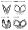
- FIG. 4 (a) is a Lorentz attractor
- Fig. 4 (b) is a reconstruction of the prototype from the Lorenz attractor recurrence plot
- Fig. 4 (c) is a random plot of 1% on the Lorenz attractor recurrence plot.
- FIG. 4D is a diagram showing a result of reconstructing the prototype by removing 90% of data from the Lorentz attractor recurrence plot.
- Fig. 5 (a) shows the wrestler attractor.
- Fig. 5 (a) shows the wrestler attractor.
- FIG. 5D is a diagram showing the result of reconstructing the prototype by removing 90% of the data from the recurrence plot of the wrestler attractor. It is a figure which shows the example (chromosome 2) of the reconstruction of a mouse genome.
- the method for reconstructing the three-dimensional structure of biomolecule data using the concept of spatial proximity determines whether any two molecular regions in a biomolecule or sequences are spatially close.
- the three-dimensional structure of the biomolecule is reconstructed by calculating the distance in the three-dimensional space between the two sequences using the binary information.
- the recurrence plot is originally a tool for visualizing time series data.
- the vertical axis and the horizontal axis are the same time axis.
- the states corresponding to two time points are compared with each other, and if the distance between the states is short, a point is hit at the corresponding place, and if not, the point is not hit.
- the recurrence plot converts continuous time series data into binary matrix information indicating whether the distance is close or not, so it may seem that the information about time series data has dropped considerably. Absent. Although the absolute value information of the time series values will certainly drop, it is known that the outline of the time series data can be restored from the recurrence plot. In particular, when the points are uniformly distributed, a metric space equivalent to the original metric space is reconstructed (see Non-Patent Documents 5 to 8 above).
- G i is a set of time points corresponding to the points on the recurrence plot of the i- th row, and
- the shortest path between any two vertices on this graph is obtained. This can be easily performed using, for example, the Dijkstra method (see Non-Patent Document 9). Then, a distance matrix is obtained in which the distance between any two vertices is given. If a multidimensional scale construction method is used for this distance matrix, time series data that reproduces the outline of the original time series data can be reconstructed.
- the technique relating to the recurrence plot can be directly applied to the nucleic acid sequence and amino acid sequence. That is, the three-dimensional structure of DNA, RNA, or protein can be obtained by looking at the top three components when reconstructed using a multidimensional scale construction method (see Non-Patent Document 10 above).
- Non-Patent Document 11 described above for information that continuous arrays are spatially close, the diagonal line at the center of the corresponding recurrence plot is thickened as a line having a width of 3 or more.
- a method can be constructed in which the dimensional structure can be reconstructed.
- FIG. 2 is a diagram showing the reconstruction of the three-dimensional structure of yeast according to an embodiment of the present invention, in which the horizontal axis is the first constituent molecule and the horizontal axis is the second constituent molecule.
- FIG. 3 is an enlarged view of FIG. 1, in which the horizontal axis is the first coordinate and the horizontal axis is the second coordinate. In addition, the position of the centromere is indicated by a cross.
- Results are shown in FIG. 2 and FIG.
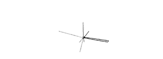
- FIG. 2 among chromosomes 1 to 16, chromosome 12 is shown in black and the others are shown in gray. A large loop was conspicuous in the rDNA region encoding rDNA located on chromosome 12 shown in black.
- FIG. 3 the centromeres are illustrated with black dots, but it can be seen that the centromeres are particularly close to each other. In fact, the cost required to construct the minimum tree tends to be smaller when the minimum tree of the centromeres is found than when a single point is randomly searched from each chromosome. I understood that. It has been suggested in previous studies that centromere is likely to aggregate in the budding yeast used to obtain this data, and that rDNA forms a large loop, suggesting the validity of the method of the present invention.
- the advantage of the present invention is that it is less susceptible to noise. For example, even if information about whether spatially close is about 1%, in the example of the numerical experiment, the correlation between the original distance and the reconstructed distance holds 0.70 or more. Since the experimental data for observing the neighboring points of the molecules in the living body is expected to contain false positive noise or not to cover all the neighboring points, the present invention is effective.
- Figures 4 and 5 (a) to (c) show examples of toy models reconstructed from Lorenz attractor and wrestler attractor from recurrence plots, and 1% noise flip for each recurrence plot. The result of reconstruction after adding.
- the present invention is extremely resistant to data loss.
- 50% and 90% of the data were randomly lost in the toy model, and reconstruction was attempted.
- Non-Patent Document 2 In the method of Non-Patent Document 2, up to 1000 points can be handled. On the other hand, the example shown in FIGS. 2 and 3 deals with 11986 points. In other words, the present invention can reconstruct a three-dimensional structure of large data with a finer resolution. According to the present invention, it is possible to handle size data including human and mouse mammalian genomes without reducing the resolution.
- FIG. 6 shows an example of mouse genome rearrangement.
- the conventional method estimates a plurality of plausible models, whereas the method of the present invention is not a probabilistic method but a method based on the reconstruction of the distance between sequences.
- the three-dimensional structure is unique and reproducible. In that respect, the method of the present invention has significant advantages over conventional methods.
- this invention is not limited to the said Example, A various deformation
- the method for reconstructing the three-dimensional structure of biomolecule data using the concept of spatial proximity is not a probabilistic method, but a method based on reconstruction of the distance between sequences. And can be used as a method for reconstructing the three-dimensional structure of biomolecule data using the concept of spatial proximity.
Landscapes
- Spectroscopy & Molecular Physics (AREA)
- Life Sciences & Earth Sciences (AREA)
- Physics & Mathematics (AREA)
- Bioinformatics & Cheminformatics (AREA)
- Biotechnology (AREA)
- Biophysics (AREA)
- Health & Medical Sciences (AREA)
- Engineering & Computer Science (AREA)
- Chemical & Material Sciences (AREA)
- Bioinformatics & Computational Biology (AREA)
- Crystallography & Structural Chemistry (AREA)
- Evolutionary Biology (AREA)
- General Health & Medical Sciences (AREA)
- Medical Informatics (AREA)
- Theoretical Computer Science (AREA)
- Management, Administration, Business Operations System, And Electronic Commerce (AREA)
- Measuring Or Testing Involving Enzymes Or Micro-Organisms (AREA)
Abstract
【課題】確率的な方法ではなく、配列間の距離の再構成に基づく方法により、一義性と再現性をもたせて、空間的な近さの概念を用いた生体分子データの3次元構造の再構成を行う。 【解決手段】空間的な近さの概念を用いた生体分子データの3次元構造の再構成方法であって、生体分子内の任意の2つの分子領域同士、あるいは、配列同士が空間的に近いか否かの2値情報を利用して、2つの配列間の3次元空間内における距離を計算することで、生体分子の3次元構造を再構成する。
Description
本発明は、核酸やタンパク質をはじめとする生体分子の部分領域同士が3次元空間内で近いか否かの2値の情報から、生体分子データの3次元構造の再構成する生体分子データの3次元構造の再構成方法に関するものである。
DNAやRNAは、ヌクレオチドが2本鎖または1本鎖の鎖状に連なった高分子である。タンパク質は、アミノ酸が鎖状に連なった高分子である。
一般的に、DNAやRNA、タンパク質は細胞内で、核酸同士、タンパク質同士、または核酸とタンパク質の相互作用により、3次元構造を形成する。DNAやRNA、タンパク質の3次元構造は、それらの機能と密接に絡んでいると考えられ、生体機能や疾病を理解する上で重要な情報であると考えられる。
染色体の3次元構造の解析には、空間的に近接する配列を検出するchromosome conformation capture(3Cアッセイ)と呼ばれる実験手法が使われる。さらに、3Cは、空間的に近接する核酸断片情報をマイクロアレイや次世代シークエンサーを用いて網羅的に解析する4C、5C、Hi-Cなどの方法が開発されてきた。これらの細胞内での染色体間の空間的な距離を測るデータから、3次元構造を再構成するためには、部分配列間の3次元空間内での距離が近いか否かという情報がよく用いられる。しかし、現状では、3次元構造の再構成をするのに確率的な解析手法や、部分配列間の距離に関する補助的な情報を用いることが一般的となっている(下記非特許文献1、2参照)。
タンパク質や核酸の3次元構造解析には、X線結晶構造解析やクライオ電子顕微鏡法を用いた解析に加え, 核磁気共鳴(NMR)法により, 原子核の間の相互作用や原子の結合、電子状態をスペクトルとして検出する方法が用いられる。
Marc A. Marti-Renom and Leonid A. Mirny, Bridging the resolution gap in structural modeling of 3D genome organization, PLoS Computational Biology 7, e1002125 (2011).
Annick Lesne, Julien Riposo, Paul Roger, Axel Cournac, and Julien Mozziconacci, 3D genome reconstruction from chromosomal contacts, Nature Method 11, 1141-1143 (2014).
J. -P. Eckmann, S. O. Kamphorst, and D. Ruelle, Recurrence plots of dynamical systems, Europhysics Letters 4, 973-977 (1987).
N. Marwan, M. C. Romano, M. Thiel, andJ. Kurths, Recurrence plots for the analysis of complex systems, Physics Reports 438, 237-329 (2007).
Yoshito Hirata, Shunsuke Horai, and Kazuyuki Aihara, Reproduction of distance matrices and original time series from recurrence plots and their applications, European PhysicalJournal Special Topics 164, 13-22 (2008)
平田祥人, 合原一幸, 正規分布に従う乱数発生機構, 特願2009-548035, CPT/JP2008/073389, 特許4947476号.(アメリカ特許, Patent No. :US8,438,202B2, Date of Patent:May7, 2013)
平田祥人, 合原一幸, 1つのシステムの受ける複数の外力の同時再構成方法及びその装置, CPT/ JP2009/ 003355.
Yoshito Hirata, Motomasa Komuro, Shunsuke Horai, and Kazuyuki Aihara, Faithfulness of recurrence plots:A mathematical proof, International Journal of Bifurcation and Chaos in press. volume 25,art.no.155168(2015)
E. W. Dijkstra, A note on two problems in connexion with graphs, Numerische Mathematik 1, 269-271 (1959).
J. C. Gower, Some distance properties of latent root and vector methods used in multivariate analysis, Biometrika, 53, 325-338 (1966).
Masaaki Tanio, Yoshito Hirata, and Hideyuki Suzuki, Reconstruction of driving forces through recurrence plots, Physics Letters A 373, 2031-2040 (2009).
Zhijun Duan, Mirela Andronescu, Kevin Schutz, Sean Mcllwain, Yoo Jung Kim, Choli Lee, Hay Shendure, Stanley Fields, C. Anthony Blau, and WilliamS. Noble, A three-dimensional model of the yeast genome, Nature 465, 363-367 (2010).
そこで、本発明では、数理的な解析方法により、部分配列・部分領域間の距離が3次元空間内で近いかどうかという2値情報を用いて、核酸やタンパク質をはじめとする生体分子データの3次元構造を再構成する空間的な近さの概念を用いた生体分子データの3次元構造の再構成方法を提案する(上記非特許文献3,4参照)。
本発明は、上記状況に鑑みて、DNAやRNAの部分配列同士が近いか否かの2値の情報から生体分子の3次元構造を再構成する、空間的な近さの概念を用いた生体分子データの3次元構造の再構成方法を提供することを目的とする。
本発明は、上記目的を達成するために、
〔1〕空間的な近さの概念を用いた生体分子データの3次元構造の再構成方法であって、生体分子内の任意の2つの分子領域同士、あるいは、配列同士が空間的に近いか否かの2値情報を利用して、2つの配列間の3次元空間内における距離を計算することで、生体分子の3次元構造を再構成することを特徴とする。
〔1〕空間的な近さの概念を用いた生体分子データの3次元構造の再構成方法であって、生体分子内の任意の2つの分子領域同士、あるいは、配列同士が空間的に近いか否かの2値情報を利用して、2つの配列間の3次元空間内における距離を計算することで、生体分子の3次元構造を再構成することを特徴とする。
〔2〕上記〔1〕記載の空間的な近さの概念を用いた生体分子データの3次元構造の再構成方法において、前記生体分子が核酸であることを特徴とする。
〔3〕上記〔2〕記載の空間的な近さの概念を用いた生体分子データの3次元構造の再構成方法において、前記核酸がDNAやRNAであることを特徴とする。
〔4〕上記〔1〕記載の空間的な近さの概念を用いた生体分子データの3次元構造の再構成方法において、前記生体分子がタンパク質であることを特徴とする。
本発明によれば、以下のような効果を奏することができる。
従来法では、複数のもっともらしいモデルを推定するのに対し、本発明の方法は、確率的な方法ではなく、配列間の距離の再構成に基づく方法であるので、本発明の方法で再構成される3次元構造には一義性と再現性がある。その点で、本発明の方法は、既存の方法に比べて大きな利点がある。
本発明の空間的な近さの概念を用いた生体分子データの3次元構造の再構成方法は、生体分子内の任意の2つの分子領域同士、あるいは、配列同士が空間的に近いか否かの2値情報を利用して、2つの配列間の3次元空間内における距離を計算することで、生体分子の3次元構造を再構成する。
以下、本発明の実施の形態について詳細に説明する。
リカレンスプロットは、元々は、時系列データを視覚化するための道具である。2次元の平面図で、縦軸、横軸とも同じ時間軸になっている。2つの時間点に対応する状態同士を比較し、その状態間の距離が近ければ、対応する場所に点を打ち、そうでなければ点を打たないとすることによって求めることができる。
リカレンスプロットは、連続値の時系列データを距離が近いかどうかを示す2値の行列情報に変換してしまっているので、時系列データに関する情報がかなり落ちてしまっていると思われるかもしれない。確かに時系列の値の絶対値の情報は落ちてしまうのだが、リカレンスプロットから時系列データの概形が復元できることが知られている。特に、点が一様に分布している時、元の距離空間と同値な距離空間が再構成される(上記非特許文献5~8参照)。
リカレンスプロットから元の時系列データの概形を復元するのは簡単である。まずは、リカレンスプロットをグラフだと見立てる。このグラフでは、リカレンスプロットの時間点が頂点になっていて、2つの時間点に対応する場所に点が打たれていれば、対応する頂点間を枝で結ぶ。この時、枝に次のような距離を割り当てる。
1-|Gi ∩Gj |/|Gi ∪Gj | .
ここで、Gi はi行目のリカレンスプロットに打たれている点に対応する時間点の集合であり、|A|は集合Aの要素数を表す。次に、このグラフ上の任意の2つの頂点間の最短路を求める。これは、例えば、ダイクストラ法(上記非特許文献9参照)などを用いると簡単にできる。そして、任意の2つの頂点間の距離が与えられる距離行列を得る。この距離行列に対して、多次元尺度構成法を用いると、元の時系列データの概形を再現するような時系列データが再構成できる。
ここで、Gi はi行目のリカレンスプロットに打たれている点に対応する時間点の集合であり、|A|は集合Aの要素数を表す。次に、このグラフ上の任意の2つの頂点間の最短路を求める。これは、例えば、ダイクストラ法(上記非特許文献9参照)などを用いると簡単にできる。そして、任意の2つの頂点間の距離が与えられる距離行列を得る。この距離行列に対して、多次元尺度構成法を用いると、元の時系列データの概形を再現するような時系列データが再構成できる。
ここで、生体高分子の一次配列を時間軸と見立てることで、リカレンスプロットに関する手法がそのまま核酸配列やアミノ酸配列に対しても応用できる。つまり、多次元尺度構成法(上記非特許文献10参照)を用いて再構成した時の上位3つの成分を見れば、DNAやRNA、タンパク質の3次元構造が求まる。
そこで、連続する配列が空間的に近いという情報を上記非特許文献11の手法を用い、対応するリカレンスプロットの中央の斜めの線を幅3以上の線のように太くすることで、必ず3次元構造が再構成できるような方法を構成できる。
図2は本発明の実施例を示す酵母の3次元構造の再構成を示す図であり、横軸は第1構成分子、横軸は第2構成分子である。図3は図1の拡大図であり、横軸は第1座標、横軸は第2座標である。また、×印でセントロメアの位置を示す。
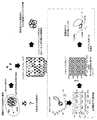
ここでは、上記非特許文献12で使用された酵母のデータを使って、本発明の方法を確かめた。ここでは、4つの別々の切断酵素を用いて求めたHi-Cのデータを統合して、3次元データの再構成を行った。Hi-Cのデータ解析における本発明の位置付けと、先行研究の概観を図1に示す。
結果を図2と図3に示す。図2では染色体1~16番のうち、染色体12番を黒で、それ以外を灰色で図示した。黒で示した染色体12番上に位置するrDNAをコードするrDNA領域に大きなル-プが目立った。また、図3では黒のドットでセントロメアを図示したが、特に、セントロメア同士が近い場所にあることが分かる。実際、各染色体上からランダムに1点探してきて、最小木を求めるよりも、セントロメア同士の最小木を求めた時の方が、最小木を構成するのに必要なコストが小さくできる傾向にあることが分かった。このデータを得るのに用いられた出芽酵母ではセントロメアが凝集しやすいこと、rDNAが大きなループを作ることは先行研究においても示唆されており、本発明の方法の妥当性を伺わせる。
加えて、一般に転写や複製などの際には、核内でDNAと関連因子群が密集することが知られている。実際、本方法で再構成した染色体の空間を64000個の立方体に分割したところ、過半数の染色体が単一立方体内に密集して局在する場所が26個で見つかった。このため、本発明の方法は、転写ファクトリーや複製ファクトリーなどの機能ドメインの推定にも利用できる。
また、本発明の利点として、ノイズの影響を受けにくいことが挙げられる。例えば、1%程度、空間的に近いかどうかという情報が誤っていたとしても、数値実験の例では、元の距離と再構成した距離の相関が0.70以上を保持している。生体内分子の近傍点を観測する実験データは、偽陽性のノイズを含んだり、全ての近傍点を網羅しきれないことが予想されるので、本発明が有効である。
図4、図5の(a)~(c)におもちゃモデルとして、ローレンツアトラクター、レスラーアトラクターの概形をリカレンスプロットから再構成した例と、さらに各リカレンスプロットに1%のノイズフリップを加えてから再構成をした結果を示す。
さらに、本発明では、データの欠損に極めて強い。実際、おもちゃモデルでランダムに50%、90%のデータを欠損させて再構成の試行を行った所、欠損のないデータと比べてもそれぞれ、0.96,0.87以上の相関があった〔図4(d)、図5(d)〕。
現在一般に行われている1細胞レベルでの観測は技術的な検出感度の限界から、得られるデータが網羅性を欠いて粗であることが指摘されている。本発明はこのような生物データに対しても、優位性があると考えられる。
上記非特許文献2の方法では、1000点までは扱えるとされている。その一方で、図2、図3に示した例では11986点を扱っている。つまり、本発明の方が、より細かい解像度で大きなデータの3次元構造の再構成が可能である。本発明であれば、人やマウスの哺乳類ゲノムなどを含めサイズのデータを解像度を落とさずに扱うことも可能である。図6にマウスのゲノム再構成の例を示す。
図1、図2のデータの再構成に、2X2.66GHz6-Core Intel XeonのCPUと64GBのメモリを搭載したコンピュータ上で、MATLABで書かれたプログラムを使用して、約2日で計算を完了できる。
従来法では、複数のもっともらしいモデルを推定するのに対し、本発明の方法は、確率的な方法ではなく、配列間の距離の再構成に基づく方法であるので、本発明の方法で再構成される3次元構造には一義性と再現性がある。その点で、本発明の方法は、従来の方法に比べて大きな利点がある。
なお、本発明は上記実施例に限定されるものではなく、本発明の趣旨に基づき種々の変形が可能であり、これらを本発明の範囲から排除するものではない。
本発明の空間的な近さの概念を用いた生体分子データの3次元構造の再構成方法は、確率的な方法ではなく、配列間の距離の再構成に基づく方法により、一義性と再現性をもたせて、空間的な近さの概念を用いた生体分子データの3次元構造の再構成方法として利用可能である。
Claims (4)
- 生体分子内の任意の2つの分子領域同士、あるいは、配列同士が空間的に近いか否かの2値情報法を利用して、2つの配列間の3次元空間内における距離を計算することで、生体分子の3次元構造を再構成することを特徴とする空間的な近さの概念を用いた生体分子データの3次元構造の再構成方法。
- 請求項1記載の空間的な近さの概念を用いた生体分子データの3次元構造の再構成方法において、前記生体分子が核酸であることを特徴とする空間的な近さの概念を用いた生体分子データの3次元構造の再構成方法。
- 請求項2記載の空間的な近さの概念を用いた生体分子データの3次元構造の再構成方法において、前記核酸がDNAやRNAであることを特徴とする空間的な近さの概念を用いた生体分子データの3次元構造の再構成方法。
- 請求項1記載の空間的な近さの概念を用いた生体分子データの3次元構造の再構成方法において、前記生体分子がタンパク質であることを特徴とする空間的な近さの概念を用いた生体分子データの3次元構造の再構成方法。
Applications Claiming Priority (2)
| Application Number | Priority Date | Filing Date | Title |
|---|---|---|---|
| JP2016-023214 | 2016-02-10 | ||
| JP2016023214A JP6765040B2 (ja) | 2016-02-10 | 2016-02-10 | 空間的な近さの概念を用いた生体分子データの3次元構造の再構成方法 |
Publications (1)
| Publication Number | Publication Date |
|---|---|
| WO2017138591A1 true WO2017138591A1 (ja) | 2017-08-17 |
Family
ID=59563143
Family Applications (1)
| Application Number | Title | Priority Date | Filing Date |
|---|---|---|---|
| PCT/JP2017/004670 Ceased WO2017138591A1 (ja) | 2016-02-10 | 2017-02-09 | 空間的な近さの概念を用いた生体分子データの3次元構造の再構成方法 |
Country Status (2)
| Country | Link |
|---|---|
| JP (1) | JP6765040B2 (ja) |
| WO (1) | WO2017138591A1 (ja) |
Cited By (1)
| Publication number | Priority date | Publication date | Assignee | Title |
|---|---|---|---|---|
| CN113808661A (zh) * | 2021-09-18 | 2021-12-17 | 山东财经大学 | 染色体三维结构重建方法及装置 |
Families Citing this family (2)
| Publication number | Priority date | Publication date | Assignee | Title |
|---|---|---|---|---|
| WO2023048251A1 (ja) * | 2021-09-27 | 2023-03-30 | 国立大学法人筑波大学 | 構造推定プログラム、構造推定装置及び構造推定方法 |
| WO2026094489A1 (ja) * | 2024-10-30 | 2026-05-07 | 国立大学法人 筑波大学 | 構造推定方法、構造推定装置、及びプログラム |
Citations (4)
| Publication number | Priority date | Publication date | Assignee | Title |
|---|---|---|---|---|
| JPH11232291A (ja) * | 1998-02-16 | 1999-08-27 | Seibutsu Bunshi Kogaku Kenkyusho:Kk | 蛋白質立体構造データベース検索方法 |
| JP2000319203A (ja) * | 1999-05-14 | 2000-11-21 | Iyaku Bunshi Sekkei Kenkyusho:Kk | 化合物の3次元構造の作成方法 |
| WO2009084524A1 (ja) * | 2007-12-27 | 2009-07-09 | Japan Science And Technology Agency | 正規分布に従う乱数発生機構 |
| WO2010010675A2 (ja) * | 2008-07-22 | 2010-01-28 | 独立行政法人科学技術振興機構 | 1つのシステムの受ける複数の外力の同時再構成方法及びその装置 |
-
2016
- 2016-02-10 JP JP2016023214A patent/JP6765040B2/ja not_active Expired - Fee Related
-
2017
- 2017-02-09 WO PCT/JP2017/004670 patent/WO2017138591A1/ja not_active Ceased
Patent Citations (4)
| Publication number | Priority date | Publication date | Assignee | Title |
|---|---|---|---|---|
| JPH11232291A (ja) * | 1998-02-16 | 1999-08-27 | Seibutsu Bunshi Kogaku Kenkyusho:Kk | 蛋白質立体構造データベース検索方法 |
| JP2000319203A (ja) * | 1999-05-14 | 2000-11-21 | Iyaku Bunshi Sekkei Kenkyusho:Kk | 化合物の3次元構造の作成方法 |
| WO2009084524A1 (ja) * | 2007-12-27 | 2009-07-09 | Japan Science And Technology Agency | 正規分布に従う乱数発生機構 |
| WO2010010675A2 (ja) * | 2008-07-22 | 2010-01-28 | 独立行政法人科学技術振興機構 | 1つのシステムの受ける複数の外力の同時再構成方法及びその装置 |
Cited By (2)
| Publication number | Priority date | Publication date | Assignee | Title |
|---|---|---|---|---|
| CN113808661A (zh) * | 2021-09-18 | 2021-12-17 | 山东财经大学 | 染色体三维结构重建方法及装置 |
| CN113808661B (zh) * | 2021-09-18 | 2022-06-10 | 山东财经大学 | 染色体三维结构重建方法及装置 |
Also Published As
| Publication number | Publication date |
|---|---|
| JP6765040B2 (ja) | 2020-10-07 |
| JP2017142633A (ja) | 2017-08-17 |
Similar Documents
| Publication | Publication Date | Title |
|---|---|---|
| Weinreb et al. | 3D RNA and functional interactions from evolutionary couplings | |
| Zhang et al. | IsRNA1: de novo prediction and blind screening of RNA 3D structures | |
| Ghauri et al. | pNitro-Tyr-PseAAC: predict nitrotyrosine sites in proteins by incorporating five features into Chou’s general PseAAC | |
| Menchaca et al. | Past, present, and future of molecular docking | |
| Cragnolini et al. | Coarse-grained HiRE-RNA model for ab initio RNA folding beyond simple molecules, including noncanonical and multiple base pairings | |
| Lin et al. | Computational methods for analyzing and modeling genome structure and organization | |
| Huang et al. | Weakly supervised learning of RNA modifications from low-resolution epitranscriptome data | |
| Xiong et al. | A deep learning framework for improving long-range residue–residue contact prediction using a hierarchical strategy | |
| Li et al. | Imputation of spatially-resolved transcriptomes by graph-regularized tensor completion | |
| Bottaro et al. | RNA folding pathways in stop motion | |
| Hu et al. | PennSeq: accurate isoform-specific gene expression quantification in RNA-Seq by modeling non-uniform read distribution | |
| Sun et al. | Accurate prediction of RNA-binding protein residues with two discriminative structural descriptors | |
| Weiss et al. | Towards the complete small RNome of Acinetobacter baumannii | |
| Khrameeva et al. | Spatial proximity and similarity of the epigenetic state of genome domains | |
| JP6765040B2 (ja) | 空間的な近さの概念を用いた生体分子データの3次元構造の再構成方法 | |
| CN103473482A (zh) | 基于差分进化和构象空间退火的蛋白质三维结构预测方法 | |
| Ma et al. | RNANetMotif: Identifying sequence-structure RNA network motifs in RNA-protein binding sites | |
| Liu et al. | Biomolecular topology: Modelling and analysis | |
| Kolev et al. | Ab initio molecular dynamics of Na+ and Mg2+ countercations at the backbone of RNA in water solution | |
| Maurizio et al. | Quantum computing for genomics: conceptual challenges and practical perspectives | |
| Mabrouk et al. | Different genomic signal processing methods for eukaryotic gene prediction: a systematic REVIEW | |
| Forero | Bioinformatics and human genomics research | |
| CN117352065A (zh) | 蛋白质功能的预测方法、装置、计算机设备和存储介质 | |
| Kole et al. | Binding of homeodomain proteins to DNA with hoogsteen base pair | |
| Ashida et al. | Shape-based alignment of genomic landscapes in multi-scale resolution |
Legal Events
| Date | Code | Title | Description |
|---|---|---|---|
| 121 | Ep: the epo has been informed by wipo that ep was designated in this application |
Ref document number: 17750312 Country of ref document: EP Kind code of ref document: A1 |
|
| NENP | Non-entry into the national phase |
Ref country code: DE |
|
| 122 | Ep: pct application non-entry in european phase |
Ref document number: 17750312 Country of ref document: EP Kind code of ref document: A1 |