WO2017053586A1 - Device, system and method for using the device or system for assaying an analyte in a sample - Google Patents
Device, system and method for using the device or system for assaying an analyte in a sample Download PDFInfo
- Publication number
- WO2017053586A1 WO2017053586A1 PCT/US2016/053145 US2016053145W WO2017053586A1 WO 2017053586 A1 WO2017053586 A1 WO 2017053586A1 US 2016053145 W US2016053145 W US 2016053145W WO 2017053586 A1 WO2017053586 A1 WO 2017053586A1
- Authority
- WO
- WIPO (PCT)
- Prior art keywords
- cysteine
- sample
- gold
- various embodiments
- analyte
- Prior art date
- Legal status (The legal status is an assumption and is not a legal conclusion. Google has not performed a legal analysis and makes no representation as to the accuracy of the status listed.)
- Ceased
Links
- FFFHZYDWPBMWHY-VKHMYHEASA-N N[C@@H](CCS)C(O)=O Chemical compound N[C@@H](CCS)C(O)=O FFFHZYDWPBMWHY-VKHMYHEASA-N 0.000 description 1
- LEVWYRKDKASIDU-IMJSIDKUSA-N N[C@@H](CSSC[C@@H](C(O)=O)N)C(O)=O Chemical compound N[C@@H](CSSC[C@@H](C(O)=O)N)C(O)=O LEVWYRKDKASIDU-IMJSIDKUSA-N 0.000 description 1
Classifications
-
- B—PERFORMING OPERATIONS; TRANSPORTING
- B01—PHYSICAL OR CHEMICAL PROCESSES OR APPARATUS IN GENERAL
- B01L—CHEMICAL OR PHYSICAL LABORATORY APPARATUS FOR GENERAL USE
- B01L3/00—Containers or dishes for laboratory use, e.g. laboratory glassware; Droppers
- B01L3/50—Containers for the purpose of retaining a material to be analysed, e.g. test tubes
- B01L3/502—Containers for the purpose of retaining a material to be analysed, e.g. test tubes with fluid transport, e.g. in multi-compartment structures
- B01L3/5027—Containers for the purpose of retaining a material to be analysed, e.g. test tubes with fluid transport, e.g. in multi-compartment structures by integrated microfluidic structures, i.e. dimensions of channels and chambers are such that surface tension forces are important, e.g. lab-on-a-chip
-
- G—PHYSICS
- G01—MEASURING; TESTING
- G01N—INVESTIGATING OR ANALYSING MATERIALS BY DETERMINING THEIR CHEMICAL OR PHYSICAL PROPERTIES
- G01N33/00—Investigating or analysing materials by specific methods not covered by groups G01N1/00 - G01N31/00
- G01N33/48—Biological material, e.g. blood, urine; Haemocytometers
- G01N33/50—Chemical analysis of biological material, e.g. blood, urine; Testing involving biospecific ligand binding methods; Immunological testing
- G01N33/53—Immunoassay; Biospecific binding assay; Materials therefor
- G01N33/543—Immunoassay; Biospecific binding assay; Materials therefor with an insoluble carrier for immobilising immunochemicals
- G01N33/54366—Apparatus specially adapted for solid-phase testing
- G01N33/54373—Apparatus specially adapted for solid-phase testing involving physiochemical end-point determination, e.g. wave-guides, FETS, gratings
-
- G—PHYSICS
- G01—MEASURING; TESTING
- G01N—INVESTIGATING OR ANALYSING MATERIALS BY DETERMINING THEIR CHEMICAL OR PHYSICAL PROPERTIES
- G01N33/00—Investigating or analysing materials by specific methods not covered by groups G01N1/00 - G01N31/00
- G01N33/48—Biological material, e.g. blood, urine; Haemocytometers
- G01N33/50—Chemical analysis of biological material, e.g. blood, urine; Testing involving biospecific ligand binding methods; Immunological testing
- G01N33/53—Immunoassay; Biospecific binding assay; Materials therefor
- G01N33/575—Immunoassay; Biospecific binding assay; Materials therefor for cancer
- G01N33/57557—Immunoassay; Biospecific binding assay; Materials therefor for cancer of other specific parts of the body, e.g. brain
-
- G—PHYSICS
- G01—MEASURING; TESTING
- G01N—INVESTIGATING OR ANALYSING MATERIALS BY DETERMINING THEIR CHEMICAL OR PHYSICAL PROPERTIES
- G01N33/00—Investigating or analysing materials by specific methods not covered by groups G01N1/00 - G01N31/00
- G01N33/48—Biological material, e.g. blood, urine; Haemocytometers
- G01N33/50—Chemical analysis of biological material, e.g. blood, urine; Testing involving biospecific ligand binding methods; Immunological testing
- G01N33/68—Chemical analysis of biological material, e.g. blood, urine; Testing involving biospecific ligand binding methods; Immunological testing involving proteins, peptides or amino acids
- G01N33/6803—General methods of protein analysis not limited to specific proteins or families of proteins
- G01N33/6806—Determination of free amino acids
- G01N33/6812—Assays for specific amino acids
- G01N33/6815—Assays for specific amino acids containing sulfur, e.g. cysteine, cystine, methionine, homocysteine
-
- A—HUMAN NECESSITIES
- A61—MEDICAL OR VETERINARY SCIENCE; HYGIENE
- A61B—DIAGNOSIS; SURGERY; IDENTIFICATION
- A61B5/00—Measuring for diagnostic purposes; Identification of persons
- A61B5/02—Detecting, measuring or recording for evaluating the cardiovascular system, e.g. pulse, heart rate, blood pressure or blood flow
- A61B5/02007—Evaluating blood vessel condition, e.g. elasticity, compliance
-
- A—HUMAN NECESSITIES
- A61—MEDICAL OR VETERINARY SCIENCE; HYGIENE
- A61B—DIAGNOSIS; SURGERY; IDENTIFICATION
- A61B5/00—Measuring for diagnostic purposes; Identification of persons
- A61B5/145—Measuring characteristics of blood in vivo, e.g. gas concentration or pH-value ; Measuring characteristics of body fluids or tissues, e.g. interstitial fluid or cerebral tissue
- A61B5/14546—Measuring characteristics of blood in vivo, e.g. gas concentration or pH-value ; Measuring characteristics of body fluids or tissues, e.g. interstitial fluid or cerebral tissue for measuring analytes not otherwise provided for, e.g. ions, cytochromes
-
- A—HUMAN NECESSITIES
- A61—MEDICAL OR VETERINARY SCIENCE; HYGIENE
- A61B—DIAGNOSIS; SURGERY; IDENTIFICATION
- A61B5/00—Measuring for diagnostic purposes; Identification of persons
- A61B5/42—Detecting, measuring or recording for evaluating the gastrointestinal, the endocrine or the exocrine systems
- A61B5/4222—Evaluating particular parts, e.g. particular organs
- A61B5/4255—Intestines, colon or appendix
-
- A—HUMAN NECESSITIES
- A61—MEDICAL OR VETERINARY SCIENCE; HYGIENE
- A61B—DIAGNOSIS; SURGERY; IDENTIFICATION
- A61B5/00—Measuring for diagnostic purposes; Identification of persons
- A61B5/43—Detecting, measuring or recording for evaluating the reproductive systems
- A61B5/4306—Detecting, measuring or recording for evaluating the reproductive systems for evaluating the female reproductive systems, e.g. gynaecological evaluations
- A61B5/4312—Breast evaluation or disorder diagnosis
-
- A—HUMAN NECESSITIES
- A61—MEDICAL OR VETERINARY SCIENCE; HYGIENE
- A61B—DIAGNOSIS; SURGERY; IDENTIFICATION
- A61B5/00—Measuring for diagnostic purposes; Identification of persons
- A61B5/43—Detecting, measuring or recording for evaluating the reproductive systems
- A61B5/4375—Detecting, measuring or recording for evaluating the reproductive systems for evaluating the male reproductive system
- A61B5/4381—Prostate evaluation or disorder diagnosis
-
- B—PERFORMING OPERATIONS; TRANSPORTING
- B01—PHYSICAL OR CHEMICAL PROCESSES OR APPARATUS IN GENERAL
- B01L—CHEMICAL OR PHYSICAL LABORATORY APPARATUS FOR GENERAL USE
- B01L2200/00—Solutions for specific problems relating to chemical or physical laboratory apparatus
- B01L2200/02—Adapting objects or devices to another
- B01L2200/026—Fluid interfacing between devices or objects, e.g. connectors, inlet details
- B01L2200/027—Fluid interfacing between devices or objects, e.g. connectors, inlet details for microfluidic devices
-
- B—PERFORMING OPERATIONS; TRANSPORTING
- B01—PHYSICAL OR CHEMICAL PROCESSES OR APPARATUS IN GENERAL
- B01L—CHEMICAL OR PHYSICAL LABORATORY APPARATUS FOR GENERAL USE
- B01L2300/00—Additional constructional details
- B01L2300/06—Auxiliary integrated devices, integrated components
- B01L2300/0627—Sensor or part of a sensor is integrated
- B01L2300/0645—Electrodes
-
- B—PERFORMING OPERATIONS; TRANSPORTING
- B01—PHYSICAL OR CHEMICAL PROCESSES OR APPARATUS IN GENERAL
- B01L—CHEMICAL OR PHYSICAL LABORATORY APPARATUS FOR GENERAL USE
- B01L2300/00—Additional constructional details
- B01L2300/08—Geometry, shape and general structure
- B01L2300/0809—Geometry, shape and general structure rectangular shaped
- B01L2300/0816—Cards, e.g. flat sample carriers usually with flow in two horizontal directions
-
- G—PHYSICS
- G01—MEASURING; TESTING
- G01N—INVESTIGATING OR ANALYSING MATERIALS BY DETERMINING THEIR CHEMICAL OR PHYSICAL PROPERTIES
- G01N2800/00—Detection or diagnosis of diseases
- G01N2800/32—Cardiovascular disorders
Definitions
- This invention concerns a device and system, and method for using the device and/or system, for detecting and quantifying an analyte in a sample, such as for monitoring circulating methionine metabolite levels, such as cysteine levels, in a sample for predicting the risk or probability of a disease state, such as occurrence or recurrence of cancer, such as prostate cancer, urinary tract cystine stone disease, and cardiovascular disease.
- a sample such as for monitoring circulating methionine metabolite levels, such as cysteine levels
- a sample for predicting the risk or probability of a disease state, such as occurrence or recurrence of cancer, such as prostate cancer, urinary tract cystine stone disease, and cardiovascular disease.
- Prostate cancer remains the most common, non-cutaneous solid malignancy in the United States, and the second leading cause of cancer specific death in men. Nevertheless, it has become increasingly clear that not all men who are diagnosed with prostate cancer require intervention. Yet, many men that receive surgical or radiation-based primary treatment develop recurrent disease. Prior to surgical intervention, serum PSA, biopsy Gleason grade, and clinical stage help determine if patients are likely to be recurrent versus those that may remain localized and possibly remain clinically inconsequential. Various approaches in improving the role of PSA in early prostate cancer detection have been tested, but their benefit to overall survival is yet to be proven.
- Cystinuria is a common genetic metabolic disorder (1 in 7000) accounting for 1-2% of all cases of renal lithiasis. Cystine crystals precipitate in the kidney and accumulate in the bladder to form calculi with a diameter of up to 5 mm. Cystine results from oxidation of two cysteine molecules covalently linked by a disulfide bond. Impaired reabsorption of cystine leads to a high risk for the formation of cystine calculi in the urinary tract, potentially causing obstruction, infections and eventually renal failure.
- Patients with cystinuria can control circulating cysteine through dietary modifications and increased fluid intake. Such patients are often treated with cystine solubilizing drugs like D-penicillamine, mercaptopropionylglycine or Captopril. The side effects from such drugs result in noncompliance that can lead to reemergence of calculi.
- Urinary tract obstruction can lead to hydronephrosis and ultimately to loss of renal function.
- the ready detection of cysteine in urine and blood of such individuals by a point of care device can enable self-dietary modification and indicate clinical intervention at an early stage to prevent renal damage and loss of function.
- MI Myocardial infarction
- ischemia i.e., heart attack
- Plasma homocysteine is an established independent risk factor for MI and coronary artery disease (Nygard et al., Plasma homocysteine levels and mortality in patients with coronary artery disease, N Engl J Med. 1997 Jul 24;337(4):230-6).
- cysteine is a biologic marker for oxidative stress, its application in heart, coronary artery, and peripheral vascular disease after an MI is a point of significant study.
- Cysteine has a general cytotoxicity in vitro (Nishiuch et al., Cytotoxicity of cysteine in culture media, In Vitro. 1976 Sep;12(9):635-8) and promotes detachment of human arterial endothelial cells in culture (Dudman et al., Human arterial endothelial cell detachment in vitro: its promotion by homocysteine and cysteine,
- Atherosclerosis 1991 Nov;91(l-2):77-83).
- Certain disclosed embodiments concern a device comprising, consisting of or consisting essentially of a sample chamber having at least one analyte inlet, and a sensor component comprising an electrically conductive metal substrate or electrically conductive metal deposited or formed on a substrate.
- the conductive metal provides a reaction surface capable of binding an analyte having a functional group comprising sulfur.
- the sensor component further comprises electrodes electrically coupled to the conductive metal and to a component for determining an electrical parameter of the metal, such as impedance, resistance, and/or conductance, subsequent to analyte binding to the metal surface. For example, if the parameter is impedance, the device further comprises a component for measuring impedance.
- the electrically conductive metal may be any suitable metal, but typically is selected from gold, silver, platinum, iridium, and combinations thereof, with gold being a particularly suitable metal.
- the electrically conductive metal may define a fluid flow path over which an analyte solution flows, the metal typically having a thickness of from 1 to 500 nanometers, a width of from 0.1 to about 20 millimeters, and a length of from about 0.1 to about 200 millimeters.
- the electrically conductive metal may be configured as a straight, curve, winding, and/or tortuous path.
- the sample chamber may define plural electrically insulated reaction surfaces.
- the device also may comprise plural sample chambers, arranged in parallel or in series.
- Certain disclosed embodiments further comprise an enzyme reaction module configured to process a sample with an enzyme, such as cystathionine synthase and/or cystathionine lyase, before the sample contacts the reaction surface.
- an enzyme reaction module configured to process a sample with an enzyme, such as cystathionine synthase and/or cystathionine lyase, before the sample contacts the reaction surface.
- the disclosed embodiments particularly concern a point of care device, and even more particularly a point of care device for detecting an amount of cysteine in a sample from a subject.
- Certain aspects of the present invention concern the recognition that a molecule reacting with a metal surface, such as a gold surface, induces an impedance change in the metal, and that impedance change can be directly correlated with the amount of the molecule reacting with the metal surface, or interacting with a capture molecule bound, typically covalently, to the metal surface.
- the conductive metal substrate may comprise a receptor biomolecule coupled to a portion of the metal surface through a thiol functional group.
- a remaining portion of the metal surface may comprise a blocking agent, such as a thiolated polyethylene glycol, to preclude target molecule binding to the surface.
- the receptor molecule is a peptide, such as an antibody or extracellular receptor domain, that is coupled to the metal surface.
- One method of coupling a peptide to the surface is by modifying the peptide to include at least one pendant cysteine.
- Exemplary embodiments instituting this concept include configuring a device to assess ⁇ -2 transferrin, NGAL, cystatin C, and C-reactive protein, Ara h 1, Ara h 2, and Ara h 3, GFAP and UCK-L1 proteins, AFP (alpha-fetoprotein), AST
- the device may include plural sample chambers, or a sample chamber may comprises plural reactive, electrically-insulated reaction surfaces, and wherein a portion of the metal surface further reacts with cysteine or homocysteine in the sample.
- certain device embodiments are used to determine if an injury or surgery is leaking CSF, wherein the device comprises an antibody or transferrin receptor immobilized to a gold nanowell surface; (2) are used to assess renal function and the device comprises NGAL, cystatin C, and C-reactive protein; (3) include Ara h 1, Ara h 2, and/or Ara h 3 immobilized on a gold nanowell surface to detect IgE immunoglobins specific to these antigens; (4) comprise antibodies to GFAP and UCK-L1 proteins immobilized on a gold nanowell surface and the device is configured for assessing concussions or traumatic brain injury; (5) comprise antibodies to AFP (alpha-fetoprotein), AST (Aspartate aminotransferase), and/or ALT (Alanine aminotransferase) immobilized on a gold nanowell surface, and the device is configured to screen for liver infections (hepatitis C) or monitor liver disease progression; and (6) comprise antibodies for troponin T
- Disclosed systems may include a sensor device that defines a disposable sensor unit comprising the electrically conductive metal for coupling to a detection device for detecting a change in an electrical parameter of the conductive metal subsequent to analyte binding.
- the system can comprise a reusable sensor unit comprising the electrically conductive metal.
- Disclosed systems can further comprise one or more of a central processing unit for controlling functions of the system; a temperature sensor; a data storage unit; a fluid pump for flowing analyte and/or enzyme solutions to and/or through the device; a sample collector; a sample reservoir or cartridge; one or more filtration modules positioned to filter a fluid stream into the system or between components of the system; an enzyme reservoir or cartridge; an enzyme reaction module; a buffer reservoir or cartridge; a power supply; and combinations thereof.
- a central processing unit for controlling functions of the system
- a temperature sensor for controlling functions of the system
- a data storage unit for controlling functions of the system
- a fluid pump for flowing analyte and/or enzyme solutions to and/or through the device
- a sample collector for flowing analyte and/or enzyme solutions to and/or through the device
- a sample reservoir or cartridge for flowing analyte and/or enzyme solutions to and/or through the device
- a sample collector for flowing analyte and/or enzyme solutions
- Various embodiments of the present invention also provide a method of using device, or a system comprising the device, as disclosed herein to detect cysteine and/or methionine metabolite levels, to diagnose, prognose or monitor a disease condition (e.g., cancer, cardiovascular disease, cystinuria and urinary tract cystine stone disease), or to predict the risk or probability of cancer recurrence.
- a disease condition e.g., cancer, cardiovascular disease, cystinuria and urinary tract cystine stone disease
- Various embodiments of the present invention provide a method of determining whether a subject has an increased probability of cancer recurrence.
- the method may consist of, or may consist essentially of, or may comprise: obtaining a sample from the subject; assaying the sample to detect an increased cysteine level, an increased methionine metabolite level, an increased PSA parameter, and/or a increased soluble CD105 (sCD105) level; detecting in the sample an increased cysteine level, an increased methionine metabolite level, an increased PSA parameter, and/or increased sCD105 level; and determining if the subject has an increased probability of cancer recurrence.
- sCD105 soluble CD105
- Certain disclosed method embodiments comprise using the device or system to measure an analyte in a sample.
- the analyte typically comprises a functional group comprising a sulfur atom.
- the analyte may be cysteine and the method comprises measuring a cysteine level in a sample.
- the analyte may have a functional group that is converted to a thiol enzymatically, chemically or thermally.
- the analyte may be reacted with cysteine to provide a terminal cysteine moiety for detection and measurement using the device.
- the method also may further comprise processing a sample with cystathionine synthase and/or cystathionine lyase, before contacting the sample with the reaction surface.
- the analyte is detected, and the analyte amount quantified, using an electrical parameter. If the electrical parameter is impedance, the measured impedance value may be correlated with an analyte amount in the sample, such as by using a standard curve.
- the analyte amount detected may be used to determine an occurrence or recurrence of cancer, atherosclerosis, or cardiovascular disease, such as to determine an occurrence or recurrence of prostate cancer, colon cancer, ovarian cancer, breast cancer, urinary tract disease, cystine stone disease (cystinuria), or myocardial infarction.
- Certain disclosed embodiments comprise using a device wherein the conductive metal substrate comprises a receptor biomolecule coupled to a portion of the metal surface through a thiol functional group. A remaining portion of the metal surface may comprise a blocking agent to preclude target molecule binding to the surface.
- the receptor molecule may be, for example, a peptide or an extracellular receptor domain that is coupled to the metal surface by cysteine.
- the peptide may be modified to include a pendant cysteine amino acid.
- the sample chamber comprises plural reactive surfaces, and a portion of the metal surface further reacts with free cysteine or homocysteine.
- Such embodiments can be used to: (1) determine if an injury or surgery is leaking CSF, wherein the device comprises an antibody or transferrin receptor immobilized to a gold nanowell surface; (2) assess renal function wherein the device comprises NGAL, cystatin C, and C-reactive protein; (3) the metal surface comprises Ara h 1, Ara h 2, and Ara h 3 to detect IgE immunoglobins specific to these antigens for peanut allergy determination; (4) the device is configured to assess concussions or traumatic brain injury, wherein the metal surface comprises antibodies to GFAP and UCK-Ll proteins; (5) the device is configured to screen for liver infections (hepatitis C) or monitor liver disease progression, wherein the metal surface comprises antibodies to AFP (alpha-fetoprotein), AST (aspartate aminotransferase), and/or ALT (alanine aminotransferase); and (6) the device is configured to assess cardiac infarction, wherein the metal surface comprises antibodies for troponin T and troponin I,
- FIG. 1 depicts, in accordance with various embodiments of the present invention, a modeled gold body, where electrical resistance is measured at various positions or along the entire length of the body.
- FIG. 2 depicts, in accordance with various embodiments of the present invention, Gold Film Experiment - Version 1.
- FIG. 3 depicts, in accordance with various embodiments of the present invention, Gold Film Experiment - Version la Setup: (a) Bare gold slide showing gold path; (b) Application of water cover to gold path.
- FIG. 4 depicts, in accordance with various embodiments of the present invention, Gold Film Experiment - Version la Results, where water cover was added to the gold path after 4 minutes and cysteine solution was added to the water cover after 12 minutes.
- FIG. 5 depicts, in accordance with various embodiments of the present invention, Gold Film Experiment - Version lb Setup: thermistor locations are shown in circles (solid line) and an oval (broken line).
- FIG. 6 depicts, in accordance with various embodiments of the present invention, Gold Film Experiment - Version lb Results, where time stamps for applying water cover to the gold surface, inserting thermistors into the water cover, and cysteine added to the water cover are shown in dashed lines.
- FIG. 7 depicts, in accordance with various embodiments of the present invention, Gold Film Experiment - Version la Compared to Version lb: (a) Water cover from Version 1, Slide 1; (b) Water cover from Version 1, Slide 2 [water cover in (b) flows over additional slide surface].
- FIG. 8 depicts, in accordance with various embodiments of the present invention, Gold Film Experiment - Version 2 Flow Cell Design: (a) two flow cells adhered to a microscope slide; (b) section of left flow cell showing fluid flow through luer lock fittings; and (c) section of left flow cell showing flow through ⁇ x 21mm x 25mm flow chamber and active gold surface.
- FIG. 9 depicts, in accordance with various embodiments of the present invention, an image of Gold Slide Experiment Version 2.
- FIG. 10 depicts, in accordance with various embodiments of the present invention, Cooling Channel and Temperature Measurement Thermistor.
- Microscope slide rests on a 25mm x 12.5mm rectangular air channel constructed from aluminum, and the channel walls are electrically insulated with clear tape.
- FIG. 11 depicts, in accordance with various embodiments of the present invention, Gold Film Experiment - Version 2 Basic Setup.
- FIG. 12 depicts, in accordance with various embodiments of the present invention, flow within the flow cell during priming: (a) the empty fluid chamber is filled with cysteine solution; (b) fluid chamber is completely filled and flow is stopped, with cysteine absorbed from the leading edge of the flow as it crosses the gold surface; and (c) cysteine completely diffuses onto the gold surface leaving the chamber full of depleted fluid.
- FIG. 13 depicts, in accordance with various embodiments of the present invention, pulse flow syringe driver: (a) syringe driver is shown to be connected to flow cell and drain cup; (b) weight is raised above extended syringe plunger.
- FIG. 14 depicts, in accordance with various embodiments of the present invention, flow profiles for Version 2 experimental flow regimes: (a) slow constant flow, where diffusion to the gold surface takes less time than fluid flow through the chamber, and preferably cysteine does not exit the chamber until the gold surface is saturated; (b) fast constant flow, where fluid flow through the chamber is faster than diffusion to the gold surface, and some cysteine solution passes though the flow cell without interacting with the gold surface; (c) pulse flow, where a body of fluid is introduced to the flow chamber before any cysteine can diffuse onto the gold surface, which closely approximates filling the chamber with a rectangular bolus of cysteine solution.
- FIG. 15 depicts, in accordance with various embodiments of the present invention, laminar velocity profile within a flow chamber.
- FIG. 16 depicts, in accordance with various embodiments of the present invention, ID diffusion model.
- FIG. 17 depicts, in accordance with various embodiments of the present invention, cysteine diffusion ( ⁇ ) versus cysteine concentration ( ⁇ ) of cysteine onto gold surface.
- FIG. 18 is graph of mass absorded (%) versus time (seconds) depicting, in accordance with various embodiments of the present invention, rate of cysteine absorption limited by diffusion.
- FIG. 19 is graph of gold resistance ( ⁇ ) versus time (hours) depicting, in accordance with various embodiments of the present invention, Test 1 Results.
- FIG. 20 is graph of gold resistance ( ⁇ ) versus time (minutes) depicting, in accordance with various embodiments of the present invention, Test 2 Results.
- FIG. 21 is graph of gold resistance ( ⁇ ) versus time (hours) depicting, in accordance with various embodiments of the present invention, Test 3 Results.
- FIG. 22 is graph of gold resistance ( ⁇ ) versus time (minutes) depicting, in accordance with various embodiments of the present invention, Test 4 Results.
- FIG. 23 is graph of gold resistance ( ⁇ ) versus time (minutes) depicting, in accordance with various embodiments of the present invention, Test 5 Results.
- FIG. 24 is graph of gold resistance ( ⁇ ) versus time (minutes) depicting, in accordance with various embodiments of the present invention, Test 6 Results.
- FIG. 25 is graph of gold resistance ( ⁇ ) versus time (minutes) depicting, in accordance with various embodiments of the present invention, Test 7 Results.
- FIG. 26 is graph of gold resistance ( ⁇ ) versus time (minutes) depicting, in accordance with various embodiments of the present invention, Test 8a Priming Results.
- FIG. 27 is graph of gold resistance ( ⁇ ) versus time (hours) depicting, in accordance with various embodiments of the present invention, Test 8b - Slow Constant Flow Results.
- FIG. 28 is graph of gold resistance ( ⁇ ) versus time (hours) depicting, in accordance with various embodiments of the present invention, Test 8b - Slow Constant Flow, Linear Region.
- FIG. 29 is graph of gold resistance ( ⁇ ) versus time (minutes) depicting, in accordance with various embodiments of the present invention, Test 9 Results.
- FIG. 30 is graph of gold resistance ( ⁇ ) versus time (minutes) depicting, in accordance with various embodiments of the present invention, Test 10 Results.
- FIG. 31 is graph of gold resistance ( ⁇ ) versus time (minutes) depicting, in accordance with various embodiments of the present invention, Test 11 Results.
- FIG. 32 is graph of gold resistance ( ⁇ ) versus time (minutes) depicting, in accordance with various embodiments of the present invention, Test 12 Results.
- FIG. 33 is graph of gold resistance ( ⁇ ) versus time (minutes) depicting, in accordance with various embodiments of the present invention, Test 13 Results.
- FIG. 34 is graph of gold resistance ( ⁇ ) versus time (minutes) depicting, in accordance with various embodiments of the present invention, Test 14 Results.
- FIG. 35 is graph of gold resistance ( ⁇ ) versus time (minutes) depicting, in accordance with various embodiments of the present invention, Test 15 Results.
- FIG. 36 is graph of gold resistance ( ⁇ ) versus time (minutes) depicting, in accordance with various embodiments of the present invention, Test 15 Selected Results.
- FIG. 37 is a schematic diagram of a spectrum analyser in accordance with various embodiments of the present invention.
- FIG. 38 is a graph of change from base resistance (%) versus applied cysteine ( ⁇ m ⁇ 2 ) depicting, in accordance with various embodiments of the present invention, gold resistance change as a function of applied cysteine.
- FIG. 39 is a graph of change from base resistance (%) versus applied cysteine ( ⁇ m ⁇ 2 ) depicting, in accordance with various embodiments of the present invention, saturation of 50nm gold surface.
- FIG. 40 is a graph of change from base resistance (%) versus time depicting, in accordance with various embodiments of the present invention, comparing resistance change of lOnm gold to 50nm gold.
- FIG. 41 is a graph of normalized resistance change versus time depicting, in accordance with various embodiments of the present invention, comparing priming with water to priming with buffer.
- FIG. 42 is a schematic diagram illustrating cysteine detection device workflow for various embodiments according to the present invention.
- FIG. 43 is a schematic drawing illustrating a gold substrate, such as a gold film, and a chamber geometry for a device model according to the present invention.
- FIG. 44 is a schematic diagram illustrating a gold substrate, such as a gold film, having a torturous path in accordance with various embodiments of the present invention.
- FIG. 45 is a cross sectional schematic diagram depicting a gold substrate, such as a gold film, having a torturous path in in accordance with various embodiments of the present invention, where the gold surface is shown in white and the substrate is shown in black.
- FIG. 46 is a cross sectional schematic diagram depicting a gold film nanostructure and additional passive area, where the gold surface is shown in white and the substrate is shown in black.
- FIG. 47 is a schematic plan view depicting an embodiment of a device according to the present invention, along with cross sectional view B-B, C-C and D-D.
- FIG. 48 depicts, in accordance with various embodiments of the present invention, experiment Version 2 line drawings.
- FIG. 49 is a schematic perspective view depicting an embodiment of a device according to the present invention.
- FIG. 50 is a schematic perspective view depicting an embodiment of a device according to the present invention comprising a resistance-based cysteine detection slide (with two test cells shown).
- FIG. 51 depicts, in accordance with various embodiments of the present invention, an overall test setup.
- FIG. 52 is a graph of response (RU) versus time (seconds) depicting, in accordance with various embodiments of the present invention, the detection of 10 ⁇ cysteine at pH 5.5, where the first set of solid and open arrowheads is the injection period of pH 5.5 citrate buffer and the second pair of solid and open arrowheads is the injection of 10 ⁇ cysteine.
- FIG. 53 is a graph of response (RU) versus time (seconds) depicting, in accordance with various embodiments of the present invention, the detection of 10 ⁇ cystine at pH 5.5, where the first set of solid and open arrowheads is the injection period of pH 5.5 citrate buffer and the second pair of solid and open arrowheads is the injection of 10 ⁇ cystine.
- FIG. 54 is a graph of response (RU) versus time (seconds) depicting, in accordance with various embodiments of the present invention, the detection of 50 mg/ml albumin at pH 5.5, where the first set of solid and open arrowheads is the injection period of pH 5.5 citrate buffer and the second pair of solid and open arrowheads is the injection of albumin.
- FIG. 55 is a graph of response (RU) versus time (seconds) depicting, in accordance with various embodiments of the present invention, the detection of 0.09 mg/ml tubulin at pH 5.5, where the first set of solid and open arrowheads is the injection period of pH 5.5 citrate buffer and the second pair of solid and open arrowheads is the injection of tubulin.
- FIG. 56 is a graph of response (RU) versus time (seconds) depicting, in accordance with various embodiments of the present invention, the detection of 10 ⁇ cysteine at pH 7.4, where the first set of solid and open arrowheads is the injection period of pH 7.4 phosphate buffer and the second pair of solid and open arrowheads is the injection of cysteine.
- FIG. 57 is a graph of response (RU) versus time (seconds) depicting, in accordance with various embodiments of the present invention, the detection of 0.1 mg/ml tubulin at pH 7.4, where the first set of solid and open arrowheads is the injection period of pH 7.4 phosphate buffer and the second pair of solid and open arrowheads is the injection of tubulin.
- FIG. 58 is a graph of recurrence-free survival probability versus number at risk depicting Kaplan-Meier estimates of recurrence-free survival in the patient population with a pre-surgical PSA ⁇ 10 that received prostatectomy.
- FIG. 59 is a graph of recurrence-free survival probability versus number at risk depicting Kaplan-Meier estimates of recurrence-free survival probability based on PSA ( ⁇ 4 vs. >4) in PSA ⁇ 10 patients.
- FIG. 60 is a graph of recurrence-free survival probability versus number at risk depicting Kaplan-Meier estimates of recurrence-free survival probability based on Cysteine ( ⁇ 450 vs. >450) in PSA ⁇ 10 patients.
- FIG. 61 is a graph of recurrence-free survival probability versus number at risk depicting Kaplan-Meier estimates of recurrence-free survival probability based on combined biomarkers (Cysteine + Homocysteine + Cystathionine) ( ⁇ 500 vs. >500) in PSA ⁇ 10 patients.
- Amino A chemical functional group -N(R)R' where R and R' are independently hydrogen, alkyl, heteroalkyl, haloalkyl, aliphatic, heteroaliphatic, aryl (such as optionally substituted phenyl or benzyl), heteroaryl, alkylsulfano, or other functionality.
- a "primary amino” group is NH2.
- “Mono substituted amino” means a radical -N(H)R substituted as above and includes, e.g., methylamino, (1 methylethyl)amino, phenylamino, and the like.
- Disubstituted amino means a radical -N(R)R' substituted as above and includes, e.g., dimethylamino, methylethylamino, di(l methylethyl) amino, and the like.
- Amino Acid An organic acid containing both a basic amino group (-NH 2 ) and an acidic carboxyl group (-COOH).
- the 25 amino acids that are protein constituents are a-amino acids, i.e., the -NH2 group is attached to the carbon atom next to the -COOH group.
- Antibody collectively refers to immunoglobulins or immunoglobulin-like molecules [including by way of example and without limitation, IgA, IgD, IgE, IgG and IgM, combinations thereof, and similar molecules produced during an immune response in any chordate such as a vertebrate, for example, in mammals such as humans, goats, rabbits and mice] and fragments thereof that specifically bind to a molecule of interest (or a group of highly similar molecules of interest) to the substantial exclusion of binding to other molecules.
- An “antibody” typically comprises a polypeptide ligand having at least a light chain or heavy chain
- Immunoglobulin variable region that specifically recognizes and binds an epitope of an antigen.
- Immunoglobulins are composed of a heavy and a light chain, each of which has a variable region, termed the variable heavy (VH) region and the variable light (VL) region. Together, the VH region and the VL region are responsible for binding the antigen recognized by the immunoglobulin.
- immunoglobulin fragments include, without limitation, proteolytic immunoglobulin fragments [such as F(ab')2 fragments, Fab' fragments, Fab'-SH fragments and Fab fragments as are known in the art], recombinant immunoglobulin fragments (such as sFv fragments, dsFv fragments, bispecific sFv fragments, bispecific dsFv fragments, F(ab)'2 fragments, single chain Fv proteins (“scFv”), and disulfide stabilized Fv proteins (“dsFv”).
- proteolytic immunoglobulin fragments such as F(ab')2 fragments, Fab' fragments, Fab'-SH fragments and Fab fragments as are known in the art
- recombinant immunoglobulin fragments such as sFv fragments, dsFv fragments, bispecific sFv fragments, bispecific dsFv fragments, F(ab)'2 fragments, single chain F
- Antibody also includes genetically engineered molecules, such as chimeric antibodies (for example, humanized murine antibodies), and heteroconjugate antibodies (such as, bispecific antibodies). See also, Pierce Catalog and Handbook, 1994-1995 (Pierce Chemical Co., Rockford, IL); Kuby, J.,
- Biomolecule Any molecule that may be included in a biological system, including but not limited to, a synthetic or naturally occurring small molecule, such as methionine, cystine, cysteine, homocysteine, protein, glycoprotein, lipoprotein, amino acid, nucleoside, nucleotide, nucleic acid, oligonucleotide, DNA, RNA, carbohydrate, sugar, lipid, fatty acid, hapten, and the like.
- a synthetic or naturally occurring small molecule such as methionine, cystine, cysteine, homocysteine, protein, glycoprotein, lipoprotein, amino acid, nucleoside, nucleotide, nucleic acid, oligonucleotide, DNA, RNA, carbohydrate, sugar, lipid, fatty acid, hapten, and the like.
- Comprising or comprises are used in reference to compositions, devices, systems, methods, etc., and respective component(s) thereof, that are useful to an embodiment, yet open to the inclusion of unspecified elements, whether useful or not. It will be understood by those within the art that, in general, terms used herein are generally intended as “open” terms (e.g., the term “including” should be interpreted as “including but not limited to,” the term “having” should be interpreted as “having at least,” the term “includes” should be interpreted as “includes but is not limited to,” etc.).
- Carboxyl A -COOH radical. Substituted carboxyl refers to -COOR where R is aliphatic, heteroaliphatic, alkyl, heteroalkyl, or a carboxylic acid or ester.
- Carboxylic Acid A carbonyl-bearing functional group having a formula RCOOH where R is aliphatic, heteroaliphatic, alkyl, or heteroalkyl.
- Conjugate Two or more moieties directly or indirectly coupled together.
- a first moiety may be covalently or noncovalently (e.g. , electrostatically) coupled to a second moiety.
- Indirect attachment is possible, such as by using a "linker" (a molecule or group of atoms positioned between two moieties).
- Control A sample or procedure performed to assess test validity.
- a control is a quality control, such as a positive control.
- a positive control is a procedure or sample, such as a tissue or cell, that is similar to the actual test sample, but which is known from previous experience to give a positive result.
- a positive control confirms that the basic conditions of the test produce a positive result, even if none of the actual test samples produce such result.
- a positive control is a sample known by previous testing to contain the suspected antigen.
- a control is a negative control.
- a negative control is a procedure or test sample known from previous experience to give a negative result. The negative control demonstrates the base-line result obtained when a test does not produce a measurable positive result; often the value of the negative control is treated as a "background" value to be subtracted from the test sample results.
- a negative control is a reagent that does not include the specific primary antibody.
- calibrator controls which are samples that contain a known amount of a control antigen. Such calibrator controls have an expected signal intensity, and therefore can be used to correct for inter- or intra-run staining variability.
- Conjugating, joining, bonding or linking Coupling a first unit to a second unit.
- This includes, but is not limited to, covalently bonding one molecule to another molecule, noncovalently bonding one molecule to another (e.g. electrostatically bonding) (see, for example, U.S. Patent No. 6,921,496, which discloses methods for electrostatic conjugation), non-covalently bonding one molecule to another molecule by hydrogen bonding, non-covalently bonding one molecule to another molecule by van der Waals forces, and any and all combinations of such couplings.
- Coupled means joined together, either directly or indirectly.
- a first atom or molecule can be directly coupled or indirectly coupled to a second atom or molecule.
- a secondary antibody provides an example of indirect coupling.
- One specific example of indirect coupling is a rabbit anti-hapten primary antibody that is bound by a mouse anti -rabbit IgG antibody, which is in turn bound by a goat anti-mouse IgG antibody that is covalently linked to a detectable label.
- Derivative A compound that is derived from a similar compound or a compound that can be imagined to arise from another compound, for example, if one atom is replaced with another atom or group of atoms.
- the latter definition is common in organic chemistry. In biochemistry, the word is used for compounds that at least theoretically can be formed from the precursor compound.
- Detecting refers to any method of determining if something exists, or does not exist, such as determining if a target molecule is present in a biological sample. For example, "detecting" can include using a visual, electrical or a mechanical device to determine if a sample displays a specific characteristic or includes a particular analyte.
- Functional group A specific group of atoms within a molecule that is responsible for the characteristic chemical reactions of the molecule.
- exemplary functional groups include, without limitation, alkyl, alkenyl, alkynyl, aryl, halo (fluoro, chloro, bromo, iodo), epoxide, hydroxyl, carbonyl (ketone), aldehyde, carbonate ester, carboxylate, carboxyl, ether, ester, peroxy, hydroperoxy, carboxamide, amino (primary, secondary, tertiary), ammonium, imide, azide, cyanate, isocyanate, thiocyanate, nitrate, nitrite, nitrile, nitroalkyl, nitroso, pyridyl, phosphate, sulfonyl, sulfide, thiol (sulfhydryl), disulfide.
- Linker A molecule or group of atoms positioned between two moieties.
- linkers are bifunctional, i.e. , the linker includes a functional group at each end, wherein the functional groups are used to couple the linker to the two moieties.
- the two functional groups may be the same, i.e. , a homobifunctional linker, or different, i.e. , a heterobifunctional linker.
- Molecule of interest or target molecule A molecule for which the presence, location and/or concentration is to be determined.
- molecules of interest include small molecules, such as cystine, cysteine, homocysteine, methionine, and other larger molecules, such as proteins, nucleic acids, and combinations thereof, in a sample.
- Polypeptide A polymer in which the monomers are amino acid residues that are joined together through amide bonds. When the amino acids are alpha-amino acids, either the L-optical isomer or the D-optical isomer can be used.
- polypeptide or protein as used herein are intended to encompass any amino acid sequence and include modified sequences such as glycoproteins.
- polypeptide is specifically intended to cover naturally occurring proteins, as well as those which are recombinantly or synthetically produced.
- the term “residue” or “amino acid residue” includes reference to an amino acid that is incorporated into a protein, polypeptide, or peptide.
- sample refers to any liquid, semi-solid or solid substance (or material) in or on which a target can be present.
- a sample can be a biological sample or a sample obtained from a biological material.
- a biological sample is any solid or fluid sample obtained from, excreted by or secreted by any living organism, including without limitation, single celled organisms, such as bacteria, yeast, protozoans, and amoebas among others, multicellular organisms (such as plants or animals, including samples from a healthy or apparently healthy human subject or a human patient affected by a condition or disease to be diagnosed or
- a biological sample can be a biological fluid such as blood, plasma, serum, urine, bile, ascites, saliva, cerebrospinal fluid, aqueous or vitreous humor, or any bodily secretion, a transudate, an exudate (for example, fluid obtained from an abscess or any other site of infection or inflammation), or fluid obtained from a joint (for example, a normal joint or a joint affected by disease).
- a biological sample can also be a sample obtained from any organ or tissue (including a biopsy or autopsy specimen, such as a tumor biopsy) or can include a cell (whether a primary cell or cultured cell) or medium conditioned by any cell, tissue or organ.
- sample also includes untreated or pretreated (or pre-processed) or treated (processed) biological samples.
- a blood sample may be processed with an anticoagulant such as coumarins (vitamin K antagonists), warfarin (Coumadin), acenocoumarol, phenprocoumon, atromentin, brodifacoum, phenindione, heparin and heparin derivatives, low molecular weight heparin, synthetic pentasaccharide inhibitors of factor Xa, fondaparinux, idraparinux, direct factor Xa inhibitors, rivaroxaban, apixaban, edoxaban, betrixaban, darexaban, letaxaban, eribaxaban, direct thrombin inhibitors, hirudin, lepirudin, bivalirudin, argatroban, dabigatran, ximelagatran, antithrombin protein therapeutics,
- an anticoagulant such as cou
- a blood sample may be lysed, that is, red blood cells may be lysed using various lysis buffers (e.g., ACK lysing buffer and isotonic NH4C1 solution).
- a blood sample may be pelleted in a low-speed centrifugation step.
- Statistically significant or significantly refers to statistical evidence that there is a difference. It is defined as the probability of making a decision to reject the null hypothesis when the null hypothesis is actually true. The decision is often made using the p-value.
- Subject refers to an animal or human subjected to a treatment, observation or experiment.
- the animal is a vertebrate such as a primate, rodent, domestic animal or game animal.
- Primates include chimpanzees, cynomologous monkeys, spider monkeys, and macaques, e.g., Rhesus.
- Rodents include mice, rats, woodchucks, ferrets, rabbits and hamsters.
- Domestic and game animals include cows, horses, pigs, deer, bison, buffalo, feline species, e.g., domestic cat, and canine species, e.g., dog, fox, wolf.
- the terms, "patient,” “individual” and “subject” are used interchangeably herein.
- the subject is mammal.
- mammal refers to any member of the class Mammalia, including, without limitation, humans and nonhuman primates such as chimpanzees and other apes and monkey species; farm animals such as cattle, sheep, pigs, goats and horses; domestic mammals such as dogs and cats; laboratory animals including rodents such as mice, rats and guinea pigs, and the like.
- the term does not denote a particular age or sex. Thus, adult and newborn subjects, as well as fetuses, whether male or female, are included within the scope of this term.
- a subject can be one who has been previously diagnosed with or identified as suffering from or having a condition in need of treatment (e.g., prostate, colon, ovarian or breast cancer, cardiovascular disease, cystinuria and cystine stone disease) or one or more complications related to the condition, and optionally, have already undergone treatment for the condition or the one or more complications related to the condition.
- a subject can also be one who has not been previously diagnosed as having a condition or one or more complications related to the condition.
- a subject can be one who exhibits one or more risk factors for a condition or one or more complications related to the condition or a subject who does not exhibit risk factors.
- a "subject in need" of treatment for a particular condition can be a subject suspected of having that condition, diagnosed as having that condition, already treated or being treated for that condition, not treated for that condition, or at risk of developing that condition.
- Tissue A collection of interconnected cells that perform a similar function within an organism. Any collection of cells that can be mounted on a standard glass microscope slide including, without limitation, sections of organs, tumor sections, bodily fluids, smears, frozen sections, cytology preps, and cell lines.
- Treating or treatment With respect to disease, either term includes (1) preventing the disease, e.g., causing the clinical symptoms of the disease not to develop in an animal that may be exposed to or predisposed to the disease but does not yet experience or display symptoms of the disease, (2) inhibiting the disease, e.g., arresting the development of the disease or its clinical symptoms, or (3) relieving the disease, e.g., causing regression of the disease or its clinical symptoms.
- Variant or Mutant include, but are not limited to, SNP variant, splicing variant, degenerate variant, biologically active portion of a nucleic acid or polypeptide, a nucleic acid or polypeptide having conservative amino acid mutation, deletion, insertion, fusion, or any mutation as compared to a wild type or reference sequence, and a combination thereof.
- a "degenerate variant” as used herein refers to a variant that has a mutated nucleotide sequence, but still encodes the same polypeptide due to the redundancy of the genetic code.
- cystathionine beta-synthase examples include but are not limited to cystathionine beta-synthase and cystathionine gamma-lyase.
- Cystathionine beta-synthase includes several isoforms as is understood by a person of ordinary skill in the art. See, Kraus et al., Genomics, 52: 312-324 (1998), which is incorporated herein by reference. Each of the cystathionine beta-synthase isoforms, or any combination of such isoforms, can be used to practice the present invention.
- cystathionine gamma-lyase may exist in several isoforms.
- cystathionine gamma-lyase isoforms can be used to practice the present invention.
- An enzyme protein may be modified, for example, to facilitate or improve identification, expression, isolation, storage and/or administration, so long as such modifications do not reduce the enzyme's function to unacceptable level.
- a variant of the enzyme protein has at least 10%, 20%, 30%, 40%, 50%, 60%, 70%, 80%, 90%, or 95% of the function of a wild- type enzyme protein.
- methionine metabolites e.g., cysteine, cystathionine, and homocysteine
- gas mass spectrometry is used with a standard curve of radioactively labeled molecules.
- HPLC high-density polychromatography
- Certain embodiments of the present invention provide a point-of-care (POC) device that detects analytes with a single test in a self-contained system.
- Certain disclosed embodiments are particularly suitable for detecting analytes that include a functional group comprising a sulfur atom, and more typically but not necessarily, a thiol functional group or functional group that can be chemically, enzymatically or thermally converted into a thiol functional group, such as methionine, methionine metabolites (e.g., cysteine, cystathionine, and homocysteine), cystine, etc.
- a person of ordinary skill in the art will also appreciate that biomolecules, such as receptors, peptides, proteins, antibodies, etc.
- Such molecules may inherently include a functional group comprising a sulfur atom, such as an amino acid comprising a sulfur functional group.
- such molecules may be modified to include a suitable functional group to facilitate detection of such molecules using disclosed embodiments of a device, system comprising the device, and disclosed embodiments of a method for using the device or system.
- one or more ends of a peptide or protein may be modified to include a terminal cysteine amino acid, or plural cysteine amino acids.
- Disclosed POC devices increase accessibility to analyte detection in a shorter time course for support of treatment decision in various conditions (e.g., cardiovascular disease, cancer, such as prostate, ovarian, breast, and colon cancer, and cystinuria).
- the device, or system comprising the device may consist of, may consist essentially of, or may comprise a sample chamber that includes a substrate comprising an electrically conductive metal, such as gold, silver, platinum, and/or iridium.
- a substrate comprising an electrically conductive metal, such as gold, silver, platinum, and/or iridium.
- Certain preferred embodiments concern sample chambers comprising a gold substrate that provides a surface for reacting with analytes having a functional group comprising a sulfur atom, such as a thiol.
- the equipment or system may further comprise one or more of the following components, modules and devices: a measurement module; a biological sample; a sample collector; a sample reservoir or cartridge; a filtration module; an enzyme reaction module; an enzyme; an enzyme reservoir or cartridge; a buffer; a buffer reservoir or cartridge; a power supply; electric components, such as wiring and switches; a pump; a vacuum; fluid channels or tubes; a control module; and a data storage and analysis module.
- the device or system is configured for microfluidic applications.
- Gold is a particularly suitable conductive metal for certain disclosed embodiments as a result of its reactivity with sulfur functional groups, particularly sulfhydryl/thiol functional groups (R-SH), but also including thioethers (R-S-R', where R and R' may be the same or different) and disulfides (R-S-S-R', where R and R' may be the same or different). Thioethers and disulfides also may be converted into thiols to facilitate detection. These functional groups may be provided by, or affirmatively incorporated into, analytes for detection.
- Cysteine, homocysteine, cystine and methionine are examples of biomolecules that include sulfur functional groups that are either reactive with a gold substrate, or can be converted into a functional group, such as a thiol, that is reactive with a gold substrate.
- the present disclosure proceeds primarily with respect to using a gold as a suitable electrically conductive metal.
- the device comprises a module that may consist of, may consist essentially of, or may comprise: a sample chamber comprising a gold surface, such as a film comprising a reaction surface capable of binding to reactive functional groups comprising sulfur, such as cysteine.
- the gold is a film configured as a gold path, such as a straight, curve, winding, and/or tortuous path.
- the gold may define a path, such as those illustrated by FIGS. 2-3, 7, and 44.
- the reaction surface is along the gold path.
- One benefit of providing a tortuous path, baffle, or other flow disruptive geometry is to provide the ability for a sulfur containing substance (cysteine) to more rapidly migrate to the gold surface. This may enhance the concentration gradient defined by the binding of a sulfur-containing moiety to the metal surface to that of the bulk solution flowing in the sample chamber by reintroducing higher concentrations of the solution in closer proximity to the gold surface.
- a sulfur containing substance cyste
- the metal substrate such as a gold film
- T thickness suitable for use in exemplary embodiments of the present invention, such as a thickness of from about 1-5, 5-10, 10-15, 15-20, 20-25, 25-30, 30-40, 40-50, 50-60, 60-70, 70-80, 80-90, 90-100, 100-200, 200-300, 300-400, or 400-500 nm.
- the metal film such as a gold film
- W width suitable for use in exemplary embodiments of the present invention, such as a width (W) of from about 0.1 to at least about 20 millimeters (mm), such as from about 0.1-0.5, 0.5-1, 1-1.5, 1.5-2, 2-2.5, 2.5-3, 3-4, 4-5, 5- 6, 6-7, 7-8, 8-9, 9-10, 10-12, 12-15, or 15-20 mm.
- the metal film such as a gold film
- L length suitable for use in exemplary embodiments of the present invention, such as a length (L) of from about 0.1 to at least about 200 mm, such as from about 0.1-0.5, 0.5-1, 1-5, 5-10, 10-15, 15-20, 20-25, 25-30, 30-40, 40-50, 50-60, 60-70, 70-80, 80-90, 90-100, 100-120, 120- 140, 140-160, 160-180, or 180-200 mm.
- the metal film such as a gold film
- W:L width to length
- the gold film device or module further comprises a substrate, wherein the gold body is located on the substrate.
- the substrate comprises glass, metal, ceramic, metal-ceramic, or plastic, or a combination thereof.
- the gold film is etched or machined into a substrate, or may be deposited onto a substrate, such as by vapor phase deposition.
- a metal substrate device such as a gold film device or module, further comprises a sample chamber configured to cover the reaction surface and to accommodate a sample, wherein the reaction surface and the sample contact each other in the sample chamber.
- the sample chamber has at least one analyte inlet, and typically further comprises at least one outlet.
- the sample chamber is configured to conduct a fluid flow into the inlet, over the reaction surface, and potentially out of the outlet.
- the sample chamber comprises a microfluidic channel configured to flow an analyte solution and/or reagent solution to mix with the analyte solution, to the sample chamber and potentially over a reaction surface of an electrically conductive metal.
- the sample flows over the reaction surface in the microfluidic channel.
- at least one inlet of the sample chamber is configured to receive a biological sample, reagent, buffer, fluid flow, and/or reaction mixture thereinto.
- the substrate is shaped as a thin flat piece.
- the substrate is shaped as a slide, is a microscope slide or has a design similar to a microscope slide.
- the slide is round, oval, triangular, rectangular, square, or polygonal, or a combination thereof.
- the slide has a length of about 1-2, 2-3, 3-4, 4-5, 5-6, 6-7, 7-8, 8-9, 9-10, 11-12, 12-13, 13-14, 14-15, 15-16, 16-17, 17-18, 18-19, or 19-20 cm.
- the slide has a width of about 0.5-0.6, 0.6-0.7, 0.7-0.8, 0.8-0.9, 0.9-1, 1-2, 1-2, 2-3, 3-4, 4-5, 5-6, 6-7, 7-8, 8-9, or 9-10 cm. In some embodiments, the slide has a thickness of about 0.5-0.6, 0.6-0.7, 0.7-0.8, 0.8-0.9, 0.9-1, 1-2, 2-3, 3-4, or 4-5 mm.
- the gold film device or module further comprises two electrodes connected to the two ends of the gold film.
- the two electrodes are connected to a measurement module.
- FIG. 2 illustrates an exemplary device component 20 comprising a substrate 22 formed from or comprising a conductive metal substrate portion 24, such as a gold film.
- FIGS. 2, FIGS. 3A and 3B and 44 illustrate that the conductive metal portion, such as a gold substrate portion 22, may be formed as a machined portion defining a path.
- FIG. 8A illustrates a measurement module 80 suitable for housing a device component such as component 20. Similar features are shown in 47-50.
- component 80 may be a measurement module as illustrated in FIGS. 8A and 8C, 48, 49 and 50.
- the device may comprise a measurement module having at least one flow cell 81, or plural such flow cells 81 and 82. Plural flow cells may be placed in series or in parallel to perform repeated measurements on a single sample, or to perform plural measurements on plural different samples.
- the measurement module 80 typically includes a fluid inlet 86 and a fluid outlet 87 for flowing fluids and/or samples to the first flow cell comprising, for example, a component 20.
- the module may also comprise an inlet 88 and an outlet 89 for flowing fluids and/or samples to the second flow cell 82.
- Fluid and/or sample is introduced into an inlet such as 86 or 88 flows from a first end to a second end as illustrated by the flow arrows of FIG. 8C and in association with the metal substrate, such as a gold film substrate.
- the measurement module is configured to measure an electrical parameter between the two ends of the metal substrate and along the length of the metal substrate, such as a gold film.
- the measurement module such as module 80, may further comprise a measurement lead 83, 84 and 85 to measure various electrical parameters associated with the metal substrate.
- lead such as 83, 84 and 85 are electrically coupled to the two ends of the metal substrate.
- the electrical parameter is impedance, resistance, and/or conductance.
- FIG. 43 illustrates applying a sample 42 having a volume to a device component comprising a gold substrate 42, such as a gold film.
- the device component of FIG. 43 comprises a resistance measurement component 44 for measuring resistance prior to and/or subsequent to application of a sample to substrate 42.
- a direct current (DC) is applied to a gold film, such as through leads 83, 84 and/or 85 of FIG. 8 to measure the resistance, impedance, conductance, or combinations thereof, between the two ends of a metal substrate, such as a gold film.
- an electrical waveform e.g., alternating current (AC) or any other waveforms
- AC alternating current
- the electrical parameter comprises an absorption and/or emission spectrum of electromagnetic radiation.
- the measurement module may further comprise an ohmmeter, ammeter, impedance meter, or any combination thereof, to measure variations in electrical parameters of the conductive metal substrate prior to and subsequent to exposure to a fluid or sample comprising an analyte having, or functionalized with, a reactive sulfur moiety, such as a sulfhydryl functional group.
- the measurement module may comprise, or may further comprise, a spectral analyzer.
- a device or system of the present invention may comprise a component or components for housing and/or applying a biological sample isolated from a subject to a component comprising a conductive metal substrate, such as a gold film.
- the biological sample is urine.
- the biological sample is whole blood, blood, processed blood, lysed blood, serum, or plasma.
- the biological sample is a blood sample treated with an anticoagulant.
- the biological sample is a blood sample in which red blood cells and/or other cells are lysed.
- the biological sample is a blood sample in which red blood cells and/or other cells are pelleted and removed.
- the biological sample is a finger prick volume of blood.
- the biological sample does not contain proteins.
- the biological sample is substantially free of proteins.
- proteins are substantially removed or depleted from the biological sample. Proteins may be removed or depleted from a biological sample using various techniques, including but not limited to, filtration, precipitation, sedimentation, centrifugation, ultracentrifugation, differential centrifugation, salting out, dialysis, column purification, gel-filtration chromatography, ion- exchange chromatography, affinity chromatography, and high-pressure liquid chromatography.
- the biological sample is a serum sample substantially free of proteins. In one embodiment, as serum contains particles larger than the target cysteine, an about 3kDa MW cut off spin filter is utilized to filter the serum sample to reduce interferences.
- the sample volume is any volume suitable for obtaining a desired test result, such as a volume of from about 0.1 to at least about 2000 ⁇ , such as from about 0.1- 0.2, 0.2-0.5, 0.5-1, 1-2, 2-5, 5-10, 10-20, 20-50, 50-100, 100-200, 200-500, 500-1000, or 1000- 2000 ⁇ ..
- the flow rate of the sample through the various components of an equipment or system of the present invention is any suitable flow rate, such as a flow rate of from 0.1 to at least about 500 ⁇ , such as a flow rate of from about 0.1-0.2, 0.2-0.5, 0.5-1, 1-2, 2-5, 5-10, 10-20, 20-50, 50-100, 100-200, or to about 200-500 ⁇ ⁇ , or a combination thereof.
- the flow rate of the sample may be substantially the same in the various components.
- the flow rate of the sample may be different in the various components.
- a pump or vacuum may be used to push or pull the fluid flow through the equipment or system, and hence is used to control the flow rate through various components and modules.
- an equipment or system of the present invention may comprise a sample collector.
- a sample collector may comprise a sample collector.
- the sample collector is connected to a gold film device or module described herein and transfers the sample into the gold film device or module.
- a filtration module is placed between the sample collector and the gold film device or module.
- a device or system of the present invention may comprise an enzyme reaction module configured to process a sample with a suitable enzyme for producing analytes for detection.
- the reaction module may be used to contact the sample with cystathionine synthase and/or cystathionine lyase before the sample contacts the reaction surface.
- the enzyme reaction module comprises an enzyme reaction chamber.
- the enzyme reaction chamber is formed by an outer shell or casing.
- the enzyme reaction module comprises at least one inlet, through which a biological sample, a reagent or buffer solution, an enzyme solution, a catalyst, and/or a reaction mixture enters the enzyme reaction chamber.
- the enzyme reaction module comprises at least one outlet, through which a biological sample, a reagent or buffer solution, an enzyme solution, a catalyst, and/or a reaction mixture exit the enzyme reaction chamber.
- the fluid pathway is from the inlet, through the enzyme reaction chamber, to the outlet.
- at least one inlet of the enzyme reaction chamber is configured to receive a quantity of cystathionine synthase and/or a quantity of cystathionine lyase.
- the enzyme reaction chamber is shaped as a column having any suitable length, such as a a length of from about 1 mm to at least about 1000 mm, such as from about 1-2, 2-5, 5-10, 10-20, 20-50, 50-100, 100-200, 200-500, or 500-1000 mm.
- the enzyme reaction chamber may be shaped as a column having any suitable diameter, such as a diameter of from about 0.1 to at least 100 mm, such as from about 0.1-0.2, 0.2- 05, 0.5-1.0, 1-2, 2-5, 5-10, 10-20, 20-50, or 50-100 mm.
- the enzyme reaction module may further comprise a heater and/or cooler configured to control the temperature inside the enzyme reaction chamber.
- the enzyme reaction module further comprise a filter along the pathway of the fluid flow, and configured to filter a biological sample, enzyme, reagent, buffer, fluid flow, and/or reaction mixture.
- the filter is located before the enzyme reaction chamber.
- the device further comprises a prefilter placed on the fluid pathway before, in or after the sample port or inlet, through which the biological sample may pass before entering the enzyme reaction chamber.
- the filter and/or prefilter may be a 3kDa filter.
- the filter and/or prefilter may be a membrane.
- the membrane may be a polysulfone membrane designed with a plurality of pores embedded in the membrane to capture and/or allow passage of specific biomarkers.
- Other non-limiting examples of the membrane include sintered metal, porous alumina, cellulose acetate (CA), polyvinylidene fluoride, polyethersulfone, polyamide, and other suitable polymers.
- a filtration module is connected to the inlet of the enzyme reaction module. In another embodiment, a filtration module is connected to the outlet of the enzyme reaction module. Still in another embodiment, the enzyme reaction module's inlet and outlet are each connected to a filtration module. In another embodiment, a filtration module is integrated with the enzyme reaction module.
- the enzyme reaction module comprises one or more enzymes. In various embodiments, the enzyme reaction module is configured to hold one or more enzymes. In various embodiments, the one or more enzymes are cystathionine synthase and/or cystathionine lyase.
- the enzyme reaction module comprises an enzyme reservoir or compartment for holding one or more enzymes therein, wherein the enzyme reservoir or compartment is connected to the enzyme reaction chamber and is configured to transfer the one or more enzymes into the enzyme reaction chamber.
- the enzyme reservoir or compartment is configured to hold a quantity of cystathionine synthase and/or a quantity of cystathionine lyase therein.
- the enzyme reaction chamber is configured to hold one or more enzymes. In some embodiments, the enzyme reaction chamber is configured to hold a quantity of cystathionine synthase therein. In some embodiments, the enzyme reaction chamber is configured to hold a quantity of cystathionine lyase therein. In other embodiments, the enzyme reaction chamber is configured to hold a quantity of cystathionine synthase and a quantity of cystathionine lyase therein. In various embodiments, the enzyme reaction chamber contains one or more enzymes, such as cystathionine synthase and cystathionine lyase.
- one or more of the enzymes held in the enzyme reaction module is in a liquid solution or fluid composition.
- one or more of the enzymes held in the enzyme reaction module is immobilized on a solid support, including but not limited to resins, gels, matrices, beads, columns, sheets and other suitable supports.
- the solid support is made of agarose, cellulose, alumina, silica gel, magnetic beads, and other suitable sugar- or acrylamide-based polymer resins.
- the enzyme reaction module may comprise an enzyme port or inlet, through which an enzyme is introduced into the enzyme reaction chamber. In one embodiment, the enzyme is transferred from an enzyme reservoir or compartment of the enzyme reaction module.
- the enzyme is transferred from an enzyme cartridge connected to the enzyme reaction module.
- Cystathionine synthase and/or cystathionine lyase may be introduced to the reaction chamber before a biological sample enters the reaction chamber, after a biological sample enters the reaction chamber, or cystathionine synthase and/or cystathionine lyase may enter concurrently with a biological sample.
- cystathionine synthase (CS), cystathionine lyase (CL) and a biological sample (S) could take many possible time sequences to enter the reaction chamber, including but not limited to: S, CS, and CL all together; S, then CS and CL together; CS and CL together, then S; S-CS-CL; S-CL-CS; CS-S-CL; CL-S-CS; CS-CL-S; and CL-CS-S.
- the respective time periods of introducing CS, CL and S into the reaction chamber may be completely separated, partially overlapped, or completely overlapped.
- the cystathionine synthase is a cystathionine beta-synthase and/or the cystathionine lyase is a cystathionine gamma- lyase.
- a device or system of the present invention may comprise an enzyme for converting methionine metabolites (e.g., homocysteine, cystathionine, and cysteine) to cysteine.
- methionine metabolites e.g., homocysteine, cystathionine, and cysteine
- cystathionine synthase and cystathionine lyase is used for this converting step.
- methionine metabolites are enzymatically converted into cysteine.
- the cystathionine synthase is a polypeptide comprising the sequence as set forth in SEQ ID NO:l or SEQ ID NO:5. In another embodiment, the cystathionine synthase is a polypeptide consisting of the sequence as set forth in SEQ ID NO:l or SEQ ID NO:5. In one embodiment, the cystathionine lyase is a polypeptide comprising the sequence as set forth in SEQ ID NO:8 or SEQ ID NO: 12. In another embodiment, the cystathionine lyase is a polypeptide consisting of the sequence as set forth in SEQ ID NO: 8 or SEQ ID NO: 12.
- cystathionine synthase refers to an enzyme that catalyzes the reaction of from homocysteine to cystathionine.
- the cystathionine synthase is cystathionine beta-synthase.
- cystathionine synthase include but are not limited to polypeptides comprising a sequence as set forth in SEQ ID NO: 1 or SEQ ID NO: 5.
- the cystathionine synthase can comprise a variant or mutant of the sequence as set forth in SEQ ID NO: 1 or SEQ ID NO: 5.
- cystathionine lyase refers to an enzyme that catalyzes the reaction of from cystathionine to cysteine.
- the cystathionine lyase is cystathionine gamma- lyase.
- cystathionine lyase include but are not limited to polypeptides comprising a sequence as set forth in SEQ ID NO: 8 or SEQ ID NO: 12.
- the cystathionine lyase can comprise a variant or mutant of the sequence as set forth in SEQ ID NO: 8 or SEQ ID NO: 12.
- cystathionine beta-synthase An example of protein sequence of the cystathionine beta-synthase is SEQ ID NO:l (Protein: Cystathionine beta-synthase 305 amino acids; Source organism: Helicobacter pylori 908; ACCESSION: ADN79248):
- An optimized cystathionine beta-synthase (oCBS, 404 amino acids) can also be used.
- the optimized enzyme is constructed with codon usage enabling high E. coli expression and the addition of a cellulose binding domain for ease of purification with cellulose.
- the cellulose also can serve as a solid substrate for enzyme reaction.
- oCBS nucleotide sequence (1215 bp; SEQ ID NO:2):
- the linker is SEQ ID NO: 3, which is bp 280-297 of SEQ ID:2:
- the Cellulose Binding Domain is SEQ ID NO: 4, which is bp 1-279 of SEQ ID:2:
- oCBS protein sequence (404 amino acids; SEQ ID NO:5):
- the linker is SEQ ID NO: 6, which is aa 94-99 of SEQ ID:5:
- the Cellulose Binding Domain is SEQ ID NO: 7, which is aa 1-93 of SEQ ID NO:
- cystathionine gamma- lyase An example of protein sequence of the cystathionine gamma- lyase is SEQ ID NO: 8 (Protein: Cystathionine gamma-lyase 378 amino acids; Source organism: Helicobacter pylori 908; ACCESSION: ADN79247):
- An optimized cystathionine gamma-lyase (oCGL, 477 amino acids) can also be used.
- the optimized enzyme is constructed with codon usage enabling high E. coli expression and the addition of a cellulose binding domain for ease of purification with cellulose.
- the cellulose also can serve as a solid substrate for enzyme reaction.
- oCGL nucleotide sequence (1434 bp; SEQ ID NO: 9):
- the linker is SEQ ID NO: 10, which is bp 280-297 of SEQ ID:9:
- the Cellulose Binding Domain is SEQ ID NO: 11, which is bp 1-279 of SEQ ID:9:
- oCGL protein sequence (477 amino acids; SEQ ID NO: 12):
- the linker is SEQ ID NO: 13, which is aa 94-99 of SEQ ID: 12:
- the Cellulose Binding Domain is SEQ ID NO: 14, which is aa 1-93 of SEQ ID: 12:
- the enzymes can be expressed in E. coli following induction with IPTG.
- the E. coli can be lysed and inclusion bodies may be centrifuged.
- the pelleted inclusion bodies can be washed 6 times and further lysed by sonication.
- the released enzymes can be denatured with 1 M urea and dialyzed in pH 5.0 HEPES buffer with 10% glycerol.
- the dialyzed enzymes can be purified with cellulose resin.
- the enzymes can be eluted from the cellulose with ddH20.
- a system described herein further comprises an enzyme cartridge configured to hold a quantity of cystathionine synthase and/or a quantity of cystathionine lyase, and to supply the quantity of cystathionine synthase and/or quantity of cystathionine lyase to the enzyme reaction chamber.
- the system comprises at least one enzyme cartridge comprising a quantity of one or more enzymes, including but not limited to cystathionine synthase and cystathionine lyase.
- the system comprises an enzyme cartridge comprising a quantity of cystathionine synthase.
- the system comprises an enzyme cartridge comprising a quantity of cystathionine lyase.
- the system comprises an enzyme cartridge comprising a quantity of cystathionine synthase and a quantity of cystathionine lyase.
- the enzyme cartridge can further comprise serine, pyridoxal phosphate.
- the enzyme cartridge is integrated with and hence a part of the enzyme reaction module, and connected to the enzyme reaction chamber.
- the enzyme cartridge is a component separate from the enzyme reaction module, and connected to the enzyme reaction chamber.
- the enzyme cartridge can comprise at least one outlet, through which the contents in the enzyme cartridge can exit.
- the enzyme cartridge's outlet is connected to an inlet of the enzyme reaction chamber, and the contents of the enzyme cartridge are transferred into the enzyme reaction chamber. The enzyme cartridge's contents can be transferred before, during or after the biological sample enters the enzyme reaction chamber.
- Sample collection modules may be used to obtain samples from a subject to transfer to disclosed embodiments of the device or system.
- the sample collection module may be effectively coupled to a device component comprising a conductive metal substrate, such as a gold film.
- a sample collector is configured to collect a urine sample from the subject.
- the urine sample collector can comprise a rigid structure body for holding and an absorbent composition to collect the urine.
- the urine sample collector is configured for a subject to urinate on the absorbent composition or for the subject to dip the absorbent end into a container comprising the urine sample.
- the urine sample collector can further comprise a cover that is removably attached to the rigid structure.
- the sample collector is configured to collect a blood sample from the subject.
- the blood sample collector may comprise a needle and a reservoir to collect the blood.
- the blood sample collector is configured with a chamber to receive the subject's finger to place the subject's finger in proximity to or in contact with the needle.
- the blood sample collector can be activated deploy the needle to prick the finger.
- a button that when pressed triggers the needle to prick the finger and allow the blood to collect in the reservoir.
- a capillary flow based device or method is used to collect a blood sample from the subject.
- the device or system of the present invention further comprises a sample reservoir or cartridge configured to hold a biological sample, and to supply the biological sample to at least one inlet of the enzyme reaction module or the electrically conductive metal, such as a gold substrate device or module.
- the sample reservoir or cartridge can be used to store the sample and/or transfer the sample into a device or module comprising an electrically conductive metal, such as a gold substrate, or transfer the sample into an enzyme reaction module as described herein.
- the sample reservoir or cartridge is part of the sample collector.
- the sample reservoir or cartridge is a separate component from the sample collector.
- the sample collector is connected to the sample reservoir or cartridge, and the collected sample is transferred from the sample collector to the sample reservoir or cartridge.
- the sample reservoir or cartridge is connected to a gold film device or module described herein and transfers the sample into the gold film device or module.
- a filtration module is placed between the sample reservoir or cartridge and the gold film device or module.
- the sample reservoir or cartridge is integrated with a gold film device or described herein and transfers the sample into the gold film device or module.
- the sample reservoir or cartridge is connected to an enzyme reaction module described herein and transfers the sample into the enzyme reaction module.
- a filtration module is placed between the sample reservoir or cartridge and the enzyme reaction module.
- the sample reservoir or cartridge is integrated with an enzyme reaction module described herein and transfers the sample into the enzyme reaction module.
- a device or system of the present invention may comprise one or plural filtration modules effectively positioned inline and potentially between various other system modules to filter a material stream.
- Each filtration module comprises a fluid passage and one or more filters placed in the fluid passage.
- the filtration module is configured to receive through at least one inlet a biological sample, a reagent solution, a buffer solution, an enzyme solution, a catalyst, and/or a reaction mixture.
- a filtered stream such as a filtered biological sample, a filtered reagent solution, a filtered buffer solution, a filtered enzyme solution, a filtered catalyst, and/or a filtered reaction mixture can exit a particular filtration module and subsequently enter a downstream processing module.
- the filter or filters have a molecular weight cutoff value to filter components having a particular molecular weight, such as a molecular weight cutoff of from 1 to at least lOOkDa, such as from about 1-5, 5-10, 10-20, 20-50 or 50-100 kDa. In some embodiments, the filter or filters have a molecular weight cutoff value of about 3, 5, or 10 kDa.
- the filter or filters may be made of any suitable materials, including organic filter materials, inorganic filter materials, and combinations thereof, including cellulose acetate (CA), polysulfone, polyvinylidene fluoride, polyethersulfone, polyamide, sintered metal or porous alumina.
- CA cellulose acetate
- polysulfone polyvinylidene fluoride
- polyethersulfone polyamide
- sintered metal or porous alumina sintered metal or porous alumina.
- a device or system of the present invention may comprise a wash or exchange buffer that has a pH suitable for supporting a desired function, such as storage of a sample or performing a reaction, such as an enzymatic reaction.
- the buffer may have a pH in the range of from greater than 0 to 14, and in some embodiments has a pH of from about 8 to about 14, such as from 8-9, 9-10, 10-11, 11-12, 12-13, or 13-14.
- the wash or exchange buffer has a pH of about 8, 8.5, 9, 9.5, 10, 10.5, 11, 11.5, 12, 12.5, 13, 13.5, or 14.
- the wash or exchange buffer is phosphate buffer, phosphate buffer saline, Tris buffer, or Tris buffered saline.
- a device or system of the present invention may comprise one or plural buffer cartridges.
- a single buffer cartridge may comprise a quantity of one or more buffers, including but not limited to the wash or exchange buffer.
- a system described herein further comprises a buffer cartridge configured to hold a wash or exchange buffer, and to supply the wash or exchange buffer to a device component comprising an electrically conductive metal, such as a gold substrate, including a gold film.
- a buffer cartridge is fluidly coupled to or otherwise integrated with and hence a part of the component comprising the electrically conductive metal, such as a gold film device or module.
- the buffer cartridge's contents can be transferred before, during or after other components are transferred into a module receiving the buffer.
- a buffer may enter a component comprising an electrically conductive metal prior to, with, or after a biological sample enters a component comprising an electrically conductive metal, such as a gold film device or module.
- a device or system of the present invention may comprise a central processing unit, data storage and/or analysis module.
- Such module may be connected to or effectively coupled to any or all of the other components or modules defining a device or integrate system.
- a central processing unit, data storage and/or analysis module may be connected to a measurement module, configured to store a measured electrical parameter, and/or configured to calculate an analyte amount or concentration in a sample, such as a cysteine and/or methionine metabolite level based on a measured electrical parameter.
- the measured electrical parameter comprises changes in resistance, conductance, and/or impedance as a result of an interaction between an analyte, or a carrier having an analyte, and the electrically conductive metal, such as a gold substrate, including a film.
- the measure parameter also may be an absorption or and/or emission spectrum of electromagnetic radiation.
- the data storage and/or analysis module may be further configured to predict, diagnose, prognosticate and/or monitor a disease or condition based on the detected analyte concentration or amount, such as an amount of cysteine and/or methionine metabolite level.
- the disease or condition may be a cancer (e.g., prostate, colon, ovarian and breast cancers), cystinuria, cystine stone disease, or cardiovascular disease (e.g., myocardial infarction (MI), coronary artery disease, peripheral vascular disease, atherosclerosis, and vascular occlusive disease).
- the data storage and/or analysis module is configured to output data and analysis results.
- Various embodiments of the equipment or system describe herein can further comprise one or more other additional components, modules and devices.
- Components, modules and devices suitable to be included in the system described herein include but are not limited to power supply, pressure gauge, electric wires and switches, fluid or vacuum pump, fluid channels or tubes;
- a pressure gauge may enable one to monitor the pressures changes in the system; electric wires and switches may connect a power supply to the component comprising the electrically conductive metal, such as the gold film device or module, measurement module, vacuum, and/or pump; fluid channels or tubes may connect various components of the system to allow the sample, enzyme composition, and buffers to flow through the system; a pump or pumps may push or pull the fluid flow through the system, and control the flow rate of the fluid through the system for each step of the process of detecting analytes, such as cysteine levels and predicting, diagnosing,
- a control module may control, streamline and automate all steps from obtaining a biological sample to predict, diagnose, prognosticate and/or monitor a disease or condition
- data storage and analysis module may store the measured electrical parameter, or the determined analyte amount or concentration, such as a cysteine and/or methionine metabolite amount; may calculate total analyte amount, such as cysteine amount or methionine metabolite amount; and may predict, diagnose, prognosticate and/or monitor a disease or condition based on the detected analyte amount, such as a cysteine and/or methionine metabolite level.
- the disease or condition may be a cancer (e.g., prostate, colon, ovarian and breast cancers), cystinuria, cystine stone disease, or cardiovascular disease (e.g., myocardial infarction (MI), coronary artery disease, peripheral vascular disease, atherosclerosis, and vascular occlusive disease).
- a cancer e.g., prostate, colon, ovarian and breast cancers
- cystinuria e.g., cystinuria
- cystine stone disease e.g., cystinuria
- cardiovascular disease e.g., myocardial infarction (MI), coronary artery disease, peripheral vascular disease, atherosclerosis, and vascular occlusive disease.
- Various embodiments of the equipment or system described herein can further comprise component, modules and devices for detecting or measuring a PSA parameter in a sample.
- Various embodiments of the equipment or system describe herein can further comprise component, modules and devices for detecting or measuring a CD105 level in a sample.
- the CD 105 level is the level of soluble CD 105 (sCD105).
- Exemplar component, modules and devices for detecting or measuring PSA and/or CD 105 include but are not limited to ELISA and/or nanoporous membrane based sensors that utilize antibodies capable of specifically binding to PSA and/or CD105 (see e.g., US Patent No. 8,409,411, which is herein incorporated by reference in its entirety as though fully set forth).
- the respective antibodies are deposited onto a nanoporous membrane.
- the target PSA and/or CD105
- an influence on the spectral impedance as quantified by spectral analysis may be observed and quantified.
- the equipment or system described herein is a multi-analyte detection equipment or system for detecting multiple analytes of interest (e.g., cysteine, PSA sCD105, C-reactive protein, and/or troponin2).
- the multi-analyte detection equipment or system is configured for microfluidic applications.
- the multi-analyte detection equipment or system detects cysteine, sCD105, and PSA, and hence may be used for prostate cancer prognosis and/or diagnosis.
- the multi- analyte detection equipment or system detects cysteine, sCD105, C-reactive protein, and troponin2, and hence may be used for detecting, monitoring, and/or predicting the risk of myocardial infarction (MI).
- MI myocardial infarction
- a multi-analyte detection equipment or system may comprise a single analysis chamber that comprises multiple surfaces (e.g., two, three, four, or more surfaces) that can be used to independently detect a particular analyte of interest.
- a single analysis chamber may include multiple surfaces (e.g., two, three, four, or more surfaces) that can be used to independently detect a particular analyte of interest.
- each of the multiple surfaces in a single analysis chamber may include an insulating material therebetween to insulate electrical current and isolate a particular applied current to a single surface.
- a biological sample e.g., urine, saliva, serum, plasma, or whole blood
- a small volume of body fluid is introduced into the single analysis chamber.
- the multiple surfaces are capable of capturing multiple analytes of interest (e.g., cysteine, PSA sCD105, C-reactive protein, and/or troponin2).
- each of the multiple surfaces is capable of capturing a particular analyte.
- a multi-analyte detection device or system also may comprise multiple analysis chambers into which a biological sample (e.g., urine, saliva, serum, plasma, or whole blood) is introduced.
- a biological sample e.g., urine, saliva, serum, plasma, or whole blood
- Each of the multiple analysis chambers may be configured to detect a single analyte, or one or more of such chambers may include multiple surfaces configured to detect and potentially capture a particular analyte.
- the multi-analyte detection equipment or system comprises two, three, four or more analysis chambers. Multiple analysis chambers may be arranged in parallel or in series.
- the multiple analysis chambers include a chamber that comprises an electrically conductive metal, such as a gold substrate comprising a surface for detecting cysteine. Measuring a change in an electrical parameter (e.g., impedance, resistance, and conductance) of the gold surface can be correlated to an amount or concentration of an analyte, such as cysteine, in the biological sample.
- the multiple analysis chambers include a chamber that comprises a gold film device or module for detecting cysteine in the biological sample.
- the multiple analysis chambers include a chamber that comprises a surface for detecting an analyte of interest, also referred herein as an "analyte surface.”
- the analyte surface may be configured or included in any desirable format, such as a nanowell, nanoparticle, nanorod, nanoporous membrane, or any combination thereof.
- the use of nanorods and particles is further described in U.S. patent application Nos. 13/963,922 and 14/617,016, which are
- a device component surface supports a detection agent that specifically detects the analyte of interest.
- the detection agent may be an antibody, peptide, protein, minibody, or aptamer that specifically captures the analyte of interest.
- the analyte of interest is PSA, sCD105, C-reactive protein, and/or troponin2.
- the analyte is a molecule that itself contains a thiol functional group, such as cysteine, or is a molecule that has one or more functional groups, such as a disulfide, for example, that can be converted into a thiol either enzymatically or reductively using an appropriate chemical reagent, such as mercaptoethanol (b-ME), dithiothreitol (DTT), Tris(2- carboxyethyl)phosphine (TCEP), and hydride reagents such as sodium borohydride.
- b-ME mercaptoethanol
- DTT dithiothreitol
- TCEP Tris(2- carboxyethyl)phosphine
- hydride reagents such as sodium borohydride.
- the binding of the analyte of interest onto the analyte surface changes an electrical parameter (e.g., impedance, resistance, and conductance) of the analyte surface, and the change of the electrical parameter provides information as to an analyte of interest and/or amount thereof in a biological sample.
- the multiple analysis chambers include a chamber that comprises a nanowell or a nanoporous membrane based sensor for detecting an analyte of interest in the biological sample.
- the multiple surfaces include a gold surface for detecting cysteine, and measuring a change in an electrical parameter of the gold surface provides information as to the concentration of cysteine in the biological sample.
- the multiple surfaces include an analyte surface for detecting an analyte of interest.
- the analyte surface is configured or included as a nanowell or a nanoporous membrane based sensor.
- the analyte surface supports a detection agent that specifically detects the analyte of interest.
- the detection agent is an antibody, peptide, protein, minibody, or aptamer that specifically captures the analyte of interest.
- the analyte of interest is PSA, sCD105, C-reactive protein, and/or troponin2.
- the binding of the analyte of interest onto the analyte surface changes an electrical parameter (e.g., impedance, resistance, and conductance) of the analyte surface, and the value of the electrical parameter subsequent to the change of the electrical parameter is a direct indicator or can be directly correlated with an analyte amount or concentration of the analyte of interest in the biological sample.
- an electrical parameter e.g., impedance, resistance, and conductance
- Various embodiments of the present invention also concern a method of using a device, or a system comprising a device, described herein.
- the method may comprise: obtaining a biological sample from a subject; and using a device, or system comprising a device, to measure or detect an analyte of interest, such as cysteine, homocysteine, cystine and/or methionine metabolite level in a biological sample.
- an analyte of interest such as cysteine, homocysteine, cystine and/or methionine metabolite level in a biological sample.
- an amount of a single molecule, particularly a biomolecule such as cysteine, by itself can be used for a diagnostic and prognostic methodology by capturing the molecule on a reactive metal surface, such as a gold surface, determining an amount of the molecule captured on the surface by correlating an electrical parameter, such as impedance, resistance and/or
- biomolecules include reactive thiol functional groups, or include sulfur-based functional groups that can be converted to gold-surface reactive functional groups.
- biomolecules can be modified to include a reactive sulfur moiety.
- any protein that includes cysteine and such cysteine residue or residues are available for reaction with a gold substrate can be coupled to a gold surface and such binding will affect an electrical parameter of the gold substrate.
- the amount of the captured molecule can be determined and a prediction made as to the occurrence or recurrence of the disease state associated with the biomolecule.
- the captured molecule can be used to bind to another analyte of interest.
- an antigen or an antibody can be coupled to the gold surface and that antigen or antibody can be used to capture a second molecule of interest that is diagnostic or prognostic of a particular disease state.
- Antibodies may be conjugated to gold surfaces using any of a number of possible conjugation methods, including ionic interactions, covalent bonds, and/or hydrophobic binding. Certain disclosed embodiments particularly concern covalently linking antibodies to the gold surface. While other methods are viable, direct covalent bonds likely provide the best detectable change in an electrical property, such as a change in impedance upon an antibody binding to an analyte.
- a linker may be used to link an antibody to the gold surface.
- one method for conjugating an antibody to a gold surface comprises generating an amide bond by a condensation reaction of a carboxylic group with a primary amine.
- a water soluble carboimide e.g. l-ethyl-3- (3-dimethylaminopropyl)carbodiimide
- the activated ester can then be reacted with primary amine functional groups in an antibody (or another protein) to covalently couple the antibody to the gold surface.
- Remaining portions of the gold surface can be coated with a coating agent, such as a thiolated polyethylene glycol (PEG-SH), to decrease nonspecific interactions.
- a coating agent such as a thiolated polyethylene glycol (PEG-SH)
- adapter molecules such as avidin and biotin may be coupled to a gold surface using the same covalent binding method described.
- Sulfhydryl-containing compound 4 is any compound that comprises both a sulfhydryl moiety and an additional functional group suitable for binding to a protein, such as a carboxylic acid.
- cysteine is shown as an exemplary sulfhydryl- containing compound 4. As shown, the cysteine comprises an optional protecting or blocking group on the amino moiety.
- a suitable protecting or blocking group is any group that substantially prevents undesired competing and/or cross reactions between an activated acid moiety and an amine moiety of different cysteine molecules.
- Suitable protecting or blocking groups include, but are not limited to, an alkyl group, such as methyl, ethyl, propyl, isopropyl, tert-butyl, or a combination thereof; an acyl group, such as formyl, or acetyl; benzyl; butoxycarbonyl (BOC); or carboxybenzyl (CBZ).
- alkyl group such as methyl, ethyl, propyl, isopropyl, tert-butyl, or a combination thereof
- an acyl group such as formyl, or acetyl
- benzyl butoxycarbonyl (BOC); or carboxybenzyl (CBZ).
- Other exemplary sulfhydryl-containing compounds include, but are not limited to, thioalkyl groups, particulary Ci-iothioalkyl groups, with particular suitable compounds including thioacetic acid, thiopropionic acid, or thiobuty
- the acid moiety on the treated gold substrate 6 is activated by a suitable technique.
- Scheme 1 illustrates activation by a carbodiimide compound 8 to form the activated intermediate 10, as one exemplary technique.
- Carbodiimide compound 8 can be any suitable carbodiimide compound, including, but not limited to, N,N'-dicyclohexylcarbodiimide (DCC), N,N'-diisopropylcarbodiimide (DIC), or l-ethyl-3-(-3-dimethylaminopropyl) carbodiimide (EDC).
- DCC N,N'-dicyclohexylcarbodiimide
- DIC N,N'-diisopropylcarbodiimide
- EDC l-ethyl-3-(-3-dimethylaminopropyl) carbodiimide
- the reaction is performed in a suitable solvent, such as an alcohol, ether, pyridine, DMSO, or chlorin
- activating agents include, but are not limited to, N- hydroxysuccinimide (NHS). Additional information concerning carbodiimide coupling and other activation techniques can be found in Hermanson, G., Bioconjugate Techniques, 1996, Academic Press, San Diego, CA, which is incorporated herein by reference in its entirety.
- the activated intermediate 10 is then treated with the protein 12 to form protein-labeled substrate 14.
- gold substrate 2 is treated with antibody 16 to form antibody- labeled gold substrate 18.
- antibody-labeled gold substrate 18 is then exposed to an antigen 20.
- the antigen 20 binds to antibodies on the gold substrate to form antigen-labeled gold substrate 22.
- not all antibodies may have a binding site available to receive an antigen.
- gold substrate 2 can be treated with a sulfhydryl-containing compound 16 to form treated gold substrate 24.
- Sulfhydryl-containing compound 16 can be any suitable sulfhydryl-containing compound that also comprises an additional functional group suitable for binding to the antibody, such as a carboxylic acid.
- Scheme 3 illustrates using thioacetic acid as an exemplary sulfhydryl-containing compound 16.
- Other sulfhydryl-containing compounds suitable for use in Scheme 3 include, but are not limited to, cysteine, optionally with a protecting or blocking group on the amine; thiopropionic acid; or thiobutyric acid.
- the acid moiety on the treated gold substrate 24 is then activated by a suitable technique and exposed to antibody 16 to form the antibody-labeled gold substrate 26.
- Scheme 3 shows carbodiimide activation as one exemplary activation technique. Carbodiimide activation is performed using carbodiimide compound 8 as described with reference to Scheme 1.
- Antibody- labeled gold substrate 26 is then exposed to antigen 20 to form antigen-labeled gold substrate 28.
- gold substrate 2 can be treated with a thioalkylamine 30 to form alkylamine-labeled gold substrate 32.
- Suitable thioalkylamines include, but are not limited to, thioethylamine, thiopropylamine, or thiobutylamine.
- a cysteine ester is used.
- Alkylamine-labeled gold substrate 32 is then treated with N- hydroxysucciminide (NHS)-labeled protein 34 to form protein-labeled gold substrate 36.
- NHS N- hydroxysucciminide
- alkylamine-labeled gold substrate 32 is treated with an NHS-labeled antibody 38 to form antibody-labeled gold substrate 40, which can then be exposed to an antigen as described with reference to Schemes 2 and 3.
- NHS-labeled antibody 38 is treated with an NHS-labeled antibody 38 to form antibody-labeled gold substrate 40, which can then be exposed to an antigen as described with reference to Schemes 2 and 3.
- analytes including the following exemplary analytes, can be identified using embodiments of the disclosed device, or system comprising the device.
- Exemplary numerical values are provided for assessing whether a particular result provided using disclosed embodiments of the present invention is diagnostic and/or prognostic. However, a person of ordinary skill in the art will appreciate that these numerical values may vary somewhat from those stated, depending on, for example, a particular subject's age, gender, weight, body mass index, general health status, etc.
- ⁇ -2 transferrin - from nasal or ear drainage is indicative of cerebrospinal fluid (CSF).
- the antibody or transferrin receptor can be immobilized to gold nano-wells. A single marker is sufficient for this determination, and the detection of any analyte concentration is considered diagnostic, and can be used to determine if an injury or surgery is leaking CSF.
- NGAL, cystatin C, and IL-18 - from urine is a renal function test that is useful for monitoring patients longitudinally following a major abdominal surgical procedure to assess recovery status. Renal dysfunction is the most common morbidity to such procedures.
- a urinary NGAL neurotrophil gelatinase-associated lipocalin
- cystatin C above about 0.5 ng/mL
- IL-18 above about 0.2 ng/mL
- Ara h 1, Ara h 2, and Ara h 3, peanut allergen proteins are immobilized on the gold surface.
- a blood sample that contains IgE immunoglobins specific to these antigens provides an immediate indication that the patient has a peanut allergy.
- other nut allergies can be tested. If any one of the three analytes captures an IgE molecule (immunoglobin) in the blood of a subject, this is considered diagnostic for the potential of an allergic reaction to peanuts or peanut products. None should be present in a non-allergic individual.
- GFAP and UCH-L1 proteins are used in a blood test for concussions or traumatic brain injury, and can be assessed within an hour of an incident. Recovery from neurosurgical intervention can also be assessed by these markers at relatively higher concentrations. Antibodies to these proteins are immobilized on the gold surface, and are used to capture the proteins, thereby producing a change in an electrical signal associated with binding to the gold surface.
- a blood GFAP above about 0.5 ng/ml and/or UCH-L1 above about 0.5 ng/ml within an hour of a potential traumatic incident is indicative of a concussive brain injury.
- AFP alpha-fetoprotein
- AST Aspartate aminotransferase
- ALT Alanine aminotransferase
- These proteins can be used to screen for liver infections (hepatitis C), monitor side effects for certain medications, used if one is planning to become pregnant, or monitor liver disease progression such, as cirrohosis.
- AFP detection is indicative of liver cancer, a potential development of chronic liver diseases like hepatitis C and cirrohosis.
- Antibodies to these proteins are immobilized to the gold surface for the detection of the respective analytes. Any one of these three analytes elevated above normal is indicative of a need for further more invasive tests.
- AFP greater than about 500 ng/ml is very suggestive of liver cancer.
- AST normal AST
- ALT between from about 7 to about 56 units per liter.
- AST greater than about 100 U/L and ALT greater than about 150 U/L is indicative of disease.
- More invasive testing is indicated for a subject if both AST and ALT are elevated above normal levels.
- Troponin T, Troponin I, and cysteine - a blood test for these proteins would be indicative of a cardiac infarction.
- Antibodies for troponin T and troponin I would be immobilized on the gold surface. The third surface would be left bare to directly capture free cysteine or homocysteine.
- Cardiac infraction is indicated if troponin T is above about 0.01 ng/niL, and/or troponin I above about 0.10 ng/niL, and/or cysteine above about 280 nmol/mL.
- Certain of the proteins used to practice disclosed embodiments of the present invention may have multiple isoforms. A person of ordinary skill in the art will appreciate that all such isoforms can be used solely, and in any combination, to practice disclosed embodiments.
- ⁇ -2 transferrin (SEQ ID NO: 1) is a carbohydrate-free isoform of transferrin; human transferrin Gene ID 7018; protein sequence NP_00105.4; US 2004/0002168 describes the production of anti-human b2-transferrin antibodies.
- Antibodies for transferrin are commercially available from ThermoFisher Scientific, Sigma Aldrich, and Sino Biological Inc.
- NGAL neutrophil gelatinase-associated lipocalin
- LN2 lipocalin-2
- NP_005555.2 antibodies are commercially available from ThermoFisher Scientific, Sigma Aldrich, and Abeam.
- cystatin C SEQ ID NO: 3: Gene ID 1471; protein sequence NP_000090; antibodies are commercially available from ThermoFisher Scientific, Sigma Aldrich, Santa Cruz Biotech, and Abeam.
- CRP C-reactive protein
- NP_000558 antibodies are commercially available from Santa Cruz Biotech, Abeam, BioLegend and ThermoFisher.
- Ara h 1 (SEQ ID NO: 5): GenBank AAL27476.1; antibodies are commercially available from Indoor Biotechnologies.
- Ara h 2 (SEQ ID NO: 6): GenBank AAM78596.1; antibodies are commercially available from Indoor Biotechnologies.
- Ara h 3 (SEQ ID NO: 7): GenBank ACH91862.1; antibodies are commercially available from Indoor Biotechnologies.
- GFAP glial fibrillary acidic protein
- SEQ ID NO: 8 Gene ID 2670; protein sequence NP_002046.1; antibodies are commercially available from Abeam, EMD Millipore, and Cell Signaling Technology.
- uridine-cytidine kinase 1 like l(UCK-Ll) SEQ ID NO: 9: Gene ID 54963; protein sequence NP_060329.2; antibodies are commercially available from Abeam, Novus Biologicals, and Sigma Aldrich.
- AFP alpha-fetoprotein
- SEQ ID NO: 10 Gene ID 174; protein sequence
- NP_001125.1 antibodies are commercially available from Cell Signaling Technology, Abeam, and R&D Systems.
- AST aspartate aminotransferase
- SEQ ID NO: 11 encoded by the GOT1 gene - Gene ID 2805; protein sequence NP_002070.1; antibodies are commercially available from Novus Biologicals, Sigma Aldrich and GeneTex.
- ALT aminotransferase
- SEQ ID NO: 12 encoded by the GPT gene - Gene ID 2875; protein sequence NP_005300.1; antibodies are commercially available from Abeam and Fitzgerald.
- troponin T (SEQ ID NO: 13): encoded by the TNNT2 gene - Gene ID 7139; protein sequence NP_000355.2; antibodies are commercially available from Abeam, ThermoFisher, Cell Signaling Technology and Santa Cruz Biotech.
- troponin I (SEQ ID NO: 14): encoded by the TNNI3 gene - Gene ID 7137; protein sequence NP_000354.4; antibodies are commercially available from Abeam, ThermoFisher, and Cell Signaling Technology.
- Disclosed embodiments of the present invention can be used to (1) qualitatively indicate a binding event to the surface of an electrically conductive metal, and/or (2) quantitate the amount of a target molecule that binds to the metal surface or that is captured by a molecule, such as an antibody or extracellular receptor, that is immobilized on the surface of the electrically conductive metal.
- Qualitative determination of a binding or capture event is indicated simply by a change in an electrical parameter, or combination of electrical parameters, such as is impedance, resistance, and/or conductance.
- a percent change in an electrical parameter, or combination of electrical parameters of greater than 0% up to at least 500% (5X), more typically from greater than 0% to 100% change, even more typically from greater than 0% up to about 90%, such as from greater than 0% to 50%, or 1% to 5%, provides a qualitative indication that a binding or capture event has occurred using disclosed embodiments of the present device, or system comprising the device.
- a person of ordinary skill in the art will appreciate that the ability to quantitate the amount of a target molecule that is bound or capture by disclosed embodiments of the device, or system comprising the device, provides a substantially improved ability to diagnose and/or prognose occurrence, recurrence or progression of a disease state.
- One feature of the presently disclosed embodiments is the recognition that the value of a changed electrical parameter, or the percent change in an electrical parameter, can be correlated to the amount of the analyte bound to or captured by a molecule, such as binding of cysteine to a gold metal surface or capture of an antigen by an antibody, using a standard curve. Generation of a standard curve is exemplified herein by reference to cysteine binding to a gold surface. See, for example, FIG.
- FIG. 38 illustrates applying cysteine in varying concentrations to a gold film substrate and then determining the change (%) in base resistance as a function of the applied cysteine ( ⁇ m- 2 ). Plotting this information as illustrated by FIG. 38 provides a standard curve that can be used to calibrate disclosed sensor embodiments and allow quantification of a bound or captured analyte.
- an antibody or transferrin receptor can be immobilized to gold nanowells.
- a single marker is sufficient for this determination, and the detection of any analyte concentration is considered diagnostic, and can be used to determine if an injury or surgery is leaking CSF.
- Ara h 1, Ara h 2, and Ara h 3, peanut allergen proteins may be immobilized on a gold surface.
- a blood sample that contains IgE immunoglobins specific to these antigens provides an immediate indication that the patient has a peanut allergy. Similarly, other nut allergies can be tested.
- the amount of the bound or captured analyte can be determined by first generating a standard concentration versus change in an electrical parameter curve, an assay conducted, and the amount of the analyte determined.
- An example of this approach is the quantitation solely of cysteine in the serum as a diagnostic or prognostic indicator of, for example, prostate cancer.
- Cysteine amounts also can be determined in combination with a determining at least one additional marker using either plural sample chambers comprising an electrically conductive metal sensor, or by using a single sample chamber comprising plural, electrically insulated metal surfaces for performing such assay.
- antibodies for troponin T and/or troponin I could be immobilized on a gold surface.
- a separate sensor, or a third surface in a single sensor, would be left bare to directly capture free cysteine or homocysteine.
- the amount of troponin T, troponin I, and/or cysteine could then be quantified using a disclosed embodiment of a calibrated sensor device to assay the occurrence of a cardiac infarction.
- point-of-care device should not be required to compare a result to a standard curve for each assay conducted. Instead, the point-of-care device can be calibrated during manufacture, and that calibration stored in the device for comparison to a determined change in an electrical parameter or parameters to determine the quantity of analyte bound to or captured by the sensor.
- the method may further comprise using the equipment, system, component, module and/or device to measure or detect a PSA parameter in a biological sample. In various embodiments, the method may further comprise using the equipment, system, component, module and/or device to measure or detect a CD 105 level in a biological sample. In some embodiments, the CD105 level is the level of soluble CD105 (sCD105).
- the method further comprises predicting the risk or probability of cancer recurrence in the subject based on the detected or measured cysteine and/or methionine metabolite level. In various embodiments, the method further comprises prognosticating or diagnosing a cancer in the subject based on the detected or measured cysteine and/or methionine metabolite level.
- the recurrence may be biochemical recurrence.
- the cancer may be prostate, colon, ovarian or breast cancer.
- the method further comprises predicting, detecting, diagnosing, prognosticating and/or monitoring cystinuria or cystine stone disease in the subject based on the detected or measured cysteine and/or methionine metabolite level. For example, monitoring of urine cysteine levels would allow a patient to correct dietary or other environment factors, and to normalize cysteine levels in order to reduce or prevent stone formation.
- the method further comprises predicting the risk or probability of a cardiovascular disease in the subject based on the detected or measured cysteine and/or methionine metabolite level.
- the method further comprises predicting, diagnosing, prognosticating and/or monitoring a cardiovascular disease in the subject based on the detected or measured cysteine and/or methionine metabolite level.
- the cardiovascular disease is myocardial infarction (MI), coronary artery disease, peripheral vascular disease, atherosclerosis, and/or vascular occlusive disease.
- the method further comprises predicting, diagnosing, prognosticating and/or monitoring a disease or condition based on the detected cysteine and/or methionine metabolite level.
- the disease or condition is a cancer (e.g., prostate, colon, ovarian and breast cancers), cystinuria, cystine stone disease, or cardiovascular disease (e.g., myocardial infarction (MI), coronary artery disease, peripheral vascular disease, atherosclerosis, and vascular occlusive disease).
- a cancer e.g., prostate, colon, ovarian and breast cancers
- cystinuria e.g., cystinuria
- cystine stone disease e.g., cystinuria
- cardiovascular disease e.g., myocardial infarction (MI), coronary artery disease, peripheral vascular disease, atherosclerosis, and vascular occlusive disease.
- Various embodiments of the present invention provide for a method for detecting a cysteine and/or methionine metabolite level in a biological sample from a subject.
- the method consists of, consists essentially of or comprises: providing an equipment, system, component, module and/or device described herein; providing cystathionine synthase, cystathionine lyase, and wash or exchange buffer; obtaining a biological sample from a subject; supplying the biological sample, cystathionine synthase, cystathionine lyase, and wash or exchange buffer into the system; operating the system; and detecting a cysteine and/or methionine metabolite level in the biological sample.
- the method further comprises predicting, diagnosing, prognosticating and/or monitoring a condition based on the detected cysteine and/or methionine metabolite level.
- the condition is a cancer (e.g., prostate, colon, ovarian and breast cancers), cystinuria, cystine stone disease, or cardiovascular disease (e.g., myocardial infarction (MI), coronary artery disease, peripheral vascular disease, atherosclerosis, and vascular occlusive disease).
- MI myocardial infarction
- coronary artery disease e.g., peripheral vascular disease, atherosclerosis, and vascular occlusive disease.
- the method further comprises predicting an increased risk or probability of cancer recurrence in the subject when the detected cysteine and/or methionine metabolite level in the subject is higher than a reference cysteine and/or methionine metabolite level.
- the reference cysteine and/or methionine metabolite level is a mean or median cysteine and/or methionine metabolite level in non-recurrent subjects detected by the same method.
- the method further comprises predicting an increased risk or probability of cystinuria or cystine stone disease in the subject when the detected cysteine and/or methionine metabolite level in the subject is higher than a reference cysteine and/or methionine metabolite level.
- the reference cysteine and/or methionine metabolite level may be a mean or median cysteine and/or methionine metabolite level in cystinuria-free and/or cystine stone-free subjects detected by the same method.
- the method further comprises predicting an increased risk or probability of a cardiovascular disease in the subject when the detected cysteine and/or methionine metabolite level in the subject is higher than a reference cysteine and/or methionine metabolite level.
- the reference cysteine and/or methionine metabolite level is a mean or median cysteine and/or methionine metabolite level in cardiovascular disease-free subjects detected by the same method.
- the reference cysteine and/or methionine metabolite level is a mean or median cysteine and/or methionine metabolite level in asymptomatic subjects detected by the same method.
- the reference cysteine and/or methionine metabolite level is a mean or median cysteine and/or methionine metabolite level in healthy subjects detected by the same method.
- the present invention provides a method of detecting a cysteine level in a sample.
- the method comprises: providing a gold substrate device having a gold material, such as a gold film having a surface; contacting the surface with a sample to bind cysteine in the sample to the surface; detecting a change in an electrical parameter of the gold film as a result of the cysteine binding and correlating the change with the cysteine level in the sample.
- the change in the electrical parameter is a difference between before contacting the sample with the reaction surface and after contacting the sample with the reaction surface.
- the method further comprises processing the sample with cystathionine synthase and/or cystathionine lyase, before contacting the sample with the reaction surface.
- the method further comprises diagnosing or prognosing a condition based on the detected cysteine level.
- the disease or condition is a cancer (e.g., prostate, colon, ovarian and breast cancers), cystinuria, cystine stone disease, or cardiovascular disease (e.g., myocardial infarction (MI), coronary artery disease, peripheral vascular disease, atherosclerosis, and vascular occlusive disease).
- MI myocardial infarction
- the present invention provides a method of detecting a methionine metabolite level in a sample.
- the method comprises: providing a gold film device or module disclosed herein, or an equipment or system disclosed herein; processing the sample with cystathionine synthase and/or cystathionine lyase; contacting the processed sample with the reaction surface of gold film device or module, thereby allowing cysteine in the sample to bind to the reaction surface; detecting a change in an electrical parameter of the gold substrate and along the length of the gold film and correlating the change in the electrical parameter to the methionine metabolite level in the sample.
- the change in the electrical parameter is a difference between before contacting the processed sample with the reaction surface and after contacting the processed sample with the reaction surface.
- the methionine metabolite comprises cysteine, and/or cystathionine, and/or homocysteine.
- the method further comprises diagnosing or prognosing a condition based on the detected methionine metabolite level.
- the disease or condition is a cancer (e.g., prostate, colon, ovarian and breast cancers), cystinuria, cystine stone disease, or
- cardiovascular disease e.g., myocardial infarction (MI), coronary artery disease, peripheral vascular disease, atherosclerosis, and vascular occlusive disease.
- MI myocardial infarction
- coronary artery disease e.g., coronary artery disease
- peripheral vascular disease e.g., peripheral vascular disease
- atherosclerosis e.g., atherosclerosis
- vascular occlusive disease e.g., myocardial infarction (MI), coronary artery disease, peripheral vascular disease, atherosclerosis, and vascular occlusive disease
- Various embodiments of the present invention provide a method of determining that a subject has an increased probability of cancer recurrence.
- the method may consist of, or may consist essentially of, or may comprise: obtaining a sample from the subject; assaying the sample to detect an increased cysteine level, and/or an increased methionine metabolite level, and/or an increased PSA parameter, and/or a decreased CD105 level; detecting in the sample an increased cysteine level, and/or an increased methionine metabolite level, and/or an increased PSA parameter, and/or a decreased CD105 level; and determining the subject as having an increased probability of cancer recurrence.
- the CD105 level is the level of soluble CD105
- Various embodiments of the present invention provide a method of determining that a subject has a decreased probability of cancer recurrence.
- the method may consist of, or may consist essentially of, or may comprise: obtaining a sample from the subject; assaying the sample to detect a decreased cysteine level, and/or a decreased methionine metabolite level, and/or a decreased PSA parameter, and/or an increased CD105 level; detecting in the sample a decreased cysteine level, and/or a decreased methionine metabolite level, and/or a decreased PSA parameter, and/or an increased CD105 level; and determining the subject as having a decreased probability of cancer recurrence.
- the CD105 level is the level of soluble CD105
- the PSA parameter is PSA velocity, PSA level, pre-surgical PSA level, post-surgical PSA level, pre-treatment PSA level, or post-treatment PSA level.
- the PSA parameter is measured or detected with an antibody that specifically binds to PSA or a fragment thereof. Examples of anti-PSA antibodies include but are not limited to F5A1/22.8.13.
- the CD105 level is the level of soluble CD105 (sCD105). In various embodiments, the CD105 level is measured or detected with an antibody that specifically binds to CD 105 or a fragment thereof.
- PSA and/or CD105 may be measured or detected using a variety of assays, including but not limited to, ELISA, immunoblot, and nanoporous membrane based sensors (see US Patent No. 8,409,411, which is herein incorporated by reference in its entirety as though fully set forth).
- the increased or decreased level of a biomarker is relative to a reference level obtained from healthy and/or non-recurrent subjects.
- the reference level can be a mean or median level in healthy and/or non-recurrent subjects.
- the detected biomarker level in the sample is at or about 5, 10, 15, 20, 25, 30, 35, 40, 45, 50, 55, 60, 65, 70, 75, 80, 85, 90, or 95% higher than a reference level.
- the detected biomarker level in the sample is at or about 1.1-fold, 1.2-fold, 1.3- fold, 1.4-fold, 1.5-fold, 1.6-fold, 1.7-fold, 1.8-fold, 1.9-fold, 2-fold, 2.1-fold, 2.2-fold, 2.3-fold, 2.4- fold, 2.5-fold, 2.6-fold, 2.7-fold, 2.8-fold, 2.9-fold, 3-fold, 4-fold, 5-fold, 6-fold, 7-fold, 8-fold, 9- fold or 10-fold increase as compared to a reference level.
- the detected biomarker level in the sample is at or about 5, 10, 15, 20, 25, 30, 35, 40, 45, 50, 55, 60, 65, 70, 75, 80, 85, 90, or 95% lower than a reference level. In other embodiments, the detected biomarker level in the sample is at or about 1.1-fold, 1.2-fold, 1.3-fold, 1.4-fold, 1.5-fold, 1.6-fold, 1.7-fold,
- the sample is serum, urine, blood, plasma, saliva, semen, lymph, or a combination thereof. In various embodiments, the sample is obtained before, during, or after a cancer treatment.
- the recurrence is biochemical recurrence.
- the cancer is prostate, colon, ovarian or breast cancer.
- the method further comprises: assessing at least one additional parameter; and predicting an increased or decreased probability of cancer recurrence in the subject based on the at least one additional parameter.
- the additional parameter is T stage, total Gleason, biopsy Gleason score, clinical stage, number of positive cores, number of negative cores, Karnofsky performance status, Hemoglobin value, Lactate dehydrogenase value, Alkaline phosphatase value, Albumin level, urinary albumin level, or urinary creatinine level, or a combination thereof.
- the additional parameter is a pre-treatment parameter obtained before cancer treatment.
- the method further comprises prescribing a first therapy to a subject with a decreased probability of cancer recurrence, or prescribing a second therapy or both the first therapy and the second therapy to a subject with an increased probability of cancer recurrence.
- the first therapy may be selected from the group consisting of active surveillance, prostatectomy, HIFU, cryotherapy and radio therapy.
- the second therapy may be selected from the group consisting of systemic chemotherapy, hormonal therapy, pelvic floor salvage radiation. Still in accordance with the present invention, the method can further comprise treating the subject with the prescribed first therapy and/or second therapy.
- Example 1 is provided to illustrate certain features of disclosed embodiments of the present invention. A person of ordinary skill in the art will appreciate that the scope of the claimed invention is not limited to these exemplary features. To the extent that specific materials are mentioned, it is merely for purposes of illustration and is not intended to limit the invention. A person of ordinary skill in the art may develop equivalent means or reactants without the exercise of inventive capacity and without departing from the scope of the invention.
- Example 1 is provided to illustrate certain features of disclosed embodiments of the present invention. A person of ordinary skill in the art will appreciate that the scope of the claimed invention is not limited to these exemplary features. To the extent that specific materials are mentioned, it is merely for purposes of illustration and is not intended to limit the invention. A person of ordinary skill in the art may develop equivalent means or reactants without the exercise of inventive capacity and without departing from the scope of the invention. Example 1
- the present invention provides a point-of-care (POC) device that detects cysteine, cystathionine, and/or homocysteine in a single test in a self-contained system. Detection of cysteine, cystathionine and/or homocysteine may be used as a prognostic test for cancer such as prostate, ovarian, breast, and colon cancer. The device may also be used to detect cystinuria (cysteine stones). The POC device allows a user to quantify cysteine, cystathionine and/or homocysteine in a single device.
- POC point-of-care
- the POC device may quantify the above biomarkers using gold plates or a glass slide coated with gold (for example, 10nm-50nm gold coating). Electrochemical detection of the biomarkers may be employed. For example, impedance changes may be detected when biomarker solution is applied to the gold slide.
- the POC device may be used to detect the above biomarkers in human serum, blood, whole blood and urine.
- the POC device may include components for enzymatic reaction of samples.
- the sample (e.g. serum, whole blood, urine, etc.) may pass through the device using a syringe affixed to the compartment of the gold slide.
- the sample may first pass through a prefilter located in the device.
- the sample may be applied to the gold slide using a microfluidic channel.
- the microfluidic channel may guide the sample to the gold slide detector using capillary forces.
- the amount of cysteine bound to the gold surface may be measured using an ohmmeter.
- the binding of cysteine to the gold slide results in an increase in impedance of the gold surface.
- the increase in concentration of cysteine binding correlates with an increase in impedance of the gold slide.
- the device may be able to output data, including the quantity of cysteine, via connection to a computer and or workstation.
- the device may be connected through a connection wire or wirelessly to the computer and/or workstation.
- the present invention provides a device that captures and quantifies cysteine.
- the device may utilize a thin film of gold for the quantification of cysteine.
- the device may also include various supporting components, such as components for the enzyme reduction of homocysteine and cystathionine, as well as whole blood separation. In various embodiments, this device may be used for cancer prognosis.
- the present invention provides resistance based cysteine detection methods as well as devices. Their compact and simple nature well suits point-of- care (POC) prognostics for cancer and other diseases.
- POC point-of- care
- the methods and devices may also be modified, tested and improved in several areas, for example, human serum samples, cross- sensitivities quantification, confidence testing, thinner gold slides for increased signal to noise ratio, atomically flat slides for characterizing packing factors ahead of size reduction, and optimization of various parameters (e.g., concentration, flow rate, temperature, and fluid channel height, etc.)
- the design may also include the feature of nano wells, which may provide increased signal to noise ratios and/or reduced sample volumes.
- the devices may be manufactured using ASIC, SPR slides, or PDMS with vapor deposition.
- the devices may integrate blood-plasma membrane for whole blood samples, may include enzymes for processing samples, and may include other biomarker in addition to cysteine.
- This invention provides a quick, inexpensive test to measure cysteine concentration using a gold surface.
- the gold-sulfur bond made when cysteine attaches to gold is somewhat covalent in nature, and it was thought that this bond may cause a measurable change in the electrical resistance of a gold body coated in cysteine (Hannu Hakkinen, the gold-sulfur interface at the nanoscale, Nature Chemistry 4, 443-455 (2012).
- Resistance change would provide a simpler detector design, therefore, an initial analysis on the potential resistance change of a rectangular gold body was conducted to determine the viability of this concept.
- the initial analysis identified that a thin gold body should produce a resistance change that could be detected by a high resolution ohmmeter, and series of experiments were devised, constructed and tested to test the analysis.
- the first proof of concept experiments called version la and version lb, revealed that gold experiences a resistance change -100 times greater than initially assumed when fully saturated with cysteine.
- a second, more comprehensive set of experiments, called version 2 were designed, and sixteen version 2 tests were conducted to determine how a thin gold body would respond to physiologically relevant cysteine concentrations.
- the version 2 experiments also included a microfluidic environment. Combined with a model of detector performance, results from the version 2 experiments proved that a gold film based detector is able to accurately quantify physiologically relevant concentrations of cysteine.
- a spectral analysis may also be conducted.
- Cysteine concentration in blood or urine was identified as a potential prognostic indicator for several cancers. Cysteine concentration is also a useful indicator for useful for patients with cystinuria.
- cysteine detector would be able to quantify the level of cysteine in a blood sample to a resolution of at least 4 ⁇ and with a dynamic range of at least 200 to 700 ⁇ , with 100 to 900 ⁇ being a more preferred range.
- the model defines three key terms to describe the behavior of the gold body:
- cysteine is applied as a percentage of base resistance
- Base resistance increases with the ratio of length, L, to width, w, (as shown in Figure 1) independent of resistance change.
- An ideal gold based cysteine detector would have maximum resistance change per unit of applied cysteine and a base resistance matched to the chosen resistance measurement system, therefore, a detector was modeling using the thinnest commercially available gold surface (lOnm gold coated microscope slides) and a length to width ratio optimized for the best available ohmmeter (W:L of 1 : 140, setting a base resistance of 300 ⁇ ). Assuming that, when saturated, one cysteine molecule bonds to the gold surface per 2.75 nm 2 gold area and that one molecule of gold is made non-conductive per cysteine bond, the modeled detector gave a resistance change of 0.062%. Because this theoretical change was detectable by the chosen ohmmeter, gold slide experiment version 1 was conducted to determine if applied cysteine would produce a detectable resistance change as expected.
- Experiment version 1 was designed as a minimum cost test to determine if cysteine bonding to a gold surface would produce a detectable change in resistance. The intent was to observe only the change in resistance at saturation and to determine if the change was large enough to justify additional tests.
- the basic design of the experiment is shown in Figure 2.
- the test included a gold coated microscope slide, gold pins adhered to the gold surface with silver epoxy to provide a consistent electrical connection, a pathway machined into the slide surface to achieve the required length to width ratio, and a body of water coating the machined path.
- Preparing the version 1 experiment required: 1) assessing the effect of temperature change on the gold slide, 2) determine a method to minimize temperature change, 3) developing an electrical connection to the thin gold surface, and 4) machining an appropriate electrical pathway into the gold surface.
- Two version 1 experiments were conducted: version la, and version lb.
- Resistance of a gold electrical conductor, R, at a given temperature, T is a function of its resistance at a reference temperature, RREF, the reverence temperature, TREF, and the temperature coefficient for the conductor, a, shown in Eq. 1
- the gold surface was covered in a in a volume of distilled water, as shown in Figure 2.
- the water body was allowed to come to room temperature before the base resistance measurement was recorded, and then a small volume of concentrated cysteine solution at room temperature was added to the water body.
- the experiment was shielded from wind and removed from heat sources, therefore, temperature change in the gold surface was limited to the slow change in overall room temperature.
- connection design is shown in Figure 2.
- the gold microscope slide was produced as detailed above and the gold path was covered in 2.4ml of distilled water measured with a plastic syringe. After the water reached room temperature, 0.2ml of 41mM cysteine in distilled water was added to the water cover using a plastic syringe, resulting in a 3.2mM solution applied to the gold surface (assuming complete mixing). Water cover temperature was manually recorded using a Fluke 189 multimeter (QMS-522) and a Fluke type K thermocouple, and gold path resistance was manually recorded using a Fluke 45 multimeter (QMS- 542). Time stamps were recorded with a stopwatch. The experiment is shown in Figure 3 and results are shown in Figure 4.
- Gold base resistance at room temperature was calculated to be 312 ⁇ and with the surface saturated in cysteine, the resistance change was expected to be 0.6 ⁇ (0.06%). However, the measured base resistance was 1014.4 ⁇ and when cysteine was applied to the fluid cover, the slide's resistance increased by 50 ohms (4.9%). The significant increase in response due to the presence of cysteine is further evaluated and quantified in Version 2 of the experiment (Section 5).
- the slide base resistance at room temperature was calculated to be 312 ⁇ and the resistance change at saturation was expected to be 0.6 ⁇ (0.06%).
- the measured base resistance was 1171 ⁇ , and when cysteine was applied to the fluid cover, the slide's resistance increased by 89 ohms (7.6%). This change was significantly higher than the change observed in Version la.
- Version lb may have produced a higher overall resistance change for several reasons, some of which are experimental in nature:
- Gold slide experiments version la and version lb proved that a thin gold surface experiences a repeatable and significant resistance change when saturated with cysteine.
- the 3.1mM and 2.9mM cysteine solutions applied to the gold film were significantly more concentrated than cysteine levels expected in blood and urine (200-550 ⁇ ).
- gold slide experiment version 2 was designed to prove that a thin gold film could detect cysteine concentrations expected in blood and urine in a microfluidic package.
- fluid flow within the version 2 test setup and cysteine diffusion were empirically modeled as described in Sections 5.4 and 5.5.
- Gold slide experiment version 2 was constructed by adhering a microfluidic flow cell to a gold coated microscope slide as shown in Figure 8 and Figure 9. Additional detail are provided in Figures 47-50.
- the flow cell was 3D printed from Somos Watershed XC 11122 and adhered to the microscope slide using Loctite 363 UV curing adhesive. The gold slide used for each test is described in Section 5.6.
- the flow cell provides a ⁇ high x 21mm wide x 25mm long fluid chamber above the gold surface with a total volume of 52.5 ⁇ .
- the setup also included a forced air cooling channel to enhance heat transfer between ambient air and both the gold surface and temperature measurement thermistor to better quantify the temperature of the flow cell (cooling is not required for function of the described experiment).
- the cooling channel and temperature measurement thermistor location are shown in Figure 10.
- Tinned copper wires were silver epoxied to the gold surface to provide a consistent electrical connection, and the wires were connected to the Fluke 45 ohmmeter in three ways:
- FC1 and FC2 two flow cells
- Wl, W2, and W3 connection wires
- the flow cell inlet of is connected to a plastic syringe of cysteine solution and the flow cell outlet is connected to a drain cup using two 10cm lengths of 1/16" ID tubing and standard luer lock fittings.
- This setup is shown in Figure 11. New syringes, tubing, and fittings were used for each different cysteine concentration aside from the connection to the drain cup.
- connection wires on either side of the selected flow cell are then connected and ohmmeter, and the slide is placed into the forced air cooling channel.
- the cooling channel fan is turned on and the data log for temperature and resistance is started.
- the data log and channel fan remain on until priming and secondary flow are completed.
- the flow cell In priming, the flow cell is filled with enough cysteine solution to fully cover its enclosed gold surface. Flow is provided by a New Era Pump Systems NE-300 Syringe pump at a rate of 150ml- min 1 . The slide is held by the unused flow cell and manually oriented and shaken to ensure that no air bubbles are left in the fluid chamber. Flow may be started and stopped during this process. Fluid flow over the gold surface is shown in Figure 12.
- Secondary flow describes how fluid is passed through the flow cell after priming.
- Three forms of secondary flow were used: 1) slow constant flow, 2) fast constant flow, and 3) pulse flow.
- Slow constant flow and fast constant flow were both driven by a New Era Pump Systems NE-300 Syringe pump as shown in Figure 11.
- the pump is set to a constant flow rate between ⁇ - ⁇ 1 and lOml- min 1 and left until the test is completed.
- Pulse flow was driven by a hammer driven syringe shown in Figure 13a.
- the hammer driven syringe delivers a series of 0.1ml fluid pulses by dropping a weight onto the extended plunger of a 1ml plastic syringe as shown in Figure 13b. Each pulse is delivered in approximately 0.012 seconds.
- Cysteine molar concentration, C was calculated from cysteine molar mass, M cys , total solvent mass, m soi t , and solvent density, p soi , using Eq. 4, a solvent temperature of 21°C, and cysteine molar mass of 121.16 g-mol 1
- Cysteine mass, water mass, and mass of added solution were each measured using a calibrated analytical scale (Sartorius TE64, QMS-524). Distilled water, buffer, and cysteine solution were transferred by pipette. New pipette tips were used for each transfer operation. Prepared concentrations are listed in Table 1.
- fluid flow in the microchannel was modeled as flow between two parallel plates.
- the pressure drop across the fluid flow channel and the flow regime was calculated for two conditions:
- the flow cell was pressure tested by filling the chamber with water, connecting the outlet luer lock fitting to a pressure gauge and applying pressure to the inlet luer lock fitting.
- the cell was pressurized to 30psi in 5 psi increments pausing at each increment for 1 minute, and no leaks were detected.
- D is the solute diffusion coefficient
- t time
- a n 4/( ⁇ ) for odd n
- a n 0 for even n.
- Test 1 used the alligator clip connection, priming with 1.49 ⁇ cysteine in water, and slow constant flow of ⁇ - ⁇ 1 , ⁇ - ⁇ "1 , and ⁇ - ⁇ 1 . Results are shown in Figure 19.
- Test 2 used the soldered connection, priming with 7.49 ⁇ cysteine in water, and pulse flow for 11 pulses. Results are shown in Figure 20 with priming flow and each of the 11 pulses labeled by a dashed line.
- Test 3 used the soldered connection, priming with 14.93 ⁇ cysteine in water, and three concentrations of constant flow. Flow rate was held at 15( L-min _1 and solutions of 14.93 ⁇ , 149 ⁇ and 1496 ⁇ in water were used. Results are shown in Figure 21.
- Test 4 used the terminal block connection and priming with 40.3 ⁇ cysteine in water. Priming resulted in a 2.41% increase in base resistance, however, -12.9% of the gold surface was occluded by an air bubble. Results are shown in Figure 22.
- Test 5 used the terminal block connection and priming with 20.1 ⁇ cysteine in water. Priming resulted in a 0.938% increase in base resistance, however, 1.8% of the gold surface was occluded by an air bubble. Results are shown in Figure 23.
- Test 6 used the terminal block connection and priming with 20.1 ⁇ cysteine in water. Priming resulted in a 1.32% increase in base resistance, however, 1.09% of the gold surface was blocked by an air bubble. Results are shown in Figure 24.
- Test 7 used the terminal block connection and priming with 20.1 ⁇ cysteine in water. Priming resulted in a 0.9% increase in base resistance, however, 9.86% of the gold surface was blocked by an air bubble. Results are shown in Figure 25.
- Test 8 used the terminal block connection and priming with ⁇ . ⁇ cysteine in water. This was followed by slow constant flow of ⁇ - ⁇ "1 with ⁇ . ⁇ cysteine in water. Priming resulted in a 0.64% increase in base resistance. Results are shown in Figure 26.
- Test 9 used the terminal block connection and priming with 40.9 ⁇ cysteine in buffer. Priming resulted in a 3.07% increase in base resistance, however, 16.1% of the gold surface was blocked by an air bubble. Results are shown in Figure 29.
- Test 10 used the terminal block connection and priming with ⁇ . ⁇ cysteine in buffer. Priming resulted in a 1.01% increase in base resistance, however, 4.88% of the gold surface was blocked by an air bubble. Results are shown in Figure 30.
- Test 11 used the terminal block connection and priming with 8mM cysteine in water. Results are shown in Figure 31. This test demonstrated that the ohmmeter has sufficient resolution to detect saturation of a 50nm thick gold surface. Saturation resulted in a 2.4% increase in base resistance.
- Test 12 used the terminal block connection, priming with 80.5 ⁇ cysteine in water.
- Test 13 used the terminal block connection and priming with 40.3 ⁇ cysteine in water. Priming resulted in a 1.25% increase in base resistance, however, 9.36% of the gold surface was blocked by an air bubble. Results are shown in Figure 33.
- Test 14 used the terminal block connection and priming with ⁇ . ⁇ cysteine in water. Priming resulted in a 0.53% increase in base resistance. Results are shown in Figure 34.
- Test 15 used the terminal block connection, priming with ⁇ . ⁇ cysteine in water, and a constant flow of lOml-min 1 . This test provided a known resistance change over time to compare to the results of Test 16 below. Results for the full test are shown in Figure 35 and results for comparison to Test 14 are shown in Figure 36.
- the syringe pump was a 30ml syringe, so multiple syringe loadings were needed to approach saturation at the chosen flow rate.
- Test 16 used the terminal block connection, priming with 0.9 ⁇ cysteine in water, and a constant flow of lOml- min 1 .
- Test 16 was setup similarly to the setup seen in Test 15; however, a cysteine concentration of 0.9 ⁇ was used. Rather than connecting to the ohmmeter, the gold surface was connected to a spectral analysis system as shown in Figure 37.
- ARfiiii is the approximate resistance change if the air bubble were not present
- ARmeasured is the measured resistance change with the air bubble
- AB is the area of the air bubble as a percentage of total gold surface area within the flow cell.
- the molar quantity of cysteine applied to the gold surface at each concentration level was calculated assuming that all cysteine within the 52.5 ⁇ 1 flow cell chamber was absorbed by the gold surface. The molar quantity was then divided by the total gold surface and converted to units of ⁇ of cysteine applied per m 2 of gold surface. Gold surface area was calculated to be 525mm 2 by assuming a perfectly flat gold surface.
- resistance change was plotted against applied cysteine as shown in Figure 38.
- the gold surface shows a linear increase in base resistance with applied cysteine and it can absorb up to 4.09 ⁇ 1 of cysteine per m 2 of gold surface area. 5.7.2 Saturation of the Gold Surface
- the detection device performance model provides an estimate of dynamic range and resolution for a gold film based cysteine detection device.
- the basic workflow of the detector is shown in Figure 42.
- Modeling of blood sample processing upstream of the gold film is outside the scope of the performance model, however, dilution of the original blood sample to facilitate filtering is included as a model parameter.
- the gold film is assumed to interact with diluted, filtered serum, and the model assumes that there are no cross-sensitivities between cysteine and any other components of the filtered serum.
- the detector could be constructed and operated in several different ways, depending on choices made for these five factors.
- the possible choices for each of the five factors are outlined in the sections below, however, only one possible detector is modeled.
- the choices defining the chosen model are outlined in the sections below.
- Serum can be applied as A) a fixed volume of serum, B) a fixed flow rate of serum, of C) some combination of A) and B). This non-limiting example applies a fixed volume of serum directly onto the gold surface.
- Blood sample dilution factor, DF is defined as the ratio of blood final volume, VBF over blood initial volume, VBI, shown in Eq. 7.
- Gold film chamber geometry is highly variable, and it includes both the shape of the serum sample and the shape of the gold film.
- the film chamber may also include gold surfaces that are not connected to the resistance measurement system to absorb excess cysteine and increase dynamic range.
- This non-limiting example uses a rectangular serum sample and single rectangular gold surface connected to the resistance measurement system as shown in Figure 43.
- the gold film may be a simple rectangle as shown in Figure 43, or it could include a torturous path to increase the films base resistance. An example torturous path is shown in Figure 44.
- Gold surface area, AGOLD is calculated from chamber width, W, chamber length, L, and a film area reduction factor, RF, using Eq. 8.
- the current model does not include a torturous path and has a RF of zero.
- the current model uses an available DC ohmmeter with a maximum resolution of +/- 0.001 Ohms.
- Gold response to cysteine may be effected by the gold surface texture, variations in thickness, and nano-scale surface features.
- the current detector is modeled assuming the response determined in Section 5.7.
- the gold film on the selected microscope slides experiences a linear increase in DC resistance with applied cysteine, and the surface can absorb at least 4.09 ⁇ 1 of cysteine per m 2 of gold surface area showing a 3.65% maximum change in base resistance.
- Adding nanostructures to the gold surface may enhance the detector in two ways:
- Nanostructures provide an additional design option for the gold film detector and may enhance detection range and sensitivity. Fabrication viability and quantification of benefit may be analyzed. 6.1.7 Candidate Manufacturing Techniques
- the final detector embodiment may have two main features: 1) an electrically connected, thin gold body, and 2) a microfluidic system to deliver cysteine to the gold body.
- Cmax.D The maximum detectable concentration of cysteine, depends on the sample dilution factor described in Section 6.1.2, DF, and the maximum detectable concentration of cysteine for the gold sensor, C max ,s, as shown in Eq. 9.
- C max is a function of the saturation limit of the gold surface, S, the volume of sample applied, V, and the gold surface area in contact with the sample as shown in Eq. 10.
- gold sensor resolution is a function of minimum resistance measurement resolution, Rn, resistance change at saturation, AR sa t, base resistance Rease, and C max ,s given in Eq. 12.
- Detector resolution is a function of Rs and DF given by Eq. 13.
- the cysteine detector has a dynamic range from RD to C MAX ,D
- a detector that meets the minimum cysteine detection requirements described in Section 2 can be created with a resistance measurement resolution of 0.001 ⁇ , a ⁇ detector height, 0.58 dilution factor, and a 10 ⁇ base resistance, and a detector that significantly exceeds the minimum requirements can be created by increasing the dilution factor to 0.4.
- a detector that meets the minimum requirements can also be created with reduced resistance measurement resolution of 0.01 ⁇ if the dilution factor is 0.58 and base resistance is increased to 48 ⁇ .
- the detector can be scaled to work with any available volume of sample within the limits of microfluidic manufacturability and tolerancing.
- Resistance change in a thin gold surface can be used to measure physiologically relevant cysteine concentrations.
- a cysteine detector was designed that exceeds the minimum requirements for dynamic range and resolution. It was also possible to meet the minimum requirement for dynamic range and resolution using a less expensive ohmmeter having a resistance resolution of 0.01 ⁇ . Both detectors offer a highly flexible geometric design, and are well suited to microfluidic fabrication. In addition, only one possible detector embodiment was analyzed.
- SPR surface plasmon resonance
- the SPR testing for cysteine binding was performed by the Core facility at Cedars-Sinai on a SensiQ Pioneer plasmon resonance instrument.
- the inventors used a gold unmodified SPR slide run with a 10 ⁇ /min flow rate. The experiment was run with the following conditions:
- tubulin pH 5.5 citrate buffer (a protein with multiple exposed thiol groups)
- cystine binds a gold surface with high affinity (FIG. 52). However, cystine did not bind the gold SPR surface above background (FIG. 53). As discussed herein, cystine can be converted to cysteine either enzymatically, chemically and/or thermally. Of note there was a slight dissociation of cysteine observed in FIG. 52, following injection. Being able to measure cysteine in whole blood may be better than the need to use filtered serum in the point of care device. The most abundant protein in blood, albumin (-34 mg/ml in humans), was tested on the SPR at a
- FIG. 54 shows strong cysteine binding at pH 7.4, despite a pronounced dissociation following injection, suggesting a weaker interaction with the gold surface.
- Tubulin binding was also evident at pH 7.4, although to a lesser extent than at pH 5.5 (FIG. 57).
- certain disclosed embodiments of a point of care device may comprise an injection binding phase and a secondary washout-phase prior to determining impedance.
- the inventors used a polymer-coated gold nanorod platform to uniquely detect cysteine in patient serum for the first time. Since the other methionine metabolites homocysteine and cystathionine were also predictive, the inventors used recombinant enzymes cystathionine beta synthase and cystathionine gamma lyase to convert the homocysteine and cystathionine to cysteine for ultimate detection by the gold nanorod method. In this study the inventors examined the serum of two prostate cancer populations to determine if cysteine could serve as a biomarker to support the use of active surveillance to alleviate unnecessary intervention and for predicting recurrent disease following surgical intervention.
- Multivariable analysis was further carried out with the Cox proportional hazards model to examine whether an improvement in predictive accuracy was obtained when each biomarker was added to the model without each biomarker, with and without adjustment for covariates known to be associated with recurrence.
- the model without each biomarker was compared to the model containing each biomarker, with and without adjustment for covariates, in terms of the bias- corrected c-statistic (corrected for possible overfitting) using the bootstrap method with 1000 replicates (Kattan, Michael, Evaluating a New Marker's Predictive Contribution, Clinical Cancer Research 2004; 10: 822-824; Carvell Nguyen and Michael Kattan, How to tell if a new marker improves prediction, European Association of Urology 2011 ; 60:226-230; Harrell, F.E., Regression modeling strategies: with applications to linear models, logistic regression, and survival analysis, Springer series in statistics, 2001, ISBN 0-387-95232-2)
- the inventors examined the value of measuring cysteine and the combined methionine metabolites (cysteine, cystathionine, homocysteine) in the serum of patients diagnosed with prostate cancer. Since prostate cancer is an often over-treated disease, the inventors first tried to determine if cysteine could distinguish patients with indolent or aggressive disease. To do this, the inventors examined 98 subjects who had no primary therapeutic intervention taken. In this group serum cysteine was found not to have predicative value in distinguishing low and high risk prostate cancer (Table 4). As it was a small sample set, further analysis of cysteine would not likely be useful in supporting its use for determining which patients should opt for active surveillance verses invasive intervention.
- Table 6 describes the 5 year biochemical recurrence statistics for this cohort. Serum cysteine, homocysteine, and cystathionine were found to have a median range of 531.8 ⁇ for biochemically recurrent subjects and 406.26 for non-recurrent subjects. The combined methionine metabolites had a median 563.96 ⁇ for the biochemical recurrent subjects and 450.11 for the non- recurrent subjects. Thus elevated cysteine and methionine metabolites were associated with biochemical recurrence of prostate cancer patients.
- embodiments specifically include one, another, or several features, while others specifically exclude one, another, or several features, while still others mitigate a particular feature by inclusion of one, another, or several advantageous features.
Landscapes
- Health & Medical Sciences (AREA)
- Life Sciences & Earth Sciences (AREA)
- Engineering & Computer Science (AREA)
- Immunology (AREA)
- Chemical & Material Sciences (AREA)
- Molecular Biology (AREA)
- Hematology (AREA)
- Biomedical Technology (AREA)
- Urology & Nephrology (AREA)
- Physics & Mathematics (AREA)
- Analytical Chemistry (AREA)
- General Health & Medical Sciences (AREA)
- General Physics & Mathematics (AREA)
- Medicinal Chemistry (AREA)
- Biotechnology (AREA)
- Food Science & Technology (AREA)
- Biochemistry (AREA)
- Cell Biology (AREA)
- Microbiology (AREA)
- Pathology (AREA)
- Bioinformatics & Computational Biology (AREA)
- Bioinformatics & Cheminformatics (AREA)
- Biophysics (AREA)
- Proteomics, Peptides & Aminoacids (AREA)
- Dispersion Chemistry (AREA)
- Clinical Laboratory Science (AREA)
- Chemical Kinetics & Catalysis (AREA)
- Measuring Or Testing Involving Enzymes Or Micro-Organisms (AREA)
- Apparatus Associated With Microorganisms And Enzymes (AREA)
Abstract
Various embodiments of the present invention provide for modules, devices, systems and methods for measuring or detecting analytes, such as cysteine and/or methionine metabolite levels, in a sample. Various embodiments of the present invention provide for modules, devices, systems and methods for prognosing or diagnosing cancer, for example, prostate, colon, ovarian or breast cancer. Various embodiments of the present invention provide for modules, devices, systems and methods for predicting the risk or probability of cancer recurrence. Various embodiments of the present invention provide for modules, devices, systems and methods for predicting, detecting and/or monitoring cystinuria or cystine stone disease.
Description
DEVICE, SYSTEM AND METHOD FOR USING THE DEVICE OR SYSTEM FOR ASSAYING AN ANALYTE IN A SAMPLE
CROSS REFERENCE TO RELATED APPLICATION
This application claims the benefit of the earlier filing date of U.S. provisional application No. 62/222,040, filed on September 22, 2015, which is incorporated herein by reference in its entirety.
FIELD OF INVENTION
This invention concerns a device and system, and method for using the device and/or system, for detecting and quantifying an analyte in a sample, such as for monitoring circulating methionine metabolite levels, such as cysteine levels, in a sample for predicting the risk or probability of a disease state, such as occurrence or recurrence of cancer, such as prostate cancer, urinary tract cystine stone disease, and cardiovascular disease.
BACKGROUND
Prostate cancer remains the most common, non-cutaneous solid malignancy in the United States, and the second leading cause of cancer specific death in men. Nevertheless, it has become increasingly clear that not all men who are diagnosed with prostate cancer require intervention. Yet, many men that receive surgical or radiation-based primary treatment develop recurrent disease. Prior to surgical intervention, serum PSA, biopsy Gleason grade, and clinical stage help determine if patients are likely to be recurrent versus those that may remain localized and possibly remain clinically inconsequential. Various approaches in improving the role of PSA in early prostate cancer detection have been tested, but their benefit to overall survival is yet to be proven. Ultimately, there is a subgroup of men without conventional negative factors harboring high risk, aggressive disease and are even at elevated risk of early recurrence after attempted definitive local therapy. Also, for diagnosing breast cancer, distinguishing those that may progress is critical to maintaining good quality of life. The ongoing challenge facing clinicians is how to identify the cohort of cancer patients at high risk from the larger cohort of cancer patients who are likely harboring more indolent disease.
Cystinuria is a common genetic metabolic disorder (1 in 7000) accounting for 1-2% of all cases of renal lithiasis. Cystine crystals precipitate in the kidney and accumulate in the bladder to form calculi with a diameter of up to 5 mm. Cystine results from oxidation of two cysteine molecules covalently linked by a disulfide bond. Impaired reabsorption of cystine leads to a high risk for the formation of cystine calculi in the urinary tract, potentially causing obstruction,
infections and eventually renal failure. Patients with cystinuria can control circulating cysteine through dietary modifications and increased fluid intake. Such patients are often treated with cystine solubilizing drugs like D-penicillamine, mercaptopropionylglycine or Captopril. The side effects from such drugs result in noncompliance that can lead to reemergence of calculi.
Frequently, interventions like lithotripsy are required. Urinary tract obstruction can lead to hydronephrosis and ultimately to loss of renal function. The ready detection of cysteine in urine and blood of such individuals by a point of care device can enable self-dietary modification and indicate clinical intervention at an early stage to prevent renal damage and loss of function.
Myocardial infarction (MI) (i.e., heart attack) is the irreversible damage of heart muscle secondary to prolonged ischemia. Approximately 1.5 million cases of MI occur annually in the United States. Plasma homocysteine is an established independent risk factor for MI and coronary artery disease (Nygard et al., Plasma homocysteine levels and mortality in patients with coronary artery disease, N Engl J Med. 1997 Jul 24;337(4):230-6). However, as circulating cysteine is a biologic marker for oxidative stress, its application in heart, coronary artery, and peripheral vascular disease after an MI is a point of significant study. Cysteine has a general cytotoxicity in vitro (Nishiuch et al., Cytotoxicity of cysteine in culture media, In Vitro. 1976 Sep;12(9):635-8) and promotes detachment of human arterial endothelial cells in culture (Dudman et al., Human arterial endothelial cell detachment in vitro: its promotion by homocysteine and cysteine,
Atherosclerosis. 1991 Nov;91(l-2):77-83). A cohort study (where blood pressure, smoking status, total cholesterol, LDL-cholesterol and triglycerides did not statistically differ between groups) established that cysteine levels were higher in patients with cardiovascular disease than in asymptomatic patients, respectively 254.7+47.7 versus 239.1+44.3 μιηοΐ/l (P=0.003) (Jacob et al., Cysteine is a cardiovascular risk factor in hyperlipidemic patients, Atherosclerosis. 1999
Sep; 146(l):53-9). Age-adjusted cysteine levels differed significantly between groups (P=0.027) while the P- value was of borderline significance for homocysteine (P=0.09). These data suggested that plasma total cysteine is a risk factor for atherosclerosis in hyperlipidemic patients. Multiple other studies have shown a relationship between total cysteine and vascular occlusive disease (Araki et al., Plasma sulfhydryl-containing amino acids in patients with cerebral infarction and in hypertensive subjects, Atherosclerosis. 1989 Oct;79(2-3): 139-46; Mansoor et al., Redox status and protein binding of plasma homocysteine and other aminothiols in patients with early-onset peripheral vascular disease. Homocysteine and peripheral vascular disease, Arterioscler Thromb Vase Biol. 1995 Feb;15(2):232-40; Mills et al., Blood glutathione and cysteine changes in cardiovascular disease, J Lab Clin Med. 2000 May;135(5):396-401). In these studies, significantly higher cysteine concentrations were found in afflicted patients than controls.
As such, for an informed clinical decision, there still exists a great need for devices, systems and methods that can predict the risk or probability of disease states, such as cancer and cardiovascular disease, and that can predict, diagnose, prognosticate, and/or monitor, for example, cancer, aggressive cancer, recurrent cancer, cardiovascular disease, and cystinuria and urinary tract cystine stone disease.
SUMMARY
Certain disclosed embodiments concern a device comprising, consisting of or consisting essentially of a sample chamber having at least one analyte inlet, and a sensor component comprising an electrically conductive metal substrate or electrically conductive metal deposited or formed on a substrate. The conductive metal provides a reaction surface capable of binding an analyte having a functional group comprising sulfur. The sensor component further comprises electrodes electrically coupled to the conductive metal and to a component for determining an electrical parameter of the metal, such as impedance, resistance, and/or conductance, subsequent to analyte binding to the metal surface. For example, if the parameter is impedance, the device further comprises a component for measuring impedance. The electrically conductive metal may be any suitable metal, but typically is selected from gold, silver, platinum, iridium, and combinations thereof, with gold being a particularly suitable metal. The electrically conductive metal may define a fluid flow path over which an analyte solution flows, the metal typically having a thickness of from 1 to 500 nanometers, a width of from 0.1 to about 20 millimeters, and a length of from about 0.1 to about 200 millimeters. The electrically conductive metal may be configured as a straight, curve, winding, and/or tortuous path. The sample chamber may define plural electrically insulated reaction surfaces. The device also may comprise plural sample chambers, arranged in parallel or in series. Certain disclosed embodiments further comprise an enzyme reaction module configured to process a sample with an enzyme, such as cystathionine synthase and/or cystathionine lyase, before the sample contacts the reaction surface. The disclosed embodiments particularly concern a point of care device, and even more particularly a point of care device for detecting an amount of cysteine in a sample from a subject.
Certain aspects of the present invention concern the recognition that a molecule reacting with a metal surface, such as a gold surface, induces an impedance change in the metal, and that impedance change can be directly correlated with the amount of the molecule reacting with the metal surface, or interacting with a capture molecule bound, typically covalently, to the metal surface. For example, the conductive metal substrate may comprise a receptor biomolecule coupled to a portion of the metal surface through a thiol functional group. In such embodiments, a remaining portion of the metal surface may comprise a blocking agent, such as a thiolated
polyethylene glycol, to preclude target molecule binding to the surface. In certain embodiments, the receptor molecule is a peptide, such as an antibody or extracellular receptor domain, that is coupled to the metal surface. One method of coupling a peptide to the surface is by modifying the peptide to include at least one pendant cysteine. Exemplary embodiments instituting this concept include configuring a device to assess β-2 transferrin, NGAL, cystatin C, and C-reactive protein, Ara h 1, Ara h 2, and Ara h 3, GFAP and UCK-L1 proteins, AFP (alpha-fetoprotein), AST
(Aspartate aminotransferase), ALT (Alanine aminotransferase), Troponin T, Troponin I, or combinations thereof, in a sample. In these embodiments, the device may include plural sample chambers, or a sample chamber may comprises plural reactive, electrically-insulated reaction surfaces, and wherein a portion of the metal surface further reacts with cysteine or homocysteine in the sample. Accordingly, certain device embodiments: (1) are used to determine if an injury or surgery is leaking CSF, wherein the device comprises an antibody or transferrin receptor immobilized to a gold nanowell surface; (2) are used to assess renal function and the device comprises NGAL, cystatin C, and C-reactive protein; (3) include Ara h 1, Ara h 2, and/or Ara h 3 immobilized on a gold nanowell surface to detect IgE immunoglobins specific to these antigens; (4) comprise antibodies to GFAP and UCK-L1 proteins immobilized on a gold nanowell surface and the device is configured for assessing concussions or traumatic brain injury; (5) comprise antibodies to AFP (alpha-fetoprotein), AST (Aspartate aminotransferase), and/or ALT (Alanine aminotransferase) immobilized on a gold nanowell surface, and the device is configured to screen for liver infections (hepatitis C) or monitor liver disease progression; and (6) comprise antibodies for troponin T and troponin I immobilized on a gold nanowell surface, and further comprise a metal surface for determining an amount of cysteine and/or homocysteine in the sample, wherein the device is configured to assess cardiac infarction.
Systems comprising embodiments of the disclosed device also are disclosed. Disclosed systems may include a sensor device that defines a disposable sensor unit comprising the electrically conductive metal for coupling to a detection device for detecting a change in an electrical parameter of the conductive metal subsequent to analyte binding. Alternatively, the system can comprise a reusable sensor unit comprising the electrically conductive metal. Disclosed systems can further comprise one or more of a central processing unit for controlling functions of the system; a temperature sensor; a data storage unit; a fluid pump for flowing analyte and/or enzyme solutions to and/or through the device; a sample collector; a sample reservoir or cartridge; one or more filtration modules positioned to filter a fluid stream into the system or between components of the system; an enzyme reservoir or cartridge; an enzyme reaction module; a buffer reservoir or cartridge; a power supply; and combinations thereof.
Various embodiments of the present invention also provide a method of using device, or a system comprising the device, as disclosed herein to detect cysteine and/or methionine metabolite levels, to diagnose, prognose or monitor a disease condition (e.g., cancer, cardiovascular disease, cystinuria and urinary tract cystine stone disease), or to predict the risk or probability of cancer recurrence. Various embodiments of the present invention provide a method of determining whether a subject has an increased probability of cancer recurrence. The method may consist of, or may consist essentially of, or may comprise: obtaining a sample from the subject; assaying the sample to detect an increased cysteine level, an increased methionine metabolite level, an increased PSA parameter, and/or a increased soluble CD105 (sCD105) level; detecting in the sample an increased cysteine level, an increased methionine metabolite level, an increased PSA parameter, and/or increased sCD105 level; and determining if the subject has an increased probability of cancer recurrence.
Certain disclosed method embodiments comprise using the device or system to measure an analyte in a sample. The analyte typically comprises a functional group comprising a sulfur atom. For example, the analyte may be cysteine and the method comprises measuring a cysteine level in a sample. Alternatively, the analyte may have a functional group that is converted to a thiol enzymatically, chemically or thermally. As yet another alternative, the analyte may be reacted with cysteine to provide a terminal cysteine moiety for detection and measurement using the device. The method also may further comprise processing a sample with cystathionine synthase and/or cystathionine lyase, before contacting the sample with the reaction surface.
The analyte is detected, and the analyte amount quantified, using an electrical parameter. If the electrical parameter is impedance, the measured impedance value may be correlated with an analyte amount in the sample, such as by using a standard curve. The analyte amount detected may be used to determine an occurrence or recurrence of cancer, atherosclerosis, or cardiovascular disease, such as to determine an occurrence or recurrence of prostate cancer, colon cancer, ovarian cancer, breast cancer, urinary tract disease, cystine stone disease (cystinuria), or myocardial infarction.
Certain disclosed embodiments comprise using a device wherein the conductive metal substrate comprises a receptor biomolecule coupled to a portion of the metal surface through a thiol functional group. A remaining portion of the metal surface may comprise a blocking agent to preclude target molecule binding to the surface. The receptor molecule may be, for example, a peptide or an extracellular receptor domain that is coupled to the metal surface by cysteine. The peptide may be modified to include a pendant cysteine amino acid. For certain disclosed
embodiments, the sample chamber comprises plural reactive surfaces, and a portion of the metal surface further reacts with free cysteine or homocysteine. Such embodiments can be used to: (1) determine if an injury or surgery is leaking CSF, wherein the device comprises an antibody or transferrin receptor immobilized to a gold nanowell surface; (2) assess renal function wherein the device comprises NGAL, cystatin C, and C-reactive protein; (3) the metal surface comprises Ara h 1, Ara h 2, and Ara h 3 to detect IgE immunoglobins specific to these antigens for peanut allergy determination; (4) the device is configured to assess concussions or traumatic brain injury, wherein the metal surface comprises antibodies to GFAP and UCK-Ll proteins; (5) the device is configured to screen for liver infections (hepatitis C) or monitor liver disease progression, wherein the metal surface comprises antibodies to AFP (alpha-fetoprotein), AST (aspartate aminotransferase), and/or ALT (alanine aminotransferase); and (6) the device is configured to assess cardiac infarction, wherein the metal surface comprises antibodies for troponin T and troponin I, and further comprises a bare metal surface, particularly a bare gold metal surface, for measuring the concentration of cysteine and/or homocysteine in the sample.
These and other embodiments are disclosed herein with reference to the following figures and examples.
BRIEF DESCRIPTION OF THE FIGURES
Exemplary embodiments are illustrated in referenced figures. It is intended that the embodiments and figures disclosed herein are to be considered illustrative rather than restrictive.
FIG. 1 depicts, in accordance with various embodiments of the present invention, a modeled gold body, where electrical resistance is measured at various positions or along the entire length of the body.
FIG. 2 depicts, in accordance with various embodiments of the present invention, Gold Film Experiment - Version 1.
FIG. 3 depicts, in accordance with various embodiments of the present invention, Gold Film Experiment - Version la Setup: (a) Bare gold slide showing gold path; (b) Application of water cover to gold path.
FIG. 4 depicts, in accordance with various embodiments of the present invention, Gold Film Experiment - Version la Results, where water cover was added to the gold path after 4 minutes and cysteine solution was added to the water cover after 12 minutes.
FIG. 5 depicts, in accordance with various embodiments of the present invention, Gold Film Experiment - Version lb Setup: thermistor locations are shown in circles (solid line) and an oval (broken line).
FIG. 6 depicts, in accordance with various embodiments of the present invention, Gold Film Experiment - Version lb Results, where time stamps for applying water cover to the gold surface, inserting thermistors into the water cover, and cysteine added to the water cover are shown in dashed lines.
FIG. 7 depicts, in accordance with various embodiments of the present invention, Gold Film Experiment - Version la Compared to Version lb: (a) Water cover from Version 1, Slide 1; (b) Water cover from Version 1, Slide 2 [water cover in (b) flows over additional slide surface].
FIG. 8 depicts, in accordance with various embodiments of the present invention, Gold Film Experiment - Version 2 Flow Cell Design: (a) two flow cells adhered to a microscope slide; (b) section of left flow cell showing fluid flow through luer lock fittings; and (c) section of left flow cell showing flow through ΙΟΟμιη x 21mm x 25mm flow chamber and active gold surface.
FIG. 9 depicts, in accordance with various embodiments of the present invention, an image of Gold Slide Experiment Version 2.
FIG. 10 depicts, in accordance with various embodiments of the present invention, Cooling Channel and Temperature Measurement Thermistor. Microscope slide rests on a 25mm x 12.5mm rectangular air channel constructed from aluminum, and the channel walls are electrically insulated with clear tape.
FIG. 11 depicts, in accordance with various embodiments of the present invention, Gold Film Experiment - Version 2 Basic Setup.
FIG. 12 depicts, in accordance with various embodiments of the present invention, flow within the flow cell during priming: (a) the empty fluid chamber is filled with cysteine solution; (b) fluid chamber is completely filled and flow is stopped, with cysteine absorbed from the leading edge of the flow as it crosses the gold surface; and (c) cysteine completely diffuses onto the gold surface leaving the chamber full of depleted fluid.
FIG. 13 depicts, in accordance with various embodiments of the present invention, pulse flow syringe driver: (a) syringe driver is shown to be connected to flow cell and drain cup; (b) weight is raised above extended syringe plunger.
FIG. 14 depicts, in accordance with various embodiments of the present invention, flow profiles for Version 2 experimental flow regimes: (a) slow constant flow, where diffusion to the gold surface takes less time than fluid flow through the chamber, and preferably cysteine does not exit the chamber until the gold surface is saturated; (b) fast constant flow, where fluid flow through the chamber is faster than diffusion to the gold surface, and some cysteine solution passes though the flow cell without interacting with the gold surface; (c) pulse flow, where a body of fluid is
introduced to the flow chamber before any cysteine can diffuse onto the gold surface, which closely approximates filling the chamber with a rectangular bolus of cysteine solution.
FIG. 15 depicts, in accordance with various embodiments of the present invention, laminar velocity profile within a flow chamber.
FIG. 16 depicts, in accordance with various embodiments of the present invention, ID diffusion model.
FIG. 17 depicts, in accordance with various embodiments of the present invention, cysteine diffusion (μιη) versus cysteine concentration (μΜ) of cysteine onto gold surface.
FIG. 18 is graph of mass absorded (%) versus time (seconds) depicting, in accordance with various embodiments of the present invention, rate of cysteine absorption limited by diffusion.
FIG. 19 is graph of gold resistance (Ω) versus time (hours) depicting, in accordance with various embodiments of the present invention, Test 1 Results.
FIG. 20 is graph of gold resistance (Ω) versus time (minutes) depicting, in accordance with various embodiments of the present invention, Test 2 Results.
FIG. 21 is graph of gold resistance (Ω) versus time (hours) depicting, in accordance with various embodiments of the present invention, Test 3 Results.
FIG. 22 is graph of gold resistance (Ω) versus time (minutes) depicting, in accordance with various embodiments of the present invention, Test 4 Results.
FIG. 23 is graph of gold resistance (Ω) versus time (minutes) depicting, in accordance with various embodiments of the present invention, Test 5 Results.
FIG. 24 is graph of gold resistance (Ω) versus time (minutes) depicting, in accordance with various embodiments of the present invention, Test 6 Results.
FIG. 25 is graph of gold resistance (Ω) versus time (minutes) depicting, in accordance with various embodiments of the present invention, Test 7 Results.
FIG. 26 is graph of gold resistance (Ω) versus time (minutes) depicting, in accordance with various embodiments of the present invention, Test 8a Priming Results.
FIG. 27 is graph of gold resistance (Ω) versus time (hours) depicting, in accordance with various embodiments of the present invention, Test 8b - Slow Constant Flow Results.
FIG. 28 is graph of gold resistance (Ω) versus time (hours) depicting, in accordance with various embodiments of the present invention, Test 8b - Slow Constant Flow, Linear Region.
FIG. 29 is graph of gold resistance (Ω) versus time (minutes) depicting, in accordance with various embodiments of the present invention, Test 9 Results.
FIG. 30 is graph of gold resistance (Ω) versus time (minutes) depicting, in accordance with various embodiments of the present invention, Test 10 Results.
FIG. 31 is graph of gold resistance (Ω) versus time (minutes) depicting, in accordance with various embodiments of the present invention, Test 11 Results.
FIG. 32 is graph of gold resistance (Ω) versus time (minutes) depicting, in accordance with various embodiments of the present invention, Test 12 Results.
FIG. 33 is graph of gold resistance (Ω) versus time (minutes) depicting, in accordance with various embodiments of the present invention, Test 13 Results.
FIG. 34 is graph of gold resistance (Ω) versus time (minutes) depicting, in accordance with various embodiments of the present invention, Test 14 Results.
FIG. 35 is graph of gold resistance (Ω) versus time (minutes) depicting, in accordance with various embodiments of the present invention, Test 15 Results.
FIG. 36 is graph of gold resistance (Ω) versus time (minutes) depicting, in accordance with various embodiments of the present invention, Test 15 Selected Results.
FIG. 37 is a schematic diagram of a spectrum analyser in accordance with various embodiments of the present invention.
FIG. 38 is a graph of change from base resistance (%) versus applied cysteine (μιηοΐ m~2) depicting, in accordance with various embodiments of the present invention, gold resistance change as a function of applied cysteine.
FIG. 39 is a graph of change from base resistance (%) versus applied cysteine (μιηοΐ m~2) depicting, in accordance with various embodiments of the present invention, saturation of 50nm gold surface.
FIG. 40 is a graph of change from base resistance (%) versus time depicting, in accordance with various embodiments of the present invention, comparing resistance change of lOnm gold to 50nm gold.
FIG. 41 is a graph of normalized resistance change versus time depicting, in accordance with various embodiments of the present invention, comparing priming with water to priming with buffer.
FIG. 42 is a schematic diagram illustrating cysteine detection device workflow for various embodiments according to the present invention.
FIG. 43 is a schematic drawing illustrating a gold substrate, such as a gold film, and a chamber geometry for a device model according to the present invention.
FIG. 44 is a schematic diagram illustrating a gold substrate, such as a gold film, having a torturous path in accordance with various embodiments of the present invention.
FIG. 45 is a cross sectional schematic diagram depicting a gold substrate, such as a gold film, having a torturous path in in accordance with various embodiments of the present invention, where the gold surface is shown in white and the substrate is shown in black.
FIG. 46 is a cross sectional schematic diagram depicting a gold film nanostructure and additional passive area, where the gold surface is shown in white and the substrate is shown in black.
FIG. 47 is a schematic plan view depicting an embodiment of a device according to the present invention, along with cross sectional view B-B, C-C and D-D.
FIG. 48 depicts, in accordance with various embodiments of the present invention, experiment Version 2 line drawings.
FIG. 49 is a schematic perspective view depicting an embodiment of a device according to the present invention.
FIG. 50 is a schematic perspective view depicting an embodiment of a device according to the present invention comprising a resistance-based cysteine detection slide (with two test cells shown).
FIG. 51 depicts, in accordance with various embodiments of the present invention, an overall test setup.
FIG. 52 is a graph of response (RU) versus time (seconds) depicting, in accordance with various embodiments of the present invention, the detection of 10 μΜ cysteine at pH 5.5, where the first set of solid and open arrowheads is the injection period of pH 5.5 citrate buffer and the second pair of solid and open arrowheads is the injection of 10 μΜ cysteine.
FIG. 53 is a graph of response (RU) versus time (seconds) depicting, in accordance with various embodiments of the present invention, the detection of 10 μΜ cystine at pH 5.5, where the first set of solid and open arrowheads is the injection period of pH 5.5 citrate buffer and the second pair of solid and open arrowheads is the injection of 10 μΜ cystine.
FIG. 54 is a graph of response (RU) versus time (seconds) depicting, in accordance with various embodiments of the present invention, the detection of 50 mg/ml albumin at pH 5.5, where the first set of solid and open arrowheads is the injection period of pH 5.5 citrate buffer and the second pair of solid and open arrowheads is the injection of albumin.
FIG. 55 is a graph of response (RU) versus time (seconds) depicting, in accordance with various embodiments of the present invention, the detection of 0.09 mg/ml tubulin at pH 5.5, where the first set of solid and open arrowheads is the injection period of pH 5.5 citrate buffer and the second pair of solid and open arrowheads is the injection of tubulin.
FIG. 56 is a graph of response (RU) versus time (seconds) depicting, in accordance with various embodiments of the present invention, the detection of 10 μΜ cysteine at pH 7.4, where the first set of solid and open arrowheads is the injection period of pH 7.4 phosphate buffer and the second pair of solid and open arrowheads is the injection of cysteine.
FIG. 57 is a graph of response (RU) versus time (seconds) depicting, in accordance with various embodiments of the present invention, the detection of 0.1 mg/ml tubulin at pH 7.4, where the first set of solid and open arrowheads is the injection period of pH 7.4 phosphate buffer and the second pair of solid and open arrowheads is the injection of tubulin.
FIG. 58 is a graph of recurrence-free survival probability versus number at risk depicting Kaplan-Meier estimates of recurrence-free survival in the patient population with a pre-surgical PSA < 10 that received prostatectomy.
FIG. 59 is a graph of recurrence-free survival probability versus number at risk depicting Kaplan-Meier estimates of recurrence-free survival probability based on PSA (<4 vs. >4) in PSA < 10 patients.
FIG. 60 is a graph of recurrence-free survival probability versus number at risk depicting Kaplan-Meier estimates of recurrence-free survival probability based on Cysteine (< 450 vs. >450) in PSA < 10 patients.
FIG. 61 is a graph of recurrence-free survival probability versus number at risk depicting Kaplan-Meier estimates of recurrence-free survival probability based on combined biomarkers (Cysteine + Homocysteine + Cystathionine) (<500 vs. >500) in PSA < 10 patients.
DETAILED DESCRIPTION
The following description includes information useful in understanding disclosed embodiments of the present invention. It is not an admission that any of the information provided herein is prior art, or that any publication specifically or implicitly referenced is prior art.
All references cited herein are incorporated by reference in their entirety as though fully set forth. Unless defined otherwise, technical and scientific terms used herein have the same meaning as commonly understood by a person of ordinary skill in the art to which this invention pertains. Singleton and Sainsbury, Dictionary of Microbiology and Molecular Biology 3rd ed., revised ed., J. Wiley & Sons (New York, NY 2006); Smith, March's Advanced Organic Chemistry Reactions, Mechanisms and Structure 7th ed., J. Wiley & Sons (New York, NY 2013) provides a person of ordinary skill in the art with a general guide to many of the terms used in the present application. For references on how to prepare antibodies, see Greenfield, Antibodies a Laboratory Manual 2nd ed., Cold Spring Harbor Press (Cold Spring Harbor NY, 2013); Kohler and Milstein, Derivation of specific antibody-producing tissue culture and tumor lines by cell fusion, Eur. J. Immunol. 1976
Jul, 6(7):511-9; Queen and Selick, Humanized immunoglobulins, U. S. Patent No. 5,585,089 (1996 Dec); and Riechmann et al., Reshaping human antibodies for therapy, Nature 1988 Mar 24, 332(6162):323-7.
A person of ordinary skill in the art will recognize many methods and materials similar or equivalent to those described herein that could be used to practice disclosed embodiments of the present invention. Other features and advantages of the invention will become apparent from the following detailed description, taken in conjunction with the accompanying drawings, which illustrate, by way of example, various features of disclosed embodiments of the present invention. The present invention is limited to the particular exemplary features and materials described.
I. Terms
Unless stated otherwise, or implicit from context, the following terms and phrases include the meanings provided below. Unless explicitly stated otherwise, or apparent from context, the terms and phrases below do not exclude the meaning that the term or phrase has acquired in the art to which it pertains. Unless otherwise defined, all technical and scientific terms used herein have the same meaning as commonly understood by one of ordinary skill in the art to which this invention pertains. The definitions and terminology used herein are provided to aid in describing particular embodiments, and are not intended to limit the claimed invention.
Unless stated otherwise, the terms "a" and "an" and "the" and similar references used in the context of describing a particular embodiment of the application (especially in the context of claims) can be construed to cover both the singular and the plural. The recitation of ranges of values herein is merely intended to serve as a shorthand method of referring individually to each separate value falling within the range. Unless otherwise indicated herein, each individual value is incorporated into the specification as if it were individually recited herein. All methods described herein can be performed in any suitable order unless otherwise indicated herein or otherwise clearly contradicted by context. The use of any and all examples, or exemplary language (for example, "such as") provided with respect to certain embodiments herein is intended merely to better illuminate the application and does not pose a limitation on the scope of the application otherwise claimed. The abbreviation, "e.g." is derived from the Latin exempli gratia, and is used herein to indicate a non-limiting example. Thus, the abbreviation "e.g." is synonymous with the term "for example." No language in the specification should be construed as indicating any non-claimed element essential to the practice of the application.
Amide: An organic compound characterized by a carbonyl group (C=0) linked to a nitrogen atom and having the following general formula, where R, R' and R" are the same or different, and typically are selected from hydrogen, aliphatic, and aryl.
Amino: A chemical functional group -N(R)R' where R and R' are independently hydrogen, alkyl, heteroalkyl, haloalkyl, aliphatic, heteroaliphatic, aryl (such as optionally substituted phenyl or benzyl), heteroaryl, alkylsulfano, or other functionality. A "primary amino" group is NH2. "Mono substituted amino" means a radical -N(H)R substituted as above and includes, e.g., methylamino, (1 methylethyl)amino, phenylamino, and the like. "Disubstituted amino" means a radical -N(R)R' substituted as above and includes, e.g., dimethylamino, methylethylamino, di(l methylethyl) amino, and the like.
Amino Acid: An organic acid containing both a basic amino group (-NH2) and an acidic carboxyl group (-COOH). The 25 amino acids that are protein constituents are a-amino acids, i.e., the -NH2 group is attached to the carbon atom next to the -COOH group.
Antibody: "Antibody" collectively refers to immunoglobulins or immunoglobulin-like molecules [including by way of example and without limitation, IgA, IgD, IgE, IgG and IgM, combinations thereof, and similar molecules produced during an immune response in any chordate such as a vertebrate, for example, in mammals such as humans, goats, rabbits and mice] and fragments thereof that specifically bind to a molecule of interest (or a group of highly similar molecules of interest) to the substantial exclusion of binding to other molecules. An "antibody" typically comprises a polypeptide ligand having at least a light chain or heavy chain
immunoglobulin variable region that specifically recognizes and binds an epitope of an antigen. Immunoglobulins are composed of a heavy and a light chain, each of which has a variable region, termed the variable heavy (VH) region and the variable light (VL) region. Together, the VH region and the VL region are responsible for binding the antigen recognized by the immunoglobulin. Exemplary immunoglobulin fragments include, without limitation, proteolytic immunoglobulin fragments [such as F(ab')2 fragments, Fab' fragments, Fab'-SH fragments and Fab fragments as are known in the art], recombinant immunoglobulin fragments (such as sFv fragments, dsFv fragments, bispecific sFv fragments, bispecific dsFv fragments, F(ab)'2 fragments, single chain Fv proteins ("scFv"), and disulfide stabilized Fv proteins ("dsFv"). Other examples of antibodies include diabodies, and triabodies (as are known in the art), and camelid antibodies. "Antibody" also includes genetically engineered molecules, such as chimeric antibodies (for example, humanized murine antibodies), and heteroconjugate antibodies (such as, bispecific antibodies). See also, Pierce Catalog and Handbook, 1994-1995 (Pierce Chemical Co., Rockford, IL); Kuby, J.,
Immunology, 3rd Ed., W.H. Freeman & Co., New York, 1997.
Biomolecule: Any molecule that may be included in a biological system, including but not limited to, a synthetic or naturally occurring small molecule, such as methionine, cystine, cysteine, homocysteine, protein, glycoprotein, lipoprotein, amino acid, nucleoside, nucleotide, nucleic acid, oligonucleotide, DNA, RNA, carbohydrate, sugar, lipid, fatty acid, hapten, and the like.
Comprising or comprises: These terms are used in reference to compositions, devices, systems, methods, etc., and respective component(s) thereof, that are useful to an embodiment, yet open to the inclusion of unspecified elements, whether useful or not. It will be understood by those within the art that, in general, terms used herein are generally intended as "open" terms (e.g., the term "including" should be interpreted as "including but not limited to," the term "having" should be interpreted as "having at least," the term "includes" should be interpreted as "includes but is not limited to," etc.).
Carboxyl: A -COOH radical. Substituted carboxyl refers to -COOR where R is aliphatic, heteroaliphatic, alkyl, heteroalkyl, or a carboxylic acid or ester.
Carboxylic Acid: A carbonyl-bearing functional group having a formula RCOOH where R is aliphatic, heteroaliphatic, alkyl, or heteroalkyl.
Conjugate: Two or more moieties directly or indirectly coupled together. For example, a first moiety may be covalently or noncovalently (e.g. , electrostatically) coupled to a second moiety. Indirect attachment is possible, such as by using a "linker" (a molecule or group of atoms positioned between two moieties).
Contacting: Placement that allows association between two or more moieties, particularly direct physical association, for example both in solid form and/or in liquid form (for example, the placement of a biological sample, such as a biological sample in contact with a surface and/or composition.
Control: A sample or procedure performed to assess test validity. In one example, a control is a quality control, such as a positive control. For example, a positive control is a procedure or sample, such as a tissue or cell, that is similar to the actual test sample, but which is known from previous experience to give a positive result. A positive control confirms that the basic conditions of the test produce a positive result, even if none of the actual test samples produce such result. In a particular example, a positive control is a sample known by previous testing to contain the suspected antigen.
In other examples, a control is a negative control. A negative control is a procedure or test sample known from previous experience to give a negative result. The negative control demonstrates the base-line result obtained when a test does not produce a measurable positive result; often the value of the negative control is treated as a "background" value to be subtracted
from the test sample results. In a particular example, a negative control is a reagent that does not include the specific primary antibody. Other examples include calibrator controls, which are samples that contain a known amount of a control antigen. Such calibrator controls have an expected signal intensity, and therefore can be used to correct for inter- or intra-run staining variability.
Conjugating, joining, bonding or linking: Coupling a first unit to a second unit. This includes, but is not limited to, covalently bonding one molecule to another molecule, noncovalently bonding one molecule to another (e.g. electrostatically bonding) (see, for example, U.S. Patent No. 6,921,496, which discloses methods for electrostatic conjugation), non-covalently bonding one molecule to another molecule by hydrogen bonding, non-covalently bonding one molecule to another molecule by van der Waals forces, and any and all combinations of such couplings.
Coupled: The term "coupled" means joined together, either directly or indirectly. A first atom or molecule can be directly coupled or indirectly coupled to a second atom or molecule. A secondary antibody provides an example of indirect coupling. One specific example of indirect coupling is a rabbit anti-hapten primary antibody that is bound by a mouse anti -rabbit IgG antibody, which is in turn bound by a goat anti-mouse IgG antibody that is covalently linked to a detectable label.
Derivative: A compound that is derived from a similar compound or a compound that can be imagined to arise from another compound, for example, if one atom is replaced with another atom or group of atoms. The latter definition is common in organic chemistry. In biochemistry, the word is used for compounds that at least theoretically can be formed from the precursor compound.
Detect: To determine if an agent (such as a signal or particular analyte, antigen, protein or nucleic acid) is present or absent, for example, in a sample. In some examples, this can further include quantification, and/or localization. "Detecting" refers to any method of determining if something exists, or does not exist, such as determining if a target molecule is present in a biological sample. For example, "detecting" can include using a visual, electrical or a mechanical device to determine if a sample displays a specific characteristic or includes a particular analyte.
Functional group: A specific group of atoms within a molecule that is responsible for the characteristic chemical reactions of the molecule. Exemplary functional groups include, without limitation, alkyl, alkenyl, alkynyl, aryl, halo (fluoro, chloro, bromo, iodo), epoxide, hydroxyl, carbonyl (ketone), aldehyde, carbonate ester, carboxylate, carboxyl, ether, ester, peroxy, hydroperoxy, carboxamide, amino (primary, secondary, tertiary), ammonium, imide, azide,
cyanate, isocyanate, thiocyanate, nitrate, nitrite, nitrile, nitroalkyl, nitroso, pyridyl, phosphate, sulfonyl, sulfide, thiol (sulfhydryl), disulfide.
Linker: A molecule or group of atoms positioned between two moieties. Typically, linkers are bifunctional, i.e. , the linker includes a functional group at each end, wherein the functional groups are used to couple the linker to the two moieties. The two functional groups may be the same, i.e. , a homobifunctional linker, or different, i.e. , a heterobifunctional linker.
Molecule of interest or target molecule: A molecule for which the presence, location and/or concentration is to be determined. Examples of molecules of interest include small molecules, such as cystine, cysteine, homocysteine, methionine, and other larger molecules, such as proteins, nucleic acids, and combinations thereof, in a sample.
Polypeptide: A polymer in which the monomers are amino acid residues that are joined together through amide bonds. When the amino acids are alpha-amino acids, either the L-optical isomer or the D-optical isomer can be used. The terms "polypeptide" or "protein" as used herein are intended to encompass any amino acid sequence and include modified sequences such as glycoproteins. The term "polypeptide" is specifically intended to cover naturally occurring proteins, as well as those which are recombinantly or synthetically produced. The term "residue" or "amino acid residue" includes reference to an amino acid that is incorporated into a protein, polypeptide, or peptide.
Sample: The term "sample" refers to any liquid, semi-solid or solid substance (or material) in or on which a target can be present. In particular, a sample can be a biological sample or a sample obtained from a biological material. A biological sample is any solid or fluid sample obtained from, excreted by or secreted by any living organism, including without limitation, single celled organisms, such as bacteria, yeast, protozoans, and amoebas among others, multicellular organisms (such as plants or animals, including samples from a healthy or apparently healthy human subject or a human patient affected by a condition or disease to be diagnosed or
investigated, such as cancer). For example, a biological sample can be a biological fluid such as blood, plasma, serum, urine, bile, ascites, saliva, cerebrospinal fluid, aqueous or vitreous humor, or any bodily secretion, a transudate, an exudate (for example, fluid obtained from an abscess or any other site of infection or inflammation), or fluid obtained from a joint (for example, a normal joint or a joint affected by disease). A biological sample can also be a sample obtained from any organ or tissue (including a biopsy or autopsy specimen, such as a tumor biopsy) or can include a cell (whether a primary cell or cultured cell) or medium conditioned by any cell, tissue or organ.
The term "sample" also includes untreated or pretreated (or pre-processed) or treated (processed) biological samples. As one non-limiting example, a blood sample may be processed
with an anticoagulant such as coumarins (vitamin K antagonists), warfarin (Coumadin), acenocoumarol, phenprocoumon, atromentin, brodifacoum, phenindione, heparin and heparin derivatives, low molecular weight heparin, synthetic pentasaccharide inhibitors of factor Xa, fondaparinux, idraparinux, direct factor Xa inhibitors, rivaroxaban, apixaban, edoxaban, betrixaban, darexaban, letaxaban, eribaxaban, direct thrombin inhibitors, hirudin, lepirudin, bivalirudin, argatroban, dabigatran, ximelagatran, antithrombin protein therapeutics, Atryn, batroxobin, hementin, Ethylene Diamine Tetra Acetic Acid (EDTA), citrate, sodium citrate, acid-citrate- dextrose, and oxalate. As another non-limiting example, a blood sample may be lysed, that is, red blood cells may be lysed using various lysis buffers (e.g., ACK lysing buffer and isotonic NH4C1 solution). As still another non-limiting sample, a blood sample may be pelleted in a low-speed centrifugation step.
Statistically significant or significantly: Refers to statistical evidence that there is a difference. It is defined as the probability of making a decision to reject the null hypothesis when the null hypothesis is actually true. The decision is often made using the p-value.
Subject: Refers to an animal or human subjected to a treatment, observation or experiment. Usually the animal is a vertebrate such as a primate, rodent, domestic animal or game animal. Primates include chimpanzees, cynomologous monkeys, spider monkeys, and macaques, e.g., Rhesus. Rodents include mice, rats, woodchucks, ferrets, rabbits and hamsters. Domestic and game animals include cows, horses, pigs, deer, bison, buffalo, feline species, e.g., domestic cat, and canine species, e.g., dog, fox, wolf. The terms, "patient," "individual" and "subject" are used interchangeably herein.
In an embodiment, the subject is mammal. "Mammal" as used herein refers to any member of the class Mammalia, including, without limitation, humans and nonhuman primates such as chimpanzees and other apes and monkey species; farm animals such as cattle, sheep, pigs, goats and horses; domestic mammals such as dogs and cats; laboratory animals including rodents such as mice, rats and guinea pigs, and the like. The term does not denote a particular age or sex. Thus, adult and newborn subjects, as well as fetuses, whether male or female, are included within the scope of this term.
A subject can be one who has been previously diagnosed with or identified as suffering from or having a condition in need of treatment (e.g., prostate, colon, ovarian or breast cancer, cardiovascular disease, cystinuria and cystine stone disease) or one or more complications related to the condition, and optionally, have already undergone treatment for the condition or the one or more complications related to the condition. Alternatively, a subject can also be one who has not been previously diagnosed as having a condition or one or more complications related to the
condition. For example, a subject can be one who exhibits one or more risk factors for a condition or one or more complications related to the condition or a subject who does not exhibit risk factors. A "subject in need" of treatment for a particular condition can be a subject suspected of having that condition, diagnosed as having that condition, already treated or being treated for that condition, not treated for that condition, or at risk of developing that condition.
Tissue: A collection of interconnected cells that perform a similar function within an organism. Any collection of cells that can be mounted on a standard glass microscope slide including, without limitation, sections of organs, tumor sections, bodily fluids, smears, frozen sections, cytology preps, and cell lines.
Treating or treatment: With respect to disease, either term includes (1) preventing the disease, e.g., causing the clinical symptoms of the disease not to develop in an animal that may be exposed to or predisposed to the disease but does not yet experience or display symptoms of the disease, (2) inhibiting the disease, e.g., arresting the development of the disease or its clinical symptoms, or (3) relieving the disease, e.g., causing regression of the disease or its clinical symptoms.
Variant or Mutant: Include, but are not limited to, SNP variant, splicing variant, degenerate variant, biologically active portion of a nucleic acid or polypeptide, a nucleic acid or polypeptide having conservative amino acid mutation, deletion, insertion, fusion, or any mutation as compared to a wild type or reference sequence, and a combination thereof. A "degenerate variant" as used herein refers to a variant that has a mutated nucleotide sequence, but still encodes the same polypeptide due to the redundancy of the genetic code.
Examples of enzymes that can be used to practice certain disclosed embodiments of the present invention include but are not limited to cystathionine beta-synthase and cystathionine gamma-lyase. Cystathionine beta-synthase includes several isoforms as is understood by a person of ordinary skill in the art. See, Kraus et al., Genomics, 52: 312-324 (1998), which is incorporated herein by reference. Each of the cystathionine beta-synthase isoforms, or any combination of such isoforms, can be used to practice the present invention. Similarly, cystathionine gamma-lyase may exist in several isoforms. See, for example, Levonen et al., Biochem. J., 347:291-295 (2000), which is incorporated herein by reference. Each of the cystathionine gamma-lyase isoforms, or any combination of such isoforms, can be used to practice the present invention.
An enzyme protein may be modified, for example, to facilitate or improve identification, expression, isolation, storage and/or administration, so long as such modifications do not reduce the enzyme's function to unacceptable level. In various embodiments, a variant of the enzyme protein
has at least 10%, 20%, 30%, 40%, 50%, 60%, 70%, 80%, 90%, or 95% of the function of a wild- type enzyme protein.
The term "functional" when used in conjunction with "equivalent," "analog," "derivative," "variant," "mutant," or "fragment" refers to an entity or molecule which possesses a biological activity that is substantially similar to a biological activity of the entity or molecule of which it is an equivalent, analog, derivative, variant, mutant or fragment thereof.
II. Disclosed Embodiments of Device and System Comprising Such Device
Currently, the detection of methionine metabolites (e.g., cysteine, cystathionine, and homocysteine) is performed in laboratories. For example, gas mass spectrometry is used with a standard curve of radioactively labeled molecules. A less sensitive method uses HPLC.
Certain embodiments of the present invention provide a point-of-care (POC) device that detects analytes with a single test in a self-contained system. Certain disclosed embodiments are particularly suitable for detecting analytes that include a functional group comprising a sulfur atom, and more typically but not necessarily, a thiol functional group or functional group that can be chemically, enzymatically or thermally converted into a thiol functional group, such as methionine, methionine metabolites (e.g., cysteine, cystathionine, and homocysteine), cystine, etc. A person of ordinary skill in the art will also appreciate that biomolecules, such as receptors, peptides, proteins, antibodies, etc. may inherently include a functional group comprising a sulfur atom, such as an amino acid comprising a sulfur functional group. Alternatively, such molecules may be modified to include a suitable functional group to facilitate detection of such molecules using disclosed embodiments of a device, system comprising the device, and disclosed embodiments of a method for using the device or system. For example, one or more ends of a peptide or protein may be modified to include a terminal cysteine amino acid, or plural cysteine amino acids. Disclosed POC devices increase accessibility to analyte detection in a shorter time course for support of treatment decision in various conditions (e.g., cardiovascular disease, cancer, such as prostate, ovarian, breast, and colon cancer, and cystinuria).
A. Sample Chamber Comprising Electrically Conductive Metal Substrate
Exemplary features suitable for making such devices are depicted by FIGS. 1-3, 5, 7-16, and 43-51. In various embodiments, the device, or system comprising the device, may consist of, may consist essentially of, or may comprise a sample chamber that includes a substrate comprising an electrically conductive metal, such as gold, silver, platinum, and/or iridium. Certain preferred embodiments concern sample chambers comprising a gold substrate that provides a surface for
reacting with analytes having a functional group comprising a sulfur atom, such as a thiol. In various embodiments, the equipment or system may further comprise one or more of the following components, modules and devices: a measurement module; a biological sample; a sample collector; a sample reservoir or cartridge; a filtration module; an enzyme reaction module; an enzyme; an enzyme reservoir or cartridge; a buffer; a buffer reservoir or cartridge; a power supply; electric components, such as wiring and switches; a pump; a vacuum; fluid channels or tubes; a control module; and a data storage and analysis module. In various embodiments, the device or system is configured for microfluidic applications.
Gold is a particularly suitable conductive metal for certain disclosed embodiments as a result of its reactivity with sulfur functional groups, particularly sulfhydryl/thiol functional groups (R-SH), but also including thioethers (R-S-R', where R and R' may be the same or different) and disulfides (R-S-S-R', where R and R' may be the same or different). Thioethers and disulfides also may be converted into thiols to facilitate detection. These functional groups may be provided by, or affirmatively incorporated into, analytes for detection. Cysteine, homocysteine, cystine and methionine, shown below, are examples of biomolecules that include sulfur functional groups that are either reactive with a gold substrate, or can be converted into a functional group, such as a thiol, that is reactive with a gold substrate.

Cvstine

Methionine
Accordingly, the present disclosure proceeds primarily with respect to using a gold as a suitable electrically conductive metal.
Particular embodiments of the present invention concern devices, and systems that include such devices, where the device comprises a module that may consist of, may consist essentially of, or may comprise: a sample chamber comprising a gold surface, such as a film comprising a reaction surface capable of binding to reactive functional groups comprising sulfur, such as cysteine. In some embodiments, the gold is a film configured as a gold path, such as a straight, curve, winding, and/or tortuous path. For example, the gold may define a path, such as those illustrated by FIGS. 2-3, 7, and 44. In various embodiments, the reaction surface is along the gold path. One benefit of providing a tortuous path, baffle, or other flow disruptive geometry is to provide the ability for a sulfur containing substance (cysteine) to more rapidly migrate to the gold surface. This may enhance the concentration gradient defined by the binding of a sulfur-containing moiety to the metal surface to that of the bulk solution flowing in the sample chamber by reintroducing higher concentrations of the solution in closer proximity to the gold surface.
In various embodiments, the metal substrate, such as a gold film, has a thickness (T) suitable for use in exemplary embodiments of the present invention, such as a thickness of from about 1 to at least about 500 nanometers (nm), such as from about 1-5, 5-10, 10-15, 15-20, 20-25, 25-30, 30-40, 40-50, 50-60, 60-70, 70-80, 80-90, 90-100, 100-200, 200-300, 300-400, or 400-500 nm. In various embodiments, the metal film, such as a gold film, has a width suitable for use in exemplary embodiments of the present invention, such as a width (W) of from about 0.1 to at least about 20 millimeters (mm), such as from about 0.1-0.5, 0.5-1, 1-1.5, 1.5-2, 2-2.5, 2.5-3, 3-4, 4-5, 5- 6, 6-7, 7-8, 8-9, 9-10, 10-12, 12-15, or 15-20 mm. In various embodiments, the metal film, such as a gold film, has a length suitable for use in exemplary embodiments of the present invention, such as a length (L) of from about 0.1 to at least about 200 mm, such as from about 0.1-0.5, 0.5-1, 1-5, 5-10, 10-15, 15-20, 20-25, 25-30, 30-40, 40-50, 50-60, 60-70, 70-80, 80-90, 90-100, 100-120, 120- 140, 140-160, 160-180, or 180-200 mm. In various embodiments, the metal film, such as a gold film, has a width to length (W:L) ratio of about 5:1 to 4:1, 4:1 to 3: 1, 3:1 to 2:1, 2:1 to 1:1, 1: 1 to 1:1.5, 1:1.5 to 1:2, 1:2 to 1:3, 1:3 to 1:4, 1:4 to 1:5, 1:5 to 1:6, 1:6 to 1:7, 1:7 to 1:8, 1:8 to 1:9, 1:9 to 1:10, 1:10 to 1:20, 1:20 to 50, 1:50 to 1: 100, 1:100 to 1: 150, 1:150 to 1:200, 1:200 to 1:250, 1:250 to 1:300, 1:300 to 1:400, 1:400 to 1:500, 1:500 to 1:600, 1:600 to 1:700, 1:700 to 1:800, 1:800 to 1:900, or 1:900 to 1:1000.
In various embodiments, the gold film device or module further comprises a substrate, wherein the gold body is located on the substrate. In various embodiments, the substrate comprises glass, metal, ceramic, metal-ceramic, or plastic, or a combination thereof. In some embodiments, the gold film is etched or machined into a substrate, or may be deposited onto a substrate, such as by vapor phase deposition.
In various embodiments, a metal substrate device, such as a gold film device or module, further comprises a sample chamber configured to cover the reaction surface and to accommodate a sample, wherein the reaction surface and the sample contact each other in the sample chamber. In various embodiments, the sample chamber has at least one analyte inlet, and typically further comprises at least one outlet. The sample chamber is configured to conduct a fluid flow into the inlet, over the reaction surface, and potentially out of the outlet. In various embodiments, the sample chamber comprises a microfluidic channel configured to flow an analyte solution and/or reagent solution to mix with the analyte solution, to the sample chamber and potentially over a reaction surface of an electrically conductive metal. In various embodiments, the sample flows over the reaction surface in the microfluidic channel. In various embodiments, at least one inlet of the sample chamber is configured to receive a biological sample, reagent, buffer, fluid flow, and/or reaction mixture thereinto.
In various embodiments, the substrate is shaped as a thin flat piece. In some embodiments, the substrate is shaped as a slide, is a microscope slide or has a design similar to a microscope slide. In some embodiments, the slide is round, oval, triangular, rectangular, square, or polygonal, or a combination thereof. In some embodiments, the slide has a length of about 1-2, 2-3, 3-4, 4-5, 5-6, 6-7, 7-8, 8-9, 9-10, 11-12, 12-13, 13-14, 14-15, 15-16, 16-17, 17-18, 18-19, or 19-20 cm. In some embodiments, the slide has a width of about 0.5-0.6, 0.6-0.7, 0.7-0.8, 0.8-0.9, 0.9-1, 1-2, 1-2, 2-3, 3-4, 4-5, 5-6, 6-7, 7-8, 8-9, or 9-10 cm. In some embodiments, the slide has a thickness of about 0.5-0.6, 0.6-0.7, 0.7-0.8, 0.8-0.9, 0.9-1, 1-2, 2-3, 3-4, or 4-5 mm.
In various embodiments, the gold film device or module further comprises two electrodes connected to the two ends of the gold film. In various embodiments, the two electrodes are connected to a measurement module.
The attached drawings illustrate certain features of exemplary devices, or components thereof, and systems comprising such devices. For example, FIG. 2 illustrates an exemplary device component 20 comprising a substrate 22 formed from or comprising a conductive metal substrate portion 24, such as a gold film. FIGS. 2, FIGS. 3A and 3B and 44 illustrate that the conductive metal portion, such as a gold substrate portion 22, may be formed as a machined portion defining a path.
FIG. 8A illustrates a measurement module 80 suitable for housing a device component such as component 20. Similar features are shown in 47-50. For example, component 80 may be a measurement module as illustrated in FIGS. 8A and 8C, 48, 49 and 50. With reference to FIG. 8A, the device may comprise a measurement module having at least one flow cell 81, or plural such flow cells 81 and 82. Plural flow cells may be placed in series or in parallel to perform repeated
measurements on a single sample, or to perform plural measurements on plural different samples. The measurement module 80 typically includes a fluid inlet 86 and a fluid outlet 87 for flowing fluids and/or samples to the first flow cell comprising, for example, a component 20. For a module comprising a second flow cell 82, the module may also comprise an inlet 88 and an outlet 89 for flowing fluids and/or samples to the second flow cell 82. Fluid and/or sample is introduced into an inlet such as 86 or 88 flows from a first end to a second end as illustrated by the flow arrows of FIG. 8C and in association with the metal substrate, such as a gold film substrate.
The measurement module is configured to measure an electrical parameter between the two ends of the metal substrate and along the length of the metal substrate, such as a gold film.
Accordingly, the measurement module, such as module 80, may further comprise a measurement lead 83, 84 and 85 to measure various electrical parameters associated with the metal substrate. In various embodiments, lead such as 83, 84 and 85 are electrically coupled to the two ends of the metal substrate. In some embodiments, the electrical parameter is impedance, resistance, and/or conductance.
FIG. 43 illustrates applying a sample 42 having a volume to a device component comprising a gold substrate 42, such as a gold film. The device component of FIG. 43 comprises a resistance measurement component 44 for measuring resistance prior to and/or subsequent to application of a sample to substrate 42. In one embodiment, a direct current (DC) is applied to a gold film, such as through leads 83, 84 and/or 85 of FIG. 8 to measure the resistance, impedance, conductance, or combinations thereof, between the two ends of a metal substrate, such as a gold film. In another embodiment, an electrical waveform (e.g., alternating current (AC) or any other waveforms) is applied to the gold film to measure the impedance between the two ends of the gold film. In other embodiments, the electrical parameter comprises an absorption and/or emission spectrum of electromagnetic radiation. In various embodiments, the measurement module may further comprise an ohmmeter, ammeter, impedance meter, or any combination thereof, to measure variations in electrical parameters of the conductive metal substrate prior to and subsequent to exposure to a fluid or sample comprising an analyte having, or functionalized with, a reactive sulfur moiety, such as a sulfhydryl functional group. In various embodiments, the measurement module may comprise, or may further comprise, a spectral analyzer.
B. Biological Samples
In various embodiments, a device or system of the present invention may comprise a component or components for housing and/or applying a biological sample isolated from a subject to a component comprising a conductive metal substrate, such as a gold film. In some
embodiments, the biological sample is urine. In some embodiments, the biological sample is whole blood, blood, processed blood, lysed blood, serum, or plasma. In one embodiment, the biological sample is a blood sample treated with an anticoagulant. In another embodiment, the biological sample is a blood sample in which red blood cells and/or other cells are lysed. Still in another embodiment, the biological sample is a blood sample in which red blood cells and/or other cells are pelleted and removed. In one embodiment, the biological sample is a finger prick volume of blood.
In various embodiments, the biological sample does not contain proteins. In various embodiments, the biological sample is substantially free of proteins. In various embodiments, proteins are substantially removed or depleted from the biological sample. Proteins may be removed or depleted from a biological sample using various techniques, including but not limited to, filtration, precipitation, sedimentation, centrifugation, ultracentrifugation, differential centrifugation, salting out, dialysis, column purification, gel-filtration chromatography, ion- exchange chromatography, affinity chromatography, and high-pressure liquid chromatography. In some embodiments, the biological sample is a serum sample substantially free of proteins. In one embodiment, as serum contains particles larger than the target cysteine, an about 3kDa MW cut off spin filter is utilized to filter the serum sample to reduce interferences.
In various embodiments, the sample volume is any volume suitable for obtaining a desired test result, such as a volume of from about 0.1 to at least about 2000 μί, such as from about 0.1- 0.2, 0.2-0.5, 0.5-1, 1-2, 2-5, 5-10, 10-20, 20-50, 50-100, 100-200, 200-500, 500-1000, or 1000- 2000 μΐ.. In certain embodiments, the flow rate of the sample through the various components of an equipment or system of the present invention is any suitable flow rate, such as a flow rate of from 0.1 to at least about 500 μΐ^εο, such as a flow rate of from about 0.1-0.2, 0.2-0.5, 0.5-1, 1-2, 2-5, 5-10, 10-20, 20-50, 50-100, 100-200, or to about 200-500 μΙ ίβο, or a combination thereof. In some embodiments, the flow rate of the sample may be substantially the same in the various components. In some embodiments, the flow rate of the sample may be different in the various components. In some embodiments, a pump or vacuum may be used to push or pull the fluid flow through the equipment or system, and hence is used to control the flow rate through various components and modules.
C. Sample Collector, Reservoir or Cartridge
In various embodiments, an equipment or system of the present invention may comprise a sample collector. Various embodiments of the present invention provide for a sample collector. In some embodiments, the sample collector is connected to a gold film device or module described
herein and transfers the sample into the gold film device or module. In certain embodiments, a filtration module is placed between the sample collector and the gold film device or module.
D. Enzyme Reaction Module
In various embodiments, a device or system of the present invention may comprise an enzyme reaction module configured to process a sample with a suitable enzyme for producing analytes for detection. For example, certain embodiments of the present invention are particularly suitable for detecting cysteine in samples, and for these embodiments, the reaction module may be used to contact the sample with cystathionine synthase and/or cystathionine lyase before the sample contacts the reaction surface. In various embodiments, the enzyme reaction module comprises an enzyme reaction chamber. In one embodiment, the enzyme reaction chamber is formed by an outer shell or casing. In various embodiments, the enzyme reaction module comprises at least one inlet, through which a biological sample, a reagent or buffer solution, an enzyme solution, a catalyst, and/or a reaction mixture enters the enzyme reaction chamber. In various embodiments, the enzyme reaction module comprises at least one outlet, through which a biological sample, a reagent or buffer solution, an enzyme solution, a catalyst, and/or a reaction mixture exit the enzyme reaction chamber. In various embodiments, the fluid pathway is from the inlet, through the enzyme reaction chamber, to the outlet. In various embodiments, at least one inlet of the enzyme reaction chamber is configured to receive a quantity of cystathionine synthase and/or a quantity of cystathionine lyase.
In various embodiments, the enzyme reaction chamber is shaped as a column having any suitable length, such as a a length of from about 1 mm to at least about 1000 mm, such as from about 1-2, 2-5, 5-10, 10-20, 20-50, 50-100, 100-200, 200-500, or 500-1000 mm. In various embodiments, the enzyme reaction chamber may be shaped as a column having any suitable diameter, such as a diameter of from about 0.1 to at least 100 mm, such as from about 0.1-0.2, 0.2- 05, 0.5-1.0, 1-2, 2-5, 5-10, 10-20, 20-50, or 50-100 mm. In various embodiments, the enzyme reaction module may further comprise a heater and/or cooler configured to control the temperature inside the enzyme reaction chamber.
In various embodiments, the enzyme reaction module further comprise a filter along the pathway of the fluid flow, and configured to filter a biological sample, enzyme, reagent, buffer, fluid flow, and/or reaction mixture. In one embodiment, the filter is located before the enzyme reaction chamber. In various embodiments, the device further comprises a prefilter placed on the fluid pathway before, in or after the sample port or inlet, through which the biological sample may pass before entering the enzyme reaction chamber.
In some embodiments, the filter and/or prefilter may be a 3kDa filter. In other embodiments, the filter and/or prefilter may be a membrane. As non- limiting examples, the membrane may be a polysulfone membrane designed with a plurality of pores embedded in the membrane to capture and/or allow passage of specific biomarkers. Other non-limiting examples of the membrane include sintered metal, porous alumina, cellulose acetate (CA), polyvinylidene fluoride, polyethersulfone, polyamide, and other suitable polymers.
In one embodiment, a filtration module is connected to the inlet of the enzyme reaction module. In another embodiment, a filtration module is connected to the outlet of the enzyme reaction module. Still in another embodiment, the enzyme reaction module's inlet and outlet are each connected to a filtration module. In another embodiment, a filtration module is integrated with the enzyme reaction module.
In various embodiments, the enzyme reaction module comprises one or more enzymes. In various embodiments, the enzyme reaction module is configured to hold one or more enzymes. In various embodiments, the one or more enzymes are cystathionine synthase and/or cystathionine lyase.
In some embodiments, the enzyme reaction module comprises an enzyme reservoir or compartment for holding one or more enzymes therein, wherein the enzyme reservoir or compartment is connected to the enzyme reaction chamber and is configured to transfer the one or more enzymes into the enzyme reaction chamber. In other embodiments, the enzyme reservoir or compartment is configured to hold a quantity of cystathionine synthase and/or a quantity of cystathionine lyase therein.
In various embodiments, the enzyme reaction chamber is configured to hold one or more enzymes. In some embodiments, the enzyme reaction chamber is configured to hold a quantity of cystathionine synthase therein. In some embodiments, the enzyme reaction chamber is configured to hold a quantity of cystathionine lyase therein. In other embodiments, the enzyme reaction chamber is configured to hold a quantity of cystathionine synthase and a quantity of cystathionine lyase therein. In various embodiments, the enzyme reaction chamber contains one or more enzymes, such as cystathionine synthase and cystathionine lyase.
In various embodiments, one or more of the enzymes held in the enzyme reaction module is in a liquid solution or fluid composition. In various embodiments, one or more of the enzymes held in the enzyme reaction module is immobilized on a solid support, including but not limited to resins, gels, matrices, beads, columns, sheets and other suitable supports. In one embodiment, the solid support is made of agarose, cellulose, alumina, silica gel, magnetic beads, and other suitable sugar- or acrylamide-based polymer resins.
In various embodiments, the enzyme reaction module may comprise an enzyme port or inlet, through which an enzyme is introduced into the enzyme reaction chamber. In one embodiment, the enzyme is transferred from an enzyme reservoir or compartment of the enzyme reaction module. In another embodiment, the enzyme is transferred from an enzyme cartridge connected to the enzyme reaction module. Cystathionine synthase and/or cystathionine lyase may be introduced to the reaction chamber before a biological sample enters the reaction chamber, after a biological sample enters the reaction chamber, or cystathionine synthase and/or cystathionine lyase may enter concurrently with a biological sample. A person of ordinary skill in the art understands that cystathionine synthase (CS), cystathionine lyase (CL) and a biological sample (S) could take many possible time sequences to enter the reaction chamber, including but not limited to: S, CS, and CL all together; S, then CS and CL together; CS and CL together, then S; S-CS-CL; S-CL-CS; CS-S-CL; CL-S-CS; CS-CL-S; and CL-CS-S. Also in various embodiments, the respective time periods of introducing CS, CL and S into the reaction chamber may be completely separated, partially overlapped, or completely overlapped. In one embodiment, the cystathionine synthase is a cystathionine beta-synthase and/or the cystathionine lyase is a cystathionine gamma- lyase.
In various embodiments, a device or system of the present invention may comprise an enzyme for converting methionine metabolites (e.g., homocysteine, cystathionine, and cysteine) to cysteine. In some embodiments, cystathionine synthase and cystathionine lyase is used for this converting step. For example, in the enzyme reaction chamber, methionine metabolites are enzymatically converted into cysteine.
In one embodiment, the cystathionine synthase is a polypeptide comprising the sequence as set forth in SEQ ID NO:l or SEQ ID NO:5. In another embodiment, the cystathionine synthase is a polypeptide consisting of the sequence as set forth in SEQ ID NO:l or SEQ ID NO:5. In one embodiment, the cystathionine lyase is a polypeptide comprising the sequence as set forth in SEQ ID NO:8 or SEQ ID NO: 12. In another embodiment, the cystathionine lyase is a polypeptide consisting of the sequence as set forth in SEQ ID NO: 8 or SEQ ID NO: 12.
"Cystathionine synthase" as used herein refers to an enzyme that catalyzes the reaction of from homocysteine to cystathionine. In various embodiments, the cystathionine synthase is cystathionine beta-synthase. Examples of "cystathionine synthase" include but are not limited to polypeptides comprising a sequence as set forth in SEQ ID NO: 1 or SEQ ID NO: 5. Also in accordance with various embodiments of the present invention, the cystathionine synthase can comprise a variant or mutant of the sequence as set forth in SEQ ID NO: 1 or SEQ ID NO: 5.
"Cystathionine lyase" as used herein refers to an enzyme that catalyzes the reaction of from cystathionine to cysteine. In various embodiments, the cystathionine lyase is cystathionine gamma- lyase. Examples of "cystathionine lyase" include but are not limited to polypeptides comprising a sequence as set forth in SEQ ID NO: 8 or SEQ ID NO: 12. Also in accordance with various embodiments of the present invention, the cystathionine lyase can comprise a variant or mutant of the sequence as set forth in SEQ ID NO: 8 or SEQ ID NO: 12.
An example of protein sequence of the cystathionine beta-synthase is SEQ ID NO:l (Protein: Cystathionine beta-synthase 305 amino acids; Source organism: Helicobacter pylori 908; ACCESSION: ADN79248):
MILTAMQDAIGRTPIFKFTRKDYPIPLKSAIYAKLEHLNPGGSVKDRLGQYLIKEAFR THKITSTTTIIEPTAGNTGIALALVAIKHHLKTIFVVPEKFSVEKQQIMRALGALVINTPTSEG ISGAIKKSKELAESIPDSYLPLQFENPDNPAAYYHTLAPEIVKELGTNFTSFVAGIGSGGTFA GTAKYLKERIPNIRLIGVEPEGSILNGGEPGPHEIEGIGVEFIPPFFANLDIDGFETISDEEGFSY TRKL AKKNGLLVGS S S G A AFA A ALKE VQRLPEGS Q VLTIFPDM ADRYLS KGI YS
An optimized cystathionine beta-synthase (oCBS, 404 amino acids) can also be used. The optimized enzyme is constructed with codon usage enabling high E. coli expression and the addition of a cellulose binding domain for ease of purification with cellulose. The cellulose also can serve as a solid substrate for enzyme reaction.
oCBS nucleotide sequence (1215 bp; SEQ ID NO:2):
ATGACCCCGGTGTCTGGCAACCTGAAAGTCGAATTTTACAACTCCAATCCGTCT GATACCACGAATAGCATTAACCCGCAGTTCAAAGTTACGAACACCGGCAGCTCTGCGA TTGATCTGTCAAAACTGACGCTGCGTTATTACTATACCGTCGATGGTCAGAAAGACCA AACCTTTTGGTGCGACCATGCGGCCATTATCGGTAGTAACGGCTCCTACAATGGCATTA CGTCTAATGTCAAAGGCACCTTCGTGAAAATGAGTTCCTCAACGAACAATGGCGCCGG TGCAGGCGCTATGATCCTGACCGCGATGCAGGATGCCATCGGCCGTACGCCGATTTTT AAATTCACCCGCAAAGACTACCCGATCCCGCTGAAAAGTGCAATTTATGCTAAACTGG AACATCTGAATCCGGGCGGCAGCGTGAAAGATCGTCTGGGTCAATATCTGATTAAAGA AGCCTTTCGCACGCACAAAATCACCAGCACCACGACCATTATCGAACCGACGGCGGGT AATACCGGTATCGCACTGGCCCTGGTTGCCATTAAACATCACCTGAAAACCATCTTTGT GGTTCCGGAAAAATTCTCAGTCGAAAAACAGCAAATCATGCGTGCGCTGGGCGCCCTG GTGATCAACACGCCGACCTCAGAAGGTATCTCGGGCGCAATTAAAAAATCGAAAGAA CTGGCTGAAAGCATTCCGGATTCTTACCTGCCGCTGCAATTTGAAAACCCGGACAATC CGGCAGCTTACTATCATACCCTGGCACCGGAAATTGTGAAAGAACTGGGCACGAATTT TACCAGCTTCGTTGCTGGTATCGGCTCTGGCGGTACGTTCGCAGGCACCGCTAAATATC
TGAAAGAACGTATTCCGAACATCCGCCTGATTGGCGTGGAACCGGAAGGTAGTATTCT
GAATGGCGGTGAACCGGGTCCGCACGAAATCGAAGGTATTGGCGTTGAATTTATCCCG
CCGTTTTTCGCCAACCTGGATATTGACGGCTTTGAAACGATTTCAGATGAAGAAGGTTT
CTCGTATACCCGCAAACTGGCGAAGAAAAACGGTCTGCTGGTTGGCAGCAGCAGCGGT
GCAGCATTTGCAGCTGCGCTGAAAGAAGTTCAGCGTCTGCCGGAAGGCAGCCAAGTCC
TGACCATTTTCCCGGATATGGCGGACCGCTACCTGAGTAAAGGTATCTATTCCTAA
In SEQ ID NO:2, the linker is SEQ ID NO: 3, which is bp 280-297 of SEQ ID:2:
GGCGCCGGTGCAGGCGCT
In SEQ ID NO:2, the Cellulose Binding Domain is SEQ ID NO: 4, which is bp 1-279 of SEQ ID:2:
ATGACCCCGGTGTCTGGCAACCTGAAAGTCGAATTTTACAACTCCAATCCGTCT GATACCACGAATAGCATTAACCCGCAGTTCAAAGTTACGAACACCGGCAGCTCTGCGA TTGATCTGTCAAAACTGACGCTGCGTTATTACTATACCGTCGATGGTCAGAAAGACCA AACCTTTTGGTGCGACCATGCGGCCATTATCGGTAGTAACGGCTCCTACAATGGCATTA CGTCTAATGTCAAAGGCACCTTCGTGAAAATGAGTTCCTCAACGAACAAT
oCBS protein sequence (404 amino acids; SEQ ID NO:5):
MTPVSGNLKVEFYNSNPSDTTNSINPQFKVTNTGSSAIDLSKLTLRYYYTVDGQKD QTFWCDHAAIIGSNGSYNGITSNVKGTFVKMSSSTNNGAGAGAMILTAMQDAIGRTPIFKF TRKDYPIPLKSAIYAKLEHLNPGGSVKDRLGQYLIKEAFRTHKITSTTTIIEPTAGNTGIALAL VAIKHHLKTIFVVPEKFSVEKQQIMRALGALVINTPTSEGISGAIKKSKELAESIPDSYLPLQF ENPDNPAAYYHTLAPEIVKELGTNFTSFVAGIGSGGTFAGTAKYLKERIPNIRLIGVEPEGSI LNGGEPGPHEIEGIGVEFIPPFFANLDIDGFETISDEEGFSYTRKLAKKNGLLVGSSSGAAFA A ALKE VQRLPEGS Q VLTIFPDM ADRYLS KGI YS *
In SEQ ID NO:5, the linker is SEQ ID NO: 6, which is aa 94-99 of SEQ ID:5:
GAGAGA
In SEQ ID NO:5, the Cellulose Binding Domain is SEQ ID NO: 7, which is aa 1-93 of SEQ
ID:5:
MTPVSGNLKVEFYNSNPSDTTNSINPQFKVTNTGSSAIDLSKLTLRYYYTVDGQKD QTFWCDHAAIIGSNGSYNGITSNVKGTFVKMSSSTNN
An example of protein sequence of the cystathionine gamma- lyase is SEQ ID NO: 8 (Protein: Cystathionine gamma-lyase 378 amino acids; Source organism: Helicobacter pylori 908; ACCESSION: ADN79247):
MQTKLIHGGISEDATTGAVSVPIYQASTYRQDAIGRHKGYEYSRSGNPTRFALEELI ADLEGGVKGFAFASGLAGIHAVFSLLQSGDHVLLGDDVYGGTFRLFNKVLVKNGLSCTIID
TSDISQIKKAIKPNTKALYLETPSNPLLKITDLAQCASVAKDHGLLTIVDNTFATPYCQNPLL
LGADIVAHSGTKYLGGHSDVVAGLVTTNNEALAQEIAFFQNAIGGVLGPQDSWLLQRGIK
TLGLRMEAHQKNALCVAEFLEKHPKVERVYYPGLPTHPNHELAKAQMRGFSGMLSFTLK
NDSEAALFVESLKLFILGESLGGVESLVGIPALMTHACIPKEQREAAGIRDGLVRLSVGIEHE
QDLLEDLEQAFAKIS
An optimized cystathionine gamma-lyase (oCGL, 477 amino acids) can also be used. The optimized enzyme is constructed with codon usage enabling high E. coli expression and the addition of a cellulose binding domain for ease of purification with cellulose. The cellulose also can serve as a solid substrate for enzyme reaction.
oCGL nucleotide sequence (1434 bp; SEQ ID NO: 9):
ATGACGCCGGTGTCTGGCAATCTGAAAGTGGAATTTTACAACAGCAACCCGAG CGATACGACGAATAGCATCAACCCGCAGTTCAAAGTGACCAACACGGGTAGCTCTGCG ATTGATCTGTCTAAACTGACCCTGCGTTATTACTATACGGTTGATGGCCAGAAAGACCA AACCTTTTGGTGCGACCATGCGGCCATTATCGGTTCTAACGGCAGTTATAATGGTATCA CCAGCAATGTGAAAGGCACGTTCGTTAAAATGAGTTCCTCAACCAACAATGGCGCAGG TGCTGGCGCGATGCAGACGAAACTGATTCATGGCGGTATCAGCGAAGATGCAACCACG GGTGCAGTCTCGGTGCCGATTTACCAGGCCAGCACCTATCGTCAAGACGCAATCGGTC GCCACAAAGGCTACGAATATTCGCGTAGCGGTAACCCGACGCGCTTTGCACTGGAAGA ACTGATTGCGGATCTGGAAGGCGGTGTGAAAGGCTTTGCCTTCGCATCAGGTCTGGCA GGCATCCATGCTGTTTTCTCGCTGCTGCAAAGCGGTGACCACGTCCTGCTGGGCGATGA CGTGTACGGCGGCACCTTTCGCCTGTTCAACAAAGTTCTGGTCAAAAATGGTCTGAGTT GTACCATTATCGATACGTCCGACATTTCACAGATCAAAAAAGCGATTAAACCGAACAC CAAAGCCCTGTATCTGGAAACGCCGTCGAATCCGCTGCTGAAAATTACCGATCTGGCC CAGTGCGCAAGCGTTGCTAAAGATCATGGCCTGCTGACGATCGTGGATAACACCTTTG CGACGCCGTACTGTCAAAATCCGCTGCTGCTGGGTGCGGATATTGTCGCCCATTCCGGC ACCAAATATCTGGGCGGTCACTCAGACGTGGTTGCCGGTCTGGTTACCACGAACAATG AAGCTCTGGCGCAGGAAATTGCGTTTTTCCAAAACGCAATCGGCGGTGTGCTGGGTCC GCAGGATAGCTGGCTGCTGCAACGTGGTATCAAAACCCTGGGCCTGCGCATGGAAGCG CATCAGAAAAATGCACTGTGCGTTGCTGAATTTCTGGAAAAACACCCGAAAGTGGAAC GTGTTTACTATCCGGGTCTGCCGACCCATCCGAACCACGAACTGGCCAAAGCACAAAT GCGCGGTTTTTCTGGCATGCTGAGTTTCACGCTGAAAAATGATTCTGAAGCAGCTCTGT TTGTGGAAAGTCTGAAACTGTTCATTCTGGGTGAATCCCTGGGCGGTGTCGAATCACTG GTGGGCATTCCGGCACTGATGACCCATGCTTGTATCCCGAAAGAACAGCGTGAAGCGG
CCGGTATTCGTGATGGCCTGGTTCGCCTGTCTGTCGGCATCGAACACGAACAGGATCT GCTGGAAGACCTGGAACAGGCGTTTGCGAAAATTAGTTAA
In SEQ ID NO:9, the linker is SEQ ID NO: 10, which is bp 280-297 of SEQ ID:9:
GGCGCAGGTGCTGGCGCG
In SEQ ID NO:9, the Cellulose Binding Domain is SEQ ID NO: 11, which is bp 1-279 of SEQ ID:9:
ATGACGCCGGTGTCTGGCAATCTGAAAGTGGAATTTTACAACAGCAACCCGAG CGATACGACGAATAGCATCAACCCGCAGTTCAAAGTGACCAACACGGGTAGCTCTGCG ATTGATCTGTCTAAACTGACCCTGCGTTATTACTATACGGTTGATGGCCAGAAAGACCA AACCTTTTGGTGCGACCATGCGGCCATTATCGGTTCTAACGGCAGTTATAATGGTATCA CCAGCAATGTGAAAGGCACGTTCGTTAAAATGAGTTCCTCAACCAACAAT
oCGL protein sequence (477 amino acids; SEQ ID NO: 12):
MVSYKCGVKDGTKNTIRATINIKNTGTTPVNLSDIKVRYWFTSDGENNFVCDYAAF GTDKVKKKIENSVPGADTYCEISVKGTFVKMSSSTNNGAGAGAMQTKLIHGGISEDATTG AVSVPIYQASTYRQDAIGRHKGYEYSRSGNPTRFALEELIADLEGGVKGFAFASGLAGIHA VFSLLQSGDHVLLGDDVYGGTFRLFNKVLVKNGLSCTIIDTSDISQIKKAIKPNTKALYLET PSNPLLKITDLAQCASVAKDHGLLTIVDNTFATPYCQNPLLLGADIVAHSGTKYLGGHSDV VAGLVTTNNEALAQEIAFFQNAIGGVLGPQDSWLLQRGIKTLGLRMEAHQKNALCVAEFL EKHPKVERVYYPGLPTHPNHELAKAQMRGFSGMLSFTLKNDSEAALFVESLKLFILGESLG GVESLVGIPALMTHACIPKEQREAAGIRDGLVRLSVGIEHEQDLLEDLEQAFAKIS*
In SEQ ID NO: 12, the linker is SEQ ID NO: 13, which is aa 94-99 of SEQ ID: 12:
GAGAGA
In SEQ ID NO: 12, the Cellulose Binding Domain is SEQ ID NO: 14, which is aa 1-93 of SEQ ID: 12:
MVSYKCGVKDGTKNTIRATINIKNTGTTPVNLSDIKVRYWFTSDGENNFVCDYAAF GTDKVKKKIENSVPGADTYCEISVKGTFVKMSSSTNN
The enzymes can be expressed in E. coli following induction with IPTG. The E. coli can be lysed and inclusion bodies may be centrifuged. The pelleted inclusion bodies can be washed 6 times and further lysed by sonication. The released enzymes can be denatured with 1 M urea and dialyzed in pH 5.0 HEPES buffer with 10% glycerol. The dialyzed enzymes can be purified with cellulose resin. The enzymes can be eluted from the cellulose with ddH20.
In some embodiments, a system described herein further comprises an enzyme cartridge configured to hold a quantity of cystathionine synthase and/or a quantity of cystathionine lyase, and
to supply the quantity of cystathionine synthase and/or quantity of cystathionine lyase to the enzyme reaction chamber.
In some embodiments the system comprises at least one enzyme cartridge comprising a quantity of one or more enzymes, including but not limited to cystathionine synthase and cystathionine lyase. In one embodiment, the system comprises an enzyme cartridge comprising a quantity of cystathionine synthase. In one embodiment, the system comprises an enzyme cartridge comprising a quantity of cystathionine lyase. In one embodiment, the system comprises an enzyme cartridge comprising a quantity of cystathionine synthase and a quantity of cystathionine lyase. The enzyme cartridge can further comprise serine, pyridoxal phosphate.
In one embodiment, the enzyme cartridge is integrated with and hence a part of the enzyme reaction module, and connected to the enzyme reaction chamber. In another embodiment, the enzyme cartridge is a component separate from the enzyme reaction module, and connected to the enzyme reaction chamber. In various embodiments, the enzyme cartridge can comprise at least one outlet, through which the contents in the enzyme cartridge can exit. In one embodiment, the enzyme cartridge's outlet is connected to an inlet of the enzyme reaction chamber, and the contents of the enzyme cartridge are transferred into the enzyme reaction chamber. The enzyme cartridge's contents can be transferred before, during or after the biological sample enters the enzyme reaction chamber.
E. Sample Collection Modules
Sample collection modules may be used to obtain samples from a subject to transfer to disclosed embodiments of the device or system. In some embodiments, the sample collection module may be effectively coupled to a device component comprising a conductive metal substrate, such as a gold film. In some embodiments, a sample collector is configured to collect a urine sample from the subject. For example, the urine sample collector can comprise a rigid structure body for holding and an absorbent composition to collect the urine. The urine sample collector is configured for a subject to urinate on the absorbent composition or for the subject to dip the absorbent end into a container comprising the urine sample. The urine sample collector can further comprise a cover that is removably attached to the rigid structure.
In some embodiments the sample collector is configured to collect a blood sample from the subject. For example, the blood sample collector may comprise a needle and a reservoir to collect the blood. The blood sample collector is configured with a chamber to receive the subject's finger to place the subject's finger in proximity to or in contact with the needle. The blood sample collector can be activated deploy the needle to prick the finger. For example, a button that when
pressed triggers the needle to prick the finger and allow the blood to collect in the reservoir. In various embodiments, a capillary flow based device or method is used to collect a blood sample from the subject.
In various embodiments, the device or system of the present invention further comprises a sample reservoir or cartridge configured to hold a biological sample, and to supply the biological sample to at least one inlet of the enzyme reaction module or the electrically conductive metal, such as a gold substrate device or module. In some embodiments, the sample reservoir or cartridge can be used to store the sample and/or transfer the sample into a device or module comprising an electrically conductive metal, such as a gold substrate, or transfer the sample into an enzyme reaction module as described herein. In some embodiments, the sample reservoir or cartridge is part of the sample collector. In other embodiments, the sample reservoir or cartridge is a separate component from the sample collector. In certain embodiments, the sample collector is connected to the sample reservoir or cartridge, and the collected sample is transferred from the sample collector to the sample reservoir or cartridge.
In various embodiments, the sample reservoir or cartridge is connected to a gold film device or module described herein and transfers the sample into the gold film device or module. In certain embodiments, a filtration module is placed between the sample reservoir or cartridge and the gold film device or module. In various embodiments, the sample reservoir or cartridge is integrated with a gold film device or described herein and transfers the sample into the gold film device or module.
In various embodiments, the sample reservoir or cartridge is connected to an enzyme reaction module described herein and transfers the sample into the enzyme reaction module. In certain embodiments, a filtration module is placed between the sample reservoir or cartridge and the enzyme reaction module. In various embodiments, the sample reservoir or cartridge is integrated with an enzyme reaction module described herein and transfers the sample into the enzyme reaction module.
F. Filtration Module
In various embodiments, a device or system of the present invention may comprise one or plural filtration modules effectively positioned inline and potentially between various other system modules to filter a material stream. Each filtration module comprises a fluid passage and one or more filters placed in the fluid passage. For example, the filtration module is configured to receive through at least one inlet a biological sample, a reagent solution, a buffer solution, an enzyme solution, a catalyst, and/or a reaction mixture. A filtered stream, such as a filtered biological sample, a filtered reagent solution, a filtered buffer solution, a filtered enzyme solution, a filtered
catalyst, and/or a filtered reaction mixture can exit a particular filtration module and subsequently enter a downstream processing module.
In some embodiments, the filter or filters have a molecular weight cutoff value to filter components having a particular molecular weight, such as a molecular weight cutoff of from 1 to at least lOOkDa, such as from about 1-5, 5-10, 10-20, 20-50 or 50-100 kDa. In some embodiments, the filter or filters have a molecular weight cutoff value of about 3, 5, or 10 kDa. The filter or filters may be made of any suitable materials, including organic filter materials, inorganic filter materials, and combinations thereof, including cellulose acetate (CA), polysulfone, polyvinylidene fluoride, polyethersulfone, polyamide, sintered metal or porous alumina.
G. Buffers and Buffer Cartridge
In various embodiments, a device or system of the present invention may comprise a wash or exchange buffer that has a pH suitable for supporting a desired function, such as storage of a sample or performing a reaction, such as an enzymatic reaction. The buffer may have a pH in the range of from greater than 0 to 14, and in some embodiments has a pH of from about 8 to about 14, such as from 8-9, 9-10, 10-11, 11-12, 12-13, or 13-14. In another embodiment, the wash or exchange buffer has a pH of about 8, 8.5, 9, 9.5, 10, 10.5, 11, 11.5, 12, 12.5, 13, 13.5, or 14. In various embodiment, the wash or exchange buffer is phosphate buffer, phosphate buffer saline, Tris buffer, or Tris buffered saline.
In some embodiments, a device or system of the present invention may comprise one or plural buffer cartridges. A single buffer cartridge may comprise a quantity of one or more buffers, including but not limited to the wash or exchange buffer. In some embodiments, a system described herein further comprises a buffer cartridge configured to hold a wash or exchange buffer, and to supply the wash or exchange buffer to a device component comprising an electrically conductive metal, such as a gold substrate, including a gold film. In one embodiment, a buffer cartridge is fluidly coupled to or otherwise integrated with and hence a part of the component comprising the electrically conductive metal, such as a gold film device or module. The buffer cartridge's contents can be transferred before, during or after other components are transferred into a module receiving the buffer. For example, a buffer may enter a component comprising an electrically conductive metal prior to, with, or after a biological sample enters a component comprising an electrically conductive metal, such as a gold film device or module.
H. Data Storage and/or Analysis Module
In various embodiments, a device or system of the present invention may comprise a central processing unit, data storage and/or analysis module. Such module may be connected to or effectively coupled to any or all of the other components or modules defining a device or integrate system. For example, a central processing unit, data storage and/or analysis module may be connected to a measurement module, configured to store a measured electrical parameter, and/or configured to calculate an analyte amount or concentration in a sample, such as a cysteine and/or methionine metabolite level based on a measured electrical parameter. In some embodiments, the measured electrical parameter comprises changes in resistance, conductance, and/or impedance as a result of an interaction between an analyte, or a carrier having an analyte, and the electrically conductive metal, such as a gold substrate, including a film. The measure parameter also may be an absorption or and/or emission spectrum of electromagnetic radiation. In some embodiments, the data storage and/or analysis module may be further configured to predict, diagnose, prognosticate and/or monitor a disease or condition based on the detected analyte concentration or amount, such as an amount of cysteine and/or methionine metabolite level. The disease or condition may be a cancer (e.g., prostate, colon, ovarian and breast cancers), cystinuria, cystine stone disease, or cardiovascular disease (e.g., myocardial infarction (MI), coronary artery disease, peripheral vascular disease, atherosclerosis, and vascular occlusive disease). In various embodiments, the data storage and/or analysis module is configured to output data and analysis results.
/. Other Components of the System
Various embodiments of the equipment or system describe herein can further comprise one or more other additional components, modules and devices. Components, modules and devices suitable to be included in the system described herein include but are not limited to power supply, pressure gauge, electric wires and switches, fluid or vacuum pump, fluid channels or tubes;
additional control module; and additional data storage and analysis module. For example, a pressure gauge may enable one to monitor the pressures changes in the system; electric wires and switches may connect a power supply to the component comprising the electrically conductive metal, such as the gold film device or module, measurement module, vacuum, and/or pump; fluid channels or tubes may connect various components of the system to allow the sample, enzyme composition, and buffers to flow through the system; a pump or pumps may push or pull the fluid flow through the system, and control the flow rate of the fluid through the system for each step of the process of detecting analytes, such as cysteine levels and predicting, diagnosing,
prognosticating and/or monitoring a disease or condition; a control module may control, streamline
and automate all steps from obtaining a biological sample to predict, diagnose, prognosticate and/or monitor a disease or condition; and data storage and analysis module may store the measured electrical parameter, or the determined analyte amount or concentration, such as a cysteine and/or methionine metabolite amount; may calculate total analyte amount, such as cysteine amount or methionine metabolite amount; and may predict, diagnose, prognosticate and/or monitor a disease or condition based on the detected analyte amount, such as a cysteine and/or methionine metabolite level. The disease or condition may be a cancer (e.g., prostate, colon, ovarian and breast cancers), cystinuria, cystine stone disease, or cardiovascular disease (e.g., myocardial infarction (MI), coronary artery disease, peripheral vascular disease, atherosclerosis, and vascular occlusive disease).
Various embodiments of the equipment or system described herein can further comprise component, modules and devices for detecting or measuring a PSA parameter in a sample. Various embodiments of the equipment or system describe herein can further comprise component, modules and devices for detecting or measuring a CD105 level in a sample. In some embodiments, the CD 105 level is the level of soluble CD 105 (sCD105). Exemplar component, modules and devices for detecting or measuring PSA and/or CD 105 include but are not limited to ELISA and/or nanoporous membrane based sensors that utilize antibodies capable of specifically binding to PSA and/or CD105 (see e.g., US Patent No. 8,409,411, which is herein incorporated by reference in its entirety as though fully set forth). In various embodiments, the respective antibodies are deposited onto a nanoporous membrane. In some embodiments, as the target (PSA and/or CD105) interacts with the respective antibodies, an influence on the spectral impedance as quantified by spectral analysis may be observed and quantified.
In various embodiments, the equipment or system described herein is a multi-analyte detection equipment or system for detecting multiple analytes of interest (e.g., cysteine, PSA sCD105, C-reactive protein, and/or troponin2). In various embodiments, the multi-analyte detection equipment or system is configured for microfluidic applications. In some embodiments, the multi-analyte detection equipment or system detects cysteine, sCD105, and PSA, and hence may be used for prostate cancer prognosis and/or diagnosis. In other embodiments, the multi- analyte detection equipment or system detects cysteine, sCD105, C-reactive protein, and troponin2, and hence may be used for detecting, monitoring, and/or predicting the risk of myocardial infarction (MI).
One embodiment of a multi-analyte detection equipment or system may comprise a single analysis chamber that comprises multiple surfaces (e.g., two, three, four, or more surfaces) that can be used to independently detect a particular analyte of interest. For detection by a change in an
electrical parameter of a conductive metal substrate, such as a gold film substrate, each of the multiple surfaces in a single analysis chamber may include an insulating material therebetween to insulate electrical current and isolate a particular applied current to a single surface. In various embodiments, a biological sample (e.g., urine, saliva, serum, plasma, or whole blood) is introduced into the single analysis chamber. In one embodiment, a small volume of body fluid is introduced into the single analysis chamber. In various embodiments, the multiple surfaces are capable of capturing multiple analytes of interest (e.g., cysteine, PSA sCD105, C-reactive protein, and/or troponin2). In one embodiment, each of the multiple surfaces is capable of capturing a particular analyte.
A multi-analyte detection device or system also may comprise multiple analysis chambers into which a biological sample (e.g., urine, saliva, serum, plasma, or whole blood) is introduced. Each of the multiple analysis chambers may be configured to detect a single analyte, or one or more of such chambers may include multiple surfaces configured to detect and potentially capture a particular analyte. In various embodiments, the multi-analyte detection equipment or system comprises two, three, four or more analysis chambers. Multiple analysis chambers may be arranged in parallel or in series.
In various embodiments, the multiple analysis chambers include a chamber that comprises an electrically conductive metal, such as a gold substrate comprising a surface for detecting cysteine. Measuring a change in an electrical parameter (e.g., impedance, resistance, and conductance) of the gold surface can be correlated to an amount or concentration of an analyte, such as cysteine, in the biological sample. In various embodiments, the multiple analysis chambers include a chamber that comprises a gold film device or module for detecting cysteine in the biological sample.
In various embodiments, the multiple analysis chambers include a chamber that comprises a surface for detecting an analyte of interest, also referred herein as an "analyte surface." The analyte surface may be configured or included in any desirable format, such as a nanowell, nanoparticle, nanorod, nanoporous membrane, or any combination thereof. The use of nanorods and particles is further described in U.S. patent application Nos. 13/963,922 and 14/617,016, which are
incorporated herein by reference in their entirety.
In various embodiments, a device component surface supports a detection agent that specifically detects the analyte of interest. In various embodiments, the detection agent may be an antibody, peptide, protein, minibody, or aptamer that specifically captures the analyte of interest. In various embodiments, the analyte of interest is PSA, sCD105, C-reactive protein, and/or troponin2. In certain embodiments, the analyte is a molecule that itself contains a thiol functional
group, such as cysteine, or is a molecule that has one or more functional groups, such as a disulfide, for example, that can be converted into a thiol either enzymatically or reductively using an appropriate chemical reagent, such as mercaptoethanol (b-ME), dithiothreitol (DTT), Tris(2- carboxyethyl)phosphine (TCEP), and hydride reagents such as sodium borohydride. In various embodiments, the binding of the analyte of interest onto the analyte surface changes an electrical parameter (e.g., impedance, resistance, and conductance) of the analyte surface, and the change of the electrical parameter provides information as to an analyte of interest and/or amount thereof in a biological sample. In various embodiments, the multiple analysis chambers include a chamber that comprises a nanowell or a nanoporous membrane based sensor for detecting an analyte of interest in the biological sample.
In various embodiments, the multiple surfaces include a gold surface for detecting cysteine, and measuring a change in an electrical parameter of the gold surface provides information as to the concentration of cysteine in the biological sample.
In various embodiments, the multiple surfaces include an analyte surface for detecting an analyte of interest. In various embodiments, the analyte surface is configured or included as a nanowell or a nanoporous membrane based sensor. In various embodiments, the analyte surface supports a detection agent that specifically detects the analyte of interest. In various embodiments, the detection agent is an antibody, peptide, protein, minibody, or aptamer that specifically captures the analyte of interest. In various embodiments, the analyte of interest is PSA, sCD105, C-reactive protein, and/or troponin2. In various embodiments, the binding of the analyte of interest onto the analyte surface changes an electrical parameter (e.g., impedance, resistance, and conductance) of the analyte surface, and the value of the electrical parameter subsequent to the change of the electrical parameter is a direct indicator or can be directly correlated with an analyte amount or concentration of the analyte of interest in the biological sample.
III. Methods of Use
Various embodiments of the present invention also concern a method of using a device, or a system comprising a device, described herein. In various embodiments, the method may comprise: obtaining a biological sample from a subject; and using a device, or system comprising a device, to measure or detect an analyte of interest, such as cysteine, homocysteine, cystine and/or methionine metabolite level in a biological sample. Particular aspects of the present invention concern the recognition that an amount of a single molecule, particularly a biomolecule such as cysteine, by itself can be used for a diagnostic and prognostic methodology by capturing the molecule on a reactive metal surface, such as a gold surface, determining an amount of the molecule captured on
the surface by correlating an electrical parameter, such as impedance, resistance and/or
conductance, with the amount of the molecule on the surface, and using that amount to assess occurrence or recurrence of a disease state, such as cancer or cardiovascular disease in a subject.
A person of ordinary skill in the art will also appreciate that other biomolecules include reactive thiol functional groups, or include sulfur-based functional groups that can be converted to gold-surface reactive functional groups. Alternatively, such biomolecules can be modified to include a reactive sulfur moiety. For example, any protein that includes cysteine and such cysteine residue or residues are available for reaction with a gold substrate can be coupled to a gold surface and such binding will affect an electrical parameter of the gold substrate. If that biological molecule itself correlates with a disease state, then the amount of the captured molecule can be determined and a prediction made as to the occurrence or recurrence of the disease state associated with the biomolecule. Alternatively, the captured molecule can be used to bind to another analyte of interest. For example, an antigen or an antibody can be coupled to the gold surface and that antigen or antibody can be used to capture a second molecule of interest that is diagnostic or prognostic of a particular disease state.
Antibodies may be conjugated to gold surfaces using any of a number of possible conjugation methods, including ionic interactions, covalent bonds, and/or hydrophobic binding. Certain disclosed embodiments particularly concern covalently linking antibodies to the gold surface. While other methods are viable, direct covalent bonds likely provide the best detectable change in an electrical property, such as a change in impedance upon an antibody binding to an analyte.
A linker may be used to link an antibody to the gold surface. For example, one method for conjugating an antibody to a gold surface comprises generating an amide bond by a condensation reaction of a carboxylic group with a primary amine. A water soluble carboimide (e.g. l-ethyl-3- (3-dimethylaminopropyl)carbodiimide) can be used to form an activated ester. The activated ester can then be reacted with primary amine functional groups in an antibody (or another protein) to covalently couple the antibody to the gold surface. Remaining portions of the gold surface can be coated with a coating agent, such as a thiolated polyethylene glycol (PEG-SH), to decrease nonspecific interactions. As an alternative to direct conjugation, adapter molecules such as avidin and biotin may be coupled to a gold surface using the same covalent binding method described.
Exemplary embodiments of a method for coupling capture molecules, such as extracellular receptor domains and antibodies, are illustrated below with reference to Schemes 1-4.
With reference to Scheme 1, gold substrate 2 is treated with sulfhydryl-containing compound 4 to form treated gold substrate 6. Sulfhydryl-containing compound 4 is any compound
that comprises both a sulfhydryl moiety and an additional functional group suitable for binding to a protein, such as a carboxylic acid. In Scheme 1 , cysteine is shown as an exemplary sulfhydryl- containing compound 4. As shown, the cysteine comprises an optional protecting or blocking group on the amino moiety. A suitable protecting or blocking group is any group that substantially prevents undesired competing and/or cross reactions between an activated acid moiety and an amine moiety of different cysteine molecules. Suitable protecting or blocking groups include, but are not limited to, an alkyl group, such as methyl, ethyl, propyl, isopropyl, tert-butyl, or a combination thereof; an acyl group, such as formyl, or acetyl; benzyl; butoxycarbonyl (BOC); or carboxybenzyl (CBZ). Other exemplary sulfhydryl-containing compounds include, but are not limited to, thioalkyl groups, particulary Ci-iothioalkyl groups, with particular suitable compounds including thioacetic acid, thiopropionic acid, or thiobutyric acid.
The acid moiety on the treated gold substrate 6 is activated by a suitable technique. Scheme 1 illustrates activation by a carbodiimide compound 8 to form the activated intermediate 10, as one exemplary technique. Carbodiimide compound 8 can be any suitable carbodiimide compound, including, but not limited to, N,N'-dicyclohexylcarbodiimide (DCC), N,N'-diisopropylcarbodiimide (DIC), or l-ethyl-3-(-3-dimethylaminopropyl) carbodiimide (EDC). The reaction is performed in a suitable solvent, such as an alcohol, ether, pyridine, DMSO, or chlorinated solvent, such as chloroform or dichloromethane. Other activating agents include, but are not limited to, N- hydroxysuccinimide (NHS). Additional information concerning carbodiimide coupling and other activation techniques can be found in Hermanson, G., Bioconjugate Techniques, 1996, Academic Press, San Diego, CA, which is incorporated herein by reference in its entirety. The activated intermediate 10 is then treated with the protein 12 to form protein-labeled substrate 14.
Scheme 1
Gold Substrate


10 14
With reference to Scheme 2, gold substrate 2 is treated with antibody 16 to form antibody- labeled gold substrate 18. A person of ordinary skill in the art will understand that any available sulfhydryl moiety on the antibody may attach to the gold substrate. The antibody-labeled gold substrate 18 is then exposed to an antigen 20. The antigen 20 binds to antibodies on the gold substrate to form antigen-labeled gold substrate 22. Depending on how the antibodies have bound to the gold substrate, not all antibodies may have a binding site available to receive an antigen.
Scheme 2

Gold Substrate
18

22
Alternatively as illustrated in Scheme 3, prior to treatment with an antibody, gold substrate 2 can be treated with a sulfhydryl-containing compound 16 to form treated gold substrate 24. Sulfhydryl-containing compound 16 can be any suitable sulfhydryl-containing compound that also comprises an additional functional group suitable for binding to the antibody, such as a carboxylic acid. Scheme 3 illustrates using thioacetic acid as an exemplary sulfhydryl-containing compound 16. Other sulfhydryl-containing compounds suitable for use in Scheme 3 include, but are not limited to, cysteine, optionally with a protecting or blocking group on the amine; thiopropionic acid; or thiobutyric acid.
The acid moiety on the treated gold substrate 24 is then activated by a suitable technique and exposed to antibody 16 to form the antibody-labeled gold substrate 26. Scheme 3 shows carbodiimide activation as one exemplary activation technique. Carbodiimide activation is performed using carbodiimide compound 8 as described with reference to Scheme 1. Antibody- labeled gold substrate 26 is then exposed to antigen 20 to form antigen-labeled gold substrate 28.
Scheme 3
Gold Substrate

24

26 28
Alternatively, with reference to Scheme 4, gold substrate 2 can be treated with a thioalkylamine 30 to form alkylamine-labeled gold substrate 32. Suitable thioalkylamines include, but are not limited to, thioethylamine, thiopropylamine, or thiobutylamine. Alternatively, a cysteine ester is used. Alkylamine-labeled gold substrate 32 is then treated with N- hydroxysucciminide (NHS)-labeled protein 34 to form protein-labeled gold substrate 36.
Alternatively, alkylamine-labeled gold substrate 32 is treated with an NHS-labeled antibody 38 to form antibody-labeled gold substrate 40, which can then be exposed to an antigen as described with reference to Schemes 2 and 3.
NH NH,
Thioalkylamine
30 s
Gold Substrate _L
Gold Substrate
32

36

40
Using these techniques, analytes, including the following exemplary analytes, can be identified using embodiments of the disclosed device, or system comprising the device. Exemplary numerical values are provided for assessing whether a particular result provided using disclosed embodiments of the present invention is diagnostic and/or prognostic. However, a person of ordinary skill in the art will appreciate that these numerical values may vary somewhat from those stated, depending on, for example, a particular subject's age, gender, weight, body mass index, general health status, etc.
1. β-2 transferrin - from nasal or ear drainage is indicative of cerebrospinal fluid (CSF). The antibody or transferrin receptor can be immobilized to gold nano-wells. A single marker is sufficient for this determination, and the detection of any analyte concentration is considered diagnostic, and can be used to determine if an injury or surgery is leaking CSF.
2. NGAL, cystatin C, and IL-18 - from urine is a renal function test that is useful for monitoring patients longitudinally following a major abdominal surgical procedure to assess recovery status. Renal dysfunction is the most common morbidity to such procedures. A urinary NGAL (neutrophil gelatinase-associated lipocalin) above about 25 ng/mL, and/or cystatin C above about 0.5 ng/mL, and/or IL-18 above about 0.2 ng/mL is indicative of an acute renal injury especially associated with postoperative recovery after a traumatic surgery (not to the kidney).
3. Ara h 1, Ara h 2, and Ara h 3, peanut allergen proteins, are immobilized on the gold surface. A blood sample that contains IgE immunoglobins specific to these antigens provides an immediate indication that the patient has a peanut allergy. Similarly, other nut allergies can be tested. If any one of the three analytes captures an IgE molecule (immunoglobin) in the blood of a subject, this is considered diagnostic for the potential of an allergic reaction to peanuts or peanut products. None should be present in a non-allergic individual.
4) GFAP and UCH-L1 proteins are used in a blood test for concussions or traumatic brain injury, and can be assessed within an hour of an incident. Recovery from neurosurgical intervention can also be assessed by these markers at relatively higher concentrations. Antibodies to these proteins are immobilized on the gold surface, and are used to capture the proteins, thereby producing a change in an electrical signal associated with binding to the gold surface. A blood GFAP above about 0.5 ng/ml and/or UCH-L1 above about 0.5 ng/ml within an hour of a potential traumatic incident is indicative of a concussive brain injury. In the case of recovery from a neurosurgical procedure, values of GFAP and/or UCH-L1 at or below about 1 ng/ml is suggestive of neuronal recovery. However, in this scenario, a progressive decrease in the values of these analytes is a prognostic factor for recovery. Conversely, prolonged levels of about 5 ng/ml following surgery is suggestive of poor recovery.
5) AFP (alpha-fetoprotein), AST (Aspartate aminotransferase), and ALT (Alanine aminotransferase). These proteins can be used to screen for liver infections (hepatitis C), monitor side effects for certain medications, used if one is planning to become pregnant, or monitor liver disease progression such, as cirrohosis. AFP detection is indicative of liver cancer, a potential development of chronic liver diseases like hepatitis C and cirrohosis. Antibodies to these proteins are immobilized to the gold surface for the detection of the respective analytes. Any one of these three analytes elevated above normal is indicative of a need for further more invasive tests. AFP greater than about 500 ng/ml is very suggestive of liver cancer. Typically the range for normal AST is between from about 10 to about 40 units per liter and ALT between from about 7 to about 56 units per liter. However, as there are many non-pathologic reasons for an increase in these enzymes, AST greater than about 100 U/L and ALT greater than about 150 U/L is indicative of
disease. However this is a case where the individual analytes are not considered independently. More invasive testing is indicated for a subject if both AST and ALT are elevated above normal levels.
6) Troponin T, Troponin I, and cysteine - a blood test for these proteins would be indicative of a cardiac infarction. Antibodies for troponin T and troponin I would be immobilized on the gold surface. The third surface would be left bare to directly capture free cysteine or homocysteine. Cardiac infraction is indicated if troponin T is above about 0.01 ng/niL, and/or troponin I above about 0.10 ng/niL, and/or cysteine above about 280 nmol/mL.
Certain of the proteins used to practice disclosed embodiments of the present invention may have multiple isoforms. A person of ordinary skill in the art will appreciate that all such isoforms can be used solely, and in any combination, to practice disclosed embodiments.
For β-2 transferrin (SEQ ID NO: 1) is a carbohydrate-free isoform of transferrin; human transferrin Gene ID 7018; protein sequence NP_00105.4; US 2004/0002168 describes the production of anti-human b2-transferrin antibodies. Antibodies for transferrin are commercially available from ThermoFisher Scientific, Sigma Aldrich, and Sino Biological Inc.
For neutrophil gelatinase-associated lipocalin (NGAL) (SEQ ID NO: 2), also known as lipocalin-2 (LCN2); Gene ID 3934; protein sequence NP_005555.2; antibodies are commercially available from ThermoFisher Scientific, Sigma Aldrich, and Abeam.
For cystatin C (SEQ ID NO: 3): Gene ID 1471; protein sequence NP_000090; antibodies are commercially available from ThermoFisher Scientific, Sigma Aldrich, Santa Cruz Biotech, and Abeam.
For C-reactive protein (CRP) (SEQ ID NO: 4): Gene ID 1401; protein sequence
NP_000558; antibodies are commercially available from Santa Cruz Biotech, Abeam, BioLegend and ThermoFisher.
For Ara h 1 (SEQ ID NO: 5): GenBank AAL27476.1; antibodies are commercially available from Indoor Biotechnologies.
For Ara h 2 (SEQ ID NO: 6): GenBank AAM78596.1; antibodies are commercially available from Indoor Biotechnologies.
For Ara h 3 (SEQ ID NO: 7): GenBank ACH91862.1; antibodies are commercially available from Indoor Biotechnologies.
For glial fibrillary acidic protein (GFAP) (SEQ ID NO: 8): Gene ID 2670; protein sequence NP_002046.1; antibodies are commercially available from Abeam, EMD Millipore, and Cell Signaling Technology.
For uridine-cytidine kinase 1 like l(UCK-Ll) (SEQ ID NO: 9): Gene ID 54963; protein sequence NP_060329.2; antibodies are commercially available from Abeam, Novus Biologicals, and Sigma Aldrich.
For AFP (alpha-fetoprotein) (SEQ ID NO: 10): Gene ID 174; protein sequence
NP_001125.1; antibodies are commercially available from Cell Signaling Technology, Abeam, and R&D Systems.
For AST (aspartate aminotransferase) (SEQ ID NO: 11): encoded by the GOT1 gene - Gene ID 2805; protein sequence NP_002070.1; antibodies are commercially available from Novus Biologicals, Sigma Aldrich and GeneTex.
For ALT (alanine aminotransferase) (SEQ ID NO: 12): encoded by the GPT gene - Gene ID 2875; protein sequence NP_005300.1; antibodies are commercially available from Abeam and Fitzgerald.
For troponin T (SEQ ID NO: 13): encoded by the TNNT2 gene - Gene ID 7139; protein sequence NP_000355.2; antibodies are commercially available from Abeam, ThermoFisher, Cell Signaling Technology and Santa Cruz Biotech.
For troponin I (SEQ ID NO: 14): encoded by the TNNI3 gene - Gene ID 7137; protein sequence NP_000354.4; antibodies are commercially available from Abeam, ThermoFisher, and Cell Signaling Technology.
Disclosed embodiments of the present invention can be used to (1) qualitatively indicate a binding event to the surface of an electrically conductive metal, and/or (2) quantitate the amount of a target molecule that binds to the metal surface or that is captured by a molecule, such as an antibody or extracellular receptor, that is immobilized on the surface of the electrically conductive metal. Qualitative determination of a binding or capture event is indicated simply by a change in an electrical parameter, or combination of electrical parameters, such as is impedance, resistance, and/or conductance. For example, a percent change in an electrical parameter, or combination of electrical parameters, of greater than 0% up to at least 500% (5X), more typically from greater than 0% to 100% change, even more typically from greater than 0% up to about 90%, such as from greater than 0% to 50%, or 1% to 5%, provides a qualitative indication that a binding or capture event has occurred using disclosed embodiments of the present device, or system comprising the device.
A person of ordinary skill in the art will appreciate that the ability to quantitate the amount of a target molecule that is bound or capture by disclosed embodiments of the device, or system comprising the device, provides a substantially improved ability to diagnose and/or prognose occurrence, recurrence or progression of a disease state. One feature of the presently disclosed
embodiments is the recognition that the value of a changed electrical parameter, or the percent change in an electrical parameter, can be correlated to the amount of the analyte bound to or captured by a molecule, such as binding of cysteine to a gold metal surface or capture of an antigen by an antibody, using a standard curve. Generation of a standard curve is exemplified herein by reference to cysteine binding to a gold surface. See, for example, FIG. 38, wherein varying concentrations of cysteine were applied to a various gold substrates and the change in base resistance determined. Specifically, FIG. 38 illustrates applying cysteine in varying concentrations to a gold film substrate and then determining the change (%) in base resistance as a function of the applied cysteine (μιηοΐ m-2). Plotting this information as illustrated by FIG. 38 provides a standard curve that can be used to calibrate disclosed sensor embodiments and allow quantification of a bound or captured analyte.
This process can be repeated for each of the particular exemplary species disclosed herein that are used to illustrate the scope of the present invention. Certain embodiments are substantially qualitative in nature. As an example, an antibody or transferrin receptor can be immobilized to gold nanowells. A single marker is sufficient for this determination, and the detection of any analyte concentration is considered diagnostic, and can be used to determine if an injury or surgery is leaking CSF. Similarly, Ara h 1, Ara h 2, and Ara h 3, peanut allergen proteins, may be immobilized on a gold surface. A blood sample that contains IgE immunoglobins specific to these antigens provides an immediate indication that the patient has a peanut allergy. Similarly, other nut allergies can be tested. Alternatively, or in addition to such substantially qualitative assays, the amount of the bound or captured analyte can be determined by first generating a standard concentration versus change in an electrical parameter curve, an assay conducted, and the amount of the analyte determined. An example of this approach is the quantitation solely of cysteine in the serum as a diagnostic or prognostic indicator of, for example, prostate cancer. Cysteine amounts also can be determined in combination with a determining at least one additional marker using either plural sample chambers comprising an electrically conductive metal sensor, or by using a single sample chamber comprising plural, electrically insulated metal surfaces for performing such assay. For example, antibodies for troponin T and/or troponin I could be immobilized on a gold surface. A separate sensor, or a third surface in a single sensor, would be left bare to directly capture free cysteine or homocysteine. The amount of troponin T, troponin I, and/or cysteine could then be quantified using a disclosed embodiment of a calibrated sensor device to assay the occurrence of a cardiac infarction.
A person of ordinary skill in the art will appreciate that an end user of a disclosed point-of- care device should not be required to compare a result to a standard curve for each assay conducted.
Instead, the point-of-care device can be calibrated during manufacture, and that calibration stored in the device for comparison to a determined change in an electrical parameter or parameters to determine the quantity of analyte bound to or captured by the sensor.
In various embodiments, the method may further comprise using the equipment, system, component, module and/or device to measure or detect a PSA parameter in a biological sample. In various embodiments, the method may further comprise using the equipment, system, component, module and/or device to measure or detect a CD 105 level in a biological sample. In some embodiments, the CD105 level is the level of soluble CD105 (sCD105).
In various embodiments, the method further comprises predicting the risk or probability of cancer recurrence in the subject based on the detected or measured cysteine and/or methionine metabolite level. In various embodiments, the method further comprises prognosticating or diagnosing a cancer in the subject based on the detected or measured cysteine and/or methionine metabolite level. In various embodiments, the recurrence may be biochemical recurrence. In various embodiments, the cancer may be prostate, colon, ovarian or breast cancer. In various embodiments, the method further comprises predicting, detecting, diagnosing, prognosticating and/or monitoring cystinuria or cystine stone disease in the subject based on the detected or measured cysteine and/or methionine metabolite level. For example, monitoring of urine cysteine levels would allow a patient to correct dietary or other environment factors, and to normalize cysteine levels in order to reduce or prevent stone formation. In various embodiments, the method further comprises predicting the risk or probability of a cardiovascular disease in the subject based on the detected or measured cysteine and/or methionine metabolite level. In various embodiments, the method further comprises predicting, diagnosing, prognosticating and/or monitoring a cardiovascular disease in the subject based on the detected or measured cysteine and/or methionine metabolite level. In some embodiment, the cardiovascular disease is myocardial infarction (MI), coronary artery disease, peripheral vascular disease, atherosclerosis, and/or vascular occlusive disease. In various embodiments, the method further comprises predicting, diagnosing, prognosticating and/or monitoring a disease or condition based on the detected cysteine and/or methionine metabolite level. In various embodiments, the disease or condition is a cancer (e.g., prostate, colon, ovarian and breast cancers), cystinuria, cystine stone disease, or cardiovascular disease (e.g., myocardial infarction (MI), coronary artery disease, peripheral vascular disease, atherosclerosis, and vascular occlusive disease).
Various embodiments of the present invention provide for a method for detecting a cysteine and/or methionine metabolite level in a biological sample from a subject. The method consists of, consists essentially of or comprises: providing an equipment, system, component, module and/or
device described herein; providing cystathionine synthase, cystathionine lyase, and wash or exchange buffer; obtaining a biological sample from a subject; supplying the biological sample, cystathionine synthase, cystathionine lyase, and wash or exchange buffer into the system; operating the system; and detecting a cysteine and/or methionine metabolite level in the biological sample. In various embodiments, the method further comprises predicting, diagnosing, prognosticating and/or monitoring a condition based on the detected cysteine and/or methionine metabolite level. In various embodiments, the condition is a cancer (e.g., prostate, colon, ovarian and breast cancers), cystinuria, cystine stone disease, or cardiovascular disease (e.g., myocardial infarction (MI), coronary artery disease, peripheral vascular disease, atherosclerosis, and vascular occlusive disease).
In various embodiments, the method further comprises predicting an increased risk or probability of cancer recurrence in the subject when the detected cysteine and/or methionine metabolite level in the subject is higher than a reference cysteine and/or methionine metabolite level. In some embodiments, the reference cysteine and/or methionine metabolite level is a mean or median cysteine and/or methionine metabolite level in non-recurrent subjects detected by the same method.
In various embodiments, the method further comprises predicting an increased risk or probability of cystinuria or cystine stone disease in the subject when the detected cysteine and/or methionine metabolite level in the subject is higher than a reference cysteine and/or methionine metabolite level. In some embodiments, the reference cysteine and/or methionine metabolite level may be a mean or median cysteine and/or methionine metabolite level in cystinuria-free and/or cystine stone-free subjects detected by the same method.
In various embodiments, the method further comprises predicting an increased risk or probability of a cardiovascular disease in the subject when the detected cysteine and/or methionine metabolite level in the subject is higher than a reference cysteine and/or methionine metabolite level. In some embodiments, the reference cysteine and/or methionine metabolite level is a mean or median cysteine and/or methionine metabolite level in cardiovascular disease-free subjects detected by the same method. In some embodiments, the reference cysteine and/or methionine metabolite level is a mean or median cysteine and/or methionine metabolite level in asymptomatic subjects detected by the same method. In some embodiments, the reference cysteine and/or methionine metabolite level is a mean or median cysteine and/or methionine metabolite level in healthy subjects detected by the same method.
In various embodiments, the present invention provides a method of detecting a cysteine level in a sample. The method comprises: providing a gold substrate device having a gold
material, such as a gold film having a surface; contacting the surface with a sample to bind cysteine in the sample to the surface; detecting a change in an electrical parameter of the gold film as a result of the cysteine binding and correlating the change with the cysteine level in the sample. In various embodiments, the change in the electrical parameter is a difference between before contacting the sample with the reaction surface and after contacting the sample with the reaction surface. In some embodiments, the method further comprises processing the sample with cystathionine synthase and/or cystathionine lyase, before contacting the sample with the reaction surface. In various embodiments, the method further comprises diagnosing or prognosing a condition based on the detected cysteine level. In various embodiments, the disease or condition is a cancer (e.g., prostate, colon, ovarian and breast cancers), cystinuria, cystine stone disease, or cardiovascular disease (e.g., myocardial infarction (MI), coronary artery disease, peripheral vascular disease, atherosclerosis, and vascular occlusive disease).
In various embodiments, the present invention provides a method of detecting a methionine metabolite level in a sample. The method comprises: providing a gold film device or module disclosed herein, or an equipment or system disclosed herein; processing the sample with cystathionine synthase and/or cystathionine lyase; contacting the processed sample with the reaction surface of gold film device or module, thereby allowing cysteine in the sample to bind to the reaction surface; detecting a change in an electrical parameter of the gold substrate and along the length of the gold film and correlating the change in the electrical parameter to the methionine metabolite level in the sample. In various embodiments, the change in the electrical parameter is a difference between before contacting the processed sample with the reaction surface and after contacting the processed sample with the reaction surface. In various embodiments, the methionine metabolite comprises cysteine, and/or cystathionine, and/or homocysteine. In various
embodiments, the method further comprises diagnosing or prognosing a condition based on the detected methionine metabolite level. In various embodiments, the disease or condition is a cancer (e.g., prostate, colon, ovarian and breast cancers), cystinuria, cystine stone disease, or
cardiovascular disease (e.g., myocardial infarction (MI), coronary artery disease, peripheral vascular disease, atherosclerosis, and vascular occlusive disease).
Various embodiments of the present invention provide a method of determining that a subject has an increased probability of cancer recurrence. The method may consist of, or may consist essentially of, or may comprise: obtaining a sample from the subject; assaying the sample to detect an increased cysteine level, and/or an increased methionine metabolite level, and/or an increased PSA parameter, and/or a decreased CD105 level; detecting in the sample an increased cysteine level, and/or an increased methionine metabolite level, and/or an increased PSA parameter,
and/or a decreased CD105 level; and determining the subject as having an increased probability of cancer recurrence. In some embodiments, the CD105 level is the level of soluble CD105
(sCD105).
Various embodiments of the present invention provide a method of determining that a subject has a decreased probability of cancer recurrence. The method may consist of, or may consist essentially of, or may comprise: obtaining a sample from the subject; assaying the sample to detect a decreased cysteine level, and/or a decreased methionine metabolite level, and/or a decreased PSA parameter, and/or an increased CD105 level; detecting in the sample a decreased cysteine level, and/or a decreased methionine metabolite level, and/or a decreased PSA parameter, and/or an increased CD105 level; and determining the subject as having a decreased probability of cancer recurrence. In some embodiments, the CD105 level is the level of soluble CD105
(sCD105).
In various embodiments, the PSA parameter is PSA velocity, PSA level, pre-surgical PSA level, post-surgical PSA level, pre-treatment PSA level, or post-treatment PSA level. In various embodiments, the PSA parameter is measured or detected with an antibody that specifically binds to PSA or a fragment thereof. Examples of anti-PSA antibodies include but are not limited to F5A1/22.8.13.
In some embodiments, the CD105 level is the level of soluble CD105 (sCD105). In various embodiments, the CD105 level is measured or detected with an antibody that specifically binds to CD 105 or a fragment thereof.
PSA and/or CD105 may be measured or detected using a variety of assays, including but not limited to, ELISA, immunoblot, and nanoporous membrane based sensors (see US Patent No. 8,409,411, which is herein incorporated by reference in its entirety as though fully set forth).
In some embodiments, the increased or decreased level of a biomarker (including but not limited to cysteine, methionine metabolite, a PSA parameter, and sCD105) is relative to a reference level obtained from healthy and/or non-recurrent subjects. In accordance with the present invention, the reference level can be a mean or median level in healthy and/or non-recurrent subjects. In some embodiments, the detected biomarker level in the sample is at or about 5, 10, 15, 20, 25, 30, 35, 40, 45, 50, 55, 60, 65, 70, 75, 80, 85, 90, or 95% higher than a reference level. In other embodiments, the detected biomarker level in the sample is at or about 1.1-fold, 1.2-fold, 1.3- fold, 1.4-fold, 1.5-fold, 1.6-fold, 1.7-fold, 1.8-fold, 1.9-fold, 2-fold, 2.1-fold, 2.2-fold, 2.3-fold, 2.4- fold, 2.5-fold, 2.6-fold, 2.7-fold, 2.8-fold, 2.9-fold, 3-fold, 4-fold, 5-fold, 6-fold, 7-fold, 8-fold, 9- fold or 10-fold increase as compared to a reference level. In some embodiments, the detected biomarker level in the sample is at or about 5, 10, 15, 20, 25, 30, 35, 40, 45, 50, 55, 60, 65, 70, 75,
80, 85, 90, or 95% lower than a reference level. In other embodiments, the detected biomarker level in the sample is at or about 1.1-fold, 1.2-fold, 1.3-fold, 1.4-fold, 1.5-fold, 1.6-fold, 1.7-fold,
1.8- fold, 1.9-fold, 2-fold, 2.1-fold, 2.2-fold, 2.3-fold, 2.4-fold, 2.5-fold, 2.6-fold, 2.7-fold, 2.8-fold,
2.9- fold, 3-fold, 4-fold, 5-fold, 6-fold, 7-fold, 8-fold, 9-fold or 10-fold decrease as compared to a reference level.
In various embodiments, the sample is serum, urine, blood, plasma, saliva, semen, lymph, or a combination thereof. In various embodiments, the sample is obtained before, during, or after a cancer treatment.
In various embodiments, the recurrence is biochemical recurrence. In various
embodiments, the cancer is prostate, colon, ovarian or breast cancer.
In various embodiments, the method further comprises: assessing at least one additional parameter; and predicting an increased or decreased probability of cancer recurrence in the subject based on the at least one additional parameter. In some embodiments, the additional parameter is T stage, total Gleason, biopsy Gleason score, clinical stage, number of positive cores, number of negative cores, Karnofsky performance status, Hemoglobin value, Lactate dehydrogenase value, Alkaline phosphatase value, Albumin level, urinary albumin level, or urinary creatinine level, or a combination thereof. In some embodiments, the additional parameter is a pre-treatment parameter obtained before cancer treatment.
In further embodiments, the method further comprises prescribing a first therapy to a subject with a decreased probability of cancer recurrence, or prescribing a second therapy or both the first therapy and the second therapy to a subject with an increased probability of cancer recurrence. The first therapy may be selected from the group consisting of active surveillance, prostatectomy, HIFU, cryotherapy and radio therapy. The second therapy may be selected from the group consisting of systemic chemotherapy, hormonal therapy, pelvic floor salvage radiation. Still in accordance with the present invention, the method can further comprise treating the subject with the prescribed first therapy and/or second therapy.
IV. EXAMPLES
The following examples are provided to illustrate certain features of disclosed embodiments of the present invention. A person of ordinary skill in the art will appreciate that the scope of the claimed invention is not limited to these exemplary features. To the extent that specific materials are mentioned, it is merely for purposes of illustration and is not intended to limit the invention. A person of ordinary skill in the art may develop equivalent means or reactants without the exercise of inventive capacity and without departing from the scope of the invention.
Example 1
In this non- limiting example, the present invention provides a point-of-care (POC) device that detects cysteine, cystathionine, and/or homocysteine in a single test in a self-contained system. Detection of cysteine, cystathionine and/or homocysteine may be used as a prognostic test for cancer such as prostate, ovarian, breast, and colon cancer. The device may also be used to detect cystinuria (cysteine stones). The POC device allows a user to quantify cysteine, cystathionine and/or homocysteine in a single device. In one example, the POC device may quantify the above biomarkers using gold plates or a glass slide coated with gold (for example, 10nm-50nm gold coating). Electrochemical detection of the biomarkers may be employed. For example, impedance changes may be detected when biomarker solution is applied to the gold slide.
The POC device may be used to detect the above biomarkers in human serum, blood, whole blood and urine. The POC device may include components for enzymatic reaction of samples.
The sample (e.g. serum, whole blood, urine, etc.) may pass through the device using a syringe affixed to the compartment of the gold slide. The sample may first pass through a prefilter located in the device. The sample may be applied to the gold slide using a microfluidic channel. The microfluidic channel may guide the sample to the gold slide detector using capillary forces.
Following application of the sample, the amount of cysteine bound to the gold surface may be measured using an ohmmeter. The binding of cysteine to the gold slide results in an increase in impedance of the gold surface. As such, the increase in concentration of cysteine binding correlates with an increase in impedance of the gold slide. The device may be able to output data, including the quantity of cysteine, via connection to a computer and or workstation. The device may be connected through a connection wire or wirelessly to the computer and/or workstation.
Example 2
In various embodiments, the present invention provides a device that captures and quantifies cysteine. The device may utilize a thin film of gold for the quantification of cysteine. The device may also include various supporting components, such as components for the enzyme reduction of homocysteine and cystathionine, as well as whole blood separation. In various embodiments, this device may be used for cancer prognosis.
At first, a simple test was devised to quantify if any resistance change could be detected. An image of this test is shown in Figure 3. The results from this initial test exceeded the anticipated resistance change significantly, likely due to increased cysteine packing factors assumed in calculations. These positive results indicated that less complex electronics and higher sensitivity could be achievable.
With positive results indicated on a simple test platform, a more comprehensive test and test plan was devised. The more comprehensive test method includes a microfluidic delivery system and simple fabrication method. It is designed around standard microscope slide elements utilized in Surface Plasmon Resonance testing. The general workflow consists of:
1) Flow cysteine containing solution through the microfluidic cell;
2) Cysteine is captured on the gold surface;
3) A change in resistance is quantified with a sensitive ohmmeter (or spectral analyzer). This resistance based cysteine detection slide (two test cells shown) and overall test setup are illustrated in Figures 50-51. The results confirmed the resistance based changes observed with initial tests, and a proportionate response to cysteine concentration was observed. These positive results were repeatable, each providing compelling evidence that this novel and simple technique is able to quantify cysteine.
As demonstrated by the positive results, the present invention provides resistance based cysteine detection methods as well as devices. Their compact and simple nature well suits point-of- care (POC) prognostics for cancer and other diseases. The methods and devices may also be modified, tested and improved in several areas, for example, human serum samples, cross- sensitivities quantification, confidence testing, thinner gold slides for increased signal to noise ratio, atomically flat slides for characterizing packing factors ahead of size reduction, and optimization of various parameters (e.g., concentration, flow rate, temperature, and fluid channel height, etc.) The design may also include the feature of nano wells, which may provide increased signal to noise ratios and/or reduced sample volumes. The devices may be manufactured using ASIC, SPR slides, or PDMS with vapor deposition. The devices may integrate blood-plasma membrane for whole blood samples, may include enzymes for processing samples, and may include other biomarker in addition to cysteine.
Example 3
1. Introduction
This invention provides a quick, inexpensive test to measure cysteine concentration using a gold surface. During the inventors' research, it was noted that the gold-sulfur bond made when cysteine attaches to gold is somewhat covalent in nature, and it was thought that this bond may cause a measurable change in the electrical resistance of a gold body coated in cysteine (Hannu Hakkinen, the gold-sulfur interface at the nanoscale, Nature Chemistry 4, 443-455 (2012).
Resistance change would provide a simpler detector design, therefore, an initial analysis on the
potential resistance change of a rectangular gold body was conducted to determine the viability of this concept.
The initial analysis identified that a thin gold body should produce a resistance change that could be detected by a high resolution ohmmeter, and series of experiments were devised, constructed and tested to test the analysis. The first proof of concept experiments, called version la and version lb, revealed that gold experiences a resistance change -100 times greater than initially assumed when fully saturated with cysteine. A second, more comprehensive set of experiments, called version 2, were designed, and sixteen version 2 tests were conducted to determine how a thin gold body would respond to physiologically relevant cysteine concentrations. The version 2 experiments also included a microfluidic environment. Combined with a model of detector performance, results from the version 2 experiments proved that a gold film based detector is able to accurately quantify physiologically relevant concentrations of cysteine. A spectral analysis may also be conducted.
2. Background on Cysteine Detection
Cysteine concentration in blood or urine was identified as a potential prognostic indicator for several cancers. Cysteine concentration is also a useful indicator for useful for patients with cystinuria.
Healthy patients have blood cysteine levels in the range of 200μΜ to 300μΜ where some cancer patients have cysteine levels up to 550μΜ. Thus a cysteine detector would be able to quantify the level of cysteine in a blood sample to a resolution of at least 4μΜ and with a dynamic range of at least 200 to 700 μΜ, with 100 to 900 μΜ being a more preferred range.
3. Gold Film Resistance Model
Resistance change in a rectangular gold body when saturated with cysteine was modeled as shown in Figure 1.
The model defines three key terms to describe the behavior of the gold body:
1) Base resistance - the resistance of the gold body before cysteine is applied,
2) Resistance change - the change in resistance, from the base resistance, when
cysteine is applied as a percentage of base resistance,
3) Saturation - when the gold surface is completely covered in bound cysteine molecules and no more cysteine can bind.
The model revealed three key observations:
1) Resistance should change as a percentage of base resistance when cysteine is applied,
2) Resistance change per unit of applied cysteine should increase as gold thickness, t, decreases,
3) Base resistance increases with the ratio of length, L, to width, w, (as shown in Figure 1) independent of resistance change.
An ideal gold based cysteine detector would have maximum resistance change per unit of applied cysteine and a base resistance matched to the chosen resistance measurement system, therefore, a detector was modeling using the thinnest commercially available gold surface (lOnm gold coated microscope slides) and a length to width ratio optimized for the best available ohmmeter (W:L of 1 : 140, setting a base resistance of 300Ω). Assuming that, when saturated, one cysteine molecule bonds to the gold surface per 2.75 nm2 gold area and that one molecule of gold is made non-conductive per cysteine bond, the modeled detector gave a resistance change of 0.062%. Because this theoretical change was detectable by the chosen ohmmeter, gold slide experiment version 1 was conducted to determine if applied cysteine would produce a detectable resistance change as expected.
4. Gold Slide Experiment Version 1
Experiment version 1 was designed as a minimum cost test to determine if cysteine bonding to a gold surface would produce a detectable change in resistance. The intent was to observe only the change in resistance at saturation and to determine if the change was large enough to justify additional tests. The basic design of the experiment is shown in Figure 2. The test included a gold coated microscope slide, gold pins adhered to the gold surface with silver epoxy to provide a consistent electrical connection, a pathway machined into the slide surface to achieve the required length to width ratio, and a body of water coating the machined path.
Preparing the version 1 experiment required: 1) assessing the effect of temperature change on the gold slide, 2) determine a method to minimize temperature change, 3) developing an electrical connection to the thin gold surface, and 4) machining an appropriate electrical pathway into the gold surface. Two version 1 experiments were conducted: version la, and version lb.
4.1 Resistance Change with Temperature
Resistance of a gold electrical conductor, R, at a given temperature, T, is a function of its resistance at a reference temperature, RREF, the reverence temperature, TREF, and the temperature coefficient for the conductor, a, shown in Eq. 1
R = RREF{\ + a(T - TREF)) 1
Using Eq. 1 and a gold temperature coefficient of a = 0.003715 °C_1, it was found that a 0.2°C change in gold body temperature would cause a resistance change equal to the change expected at saturation with cysteine (0.062%), thus, an experimental procedure was designed minimize temperature change within the gold body.
4.2 Experiment Procedure to Minimize Temperature Change
To minimize gold body temperature change, the gold surface was covered in a in a volume of distilled water, as shown in Figure 2. The water body was allowed to come to room temperature before the base resistance measurement was recorded, and then a small volume of concentrated cysteine solution at room temperature was added to the water body. The experiment was shielded from wind and removed from heat sources, therefore, temperature change in the gold surface was limited to the slow change in overall room temperature.
4.3 Electrical Connection to Gold Surface
Three connection methods were tested to electrically connect an ohmmeter to the gold microscope slide surface:
1) Basic ohmmeter leads pressed into the gold surface,
2) Gold coated spring pins clamped onto the gold surface,
3) Silver epoxied gold pins.
Both the ohmmeter and clamped pins showed a variation in electrical contact resistance greater than expected resistance change from cysteine application under light mechanical stimulus. The silver epoxy connection was not effected by mechanical stimulus, and it was chosen as the best connection method. Without wishing to be bound by any particular theory, it is theorized that the source of variations in contact resistance from the alternatively proposed contact mechanisms is due to the effective surface area contact and subsequent resistance through the immediately adjacent thin gold film. The contact method is seen to be a key design feature for future embodiments. The connection design is shown in Figure 2.
4.4 Machining the Gold Surface
A 1.7mm x 220mm gold path, giving a width to length ratio of 1:130 was machined into a lOnm thick gold and 2nm titanium coated microscope slide (Sigma Aldrich 643203-5EA
Lot#MKBR6509V) using a 3 axis manual milling machine with digital readout and a dermal cutoff wheel. The cut path is shown in Figure 2.
4.5 Gold Slide Experiment, Version la
The gold microscope slide was produced as detailed above and the gold path was covered in 2.4ml of distilled water measured with a plastic syringe. After the water reached room temperature, 0.2ml of 41mM cysteine in distilled water was added to the water cover using a plastic syringe, resulting in a 3.2mM solution applied to the gold surface (assuming complete mixing). Water cover temperature was manually recorded using a Fluke 189 multimeter (QMS-522) and a Fluke type K thermocouple, and gold path resistance was manually recorded using a Fluke 45 multimeter (QMS- 542). Time stamps were recorded with a stopwatch. The experiment is shown in Figure 3 and results are shown in Figure 4.
Gold base resistance at room temperature was calculated to be 312 Ω and with the surface saturated in cysteine, the resistance change was expected to be 0.6Ω (0.06%). However, the measured base resistance was 1014.4Ω and when cysteine was applied to the fluid cover, the slide's resistance increased by 50 ohms (4.9%). The significant increase in response due to the presence of cysteine is further evaluated and quantified in Version 2 of the experiment (Section 5).
4.6 Gold Slide Experiment, Version lb
Following the promising results of experiment version la, a second experiment was conducted following the same procedure, but adding automated resistance and temperature measurement. The gold path was covered in 2.6ml of distilled water measured with a plastic syringe. After the water reached room temperature, 0.2ml of 41mM cysteine in distilled water was added to water cover using a plastic syringe, resulting in a 2.9mM solution applied to the gold surface (assuming complete mixing). Temperature was recorded using a designed temperature logger and 495-2149-ND 10k thermistors and resistance was recorded with a Fluke 45 multimeter (QMS-542). Both were recorded every second. The experiment setup is shown in Figure 5 and results are shown in Figure 6.
As with experiment version la, the slide base resistance at room temperature was calculated to be 312 Ω and the resistance change at saturation was expected to be 0.6Ω (0.06%). The measured base resistance was 1171Ω, and when cysteine was applied to the fluid cover, the slide's resistance increased by 89 ohms (7.6%). This change was significantly higher than the change observed in Version la.
Without wishing to be bound by any particular theory, Version lb may have produced a higher overall resistance change for several reasons, some of which are experimental in nature:
1) When the water cover spilled over from the machined gold surface, more gold surface from version lb was coated in cysteine (shown in Figure 7).
2) The version la gold surface may have been partially passivated before the test was conducted. It was washed several times with distilled water to prove the water cover did not affect the resistance measurement, and version la was exposed to the office environment for several days before cysteine was applied. Slide 2 was removed from its protective case, machined, and tested within one day, so it likely had a cleaner surface.
3) Differences in gold surface thickness, microscopic scratches, and other surface deviations may have effected resistance change.
4.7 Discussion
Gold slide experiments version la and version lb proved that a thin gold surface experiences a repeatable and significant resistance change when saturated with cysteine. However, the 3.1mM and 2.9mM cysteine solutions applied to the gold film were significantly more concentrated than cysteine levels expected in blood and urine (200-550μΜ). In order to test at lower concentrations in a more repeatable environment and with reduced fabrication efforts, gold slide experiment version 2 was designed to prove that a thin gold film could detect cysteine concentrations expected in blood and urine in a microfluidic package.
5. Gold Slide Experiment, Version 2
After experiment version la and lb proved that gold does experience a measurable resistance change when saturated with cysteine, a second set of experiments was designed and conducted. Gold slide experiment version 2 was intended to prove that a gold film based detector could quantify cysteine concentrations at the levels expected in blood and urine (200-550μΜ). Sixteen tests were conducted using a range of cysteine concentrations from ΙμΜ to 8mM.
Experiment version 2 had three primary goals:
1) Determine the quantity of cysteine absorbed by the gold surface at saturation,
2) Determine how the gold surface responds to different concentrations of cysteine,
3) Prove that a gold surface based detector can quantify physiologically relevant concentrations of cysteine.
To aid in interpreting test results, fluid flow within the version 2 test setup and cysteine diffusion were empirically modeled as described in Sections 5.4 and 5.5.
5.1 Experiment Setup
Gold slide experiment version 2 was constructed by adhering a microfluidic flow cell to a gold coated microscope slide as shown in Figure 8 and Figure 9. Additional detail are provided in Figures 47-50.
The flow cell was 3D printed from Somos Watershed XC 11122 and adhered to the microscope slide using Loctite 363 UV curing adhesive. The gold slide used for each test is described in Section 5.6. The flow cell provides a ΙΟΟμιη high x 21mm wide x 25mm long fluid chamber above the gold surface with a total volume of 52.5μί.
Temperature was recorded using a designed temperature logger and 495-2149-ND 10k thermistors and resistance was recorded with a Fluke 45 multimeter (QMS-542). Both were recorded every second. The setup also included a forced air cooling channel to enhance heat transfer between ambient air and both the gold surface and temperature measurement thermistor to better quantify the temperature of the flow cell (cooling is not required for function of the described experiment). The cooling channel and temperature measurement thermistor location are shown in Figure 10.
Tinned copper wires were silver epoxied to the gold surface to provide a consistent electrical connection, and the wires were connected to the Fluke 45 ohmmeter in three ways:
1) Alligator clips attached to the ends of the copper wires,
2) Connection wires soldered to the copper wires,
3) Terminal block clamped to the copper wires.
Alligator clips proved to show a small resistance change when moved. Direct soldering significantly heated the gold surface and it took over 2 hours for the gold surface to return to room temperature, therefore, the terminal block connection (shown in Figure 10) was used for the majority of testing.
5.2 Experimental Procedure
As shown in Figure 8, two flow cells (FC1 and FC2) and three connection wires (Wl, W2, and W3) are attached to each gold coated microscope slide. Two tests are conducted on each microscope slide, one test using FC1, Wl, and W2, and the other test using FC2, W2, and W3. Each test requires three steps: 1) connection to fluid source and ohmmeter, 2) priming the flow cell, and 3) secondary fluid flow. Each step is described below.
5.2.1 Connection to Fluid Source and Ohmmeter
The flow cell inlet of is connected to a plastic syringe of cysteine solution and the flow cell outlet is connected to a drain cup using two 10cm lengths of 1/16" ID tubing and standard luer lock fittings. This setup is shown in Figure 11. New syringes, tubing, and fittings were used for each different cysteine concentration aside from the connection to the drain cup.
The connection wires on either side of the selected flow cell are then connected and ohmmeter, and the slide is placed into the forced air cooling channel. The cooling channel fan is
turned on and the data log for temperature and resistance is started. The data log and channel fan remain on until priming and secondary flow are completed.
5.2.2 Flow Cell Priming
In priming, the flow cell is filled with enough cysteine solution to fully cover its enclosed gold surface. Flow is provided by a New Era Pump Systems NE-300 Syringe pump at a rate of 150ml- min 1. The slide is held by the unused flow cell and manually oriented and shaken to ensure that no air bubbles are left in the fluid chamber. Flow may be started and stopped during this process. Fluid flow over the gold surface is shown in Figure 12.
5.2.3 Secondary Fluid Flow
Secondary flow describes how fluid is passed through the flow cell after priming. Three forms of secondary flow were used: 1) slow constant flow, 2) fast constant flow, and 3) pulse flow. Slow constant flow and fast constant flow were both driven by a New Era Pump Systems NE-300 Syringe pump as shown in Figure 11. The pump is set to a constant flow rate between ΙμΕ- ηιίη 1 and lOml- min 1 and left until the test is completed. Pulse flow was driven by a hammer driven syringe shown in Figure 13a. The hammer driven syringe delivers a series of 0.1ml fluid pulses by dropping a weight onto the extended plunger of a 1ml plastic syringe as shown in Figure 13b. Each pulse is delivered in approximately 0.012 seconds. Using the ID diffusion model outlined in Section 5.5 and considering the limiting case where chamber is instantly filled with a rectangular bolus of cysteine solution, only 3.4% of the introduced cysteine is absorbed by the gold surface over 0.012 seconds, therefore, the hammer driven syringe approximates instantly filling the fluid chamber with a fresh body of fluid. Flow profiles for the three flow types are shown in Figure 14.
5.3 Cysteine Solution Preparation
For all tests, solutions of Sigma Life Sciences L-Cysteine in distilled water or the same cysteine powder in Fluka Analytical pH 5.0 sodium citrate buffer were prepared by a processes of successive dilution. An initial solution was made from a measured quantity of solvent and cysteine powder, and more dilute solutions were made by mixing additional solvent with a measured quantity of a more concentrated solution. Cysteine mass concentration, yc, for the initial solution was determined from added solvent mass, msoi and added cysteine mass, mcys using Eq. 2
Cysteine mass concentration for each diluted solutions, Ycsoi, was determined from added solvent mass, added solution mass, madd, and yc for the added solution using Eq. 3
Ycsoi

Cysteine molar concentration, C, was calculated from cysteine molar mass, Mcys , total solvent mass, msoi t, and solvent density, psoi, using Eq. 4, a solvent temperature of 21°C, and cysteine molar mass of 121.16 g-mol 1
C = YcMcysPsoi 4
Cysteine mass, water mass, and mass of added solution were each measured using a calibrated analytical scale (Sartorius TE64, QMS-524). Distilled water, buffer, and cysteine solution were transferred by pipette. New pipette tips were used for each transfer operation. Prepared concentrations are listed in Table 1.
Table 1
List of Cysteine Solutions Prepared

Mixed by diluting 100.6μΜ solution mixed on June 9th 2015
5.4 Flow Modeling and Pressure Testing
Based on the flow cell's height to width ratio, fluid flow in the microchannel was modeled as flow between two parallel plates.
The pressure drop across the fluid flow channel and the flow regime was calculated for two conditions:
1) At the maximum flow rate provided by the syringe pump, 25 ml»min_1,
2) Hammer driven pulse flow described in Section 5.2.3.
At the maximum possible syringe pump flow of 25 ml nin 1, the pressure drop across the fluid channel was calculated to be 0.81 psi and the flow was laminar. Pressure drop for the pulse flow was 18psi and the flow was laminar. The velocity profile for laminar flow is shown in Figure
15.
The flow cell was pressure tested by filling the chamber with water, connecting the outlet luer lock fitting to a pressure gauge and applying pressure to the inlet luer lock fitting. The cell was pressurized to 30psi in 5 psi increments pausing at each increment for 1 minute, and no leaks were detected.
5.5 Cysteine Diffusion Model
Diffusion across the flow channel approximated with ID fluid body shown in Figure 16.
The body has length of L, a constant initial concentration profile C(x, 0) = Ci, a zero flux boundary condition at x = L, and a constant concentration boundary condition C(0, t) = Cs.
Concentration profile over time is calculated using Eq. 5 (1-D Thermal Diffusion Equation and Solutions are known to one of ordinary skill in the art, for example, available at
http://ocw.mit.edu/courses/materials-science-and-engineering/3-185-transport-phenomena-in- materials-engi

D is the solute diffusion coefficient, t is time, x is location between the gold surface (x = 0) and the flow cell roof (x = L), an = 4/(ηπ) for odd n, and an = 0 for even n.
Eq. 5 was solved using Scilab 5.5.2 with Cs = 0 to simulate a case where cysteine absorption is not limited by available gold surface and with D = 7.5E-6 cm2's_1(Markus W.
Germann, Tierre Turner, and Stuart A. Allison, Translational Diffusion Constants of the Amino Acids, J. Phys. Chem. A 2007, 111, 1452-1455), Ci = lOOuM, n = 0 to 10000, and L = lOOnm for a range of time points from Os to 25s. Results are shown in Figure 17. The percentage of initial cysteine mass absorbed at each time step was also calculated and shown in Figure 18. The analysis revealed that diffusion of cysteine to the gold surface was sufficient to deplete the flow channel of cysteine within 30 seconds. However, 30% of the cysteine within the channel was absorbed within the first second of contact with the gold surface.
5.6 Tests and Results
Sixteen tests were conducted using gold film experiment version 2. Each test used one of the three ohmmeter connection types described in Section 5.1 and priming and secondary flow as described in Section 5.2. Cysteine solutions were prepared as described in Section 5.3. Setup, results, and observations for each test are described below. Result graphs are labeled with the microscope slide type, and flow cell number used for each test. The three slide types used are shown in Table 2.
Table 2
Gold Slide Types and Sources

5.6.1 Test 1: Slow Flow Experiment 1
Test 1 used the alligator clip connection, priming with 1.49μΜ cysteine in water, and slow constant flow of Ιμί-ηιίη 1, ΙΟμί-ηιίη"1, and ΙΟΟμί-ιηίη 1. Results are shown in Figure 19.
1.49μΜ was not sufficient to saturate the gold surface by slow flow within one working day. The flow was increased just after hour 15.5 to attempt saturation, however, all available 1.49uM solution was used by hour 16 and the test was ended. Results from this test were used to design Test 11. This initial experiment further validated a detectable resistance change in the presence of cysteine. A resistance change of 1.2% was detected and the sinusoidal variation in resistance provided an approximate correlation to temperature variations.
5.6.2 Test 2: Pulse Flow Experiment
Test 2 used the soldered connection, priming with 7.49μΜ cysteine in water, and pulse flow for 11 pulses. Results are shown in Figure 20 with priming flow and each of the 11 pulses labeled by a dashed line.
Priming initially contained a small air bubble on the slide surface, so the chamber was sucked dry with a 30ml syringe, and priming was repeated producing a second saturation curve. During priming flow, the chamber developed air bubbles, which may have interfered with individual pulses. Observing the saturation between each pulse, the time waiting between pulses was likely insufficient for full cysteine absorption between steps.
5.6.3 Test 3: Progressive Saturation
Test 3 used the soldered connection, priming with 14.93μΜ cysteine in water, and three concentrations of constant flow. Flow rate was held at 15( L-min_1 and solutions of 14.93μΜ, 149μΜ and 1496μΜ in water were used. Results are shown in Figure 21.
While graphing results post experiment, it was discovered that the experiment was started before the gold surface had cooled to room temperature after soldering the connection wires to the ohmmeter wires as observed from time 13 to 13.3h and 13.8 to 15h
5.6.4 Test 4: lOnm Priming 1
Test 4 used the terminal block connection and priming with 40.3μΜ cysteine in water. Priming resulted in a 2.41% increase in base resistance, however, -12.9% of the gold surface was occluded by an air bubble. Results are shown in Figure 22.
5.6.5 Test 5: lOnm Priming 2
Test 5 used the terminal block connection and priming with 20.1μΜ cysteine in water. Priming resulted in a 0.938% increase in base resistance, however, 1.8% of the gold surface was occluded by an air bubble. Results are shown in Figure 23.
5.6.6 Test 6: lOnm Priming 3
Test 6 used the terminal block connection and priming with 20.1μΜ cysteine in water. Priming resulted in a 1.32% increase in base resistance, however, 1.09% of the gold surface was blocked by an air bubble. Results are shown in Figure 24.
5.6.7 Test 7: lOnm Priming 4
Test 7 used the terminal block connection and priming with 20.1μΜ cysteine in water. Priming resulted in a 0.9% increase in base resistance, however, 9.86% of the gold surface was blocked by an air bubble. Results are shown in Figure 25.
5.6.8 Test 8: lOnm Priming 5 and Slow Flow Experiment 2
Test 8 used the terminal block connection and priming with ΙΟ.ΙμΜ cysteine in water. This was followed by slow constant flow of Ιμί-ητϊη"1 with ΙΟ.ΙμΜ cysteine in water. Priming resulted in a 0.64% increase in base resistance. Results are shown in Figure 26.
After priming was completed, flow of ΙΟ.ΙμΜ cysteine in water was restored at a rate of Ιμί-ητϊη"1 and allowed to continue for 7.4 hours. The pump and cooling fan were stopped overnight and restarted for an additional 5 hours the following day. As shown in Figure 27, resistance change showed a leveling off from hours 2.2 to 3.2. This may have been caused by the
syringe slipping in the syringe pump. The linear region of Figure 27 with the level region between hours 2.2 and 3.2 removed is shown in Figure 28.
5.6.9 Test 9: lOnm Priming 6
Test 9 used the terminal block connection and priming with 40.9μΜ cysteine in buffer. Priming resulted in a 3.07% increase in base resistance, however, 16.1% of the gold surface was blocked by an air bubble. Results are shown in Figure 29.
5.6.10 Test 10: lOnm Priming 7
Test 10 used the terminal block connection and priming with ΙΟ.ΙμΜ cysteine in buffer. Priming resulted in a 1.01% increase in base resistance, however, 4.88% of the gold surface was blocked by an air bubble. Results are shown in Figure 30.
5.6.11 Test 11: 50nm Priming 1
Test 11 used the terminal block connection and priming with 8mM cysteine in water. Results are shown in Figure 31. This test demonstrated that the ohmmeter has sufficient resolution to detect saturation of a 50nm thick gold surface. Saturation resulted in a 2.4% increase in base resistance.
5.6.12 Test 12: 50nm Priming 2
Test 12 used the terminal block connection, priming with 80.5μΜ cysteine in water.
Priming resulted in a 1.24% increase in base resistance. Results are shown in Figure 32.
5.6.13 Test 13: 50nm Priming 3
Test 13 used the terminal block connection and priming with 40.3μΜ cysteine in water. Priming resulted in a 1.25% increase in base resistance, however, 9.36% of the gold surface was blocked by an air bubble. Results are shown in Figure 33.
5.6.14 Test 14: 50nm Priming 4
Test 14 used the terminal block connection and priming with ΙΟ.ΙμΜ cysteine in water. Priming resulted in a 0.53% increase in base resistance. Results are shown in Figure 34.
5.6.15 Test 15: lOnm Spectral Analysis Baseline
Test 15 used the terminal block connection, priming with Ι.ΟμΜ cysteine in water, and a constant flow of lOml-min 1. This test provided a known resistance change over time to compare to the results of Test 16 below. Results for the full test are shown in Figure 35 and results for
comparison to Test 14 are shown in Figure 36. The syringe pump was a 30ml syringe, so multiple syringe loadings were needed to approach saturation at the chosen flow rate.
5.6.16 Test 16: lOnm Spectral Analysis
Test 16 used the terminal block connection, priming with 0.9μΜ cysteine in water, and a constant flow of lOml- min 1. Test 16 was setup similarly to the setup seen in Test 15; however, a cysteine concentration of 0.9μΜ was used. Rather than connecting to the ohmmeter, the gold surface was connected to a spectral analysis system as shown in Figure 37.
White noise ranging from 9kHz to 400MHz was applied to the gold surface and the output signal was measured using a Rigol DSA 1030 Spectral Analyzer at 3000 points between 9kHz and 400MHz. A video of the spectral analyzer output was recorded as 30ml of cysteine solution was passed through the flow cell over the period of 3 minutes. This would have resulted in a steady resistance change from 0% to 1.3%, however, only two random deviations from the baseline signal were observed during this time.
5.7 Processing of Results
5.7.1 Gold Resistance Change at Various Concentrations
Change in gold film resistance as a function of added cysteine was determined using data from the priming stage of tests 1 through 14. First, resistance change from priming tests that included air bubbles was scaled using Eq. 6 to approximate resistance change for a fully primed gold surface.
.„ Rmeasureci
1 - AB 6
ARfiiii is the approximate resistance change if the air bubble were not present, ARmeasured is the measured resistance change with the air bubble, and AB is the area of the air bubble as a percentage of total gold surface area within the flow cell.
Second, the molar quantity of cysteine applied to the gold surface at each concentration level was calculated assuming that all cysteine within the 52.5μ1 flow cell chamber was absorbed by the gold surface. The molar quantity was then divided by the total gold surface and converted to units of μιηοΐ of cysteine applied per m2 of gold surface. Gold surface area was calculated to be 525mm2 by assuming a perfectly flat gold surface. Third, resistance change was plotted against applied cysteine as shown in Figure 38.
The gold surface shows a linear increase in base resistance with applied cysteine and it can absorb up to 4.09μιηο1 of cysteine per m2 of gold surface area.
5.7.2 Saturation of the Gold Surface
Shown in Figure 39, increasing the applied cysteine during priming from 4μιηο1·ιη"2 to 8μιηο1· ιη"2 does not increase resistance change. It is likely that the surface saturates at an applied quantity of cysteine between 4μιηο1· ιη"2 and 8umol-m~2.
5.7.3 Effect of Gold Thickness
Shown in Figure 40, increasing the thickness of the gold layer decreases the percentage resistance change of the gold surface. The attenuation of the resistance change is anticipated, however the theoretical value for this attenuation is considerably less than that calculated.
5.7.4 Effect of Solution pH
Comparing the rate of resistance change when priming with distilled water solution to priming with buffer solution, increasing cysteine solution pH appears to increase the rate at which cysteine is absorbed by the gold surface.
5.7.5 Variation in Base Resistance
Modeling the gold surface as a gold rectangle bonded to a titanium rectangle did not accurately predict base resistance. Examining the results of gold slide experiment version la (Section 4.5), the expected base resistance was 312Ω and the measured base resistance was 1014Ω. Examining the results of gold slide experiment version 2, Test 4 (Section 5.6.4), the expected base resistance was 3.015 Ω and the measured base resistance was 10.2Ω.
The surface area of silver epoxy contacting the gold surface is similar in both cases, thus contact resistance should also be similar. Since contact resistance in Test 4 cannot be greater than 10.2Ω, the higher than modeled resistance is likely due to inaccurate modeling of the gold surface. Without wishing to be bound by any particular theory, differences could be caused by:
1) Variations in gold surface thickness
2) Nanoscale surface features
3) Micro-cracking of the gold surface
Additional analysis may accurately model gold surface base resistance. 6. Detection Device Performance Model
The detection device performance model provides an estimate of dynamic range and resolution for a gold film based cysteine detection device. The basic workflow of the detector is shown in Figure 42.
Modeling of blood sample processing upstream of the gold film is outside the scope of the performance model, however, dilution of the original blood sample to facilitate filtering is included
as a model parameter. The gold film is assumed to interact with diluted, filtered serum, and the model assumes that there are no cross-sensitivities between cysteine and any other components of the filtered serum.
6.1 Detector Model and Possible Embodiments
Dynamic range and resolution for gold resistance change based cysteine detector depend on five factors:
1) Serum application technique
2) Sample dilution factor
3) Gold film chamber geometry
4) The detectors resistance measurement system
5) The gold surfaces response to cysteine
The detector could be constructed and operated in several different ways, depending on choices made for these five factors. The possible choices for each of the five factors are outlined in the sections below, however, only one possible detector is modeled. The choices defining the chosen model are outlined in the sections below.
6.1.1 Serum Application Techniques
Two serum application techniques have been identified: 1) applying serum directly onto the gold surface or 2) applying serum to an intermediate medium (e.g. water, gel, or filter media) and allowing it to diffuse to the gold surface. Serum can be applied as A) a fixed volume of serum, B) a fixed flow rate of serum, of C) some combination of A) and B). This non-limiting example applies a fixed volume of serum directly onto the gold surface.
6.1.2 Sample Dilution Factor
Blood sample dilution factor, DF, is defined as the ratio of blood final volume, VBF over blood initial volume, VBI, shown in Eq. 7.
DF =— 1
VBI
6.1.3 Gold Film Chamber Geometry
Gold film chamber geometry is highly variable, and it includes both the shape of the serum sample and the shape of the gold film. The film chamber may also include gold surfaces that are not connected to the resistance measurement system to absorb excess cysteine and increase dynamic range. This non-limiting example uses a rectangular serum sample and single rectangular gold surface connected to the resistance measurement system as shown in Figure 43.
The gold film may be a simple rectangle as shown in Figure 43, or it could include a torturous path to increase the films base resistance. An example torturous path is shown in Figure 44.
Gold surface area, AGOLD, is calculated from chamber width, W, chamber length, L, and a film area reduction factor, RF, using Eq. 8.
AGOLD = L - W - RF 8
RF is defined as the area of the gold surface removed to produce a torturous path divided by the area of a rectangular gold film. For example, RF = 0 for the gold film shown in Figure 43 and RF = 0.3 for the gold film shown in Figure 44. The current model does not include a torturous path and has a RF of zero.
6.1.4 Resistance Measurement System
A variety of resistance measurement systems are available, each with their own tradeoffs between cost and performance. The current model uses an available DC ohmmeter with a maximum resolution of +/- 0.001 Ohms.
6.1.5 Gold Response to Cysteine
Gold response to cysteine may be effected by the gold surface texture, variations in thickness, and nano-scale surface features. The current detector is modeled assuming the response determined in Section 5.7. The gold film on the selected microscope slides experiences a linear increase in DC resistance with applied cysteine, and the surface can absorb at least 4.09μιηο1 of cysteine per m2 of gold surface area showing a 3.65% maximum change in base resistance.
6.1.6 Effect of Nano structures
Adding nanostructures to the gold surface may enhance the detector in two ways:
1) They may provide the effect of a tortuous path as outlined in Section 6.1.3 through alternate fabrication methods, an exemplar geometry is shown in Figure 45.
2) They may provide additional, inactive surface area to the gold film. This would increase the maximum detectable concentration, however, the additional inactive area would need to be accurately determined for each detector. An exemplar geometry is shown in Figure 46.
Nanostructures provide an additional design option for the gold film detector and may enhance detection range and sensitivity. Fabrication viability and quantification of benefit may be analyzed.
6.1.7 Candidate Manufacturing Techniques
The final detector embodiment may have two main features: 1) an electrically connected, thin gold body, and 2) a microfluidic system to deliver cysteine to the gold body. Several options for manufacturing these two features have been identified:
1) Standard Application-Specific Integrated Circuit (ASIC) methods
2) Applying a PDMS flow system to a gold coated substrate (e.g. a gold coated microscope slide)
3) Hot embossing to create a flow system followed by vapor deposition to add a gold body
4) Building a gold body directly on a PDMS flow system using the technique identified by Feng et. al. (Biomed. Microdevices 2008, 10, 65-72), which is herein incorporated by reference in its entirety as though fully set forth.
In addition, two techniques were identified to alter the surface texture of the gold body.
5) Adding nano-scale surface texture using the technique identified by Prasad et al. in US Patent No. 8,409,411, which is herein incorporated by reference in its entirety as though fully set forth.
6) Using electro-spun material to provide surface texture combined with vapor deposition to provide a gold surface.
6.2 Dynamic Range and Resolution
Assuming that 1) the serum sample is uniformly applied to the gold film and that 2) all cysteine in the sample is absorbed by the gold surface. The maximum detectable concentration of cysteine, Cmax.D, depends on the sample dilution factor described in Section 6.1.2, DF, and the maximum detectable concentration of cysteine for the gold sensor, Cmax,s, as shown in Eq. 9.
'max.S
'max.D 9
DF
For detectors based on applying a fixed volume of sample directly to a gold surface, Cmax,s is a function of the saturation limit of the gold surface, S, the volume of sample applied, V, and the gold surface area in contact with the sample as shown in Eq. 10.
S - A
'max.S 10
V
For the detector design outlined in Section 6.1 with RF = 0, Cmax,s is only a function of channel height shown in Eq. 11.
s
max.S 11
H
Given that gold surface resistance change varies linearly with applied cysteine, gold sensor resolution, Rs, is a function of minimum resistance measurement resolution, Rn, resistance change at saturation, ARsat, base resistance Rease, and Cmax,s given in Eq. 12.
R = C, max.S 12
ARsa-t^Base
Detector resolution, RD, is a function of Rs and DF given by Eq. 13.
_ Rs 13
RD ~ DF
Assuming that cysteine absorption is not limited at low concentrations, the cysteine detector has a dynamic range from RD to CMAX,D
6.3 Detector Design Cases
Assuming a gold saturation level of 4.09 μιηοΐ/m2 and a resistance change at saturation of 3.87%, corresponding to the greatest measured resistance change from physical testing, shown in Figure 38, detector dynamic range and resolution were calculated for a range of geometries. A minimum chamber height of ΙΟμιη was chosen for ease of manufacture. As outlined in Section 3, base resistance depends only on the ratio of gold surface length to gold surface width. Results are shown in Table 3.
Table 3
Detector Dynamic Range and Resolution

Because dynamic range, resolution, and base resistance for the chosen detector design do not depend on sample volume, the detector can be scaled to work with any available volume of sample within the limits of microfluidic manufacturability and tolerancing.
7 Discussion
Resistance change in a thin gold surface can be used to measure physiologically relevant cysteine concentrations. Using experimental data, conservative geometry choices, and an ohmmeter with 0.001Ω resolution, a cysteine detector was designed that exceeds the minimum requirements for dynamic range and resolution. It was also possible to meet the minimum requirement for dynamic range and resolution using a less expensive ohmmeter having a resistance resolution of 0.01Ω. Both detectors offer a highly flexible geometric design, and are well suited to microfluidic fabrication. In addition, only one possible detector embodiment was analyzed.
Example 4
Introduction
The inventors' work revealed that there was a voltage increase upon cysteine loading of the gold particles. This suggested the use of a gold covered slide for the detection of a change in impedance upon cysteine binding. The linearity of bound cysteine detection was established with pure cysteine. The inventors further demonstrated the feasibility of use with serum. The inventors determined: 1) if thiol-containing amino acids or peptides in serum can bind the gold slide and 2) if abundant proteins in the serum would need to be filtered prior to detection by the gold slide platform. Since the surface plasmon resonance (SPR) platform is a very similar platform as initially tested with analytical grade cysteine, the outcomes in this non-limiting example would help understand the use of serum in terms of the level of background expected from cysteine dimer (cystine) and requirement of serum 3 KDa filtration.
Materials and Methods
The SPR testing for cysteine binding was performed by the Core facility at Cedars-Sinai on a SensiQ Pioneer plasmon resonance instrument. The inventors used a gold unmodified SPR slide run with a 10 μΐ/min flow rate. The experiment was run with the following conditions:
1) 10 μΜ cysteine in pH 5.5 citrate buffer
2) 10 μΜ cystine pH 5.5 citrate buffer (a divalent cysteine with a disulfide bond, resulting in an occupied thiol group)
3) 50 mg/ml albumin pH 5.5 citrate buffer (abundant in serum)
4) 0.09 mg/ml tubulin pH 5.5 citrate buffer (a protein with multiple exposed thiol groups)
5) 10 μΜ cysteine in pH 7.4 phosphate buffer
6) 0.1 mg/ml tubulin pH 7.4 phosphate buffer
Results
Cysteine binds a gold surface with high affinity (FIG. 52). However, cystine did not bind the gold SPR surface above background (FIG. 53). As discussed herein, cystine can be converted to cysteine either enzymatically, chemically and/or thermally. Of note there was a slight dissociation of cysteine observed in FIG. 52, following injection. Being able to measure cysteine in whole blood may be better than the need to use filtered serum in the point of care device. The most abundant protein in blood, albumin (-34 mg/ml in humans), was tested on the SPR at a
concentration of 50 mg/ml at a pH of 5.5. No binding of albumin to gold was detected (FIG. 54). However, some tubulin binding to gold at pH 5.5 was in fact observed (FIG. 55). These studies support the use of a gold surface for the detection of monomeric serum cysteine; however, trace levels of cysteine-containing proteins in blood may increase the background of unfiltered whole blood. Thus, the cysteine and tubulin binding studies were performed at pH 7.4. FIG. 56 shows strong cysteine binding at pH 7.4, despite a pronounced dissociation following injection, suggesting a weaker interaction with the gold surface. Tubulin binding was also evident at pH 7.4, although to a lesser extent than at pH 5.5 (FIG. 57).
Conclusions
1) Monomeric cysteine binds the gold surface; however, exposed protonated cysteine on proteins may also bind the gold surface.
2) While albumin at even super-physiologic levels does not bind the gold surface, blood filtration may still help. The need for blood filtration prior to loading onto the gold surface may be needed since tubulin bound the gold surface.
3) Based on results in FIGS. 54 and 55, certain disclosed embodiments of a point of care device may comprise an injection binding phase and a secondary washout-phase prior to determining impedance.
4) Binding of cysteine on the gold surface at pH 7.4 is strong. Thus, additional buffers in the POC device are not likely needed when analyzing blood or urine.
Example 5
Introduction
Observed elevation of DNA and protein methylation in prostate cancer patients with poor prognosis lead the inventors to further examine the only metabolic source of methyl groups - methionine metabolism. Accordingly, it was demonstrated that serum and urine cysteine, cystathionine, and homocysteine can be predictive of biochemical recurrence following prostatectomy in prostate cancer subjects (Stabler S, Koyama T, Martinez-Ferrer M, Allen, R.H, Luka Z, Loukachevitch L.V, Zhao Z, Clark P. E, Wagner C, Bhowmick N. A. Serum methionine metabolites are risk factors for metastatic prostate cancer progression. PLoS One 2011; 6:e22486). These studies were performed by GC-MS methods. In this non-limiting example, the inventors used a polymer-coated gold nanorod platform to uniquely detect cysteine in patient serum for the first time. Since the other methionine metabolites homocysteine and cystathionine were also predictive, the inventors used recombinant enzymes cystathionine beta synthase and cystathionine gamma lyase to convert the homocysteine and cystathionine to cysteine for ultimate detection by the gold nanorod method. In this study the inventors examined the serum of two prostate cancer populations to determine if cysteine could serve as a biomarker to support the use of active surveillance to alleviate unnecessary intervention and for predicting recurrent disease following surgical intervention.
Study Population
In the first study population from the VA hospital from Duham NC, 98 prostate cancer patient serums were analyzed. These patients were not subjected to surgical or radiation intervention. The results from studying this group would support the use of biomarkers to inform active surveillance or intervention.
In the second population, the inventors examined 212 samples from University of
Washington, 185 patients had PSA < 10. Of these 185 patients, 65 patients developed biochemical
recurrence in 5 years after prostatectomy; and 120 patients did not. The results from studying this group would support the use of biomarkers in predicting biochemical recurrence at an early stage.
Statistical Plan
Patient characteristics were presented by 5-year recurrence status. Cysteine, combined biomarker and sCD105 had a skewed distribution and they were base 2 log-transformed to stabilize the variance and modeled. Pairwise correlation between biomarkers was assessed with Spearman rank correlation coefficient. The univariate association of each biomarker with covariates was examined with Kruskal-Wallis test or Spearman rank correlation, where appropriate.
Univariate and multivariable survival analyses were carried out using a Cox proportional hazards model in predicting recurrence-free survival (RFS) risk (Cox, D. R, Regression Models and Life Tables, J Royal Stat Society 1972; B34: 187-220). The proportional hazards assumption was checked with Schoenfeld residuals and a Kolmogorov-type supremum test (Schoenfeld, D.A., Partial residuals for the proportional hazards regression model, Biometrika 1982; 69: 239-241). There was no evidence against proportional hazards except for PSA. For PSA models a Cox regression model with a time- varying coefficient was employed to estimate the PSA effect varying over time (Thomas L, Reyes EM, Tutorial: survival estimation for Cox regression models with time-varying coefficients using SAS and R. Journal of Statistical Software. Vol. 61, Oct. 2014). Hazard ratios were expressed per 1-SD (standard deviation) increment in each biomarker. The overall survival function was estimated by the Kaplan-Meier method with 95% log-log confidence intervals in all subjects (Kalbfleisch JD and Prentice RL, The statistical analysis of failure time data. Wiley series in probability and mathematical statistics. Wiley, New York, 1980). To present a relationship between each biomarker and estimated RFS rate, the unadjusted 5 -year survival rate for each subject was estimated by a Cox model with each PSA, cysteine, combined biomarker, and sCD105, and plotted against their values with a loess smoothed fit curve (Cristine Allmer and Daniel Sargent. An approach to displaying predicted survival data based on the level of a continuous covariate. SAS online proceedings 2000 SUGI28, paper 201-28).
Multivariable analysis was further carried out with the Cox proportional hazards model to examine whether an improvement in predictive accuracy was obtained when each biomarker was added to the model without each biomarker, with and without adjustment for covariates known to be associated with recurrence. The model without each biomarker was compared to the model containing each biomarker, with and without adjustment for covariates, in terms of the bias- corrected c-statistic (corrected for possible overfitting) using the bootstrap method with 1000 replicates (Kattan, Michael, Evaluating a New Marker's Predictive Contribution, Clinical Cancer
Research 2004; 10: 822-824; Carvell Nguyen and Michael Kattan, How to tell if a new marker improves prediction, European Association of Urology 2011 ; 60:226-230; Harrell, F.E., Regression modeling strategies: with applications to linear models, logistic regression, and survival analysis, Springer series in statistics, 2001, ISBN 0-387-95232-2)
All analyses were done using SAS 9.3 (SAS Institute, Inc., Cary, North Carolina) and R package version 3.1.3 (rcorr.cens function in Hmisc library, Irm, calibrate, and validate functions in rms library; The R Foundation for Statistical Computing) with two-sided tests and a significant level of 0.05. The R code for decision curve analysis can be found at
ht.tp://wwvv.decisioncurvear)aiysis.org along with tutorials on using the code.
Results
The inventors examined the value of measuring cysteine and the combined methionine metabolites (cysteine, cystathionine, homocysteine) in the serum of patients diagnosed with prostate cancer. Since prostate cancer is an often over-treated disease, the inventors first tried to determine if cysteine could distinguish patients with indolent or aggressive disease. To do this, the inventors examined 98 subjects who had no primary therapeutic intervention taken. In this group serum cysteine was found not to have predicative value in distinguishing low and high risk prostate cancer (Table 4). As it was a small sample set, further analysis of cysteine would not likely be useful in supporting its use for determining which patients should opt for active surveillance verses invasive intervention.
Table 4
Analysis of cysteine and sCD105 in Biopsy data. Median, 25, and
75 percentile of cysteine and sCD105 by Gleason score

Low 42 678 (506, 785) 18 8506 (6688, 9362)
Intermediate/High 57 692 (585, 798) 55 5958 (4147, 7294)
I siliiv'"5
*By Kruskal-Wallis test
**By rank-sum test
In the next study the inventors wanted to determine if sCD105 could help predict biochemical recurrence in patients that had surgical intervention following diagnosis. In this study the serum of 212 prostate cancer subjects were measured and correlated to standard clinical and pathologic variables described in Table 5.
Table 5
Descriptive statistics
Variable Level N ( )
Prime Gleason 2 1 (0.47)
3 163 (76.89)
4 45 (21.23)
5 3 (1.42)
Second Gleason 3 107 (50.47)
4 101 (47.64)
5 4 (1.89)
Tirtiary Gleason 3 1 (6.25)
4 1 (6.25)
5 14 (87.5)
Missing 196
Total Gleason 5 1 (0.47)
6 65 (30.66)
Variable Level N (%)
7 136 (64.15)
8 5 (2.36)
9 5 (2.36)
Surgical margin Yes 54 (25.47)
No 158 (74.53)
T stage T2 162 (76.42)
T3 50 (23.58)
N stage Positive 8 (4.02)
Negative 191 (95.98)
Missing 13
Recurrence status Recurrence 104 (49.06)
No Recurrence 108 (50.94)
Recurrence status Recurrence within 5 yrs 81 (38.21)
Recurrence > 5 yrs 23 (10.85)
No recurrence 108 (50.94)
Age at surgery Mean (+ SD) 60.15 (+ 7.23)
Median (Range) 60 (35 - 75)
Missing 0
Pre-surgery PSA Mean (+ SD) 6.9 (+ 5.8)
Median (Range) 5.5 (1 - 67.3)
Missing 0
Variable Level N (%)
Log-transformed PSA Mean (+ SD) 2.54 (+ 0.79)
Median (Range) 2.46 (0 - 6.07)
Missing 0
Cysteine Mean (+ SD) 469.1 (+ 177.35)
Median (Range) 444.6 (139.99 - 1172.56)
Missing 0
Log-transformed Cysteine Mean (+ SD) 8.77 (+ 0.56)
Median (Range) 8.8 (7.13 - 10.2)
Missing 0
Combined homocysteine, cystathionine, Mean (+ SD) 511 (+ 179.81)
and cysteine
Median (Range) 477.03 (184.15 - 1266.13)
Missing 0 sCD105
Mean (+ SD) 5799.28 (+ 3425.05)
Median (Range) 5520.62 (395.83 - 20762.96)
Missing
Total N=212. Data are presented as number of patients (%), mean (+ SD) or median (range).
Table 6 describes the 5 year biochemical recurrence statistics for this cohort. Serum cysteine, homocysteine, and cystathionine were found to have a median range of 531.8 μΜ for biochemically recurrent subjects and 406.26 for non-recurrent subjects. The combined methionine metabolites had a median 563.96 μΜ for the biochemical recurrent subjects and 450.11 for the non-
recurrent subjects. Thus elevated cysteine and methionine metabolites were associated with biochemical recurrence of prostate cancer patients.
Table 6
Patient Characteristics by 5-year recurrence status
5-year Recurrence status
Non- Recurrence
Covariate Level All patients (N=212) Recurrence (N=81) (N=131)
Total Gleason 5 1 (0.47) 0 (0) 1 (100)
6 65 (30.66) 7 (10.77) 58 (89.23)
7 136 (64.15) 67 (49.26) 69 (50.74)
8 5 (2.36) 3 (60) 2 (40)
9 5 (2.36) 4 (80) 1 (20)
Total Gleason 5-6 66 (31.13) 7 (10.61) 59 (89.39)
7 136 (64.15) 67 (49.26) 69 (50.74)
8-9 10 (4.72) 7 (70) 3 (30)
Surgical margin No 158 (74.53) 50 (31.65) 108 (68.35)
Yes 54 (25.47) 31 (57.41) 23 (42.59)
T stage T2 162 (76.42) 47 (29.01) 115 (70.99)
T3 50 (23.58) 34 (68) 16 (32)
N stage Negative 191 (95.98) 72 (37.7) 119 (62.3)
Positive 8 (4.02) 8 (100) 0 (0)
Age at surgery Mean (+ SD) 60.15 (+ 7.23) 60.3 (+ 7.39) 60.06 (± 7.16)
Pre-surgery Median 5.5 (1 - 67.3) 7 (2.2 - 67.3) 4.9 (1 - 26.6)
PSA (Range)
5-year Recurrence status
Non- Recurrence
Covariate Level All patients (N=212) Recurrence (N=81) (N=131)

(Range) 20762.96) 15744) 20762.96)
Data are presented as number of patients (%), mean (+ SD) or median (i
Univariate logistic analysis of the cysteine and combined methionine metabolites demonstrated high level of significance in predicting biochemical recurrence (Table 7, p value < 0.001).
Table 7
Univariate logistic model of 5-year recurrence
5-year Recurrence status=Recurrence
Odds Ratio OR P-
Covariate Level N (95% CI) value Type3 P- value
Total Gleason 5-6 vs. 8-9 66 vs. 10 0.05 (0.01-0.24) <.001 <.001
7 vs. 8-9 136 vs. 0.42 (0.10-1.68) 0.218
10
5-year Recurrence status=Recurrence
Odds Ratio OR P-
Covariate Level N (95% CI) value Type3 P- value
Surgical margin Yes vs. No 54 vs. 2.91 (1.54-5.49) <.001 <.001
158
T stage T2 vs. T3 162 vs. 0.19 (0.10-0.38) <.001 <.001
50
Age at surgery 56 to 65 212 1.04 (0.74-1.47) 0.818 0.818
Pre-surgery PSA 4.4 to 7.725 212 2.17 (1.52-3.10) <.001 <.001 m 3 375 to 212 2 52 < 1 60-3.05■ <Ml <Λ )1
Combined 377 752 in 212 2 h i t 1 66-4. IO i <Ml <Λ )1
ΙχιιηοΐΛ ικϊ. 61 .2 1
sCD105 3378.29 to 208 0.56 (0.40-0.80) 0.006 0.006
7291.31
Note: For continuous variables, values are odds ratios with 95% confidence intervals for comparison of the 25 to the 75 percentile.
Since patients with elevated serum PSA (prostate specific antigen) greater than 10 are recognized to be of higher risk, the inventors examined if cysteine and combined methionine metabolites would have predictive value in the more difficult to group of patients with lower PSA values. In Table 8, the inventors show that in an univariate analysis of subjects with PSA < 10, cysteine and combined methionine metabolites remained a valuable predictor (p value < 0.001).
Table 8
Univariate recurrence-free survival (RFS) analysis in PSA < 10
patients
Hazard Ratio (95% HR P- Type3
Covariate Level N CI) value P-value
Total Gleason 5-6 vs. 8-9 63 vs. 7 0.05 (0.02-0.13) <.001 <.001
7 vs. 8-9 1 15 vs. 7 0.20 (0.09-0.44) <.001
Surgical margin Yes vs. No 42 vs. 2.77 (1.78-4.33) <.001 <.001
143
T stage T2 vs. T3 146 vs. 0.35 (0.22-0.54) <.001 <.001
39
Age at surgery 1 SD 185 1.18 (0.94-1.46) 0.149 0.149
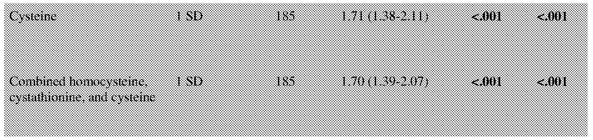
Pre-surgery PSA
If year post-surgery <= 5.6 1 SD 1 12 1.85 (1.44-2.37) <.001 <.001
If year post-surgery > 5.6 1 SD 73 0.84 (0.49-1.42) 0.516 0.516
Abbreviation: SD, standard deviation.
Kaplan-Meier estimates of recurrence-free survival in the patient population with a pre- surgical PSA < 10 that received prostatectomy is illustrated in Figure 58. Kaplan-Meier plots demonstrated recurrence-free survival as a factor of time for high and low levels of serum PSA (Figure 59), cysteine (Figure 60), and the combined methionine metabolites (Figure 61) as independent biomarkers.
Finally, in a multivariate analysis to determine if cysteine was an independent predictor of biochemical recurrence among other variables (PSA, cysteine, Gleason score, and T stage), the Hazard Ratio of 1.50 was significant (Table 9, p value < 0.001). These results support the use of cysteine as a strong member of a blood-based biomarker panel that may also include sCD105 and PSA.
Table 9
Multivariable RFS analysis with Cysteine, sCD105, PSA, Gleason
and T stage in PSA < 10
Type3 P-
Variables Level Hazard Ratio (95% CI) P-value value v«ti» < 1 in iniwtiiMil t -Sfl Π BX> (Mil ^ ftfll
SCD105 1 SD increment 0.80 (0.65-0.99) 0.040 0.040
PSA
post-surgery < 5.6 year 1 SD increment 1.43 (1.10-1.85) 0.007 0.007
post-surgery > 5.6 year 1 SD increment 0.61 (0.34-1.09) 0.093 0.093
Total Gleason 5-6 vs. 8-9 0.13 (0.04-0.38) <.001 <.001
7 vs. 8-9 0.35 (0.15-0.80) 0.014
T stage T2 vs. T3 0.54 (0.33-0.89) 0.016 0.016
Abbreviation: SD, standard deviation. AIC= =701.973.
The various methods and techniques described above provide a number of ways to carry out the application. Of course, it is to be understood that not necessarily all objectives or advantages described can be achieved in accordance with any particular embodiment described herein. Thus, for example, a person of ordinary skill in the art will recognize that the methods can be performed in a manner that achieves or optimizes one advantage or group of advantages as taught herein
without necessarily achieving other objectives or advantages as taught or suggested herein. A variety of alternatives are mentioned herein. It is to be understood that some preferred
embodiments specifically include one, another, or several features, while others specifically exclude one, another, or several features, while still others mitigate a particular feature by inclusion of one, another, or several advantageous features.
Furthermore, a person of ordinary skill in the art will recognize the applicability of various features from different embodiments. Similarly, the various elements, features and steps discussed above, as well as other known equivalents for each such element, feature or step, can be employed in various combinations by one of ordinary skill in this art to perform methods in accordance with the principles described herein. Among the various elements, features, and steps some will be specifically included and others specifically excluded in diverse embodiments.
Although the application has been disclosed in the context of certain embodiments and examples, it will be understood by a person of ordinary skill in the art that the embodiments of the application extend beyond the specifically disclosed embodiments to other alternative embodiments and/or uses and modifications and equivalents thereof.
Preferred embodiments of this application are described herein, including the best mode known to the inventors for carrying out the application. Variations on those preferred embodiments will become apparent to those of ordinary skill in the art upon reading the foregoing description. It is contemplated that a person of ordinary skill in the art can employ such variations as appropriate, and the application can be practiced otherwise than specifically described herein. Accordingly, many embodiments of this application include all modifications and equivalents of the subject matter recited in the claims appended hereto as permitted by applicable law. Moreover, any combination of the above-described elements in all possible variations thereof is encompassed by the application unless otherwise indicated herein or otherwise clearly contradicted by context.
All patents, patent applications, publications of patent applications, and other material, such as articles, books, specifications, publications, documents, things, and/or the like, referenced herein are hereby incorporated herein by this reference in their entirety for all purposes, excepting any prosecution file history associated with same, any of same that is inconsistent with or in conflict with the present document, or any of same that may have a limiting affect as to the broadest scope of the claims now or later associated with the present document. By way of example, should there be any inconsistency or conflict between the description, definition, and/or the use of a term associated with any of the incorporated material and that associated with the present document, the description, definition, and/or the use of the term in the present document shall prevail.
It is to be understood that the embodiments of the application disclosed herein are illustrative of the principles of the embodiments of the application. Other modifications that can be employed can be within the scope of the application. Thus, by way of example, but not of limitation, alternative configurations of the embodiments of the application can be utilized in accordance with the teachings herein. Accordingly, embodiments of the present application are not limited to that precisely as shown and described.
Various embodiments of the invention are described above in the Detailed Description. While these descriptions directly describe the above embodiments, it is understood that a person of ordinary skill in the art may conceive modifications and/or variations to the specific embodiments shown and described herein. Any such modifications or variations that fall within the purview of this description are intended to be included therein as well. Unless specifically noted, it is the intention of the inventors that the words and phrases in the specification and claims be given the ordinary and accustomed meanings to those of ordinary skill in the applicable art(s).
The foregoing description of various embodiments of the invention known to the applicant at this time of filing the application has been presented and is intended for the purposes of illustration and description. The present description is not intended to be exhaustive nor limit the invention to the precise form disclosed and many modifications and variations are possible in the light of the above teachings. The embodiments described serve to explain the principles of the invention and its practical application and to enable a person of ordinary skill in the art to utilize the invention in various embodiments and with various modifications as are suited to the particular use contemplated. Therefore, it is intended that the invention not be limited to the particular embodiments disclosed for carrying out the invention.
While particular embodiments of the present invention have been shown and described, a person of ordinary skill in the art will understand that, based upon the teachings herein, changes and modifications may be made without departing from this invention and its broader aspects and, therefore, the appended claims are to encompass within their scope all such changes and modifications as are within the true spirit and scope of this invention.
Claims
1. A device, comprising:
a sample chamber having at least one analyte inlet; and
a sensor component comprising an electrically conductive metal substrate or electrically conductive metal deposited or formed on a substrate, the conductive metal providing a reaction surface capable of binding an analyte having a functional group comprising sulfur, the sensor component further comprising electrodes electrically coupled to the conductive metal and to a component for determining an electrical parameter of the metal subsequent to analyte binding to the metal surface.
2. The device according to claim 1 wherein the electrical parameter is impedance, resistance, and/or conductance.
3. The device according to claim 2 wherein the parameter is impedance and the device further comprises a component for measuring impedance.
4. The device according to any of claims 1-3 wherein the electrically conductive metal is selected from gold, silver, platinum, iridium, and combinations thereof.
5. The device according to any of claims 1-4 wherein the electrically conductive metal defines a fluid flow path over which an analyte solution flows, the metal having a thickness of from 1 to 500 nanometers, a width of from 0.1 to about 20 millimeters, and a length of from about 0.1 to about 200 millimeters,
6. The device according to claim 1 wherein the electrical conductive metal is gold.
7. The device according to any of claims 1-5 wherein the conductive metal is gold film configured as a straight, curve, winding, and/or tortuous path.
8. The device according to any of claims 1-7 wherein the sample chamber comprises a microfluidic channel to conduct a fluid flow over the reaction surface.
9. The device according to any of claims 1-8 wherein the sample chamber defines plural electrically insulated reaction surfaces.
10. The device according to any of claims 1-7 wherein the device comprises plural sample chambers.
11. The device according to claim 1 comprising plural sample chambers arranged in parallel or in series.
12. The device according to any of claims 1-11 wherein the substrate comprises glass, metal, ceramic, metal-ceramic, plastic, or a combination thereof.
13. The device according to any of claims 1-12, further comprising an enzyme reaction module configured to process a sample with cystathionine synthase and/or cystathionine lyase before the sample contacts the reaction surface.
14. The device according to claim 1 wherein the conductive metal substrate comprises a receptor biomolecule coupled to a portion of the metal surface through a thiol functional group.
15. The device according to claim 14 wherein a remaining portion of the metal surface comprises a blocking agent to preclude target molecule binding to the surface.
16. The device according to claim 15 wherein the blocking agent is a thiolated polyethylene glycol.
17. The device according to claim 14 wherein the receptor molecule is a peptide that is coupled to the metal surface by cysteine.
18. The device according to claim 14 wherein the receptor molecule is an antibody.
19. The device according to claim 17 wherein the peptide is modified to include a pendant cysteine amino acid.
20. The device according to claim 14 wherein the receptor molecule is an extracellular receptor domain.
21. The device according to claim 14 configured to assess β-2 transferrin, NGAL, cystatin C, and C-reactive protein, Ara h 1, Ara h 2, and Ara h 3, GFAP and UCK-Ll proteins, AFP (alpha-fetoprotein), AST (Aspartate aminotransferase), ALT (Alanine aminotransferase), Troponin T, Troponin I, or combinations thereof, in a sample.
22. The device according to claim 21 wherein the sample chamber comprises plural reactive surfaces, and wherein a portion of the metal surface further reacts with cysteine or homocysteine in the sample.
23. The device according to claim 21 used to determine if an injury or surgery is leaking CSF, the device comprising an antibody or transferrin receptor immobilized to a gold nanowell surface.
24. The device according to claim 21 wherein the device comprises NGAL, cystatin C, and C-reactive protein and is configured to assess renal function.
25. The device according to claim 21 wherein the device comprises Ara h 1, Ara h 2, and/or Ara h 3 immobilized on a gold nanowell surface to detect IgE immunoglobins specific to these antigens.
26. The device according to claim 21 wherein the device comprises antibodies to GFAP and UCK-Ll proteins immobilized on a gold nanowell surface and the device is configured for assessing concussions or traumatic brain injury.
27. The device according to claim 21 wherein the device comprises antibodies to AFP (alpha-fetoprotein), AST (Aspartate aminotransferase), and/or ALT (Alanine aminotransferase) immobilized on a gold nanowell surface and the device is configured to screen for liver infections (hepatitis C) or monitor liver disease progression.
28. The device according to claim 21 wherein the device comprises antibodies for troponin T and troponin I immobilized on a gold nanowell surface, and further comprises a metal
surface for free cysteine and/or homocysteine, wherein the device is configured to assess cardiac infarction.
29. A point of care device according to claim 1.
30. A system, comprising a device according to any of claims 1-29.
31. The system according to claim 30 wherein the device defines a disposable sensor unit comprising the electrically conductive metal for coupling to a detection device for detecting a change in an electrical parameter of the conductive metal subsequent to analyte binding.
32. The system according to claim 30 wherein the device defines a reusable sensor unit comprising the electrically conductive metal for coupling to a detection device for detecting a change in an electrical parameter of the conductive metal subsequent to analyte binding.
33. The system according to claim 30, further comprising one or more of:
a central processing unit for controlling functions of the system;
a temperature sensor;
a data storage unit;
a fluid pump for flowing analyte and/or enzyme solutions to and/or through the device; a sample collector;
a sample reservoir or cartridge;
one or more filtration modules positioned to filter a fluid stream into the system or between components of the system;
an enzyme reservoir or cartridge;
an enzyme reaction module;
a buffer reservoir or cartridge;
a power supply; and
combinations thereof.
34. A method, comprising:
providing a device or system according to any of claims 1-33; and
using the device or system to measure an analyte in a sample.
35. The method according to claim 34 for detecting an analyte having a functional group comprising a sulfur atom.
36. The method according to any of claims 34 or 35 wherein the analyte is cysteine and the method comprises measuring a cysteine level in a sample.
37. The method according to claim 35 wherein the analyte has a functional group that is converted to a thiol enzymatically, chemically or thermally.
38. The method according to claim 35 wherein the analyte is reacted with a cysteine to provide a terminal cysteine moiety for detection and measurement using the device.
39. The method according to any of claims 34-38 wherein the sample is from a mammalian subject.
40. The method according to any of claims 34-39 wherein the subject is a human.
41. The method according to claim 34, further comprising:
obtaining a sample from a subject;
introducing the sample to the device to contact the reaction surface with the sample, thereby allowing cysteine in the sample to bind to the reaction surface; and
detecting a change in an electrical parameter wherein the electrical parameter correlates with the cysteine level in the sample.
42. The method according to claim 41 wherein the electrical parameter is impedance, resistance, and/or conductance.
43. The method according to claim 41 wherein the change in the electrical parameter is a difference in the electrical parameter measured before contacting the reaction surface with the sample and after contacting the reaction surface with the sample.
44. The method according to any of claims 34-43 wherein the electrical parameter is impedance, and the measured impedance value is correlated with a cysteine amount in the sample.
45. The method according to claim 44 wherein the impedance value is correlated with the cysteine amount using a standard curve.
46. The method according to any of claims 41-44 wherein the cysteine amount is used to determine an occurrence or recurrence of cancer, atherosclerosis, or cardiovascular disease.
47. The method according to claim 46 wherein the cysteine amount is used to determine an occurrence or recurrence of prostate cancer, colon cancer, ovarian cancer, breast cancer, urinary tract disease, cystine stone disease, or myocardial infarction.
48. The method according to claim 46 wherein the cancer is prostate cancer.
49. The method according to any of claims 41-48 further comprising processing the sample with cystathionine synthase and/or cystathionine lyase, before contacting the sample with the reaction surface.
50. The method according to claim 34, comprising:
detecting a cysteine level in the sample;
diagnosing or prognosing the condition based on the detected cysteine level.
51. The method according to claim 50 comprising processing the sample with cystathionine synthase and/or cystathionine lyase after obtaining the sample.
52. The method according to any of claims 34-51 further comprising diagnosing or prognosing a condition based on considering at least one additional prognostic or diagnostic factor.
53. The method according to claim 34, comprising:
introducing a sample to the device to contact the surface with an analyte;
detecting a change in an electrical parameter between the two ends of a gold film and along the length of the gold film, thereby detecting a methionine metabolite level in the sample.
54. The method according to claim 53 wherein the methionine metabolite comprises cysteine, and/or cystathionine, and/or homocysteine.
55. The method according to claim 34 for diagnosing or prognosing a condition in a subject, comprising:
obtaining a sample from the subject;
detecting a methionine metabolite level in the sample; and
diagnosing or prognosing the condition based on the detected methionine metabolite level.
56. The method according to claim 34 for determining if a subject has an increased probability of cancer recurrence, comprising:
obtaining a sample from the subject;
assaying the sample to detect an increased cysteine level, an increased methionine metabolite level, an increased PSA parameter, and/or a decreased sCD105 level;
detecting in the sample an increased cysteine level, an increased methionine metabolite level, an increased PSA parameter, and/or a decreased sCD105 level; and
determining if the subject has an increased probability of cancer recurrence.
57. The method according to claim 34 wherein the conductive metal substrate comprises a receptor biomolecule coupled to a portion of the metal surface through a thiol functional group, a remaining portion of the metal surface comprises a blocking agent to preclude target molecule binding to the surface, and the receptor molecule is a peptide or an extracellular receptor domain that is coupled to the metal surface by cysteine.
58. The method according to claim 57 wherein the peptide is an antibody.
59. The method according to claim 57 wherein the peptide is modified to include a pendant cysteine amino acid.
60. The method according to claim 57 wherein the sample chamber comprises plural reactive surfaces, and wherein a portion of the metal surface further reacts with free cysteine or homocysteine.
61. The method according to claim 57 for determining if an injury or surgery is leaking CSF, the device comprising an antibody or transferrin receptor immobilized to a gold nano-well surface.
62. The method according to claim 57 wherein the device comprises NGAL, cystatin C, and C-reactive protein and is configured to assess renal function.
63. The method according to claim 57 wherein the metal surface comprises Ara h 1, Ara h 2, and Ara h 3 to detect IgE immunoglobins specific to these antigens.
64. The method according to claim 57 wherein the metal surface comprises antibodies to GFAP and UCK-Ll proteins and the device is configured for assessing concussions or traumatic brain injury.
65. The method according to claim 57 wherein the metal surface comprises antibodies to AFP (alpha-fetoprotein), AST (Aspartate aminotransferase), and/or ALT (Alanine
aminotransferase) and the device is configured to screen for liver infections (hepatitis C) or monitor liver disease progression.
66. The method according to claim 57 wherein the metal surface comprises antibodies for troponin T and troponin I, and further comprises a metal surface for assessing an amount of cysteine and/or homocysteine in the sample, and the device is configured to assess cardiac infarction.
Priority Applications (1)
| Application Number | Priority Date | Filing Date | Title |
|---|---|---|---|
| CA2998738A CA2998738A1 (en) | 2015-09-22 | 2016-09-22 | Device, system and method for using the device or system for assaying an analyte in a sample |
Applications Claiming Priority (2)
| Application Number | Priority Date | Filing Date | Title |
|---|---|---|---|
| US201562222040P | 2015-09-22 | 2015-09-22 | |
| US62/222,040 | 2015-09-22 |
Publications (1)
| Publication Number | Publication Date |
|---|---|
| WO2017053586A1 true WO2017053586A1 (en) | 2017-03-30 |
Family
ID=58387189
Family Applications (1)
| Application Number | Title | Priority Date | Filing Date |
|---|---|---|---|
| PCT/US2016/053145 Ceased WO2017053586A1 (en) | 2015-09-22 | 2016-09-22 | Device, system and method for using the device or system for assaying an analyte in a sample |
Country Status (2)
| Country | Link |
|---|---|
| CA (1) | CA2998738A1 (en) |
| WO (1) | WO2017053586A1 (en) |
Cited By (3)
| Publication number | Priority date | Publication date | Assignee | Title |
|---|---|---|---|---|
| US10490679B2 (en) | 2016-02-17 | 2019-11-26 | The Curators Of The University Of Missouri | Fabrication of multilayer nanograting structures |
| CN115558910A (en) * | 2022-11-03 | 2023-01-03 | 重庆海盛鑫铜业有限公司 | Portable tinning machine for copper wire processing and use method |
| JP2023503615A (en) * | 2019-11-26 | 2023-01-31 | シーダーズ-サイナイ メディカル センター | Compositions and methods for treating diseases and conditions by depleting mitochondrial or genomic DNA from circulation |
Citations (3)
| Publication number | Priority date | Publication date | Assignee | Title |
|---|---|---|---|---|
| US20020192722A1 (en) * | 2001-04-02 | 2002-12-19 | Prolinx Incorporated | Sensor surfaces for detecting analytes |
| US20080103064A1 (en) * | 2006-06-13 | 2008-05-01 | Antara Biosciences Inc. | Microscale fluidic devices for electrochemical detection of biological molecules |
| US20140106441A1 (en) * | 2011-01-28 | 2014-04-17 | Joseph Wang | Bioaffinity sensors based on surface monolayers |
-
2016
- 2016-09-22 WO PCT/US2016/053145 patent/WO2017053586A1/en not_active Ceased
- 2016-09-22 CA CA2998738A patent/CA2998738A1/en not_active Abandoned
Patent Citations (3)
| Publication number | Priority date | Publication date | Assignee | Title |
|---|---|---|---|---|
| US20020192722A1 (en) * | 2001-04-02 | 2002-12-19 | Prolinx Incorporated | Sensor surfaces for detecting analytes |
| US20080103064A1 (en) * | 2006-06-13 | 2008-05-01 | Antara Biosciences Inc. | Microscale fluidic devices for electrochemical detection of biological molecules |
| US20140106441A1 (en) * | 2011-01-28 | 2014-04-17 | Joseph Wang | Bioaffinity sensors based on surface monolayers |
Non-Patent Citations (1)
| Title |
|---|
| HUANG ET AL.: "Impedance biosensor for peanut protein Ara h 1.", ANALYTICAL CHEMISTRY, vol. 80, no. 23, 28 October 2008 (2008-10-28), pages 9157 - 9161, XP055370768 * |
Cited By (5)
| Publication number | Priority date | Publication date | Assignee | Title |
|---|---|---|---|---|
| US10490679B2 (en) | 2016-02-17 | 2019-11-26 | The Curators Of The University Of Missouri | Fabrication of multilayer nanograting structures |
| JP2023503615A (en) * | 2019-11-26 | 2023-01-31 | シーダーズ-サイナイ メディカル センター | Compositions and methods for treating diseases and conditions by depleting mitochondrial or genomic DNA from circulation |
| JP7797384B2 (en) | 2019-11-26 | 2026-01-13 | シーダーズ-サイナイ メディカル センター | Compositions and methods for treating diseases and conditions by depleting mitochondrial or genomic DNA from the circulation |
| CN115558910A (en) * | 2022-11-03 | 2023-01-03 | 重庆海盛鑫铜业有限公司 | Portable tinning machine for copper wire processing and use method |
| CN115558910B (en) * | 2022-11-03 | 2024-05-07 | 重庆海盛鑫铜业有限公司 | Portable tinning machine for copper wire processing and use method |
Also Published As
| Publication number | Publication date |
|---|---|
| CA2998738A1 (en) | 2017-03-30 |
Similar Documents
| Publication | Publication Date | Title |
|---|---|---|
| Gaikwad et al. | Advances in point-of-care diagnostic devices in cancers | |
| US7993854B2 (en) | Detection and quantification of biomarkers via a piezoelectric cantilever sensor | |
| Laboria et al. | Amperometric immunosensor for carcinoembryonic antigen in colon cancer samples based on monolayers of dendritic bipodal scaffolds | |
| Jolly et al. | Label-free impedimetric aptasensor with antifouling surface chemistry: A prostate specific antigen case study | |
| Otieno et al. | On-line protein capture on magnetic beads for ultrasensitive microfluidic immunoassays of cancer biomarkers | |
| Songjaroen et al. | Label-free detection of C-reactive protein using an electrochemical DNA immunoassay | |
| Aliño et al. | Using liquid crystals as a readout system in urinary albumin assays | |
| CN105940300A (en) | Device and methods of using device for detection of hyperammonemia | |
| BR112014027489B1 (en) | ELECTRODE AND METHOD TO DETECT A TARGET SPECIES | |
| US20210063334A1 (en) | Apparatus and methods for detection of diabetes-associated molecules using electrochemical impedance spectroscopy | |
| Campuzano et al. | Electrochemical biosensors for autoantibodies in autoimmune and cancer diseases | |
| Chiriacò et al. | Towards pancreatic cancer diagnosis using EIS biochips | |
| Cennamo et al. | Plasmon resonance biosensor for interleukin-1β point-of-care determination: A tool for early periodontitis diagnosis | |
| Wang et al. | A simple electrochemical immunosensor based on a gold nanoparticle monolayer electrode for neutrophil gelatinase-associated lipocalin detection | |
| JP2021185371A (en) | Marker for statin treatment hierarchization at cardiac failure | |
| Ko et al. | Handheld microfluidic multiple detection device for concurrent blood urea nitrogen and creatinine ratio determination using colorimetric approach | |
| JP2020507744A (en) | Optical imaging system using lateral illumination for digital assays | |
| WO2006015874A2 (en) | Method for diagnosing liver fibrosis | |
| WO2017053586A1 (en) | Device, system and method for using the device or system for assaying an analyte in a sample | |
| Lin et al. | based CRP monitoring devices | |
| Shinde et al. | Utility of cardiac biomarkers and biosensors for diagnosis of acute myocardial infarction | |
| Yao et al. | Bioconjugated, single-use biosensor for the detection of biomarkers of prostate cancer | |
| Xu et al. | Recent development on nanomaterial‐based biosensors for identifying thyroid tumor biomarkers | |
| US10801073B2 (en) | Device, system and method for cancer prognosis and uses thereof | |
| Lee et al. | Advances in uremic toxin detection and monitoring in the management of chronic kidney disease progression to end-stage renal disease |
Legal Events
| Date | Code | Title | Description |
|---|---|---|---|
| 121 | Ep: the epo has been informed by wipo that ep was designated in this application |
Ref document number: 16849607 Country of ref document: EP Kind code of ref document: A1 |
|
| ENP | Entry into the national phase |
Ref document number: 2998738 Country of ref document: CA |
|
| NENP | Non-entry into the national phase |
Ref country code: DE |
|
| 122 | Ep: pct application non-entry in european phase |
Ref document number: 16849607 Country of ref document: EP Kind code of ref document: A1 |