WO2017021354A1 - Antibody constructs for cd70 and cd3 - Google Patents
Antibody constructs for cd70 and cd3 Download PDFInfo
- Publication number
- WO2017021354A1 WO2017021354A1 PCT/EP2016/068294 EP2016068294W WO2017021354A1 WO 2017021354 A1 WO2017021354 A1 WO 2017021354A1 EP 2016068294 W EP2016068294 W EP 2016068294W WO 2017021354 A1 WO2017021354 A1 WO 2017021354A1
- Authority
- WO
- WIPO (PCT)
- Prior art keywords
- seq
- nos
- cdr
- amino acid
- antibody construct
- Prior art date
Links
Classifications
-
- C—CHEMISTRY; METALLURGY
- C07—ORGANIC CHEMISTRY
- C07K—PEPTIDES
- C07K16/00—Immunoglobulins [IGs], e.g. monoclonal or polyclonal antibodies
- C07K16/18—Immunoglobulins [IGs], e.g. monoclonal or polyclonal antibodies against material from animals or humans
- C07K16/28—Immunoglobulins [IGs], e.g. monoclonal or polyclonal antibodies against material from animals or humans against receptors, cell surface antigens or cell surface determinants
- C07K16/2875—Immunoglobulins [IGs], e.g. monoclonal or polyclonal antibodies against material from animals or humans against receptors, cell surface antigens or cell surface determinants against the NGF/TNF superfamily, e.g. CD70, CD95L, CD153, CD154
-
- A—HUMAN NECESSITIES
- A61—MEDICAL OR VETERINARY SCIENCE; HYGIENE
- A61P—SPECIFIC THERAPEUTIC ACTIVITY OF CHEMICAL COMPOUNDS OR MEDICINAL PREPARATIONS
- A61P35/00—Antineoplastic agents
-
- A—HUMAN NECESSITIES
- A61—MEDICAL OR VETERINARY SCIENCE; HYGIENE
- A61P—SPECIFIC THERAPEUTIC ACTIVITY OF CHEMICAL COMPOUNDS OR MEDICINAL PREPARATIONS
- A61P35/00—Antineoplastic agents
- A61P35/02—Antineoplastic agents specific for leukemia
-
- C—CHEMISTRY; METALLURGY
- C07—ORGANIC CHEMISTRY
- C07K—PEPTIDES
- C07K16/00—Immunoglobulins [IGs], e.g. monoclonal or polyclonal antibodies
- C07K16/18—Immunoglobulins [IGs], e.g. monoclonal or polyclonal antibodies against material from animals or humans
- C07K16/28—Immunoglobulins [IGs], e.g. monoclonal or polyclonal antibodies against material from animals or humans against receptors, cell surface antigens or cell surface determinants
- C07K16/2803—Immunoglobulins [IGs], e.g. monoclonal or polyclonal antibodies against material from animals or humans against receptors, cell surface antigens or cell surface determinants against the immunoglobulin superfamily
- C07K16/2809—Immunoglobulins [IGs], e.g. monoclonal or polyclonal antibodies against material from animals or humans against receptors, cell surface antigens or cell surface determinants against the immunoglobulin superfamily against the T-cell receptor (TcR)-CD3 complex
-
- C—CHEMISTRY; METALLURGY
- C07—ORGANIC CHEMISTRY
- C07K—PEPTIDES
- C07K16/00—Immunoglobulins [IGs], e.g. monoclonal or polyclonal antibodies
- C07K16/18—Immunoglobulins [IGs], e.g. monoclonal or polyclonal antibodies against material from animals or humans
- C07K16/28—Immunoglobulins [IGs], e.g. monoclonal or polyclonal antibodies against material from animals or humans against receptors, cell surface antigens or cell surface determinants
- C07K16/30—Immunoglobulins [IGs], e.g. monoclonal or polyclonal antibodies against material from animals or humans against receptors, cell surface antigens or cell surface determinants from tumour cells
- C07K16/3069—Reproductive system, e.g. ovaria, uterus, testes, prostate
-
- A—HUMAN NECESSITIES
- A61—MEDICAL OR VETERINARY SCIENCE; HYGIENE
- A61K—PREPARATIONS FOR MEDICAL, DENTAL OR TOILETRY PURPOSES
- A61K39/00—Medicinal preparations containing antigens or antibodies
- A61K2039/505—Medicinal preparations containing antigens or antibodies comprising antibodies
-
- C—CHEMISTRY; METALLURGY
- C07—ORGANIC CHEMISTRY
- C07K—PEPTIDES
- C07K2317/00—Immunoglobulins specific features
- C07K2317/30—Immunoglobulins specific features characterized by aspects of specificity or valency
- C07K2317/31—Immunoglobulins specific features characterized by aspects of specificity or valency multispecific
-
- C—CHEMISTRY; METALLURGY
- C07—ORGANIC CHEMISTRY
- C07K—PEPTIDES
- C07K2317/00—Immunoglobulins specific features
- C07K2317/30—Immunoglobulins specific features characterized by aspects of specificity or valency
- C07K2317/33—Crossreactivity, e.g. for species or epitope, or lack of said crossreactivity
-
- C—CHEMISTRY; METALLURGY
- C07—ORGANIC CHEMISTRY
- C07K—PEPTIDES
- C07K2317/00—Immunoglobulins specific features
- C07K2317/50—Immunoglobulins specific features characterized by immunoglobulin fragments
- C07K2317/56—Immunoglobulins specific features characterized by immunoglobulin fragments variable (Fv) region, i.e. VH and/or VL
- C07K2317/565—Complementarity determining region [CDR]
-
- C—CHEMISTRY; METALLURGY
- C07—ORGANIC CHEMISTRY
- C07K—PEPTIDES
- C07K2317/00—Immunoglobulins specific features
- C07K2317/60—Immunoglobulins specific features characterized by non-natural combinations of immunoglobulin fragments
- C07K2317/62—Immunoglobulins specific features characterized by non-natural combinations of immunoglobulin fragments comprising only variable region components
- C07K2317/622—Single chain antibody (scFv)
-
- C—CHEMISTRY; METALLURGY
- C07—ORGANIC CHEMISTRY
- C07K—PEPTIDES
- C07K2317/00—Immunoglobulins specific features
- C07K2317/60—Immunoglobulins specific features characterized by non-natural combinations of immunoglobulin fragments
- C07K2317/64—Immunoglobulins specific features characterized by non-natural combinations of immunoglobulin fragments comprising a combination of variable region and constant region components
-
- C—CHEMISTRY; METALLURGY
- C07—ORGANIC CHEMISTRY
- C07K—PEPTIDES
- C07K2317/00—Immunoglobulins specific features
- C07K2317/70—Immunoglobulins specific features characterized by effect upon binding to a cell or to an antigen
- C07K2317/73—Inducing cell death, e.g. apoptosis, necrosis or inhibition of cell proliferation
-
- C—CHEMISTRY; METALLURGY
- C07—ORGANIC CHEMISTRY
- C07K—PEPTIDES
- C07K2317/00—Immunoglobulins specific features
- C07K2317/90—Immunoglobulins specific features characterized by (pharmaco)kinetic aspects or by stability of the immunoglobulin
-
- C—CHEMISTRY; METALLURGY
- C07—ORGANIC CHEMISTRY
- C07K—PEPTIDES
- C07K2317/00—Immunoglobulins specific features
- C07K2317/90—Immunoglobulins specific features characterized by (pharmaco)kinetic aspects or by stability of the immunoglobulin
- C07K2317/92—Affinity (KD), association rate (Ka), dissociation rate (Kd) or EC50 value
-
- C—CHEMISTRY; METALLURGY
- C07—ORGANIC CHEMISTRY
- C07K—PEPTIDES
- C07K2317/00—Immunoglobulins specific features
- C07K2317/90—Immunoglobulins specific features characterized by (pharmaco)kinetic aspects or by stability of the immunoglobulin
- C07K2317/94—Stability, e.g. half-life, pH, temperature or enzyme-resistance
Definitions
- the present invention relates to a bispecific antibody construct comprising a first binding domain which binds to human CD70 on the surface of a target cell and a second binding domain which binds to human CD3 on the surface of a T cell.
- the invention provides a polynucleotide encoding the antibody construct, a vector comprising said polynucleotide and a host cell transformed or transfected with said polynucleotide or vector.
- the invention provides a process for the production of the antibody construct of the invention, a medical use of said antibody construct and a kit comprising said antibody construct.
- CD70 (CD27L, TNFSF7) is a type II integral membrane protein whose normal expression is restricted to a subset of activated T and B cells, mature dendritic cells and thymic medullar epithelial cells. The biological functions of CD70 are mediated via binding to the CD27 receptor, which is expressed on lymphocytes and NK cells. CD70 interactions regulate B-cell maturation, T-cell co-stimulation and memory T cell function. Genetic ablation of CD27 in mice, which abrogates CD70 signaling, does not impact the development of B and T cells, but interferes with memory responses to viral re-challenge.
- CD70 expression has been documented in 60% of Non-Hodgkin Lymphoma (NHL), 70% of clear cell renal cell carcinoma (ccRCC), 40% of papillary RCC, as well as 25% of pancreatic adenocarcinoma, 22% of laryngeal / pharyngeal carcinoma, 15% of ovarian carcinoma, and 10% of lung adenocarcinoma and glioblastoma.
- NDL Non-Hodgkin Lymphoma
- ccRCC clear cell renal cell carcinoma
- papillary RCC 40% of papillary RCC
- pancreatic adenocarcinoma 22% of laryngeal / pharyngeal carcinoma
- 15% of ovarian carcinoma 15% of ovarian carcinoma
- 10% of lung adenocarcinoma and glioblastoma 10% of lung adenocarcinoma and glioblastoma.
- Diffuse large cell B cell lymphoma is the largest NHL sub-type with 28,740 new cases per year in the U.S. (American Cancer Society: Cancer Facts and Figures, 2015). 71 % of DLBCL tumors express CD70, and while most patients initially respond to standard of care therapy (Rituxan plus cyclophosphamide, adriamycin, vincristine, prednisone), many relapse or are refractory, and 19,790 patients die of this disease every year (American Cancer Society: Cancer Facts and Figures 2015). Therefore a clear unmet medical need remains for DLCBL, and up to 14,000 patients a year presenting with CD70-expressing DLBCL could benefit from a CD70-targeted therapy in the U.S.
- the present invention provides a bispecific antibody construct comprising a first binding domain which binds to human CD70 on the surface of a target cell and a second binding domain which binds to human CD3 on the surface of a T cell.
- an antibody construct refers to a molecule in which the structure and/or function is/are based on the structure and/or function of an antibody, e.g., of a full-length or whole immunoglobulin molecule.
- An antibody construct is hence capable of binding to its specific target or antigen.
- an antibody construct according to the invention comprises the minimum structural requirements of an antibody which allow for the target binding. This minimum requirement may e.g. be defined by the presence of at least the three light chain CDRs (i.e. CDR1 , CDR2 and CDR3 of the VL region) and/or the three heavy chain CDRs (i.e. CDR1 , CDR2 and CDR3 of the VH region), preferably of all six CDRs.
- the antibodies on which the constructs according to the invention are based include for example monoclonal, recombinant, chimeric, deimmunized, humanized and human antibodies.
- antibody constructs include full-length or whole antibodies also including camelid antibodies and other immunoglobulin antibodies generated by biotechnological or protein engineering methods or processes. These full-length antibodies may be for example monoclonal, recombinant, chimeric, deimmunized, humanized and human antibodies.
- antibody constructs fragments of full-length antibodies, such as VH, VHH, VL, (s)dAb, Fv, Fd, Fab, Fab', F(ab')2 or "r IgG" ("half antibody").
- Antibody constructs according to the invention may also be modified fragments of antibodies, also called antibody variants, such as scFv, di-scFv or bi(s)-scFv, scFv-Fc, scFv-zipper, scFab, Fab2, Fab3, diabodies, single chain diabodies, tandem diabodies (Tandab's), tandem di-scFv, tandem tri-scFv, possiblyminibodies” exemplified by a structure which is as follows: (VH-VL-CH3) 2 , (scFv-CH3) 2 , ((scFv) 2 -CH3 + CH3), ((scFv) 2 -CH3) or (scFv-CH3-scFv) 2 , multibodies such as triabodies or tetrabodies, and single domain antibodies such as nanobodies or single variable domain antibodies comprising merely one variable domain, which might be VHH, VH or VL, that
- a binding domain may typically comprise an antibody light chain variable region (VL) and an antibody heavy chain variable region (VH); however, it does not have to comprise both.
- Fd fragments for example, have two VH regions and often retain some antigen-binding function of the intact antigen-binding domain.
- Additional examples for the format of antibody fragments, antibody variants or binding domains include (1 ) a Fab fragment, a monovalent fragment having the VL, VH, CL and CH1 domains; (2) a F(ab')2 fragment, a bivalent fragment having two Fab fragments linked by a disulfide bridge at the hinge region; (3) an Fd fragment having the two VH and CH1 domains; (4) an Fv fragment having the VL and VH domains of a single arm of an antibody, (5) a dAb fragment (Ward et al., (1989) Nature 341 :544-546), which has a VH domain; (6) an isolated complementarity determining region (CDR), and (7) a single chain Fv (scFv) , the latter being preferred (for example, derived from an scFv-library).
- a Fab fragment a monovalent fragment having the VL, VH, CL and CH1 domains
- F(ab')2 fragment a bivalent fragment having
- antibody constructs examples are e.g. described in WO 00/006605, WO 2005/040220, WO 2008/1 19567, WO 2010/037838, WO 2013/026837, WO 2013/026833, US 2014/0308285, US 2014/0302037, W 02014/144722, WO 2014/151910, and WO 2015/048272.
- antibody construct includes monovalent, bivalent and polyvalent / multivalent constructs and, thus, monospecific constructs, specifically binding to only one antigenic structure, as well as bispecific and polyspecific / multispecific constructs, which specifically bind more than one antigenic structure, e.g.
- antibody construct includes molecules consisting of only one polypeptide chain as well as molecules consisting of more than one polypeptide chain, which chains can be either identical (homodimers, homotrimers or homo oligomers) or different (heterodimer, heterotrimer or heterooligomer). Examples for the above identified antibodies and variants or derivatives thereof are described inter alia in Harlow and Lane, Antibodies a laboratory manual, CSHL Press (1988) and Using Antibodies: a laboratory manual, CSHL Press (1999), Kontermann and Dubel, Antibody Engineering, Springer, 2nd ed. 2010 and Little, Recombinant Antibodies for Immunotherapy, Cambridge University Press 2009.
- the antibody constructs of the present invention are preferably "in vitro generated antibody constructs".
- This term refers to an antibody construct according to the above definition where all or part of the variable region (e.g., at least one CDR) is generated in a non-immune cell selection, e.g., an in vitro phage display, protein chip or any other method in which candidate sequences can be tested for their ability to bind to an antigen.
- a non-immune cell selection e.g., an in vitro phage display, protein chip or any other method in which candidate sequences can be tested for their ability to bind to an antigen.
- a "recombinant antibody” is an antibody made through the use of recombinant DNA technology or genetic engineering.
- mAb monoclonal antibody
- monoclonal antibody construct refers to an antibody obtained from a population of substantially homogeneous antibodies, i.e., the individual antibodies comprising the population are identical except for possible naturally occurring mutations and/or post-translation modifications (e.g., isomerizations, amidations) that may be present in minor amounts.
- Monoclonal antibodies are highly specific, being directed against a single antigenic site or determinant on the antigen, in contrast to conventional (polyclonal) antibody preparations which typically include different antibodies directed against different determinants (or epitopes).
- the monoclonal antibodies are advantageous in that they are synthesized by the hybridoma culture, hence uncontaminated by other immunoglobulins.
- the modifier "monoclonal” indicates the character of the antibody as being obtained from a substantially homogeneous population of antibodies, and is not to be construed as requiring production of the antibody by any particular method.
- any technique providing antibodies produced by continuous cell line cultures can be used.
- monoclonal antibodies to be used may be made by the hybridoma method first described by Koehler et al., Nature, 256: 495 (1975), or may be made by recombinant DNA methods (see, e.g., U.S. Patent No.
- Examples for further techniques to produce human monoclonal antibodies include the trioma technique, the human B-cell hybridoma technique (Kozbor, Immunology Today 4 (1983), 72) and the EBV- hybridoma technique (Cole et al., Monoclonal Antibodies and Cancer Therapy, Alan R. Liss, Inc. (1985), 77-96).
- Hybridomas can then be screened using standard methods, such as enzyme-linked immunosorbent assay (ELISA) and surface plasmon resonance (BIACORETM) analysis, to identify one or more hybridomas that produce an antibody that specifically binds with a specified antigen.
- ELISA enzyme-linked immunosorbent assay
- BIACORETM surface plasmon resonance
- Any form of the relevant antigen may be used as the immunogen, e.g., recombinant antigen, naturally occurring forms, any variants or fragments thereof, as well as an antigenic peptide thereof.
- BIAcore Surface plasmon resonance as employed in the BIAcore system can be used to increase the efficiency of phage antibodies which bind to an epitope of a target antigen, such as CD70 or CD3 epsilon (Schier, Human Antibodies Hybridomas 7 (1996), 97-105; Malmborg, J. Immunol. Methods 183 (1995), 7-13).
- a target antigen such as CD70 or CD3 epsilon
- Another exemplary method of making monoclonal antibodies includes screening protein expression libraries, e.g., phage display or ribosome display libraries.
- Phage display is described, for example, in Ladner et al., U.S. Patent No. 5,223,409; Smith (1985) Science 228:1315-1317, Clackson et al., Nature, 352: 624-628 (1991 ) and Marks et al., J. Mol. Biol., 222: 581 -597 (1991 ).
- the relevant antigen can be used to immunize a non-human animal, e.g., a rodent (such as a mouse, hamster, rabbit or rat).
- the non-human animal includes at least a part of a human immunoglobulin gene.
- antigen-specific monoclonal antibodies derived from the genes with the desired specificity may be produced and selected. See, e.g., XENOMOUSETM, Green et al.
- a monoclonal antibody can also be obtained from a non-human animal, and then modified, e.g., humanized, deimmunized, rendered chimeric etc., using recombinant DNA techniques known in the art.
- modified antibody constructs include humanized variants of non-human antibodies, "affinity matured" antibodies (see, e.g. Hawkins et al. J. Mol. Biol.
- affinity maturation is the process by which B cells produce antibodies with increased affinity for antigen during the course of an immune response. With repeated exposures to the same antigen, a host will produce antibodies of successively greater affinities.
- the in vitro affinity maturation is based on the principles of mutation and selection. The in vitro affinity maturation has successfully been used to optimize antibodies, antibody constructs, and antibody fragments. Random mutations inside the CDRs are introduced using radiation, chemical mutagens or error-prone PCR. In addition, the genetical diversity can be increased by chain shuffling. Two or three rounds of mutation and selection using display methods like phage display usually results in antibody fragments with affinities in the low nanomolar range.
- a preferred type of an amino acid substitutional varianation of the antibody constructs involves substituting one or more hypervariable region residues of a parent antibody (e. g. a humanized or human antibody).
- a parent antibody e. g. a humanized or human antibody
- the resulting variant(s) selected for further development will have improved biological properties relative to the parent antibody from which they are generated.
- a convenient way for generating such substitutional variants involves affinity maturation using phage display. Briefly, several hypervariable region sites (e. g. 6-7 sites) are mutated to generate all possible amino acid substitutions at each site.
- the antibody variants thus generated are displayed in a monovalent fashion from filamentous phage particles as fusions to the gene III product of M13 packaged within each particle.
- the phage-displayed variants are then screened for their biological activity (e. g. binding affinity) as herein disclosed.
- alanine scanning mutagenesis can be performed to identify hypervariable region residues contributing significantly to antigen binding.
- the panel of variants is subjected to screening as described herein and antibodies with superior properties in one or more relevant assays may be selected for further development.
- the monoclonal antibodies and antibody constructs of the present invention specifically include "chimeric" antibodies (immunoglobulins) in which a portion of the heavy and/or light chain is identical with or homologous to corresponding sequences in antibodies derived from a particular species or belonging to a particular antibody class or subclass, while the remainder of the chain(s) is/are identical with or homologous to corresponding sequences in antibodies derived from another species or belonging to another antibody class or subclass, as well as fragments of such antibodies, so long as they exhibit the desired biological activity (U.S. Patent No. 4,816,567; Morrison et al., Proc. Natl. Acad. Sci. USA, 81 : 6851 -6855 (1984)).
- chimeric antibodies immunoglobulins
- Chimeric antibodies of interest herein include "primitized" antibodies comprising variable domain antigen- binding sequences derived from a non-human primate (e.g., Old World Monkey, Ape etc.) and human constant region sequences.
- a non-human primate e.g., Old World Monkey, Ape etc.
- human constant region sequences e.g., human constant region sequences.
- a variety of approaches for making chimeric antibodies have been described. See e.g., Morrison et al., Proc. Natl. Acad. ScL U.S.A. 81 :6851 , 1985; Takeda et al., Nature 314:452, 1985, Cabilly et al., U.S. Patent No. 4,816,567; Boss et al., U.S. Patent No. 4,816,397; Tanaguchi et al., EP 0171496; EP 0173494; and GB 2177096.
- An antibody, antibody construct, antibody fragment or antibody variant may also be modified by specific deletion of human T cell epitopes (a method called "deimmunization") by the methods disclosed for example in WO 98/52976 or WO 00/34317. Briefly, the heavy and light chain variable domains of an antibody can be analyzed for peptides that bind to MHC class II; these peptides represent potential T cell epitopes (as defined in WO 98/52976 and WO 00/34317).
- peptide threading For detection of potential T cell epitopes, a computer modeling approach termed "peptide threading" can be applied, and in addition a database of human MHC class II binding peptides can be searched for motifs present in the VH and VL sequences, as described in WO 98/52976 and WO 00/34317. These motifs bind to any of the 18 major MHC class II DR allotypes, and thus constitute potential T cell epitopes.
- Potential T cell epitopes detected can be eliminated by substituting small numbers of amino acid residues in the variable domains, or preferably, by single amino acid substitutions. Typically, conservative substitutions are made. Often, but not exclusively, an amino acid common to a position in human germline antibody sequences may be used.
- Humanized antibodies are antibodies or immunoglobulins of mostly human sequences, which contain (a) minimal sequence(s) derived from non-human immunoglobulin.
- humanized antibodies are human immunoglobulins (recipient antibody) in which residues from a hypervariable region (also CDR) of the recipient are replaced by residues from a hypervariable region of a non-human (e.g., rodent) species (donor antibody) such as mouse, rat, hamster or rabbit having the desired specificity, affinity, and capacity.
- donor antibody such as mouse, rat, hamster or rabbit having the desired specificity, affinity, and capacity.
- Fv framework region (FR) residues of the human immunoglobulin are replaced by corresponding non-human residues.
- "humanized antibodies” as used herein may also comprise residues which are found neither in the recipient antibody nor the donor antibody. These modifications are made to further refine and optimize antibody performance.
- the humanized antibody may also comprise at least a portion of an immunoglobulin constant region (Fc), typically that of a human immunoglobulin.
- Fc immunoglobulin constant region
- Humanized antibodies or fragments thereof can be generated by replacing sequences of the Fv variable domain that are not directly involved in antigen binding with equivalent sequences from human Fv variable domains.
- Exemplary methods for generating humanized antibodies or fragments thereof are provided by Morrison (1985) Science 229:1202-1207; by Oi et al. (1986) BioTechniques 4:214; and by US 5,585,089; US 5,693,761 ; US 5,693,762; US 5,859,205; and US 6,407,213. Those methods include isolating, manipulating, and expressing the nucleic acid sequences that encode all or part of immunoglobulin Fv variable domains from at least one of a heavy or light chain.
- nucleic acids may be obtained from a hybridoma producing an antibody against a predetermined target, as described above, as well as from other sources.
- the recombinant DNA encoding the humanized antibody molecule can then be cloned into an appropriate expression vector.
- Humanized antibodies may also be produced using transgenic animals such as mice that express human heavy and light chain genes, but are incapable of expressing the endogenous mouse immunoglobulin heavy and light chain genes.
- Winter describes an exemplary CDR grafting method that may be used to prepare the humanized antibodies described herein (U.S. Patent No. 5,225,539). All of the CDRs of a particular human antibody may be replaced with at least a portion of a non-human CDR, or only some of the CDRs may be replaced with non- human CDRs. It is only necessary to replace the number of CDRs required for binding of the humanized antibody to a predetermined antigen.
- a humanized antibody can be optimized by the introduction of conservative substitutions, consensus sequence substitutions, germline substitutions and/or back mutations.
- altered immunoglobulin molecules can be made by any of several techniques known in the art, (e.g., Teng et al., Proc. Natl. Acad. Sci. U.S.A., 80: 7308-7312, 1983; Kozbor et al., Immunology Today, 4: 7279, 1983; Olsson et ai, Meth. Enzymol., 92: 3-16, 1982, and EP 239 400).
- human antibody includes antibodies, antibody constructs and binding domains having antibody regions such as variable and constant regions or domains which correspond substantially to human germline immunoglobulin sequences known in the art, including, for example, those described by Kabat et al. (1991 ) (loc. cit.).
- the human antibodies, antibody constructs or binding domains of the invention may include amino acid residues not encoded by human germline immunoglobulin sequences (e.g., mutations introduced by random or site-specific mutagenesis in vitro or by somatic mutation in vivo), for example in the CDRs, and in particular, in CDR3.
- human antibodies, antibody constructs or binding domains can have at least one, two, three, four, five, or more positions replaced with an amino acid residue that is not encoded by the human germline immunoglobulin sequence.
- the definition of human antibodies, antibody constructs and binding domains as used herein also contemplates fully human antibodies, which include only non-artificially and/or genetically altered human sequences of antibodies as those can be derived by using technologies or systems such as the Xenomouse.
- the antibody constructs of the invention are “isolated” or “substantially pure” antibody constructs.
- “Isolated” or “substantially pure”, when used to describe the antibody constructs disclosed herein, means an antibody construct that has been identified, separated and/or recovered from a component of its production environment.
- the antibody construct is free or substantially free of association with all other components from its production environment. Contaminant components of its production environment, such as that resulting from recombinant transfected cells, are materials that would typically interfere with diagnostic or therapeutic uses for the polypeptide, and may include enzymes, hormones, and other proteinaceous or non-proteinaceous solutes.
- the antibody constructs may e.g constitute at least about 5%, or at least about 50% by weight of the total protein in a given sample. It is understood that the isolated protein may constitute from 5% to 99.9% by weight of the total protein content, depending on the circumstances.
- the polypeptide may be made at a significantly higher concentration through the use of an inducible promoter or high expression promoter, such that it is made at increased concentration levels.
- the definition includes the production of an antibody construct in a wide variety of organisms and/or host cells that are known in the art.
- the antibody construct will be purified (1 ) to a degree sufficient to obtain at least 15 residues of N-terminal or internal amino acid sequence by use of a spinning cup sequenator, or (2) to homogeneity by SDS-PAGE under non-reducing or reducing conditions using Coomassie blue or, preferably, silver stain.
- an isolated antibody construct will be prepared by at least one purification step.
- binding domain characterizes in connection with the present invention a domain which (specifically) binds to / interacts with / recognizes a given target epitope or a given target site on the target molecules (antigens), here: CD70 and CD3, respectively.
- the structure and function of the first binding domain (recognizing CD70), and preferably also the structure and/or function of the second binding domain (recognizing CD3), is/are based on the structure and/or function of an antibody, e.g. of a full-length or whole immunoglobulin molecule.
- the first binding domain is characterized by the presence of three light chain CDRs (i.e.
- the second binding domain preferably also comprises the minimum structural requirements of an antibody which allow for the target binding. More preferably, the second binding domain comprises at least three light chain CDRs (i.e. CDR1 , CDR2 and CDR3 of the VL region) and/or three heavy chain CDRs (i.e. CDR1 , CDR2 and CDR3 of the VH region). It is envisaged that the first and/or second binding domain is produced by or obtainable by phage-display or library screening methods rather than by grafting CDR sequences from a pre-existing (monoclonal) antibody into a scaffold.
- binding domains are in the form of one or more polypeptides.
- polypeptides may include proteinaceous parts and non-proteinaceous parts (e.g. chemical linkers or chemical cross-linking agents such as glutaraldehyde).
- Proteins including fragments thereof, preferably biologically active fragments, and peptides, usually having less than 30 amino acids) comprise two or more amino acids coupled to each other via a covalent peptide bond (resulting in a chain of amino acids).
- polypeptide as used herein describes a group of molecules, which usually consist of more than 30 amino acids.
- Polypeptides may further form multimers such as dimers, trimers and higher oligomers, i.e., consisting of more than one polypeptide molecule.
- Polypeptide molecules forming such dimers, trimers etc. may be identical or non-identical.
- the corresponding higher order structures of such multimers are, consequently, termed homo- or heterodimers, homo- or heterotrimers etc.
- An example for a hereteromultimer is an antibody molecule, which, in its naturally occurring form, consists of two identical light polypeptide chains and two identical heavy polypeptide chains.
- the terms "peptide”, “polypeptide” and “protein” also refer to naturally modified peptides / polypeptides / proteins wherein the modification is effected e.g.
- a "peptide”, “polypeptide” or “protein” when referred to herein may also be chemically modified such as pegylated. Such modifications are well known in the art and described herein below.
- the binding domain which binds to CD70 and/or the binding domain which binds to CD3 is/are human binding domains.
- Antibodies and antibody constructs comprising at least one human binding domain avoid some of the problems associated with antibodies or antibody constructs that possess non-human such as rodent (e.g. murine, rat, hamster or rabbit) variable and/or constant regions. The presence of such rodent derived proteins can lead to the rapid clearance of the antibodies or antibody constructs or can lead to the generation of an immune response against the antibody or antibody construct by a patient.
- rodent derived proteins can lead to the rapid clearance of the antibodies or antibody constructs or can lead to the generation of an immune response against the antibody or antibody construct by a patient.
- human or fully human antibodies / antibody constructs can be generated through the introduction of human antibody function into a rodent so that the rodent produces fully human antibodies.
- Fully human antibodies or antibody constructs are expected to minimize the immunogenic and allergic responses intrinsic to mouse or mouse-derivatized mAbs and thus to increase the efficacy and safety of the administered antibodies / antibody constructs.
- the use of fully human antibodies or antibody constructs can be expected to provide a substantial advantage in the treatment of chronic and recurring human diseases, such as inflammation, autoimmunity, and cancer, which require repeated compound administrations.
- the XenoMouse strains were engineered with yeast artificial chromosomes (YACs) containing 245 kb and 190 kb-sized germline configuration fragments of the human heavy chain locus and kappa light chain locus, respectively, which contained core variable and constant region sequences.
- YACs yeast artificial chromosomes
- the human Ig containing YACs proved to be compatible with the mouse system for both rearrangement and expression of antibodies and were capable of substituting for the inactivated mouse Ig genes. This was demonstrated by their ability to induce B cell development, to produce an adult-like human repertoire of fully human antibodies, and to generate antigen-specific human mAbs.
- minilocus In an alternative approach, others, including GenPharm International, Inc., have utilized a "minilocus" approach. In the minilocus approach, an exogenous Ig locus is mimicked through the inclusion of pieces (individual genes) from the Ig locus. Thus, one or more VH genes, one or more DH genes, one or more JH genes, a mu constant region, and a second constant region (preferably a gamma constant region) are formed into a construct for insertion into an animal. This approach is described in U.S. Pat. No. 5,545,807 to Surani et al. and U.S. Pat. Nos.
- HAMA Human anti-mouse antibody
- HACA human anti-chimeric antibody
- the CD70 binding domains of the bispecific antibody constructs that were generated in the context of the present invention were characterized for their epitope specificity, as described in Example 2.
- the binders were classified into different groups (I, II, III, lllv, V, VI, VII and VIII - see Table 4), depending on the chimeric CD70 molecule for which a loss of binding was observed in the flow cytometry readout.
- the first binding domain of the antibody construct of the invention binds to an epitope of CD70 which is comprised within the region denominated E3 (the region as depicted in SEQ ID NO: 745).
- the binding domain recognizing region E3 may recognize additional CD70 regions.
- the first binding domain of the antibody construct of the invention binds to an epitope of CD70 which is comprised within a combination of CD70 regions selected from the group consisting of:
- the epitope recognized by the anti-CD70 binding domain may be linear or conformational.
- the position of the above described regions E2, E3, E4 and E6 within the human CD70 protein as well as their sequence identifyers are described in Example 1 .
- the first binding domain of the antibody construct of the invention does not bind to an epitope comprised within the region denominated E1 . It is additionally or alternatively envisaged that the first binding domain of the antibody construct of the invention does not bind to an epitope comprised within the region denominated E7.
- binding domain interacts or specifically interacts with a given epitope or a given target site on the target molecules (antigens), here: CD70 and CD3, respectively.
- epitope refers to a site on an antigen to which a binding domain, an antibody or immunoglobulin, or a derivative, fragment or variant of an antibody or an immunoglobulin, specifically binds.
- An “epitope” is antigenic and thus the term epitope is sometimes also referred to herein as “antigenic structure” or “antigenic determinant”.
- the binding domain is an "antigen interaction site”. Said binding/interaction is also understood to define a "specific recognition”.
- Epitopes can be formed both by contiguous amino acids or non-contiguous amino acids juxtaposed by tertiary folding of a protein.
- a “linear epitope” is an epitope where an amino acid primary sequence comprises the recognized epitope.
- a linear epitope typically includes at least 3 or at least 4, and more usually, at least 5 or at least 6 or at least 7, for example, about 8 to about 10 amino acids in a unique sequence.
- a “conformational epitope”, in contrast to a linear epitope, is an epitope wherein the primary sequence of the amino acids comprising the epitope is not the sole defining component of the epitope recognized (e.g., an epitope wherein the primary sequence of amino acids is not necessarily recognized by the binding domain).
- a conformational epitope comprises an increased number of amino acids relative to a linear epitope.
- the binding domain recognizes a three-dimensional structure of the antigen, preferably a peptide or protein or fragment thereof (in the context of the present invention, the antigenic structure for the first binding domain is comprised within the CD70 protein).
- a protein molecule folds to form a three-dimensional structure
- certain amino acids and/or the polypeptide backbone forming the conformational epitope become juxtaposed enabling the antibody to recognize the epitope.
- Methods of determining the conformation of epitopes include, but are not limited to, x-ray crystallography, two-dimensional nuclear magnetic resonance (2D-NMR) spectroscopy and site-directed spin labelling and electron paramagnetic resonance (EPR) spectroscopy.
- 2D-NMR two-dimensional nuclear magnetic resonance
- EPR electron paramagnetic resonance
- a method for epitope mapping is described in the following: When a region (a contiguous amino acid stretch) in the human CD70 protein is exchanged / replaced with its corresponding region of a non-human and non-primate CD70 antigen (e.g., mouse CD70, but others like chicken, rat, hamster, rabbit etc. might also be conceivable), a decrease in the binding of the binding domain is expected to occur, unless the binding domain is cross-reactive for the non-human, non- primate CD70 used.
- a region a contiguous amino acid stretch
- a non-human and non-primate CD70 antigen e.g., mouse CD70, but others like chicken, rat, hamster, rabbit etc. might also be conceivable
- Said decrease is preferably at least 10%, 20%, 30%, 40%, or 50%; more preferably at least 60%, 70%, or 80%, and most preferably 90%, 95% or even 100% in comparison to the binding to the respective region in the human CD70 protein, whereby binding to the respective region in the human CD70 protein is set to be 100%.
- the aforementioned human CD70 / non-human CD70 chimeras are expressed in CHO cells.
- a v5 tag is fused, e.g. via a GGGGS linker. The method is described in more detail in Examples 1 and 2.
- truncated versions of the human CD70 extracellular domain can be generated in order to determine a specific region that is recognized by a binding domain.
- the different extracellular CD70 domains / sub-domains or regions are stepwise deleted, starting from the C-terminus. It is envisaged that the truncated CD70 versions are expressed in CHO cells. It is also envisaged that at the C-terminus of the truncated molecules a v5 tag is fused, e.g. via a GGGGS linker, which allows verifying their correct expression on the cell surface.
- a decrease or a loss of binding is expected to occur with those truncated CD70 versions which do not encompass any more the CD70 region that is recognized by the binding domain.
- the decrease of binding is preferably at least 10%, 20%, 30%, 40%, 50%; more preferably at least 60%, 70%, 80%, and most preferably 90%, 95% or even 100%, whereby binding to the entire human CD70 protein (or its extracellular region or domain) is set to be 100%.
- a further method to determine the contribution of a specific residue of a target antigen to the recognition by a antibody construct or binding domain is alanine scanning (see e.g. Morrison KL & Weiss GA. Cur Opin Chem Biol.
- each residue to be analyzed is replaced by alanine, e.g. via site-directed mutagenesis.
- Alanine is used because of its non- bulky, chemically inert, methyl functional group that nevertheless mimics the secondary structure references that many of the other amino acids possess. Sometimes bulky amino acids such as valine or leucine can be used in cases where conservation of the size of mutated residues is desired. Alanine scanning is a mature technology which has been used for a long period of time.
- binding domain exhibits appreciable affinity for the epitope / the region comprising the epitope on a particular protein or antigen (here: CD70 and CD3, respectively) and, generally, does not exhibit significant reactivity with proteins or antigens other than CD70 or CD3.
- Appreciable affinity includes binding with an affinity of about 10 "6 M (KD) or stronger.
- binding is considered specific when the binding affinity is about 10 "12 to 10 "8 M, 10 "12 to 10 "9 M, 10 "12 to 10 "10 M, 10 "11 to 10 "8 M, preferably of about 10 "11 to 10 "9 M.
- a binding domain specifically reacts with or binds to a target can be tested readily by, inter alia, comparing the reaction of said binding domain with a target protein or antigen with the reaction of said binding domain with proteins or antigens other than CD70 or CD3.
- a binding domain of the invention does not essentially or substantially bind to proteins or antigens other than CD70 or CD3 (i.e., the first binding domain is not capable of binding to proteins other than CD70 and the second binding domain is not capable of binding to proteins other than CD3).
- a binding domain of the present invention does not bind a protein or antigen other than CD70 or CD3, i.e., does not show reactivity of more than 30%, preferably not more than 20%, more preferably not more than 10%, particularly preferably not more than 9%, 8%, 7%, 6% or 5% with proteins or antigens other than CD70 or CD3, whereby binding to CD70 or CD3, respectively, is set to be 100%.
- Specific binding is believed to be effected by specific motifs in the amino acid sequence of the binding domain and the antigen. Thus, binding is achieved as a result of their primary, secondary and/or tertiary structure as well as the result of secondary modifications of said structures.
- the specific interaction of the antigen-interaction-site with its specific antigen may result in a simple binding of said site to the antigen.
- the specific interaction of the antigen-interaction-site with its specific antigen may alternatively or additionally result in the initiation of a signal, e.g. due to the induction of a change of the conformation of the antigen, an oligomerization of the antigen, etc.
- the first binding domain of the bispecific antibody construct of the invention comprises a VH region comprising CDR-H1, CDR-H2 and CDR-H3 and a VL region comprising CDR-L1, CDR-L2 and CDR-L3, wherein said CDR-H1, CDR-H2, CDR-H3, CDR-L1, CDR-L2 and CDR-L3 are selected from the group consisting of those depicted in:
- the bispecific antibody constructs comprising a first binding domain which binds to human CD70 on the surface of a target cell and a second binding domain which binds to human CD3 on the surface of a T cell, wherein the first binding domain comprises a VH region comprising CDR-H1 , CDR-H2 and CDR-H3 and a VL region comprising CDR-L1 , CDR-L2 and CDR-L3, wherein said CDR-H1 , CDR-H2, CDR-H3, CDR-L1 , CDR-L2 and CDR-L3 are selected from the group consisting of those depicted in:
- constructs are characterized e.g. by a particularly high affinity, high cytotoxic activity (low EC50 values), and beneficial interspecies affinity gap, as well as a high sequence identity of the VH and VL sequences to the human germline, low monomer to dimer conversion, high thermostability, high plasma stability, low turbidity, high protein homogeneity, and a beneficial monomer/dimer potency gap.
- these CDR amino acid sequences have a considerably high sequence similarity, which is outlined in the following Tables 1 and 2.
- Table 1 VH-CDR sequences of a selection of CD70 binders
- CD70-24D_CC SYAMS AISGSGGSTFYAESVKG HDYSNYPYFDY 1 170-1 172
- CD70-25D_CC SYAMS AISGSGGRTFYAESVEG HDYSNYPYFDY 1 184-1 186
- CD70_1 -G2D_CC SYAMS AISGSGGSTFYAESVQG HDYSNYPYFDY 1212-1214
- CD70-27D_CC YAMS AISGSGGGTFYAESVKG HDYSNYPYFDY 1226-1228
- CD70-28D_CC YAMS LI SGSGGRTYYAESVKG HDYSNYPYFDY 1240-1242
- CD70-32D_CC SYAMS AISGSGGRTFYAESVEG HDYSNYPYFDY 1268-1270
- CD70-43D_CC SYAMS AISGSGGRTFYAESVEG HDYSNYPYFDY 1352-1354
- CD70-48D_CC SYAMS VISGSGGITDFAESVKG HDYSNYFFFDY 1366-1368
- CD70-62D_CC SYSMN YISSSGGYIYYAESVKG GDYSNYAYFDY 1422-1424
- CD70-13D_CC VYAMS TISGSGGSTFYAESVKG HDYSNYAYFDY 1436-1438
- a bispecific antibody construct comprising a first binding domain which binds to human CD70 on the surface of a target cell and a second binding domain which binds to human CD3 on the surface of a T cell (and in one embodiment at least to macaque CD3), wherein the first binding domain comprises a VH region comprising CDR-H1 , CDR-H2 and CDR-H3 and a VL region comprising CDR-L1 , CDR-L2 and CDR-L3, wherein
- said CDR-H1 has the amino acid sequence X 1 YX 2 MX 3 (SEQ ID NO: 1869), wherein Xi is S, T or V, X 2 is A or S, and X 3 is S or N;
- said CDR-H2 has the amino acid sequence X1ISX2SGGX 3 X4X 5 X 6 AESVX 7 G (SEQ ID NO: 1870), wherein X 1 is A, Y, V, L, or T, X 2 is G or S, X 3 is R, Y, S, G or I , X 4 is T, I, P, or A, X 5 is F, Y, N, Q or D, X 6 is Y or F, and X 7 is E, K or Q;
- said CDR-H3 has the amino acid sequence X 1 DYSNYX 2 X 3 FDY (SEQ ID NO: 1871 ), wherein X-i is H, G or V, X 2 is P, A, L or F, and X 3 is Y or F;
- said CDR-L1 has the amino acid sequence RAX 1 QX 2 X 3 X4X 5 X6X7LX8 (SEQ ID NO: 1872), wherein Xi is S or G, X 2 is S or G, X 3 is I or V, X 4 is R, S or no amino acid, X 5 is S or G, X 6 is S, N, T or D, X 7 is Y or no amino acid, and X 8 is A or G;
- said CDR-L2 has the amino acid sequence XiX 2 SX 3 X 4 X 5 X6 (SEQ ID NO: 1873), wherein Xi is G or A, X 2 is A or S, X 3 is S, T, N or I , X 4 is R or L, X 5 is A or Q, and X 6 is T or S; and
- said CDR-L3 has the amino acid sequence QQYXiX 2 X 3 PX 4 X 5 (SEQ ID NO: 1874), wherein X-i is G, Y or F, X 2 is D, S, Y, I or A, X 3 is L, T, S or Y, X 4 is F, L, P or I, and X 5 is T or P.
- said CDR-H1 has the amino acid sequence SYSMN (SEQ ID NO: 1422) or X-iYAMS (SEQ ID NO: 1875), wherein X 1 is S, T or V;
- said CDR-H2 has an amino acid sequence selected from the group consisting of YISSSGGYIYYAESVKG (SEQ ID NO: 1423), VISGSGGITDFAESVKG (SEQ ID NO: 1367) and Xi lSGSGGXzX ⁇ YAESVXsG (SEQ ID NO: 1876), wherein X 1 is A, V, L or T, X 2 is R, S, or G, X 3 is T, P or A, X 4 is F, N, Y or Q, and X 5 is E, K, or Q; • said CDR-H3 has an amino acid sequence selected from the group consisting of GDYSNYAYFDY (SEQ ID NO: 1424), HDYSNYFFFDY (SEQ ID NO: 1368), and X 1 DYSNYX 2 X 3 FDY (SEQ I D NO: 1877), wherein X, is H or V, X 2 is P, L or A, and X 3 is Y or F;
- said CDR-L1 has the amino acid sequence RASQGISNYLA (SEQ ID NO: 1425) or RAX 1 QX 2 X3X4X 5 X6YLX7 (SEQ I D NO: 1878), wherein X, is S or G, X 2 is S or G, X 3 is I or V, X 4 is R or S, X 5 is S or G, X 6 is S, T, N or D, and X 7 is A or G;
- said CDR-L2 has the amino acid sequence AASXLQS (SEQ I D NO: 1879), wherein X is T or I , or GXiSX 2 RAT (SEQ I D NO: 1880), wherein X ⁇ is A or S, and X 2 is S or N ; and
- said CDR-L3 has an amino acid sequence selected from the group consisting of QQYYSTPLT (SEQ ID NO: 1427), QQYFAYPIT (SEQ ID NO: 1371 ) and QQYGXiX 2 PX 3 X 4 (SEQ ID NO: 1881 ), wherein X, is D, Y, S or I , X 2 is L, S or T, X 3 is F or P, and X 4 is T or P.
- said CDR-H 1 has the amino acid sequence XiYAMS (SEQ ID NO: 1875), wherein X 1 is S, T or V;
- said CDR-H2 has the amino acid sequence X 1 ISGSGGX 2 X 3 X4YAESVX 5 G (SEQ ID NO: 1876), wherein X ⁇ is A, V, L or T, X 2 is R, S, or G, X 3 is T, P or A, X 4 is F, N, Y or Q, and X 5 is E, K, or Q;
- said CDR-H3 has the amino acid sequence X 1 DYSNYX 2 X 3 FDY (SEQ ID NO: 1877), wherein Xi is H or V, X 2 is P, L or A, and X 3 is Y or F;
- said CDR-L1 has the amino acid sequence RAX1QX2X3X4X5X6YL 7 (SEQ I D NO: 1878), wherein Xi is S or G, X 2 is S or G, X 3 is I or V, X 4 is R or S, X 5 is S or G, X 6 is S, T, N or D, and X 7 is A or G;
- said CDR-L2 has the amino acid sequence GXiSX 2 RAT (SEQ I D NO: 1880), wherein X is A or S, and X 2 is S or N ; and
- said CDR-L3 has the amino acid sequence QQYGXiX 2 PX 3 X 4 (SEQ ID NO: 1881 ), wherein X-i is D, Y, S or I , X 2 is L, S or T, X 3 is F or P, and X 4 is T or P.
- said CDR-H 1 has an amino acid sequence selected from the group consisting of SYAMS (SEQ ID NO: 1044), TYAMS (SEQ ID NO: 1 142), VYAMS (SEQ ID NO: 1436) and SYSMN (SEQ I D NO: 1422); • said CDR-H2 has an amino acid sequence selected from the group consisting of AISGSGGRTFYAESVEG (SEQ ID NO: 1087), VISGSGGRPNYAESVKG (SEQ ID NO: 1045), AISGSGGSTFYAESVKG (SEQ ID NO: 1 171 ), AISGSGGSTFYAESVQG (SEQ ID NO: 1213), AISGSGGGTFYAESVKG (SEQ ID NO: 1227), LISGSGGRTYYAESVKG (SEQ ID NO: 1241 ), AISGSGGRAQYAESVQG (SEQ ID NO: 1255), TISGSGGSTFYAESVKG (SEQ ID NO: 1437), YISSSGGYIYYAESVKG (SEQ
- said CDR-H3 has an amino acid sequence selected from the group consisting of HDYSNYPYFDY (SEQ ID NO: 1088), VDYSNYLFFDY (SEQ ID NO: 1046), HDYSNYAYFDY (SEQ ID NO: 1438), GDYSNYAYFDY (SEQ ID NO: 1424), and HDYSNYFFFDY (SEQ ID NO: 1368);
- said CDR-L1 has an amino acid sequence selected from the group consisting of RASQSIRSSYLA (SEQ ID NO: 1089), RASQSVRSTYLA (SEQ ID NO: 1 145), RAGQSVRSSYLG (SEQ ID NO: 1047), RASQSVRSSYLA (SEQ ID NO: 1 173), RASQSVRSNYLA (SEQ ID NO: 1 187), RASQSVRGNYLA (SEQ ID NO: 1215), RASQSIRSNYLA (SEQ ID NO: 1229), RASQSVSSNLA (SEQ ID NO: 1257), RASQGVRSDYLA (SEQ ID NO: 1271 ), RASQGVRSSYLA (SEQ ID NO: 1341 ), and RASQGISNYLA (SEQ ID NO: 1369);
- said CDR-L2 has an amino acid sequence selected from the group consisting of GASSRAT (SEQ ID NO: 1048), GASNRAT (SEQ ID NO: 1244), GSSSRAT (SEQ ID NO: 1258), AASTLQS (SEQ ID NO: 1426) and AASILQS (SEQ ID NO: 1370); and
- said CDR-L3 has an amino acid sequence selected from the group consisting of QQYGDLPFT (SEQ ID NO: 1091 ); QQYGYSPPT (SEQ ID NO: 1049), QQYGYSPFT (SEQ ID NO: 1217), QQYGSSPFT (SEQ ID NO: 1231 ), QQYGISPPT (SEQ ID NO: 1245), QQYGSSPPP (SEQ ID NO: 1259), QQYGSTPPT (SEQ ID NO: 1273), QQYGSSPPT (SEQ ID NO: 1343), QQYYSTPLT (SEQ ID NO: 1427), and QQYFAYPIT (SEQ ID NO: 1371 ).
- variable refers to the portions of the antibody or immunoglobulin domains that exhibit variability in their sequence and that are involved in determining the specificity and binding affinity of a particular antibody (i.e., the "variable domain(s)").
- VH variable heavy chain
- VL variable light chain
- Variability is not evenly distributed throughout the variable domains of antibodies; it is concentrated in sub-domains of each of the heavy and light chain variable regions. These sub- domains are called “hypervariable regions” or “complementarity determining regions” (CDRs).
- CDRs complementarity determining regions
- the more conserved (i.e., non-hypervariable) portions of the variable domains are called the "framework" regions (FRM or FR) and provide a scaffold for the six CDRs in three dimensional space to form an antigen-binding surface.
- variable domains of naturally occurring heavy and light chains each comprise four FRM regions (FR1 , FR2, FR3, and FR4), largely adopting a ⁇ -sheet configuration, connected by three hypervariable regions, which form loops connecting, and in some cases forming part of, the ⁇ -sheet structure.
- the hypervariable regions in each chain are held together in close proximity by the FRM and, with the hypervariable regions from the other chain, contribute to the formation of the antigen-binding site (see Kabat et al., loc. cit.).
- CDR refers to the complementarity determining region of which three make up the binding character of a light chain variable region (CDR-L1 , CDR-L2 and CDR-L3) and three make up the binding character of a heavy chain variable region (CDR- H1 , CDR-H2 and CDR-H3).
- CDRs contain most of the residues responsible for specific interactions of the antibody with the antigen and hence contribute to the functional activity of an antibody molecule: they are the main determinants of antigen specificity.
- CDRs may therefore be referred to by Kabat, Chothia, contact or any other boundary definitions, including the numbering system described herein. Despite differing boundaries, each of these systems has some degree of overlap in what constitutes the so called "hypervariable regions" within the variable sequences. CDR definitions according to these systems may therefore differ in length and boundary areas with respect to the adjacent framework region. See for example Kabat (an approach based on cross-species sequence variability), Chothia (an approach based on crystallographic studies of antigen-antibody complexes), and/or MacCallum (Kabat et al., loc. cit.; Chothia et al., J. Mol.
- CDRs form a loop structure that can be classified as a canonical structure.
- canonical structure refers to the main chain conformation that is adopted by the antigen binding (CDR) loops. From comparative structural studies, it has been found that five of the six antigen binding loops have only a limited repertoire of available conformations. Each canonical structure can be characterized by the torsion angles of the polypeptide backbone. Correspondent loops between antibodies may, therefore, have very similar three dimensional structures, despite high amino acid sequence variability in most parts of the loops (Chothia and Lesk, J. Mol.
- canonical structure may also include considerations as to the linear sequence of the antibody, for example, as catalogued by Kabat (Kabat et al., loc. cit.).
- Kabat numbering scheme system
- a given antibody sequence may be placed into a canonical class which allows for, among other things, identifying appropriate chassis sequences (e.g., based on a desire to include a variety of canonical structures in a library).
- Kabat numbering of antibody amino acid sequences and structural considerations as described by Chothia et al., loc. cit. and their implications for construing canonical aspects of antibody structure are described in the literature.
- the subunit structures and three-dimensional configurations of different classes of immunoglobulins are well known in the art. For a review of the antibody structure, see Antibodies: A Laboratory Manual, Cold Spring Harbor Laboratory, eds. Harlow et al., 1988.
- the CDR3 of the light chain and, particularly, the CDR3 of the heavy chain may constitute the most important determinants in antigen binding within the light and heavy chain variable regions.
- the heavy chain CDR3 appears to constitute the major area of contact between the antigen and the antibody.
- CDR3 is typically the greatest source of molecular diversity within the antibody-binding site.
- H3 for example, can be as short as two amino acid residues or greater than 26 amino acids.
- each light (L) chain is linked to a heavy (H) chain by one covalent disulfide bond, while the two H chains are linked to each other by one or more disulfide bonds depending on the H chain isotype.
- the CH domain most proximal to VH is usually designated as CH1.
- the constant (“C") domains are not directly involved in antigen binding, but exhibit various effector functions, such as antibody-dependent, cell-mediated cytotoxicity and complement activation.
- the Fc region of an antibody is comprised within the heavy chain constant domains and is for example able to interact with cell surface located Fc receptors.
- the sequence of antibody genes after assembly and somatic mutation is highly varied, and these varied genes are estimated to encode 10 10 different antibody molecules (Immunoglobulin Genes, 2 nd ed., eds. Jonio et al., Academic Press, San Diego, CA, 1995). Accordingly, the immune system provides a repertoire of immunoglobulins.
- the term "repertoire” refers to at least one nucleotide sequence derived wholly or partially from at least one sequence encoding at least one immunoglobulin.
- the sequence(s) may be generated by rearrangement in vivo of the V, D, and J segments of heavy chains, and the V and J segments of light chains.
- sequence(s) can be generated from a cell in response to which rearrangement occurs, e.g., in vitro stimulation.
- part or all of the sequence(s) may be obtained by DNA splicing, nucleotide synthesis, mutagenesis, and other methods, see, e.g., U.S. Patent 5,565,332.
- a repertoire may include only one sequence or may include a plurality of sequences, including ones in a genetically diverse collection.
- a preferred antibody construct according to the invention can also be defined as a bispecific antibody construct comprising a first (preferably human) binding domain which binds to human CD70 on the surface of a target cell and a second binding domain which binds to human CD3 on the surface of a T cell, wherein the first binding domain binds to the same epitope on human CD70 as an antibody selected from the group consisting of CD70-1 to CD70-74 and those as depicted in items 75 to 1 12 below, i.e., an antibody comprising a VH region comprising CDR- H1, CDR-H2 and CDR-H3 and a VL region comprising CDR-L1 , CDR-L2 and CDR-L3, wherein said CDR-H1, CDR-H2, CDR-H3, CDR-L1, CDR-L2 and CDR-L3 are selected from the group consisting of those depicted in:
- an antibody construct binds to the same epitope of CD70 as another given antibody construct can be measured e.g. by epitope mapping with chimeric or truncated target molecules, e.g. as described herein above and in Example 2.
- a preferred antibody construct according to the invention can also be defined as a bispecific antibody construct comprising a first (preferably human) binding domain which binds to human CD70 on the surface of a target cell and a second binding domain which binds to human CD3 on the surface of a T cell, wherein the first binding domain competes for binding with an antibody selected from the group consisting of CD70-1 to CD70-74 and those as depicted in items 75 to 1 12 above, i.e., an antibody comprising a VH region comprising CDR-H1 , CDR-H2 and CDR-H3 and a VL region comprising CDR-L1 , CDR-L2 and CDR-L3 selected from the group consisting of those described in items 1 ) to 1 12) above.
- an antibody comprising a VH region comprising CDR-H1 , CDR-H2 and CDR-H3 and a VL region comprising CDR-L1 , CDR-L2 and CDR-L3 selected from the group consist
- an antibody construct competes for binding with another given antibody construct can be measured in a competition assay such as a competitive ELISA or a cell-based competition assay.
- Avidin-coupled microparticles can also be used. Similar to an avidin- coated ELISA plate, when reacted with a biotinylated protein, each of these beads can be used as a substrate on which an assay can be performed. Antigen is coated onto a bead and then precoated with the first antibody. The second antibody is added and any additional binding is determined. Read-out occurs via flow cytometry. See Figure 2.
- the first binding domain of the antibody construct of the invention comprises a VH region selected from the group consisting of those depicted in SEQ ID NO: 7, SEQ ID NO: 17, SEQ ID NO: 27, SEQ ID NO: 37, SEQ ID NO: 47, SEQ ID NO: 57, SEQ ID NO: 67, SEQ ID NO: 77, SEQ ID NO: 87, SEQ ID NO: 97, SEQ ID NO: 107, SEQ ID NO: 1 17, SEQ ID NO: 127, SEQ ID NO: 137, SEQ ID NO: 147, SEQ ID NO: 157, SEQ ID NO: 167, SEQ ID NO: 177, SEQ ID NO: 187, SEQ ID NO: 197, SEQ ID NO: 207, SEQ ID NO: 217, SEQ ID NO: 227, SEQ ID NO: 237, SEQ ID NO: 247, SEQ ID NO: 257, SEQ ID NO: 267, SEQ ID NO: 277, SEQ ID NO: 287
- SEQ ID NO: 1064 SEQ ID NO: 1068, SEQ ID NO: 1078, SEQ ID NO: 1082, SEQ ID NO: 1092,
- SEQ ID NO: 1096 SEQ ID NO: 1 106, SEQ ID NO: 1 1 10, SEQ ID NO: 1 120, SEQ ID NO: 1 124,
- SEQ ID NO: 1 134 SEQ ID NO: 1 138, SEQ ID NO: 1 148, SEQ ID NO: 1 152, SEQ ID NO: 1 162,
- SEQ ID NO: 1 166 SEQ ID NO: 1 176, SEQ ID NO: 1 180, SEQ ID NO: 1 190, SEQ ID NO: 1 194,
- SEQ ID NO: 1204 SEQ ID NO: 1208, SEQ ID NO: 1218, SEQ ID NO: 1222, SEQ ID NO: 1232,
- SEQ ID NO: 1274 SEQ ID NO: 1278, SEQ ID NO: 1288, SEQ ID NO: 1292, SEQ ID NO: 1302,
- SEQ ID NO: 1344 SEQ ID NO: 1348, SEQ ID NO: 1358, SEQ ID NO: 1362, SEQ ID NO: 1372,
- SEQ ID NO: 1376 SEQ ID NO: 1386, SEQ ID NO: 1390, SEQ ID NO: 1400, SEQ ID NO: 1404,
- SEQ ID NO: 1414 SEQ ID NO: 1418, SEQ ID NO: 1428, SEQ ID NO: 1432, SEQ ID NO: 1442,
- SEQ ID NO: 1446 SEQ ID NO: 1456, SEQ ID NO: 1460, SEQ ID NO: 1470, SEQ ID NO: 1474,
- SEQ ID NO: 1484 SEQ ID NO: 1488, SEQ ID NO: 1498, SEQ ID NO: 1502, SEQ ID NO: 1512,
- SEQ ID NO: 1516 SEQ ID NO: 1526, SEQ ID NO: 1530, SEQ ID NO: 1540, SEQ ID NO: 1544,
- SEQ ID NO: 1554 SEQ ID NO: 1558, SEQ ID NO: 1568, and SEQ ID NO: 1572.
- the first binding domain of the antibody construct of the invention comprises a VH region selected from the group consisting of those depicted in SEQ ID NO: 1050, SEQ ID NO: 1092, SEQ ID NO: 1 148, SEQ ID NO: 1 176, SEQ ID NO: 1 190, SEQ ID NO: 1218, SEQ ID NO: 1232, SEQ ID NO: 1246, SEQ ID NO: 1260, SEQ ID NO: 1274, SEQ ID NO: 1344, SEQ ID NO: 1358, SEQ ID NO: 1372, SEQ ID NO: 1428, and SEQ ID NO: 1442.
- the first binding domain of the antibody construct of the invention comprises a VL region selected from the group consisting of those depicted in SEQ ID NO: 8, SEQ ID NO: 18, SEQ ID NO: 28, SEQ ID NO: 38, SEQ ID NO: 48, SEQ ID NO: 58, SEQ ID NO: 68, SEQ ID NO: 78, SEQ ID NO: 88, SEQ ID NO: 98, SEQ ID NO: 108, SEQ ID NO: 1 18,
- SEQ ID NO: 428 SEQ ID NO: 438, SEQ ID NO: 448, SEQ ID NO: 458, SEQ ID NO: 468,
- the first binding domain of the antibody construct of the invention comprises a VL region selected from the group consisting of those depicted in SEQ ID NO: 1051 , SEQ ID NO: 1093, SEQ ID NO: 1 149, SEQ ID NO: 1 177, SEQ ID NO: 1 191 , SEQ ID NO: 1219, SEQ ID NO: 1233, SEQ ID NO: 1247, SEQ ID NO: 1261 , SEQ ID NO: 1275, SEQ ID NO: 1345, SEQ ID NO: 1359, SEQ ID NO: 1373, SEQ ID NO: 1429, and SEQ ID NO: 1443.
- the first binding domain of the antibody construct of the invention comprises a VH region and a VL region selected from the group consisting of pairs of a VH region and a VL region as depicted in SEQ ID NOs: 7+8, SEQ ID NOs: 17+18, SEQ ID NOs: 27+28, SEQ ID NOs: 37+38, SEQ ID NOs: 47+48, SEQ ID NOs: 57+58, SEQ ID NOs: 67+68, SEQ ID NOs: 77+78, SEQ ID NOs: 87+88, SEQ ID NOs: 97+98, SEQ ID NOs: 107+108, SEQ ID NOs: 1 17+1 18, SEQ ID NOs: 127+128, SEQ ID NOs: 137+138, SEQ ID NOs: 147+148, SEQ ID NOs: 157+158, SEQ ID NOs: 167+168, SEQ ID NOs: 177+178, SEQ ID NOs: 187+188, SEQ ID NO
- the first binding domain of the antibody construct of the invention comprises a VH region and a VL region selected from the group consisting of pairs of a VH region and a VL region as depicted in SEQ ID NOs: 1050+1051 , SEQ ID NOs: 1092+1093, SEQ ID NOs: 1 148+1 149, SEQ ID NOs: 1 176+1 177, SEQ ID NOs: 1 190+1 191 , SEQ ID NOs: 1218+1219, SEQ ID NOs: 1232+1233, SEQ ID NOs: 1246+1247, SEQ ID NOs: 1260+1261 , SEQ ID NOs: 1274+1275, SEQ ID NOs: 1344+1345, SEQ ID NOs: 1358+1359, NOs: 1372+1373, SEQ ID NOs: 1428+1429, and SEQ ID NOs: 1442+1443.
- the first binding domain of the antibody construct of the invention comprises a polypeptide selected from the group consisting of those depicted in SEQ ID NO: 9, SEQ ID NO: 19, SEQ ID NO: 29, SEQ ID NO: 39, SEQ ID NO: 49, SEQ ID NO: 59, SEQ ID NO: 69, SEQ ID NO: 79, SEQ ID NO: 89, SEQ ID NO: 99, SEQ ID NO: 109, SEQ ID NO: 1 19, SEQ ID NO: 129, SEQ ID NO: 139, SEQ ID NO: 149, SEQ ID NO: 159, SEQ ID NO: 169, SEQ ID NO: 179, SEQ ID NO: 189, SEQ ID NO: 199, SEQ ID NO: 209, SEQ ID NO: 219, SEQ ID NO: 229, SEQ ID NO: 239, SEQ ID NO: 249, SEQ ID NO: 259 SEQ ID NO: 269 SEQ ID NO: 279, SEQ ID NO: 289
- the first binding domain of the antibody construct of the invention comprises a polypeptide selected from the group consisting of those depicted in SEQ ID NO: 1052, SEQ ID NO: 1094, SEQ ID NO: 1 150, SEQ ID NO: 1 178, SEQ ID NO: 1 192, SEQ ID NO: 1220, SEQ ID NO: 1234, SEQ ID NO: 1248, SEQ ID NO: 1262, SEQ ID NO: 1276, SEQ ID NO: 1346, SEQ ID NO: 1360, SEQ ID NO: 1374, SEQ ID NO: 1430, and SEQ ID NO: 1444.
- bispecific refers to an antibody construct which is "at least bispecific", i.e., it comprises at least a first binding domain and a second binding domain, wherein the first binding domain binds to one antigen or target (here: CD70), and the second binding domain binds to another antigen or target (here: CD3). Accordingly, antibody constructs according to the invention comprise specificities for at least two different antigens or targets.
- bispecific antibody construct of the invention also encompasses multispecific antibody constructs such as trispecific antibody constructs, the latter ones including three binding domains, or constructs having more than three (e.g. four, five...) specificites.
- bispecific antibody constructs are (at least) bispecific, they do not occur naturally and they are markedly different from naturally occurring products.
- a "bispecific" antibody construct or immunoglobulin is hence an artificial hybrid antibody or immunoglobulin having at least two distinct binding sites with different specificities.
- Bispecific antibody constructs can be produced by a variety of methods including fusion of hybridomas or linking of Fab' fragments. See, e.g., Songsivilai & Lachmann, Clin. Exp. Immunol. 79:315-321 (1990).
- the at least two binding domains and the variable domains of the antibody construct of the present invention may or may not comprise peptide linkers (spacer peptides).
- the term "peptide linker" comprises in accordance with the present invention an amino acid sequence by which the amino acid sequences of one (variable and/or binding) domain and another (variable and/or binding) domain of the antibody construct of the invention are linked with each other.
- An essential technical feature of such peptide linker is that it does not comprise any polymerization activity.
- suitable peptide linkers are those described in U.S. Patents 4,751 ,180 and 4,935,233 or WO 88/09344.
- the peptide linkers can also be used to attach other domains or modules or regions (such as half-life extending domains) to the antibody construct of the invention.
- this linker is preferably of a length and sequence sufficient to ensure that each of the first and second domains can, independently from one another, retain their differential binding specificities.
- those peptide linkers are preferred which comprise only a few number of amino acid residues, e.g. 12 amino acid residues or less.
- peptide linkers of 12, 1 1 , 10, 9, 8, 7, 6 or 5 amino acid residues are preferred.
- An envisaged peptide linker with less than 5 amino acids comprises 4, 3, 2 or one amino acid(s), wherein Gly-rich linkers are preferred.
- a particularly preferred "single" amino acid in the context of said "peptide linker” is Gly. Accordingly, said peptide linker may consist of the single amino acid Gly.
- Another preferred embodiment of a peptide linker is characterized by the amino acid sequence Gly-Gly-Gly-Gly-Ser, i.e. Gly 4 Ser (SEQ ID NO: 771 ), or polymers thereof, i.e. (Gly 4 Ser)x, where x is an integer of 1 or greater (e.g. 2 or 3).
- Preferred linkers are depicted in SEQ ID NOs: 770-778.
- the characteristics of said peptide linker, which comprise the absence of the promotion of secondary structures, are known in the art and are described e.g.
- the invention provides a preferred embodiment wherein the antibody construct is in a format selected from the group consisting of (scFv) 2 , scFv-single domain mAb, diabodies and oligomers of any of the afermentioned formats.
- the term "is in a format” does not exclude that the construct can be further modified by attachment or fusion to other moieties, as described herein.
- the antibody construct of the invention is a "bispecific single chain antibody construct", more prefereably a bispecific "single chain Fv" (scFv).
- scFv single chain Fv
- the two domains of the Fv fragment, VL and VH are coded for by separate genes, they can be joined, using recombinant methods, by a synthetic linker - as described hereinbefore - that enables them to be made as a single protein chain in which the VL and VH regions pair to form a monovalent molecule; see e.g., Huston et al. (1988) Proc. Natl. Acad. Sci USA 85:5879-5883).
- a single-chain variable fragment is hence a fusion protein of the variable region of the heavy chain (VH) and of the light chain (VL) of immunoglobulins, usually connected with a short linker peptide of about ten to about 25 amino acids, preferably about 15 to 20 amino acids.
- the linker is usually rich in glycine for flexibility, as well as serine or threonine for solubility, and can either connect the N-terminus of the VH with the C-terminus of the VL, or vice versa. This protein retains the specificity of the original immunoglobulin, despite removal of the constant regions and introduction of the linker.
- Bispecific single chain molecules are known in the art and are described in WO 99/54440, Mack, J. Immunol. (1997), 158, 3965-3970, Mack, PNAS, (1995), 92, 7021 -7025, Kufer, Cancer Immunol. Immunother., (1997), 45, 193-197, Loffler, Blood, (2000), 95, 6, 2098-2103, Bruhl, Immunol., (2001 ), 166, 2420-2426, Kipriyanov, J. Mol. Biol., (1999), 293, 41 -56.
- Techniques described for the production of single chain antibodies see, inter alia, US Patent 4,946,778, Kontermann and Dubel (2010), loc. cit. and Little (2009), loc. cit.
- Bivalent (also called divalent) or bispecific single-chain variable fragments can be engineered by linking two scFv molecules (e.g. with linkers as described hereinbefore). If these two scFv molecules have the same binding specificity, the resulting (scFv) 2 molecule will preferably be called bivalent (i.e. it has two valences for the same target epitope). If the two scFv molecules have different binding specificities, the resulting (scFv) 2 molecule will preferably be called bispecific.
- the linking can be done by producing a single peptide chain with two VH regions and two VL regions, yielding tandem scFvs (see e.g. Kufer P. ei a/., (2004) Trends in Biotechnology 22(5):238-244).
- Another possibility is the creation of scFv molecules with linker peptides that are too short for the two variable regions to fold together (e.g. about five amino acids), forcing the scFvs to dimerize. This type is known as diabodies (see e.g. Hollinger, Philipp ei a/., (July 1993) Proceedings of the National Academy of Sciences of the United States of America 90 (14): 6444-8.).
- the heavy chain (VH) and the light chain (VL) of a binding domain are not directly connected via a peptide linker as described above, but the binding domains are formed as described for the diabody.
- the VH of the CD3 binding domain may be fused to the VL of the CD70 binding domain via a peptide linker, and the VH of the CD70 binding domain is fused to the VL of the CD3 binding domain via such peptide linker.
- Single domain antibodies comprise merely one (monomeric) antibody variable domain which is able to bind selectively to a specific antigen, independently of other V regions or domains.
- the first single domain antibodies were engineered from havy chain antibodies found in camelids, and these are called V H H fragments.
- Cartilaginous fishes also have heavy chain antibodies (IgNAR) from which single domain antibodies called V NA R fragments can be obtained.
- IgNAR heavy chain antibodies
- An alternative approach is to split the dimeric variable domains from common immunoglobulins e.g. from humans or rodents into monomers, hence obtaining VH or VL as a single domain Ab.
- nanobodies derived from light chains have also been shown to bind specifically to target epitopes. Examples of single domain antibodies are called sdAb, nanobodies or single variable domain antibodies.
- a (single domain mAb) 2 is hence a monoclonal antibody construct composed of (at least) two single domain monoclonal antibodies, which are individually selected from the group comprising VH , VL, V H H and V NA R.
- the linker is preferably in the form of a peptide linker.
- an "scFv-single domain mAb" is a monoclonal antibody construct composed of at least one single domain antibody as described above and one scFv molecule as described above. Again, the linker is preferably in the form of a peptide linker.
- the antibody construct of the invention has, in addition to its function to bind to the target molecules CD70 and CD3, a further function.
- the antibody construct is a trifunctional or multifunctional antibody construct by targeting target cells through binding to CD70, mediating cytotoxic T cell activity through CD3 binding and providing a further function such as a fully functional Fc constant domain mediating antibody-dependent cellular cytotoxicity through recruitment of effector cells like NK cells, a label (fluorescent etc.), a therapeutic agent such as a toxin or radionuclide, and/or means to enhance serum half-life, etc.
- Examples for means to extend serum half-life of the antibody constructs of the invention include peptides, proteins or domains of proteins, which are fused or otherwise attached to the antibody constructs.
- the group of peptides, proteins or protein domains includes peptides binding to other proteins with preferred pharmacokinetic profile in the human body such as serum albumin (see WO 2009/127691 ).
- An alternative concept of such half-life extending peptides includes peptides binding to the neonatal Fc receptor (FcRn, see WO 2007/098420), which can also be used in the constructs of the present invention.
- the concept of attaching larger domains of proteins or complete proteins includes e.g.
- the bispecific antibody constructs according to the invention may be linked (e.g. via peptide bond) with a fusion partner (such as a protein or polypeptide or peptide), e.g. for the purpose of extending the construct's serum half-life.
- a fusion partner such as a protein or polypeptide or peptide
- fusion partners can be selected from human serum albumin ("HSA” or "HALB") as wells as sequence variants thereof, peptides binding to HSA, peptides binding to FcRn (“FcRn BP”), or constructs comprising an (antibody derived) Fc region.
- HSA human serum albumin
- FcRn BP FcRn BP
- constructs comprising an (antibody derived) Fc region Exemplary sequences of these fusion partners are depticed in SEQ ID NOs: 780-826.
- the fusion partners may be linked to the N-terminus or to the C-terminus of the bispecific antibody constructs according to the invention, either directly (e.g. via peptide bond) or through a peptide linker such as (GGGGS) n (wherein "n” is an integer of 2 or greater, e.g. 2 or 3 or 4).
- Suitable peptide linkers are depticed in SEQ ID NOs: 770-778.
- a preferred antibody construct according to the present invention comprises:
- polypeptide having an amino acid sequence selected from the group consisting of SEQ ID NO: 835, SEQ ID NO: 844, SEQ ID NO: 853, SEQ ID NO: 862, SEQ ID NO: 871 , SEQ ID NO: 880, SEQ ID NO: 889, SEQ ID NO: 898, SEQ ID NO: 907, SEQ ID NO: 916, and SEQ ID NO: 919; and
- His-tag such as the one depicted in SEQ ID NO 779;
- a peptide linker having an amino acid sequence selected from the group consisting of SEQ ID NOs: 770-778;
- polypeptide having an amino acid sequence selected from the group consisting of SEQ ID NO: 835, SEQ ID NO: 844, SEQ ID NO: 853, SEQ ID NO: 862, SEQ ID NO: 871 , SEQ ID NO: 880, SEQ ID NO: 889, SEQ ID NO: 898, SEQ ID NO: 907, SEQ ID NO: 916, and SEQ ID NO: 919;
- His-tag such as the one depicted in SEQ ID NO 779;
- a peptide linker having an amino acid sequence selected from the group consisting of SEQ ID NOs: 770-778;
- polypeptide having the amino acid sequence depicted in SEQ ID NO: 820 and polypeptide having the amino acid sequence depicted in SEQ ID NO: 821 ;
- polypeptide having an amino acid sequence selected from the group consisting of SEQ ID NO: 835, SEQ ID NO: 844, SEQ ID NO: 853, SEQ ID NO: 862, SEQ ID NO: 871 , SEQ ID NO: 880, SEQ ID NO: 889, SEQ ID NO: 898, SEQ ID NO: 907, SEQ ID NO: 916, and SEQ ID NO: 919; and
- a polypeptide comprising in the following order starting from the N-terminus: a polypeptide having an amino acid sequence selected from the group consisting of SEQ ID NO: 9, SEQ ID NO: 19, SEQ ID NO: 29, SEQ ID NO: 39, SEQ ID NO: 49, SEQ ID NO: 59, SEQ ID NO: 69, SEQ ID NO: 79, SEQ ID NO: 89, SEQ ID NO: 99, SEQ ID NO: 109, SEQ ID NO: 1 19, SEQ ID NO: 129, SEQ ID NO: 139, SEQ ID NO: 149, SEQ ID NO: 159, SEQ ID NO: 169, SEQ ID NO: 179, SEQ ID NO: 189, SEQ ID NO: 199, SEQ ID NO: 209, SEQ ID NO: 219, SEQ ID NO: 229, SEQ ID NO: 239, SEQ ID NO: 249, SEQ ID NO: 259, SEQ ID NO: 269, SEQ ID NO: 279, SEQ ID NO
- a peptide linker having an amino acid sequence selected from the group consisting of SEQ ID NOs: 770-778;
- polypeptide having an amino acid sequence selected from the group consisting of SEQ ID NO: 835, SEQ ID NO: 844, SEQ ID NO: 853, SEQ ID NO: 862, SEQ ID NO: 871 , SEQ ID NO: 880, SEQ ID NO: 889, SEQ ID NO: 898, SEQ ID NO: 907, SEQ ID NO: 916, and SEQ ID NO: 919; and
- fusion constructs of the invention are modified by fusion with another moiety such as albumin or albumin variants. If these fusion constructs are characterized for their properties, such as in particular their target affinity or cytotoxic activity, the skilled person will be aware that similar fusion constructs or unmodified bispecific antibody constructs can be expected to have similar (or possibly even better) properties. For example, if a bispecific antibody construct fused with albumin has an appreciable or desirable cytotoxic activity or target affinity, it can be expected that the same / similar or even a higher cytotoxic activity / target affinity will be observed for the same construct w/o albumin.
- the bispecific antibody construct of the invention comprises (in addition to the two binding domains) a third domain which comprises two polypeptide monomers, each comprising a hinge, a CH2 and a CH3 domain, wherein said two polypeptides (or polypeptide monomers) are fused to each other via a peptide linker.
- said third domain comprises in an N- to C-terminal order: hinge-CH2-CH3-linker-hinge-CH2- CH3.
- Preferred amino acid sequences for said third domain are depicted in SEQ ID NOs: 1856- 1863.
- Each of said polypeptide monomers preferably has an amino acid sequence that is selected from the group consisting of SEQ ID NOs: 1848-1855, or that is at least 90% identical to those sequences.
- the first and second binding domains of the bispecific antibody construct of the invention are fused to the third domain via a peptide linker which is for example selected from the group consisting of SEQ ID NOs: 770, 771 , 773, 774, 775, 777 and 778.
- a "hinge” is an IgG hinge region. This region can be identified by analogy using the Kabat numbering, see Kabat positions 223-243. In line with the above, the minimal requirement for a "hinge” are the amino acid residues corresponding to the IgGi sequence stretch of D231 to P243 according to the Kabat numbering.
- the terms CH2 and CH3 refer to the immunoglobulin heavy chain constant regions 2 and 3. These regions can as well be identified by analogy using the Kabat numbering, see Kabat positions 244-360 for CH2 and Kabat positions 361 -478 for CH3.
- Is is understood that there is some variation between the immunoglobulins in terms of their IgGi Fc region, lgG 2 Fc region, lgG 3 Fc region, lgG 4 Fc region, IgM Fc region, IgA Fc region, IgD Fc region and IgE Fc region (see, e.g., Padlan, Molecular Immunology, 31 (3), 169-217 (1993)).
- Fc monomer refers to the last two heavy chain constant regions of IgA, IgD, and IgG, and the last three heavy chain constant regions of IgE and IgM.
- the Fc monomer can also include the flexible hinge N-terminal to these domains.
- the Fc monomer may include the J chain.
- the Fc portion comprises immunoglobulin domains CH2 and CH3 and the hinge between the first two domains and CH2.
- the boundaries of the Fc portion of an immunoglobulin may vary, an example for a human IgG heavy chain Fc portion comprising a functional hinge, CH2 and CH3 domain can be defined e.g. to comprise residues D231 (of the hinge domain) to P476 (of the C- terminus of the CH3 domain), or D231 to L476, respectively, for lgG 4 , wherein the numbering is according to Kabat.
- the antibody construct of the invention may hence comprise in an N- to C-terminal order:
- a peptide linker having an amino acid sequence selected from the group consisting of SEQ ID NOs: 770, 771 , 773, 774, 775, 777 and 778;
- the first polypeptide monomer of the third domain comprising a hinge, a CH2 and a CH3 domain
- a peptide linker having an amino acid sequence selected from the group consisting of SEQ ID NOs: 1865, 1866, 1867, and 1868;
- the antibody construct of the invention comprises in an N- to C-terminal order:
- the first binding domain having an amino acid sequence selected from the group consisting of SEQ ID NO: 9, SEQ ID NO: 19, SEQ ID NO: 29, SEQ ID NO: 39, SEQ ID NO: 49, SEQ ID NO: 59, SEQ ID NO: 69, SEQ ID NO: 79, SEQ ID NO: 89, SEQ ID NO: 99, SEQ ID NO: 109, SEQ ID NO: 1 19, SEQ ID NO: 129, SEQ ID NO: 139, SEQ ID NO: 149, SEQ ID NO: 159, SEQ ID NO: 169, SEQ ID NO: 179, SEQ ID NO: 189, SEQ ID NO: 199, SEQ ID NO: 209, SEQ ID NO: 219, SEQ ID NO: 229, SEQ ID NO: 239, SEQ ID NO: 249, SEQ ID NO: 259, SEQ ID NO: 269, SEQ ID NO: 279, SEQ ID NO: 289, SEQ ID NO: 299, SEQ ID NO: 309,
- a peptide linker having an amino acid sequence selected from the group consisting of SEQ ID NOs: 771 , 777, and 778;
- the second binding domain having an amino acid sequence selected from the group consisting of SEQ ID NO: 835, SEQ ID NO: 844, SEQ ID NO: 853, SEQ ID NO: 862, SEQ ID NO: 871 , SEQ ID NO: 880, SEQ ID NO: 889, SEQ ID NO: 898, SEQ ID NO: 907, SEQ ID NO: 916, and SEQ ID NO: 919;
- a peptide linker having an amino acid sequence selected from the group consisting of SEQ ID NOs: 770, 771 , 773, 774, 775, 777 and 778; and • the third domain having an amino acid sequence selected from the group consisting of SEQ ID NOs: 1856-1863.
- the antibody construct of the present invention comprises or consists of a polypeptide selected from the group consisting of those depicted in SEQ ID NOs: 1576-1847.
- Covalent modifications of the antibody constructs are also included within the scope of this invention, and are generally, but not always, done post-translationally.
- several types of covalent modifications of the antibody construct are introduced into the molecule by reacting specific amino acid residues of the antibody construct with an organic derivatizing agent that is capable of reacting with selected side chains or the N- or C-terminal residues.
- Cysteinyl residues most commonly are reacted with ohaloacetates (and corresponding amines), such as chloroacetic acid or chloroacetamide, to give carboxymethyl or carboxyamidomethyl derivatives. Cysteinyl residues also are derivatized by reaction with bromotrifluoroacetone, a-bromo-3-(5-imidozoyl)propionic acid, chloroacetyl phosphate, N- alkylmaleimides, 3-nitro-2-pyridyl disulfide, methyl 2-pyridyl disulfide, p-chloromercuribenzoate, 2-chloromercuri-4-nitrophenol, or chloro-7-nitrobenzo-2-oxa-1 ,3-diazole.
- Histidyl residues are derivatized by reaction with diethylpyrocarbonate at pH 5.5-7.0 because this agent is relatively specific for the histidyl side chain.
- Para-bromophenacyl bromide also is useful; the reaction is preferably performed in 0.1 M sodium cacodylate at pH 6.0.
- Lysinyl and amino terminal residues are reacted with succinic or other carboxylic acid anhydrides. Derivatization with these agents has the effect of reversing the charge of the lysinyl residues.
- Suitable reagents for derivatizing alpha-amino-containing residues include imidoesters such as methyl picolinimidate; pyridoxal phosphate; pyridoxal; chloroborohydride; trinitrobenzenesulfonic acid; O-methylisourea; 2,4-pentanedione; and transaminase-catalyzed reaction with glyoxylate.
- Arginyl residues are modified by reaction with one or several conventional reagents, among them phenylglyoxal, 2,3-butanedione, 1 ,2-cyclohexanedione, and ninhydrin.
- arginine residues requires that the reaction be performed in alkaline conditions because of the high pKa of the guanidine functional group. Furthermore, these reagents may react with the groups of lysine as well as the arginine epsilon-amino group.
- tyrosyl residues may be made, with particular interest in introducing spectral labels into tyrosyl residues by reaction with aromatic diazonium compounds or tetranitromethane.
- aromatic diazonium compounds or tetranitromethane Most commonly, N-acetylimidizole and tetranitromethane are used to form O- acetyl tyrosyl species and 3-nitro derivatives, respectively.
- Tyrosyl residues are iodinated using 125
- aspartyl and glutamyl residues are converted to asparaginyl and glutaminyl residues by reaction with ammonium ions.
- Derivatization with bifunctional agents is useful for crosslinking the antibody constructs of the present invention to a water-insoluble support matrix or surface for use in a variety of methods.
- Commonly used crosslinking agents include, e.g., 1 ,1 -bis(diazoacetyl)-2-phenylethane, glutaraldehyde, N-hydroxysuccinimide esters, for example, esters with 4-azidosalicylic acid, homobifunctional imidoesters, including disuccinimidyl esters such as 3,3'- dithiobis(succinimidylpropionate), and bifunctional maleimides such as bis-N-maleimido-1 ,8- octane.
- Derivatizing agents such as methyl-3-[(p-azidophenyl)dithio]propioimidate yield photoactivatable intermediates that are capable of forming crosslinks in the presence of light.
- reactive water-insoluble matrices such as cyanogen bromide-activated carbohydrates and the reactive substrates as described in U.S. Pat. Nos. 3,969,287; 3,691 ,016; 4,195,128; 4,247,642; 4,229,537; and 4,330,440 are employed for protein immobilization.
- Glutaminyl and asparaginyl residues are frequently deamidated to the corresponding glutamyl and aspartyl residues, respectively. Alternatively, these residues are deamidated under mildly acidic conditions. Either form of these residues falls within the scope of this invention.
- Other modifications include hydroxylation of proline and lysine, phosphorylation of hydroxyl groups of seryl or threonyl residues, methylation of the oarmino groups of lysine, arginine, and histidine side chains (T. E. Creighton, Proteins: Structure and Molecular Properties, W. H. Freeman & Co., San Francisco, 1983, pp. 79-86), acetylation of the N-terminal amine, and amidation of any C-terminal carboxyl group.
- glycosylation patterns can depend on both the sequence of the protein (e.g., the presence or absence of particular glycosylation amino acid residues, discussed below), or the host cell or organism in which the protein is produced. Particular expression systems are discussed below.
- N-linked refers to the attachment of the carbohydrate moiety to the side chain of an asparagine residue.
- the tri- peptide sequences asparagine-X-serine and asparagine-X-threonine, where X is any amino acid except proline, are the recognition sequences for enzymatic attachment of the carbohydrate moiety to the asparagine side chain.
- X is any amino acid except proline
- O-linked glycosylation refers to the attachment of one of the sugars N-acetylgalactosamine, galactose, or xylose, to a hydroxyamino acid, most commonly serine or threonine, although 5-hydroxyproline or 5- hydroxylysine may also be used.
- Addition of glycosylation sites to the antibody construct is conveniently accomplished by altering the amino acid sequence such that it contains one or more of the above-described tri-peptide sequences (for N-linked glycosylation sites).
- the alteration may also be made by the addition of, or substitution by, one or more serine or threonine residues to the starting sequence (for O- linked glycosylation sites).
- the amino acid sequence of an antibody construct is preferably altered through changes at the DNA level, particularly by mutating the DNA encoding the polypeptide at preselected bases such that codons are generated that will translate into the desired amino acids.
- Another means of increasing the number of carbohydrate moieties on the antibody construct is by chemical or enzymatic coupling of glycosides to the protein. These procedures are advantageous in that they do not require production of the protein in a host cell that has glycosylation capabilities for N- and O-linked glycosylation.
- the sugar(s) may be attached to (a) arginine and histidine, (b) free carboxyl groups, (c) free sulfhydryl groups such as those of cysteine, (d) free hydroxyl groups such as those of serine, threonine, or hydroxyproline, (e) aromatic residues such as those of phenylalanine, tyrosine, or tryptophan, or (f) the amide group of glutamine.
- Removal of carbohydrate moieties present on the starting antibody construct may be accomplished chemically or enzymatically.
- Chemical deglycosylation requires exposure of the protein to the compound trifluoromethanesulfonic acid, or an equivalent compound. This treatment results in the cleavage of most or all sugars except the linking sugar (N- acetylglucosamine or N-acetylgalactosamine), while leaving the polypeptide intact.
- Chemical deglycosylation is described by Hakimuddin et al., 1987, Arch. Biochem. Biophys. 259:52 and by Edge et al., 1981 , Anal. Biochem. 1 18:131 .
- Enzymatic cleavage of carbohydrate moieties on polypeptides can be achieved by the use of a variety of endo- and exo-glycosidases as described by Thotakura et al., 1987, Meth. Enzymol. 138:350. Glycosylation at potential glycosylation sites may be prevented by the use of the compound tunicamycin as described by Duskin et al., 1982, J. Biol. Chem. 257:3105. Tunicamycin blocks the formation of protein-N- glycoside linkages.
- another type of covalent modification of the antibody construct comprises linking the antibody construct to various non-proteinaceous polymers, including, but not limited to, various polyols such as polyethylene glycol, polypropylene glycol, polyoxyalkylenes, or copolymers of polyethylene glycol and polypropylene glycol, in the manner set forth in U.S. Patent Nos. 4,640,835; 4,496,689; 4,301 ,144; 4,670,417; 4,791 ,192 or 4,179,337.
- amino acid substitutions may be made in various positions within the antibody construct, e.g. in order to facilitate the addition of polymers such as PEG.
- the covalent modification of the antibody constructs of the invention comprises the addition of one or more labels.
- the labelling group may be coupled to the antibody construct via spacer arms of various lengths to reduce potential steric hindrance.
- spacer arms of various lengths to reduce potential steric hindrance.
- labelling proteins are known in the art and can be used in performing the present invention.
- label or “labelling group” refers to any detectable label.
- labels fall into a variety of classes, depending on the assay in which they are to be detected - the following examples include, but are not limited to: a) isotopic labels, which may be radioactive or heavy isotopes, such as radioisotopes or radionuclides (e.g., 3 H, 14 C, 15 N, 35 S, 89 Zr, 90 Y, 99 Tc, 111 ln, 125 l, 131 l)
- isotopic labels which may be radioactive or heavy isotopes, such as radioisotopes or radionuclides (e.g., 3 H, 14 C, 15 N, 35 S, 89 Zr, 90 Y, 99 Tc, 111 ln, 125 l, 131 l)
- magnetic labels ⁇ e.g., magnetic particles
- optical dyes including, but not limited to, chromophores, phosphors and fluorophores
- fluorescent groups e.g., FITC, rhodamine, lanthanide phosphors
- chemiluminescent groups e.g., FITC, rhodamine, lanthanide phosphors
- fluorophores which can be either "small molecule" fluores or proteinaceous fluores
- enzymatic groups e.g. horseradish peroxidase, ⁇ -galactosidase, luciferase, alkaline phosphatase
- predetermined polypeptide epitopes recognized by a secondary reporter e.g., leucine zipper pair sequences, binding sites for secondary antibodies, metal binding domains, epitope tags, etc.
- fluorescent label any molecule that may be detected via its inherent fluorescent properties. Suitable fluorescent labels include, but are not limited to, fluorescein, rhodamine, tetramethylrhodamine, eosin, erythrosin, coumarin, methyl-coumarins, pyrene, Malacite green, stilbene, Lucifer Yellow, Cascade BlueJ, Texas Red, IAEDANS, EDANS, BODIPY FL, LC Red 640, Cy 5, Cy 5.5, LC Red 705, Oregon green, the Alexa-Fluor dyes (Alexa Fluor 350, Alexa Fluor 430, Alexa Fluor 488, Alexa Fluor 546, Alexa Fluor 568, Alexa Fluor 594, Alexa Fluor 633, Alexa Fluor 660, Alexa Fluor 680), Cascade Blue, Cascade Yellow and R-phycoerythrin (PE) (Molecular Probes, Eugene, OR), FITC, Rhodamine, and
- Suitable proteinaceous fluorescent labels also include, but are not limited to, green fluorescent protein, including a Renilla, Ptilosarcus, or Aequorea species of GFP (Chalfie et al., 1994, Science 263:802-805), EGFP (Clontech Laboratories, Inc., Genbank Accession Number U55762), blue fluorescent protein (BFP, Quantum Biotechnologies, Inc. 1801 de Maisonneuve Blvd. West, 8th Floor, Montreal, Quebec, Canada H3H 1 J9; Stauber, 1998, Biotechniques 24:462-471 ; Heim et al., 1996, Curr. Biol.
- green fluorescent protein including a Renilla, Ptilosarcus, or Aequorea species of GFP (Chalfie et al., 1994, Science 263:802-805), EGFP (Clontech Laboratories, Inc., Genbank Accession Number U55762), blue fluorescent protein (BFP, Quantum Biotechnologies, Inc. 1801 de Maisonneuve
- EYFP enhanced yellow fluorescent protein
- luciferase lchiki et al., 1993, J. Immunol. 150:5408-5417
- ⁇ galactosidase Nolan et al., 1988, Proc. Natl. Acad. Sci. U.S.A. 85:2603-2607
- Renilla W092/15673, WO95/07463, WO98/14605, W098/26277, WO99/49019, U.S. Patent Nos. 5,292,658; 5,418,155; 5,683,888; 5,741 ,668; 5,777,079; 5,804,387; 5,874,304; 5,876,995; 5,925,558).
- Leucine zipper domains are peptides that promote oligomerization of the proteins in which they are found. Leucine zippers were originally identified in several DNA-binding proteins (Landschulz et al., 1988, Science 240:1759), and have since been found in a variety of different proteins. Among the known leucine zippers are naturally occurring peptides and derivatives thereof that dimerize or trimerize. Examples of leucine zipper domains suitable for producing soluble oligomeric proteins are described in PCT application WO 94/10308, and the leucine zipper derived from lung surfactant protein D (SPD) described in Hoppe et al., 1994, FEBS Letters 344:191.
- SPD lung surfactant protein D
- a modified leucine zipper that allows for stable trimerization of a heterologous protein fused thereto is described in Fanslow et al., 1994, Semin. Immunol. 6:267- 78.
- recombinant fusion proteins comprising a CD70 antibody fragment or derivative fused to a leucine zipper peptide are expressed in suitable host cells, and the soluble oligomeric CD70 antibody fragments or derivatives that form are recovered from the culture supernatant.
- the antibody construct of the invention may also comprise additional domains, which are e.g. helpful in the isolation of the molecule or relate to an adapted pharmacokinetic profile of the molecule.
- Domains helpful for the isolation of an antibody construct may be selected from peptide motives or secondarily introduced moieties, which can be captured in an isolation method, e.g. an isolation column.
- additional domains comprise peptide motives known as Myc-tag, HAT-tag, HA-tag, TAP-tag, GST-tag, chitin binding domain (CBD-tag), maltose binding protein (MBP-tag), Flag-tag, Strep-tag and variants thereof (e.g. Strepll-tag) and His-tag.
- All herein disclosed antibody constructs characterized by the identified CDRs are preferred to comprise a His-tag domain, which is generally known as a repeat of consecutive His residues in the amino acid sequence of a molecule, preferably of five, and more preferably of six His residues (hexa-histidine).
- the His-tag may be located e.g. at the N- or C-terminus of the antibody construct, preferably it is located at the C-terminus.
- a hexa-histidine tag (HHHHHH, see SEQ ID NO: 779) is linked via peptide bond to the C-terminus of the antibody construct according to the invention.
- the first binding domain of the antibody construct of the present invention binds to human CD70 on the surface of a target cell.
- the preferred amino acid sequence of human CD70 is represented by SEQ ID NO: 741 . It is understood that the term "on the surface", in the context of the present invention, means that the binding domain specifically binds to an epitope comprised within the CD70 extracellular domain (CD70 ECD).
- the first binding domain according to the invention hence preferably binds to CD70 when it is expressed by naturally expressing cells or cell lines, and/or by cells or cell lines transformed or (stably / transiently) transfected with CD70.
- the first binding domain also binds to CD70 when CD70 is used as a "target” or “ligand” molecule in an in vitro binding assay such as BIAcore or Scatchard.
- the "target cell” can be any prokaryotic or eukaryotic cell expressing CD70 on its surface; preferably the target cell is a cell that is part of the human or animal body, such as a specific cancer cell being selected e.g. from the group consisting of renal, lung, pancreatic, ovarian, breast, colon, or head and neck cancer, melanoma, Kaposi's Sarcoma, embryonal carcinoma, brain tumor, leukemia or lymphoma.
- CD70 ECD refers to a form of CD70 which is essentially free of transmembrane and cytoplasmic domains of CD70. It will be understood by the skilled artisan that the transmembrane domain identified for the CD70 polypeptide of the present invention is identified pursuant to criteria routinely employed in the art for identifying that type of hydrophobic domain. The exact boundaries of a transmembrane domain may vary but most likely by no more than about 5 amino acids at either end of the domain specifically mentioned herein. A preferred human CD70 ECD is shown in SEQ ID NO: 742.
- the affinity of the first binding domain for human CD70 is preferably ⁇ 20 nM or ⁇ 15 nM, more preferably ⁇ 10 nM, even more preferably ⁇ 5 nM, even more preferably ⁇ 2 nM, even more preferably ⁇ 1 nM, even more preferably ⁇ 0.6 nM, even more preferably ⁇ 0.5 nM, and most preferably ⁇ 0.4 nM.
- the affinity can be measured for example in a BIAcore assay or in a Scatchard assay, e.g. as described in the Examples. Other methods of determining the affinity are also well-known to the skilled person.
- T cells or T lymphocytes are a type of lymphocyte (itself a type of white blood cell) that play a central role in cell-mediated immunity. There are several subsets of T cells, each with a distinct function. T cells can be distinguished from other lymphocytes, such as B cells and NK cells, by the presence of a T cell receptor (TCR) on the cell surface.
- TCR T cell receptor
- the TCR is responsible for recognizing antigens bound to major histocompatibility complex (MHC) molecules and is composed of two different protein chains. In 95% of the T cells, the TCR consists of an alpha (a) and beta ( ⁇ ) chain.
- the T lymphocyte When the TCR engages with antigenic peptide and MHC (peptide / MHC complex), the T lymphocyte is activated through a series of biochemical events mediated by associated enzymes, co-receptors, specialized adaptor molecules, and activated or released transcription factors
- the CD3 receptor complex is a protein complex and is composed of four chains. In mammals, the complex contains a CD3y (gamma) chain, a CD35 (delta) chain, and two CD3e (epsilon) chains. These chains associate with the T cell receptor (TCR) and the so-called ⁇ (zeta) chain to form the T cell receptor CD3 complex and to generate an activation signal in T lymphocytes.
- the CD3y (gamma), CD35 (delta), and CD3e (epsilon) chains are highly related cell-surface proteins of the immunoglobulin superfamily containing a single extracellular immunoglobulin domain.
- the intracellular tails of the CD3 molecules contain a single conserved motif known as an immunoreceptor tyrosine-based activation motif or ITAM for short, which is essential for the signaling capacity of the TCR.
- ITAM immunoreceptor tyrosine-based activation motif
- the CD3 epsilon molecule is a polypeptide which in humans is encoded by the CD3E gene which resides on chromosome 1 1 .
- the most preferred epitope of CD3 epsilon is comprised within amino acid residues 1 -27 of the human CD3 epsilon extracellular domain.
- the redirected lysis of target cells via the recruitment of T cells by a multispecific, at least bispecific, antibody construct involves cytolytic synapse formation and delivery of perforin and granzymes.
- the engaged T cells are capable of serial target cell lysis, and are not affected by immune escape mechanisms interfering with peptide antigen processing and presentation, or clonal T cell differentiation; see, for example, WO 2007/042261 .
- Cytotoxicity mediated by CD70xCD3 bispecific antibody constructs can be measured in various ways. See Examples 8.1 to 8.7. Effector cells can be e.g. stimulated enriched (human) CD8 positive T cells or unstimulated (human) peripheral blood mononuclear cells (PBMC). If the target cells are of macaque origin or express or are transfected with macaque CD70, the effector cells should also be of macaque origin such as a macaque T cell line, e.g. 41 19LnPx. The target cells should express (at least the extracellular domain of) CD70, e.g. human or macaque CD70.
- PBMC peripheral blood mononuclear cells
- Target cells can be a cell line (such as CHO) which is stably or transiently transfected with CD70, e.g. human or macaque CD70.
- the target cells can be a CD70 positive natural expresser cell line, such as the human ovarian carcinoma cell line OVCAR 8.
- EC50 values are expected to be lower with target cell lines expressing higher levels of CD70 on the cell surface.
- the effector to target cell (E:T) ratio is usually about 10:1 , but can also vary.
- Cytotoxic activity of CD70xCD3 bispecific antibody constructs can be measured in a 51 -chromium release assay (incubation time of about 18 hours) or in a in a FACS-based cytotoxicity assay (incubation time of about 48 hours). Modifications of the assay incubation time (cytotoxic reaction) are also possible.
- Other methods of measuring cytotoxicity are well-known to the skilled person and comprise MTT or MTS assays, ATP-based assays including bioluminescent assays, the sulforhodamine B (SRB) assay, WST assay, clonogenic assay and the ECIS technology.
- the cytotoxic activity mediated by CD70xCD3 bispecific antibody constructs of the present invention is preferably measured in a cell-based cytotoxicity assay. It may also be measured in a 51 -chromium release assay. It is represented by the EC 50 value, which corresponds to the half maximal effective concentration (concentration of the antibody construct which induces a cytotoxic response halfway between the baseline and maximum).
- the EC 50 value of the CD70xCD3 bispecific antibody constructs is ⁇ 10.000 pM or ⁇ 5000 pM, more preferably ⁇ 4000 pM or ⁇ 3000 pM, even more preferably ⁇ 2000 pM or ⁇ 1000 pM, even more preferably ⁇ 500 pM or ⁇ 400 pM, even more preferably ⁇ 300 pM or ⁇ 200 pM, even more preferably ⁇ 100 pM or ⁇ 50 pM, even more preferably ⁇ 20 pM or ⁇ 10 pM, and most preferably ⁇ 5 pM or ⁇ 2 pM or ⁇ 1 pM, or even ⁇ 0.5 pM or ⁇ 0.2 pM or ⁇ 0.1 pM.
- EC 50 values can be measured in different assays.
- the skilled person is aware that an EC50 value can be expected to be lower when stimulated / enriched CD8+ T cells are used as effector cells, compared with unstimulated PBMC. It can furthermore be expected that the EC50 values are lower when the target cells express a high number of the target antigen compared with a low target expression rat.
- the EC 50 value of the CD70 xCD3 bispecific antibody construct is preferably ⁇ 1000 pM, more preferably ⁇ 500 pM, even more preferably ⁇ 250 pM, even more preferably ⁇ 100 pM, even more preferably ⁇ 50 pM, even more preferably ⁇ 10 pM, and most preferably ⁇ 5 pM or ⁇ 2 pM or ⁇ 1 pM.
- the EC 50 value of the CD70xCD3 bispecific antibody construct is preferably ⁇ 10.000 pM or ⁇ 5000 pM or ⁇ 4000 pM (in particular when the target cells are a CD70 positive cell line), more preferably ⁇ 2000 pM (in particular when the target cells are CD70 transfected cells such as CHO cells), more preferably ⁇ 1000 pM or ⁇ 500 pM, even more preferably ⁇ 200 pM, even more preferably ⁇ 150 pM, even more preferably ⁇ 100 pM, and most preferably ⁇ 50 pM or ⁇ 20 pM, or lower.
- the EC 50 value of the CD70 xCD3 bispecific antibody construct is preferably ⁇ 2000 pM or ⁇ 1500 pM, more preferably ⁇ 1000 pM or ⁇ 500 pM, even more preferably ⁇ 300 pM or ⁇ 250 pM, even more preferably ⁇ 100 pM, and most preferably ⁇ 50 pM or ⁇ 20 pM, or lower, such as ⁇ 10 pM, ⁇ 5 pM, ⁇ 1 pM, or ⁇ 0.5 pM.
- the CD70xCD3 bispecific antibody constructs of the present invention do not induce / mediate lysis or do not essentially induce / mediate lysis of CD70 negative cells such as CHO cells.
- the term “do not induce lysis”, “do not essentially induce lysis”, “do not mediate lysis” or “do not essentially mediate lysis” means that an antibody construct of the present invention does not induce or mediate lysis of more than 30%, preferably not more than 20%, more preferably not more than 10%, particularly preferably not more than 9%, 8%, 7%, 6% or 5% of CD70 negative cells, whereby lysis of a CD70 positive cell line (see above) is set to be 100%. This usually applies for concentrations of the antibody construct of up to 500 nM. The skilled person knows how to measure cell lysis without further ado. Moreover, the present specification teaches specific instructions how to measure cell lysis.
- Potency gap The difference in cytotoxic activity between the monomeric and the dimeric isoform of individual CD70xCD3 bispecific antibody constructs is referred to as "potency gap".
- This potency gap can e.g. be calculated as ratio between EC 50 values of the molecule's monomeric and dimeric form, see Example 15.
- Potency gaps of the CD70xCD3 bispecific antibody constructs of the present invention are preferably ⁇ 5, more preferably ⁇ 4, even more preferably ⁇ 3, even more preferably ⁇ 2 or ⁇ 1 .5, and most preferably ⁇ 1 .
- the first and/or the second (or any further) binding domain(s) of the antibody construct of the invention is/are preferably cross-species specific for members of the mammalian order of primates.
- Cross-species specific CD3 binding domains are, for example, described in WO 2008/1 19567.
- the first and/or second binding domain in addition to binding to human CD70 and human CD3, respectively, will also bind to CD70 / CD3 of primates including (but not limited to) new world primates (such as Callithrix jacchus, Saguinus Oedipus or Saimiri sciureus), old world primates (such as baboons and macaques), gibbons, orangutans, and non-human homininae.
- first binding domain of the antibody construct of the invention which binds to human CD70 on the surface of a target cell also binds at least to macaque CD70
- second binding domain which binds to human CD3 on the surface of a T cell also binds at least to macaque CD3.
- a preferred macaque is Macaca fascicularis. Macaca mulatta (Rhesus) is also envisaged.
- a preferred bispecific antibody construct of the invention comprises a first binding domain which binds to human CD70 on the surface of a target cell and a second binding domain which binds to human CD3 on the surface of a T cell and at least macaque CD3.
- the first binding domain binds to human CD70 and further binds to macaque CD70, such as CD70 of Macaca fascicularis, and more preferably, to macaque CD70 ECD.
- macaque CD70 such as CD70 of Macaca fascicularis, and more preferably, to macaque CD70 ECD.
- a preferred Macaca fascicularis CD70 is depicted in SEQ ID NO: 755.
- a preferred macaque CD70 ECD is depicted in SEQ ID NO: 756.
- the affinity of the first binding domain for macaque CD70 is preferably ⁇ 50 or ⁇ 40 nM, more preferably ⁇ 30 nM or ⁇ 20 nM or ⁇ 15 nM, more preferably ⁇ 10 nM or ⁇ 5 nM, even more preferably ⁇ 2 nM or ⁇ 1 nM, even more preferably ⁇ 0.5 nM or ⁇ 0.2 nM, and most preferably ⁇ 0.1 nM.
- the affinity gap of the antibody constructs according to the invention for binding macaque CD70 versus human CD70 [ma CD70:hu CD70] is between 0.1 and 10, more preferably between 0.2 and 5 or between 0.2 and 2, even more preferably between 0.3 and 4, even more preferably between 0.5 and 3 or between 0.5 and 2.5, and most preferably between 0.5 and 2 or between 0.6 and 2. See Examples 3 and 4.
- the second binding domain binds to human CD3 epsilon and to Callithrix jacchus, Saguinus Oedipus or Saimiri sciureus CD3 epsilon.
- the second binding domain binds to an extracellular epitope of these CD3 epsilon chains. It is also envisaged that the second binding domain binds to an extracellular epitope of the human and the Macaca CD3 epsilon chain.
- the most preferred epitope of CD3 epsilon is comprised within amino acid residues 1 -27 of the human CD3 epsilon extracellular domain.
- the epitope comprises at least the amino acid sequence Gln-Asp-Gly-Asn-Glu.
- Callithrix jacchus and Saguinus oedipus are both new world primate belonging to the family of Callitrichidae, while Saimiri sciureus is a new world primate belonging to the family of Cebidae.
- the second binding domain which binds to human CD3 on the surface of a T cell comprises a VL region comprising CDR-L1 , CDR-L2 and CDR-L3 selected from: (a) CDR-L1 as depicted in SEQ ID NO: 27 of WO 2008/1 19567, CDR-L2 as depicted in SEQ ID NO: 28 of WO 2008/1 19567 and CDR-L3 as depicted in SEQ ID NO: 29 of WO 2008/1 19567;
- the second binding domain which binds to human CD3 on the surface of a T cell comprises a VH region comprising CDR-H 1 , CDR-H2 and CDR-H3 selected from:
- the second binding domain which binds to human CD3 on the surface of a T cell comprises a VL region selected from the group consisting of a VL region as depicted in SEQ ID NO: 35, 39, 125, 129, 161 or 165 of WO 2008/1 19567.
- the second binding domain which binds to human CD3 on the surface of a T cell comprises a VH region selected from the group consisting of a VH region as depicted in SEQ ID NO: 15, 19, 33, 37, 51 , 55, 69, 73, 87, 91 , 105, 109, 123, 127, 141 , 145, 159, 163, 177 or 181 of WO 2008/1 19567.
- the antibody construct of the present invention is characterized by the second binding domain which binds to human CD3 on the surface of a T cell comprising a VL region and a VH region selected from the group consisting of:
- VL region as depicted in SEQ ID NO: 143 or 147 of WO 2008/1 19567 and a VH region as depicted in SEQ ID NO: 141 or 145 of WO 2008/1 19567;
- VL region as depicted in SEQ ID NO: 161 or 165 of WO 2008/1 19567 and a VH region as depicted in SEQ ID NO: 159 or 163 of WO 2008/1 19567;
- VL region as depicted in SEQ ID NO: 179 or 183 of WO 2008/1 19567 and a VH region as depicted in SEQ ID NO: 177 or 181 of WO 2008/1 19567.
- the binding domains and in particular the second binding domain (which binds to human CD3 on the surface of a T cell) have the following format:
- the pairs of VH regions and VL regions are in the format of a single chain antibody (scFv).
- the VH and VL regions are arranged in the order VH-VL or VL-VH. It is preferred that the VH-region is positioned N-terminally of a linker sequence, and the VL-region is positioned C-terminally of the linker sequence.
- a preferred embodiment of the above described antibody construct of the present invention is characterized by the second binding domain which binds to human CD3 on the surface of a T cell comprising an amino acid sequence selected from the group consisting of SEQ ID NOs: 23, 25, 41 , 43, 59, 61 , 77, 79, 95, 97, 1 13, 1 15, 131 , 133, 149, 151 , 167, 169, 185 or 187 of WO 2008/1 19567.
- the antibody construct of the invention comprises a polypeptide selected from the group consisting of those depicted in SEQ ID NO: 10, SEQ ID NO: 20, SEQ ID NO: 30, SEQ ID NO: 40, SEQ ID NO: 50, SEQ ID NO: 60, SEQ ID NO: 70, SEQ ID NO: 80, SEQ ID NO: 90, SEQ ID NO: 100, SEQ ID NO: 1 10, SEQ ID NO: 120, SEQ ID NO: 130, SEQ ID NO: 140, SEQ ID NO: 150, SEQ ID NO: 160, SEQ ID NO: 170, SEQ ID NO: 180, SEQ ID NO: 190, SEQ ID NO: 200, SEQ ID NO: 210, SEQ ID NO: 220, SEQ ID NO: 230, SEQ ID NO: 240, SEQ ID NO: 250, SEQ ID NO: 260, SEQ ID NO: 270, SEQ ID NO: 280, SEQ ID NO: 290, SEQ ID NO: 300, SEQ ID NO: 310
- the antibody construct of the invention comprises a polypeptide selected from the group consisting of those depicted in SEQID NO: 1053, SEQID NO: 1095, SEQID NO: 1151, SEQ ID NO: 1179, SEQ ID NO: 1193, SEQ ID NO: 1221, SEQ ID NO: 1235, SEQ ID NO: 1249, SEQ ID NO: 1263, SEQ ID NO: 1277, SEQ ID NO: 1347, SEQ ID NO: 1361, SEQ ID NO: 1375, SEQ ID NO: 1431, and SEQ ID NO: 1445.
- Amino acid sequence modifications of the antibody constructs described herein are also contemplated. For example, it may be desirable to improve the binding affinity and/or other biological properties of the antibody construct.
- Amino acid sequence variants of the antibody constructs are prepared by introducing appropriate nucleotide changes into the antibody constructs nucleic acid, or by peptide synthesis. All of the below described amino acd sequence modifications should result in an antibody construct which still retains the desired biological activity (binding to CD70 and to CD3) of the unmodified parental molecule.
- amino acid typically refers to an amino acid having its art recognized definition such as an amino acid selected from the group consisting of: alanine (Ala or A); arginine (Arg or R); asparagine (Asn or N); aspartic acid (Asp or D); cysteine (Cys or C); glutamine (Gin or Q); glutamic acid (Glu or E); glycine (Gly or G); histidine (His or H); isoleucine (He or I): leucine (Leu or L); lysine (Lys or K); methionine (Met or M); phenylalanine (Phe or F); pro line (Pro or P); serine (Ser or S); threonine (Thr or T); tryptophan (Trp or W); tyrosine (Tyr or Y); and valine (Val or V), although modified, synthetic, or rare amino acids may be used as desired.
- amino acids can be grouped as having a nonpolar side chain (e.g., Ala, Cys, He, Leu, Met, Phe, Pro, Val); a negatively charged side chain (e.g., Asp, Glu); a positively charged sidechain (e.g., Arg, His, Lys); or an uncharged polar side chain (e.g., Asn, Cys, Gin, Gly, His, Met, Phe, Ser, Thr, Trp, and Tyr).
- a nonpolar side chain e.g., Ala, Cys, He, Leu, Met, Phe, Pro, Val
- a negatively charged side chain e.g., Asp, Glu
- a positively charged sidechain e.g., Arg, His, Lys
- an uncharged polar side chain e.g., Asn, Cys, Gin, Gly, His, Met, Phe, Ser, Thr, Trp, and Tyr.
- Amino acid modifications include, for example, deletions from, and/or insertions into, and/or substitutions of, residues within the amino acid sequences of the antibody constructs. Any combination of deletion, insertion, and substitution is made to arrive at the final construct, provided that the final construct possesses the desired characteristics.
- the amino acid changes also may alter post-translational processes of the antibody constructs, such as changing the number or position of glycosylation sites.
- amino acid sequence insertions include amino- and/or carboxyl-terminal fusions ranging in length from 1 , 2, 3, 4, 5, 6, 7, 8, 9 or 10 residues to polypeptides containing a hundred or more residues, as well as intra-sequence insertions of single or multiple amino acid residues.
- An insertional variant of the antibody construct of the invention includes the fusion to the N-terminus or to the C- terminus of the antibody construct of an enzyme or the fusion to a polypeptide which increases the serum half-life of the antibody construct.
- the sites of greatest interest for substitutional mutagenesis include the CDRs of the heavy and/or light chain, in particular the hypervariable regions, but FR alterations in the heavy and/or light chain are also contemplated.
- the substitutions are preferably conservative substitutions as described herein.
- 1 , 2, 3, 4, 5, 6, 7, 8, 9, or 10 amino acids may be substituted in a CDR, while 1 , 2, 3, 4, 5, 6, 7, 8, 9, 10, 1 1 , 12, 13, 14, 15, 16, 17, 18, 19, 20, or 25 amino acids may be substituted in the framework regions (FRs), depending on the length of the CDR or FR.
- FRs framework regions
- a CDR sequence encompasses 6 amino acids, it is envisaged that one, two or three of these amino acids are substituted.
- a CDR sequence encompasses 15 amino acids it is envisaged that one, two, three, four, five or six of these amino acids are substituted.
- a useful method for identification of certain residues or regions of the antibody constructs that are preferred locations for mutagenesis is called "alanine scanning mutagenesis" as described by Cunningham and Wells in Science, 244: 1081 -1085 (1989).
- a residue or group of target residues within the antibody construct is/are identified (e.g. charged residues such as arg, asp, his, lys, and glu) and replaced by a neutral or negatively charged amino acid (most preferably alanine or polyalanine) to affect the interaction of the amino acids with the epitope.
- Those amino acid locations demonstrating functional sensitivity to the substitutions are then refined by introducing further or other variants at, or for, the sites of substitution.
- the site or region for introducing an amino acid sequence variation is predetermined, the nature of the mutation per se needs not to be predetermined.
- alanine scanning or random mutagenesis may be conducted at a target codon or region, and the expressed antibody construct variants are screened for the optimal combination of desired activity.
- Techniques for making substitution mutations at predetermined sites in the DNA having a known sequence are well known, for example, M13 primer mutagenesis and PCR mutagenesis. Screening of the mutants is done using assays of antigen binding activities, such as CD70 or CD3 binding.
- the then-obtained "substituted" sequence is at least 60% or 65%, more preferably 70% or 75%, even more preferably 80% or 85%, and particularly preferably 90% or 95% identical to the "original" CDR sequence.
- a CDR having 5 amino acids is preferably 80% identical to its substituted sequence in order to have at least one amino acid substituted.
- the CDRs of the antibody construct may have different degrees of identity to their substituted sequences, e.g., CDRL1 may have 80%, while CDRL3 may have 90%.
- substitutions are conservative substitutions.
- any substitution including non-conservative substitution or one or more from the "exemplary substitutions” listed in Table 3, below
- the antibody construct retains its capability to bind to CD70 via the first binding domain and to CD3 or CD3 epsilon via the second binding domain and/or its CDRs have an identity to the then substituted sequence (at least 60% or 65%, more preferably 70% or 75%, even more preferably 80% or 85%, and particularly preferably 90% or 95% identical to the "original" CDR sequence).
- Substantial modifications in the biological properties of the antibody construct of the present invention are accomplished by selecting substitutions that differ significantly in their effect on maintaining (a) the structure of the polypeptide backbone in the area of the substitution, for example, as a sheet or helical conformation, (b) the charge or hydrophobicity of the molecule at the target site, or (c) the bulk of the side chain.
- Naturally occurring residues are divided into groups based on common side-chain properties: (1 ) hydrophobic: norleucine, met, ala, val, leu, ile; (2) neutral hydrophilic: cys, ser, thr; (3) acidic: asp, glu; (4) basic: asn, gin, his, lys, arg; (5) residues that influence chain orientation: gly, pro; and (6) aromatic : trp, tyr, phe.
- Non-conservative substitutions will entail exchanging a member of one of these classes for another class. Any cysteine residue not involved in maintaining the proper conformation of the antibody construct may be substituted, generally with serine, to improve the oxidative stability of the molecule and prevent aberrant crosslinking. Conversely, cysteine bond(s) may be added to the antibody to improve its stability (particularly where the antibody is an antibody fragment such as an Fv fragment).
- sequence identity and/or similarity is determined by using standard techniques known in the art, including, but not limited to, the local sequence identity algorithm of Smith and Waterman, 1981 , Adv. Appl. Math. 2:482, the sequence identity alignment algorithm of Needleman and Wunsch, 1970, J. Mol. Biol. 48:443, the search for similarity method of Pearson and Lipman, 1988, Proc. Nat. Acad. Sci. U.S.A. 85:2444, computerized implementations of these algorithms (GAP, BESTFIT, FASTA, and TFASTA in the Wisconsin Genetics Software Package, Genetics Computer Group, 575 Science Drive, Madison, Wis.), the Best Fit sequence program described by Devereux et al., 1984, Nucl. Acid Res.
- percent identity is calculated by FastDB based upon the following parameters: mismatch penalty of 1 ; gap penalty of 1 ; gap size penalty of 0.33; and joining penalty of 30, "Current Methods in Sequence Comparison and Analysis,” Macromolecule Sequencing and Synthesis, Selected Methods and Applications, pp 127-149 (1988), Alan R. Liss, Inc.
- PILEUP creates a multiple sequence alignment from a group of related sequences using progressive, pairwise alignments. It can also plot a tree showing the clustering relationships used to create the alignment. PILEUP uses a simplification of the progressive alignment method of Feng & Doolittle, 1987, J. Mol. Evol. 35:351 -360; the method is similar to that described by Higgins and Sharp, 1989, CABIOS 5:151 -153.
- Useful PILEUP parameters including a default gap weight of 3.00, a default gap length weight of 0.10, and weighted end gaps.
- BLAST algorithm Another example of a useful algorithm is the BLAST algorithm, described in: Altschul et al., 1990, J. Mol. Biol. 215:403-410; Altschul et al., 1997, Nucleic Acids Res. 25:3389-3402; and Karin et al., 1993, Proc. Natl. Acad. Sci. U.S.A. 90:5873-5787.
- a particularly useful BLAST program is the WU-BLAST-2 program which was obtained from Altschul et al., 1996, Methods in Enzymology 266:460-480. WU-BLAST-2 uses several search parameters, most of which are set to the default values.
- the HSP S and HSP S2 parameters are dynamic values and are established by the program itself depending upon the composition of the particular sequence and composition of the particular database against which the sequence of interest is being searched; however, the values may be adjusted to increase sensitivity.
- Gapped BLAST uses BLOSUM-62 substitution scores; threshold T parameter set to 9; the two-hit method to trigger ungapped extensions, charges gap lengths of k a cost of 10+k; Xu set to 16, and Xg set to 40 for database search stage and to 67 for the output stage of the algorithms. Gapped alignments are triggered by a score corresponding to about 22 bits.
- the amino acid homology, similarity, or identity between individual variant CDRs are at least 60% to the sequences depicted herein, and more typically with preferably increasing homologies or identities of at least 65% or 70%, more preferably at least 75% or 80%, even more preferably at least 85%, 90%, 91 %, 92%, 93%, 94%, 95%, 96%, 97%, 98%, 99%, and almost 100%.
- percent (%) nucleic acid sequence identity with respect to the nucleic acid sequence of the binding proteins identified herein is defined as the percentage of nucleotide residues in a candidate sequence that are identical with the nucleotide residues in the coding sequence of the antibody construct.
- a specific method utilizes the BLASTN module of WU-BLAST-2 set to the default parameters, with overlap span and overlap fraction set to 1 and 0.125, respectively.
- nucleic acid sequence homology, similarity, or identity between the nucleotide sequences encoding individual variant CDRs and the nucleotide sequences depicted herein are at least 60%, and more typically with preferably increasing homologies or identities of at least 65%, 70%, 75%, 80%, 81 %, 82%, 83%, 84%, 85%, 86%, 87%, 88%, 89%, 90%, 91 %, 92%, 93%, 94%, 95%, 96%, 97%, 98%, or 99%, and almost 100%.
- a "variant CDR" is one with the specified homology, similarity, or identity to the parent CDR of the invention, and shares biological function, including, but not limited to, at least 60%, 65%, 70%, 75%, 80%, 81 %, 82%, 83%, 84%, 85%, 86%, 87%, 88%, 89%, 90%, 91 %, 92%, 93%, 94%, 95%, 96%, 97%, 98%, or 99% of the specificity and/or activity of the parent CDR.
- the percentage of identity to human germline of the antibody constructs according to the invention is ⁇ 70% or ⁇ 75%, more preferably ⁇ 80% or ⁇ 85%, even more preferably ⁇ 90%, and most preferably ⁇ 91 %, ⁇ 92%, ⁇ 93%, ⁇ 94%, ⁇ 95% or even ⁇ 96%, ⁇ 97% or ⁇ 98%.
- Identity to human antibody germline gene products is thought to be an important feature to reduce the risk of therapeutic proteins to elicit an immune response against the drug in the patient during treatment.
- Hwang & Foote (“Immunogenicity of engineered antibodies"; Methods 36 (2005) 3-10) demonstrate that the reduction of non-human portions of drug antibody constructs leads to a decrease of risk to induce anti-drug antibodies in the patients during treatment.
- the V-regions of VL can be aligned with the amino acid sequences of human germline V segments and J segments (http://vbase.mrc-cpe.cam.ac.uk/) using Vector NTI software and the amino acid sequence calculated by dividing the identical amino acid residues by the total number of amino acid residues of the VL in percent.
- the same can be done for the VH segments (http://vbase.mrc-cpe.cam.ac.uk/) with the exception that the VH CDR3 may be excluded due to its high diversity and a lack of existing human germline VH CDR3 alignment partners.
- Recombinant techniques can then be used to increase sequence identity to human antibody germline genes.
- the bispecific antibody constructs of the present invention exhibit high monomer yields under standard research scale conditions, e.g., in a standard two-step purification process.
- the monomer yield of the antibody constructs according to the invention is ⁇ 0.25 mg/L supernatant, more preferably ⁇ 0.5 mg/L, even more preferably ⁇ 1 mg/L, and most preferably ⁇ 2.5 mg/L supernatant.
- the yield of the dimeric antibody construct isoforms and hence the monomer percentage (i.e., monomer : (monomer+dimer)) of the antibody constructs can be determined.
- the productivity of monomeric and dimeric antibody constructs and the calculated monomer percentage can e.g. be obtained in the SEC purification step of culture supernatant from standardized research-scale production in roller bottles.
- the monomer percentage of the antibody constructs is ⁇ 80%, more preferably ⁇ 85%, even more preferably ⁇ 90%, and most preferably ⁇ 95%.
- the antibody constructs have a preferred plasma stability (ratio of EC50 with plasma to EC50 w/o plasma) of ⁇ 8 or ⁇ 7 or ⁇ 6, more preferably ⁇ 5 or ⁇ 4, more preferably ⁇ 3.5 or ⁇ 3, even more preferably ⁇ 2.5 or ⁇ 2, and most preferably ⁇ 1 .5 or ⁇ 1 .
- the plasma stability of an antibody construct can be tested by incubation of the construct in human plasma at 37°C for 24 hours followed by EC50 determination in a 51 -chromium release cytotoxicity assay.
- the effector cells in the cytotoxicity assay can be stimulated enriched human CD8 positive T cells.
- Target cells can e.g. be CHO cells transfected with human CD70.
- the effector to target cell (E:T) ratio can be chosen as 10:1 .
- the human plasma pool used for this purpose is derived from the blood of healthy donors collected by EDTA coated syringes. Cellular components are removed by centrifugation and the upper plasma phase is collected and subsequently pooled. As control, antibody constructs are diluted immediately prior to the cytotoxicity assay in RPMI-1640 medium. The plasma stability is calculated as ratio of EC50 (after plasma incubation) to EC50 (control). See Example 1 1 .
- the monomer to dimer conversion of antibody constructs of the invention is low.
- the conversion can be measured under different conditions and analyzed by high performance size exclusion chromatography. See Example 9.
- incubation of the monomeric isoforms of the antibody constructs can be carried out for 7 days at 37°C and concentrations of e.g. 100 ⁇ or 250 ⁇ in an incubator.
- the antibody constructs of the invention show a dimer percentage that is ⁇ 5%, more preferably ⁇ 4%, even more preferably ⁇ 3%, even more preferably ⁇ 2.5%, even more preferably ⁇ 2%, even more preferably ⁇ 1 .5%, and most preferably ⁇ 1 %, ⁇ 0.6%, or ⁇ 0.5%, ⁇ 0.3% or even 0%.
- the bispecific antibody constructs of the present invention present with very low dimer conversion after a number of freeze/thaw cycles.
- the antibody construct monomer is adjusted to a concentration of 250 ⁇ g/ml e.g. in generic formulation buffer and subjected to three freeze/thaw cycles (freezing at -80°C for 30 min followed by thawing for 30 min at room temperature), followed by high performance SEC to determine the percentage of initially monomeric antibody construct, which had been converted into dimeric antibody construct.
- the dimer percentages of the bispecific antibody constructs are ⁇ 5%, more preferably ⁇ 4%, even more preferably ⁇ 3%, even more preferably ⁇ 2.5%, even more preferably ⁇ 2%, even more preferably ⁇ 1 .5%, and most preferably ⁇ 1 % or even ⁇ 0.5% or 0%, for example after three freeze/thaw cycles.
- the bispecific antibody constructs of the present invention preferably show a favorable thermostability with aggregation temperatures ⁇ 45°C or ⁇ 50°C, more preferably ⁇ 52°C or ⁇ 54°C, even more preferably ⁇ 56°C or ⁇ 57°C, and most preferably ⁇ 58°C or ⁇ 59°C or ⁇ 60°C.
- the thermostability parameter can be determined in terms of antibody aggregation temperature as follows: Antibody solution at a concentration 250 ⁇ g ml is transferred into a single use cuvette and placed in a Dynamic Light Scattering (DLS) device. The sample is heated from 40°C to 70°C at a heating rate of 0.5°C/min with constant acquisition of the measured radius. Increase of radius indicating melting of the protein and aggregation is used to calculate the aggregation temperature of the antibody. See Example 10.
- temperature melting curves can be determined by Differential Scanning Calorimetry (DSC) to determine intrinsic biophysical protein stabilities of the antibody constructs. These experiments are performed using a MicroCal LLC (Northampton, MA, U.S.A) VP-DSC device.
- the energy uptake of a sample containing an antibody construct is recorded from 20°C to 90°C compared to a sample containing only the formulation buffer.
- the antibody constructs are adjusted to a final concentration of 250 ⁇ g ml e.g. in SEC running buffer.
- the overall sample temperature is increased stepwise.
- T energy uptake of the sample and the formulation buffer reference is recorded.
- the difference in energy uptake Cp (kcal/mole/°C) of the sample minus the reference is plotted against the respective temperature.
- the melting temperature is defined as the temperature at the first maximum of energy uptake.
- CD70xCD3 bispecific antibody constructs of the invention do not cross-react with (i.e., do not essentially bind to) the human CD70 paralogue CD40L. Furthermore, it is envisaged that the CD70xCD3 bispecific antibody constructs of the invention do not cross-react with (i.e., do not essentially bind to) the macaque / cyno CD70 paralogue CD40L. See Example 6.
- the binding of the first binding domain of the antibody constructs of the invention to human CD70 is reduced by the presence of soluble CD27 by ⁇ 25%, preferably ⁇ 20%, more preferably ⁇ 15%, more preferably ⁇ 10%, even more preferably ⁇ 5%, and most preferably ⁇ 2%.
- concentration of CD27 in this context is physiological, e.g., between about 100 and about 500 pg/ml, or even higher, e.g., up to 1 ⁇ g ml.
- the CD70xCD3 bispecific antibody constructs of the invention are also envisaged to have a turbidity (as measured by OD340 after concentration of purified monomeric antibody construct to 2.5 mg/ml and over night incubation) of ⁇ 0.2, preferably of ⁇ 0.15, more preferably of ⁇ 0.12, even more preferably of ⁇ 0.1 , and most preferably of ⁇ 0.08 or ⁇ 0.05. See Example 12.
- the CD70xCD3 bispecific antibody constructs of the invention are also envisaged to not be internalized or to not undergo significant internalization by the target cell. Changes in the potency of the CD70xCD3 bispecific antibody construct as a function of preincubation of the construct on the target cells in the absence of T cells can be measured. If the antibody construct is internalized, it should undergo lysosomal degradation. The effective concentration should decrease with time, and thus the apparent potency should decrease as well. The effect is observed with other targets, for which this is a known phenomenon.
- the rate of internalization can be assayed e.g. as follows: T cells are counted and diluted to a concentration of 1 x 10 5 / ml in assay media.
- CD70 positive (e.g. natural expresser) cells are counted and plated at 2500 cells per well (cpw).
- the antibody construct is diluted serially 1 :2, at a starting concentration of 100 nM.
- the antibody construct is added to the culture assay plates to allow for 0 hours, 1 hour or 2 hours of incubation prior to addition of the T cells. Then the T cells are plated at 25000 cpw, and the assay is incubated for 48 hours at 37°C.
- Target cell survival is analyzed e.g.
- the internalization rate is ⁇ 20% after a 2 hour (pre-)incubation of the antibody construct with the target cell, more preferably ⁇ 15%, even more preferably ⁇ 10%, and most preferably ⁇ 5%.
- the antibody construct according to the invention is stable at acidic pH.
- Recovery of the antibody construct from an ion (e.g., cation) exchange column at pH 5.5 is preferably ⁇ 50%, more preferably ⁇ 60% or ⁇ 65%, even more preferably ⁇ 70% or ⁇ 72% or ⁇ 74% or ⁇ 76% or ⁇ 78%, even more preferably ⁇ 80%, even more preferably ⁇ 90%, even more preferably ⁇ 95%, and most preferably ⁇ 99%. See Example 13.
- bispecific antibody constructs of the present invention exhibit therapeutic efficacy or anti-tumor activity. This can e.g. be assessed in a study as disclosed in the following example of an advanced stage human tumor xenograft model:
- mice On day 1 of the study, 5x10 6 cells of a human CD70 positive cancer cell line are subcutaneously injected in the right dorsal flank of female NOD/SCI D mice. When the mean tumor volume reaches about 100 mm 3 , in vitro expanded human CD3 positive T cells are transplanted into the mice by injection of about 2x10 7 cells into the peritoneal cavity of the animals. Mice of vehicle control group 1 do not receive effector cells and are used as an untransplanted control for comparison with vehicle control group 2 (receiving effector cells) to monitor the impact of T cells alone on tumor growth. The antibody treatment starts when the mean tumor volume reaches about 200 mm 3 .
- the tumor growth inhibition T/C [%] is ⁇ 70 or ⁇ 60, more preferably ⁇ 50 or ⁇ 40, even more preferably ⁇ 30 or ⁇ 20 and most preferably ⁇ 10 or ⁇ 5 or even ⁇ 2.5.
- the invention further provides a polynucleotide / nucleic acid molecule encoding an antibody construct of the invention.
- a polynucleotide is a biopolymer composed of 13 or more nucleotide monomers covalently bonded in a chain.
- DNA such as cDNA
- RNA such as mRNA
- Nucleotides are organic molecules that serve as the monomers or subunits of nucleic acid molecules like DNA or RNA.
- the nucleic acid molecule or polynucleotide can be double stranded and single stranded, linear and circular. It is preferably comprised in a vector which is preferably comprised in a host cell. Said host cell is, e.g. after transformation or transfection with the vector or the polynucleotide of the invention, capable of expressing the antibody construct.
- the polynucleotide or nucleic acid molecule is operatively linked with control sequences.
- the genetic code is the set of rules by which information encoded within genetic material (nucleic acids) is translated into proteins. Biological decoding in living cells is accomplished by the ribosome which links amino acids in an order specified by mRNA, using tRNA molecules to carry amino acids and to read the mRNA three nucleotides at a time. The code defines how sequences of these nucleotide triplets, called codons, specify which amino acid will be added next during protein synthesis. With some exceptions, a three-nucleotide codon in a nucleic acid sequence specifies a single amino acid. Because the vast majority of genes are encoded with exactly the same code, this particular code is often referred to as the canonical or standard genetic code. While the genetic code determines the protein sequence for a given coding region, other genomic regions can influence when and where these proteins are produced.
- the invention provides a vector comprising a polynucleotide / nucleic acid molecule of the invention.
- a vector is a nucleic acid molecule used as a vehicle to transfer (foreign) genetic material into a cell.
- the term "vector” encompasses - but is not restricted to - plasmids, viruses, cosmids and artificial chromosomes.
- engineered vectors comprise an origin of replication, a multicloning site and a selectable marker.
- the vector itself is generally a nucleotide sequence, commonly a DNA sequence, that comprises an insert (transgene) and a larger sequence that serves as the "backbone” of the vector.
- Modern vectors may encompass additional features besides the transgene insert and a backbone: promoter, genetic marker, antibiotic resistance, reporter gene, targeting sequence, protein purification tag.
- Vectors called expression vectors (expression constructs) specifically are for the expression of the transgene in the target cell, and generally have control sequences.
- control sequences refers to DNA sequences necessary for the expression of an operably linked coding sequence in a particular host organism.
- the control sequences that are suitable for prokaryotes include a promoter, optionally an operator sequence, and a ribosome binding site.
- Eukaryotic cells are known to utilize promoters, polyadenylation signals, and enhancers.
- a nucleic acid is "operably linked" when it is placed into a functional relationship with another nucleic acid sequence.
- DNA for a presequence or secretory leader is operably linked to DNA for a polypeptide if it is expressed as a preprotein that participates in the secretion of the polypeptide;
- a promoter or enhancer is operably linked to a coding sequence if it affects the transcription of the sequence; or
- a ribosome binding site is operably linked to a coding sequence if it is positioned so as to facilitate translation.
- "operably linked” means that the DNA sequences being linked are contiguous, and, in the case of a secretory leader, contiguous and in reading phase. However, enhancers do not have to be contiguous. Linking is accomplished by ligation at convenient restriction sites. If such sites do not exist, the synthetic oligonucleotide adaptors or linkers are used in accordance with conventional practice.
- Transfection is the process of deliberately introducing nucleic acid molecules or polynucleotides (including vectors) into target cells. The term is mostly used for non-viral methods in eukaryotic cells. Transduction is often used to describe virus-mediated transfer of nucleic acid molecules or polynucleotides. Transfection of animal cells typically involves opening transient pores or "holes" in the cell membrane, to allow the uptake of material. Transfection can be carried out using calcium phosphate, by electroporation, by cell squeezing or by mixing a cationic lipid with the material to produce liposomes, which fuse with the cell membrane and deposit their cargo inside.
- transformation is used to describe non-viral transfer of nucleic acid molecules or polynucleotides (including vectors) into bacteria, and also into non-animal eukaryotic cells, including plant cells. Transformation is hence the genetic alteration of a bacterial or non-animal eukaryotic cell resulting from the direct uptake through the cell membrane(s) from its surroundings and subsequent incorporation of exogenous genetic material (nucleic acid molecules). Transformation can be effected by artificial means. For transformation to happen, cells or bacteria must be in a state of competence, which might occur as a time-limited response to environmental conditions such as starvation and cell density.
- the invention provides a host cell transformed or transfected with the polynucleotide / nucleic acid molecule or with the vector of the invention.
- the terms "host cell” or “recipient cell” are intended to include any individual cell or cell culture that can be or has/have been recipients of vectors, exogenous nucleic acid molecules, and polynucleotides encoding the antibody construct of the present invention; and/or recipients of the antibody construct itself. The introduction of the respective material into the cell is carried out by way of transformation, transfection and the like.
- the term "host cell” is also intended to include progeny or potential progeny of a single cell.
- Suitable host cells include prokaryotic or eukaryotic cells, and also include but are not limited to bacteria, yeast cells, fungi cells, plant cells, and animal cells such as insect cells and mammalian cells, e.g., murine, rat, macaque or human.
- the antibody construct of the invention can be produced in bacteria. After expression, the antibody construct of the invention is isolated from the E. coli cell paste in a soluble fraction and can be purified through, e.g., affinity chromatography and/or size exclusion. Final purification can be carried out similar to the process for purifying antibody expressed e.g., in CHO cells.
- eukaryotic microbes such as filamentous fungi or yeast are suitable cloning or expression hosts for the antibody construct of the invention.
- Saccharomyces cerevisiae or common baker's yeast, is the most commonly used among lower eukaryotic host microorganisms.
- a number of other genera, species, and strains are commonly available and useful herein, such as Schizosaccharomyces pombe, Kluyveromyces hosts such as K. lactis, K. fragilis (ATCC 12424), K. bulgaricus (ATCC 16045), K. wickeramii (ATCC 24178), K. waltii (ATCC 56500), K.
- drosophilarum ATCC 36906
- K. thermotolerans K. marxianus
- yarrowia EP 402 226
- Pichia pastoris EP 183 070
- Candida Trichoderma reesia
- Neurospora crassa Schwanniomyces such as Schwanniomyces occidentalis
- filamentous fungi such as Neurospora, Penicillium, Tolypocladium, and Aspergillus hosts such as A. nidulans and A. niger.
- Suitable host cells for the expression of glycosylated antibody construct of the invention are derived from multicellular organisms.
- invertebrate cells include plant and insect cells.
- Numerous baculoviral strains and variants and corresponding permissive insect host cells from hosts such as Spodoptera frugiperda (caterpillar), Aedes aegypti (mosquito), Aedes albopictus (mosquito), Drosophila melanogaster (fruit fly), and Bombyx mori have been identified.
- a variety of viral strains for transfection are publicly available, e.g., the L-1 variant of Autographa californica NPV and the Bm-5 strain of Bombyx mori NPV, and such viruses may be used as the virus herein according to the present invention, particularly for transfection of Spodoptera frugiperda cells.
- Plant cell cultures of cotton, corn, potato, soybean, petunia, tomato, Arabidopsis and tobacco can also be used as hosts.
- Cloning and expression vectors useful in the production of proteins in plant cell culture are known to those of skill in the art. See e.g. Hiatt et al., Nature (1989) 342: 76-78, Owen et al. (1992) Bio/Technology 10: 790-794, Artsaenko et al. (1995) The Plant J 8: 745-750, and Fecker ei a/. (1996) Plant Mol Biol 32: 979-986.
- vertebrate cells have been greatest in vertebrate cells, and propagation of vertebrate cells in culture (tissue culture) has become a routine procedure.
- useful mammalian host cell lines are monkey kidney CV1 line transformed by SV40 (COS-7, ATCC CRL 1651 ); human embryonic kidney line (293 or 293 cells subcloned for growth in suspension culture, Graham et al. , J. Gen Virol. 36 : 59 (1977)); baby hamster kidney cells (BHK, ATCC CCL 10); Chinese hamster ovary cells/-DHFR (CHO, Urlaub et al., Proc. Natl. Acad. Sci. USA 77: 4216 (1980)); mouse Sertoli cells (TM4, Mather, Biol. Reprod.
- SV40 monkey kidney CV1 line transformed by SV40
- human embryonic kidney line (293 or 293 cells subcloned for growth in suspension culture, Graham et al. , J. Gen Virol. 36 : 59 (1977)
- monkey kidney cells CVI ATCC CCL 70); African green monkey kidney cells (VERO-76, ATCC CRL1587); human cervical carcinoma cells (HELA, ATCC CCL 2); canine kidney cells (MDCK, ATCC CCL 34); buffalo rat liver cells (BRL 3A, ATCC CRL 1442); human lung cells (W138, ATCC CCL 75); human liver cells (Hep G2,1413 8065); mouse mammary tumor (MMT 060562, ATCC CCL5 1 ); TRI cells (Mather et al., Annals N. Y Acad. Sci. (1982) 383: 44-68); MRC 5 cells; FS4 cells; and a human hepatoma line (Hep G2).
- the invention provides a process for the production of an antibody construct of the invention, said process comprising culturing a host cell of the invention under conditions allowing the expression of the antibody construct of the invention and recovering the produced antibody construct from the culture.
- the term “culturing” refers to the in vitro maintenance, differentiation, growth, proliferation and/or propagation of cells under suitable conditions in a medium.
- the term “expression” includes any step involved in the production of an antibody construct of the invention including, but not limited to, transcription, post-transcriptional modification, translation, post-translational modification, and secretion.
- the antibody construct can be produced intracellular ⁇ , in the periplasmic space, or directly secreted into the medium. If the antibody construct is produced intracellularly, as a first step, the particulate debris, either host cells or lysed fragments, are removed, for example, by centrifugation or ultrafiltration. Carter et al., Bio/Technology 10: 163-167 (1992) describe a procedure for isolating antibodies which are secreted to the periplasmic space of E. coli. Briefly, cell paste is thawed in the presence of sodium acetate (pH 3.5), EDTA, and phenylmethylsulfonylfluoride (PMSF) over about 30 min.
- sodium acetate pH 3.5
- EDTA EDTA
- PMSF phenylmethylsulfonylfluoride
- Cell debris can be removed by centrifugation.
- supernatants from such expression systems are generally first concentrated using a commercially available protein concentration filter, for example, an Amicon or Millipore Pellicon ultrafiltration unit.
- a protease inhibitor such as PMSF may be included in any of the foregoing steps to inhibit proteolysis and antibiotics may be included to prevent the growth of adventitious contaminants.
- the antibody construct of the invention prepared from the host cells can be recovered or purified using, for example, hydroxylapatite chromatography, gel electrophoresis, dialysis, and affinity chromatography.
- Other techniques for protein purification such as fractionation on an ion-exchange column, ethanol precipitation, Reverse Phase HPLC, chromatography on silica, chromatography on heparin SEPHAROSETM, chromatography on an anion or cation exchange resin (such as a polyaspartic acid column), chromato-focusing, SDS-PAGE, and ammonium sulfate precipitation are also available depending on the antibody to be recovered.
- the antibody construct of the invention comprises a CH3 domain
- the Bakerbond ABX resin J.T. Baker, Phillipsburg, NJ
- Affinity chromatography is a preferred purification technique.
- the matrix to which the affinity ligand is attached is most often agarose, but other matrices are available.
- Mechanically stable matrices such as controlled pore glass or poly (styrenedivinyl) benzene allow for faster flow rates and shorter processing times than can be achieved with agarose.
- the invention provides a pharmaceutical composition comprising an antibody construct of the invention or an antibody construct produced according to the process of the invention.
- the term "pharmaceutical composition” relates to a composition which is suitable for administration to a patient, preferably a human patient.
- the particularly preferred pharmaceutical composition of this invention comprises one or a plurality of the antibody construct(s) of the invention, preferably in a therapeutically effective amount.
- the pharmaceutical composition further comprises suitable formulations of one or more (pharmaceutically effective) carriers, stabilizers, excipients, diluents, solubilizers, surfactants, emulsifiers, preservatives and/or adjuvants.
- Acceptable constituents of the composition are preferably nontoxic to recipients at the dosages and concentrations employed.
- Pharmaceutical compositions of the invention include, but are not limited to, liquid, frozen, and lyophilized compositions.
- compositions may comprise a pharmaceutically acceptable carrier.
- pharmaceutically acceptable carrier means any and all aqueous and nonaqueous solutions, sterile solutions, solvents, buffers, e.g. phosphate buffered saline (PBS) solutions, water, suspensions, emulsions, such as oil/water emulsions, various types of wetting agents, liposomes, dispersion media and coatings, which are compatible with pharmaceutical administration, in particular with parenteral administration.
- PBS phosphate buffered saline
- compositions comprising the antibody construct of the invention and further one or more excipients such as those illustratively described in this section and elsewhere herein.
- Excipients can be used in the invention in this regard for a wide variety of purposes, such as adjusting physical, chemical, or biological properties of formulations, such as adjustment of viscosity, and or processes of the invention to improve effectiveness and or to stabilize such formulations and processes against degradation and spoilage due to, for instance, stresses that occur during manufacturing, shipping, storage, pre- use preparation, administration, and thereafter.
- the pharmaceutical composition may contain formulation materials for the purpose of modifying, maintaining or preserving, e.g., the pH, osmolarity, viscosity, clarity, color, isotonicity, odor, sterility, stability, rate of dissolution or release, adsorption or penetration of the composition (see, REMINGTON'S PHARMACEUTICAL SCIENCES, 18" Edition, (A.R. Genrmo, ed.), 1990, Mack Publishing Company).
- suitable formulation materials may include, but are not limited to:
- amino acids such as glycine, alanine, glutamine, asparagine, threonine, proline, 2- phenylalanine, including charged amino acids, preferably lysine, lysine acetate, arginine, glutamate and/or histidine
- antimicrobials such as antibacterial and antifungal agents
- antioxidants such as ascorbic acid, methionine, sodium sulfite or sodium hydrogen- sulfite
- buffers buffer systems and buffering agents which are used to maintain the composition at physiological pH or at a slightly lower pH, typically within a pH range of from about 5 to about 8 or 9;
- buffers are borate, bicarbonate, Tris-HCI, citrates, phosphates or other organic acids, succinate, phosphate, histidine and acetate; for example Tris buffer of about pH 7.0-8.5, or acetate buffer of about pH 4.0-5.5;
- non-aqueous solvents such as propylene glycol, polyethylene glycol, vegetable oils such as olive oil, and injectable organic esters such as ethyl oleate;
- aqueous carriers including water, alcoholic/aqueous solutions, emulsions or suspensions, including saline and buffered media;
- biodegradable polymers such as polyesters
- chelating agents such as ethylenediamine tetraacetic acid (EDTA);
- complexing agents such as caffeine, polyvinylpyrrolidone, beta-cyclodextrin or hydroxypropyl-beta-cyclodextrin
- carbohydrates may be non-reducing sugars, preferably trehalose, sucrose, octasulfate, sorbitol or xylitol;
- polypeptides or proteinaceous carriers such as human or bovine serum albumin, gelatin or immunoglobulins, preferably of human origin;
- sulfur containing reducing agents such as glutathione, thioctic acid, sodium thioglycolate, thioglycerol, [alpha]-monothioglycerol, and sodium thio sulfate
- hydrophilic polymers such as polyvinylpyrrolidone
- salt-forming counter-ions such as sodium
- preservatives such as antimicrobials, anti-oxidants, chelating agents, inert gases and the like; examples are: benzalkonium chloride, benzoic acid, salicylic acid, thimerosal, phenethyl alcohol, methylparaben, propylparaben, chlorhexidine, sorbic acid or hydrogen peroxide);
- metal complexes such as Zn-protein complexes
- solvents and co-solvents such as glycerin, propylene glycol or polyethylene glycol
- sugars and sugar alcohols such as trehalose, sucrose, octasulfate, mannitol, sorbitol or xylitol stachyose, mannose, sorbose, xylose, ribose, myoinisitose, galactose, lactitol, ribitol, myoinisitol, galactitol, glycerol, cyclitols (e.g., inositol), polyethylene glycol; and polyhydric sugar alcohols;
- sugars and sugar alcohols such as trehalose, sucrose, octasulfate, mannitol, sorbitol or xylitol stachyose, mannose, sorbose, xylose, ribose, myoinisi
- surfactants or wetting agents such as pluronics, PEG, sorbitan esters, polysorbates such as polysorbate 20, polysorbate, triton, tromethamine, lecithin, cholesterol, tyloxapal; surfactants may be detergents, preferably with a molecular weight of >1.2 KD and/or a polyether, preferably with a molecular weight of >3 KD; non-limiting examples for preferred detergents are Tween 20, Tween 40, Tween 60, Tween 80 and Tween 85; non-limiting examples for preferred polyethers are PEG 3000, PEG 3350, PEG 4000 and PEG 5000;
- tonicity enhancing agents such as alkali metal halides, preferably sodium or potassium chloride, mannitol sorbitol;
- parenteral delivery vehicles including sodium chloride solution, Ringer's dextrose, dextrose and sodium chloride, lactated Ringer's, or fixed oils; • intravenous delivery vehicles including fluid and nutrient replenishers, electrolyte replenishers (such as those based on Ringer's dextrose).
- amino acid can act as a buffer, a stabilizer and/or an antioxidant
- mannitol can act as a bulking agent and/or a tonicity enhancing agent
- sodium chloride can act as delivery vehicle and/or tonicity enhancing agent; etc.
- composition of the invention might comprise, in addition to the polypeptide of the invention defined herein, further biologically active agents, depending on the intended use of the composition.
- agents might be drugs acting on the gastro-intestinal system, drugs acting as cytostatica, drugs preventing hyperurikemia, drugs inhibiting immunoreactions (e.g. corticosteroids), drugs modulating the inflammatory response, drugs acting on the circulatory system and/or agents such as cytokines known in the art.
- the antibody construct of the present invention is applied in a co-therapy, i.e., in combination with another anti-cancer medicament.
- the optimal pharmaceutical composition will be determined by one skilled in the art depending upon, for example, the intended route of administration, delivery format and desired dosage. See, for example, REMINGTON'S PHARMACEUTICAL SCIENCES, supra. In certain embodiments, such compositions may influence the physical state, stability, rate of in vivo release and rate of in vivo clearance of the antibody construct of the invention.
- the primary vehicle or carrier in a pharmaceutical composition may be either aqueous or non-aqueous in nature.
- a suitable vehicle or carrier may be water for injection, physiological saline solution or artificial cerebrospinal fluid, possibly supplemented with other materials common in compositions for parenteral administration.
- the antibody construct of the invention compositions may be prepared for storage by mixing the selected composition having the desired degree of purity with optional formulation agents (REMINGTON'S PHARMACEUTICAL SCIENCES, supra) in the form of a lyophilized cake or an aqueous solution. Further, in certain embodiments, the antibody construct of the invention may be formulated as a lyophilizate using appropriate excipients such as sucrose.
- the therapeutic compositions for use in this invention may be provided in the form of a pyrogen-free, parenterally acceptable aqueous solution comprising the desired antibody construct of the invention in a pharmaceutically acceptable vehicle.
- a particularly suitable vehicle for parenteral injection is sterile distilled water in which the antibody construct of the invention is formulated as a sterile, isotonic solution, properly preserved.
- the preparation can involve the formulation of the desired molecule with an agent, such as injectable microspheres, bio-erodible particles, polymeric compounds (such as polylactic acid or polyglycolic acid), beads or liposomes, that may provide controlled or sustained release of the product which can be delivered via depot injection.
- an agent such as injectable microspheres, bio-erodible particles, polymeric compounds (such as polylactic acid or polyglycolic acid), beads or liposomes, that may provide controlled or sustained release of the product which can be delivered via depot injection.
- hyaluronic acid may also be used, having the effect of promoting sustained duration in the circulation.
- implantable drug delivery devices may be used to introduce the desired antibody construct.
- sustained- or controlled-delivery means such as liposome carriers, bio-erodible microparticles or porous beads and depot injections, are also known to those skilled in the art. See, for example, International Patent Application No. PCT/US93/00829, which describes controlled release of porous polymeric microparticles for delivery of pharmaceutical compositions.
- Sustained-release preparations may include semipermeable polymer matrices in the form of shaped articles, e.g., films, or microcapsules.
- Sustained release matrices may include polyesters, hydrogels, polylactides (as disclosed in U.S. Pat. No. 3,773,919 and European Patent Application Publication No. EP 058481 ), copolymers of L-glutamic acid and gamma ethyl-L-glutamate (Sidman et al., 1983, Biopolymers 2:547-556), poly (2-hydroxyethyl-methacrylate) (Langer et al., 1981 , J. Biomed. Mater. Res. 15:167-277 and Langer, 1982, Chem. Tech.
- Sustained release compositions may also include liposomes that can be prepared by any of several methods known in the art. See, e.g., Eppstein et al., 1985, Proc. Natl. Acad. Sci. U.S.A. 82:3688-3692; European Patent Application Publication Nos. EP 036,676; EP 088,046 and EP 143,949.
- the antibody construct may also be entrapped in microcapsules prepared, for example, by coacervation techniques or by interfacial polymerization (for example, hydroxymethylcellulose or gelatine-microcapsules and poly (methylmethacylate) microcapsules, respectively), in colloidal drug delivery systems (for example, liposomes, albumin microspheres, microemulsions, nanoparticles and nanocapsules), or in macroemulsions.
- colloidal drug delivery systems for example, liposomes, albumin microspheres, microemulsions, nanoparticles and nanocapsules
- compositions used for in vivo administration are typically provided as sterile preparations. Sterilization can be accomplished by filtration through sterile filtration membranes. When the composition is lyophilized, sterilization using this method may be conducted either prior to or following lyophilization and reconstitution.
- Compositions for parenteral administration can be stored in lyophilized form or in a solution. Parenteral compositions generally are placed into a container having a sterile access port, for example, an intravenous solution bag or vial having a stopper pierceable by a hypodermic injection needle.
- Another aspect of the invention includes self-buffering antibody construct of the invention formulations, which can be used as pharmaceutical compositions, as described in international patent application WO 06138181A2 (PCT/US2006/022599).
- a variety of expositions are available on protein stabilization and formulation materials and methods useful in this regard, such as Arakawa et al., "Solvent interactions in pharmaceutical formulations," Pharm Res. 8(3): 285-91 (1991 ); Kendrick et al., "Physical stabilization of proteins in aqueous solution” in: RATIONAL DESIGN OF STABLE PROTEIN FORMULATIONS: THEORY AND PRACTICE, Carpenter and Manning, eds. Pharmaceutical Biotechnology.
- Salts may be used in accordance with certain embodiments of the invention to, for example, adjust the ionic strength and/or the isotonicity of a formulation and/or to improve the solubility and/or physical stability of a protein or other ingredient of a composition in accordance with the invention.
- ions can stabilize the native state of proteins by binding to charged residues on the protein's surface and by shielding charged and polar groups in the protein and reducing the strength of their electrostatic interactions, attractive, and repulsive interactions.
- Ions also can stabilize the denatured state of a protein by binding to, in particular, the denatured peptide linkages (--CONH) of the protein.
- ionic interaction with charged and polar groups in a protein also can reduce intermolecular electrostatic interactions and, thereby, prevent or reduce protein aggregation and insolubility.
- Ionic species differ significantly in their effects on proteins.
- a number of categorical rankings of ions and their effects on proteins have been developed that can be used in formulating pharmaceutical compositions in accordance with the invention.
- One example is the Hofmeister series, which ranks ionic and polar non-ionic solutes by their effect on the conformational stability of proteins in solution.
- Stabilizing solutes are referred to as "kosmotropic”.
- Destabilizing solutes are referred to as "chaotropic”.
- Kosmotropes commonly are used at high concentrations (e.g., >1 molar ammonium sulfate) to precipitate proteins from solution (“salting-out”).
- Chaotropes commonly are used to denture and/or to solubilize proteins (“salting-in”).
- the relative effectiveness of ions to "salt-in” and “salt-out” defines their position in the Hofmeister series.
- Free amino acids can be used in the antibody construct of the invention formulations in accordance with various embodiments of the invention as bulking agents, stabilizers, and antioxidants, as well as other standard uses. Lysine, proline, serine, and alanine can be used for stabilizing proteins in a formulation. Glycine is useful in lyophilization to ensure correct cake structure and properties. Arginine may be useful to inhibit protein aggregation, in both liquid and lyophilized formulations. Methionine is useful as an antioxidant.
- Polyols include sugars, e.g., mannitol, sucrose, and sorbitol and polyhydric alcohols such as, for instance, glycerol and propylene glycol, and, for purposes of discussion herein, polyethylene glycol (PEG) and related substances.
- Polyols are kosmotropic. They are useful stabilizing agents in both liquid and lyophilized formulations to protect proteins from physical and chemical degradation processes. Polyols also are useful for adjusting the tonicity of formulations.
- polyols useful in select embodiments of the invention is mannitol, commonly used to ensure structural stability of the cake in lyophilized formulations. It ensures structural stability to the cake.
- a lyoprotectant e.g., sucrose.
- Sorbitol and sucrose are among preferred agents for adjusting tonicity and as stabilizers to protect against freeze-thaw stresses during transport or the preparation of bulks during the manufacturing process.
- Reducing sugars which contain free aldehyde or ketone groups, such as glucose and lactose, can glycate surface lysine and arginine residues. Therefore, they generally are not among preferred polyols for use in accordance with the invention.
- sugars that form such reactive species such as sucrose, which is hydrolyzed to fructose and glucose under acidic conditions, and consequently engenders glycation, also is not among preferred polyols of the invention in this regard.
- PEG is useful to stabilize proteins and as a cryoprotectant and can be used in the invention in this regard.
- Embodiments of the antibody construct of the invention formulations further comprise surfactants.
- Protein molecules may be susceptible to adsorption on surfaces and to denaturation and consequent aggregation at air-liquid, solid-liquid, and liquid-liquid interfaces. These effects generally scale inversely with protein concentration. These deleterious interactions generally scale inversely with protein concentration and typically are exacerbated by physical agitation, such as that generated during the shipping and handling of a product.
- Surfactants routinely are used to prevent, minimize, or reduce surface adsorption.
- Useful surfactants in the invention in this regard include polysorbate 20, polysorbate 80, other fatty acid esters of sorbitan polyethoxylates, and poloxamer 188.
- Surfactants also are commonly used to control protein conformational stability. The use of surfactants in this regard is protein-specific since, any given surfactant typically will stabilize some proteins and destabilize others.
- Polysorbates are susceptible to oxidative degradation and often, as supplied, contain sufficient quantities of peroxides to cause oxidation of protein residue side-chains, especially methionine. Consequently, polysorbates should be used carefully, and when used, should be employed at their lowest effective concentration. In this regard, polysorbates exemplify the general rule that excipients should be used in their lowest effective concentrations.
- Embodiments of the antibody construct of the invention formulations further comprise one or more antioxidants.
- antioxidants can be used as well to prevent oxidative degradation of proteins.
- useful antioxidants in this regard are reducing agents, oxygen/free-radical scavengers, and chelating agents.
- Antioxidants for use in therapeutic protein formulations in accordance with the invention preferably are water-soluble and maintain their activity throughout the shelf life of a product.
- EDTA is a preferred antioxidant in accordance with the invention in this regard. Antioxidants can damage proteins.
- antioxidants for use in the invention are selected to, among other things, eliminate or sufficiently reduce the possibility of themselves damaging proteins in the formulation.
- Formulations in accordance with the invention may include metal ions that are protein co-factors and that are necessary to form protein coordination complexes, such as zinc necessary to form certain insulin suspensions. Metal ions also can inhibit some processes that degrade proteins. However, metal ions also catalyze physical and chemical processes that degrade proteins. Magnesium ions (10-120 mM) can be used to inhibit isomerization of aspartic acid to isoaspartic acid.
- Ca +2 ions (up to 100 mM) can increase the stability of human deoxyribonuclease.
- Mg +2 , Mn +2 , and Zn +2 can destabilize rhDNase.
- Ca +2 and Sr +2 can stabilize Factor VIII, it can be destabilized by Mg +2 , Mn +2 and Zn +2 , Cu +2 and Fe +2 , and its aggregation can be increased by ⁇ 3 ions.
- Embodiments of the antibody construct of the invention formulations further comprise one or more preservatives.
- Preservatives are necessary when developing multi-dose parenteral formulations that involve more than one extraction from the same container. Their primary function is to inhibit microbial growth and ensure product sterility throughout the shelf-life or term of use of the drug product. Commonly used preservatives include benzyl alcohol, phenol and m-cresol. Although preservatives have a long history of use with small-molecule parenterals, the development of protein formulations that includes preservatives can be challenging. Preservatives almost always have a destabilizing effect (aggregation) on proteins, and this has become a major factor in limiting their use in multi-dose protein formulations.
- the antibody constructs disclosed herein may also be formulated as immuno-liposomes.
- a "liposome” is a small vesicle composed of various types of lipids, phospholipids and/or surfactant which is useful for delivery of a drug to a mammal. The components of the liposome are commonly arranged in a bilayer formation, similar to the lipid arrangement of biological membranes. Liposomes containing the antibody construct are prepared by methods known in the art, such as described in Epstein et al., Proc. Natl. Acad. Sci. USA, 82: 3688 (1985); Hwang et al. , Proc. Natl Acad. Sci. USA, 77: 4030 (1980); US Pat. Nos.
- Liposomes with enhanced circulation time are disclosed in US Patent No. 5,013, 556.
- Particularly useful liposomes can be generated by the reverse phase evaporation method with a lipid composition comprising phosphatidylcholine, cholesterol and PEG-derivatized phosphatidylethanolamine (PEG-PE). Liposomes are extruded through filters of defined pore size to yield liposomes with the desired diameter.
- Fab' fragments of the antibody construct of the present invention can be conjugated to the liposomes as described in Martin et al. J. Biol. Chem. 257: 286-288 (1982) via a disulfide interchange reaction.
- a chemotherapeutic agent is optionally contained within the liposome. See Gabizon et al. J. National Cancer Inst. 81 (19) 1484 (1989).
- the pharmaceutical composition may be stored in sterile vials as a solution, suspension, gel, emulsion, solid, crystal, or as a dehydrated or lyophilized powder.
- Such formulations may be stored either in a ready-to-use form or in a form (e.g., lyophilized) that is reconstituted prior to administration.
- the biological activity of the pharmaceutical composition defined herein can be determined for instance by cytotoxicity assays, as described in the following examples, in WO 99/54440 or by Schlereth et al. (Cancer Immunol. Immunother. 20 (2005), 1 -12).
- "Efficacy” or "in vivo efficacy” as used herein refers to the response to therapy by the pharmaceutical composition of the invention, using e.g. standardized NCI response criteria.
- the success or in vivo efficacy of the therapy using a pharmaceutical composition of the invention refers to the effectiveness of the composition for its intended purpose, i.e. the ability of the composition to cause its desired effect, i.e. depletion of pathologic cells, e.g. tumor cells.
- the in vivo efficacy may be monitored by established standard methods for the respective disease entities including, but not limited to white blood cell counts, differentials, Fluorescence Activated Cell Sorting, bone marrow aspiration.
- various disease specific clinical chemistry parameters and other established standard methods may be used.
- computer-aided tomography, X-ray, nuclear magnetic resonance tomography e.g.
- positron-emission tomography scanning white blood cell counts, differentials, Fluorescence Activated Cell Sorting, bone marrow aspiration, lymph node biopsies/histologies, and various lymphoma specific clinical chemistry parameters (e.g. lactate dehydrogenase) and other established standard methods may be used.
- a pharmacokinetic profile of the drug candidate i.e. a profile of the pharmacokinetic parameters that affect the ability of a particular drug to treat a given condition
- Pharmacokinetic parameters of the drug influencing the ability of a drug for treating a certain disease entity include, but are not limited to: half-life, volume of distribution, hepatic first-pass metabolism and the degree of blood serum binding.
- the efficacy of a given drug agent can be influenced by each of the parameters mentioned above.
- Hepatic first-pass metabolism is meant the propensity of a drug to be metabolized upon first contact with the liver, i.e. during its first pass through the liver.
- Volume of distribution means the degree of retention of a drug throughout the various compartments of the body, like e.g. intracellular and extracellular spaces, tissues and organs, etc. and the distribution of the drug within these compartments.
- Degree of blood serum binding means the propensity of a drug to interact with and bind to blood serum proteins, such as albumin, leading to a reduction or loss of biological activity of the drug.
- Pharmacokinetic parameters also include bioavailability, lag time (Tlag), Tmax, absorption rates, more onset and/or Cmax for a given amount of drug administered.
- Bioavailability means the amount of a drug in the blood compartment.
- Lag time means the time delay between the administration of the drug and its detection and measurability in blood or plasma.
- Tmax is the time after which maximal blood concentration of the drug is reached, and
- Cmax is the blood concentration maximally obtained with a given drug. The time to reach a blood or tissue concentration of the drug which is required for its biological effect is influenced by all parameters.
- One embodiment provides the antibody construct of the invention or the antibody construct produced according to the process of the invention for use in the prevention, treatment or amelioration of a tumor or cancer disease or of a metastatic cancer disease. It is envisaged that this tumor or cancer or metastatic cancer is CD70 expressing.
- treatment refers to both therapeutic treatment and prophylactic or preventative measures.
- Treatment includes the application or administration of the formulation to the body, an isolated tissue, or cell from a patient who has a disease/disorder, a symptom of a disease/disorder, or a predisposition toward a disease/disorder, with the purpose to cure, heal, alleviate, relieve, alter, remedy, ameliorate, improve, or affect the disease, the symptom of the disease, or the predisposition toward the disease.
- the term “amelioration” as used herein refers to any improvement of the disease state of a patient having a tumor or cancer or a metastatic cancer as specified herein below, by the administration of an antibody construct according to the invention to a subject in need thereof. Such an improvement may also be seen as a slowing or stopping of the progression of the tumor or cancer or metastatic cancer of the patient.
- prevention means the avoidance of the occurrence or re-occurrence of a patient having a tumor or cancer or a metastatic cancer as specified herein below, by the administration of an antibody construct according to the invention to a subject in need thereof.
- the term “disease” refers to any condition that would benefit from treatment with the antibody construct or the pharmaceutic composition described herein. This includes chronic and acute disorders or diseases including those pathological conditions that predispose the mammal to the disease in question.
- Neoplasm is an abnormal growth of tissue, usually but not always forming a mass. When also forming a mass, it is commonly referred to as a "tumor". Neoplasms or tumors or can be benign, potentially malignant (pre-cancerous), or malignant. Malignant neoplasms are commonly called cancer. They usually invade and destroy the surrounding tissue and may form metastases, i.e., they spread to other parts, tissues or organs of the body. Hence, the term “metatstatic cancer” encompasses metastases to other tissues or organs than the one of the original tumor. Lymphomas and leukemias are lymphoid neoplasms. For the purposes of the present invention, they are also encompassed by the terms "tumor” or "cancer”.
- the tumor or cancer disease is selected from the group consisting of renal, lung, pancreatic, ovarian, breast, colon, head and neck, stomach, rectal, thymic, and brain tumor or cancer, leukemia, lymphoma, melanoma, Kaposi's Sarcoma, embryonal carcinoma, and a metastatic cancer disease derived from any of the foregoing.
- the tumor or cancer disease can be selected from the group consisting of renal cell carcinoma (RCC), clear cell RCC (ccRCC), papillary RCC, chromophobe RCC, sarcomatoid RCC, non-small cell lung cancer (NSCLC), lung squamous carcinoma, pancreatic adenocarcinoma such as pancreatic ductal adenocarcinoma, non-small cell lung cancer (NSCLC), lung squamous carcinoma, laryngeal carcinoma, pharyngeal carcinoma, nasopharangeal carcinoma, undifferentiated carcinoma of the rhino-pharynx, lymphoepithelioma, glioblastoma, glioma, meningioma, acute lymphocytic leukemia (ALL), chronic lymphocytic leukemia (CLL), T-cell leukemia, adult T-cell leukemia, B-cell lineage cancer, B-cell lymphoma, diffuse large B cell lymphom
- the metastatic cancer disease can be derived from any of the foregoing.
- the invention also provides a method for the treatment or amelioration of tumor or cancer disease or a metastatic cancer disease, comprising the step of administering to a subject in need thereof the antibody construct of the invention or the antibody construct produced according to the process of the invention. It is envisaged that this tumor or cancer or metastatic cancer is CD70 expressing.
- subject in need or those "in need of treatment” includes those already with the disorder, as well as those in which the disorder is to be prevented.
- subject in need or patient includes human and other mammalian subjects that receive either prophylactic or therapeutic treatment.
- the antibody construct of the invention will generally be designed for specific routes and methods of administration, for specific dosages and frequencies of administration, for specific treatments of specific diseases, with ranges of bio-availability and persistence, among other things.
- the materials of the composition are preferably formulated in concentrations that are acceptable for the site of administration.
- Formulations and compositions thus may be designed in accordance with the invention for delivery by any suitable route of administration.
- routes of administration include, but are not limited to
- topical routes such as epicutaneous, inhalational, nasal, opthalmic, auricular / aural, vaginal, mucosal
- enteral routes such as oral, gastrointestinal, sublingual, sublabial, buccal, rectal
- parenteral routes such as intravenous, intraarterial, intraosseous, intramuscular, intracerebral, intracerebroventricular, epidural, intrathecal, subcutaneous, intraperitoneal, extra-amniotic, intraarticular, intracardiac, intradermal, intralesional, intrauterine, intravesical, intravitreal, transdermal, intranasal, transmucosal, intrasynovial, intraluminal).
- compositions and the antibody construct of this invention are particularly useful for parenteral administration, e.g., subcutaneous or intravenous delivery, for example by injection such as bolus injection, or by infusion such as continuous infusion.
- Pharmaceutical compositions may be administered using a medical device. Examples of medical devices for administering pharmaceutical compositions are described in U.S. Patent Nos. 4,475,196; 4,439,196; 4,447,224; 4,447, 233; 4,486,194; 4,487,603; 4,596,556; 4,790,824; 4,941 ,880; 5,064,413; 5,312,335; 5,312,335; 5,383,851 ; and 5,399,163.
- the present invention provides for an uninterrupted administration of the suitable composition.
- uninterrupted or substantially uninterrupted, i.e. continuous administration may be realized by a small pump system worn by the patient for metering the influx of therapeutic agent into the body of the patient.
- the pharmaceutical composition comprising the antibody construct of the invention can be administered by using said pump systems.
- Such pump systems are generally known in the art, and commonly rely on periodic exchange of cartridges containing the therapeutic agent to be infused.
- a temporary interruption of the otherwise uninterrupted flow of therapeutic agent into the body of the patient may ensue.
- the phase of administration prior to cartridge replacement and the phase of administration following cartridge replacement would still be considered within the meaning of the pharmaceutical means and methods of the invention together make up one "uninterrupted administration" of such therapeutic agent.
- the continuous or uninterrupted administration of the antibody constructs of the invention may be intravenous or subcutaneous by way of a fluid delivery device or small pump system including a fluid driving mechanism for driving fluid out of a reservoir and an actuating mechanism for actuating the driving mechanism.
- Pump systems for subcutaneous administration may include a needle or a cannula for penetrating the skin of a patient and delivering the suitable composition into the patient's body. Said pump systems may be directly fixed or attached to the skin of the patient independently of a vein, artery or blood vessel, thereby allowing a direct contact between the pump system and the skin of the patient.
- the pump system can be attached to the skin of the patient for 24 hours up to several days.
- the pump system may be of small size with a reservoir for small volumes. As a non-limiting example, the volume of the reservoir for the suitable pharmaceutical composition to be administered can be between 0.1 and 50 ml.
- the continuous administration may also be transdermal by way of a patch worn on the skin and replaced at intervals.
- a patch worn on the skin and replaced at intervals One of skill in the art is aware of patch systems for drug delivery suitable for this purpose. It is of note that transdermal administration is especially amenable to uninterrupted administration, as exchange of a first exhausted patch can advantageously be accomplished simultaneously with the placement of a new, second patch, for example on the surface of the skin immediately adjacent to the first exhausted patch and immediately prior to removal of the first exhausted patch. Issues of flow interruption or power cell failure do not arise.
- the lyophilized material is first reconstituted in an appropriate liquid prior to administration.
- the lyophilized material may be reconstituted in, e.g., bacteriostatic water for injection (BWFI), physiological saline, phosphate buffered saline (PBS), or the same formulation the protein had been in prior to lyophilization.
- BWFI bacteriostatic water for injection
- PBS phosphate buffered saline
- compositions of the present invention can be administered to the subject at a suitable dose which can be determined e.g. by dose escalating studies by administration of increasing doses of the antibody construct of the invention exhibiting cross-species specificity described herein to non-chimpanzee primates, for instance macaques.
- the antibody construct of the invention exhibiting cross-species specificity described herein can be advantageously used in identical form in preclinical testing in non-chimpanzee primates and as drug in humans.
- the dosage regimen will be determined by the attending physician and clinical factors. As is well known in the medical arts, dosages for any one patient depend upon many factors, including the patient's size, body surface area, age, the particular compound to be administered, sex, time and route of administration, general health, and other drugs being administered concurrently.
- an effective dose or "effective dosage” is defined as an amount sufficient to achieve or at least partially achieve the desired effect.
- therapeutically effective dose is defined as an amount sufficient to cure or at least partially arrest the disease and its complications in a patient already suffering from the disease. Amounts or doses effective for this use will depend on the condition to be treated (the indication), the delivered antibody construct, the therapeutic context and objectives, the severity of the disease, prior therapy, the patient's clinical history and response to the therapeutic agent, the route of administration, the size (body weight, body surface or organ size) and/or condition (the age and general health) of the patient, and the general state of the patient's own immune system. The proper dose can be adjusted according to the judgment of the attending physician such that it can be administered to the patient once or over a series of administrations, and in order to obtain the optimal therapeutic effect.
- a typical dosage may range from about 0.1 ⁇ g kg to up to about 30 mg/kg or more, depending on the factors mentioned above. In specific embodiments, the dosage may range from 1 .0 ⁇ g kg up to about 20 mg/kg, optionally from 10 ⁇ g/kg up to about 10 mg/kg or from 100 ⁇ g/kg up to about 5 mg/kg.
- a therapeutic effective amount of an antibody construct of the invention preferably results in a decrease in severity of disease symptoms, an increase in frequency or duration of disease symptom-free periods or a prevention of impairment or disability due to the disease affliction.
- a therapeutically effective amount of the antibody construct of the invention e.g.
- an anti-CD70 /anti-CD3 antibody construct preferably inhibits cell growth or tumor growth by at least about 20%, at least about 40%, at least about 50%, at least about 60%, at least about 70%, at least about 80%, or at least about 90% relative to untreated patients.
- the ability of a compound to inhibit tumor growth may be evaluated in an animal model predictive of efficacy in human tumors.
- the pharmaceutical composition can be administered as a sole therapeutic or in combination with additional therapies such as anti-cancer therapies as needed, e.g. other proteinaceous and non-proteinaceous drugs.
- additional therapies such as anti-cancer therapies as needed, e.g. other proteinaceous and non-proteinaceous drugs.
- These drugs may be administered simultaneously with the composition comprising the antibody construct of the invention as defined herein or separately before or after administration of said antibody construct in timely defined intervals and doses.
- effective and non-toxic dose refers to a tolerable dose of an inventive antibody construct which is high enough to cause depletion of pathologic cells, tumor elimination, tumor shrinkage or stabilization of disease without or essentially without major toxic effects.
- effective and non-toxic doses may be determined e.g. by dose escalation studies described in the art and should be below the dose inducing severe adverse side events (dose limiting toxicity, DLT).
- toxicity refers to the toxic effects of a drug manifested in adverse events or severe adverse events. These side events might refer to a lack of tolerability of the drug in general and/or a lack of local tolerance after administration. Toxicity could also include teratogenic or carcinogenic effects caused by the drug.
- safety means the administration of a drug without inducing severe adverse events directly after administration (local tolerance) and during a longer period of application of the drug.
- Safety can be evaluated e.g. at regular intervals during the treatment and follow-up period. Measurements include clinical evaluation, e.g. organ manifestations, and screening of laboratory abnormalities. Clinical evaluation may be carried out and deviations to normal findings recorded/coded according to NCI-CTC and/or MedDRA standards. Organ manifestations may include criteria such as allergy/immunology, blood/bone marrow, cardiac arrhythmia, coagulation and the like, as set forth e.g.
- CCAE Common Terminology Criteria for adverse events v3.0
- Laboratory parameters which may be tested include for instance hematology, clinical chemistry, coagulation profile and urine analysis and examination of other body fluids such as serum, plasma, lymphoid or spinal fluid, liquor and the like.
- Safety can thus be assessed e.g. by physical examination, imaging techniques (i.e. ultrasound, x-ray, CT scans, Magnetic Resonance Imaging (MRI), other measures with technical devices (i.e. electrocardiogram), vital signs, by measuring laboratory parameters and recording adverse events.
- imaging techniques i.e. ultrasound, x-ray, CT scans, Magnetic Resonance Imaging (MRI), other measures with technical devices (i.e. electrocardiogram), vital signs
- adverse events in non-chimpanzee primates in the uses and methods according to the invention may be examined by histopathological and/or histochemical methods.
- the invention provides a kit comprising an antibody construct of the invention, an antibody construct produced according to the process of the invention, a polynucleotide / nucleic acid molecule of the invention, a vector of the invention, and/or a host cell of the invention.
- kit means two or more components - one of which corresponding to the antibody construct, the pharmaceutical composition, the vector or the host cell of the invention - packaged together in a container, recipient or otherwise.
- a kit can hence be described as a set of products and/or utensils that are sufficient to achieve a certain goal, which can be marketed as a single unit.
- the kit may comprise one or more recipients (such as vials, ampoules, containers, syringes, bottles, bags) of any appropriate shape, size and material (preferably waterproof, e.g. plastic or glass) containing the antibody construct or the pharmaceutical composition of the present invention in an appropriate dosage for administration (see above).
- the kit may additionally contain directions for use (e.g. in the form of a leaflet or instruction manual), means for administering the antibody construct of the present invention such as a syringe, pump, infuser or the like, means for reconstituting the antibody construct of the invention and/or means for diluting the antibody construct of the invention.
- the invention also provides kits for a single-dose administration unit.
- the kit of the invention may also contain a first recipient comprising a dried / lyophilized antibody construct and a second recipient comprising an aqueous formulation.
- kits containing single-chambered and multi-chambered pre-filled syringes e.g., liquid syringes and lyosyringes are provided.
- Example 2 Experimental set-up of the epitope mapping analysis, see Example 2. A V5 tag was fused via a glycine/serine linker to the C-terminus of the chimeric molecules, as indicated.
- CD70xCD3 CD70xCD3 bispecific binders in an scFc format, carried out as described in Example 2.
- CD70xCD3 I2C
- Bispecific binding, interspecies cross-reactivity and absence of binding to human CD70 paralogue CD40L shown for a respresentative number of antibody constructs in an scFc format, as analyzed in Examples 5 and 6.
- Cytotoxic activity of CD70-21 D_CCxl2C-scFc as analyzed in a FACS-based cytotoxicity assay using CHO cells transfected with human CD70 as target cells, and unstimulated human PBMC as effector cells.
- the assay was carried out as described in Example 8.5.
- soluble CD27 was added in a concentration of 1 ng/ml. The presence of the soluble form of CD27 did not impair the cytotoxic activity of the tested antibody construct. Examples:
- the extracellular domain of the CD70 antigen can be subdivided into different sub-domains or regions E1 to E7 that are defined, for the purposes of Examples 1 and 2, by the following amino acid positions:
- full length human CD70 SEQ ID NO: 741
- full-length mouse CD70 SEQ ID NO: 757
- V5 tag GKPIPNPLLGLDST
- pEF-DHFR plasmid designated pEF-DHFR
- pEF-DHFR is described in Kunststoff et al. Cancer Immunol Immunother 50 (2001 ) 141 -150.
- CHO cells transfected with human CD70, but without the V5 tag, were also generated. All cloning procedures were carried out according to standard protocols (Sambrook, Molecular Cloning; A Laboratory Manual, 3rd edition, Cold Spring Harbour Laboratory Press, Cold Spring Harbour, New York (2001 )).
- a corresponding plasmid was transfected into DHFR deficient CHO cells for eukaryotic expression, as described by Kaufman R.J. (1990) Methods Enzymol. 185, 537-566.
- the expression of the constructs on the CHO cells was verified using a monoclonal mouse lgG2a anti-v5 tag antibody (1 ⁇ g ml; AbD Serotec, # MCA 1360). Bound monoclonal antibody was detected with an anti-mouse IgG Fc-gamma-PE. As negative control, cells were incubated with isotype control antibody instead of the first antibody. The samples were measured by flow cytometry. CHO transfectants did not express E5 alone or E5+6. See Figure 1 .
- the CHO cells generated as described in Example 1 were stained with crude, undiluted periplasmic extract containing individual anti-CD70 scFv molecules. Bound scFv were detected with mouse monoclonal anti-FLAG M2 antibody (1 ⁇ g ml; Sigma F1804) followed by an anti- mouse IgG Fc-gamma-PE (1 :100; Jackson Immunoresearch # 1 15-1 16-071 ). All antibodies were diluted in PBS / 1 .5% FCS. As negative control, cells were incubated with PBS 1 2% FCS instead of the periplasmic extract. The samples were measured by flow cytometry. While all tested anti-CD70 scFv molecules were able to bind to human CD70 w/o the V5 tag (strong FACS signal), none of them was able to bind to murine CD70 (no FACS signal).
- the binders were classified into different groups (I, II, III, lllv, V, VI, VII and VIII), depending on the chimeric CD70 molecule for which a loss of binding was observed.
- the loss of binding is in most of the cases complete, but can in some cases also be partial.
- a merely partial loss of binding was observed in particular for the region denominated E6 (see Groups II, lllv, VI and VIII), but in some cases also for other regions.
- the results obtained with the combination of two neighboring regions (E1 +2, E3+4, E4+5, E6+7) were not always explicit. Therefore, the classification into the different groups was mainly driven by the results obtained with those chimeric molecules having only one region replaced with the murine counterpart region.
- Results are shown in Table 4.
- Table 4 Several regions of the extracellular CD70 domain (E2, E3, E4, E6 and combinations thereof) were identified as comprising an epitope recognized by the respective anti-CD70 binding molecules, as detected by a complete or partial loss of the FACS signal. * The denomination of the groups and of the binders is also used in the sequence listing.
- the first binding domain of the antibody constructs of the invention seems to not bind to an epitope comprised within the region denominated E1 , and it seems to not bind to an epitope comprised within the region denominated E7.
- Group VII it appears that the presence of the V5 tag in the chimeric molecules interferes with the binding of the anti-CD70 scFv molecules. Therefore, the epitope could not be determined. It can be speculated that this group of anti-CD70 scFv molecules binds to an epitope comprised within the region denominated E7, as this region is neighboring to the V5 tag.
- Figure 3 shows the experimental set-up
- Figures 4A and B show the epitope mapping result of several exemplary bispecific binders in an scFc format.
- Biacore analysis experiments were performed using recombinant human / cyno CD70-ECD fusion proteins with albumin to determine target binding of the antibody constructs of the invention.
- CM5 Sensor Chips (GE Healthcare) were immobilized with approximately 600-800 RU of the respective recombinant antigen using acetate buffer pH 4.5 according to the manufacturer's manual.
- the CD70xCD3 bispecific antibody construct samples were loaded in a dilution series of the following concentrations: 50 nM, 25 nM, 12.5 nM, 6.25 nM and 3.13 nM diluted in HBS-EP running buffer (GE Healthcare). Flow rate was 30 ⁇ /min for 3 min, then HBS- EP running buffer was applied for 8 min to 20 min again at a flow rate of 30 ⁇ /ml. Regeneration of the chip was performed using 10 mM glycine 10 mM NaCI pH 1 .5 solution. Data sets were analyzed using BiaEval Software. In general two independent experiments were performed.
- association was measured in a black 96 well MTP, Flow rate was l OOOrpm for 8 min association.
- Flow rate was l OOOrpm for 8 min association.
- dissociation the tips were incubated in HBS-EP running buffer for 15 min to 30 min again at a flow rate of l OOOrpm.
- Data sets were analyzed using ForteBio Data Analysis Software. In general two independent experiments were performed.
- the analyzed CD70xCD3 bispecific antibody constructs in an scFc format showed very high affinities to human CD70 in the 1 -digit nanomolar range (except for one construct in the very low 2-digit nanomolar range), as determined by Octet analysis. Binding to macaque CD70 was balanced, also showing affinities in the 1 -digit nanomolar range. The affinity values as well as the calculated affinity gap are shown in Table 5.
- Table 5 Affinities of CD70xCD3 (I2C) bispecific antibody constructs to human and macaque CD70 as determined by Octet analysis, as well as the calculated interspecies affinity gaps. All constructs were measured in two independent experiments each using a dilution series, except for those marked with an ( * ) which were measured in one experiment only.
- the binding of the bispecific antibody constructs to human CD3 and macaque CD3 as well as to human and cyno FcRn was confirmed in a Biacore assay and was shown to be strong and well balanced.
- the analyzed CD70xCD3 bispecific antibody constructs in an scFc format showed high affinities to human CD3 in the very low 2-digit or in the 1 -digit nanomolar range. Binding to macaque CD3 was balanced, also showing affinities in similar ranges.
- the affinity values as well as the calculated affinity gap are shown in Table 6.
- CD70xCD3 I2C bispecific antibody constructs to human and macaque CD3 as determined by Biacore analysis, as well as the calculated interspecies affinity gaps.
- CD70-13D_CCxl2C-scFc 13.35 ⁇ 0.78 1 1.05 ⁇ 0.64 0.83
- CD70-28D_CCxl2C-scFc 10.13 ⁇ 1.66 8.56 ⁇ 1 .48 0.85
- CD70-31 D_CCxl2C-scFc 14.60 ⁇ 1.13 12.22 ⁇ 0.99 0.84
- CD70-23D_CCxl2C-scFc 1 1.70 ⁇ 1.13 9.65 ⁇ 1 .06 0.82
- affinities of CD70xCD3 bispecific antibody constructs to CHO cells transfected with human or macaque CD70 were also determined by Scatchard analysis as the most reliable method for measuring potential affinity gaps between human and macaque CD70.
- Scatchard analysis saturation binding experiments are performed using a monovalent detection system to precisely determine monovalent binding of the CD70xCD3 bispecific antibody constructs to the respective cell line.
- the cells were resuspended in 150 ⁇ FACS buffer containing 3.5 % formaldehyde, incubated for further 15 min, centrifuged, resuspended in FACS buffer and analyzed using a FACS Cantoll machine and FACS Diva software. Data were generated from two independent sets of experiments, each using triplicates. Respective Scatchard analysis was calculated to extrapolate maximal binding (Bmax). The concentrations of CD70xCD3 bispecific antibody constructs at half-maximal binding were determined reflecting the respective KDs. Values of triplicate measurements were plotted as hyperbolic curves and as S-shaped curves to demonstrate proper concentration ranges from minimal to optimal binding.
- Table 7 Affinities (KD) of CD70xCD3 bispecific antibody constructs as determined in cell based Scatchard analysis with the calculated affinity gap KD macaque CD70 / KD human CD70. Antibody constructs were measured in two independent experiments, each using triplicates.
- bispecific antibody constructs of the invention were tested by flow cytometry using
- CD70 positive human ovarian carcinoma cell line OVCAR 8 but other CD70 positive human cell lines such as the renal clear cell adenocarcinoma cell line 0-786 are also conceivable
- CD3-expressing human T cell leukemia cell line HPB-all (DSMZ, Braunschweig, ACC483)
- Human CD70 paralogue CD40L was stably transfected into CHO cells.
- the sequence of the paralogue as used in the present example is identified in the sequence listing (SEQ ID NO: 769). Protein expression was confirmed in FACS analyses with specific antibodies. The flow cytometry assay was carried out as described in Example 5. The results are shown in Figure 5A-C and also referred to in Example 5. The analysis confirmed that none of the CD70xCD3 bispecific antibody constructs of the invention cross-reacts with the human CD70 paralogue CD40L.
- the CD70 binding domains of the invention were aligned as follows: Full VL including all CDRs was aligned; full VH including CDRs 1 and 2 but except CDR3 was aligned against human antibody germline genes (Vbase). More details can be found in the specification of this application. The results are shown in Table 8 below. All variants show high germline similarity.
- Stimulated T cells enriched for CD8 + T cells were obtained as described in the following.
- a petri dish (145 mm diameter, Greiner bio-one GmbH, Kremsmijnster) was coated with a commercially available anti-CD3 specific antibody (OKT3, Orthoclone) in a final concentration of 1 ⁇ / ⁇ for 1 hour at 37°C. Unbound protein was removed by one washing step with PBS.
- 3 - 5 x 10 7 human PBMC were added to the precoated petri dish in 120 ml of RPMI 1640 with stabilized glutamine / 10% FCS / IL-2 20 U/ml (Proleukin®, Chiron) and stimulated for 2 days. On the third day, the cells were collected and washed once with RPMI 1640.
- IL-2 was added to a final concentration of 20 U/ml and the cells were cultured again for one day in the same cell culture medium as above.
- CD8 + cytotoxic T lymphocytes (CTLs) were enriched by depletion of CD4 + T cells and CD56 + NK cells using Dynal-Beads according to the manufacturer's protocol.
- Cyno CD70- or human CD70-transfected CHO target cells were washed twice with PBS and labeled with 1 1.1 MBq 51 Cr in a final volume of 100 ⁇ RPMI with 50% FCS for 60 minutes at 37°C. Subsequently, the labeled target cells were washed 3 times with 5 ml RPMI and then used in the cytotoxicity assay. The assay was performed in a 96-well plate in a total volume of 200 ⁇ supplemented RPMI with an E:T ratio of 10:1. A starting concentration of 0.01 - 1 ⁇ g/ml of purified bispecific antibody construct and threefold dilutions thereof were used. Incubation time for the assay was 18 hours.
- Cytotoxicity was determined as relative values of released chromium in the supernatant relative to the difference of maximum lysis (addition of Triton-X) and spontaneous lysis (without effector cells). All measurements were carried out in quadruplicates. Measurement of chromium activity in the supernatants was performed in a Wizard 3" gamma counter (Perkin Elmer Life Sciences GmbH, Koln, Germany). Analysis of the results was carried out with Prism 5 for Windows (version 5.0, GraphPad Software Inc., San Diego, California, USA). EC50 values calculated by the analysis program from the sigmoidal dose response curves were used for comparison of cytotoxic activity.
- the cytotoxic activity of CD70xCD3 bispecific antibody constructs according to the invention was analyzed in a 51 -chromium ( 51 Cr) release cytotoxicity assay using CHO cells transfected with human CD70 as target cells, and stimulated human CD8+ T cells as effector cells. The experiment was carried out as described in Example 8.1.
- Table 9 The results are shown in Table 9 for a respresentative number of antibody constructs in an scFc format.
- the CD70xCD3 (I2C) bispecific antibody constructs showed very potent cytotoxic activity against human CD70 transfected CHO cells in the sub-picomolar range.
- Table 9 EC50 values [pM] of CD70xCD3 bispecific antibody constructs analyzed in a 51 - chromium ( 51 Cr) release cytotoxicity assay using CHO cells transfected with human CD70 as target cells, and stimulated human CD8 T cells as effector cells.
- the cytotoxic activity of CD70xCD3 bispecific antibody constructs was analyzed in a 51 - chromium ( 51 Cr) release cytotoxicity assay using the CD70 positive human ovarian carcinoma cell line OVCAR 8 as source of target cells, and stimulated human CD8+ T cells as effector cells.
- the assay was carried out as described in Example 8.1.
- the results are shown in Table 10 for a respresentative number of antibody constructs in an scFc format.
- the CD70xCD3 bispecific antibody constructs of the present invention are also very potent in cytotoxic activity against natural expresser target cells with a cytotoxic activity against natural expresser cells in the sub-picomolar range.
- Table 10 EC50 values [pM] of CD70xCD3 (I2C) bispecific antibody constructs analyzed in an 18-hour 51 -chromium ( 51 Cr) release cytotoxicity assay with the CD70-positive human ovarian carcinoma cell line OVCAR 8 as source of target cells, and stimulated enriched human CD8 T cells as effector cells.
- PBMC Human peripheral blood mononuclear cells
- PBMC Human peripheral blood mononuclear cells
- Buffy coats enriched lymphocyte preparations
- Buffy coats were supplied by a local blood bank and PBMC were prepared on the same day of blood collection.
- erythrocytes were removed from PBMC via incubation with erythrocyte lysis buffer (155 mM NH 4 CI, 10 mM KHC0 3 , 100 ⁇ EDTA). Platelets were removed via the supernatant upon centrifugation of PBMC at 100 x g.
- Remaining lymphocytes mainly encompass B and T lymphocytes, NK cells and monocytes.
- PBMC were kept in culture at 37°C/5% C0 2 in RPMI medium (Gibco) with 10% FCS (Gibco).
- CD14 MicroBeads and CD56 MicroBeads (20 ⁇ _/10 7 cells) were added and incubated for 15 min at 4 - 8°C. The cells were washed with MACS isolation buffer (1 - 2 mL/10 7 cells). After centrifugation (see above), supernatant was discarded and cells resuspended in MACS isolation buffer (500 ⁇ _/10 8 cells). CD14/CD56 negative cells were then isolated using LS Columns (Miltenyi Biotec, #130-042-401 ). PBMC w/o CD14+/CD56+ cells were cultured in RPMI complete medium i.e.
- RPMI1640 Biochrom AG, #FG1215) supplemented with 10% FBS (Biochrom AG, #S01 15), 1 x non-essential amino acids (Biochrom AG, #K0293), 10 mM Hepes buffer (Biochrom AG, #L1613), 1 mM sodium pyruvate (Biochrom AG, #L0473) and 100 U/mL penicillin/streptomycin (Biochrom AG, #A2213) at 37°C in an incubator until needed.
- FBS Biochrom AG, #S01 15
- 1 x non-essential amino acids Biochrom AG, #K0293
- 10 mM Hepes buffer Biochrom AG, #L1613
- 1 mM sodium pyruvate Biochrom AG, #L0473
- 100 U/mL penicillin/streptomycin Biochrom AG, #A2213
- the fluorescent membrane dye DiOCi 8 (Molecular Probes, #V22886) was used to label human CD70- or macaque CD70- transfected CHO cells as target cells and distinguish them from effector cells. Briefly, cells were harvested, washed once with PBS and adjusted to 10 6 cell/mL in PBS containing 2 % (v/v) FBS and the membrane dye DiO (5 ⁇ _/10 6 cells). After incubation for 3 min at 37°C, cells were washed twice in complete RPMI medium and the cell number adjusted to 1 .25 x 10 5 cells/mL. The vitality of cells was determined using 0.5 % (v/v) isotonic EosinG solution (Roth, #45380).
- This assay was designed to quantify the lysis of cyno or human CD70-transfected CHO cells in the presence of serial dilutions of CD70 bispecific antibody constructs. Equal volumes of DiO- labeled target cells and effector cells (i.e., PBMC w/o CD14 + cells) were mixed, resulting in an E:T cell ratio of 10:1 . 160 ⁇ of this suspension were transferred to each well of a 96-well plate. 40 ⁇ _ of serial dilutions of the CD70xCD3 bispecific antibody constructs and a negative control bispecific (a CD3-based bispecific antibody construct recognizing an irrelevant target antigen) or RPMI complete medium as an additional negative control were added.
- PBMC w/o CD14 + cells DiO- labeled target cells and effector cells
- the bispecific antibody- mediated cytotoxic reaction proceeded for 48 hours in a 7% C0 2 humidified incubator. Then cells were transferred to a new 96-well plate and loss of target cell membrane integrity was monitored by adding propidium iodide (PI) at a final concentration of 1 ⁇ g mL.
- PI is a membrane impermeable dye that normally is excluded from viable cells, whereas dead cells take it up and become identifiable by fluorescent emission.
- n number of events
- the cytotoxic activity of CD70xCD3 bispecific antibody constructs was analyzed in a FACS- based cytotoxicity assay using CHO cells transfected with human CD70 as target cells, and unstimulated human PBMC as effector cells.
- the assay was carried out as described in Example 8.4 above.
- soluble CD27 was added in a concentration of 1 ng/ml, in order to verify whether the presence of the soluble form of CD27 would impair the cytotoxic activity of the tested antibody constructs.
- Table 11 EC50 values [pM] of CD70xCD3 (I2C) bispecific antibody constructs as measured in a 48-hour FACS-based cytotoxicity assay with unstimulated human PBMC as effector cells and CHO cells transfected with human CD70 as target cells.
- the cytotoxic activity of CD70xCD3 bispecific antibody constructs was furthermore analyzed in a FACS-based cytotoxicity assay using the CD70 positive human ovarian carcinoma cell line OVCAR 8 as a source of target cells and unstimulated human PBMC as effector cells.
- the assay was carried out as described in Example 8.4 above. The results are shown in Table 12 for a respresentative number of antibody constructs in an scFc format.
- Table 12 EC50 values [pM] of CD70xCD3 (I2C) bispecific antibody constructs as measured in a 48-hour FACS-based cytotoxicity assay with unstimulated human PBMC as effector cells and the human cell line SHP-77 as source of target cells.
- cytotoxic activity of CD70xCD3 bispecific antibody constructs was analyzed in a FACS-based cytotoxicity assay using CHO cells transfected with macaque (cyno) CD70 as target cells, and the macaque T cell line 41 19LnPx (Knappe et al. Blood 95:3256-61 (2000)) as source of effector cells.
- Target cell labeling of macaque CD70-transfected CHO cells and flow cytometry based analysis of cytotoxic activity was performed as described above.
- Results are shown in Table 13 for a respresentative number of antibody constructs in an scFc format.
- Macaque T cells from cell line 41 19LnPx were induced to efficiently kill macaque CD70- transfected CHO cells by CD70xCD3 (I2C) bispecific antibody constructs according to the invention.
- the antibody constructs presented potently with sub-picomolar EC50-values in this assay, confirming that they are very active in the macaque system.
- Table 13 EC50 values [pM] of CD70xCD3 bispecific antibody constructs as measured in a 48- hour FACS-based cytotoxicity assay with macaque T cell line 41 19LnPx as effector cells and CHO cells transfected with macaque CD70 as target cells.
- Bispecific CD70xCD3 antibody monomeric construct was subjected to different stress conditions followed by high performance SEC to determine the percentage of initially monomeric antibody construct, which had been converted into dimeric antibody construct.
- Table 14 Percentage of monomeric versus dimeric CD70xCD3 bispecific antibody constructs as determined by High Performance Size Exclusion Chromatography (HP-SEC).
- Antibody aggregation temperature was determined as follows: 40 ⁇ of antibody construct solution at 250 ⁇ g ml were transferred into a single use cuvette and placed in a Wyatt Dynamic Light Scattering device DynaPro Nanostar (Wyatt). The sample was heated from 40°C to 70°C at a heating rate of 0.5°C/min with constant acquisition of the measured radius. Increase of radius indicating melting of the protein and aggregation was used by the software package delivered with the DLS device to calculate the aggregation temperature of the antibody construct.
- CD70xCD3 (I2C) bispecific antibody constructs of the invention showed thermal stability with aggregation temperatures ⁇ 52.0°C, as shown in Table 15 below for a respresentative number of antibody constructs in an scFc format.
- Table 15 Thermostability of the CD70xCD3 bispecific antibody constructs as determined by DLS (dynamic light scattering)
- Purified bispecific antibody constructs were incubated at a ratio of 1 :5 in a human plasma pool at 37°C for 96 hours at a final concentration of 2-20 ⁇ g ml. After plasma incubation the antibody constructs were compared in a 51 -chromium release assay with stimulated enriched human CD8+ T cells and human CD70-transfected CHO cells at a starting concentration of 0.01 - 0.1 ⁇ g/ml and with an effector to target cell (E:T) ratio of 10:1 (assay as described in Example 8.1 ). Non-incubated, freshly thawed bispecific antibody constructs were included as controls.
- Table 16 EC50 values of the CD70xCD3 (I2C) antibody constructs with and without plasma incubation and calculated plasma / control value
- the protein homogeneity the antibody constructs of the invention was analyzed by high resolution cation exchange chromatography CI EX.
- 50 ⁇ g of antibody construct monomer were diluted with 50 ml binding buffer A (20 mM sodium dihydrogen phosphate, 30 mM NaCI, 0.01 % sodium octanate, pH 5.5), and 40 ml of this solution were applied to a 1 ml BioPro SP-F column (YMC, Germany) connected to an Akta Micro FPLC device (GE Healthcare, Germany). After sample binding, a wash step with further binding buffer was carried out. For protein elution, a linear increasing salt gradient using buffer B (20 mM sodium dihydrogen phosphate, 1000 mM NaCI, 0.01 % sodium octanate, pH 5.5) up to 50% percent buffer B was applied over 10 column volumes. The whole run was monitored at 280, 254 and 210 nm optical absorbance. Analysis was done by peak integration of the 280 nm signal recorded in the Akta Unicorn software run evaluation sheet.
- buffer A 20 mM sodium dihydrogen phosphate, 30 mM NaCI, 0.01
- antibody construct monomer 50 ⁇ g were diluted with generic formulation buffer to a final volume of 500 ⁇ (10 mM citric acid, 75 mM lysine HCI, 4% trehalose, pH 7.0) and applied to a 1 ml Butyl Sepharose FF column (GE Healthcare, Germany) connected to a Akta Purifier FPLC system (GE Healthcare, Germany).
- the whole run was monitored at 280, 254 and 210 nm optical absorbance. Analysis was done by peak integration of the 280 nm signal recorded in the Akta Unicorn software run evaluation sheet. Elution behavior was evaluated by comparing area and velocity of rise and decline of protein signal thereby indicating the strength of interaction of the BiTE albumin fusion with the matrix.
- CD70xCD3 (I2C) antibody constructs in an scFc format was analysed and shown to have a good elution behaviour, which was mostly rapid and complete.
- Potency gap between the monomeric and the dimeric isoform of bispecific antibody constructs
- an 18 hour 51 -chromium release cytotoxicity assay was carried out as described hereinabove (Example 8.1 ) with purified bispecific antibody construct monomer and dimer. Effector cells were stimulated enriched human CD8+ T cells. Target cells were hu CD70 transfected CHO cells. Effector to target cell (E:T) ratio was 10:1. The potency gap was calculated as ratio between EC50 values.
- Results are shown in Table 19 for a respresentative number of antibody constructs in an scFc format. Potency gaps of the tested CD70xCD3 (I2C) bispecific antibody constructs were between 0.0 and 1.5. There is hence no substantially more active dimer compared to its respective monomer.
- CD27 does not inhibit binding of anti-CD70 scFv molecules
- CHO cells transfected with human CD70 were incubated with or without CD27 for 30 minutes at 4°C (1 Mg/ml; R&D #382-CD-100). Afterwards cells were washed twice and then stained with crude, undiluted periplasmic extract containing scFv binding to CD70. Bound scFv was detected with mouse monoclonal anti-FLAG M2 antibody (1 ⁇ g/ml; Sigma F1804) followed by an anti-mouse IgG Fc-gamma-PE (1 :100; Jackson Immunoresearch # 1 15-1 16-071 ). As negative control, cells were incubated with an unspecific scFv instead of anti-CD70 scFv. CD27 and all antibodies were diluted in PBS with 2%FCS.
- PPKPKDTLMI SR PEVTCVVVDVSHEDPEVKFNWYVDGVEVHNAKTKPCEEQYGS YRCVSVL
- GQPKAAPSVTLFPPSSEELQANKATLVCLI SDFYPGAVTVAWKADSSPVKAGVETT PSKQSN NKYAASSYLSLTPEQWKSHRSYSCQVTHEGSTVEKTVAPTECSDKTHTCPPCPAPELLGGPSV
Abstract
Description


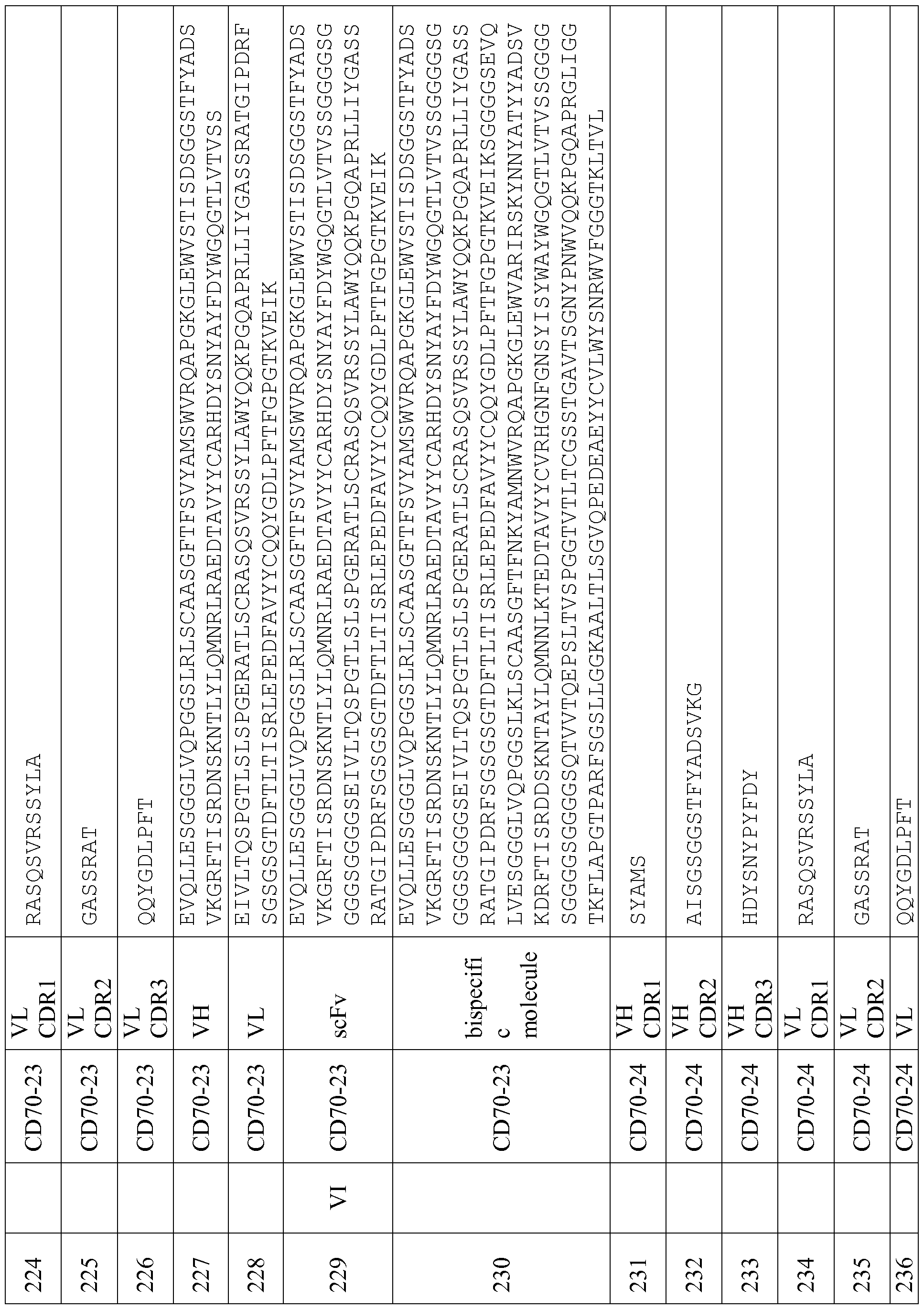


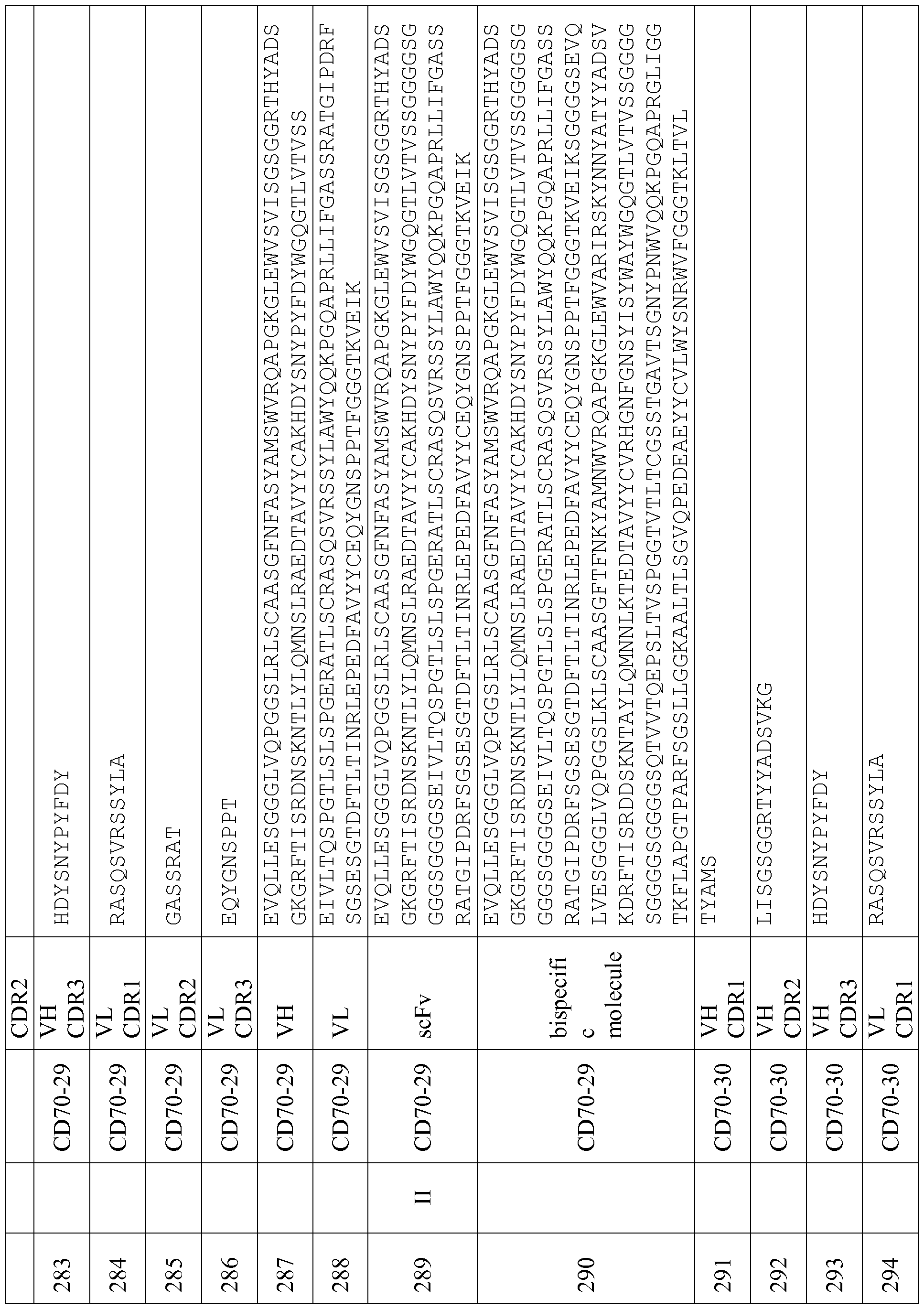


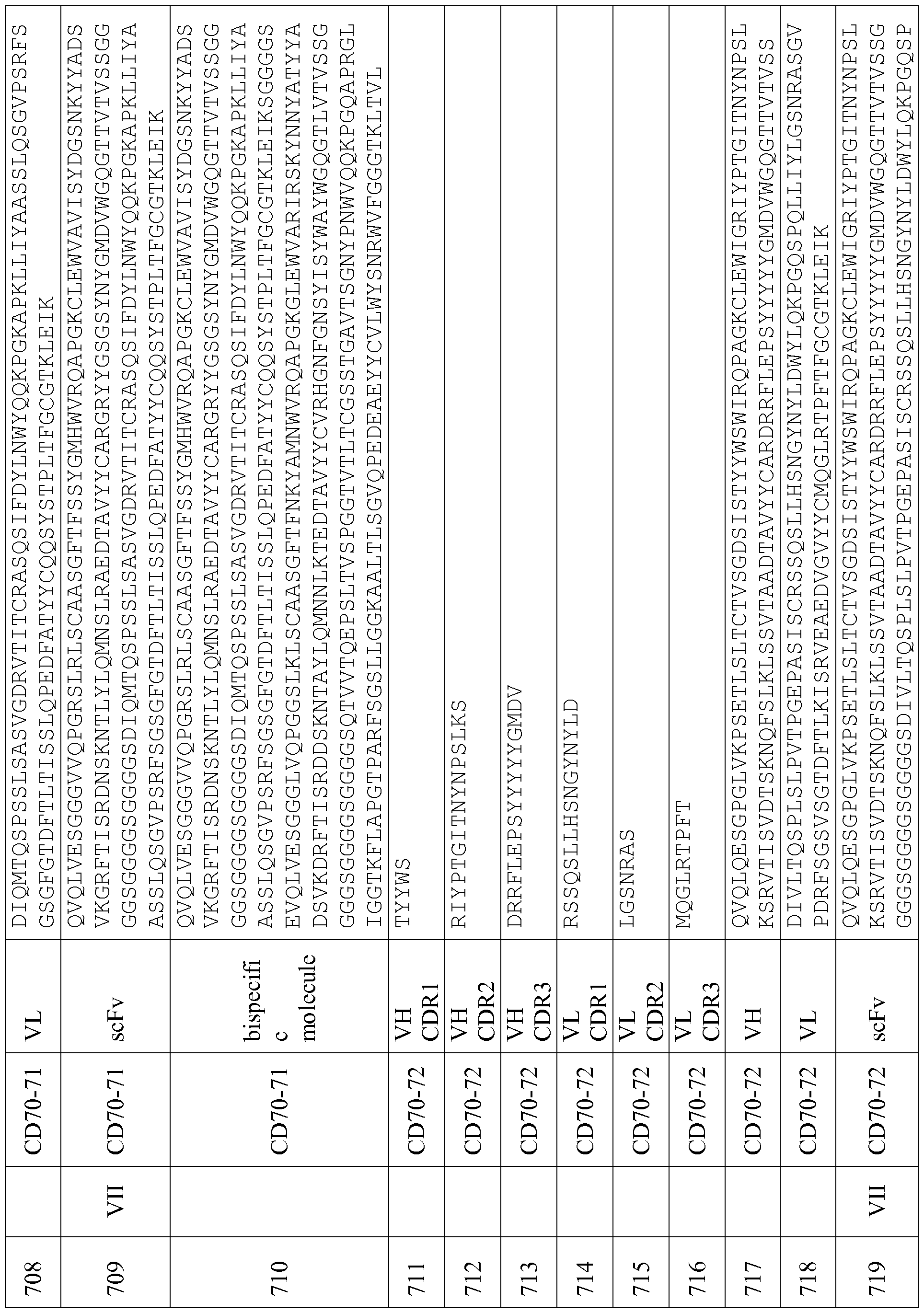


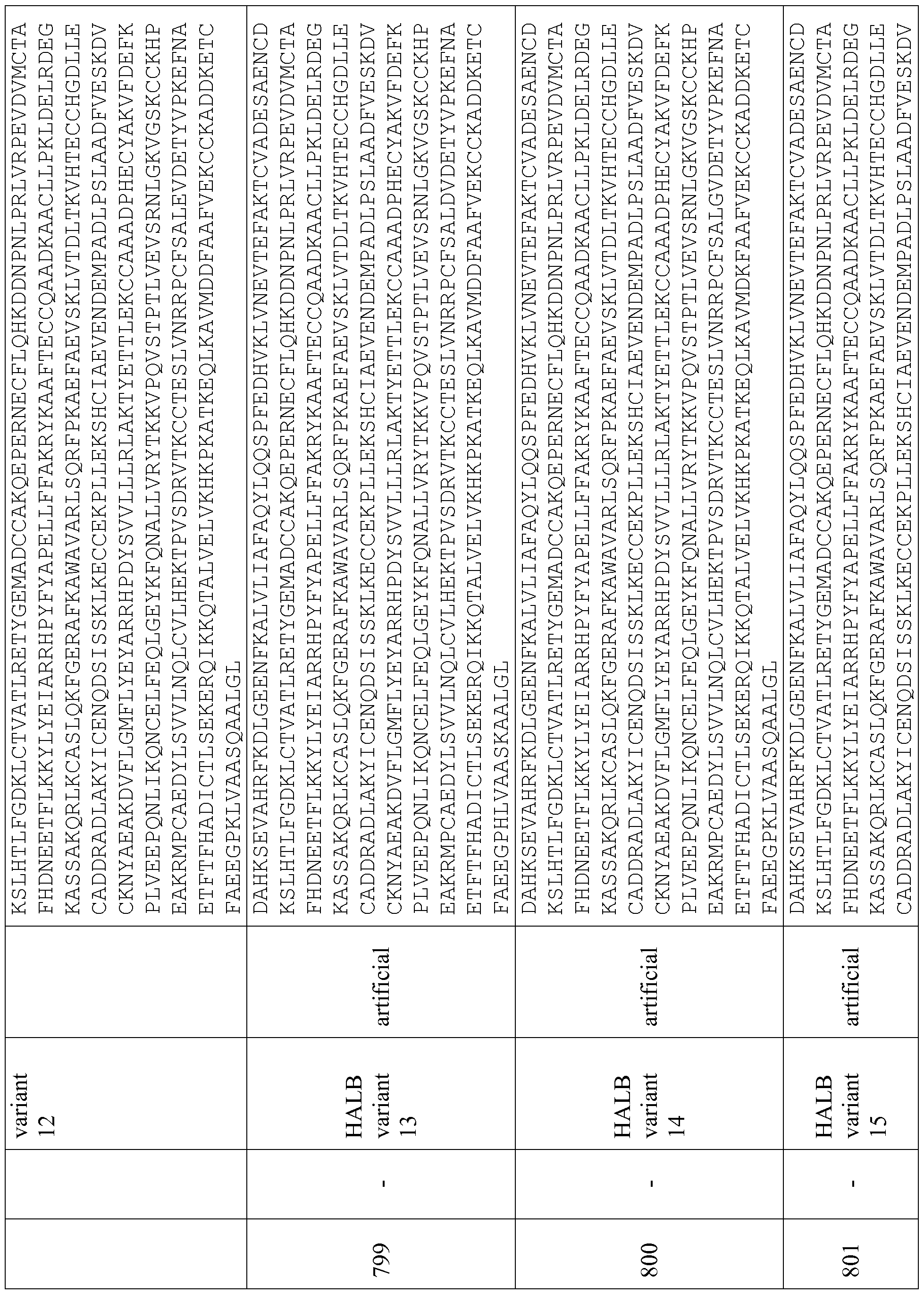


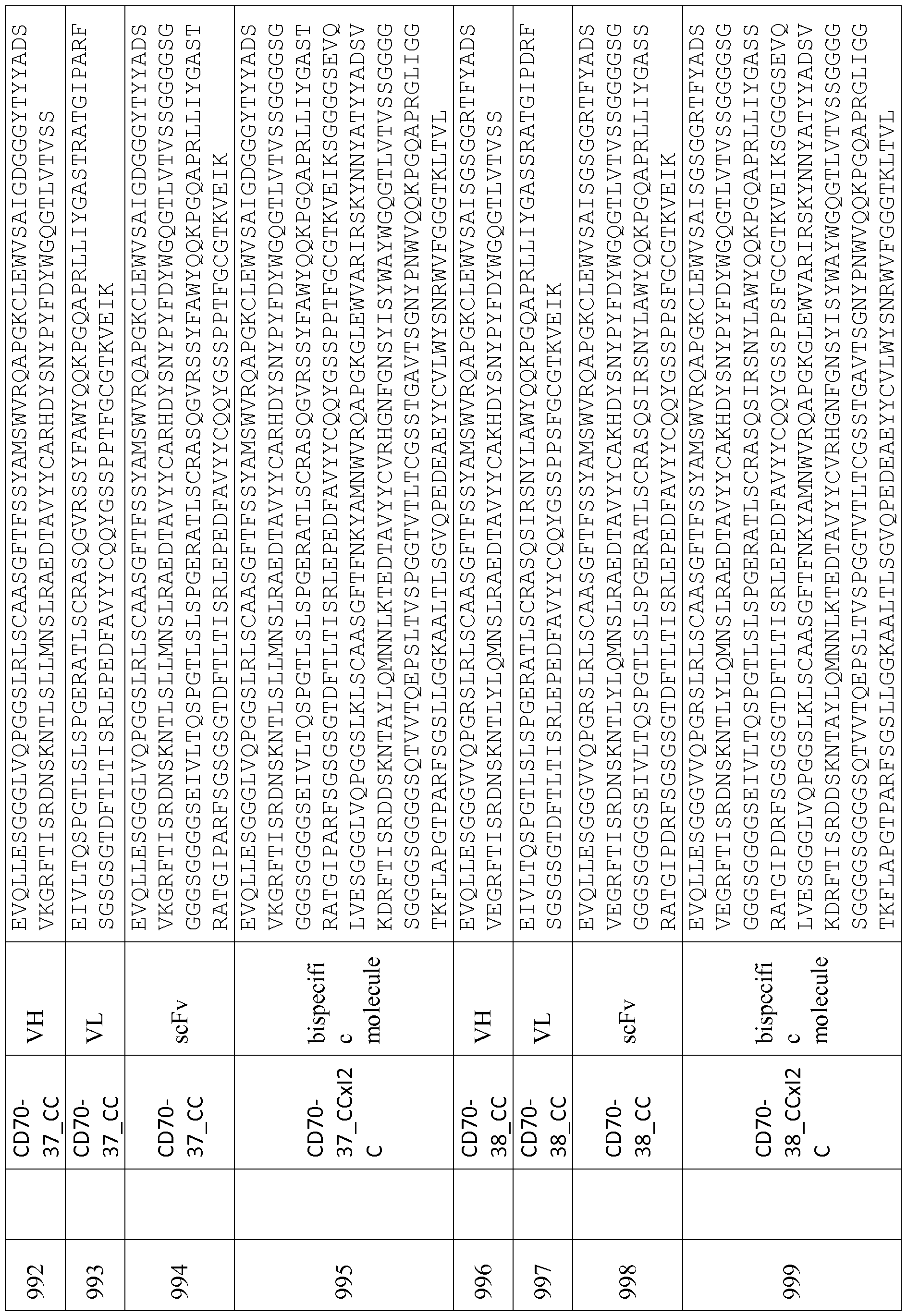


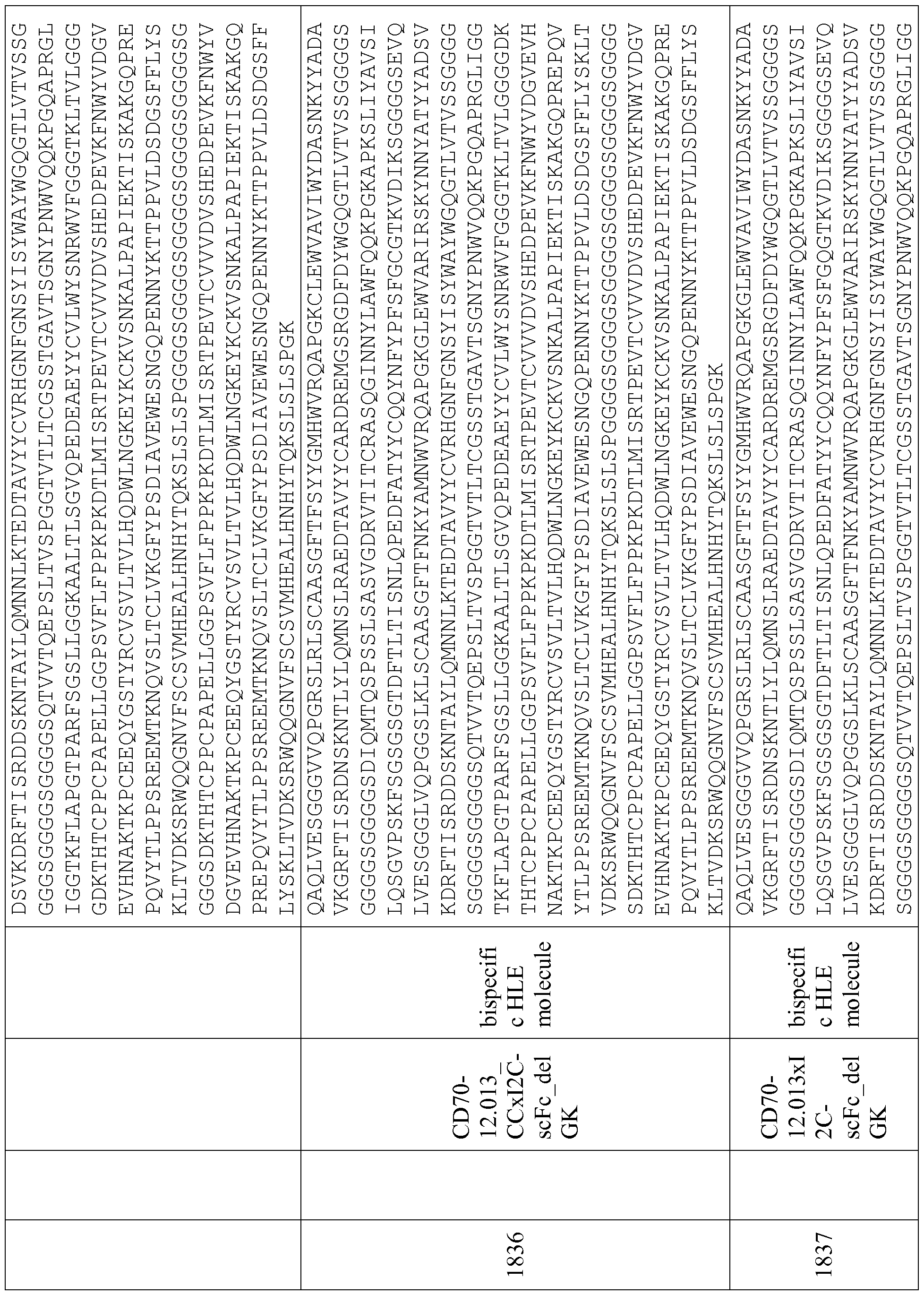


Claims
Priority Applications (19)
| Application Number | Priority Date | Filing Date | Title |
|---|---|---|---|
| ES16756951T ES2837134T3 (en) | 2015-07-31 | 2016-08-01 | Antibody Constructs for CD70 and CD3 |
| MX2018001156A MX2018001156A (en) | 2015-07-31 | 2016-08-01 | Antibody constructs for cd70 and cd3. |
| CN201680044597.7A CN109476734B (en) | 2015-07-31 | 2016-08-01 | Antibody constructs to CD70 and CD3 |
| EA201890382A EA201890382A1 (en) | 2015-07-31 | 2016-08-01 | ANTIBODY CONSTRUCTIONS FOR CD70 AND CD3 |
| TNP/2017/000551A TN2017000551A1 (en) | 2015-07-31 | 2016-08-01 | Antibody constructs for cd70 and cd3. |
| BR112018001531A BR112018001531A2 (en) | 2015-07-31 | 2016-08-01 | cd70 and cd3 antibody constructs |
| CR20180068A CR20180068A (en) | 2015-07-31 | 2016-08-01 | ANTIBODY CONSTRUCTS FOR CD70 AND CD3 |
| EP16756951.6A EP3328890B1 (en) | 2015-07-31 | 2016-08-01 | Antibody constructs for cd70 and cd3 |
| CA2988245A CA2988245A1 (en) | 2015-07-31 | 2016-08-01 | Antibody constructs for cd70 and cd3 |
| KR1020187001694A KR20180037949A (en) | 2015-07-31 | 2016-08-01 | Antibody constructs for CD70 and CD3 |
| AU2016302573A AU2016302573B2 (en) | 2015-07-31 | 2016-08-01 | Antibody constructs for CD70 and CD3 |
| JP2018504925A JP6879998B2 (en) | 2015-07-31 | 2016-08-01 | Antibody constructs against CD70 and CD3 |
| MYPI2017704947A MY187971A (en) | 2015-07-31 | 2016-08-01 | Antibody constructs for cd70 and cd3 |
| UAA201802021A UA126275C2 (en) | 2015-07-31 | 2016-08-01 | Antibody constructs for cd70 and cd3 |
| ZA2017/07967A ZA201707967B (en) | 2015-07-31 | 2017-11-23 | Antibody constructs for cd70 and cd3 |
| IL256870A IL256870B2 (en) | 2015-07-31 | 2018-01-11 | Constructs for bispecific antibodies binding cd70 and cd3 and uses thereof |
| PH12018500216A PH12018500216A1 (en) | 2015-07-31 | 2018-01-26 | Antibody constructs for cd70 and cd3 |
| CONC2018/0000976A CO2018000976A2 (en) | 2015-07-31 | 2018-01-30 | Antibody constructs for cd70 and cd3 |
| HK18114718.2A HK1255589A1 (en) | 2015-07-31 | 2018-11-16 | Antibody constructs for cd70 and cd3 |
Applications Claiming Priority (4)
| Application Number | Priority Date | Filing Date | Title |
|---|---|---|---|
| US201562199935P | 2015-07-31 | 2015-07-31 | |
| US62/199,935 | 2015-07-31 | ||
| US201662290886P | 2016-02-03 | 2016-02-03 | |
| US62/290,886 | 2016-02-03 |
Publications (1)
| Publication Number | Publication Date |
|---|---|
| WO2017021354A1 true WO2017021354A1 (en) | 2017-02-09 |
Family
ID=56800253
Family Applications (1)
| Application Number | Title | Priority Date | Filing Date |
|---|---|---|---|
| PCT/EP2016/068294 WO2017021354A1 (en) | 2015-07-31 | 2016-08-01 | Antibody constructs for cd70 and cd3 |
Country Status (26)
| Country | Link |
|---|---|
| US (2) | US10851170B2 (en) |
| EP (1) | EP3328890B1 (en) |
| JP (1) | JP6879998B2 (en) |
| KR (1) | KR20180037949A (en) |
| CN (1) | CN109476734B (en) |
| AU (1) | AU2016302573B2 (en) |
| BR (1) | BR112018001531A2 (en) |
| CA (1) | CA2988245A1 (en) |
| CL (2) | CL2018000270A1 (en) |
| CO (1) | CO2018000976A2 (en) |
| CR (1) | CR20180068A (en) |
| EA (1) | EA201890382A1 (en) |
| ES (1) | ES2837134T3 (en) |
| HK (1) | HK1255589A1 (en) |
| IL (1) | IL256870B2 (en) |
| JO (1) | JO3714B1 (en) |
| MX (1) | MX2018001156A (en) |
| MY (1) | MY187971A (en) |
| PE (1) | PE20181291A1 (en) |
| PH (1) | PH12018500216A1 (en) |
| SG (1) | SG10202000834WA (en) |
| TN (1) | TN2017000551A1 (en) |
| TW (1) | TWI717375B (en) |
| UA (1) | UA126275C2 (en) |
| WO (1) | WO2017021354A1 (en) |
| ZA (1) | ZA201707967B (en) |
Cited By (11)
| Publication number | Priority date | Publication date | Assignee | Title |
|---|---|---|---|---|
| WO2019141732A1 (en) * | 2018-01-16 | 2019-07-25 | Argenx Bvba | Cd70 combination therapy |
| WO2021006199A1 (en) | 2019-07-05 | 2021-01-14 | 小野薬品工業株式会社 | Treatment of hematologic cancer with pd-1/cd3 dual specificity protein |
| JP2021511811A (en) * | 2018-02-01 | 2021-05-13 | ファイザー・インク | CD70-specific antibodies and their use |
| US11072665B2 (en) | 2011-03-16 | 2021-07-27 | Argenx Bvba | Antibodies to CD70 |
| WO2022096698A1 (en) * | 2020-11-06 | 2022-05-12 | Amgen Inc. | Polypeptide constructs binding to cd3 |
| US11352433B2 (en) | 2016-02-03 | 2022-06-07 | Amgen Research (Munich) Gmbh | BCMA and CD3 bispecific T cell engaging antibody constructs |
| US11434302B2 (en) | 2016-02-03 | 2022-09-06 | Amgen Research (Munich) Gmbh | Bispecific T cell engaging antibody constructs |
| US11447567B2 (en) | 2015-07-31 | 2022-09-20 | Amgen Research (Munich) Gmbh | Antibody constructs for FLT3 and CD3 |
| US11571475B1 (en) | 2014-08-22 | 2023-02-07 | University Of Bern | Anti-CD70 and BCR-ABL inhibitor combination therapy |
| US11712468B2 (en) | 2018-12-18 | 2023-08-01 | argenx BV | CD70 combination therapy |
| US11884720B2 (en) | 2015-07-31 | 2024-01-30 | Amgen Research (Munich) Gmbh | Antibody constructs for MSLN and CD3 |
Families Citing this family (15)
| Publication number | Priority date | Publication date | Assignee | Title |
|---|---|---|---|---|
| AU2009299794B2 (en) | 2008-10-01 | 2015-08-13 | Amgen Research (Munich) Gmbh | Cross-species-specific single domain bispecific single chain antibody |
| JO3519B1 (en) | 2013-01-25 | 2020-07-05 | Amgen Inc | Antibody constructs for CDH19 and CD3 |
| AU2015294834B2 (en) | 2014-07-31 | 2021-04-29 | Amgen Research (Munich) Gmbh | Optimized cross-species specific bispecific single chain antibody constructs |
| TWI744242B (en) | 2015-07-31 | 2021-11-01 | 德商安美基研究(慕尼黑)公司 | Antibody constructs for egfrviii and cd3 |
| TWI793062B (en) * | 2015-07-31 | 2023-02-21 | 德商安美基研究(慕尼黑)公司 | Antibody constructs for dll3 and cd3 |
| EA201891753A1 (en) | 2016-02-03 | 2019-01-31 | Эмджен Рисерч (Мюник) Гмбх | BISPECIFIC CONSTRUCTIONS OF ANTIBODIES TO PSMA AND CD3, INVOLVING T-CELLS |
| MX2019000514A (en) * | 2016-07-14 | 2019-07-12 | Scholar Rock Inc | Tgfb antibodies, methods, and uses. |
| TWI814525B (en) * | 2017-02-14 | 2023-09-01 | 美商凱特製藥公司 | Cd70 binding molecules and methods of use thereof |
| EP3638697A4 (en) * | 2017-06-12 | 2021-07-07 | Bluefin Biomedicine, Inc. | Anti-il1rap antibodies and antibody drug conjugates |
| KR20200128018A (en) | 2018-02-01 | 2020-11-11 | 화이자 인코포레이티드 | Chimeric antigen receptor targeting CD70 |
| CN110804099B (en) * | 2019-10-31 | 2020-06-16 | 浙江蓝盾药业有限公司 | Human CD70 antigen binding polypeptides |
| WO2021113748A1 (en) * | 2019-12-05 | 2021-06-10 | Arbele Corp. | Composition of triaxial antibodies and method of making and using thereof |
| JP2024504162A (en) * | 2021-01-22 | 2024-01-30 | エルピス・バイオファーマシューティカルズ | Bispecific chimeric antigen receptor that binds CD19 and CD22 |
| TW202330603A (en) * | 2021-09-29 | 2023-08-01 | 美商蜻蜓醫療公司 | Proteins binding nkg2d, cd16 and baff-r |
| WO2023137958A1 (en) * | 2022-01-19 | 2023-07-27 | 上海恒润达生生物科技股份有限公司 | Anti-cd70 nanoantibody and use thereof |
Citations (138)
| Publication number | Priority date | Publication date | Assignee | Title |
|---|---|---|---|---|
| US3180193A (en) | 1963-02-25 | 1965-04-27 | Benedict David | Machines for cutting lengths of strip material |
| US3691016A (en) | 1970-04-17 | 1972-09-12 | Monsanto Co | Process for the preparation of insoluble enzymes |
| US3773919A (en) | 1969-10-23 | 1973-11-20 | Du Pont | Polylactide-drug mixtures |
| US3969287A (en) | 1972-12-08 | 1976-07-13 | Boehringer Mannheim Gmbh | Carrier-bound protein prepared by reacting the protein with an acylating or alkylating compound having a carrier-bonding group and reacting the product with a carrier |
| US4179337A (en) | 1973-07-20 | 1979-12-18 | Davis Frank F | Non-immunogenic polypeptides |
| US4195128A (en) | 1976-05-03 | 1980-03-25 | Bayer Aktiengesellschaft | Polymeric carrier bound ligands |
| US4229537A (en) | 1978-02-09 | 1980-10-21 | New York University | Preparation of trichloro-s-triazine activated supports for coupling ligands |
| US4247642A (en) | 1977-02-17 | 1981-01-27 | Sumitomo Chemical Company, Limited | Enzyme immobilization with pullulan gel |
| EP0036676A1 (en) | 1978-03-24 | 1981-09-30 | The Regents Of The University Of California | Method of making uniformly sized liposomes and liposomes so made |
| US4301144A (en) | 1979-07-11 | 1981-11-17 | Ajinomoto Company, Incorporated | Blood substitute containing modified hemoglobin |
| US4330440A (en) | 1977-02-08 | 1982-05-18 | Development Finance Corporation Of New Zealand | Activated matrix and method of activation |
| EP0058481A1 (en) | 1981-02-16 | 1982-08-25 | Zeneca Limited | Continuous release pharmaceutical compositions |
| EP0088046A2 (en) | 1982-02-17 | 1983-09-07 | Ciba-Geigy Ag | Lipids in the aqueous phase |
| US4439196A (en) | 1982-03-18 | 1984-03-27 | Merck & Co., Inc. | Osmotic drug delivery system |
| US4447224A (en) | 1982-09-20 | 1984-05-08 | Infusaid Corporation | Variable flow implantable infusion apparatus |
| US4447233A (en) | 1981-04-10 | 1984-05-08 | Parker-Hannifin Corporation | Medication infusion pump |
| US4475196A (en) | 1981-03-06 | 1984-10-02 | Zor Clair G | Instrument for locating faults in aircraft passenger reading light and attendant call control system |
| US4485045A (en) | 1981-07-06 | 1984-11-27 | Research Corporation | Synthetic phosphatidyl cholines useful in forming liposomes |
| US4486194A (en) | 1983-06-08 | 1984-12-04 | James Ferrara | Therapeutic device for administering medicaments through the skin |
| US4487603A (en) | 1982-11-26 | 1984-12-11 | Cordis Corporation | Implantable microinfusion pump system |
| US4496689A (en) | 1983-12-27 | 1985-01-29 | Miles Laboratories, Inc. | Covalently attached complex of alpha-1-proteinase inhibitor with a water soluble polymer |
| EP0133988A2 (en) | 1983-08-02 | 1985-03-13 | Hoechst Aktiengesellschaft | Regulating peptide-containing pharmaceutical preparations with retarded release, and process for their preparation |
| EP0143949A1 (en) | 1983-11-01 | 1985-06-12 | TERUMO KABUSHIKI KAISHA trading as TERUMO CORPORATION | Pharmaceutical composition containing urokinase |
| US4544545A (en) | 1983-06-20 | 1985-10-01 | Trustees University Of Massachusetts | Liposomes containing modified cholesterol for organ targeting |
| EP0171496A2 (en) | 1984-08-15 | 1986-02-19 | Research Development Corporation of Japan | Process for the production of a chimera monoclonal antibody |
| EP0173494A2 (en) | 1984-08-27 | 1986-03-05 | The Board Of Trustees Of The Leland Stanford Junior University | Chimeric receptors by DNA splicing and expression |
| EP0183070A2 (en) | 1984-10-30 | 1986-06-04 | Phillips Petroleum Company | Transformation of yeasts of the genus pichia |
| US4596556A (en) | 1985-03-25 | 1986-06-24 | Bioject, Inc. | Hypodermic injection apparatus |
| GB2177096A (en) | 1984-09-03 | 1987-01-14 | Celltech Ltd | Production of chimeric antibodies |
| US4640835A (en) | 1981-10-30 | 1987-02-03 | Nippon Chemiphar Company, Ltd. | Plasminogen activator derivatives |
| US4670417A (en) | 1985-06-19 | 1987-06-02 | Ajinomoto Co., Inc. | Hemoglobin combined with a poly(alkylene oxide) |
| WO1987005330A1 (en) | 1986-03-07 | 1987-09-11 | Michel Louis Eugene Bergh | Method for enhancing glycoprotein stability |
| EP0239400A2 (en) | 1986-03-27 | 1987-09-30 | Medical Research Council | Recombinant antibodies and methods for their production |
| EP0244234A2 (en) | 1986-04-30 | 1987-11-04 | Alko Group Ltd. | Transformation of trichoderma |
| US4751180A (en) | 1985-03-28 | 1988-06-14 | Chiron Corporation | Expression using fused genes providing for protein product |
| WO1988009344A1 (en) | 1987-05-21 | 1988-12-01 | Creative Biomolecules, Inc. | Targeted multifunctional proteins |
| US4790824A (en) | 1987-06-19 | 1988-12-13 | Bioject, Inc. | Non-invasive hypodermic injection device |
| US4791192A (en) | 1986-06-26 | 1988-12-13 | Takeda Chemical Industries, Ltd. | Chemically modified protein with polyethyleneglycol |
| US4816397A (en) | 1983-03-25 | 1989-03-28 | Celltech, Limited | Multichain polypeptides or proteins and processes for their production |
| US4816567A (en) | 1983-04-08 | 1989-03-28 | Genentech, Inc. | Recombinant immunoglobin preparations |
| US4935233A (en) | 1985-12-02 | 1990-06-19 | G. D. Searle And Company | Covalently linked polypeptide cell modulators |
| US4941880A (en) | 1987-06-19 | 1990-07-17 | Bioject, Inc. | Pre-filled ampule and non-invasive hypodermic injection device assembly |
| US4946778A (en) | 1987-09-21 | 1990-08-07 | Genex Corporation | Single polypeptide chain binding molecules |
| EP0402226A1 (en) | 1989-06-06 | 1990-12-12 | Institut National De La Recherche Agronomique | Transformation vectors for yeast yarrowia |
| US5013556A (en) | 1989-10-20 | 1991-05-07 | Liposome Technology, Inc. | Liposomes with enhanced circulation time |
| US5064413A (en) | 1989-11-09 | 1991-11-12 | Bioject, Inc. | Needleless hypodermic injection device |
| WO1992003918A1 (en) | 1990-08-29 | 1992-03-19 | Genpharm International, Inc. | Transgenic non-human animals capable of producing heterologous antibodies |
| WO1992015673A1 (en) | 1991-03-11 | 1992-09-17 | The University Of Georgia Research Foundation, Inc. | Cloning and expression of renilla luciferase |
| WO1992022645A1 (en) | 1991-06-14 | 1992-12-23 | Genpharm International, Inc. | Transgenic immunodeficient non-human animals |
| WO1992022647A1 (en) | 1991-06-12 | 1992-12-23 | Genpharm International, Inc. | Early detection of transgenic emryros |
| WO1993012227A1 (en) | 1991-12-17 | 1993-06-24 | Genpharm International, Inc. | Transgenic non-human animals capable of producing heterologous antibodies |
| US5223409A (en) | 1988-09-02 | 1993-06-29 | Protein Engineering Corp. | Directed evolution of novel binding proteins |
| US5225539A (en) | 1986-03-27 | 1993-07-06 | Medical Research Council | Recombinant altered antibodies and methods of making altered antibodies |
| WO1994000569A1 (en) | 1992-06-18 | 1994-01-06 | Genpharm International, Inc. | Methods for producing transgenic non-human animals harboring a yeast artificial chromosome |
| WO1994002602A1 (en) | 1992-07-24 | 1994-02-03 | Cell Genesys, Inc. | Generation of xenogeneic antibodies |
| US5292658A (en) | 1989-12-29 | 1994-03-08 | University Of Georgia Research Foundation, Inc. Boyd Graduate Studies Research Center | Cloning and expressions of Renilla luciferase |
| WO1994010308A1 (en) | 1992-10-23 | 1994-05-11 | Immunex Corporation | Methods of preparing soluble, oligomeric proteins |
| US5312335A (en) | 1989-11-09 | 1994-05-17 | Bioject Inc. | Needleless hypodermic injection device |
| US5313193A (en) | 1990-06-29 | 1994-05-17 | Thomson-Csf | Object-marking means, method for making such means and reading device |
| WO1994025585A1 (en) | 1993-04-26 | 1994-11-10 | Genpharm International, Inc. | Transgenic non-human animals capable of producing heterologous antibodies |
| US5383851A (en) | 1992-07-24 | 1995-01-24 | Bioject Inc. | Needleless hypodermic injection device |
| WO1995007463A1 (en) | 1993-09-10 | 1995-03-16 | The Trustees Of Columbia University In The City Of New York | Uses of green fluorescent protein |
| US5476996A (en) | 1988-06-14 | 1995-12-19 | Lidak Pharmaceuticals | Human immune system in non-human animal |
| WO1996014436A1 (en) | 1994-11-04 | 1996-05-17 | Genpharm International, Inc. | Method for making recombinant yeast artificial chromosomes |
| EP0463151B1 (en) | 1990-01-12 | 1996-06-12 | Cell Genesys, Inc. | Generation of xenogeneic antibodies |
| US5545806A (en) | 1990-08-29 | 1996-08-13 | Genpharm International, Inc. | Ransgenic non-human animals for producing heterologous antibodies |
| US5545807A (en) | 1988-10-12 | 1996-08-13 | The Babraham Institute | Production of antibodies from transgenic animals |
| US5565332A (en) | 1991-09-23 | 1996-10-15 | Medical Research Council | Production of chimeric antibodies - a combinatorial approach |
| WO1996034096A1 (en) | 1995-04-28 | 1996-10-31 | Abgenix, Inc. | Human antibodies derived from immunized xenomice |
| WO1996033735A1 (en) | 1995-04-27 | 1996-10-31 | Abgenix, Inc. | Human antibodies derived from immunized xenomice |
| US5585089A (en) | 1988-12-28 | 1996-12-17 | Protein Design Labs, Inc. | Humanized immunoglobulins |
| US5591669A (en) | 1988-12-05 | 1997-01-07 | Genpharm International, Inc. | Transgenic mice depleted in a mature lymphocytic cell-type |
| US5612205A (en) | 1990-08-29 | 1997-03-18 | Genpharm International, Incorporated | Homologous recombination in mammalian cells |
| WO1997013852A1 (en) | 1995-10-10 | 1997-04-17 | Genpharm International, Inc. | Transgenic non-human animals capable of producing heterologous antibodies |
| US5625126A (en) | 1990-08-29 | 1997-04-29 | Genpharm International, Inc. | Transgenic non-human animals for producing heterologous antibodies |
| US5625825A (en) | 1993-10-21 | 1997-04-29 | Lsi Logic Corporation | Random number generating apparatus for an interface unit of a carrier sense with multiple access and collision detect (CSMA/CD) ethernet data network |
| EP0773288A2 (en) | 1995-08-29 | 1997-05-14 | Kirin Beer Kabushiki Kaisha | Chimeric animal and method for producing the same |
| US5633425A (en) | 1990-08-29 | 1997-05-27 | Genpharm International, Inc. | Transgenic non-human animals capable of producing heterologous antibodies |
| US5648260A (en) | 1987-03-18 | 1997-07-15 | Scotgen Biopharmaceuticals Incorporated | DNA encoding antibodies with altered effector functions |
| US5661016A (en) | 1990-08-29 | 1997-08-26 | Genpharm International Inc. | Transgenic non-human animals capable of producing heterologous antibodies of various isotypes |
| WO1997038731A1 (en) | 1996-04-18 | 1997-10-23 | The Regents Of The University Of California | Immunoliposomes that optimize internalization into target cells |
| US5683888A (en) | 1989-07-22 | 1997-11-04 | University Of Wales College Of Medicine | Modified bioluminescent proteins and their use |
| WO1998014605A1 (en) | 1996-10-04 | 1998-04-09 | Loma Linda University | Renilla luciferase and green fluorescent protein fusion genes |
| US5741668A (en) | 1994-02-04 | 1998-04-21 | Rutgers, The State University Of New Jersey | Expression of a gene for a modified green-fluorescent protein |
| WO1998024893A2 (en) | 1996-12-03 | 1998-06-11 | Abgenix, Inc. | TRANSGENIC MAMMALS HAVING HUMAN IG LOCI INCLUDING PLURAL VH AND Vλ REGIONS AND ANTIBODIES PRODUCED THEREFROM |
| WO1998024884A1 (en) | 1996-12-02 | 1998-06-11 | Genpharm International | Transgenic non-human animals capable of producing heterologous antibodies |
| WO1998026277A2 (en) | 1996-12-12 | 1998-06-18 | Prolume, Ltd. | Apparatus and method for detecting and identifying infectious agents |
| US5777079A (en) | 1994-11-10 | 1998-07-07 | The Regents Of The University Of California | Modified green fluorescent proteins |
| US5789650A (en) | 1990-08-29 | 1998-08-04 | Genpharm International, Inc. | Transgenic non-human animals for producing heterologous antibodies |
| US5789215A (en) | 1991-08-20 | 1998-08-04 | Genpharm International | Gene targeting in animal cells using isogenic DNA constructs |
| US5804387A (en) | 1996-02-01 | 1998-09-08 | The Board Of Trustees Of The Leland Stanford Junior University | FACS-optimized mutants of the green fluorescent protein (GFP) |
| US5814318A (en) | 1990-08-29 | 1998-09-29 | Genpharm International Inc. | Transgenic non-human animals for producing heterologous antibodies |
| WO1998052976A1 (en) | 1997-05-21 | 1998-11-26 | Biovation Limited | Method for the production of non-immunogenic proteins |
| US5859205A (en) | 1989-12-21 | 1999-01-12 | Celltech Limited | Humanised antibodies |
| US5874304A (en) | 1996-01-18 | 1999-02-23 | University Of Florida Research Foundation, Inc. | Humanized green fluorescent protein genes and methods |
| US5874299A (en) | 1990-08-29 | 1999-02-23 | Genpharm International, Inc. | Transgenic non-human animals capable of producing heterologous antibodies |
| US5877397A (en) | 1990-08-29 | 1999-03-02 | Genpharm International Inc. | Transgenic non-human animals capable of producing heterologous antibodies of various isotypes |
| US5876995A (en) | 1996-02-06 | 1999-03-02 | Bryan; Bruce | Bioluminescent novelty items |
| US5925558A (en) | 1996-07-16 | 1999-07-20 | The Regents Of The University Of California | Assays for protein kinases using fluorescent protein substrates |
| US5958765A (en) | 1995-06-07 | 1999-09-28 | Idec Pharmaceuticals Corporation | Neutralizing high affinity human monoclonal antibodies specific to RSV F-protein and methods for their manufacture and therapeutic use thereof |
| WO1999049019A2 (en) | 1998-03-27 | 1999-09-30 | Prolume, Ltd. | Luciferases, fluorescent proteins, nucleic acids encoding the luciferases and fluorescent proteins and the use thereof in diagnostics |
| WO1999054440A1 (en) | 1998-04-21 | 1999-10-28 | Micromet Gesellschaft Für Biomedizinische Forschung Mbh | CD19xCD3 SPECIFIC POLYPEPTIDES AND USES THEREOF |
| US5981175A (en) | 1993-01-07 | 1999-11-09 | Genpharm Internation, Inc. | Methods for producing recombinant mammalian cells harboring a yeast artificial chromosome |
| WO2000006605A2 (en) | 1998-07-28 | 2000-02-10 | Micromet Ag | Heterominibodies |
| US6075181A (en) | 1990-01-12 | 2000-06-13 | Abgenix, Inc. | Human antibodies derived from immunized xenomice |
| WO2000034317A2 (en) | 1998-12-08 | 2000-06-15 | Biovation Limited | Method for reducing immunogenicity of proteins |
| US6150584A (en) | 1990-01-12 | 2000-11-21 | Abgenix, Inc. | Human antibodies derived from immunized xenomice |
| US6162963A (en) | 1990-01-12 | 2000-12-19 | Abgenix, Inc. | Generation of Xenogenetic antibodies |
| WO2000076310A1 (en) | 1999-06-10 | 2000-12-21 | Abgenix, Inc. | Transgenic animals for producing specific isotypes of human antibodies via non-cognate switch regions |
| US6255458B1 (en) | 1990-08-29 | 2001-07-03 | Genpharm International | High affinity human antibodies and human antibodies against digoxin |
| US6300064B1 (en) | 1995-08-18 | 2001-10-09 | Morphosys Ag | Protein/(poly)peptide libraries |
| US6407213B1 (en) | 1991-06-14 | 2002-06-18 | Genentech, Inc. | Method for making humanized antibodies |
| WO2003047336A2 (en) | 2001-11-30 | 2003-06-12 | Abgenix, Inc. | TRANSGENIC ANIMALS BEARING HUMAN Igμ LIGHT CHAIN GENES |
| WO2005040220A1 (en) | 2003-10-16 | 2005-05-06 | Micromet Ag | Multispecific deimmunized cd3-binders |
| WO2006138181A2 (en) | 2005-06-14 | 2006-12-28 | Amgen Inc. | Self-buffering protein formulations |
| WO2007042261A2 (en) | 2005-10-11 | 2007-04-19 | Micromet Ag | Compositions comprising cross-species-specific antibodies and uses thereof |
| WO2007098420A2 (en) | 2006-02-17 | 2007-08-30 | Syntonix Pharmaceuticals, Inc. | Peptides that block the binding of igg to fcrn |
| WO2008119567A2 (en) | 2007-04-03 | 2008-10-09 | Micromet Ag | Cross-species-specific cd3-epsilon binding domain |
| WO2009127691A1 (en) | 2008-04-17 | 2009-10-22 | Ablynx N.V. | Peptides capable of binding to serum proteins and compounds, constructs and polypeptides comprising the same |
| WO2010037838A2 (en) | 2008-10-01 | 2010-04-08 | Micromet Ag | Cross-species-specific single domain bispecific single chain antibody |
| WO2011051489A2 (en) | 2009-10-30 | 2011-05-05 | Novozymes Biopharma Dk A/S | Albumin variants |
| WO2012059486A1 (en) | 2010-11-01 | 2012-05-10 | Novozymes Biopharma Dk A/S | Albumin variants |
| WO2012150319A1 (en) | 2011-05-05 | 2012-11-08 | Novozymes Biopharma Dk A/S | Albumin variants |
| WO2013026837A1 (en) | 2011-08-23 | 2013-02-28 | Roche Glycart Ag | Bispecific t cell activating antigen binding molecules |
| WO2013026833A1 (en) | 2011-08-23 | 2013-02-28 | Roche Glycart Ag | Bispecific t cell activating antigen binding molecules |
| WO2013075066A2 (en) | 2011-11-18 | 2013-05-23 | Eleven Biotherapeutics, Inc. | Proteins with improved half-life and other properties |
| WO2013135896A1 (en) | 2012-03-16 | 2013-09-19 | Novozymes Biopharma Dk A/S | Albumin variants |
| US20140099254A1 (en) * | 2012-08-14 | 2014-04-10 | Ibc Pharmaceuticals, Inc. | Combination therapy for inducing immune response to disease |
| WO2014072481A1 (en) | 2012-11-08 | 2014-05-15 | Novozymes Biopharma Dk A/S | Albumin variants |
| WO2014144722A2 (en) | 2013-03-15 | 2014-09-18 | Amgen Inc. | BISPECIFIC-Fc MOLECULES |
| WO2014151910A1 (en) | 2013-03-15 | 2014-09-25 | Amgen Inc. | Heterodimeric bispecific antibodies |
| US20140308285A1 (en) | 2013-03-15 | 2014-10-16 | Amgen Inc. | Heterodimeric bispecific antibodies |
| WO2015048272A1 (en) | 2013-09-25 | 2015-04-02 | Amgen Inc. | V-c-fc-v-c antibody |
| US20150166661A1 (en) * | 2013-12-17 | 2015-06-18 | Genentech, Inc. | Anti-cd3 antibodies and methods of use |
| WO2016016859A1 (en) * | 2014-07-31 | 2016-02-04 | Amgen Research (Munich) Gmbh | Optimized cross-species specific bispecific single chain antibody constructs |
| US9300829B2 (en) | 2014-04-04 | 2016-03-29 | Canon Kabushiki Kaisha | Image reading apparatus and correction method thereof |
| US9676293B2 (en) | 2013-04-23 | 2017-06-13 | Calsonic Kansei Corporation | Driving force controller for electric vehicle |
| US11284893B2 (en) | 2019-04-02 | 2022-03-29 | Covidien Lp | Stapling device with articulating tool assembly |
Family Cites Families (13)
| Publication number | Priority date | Publication date | Assignee | Title |
|---|---|---|---|---|
| US5470582A (en) | 1992-02-07 | 1995-11-28 | Syntex (U.S.A.) Inc. | Controlled delivery of pharmaceuticals from preformed porous polymeric microparticles |
| WO2004106381A1 (en) * | 2003-05-31 | 2004-12-09 | Micromet Ag | Pharmaceutical compositions comprising bispecific anti-cd3, anti-cd19 antibody constructs for the treatment of b-cell related disorders |
| AU2007333098A1 (en) * | 2006-12-14 | 2008-06-19 | Medarex, Inc. | Human antibodies that bind CD70 and uses thereof |
| KR101589759B1 (en) | 2007-04-03 | 2016-01-29 | 암젠 리서치 (뮌헨) 게엠베하 | Cross-species-specific cd3-epsilon binding domain |
| AP2014007588A0 (en) | 2011-09-22 | 2014-04-30 | Amgen Inc | CD27L antigen binding proteins |
| JO3519B1 (en) | 2013-01-25 | 2020-07-05 | Amgen Inc | Antibody constructs for CDH19 and CD3 |
| TWI744242B (en) | 2015-07-31 | 2021-11-01 | 德商安美基研究(慕尼黑)公司 | Antibody constructs for egfrviii and cd3 |
| TWI796283B (en) | 2015-07-31 | 2023-03-21 | 德商安美基研究(慕尼黑)公司 | Antibody constructs for msln and cd3 |
| TWI793062B (en) | 2015-07-31 | 2023-02-21 | 德商安美基研究(慕尼黑)公司 | Antibody constructs for dll3 and cd3 |
| TWI829617B (en) | 2015-07-31 | 2024-01-21 | 德商安美基研究(慕尼黑)公司 | Antibody constructs for flt3 and cd3 |
| EA201891753A1 (en) | 2016-02-03 | 2019-01-31 | Эмджен Рисерч (Мюник) Гмбх | BISPECIFIC CONSTRUCTIONS OF ANTIBODIES TO PSMA AND CD3, INVOLVING T-CELLS |
| EA039859B1 (en) | 2016-02-03 | 2022-03-21 | Эмджен Рисерч (Мюник) Гмбх | Bispecific antibody constructs binding egfrviii and cd3 |
| LT3411402T (en) | 2016-02-03 | 2022-01-25 | Amgen Research (Munich) Gmbh | Bcma and cd3 bispecific t cell engaging antibody constructs |
-
2016
- 2016-07-29 TW TW105124269A patent/TWI717375B/en active
- 2016-07-31 JO JOP/2016/0164A patent/JO3714B1/en active
- 2016-08-01 KR KR1020187001694A patent/KR20180037949A/en not_active Application Discontinuation
- 2016-08-01 CA CA2988245A patent/CA2988245A1/en active Pending
- 2016-08-01 CR CR20180068A patent/CR20180068A/en unknown
- 2016-08-01 TN TNP/2017/000551A patent/TN2017000551A1/en unknown
- 2016-08-01 CN CN201680044597.7A patent/CN109476734B/en active Active
- 2016-08-01 UA UAA201802021A patent/UA126275C2/en unknown
- 2016-08-01 ES ES16756951T patent/ES2837134T3/en active Active
- 2016-08-01 MX MX2018001156A patent/MX2018001156A/en unknown
- 2016-08-01 SG SG10202000834WA patent/SG10202000834WA/en unknown
- 2016-08-01 EA EA201890382A patent/EA201890382A1/en unknown
- 2016-08-01 US US15/225,282 patent/US10851170B2/en active Active
- 2016-08-01 MY MYPI2017704947A patent/MY187971A/en unknown
- 2016-08-01 WO PCT/EP2016/068294 patent/WO2017021354A1/en active Application Filing
- 2016-08-01 EP EP16756951.6A patent/EP3328890B1/en active Active
- 2016-08-01 PE PE2018000147A patent/PE20181291A1/en unknown
- 2016-08-01 BR BR112018001531A patent/BR112018001531A2/en active Search and Examination
- 2016-08-01 JP JP2018504925A patent/JP6879998B2/en active Active
- 2016-08-01 AU AU2016302573A patent/AU2016302573B2/en active Active
-
2017
- 2017-11-23 ZA ZA2017/07967A patent/ZA201707967B/en unknown
-
2018
- 2018-01-11 IL IL256870A patent/IL256870B2/en unknown
- 2018-01-26 PH PH12018500216A patent/PH12018500216A1/en unknown
- 2018-01-30 CO CONC2018/0000976A patent/CO2018000976A2/en unknown
- 2018-01-30 CL CL2018000270A patent/CL2018000270A1/en unknown
- 2018-11-16 HK HK18114718.2A patent/HK1255589A1/en unknown
-
2020
- 2020-10-29 US US17/084,088 patent/US20210284748A1/en active Pending
-
2021
- 2021-03-23 CL CL2021000717A patent/CL2021000717A1/en unknown
Patent Citations (157)
| Publication number | Priority date | Publication date | Assignee | Title |
|---|---|---|---|---|
| US3180193A (en) | 1963-02-25 | 1965-04-27 | Benedict David | Machines for cutting lengths of strip material |
| US3773919A (en) | 1969-10-23 | 1973-11-20 | Du Pont | Polylactide-drug mixtures |
| US3691016A (en) | 1970-04-17 | 1972-09-12 | Monsanto Co | Process for the preparation of insoluble enzymes |
| US3969287A (en) | 1972-12-08 | 1976-07-13 | Boehringer Mannheim Gmbh | Carrier-bound protein prepared by reacting the protein with an acylating or alkylating compound having a carrier-bonding group and reacting the product with a carrier |
| US4179337A (en) | 1973-07-20 | 1979-12-18 | Davis Frank F | Non-immunogenic polypeptides |
| US4195128A (en) | 1976-05-03 | 1980-03-25 | Bayer Aktiengesellschaft | Polymeric carrier bound ligands |
| US4330440A (en) | 1977-02-08 | 1982-05-18 | Development Finance Corporation Of New Zealand | Activated matrix and method of activation |
| US4247642A (en) | 1977-02-17 | 1981-01-27 | Sumitomo Chemical Company, Limited | Enzyme immobilization with pullulan gel |
| US4229537A (en) | 1978-02-09 | 1980-10-21 | New York University | Preparation of trichloro-s-triazine activated supports for coupling ligands |
| EP0036676A1 (en) | 1978-03-24 | 1981-09-30 | The Regents Of The University Of California | Method of making uniformly sized liposomes and liposomes so made |
| US4301144A (en) | 1979-07-11 | 1981-11-17 | Ajinomoto Company, Incorporated | Blood substitute containing modified hemoglobin |
| EP0058481A1 (en) | 1981-02-16 | 1982-08-25 | Zeneca Limited | Continuous release pharmaceutical compositions |
| US4475196A (en) | 1981-03-06 | 1984-10-02 | Zor Clair G | Instrument for locating faults in aircraft passenger reading light and attendant call control system |
| US4447233A (en) | 1981-04-10 | 1984-05-08 | Parker-Hannifin Corporation | Medication infusion pump |
| US4485045A (en) | 1981-07-06 | 1984-11-27 | Research Corporation | Synthetic phosphatidyl cholines useful in forming liposomes |
| US4640835A (en) | 1981-10-30 | 1987-02-03 | Nippon Chemiphar Company, Ltd. | Plasminogen activator derivatives |
| EP0088046A2 (en) | 1982-02-17 | 1983-09-07 | Ciba-Geigy Ag | Lipids in the aqueous phase |
| US4439196A (en) | 1982-03-18 | 1984-03-27 | Merck & Co., Inc. | Osmotic drug delivery system |
| US4447224A (en) | 1982-09-20 | 1984-05-08 | Infusaid Corporation | Variable flow implantable infusion apparatus |
| US4487603A (en) | 1982-11-26 | 1984-12-11 | Cordis Corporation | Implantable microinfusion pump system |
| US4816397A (en) | 1983-03-25 | 1989-03-28 | Celltech, Limited | Multichain polypeptides or proteins and processes for their production |
| US4816567A (en) | 1983-04-08 | 1989-03-28 | Genentech, Inc. | Recombinant immunoglobin preparations |
| US4486194A (en) | 1983-06-08 | 1984-12-04 | James Ferrara | Therapeutic device for administering medicaments through the skin |
| US4544545A (en) | 1983-06-20 | 1985-10-01 | Trustees University Of Massachusetts | Liposomes containing modified cholesterol for organ targeting |
| EP0133988A2 (en) | 1983-08-02 | 1985-03-13 | Hoechst Aktiengesellschaft | Regulating peptide-containing pharmaceutical preparations with retarded release, and process for their preparation |
| EP0143949A1 (en) | 1983-11-01 | 1985-06-12 | TERUMO KABUSHIKI KAISHA trading as TERUMO CORPORATION | Pharmaceutical composition containing urokinase |
| US4496689A (en) | 1983-12-27 | 1985-01-29 | Miles Laboratories, Inc. | Covalently attached complex of alpha-1-proteinase inhibitor with a water soluble polymer |
| EP0171496A2 (en) | 1984-08-15 | 1986-02-19 | Research Development Corporation of Japan | Process for the production of a chimera monoclonal antibody |
| EP0173494A2 (en) | 1984-08-27 | 1986-03-05 | The Board Of Trustees Of The Leland Stanford Junior University | Chimeric receptors by DNA splicing and expression |
| GB2177096A (en) | 1984-09-03 | 1987-01-14 | Celltech Ltd | Production of chimeric antibodies |
| EP0183070A2 (en) | 1984-10-30 | 1986-06-04 | Phillips Petroleum Company | Transformation of yeasts of the genus pichia |
| US4596556A (en) | 1985-03-25 | 1986-06-24 | Bioject, Inc. | Hypodermic injection apparatus |
| US4751180A (en) | 1985-03-28 | 1988-06-14 | Chiron Corporation | Expression using fused genes providing for protein product |
| US4670417A (en) | 1985-06-19 | 1987-06-02 | Ajinomoto Co., Inc. | Hemoglobin combined with a poly(alkylene oxide) |
| US4935233A (en) | 1985-12-02 | 1990-06-19 | G. D. Searle And Company | Covalently linked polypeptide cell modulators |
| WO1987005330A1 (en) | 1986-03-07 | 1987-09-11 | Michel Louis Eugene Bergh | Method for enhancing glycoprotein stability |
| EP0239400A2 (en) | 1986-03-27 | 1987-09-30 | Medical Research Council | Recombinant antibodies and methods for their production |
| US5225539A (en) | 1986-03-27 | 1993-07-06 | Medical Research Council | Recombinant altered antibodies and methods of making altered antibodies |
| EP0244234A2 (en) | 1986-04-30 | 1987-11-04 | Alko Group Ltd. | Transformation of trichoderma |
| US4791192A (en) | 1986-06-26 | 1988-12-13 | Takeda Chemical Industries, Ltd. | Chemically modified protein with polyethyleneglycol |
| US5648260A (en) | 1987-03-18 | 1997-07-15 | Scotgen Biopharmaceuticals Incorporated | DNA encoding antibodies with altered effector functions |
| WO1988009344A1 (en) | 1987-05-21 | 1988-12-01 | Creative Biomolecules, Inc. | Targeted multifunctional proteins |
| US4790824A (en) | 1987-06-19 | 1988-12-13 | Bioject, Inc. | Non-invasive hypodermic injection device |
| US4941880A (en) | 1987-06-19 | 1990-07-17 | Bioject, Inc. | Pre-filled ampule and non-invasive hypodermic injection device assembly |
| US4946778A (en) | 1987-09-21 | 1990-08-07 | Genex Corporation | Single polypeptide chain binding molecules |
| US5698767A (en) | 1988-06-14 | 1997-12-16 | Lidak Pharmaceuticals | Human immune system in non-human animal |
| US5476996A (en) | 1988-06-14 | 1995-12-19 | Lidak Pharmaceuticals | Human immune system in non-human animal |
| US5223409A (en) | 1988-09-02 | 1993-06-29 | Protein Engineering Corp. | Directed evolution of novel binding proteins |
| US5545807A (en) | 1988-10-12 | 1996-08-13 | The Babraham Institute | Production of antibodies from transgenic animals |
| US5591669A (en) | 1988-12-05 | 1997-01-07 | Genpharm International, Inc. | Transgenic mice depleted in a mature lymphocytic cell-type |
| US6023010A (en) | 1988-12-05 | 2000-02-08 | Genpharm International | Transgenic non-human animals depleted in a mature lymphocytic cell-type |
| US5693762A (en) | 1988-12-28 | 1997-12-02 | Protein Design Labs, Inc. | Humanized immunoglobulins |
| US5693761A (en) | 1988-12-28 | 1997-12-02 | Protein Design Labs, Inc. | Polynucleotides encoding improved humanized immunoglobulins |
| US5585089A (en) | 1988-12-28 | 1996-12-17 | Protein Design Labs, Inc. | Humanized immunoglobulins |
| EP0402226A1 (en) | 1989-06-06 | 1990-12-12 | Institut National De La Recherche Agronomique | Transformation vectors for yeast yarrowia |
| US5683888A (en) | 1989-07-22 | 1997-11-04 | University Of Wales College Of Medicine | Modified bioluminescent proteins and their use |
| US5013556A (en) | 1989-10-20 | 1991-05-07 | Liposome Technology, Inc. | Liposomes with enhanced circulation time |
| US5312335A (en) | 1989-11-09 | 1994-05-17 | Bioject Inc. | Needleless hypodermic injection device |
| US5064413A (en) | 1989-11-09 | 1991-11-12 | Bioject, Inc. | Needleless hypodermic injection device |
| US5859205A (en) | 1989-12-21 | 1999-01-12 | Celltech Limited | Humanised antibodies |
| US5418155A (en) | 1989-12-29 | 1995-05-23 | University Of Georgia Research Foundation, Inc. | Isolated Renilla luciferase and method of use thereof |
| US5292658A (en) | 1989-12-29 | 1994-03-08 | University Of Georgia Research Foundation, Inc. Boyd Graduate Studies Research Center | Cloning and expressions of Renilla luciferase |
| US6150584A (en) | 1990-01-12 | 2000-11-21 | Abgenix, Inc. | Human antibodies derived from immunized xenomice |
| JP3068507B2 (en) | 1990-01-12 | 2000-07-24 | アブジェニックス インコーポレイテッド | Generation of heterologous antibodies |
| US5939598A (en) | 1990-01-12 | 1999-08-17 | Abgenix, Inc. | Method of making transgenic mice lacking endogenous heavy chains |
| US6075181A (en) | 1990-01-12 | 2000-06-13 | Abgenix, Inc. | Human antibodies derived from immunized xenomice |
| JP3068180B2 (en) | 1990-01-12 | 2000-07-24 | アブジェニックス インコーポレイテッド | Generation of heterologous antibodies |
| EP0463151B1 (en) | 1990-01-12 | 1996-06-12 | Cell Genesys, Inc. | Generation of xenogeneic antibodies |
| JP3068506B2 (en) | 1990-01-12 | 2000-07-24 | アブジェニックス インコーポレイテッド | Generation of heterologous antibodies |
| US6114598A (en) | 1990-01-12 | 2000-09-05 | Abgenix, Inc. | Generation of xenogeneic antibodies |
| US6162963A (en) | 1990-01-12 | 2000-12-19 | Abgenix, Inc. | Generation of Xenogenetic antibodies |
| US5313193A (en) | 1990-06-29 | 1994-05-17 | Thomson-Csf | Object-marking means, method for making such means and reading device |
| US5661016A (en) | 1990-08-29 | 1997-08-26 | Genpharm International Inc. | Transgenic non-human animals capable of producing heterologous antibodies of various isotypes |
| US5770429A (en) | 1990-08-29 | 1998-06-23 | Genpharm International, Inc. | Transgenic non-human animals capable of producing heterologous antibodies |
| US5877397A (en) | 1990-08-29 | 1999-03-02 | Genpharm International Inc. | Transgenic non-human animals capable of producing heterologous antibodies of various isotypes |
| US5612205A (en) | 1990-08-29 | 1997-03-18 | Genpharm International, Incorporated | Homologous recombination in mammalian cells |
| US5814318A (en) | 1990-08-29 | 1998-09-29 | Genpharm International Inc. | Transgenic non-human animals for producing heterologous antibodies |
| US5625126A (en) | 1990-08-29 | 1997-04-29 | Genpharm International, Inc. | Transgenic non-human animals for producing heterologous antibodies |
| US6255458B1 (en) | 1990-08-29 | 2001-07-03 | Genpharm International | High affinity human antibodies and human antibodies against digoxin |
| US5874299A (en) | 1990-08-29 | 1999-02-23 | Genpharm International, Inc. | Transgenic non-human animals capable of producing heterologous antibodies |
| US5633425A (en) | 1990-08-29 | 1997-05-27 | Genpharm International, Inc. | Transgenic non-human animals capable of producing heterologous antibodies |
| EP0546073B1 (en) | 1990-08-29 | 1997-09-10 | GenPharm International, Inc. | production and use of transgenic non-human animals capable of producing heterologous antibodies |
| WO1992003918A1 (en) | 1990-08-29 | 1992-03-19 | Genpharm International, Inc. | Transgenic non-human animals capable of producing heterologous antibodies |
| US5721367A (en) | 1990-08-29 | 1998-02-24 | Pharming B.V. | Homologous recombination in mammalian cells |
| US5789650A (en) | 1990-08-29 | 1998-08-04 | Genpharm International, Inc. | Transgenic non-human animals for producing heterologous antibodies |
| US5545806A (en) | 1990-08-29 | 1996-08-13 | Genpharm International, Inc. | Ransgenic non-human animals for producing heterologous antibodies |
| WO1992015673A1 (en) | 1991-03-11 | 1992-09-17 | The University Of Georgia Research Foundation, Inc. | Cloning and expression of renilla luciferase |
| WO1992022670A1 (en) | 1991-06-12 | 1992-12-23 | Genpharm International, Inc. | Early detection of transgenic embryos |
| WO1992022647A1 (en) | 1991-06-12 | 1992-12-23 | Genpharm International, Inc. | Early detection of transgenic emryros |
| WO1992022645A1 (en) | 1991-06-14 | 1992-12-23 | Genpharm International, Inc. | Transgenic immunodeficient non-human animals |
| US6407213B1 (en) | 1991-06-14 | 2002-06-18 | Genentech, Inc. | Method for making humanized antibodies |
| US5789215A (en) | 1991-08-20 | 1998-08-04 | Genpharm International | Gene targeting in animal cells using isogenic DNA constructs |
| US5565332A (en) | 1991-09-23 | 1996-10-15 | Medical Research Council | Production of chimeric antibodies - a combinatorial approach |
| WO1993012227A1 (en) | 1991-12-17 | 1993-06-24 | Genpharm International, Inc. | Transgenic non-human animals capable of producing heterologous antibodies |
| WO1994000569A1 (en) | 1992-06-18 | 1994-01-06 | Genpharm International, Inc. | Methods for producing transgenic non-human animals harboring a yeast artificial chromosome |
| WO1994002602A1 (en) | 1992-07-24 | 1994-02-03 | Cell Genesys, Inc. | Generation of xenogeneic antibodies |
| US5383851A (en) | 1992-07-24 | 1995-01-24 | Bioject Inc. | Needleless hypodermic injection device |
| US5399163A (en) | 1992-07-24 | 1995-03-21 | Bioject Inc. | Needleless hypodermic injection methods and device |
| WO1994010308A1 (en) | 1992-10-23 | 1994-05-11 | Immunex Corporation | Methods of preparing soluble, oligomeric proteins |
| US5981175A (en) | 1993-01-07 | 1999-11-09 | Genpharm Internation, Inc. | Methods for producing recombinant mammalian cells harboring a yeast artificial chromosome |
| WO1994025585A1 (en) | 1993-04-26 | 1994-11-10 | Genpharm International, Inc. | Transgenic non-human animals capable of producing heterologous antibodies |
| WO1995007463A1 (en) | 1993-09-10 | 1995-03-16 | The Trustees Of Columbia University In The City Of New York | Uses of green fluorescent protein |
| US5625825A (en) | 1993-10-21 | 1997-04-29 | Lsi Logic Corporation | Random number generating apparatus for an interface unit of a carrier sense with multiple access and collision detect (CSMA/CD) ethernet data network |
| US5741668A (en) | 1994-02-04 | 1998-04-21 | Rutgers, The State University Of New Jersey | Expression of a gene for a modified green-fluorescent protein |
| US5643763A (en) | 1994-11-04 | 1997-07-01 | Genpharm International, Inc. | Method for making recombinant yeast artificial chromosomes by minimizing diploid doubling during mating |
| WO1996014436A1 (en) | 1994-11-04 | 1996-05-17 | Genpharm International, Inc. | Method for making recombinant yeast artificial chromosomes |
| US5777079A (en) | 1994-11-10 | 1998-07-07 | The Regents Of The University Of California | Modified green fluorescent proteins |
| WO1996033735A1 (en) | 1995-04-27 | 1996-10-31 | Abgenix, Inc. | Human antibodies derived from immunized xenomice |
| WO1996034096A1 (en) | 1995-04-28 | 1996-10-31 | Abgenix, Inc. | Human antibodies derived from immunized xenomice |
| US5958765A (en) | 1995-06-07 | 1999-09-28 | Idec Pharmaceuticals Corporation | Neutralizing high affinity human monoclonal antibodies specific to RSV F-protein and methods for their manufacture and therapeutic use thereof |
| US6300064B1 (en) | 1995-08-18 | 2001-10-09 | Morphosys Ag | Protein/(poly)peptide libraries |
| EP0843961A1 (en) | 1995-08-29 | 1998-05-27 | Kirin Beer Kabushiki Kaisha | Chimeric animal and method for constructing the same |
| EP0773288A2 (en) | 1995-08-29 | 1997-05-14 | Kirin Beer Kabushiki Kaisha | Chimeric animal and method for producing the same |
| WO1997013852A1 (en) | 1995-10-10 | 1997-04-17 | Genpharm International, Inc. | Transgenic non-human animals capable of producing heterologous antibodies |
| US5874304A (en) | 1996-01-18 | 1999-02-23 | University Of Florida Research Foundation, Inc. | Humanized green fluorescent protein genes and methods |
| US5804387A (en) | 1996-02-01 | 1998-09-08 | The Board Of Trustees Of The Leland Stanford Junior University | FACS-optimized mutants of the green fluorescent protein (GFP) |
| US5876995A (en) | 1996-02-06 | 1999-03-02 | Bryan; Bruce | Bioluminescent novelty items |
| WO1997038731A1 (en) | 1996-04-18 | 1997-10-23 | The Regents Of The University Of California | Immunoliposomes that optimize internalization into target cells |
| US5925558A (en) | 1996-07-16 | 1999-07-20 | The Regents Of The University Of California | Assays for protein kinases using fluorescent protein substrates |
| WO1998014605A1 (en) | 1996-10-04 | 1998-04-09 | Loma Linda University | Renilla luciferase and green fluorescent protein fusion genes |
| WO1998024884A1 (en) | 1996-12-02 | 1998-06-11 | Genpharm International | Transgenic non-human animals capable of producing heterologous antibodies |
| US20030070185A1 (en) | 1996-12-03 | 2003-04-10 | Aya Jakobovits | Transgenic mammals having human Ig loci including plural Vh and Vk regions and antibodies produced therefrom |
| WO1998024893A2 (en) | 1996-12-03 | 1998-06-11 | Abgenix, Inc. | TRANSGENIC MAMMALS HAVING HUMAN IG LOCI INCLUDING PLURAL VH AND Vλ REGIONS AND ANTIBODIES PRODUCED THEREFROM |
| WO1998026277A2 (en) | 1996-12-12 | 1998-06-18 | Prolume, Ltd. | Apparatus and method for detecting and identifying infectious agents |
| WO1998052976A1 (en) | 1997-05-21 | 1998-11-26 | Biovation Limited | Method for the production of non-immunogenic proteins |
| WO1999049019A2 (en) | 1998-03-27 | 1999-09-30 | Prolume, Ltd. | Luciferases, fluorescent proteins, nucleic acids encoding the luciferases and fluorescent proteins and the use thereof in diagnostics |
| WO1999054440A1 (en) | 1998-04-21 | 1999-10-28 | Micromet Gesellschaft Für Biomedizinische Forschung Mbh | CD19xCD3 SPECIFIC POLYPEPTIDES AND USES THEREOF |
| WO2000006605A2 (en) | 1998-07-28 | 2000-02-10 | Micromet Ag | Heterominibodies |
| WO2000034317A2 (en) | 1998-12-08 | 2000-06-15 | Biovation Limited | Method for reducing immunogenicity of proteins |
| WO2000076310A1 (en) | 1999-06-10 | 2000-12-21 | Abgenix, Inc. | Transgenic animals for producing specific isotypes of human antibodies via non-cognate switch regions |
| WO2003047336A2 (en) | 2001-11-30 | 2003-06-12 | Abgenix, Inc. | TRANSGENIC ANIMALS BEARING HUMAN Igμ LIGHT CHAIN GENES |
| WO2005040220A1 (en) | 2003-10-16 | 2005-05-06 | Micromet Ag | Multispecific deimmunized cd3-binders |
| WO2006138181A2 (en) | 2005-06-14 | 2006-12-28 | Amgen Inc. | Self-buffering protein formulations |
| WO2007042261A2 (en) | 2005-10-11 | 2007-04-19 | Micromet Ag | Compositions comprising cross-species-specific antibodies and uses thereof |
| WO2007098420A2 (en) | 2006-02-17 | 2007-08-30 | Syntonix Pharmaceuticals, Inc. | Peptides that block the binding of igg to fcrn |
| WO2008119567A2 (en) | 2007-04-03 | 2008-10-09 | Micromet Ag | Cross-species-specific cd3-epsilon binding domain |
| WO2009127691A1 (en) | 2008-04-17 | 2009-10-22 | Ablynx N.V. | Peptides capable of binding to serum proteins and compounds, constructs and polypeptides comprising the same |
| WO2010037838A2 (en) | 2008-10-01 | 2010-04-08 | Micromet Ag | Cross-species-specific single domain bispecific single chain antibody |
| WO2011051489A2 (en) | 2009-10-30 | 2011-05-05 | Novozymes Biopharma Dk A/S | Albumin variants |
| WO2012059486A1 (en) | 2010-11-01 | 2012-05-10 | Novozymes Biopharma Dk A/S | Albumin variants |
| WO2012150319A1 (en) | 2011-05-05 | 2012-11-08 | Novozymes Biopharma Dk A/S | Albumin variants |
| WO2013026837A1 (en) | 2011-08-23 | 2013-02-28 | Roche Glycart Ag | Bispecific t cell activating antigen binding molecules |
| WO2013026833A1 (en) | 2011-08-23 | 2013-02-28 | Roche Glycart Ag | Bispecific t cell activating antigen binding molecules |
| WO2013075066A2 (en) | 2011-11-18 | 2013-05-23 | Eleven Biotherapeutics, Inc. | Proteins with improved half-life and other properties |
| WO2013135896A1 (en) | 2012-03-16 | 2013-09-19 | Novozymes Biopharma Dk A/S | Albumin variants |
| US20140099254A1 (en) * | 2012-08-14 | 2014-04-10 | Ibc Pharmaceuticals, Inc. | Combination therapy for inducing immune response to disease |
| WO2014072481A1 (en) | 2012-11-08 | 2014-05-15 | Novozymes Biopharma Dk A/S | Albumin variants |
| WO2014144722A2 (en) | 2013-03-15 | 2014-09-18 | Amgen Inc. | BISPECIFIC-Fc MOLECULES |
| WO2014151910A1 (en) | 2013-03-15 | 2014-09-25 | Amgen Inc. | Heterodimeric bispecific antibodies |
| US20140302037A1 (en) | 2013-03-15 | 2014-10-09 | Amgen Inc. | BISPECIFIC-Fc MOLECULES |
| US20140308285A1 (en) | 2013-03-15 | 2014-10-16 | Amgen Inc. | Heterodimeric bispecific antibodies |
| US9676293B2 (en) | 2013-04-23 | 2017-06-13 | Calsonic Kansei Corporation | Driving force controller for electric vehicle |
| WO2015048272A1 (en) | 2013-09-25 | 2015-04-02 | Amgen Inc. | V-c-fc-v-c antibody |
| US20150166661A1 (en) * | 2013-12-17 | 2015-06-18 | Genentech, Inc. | Anti-cd3 antibodies and methods of use |
| US9300829B2 (en) | 2014-04-04 | 2016-03-29 | Canon Kabushiki Kaisha | Image reading apparatus and correction method thereof |
| WO2016016859A1 (en) * | 2014-07-31 | 2016-02-04 | Amgen Research (Munich) Gmbh | Optimized cross-species specific bispecific single chain antibody constructs |
| US11284893B2 (en) | 2019-04-02 | 2022-03-29 | Covidien Lp | Stapling device with articulating tool assembly |
Non-Patent Citations (115)
| Title |
|---|
| "Macromolecule Sequencing and Synthesis, Selected Methods and Applications", 1988, ALAN R. LISS, INC, article "Current Methods in Sequence Comparison and Analysis", pages: 127 - 149 |
| "Using Antibodies: a laboratory manual", 1999, CSHL PRESS |
| A.R. GENRMO: "REMINGTON'S PHARMACEUTICAL SCIENCES, 18th ed.", 1990, MACK PUBLISHING COMPANY |
| ALTSCHUL ET AL., J. MOL. BIOL., vol. 215, 1990, pages 403 - 410 |
| ALTSCHUL ET AL., METHODS IN ENZYMOLOGY, vol. 266, 1996, pages 460 - 480 |
| ALTSCHUL ET AL., NUCL. ACIDS RES, vol. 25, 1993, pages 3389 - 3402 |
| ALTSCHUL ET AL., NUCLEIC ACIDS RES., vol. 25, 1997, pages 3389 - 3402 |
| APLIN; WRISTON, CRC CRIT. REV. BIOCHEM., 1981, pages 259 - 306 |
| ARAKAWA ET AL.: "Solvent interactions in pharmaceutical formulations", PHARM RES., vol. 8, no. 3, 1991, pages 285 - 91 |
| ARTSAENKO ET AL., THE PLANT J, vol. 8, 1995, pages 745 - 750 |
| BRÜHL, IMMUNOL, vol. 166, 2001, pages 2420 - 2426 |
| CARTER ET AL., BIO/TECHNOLOGY, vol. 10, 1992, pages 163 - 167 |
| CHALFIE ET AL., SCIENCE, vol. 263, 1994, pages 802 - 805 |
| CHEADLE ET AL., MOL IMMUNOL, vol. 29, 1992, pages 21 - 30 |
| CHESON BD; HORNING SJ; COIFFIER B; SHIPP MA; FISHER RI; CONNORS JM; LISTER TA; VOSE J; GRILLO-LOPEZ A; HAGENBEEK A: "Report of an international workshop to standardize response criteria for non-Hodgkin's lymphomas. NCI Sponsored International Working Group", J CLIN ONCOL., vol. 17, no. 4, April 1999 (1999-04-01), pages 1244 |
| CHOTHIA ET AL., J. MOL. BIOL, vol. 196, 1987, pages 901 - 917 |
| CHOTHIA ET AL., NATURE, vol. 342, 1989, pages 877 |
| CHOTHIA; LESK, J. MOL. BIOL., vol. 196, 1987, pages 901 |
| CLACKSON ET AL., NATURE, vol. 352, 1991, pages 624 - 628 |
| COLE ET AL.: "Monoclonal Antibodies and Cancer Therapy", 1985, ALAN R. LISS, INC., pages: 77 - 96 |
| COOK, G.P. ET AL., IMMUNOL. TODAY, vol. 16, no. 5, 1995, pages 237 - 242 |
| CUNNINGHAM; WELLS, SCIENCE, vol. 244, 1989, pages 1081 - 1085 |
| DALL'ACQUA ET AL., BIOCHEM, vol. 37, 1998, pages 9266 - 9273 |
| DEVEREUX ET AL., NUCL. ACID RES., vol. 12, 1984, pages 387 - 395 |
| DUEBEL, S. AND KONTERMANN, R: "Antibody Engineering Lab Manual", SPRINGER-VERLAG, article "Protein Sequence and Structure Analysis of Antibody Variable Domains" |
| DUSKIN ET AL., J. BIOL. CHEM., vol. 257, 1982, pages 3105 |
| EDGE ET AL., ANAL. BIOCHEM., vol. 118, 1981, pages 131 |
| EDWIN BREMER: "Targeting of the Tumor Necrosis Factor Receptor Superfamily for Cancer Immunotherapy", ISRN ONCOLOGY, vol. 176, no. 2, 1 January 2013 (2013-01-01), pages 974 - 25, XP055184622, ISSN: 2090-5661, DOI: 10.1186/1479-5876-9-204 * |
| EPPSTEIN ET AL., PROC. NATL. ACAD. SCI. U.S.A., vol. 82, 1985, pages 3688 - 3692 |
| EPSTEIN ET AL., PROC. NATL. ACAD. SCI. USA, vol. 82, 1985, pages 3688 |
| FANSLOW ET AL., SEMIN. IMMUNOL., vol. 6, 1994, pages 267 - 78 |
| FECKER ET AL., PLANT MOL BIOL, vol. 32, 1996, pages 979 - 986 |
| FENG; DOOLITTLE, J. MOL. EVOL., vol. 35, 1987, pages 351 - 360 |
| GABIZON ET AL., J. NATIONAL CANCER INST., vol. 81, no. 19, 1989, pages 1484 |
| GRAHAM ET AL., J. GEN VIROL., vol. 36, 1977, pages 59 |
| GREEN ET AL., NATURE GENETICS, vol. 7, 1994, pages 13 - 21 |
| GREEN; JAKOBOVITS, J. EXP. MED., vol. 188, 1998, pages 483 - 495 |
| HAKIMUDDIN ET AL., ARCH. BIOCHEM. BIOPHYS., vol. 259, 1987, pages 52 |
| HARLOW ET AL.: "Antibodies: A Laboratory Manual", 1988, COLD SPRING HARBOR LABORATORY |
| HARLOW; LANE: "Antibodies a laboratory manual", 1988, CSHL PRESS |
| HAWKINS ET AL., J. MOL. BIOL., vol. 254, 1992, pages 889 - 896 |
| HEIM ET AL., CURR. BIOL, vol. 6, 1996, pages 178 - 182 |
| HIATT ET AL., NATURE, vol. 342, 1989, pages 76 - 78 |
| HIGGINS; SHARP, CABIOS, vol. 5, 1989, pages 151 - 153 |
| HOPPE ET AL., FEBS LETTERS, vol. 344, 1994, pages 191 |
| HUANG J. ET AL., J IMMUNOL, vol. 190, 2013, pages 1 - 9 |
| HUSTON ET AL., PROC. NATL. ACAD. SCI USA, vol. 85, 1988, pages 5879 - 5883 |
| HWANG ET AL., PROC. NATL ACAD. SCI. USA, vol. 77, 1980, pages 4030 |
| HWANG; FOOTE: "Immunogenicity of engineered antibodies", METHODS, vol. 36, 2005, pages 3 - 10 |
| ICHIKI ET AL., J. IMMUNOL., vol. 150, 1993, pages 5408 - 5417 |
| JONES ET AL., NATURE, vol. 321, 1986, pages 522 - 525 |
| JONIO ET AL.: "Immunoglobulin Genes, 2nd ed.", 1995, ACADEMIC PRESS |
| KARIN ET AL., PROC. NATL. ACAD. SCI. U.S.A., vol. 90, 1993, pages 5873 - 5787 |
| KAUFMAN R.J., METHODS ENZYMOL., vol. 185, 1990, pages 537 - 566 |
| KENDRICK ET AL.: "RATIONAL DESIGN OF STABLE PROTEIN FORMULATIONS: THEORY AND PRACTICE", article "Physical stabilization of proteins in aqueous solution" |
| KIPRIYANOV, J. MOL. BIOL., vol. 293, 1999, pages 41 - 56 |
| KNAPPE ET AL., BLOOD, vol. 95, 2000, pages 3256 - 61 |
| KOEHLER ET AL., NATURE, vol. 256, 1975, pages 495 |
| KONTERMANN; DUBEL: "Antibody Engineering, 2ND ED.", 2010, SPRINGER |
| KOZBOR ET AL., IMMUNOLOGY TODAY, vol. 4, 1983, pages 7279 |
| KOZBOR, IMMUNOLOGY TODAY, vol. 4, 1983, pages 72 |
| KUFER P., TRENDS IN BIOTECHNOLOGY, vol. 22, no. 5, 2004, pages 238 - 244 |
| KUFER, CANCER IMMUNOL. IMMUNOTHER., vol. 45, 1997, pages 193 - 197 |
| LANDSCHULZ ET AL., SCIENCE, vol. 240, 1988, pages 1759 |
| LANGER ET AL., J. BIOMED. MATER. RES, vol. 15, 1981, pages 167 - 277 |
| LANGER, CHEM. TECH, vol. 12, 1982, pages 98 - 105 |
| LITTLE: "Recombinant Antibodies for Immunotherapy", 2009, CAMBRIDGE UNIVERSITY PRESS |
| LOFFLER, BLOOD, vol. 95, no. 6, 2000, pages 2098 - 2103 |
| LOWMAN ET AL., BIOCHEMISTRY, vol. 30, 1991, pages 10832 - 10837 |
| MACCALLUM ET AL., J. MOL. BIOL, vol. 262, 1996, pages 732 |
| MACK, J. IMMUNOL., vol. 158, 1997, pages 3965 - 3970 |
| MACK, PNAS, vol. 92, 1995, pages 7021 - 7025 |
| MALMBORG, J. IMMUNOL. METHODS, vol. 183, 1995, pages 7 - 13 |
| MARKS ET AL., J. MOL. BIOL., vol. 222, 1991, pages 581 - 597 |
| MARTIN ET AL., J. BIOL. CHEM., vol. 257, 1982, pages 286 - 288 |
| MARTIN; THORNTON, J. MOL. BIOL, vol. 263, 1996, pages 800 |
| MATHER, BIOL. REPROD, vol. 23, 1980, pages 243 - 251 |
| MATHER: "Annals N. Y Acad. Sci.", vol. 383, 1982, pages: 44 - 68 |
| MENDEZ ET AL., NATURE GENETICS, vol. 15, 1997, pages 146 - 156 |
| MORRISON ET AL., PROC. NATL. ACAD. SCI. USA, vol. 81, 1984, pages 6851 - 6855 |
| MORRISON ET AL., PROC. NATL. ACAD. SCL U.S.A., vol. 81, 1985, pages 6851 |
| MORRISON KL; WEISS GA., CUR OPIN CHEM BIOL., vol. 5, no. 3, June 2001 (2001-06-01), pages 302 - 7 |
| MORRISON, SCIENCE, vol. 229, 1985, pages 1202 - 1207 |
| NEEDLEMAN; WUNSCH, J. MOL. BIOL., vol. 48, 1970, pages 443 |
| NOLAN ET AL., PROC. NATL. ACAD. SCI. U.S.A., vol. 85, 1988, pages 2603 - 2607 |
| OI ET AL., BIOTECHNIQUES, vol. 4, 1986, pages 214 |
| OLSSON ET AL., METH. ENZYMOL., vol. 92, 1982, pages 3 - 16 |
| OSLO, A.: "Remington's Pharmaceutical Sciences, 16th ed.", 1980 |
| OWEN ET AL., BIO/TECHNOLOGY, vol. 10, 1992, pages 790 - 794 |
| PADLAN, MOLECULAR IMMUNOLOGY, vol. 31, no. 3, 1993, pages 169 - 217 |
| PEARSON; LIPMAN, PROC. NAT. ACAD. SCI. U.S.A., vol. 85, 1988, pages 2444 |
| PHARMACEUTICAL BIOTECHNOLOGY, vol. 13, 2002, pages 61 - 84 |
| PHILIPP ET AL., PROCEEDINGS OF THE NATIONAL ACADEMY OF SCIENCES OF THE UNITED STATES OF AMERICA, vol. 90, no. 14, July 1993 (1993-07-01), pages 6444 - 8 |
| PRESTA, CURR. OP. STRUCT. BIOL., vol. 2, 1992, pages 593 - 596 |
| RAAG; WHITLOW, FASEB, vol. 9, no. 1, 1995, pages 73 - 80 |
| RANDOLPH ET AL.: "Surfactant-protein interactions", PHARM BIOTECHNOL, vol. 13, 2002, pages 159 - 75 |
| RAUM ET AL., CANCER IMMUNOL IMMUNOTHER, vol. 50, 2001, pages 141 - 150 |
| REICHMANN ET AL., NATURE, vol. 332, 1988, pages 323 - 329 |
| SAMBROOK ET AL.: "Molecular Cloning: A Laboratory Manual", 2001, COLD SPRING HARBOR LABORATORY PRESS |
| SAMBROOK: "Molecular Cloning; A Laboratory Manual, 3rd ed.", 2001, COLD SPRING HARBOUR LABORATORY PRESS |
| SCHIER, HUMAN ANTIBODIES HYBRIDOMAS, vol. 7, 1996, pages 97 - 105 |
| SCHLERETH ET AL., CANCER IMMUNOL. IMMUNOTHER, vol. 20, 2005, pages 1 - 12 |
| SIDMAN ET AL., BIOPOLYMERS, vol. 2, 1983, pages 547 - 556 |
| SMITH, SCIENCE, vol. 228, 1985, pages 1315 - 1317 |
| SMITH; WATERMAN, ADV. APPL. MATH., vol. 2, 1981, pages 482 |
| SONGSIVILAI; LACHMANN, CLIN. EXP. IMMUNOL., vol. 79, 1990, pages 315 - 321 |
| STAUBER, BIOTECHNIQUES, vol. 24, 1998, pages 462 - 471 |
| T. E. CREIGHTON: "Proteins: Structure and Molecular Properties", 1983, W. H. FREEMAN & CO., pages: 79 - 86 |
| TAKEDA ET AL., NATURE, vol. 314, 1985, pages 452 |
| TENG ET AL., PROC. NATL. ACAD. SCI. U.S.A., vol. 80, 1983, pages 7308 - 7312 |
| THOTAKURA ET AL., METH. ENZYMOL., vol. 138, 1987, pages 350 |
| TOMLINSON ET AL., EMBO J., vol. 14, no. 14, 1995, pages 4628 - 4638 |
| TOMLINSON ET AL., J. MOL. BIOL., vol. 227, 1992, pages 776 - 798 |
| URLAUB ET AL., PROC. NATL. ACAD. SCI. USA, vol. 77, 1980, pages 4216 |
| WARD ET AL., NATURE, vol. 341, 1989, pages 544 - 546 |
Cited By (15)
| Publication number | Priority date | Publication date | Assignee | Title |
|---|---|---|---|---|
| US11434298B2 (en) | 2011-03-16 | 2022-09-06 | argenx BV | Antibodies to CD70 |
| US11072665B2 (en) | 2011-03-16 | 2021-07-27 | Argenx Bvba | Antibodies to CD70 |
| US11571475B1 (en) | 2014-08-22 | 2023-02-07 | University Of Bern | Anti-CD70 and BCR-ABL inhibitor combination therapy |
| US11884720B2 (en) | 2015-07-31 | 2024-01-30 | Amgen Research (Munich) Gmbh | Antibody constructs for MSLN and CD3 |
| US11447567B2 (en) | 2015-07-31 | 2022-09-20 | Amgen Research (Munich) Gmbh | Antibody constructs for FLT3 and CD3 |
| US11352433B2 (en) | 2016-02-03 | 2022-06-07 | Amgen Research (Munich) Gmbh | BCMA and CD3 bispecific T cell engaging antibody constructs |
| US11434302B2 (en) | 2016-02-03 | 2022-09-06 | Amgen Research (Munich) Gmbh | Bispecific T cell engaging antibody constructs |
| WO2019141732A1 (en) * | 2018-01-16 | 2019-07-25 | Argenx Bvba | Cd70 combination therapy |
| US11530271B2 (en) | 2018-01-16 | 2022-12-20 | argenx BV | CD70 combination therapy |
| CN111836830A (en) * | 2018-01-16 | 2020-10-27 | 阿根思公司 | CD70 combination therapy |
| EP4275702A3 (en) * | 2018-01-16 | 2024-02-28 | Argenx BVBA | Cd70 combination therapy |
| JP2021511811A (en) * | 2018-02-01 | 2021-05-13 | ファイザー・インク | CD70-specific antibodies and their use |
| US11712468B2 (en) | 2018-12-18 | 2023-08-01 | argenx BV | CD70 combination therapy |
| WO2021006199A1 (en) | 2019-07-05 | 2021-01-14 | 小野薬品工業株式会社 | Treatment of hematologic cancer with pd-1/cd3 dual specificity protein |
| WO2022096698A1 (en) * | 2020-11-06 | 2022-05-12 | Amgen Inc. | Polypeptide constructs binding to cd3 |
Also Published As
Similar Documents
| Publication | Publication Date | Title |
|---|---|---|
| US20210284748A1 (en) | Antibody constructs for cd70 and cd3 | |
| US11591396B2 (en) | Antibody constructs for DLL3 and CD3 | |
| US11155629B2 (en) | Method for treating glioblastoma or glioma with antibody constructs for EGFRVIII and CD3 | |
| US20230272113A1 (en) | Antibody constructs for flt3 and cd3 | |
| US11926666B2 (en) | Bispecific antibody constructs for CDH3 and CD3 |
Legal Events
| Date | Code | Title | Description |
|---|---|---|---|
| 121 | Ep: the epo has been informed by wipo that ep was designated in this application |
Ref document number: 16756951 Country of ref document: EP Kind code of ref document: A1 |
|
| WWE | Wipo information: entry into national phase |
Ref document number: 11201709895P Country of ref document: SG |
|
| ENP | Entry into the national phase |
Ref document number: 2988245 Country of ref document: CA |
|
| ENP | Entry into the national phase |
Ref document number: 2016302573 Country of ref document: AU Date of ref document: 20160801 Kind code of ref document: A |
|
| WWE | Wipo information: entry into national phase |
Ref document number: 256870 Country of ref document: IL |
|
| ENP | Entry into the national phase |
Ref document number: 20187001694 Country of ref document: KR Kind code of ref document: A |
|
| WWE | Wipo information: entry into national phase |
Ref document number: 12018500216 Country of ref document: PH Ref document number: MX/A/2018/001156 Country of ref document: MX |
|
| ENP | Entry into the national phase |
Ref document number: 2018504925 Country of ref document: JP Kind code of ref document: A |
|
| WWE | Wipo information: entry into national phase |
Ref document number: NC2018/0000976 Country of ref document: CO Ref document number: 000147-2018 Country of ref document: PE Ref document number: CR2018-000068 Country of ref document: CR |
|
| NENP | Non-entry into the national phase |
Ref country code: DE |
|
| WWE | Wipo information: entry into national phase |
Ref document number: 201890382 Country of ref document: EA |
|
| WWE | Wipo information: entry into national phase |
Ref document number: A201802021 Country of ref document: UA |
|
| WWE | Wipo information: entry into national phase |
Ref document number: 2016756951 Country of ref document: EP |
|
| REG | Reference to national code |
Ref country code: BR Ref legal event code: B01A Ref document number: 112018001531 Country of ref document: BR |
|
| ENP | Entry into the national phase |
Ref document number: 112018001531 Country of ref document: BR Kind code of ref document: A2 Effective date: 20180124 |