WO2017005879A1 - Nitroxides for use as contrast agents for overhauser magnetic resonance imaging (omri) - Google Patents
Nitroxides for use as contrast agents for overhauser magnetic resonance imaging (omri) Download PDFInfo
- Publication number
- WO2017005879A1 WO2017005879A1 PCT/EP2016/066199 EP2016066199W WO2017005879A1 WO 2017005879 A1 WO2017005879 A1 WO 2017005879A1 EP 2016066199 W EP2016066199 W EP 2016066199W WO 2017005879 A1 WO2017005879 A1 WO 2017005879A1
- Authority
- WO
- WIPO (PCT)
- Prior art keywords
- compound
- enzyme
- formula
- alkyl
- resonance imaging
- Prior art date
- Legal status (The legal status is an assumption and is not a legal conclusion. Google has not performed a legal analysis and makes no representation as to the accuracy of the status listed.)
- Ceased
Links
- 0 CC(CC(C)(*)N1O)C(**)=*C1(*)P(*)(*)=C Chemical compound CC(CC(C)(*)N1O)C(**)=*C1(*)P(*)(*)=C 0.000 description 6
Classifications
-
- A—HUMAN NECESSITIES
- A61—MEDICAL OR VETERINARY SCIENCE; HYGIENE
- A61K—PREPARATIONS FOR MEDICAL, DENTAL OR TOILETRY PURPOSES
- A61K49/00—Preparations for testing in vivo
- A61K49/06—Nuclear magnetic resonance [NMR] contrast preparations; Magnetic resonance imaging [MRI] contrast preparations
- A61K49/20—Nuclear magnetic resonance [NMR] contrast preparations; Magnetic resonance imaging [MRI] contrast preparations containing free radicals, e.g. trityl radical for overhauser
-
- G—PHYSICS
- G01—MEASURING; TESTING
- G01N—INVESTIGATING OR ANALYSING MATERIALS BY DETERMINING THEIR CHEMICAL OR PHYSICAL PROPERTIES
- G01N24/00—Investigating or analyzing materials by the use of nuclear magnetic resonance, electron paramagnetic resonance or other spin effects
- G01N24/08—Investigating or analyzing materials by the use of nuclear magnetic resonance, electron paramagnetic resonance or other spin effects by using nuclear magnetic resonance
-
- G—PHYSICS
- G01—MEASURING; TESTING
- G01N—INVESTIGATING OR ANALYSING MATERIALS BY DETERMINING THEIR CHEMICAL OR PHYSICAL PROPERTIES
- G01N24/00—Investigating or analyzing materials by the use of nuclear magnetic resonance, electron paramagnetic resonance or other spin effects
- G01N24/10—Investigating or analyzing materials by the use of nuclear magnetic resonance, electron paramagnetic resonance or other spin effects by using electron paramagnetic resonance
-
- G—PHYSICS
- G01—MEASURING; TESTING
- G01N—INVESTIGATING OR ANALYSING MATERIALS BY DETERMINING THEIR CHEMICAL OR PHYSICAL PROPERTIES
- G01N24/00—Investigating or analyzing materials by the use of nuclear magnetic resonance, electron paramagnetic resonance or other spin effects
- G01N24/12—Investigating or analyzing materials by the use of nuclear magnetic resonance, electron paramagnetic resonance or other spin effects by using double resonance
-
- G—PHYSICS
- G01—MEASURING; TESTING
- G01R—MEASURING ELECTRIC VARIABLES; MEASURING MAGNETIC VARIABLES
- G01R33/00—Arrangements or instruments for measuring magnetic variables
- G01R33/20—Arrangements or instruments for measuring magnetic variables involving magnetic resonance
- G01R33/28—Details of apparatus provided for in groups G01R33/44 - G01R33/64
- G01R33/281—Means for the use of in vitro contrast agents
-
- G—PHYSICS
- G01—MEASURING; TESTING
- G01R—MEASURING ELECTRIC VARIABLES; MEASURING MAGNETIC VARIABLES
- G01R33/00—Arrangements or instruments for measuring magnetic variables
- G01R33/20—Arrangements or instruments for measuring magnetic variables involving magnetic resonance
- G01R33/44—Arrangements or instruments for measuring magnetic variables involving magnetic resonance using nuclear magnetic resonance [NMR]
- G01R33/46—NMR spectroscopy
- G01R33/465—NMR spectroscopy applied to biological material, e.g. in vitro testing
-
- G—PHYSICS
- G01—MEASURING; TESTING
- G01R—MEASURING ELECTRIC VARIABLES; MEASURING MAGNETIC VARIABLES
- G01R33/00—Arrangements or instruments for measuring magnetic variables
- G01R33/20—Arrangements or instruments for measuring magnetic variables involving magnetic resonance
- G01R33/60—Arrangements or instruments for measuring magnetic variables involving magnetic resonance using electron paramagnetic resonance
-
- G—PHYSICS
- G01—MEASURING; TESTING
- G01R—MEASURING ELECTRIC VARIABLES; MEASURING MAGNETIC VARIABLES
- G01R33/00—Arrangements or instruments for measuring magnetic variables
- G01R33/20—Arrangements or instruments for measuring magnetic variables involving magnetic resonance
- G01R33/62—Arrangements or instruments for measuring magnetic variables involving magnetic resonance using double resonance
Definitions
- Proteases are enzymes which catalyze the hydrolysis of the peptide bonds in proteins.
- the selectivity for a specific amino acid sequence may be broad or narrow depending on the protease involved.
- Proteases are present in every extra- and intracellular compartment. Since uncontrolled proteolysis can be very deleterious for tissues, proteases (other than those found in the digestive track) are stored as inactive pro-enzymes that are activated only for specific and localized tasks limited in time.
- a tight control of the proteolytic activity is also performed by numerous endogenous protease inhibitors present at high concentration in all tissues or by compartmentalization like in the lysosomes.
- the Human Genome Project revealed more than 500 protease-encoding genes.
- the substrate specificity, the trigger events and site of activation are still unknown for most of them.
- transient proteolysis is involved in many physiological situations like inflammation, coagulation, fibrinolysis, hormone generation, development or tissue turnover.
- persistent proteolysis has been observed in many diseases like cystic fibrosis, emphysema, rheumatoid arthritis, bacterial, viral and parasitic infections, tumor and metastasis spreading or pancreatitis.
- cystic fibrosis emphysema
- rheumatoid arthritis bacterial, viral and parasitic infections
- tumor and metastasis spreading or pancreatitis In the intracellular compartment apoptosis also involves specific proteolytic cascades.
- Nitroxides are a family of stable free radicals. Several biocompatible nitroxides have been used in EPR [2] and OMRI [3] experiments in vivo. The Overhauser enhancement strictly depends on the nitroxide EPR line width. Due to the nitroxide asymmetric structure their EPR spectra significantly widen and flatten as their rotational correlation times increase [4].
- an elastase substrate was prepared by grafting radicals free nitroxide, namely l-Oxyl-2,2,5,5-tetramethylpyrroline-3-carboxylate N-
- This substrate generates a high Overhauser Magnetic Resonance Imaging (OMRI) contrast upon digestion by elastases, the target proteases through the modulation of its rotational correlation time.
- OMRI Overhauser Magnetic Resonance Imaging
- Both those protein nitroxide-labeled substrates exhibit broad line width which are refined during the proteolysis of the substrate and release of the nitroxide radical. This change in line width can be momtored by OMRI. However, there remains a residual signal, inconvenient for imaging. Thus, even for broad line, residual lightening in OMRI is detected. As a further limitation, line width broadening requires that the nitroxide be grafted on very large proteins with the inconvenient of an inaccurate tissue distribution.
- nitroxide stable radicals enabling to enhance both sensitivity and resolution of enzyme activity imaging, notably proteolysis. More specifically, it has been shown that, the conversion of betaphosphorylated nitroxides having an intracyclic double bond of formula (la), or (lb), into a nitroxyde of formula (Ilia), or (Illb) respectively, upon enzyme digestion leads to a change of the hyperfine coupling constant (hcc) to the phosphorus atom (a v ) and thus of line positions, allowing an improved detection and/or quantification via OMRI (see scheme I).
- the enzyme activity can be monitored.
- irradiating at the frequency of nitroxyde (la), or (lb) biodistribution of nitroxide (la) or (lb) respectively can be accessed.
- the present invention relates to an in vitro method for detecting and/or quantifying the catalytic activity of an enzyme E, said method comprising the steps of:
- X is CH or N
- Ri, R 2 , R 3 are each independently selected from C?-C 6 alkyl optionally substituted by OH, NR 6 , COOH,
- R 4 , R 5 are each independently selected from H, Ci-C 6 alkyl, CpQ alkoxy,
- R 6 is H or Ci-C 6 alkyl
- W is O, S, or NRn
- R 7 , Rg, R9, RJO, Rn are each independently selected from C]-C 6 alkyl, n is 0, 1, 2 or 3,
- Z is a substrate residue of an enzyme E
- R ls R 2 , R 3 , R , R5, W, X, Y, Z and n are as defined in formulae (la) and (lb), and U is CH 2 when X is CH and NH when X is N.
- the present invention also relates to an in vitro method for detecting and/or quantifying the catalytic activity of an enzyme E, said method comprising the steps of:
- X is CH or N
- Ri, R 2 , R 3 are each independently selected from Ci-C 6 alkyl optionally substituted by OH, NR 6 , COOH,
- R4, R5 are each independently selected from H, Q-Q alkyl, C]-C 6 alkoxy,
- R 6 is H or C r C 6 alkyl
- W is O, S, or NR n ,
- R , Rg, R 3 ⁇ 4 Rio, Ri 1 are each independently selected from Ci-C 6 alkyl, n is 0, 1, 2 or 3,
- Z is a substrate residue of an enzyme E, and the stereoisomeric forms, mixtures of stereoisomer! c forms or pharmaceutically acceptable salts forms thereof;
- step ii) by Electron
- Paramagnetic Resonance is performed by Electron Paramagnetic Resonance Imaging (EPRI) or by Electron Paramagnetic Resonance Spectroscopy (EPRS).
- the enzyme may be a hydrolase, notably a protease, an esterase, a glycosidase or a lipase, for example neutrophile elastase.
- Z is a residue of a peptide, a protein, a glycoside, a fatty acid or a lipid.
- Ri is methyl
- both R 2 and R 3 are methyl.
- R 4 and R 5 are ethoxy (EtO).
- the in vitro method according to the invention is used to quantify and/or to detect the catalytic activity of an enzyme E in a solution, notably in an opaque solution.
- the monitoring of step ii) by Electron Paramagnetic Resonance is more particularly performed by Electron Paramagnetic Resonance Spectroscopy (EPRS).
- This method can advantageously be applied notably in research laboratories, analysis laboratories, I agribusiness, or in industrial fermentations.
- the present invention relates to a compound of formula
- Ri, R 2 , R 3 are each independently selected from Cj-Ce alkyl optionally substituted by OH, NR 6 , COOH,
- R4, R 5 are each independently selected from H, Ci-C 6 alkyl, Ci-C 6 alkoxy,
- R 6 is H or C[-C 6 alkyl
- W is O, S, or NRn
- R 7 , Rs, R9, Rio, Ri 1 are each independently selected from C]-C 6 alkyl, n is 0, 1, 2 or 3,
- Z is a substrate residue of an enzyme E
- EPRI Electron Paramagnetic Resonance imaging
- OMRI Overhauser-enhanced Magnetic Resonance Imaging
- PEDRI Proton Electron Double Resonance Imaging
- the in vivo diagnosis is used for imaging, detecting and/or quantifying the activity of enzyme E, notably the proteolytic activity related to diseases and disorders.
- the diseases and disorders are diseases and disorders exhibiting specific enzymatic activity and/or over-expressed enzymatic activity.
- the diseases and disorders are selected from cancer, inflammation, rheumatoid arthritis, cystic fibrosis, mucoviscidosis, pancreatitis, emphysema, bacterial, viral or parasitic infections.
- the compound of formula (la) or (lb) is converted into a compound of formula (Ilia) or (Illb) through enzyme E:
- the compound of formula (Ilia) or (Illb) is selected from:
- the invention in a third aspect, relates to a compound of formula (la), or
- W, X, Y, Z, i, R 2 , R3, R4, R5, are as defined above, and the stereoisomer ⁇ forms, mixtures of stereoisomeric forms or pharmaceutically acceptable salts forms thereof.
- the present invention relates to a compound of formula
- n, W, X, Y, R 1 ⁇ R 2 , R 3 , R , R5, and U are as defined above.
- the present invention relates to a contrast agent comprising a compound of formula (la), or (lb) as defined above.
- alkyl refers to a linear chain, or branched alkyl group having 1 to 6 carbon atoms, such as methyl, ethyl, propyl, isopropyl, butyl, isobutyl, sec-butyl, tert-butyl, pentyl, isoamyl, neopentyl, 1-ethylpropyl, 3-methylpentyl, 2,2- dimethylbutyl, 2,3-dimethylbutyl, hexyl, etc.
- the alkyl moiety of alkyl-containing groups, such as alkoxy, alkoxycarbonyl, and alkylaminocarbonyl groups has the same meaning as alkyl defined above.
- Lower alkyl groups which are preferred, are alkyl groups as defined above which contain 1 to 4 carbons.
- a designation such as "C1-C4 alkyl” refers to an alkyl radical containing from 1 to 4 carbon atoms.
- pharmaceutically acceptable refers to those compounds, materials, compositions, and/or dosage forms which are, within the scope of sound medical judgment, suitable for contact with the tissues of human beings and animals without excessive toxicity, irritation, allergic response, or other problem complications commensurate with a reasonable benefit risk ratio.
- pharmaceutically acceptable salts includes salts of compounds of the present invention derived from the combination of such compounds with non-toxic acid or base addition salts.
- Acid addition salts include inorganic acids such as hydrochloric, hydrobromic, hydroiodic, sulfuric, nitric and phosphoric acid, as well as organic acids such as acetic, citric, propionic, tartaric, glutamic, salicylic, oxalic, methanesulfonic, para- toluenesulfonic, succinic, and benzoic acid, and related inorganic and organic acids.
- inorganic acids such as hydrochloric, hydrobromic, hydroiodic, sulfuric, nitric and phosphoric acid
- organic acids such as acetic, citric, propionic, tartaric, glutamic, salicylic, oxalic, methanesulfonic, para- toluenesulfonic, succinic, and benzoic acid, and related inorganic and organic acids.
- Base addition salts include those derived from inorganic bases such as ammonium and alkali and alkaline earth metal hydroxides, carbonates, bicarbonates, and the like, as well as salts derived from basic organic amines such as aliphatic and aromatic amines, aliphatic diamines, hydroxy alkamines, and the like.
- bases useful in preparing the salts of this invention thus include ammonium hydroxide, potassium carbonate, sodium bicarbonate, calcium hydroxide, methylamine, diethyl amine, ethylenediamine, cyclohexylamine, ethanolamine and the like.
- salts are included in the invention. They may serve as intermediates in the purification of the compounds, in the preparation of other salts, or in the identification and characterization of the compounds or intermediates.
- Figure 1 Concept to use itroxides of the invention as probes for enzymatic activity or quantitative spectroscopic measurements.
- Figure 7. (A) 3D anatomical MRI image of the stomach. (B) The first row corresponds to the image with electronic saturation at 5425.7MHz and the slices in the second row are the corresponding non saturated electronic spins. (C) The first row corresponds to the image with a electronic saturation of 5417.5MHz (first row) and without (second row). All OMR! images were acquired in 18 seconds and then reconstructed into an isotropic spatial resolution of 0.5 mm in all three directions. The anatomical image has a spatial resolution of 0.5x0.54x1 (mm) 3 and a temporal resolution of about 3.5 minutes.
- Nitroxide 9 was prepared in 8 steps (Scheme 2) starting from the commercially available 2,6-dimethyl-4-hydroxy-hept-l ,6-diene 1.
- the hydroxyl function was protected with the conventional tert-butyldimethylsilyl chloride (TBDMSC1) to afford 2.
- TBDMSC1 tert-butyldimethylsilyl chloride
- one of the double bond of 2 was selectively oxidized into diol 3 with Os0 4 as oxidizing reagent.
- Diol 3 oxidized into ketone 4 with NaI0 .
- aminophosphonate 5 was obtained under condition specific for aminophosphorylation.
- Diol 3 (6,0 g, 20.8 mmol) was solved in 300 ml of a THF/water mixture (3/1 v:v) with 11.0 g (52.0 mmol) of Nal0 4 and stirred at 0 °C for 3 h. The solution was poured into water and extracted with Et 2 0. The organic layers were washed with water, dried with MgS0 4 , filtered, and concentrated. Column chromatography yielded 5.1 1 g (96%) of 4 as a yellowish oil.
- Ketone 4 (1.0 g, 3.9 mmol) was dissolved in 5 ml of diethylphosphite. Molecular sieves 4A were added and the mixture was stirred under an ammoniac atmosphere for 24 h. Then, the mixture was filtered and the excess of diethylphosphite was removed under vacuum (50 °C, 2 mmHg). Column chromatography afforded 5 (550 mg, 37%) as a yellowish oil.
- aminophosphonate 5 400 mg, 1.02 mmol was dissolved in 50 mL of
- N-methylmorpholine JV-oxide (128 mg, 1.09 mmol, 4.0 equiv.) was added to a stirred ice-cold solution of 8 (80 mg, 0.272 mmol) with a catalytic amount of tetrapropylammonium perruthenate and powdered molecular sieves 4 A in dry DCM ( 0 mL) under an argon atmosphere. After 20 min of stirring at room temperature, the reaction mixture was added to a column of silica gel to afford 70 mg of 9 (88%) as an orange solid.
- Ketone 9 (100 mg, 0.342 mmol) in 5 mL of dry THF was slowly added to a solution of KHMDS (1.0 M solution in THF, 0.513 mL, 0.513 mmol) in 5 mL of dry THF.
- the enolate formation was kept 3 hours from -80 °C to -45 °C.
- freshly distillated AC2O 70 mg, 0.684 mmol was slowly added. The mixture was stirred 2.5 hours. After that, the solution was poured on brine and extracted with EtOAc. The organic layers were dried with MgS0 4 , filtered, and concentrated. Column chromatography yielded 105 mg (91%) of 10 as a red oil.
- Example 2 EPR features of compounds 9 and 10 Electron Paramagnetic Resonance (EPR) features were reported in Table 1 and Figure 2, and are in very good agreement with parent nitroxides reported in the literature ("Solvent Effect in ⁇ -Phosphorylated Nitroxides: Model Nitroxides” Audran, G.; Bosco, L.; Bremond, P.; Butscher, T.; Marque, S. R. A. Appl. Magnet. eson. 2015). Interestingly, nitroxide 10 (intracyclic double bond, i.e., enol form) exhibit an a ? value 4.4 G larger than the one of 9 (exocyclic double bond). Moreover, linewidths are narrow enough ( ⁇ 1.5 G for 9) for OMRI experiments.
- EPR Electron Paramagnetic Resonance
- Example 3 Study of , hydrolysis of 10 into 9 in the presence of enzymes using EPR
- Enzymatic activity assays were carried out in vitro using the EPR technique to monitor the keto-enol hydrolysis. After an incubation time of 5 hours and quantification of the third EPR line for the product 9, Fig. 3 showed that out of 8 proteases of various specificities and origin, three were able to hydrolyze 10 into 9, namely, porcine pancreatic elastase (PPE), human neutrophil elastase (hNE) and subtilisin A ( Figure 6). As displayed in Figure 2 (middle), lines 5 and 8 for 9 and lines 6 and 7 for 10 are clearly separated affording an easy signal to monitor and for titration.
- PPE porcine pancreatic elastase
- hNE human neutrophil elastase
- subtilisin A Figure 6
- Example 6 Hydrolysis of nitroxide-peptide 18 by Elastase neutrophile (9H-fluoren-9-vnmethyl-(S)-2-(((S -l-(benzyloxyV3-methyl-l-oxobutan-2- yPcarbamoyl) pyrrolidine- 1 -carboxylate (11).
- DIPEA (2 mL, 1 1.5 mmol) was added dropwise to a stirred suspension of L-Val-OBn- HC1 (2.8 g, 11.5 mmol) in dichloromethane (30 mL) at room temperature under an atmosphere of nitrogen. On dissolution, the solution was cooled to 0 °C and Fmoc-L-Pro (4.26 g, 12.6 mmol) and 1-hydroxybenzotriazole (1.86 g, 13.8 mmol) were added successively, each in one portion. The suspension was stirred at 0 °C for a further 15 min, and then DCC (2.85 g, 13.8 mmol) was added in one portion.
- DIPEA (1 mL, 17 mmol) was added dropwise to a stirred suspension of L-Ala-OBn-HCl (6.02 g, 27.9 mmol) in dichloromethane (70 mL) at room temperature under an atmosphere of nitrogen. On dissolution, the solution was cooled to 0 °C and succinic acid monomethyl ester (4.05 g, 30.7 mmol) and 1 -hydroxybenzotriazole (4.52 g, 33.48 mmol) were added successively, each in one portion. The suspension was stirred at 0 °C for a further 15 min, and then DCC (6.91 g, 33.48 mmol) was added in one portion.
- Figure 9 reports on the hydrolysis of nitroxide-peptide (0.05 mM) in vitro in the presence of Neutrophile Elastase (8 nM). For a 10-fold lower concentration in enzyme than in the previous experiment, the hydrolysis is clearly slower. Consequently, it is possible to observe by EPR both the decay of the signal related to the nitroxide-peptide on the one hand and the growth of the signal related to the free keto-nitroxide on the other hand. Again, this experiment highlights nicely the sensitivity of the substrate 18 to the enzyme.
Landscapes
- Physics & Mathematics (AREA)
- Health & Medical Sciences (AREA)
- Life Sciences & Earth Sciences (AREA)
- General Health & Medical Sciences (AREA)
- High Energy & Nuclear Physics (AREA)
- General Physics & Mathematics (AREA)
- Biochemistry (AREA)
- Pathology (AREA)
- Immunology (AREA)
- Chemical & Material Sciences (AREA)
- Analytical Chemistry (AREA)
- Condensed Matter Physics & Semiconductors (AREA)
- Spectroscopy & Molecular Physics (AREA)
- Optics & Photonics (AREA)
- Molecular Biology (AREA)
- Nuclear Medicine, Radiotherapy & Molecular Imaging (AREA)
- Radiology & Medical Imaging (AREA)
- Epidemiology (AREA)
- Animal Behavior & Ethology (AREA)
- Public Health (AREA)
- Veterinary Medicine (AREA)
- Medicines Containing Antibodies Or Antigens For Use As Internal Diagnostic Agents (AREA)
Abstract
The present invention relates to an in vitro method for detecting and/ or quantifying the catalytic activity of an enzyme E, said method comprising the steps of: i) Contacting enzyme E with a compound of formula (la), or (lb): ii) Monitoring the conversion of compound (la) or (lb) into compound (IlIa) or (lllb) by Electron Paramagnetic Resonance (EPR) or Overhauser-enhanced Magnetic Resonance Imaging (OMRI), or by Proton Electron Double Resonance Imaging (PEDRI) thereby detecting or quantifying the activity of enzyme E wherein: Y is OC(=O), S(C=O), NR7; X is CH or N; R1, R2, R3, are each independently selected from C1-C6 alkyl optionally substituted by OH, NRe, COOH; R4, R5 are each independently selected from H, C1-C6 alkyl, C1-C6 alkoxy, NR8R9, SR10; R6 is H or C1-C6 alkyl; ==W is =W or is absent; W is O, S, or NR11; R7, R8, R9, R10, R11 are each independently selected from C1-C6 alkyl; n is 0, 1, 2 or 3, Z is a substrate residue of an enzyme E.
Description
NITROXIDES FOR USE AS CONTRAST AGENTS FOR OVERHAIJSER MAGNETIC RESONANCE IMAGING (OMRI)
Proteases are enzymes which catalyze the hydrolysis of the peptide bonds in proteins. The selectivity for a specific amino acid sequence may be broad or narrow depending on the protease involved. Proteases are present in every extra- and intracellular compartment. Since uncontrolled proteolysis can be very deleterious for tissues, proteases (other than those found in the digestive track) are stored as inactive pro-enzymes that are activated only for specific and localized tasks limited in time. A tight control of the proteolytic activity is also performed by numerous endogenous protease inhibitors present at high concentration in all tissues or by compartmentalization like in the lysosomes.
The Human Genome Project revealed more than 500 protease-encoding genes. The substrate specificity, the trigger events and site of activation are still unknown for most of them. However, it is known that transient proteolysis is involved in many physiological situations like inflammation, coagulation, fibrinolysis, hormone generation, development or tissue turnover. Interestingly, persistent proteolysis has been observed in many diseases like cystic fibrosis, emphysema, rheumatoid arthritis, bacterial, viral and parasitic infections, tumor and metastasis spreading or pancreatitis. In the intracellular compartment apoptosis also involves specific proteolytic cascades.
Thus many physiological and pathological events are closely related to persistent protease activity. From the point-of-view of integrative biology, the question arises whether those events could be detected and followed in-vivo in an intact organism by revealing the corresponding proteolytic activity. Non invasive imaging methods using protease-specific contrasts agents are natural candidates for this purpose. Furthermore detecting an enzymatic activity offers the possibility of signal amplification via a renewable substrate.
The first evidence that protease imaging is a pertinent way to study diseases in vivo was given using near infrared fluorimetry on a murine tumor model. Self quenched fluorescent peptides were actually cleaved and were able to generate a detectable signal in the tumor environment. In spite of recent progresses optical methods have strong limitations due to the light transmission to and from deep-seated organs. With this respect Magnetic Resonance Imaging (MRI) constitutes a good alternative. Interesting protease and glycosidase substrates acting as MRI contrast agents have been proposed. However lower toxicity and much higher contrasts are needed to compensate
for the low sensitivity of nuclear magnetic resonance. Overhauser Magnetic Resonance Imaging (OMRI) has the potential to significantly enhance the sensitivity of MRI. It is a double resonance experiment that transfers a fraction of the higher magnetization of the electron of a free radical to the protons of surrounding water molecules. Recently OMRI was successfully applied to in vivo oxymetry imaging by correlating the Electron Paramagnetic Resonance (EPR) line width variation of a trityl free radical to oxygen concentration [1],
Nitroxides are a family of stable free radicals. Several biocompatible nitroxides have been used in EPR [2] and OMRI [3] experiments in vivo. The Overhauser enhancement strictly depends on the nitroxide EPR line width. Due to the nitroxide asymmetric structure their EPR spectra significantly widen and flatten as their rotational correlation times increase [4].
Using this property, a non invasive proteolysis imaging using a nitroxide- labeled protein has been disclosed [5]. More specifically, Overhauser- enhanced Magnetic Resonance Imaging (OMRI) at 0,2 Tesla was used to monitor the enzymatic hydrolysis of Bovine Serum Albumin (BSA) labeled with Oxo-Tempo. In vitro, image intensity switched from 1 to 25 upon proteolysis due to the associated decrease in the motional correlation time of the substrate.

Oxo-TEMPO
Further, an elastase substrate was prepared by grafting radicals free nitroxide, namely l-Oxyl-2,2,5,5-tetramethylpyrroline-3-carboxylate N-
Hydroxysuccinimide Ester on soluble elastin [6]. This substrate generates a high Overhauser Magnetic Resonance Imaging (OMRI) contrast upon digestion by elastases, the target proteases through the modulation of its rotational correlation time.
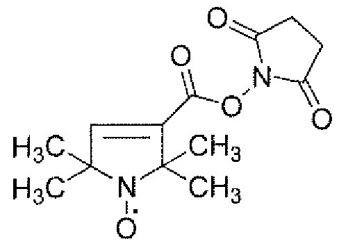
Both those protein nitroxide-labeled substrates exhibit broad line width which are refined during the proteolysis of the substrate and release of the nitroxide radical. This change in line width can be momtored by OMRI. However, there remains a residual signal, inconvenient for imaging. Thus, even for broad line, residual lightening in OMRI is detected. As a further limitation, line width broadening requires that the nitroxide be grafted on very large proteins with the inconvenient of an inaccurate tissue distribution.
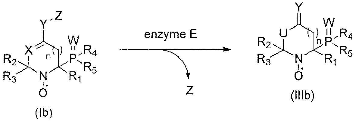
It now has been discovered nitroxide stable radicals enabling to enhance both sensitivity and resolution of enzyme activity imaging, notably proteolysis. More specifically, it has been shown that, the conversion of betaphosphorylated nitroxides having an intracyclic double bond of formula (la), or (lb), into a nitroxyde of formula (Ilia), or (Illb) respectively, upon enzyme digestion leads to a change of the hyperfine coupling constant (hcc) to the phosphorus atom (av) and thus of line positions, allowing an improved detection and/or quantification via OMRI (see scheme I).

(la) Z ('Ha)

Scheme I
Advantageously, by irradiating at the resonance frequency of the nitroxide (Ilia) or (Illb), the enzyme activity can be monitored. By irradiating at the frequency of nitroxyde (la), or (lb) biodistribution of nitroxide (la) or (lb) respectively can be accessed.
In vitro method for detecting and for quantifying: the catalytic activity of an enzyme E
Thus, in a first aspect, the present invention relates to an in vitro method for detecting and/or quantifying the catalytic activity of an enzyme E, said method comprising the steps of:
i) Contacting enzyme E with a compound of formula (la), or (lb):

(la) (lb)
wherein;
Y is OC(=0), S(CO), NR7,
X is CH or N
Ri, R2, R3, are each independently selected from C?-C6 alkyl optionally substituted by OH, NR6, COOH,
R4, R5 are each independently selected from H, Ci-C6 alkyl, CpQ alkoxy,
NRgRt}, SRJO,
R6 is H or Ci-C6 alkyl,
- - W is =W or is absent,
W is O, S, or NRn,
R7, Rg, R9, RJO, Rn are each independently selected from C]-C6 alkyl, n is 0, 1, 2 or 3,
Z is a substrate residue of an enzyme E,
and the stereoisomeric forms, mixtures of stereoisomeric forms or pharmaceutically acceptable salts forms thereof; and
ii) Monitoring the conversion of compound (la) or (lb) into compound (Ilia) or (Illb) by Electron Paramagnetic Resonance Imaging (EPRI) or Overhauser-enhanced Magnetic Resonance Imaging (OMRI), or by Proton Electron Double Resonance Imaging (PEDRI) thereby detecting or quantifying the activity of enzyme E:

(la)

wherein Rls R2, R3, R , R5, W, X, Y, Z and n are as defined in formulae (la) and (lb), and U is CH2 when X is CH and NH when X is N.
The present invention also relates to an in vitro method for detecting and/or quantifying the catalytic activity of an enzyme E, said method comprising the steps of:
i) Contacting enz me E with a compound of formula (la), or (lb):

(ia) (lb)
wherein:
Y is OC(=0), S(C=0), NR7;
X is CH or N
Ri, R2, R3, are each independently selected from Ci-C6 alkyl optionally substituted by OH, NR6, COOH,
R4, R5 are each independently selected from H, Q-Q alkyl, C]-C6 alkoxy,
NR8R9, SR10,
R6 is H or CrC6 alkyl,
- - W is =W or is absent,
W is O, S, or NRn,
R , Rg, R¾ Rio, Ri 1 are each independently selected from Ci-C6 alkyl, n is 0, 1, 2 or 3,
Z is a substrate residue of an enzyme E,
and the stereoisomeric forms, mixtures of stereoisomer! c forms or pharmaceutically acceptable salts forms thereof; and
ii) Monitoring the conversion of compound (la) or (lb) into compound (Ilia) or (Illb) by Electron Paramagnetic Resonance (EPR) or Overhauser-enhanced Magnetic Resonance Imaging (OMRI), or by Proton Electron Double Resonance Imaging (PEDRI) thereby detecting or uantifying the activity of enzyme E:


O (Illb) wherein Ri, R2, R , R4, R5, W, X, Y, Z and n are as defined in formulae (la) and (lb), and U is C¾ when X is CH and NH when X is N.
In a particular embodiment, the monitoring of step ii) by Electron
Paramagnetic Resonance is performed by Electron Paramagnetic Resonance Imaging (EPRI) or by Electron Paramagnetic Resonance Spectroscopy (EPRS).The enzyme may be a hydrolase, notably a protease, an esterase, a glycosidase or a lipase, for example neutrophile elastase.
In one embodiment, Z is a residue of a peptide, a protein, a glycoside, a fatty acid or a lipid.
In another embodiment, Ri is methyl.
In a further embodiment, both R2 and R3 are methyl.
In yet a further embodiment, R4 and R5 are ethoxy (EtO).
In still a further embodiment Y is OC(=0).
In a particular embodiment, the in vitro method according to the invention is used to quantify and/or to detect the catalytic activity of an enzyme E in a solution, notably in an opaque solution. In this case, the monitoring of step ii) by Electron Paramagnetic
Resonance is more particularly performed by Electron Paramagnetic Resonance Spectroscopy (EPRS).
This method can advantageously be applied notably in research laboratories, analysis laboratories, I agribusiness, or in industrial fermentations.
Compounds of formula (la) or (lb) for use in in vivo diagnosis
In a second aspect, the present invention relates to a compound of formula
(la), or (lb):

(la) (lb)
wherein:
Y is OC(=0), S(C=0), NR7)
is CH or N
Ri, R2, R3 , are each independently selected from Cj-Ce alkyl optionally substituted by OH, NR6, COOH,
R4, R5 are each independently selected from H, Ci-C6 alkyl, Ci-C6 alkoxy,

R6 is H or C[-C6 alkyl,
= = W is =W or is absent,
W is O, S, or NRn,
R7, Rs, R9, Rio, Ri 1 are each independently selected from C]-C6 alkyl, n is 0, 1, 2 or 3,
Z is a substrate residue of an enzyme E,
and the stereoisomeric forms, mixtures of stereoisomeric forms or pharmaceutically acceptable salts forms thereof; and
for use in in vivo diagnosis, notably via Electron Paramagnetic Resonance imaging (EPRI), or Overhauser-enhanced Magnetic Resonance Imaging (OMRI) or by Proton Electron Double Resonance Imaging (PEDRI).
In a first embodiment, the in vivo diagnosis is used for imaging, detecting and/or quantifying the activity of enzyme E, notably the proteolytic activity related to diseases and disorders.
In another embodiment, the diseases and disorders are diseases and disorders exhibiting specific enzymatic activity and/or over-expressed enzymatic activity.
In a further embodiment, the diseases and disorders are selected from cancer, inflammation, rheumatoid arthritis, cystic fibrosis, mucoviscidosis, pancreatitis, emphysema, bacterial, viral or parasitic infections.
In still a further embodiment, the compound of formula (la) or (lb) is converted into a compound of formula (Ilia) or (Illb) through enzyme E:

In yet a further embodiment, the compound of formula (Ilia) or (Illb) is selected from:
-(diethoxyphosphoryl)-2,656-trimethyl-l ,2,3,6-tetrahydropyridin-4-yl-N- oxyl acetate.
Compounds of formula (la) or (lb)
in a third aspect, the invention relates to a compound of formula (la), or
(lb):

(la) (lb) wherein :
W, X, Y, Z, i, R2, R3, R4, R5, are as defined above,
and the stereoisomer^ forms, mixtures of stereoisomeric forms or pharmaceutically acceptable salts forms thereof.
Compounds of formula (Ilia) or (Illb):
In a fourth aspect, the present invention relates to a compound of formula
(Ilia) or (Illb):

(ilia) (i llb) wherein :
n, W, X, Y, R1} R2, R3, R , R5, and U are as defined above.
Contrast agent
In a fifth embodiment, the present invention relates to a contrast agent comprising a compound of formula (la), or (lb) as defined above.
Definitions
The following terms and expressions contained herein are defined as follows:
As used herein, the term "alkyl" refers to a linear chain, or branched alkyl group having 1 to 6 carbon atoms, such as methyl, ethyl, propyl, isopropyl, butyl, isobutyl, sec-butyl, tert-butyl, pentyl, isoamyl, neopentyl, 1-ethylpropyl, 3-methylpentyl, 2,2- dimethylbutyl, 2,3-dimethylbutyl, hexyl, etc. The alkyl moiety of alkyl-containing groups, such as alkoxy, alkoxycarbonyl, and alkylaminocarbonyl groups, has the same meaning as alkyl defined above. Lower alkyl groups, which are preferred, are alkyl groups as defined above which contain 1 to 4 carbons. A designation such as "C1-C4 alkyl" refers to an alkyl radical containing from 1 to 4 carbon atoms.
As used herein, the term "pharmaceutically acceptable" refers to those compounds, materials, compositions, and/or dosage forms which are, within the scope of sound medical judgment, suitable for contact with the tissues of human beings and animals without excessive toxicity, irritation, allergic response, or other problem complications commensurate with a reasonable benefit risk ratio.
As used herein, "pharmaceutically acceptable salts" includes salts of compounds of the present invention derived from the combination of such compounds with non-toxic acid or base addition salts.
Acid addition salts include inorganic acids such as hydrochloric, hydrobromic, hydroiodic, sulfuric, nitric and phosphoric acid, as well as organic acids such as acetic, citric, propionic, tartaric, glutamic, salicylic, oxalic, methanesulfonic, para- toluenesulfonic, succinic, and benzoic acid, and related inorganic and organic acids.
Base addition salts include those derived from inorganic bases such as ammonium and alkali and alkaline earth metal hydroxides, carbonates, bicarbonates, and the like, as well as salts derived from basic organic amines such as aliphatic and aromatic amines, aliphatic diamines, hydroxy alkamines, and the like. Such bases useful in preparing the salts of this invention thus include ammonium hydroxide, potassium carbonate, sodium bicarbonate, calcium hydroxide, methylamine, diethyl amine, ethylenediamine, cyclohexylamine, ethanolamine and the like.
In addition to pharmaceutically acceptable salts, other salts are included in the invention. They may serve as intermediates in the purification of the compounds, in the preparation of other salts, or in the identification and characterization of the compounds or intermediates.
Figures
Figure 1 , Concept to use itroxides of the invention as probes for enzymatic activity or quantitative spectroscopic measurements.
Figure 2. EPR signals of 10 (top) and 9 (down) and a mixture (~ 50% conversion) of 9/10 (middle, arrows are for lines of 9) in the presence of Subtilisin.
Figure 3. Kinetics for the decay of 1.1 mM of 10 (□) and the generation of 9 (■) in the presence of 2.8 μΜ of Subtilisin A. The generation of 9 in the presence of Subtilsin A plus the Eglin C inhibitor (►), and spontaneous hydrolysis in PBS buffer solution «).
Figure 4. OMRI enhancing factors for 9 (·) and 10 (+/-) (■) versus the electron frequency of irradiation.
Figure 5. In vitro lightening (left for OMRI on and right for OMRI off) of nitroxides 9 (top) and 10(-) (bottom) specifically at their respective resonance frequencies.
Figure 6. Efficiency of various enzymes as well as of PBS buffer and Ca2+ PBS buffer in hydrolysis of 1.8 mM of 10 into 9 for 5 h incubation, -axis in mM (left) and % (right).
Figure 7. (A) 3D anatomical MRI image of the stomach. (B) The first row corresponds to the image with electronic saturation at 5425.7MHz and the slices in the second row are the corresponding non saturated electronic spins. (C) The first row corresponds to the image with a electronic saturation of 5417.5MHz (first row) and without (second row). All OMR! images were acquired in 18 seconds and then reconstructed into an isotropic spatial resolution of 0.5 mm in all three directions. The anatomical image has a spatial resolution of 0.5x0.54x1 (mm)3 and a temporal resolution of about 3.5 minutes.
Figure 8: Hydrolysis of 0,05 mM succinyl- Ala-Pro -Val nitroxide substrate 18 by 8.10" M neutrophile elastase
Figure 9: Hydrolysis of 0,05 mM succinyl-Ala-Pro-Val nitroxide substrate
18 by 8.10"9 M neutrophile elastase
Examples
Example 1 : Synthesis of diethyl (2,6,6-trimethyl-4-oxopiperidin-2-yl-N- oxypphosphonate (10)
Nitroxide 9 was prepared in 8 steps (Scheme 2) starting from the commercially available 2,6-dimethyl-4-hydroxy-hept-l ,6-diene 1. The hydroxyl function was protected with the conventional tert-butyldimethylsilyl chloride (TBDMSC1) to afford 2. Then, one of the double bond of 2 was selectively oxidized into diol 3 with Os04 as oxidizing reagent. Diol 3 oxidized into ketone 4 with NaI0 . Then, aminophosphonate 5 was obtained under condition specific for aminophosphorylation. Cyclization of 5 into 6 was performed in the presence of Hg(OAc)2 and the crude materials was oxidized with meta-chloperbenzoic acid (m-CPBA) into nitroxide 7. The protecting group of the latter was hydrolyzed to yield 8 which was oxidized into 9. The regioselectivity of the formation of the intracyclic double bond to afford 10 was controlled by using a bulky base, potassium hexamethyldisilazane (KHMDS), which reacted with the most accessible acidic protons which were the farer from the diethoxyphosphoryl group.


Scheme 2

disclosed into Breit, B., Breuninger, D., J. Am. Chem, Soc. 2004, 126, 10244, was dissolved in DMF (250 mL), imidazole (17 g, 246 mmol) and iert-butyldimethylsilyl chloride (25 g, 164 mmol) were added, and the mixture was stirred for 5 h at 0 °C. The solution was poured into water and extracted with Et20. The organic layers were washed with water, brine, dried with MgS04, filtered, and concentrated. Column chromatography yielded 20.6 g (99%) of 2 as a colorless oiVn NMR (400 MHz, CDC13): δ 4.77 (br s,2H), 4.71 (br s,2H),3.93 (q, J= 6.3 Hz, 1H), 2.20-2.11 (m, 4H), 1.73 (s, 6H), 0.87 (s, 9H), 0.04 (s, 6H). 13C NMR (75 MHz, CDC13): δ 143.0 (2C), 1 13.2 (2CH2), 69.8 (CH), 46.0.(2CH2), 26.1 (3CH3), 23.2 (2CH3), 18.3 (C), -4.4 (2CH3).
b) 4~((tert~butyldimethylsilyl)oxy)-2,6-dtmethylhept-6-ene-lt2-diol (3).

Alkene 2 (4.3 g, 16.9 mmol) was dissolved in 200 ml of a mixture acetone/ water (3/lv:v), then Os04 (1 mL of 4%w in water) was added and the mixture was stirred for
15 min at 0 °C. After that, iV-niethylmorpholine-N-oxide (4.0 g, 33.8 mmol) was added and stirred for 4 h at 0 °C. The solution was poured into 10 % aqueous Na2S 03 and extracted with Et20. The organic layers were washed with water, brine, dried with MgS04, filtered, and concentrated. The crude product was obtained as a mixture of 2 diastereoisomers (ratio 2: 1). Column chromatography afforded 3.38 g of 2and 0.9g (86%) of 3 as a yellowish oil, !H NMR (400 MHz, C6D6): δ 4.78 (br s, 1H, ), 4.76 (br s, 1H, m+M), 4.70 (br s, 1H, m), 4.26 (m, 1H, m+M), 3.78 (s, 1H, m), 3.45-3.28 (m, 2H m, 3H M), 2.37-2.27 (m, 1H, m+M), 2.22-2.07 (m, 1H M, 2H m), 1.87-1.70 (m, 2H M, 1H m) 1.65-1.50 (m, 4H M, 4H m), 1.18 (s, 3H, M), 1.14 (s, 3H, m), 0.94 (s, 9H, m + M), 0.11 (s, 3H, m), 0.09 (s, 3H, M), 0.05 (s, 3H, m+M). !3C NMR (75 MHz, C6D6): 6 142.5 (C,M), 142.0 (C,m), 113.9 (CH2, m), 113.7 (CH2, M), 73.0 (C, m), 72.7 (C, M), 71.0 (CH2, m), 70.1 (CH2, M), 69.7 (CH, m), 69.6 (CH, M), 47.9 (CH2, m+M), 44.3 (CH2, M), 43.1 (CH2, m), 26.2 (CH3, M), 26.2 (CH3, m), 25.5 (CH3} M), 24.0 (CH3, m), 22.9 (CH3, M), 22.8 (CH3, m), 18.2 (C, M), 18.2 (C,m), -3.4 (CH3, m), -3.5 (CH3, M), -4.2 (CH3, M), -4.3 (CH3, m).
c) 4-((tert-butyldimethylsilyl)oxy)~6-methylhept~6~en-2~one (4)

Diol 3 (6,0 g, 20.8 mmol) was solved in 300 ml of a THF/water mixture (3/1 v:v) with 11.0 g (52.0 mmol) of Nal04 and stirred at 0 °C for 3 h. The solution was poured into water and extracted with Et20. The organic layers were washed with water, dried with MgS04, filtered, and concentrated. Column chromatography yielded 5.1 1 g (96%) of 4 as a yellowish oil. 1H NMR (400 MHz, CDC13): δ 4.78 (br s, 1H), 4.70 (br s, 1H), 4.36-4.28 (m, 1H), 2.60-2.47 (m, 2H), 2.30-2.10 (m, 2H), 2.15 (s, 3H), 1.74 (brs, 3H), 0.86 (s, 9H), 0.07 (s, 3H) 0.03 (s, 3H). 13C NMR (75 MHz, CDC13): 5 207.9 (C), 142.3 (C), 113.7 (C¾), 67.8 (CH), 50.5 (CH2), 46.6 (CH2), 31.8 (CH3), 25.9 (3CH3), 23.0 (CH3), 18.05 (C), -4.5 (CH3), - 4.8 (CH3).
d) diethyl (2»amino-4-((tert-butyldimethylsilyl)oxy)-6-methylhept-6-en-2- yl)phosphonate(S)

Ketone 4 (1.0 g, 3.9 mmol) was dissolved in 5 ml of diethylphosphite. Molecular sieves 4A were added and the mixture was stirred under an ammoniac atmosphere for 24 h. Then, the mixture was filtered and the excess of diethylphosphite was removed
under vacuum (50 °C, 2 mmHg). Column chromatography afforded 5 (550 mg, 37%) as a yellowish oil. 1H NMR (400 MHz, CDC13): 5 4.77 (br s, 1H, m+M), 4.71 (br s, 1H, m+M), 4.42-4.32 (m, 1H, M), 4.27-4.20 (ra, 1H, m), 4.19-4.07 (m, 4H, m+M), 2.34-2.13 (m, 2H, m+M), 1.95-1.85 (m, 1H, m+M), 1.80-1.55 (m, 6H, m+M), 1.37-1.28 (m, 9H), 0.88 (s, 9H, m+M), 0.12 (s, 3H,M), 0.12 (s, 3H,m), 0.11 (s, 3H,m), 0.11 (s, 3H, M).3 !P NMR (162 MHz, CDCI3): δ 31.67 (M), 30.88 (m). 13C NMR (75 MHz, CDCI3): δ 142.7 (C, M), 142.2 (C, m), Π 3.5 (CH2, m), 113.3 (CH2, M), 67.8 (d, J= 13.2 Hz, CH, m), 67.6 (d, J= 10.5 Hz, CH, M), 62.5-62. l(d overlapped, 2CH2 m, 2CH2 M), 52.0 (d, J = 155 Hz, C, m), 51.8 (d, J = 146 Hz, C, M), 48.16 (CH2, M), 48.0 (CH2, m), 42.7 (d, J = 3.9 Hz, CH2), 41 ,9 (CH2), 26.0 (3CH3), 23.8 (CH3), 22.9 (d, J = 2.2 Hz, CH3, M), 22.7 (d, J = 2.2 Hz, CH3, m), 18.0 (C, M), 18.0 (C,m), 16.6 (d, J= 5.5 Hz, CH3), -3.6 (CH3, m), -3.6 (CH3, M), -4.1 (CH3, m), -4.1 (CH3, M).
e) diethyt(4-((tert-but ldhnethylsilyl)oxy)~2,6,6-trimethylpiperiM
N-oxyl)phosphonate (7).

The aminophosphonate 5 (400 mg, 1.02 mmol) was dissolved in 50 mL of
THF/water mixture (3/1 v:v). Hg(OAc)2 (389 mg, 1.22 mmol) in 50 mL of the same solvent was slowly added to the aminophosphonate mixture and was stirred for 30 min at room temperature. Then, the mixture was poured on 50 ml of aqueous solution of NaBH4 (77 mg, 2.03 mmol) and NaOH (163 mg, 4.06 mmol), extracted with Et20. The organic layers were washed with brine, dried with MgS04, filtered, and concentrated to yield 397 mg of piperidine 6 which was used in the next step without further purifications.
A solution of piperidin 6 (900 mg, 2.29 mmol) and m-CPBA (1.26 g, 4.58 mmol) in DCM was stirred 2 hours at 0 °C. The solution was poured into 10 % aqueous N 2S203 and extracted with DCM. The organic layers were washed with saturated aqueous solution of NaHC03, dried with MgS0 , filtered, and concentrated. The crude product was obtained as a mixture of 2 diastereoisomers (ratio 2:1). Column chromatography yielded 685 mg of 7 (73%) as a red oil. The two diastereoisomers was separated and the major one was crystalized from Et20.
f) diethyl (4-hydroxy-2,6,6-trimethylpiperidin-2~yl-N-oxyl)phosphonate (8)

TBAF (1.0 M solution in THF, 1.47 mL, 1.47 mmol) was added dropwise to an ice-cold solution of silyl ether 7 (400 mg, 0.979 mmol) in dry THF (4 mL), under argon, The mixture was stirred 3 hours at room temperature and poured in a column of silica gel to afford 262 mg of 8 (91 %) as a red oil. Enantiomers 8a and 8b have been separated by chiral chromato gr aphy .
g) diethyl (2,6,6-trimethyl-4-oxopiperidin-2-yl~N-oxyl)phosphonate (9)

N-methylmorpholine JV-oxide (128 mg, 1.09 mmol, 4.0 equiv.) was added to a stirred ice-cold solution of 8 (80 mg, 0.272 mmol) with a catalytic amount of tetrapropylammonium perruthenate and powdered molecular sieves 4 A in dry DCM ( 0 mL) under an argon atmosphere. After 20 min of stirring at room temperature, the reaction mixture was added to a column of silica gel to afford 70 mg of 9 (88%) as an orange solid.
h) 2'(diethoxyphosphoryl)-2,6,6-trimethyl-l,2,3,6-tetrahydropyridin-4~yl- N-oxyl acetate (10)

Ketone 9 (100 mg, 0.342 mmol) in 5 mL of dry THF was slowly added to a solution of KHMDS (1.0 M solution in THF, 0.513 mL, 0.513 mmol) in 5 mL of dry THF. The enolate formation was kept 3 hours from -80 °C to -45 °C. Then, freshly distillated AC2O (70 mg, 0.684 mmol) was slowly added. The mixture was stirred 2.5 hours. After that, the solution was poured on brine and extracted with EtOAc. The organic layers were dried with MgS04, filtered, and concentrated. Column chromatography yielded 105 mg (91%) of 10 as a red oil.
Example 2: EPR features of compounds 9 and 10
Electron Paramagnetic Resonance (EPR) features were reported in Table 1 and Figure 2, and are in very good agreement with parent nitroxides reported in the literature ("Solvent Effect in β-Phosphorylated Nitroxides: Model Nitroxides" Audran, G.; Bosco, L.; Bremond, P.; Butscher, T.; Marque, S. R. A. Appl. Magnet. eson. 2015). Interestingly, nitroxide 10 (intracyclic double bond, i.e., enol form) exhibit an a? value 4.4 G larger than the one of 9 (exocyclic double bond). Moreover, linewidths are narrow enough (< 1.5 G for 9) for OMRI experiments. [6] Noteworthy, only traces of 9 were detected when 10 was kept in phosphate buffer solution (pH = 7.3 at 37 °C) for 24 hours, and less than 30% conversion were reached after 72 hours. Kinetics of hydrolysis of the enol-ester moiety of 10(+) into 9 reached roughly 50% conversion in 90 min. in basic (pH = 9) solution and less than 10% conversion in 90 min. in acidic (pH = 3) solution (Figure 3).
Table 1. EPR parameters of nitroxides 9 and 10.
nitroxides3-' a (G)[a] «P (G)[a] Δ¾, (G)M
9 15.0 43.1 2.0062 1.2
10 15.6 38.7 2.0063 1.8
1 G = 0.1 mT. [ ] Lande's factor. [c] Linewidth peak to peak for the central lines.
Example 3: Study of , hydrolysis of 10 into 9 in the presence of enzymes using EPR
Enzymatic activity assays were carried out in vitro using the EPR technique to monitor the keto-enol hydrolysis. After an incubation time of 5 hours and quantification of the third EPR line for the product 9, Fig. 3 showed that out of 8 proteases of various specificities and origin, three were able to hydrolyze 10 into 9, namely, porcine pancreatic elastase (PPE), human neutrophil elastase (hNE) and subtilisin A (Figure 6). As displayed in Figure 2 (middle), lines 5 and 8 for 9 and lines 6 and 7 for 10 are clearly separated affording an easy signal to monitor and for titration.
Example 4: In vitro experiments
As mentioned above, in vivo OMRI requires nitroxides exhibiting EPR changes upon enzymatic activity. As seen in Table 1, EPR linewidth is narrow enough to observe high polarization and the AaP (ca. 8 MHz, Figure 4) between lines 5 for 9 and 6 for 10 is large enough that each line can be excited to a unique micro-wave. In vitro experiments (Figure 4) showed that nitroxides 9 and 10 were selectively lightened on at the micro wave
frequencies of 5417.7 MHz and of 5425.4 MHz, respectively, affording 6-fold and 3-fold enhanced signals for 9 and 10, respectively. Consequently, OMRI experiments showed that each sample of 9 and 10 can be lightened selectively with different intensity (Figure 5).
Example 5: In vivo experiments
To definitively highlight the potential of nitroxides 9 and 10 for probing non-radical enzymatic activity, a mouse was fed with a solution of nitroxide 10. Anatomical MRI was first performed and did not provide useful information (Figure 7A). Then, the mouse was irradiated at v = 5417.7 MHz (Figure 7B) and oesophagi and a part of stomach were brightening whereas, when irradiation was performed at y = 5425.4 MHz (Figure 7C), only the lower part of the stomach and the duodenum were brightening. Clearly, the conformational change from 10 to 9 plays the expected on/off role for selective OMRI experiments affording a nice tool to investigate in vivo by MRI enzymatic processes.
Example 6: Hydrolysis of nitroxide-peptide 18 by Elastase neutrophile (9H-fluoren-9-vnmethyl-(S)-2-(((S -l-(benzyloxyV3-methyl-l-oxobutan-2- yPcarbamoyl) pyrrolidine- 1 -carboxylate (11).
Bn02

DIPEA (2 mL, 1 1.5 mmol) was added dropwise to a stirred suspension of L-Val-OBn- HC1 (2.8 g, 11.5 mmol) in dichloromethane (30 mL) at room temperature under an atmosphere of nitrogen. On dissolution, the solution was cooled to 0 °C and Fmoc-L-Pro (4.26 g, 12.6 mmol) and 1-hydroxybenzotriazole (1.86 g, 13.8 mmol) were added successively, each in one portion. The suspension was stirred at 0 °C for a further 15 min, and then DCC (2.85 g, 13.8 mmol) was added in one portion. The mixture was allowed to warm to room temperature over the course of 18 h and then filtered, and the filtrate was evaporated in vacuo. The residue was taken up in ethyl acetate and filtered, and the filtrate was then washed with 10% aqueous citric acid solution followed by saturated aqueous sodium bicarbonate solution. The combined organic extracts were dried and evaporated in vacuo to leave the crude product which was purified by chromatography on silica using 3:2 petroleum ether-EtOAc as eluent to give the dipeptide 11 (5.94 g, 11.28 mmol) as a yellow solid in 98% yield. [a]D 20 - 50 (c 1.0, CHC13).
(((9H-fluoren-9-yl)methoxy)carbonyl)-L-prolyl-L- valine (12).

11 12
To a solution of 11 (5.94 g, 1 1.28 mmol) in MeOH (80 mL) was added 10% Pd/C (600 mg), and the mixture was stirred for 10 h in hydrogen atmosphere (1 atm). The reaction mixture was filtered through Celite followed by MeOH removal in vacuo to yield 12 in quantitative yield (4.92 g, 1 1.27 mmol). [a]D 20 - 46.1 (c 1.0, CHC13).
(^-2-((iy)-l -(((9H-fluoren-9-yl)methoxy)carbonyl)pyrrolidine-2-carboxamido)-
3 -methyl- butanoic piyalic anhydride (13)

12 13
To an ice cold solution of dipeptide 12 (1.23 g, 2.83 mmol) in 3 ml dry dichloromethane, Pivaloyl chloride (0.7 mL, 5.7 mmol) was added followed by the dropwise addition of triethyl amine (0.6 ml, 4,3 mmol). The reaction mixture was then allowed to stir at 0 °C for 1 h (TLC shows completion of reaction) after which excess dichloromethane was removed and the residue was taken up in diethyl ether. The solution was then filtered through a celite bed. Removal of diethyl ether in vacuo yielded the desired anhydride 13 as a white foamy solid in quantitative yield which was used in the next reaction without any further purification.
Synthesis of (14a, HPLC 1).

A solution of ketone 8a (209 mg, 0.71 mmol) in dry THF (10 mL) was slowly added to a- 78 °C solution of LiHMDS (1.0 M solution in THF, 1.2 mL, 1.21 mmol, 1.70 equiv.) in dry THF
(5 mL), The mixture was stirred for 3 hours from - 78 °C to - 45 °C. Then, 13 (739 mg, 1.42 mmol, 2 equiv.) was slowly added as a cold (- 45 °C) solution in 10 mL THF. The mixture was stirred for 2.5 hours, then it was poured quenched with saturated aqueous NH4C1 solution and extracted with EtOAc. The combined organic extracts were dried with MgS04, filtered, and concentrated under vacuo. Column chromatography of the residue gave starting material 8a (92 mg, 0.31 mmol) and the desired product 14a (175 mg, 0.25 mmol, 65% based on recovered starting material) as a red oil and. HRMS (ESI) calc for C36H42N20gPNa+: 728.3545 [M+NH4]+; found: 728.3553, [a]D 20 - 58.3 (c 1.0, CHC13).

A solution of ketone 8b (208 mg, 0.71 mmol) in dry THF (10 mL) was slowly added to a- 78 °C solution of LiHMDS (1.0 M solution in THF, 1.2 mL, 1.21 mmol, 1.70 equiv.) in dry THF (5 mL). The mixture was stirred for 3 hours from - 78 °C to - 45 °C. Then, 13 (739 mg, 1.42 mmol, 2 equiv.) was slowly added as a cold (- 45 °C) solution in 10 mL THF. The mixture was stirred for 2.5 hours, then it was poured quenched with saturated aqueous NH4C1 solution and extracted with EtOAc. The combined organic extracts were dried with MgS0 , filtered, and concentrated under vacuo. Column chromatography of the residue gave starting material 8b (105 mg, 0.36 mmol) and the desired product 14b (229 mg, 0.32 mmol, 91% based on recovered starting material) as a red oil and. HRMS (ESI) calc for Cae^^OgPNa^ 728.3545 [M+NH4 ; found: 728.3553, [a]D 20 - 21.3 (c 1.0, CHC13).
Methyl (S)-4-(( 1 -(benzyloxy)- 1 -oxopropan-2- yi amino)-4-oxobutanoate (15).

DIPEA (17 mL, 17 mmol) was added dropwise to a stirred suspension of L-Ala-OBn-HCl (6.02 g, 27.9 mmol) in dichloromethane (70 mL) at room temperature under an atmosphere of nitrogen. On dissolution, the solution was cooled to 0 °C and succinic acid monomethyl ester
(4.05 g, 30.7 mmol) and 1 -hydroxybenzotriazole (4.52 g, 33.48 mmol) were added successively, each in one portion. The suspension was stirred at 0 °C for a further 15 min, and then DCC (6.91 g, 33.48 mmol) was added in one portion. The mixture was allowed to warm to room temperature over the course of 18 h and then filtered, and the filtrate was evaporated in vacuo. The residue was taken up in ethyl acetate and filtered, and the filtrate was then washed with 10% aqueous citric acid solution followed by saturated aqueous sodium bicarbonate solution. The combined organic extracts were dried and evaporated in vacuo to leave the crude product which was purified by chromatography on silica using 3 :2 petroleum ether-EtOAc as eluent to give the dipeptide 15 (7.2 g, 24.55 mmol) as a white solid in 88% yield. [a]D 20 - 4.3 (c 1.0, CHC13).
Met y\-4-(((S)- 1 ~(((S)- 1 -(benzylpxy)-! -oxopropan-2-yl)amino)- 1 -oxopropan- 2-yl)amino)-4-oxobutanoate (16).
1 g

To a solution of 15 (7 g, 23.86 mmol) in MeOH (80 mL) was added 10% Pd/C (700 mg), and the mixture was stirred for 10 h in hydrogen atmosphere (1 atm). The reaction mixture was filtered through Celite followed by MeOH removal in vacuo to yield the free acid in quantitative yield (4.85 g, 23.8 mmol).
DIPEA (14.9 mL, 83.52 mmol) was added dropwise to a stirred suspension of L-Ala- OBn*HCl (6.16 g, 28.56 mmol) in dichloromethane (100 mL) at room temperature under an atmosphere of nitrogen. On dissolution, the solution was cooled to 0 °C and the acid obtained in the previous step (4.85 g, 23.8 mmol) and 1 -hydroxybenzotriazole (3.86 g, 28.56 mmol) were added successively, each in one portion. The suspension was stirred at 0 °C for a further 15 min, and then DCC (5.89 g, 28.56 mmol) was added in one portion. The mixture was allowed to warm to room temperature over the course of 18 h and then filtered, and the filtrate was evaporated in vacuo. The residue was taken up in ethyl acetate and filtered, and the filtrate was then washed with 10% aqueous citric acid solution followed by saturated aqueous sodium bicarbonate solution. The combined organic extracts were dried and evaporated in vacuo to leave the crude product which was purified by chromatography on silica using 3 :2
petroleum ether-EtOAc as eluent to give the tripeptide 16 (7.5 g, 20.71 mmol) as a white solid in 87% yield. [cc]D 20 - 44 (c 1.0, CHC13).
Methyl-4-((( )-l-i(ffl-l-f(2,5-dioxopyrrolidin-l-yl)oxyVl-oxopropan-2- yi)amino)-l -oxopropan-2-yl)amino)-4-oxobutanoate (17)

To a solution of the free acid from above (484 mg, 1.76 mmol) and N-Hydroxysuccinimide (203 mg, 1.76 mmol) in 7 mL THF, DCC (364 mg, 1.76 mmol) was added in one portion. The mixture was allowed to stir at room temperature for 18 h and then filtered, and the filtrate was evaporated in vacuo to yield the NHS ester 17 as a white solid in quantitative yield (650 mg, 1.75 mmol).
Synthesis of 18a.

To a cold solution of 14a (275 mg, 0,39 mmol) in 20 mL DCM, DBU (69 μί, 0.46 mmol) was added. The reaction mixture was stirred at 0 °C for 3 h (TLC showed completion of reaction) followed by column purification to yield the deprotected compound in 74% yield (143 mg, 0.29 mmol),
The deprotected compound (143 mg, 0.29 mmol) and compound 17 (120 mg, 0.32 mmol) was dissolved in 5 mL annhydrous DMF and the mixture was allowed to stir for 18 h at room
temperature. Removal of DMF in vacuo followed by column chromatography yielded 18a in 34% overall yield (105 mg, 0.14 rnmol).
Enzymatic hydrolysis
A Miniscope MS200 EPR instrument (Magnettech, Berlin, Germany) was used for enzymatic reactions. Nitroxide samples were loaded in 75 mm/60 μΐ capillaries (Hirschmann Laborgerate, Germany). The experiments were performed as previously reported. [1] EPR acquisition parameters were as follows: B0 = 3353,55 G; sweep width = 90.42 G; sweep time = 60 s; number of passes = 1 ; modulation == 300 mG. The gain was kept constant throughout the kinetic experiments. EPR spectra processed using IGOR Pro (Wavemetrics, Lake Oswego, OR, USA) enabled quantification of the third peak(s) during the required time course. After integrating the EPR peaks, the surface areas of the third peak were measured with a Lorentzian Curve fit. These data were then normalized to a final concentration of 0.05 mM.
Experiment with elastase neutrophile was done in 0.1 M phosphate buffer, pH = 7.4 at 37 °C. The enzymatic reaction was initiated by adding a small volume of enzyme solution between 5 to 20 μΐ at a concentration around 0.08 μΜ to the buffered substrate solution at a concentration of 0.05 mM nitroxide-peptide substrate 18. Figure 8 reports on the hydrolysis of nitroxide-peptide (0.05 mM) in vitro in the presence of Neutrophile Elastase (80 nM). The nitroxide-peptide (small signal) is completely converted (100% conversion) into keto-nitroxide (intense signal) by a ca 1000-fold lower concentration in enzyme in less than 4 minutes. This denotes the high suitability of nitroxide-peptide 18 to the enzyme. Moreover, despite concentration in nitroxide-peptide at t=0 is the same than the concentration in keto nitroxide at t = 4 minutes, signal at t=0 clearly exhibits a weaker intensity than for the signal at t = 4 minutes. This difference is due to large EPR line width for the nitroxide-peptide (the 4 residues slow down the motion of the nitroxide leading to an increase of the line width). This is another advantage of these probes :more intense the signal of the free nitroxide stronger the enhancement in the Overhauser effect experiments.
Figure 9 reports on the hydrolysis of nitroxide-peptide (0.05 mM) in vitro in the presence of Neutrophile Elastase (8 nM). For a 10-fold lower concentration in enzyme than in the previous experiment, the hydrolysis is clearly slower. Consequently, it is possible to observe
by EPR both the decay of the signal related to the nitroxide-peptide on the one hand and the growth of the signal related to the free keto-nitroxide on the other hand. Again, this experiment highlights nicely the sensitivity of the substrate 18 to the enzyme.
REFERENCES
[1] Krishna MC, English S, Yamada K, Yoo J, Murugesan R, et al. (2002). Overhauser enhanced magnetic resonance imaging for tumor oximetry: coregistration of tumor anatomy and tissue oxygen concentration. Proc Natl Acad Sci USA 99: 2216-2221.
[2] Berliner LJ (2003). In Vivo EPR (ESR). Theory and applications. New York: Kluwer Academic/Plenum Publishers.
[3] Grucker D (1990). In vivo detection of injected free radicals by Overhauser effect imaging, Magn Reson Med 14: 140-147. [4] Freed JH (1976). In: Berliner LJ, ed. Spin Labeling: Theory and applications. New York: Academic Press.
[5] ellet P.; Massot P.; Madelin, G.; Marque, S.; Harte, E.; Franconi, J.M.; Thiaudiere, E. PLoS ONE, 2009, 4, 4, 1-9.
[6] Parzy, E.; Bouchaud, V.; Massot P.; Voisin, P.; Koonjoo, N.; Moncelet, D.; Franconi, J.M.; Thiaudiere, E.; Mellet, P. PLoS ONE, 2013, 8, 2, 1-7.
[7] Koonjoo CMMI 2014
Claims
1. An in vitro method for detecting and/or quantifying the catalytic activity of an enzyme E, said method comprising the steps of:
i) Contacting enz me E with a com ound of formula la), or (lb):

(la) (lb)
wherein:
Y is OC(=0), S(C=0), NR7>
X is CH or N
Ri, R2, R3, are each independently selected from Ci-C6 alkyl optionally substituted by OH, NR6, COOH,
R4, R5 are each independently selected from H, Ci-C6 alkyl, Cj -C6 alkoxy,
NR8R9, SR10,
R6 is H or C C6 alkyl,
- -W is =W or is absent,

R7, Rs, R9, Rio, R11 are each independently selected from Ci-C6 alkyl, n is 0, 1 , 2 or 3,
Z is a substrate residue of an enzyme E,
and the stereoisomeric forms, mixtures of stereoisomeric forms or pharmaceutically acceptable salts forms thereof; and
ii) Monitoring the conversion of compound (la) or (lb) into compound (Ilia) or (Illb) by Electron Paramagnetic Resonance (EPR) or Overhauser-enhanced Magnetic Resonance Imaging (OMRI), or by Proton Electron Double Resonance Imaging (PEDRI) thereby detecting or quantifying the activity of enzyme E:
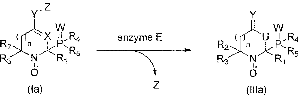
 wherein R] ; R2, R3, R4, Rs, W, X, Y, Z and n are as defined in formulae (la) and (lb), and U is CH2 when X is CH and NH when X is N.
2. The method of claim 1, wherein the monitoring of step ii) by Electron Paramagnetic Resonance is performed by Electron Paramagnetic Resonance Imaging (EPRI) or by Electron Paramagnetic Resonance Spectroscopy (EPRS).
3. The method of any of claims 1 or 2, wherein the enzyme is a hydrolase, notably a protease, an esterase, a glycosidase or a lipase.
4. The method according to any of claims 1 to 3, wherein Z is a residue of a peptide, a protein, a glycoside, a fatty acid or a lipid.
5. The method according to any of claims 3 to 4, wherein Ri is methyl.
6. The compound of formula (la) or (lb) for use according to any of claims 1 to 5, wherein both R2 and R3 are methyl.
7. The method according to any of claims 1 to 6, wherein R and R5 are ethyl.
8. The method according to any of claims 1 to 7, wherein Y is OC(=0).
9. A compound of formula la), or (lb) :

(la) (lb)
wherein:
Y is OC(=0), S(C=0), NR7>
X is CH or N
Ri, R2, R3 , are each independently selected from Q-Ce alkyl optionally substituted by OH, NR6, COOH„
R4, R5 are each independently selected from H, Ci-C6 alkyl, C[-C6 alkoxy,

R6 is H or Ci-C6 alkyl,
= = W is =W or is absent,
W is O, S, or NRH,
R7, R¾, R9, R;o, R1 1 are each independently selected from Q-Q alkyl, n is 0, 1, 2 or 3,
Z is a substrate residue of an enzyme E,
and the stereoisomeric forms, mixtures of stereoisomeric forms or pharmaceutically acceptable salts forms thereof; and
for use in in vivo diagnosis, notably via Electron Paramagnetic Resonance Imaging (EPRI), or Overhauser-enhanced Magnetic Resonance Imaging (OMRI) or by Proton Electron Double Resonance Imaging (PEDRI).
10. The compound for use according to claim 9, for imaging, detecting and/or quantifying the activity of enzyme E.
1 1. The compound for use according to any of claims 9 or 10, for imaging the proteolytic activity related to diseases and disorders.
12. The compound for use according to claim 1 1, wherein the diseases and disorders are diseases and disorders exhibiting specific enzymatic activity and/or over- expressed enzymatic activity.
13. The compound for use according to claim 12, wherein the disease and disorders are selected from cancer, inflammation, rheumatoid arthritis, cystic fibrosis, mucoviscidosis, pancreatitis, emphysema, bacterial, viral or parasitic infections .
14. The compound for use according to any of claims 9 to 13, wherein the compound of formula (la) or (lb) is converted into a compound of formula (Ilia) or (Illb) through enzyme E:

(Ilia)

(lb) wherein Rls R2, R3, 4, R5, W, X, Y, Z and n are as defined in formulae (la) and (lb) in claim 1, and U is CH2 when X is CH and NH when X is N.
15. The compound of formula (la), (lb), for use according to claim 14, wherein the compound of formula (Ilia) or (Illb) is selected from:
(diethoxyphosphoryl)-2J6,6-trimethyl-l,2,3,6-tetrahydropyridin-4-yl-N- oxyl acetate
16. A compound of formula (la), or (lb)):

(la) (lb) wherein ;
n, W, X, Y, Z, R1} R2, R3, R4, R5, are as defined in any of claims 1 to 8, and the stereoisomeric forms, mixtures of stereoisomeric forms or pharmaceutically acceptable salts forms thereof.
17. A compound of formula (Ilia) or (Illb):

(Ilia) (I l lb)
wherein :
, W, X, Y, R], R2, R3, R4, R5, and U are as defined in any of claims 1 to 8 18. A contrast agent comprising a compound of formula (la), or (lb) defined in any of claims 1 to 8.
Applications Claiming Priority (2)
| Application Number | Priority Date | Filing Date | Title |
|---|---|---|---|
| EP15306115.5 | 2015-07-07 | ||
| EP15306115 | 2015-07-07 |
Publications (1)
| Publication Number | Publication Date |
|---|---|
| WO2017005879A1 true WO2017005879A1 (en) | 2017-01-12 |
Family
ID=53716414
Family Applications (1)
| Application Number | Title | Priority Date | Filing Date |
|---|---|---|---|
| PCT/EP2016/066199 Ceased WO2017005879A1 (en) | 2015-07-07 | 2016-07-07 | Nitroxides for use as contrast agents for overhauser magnetic resonance imaging (omri) |
Country Status (1)
| Country | Link |
|---|---|
| WO (1) | WO2017005879A1 (en) |
-
2016
- 2016-07-07 WO PCT/EP2016/066199 patent/WO2017005879A1/en not_active Ceased
Non-Patent Citations (9)
| Title |
|---|
| ANNE MERCIER ET AL: "beta-Phosphorylated Five Membered Ring Nitroxides, A New Class of Stable Nitroxides. 1. Synthesis by Reaction of Dialkglphosphites with 1-Pyrroline-N-Oxides", 1 January 1991 (1991-01-01), pages 2125 - 212800, XP055235445, Retrieved from the Internet <URL:http://ac.els-cdn.com/S0040403900712536/1-s2.0-S0040403900712536-main.pdf?_tid=6662dc80-9f3f-11e5-bdad-00000aacb35f&acdnat=1449753215_9a44b0c6ed0dc294e616d12760d1edbb> * |
| ELODIE PARZY ET AL: "Overhauser-Enhanced MRI of Elastase Activity from In Vitro Human Neutrophil Degranulation", PLOS ONE, vol. 8, no. 2, 28 February 2013 (2013-02-28), pages e57946, XP055234713, DOI: 10.1371/journal.pone.0057946 * |
| GÉRARD AUDRAN ET AL: "Enzymatically Shifting Nitroxides for EPR Spectroscopy and Overhauser-Enhanced Magnetic Resonance Imaging", ANGEWANDTE CHEMIE INTERNATIONAL EDITION, vol. 54, no. 45, 17 September 2015 (2015-09-17), DE, pages 13379 - 13384, XP055232809, ISSN: 1433-7851, DOI: 10.1002/anie.201506267 * |
| GÉRARD AUDRAN ET AL: "Solvent Effect in [beta]-Phosphorylated Nitroxides: Model Nitroxides", APPLIED MAGNETIC RESONANCE., vol. 46, no. 12, 24 February 2015 (2015-02-24), AU, pages 1333 - 1342, XP055232777, ISSN: 0937-9347, DOI: 10.1007/s00723-015-0649-4 * |
| GÉRARD AUDRAN ET AL: "The [beta]-phosphorus hyperfine coupling constant in nitroxide: part 3: titration of water by electron paramagnetic resonance", ORGANIC & BIOMOLECULAR CHEMISTRY, vol. 13, no. 46, 1 January 2015 (2015-01-01), GB, pages 11393 - 11400, XP055232807, ISSN: 1477-0520, DOI: 10.1039/C5OB01867F * |
| NEHA KOONJOO ET AL: "In vivo Overhauser-enhanced MRI of proteolytic activity", CONTRAST MEDIA & MOLECULAR IMAGING, vol. 9, no. 5, 14 April 2014 (2014-04-14), GB, pages 363 - 371, XP055234715, ISSN: 1555-4309, DOI: 10.1002/cmmi.1586 * |
| NOLWENN LE BRETON ET AL: "Diversification of EPR signatures in site directed spin labeling using a [beta]-phosphorylated nitroxide", PHYSICAL CHEMISTRY CHEMICAL PHYSICS., vol. 16, no. 9, 1 January 2014 (2014-01-01), GB, pages 4202, XP055235378, ISSN: 1463-9076, DOI: 10.1039/c3cp54816c * |
| NOLWENN LE BRETON ET AL: "Understanding protein non-folding", FRONTIERS IN MOLECULAR BIOSCIENCES, vol. 1804, 19 May 2015 (2015-05-19), pages 1231, XP055235330, DOI: 10.1016/j.bbapap.2010.01.017 * |
| PHILIPPE MELLET ET AL: "New Concepts in Molecular Imaging: Non-Invasive MRI Spotting of Proteolysis Using an Overhauser Effect Switch", PLOS ONE, vol. 4, no. 4, 27 April 2009 (2009-04-27), pages e5244, XP055234711, DOI: 10.1371/journal.pone.0005244 * |
Similar Documents
| Publication | Publication Date | Title |
|---|---|---|
| CN1108824C (en) | Bioactivated diagnostic imaging contrast agents | |
| Peters et al. | Synthesis of borondipyrromethene (BODIPY)-labeled sphingosine derivatives by cross-metathesis reaction | |
| CN108368067B (en) | Dimeric contrast agent | |
| CA1230114A (en) | Nitroxyl compounds, process for their preparation, and diagnostic media containing same | |
| JP6483108B2 (en) | Process for producing hyperpolarized carboxylate compound | |
| Richard et al. | Design and synthesis of cathepsin-K-activated osteoadsorptive fluorogenic sentinel (OFS) probes for detecting early osteoclastic bone resorption in a multiple myeloma mouse model | |
| DK2550966T3 (en) | Dabigatran amidoxic acid esters as prodrugs and their use as a drug | |
| CN105712987A (en) | Bioluminescence probe substrate for human carboxylesterase 1 and preparation method and application thereof | |
| WO2002074342A1 (en) | Remedies for arteriosclerosis | |
| WO2017005879A1 (en) | Nitroxides for use as contrast agents for overhauser magnetic resonance imaging (omri) | |
| FR2868320A1 (en) | CONTRAST AGENT FOR MAGNETIC RESONANCE IMAGING | |
| CN105968170B (en) | A kind of the fluorescence probe substrate and preparation method and application of DPP IV | |
| Nemoto et al. | Synthesis of Paclitaxel–BGL Conjugates | |
| US20060088475A1 (en) | Self-immolative magnetic resonance imaging contrast agents sensitive to beta-glucuronidase | |
| Ding et al. | Design, synthesis and pharmacological activity of novel enantiomerically pure phosphonic acid-based NAALADase inhibitors | |
| US10017527B2 (en) | KLK4 inhibitors | |
| Cowell et al. | Towards an MMP-2-activated molecular agent for cancer imaging | |
| CN117304246A (en) | A fluorescent probe for detecting glycocholic acid hydrolase and its preparation method and application | |
| Brotherton et al. | Synthesis and relaxivity of gadolinium-based DOTAGA conjugated 3-phosphoglycerate | |
| Oskolkova et al. | Simplified synthesis of oxidized phospholipids on alkyl-amide scaffold | |
| US6482797B1 (en) | N-terminal site selective inhibitors of human angiotensin conversion enzyme (ACE) | |
| JPWO2009054327A1 (en) | MRI probe | |
| Barkhatova et al. | Synthesis of novel glutarimide derivatives via the Ugi multicomponent reaction: affinity towards the E3 ubiquitin ligase substrate receptor Cereblon | |
| US20240366800A1 (en) | Purified signal-enhanced contrast agents for magnetic resonance imaging | |
| Truong | Discovery of Signal Suppression by N-hydroxy Piperidine to Enable Activity-based Sensing by Chemical Exchange Saturation Transfer Magnetic Resonance Imaging |
Legal Events
| Date | Code | Title | Description |
|---|---|---|---|
| 121 | Ep: the epo has been informed by wipo that ep was designated in this application |
Ref document number: 16738394 Country of ref document: EP Kind code of ref document: A1 |
|
| NENP | Non-entry into the national phase |
Ref country code: DE |
|
| 122 | Ep: pct application non-entry in european phase |
Ref document number: 16738394 Country of ref document: EP Kind code of ref document: A1 |