WO2017003908A1 - Talc-bound compositions and uses thereof - Google Patents
Talc-bound compositions and uses thereof Download PDFInfo
- Publication number
- WO2017003908A1 WO2017003908A1 PCT/US2016/039504 US2016039504W WO2017003908A1 WO 2017003908 A1 WO2017003908 A1 WO 2017003908A1 US 2016039504 W US2016039504 W US 2016039504W WO 2017003908 A1 WO2017003908 A1 WO 2017003908A1
- Authority
- WO
- WIPO (PCT)
- Prior art keywords
- talc
- avidin
- cancer
- pbs
- cells
- Prior art date
Links
Classifications
-
- A—HUMAN NECESSITIES
- A61—MEDICAL OR VETERINARY SCIENCE; HYGIENE
- A61K—PREPARATIONS FOR MEDICAL, DENTAL OR TOILETRY PURPOSES
- A61K31/00—Medicinal preparations containing organic active ingredients
- A61K31/33—Heterocyclic compounds
- A61K31/335—Heterocyclic compounds having oxygen as the only ring hetero atom, e.g. fungichromin
- A61K31/337—Heterocyclic compounds having oxygen as the only ring hetero atom, e.g. fungichromin having four-membered rings, e.g. taxol
-
- A—HUMAN NECESSITIES
- A61—MEDICAL OR VETERINARY SCIENCE; HYGIENE
- A61K—PREPARATIONS FOR MEDICAL, DENTAL OR TOILETRY PURPOSES
- A61K31/00—Medicinal preparations containing organic active ingredients
- A61K31/33—Heterocyclic compounds
- A61K31/395—Heterocyclic compounds having nitrogen as a ring hetero atom, e.g. guanethidine or rifamycins
- A61K31/40—Heterocyclic compounds having nitrogen as a ring hetero atom, e.g. guanethidine or rifamycins having five-membered rings with one nitrogen as the only ring hetero atom, e.g. sulpiride, succinimide, tolmetin, buflomedil
- A61K31/407—Heterocyclic compounds having nitrogen as a ring hetero atom, e.g. guanethidine or rifamycins having five-membered rings with one nitrogen as the only ring hetero atom, e.g. sulpiride, succinimide, tolmetin, buflomedil condensed with other heterocyclic ring systems, e.g. ketorolac, physostigmine
-
- A—HUMAN NECESSITIES
- A61—MEDICAL OR VETERINARY SCIENCE; HYGIENE
- A61K—PREPARATIONS FOR MEDICAL, DENTAL OR TOILETRY PURPOSES
- A61K31/00—Medicinal preparations containing organic active ingredients
- A61K31/70—Carbohydrates; Sugars; Derivatives thereof
- A61K31/7028—Compounds having saccharide radicals attached to non-saccharide compounds by glycosidic linkages
- A61K31/7034—Compounds having saccharide radicals attached to non-saccharide compounds by glycosidic linkages attached to a carbocyclic compound, e.g. phloridzin
- A61K31/704—Compounds having saccharide radicals attached to non-saccharide compounds by glycosidic linkages attached to a carbocyclic compound, e.g. phloridzin attached to a condensed carbocyclic ring system, e.g. sennosides, thiocolchicosides, escin, daunorubicin
-
- A—HUMAN NECESSITIES
- A61—MEDICAL OR VETERINARY SCIENCE; HYGIENE
- A61K—PREPARATIONS FOR MEDICAL, DENTAL OR TOILETRY PURPOSES
- A61K31/00—Medicinal preparations containing organic active ingredients
- A61K31/70—Carbohydrates; Sugars; Derivatives thereof
- A61K31/7042—Compounds having saccharide radicals and heterocyclic rings
- A61K31/7052—Compounds having saccharide radicals and heterocyclic rings having nitrogen as a ring hetero atom, e.g. nucleosides, nucleotides
- A61K31/706—Compounds having saccharide radicals and heterocyclic rings having nitrogen as a ring hetero atom, e.g. nucleosides, nucleotides containing six-membered rings with nitrogen as a ring hetero atom
- A61K31/7064—Compounds having saccharide radicals and heterocyclic rings having nitrogen as a ring hetero atom, e.g. nucleosides, nucleotides containing six-membered rings with nitrogen as a ring hetero atom containing condensed or non-condensed pyrimidines
- A61K31/7068—Compounds having saccharide radicals and heterocyclic rings having nitrogen as a ring hetero atom, e.g. nucleosides, nucleotides containing six-membered rings with nitrogen as a ring hetero atom containing condensed or non-condensed pyrimidines having oxo groups directly attached to the pyrimidine ring, e.g. cytidine, cytidylic acid
-
- A—HUMAN NECESSITIES
- A61—MEDICAL OR VETERINARY SCIENCE; HYGIENE
- A61K—PREPARATIONS FOR MEDICAL, DENTAL OR TOILETRY PURPOSES
- A61K33/00—Medicinal preparations containing inorganic active ingredients
- A61K33/24—Heavy metals; Compounds thereof
-
- A—HUMAN NECESSITIES
- A61—MEDICAL OR VETERINARY SCIENCE; HYGIENE
- A61K—PREPARATIONS FOR MEDICAL, DENTAL OR TOILETRY PURPOSES
- A61K33/00—Medicinal preparations containing inorganic active ingredients
- A61K33/24—Heavy metals; Compounds thereof
- A61K33/242—Gold; Compounds thereof
-
- A—HUMAN NECESSITIES
- A61—MEDICAL OR VETERINARY SCIENCE; HYGIENE
- A61K—PREPARATIONS FOR MEDICAL, DENTAL OR TOILETRY PURPOSES
- A61K33/00—Medicinal preparations containing inorganic active ingredients
- A61K33/24—Heavy metals; Compounds thereof
- A61K33/243—Platinum; Compounds thereof
-
- A—HUMAN NECESSITIES
- A61—MEDICAL OR VETERINARY SCIENCE; HYGIENE
- A61K—PREPARATIONS FOR MEDICAL, DENTAL OR TOILETRY PURPOSES
- A61K33/00—Medicinal preparations containing inorganic active ingredients
- A61K33/24—Heavy metals; Compounds thereof
- A61K33/244—Lanthanides; Compounds thereof
-
- A—HUMAN NECESSITIES
- A61—MEDICAL OR VETERINARY SCIENCE; HYGIENE
- A61K—PREPARATIONS FOR MEDICAL, DENTAL OR TOILETRY PURPOSES
- A61K38/00—Medicinal preparations containing peptides
- A61K38/04—Peptides having up to 20 amino acids in a fully defined sequence; Derivatives thereof
- A61K38/14—Peptides containing saccharide radicals; Derivatives thereof, e.g. bleomycin, phleomycin, muramylpeptides or vancomycin
-
- A—HUMAN NECESSITIES
- A61—MEDICAL OR VETERINARY SCIENCE; HYGIENE
- A61K—PREPARATIONS FOR MEDICAL, DENTAL OR TOILETRY PURPOSES
- A61K47/00—Medicinal preparations characterised by the non-active ingredients used, e.g. carriers or inert additives; Targeting or modifying agents chemically bound to the active ingredient
- A61K47/02—Inorganic compounds
-
- A—HUMAN NECESSITIES
- A61—MEDICAL OR VETERINARY SCIENCE; HYGIENE
- A61K—PREPARATIONS FOR MEDICAL, DENTAL OR TOILETRY PURPOSES
- A61K47/00—Medicinal preparations characterised by the non-active ingredients used, e.g. carriers or inert additives; Targeting or modifying agents chemically bound to the active ingredient
- A61K47/50—Medicinal preparations characterised by the non-active ingredients used, e.g. carriers or inert additives; Targeting or modifying agents chemically bound to the active ingredient the non-active ingredient being chemically bound to the active ingredient, e.g. polymer-drug conjugates
- A61K47/51—Medicinal preparations characterised by the non-active ingredients used, e.g. carriers or inert additives; Targeting or modifying agents chemically bound to the active ingredient the non-active ingredient being chemically bound to the active ingredient, e.g. polymer-drug conjugates the non-active ingredient being a modifying agent
- A61K47/52—Medicinal preparations characterised by the non-active ingredients used, e.g. carriers or inert additives; Targeting or modifying agents chemically bound to the active ingredient the non-active ingredient being chemically bound to the active ingredient, e.g. polymer-drug conjugates the non-active ingredient being a modifying agent the modifying agent being an inorganic compound, e.g. an inorganic ion that is complexed with the active ingredient
-
- A—HUMAN NECESSITIES
- A61—MEDICAL OR VETERINARY SCIENCE; HYGIENE
- A61K—PREPARATIONS FOR MEDICAL, DENTAL OR TOILETRY PURPOSES
- A61K47/00—Medicinal preparations characterised by the non-active ingredients used, e.g. carriers or inert additives; Targeting or modifying agents chemically bound to the active ingredient
- A61K47/50—Medicinal preparations characterised by the non-active ingredients used, e.g. carriers or inert additives; Targeting or modifying agents chemically bound to the active ingredient the non-active ingredient being chemically bound to the active ingredient, e.g. polymer-drug conjugates
- A61K47/51—Medicinal preparations characterised by the non-active ingredients used, e.g. carriers or inert additives; Targeting or modifying agents chemically bound to the active ingredient the non-active ingredient being chemically bound to the active ingredient, e.g. polymer-drug conjugates the non-active ingredient being a modifying agent
- A61K47/68—Medicinal preparations characterised by the non-active ingredients used, e.g. carriers or inert additives; Targeting or modifying agents chemically bound to the active ingredient the non-active ingredient being chemically bound to the active ingredient, e.g. polymer-drug conjugates the non-active ingredient being a modifying agent the modifying agent being an antibody, an immunoglobulin or a fragment thereof, e.g. an Fc-fragment
- A61K47/6801—Drug-antibody or immunoglobulin conjugates defined by the pharmacologically or therapeutically active agent
- A61K47/6803—Drugs conjugated to an antibody or immunoglobulin, e.g. cisplatin-antibody conjugates
- A61K47/6807—Drugs conjugated to an antibody or immunoglobulin, e.g. cisplatin-antibody conjugates the drug or compound being a sugar, nucleoside, nucleotide, nucleic acid, e.g. RNA antisense
- A61K47/6809—Antibiotics, e.g. antitumor antibiotics anthracyclins, adriamycin, doxorubicin or daunomycin
-
- A—HUMAN NECESSITIES
- A61—MEDICAL OR VETERINARY SCIENCE; HYGIENE
- A61K—PREPARATIONS FOR MEDICAL, DENTAL OR TOILETRY PURPOSES
- A61K9/00—Medicinal preparations characterised by special physical form
- A61K9/0012—Galenical forms characterised by the site of application
-
- A—HUMAN NECESSITIES
- A61—MEDICAL OR VETERINARY SCIENCE; HYGIENE
- A61K—PREPARATIONS FOR MEDICAL, DENTAL OR TOILETRY PURPOSES
- A61K9/00—Medicinal preparations characterised by special physical form
- A61K9/0012—Galenical forms characterised by the site of application
- A61K9/0019—Injectable compositions; Intramuscular, intravenous, arterial, subcutaneous administration; Compositions to be administered through the skin in an invasive manner
-
- A—HUMAN NECESSITIES
- A61—MEDICAL OR VETERINARY SCIENCE; HYGIENE
- A61P—SPECIFIC THERAPEUTIC ACTIVITY OF CHEMICAL COMPOUNDS OR MEDICINAL PREPARATIONS
- A61P35/00—Antineoplastic agents
-
- A—HUMAN NECESSITIES
- A61—MEDICAL OR VETERINARY SCIENCE; HYGIENE
- A61K—PREPARATIONS FOR MEDICAL, DENTAL OR TOILETRY PURPOSES
- A61K2300/00—Mixtures or combinations of active ingredients, wherein at least one active ingredient is fully defined in groups A61K31/00 - A61K41/00
Definitions
- the present disclosure generally relates to kits, compositions, or methods for the treatment of a disease, disorder, or condition, such as a proliferative disease, disorder, or condition, including a therapeutic composition bound to a substrate.
- MPM Malignant pleural mesothelioma
- a common cause of MPM is exposure to asbestos (a silicate mineral), and although asbestos use has decreased, the cases of MPM is expected to rise.
- MPM can present as a pleural effusion or as localized plaque-like pleural lesions.
- Pleural effusion a condition where liquid buildup in between lung walls leads to shortness of breath, affects 95% of MPM patients.
- MPM is conventionally treated by stripping of the pleura if possible followed by evacuation of the effusion by suction and injecting a solution of talc particles into the residual cavity to inflame the surfaces, thereby allowing the parietal and visceral pleura to adhere to each other, closing the cavity and preventing the recurrence of the effusion.
- Follow-up treatment includes systemic
- cancerous cells e.g., free-floating persistent microscopic tumor cells.
- these follow-up treatments are not selective in their targeting or may not penetrate into the now poorly vascularized, inflamed, tumor-contaminated pleural space or pleurodesed surfaces. Recurrence of tumors from cancerous cells left behind can be a common outcome.
- compositions for treatment of a proliferative disease, disorder, or condition.
- the compositions can include a therapeutic agent and a substrate, where the therapeutic agent is contained in or on the substrate and the substrate includes silica, silicate, or talc.
- the composition includes a chemotherapeutic agent.
- the therapeutic agent includes an antitumor antibiotic, anthracycline, aziridine- containing composition, nucleoside analog, taxane, or diterpene.
- the therapeutic agent further includes a platin.
- therapeutic agent includes bleomycin, doxorubicin, gemcitabine, mitomycin, or paclitaxel.
- the composition includes a therapeutic agent and a substrate; and the therapeutic agent or the substrate has specific or non-specific affinity for a target tissue associated with the disease, disorder, or condition.
- the substrate includes talc.
- Another aspect provides a method of treating a disease, disorder, or condition in a subject.
- the disease, disorder, or condition comprises a proliferative disease, disorder, or condition.
- the disease, disorder, or condition includes one or more selected from the group consisting of: a cancer, malignant pleural mesothelioma, peritoneal carcinomatosis, leukemia, lymphoma, non-small cell lung cancer, testicular cancer, lung cancer, abdominal cancer, ovarian cancer, uterine cancer, cervical cancer, pancreatic cancer, colorectal cancer, breast cancer, prostate cancer, gastric cancer, colon cancer, skin cancer, stomach cancer, liver cancer, liver metastasis, esophageal cancer, bladder cancer, appendiceal carcinoma, gastric carcinoma, pancreatic carcinoma, peritoneal mesothelioma, pseudomyxoma peritonei, blood vessel proliferative disorder, fibrotic disorder, mesangial cell proliferative disorder, psoriasis, actinic keratoses, seborrheic ker
- the method includes administering the composition to the pleural space of the subject. In some embodiments, the method includes administering the composition to the subject post-operatively in or near a surgically operated area. In some embodiments, the method includes administering the composition to the subject postoperatively in a cavity where proliferative cells or tissue were surgically removed. In some embodiments, the method includes administering the composition to the subject in an amount effective to inhibit replication of cancer cells; inhibit spread of the disease, disorder, or condition; reduce tumor size; decrease tumor vascularization; increase tumor permeability; reduce recurrence of tumor growth; prevent recurrence of tumor growth; reduce a number of cancerous cells in the subject; or ameliorate a symptom of the disease, disorder, or condition.
- FIG. 1A - FIG. B are a series of microscopy images depicting the binding capacity of proteins to Talc using a FITC filter and a Rhodamine filter.
- FIG. 1A shows Biotin Rhodamine binding to talc.
- FIG. 1 B shows Anti-Avidin FITC binding to talc.
- FIG. 2A -FIG. 2J are a series of microscopy images depicting Avidin and Avidin Rhodamine after washing.
- FIG. 2A shows 100 ⁇ Avidin and Avidin Rhodamine in reaction after washing with 3x with 1 ml 1x PBS.
- FIG. 2B shows 100 ⁇ Avidin and Avidin Rhodamine in reaction after washing with 3x with 1 ml 1x PBS followed by washing 3x with 0.2% EDTA.
- FIG. 2C shows 10 ⁇ Avidin and Avidin Rhodamine after washing with 3x with 1 ml 1x
- FIG. 2D shows 10 ⁇ Avidin and Avidin Rhodamine after washing with 3x with 1 ml 1x PBS followed by washing 3x with 0.2% EDTA (0.5 ml).
- FIG. 2E shows 1 ⁇ Avidin and Avidin Rhodamine after washing with 3x with 1 ml 1x PBS.
- FIG. 2F shows 1 ⁇ Avidin and Avidin Rhodamine after washing with 3x with 1 ml 1x PBS followed by washing 3x with 0.2% EDTA.
- FIG. 2G shows 100 nM Avidin and Avidin Rhodamine after washing with 3x with 1 ml 1x PBS.
- FIG. 2H shows 100 nM Avidin and Avidin Rhodamine after washing with 3x with 1 ml
- FIG. 2I shows 10 nM Avidin and Avidin Rhodamine after washing with 3x with 1 ml 1x
- FIG. 2J shows 10 nM Avidin and Avidin Rhodamine after washing with 3x with 1 ml 1x PBS followed by washing 3x with 0.2% EDTA.
- FIG. 3 is a scatter plot depicting the saturation amount of Avidin with 100 mg talc.
- FIG. 4 is a scatter plot depicting the amount of Avidin removed from the surface of talc during wash.
- FIG. 5 shows the data points for the scatter plot in FIG. 4.
- FIG. 6 is a scatter plot depicting Avidin bound to the surface of talc.
- FIG. 7 are a series of flow cytometry data for FITC and Rhodamine labeled talc.
- FIG. 8A shows Optical Density (OD) values for HRP Avidin remaining in supernatant following overnight incubation with talc.
- FIG. 8B shows Optical Density (OD) values for HRP Avidin at varying concentrations.
- FIG. 9A shows Optical Density (OD) values for HRP Avidin remaining in supernatant following overnight incubation with talc.
- FIG. 10 are a series of flow cytometry data for bleomycin and talc at various excitation and emission wavelengths.
- FIG. 1 1 are a series of flow cytometry data for bleomycin and talc at various excitation and emission wavelengths (repeated study).
- FIG. 12 are a series of flow cytometry data for bleomycin and talc at various excitation and emission wavelengths (repeated study).
- FIG. 13 is a scatter plot depicting % survival NCI-28H cells after incubation for 72 hrs with talc and talc bound to bleomycin.
- FIG. 14 is a scatter plot depicting % survival NCI-28H cells after 72 hours of bleomycin treatment.
- FIG. 15 are a series of flow cytometry data for washed samples of bleomycin and talc at various excitation and emission wavelengths.
- FIG. 16 is a scatter plot depicting % NCI-28H cells survival after 72 hours of doxorubicin treatment.
- FIG. 17 is a bar graph of % NCI-28H cells survival after various treatments.
- FIG. 18 is a scatter plot depicting % NCI-28H cells survival after 72 hours of exposure to cisplatin.
- FIG. 19 is the data and a bar graph showing the comparison of survival NCI-28H cells with different treatments.
- FIG. 20 is the data and a bar graph showing the comparison of survival NCI-28H cells with different treatments.
- FIG. 21 is a scatter plot depicting % NCI-28H cells survival after 72 hours of paclitaxel treatment.
- FIG. 22 is a scatter plot depicting % NCI-28H cells survival after exposure to talc or talc bound to paclitaxel.
- FIG. 23 is the data and a bar graph showing the comparison of survival NCI-28H cells with different treatments.
- FIG. 24 is a scatter plot depicting % NCI-28H cells survival after exposure to carboplatin.
- FIG. 25 is a scatter plot depicting % NCI-28H cells survival after 72 hours exposure to talc or talc/carboplatin.
- FIG. 26 is the data and a bar graph showing the comparison of survival NCI-28H cells with different treatments.
- FIG. 27 is a scatter plot depicting % NCI-28H cells survival after 72 hours exposure to mitomycin.
- FIG. 28 is a scatter plot depicting % NCI-28H cells survival after exposure to talc or talc bound to mitomycin.
- FIG. 29 is a scatter plot depicting % NCI-28H cells survival after 72 hours exposure to talc or talc bound to gemcitabine.
- FIG. 30 is a scatter plot depicting % NCI-28H cells survival after 72 hours exposure to talc or talc bound to gemcitabine.
- FIG. 31 is a scatter plot depicting % NCI-2052H cells survival after 72 hours exposure to bleomycin.
- FIG. 32 is a scatter plot depicting % NCI-2052H cells survival after exposure to talc or talc/bleomycin.
- FIG. 33 is the data and a bar graph showing the comparison of survival NCI-2052H cells with different treatments.
- FIG. 34 is a scatter plot depicting % NCI-2052H cells survival after 72 hours exposure to mitomycin.
- FIG. 35 is a scatter plot depicting % NCI-2052H cells survival after 72 hours exposure to doxorubicin.
- FIG. 36 is a scatter plot depicting % NCI-2052H cells survival after exposure to talc or talc/doxorubicin.
- FIG. 37 is a scatter plot depicting % NCI-2052H cells survival after 72 hours exposure to paclitaxel.
- FIG. 38 is a scatter plot depicting % NCI-2052H cells survival after 72 hours exposure to talc or talc/paclitaxel.
- FIG. 39 is the data and a bar graph showing the comparison of survival NCI-2052H cells with different treatments.
- FIG. 40 is a scatter plot depicting % NCI-2052H cells survival after 72 hours exposure to talc or talc/mitomycin.
- FIG. 41 is a scatter plot depicting % NCI-2052H cells survival after 72 hours exposure to mitomycin.
- the present disclosure is based, at least in part, on the discovery that a combination of a therapeutic agent coupled to a molecule or substrate can be used to precisely deliver targeted therapy to a tissue of a subject in need thereof. Such an approach can provide a therapeutic agent-based targeted therapy.
- Various approaches described herein can prolong the life of a subject with a neoplastic disorder, such as intracavitary cancer, or supplement or replace chemotherapy.
- talc a type of mineral
- a therapeutic agent bound to a substrate can be used as a targeting agent.
- the substrate e.g., talc
- the therapeutic agent can be trapped in the potential pleural space formed. Accordingly, targeted therapy of a pleurodesed space can be performed (e.g., repeatedly performed) without compromising surrounding tissue, or without excessive systemic toxicity.
- a therapeutic agent By linking a therapeutic agent to a molecule or substrate (e.g., talc itself or to molecules or particles that can be mixed with talc), the pleurodesed areas containing the therapeutic agent can be targeted to the areas with which they come in contact. It follows from the above that if therapeutic agent-conjugated molecules or particles (e.g., therapeutic agent-talc) can be deposited in a tissue in a controlled uniform manner, the dosage given to a subject can be precisely controlled at the site.
- a molecule or substrate e.g., talc itself or to molecules or particles that can be mixed with talc
- compositions and methods described herein can include the use of cytotoxic agents or chemotherapeutic agents bound to silica or talc, thus effectively killing cells in their vicinity but not significantly or substantially harming more distant tissues or bone marrow.
- therapeutic agent-conjugated talc can be injected into the pleural space of a subject for precisely targeting therapy of mesothelioma or other cancers
- the present disclosure is based, at least in part, on the discovery that a combination of a ligand coupled to (an endogenous or exogenous) molecule or substrate and a receptor coupled to a radioisotope (or vice versa, a receptor coupled to molecule or substrate and a ligand coupled to a radioisotope) can be used to precisely deliver targeted radiotherapy to a tissue of a subject in need thereof.
- a combination of a ligand coupled to (an endogenous or exogenous) molecule or substrate and a receptor coupled to a radioisotope can be used to precisely deliver targeted radiotherapy to a tissue of a subject in need thereof.
- Such an approach can provide a ligand-based pre-target for a subsequent administration of receptor-radioisotope complex.
- Such an approach can be amenable to a broad array of natural and artificial materials including, but not limited to, polylactic materials, glass, or other surgical, prosthetic, implantable materials, or endogenous tissues.
- talc a type of mineral
- a similar silicate functionalized with ligand (e.g., avidin or streptavidin)
- ligand e.g., avidin or streptavidin
- a receptor-conjugated radioisotope e.g., a biotin-conjugated radioisotope.
- biotin has a high affinity for avidin or streptavidin
- radioisotopes can be selectively targeted to a tumor-contaminated pleural space given the presence of the avidin or streptavidin target.
- a ligand (e.g., avidin or streptavidin) bound to substrate can be used as a pretargeting agent.
- the substrate e.g., talc
- the ligand e.g., avidin or streptavidin
- the ligand-coupled radioisotopes e.g., biotinylated radioisotopes
- targeted radiotherapy of a pleurodesed space can be performed (e.g., repeatedly performed) without compromising surrounding tissue, or without excessive systemic toxicity.
- a ligand e.g., avidin or related molecules
- a molecule or substrate e.g., talc itself or to molecules or particles that can be mixed with talc
- the pleurodesed areas containing the ligand can be positioned to bind tightly to any circulating receptor-containing small molecules (e.g., biotin-radioisotope) with which they come in contact, with an extraordinarily high association constant (e.g., 10 E-15).
- biodegradable ligand-conjugated molecules or particles e.g., avidin-talc
- they can precisely determine the shape and intensity of radiotherapy delivered by alpha-emitting receptor-conjugated radioisotopes (e.g., biotin- radioisotope) attracted to the site.
- Various systems described herein can include the use of receptor-conjugated alpha emitting isotopes, for example Radium 223 or Bismuth 212, which emit energetic alpha particles over a short range (e.g., about 1 10 microns or less), thus effectively killing cells in their vicinity but not significantly or substantially harming more distant tissues or bone marrow.
- an isotope can be safely given repeatedly as often as weekly or monthly with no rise in side effects attributable to the drug.
- avidin- or streptavidin-conjugated silica or talc can be injected into the pleural space of a subject to attract biotin-labeled alpha emitting isotopes (e.g., Radium 223, Bismuth 212, Yttrium 190) for precisely targeted radiotherapy of mesothelioma or other cancers occupying the pleural space needing such treatment.
- biotin-labeled alpha emitting isotopes e.g., Radium 223, Bismuth 212, Yttrium 190
- a ligand e.g., avidin or streptavidin
- fibrinogen e.g., avidin or streptavidin
- the ligand-fibrinogen complex can then be incorporated into a fibrin "glue", or a fibrin mesh or gel, and activated with thrombin.
- the ligand-fibrin glue, mesh, or gel can be used as a support, sealant, clot-promoting agent, or surgical adhesive.
- a receptor-radioisotope e.g., a biotinylated alpha emitting radioisotope
- a ligand e.g., avidin or streptavidin
- gelatin such as can be present in a conventional surgical gelfoam (e.g., in the form of a powder or gauze).
- the stability of the ligand-gelfoam complex may be incrementally enhanced and adjusted by crosslinking the proteins by exposing the mixture to ultraviolet light.
- the gelfoam can then be used as is, or optionally incorporated into a fibrin "glue", or a fibrin mesh or gel, and activated with thrombin.
- the avidin-gelfoam material can itself serve as a support, sealant, clot-promoting agent, or surgical adhesive.
- a receptor-radioisotope e.g., a biotinylated alpha emitting radioisotope
- compositions, systems, or methods in which the ligand is not coupled to a molecule or substrate prior to administration to a subject are also provided.
- a "bare" ligand has specific or non-specific binding affinity for a biological tissue associated with a disease, disorder, or condition described herein.
- a ligand such as avidin having a highly positive charge can adhere to a negatively charged tissue, such as a peritoneal surface.
- Avidin administered to at or near the peritoneal membrane e.g., by injection
- a receptor-radioisotope complex e.g., a biotinylated radioisotope
- a receptor-radioisotope complex can be directly introduced into the cavity (e.g., by radiologically guided catheter), where it would bind to avidin (or other ligand) on exposed surfaces.
- Intravenous avidin could simultaneously "clear" some or all isotope escaping from the peritoneal cavity.
- exemplary compositions, systems, or methods are further described herein.
- a molecule or substrate, or plurality or combination thereof can be coupled to a therapeutic agent (e.g., chemotherapeutic agent) so as to provide a therapeutic effect (e.g., a cytotoxic effect) in an area in or around the molecule or substrate.
- a molecule or substrate, or plurality or combination thereof can be coupled to a ligand (e.g., avidin, streptavidin) so as to attract a radioisotope coupled to a corresponding receptor (e.g., biotin).
- a ligand e.g., avidin, streptavidin
- a molecule can be a plurality of molecules.
- a substrate can be a plurality of substrates.
- a molecule can be a molecule endogenous or exogenous to the subject.
- a molecule as described herein can be a microsphere or other particle.
- a molecule as described herein can be a microsphere or other particle introduced into talc.
- a molecule or a plurality of molecules coupled or attached to part of a ligand/receptor pair can be any molecule present in or introduced into a subject having a proliferative disease, disorder, or condition.
- a substrate can be any natural or artificial material.
- Exemplary substrates include, but are not limited to, talc, fibrin, polymeric materials, plastics, plastic fillers, latex particles, gels, polylactic materials, microspheres, glass, proteinaceous materials, carbohydrate materials, or other surgical, prosthetic, or implantable materials, such as a mesh, suture, tissue scaffold, or other such materials.
- a molecule or substrate can be an endogenous tissue of the subject (e.g., a peritoneal membrane).
- a molecule or a plurality of molecules coupled or attached to part of a ligand/receptor pair or a therapeutic agent can be, for example, silica, silicate, or talc.
- Talc is understood to be a metamorphic mineral composed of hydrated magnesium silicate with the chemical formula H 2 Mg3(Si0 3 ) 4 or Mg 3 Si 4 Oio(OH) 2 .
- Talc is understood to have a tri-octahedral layered structure, similar to that of pyrophyllite, but with magnesium in the octahedral sites of the composite layers.
- talc can mean a hydrated magnesium silicate (e.g., H 2 Mg 3 (Si03) 4 or Mg 3 Si 4 Oio(OH) 2 ), a variant thereof, or a similar silicate.
- a molecule or a plurality of molecules coupled or attached to part of a ligand/receptor pair or a therapeutic agent can be a soft mineral similar to talc, such as steatite, pinite, pyrophyllite (a.k.a. French chalk).
- a molecule or a plurality of molecules coupled or attached to part of a ligand/receptor pair or a therapeutic agent can be a talc-schist, such as steatite.
- silicate minerals that share an eponymous asbestiform habit of long, thin crystals (e.g., serpentine, chrysotile, amphibole, amosite, crocidolite, tremolite, actinolite, anthophyllite, richterite, winchite).
- Surface features and binding characteristics of asbestos can be useful for characterizing binding of talc, or another silicate, to one part of a ligand/receptor pair (e.g., avidin or streptavidin) or a therapeutic agent (e.g.,
- talc has a high capacity to absorb and accommodate biomolecules (e.g., a ligand or a receptor) on its surface area. Accordingly, talc or other silicates should have a high capacity for linkage to a ligand or a receptor or a therapeutic agent, as described herein. Such predictive mechanism has been confirmed by preliminary talc-avidin and talc-chemotherapeutic agent binding studies. Fibrin.
- a molecule or substrate can be fibrin.
- Fibrin is generally understood as a fibrous, non-globular protein involved in the clotting of blood, which can be formed by the action of protease thrombin on fibrinogen (a glycoprotein), which causes the latter to polymerize.
- Fibrin sealant has been used with increasing frequency in a variety of surgical field for its unique hemostatic and adhesive abilities, such as mimicking the last step of the coagulation cascade independently of a subjects coagulation status (see generally, Lee, 2005, Surg Innov, 12(3), 203-213; Gibble and Ness, 1990, Transfusion, 30(8), 741 -747;
- fibrin or fibrinogen can be coupled to a therapeutic agent.
- fibrin or fibrinogen can be coupled to avidin.
- Fibrinogen can be dispensed as a "glue", where after being applied, it can be treated with thrombin (so as to polymerize and form fibrin) to produce a biotinylated clot.
- a subject can be given intravenous avidin to displace any unbound biotin, and some time later (e.g., about 24 hours later), a biotinylated radioisotope can be given, which would then bind to the avidin immobilized on the fibrinogen clot.
- fibrin or fibrinogen can be biotinylated.
- a protein such as fibrinogen e.g., about 10 to about 20 mg/ml
- the biotinylated fibrinogen can then be dispensed as a "glue", where after being applied, it can be treated with thrombin (so as to polymerize and form fibrin) to produce a biotinylated clot.
- thrombin ser as to polymerize and form fibrin
- the subject can be given intravenous avidin which would be expected to bind to the biotinylated fibrinogen.
- Unbound avidin can be expected to be cleared after some amount of time (e.g., about 2 hours, about 3 hours, or up to 24 hours).
- biotinylated fibrin glue Some portion of the avidin would remain at the site of the biotinylated fibrin glue, but would present binding sites for addition of biotin, which would represent a "pretarget" for the biotinylated isotope. Biotinylated radioactive isotopes can then be injected, which would then bind to the molecule immobilized on the fibrinogen clot. Such a "double-decker" approach can allow for amplification of the number of sites to which the radioactive isotopes can bind.
- a molecule e.g., talc
- a ligand e.g., avidin
- a therapeutic agent e.g., a chemotherapeutic agent
- a fibrin/gelatin matrix e.g., an FDA-approved fibrin/gelatin matrix
- Gelatin or Gelfoam e.g., an FDA-approved fibrin/gelatin matrix
- a molecule or a plurality of molecules coupled or attached to part of a ligand/receptor pair can be, for example, a gelatin. It has been discovered that positively charged avidin can form multiple linkages with a gelatin matrix, such as that used in a gelfoam. The avidin-gelatin bond can withstand repeated washing with serum.
- a gelfoam can be understood to be a particulate embolic agent that can temporarily occlude blood vessels for a period of time (e.g., up to five weeks) by absorbing liquid and plugging the vessel.
- a gelfoam can be a frequently used surgical hemostatic device.
- a gelfoam can be composed of water-insoluble gelatin particles that may travel distally and occlude smaller capillaries.
- a ligand described herein, such as avidin, can be mixed with gelatin particles so as to form a gelfoam of gelatin bound to ligand (e.g., gelatin-avidin complex).
- Gelfoam can be commercially available (e.g., Gelfoam®, Pfizer/Baxter). Conventional use of gelfoam is understood in the art. Except as otherwise noted herein, therefore, methods and compositions of the present disclosure (e.g., ligand-gelatin complex in a gelfoam) can be carried out in accordance with such processes.
- gelatin can be coupled to a ligand (e.g., avidin or streptavidin).
- a ligand e.g., avidin or streptavidin
- the gelatin can be present in a conventional surgical gelfoam (e.g., in the form of a powder or gauze).
- the gelfoam can then be used as is, or optionally incorporated into a fibrin "glue", or a fibrin mesh or gel, and activated with thrombin.
- the avidin-gelfoam material can itself serve as a support, sealant, clot-promoting agent, or surgical adhesive.
- a receptor-radioisotope e.g., a biotinylated alpha emitting
- the ligand-molecule or substrate complex can be exposed to ultraviolet light for a period of time sufficient to stabilize or strength the coupling there between.
- the stability of the ligand-gelfoam complex may be incrementally enhanced and adjusted by crosslinking the proteins by exposing the mixture to ultraviolet light.
- gelfoam loaded with avidin may lose some of the attached material when exposed to serum. This may be a problem if the loaded gauze is placed in juxtaposition with tissues for long periods. It has further been discovered that exposing gelfoam (e.g., gauze or pellets) to ultraviolet light for varying periods of time can stabilize the bond between gelfoam and avidin while retaining an ability to bind biotin.
- gelfoam e.g., gauze or pellets
- no reagents are needed other than gelfoam and avidin.
- a therapeutic agent can be coupled to a substrate.
- a substrate can include an implantable devices, for example: drug-delivering vascular stents (e.g. , self-expanding stents typically made from nitinol, balloon-expanded stents typically prepared from stainless steel, cobalt chrome, and others); other vascular devices (e.g. , grafts, catheters, valves, artificial hearts, heart assist devices); implantable defibrillators, especially defibrillator leads; blood oxygenator devices (e.g. , tubing, membranes); surgical devices (e.g.
- drug-delivering vascular stents e.g. , self-expanding stents typically made from nitinol, balloon-expanded stents typically prepared from stainless steel, cobalt chrome, and others
- other vascular devices e.g. , grafts, catheters, valves, artificial hearts, heart assist devices
- sutures, staples, anastomosis devices vertebral disks, bone pins, suture anchors, hemostatic barriers, clamps, screws, plates, clips, vascular implants, tissue adhesives and sealants, tissue scaffolds); membranes; cell culture devices; chromatographic support materials; biosensors; shunts for hydrocephalus; wound management devices; endoscopic devices; infection control devices; orthopedic devices (e.g. , for joint implants, fracture repairs); dental devices (e.g. , dental implants, fracture repair devices), urological devices (e.g.
- colostomy bag attachment devices e.g., ocular coils
- glaucoma drain shunts synthetic prostheses (e.g. , breast);
- intraocular lenses respiratory, peripheral, cardiovascular, spinal, neurological, dental, gastro- intestinal, gastro-esophageal (e.g. , for Barrett's Esophagus or pre-cancerous esophageal tissue or cells), ear/nose/throat (e.g. , ear drainage tubes) devices; renal devices; iliac devices; cardiac devices; aortic devices ⁇ e.g., grafts or stents); and dialysis devices (e.g. , tubing, membranes, grafts).
- gastro- intestinal, gastro-esophageal e.g. , for Barrett's Esophagus or pre-cancerous esophageal tissue or cells
- ear/nose/throat e.g. , ear drainage tubes
- renal devices e.g., iliac devices
- cardiac devices e.g., grafts or stents
- dialysis devices e.g. , tubing, membranes, grafts
- Non-limiting examples of substrates include urinary catheters (e.g. , surface-coated with antimicrobial agents such as vancomycin or norfloxacin), intravenous catheters (e.g. , treated with additional antithrombotic agents such as heparin, hirudin, or Coumadin), tissue grafts including small diameter grafts, tissue scaffolds including artificial or natural materials, vascular grafts, artificial lung catheters, atrial septal defect closures, electro-stimulation leads for cardiac rhythm management (e.g.
- pacer leads glucose sensors (long-term and short- term), degradable, non-degradable, or partially degradable coronary stents, blood pressure and stent graft catheters, birth control devices, benign prostate and prostate cancer implants, bone repair/augmentation devices, breast implants, cartilage repair devices, dental implants, implanted drug infusion tubes, intravitreal drug delivery devices, nerve regeneration conduits, oncological implants, electrostimulation leads, pain management implants, spinal/orthopedic repair devices, wound dressings, embolic protection filters, abdominal aortic aneurysm grafts, heart valves (e.g., mechanical, polymeric, tissue, percutaneous, carbon, sewing cuff), valve annuloplasty devices, mitral valve repair devices, vascular intervention devices, left ventricle assist devices, neuro aneurysm treatment coils, neurological catheters, left atrial appendage filters, hemodialysis devices, catheter cuff, anastomotic closures, vascular access catheters, cardiac sensors, uterine bleeding patches,
- Non-limiting examples of substrates include vena cava filters, urinary dilators, endoscopic surgical tissue extractors, endoscopic drug or fluid delivery devices, atherectomy catheters or devices, imaging catheters or devices (e.g., Intravascular Ultrasound (IVUS), Magnetic Resonance Imaging (MRI), or Optical Coherence Tomography (OCT) catheters or devices), thrombis or clot extraction catheters or devices (e.g., thrombectomy devices), percutaneous transluminal angioplasty catheters or devices, PTCA catheters, stylets
- IVUS Intravascular Ultrasound
- MRI Magnetic Resonance Imaging
- OCT Optical Coherence Tomography
- thrombis or clot extraction catheters or devices e.g., thrombectomy devices
- percutaneous transluminal angioplasty catheters or devices PTCA catheters, stylets
- vascular and non-vascular guiding catheters, drug infusion catheters, esophageal stents, pulmonary stents, bronchial stents, circulatory support systems, angiographic catheters, transition sheaths and dilators, coronary and peripheral guidewires, hemodialysis catheters, neurovascular balloon catheters or devices, tympanostomy vent tubes, cerebro-spinal fluid shunts, defibrillator leads, percutaneous closure devices, drainage tubes, thoracic cavity suction drainage catheters, electrophysiology catheters or devices, stroke therapy catheters or devices, abscess drainage catheters, biliary drainage products, dialysis catheters, central venous access catheters, and parental feeding catheters or devices.
- Non-limiting examples of substrates include catheters, implantable vascular access ports, blood storage bags, vascular stents, blood tubing, arterial catheters, vascular grafts, intraaortic balloon pumps, sutures (e.g., cardiovascular), total artificial hearts and ventricular assist pumps, extracorporeal devices such as blood oxygenators, blood filters, hemodialysis units, hemoperfusion units, plasmapheresis units, hybrid artificial organs such as pancreas or liver and artificial lungs, as well as filters adapted for deployment in a blood vessel in order to trap emboli (also known as “distal protection devices” or “distal embolic protection devices”).
- emboli also known as "distal protection devices” or “distal embolic protection devices”
- a ligand e.g., avidin or streptavidin
- a therapeutic agent e.g. , chemotherapeutic agent
- a biodegradable or non-biodegradable substrate such as sutures, clips or meshes, implanted adjacent to or within delicate, relatively inaccessible surgically operated areas (e.g., pancreatic head, superior mesenteric artery region) or tumor-cell-contaminated surgical fields (e.g., surface of kidney in contact with a resected retroperitoneal sarcoma).
- postoperative therapy e.g., chemotherapy
- risk of injury e.g., radiation or cellular toxicity
- other areas of the tissue or organ e.g., liver or kidney
- a ligand or a therapeutic agent can be coupled to a fibrin sealant sprayed on a synthetic bioabsorbable sheet made of mixture of polyabsorable material such as a mixture of polygycolic and acid and polylactic acid (e.g., Resomer®, GMP).
- a ligand can be coupled to a PGA fabric, nonwoven homopolymer (e.g., Neovell, Gunze, Kyoto Japan) that hydrolozyes and disintegrates by about 50% in about 10 days, with remaining product disintegrating in about 15 weeks.
- a ligand or a therapeutic agent can be coupled to a transparent fibrin glue film dressing that can be sprayed onto a surface.
- a ligand or a therapeutic agent can be coupled to an aerosolized fibrin sealant (Bolheal, Chemo-Sero-Therapeutic Research Institute, Kumamoto. Japan).
- a ligand or a therapeutic agent can be coupled to an acrylic spray, such as a polymer sprayed to seal lungs (e.g., Optispray).
- a ligand can be coupled to a collagen or chitosan patch (e.g., chitosan g210, Pronova Biopolymer).
- a ligand or a therapeutic agent can be coupled to a hydrocolloid dressing (a dispersion of gelatin, pectin and carboxy-methylcellulose together with other polymers and adhesives).
- a ligand or a therapeutic agent can be coupled to a collagen filler, such as used to hold moisture in ostomy appliances.
- a ligand or a therapeutic agent can be coupled to a bioengineered human collagen dermal fillers (e.g., CosmoDerm I, CosmoDem II, CosmoPlast), which contain collagen fillers and lidocaine.
- a ligand or a therapeutic agent can be coupled to a Bovine collagen (e.g., Zyderm I, Zyderm II, and Zyplast).
- a ligand can be coupled to sheets of collagen coated with fibrinogen, thrombin, or aprotinin (e.g., TachoComb®, Nycomed Pharma; TachoSil®, Takeda Pharmaceuticals).
- a molecule or substrate can be composed of any suitable biocompatible, bioerodable, or bio-tolerant material including, but not limited to, gold, tantalum, iridium, platinum, nitinol, stainless steel, platinum, titanium, tantalum, nickel-titanium, cobalt-chromium, magnesium, ferromagnetic, nonferromagnetic, alloys thereof, fiber, cellulose, various biodegradable or non-biodegradable polymers, or combinations thereof.
- a substrate can be composed of MP35N or MP20N (trade names for alloys of cobalt, nickel, chromium, and molybdenum, Standard Press Steel Co., PA).
- a substrate can be a metal (e.g., transition, actinide, or lanthanide metal).
- a substrate can be non-magnetic, magnetic, ferromagnetic, paramagnetic, or superparamagnetic.
- a substrate can further include strength-reinforcement materials that include but are not limited to, thickened sections of base material, modified surface properties (e.g., for promotion of endothelial progenitor cells), modified geometries, intermediate material, coating, fibers (such as composites, carbon, cellulose or glass), Kevlar, or other material(s).
- a molecule or substrate can be composed of a biodegradable, a bioerodable, a nonbiodegradable material, a non-bioerodable material, or a combination thereof.
- a molecule or substrate can be permanent or temporary.
- a temporary molecule or substrate can be resident for a period of time such as about one day, about 10 days, about 15 days, about 30 days, about 60 days, about 90 days, or longer.
- a molecule or substrate can be composed, in whole or in part, of a non-biodegradable polymer such as polyetheretherketone (PEEK), PEEK derivatives, polyethyleneteraphthalate, polyetherimide, polymide, polyethylene, polyvinylfluoride, polyphenylene,
- PEEK polyetheretherketone
- PEEK derivatives polyethyleneteraphthalate
- polyetherimide polymide
- polyethylene polyethylene
- polyvinylfluoride polyphenylene
- polytetrafluroethylene-co-hexafluoropropylene polymethylmethacrylate, polyetherketone, poly (ethylene-co-hexafluoropropylene), polyphenylenesulfide, polycarbonate, poly
- a molecule or substrate can be composed, in whole or in part, of a biodegradable materials, such as polycaprolactone, poly (D,-lactide), polyhydroxyvalerate, polyanhydrides, polyhydroxybutyrate, polyorthoesters, polyglycolide, poly (L-lactide), copolymers of lactide and glycolide, polyphosphazenes, or polytrimethylenecarbonate.
- a biodegradable materials such as polycaprolactone, poly (D,-lactide), polyhydroxyvalerate, polyanhydrides, polyhydroxybutyrate, polyorthoesters, polyglycolide, poly (L-lactide), copolymers of lactide and glycolide, polyphosphazenes, or polytrimethylenecarbonate.
- a therapeutic agent e.g., a chemotherapeutic agent
- a therapeutic agent can be coupled to a molecule or substrate. Such an approach can provide targeted therapy in a subject via binding of the therapeutic agent and molecule or substrate.
- a therapeutic agent can be any agent or drug that treats any disease, disorder, or condition.
- a therapeutic agent can be a cytotoxic therapeutic agent.
- a therapeutic agent can be a chemotherapeutic agent.
- a chemotherapeutic agent can be one or more anti-cancer drugs that are given as part of a standardized chemotherapy regimen.
- a chemotherapeutic agent can be given with a curative intent, or it may aim to prolong life or to reduce symptoms (palliative chemotherapy).
- a chemotherapeutic agent can be given with other therapeutic agents.
- a therapeutic agent can include hormonal therapeutic agents or targeted therapeutic agents.
- Therapeutic agents can be used in conjunction with other treatments (e.g., cancer treatments), such as radiation therapy, surgery, or hyperthermia therapy. Some chemotherapeutic agents can also be used to treat other conditions, including cancer treatments, such as radiation therapy, surgery, or hyperthermia therapy. Some chemotherapeutic agents can also be used to treat other conditions, including cancer treatments, such as radiation therapy, surgery, or hyperthermia therapy. Some chemotherapeutic agents can also be used to treat other conditions, including
- AL amyloidosis ankylosing spondylitis, multiple sclerosis, Crohn's disease, psoriasis, psoriatic arthritis, systemic lupus erythematosus, rheumatoid arthritis, and scleroderma.
- Chemotherapeutic agents can be cytotoxic. Cytotoxic agents can kill cells that divide rapidly, one property of most cancer cells. Chemotherapeutic agents can also harm cells that divide rapidly under normal circumstances: cells in the bone marrow, digestive tract, or hair follicles. This can result in the common side-effects of chemotherapy: myelosuppression (decreased production of blood cells, hence also immunosuppression), mucositis
- Chemotherapeutic agents may also not be indiscriminately cytotoxic, but can target proteins that are abnormally expressed in cancer cells and are essential for their growth. Such chemotherapeutic agents can be referred to as targeted therapeutic agents (to distinguish from conventional chemotherapeutic agents) and can be used alongside traditional chemotherapeutic agents in antineoplastic treatment regimens.
- Chemotherapeutic agents can be one drug (single-agent chemotherapy) or several drugs at once (e.g., combination chemotherapy or polychemotherapy). For example, the combination of chemotherapy and radiotherapy can be referred to as chemoradiotherapy. Chemotherapeutic agents using drugs that convert to cytotoxic activity only upon light exposure is called photochemotherapy or photodynamic therapy.
- a composition described herein can include a molecule or substrate coupled to two or more therapeutic agents.
- Targeted therapeutic agents can overcome many issues seen with the use of cytotoxic agents.
- Targeted therapeutic agents can be localized or directed to a specific area or site of pathology.
- Targeted therapeutic agents can be small molecules or antibodies.
- the toxicity seen with the use of cytotoxics can be due to the lack of cell specificity of the drugs.
- Cytotoxic agents can kill a rapidly dividing cell, tumor cell, or normal cell.
- Targeted therapeutic agents can be designed to affect cellular proteins or processes that can be utilized by cancer cells.
- Targeted therapeutic agents can allow for a high dose to cancer tissues with a relatively low dose to other tissues.
- Targeted therapeutic agents can be used on a cancer-specific or patient-specific basis. Side effects can be often less severe than that of traditional methods of administering cytotoxic chemotherapeutic agents.
- Targeted therapeutics can be selective for one protein.
- Targeted therapeutics can bind a range of protein targets.
- Targeted therapeutic agents can target the protein produced by the Philadelphia chromosome, a genetic lesion found commonly in chronic myelomonocytic leukemia. This fusion protein has enzyme activity that can be inhibited by imatinib, a small molecule drug.
- Chemotherapeutic agents can be used in diseases other than cancer (e.g., cancer, cardiovascular disease, diabetes, neurological disorders, neurological disorders, and cancer.
- Chemotherapeutic agents can be often used at lower doses, which can mean that the side effects are reduced or minimized.
- Chemotherapeutic agents such as methotrexate, can be used in the treatment of rheumatoid arthritis (RA), psoriasis, ankylosing spondylitis, or multiple sclerosis.
- RA rheumatoid arthritis
- psoriasis psoriasis
- ankylosing spondylitis or multiple sclerosis.
- the anti-inflammatory response seen in RA is presently thought to be due to increases in adenosine, which can cause immunosuppression, effects on immuno-regulatory cyclooxygenase-2 enzyme pathways, reduction in pro-inflammatory cytokines, or anti-proliferative properties.
- Chemotherapeutic agents such as cyclophosphamide can be used to treat lupus nephritis, a common symptom of systemic lupus erythematosus.
- Chemotherapeutic agents such as dexamethasone, bortezomib, or melphalan (or combinations thereof) is commonly used as a treatment for AL amyloidosis.
- Chemotherapeutic agents such as bortezomid in combination with cyclophosphamide and dexamethasone can also treat AL amyloidosis.
- Chemotherapeutic agents such as lenalidomide can treat myeloma and AL amyloidosis.
- a chemotherapeutic agent can be used in conditioning regimens prior to bone marrow transplant (e.g., hematopoietic stem cell transplant).
- Chemotherapeutic agents used in conditioning regimens can be used to suppress the recipient's immune system in order to allow a transplant to engraft.
- Chemotherapeutic agents such as cyclophosphamide is a common cytotoxic drug used in this manner, and can be used in conjunction with total body irradiation.
- Chemotherapeutic agents can be used at high doses to permanently remove the recipient's bone marrow cells (e.g., myeloablative conditioning) or at lower doses that will prevent permanent bone marrow loss (non-myeloablative and reduced intensity conditioning).
- Treatment protocols described above can be adapted for compositions described herein (e.g., a molecule or substrate coupled to a therapeutic agent).
- a therapeutic agent can be an antitumor antibiotic, anthracycline, platin, aziridine- containing composition, nucleoside analog, taxane, or diterpene.
- a therapeutic agent can be a form of an antitumor antibiotic or anthracycline (e.g., bleomycin, bleomycin A2, bleomycin B2, actinomycin, plicamycin, mitomycin, Doxorubicin, daunorubicin, pirarubicin, aclarubicin, mitoxantrone, doxorubicin, myocet, adriamycin, Adriamycin PFS, Adriamycin RDF, rubex, doxil, caelyx,
- an antitumor antibiotic or anthracycline e.g., bleomycin, bleomycin A2, bleomycin B2, actinomycin, plicamycin, mitomycin, Doxorubicin, daunorubicin, pirarubicin, aclarubicin, mitoxantrone, doxorubicin, myocet, adriamycin, Adriamycin PFS, Adriamycin R
- Antitumor antibiotics or anthracyclines can effect DNA intercalation (molecules insert between the two strands of DNA), generation of highly reactive free radicals that damage intercellular molecules, or topoisomerase inhibition.
- Antitumor antibiotics can effect DNA intercalation (molecules insert between the two strands of DNA), generation of highly reactive free radicals that damage intercellular molecules, or
- Antitumor antibiotics or anthracyclines can be cytotoxic.
- a therapeutic agent can be a form of a platin, a platinum-based antineoplastic (e.g., carboplatin, Paraplatin, Paraplatin-AQ, cisplatin, oxaliplatin, satraplatin, picoplatin, Nedaplatin, Triplatin, Lipoplatin).
- Platinum-based antineoplastic agents can cause crosslinking of DNA as monoadduct, interstrand crosslinks, intrastrand crosslinks, or DNA protein crosslinks. Platins can be cytotoxic.
- a therapeutic agent can be a form of a nucleoside analog (e.g., analogue of pyrimidines, gemcitabine, cytarabine, fluorouracil, Adrucil, Carac, Efudex, Efudix, 5-FU, pyrimidine, floxuridine).
- Nucleotide analogues can replace a building blocks of nucleic acids (e.g. in this case of flurouracil, it replaces cytidine), during DNA replication, which can arrest tumor growth, as only one additional nucleoside can be attached to the "faulty" nucleoside, resulting in apoptosis.
- a nucleoside analog can be cytotoxic.
- a therapeutic agent can be a form of aziridine-containing composition (e.g., mitomycin, mitomycin C, tamoxifen azidirine).
- Aziridine-containing composition can be a potent DNA cross-linker and can cause DNA replication arrest and cell death.
- a therapeutic agent can be a form of taxane or diterpenes (e.g., paclitaxel, docetaxel, cabazitaxel, theotepa, AZQ, BZQ). Taxanes or diterpenes can disrupt of microtubule function, inhibiting the process of cell division. Taxanes or diterpenes can be cytotoxic.
- taxane or diterpenes e.g., paclitaxel, docetaxel, cabazitaxel, theotepa, AZQ, BZQ.
- a therapeutic agent can be an agent that can treat cancer.
- a therapeutic agent can be Abiraterone Acetate; Abitrexate (Methotrexate); Abraxane (Paclitaxel Albumin- stabilized Nanoparticle Formulation); ABVD; ABVE; ABVE-PC; AC; AC-T; Adcetris
- Ambochlorin Chlorambucil
- Amboclorin Chlorambucil
- Aminolevulinic Acid Anastrozole; Aprepitant; Aredia (Pamidronate Disodium); Arimidex (Anastrozole); Aromasin (Exemestane); Arranon (Nelarabine); Arsenic Trioxide; Arzerra (Ofatumumab); Asparaginase Erwinia chrysanthemi; Avastin (Bevacizumab); Axitinib; Azacitidine; BEACOPP; Becenum
- Bevacizumab Bexarotene; Bexxar (Tositumomab and I 131 Iodine Tositumomab);
- Bicalutamide; BiCNU Carmustine; Bleomycin; Blinatumomab; Blincyto (Blinatumomab); Bortezomib; Bosulif (Bosutinib); Bosutinib; Brentuximab Vedotin; Busulfan; Busulfex
- Clofarabine Clolar (Clofarabine); CMF; Cometriq (Cabozantinib-S-Malate); COPP; COPP- ABV; Cosmegen (Dactinomycin); Crizotinib; CVP; Cyclophosphamide; Cyfos (Ifosfamide); Cyramza (Ramucirumab); Cytarabine; Cytarabine, Liposomal; Cytosar-U (Cytarabine);
- Cytoxan (Cyclophosphamide); Dabrafenib; dacarbazine; Dacogen (Decitabine);
- Dactinomycin Dactinomycin; Dasatinib; Daunorubicin Hydrochloride; Decitabine; Degarelix; Denileukin Diftitox; Denosumab; DepoCyt (Liposomal Cytarabine); DepoFoam (Liposomal Cytarabine); Dexrazoxane Hydrochloride; Dinutuximab; Docetaxel; Doxil (Doxorubicin Hydrochloride Liposome); Doxorubicin Hydrochloride; Doxorubicin Hydrochloride Liposome; Dox-SL
- Doxorubicin Hydrochloride Liposome DTIC-Dome (Dacarbazine); Efudex (Fluorouracil); Elitek (Rasburicase); Ellence (Epirubicin Hydrochloride); Eloxatin (Oxaliplatin); Eltrombopag Olamine; Emend (Aprepitant); Enzalutamide; Epirubicin Hydrochloride; EPOCH; Erbitux (Cetuximab); Eribulin Mesylate; Erivedge (Vismodegib); Erlotinib Hydrochloride; Erwinaze (Asparaginase Erwinia chrysanthemi); Etopophos (Etoposide Phosphate); Etoposide;
- Etoposide Phosphate Evacet (Doxorubicin Hydrochloride Liposome); Everolimus; Evista (Raloxifene Hydrochloride); Exemestane; Fareston (Toremifene); Farydak (Panobinostat); Faslodex (Fulvestrant); FEC; Femara (Letrozole); Filgrastim; Fludara (Fludarabine Phosphate); Fludarabine Phosphate; Fluoroplex (Fluorouracil); Fluorouracil; Folex
- Linfolizin Chlorambucil
- LipoDox Doxorubicin Hydrochloride Liposome
- Liposomal Cytarabine Lomustine; Lupron (Leuprolide Acetate); Lupron Depot (Leuprolide Acetate); Lupron Depot-Ped (Leuprolide Acetate); Lupron Depot-3 Month (Leuprolide Acetate); Lupron Depot-4 Month (Leuprolide Acetate); Lynparza (Olaparib); Marqibo (Vincristine Sulfate Liposome); Matulane (Procarbazine Hydrochloride); Mechlorethamine Hydrochloride;
- Megestrol Acetate Megestrol Acetate
- Mekinist Trametinib
- Mercaptopurine Mesna; Mesnex (Mesna); Methazolastone (Temozolomide); Methotrexate; Methotrexate LPF (Methotrexate); Mexate (Methotrexate); Mexate-AQ (Methotrexate); Mitomycin C;
- Mitoxantrone Hydrochloride Mitozytrex (Mitomycin C); MOPP; Mozobil (Plerixafor); Mustargen (Mechlorethamine Hydrochloride); Mutamycin (Mitomycin C); Myleran (Busulfan); Mylosar (Azacitidine); Mylotarg (Gemtuzumab Ozogamicin); Nanoparticle Paclitaxel
- Olaparib Omacetaxine Mepesuccinate; Oncaspar (Pegaspargase); Ontak (Denileukin Diftitox); Opdivo (Nivolumab); OPPA; Oxaliplatin; Paclitaxel; Paclitaxel Albumin-stabilized Nanoparticle Formulation; PAD; Palbociclib; Palifermin; Palonosetron Hydrochloride;
- Pertuzumab Platinol (Cisplatin); Platinol-AQ (Cisplatin); Plerixafor; Pomalidomide; Pomalyst (Pomalidomide); Ponatinib Hydrochloride; Pralatrexate; Prednisone; Procarbazine
- Temsirolimus Thalidomide; Thalomid (Thalidomide); Thiotepa; Toposar (Etoposide);
- Topotecan Hydrochloride Toremifene; Torisel (Temsirolimus); Tositumomab and I 131 Iodine Tositumomab; Totect (Dexrazoxane Hydrochloride); TPF; Trametinib; Trastuzumab; Treanda (Bendamustine Hydrochloride); Trisenox (Arsenic Trioxide); Tykerb (Lapatinib Ditosylate); Unituxin (Dinutuximab); Vandetanib; VAMP; Vectibix (Panitumumab); VelP; Velban
- VePesid Etoposide
- Viadur Leuprolide Acetate
- Vidaza Azacitidine
- Vinblastine Sulfate Vincasar PFS (Vincristine Sulfate); Vincristine Sulfate; Vincristine Sulfate Liposome;
- a therapeutic agent can be:
- a ligand e.g., a streptavidin, an avidin
- a ligand can be coupled to a molecule or substrate so as to attract a radioisotope coupled to a corresponding receptor.
- a ligand can be selective or non-selective for a receptor.
- a ligand can be preferably selective for a receptor (or vice versa, a receptor can be preferably selective for a ligand).
- a ligand can be a streptavidin.
- a streptavidin can be a protein having a high affinity for biotin (e.g., K d of about 10 "14 mol/L).
- a streptavidin or a nucleotide encoding such can be isolated from the bacterium Streptomyces (e.g., Streptomyces avidinii).
- a streptavidin can be any commercially available streptavidin (e.g., Invitrogen; Qiagen; Thermo Scientific; Jackson ImmunoResearch; Sigma Aldrich; Cell Signaling Technology).
- a streptavidin can be a variant of a naturally occurring streptavidin having at least about 80%, 85%, 90%, 95%, or 99% sequence identity thereto and retaining or substantially retaining high affinity for biotin.
- a streptavidin can be a tetramer, with each subunit binding a biotin with equal or substantially equal affinity.
- a streptavidin can have a mildly acidic isoelectric point (pi) (e.g., about 5).
- pi isoelectric point
- a streptavidin can lack any carbohydrate modification. Where a streptavidin has no
- carbohydrate modification and a near-neutral pi it can have substantially lower nonspecific binding compared to avidin.
- a streptavidin can be an streptavidin coupled to a glycan.
- a streptavidin can be a glycol streptavidin (e.g., a, ethylene glycol streptavidin; or an streptavidin-poly (ethylene glycol)(PEG)).
- a streptavidin be attached in a branched form incorporating polyethylene glycol (e.g., PEG- streptavidin), which can give the streptavidin a branched structure, allowing it to bind more biotin.
- a streptavidin can be a streptavidin variant.
- a streptavidin can be a monovalent, divalent, and trivalent variant.
- a variant streptavidin can have a near-neutral pi.
- a ligand can be an avidin.
- An avidin can be a protein having a high affinity for biotin (e.g., K d of about 10 "15 mol/L).
- An avidin or a nucleotide encoding such can be isolated from egg white. Wild type avidin has about 30% sequence identity to wild type streptavidin, but highly similar secondary, tertiary and quaternary structure.
- An avidin can be glycosylated, positively charged, or have pseudo-catalytic activity (i.e., enhance alkaline hydrolysis of an ester linkage between biotin and a nitrophenyl group) or can have a higher tendency for aggregation as compared to a streptavidin.
- An avidin can be a tetramer of about 66-69 kDa in size.
- An avidin can have about 10% of molecular weight attributed to carbohydrate content composed of about 4 to 5 mannose or about three N-acetylglucosamine residues.
- an avidin can be a streptavidin variant.
- an avidin can be a non- glycosylated avidin.
- an avidin can be a deglycosylated avidin (e.g., Neutravidin), which can be more comparable to the size, pi or nonspecific binding of a wild type streptavidin.
- an avidin can be a deglycosylated avidin having modified arginines, exhibiting a more neutral isoelectric point (pi) and can better overcome problems of non-specific binding.
- Deglycosylated, neutral forms of avidin are commercially available (e.g., Extravidin, Sigma-Aldrich; Neutravidin, Thermo Scientific or Invitrogen;
- an avidin can be an avidin coupled to a glycan.
- an avidin can be a glycol avidin (e.g., a, ethylene glycol avidin; or an avidin- poly(ethylene glycol) (avidin-PEG)) (see generally, Caliceti et al., 2002, Journal of Controlled Release, 83, 97-108; Salmaso et al., 2005, Biochimica et Biophysica Acta, 1726, 57-66).
- an avidin be attached in a branched form incorporating polyethylene glycol (e.g., PEG-avidin), which can give the avidin a branched structure, allowing it to bind more biotin.
- An avidin can be a variant AvidinOX , which can be obtained by 4- hydroxyazobenzene-2'-carboxylic acid-assisted sodium periodate oxidation of avidin (see generally De Santis et al., 2010, Cancer Biother Radiopharm, 25(2), 143-148; U.S. Patent No. 8,562,947). This method can generate aldehyde groups from avidin carbohydrates, sparing biotin-binding sites from inactivation.
- An avidin variant, such as AvidinOX can have an increased tissue half-life (e.g., one, two, or more weeks).
- avidin can be pegylated to produce a much larger molecule (e.g., MW>100 kDA) with more binding sites, and then periodation can be used to form Schiff bases, which could then bind tightly to the amino groups of proteins.
- the pegylated molecule would be too large to pass easily out of the peritoneal cavity; and it could be introduced in a large volume of solution, and be allowed to attach to surfaces, then flushed out, and biotinylated isotopes (e.g., tracer biotinylated isotopes) could then be introduced, which would likewise coat the surfaces, and allowed to remain.
- An avidin can have reversible binding characteristics through nitration or iodination of a binding site tyrosine, or exhibit strong biotin binding characteristics at about pH 4 or biotin release at a pH of about 10 or higher.
- An avidin can be a monovalent, divalent, and trivalent variant of avidin.
- a ligand such as avidin or streptavidin
- a ligand such as avidin or streptavidin
- a ligand such as avidin or streptavidin
- a molecule or substrate are well known (see e.g. Savage, 1992, Avidin-Biotin Chemistry: A Handbook, Pierce Chemical Co, ISBN-10 09359401 1 1 , ISBN-13 978-09359401 14; McMahon, 2010, Avidin-Biotin Interactions: Methods and Applications, Humana Press, ASIN B00GA4420E; Hermanson, 2010, Bioconjugate Techniques, Academic Press, ASIN B005YXETUU). Except as otherwise noted herein, therefore, the process of the present disclosure can be carried out in accordance with such processes.
- avidin can be coupled to talc, for example, using both
- talc can bind in excess of 2 nanograms of avidin per mg of talc (i.e., about 2 micrograms per gram). For context, about 2 grams of talc can be
- a ligand can be a molecularly imprinted polymer (MIP).
- MIP molecularly imprinted polymer
- a MIP is understood as a synthetic compound that can select, recognize or capture biological substances. MIPs can be generated via the polymerization of monomers in the presence of a template (see generally, Alvarez-Lorenzo and Concheiro, Ed., 2013, Handbook of Molecularly Imprinted Polymers, Smithers Rapra Technology, ISBN-10: 1847359604).
- a MIP can be processed using a molecular imprinting technique that leaves cavities in polymer matrix with affinity to a chosen "template” molecule.
- the process can involve initiating polymerization of monomers in the presence of a template molecule that can be extracted afterwards, thus leaving complementary cavities behind.
- Such polymers can have affinity for the original molecule and have been used in applications such as chemical separations, catalysis, or molecular sensors.
- Binding activity of MIPs, or so called “plastic antibodies” can be about two orders of magnitude lower than specific antibodies but are still highly specific binding sites that can be made easily and are relatively inexpensive.
- MIPs can be generated as specific for receptors described herein. For example, MIPs can be specific for biotin (see e.g., WO2014/030002). MIPs can be coupled to a molecule or substrate described herein.
- a radioisotope can be coupled to a receptor so as to provide targeted radiotherapy via selective binding to a molecule or substrate coupled to a ligand.
- Systemic radioisotope therapy can be a form of targeted therapy.
- targeting a radioisotope can be achieved by attaching it to one part of a ligand/receptor combination, where the other part can be attached to a target.
- a radioisotope can be used to destroy or weaken cells associated with a proliferative disease, disorder, or condition.
- a radioisotope that generates radiation can be localized in a desired location (e.g., a tissue) according to approaches described herein.
- beta radiation from the radioisotope can result in the destruction of cells, which is a process understood as radionuclide therapy (RNT) or radiotherapy.
- RNT radionuclide therapy
- Short-range radiotherapy may be known as brachytherapy.
- a radioisotope for use with compositions and methods described herein can be a strong beta emitter, optionally with sufficient gamma to enable imaging, such as lutetium-177.
- Lutetium-177 can be prepared from ytterbium-176, which is irradiated to become Yb-177, which decays rapidly to Lu-177. Lu-177 can emit sufficient beta radiation for therapy on small (e.g., endocrine) tumors.
- Another exemplary radioisotope for use with compositions and methods described herein includes Yttrium-90, which can be conventionally used for treatment of cancer, particularly non-Hodgkin's lymphoma and liver cancer, and as a silicate colloid for the relieving the pain of arthritis in larger synovial joints.
- Other exemplary radioisotopes for use with compositions and methods described herein include lodine-131 or phosphorus-32.
- lodine-131 has been conventionally used to treat the thyroid for cancers and other abnormal conditions such as hyperthyroidism (i.e., over-active thyroid), lodine-131 is a strong gamma emitter, and can be conventionally used for beta therapy.
- Phosphorus-32 has been conventionally used to treat Polycythemia vera, in which an excess of red blood cells is produced in the bone marrow and Phosphorus-32 can be used to control this excess.
- Another exemplary radioisotope for use with compositions and methods described herein includes boron-10.
- a subject administered a composition including Boron-10 can be irradiated with neutrons which are strongly absorbed by the boron, to produce high-energy alpha particles that can kill cells including those associated with a proliferative disease, disorder, or condition.
- Another exemplary radioisotope for use with compositions and methods described herein includes Radium-223, which can be conventionally used for treatment of prostate cancer.
- Another exemplary radioisotope for use with compositions and methods described herein includes bismuth-213.
- Bismuth-213, having a 46-minute half-life and high energy (8.4 MeV), can be formed from readily available Actinium-225 (via 3 alpha decays).
- Another exemplary radioisotope for use with compositions and methods described herein includes lead-212, having a half-life of 10.6 hours.
- Lead-212 has been conventionally attached to monoclonal antibodies for cancer treatment. Such approaches can be adapted for methods and compositions described herein.
- the decay chain of lead-212 includes the shortlived isotopes bismuth-212 by beta decay, polonium-212 by beta decay, and thallium-208 by alpha decay of the bismuth, with further alpha and beta decays respectively to Pb-208, all over about an hour.
- Radioisotopes for use with compositions and methods described herein include Holmium-166, having a 26 hour half-life and conventionally used for treatment of liver tumor; Dysprosium-165, having a 2 hour half-life and conventionally used as aggregated hydroxide for synovectomy treatment of arthritis; Erbium-169, having a 9.4 day half-life and conventionally used for relieving arthritis pain in synovial joints; Holmium-166, having a 26 hour half-life and conventionally used for treatment of liver tumors; lodine-125, having a 60 day half-life and conventionally used in cancer brachytherapy, including prostate and brain; lridium-192, a beta emitter having a 74 day half-life; Rhenium-186, having a 3.8 day half-life, conventionally used for pain relief in bone cancer; Rhenium-188, having a 17 hour half-life, conventionally used to beta irradiate coronary arteries; Samarium-153, having a 47 hour half half-
- Radioisotopes can be obtained from a variety or commercial or research sources including, but not limited to MDS Nordion, IRE, Covidien, NTP, ANSTO, and Isotop-NIIAR.
- a conjugated radioisotope can be administered by any conventional route.
- a conjugated radioisotope can be delivered through infusion (e.g., into the bloodstream) or ingestion.
- yttrium-90 radioactive glass or resin microspheres e.g., SIR-90 radioactive glass or resin microspheres
- Spheres and TheraSphere) coupled to a receptor can be injected into the hepatic artery to radioembolize liver tumors or liver metastases.
- a receptor such as biotin
- Such microspheres can be used in treatment approach known as selective internal radiation therapy.
- the microspheres can be approximately 30 pm in diameter and can be delivered directly into an artery supplying blood to the tumors.
- Such treatments can begin by guiding a catheter up through the femoral artery in the leg, navigating to the desired target site and administering treatment.
- a molecule or substrate coupled to a ligand, such as avidin or biotin can be introduced into tissue at, in or near a tumor. Blood feeding the tumor can carry the microspheres directly to the tumor, allowing specific binding to the ligand-coupled molecule or substrate, thus providing a more selective approach than traditional systemic chemotherapy.
- a receptor e.g., biotin
- strontium-89 or samarium (153Sm) lexidronam can be used in the treatment of bone metastasis from cancer.
- the coupled radioisotopes can travel selectively to areas of damaged bone, in or around which have been introduced a ligand (e.g., avidin or streptavidin) coupled to a molecule or substrate, and spare normal undamaged bone.
- a receptor e.g., biotin
- ibritumomab tiuxetan i.e., Zevalin
- Zevalin an FDA approved anti-CD20 monoclonal antibody conjugated to yttrium-90.
- a receptor e.g., biotin
- Such medications can be used for, e.g., the treatment of refractory non-Hodgkin's lymphoma according to approaches described herein.
- Coupling can be any type attraction, link, or reaction that serves to immobilize a ligand on a molecule. Coupling can be via a bond.
- a radioisotope-receptor bond is understood as an attraction between atoms of a radioisotope and atoms of a receptor that allows the formation of a linkage between atoms of the biomolecule and the matrix material.
- a bond can be caused by an electrostatic force of attraction between opposite charges, either between electrons and nuclei, or as the result of a dipole attraction.
- a bond (e.g., between a
- biomolecule and a matrix material can be, for example, a covalent bond, a coordinate covalent bond, an ionic bond, polar covalent, a dipole-dipole interaction, a London dispersion force, a cation-pi interaction, or hydrogen bonding.
- a receptor e.g., a biotin
- a receptor can be coupled to a radioisotope so as to provide targeted radiotherapy via selective binding to a molecule or substrate coupled to a ligand.
- a receptor can be selective or non-selective for a ligand.
- a receptor can be preferably selective for a ligand (or vice versa, a ligand can be preferably selective for a receptor).
- a receptor can be a biotin.
- a biotin can be a water soluble B-complex vitamin (e.g., vitamin B 7 , vitamin H, or coenzyme R).
- a biotin can be a heterocyclic sulfur-containing
- a biotin can comprise an imidazole ring and thiophene ring fused.
- a biotin can comprise a ureido (tetrahydroimidizalone) ring fused with a tetrahydrothiophene ring, optionally with a veleric acid substituent on a carbon of the tetrahydrothiophene ring.
- Streptavidin or avidin can bind biotin with high affinity (e.g., K d of 10 "14 mol/L to 10 "15 mol/l) and specificity.
- a biotin can be any commercially available biotin (e.g., Invitrogen; Qiagen; Thermo Scientific; Jackson ImmunoResearch; Sigma Aldrich; Cell Signaling Technology).
- a biotin can be a variant compound of a naturally occurring biotin that retains or substantially retaining high affinity for streptavidin.
- a biotin can have a structural formula according to C10 H16 03 N2 S.
- a biotin can have a structure as follows:
- Biotin-binding to streptavidin or avidin can be resistant to extremes of heat, pH, or proteolysis, which can allow use of a biotinylated molecule or substrate in a wide variety of environments. Furthermore, multiple biotin molecules can be conjugated to a molecule or substrate, which can allow binding of multiple streptavidin, avidin, or Neutravidin. A large number of biotinylation reagents are know in the art and commercially available.
- the HABA (2-(4-hydroxyazobenzene) benzoic acid) assay can be used to determine the extent of biotinylation.
- HABA dye can be bound to avidin or streptavidin and yields a characteristic absorbance.
- biotinylated proteins or other molecules are introduced, the biotin displaces the dye, resulting in a change in absorbance at 500 nm. This change can be directly proportional to the level of biotin in the sample.
- a HABA assay can require a relatively large amount of sample.
- Extent of biotinylation can also be measured by streptavidin gel-shift, since
- streptavidin remains bound to biotin during agarose gel electrophoresis or polyacrylamide gel electrophoresis.
- the proportion of target biotinylated can be measured via the change in band intensity of the target with or without excess streptavidin, seen quickly and quantitatively by Coomassie Brilliant Blue staining.
- Biotinylation also called biotin labeling, can be most commonly performed through chemical means, although enzymatic methods are also available. Chemical biotinylation can use various conjugation chemistries to yield a nonspecific biotinylation of amines,
- Biotinylation reagents can include a reactive group attached via a linker to the valeric acid side chain of biotin. Because the biotin binding pocket in avidin or streptavidin can be buried beneath the protein surface, a biotinylation reagent possessing a longer linker can be desirable, as such longer linker can enable the biotin molecule to be more accessible to binding avidin, streptavidin, or Neutravidin. A linker can also mediate the solubility of a biotinylation reagent. Linkers that incorporate poly(ethylene) glycol (PEG) can make water-insoluble reagents soluble or increase the solubility of biotinylation reagents that are already soluble to some extent. Primary Amine Biotinylation.
- PEG poly(ethylene) glycol
- Biotin can be conjugated to an amine group on the molecule or substrate.
- a primary amine group can be present as a lysine side chain epsilon-amine or N-terminal a-amine.
- Amine-reactive biotinylation reagents can be divided into two groups based on water solubility. N-hydroxysuccinimide (NHS) esters have poor solubility in aqueous solutions.
- NHS can be first be dissolved in an organic solvent, then diluted into the aqueous reaction mixture. Commonly used organic solvents for this purpose can include dimethyl sulfoxide (DMSO) and dimethyl formamide (DMF). Because of the hydrophobicity of NHS-esters, NHS biotinylation reagents can also diffuse through the cell membrane, meaning that they will biotinylate both internal and external components of a cell.
- DMSO dimethyl sulfoxide
- DMF dimethyl formamide
- Sulfo-NHS esters are more soluble in water and can be dissolved in water just before use because they hydrolyze easily.
- the water solubility of sulfo-NHS-esters can be due at least in part from a sulfonate group on the N-hydroxysuccinimide ring. Water solubility can eliminate a need to dissolve the reagent in an organic solvent. Sulfo-NHS-esters of biotin do not penetrate the cell membrane.
- the chemical reactions of NHS- and sulfo-NHS esters can be identical, in that they can both react spontaneously with amines to form an amide bond. Because the target for the ester is a deprotonated primary amine, the reaction can be favored under basic conditions (above pH 7). Hydrolysis of the NHS ester can be a major competing reaction, and the rate of hydrolysis increases with increasing pH. NHS- and sulfo-NHS-esters have a half-life of several hours at pH 7 but only a few minutes at pH 9.
- Incubation temperatures can range from about 4-37 °C, pH values in the reaction range from about 7-9, or incubation times range from a few minutes to about 12 hours.
- Buffers containing amines e.g., Tris or glycine
- An alternative to primary amine biotinylation can be to label sulfhydryl groups with biotin.
- Sulfhydryl-reactive groups such as maleimides, haloacetyls, or pyridyl disulfides, can require free sulfhydryl groups for conjugation; disulfide bonds can be first reduced to free up the sulfhydryl groups for biotinylation. If no free sulfhydryl groups are available, lysines can be modified with various thiolation reagents (Traut's Reagent, SAT(PEG4), SATA and SATP), resulting in the addition of a free sulfhydryl.
- Sulfhydryl biotinylation can be performed at a slightly lower pH (e.g., about 6.5-7.5) than labeling with NHS esters.
- Biotinylation reagents that target carboxyl groups do not have a carboxyl-reactive moiety per se but instead rely on a carbodiimide crosslinker such as EDC to bind the primary amine on a biotinylation reagent to a carboxyl group on the target. Biotinylation at carboxyl groups can occur at a pH of about 4.5-5.5. To prevent crossreactivity of the crosslinker with buffer constituents, buffers should not contain primary amines (e.g., Tris, glycine) or carboxyls (e.g., acetate, citrate).
- primary amines e.g., Tris, glycine
- carboxyls e.g., acetate, citrate
- Glycoproteins can be biotinylated by modifying the carbohydrate residues to
- aldehydes which can then react with hydrazine- or alkoxyamine-based biotinylation reagents.
- Sodium periodate can oxidize a sialic acid on glycoproteins to aldehydes to form these stable linkages at a pH of about 4-6.
- Antibodies can be heavily glycosylated, and because glycosylation does not interfere with the antibody activity, biotinylating the glycosyl groups can be an ideal strategy to generate biotinylated antibodies.
- Biotinylation at carboxyl groups can occur at a pH of about 4.5-5.5.
- buffers should not contain primary amines (e.g., Tris, glycine) or carboxyls (e.g., acetate, citrate). Oligonucleotide biotinylation.
- Oligonucleotides can be readily biotinylated in the course of oligonucleotide synthesis by the phosphoramidite method using, e.g., commercial biotin phosphoramidite. Upon the standard deprotection, the conjugates obtained can be purified using reverse-phase or anion- exchange HPLC.
- Photoactivatable biotinylation reagents can be useful when primary amines
- a photoactivatable biotinylation reagent relies on aryl azides, which become activated by ultraviolet light (UV; >350 nm), which then react at C-H and N-H bonds.
- a photoactivatable biotinylation reagent can also be used to activate biotinylation at specific times by simply exposing the reaction to UV light at the specific time or condition.
- Coupling can be any type attraction, link, or reaction that serves to immobilize a therapeutic agent on a molecule/substrate; a ligand on a molecule/substrate; or a receptor on a radioisotope (or vice versa, a receptor on a molecule/substrate or ligand on a radioisotope).
- Coupling can be via a bond.
- a molecule-therapeutic agent bond is understood as an attraction between atoms of a molecule and atoms of a therapeutic agent that allows the formation of a linkage between atoms of the therapeutic agent and the matrix material.
- a molecule-ligand bond is understood as an attraction between atoms of a molecule and atoms of a ligand that allows the formation of a linkage between atoms of the biomolecule and the matrix material.
- a bond can be caused by an electrostatic force of attraction between opposite charges, either between electrons and nuclei, or as the result of a dipole attraction.
- a bond (e.g., between a biomolecule and a matrix material) can be, for example, a covalent bond, a coordinate covalent bond, an ionic bond, polar covalent, a dipole-dipole interaction, a London dispersion force, a cation-pi interaction, or hydrogen bonding.
- Coupling can be reversible or irreversible. One of ordinary skill will understand that coupling does not necessarily need to be irreversible and can be preferred to be reversible coupling.
- a ligand or therapeutic agent can be considered to be bound to a substrate (e.g., talc) if the ligand or therapeutic agent was detected on the substrate (e.g., via flow cytomoetry) after washing (e.g., with PBS).
- a substrate e.g., talc
- heterologous DNA sequence "exogenous DNA segment” or
- heterologous nucleic acid each refer to a sequence that originates from a source foreign to the particular host cell or, if from the same source, is modified from its original form.
- a heterologous gene in a host cell includes a gene that can be
- DNA shuffling endogenous to the particular host cell but has been modified through, for example, the use of DNA shuffling.
- the terms also include non-naturally occurring multiple copies of a naturally occurring DNA sequence.
- the terms refer to a DNA segment that can be foreign or heterologous to the cell, or homologous to the cell but in a position within the host cell nucleic acid in which the element is not ordinarily found. Exogenous DNA segments are expressed to yield exogenous polypeptides.
- a "homologous" DNA sequence can be a DNA sequence that is naturally associated with a host cell into which it can be introduced.
- Expression vector, expression construct, plasmid, or recombinant DNA construct is generally understood to refer to a nucleic acid that has been generated via human
- the expression vector can be part of a plasmid, virus, or nucleic acid fragment.
- the expression vector can include a nucleic acid to be transcribed operably linked to a promoter.
- a “promoter” is generally understood as a nucleic acid control sequence that directs transcription of a nucleic acid.
- An inducible promoter is generally understood as a promoter that mediates transcription of an operably linked gene in response to a particular stimulus.
- a promoter can include necessary nucleic acid sequences near the start site of transcription, such as, in the case of a polymerase II type promoter, a TATA element.
- a promoter can optionally include distal enhancer or repressor elements, which can be located as much as several thousand base pairs from the start site of transcription.
- a "transcribable nucleic acid molecule” as used herein refers to any nucleic acid molecule capable of being transcribed into a RNA molecule. Methods are known for introducing constructs into a cell in such a manner that the transcribable nucleic acid molecule can be transcribed into a functional mRNA molecule that can be translated and therefore expressed as a protein product. Constructs may also be constructed to be capable of expressing antisense RNA molecules, in order to inhibit translation of a specific RNA molecule of interest.
- compositions and methods for preparing and using constructs and host cells are well known to one skilled in the art (see e.g., Sambrook and Russel, (2006), Condensed Protocols from Molecular Cloning: A Laboratory Manual, Cold Spring Harbor Laboratory Press, ISBN-10: 0879697717; Ausubel et al., (2002), Short Protocols in Molecular Biology, 5th ed., Current Protocols, ISBN- 10: 0471250929; Sambrook and Russel, (2001 ), Molecular Cloning: A Laboratory Manual, 3d ed., Cold Spring Harbor Laboratory Press, ISBN-10: 0879695773; Elhai, J. and Wolk, C, P. 1988. Methods in Enzymology, 167, 747-754).
- the "transcription start site” or “initiation site” can be the position surrounding the first nucleotide that can be part of the transcribed sequence, which can also defined as position +1 . With respect to this site all other sequences of the gene and its controlling regions can be numbered. Downstream sequences (i.e., further protein encoding sequences in the 3' direction) can be denominated positive, while upstream sequences (mostly of the controlling regions in the 5' direction) are denominated negative.
- “Operably-linked” or “functionally linked” refers preferably to the association of nucleic acid sequences on a single nucleic acid fragment so that the function of one can be affected by the other.
- a regulatory DNA sequence can be said to be “operably linked to” or “associated with” a DNA sequence that codes for an RNA or a polypeptide if the two sequences are situated such that the regulatory DNA sequence affects expression of the coding DNA sequence (i.e., that the coding sequence or functional RNA can be under the transcriptional control of the promoter). Coding sequences can be operably-linked to regulatory sequences in sense or antisense orientation.
- the two nucleic acid molecules may be part of a single contiguous nucleic acid molecule and may be adjacent.
- a promoter can be operably linked to a gene of interest if the promoter regulates or mediates transcription of the gene of interest in a cell.
- a "construct” is generally understood as any recombinant nucleic acid molecule such as a plasmid, cosmid, virus, autonomously replicating nucleic acid molecule, phage, or linear or circular single-stranded or double-stranded DNA or RNA nucleic acid molecule, derived from any source, capable of genomic integration or autonomous replication, comprising a nucleic acid molecule where one or more nucleic acid molecule has been operably linked.
- a constructs of the present disclosure can contain a promoter operably linked to a transcribable nucleic acid molecule operably linked to a 3' transcription termination nucleic acid molecule.
- constructs can include but are not limited to additional regulatory nucleic acid molecules from, e.g., the 3'-untranslated region (3' UTR).
- constructs can include but are not limited to the 5' untranslated regions (5' UTR) of an mRNA nucleic acid molecule which can play an important role in translation initiation and can also be a genetic component in an expression construct.
- These additional upstream and downstream regulatory nucleic acid molecules may be derived from a source that can be native or heterologous with respect to the other elements present on the promoter construct.
- transgenic refers to the transfer of a nucleic acid fragment into the genome of a host cell, resulting in genetically stable inheritance.
- Host cells containing the transformed nucleic acid fragments are referred to as “transgenic” cells, and organisms comprising transgenic cells are referred to as “transgenic organisms”.
- Transformed refers to a host cell or organism such as a bacterium, cyanobacterium, animal or a plant into which a heterologous nucleic acid molecule has been introduced.
- the nucleic acid molecule can be stably integrated into the genome as generally known in the art and disclosed (Sambrook, 1989; Innis, 1995; Gelfand, 1995; Innis & Gelfand, 1999).
- Known methods of PCR include, but are not limited to, methods using paired primers, nested primers, single specific primers, degenerate primers, gene-specific primers, vector-specific primers, partially mismatched primers, and the like.
- the term "untransformed” refers to normal cells that have not been through the transformation process.
- Wild-type refers to a virus or organism found in nature without any known mutation.
- Nucleotide or amino acid sequence identity percent can be understood as the percentage of nucleotide or amino acid residues that are identical with nucleotide or amino acid residues in a candidate sequence in comparison to a reference sequence when the two sequences are aligned. To determine percent identity, sequences are aligned and if necessary, gaps are introduced to achieve the maximum percent sequence identity. Sequence alignment procedures to determine percent identity are well known to those of skill in the art. Often publicly available computer software such as BLAST, BLAST2, ALIGN2 or Megalign (DNASTAR) software can be used to align sequences. Those skilled in the art can determine appropriate parameters for measuring alignment, including any algorithms needed to achieve maximal alignment over the full-length of the sequences being compared.
- percent sequence identity ⁇ 00, where X is the number of residues scored as identical matches by the sequence alignment program's or algorithm's alignment of A and B and Y is the total number of residues in B. If the length of sequence A is not equal to the length of sequence B, the percent sequence identity of A to B will not equal the percent sequence identity of B to A.
- conservative substitutions can be made at any position so long as the required activity can be retained.
- So-called conservative exchanges can be carried out in which the amino acid which is replaced has a similar property as the original amino acid, for example the exchange of Glu by Asp, Gin by Asn, Val by lie, Leu by lie, and Ser by Thr.
- Deletion can be the replacement of an amino acid by a direct bond.
- Positions for deletions include the termini of a polypeptide and linkages between individual protein domains.
- Insertions are introductions of amino acids into the polypeptide chain, a direct bond formally being replaced by one or more amino acids.
- Amino acid sequence can be modulated with the help of art-known computer simulation programs that can produce a polypeptide with, for example, improved activity or altered regulation.
- a corresponding nucleic acid molecule coding for such a modulated polypeptide can be synthesized in-vitro using the specific codon-usage of the desired host cell.
- Highly stringent hybridization conditions are defined as hybridization at 65 °C in a 6 X SSC buffer (i.e., 0.9 M sodium chloride and 0.09 M sodium citrate). Given these conditions, a determination can be made as to whether a given set of sequences will hybridize by calculating the melting temperature (T m ) of a DNA duplex between the two sequences. If a particular duplex has a melting temperature lower than 65°C in the salt conditions of a 6 X SSC, then the two sequences will not hybridize. On the other hand, if the melting temperature is above 65 °C in the same salt conditions, then the sequences will hybridize.
- T m melting temperature
- Host cells can be transformed using a variety of standard techniques known to the art (see, e.g., Sambrook and Russel, (2006), Condensed Protocols from Molecular Cloning: A Laboratory Manual, Cold Spring Harbor Laboratory Press, ISBN-10: 0879697717; Ausubel et al., (2002), Short Protocols in Molecular Biology, 5th ed., Current Protocols, ISBN-10:
- the transfected cells can be selected and propagated to provide recombinant host cells that comprise the expression vector stably integrated in the host cell genome.
- Exemplary nucleic acids which may be introduced to a host cell include, for example, DNA sequences or genes from another species, or even genes or sequences which originate with or are present in the same species, but are incorporated into recipient cells by genetic engineering methods.
- exogenous can be also intended to refer to genes that are not normally present in the cell being transformed, or perhaps simply not present in the form, structure, etc. , as found in the transforming DNA segment or gene, or genes which are normally present and that one desires to express in a manner that differs from the natural expression pattern, e.g. , to over-express.
- exogenous gene or DNA can be intended to refer to any gene or DNA segment that can be introduced into a recipient cell, regardless of whether a similar gene may already be present in such a cell.
- the type of DNA included in the exogenous DNA can include DNA which can be already present in the cell, DNA from another individual of the same type of organism, DNA from a different organism, or a DNA generated externally, such as a DNA sequence containing an antisense message of a gene, or a DNA sequence encoding a synthetic or modified version of a gene.
- Host strains developed according to the approaches described herein can be evaluated by a number of means known in the art (see, e.g., Studier, (2005), Protein Expr Purif., 41 (1 ), 207-234; Gellissen, ed. (2005), Production of Recombinant Proteins: Novel Microbial and Eukaryotic Expression Systems, Wiley-VCH, ISBN-10: 3527310363; Baneyx, (2004), Protein Expression Technologies, Taylor & Francis, ISBN-10: 0954523253).
- RNA interference e.g., small interfering RNAs (siRNA), short hairpin RNA (shRNA), and micro RNAs (miRNA)
- siRNA small interfering RNAs
- shRNA short hairpin RNA
- miRNA micro RNAs
- RNAi molecules are commercially available from a variety of sources (e.g.
- siRNA molecule design programs using a variety of algorithms are known to the art (see e.g., Cenix algorithm, Ambion; BLOCK-iTTM RNAi Designer, Invitrogen; siRNA Whitehead Institute Design Tools, Bioinofrmatics & Research Computing). Traits influential in defining optimal siRNA sequences include G/C content at the termini of the siRNAs, Tm of specific internal domains of the siRNA, siRNA length, position of the target sequence within the CDS (coding region), and nucleotide content of the 3' overhangs.
- compositions described herein can be formulated by any conventional manner using one or more pharmaceutically acceptable carriers or excipients as described in, for example, Remington's Pharmaceutical Sciences (A.R. Gennaro, Ed.), 21 st edition, ISBN: 0781746736 (2005), incorporated herein by reference in its entirety.
- Such formulations will contain a therapeutically effective amount of a biologically active agent described herein, which can be in purified form, together with a suitable amount of carrier so as to provide the form for proper administration to the subject.
- formulation refers to preparing a drug in a form suitable for administration to a subject, such as a human.
- a “formulation” can include pharmaceutically acceptable excipients, including diluents or carriers.
- pharmaceutically acceptable as used herein can describe substances or components that do not cause unacceptable losses of pharmacological activity or
- Examples of pharmaceutically acceptable ingredients can be those having monographs in United States Pharmacopeia (USP 29) and National
- Formulary (NF 24), United States Pharmacopeial Convention, Inc, Rockville, Maryland, 2005 (“USP/NF”), or a more recent edition, and the components listed in the continuously updated Inactive Ingredient Search online database of the FDA.
- USP/NF United States Pharmacopeial Convention, Inc, Rockville, Maryland, 2005
- Other useful components that are not described in the USP/NF, etc. may also be used.
- pharmaceutically acceptable excipient can include any and all solvents, dispersion media, coatings, antibacterial and antifungal agents, isotonic, or absorption delaying agents.
- dispersion media can include any and all solvents, dispersion media, coatings, antibacterial and antifungal agents, isotonic, or absorption delaying agents.
- the use of such media and agents for pharmaceutical active substances is well known in the art (see generally Remington's Pharmaceutical Sciences (A.R. Gennaro, Ed.), 21 st edition, ISBN: 0781746736 (2005)). Except insofar as any conventional media or agent is incompatible with an active ingredient, its use in the therapeutic compositions can be contemplated. Supplementary active ingredients can also be incorporated into the compositions.
- a “stable" formulation or composition can refer to a composition having sufficient stability to allow storage at a convenient temperature, such as between about 0 °C and about 60 °C, for a commercially reasonable period of time, such as at least about one day, at least about one week, at least about one month, at least about three months, at least about six months, at least about one year, or at least about two years.
- the formulation should suit the mode of administration.
- agents of use with the current disclosure can be formulated by known methods for administration to a subject using several routes which include, but are not limited to, parenteral, pulmonary, oral, topical, intradermal, intramuscular, intraperitoneal, intravenous, subcutaneous, intranasal, epidural, ophthalmic, buccal, and rectal.
- routes include, but are not limited to, parenteral, pulmonary, oral, topical, intradermal, intramuscular, intraperitoneal, intravenous, subcutaneous, intranasal, epidural, ophthalmic, buccal, and rectal.
- the individual agents may also be administered in
- Controlled-release (or sustained-release) preparations may be formulated to extend the activity of the agent(s) and reduce dosage frequency. Controlled-release preparations can also be used to effect the time of onset of action or other characteristics, such as blood levels of the agent, and consequently affect the occurrence of side effects.
- Controlled-release preparations may be designed to initially release an amount of an agent(s) that produces the desired therapeutic effect, and gradually and continually release other amounts of the agent to maintain the level of therapeutic effect over an extended period of time.
- the agent can be released from the dosage form at a rate that will replace the amount of agent being metabolized or excreted from the body.
- the controlled-release of an agent may be stimulated by various inducers, e.g., change in pH, change in temperature, enzymes, water, or other physiological conditions or molecules.
- Agents or compositions described herein can also be used in combination with other therapeutic modalities, as described further below.
- therapies described herein one may also provide to the subject other therapies known to be efficacious for treatment of the disease, disorder, or condition.
- Another aspect provided herein is a process of treating a proliferative disease, disorder, or condition with a composition described herein.
- molecule or substrate coupled to a therapeutic agent can be used to treat a proliferative disease, disorder.
- the therapeutic method can include administration of a molecule or substrate coupled to a therapeutic agent.
- combination of a first composition including a ligand coupled to molecule or substrate and a second composition including a receptor coupled to a radioisotope can be used to treat a proliferative disease, disorder.
- a first composition including a ligand coupled to molecule or substrate and a second composition including a receptor coupled to a radioisotope can be used to treat a proliferative disease, disorder.
- a second composition including a receptor coupled to a radioisotope can be used to treat a proliferative disease, disorder.
- the therapeutic method can include administration of a first composition including a ligand coupled to molecule or substrate and a second composition including a receptor coupled to a radioisotope.
- Exemplary technology for rapidly delivering precisely calibrated and dispersed loads of microparticles into living tissue to depths of 2 cm include the use of air-powered injectors or sprays, and other methods known in the art.
- Such particles can be injected, e.g., directly into the walls or floor of the cavity created in breast tissue during lumpectomy for cancer, or in retroperitoneal tissues after excision of a pancreatic head cancer, or the cavity created in subcutaneous tissues of the thigh after radical excision of a sarcoma.
- a subject can be given, e.g., an intravenous dose of biotin-labeled radioisotope once monthly for one, two, three or more months until the recommended dose can be achieved.
- administration can be parenteral, pulmonary, oral, topical, intradermal, intramuscular, intraperitoneal, intravenous,
- administration can be according to conventional pleurodesis modified to incorporate compositions described herein.
- Methods described herein are generally performed on a subject in need thereof.
- a subject in need of the therapeutic methods described herein can be a subject having, diagnosed with, suspected of having, or at risk for developing proliferative disease, disorder, or condition.
- a determination of the need for treatment will typically be assessed by a history and physical exam consistent with the disease or condition at issue. Diagnosis of the various conditions treatable by the methods described herein is within the skill of the art.
- the subject can be an animal subject, including a mammal, such as horses, cows, dogs, cats, sheep, pigs, mice, rats, monkeys, hamsters, guinea pigs, and chickens, and humans.
- the subject can be a human subject.
- compositions, systems, or methods described herein can be used to treat proliferative diseases, disorders, or conditions.
- compositions, systems, or methods described herein can be used (e.g., operatively or post-operatively) to treat mesothelioma, Meigs Syndrome, sarcoma, appendiceal carcinoma, pseudomyxoma peritonei, prostate cancer, prostate cancer lymph node dissection beds, rectovesical pouch tumor bed, ovarian cancer resection bed and peritoneal spread, uterine cancer resection cavities, pleural and peritoneal mesothelioma resection bed and peritoneal seeding, colorectal carcinoma, appendiceal carcinoma, pancreatic carcinoma, liver metastases, gastric carcinoma, renal carcinoma, retroperitoneal tumors (sarcomas, carcinomas), breast lumpectomy or breast lymph node dissection cavities, melanoma node dissection cavities, sarcoma resection cavities, head and neck cancer re
- compositions, systems, or methods described herein can be used at post-operative sites associated with a disease, disorder, or condition described herein.
- an avidin-talc complex followeded by a receptor-radioisotope complex
- a receptor-radioisotope complex can be used at
- an avidin-fibrin glue complex (followed by a receptor-radioisotope complex) can be used at postoperative sites associated with a disease, disorder, or condition described herein.
- an avidin-gelfoam complex (followed by a receptor-radioisotope complex) can be used at postoperative sites associated with a disease, disorder, or condition described herein.
- Compositions, systems, or methods described herein can be used to treat proliferative diseases, disorders, or conditions.
- proliferative diseases, disorders, or conditions treatable with compositions described include, but are not limited to, cancer; blood vessel proliferative disorders; fibrotic disorders; mesangial cell proliferative disorders; psoriasis; actinic keratoses; seborrheic keratoses; warts; keloid scars; eczema; and hyperproliferative diseases caused by virus infections, such as papilloma virus infection.
- Cancer refers generally to any malignant neoplasm or spontaneous growth or proliferation of cells.
- a subject having "cancer" for example, may have a leukemia, lymphoma, or other malignancy of blood cells.
- the subject methods are used to treat a solid tumor.
- Exemplary solid tumors include but are not limited to non- small cell lung cancer (NSCLC), testicular cancer, lung cancer, ovarian cancer, uterine cancer, cervical cancer, pancreatic cancer, colorectal cancer (CRC), breast cancer, as well as prostate, gastric, colon, skin, stomach, esophageal, and bladder cancer.
- NSCLC non- small cell lung cancer
- testicular cancer lung cancer
- lung cancer ovarian cancer
- uterine cancer cervical cancer
- pancreatic cancer colorectal cancer
- breast cancer as well as prostate, gastric, colon, skin, stomach, esophageal, and bladder cancer.
- Systems and compositions described herein can be used in treatment methods for the above diseases or disorders.
- Treatment of cancer or treating a subject having cancer can include inhibition of replication of cancer cells, inhibition of spread of cancer, reduction in tumor size, lessening or reducing the number of cancerous cells in the body of a subject, or amelioration or alleviation of symptoms of cancer.
- a treatment can be considered therapeutic if there can be or is a decrease in mortality or morbidity, and can be performed prophylactically, or therapeutically.
- Methods described herein can be used to treat (e.g., reduce tumor size, decrease the vascularization, increase the permeability of, or reduce or prevent recurrence of tumor growth) an established tumor.
- An established tumor is generally understood as a solid tumor of sufficient size such that nutrients, e.g., oxygen, can no longer permeate to the center of the tumor from the subject's vasculature by osmosis and therefore the tumor requires its own vascular supply to receive nutrients.
- Methods described herein can be used to treat a solid tumor that is not quiescent and can be actively undergoing exponential growth.
- a therapeutic protocol can be modified according to permeability of a solid tumor.
- Permeability of a solid tumor generally refers to the permeability of a solid tumor to a therapeutic.
- a solid tumor may be said to be permeable to a therapeutic if the therapeutic is able to reach cells at the center of the tumor.
- An agent that increases the permeability of a tumor may for example, normalize, e.g., maintain, the vasculature of a solid tumor.
- Tumor vascularization or tumor permeability can be determined by a variety of methods known in the art, such as, e.g. by immunohistochemical analysis of biopsy specimens, or by imaging techniques, such as sonography of the tumor, computed tomography (CT) or magnetic resonance imaging (MRI) scans.
- CT computed tomography
- MRI magnetic resonance imaging
- a ligand e.g., therapeutic agent, avidin or streptavidin
- a ligand e.g., therapeutic agent, avidin or streptavidin
- a biodegradable or non-biodegradable substrate such as sutures, clips or meshes, implanted adjacent to or within delicate, relatively inaccessible surgically operated areas (e.g., pancreatic head, superior mesenteric artery region) or tumor-cell-contaminated surgical fields (e.g., surface of kidney in contact with a resected retroperitoneal sarcoma) to pre-target the region for postoperative chemotherapy while reducing the risk of radiation injury to the liver or kidney.
- a cancer treatment system can include avidin or streptavidin-conjugated biodegradable or non-biodegradable microspheres or other particles, introduced into a tumor-associated tissues (e.g., by air-powered needle-less injection) so to attract biotin-labeled alpha-emitting isotopes (e.g., Radium 223, Bismuth 212) for precisely targeted adjuvant radiotherapy of the surrounding marginal cavity of resected cancers (e.g., sarcoma, breast lumpectomy, pancreatic head, others) appropriate for such treatment, with at least one intent of forestalling local recurrence of tumor.
- biotin-labeled alpha-emitting isotopes e.g., Radium 223, Bismuth 212
- a therapeutic agent conjugated to a biodegradable or nonbiodegradable substrate such as silica or talc
- a biodegradable or nonbiodegradable substrate such as silica or talc
- a tumor-cell-contaminated area e.g., a pleural space, surface of kidney in contact with a resected retroperitoneal sarcoma
- pre-target the region for postoperative chemotherapy while reducing the risk of additional effects to the tissue or organ (e.g., liver or kidney).
- a cancer treatment system can include therapeutic agent conjugated to biodegradable or non-biodegradable talc, introduced into a tumor-associated tissues (e.g., by air-powered needle-less injection) so to precisely targeted adjuvant therapy of the surrounding marginal cavity of resected cancers (e.g., sarcoma, breast lumpectomy, pancreatic head, others) appropriate for such treatment, with at least one intent of forestalling local recurrence of tumor.
- resected cancers e.g., sarcoma, breast lumpectomy, pancreatic head, others
- a general pleurodesis approach using compositions, systems, or methods described herein described herein can be adapted for other indications.
- a molecule-ligand combination e.g., the talc-avidin
- a molecule-therapeutic agent combination mixed or suspended in matrix material e.g., a fibrin/gelatin matrix
- matrix material e.g., a fibrin/gelatin matrix
- compositions, systems, or methods described herein e.g., molecule-ligand-molecule or radioisotope-receptor
- molecule-ligand-molecule or radioisotope-receptor can be used as a substitute or
- radioactive glass spheres are directly injected into the liver vasculature, and because of their size, are held up in small arterioles and precapillaries, where they irradiate the surrounding tissue.
- the drawbacks of this conventional technique can be the difficulty of controlling the dose without repeat cannulation.
- Molecule-ligand compositions described herein e.g., talc-avidin
- a specific size e.g., graded by flow cytometery
- This approaches imparts greater flexibility in treatment by separating the interventional procedure from the radioactive dose, not requiring radioactive precautions, or allowing choice of isotope and repeated dosing.
- Peritoneal Carcinomatosis e.g., talc-avidin
- compositions, systems, or methods described herein can be used as treatment (e.g., adjuvant treatment) of peritoneal carcinomatosis.
- Peritoneal carcinomatosis can be a frequent complication of ovarian carcinoma, colorectal or especially appendiceal carcinoma, gastric carcinoma, pancreatic carcinoma, peritoneal mesothelioma, or pseudomyxoma peritonei.
- Conventional treatment of these conditions can employ cytoreductive surgery. In cytoreductive surgery , as much tumor as possible can be surgically resected (e.g., all tumor nodules greater than about 5.0 mm across) then intraoperative "heated" chemotherapy can be given using conventional drugs. Subjects are then observed, with or without additional systemic chemotherapy.
- a catheter can be placed into the abdominal cavity and additional chemotherapy can be given repeatedly in the outpatient setting.
- chemotherapy drugs including small molecules such as cisplatin, do not penetrate deeper than 4 or 5 cell layers beneath the peritoneum, or cannot reach tumor cells that are lodged as deep as 2.5 mm below the surface. While intraperitoneal
- radioisotopes have been used for treatment of peritoneal malignancies in the past, results were unsatisfactory due to poor delivery of cytotoxic energy to the relevant target, excessive local fibrotic reactions and inflammation, necessity for protection and radioactive shielding of patients and personnel, and systemic effects on the bone marrow.
- Such conventional treatment can be adapted for use with compositions, systems, or methods described herein (e.g., as adjuvant treatment).
- Various embodiments of the present disclosure provide an alpha-emitting cytotoxic isotope having short range radiation (usually under about 1 mm), with minimal marrow toxicity, and direct delivery of the isotope to the peritoneal surfaces.
- avidin which has a highly positive charged, can adhere to negatively charged normal peritoneal surfaces. When injected into the blood, avidin can be rapidly cleared (e.g., by about 5 hours) and can be cleared from the liver and circulation (e.g., by about 36 hours). Because of the structure of the peritoneal membrane, intraperitoneally injected avidin may also be taken up into the circulation or rapidly degraded in the reticuloendothelial system of the liver. In some embodiments, such as treatment of omentectomized patients, liver clearance may be slower (e.g., a few days). Using a branched polyethylene glycolavidin conjugate can slow its exit from the peritoneal compartment while retaining avidin's ability to bind biotin, and its ability to stick to peritoneal surfaces.
- a therapeutically effective amount of a first composition e.g., a ligand coupled to molecule or substrate
- a second composition e.g., a receptor coupled to a radioisotope
- compounds, molecules, substrates, radioisotopes or other compositions or materials of the present disclosure can be administered, at a reasonable benefit/risk ratio applicable to any medical treatment, in a sufficient amount to provide a sufficient therapeutic outcome, as described further herein.
- an effective amount of a compound described herein is generally that which can exhibit a therapeutic effect (e.g., an anti-proliferative therapeutic effect) to an extent such as to ameliorate the treated disease, disorder, or condition.
- an effective amount of compositions described herein can be that amount sufficient to affect a desired result on a cancerous cell or tumor, including, but not limited to, for example, inhibiting spread of the disease, disorder, or condition, reducing tumor size, reducing tumor volume,
- an effective amount of therapy can be the amount that results in a percent tumor reduction or inhibition of more than about 55%, about 60%, about 65%, about 70%, about 75%, about 80%, about 85%, about 90%, about 95%, about 99%, or about 100%.
- an effective amount of therapy can be sufficient to achieve a desired clinical result, including but not limited to, for example, ameliorating disease, stabilizing a subject, preventing or delaying the development of, or progression of, a proliferative disease, disorder, or condition in a subject.
- An effective amount of therapy can be determined based on one administration or repeated administration. Methods of detection and measurement of the indicators above are known to those of skill in the art. Such methods include, but are not limited to measuring reduction in tumor burden, reduction of tumor size, reduction of tumor volume, reduction in proliferation of secondary tumors, decreased solid tumor vascularization, expression of genes in tumor tissue, presence of biomarkers, lymph node involvement, histologic grade, and nuclear grade.
- tumor burden can be determined.
- Tumor burden also referred to as tumor load, generally refers to a total amount of tumor material distributed throughout the body of a subject.
- Tumor burden can refer to a total number of cancer cells or a total size of tumor(s), throughout the body, including lymph nodes and bone barrow.
- Tumor burden can be determined by a variety of methods known in the art, such as, for example, by measuring the dimensions of tumor(s) upon removal from the subject, e.g., using calipers, or while in the body using imaging techniques, e.g., ultrasound, computed tomography (CT) or magnetic resonance imaging (MRI) scans.
- Tumor size can be determined, for example, by determining tumor weight or tumor volume.
- compositions described herein that can be combined with a pharmaceutically acceptable carrier to produce a single dosage form will vary depending upon the host treated and the particular mode of administration. It will be appreciated by those skilled in the art that the unit content of agent contained in an individual dose of each dosage form need not in itself constitute a therapeutically effective amount, as the necessary therapeutically effective amount could be reached by administration of a number of individual doses.
- compositions described herein can be determined by standard pharmaceutical procedures in cell cultures or experimental animals for
- the dose ratio between toxic and therapeutic effects can be the therapeutic index that can be expressed as the ratio LD50/ED50, where large therapeutic indices are preferred.
- the specific therapeutically effective dose level for any particular subject will depend upon a variety of factors including the disorder being treated and the severity of the disorder; activity of the specific compound employed; the specific composition employed; the age, body weight, general health, sex and diet of the patient; the time of administration; the route of administration; the rate of excretion of the composition employed; the duration of the treatment; drugs used in combination or coincidental with the specific compound employed; and like factors well known in the medical arts (see e.g.
- compositions may contain such amounts or submultiples thereof to make up the daily dose. It will be understood, however, that the total daily usage of the compounds and compositions of the present disclosure will be decided by an attending physician within the scope of sound medical judgment.
- treating a state, disease, disorder, or condition includes preventing or delaying the
- Treating can also include inhibiting the state, disease, disorder, or condition, e.g., arresting or reducing the development of the disease or at least one clinical or subclinical symptom thereof.
- treating can include relieving the disease, e.g., causing regression of the state, disease, disorder, or condition or at least one of its clinical or subclinical symptoms.
- a benefit to a subject to be treated can be either statistically significant or at least perceptible to the subject or to a physician.
- compositions described herein can occur as a single event, a periodic event, or over a time course of treatment.
- agents can be administered daily, weekly, bi-weekly, or monthly.
- agents can be administered in multiple treatment sessions, such as 2 weeks on, 2 weeks off, and then repeated twice; or every 3rd day for 3 weeks.
- a first composition including a ligand coupled to molecule or substrate and a second composition including a receptor coupled to a radioisotope can have the same or different administration protocols.
- the time course of treatment will usually be at least several days. Certain conditions could extend treatment from several days to several weeks. For example, treatment could extend over one week, two weeks, or three weeks. For more chronic conditions, treatment could extend from several weeks to several months or even a year or more.
- Treatment in accord with the methods described herein can be performed prior to, concurrent with, or after conventional treatment modalities for a proliferative disease, disorder, or condition.
- a combination of a first composition including a ligand coupled to molecule or substrate and a second composition including a receptor coupled to a radioisotope can be administered simultaneously or sequentially with another agent, such as an antibiotic, an anti- inflammatory, or another agent. Simultaneous administration can occur through another agent, such as an antibiotic, an anti- inflammatory, or another agent. Simultaneous administration can occur through another agent, such as an antibiotic, an anti- inflammatory, or another agent. Simultaneous administration can occur through
- compositions each containing one or more of a molecule or substrate, a ligand, a radioisotope, and receptor, an antibiotic, an anti-inflammatory, or another agent.
- Agents and compositions described herein can be administered according to methods described herein in a variety of means known to the art.
- the agents and composition can be used therapeutically either as exogenous materials or as endogenous materials.
- Exogenous agents are those produced or manufactured outside of the body and administered to the body.
- Endogenous agents are those produced or manufactured inside the body by some type of device (biologic or other) for delivery within or to other organs in the body.
- administration can be parenteral, pulmonary, oral, topical, intradermal, intramuscular, intraperitoneal, intravenous, subcutaneous, intranasal, epidural, ophthalmic, buccal, or rectal administration.
- Agents and compositions described herein can be administered in a variety of methods well known in the arts. Administration can include, for example, methods involving oral ingestion, direct injection (e.g., systemic or stereotactic), implantation of cells engineered to secrete the factor of interest, drug-releasing biomaterials, polymer matrices, gels, permeable membranes, osmotic systems, multilayer coatings, microparticles, implantable matrix devices, mini-osmotic pumps, implantable pumps, injectable gels and hydrogels, liposomes, micelles (e.g., up to 30 ⁇ ), nanospheres (e.g., less than 1 ⁇ ), microspheres (e.g.
- Delivery systems may include, for example, an infusion pump which may be used to administer the agent or composition in a manner similar to that used for delivering insulin or chemotherapy to specific organs or tumors.
- an agent or composition can be administered in combination with a biodegradable, biocompatible polymeric implant that releases the agent over a controlled period of time at a selected site.
- polymeric materials include polyanhydrides, polyorthoesters, polyglycolic acid, polylactic acid, polyethylene vinyl acetate, and copolymers and combinations thereof.
- a controlled release system can be placed in proximity of a therapeutic target, thus requiring only a fraction of a systemic dosage. Agents can be encapsulated and administered in a variety of carrier delivery systems.
- carrier delivery systems include microspheres, hydrogels, polymeric implants, smart polymeric carriers, and liposomes (see generally, Uchegbu and Schatzlein, eds.
- Carrier-based systems for molecular or biomolecular agent delivery can: provide for intracellular delivery; tailor biomolecule/agent release rates; increase the proportion of biomolecule that reaches its site of action; improve the transport of the drug to its site of action; allow colocalized deposition with other agents or excipients; improve the stability of the agent in vivo; prolong the residence time of the agent at its site of action by reducing clearance; decrease the nonspecific delivery of the agent to non-target tissues; decrease irritation caused by the agent; decrease toxicity due to high initial doses of the agent; alter the immunogenicity of the agent; decrease dosage frequency, improve taste of the product; or improve shelf life of the product.
- kits can include an agent or composition described herein and, in certain embodiments, instructions for administration. Such kits can facilitate
- Components can include, but are not limited to a first composition including a ligand coupled to molecule or substrate and a second composition including a receptor coupled to a radioisotope (or vice versa, a receptor coupled to molecule or substrate and a ligand coupled to a radioisotope).
- Components can include, but are not limited to, a first composition including a ligand and a second composition including a substrate, wherein the ligand couples to the substrate.
- Components can include, but are not limited to, a therapeutic agent and a molecule or substrate, wherein the therapeutic agent couples to the substrate.
- Kits may also include reagents in separate containers such as, for example, sterile water or saline to be added to a lyophilized active component packaged separately.
- sealed glass ampules may contain a lyophilized component and in a separate ampule, sterile water, sterile saline or sterile each of which has been packaged under a neutral non-reacting gas, such as nitrogen.
- Ampules may consist of any suitable material, such as glass, organic polymers, such as polycarbonate, polystyrene, ceramic, metal or any other material typically employed to hold reagents.
- suitable containers include bottles that may be fabricated from similar substances as ampules, and envelopes that may consist of foil-lined interiors, such as aluminum or an alloy.
- Other containers include test tubes, vials, flasks, bottles, syringes, and the like.
- Containers may have a sterile access port, such as a bottle having a stopper that can be pierced by a hypodermic injection needle.
- Other containers may have two compartments that are separated by a readily removable membrane that upon removal permits the components to mix.
- kits can be supplied with instructional materials. Instructions may be printed on paper or other substrate, or may be supplied as an electronic-readable medium, such as a floppy disc, mini-CD-ROM, CD-ROM, DVD-ROM, Zip disc, videotape, audio tape, and the like. Detailed instructions may not be physically associated with the kit; instead, a user may be directed to an Internet web site specified by the manufacturer or distributor of the kit.
- compositions and methods described herein utilizing molecular biology protocols can be according to a variety of standard techniques known to the art (see, e.g. , Sambrook and Russel, (2006), Condensed Protocols from Molecular Cloning: A Laboratory Manual, Cold Spring Harbor Laboratory Press, ISBN-10: 0879697717; Ausubel et al., (2002), Short
- numbers expressing quantities of ingredients, properties such as molecular weight, reaction conditions, and so forth, used to describe and claim certain embodiments of the present disclosure are to be understood as being modified in some instances by the term "about.”
- the term “about” is used to indicate that a value includes the standard deviation of the mean for the device or method being employed to determine the value.
- the numerical parameters set forth in the written description and attached claims are approximations that can vary depending upon the desired properties sought to be obtained by a particular embodiment.
- the numerical parameters should be construed in light of the number of reported significant digits and by applying ordinary rounding techniques.
- the terms “a” and “an” and “the” and similar references used in the context of describing a particular embodiment (especially in the context of certain of the following claims) can be construed to cover both the singular and the plural, unless specifically noted otherwise.
- the term “or” as used herein, including the claims, is used to mean “and/or” unless explicitly indicated to refer to alternatives only or the alternatives are mutually exclusive.
- the terms “comprise,” “have”, or “include” are open-ended linking verbs. Any forms or tenses of one or more of these verbs, such as “comprises,” “comprising,” “has,” “having,” “includes”, or “including,” are also open-ended.
- any method that "comprises,” “has” or “includes” one or more steps is not limited to possessing only those one or more steps and can also cover other unlisted steps.
- any composition or device that "comprises,” “has”, or “includes” one or more features is not limited to possessing only those one or more features and can cover other unlisted features.
- EXAMPLE 1 BINDING CAPACITY OF BIOTIN-RHODAMINE AND ANTI-AVIDIN-FITC TO TALC
- Example 2 determined if talc naturally binds to proteins without cross- linkers or chemical reactions.
- Talc was used as nanoparticles to bind to Anti-Avidin FITC and Biotin Rhodamine. This combinant nanoparticle was observed under microscopy for efficiency and efficacy.
- Biotin Rhodamine 1 10 (Biotium, cat. #: 80022 at 5 mg in 31 1 .4 ⁇ DMSO; 20 mM or 16 Mg/ ⁇ )
- Block Talc at RT using 1 mL blocking buffer 1 hour (Buffer soln. : 978.5 ⁇ PBS + 16.5 ML 30% BSA + 5 ⁇ 10% Tween20 or PBS + 0.5% BSA + 0.05% Tween20)
- EXAMPLE 2 AVIDIN AND AVIDIN-RHODAMINE BINDING TO TALC
- Example 2 determined if the binding of Avidin and Avidin/Rhodamine to talc can be destroyed by washing with either PBS or PBS followed by 0.2% EDTA.
- -Avidin Rhodamine Add 1 mL of water to 2 mg of Avidin producing a molecular weight of 66 kDa and a Molarity of 30.3 ⁇ . Because the above was not enough to use for the experiment, it was mixed with pure Avidin and then added to the talc.
- step 5 in 1 10 dilutions until you get to the last and lowest concentration of Avidin.
- EXAMPLE 3 BINDING AVIDIN TO STERILE TALC POWDER
- the following Example defined the Avidin plateau (i.e., concentration of Avidin which fully saturates 100 mg of talc) and determined the release of Avidin from talc surface during subsequent washings.
- 100 mg of sterile Talc was mixed with different concentrations of Avidin (i.e., 50 ⁇ , 5 ⁇ , 0.5 ⁇ , 50 nM, 5 nM) overnight at 4 °C in 0.5 mL of PBS. After the incubation period, wash talc 3x with 1 mL PBS and 3x with 0.5 mL of 0.2% EDTA. Collect the supernatant liquid from two tubes containing the two highest concentrations of Avidin at varying points.
- Tube 1 - has the highest concentration of Avidin
- Tube 5- has the lowest concentration of Avidin.
- Talc is sterile and free of asbestos. The shape is similar to a nugget, and the calculations will substitute it's geometry with spheres. The Talc is calibrated to the distribution of 90% particles at size from 30 pm to 35 pm. Less than 5%is below that range, and above that range.
- Example 2 determined the Avidin plateau by exposing 100 mg of talc to significantly higher concentrations of Avidin.
- EXAMPLE 5 DETERMINATION OF FLOW CYTOMETRY ABILITY TO ANALYZE FITC- AND RHODAMINE-LABELED TALC
- Example determined if talc binding to FITC-Biotin/Rhodamine and anti- Avidin-FITC can be analyzed by Flow Cytometry.
- the following Example shows the size and shape of talc does not preclude analysis of talc samples by Flow Cytometry.
- the aim of the below study was to determine if the Flow Cytometry can successfully analyze 50 mg of Talc added to Anti-Avidin FITC and Biotin Rhodamine.
- the study showed that the size and shape of the talc samples can be successfully run through the Flow Cytometry instruments as the level of the dye is detectable. Thus, the study showed FITC and Rhodamine present on the surface of talc is detectable.
- EXAMPLE 6 VARYING CONCENTRATIONS OF TALC INCUBATED WITH HRP-AVIDIN IN 96- WELL PLATES TO DETERMINE THE PLATEAU
- Example 4 attempted to determine the plateau by using decreasing amounts of talc exposed to HRP-Avidin because increasing the amount of Avidin was not successful in determining the plateau (see e.g. , Example 4).
- the aim of this study was to define the plateau (the full saturation of Talc) by incubating small amounts of Talc in a 96 well plate with different concentrations of HRP Avidin.
- Stop Solution 2 (ENZO, Cat. #: 80-0377, Lot #: 03191306) Day 1 :
- talc mg 40 ng/ml 20 ng/ml 10 ng/ml 5 ng/ml
- the aim of the study was to minimize the amount of Talc in the 96 well microplates by using increments of 20 mg, 10 mg, 5 mg, and 1 mg of Talc added to HRP Avidin to determine the point of full saturation of Talc.
- Stop Solution 2 (ENZO, Cat. #: 80-0377, Lot #: 03191306) Day 1 :
- EXAMPLE 8 SATURATION OF AVIDIN TO TALC (CONTINUATION OF EXPERIMENT TO
- Example attempted to determine the plateau by utilizing small amounts of talc using mixtures of containing varying concentrations of labeled and unlabeled Avidin.
- HRP Avidin stock solution 90 ⁇ of PBS + 10 ⁇ of 1 : 100 HRP Avidin stock
- mg/well 40 mg/well 40 ng/ml+70 mg/ml 20 ng/ml+35 mg/ml 10 ng/ml+17.5 mg/ml 5 ng/ml+8.75 mg/ml 2.5 ng/ml+4.37 1.25 ng/ml+2.18 mg/ml mg/ml
- mg/well 40 mg/well 40 ng/ml+70 mg/ml 20 ng/ml+35 mg/ml 10 ng/ml+17.5 mg/ml 5 ng/ml+8.75 mg/ml 2.5 ng/ml+4.37 1.25 ng/ml+2.18 mg/ml mg/ml
- Example incubated talc with varying concentrations of bleomycin and determined the efficiency of binding with fluorescent microscopy. 50 mg of talc was incubated with different concentrations of bleomycin and the efficiency of binding was determined under the fluorescent microscope.
- bleomycin Reconstitute bleomycin by adding 100 ⁇ of water to 1 mg of bleomycin. Get the concentration to 10 mg/mL and mix, keeping the drug at 4 °C.
- EXAMPLE 10 "HOT” AND “COLD” AVIDIN MIX BINDS TO TALC (CONTINUATION OF PLATEAU DEFINITION)
- EXAMPLE 11 BINDING OF BLEOMYCIN TO TALC: A REPEATED EXPERIMENT TO CHECK THE EFFICIENCY WITH FLOW CYTOMETRY
- Example 2 verified the binding efficiency of bleomycin to talc by incubating 25 mg talc with varying concentrations of bleomycin with subsequent reading by flow cytometry.
- Purpose incubate 25 mg talc with different concentration of BLEOMYCIN and check efficiency of binding under flow cytometry.
- the data was placed into a graph and exported to a PDF.
- EXAMPLE 12 "HOT” AND “COLD” AVIDIN MIX BINDS TO TALC (REPEAT OF EXPERIMENT)
- Example 10 repeated the hot/cold experiment shown in Example 10, with the addition of multiple controls.
- HRP (Hot) Avidin 1 . Prepare 10 mL of 40 ng/mL (or 260 mM) HRP Avidin in 1x PBS using 5.75 mg/mL or 32.5 ⁇ of HRP Avidin stock solution.
- FBS 100 ug/mL of Cold Avidin: 2.85 1 mg of Talc incubated with 40 ng/mL of Hot Avidin +
- EXAMPLE 13 BINDING OF BLEOMYCIN TO TALC: FLOW CYTOMETRY
- Example 1 1 The following Example repeated the experiments shown in Example 1 1 and determined the best excitation and emission parameters for flow cytometry in order to analyze the bleomycin-talc conjugate.
- the study's aim was to incubate 25 mg talc with different concentrations of bleomycin and determine the efficiency of binding under flow cytometry.
Abstract
Provided herein are compositions and methods for treatment of a disease, disorder, or condition, such as a proliferative disease, disorder, or condition. One aspect provides a composition including a therapeutic agent and a substrate. Another aspect provides methods for treating a disease, disorder, or condition.
Description
TALC-BOUND COMPOSITIONS AND USES THEREOF
CROSS-REFERENCE TO RELATED APPLICATIONS
The present application claims the benefit of U.S. Provisional Application Serial No. 62/186,870, filed 30 June 2015; which is incorporated herein by reference in its entirety.
STATEMENT REGARDING FEDERALLY SPONSORED RESEARCH OR DEVELOPMENT
Not Applicable.
MATERIAL INCORPORATED-BY-REFERENCE
Not Applicable. FIELD OF THE INVENTION
The present disclosure generally relates to kits, compositions, or methods for the treatment of a disease, disorder, or condition, such as a proliferative disease, disorder, or condition, including a therapeutic composition bound to a substrate.
BACKGROUND OF THE INVENTION
Malignant pleural mesothelioma (MPM) is a rare tumor that usually forms on the tissue lining organs. The cancer is treatable but not curable. A common cause of MPM is exposure to asbestos (a silicate mineral), and although asbestos use has decreased, the cases of MPM is expected to rise. MPM can present as a pleural effusion or as localized plaque-like pleural lesions. Pleural effusion, a condition where liquid buildup in between lung walls leads to shortness of breath, affects 95% of MPM patients. MPM is conventionally treated by stripping of the pleura if possible followed by evacuation of the effusion by suction and injecting a solution of talc particles into the residual cavity to inflame the surfaces, thereby allowing the parietal and visceral pleura to adhere to each other, closing the cavity and preventing the recurrence of the effusion. Follow-up treatment includes systemic
chemotherapy to remove residual cancerous cells (e.g., free-floating persistent microscopic tumor cells). But these follow-up treatments are not selective in their targeting or may not penetrate into the now poorly vascularized, inflamed, tumor-contaminated pleural space or
pleurodesed surfaces. Recurrence of tumors from cancerous cells left behind can be a common outcome.
SUMMARY OF THE INVENTION
Among the various aspects of the present disclosure is the provision of a composition for treatment of a proliferative disease, disorder, or condition. In some embodiments, the compositions can include a therapeutic agent and a substrate, where the therapeutic agent is contained in or on the substrate and the substrate includes silica, silicate, or talc.
In some embodiments, the composition includes a chemotherapeutic agent. In some embodiments the therapeutic agent includes an antitumor antibiotic, anthracycline, aziridine- containing composition, nucleoside analog, taxane, or diterpene. In some embodiments, the therapeutic agent further includes a platin. In some embodiments, therapeutic agent includes bleomycin, doxorubicin, gemcitabine, mitomycin, or paclitaxel.
In some embodiments, the composition includes a therapeutic agent and a substrate; and the therapeutic agent or the substrate has specific or non-specific affinity for a target tissue associated with the disease, disorder, or condition.
In some embodiments, the substrate includes talc.
Another aspect provides a method of treating a disease, disorder, or condition in a subject.
In some embodiments, the disease, disorder, or condition comprises a proliferative disease, disorder, or condition. In some embodiments, the disease, disorder, or condition includes one or more selected from the group consisting of: a cancer, malignant pleural mesothelioma, peritoneal carcinomatosis, leukemia, lymphoma, non-small cell lung cancer, testicular cancer, lung cancer, abdominal cancer, ovarian cancer, uterine cancer, cervical cancer, pancreatic cancer, colorectal cancer, breast cancer, prostate cancer, gastric cancer, colon cancer, skin cancer, stomach cancer, liver cancer, liver metastasis, esophageal cancer, bladder cancer, appendiceal carcinoma, gastric carcinoma, pancreatic carcinoma, peritoneal mesothelioma, pseudomyxoma peritonei, blood vessel proliferative disorder, fibrotic disorder, mesangial cell proliferative disorder, psoriasis, actinic keratoses, seborrheic keratoses, warts, keloid scars, eczema, viral-associated hyperproliferative disease, papilloma viral infection, mesothelioma, Meigs Syndrome, sarcoma, appendiceal carcinoma, pseudomyxoma
peritonei, prostate cancer, prostate cancer lymph node dissection beds, rectovesical pouch tumor bed, ovarian cancer resection bed and peritoneal spread, uterine cancer resection cavities, pleural and peritoneal mesothelioma resection bed and peritoneal seeding, colorectal carcinoma, appendiceal carcinoma, pancreatic carcinoma, liver metastases, gastric carcinoma, renal carcinoma, retroperitoneal tumors, retroperitoneal sarcoma, retroperitoneal carcinoma, breast cancer, breast cancer lumpectomy, breast cancer lumpectomy dissection cavity, breast cancer lymph node, breast cancer lymph node dissection cavity, melanoma, melanoma node dissection cavity, sarcoma, sarcoma resection cavities, head or neck cancer, head or neck cancer resection cavity, neck cancer lymph node, neck lymph node dissection cavities, scalp lesion, glioblastoma, glioblastoma resection cavity, brain surface tumor lesion, resected brain surface tumor lesion, non resected brain surface tumor lesion, trunk sarcoma, trunk sarcoma resection cavity, extremity sarcoma, and extremity sarcoma resection cavity, or a combination thereof. In some embodiments, the proliferative disease, disorder, or condition includes a cancer.
In some embodiments, the method includes administering the composition to the pleural space of the subject. In some embodiments, the method includes administering the composition to the subject post-operatively in or near a surgically operated area. In some embodiments, the method includes administering the composition to the subject postoperatively in a cavity where proliferative cells or tissue were surgically removed. In some embodiments, the method includes administering the composition to the subject in an amount effective to inhibit replication of cancer cells; inhibit spread of the disease, disorder, or condition; reduce tumor size; decrease tumor vascularization; increase tumor permeability; reduce recurrence of tumor growth; prevent recurrence of tumor growth; reduce a number of cancerous cells in the subject; or ameliorate a symptom of the disease, disorder, or condition.
Other objects and features will be in part apparent and in part pointed out hereinafter.
DESCRIPTION OF THE DRAWINGS
Those of skill in the art will understand that the drawings, described below, are for illustrative purposes only. The drawings are not intended to limit the scope of the present teachings in any way.
FIG. 1A - FIG. B are a series of microscopy images depicting the binding capacity of
proteins to Talc using a FITC filter and a Rhodamine filter. FIG. 1A shows Biotin Rhodamine binding to talc. FIG. 1 B shows Anti-Avidin FITC binding to talc.
FIG. 2A -FIG. 2J are a series of microscopy images depicting Avidin and Avidin Rhodamine after washing.
FIG. 2A shows 100 μΜ Avidin and Avidin Rhodamine in reaction after washing with 3x with 1 ml 1x PBS.
FIG. 2B shows 100 μΜ Avidin and Avidin Rhodamine in reaction after washing with 3x with 1 ml 1x PBS followed by washing 3x with 0.2% EDTA. FIG. 2C shows 10 μΜ Avidin and Avidin Rhodamine after washing with 3x with 1 ml 1x
PBS.
FIG. 2D shows 10 μΜ Avidin and Avidin Rhodamine after washing with 3x with 1 ml 1x PBS followed by washing 3x with 0.2% EDTA (0.5 ml).
FIG. 2E shows 1 μΜ Avidin and Avidin Rhodamine after washing with 3x with 1 ml 1x PBS.
FIG. 2F shows 1 μΜ Avidin and Avidin Rhodamine after washing with 3x with 1 ml 1x PBS followed by washing 3x with 0.2% EDTA.
FIG. 2G shows 100 nM Avidin and Avidin Rhodamine after washing with 3x with 1 ml 1x PBS. FIG. 2H shows 100 nM Avidin and Avidin Rhodamine after washing with 3x with 1 ml
1x PBS followed by washing 3x with 0.2% EDTA.
FIG. 2I shows 10 nM Avidin and Avidin Rhodamine after washing with 3x with 1 ml 1x
PBS.
FIG. 2J shows 10 nM Avidin and Avidin Rhodamine after washing with 3x with 1 ml 1x PBS followed by washing 3x with 0.2% EDTA.
FIG. 3 is a scatter plot depicting the saturation amount of Avidin with 100 mg talc.
FIG. 4 is a scatter plot depicting the amount of Avidin removed from the surface of talc
during wash.
FIG. 5 shows the data points for the scatter plot in FIG. 4.
FIG. 6 is a scatter plot depicting Avidin bound to the surface of talc.
FIG. 7 are a series of flow cytometry data for FITC and Rhodamine labeled talc.
FIG. 8A shows Optical Density (OD) values for HRP Avidin remaining in supernatant following overnight incubation with talc.
FIG. 8B shows Optical Density (OD) values for HRP Avidin at varying concentrations.
FIG. 9A shows Optical Density (OD) values for HRP Avidin remaining in supernatant following overnight incubation with talc.
FIG. 10 are a series of flow cytometry data for bleomycin and talc at various excitation and emission wavelengths.
FIG. 1 1 are a series of flow cytometry data for bleomycin and talc at various excitation and emission wavelengths (repeated study).
FIG. 12 are a series of flow cytometry data for bleomycin and talc at various excitation and emission wavelengths (repeated study).
FIG. 13 is a scatter plot depicting % survival NCI-28H cells after incubation for 72 hrs with talc and talc bound to bleomycin.
FIG. 14 is a scatter plot depicting % survival NCI-28H cells after 72 hours of bleomycin treatment.
FIG. 15 are a series of flow cytometry data for washed samples of bleomycin and talc at various excitation and emission wavelengths.
FIG. 16 is a scatter plot depicting % NCI-28H cells survival after 72 hours of doxorubicin treatment.
FIG. 17 is a bar graph of % NCI-28H cells survival after various treatments.
FIG. 18 is a scatter plot depicting % NCI-28H cells survival after 72 hours of exposure to cisplatin.
FIG. 19 is the data and a bar graph showing the comparison of survival NCI-28H cells
with different treatments.
FIG. 20 is the data and a bar graph showing the comparison of survival NCI-28H cells with different treatments.
FIG. 21 is a scatter plot depicting % NCI-28H cells survival after 72 hours of paclitaxel treatment.
FIG. 22 is a scatter plot depicting % NCI-28H cells survival after exposure to talc or talc bound to paclitaxel.
FIG. 23 is the data and a bar graph showing the comparison of survival NCI-28H cells with different treatments. FIG. 24 is a scatter plot depicting % NCI-28H cells survival after exposure to carboplatin.
FIG. 25 is a scatter plot depicting % NCI-28H cells survival after 72 hours exposure to talc or talc/carboplatin.
FIG. 26 is the data and a bar graph showing the comparison of survival NCI-28H cells with different treatments.
FIG. 27 is a scatter plot depicting % NCI-28H cells survival after 72 hours exposure to mitomycin.
FIG. 28 is a scatter plot depicting % NCI-28H cells survival after exposure to talc or talc bound to mitomycin.
FIG. 29 is a scatter plot depicting % NCI-28H cells survival after 72 hours exposure to talc or talc bound to gemcitabine.
FIG. 30 is a scatter plot depicting % NCI-28H cells survival after 72 hours exposure to talc or talc bound to gemcitabine.
FIG. 31 is a scatter plot depicting % NCI-2052H cells survival after 72 hours exposure to bleomycin.
FIG. 32 is a scatter plot depicting % NCI-2052H cells survival after exposure to talc or talc/bleomycin.
FIG. 33 is the data and a bar graph showing the comparison of survival NCI-2052H cells with different treatments.
FIG. 34 is a scatter plot depicting % NCI-2052H cells survival after 72 hours exposure to mitomycin. FIG. 35 is a scatter plot depicting % NCI-2052H cells survival after 72 hours exposure to doxorubicin.
FIG. 36 is a scatter plot depicting % NCI-2052H cells survival after exposure to talc or talc/doxorubicin.
FIG. 37 is a scatter plot depicting % NCI-2052H cells survival after 72 hours exposure to paclitaxel.
FIG. 38 is a scatter plot depicting % NCI-2052H cells survival after 72 hours exposure to talc or talc/paclitaxel.
FIG. 39 is the data and a bar graph showing the comparison of survival NCI-2052H cells with different treatments. FIG. 40 is a scatter plot depicting % NCI-2052H cells survival after 72 hours exposure to talc or talc/mitomycin.
FIG. 41 is a scatter plot depicting % NCI-2052H cells survival after 72 hours exposure to mitomycin.
DETAILED DESCRIPTION OF THE INVENTION
The present disclosure is based, at least in part, on the discovery that a combination of a therapeutic agent coupled to a molecule or substrate can be used to precisely deliver targeted therapy to a tissue of a subject in need thereof. Such an approach can provide a therapeutic agent-based targeted therapy. Various approaches described herein can prolong the life of a subject with a neoplastic disorder, such as intracavitary cancer, or supplement or replace chemotherapy.
Various technologies described herein can target cancerous cells after pleurodesis. In in one embodiment, talc (a type of mineral), or a similar silicate, functionalized with a therapeutic agent can be injected into the pleural cavity of a subject after pleurodesis.
Because many therapeutic agents have an affinity for talc, therapy can be selectively targeted to a tumor-contaminated pleural space. In some embodiments, a therapeutic agent bound to a substrate (e.g., talc) can be used as a targeting agent. When pleurodesis is performed, the substrate (e.g., talc) and the therapeutic agent can be trapped in the potential pleural space formed. Accordingly, targeted therapy of a pleurodesed space can be performed (e.g., repeatedly performed) without compromising surrounding tissue, or without excessive systemic toxicity.
By linking a therapeutic agent to a molecule or substrate (e.g., talc itself or to molecules or particles that can be mixed with talc), the pleurodesed areas containing the therapeutic agent can be targeted to the areas with which they come in contact. It follows from the above that if therapeutic agent-conjugated molecules or particles (e.g., therapeutic agent-talc) can be deposited in a tissue in a controlled uniform manner, the dosage given to a subject can be precisely controlled at the site.
Various compositions and methods described herein can include the use of cytotoxic agents or chemotherapeutic agents bound to silica or talc, thus effectively killing cells in their vicinity but not significantly or substantially harming more distant tissues or bone marrow.
In one embodiment, therapeutic agent-conjugated talc can be injected into the pleural space of a subject for precisely targeting therapy of mesothelioma or other cancers
occupying the pleural space needing such treatment. The approach of combining the use of therapeutic agent-bound silica or talc in pleurodesis, so as to serve as a third party target for chemotherapy-mediated cell death has not been previously reported.
The present disclosure is based, at least in part, on the discovery that a combination of a ligand coupled to (an endogenous or exogenous) molecule or substrate and a receptor coupled to a radioisotope (or vice versa, a receptor coupled to molecule or substrate and a ligand coupled to a radioisotope) can be used to precisely deliver targeted radiotherapy to a tissue of a subject in need thereof. Such an approach can provide a ligand-based pre-target for a subsequent administration of receptor-radioisotope complex. Such an approach can be amenable to a broad array of natural and artificial materials including, but not limited to, polylactic materials, glass, or other surgical, prosthetic, implantable materials, or endogenous
tissues. Various approaches described herein can prolong the life of a subject with a neoplastic disorder, such as intracavitary cancer, or supplement or replace chemotherapy.
Various technologies described herein can target cancerous cells after pleurodesis. In in one embodiment, talc (a type of mineral), or a similar silicate, functionalized with ligand (e.g., avidin or streptavidin) can be injected into the pleural cavity of a subject after pleurodesis, and the subject can then be treated with a receptor-conjugated radioisotope (e.g., a biotin-conjugated radioisotope). Because biotin has a high affinity for avidin or streptavidin, radioisotopes can be selectively targeted to a tumor-contaminated pleural space given the presence of the avidin or streptavidin target. In some embodiments, a ligand (e.g., avidin or streptavidin) bound to substrate (e.g., talc) can be used as a pretargeting agent. When pleurodesis is performed, the substrate (e.g., talc) can be trapped in the potential pleural space formed, and the ligand (e.g., avidin or streptavidin) can serve as a target for ligand-coupled radioisotopes (e.g., biotinylated radioisotopes) (e.g., at a binding constant ~ 10 E-15 for biotin-avidin). Accordingly, targeted radiotherapy of a pleurodesed space can be performed (e.g., repeatedly performed) without compromising surrounding tissue, or without excessive systemic toxicity.
By linking a ligand (e.g., avidin or related molecules) to a molecule or substrate (e.g., talc itself or to molecules or particles that can be mixed with talc), the pleurodesed areas containing the ligand can be positioned to bind tightly to any circulating receptor-containing small molecules (e.g., biotin-radioisotope) with which they come in contact, with an extraordinarily high association constant (e.g., 10 E-15). It follows from the above that if biodegradable ligand-conjugated molecules or particles (e.g., avidin-talc) can be deposited in a tissue in a controlled uniform manner, they can precisely determine the shape and intensity of radiotherapy delivered by alpha-emitting receptor-conjugated radioisotopes (e.g., biotin- radioisotope) attracted to the site.
Various systems described herein can include the use of receptor-conjugated alpha emitting isotopes, for example Radium 223 or Bismuth 212, which emit energetic alpha particles over a short range (e.g., about 1 10 microns or less), thus effectively killing cells in their vicinity but not significantly or substantially harming more distant tissues or bone marrow. In some embodiments, an isotope can be safely given repeatedly as often as weekly
or monthly with no rise in side effects attributable to the drug.
In one embodiment, avidin- or streptavidin-conjugated silica or talc can be injected into the pleural space of a subject to attract biotin-labeled alpha emitting isotopes (e.g., Radium 223, Bismuth 212, Yttrium 190) for precisely targeted radiotherapy of mesothelioma or other cancers occupying the pleural space needing such treatment.
The approach of combining the use of streptavidin- or avidin-labeled silica or talc in pleurodesis, so as to serve as a third party target for radioisotope-mediated cell death has not been previously reported.
In one embodiment, a ligand (e.g., avidin or streptavidin) can be coupled directly or indirectly to fibrinogen. The ligand-fibrinogen complex can then be incorporated into a fibrin "glue", or a fibrin mesh or gel, and activated with thrombin. After activation, the ligand-fibrin glue, mesh, or gel can be used as a support, sealant, clot-promoting agent, or surgical adhesive. Thus can be provided pretargeting of difficult to reach surgical areas for
postoperative radiation supplied by, e.g., intravenously injecting a receptor-radioisotope (e.g., a biotinylated alpha emitting radioisotope).
In another embodiment, a ligand (e.g., avidin or streptavidin) can be coupled to gelatin, such as can be present in a conventional surgical gelfoam (e.g., in the form of a powder or gauze). The stability of the ligand-gelfoam complex may be incrementally enhanced and adjusted by crosslinking the proteins by exposing the mixture to ultraviolet light. The gelfoam can then be used as is, or optionally incorporated into a fibrin "glue", or a fibrin mesh or gel, and activated with thrombin. The avidin-gelfoam material can itself serve as a support, sealant, clot-promoting agent, or surgical adhesive. Thus can be provided pretargeting of difficult to reach surgical areas for postoperative radiation supplied by, e.g., intravenously injecting a receptor-radioisotope (e.g., a biotinylated alpha emitting radioisotope). Also provided are compositions, systems, or methods in which the ligand is not coupled to a molecule or substrate prior to administration to a subject. In some embodiments, a "bare" ligand has specific or non-specific binding affinity for a biological tissue associated with a disease, disorder, or condition described herein. For example, a ligand such as avidin having a highly positive charge can adhere to a negatively charged tissue, such as a peritoneal surface. Avidin administered to at or near the peritoneal membrane (e.g., by
injection), where it binds. A receptor-radioisotope complex (e.g., a biotinylated radioisotope) can be directly introduced into the cavity (e.g., by radiologically guided catheter), where it would bind to avidin (or other ligand) on exposed surfaces. Intravenous avidin could simultaneously "clear" some or all isotope escaping from the peritoneal cavity. Above exemplary compositions, systems, or methods are further described herein.
MOLECULE OR SUBSTRATE
As described herein, a molecule or substrate, or plurality or combination thereof, can be coupled to a therapeutic agent (e.g., chemotherapeutic agent) so as to provide a therapeutic effect (e.g., a cytotoxic effect) in an area in or around the molecule or substrate. As described herein, a molecule or substrate, or plurality or combination thereof, can be coupled to a ligand (e.g., avidin, streptavidin) so as to attract a radioisotope coupled to a corresponding receptor (e.g., biotin). Such an approach can provide targeted radiotherapy in a subject via selective binding of the ligand and receptor. A molecule can be a plurality of molecules. A substrate can be a plurality of substrates. A molecule can be a molecule endogenous or exogenous to the subject. A molecule as described herein can be a microsphere or other particle. A molecule as described herein can be a microsphere or other particle introduced into talc. A molecule or a plurality of molecules coupled or attached to part of a ligand/receptor pair can be any molecule present in or introduced into a subject having a proliferative disease, disorder, or condition. A substrate can be any natural or artificial material. Exemplary substrates include, but are not limited to, talc, fibrin, polymeric materials, plastics, plastic fillers, latex particles, gels, polylactic materials, microspheres, glass, proteinaceous materials, carbohydrate materials, or other surgical, prosthetic, or implantable materials, such as a mesh, suture, tissue scaffold, or other such materials. A molecule or substrate can be an endogenous tissue of the subject (e.g., a peritoneal membrane).
Silicates, talc.
A molecule or a plurality of molecules coupled or attached to part of a ligand/receptor
pair or a therapeutic agent can be, for example, silica, silicate, or talc.
Talc is understood to be a metamorphic mineral composed of hydrated magnesium silicate with the chemical formula H2Mg3(Si03)4 or Mg3Si4Oio(OH)2. Talc is understood to have a tri-octahedral layered structure, similar to that of pyrophyllite, but with magnesium in the octahedral sites of the composite layers. As used herein, talc can mean a hydrated magnesium silicate (e.g., H2Mg3(Si03)4 or Mg3Si4Oio(OH)2), a variant thereof, or a similar silicate. For example, a molecule or a plurality of molecules coupled or attached to part of a ligand/receptor pair or a therapeutic agent can be a soft mineral similar to talc, such as steatite, pinite, pyrophyllite (a.k.a. French chalk). As another example, a molecule or a plurality of molecules coupled or attached to part of a ligand/receptor pair or a therapeutic agent can be a talc-schist, such as steatite.
Talc and asbestos are both naturally occurring silicate minerals. Asbestos is
understood as a set of naturally occurring silicate minerals that share an eponymous asbestiform habit of long, thin crystals (e.g., serpentine, chrysotile, amphibole, amosite, crocidolite, tremolite, actinolite, anthophyllite, richterite, winchite). Surface features and binding characteristics of asbestos (see generally, Nagai et al. 2011 Cancer Science 102(12), 2118-2125) can be useful for characterizing binding of talc, or another silicate, to one part of a ligand/receptor pair (e.g., avidin or streptavidin) or a therapeutic agent (e.g.,
chemotherapeutic agent). While under no obligation to provide a mechanism, and in no way limited the scope of the present disclosure, it is presently thought that talc has a high capacity to absorb and accommodate biomolecules (e.g., a ligand or a receptor) on its surface area. Accordingly, talc or other silicates should have a high capacity for linkage to a ligand or a receptor or a therapeutic agent, as described herein. Such predictive mechanism has been confirmed by preliminary talc-avidin and talc-chemotherapeutic agent binding studies. Fibrin.
A molecule or substrate can be fibrin. One part of a ligand/receptor pair or a
therapeutic agent can be coupled or attached to fibrin. Fibrin is generally understood as a fibrous, non-globular protein involved in the clotting of blood, which can be formed by the action of protease thrombin on fibrinogen (a glycoprotein), which causes the latter to polymerize. Fibrin sealant has been used with increasing frequency in a variety of surgical
field for its unique hemostatic and adhesive abilities, such as mimicking the last step of the coagulation cascade independently of a subjects coagulation status (see generally, Lee, 2005, Surg Innov, 12(3), 203-213; Gibble and Ness, 1990, Transfusion, 30(8), 741 -747;
Canonico, 2003, Acta Bio Medica, 74 Supp 2, 21 -25; Handagama et al., 1989, J Clin Invest, 84, 73-82). Except as otherwise noted herein, therefore, the process of the present disclosure can be carried out in accordance with such uses of fibrin or fibrin glue.
In some embodiments, fibrin or fibrinogen can be coupled to a therapeutic agent. In some embodiments, fibrin or fibrinogen can be coupled to avidin. Fibrinogen can be dispensed as a "glue", where after being applied, it can be treated with thrombin (so as to polymerize and form fibrin) to produce a biotinylated clot. A subject can be given intravenous avidin to displace any unbound biotin, and some time later (e.g., about 24 hours later), a biotinylated radioisotope can be given, which would then bind to the avidin immobilized on the fibrinogen clot.
In some embodiments, fibrin or fibrinogen can be biotinylated. For example, a protein such as fibrinogen (e.g., about 10 to about 20 mg/ml) can be dialyzed against 1 .0 M
NaCI/0.03 M N-Tris[hydroxymethyl]methyl-2aminoethane sulfonic acid, pH 7.42. Biotinyl- epsilon-aminocaproic acid N-hydroxysuccinate ester (about 50 mg/ml in dimethyl-formamide) can be added (e.g., in a 1 : 100 dilution, vol/vol), and the mixture incubated (e.g., at 20 °C for 30 min, then at 4 °C for 90 min). Samples can then be dialyzed extensively against the NaCI/TES buffer, and finally against 0.15 M NaCI/0.0 I M NaP04, pH 7.4, at 20 °C.
The biotinylated fibrinogen can then be dispensed as a "glue", where after being applied, it can be treated with thrombin (so as to polymerize and form fibrin) to produce a biotinylated clot. Some time later (e.g., one day), the subject can be given intravenous avidin which would be expected to bind to the biotinylated fibrinogen. Unbound avidin can be expected to be cleared after some amount of time (e.g., about 2 hours, about 3 hours, or up to 24 hours). Some portion of the avidin would remain at the site of the biotinylated fibrin glue, but would present binding sites for addition of biotin, which would represent a "pretarget" for the biotinylated isotope. Biotinylated radioactive isotopes can then be injected, which would then bind to the molecule immobilized on the fibrinogen clot. Such a "double-decker" approach can allow for amplification of the number of sites to which the radioactive isotopes
can bind.
A molecule (e.g., talc) coupled to a ligand (e.g., avidin) or a therapeutic agent (e.g., a chemotherapeutic agent) can be mixed, coated or suspended in or on another composition, such as a fibrin/gelatin matrix (e.g., an FDA-approved fibrin/gelatin matrix). Gelatin or Gelfoam.
A molecule or a plurality of molecules coupled or attached to part of a ligand/receptor pair can be, for example, a gelatin. It has been discovered that positively charged avidin can form multiple linkages with a gelatin matrix, such as that used in a gelfoam. The avidin-gelatin bond can withstand repeated washing with serum. A gelfoam can be understood to be a particulate embolic agent that can temporarily occlude blood vessels for a period of time (e.g., up to five weeks) by absorbing liquid and plugging the vessel. A gelfoam can be a frequently used surgical hemostatic device. A gelfoam can be composed of water-insoluble gelatin particles that may travel distally and occlude smaller capillaries. A ligand described herein, such as avidin, can be mixed with gelatin particles so as to form a gelfoam of gelatin bound to ligand (e.g., gelatin-avidin complex). Gelfoam can be commercially available (e.g., Gelfoam®, Pfizer/Baxter). Conventional use of gelfoam is understood in the art. Except as otherwise noted herein, therefore, methods and compositions of the present disclosure (e.g., ligand-gelatin complex in a gelfoam) can be carried out in accordance with such processes.
For example, gelatin can be coupled to a ligand (e.g., avidin or streptavidin). The gelatin can be present in a conventional surgical gelfoam (e.g., in the form of a powder or gauze). The gelfoam can then be used as is, or optionally incorporated into a fibrin "glue", or a fibrin mesh or gel, and activated with thrombin. The avidin-gelfoam material can itself serve as a support, sealant, clot-promoting agent, or surgical adhesive. Thus can be provided pretargeting of difficult to reach surgical areas for postoperative radiation supplied by, e.g., intravenously injecting a receptor-radioisotope (e.g., a biotinylated alpha emitting
radioisotope).
In some embodiments, the ligand-molecule or substrate complex can be exposed to ultraviolet light for a period of time sufficient to stabilize or strength the coupling there between. For example, the stability of the ligand-gelfoam complex may be incrementally enhanced and adjusted by crosslinking the proteins by exposing the mixture to ultraviolet
light.
It has been discovered that gelfoam loaded with avidin may lose some of the attached material when exposed to serum. This may be a problem if the loaded gauze is placed in juxtaposition with tissues for long periods. It has further been discovered that exposing gelfoam (e.g., gauze or pellets) to ultraviolet light for varying periods of time can stabilize the bond between gelfoam and avidin while retaining an ability to bind biotin. In some
embodiments, no reagents are needed other than gelfoam and avidin.
Substrate.
A therapeutic agent can be coupled to a substrate. One part of a ligand/receptor pair can be coupled or attached to a substrate. A substrate can include an implantable devices, for example: drug-delivering vascular stents (e.g. , self-expanding stents typically made from nitinol, balloon-expanded stents typically prepared from stainless steel, cobalt chrome, and others); other vascular devices (e.g. , grafts, catheters, valves, artificial hearts, heart assist devices); implantable defibrillators, especially defibrillator leads; blood oxygenator devices (e.g. , tubing, membranes); surgical devices (e.g. , sutures, staples, anastomosis devices, vertebral disks, bone pins, suture anchors, hemostatic barriers, clamps, screws, plates, clips, vascular implants, tissue adhesives and sealants, tissue scaffolds); membranes; cell culture devices; chromatographic support materials; biosensors; shunts for hydrocephalus; wound management devices; endoscopic devices; infection control devices; orthopedic devices (e.g. , for joint implants, fracture repairs); dental devices (e.g. , dental implants, fracture repair devices), urological devices (e.g. , penile, sphincter, urethral, bladder, prostrate, vaginal, fallopian, and renal devices, and catheters); colostomy bag attachment devices; ophthalmic devices {e.g., ocular coils); glaucoma drain shunts; synthetic prostheses (e.g. , breast);
intraocular lenses; respiratory, peripheral, cardiovascular, spinal, neurological, dental, gastro- intestinal, gastro-esophageal (e.g. , for Barrett's Esophagus or pre-cancerous esophageal tissue or cells), ear/nose/throat (e.g. , ear drainage tubes) devices; renal devices; iliac devices; cardiac devices; aortic devices {e.g., grafts or stents); and dialysis devices (e.g. , tubing, membranes, grafts).
Non-limiting examples of substrates include urinary catheters (e.g. , surface-coated with antimicrobial agents such as vancomycin or norfloxacin), intravenous catheters (e.g. ,
treated with additional antithrombotic agents such as heparin, hirudin, or Coumadin), tissue grafts including small diameter grafts, tissue scaffolds including artificial or natural materials, vascular grafts, artificial lung catheters, atrial septal defect closures, electro-stimulation leads for cardiac rhythm management (e.g. , pacer leads), glucose sensors (long-term and short- term), degradable, non-degradable, or partially degradable coronary stents, blood pressure and stent graft catheters, birth control devices, benign prostate and prostate cancer implants, bone repair/augmentation devices, breast implants, cartilage repair devices, dental implants, implanted drug infusion tubes, intravitreal drug delivery devices, nerve regeneration conduits, oncological implants, electrostimulation leads, pain management implants, spinal/orthopedic repair devices, wound dressings, embolic protection filters, abdominal aortic aneurysm grafts, heart valves (e.g., mechanical, polymeric, tissue, percutaneous, carbon, sewing cuff), valve annuloplasty devices, mitral valve repair devices, vascular intervention devices, left ventricle assist devices, neuro aneurysm treatment coils, neurological catheters, left atrial appendage filters, hemodialysis devices, catheter cuff, anastomotic closures, vascular access catheters, cardiac sensors, uterine bleeding patches, uterine stent or stent-like devices, cervix treatment devices, urological catheters/stents/im plants, gastro-esophageal stents, treatments for lower esophageal sphincter, in vitro diagnostics, aneurysm exclusion devices, and neuropatches.
Non-limiting examples of substrates include vena cava filters, urinary dilators, endoscopic surgical tissue extractors, endoscopic drug or fluid delivery devices, atherectomy catheters or devices, imaging catheters or devices (e.g., Intravascular Ultrasound (IVUS), Magnetic Resonance Imaging (MRI), or Optical Coherence Tomography (OCT) catheters or devices), thrombis or clot extraction catheters or devices (e.g., thrombectomy devices), percutaneous transluminal angioplasty catheters or devices, PTCA catheters, stylets
(vascular and non-vascular), guiding catheters, drug infusion catheters, esophageal stents, pulmonary stents, bronchial stents, circulatory support systems, angiographic catheters, transition sheaths and dilators, coronary and peripheral guidewires, hemodialysis catheters, neurovascular balloon catheters or devices, tympanostomy vent tubes, cerebro-spinal fluid shunts, defibrillator leads, percutaneous closure devices, drainage tubes, thoracic cavity suction drainage catheters, electrophysiology catheters or devices, stroke therapy catheters or devices, abscess drainage catheters, biliary drainage products, dialysis catheters, central venous access catheters, and parental feeding catheters or devices.
Non-limiting examples of substrates include catheters, implantable vascular access ports, blood storage bags, vascular stents, blood tubing, arterial catheters, vascular grafts, intraaortic balloon pumps, sutures (e.g., cardiovascular), total artificial hearts and ventricular assist pumps, extracorporeal devices such as blood oxygenators, blood filters, hemodialysis units, hemoperfusion units, plasmapheresis units, hybrid artificial organs such as pancreas or liver and artificial lungs, as well as filters adapted for deployment in a blood vessel in order to trap emboli (also known as "distal protection devices" or "distal embolic protection devices").
As another example, a ligand (e.g., avidin or streptavidin) or a therapeutic agent (e.g. , chemotherapeutic agent) can be coupled to a biodegradable or non-biodegradable substrate, such as sutures, clips or meshes, implanted adjacent to or within delicate, relatively inaccessible surgically operated areas (e.g., pancreatic head, superior mesenteric artery region) or tumor-cell-contaminated surgical fields (e.g., surface of kidney in contact with a resected retroperitoneal sarcoma). Such an approach can pre-target the region for
postoperative therapy (e.g., chemotherapy) while reducing the risk of injury (e.g., radiation or cellular toxicity) to other areas of the tissue or organ (e.g., liver or kidney).
As another example, a ligand or a therapeutic agent can be coupled to a fibrin sealant sprayed on a synthetic bioabsorbable sheet made of mixture of polyabsorable material such as a mixture of polygycolic and acid and polylactic acid (e.g., Resomer®, GMP). As another example, a ligand can be coupled to a PGA fabric, nonwoven homopolymer (e.g., Neovell, Gunze, Kyoto Japan) that hydrolozyes and disintegrates by about 50% in about 10 days, with remaining product disintegrating in about 15 weeks. As another example, a ligand or a therapeutic agent can be coupled to a transparent fibrin glue film dressing that can be sprayed onto a surface. As another example, a ligand or a therapeutic agent can be coupled to an aerosolized fibrin sealant (Bolheal, Chemo-Sero-Therapeutic Research Institute, Kumamoto. Japan). As another example, a ligand or a therapeutic agent can be coupled to an acrylic spray, such as a polymer sprayed to seal lungs (e.g., Optispray). As another example, a ligand can be coupled to a collagen or chitosan patch (e.g., chitosan g210, Pronova Biopolymer). As another example, a ligand or a therapeutic agent can be coupled to a hydrocolloid dressing (a dispersion of gelatin, pectin and carboxy-methylcellulose together with other polymers and adhesives). As another example, a ligand or a therapeutic agent can be coupled to a collagen filler, such as used to hold moisture in ostomy appliances. As
another example, a ligand or a therapeutic agent can be coupled to a bioengineered human collagen dermal fillers (e.g., CosmoDerm I, CosmoDem II, CosmoPlast), which contain collagen fillers and lidocaine. As another example, a ligand or a therapeutic agent can be coupled to a Bovine collagen (e.g., Zyderm I, Zyderm II, and Zyplast). As another example, a ligand can be coupled to sheets of collagen coated with fibrinogen, thrombin, or aprotinin (e.g., TachoComb®, Nycomed Pharma; TachoSil®, Takeda Pharmaceuticals).
A molecule or substrate can be composed of any suitable biocompatible, bioerodable, or bio-tolerant material including, but not limited to, gold, tantalum, iridium, platinum, nitinol, stainless steel, platinum, titanium, tantalum, nickel-titanium, cobalt-chromium, magnesium, ferromagnetic, nonferromagnetic, alloys thereof, fiber, cellulose, various biodegradable or non-biodegradable polymers, or combinations thereof. For example, a substrate can be composed of MP35N or MP20N (trade names for alloys of cobalt, nickel, chromium, and molybdenum, Standard Press Steel Co., PA). A substrate can be a metal (e.g., transition, actinide, or lanthanide metal). A substrate can be non-magnetic, magnetic, ferromagnetic, paramagnetic, or superparamagnetic. A substrate can further include strength-reinforcement materials that include but are not limited to, thickened sections of base material, modified surface properties (e.g., for promotion of endothelial progenitor cells), modified geometries, intermediate material, coating, fibers (such as composites, carbon, cellulose or glass), Kevlar, or other material(s). A molecule or substrate can be composed of a biodegradable, a bioerodable, a nonbiodegradable material, a non-bioerodable material, or a combination thereof. A molecule or substrate can be permanent or temporary. A temporary molecule or substrate can be resident for a period of time such as about one day, about 10 days, about 15 days, about 30 days, about 60 days, about 90 days, or longer. A molecule or substrate can be composed, in whole or in part, of a non-biodegradable polymer such as polyetheretherketone (PEEK), PEEK derivatives, polyethyleneteraphthalate, polyetherimide, polymide, polyethylene, polyvinylfluoride, polyphenylene,
polytetrafluroethylene-co-hexafluoropropylene, polymethylmethacrylate, polyetherketone, poly (ethylene-co-hexafluoropropylene), polyphenylenesulfide, polycarbonate, poly
(vinylidene fluoride-co-hexafluoropropylene), poly (tetrafluoroethylene-co-ethylene),
polypropylene, or polyvinylidene fluoride.
A molecule or substrate can be composed, in whole or in part, of a biodegradable materials, such as polycaprolactone, poly (D,-lactide), polyhydroxyvalerate, polyanhydrides, polyhydroxybutyrate, polyorthoesters, polyglycolide, poly (L-lactide), copolymers of lactide and glycolide, polyphosphazenes, or polytrimethylenecarbonate.
THERAPEUTIC AGENT.
As described herein, a therapeutic agent (e.g., a chemotherapeutic agent) can be coupled to a molecule or substrate. Such an approach can provide targeted therapy in a subject via binding of the therapeutic agent and molecule or substrate. A therapeutic agent can be any agent or drug that treats any disease, disorder, or condition. A therapeutic agent can be a cytotoxic therapeutic agent.
A therapeutic agent can be a chemotherapeutic agent. A chemotherapeutic agent can be one or more anti-cancer drugs that are given as part of a standardized chemotherapy regimen. A chemotherapeutic agent can be given with a curative intent, or it may aim to prolong life or to reduce symptoms (palliative chemotherapy). A chemotherapeutic agent can be given with other therapeutic agents.
A therapeutic agent can include hormonal therapeutic agents or targeted therapeutic agents. Therapeutic agents can be used in conjunction with other treatments (e.g., cancer treatments), such as radiation therapy, surgery, or hyperthermia therapy. Some chemotherapeutic agents can also be used to treat other conditions, including
AL amyloidosis, ankylosing spondylitis, multiple sclerosis, Crohn's disease, psoriasis, psoriatic arthritis, systemic lupus erythematosus, rheumatoid arthritis, and scleroderma.
Chemotherapeutic agents can be cytotoxic. Cytotoxic agents can kill cells that divide rapidly, one property of most cancer cells. Chemotherapeutic agents can also harm cells that divide rapidly under normal circumstances: cells in the bone marrow, digestive tract, or hair follicles. This can result in the common side-effects of chemotherapy: myelosuppression (decreased production of blood cells, hence also immunosuppression), mucositis
(inflammation of the lining of the digestive tract), or alopecia (hair loss).
Chemotherapeutic agents (e.g., various monoclonal antibodies) may also not be indiscriminately cytotoxic, but can target proteins that are abnormally expressed in cancer cells and are essential for their growth. Such chemotherapeutic agents can be referred to as targeted therapeutic agents (to distinguish from conventional chemotherapeutic agents) and can be used alongside traditional chemotherapeutic agents in antineoplastic treatment regimens.
Chemotherapeutic agents can be one drug (single-agent chemotherapy) or several drugs at once (e.g., combination chemotherapy or polychemotherapy). For example, the combination of chemotherapy and radiotherapy can be referred to as chemoradiotherapy. Chemotherapeutic agents using drugs that convert to cytotoxic activity only upon light exposure is called photochemotherapy or photodynamic therapy. A composition described herein can include a molecule or substrate coupled to two or more therapeutic agents.
Targeted therapeutic agents can overcome many issues seen with the use of cytotoxic agents. Targeted therapeutic agents can be localized or directed to a specific area or site of pathology. Targeted therapeutic agents can be small molecules or antibodies. The toxicity seen with the use of cytotoxics can be due to the lack of cell specificity of the drugs. Cytotoxic agents can kill a rapidly dividing cell, tumor cell, or normal cell. Targeted therapeutic agents can be designed to affect cellular proteins or processes that can be utilized by cancer cells. Targeted therapeutic agents can allow for a high dose to cancer tissues with a relatively low dose to other tissues. Targeted therapeutic agents can be used on a cancer-specific or patient-specific basis. Side effects can be often less severe than that of traditional methods of administering cytotoxic chemotherapeutic agents.
Targeted therapeutics can be selective for one protein. Targeted therapeutics can bind a range of protein targets. Targeted therapeutic agents can target the protein produced by the Philadelphia chromosome, a genetic lesion found commonly in chronic myelomonocytic leukemia. This fusion protein has enzyme activity that can be inhibited by imatinib, a small molecule drug.
Chemotherapeutic agents can be used in diseases other than cancer (e.g.,
autoimmune disorders, noncancerous plasma cell dyscrasia). Chemotherapeutic agents can be often used at lower doses, which can mean that the side effects are reduced or minimized.
Chemotherapeutic agents, such as methotrexate, can be used in the treatment of rheumatoid arthritis (RA), psoriasis, ankylosing spondylitis, or multiple sclerosis. The anti-inflammatory response seen in RA is presently thought to be due to increases in adenosine, which can cause immunosuppression, effects on immuno-regulatory cyclooxygenase-2 enzyme pathways, reduction in pro-inflammatory cytokines, or anti-proliferative properties.
Chemotherapeutic agents such as cyclophosphamide can be used to treat lupus nephritis, a common symptom of systemic lupus erythematosus. Chemotherapeutic agents such as dexamethasone, bortezomib, or melphalan (or combinations thereof) is commonly used as a treatment for AL amyloidosis. Chemotherapeutic agents such as bortezomid in combination with cyclophosphamide and dexamethasone can also treat AL amyloidosis.
Chemotherapeutic agents such as lenalidomide can treat myeloma and AL amyloidosis.
A chemotherapeutic agent can be used in conditioning regimens prior to bone marrow transplant (e.g., hematopoietic stem cell transplant). Chemotherapeutic agents used in conditioning regimens can be used to suppress the recipient's immune system in order to allow a transplant to engraft. Chemotherapeutic agents such as cyclophosphamide is a common cytotoxic drug used in this manner, and can be used in conjunction with total body irradiation. Chemotherapeutic agents can be used at high doses to permanently remove the recipient's bone marrow cells (e.g., myeloablative conditioning) or at lower doses that will prevent permanent bone marrow loss (non-myeloablative and reduced intensity conditioning). Treatment protocols described above can be adapted for compositions described herein (e.g., a molecule or substrate coupled to a therapeutic agent).
A therapeutic agent can be an antitumor antibiotic, anthracycline, platin, aziridine- containing composition, nucleoside analog, taxane, or diterpene.
As an example, a therapeutic agent can be a form of an antitumor antibiotic or anthracycline (e.g., bleomycin, bleomycin A2, bleomycin B2, actinomycin, plicamycin, mitomycin, Doxorubicin, daunorubicin, pirarubicin, aclarubicin, mitoxantrone, doxorubicin, myocet, adriamycin, Adriamycin PFS, Adriamycin RDF, rubex, doxil, caelyx,
hydroxydaunorubicin, hydroxydaunomycin, AC (Adriamycin, cyclophosphamide), TAC
(Taxotere, AC), ABVD (Adriamycin, bleomycin, vinblastine, dacarbazine), BEACOPP, CHOP (cyclophosphamide, hydroxydaunorubicin, vincristine, prednisone), FAC (5-fluorouracil,
Adriamycin, cyclophosphamide). Antitumor antibiotics or anthracyclines can effect DNA intercalation (molecules insert between the two strands of DNA), generation of highly reactive free radicals that damage intercellular molecules, or topoisomerase inhibition. Antitumor antibiotics can effect DNA intercalation (molecules insert between the two strands of DNA), generation of highly reactive free radicals that damage intercellular molecules, or
topoisomerase inhibition. Antitumor antibiotics or anthracyclines can be cytotoxic.
As another example, a therapeutic agent can be a form of a platin, a platinum-based antineoplastic (e.g., carboplatin, Paraplatin, Paraplatin-AQ, cisplatin, oxaliplatin, satraplatin, picoplatin, Nedaplatin, Triplatin, Lipoplatin). Platinum-based antineoplastic agents can cause crosslinking of DNA as monoadduct, interstrand crosslinks, intrastrand crosslinks, or DNA protein crosslinks. Platins can be cytotoxic.
As another example, a therapeutic agent can be a form of a nucleoside analog (e.g., analogue of pyrimidines, gemcitabine, cytarabine, fluorouracil, Adrucil, Carac, Efudex, Efudix, 5-FU, pyrimidine, floxuridine). Nucleotide analogues can replace a building blocks of nucleic acids (e.g. in this case of flurouracil, it replaces cytidine), during DNA replication, which can arrest tumor growth, as only one additional nucleoside can be attached to the "faulty" nucleoside, resulting in apoptosis. A nucleoside analog can be cytotoxic.
As another example, a therapeutic agent can be a form of aziridine-containing composition (e.g., mitomycin, mitomycin C, tamoxifen azidirine). Aziridine-containing composition can be a potent DNA cross-linker and can cause DNA replication arrest and cell death.
As another example, a therapeutic agent can be a form of taxane or diterpenes (e.g., paclitaxel, docetaxel, cabazitaxel, theotepa, AZQ, BZQ). Taxanes or diterpenes can disrupt of microtubule function, inhibiting the process of cell division. Taxanes or diterpenes can be cytotoxic.
A therapeutic agent can be an agent that can treat cancer. For example, a therapeutic agent can be Abiraterone Acetate; Abitrexate (Methotrexate); Abraxane (Paclitaxel Albumin- stabilized Nanoparticle Formulation); ABVD; ABVE; ABVE-PC; AC; AC-T; Adcetris
(Brentuximab Vedotin); ADE; Ado-Trastuzumab Emtansine; Adriamycin (Doxorubicin
Hydrochloride); Adrucil (Fluorouracil); Afatinib Dimaleate; Afinitor (Everolimus); Akynzeo
(Netupitant and Palonosetron Hydrochloride); Aldara (Imiquimod); Aldesleukin;
Alemtuzumab; Alimta (Pemetrexed Disodium); Aloxi (Palonosetron Hydrochloride);
Ambochlorin (Chlorambucil); Amboclorin (Chlorambucil); Aminolevulinic Acid; Anastrozole; Aprepitant; Aredia (Pamidronate Disodium); Arimidex (Anastrozole); Aromasin (Exemestane); Arranon (Nelarabine); Arsenic Trioxide; Arzerra (Ofatumumab); Asparaginase Erwinia chrysanthemi; Avastin (Bevacizumab); Axitinib; Azacitidine; BEACOPP; Becenum
(Carmustine); Beleodaq (Belinostat); Belinostat; Bendamustine Hydrochloride; BEP;
Bevacizumab; Bexarotene; Bexxar (Tositumomab and I 131 Iodine Tositumomab);
Bicalutamide; BiCNU (Carmustine); Bleomycin; Blinatumomab; Blincyto (Blinatumomab); Bortezomib; Bosulif (Bosutinib); Bosutinib; Brentuximab Vedotin; Busulfan; Busulfex
(Busulfan); Cabazitaxel; Cabozantinib-S-Malate; CAF; Campath (Alemtuzumab); Camptosar (Irinotecan Hydrochloride); Capecitabine; CAPOX; Carboplatin; CARBOPLATIN-TAXOL; Carfilzomib; Carmubris (Carmustine); Carmustine; Carmustine Implant; Casodex
(Bicalutamide); CeeNU (Lomustine); Ceritinib; Cerubidine (Daunorubicin Hydrochloride); Cervarix (Recombinant HPV Bivalent Vaccine); Cetuximab; Chlorambucil; CHLORAMBUCIL- PREDNISONE; CHOP; Cisplatin; Clafen (Cyclophosphamide); Clofarabine; Clofarex
(Clofarabine); Clolar (Clofarabine); CMF; Cometriq (Cabozantinib-S-Malate); COPP; COPP- ABV; Cosmegen (Dactinomycin); Crizotinib; CVP; Cyclophosphamide; Cyfos (Ifosfamide); Cyramza (Ramucirumab); Cytarabine; Cytarabine, Liposomal; Cytosar-U (Cytarabine);
Cytoxan (Cyclophosphamide); Dabrafenib; Dacarbazine; Dacogen (Decitabine);
Dactinomycin; Dasatinib; Daunorubicin Hydrochloride; Decitabine; Degarelix; Denileukin Diftitox; Denosumab; DepoCyt (Liposomal Cytarabine); DepoFoam (Liposomal Cytarabine); Dexrazoxane Hydrochloride; Dinutuximab; Docetaxel; Doxil (Doxorubicin Hydrochloride Liposome); Doxorubicin Hydrochloride; Doxorubicin Hydrochloride Liposome; Dox-SL
(Doxorubicin Hydrochloride Liposome); DTIC-Dome (Dacarbazine); Efudex (Fluorouracil); Elitek (Rasburicase); Ellence (Epirubicin Hydrochloride); Eloxatin (Oxaliplatin); Eltrombopag Olamine; Emend (Aprepitant); Enzalutamide; Epirubicin Hydrochloride; EPOCH; Erbitux (Cetuximab); Eribulin Mesylate; Erivedge (Vismodegib); Erlotinib Hydrochloride; Erwinaze (Asparaginase Erwinia chrysanthemi); Etopophos (Etoposide Phosphate); Etoposide;
Etoposide Phosphate; Evacet (Doxorubicin Hydrochloride Liposome); Everolimus; Evista (Raloxifene Hydrochloride); Exemestane; Fareston (Toremifene); Farydak (Panobinostat);
Faslodex (Fulvestrant); FEC; Femara (Letrozole); Filgrastim; Fludara (Fludarabine Phosphate); Fludarabine Phosphate; Fluoroplex (Fluorouracil); Fluorouracil; Folex
(Methotrexate); Folex PFS (Methotrexate); FOLFIRI; FOLFIRI-BEVACIZUMAB; FOLFIRI- CETUXIMAB; FOLFIRINOX; FOLFOX; Folotyn (Pralatrexate); FU-LV; Fulvestrant; Gardasil (Recombinant HPV Quadrivalent Vaccine); Gardasil 9 (Recombinant HPV Nonavalent Vaccine); Gazyva (Obinutuzumab); Gefitinib; Gemcitabine Hydrochloride; GEMCITABINE- CISPLATIN; GEMCITABINE-OXALIPLATIN; Gemtuzumab Ozogamicin; Gemzar
(Gemcitabine Hydrochloride); Gilotrif (Afatinib Dimaleate); Gleevec (Imatinib Mesylate); Gliadel (Carmustine Implant); Gliadel wafer (Carmustine Implant); Glucarpidase; Goserelin Acetate; Halaven (Eribulin Mesylate); Herceptin (Trastuzumab); HPV Bivalent Vaccine, Recombinant; HPV Nonavalent Vaccine, Recombinant; HPV Quadrivalent Vaccine,
Recombinant; Hycamtin (Topotecan Hydrochloride); Hyper-CVAD; Ibrance (Palbociclib); Ibritumomab Tiuxetan; Ibrutinib; ICE; lclusig (Ponatinib Hydrochloride); Idamycin (Idarubicin Hydrochloride); Idarubicin Hydrochloride; Idelalisib; Ifex (Ifosfamide); Ifosfamide; Ifosfamidum (Ifosfamide); Imatinib Mesylate; Imbruvica (Ibrutinib); Imiquimod; Inlyta (Axitinib); Intron A (Recombinant Interferon Alfa-2b); Iodine 131 Tositumomab and Tositumomab; Ipilimumab; Iressa (Gefitinib); Irinotecan Hydrochloride; Istodax (Romidepsin); Ixabepilone; Ixempra (Ixabepilone); Jakafi (Ruxolitinib Phosphate); Jevtana (Cabazitaxel); Kadcyla (Ado- Trastuzumab Emtansine); Keoxifene (Raloxifene Hydrochloride); Kepivance (Palifermin); Keytruda (Pembrolizumab); Kyprolis (Carfilzomib); Lanreotide Acetate; Lapatinib Ditosylate; Lenalidomide; Lenvatinib Mesylate; Lenvima (Lenvatinib Mesylate); Letrozole; Leucovorin Calcium; Leukeran (Chlorambucil); Leuprolide Acetate; Levulan (Aminolevulinic Acid);
Linfolizin (Chlorambucil); LipoDox (Doxorubicin Hydrochloride Liposome); Liposomal Cytarabine; Lomustine; Lupron (Leuprolide Acetate); Lupron Depot (Leuprolide Acetate); Lupron Depot-Ped (Leuprolide Acetate); Lupron Depot-3 Month (Leuprolide Acetate); Lupron Depot-4 Month (Leuprolide Acetate); Lynparza (Olaparib); Marqibo (Vincristine Sulfate Liposome); Matulane (Procarbazine Hydrochloride); Mechlorethamine Hydrochloride;
Megace (Megestrol Acetate); Megestrol Acetate; Mekinist (Trametinib); Mercaptopurine; Mesna; Mesnex (Mesna); Methazolastone (Temozolomide); Methotrexate; Methotrexate LPF (Methotrexate); Mexate (Methotrexate); Mexate-AQ (Methotrexate); Mitomycin C;
Mitoxantrone Hydrochloride; Mitozytrex (Mitomycin C); MOPP; Mozobil (Plerixafor);
Mustargen (Mechlorethamine Hydrochloride); Mutamycin (Mitomycin C); Myleran (Busulfan); Mylosar (Azacitidine); Mylotarg (Gemtuzumab Ozogamicin); Nanoparticle Paclitaxel
(Paclitaxel Albumin-stabilized Nanoparticle Formulation); Navelbine (Vinorelbine Tartrate); Nelarabine; Neosar (Cyclophosphamide); Netupitant and Palonosetron Hydrochloride;
Neupogen (Filgrastim); Nexavar (Sorafenib Tosylate); Nilotinib; Nivolumab; Nolvadex
(Tamoxifen Citrate); Nplate (Romiplostim); Obinutuzumab; OEPA; Ofatumumab; OFF;
Olaparib; Omacetaxine Mepesuccinate; Oncaspar (Pegaspargase); Ontak (Denileukin Diftitox); Opdivo (Nivolumab); OPPA; Oxaliplatin; Paclitaxel; Paclitaxel Albumin-stabilized Nanoparticle Formulation; PAD; Palbociclib; Palifermin; Palonosetron Hydrochloride;
Pamidronate Disodium; Panitumumab; Panobinostat; Paraplat (Carboplatin); Paraplatin (Carboplatin); Pazopanib Hydrochloride; Pegaspargase; Peginterferon Alfa-2b; PEG-lntron (Peginterferon Alfa-2b); Pembrolizumab; Pemetrexed Disodium; Perjeta (Pertuzumab);
Pertuzumab; Platinol (Cisplatin); Platinol-AQ (Cisplatin); Plerixafor; Pomalidomide; Pomalyst (Pomalidomide); Ponatinib Hydrochloride; Pralatrexate; Prednisone; Procarbazine
Hydrochloride; Proleukin (Aldesleukin); Prolia (Denosumab); Promacta (Eltrombopag
Olamine); Provenge (Sipuleucel-T); Purinethol (Mercaptopurine); Purixan (Mercaptopurine); Radium 223 Dichloride; Raloxifene Hydrochloride; Ramucirumab; Rasburicase; R-CHOP; R- CVP; Recombinant Human Papillomavirus (HPV) Bivalent Vaccine; Recombinant Human Papillomavirus (HPV) Nonavalent Vaccine; Recombinant Human Papillomavirus (HPV) Quadrivalent Vaccine; Recombinant Interferon Alfa-2b; Regorafenib; R-EPOCH; Revlimid (Lenalidomide); Rheumatrex (Methotrexate); Rituxan (Rituximab); Rituximab; Romidepsin; Romiplostim; Rubidomycin (Daunorubicin Hydrochloride); Ruxolitinib Phosphate; Sclerosol Intrapleural Aerosol (Talc); Siltuximab; Sipuleucel-T; Somatuline Depot (Lanreotide Acetate); Sorafenib Tosylate; Sprycel (Dasatinib); STANFORD V; Sterile Talc Powder (Talc); Steritalc (Talc); Stivarga (Regorafenib); Sunitinib Malate; Sutent (Sunitinib Malate); Sylatron
(Peginterferon Alfa-2b); Sylvant (Siltuximab); Synovir (Thalidomide); Synribo (Omacetaxine Mepesuccinate); TAC; Tafinlar (Dabrafenib); Talc; Tamoxifen Citrate; Tarabine PFS
(Cytarabine); Tarceva (Erlotinib Hydrochloride); Targretin (Bexarotene); Tasigna (Nilotinib); Taxol (Paclitaxel); Taxotere (Docetaxel); Temodar (Temozolomide); Temozolomide;
Temsirolimus; Thalidomide; Thalomid (Thalidomide); Thiotepa; Toposar (Etoposide);
Topotecan Hydrochloride; Toremifene; Torisel (Temsirolimus); Tositumomab and I 131 Iodine
Tositumomab; Totect (Dexrazoxane Hydrochloride); TPF; Trametinib; Trastuzumab; Treanda (Bendamustine Hydrochloride); Trisenox (Arsenic Trioxide); Tykerb (Lapatinib Ditosylate); Unituxin (Dinutuximab); Vandetanib; VAMP; Vectibix (Panitumumab); VelP; Velban
(Vinblastine Sulfate); Velcade (Bortezomib); Velsar (Vinblastine Sulfate); Vemurafenib;
VePesid (Etoposide); Viadur (Leuprolide Acetate); Vidaza (Azacitidine); Vinblastine Sulfate; Vincasar PFS (Vincristine Sulfate); Vincristine Sulfate; Vincristine Sulfate Liposome;
Vinorelbine Tartrate; VIP; Vismodegib; Voraxaze (Glucarpidase); Vorinostat; Votrient (Pazopanib Hydrochloride); Wellcovorin (Leucovorin Calcium); Xalkori (Crizotinib); Xeloda (Capecitabine); XELIRI; XELOX; Xgeva (Denosumab); Xofigo (Radium 223 Dichloride); Xtandi (Enzalutamide); Yervoy (Ipilimumab); Zaltrap (Ziv-Aflibercept); Zelboraf
(Vemurafenib); Zevalin (Ibritumomab Tiuxetan); Zinecard (Dexrazoxane Hydrochloride); Ziv- Aflibercept; Zoladex (Goserelin Acetate); Zoledronic Acid; Zolinza (Vorinostat); Zometa (Zoledronic Acid); Zydelig (Idelalisib); Zykadia (Ceritinib); or Zytiga (Abiraterone Acetate).
For example, a therapeutic agent can be:
(bleomycin);


(cisplatin);
 (gemcitabine);
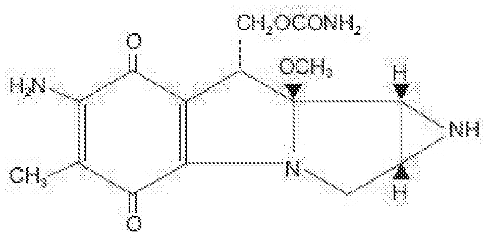

(paclitaxel).
LIGAND
As described herein, a ligand (e.g., a streptavidin, an avidin) can be coupled to a molecule or substrate so as to attract a radioisotope coupled to a corresponding receptor. Such an approach can provide targeted radiotherapy in a subject via selective binding of the ligand and receptor. A ligand can be selective or non-selective for a receptor. A ligand can be preferably selective for a receptor (or vice versa, a receptor can be preferably selective for a ligand).
Streptavidin. A ligand can be a streptavidin. A streptavidin can be a protein having a high affinity for biotin (e.g., Kd of about 10"14 mol/L). A streptavidin or a nucleotide encoding such, can be isolated from the bacterium Streptomyces (e.g., Streptomyces avidinii). A streptavidin can be any commercially available streptavidin (e.g., Invitrogen; Qiagen; Thermo Scientific; Jackson ImmunoResearch; Sigma Aldrich; Cell Signaling Technology). A streptavidin can be a variant of a naturally occurring streptavidin having at least about 80%, 85%, 90%, 95%, or 99% sequence identity thereto and retaining or substantially retaining high affinity for biotin. A streptavidin can be a tetramer, with each subunit binding a biotin with equal or substantially equal affinity. A streptavidin can have a mildly acidic isoelectric point (pi) (e.g., about 5). A streptavidin can lack any carbohydrate modification. Where a streptavidin has no
carbohydrate modification and a near-neutral pi, it can have substantially lower nonspecific binding compared to avidin.
A streptavidin can be an streptavidin coupled to a glycan. A streptavidin can be a glycol streptavidin (e.g., a, ethylene glycol streptavidin; or an streptavidin-poly (ethylene
glycol)(PEG)). In some embodiments, a streptavidin be attached in a branched form incorporating polyethylene glycol (e.g., PEG- streptavidin), which can give the streptavidin a branched structure, allowing it to bind more biotin.
A streptavidin can be a streptavidin variant. For example, a streptavidin can be a monovalent, divalent, and trivalent variant. As another example, a variant streptavidin can have a near-neutral pi.
Avidin.
A ligand can be an avidin. An avidin can be a protein having a high affinity for biotin (e.g., Kd of about 10"15 mol/L). An avidin or a nucleotide encoding such, can be isolated from egg white. Wild type avidin has about 30% sequence identity to wild type streptavidin, but highly similar secondary, tertiary and quaternary structure. An avidin can be glycosylated, positively charged, or have pseudo-catalytic activity (i.e., enhance alkaline hydrolysis of an ester linkage between biotin and a nitrophenyl group) or can have a higher tendency for aggregation as compared to a streptavidin. An avidin can be a tetramer of about 66-69 kDa in size. An avidin can have about 10% of molecular weight attributed to carbohydrate content composed of about 4 to 5 mannose or about three N-acetylglucosamine residues.
An avidin can be a streptavidin variant. For example, an avidin can be a non- glycosylated avidin. As another example, an avidin can be a deglycosylated avidin (e.g., Neutravidin), which can be more comparable to the size, pi or nonspecific binding of a wild type streptavidin. As another example, an avidin can be a deglycosylated avidin having modified arginines, exhibiting a more neutral isoelectric point (pi) and can better overcome problems of non-specific binding. Deglycosylated, neutral forms of avidin are commercially available (e.g., Extravidin, Sigma-Aldrich; Neutravidin, Thermo Scientific or Invitrogen;
NeutraLite, Belovo). As another example, an avidin can be an avidin coupled to a glycan. As another example, an avidin can be a glycol avidin (e.g., a, ethylene glycol avidin; or an avidin- poly(ethylene glycol) (avidin-PEG)) (see generally, Caliceti et al., 2002, Journal of Controlled Release, 83, 97-108; Salmaso et al., 2005, Biochimica et Biophysica Acta, 1726, 57-66). In some embodiments, an avidin be attached in a branched form incorporating polyethylene glycol (e.g., PEG-avidin), which can give the avidin a branched structure, allowing it to bind more biotin.
An avidin can be a variant AvidinOX , which can be obtained by 4- hydroxyazobenzene-2'-carboxylic acid-assisted sodium periodate oxidation of avidin (see generally De Santis et al., 2010, Cancer Biother Radiopharm, 25(2), 143-148; U.S. Patent No. 8,562,947). This method can generate aldehyde groups from avidin carbohydrates, sparing biotin-binding sites from inactivation. An avidin variant, such as AvidinOX, can have an increased tissue half-life (e.g., one, two, or more weeks).
In some embodiments, avidin can be pegylated to produce a much larger molecule (e.g., MW>100 kDA) with more binding sites, and then periodation can be used to form Schiff bases, which could then bind tightly to the amino groups of proteins. The pegylated molecule would be too large to pass easily out of the peritoneal cavity; and it could be introduced in a large volume of solution, and be allowed to attach to surfaces, then flushed out, and biotinylated isotopes (e.g., tracer biotinylated isotopes) could then be introduced, which would likewise coat the surfaces, and allowed to remain.
An avidin can have reversible binding characteristics through nitration or iodination of a binding site tyrosine, or exhibit strong biotin binding characteristics at about pH 4 or biotin release at a pH of about 10 or higher. An avidin can be a monovalent, divalent, and trivalent variant of avidin.
Processes for linking a ligand, such as avidin or streptavidin, to a molecule or substrate are well known (see e.g. Savage, 1992, Avidin-Biotin Chemistry: A Handbook, Pierce Chemical Co, ISBN-10 09359401 1 1 , ISBN-13 978-09359401 14; McMahon, 2010, Avidin-Biotin Interactions: Methods and Applications, Humana Press, ASIN B00GA4420E; Hermanson, 2010, Bioconjugate Techniques, Academic Press, ASIN B005YXETUU). Except as otherwise noted herein, therefore, the process of the present disclosure can be carried out in accordance with such processes. In some embodiments, avidin can be coupled to talc, for example, using both
Rhodamine and fluorescein avidin bound to talc then thrice washed, or using HRP-labeled avidin, which has shown saturation of binding (e.g., from 1 : 10 to 10: 1 HRP to natural Avidin, e.g., 1 : 1 ). In some embodiments, talc can bind in excess of 2 nanograms of avidin per mg of talc (i.e., about 2 micrograms per gram). For context, about 2 grams of talc can be
conventionally used for pleurodesis.
Molecularly Imprinted Polymer.
A ligand can be a molecularly imprinted polymer (MIP). A MIP is understood as a synthetic compound that can select, recognize or capture biological substances. MIPs can be generated via the polymerization of monomers in the presence of a template (see generally, Alvarez-Lorenzo and Concheiro, Ed., 2013, Handbook of Molecularly Imprinted Polymers, Smithers Rapra Technology, ISBN-10: 1847359604).
A MIP can be processed using a molecular imprinting technique that leaves cavities in polymer matrix with affinity to a chosen "template" molecule. The process can involve initiating polymerization of monomers in the presence of a template molecule that can be extracted afterwards, thus leaving complementary cavities behind. Such polymers can have affinity for the original molecule and have been used in applications such as chemical separations, catalysis, or molecular sensors. Binding activity of MIPs, or so called "plastic antibodies", can be about two orders of magnitude lower than specific antibodies but are still highly specific binding sites that can be made easily and are relatively inexpensive. MIPs can be generated as specific for receptors described herein. For example, MIPs can be specific for biotin (see e.g., WO2014/030002). MIPs can be coupled to a molecule or substrate described herein.
RADIOISOTOPE
As described herein, a radioisotope can be coupled to a receptor so as to provide targeted radiotherapy via selective binding to a molecule or substrate coupled to a ligand. Systemic radioisotope therapy can be a form of targeted therapy. As described herein, targeting a radioisotope can be achieved by attaching it to one part of a ligand/receptor combination, where the other part can be attached to a target.
A radioisotope can be used to destroy or weaken cells associated with a proliferative disease, disorder, or condition. A radioisotope that generates radiation can be localized in a desired location (e.g., a tissue) according to approaches described herein. In some embodiments, beta radiation from the radioisotope can result in the destruction of cells, which is a process understood as radionuclide therapy (RNT) or radiotherapy. Short-range radiotherapy may be known as brachytherapy.
A radioisotope for use with compositions and methods described herein can be a strong beta emitter, optionally with sufficient gamma to enable imaging, such as lutetium-177. Lutetium-177 can be prepared from ytterbium-176, which is irradiated to become Yb-177, which decays rapidly to Lu-177. Lu-177 can emit sufficient beta radiation for therapy on small (e.g., endocrine) tumors.
Another exemplary radioisotope for use with compositions and methods described herein includes Yttrium-90, which can be conventionally used for treatment of cancer, particularly non-Hodgkin's lymphoma and liver cancer, and as a silicate colloid for the relieving the pain of arthritis in larger synovial joints. Other exemplary radioisotopes for use with compositions and methods described herein include lodine-131 or phosphorus-32. lodine-131 has been conventionally used to treat the thyroid for cancers and other abnormal conditions such as hyperthyroidism (i.e., over-active thyroid), lodine-131 is a strong gamma emitter, and can be conventionally used for beta therapy. Phosphorus-32 has been conventionally used to treat Polycythemia vera, in which an excess of red blood cells is produced in the bone marrow and Phosphorus-32 can be used to control this excess.
Another exemplary radioisotope for use with compositions and methods described herein includes boron-10. A subject administered a composition including Boron-10 can be irradiated with neutrons which are strongly absorbed by the boron, to produce high-energy alpha particles that can kill cells including those associated with a proliferative disease, disorder, or condition.
Another exemplary radioisotope for use with compositions and methods described herein includes Radium-223, which can be conventionally used for treatment of prostate cancer. Another exemplary radioisotope for use with compositions and methods described herein includes bismuth-213. Bismuth-213, having a 46-minute half-life and high energy (8.4 MeV), can be formed from readily available Actinium-225 (via 3 alpha decays).
Another exemplary radioisotope for use with compositions and methods described herein includes lead-212, having a half-life of 10.6 hours. Lead-212 has been conventionally
attached to monoclonal antibodies for cancer treatment. Such approaches can be adapted for methods and compositions described herein. The decay chain of lead-212 includes the shortlived isotopes bismuth-212 by beta decay, polonium-212 by beta decay, and thallium-208 by alpha decay of the bismuth, with further alpha and beta decays respectively to Pb-208, all over about an hour.
Other exemplary radioisotopes for use with compositions and methods described herein include Holmium-166, having a 26 hour half-life and conventionally used for treatment of liver tumor; Dysprosium-165, having a 2 hour half-life and conventionally used as aggregated hydroxide for synovectomy treatment of arthritis; Erbium-169, having a 9.4 day half-life and conventionally used for relieving arthritis pain in synovial joints; Holmium-166, having a 26 hour half-life and conventionally used for treatment of liver tumors; lodine-125, having a 60 day half-life and conventionally used in cancer brachytherapy, including prostate and brain; lridium-192, a beta emitter having a 74 day half-life; Rhenium-186, having a 3.8 day half-life, conventionally used for pain relief in bone cancer; Rhenium-188, having a 17 hour half-life, conventionally used to beta irradiate coronary arteries; Samarium-153, having a 47 hour half-life, conventionally used for relieving pain of secondary cancers lodged in the bone and for prostate and breast cancer; Strontium-89, having a 50 day half-life,
conventionally used for reducing pain of prostate and bone cancer; and radioisotopes of caesium, gold or ruthenium. Radioisotopes can be obtained from a variety or commercial or research sources including, but not limited to MDS Nordion, IRE, Covidien, NTP, ANSTO, and Isotop-NIIAR.
A conjugated radioisotope can be administered by any conventional route. For example, a conjugated radioisotope can be delivered through infusion (e.g., into the bloodstream) or ingestion. In some embodiments, yttrium-90 radioactive glass or resin microspheres (e.g., SIR-
Spheres and TheraSphere) coupled to a receptor, such as biotin, can be injected into the hepatic artery to radioembolize liver tumors or liver metastases. Such microspheres can be used in treatment approach known as selective internal radiation therapy. The microspheres can be approximately 30 pm in diameter and can be delivered directly into an artery supplying blood to the tumors. Such treatments can begin by guiding a catheter up through
the femoral artery in the leg, navigating to the desired target site and administering treatment. A molecule or substrate coupled to a ligand, such as avidin or biotin, can be introduced into tissue at, in or near a tumor. Blood feeding the tumor can carry the microspheres directly to the tumor, allowing specific binding to the ligand-coupled molecule or substrate, thus providing a more selective approach than traditional systemic chemotherapy.
In some embodiments, a receptor (e.g., biotin) coupled to strontium-89 or samarium (153Sm) lexidronam can be used in the treatment of bone metastasis from cancer. The coupled radioisotopes can travel selectively to areas of damaged bone, in or around which have been introduced a ligand (e.g., avidin or streptavidin) coupled to a molecule or substrate, and spare normal undamaged bone.
In some embodiments, a receptor (e.g., biotin) can be coupled to ibritumomab tiuxetan (i.e., Zevalin), which is an FDA approved anti-CD20 monoclonal antibody conjugated to yttrium-90. In some embodiments, a receptor (e.g., biotin) can be coupled to one or more parts of tositumomab/iodine (131 l) tositumomab regimen (Bexxar), which is a combination of an iodine-131 labeled and an unlabeled anti-CD20 monoclonal antibody. Such medications can be used for, e.g., the treatment of refractory non-Hodgkin's lymphoma according to approaches described herein.
Coupling can be any type attraction, link, or reaction that serves to immobilize a ligand on a molecule. Coupling can be via a bond. A radioisotope-receptor bond is understood as an attraction between atoms of a radioisotope and atoms of a receptor that allows the formation of a linkage between atoms of the biomolecule and the matrix material. A bond can be caused by an electrostatic force of attraction between opposite charges, either between electrons and nuclei, or as the result of a dipole attraction. A bond (e.g., between a
biomolecule and a matrix material) can be, for example, a covalent bond, a coordinate covalent bond, an ionic bond, polar covalent, a dipole-dipole interaction, a London dispersion force, a cation-pi interaction, or hydrogen bonding.
Process for coupling a radioisotope to a receptor or ligand (e.g., biotin) are well known (see, e.g., Savage, 1992, Avidin-Biotin Chemistry: A Handbook, Pierce Chemical Co, ISBN- 10 09359401 1 1 , ISBN-13 978-09359401 14; McMahon, 2010, Avidin-Biotin Interactions:
Methods and Applications, Humana Press, ASIN B00GA4420E; Hermanson, 2010,
Bioconjugate Techniques, Academic Press, ASIN B005YXETUU; Bolzati et al., 2006, Nuclear Medicine and Biology, 34, 51 1 -522; Runn-Dufault et al., 2000, Nuclear Medicine and Biology, 27, 803-807). Except as otherwise noted herein, therefore, the process of the present disclosure can be carried out in accordance with such processes.
RECEPTOR
As described herein, a receptor (e.g., a biotin) can be coupled to a radioisotope so as to provide targeted radiotherapy via selective binding to a molecule or substrate coupled to a ligand. A receptor can be selective or non-selective for a ligand. A receptor can be preferably selective for a ligand (or vice versa, a ligand can be preferably selective for a receptor).
Biotin.
A receptor can be a biotin. A biotin can be a water soluble B-complex vitamin (e.g., vitamin B7, vitamin H, or coenzyme R). A biotin can be a heterocyclic sulfur-containing
(mono-)carboxylic acid. A biotin can comprise an imidazole ring and thiophene ring fused. A biotin can comprise a ureido (tetrahydroimidizalone) ring fused with a tetrahydrothiophene ring, optionally with a veleric acid substituent on a carbon of the tetrahydrothiophene ring.
Streptavidin or avidin can bind biotin with high affinity (e.g., Kd of 10"14 mol/L to 10"15 mol/l) and specificity.
A biotin can be any commercially available biotin (e.g., Invitrogen; Qiagen; Thermo Scientific; Jackson ImmunoResearch; Sigma Aldrich; Cell Signaling Technology). A biotin can be a variant compound of a naturally occurring biotin that retains or substantially retaining high affinity for streptavidin.
A biotin can have a structural formula according to C10 H16 03 N2 S. A biotin can have a structure as follows:

Biotin can be attached to a molecule or substrate by biotinylation. Biotinylated proteins of interest can be isolated from a sample by exploiting this highly stable interaction.
Biotinylation can be the process of covalently attaching a biotin to a molecule or substrate. Biotinylation can be generally rapid, specific and can be unlikely to perturb the natural function of the molecule or substrate to which it is attached given the small size of a biotin (e.g., MW = 244.31 g/mol). Biotin can bind to streptavidin or avidin with an extremely high affinity, fast on-rate, and high specificity, and these interactions can be exploited as described herein. Biotin-binding to streptavidin or avidin can be resistant to extremes of heat, pH, or proteolysis, which can allow use of a biotinylated molecule or substrate in a wide variety of environments. Furthermore, multiple biotin molecules can be conjugated to a molecule or substrate, which can allow binding of multiple streptavidin, avidin, or Neutravidin. A large number of biotinylation reagents are know in the art and commercially available.
Various assays are available to determine extent of biotinylation.
The HABA (2-(4-hydroxyazobenzene) benzoic acid) assay can be used to determine the extent of biotinylation. HABA dye can be bound to avidin or streptavidin and yields a characteristic absorbance. When biotinylated proteins or other molecules are introduced, the biotin displaces the dye, resulting in a change in absorbance at 500 nm. This change can be directly proportional to the level of biotin in the sample. A HABA assay can require a relatively large amount of sample.
Extent of biotinylation can also be measured by streptavidin gel-shift, since
streptavidin remains bound to biotin during agarose gel electrophoresis or polyacrylamide gel electrophoresis. The proportion of target biotinylated can be measured via the change in band intensity of the target with or without excess streptavidin, seen quickly and quantitatively by Coomassie Brilliant Blue staining.
Biotinylation, also called biotin labeling, can be most commonly performed through chemical means, although enzymatic methods are also available. Chemical biotinylation can use various conjugation chemistries to yield a nonspecific biotinylation of amines,
carboxylates, sulfhydryls or carbohydrates (e.g., NHS-coupling gives biotinylation of a primary amines). Chemical biotinylation reagents can include a reactive group attached via a linker to the valeric acid side chain of biotin. Because the biotin binding pocket in avidin or streptavidin can be buried beneath the protein surface, a biotinylation reagent possessing a longer linker can be desirable, as such longer linker can enable the biotin molecule to be
more accessible to binding avidin, streptavidin, or Neutravidin. A linker can also mediate the solubility of a biotinylation reagent. Linkers that incorporate poly(ethylene) glycol (PEG) can make water-insoluble reagents soluble or increase the solubility of biotinylation reagents that are already soluble to some extent. Primary Amine Biotinylation.
Biotin can be conjugated to an amine group on the molecule or substrate. A primary amine group can be present as a lysine side chain epsilon-amine or N-terminal a-amine. Amine-reactive biotinylation reagents can be divided into two groups based on water solubility. N-hydroxysuccinimide (NHS) esters have poor solubility in aqueous solutions. For reactions in aqueous solution, NHS can be first be dissolved in an organic solvent, then diluted into the aqueous reaction mixture. Commonly used organic solvents for this purpose can include dimethyl sulfoxide (DMSO) and dimethyl formamide (DMF). Because of the hydrophobicity of NHS-esters, NHS biotinylation reagents can also diffuse through the cell membrane, meaning that they will biotinylate both internal and external components of a cell.
Sulfo-NHS esters are more soluble in water and can be dissolved in water just before use because they hydrolyze easily. The water solubility of sulfo-NHS-esters can be due at least in part from a sulfonate group on the N-hydroxysuccinimide ring. Water solubility can eliminate a need to dissolve the reagent in an organic solvent. Sulfo-NHS-esters of biotin do not penetrate the cell membrane.
The chemical reactions of NHS- and sulfo-NHS esters can be identical, in that they can both react spontaneously with amines to form an amide bond. Because the target for the ester is a deprotonated primary amine, the reaction can be favored under basic conditions (above pH 7). Hydrolysis of the NHS ester can be a major competing reaction, and the rate of hydrolysis increases with increasing pH. NHS- and sulfo-NHS-esters have a half-life of several hours at pH 7 but only a few minutes at pH 9.
There can be additional flexibility in the conditions for conjugating NHS-esters to primary amines. Incubation temperatures can range from about 4-37 °C, pH values in the reaction range from about 7-9, or incubation times range from a few minutes to about 12
hours. Buffers containing amines (e.g., Tris or glycine) can be avoided, because they compete with the reaction.
Sulfhydryl biotinylation
An alternative to primary amine biotinylation can be to label sulfhydryl groups with biotin. Sulfhydryl-reactive groups such as maleimides, haloacetyls, or pyridyl disulfides, can require free sulfhydryl groups for conjugation; disulfide bonds can be first reduced to free up the sulfhydryl groups for biotinylation. If no free sulfhydryl groups are available, lysines can be modified with various thiolation reagents (Traut's Reagent, SAT(PEG4), SATA and SATP), resulting in the addition of a free sulfhydryl. Sulfhydryl biotinylation can be performed at a slightly lower pH (e.g., about 6.5-7.5) than labeling with NHS esters.
Carboxyl biotinylation.
Biotinylation reagents that target carboxyl groups do not have a carboxyl-reactive moiety per se but instead rely on a carbodiimide crosslinker such as EDC to bind the primary amine on a biotinylation reagent to a carboxyl group on the target. Biotinylation at carboxyl groups can occur at a pH of about 4.5-5.5. To prevent crossreactivity of the crosslinker with buffer constituents, buffers should not contain primary amines (e.g., Tris, glycine) or carboxyls (e.g., acetate, citrate).
Glycoprotein biotinylation
Glycoproteins can be biotinylated by modifying the carbohydrate residues to
aldehydes, which can then react with hydrazine- or alkoxyamine-based biotinylation reagents. Sodium periodate can oxidize a sialic acid on glycoproteins to aldehydes to form these stable linkages at a pH of about 4-6.
Antibodies can be heavily glycosylated, and because glycosylation does not interfere with the antibody activity, biotinylating the glycosyl groups can be an ideal strategy to generate biotinylated antibodies.
Biotinylation at carboxyl groups can occur at a pH of about 4.5-5.5. To prevent crossreactivity of the crosslinker with buffer constituents, buffers should not contain primary amines (e.g., Tris, glycine) or carboxyls (e.g., acetate, citrate).
Oligonucleotide biotinylation.
Oligonucleotides can be readily biotinylated in the course of oligonucleotide synthesis by the phosphoramidite method using, e.g., commercial biotin phosphoramidite. Upon the standard deprotection, the conjugates obtained can be purified using reverse-phase or anion- exchange HPLC.
Non-specific biotinylation.
Photoactivatable biotinylation reagents can be useful when primary amines,
sulfhydryls, carboxyls or carbohydrates are not available or not desired for labeling. A photoactivatable biotinylation reagent relies on aryl azides, which become activated by ultraviolet light (UV; >350 nm), which then react at C-H and N-H bonds. A photoactivatable biotinylation reagent can also be used to activate biotinylation at specific times by simply exposing the reaction to UV light at the specific time or condition.
Process for coupling a receptor or ligand (e.g., biotin) to a radioisotope are well known (see e.g., Savage, 1992, Avidin-Biotin Chemistry: A Handbook, Pierce Chemical Co, ISBN-10 09359401 1 1 , ISBN-13 978-09359401 14; McMahon, 2010, Avidin-Biotin Interactions:
Methods and Applications, Humana Press, ASIN B00GA4420E; Hermanson, 2010,
Bioconjugate Techniques, Academic Press, ASIN B005YXETUU). Except as otherwise noted herein, therefore, the process of the present disclosure can be carried out in accordance with such processes. COUPLING
Coupling can be any type attraction, link, or reaction that serves to immobilize a therapeutic agent on a molecule/substrate; a ligand on a molecule/substrate; or a receptor on a radioisotope (or vice versa, a receptor on a molecule/substrate or ligand on a radioisotope).
Coupling can be via a bond. A molecule-therapeutic agent bond is understood as an attraction between atoms of a molecule and atoms of a therapeutic agent that allows the formation of a linkage between atoms of the therapeutic agent and the matrix material. A molecule-ligand bond is understood as an attraction between atoms of a molecule and atoms of a ligand that allows the formation of a linkage between atoms of the biomolecule and the matrix material. A bond can be caused by an electrostatic force of attraction between
opposite charges, either between electrons and nuclei, or as the result of a dipole attraction. A bond (e.g., between a biomolecule and a matrix material) can be, for example, a covalent bond, a coordinate covalent bond, an ionic bond, polar covalent, a dipole-dipole interaction, a London dispersion force, a cation-pi interaction, or hydrogen bonding. Coupling can be reversible or irreversible. One of ordinary skill will understand that coupling does not necessarily need to be irreversible and can be preferred to be reversible coupling.
Processes for coupling a molecule or substrate to a receptor or ligand (e.g., avidin or streptavidin) are well known (see e.g., Savage, 1992, Avidin-Biotin Chemistry: A Handbook, Pierce Chemical Co, ISBN-10 09359401 1 1 , ISBN-13 978-09359401 14; McMahon, 2010, Avidin-Biotin Interactions: Methods and Applications, Humana Press, ASIN B00GA4420E; Hermanson, 2010, Bioconjugate Techniques, Academic Press, ASIN B005YXETUU). Except as otherwise noted herein, therefore, the process of the present disclosure can be carried out in accordance with such processes.
A ligand or therapeutic agent can be considered to be bound to a substrate (e.g., talc) if the ligand or therapeutic agent was detected on the substrate (e.g., via flow cytomoetry) after washing (e.g., with PBS).
MOLECULAR ENGINEERING
The following definitions and methods are provided to better define the present disclosure and to guide those of ordinary skill in the art in the practice of the present disclosure. Unless otherwise noted, terms are to be understood according to conventional usage by those of ordinary skill in the relevant art.
The terms "heterologous DNA sequence", "exogenous DNA segment" or
"heterologous nucleic acid," as used herein, each refer to a sequence that originates from a source foreign to the particular host cell or, if from the same source, is modified from its original form. Thus, a heterologous gene in a host cell includes a gene that can be
endogenous to the particular host cell but has been modified through, for example, the use of DNA shuffling. The terms also include non-naturally occurring multiple copies of a naturally occurring DNA sequence. Thus, the terms refer to a DNA segment that can be foreign or heterologous to the cell, or homologous to the cell but in a position within the host cell nucleic
acid in which the element is not ordinarily found. Exogenous DNA segments are expressed to yield exogenous polypeptides. A "homologous" DNA sequence can be a DNA sequence that is naturally associated with a host cell into which it can be introduced.
Expression vector, expression construct, plasmid, or recombinant DNA construct is generally understood to refer to a nucleic acid that has been generated via human
intervention, including by recombinant means or direct chemical synthesis, with a series of specified nucleic acid elements that permit transcription or translation of a particular nucleic acid in, for example, a host cell. The expression vector can be part of a plasmid, virus, or nucleic acid fragment. Typically, the expression vector can include a nucleic acid to be transcribed operably linked to a promoter.
A "promoter" is generally understood as a nucleic acid control sequence that directs transcription of a nucleic acid. An inducible promoter is generally understood as a promoter that mediates transcription of an operably linked gene in response to a particular stimulus. A promoter can include necessary nucleic acid sequences near the start site of transcription, such as, in the case of a polymerase II type promoter, a TATA element. A promoter can optionally include distal enhancer or repressor elements, which can be located as much as several thousand base pairs from the start site of transcription.
A "transcribable nucleic acid molecule" as used herein refers to any nucleic acid molecule capable of being transcribed into a RNA molecule. Methods are known for introducing constructs into a cell in such a manner that the transcribable nucleic acid molecule can be transcribed into a functional mRNA molecule that can be translated and therefore expressed as a protein product. Constructs may also be constructed to be capable of expressing antisense RNA molecules, in order to inhibit translation of a specific RNA molecule of interest. For the practice of the present disclosure, conventional compositions and methods for preparing and using constructs and host cells are well known to one skilled in the art (see e.g., Sambrook and Russel, (2006), Condensed Protocols from Molecular Cloning: A Laboratory Manual, Cold Spring Harbor Laboratory Press, ISBN-10: 0879697717; Ausubel et al., (2002), Short Protocols in Molecular Biology, 5th ed., Current Protocols, ISBN- 10: 0471250929; Sambrook and Russel, (2001 ), Molecular Cloning: A Laboratory Manual, 3d ed., Cold Spring Harbor Laboratory Press, ISBN-10: 0879695773; Elhai, J. and Wolk, C, P.
1988. Methods in Enzymology, 167, 747-754).
The "transcription start site" or "initiation site" can be the position surrounding the first nucleotide that can be part of the transcribed sequence, which can also defined as position +1 . With respect to this site all other sequences of the gene and its controlling regions can be numbered. Downstream sequences (i.e., further protein encoding sequences in the 3' direction) can be denominated positive, while upstream sequences (mostly of the controlling regions in the 5' direction) are denominated negative.
"Operably-linked" or "functionally linked" refers preferably to the association of nucleic acid sequences on a single nucleic acid fragment so that the function of one can be affected by the other. For example, a regulatory DNA sequence can be said to be "operably linked to" or "associated with" a DNA sequence that codes for an RNA or a polypeptide if the two sequences are situated such that the regulatory DNA sequence affects expression of the coding DNA sequence (i.e., that the coding sequence or functional RNA can be under the transcriptional control of the promoter). Coding sequences can be operably-linked to regulatory sequences in sense or antisense orientation. The two nucleic acid molecules may be part of a single contiguous nucleic acid molecule and may be adjacent. For example, a promoter can be operably linked to a gene of interest if the promoter regulates or mediates transcription of the gene of interest in a cell.
A "construct" is generally understood as any recombinant nucleic acid molecule such as a plasmid, cosmid, virus, autonomously replicating nucleic acid molecule, phage, or linear or circular single-stranded or double-stranded DNA or RNA nucleic acid molecule, derived from any source, capable of genomic integration or autonomous replication, comprising a nucleic acid molecule where one or more nucleic acid molecule has been operably linked.
A constructs of the present disclosure can contain a promoter operably linked to a transcribable nucleic acid molecule operably linked to a 3' transcription termination nucleic acid molecule. In addition, constructs can include but are not limited to additional regulatory nucleic acid molecules from, e.g., the 3'-untranslated region (3' UTR). Constructs can include but are not limited to the 5' untranslated regions (5' UTR) of an mRNA nucleic acid molecule which can play an important role in translation initiation and can also be a genetic component in an expression construct. These additional upstream and downstream regulatory nucleic
acid molecules may be derived from a source that can be native or heterologous with respect to the other elements present on the promoter construct.
The term "transformation" refers to the transfer of a nucleic acid fragment into the genome of a host cell, resulting in genetically stable inheritance. Host cells containing the transformed nucleic acid fragments are referred to as "transgenic" cells, and organisms comprising transgenic cells are referred to as "transgenic organisms".
"Transformed," "transgenic," and "recombinant" refer to a host cell or organism such as a bacterium, cyanobacterium, animal or a plant into which a heterologous nucleic acid molecule has been introduced. The nucleic acid molecule can be stably integrated into the genome as generally known in the art and disclosed (Sambrook, 1989; Innis, 1995; Gelfand, 1995; Innis & Gelfand, 1999). Known methods of PCR include, but are not limited to, methods using paired primers, nested primers, single specific primers, degenerate primers, gene-specific primers, vector-specific primers, partially mismatched primers, and the like. The term "untransformed" refers to normal cells that have not been through the transformation process.
"Wild-type" refers to a virus or organism found in nature without any known mutation.
Design, generation, and testing of the variant nucleotides, and their encoded polypeptides, having the above required percent identities and retaining a required activity of the expressed protein is within the skill of the art. For example, directed evolution and rapid isolation of mutants can be according to methods described in references including, but not limited to, Link et al., (2007), Nature Reviews, 5(9), 680-688; Sanger et al., (1991 ), Gene, 97(1 ), 1 19-123; Ghadessy et al., (2001 ), Proc Natl Acad Sci USA, 98(8), 4552-4557. Thus, one skilled in the art could generate a large number of nucleotide or polypeptide variants having, for example, at least 95-99% identity to the reference sequence described herein and screen such for desired phenotypes according to methods routine in the art.
Nucleotide or amino acid sequence identity percent (%) can be understood as the percentage of nucleotide or amino acid residues that are identical with nucleotide or amino acid residues in a candidate sequence in comparison to a reference sequence when the two sequences are aligned. To determine percent identity, sequences are aligned and if necessary, gaps are introduced to achieve the maximum percent sequence identity.
Sequence alignment procedures to determine percent identity are well known to those of skill in the art. Often publicly available computer software such as BLAST, BLAST2, ALIGN2 or Megalign (DNASTAR) software can be used to align sequences. Those skilled in the art can determine appropriate parameters for measuring alignment, including any algorithms needed to achieve maximal alignment over the full-length of the sequences being compared. When sequences are aligned, the percent sequence identity of a given sequence A to, with, or against a given sequence B (which can alternatively be phrased as a given sequence A that has or comprises a certain percent sequence identity to, with, or against a given sequence B) can be calculated as: percent sequence identity = ΧΛΊ 00, where X is the number of residues scored as identical matches by the sequence alignment program's or algorithm's alignment of A and B and Y is the total number of residues in B. If the length of sequence A is not equal to the length of sequence B, the percent sequence identity of A to B will not equal the percent sequence identity of B to A.
Generally, conservative substitutions can be made at any position so long as the required activity can be retained. So-called conservative exchanges can be carried out in which the amino acid which is replaced has a similar property as the original amino acid, for example the exchange of Glu by Asp, Gin by Asn, Val by lie, Leu by lie, and Ser by Thr. Deletion can be the replacement of an amino acid by a direct bond. Positions for deletions include the termini of a polypeptide and linkages between individual protein domains.
Insertions are introductions of amino acids into the polypeptide chain, a direct bond formally being replaced by one or more amino acids. Amino acid sequence can be modulated with the help of art-known computer simulation programs that can produce a polypeptide with, for example, improved activity or altered regulation. On the basis of this artificially generated polypeptide sequences, a corresponding nucleic acid molecule coding for such a modulated polypeptide can be synthesized in-vitro using the specific codon-usage of the desired host cell.
"Highly stringent hybridization conditions" are defined as hybridization at 65 °C in a 6 X SSC buffer (i.e., 0.9 M sodium chloride and 0.09 M sodium citrate). Given these conditions, a determination can be made as to whether a given set of sequences will hybridize by calculating the melting temperature (Tm) of a DNA duplex between the two sequences. If a
particular duplex has a melting temperature lower than 65°C in the salt conditions of a 6 X SSC, then the two sequences will not hybridize. On the other hand, if the melting temperature is above 65 °C in the same salt conditions, then the sequences will hybridize. In general, the melting temperature for any hybridized DNA:DNA sequence can be determined using the following formula: Tm = 81 .5 °C + 16.6(log10[Na+]) + 0.41 (fraction G/C content) - 0.63(% formamide) - (600/I). Furthermore, the Tm of a DNA:DNA hybrid is decreased by 1 -1.5°C for every 1 % decrease in nucleotide identity (see, e.g., Sambrook and Russel, 2006).
Host cells can be transformed using a variety of standard techniques known to the art (see, e.g., Sambrook and Russel, (2006), Condensed Protocols from Molecular Cloning: A Laboratory Manual, Cold Spring Harbor Laboratory Press, ISBN-10: 0879697717; Ausubel et al., (2002), Short Protocols in Molecular Biology, 5th ed., Current Protocols, ISBN-10:
0471250929; Sambrook and Russel, (2001 ), Molecular Cloning: A Laboratory Manual, 3d ed., Cold Spring Harbor Laboratory Press, ISBN-10: 0879695773; Elhai, J. and Wolk, C, P. 1988. Methods in Enzymology, 167, 747-754). Such techniques include, but are not limited to, viral infection, calcium phosphate transfection, liposome-mediated transfection,
microprojectile-mediated delivery, receptor-mediated uptake, cell fusion, electroporation, and the like. The transfected cells can be selected and propagated to provide recombinant host cells that comprise the expression vector stably integrated in the host cell genome.
Exemplary nucleic acids which may be introduced to a host cell include, for example, DNA sequences or genes from another species, or even genes or sequences which originate with or are present in the same species, but are incorporated into recipient cells by genetic engineering methods. The term "exogenous" can be also intended to refer to genes that are not normally present in the cell being transformed, or perhaps simply not present in the form, structure, etc. , as found in the transforming DNA segment or gene, or genes which are normally present and that one desires to express in a manner that differs from the natural expression pattern, e.g. , to over-express. Thus, the term "exogenous" gene or DNA can be intended to refer to any gene or DNA segment that can be introduced into a recipient cell, regardless of whether a similar gene may already be present in such a cell. The type of DNA included in the exogenous DNA can include DNA which can be already present in the cell, DNA from another individual of the same type of organism, DNA from a different organism, or
a DNA generated externally, such as a DNA sequence containing an antisense message of a gene, or a DNA sequence encoding a synthetic or modified version of a gene.
Host strains developed according to the approaches described herein can be evaluated by a number of means known in the art (see, e.g., Studier, (2005), Protein Expr Purif., 41 (1 ), 207-234; Gellissen, ed. (2005), Production of Recombinant Proteins: Novel Microbial and Eukaryotic Expression Systems, Wiley-VCH, ISBN-10: 3527310363; Baneyx, (2004), Protein Expression Technologies, Taylor & Francis, ISBN-10: 0954523253).
Methods of down-regulation or silencing genes are known in the art. For example, expressed protein activity can be down-regulated or eliminated using antisense
oligonucleotides, protein aptamers, nucleotide aptamers, and RNA interference (RNAi) (e.g., small interfering RNAs (siRNA), short hairpin RNA (shRNA), and micro RNAs (miRNA) (see e.g., Fanning and Symonds, (2006) Handb Exp Pharmacol., 173, 289-303G, describing hammerhead ribozymes and small hairpin RNA; Helene, C, et al., (1992), Ann. N.Y. Acad. Sci., 660, 27-36; Maher, (1992), Bioassays, 14(12): 807-15, describing targeting
deoxyribonucleotide sequences; Lee et al., (2006), Curr Opin Chem Biol., 10, 1 -8, describing aptamers; Reynolds et al., (2004), Nature Biotechnology, 22(3), 326 - 330, describing RNAi; Pushparaj and Melendez, (2006), Clinical and Experimental Pharmacology and Physiology, 33(5-6), 504-510, describing RNAi; Dillon et al., (2005), Annual Review of Physiology, 67, 147-173, describing RNAi; Dykxhoorn and Lieberman, (2005), Annual Review of Medicine, 56, 401 -423, describing RNAi). RNAi molecules are commercially available from a variety of sources (e.g. , Ambion, TX; Sigma Aldrich, MO; Invitrogen). Several siRNA molecule design programs using a variety of algorithms are known to the art (see e.g., Cenix algorithm, Ambion; BLOCK-iT™ RNAi Designer, Invitrogen; siRNA Whitehead Institute Design Tools, Bioinofrmatics & Research Computing). Traits influential in defining optimal siRNA sequences include G/C content at the termini of the siRNAs, Tm of specific internal domains of the siRNA, siRNA length, position of the target sequence within the CDS (coding region), and nucleotide content of the 3' overhangs.
FORMULATION
The agents and compositions described herein can be formulated by any conventional
manner using one or more pharmaceutically acceptable carriers or excipients as described in, for example, Remington's Pharmaceutical Sciences (A.R. Gennaro, Ed.), 21 st edition, ISBN: 0781746736 (2005), incorporated herein by reference in its entirety. Such formulations will contain a therapeutically effective amount of a biologically active agent described herein, which can be in purified form, together with a suitable amount of carrier so as to provide the form for proper administration to the subject.
The term "formulation" refers to preparing a drug in a form suitable for administration to a subject, such as a human. Thus, a "formulation" can include pharmaceutically acceptable excipients, including diluents or carriers. The term "pharmaceutically acceptable" as used herein can describe substances or components that do not cause unacceptable losses of pharmacological activity or
unacceptable adverse side effects. Examples of pharmaceutically acceptable ingredients can be those having monographs in United States Pharmacopeia (USP 29) and National
Formulary (NF 24), United States Pharmacopeial Convention, Inc, Rockville, Maryland, 2005 ("USP/NF"), or a more recent edition, and the components listed in the continuously updated Inactive Ingredient Search online database of the FDA. Other useful components that are not described in the USP/NF, etc. may also be used.
The term "pharmaceutically acceptable excipient," as used herein, can include any and all solvents, dispersion media, coatings, antibacterial and antifungal agents, isotonic, or absorption delaying agents. The use of such media and agents for pharmaceutical active substances is well known in the art (see generally Remington's Pharmaceutical Sciences (A.R. Gennaro, Ed.), 21 st edition, ISBN: 0781746736 (2005)). Except insofar as any conventional media or agent is incompatible with an active ingredient, its use in the therapeutic compositions can be contemplated. Supplementary active ingredients can also be incorporated into the compositions.
A "stable" formulation or composition can refer to a composition having sufficient stability to allow storage at a convenient temperature, such as between about 0 °C and about 60 °C, for a commercially reasonable period of time, such as at least about one day, at least about one week, at least about one month, at least about three months, at least about six months, at least about one year, or at least about two years.
The formulation should suit the mode of administration. The agents of use with the current disclosure can be formulated by known methods for administration to a subject using several routes which include, but are not limited to, parenteral, pulmonary, oral, topical, intradermal, intramuscular, intraperitoneal, intravenous, subcutaneous, intranasal, epidural, ophthalmic, buccal, and rectal. The individual agents may also be administered in
combination with one or more additional agents or together with other biologically active or biologically inert agents. Such biologically active or inert agents may be in fluid or mechanical communication with the agent(s) or attached to the agent(s) by ionic, covalent, Van der Waals, hydrophobic, hydrophilic or other physical forces. Controlled-release (or sustained-release) preparations may be formulated to extend the activity of the agent(s) and reduce dosage frequency. Controlled-release preparations can also be used to effect the time of onset of action or other characteristics, such as blood levels of the agent, and consequently affect the occurrence of side effects. Controlled-release preparations may be designed to initially release an amount of an agent(s) that produces the desired therapeutic effect, and gradually and continually release other amounts of the agent to maintain the level of therapeutic effect over an extended period of time. In order to maintain a near-constant level of an agent in the body, the agent can be released from the dosage form at a rate that will replace the amount of agent being metabolized or excreted from the body. The controlled-release of an agent may be stimulated by various inducers, e.g., change in pH, change in temperature, enzymes, water, or other physiological conditions or molecules.
Agents or compositions described herein (e.g., molecule-ligand or radioisotope- receptor) can also be used in combination with other therapeutic modalities, as described further below. Thus, in addition to the therapies described herein, one may also provide to the subject other therapies known to be efficacious for treatment of the disease, disorder, or condition.
THERAPEUTIC METHODS
Another aspect provided herein is a process of treating a proliferative disease, disorder, or condition with a composition described herein.
For example, molecule or substrate coupled to a therapeutic agent can be used to treat a proliferative disease, disorder. Provided is a process of treating a proliferative disease, disorder, or condition in a subject in need administration of a therapeutically effective amount of a molecule or substrate coupled to a therapeutic agent, so as to provide targeted or selective therapy. The therapeutic method can include administration of a molecule or substrate coupled to a therapeutic agent.
For example, combination of a first composition including a ligand coupled to molecule or substrate and a second composition including a receptor coupled to a radioisotope (or vice versa, a receptor coupled to molecule or substrate and a ligand coupled to a radioisotope) can be used to treat a proliferative disease, disorder. Provided is a process of treating a proliferative disease, disorder, or condition in a subject in need administration of a
therapeutically effective amount of the first composition and the second composition, so as to provide targeted or selective radiotherapy. The therapeutic method can include administration of a first composition including a ligand coupled to molecule or substrate and a second composition including a receptor coupled to a radioisotope.
Exemplary technology for rapidly delivering precisely calibrated and dispersed loads of microparticles into living tissue to depths of 2 cm include the use of air-powered injectors or sprays, and other methods known in the art. Such particles can be injected, e.g., directly into the walls or floor of the cavity created in breast tissue during lumpectomy for cancer, or in retroperitoneal tissues after excision of a pancreatic head cancer, or the cavity created in subcutaneous tissues of the thigh after radical excision of a sarcoma. Instead of conventional daily postoperative regimens of external beam radiation, a subject can be given, e.g., an intravenous dose of biotin-labeled radioisotope once monthly for one, two, three or more months until the recommended dose can be achieved. According to the methods described herein, administration can be parenteral, pulmonary, oral, topical, intradermal, intramuscular, intraperitoneal, intravenous,
subcutaneous, intranasal, epidural, ophthalmic, buccal, or rectal administration.
In some embodiments, administration can be according to conventional pleurodesis modified to incorporate compositions described herein. Methods described herein are generally performed on a subject in need thereof. A
subject in need of the therapeutic methods described herein can be a subject having, diagnosed with, suspected of having, or at risk for developing proliferative disease, disorder, or condition. A determination of the need for treatment will typically be assessed by a history and physical exam consistent with the disease or condition at issue. Diagnosis of the various conditions treatable by the methods described herein is within the skill of the art. The subject can be an animal subject, including a mammal, such as horses, cows, dogs, cats, sheep, pigs, mice, rats, monkeys, hamsters, guinea pigs, and chickens, and humans. For example, the subject can be a human subject.
Compositions, systems, or methods described herein can be used to treat proliferative diseases, disorders, or conditions. For example, compositions, systems, or methods described herein can be used (e.g., operatively or post-operatively) to treat mesothelioma, Meigs Syndrome, sarcoma, appendiceal carcinoma, pseudomyxoma peritonei, prostate cancer, prostate cancer lymph node dissection beds, rectovesical pouch tumor bed, ovarian cancer resection bed and peritoneal spread, uterine cancer resection cavities, pleural and peritoneal mesothelioma resection bed and peritoneal seeding, colorectal carcinoma, appendiceal carcinoma, pancreatic carcinoma, liver metastases, gastric carcinoma, renal carcinoma, retroperitoneal tumors (sarcomas, carcinomas), breast lumpectomy or breast lymph node dissection cavities, melanoma node dissection cavities, sarcoma resection cavities, head and neck cancer resection cavities, neck lymph node dissection cavities, scalp lesions, glioblastoma resection cavities, brain surface tumor lesions (resected, non resected), or trunk and extremity sarcoma resection cavities.
Compositions, systems, or methods described herein can be used at post-operative sites associated with a disease, disorder, or condition described herein. For example, an avidin-talc complex (followed by a receptor-radioisotope complex) can be used at
postoperative sites associated with a disease, disorder, or condition described herein. As another example, an avidin-fibrin glue complex (followed by a receptor-radioisotope complex) can be used at postoperative sites associated with a disease, disorder, or condition described herein. As another example, an avidin-gelfoam complex (followed by a receptor-radioisotope complex) can be used at postoperative sites associated with a disease, disorder, or condition described herein.
Compositions, systems, or methods described herein can be used to treat proliferative diseases, disorders, or conditions. Examples of proliferative diseases, disorders, or conditions treatable with compositions described (e.g., molecule-ligand or radioisotope- receptor) include, but are not limited to, cancer; blood vessel proliferative disorders; fibrotic disorders; mesangial cell proliferative disorders; psoriasis; actinic keratoses; seborrheic keratoses; warts; keloid scars; eczema; and hyperproliferative diseases caused by virus infections, such as papilloma virus infection.
Cancer, or neoplasia, refers generally to any malignant neoplasm or spontaneous growth or proliferation of cells. A subject having "cancer", for example, may have a leukemia, lymphoma, or other malignancy of blood cells. In certain embodiments, the subject methods are used to treat a solid tumor. Exemplary solid tumors include but are not limited to non- small cell lung cancer (NSCLC), testicular cancer, lung cancer, ovarian cancer, uterine cancer, cervical cancer, pancreatic cancer, colorectal cancer (CRC), breast cancer, as well as prostate, gastric, colon, skin, stomach, esophageal, and bladder cancer. Systems and compositions described herein can be used in treatment methods for the above diseases or disorders.
Treatment of cancer or treating a subject having cancer can include inhibition of replication of cancer cells, inhibition of spread of cancer, reduction in tumor size, lessening or reducing the number of cancerous cells in the body of a subject, or amelioration or alleviation of symptoms of cancer. A treatment can be considered therapeutic if there can be or is a decrease in mortality or morbidity, and can be performed prophylactically, or therapeutically.
Methods described herein can be used to treat (e.g., reduce tumor size, decrease the vascularization, increase the permeability of, or reduce or prevent recurrence of tumor growth) an established tumor. An established tumor is generally understood as a solid tumor of sufficient size such that nutrients, e.g., oxygen, can no longer permeate to the center of the tumor from the subject's vasculature by osmosis and therefore the tumor requires its own vascular supply to receive nutrients. Methods described herein can be used to treat a solid tumor that is not quiescent and can be actively undergoing exponential growth.
A therapeutic protocol can be modified according to permeability of a solid tumor. Permeability of a solid tumor generally refers to the permeability of a solid tumor to a
therapeutic. A solid tumor may be said to be permeable to a therapeutic if the therapeutic is able to reach cells at the center of the tumor. An agent that increases the permeability of a tumor may for example, normalize, e.g., maintain, the vasculature of a solid tumor. Tumor vascularization or tumor permeability can be determined by a variety of methods known in the art, such as, e.g. by immunohistochemical analysis of biopsy specimens, or by imaging techniques, such as sonography of the tumor, computed tomography (CT) or magnetic resonance imaging (MRI) scans.
For example, a ligand (e.g., therapeutic agent, avidin or streptavidin) can be
conjugated to a biodegradable or non-biodegradable substrate, such as sutures, clips or meshes, implanted adjacent to or within delicate, relatively inaccessible surgically operated areas (e.g., pancreatic head, superior mesenteric artery region) or tumor-cell-contaminated surgical fields (e.g., surface of kidney in contact with a resected retroperitoneal sarcoma) to pre-target the region for postoperative chemotherapy while reducing the risk of radiation injury to the liver or kidney. As another example, a cancer treatment system can include avidin or streptavidin-conjugated biodegradable or non-biodegradable microspheres or other particles, introduced into a tumor-associated tissues (e.g., by air-powered needle-less injection) so to attract biotin-labeled alpha-emitting isotopes (e.g., Radium 223, Bismuth 212) for precisely targeted adjuvant radiotherapy of the surrounding marginal cavity of resected cancers (e.g., sarcoma, breast lumpectomy, pancreatic head, others) appropriate for such treatment, with at least one intent of forestalling local recurrence of tumor.
As another example, a therapeutic agent conjugated to a biodegradable or nonbiodegradable substrate, such as silica or talc, can be placed into an area adjacent to a tumor of a subject or tumor-cell-contaminated area (e.g., a pleural space, surface of kidney in contact with a resected retroperitoneal sarcoma) to pre-target the region for postoperative chemotherapy while reducing the risk of additional effects to the tissue or organ (e.g., liver or kidney). As another example, a cancer treatment system can include therapeutic agent conjugated to biodegradable or non-biodegradable talc, introduced into a tumor-associated tissues (e.g., by air-powered needle-less injection) so to precisely targeted adjuvant therapy of the surrounding marginal cavity of resected cancers (e.g., sarcoma, breast lumpectomy, pancreatic head, others) appropriate for such treatment, with at least one intent of forestalling local recurrence of tumor.
Abdominal Cancer.
In some embodiments, a general pleurodesis approach using compositions, systems, or methods described herein described herein can be adapted for other indications. For example, a molecule-ligand combination (e.g., the talc-avidin) or a molecule-therapeutic agent combination mixed or suspended in matrix material (e.g., a fibrin/gelatin matrix), can be used during abdominal cancer surgery to spread over tissue surfaces, particularly the so called "bare area" of the liver between the liver and diaphragm, so as to pretarget that area for postoperative therapy (e.g., radiotherapy or chemotherapy), in a manner similar to its use in pleurodesis. It is understood that this area is conventionally difficult to completely clear of metastatic tumor, and that radiation therapy to this areas has been problematic.
Liver Metastasis.
In some embodiments, compositions, systems, or methods described herein (e.g., molecule-ligand-molecule or radioisotope-receptor) can be used as a substitute or
replacement for glass microspheres-yttrium 90 in indications such as ablating liver
metastasis. Conventionally, radioactive glass spheres are directly injected into the liver vasculature, and because of their size, are held up in small arterioles and precapillaries, where they irradiate the surrounding tissue. The drawbacks of this conventional technique, among others, can be the difficulty of controlling the dose without repeat cannulation.
Molecule-ligand compositions described herein (e.g., talc-avidin) of a specific size (e.g., graded by flow cytometery) can be used to similarly permeate hepatic metastases, thus pretargeting the tissue for repeated doses of therapeutic radioisotopes. This approaches imparts greater flexibility in treatment by separating the interventional procedure from the radioactive dose, not requiring radioactive precautions, or allowing choice of isotope and repeated dosing. Peritoneal Carcinomatosis.
As another example, compositions, systems, or methods described herein can be used as treatment (e.g., adjuvant treatment) of peritoneal carcinomatosis. Peritoneal carcinomatosis can be a frequent complication of ovarian carcinoma, colorectal or especially appendiceal carcinoma, gastric carcinoma, pancreatic carcinoma, peritoneal mesothelioma, or pseudomyxoma peritonei. Conventional treatment of these conditions can employ
cytoreductive surgery. In cytoreductive surgery , as much tumor as possible can be surgically resected (e.g., all tumor nodules greater than about 5.0 mm across) then intraoperative "heated" chemotherapy can be given using conventional drugs. Subjects are then observed, with or without additional systemic chemotherapy. In some instances, a catheter can be placed into the abdominal cavity and additional chemotherapy can be given repeatedly in the outpatient setting. But chemotherapy drugs, including small molecules such as cisplatin, do not penetrate deeper than 4 or 5 cell layers beneath the peritoneum, or cannot reach tumor cells that are lodged as deep as 2.5 mm below the surface. While intraperitoneal
radioisotopes have been used for treatment of peritoneal malignancies in the past, results were unsatisfactory due to poor delivery of cytotoxic energy to the relevant target, excessive local fibrotic reactions and inflammation, necessity for protection and radioactive shielding of patients and personnel, and systemic effects on the bone marrow. Such conventional treatment can be adapted for use with compositions, systems, or methods described herein (e.g., as adjuvant treatment). Various embodiments of the present disclosure provide an alpha-emitting cytotoxic isotope having short range radiation (usually under about 1 mm), with minimal marrow toxicity, and direct delivery of the isotope to the peritoneal surfaces. For example, avidin, which has a highly positive charged, can adhere to negatively charged normal peritoneal surfaces. When injected into the blood, avidin can be rapidly cleared (e.g., by about 5 hours) and can be cleared from the liver and circulation (e.g., by about 36 hours). Because of the structure of the peritoneal membrane, intraperitoneally injected avidin may also be taken up into the circulation or rapidly degraded in the reticuloendothelial system of the liver. In some embodiments, such as treatment of omentectomized patients, liver clearance may be slower (e.g., a few days). Using a branched polyethylene glycolavidin conjugate can slow its exit from the peritoneal compartment while retaining avidin's ability to bind biotin, and its ability to stick to peritoneal surfaces.
With avidin in place on the peritoneal surface, the unbound avidin can be washed off by peritoneal lavage. Biotinylated radioisotope can be directly introduced into the cavity by radiologically guided catheter, where it would bind to all exposed surfaces. Intravenous avidin can simultaneously be given to "clear" some or all isotope escaping from the peritoneal cavity. The above techniques can be accomplished with avidin alone, rather than conjugated
to polyethylene glycol.
The above discussion references avidin as ligand and biotin as receptor, but one of ordinary skill will recognize such techniques can be performed with other ligands and receptors described herein. When used in the treatments described herein, a therapeutically effective amount of a first composition (e.g., a ligand coupled to molecule or substrate) and a second composition (e.g., a receptor coupled to a radioisotope) can be employed in pure form or, where such forms exist, in pharmaceutically acceptable salt form and with or without a pharmaceutically acceptable excipient. For example, compounds, molecules, substrates, radioisotopes or other compositions or materials of the present disclosure can be administered, at a reasonable benefit/risk ratio applicable to any medical treatment, in a sufficient amount to provide a sufficient therapeutic outcome, as described further herein.
An effective amount of a compound described herein is generally that which can exhibit a therapeutic effect (e.g., an anti-proliferative therapeutic effect) to an extent such as to ameliorate the treated disease, disorder, or condition. In some embodiments, an effective amount of compositions described herein can be that amount sufficient to affect a desired result on a cancerous cell or tumor, including, but not limited to, for example, inhibiting spread of the disease, disorder, or condition, reducing tumor size, reducing tumor volume,
decreasing vascularization of a solid tumor, increasing the permeability of a solid tumor to an agent, either in vitro or in vivo, reducing or eliminating recurrence of a tumor, reduce recurrence of tumor growth; prevent recurrence of tumor growth; reduce a number of cancerous cells in the subject; or ameliorate a symptom of the disease, disorder, or condition. In certain embodiments, an effective amount of therapy can be the amount that results in a percent tumor reduction or inhibition of more than about 55%, about 60%, about 65%, about 70%, about 75%, about 80%, about 85%, about 90%, about 95%, about 99%, or about 100%.
In certain embodiments, an effective amount of therapy can be sufficient to achieve a desired clinical result, including but not limited to, for example, ameliorating disease, stabilizing a subject, preventing or delaying the development of, or progression of, a proliferative disease, disorder, or condition in a subject. An effective amount of therapy can be determined based on one administration or repeated administration. Methods of detection
and measurement of the indicators above are known to those of skill in the art. Such methods include, but are not limited to measuring reduction in tumor burden, reduction of tumor size, reduction of tumor volume, reduction in proliferation of secondary tumors, decreased solid tumor vascularization, expression of genes in tumor tissue, presence of biomarkers, lymph node involvement, histologic grade, and nuclear grade.
In some embodiments, tumor burden can be determined. Tumor burden, also referred to as tumor load, generally refers to a total amount of tumor material distributed throughout the body of a subject. Tumor burden can refer to a total number of cancer cells or a total size of tumor(s), throughout the body, including lymph nodes and bone barrow. Tumor burden can be determined by a variety of methods known in the art, such as, for example, by measuring the dimensions of tumor(s) upon removal from the subject, e.g., using calipers, or while in the body using imaging techniques, e.g., ultrasound, computed tomography (CT) or magnetic resonance imaging (MRI) scans. Tumor size can be determined, for example, by determining tumor weight or tumor volume. The amount of a composition(s) described herein that can be combined with a pharmaceutically acceptable carrier to produce a single dosage form will vary depending upon the host treated and the particular mode of administration. It will be appreciated by those skilled in the art that the unit content of agent contained in an individual dose of each dosage form need not in itself constitute a therapeutically effective amount, as the necessary therapeutically effective amount could be reached by administration of a number of individual doses.
Toxicity and therapeutic efficacy of compositions described herein can be determined by standard pharmaceutical procedures in cell cultures or experimental animals for
determining the LD50 (the dose lethal to 50% of the population) and the ED50, (the dose therapeutically effective in 50% of the population). The dose ratio between toxic and therapeutic effects can be the therapeutic index that can be expressed as the ratio LD50/ED50, where large therapeutic indices are preferred.
The specific therapeutically effective dose level for any particular subject will depend upon a variety of factors including the disorder being treated and the severity of the disorder; activity of the specific compound employed; the specific composition employed; the age,
body weight, general health, sex and diet of the patient; the time of administration; the route of administration; the rate of excretion of the composition employed; the duration of the treatment; drugs used in combination or coincidental with the specific compound employed; and like factors well known in the medical arts (see e.g. , Koda-Kimble et al., (2004), Applied Therapeutics: The Clinical Use of Drugs, Lippincott Williams & Wilkins, ISBN 0781748453; Winter, (2003), Basic Clinical Pharmacokinetics, 4th ed., Lippincott Williams & Wilkins, ISBN 0781741475; Sharqel, (2004), Applied Biopharmaceutics & Pharmacokinetics, McGraw- Hill/Appleton & Lange, ISBN 0071375503). For example, it is well within the skill of the art to start doses of agents at levels lower than those required to achieve the desired therapeutic effect and to gradually increase the dosage until the desired effect can be achieved. If desired, the effective daily dose may be divided into multiple doses for purposes of
administration. Consequently, single dose compositions may contain such amounts or submultiples thereof to make up the daily dose. It will be understood, however, that the total daily usage of the compounds and compositions of the present disclosure will be decided by an attending physician within the scope of sound medical judgment.
Again, each of the states, diseases, disorders, and conditions, described herein, as well as others, can benefit from compositions and methods described herein. Generally, treating a state, disease, disorder, or condition includes preventing or delaying the
appearance of clinical symptoms in a subject that may be afflicted with or predisposed to the state, disease, disorder, or condition but does not yet experience or display clinical or subclinical symptoms thereof. Treating can also include inhibiting the state, disease, disorder, or condition, e.g., arresting or reducing the development of the disease or at least one clinical or subclinical symptom thereof. Furthermore, treating can include relieving the disease, e.g., causing regression of the state, disease, disorder, or condition or at least one of its clinical or subclinical symptoms. A benefit to a subject to be treated can be either statistically significant or at least perceptible to the subject or to a physician.
Administration of compositions described herein can occur as a single event, a periodic event, or over a time course of treatment. For example, agents can be administered daily, weekly, bi-weekly, or monthly. As another example, agents can be administered in multiple treatment sessions, such as 2 weeks on, 2 weeks off, and then repeated twice; or every 3rd day for 3 weeks. A first composition including a ligand coupled to molecule or
substrate and a second composition including a receptor coupled to a radioisotope can have the same or different administration protocols. One of ordinary skill will understand these regimes to be exemplary and could design other suitable periodic regimes. For treatment of acute conditions, the time course of treatment will usually be at least several days. Certain conditions could extend treatment from several days to several weeks. For example, treatment could extend over one week, two weeks, or three weeks. For more chronic conditions, treatment could extend from several weeks to several months or even a year or more.
Treatment in accord with the methods described herein can be performed prior to, concurrent with, or after conventional treatment modalities for a proliferative disease, disorder, or condition.
A combination of a first composition including a ligand coupled to molecule or substrate and a second composition including a receptor coupled to a radioisotope can be administered simultaneously or sequentially with another agent, such as an antibiotic, an anti- inflammatory, or another agent. Simultaneous administration can occur through
administration of separate compositions, each containing one or more of a molecule or substrate, a ligand, a radioisotope, and receptor, an antibiotic, an anti-inflammatory, or another agent.
ADMINISTRATION
Agents and compositions described herein can be administered according to methods described herein in a variety of means known to the art. The agents and composition can be used therapeutically either as exogenous materials or as endogenous materials. Exogenous agents are those produced or manufactured outside of the body and administered to the body. Endogenous agents are those produced or manufactured inside the body by some type of device (biologic or other) for delivery within or to other organs in the body.
As discussed above, administration can be parenteral, pulmonary, oral, topical, intradermal, intramuscular, intraperitoneal, intravenous, subcutaneous, intranasal, epidural, ophthalmic, buccal, or rectal administration.
Agents and compositions described herein can be administered in a variety of
methods well known in the arts. Administration can include, for example, methods involving oral ingestion, direct injection (e.g., systemic or stereotactic), implantation of cells engineered to secrete the factor of interest, drug-releasing biomaterials, polymer matrices, gels, permeable membranes, osmotic systems, multilayer coatings, microparticles, implantable matrix devices, mini-osmotic pumps, implantable pumps, injectable gels and hydrogels, liposomes, micelles (e.g., up to 30 μιτη), nanospheres (e.g., less than 1 μιτη), microspheres (e.g. , 1 -100 μιτη), reservoir devices, a combination of any of the above, or other suitable delivery vehicles to provide the desired release profile in varying proportions. Other methods of controlled-release delivery of agents or compositions will be known to the skilled artisan and are within the scope of the present disclosure.
Delivery systems may include, for example, an infusion pump which may be used to administer the agent or composition in a manner similar to that used for delivering insulin or chemotherapy to specific organs or tumors. Typically, using such a system, an agent or composition can be administered in combination with a biodegradable, biocompatible polymeric implant that releases the agent over a controlled period of time at a selected site. Examples of polymeric materials include polyanhydrides, polyorthoesters, polyglycolic acid, polylactic acid, polyethylene vinyl acetate, and copolymers and combinations thereof. In addition, a controlled release system can be placed in proximity of a therapeutic target, thus requiring only a fraction of a systemic dosage. Agents can be encapsulated and administered in a variety of carrier delivery systems.
Examples of carrier delivery systems include microspheres, hydrogels, polymeric implants, smart polymeric carriers, and liposomes (see generally, Uchegbu and Schatzlein, eds.
(2006), Polymers in Drug Delivery, CRC, ISBN-10: 0849325331 ). Carrier-based systems for molecular or biomolecular agent delivery can: provide for intracellular delivery; tailor biomolecule/agent release rates; increase the proportion of biomolecule that reaches its site of action; improve the transport of the drug to its site of action; allow colocalized deposition with other agents or excipients; improve the stability of the agent in vivo; prolong the residence time of the agent at its site of action by reducing clearance; decrease the nonspecific delivery of the agent to non-target tissues; decrease irritation caused by the agent; decrease toxicity due to high initial doses of the agent; alter the immunogenicity of the
agent; decrease dosage frequency, improve taste of the product; or improve shelf life of the product.
KITS
Also provided are kits. Such kits can include an agent or composition described herein and, in certain embodiments, instructions for administration. Such kits can facilitate
performance of the methods described herein. When supplied as a kit, the different components of the composition can be packaged in separate containers and admixed immediately before use. Components can include, but are not limited to a first composition including a ligand coupled to molecule or substrate and a second composition including a receptor coupled to a radioisotope (or vice versa, a receptor coupled to molecule or substrate and a ligand coupled to a radioisotope). Components can include, but are not limited to, a first composition including a ligand and a second composition including a substrate, wherein the ligand couples to the substrate. Components can include, but are not limited to, a therapeutic agent and a molecule or substrate, wherein the therapeutic agent couples to the substrate. Such packaging of the components separately can, if desired, be presented in a pack or dispenser device which may contain one or more unit dosage forms containing the composition. The pack may, for example, comprise metal or plastic foil such as a blister pack. Such packaging of the components separately can also, in certain instances, permit long- term storage without losing activity of the components. Kits may also include reagents in separate containers such as, for example, sterile water or saline to be added to a lyophilized active component packaged separately. For example, sealed glass ampules may contain a lyophilized component and in a separate ampule, sterile water, sterile saline or sterile each of which has been packaged under a neutral non-reacting gas, such as nitrogen. Ampules may consist of any suitable material, such as glass, organic polymers, such as polycarbonate, polystyrene, ceramic, metal or any other material typically employed to hold reagents. Other examples of suitable containers include bottles that may be fabricated from similar substances as ampules, and envelopes that may consist of foil-lined interiors, such as aluminum or an alloy. Other containers include test tubes, vials, flasks, bottles, syringes, and the like. Containers may have a sterile access
port, such as a bottle having a stopper that can be pierced by a hypodermic injection needle. Other containers may have two compartments that are separated by a readily removable membrane that upon removal permits the components to mix. Removable membranes may be glass, plastic, rubber, and the like. In certain embodiments, kits can be supplied with instructional materials. Instructions may be printed on paper or other substrate, or may be supplied as an electronic-readable medium, such as a floppy disc, mini-CD-ROM, CD-ROM, DVD-ROM, Zip disc, videotape, audio tape, and the like. Detailed instructions may not be physically associated with the kit; instead, a user may be directed to an Internet web site specified by the manufacturer or distributor of the kit.
Compositions and methods described herein utilizing molecular biology protocols can be according to a variety of standard techniques known to the art (see, e.g. , Sambrook and Russel, (2006), Condensed Protocols from Molecular Cloning: A Laboratory Manual, Cold Spring Harbor Laboratory Press, ISBN-10: 0879697717; Ausubel et al., (2002), Short
Protocols in Molecular Biology, 5th ed., Current Protocols, ISBN-10: 0471250929; Sambrook and Russel, (2001 ), Molecular Cloning: A Laboratory Manual, 3d ed., Cold Spring Harbor Laboratory Press, ISBN-10: 0879695773; Elhai, J. and Wolk, C. P., 1988. Methods in
Enzymology, 167, 747-754; Studier, (2005), Protein Expr Purif., 41 (1 ), 207-234; Gellissen, ed. (2005) Production of Recombinant Proteins: Novel Microbial and Eukaryotic Expression Systems, Wiley-VCH, ISBN-10: 3527310363; Baneyx, (2004), Protein Expression
Technologies, Taylor & Francis, ISBN-10: 0954523253).
Definitions and methods described herein are provided to better define the present disclosure and to guide those of ordinary skill in the art in the practice of the present disclosure. Unless otherwise noted, terms are to be understood according to conventional usage by those of ordinary skill in the relevant art.
In some embodiments, numbers expressing quantities of ingredients, properties such as molecular weight, reaction conditions, and so forth, used to describe and claim certain embodiments of the present disclosure are to be understood as being modified in some instances by the term "about." In some embodiments, the term "about" is used to indicate that a value includes the standard deviation of the mean for the device or method being employed
to determine the value. In some embodiments, the numerical parameters set forth in the written description and attached claims are approximations that can vary depending upon the desired properties sought to be obtained by a particular embodiment. In some embodiments, the numerical parameters should be construed in light of the number of reported significant digits and by applying ordinary rounding techniques. Notwithstanding that the numerical ranges and parameters setting forth the broad scope of some embodiments of the present disclosure are approximations, the numerical values set forth in the specific examples are reported as precisely as practicable. The numerical values presented in some embodiments of the present disclosure may contain certain errors necessarily resulting from the standard deviation found in their respective testing measurements. The recitation of ranges of values herein is merely intended to serve as a shorthand method of referring individually to each separate value falling within the range. Unless otherwise indicated herein, each individual value is incorporated into the specification as if it were individually recited herein.
In some embodiments, the terms "a" and "an" and "the" and similar references used in the context of describing a particular embodiment (especially in the context of certain of the following claims) can be construed to cover both the singular and the plural, unless specifically noted otherwise. In some embodiments, the term "or" as used herein, including the claims, is used to mean "and/or" unless explicitly indicated to refer to alternatives only or the alternatives are mutually exclusive. The terms "comprise," "have", or "include" are open-ended linking verbs. Any forms or tenses of one or more of these verbs, such as "comprises," "comprising," "has," "having," "includes", or "including," are also open-ended. For example, any method that "comprises," "has" or "includes" one or more steps is not limited to possessing only those one or more steps and can also cover other unlisted steps. Similarly, any composition or device that "comprises," "has", or "includes" one or more features is not limited to possessing only those one or more features and can cover other unlisted features.
All methods described herein can be performed in any suitable order unless otherwise indicated herein or otherwise clearly contradicted by context. The use of any and all examples, or exemplary language (e.g. "such as") provided with respect to certain
embodiments herein is intended merely to better illuminate the present disclosure and does
not pose a limitation on the scope of the present disclosure otherwise claimed. No language in the specification should be construed as indicating any non-claimed element essential to the practice of the present disclosure.
Groupings of alternative elements or embodiments of the present disclosure disclosed herein are not to be construed as limitations. Each group member can be referred to and claimed individually or in any combination with other members of the group or other elements found herein. One or more members of a group can be included in, or deleted from, a group for reasons of convenience or patentability. When any such inclusion or deletion occurs, the specification is herein deemed to contain the group as modified thus fulfilling the written description of all Markush groups used in the appended claims.
Citation of a reference herein shall not be construed as an admission that such is prior art to the present disclosure.
Having described the present disclosure in detail, it will be apparent that modifications, variations, and equivalent embodiments are possible without departing the scope of the present disclosure defined in the appended claims. Furthermore, it should be appreciated that all examples in the present disclosure are provided as non-limiting examples.
EXAMPLES
The following non-limiting examples are provided to further illustrate the present disclosure. It should be appreciated by those of skill in the art that the techniques disclosed in the examples that follow represent approaches the inventors have found function well in the practice of the present disclosure, and thus can be considered to constitute examples of modes for its practice. However, those of skill in the art should, in light of the present disclosure, appreciate that many changes can be made in the specific embodiments that are disclosed and still obtain a like or similar result without departing from the spirit and scope of the present disclosure.
EXAMPLE 1: BINDING CAPACITY OF BIOTIN-RHODAMINE AND ANTI-AVIDIN-FITC TO TALC
The following Example determined if talc naturally binds to proteins without cross- linkers or chemical reactions.
Talc was used as nanoparticles to bind to Anti-Avidin FITC and Biotin Rhodamine. This combinant nanoparticle was observed under microscopy for efficiency and efficacy.
Materials used in this Example, include:
1 . Sterile Talc powder (Bryan Corporation, Cat #: 1690, Lot #: 3M021 , Exp. Date: Dec.
2016)
2. Albumin solution from bovine serum (30%) (Sigma-Aldrich cat. #: A7284-50 mL, lot #: SLBD8234B
3. Monoclonal Anti-Avidin FITC conjugate, Clone WC19.10 (Sigma-Aldrich, cat. #: F1269)
4. Biotin Rhodamine 1 10 (Biotium, cat. #: 80022 at 5 mg in 31 1 .4 μΙ DMSO; 20 mM or 16 Mg/μΙ)
5. Tween 20 (Fisher, cat. #BP337-500, lot # 145162)
6. PBS (10x) (Sigma cat. #: P5493, lot #: SLBH0296) Day 1 :
1 . Take 30 mg of Talc and mix with 1 mL of 1 x PBS
2. Centrifuge Talc at 1500 rpm 5 min
3. Remove supernatant liquid
4. Block Talc at RT using 1 mL blocking buffer 1 hour (Buffer soln. : 978.5 μί PBS + 16.5 ML 30% BSA + 5 μί 10% Tween20 or PBS + 0.5% BSA + 0.05% Tween20)
5. Remove blocking buffer and incubate talc overnight at 4 °C in 1 mL Blocking Buffer containing 1 :500 dilution of Anti-Avidin FITC
*Protect reaction from light.
Day 2:
1 . Centrifuge tube with overnight reaction at 1500 rpm 5 min.
2. Discard supernatant liquid.
3. Wash Talc 3x with 1 mL PBS + 0.05% Tween20.
4. Incubate Talc again at RT in 1 mL of blocking buffer 1 hour (buffer soln. containing 2 μΙ_ Biotin Rhodamine).
*Protect from light.
5. Centrifuge tube.
6. Remove supernatant.
7. Wash Talc 5x with 1 mL washing buffer (buffer soln.: PBS + 0.05% Tween20).
8. Mount onto slides.
9. Analyze under microscope using FITC filter and Rhodamine filter. Photographs of results under microscopy filters are shown in FIG. 1A-B.
The study showed proteins binding to talc after incubation for several hours at 4 °C.
EXAMPLE 2: AVIDIN AND AVIDIN-RHODAMINE BINDING TO TALC
The following Example determined if the binding of Avidin and Avidin/Rhodamine to talc can be destroyed by washing with either PBS or PBS followed by 0.2% EDTA.
100 mg of talc was mixed with different levels of concentrations of Avidin and
Avidin/Rhodamine overnight. The resulting mixtures were then washed with PBS and then washed with 0.2% EDTA.
Materials:
1 . Avidin Rhodamine (Rhodamine Conjugated Avidin from Rockland, Cat. #: A 003-00, Lot #: 2496).
2. Avidin from egg white (Sigma, Cat. #: A 9275-100 mg, lot #: SLBB9685)
3. Sterile talc powder (Brian Corporation, Cat. #: 1690, Lot#: 3M021 ; exp. Date:
December 2016)
Day 1 :
1 . Calculate the solutions for the experiment:
-Avidin Rhodamine: Add 1 mL of water to 2 mg of Avidin producing a molecular weight
of 66 kDa and a Molarity of 30.3 μΜ. Because the above was not enough to use for the experiment, it was mixed with pure Avidin and then added to the talc.
-Avidin from egg white: 5 mL of 100 μΜ Avidin was prepared (33 mg Avidin + 5 mL of PBS) and stored at 4 °C for one week. -Sterile Talc Powder
-Make 1x PBS: 9 mL of water + 1 mL of 10x PBS
-Make 0.2%EDTA: First make stock solution of 2% EDTA = 98.63 mL water + 13.7 mL of 0.5 M EDTA. Then make a 1 : 10 dilution to get 0.2% EDTA.
2. Prepare 10 tubes with 100 mg of Talc in each labeled as in TABLE 1 : TABLE 1 . 10 samples with concentrations of Avidin/Avidin rhodamine.

All "a" tubes: Talc after last wash with only PBS collected for slides.
All "b" tubes: Talc after three washes with PBS and three washes with 0.2% EDTA transferred for slides.
3. Mix 1 mL of 100 μΜ of Avidin with 500 μί of 30.3 μΜ of Avidin Rhodamine (keep lights off).
4. Add 500 mL of mixed Avidin to Talc in tubes 1 -1 a and 1 -1 b. Mix well to bring the dry talc powder to evenly distributed reaction solution.
5. Prepare 1 .5 mL of 10 μΜ mixed Avidin: 1 .35 mL of PBS + 150 μί of 100 μΜ mixed Avidin. Add 500 μί of 10 μΜ mixed Avidin to the tubes 1 -2a and 1 -2b. Mix well to bring the dry talc powder to evenly distributed reaction solution.
6. Continue step 5 in 1 : 10 dilutions until you get to the last and lowest concentration of
Avidin.
7. Tightly cover the tubes with aluminum foil as to protect Rhodamine from the light.
8. Place tubes on the rotator and incubate overnight at 4 °C. Day 2: 1 . Centrifuge all tubes at 3200 rpm for 3 min.
2. Discard the supernatant liquid.
3. Wash the talc in all tubes 3x in 1 mL PBS (discard the supernatant liquid after each wash).
4. Take all "a" labeled tubes and make slides, store them in the dark at 4 °C. 5. Continue to wash all "b" labeled tubes with 0.2% EDTA. Wash 3x in 0.5 mL EDTA.
6. Take all "b" labeled tubes and make slides, store them in the dark at 4 °C.
7. View slides under fluorescent microscope.
8. Photographs of results under microscopy filters are shown in FIG. 2A-J.
The study showed Avidin and Avidin-Rhodamine remained bound to talc despite multiple washes.
EXAMPLE 3: BINDING AVIDIN TO STERILE TALC POWDER
The following Example defined the Avidin plateau (i.e., concentration of Avidin which fully saturates 100 mg of talc) and determined the release of Avidin from talc surface during subsequent washings. 100 mg of sterile Talc was mixed with different concentrations of Avidin (i.e., 50 μΜ, 5 μΜ, 0.5 μΜ, 50 nM, 5 nM) overnight at 4 °C in 0.5 mL of PBS. After the incubation period, wash talc 3x with 1 mL PBS and 3x with 0.5 mL of 0.2% EDTA. Collect the supernatant liquid from two tubes containing the two highest concentrations of Avidin at varying points.
Materials: 1 . Sterile Talc Powder (Bryan Corporation, Cat #: 1690, Lot #: 3M021 , Exp. Date: Dec.
2016)
2. Avidin from egg white (Sigma, Cat. #: A9275-100 mg, Lot#: SLBB9685)
3. PBS (Sigma, Cat. #:P5493-1 L, Lot #: SLBH0296)
4. 0.5 M EDTA (Fischer Scientific) 5. Pierce 660 nm Protein Assay Kit (Thermo Scientific, Cat. #: 22662). Methods are based on the instructions provided in the kit.
Day 1 :
1 . Mix 100 mg of sterile talc with different concentrations of Avidin (50 μΜ, 5 μΜ, 0.5 μΜ, 50 nM, 5 nM) overnight at 4 °C in 0.5 mL of PBS. 2. After the incubation period, wash talc 3x with 1 mL PBS and 3x with 0.5 mL of 0.2%
EDTA.
3. Collect the supernatant liquid from two tubes containing the two highest concentrations of Avidin at varying points:
-Before mixing with Talc -Right after incubation
-After each wash with PBS
-After each wash with EDTA
-For other tubes, collect supernatant at points:
-Before mixing with Talc -Right after incubation
-After last wash with PBS
- After last wash with EDTA
4. Run total protein assay using Pierce microplate kit and read plate in plate reader.
5. Calculate reaction: - To make 1 mL of 50 μΜ of Avidin = 3.3 mg Avidin + 1 mL of 1x PBS (the molecular
weight of Avidin is 66,000 Da)
- Reserve 0.5 mL of 50 μΜ Avidin for the first tube and make 1 :10 dilution to get 5 μΜ Avidin solution (900 μΙ_ of PBS + 100 μΙ_ of 50 μΜ stock solution, then use the same proportions going down)
- Make 1x PBS = 9 ml_ of water + 1 mL of 10x PBS
- Make 0.2% EDTA = make stock 2% EDTA = 98.63 mL of water + 13.7 mL of 0.5 M EDTA. Then make 1 : 10 dilution to get 0.2% EDTA.
6. Prepare five tubes with 100 mg of Talc in each tube.
7. Dampen the Talc powder with 1 mL of PBS and mix the two.
8. Centrifuge at 3200 rpm 5 min.
9. Remove supernatant liquid as much as possible.
10. Add the prepared concentrations of Avidin in each tube.
1 1 . Mix Talc again with the solution of Avidin. Protect the tubes from light.
12. Mix overnight at 4 °C.
13. Take 100 μί of different concentrations of Avidin and transfer to new tubes labeled as shown in TABLE 2.
TABLE 2. Avidin tube numbers and corresponding concentrations.

14. Store at 4 °C. Day 2:
1 . Centrifuge all tubes at 3200 rpm for 3 min.
2. Collect supernatant liquid and distribute accordingly to tubes labeled as shown in
TABLE 3.
TABLE 3. Supernatant collected and labeled.

*Tube 1 - has the highest concentration of Avidin, Tube 5- has the lowest concentration of Avidin.
*Keep on ice
3. Add 1 mL of 1x PBS and mix well.
4. Centrifuge all tubes at 3200 rpm for 3 min.
5. Collect supernatant to the tubes labeled as shown in TABLE 4. TABLE 4. Supernatant collected to the below labeled tubes.

6. Wash all tubes in PBS two times, collecting the supernatant liquid from each wash ONLY from the two highest concentrations of Avidin (tubes labeled 1 -2b, 1 -2c, and 2-2b, 2- 2c).
7. Continue to wash 3x with 0.5 mL of 0.2% EDTA.
8. Collect the supernatant from each wash and transfer to the new tubes only from the original tubes with the highest concentration of Avidin (50 μΜ and 5 μΜ). Collect only the FIRST wash with EDTA solution for the other tubes.
9. Keep the supernatant on ice.
10. Run the total protein assay (see e.g., Example 1 ) using all the collected
supernatant liquid.
Day 3:
1 . Check all the data from the previous day's protein assay.
2. One reading (e.g., sample 1 -1 ) is more than the highest standard. Therefore, you need to repeat assay run in only two samples:
TABLE 5. Two samples ran.

3. See e.g., TABLE 6, TABLE 7, FIG. 3 and FIG. 4 for results.
4. Use reading from the last run in the final analysis of data.
TABLE 6. Data for determining the saturation amount of Avidin with 100 mg talc (see e.g., FIG. 3).
cone. AVIDIN pg/ml 3300.6 396.8
cone. AVIDIN in reaction (0.5 ml) 1650.3 198.4
AVIDIN in supernatant after incubation, pg/ml 2370 27.4
AVIDIN in supernatant after incubation, pg/0.5 ml 1185 13.7
Avidin binded to talc, pg 465.3 184.7
total surface area of 100 mg talc, cm2 74.4 74.4
Saturation of AVIDIN to the combine surface of 100 mg talc, pg/cm2 6.25 2.48
Saturation of AVIDIN to the combine surface of 100 mg talc
Amount Avidin added to reaction, pg 0 198.4 1650.3
Amount Avidin added to combine surface of 100 mg talc, pg/cm2 2.7 22.2
Saturation of AVIDIN to combine surface of 100 mg talc, pg/cm2 0 2.48 6.25
TABLE 7. Amount of Avidin, pg/ml removed by PBS and EDTA wash (see e.g., FIG. 4A, FIG. 4B, FIG. 4C).
1650.3 μ9 198.4 μ9
1 st wash with PBS 307.4 31 .4
2nd wash with PBS 67.3 27.6
3rd wash with PBS 40.3 32.7
1 st wash with EDTA 13.2 15.8
2nd wash with EDTA 12.3 19.8
3rd wash with EDTA 13.2 13
Estimation of sphere surface.
1 . Talc particles
Bryan Corporation Talc is sterile and free of asbestos. The shape is similar to a nugget, and the calculations will substitute it's geometry with spheres. The Talc is calibrated to the distribution of 90% particles at size from 30 pm to 35 pm. Less than 5%is below that range, and above that range.
2. Volume and the surface area of the sphere

3. Specific gravity of the talc is: p = 2.75 g/cm3 = 0.00275 g/mm3
4. Weight if each particle is: G = v x p [g],
G=V x r = 0.000014136 x 0.00275 = 0.000 000 038 g
5. Number of particles in 1 gram of Talc and total surface area Np = 1/0.000 000 038 = 26315789.47 (particles) The Total surface area of 1 gram of Talc is: ATot = Np x A
Atot = 26315789.47 x 0.002827 = 74394.737 mm2 Atot = 743.947 cm2
The study showed that even at highest concentration of Avidin (50 μΜ), talc particles were not fully saturated. Only first wash removed quantifiable amounts of Avidin from talc surface.
EXAMPLE 4: OPTIMIZATION OF AMOUNT OF AVIDIN WHICH COMPLETELY SATURATES TALC
The following Example determined the Avidin plateau by exposing 100 mg of talc to significantly higher concentrations of Avidin.
The below study describes the optimization of the amount of Avidin that was
completely saturated in 100 mg of talc. This study's aim was to see the maximum ability of Talc saturation with Avidin by increasing the amount of Avidin added to 100 mg of Talc (see plateau in the curved slope).
Materials:
1 . Sterile Talc Powder (Bryan Corporation, Cat #: 1690, Lot #: 3M021 , Esp. Date: Dec. 2016)
2 Avidin from egg white (Sigma, Cat. #: A9275-100 mg, Lot #: SLBB9685)
3 PBS (Sigma, Cat. #:P5493-1 L, Lot #: SLBH0296)
Day 1 :
1 . Add 100 mg of Talc to each of the six Eppendorf tubes (round bottom).
2. Weigh Avidin and add the increments of varying weights of Avidin to the new tubes numerically labeled as in TABLE 8.
TABLE 8. Tube labels.

4. Dampen talc by adding 500 μΙ_ of 1x PBS. Mix well.
5. Centrifuge tubes at 3200 rpm for 5 min
6. Discard supernatant.
7. Add diluted Avidin (see above) and incubate Talc overnight at 4 °C, constantly
mixing.
Day 2:
1 . Centrifuge all tubes with Avidin/Talc for 5 min and incubated overnight at 3200 rpm at 4 °C.
2. Collect the supernatant from each tube but discard the pellet.
3. Run the Pierce total protein assay using the following dilutions of the collected
supernatant.
TABLE 10. Dilutions for the Pierce protocol.

4. Read plate in the plate reader (absorbance assay) at 660 nm wavelength.
5. See e.g., TABLE 1 1 , TABLE 12, and FIG. 6 for results. TABLE 1 1 . Data for Avidin binding to talc (see e.g., FIG. 6).
Combine surface of 100 mg
Avidin g/ml added Avidin pg, ml in supernatant Avidin pg/ml binded with talc Avidin pg/cm2 talc surface talc,cm2
19800 7633.3 12166.7 74.4 163.5
13200 5247.3 7952.7 74.4 106.9
6600 1968.5 4631.5 74.4 62.3
3300 847.3 2452.7 74.4 33.0
330 219.6 110.4 74.4 1.5
TABLE 12. Data for Avidin binding to talc (see e.g., FIG. 6).
Avidin g/ml added Avidin ^g/cm2 talc surface
0 0
330 1 .5
3300 33
6600 62.3
13200 106.9
19800 163.5
According to the above results, the plateau was not reached. It was determined that the concentration of Avidin needs to be increased. Thus, the study showed that even with increasing concentrations of Avidin, talc particles were not fully saturated. Therefore, subsequent experiment(s) used decreased amounts of talc.
EXAMPLE 5: DETERMINATION OF FLOW CYTOMETRY ABILITY TO ANALYZE FITC- AND RHODAMINE-LABELED TALC
The following Example determined if talc binding to FITC-Biotin/Rhodamine and anti- Avidin-FITC can be analyzed by Flow Cytometry. The following Example shows the size and shape of talc does not preclude analysis of talc samples by Flow Cytometry.
The aim of the below study was to determine if the Flow Cytometry can successfully analyze 50 mg of Talc added to Anti-Avidin FITC and Biotin Rhodamine.
Materials: 1 . Monoclonal Anti-Avidin FITC conjugate, Clone WC19.10 (Sigma-Aldrich, Cat. #:
F1269, Log #: 1 1 1 M4813)
2. Biotin Rhodamine 1 10 Biotium (Cat. #: 80022); 5 mg/3.1 1 .4 μί DMSO or 1 .6 mg/mL or 20 mM
3. 10x PBS (Sigma, Cat. #: P5493, Lot #; SLBH0296)
4. Sterile Talc Powder (Bryan Corporation, Cat #: 1690, Lot #: 3M021 , Exp. Date: Dec.
2016)
Day 1 :
1 . Prepare three round bottom Eppendorf tubes with 50 mg of Talc in each.
2. Dampen two tubes with 1 ml_ of PBS
3. Centrifuge at 3200 rpm for 5 min
4. Discard supernatant
5. Prepare 1 ml_ solution of 1x PBS, containing 5 μΙ_ (or 9.5 g) of Anti-Avidin
(concentration of 1 .9 mg/ml_).
6. Prepare 1 ml_ of 1x PBS containing 2 μΙ_ (3.2 g) of Biotin Rhodamine
(concentration of 1 .6 mg/ml_).
7. Incubate Talc with above solutions overnight at 4 °C constantly mixing it. Day 2:
1 . Centrifuge incubated tubes at 3200 rpm for 5 min
2. Discard the supernatant liquid.
3. Wash Talc 3x with 1 ml_ PBS.
4. Resuspend after last wash at 1 ml_ PBS.
5. Store at 4 °C.
6. Dampen dry 50 mg of Talc in third tube with 1 ml_ PBS.
*Use this Talc as the negative control - request from the flow cytometry technician.
7. Transfer all three tubes to flow cytometry to perform an analysis of the samples.
8. See e.g., FIG. 7 for results.
The study showed that the size and shape of the talc samples can be successfully run through the Flow Cytometry instruments as the level of the dye is detectable. Thus, the study showed FITC and Rhodamine present on the surface of talc is detectable.
EXAMPLE 6: VARYING CONCENTRATIONS OF TALC INCUBATED WITH HRP-AVIDIN IN 96- WELL PLATES TO DETERMINE THE PLATEAU
The following Example attempted to determine the plateau by using decreasing
amounts of talc exposed to HRP-Avidin because increasing the amount of Avidin was not successful in determining the plateau (see e.g. , Example 4).
1 mg, 5 mg, 10 mg, and 20 mg of Talc was incubated with HRP Avidin in 96 Well Plate To Find The Plateau.
The aim of this study was to define the plateau (the full saturation of Talc) by incubating small amounts of Talc in a 96 well plate with different concentrations of HRP Avidin.
Materials:
1 . HRP Avidin
2. Talc
3. 10x PBS
4. TMB (ENZO, Cat. #: 80-0350, Lot #: 01071401 )
5. Stop Solution 2 (ENZO, Cat. #: 80-0377, Lot #: 03191306) Day 1 :
1 . This experiment uses the following concentrations of HRP Avidin (see TABLE 13).
TABLE 13. Concentrations of HRP Avidin.
40 ng/mL (260 pM)
20 ng/mL (130 pM)
10 ng/mL (65 pM)
5 ng/mL (32.5 pM)
2. Calculations of stock dilutions: 5.75 mg/mL or 5.75 g/ L or 32.5 μΜ of HRP Avidin stock (see TABLE 14).
TABLE 14. Calculations of stock dilutions: 5.75 mg/mL or 5.75 g/ L or 32.5 μΜ of HRP Avidin stock.
1 : 100 HRP Avidin stock dilution 198 ML of PBS + 2 μΐ of HRP Avidin
1 : 1000 HRP Avidin stock dilution 198 μί οί PBS + 2 ML of 1 : 100 HPR Avidin
dilution
1 .5 mL of 40 ng/mL or 260 pM of solution 1.488 mL of PBS + 12 μί of 1 : 1000 HRP
Avidin dilution
1 :2 serial dilutions down* 0.75 mL of PBS + 0.75 mL of the previous dilution*
*Continued to dilute these solutions until the last concentration.
3. Weigh 200 mg of Talc and resuspend in 1 mL of PBS. This makes 200 mg/1000 μί or 0.2 mg/1 μΙ_.
4. Follow the following calculations to get the proper amount of Talc in the wells (see TABLE 15).
TABLE 15. Calculations for concentration of Talc in the wells.

5. Follow the plate layout and calculations to fill up the plate with Talc.
6. Centrifuge plate at 3200 rpm for 3 min.
7. Take out supernatant liquid as much as possible. *Do not touch pellet.
8. Fill up the plate with HRP solutions (see plate layout).
9. Incubate plate overnight at 4 °C, constantly mixing it. *sample was protected from light.
Day 2:
1 . Finishing Day 1 experiment, wash wells with Talc 3x with 300 μί of PBS.
2. After final wash, resuspend Talc in 100 μί PBS.
3. Add 100 μΙ_ of TMB to all the wells used in the experiment.
4. Incubate at Room Temperature for 20 min
5. Add 100 μΙ_ Stop solution 2.
6. Read plate at absorbance setting of 450 nm.
7. See e.g., TABLE 16, TABLE 17, TABLE 18, and TABLE 19 for results. TABLE 16. Concentration Avidin HRP.
talc, mg 40 ng/ml 20 ng/ml 10 ng/ml 5 ng/ml
Ϊ 2.741 1 .5835 1 .1545 0.798
5 2.7495 2.4425 1 .4245 1 .051
10 3.1785 3.6495 3.296 2.6405
20 3.5645 3.475 3.0635 2.617
TABLE 17. Concentration Avidin HRP.
average stock OD
20 ng/ml 3.958
10 ng/ml 3.826
5 ng/ml 2.908
TABLE 18. Combined surface of talc.
talc, mg Combine surface of talc, cm2
Ϊ 0.744
5 3.72
10 7.44
20 14.88
TABLE 19. HRP Avidin bound to cm2 surface of talc, OD.

5 0.74 0.65 0.38 0.28
10 0.43 0.49 0.44 0.35
20 0.24 0.23 0.21 0.18
The results were unsuccessful in defining a plateau. The study showed HRP-Avidin readings were out of the range detectable by instrumentation. The experiment was repeated using a mixture of labeled and unlabeled Avidin.
EXAMPLE 7: HRP-AVIDIN: DETERMINATION OF THE AMOUNT OF AVIDIN COMPLETELY SATURATING 1 MG, 5 MG, 10 MG, AND 20 MG TALC
The following Example attempted to determine the plateau by repeating the
experiment in Example 6 with subsequent collection of plate supernatants followed by analysis of same.
The aim of the study was to minimize the amount of Talc in the 96 well microplates by using increments of 20 mg, 10 mg, 5 mg, and 1 mg of Talc added to HRP Avidin to determine the point of full saturation of Talc.
Materials:
1 . HRP Avidin
2. Talc
3. 10x PBS
4. TMB (ENZO, Cat. #: 80-0350, Lot #: 01071401 )
5. Stop Solution 2 (ENZO, Cat. #: 80-0377, Lot #: 03191306) Day 1 :
1 . This experiment uses the following concentrations of HRP Avidin (see TABLE 20).
TABLE 20. Concentrations of HRP Avidin.
40 ng/mL (260 pM)
20 ng/mL (130 pM)
10 ng/mL (65 pM)
5 ng/mL (32.5 pM)
2.5 ng/mL (16.25 pM)
1 .25 ng/mL (8.13 pM)
0.63 ng/mL (4.05 pM)
2. Calculations of the HRP stock dilutions: 5.75 mg/mL or 575 g/ L or 32.5 μΜ (see TABLE 21 ).
TABLE 21 . Calculations of the HRP stock dilutions. *Continue to dilute these solutions
down until the last concentration.
1 : 100 HRP Avidin stock dilution 198 μΙ_ of PBS + 2 μΙ_ of HRP Avidin
1 : 1000 HRP Avidin stock dilution 90 μΙ_ of PBS+ 10 μΙ_ of 1 : 100 HPR Avidin dilution
3 ml_ of 40 ng/mL or 260 pM of solution 3 ml_ of PBS + 24 μΙ_ of 1 : 1000 HRP
Avidin dilution
1 :2 serial dilutions down* 1 .5 ml_ of PBS+ 1.5 ml_ of the previous
dilution*
3. Weigh 500 mg of Talc and resuspend it in 2.5 mL of PBS; resulting in a concentration of 200 mg/1000 μΙ_.
4. Follow calculations to get the proper amount of Talc in the wells (see TABLE 22). TABLE 22. Calculations for Talc concentrations in the wells.

5. Fill up the plate with Talc.
6. Centrifuge plate at 3200 rpm for 3 min
7. Remove supernatant without touching the remaining pellet.
8. Fill up the plate with HRP solutions.
9. Mix Talc with the HRP solutions by pipetting up and down.
10. Incubate the plate overnight at 4 °C, constantly mixing. Protect it from the light. Day 2:
1 . Complete Day 1 experiments, wash wells with Talc 3x with 300 μΙ_ of PBS.
2. After the final wash, resuspend Talc in 100 μΙ_ of PBS.
3. Add 100 μΙ_ of TMB to all the wells used in the experiment.
4. Incubate at room temperature for 20 min.
5. Add 100 μΙ_ of Stop Solution 2.
6. Read the plate at absorbance bandwidth of 450 nm.
7. Centrifuge plate at 3200 rpm for 3 min 8. Remove the supernatant and transfer onto a new clean plate.
9. Read absorbance of samples (supernatant) in new plate at 450 nm.
10. Obtain results for plate 1 and plate 2 (see e.g., FIG. 8, FIG. 9).
The study was unsuccessful in determining the plateau. Next experiments will increase the amount of HRP Avidin, and work only with 1 mg and 5 mg of Talc. Use min HRP Avidin + Cold Avidin to fill up surface of Talc. The study showed HRP-Avidin readings were out of the range detectable by instrumentation. The experiment was repeated using mixture of labeled and unlabeled Avidin with 1 and 5 mg talc.
EXAMPLE 8: SATURATION OF AVIDIN TO TALC (CONTINUATION OF EXPERIMENT TO
DETERMINE PLATEAU)
The following Example attempted to determine the plateau by utilizing small amounts of talc using mixtures of containing varying concentrations of labeled and unlabeled Avidin.
The aim of the following study was to optimize the concentration of Avidin to Talc by increasing the concentration of Avidin and to completely saturate 1 mg and 5 mg of Talc. Two experiments with different combinations of Avidin were designed as follows:
Experiment 1 : Using a mixture of Horseradish Peroxidase (HRP) Avidin (hot Avidin) and cold Avidin.
Experiment 2: Using only a high concentration of HRP Avidin.
Materials:
1 . Talc
2. HRP Avidin
3. Pure (cold) Avidin
4. TMB (ENZO, Cat. #: 80-0350, Lot #: 01071401 )
5. Stop Solution 2 (ENZO, Cat. #: 80-0377, Lot #: 03191306)
6. 10x PBS Day 1 : Experiment 1 :
1 . Calculate the amount of hot and cold Avidin that will be used to mix in Experiment 1 . TABLE 23. Calculated amount of hot and cold Avidin used in Experiment 1 .

2. Weigh 42 mg Avidin and resuspend it in 600 μί of PBS containing 40 ng/mL HRP Avidin (hot).
3. The following steps are the serial dilution preparations of the 40 ng/mL of hot Avidin:
- 1 : 100 dilution of HRP Avidin stock solution: 198 μί of PBS + 2 μί of 32.5 μΜ HRP Avidin stock
- 1 : 1000 dilution of HRP Avidin stock solution: 90 μί of PBS + 10 μί of 1 : 100 HRP Avidin stock
- Prepare 600 μί of 40 ng/mL of hot Avidin: 595.2 μί of PBS + 4.8 μί of 1 : 1000 hot
Avidin
4. Make 1 :2 serial dilutions down to keep them above the planned concentrations of hot/cold Avidin mix:
- #1 : 595.2 μί of PBS + 4.8 μί of 1 :1000 hot Avidin + 42 mg of cold Avidin
- #2: 300 μΙ_ of PBS + 300 μΙ_ of #1 solution
- Keep the same proportions down to the last planned concentration
5. Weigh 100 mg of Talc
6. Dampen Talc in 200 μΙ_ of PBS (100 mg/200 μΙ_ or 0.5 mg/μΐ.) and mix gently until all the Talc is in solution.
7. Transfer Talc to the 96 well plate: Take 2 μΙ_ of Talc to get 1 mg of Talc in the well and 10 μΙ_ to get 5 mg of talc in the well following the plate layout (see TABLE 24).
TABLE 24. Experiment 1 : mix of hot and cold Avidin. Avidin bound to talc, OD.
I HRP AVIDIN + cold AVIDIN added
Talc, mg/well 40 ng/ml+70 mg/ml 20 ng/ml+35 mg/ml 10 ng/ml+17.5 mg/ml 5 ng/ml+8.75 mg/ml 2.5 ng/ml+4.37 1.25 ng/ml+2.18 mg/ml mg/ml
1 0.465 0.482 0.365 0.21 0.332 0.312
5 1.839 1.41 1.196 1.019 1.152 0.992
TABLE 25. Experiment 1 : remaining Avidin (not bound to talc) in supernatant, OD.
HRP AVIDIN + cold AVIDIN added
Talc, mg/well 40 ng/ml+70 mg/ml 20 ng/ml+35 mg/ml 10 ng/ml+17.5 mg/ml 5 ng/ml+8.75 mg/ml 2.5 ng/ml+4.37 1.25 ng/ml+2.18 mg/ml mg/ml
1 4 4 4 3.576 1.938 1.01
5 4 4 4 3.46 1.869 1.177
*Repeat steps for experiment 2.
8. Mix the talc with 100 μί of Hot/Cold Avidin solutions prepared above (see plate layout).
Experiment 2:
1 . This experiment only uses Hot HRP Avidin. Using the 1 : 1000 stock dilution that used in Experiment 1 , prepare the following concentrations of Hot Avidin:
TABLE 26. concentrations of Hot Avidin for Experiment 2.
300 ng/mL
150 ng/mL
75 ng/mL
37.5 ng/mL
18.75 ng/mL

2. Prepare the following calculations for dilution of hot Avidin:
- Take 600 μΙ_ of 300 ng/mL of Hot Avidin=564 μΙ_ of PBS + 36 μί of 1 : 1000 HRP Avidin stock solution
- Make 600 μΙ_ of the next concentration: 300 μΙ_ of PBS + 300 μΙ_ of 300 ng/mL of Hot
Avidin.
- Use the same proportions to get the last planned concentration.
3. Transfer 100 μΙ_ of prepared solutions to the well.
4. Cover the plate with aluminum foil.
5. Incubate overnight at 4 °C, constantly mixing the solutions. Day 2:
1 . Centrifuge plate at 3200 rpm for 3 min.
2. Using a new 96 well plate, transfer 80 μΙ_ of the supernatant to the new plate without disturbing the pellet of Talc.
3. Add 280 μΙ_ of PBS to the original plate with Talc and mix by pipetting up and down.
4. Centrifuge plate and discard the supernatant.
5. Repeat washing 2x with 300 μΙ_ of PBS and discard the supernatant.
6. After the last wash, resuspend the pellet in 100 μΙ_ PBS.
7. Add 100 μΙ_ of TMB solution to plate #1 with Talc.
8. Incubate Plate #1 at room temperature for 20 min.
9. Add 80 μΙ_ of TMB solution to the plate #2 (containing only the supernatant after overnight incubation).
10. Add 100 μΙ_ of Stop Solution 2 to the plate #1 .
1 1 . Add 80 μΙ_ of Stop Solution 2 to the plate #2.
12. Read absorbance at 450 nm.
TABLE 27. Experiment 2: Hot Avidin bound to talc, OD.
Amount of Hot AVID IN added

TABLE 28. Experiment 2: Remaining Avidin (not bound to talc) in supernatant, OD.
Amount of Hot AVID IN added

TABLE 29. OD of working solutions.
hot AVIDIN OD
300 ng/ml 3.608
150 ng/ml 4
75 ng/ml 3.938
37.5 ng/ml 3.898
18.75 ng/ml 2.212
9.375 ng/ml 2.185
TABLE 30. OD of working solutions.
Hot+cold AVIDIN OD

The study showed HRP-Avidin readings were out of the range detectable by instrumentation.
EXAMPLE 9: BINDING OF BLEOMYCIN TO TALC
The following Example incubated talc with varying concentrations of bleomycin and determined the efficiency of binding with fluorescent microscopy.
50 mg of talc was incubated with different concentrations of bleomycin and the efficiency of binding was determined under the fluorescent microscope.
Materials:
1 . Bleomycin Sulfate Streptomyces verticillus (Sigma-Aldrich, Cat. #: 15361 -1 mg, Lot #: BCBK 1641V)
2. Water (Sigma Life Science, Cat. #: W3500, Lot #: RNBD1 156)
3. Talc (same as in previous examples)
4. Vectashield Mounting Medium Day 1 :
1 . Reconstitute bleomycin by adding 100 μί of water to 1 mg of bleomycin. Get the concentration to 10 mg/mL and mix, keeping the drug at 4 °C.
2. Prepare three identical tubes with 50 mg of talc in each.
3. Make 1 mL of 100 pg/mL solution: 990 μί of water + 10 μί of 10 mg/mL of stock.
4. Make 1 mL of 1 pg/mL solution: 990 μί of water + 10 μί of 100 pg/mL solution.
5. Add 1 mL of water to tube with talc and label it as #1 .
6. Add 990 μί of 100 pg/mL solution to the other tube with talc and label it as Tube #2.
7. Add 1 mL of 1 pg/mL solution to remaining tube with talc and label it as Tube #3.
8. Mix Talc with added solutions and cover the tubes with aluminum foil.
9. Incubate overnight at 4 °C, constantly mixing it. Day 2:
1 . Centrifuge tubes at 3200 rpm for 3 min.
2. Discard the supernatant.
3. Wash tubes 3x with 1 mL of water.
4. After last wash, complete, remove the water and resuspend the pellet in Vectashield mounting medium for fluorescence.
5. Take out ~45 μΙ_ of mixture from each tube to the glass slides.
6. Check slides under fluorescent microscope under DAPI filter.
7. There is no difference in image between the negative control (talc that did not incubate with bleomycin) and positive samples (talc that incubated with bleomycin). Conclusion: It is presently thought that emission signals are very weak (see e.g., same results in publication Periasamy et al, Localization of bleomycin in single living cell using three-photon excitation microscopy, SPIE Proceedings, 2001 , p. 348, Vol. 4262). The same experiment as designed above was repeated, but it did not use the microscope to check the binding. Rather, an experiment using Flow Cytometry with excitation at 290 nm will be conducted and with an expected emission of around 420 nm.
The study showed there is no difference in fluorescent imaging between negative control and talc incubated with bleomycin. It is presently thought that the emission filter is not adequate or that signal is very weak and, thus, a repeat experiment was planned to analyze the binding with a flow cytometer at excitation wavelength of 290 nm and emission
wavelength at approximately 420 nm (see e.g., Example 1 1 ).
EXAMPLE 10: "HOT" AND "COLD" AVIDIN MIX BINDS TO TALC (CONTINUATION OF PLATEAU DEFINITION)
The following Example verified the difference in absorbance between "cold"
(unlabeled) Avidin binding to talc by utilizing a fixed amount (40 ng/ml) of "hot" (labeled) HRP- Avidin and adding different amounts of "cold" Avidin.
Materials:
1 . Sterile talc powder (Bryan Corporation, Cat. #: 1690, Lot #: 3M021 , Exp. Date: Dec.
2016)
2. Avidin from egg white (Sigma, Cat. #: A9275-100 mg, Lot #: SLBB9685) 3. 10x PBS (Sigma, Cat. #: P5493-1 L, Lot #: SLBB9685)
4 Immunopure Avidin, Horseradish Peroxidase, Conjugated (Thermo Scientific, Cat. #: 21 123, Lot #: OJ193825)
5. Water (Sigma Life Science, Cat. #: 3500, Lot #: RNBD1 156)
6. Fetal Bovine Serum (ATCC, Cat. #: 30-2020, Lot #: 60353051 , Bottle #: 2692)
7. TMB Substrate (ENZO, Cat. #: 80-0350, Lot #: 01071401 )
8. Stop Solution 2 (ENZO, Cat. #: 80-0377, Lot #: 02241430) Day 1 :
Preparation of HRP (Hot) Avidin:
1 . Prepare 10 mL of 40 ng/mL (or 260 pM) HRP Avidin in 1x PBS using 5.75 mg/mL or 32.5 μΜ of HRP Avidin stock solution.
2. Make 1 : 100 dilution from HRP Avidin stock solution: 198 μί of PBS + 2 μί HRP Avidin.
3. Make 1 : 1000 dilution: 90 μί of PBS + 10 μί of 1 : 100 HRP Avidin stock solution dilution.
4. Make 10 mL of 40 ng/mL or 260 pM solution: 10 mL of PBS + 80 μί of 1 :1000 dilution. 5. Keep solution on ice.
Preparation of Diluted Cold Avidin:
6. Weighed 12 mg of Avidin (cold Avidin), then resuspend it in 3 mL of PBS that contained 40 ng/mL of "hot" Avidin. So the solution will now be 40 ng/mL hot Avidin + 4 mg/mL of Cold Avidin. Labeled this tube as #1 . 7. Make 3 mL of 1 : 10 dilution of solution in Tube #1 and make labeled Tube #2 containing 2.7 mL of 40 ng/mL in PBS + 300 μί of tube #1 . The solution in the tube will contain 40 ng/mL of Hot Avidin + 400 μg of Cold Avidin.
8. Make 1 :2 dilution of solution in Tube #2 using as a diluted solvent of 4 ng/mL of Hot Avidin in PBS. The final concentration will be 40 ng/mL of Hot Avidin + 200 μg/mL of Cold Avidin. Label this tube as Tube #3.
9. Prepare 3 mL of solution from step 8: 1 .5 mL of 40 ng/mL of Hot Avidin in PBS + 1 .5 mL of Tube #2.
10. Make 1 :2 dilution of solution in Tube #3, using as a solvent of 40 ng/mL Hot Avidin in PBS. The final concentration will be 40 ng/mL of hot Avidin + 100 pg/mL of Cold Avidin. Label this tube as Tube #4.
1 1 . Prepare 3 mL of solution: 1 .5 mL of 40 ng/mL of Hot Avidin in PBS + 1 .5 mL of Tube #3.
12. Keep solutions on ice. Preparation of Talc:
13. Weight 100 mg of Talc.
14. Resuspend Talc in 200 μί of PBS making 0.5 mg/pL.
15. This experiment will be using 1 mg and 5 mg of Talc. To get the correct amount of
1 mg of Talc into the 96 well microplate, 2 μί of Talc/PBS mixture will be transferred. To get 5 mg of Talc, 10 μί of Talc/PBS mixture will be taken.
16. Plate will be loaded as shown in TABLE 31 . TABLE 31 . Plate design.
1 2 3 4 5 6 7 8 9 10 11 12

Talc
TABLE 32. Plate loading protocol.

17. Add Talc mixture to proper wells.
18. Add 100 μί of Prepared hot/cold Avidin solutions stored on ice to the Talc
following the design of the plate (see e.g., FIG. 7).
19. Using the pipetter, mix the Talc and Avidin mixture well by pumping up and down.
20. Cover the plate with Aluminum foil.
21 . Incubate plate overnight at 4 °C, constantly mixing it on the rocker. Day 2:
1 . Transfer the plate to room temperature.
2. Centrifuge it at 1500 rpm for 3 min.
3. Wash the plate 3x with 300 μΙ_ PBS containing 10% FBS.
4. After the final wash, resuspend Talc in 100 μΙ_ of PBS.
5. Add 100 μΙ_ of TMB.
6. Incubate at room temperature in no light for 20 min
7. Add 100 μΙ_ of Stop Solution 2.
8. Read absorbance at 450 nm using the plate reader.
9. See e.g., TABLE 32 for results.
TABLE 32. Efficiency of binding talc to different combinations of "hot" and "cold" Avidin, OD.

The study successfully obtained the plateau. The above experiment was repeated to verify results and included additional negative and positive controls (see e.g., Example 12).
EXAMPLE 11: BINDING OF BLEOMYCIN TO TALC: A REPEATED EXPERIMENT TO CHECK THE EFFICIENCY WITH FLOW CYTOMETRY
The following Example verified the binding efficiency of bleomycin to talc by incubating 25 mg talc with varying concentrations of bleomycin with subsequent reading by flow
cytometry.
Binding Bleomycin to Talc (A Repeated Experiment): Checking The Efficiency Of The Flow Cytometry
Purpose: incubate 25 mg talc with different concentration of BLEOMYCIN and check efficiency of binding under flow cytometry.
Materials:
1 . Bleomycin sulfate Streptomyces verticillus (Sigma-Aldrich, cat # 15361 -1 mg, lot# BCBK 1641V ).
2. Talc, (same as used in previous Examples)
3. 10x PBS (Sigma, Cat. #: P5493-1 L, Lot #: SLBH0296) Day 1 :
1 . Prepare four identical tubes with 25 mg talc in each one.
2. Make 0.5 mL of 500 pg/mL Bleomycin solution: 475 μί PBS+ 25 μί of 10 mg/mL bleomycin stock solution.
3. Make 0.5 ml of 100 g/ml Bleomycin solution: 475 μΙ_ PBS+ 5 μΙ_ of 10 mg/ml solution.
4. Make 0.5 mL of 1 g/mL Bleomycin solution: 495 μί PBS + 5 μί of 100 g/mL solution.
5. Make the negative control: 500 μί of PBS + 25 mg of Talc.
6 Mix all tubes well.
7 Incubate overnight at 4 °C on the 360 rotator and protected from light.
Day 2:
1 . Centrifuge all tubes at 3200 rpm. 3 min
2. Discard the supernatant liquid.
3. Wash tubes 3x with 1 ml of PBS.
4. After last wash completely remove PBS and resuspend pellet in
500 μΙ_ PBS.
5. Transfer tubes for flow cytometry for analysis.
6. Flow cytometry with bleomycin: a. 25 μΙ_ of each concentration was transferred into a glass falcon tube as shown in TABLE 33.
TABLE 33. Bleomycin sample concentration.
25 μΙ_ control no bleomycin + 0.5 ml of PBS
25 μΙ_ of 1 mg/ml bleomycin+ 0.5 ml of PBS
25 μΙ_ of 100 mg/ml bleomycin + 0.5 ml of PBS
25 μΙ_ of 500 mg/ml bleomycin + 0.5 ml of PBS b. The control sample was placed in the flow cytometer to determine the control light scatter. c. The emissions were set for 353 and 405 with excitation wavelength set between 244-248 mm and 289-294 mm. d. Each concentration was placed in the flow cytometer and the data was uploaded. e. The emissions and excitation wavelength was changed to the values shown in TABLE 34.
TABLE 34. Emission and excitation wavelengths (nm).

g. The data was placed into a graph and exported to a PDF.
7. Flow Cytometry Results showed that 1 mg/ml appeared below the control in each
graph. It is presently thought that this could be due to too little amount remaining after
washing to be detected by the sensor. 500 mg/ml showed the greatest excitation with every laser (see e.g., FIG. 10 and TABLE 35).
TABLE 35. Excitation of 1 mg, 100 mg, 500 mg, and control samples at various
wavelengths.
Sample 488/525 Ratio 633/670 Ratio 405/515 Ratio 355/450 Ratio 355/515 Ratio 405/450 Ratio 532/575 Ratio 355/620 Ratio
1 mg 194.0 0.9 57.5 1 .1 74.4 0.6 109.0 1 .0 42.4 1 .0 77.1 0.8 158.0 1 .1 1 12.0 1 .3
100 mg 345.0 1 .7 52.8 1 .0 299.0 2.5 256.0 2.3 47.4 1 .2 216.0 2.4 233.0 1 .6 140.0 1 .6
500 mg 435.0 2.1 62.2 1 .1 781 .0 782.0 7.0 55.1 1 .3 666.0 7 i 428.0 3.0 221 .0 2.5
No 208.0 1 .0 54.7 1 .0 1 19.0 1 .0 1 12.0 1 .0 40.9 1 .0 91 .6 1 .0 143.0 1 .0 86.8 1 .0
Flow Cytometry was able to detect the presence of bleomycin on talc. Additionally, there is a one and a half log difference between the negative control and the highest
concentration of Avidin incubated 500 pg/mL of talc. Following experiments concentrate on excitation from the UV range of 355-405 nm and the experiments were repeated to verify prior data.
The study showed bleomycin binds to talc and remains on surface of talc even
following multiple PBS washes.
EXAMPLE 12: "HOT" AND "COLD" AVIDIN MIX BINDS TO TALC (REPEAT OF EXPERIMENT)
The following Example repeated the hot/cold experiment shown in Example 10, with the addition of multiple controls.
Materials:
1 . Sterile Talc Powder (Bryan Corporation, Cat. #: 1690, Lot #: 3M021 , Exp. Date:
Dec. 2016) 2. Avidin from egg white (Sigma, Cat. #: A9275-100 mg, Lot #: SLBB9685)
3. 10x PBS (Sigma, Cat. #: P5493-1 L, Lot #: SLBB9685)
4. Immunopure Avidin, Horseradish Peroxidase, Conjugated
5. Water (Sigma Life Science, Cat. #: 3500, Lot #: RNBD1 156)
6. Fetal Bovine Serum (ATCC, Cat. #: 30-2020, Lot #: 60353051 , Bottle #: 2692)
7. TMB Substrate (ENZO, Cat. #: 80-0350, Lot #: 01071401 )
8. Stop Solution 2 (ENZO, Cat. #: 80-0377, Lot #: 02241430) Day 1 :
Preparation of HRP (Hot) Avidin: 1 . Prepare 10 mL of 40 ng/mL (or 260 mM) HRP Avidin in 1x PBS using 5.75 mg/mL or 32.5 μΜ of HRP Avidin stock solution.
2. Make 1 : 100 dilution from HRP Avidin stock solution: 198 μί of PBS + 2 μί HRP
Avidin.
4. Make 10 mL of 40 ng/mL or 260 pM solution: 10 mL of PBS + 80 μί of 1 :1000 dilution.
5. Keep solution on ice. Preparation of Diluted Cold Avidin:
6. Weigh 8 mg of Avidin (cold Avidin), then resuspend it in 2 mL of PBS that contains 40 ng/mL of "hot" Avidin. So the solution will now be 40 ng/mL hot Avidin + 4 mg/mL of Cold Avidin. Label this tube as #1.
7. Make 3 mL of 1 : 10 dilution of solution in Tube #1 and make labeled Tube #2 containing 2.7 mL of 40 ng/mL in Hot Avidin solution in PBS + 300 μΐ_ of Tube #1 . The solution in the tube will contain 40 ng/mL of Hot Avidin + 400 of Cold Avidin.
8. Make 1 :2 dilution of solution in Tube #2 using as a diluted solvent of 4 ng/mL of Hot Avidin in PBS. Added 1 mL of Hot Avidin + 1 mL of Tube #2. The final concentration will be
40 ng/mL of Hot Avidin + 200 g/mL of Cold Avidin. Label this tube as Tube #3.
9. Make 1 :2 dilution of solution in Tube #3, using as a solvent of 40 ng/mL Hot Avidin in PBS Prepare 2 mL of solution: 1 mL of 40 ng/mL of Hot Avidin in PBS + 1 mL of Tube #3 The final concentration will be 40 ng/mL of hot Avidin + 100 g/mL of Cold Avidin. Label this tube as Tube #4.
10. Keep solutions on ice. Preparation of Talc:
1 1 . Weigh 100 mg of Talc.
12. Resuspend Talc in 200 μΐ_ of PBS making 0.5 mg^L.
13. This experiment will be using 1 mg and 5 mg of Talc. To get the correct amount of
1 mg of Talc into the 96 well microplate, 2 μΙ_ of Talc/PBS mixture will be transferred. To get 5 mg of Talc 10 pL of Talc/PBS mixture will be taken.
14. Design of the plate in TABLE 36. TABLE 36. Plate design.
1 2 3 4 5 6 7 8 9 10 1 1 12

15. Add Talc mixture to proper wells.
16. Add 100 μΐ_ of prepared hot/cold Avidin solutions stored on ice to the Talc following the design of the plate (see TABLE 36).
17. Using the pipetter, mix the Talc and Avidin mixture well by pumping up and down.
18. Cover the plate with Aluminum foil.
19. Incubate plate overnight at 4 °C, constantly mixing it on the Day 2:
1 . Transfer the plate to room temperature.
2. Centrifuge it at 1500 rpm for 3 min.
3. Wash the plate 3x with 300 μΐ_ PBS containing 10% FBS.
4. After the final wash, resuspend Talc in 100 μΐ_ of PBS.
5. Add 100 μΐ_ of TMB.
6. Incubate at room temperature in no light for 20 min.
7. Add 100 μΐ_ of Stop Solution 2.
8. Read absorbance at 450 nm using the plate reader.
9. See data in TABLE 37.
TABLE 37. Efficiency of binding hot/cold Avidin mixture, OD. sample OD sample OD
CONTROLS: 1 mg Talc incubated:
40 ng/mL of Hot Avidin +
1 mg talc in PBS only: 100 ug/mL of Cold Avidin: 1 .45
40 ng/mL of Hot Avidin +
5 mg talc in PBS only: 200 ug/mL of Cold Avidin: 1 .45
1 mg talc incubated with only 40 ng/mL of Hot Avidin +
40 ng/mL HRP Avidin in PBS 400 ug/mL of Cold Avidin: 1 .31
5 mg talc incubated with only 40 ng/mL of Hot Avidin +
40 ng/mL HRP Avidin in PBS 4 mg/mL of Cold Avidin: 1 .06
1 mg talc in in 40 ng/mL HRP
Avidin in PBS containing 10%
FBS: 5 mg Talc incubated:
5 mg talc in in 40 ng/mL HRP
Avidin in PBS containing 10% 40 ng/mL of Hot Avidin +
FBS: 100 ug/mL of Cold Avidin: 2.85
1 mg of Talc incubated with 40 ng/mL of Hot Avidin +
Cold Avidin only: 200 ug/mL of Cold Avidin: 2.7
40 ng/mL of Hot Avidin +
100 ug/mL 0.31 400 ug/mL of Cold Avidin: 2.65
40 ng/mL of Hot Avidin +
200 ug/mL 0.27 4 mg/mL of Cold Avidin: 1 .6
400 ug/mL 0.3
4 mg/MI 0.33
5 mg of Talc incubated with
Cold Avidin only:
100 ug/mL 1.38
200 ug/mL 1.4
400 ug/mL 1.01
4 mg/mL 1.23
TABLE 38. Raw data from microplate of TABLE 36.
1 2 3 4 5 6 7 8 9 10 1 1 12
1 .4128 1 .5969 1 .3492 1 .3182 1 .6409 1 .395 1 .1787 1 .5351 1 .2049 1 .0529 1 .0769 1 .0381
2.6693 2.8218 2.9528 2.4959 2.8141 2.7906 3.082 3.0227 1 .86 1 .5939 1 .5378 1 .6533
2.1669 2.0477 2.216 2.0499 2.2032 1 .7009 0.7017 0.4947 0.4826 0.7828 0.7434 0.7407
2.8453 2.962 3.0786 2.7933 2.8299 2.4302 0.0537 0.0513 0.0547 0.0491 0.0514 0.0522
0.0516 0.048 0.0466 0.0504 0.0521 0.0542 0.0537 0.0543 0.0504 0.048 0.052 0.0569
0.4827 0.4734 0.4752 1 .2381 1 .1888 1 .5445 0.0495 0.0523 0.0505 0.0498 0.0496 0.0525
0.4373 0.2746 0.2226 0.2701 0.2684 0.2574 0.2408 0.3108 0.354 0.2618 0.3584 0.3743
1 .4221 1 .4705 1 .2587 1 .3784 1 .4737 1 .3586 0.9944 1 .357 0.6924 1 .4221 0.9106 1 .3512
TABLE 39. Average OD from triplicate loading of sam
1 2 3 4
1.45 1 .45 1.31 1 .06
2.81 2.70 2.65 1 .60
2.55 2.33 0.56 0.76
0.48 1 .32
0.31 0.27 0.30 0.33
1.38 1 .40 1.01 1 .23
The study determined the full saturation of talc and completion of the plateau determination.
EXAMPLE 13: BINDING OF BLEOMYCIN TO TALC: FLOW CYTOMETRY
The following Example repeated the experiments shown in Example 1 1 and determined the best excitation and emission parameters for flow cytometry in order to analyze the bleomycin-talc conjugate.
The study's aim was to incubate 25 mg talc with different concentrations of bleomycin and determine the efficiency of binding under flow cytometry.
Materials: 1 . Bleomycin sulfate Streptomyces verticillus (Sigma-Aldrich, cat # 15361 -1 mg, lot #
BCBK 1641V)
2. Talc (same as previous Examples).
3. 10x PBS (Sigma, Cat. #: P5493-1 L, Lot #: SLBH0296) Day 1 : 1 . Prepare four identical tubes with 25 mg talc in each one.
2. Make 0.5 ml of 500 pg/mL Bleomycin solution: 475 μΙ_ PBS+ 25 μΙ_ of 10 mg/mL bleomycin stock solution.
3. Make 0.5 ml of 100 g/ml Bleomycin solution: 475 μΙ_ PBS + 5 μΙ_ of 10 mg/ml solution. 4. Make 0.5 ml_ of 1 Mg/mL bleomycin solution: 495 μΙ_ PBS + 5 μΙ_ of 100 Mg/mL solution.
5. Make the negative control: 500 μί of PBS + 25 mg of Talc.
6. Mix all tubes well.
7. Incubate overnight at 4 °C on the 360° rotator. Protect from light. Day 2:
1 . Split talc in tube containing 1 μ9/Γη Ι solution in half; label tubes as 1 μ9/Γη Ι (a) and 1 μ9/Γη Ι (b). Keep tube (b) on ice. Not wash tube 1 μ9/Γη Ι (b).
2. Centrifuge all other tubes at 3200 rpm. 3 min
3. Discard the supernatant liquid.
4. Wash tubes 3x with 1 ml of PBS.
5. After last wash completely remove PBS and resuspend pellet in 500 μΐ_ PBS.
6. Transfer tubes for flow cytometry for analysis. Flow cytometry with Bleomycin
1 . 25 μΙ_ of each concentration was transferred into a glass falcon tube:
TABLE 40. Bleomycin samples.
25 μΙ_ control no bleomycin + 2 ml of PBS
25 μΙ_ of 1 mg^L bleomycin + 2 ml of PBS
25 μΙ_ of 100 mg/μΙ. bleomycin + 2 ml of PBS
25 μΙ_ of 500 mg/μί bleomycin + 2 ml of PBS
2. The control sample was placed in the flow cytometer to determine the control light scatter.
3. The emissions was set for 353 and 405 with excitation wavelength set between 244- 248 mm and 289-294 mm.
4. Each concentration was placed in the flow cytometer and the data was uploaded.
5. The emissions and excitation wavelength was changed to values as shown in TABLE 41 .
6.
TABLE 41 . Emissions and excitation wavelength.
 (Gray Laser) (Violet Laser) (blue laser) (green laser) (Red laser)
355/450 405/450 488/525 532/575 633/670
355/515 405/515
355/620
7. The date was put into a graph and exported to a PDF (see e.g., FIG. 1 1 A-H).
The study showed analysis utilizing different flow cytometry lasers. The data showed 1 mg/ L appears below the control in each graph. 500 mg/ L showed the greatest excitation with every laser.
EXAMPLE 14: TALC BOUND TO HRP- AND COLD-AVIDIN, INCUBATION FOR 48 HOURS IN PBS CONTAINING 10% FBS
The following Example determined stability of talc binding to Avidin at 48 hours.
Purpose: To check how strong the conjugate of hot/cold Avidin to Talc is. This is then washed (incubate) talc/Avidin conjugate for 48 hours with PBS containing 10% FBS solution. Absorbance will be checked twice- once before washing with PBS containing 10% FBS and after 48 hours, washing will be done.
Hypothesis: The presence of FBS will not destroy the conjugate talc/AVIDIN.
Materials: 1 . Sterile Talc Powder
(Bryan Corporation, Cat. #: 1690, Lot #: 3M021 , Exp. Date: Dec. 2016)
2. Avidin from egg white
(Sigma, Cat. #: A9275-100 mg, Lot #: SLBB9685)
3. 10x PBS (Sigma, Cat. #: P5493-1 L, Lot #: SLBB9685)
4. Immunopure Avidin, Horseradish Peroxidase, Conjugated (Thermo Scientific, Cat.
#: 21 123, Lot #: OJ193825)
5. Water
(Sigma Life Science, Cat. #: 3500, Lot #: RNBD1 156)
6. Fetal Bovine Serum
(ATCC, Cat. #: 30-2020, Lot #: 60353051 , Bottle #: 2692)
7. TMB Substrate
(ENZO, Cat. #: 80-0350, Lot #: 01071401 )
8. Stop Solution 2
(ENZO, Cat. #: 80-0377, Lot #: 02241430) Day 1 : Preparation of HRP (Hot) Avidin:
1 . Prepare 10 mL of 40 ng/mL (or 260 pM) HRP Avidin in Ix PBS using 5.75 mg/mL or 32.5 uM of HRP Avidin stock solution.
2. Make 1 : 100 dilution from HRP Avidin stock solution: 198 μί of PBS + 2 μί HRP
Avidin.
3. Make 1 : 1000 dilution: 90 μί of PBS + 10 μί of 1 : 100 HRP Avidin stock solution dilution.
4. Make 10 mL of 40 ng/mL or 260 pM solution: 10 mL of PBS + 80 μί of 1 :1000 dilution.
5. Keep solution on ice. Preparation of Diluted Cold Avidin:
6. Weigh 8 mg of Avidin (cold Avidin), then resuspend it in 2 mL of PBS containing 40 ng/mL of "hot" Avidin. So the solution will now be 40 ng/mL hot Avidin + 4 mg/mL of Cold Avidin. Label this tube as #1.
7. Make 3 mL of 1 : 10 dilution of solution in Tube #1 and make labeled Tube #2 containing 2.7 mL of 40 ng/mL in Hot Avidin solution in PBS + 300 μί of Tube #1 . The solution in the tube will contain 40 ng/mL of Hot Avidin + 400 g of Cold Avidin.
8. Make 1 :2 dilution of solution in Tube #2 using as a diluted solvent of 4 ng/mL of Hot
Avidin in PBS. Added 1 .5 mL of Hot Avidin + 1 .5 mL of Tube #2. The final concentration will be 40 ng/mL of Hot Avidin + 200 pg/mL of Cold Avidin. Label this tube as Tube #3.
9. Make 1 :2 dilution of solution in Tube #3, using as a solvent of 40 ng/mL Hot Avidin in PBS. Prepare 2 mL of solution: 1 mL of 40 ng/mL of Hot Avidin in PBS + 1 mL of Tube #3. The final concentration will be 40 ng/mL of hot Avidin + 100 pg/mL of Cold Avidin. Label this tube as Tube #4.
10. Keep solutions on ice. Preparation of Talc:
1 1 . Weight 200 mg of Talc. 12. Resuspend Talc in 400 μί of PBS, making 0.5 mg/pL.
13. This experiment will be using 1 mg and 5 mg of Talc. To get the correct amount of 1 mg of Talc into the 96 well microplate, 2 μί of Talc/PBS mixture will be transferred. To get 5 mg of Talc, 10 μί of Talc/PBS mixture will be taken.
14. OD data of samples before 48 hrs incubation of binded talc with PBS + 10% FBS and after incubation with FBS is over is needed to be required for the purpose of the experiment. Two identical microplates need to be set up, labeling them as: plate #1 and plate #2. Design of the plate:
15. Add Talc mixture to proper wells.
16. Add 100 μί of Prepared hot/cold Avidin solutions stored on ice to the Talc following the design of the plate (see above).
17 Using the pipetter, mix the Talc and Avidin mixture well by pumping up and down.
18 Cover the plates with Aluminum foil.
19 Incubate plates overnight at 4 °C, constantly mixing it on the rocker.
Day 2:
1 . Transfer the plates to room temperature.
2. Centrifuge them at 1500 rpm. 3 min
3. Wash both plates 3x with 300 μί PBS .
4. After the final wash, resuspend Talc in plate #1 , that will be read for absorbance, in 100 μΙ_ of PBS.
5. Add 150 μΙ_ of PBS contains 10% FBS to talc in plate #2 and return plate to 4 °C to continue incubation for another 48 hrs. Mix constantly, cover plate with Aluminum foil. 6. Add 100 μΙ_ of TMB to samples in plate #1 .
7. Incubate at room temperature in no light. 20 min
8. After incubation with TMB is over, add 100 μΙ_ of Stop Solution 2.
9. Read absorbance at 450 nm using the plate reader. Day 3: 1 . Continue incubation of plate#2
Day 4:
1 . Transfer plate#2 to room temperature.
2. Centrifuge it at 1500 rpm.
3. Collect 100 μΙ_ supernatant from samples except native controls and load on same plate.
4. Wash plate except supernatant samples 3x with 300 μΙ_ PBS .
5. After the final wash, resuspend Talc in plate #2, that will be read for absorbance, in 100 μΙ_ of PBS.
6. Add 100 μΙ_ of TMB to all samples in plate#2. 7. Incubate at room temperature in no light for 20 min.
8. After incubation with TMB is over add 100 μΙ_ of Stop Solution 2.
9. Read absorbance at 450 nm using the plate reader.
10. See data in TABLE 42.
TABLE 42. Comparison of absorbance right after incubation of talc with hot/cold Avidin and after 48 hrs wash with FBS, OD.

TABLE 43. Supernatant after 48 hrs wash (PBS + 10% FBS).
Supernatant after 48 hrs wash (PBS+10%FBS) OD
1 mg Talc binded: 2.94
40 ng/mL of Hot Avidin + 100 ug/mL of Cold Avidin 2.66
40 ng/mL of Hot Avidin + 200 ug/mL of Cold Avidin 2.41
40 ng/mL of Hot Avidin + 400 ug/mL of Cold Avidin 2.55
40 ng/mL of Hot Avidin + 4 mg/mL of Cold Avidin:
5 mg Talc binded:
40 ng/mL of Hot Avidin + 100 ug/mL of Cold Avidin 2.9
40 ng/mL of Hot Avidin + 200 ug/mL of Cold Avidin 3.07
40 ng/mL of Hot Avidin + 400 ug/mL of Cold Avidin 2.49
40 ng/mL of Hot Avidin + 4 mg/mL of Cold Avidin: 2.28
The study showed binding of Avidin to talc is unchanged at 48 hours. EXAMPLE 15: BINDING OF BLEOMYCIN TO TALC: FLOW CYTOMETRY
The following Example repeated the experiments shown in Example 1 1 and determined the best excitation and emission parameters for flow cytometry in order to analyze bleomycin-talc conjugate.
The aim of the study was to incubate 25 mg talc with different concentration of bleomycin and check efficiency of binding under flow cytometry.
Materials:
1 . Bleomycin sulfate Streptomyces verticillus (Sigma-Aldrich, cat # 15361 -1 mg, lot# BCBK 1641V)
2. Talc (same as previous Examples) 3. 10x PBS (Sigma, Cat. #: P5493-1 L, Lot #: SLBH0296)
Day 1 :
1 . Prepare four identical tubes with 25 mg talc in each one.
2. Make 0.5 ml of 500 g/mL Bleomycin solution: 475 μΙ_ PBS+ 25 μΙ_ of 10 mg/mL bleomycin stock solution.
3. Make 0.5 ml of 100 g/ml bleomycin solution: 475 μί PBS+ 5 μί of 10 mg/ml solution.
4. Make 0.5 mL of 1 g/mL Bleomycin solution: 495 μί PBS + 5 μί of 100 g/mL solution.
5. Make the negative control: 500 μί of PBS + 25 mg of Talc.
6. Mix all tubes well.
7. Incubate overnight at 4 °C on the 360° rotator. Protect from light. Day 2:
1 . Split talc in tube containing 1 pg/ml solution in half; label tubes as 1 pg/ml (a) and 1 pg/ml (b). Keep tube (b) on ice. Not wash tube 1 pg/ml (b).
2. Centrifuge all other tubes at 3200 rpm for 3 min
3. Discard the supernatant liquid.
4. Wash tubes 3x with 1 ml of PBS.
5. After last wash completely remove PBS and resuspend pellet in 500 μί PBS.
6. Transfer tubes for flow cytometry for analysis. Flow cytometry with Bleomycin
1 . 25 μί of each concentration was transferred into a glass falcon tube as shown in TABLE 45.
TABLE 45. Bleomycin samples.
25 μί control no bleomycin +2 ml of PBS
25 μί of 1 mg/pL bleomycin + 2 ml of PBS
25 μί of 100 mg/μί bleomycin + 2 ml of PBS
25 μί of 500 mg/μί bleomycin + 2 ml of PBS
2. The control sample was placed in the flow cytometer to determine the control light scatter.
3. The emissions were set for 353 and 405 with excitation wavelength set between 244-248 mm and 289-294 mm.
4. Each concentration was placed in the flow cytometer and the data was uploaded.
5. The emissions and excitation wavelength was changed to values as shown in TABLE 46.
6.
TABLE 46. Emission and excitation wavelengths.

TABLE 47. Raw data.
Sample 488/525 Ratio 633/670 Ratio 405/515 Ratio 355/450 Ratio 355/515
1 : 100 washe 56.3 0.9 72.4 1.3 67.1 1.3 20.1 1.2 -4.57
2: 500 washe 77.7 1.2 56.6 1 130 2.5 13.3 0.8 3.26
3: A washed. f 66.6 1.1 68.6 1.3 51.4 1 22.3 1.3 -5.66
4: B not wash 125 2 65.8 1.2 97.8 1.9 19 1.1 -1.3
5: No.fcs 62.4 1 54.7 1 52.3 1 16.8 1 -4.57
TABLE 47 (continued). Raw data.
Ratio 405/450 Ratio 532/575 Ratio 355/620 Ratio
1 65.1 1 .6 0 0 65.8 1 .3
-0.7 117 2.9 -1.09 1 51 .4 1
1.2 44.4 1 .1 -1.09 1 53.7 1
0.3 57.3 1 .4 -1.09 1 51 .4 1
1 40.5 1 -1.09 1 51 .4 1
7. The date was placed into a graph and exported to a PDF (see e.g., FIG. 12A-H).
The study showed analysis utilizing different flow cytometry lasers. Results showed that 1 mg/pL appeared below the control in each graph. 500 mg/pL showed the greatest excitation with every laser.
EXAMPLE 16: CYTOTOXICITY ASSAY OF NCI-28H CELLS TREATED WITH COMPOUNDS- BLEOMYCIN, TALC, AND TALC BOUND TO BLEOMYCIN
The following Example determined which of the above three compounds (bleomycin, talc, or talc bound to bleomycin) is more cytotoxic to NCI-28H cells after 72 hours of treatment.
The study added different type of compounds to NCI-28H cells: only bleomycin, only talc, and talc that was previously incubated with bleomycin. Read absorbance (MTS assay) and calculate cells survival rate.

1 . Bleomycin sulfate Streptomyces verticillus (Sigma-Aldrich, cat # 15361 -1 mg, lot# BCBK 1641V)
2. Sterile Talc Powder (Bryan Corporation, cat.# NDC 63256-200-05; lot #:3M021 ; exp. Dec 2016) 3. DPBS, 1 x (ATCC, Cat. #: 30-2200, Lot #: 61443818)
4. NCI-28H (ATCC, cat. #:CRL-5820, lot #:7379248)
5. RPMI-1640 media (ATCC cat. # 30-2001 , lot# 62027197).
6. Trypsin-EDTA (ATCC cat.#30-2101 , lot# 61618818).
7. Fetal Bovine serum (ATCC cat.# 3022). 8. CellTiter 96 AQueous One Solution cell proliferation assay (Promega cat# G3581 )
DAY 1
Cells preparation:
1 . Set up 1 cytotoxicity plate for tomorrow experiment: trypsinize NCI-28H cells (T-75 flask, passage 7):
-Remove old medium, wash cells with 7 ml DPBS, remove DPBS, add 2 ml trypsin, incubate plates for 1 -2', when cells detached add 6 ml fresh medium, mix cells and medium.
-Count cells under the microscope using the glass slide. Average # of cells in slide is 67; average in 1 ml of mix is 67x 10,000=670,000 cells/ml;
-Count how much cell/medium stock needed: use 1 plate (60 wells) in the assay; count extra wells for safety reason. If we need 100 wells, in each well will be 5,000 cells in 0.1 ml; so total we need 500,000 cells in 10 ml. 500,000 cells/670,000=0.75 ml of cells/media mix need to take from flask and transfer to 9.25 ml media. In 50 ml Falcon tube combine 9.25 ml fresh medium and 0.75 ml cells. Gently mix.
2. Transfer 100 μΙ_ of prepared cells/medium mix to proper wells, keep overnight at 37 °C, 5% C02.
Talc preparation:
1 .Under the hood open new bottle of sterile talc and transfer approximately 25 mg of talc to each of 2 sterile Eppendorf tubes. Close tubes and weigh talc added to each tube. Result: tube #1 -56.2 mg; tube #2 - 63.3 mg.
2. Reconstitute the bleomycin 1 mg powder with 100 μΙ_ water; solution will be 10 mg/ml or 6.25 mM (mw=1600).
3. Make 400 μΙ_ of 1 mg/ml Bleomycin solution using 360 μΙ_ DPBS + 40 μΙ_ of 10 mg/ml stock of drug. Final concentration was 1 mg/ml or 625 μΜ.
4. Mix 56.2 mg talc in tube #1 with 400 μΙ_ of 625 μΜ bleomycin.
5. Mix 63.3 mg talc with 400 μΙ_ DPBS. 6. Protect tubes from light, tape them on rotator and incubate overnight at 4 °C.
DAY 2
Preparation of Talc
1 . Bring back tubes from cold room to laboratory. Centrifuge 3200 rpm for 3 min. Take out supernatant. Wash pellet 3 times with 1 .0 ml of DPBS (sterile) after last wash add to tube #1 : contains 56.2 mg talc, 1 12.4 μΙ_ of media; final concentration talc in tube will be 0.5 mg/pL. Add to tube #2 contains 63.3 mg talc, 126.6 μΙ_ of media; final concentration talc in tube will be 0.5 mg/pL.
2. Keep tubes with talc at 4 °C
3. Prepare first dilution of each of the above tubes by adding 540 μΙ_ media + 60 μΙ_ of tube #1 or tube #2 solution.
4. After preparation of the above solution, prepare 3 subsequent 1 :2 serial dilutions of each of the above preparations (300 μΙ_ media + 300 μΙ_ of previous dilution).
5. Add 100 μΙ_ of the above preparations in steps #3, #4 to the proper wells as indicated in the 96-well plate. The resultant preparation added to each well will give presence of talc in the wells as following: 0.6 mg talc/well, 1 .25 mg talc/well, 2.5 mg talc/well, and 5.0 mg talc/well after sequential dilutions (1 :2) across plate. 6. The above procedure was again utilized for the second tube (#1 ) which contains bleomycin bound to talc.
Preparation of bleomycin:
Prepare the following dilutions of bleomycin
(1 ) 1 mg/ml (625 μΜ), (2) 250 μg/ml (156.3 μM),
(3) 62.5 g/ml (39 μΜ),
(4) 15.6 g/ml (9.75 μΜ),
(5) 3.9 μg/ml(2.43 μΜ),
(6) 0.97 Mg/ml (0.6 μΜ), (7) 0.24 Mg ml (0.15 MM),
(8) 0.06 Mg/ml (0.038 μΜ),
(9) 0.015 Mg/ml (0.009 μΜ),
(10) 0.004 Mg/ml (0.002 μΜ)
Prepare 500 μΙ_ stock solution of 625 μΜ
Bleomycin as follows:
1 . 450 μΙ_ media + 50 μΙ_ of stock (6.25 mM bleomycin). 2. Following preparation of above solution prepare the above 9 sequential serial dilutions using the following formula: 375 μΙ_ media + 125 μΙ_ of prior dilution.
3. Add 100 μΙ_ of each 10 preparations of diluted bleomycin (step 1 and 2) to the proper wells according to a plate layout.
4. Add 100 μΙ_ of media for untreated cells that will use as a control and not contain any drug or any kind of talc.
5. Check the plate and start incubation at 37 °C/5% C02 DAY 3
1 . Check plate under microscope, no visible sign of contamination is present.
2. Mix by pipetting up and down media in the wells containing talc. 3. Continue incubation plate at 37 °C/5% C02
DAY 4
1 . Check plate under microscope, no visible sign of contamination is present.
2. Mix by pipetting up and down media in the wells containing talc.
3. Continue incubation plate at 37 °C/5% C02. DAY 5
1 . Check plate under microscope, no visible sign of contamination is present.
2. Mix talc/media liquid in the wells. Using needle/vacuum system remove all liquid from all wells.
3. Wash cells 2x 300 μΙ_ DPBS, remove final wash. 4. Add 120 μΙ_ fresh media to all wells.
5. Add 20 μΙ_ of CellTiter 96 Aqueous One solution to each well.
6. Incubate plate 1 hr at 37 °C, 5% CO2.
7. Read absorbance in plate reader at 490 nm.
8. See e.g., TABLE 48, TABLE 49, TABLE 50, FIG. 13, and FIG. 14 for results. TABLE 48. Cells treated with talc.
amount of talc added to cells, mg 0 0.62 1.25 2.5 5_
talc only, no BLEOMYCIN 1.87 1.01 0.82 0.90 1.36
Talc binded to 1 mg/ml BLEOMYCIN 1.87 0.47 0.45 0.51 0.60
TABLE 49. % survival after incubation with talc.
amount of talc added to cells, mg 0 0.62 1.25 2.5 5_
talc only, no BLEOMYCIN 100 53.87 43.97 48.20 72.31
Talc binded to 1 mg/ml BLEOMYCIN 100 25.23 24.1 1 27.24 31 .81
TABLE 50. % survival NCI-28H cells after treatment with bleomycin.
% survival NCI-28H cells after treatment with BLEOMYCIN
Bleomycin, ug/ml
0 0.004 0.015 0.06 0.24 0.97 3.9 15.6 62.5 250 1000
100 92.41 88.25 93.38 91 .86 86.19 71 .05 74.60 68.76 47.13 24.92
The above data suggests a clear toxic effect on NCI-28H cells with talc alone and an even more toxic effect when cells were exposed to talc bound to bleomycin. Toxicity of talc and talc bound to bleomycin was even higher than when cells were exposed to pure bleomycin. Thus, the study showed that following exposure of NCI-28H cells to the above compounds, it was found that talc-bleomycin was more toxic than talc alone, and talc alone is more toxic than bleomycin alone (see e.g., FIG. 13, FIG. 14).
EXAMPLE 17: BIOTIN-HRP: DETERMINATION OF CONCENTRATION RANGE FOR ABSORBANCE ASSAY
The following Example determined the maximum detection range for absorbance at
440 nm by utilizing varying concentrations of Biotin-HRP.
BIOTIN HRP: determination of concentration range for absorbance assay.
Plan: make different concentration of BIOTIN HRP to find out the maximum detection range for absorbance assay (450 nm).
MATERIAL: - Biotinylated Peroxidase; Invitrogen, cat.# 432040, lot# 1482487A.
- TMB substrate; ENZO, cat.# 80-0350 lot # 01071401 . -Stop Solution 2; ENZO, cat.# 80-0377, lot# 02241430.
- 10x PBS; (Sigma, Cat. #: P5493-1 L, Lot #: SLBB9685).
- Water;(Sigma Life Science, Cat. #: 3500, Lot #: RNBD1 156) DAY 1
1 . Prepare dilution (1 :5) of Biotin HRP (2.5 mg/ml) stock in following range: 5 pg/ml- 1 mg/ml - 200 ng/ml - 40 ng/ml - 8 ng/ml - 1.6 ng/ml - 0.3 ng/ml-0.06 ng/ml.
2. Make first dilution (5 Mg/ml): 998 μί PBS + 2 μί of stock 2.5 mg/ml Biotin HRP.
3. Make serial dilution (1 :5) down using formula: 800 μί PBS + 200 μί of previous dilution of Biotin HRP.
4. Add 100 μί of each dilution (2 wells for each dilution) to the proper wells in 96 wells microplate.
5. Add 100 μί TMB substrate, incubate at RT for 20 min.
6. Add 100 μί Stop Solution 2. 7. Read absorbance in plate reader using 450 nm setting.
CONCLUSION: for further experiments, concentrations of BIOTIN HRP more than 1 .6 ng/ml was shown to be not optimal. The working range was shown to be optimal between 0.3 ng/ml to 1 .6 ng/ml.
The study showed a concentration of 1 .6 ng/ml was the preferred maximum
concentration of Biotin-HRP for detection. Therefore, for future experimentation, a range of 0.3 ng/ml - 1.6 ng/ml is appropriate for the absorbance assay.
EXAMPLE 18: BINDING OF BLEOMYCIN TO TALC: FLOW CYTOMETRY ANALYSIS OF WASHED AND UNWASHED BLEOMYCIN-TALC PARTICLES
The following Example determined if repeated washing removes bleomycin from surface of talc by flow cytometry analysis of particles prior to and following PBS washing.
Purpose: incubate 25 mg talc with different concentration of BLEOMYCIN and check efficiency of binding under flow cytometry using washed and not washed talc
Materials:
1 . Bleomycin sulfate Streptomyces verticillus Sigma-Aldrich, cat # 15361 -1 mg, lot# BCBL 5313V
2. Talc, same as before; see previous experiments
3. 10x PBS
Sigma, Cat. #: P5493-1 L, Lot #: SLBH0296 Day 1 :
1 . Prepare 3 identical tubes with 25 mg talc in each one.
2. Reconstitute Bleomycin with 100 μΙ_ water, making final dilution as 1 g/ml.
3. Make 0.5 ml of 500 pg/mL Bleomycin solution: 475 μΙ_ PBS+ 25 μΙ_ of 10 mg/mL Bleomycin stock solution.
4. Make 0.5 ml of 100 g/ml Bleomycin solution: 475 μΙ_ PBS+ 5 μΙ_ of 10 mg/ml solution.
5. Make the negative control: 500 μΙ_ of PBS + 25 mg of Talc.
6. Mix all tubes well.
7. Incubate overnight at 4 °C on the 360° rotator. Protect from light. Day 2:
1 . Split talc in tubes containing 100 μg/ml and 500 μg/ml solution in half. Keep one tube from each dilution of drug on ice. Not wash it.
2. Centrifuge all other tubes at 3200 rpm. 3 min
3. Discard the supernatant liquid.
4. Wash tubes 3x with 1 ml of PBS.
5. After last wash completely remove PBS and resuspend pellet in 250 μΙ_ PBS.
6. Transfer tubes for flow cytometry for analysis. Flow cytometry with Bleomycin
1 . 25 μΙ_ of each concentration was transferred into a glass falcon tube (see TABLE
51 ).
TABLE 51 . Sample concentration.
25 μί control no bleomycin + 0.5 ml of PBS
25 μί of 100 mg/ml not washed bleomycin + 0.5 ml of PBS
25 \JL of 100 mg/ml washed bleomycin + 0.5 ml of PBS
25 μί of 500 mg/ml not washed bleomycin + 0.5 ml of PBS
25 μί of 500 mg/ml washed bleomycin + 0.5 ml of PBS
2. The control sample was placed in the flow cytometer to determine the control light scatter.
3. The emissions was set for 353 and 405 with excitation wavelength set between 244- 248 mm and 289-294 mm.
4. Each concentration was placed in the flow cytometer and the data was uploaded.
5. The emissions and excitation wavelength was changed to
6.
TABLE 51 . Emission and excitation wavelengths.

7. The data was put into a graph and exported to a PDF (see e.g. , FIG. 15A-H).
TABLE 52. Raw data.
Sample 488/525 ratio 633/670 ratio 405/515 ratio 355/450 ratio 355/515 ratio 405/450 ratio
not washed 100 mg 155 2.29 91.1 1.95 132 2.87 95.2 4.11 8.7 47.5 98.9 3.04 washed 100 mg 152 2.24 75.4 1.91 124 2.81 87.5 4.03 6.53 46.6 91.9 2.98 not washed 500 mg 199 2.94 95 2.5 222 3.68 205 5.27 12.2 61 191 3.9 washed 500 mg 164 2.42 82.2 2.06 190 3.03 168 4.35 9.79 50.3 159 3.22
Control 67.6 1 79.3 0.85 54 1 37.7 1 3.26 1 50.9 1
TABLE 52 (continued). Raw data.
532/575 ratio 355/620 ratio
143 1 .61 28.7 8.24
142 1 .58 26.5 8.08
199 2.07 29.8 10.5
173 1 .7 37.7 8.72
96.1 0.7 18.8 3.59 Thus, the study showed that there is very little difference in washed and unwashed bleomycin-talc as shown by analysis at 405 nm and 488 nm excitation and emission. It is presently thought that the agent (bleomycin) is absorbed or bound by talc.
EXAMPLE 19: TALC BOUND TO COLD AVIDIN/BIOTIN HRP AND ONLY TO BIOTIN/HRP
The following Example determined if there is a difference in binding of Biotin/HRP to talc which has or does not have Avidin on its surface.
Plan: prepare two different kind of particles: talc bound with different amounts of cold AVIDIN and talc that did not exposed to cold Avidin. Incubate both particles to 1 ng/ml biotin HRP and find difference in binding.
MATERIAL: -Biotinylated Peroxidase; Invitrogen, cat.# 432040, lot# 1482487A.
- Avidin from egg white.(Sigma, Cat. #: A9275- 100 mg, Lot #: SLBB9685)
-TMB substrate; ENZO, cat.# 80-0350 lot # 01071401 .
-Stop Solution 2; ENZO, cat.# 80-0377, lot# 02241430.
-10x PBS; (Sigma, Cat. #: P5493-1 L, Lot #: SLBB9685). -Sterile Talc Powder, Bryan Corporation, cat.# NDC 63256-200-05; lot #:3M021 ; exp.
Dec 2016
DAY 1
Preparation of Diluted Cold Avidin:
I . Weight 6 mg of Avidin (cold Avidin), then resuspend it in 1 .5 ml_ of PBS. So the solution will now be 4 mg/ml_ of Cold Avidin. Label it as tube #1 . 2. Make 1 .5 mL of 1 :2 dilution of solution in Tube #1 and make labeled Tube #2 containing 750 μί PBS + 750 μί of Tube #1 . The solution in the tube will contain 2 mg/ml of Cold Avidin.
3. Make 1 :2 dilution of solution in Tube #2. Added 750 μί PBS + 750 μί of Tube #2. The final concentration 1 mg/ml_ of Cold Avidin. Label this tube as Tube #3. 4. Make 1 :2 dilution of solution in Tube #3 in PBS. Prepare 1 .5 mL of solution: 750 μί of PBS + 750 μί of Tube #3. The final concentration will be 0.5 mg/ml of Avidin. Label this tube as Tube #4.
5. Keep solutions on ice.
Preparation of Talc: 6. Weight 150 mg of Talc.
7. Resuspend Talc in 300 μί of PBS making 0.5 mg/pL.
8. This experiment will be using 1 mg and 5 mg of Talc. To get the correct amount of 1 mg of Talc into the 96 well microplate, 2 μί of Talc/PBS mixture will be transferred. To get 5 mg of Talc, 10 μί of Talc/PBS mixture will be taken. 9. Design plate.
10. Add Talc mixture to proper wells.
I I . Add 100 μί of Prepared cold Avidin or PBS to the Talc following the design of the plate.
12. Using the pipette, mix the Talc with Avidin mixture or PBS well by pumping up and down.
13. Cover the plate with Aluminum foil.
14. Incubate plate overnight at 4 °C, constantly mixing it on the rocker.
DAY 2
1 . Transfer the plate to room temperature.
2. Centrifuge it at 1500 rpm. For 3 min and discard supernatant from wells containing Avidin only.
3. Wash those wells 3x with 300 μΙ_ PBS.
4. After the final wash, centrifuge plate and remove all PBS from all wells except controls.
5. Prepare 4 ml of 1 ng/ml biotin HRP:
-make 1 ml of 5 pg/ml Biotin= 998 μΙ_ PBS + 2 μΙ_ of Biotin HRP stock 2.5 mg/ml; -make 1 : 100 dilution of 5 Mg/ml =990 μΙ_ PBS + 10 μΙ_ of above dilution; -make 4 ml of 1 ng/ml Biotin HRP =3.92 ml PBS + 80 μί of 1 : 100 dilution.
6. Add 100 μΙ_ of 1 ng/ml solution to all wells (pre-incubated with Avidin and not exposed to Avidin) except controls.
7. Cover the plate with Aluminum foil.
8. Incubate plate for 1 hr at 4 °C, constantly mixing it on the rocker.
9. Transfer the plate to room temperature.
10. Centrifuge it at 1500 rpm 3 min and discard supernatant from all wells except control.
1 1 . Wash wells 3x with 300 μΙ_ PBS.
12. After the final wash, centrifuge plate and remove all PBS from all wells except controls.
13. Add 100 μΙ_ PBS to wells.
14. Add 100 μΙ_ TMB subtract to all wells, incubate 20 min at RT.
15. Add 100 μΙ_ Stop Solution 2 to the wells and read absorbance in plate reader at 450 nm setting.
16. See e.g., TABLE 53 for results.
TABLE 53. Absorbance: Biotin HRP bound to Avidin/talc complex.

5 1 .05 2.66 2.21 1 .70 1 .60
The study showed talc having an Avidin on its surface binds greater amounts of Biotin/HRP than talc alone. EXAMPLE 20: ABSORBANCE OF TALC BOUND TO AVIDIN-HRP
The following Example determined effect of vacuum-drying and -20 °C storage of Avidin-talc conjugate.
Plan: check stability of Avidin HRP bound to talc if talc is completely dry and powder stored at -20 °C. MATERIAL:
1 . Sterile Talc Powder (Bryan Corporation, Cat. #: 1690, Lot #: 3M021 , Exp. Date: Dec. 2016)
2. 10x PBS (Sigma, Cat. #: P5493-1 L, Lot #: SLBB9685)
3. Water (Sigma Life Science, Cat. #: 3500, Lot #: RNBD1 156) 4. TMB Substrate(ENZO, Cat. #: 80-0350, Lot #: 01071401 )
5. Stop Solution 2 (ENZO, Cat. #: 80-0377, Lot #: 02241430)
6. Immunopure Avidin, Horseradish Peroxidase, Conjugated(Thermo Scientific, Cat. #: 21 123, Lot #: OJ193825).
EQUIPMENT: 1 . Desi-Vac container. (Fischer Scientific; cat. #08-664-5A
2. Rotator for 2 ml tubes (360°) DAY 1
1 . Weigh 6 tubes with 25 mg talc in each.
2. Prepare 5 ml of 40 ng/ml AVIDIN HRP:
- Make 1 : 100 dilution of stock
198 μΙ_ of PBS + 2 μΙ_ of Avidin HRP
- Make 1 :1000 dilution 90 μΙ_ of PBS + 10 μΙ_ of 1 : 100 dilution
- Make 5 mL of 40 ng/mL
5 mL PBS + 40 pL of 1 : 1000 dilution
3. Mix each tube with 1 mL of 40 ng/mL Avidin HRP solution. Add to tube #6 1 mL of PBS (negative control) 4. Incubate overnight at 4 °C. Rotate tubes.
Day 2
1 . Centrifuge all tubes. 3200 rpm for 3 minutes.
2. Discard the supernatant.
3. Wash all tubes with 1 mL PBS 3x. 4. Take negative control and 1 tube bound Avidin HRP. Resuspend both tubes in
500 pL of PBS.
5. Run absorbance:
- Using the 96 well micro-plate, transfer and split each tube into five wells in equal portions of 100 pL. - Add 100 pL of TMB substrate to each well.
- Incubate for 20 min at RT.
- Add 100 pL of Stop solution #2 and measure absorbance in 450 nm.
6. See e.g., TABLE 54, TABLE 55, and FIG. 15 for results.
TABLE 54. Average OD: 5 mg talc binds to Avidin HRP (powder format).
AVERAGE OD : 5 mg talc binds to AVIDIN HRP (powder format)
samples no AVIDIN added after o/n incubation after vacuum dry 24 hrs at -20C 48 hrs at -20C 7 days at -20C
5 mg talc with drug Ϊ97 3~06 2 8 2.306 Ϊ92 2.162
TABLE 55. % from OD of pure talc.
% from OD of pure talc
TABLE 56. Average reading (Absorbance assay), OD Day 1 .
5 mg talc binded to AVIDIN HRP
DAY 1 , right after incubation 3.06
5 mg talc, no AVIDIN HRP added 1.97
TABLE 57. Average Absorbance of 5 mg Talc to Avidin HRP; Day 2, OD.
right after making powder, 24 hrs after binding
5 mg talc with 40 ng/ml Avidin HRP 2.08
7. Take all the supernatant from the experimental four tubes and place them into four new tubes with tops open in the vacuum o/n in 4 °C. The end result is the protein powder containing the bound Talc/Avidin HRP.
Day 3
1 . Take one tube and run absorbance assay to check the presence of the Avidin HRP.
2. Resuspend powder in the one tube in 500 μί of PBS.
3. Run absorbance: - Using the 96 well micro-plate, transfer and split 500 μί tube into five wells in equal portions of 100 μί.
- Add 100 μί of TMB substrate to each well.
- Incubate for 20 min at RT.
- Add 100 μί of Stop solution #2 and measure absorbance in 450 nm. 4.
TABLE 58. Average Absorbance of 5 mg Talc bound to 40 ng/mL Avidin HRP; Day 3,
OD.
24 hrs after powder stored in -20C
OD 2.306
5. The other three tubes: transfer immediately into -20 °C. Day 4 1 . Take one tube from the -20 °C and run absorbance assay to check the presence of the Avidin HRP.
2. Resuspend powder in the one tube in 500 μΙ_ of PBS.
3. Run absorbance: split 500 μΙ_ tube into five wells in equal.
- Using the 96 well micro-plate, transfer and split 500 μΙ_ tube into five wells in equal portions of 100 μΙ_.
- Add 100 μΙ_ of TMB substrate to each well.
- Incubate for 20 min at RT.
- Add 100 μΙ_ of Stop solution #2 and measure absorbance in 450 nm. 4. TABLE 59. Average Absorbance of 5 mg Talc to Avidin HRP; Day 4, OD.

Day 5
1 . Take one tube from the -20 °C and run absorbance assay to check the presence of the Avidin HRP. 2. Resuspend powder in the one tube in 500 μΙ_ of PBS.
3. Run absorbance:
- Using the 96 well micro-plate, transfer and split 500 μΙ_ tube into five wells in equal portions of 100 μΙ_.
- Add 100 μΙ_ of TMB substrate to each well.
- Incubate for 20 min at RT.
- Add 100 μΙ_ of Stop solution #2 and measure absorbance in 450 nm. 4.
Day 6
1 . Take one tube from the -20 °C and run absorbance assay to check the presence of the Avidin HRP.
2. Resuspend powder in the one tube in 500 μΙ_ of PBS.
3. Run absorbance:
- Using the 96 well micro-plate, transfer and split 500 μΙ_ tube into five wells in equal portions of 100 μΙ_.
- Add 100 μΙ_ of TMB substrate to each well.
- Incubate for 20 min at RT.
- Add 100 μΙ_ of Stop solution #2 and measure absorbance in 450 nm. 4.
TABLE 60. Average absorbance of 5 mg Talc to Avidin HRP, Day 7, OD.
AVERAGE ABSORBANCE OF 5 mg TALC TO AVIDIN HRP: DAY 7; OD
OD 2.162
The study showed that dry vacuum procedure is not optimal to reverse talc - Avidin HRP to powder again. Further studies optimize the procedure to store binding talc for longer periods of time.
The study showed both vacuum-drying and -20 °C storage did not show optimum stability preservation of Avidin-talc conjugate.
EXAMPLE 21: CYTOTOXICITY ASSAY OF NCI-28H CELLS TREATED WITH DOXORUBICIN, CISPLATIN, PACLITAXEL, TALC, AND TALC BOUND TO DOXORUBICIN, CISPLATIN, AND PACLITAXEL
The following Example determined which of the above compounds (doxorubicin, cisplatin, paclitaxel, talc alone, or talc bound to doxorubicin, cisplatin, and paclitaxel) is more cytotoxic to NCI-28H.
The following study exposed NCI-28H cells to different types of formulations: only drugs, only talc, and talc that was previously incubated with doxorubicin, cisplatin, or paclitaxel.
Experimental Plan:
Add to NCI-281 -1 cells to different types of compounds: only drugs, only talc and talc that previously incubated with doxorubicin, cisplatin, and paclitaxel.
•HCI doxorubicin;

cisplatin; and

paclitaxel.
Read absorbance (MTS assay) and calculate cells survival rate. Compare survival rate between each formulation. Materials:
1 . Doxorubicin Hydrochloride, 50 mg/25 ml; Amneal- Agila LLC, cat. # NDC 53150- 315-01 ; lot # 7800982; exp. 03/2015.
2. Cisplatin 100 mg/ml; TEVA, cat.# NDC 0703-5748-1 1 ; lot # 13J04LA, exp.04-2015.
3. Paclitaxel, 300 mg/ml; Sagent, cat.# NDC 25021 - 213-50; lot# 38J01 1 1 ; exp.
04/2015.
4. Sterile Talc Powder,
Bryan Corporation, cat.# NDC 63256-200-05; lot #:3M021 ; exp. Dec 2016
5. DPBS, 1x
ATCC, Cat. #: 30-2200, Lot #: 61443818. 6. NCI-28H,
ATCC, cat. #:CRL-5820, lot #:7379248
7. RPMI-1640 media; ATCC cat.#30-2001 , lot# 62027197.
8. Trypsin-EDTA; ATCC cat.#30-2101 , lot# 61618818.
9. Fetal Bovine serum, ATCC cat.# 3022 10. CellTiter 96 AQueous One Solution cell proliferation assay; Promega cat# G3581 .
DAY 1
Cells preparation:
1 . Set up 3 cytotoxicity plates for Day 2 experiment: trypsinize NCI-28H cells (T-75 flask, passage 12):
-Remove old medium, wash cells with 7 ml DPBS, remove DPBS, add 2 ml trypsin, incubate plates for 1 -2', when cells detached add 6 ml fresh medium, mix cells and medium.
-Count cells under the microscope using the glass slide. Average # of cells in slide is 29.3; average in 1 ml of mix is 29.3 x 10,000=293,000 cells/ml;
-Count how much cell/medium stock needed: use 3 plates (60 wells) in the assay; count extra wells for safety reason. If we need 200 wells, in each well will be 5,000 cells in 0.1 ml; so total we need 1 ,000,000 cells in 20 ml. 1 ,000,000 cells / 293,000 = 3.4 ml of cells/media mix need to take from flask and transfer to 16.6 ml media. In 50 ml Falcon tube, combine 16.6 ml fresh medium and 3.4 ml cells. Gently mix.
2. Transfer 100 μΙ_ of prepared cells/medium mix to proper wells, keep overnight at 37 °C, 5% C02.
Talc preparation:
1 . Under the hood transfer sterile talc approximately 25 mg of talc to each of 3 sterile Eppendorf tubes, and approximately 100 mg of sterile talc to one tube. Total tubes are 4. Close tubes and weigh how much exactly talc added to each tube. Result: tube #1 =1 10 mg; tube #2 (dox) = 35.8 mg, tube #3(CIS)=43.1 mg, tube #4 (Paclitaxel)=45.2 mg. 2. Talc/doxorubicin preparation : to make 500 μΙ_ of 1 μΜ doxorubicin solution use stock 3.45 mM; dilute stock 1 : 100=495 μΙ_ DPBS + 5 μΙ_ stock; combine 485.5 μΙ_ DPBS + 14.5 μί of 1 : 100 dilution of doxorubicin stock. Final solution is 500 μΙ_ of 1 μΜ doxorubicin. Mix talc in tube #2 with this solution.
3. Talc/cisplatin preparation : to make 500 μΙ_ of 20 μΜ CISPLATIN solution use stock 3.33 mM; dilute stock 1 : 10=90 μΙ_ DPBS + 10 μΙ_ stock; combine 470.0 μΙ_ DPBS + 30 μΙ_ of
1 : 10 dilution of CISPLATIN stock. Final solution is 500 μΙ_ of 20 μΜ. cisplatin. Mix talc in tube #3 with this solution.
4. Talc/paclitaxel preparation: to make 500 μΙ_ of 1 μΜ paclitaxel solution use stock
7.03 mM; dilute stock 1 : 100=495 μΙ_ DPBS + 5 μΙ_ stock; combine 482 μΙ_ DPBS + 18.0 μΙ_ of 1 : 100 dilution of paclitaxel stock. Final solution is 500 μΙ_ of 1 μΜ paclitaxel. Mix talc in tube #4 with this solution.
5. Protect tubes from light, tape them on rotator and incubate overnight at RT. DAY 2
Preparation of Talc
1 . Centrifuge tubes at 3200 rpm for 3 min. Take out supernatant. Wash pellet 3 times with 1 .0 ml of DPBS (sterile) after last wash add to tube #1 : contains 1 10 mg talc, 220 μΙ_ of media; final concentration talc in tube will be 0.5 mg^L. Add to tube #2 contains 35.8 mg talc, 71 .6 μΙ_ of media; to tube #3 contains 43.1 mg talc add 86.2 μΙ_ media and for tube #4 contains.
45.2 mg talc add 90.4 μΙ_ media; final concentration talc in all tubes will be 0.5 mg^L.
2. Keep tubes with talc at RT.
3. Prepare first working solution of talc from tube #1 : 1 .26 ml media + 140 μΙ_ of 0.5 mg/μί talc. Total concentration will be 5 mg/100 μΙ_. Make dilutions 1 :2 (700 μΙ_ media + 700 μΙ_ previous dilution) to make following concentration talc in well 2.5 mg talc/100 μΙ_ media; 1 .25 mg/100 μΙ_; 0.6 mg/100 μΙ_.
4. Prepare first dilution of each of the above tubes #2, #3, #4 by adding 540 μΙ_ media + 60 μΙ_ of prepared above 0.5 mg talc and drug/μ.-. 5. After preparation of the above solution, prepare 3 subsequent 1 :2 serial dilutions of each of the above preparations (300 μΙ_ media + 300 μΙ_ of previous dilution).
6. Add 100 μΙ_ of the above preparations in steps #3, #4, #5 to the proper wells as indicated in a 96-well plate layout. The resultant preparation added to each well will give presence of talc in the wells as following: 0.6 mg talc/well, 1 .25 mg talc/well, 2.5 mg talc/well, and 5.0 mg talc/well after sequential dilutions (1 :2) across plate.
Preparation of doxorubicin (stock 3.4 5 mM): Prepare the following dilutions (1 :5) of drug:
(1 ) 5 μΜ
(2) 1 μΜ
(3) 0.2 μΜ
(4) 0.04 μΜ
(5) 0.008 μΜ
(6) 0.0016 μΜ
(7) 0.00032 μΜ
(8) 0.000064 μΜ
Prepare 600 μΙ_ of 10 μΜ doxorubicin solution( double concentration to keep 5 μΜ drug in total volume 200 μΙ_ media in well) as follows:
1 . 582.6 μΙ_ media + 17.4 μί of 1 : 10 dilution of doxorubicin stock.
2. Following preparation of above solution prepare the above 7 sequential serial dilutions using the following formula: 480 μΙ_ media + 120 μΙ_ of prior dilution.
3. Add 100 μΙ_ of each 8 preparations of diluted doxorubicin (step 1 and 2) to the proper wells according to a plate layout.
Preparation of cisplatin (stock 3.33 mM):
Prepare the following dilutions (1 :5) of drug:
(1 ) 100 μΜ
(2) 20 μΜ
(3) 4 μΜ
(4) 0.8 μΜ
(5) 0.16 μΜ
(6) 0.032 μΜ
(7) 0.0064 μΜ
(8) 0.0013 μΜ
1 . Prepare 600 μΙ_ of 200 μΜ CISPLATIN solution (double concentration to keep
100 μΜ drug in total volume 200 μΙ_ media in well) as follows: 564.0 μΙ_ media + 36.0 μΙ_ of cisplatin stock.
2. Following preparation of above solution prepare the above 7 sequential serial dilutions using the following formula: 480 μΙ_ media + 120 μΙ_ of prior dilution.
3. Add 100 μΙ_ of each 8 preparations of diluted CISPLATIN to the proper wells according to a prepared plate layout.
Preparation of paclitaxel (stock 7.03 mM):
1 . Prepare the following dilutions (1 :5) of drug:
(1 ) 10 μΜ
(2) 2 μΜ
(3) 0.4 μΜ
(4) 0.08 μΜ
(5) 0.016 μΜ
(6) 0.0032 μΜ
(7) 0.00064 μΜ
(8) 0.00013 μΜ
2. Prepare 600 μΙ_ of 20 μΜ paclitaxel solution (double concentration to keep 10 μΜ drug in total volume 200 μΙ_ media in well) as follows: 582.9 μΙ_ media + 17.1 μΙ_ of 1 : 10 dilution of paclitaxel stock.
3. Following preparation of above solution prepare the above 7 sequential serial dilutions using the following formula: 480 μΙ_ media + 120 μΙ_ of prior dilution.
4. Add 100 μΙ_ of each 8 preparations of diluted paclitaxel (step 1 , 2, 3) to the proper wells according to a plate layout.
5. Add 100 μΙ_ of media for untreated cells that will use as a control and not contain any drug or any kind of talc.
6. Check the plate and start incubation at 37 °C/5% C02
DAY 3
1 . Continue incubation plate at 37 °C/5% C02 DAY 4
1 . Continue incubation plate at 37 °C/5% C02. DAY 5
1 . Check plate under microscope, no visible sign of contamination is present.
2. Mix talc/media liquid in the wells. Using needle/vacuum system, remove all liquid from all wells.
3. Wash cells 1x 300 μΙ_ media, remove final wash.
4. Add 120 μΙ_ fresh media to all wells.
5. Add 20 μΙ_ of CellTiter 96 Aqueous One solution to each well.
6. Incubate plate 1 hr at 37 °C, 5% CO2.
7. Read absorbance in plate reader at 490 nm.
8. Data is shown below and in FIG. 16, FIG. 17, FIG. 18, FIG. 19, FIG. 20, FIG. 21 , FIG. 22, and FIG. 23.
TABLE 61 . Data for NCI-28H cells treated with Dox/Talc.
O talc 0.6 mg 1.25 mg 2.5 mg 5 mg
1.49 0.72 0.72 0.78 1.55
% survival from untreated by DOX cellsfcells +talc)
O talc 0.6 mg 1.25 mg 2.5 mg 5 mg
100 48.7 48.7 52.4 104.1
Average:cells+talc binded to 1 μΜ DOXORUBICIN
O talc 0.6 mg 1.25 mg 2.5 mg 5 mg
1.38 0.83 1.16 1.21 1.72
% survival from untreated by DOX cellsfcells +talc binded to 1 μΜ DOX)
O talc 0.6 mg 1.25 mg 2.5 mg 5 mg
100 59.8 83.4 87.4 124.6
% survival NCI-28H cells after different treatmentfcompare to untreated cells)
cells+125 nM DOX 70
cells +0.6 mg talc 48.7
cells +0.6 mg talc 125 nM DOX 59.5
TABLE 62. Data for NCI-28H cells treated and untreated by Doxorubicin.

TABLE 63. Average reading: cells + cisplatin
Cisplatin. μΜ
0.0013 0.0064 0.032 0.16 0.8 4 20 100 0 1.37 1.19 1.15 1.20 1.23 1.18 0.48 0.59
TABLE 64. % survival from untreated cells (cells + cisplatin)
0 0.0013 0.0064 0.032 0.16 0.8 4 20 100 0 91.07 79.21 76.66 79.74 82.09 78.41 31.99 39.35
TABLE 65. Average reading: cells + talc only.
talc.mg
0 0.6 mg 1.25 mg 2.5 mg 5 mg
1.3545 0.7295 0.6255 0.7745 1.221
TABLE 66. % survival from untreated cells: cells + talc only.
0 0.6 mg 1.25 mg 2.5 mg 5 mg
100 53.86 46.18 57.18 90.14
TABLE 67. Average readings: cells + talc incubated with 20 μΜ Cisplatin.
0 0.6 mg 1.25 mg 2.5 mg 5 mg
1.354 0.697 0.7415 1.1475 1.6665
TABLE 68. % survival from untreated: cells + talc / 20 μΜ cisplatin.
_0 0.6 mg2.5 μΜ 1.25 mg/5 μΜ C IS 2.5 mg/10 μΜ (5mg/20 μΜ CIS
100 51.48 54.76 84.75 123.08
TABLE 69. Paclitaxel, talc, cell data.
Average readings : cells+ drug
Paclitaxel.nM
0.13 0.68 3.5 16 80 400 2000 10,000
1 .56 1 .33 1 .37 1 .29 1 .10 1 .01 0.95 0.93
% survival (cells + drug)
0.13 0.68 3.5 16 80 400 2000 10,000
102.50 86.98 89.85 84.47 71 .81 66.39 62.43 60.71
TABLE 70. Paclitaxel, talc, cell data.
Average reading: cells+talc
0 0.6 mg 1.25 mg 2.5 mg 5 mg
1.62 1.01 1.20 1.20 1.59
% survival from untreated cells : cells + talc only
0 0.6 mg 1.25 mg 2.5 mg 5 mg
100 62.40 74.09 74.09 98.58
Average reading: cells+talc binded to 1 μΜ Paclitaxel
0 0.6 mg 1.25 mg 2.5 mg 5 mg
1.4835 0.725 0.43 0.6875 1.011
% survival from untreated cells: cells + talc binded to 1 μΜ Taxol
0 0.6 mg 1.25 mg 2.5 mg 5 mg
100 48.87 28.99 46.34 68.15
% survival from untreated cells : combine data
0 mg 0.6 mg 1.25 mg 2.5 mg 5 mg cells+talc 100 62.40 74.09 74.09 98.58
cells+talc binded to 1 μΜ Taxol 100 48.87 28.99 46.34 68.15
The data shows that talc bound to paclitaxel has a greater cytotoxic effect than paclitaxel or talc alone (see e.g., FIG. 21 , FIG. 22, FIG. 23).
The data shows that talc bound to doxorubicin has a greater cytotoxic effect than doxorubicin or talc alone (see e.g., FIG. 16, FIG. 17).
The study showed clear cytotoxic effect on NCI-28H cells with talc alone. But when talc is bound to doxorubicin and paclitaxel, toxicity is enhanced over either of these cytotoxic agents used alone. In contrast, there was no noticeable difference in toxicity of cisplatin when bound or unbound to talc. Therefore, it is presently thought that cisplatin may not be effective because it may not bind to talc.
EXAMPLE 22: CYTOTOXICITY ASSAY OF NCI-28H CELLS TREATED WITH COMPOUNDS- CARBOPLATIN, MITOMYCIN, GEMCITABINE, TALC ALONE, AND TALC BOUND TO
CARBOPLATIN, MITOMYCIN, AND GEMCITABINE
The following Example determined which of the above compounds (carboplatin, mitomycin, gemcitabine, or talc bound to carboplatin, mitomycin, or gemcitabine) is more cytotoxic to NCI-28H.
Carboplatin, Mitomycin, Gemcitabine, talc, and talc bound to the drugs. Experimental plan:
Add to NCI-28H cells different type of compounds: only drugs, only talc and talc that previously incubated with Carboplatin, Mitomycin, or Gemcitabine.
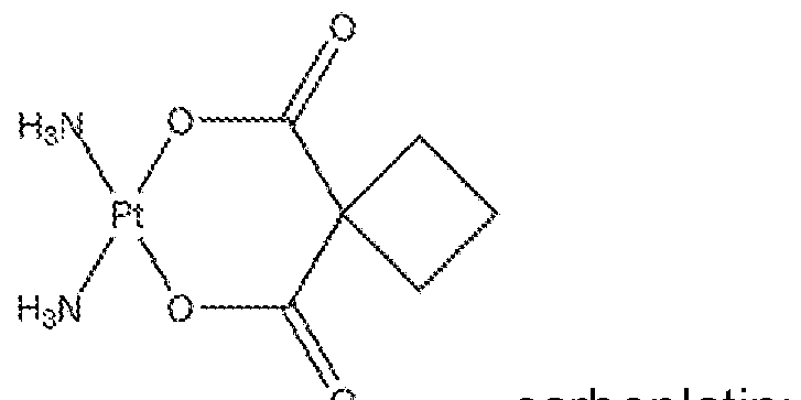
0 car op at n;
mitomycin; and

Read absorbance (MTS assay) and calculate cells survive rate. Compare survival rate
Materials:
1 . Carboplatin 450 mg/45 ml; Hospira, cat # NDC 61703-339-50, lot #A01 171 1AA, exp. September 2014.
2. Mitomycin 20 mg; Accord, cat.# NDC 16729-108-1 1 , lot #PP01516, exp.07/2015
3. Gemcitabine 200 mg; SUN pharmaceutical industries LTD, cat.# NDC 47335-153- 40, lot#JKL4371A, exp. 08/2015.
4. Sterile Talc Powder,
Bryan Corporation, cat.# NDC 63256-200-05; lot #:3M021 ; exp. Dec 2016
5. DPBS, 1x
ATCC, Cat. #: 30-2200, Lot #: 61443818.
6. NCI-28H,
ATCC, cat. #:CRL-5820, lot #:7379248
7. RPMI-1640 media; ATCC cat.#30-2001 , lot# 62027197.
8. Trypsin-EDTA; ATCC cat.#30-2101 , lot# 61618818.
9. Fetal Bovine serum, ATCC cat.# 3022
10. CellTiter 96 AQueous One Solution cell proliferation assay; Promega cat# G3581 .
1 1 . 0.9% Sodium Chloride 50 ml; BAXTER cat.# 2B1306, NDC 03380049-41 , lot# P300574, exp. April 2014.
DAY 1
Cells preparation:
1 . Set up 3 cytotoxicity plates for tomorrow experiment: trypsinize NCI-28H cells (T-75 flask, passage 12):
-Remove old medium, wash cells with 7 ml DPBS, remove DPBS, add 2 ml trypsin, incubate plates for 1 -2', when cells detached add 6 ml fresh medium, mix cells and medium.
-Count cells under the microscope using the glass slide. Average # of cells in slide is 31 ; average in 1 ml of mix is 31x 10,000=310,000 cells/ml;
-Count how much cell/medium stock needed: will use 3 plates (60 wells) in the assay;
count extra wells for safety reason. If we need 200 wells, in each well will be 5,000 cells in 0.1 ml; so total we need 1 ,000,000 cells in 20 ml.
1 ,000,000 cells/310,000=3.2 ml of cells/media mix need to take from flask and transfer to 16.8 ml media. In 50 ml Falcon tube, combine 16.8 ml fresh medium and 3.2 ml cells. Gently mix.
2. Transfer 100 μΙ_ of prepared cells/medium mix to proper wells, keep overnight at 37 °C, 5% C02.
Talc preparation:
1 . Under the hood transfer sterile talc approximately 25 mg of talc to each of 3 sterile Eppendorf tubes, and approximately 100 mg of sterile talc to one tube. Total tubes are 4.
Close tubes and weigh how much exactly talc added to each tube. Result: tube #1 = 89.1 mg; tube #2 (CARBO) = 32.0 mg, tube #3 (MITOMYCIN)=32.5 mg, tube #4 (GEM)=28.8 mg.
2. Talc/Carboplatin preparation : to make 500 μΙ_ of 500 μΜ carboplatin solution use stock 26.9 mM; dilute stock 1 : 10 = 90 μΙ_ DPBS + 10 μΙ_ stock; combine 407.0 μΙ_ DPBS + 93.0 μί of 1 : 10 dilution of carboplatin stock. Final solution is 500 μΙ_ of 500 μΜ carboplatin. Mix talc in tube #2 with this solution.
3. Talc/mitomycin preparation: reconstitute 20 mg powder of drug with 20 ml sterile water, molarity of stock solution will be 2.99 mM to make 500 μΙ_ of 200 μΜ Mitomycin solution use stock 2.99 mM; combine 466.6 μΙ_ DPBS + 33.4 μΙ_ stock. Final solution is 500 μΙ_ of 200 μΜ Mitomycin. Mix talc in tube #3 with this solution.
4. Talc/Gemcitabine preparation : to make 500 μΙ_ of 200 μΜ Gemcitabine, reconstitute drug with 5 ml of 0.9% Sodium Chloride; stock will be 133.48 mM; dilute stock 1 : 10=90 μΙ_ DPBS + 10 μΙ_ stock; combine 462.6 μΙ_ DPBS + 37.4 μΙ_ of 1 : 10 dilution of stock. Final solution is 500 μΙ_ of 200 μΜ Gemcitabine. Mix talc in tube #4 with this solution. 5. Protect tubes from light, tape them on rotator and incubate o/n at RT.
DAY 2
Preparation of Talc
1 . Centrifuge tubes at 3200 rpm for 3 min. Take out supernatant. Wash pellet 3 times
with 1 .0 ml of DPBS (sterile) after last wash add to tube #1 : contains 89.1 mg talc, 178.2 μΙ_ of media; final concentration talc in tube will be 0.5 mg/pL. Add to tube #2 contains 32.0 mg talc, 64.0 μΙ_ of media; to tube #3 contains 32.5 mg talc add 65.0 μΙ_ media and for tube #4 contains 28.8 mg talc add 57.6 μΙ_ media; final concentration talc in all tubes will be 0.5 mg^L.
2. Keep tubes with talc at RT.
3. Prepare first working solution of talc from tube #1 : 1 .35 ml media + 150 μΙ_ of 0.5 mg/μί talc. Total concentration will be 5 mg/100 μΙ_. Make dilutions 1 :2 (750 μΙ_ media + 750 μΙ_ previous dilution) to make following concentration talc in well 2.5 mg talc/100 μΙ_ media; 1 .25 mg/100 μΙ_; 0.6 mg/100 μΙ_.
4. Prepare first dilution of each of the above tubes #2, #3, #4 by adding 540 μΙ_ media + 60 μΙ_ of prepared above 0.5 mg talc and drug/μ.-.
5. After preparation of the above solution, prepare 3 subsequent 1 :2 serial dilutions of each of the above preparations (300 μΙ_ media + 300 μΙ_ of previous dilution). 6. Add 100 μΙ_ of the above preparations in steps #3, #4, #5 to the proper wells as indicated in a 96-well plate layout. The resultant preparation added to each well will give presence of talc in the wells as following: 0.6 mg talc/well, 1 .25 mg talc/well, 2.5 mg talc/well, and 5.0 mg talc/well after sequential dilutions (1 :2) across plate.
Preparation of carboplatin (stock 26.9 mM) : Prepare the following dilutions (1 :5) of drug:
(1 ) 500 μΜ
(2) 100 μΜ
(3) 20 μΜ
(4) 4 μΜ
(5) 0.8 μΜ
(6) 0.16 μΜ
(7) 0.032 μΜ
(8) 0.0064 μΜ
Prepare 600 μΙ_ of 1 mM carboplatin solution( double concentration to keep 500 μΜ drug in total volume 200 μΙ_ media in well) as follows:
1 . 577.7 μΙ_ media + 22.3 μΙ_ of carboplatin stock. 2. Following preparation of above solution prepare the above 7 sequential serial dilutions using the following formula: 480 μΙ_ media + 120 μΙ_ of prior dilution.
3. Add 100 μΙ_ of each 8 preparations of diluted carboplatin (step 1 and 2) to the proper wells according to a plate layout.
Preparation of mitomycin (stock 2.99 mM): Prepare the following dilutions (1 :5) of drug:
(1 ) 200 μΜ
(2) 40 μΜ
(3) 8 μΜ
(4) 1 .6 μΜ (5) 0.32 μΜ
(6) 0.064 μΜ
(7) 0.013 μΜ
(8) 0.0026 μΜ
1 . Prepare 600 μΙ_ of 400 μΜ Mitomycin solution (double concentration to keep 100 μΜ drug in total volume 200 μΙ_ media in well) as follows: 520.0 μΙ_ media + 80.2 μΙ_ of stock.
2. Following preparation of above solution prepare the above 7 sequential serial dilutions using the following formula: 480 μΙ_ media + 120 μΙ_ of prior dilution.
3. Add 100 μΙ_ of each 8 preparations of diluted mitomycin to the proper wells according to a plate layout. Preparation of gemcitabine (stock 133.48 mM):
1 . Prepare the following dilutions (1 :5) of drug:
(1 ) 200 μΜ
(2) 40 μΜ
(3) 8 μΜ
(4) 1 .6 μΜ (5) 0.32 μΜ
(6) 0.064 μΜ
(7) 0.013 μΜ
(8) 0.0026 μΜ
2. Prepare 600 μΙ_ of 400 μΜ drug solution (double concentration to keep 200 μΜ drug in total volume 200 μΙ_ media in well) as follows:
582.0 μΙ_ media + 18.0 μΙ_ of 1 : 10 dilution of stock.
3. Following preparation of above solution prepare the above 7 sequential serial dilutions using the following formula: 480 μΙ_ media + 120 μΙ_ of prior dilution.
4. Add 100 μΙ_ of each 8 preparations of diluted Gemcitabine (step 1 , 2, 3) to the proper wells according to a plate layout.
5. Add 100 μΙ_ of media for untreated cells that will use as a control and not contain any drug or any kind of talc.
6. Check the plate and start incubation at 37 °C/5% C02. DAY 3 1 . Mix talc in wells by pipetting up and down. Continue incubation plate at 37 °C/5%
C02.
DAY 4
1 . Mix talc in wells by pipetting up and down. Continue incubation plate at 37 °C/5%
C02.
DAY 5
1 . Check plate under microscope, no visible sign of contamination is present.
2. Mix talc/media liquid in the wells. Using needle/vacuum system remove all liquid from all wells.
3. Wash all cells 1x 300 μΙ_ DPBS, remove final wash.
4. Add 120 μΙ_ fresh media to all wells.
5. Add 20 μΙ_ of CellTiter 96 Aqueous One solution to each well.
6. Incubate plate 1 hr at 37 °C, 5% CO2.
7. Read absorbance in plate reader at 490 nm.
TABLE 71 . Average reading: cells + drug, carboplatin, μΜ.
0 0.006 0.032 0.16 0J 4 20 100 500
1.65593 1.504233 1.514967 1.5334 1.4238 1.605067 1 .7355 1 .388067 0.618933
TABLE 72. % survival from untreated cells: cells + drug, carboplatin, μΜ.
0 0.006 0.032 0.16 0.8 4 20 100 500
100 90.84 91 .49 92.60 85.98 96.93 104.81 83.82 37.38
100 94.28 92.95 97.48 87.53 93.62 99.20 79.53 35.14
TABLE 73. Average absorbance reading talc + cells and cells + talc incubated with
500 μΜ.
0 mg 0.6 mg 1 .25 mg 2.5 mg 5 mg
cells+talc 1.65593 0.771 0.78895 0.74915 0.9812 cells+talc/500 MMCarbo 1.65593 0.9763 0.7927 0.7154 1.0903
TABLE 74. % survival cells after talc and talc bound to carboplatin added.
0 mg 0.6 mg 1.25 mg 2.5 mg 5 mg
Cells+talc 10C) 46.56 47.64 45.24 59.25
Cells+talc/500 MCarbo 100 58.95 47.87 43.20 65.84
TABLE 73. Average reading: cells + drug, gemcitabine, μΜ.
0 0.0026 0.013 0.064 0.32 1j$ 8 40 200
1 .77441 2.033633 1 .913633 1.430167 1 .388733 1.1977 1 .00395 1.0663 0.9352
TABLE 74. % survival from untreated cells: cells + drug, gemcitabine, μΜ.
Gemcitabinem^M
0 0.0026 0.013 0.064 0.32 V6 8 40 200
100 1 14.61 107.85 80.60 78.26 67.50 56.58 60.09 52.70
TABLE 75. Average absorbance readings: cells + drug, mitomycin, μΜ.
0 0.0026 0.013 0.064 0 2 1J 8 40 200
1 .91438 2.031533 1.965033 1 .8766 1.827167 1.036467 0.3451 0.520033 0.246367
TABLE 76. % survival cells after exposure to drug for 72 hrs (cells + drug only), mitomycin, μΜ.
0 0.0026 0.013 0.064 0.32 1J3 8 40 200
100 106.12 102.65 98.03 95.44 54.14 18.03 27.16 12.87
TABLE 77. Average absorbance readings: survival cells after talc incubated to mitomycin added.
0 mg 0.6 mg 1 .25 mg 2.5 mg 5 mg
Cells+talc 1 .91438 1.7976 2.1208 1.99815 2.1 156
Cells+talc/Mitomycin 1 .91438 0.6719 0.6604 0.7877 1 .22415
TABLE 78. % survival cells after talc and talc/mitomycin treatment.
0 mg 0.6 mg 1 .25 mg 2.5 mg 5 mg
Cells+talc 10() 93.90 1 10.78 104.38 1 10.51
Cells+talc/Mitomycin 100 35.10 34.50 41 .15 63.94
TABLE 79. Average absorbance reading: cells + drug, gemcitabine, μΜ.
Gemcitabine, uM
0 0.0026 0.013 0.064 0.32 1 .6 8 40 200
1 .77441 2.033633 1 .913633 1.430167 1 .388733 1.1977 1 .00395 1.0663 0.9352
TABLE 80. % survival cells: cells + drug only.
Gemcitabine, uM
0 0.0026 0.013 0.064 0.32 1J3 8 40 200
100 114.61 107.85 80.60 78.26 67.50 56.58 60.09 52.70
TABLE 81 . Average absorbance reading: cells + talc.
0 mg 0.6 mg 1.25 mg 2.5 mg 5 mg
Cells+talc only 1.77441 1 .10175 1 .0486 1 .12035 1 .20865
Cells+talc/Gemcitabine 1.77441 0.9357 0.7983 0.70395 0.84505
TABLE 82. % survival cells: cells + talc/drug; % from untreated cells.
0 mg 0.6 mg 1.25 mg 2.5 mg 5 mg
Cells+talc only I OC) 62.09 59.10 63.14 68.12
Cells+talc/Gemcitabine 100 52.73 44.99 39.67 47.62
The data showed that talc bound to mitomycin has a greater cytotoxic effect than mitomycin or talc alone (see e.g., FIG. 27, FIG. 28).
The study showed clear cytotoxic effect on NCI-28H cells with talc, mitomycin, and gemcitabine when used as single-agents. However, when talc is bound to mitomycin or gemcitabine, toxicity is greatly enhanced over any of these agents when used alone. In contrast, there was no noticeable difference in toxicity of carboplatin-talc or talc alone on NCI- 28H cells. Based on these findings, it is presently thought that carboplatin may not bind to talc or talc does not absorb carboplatin.
EXAMPLE 23: CYTOTOXICITY ASSAY: NCI-2052H CELLS TREATED WITH DIFFERENT
COMPOUNDS-BLEOMYCIN, MITOMYCIN, DOXORUBICIN, PACUTAXEL, TALC ALONE AND TALC BOUND TO EACH OF THESE COMPOUNDS
The following Example determined if cytotoxicity would also occur when another cell line was exposed to similar conditions, based on above experimental results with NCI-28H. Therefore, a similar experiment was designed utilizing NCI-2052H to determine if different compounds (talc, chemotherapy drugs, and talc conjugated to chemotherapy agents) would also be cytotoxic to NCI-2052H cells. Plan: add to NCI-2052H cells different type of compounds: only drugs, only talc and talc that previously incubated with Bleomycin, Mitomycin, Doxorubicin, or Paclitaxel.
mycin;
mitomycin;

•HCI doxorubicin; and

paclitaxel.
Read absorbance (MTS assay) and calculate cells survival rate. Compare survival rate Materials:
1 . Bleomycin sulfate from Streptomyces verticillus; Sigma, cat # 15361 -10 mg, lot#
BCBL0535V.
2. Mitomycin 20 mg; Accord, cat.# NDC 16729-108-1 1 , lot #PP01516, exp.07/2015.
3. Doxorubicin Hydrochloride, 50 mg/25 ml; Amneal- Agila LLC, cat.#NDC 53150-315- 01 ; lot # 7800982; exp.03/2015.
4. Paclitaxel, 300 mg/ml; Sagent, cat.# NDC 25021 -213-50; lot# 38J01 1 1 ; exp.
04/2015.
5. Sterile Talc Powder, Bryan Corporation, cat.# NDC 63256-200-05; lot#:3M021 ; exp. Dec 2016
6. DPBS 1x; ATCC, Cat. #: 30-2200, Lot #: 61443818.
7. NCI-2052H cell line, ATCC, cat. #:CRL-5915, lot #:57608140.
8. RPMI-1640 media; ATCC cat.#30-2001 , lot# 62027197.
9. Trypsin-EDTA; ATCC cat.#30-2101 , lot# 61618818.
10. Fetal Bovine serum, ATCC cat.# 3022
1 1 . CellTiter 96 AQueous One Solution cell proliferation assay; Promega cat# G3581 . DAY 1
Cells preparation:
1 Set up 4 cytotoxicity plates for tomorrow experiment: trypsinize NCI-28H cells (T-75 flask, passage 6):
-Remove old medium, wash cells with 7 ml DPBS, remove DPBS, add 2 ml trypsin, incubate plates for 1 -2 min., when cells detached add 6 ml fresh medium, mix cells and medium. -Count cells under the microscope using the glass slide. Average # of cells in slide is 44; average in 1 ml of mix is 44x 10,000=440,000 cells/ml;
-Count how much cell/medium stock needed: will use 4 plates (60 wells) in the assay; count extra wells for safety reason. If we need 300 wells, in each well will be 5,000 cells in 0.1 ml media; so total we need 1 ,500,000 cells in 30 ml media. 1 ,500,000 cells/440,000=3.4 ml of cells/media mix need to take from flask and transfer to 26.6 ml media. In 50 ml Falcon tube, combine 26.6 ml fresh medium and 3.4 ml cells. Gently mix.
2 Transfer 100 μΙ_ of prepared cells/medium mix to proper wells, keep overnight at 37 °C, 5% C02.
Talc preparation;
1 . Under the hood transfer sterile talc approximately 25 mg of talc to each of 4 sterile Eppendorf tubes, and approximately 150 mg of sterile talc to one tube. Total tubes are 5.
Close tubes and weigh exactly how much talc added to each tube. Result: tube #1
(Bleomycin)=52.0 mg; tube #2(Mitomycin) = 35.2 mg, tube #3(Dox)=42.4 mg, tube #4
(Paclitaxel)=44.0 mg. Tube #5(no drugs)=133.6 mg.
2. Talc/Bleomycin preparation: need to make 400 μΙ_ of 1 mg/ml solution. Reconstitute powder of drug in 100 μΙ_ of sterile water; stock of drug will be 100 mg/ml. To make 400 μΙ_ of
1 mg/ml Bleomycin, combine 396.0 μΙ_ DPBS + 4.0 μΙ_ of Bleomycin stock. Final solution is 400 μΙ_ of 1 mg/ml of Bleomycin. Mix talc in tube #1 with this solution.
3. Talc/Mitomycin preparation: use stock solution 2.99 mM to make 500 μΙ_ of 200 μΜ Mitomycin solution use stock 2.99 mM; combine 466.6 μΙ_ DPBS + 33.4 μΙ_ stock. Final solution is 500 μΙ_ of 200 μΜ Mitomycin. Mix talc in tube #2 with this solution.
4. Talc/Doxorubicin preparation : to make 500 μΙ_ of 1 μΜ Doxorubicin use stock 3.45 mM; dilute stock 1 : 100=495.0 μΙ_ DPBS + 5 μΙ_ stock; combine 485.5 μΙ_ DPBS + 14.5 μΙ_ of 1 : 100 dilution of stock. Mix talc in tube #3 with this solution.
5. Talc/PACLITAXEL preparation : to make 500 μΙ_ of 1 μΜ paclitaxel solution use stock 7.03 mM; dilute stock 1 : 100=495 μΙ_ DPBS + 5 μΙ_ stock; combine 482 μΙ_ DPBS + 18.0 μί of 1 : 100 dilution of paclitaxel stock. Final solution is 500 μΙ_ of 1 μΜ paclitaxel. Mix talc in tube #4 with this solution.
6. Add 1 ml DPBS in tube # 5, mix talc with DPBS.
7. Protect tubes from light, tape them on rotator and incubate o/n at RT. DAY 2
Preparation of Talc
1 . Centrifuge tubes at 3200 rpm for 3 min. Take out supernatant. Wash pellet in tubes #1 , #2, #3, #4 3 times with 1 .0 ml of DPBS (sterile) after last wash add to tube #1 : contains
52.0 mg talc, 104.0 μΙ_ of media; final concentration talc in tube will be 0.5 mg/pL. Add to tube #2 contains 35.2 mg talc, 70.4 μΙ_ of media; to tube #3 contains 42.4 mg talc add 84.8 μΙ_ media ; for tube #4 contains 44.0 mg talc add 88.8 μΙ_ media; for tube #5 contains 133.6 mg talc add 267.2 μΙ_ media; final concentration talc in all tubes will be 0.5 mg^L.
2. Keep tubes with talc at RT.
3. Prepare first working solution of talc from tube #5: 1 .8 ml media + 200 μΙ_ of 0.5 mg/μί talc. Total concentration will be 5 mg/100 μΙ_. Make dilutions 1 :2 (900 μΙ_ media + 900 μΙ_ previous dilution) to make following concentration talc in well 2.5 mg talc/100 μΙ_ media; 1 .25 mg/100 μΙ_; 0.6 mg/100 μΙ_.
4. Prepare first dilution of each of the above tubes #1 , #2, #3, #4 by adding 540 μΙ_ media + 60 μΙ_ of prepared above 0.5 mg (talc and drug)^L media
5. After preparation of the above solution, prepare 3 subsequent 1 :2 serial dilutions of each of the above preparations (300 μΙ_ media + 300 μΙ_ of previous dilution).
6. Add 100 μΙ_ of the above preparations in steps #3, #4, #5 to the proper wells as indicated in a 96-well plate layout. The resultant preparation added to each well will give presence of talc in the wells as following: 0.6 mg talc/well, 1 .25 mg talc/well, 2.5 mg talc/well, and 5.0 mg talc/well after sequential dilutions (1 :2) across plate.
Preparation of Bleomycin:
Prepare the following dilutions of Bleomycin
(1 ) 1 mg/ml (625 μΜ),
(2) 250 μg/ml (156.3 μΜ),
(3) 62.5 μg/ml (39 μΜ),
(4) 15.6 μg/ml (9.75 μΜ),
(5) 3.9 μg/ml (2.43 μΜ),
(6) 0.97 μg/ml (0.6 μΜ),
(7) 0.24 μg/ml (0.15 μΜ),
(8) 0.06 μg/ml (0.038 μΜ).
Prepare 500 μΙ_ stock solution of 625 μΜ Bleomycin as follows:
1 . 450 μΙ_ media + 50 μΙ_ of stock (6.25 mM bleomycin).
2. Following preparation of above solution prepare the above 7 sequential serial dilutions using the following formula: 375 μΙ_ media + 125 μΙ_ of prior dilution.
3. Add 100 μΙ_ of each 8 preparations of diluted bleomycin (step 1 and 2) to the proper wells according to a plate layout.
4. Add 100 μΙ_ of media for untreated cells that will use as a control and not contain any drug or any kind of talc.
5. Preparation of mitomycin (stock 2.99 mM): Prepare the following dilutions (1 :5) of drug:
(1 ) 200 μΜ
(2) 40 μΜ
(3) 8 μΜ
(4) 1 .6 μΜ
(5) 0.32 μΜ
(6) 0.064 μΜ
(7) 0.013 μΜ
(8) 0.0026 μΜ
6. Prepare 600 μΙ_ of 400 μΜ Mitomycin solution( double concentration to keep 200 μΜ drug in total volume 200 μΙ_ media in well) as follows: 520.0 μΙ_ media + 80.2 μΙ_ of stock .
7. Following preparation of above solution prepare the above 7 sequential serial dilutions using the following formula: 480 μΙ_ media + 120 μΙ_ of prior dilution.
8. Add 100 μΙ_ of each 8 preparations of diluted MITOMYCIN to the proper wells according to a plate layout.
Preparation of DOXORUBICIN (stock3.45 mM) : Prepare the following dilutions (1 :5)
of drug:
(1 ) 10 μΜ
(2) 2 μΜ
(3) 0.4 μΜ
(4) 0.08 μΜ
(5) 0.016 μΜ
(6) 0.0032 μΜ
(7) 0.00064 μΜ
(8) 0.000128 μΜ
Prepare 600 μΙ_ of 20 μΜ doxorubicin solution (double concentration to keep 10 μΜ drug in total volume 200 μΙ_ media in well) as follows:
1 .565.2 μΙ_ media + 34.8 μί of 1 :10 dilution of doxorubicin stock.
2. Following preparation of above solution prepare the above 7 sequential serial dilutions using the following formula: 480 μΙ_ media + 120 μΙ_ of prior dilution.
3. Add 100 μΙ_ of each 8 preparations of diluted doxorubicin (step 1 and 2) to the proper wells according to a plate layout. Preparation of paclitaxel (stock 7.03 mM):
1 . Prepare the following dilutions (1 :5) of drug:
(1 ) 20 μΜ
(2) 4 μΜ
(3) 0.8 μΜ
(4) 0.16 μΜ
(5) 0.032 μΜ
(6) 0.0064 μΜ
(7) 0.00128 μΜ
(8) 0.000256 μΜ
2. Prepare 600 μΙ_ of 40 μΜ paclitaxel solution( double concentration to keep 20 μΜ drug in total volume 200 μΙ_ media in well) as follows:
565.8 μΙ_ media + 34.2 μΙ_ of 1 : 10 dilution of
Paclitaxel stock.
3. Following preparation of above solution prepare the above 7 sequential serial dilutions using the following formula: 480 μΙ_ media + 120 μΙ_ of prior dilution.
4. Add 100 μΙ_ of each 8 preparations of diluted paclitaxel (step 1 , 2, 3) to the proper wells according to a plate layout.
5. Add 100 μΙ_ of media for untreated cells that will use as a control and not contain any drug or any kind of talc.
6. Check the plates and start incubation for 72 hrs at 37 °C/5% C02. DAY 3
1 . Mix talc in wells by pipetting up and down. Continue incubation plate at 37 °C/5%
C02.
DAY 4
1 . Mix talc in wells by pipetting up and down. Continue incubation plate at 37 °C/5%
CO 2- DAY 5
1 . Check plate under microscope, no visible sign of contamination is present. 1 . Mix talc/media liquid in the wells. Using needle/vacuum system, remove all liquid from all wells.
2. Wash all cells 1x 300 μΙ_ DPBS, remove final wash.
3. Add 120 μΙ_ fresh media to all wells.
4. Add 20 μΙ_ of CellTiter 96 Aqueous One solution to each well. 5. Incubate plate 1 hr at 37 °C, 5% CO2.
6. Read absorbance in plate reader at 490 nm.
7.
TABLE 83. Average readings: cells + drug, bleomycin, pg/ml (see e.g., FIG. 31 ). 0 0.24 0.97 3.9 15.6 62.5 250 1000
1.32366 1.3454 1.2884 1 .1733 0.684633 0.507533 0.276567 0.193467
TABLE 84. % survival from untreated cells (see e.g., FIG. 31 ).
Bleomycin, ug/ml
0 0.24 0.97 3.9 15.6 62.5 250 1000
100 101 .64 97.34 88.64 51 .72 38.34 20.89 14.62
TABLE 85. Average readings: talc, talc + drug (see e.g., FIG. 32).
talc, mg/well
0 0.62 1 .25 2.5 5~ talc only 1.32366 0.4757 0.47085 0.5883 0.97375 talc + Bleo 1.32366 0.3168 0.3819 0.46465 1.0287
TABLE 86. % survival from untreated cells (see e.g., FIG. 32).
talc, mg/well
0 0.62 1 .25 2.5 5~ talc only 100 35.94 35.57 44.44 73.56 talc + Bleo 100 23.93 28.85 35.10 77.72 TABLE 87. Average reading: cells + drug, mitomycin, μΜ (see e.g., FIG. 34). 0 .0026 0.013 0.064 0.32 1J3 8 40 200
1.57579 1.42396 1.28363 1.19923 1.2222 0.81376 0.4142 0.2305 0.20983
7 3 3 7 3
TABLE 88. Average reading: cells + talc/mitomycin.
talc, mg/well
0 062 1^25 2J5 5 cells+talc 1.57579 0.359 0.4202 0.5906 1.26305 cells+talc/Mitomycin 1.57579 0.4339 0.51025 0.79055 1.0963
TABLE 89. % survival from untreated cells.
talc, mg/well
0.62
cells+talc 100 22.78 26.67 37.48 80.15 cells+talc/Mitomycin 100 27.54 32.38 50.17 69.57
TABLE 90. Average reading: cells after treated for 72 hrs with doxorubicin, nM (see e.g., FIG. 35). 0 0.128 0.64 3J 16 80 400 2000 10,000
1.37733 1.082267 1.149067 1.084133 1.1 1 13 1.1 153 0.795533 0.352467 0.2605
TABLE 91 . % survival from untreated cells, doxorubicin, nM (see e.g., FIG. 35).
0 0.128 0.64 32 16 80 400 2000 10,000
100 78.58 83.43 78.71 80.69 80.98 57.76 25.59 18.91
TABLE 92. Average readings: cells + talc/doxorubicin (see e.g., FIG. 36).
talc, mg/well
0 0.6 1.25 2.5 5 cells+talc 1 .37733 1.52825 1 .5293 1.1036 1.2317 cells+talc/Dox 1 .37733 0.5573 0.37065 0.3926 0.5758
TABLE 93. % survival from untreated cells (see e.g., FIG. 36).
talc, mg/well
0 0.6 1.25 2.5 5 cells+talc 100 110.96 1 1 1 .03 80.13 89.43 cells+talc/Dox 100 40.46 26.91 28.50 41 .81
TABLE 94. Average reading: cells + drug, paclitaxel, nM (see e.g., FIG. 37).
Paclitaxel, nM
0 0.256 1^28 6Λ 32 160 800 4000 20,000
1.70244 1.4273 1.5873 1.539567 0.581767 0.436067 0.4326 0.552967 0.1647
TABLE 95. % survival from untreated cells (see e.g., FIG. 37).
0 0.256 1 .28 6.4 32 160 800 4000 20,000
100 83.84 93.24 90.43 34.17 25.61 25.41 32.48 9.67
TABLE 96. Average absorbance reading: cells + talc; cells + talc binds to paclitaxel. Talc, mg/well (see e.g., FIG. 38).
0 0.62 1.25 2.5 5
cells+talc 1.70244 0.6172 0.96435 0.29925 0.4724
cells+talc, that binds to Taxol 1.70244 0.20145 0.2091 0.3997 0.54495
TABLE 97. % survival from untreated cells (see e.g., FIG. 38).
0 0.62 1.25 2.5 5
cells+talc 100 36.25 56.65 17.58 27.75
cells+talc, that binds to Taxol 100 11 .83 12.28 23.48 32.01
The study showed enhanced cytotoxicity on NCI-2052H cells when cells are exposed to talc conjugated to the following chemotherapy drugs: bleomycin, doxorubicin, and paclitaxel vs. talc alone. But the talc-mitomycin conjugate demonstrated less cytotoxicity than talc alone. The result was not expected, thus, additional experiments for mitomycin and NCI- 2052 H were performed to test if the results were correct.
EXAMPLE 24: REPEATED CYTOTOXICITY EXPERIMENT: NCI-2052H CELLS TREATED WITH MITOMYCIN, TALC ALONE AND TALC BOUND TO MITOMYCIN
The following Example repeated the experiment in Example 23 with NCI-2052H and mitomycin. Plan: repeat one more time experiment when NCI- 2052H cells exposed for 72 hrs to
Mitomycin, talc and talc bound to Mitomycin. Compare survival rate of cells.
Materials:
1 . Mitomycin 20 mg; Accord, cat.# NDC 16729-108--1 1 , lot #PP01516, exp.07/2015.
2. Sterile Talc Powder, Bryan Corporation, cat.# NDC 63256-200-05; lot #:3M021 ; exp. Dec 2016.
3. DPBS 1x; ATCC, Cat. #: 30-2200, Lot #: 61443818.
4. NCI-2052H cell line, ATCC, cat. #:CRL-5915, lot #:57608140.
5. RPMI-1640 media; ATCC cat.#30-2001 , lot# 62027197.
6. Trypsin-EDTA; ATCC cat.#30-2101 , lot# 61618818.
7. Fetal Bovine serum, ATCC cat.# 3022.
8. CellTiter 96 AQueous One Solution cell proliferation assay; Promega cat# G3581 . DAY 1 1 . Use NCI-2052H cells, passage 9 to fill up 96 well microplate with 5,000 cells per well. Protocol how to do that see in previous cytotoxicity experiment.
2. Incubate plate overnight at 37 °C, 5% CO2.
3. Prepare talc: under the hood transfer sterile talc approximately 25 mg of talc to each of 2 sterile Eppendorf tubes. Close tubes and weigh how much exactly talc added to each tube. Result: tube #1 = 48.1 mg; tube #2=44.5 mg.
4. Add 500 μΙ_ DPBS (sterile) to tube #1 , add 500 μΙ_ DPBS containing 200 μΜ
Mitomycin to tube #2.
5. Talc/mitomycin preparation: use Mitomycin stock 2.99 mM. To make 500 μΙ_ of 200 uM Mitomycin combine 466.6 μΙ_ DPBS + 33.4 μΙ_ stock. Mix talc in tube #2 with this solution. 6. Incubate both tubes overnight at 4 °C, on rotator.
DAY 2
Preparation of Talc.
1 . Centrifuge tubes at 3200 rpm for 3 min. Take out supernatant. Wash pellet in tubes # 1 and #2 3 times with 1 .0 ml of DPBS (sterile) after last wash add to tube #1 : contains 48.1 mg talc, 96.2 μΙ_ of media; final concentration talc in tube will be 0.5 mg^L. Add to tube #2 contains 44.5 mg talc 89.0 μΙ_ of media; final concentration talc in the tube will be 0.5 mg^L.
2. Keep tubes with talc at RT.
3. Prepare first dilution of talc from tube #1 : 540 μΙ_ media + 60 μΙ_ of 0.5 mg/μί talc. Total concentration will be 5 mg/100 μΙ_. Make dilutions 1 :2 (300 μΙ_ media + 300 μΙ_ previous dilution) to make following concentration talc in well 2.5 mg talc/100 μΙ_ media; 1 .25 mg/100 μΙ_; 0.6 mg/100 μΙ_.
4. Prepare dilutions from tube #2 by adding 540 μΙ_ media + 60 μΙ_ of prepared above
0.5 mg (talc bound to drug)/pl_ media.
5. After preparation of the above solution, prepare 3 subsequent 1 :2 serial dilutions (300 μΙ_ media + 300 μΙ_ of previous dilution).
6. Add 100 μΙ_ of the above preparations to the proper wells as indicated in a 96-well plate layout. The resultant preparation added to each well will give presence of talc in the wells as following: 0.6 mg talc/well, 1 .25 mg talc/well, 2.5 mg talc/well, and 5.0 mg talc/well after sequential dilutions (1 :2) across plate.
Preparation of mitomycin (stock 2.99 mM): Prepare the following dilutions (1 :5) of drug: (1 ) 200 μΜ
(2) 40 μΜ
(3) 8 μΜ
(4) 1 .6 μΜ
(5) 0.32 μΜ (6) 0.064 μΜ
(7) 0.013 μΜ
(8) 0.0026 μΜ
1 . Prepare 600 μΙ_ of 400 μΜ Mitomycin solution (double concentration to keep 200 μΜ drug in total volume 200 μΙ_ media in well) as follows: 519.8 μΙ_ media + 80.2 μΙ_ of stock. 2. Following preparation of above solution prepare the above 7 sequential serial dilutions using the following formula: 480 μΙ_ media + 120 μΙ_ of prior dilution.
3. Add 100 μΙ_ of each 8 preparations of diluted mitomycin to the proper wells according to a plate layout.
INCUBATION 1 . Incubate plate for 72 hrs at 37 °C, 5% CO2.
DAY 3
1 . Check plate under microscope. Continue incubation plate at 37 °C/5% CO2. DAY 4
1 . Check plate under microscope. Continue incubation plate at 37 °C/5% CO2. DAY 5
1 . Check plate under microscope, no visible sign of contamination is present.
2. Mix talc/media liquid in the wells. Using needle/vacuum system, remove all liquid from all wells.
3. Wash all wells 1x 300 μΙ_ DPBS, remove final wash.
4. Add 120 μΙ_ fresh media to all wells.
5. Add 20 μΙ_ of CellTiter 96 Aqueous One solution to each well.
6. Incubate plate 1 hr at 37 °C, 5% CO2.
7. Read absorbance in plate reader at 490 nm.
8. Results and Data:
TABLE 98. Average absorbance reading: cells + talc + talc/drug (see e.g., FIG. 40).
Talc, mg
0 0.6 mg 1 .25 mg 2.5 mg 5 mg
cells+talc 1 .095 1.615 072 0.6485 1.0035 cells+talc/Mitomycin 1 .095 1.076 0.659 0.316 1 .28
TABLE 99. % survival from untreated cells (see e.g., FIG. 40).
Talc, mg
0 0.6 mg 1 .25 mg 2.5 mg 5 mg
cells+talc 10C) 147.49 65.75 59.22 91 .64 cells+talc/Mitomycin 100 98.26 60.18 28.86 116.89
TABLE 100. Average reading: drug + cells (see e.g., FIG. 41 ).
Mitomycin, uM
0 .0026 0.013 0.064 0.32 V6 8 40 200
1.095 0.86366 0.79066 0.82666 0.82233 0.677 0.42166 0.22566 0.22433
7 7 7 3 7 7 3
TABLE 101 . % survival from untreated cells (see e.g. , FIG. 41 ).
0 0.0026 0.013 0.064 0.32 1J3 8 40 200
100 78.87 72.21 75.49 75.10 61 .83 38.51 20.61 20.49
The study showed clear cytotoxic effect on NCI-2052H cells with talc alone. Further, when talc is bound to mitomycin, toxicity was enhanced.
EXAMPLE 25: INHIBITION OF NON-SPECIFIC BINDING OF BIOTIN-RHODAMINE TO TALC BY WASHING WITH LOW AND HIGH PH BUFFERS
The following Example determined if various buffers of varying pH (4.8-8.0) effect the binding of Biotin-Rhodamine to talc particles.
Plan: incubate talc with Biotin Rhodamine. Make washings using different pH buffers: PBS, TBS (pH 8.0), Citrate buffer (pH 4.8). Run fluorescent assay.
Materials:
1 . Sterile Talc Powder (Bryan Corporation, Cat. #: 1690, Lot #: 3M021 , Exp. Date: Dec. 2016)
2.1 Ox PBS (Sigma, Cat. #: P5493-1 L, Lot #: SLBB9685)
3. Water (Sigma Life Science, Cat. #: 3500, Lot #: RNBD1 156)
4. Water deionized; Sigma-Aldrich, cat.# 38796- 1 L; lot# BCBM0010V
5. Biotin rhodamine 1 10; Biotium, cat.#80022.
6. Tris Buffered Saline pH 8.0, powder; Sigma cat.#T6664-10 pak; lot#SLBK8366V.
7. Citrate Buffer solution, 0.09 M; Sigma, cat.# C2488-500 ml, lot# SLBD8857V DAY 1
1 . Add 25 mg talc to the each of 4 eppendorf tubes (round bottom). Label tubes as #1 , #2, #3, #4.
2. Add to each tube 500 μί 1 x PBS.
3. Add 5 \JL of Biotin Rhodamine (concentration: 16 pg/pL) to the tubes #1 , #2, #3, but not to tube #4.
4. Mix well, incubate all tubes for 1 hr at 4 °C, rotator. Protect from light.
5. Wash talc in tube#1 three times with 500 μΙ_ TBS; in tube #2 three times with 500 μΙ_ Citrate buffer and in tube #3 three times with PBS. (Centrifuge speed -3200 rpm for 3 min)
6. Centrifuge talc in tube #4 and remove supernatant.
7. Add to all tubes 200 μΙ_ PBS, mix well.
8. Transfer 50 μΙ_ talc mix from each tube to fluorescent assay 96 well plate. Then add to each well 100 μΙ_ PBS to keep talc in equal distribution around well.
9. Run fluorescent assay using settings excitation/emission as 496 nm/520 nm. TABLE 102. Average fluorescent signal (Rhodamine 1 10).
Average fluorescent signal (RHODAMINE 1 10)
talc/Biotin washed with TBS, pH 8.0 12868.68
talc/Biotin washed with Citrate buffer, pH 4.8 14027.61
talc/Biotin washed with PBS 13760.41
Talc in PBS, no Biotin 715.75
The evidence strongly suggests the presence of Biotin-Rhodamine on talc. Changing pH of washing buffers did not change amount of Biotin Rhodamine that nonspecifically bound with talc. Thus, the study showed strong evidence that pH does not affect the binding of Biotin-Rhodamine to talc particles.
Claims
Claim 1 . A composition for treatment of a proliferative disease, disorder, or condition comprising:
a therapeutic agent; and
a substrate;
wherein,
the therapeutic agent is contained in or on the substrate; and
the substrate comprises silica, silicate, or talc.
Claim 2. The composition of claim 1 , wherein the therapeutic agent comprises a chemotherapeutic agent.
Claim 3. The composition of any one of claims 1 -2, wherein the therapeutic agent comprises an antitumor antibiotic, anthracycline, aziridine-containing composition, nucleoside analog, taxane, or diterpene.
Claim 4. The composition of claim 3, wherein the therapeutic agent further comprises a platin.
Claim 5. The composition of any one of claims 1 -4, wherein,
the composition consists essentially of a therapeutic agent and a substrate; and the therapeutic agent or the substrate has specific or non-specific affinity for a target tissue associated with the disease, disorder, or condition.
Claim 6. The composition of any one of claims 1 -5, wherein the substrate comprises talc.
Claim 7. The composition of any one of claims 1 -6, wherein the therapeutic agent comprises bleomycin, doxorubicin, gemcitabine, mitomycin, or paclitaxel.
Claim 8. A method of treating a disease, disorder, or condition in a subject comprising administering to a subject in need thereof a composition according to any of claims 1 -7.
Claim 9. The method of claim 8, wherein the disease, disorder, or condition comprises a proliferative disease, disorder, or condition.
Claim 10. The method of any one of claims 8-9, wherein the disease, disorder, or condition comprises one or more selected from the group consisting of: a cancer, malignant pleural mesothelioma, peritoneal carcinomatosis, leukemia, lymphoma, non-small cell lung cancer, testicular cancer, lung cancer, abdominal cancer, ovarian cancer, uterine cancer, cervical cancer, pancreatic cancer, colorectal cancer, breast cancer, prostate cancer, gastric cancer, colon cancer, skin cancer, stomach cancer, liver cancer, liver metastasis, esophageal cancer, bladder cancer, appendiceal carcinoma, gastric carcinoma, pancreatic carcinoma, peritoneal mesothelioma, pseudomyxoma peritonei, blood vessel proliferative disorder, fibrotic disorder, mesangial cell proliferative disorder, psoriasis, actinic keratoses, seborrheic keratoses, warts, keloid scars, eczema, viral-associated hyperproliferative disease, papilloma viral infection, mesothelioma, Meigs Syndrome, sarcoma, appendiceal carcinoma,
pseudomyxoma peritonei, prostate cancer, prostate cancer lymph node dissection beds, rectovesical pouch tumor bed, ovarian cancer resection bed and peritoneal spread, uterine cancer resection cavities, pleural and peritoneal mesothelioma resection bed and peritoneal seeding, colorectal carcinoma, appendiceal carcinoma, pancreatic carcinoma, liver
metastases, gastric carcinoma, renal carcinoma, retroperitoneal tumors, retroperitoneal sarcoma, retroperitoneal carcinoma, breast cancer, breast cancer lumpectomy, breast cancer lumpectomy dissection cavity, breast cancer lymph node, breast cancer lymph node dissection cavity, melanoma, melanoma node dissection cavity, sarcoma, sarcoma resection cavities, head or neck cancer, head or neck cancer resection cavity, neck cancer lymph node, neck lymph node dissection cavities, scalp lesion, glioblastoma, glioblastoma resection cavity, brain surface tumor lesion, resected brain surface tumor lesion, non resected brain surface tumor lesion, trunk sarcoma, trunk sarcoma resection cavity, extremity sarcoma, and extremity sarcoma resection cavity, or a combination thereof.
Claim 1 1. The method of any one of claims 8-10, wherein the proliferative disease, disorder, or condition comprises a cancer.
Claim 12. The method of any one of claims 8-1 1 , wherein the composition is administered to the pleural space of the subject.
Claim 13. The method of any one of claims 8-12, wherein the composition is administered to the subject post-operatively in or near a surgically operated area.
Claim 14. The method of any one of claims 8-13, wherein the composition is administered to the subject post-operatively in a cavity where proliferative cells or tissue were surgically removed.
Claim 15. The method of any one of claims 8-14, wherein the composition is administered to the subject in an amount effective to: inhibit replication of cancer cells, inhibit spread of the disease, disorder, or condition, reduce tumor size, decrease tumor
vascularization, increase tumor permeability, reduce recurrence of tumor growth, prevent recurrence of tumor growth, reduce a number of cancerous cells in the subject, or ameliorate a symptom of the disease, disorder, or condition.
Priority Applications (2)
| Application Number | Priority Date | Filing Date | Title |
|---|---|---|---|
| US15/740,938 US10925852B2 (en) | 2015-06-30 | 2016-06-27 | Talc-bound compositions and uses thereof |
| EP16818530.4A EP3316871A4 (en) | 2015-06-30 | 2016-06-27 | Talc-bound compositions and uses thereof |
Applications Claiming Priority (2)
| Application Number | Priority Date | Filing Date | Title |
|---|---|---|---|
| US201562186870P | 2015-06-30 | 2015-06-30 | |
| US62/186,870 | 2015-06-30 |
Publications (1)
| Publication Number | Publication Date |
|---|---|
| WO2017003908A1 true WO2017003908A1 (en) | 2017-01-05 |
Family
ID=57608878
Family Applications (1)
| Application Number | Title | Priority Date | Filing Date |
|---|---|---|---|
| PCT/US2016/039504 WO2017003908A1 (en) | 2015-06-30 | 2016-06-27 | Talc-bound compositions and uses thereof |
Country Status (3)
| Country | Link |
|---|---|
| US (1) | US10925852B2 (en) |
| EP (1) | EP3316871A4 (en) |
| WO (1) | WO2017003908A1 (en) |
Cited By (4)
| Publication number | Priority date | Publication date | Assignee | Title |
|---|---|---|---|---|
| US10287353B2 (en) | 2016-05-11 | 2019-05-14 | Huya Bioscience International, Llc | Combination therapies of HDAC inhibitors and PD-1 inhibitors |
| US10385131B2 (en) | 2016-05-11 | 2019-08-20 | Huya Bioscience International, Llc | Combination therapies of HDAC inhibitors and PD-L1 inhibitors |
| WO2020030591A1 (en) * | 2018-08-05 | 2020-02-13 | Da Volterra | Method for improving anticancer agent efficacy |
| EP3490615A4 (en) * | 2016-07-27 | 2020-04-01 | The Trustees of Columbia University in the City of New York | Gel-bound compositions for radiotherapy and uses thereof |
Families Citing this family (1)
| Publication number | Priority date | Publication date | Assignee | Title |
|---|---|---|---|---|
| WO2019185685A1 (en) * | 2018-03-27 | 2019-10-03 | Sintef Tto As | Poly (alkyl cyanoacrylate) nanoparticles for use in treatment of cancer |
Citations (4)
| Publication number | Priority date | Publication date | Assignee | Title |
|---|---|---|---|---|
| US20100255103A1 (en) * | 2007-12-06 | 2010-10-07 | The Regents Of The University Of California | Mesoporous Silica Nanoparticles for Biomedical Applications |
| US20130039848A1 (en) * | 2009-07-02 | 2013-02-14 | Cornell University | Fluorescent silica-based nanoparticles |
| US20130115295A1 (en) * | 2009-11-22 | 2013-05-09 | Qiang Wang | Rare Earth-Doped Up-Conversion Nanoparticles for Therapeutic and Diagnostic Applications |
| US20140072643A1 (en) * | 2010-03-29 | 2014-03-13 | Abraxis Bioscience, Llc | Methods of treating cancer |
Family Cites Families (20)
| Publication number | Priority date | Publication date | Assignee | Title |
|---|---|---|---|---|
| US5525338A (en) | 1992-08-21 | 1996-06-11 | Immunomedics, Inc. | Detection and therapy of lesions with biotin/avidin conjugates |
| US5608060A (en) | 1992-06-09 | 1997-03-04 | Neorx Corporation | Biotinidase-resistant biotin-DOTA conjugates |
| CA2154667A1 (en) | 1993-02-02 | 1994-08-18 | Linda M. Gustavson | Directed biodistribution of small molecules |
| US6120768A (en) | 1993-05-17 | 2000-09-19 | Immunomedics, Inc. | Dota-biotin derivatives |
| CA2253904A1 (en) | 1996-05-03 | 1997-11-13 | Immunomedics, Inc. | Targeted combination immunotherapy of cancer |
| US20030091565A1 (en) | 2000-08-18 | 2003-05-15 | Beltzer James P. | Binding polypeptides and methods based thereon |
| AU2003248679A1 (en) | 2002-06-14 | 2003-12-31 | Actinium Pharmaceuticals Ltd. | Treating disease using radium-225 |
| ITRM20030196A1 (en) | 2003-04-24 | 2004-10-25 | Sigma Tau Ind Farmaceuti | USE OF REAGENTS FOR THE PREPARATION OF A DRUG |
| WO2006003123A2 (en) | 2004-06-25 | 2006-01-12 | The European Community, Represented By The European Commission | Radionuclides for medical use |
| US7794691B2 (en) | 2004-06-25 | 2010-09-14 | The European Community, Represented By The European Commission | Radionuclides for medical use |
| WO2006004620A2 (en) | 2004-06-29 | 2006-01-12 | Terman David S | Enterotoxin gene cluster (egc) superantigens to treat malignant disease |
| US8349349B2 (en) | 2006-02-28 | 2013-01-08 | Covidien Lp | Tissue adhesives and sealants and method for their use |
| NZ574093A (en) | 2006-06-12 | 2011-03-31 | Symphogen As | Pan-cell surface receptor- specific therapeutics |
| WO2008006226A1 (en) * | 2006-07-14 | 2008-01-17 | Wine, Harvey | Transdermal formulations of synthetic cannabinoids and nano colloidal silica |
| LT2185203T (en) | 2007-08-02 | 2017-06-12 | Sigma-Tau Industrie Farmaceutiche Riunite S.P.A. | Oxidized avidin with high residency time in the treated tissues |
| GB0922369D0 (en) | 2009-12-22 | 2010-02-03 | Hammersmith Imanet Ltd | Labelled biotin conjugates |
| WO2011060203A1 (en) | 2009-11-11 | 2011-05-19 | The Trustees Of Columbia University In The City Of New York | Methods for assessing the immune system in a patient |
| GB2505249A (en) | 2012-08-24 | 2014-02-26 | Univ Kent Canterbury | Molecularly imprinted polymers |
| US9511046B2 (en) * | 2013-01-11 | 2016-12-06 | Abraxis Bioscience, Llc | Methods of treating pancreatic cancer |
| US20160331853A1 (en) * | 2014-01-21 | 2016-11-17 | The Trustees Of Columbia University In The City Of New York | Compositions for radiotherapy and uses thereof |
-
2016
- 2016-06-27 EP EP16818530.4A patent/EP3316871A4/en not_active Withdrawn
- 2016-06-27 WO PCT/US2016/039504 patent/WO2017003908A1/en active Application Filing
- 2016-06-27 US US15/740,938 patent/US10925852B2/en active Active
Patent Citations (4)
| Publication number | Priority date | Publication date | Assignee | Title |
|---|---|---|---|---|
| US20100255103A1 (en) * | 2007-12-06 | 2010-10-07 | The Regents Of The University Of California | Mesoporous Silica Nanoparticles for Biomedical Applications |
| US20130039848A1 (en) * | 2009-07-02 | 2013-02-14 | Cornell University | Fluorescent silica-based nanoparticles |
| US20130115295A1 (en) * | 2009-11-22 | 2013-05-09 | Qiang Wang | Rare Earth-Doped Up-Conversion Nanoparticles for Therapeutic and Diagnostic Applications |
| US20140072643A1 (en) * | 2010-03-29 | 2014-03-13 | Abraxis Bioscience, Llc | Methods of treating cancer |
Non-Patent Citations (5)
| Title |
|---|
| AHN ET AL.: "Mesoporous silica nanoparticle-based cisplatin prodrug delivery and anticancer effect under reductive cellular environment", JOURNAL OF MATERIALS CHEMISTRY B, vol. 1, 4 April 2013 (2013-04-04), pages 2829 - 2836, XP055343419 * |
| LEBOLD ET AL.: "Nanostructured Silica M aterials As Drug-Delivery Systems for Doxorubicin: Single Molecule and Cellular Studies", NANO LETTERS, vol. 9, no. Issue 8, 2 July 2009 (2009-07-02), pages 2877 - 2883, XP055343415 * |
| LI ET AL.: "ln Vivo Delivery ot Silica Nanorattle Encapsulated Docetaxel for Liver Cancer Therapy with Low Toxicity and High Efficacy", ACS NANO, vol. 4, no. Issue 11, 25 October 2010 (2010-10-25), pages 6874 - 6882, XP055343385 * |
| See also references of EP3316871A4 * |
| WAN ET AL.: "pH-Disintegrable Polyelectrolyte MultilayerCoated Mesoporous Silica Nanoparticles Exhibiting Triggered Co-Release of Cisplatin and Model Drug Moleculesa", MACROMOLECULAR RAPID COMMUNITCATIONS, vol. 32, no. Issue 14, 25 May 2011 (2011-05-25), pages 1082 - 1089, XP055343417 * |
Cited By (7)
| Publication number | Priority date | Publication date | Assignee | Title |
|---|---|---|---|---|
| US10287353B2 (en) | 2016-05-11 | 2019-05-14 | Huya Bioscience International, Llc | Combination therapies of HDAC inhibitors and PD-1 inhibitors |
| US10385130B2 (en) | 2016-05-11 | 2019-08-20 | Huya Bioscience International, Llc | Combination therapies of HDAC inhibitors and PD-1 inhibitors |
| US10385131B2 (en) | 2016-05-11 | 2019-08-20 | Huya Bioscience International, Llc | Combination therapies of HDAC inhibitors and PD-L1 inhibitors |
| US11535670B2 (en) | 2016-05-11 | 2022-12-27 | Huyabio International, Llc | Combination therapies of HDAC inhibitors and PD-L1 inhibitors |
| EP3490615A4 (en) * | 2016-07-27 | 2020-04-01 | The Trustees of Columbia University in the City of New York | Gel-bound compositions for radiotherapy and uses thereof |
| WO2020030591A1 (en) * | 2018-08-05 | 2020-02-13 | Da Volterra | Method for improving anticancer agent efficacy |
| CN112689517A (en) * | 2018-08-05 | 2021-04-20 | 达·沃尔泰拉公司 | Method for improving the efficacy of anticancer agents |
Also Published As
| Publication number | Publication date |
|---|---|
| EP3316871A4 (en) | 2019-02-20 |
| EP3316871A1 (en) | 2018-05-09 |
| US10925852B2 (en) | 2021-02-23 |
| US20180185321A1 (en) | 2018-07-05 |
Similar Documents
| Publication | Publication Date | Title |
|---|---|---|
| US20160331853A1 (en) | Compositions for radiotherapy and uses thereof | |
| AU2021282425B2 (en) | Bioorthogonal compositions | |
| US10925852B2 (en) | Talc-bound compositions and uses thereof | |
| JP6787789B2 (en) | Refillable drug delivery device and how to use it | |
| AU2009214533B2 (en) | Implantable products comprising nanoparticles | |
| JP2008526749A (en) | Sustained release delivery of PDGF using self-assembling peptide nanofibers | |
| WO2010008792A1 (en) | Nanoparticles for use as synthetic platelets and therapeutic agent delivery vehicles | |
| US20190216956A1 (en) | Compositions for radiotherapy and uses thereof | |
| Ci et al. | Delivery strategies in treatments of leukemia | |
| US20190307691A1 (en) | Hydrogels with liposomes for controlled release of drugs | |
| CN104159596B (en) | Administration for the antagonist of α 5 β 1 of angiogenesis inhibitor and treatment of cancer | |
| EP3490615A1 (en) | Gel-bound compositions for radiotherapy and uses thereof | |
| US20200078459A1 (en) | Near infrared (nir) photodynamic therapy (pdt) in combination with chemotherapy | |
| ES2883837T3 (en) | Compositions and methods for immunomodulation of anti-LYST | |
| Mittal et al. | Overcoming the challenges in the treatment of glioblastoma via nanocarrier-based drug delivery approach | |
| US20210236668A1 (en) | Compositions for radiotherapy and uses thereof | |
| US20210290816A1 (en) | Liquid embolics | |
| Huang et al. | Preparation and biocompatibility evaluation of PEG-PLL/RGD-PEG-DSPE/phospholipid/CaP nanoparticles | |
| Nozhat et al. | Advanced biomaterials for human glioblastoma multiforme (GBM) drug delivery | |
| Garg et al. | Resealed erythrocytes: Towards a novel approach for anticancer therapy | |
| ES2874930T3 (en) | Rechargeable drug delivery devices and procedures for using them | |
| WO2020227682A1 (en) | Treating cancer | |
| WO2023154478A1 (en) | Methods of assessing cancer | |
| Hou et al. | Engineered biomaterial delivery strategies are used to reduce cardiotoxicity in osteosarcoma | |
| Song et al. | Inhibiting PI3K/AKT/mTOR signaling by metal-organic frameworks for overcoming multiple drug resistance in chemoradiotherapy |
Legal Events
| Date | Code | Title | Description |
|---|---|---|---|
| 121 | Ep: the epo has been informed by wipo that ep was designated in this application |
Ref document number: 16818530 Country of ref document: EP Kind code of ref document: A1 |
|
| NENP | Non-entry into the national phase |
Ref country code: DE |
|
| WWE | Wipo information: entry into national phase |
Ref document number: 2016818530 Country of ref document: EP |