WO2016199936A1 - Human parainfluenza type 2 virus vector and vaccine - Google Patents
Human parainfluenza type 2 virus vector and vaccine Download PDFInfo
- Publication number
- WO2016199936A1 WO2016199936A1 PCT/JP2016/067516 JP2016067516W WO2016199936A1 WO 2016199936 A1 WO2016199936 A1 WO 2016199936A1 JP 2016067516 W JP2016067516 W JP 2016067516W WO 2016199936 A1 WO2016199936 A1 WO 2016199936A1
- Authority
- WO
- WIPO (PCT)
- Prior art keywords
- virus
- protein
- sequence
- gene
- antigen
- Prior art date
Links
- 241000700605 Viruses Species 0.000 title claims abstract description 176
- 239000013598 vector Substances 0.000 title claims abstract description 103
- 229960005486 vaccine Drugs 0.000 title claims abstract description 51
- 208000002606 Paramyxoviridae Infections Diseases 0.000 title claims description 22
- 108090000623 proteins and genes Proteins 0.000 claims abstract description 207
- 108091007433 antigens Proteins 0.000 claims abstract description 135
- 102000036639 antigens Human genes 0.000 claims abstract description 122
- 239000000427 antigen Substances 0.000 claims abstract description 121
- 108090000765 processed proteins & peptides Proteins 0.000 claims abstract description 67
- 102000004196 processed proteins & peptides Human genes 0.000 claims abstract description 46
- 229920001184 polypeptide Polymers 0.000 claims abstract description 38
- 108020004707 nucleic acids Proteins 0.000 claims abstract description 36
- 102000039446 nucleic acids Human genes 0.000 claims abstract description 36
- 150000007523 nucleic acids Chemical class 0.000 claims abstract description 36
- 101150034814 F gene Proteins 0.000 claims abstract description 24
- 238000011144 upstream manufacturing Methods 0.000 claims abstract description 22
- 125000000539 amino acid group Chemical group 0.000 claims abstract description 19
- 230000002950 deficient Effects 0.000 claims abstract description 19
- 101150008820 HN gene Proteins 0.000 claims abstract description 12
- 108020001507 fusion proteins Proteins 0.000 claims abstract description 6
- 102000037865 fusion proteins Human genes 0.000 claims abstract description 6
- 108010068327 4-hydroxyphenylpyruvate dioxygenase Proteins 0.000 claims description 104
- 102000004169 proteins and genes Human genes 0.000 claims description 90
- 102100031675 DnaJ homolog subfamily C member 5 Human genes 0.000 claims description 32
- 239000013603 viral vector Substances 0.000 claims description 31
- 108010088468 Ebola virus envelope glycoprotein Proteins 0.000 claims description 21
- 241001115402 Ebolavirus Species 0.000 claims description 21
- 241000907316 Zika virus Species 0.000 claims description 20
- 230000002779 inactivation Effects 0.000 claims description 18
- 239000012634 fragment Substances 0.000 claims description 17
- 101710204837 Envelope small membrane protein Proteins 0.000 claims description 16
- 101710145006 Lysis protein Proteins 0.000 claims description 16
- 201000004792 malaria Diseases 0.000 claims description 15
- 241000712461 unidentified influenza virus Species 0.000 claims description 13
- 241000711504 Paramyxoviridae Species 0.000 claims description 10
- 241000282412 Homo Species 0.000 claims description 8
- 230000000890 antigenic effect Effects 0.000 claims description 7
- 241000725303 Human immunodeficiency virus Species 0.000 claims description 6
- 239000003937 drug carrier Substances 0.000 claims description 5
- 244000045947 parasite Species 0.000 claims description 5
- 108091006027 G proteins Proteins 0.000 claims description 4
- 102000030782 GTP binding Human genes 0.000 claims description 4
- 108091000058 GTP-Binding Proteins 0.000 claims description 4
- 241000893570 Hendra henipavirus Species 0.000 claims description 3
- 241000711549 Hepacivirus C Species 0.000 claims description 3
- 241000700721 Hepatitis B virus Species 0.000 claims description 3
- 241000712902 Lassa mammarenavirus Species 0.000 claims description 3
- 241001115401 Marburgvirus Species 0.000 claims description 3
- 241000127282 Middle East respiratory syndrome-related coronavirus Species 0.000 claims description 3
- 241000187479 Mycobacterium tuberculosis Species 0.000 claims description 3
- 241000526636 Nipah henipavirus Species 0.000 claims description 3
- 241001263478 Norovirus Species 0.000 claims description 3
- 241000150452 Orthohantavirus Species 0.000 claims description 3
- 241001631646 Papillomaviridae Species 0.000 claims description 3
- 241000702670 Rotavirus Species 0.000 claims description 3
- 241000710799 Rubella virus Species 0.000 claims description 3
- 241000315672 SARS coronavirus Species 0.000 claims description 3
- 241000607626 Vibrio cholerae Species 0.000 claims description 3
- 241000710886 West Nile virus Species 0.000 claims description 3
- 241001529453 unidentified herpesvirus Species 0.000 claims description 3
- 229940118696 vibrio cholerae Drugs 0.000 claims description 3
- LKKMLIBUAXYLOY-UHFFFAOYSA-N 3-Amino-1-methyl-5H-pyrido[4,3-b]indole Chemical compound N1C2=CC=CC=C2C2=C1C=C(N)N=C2C LKKMLIBUAXYLOY-UHFFFAOYSA-N 0.000 claims description 2
- 102100025570 Cancer/testis antigen 1 Human genes 0.000 claims description 2
- 201000003075 Crimean-Congo hemorrhagic fever Diseases 0.000 claims description 2
- 241000701022 Cytomegalovirus Species 0.000 claims description 2
- 101000856237 Homo sapiens Cancer/testis antigen 1 Proteins 0.000 claims description 2
- 101000914324 Homo sapiens Carcinoembryonic antigen-related cell adhesion molecule 5 Proteins 0.000 claims description 2
- 101000914321 Homo sapiens Carcinoembryonic antigen-related cell adhesion molecule 7 Proteins 0.000 claims description 2
- 101000578784 Homo sapiens Melanoma antigen recognized by T-cells 1 Proteins 0.000 claims description 2
- 101001133056 Homo sapiens Mucin-1 Proteins 0.000 claims description 2
- 101000623901 Homo sapiens Mucin-16 Proteins 0.000 claims description 2
- 101001024605 Homo sapiens Next to BRCA1 gene 1 protein Proteins 0.000 claims description 2
- 101000617725 Homo sapiens Pregnancy-specific beta-1-glycoprotein 2 Proteins 0.000 claims description 2
- 101001012157 Homo sapiens Receptor tyrosine-protein kinase erbB-2 Proteins 0.000 claims description 2
- 101001094545 Homo sapiens Retrotransposon-like protein 1 Proteins 0.000 claims description 2
- 102100031413 L-dopachrome tautomerase Human genes 0.000 claims description 2
- 101710093778 L-dopachrome tautomerase Proteins 0.000 claims description 2
- 108010010995 MART-1 Antigen Proteins 0.000 claims description 2
- 102000016200 MART-1 Antigen Human genes 0.000 claims description 2
- 102100022430 Melanocyte protein PMEL Human genes 0.000 claims description 2
- 102100028389 Melanoma antigen recognized by T-cells 1 Human genes 0.000 claims description 2
- 102000000440 Melanoma-associated antigen Human genes 0.000 claims description 2
- 108050008953 Melanoma-associated antigen Proteins 0.000 claims description 2
- 102100034256 Mucin-1 Human genes 0.000 claims description 2
- 102100023123 Mucin-16 Human genes 0.000 claims description 2
- 206010028980 Neoplasm Diseases 0.000 claims description 2
- 102100022019 Pregnancy-specific beta-1-glycoprotein 2 Human genes 0.000 claims description 2
- 102100030086 Receptor tyrosine-protein kinase erbB-2 Human genes 0.000 claims description 2
- 101800001271 Surface protein Proteins 0.000 claims description 2
- 230000001580 bacterial effect Effects 0.000 claims description 2
- 201000011510 cancer Diseases 0.000 claims description 2
- 238000012737 microarray-based gene expression Methods 0.000 claims description 2
- 238000012243 multiplex automated genomic engineering Methods 0.000 claims description 2
- 241000710829 Dengue virus group Species 0.000 claims 1
- 241000710843 Japanese encephalitis virus group Species 0.000 claims 1
- 241000351643 Metapneumovirus Species 0.000 claims 1
- 235000018102 proteins Nutrition 0.000 description 84
- 210000004027 cell Anatomy 0.000 description 48
- 230000014509 gene expression Effects 0.000 description 41
- 239000002245 particle Substances 0.000 description 27
- 235000001014 amino acid Nutrition 0.000 description 23
- 208000015181 infectious disease Diseases 0.000 description 22
- 150000001413 amino acids Chemical class 0.000 description 20
- 238000011084 recovery Methods 0.000 description 18
- 108010030074 endodeoxyribonuclease MluI Proteins 0.000 description 17
- 238000003776 cleavage reaction Methods 0.000 description 16
- 230000007017 scission Effects 0.000 description 16
- 108010063954 Mucins Proteins 0.000 description 15
- VEZXCJBBBCKRPI-UHFFFAOYSA-N beta-propiolactone Chemical compound O=C1CCO1 VEZXCJBBBCKRPI-UHFFFAOYSA-N 0.000 description 15
- 230000015572 biosynthetic process Effects 0.000 description 15
- 238000010276 construction Methods 0.000 description 15
- 229960000380 propiolactone Drugs 0.000 description 15
- 210000003501 vero cell Anatomy 0.000 description 15
- 239000013638 trimer Substances 0.000 description 14
- 230000000694 effects Effects 0.000 description 13
- 230000035772 mutation Effects 0.000 description 13
- 201000011001 Ebola Hemorrhagic Fever Diseases 0.000 description 12
- 108010003533 Viral Envelope Proteins Proteins 0.000 description 12
- 230000003472 neutralizing effect Effects 0.000 description 12
- 239000013600 plasmid vector Substances 0.000 description 12
- 238000001262 western blot Methods 0.000 description 12
- 230000004927 fusion Effects 0.000 description 11
- 238000000034 method Methods 0.000 description 11
- 108091008146 restriction endonucleases Proteins 0.000 description 11
- 125000003275 alpha amino acid group Chemical group 0.000 description 10
- 230000002458 infectious effect Effects 0.000 description 10
- 230000005030 transcription termination Effects 0.000 description 10
- 102000004961 Furin Human genes 0.000 description 9
- 108090001126 Furin Proteins 0.000 description 9
- OKKJLVBELUTLKV-UHFFFAOYSA-N Methanol Chemical compound OC OKKJLVBELUTLKV-UHFFFAOYSA-N 0.000 description 9
- 108090001074 Nucleocapsid Proteins Proteins 0.000 description 9
- 230000006698 induction Effects 0.000 description 9
- 102400001093 PAK-2p27 Human genes 0.000 description 8
- 239000002671 adjuvant Substances 0.000 description 8
- 230000036039 immunity Effects 0.000 description 8
- 108091092195 Intron Proteins 0.000 description 7
- 241000223830 Plasmodium yoelii Species 0.000 description 7
- 230000001939 inductive effect Effects 0.000 description 7
- 230000005026 transcription initiation Effects 0.000 description 7
- WSFSSNUMVMOOMR-UHFFFAOYSA-N Formaldehyde Chemical compound O=C WSFSSNUMVMOOMR-UHFFFAOYSA-N 0.000 description 6
- 230000000120 cytopathologic effect Effects 0.000 description 6
- 210000004779 membrane envelope Anatomy 0.000 description 6
- 239000013612 plasmid Substances 0.000 description 6
- 210000002966 serum Anatomy 0.000 description 6
- 230000003612 virological effect Effects 0.000 description 6
- 108010052285 Membrane Proteins Proteins 0.000 description 5
- 230000008901 benefit Effects 0.000 description 5
- 238000007796 conventional method Methods 0.000 description 5
- 239000012228 culture supernatant Substances 0.000 description 5
- 238000011161 development Methods 0.000 description 5
- 230000018109 developmental process Effects 0.000 description 5
- 206010022000 influenza Diseases 0.000 description 5
- 230000004048 modification Effects 0.000 description 5
- 238000012986 modification Methods 0.000 description 5
- 238000004806 packaging method and process Methods 0.000 description 5
- 238000006467 substitution reaction Methods 0.000 description 5
- 230000035897 transcription Effects 0.000 description 5
- 238000013518 transcription Methods 0.000 description 5
- 238000002255 vaccination Methods 0.000 description 5
- 101710091045 Envelope protein Proteins 0.000 description 4
- 102000009123 Fibrin Human genes 0.000 description 4
- 108010073385 Fibrin Proteins 0.000 description 4
- BWGVNKXGVNDBDI-UHFFFAOYSA-N Fibrin monomer Chemical compound CNC(=O)CNC(=O)CN BWGVNKXGVNDBDI-UHFFFAOYSA-N 0.000 description 4
- 101710133291 Hemagglutinin-neuraminidase Proteins 0.000 description 4
- 241001559187 Human rubulavirus 2 Species 0.000 description 4
- 101150118742 NP gene Proteins 0.000 description 4
- 101710188315 Protein X Proteins 0.000 description 4
- 102100021696 Syncytin-1 Human genes 0.000 description 4
- 210000004899 c-terminal region Anatomy 0.000 description 4
- 210000000170 cell membrane Anatomy 0.000 description 4
- 229950003499 fibrin Drugs 0.000 description 4
- 230000008595 infiltration Effects 0.000 description 4
- 238000001764 infiltration Methods 0.000 description 4
- 239000007924 injection Substances 0.000 description 4
- 238000002347 injection Methods 0.000 description 4
- 239000012528 membrane Substances 0.000 description 4
- 229960000402 palivizumab Drugs 0.000 description 4
- 230000002062 proliferating effect Effects 0.000 description 4
- 230000006641 stabilisation Effects 0.000 description 4
- 238000011105 stabilization Methods 0.000 description 4
- 108091032973 (ribonucleotides)n+m Proteins 0.000 description 3
- 101710085938 Matrix protein Proteins 0.000 description 3
- 101710127721 Membrane protein Proteins 0.000 description 3
- 241000699670 Mus sp. Species 0.000 description 3
- 206010061603 Respiratory syncytial virus infection Diseases 0.000 description 3
- 108700019146 Transgenes Proteins 0.000 description 3
- 238000004458 analytical method Methods 0.000 description 3
- 230000007910 cell fusion Effects 0.000 description 3
- 238000006243 chemical reaction Methods 0.000 description 3
- 235000018417 cysteine Nutrition 0.000 description 3
- 150000001945 cysteines Chemical class 0.000 description 3
- 210000004443 dendritic cell Anatomy 0.000 description 3
- 238000011156 evaluation Methods 0.000 description 3
- 239000008187 granular material Substances 0.000 description 3
- 238000012744 immunostaining Methods 0.000 description 3
- 230000000415 inactivating effect Effects 0.000 description 3
- 210000004072 lung Anatomy 0.000 description 3
- 210000004962 mammalian cell Anatomy 0.000 description 3
- 230000034217 membrane fusion Effects 0.000 description 3
- 210000000440 neutrophil Anatomy 0.000 description 3
- 230000002829 reductive effect Effects 0.000 description 3
- 238000001179 sorption measurement Methods 0.000 description 3
- 238000005829 trimerization reaction Methods 0.000 description 3
- NHBKXEKEPDILRR-UHFFFAOYSA-N 2,3-bis(butanoylsulfanyl)propyl butanoate Chemical compound CCCC(=O)OCC(SC(=O)CCC)CSC(=O)CCC NHBKXEKEPDILRR-UHFFFAOYSA-N 0.000 description 2
- 206010006448 Bronchiolitis Diseases 0.000 description 2
- 108020004705 Codon Proteins 0.000 description 2
- 208000035473 Communicable disease Diseases 0.000 description 2
- 241000725619 Dengue virus Species 0.000 description 2
- 238000002965 ELISA Methods 0.000 description 2
- 241000710831 Flavivirus Species 0.000 description 2
- 101150066002 GFP gene Proteins 0.000 description 2
- 101150117028 GP gene Proteins 0.000 description 2
- 241000342334 Human metapneumovirus Species 0.000 description 2
- 101100281857 Human parainfluenza 2 virus F gene Proteins 0.000 description 2
- MHAJPDPJQMAIIY-UHFFFAOYSA-N Hydrogen peroxide Chemical compound OO MHAJPDPJQMAIIY-UHFFFAOYSA-N 0.000 description 2
- 241000710842 Japanese encephalitis virus Species 0.000 description 2
- 101150046652 M2 gene Proteins 0.000 description 2
- 102000018697 Membrane Proteins Human genes 0.000 description 2
- 241001465754 Metazoa Species 0.000 description 2
- 241000699666 Mus <mouse, genus> Species 0.000 description 2
- 241000204031 Mycoplasma Species 0.000 description 2
- 101150064860 PRM gene Proteins 0.000 description 2
- 238000010357 RNA editing Methods 0.000 description 2
- 230000026279 RNA modification Effects 0.000 description 2
- 108091081024 Start codon Proteins 0.000 description 2
- 108700005077 Viral Genes Proteins 0.000 description 2
- 229940100198 alkylating agent Drugs 0.000 description 2
- 239000002168 alkylating agent Substances 0.000 description 2
- 238000004873 anchoring Methods 0.000 description 2
- 239000007853 buffer solution Substances 0.000 description 2
- 230000008859 change Effects 0.000 description 2
- 239000002299 complementary DNA Substances 0.000 description 2
- 239000013068 control sample Substances 0.000 description 2
- 101150026451 csp gene Proteins 0.000 description 2
- 230000016396 cytokine production Effects 0.000 description 2
- 230000006378 damage Effects 0.000 description 2
- 230000007423 decrease Effects 0.000 description 2
- 238000010586 diagram Methods 0.000 description 2
- 229940079593 drug Drugs 0.000 description 2
- 239000003814 drug Substances 0.000 description 2
- 210000003979 eosinophil Anatomy 0.000 description 2
- 239000013604 expression vector Substances 0.000 description 2
- 238000001914 filtration Methods 0.000 description 2
- 230000002068 genetic effect Effects 0.000 description 2
- 238000010438 heat treatment Methods 0.000 description 2
- 230000028993 immune response Effects 0.000 description 2
- 229940031551 inactivated vaccine Drugs 0.000 description 2
- 229960003971 influenza vaccine Drugs 0.000 description 2
- 210000003141 lower extremity Anatomy 0.000 description 2
- 239000011159 matrix material Substances 0.000 description 2
- 230000035800 maturation Effects 0.000 description 2
- 210000003205 muscle Anatomy 0.000 description 2
- -1 phospho Chemical class 0.000 description 2
- 239000002504 physiological saline solution Substances 0.000 description 2
- 239000013641 positive control Substances 0.000 description 2
- 238000002360 preparation method Methods 0.000 description 2
- 239000003755 preservative agent Substances 0.000 description 2
- 230000002335 preservative effect Effects 0.000 description 2
- 230000002265 prevention Effects 0.000 description 2
- 230000001902 propagating effect Effects 0.000 description 2
- 239000003380 propellant Substances 0.000 description 2
- 239000000523 sample Substances 0.000 description 2
- 239000003381 stabilizer Substances 0.000 description 2
- 230000000087 stabilizing effect Effects 0.000 description 2
- 238000007920 subcutaneous administration Methods 0.000 description 2
- 208000024891 symptom Diseases 0.000 description 2
- 238000012360 testing method Methods 0.000 description 2
- 210000001519 tissue Anatomy 0.000 description 2
- 231100000331 toxic Toxicity 0.000 description 2
- 230000002588 toxic effect Effects 0.000 description 2
- 238000005199 ultracentrifugation Methods 0.000 description 2
- 210000000689 upper leg Anatomy 0.000 description 2
- 102000040650 (ribonucleotides)n+m Human genes 0.000 description 1
- FWMNVWWHGCHHJJ-SKKKGAJSSA-N 4-amino-1-[(2r)-6-amino-2-[[(2r)-2-[[(2r)-2-[[(2r)-2-amino-3-phenylpropanoyl]amino]-3-phenylpropanoyl]amino]-4-methylpentanoyl]amino]hexanoyl]piperidine-4-carboxylic acid Chemical compound C([C@H](C(=O)N[C@H](CC(C)C)C(=O)N[C@H](CCCCN)C(=O)N1CCC(N)(CC1)C(O)=O)NC(=O)[C@H](N)CC=1C=CC=CC=1)C1=CC=CC=C1 FWMNVWWHGCHHJJ-SKKKGAJSSA-N 0.000 description 1
- 241000894006 Bacteria Species 0.000 description 1
- 208000014181 Bronchial disease Diseases 0.000 description 1
- 102000000844 Cell Surface Receptors Human genes 0.000 description 1
- 108010001857 Cell Surface Receptors Proteins 0.000 description 1
- 101710098119 Chaperonin GroEL 2 Proteins 0.000 description 1
- 101710117490 Circumsporozoite protein Proteins 0.000 description 1
- 241000150230 Crimean-Congo hemorrhagic fever orthonairovirus Species 0.000 description 1
- 102000004127 Cytokines Human genes 0.000 description 1
- 108090000695 Cytokines Proteins 0.000 description 1
- 201000010374 Down Syndrome Diseases 0.000 description 1
- 101150013191 E gene Proteins 0.000 description 1
- 101710189104 Fibritin Proteins 0.000 description 1
- 241000233866 Fungi Species 0.000 description 1
- 206010064571 Gene mutation Diseases 0.000 description 1
- NYHBQMYGNKIUIF-UUOKFMHZSA-N Guanosine Chemical compound C1=NC=2C(=O)NC(N)=NC=2N1[C@@H]1O[C@H](CO)[C@@H](O)[C@H]1O NYHBQMYGNKIUIF-UUOKFMHZSA-N 0.000 description 1
- 101710154606 Hemagglutinin Proteins 0.000 description 1
- 101710123026 High molecular weight antigen Proteins 0.000 description 1
- 108090000978 Interleukin-4 Proteins 0.000 description 1
- 108010002616 Interleukin-5 Proteins 0.000 description 1
- 239000000232 Lipid Bilayer Substances 0.000 description 1
- 208000019693 Lung disease Diseases 0.000 description 1
- 102000005348 Neuraminidase Human genes 0.000 description 1
- 108010006232 Neuraminidase Proteins 0.000 description 1
- 108091028043 Nucleic acid sequence Proteins 0.000 description 1
- 101710093908 Outer capsid protein VP4 Proteins 0.000 description 1
- 101710135467 Outer capsid protein sigma-1 Proteins 0.000 description 1
- 208000030852 Parasitic disease Diseases 0.000 description 1
- 206010035664 Pneumonia Diseases 0.000 description 1
- 241000711902 Pneumovirus Species 0.000 description 1
- 101710176177 Protein A56 Proteins 0.000 description 1
- 108010076504 Protein Sorting Signals Proteins 0.000 description 1
- 108010042038 Protozoan Proteins Proteins 0.000 description 1
- 229940124679 RSV vaccine Drugs 0.000 description 1
- 108020004511 Recombinant DNA Proteins 0.000 description 1
- 102220513366 Shugoshin 1_N61I_mutation Human genes 0.000 description 1
- 101001039853 Sonchus yellow net virus Matrix protein Proteins 0.000 description 1
- 108010008038 Synthetic Vaccines Proteins 0.000 description 1
- 101150084279 TM gene Proteins 0.000 description 1
- 102220610617 Vasoactive intestinal polypeptide receptor 1_F88E_mutation Human genes 0.000 description 1
- 208000020329 Zika virus infectious disease Diseases 0.000 description 1
- 229940124743 Zika virus vaccine Drugs 0.000 description 1
- 230000009471 action Effects 0.000 description 1
- 230000004913 activation Effects 0.000 description 1
- XAGFODPZIPBFFR-UHFFFAOYSA-N aluminium Chemical compound [Al] XAGFODPZIPBFFR-UHFFFAOYSA-N 0.000 description 1
- 229910052782 aluminium Inorganic materials 0.000 description 1
- 210000003719 b-lymphocyte Anatomy 0.000 description 1
- SQVRNKJHWKZAKO-UHFFFAOYSA-N beta-N-Acetyl-D-neuraminic acid Natural products CC(=O)NC1C(O)CC(O)(C(O)=O)OC1C(O)C(O)CO SQVRNKJHWKZAKO-UHFFFAOYSA-N 0.000 description 1
- 125000002915 carbonyl group Chemical group [*:2]C([*:1])=O 0.000 description 1
- 230000034303 cell budding Effects 0.000 description 1
- 230000001413 cellular effect Effects 0.000 description 1
- 239000003795 chemical substances by application Substances 0.000 description 1
- 210000004978 chinese hamster ovary cell Anatomy 0.000 description 1
- 210000000349 chromosome Anatomy 0.000 description 1
- 230000001684 chronic effect Effects 0.000 description 1
- 238000012790 confirmation Methods 0.000 description 1
- 238000012258 culturing Methods 0.000 description 1
- 238000005520 cutting process Methods 0.000 description 1
- 210000000805 cytoplasm Anatomy 0.000 description 1
- 230000001086 cytosolic effect Effects 0.000 description 1
- 230000034994 death Effects 0.000 description 1
- 231100000517 death Toxicity 0.000 description 1
- 230000003111 delayed effect Effects 0.000 description 1
- 230000004041 dendritic cell maturation Effects 0.000 description 1
- 241001493065 dsRNA viruses Species 0.000 description 1
- 238000005516 engineering process Methods 0.000 description 1
- 238000010195 expression analysis Methods 0.000 description 1
- 239000013613 expression plasmid Substances 0.000 description 1
- 229960004279 formaldehyde Drugs 0.000 description 1
- 238000001415 gene therapy Methods 0.000 description 1
- 230000012010 growth Effects 0.000 description 1
- 230000035931 haemagglutination Effects 0.000 description 1
- 210000002443 helper t lymphocyte Anatomy 0.000 description 1
- 239000000185 hemagglutinin Substances 0.000 description 1
- 101150076858 hpi gene Proteins 0.000 description 1
- 239000002663 humin Substances 0.000 description 1
- 230000004727 humoral immunity Effects 0.000 description 1
- 210000000987 immune system Anatomy 0.000 description 1
- 230000005847 immunogenicity Effects 0.000 description 1
- 230000001976 improved effect Effects 0.000 description 1
- 238000010348 incorporation Methods 0.000 description 1
- 230000015788 innate immune response Effects 0.000 description 1
- 238000011081 inoculation Methods 0.000 description 1
- 238000003780 insertion Methods 0.000 description 1
- 230000037431 insertion Effects 0.000 description 1
- 238000007918 intramuscular administration Methods 0.000 description 1
- 238000010255 intramuscular injection Methods 0.000 description 1
- 239000007927 intramuscular injection Substances 0.000 description 1
- 238000001990 intravenous administration Methods 0.000 description 1
- 230000009545 invasion Effects 0.000 description 1
- 208000030500 lower respiratory tract disease Diseases 0.000 description 1
- 230000014759 maintenance of location Effects 0.000 description 1
- 238000004519 manufacturing process Methods 0.000 description 1
- 230000008774 maternal effect Effects 0.000 description 1
- 229940126619 mouse monoclonal antibody Drugs 0.000 description 1
- 210000004877 mucosa Anatomy 0.000 description 1
- 239000007922 nasal spray Substances 0.000 description 1
- 229940097496 nasal spray Drugs 0.000 description 1
- 238000007500 overflow downdraw method Methods 0.000 description 1
- 230000036281 parasite infection Effects 0.000 description 1
- 230000036961 partial effect Effects 0.000 description 1
- 229960001539 poliomyelitis vaccine Drugs 0.000 description 1
- 239000002243 precursor Substances 0.000 description 1
- 230000002028 premature Effects 0.000 description 1
- 230000008569 process Effects 0.000 description 1
- 239000000047 product Substances 0.000 description 1
- 230000035755 proliferation Effects 0.000 description 1
- 230000000069 prophylactic effect Effects 0.000 description 1
- 230000002685 pulmonary effect Effects 0.000 description 1
- 230000005855 radiation Effects 0.000 description 1
- 102000005962 receptors Human genes 0.000 description 1
- 108020003175 receptors Proteins 0.000 description 1
- 229940124551 recombinant vaccine Drugs 0.000 description 1
- 230000010076 replication Effects 0.000 description 1
- 208000023504 respiratory system disease Diseases 0.000 description 1
- 230000004044 response Effects 0.000 description 1
- 230000000717 retained effect Effects 0.000 description 1
- 230000002441 reversible effect Effects 0.000 description 1
- 239000002342 ribonucleoside Substances 0.000 description 1
- 239000011163 secondary particle Substances 0.000 description 1
- 230000003248 secreting effect Effects 0.000 description 1
- 230000028327 secretion Effects 0.000 description 1
- SQVRNKJHWKZAKO-OQPLDHBCSA-N sialic acid Chemical compound CC(=O)N[C@@H]1[C@@H](O)C[C@@](O)(C(O)=O)OC1[C@H](O)[C@H](O)CO SQVRNKJHWKZAKO-OQPLDHBCSA-N 0.000 description 1
- 108010061514 sialic acid receptor Proteins 0.000 description 1
- 239000000243 solution Substances 0.000 description 1
- 210000003046 sporozoite Anatomy 0.000 description 1
- 239000007921 spray Substances 0.000 description 1
- 239000006228 supernatant Substances 0.000 description 1
- 230000001629 suppression Effects 0.000 description 1
- 238000002636 symptomatic treatment Methods 0.000 description 1
- 230000002463 transducing effect Effects 0.000 description 1
- 238000012546 transfer Methods 0.000 description 1
- 238000013519 translation Methods 0.000 description 1
- 238000009281 ultraviolet germicidal irradiation Methods 0.000 description 1
- 210000003462 vein Anatomy 0.000 description 1
- 230000029812 viral genome replication Effects 0.000 description 1
- 210000000605 viral structure Anatomy 0.000 description 1
- 238000002424 x-ray crystallography Methods 0.000 description 1
Images


Classifications
-
- A—HUMAN NECESSITIES
- A61—MEDICAL OR VETERINARY SCIENCE; HYGIENE
- A61K—PREPARATIONS FOR MEDICAL, DENTAL OR TOILETRY PURPOSES
- A61K39/00—Medicinal preparations containing antigens or antibodies
- A61K39/12—Viral antigens
- A61K39/155—Paramyxoviridae, e.g. parainfluenza virus
-
- A—HUMAN NECESSITIES
- A61—MEDICAL OR VETERINARY SCIENCE; HYGIENE
- A61K—PREPARATIONS FOR MEDICAL, DENTAL OR TOILETRY PURPOSES
- A61K39/00—Medicinal preparations containing antigens or antibodies
-
- A—HUMAN NECESSITIES
- A61—MEDICAL OR VETERINARY SCIENCE; HYGIENE
- A61K—PREPARATIONS FOR MEDICAL, DENTAL OR TOILETRY PURPOSES
- A61K39/00—Medicinal preparations containing antigens or antibodies
- A61K39/002—Protozoa antigens
- A61K39/015—Hemosporidia antigens, e.g. Plasmodium antigens
-
- A—HUMAN NECESSITIES
- A61—MEDICAL OR VETERINARY SCIENCE; HYGIENE
- A61K—PREPARATIONS FOR MEDICAL, DENTAL OR TOILETRY PURPOSES
- A61K39/00—Medicinal preparations containing antigens or antibodies
- A61K39/02—Bacterial antigens
-
- A—HUMAN NECESSITIES
- A61—MEDICAL OR VETERINARY SCIENCE; HYGIENE
- A61K—PREPARATIONS FOR MEDICAL, DENTAL OR TOILETRY PURPOSES
- A61K39/00—Medicinal preparations containing antigens or antibodies
- A61K39/02—Bacterial antigens
- A61K39/04—Mycobacterium, e.g. Mycobacterium tuberculosis
-
- A—HUMAN NECESSITIES
- A61—MEDICAL OR VETERINARY SCIENCE; HYGIENE
- A61K—PREPARATIONS FOR MEDICAL, DENTAL OR TOILETRY PURPOSES
- A61K39/00—Medicinal preparations containing antigens or antibodies
- A61K39/12—Viral antigens
-
- A—HUMAN NECESSITIES
- A61—MEDICAL OR VETERINARY SCIENCE; HYGIENE
- A61P—SPECIFIC THERAPEUTIC ACTIVITY OF CHEMICAL COMPOUNDS OR MEDICINAL PREPARATIONS
- A61P31/00—Antiinfectives, i.e. antibiotics, antiseptics, chemotherapeutics
-
- C—CHEMISTRY; METALLURGY
- C12—BIOCHEMISTRY; BEER; SPIRITS; WINE; VINEGAR; MICROBIOLOGY; ENZYMOLOGY; MUTATION OR GENETIC ENGINEERING
- C12N—MICROORGANISMS OR ENZYMES; COMPOSITIONS THEREOF; PROPAGATING, PRESERVING, OR MAINTAINING MICROORGANISMS; MUTATION OR GENETIC ENGINEERING; CULTURE MEDIA
- C12N15/00—Mutation or genetic engineering; DNA or RNA concerning genetic engineering, vectors, e.g. plasmids, or their isolation, preparation or purification; Use of hosts therefor
- C12N15/09—Recombinant DNA-technology
- C12N15/63—Introduction of foreign genetic material using vectors; Vectors; Use of hosts therefor; Regulation of expression
- C12N15/79—Vectors or expression systems specially adapted for eukaryotic hosts
- C12N15/85—Vectors or expression systems specially adapted for eukaryotic hosts for animal cells
- C12N15/86—Viral vectors
-
- C—CHEMISTRY; METALLURGY
- C12—BIOCHEMISTRY; BEER; SPIRITS; WINE; VINEGAR; MICROBIOLOGY; ENZYMOLOGY; MUTATION OR GENETIC ENGINEERING
- C12N—MICROORGANISMS OR ENZYMES; COMPOSITIONS THEREOF; PROPAGATING, PRESERVING, OR MAINTAINING MICROORGANISMS; MUTATION OR GENETIC ENGINEERING; CULTURE MEDIA
- C12N7/00—Viruses; Bacteriophages; Compositions thereof; Preparation or purification thereof
-
- C—CHEMISTRY; METALLURGY
- C07—ORGANIC CHEMISTRY
- C07K—PEPTIDES
- C07K14/00—Peptides having more than 20 amino acids; Gastrins; Somatostatins; Melanotropins; Derivatives thereof
- C07K14/005—Peptides having more than 20 amino acids; Gastrins; Somatostatins; Melanotropins; Derivatives thereof from viruses
- C07K14/08—RNA viruses
- C07K14/115—Paramyxoviridae, e.g. parainfluenza virus
-
- C—CHEMISTRY; METALLURGY
- C07—ORGANIC CHEMISTRY
- C07K—PEPTIDES
- C07K2319/00—Fusion polypeptide
- C07K2319/01—Fusion polypeptide containing a localisation/targetting motif
- C07K2319/03—Fusion polypeptide containing a localisation/targetting motif containing a transmembrane segment
-
- Y—GENERAL TAGGING OF NEW TECHNOLOGICAL DEVELOPMENTS; GENERAL TAGGING OF CROSS-SECTIONAL TECHNOLOGIES SPANNING OVER SEVERAL SECTIONS OF THE IPC; TECHNICAL SUBJECTS COVERED BY FORMER USPC CROSS-REFERENCE ART COLLECTIONS [XRACs] AND DIGESTS
- Y02—TECHNOLOGIES OR APPLICATIONS FOR MITIGATION OR ADAPTATION AGAINST CLIMATE CHANGE
- Y02A—TECHNOLOGIES FOR ADAPTATION TO CLIMATE CHANGE
- Y02A50/00—TECHNOLOGIES FOR ADAPTATION TO CLIMATE CHANGE in human health protection, e.g. against extreme weather
- Y02A50/30—Against vector-borne diseases, e.g. mosquito-borne, fly-borne, tick-borne or waterborne diseases whose impact is exacerbated by climate change
Definitions
- the present invention relates to a non-transmissible human parainfluenza 2 virus vector and a vaccine using the virus vector.
- RS virus is an enveloped virus that is classified into the genus Pneumovirus of the Paramyxovirus family. It has a spherical or filamentous shape with a diameter of 80 to 350 nm, and has major subtypes A and B. Causes infection.
- the first infection with RSV has symptoms ranging from mild cold-like symptoms to lower bronchial diseases such as severe bronchiolitis and pneumonia.
- Maternal antibody to RSV is present but gradually decreases and is not detected 7 months after birth. Initial infection is overt and has a high risk of developing lower respiratory tract disease and is most severe with infection within 6 months of life.
- the number of hospitalized patients aged 5 years or younger is 75-125,000 per year in the United States, 80,000 in five European countries, 3-80,000 in Japan, and 65 years of age , 200,000 in the US, 200,000 in five European countries, and 140,000 in Japan.
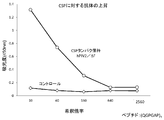
- bronchiolitis caused by RSV does not show infiltration of eosinophils or neutrophils into the lung, but neutrophils into the lung in RSV spontaneously infected infants after vaccination with formalin-inactivated RSV vaccine (Lot 100)
- formalin-inactivated RSV vaccine Lico-inactivated RSV vaccine
- Th-2 type 2 helper T cells
- F protein which is one of the membrane proteins of RSV IV, has been reported to be important as a vaccine antigen, but it has been found that the F protein has a three-dimensional conformation that varies greatly depending on the environment.
- Formalin treatment not only changes the three-dimensional structure of RSV F, but the treatment significantly increases the carbonyl content in the protein and induces a Th2 reaction, which is likely to cause ERD. This has been reported in animal tests (Non-Patent Document 5).
- the document also states that heat-treated RSV vaccines (only the three-dimensional structure changes) have less neutrophil infiltration into lung tissue than formalin-treated RSV vaccines. It is thought that it is not only a change in the three-dimensional structure.
- F protein an RSV membrane protein
- the F protein is translated as a protein precursor called F0.
- F0 there are two furin cleavage sites, which are cleaved into F1 and F2 that are cross-linked by S-S bonds through the maturation process (mature F protein).
- the 27 polypeptides called p27 are deleted by cutting with Hulin. It is known that the mature F protein forms a trimer on the infected cell and on the viral envelope and has two different three-dimensional structures, prefusion F and postfusion F, depending on the environment.
- F protein before RSV virus is fused to cells usually has a prefusion F structure, but the structure is thermodynamically unstable, cell fusion, heating, denaturing agent, F protein to cells and envelope
- the structure can be easily converted to a thermodynamically stable postfusion-F structure by removing the transmembrane array that carries the anchoring.
- the three-dimensional structures of prefusion F and postfusion F have been clarified by X-ray crystallography, and the antibodies and the binding sites that bind to each have been reported in detail (Non-patent Document 1).
- Monoclonal antibody palivizumab binds to a site called site IV II of RSV F protein.
- the sequence region maintains the three-dimensional structure in both prefusion F and postfusion F, and palivizumab binds to the F protein of both structures and exhibits neutralizing activity.
- site ⁇ (zero) an antibody that binds to an amino acid sequence called site ⁇ (zero), which is a three-dimensional antigen epitope of prefusion F.
- the region in the vicinity of the epitope has a bent structure composed of two ⁇ sheet structures and four ⁇ helices in prefusion F.
- postfusion F the region has a linear structure with one ⁇ helix structure.
- the site ⁇ (zero) structure is different between prefusion F and postfusion F, and the antibodies named 5C4, AM22, and D25 bind only to the prefusion F structure and not to the F protein of the postfusion structure. .
- the prefusion F site ⁇ (zero) -binding antibody has 10-100 times higher neutralizing activity against RSV than the site II-binding antibody, which is an unstable prefusion for effective vaccine development. It is important to develop vaccine technology that can provide structured F protein as an antigen.
- prefusion F protein As a method of providing prefusion F protein as an antigen, (i) structure stabilization of prefusion F protein, or (ii) development of an antigen delivery system that maintains the three-dimensional structure of prefusion F, or a fusion method of both may be considered.
- prefusion ⁇ ⁇ ⁇ F protein is obtained by removing or substituting part or both of the amino acid sites of the two furin recognition sites, or by removing or substituting the p27 peptide located between the furin recognition sites.
- the structure stabilization modification of can be considered.
- the RSV F 67th Asn (N) was replaced with Ile (I)
- the 251st Ser (S) was replaced with Pro (P).
- Stabilization (Non-Patent Document 4) can be considered.
- RSV F was stabilized by substituting the 155th SerS (S) with Cys (C) and the 290th Ser) (S) with Cys (C) to crosslink the SS bond between cysteines (references). 7) is also conceivable. Furthermore, structural stabilization is also conceivable by deleting part or all of the fusion region such as a cell located immediately after the rear sequence of the two furin recognition sequences of the F protein, thereby eliminating the Fusion ability.
- soluble F protein When F protein is used as an antigen, soluble F protein (soluble) that leaves the secretory signal sequence located on the N-terminal side and lacks the transmembrane (TM) and transmembrane (CT) sequences on the C-terminal side Type F protein) is often used.
- TM transmembrane
- CT transmembrane
- the F protein retaining the sequence is expressed in cells, it is anchored to the membrane and is difficult to recover. This is a measure taken to remove this and secrete the F protein into the medium to facilitate recovery.
- the F protein has a TM sequence and a CT sequence, thereby forming a trimer and anchoring on the cell membrane or the viral envelope, thereby stabilizing the prefusion structure.
- a delivery system that delivers the F protein to the recipient in a state in which the structure is stabilized by binding the F protein on the cell membrane, virus envelope, vector, etc. while retaining the TM sequence and CT sequence. It is conceivable to take measures to develop or to integrate (i) into this system.
- the inventors fused a foreign antigen gene to a membrane protein gene of a non-propagating hPIV2 viral vector to produce a non-proliferating viral vector retaining the antigen and use it as a vaccine.
- a method to inactivate and use as a vaccine so that virus replication does not occur with ⁇ -propiolactone at a low concentration of 1 / 10-1 / 100 Patent Document 1.
- a gene 113 amino acid residues fused with 48 amino acids linked to two universal influenza antigens M2e and TM and CT sequences of hPIV2 F protein, NP with the highest transcription amount of F-deficient hPIV2 vector.
- RSV F protein, Ebola virus GP protein, etc. are antigens of 500 amino acid residues or more, and a strict three-dimensional structure is required to function as an antigen. There is a need for further development of vectors and vaccines that can deliver such high molecular proteins while maintaining their three-dimensional structure.
- An object of the present invention is to use a viral vector capable of efficiently introducing a high molecular antigen peptide (including protein and peptide) into a target cell while maintaining a three-dimensional structure necessary for functioning as an antigen. In providing vaccines.
- the inventors incorporated an antigen peptide immediately upstream of the HN gene 5 ′ of the non-transmissible Paramyxoviridae virus genome gene lacking the membrane protein gene, and fused expression of the envelope protein TM and CT sequence of the virus vector and the antigen peptide. It was found that a high molecular antigen peptide having several hundred amino acid residues can be efficiently delivered to target cells while maintaining the three-dimensional structure.
- the present invention relates to the following (1) to (9).
- the virus vector according to (1) wherein the virus of the Paramyxoviridae is a non-transmissible human parainfluenza type 2 virus.
- the nucleic acid inactivation treatment is a treatment that does not substantially change the structure of the virus envelope, such as treatment with a low concentration of ⁇ -propiolactone.
- the TM sequence and / or CT sequence or GPI-like anchor protein of the antigen polypeptide gene is incorporated as a nucleic acid substituted with a TM sequence and / or CT sequence derived from human parainfluenza type 2 virus, (1) The virus vector according to any one of (5) to (5).
- Influenza virus including highly toxic influenza virus, parainfluenza type 3 virus, RS virus, Hendra virus, SARS virus, MERS virus, Nipah virus, Lassa virus, dengue virus, West Nile virus, Japanese encephalitis Virus, human metapneumovirus, Ebola virus, hantavirus, AIDS virus, hepatitis C virus, hepatitis B virus, rubella virus, rotavirus, norovirus, Crimea-Congo hemorrhagic fever virus, herpes virus, cytomegalovirus, Zika virus , A virus antigen peptide selected from Marburg virus, HIV and papilloma virus, a malaria parasite antigen peptide, group A ⁇ (beta) streptococci, Mycobacterium tuberculosis Bacterial antigen peptide selected from Vibrio cholerae and Mycoplasma, or cancer antigen gp100, MUC1, NY-ESO-1, MelanA / MART
- the antigen polypeptide is three or more consecutive M2e antigens of influenza virus, RSV F protein or G protein or variant or fragment thereof, Ebola virus GP protein or variant or fragment thereof, malaria CSP
- the virus vector according to (7) which is any one selected from a protein or a variant or fragment thereof, and a prM / E protein of Zika virus or a variant or fragment thereof.
- the antigen polypeptide is incorporated into a viral vector in the following manner: a) a nucleic acid comprising at least three consecutive M2e antigen genes of influenza virus and TM and CT sequences of human parainfluenza type 2 virus F protein; b) a nucleic acid in which the gene encoding the full-length F protein of RSV or the TM sequence and / or CT sequence of the protein is replaced with the TM sequence and / or CT sequence of human parainfluenza type 2 virus F protein, or prefusion F
- a nucleic acid encoding a protein in which an F protein introduced with an amino acid mutation that increases stability is substituted with a human parainfluenza type 2 virus F protein TM sequence and / or CT sequence; c) A gene encoding an F protein in which the cell fusion region sequence (SEQ ID NO: 5) is partially deleted from the full-length F protein of RSV, or a gene encoding an F protein introduced with an amino acid mutation that increases the stability of prefusion F
- a nucleic acid encoding a protein substituted with a sequence e) A gene in which the Fibritin trimer-forming sequence gene or GCN trimer-forming sequence gene of T4 phage is inserted into the 3 ′ end of the RSV F extra-membrane sequence gene of the nucleic acid described in b) -d) above.
- a nucleic acid encoding a gene in which an amino acid is replaced with Cys to form an SS bond that stabilizes trimer formation f)
- the TM sequence and CT sequence of the GP protein are replaced with the TM sequence and CT sequence of human parainfluenza type 2 virus F protein or GP protein, or the TM sequence of the GP protein.
- the nucleic acid in which the CT sequence is maintained includes the 88th Phe (F) of the amino acid sequence of the Ebola virus GP protein (GenBank Accession No.
- the virus vector of the present invention can be delivered as an antigen while maintaining the three-dimensional structure of a macromolecular peptide (several hundred amino acid residues or more). This makes it possible to produce licensed vaccines effective against RSV and Ebola virus, malaria parasite and Zika virus.
- the virus vector of the present invention can be efficiently recovered in a state where the antigen polypeptide is displayed on the virus particle.
- the virus vector of the present invention is inactivated by using a low concentration of ⁇ -propiolactone or the like, only the virus genome is inactivated while maintaining the three-dimensional structure of the virus envelope protein and the introduced antigen. The Therefore, in a recombinant viral vector in which a pre-existence antibody exists, the vector is neutralized by the antibody and the amount of antigen is reduced due to suppression of gene expression in the recipient, but such a problem does not occur in the vector, There is also an advantage that antigen gene mutation does not occur in the recipient.
- the vector has cell adsorbability and immunity-inducing ability similar to that of live virus, and the introduced antigen can maintain the three-dimensional structure of the antigen epitope for inducing efficient neutralizing antibodies, so without adding an adjuvant. Alternatively, a reduced and effective vaccine effect can be expected. From the above-mentioned advantages, by using the virus vector of the present invention, high effects and safety can be expected even in humans, and it becomes possible to develop a licensed vaccine effective for RSV, Ebola virus, malaria, and Zika virus.
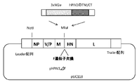
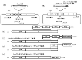
- FIG. 1 shows the construction of 3xM2e + hPIV2 TM / CT sequence gene introduction into the Mlu restriction enzyme site.
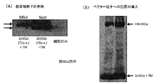
- FIG. 2 shows the result of confirming the expression of M2e in the infected cells of the collected vector (A) and the result of confirming the introduction of the antigen into the vector particles by Western blot (B).
- FIG. 3 shows the introduction of RSV F antigen.
- FIG. 4 shows the results of expression of mutant RSV F expressed in infected cells by infection with hPIV2 ⁇ F (A) and expression of the mutant by Western blot (B).
- FIG. 5 shows an introduction diagram of Ebola virus GP antigen.
- FIG. 1 shows the construction of 3xM2e + hPIV2 TM / CT sequence gene introduction into the Mlu restriction enzyme site.
- FIG. 2 shows the result of confirming the expression of M2e in the infected cells of the collected vector (A) and the result of confirming the introduction of the antigen into the vector particles by
- FIG. 6 shows the expression in an infected cell (A) of Ebola GP carrying Ebola GP or the TM / CT sequence of Ebola GP or hPIV2 introduced in two cells (A) and Western showing GP incorporation into vector particles. Blot results (B) are shown.
- Figure 7 shows the presence or absence of infectious particles of hPIV2 ⁇ F virus carrying Ebola GP and GFP protein introduced with two amino acid mutations, using PGFP2 F-expressing Vero cells and normal Vero cells as indicators of GFP fluorescence. The result figure is shown.
- FIG. 8 shows the results of antibody induction against Ebola GP by mouse administration.
- FIG. 9 shows the introduction of Plasmodium yoelli CSP antigen.
- FIG. 10 shows the result of confirming the expression of CSP antigen by Western blot.
- FIG. 11 shows the results of antibody induction against CSP by mouse administration.
- FIG. 12 shows an introduction diagram of prM / E antigen of Zika virus.
- the present invention is a viral vector in which a nucleic acid encoding an antigen polypeptide is incorporated immediately upstream of the HN gene 5 ′ of the F gene-deficient Paramyxoviridae virus genome gene, and the antigen polypeptide is derived from the virus. And a viral vector expressed as a fusion protein of 130 amino acid residues or more fused / substituted with the TM sequence and / or CT sequence.
- the present invention is directed to the surface of a virus, including forms in which a gene encoding an antigen polypeptide is fused / replaced with a TM sequence and / or a CT sequence gene of the F gene of a Paramyxoviridae virus.
- a viral vector incorporated so as to be expressed, wherein the high molecular antigen polypeptide is expressed while maintaining a desired three-dimensional structure (three-dimensional structure capable of functioning as a vaccine antigen), and the virus vector Provided is an inactivated vector that can induce immunity similar to that of a live virus without using an adjuvant or by reducing the amount of adjuvant added.
- the inventors have succeeded in constructing a type that incorporates an antigen onto a vector by fusing / substituting the TM sequence and CT sequence of a membrane protein that functions as a packaging signal for the antigen and virus. It is reported (WO2014 / 103310).
- an antigen can be efficiently expressed with about 120 amino acid residues, and it has been difficult to introduce an antigen having 130 amino acid residues or more and maintaining a desired three-dimensional structure.
- a foreign antigen gene having TM and CT sequences of hPIV2 F protein is inserted into recombinant non-proliferating hPIV2 in order to introduce the foreign antigen onto the vector.
- hPIV2 F protein with the same TM and CT sequences competes during the assembly of virus particles through the M protein, preventing efficient virus formation, making it difficult to recover high-titer virus.
- the CT sequence of the Ebola virus GP protein (TM sequence remains the Ebola virus GP) in PIV3 was replaced with the PIV3 F protein from the Ebola virus GP protein and introduced into the PIV3 vector in which the F and HN genes were deleted.
- Non-patent Document 6 it has been reported that the recovery efficiency of the PIV3 virus is extremely poor. Therefore, when large foreign antigens holding TM and CT sequences are replaced with foreign PI TM and CT sequences with hPIV2 F TM and CT sequences and introduced into hPIV2 / ⁇ F, virus recovery becomes more difficult. it was thought. Furthermore, in PIV3, in which an antigen gene was introduced at a site located 5 'upstream of the NP gene with the largest transcription amount, a mutation was found in the introduced antigen gene, and a foreign gene was introduced to a site that was highly expressed from the virus component genes. Problems in the case of antigens have been pointed out (Non-patent document 8, Non-patent document 9). The inventors have also confirmed that the M2 gene of influenza virus is introduced into the site 5 'upstream of the NP gene of hPIV2 and that a mutation is introduced into the recovered virus M2 gene to cause amino acid substitution.
- an MluI restriction enzyme cleavage site is introduced into the F gene deficient site of the F gene deficient hPIV2 vector, that is, an antigen gene or the like is incorporated into the F gene deficient site immediately upstream of the HN gene, and 130 amino acid residues or more are incorporated.
- the inventors have developed a 3xM2e and hPIV2 vector F protein gene TM sequence and / or CT sequence gene fused gene, RSV F protein gene extra-membrane sequence region Introducing multiple RSV F protein genes with intact and modified, and RSV F protein genes in which the TM and CT sequences of these RSV F proteins are replaced with hPIV2 TM and / or CT sequences. Then, the pros and cons of virus vector recovery and their expression levels were compared.
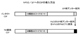
- the gene encoding the malaria CSP protein and the gene encoding the CSP protein in which the GPI anchor-like sequence of the CSP protein was replaced with the TM and CT sequences of hPIV2 were introduced to examine the success of vector recovery and the expression of the antigen.
- it encodes the prM / E protein that replaces the Zika virus prM / E protein (including some variants) and prM / E protein stem and TM sequences with hPIV2 TM and CT sequences. The effect of the present invention was confirmed by introducing the gene and examining the success of the vector recovery and the expression of the antigen.
- a particularly preferred “paramyxoviridae virus” used in the present invention is “human parainfluenza type 2 virus”, and its genome is a monocistronic single-stranded minus RNA of about 15,000 bases.
- NP protein, P (phospho) protein, M (matrix) protein, F (fusion), HN (hemagglutinin / neuraminidase) protein and L (large) protein are encoded on the genome in this order.
- RNA editing produces V (buoy) protein.
- Nucleocapsid protein (NP) binds to the RNA genome, forming a helically symmetric ribonucleoside protein complex (nucleocapsid, RNP).
- NP protein protein, P (phospho) protein and L (large) protein are necessary for the formation of RNP.
- F (fusion) protein and HN (hemagglutinin neuraminidase) protein exist on the viral envelope, and are responsible for adsorption and fusion to the receptor.
- the M (matrix) protein interacts with the cytoplasmic domains of F and HN proteins, the envelope lipid bilayer and RNP, and is important for budding of virus particles.
- Human parainfluenza type 2 virus is an RNA virus that propagates in the cytoplasm, so that no gene is integrated into the chromosome of the host cell.
- this virus is known to infect human airway mucosa and induce mucosal immunity centered on IgA, humoral immunity by IgG, and cellular epidemic immunity.
- IgA human airway mucosa
- IgG humoral immunity by IgG
- cellular epidemic immunity since there have been no serious reports of human (adult) infections so far, it is considered extremely useful as a viral vector for treatment.
- F gene-deficient paramyxoviridae virus is used.
- F gene deficient (non-transmissible) human parainfluenza type 2 virus is used.
- the F protein of human parainfluenza type 2 virus is a protein required when a viral nucleocapsid is introduced into a host by fusing a viral envelope and a cell membrane.
- F gene-deficient human parainfluenza type 2 virus produces infectious hPIV2 carrying the F gene in packaging cells, but does not produce infectious virus in cells that do not express F protein. It is a virus. Therefore, virus particles having autonomous proliferation ability cannot be formed after infecting recipient cells, and other cells are not infected, so that it is highly safe as a virus for vaccines.
- Vero cells are used as packaging cells that can be infected with F gene-deficient virus and produce virus particles.
- Vero cells are particularly well tolerated against human palinjuenza type 2 virus F protein, have no interferon production, and can grow stably even when the F gene of human parainfluenza type 2 virus is constantly expressed. And virus particles can be produced efficiently.
- the viral vector of the present invention has been subjected to “nucleic acid inactivation treatment”.
- Nucleic acid inactivation treatment refers to an introduced antigen polypeptide that retains the three-dimensional structure of the envelope protein such as F protein and HN protein and the introduced antigen polypeptide and requires the function and three-dimensional structure of these envelope proteins. The inactivation of only the viral genome while having the ability to induce neutralizing antibodies.
- the nucleic acid inactivation treatment can be performed by, for example, nucleic acid alkylating agent treatment, hydrogen peroxide treatment, UV irradiation, radiation irradiation, or heat treatment.
- the viral vector is treated with ⁇ -propiolactone, which is a nucleic acid alkylating agent.
- ⁇ -propiolactone may be added to the virus culture and incubated at 4 ° C. for about 17 hours.
- a preferred concentration of ⁇ -propiolactone is 0.001% to 0.05%, more preferably 0.004% to 0.01% in terms of final concentration.
- the antigen gene is incorporated on the vector and it is not necessary to perform the antigen introduction operation after the nucleic acid inactivation, problems associated with the antigen introduction operation after inactivation (decrease in antigen introduction efficiency, Envelope and antigen structural destruction can also be avoided.
- problems associated with the antigen introduction operation after inactivation decrease in antigen introduction efficiency, Envelope and antigen structural destruction can also be avoided.
- F gene-deficient virus even if a live virus remains due to drug treatment, the virus lacks the ability to grow, and there is no risk of growth in the recipient, making the inactivated vaccine highly safe. It becomes possible to keep.
- Virus vector means a virus particle in which a gene to be expressed in an infected cell is packaged together with a virus genome gene, and a vector not containing a virus genome capable of producing an infectious virus. In the present specification, the latter is particularly referred to as an “inactivation vector” and can be obtained by inactivating a nucleic acid by treatment with a drug or the like.
- an “antigen polypeptide” is a polypeptide having 130 amino acid residues or more capable of inducing an immune response in a subject administered with the viral vector of the present invention, and examples include influenza, RS virus, Ebola virus, Zika virus Alternatively, a part thereof, a protozoan protein or part thereof, a protein derived from bacteria or virus, or a part thereof may be mentioned.
- antigenic polypeptides useful for use as influenza vaccines include influenza virus HA protein, NA protein, M2 protein or M2e peptide, or fragments thereof.
- an antigen polypeptide useful for use as a vaccine for RSV the F protein or G protein of RSV or a fragment thereof
- an antigen polypeptide useful as a vaccine for Ebola virus the GP protein of Ebola virus or these Fragments, Zika virus prM / E protein or fragments thereof, malaria parasite CSP protein or fragments thereof.
- the antigen polypeptide may be a fragment as described above or may contain an appropriate mutation as long as it has immunogenicity that functions as a vaccine. Examples of such mutants include mutant RSV F protein (61st Asn (N) Ile (I) and 251) in which p27 or p27 of RSV F protein and its neighboring sequences are removed and cleavage by furin is removed.
- Non-patent Document 4 Including mutants in which the Ser (S) of the second is replaced with Pro (P)
- Non-patent Document 7 mutants in which the Ser (S) of the second is replaced with Pro (P)
- mutant RSV F protein 155th Ser ( Including mutants in which S) is replaced with Cys (C) and 290th Ser (S) is replaced with Cys (C)
- RSV F mutant Ebola virus GP protein amino acid introduced with a nucleic acid encoding a gene in which the amino acid is replaced with Cys to form an SS bond that stabilizes the formation sequence, GCN trimerization sequence or trimer formation
- the 88th Phe (F) of the sequence (GenBank Accession No.
- KJ660346 is Ala (A) and the 535th Phe (F) is Ala (A ), A mutant GP1 protein from which the mucin region sequence located after the 313rd position is removed, or a mutant GP protein from which the mucin region sequence has been removed from the GP sequence.
- infectious disease-related transducing antigen polypeptides include influenza virus (including highly toxic influenza virus), parainfluenza 3 virus, RS virus, Hendra virus, SARS virus, MERS virus, Nipah virus, Lassa virus, Dengue virus, West Nile virus, Japanese encephalitis virus, human metapneumovirus, Ebola virus, hantavirus, AIDS virus, hepatitis B virus, hepatitis C virus, rubella virus, rotavirus, norovirus, Crimean-Congo hemorrhagic fever virus, herpes virus, site
- the “antigen polypeptide” is expressed on the viral envelope as a protein fused / replaced with the vector virus TM sequence and / or CT sequence or GPI-like anchor protein.
- the antigen polypeptide expressed in the virus-producing cell is taken into the virus particle and delivered to the recipient. That is, in the present invention, not only is the antigen polypeptide gene incorporated on the viral gene transcribed and translated in the infected cells, but the viral particles themselves have the antigen polypeptide, which Can be reliably delivered to the recipient.
- the gene encoding the antigenic polypeptide is replaced with the gene encoding the TM sequence and / or CT sequence of the vector virus F protein or GPI-like anchor. It is incorporated into a viral gene by linking to a gene encoding a protein. Examples of the gene encoding the TM sequence of virus F protein and / or CT sequence protein include hPIV2 F gene.
- a vector can be constructed by a conventional method using a conventional recombinant DNA technique.
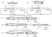
- the nucleic acid (gene) encoding the antigen polypeptide is inserted into the F gene-deficient site “upstream of the HN gene 5 ′” in the structural gene of the vector virus described above (see FIG. 1).
- “HN gene 5 ′ immediately upstream” means immediately upstream of the NH gene 5 ′ end, but a sequence that does not affect expression is included between the nucleic acid encoding the antigen polypeptide and the HN gene 5 ′ end. It may be.
- the “HN gene 5 ′ immediately upstream” can be said to be an F gene-deficient site.
- a large amount of antigen can be expressed on the envelope of the defective hPIV2 by incorporating the protein gene as a fusion gene or an intact gene into the packaging cell that supplies the structural gene product to trans.
- the viral vector of the present invention When the viral vector of the present invention is treated with ⁇ -propiolactone at a lower concentration than usual, the viral vector retains the hemagglutination activity, cell adsorption ability and ability to mature dendritic cells (that is, on the vector membrane). While maintaining the 3D structure of the protein in a state close to that of a live virus, only the genome can be inactivated. Treatment with ⁇ -propiolactone at a lower concentration than normal, that is, the bareest concentration that can inactivate the genome, is possible by using non-propagating (or non-transmissible) viral vectors that do not produce secondary particles. It becomes. Even if a small amount of infectious virus remains, secondary infectious particles are not produced because it is a non-transmissible virus.
- Non-Patent Document 10 RNA strands that are fragmented by treatment with a low concentration of ⁇ -propiolactone and remain incapable of transcription replication remain in the vector, and innate immunity is activated by the action of RIGI, IFIT, and the like. According to this method, since the virus particle itself has an adjuvant activity, there is an advantage that it is not necessary to use it together with an adjuvant that induces immunity to an antigen, or the concentration of the adjuvant can be reduced.
- the virus vector of the present invention infects cells via sialic acid receptors. Since sialic acid is present in many cells / tissues, vector administration routes include nasal spray, pulmonary, oral, sublingual, intradermal, subcutaneous, and direct administration to veins, Ex vivo administration to immune-inducing cells such as dendritic cells can be considered.
- the virus vector of the present invention is useful as a vaccine against mammalian cells including humans.
- the vaccine of the present invention comprises a viral vector and a pharmaceutically acceptable carrier.
- Vaccines can typically be administered to mammalian cells including humans as a spray.
- the propellant can be prepared by a conventional method.
- the culture supernatant containing the virus vector is concentrated if necessary, and suspended in a buffer solution such as PBS, a virus vector stable solution or physiological saline together with a pharmaceutically acceptable carrier, and then a filter if necessary. And then sterilized by filtration and then filled into a sterile container. You may add a stabilizer, a preservative, etc. to a propellant as needed.
- the expression vector thus obtained can be administered by inhalation to a subject.
- the culture supernatant containing the viral vector is concentrated as necessary, suspended in a buffer solution such as PBS or physiological saline together with a pharmaceutically acceptable carrier, and then sterilized by filtration with a filter or the like as necessary. It can then be prepared by filling a sterile container. A stabilizer, a preservative and the like may be added to the injection as necessary.
- the expression vector thus obtained can be administered to a subject as an injection.
- the amount of vaccine to be administered usually ranges from 0.01 ⁇ g to 100,000 ⁇ g of antigen per dose, which depends on the subject being treated, the ability to synthesize antibodies in the subject's immune system, and the degree of protection desired.
- route of administration including oral, subcutaneous, nasal, intradermal, intramuscular and intravenous routes of administration.
- the vaccine of the present invention may be given on a single dose schedule or preferably on a combined dose schedule.
- a combined dosing schedule 1 to 10 individual doses are given at the start of the inoculation, followed by the time interval required to maintain and / or strengthen the immune response, eg 1 to 4 as the second dose.
- Another dose may be given after a month. If necessary, subsequent administrations can be given after several months.
- the dosage regimen will also be determined, at least in part, by the individual's needs and will depend on the judgment of the physician.
- the viral vector of the present invention can be delivered as an antigen while maintaining the three-dimensional structure of a high molecular peptide (several hundred amino acid residues).
- this vector has a problem that the virus vector cannot be efficiently recovered in a state where an antigen polypeptide of 130 amino acid residues or more is displayed on the virus particle.
- a virus vector can be efficiently recovered by displaying an antigen polypeptide of 500 amino acid groups or more on a virus particle.
- nucleic acid inactivation is performed using a low concentration of ⁇ -propiolactone or the like, so that the three-dimensional structure of the viral envelope protein and the introduced antigen is maintained, and only the viral genome is maintained in a state in which the structure of these proteins is maintained. Inactivated. Therefore, the vector has cell adsorbability and immunity-inducing ability similar to that of a live virus, and the introduced antigen can maintain the three-dimensional structure of the antigen epitope for inducing efficient neutralizing antibodies. I can expect.
- the introduction of the antigen does not require an antigen introduction operation after inactivation in order to produce a virus vector into which an antigen gene has been introduced in advance, and there are problems associated with the antigen introduction operation after inactivation (low antigen introduction efficiency by conventional methods, Envelope and antigen structural destruction can be avoided. Because of the advantages described above, the virus vector of the present invention can be expected to have a high effect in humans, unlike a virus vector that could not be expected to have a sufficient effect in humans.
- the viral vector of the present invention can be configured so that antigens can be arranged outside, inside, or both of the vector envelope, thereby allowing various introductions of a plurality of antigens into a vector.
- immunity induction comparable to that of a live virus vector can be achieved by efficient virus inactivation treatment, and delivery is possible while maintaining the structure of an unstable high molecular antigen protein.
- An activation vector can be provided, and it becomes possible to open the way to vaccination of an inactivation vector using a Paramyxoviridae virus.
- hPIV2 / ⁇ F Construction of hPIV2 / ⁇ F by fusing TM sequence and CT sequence protein of F protein of 3xM2e and hPIV2 and introducing into MluI restriction site TM sequence and CT sequence of HPIV2 F protein (N-terminal-YSLSAIALILSVITLVVVGLLIAYIIKLVSQIHQFRSLAATTMFHRENPAFFSKNNHGNIYGIS1-C-terminal (sequence number) : A gene that encodes a sequence (3 x M2e) in which the M2e antigen peptide of the universal influenza virus (N-terminal-SLLTEVETPIRNEWGCRCNDSSDD-C-terminal (SEQ ID NO: 2)) is repeated three times upstream of the gene encoding 65 amino acid residues) And a gene in which an initiation codon (ATG) is linked and a gene added with a gene necessary for expression in hPIV2 virus, and a plasmid construct
- the insertion site of the MluI restriction enzyme cleavage sequence is located 5 ′ upstream of the HPI gene of hPIV2 / ⁇ F.
- the construction was constructed based on the rule of 6 rule so that the total number of genomes is a multiple of 6, which is important in constructing hPIV2. According to the previous report (Non-Patent Document 11), the virus was collected by the reverse genetics method. Furthermore, the influenza antigen was also introduced into the MluI restriction enzyme cleavage site of hPIV2 / ⁇ F fused with H2 protein to M2e antigen (FIG. 1).
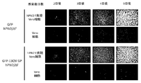
- hPIV2 / ⁇ F which is a fusion of the 3 x M2e antigen peptide of the universal influenza virus and the TM and CT sequences of the FPI protein of hPIV2, and efficiently express M2e in infected cells. Further, it was confirmed that the M2e protein was also incorporated on the vector particles (FIG. 2B).
- the 3 x M2e gene is introduced into the NotI restriction enzyme cleavage site upstream of the NP upstream of hPIV2 / ⁇ F, as shown in Fig. 2A, it is as efficient as when 2 x M2e is introduced into the NotI restriction enzyme cleavage site. The virus could not be recovered.
- the 3 x M2e antigen gene was introduced into the MluI restriction enzyme cleavage site, the virus could be efficiently recovered and incorporated into the vector even when fused to M2e to the HN protein (Fig. 2B).
- Example 2 Increased stability of RSV intact F protein gene (codon optimized gene) or RSV TM sequence and / or CT sequence with TM sequence and / or CT sequence of hPIV2 F protein, and prefusion F Construction of hPIV2 / ⁇ F in which mutation-introduced gene was introduced into MluI restriction site
- the RSV F protein of accession number P03420 was used as the amino acid sequence of the RSV F vaccine antigen.
- the DNA sequence of RSV F (catalog number: VG40042-UT) in which the expression codon of each amino acid was optimized for humans was used from Sino Biological.
- RSV F protein gene (Fig. 3A, C), or RSV F protein TM sequence and CT region (IMITTIIIVIIVILLSLIAVGLLLYCKARSTPVTLSKDQLSGINNIAFSN (SEQ ID NO: 3)) to contain RSV F protein on hPIV2 / ⁇ F vector hPIV2 TM sequence and CT sequence (YSLSAIALILSVITLVVVGLLIAYIIKLVSQIHQFRSLAATTMFHRENPAFFSKNNHGNIYGIS (SEQ ID NO: 1)) or RSV F protein CT sequence (KARSTPVTLSKDQLSGINNIAFSN (SEQ ID NO: 9)) is converted to hPIV2 CT sequence (KLVSQIHQPAGISHH sequence is replaced by KLVSQIHQPARSH A gene in which hPIV2 transcription initiation
- 3B, C was constructed by a conventional method. Transcription initiation sequence, etc. is partially modified (construction in which a transgene is expressed by reading through from the previous gene without adding a start sequence 5 ′ upstream of the transgene, or a transcription termination sequence 3 ′ downstream of the transgene. A construction in which two sets of start sequences were introduced downstream of the start sequence and 5 ′ upstream of the HN gene was also prepared. Stability of RSV F mutant by substituting 155th Ser (S) with Cys (C) and 290th Ser (S) with Cys (C) to bridge the SS bond between cysteines. (Reference 7), F mutant genes introduced with S155C and S290C were prepared (FIG.
- the RSV F mutant encodes a gene in which the amino acid is replaced with Cys in order to form a fibrin trimer formation sequence, a GCN trimer formation sequence of the T4 phage, or an SS bond that stabilizes the trimer formation.
- Virus recovery was performed according to the previous report (Non-patent Document 11). All types of viruses caused cytopathic effects (CPE) in infected cells and recovered sufficient titers of virus. Virus recovery was performed according to the previous report (Non-patent Document 11). All types of viruses caused cytopathic effects (CPE) in infected cells and recovered sufficient titers of virus.
- the collected virus was infected with F gene-expressing Vero cells, and RSV F expression was confirmed by immunostaining using an antibody against RSV F (RSV anti-F monoclonal antibody [2F7] from abcam). Infection was performed for 2 days, fixed with 100% cold methanol, and expression was confirmed using a secondary antibody labeled with Alexa488 under a fluorescence microscope.
- Example 3 RSV F protein gene from which the two furin cleavage region peptide sequence genes of RSV have been removed, or the TM sequence and / or CT sequence of the RSV F protein gene replaced with the TM sequence and / or CT sequence of the hPIV2 F protein Construction of hPIV2 / ⁇ F Introducing a Gene Containing a MluI Restriction Site As shown in FIG. 3D, there are two sites for the recognition of a human huulin in RSV F (FIG. 3C).
- CT sequence IMITTIIIVIIVILLSLIAVGLLLYCKARSTPVTLSKDQLSGINNIAFSN (SEQ ID NO: 3)
- CT array YSLSAIALILSVITLVVVGLLIAYIIKLVSQIHQFRSLAATTMFHRENPAFFSKNNHGNIYGIS (sequence number 1)) or RSV F protein CT sequence (KARSTPVTLSKDQLSGINNIAFSN (sequence number 9)) hPIV2 CT sequence (KLVSQIHQFRSLAATTMFHRENPAFFSK array sequence number HGVSQNI array sequence
- the gene in which the transcription initiation sequence, intervening sequence and transcription termination sequence of hPIV2 were added to the sequence substituted in (D) was introduced into the Mlu site of the plasmid vector for hPIV2 / ⁇ F expression.
- RSV is also the first hurin recognition sequence of F (106, 107, 108, 109th sequence Arg (R) Ala (A) Arg (R) Arg (R) is replaced with Gln (Q) Ala (A) Gln (Q )
- a mutant F ⁇ p27: Fig. 3D) in which Gln (Q) is substituted and p27 sequence is deleted is prepared, and TM and / or CT sequences of RSV F protein are replaced with TM and / or CT sequences of hPIV2.
- the mutant gene was introduced into the MluII site of the plasmid vector for hPIV2 / ⁇ F expression
- the purpose of the production of these mutants was to stabilize the three-dimensional structure of prefusion F by introducing mutations.
- the aim is to develop highly effective RSV vaccines by inducing antibodies with high sum activity: 67th Asn (N) of RSV F to Ile (I), and 251st Ser (S) to Pro ( There is also a report that the stability of the F mutant of RSV is enhanced by substituting P) (Non-patent Document 4), and a mutant gene of F ⁇ p27 introduced with N61I and S251P (FIG. 3B, D) is prepared.
- HPIV2 / ⁇ F expression The plasmid was introduced into the MluI site of the plasmid vector, and the inserted sequence was made to be rule of 6. The virus was recovered in accordance with the previous report (Non-patent Document 11).
- An RSV F variant introduced with a nucleic acid encoding a gene in which an amino acid is replaced with Cys to form an SS bond that stabilizes fibrin trimer formation sequence, GCN trimer formation sequence or trimer formation All types of viruses produced cytopathic effects (CPE) in infected cells and were able to recover sufficient titers of virus.
- CPE cytopathic effects
- the collected virus was infected with F gene-expressing Vero cells, and expression of RSV F was confirmed by immunostaining using an antibody against RSV F (RSV anti-F monoclonal antibody [2F7] manufactured by abcam). Infection was performed for 2 days, fixed with 100% cold methanol, and expression was confirmed using a secondary antibody labeled with Alexa488 under a fluorescence microscope.
- the RSV F ⁇ p27 + 2 protein gene and the RSV F ⁇ p27 + 2 protein gene into which the TMV and / or CT sequence of the RSV F ⁇ p27 + 2 protein has been replaced with the TM sequence and / or CT sequence of the FPI of FPI2 ⁇ F showed a positive reaction to anti-F antibody against RSV F (FIG. 4 (A)).
- Example 4 The FPI gene from which 8 amino acids of the RSV membrane fusion sequence have been removed, the TM sequence and / or the CT sequence of the RSV F protein gene, or a gene introduced with a mutation that has improved the stability of prefusion F Construction of hPIV2 / ⁇ F in which a gene having the F protein TM sequence and a CT protein gene substitution sequence was introduced into the MluI restriction site RSV F contains the amino acid sequence 137-151 (FLGFLLGVGSAIASG) as shown in Fig. 3C. (SEQ ID NO: 5)) is called a fusion domain (FD), and is a region involved in cell fusion, etc.
- FLGFLLGVGSAIASG amino acid sequence 137-151
- FD fusion domain
- RSV F protein was removed from the 8 amino acids of FD (FLGFLLGV (SEQ ID NO: 6))
- the F protein was constructed (FIG. 3E).
- the reason for introducing the construction in which a part of the FD sequence is removed is that hPIV2 / ⁇ F lacks the F gene, so it does not produce proliferative viruses other than F-expressing packaging cells. It has been reported that the F protein may be adsorbed and fused to cells alone, and in order to ensure safety, a part of the fusion sequence was removed from the FD to eliminate the risk of this possibility.
- the RSV F protein (FIG.
- RSV F protein TM sequence and CT sequence IMITTIIIVIIVILLSLIAVGLLLYCKARSTPVTLSKDQLSGINNIAFSN (SEQ ID NO: 3)
- RSV F protein TM sequence and CT sequence YSLSAIALILSVITLVVVGLLIAYIIKLVSQIHQFRSLAATTMFHRENPAFFSKNNHGNIYGIS (sequence number 1)
- RSV F protein CT sequence KARSTPVTLSKDQLSGINNIAFSN (sequence number 9)
- hPIV2 CT sequence KLVSQIHQPARSNKHGN sequence
- Non-patent Document 11 The virus was collected according to the previous report (Non-patent Document 11), and the virus was successfully collected. In addition, it encodes a gene in which the amino acid is replaced with Cys to form a fibrin trimerization sequence, a GCN trimerization sequence, or an SS bond that stabilizes trimer formation of the T4 phage in the right RSV F mutant. We have also produced F mutants of RSV with nucleic acid introduced.
- the collected virus was infected with F gene-expressing Vero cells, and RSV F expression was confirmed by immunostaining using an antibody against RSV F (RSV anti-F monoclonal antibody [2F7] from abcam). Infection was performed for 2 days, fixed with 100% cold methanol, and expression was confirmed using a secondary antibody labeled with Alexa488 under a fluorescence microscope.
- a protein gene from which a part of the FD of RSV F has been removed, and an hVIV2 / RSV F ⁇ p27 protein gene into which the TM and CT sequences of the F protein of RSV have been replaced with the TM and CT sequences of hPIV2 ⁇ F showed a positive reaction to anti-F antibody against RSV F (FIG. 4 (A)). Further, it was confirmed by Western blotting analysis using an anti-F monoclonal antibody [2F7]) that the target protein was expressed (FIG. 4B).
- Example 5 The Ebola virus GP protein gene into which amino acid mutations were introduced at two sites, or a gene having a sequence obtained by replacing the TM sequence and CT sequence of the GP protein gene with the TM sequence and CT sequence of the hPIV2 F protein was introduced into the MluI restriction site. Construction of hPIV2 / ⁇ F The GP protein of Ebola virus is a protein responsible for binding to the cell surface receptor of the virus and membrane fusion of the virus envelope and the host cell membrane (FIG. 5C). A commercially available plasmid (Sino Biological, accession number: KJ660346, cDNA length: 2031 bp) was used as cDNA.
- a partial sequence of the GP protein was substituted with a sequence (F88A and F535A; JVI: 2013 (87) 3324-3334: Non-Patent Document 15) reported not to have entry ability to cells.
- the gene was replaced (no amino acid sequence substitution) so that RNA editing did not occur.
- the GP protein gene of Ebola virus (FIGS.
- TM sequence and CT sequence of the GP protein gene (WIPAGIGVTGVIIAVIALFCICKFVF (SEQ ID NO: 7)) is the TM sequence and CT sequence of hPIV2 (YSLSAIALILSVITLVVVGLLIAYIIKLVSQIHQFRSLAATTMFHRENPAFFSK1)
- hPIV2 YSLSAIALILSVITLVVVGLLIAYIIKLVSQIHQFRSLAATTMFHRENPAFFSK1
- the inserted sequence was rule-of-6.
- Virus recovery was performed according to a previous report (Non-patent Document 11).
- the recovered virus was infected with Fero-expressing Vero cells of PIV2 for 2 days, and GP expression was confirmed (FIG. 6).
- an anti-GP mouse monoclonal antibody (4F3: IBT BIOSEVICE) was used.
- the GP protein and the GP protein substituted with the TM and CT sequences of hPIV2 were both expressed in the same amount (FIG. 6A).
- GP protein from which the mucin region described below was removed did not react. (The epitope sequence of the antibody is not specified, but it was expected to recognize the epitope of the mucin region because it did not react with GP from which the mucin region was removed.)
- the virus was recovered from the supernatant of the virus-expressing cells by ultracentrifugation at 141,000 ⁇ xg for 35 minutes, and the GP protein contained in the vector was examined by Western blot (WB) (FIG. 6B).
- WB Western blot
- FIG. 6B The expression levels were the same for both NP and P of PIV2.
- the GP protein near 102 kDa (ER and Golgi before undergoing sugar modification) GP protein (GP protein) had the same level of GP expression when either GP or hPIV2 F TM / CT sequences were retained, but the higher molecular weight 4F3 positive GP protein (subject to sugar modification) (GP protein) and TM / CT sequence of Ebola virus GP were found to be more common. It was also confirmed that the GP protein substituted with the TM / CT sequence of hPIV2 F undergoes sugar chain modification and is incorporated into the vector. Examples of viruses incorporating a protein having a TM / CT sequence different from the original virus include Ebola's GP protein (Non-patent Document 6) and VSV's G protein (Non-patent Document 12).
- Fig. 5A, D or hPIV2 TM sequence and CT sequence (YSLSAIALILSVITLVVVGLLIAYIIKLVSQIHQFRSLAATTMFHRENPAFFSKNNHGNIGIS)
- Fig. 5B, D Ebola virus GP1 protein gene added with hPIV2 transcription initiation sequence, Standing sequence, gene was introduced by adding a transcription termination sequence Mlu site of the plasmid vector for HPIV2 / [Delta] F expression. The inserted sequence was rule-of-6. Virus recovery was performed according to a previous report (Non-patent Document 11).
- Example 7 A gene having a sequence in which the mucin region gene has been removed from the GP protein of Ebola virus into which amino acid mutations have been introduced in two places, or the TM and CT sequences of the GP protein gene are replaced with the TM and CT sequences of the hPIV2 F protein. Construction of hPIV2 / ⁇ F Introduced into MluI Restriction Site The amino acid sequence from 313th to 454th of Ebola virus GP is a sequence called mucin region, and the mucin region protein covers the domain formed by other regions. There are many amino acid residues that are arranged and sugar modified.
- Ebola virus GP ( ⁇ mucin GP) from which the region has been removed was constructed.
- TMPI and CT sequence of Ebola virus (YSLSAIALILSVITLVVVGLLIAYIIKLVSQIHQFRSLAATTMFHRENPAFFSKNNHGNIYGIS (SEQ ID NO: 1)) (FIG. 5B, E) in the ⁇ mucin GP (FIGS.
- hPIV2 transcription initiation sequence was introduced into the Mlu site of the plasmid vector for hPIV2 / ⁇ F expression.
- the inserted sequence was rule-of-6.
- Virus recovery was performed according to the previous report (Non-patent Document 11), and all viruses were successfully recovered.
- Western blot analysis was performed, it was highly possible that the anti-GP antibody used had an epitope in the mucin region (Fig. 6), and the presence or absence of expression could not be analyzed by Western blot analysis (hPIV2 NP And P were expressed.)
- Example 8 A gene with the mucin region gene removed from the GP1 protein of Ebola virus into which amino acid mutations have been introduced in two places, or a gene having the TM and CT sequences of the GP1 protein gene replaced with the TM and CT sequences of the hPIV2 F protein Construction of hPIV2 / ⁇ F Introduced into MluI Restriction Site Mucin translation region sequence was removed from GP1 protein gene of Ebola virus constructed in Example 5, and TM sequence and CT sequence of GP protein gene (WIPAGIGVTGVIIAVIALFCICKFVF (SEQ ID NO: 7 )) (Fig.
- hPIV2 YSLSAIALILSVITLVVVGLLIAYIIKLVSQIHQFRSLAATTMFHRENPAFFSKNNHGNIYGIS (SEQ ID NO: 1)
- Fig. A gene with a transcription termination sequence was introduced into the Mlu site, and the inserted sequence was rule-of-6. Yield was performed in accordance with a previous report (Non-Patent Document 11).
- Example 9 Antibody titer against GP protein in mice by hPIV2 / ⁇ F carrying Ebola virus GP protein gene introduced with amino acid mutations constructed in Example 5 and 0.01% ⁇ -propiolactone inactivation vector of the vector Evaluation of the vector and the inactivated vector (without adjuvant) were prepared so that the virus count was 10 8 / mL. 100 ul of each vector sample and control sample was injected twice into the thigh muscle of 5 week-old BALB / c mice ( ⁇ ) hind limbs, and serum was collected 2 weeks after administration. Using the serum, Ebola virus GP protein (Sino Biological Inc.
- Example 10 Construction of hPIV2 / ⁇ F retaining malaria antigen CSP (circumsporozoite protein; Plasmodium yoelli) A CSP derived from Plasmodium yoelli was introduced into hPIV2 / ⁇ F.
- CSP malaria antigen
- the virus was infected with F-expressing Vero cells, and expression analysis of CSP protein was performed by Western blot analysis.
- the antibody against CSP was a monoclonal antibody against Plasmodium yoelli QQPP repeat (2F6).
- As a positive control 10,000 sporozoid protozoa of Plasmodium yoelli were used.
- CSP protein could be expressed by the vector.
- the culture supernatant is expected to be used as a means for expressing other malaria-related genes.
- the virus vector recovered from the culture supernatant by ultracentrifugation (141,000 g, 35 minutes) is an intact CSP that retains the GPI-like anchor sequence and a CSP that replaces the GPI-like anchor sequence with TM of hPIV2. It was also confirmed that the same degree of retention was possible on the ⁇ F vector particles.
- the CSP protein was found to have the ability of the GPI-like anchor protein to hold the CSP protein on the vector to the same extent as the TM protein of hPIV2.
- the molecular weight of the antibody against the QQPP antigen was different between the transmembrane domain of the original GPI CSP and the CSP with hPIV2 TM / CT.
- the molecular weight of the positive control sporozoite CSP protein was the same as that of the original GPI, but the molecular weight of the positive band was different.
- the CSP replaced with TM / CT of hPIV2 was able to be incorporated into the vector in a form close to the original (sporozoid CSP).
- Example 11 Evaluation of antibody titer against CSP by hPIV2 / ⁇ F retaining malaria antigen CSP hPIV2 / ⁇ F retaining the CSP gene in which the GPI-like anchor sequence was replaced with TM of hPIV2 was prepared so that the number of viruses was 10 9 / mL.
- Each sample and 100 ul of the control sample were injected twice into the thigh muscle of the 5-week-old BALB / c ( ⁇ ) hind limbs at intervals of 2 weeks, and serum was collected 2 weeks after administration.
- the antibody titer against Plasmodium yoelli CSP was evaluated using the serum.
- peptides for evaluation by ELISA, 3 peptides (QGPGAP) in which the QGPGAP sequence of the B cell epitope repeatedly present in the CSP of Plasmodium yoelli was repeated three times were used.
- the peptide was prepared at 5 ug / mL, and 100 ul was affixed to one well of a 96 plate overnight, and the increase in the antibody against CSP (IgG1 + IgG2a) was evaluated.
- the above-mentioned serum was diluted from 10 times to 4560 times to 2560 times, and the presence or absence of the antibody was evaluated by a normal ABC method. From the results (FIG. 11), the antibody against CSP could be increased by administration of the vector.
- Example 12 Preparation of Zika virus vaccine using prM / E antigen A start codon was added to the 5'-end of the prM gene of the gene (including some amino acid mutations) encoding the prM / E region (KU321639) of Zika virus. Add a gene that adds or does not add a gene that encodes a secretion signal, and create a gene with hPIV2 transcription initiation sequence, intervening sequence, and transcription termination sequence added before and after the gene. MluI site of plasmid vector for hPIV2 / ⁇ F expression (FIGS. 12A and 12C). The inserted array is set to rule of 6. Virus recovery is performed according to the previous report (Non-patent Document 11).
- Example 13 Preparation of a vaccine in which the stem sequence (EH1 and EH2) and TM sequence (TH1 and TH2) of the C-terminal region of the E protein of Zika virus are substituted and deleted
- the gene of prM of Zika virus having the same structure as in Example 14
- a gene encoding a protein obtained by adding an hPIV2 TM / CT sequence (hPIV2 TM sequence and / or CT arrangement YSLSAIALILSVITLVVVGLLIAYIIKLVSQIHQFRSLAATTMFHRENPAFFSKNNHGNIYGIS (SEQ ID NO: 1)
- the following sequences mean EH1 and EH2 sequences and TM1 and TM2 sequences, EH2 sequences and TM1 and TM2 sequences, or TM1 and TM2 sequences of E protein, hPIV2 transcription start sequence before and after the gene. Then, a gene to which an intervening sequence and a transcription termination sequence are added is prepared and introduced into the MluI site of the plasmid vector for hPIV2 / ⁇ F expression (FIGS. 12B, D, E, F). Do Recovery of virus recovery Virus is performed according previous report (Non-Patent Document 11).
- Example 14 Vaccine effect against Zika virus The vaccine effect against Zika virus is evaluated using a vector prepared by inactivating the vector retaining prM / E or E protein prepared in Example 15 with low-concentration ⁇ -propiolactone.
- Example 15 Collection of prM / E protein or M / E protein granule (subviral particle) that does not contain genetic information
- viruses belonging to Flavi virus only contain prM / E protein or M / E protein.
- the release of protein granules (subviral particles) that do not contain genetic information is reported in other Flavi viruses (Non-patent Documents 13 and 14).
- VLP virus-like particles
- the virus vector of the present invention can present a high molecular antigen peptide (several hundred amino acid residues) on a virus particle while maintaining its three-dimensional structure.
- the virus vector of the present invention has cell adsorbability and immunity induction ability similar to that of a live virus even after nucleic acid inactivation treatment, presents antigen epitopes while maintaining the three-dimensional structure, and induces efficient neutralizing antibodies it can. Therefore, the present invention is useful for producing a licensed vaccine effective for RSV, Ebola virus, and the like.
Abstract
Description
本明細書は、本願の優先権の基礎である特願2015-119097号(2015年6月12日出願)及び特願2016-63315号(2016年3月28日出願)の明細書に記載された内容を包含する。
[技術分野]
本発明は、非伝播型ヒトパラインフルエンザ2型ウイルスベクター及び前記ウイルスベクターを利用したワクチンに関する。 [Related applications]
This specification is described in the specification of Japanese Patent Application No. 2015-119097 (filed on June 12, 2015) and Japanese Patent Application No. 2016-63315 (filed on March 28, 2016), which are the basis of the priority of the present application. The contents are included.
[Technical field]
The present invention relates to a non-transmissible
したがって、安価なRSVの感染予防ワクチンの開発が強く求められているが、以下に記載する過去のワクチン禍により、他の感染症ワクチンに比べてワクチン開発が大幅に遅れている。 There is no effective vaccine for the prevention of RSV infection, and there is only symptomatic treatment with the anti-RSV humanized monoclonal antibody palivizumab (Synazis). Palivizumab is expected to have a prophylactic effect by intramuscular injection of 15 mg / kg once a month before the RSV epidemic begins and during the epidemic period. However, the humanized monoclonal antibody is extremely expensive, and the insurance coverage in Japan is limited to premature infants, children with chronic lung disease, and children with Down syndrome. The high cost of antibodies is a bottleneck, making it difficult to expand the number of patients to be administered.
Therefore, development of an inexpensive RSV infection prevention vaccine is strongly demanded, but vaccine development is greatly delayed compared to other infectious disease vaccines due to past vaccine vaccines described below.
(1)抗原ポリペプチドをコードする核酸が、F遺伝子欠損型パラミクソウイルス科ウイルスゲノム遺伝子のHN遺伝子5’直上流に組み込まれているウイルスベクターであって、該抗原ポリペプチドが前記ウイルス由来のTM配列及び/又はCT配列と融合された130アミノ酸残基以上の融合タンパクとして発現される非伝播型ウイルスベクター。
(2)パラミクソウイルス科のウイルスが非伝播型ヒトパラインフルエンザ2型ウイルスである、(1)記載のウイルスベクター。
(3)抗原ポリペプチドがベクターエンベロープ表面に発現されることを特徴とする、(1)又は(2)に記載のウイルスベクター。
(4)抗原ポリペプチドが天然の立体構造又はワクチン抗原として必要な立体構造を維持した状態で発現されることを特徴とする、(1)~(3)のいずれかに記載のウイルスベクター。
(5)核酸不活化処理されたものである、(1)~(4)のいずれかに記載のウイルスベクター。
前記核酸不活化処理は、低濃度のβプロピオラクトン処理など、ウイルスエンベロープの構造を実質的に変化させないものである。
(6)抗原ポリペプチド遺伝子のTM配列及び/又はCT配列あるいはGPI様アンカータンパク質がヒトパラインフルエンザ2型ウイルス由来のTM配列及び/又はCT配列で置換された核酸として組み込まれている、(1)~(5)のいずれかに記載のウイルスベクター。
(7)抗原ポリペプチドが、強毒型インフルエンザウイルスを含むインフルエンザウイルス、パラインフルエンザ3型ウイルス、RSウイルス、ヘンドラウイルス、SARSウイルス、MERSウイルス、ニパウイルス、ラッサウイルス、デングウイルス、ウエストナイルウイルス、日本脳炎ウイルス、ヒトメタニューモウイルス、エボラウイルス、ハンタウイルス、エイズウイルス、C型肝炎ウイルス、B型肝炎ウイルス、風疹ウイルス、ロタウイルス、ノロウイルス、クリミア-コンゴ出血熱ウイルス、ヘルペスウイルス、サイトメガロウイルス、Zikaウイルス、マールブルグウイルス、HIVおよびパピローマウイルスから選ばれるウイルスの抗原ペプチド、マラリア原虫の抗原ペプチド、A群β(ベータ)型溶連菌、Mycobacterium tuberculosis、コレラ菌、およびマイコプラズマから選ばれる細菌の抗原ペプチド、または癌抗原gp100、MUC1、NY-ESO-1、MelanA/MART1、TRP2、MAGE、CEA、CA125、HER2/neu、WT1、PSAおよびネオアンチジェンからなる群から選ばれる抗原ペプチドのいずれか1または2以上、あるいはこれらの断片である、(1)~(6)のいずれかに記載のウイルスベクター。
(8)抗原ポリペプチドが、インフルエンザウイルスの3個以上の連続したM2e抗原、RSVのFタンパク又はGタンパクあるいはその変異体もしくはその断片、エボラウイルスのGPタンパク又はその変異体もしくはその断片、マラリアCSPタンパク又はその変異体もしくはその断片、及びZikaウイルスのprM/Eタンパク又はその変異体もしくはその断片から選ばれるいずれかである、(7)記載のウイルスベクター。
なお、前記抗原ポリペプチドは、下記のような態様でウイルスベクターに組み込まれる:
a)インフルエンザウイルスの3個以上の連続したM2e抗原遺伝子とヒトパラインフルエンザ2型ウイルスFタンパクのTM配列及びCT配列を含む核酸;
b)RSVの全長Fタンパクをコードする遺伝子又は当該タンパクのTM配列及び/又はCT配列が、ヒトパラインフルエンザ2型ウイルスFタンパクのTM配列及び/又はCT配列で置換された核酸、あるいはprefusion Fの安定性を増加させるアミノ酸変異を導入したFタンパクにヒトパラインフルエンザ2型ウイルスFタンパクのTM配列及び/又はCT配列で置換されたタンパクをコードする核酸;
c)RSVの全長Fタンパクから細胞融合領域配列(配列番号5)を一部欠失させたFタンパクをコードする遺伝子又はprefusion Fの安定性を増加させるアミノ酸変異を導入したFタンパクをコードする遺伝子において、該FタンパクのTM配列及び/又はCT配列がヒトパラインフルエンザ2型ウイルスFタンパクのTM配列及び/又はCT配列で置換されたタンパクをコードする核酸;
d)RSVの全長Fタンパクより2箇所のヒューリン切断領域間のp27ペプチド配列と前記p27の最初のヒューリン切断認識配列のRRを除去したRSVのFタンパクをコードする遺伝子もしくはその変異体又は最初のフューリン切断認識配列RARRをQAQQに置換したRSVのFタンパクをコードする遺伝子又はその変異体において、該FタンパクのTM配列及び/又はCT配列がヒトパラインフルエンザ2型ウイルスFタンパクのTM配列及び/又はCT配列で置換されたタンパクをコードする核酸;
e)上記b)-d)に記載された核酸のRSV Fの膜外配列遺伝子の3'末端側に、T4ファージのFibritin三量体形成配列遺伝子もしくはGCN三量体形成配列遺伝子を挿入した遺伝子をコードする核酸、又は三量体形成を安定化させるS-S結合を形成するためにアミノ酸をCysに置換した遺伝子をコードする核酸;
f)エボラウイルスの全長GPタンパク又はその変異体において、該GPタンパクのTM配列及びCT配列がヒトパラインフルエンザ2型ウイルスFタンパク又はGPタンパクのTM配列及びCT配列で置換又は該GPタンパクのTM配列及びCT配列が維持された核酸
(前記エボラウイルスの変異体としては、エボラウイルスGPタンパクのアミノ酸配列(GenBank Accession No.KJ660346)の第88番目のPhe(F)をAla(A)に、535番目のPhe(F)をAla(A)に置換した変異GPタンパク、前記変異GPタンパクよりさらに313番目以降に位置するムチン領域配列が除去された変異GP1タンパク、又はGP配列よりムチン領域配列が除去された変異GPタンパクが挙げられる);
g)マラリアCSPタンパク又は該タンパクのGPI型アンカー配列をヒトパラインフルエンザ2型ウイルスFタンパクのTM配列及びCT配列に置換したタンパクをコードする核酸;
h)ZikaウイルスのprM/Eタンパクをコードする核酸;
i)ZikaウイルスのEタンパクのC末端領域に位置する2つのstem配列(EH1及びEH2)並びに2つのTM配列(TM1及びTM2)のうち、EH1及びEH2を欠失又はEH2を欠失させヒトパラインフルエンザ2型ウイルスFタンパクのTM配列及び/又はCT配列を付加したタンパクをコードする核酸。
(9)(1)~(8)のいずれかに記載のウイルスベクターと製薬上許容し得る担体を含むワクチン。 That is, the present invention relates to the following (1) to (9).
(1) A viral vector in which a nucleic acid encoding an antigen polypeptide is incorporated immediately upstream of the
(2) The virus vector according to (1), wherein the virus of the Paramyxoviridae is a non-transmissible
(3) The viral vector according to (1) or (2), wherein the antigen polypeptide is expressed on the surface of the vector envelope.
(4) The viral vector according to any one of (1) to (3), wherein the antigen polypeptide is expressed in a state in which a natural three-dimensional structure or a three-dimensional structure necessary as a vaccine antigen is maintained.
(5) The viral vector according to any one of (1) to (4), which has been subjected to nucleic acid inactivation treatment.
The nucleic acid inactivation treatment is a treatment that does not substantially change the structure of the virus envelope, such as treatment with a low concentration of β-propiolactone.
(6) The TM sequence and / or CT sequence or GPI-like anchor protein of the antigen polypeptide gene is incorporated as a nucleic acid substituted with a TM sequence and / or CT sequence derived from
(7) Influenza virus including highly toxic influenza virus,
(8) The antigen polypeptide is three or more consecutive M2e antigens of influenza virus, RSV F protein or G protein or variant or fragment thereof, Ebola virus GP protein or variant or fragment thereof, malaria CSP The virus vector according to (7), which is any one selected from a protein or a variant or fragment thereof, and a prM / E protein of Zika virus or a variant or fragment thereof.
The antigen polypeptide is incorporated into a viral vector in the following manner:
a) a nucleic acid comprising at least three consecutive M2e antigen genes of influenza virus and TM and CT sequences of
b) a nucleic acid in which the gene encoding the full-length F protein of RSV or the TM sequence and / or CT sequence of the protein is replaced with the TM sequence and / or CT sequence of
c) A gene encoding an F protein in which the cell fusion region sequence (SEQ ID NO: 5) is partially deleted from the full-length F protein of RSV, or a gene encoding an F protein introduced with an amino acid mutation that increases the stability of prefusion F A nucleic acid encoding a protein in which the TM sequence and / or CT sequence of the F protein is replaced with the TM sequence and / or CT sequence of a
d) Gene encoding RSV F protein from which p27 peptide sequence between two furin cleavage regions and RR of the first furin cleavage recognition sequence of p27 are removed from RSV full-length F protein, or a variant thereof, or the first furin In the gene encoding RSV F protein in which the cleavage recognition sequence RARR is replaced with QAQQ or a variant thereof, the TM sequence and / or CT sequence of the F protein is the TM sequence and / or CT of the
e) A gene in which the Fibritin trimer-forming sequence gene or GCN trimer-forming sequence gene of T4 phage is inserted into the 3 ′ end of the RSV F extra-membrane sequence gene of the nucleic acid described in b) -d) above. Or a nucleic acid encoding a gene in which an amino acid is replaced with Cys to form an SS bond that stabilizes trimer formation;
f) In the full-length GP protein of Ebola virus or a variant thereof, the TM sequence and CT sequence of the GP protein are replaced with the TM sequence and CT sequence of
g) a nucleic acid encoding a malaria CSP protein or a protein obtained by replacing the GPI-type anchor sequence of the protein with the TM sequence and CT sequence of
h) a nucleic acid encoding the prM / E protein of Zika virus;
i) Of the two stem sequences (EH1 and EH2) and two TM sequences (TM1 and TM2) located in the C-terminal region of the E protein of Zika virus, EH1 and EH2 are deleted or EH2 is deleted A nucleic acid encoding a protein to which a TM sequence and / or CT sequence of
(9) A vaccine comprising the viral vector according to any one of (1) to (8) and a pharmaceutically acceptable carrier.
抗原の導入は、あらかじめ抗原遺伝子を導入したウイルスベクターを作製するために、不活化後に抗原導入操作を行う必要がなく、不活化後の抗原導入操作に伴う問題(従来法による低い抗原導入効率、エンベロープ及び抗原の構造破壊等)を回避できる。
上記した利点から、本発明のウイルスベクターは、従来ヒトにおいて十分な効果が期待できなかったウイルスベクターとは異なり、ヒトにおいても高い効果を期待できる。 In the present invention, nucleic acid inactivation is performed using a low concentration of β-propiolactone or the like, so that the three-dimensional structure of the viral envelope protein and the introduced antigen is maintained, and only the viral genome is maintained in a state in which the structure of these proteins is maintained. Inactivated. Therefore, the vector has cell adsorbability and immunity-inducing ability similar to that of a live virus, and the introduced antigen can maintain the three-dimensional structure of the antigen epitope for inducing efficient neutralizing antibodies. I can expect.
The introduction of the antigen does not require an antigen introduction operation after inactivation in order to produce a virus vector into which an antigen gene has been introduced in advance, and there are problems associated with the antigen introduction operation after inactivation (low antigen introduction efficiency by conventional methods, Envelope and antigen structural destruction can be avoided.
Because of the advantages described above, the virus vector of the present invention can be expected to have a high effect in humans, unlike a virus vector that could not be expected to have a sufficient effect in humans.
3xM2eとhPIV2のFタンパクのTM配列及びCT配列タンパクを融合させMluI制限部位に導入したhPIV2/ΔFの構築
hPIV2のFタンパクのTM配列及びCT配列(N末端-YSLSAIALILSVITLVVVGLLIAYIIKLVSQIHQFRSLAATTMFHRENPAFFSKNNHGNIYGIS-C末端(配列番号1):65アミノ酸残基)をコードする遺伝子の上流に万能型インフルエンザウイルスのM2e抗原ペプチド(N末端-SLLTEVETPIRNEWGCRCNDSSDD-C末端(配列番号2))を3回繰り返した配列(3 x M2e)をコードする遺伝子と開始コドン(ATG)を連結した遺伝子及びhPIV2ウイルスでの発現に必要な遺伝子を付加した遺伝子を構築し、両端にMluI制限酵素切断配列を付加したhPIV2/ΔFベクターのプラスミドコンストラクトを構築した(図1)。MluI制限酵素切断配列の挿入部位は、hPIV2/ΔFのHN遺伝子の5’直上流に位置する。当該構築はhPIV2にコンストラクト上重要とされる、ゲノムの総数が6の倍数になるように、ルールオブ6ルールに基づいて構築した。既報(非特許文献11)にしたがい、リバースジェネティクス法により、ウイルスの回収を図った。さらに、HNタンパクにM2e抗原融合させたhPIV2/ΔFのMluI制限酵素切断部位にも当該インフルエンザ抗原を導入した(図1)。 [Example 1]
Construction of hPIV2 / ΔF by fusing TM sequence and CT sequence protein of F protein of 3xM2e and hPIV2 and introducing into MluI restriction site TM sequence and CT sequence of HPIV2 F protein (N-terminal-YSLSAIALILSVITLVVVGLLIAYIIKLVSQIHQFRSLAATTMFHRENPAFFSKNNHGNIYGIS1-C-terminal (sequence number) : A gene that encodes a sequence (3 x M2e) in which the M2e antigen peptide of the universal influenza virus (N-terminal-SLLTEVETPIRNEWGCRCNDSSDD-C-terminal (SEQ ID NO: 2)) is repeated three times upstream of the gene encoding 65 amino acid residues) And a gene in which an initiation codon (ATG) is linked and a gene added with a gene necessary for expression in hPIV2 virus, and a plasmid construct of hPIV2 / ΔF vector with MluI restriction enzyme cleavage sequences added at both ends was constructed (Fig. 1). The insertion site of the MluI restriction enzyme cleavage sequence is located 5 ′ upstream of the HPI gene of hPIV2 / ΔF. The construction was constructed based on the rule of 6 rule so that the total number of genomes is a multiple of 6, which is important in constructing hPIV2. According to the previous report (Non-Patent Document 11), the virus was collected by the reverse genetics method. Furthermore, the influenza antigen was also introduced into the MluI restriction enzyme cleavage site of hPIV2 / ΔF fused with H2 protein to M2e antigen (FIG. 1).
RSVのインタクトなFタンパク遺伝子(コドン最適化遺伝子)又はRSVのTM配列及び/又はCT配列をhPIV2のFタンパクのTM配列及び/又はCT配列に置換した遺伝子、及びprefusion Fの安定性を高めた変異を導入した遺伝子をMluI制限部位に導入したhPIV2/ΔFの構築
RSVのFワクチン用抗原のアミノ酸配列は、accession numberP03420のRSVのFタンパクを用いた。当該アミノ酸配列をコードする遺伝子は、Sino Biological社より各アミノ酸の発現コドンをヒト用に最適化したRSV FのDNA配列(カタログ番号:VG40042-UT)を使用した。以下の抗原遺伝子hPIV2/ΔF発現用プラスミドベクターのMluI制限配列に導入した(図3A、B)。RSVのインタクトなFタンパク遺伝子(図3A、C)、又はhPIV2/ΔFベクター上にRSVのFタンパクを含有させるためにRSVのFタンパクのTM配列及びCT領域(IMITTIIIVIIVILLSLIAVGLLLYCKARSTPVTLSKDQLSGINNIAFSN(配列番号3))をhPIV2のTM配列及びCT配列(YSLSAIALILSVITLVVVGLLIAYIIKLVSQIHQFRSLAATTMFHRENPAFFSKNNHGNIYGIS(配列番号1))又はRSVのFタンパクのCT配列(KARSTPVTLSKDQLSGINNIAFSN(配列番号9))をhPIV2のCT配列(KLVSQIHQFRSLAATTMFHRENPAFFSKNNHGNIYGIS(配列番号10))に置換した配列をコードする遺伝子(図3B、C)に置換した配列をコードする遺伝子に、hPIV2の転写開始配列、介在配列、転写終結配列を付加した遺伝子を通常の方法により構築した。転写開始配列等は一部修飾した構築(導入遺伝子の5‘上流に開始配列を付加せず前遺伝子からのread throughにより導入遺伝子を発現させる構築、または導入遺伝子の3’下流の転写終結配列の下流に開始配列を導入しHN遺伝子の5’上流に開始配列を2組導入した構築)も作製した。RSV Fの155番目のSer (S)をCys (C)に、290番目のSer (S)をCys (C)に置換しシステイン間のS-S結合を架橋することによりRSVのF変異体の安定性が増強されると報告(参考文献7)されており、S155C及びS290C導入したFの変異体遺伝子を作製し(図3B、C)、hPIV2/ΔF発現用プラスミドベクターのMluII部位に導入した。挿入した配列はルールオブ6(遺伝子長が6の倍数)となるように調整した。さらに、前記RSV F変異体にT4ファージのFibritin三量体形成配列、GCN三量体形成配列又は三量体形成を安定化させるS-S結合を形成するためにアミノ酸をCysに置換した遺伝子をコードする核酸を導入したRSVのF変異体も作製している。ウイルスの回収は既報(非特許文献11)に従い行った。どのタイプのウイルスも感染細胞で細胞変性効果(CPE)を起こし、十分な力価のウイルスを回収することができた。ウイルスの回収は、既報(非特許文献11)にしたがい実施した。どのタイプのウイルスも感染細胞で細胞変性効果(CPE)を起こし、十分な力価のウイルスを回収することができた。 [Example 2]
Increased stability of RSV intact F protein gene (codon optimized gene) or RSV TM sequence and / or CT sequence with TM sequence and / or CT sequence of hPIV2 F protein, and prefusion F Construction of hPIV2 / ΔF in which mutation-introduced gene was introduced into MluI restriction site The RSV F protein of accession number P03420 was used as the amino acid sequence of the RSV F vaccine antigen. As the gene encoding the amino acid sequence, the DNA sequence of RSV F (catalog number: VG40042-UT) in which the expression codon of each amino acid was optimized for humans was used from Sino Biological. It was introduced into MluI restriction sequences of the following plasmid vector for antigen gene hPIV2 / ΔF expression (FIGS. 3A and 3B). RSV F protein gene (Fig. 3A, C), or RSV F protein TM sequence and CT region (IMITTIIIVIIVILLSLIAVGLLLYCKARSTPVTLSKDQLSGINNIAFSN (SEQ ID NO: 3)) to contain RSV F protein on hPIV2 / ΔF vector hPIV2 TM sequence and CT sequence (YSLSAIALILSVITLVVVGLLIAYIIKLVSQIHQFRSLAATTMFHRENPAFFSKNNHGNIYGIS (SEQ ID NO: 1)) or RSV F protein CT sequence (KARSTPVTLSKDQLSGINNIAFSN (SEQ ID NO: 9)) is converted to hPIV2 CT sequence (KLVSQIHQPAGISHH sequence is replaced by KLVSQIHQPARSH A gene in which hPIV2 transcription initiation sequence, intervening sequence, and transcription termination sequence were added to the gene encoding the sequence replaced with the gene (FIG. 3B, C) was constructed by a conventional method. Transcription initiation sequence, etc. is partially modified (construction in which a transgene is expressed by reading through from the previous gene without adding a
RSVの2つのヒューリン切断領域ペプチド配列遺伝子を除去したRSVのFタンパク遺伝子又は当該RSVのFタンパク遺伝子のTM配列及び/又はCT配列をhPIV2のFタンパクのTM配列及び/又はCT配列に置換した配列をもつ遺伝子をMluI制限部位に導入したhPIV2/ΔFの構築
RSVのFには図3Dに示すように2箇所のヒューリン認識配列部位が存在している(図3C)。当該ヒューリン認識配列間のp27と呼ばれるアミノ酸(N末端-ELPRFMNYTLNNAKKTNVTLSKKRKRR-C末端(配列番号4))を除去し、さらに前部のヒューリン認識配列の(108と109番目のArg(R) Arg(R))を欠損させた、RSVのF(FΔp27+2)を構築した(図3D)。FΔp27+2、又はhPIV2/ΔFベクター上にRSVのFタンパクを含有させるためにRSVのFタンパクのTM配列(図3A、D)及びCT配列(IMITTIIIVIIVILLSLIAVGLLLYCKARSTPVTLSKDQLSGINNIAFSN(配列番号3))をhPIV2のTM配列及びCT配列(YSLSAIALILSVITLVVVGLLIAYIIKLVSQIHQFRSLAATTMFHRENPAFFSKNNHGNIYGIS(配列番号1))又はRSVのFタンパクのCT配列(KARSTPVTLSKDQLSGINNIAFSN(配列番号9))をhPIV2のCT配列(KLVSQIHQFRSLAATTMFHRENPAFFSKNNHGNIYGIS(配列番号10))に置換した配列をコードする遺伝子(図3B、D)に置換した配列に、hPIV2の転写開始配列、介在配列、転写終結配列を付加した遺伝子をhPIV2/ΔF発現用プラスミドベクターのMlu部位に導入した。また、RSVもFの最初のヒューリン認識配列(106、107、108、109番目の配列Arg(R)Ala(A)Arg(R)Arg(R)をGln(Q)Ala(A)Gln(Q)Gln(Q)に置換しp27配列を欠失させた変異体(FΔp27:図3D)も作製し、RSVのFタンパクのTM配列及び/又はCT配列をhPIV2のTM及び/又はCT配列に置換した変異体遺伝子をhPIV2/ΔF発現用プラスミドベクターのMluII部位に導入した。これら変異体作製の目的は、変異を導入することにより、prefusion Fの立体構造の安定化を図り、prefusion F抗原に対する中和活性の高い抗体を誘導し、高い有効性をもつRSVワクチンを開発することにある。RSV Fの67番目のAsn(N)をIle (I)に、251番目のSer(S)をPro(P)に置換することによりRSVのF変異体の安定性が増強されるとの報告もあり(非特許文献4)、N61I及びS251Pを導入したFΔp27の変異体遺伝子(図3B、D)を作製し、hPIV2/ΔF発現用プラスミドベクターのMluI部位に導入した。挿入した配列はルールオブ6となるようにした。ウイルスの回収は、既報(非特許文献11)にそって行った。さらに、前記RSV F変異体にT4ファージのFibritin三量体形成配列、GCN三量体形成配列又は三量体形成を安定化させるS-S結合を形成するためにアミノ酸をCysに置換した遺伝子をコードする核酸を導入したRSVのF変異体も作製している。どのタイプのウイルスも感染細胞で細胞変性効果(CPE)を起こし、十分な力価のウイルスを回収することができた。 [Example 3]
RSV F protein gene from which the two furin cleavage region peptide sequence genes of RSV have been removed, or the TM sequence and / or CT sequence of the RSV F protein gene replaced with the TM sequence and / or CT sequence of the hPIV2 F protein Construction of hPIV2 / ΔF Introducing a Gene Containing a MluI Restriction Site As shown in FIG. 3D, there are two sites for the recognition of a human huulin in RSV F (FIG. 3C). The amino acid called p27 (N-terminal-ELPRFMNYTLNNAKKTNVTLSKKRKRR-C-terminal (SEQ ID NO: 4)) between the hurin recognition sequences was removed, and the humin recognition sequence at the front (108 and 109 Arg (R) Arg (R) RSV F (FΔp27 + 2) lacking) was constructed (FIG. 3D). In order to include the RSV F protein on the FΔp27 + 2 or hPIV2 / ΔF vector, the TM sequence of the RSV F protein (FIGS. 3A and 3D) and the CT sequence (IMITTIIIVIIVILLSLIAVGLLLYCKARSTPVTLSKDQLSGINNIAFSN (SEQ ID NO: 3)) CT array (YSLSAIALILSVITLVVVGLLIAYIIKLVSQIHQFRSLAATTMFHRENPAFFSKNNHGNIYGIS (sequence number 1)) or RSV F protein CT sequence (KARSTPVTLSKDQLSGINNIAFSN (sequence number 9)) hPIV2 CT sequence (KLVSQIHQFRSLAATTMFHRENPAFFSK array sequence number HGVSQNI array sequence The gene in which the transcription initiation sequence, intervening sequence and transcription termination sequence of hPIV2 were added to the sequence substituted in (D) was introduced into the Mlu site of the plasmid vector for hPIV2 / ΔF expression. RSV is also the first hurin recognition sequence of F (106, 107, 108, 109th sequence Arg (R) Ala (A) Arg (R) Arg (R) is replaced with Gln (Q) Ala (A) Gln (Q ) A mutant (FΔp27: Fig. 3D) in which Gln (Q) is substituted and p27 sequence is deleted is prepared, and TM and / or CT sequences of RSV F protein are replaced with TM and / or CT sequences of hPIV2. The mutant gene was introduced into the MluII site of the plasmid vector for hPIV2 / ΔF expression The purpose of the production of these mutants was to stabilize the three-dimensional structure of prefusion F by introducing mutations. The aim is to develop highly effective RSV vaccines by inducing antibodies with high sum activity: 67th Asn (N) of RSV F to Ile (I), and 251st Ser (S) to Pro ( There is also a report that the stability of the F mutant of RSV is enhanced by substituting P) (Non-patent Document 4), and a mutant gene of FΔp27 introduced with N61I and S251P (FIG. 3B, D) is prepared. HPIV2 / ΔF expression The plasmid was introduced into the MluI site of the plasmid vector, and the inserted sequence was made to be rule of 6. The virus was recovered in accordance with the previous report (Non-patent Document 11). An RSV F variant introduced with a nucleic acid encoding a gene in which an amino acid is replaced with Cys to form an SS bond that stabilizes fibrin trimer formation sequence, GCN trimer formation sequence or trimer formation All types of viruses produced cytopathic effects (CPE) in infected cells and were able to recover sufficient titers of virus.
RSVの膜融合配列の8アミノ酸を除去したFタンパク遺伝子又は当該RSVのFタンパク遺伝子のTM配列及び/又はCT配列をコードする遺伝子又はprefusion Fの安定性を高めた変異を導入した遺伝子をhPIV2のFタンパクのTM配列及びCTタンパク遺伝子に置換した配列をもつ遺伝子をMluI制限部位に導入したhPIV2/ΔFの構築
RSVのFには図3Cに示すようにアミン酸配列137-151番目の配列(FLGFLLGVGSAIASG(配列番号5))はfusion domain(FD)と呼ばれ、細胞等の融合に関与する領域であり、今回RSVのFタンパクより、FDの8アミノ酸(FLGFLLGV(配列番号6))を除去した RSVのFタンパクを構築した(図3E)。当該FD配列の一部を除去した構築を導入した理由は、hPIV2/ΔFはF遺伝子を欠失しているため、F発現パッケージング細胞以外では増殖性のウイルスを産生することはないが、RSVのFタンパクは単独で細胞へ吸着し融合する可能性があることが報告されており、安全性を確保するため、FDより融合配列を一部除去して当該可能性のリスクを排除した。当該RSVのFタンパク(図3A、E)、又はhPIV2/ΔFベクター上にRSVのFタンパクを含有させるために当該RSVのFタンパクのTM配列及びCT配列(IMITTIIIVIIVILLSLIAVGLLLYCKARSTPVTLSKDQLSGINNIAFSN(配列番号3))をhPIV2のTM配列及びCT配列(YSLSAIALILSVITLVVVGLLIAYIIKLVSQIHQFRSLAATTMFHRENPAFFSKNNHGNIYGIS(配列番号1))、又はRSVのFタンパクのCT配列(KARSTPVTLSKDQLSGINNIAFSN(配列番号9))をhPIV2のCT配列(KLVSQIHQFRSLAATTMFHRENPAFFSKNNHGNIYGIS(配列番号10))に置換した配列をコードする遺伝子(図3B、E)に置換した配列に、hPIV2の転写開始配列、介在配列、転写終結配列を付加した遺伝子に、hPIV2/ΔF発現用プラスミドベクターのMlu部位に導入した。RSV Fの155番目のSer(S)をCys(C)に、290番目のSer(S)をCys(C)に置換しシステイン間のS-S結合を架橋することによりRSVのF変異体の安定性が増強されると報告(非特許文献7)されており、S155C及びS290C導入したFの変異体遺伝子(図3B、E)を作製し、hPIV2/ΔF発現用プラスミドベクターのMluII部位に導入した。挿入した配列はルールオブ6となるようにした。ウイルスの回収は、既報(非特許文献11)に従って実施し、ウイルスの回収に成功した。さらに、右RSV F変異体にT4ファージのFibritin三量体形成配列、GCN三量体形成配列又は三量体形成を安定化させるS-S結合を形成するためにアミノ酸をCysに置換した遺伝子をコードする核酸を導入したRSVのF変異体も作製している。 [Example 4]
The FPI gene from which 8 amino acids of the RSV membrane fusion sequence have been removed, the TM sequence and / or the CT sequence of the RSV F protein gene, or a gene introduced with a mutation that has improved the stability of prefusion F Construction of hPIV2 / ΔF in which a gene having the F protein TM sequence and a CT protein gene substitution sequence was introduced into the MluI restriction site RSV F contains the amino acid sequence 137-151 (FLGFLLGVGSAIASG) as shown in Fig. 3C. (SEQ ID NO: 5)) is called a fusion domain (FD), and is a region involved in cell fusion, etc. This time RSV F protein was removed from the 8 amino acids of FD (FLGFLLGV (SEQ ID NO: 6)) The F protein was constructed (FIG. 3E). The reason for introducing the construction in which a part of the FD sequence is removed is that hPIV2 / ΔF lacks the F gene, so it does not produce proliferative viruses other than F-expressing packaging cells. It has been reported that the F protein may be adsorbed and fused to cells alone, and in order to ensure safety, a part of the fusion sequence was removed from the FD to eliminate the risk of this possibility. The RSV F protein (FIG. 3A, E), or the RSV F protein TM sequence and CT sequence (IMITTIIIVIIVILLSLIAVGLLLYCKARSTPVTLSKDQLSGINNIAFSN (SEQ ID NO: 3)) to contain the RSV F protein on the hPIV2 / ΔF vector TM sequence and CT sequence (YSLSAIALILSVITLVVVGLLIAYIIKLVSQIHQFRSLAATTMFHRENPAFFSKNNHGNIYGIS (sequence number 1)) or RSV F protein CT sequence (KARSTPVTLSKDQLSGINNIAFSN (sequence number 9)) is replaced by hPIV2 CT sequence (KLVSQIHQPARSNKHGN sequence) The gene replaced with the gene replaced with the gene (FIG. 3B, E) and the transcription initiation sequence, intervening sequence, and transcription termination sequence of hPIV2 were introduced into the Mlu site of the plasmid vector for hPIV2 / ΔF expression. Stability of RSV F mutant by substituting 155th Ser (S) with Cys (C) and 290th Ser (S) with Cys (C) to bridge the SS bond between cysteines. (Non-patent Document 7), a mutant gene of F introduced with S155C and S290C (FIG. 3B, E) was prepared and introduced into the MluII site of the plasmid vector for hPIV2 / ΔF expression. The inserted sequence was rule-of-6. The virus was collected according to the previous report (Non-patent Document 11), and the virus was successfully collected. In addition, it encodes a gene in which the amino acid is replaced with Cys to form a fibrin trimerization sequence, a GCN trimerization sequence, or an SS bond that stabilizes trimer formation of the T4 phage in the right RSV F mutant. We have also produced F mutants of RSV with nucleic acid introduced.
2箇所のアミノ酸変異を導入したEbolaウイルスのGPタンパク遺伝子又は当該GPタンパク遺伝子のTM配列及びCT配列をhPIV2のFタンパクのTM配列及びCT配列に置換した配列をもつ遺伝子をMluI制限部位に導入したhPIV2/ΔFの構築
EbolaウイルスのGPタンパクはウイルスの細胞表面レセプターへの結合及びウイルスエンベロープと宿主細胞膜の膜融合を担うタンパク質である(図5C)。cDNAは市販プラスミド(Sino Biological社、アクセッションナンバー: KJ660346、cDNA長:2031bp)を用いた。当該GPタンパク質の一部配列を細胞へのentry能を有さないと報告されている配列(F88A及びF535A;JVI:2013 (87) 3324-3334:非特許文献15)に置換した。またRNA編集が起こらいように遺伝子を置換(アミノ酸配列置換なし)した。Ebolaウイルスの当該GPタンパク遺伝子(図5 A、C)又は当該GPタンパク遺伝子のTM配列及びCT配列(WIPAGIGVTGVIIAVIALFCICKFVF(配列番号7))をhPIV2のTM配列及びCT配列(YSLSAIALILSVITLVVVGLLIAYIIKLVSQIHQFRSLAATTMFHRENPAFFSKNNHGNIYGIS(配列番号1))(図5B、C)に置換した配列に、hPIV2の転写開始配列、介在配列、転写終結配列を付加した遺伝子をhPIV2/ΔF発現用プラスミドベクターのMlu部位に導入した。挿入した配列はルールオブ6となるようにした。ウイルスの回収は、既報(非特許文献11)に従い実施した。 [Example 5]
The Ebola virus GP protein gene into which amino acid mutations were introduced at two sites, or a gene having a sequence obtained by replacing the TM sequence and CT sequence of the GP protein gene with the TM sequence and CT sequence of the hPIV2 F protein was introduced into the MluI restriction site. Construction of hPIV2 / ΔF The GP protein of Ebola virus is a protein responsible for binding to the cell surface receptor of the virus and membrane fusion of the virus envelope and the host cell membrane (FIG. 5C). A commercially available plasmid (Sino Biological, accession number: KJ660346, cDNA length: 2031 bp) was used as cDNA. A partial sequence of the GP protein was substituted with a sequence (F88A and F535A; JVI: 2013 (87) 3324-3334: Non-Patent Document 15) reported not to have entry ability to cells. The gene was replaced (no amino acid sequence substitution) so that RNA editing did not occur. The GP protein gene of Ebola virus (FIGS. 5A and 5C) or the TM sequence and CT sequence of the GP protein gene (WIPAGIGVTGVIIAVIALFCICKFVF (SEQ ID NO: 7)) is the TM sequence and CT sequence of hPIV2 (YSLSAIALILSVITLVVVGLLIAYIIKLVSQIHQFRSLAATTMFHRENPAFFSK1) A gene in which the transcription start sequence, intervening sequence and transcription termination sequence of hPIV2 were added to the sequence substituted in FIGS. 5B and 5C) was introduced into the Mlu site of the plasmid vector for hPIV2 / ΔF expression. The inserted sequence was rule-of-6. Virus recovery was performed according to a previous report (Non-patent Document 11).
EbolaウイルスのGP1タンパク遺伝子にEbolaウイルスGPのTM配列及びCT配列遺伝子を融合させたGP1タンパク遺伝子、又はエボラウイルスのGP1タンパク配列遺伝子にhPIV2のFタンパクのTM配列及びCT配列遺伝子を融合させた遺伝子をMluI制限部位に導入したhPIV2/ΔFの構築
実施例5で構築したEbolaウイルスのGPタンパク遺伝子のヒューリン切断認識配列の前部のGP1タンパク(GPタンパクがヒューリンにより切断されGP1およびGP2に切断されたGP1タンパク領域)(ヒューリン切断部位排除)にGPタンパク遺伝子のTM配列及びCT配列(WIPAGIGVTGVIIAVIALFCICKFVF(配列番号7))(図5A、D)又はhPIV2のTM配列及びCT配列(YSLSAIALILSVITLVVVGLLIAYIIKLVSQIHQFRSLAATTMFHRENPAFFSKNNHGNIYGIS(配列番号1))(図5B、D)を付加したEbolaウイルスのGP1タンパク遺伝子を構築し、hPIV2の転写開始配列、介在配列、転写終結配列を付加した遺伝子をhPIV2/ΔF発現用プラスミドベクターのMlu部位に導入した。挿入した配列はルールオブ6となるようにした。ウイルスの回収は、既報(非特許文献11)に従い実施した。 [Example 6]
GP1 protein gene fused with Ebola virus GP1 TM gene and CT sequence gene to Ebola virus GP1 protein gene, or hPIV2 F protein TM sequence and CT sequence gene fused with Ebola virus GP1 protein sequence gene Construction of hPIV2 / ΔF Introduced into MluI Restriction Site GP1 protein (GP protein was cleaved by hulin and cleaved into GP1 and GP2) in front of the Ebola virus GP protein gene hurin cleavage recognition sequence constructed in Example 5 GP1 protein region) (Hurin cleavage site excluded) TM protein sequence and CT sequence (WIPAGIGVTGVIIAVIALFCICKFVF (SEQ ID NO: 7)) (Fig. 5A, D) or hPIV2 TM sequence and CT sequence (YSLSAIALILSVITLVVVGLLIAYIIKLVSQIHQFRSLAATTMFHRENPAFFSKNNHGNIGIS) (Fig. 5B, D) Ebola virus GP1 protein gene added with hPIV2 transcription initiation sequence, Standing sequence, gene was introduced by adding a transcription termination sequence Mlu site of the plasmid vector for HPIV2 / [Delta] F expression. The inserted sequence was rule-of-6. Virus recovery was performed according to a previous report (Non-patent Document 11).
2箇所のアミノ酸変異を導入したEbolaウイルスのGPタンパクよりムチン領域遺伝子を除去したGP又は当該GPタンパク遺伝子のTM配列及びCT配列をhPIV2のFタンパクのTM配列及びCT配列に置換した配列をもつ遺伝子をMluI制限部位に導入したhPIV2/ΔFの構築
EbolaウイルスのGPのアミノ酸配列313番目から454番目の領域はムチン領域と呼ばれる配列でムチン領域タンパクは、その他の領域により形成されるドメインを覆う形で配置し、糖修飾されるアミノ酸残基が多い。GPタンパクの細胞への吸着、侵入にはあまり関係しない領域であることが知られており、当該領域を除去したEbolaウイルスのGP(ΔムチンGP)を構築した。当該ΔムチンGP(図5A、E)にEbolaウイルスのTM配列及びCT配列(YSLSAIALILSVITLVVVGLLIAYIIKLVSQIHQFRSLAATTMFHRENPAFFSKNNHGNIYGIS(配列番号1))(図5B、E)に置換した配列に、hPIV2の転写開始配列、介在配列、転写終結配列を付加した遺伝子をhPIV2/ΔF発現用プラスミドベクターのMlu部位に導入した。挿入した配列はルールオブ6となるようにした。ウイルスの回収は、既報(非特許文献11)に従い実施し、いずれもウイルスの回収にも成功した。しかし、ウエスタンブロット解析を行ったが、用いた抗GP抗体がムチン領域をエピトープとしている可能性が高く(図6)、ウエスタンブロット解析では発現の有無を解析することはできなかった(hPIV2のNPとPは発現していた。)。 [Example 7]
A gene having a sequence in which the mucin region gene has been removed from the GP protein of Ebola virus into which amino acid mutations have been introduced in two places, or the TM and CT sequences of the GP protein gene are replaced with the TM and CT sequences of the hPIV2 F protein. Construction of hPIV2 / ΔF Introduced into MluI Restriction Site The amino acid sequence from 313th to 454th of Ebola virus GP is a sequence called mucin region, and the mucin region protein covers the domain formed by other regions. There are many amino acid residues that are arranged and sugar modified. It is known that it is a region that is not so much related to the adsorption and invasion of GP protein into cells, and Ebola virus GP (Δ mucin GP) from which the region has been removed was constructed. TMPI and CT sequence of Ebola virus (YSLSAIALILSVITLVVVGLLIAYIIKLVSQIHQFRSLAATTMFHRENPAFFSKNNHGNIYGIS (SEQ ID NO: 1)) (FIG. 5B, E) in the Δ mucin GP (FIGS. 5A and 5E), hPIV2 transcription initiation sequence, intervening sequence, and transcription termination The gene to which the sequence was added was introduced into the Mlu site of the plasmid vector for hPIV2 / ΔF expression. The inserted sequence was rule-of-6. Virus recovery was performed according to the previous report (Non-patent Document 11), and all viruses were successfully recovered. However, although Western blot analysis was performed, it was highly possible that the anti-GP antibody used had an epitope in the mucin region (Fig. 6), and the presence or absence of expression could not be analyzed by Western blot analysis (hPIV2 NP And P were expressed.)
2箇所のアミノ酸変異を導入したEbolaウイルスのGP1タンパクよりムチン領域遺伝子を除去した遺伝子又は当該GP1タンパク遺伝子のTM配列及びCT配列をhPIV2のFタンパクのTM配列及びCT配列に置換した配列をもつ遺伝子をMluI制限部位に導入したhPIV2/ΔFの構築
実施例5で構築したEbolaウイルスのGP1タンパク遺伝子よりムチン翻訳領域配列除去し、当該配列にGPタンパク遺伝子のTM配列及びCT配列(WIPAGIGVTGVIIAVIALFCICKFVF(配列番号7))(図5A、F)又はhPIV2のTM配列及びCT配列(YSLSAIALILSVITLVVVGLLIAYIIKLVSQIHQFRSLAATTMFHRENPAFFSKNNHGNIYGIS(配列番号1))(図5B、Fを付加したEbolaウイルスのGP1タンパク遺伝子を構築し、hPIV2の転写開始配列、介在配列、転写終結配列を付加した遺伝子をMlu部位に導入した。挿入した配列はルールオブ6となるようにした。ウイルスの回収は、既報(非特許文献11)に従い実施した。 [Example 8]
A gene with the mucin region gene removed from the GP1 protein of Ebola virus into which amino acid mutations have been introduced in two places, or a gene having the TM and CT sequences of the GP1 protein gene replaced with the TM and CT sequences of the hPIV2 F protein Construction of hPIV2 / ΔF Introduced into MluI Restriction Site Mucin translation region sequence was removed from GP1 protein gene of Ebola virus constructed in Example 5, and TM sequence and CT sequence of GP protein gene (WIPAGIGVTGVIIAVIALFCICKFVF (SEQ ID NO: 7 )) (Fig. 5A, F) or TM and CT sequences of hPIV2 (YSLSAIALILSVITLVVVGLLIAYIIKLVSQIHQFRSLAATTMFHRENPAFFSKNNHGNIYGIS (SEQ ID NO: 1)) (Fig. A gene with a transcription termination sequence was introduced into the Mlu site, and the inserted sequence was rule-of-6. Yield was performed in accordance with a previous report (Non-Patent Document 11).
実施例5で構築した2箇所のアミノ酸変異を導入したEbolaウイルスのGPタンパク遺伝子を保持するhPIV2/ΔFおよび当該ベクターの0.01%のβ-プロピオラクトン不活化ベクターによるマウスでのGPタンパクに対する抗体価の評価
当該ベクターおよび不活化ベクター(アジュバント不添加)をウイルス数が108/mLになるよう調製した。5週齢BALB/cマウス(♀)後肢の大腿部筋肉内に、それぞれのベクター試料およびコントロール試料100ulを2週間の間隔を空けて2回注射し、投与2週間後に血清を回収した。当該血清を用いてEbolaウイルスのGPタンパク(Sino Biological Inc. cat#:40442-V08H2)を用いて、抗体(IgG1+IgG2a)の上昇をELISAにより評価した。96プレートの1ウェルに50ngのGPタンパクを貼り付け、上述の血清を50倍から2倍ずつ6400倍まで希釈し、通常のABC法により抗体の有無を評価した。図8の結果が示すように、EbolaウイルスのGPタンパク遺伝子を保持するhPIV2/ΔFおよび当該ベクターの0.01%のβ-プロピオラクトン不活化ベクターともに、効率的にEbolaウイルスGPに対するIgG抗体を上昇させることができることが確認できた。中和作用をもつ抗体も誘導されていることを確認した。 [Example 9]
Antibody titer against GP protein in mice by hPIV2 / ΔF carrying Ebola virus GP protein gene introduced with amino acid mutations constructed in Example 5 and 0.01% β-propiolactone inactivation vector of the vector Evaluation of the vector and the inactivated vector (without adjuvant) were prepared so that the virus count was 10 8 / mL. 100 ul of each vector sample and control sample was injected twice into the thigh muscle of 5 week-old BALB / c mice (♀) hind limbs, and serum was collected 2 weeks after administration. Using the serum, Ebola virus GP protein (Sino Biological Inc. cat #: 40442-V08H2) was used to evaluate the rise in antibody (IgG1 + IgG2a) by ELISA. 50 ng of GP protein was affixed to one well of a 96 plate, the above-mentioned serum was diluted 50-fold to 2-fold 6400 times, and the presence or absence of the antibody was evaluated by a normal ABC method. As shown in the results of FIG. 8, both hPIV2 / ΔF retaining the GP protein gene of Ebola virus and 0.01% β-propiolactone inactivation vector of the vector efficiently raise IgG antibody against Ebola virus GP. It was confirmed that it was possible. It was confirmed that antibodies having a neutralizing action were also induced.
マラリア抗原CSP(circumsporozoite protein;マラリア原虫スポロゾイト表面タンパク質)を保持するhPIV2/ΔFの構築
Plasmodium yoelli 由来のCSPをhPIV2/ΔFに導入した。インタクトなCSP遺伝子とGPI様アンカー配列(CSSIFNIVSNSLGFVILLVLVFFN(配列番号8))をhPIV2の(CT配列)(YSLSAIALILSVITLVVVGLLIAYIIKLVSQIHQFRSLAATTMFHRENPAFFSKNNHGNIYGIS(配列番号1))に置換したCSP遺伝子にhPIV2に発現に必要な配列を付加してhPIV2/ΔF発現用プラスミドのMlu I制限酵素部位に導入した(図9)。ウイルスの回収は、既報(非特許文献11)に従い実施し、両者のウイルスの回収に成功した。当該ウイルスをF発現Vero細胞に感染し、ウエスタンブロット解析によるCSPタンパクの発現解析を行った。CSPに対する抗体はPlasmodium yoelli のQQPPリピートに対するモノクローナル抗体を使用した(2F6)。陽性コントロールとしてPlasmodium yoelliのスポロゾイド原虫10,000匹を用いた。 [Example 10]
Construction of hPIV2 / ΔF retaining malaria antigen CSP (circumsporozoite protein; Plasmodium yoelli) A CSP derived from Plasmodium yoelli was introduced into hPIV2 / ΔF. An intact CSP gene and a GPI-like anchor sequence (CSSIFNIVSNSLGFVILLVLVFFN (SEQ ID NO: 8)) are replaced with hPIV2 (CT sequence) (YSLSAIALILSVITLVVVGLLIAYIIKLVSQIHQFRSLAATTMFHRENPAFFSKNNHGNIYGIS (SEQ ID NO: 1)) hPIV2 is required for expression in hPIV2 / ΔF expression plasmid was introduced into the Mlu I restriction enzyme site (FIG. 9). Virus recovery was performed according to the previous report (Non-patent Document 11), and both viruses were successfully recovered. The virus was infected with F-expressing Vero cells, and expression analysis of CSP protein was performed by Western blot analysis. The antibody against CSP was a monoclonal antibody against Plasmodium yoelli QQPP repeat (2F6). As a positive control, 10,000 sporozoid protozoa of Plasmodium yoelli were used.
マラリア抗原CSPを保持するhPIV2/ΔFによるCSPに対する抗体価の評価
GPI様アンカー配列をhPIV2のTMに置換したCSP遺伝子保持するhPIV2/ΔFをウイルス数が109/mLになるよう調製した。5週齢BALB/c(♀)後肢の大腿部筋肉内に、それぞれの試料およびコントロール試料100ulを2週間の間隔を空けて2回注射し、投与2週間後に血清を回収した。当該血清を用いてPlasmodium yoelli のCSPに対する抗体価を評価した。ELISAでの評価には、Plasmodium yoelli のCSPに繰り返し存在するB細胞エピトープのQGPGAP配列を3回リピートした(QGPGAP)3ペプチドを用いた。当該ペプチドを5ug/mLに調製し、100ulを96プレートの1ウェルに1晩貼り付け、CSPに対する抗体(IgG1+IgG2a)の上昇を評価した。上述の血清を10倍から4倍ずつ2560倍まで希釈し、通常のABC法により抗体の有無を評価した。結果(図11)より、当該ベクターの投与によりCSPに対する抗体を上昇させることができた。 [Example 11]
Evaluation of antibody titer against CSP by hPIV2 / ΔF retaining malaria antigen CSP hPIV2 / ΔF retaining the CSP gene in which the GPI-like anchor sequence was replaced with TM of hPIV2 was prepared so that the number of viruses was 10 9 / mL. Each sample and 100 ul of the control sample were injected twice into the thigh muscle of the 5-week-old BALB / c (♀) hind limbs at intervals of 2 weeks, and serum was collected 2 weeks after administration. The antibody titer against Plasmodium yoelli CSP was evaluated using the serum. For evaluation by ELISA, 3 peptides (QGPGAP) in which the QGPGAP sequence of the B cell epitope repeatedly present in the CSP of Plasmodium yoelli was repeated three times were used. The peptide was prepared at 5 ug / mL, and 100 ul was affixed to one well of a 96 plate overnight, and the increase in the antibody against CSP (IgG1 + IgG2a) was evaluated. The above-mentioned serum was diluted from 10 times to 4560 times to 2560 times, and the presence or absence of the antibody was evaluated by a normal ABC method. From the results (FIG. 11), the antibody against CSP could be increased by administration of the vector.
prM/E抗原を用いたZikaウイルスワクチンの作製
ZikaウイルスのprM/E領域(KU321639)をコードする遺伝子(一部アミノ酸変異を含む)のprM遺伝子の5'-末端に開始コドンを付加し、さらに分泌シグナルをコードする遺伝子を付加又は付加しない遺伝子を加え、当該遺伝子の前後にhPIV2の転写開始配列、介在配列、転写終止配列を付加した遺伝子を作製し、hPIV2/ΔF発現用プラスミドベクターのMluI部位に導入する(図12A,C)。挿入した配列はルールオブ6となるようにする。ウイルスの回収は、既報(非特許文献11)に従い実施する。 [Example 12]
Preparation of Zika virus vaccine using prM / E antigen A start codon was added to the 5'-end of the prM gene of the gene (including some amino acid mutations) encoding the prM / E region (KU321639) of Zika virus. Add a gene that adds or does not add a gene that encodes a secretion signal, and create a gene with hPIV2 transcription initiation sequence, intervening sequence, and transcription termination sequence added before and after the gene. MluI site of plasmid vector for hPIV2 / ΔF expression (FIGS. 12A and 12C). The inserted array is set to rule of 6. Virus recovery is performed according to the previous report (Non-patent Document 11).
ZikaウイルスのEタンパクのC末端領域のstem配列(EH1およびEH2)及びTM配列(TH1及びTH2)を置換、欠失させたワクチンの作製
実施例14と同じ構造をもつZikaウイルスのprMの遺伝子に、以下の配列を欠失させたEタンパクにhPIV2のTM/CT配列(hPIV2のTM配列及び/又はCT配(YSLSAIALILSVITLVVVGLLIAYIIKLVSQIHQFRSLAATTMFHRENPAFFSKNNHGNIYGIS(配列番号1))を付加したタンパクをコードする遺伝子を構築する。ここで以下の配列とは、EタンパクのEH1配列及びEH2配列並びにTM1配列及びTM2配列、EH2配列並びにTM1配列及びTM2配列、又はTM1配列及びTM2配列を意味する。当該遺伝子の前後にhPIV2の転写開始配列、介在配列、転写終結配列を付加した遺伝子を作製し、hPIV2/ΔF発現用プラスミドベクターのMluI部位に導入する(図12B,D,E,F)。挿入した配列はルールオブ6となるようにする。ウイルスの回収はウイルスの回収は既報(非特許文献11)に従い実施する。 [Example 13]
Preparation of a vaccine in which the stem sequence (EH1 and EH2) and TM sequence (TH1 and TH2) of the C-terminal region of the E protein of Zika virus are substituted and deleted The gene of prM of Zika virus having the same structure as in Example 14 Then, a gene encoding a protein obtained by adding an hPIV2 TM / CT sequence (hPIV2 TM sequence and / or CT arrangement (YSLSAIALILSVITLVVVGLLIAYIIKLVSQIHQFRSLAATTMFHRENPAFFSKNNHGNIYGIS (SEQ ID NO: 1)) to the E protein from which the following sequence has been deleted is constructed. The following sequences mean EH1 and EH2 sequences and TM1 and TM2 sequences, EH2 sequences and TM1 and TM2 sequences, or TM1 and TM2 sequences of E protein, hPIV2 transcription start sequence before and after the gene. Then, a gene to which an intervening sequence and a transcription termination sequence are added is prepared and introduced into the MluI site of the plasmid vector for hPIV2 / ΔF expression (FIGS. 12B, D, E, F). Do Recovery of virus recovery Virus is performed according previous report (Non-Patent Document 11).
Zikaウイルスに対するワクチン効果
実施例15で作製したprM/E又はEタンパクを保持するベクターを低濃度β-プロピオラクトンで不活化して調製したベクターを用いて、Zikaウイルスに対するワクチン効果を評価する。 [Example 14]
Vaccine effect against Zika virus The vaccine effect against Zika virus is evaluated using a vector prepared by inactivating the vector retaining prM / E or E protein prepared in Example 15 with low-concentration β-propiolactone.
遺伝子情報を含まないprM/Eタンパク又はM/Eタンパク顆粒(subviral particle)の回収
Flaviウイルスに属するウイルスは、ERから感染性ウイルス粒子を放出するほかに、prM/Eタンパク又はM/Eタンパクのみで遺伝子情報を含まないタンパク顆粒(subviral particle)を放出することが他のFlavi属ウイルスで報告されている(非特許文献13、非特許文献14)。非増殖型hPIV2/ΔFにZikaウイルスのprM/E遺伝子を導入したベクターを作製し(図12A,C)、当該ベクターをhPIV2のF遺伝子を発現していない細胞(Vero細胞、CHO細胞等)に感染させ、培養上清から当該顆粒を効率的に回収し、ウイルス様パーティクル(VLP)としてZikaワクチンとして用いる。 [Example 15]
Collection of prM / E protein or M / E protein granule (subviral particle) that does not contain genetic information In addition to releasing infectious virus particles from ER, viruses belonging to Flavi virus only contain prM / E protein or M / E protein. The release of protein granules (subviral particles) that do not contain genetic information is reported in other Flavi viruses (Non-patent Documents 13 and 14). A vector in which the Zika virus prM / E gene was introduced into non-proliferating hPIV2 / ΔF was prepared (FIGS. 12A and 12C), and the vector was applied to cells not expressing the hPIV2 F gene (Vero cells, CHO cells, etc.) Infect and efficiently collect the granules from the culture supernatant and use as virus-like particles (VLP) as Zika vaccine.
Claims (9)
- 抗原ポリペプチドをコードする核酸が、F遺伝子欠損型パラミクソウイルス科ウイルス遺伝子のHN遺伝子5’直上流に組み込まれているウイルスベクターであって、該抗原ポリペプチドが前記ウイルス由来のTM配列及び/又はCT配列と融合した130アミノ酸残基以上の融合タンパクとして発現される非伝播型ウイルスベクター。 A viral vector in which a nucleic acid encoding an antigen polypeptide is incorporated immediately upstream of the HN gene 5 ′ of the F gene-deficient paramyxoviridae virus gene, wherein the antigen polypeptide is a TM sequence derived from the virus and / or Alternatively, a non-transmissible viral vector expressed as a fusion protein of 130 amino acid residues or more fused with a CT sequence.
- パラミクソウイルス科のウイルスが非伝播型ヒトパラインフルエンザ2型ウイルスである、請求項1記載のウイルスベクター。 The virus vector according to claim 1, wherein the virus of the Paramyxoviridae is a non-transmissible human parainfluenza type 2 virus.
- 抗原ポリペプチドがベクターエンベロープ表面に発現されることを特徴とする、請求項1又は2に記載のウイルスベクター。 The viral vector according to claim 1 or 2, wherein the antigen polypeptide is expressed on the surface of the vector envelope.
- 抗原ポリペプチドが天然の立体構造又はワクチン抗原として必要な立体構想を維持した状態で発現されることを特徴とする、請求項1~3のいずれか1項に記載のウイルスベクター。 The viral vector according to any one of claims 1 to 3, wherein the antigen polypeptide is expressed in a state of maintaining a natural three-dimensional structure or a three-dimensional concept necessary as a vaccine antigen.
- 核酸不活化処理されたものである、請求項1~4のいずれか1項に記載のウイルスベクター。 The virus vector according to any one of claims 1 to 4, which has been subjected to nucleic acid inactivation treatment.
- 抗原ポリペプチド遺伝子のTM配列及び/又はCT配列あるいはGPI様アンカータンパク質がヒトパラインフルエンザ2型ウイルス由来のTM配列及び/又はCT配列で置換された核酸として組み込まれている、請求項1~5のいずれか1項に記載のウイルスベクター。 The TM sequence and / or CT sequence or GPI-like anchor protein of the antigen polypeptide gene is incorporated as a nucleic acid substituted with a TM sequence and / or CT sequence derived from human parainfluenza type 2 virus. The viral vector according to any one of the above.
- 抗原ポリペプチドが、強毒型インフルエンザウイルスを含むインフルエンザウイルス、パラインフルエンザ3型ウイルス、RSウイルス、ヘンドラウイルス、SARSウイルス、MERSウイルス、ニパウイルス、ラッサウイルス、デングウイルス、ウエストナイルウイルス、日本脳炎ウイルス、ヒトメタニューモウイルス、エボラウイルス、ハンタウイルス、エイズウイルス、B型肝炎ウイルス、C型肝炎ウイルス、風疹ウイルス、ロタウイルス、ノロウイルス、クリミア-コンゴ出血熱ウイルス、ヘルペスウイルス、サイトメガロウイルス、ジカウイルス、マールブルグウイルス、HIVおよびパピローマウイルスから選ばれるウイルスの抗原ペプチド、A群β(ベータ)型溶連菌、Mycobacterium tuberculosis、コレラ菌、およびマイコプラズマから選ばれる細菌の抗原ペプチド、マラリア原虫の抗原ペプチド、または癌抗原gp100、MUC1、NY-ESO-1、MelanA/MART1、TRP2、MAGE、CEA、CA125、HER2/neu、WT1、PSAおよびネオアンチジェンからなる群から選ばれる抗原ペプチドのいずれか1または2以上、あるいはこれらの断片である、請求項1~6のいずれか1項に記載のウイルスベクター。 Antigen polypeptides include influenza viruses including virulent influenza viruses, parainfluenza 3 viruses, RS viruses, Hendra viruses, SARS viruses, MERS viruses, Nipah viruses, Lassa viruses, dengue viruses, West Nile viruses, Japanese encephalitis viruses, humans Metapneumovirus, Ebola virus, Hantavirus, AIDS virus, Hepatitis B virus, Hepatitis C virus, Rubella virus, Rotavirus, Norovirus, Crimea-Congo hemorrhagic fever virus, Herpes virus, Cytomegalovirus, Zika virus, Marburg virus Virus antigenic peptides selected from HIV, papillomavirus, group A β (beta) streptococci, Mycobacterium tuberculosis, Vibrio cholerae, and Mycoplasm Bacterial antigen peptide, malaria parasite antigen peptide or cancer antigen gp100, MUC1, NY-ESO-1, MelanA / MART1, TRP2, MAGE, CEA, CA125, HER2 / neu, WT1, PSA and neoantigen The virus vector according to any one of claims 1 to 6, which is any one or two or more of antigen peptides selected from the group consisting of these or fragments thereof.
- 抗原ポリペプチドが、インフルエンザウイルスの3個以上の連続したM2e抗原、RSVのFタンパク又はGタンパクあるいはその変異体若しくはこれらの断片、M2e抗原とRSVのFタンパク又はその断片、及びエボラウイルスのGPタンパク又はその変異体もしくはこれらの断片、マラリアCSPタンパク又はその変異体若しくはその断片、及びZikaウイルスのprM/Eタンパク又はその変異体若しくはその断片から選ばれるいずれかである、請求項7記載のウイルスベクター。 The antigen polypeptide is three or more consecutive M2e antigens of influenza virus, RSV F protein or G protein or a variant or fragment thereof, M2e antigen and RSV F protein or fragment thereof, and Ebola virus GP protein Or a virus vector according to claim 7, which is any one selected from a mutant or a fragment thereof, a malaria CSP protein or a variant or a fragment thereof, and a prM / E protein of Zika virus or a variant or a fragment thereof. .
- 請求項1~8のいずれか1項に記載のウイルスベクターと製薬上許容し得る担体を含むワクチン。 A vaccine comprising the viral vector according to any one of claims 1 to 8 and a pharmaceutically acceptable carrier.
Priority Applications (4)
| Application Number | Priority Date | Filing Date | Title |
|---|---|---|---|
| US15/580,834 US20180339038A1 (en) | 2015-06-12 | 2016-06-13 | Human parainfluenza virus type 2 vector and vaccine |
| CN201680046070.8A CN107849558A (en) | 2015-06-12 | 2016-06-13 | Human Parainfluenza virus type 2 carrier and vaccine |
| EP16807639.6A EP3309251A4 (en) | 2015-06-12 | 2016-06-13 | Human parainfluenza type 2 virus vector and vaccine |
| JP2017523740A JPWO2016199936A1 (en) | 2015-06-12 | 2016-06-13 | Human parainfluenza type 2 virus vector and vaccine |
Applications Claiming Priority (4)
| Application Number | Priority Date | Filing Date | Title |
|---|---|---|---|
| JP2015119097 | 2015-06-12 | ||
| JP2015-119097 | 2015-06-12 | ||
| JP2016-063315 | 2016-03-28 | ||
| JP2016063315 | 2016-03-28 |
Publications (1)
| Publication Number | Publication Date |
|---|---|
| WO2016199936A1 true WO2016199936A1 (en) | 2016-12-15 |
Family
ID=57503523
Family Applications (1)
| Application Number | Title | Priority Date | Filing Date |
|---|---|---|---|
| PCT/JP2016/067516 WO2016199936A1 (en) | 2015-06-12 | 2016-06-13 | Human parainfluenza type 2 virus vector and vaccine |
Country Status (5)
| Country | Link |
|---|---|
| US (1) | US20180339038A1 (en) |
| EP (1) | EP3309251A4 (en) |
| JP (1) | JPWO2016199936A1 (en) |
| CN (1) | CN107849558A (en) |
| WO (1) | WO2016199936A1 (en) |
Cited By (9)
| Publication number | Priority date | Publication date | Assignee | Title |
|---|---|---|---|---|
| WO2017150683A1 (en) * | 2016-03-04 | 2017-09-08 | Vlp Therapeutics, Llc | Zika virus virus like particle |
| US9969986B2 (en) | 2014-08-08 | 2018-05-15 | Vlp Therapeutics, Llc | Virus like particle comprising modified envelope protein E3 |
| US10098943B2 (en) | 2014-09-11 | 2018-10-16 | Vlp Therapeutics, Llc | Flavivirus virus like particle |
| US10385101B2 (en) | 2014-08-08 | 2019-08-20 | Vlp Therapeutics, Llc | Virus like particle comprising modified envelope protein E3 |
| US10464986B2 (en) | 2013-07-12 | 2019-11-05 | Vlp Therapeutics, Llc | Virus like particle comprising PD-1 antigen or PD-1 ligand antigen |
| CN110891600A (en) * | 2017-06-07 | 2020-03-17 | 巴斯德研究院 | Recombinant measles virus expressing Zika virus protein and application thereof |
| WO2020075672A1 (en) | 2018-10-09 | 2020-04-16 | バイオコモ株式会社 | Anticancer agent, pharmaceutical composition for cancer treatment, and kit |
| WO2022014620A1 (en) * | 2020-07-13 | 2022-01-20 | バイオコモ株式会社 | SARS-CoV-2 VACCINE |
| US11345726B2 (en) | 2012-02-16 | 2022-05-31 | VLP Theranentics. Inc. | Chikungunya virus (CHIKV) or Venezuelan equine encephalitis virus (VEEV) virus-like particles comprising heterologous antigens inserted into the envelope protein |
Families Citing this family (2)
| Publication number | Priority date | Publication date | Assignee | Title |
|---|---|---|---|---|
| US20210340185A1 (en) * | 2018-08-29 | 2021-11-04 | Centre Hospitalier Universitaire Vaudois | Ebola vaccine compositions and methods of using same |
| CN113073116B (en) * | 2021-04-02 | 2022-11-11 | 中国农业大学 | Preparation and application of recombinant parainfluenza virus 5 expressing new coronavirus Spike protein |
Citations (7)
| Publication number | Priority date | Publication date | Assignee | Title |
|---|---|---|---|---|
| JP2005518208A (en) * | 2002-02-21 | 2005-06-23 | メドイミューン ワクチンズ,インコーポレーテッド | Recombinant parainfluenza virus expression system and a vaccine containing a heterologous antigen obtained from metapneumovirus |
| JP2006512904A (en) * | 2002-09-18 | 2006-04-20 | アメリカ合衆国 | Recombination from cDNA of recombinant human type 2 parainfluenza virus (HPIV2) and use of recombinant HPIV2 in immunogenic compositions to elicit immune responses against PIV and other human pathogens and as vectors |
| WO2012108195A1 (en) * | 2011-02-08 | 2012-08-16 | 国立大学法人三重大学 | Method for producing virus vector for gene transfer |
| WO2014103310A1 (en) * | 2012-12-26 | 2014-07-03 | バイオコモ株式会社 | Vaccine prepared utilizing human parainfluenza virus type 2 vector |
| WO2014198407A1 (en) * | 2013-06-10 | 2014-12-18 | Amvac Ag | Semi-live respiratory syncytial virus vaccine |
| WO2014198408A1 (en) * | 2013-06-10 | 2014-12-18 | Amvac Ag | Semi-live respiratory syncytial virus vaccine |
| JP2015506689A (en) * | 2012-01-24 | 2015-03-05 | ユニバーシティ オブ ジョージア リサーチ ファンデーション,インコーポレーテッド | Parainfluenza virus type 5 vaccine |
Family Cites Families (5)
| Publication number | Priority date | Publication date | Assignee | Title |
|---|---|---|---|---|
| US20040009936A1 (en) * | 1999-05-03 | 2004-01-15 | Tang De-Chu C. | Vaccine and drug delivery by topical application of vectors and vector extracts |
| KR20070058607A (en) * | 1999-05-18 | 2007-06-08 | 가부시키가이샤 디나벡크 겐큐쇼 | Paramyxoviridae virus vector defective in envelope gene |
| CN1675357A (en) * | 2002-06-03 | 2005-09-28 | 株式会社载体研究所 | Pramyxovirus vectors encoding antibody and utilization thereof |
| US20120219582A1 (en) * | 2009-11-02 | 2012-08-30 | Mie University | Intranasal spray-type tuberculosis vaccine using paramyxovirus vector |
| WO2014198406A1 (en) * | 2013-06-11 | 2014-12-18 | Anteis S.A. | Method for crosslinking hyaluronic acid; method for preparing an injectable hydrogel; hydrogel obtained; use of the obtained hydrogel |
-
2016
- 2016-06-13 EP EP16807639.6A patent/EP3309251A4/en not_active Withdrawn
- 2016-06-13 WO PCT/JP2016/067516 patent/WO2016199936A1/en active Application Filing
- 2016-06-13 US US15/580,834 patent/US20180339038A1/en not_active Abandoned
- 2016-06-13 JP JP2017523740A patent/JPWO2016199936A1/en active Pending
- 2016-06-13 CN CN201680046070.8A patent/CN107849558A/en active Pending
Patent Citations (7)
| Publication number | Priority date | Publication date | Assignee | Title |
|---|---|---|---|---|
| JP2005518208A (en) * | 2002-02-21 | 2005-06-23 | メドイミューン ワクチンズ,インコーポレーテッド | Recombinant parainfluenza virus expression system and a vaccine containing a heterologous antigen obtained from metapneumovirus |
| JP2006512904A (en) * | 2002-09-18 | 2006-04-20 | アメリカ合衆国 | Recombination from cDNA of recombinant human type 2 parainfluenza virus (HPIV2) and use of recombinant HPIV2 in immunogenic compositions to elicit immune responses against PIV and other human pathogens and as vectors |
| WO2012108195A1 (en) * | 2011-02-08 | 2012-08-16 | 国立大学法人三重大学 | Method for producing virus vector for gene transfer |
| JP2015506689A (en) * | 2012-01-24 | 2015-03-05 | ユニバーシティ オブ ジョージア リサーチ ファンデーション,インコーポレーテッド | Parainfluenza virus type 5 vaccine |
| WO2014103310A1 (en) * | 2012-12-26 | 2014-07-03 | バイオコモ株式会社 | Vaccine prepared utilizing human parainfluenza virus type 2 vector |
| WO2014198407A1 (en) * | 2013-06-10 | 2014-12-18 | Amvac Ag | Semi-live respiratory syncytial virus vaccine |
| WO2014198408A1 (en) * | 2013-06-10 | 2014-12-18 | Amvac Ag | Semi-live respiratory syncytial virus vaccine |
Non-Patent Citations (6)
Cited By (11)
| Publication number | Priority date | Publication date | Assignee | Title |
|---|---|---|---|---|
| US11345726B2 (en) | 2012-02-16 | 2022-05-31 | VLP Theranentics. Inc. | Chikungunya virus (CHIKV) or Venezuelan equine encephalitis virus (VEEV) virus-like particles comprising heterologous antigens inserted into the envelope protein |
| US10464986B2 (en) | 2013-07-12 | 2019-11-05 | Vlp Therapeutics, Llc | Virus like particle comprising PD-1 antigen or PD-1 ligand antigen |
| US9969986B2 (en) | 2014-08-08 | 2018-05-15 | Vlp Therapeutics, Llc | Virus like particle comprising modified envelope protein E3 |
| US10385101B2 (en) | 2014-08-08 | 2019-08-20 | Vlp Therapeutics, Llc | Virus like particle comprising modified envelope protein E3 |
| US10098943B2 (en) | 2014-09-11 | 2018-10-16 | Vlp Therapeutics, Llc | Flavivirus virus like particle |
| WO2017150683A1 (en) * | 2016-03-04 | 2017-09-08 | Vlp Therapeutics, Llc | Zika virus virus like particle |
| CN110891600A (en) * | 2017-06-07 | 2020-03-17 | 巴斯德研究院 | Recombinant measles virus expressing Zika virus protein and application thereof |
| JP2020524496A (en) * | 2017-06-07 | 2020-08-20 | アンスティテュ・パストゥール | Recombinant measles virus expressing Zika virus protein and its application |
| CN110891600B (en) * | 2017-06-07 | 2024-04-02 | 巴斯德研究院 | Recombinant measles virus expressing Zika virus protein and application thereof |
| WO2020075672A1 (en) | 2018-10-09 | 2020-04-16 | バイオコモ株式会社 | Anticancer agent, pharmaceutical composition for cancer treatment, and kit |
| WO2022014620A1 (en) * | 2020-07-13 | 2022-01-20 | バイオコモ株式会社 | SARS-CoV-2 VACCINE |
Also Published As
| Publication number | Publication date |
|---|---|
| US20180339038A1 (en) | 2018-11-29 |
| JPWO2016199936A1 (en) | 2018-04-05 |
| CN107849558A (en) | 2018-03-27 |
| EP3309251A1 (en) | 2018-04-18 |
| EP3309251A4 (en) | 2019-03-13 |
Similar Documents
| Publication | Publication Date | Title |
|---|---|---|
| WO2016199936A1 (en) | Human parainfluenza type 2 virus vector and vaccine | |
| Rigter et al. | A protective and safe intranasal RSV vaccine based on a recombinant prefusion-like form of the F protein bound to bacterium-like particles | |
| JP6643239B2 (en) | Immunogenic middle east respiratory syndrome coronavirus (MERS-CoV) compositions and methods | |
| WO2020063370A2 (en) | Immune composition, preparation method therefor, and application thereof | |
| EP3758747A1 (en) | Self-asssembling nanostructure vaccines | |
| JP6358706B2 (en) | Vaccine using human parainfluenza type 2 virus vector | |
| Wang et al. | Viral vectored vaccines: design, development, preventive and therapeutic applications in human diseases | |
| US11806394B2 (en) | Protein-based nanoparticle vaccine for metapneumovirus | |
| JP2023524990A (en) | Recombinant Newcastle disease virus expressing SARS-CoV-2 spike protein and uses thereof | |
| US11229692B2 (en) | Methods and compositions for inducing protective immunity against RSV infection | |
| CN111527213A (en) | Adenovirus vectors with two expression cassettes encoding RSV antigenic proteins or fragments | |
| US11576960B2 (en) | Respiratory syncytial virus (RSV) vaccines | |
| Chaparian et al. | A virion-based combination vaccine protects against influenza and SARS-COV-2 disease in mice | |
| JP7314059B2 (en) | Recombinant Chimeric Bovine/Human Parainfluenza Virus 3 Expressing RSV G and Methods of Use Thereof | |
| US20160346378A1 (en) | Semi-live respiratory syncytial virus vaccine | |
| US11123423B2 (en) | Zika virus vaccine | |
| CN113801206A (en) | Method for inducing anti-neocoronavirus neutralizing antibody by using receptor recognition domain | |
| JPWO2018092887A1 (en) | Infectious disease vaccine using non-infectious paramyxovirus particles | |
| EP3202780A1 (en) | Fusion protein comprising the n protein of a virus of the pneumovirinae subfamily | |
| WO2023101007A1 (en) | Antigen-protein expression vector and utilization thereof | |
| Mandviwala et al. | Immunogenicity Evaluation of Respiratory Syncytial Virus Prefusogenic-F Based Virus-like-Particles Consisting of G and M Proteins in Mice | |
| WO2023227758A1 (en) | Vaccine with reduced anti-vector antigenicity | |
| JP2023530435A (en) | RSV vaccine compositions, methods and uses thereof | |
| Rigter et al. | A Protective and Safe Intranasal RSV Vaccine Based on a Recombinant | |
| Shirabe et al. | Hironori Sato, National Institute of Infectious Diseases, Japan |
Legal Events
| Date | Code | Title | Description |
|---|---|---|---|
| 121 | Ep: the epo has been informed by wipo that ep was designated in this application |
Ref document number: 16807639 Country of ref document: EP Kind code of ref document: A1 |
|
| ENP | Entry into the national phase |
Ref document number: 2017523740 Country of ref document: JP Kind code of ref document: A |
|
| WWE | Wipo information: entry into national phase |
Ref document number: 15580834 Country of ref document: US |
|
| NENP | Non-entry into the national phase |
Ref country code: DE |
|
| WWE | Wipo information: entry into national phase |
Ref document number: 2016807639 Country of ref document: EP |