WO2016160717A1 - Endochondral tissue engineering for vascularized bone regeneration - Google Patents
Endochondral tissue engineering for vascularized bone regeneration Download PDFInfo
- Publication number
- WO2016160717A1 WO2016160717A1 PCT/US2016/024547 US2016024547W WO2016160717A1 WO 2016160717 A1 WO2016160717 A1 WO 2016160717A1 US 2016024547 W US2016024547 W US 2016024547W WO 2016160717 A1 WO2016160717 A1 WO 2016160717A1
- Authority
- WO
- WIPO (PCT)
- Prior art keywords
- cartilage
- bone
- devitalized
- endochondral
- hypertrophic
- Prior art date
- Legal status (The legal status is an assumption and is not a legal conclusion. Google has not performed a legal analysis and makes no representation as to the accuracy of the status listed.)
- Ceased
Links
Classifications
-
- A—HUMAN NECESSITIES
- A61—MEDICAL OR VETERINARY SCIENCE; HYGIENE
- A61K—PREPARATIONS FOR MEDICAL, DENTAL OR TOILETRY PURPOSES
- A61K35/00—Medicinal preparations containing materials or reaction products thereof with undetermined constitution
- A61K35/12—Materials from mammals; Compositions comprising non-specified tissues or cells; Compositions comprising non-embryonic stem cells; Genetically modified cells
- A61K35/32—Bones; Osteocytes; Osteoblasts; Tendons; Tenocytes; Teeth; Odontoblasts; Cartilage; Chondrocytes; Synovial membrane
-
- A—HUMAN NECESSITIES
- A61—MEDICAL OR VETERINARY SCIENCE; HYGIENE
- A61K—PREPARATIONS FOR MEDICAL, DENTAL OR TOILETRY PURPOSES
- A61K35/00—Medicinal preparations containing materials or reaction products thereof with undetermined constitution
- A61K35/12—Materials from mammals; Compositions comprising non-specified tissues or cells; Compositions comprising non-embryonic stem cells; Genetically modified cells
- A61K35/14—Blood; Artificial blood
- A61K35/16—Blood plasma; Blood serum
-
- A—HUMAN NECESSITIES
- A61—MEDICAL OR VETERINARY SCIENCE; HYGIENE
- A61K—PREPARATIONS FOR MEDICAL, DENTAL OR TOILETRY PURPOSES
- A61K35/00—Medicinal preparations containing materials or reaction products thereof with undetermined constitution
- A61K35/12—Materials from mammals; Compositions comprising non-specified tissues or cells; Compositions comprising non-embryonic stem cells; Genetically modified cells
- A61K35/28—Bone marrow; Haematopoietic stem cells; Mesenchymal stem cells of any origin, e.g. adipose-derived stem cells
-
- A—HUMAN NECESSITIES
- A61—MEDICAL OR VETERINARY SCIENCE; HYGIENE
- A61L—METHODS OR APPARATUS FOR STERILISING MATERIALS OR OBJECTS IN GENERAL; DISINFECTION, STERILISATION OR DEODORISATION OF AIR; CHEMICAL ASPECTS OF BANDAGES, DRESSINGS, ABSORBENT PADS OR SURGICAL ARTICLES; MATERIALS FOR BANDAGES, DRESSINGS, ABSORBENT PADS OR SURGICAL ARTICLES
- A61L27/00—Materials for grafts or prostheses or for coating grafts or prostheses
- A61L27/36—Materials for grafts or prostheses or for coating grafts or prostheses containing ingredients of undetermined constitution or reaction products thereof, e.g. transplant tissue, natural bone, extracellular matrix
- A61L27/3604—Materials for grafts or prostheses or for coating grafts or prostheses containing ingredients of undetermined constitution or reaction products thereof, e.g. transplant tissue, natural bone, extracellular matrix characterised by the human or animal origin of the biological material, e.g. hair, fascia, fish scales, silk, shellac, pericardium, pleura, renal tissue, amniotic membrane, parenchymal tissue, fetal tissue, muscle tissue, fat tissue, enamel
- A61L27/3612—Cartilage, synovial fluid
-
- A—HUMAN NECESSITIES
- A61—MEDICAL OR VETERINARY SCIENCE; HYGIENE
- A61L—METHODS OR APPARATUS FOR STERILISING MATERIALS OR OBJECTS IN GENERAL; DISINFECTION, STERILISATION OR DEODORISATION OF AIR; CHEMICAL ASPECTS OF BANDAGES, DRESSINGS, ABSORBENT PADS OR SURGICAL ARTICLES; MATERIALS FOR BANDAGES, DRESSINGS, ABSORBENT PADS OR SURGICAL ARTICLES
- A61L27/00—Materials for grafts or prostheses or for coating grafts or prostheses
- A61L27/36—Materials for grafts or prostheses or for coating grafts or prostheses containing ingredients of undetermined constitution or reaction products thereof, e.g. transplant tissue, natural bone, extracellular matrix
- A61L27/3683—Materials for grafts or prostheses or for coating grafts or prostheses containing ingredients of undetermined constitution or reaction products thereof, e.g. transplant tissue, natural bone, extracellular matrix subjected to a specific treatment prior to implantation, e.g. decellularising, demineralising, grinding, cellular disruption/non-collagenous protein removal, anti-calcification, crosslinking, supercritical fluid extraction, enzyme treatment
- A61L27/3687—Materials for grafts or prostheses or for coating grafts or prostheses containing ingredients of undetermined constitution or reaction products thereof, e.g. transplant tissue, natural bone, extracellular matrix subjected to a specific treatment prior to implantation, e.g. decellularising, demineralising, grinding, cellular disruption/non-collagenous protein removal, anti-calcification, crosslinking, supercritical fluid extraction, enzyme treatment characterised by the use of chemical agents in the treatment, e.g. specific enzymes, detergents, capping agents, crosslinkers, anticalcification agents
-
- A—HUMAN NECESSITIES
- A61—MEDICAL OR VETERINARY SCIENCE; HYGIENE
- A61L—METHODS OR APPARATUS FOR STERILISING MATERIALS OR OBJECTS IN GENERAL; DISINFECTION, STERILISATION OR DEODORISATION OF AIR; CHEMICAL ASPECTS OF BANDAGES, DRESSINGS, ABSORBENT PADS OR SURGICAL ARTICLES; MATERIALS FOR BANDAGES, DRESSINGS, ABSORBENT PADS OR SURGICAL ARTICLES
- A61L2430/00—Materials or treatment for tissue regeneration
- A61L2430/02—Materials or treatment for tissue regeneration for reconstruction of bones; weight-bearing implants
-
- A—HUMAN NECESSITIES
- A61—MEDICAL OR VETERINARY SCIENCE; HYGIENE
- A61L—METHODS OR APPARATUS FOR STERILISING MATERIALS OR OBJECTS IN GENERAL; DISINFECTION, STERILISATION OR DEODORISATION OF AIR; CHEMICAL ASPECTS OF BANDAGES, DRESSINGS, ABSORBENT PADS OR SURGICAL ARTICLES; MATERIALS FOR BANDAGES, DRESSINGS, ABSORBENT PADS OR SURGICAL ARTICLES
- A61L2430/00—Materials or treatment for tissue regeneration
- A61L2430/40—Preparation and treatment of biological tissue for implantation, e.g. decellularisation, cross-linking
Definitions
- Bone is highly effective at self-healing under normal conditions, with 85-90 % of fractures producing new bone tissue that is indistinguishable from the original in both form and function.
- fracture repair bone is formed through two pathways:
- Intramembranous bone formation occurs through direct differentiation of osteochondral progenitors into osteoblasts along the periosteal and endosteal surfaces of the cortical bone (Colnot, 2009).
- Endochondral ossification occurs between the bone gaps, where mechanical instability drives chondrogenic differentiation of the periosteal stem cells (Colnot, 2009; Le et al, 2001).
- This "soft" cartilage callus plays a central role in promoting angiogenesis (Gerber et al, 1999; Maes et al, 2006; Bruey et al, 2015) and mineralization (Behonik et al, 2007; Gerstenfeld et al, 2007) of the matrix.
- Cartilage is subsequently remodeled into cortical bone by transitioning through a trabeculated bone state.
- bone grafts Despite the robust regenerative capacity of bone, there are a number of clinical situations in which repair needs to be augmented through the use of bone grafts.
- Common indications for bone grafting include: fracture mal-unions (10-15 % of the normal fracture population, 46 % of traumatic bone fractures where vasculature is damaged (Dickson et al, 1995)), osteotomies from tumor resection, arthrodesis, to replace bone loss around joint replacements, or to treat diseases such as osteonecrosis.
- fracture mal-unions (10-15 % of the normal fracture population, 46 % of traumatic bone fractures where vasculature is damaged (Dickson et al, 1995)
- osteotomies from tumor resection a type of the normal fracture population
- arthrodesis to replace bone loss around joint replacements
- diseases such as osteonecrosis
- a method of regenerating vascularized bone in an animal in need thereof comprises grafting endochondral cartilage to bone in an animal; and allowing the graft to incubate such that vascularized bone is regenerated at the site of the graft.
- the endochondral cartilage described herein is minimally manipulated for homologous use.
- the endochondral cartilage is not embedded in a scaffold.
- the endochondral cartilage can be embedded in a natural based, non- synthetic, scaffold.
- the endochondral cartilage is embedded into a synthetic scaffold.
- the synthetic scaffold can include an acrylated poly(ethylene glycol) (aPEG) in a single arm (PEGDA) or in a multi-arm structure.
- the synthetic scaffold can be an acrylated hyaluronic acid.
- the synthetic scaffold is photopolymerized.
- the method also includes in vitro culturing the endochondral cartilage in a culture medium prior to grafting, or alternatively, prior to devitalization.
- the endochondral cartilage can be cultured to increase hypertrophy (e.g., increase the number of hypertrophic chondrocytes).
- the endochondral cartilage is cultured to prime the cartilage for endochondral ossification.
- the culture medium can include ascorbate- 2-phosphate, dexamethasone; 3,3 ',5-triiodo-L-thyronine sodium salt (T3); ⁇ -glycerol phosphate, bone morphogenetic protein, or any combination thereof.
- the endochondral cartilage is hypertrophic cartilage.
- the hypertrophic cartilage can be selected from costal cartilage, growth plate cartilage, xyphoid process cartilage, fracture callus cartilage, mesenchymal stem cell (MSC)-derived cartilage, embryonic stem cell (ESC)-derived cartilage, or induced pluripotent stem cell (iPS)-derived cartilage.
- Hypertrophic cartilage can have a capacity to under endochondral ossification.
- the endochondral cartilage is living cartilage (e.g., cartilage that has been harvested and maintained alive). Living cartilage can comprise cartilage cells, wherein at least 75%, e.g., 75%, 80%, 85%, 90%, 95, 99% or more of the cells are alive or viable.
- the endochondral cartilage is devitalized cartilage (e.g., devitalized cartilage contains no living or viable cells). or decellularized cartilage (e.g., decellularized cartilage contains substantially no or no cells).
- Devitalized cartilage can be produced by performing one or more freeze-thaw cycles, lyophilization, or vacuum drying on the cartilage.
- the devitalized is processed into strips.
- the devitalized cartilage is milled into a powder.
- the milling can comprise using a tissue mill, cryogrinder, or mortar and pestle.
- the method further includes homogenizing the devitalized cartilage prior to milling into a powder.
- the devitalized can be treated with sodium deoxycholate (SDC).
- the devitalized cartilage is treated with SDC under conditions to remove less than 50% of glycoaminoglycans and proteoglycans in the cartilage, e.g., the extracellular matrix (ECM) of the cartilage.
- the SDC treatment can be performed for a minimal amount of time to avoid excessive removal of glycoaminoglycans and proteoglycans (e.g., avoid removal of greater than 50% of the glycoaminoglycans and proteoglycans) from the (ECM) of the endochondral cartilage tissue.
- decellularized cartilage is generated by treating the devitalized cartilage with a nuclease to remove nuclear material.
- Devitalized cartilage can be treated with a nuclease after SDC treatment.
- devitalized cartilage can be treated with a nuclease before SDC treatment to produce decellularized cartilage.
- the method can also include rehydrating the devitalized cartilage or decellularized cartilage to form a gel or paste.
- the step of rehydrating comprises mixing the devitalized or decellularized cartilage with any of the group consisting of phosphate buffered saline (PBS), autologous or allogeneic blood, platelet rich plasma, fibrin glue, bone marrow aspirate, bone autograft, bone marrow derived stem cells, PEGDA, and a combination thereof.
- PBS phosphate buffered saline
- the devitalized or decellularized cartilage can undergo reconstitution and/or gelation.
- the animal receiving the graft can be a human.
- the graft is allogeneic to the animal.
- the graft is autologous to the animal.
- the graft is xenogeneic to the animal.
- composition for bone grafting comprising the endochondral cartilage, e.g., living, devitalized, or decellularized cartilage recited herein, and a pharmaceutically acceptable carrier.
- a method of generating decellularized endochondral cartilage for bone regeneration includes providing endochondral cartilage, devitalizing the cartilage, and treating the devitalized cartilage with a nuclease to produce a decellularized cartilage.
- the step of devitalizing can include performing one or more freeze-thaw cycles, lyophilization, or vacuum drying on the endochondral cartilage. In some cases, the step of freeze-thawing is performed in an aqueous solution.
- the method also comprises treating the devitalized hypertrophic cartilage with sodium deoxycholate (SDC) under conditions to remove less than 50% of glycosaminoglycans and proteoglycans in the cartilage.
- SDC sodium deoxycholate
- the method comprises rehydrating the devitalized cartilage with any of the group consisting of phosphate buffered saline (PBS), autologous or allogeneic blood, platelet rich plasma, fibrin glue, bone marrow aspirate, bone autograft, bone marrow derived stem cells, PEGDA, and a combination thereof.
- PBS phosphate buffered saline
- autologous or allogeneic blood platelet rich plasma
- fibrin glue fibrin glue
- bone marrow aspirate bone autograft
- bone marrow derived stem cells PEGDA, and a combination thereof.
- the decellularized cartilage is embedded in a natural-based scaffold or a synthetic scaffold. In other embodiments, the decellularized cartilage is not embedded in a scaffold.
- the method includes in vitro culturing the endochondral cartilage in a culture medium prior to devitalizing the cartilage.
- the culture medium can include ascorbate-2-phosphate, dexamethasone; 3,3',5-triiodo-L-thyronine sodium salt (T3); ⁇ -glycerol phosphate; bone morphogenetic protein; or a combination thereof.
- the cartilage used in the method is hypertrophic cartilage.
- Hypertrophic cartilage can be selected from costal cartilage, growth plate cartilage, xyphoid process cartilage, fracture callus cartilage, mesenchymal stem cell (MSC)-derived cartilage, embryonic stem cell-derived cartilage, or induced pluripotent stem cell-derived cartilage.
- MSC mesenchymal stem cell
- a method of generating devitalized endochondral cartilage for implanting into bone graft includes providing endochondral cartilage, devitalizing the cartilage, and treating the devitalized hypertrophic cartilage with sodium deoxycholate (SDC) under conditions to remove less than 50% of glycosaminoglycans and proteoglycans in the cartilage to prepare the devitalized
- the step of devitalizing can include performing one or more freeze-thaw cycles, lyophilization, or vacuum drying on the endochondral cartilage. In some cases, the step of freeze-thawing is performed in an aqueous solution.
- the method also comprises treating the devitalized hypertrophic cartilage with sodium deoxycholate (SDC) under conditions to remove less than 50% of glycosaminoglycans and proteoglycans in the cartilage.
- SDC sodium deoxycholate
- the method comprises rehydrating the devitalized cartilage with any of the group consisting of phosphate buffered saline (PBS), autologous or allogeneic blood, platelet rich plasma, fibrin glue, bone marrow aspirate, bone autograft, bone marrow derived stem cells, PEGDA, and a combination thereof.
- PBS phosphate buffered saline
- autologous or allogeneic blood platelet rich plasma
- fibrin glue fibrin glue
- bone marrow aspirate bone autograft
- bone marrow derived stem cells PEGDA, and a combination thereof.
- the resulting devitalized cartilage is embedded in a natural-based scaffold or a synthetic scaffold. In other embodiments, the cartilage is not embedded in a scaffold prior to implantation in a bone graft.
- the method includes in vitro culturing the endochondral cartilage in a culture medium prior to devitalizing the cartilage.
- the culture medium can include ascorbate-2-phosphate, dexamethasone; 3,3 ',5-triiodo-L-thyronine sodium salt (T3); ⁇ -glycerol phosphate; bone morphogenetic protein; or a combination thereof.
- the cartilage used in the method is hypertrophic cartilage.
- Hypertrophic cartilage can be selected from costal cartilage, growth plate cartilage, xyphoid process cartilage, fracture callus cartilage, mesenchymal stem cell (MSC)-derived cartilage, embryonic stem cell-derived cartilage, or induced pluripotent stem cell-derived cartilage.
- MSC mesenchymal stem cell
- FIGS. 1A-1W provide characterization of live and devitalized bone and cartilage grafts.
- Six types of tissue live bone, devitalized bone, live cartilage, devitalized cartilage, live hypertrophic cartilage, and devitalized hypertrophic cartilage) were isolated from eGFP mice and analyzed. Grafts were stained with Safranin-O/Fast Green to visualize
- FIGS. 2A-2L show that devitalization of graft tissue reduces cell viability.
- FIGS. 3A-3L provide comparisons of cartilage volume and composition at 4 weeks following graft implantation. Samples were stained with Safranin-O/Fast Green (live bone, FIGS. 3A and 3B; devitalized bone, FIGS. 3C and 3D; live cartilage, FIGS. 3E and 3F;
- devitalized cartilage FIGS. 3G and 3H; devitalized hypertrophic cartilage, FIGS. 31 and 3J) to visualize proteoglycan content in red.
- Asterisks in top row indicates region of inset images in bottom row.
- LB live bone
- DB devitalized bone
- LC live cartilage
- DC devitalized cartilage
- DHC devitalized hypertrophic cartilage
- CB cortical bone
- G graft.
- FIGS. 4A-4M show comparisons of trabecular bone volume and composition formed at 4 weeks following graft implantation.
- Samples were treated with Milligan's Trichrome staining (live bone, FIGS. 4A and 4B; devitalized bone, FIGS. 4C and 4D; live cartilage, FIGS. 4E and 4F; devitalized cartilage, FIGS. 4G and 4H; devitalized hypertrophic cartilage, FIGS. 41 and 4J) to stain bone blue and all other tissues red.
- Asterisks in top row indicates region of inset images in bottom row.
- the percentage of bone volume to total callus volume (FIG. 4K) and total bone volume (FIG. 4L) was quantified via
- LB live bone
- DB devitalized bone
- LC live cartilage
- DC devitalized cartilage
- DHC devitalized hypertrophic cartilage
- CB cortical bone
- G graft.
- FIGS. 5A-5J provide digital radiography of grafted tibiae at 4 and 8 weeks. Images of grafted tibiae at 4 weeks using live bone (FIG. 5 A), devitalized bone (FIG. 5B), live cartilage (FIG. 5C), devitalized cartilage (FIG. 5D), and devitalized hypertrophic cartilage (FIG. 5E) are provided. Images of grafted tibiae at 8 weeks using live bone (FIG. 5F), devitalized bone (FIG. 5G), live cartilage (FIG. 5H), devitalized cartilage (FIG. 51), and devitalized hypertrophic cartilage (FIG. 5J) are shown.
- Tibiae from five graft groups were harvested and placed longitudinally in a petri dish with the implanted site exposed.
- a Gendex GX-770 intra-oral x-ray system was adjusted to 70 KVp, 7 mA with 8 pulses exposure time for optimized resolution. Brackets indicate area of graft.
- FIGS. 6A-6L show comparisons of vasculature formed at 4 weeks following graft implantation.
- Samples were treated with Milligan's Trichrome staining (live bone, FIG. 6A; devitalized bone, FIG. 6B; live cartilage, FIG. 6C; devitalized cartilage, FIG. 6D; devitalized hypertrophic cartilage, FIG. 6E) to stain bone blue (dark grey) and all other tissues red (light grey).
- Platelet endothelial cell adhesion molecule (PEC AM) staining was used on adjacent slides (live bone, FIG. 6F; devitalized bone, FIG. 6G; live cartilage, FIG. 6H; devitalized cartilage, FIG.
- PEC AM Platelet endothelial cell adhesion molecule
- FIG. 6J devitalized hypertrophic cartilage, FIG. 6J
- FIG. 6J devitalized hypertrophic cartilage
- Asterisks in top row indicates region of middle row.
- the capillary surface density (FIG. 6K) and marrow composition (FIG. 6L) were quantified via histomorphometry.
- LB live bone
- DB devitalized bone
- LC live cartilage
- DC devitalized cartilage
- DHC devitalized hypertrophic cartilage.
- FIGS. 7A-7H show TUNEL staining of cartilage-grafted tibiae.
- Tibiae implanted with live cartilage FIGG. 7A
- devitalized cartilage FIGG. 7B
- devitalized hypertrophic cartilage FIGG. 7C
- Safranin-O/Fast Green to visualize proteoglycan content.
- Adjacent tissues were treated with TU EL assay to visualize cell apoptosis green (white) and cell nuclei blue (grey) (FIG. 7D, live cartilage
- FIGS. 7E and 7G devitalized cartilage
- FIGS. 7F and 7H devitalized hypertrophic cartilage.
- Asterisks and hashtag in top row are coded to indicate corresponding region of inset images in FIGS. 7A-7D, and FIGS. 7G -7H, respectively.
- Scale bar 200 ⁇ .
- CB cortical bone
- G graft
- BV blood vessel.
- FIGS. 8A-8F show that cartilage grafts implanted subcutaneously induced ectopic bone formation.
- Live cartilage, devitalized cartilage, and devitalized hypertrophic cartilage pellets were implanted subcutaneously on the dorsal aspect and harvested after 2 weeks.
- Tissue was stained with Safranin-O/Fast Green to visualize proteoglycan content (live cartilage, FIG. 8A; devitalized cartilage, FIG. 8B; and devitalized hypertrophic cartilage, FIG. 8C).
- Adjacent tissues were stained with Milligan's Tri chrome to stain bone blue (dark grey) and all other tissues red (light grey) (live cartilage, FIG. 8D; devitalized cartilage, FIG.
- FIG. 8E devitalized hypertrophic cartilage, FIG. 8F).
- Black boxes on macroscopic pellets indicate corresponding inset of magnified images.
- Scale bar 200 ⁇ .
- FIGS. 9A-9D show exemplary embodiments of the devitalized hypertrophic cartilage described herein.
- FIG. 9A shows devitalized hypertrophic cartilage.
- FIG. 9B shows lyophilized, devitalized hypertrophic cartilage processed into strips.
- FIG. 9C shows lyophilized, devitalized hypertrophic cartilage processed into powder.
- FIG. 9D shows rehydrated devitalized hypertrophic cartilage powder as a gel.
- FIG. 10 shows enhanced endochondral potential with in vitro pre-culture of costal cartilage.
- endochondral cartilage-containing compositions based on living cartilage, devitalized cartilage, or decellularized cartilage that has been minimally manipulated.
- Such cartilage-based compositions can be attached to scaffolds, e.g., natural -based scaffolds and synthetic scaffolds, and grafted to bone in a host animal to regenerate or repair injured bone via endochondral ossification.
- the inventors have discovered a novel method for producing devitalized cartilage by exposing cartilage, e.g., endochondral cartilage and endochondral hypertrophic cartilage, to one or more freeze-thaw cycles, lyophilization, or vacuum-drying.
- the devitalized cartilage is also treated with sodium deoxycholate (SDC) under conditions that maintain the integrity of the cartilage' s extracellular matrix.
- SDC sodium deoxycholate
- the devitalized cartilage is further processed into decellularized cartilage by treating the cartilage with a nuclease under conditions to remove substantially all or all of the nuclear material.
- the compositions, methods and kit provided herein are based on engineered cartilage to regenerate bone in subjects needing bone repair, bone healing, bone formation, bone grafting, and/or bone regeneration.
- the term "cartilage” refers to a type of dense connective tissue composed of chondrocytes that are dispersed in an extracellular matrix (ECM).
- ECM extracellular matrix
- Cartilage is found, for example, in the joints, rib cage, ear, nose, throat and between intervertebral disks. Chondrocytes are cells found in cartilage and are responsible for producing and maintaining the matrix.
- endochondral tissue refers to a cartilage substrate with the potential to go onto form bone through the process of endochondral ossification. This is in contrast to cartilage with a permanent hyaline phenotype (articular cartilage), elastic cartilage, or fibrocartilage.
- Endochondral cartilage expresses a specific genetic signature such as Sox9 and collagen II. It can also have gene expression for markers of hypertrophy including collagen X, osteocalcin, runx2, osterix; along with displaying the enlarged morphology of the hypertrophic chondrocyte.
- Endochondral cartilage can be in a pre- or post-mineralized state. Endochondral cartilage can be isolated from a natural source that is actively undergoing endochondral ossification (e.g., growth plate, hypertrophic costal cartilage, fracture callus) or be manipulated to induce a hypertrophic state.
- the term "hypertrophic cartilage” refers to cartilage or cartilage-like tissue comprising a population of hypertrophic chondrocytes. Hypertrophic cartilage can transform into bone or a bone-like tissue.
- hypertrophic cartilage In some cases, at least 10%, e.g., 10%, 20%, 30%, 40%, 50%), 60%), 70%), 80%), 90%) or more of the cells in hypertrophic cartilage are hypertrophic chondrocytes.
- the term "hypertrophic cartilage” can be hypertrophic endochondral cartilage.
- living cartilage or “vital cartilage” refers to cartilage tissue containing chondrocytes or tissue that are living or viable.
- the term "devitalized bone” refers to bone tissue containing osteoclasts, osteoblasts, osteocytes, and/or bone tissue that are not living or viable.
- the term “devitalized cartilage” refers to cartilage tissue containing chondroblasts, chondrocytes, chondroclasts and/or cartilage tissue that are not living or not viable.
- decellularized cartilage refers to cartilage tissue that has substantially no cells or is absent of cells (e.g., acellular). This type of cartilage may be devoid of cells.
- decellularized cartilage refers to cartilage having at least 90%, e.g., 90%, 91%, 92%, 93%, 94%, 95%, 96%, 97%, 98%, 99% or more fewer cells, compared to non- decellularized cartilage.
- a graft refers to a donor tissue, e.g., cartilage, that is taken from and transplanted back into host tissue of the same individual.
- allogenic in the context of, for example, a graft refers to a donor tissue, e.g., cartilage, that is taken from one individual and transplanted back into host tissue in a different individual.
- xenograft in the context of, for example, a graft refers to a donor tissue, e.g., cartilage, that is derived from or obtained from an organism of one species, e.g., a pig and transplanted back into host tissue of an organism of a different species, e.g., a human.
- a donor tissue e.g., cartilage
- the terms "individual,” “subject,” “host,” and “patient,” used interchangeably herein, refer to an animal, including, but not limited to, mammals such as rodents (rats, mice), non-human primates, humans, canines, felines, ungulates (e.g., equines, bovines, ovines, porcines, caprines), etc.
- the subject is a human.
- the subject is a rodent.
- the following terms have the meanings ascribed to them unless specified otherwise.
- Cartilage e.g., endochondral cartilage can be obtained from any source known to those of ordinary skill in the art.
- cartilage is harvested from a living animal (e.g., pig or cow) or a cadaver.
- a living human donor a human cadaver can be the source.
- the harvested cartilage is utilized in xenograft, allograft or autograft surgery.
- Cartilage can be from any joint of the body including the knee, elbow, ankle, hip, shoulder, wrist, finger, spine and the like.
- Cartilage can be a permanent hyaline phenotype (articular cartilage), elastic cartilage, fibrocartilage, or hypertrophic cartilage.
- hypertrophic cartilage is used.
- Hypertrophic cartilage is useful because it promotes mineralization, vascular and neural invasion, and can be eventually completely remodeled into bone through the process of endochondral ossification.
- cartilage can be hypertrophic cartilage harvested from or derived from costal cartilage, growth plate cartilage, cartilage of a xiphoid process, cartilage of a fracture callus, and mesenchymal stem cell derived cartilage.
- Hypertrophic cartilage can be obtained from all sections of a costal cartilage segment. In fact, it is homogenous at the lateral and medial edges at the interface of the costal cartilage and the rib bones.
- Hypertrophic cartilage can be obtained from a region of the growth plate that includes white vascularized tissue.
- the xiphoid process contains hypertrophic cartilage throughout the process. This type of cartilage can also be isolated from a fracture callus at the early stages of long bone fracture healing.
- Hypertrophic cartilage can also be produced from mesenchymal stem cells according to any method recognized by those of ordinary skill in the art. Such methods are described in, e.g., Peilttari et al., Arthritis Rheum, 2006, 54(10):3254-3266, Mueller and Tuan, Arthritis Rheum, 2008, 58(5): 1377-1388 and Yu et al., Int J Stem Cells, 2012, 5(1): 16- 22. Pluripotent stem cells including induced pluripotent stem cells and embryonic stem cells can be used to generate hypertrophic cartilage. Stem cell differentiation methods are described, e.g., in U.S. Patent Nos.
- the cartilage is cultured in vitro prior to grafting. Culturing can increase hypertrophy and/or prime the cartilage tissue for endochondral ossification.
- culturing includes incubating cartilage, e.g., harvested from a donor or generated by in vitro differentiation of stem cells, in a culture medium containing ascorbate- 2-phosphate, dexamethasone, 3,3 ',5-triiodo-L-thyronine sodium salt (T3), ⁇ -glycerol phosphate, bone morphogenetic protein (BMP), or any combination thereof.
- the culture medium can include ascorbate-2-phosphate, dexamethasone, 3,3',5-triiodo-L-thyronine sodium salt (T3), ⁇ -glycerol phosphate, or bone morphogenetic protein (BMP).
- Ascorbate-2- phosphate (a stable form of ascorbic acid) is useful for inducing collagen synthesis.
- the media will stimulate hypertrophy or mineralization by using media conditions dexamethasone or 3,3',5-triiodo-L-thyronine sodium salt (T3) can induce hypertrophy and alkaline phosphotase activity in cartilage, ⁇ -glycerol phosphate along with either dexamethasone or bone morphogenetic protein can promote mineralization of cartilage.
- the culture medium comprises or consists of ascorbate-2-phosphate, and optionally one or more of the following: dexamethasone, 3,3 ',5-triiodo-L-thyronine sodium salt (T3), ⁇ -glycerol phosphate, and bone morphogenetic protein (BMP).
- the culture medium comprises or consists of dexamethasone, and optionally one or more of the following: ascorbate-2-phosphate, 3,3',5-triiodo-L-thyronine sodium salt (T3), ⁇ -glycerol phosphate, and bone morphogenetic protein (BMP).
- the culture medium comprises or consists of 3,3 ',5-triiodo-L-thyronine sodium salt (T3), and optionally one or more of the following: ascorbate-2-phosphate, dexamethasone, ⁇ -glycerol phosphate, and bone morphogenetic protein (BMP).
- the culture medium comprises or consists of ⁇ -glycerol phosphate and optionally one or more of the following: ascorbate-2- phosphate, dexamethasone, 3,3',5-triiodo-L-thyronine sodium salt (T3), and bone
- the culture medium comprises or consists of BMP, and optionally one or more of the following: ascorbate-2-phosphate, dexamethasone, 3,3',5-triiodo-L-thyronine sodium salt (T3), and ⁇ -glycerol phosphate.
- Cartilage tissue e.g., endochondral cartilage tissue can be viable (living) cartilage, decellularized cartilage or devitalized cartilage.
- Vital cartilage can include fresh living cartilage harvest from any site in the body. This type of cartilage includes a population of living cells, such as living chondrocytes.
- Devitalized cartilage can include cartilage tissue that is substantially absent of or completely absent of living or viable chondrocytes.
- devitalized cartilage can contain cells that are no longer viable or alive.
- Decellularized cartilage can include cartilage tissue in which at least 90%, e.g., 90%, 91%, 92%, 93%, 94%, 95%, 96%, 97%, 98%, 99% or more of the cellular material (cells) has been removed.
- Decellularized cartilage can be substantially devoid or completely devoid of cells.
- living cartilage is harvested and grafted to bone in an animal.
- the living cartilage can be stored under refrigeration in a balanced osmotic solution such as PBS or Ringer's solution.
- the living cartilage can be cryopreserved prior to grafting.
- the living cartilage is cryopreserved in culture media comprising a cryopreservation agent such as glycerol or DMSO (e.g., about 5% to about 20% DMSO), and serum (e.g., about 10% to about 50% serum).
- a cryopreservation agent such as glycerol or DMSO (e.g., about 5% to about 20% DMSO)
- serum e.g., about 10% to about 50% serum.
- additional agents can be include such as, but not limited to, albumin, for example, human serum albumin.
- the living cartilage is cultured in a culture medium that increases the number of hypertrophic chondrocytes in the cartilage or primes/prepares the cartilage for endochondral ossification.
- the cartilage Prior to grafting to bone, the cartilage can be washed and/or sterilized.
- cartilage Prior to grafting to bone, the cartilage can be washed and/or sterilized.
- cartilage can be washed in a sterile buffer solution, e.g., phosphate-buffered saline containing an antibiotic or an antimicrobial. Additional sterilization methods include gamma-ray sterilization, ethanol treatment, or UV treatment.
- the living cartilage can also be lyophilized or vacuum dried.
- Cartilage e.g., endochondral cartilage for grafting can be devitalized.
- Devitalization preserves or maintains the biological activity of the cartilage, in particular, the extracellular matrix (ECM) of the cartilage.
- the devitalized cartilage contains non-viable cells. Decellularization of devitalized cartilage removes substantially all or all of the cellular material in the cartilage. In some embodiments, the decellularized cartilage is cell-free, or in other words, devoid of cells. The living or vital components of the cartilage can be eliminated without substantially reducing bone regenerating properties of the cartilage.
- Devitalized cartilage and/or decellularized cartilage can have an about 50% lower level of glycosaminoglycans and/or proteoglycans (GAG/proteoglycans) compared to normal (untreated) cartilage.
- GAG/proteoglycans glycosaminoglycans and/or proteoglycans
- devitalized andor decellularized cartilage can have 50% less, 45% less, 40% less, 35% less, 30% less, 25% less, 20% less, 15% less, 10% less, 5% less, 1%) less or the same level or concentration of GAG/proteoglycans as cartilage that has not undergone devitalization and/or decellularization.
- decellularized cartilage contains minimal GAG/proteoglycan loss in the ECM compared to non-processed cartilage.
- devitalization is performed by performing the following steps: subjecting cartilage to one or more, e.g., 1, 2, 3, 4, 5, or more freeze-thaw cycles.
- a freeze-thaw cycle is performed at about -20°C to about -80°C, e.g., about - 20°C to about -50°C, about -40°C to about -80°C, about -20°C to about -30°C, about -30°C to about -40°C, about -40°C to about -50°C, about -50°C to about -60°C, about -60°C to about -70°C, about -70°C to about -80°C, and optionally, in an aqueous solution (e.g., water).
- an aqueous solution e.g., water
- a freeze-thaw cycle can be performed at about -20°C, -25°C, -30°C, -35°C, -40°C,-45°C, - 50°C,-55°C, -60°C,-65°C, -70°C, -75°C, -80°C, or any temperature in between.
- the freeze-thaw cycle(s) are performed in liquid nitrogen under conditions such that at least 50% of the glycosaminoglycans and/or proteoglycans (GAG/proteoglycans) remains in the cartilage.
- a freeze-thaw cycle(s) are performed in liquid nitrogen can be at a temperature less than about -80°C, -85°C, -90°C, -95°C, -100°C, -110°C, -120°C, -130°C, - 140°C, -150°C, -160°C, -170°C, -180°C, -190°C, -200°C, or less.
- cartilage is lyophilized or freeze-dried.
- cartilage is vacuum dried to produce devitalized cartilage.
- the cartilage is minced or homogenized. Homogenization can be performed by physical or chemical means.
- the cartilage can be homogenized in an aqueous solution.
- Cartilage can be further processed into rectangles, cubes, strips, pieces, and particles.
- cartilage is milled into microparticles to increase the surface area of the cartilage tissue.
- the cartilage can be milled into particles of about 40 ⁇ or smaller, e.g., about 40 ⁇ , about 35 ⁇ , about 30 ⁇ , about 25 ⁇ , about 20 ⁇ , about 19 ⁇ , about 18 ⁇ , about 17 ⁇ , about 16 ⁇ , about 15 ⁇ , about 14 ⁇ , about 13 ⁇ , about 12 ⁇ , about 10 ⁇ , about 9 ⁇ , about 8 ⁇ , about 7 ⁇ , about 6 ⁇ , about 5 ⁇ , or smaller.
- the cartilage particles are about 40 ⁇ to about 5 ⁇ , about 40 ⁇ to about 10 ⁇ , about 40 ⁇ to about 20 ⁇ , about 30 ⁇ to about 5 ⁇ , about 30 ⁇ to about 10 ⁇ , about 30 ⁇ to about 20 ⁇ , about 20 ⁇ to about 10 ⁇ , about 10 ⁇ to about 5 ⁇ , or about 20 ⁇ to about 5 ⁇ in size.
- Any method can be used to process the cartilage into microparticles or into a powder. For instance, a tissue mill, a grinder, a cryogrinder, or a mortar and pestle can be utilized.
- the cartilage is processed into substantially dry or completely dry particles. In other cases, the cartilage is processed into a substantially dry or completely dry powder.
- cartilage is treated with a detergent, e.g., SDC or an equivalent thereof, under conditions to minimize GAG/proteoglycan loss from the treatment.
- a solution comprising about 1%-10% SDC, e.g., about 1%-10%, l%-8%, l%-8%, l%-7%, l%-6%, l%-5%, l%-4%, l%-3%, l%-2%, 2%-10%, 2%-9%, 2%-8%, 2%-7%, 2%-6%, 2%- 5%, 2%-4%, 3%-5%, 4%-6%, 5%-7%, 6%-8%, 7%-9%, or 8%-10% SDC can be used.
- the SDC solution can comprises about 1%, 2%, 3%, 4%, 5%, 6%, 7%, 8%, 9%, or 10% SDC.
- cartilage and the SDC solution can be incubated together for about 1-10 hours, e.g., 1-10 hrs, 1-8 hrs, 1-6 hrs, 1-4 hrs, 1-2 hrs, 2-10 hrs, 2-8 hrs, 2-6 hrs, 2-4 hrs, 3-10 hrs, 3-8 hrs, 3-6 hrs, 3-4 hrs and the like. The incubation can be performed at any combination of the SDC.
- cartilage and the SDC solution can be incubated together for about 1-10 hours, e.g., 1-10 hrs, 1-8 hrs, 1-6 hrs, 1-4 hrs, 1-2 hrs, 2-10 hrs, 2-8 hrs, 2-6 hrs, 2-4 hrs, 3-10 hrs, 3-8 hrs, 3-6 hrs, 3-4 hrs and the like.
- the incubation can be performed at
- the cartilage can be in any form including as particles or as a powder when treated with SDC.
- cartilage such as devitalized cartilage is treated with a nuclease, e.g., deoxyribonuclease, ribonuclease or equivalents thereof, under conditions to remove nuclear or cellular material from the cartilage.
- a nuclease e.g., deoxyribonuclease, ribonuclease or equivalents thereof.
- cartilage and the nuclease can be incubated together for about 1-10 hours, e.g., 1-10 hrs, 1-8 hrs, 1-6 hrs, 1-4 hrs, 1-2 hrs, 2-10 hrs, 2-8 hrs, 2-6 hrs, 2- 4 hrs, 3-10 hrs, 3-8 hrs, 3-6 hrs, 3-4 hrs and the like.
- the incubation can be performed at any temperature that remove substantially all or all of the cells, such as at about 28°C-39°C, 30°C-39°C, 30°C-37°C, 32°C-39°C, 32°C-37°C, 34°C-39°C, 34°C-37°C, 37°C-39°C and the like.
- the cartilage can be in any form when treated with the nuclease, such as but not limited to a particle form or a powder form.
- treatment with detergent e.g., SDC or an equivalent thereof is performed prior to nuclease treatment.
- treatment with detergent, e.g., SDC or an equivalent thereof is performed after nuclease treatment.
- devitalization is performed prior to SDC treatment and/or nuclease treatment.
- devitalization is performed after SDC treatment and/or nuclease treatment.
- devitalization is performed prior to and again after SDC treatment and/or nuclease treatment. For instance, one or more freeze-thaw cycles can be performed on the cartilage, followed by SDC treatment and/or nuclease treatment, and then subsequently, one or more additional freeze-thaw cycles.
- Devitalized or decellularized cartilage can be produced by performing the following steps: exposing the cartilage to one or more consecutive freeze-thaw cycles, homogenizing the cartilage, milling the cartilage into a powder, treating the cartilage with SDC, treating the cartilage with a nuclease, drying the cartilage, and rehydrating the cartilage.
- the steps include lyophilizing the cartilage, homogenizing the cartilage, milling the cartilage into a powder, treating the cartilage with SDC, treating the cartilage with a nuclease, drying the cartilage, and rehydrating the cartilage.
- the steps include vacuum drying the cartilage, homogenizing the cartilage, milling the cartilage into a powder, treating the cartilage with SDC, treating the cartilage with a nuclease, drying the cartilage, and rehydrating the cartilage.
- one or more of the steps described herein are omitted. These steps can be performed in any order as to generate devitalized or decellularized cartilage with bioactivity to induce bone repair or regeneration. Processed cartilage can be decontaminated and/or sterilized prior to embedding into a scaffold and/or prior to bone grafting.
- the bioactivity and composition of the engineered cartilage described herein can be characterized using FTIR spectral analysis, spectrophotometry, mass spectroscopy, reporter assays, and in vivo cell assays such as subcutaneous implantation or insertion into a muscle pouch.
- the combination of these analyses provide data regarding the matrix composition and functionality of the cartilage once it is devitalized or decellularized.
- Engineered cartilage compositions of the invention can be mixed with a
- biocompatible carrier Such carriers include saline, hyaluronic acid, cellulose ethers (such as carboxymethyl cellulose), collagen, gelatin, autoclaved bone powder, osteoconductive carriers, and mixtures thereof.
- Osteoinductive carriers include other cartilage matrix, autograft bone particles, allograft bone particles, other demineralized bone matrix, calcium phosphate, calcium sulfate, hydroxyapatite, polylactic acid, polyglycolic acid and mixtures thereof.
- Other carriers include monosaccharides, disaccharides, water dispersible
- Devitalized cartilage and decellularized cartilage can be incorporated into a form that meets surgical requirements for bone grafting.
- Cartilage can be hydrated and processed into a gel or paste form.
- Cartilage can be rehydrated with, for example, phosphate buffered saline, whole blood (e.g., autologous blood or allogeneic blood), platelet rich plasma, bone marrow aspirate, bone marrow derived cells, bone marrow derived stem cells, fibrin gel, PEGDA gel, a bone autograft, and any combination thereof.
- Cartilage of the present invention can undergo reconstitution and/or gelation as needed for bone grafting.
- cartilage described herein is not embedded in a scaffold prior to bone grafting.
- cartilage described herein is embedded in a scaffold such as a natural -based scaffold and a synthetic scaffold prior to bone grafting.
- a scaffold can provide a foundation for cartilage and bone regeneration through tissue engineering principles.
- An ideal scaffold provides structural support for the cells (either encapsulated or invading), biomimetic elements to interact with cells through adhesion domains, bioactivity to stimulate a specific tissue response through retained growth factors, and is biodegradable to allow sufficient remodeling during repair.
- Cartilage described herein can be attached, e.g., covalently attached to a synthetic scaffold or a non-synthetic (natural-based) scaffold prior to grafting.
- Natural scaffolds can be made of natural polymer-based materials including proteins (e.g., collagen, gelatin, and fibrin), and polysaccharides (e.g., alginate chitosan, hyaluronic acid, dextran).
- the cartilage is attached to matrigel.
- the synthetic scaffold can be formed from a synthetic polymer.
- synthetic hydrogels such as those made from such as poly(acrylic acid) (PAA), poly(ethylene glycol) (PEG), poly(vinyl alcohol) (PVA), polyacrylamide (PAAm), and polypeptides.
- Synthetic polymers can also include, but are not limited to scaffolds that are chemically modified with acrylate groups, including but not limited to hyaluronic acid (e.g., acrylated hyaluronic acid) or gelatin.
- An exemplary synthetic polymer is PEGDA, a synthetic monomer that is popular for biomedical applications because it is fully biocompatible and highly hydrophilic. When used for tissue engineering purposes, monomer chemistry can be easily tuned to optimize structural aspects of the scaffold such as pore size and stiffness.
- the synthetic polymer is a synthetic multi-arm polymer.
- the synthetic polymer can be an acrylated scaffold. In some cases, the scaffold is produced by
- photopolymerization including acrylate photopolymerization (see, e.g., Bruey et al., Eur Cell Mater, 2011, 22, 43-55).
- Synthetic scaffolds can be easily tuned to optimize biophysical properties (porosity and stiffness) by changing the monomer chemistry, while encapsulating the cartilage, e.g., devitalized cartilage will provide critical bioresponsive functionality (tissue remodeling, cell adhesion, and growth factor presentation). As such, the engineered cartilage matricies will promote bone formation through endochondral ossification.
- one or more bioactive agents are incorporated into the cartilage matrix provided herein in order to, for example, promote endochondral ossification.
- the bioactive agents can promote transdifferentiation of chondrocytes to osteoblasts, induce angiogenesis and/or innervation, facilitate osteogenesis, promote tissue remodeling and/or repair, and the like.
- Non-limiting bioactive agents include cells, e.g., osteoclasts, osteoclast progenitor cells, osteoclast precursor cells, chondrocytes, chondrocyte progenitor cells, chondrocyte precursor cells, mesenchymal stem cells; polypeptides or peptides, e.g., recombinant polypeptides or peptides that mediate osteogenesis, angiogenesis and/or innervation; small molecule compounds; biomaterials, therapeutic agents, and the like.
- lyophilized peptide fragments such as from decellularized cartilage can be generated and reconstituted within a PEGDA scaffold using the chemistry previously described (Bruey et al., Eur Cell Mater, 2011, 22, 43-55; Bruey et al., FASEB J, 2011, 25, 1486-1496.
- Cartilage provided herein may be used for regenerating bone in a subject in need thereof.
- the cartilage that is grafted to bone of the subject is autologous.
- the grafted cartilage is allogeneic to the subject.
- the grafted cartilage is xenogenous to the subject.
- the devitalized cartilage may be derived from an animal such as a pig or cow, and grafted to a human.
- the cartilage composition described herein can be administered to a bone defect or injury site in an animal, e.g., a human to induce bone healing or bone repair.
- the engineered cartilage implants can stimulate osteoinduction, bone regrowth, and or bone repair at the site of grafting.
- Hypertrophic cartilage containing compositions can promote vascular invasion and bone remodeling upon implantation.
- Devitalized cartilage such as devitalized hypertrophic cartilage can stimulate bone regeneration more slowly than a cellular construct, but that sufficient bioactivity is retained within the scaffold to promote osteogenic remodeling and vascular invasion.
- bone grafts e.g., bone allografts
- devitalized cartilage such as devitalized hypertrophic cartilage stimulates a robust endochondral healing response to bridge the graft and host bone.
- Devitalized cartilage such as devitalized hypertrophic cartilage can promote better clinical outcomes than devitalized bone.
- Subjects selected to receive cartilage graft may have bone fractures (e.g., fissured fractures, greenstick fractures, transverse fractures, oblique fractures, spiral fractures, segmental fractures, comminuted fractures, avulsion fractures, compression fractures, depressed fractures, and the like), non-healing fractures (e.g., non-union fractures), spinal fusions, revision arthroplasty, or segmental defects.
- Subjects may have clinical indications requiring bone formation or bone graft. Non-limiting clinical indications include
- osteoporosis bone cancer; post-traumatic gap defects; osteotomies from tumor resections; craniofacial reconstruction; complex joint replacements; acquired and congenital craniofacial anomalies; skeletal anomalies; dental anomalies or bone loss requiring treatment; metabolic abnormalities, and bone injury or loss due to acute trauma, neoplasia, reconstructive surgery, and congenital defects.
- kits for promoting in vivo bone repair in an animal, e.g., a human.
- the kit can comprise the endochondral cartilage composition described herein (e.g., living cartilage, devitalized cartilage and decellularized cartilage) and, optionally a pharmaceutically acceptable carrier.
- the kit can further comprise an applicator, and instruction material describing the use thereof.
- Cartilage allografts outperform bone allografts in promoting vascularized bone regeneration in murine tibia defects.
- Devitalized cartilage with different maturation states is compared to living cartilage, living bone autografts, and devitalized bone autografts in externally stabilized critical sized defects created in a murine tibia.
- Our results show that devitalized cartilage promotes better angiogenesis and graft integration relative to bone allograft. While the vascularization and conversion to bone of the devitalized cartilage matrix is slower than living cartilage, this data indicates that the bioactivity of the cartilage matrix may be sufficient to stimulate bone regeneration. Since translation of a cell based therapy to the clinic is significantly more challenging than a matrix only technology, this data may prove a clinically useful solution to endochondral bone regeneration.
- the significance of the matrix is an important consideration in developing translational strategies aimed at promoting endochondral ossification.
- Bone allografts were prepared by removing a 2 mm segment of cortical bone and were devitalized as described below (Sprey et al., 2013). Cartilage grafts for this aim were prepared from the central portion of the day-7 fracture callus as previously described (Bruey et al., 2014).
- Hypertrophic cartilage grafts were generated by culturing the day-7 fracture callus for two weeks in vitro in chondrogenic medium (serum-free high glucose DMEM containing 1 % penicillin-streptomycin, 1 % ITS + Premix (BD Biosciences Cat #354352), ImM sodium pyruvate, 100 ng/mL ascorbate-2-phosphate, and 10 "7 M dexamethasone), without TGFP in order to induce hypertrophic maturation.
- chondrogenic medium serum-free high glucose DMEM containing 1 % penicillin-streptomycin, 1 % ITS + Premix (BD Biosciences Cat #354352), ImM sodium pyruvate, 100 ng/mL ascorbate-2-phosphate, and 10 "7 M dexamethasone
- eGFP mice received a standardized, closed fracture in the mid-diaphysis of the tibia using a custom-built apparatus designed to deliver a reproducible three-point bending fracture by controlling the weight (460 g) and distance (14 cm) of the force. Fractures were not stabilized to promote endochondral repair as previously detailed (Le et a/., 2001). Animals were allowed to ambulate freely post-operatively and pain was managed per the IACUC approved protocol.
- Devitalization or Decellularization of Tissue Grafts was optimized as part of this manuscript following the methodologies previously published (Zang et al, 2012; Zang et a/., 2013; Elder et a/., 2009). After completing 3 freeze-thaw cycles at -80 °C in deionized water and a rinse in 0.02 % EDTA in PBS, cartilage grafts were exposed to detergent (2% sodium deoxycholate) and nuclease treatment for varying times in order to promote removal of nuclear material, while minimizing proteoglycan loss (data not shown).
- the optimized protocol consisted of a freeze-thaw cycle of the grafts in -80 °C, followed by incubation in detergent for 6 h, nuclease treatment for 3 h, and deionized water for 41 h. Devitalization/ decellularization was completed with a final freeze-thaw cycle and decontamination in 70% EtOH and 15 min incubation under UV light. The grafts were stored in -80 °C until tibial transplantation.
- sulfated proteoglycan content was determined using the 1,9-dimethylmethylene blue (DMMB) day assay
- Cartilage and bone grafts were implanted subcutaneously on the dorsal aspect of adult mice using scissors to make a small 1 mm incision in the skin 1 cm lateral to the spine. Subcutaneous pockets were created by blunt dissection and tissue implanted away from the incision site. Skin was closed with a single 6.0 silk suture, antibiotic ointment was applied to the wound and post-operative analgesics were administered per the approved IACUC protocol.
- Tibiae from euthanized mice or subcutaneously placed grafts were collected and fixed in freshly made 4% paraformaldehyde (PFA, pH 7.2-7.4) for 24 h at 4 °C.
- Tibiae were decalcified in 19% EDTA (pH 7.4) for 14 days at 4 °C and then embedded in paraffin wax.
- Serial sections were collected through the entire callus tissue using a Leica microtome (Leica Microsystems GmbH, Wetzer, Germany).
- an anti-PECAM Platinum Endothelial Cell Adhesion Molecule; Pharmingen, San Diego, CA, Cat #553370
- Pharmingen San Diego, CA, Cat #553370
- vascular endothelial cells by immunohistochemistry. Briefly, paraffin embedded sections were rehydrated then treated with 0.3% Triton X (15 min), 0.05 % trypsin (20 min, 37 °C), 3% H 2 0 2 in methanol (30 min), and blocked with 10 % goat serum albumin (1-2 h).
- Detection of fragmented DNA was performed using the Roche In Situ Cell Death Detection Kit (Roche #1 16847959) according to the manufacturer's protocol. Sections were deparaffinized, treated with proteinase-K (20 ⁇ g/mL in 10 mM Tris HC1, 15 min), then reacted with the kit for 1 h at 37 °C in the dark. Positive controls were treated with DNase I prior to the TUNEL reaction, while negative controls were not reacted with vial #1 from the kit. Slides were mounted with VectaShield with DAPI (Vector Laboratories #H-1200).
- Apoptosis was visualized using a fluorescent microscope and photos merged with Safranin- O/Fast Green-stained images from adjacent slides.
- graft composition (cartilage, bone, fibrous, marrow space, capillary surface density, and osteoclasts) was determined using an Olympus CAST system (Olympus, Center Valley, PA) and software by Visiopharm (Visiopharm, Ltorsholm,
- tissue graft along with any periosteal healing response was outlined using low magnification (2x).
- Cell identity was determined morphologically and using standard histological (e.g. Trichrome or Safranin- O) or immunohistochemical identification (e.g. PEC AM) at high magnification (20 x) and a randomized count frame probe. Quantification was completed to ensure that a minimum of 200 counts was achieved because it has previously been determine this is the optimal value for deriving accurate and precise estimates using stereology (Howard and Reed, 1998).
- Tissue composition was quantified using 25-37.5 % of the fracture callus by automated uniform random sampling to ensure that the 200 counts per category requirement was exceeded.
- volume of the specific tissue type was determined in reference to the total tissue regenerate volume by summing the individual compositions relative to the whole.
- Marrow space was considered as any graft volume that fell within a blood vessel or marrow space bone.
- Total marrow space included the existing marrow cavity of the bone auto- and allografts, while "new" marrow space excluded this volume and only looked at marrow volume generated during the healing response.
- Surface density of the blood vessels within the grafts was quantified from the PEC AM immunohistochemistry staining on serial sections by counting fields that covered approximately 40 % of the tissue regenerate.
- a Scanco Medical AG ⁇ CT was used to scan both the grafting area and fracture callus. Samples were rotated through 360° and the X-ray settings were standardized to 70 kV and 114 ⁇ , with an exposure time of 0.14 s per frame to yield a nominal resolution of 10.5 ⁇ . A 0.5 mm thick aluminum filter was employed to minimize beam-hardening artifacts. Scan time for each sample was approximately 50 min. Bone mineral density was analyzed from 200 slices within the integration site or fracture callus, using a custom made script. Briefly, bone mineral density (BMD) was measured by normalizing mineral content from the X-ray attenuation by bone volume. 14. Digital Radiology
- a Gendex GX-770 intra-oral x-ray system (Gendex Dental Systems, Hatfield, PA) was used to capture digital radiography images of the harvested tibiae at 4 and 8 weeks post- surgery.
- Implanted tibiae were harvested from euthanized mice and placed longitudinally in a petri dish with the implanted site exposed. The machine settings were adjusted to 70 KVp, 7 mA with 8 pulses exposure time for optimized resolution.
- Hypertrophic cartilage was generated by isolating cartilage from the fracture callus, then culturing in vitro in chondrogenic media for two weeks, as previously described (Sprey et al, 2014).
- the hypertrophic phenotype of these grafts was confirmed by enlarged cellular morphology (FIGS. 1 and IK), increased proteoglycan content (FIG. 1Q), and collagen X staining (FIG. 1U).
- FIGS. 1 and IK enlarged cellular morphology
- FIG. 1Q increased proteoglycan content
- FIG. 1U collagen X staining
- FIG. 1W While DAPI staining indicates some nuclear matter is retained within the cartilage allografts, TUNEL staining demonstrates that the cartilage is thoroughly devitalized and the allografts contain only non-viable cells (FIGS. 2A-2I).
- Endochondral remodeling of devitalized cartilage has slower conversion to bone than living cartilage [0106]
- the two cartilage phenotypes were generated to test whether the more hypertrophic cartilage would increase the rate of bone formation during endochondral remodeling of the tissue.
- To test healing in a pre-clinical model of fracture non-union we generated critical sized defects in an externally stabilized murine tibia using a custom built apparatus that models the Ilizarov fixator.
- Tri chrome staining (FIGS. 4A-4J) and bone volume quantified by stereology (FIGS. 4K and 4L). Quantification of bone composition (bone volume/total callus volume) shows that bone formation in LC was not statistically different than the LB or DB at 4 weeks (FIG. 4K). While absolute bone volume in the LC is lower than DB (FIG. 4L), it corresponded to a smaller total callus volume and may be associated with high degree of remodeling and trabeculation of bone that forms from the LC graft (FIGS. 4E and 4F). While bone regeneration occurred in the devitalized cartilage samples (DC and DHC), after 8 weeks of healing bone composition only reached an average of 50 % of the LB (FIG. 4K).
- Bone mineral density of DHC was significantly different from both bone groups after 4 weeks; DC was also significantly lower than DB (FIG. 4L). Mineral content and bone healing were also visualized by digital radiography (FIGS. 5A-5J). Cartilage allografts promote improved vascularization compared to bone allograft
- Hypertrophic cartilage is the natural precursor of bone both during embryonic development and fracture healing, transplanting this tissue to promote healing represents a developmentally relevant strategy that we have previously demonstrated was as good as bone autograft and significantly better than bone allograft in healing murine tibia defects (Sprey et al., 2014).
- the purpose of this study was to determine whether the extracellular matrix from devitalized cartilage allografts alone was sufficient to promote endochondral repair our murine model of a critical-sized bone defect (Yu et al, 2012).
- the extent of cartilage maturation influenced the extent of healing in the cartilage allografts. We hypothesized that the cartilage extracellular matrix would retain sufficient bioactivity to promote endochondral ossification in the absence of cells, and that a more mature (hypertrophic) cartilage matrix would accelerate vascular invasion and mineralization.
- the cartilage extracellular matrix is composed of collagen, which accounts for approximately 75 % of the dry weight of the matrix, and proteoglycans.
- Type II collagen is the predominant type of collagen in cartilage, making up 90-95 % of the collagen in articular cartilage. In hypertrophic cartilage the matrix also accumulates collagen X, which is associated with initiating mineralization.
- collagens II and X are degraded and replaced by collagen I and a mineralized hydroxyapatite matrix that are the foundation of the bone matrix.
- Osteogenic and angiogenic growth factors are known to bind to both the collagen fibrils and electrostatically interact with the negatively charged proteoglycans. Specifically,
- BMP bone morphogenetic protein
- MMP-13 matrix metalloproteinase-13
- VEGF vascular endothelial growth factor
- PIGF placental growth factor
- Bone allografts are currently the standard therapeutic alternative to bone autografts.
- demineralized bone matrix is an allograft product that is processed into powders, putties, chips, or gels to generate
- Allograft products all rely on matrix associated BMP to generate osteoinductive functionality (Cheng et al, 2014). However, failure rates of these products remain high, estimated between 16-35 % (Giannoudis et al, 2005; De Long et al, 2007; Brigman et al, 2004). Augmenting healing through direct delivery of recombinant BMP (Medtronics INFUSE) was previously used frequently for spinal fusion procedures and to treat fracture non-unions. However, clinical use of BMP has recently dropped dramatically as a result of reports detailing adverse patient side effects, cost-concerns, and potential bias in the original studies (Benglis et al., 2008; Carragee et al, 2011; Tannoury and An, 2014).
- devitalized cartilage allografts can promote angiogenesis and osteogenesis in a segmental bone defect, suggesting that this approach may represent a clinical alternative to bone allografts.
- DC devitalized cartilage
- DHC trended towards a higher capillary surface density and new marrow formation than LB and DB.
- cartilage allografts both had good vascularity at the integration site between the graft and host, suggesting a better potential for integration compared to DB, which typically formed only a fibrous connection.
- hypertrophic cartilage scaffolds for implantation into critical sized defects in murine femurs (Carragee et al, 2011).
- hypertrophic cartilage scaffolds were developed by homogenizing mineralized hypertrophic cartilage produced from human MSCs, casting the homogenate into molds, then fireeze- drying to create porous structures. Full bridging of the bone was observed in half of the defects treated with this scaffold, compared to no bridging of "empty" controls.
- the new bone appeared to be vital and vascularized, although these components were not specifically analyzed or quantified.
- chondrocytes can transdifferentiate to osteoblasts/cytes both in the growth plate and during fracture healing using lineage tracing of cartilage markers (collagen II (Zhou et al, 2014; G. Yang et al, 2014), aggrecan (Zhou et al, 2014), and collagen X (L. Yang et al, 2014)) during endochondral ossification.
- Cartilage allografts represent a novel strategy to promote bone formation by stimulating endochondral ossification through a combined angiogenesic and osteogenesic. Devitalized and decellularized matrices represent an attractive scaffold base because these products are easier to translate through the regulatory process compared to tissue engineering solutions that may involve both cellular components and addition of growth factors.
- This study we use a pre-clinical murine model of non-union to demonstrate that cartilage allografts were significantly more effective at promoting vascularization and boney remodeling of the allograft when compared to bone allografts. Compared to living cartilage grafts, devitalized grafts showed slower healing highlighting the mechanistic importance of chondrocyte to osteoblast transdifferentiation during endochondral repair.
- the devitalized cartilage compositions described herein are advantageous for regenerating vascularized bone in an animal with a bone injury or bone defect (e.g., critical sized bone defect).
- Such compositions can be derived from hypertrophic cartilage (FIG. 9A).
- the devitalized hypertrophic cartilage can be lyophilized according to the methods described herein and processed into strips (FIG. 9B).
- the lyophilized, devitalized hypertrophic cartilage can be milled into a powder (FIG. 9C).
- FIG. 9D The compositions and methods disclosed herein can be used for endochondral bone regeneration in subjects in need thereof.
- Photopatterning of vascular endothelial growth factor within collagen-glycosaminoglycan scaffolds can induce a spatially confined response in human umbilical vein endothelial cells. Acta Biomater 10: 4715-4722.
- VEGF Vascular endothelial growth factor
- VEGF couples hypertrophic cartilage remodeling, ossification and angiogenesis during
- Tannoury CA An HS (2014) Complications with the use of bone morphogenetic protein 2 (BMP -2) in spine surgery. Spine J 14: 552-559.
- BMP -2 bone morphogenetic protein 2
- Thompson Z Miclau T, Hu D, Helms JA (2002) A model for intramembranous ossification during fracture healing. J Orthop Res 20: 1091-1098.
- chondrocytes can become osteoblasts and osteocytes in endochondral bone formation. Proc Natl Acad Sci USA 111: 12097-12102.
- Example 2 Enhanced Endochondral Potential with In-vitro Pre-culture of Costal Cartilage.
- FIG. 10 shows alkaline phosphatase activity (ALP, an enzymatic function involved in mineralization during the process of endochondral ossification) of either hypertrophic (black) or non-hypertrophic (white) cartilage isolated from costal cartilage.
- ALP alkaline phosphatase activity
Landscapes
- Health & Medical Sciences (AREA)
- Life Sciences & Earth Sciences (AREA)
- Chemical & Material Sciences (AREA)
- Engineering & Computer Science (AREA)
- Biomedical Technology (AREA)
- Epidemiology (AREA)
- Animal Behavior & Ethology (AREA)
- Cell Biology (AREA)
- Veterinary Medicine (AREA)
- Public Health (AREA)
- General Health & Medical Sciences (AREA)
- Medicinal Chemistry (AREA)
- Immunology (AREA)
- Developmental Biology & Embryology (AREA)
- Zoology (AREA)
- Biotechnology (AREA)
- Virology (AREA)
- Chemical Kinetics & Catalysis (AREA)
- Hematology (AREA)
- Pharmacology & Pharmacy (AREA)
- Botany (AREA)
- Dermatology (AREA)
- Transplantation (AREA)
- Oral & Maxillofacial Surgery (AREA)
- Molecular Biology (AREA)
- Orthopedic Medicine & Surgery (AREA)
- Urology & Nephrology (AREA)
- General Chemical & Material Sciences (AREA)
- Rheumatology (AREA)
- Materials For Medical Uses (AREA)
- Medicines Containing Material From Animals Or Micro-Organisms (AREA)
Abstract
Provided herein are compositions and methods for promoting vascularized bone regeneration in a subject in need thereof by using endochondral cartilage such as devitalized cartilage or devitalized hypertrophic cartilage.
Description
ENDOCHONDRAL TISSUE ENGINEERING FOR VASCULARIZED
BONE REGENERATION
CROSS-REFERENCES TO RELATED APPLICATIONS
[0001] The present application claims benefit of priority to U.S. Provisional Patent
Application No. 62/139,461, filed March 27, 2015, the disclosure is hereby incorporated by reference in its entirety for all purposes.
STATEMENT AS TO RIGHTS TO INVENTIONS MADE UNDER FEDERALLY SPONSORED RESEARCH AND DEVELOPMENT
[0002] This invention was made with government support under grants no. F32 AR062469, R01 AR057344, and TR000004, awarded by the National Institutes of Health. The government has certain rights in the invention.
BACKGROUND OF THE INVENTION
[0003] Bone is highly effective at self-healing under normal conditions, with 85-90 % of fractures producing new bone tissue that is indistinguishable from the original in both form and function. During fracture repair, bone is formed through two pathways:
intramembranous and endochondral ossification. Intramembranous bone formation occurs through direct differentiation of osteochondral progenitors into osteoblasts along the periosteal and endosteal surfaces of the cortical bone (Colnot, 2009). Endochondral ossification occurs between the bone gaps, where mechanical instability drives chondrogenic differentiation of the periosteal stem cells (Colnot, 2009; Le et al, 2001). This "soft" cartilage callus plays a central role in promoting angiogenesis (Gerber et al, 1999; Maes et al, 2006; Bahney et al, 2015) and mineralization (Behonik et al, 2007; Gerstenfeld et al, 2007) of the matrix. Cartilage is subsequently remodeled into cortical bone by transitioning through a trabeculated bone state.
[0004] Despite the robust regenerative capacity of bone, there are a number of clinical situations in which repair needs to be augmented through the use of bone grafts. Common indications for bone grafting include: fracture mal-unions (10-15 % of the normal fracture population, 46 % of traumatic bone fractures where vasculature is damaged (Dickson et al,
1995)), osteotomies from tumor resection, arthrodesis, to replace bone loss around joint replacements, or to treat diseases such as osteonecrosis. Taken together, bone is the second most commonly transplanted tissue, behind blood, with an estimated 2.2 million grafting procedures performed annually (Giannoudis et al,. 2005). [0005] Currently, the gold standard technique for stimulating de-novo bone regeneration is autologous bone grafts. However, significant donor site morbidity and limited supply of suitable transplantable autologous bone makes this option insufficient to meet the growing demand for -2.2 million bone grafts annually. Allograft bone and bone graft substitutes aim to address these shortcomings, but have produced high clinical failure rates between 16-35% as a result of their inability to revascularize and appropriately remodel. These therapeutic strategies promote direct bone regeneration through the process of intramembranous ossification.
[0006] The concept of stimulating bone regeneration through a cartilage intermediate has recently been demonstrated as an effective strategy for de novo bone regeneration (Scotti et al., 2014; Sheehy et al., 2014) and bone repair (Bahney et al., 2014). Importantly, by promoting the process of endochondral ossification {i.e., remodeling of hypertrophic cartilage into bone), rather than intramembranous ossification, bone repair is highly vascularized and well integrated in a pre-clinical model of a segmental bone defect (Bahney et al, 2014). However, these technologies are only suitable in the mouse models and thus, considerable changes must be considered to create a clinically relevant and commercially viable product.
[0007] The classic model for endochondral ossification involved apoptosis of the hypertrophic chondrocyte and bone formation from osteoprogenitors that invaded the cartilage matrix (Kronenberg, 2003; Maes et al, 2010; Shapiro et al, 2005). However, convincing genetic and transplantation data demonstrate that chondrocytes transform into osteoblasts to directly give rise to new bone during endochondral ossification in the growth plate and fracture callus (Bahney et al, 2014; L. Yang et al, 2014; Zhou et al, 2014; G. Yang et al, 2014). While these papers describe a role for the chondrocytes themselves in endochondral ossification, the significance of the matrix contribution and whether it maintains sufficient bioactivity to serve as substrate for bone formation and angiogenesis remains unclear.
BRIEF SUMMARY OF THE INVENTION
[0008] In one aspect, provided herein is a method of regenerating vascularized bone in an animal in need thereof. The method comprises grafting endochondral cartilage to bone in an animal; and allowing the graft to incubate such that vascularized bone is regenerated at the site of the graft. The endochondral cartilage described herein is minimally manipulated for homologous use.
[0009] In some embodiments, the endochondral cartilage is not embedded in a scaffold. In other embodiments, the endochondral cartilage can be embedded in a natural based, non- synthetic, scaffold. In yet other embodiments, the endochondral cartilage is embedded into a synthetic scaffold. The synthetic scaffold can include an acrylated poly(ethylene glycol) (aPEG) in a single arm (PEGDA) or in a multi-arm structure. The synthetic scaffold can be an acrylated hyaluronic acid. Optionally, the synthetic scaffold is photopolymerized.
[0010] In some embodiments, the method also includes in vitro culturing the endochondral cartilage in a culture medium prior to grafting, or alternatively, prior to devitalization. The endochondral cartilage can be cultured to increase hypertrophy (e.g., increase the number of hypertrophic chondrocytes). In other embodiments, the endochondral cartilage is cultured to prime the cartilage for endochondral ossification. The culture medium can include ascorbate- 2-phosphate, dexamethasone; 3,3 ',5-triiodo-L-thyronine sodium salt (T3); β-glycerol phosphate, bone morphogenetic protein, or any combination thereof. [0011] In some embodiments, the endochondral cartilage is hypertrophic cartilage. The hypertrophic cartilage can be selected from costal cartilage, growth plate cartilage, xyphoid process cartilage, fracture callus cartilage, mesenchymal stem cell (MSC)-derived cartilage, embryonic stem cell (ESC)-derived cartilage, or induced pluripotent stem cell (iPS)-derived cartilage. Hypertrophic cartilage can have a capacity to under endochondral ossification. [0012] In some embodiments, the endochondral cartilage is living cartilage (e.g., cartilage that has been harvested and maintained alive). Living cartilage can comprise cartilage cells, wherein at least 75%, e.g., 75%, 80%, 85%, 90%, 95, 99% or more of the cells are alive or viable. In other embodiments, the endochondral cartilage is devitalized cartilage (e.g., devitalized cartilage contains no living or viable cells). or decellularized cartilage (e.g., decellularized cartilage contains substantially no or no cells).
[0013] Devitalized cartilage can be produced by performing one or more freeze-thaw cycles, lyophilization, or vacuum drying on the cartilage.
[0014] In some instances, the devitalized is processed into strips. In other instances, the devitalized cartilage is milled into a powder. The milling can comprise using a tissue mill, cryogrinder, or mortar and pestle. In some cases, the method further includes homogenizing the devitalized cartilage prior to milling into a powder. [0015] The devitalized can be treated with sodium deoxycholate (SDC). In some embodiments the devitalized cartilage is treated with SDC under conditions to remove less than 50% of glycoaminoglycans and proteoglycans in the cartilage, e.g., the extracellular matrix (ECM) of the cartilage. The SDC treatment can be performed for a minimal amount of time to avoid excessive removal of glycoaminoglycans and proteoglycans (e.g., avoid removal of greater than 50% of the glycoaminoglycans and proteoglycans) from the (ECM) of the endochondral cartilage tissue.
[0016] In some embodiments, decellularized cartilage is generated by treating the devitalized cartilage with a nuclease to remove nuclear material. Devitalized cartilage can be treated with a nuclease after SDC treatment. Alternatively, devitalized cartilage can be treated with a nuclease before SDC treatment to produce decellularized cartilage.
[0017] The method can also include rehydrating the devitalized cartilage or decellularized cartilage to form a gel or paste. In some cases, the step of rehydrating comprises mixing the devitalized or decellularized cartilage with any of the group consisting of phosphate buffered saline (PBS), autologous or allogeneic blood, platelet rich plasma, fibrin glue, bone marrow aspirate, bone autograft, bone marrow derived stem cells, PEGDA, and a combination thereof. The devitalized or decellularized cartilage can undergo reconstitution and/or gelation.
[0018] The animal receiving the graft can be a human. In some embodiments, the graft is allogeneic to the animal. In other embodiments, the graft is autologous to the animal. In yet other embodiments, the graft is xenogeneic to the animal.
[0019] In another aspect, provided herein is a composition for bone grafting comprising the endochondral cartilage, e.g., living, devitalized, or decellularized cartilage recited herein, and a pharmaceutically acceptable carrier.
[0020] In another aspect, provided herein is a method of generating decellularized endochondral cartilage for bone regeneration. The method includes providing endochondral
cartilage, devitalizing the cartilage, and treating the devitalized cartilage with a nuclease to produce a decellularized cartilage.
[0021] The step of devitalizing can include performing one or more freeze-thaw cycles, lyophilization, or vacuum drying on the endochondral cartilage. In some cases, the step of freeze-thawing is performed in an aqueous solution. The method also comprises treating the devitalized hypertrophic cartilage with sodium deoxycholate (SDC) under conditions to remove less than 50% of glycosaminoglycans and proteoglycans in the cartilage. In some embodiments, the method comprises rehydrating the devitalized cartilage with any of the group consisting of phosphate buffered saline (PBS), autologous or allogeneic blood, platelet rich plasma, fibrin glue, bone marrow aspirate, bone autograft, bone marrow derived stem cells, PEGDA, and a combination thereof.
[0022] In some embodiments, the decellularized cartilage is embedded in a natural-based scaffold or a synthetic scaffold. In other embodiments, the decellularized cartilage is not embedded in a scaffold. [0023] In some embodiments, the method includes in vitro culturing the endochondral cartilage in a culture medium prior to devitalizing the cartilage. The culture medium can include ascorbate-2-phosphate, dexamethasone; 3,3',5-triiodo-L-thyronine sodium salt (T3); β-glycerol phosphate; bone morphogenetic protein; or a combination thereof.
[0024] In some embodiments, the cartilage used in the method is hypertrophic cartilage. Hypertrophic cartilage can be selected from costal cartilage, growth plate cartilage, xyphoid process cartilage, fracture callus cartilage, mesenchymal stem cell (MSC)-derived cartilage, embryonic stem cell-derived cartilage, or induced pluripotent stem cell-derived cartilage.
[0025] In yet another aspect, provided herein is a method of generating devitalized endochondral cartilage for implanting into bone graft. The method includes providing endochondral cartilage, devitalizing the cartilage, and treating the devitalized hypertrophic cartilage with sodium deoxycholate (SDC) under conditions to remove less than 50% of glycosaminoglycans and proteoglycans in the cartilage to prepare the devitalized
endochondral cartilage for bone grafting.
[0026] The step of devitalizing can include performing one or more freeze-thaw cycles, lyophilization, or vacuum drying on the endochondral cartilage. In some cases, the step of freeze-thawing is performed in an aqueous solution. The method also comprises treating the
devitalized hypertrophic cartilage with sodium deoxycholate (SDC) under conditions to remove less than 50% of glycosaminoglycans and proteoglycans in the cartilage. In some embodiments, the method comprises rehydrating the devitalized cartilage with any of the group consisting of phosphate buffered saline (PBS), autologous or allogeneic blood, platelet rich plasma, fibrin glue, bone marrow aspirate, bone autograft, bone marrow derived stem cells, PEGDA, and a combination thereof.
[0027] In some embodiments, after SDC treatment the resulting devitalized cartilage is embedded in a natural-based scaffold or a synthetic scaffold. In other embodiments, the cartilage is not embedded in a scaffold prior to implantation in a bone graft. [0028] In some embodiments, the method includes in vitro culturing the endochondral cartilage in a culture medium prior to devitalizing the cartilage. The culture medium can include ascorbate-2-phosphate, dexamethasone; 3,3 ',5-triiodo-L-thyronine sodium salt (T3); β-glycerol phosphate; bone morphogenetic protein; or a combination thereof.
[0029] In some embodiments, the cartilage used in the method is hypertrophic cartilage. Hypertrophic cartilage can be selected from costal cartilage, growth plate cartilage, xyphoid process cartilage, fracture callus cartilage, mesenchymal stem cell (MSC)-derived cartilage, embryonic stem cell-derived cartilage, or induced pluripotent stem cell-derived cartilage.
[0030] Other objects, features, and advantages of the present invention will be apparent to one of skill in the art from the following detailed description and figures. BRIEF DESCRIPTION OF THE DRAWINGS
[0031] FIGS. 1A-1W provide characterization of live and devitalized bone and cartilage grafts. Six types of tissue (live bone, devitalized bone, live cartilage, devitalized cartilage, live hypertrophic cartilage, and devitalized hypertrophic cartilage) were isolated from eGFP mice and analyzed. Grafts were stained with Safranin-O/Fast Green to visualize
proteoglycan content in red (grey): live bone (FIGS. 1 A and 1G), devitalized bone (FIGS. IB and 1H), live cartilage (FIGS. 1C and II), devitalized cartilage (FIGS. ID and 1J), hypertrophic cartilage (FIGS. IE and IK) and devitalized hypertrophic cartilage (FIGS. IF and 1L). Grafts were also stained with Picrosirius Red to visualize collagen in red (grey): live bone (FIG. 1M), devitalized bone (FIG. IN), live cartilage (FIG. 10), devitalized cartilage (FIG. IP), hypertrophic cartilage (FIG. 1Q) and devitalized hypertrophic cartilage (FIG. 1R). Cartilage grafts were stained with an antibody for collagen X to examine cartilage cell maturation state: live cartilage (FIG. I S), devitalized cartilage (FIG. IT), hypertrophic
cartilage (FIG. 1U) and devitalized hypertrophic cartilage (FIG. IV). Sulfated proteoglycan content (FIG. 1W) was analyzed in all cartilage groups by DMMB assay. Scale bars = 100 μιη. LC = live cartilage, DC = devitalized cartilage, HC = hypertrophic cartilage, DHC = devitalized hypertrophic cartilage. [0032] FIGS. 2A-2L show that devitalization of graft tissue reduces cell viability.
Safranin-O/Fast Green staining of live cartilage, devitalized cartilage and devitalized hypertrophic cartilage (live cartilage, FIG. 2J; devitalized cartilage, FIG. 2K; and devitalized hypertrophic cartilage; FIG. 2L) stained proteoglycan content red (grey) to indicate cartilage matrix. A DAPI assay (live cartilage, FIG. 2A; devitalized cartilage, FIG. 2D; and devitalized hypertrophic cartilage; FIG. 2G) to stain living cell nuclei (pseudo-colored), along with a TU EL assay (live cartilage, FIG. 2B; devitalized cartilage, FIG. 2E; and devitalized hypertrophic cartilage; FIG. 2H) to stain apoptotic cells, were conducted on the graft. The overlay of both assays (live cartilage, FIG. 2C; devitalized cartilage, FIG. 2F; and devitalized hypertrophic cartilage; FIG. 21) indicates cells that were present but devitalized (WHITE). Scale bar = 100 μιη.
[0033] FIGS. 3A-3L provide comparisons of cartilage volume and composition at 4 weeks following graft implantation. Samples were stained with Safranin-O/Fast Green (live bone, FIGS. 3A and 3B; devitalized bone, FIGS. 3C and 3D; live cartilage, FIGS. 3E and 3F;
devitalized cartilage, FIGS. 3G and 3H; devitalized hypertrophic cartilage, FIGS. 31 and 3J) to visualize proteoglycan content in red. Asterisks in top row indicates region of inset images in bottom row. The percentage of relative cartilage volume to total callus volume (FIG. 3K) and total cartilage volume (FIG. 3L) were quantified via histomorphometry. Graphs represent mean and standard error. *p <0.05, **p<0.01. Solid brackets indicate significance amongst 4 week samples; dashed brackets indicate significance amongst 8 week samples. Scale bar = 100 μπι. LB = live bone, DB = devitalized bone, LC = live cartilage, DC = devitalized cartilage, DHC = devitalized hypertrophic cartilage, CB = cortical bone, G = graft.
[0034] FIGS. 4A-4M show comparisons of trabecular bone volume and composition formed at 4 weeks following graft implantation. Samples were treated with Milligan's Trichrome staining (live bone, FIGS. 4A and 4B; devitalized bone, FIGS. 4C and 4D; live cartilage, FIGS. 4E and 4F; devitalized cartilage, FIGS. 4G and 4H; devitalized hypertrophic cartilage, FIGS. 41 and 4J) to stain bone blue and all other tissues red. Asterisks in top row
indicates region of inset images in bottom row. The percentage of bone volume to total callus volume (FIG. 4K) and total bone volume (FIG. 4L) was quantified via
histomorphometry. Bone mineral density of grafts was analyzed via μCT (FIG. 4M). Graphs represent mean and standard error. *p<0.05, **p<0.01. Solid brackets indicate significance amongst 4 week samples; dashed brackets indicate significance amongst 8 week samples. Scale bar = 100 μπι. LB = live bone, DB = devitalized bone, LC = live cartilage, DC = devitalized cartilage, DHC = devitalized hypertrophic cartilage, CB = cortical bone, G = graft.
[0035] FIGS. 5A-5J provide digital radiography of grafted tibiae at 4 and 8 weeks. Images of grafted tibiae at 4 weeks using live bone (FIG. 5 A), devitalized bone (FIG. 5B), live cartilage (FIG. 5C), devitalized cartilage (FIG. 5D), and devitalized hypertrophic cartilage (FIG. 5E) are provided. Images of grafted tibiae at 8 weeks using live bone (FIG. 5F), devitalized bone (FIG. 5G), live cartilage (FIG. 5H), devitalized cartilage (FIG. 51), and devitalized hypertrophic cartilage (FIG. 5J) are shown. Tibiae from five graft groups were harvested and placed longitudinally in a petri dish with the implanted site exposed. A Gendex GX-770 intra-oral x-ray system was adjusted to 70 KVp, 7 mA with 8 pulses exposure time for optimized resolution. Brackets indicate area of graft.
[0036] FIGS. 6A-6L show comparisons of vasculature formed at 4 weeks following graft implantation. Samples were treated with Milligan's Trichrome staining (live bone, FIG. 6A; devitalized bone, FIG. 6B; live cartilage, FIG. 6C; devitalized cartilage, FIG. 6D; devitalized hypertrophic cartilage, FIG. 6E) to stain bone blue (dark grey) and all other tissues red (light grey). Platelet endothelial cell adhesion molecule (PEC AM) staining was used on adjacent slides (live bone, FIG. 6F; devitalized bone, FIG. 6G; live cartilage, FIG. 6H; devitalized cartilage, FIG. 61; devitalized hypertrophic cartilage, FIG. 6J) to visualize blood vessels as purple. Asterisks in top row indicates region of middle row. The capillary surface density (FIG. 6K) and marrow composition (FIG. 6L) were quantified via histomorphometry.
Graphs represent mean and standard error. *p <0.05, **p<0.01. Solid brackets indicate significance amongst 4 week samples; dashed brackets indicate significance amongst 8 week samples. Scale bar = 100 μπι. LB = live bone, DB = devitalized bone, LC = live cartilage, DC = devitalized cartilage, DHC = devitalized hypertrophic cartilage.
[0037] FIGS. 7A-7H show TUNEL staining of cartilage-grafted tibiae. Tibiae implanted with live cartilage (FIG. 7A), devitalized cartilage (FIG. 7B), or devitalized hypertrophic
cartilage (FIG. 7C) were stained with Safranin-O/Fast Green to visualize proteoglycan content. Adjacent tissues were treated with TU EL assay to visualize cell apoptosis green (white) and cell nuclei blue (grey) (FIG. 7D, live cartilage; FIGS. 7E and 7G, devitalized cartilage; FIGS. 7F and 7H, devitalized hypertrophic cartilage). Asterisks and hashtag in top row are coded to indicate corresponding region of inset images in FIGS. 7A-7D, and FIGS. 7G -7H, respectively. Scale bar = 200 μιη. CB = cortical bone, G = graft, BV = blood vessel.
[0038] FIGS. 8A-8F show that cartilage grafts implanted subcutaneously induced ectopic bone formation. Live cartilage, devitalized cartilage, and devitalized hypertrophic cartilage pellets were implanted subcutaneously on the dorsal aspect and harvested after 2 weeks. Tissue was stained with Safranin-O/Fast Green to visualize proteoglycan content (live cartilage, FIG. 8A; devitalized cartilage, FIG. 8B; and devitalized hypertrophic cartilage, FIG. 8C). Adjacent tissues were stained with Milligan's Tri chrome to stain bone blue (dark grey) and all other tissues red (light grey) (live cartilage, FIG. 8D; devitalized cartilage, FIG. 8E; and devitalized hypertrophic cartilage, FIG. 8F). Black boxes on macroscopic pellets indicate corresponding inset of magnified images. Scale bar = 200 μπι. SO = Safranin-O/Fast Green, TC = Tri chrome.
[0039] FIGS. 9A-9D show exemplary embodiments of the devitalized hypertrophic cartilage described herein. FIG. 9A shows devitalized hypertrophic cartilage. FIG. 9B shows lyophilized, devitalized hypertrophic cartilage processed into strips. FIG. 9C shows lyophilized, devitalized hypertrophic cartilage processed into powder. FIG. 9D shows rehydrated devitalized hypertrophic cartilage powder as a gel.
[0040] FIG. 10 shows enhanced endochondral potential with in vitro pre-culture of costal cartilage.
DETAILED DESCRIPTION OF THE INVENTION
I. Introduction
[0041] Provided herein are engineered endochondral cartilage-containing compositions, methods and kits based on living cartilage, devitalized cartilage, or decellularized cartilage that has been minimally manipulated. Such cartilage-based compositions can be attached to scaffolds, e.g., natural -based scaffolds and synthetic scaffolds, and grafted to bone in a host animal to regenerate or repair injured bone via endochondral ossification. The inventors have discovered a novel method for producing devitalized cartilage by exposing cartilage, e.g., endochondral cartilage and endochondral hypertrophic cartilage, to one or more freeze-thaw
cycles, lyophilization, or vacuum-drying. In some embodiments, the devitalized cartilage is also treated with sodium deoxycholate (SDC) under conditions that maintain the integrity of the cartilage' s extracellular matrix. In some instances, the devitalized cartilage is further processed into decellularized cartilage by treating the cartilage with a nuclease under conditions to remove substantially all or all of the nuclear material. The compositions, methods and kit provided herein are based on engineered cartilage to regenerate bone in subjects needing bone repair, bone healing, bone formation, bone grafting, and/or bone regeneration.
II. Definitions [0042] The terms "a," "an," or "the" as used herein not only include aspects with one member, but also include aspects with more than one member. For instance, the singular forms "a," "an," and "the" include plural referents unless the context clearly dictates otherwise. Thus, for example, reference to "a cell" includes a plurality of such cells and reference to "the agent" includes reference to one or more agents known to those skilled in the art, and so forth.
[0043] The term "cartilage" refers to a type of dense connective tissue composed of chondrocytes that are dispersed in an extracellular matrix (ECM). The ECM can be composed of collagen, proteoglycan and water. Cartilage is found, for example, in the joints, rib cage, ear, nose, throat and between intervertebral disks. Chondrocytes are cells found in cartilage and are responsible for producing and maintaining the matrix.
[0044] The term "endochondral tissue," "endochondral cartilage tissue" or "endochondral cartilage" refers to a cartilage substrate with the potential to go onto form bone through the process of endochondral ossification. This is in contrast to cartilage with a permanent hyaline phenotype (articular cartilage), elastic cartilage, or fibrocartilage. Endochondral cartilage expresses a specific genetic signature such as Sox9 and collagen II. It can also have gene expression for markers of hypertrophy including collagen X, osteocalcin, runx2, osterix; along with displaying the enlarged morphology of the hypertrophic chondrocyte.
Endochondral cartilage can be in a pre- or post-mineralized state. Endochondral cartilage can be isolated from a natural source that is actively undergoing endochondral ossification (e.g., growth plate, hypertrophic costal cartilage, fracture callus) or be manipulated to induce a hypertrophic state.
[0045] The term "hypertrophic cartilage" refers to cartilage or cartilage-like tissue comprising a population of hypertrophic chondrocytes. Hypertrophic cartilage can transform into bone or a bone-like tissue. In some cases, at least 10%, e.g., 10%, 20%, 30%, 40%, 50%), 60%), 70%), 80%), 90%) or more of the cells in hypertrophic cartilage are hypertrophic chondrocytes. The term "hypertrophic cartilage" can be hypertrophic endochondral cartilage.
[0046] The term "living cartilage" or "vital cartilage" refers to cartilage tissue containing chondrocytes or tissue that are living or viable.
[0047] The term "devitalized bone" refers to bone tissue containing osteoclasts, osteoblasts, osteocytes, and/or bone tissue that are not living or viable. [0048] The term "devitalized cartilage" refers to cartilage tissue containing chondroblasts, chondrocytes, chondroclasts and/or cartilage tissue that are not living or not viable.
[0049] The term "decellularized cartilage" refers to cartilage tissue that has substantially no cells or is absent of cells (e.g., acellular). This type of cartilage may be devoid of cells. In some embodiments, decellularized cartilage refers to cartilage having at least 90%, e.g., 90%, 91%, 92%, 93%, 94%, 95%, 96%, 97%, 98%, 99% or more fewer cells, compared to non- decellularized cartilage.
[0050] The term "autologous," in the context of, for example, a graft refers to a donor tissue, e.g., cartilage, that is taken from and transplanted back into host tissue of the same individual. [0051] The term "allogenic," in the context of, for example, a graft refers to a donor tissue, e.g., cartilage, that is taken from one individual and transplanted back into host tissue in a different individual.
[0052] The term "xenograft," in the context of, for example, a graft refers to a donor tissue, e.g., cartilage, that is derived from or obtained from an organism of one species, e.g., a pig and transplanted back into host tissue of an organism of a different species, e.g., a human.
[0053] The terms "individual," "subject," "host," and "patient," used interchangeably herein, refer to an animal, including, but not limited to, mammals such as rodents (rats, mice), non-human primates, humans, canines, felines, ungulates (e.g., equines, bovines, ovines, porcines, caprines), etc. In some embodiments, the subject is a human. In some
embodiments, the subject is a rodent.
[0054] As used herein, the following terms have the meanings ascribed to them unless specified otherwise.
III. Detailed Descriptions of Embodiments A. Cartilage [0055] Cartilage, e.g., endochondral cartilage can be obtained from any source known to those of ordinary skill in the art. In some cases, cartilage is harvested from a living animal (e.g., pig or cow) or a cadaver. For instance, a living human donor a human cadaver can be the source. In some cases, the harvested cartilage is utilized in xenograft, allograft or autograft surgery. Cartilage can be from any joint of the body including the knee, elbow, ankle, hip, shoulder, wrist, finger, spine and the like. Cartilage can be a permanent hyaline phenotype (articular cartilage), elastic cartilage, fibrocartilage, or hypertrophic cartilage.
[0056] In some embodiments, hypertrophic cartilage is used. Hypertrophic cartilage is useful because it promotes mineralization, vascular and neural invasion, and can be eventually completely remodeled into bone through the process of endochondral ossification. In some embodiments, cartilage can be hypertrophic cartilage harvested from or derived from costal cartilage, growth plate cartilage, cartilage of a xiphoid process, cartilage of a fracture callus, and mesenchymal stem cell derived cartilage. Hypertrophic cartilage can be obtained from all sections of a costal cartilage segment. In fact, it is homogenous at the lateral and medial edges at the interface of the costal cartilage and the rib bones. Hypertrophic cartilage can be obtained from a region of the growth plate that includes white vascularized tissue. The xiphoid process contains hypertrophic cartilage throughout the process. This type of cartilage can also be isolated from a fracture callus at the early stages of long bone fracture healing.
[0057] Hypertrophic cartilage can also be produced from mesenchymal stem cells according to any method recognized by those of ordinary skill in the art. Such methods are described in, e.g., Peilttari et al., Arthritis Rheum, 2006, 54(10):3254-3266, Mueller and Tuan, Arthritis Rheum, 2008, 58(5): 1377-1388 and Yu et al., Int J Stem Cells, 2012, 5(1): 16- 22. Pluripotent stem cells including induced pluripotent stem cells and embryonic stem cells can be used to generate hypertrophic cartilage. Stem cell differentiation methods are described, e.g., in U.S. Patent Nos. 6,761,887, 8,349,609, and 8,927,275, and U.S. Pat. App. Publ. Nos. 2012/0148632 and 2016/0038544.
[0058] In some embodiments, the cartilage is cultured in vitro prior to grafting. Culturing can increase hypertrophy and/or prime the cartilage tissue for endochondral ossification. In some embodiments, culturing includes incubating cartilage, e.g., harvested from a donor or generated by in vitro differentiation of stem cells, in a culture medium containing ascorbate- 2-phosphate, dexamethasone, 3,3 ',5-triiodo-L-thyronine sodium salt (T3), β-glycerol phosphate, bone morphogenetic protein (BMP), or any combination thereof. The culture medium can include ascorbate-2-phosphate, dexamethasone, 3,3',5-triiodo-L-thyronine sodium salt (T3), β-glycerol phosphate, or bone morphogenetic protein (BMP). Ascorbate-2- phosphate (a stable form of ascorbic acid) is useful for inducing collagen synthesis.
Furthermore, the media will stimulate hypertrophy or mineralization by using media conditions dexamethasone or 3,3',5-triiodo-L-thyronine sodium salt (T3) can induce hypertrophy and alkaline phosphotase activity in cartilage, β-glycerol phosphate along with either dexamethasone or bone morphogenetic protein can promote mineralization of cartilage. In some instances, the culture medium comprises or consists of ascorbate-2-phosphate, and optionally one or more of the following: dexamethasone, 3,3 ',5-triiodo-L-thyronine sodium salt (T3), β-glycerol phosphate, and bone morphogenetic protein (BMP). In other instances, the culture medium comprises or consists of dexamethasone, and optionally one or more of the following: ascorbate-2-phosphate, 3,3',5-triiodo-L-thyronine sodium salt (T3), β-glycerol phosphate, and bone morphogenetic protein (BMP). In yet other instances, the culture medium comprises or consists of 3,3 ',5-triiodo-L-thyronine sodium salt (T3), and optionally one or more of the following: ascorbate-2-phosphate, dexamethasone, β-glycerol phosphate, and bone morphogenetic protein (BMP). In some cases, the culture medium comprises or consists of β-glycerol phosphate and optionally one or more of the following: ascorbate-2- phosphate, dexamethasone, 3,3',5-triiodo-L-thyronine sodium salt (T3), and bone
morphogenetic protein (BMP). In other cases, the culture medium comprises or consists of BMP, and optionally one or more of the following: ascorbate-2-phosphate, dexamethasone, 3,3',5-triiodo-L-thyronine sodium salt (T3), and β-glycerol phosphate.
[0059] Cartilage tissue, e.g., endochondral cartilage tissue can be viable (living) cartilage, decellularized cartilage or devitalized cartilage. Vital cartilage can include fresh living cartilage harvest from any site in the body. This type of cartilage includes a population of living cells, such as living chondrocytes. Devitalized cartilage can include cartilage tissue that is substantially absent of or completely absent of living or viable chondrocytes. In some embodiments, devitalized cartilage can contain cells that are no longer viable or alive.
Decellularized cartilage can include cartilage tissue in which at least 90%, e.g., 90%, 91%, 92%, 93%, 94%, 95%, 96%, 97%, 98%, 99% or more of the cellular material (cells) has been removed. Decellularized cartilage can be substantially devoid or completely devoid of cells.
B. Methods for Generating Living Cartilage [0060] In some embodiments of the present disclosure, living cartilage is harvested and grafted to bone in an animal. The living cartilage can be stored under refrigeration in a balanced osmotic solution such as PBS or Ringer's solution. Alternatively, the living cartilage can be cryopreserved prior to grafting. In some instances, the living cartilage is cryopreserved in culture media comprising a cryopreservation agent such as glycerol or DMSO (e.g., about 5% to about 20% DMSO), and serum (e.g., about 10% to about 50% serum). Optionally, additional agents can be include such as, but not limited to, albumin, for example, human serum albumin. In other instances, the living cartilage is cultured in a culture medium that increases the number of hypertrophic chondrocytes in the cartilage or primes/prepares the cartilage for endochondral ossification. [0061] Prior to grafting to bone, the cartilage can be washed and/or sterilized. For example, cartilage can be washed in a sterile buffer solution, e.g., phosphate-buffered saline containing an antibiotic or an antimicrobial. Additional sterilization methods include gamma-ray sterilization, ethanol treatment, or UV treatment.
The living cartilage can also be lyophilized or vacuum dried. C. Methods for Generating Devitalized Cartilage and Decellularized Cartilage
[0062] Cartilage, e.g., endochondral cartilage for grafting can be devitalized.
Devitalization preserves or maintains the biological activity of the cartilage, in particular, the extracellular matrix (ECM) of the cartilage. In some embodiments, the devitalized cartilage contains non-viable cells. Decellularization of devitalized cartilage removes substantially all or all of the cellular material in the cartilage. In some embodiments, the decellularized cartilage is cell-free, or in other words, devoid of cells. The living or vital components of the cartilage can be eliminated without substantially reducing bone regenerating properties of the cartilage.
[0063] Devitalized cartilage and/or decellularized cartilage can have an about 50% lower level of glycosaminoglycans and/or proteoglycans (GAG/proteoglycans) compared to normal (untreated) cartilage. In other words, devitalized andor decellularized cartilage can have 50%
less, 45% less, 40% less, 35% less, 30% less, 25% less, 20% less, 15% less, 10% less, 5% less, 1%) less or the same level or concentration of GAG/proteoglycans as cartilage that has not undergone devitalization and/or decellularization. Devitalized cartilage and
decellularized cartilage contains minimal GAG/proteoglycan loss in the ECM compared to non-processed cartilage.
[0064] In some embodiments, devitalization is performed by performing the following steps: subjecting cartilage to one or more, e.g., 1, 2, 3, 4, 5, or more freeze-thaw cycles. In some cases, a freeze-thaw cycle is performed at about -20°C to about -80°C, e.g., about - 20°C to about -50°C, about -40°C to about -80°C, about -20°C to about -30°C, about -30°C to about -40°C, about -40°C to about -50°C, about -50°C to about -60°C, about -60°C to about -70°C, about -70°C to about -80°C, and optionally, in an aqueous solution (e.g., water). A freeze-thaw cycle can be performed at about -20°C, -25°C, -30°C, -35°C, -40°C,-45°C, - 50°C,-55°C, -60°C,-65°C, -70°C, -75°C, -80°C, or any temperature in between. In other embodiments, the freeze-thaw cycle(s) are performed in liquid nitrogen under conditions such that at least 50% of the glycosaminoglycans and/or proteoglycans (GAG/proteoglycans) remains in the cartilage. A freeze-thaw cycle(s) are performed in liquid nitrogen can be at a temperature less than about -80°C, -85°C, -90°C, -95°C, -100°C, -110°C, -120°C, -130°C, - 140°C, -150°C, -160°C, -170°C, -180°C, -190°C, -200°C, or less. In yet other embodiments, cartilage is lyophilized or freeze-dried. In yet other embodiments, cartilage is vacuum dried to produce devitalized cartilage.
[0065] In some embodiments, the cartilage is minced or homogenized. Homogenization can be performed by physical or chemical means. The cartilage can be homogenized in an aqueous solution.
[0066] Cartilage can be further processed into rectangles, cubes, strips, pieces, and particles. In some embodiments, cartilage is milled into microparticles to increase the surface area of the cartilage tissue. The cartilage can be milled into particles of about 40 μιη or smaller, e.g., about 40 μιτι, about 35 μιτι, about 30 μιτι, about 25 μιτι, about 20 μιτι, about 19 μιη, about 18 μιτι, about 17 μιτι, about 16 μιτι, about 15 μιτι, about 14 μιτι, about 13 μιτι, about 12 μιη, about 10 μπι, about 9 μιτι, about 8 μιτι, about 7 μιτι, about 6 μιτι, about 5 μιτι, or smaller. In some embodiments, the cartilage particles are about 40 μιη to about 5 μιτι, about 40 μιη to about 10 μιτι, about 40 μιη to about 20 μιτι, about 30 μιη to about 5 μιτι, about 30 μιη to about 10 μιτι, about 30 μιη to about 20 μιτι, about 20 μιη to about 10 μιτι, about 10
μηι to about 5 μηι, or about 20 μιη to about 5 μιη in size. Any method can be used to process the cartilage into microparticles or into a powder. For instance, a tissue mill, a grinder, a cryogrinder, or a mortar and pestle can be utilized. In some cases, the cartilage is processed into substantially dry or completely dry particles. In other cases, the cartilage is processed into a substantially dry or completely dry powder.
[0067] In some embodiments, cartilage is treated with a detergent, e.g., SDC or an equivalent thereof, under conditions to minimize GAG/proteoglycan loss from the treatment. A solution comprising about 1%-10% SDC, e.g., about 1%-10%, l%-8%, l%-8%, l%-7%, l%-6%, l%-5%, l%-4%, l%-3%, l%-2%, 2%-10%, 2%-9%, 2%-8%, 2%-7%, 2%-6%, 2%- 5%, 2%-4%, 3%-5%, 4%-6%, 5%-7%, 6%-8%, 7%-9%, or 8%-10% SDC can be used. The SDC solution can comprises about 1%, 2%, 3%, 4%, 5%, 6%, 7%, 8%, 9%, or 10% SDC. In some instances, cartilage and the SDC solution can be incubated together for about 1-10 hours, e.g., 1-10 hrs, 1-8 hrs, 1-6 hrs, 1-4 hrs, 1-2 hrs, 2-10 hrs, 2-8 hrs, 2-6 hrs, 2-4 hrs, 3-10 hrs, 3-8 hrs, 3-6 hrs, 3-4 hrs and the like. The incubation can be performed at any
temperature that minimized the loss of GAG/proteoglycans, such as at about 28°C- 39°C, 30°C- 39°C, 30°C- 37°C, 32°C- 39°C, 32°C- 37°C, 34°C - 39°C, 34°C- 37°C, 37°C- 39°C . and the like. SDC treatment is performed such that no more than 50%, 40%, 30%, 20%, 15%), 10%), 5%, 1%> or less of the GAG/proteoglycans of the cartilage are lost. The cartilage can be in any form including as particles or as a powder when treated with SDC. [0068] In some embodiments, cartilage such as devitalized cartilage is treated with a nuclease, e.g., deoxyribonuclease, ribonuclease or equivalents thereof, under conditions to remove nuclear or cellular material from the cartilage. Upon nuclease treatment to remove substantially all or all of the cells, the cartilage, e.g., devitalized cartilage is considered to be decellularized. In some instances, cartilage and the nuclease can be incubated together for about 1-10 hours, e.g., 1-10 hrs, 1-8 hrs, 1-6 hrs, 1-4 hrs, 1-2 hrs, 2-10 hrs, 2-8 hrs, 2-6 hrs, 2- 4 hrs, 3-10 hrs, 3-8 hrs, 3-6 hrs, 3-4 hrs and the like. The incubation can be performed at any temperature that remove substantially all or all of the cells, such as at about 28°C-39°C, 30°C-39°C, 30°C-37°C, 32°C-39°C, 32°C-37°C, 34°C-39°C, 34°C-37°C, 37°C-39°C and the like. The cartilage can be in any form when treated with the nuclease, such as but not limited to a particle form or a powder form.
[0069] In some embodiments, treatment with detergent, e.g., SDC or an equivalent thereof is performed prior to nuclease treatment. In other embodiments, treatment with detergent,
e.g., SDC or an equivalent thereof is performed after nuclease treatment. In some cases, devitalization is performed prior to SDC treatment and/or nuclease treatment. In other cases, devitalization is performed after SDC treatment and/or nuclease treatment. In some embodiments, devitalization is performed prior to and again after SDC treatment and/or nuclease treatment. For instance, one or more freeze-thaw cycles can be performed on the cartilage, followed by SDC treatment and/or nuclease treatment, and then subsequently, one or more additional freeze-thaw cycles.
[0070] Devitalized or decellularized cartilage can be produced by performing the following steps: exposing the cartilage to one or more consecutive freeze-thaw cycles, homogenizing the cartilage, milling the cartilage into a powder, treating the cartilage with SDC, treating the cartilage with a nuclease, drying the cartilage, and rehydrating the cartilage. In some embodiments, the steps include lyophilizing the cartilage, homogenizing the cartilage, milling the cartilage into a powder, treating the cartilage with SDC, treating the cartilage with a nuclease, drying the cartilage, and rehydrating the cartilage. In other embodiments, the steps include vacuum drying the cartilage, homogenizing the cartilage, milling the cartilage into a powder, treating the cartilage with SDC, treating the cartilage with a nuclease, drying the cartilage, and rehydrating the cartilage. In some cases, one or more of the steps described herein are omitted. These steps can be performed in any order as to generate devitalized or decellularized cartilage with bioactivity to induce bone repair or regeneration. Processed cartilage can be decontaminated and/or sterilized prior to embedding into a scaffold and/or prior to bone grafting.
[0071] The bioactivity and composition of the engineered cartilage described herein can be characterized using FTIR spectral analysis, spectrophotometry, mass spectroscopy, reporter assays, and in vivo cell assays such as subcutaneous implantation or insertion into a muscle pouch. The combination of these analyses provide data regarding the matrix composition and functionality of the cartilage once it is devitalized or decellularized.
[0072] Engineered cartilage compositions of the invention can be mixed with a
biocompatible carrier. Such carriers include saline, hyaluronic acid, cellulose ethers (such as carboxymethyl cellulose), collagen, gelatin, autoclaved bone powder, osteoconductive carriers, and mixtures thereof. Osteoinductive carriers include other cartilage matrix, autograft bone particles, allograft bone particles, other demineralized bone matrix, calcium phosphate, calcium sulfate, hydroxyapatite, polylactic acid, polyglycolic acid and mixtures
thereof. Other carriers include monosaccharides, disaccharides, water dispersible
oligosaccharides, polysaccharides, low weight organic solvents, including glycerol, polyhydroxy compounds, such as mucopolysaccharide or polyhyaluronic acid and various aqueous solutions. [0073] Devitalized cartilage and decellularized cartilage can be incorporated into a form that meets surgical requirements for bone grafting. Cartilage can be hydrated and processed into a gel or paste form. Cartilage can be rehydrated with, for example, phosphate buffered saline, whole blood (e.g., autologous blood or allogeneic blood), platelet rich plasma, bone marrow aspirate, bone marrow derived cells, bone marrow derived stem cells, fibrin gel, PEGDA gel, a bone autograft, and any combination thereof. Cartilage of the present invention can undergo reconstitution and/or gelation as needed for bone grafting.
D. Scaffolds
[0074] In some embodiments, cartilage described herein is not embedded in a scaffold prior to bone grafting. In other embodiments, cartilage described herein is embedded in a scaffold such as a natural -based scaffold and a synthetic scaffold prior to bone grafting. A scaffold can provide a foundation for cartilage and bone regeneration through tissue engineering principles. An ideal scaffold provides structural support for the cells (either encapsulated or invading), biomimetic elements to interact with cells through adhesion domains, bioactivity to stimulate a specific tissue response through retained growth factors, and is biodegradable to allow sufficient remodeling during repair.
[0075] Cartilage described herein can be attached, e.g., covalently attached to a synthetic scaffold or a non-synthetic (natural-based) scaffold prior to grafting. Natural scaffolds can be made of natural polymer-based materials including proteins (e.g., collagen, gelatin, and fibrin), and polysaccharides (e.g., alginate chitosan, hyaluronic acid, dextran). In some embodiments, the cartilage is attached to matrigel.
[0076] The synthetic scaffold can be formed from a synthetic polymer. For example, synthetic hydrogels such as those made from such as poly(acrylic acid) (PAA), poly(ethylene glycol) (PEG), poly(vinyl alcohol) (PVA), polyacrylamide (PAAm), and polypeptides.
Synthetic polymers can also include, but are not limited to scaffolds that are chemically modified with acrylate groups, including but not limited to hyaluronic acid (e.g., acrylated hyaluronic acid) or gelatin. An exemplary synthetic polymer is PEGDA, a synthetic monomer that is popular for biomedical applications because it is fully biocompatible and
highly hydrophilic. When used for tissue engineering purposes, monomer chemistry can be easily tuned to optimize structural aspects of the scaffold such as pore size and stiffness. In other embodiments, the synthetic polymer is a synthetic multi-arm polymer. The synthetic polymer can be an acrylated scaffold. In some cases, the scaffold is produced by
photopolymerization including acrylate photopolymerization (see, e.g., Bahney et al., Eur Cell Mater, 2011, 22, 43-55).
[0077] Synthetic scaffolds can be easily tuned to optimize biophysical properties (porosity and stiffness) by changing the monomer chemistry, while encapsulating the cartilage, e.g., devitalized cartilage will provide critical bioresponsive functionality (tissue remodeling, cell adhesion, and growth factor presentation). As such, the engineered cartilage matricies will promote bone formation through endochondral ossification.
[0078] Additional scaffold materials are described in, e.g., Zhu, Biomaterials, 2010, 31(17):4639-4656 and Zhu and Marchant, Rev Med Devices, 2011, 8(5):607-626, Polo- Corrales, J Nanoscie Nanotechnol, 2014, 14(1): 15-56. E. Bioactive Agents
[0079] In some embodiments, one or more bioactive agents are incorporated into the cartilage matrix provided herein in order to, for example, promote endochondral ossification. The bioactive agents can promote transdifferentiation of chondrocytes to osteoblasts, induce angiogenesis and/or innervation, facilitate osteogenesis, promote tissue remodeling and/or repair, and the like. Non-limiting bioactive agents include cells, e.g., osteoclasts, osteoclast progenitor cells, osteoclast precursor cells, chondrocytes, chondrocyte progenitor cells, chondrocyte precursor cells, mesenchymal stem cells; polypeptides or peptides, e.g., recombinant polypeptides or peptides that mediate osteogenesis, angiogenesis and/or innervation; small molecule compounds; biomaterials, therapeutic agents, and the like. For example, lyophilized peptide fragments such as from decellularized cartilage can be generated and reconstituted within a PEGDA scaffold using the chemistry previously described (Bahney et al., Eur Cell Mater, 2011, 22, 43-55; Bahney et al., FASEB J, 2011, 25, 1486-1496.
F. Methods for Promoting Bone Regeneration In Vivo [0080] Cartilage provided herein may be used for regenerating bone in a subject in need thereof. In some embodiments, the cartilage that is grafted to bone of the subject is
autologous. In other embodiments, the grafted cartilage is allogeneic to the subject. In yet other embodiments, the grafted cartilage is xenogenous to the subject. For example, the devitalized cartilage may be derived from an animal such as a pig or cow, and grafted to a human. [0081] The cartilage composition described herein can be administered to a bone defect or injury site in an animal, e.g., a human to induce bone healing or bone repair. The engineered cartilage implants can stimulate osteoinduction, bone regrowth, and or bone repair at the site of grafting. Hypertrophic cartilage containing compositions can promote vascular invasion and bone remodeling upon implantation. Devitalized cartilage such as devitalized hypertrophic cartilage can stimulate bone regeneration more slowly than a cellular construct, but that sufficient bioactivity is retained within the scaffold to promote osteogenic remodeling and vascular invasion. Furthermore, compared with bone grafts (e.g., bone allografts) that show poor integration due to a fiberous healing response at the graft-host junction, devitalized cartilage such as devitalized hypertrophic cartilage stimulates a robust endochondral healing response to bridge the graft and host bone. Devitalized cartilage such as devitalized hypertrophic cartilage can promote better clinical outcomes than devitalized bone.
[0082] Subjects selected to receive cartilage graft may have bone fractures (e.g., fissured fractures, greenstick fractures, transverse fractures, oblique fractures, spiral fractures, segmental fractures, comminuted fractures, avulsion fractures, compression fractures, depressed fractures, and the like), non-healing fractures (e.g., non-union fractures), spinal fusions, revision arthroplasty, or segmental defects. Subjects may have clinical indications requiring bone formation or bone graft. Non-limiting clinical indications include
osteoporosis; bone cancer; post-traumatic gap defects; osteotomies from tumor resections; craniofacial reconstruction; complex joint replacements; acquired and congenital craniofacial anomalies; skeletal anomalies; dental anomalies or bone loss requiring treatment; metabolic abnormalities, and bone injury or loss due to acute trauma, neoplasia, reconstructive surgery, and congenital defects.
G. Kits [0083] Provided herein is a kit for promoting in vivo bone repair in an animal, e.g., a human. The kit can comprise the endochondral cartilage composition described herein (e.g., living cartilage, devitalized cartilage and decellularized cartilage) and, optionally a
pharmaceutically acceptable carrier. The kit can further comprise an applicator, and instruction material describing the use thereof.
IV. Examples
[0084] The following examples are offered to illustrate, but not to limit, the claimed invention.
Example 1. Cartilage allografts outperform bone allografts in promoting vascularized bone regeneration in murine tibia defects.
[0085] Current bone graft technologies promote repair through direct osteogenesis by use of auto- or allografted bone. Critical failures associated with these therapies include limited revitalization of the graft and poor osseointegration. Endochondral grafts represent an alternative approach by promoting bone regeneration through a cartilage intermediate. We, and others, have previously demonstrated that cell-based approaches to endochondral bone regeneration show experimental and pre-clinical validity. In this study, we investigate whether a devitalized cartilage matrix can stimulate bone healing through bioactivity retained within the extracellular matrix. Devitalized cartilage with different maturation states is compared to living cartilage, living bone autografts, and devitalized bone autografts in externally stabilized critical sized defects created in a murine tibia. Our results show that devitalized cartilage promotes better angiogenesis and graft integration relative to bone allograft. While the vascularization and conversion to bone of the devitalized cartilage matrix is slower than living cartilage, this data indicates that the bioactivity of the cartilage matrix may be sufficient to stimulate bone regeneration. Since translation of a cell based therapy to the clinic is significantly more challenging than a matrix only technology, this data may prove a clinically useful solution to endochondral bone regeneration.
[0086] In this study, we investigate whether hypertrophic cartilage matrix, in the absence of living chondrocytes ("cartilage allografts"), can stimulate effective bone repair in a clinically relevant murine model of a critical-sized segmental bone defect. We hypothesize that the hypertrophic cartilage matrix maintains sufficient bioactivity to promote osteogenesis and angiogenesis to stimulate better bone healing than bone allografts.
[0087] The significance of the matrix is an important consideration in developing translational strategies aimed at promoting endochondral ossification. First, because a cell- free matrix tissue is ideal to avoid a host immune response against the grafted material.
Second, because any therapeutic strategy that includes cells becomes a significantly more complicated product to approve through the regulatory process. Third, collagens and proteoglycans found within the cartilage extracellular matrix are essential in modulating bioavailability of the growth factors produced by hypertrophic chondrocytes. We are able to design a more effective scaffold for vascularized bone formation by incorporating critical elements of the hypertrophic cartilage.
A. Materials and Methods
1. Animals
[0088] All murine studies were approved by the UCSF Institutional Animal Care and Use Committee and a standardized protocol for anesthesia, aseptic technique and post-operative analgesics was followed. eGFP homozygous and immunocompromised Nu/J homozygous (Jackson Laboratory, Bar Harbor, ME) were obtained or bred for the study. Adult (10-14 weeks old) male mice were used to conduct all experiments.
2. Generating Tissue Grafts [0089] Five different tissue grafts were studied in this paper: (1) bone autografts, (2) devitalized bone allografts, (3) living cartilage grafts, (4) devitalized cartilage grafts, and (5) cartilage grafts induced to hypertrophy before being devitalized. Following previously published pre-clinical studies, bone autografts were modeled in the segmental defect as a structural cortical "isograft" in which live osteotomized bone is directly replaced
(Tiyapatanaputi et al, 2004). Bone allografts were prepared by removing a 2 mm segment of cortical bone and were devitalized as described below (Bahney et al., 2013). Cartilage grafts for this aim were prepared from the central portion of the day-7 fracture callus as previously described (Bahney et al., 2014). Hypertrophic cartilage grafts were generated by culturing the day-7 fracture callus for two weeks in vitro in chondrogenic medium (serum-free high glucose DMEM containing 1 % penicillin-streptomycin, 1 % ITS+ Premix (BD Biosciences Cat #354352), ImM sodium pyruvate, 100 ng/mL ascorbate-2-phosphate, and 10"7 M dexamethasone), without TGFP in order to induce hypertrophic maturation.
3. Fractures
[0090] To generate the cartilage grafts, eGFP mice received a standardized, closed fracture in the mid-diaphysis of the tibia using a custom-built apparatus designed to deliver a reproducible three-point bending fracture by controlling the weight (460 g) and distance (14
cm) of the force. Fractures were not stabilized to promote endochondral repair as previously detailed (Le et a/., 2001). Animals were allowed to ambulate freely post-operatively and pain was managed per the IACUC approved protocol.
4. Devitalization or Decellularization of Tissue Grafts [0091] Devitalization or decellularization of the grafts was optimized as part of this manuscript following the methodologies previously published (Zang et al, 2012; Zang et a/., 2013; Elder et a/., 2009). After completing 3 freeze-thaw cycles at -80 °C in deionized water and a rinse in 0.02 % EDTA in PBS, cartilage grafts were exposed to detergent (2% sodium deoxycholate) and nuclease treatment for varying times in order to promote removal of nuclear material, while minimizing proteoglycan loss (data not shown). The optimized protocol consisted of a freeze-thaw cycle of the grafts in -80 °C, followed by incubation in detergent for 6 h, nuclease treatment for 3 h, and deionized water for 41 h. Devitalization/ decellularization was completed with a final freeze-thaw cycle and decontamination in 70% EtOH and 15 min incubation under UV light. The grafts were stored in -80 °C until tibial transplantation.
5. Proteoglycan Quantification
[0092] To determine proteoglycan loss following devitalization, sulfated proteoglycan content was determined using the 1,9-dimethylmethylene blue (DMMB) day assay
(Polysciences, Warrington PA, pH 3.0) (Farndale et a/., 1986). Cartilage grafts were dehydrated and dry weight recorded. Samples were then digested for 12-24 h at 60 °C with 125 μg/mL papain in 10 mM EDTA with 2 mM cysteine, pH 6.0. Sample proteoglycan content was determined spectrophotometrically and standardized to shark cartilage chondroitin sulfate (Sigma-Aldrich, Oakville, Ontario, Canada).
6. Tissue Grafts into the Tibia Defect Model [0093] These experiments utilized an established murine model of a critical sized segmental defect in an externally stabilized tibia (Bahney et a/., 2014; Thompson et a/., 2002; Yu et a/., 2012). Briefly, a circular external fixator, consisting of two 2 cm circular rings held concentrically by three threaded rods, was placed on the right tibia and a 2 mm segmental defect was created by an osteotomy in the mid-diaphysis; we have previously demonstrated there is no boney bridging in these defects after 4 weeks of healing (Bahney et a/., 2014). One of the five different tissue grafts was inserted into the segmental defect of adult nude
mice (Jackson Laboratory, homozygous Nu/J) in order to allow for transplantation of living cartilage from a reporter mouse line. Animals were survived for 4 and 8 weeks and analyzed by digital x-ray (n=7-8/time point), followed by histology (n=3-5/time point) or μCT analysis (n=3-4/time group). 7. Murine Subcutaneous Model
[0094] Cartilage and bone grafts were implanted subcutaneously on the dorsal aspect of adult mice using scissors to make a small 1 mm incision in the skin 1 cm lateral to the spine. Subcutaneous pockets were created by blunt dissection and tissue implanted away from the incision site. Skin was closed with a single 6.0 silk suture, antibiotic ointment was applied to the wound and post-operative analgesics were administered per the approved IACUC protocol.
8. Bone Tissue Embedding and Histology
[0095] Tibiae from euthanized mice or subcutaneously placed grafts were collected and fixed in freshly made 4% paraformaldehyde (PFA, pH 7.2-7.4) for 24 h at 4 °C. Tibiae were decalcified in 19% EDTA (pH 7.4) for 14 days at 4 °C and then embedded in paraffin wax. Serial sections were collected through the entire callus tissue using a Leica microtome (Leica Microsystems GmbH, Wetzer, Germany). Standard histology staining to visualize bone, cartilage, and osteoclasts was completed: Modified Milligan's Tri chrome (bone = blue), Safranin O/fast green (cartilage = red), tartrate-resistant acid phosphatase (TRAP) staining (osteoclasts = purple). Images of stained sections were captured using a Leica DM 5000 B light microscope (Leica Microsystems GmbH) with an attached high-resolution digital c- mount camera (Diagnostic Instruments, Inc., Sterling Heights, MI, USA).
9. Immunohistochemistry
[0096] To visualize the blood vessels an anti-PECAM (Platelet Endothelial Cell Adhesion Molecule; Pharmingen, San Diego, CA, Cat #553370) antibody was used to detect the vascular endothelial cells by immunohistochemistry. Briefly, paraffin embedded sections were rehydrated then treated with 0.3% Triton X (15 min), 0.05 % trypsin (20 min, 37 °C), 3% H202 in methanol (30 min), and blocked with 10 % goat serum albumin (1-2 h). Primary antibody was applied at a 1 :250 dilution in 5% goat serum (overnight, 4 °C), samples were washed then reacted the species-specific secondary antibody (Santa Cruz), and detected by
VECTASTAIN ABC Kit (Vector Laboratories, Burlingame, CA, #PK-4000) followed by 3, 3'-diaminobenzidine (DAB) with nickel ammonium sulfate and cobalt chloride.
10. In Situ Cell Death Detection
[0097] Detection of fragmented DNA was performed using the Roche In Situ Cell Death Detection Kit (Roche #1 16847959) according to the manufacturer's protocol. Sections were deparaffinized, treated with proteinase-K (20 μg/mL in 10 mM Tris HC1, 15 min), then reacted with the kit for 1 h at 37 °C in the dark. Positive controls were treated with DNase I prior to the TUNEL reaction, while negative controls were not reacted with vial #1 from the kit. Slides were mounted with VectaShield with DAPI (Vector Laboratories #H-1200).
Apoptosis was visualized using a fluorescent microscope and photos merged with Safranin- O/Fast Green-stained images from adjacent slides.
11. Stereology
[0098] Quantification of the graft composition (cartilage, bone, fibrous, marrow space, capillary surface density, and osteoclasts) was determined using an Olympus CAST system (Olympus, Center Valley, PA) and software by Visiopharm (Visiopharm, Ltorsholm,
Denmark) as described previously (Yu, et al., 2012; Lu et al., 2008). The tissue graft along with any periosteal healing response was outlined using low magnification (2x). Cell identity was determined morphologically and using standard histological (e.g. Trichrome or Safranin- O) or immunohistochemical identification (e.g. PEC AM) at high magnification (20 x) and a randomized count frame probe. Quantification was completed to ensure that a minimum of 200 counts was achieved because it has previously been determine this is the optimal value for deriving accurate and precise estimates using stereology (Howard and Reed, 1998).
Tissue composition was quantified using 25-37.5 % of the fracture callus by automated uniform random sampling to ensure that the 200 counts per category requirement was exceeded.
[0099] Volume of the specific tissue type was determined in reference to the total tissue regenerate volume by summing the individual compositions relative to the whole. Marrow space was considered as any graft volume that fell within a blood vessel or marrow space bone. Total marrow space included the existing marrow cavity of the bone auto- and allografts, while "new" marrow space excluded this volume and only looked at marrow volume generated during the healing response.
[0100] Surface density of the blood vessels within the grafts was quantified from the PEC AM immunohistochemistry staining on serial sections by counting fields that covered approximately 40 % of the tissue regenerate. Randomly oriented line probes with points were applied to each field and the points that fell onto callus tissue (p) and the number of intersections (i) between the outer surface of blood vessels and the line probes were quantified. The surface density was calculated as: Sv=2*∑ (i)/((l/p)*∑ (p)). Using this configuration, the length per point (1/p) is 22.25 μπι.
12. Statistics
[0101] Data were analyzed using JMP® Software (Version 12.1.0). Data represents the mean ± standard error. Significance was tested using ANOVA and then a post-hoc student t- test comparing all means, with p values < 0.05 determined to be significantly different. Statistical comparisons were made between groups at the same time point. To indicate statistical differences between group in the grafted data, treatment groups not connected by the same letter were determined to be statistically different. Tables 1-7 contain ^-values for all statistical comparisons. 3-6 biological replicates were used for stereology data.
13. μϋΊ
[0102] A Scanco Medical AG μCT was used to scan both the grafting area and fracture callus. Samples were rotated through 360° and the X-ray settings were standardized to 70 kV and 114 μΑ, with an exposure time of 0.14 s per frame to yield a nominal resolution of 10.5 μπι. A 0.5 mm thick aluminum filter was employed to minimize beam-hardening artifacts. Scan time for each sample was approximately 50 min. Bone mineral density was analyzed from 200 slices within the integration site or fracture callus, using a custom made script. Briefly, bone mineral density (BMD) was measured by normalizing mineral content from the X-ray attenuation by bone volume. 14. Digital Radiology
[0103] A Gendex GX-770 intra-oral x-ray system (Gendex Dental Systems, Hatfield, PA) was used to capture digital radiography images of the harvested tibiae at 4 and 8 weeks post- surgery. Implanted tibiae were harvested from euthanized mice and placed longitudinally in a petri dish with the implanted site exposed. The machine settings were adjusted to 70 KVp, 7 mA with 8 pulses exposure time for optimized resolution.
B. Results
Methodology for devitalizing cartilage tissue for grafting
[0104] Protocols for devitalizing bone for allograft studies are well established within the literature. However, strategies to devitalize and decellularize cartilage tissue are more varied because strategies to remove the cellular material also lead to loss of proteoglycan content within the extracellular matrix. As a preliminary investigation we compared various protocols to understand the effect of different detergent treatments on proteoglycan loss in the extracellular matrix. We found that use of Triton-XlOO or sodium dodecyl sulfate caused excessive loss of proteoglycans from the matrix (data not shown), and opted for treatment with 2 % sodium deoxycholate (SDC) because it was gentler on the matrix proteins.
However, even SDC could clear all proteoglycans after 12 h of treatment (data not shown) without complete loss of the nuclear material. To increase clearance of the nuclear material we added a nuclease treatment, which did not in itself cause significant proteoglycan loss, and if used in combination with SDC reduced the nuclear content of the cartilage. The matrix-depleting attribute of the detergents did not cause a discernible loss of collagen content determined by Picrosirius Red staining.
[0105] Based on these initial optimization studies, we chose to follow a protocol for devitalizing the cartilage allografts where a freeze-thaw cycle at -80 °C was followed by 6 h of 2% SDC treatment, 3 h of nuclease treatment, 41 h rinse of deionized water, and a final freeze-thaw at -80 °C. Two types of cartilage were generated for allograft testing in the murine segmental defect model: 'standard' cartilage, to represent the early phase of soft callus healing in a fracture; and hypertrophic cartilage, to represent the later stage of endochondral healing (FIGS. 1 A-l V). Hypertrophic cartilage was generated by isolating cartilage from the fracture callus, then culturing in vitro in chondrogenic media for two weeks, as previously described (Bahney et al, 2014). The hypertrophic phenotype of these grafts was confirmed by enlarged cellular morphology (FIGS. 1 and IK), increased proteoglycan content (FIG. 1Q), and collagen X staining (FIG. 1U). We have previously demonstrated no mineralization is induced in this model of hypertrophic cartilage (Bahney et al, 2014). The amount of proteoglycan lost during the process of devitalization can be visualized by Safranin O staining (FIGS. ID and 1 J) and is quantified using the DMMB
Assay (FIG. 1W). While DAPI staining indicates some nuclear matter is retained within the
cartilage allografts, TUNEL staining demonstrates that the cartilage is thoroughly devitalized and the allografts contain only non-viable cells (FIGS. 2A-2I).
Endochondral remodeling of devitalized cartilage has slower conversion to bone than living cartilage [0106] The two cartilage phenotypes were generated to test whether the more hypertrophic cartilage would increase the rate of bone formation during endochondral remodeling of the tissue. We hypothesized that the increased proteoglycan content of hypertrophic cartilage would retain more bioactivity in the matrix and accelerate endochondral ossification from the devitalized cartilage grafts. To test healing in a pre-clinical model of fracture non-union, we generated critical sized defects in an externally stabilized murine tibia using a custom built apparatus that models the Ilizarov fixator. This murine model has previously been rigorously characterized and provides rigid stabilization throughout healing (Le et al, 2001; Thompson et al., 2002). Defects were filled with the devitalized cartilage allografts described above and extent of healing compared to living cartilage and bone, or devitalized bone allografts, after 4 and 8 weeks of healing.
[0107] Histological evaluation of the tibia following a Safranin-0 stain for proteoglycans in the cartilage tissue demonstrates that after 4 weeks of healing the living cartilage (LC) grafts consistently converted to a trabeculated bone regenerate, but both the devitalized cartilage (DC) and devitalized hypertrophic cartilage (DHC) retain significant amounts of cartilage (FIGS. 3A-3J). Quantification of the cartilage composition (cartilage volume/total callus volume) in the regenerate show that an average of -28.3 ± 6.8 % of the DC and 33.5 ± 7.5 % DHC allografts remains significantly cartilaginous at 4 weeks, compared to only 15.4 ± 3.0 % of the LC graft (FIG. 3K). Cartilage composition of the LC grafts is not statistically different than either bone graft, indicating efficient conversion to bone. However both DC and DHC grafts contained significantly more cartilage than the LB and DB grafts (p < 0.03). Following extended healing the DC allograft continued to remodel decreasing from an average of 28.4 ± 6.8 % cartilage at 4 weeks to 11.6 ± 6.5 % cartilage at 8 weeks, such that it was no longer statistically different from LB, DB, and LC. DHC failed to demonstrate continued remodeling between 4 and 8 weeks. [0108] Conversion of cartilage to bone was visualized histologically using Masson's
Tri chrome staining (FIGS. 4A-4J) and bone volume quantified by stereology (FIGS. 4K and 4L). Quantification of bone composition (bone volume/total callus volume) shows that bone
formation in LC was not statistically different than the LB or DB at 4 weeks (FIG. 4K). While absolute bone volume in the LC is lower than DB (FIG. 4L), it corresponded to a smaller total callus volume and may be associated with high degree of remodeling and trabeculation of bone that forms from the LC graft (FIGS. 4E and 4F). While bone regeneration occurred in the devitalized cartilage samples (DC and DHC), after 8 weeks of healing bone composition only reached an average of 50 % of the LB (FIG. 4K). Bone mineral density of DHC was significantly different from both bone groups after 4 weeks; DC was also significantly lower than DB (FIG. 4L). Mineral content and bone healing were also visualized by digital radiography (FIGS. 5A-5J). Cartilage allografts promote improved vascularization compared to bone allograft
[0109] Revascularization of the cartilage grafts was significantly better than in bone grafts (FIGS. 6A-6L ). Stereology was used to quantify the capillary surface density (FIG. 6K) by calculating the length and area of vascular endothelial cells identified by PECAM
immunohistochemistry (FIGS. 6F-6J). At 4 weeks, the capillary surface density of LC is significantly greater than both the LB and DB groups (p<0.01), as well as the DHC groups (p=0.0112). DC grafts show strong vascular invasion and capillary surface density is significantly higher than both LB and DB (p<0.001), and not different than LC (p=0.65). DHC grafts consistently demonstrate some vascular remodeling, but capillary surface density was never statistically different than LB or DB. DHC was significantly different from LC and DC grafts (p<0.01). However, at the integration site between the host bone and cartilage allografts there was a high degree of vascularization for both DC and DHC (FIGS. 61-6 J), suggesting a strong healing response. In contrast, DB never showed new vascular invasion into the devitalized cortical bone (FIG. 6G) and the interface between the bone allograft and host bone was consistently comprised of fibrous tissue only (FIG. 6G), consistent with the described clinical failures.
[0110] Stereology was used to quantify the amount of marrow space (FIG. 61).
Interestingly, the marrow space generated by the LC grafts at 4 weeks (30.9 ± 3.3 %) was not significantly different than the marrow space of the bone grafts (p=0.956, 0.4384), even when the bone grafts included the existing cortical bone marrow volume. This indicates that living cartilage grafts could prove to be a novel approach to generating a new hematopoietic niche.
Endochondral ossification is mediated by chondrocyte to osteoblast transdifferentiation and physiological loading
[0111] Despite strong vascular invasion into the cartilage allografts, bone formation was never as good as the living cartilage grafts. Interestingly, TU EL staining of the cartilage grafts after 4 weeks of healing indicate good penetration of cells throughout the graft with very minimal evidence of cell death (FIGS. 7A-7H). Apoptosis was rarely seen within the uniformly cartilaginous regions of the allografts (FIGS. 7B, 7C, 7E and 7F), rather was most strongly observed around the invading vasculature at the cartilage to bone interface (FIGS. 7B, 7C, 7G and 7H),). This observation is consistent with our finding of minimal chondrocyte apoptosis during endochondral fracture repair and we have previously proposed that the apoptosis is occurring in areas where bone marrow cavities are being formed.
[0112] At the same time that the grafts were prepared for implantation into the segmental tibia defect, a separate set of mice had the grafts implanted subcutaneously for evaluation of ectopic bone formation. We found that the cartilage allografts showed very minimal conversion to bone at this ectopic site lacking physiological loading (FIGS. 8A-8F).
Interestingly, subcutaneous LC grafts become rapidly vascularized with a trabeculated boney structure by 2 weeks (FIG. 8A). Conversely, DC and DHC (FIGS. 8B and 8C) show strong retention of their cartilage matrix, minimal vascular infiltration, and limited bone formation by Trichrome. C. Discussion
[0113] Adequate bone repair requires an appropriate vascular supply and a fully functional bone regenerate requires that both the embedded cortical vasculature and bone marrow niche be restored. Clinically, poor vascular perfusion is a major factor contributing to fracture nonunions and bone allograft failures (Dickson et al., 1995; Lu et al., 2007; Lu et al., 2006). As such, developing improved strategies for promoting vascular bone formation remains a significant unmet clinical need. Utilizing cartilage grafts to promote endochondral bone regeneration is a novel strategy that capitalizes on the strongly angiogenic and osteogenic nature of hypertrophic cartilage. Hypertrophic cartilage is the natural precursor of bone both during embryonic development and fracture healing, transplanting this tissue to promote healing represents a developmentally relevant strategy that we have previously demonstrated was as good as bone autograft and significantly better than bone allograft in healing murine tibia defects (Bahney et al., 2014).
[0114] The purpose of this study was to determine whether the extracellular matrix from devitalized cartilage allografts alone was sufficient to promote endochondral repair our murine model of a critical-sized bone defect (Yu et al, 2012). Furthermore, we tested whether the extent of cartilage maturation influenced the extent of healing in the cartilage allografts. We hypothesized that the cartilage extracellular matrix would retain sufficient bioactivity to promote endochondral ossification in the absence of cells, and that a more mature (hypertrophic) cartilage matrix would accelerate vascular invasion and mineralization.
[0115] Decellularized matrices are attractive for regenerative medicine purposes because the extracellular matrix retains bioactivity that can promote cell recruitment, infiltration, and differentiation without the addition of growth factors or cells, which complicates translation (Cheng et al, 2014). The cartilage extracellular matrix is composed of collagen, which accounts for approximately 75 % of the dry weight of the matrix, and proteoglycans. Type II collagen is the predominant type of collagen in cartilage, making up 90-95 % of the collagen in articular cartilage. In hypertrophic cartilage the matrix also accumulates collagen X, which is associated with initiating mineralization. During endochondral ossification collagens II and X, and the sulfated proteoglycans in the cartilage matrix, are degraded and replaced by collagen I and a mineralized hydroxyapatite matrix that are the foundation of the bone matrix. Osteogenic and angiogenic growth factors are known to bind to both the collagen fibrils and electrostatically interact with the negatively charged proteoglycans. Specifically,
hypertrophic chondrocytes secrete BMP (bone morphogenetic protein) (Bostrom et al, 1995; Cho et al, 2002; Yu et al, 2010), MMP-13 (matrix metalloproteinase-13) (Behonick et al, 2007; Inada et al, 2004), alkaline phosphatase, VEGF (vascular endothelial growth factor) (Gerber et al, 1999; Carlevaro et al, 2000), and PIGF (placental growth factor) (Maes et al, 2006). Bioavailability of these growth factors is believed to be regulated through interactions with the matrix.
[0116] Bone allografts are currently the standard therapeutic alternative to bone autografts. In addition standard cortical and cancellous bone grafts, demineralized bone matrix is an allograft product that is processed into powders, putties, chips, or gels to generate
approximately 25 different products. Allograft products all rely on matrix associated BMP to generate osteoinductive functionality (Cheng et al, 2014). However, failure rates of these products remain high, estimated between 16-35 % (Giannoudis et al, 2005; De Long et al, 2007; Brigman et al, 2004). Augmenting healing through direct delivery of recombinant BMP (Medtronics INFUSE) was previously used frequently for spinal fusion procedures and
to treat fracture non-unions. However, clinical use of BMP has recently dropped dramatically as a result of reports detailing adverse patient side effects, cost-concerns, and potential bias in the original studies (Benglis et al., 2008; Carragee et al, 2011; Tannoury and An, 2014).
[0117] In this paper we demonstrate that devitalized cartilage allografts can promote angiogenesis and osteogenesis in a segmental bone defect, suggesting that this approach may represent a clinical alternative to bone allografts. While living cartilage matrix remains most effective at promoting both angiogenesis and osteogenesis, devitalized cartilage (DC) promotes more vascular invasion and new marrow formation than either living or devitalized bone grafts. While not statistically higher than the bone allografts, DHC trended towards a higher capillary surface density and new marrow formation than LB and DB. Importantly, cartilage allografts (DC and DHC) both had good vascularity at the integration site between the graft and host, suggesting a better potential for integration compared to DB, which typically formed only a fibrous connection.
[0118] Our data corroborates recent studies demonstrating devitalized mineralized hypertrophic cartilaginous scaffolds induce bone formation in vivo (Bourgine et al., 2014; Cunniffe et al., 2015). In the first study, Bourgine et al. found that cartilage undergoing freeze-thaw cycles in liquid nitrogen led to loss of bioactivity, so they developed an innovative approach to devitalize cartilage through induced apoptosis. After inducing apoptosis, the cartilage maintained bioactivity through retention of VEGF, MMP-13, and BMP2 and induced ectopic bone formation following subcutaneous implantation. However, analogous to our study, living cartilage was both more angiogenic and osteogenic than devitalized cartilage. In contrast to our work, this study shows that cartilage devitalized by freeze-thaw cycles in liquid nitrogen did not stimulate ectopic bone formation. Our data also indicates minimal endochondral ossification of devitalized cartilage when implanted subcutaneously, but significant angiogenesis and osteogenesis when implanted into a critical sized bone defect, emphasizing the importance of the orthotropic regeneration site. Also potentially important is the fact that our hypertrophic cartilage was in a pre-mineralized state and was frozen in water at -80° C rather than in liquid nitrogen. Other work, as well as our own studies, have also shown that neither lyophilization or multiple freeze-thaw cycles at - 20° C will significantly disrupt the proteoglycan or collagen content (Sutherland et al., 2015), indicating that understanding the effect of tissue processing and storage on graft bioactivity is essential for translation of a devitalized cartilage graft technology.
[0119] A separate study generated hypertrophic cartilage scaffolds for implantation into critical sized defects in murine femurs (Carragee et al, 2011). In these engineered constructs, hypertrophic cartilage scaffolds were developed by homogenizing mineralized hypertrophic cartilage produced from human MSCs, casting the homogenate into molds, then fireeze- drying to create porous structures. Full bridging of the bone was observed in half of the defects treated with this scaffold, compared to no bridging of "empty" controls.
Histologically, the new bone appeared to be vital and vascularized, although these components were not specifically analyzed or quantified.
[0120] Taken together with our data, these studies provide strong indication that devitalized cartilage can promote functional bone regeneration in vivo. At this point it is unclear which cells form the new bone during healing, but presumably they are osteoprogenitors that come into the constructs with the invading vasculature (Maes et al., 2010). Consequently, retention of the complex set of angiogenic and osteogenic factors is presumably critical for the functional improvement between cartilage allografts and bone allografts which only retain BMP. Furthermore, the ability of blood vessels to readily invade both our DC matrix and the other cartilage scaffolds (Sheehy et al., 2014; Bourgine et al., 2014; Cunneffe et al., 2015) suggest that these matrix based substrates that enable developmentally relevant remodeling to occur, may reduce the need for complex engineering strategies to enable vascular invasion by creating structural channels for vessels invasion (DeForest et al, 2009; Alsop et al, 2014) or delivery of VEGF (De la Riva et al, 2010; Ito et al, 2005; Kaigler et al, 2006; Leach et al, 2006; Sukul et al, 2015; Zisch et al, 2003).
[0121] Interestingly, when compared to living cartilage both our devitalized cartilage grafts and the apoptosed grafts developed by Bourgine et al. (2014) were slower to convert to bone than the vital cartilage counterpart, despite demonstrated retention of the angiogenic and osteogenic growth factors. In earlier work, we used genetic labeling (LacZ+/+ and eGFP) to track the fate of transplanted living cartilage during endochondral bone regeneration in the same murine defects. Importantly, in that study we found that osteoblasts and osteocytes in the new bone regenerate retain their donor marker, indicating that bone was formed by transdifferentiation of the chondrocytes (Bahney et al, 2014). Recently, others have also shown that chondrocytes can transdifferentiate to osteoblasts/cytes both in the growth plate and during fracture healing using lineage tracing of cartilage markers (collagen II (Zhou et al, 2014; G. Yang et al, 2014), aggrecan (Zhou et al, 2014), and collagen X (L. Yang et al, 2014)) during endochondral ossification. Given that our devitalized cartilage grafts show
slower bone formation than LC, despite strong cell vitality throughout the graft, it is reasonable to conclude that bone formation by chondrocyte to osteoblast transdifferentiation is more efficient than invasion of osteoprogenitors cells and suggests that this is likely the dominant mechanism for endochondral repair during fracture healing. D. Conclusion
[0122] Cartilage allografts represent a novel strategy to promote bone formation by stimulating endochondral ossification through a combined angiogenesic and osteogenesic. Devitalized and decellularized matrices represent an attractive scaffold base because these products are easier to translate through the regulatory process compared to tissue engineering solutions that may involve both cellular components and addition of growth factors. In this study we use a pre-clinical murine model of non-union to demonstrate that cartilage allografts were significantly more effective at promoting vascularization and boney remodeling of the allograft when compared to bone allografts. Compared to living cartilage grafts, devitalized grafts showed slower healing highlighting the mechanistic importance of chondrocyte to osteoblast transdifferentiation during endochondral repair.
[0123] The devitalized cartilage compositions described herein are advantageous for regenerating vascularized bone in an animal with a bone injury or bone defect (e.g., critical sized bone defect). Such compositions can be derived from hypertrophic cartilage (FIG. 9A). The devitalized hypertrophic cartilage can be lyophilized according to the methods described herein and processed into strips (FIG. 9B). Alternatively, the lyophilized, devitalized hypertrophic cartilage can be milled into a powder (FIG. 9C). In some cases, if is useful to rehydrate the powder into a gel (FIG. 9D). The compositions and methods disclosed herein can be used for endochondral bone regeneration in subjects in need thereof.
E. References [0124] Alsop AT, Pence JC, Weisgerber DW, Harley BA, Bailey RC (2014)
Photopatterning of vascular endothelial growth factor within collagen-glycosaminoglycan scaffolds can induce a spatially confined response in human umbilical vein endothelial cells. Acta Biomater 10: 4715-4722.
[0125] Bahney CS, Hu DP, Miclau T, Marcucio RS (2015) The multifaceted role of the vasculature in endochondral fracture repair. Front Endocrinol 6: 4.
[0126] Bahney CS, Hu DP, Taylor AJ, Ferro R, Britz HM, Hallgrimsson B, Johnstone B, Miclau T, Marcucio RS (2014) Stem cell-derived endochondral cartilage stimulates bone healing by tissue transformation. J Bone Miner Res 29: 1269-1282.
[0127] Behonick DJ, Xing Z, Lieu S, Buckley JM, Lotz JC, Marcucio RS, Werb Z, Miclau T, Colnot C (2007) Role of matrix metalloproteinase 13 in both endochondral and intramembranous ossification during skeletal regeneration. PLoS One 2: el 150.
[0128] Benglis D, Wang MY, Levi AD (2008) A comprehensive review of the safety profile of the bone morphogenetic protein in spine surgery. Neurosurgery 62: Suppl 2 ONS426-431. [0129] Bostrom MP, Lane JM, Berberian WS, Missri AA, Tomin E, Weiland A, Doty SB, Glaser D, Rosen VM (1995) Immunolocalization and expression of bone morphogenetic proteins 2 and 4 in fracture healing. J Orthop Res 13: 357-367.
[0130] Bourgine PE, Scotti C, Pigeot S, Tchang LA, Todorov A, Martin I (2014)
Osteoinductivity of engineered cartilaginous templates devitalized by inducible apoptosis. Proc Natl Acad Sci USA 111: 17426-17431.
[0131] Brigman BE, Hornicek FJ, Gebhardt MC, Mankin HJ (2004) Allografts about the knee in young patients with high-grade sarcoma. Clin Orthop Relat Res 421: 232-239.
[0132] Burchardt H, Enneking WF (1978) Transplantation of bone. Surg Clin North Am 58: 403-427. [0133] Carlevaro MF, Cermelli S, Cancedda R, Descalzi Cancedda F (2000) Vascular endothelial growth factor (VEGF) in cartilage neovascularization and chondrocyte differentiation: auto-paracrine role during endochondral bone formation. J Cell Sci 113: Pt 1 59-69.
[0134] Carragee EJ, Hurwitz EL, Weiner BK (2011) A critical review of recombinant human bone morphogenetic protein-2 trials in spinal surgery: emerging safety concerns and lessons learned. Spine J 11: 471-491.
[0135] Cheng CW, Solorio LD, Alsberg E (2014) Decellularized tissue and cell-derived extracellular matrices as scaffolds for orthopaedic tissue engineering. Biotechnol Adv 32: 462-484.
[0136] Cho TJ, Gerstenfeld LC, Einhorn TA (2002) Differential temporal expression of members of the transforming growth factor beta superfamily during murine fracture healing. J Bone Miner Res 17: 513-520.
[0137] Colnot C (2009) Skeletal cell fate decisions within periosteum and bone marrow during bone regeneration. J Bone Miner Res 24: 274-282.
[0138] Cunniffe GM, Vinardell T, Murphy JM, Thompson EM, Matsiko A, O'Brien FJ, Kelly DJ (2015) Porous decellulanzed tissue engineered hypertrophic cartilage as a scaffold for large bone defect healing. Acta Biomater 23: 82-90.
[0139] De la Riva B, Sanchez E, Hernandez A, Reyes R, Tamimi F, Lopez-Cabarcos E, Delgado A, Evora C (2010) Local controlled release of VEGF and PDGF from a combined brushite-chitosan system enhances bone regeneration. J Control Release 143: 45-52.
[0140] De Long WG Jr, Einhorn TA, Koval K, McKee M, Smith W, Sanders R, Watson T (2007) Bone grafts and bone graft substitutes in orthopaedic trauma surgery. A critical analysis. J Bone Joint Surg Am 89: 649-658. [0141] DeForest CA, Polizzotti BD, Anseth KS (2009) Sequential click reactions for synthesizing and patterning three-dimensional cell microenvironments. Nat Mater 8: 659- 664.
[0142] Dickson KF, Katzman S, Paiement G (1995) The importance of the blood supply in the healing of tibial fractures. Contemp Orthop 30: 489-493. [0143] Elder BD, Eleswarapu SV, Athanasiou KA (2009) Extraction techniques for the decellularization of tissue engineered articular cartilage constructs. Biomaterials 30: 3749- 3756.
[0144] Farndale RW, Buttle DJ, Barret AJ (1986) Improved quantitation and discrimination of sulphated glycosaminoglycans by use of dimethylethylene blue. Biochem Biophys Acta 883: 173-177.
[0145] Gerber HP, Vu TH, Ryan AM, Kowalski J, Werb Z, Ferrara N (1999) VEGF couples hypertrophic cartilage remodeling, ossification and angiogenesis during
endochondral bone formation. Nat Mad 5: 623-8.
[0146] Gerstenfeld LC, Cruceta J, Shea CM, Sampath K, Barnes GL, Einhorn TA (2002) Chondrocytes provide morphogenic signals that selectively induce osteogenic differentiation of mesenchymal stem cells. J Bone Miner Res 17: 221-230.
[0147] Giannoudis PV, Dinopoulos H, Tsiridis E (2005) Bone substitutes: an update. Injury 36: Suppl 3 S20-27.
[0148] Howard CV, Reed MG (2005) Unbiased Stereology: Three-dimensional
Measurement in Microscopy (Advanced Methods), 2nd edition. Garland Science, Oxon, UK: 1-277.
[0149] Inada M, Wang Y, Byrne MH, Rahman MU, Miyaura C, Lopez-Otin C, Krane SM (2004) Critical roles for collagenase-3 (Mmpl3) in development of growth plate cartilage and in endochondral ossification. Proc Natl Acad Sci USA 101: 17192-17197.
[0150] Ito H, Koefoed M, Tiyapatanaputi P, Gromov K, Goater JJ, Carmouche J, Zhang X, Rubery PT, Rabinowitz J, Samulski RJ, Nakamura T, Soballe K, O'Keefe RJ, Boyce BF, Schwarz EM (2005) Remodeling of cortical bone allografts mediated by adherent rAAV- RANKL and VEGF gene therapy. Nat Med 11 : 291 -297.
[0151] Kaigler D, Wang Z, Horger K, Mooney DJ, Krebsbach PH (2006) VEGF scaffolds enhance angiogenesis and bone regeneration in irradiated osseous defects. J Bone Miner Res 21: 735-744.
[0152] Kronenberg FDVI (2003) Developmental regulation of the growth plate. Nature 423: 332-336.
[0153] Le AX, Miclau T, Hu D, Helms JA (2001) Molecular aspects of healing in stabilized and non-stabilized fractures. J Orthop Res 19: 78-84.
[0154] Leach JK, Kaigler D, Wang Z, Krebsbach PH, Mooney DJ (2006) Coating of VEGF-releasing scaffolds with bioactive glass for angiogenesis and bone regeneration.
Biomaterials 27: 3249-3255.
[0155] Lu C, Hansen E, Sapozhnikova A, Hu D, Miclau T, Marcucio RS (2008) Effect of age on vascularization during fracture repair. J Orthop Res 26: 1384-1389.
[0156] Lu C, Marcucio R, Miclau T (2006) Assessing angiogenesis during fracture healing. Iowa Orthop J 26: 17-26.
[0157] Lu C, Miclau T, Hu D, Marcucio RS (2007) Ischemia leads to delayed union during fracture healing: a mouse model. J Orthop Res 25: 51-61.
[0158] Maes C, Coenegrachts L, Stockmans I, Daci E, Luttun A, Petryk A, Gopalakrishnan R, Moermans K, Smets N, Verfaillie CM, Carmeliet P, Bouillon R, Carmeliet G (2006) Placental growth factor mediates mesenchymal cell development, cartilage turnover, and bone remodeling during fracture repair. J Clin Invest 116: 1230-42.
[0159] Maes C, Kobayashi T, Selig MK, Torrekens S, Roth SI, Mackem S, Carmeliet G, Kronenberg HM (2010) Osteoblast precursors, but not mature osteoblasts, move into developing and fracture bones along with invading blood vessels. Dev Cell 19: 329-344. [0160] Nguyen LH, Annabi N, Nikkhah M, Bae H, Binan L, Park S, Kang Y, Yang Y, Khademhosseini A (2012) Vascularized bone tissue engineering: approaches for potential improvement. Tissue Eng Part B Rev 18: 363-382.
[0161] Scotti C, Piccinini E, Takizawa H, Todorov A, Bourgine P, Papadimitropoulos A, Barbero A, Manz MG, Martin I (2013) Engineering of a functional bone organ through endochondral ossification. Proc Natl Acad Sci USA 110: 3997-4002.
[0162] Shapiro EVI, Adams CS, Freeman T, Srinivas V (2005) Fate of the hypertrophic chondrocyte: microenvironmental perspectives on apoptosis and survival in the epiphyseal growth plate. Birth Defects Res C Embryo Today 75: 330-339.
[0163] Sheehy EJ, Vinardell T, Toner ME, Buckley CT, Kelly DJ (2014) Altering the architecture of tissue engineered hypertrophic cartilaginous grafts facilitates vascularization and accelerates mineralization. PLoS One 9: e90716.
[0164] Sukul M, Mguyen TB, Min YK, Lee SY, Lee BT (2015) Effect of local sustainable release of BMP2-VEGF from nano-cellulose loaded in sponge biphasic calcium phosphate on bone regeneration. Tissue Eng Part A 21: 1822-1836. [0165] Sutherland AJ, Beck EC, Dennis SC, Converse GL, Hopkins RA, Berkland CJ, Detamore MS (2015) Decellularized cartilage may be a chondroinductive material for osteochondral tissue engineering. PLoS One 10: e0121966.
[0166] Tannoury CA, An HS (2014) Complications with the use of bone morphogenetic protein 2 (BMP -2) in spine surgery. Spine J 14: 552-559.
[0167] Thompson Z, Miclau T, Hu D, Helms JA (2002) A model for intramembranous ossification during fracture healing. J Orthop Res 20: 1091-1098.
[0168] Tiyapatanaputi P, Rubery PT, Carmouche J, Schwarz EM, O'keefe RJ, Zhang X (2004) A novel murine segmental femoral graft model. J Orthop Res 22: 1254-1260. [0169] Xing Z, Lu C, Hu D, Yu YY, Wang X, Colnot C, Nakamura M, Wu Y, Miclau T, Marcucio RS (2010) Multiple roles for CCR2 during fracture healing. Dis Model Mech 3: 451-458.
[0170] Yang G, Zhu L, Hou N, Lan Y, Wu XM, Zhou B, Teng Y, Yang X (2014)
Osteogenic fate of hypertrophic chondrocytes. Cell res 24: 1266-1269. [0171] Yang L, Tsang KY, Tang HC, Chan D, Cheah KS (2014) Hypertrophic
chondrocytes can become osteoblasts and osteocytes in endochondral bone formation. Proc Natl Acad Sci USA 111: 12097-12102.
[0172] Yu YY, Bahney C, Hu D, Marcucio RS, Miclau T 3rd (2012) Creating rigidly stabilized fractures for assessing intramembranous ossification, distraction osteogenesis, or healing of critical sized defects. J Vis Exp 62: 3552.
[0173] Yu YY, Lieu S, Lu C, Miclau T, Marcucio RS, Colnot C (2010)
Immunolocalization of BMPs, BMP antagonists, receptors, and effectors during fracture repair. Bone 46: 941-951.
[0174] Zang M, Zhang Q, Chang EI, Mathur AB, Yu P (2012) Decellularized tracheal matrix scaffold for tissue engineering. Plast Reconstr Surg 130: 532-540.
[0175] Zang M, Zhang Q, Chang EI, Mathur AB, Yu P (2013) Decellularized tracheal matrix scaffold for tracheal tissue engineering: in vivo host response. Plast Reconstr Surg 132: 549-559.
[0176] Zhou X, von der Mark K, Henry S, Norton W, Adams H, de Crombrugghe B (2014) Chondrocytes transdifferentiate into osteoblasts in endochondral bone during development, postnatal growth and fracture healing in mice. PLoS Genet 10: el004820.
[0177] Zisch AH, Lutolf MP, Ehrbar M, Raeber GP, Rizzi SC, Davies N, Schmokel H, Bezuidenhout D, Djonov V, Zilla P Hubbell JA (2003) Cell-demanded release of VEGF from synthetic, biointeractive cell ingrowth matrices for vascularized tissue growth. FASEB J 17: 2260-2262.
Table 1. P-values for Cartilage Composition

* denotes p<0.05; ** denotes p<0.01; bold font denotes 4 weeks; non-bold font denotes weeks.
Table 2. P-values for Cartilage Volume

* denotes p<0.05; ** denotes p<0.01; bold font denotes 4 weeks; non- bold font denotes 8 weeks.
Table 3. P-values for Bone Composition

* denotes p<0.05; ** denotes p<0.01; bold font denotes 4 weeks; non-bold font denotes weeks.
Table 4. P-values for Bone Volume
LB DB LC DC DHC
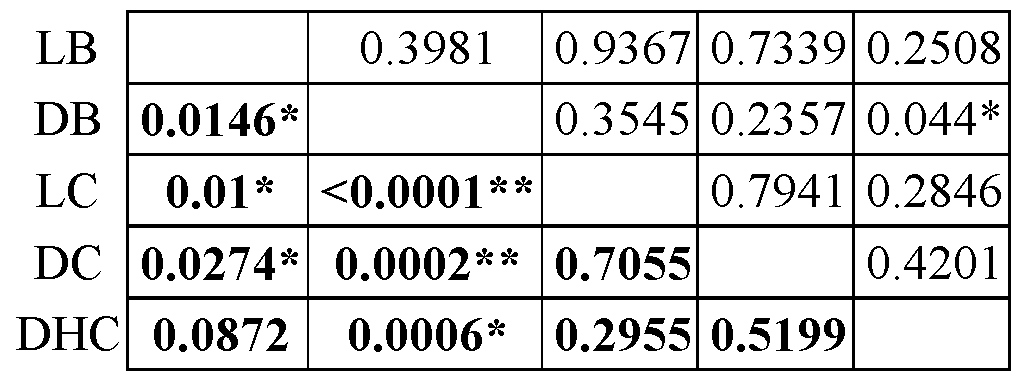
Table 5. P-values for Bone Mineral Density

* denotes p<0.05; ** denotes p<0.01; bold font denotes 4 weeks; non-bold font denotes 8 weeks.
Table 6. P-values for Bone Marrow Composition

* denotes p<0.05; ** denotes p<0.01; bold font denotes 4 weeks; non-bold font denotes 8 weeks.
Table 7. P-values for Capillary Surface Density

* denotes p<0.05; ** denotes p<0.01; bold font denotes 4 weeks; non-bold font denotes 8 weeks.
Example 2. Enhanced Endochondral Potential with In-vitro Pre-culture of Costal Cartilage.
[0178] Costal cartilage was harvested from a donor animal and in vitro cultured in a culture media containing 1 nM 3,3, 5-triiodo-L-thyronine (T3) prior to implantation into a host animal. It has been described that T3 can induce hypertrophy in human mesenchymal stem
cells (Mueller and Tuan, Arthritis Rheum, 2008, 58(5): 1377-88). FIG. 10 shows alkaline phosphatase activity (ALP, an enzymatic function involved in mineralization during the process of endochondral ossification) of either hypertrophic (black) or non-hypertrophic (white) cartilage isolated from costal cartilage. Cultured cartilage displayed enhanced endochondral potential and hypertrophy. Media containing 1 nM 3,3,5-triiodo-L-thyronine (T3) significantly increased ALP activity of both the non-hypertrophic and hypertrophic cartilage, compared to the other culture conditions ("a" of FIG. 10) but was even more potent on the hypertrophic tissue ("b" of FIG. 10).
[0179] Although the foregoing invention has been described in some detail by way of illustration and example for purposes of clarity of understanding, one of skill in the art will appreciate that certain changes and modifications may be practiced within the scope of the appended claims. In addition, each reference provided herein is incorporated by reference in its entirety to the same extent as if each reference was individually incorporated by reference.
Claims
WHAT IS CLAIMED IS: 1. A method of regenerating vascularized bone in an animal in need thereof, the method comprising:
grafting endochondral cartilage to bone in an animal; and
allowing the graft to incubate such that vascularized bone is regenerated at the site of the graft.
2. The method of claim 1, wherein the endochondral cartilage is not embedded into a scaffold.
3. The method of claim 1, wherein the endochondral cartilage is embedded into a non-synthetic scaffold.
4. The method of claim 1, wherein the endochondral cartilage is embedded into a synthetic scaffold.
5. The method of claim 4, wherein the synthetic scaffold comprises an acrylated poly(ethylene glycol) (aPEG) in a single arm (PEGDA) or in a multi-arm structure.
6. The method of claim 4, wherein the synthetic scaffold is an acrylated hyaluronic acid.
7. The method of any one of claims 4 to 6, wherein the synthetic scaffold is photopolymerized.
8. The method of any one of claims 1 to 7, wherein the endochondral cartilage is in vitro cultured in a culture medium prior to grafting.
9. The method of claim 8, wherein the culture medium comprises ascorbate-2-phosphate, dexamethasone; 3,3',5-triiodo-L-thyronine sodium salt (T3); β- glycerol phosphate; bone morphogenetic protein; or a combination thereof.
10. The method of any one of claims 1 to 9, wherein the endochondral cartilage is hypertrophic cartilage.
11. The method of claim 10, wherein the hypertrophic cartilage is selected from costal cartilage, growth plate cartilage, xyphoid process cartilage, fracture callus
cartilage, mesenchymal stem cell (MSC)-derived cartilage, embryonic stem cell-derived cartilage, or induced pluripotent stem cell-derived cartilage.
12. The method of any one of claims 1 to 11, wherein the endochondral cartilage is living cartilage.
13. The method of any one of claims 1 to 11, wherein the endochondral cartilage is devitalized cartilage.
14. The method of claim 13, wherein the devitalized cartilage is produced by performing one or more freeze-thaw cycles, lyophilization, or vacuum drying on the cartilage.
15. The method of claim 13 or 14, wherein the devitalized cartilage is processed into strips.
16. The method of claim 13 or 14, wherein the devitalized cartilage is milled into a powder.
17. The method of claim 16, wherein the milling comprises using a tissue mill, cryogrinder, or mortar and pestle.
18. The method of claim 16 or 17, wherein the devitalized cartilage is homogenized prior to milling.
19. The method of any one of claims 13 to 18, wherein the devitalized cartilage is treated with sodium deoxycholate (SDC) under conditions to remove less than 50% of glycosaminoglycans and proteoglycans in the cartilage.
20. The method of any one of claims 13 to 19, wherein the devitalized cartilage is treated with a nuclease under conditions to remove nuclear material.
21. The method of any one of claims 13 to 20, wherein the devitalized cartilage is rehydrated to form a gel or paste.
22. The method of claim 21, wherein the rehydration comprises mixing the devitalized cartilage with any of the group consisting of phosphate buffered saline (PBS),
autologous or allogeneic blood, platelet rich plasma, fibrin glue, bone marrow aspirate, bone autograft, bone marrow derived stem cells, PEGDA, and a combination thereof.
23. The method of any one of claims 1 to 22, wherein the animal is a human.
24. The method of any one of claims 1 to 23, the graft is allogeneic to the animal.
25. The method of any one of claims 1 to 23, the graft is autologous to the animal.
26. The method of any one of claims 1 to 23, the graft is xenogeneic autologous to the animal.
27. A composition for bone grafting comprising the endochondral cartilage recited in of any one of claims 1 to 26, and a pharmaceutically acceptable carrier.
28. A method of generating decellularized endochondral cartilage for bone regeneration, the method comprising:
providing endochondral cartilage,
devitalizing the endochondral cartilage, and
treating the devitalized endochondral cartilage with a nuclease under conditions to produce the decellularized endochondral cartilage for bone regeneration.
29. The method of claim 28, wherein the devitalizing comprises performing one or more freeze-thaw cycles, lyophilization, or vacuum-drying on the endochondral cartilage.
30. The method of claim 29, wherein the one or more freeze-thaw cycles are performed in an aqueous solution.
31. The method of any one of claims 28 to 30, further comprising treating the devitalized hypertrophic cartilage with sodium deoxycholate (SDC) under conditions to remove less than 50% of glycosaminoglycans and proteoglycans in the cartilage.
32. The method of any one of claims 28 to 31, further comprising milling the devitalized cartilage prior to contacting with the nuclease.
33. The method of any one of claims 28 to 32, further comprising rehydrating the decellularized cartilage with any of the group consisting of phosphate buffered saline (PBS), autologous or allogeneic blood, platelet rich plasma, fibrin glue, bone marrow aspirate, bone autograft, bone marrow derived stem cells, PEGDA, and a combination thereof.
34. The method of any one of claims 28 to 33, further comprising in vitro culturing the endochondral cartilage in a culture medium prior to devitalizing the cartilage.
35. The method of claim 34, wherein the culture medium comprises ascorbate-2-phosphate, dexamethasone; 3,3',5-triiodo-L-thyronine sodium salt (T3); β- glycerol phosphate; bone morphogenetic protein; or a combination thereof.
36. The method of any one of claims 28 to 35, wherein the endochondral cartilage is hypertrophic cartilage.
37. The method of claim 36, wherein the hypertrophic cartilage is selected from costal cartilage, growth plate cartilage, xyphoid process cartilage, fracture callus cartilage, mesenchymal stem cell (MSC)-derived cartilage, embryonic stem cell-derived cartilage, or induced pluripotent stem cell-derived cartilage.
Applications Claiming Priority (2)
| Application Number | Priority Date | Filing Date | Title |
|---|---|---|---|
| US201562139461P | 2015-03-27 | 2015-03-27 | |
| US62/139,461 | 2015-03-27 |
Publications (1)
| Publication Number | Publication Date |
|---|---|
| WO2016160717A1 true WO2016160717A1 (en) | 2016-10-06 |
Family
ID=57005252
Family Applications (1)
| Application Number | Title | Priority Date | Filing Date |
|---|---|---|---|
| PCT/US2016/024547 Ceased WO2016160717A1 (en) | 2015-03-27 | 2016-03-28 | Endochondral tissue engineering for vascularized bone regeneration |
Country Status (1)
| Country | Link |
|---|---|
| WO (1) | WO2016160717A1 (en) |
Cited By (4)
| Publication number | Priority date | Publication date | Assignee | Title |
|---|---|---|---|---|
| WO2021105404A1 (en) * | 2019-11-29 | 2021-06-03 | Novadip Biosciences | Biomaterials for the prevention and the treatment of tissue disorders |
| CN113057761A (en) * | 2021-03-10 | 2021-07-02 | 上海市肺科医院 | Bionic trachea and preparation method thereof |
| EP3967335A1 (en) | 2020-09-11 | 2022-03-16 | UMC Utrecht Holding B.V. | Engineered devitalized cartilaginous tissue for bone regeneration |
| CN116271216A (en) * | 2023-04-08 | 2023-06-23 | 扬州大学 | A preparation method of bionic chondrification-promoting 3D printed tissue-engineered trachea |
Citations (2)
| Publication number | Priority date | Publication date | Assignee | Title |
|---|---|---|---|---|
| US20130028981A1 (en) * | 2010-02-26 | 2013-01-31 | Decell Technologies Inc. | Methods for tissue decellularization |
| WO2014015109A1 (en) * | 2012-07-20 | 2014-01-23 | The Regents Of The University Of California | Methods for producing cartilage and bone |
-
2016
- 2016-03-28 WO PCT/US2016/024547 patent/WO2016160717A1/en not_active Ceased
Patent Citations (2)
| Publication number | Priority date | Publication date | Assignee | Title |
|---|---|---|---|---|
| US20130028981A1 (en) * | 2010-02-26 | 2013-01-31 | Decell Technologies Inc. | Methods for tissue decellularization |
| WO2014015109A1 (en) * | 2012-07-20 | 2014-01-23 | The Regents Of The University Of California | Methods for producing cartilage and bone |
Non-Patent Citations (1)
| Title |
|---|
| BAHNEY ET AL.: "Stem cell derived endochondral cartilage stimulates bone healing by tissue transformation.", J BONE MINER RES., vol. 29, no. 5, 2014, pages 1269 - 1282, XP055317904 * |
Cited By (5)
| Publication number | Priority date | Publication date | Assignee | Title |
|---|---|---|---|---|
| WO2021105404A1 (en) * | 2019-11-29 | 2021-06-03 | Novadip Biosciences | Biomaterials for the prevention and the treatment of tissue disorders |
| EP3967335A1 (en) | 2020-09-11 | 2022-03-16 | UMC Utrecht Holding B.V. | Engineered devitalized cartilaginous tissue for bone regeneration |
| WO2022055355A1 (en) | 2020-09-11 | 2022-03-17 | Umc Utrecht Holding B.V. | Engineered devitalized cartilaginous tissue for bone regeneration |
| CN113057761A (en) * | 2021-03-10 | 2021-07-02 | 上海市肺科医院 | Bionic trachea and preparation method thereof |
| CN116271216A (en) * | 2023-04-08 | 2023-06-23 | 扬州大学 | A preparation method of bionic chondrification-promoting 3D printed tissue-engineered trachea |
Similar Documents
| Publication | Publication Date | Title |
|---|---|---|
| Li et al. | Bone regeneration by implantation of adipose-derived stromal cells expressing BMP-2 | |
| Gao et al. | MSC-seeded dense collagen scaffolds with a bolus dose of VEGF promote healing of large bone defects | |
| JP4628756B2 (en) | Tissue repair implant, manufacturing method thereof, and tissue repair method | |
| AU2007240510B2 (en) | Bone graft composition | |
| Giannoni et al. | Regeneration of large bone defects in sheep using bone marrow stromal cells | |
| Sunay et al. | Autologous rabbit adipose tissue-derived mesenchymal stromal cells for the treatment of bone injuries with distraction osteogenesis | |
| Abedin et al. | Development of a demineralized and decellularized human epiphyseal bone scaffold for tissue engineering: A histological study | |
| KR102751476B1 (en) | Autologous bone graft substitute | |
| KR102673401B1 (en) | Biomaterials containing adipose-derived stem cells and methods for producing the same | |
| Vaněček et al. | The combination of mesenchymal stem cells and a bone scaffold in the treatment of vertebral body defects | |
| Xing et al. | Establishment of a bilateral femoral large segmental bone defect mouse model potentially applicable to basic research in bone tissue engineering | |
| US20230001053A1 (en) | Bone repair compositions | |
| Salamanna et al. | Mesenchymal stem cells for the treatment of spinal arthrodesis: from preclinical research to clinical scenario | |
| Teotia et al. | Endogenous platelet-rich plasma supplements/augments growth factors delivered via porous collagen-nanohydroxyapatite bone substitute for enhanced bone formation | |
| Wang et al. | Sheet of osteoblastic cells combined with platelet-rich fibrin improves the formation of bone in critical-size calvarial defects in rabbits | |
| US20220168356A1 (en) | Allografts containing viable cells and methods therof | |
| WO2016160717A1 (en) | Endochondral tissue engineering for vascularized bone regeneration | |
| Betz et al. | The effect of BMP-7 gene activated muscle tissue implants on the repair of large segmental bone defects | |
| CN111094551A (en) | Biomaterial containing adipose stem cells and preparation method thereof | |
| Wu et al. | Transcriptomic and cellular decoding of scaffolds-induced suture mesenchyme regeneration | |
| Hassibi et al. | Allogenic bone graft enriched by periosteal stem cell and growth factors for osteogenesis in critical size bone defect in rabbit model: histopathological and radiological evaluation | |
| Cohen et al. | A combined cell and growth factor delivery for the repair of a critical size tibia defect using biodegradable hydrogel implants | |
| JP2024534877A (en) | Compositions and methods for treating bone injuries | |
| Parsaei et al. | Acceleration of bone healing by a growth factor-releasing allo-hybrid graft | |
| Reichert | Tissue engineering bone-reconstruction of critical sized segmental bone defects in a large animal model |
Legal Events
| Date | Code | Title | Description |
|---|---|---|---|
| 121 | Ep: the epo has been informed by wipo that ep was designated in this application |
Ref document number: 16773938 Country of ref document: EP Kind code of ref document: A1 |
|
| NENP | Non-entry into the national phase |
Ref country code: DE |
|
| 122 | Ep: pct application non-entry in european phase |
Ref document number: 16773938 Country of ref document: EP Kind code of ref document: A1 |