WO2016111364A1 - がん又は炎症性疾患患者の予後を予測する方法 - Google Patents
がん又は炎症性疾患患者の予後を予測する方法 Download PDFInfo
- Publication number
- WO2016111364A1 WO2016111364A1 PCT/JP2016/050555 JP2016050555W WO2016111364A1 WO 2016111364 A1 WO2016111364 A1 WO 2016111364A1 JP 2016050555 W JP2016050555 W JP 2016050555W WO 2016111364 A1 WO2016111364 A1 WO 2016111364A1
- Authority
- WO
- WIPO (PCT)
- Prior art keywords
- gene
- patient
- expression level
- prognosis
- cancer
- Prior art date
- Legal status (The legal status is an assumption and is not a legal conclusion. Google has not performed a legal analysis and makes no representation as to the accuracy of the status listed.)
- Ceased
Links
Images


Classifications
-
- C—CHEMISTRY; METALLURGY
- C12—BIOCHEMISTRY; BEER; SPIRITS; WINE; VINEGAR; MICROBIOLOGY; ENZYMOLOGY; MUTATION OR GENETIC ENGINEERING
- C12Q—MEASURING OR TESTING PROCESSES INVOLVING ENZYMES, NUCLEIC ACIDS OR MICROORGANISMS; COMPOSITIONS OR TEST PAPERS THEREFOR; PROCESSES OF PREPARING SUCH COMPOSITIONS; CONDITION-RESPONSIVE CONTROL IN MICROBIOLOGICAL OR ENZYMOLOGICAL PROCESSES
- C12Q1/00—Measuring or testing processes involving enzymes, nucleic acids or microorganisms; Compositions therefor; Processes of preparing such compositions
- C12Q1/68—Measuring or testing processes involving enzymes, nucleic acids or microorganisms; Compositions therefor; Processes of preparing such compositions involving nucleic acids
- C12Q1/6876—Nucleic acid products used in the analysis of nucleic acids, e.g. primers or probes
- C12Q1/6883—Nucleic acid products used in the analysis of nucleic acids, e.g. primers or probes for diseases caused by alterations of genetic material
- C12Q1/6886—Nucleic acid products used in the analysis of nucleic acids, e.g. primers or probes for diseases caused by alterations of genetic material for cancer
-
- C—CHEMISTRY; METALLURGY
- C12—BIOCHEMISTRY; BEER; SPIRITS; WINE; VINEGAR; MICROBIOLOGY; ENZYMOLOGY; MUTATION OR GENETIC ENGINEERING
- C12Q—MEASURING OR TESTING PROCESSES INVOLVING ENZYMES, NUCLEIC ACIDS OR MICROORGANISMS; COMPOSITIONS OR TEST PAPERS THEREFOR; PROCESSES OF PREPARING SUCH COMPOSITIONS; CONDITION-RESPONSIVE CONTROL IN MICROBIOLOGICAL OR ENZYMOLOGICAL PROCESSES
- C12Q1/00—Measuring or testing processes involving enzymes, nucleic acids or microorganisms; Compositions therefor; Processes of preparing such compositions
- C12Q1/02—Measuring or testing processes involving enzymes, nucleic acids or microorganisms; Compositions therefor; Processes of preparing such compositions involving viable microorganisms
- C12Q1/025—Measuring or testing processes involving enzymes, nucleic acids or microorganisms; Compositions therefor; Processes of preparing such compositions involving viable microorganisms for testing or evaluating the effect of chemical or biological compounds, e.g. drugs, cosmetics
-
- C—CHEMISTRY; METALLURGY
- C12—BIOCHEMISTRY; BEER; SPIRITS; WINE; VINEGAR; MICROBIOLOGY; ENZYMOLOGY; MUTATION OR GENETIC ENGINEERING
- C12Q—MEASURING OR TESTING PROCESSES INVOLVING ENZYMES, NUCLEIC ACIDS OR MICROORGANISMS; COMPOSITIONS OR TEST PAPERS THEREFOR; PROCESSES OF PREPARING SUCH COMPOSITIONS; CONDITION-RESPONSIVE CONTROL IN MICROBIOLOGICAL OR ENZYMOLOGICAL PROCESSES
- C12Q1/00—Measuring or testing processes involving enzymes, nucleic acids or microorganisms; Compositions therefor; Processes of preparing such compositions
- C12Q1/02—Measuring or testing processes involving enzymes, nucleic acids or microorganisms; Compositions therefor; Processes of preparing such compositions involving viable microorganisms
- C12Q1/04—Determining presence or kind of microorganism; Use of selective media for testing antibiotics or bacteriocides; Compositions containing a chemical indicator therefor
-
- C—CHEMISTRY; METALLURGY
- C12—BIOCHEMISTRY; BEER; SPIRITS; WINE; VINEGAR; MICROBIOLOGY; ENZYMOLOGY; MUTATION OR GENETIC ENGINEERING
- C12Q—MEASURING OR TESTING PROCESSES INVOLVING ENZYMES, NUCLEIC ACIDS OR MICROORGANISMS; COMPOSITIONS OR TEST PAPERS THEREFOR; PROCESSES OF PREPARING SUCH COMPOSITIONS; CONDITION-RESPONSIVE CONTROL IN MICROBIOLOGICAL OR ENZYMOLOGICAL PROCESSES
- C12Q1/00—Measuring or testing processes involving enzymes, nucleic acids or microorganisms; Compositions therefor; Processes of preparing such compositions
- C12Q1/68—Measuring or testing processes involving enzymes, nucleic acids or microorganisms; Compositions therefor; Processes of preparing such compositions involving nucleic acids
- C12Q1/6876—Nucleic acid products used in the analysis of nucleic acids, e.g. primers or probes
- C12Q1/6883—Nucleic acid products used in the analysis of nucleic acids, e.g. primers or probes for diseases caused by alterations of genetic material
-
- G—PHYSICS
- G01—MEASURING; TESTING
- G01N—INVESTIGATING OR ANALYSING MATERIALS BY DETERMINING THEIR CHEMICAL OR PHYSICAL PROPERTIES
- G01N33/00—Investigating or analysing materials by specific methods not covered by groups G01N1/00 - G01N31/00
- G01N33/48—Biological material, e.g. blood, urine; Haemocytometers
- G01N33/50—Chemical analysis of biological material, e.g. blood, urine; Testing involving biospecific ligand binding methods; Immunological testing
- G01N33/53—Immunoassay; Biospecific binding assay; Materials therefor
-
- G—PHYSICS
- G01—MEASURING; TESTING
- G01N—INVESTIGATING OR ANALYSING MATERIALS BY DETERMINING THEIR CHEMICAL OR PHYSICAL PROPERTIES
- G01N33/00—Investigating or analysing materials by specific methods not covered by groups G01N1/00 - G01N31/00
- G01N33/48—Biological material, e.g. blood, urine; Haemocytometers
- G01N33/50—Chemical analysis of biological material, e.g. blood, urine; Testing involving biospecific ligand binding methods; Immunological testing
- G01N33/53—Immunoassay; Biospecific binding assay; Materials therefor
- G01N33/575—Immunoassay; Biospecific binding assay; Materials therefor for cancer
- G01N33/5758—Immunoassay; Biospecific binding assay; Materials therefor for cancer involving compounds serving as markers for tumours, cancers or neoplasias, e.g. cellular determinants, receptors, heat shock/stress proteins, A-protein, oligosaccharides or metabolites
- G01N33/5759—Immunoassay; Biospecific binding assay; Materials therefor for cancer involving compounds serving as markers for tumours, cancers or neoplasias, e.g. cellular determinants, receptors, heat shock/stress proteins, A-protein, oligosaccharides or metabolites involving compounds localised on the membrane of tumour or cancer cells
-
- G—PHYSICS
- G01—MEASURING; TESTING
- G01N—INVESTIGATING OR ANALYSING MATERIALS BY DETERMINING THEIR CHEMICAL OR PHYSICAL PROPERTIES
- G01N33/00—Investigating or analysing materials by specific methods not covered by groups G01N1/00 - G01N31/00
- G01N33/48—Biological material, e.g. blood, urine; Haemocytometers
- G01N33/50—Chemical analysis of biological material, e.g. blood, urine; Testing involving biospecific ligand binding methods; Immunological testing
- G01N33/68—Chemical analysis of biological material, e.g. blood, urine; Testing involving biospecific ligand binding methods; Immunological testing involving proteins, peptides or amino acids
- G01N33/6863—Cytokines, i.e. immune system proteins modifying a biological response such as cell growth proliferation or differentiation, e.g. TNF, CNF, GM-CSF, lymphotoxin, MIF or their receptors
-
- C—CHEMISTRY; METALLURGY
- C12—BIOCHEMISTRY; BEER; SPIRITS; WINE; VINEGAR; MICROBIOLOGY; ENZYMOLOGY; MUTATION OR GENETIC ENGINEERING
- C12Q—MEASURING OR TESTING PROCESSES INVOLVING ENZYMES, NUCLEIC ACIDS OR MICROORGANISMS; COMPOSITIONS OR TEST PAPERS THEREFOR; PROCESSES OF PREPARING SUCH COMPOSITIONS; CONDITION-RESPONSIVE CONTROL IN MICROBIOLOGICAL OR ENZYMOLOGICAL PROCESSES
- C12Q2600/00—Oligonucleotides characterized by their use
- C12Q2600/106—Pharmacogenomics, i.e. genetic variability in individual responses to drugs and drug metabolism
-
- C—CHEMISTRY; METALLURGY
- C12—BIOCHEMISTRY; BEER; SPIRITS; WINE; VINEGAR; MICROBIOLOGY; ENZYMOLOGY; MUTATION OR GENETIC ENGINEERING
- C12Q—MEASURING OR TESTING PROCESSES INVOLVING ENZYMES, NUCLEIC ACIDS OR MICROORGANISMS; COMPOSITIONS OR TEST PAPERS THEREFOR; PROCESSES OF PREPARING SUCH COMPOSITIONS; CONDITION-RESPONSIVE CONTROL IN MICROBIOLOGICAL OR ENZYMOLOGICAL PROCESSES
- C12Q2600/00—Oligonucleotides characterized by their use
- C12Q2600/118—Prognosis of disease development
-
- C—CHEMISTRY; METALLURGY
- C12—BIOCHEMISTRY; BEER; SPIRITS; WINE; VINEGAR; MICROBIOLOGY; ENZYMOLOGY; MUTATION OR GENETIC ENGINEERING
- C12Q—MEASURING OR TESTING PROCESSES INVOLVING ENZYMES, NUCLEIC ACIDS OR MICROORGANISMS; COMPOSITIONS OR TEST PAPERS THEREFOR; PROCESSES OF PREPARING SUCH COMPOSITIONS; CONDITION-RESPONSIVE CONTROL IN MICROBIOLOGICAL OR ENZYMOLOGICAL PROCESSES
- C12Q2600/00—Oligonucleotides characterized by their use
- C12Q2600/158—Expression markers
-
- G—PHYSICS
- G01—MEASURING; TESTING
- G01N—INVESTIGATING OR ANALYSING MATERIALS BY DETERMINING THEIR CHEMICAL OR PHYSICAL PROPERTIES
- G01N2333/00—Assays involving biological materials from specific organisms or of a specific nature
- G01N2333/435—Assays involving biological materials from specific organisms or of a specific nature from animals; from humans
- G01N2333/705—Assays involving receptors, cell surface antigens or cell surface determinants
- G01N2333/715—Assays involving receptors, cell surface antigens or cell surface determinants for cytokines; for lymphokines; for interferons
- G01N2333/7158—Assays involving receptors, cell surface antigens or cell surface determinants for cytokines; for lymphokines; for interferons for chemokines
-
- G—PHYSICS
- G01—MEASURING; TESTING
- G01N—INVESTIGATING OR ANALYSING MATERIALS BY DETERMINING THEIR CHEMICAL OR PHYSICAL PROPERTIES
- G01N2800/00—Detection or diagnosis of diseases
- G01N2800/52—Predicting or monitoring the response to treatment, e.g. for selection of therapy based on assay results in personalised medicine; Prognosis
-
- G—PHYSICS
- G01—MEASURING; TESTING
- G01N—INVESTIGATING OR ANALYSING MATERIALS BY DETERMINING THEIR CHEMICAL OR PHYSICAL PROPERTIES
- G01N2800/00—Detection or diagnosis of diseases
- G01N2800/70—Mechanisms involved in disease identification
- G01N2800/7095—Inflammation
Definitions
- the present invention relates to a method for predicting the prognosis of a patient with cancer or an inflammatory disease.
- the front protein is a cytoplasmic protein that binds to the intracellular C-terminal region of the chemokine receptors CCR2 and CCR5, and positively controls migration signals of macrophages (Patent Document 1, Non-Patent Documents 1 and 2). It is a novel molecule discovered by the group of the present inventors.
- Non-Patent Documents 3 and 4 The existing approach targeted the signal transduction system such as binding of chemokine CCL2 and receptor CCR2, binding of chemokine CCL3-5 to receptor CCR5, and PI3K downstream of the receptor. Inhibition of binding between the chemokine receptors CCR2 and CCR5 and the front is expected as a new drug discovery target (Non-patent Documents 3 and 4).
- An object of the present invention is to provide a novel means for enabling prognosis prediction of patients with cancer and inflammatory diseases. It is another object of the present invention to provide a novel means capable of selecting patients who have high effects of chemokine inhibitors and front inhibitors, or patients who are expected to improve symptoms significantly by administration of these inhibitors.
- the prognosis is good even when the expression of front, which is a prognostic exacerbation factor, is high, and the expression data and prognostic data of known patient groups are analyzed, and CC chemokine receptors / ligands are analyzed. It was found that a more accurate prognosis can be predicted by predetermining the cut-off value.
- the present invention was completed by finding the above.
- the present invention is a method for predicting the prognosis of a patient with cancer or inflammatory disease, comprising measuring the expression level of a front gene in a sample collected from the patient, wherein the expression level of the front gene is The lower the method, the better the prognosis of the patient is expected.
- the present invention is also a method for predicting the prognosis of a patient with cancer or inflammatory disease, comprising measuring the expression level of a CC chemokine receptor / ligand gene in a sample collected from the patient, The higher the expression level of the chemokine receptor / ligand gene, the better the prognosis of the patient, the CC chemokine receptor / ligand gene from the group consisting of CCR2 gene, CCR5 gene, CCL2 gene and CCL5 gene A method is provided that is at least one selected.
- the present invention is a method for predicting the prognosis of a patient with cancer or inflammatory disease, wherein the expression level of a front gene and the expression level of a CC chemokine receptor / ligand gene in a sample collected from the patient are measured.
- the CC chemokine receptor / ligand gene in the sample is greater than or equal to a predetermined reference value, the patient's prognosis is predicted to be good, and the CC chemokine receptor /
- the CC chemokine receptor / ligand gene is CCR2 gene, A method is provided that is at least one selected from the group consisting of a CCR5 gene, a CCL2 gene, and a CCL5 gene.
- the present invention relates to a method for predicting the effectiveness of a medicine comprising a front inhibitor or a chemokine inhibitor as an active ingredient, and the expression of the front gene in a sample isolated from a patient for which administration of the medicine is considered. Measuring the amount and providing a method wherein the higher the expression of the front gene, the higher the effect of the medicament in the patient is predicted. Furthermore, the present invention is a method for selecting a patient having a high pharmaceutical effect comprising a front inhibitor or a chemokine inhibitor as an active ingredient, wherein the front inhibitor or chemokine inhibitor is used as an active ingredient by the method of the present invention.
- the present invention is a method for selecting a patient for whom administration of a medicine containing a front inhibitor or a chemokine inhibitor as an active ingredient is desired, comprising examining the expression of a front gene in a sample collected from the patient, The chemokine inhibitor is at least one inhibitor selected from the group consisting of CCR2, CCR5, CCL2 and CCL5, and a patient whose front gene expression is detected is selected as a patient for whom administration of the medicine is desired.
- a method for selecting a patient for whom administration of a medicine containing a front inhibitor or a chemokine inhibitor as an active ingredient is desired, comprising examining the expression of a front gene in a sample collected from the patient, The chemokine inhibitor is at least one inhibitor selected from the group consisting of CCR2, CCR5, CCL2 and CCL5, and a patient whose front gene expression is detected is selected as a patient for whom administration of the medicine is desired.
- the present invention provides the use of the front gene product as a prognostic predictive marker that is negatively correlated with the prognosis of cancer or inflammatory disease. Furthermore, the present invention provides at least one gene selected from the group consisting of a CCR2 gene, a CCR5 gene, a CCL2 gene, and a CCL5 gene as a prognostic predictive marker that shows a positive correlation with the prognosis of cancer or inflammatory disease. Provide product use. Furthermore, the present invention provides a prognostic kit for cancer or inflammatory disease, comprising at least one of the following (1) and (2).
- predicting the prognosis of patients with cancer and inflammatory diseases based on the expression level of a front gene as a prognostic exacerbation marker or a CC chemokine receptor / ligand gene such as CCR2 as a good prognosis marker Is possible.
- a front gene as a prognostic exacerbation marker or a CC chemokine receptor / ligand gene such as CCR2 as a good prognosis marker Is possible.
- a more accurate prediction is possible.
- the prognostic prediction results can be used for the determination of treatment policy and follow-up.
- various chemokine inhibitors have been studied and developed for the treatment of cancer and inflammatory diseases, and inhibitors targeting the receptors CCR2 and CCR5 and their ligands CCL2 and CCL5 are also cancerous.
- a drug containing such a chemokine inhibitor or front inhibitor as an active ingredient is expected to be particularly effective in patients with a high expression level of the front gene. According to the present invention, it is possible to predict the effectiveness of chemokine inhibitors and front inhibitors and to select patients who are highly effective in these inhibitors or patients who want to administer these inhibitors.
- (A) A graph showing the relationship between the expression state of CCR2 and CCL2 and the survival rate after surgery.
- (B) is a graph showing the relationship between the expression state of CCR5 and CCL5 and the postoperative survival rate.
- hi high expression
- lo low expression.
- Both A and B were significantly different from the other groups in the group with high expression of both ligand and receptor (p ⁇ 0.05).
- This is the result of recursive partitioning analysis of data on the front expression level, CCR2 expression level and postoperative survival time of 68 lung cancer patients. It was divided into three groups: front low expression (FNT-L), front high expression (FNT-H), and CCR2 high expression (CCR2-H).
- FNT-L front low expression
- FNT-H front high expression
- the expression level of the front gene is measured using a sample collected from a patient with cancer or an inflammatory disease.
- the front gene is a molecule that binds to CCR2 and CCR5 and was found for the first time by the present inventors' group to positively control migration signals such as macrophages (Nature Immunology, 6: 827-835, 2005, Journal of Immunology, 183: 6387-6394, 2009).
- Front gene and front protein sequence information is registered in NCBI database GenBank with accession numbers of AF498261 and NM_024844. Among these, the base sequence and amino acid sequence registered as AF498261 are shown in SEQ ID NOs: 1 and 2 in the sequence listing.
- the front is a prognostic exacerbation factor, and it can be predicted that the lower the expression level of the front gene, the better the prognosis. That is, the front gene product can be used as a prognostic predictive marker that shows a negative correlation with the prognosis of cancer or inflammatory disease.
- the term gene product includes both mRNA and protein expressed from the gene.
- the expression level of the CC chemokine receptor / ligand gene is measured using a sample collected from a patient with cancer or an inflammatory disease.
- the “CC chemokine receptor / ligand gene” is at least one gene selected from the group consisting of a CCR2 gene, a CCR5 gene, a CCL2 gene, and a CCL5 gene, such as a CCR2 gene and a CCR5 gene. There may be at least one selected.
- the CC chemokine receptor / ligand gene can be a combination of two genes, the CCR2 gene and the CCL2 gene, or a combination of two genes, the CCR5 gene and the CCL5 gene.
- CC chemokine receptor / ligand is a good prognostic factor, and patients with higher expression levels of CC chemokine receptor / ligand gene can be predicted to have better prognosis. That is, the CC chemokine receptor / ligand gene product can be used as a prognostic marker that shows a positive correlation with the prognosis of cancer or inflammatory disease. The prognosis is particularly good in patients with high CCR2 and high CCL2 expression, and patients with high CCR5 and high CCL5 gene expression.
- CCR2 has two isoforms, CCR2A and CCR2B, with different C-terminal regions, but the main isoform widely expressed in cells is CCR2B.
- CCR2 to be measured is CCR2B.
- CCR2B is CCR2B unless it is clear from the context.
- the base sequence and amino acid sequence of CCR2B registered in GenBank with the accession number of NM_001123396.1 are shown in SEQ ID NOs: 3 and 4.
- CCR5 has two isoforms, CCR5A and CCR5B, with different 5'-UTR regions, but both have the same coding region sequence and the same protein amino acid sequence.
- both isoforms can be detected by setting primers or the like for the coding region.
- sequence of the CCR5 gene the nucleotide sequence and amino acid sequence of CCR5A registered in GenBank as NM_000579.3 are shown in SEQ ID NOs: 5 and 6, respectively.
- SEQ ID Nos: 7 to 10 show the base sequence and amino acid sequence of CCL2 (NM_002982.3) and CCL5 (NM_002985.2), respectively.
- the expression level of the front gene and the expression level of the CC chemokine receptor / ligand gene are measured using a sample collected from a patient with cancer or an inflammatory disease.
- Analysis of survival data from known lung cancer patient populations revealed that patients with high chemokine receptor / ligand gene expression levels above a certain level had a good prognosis even with high front expression levels.
- the reference value of the expression level of the CC chemokine receptor / ligand gene determined in advance by analysis of the expression data and prognostic data of the known patient population, and the CC chemokine reception measured in the patient-derived sample. Contrast body / ligand gene expression levels. If the patient's CC chemokine receptor / ligand expression level is above the reference value, the patient's prognosis can be predicted to be good.
- cancer patients with a high expression level of CC chemokine receptor / ligand gene have a good prognosis even if the front expression level is high, but in this CC chemokine receptor / ligand high expression group, The proportion of stage I patients is extremely high. Therefore, when the first method is performed on a cancer patient, a patient of stage II or higher may be selected as a prognosis target. When the first method is performed on stage II or higher cancer patients, it is possible to predict that the higher the expression of the front gene, the worse the prognosis.
- the sample used for expression analysis is, for example, a tissue sample of a lesioned part collected from a patient.
- a lesion tissue sample (a microenvironment containing cancer cells) can be obtained from a surgical specimen excised by surgery.
- a tissue sample can be obtained from a biopsy sample of an inflammatory lesion (inflammatory microenvironment containing inflammatory cells).
- a tissue fragment containing cancer cells or inflammatory cells and surrounding microenvironmental cells may be used.
- the sample can be a blood sample or sputum.
- macrophages are the most prominent cells.
- peripheral blood and sputum are the amount of front-expressed macrophages in the patient. Therefore, it is possible to implement the present invention using a blood sample or sputum.
- the method for measuring the expression level is not particularly limited, and any method may be used as long as the expression level can be quantitatively or semi-quantitatively evaluated as numerical data.
- immunological measurement methods such as Western blotting and immunohistochemical staining using antibodies against the molecules to be measured can also be employed.
- a method for examining the expression level of mRNA, particularly real-time PCR can be preferably used.
- the measurement value of the expression level includes a relative value after normalization with respect to a standard gene and a logarithmic value thereof (log gene expression level).
- a housekeeping gene such as a Gapdh gene is usually used.
- the expression level of the molecule to be measured may be evaluated quantitatively or semi-quantitatively according to the degree of staining. It may be a semi-quantitative evaluation by visual observation, or may be quantitatively evaluated by analyzing a stained image with a computer and digitizing the degree of staining. When the expression level is visually evaluated from the stained image, for example, whether the expression level is high may be evaluated by comparing with a plurality of sample images including examples of low and high expression levels.
- the first and second methods for predicting that the lower the expression level or the higher the better the prognosis can be performed without setting a reference value for the expression level, so in addition to the method for examining the expression level of mRNA, A method for examining the expression level of a protein by immunohistological staining or the like can also be preferably used.
- the measurement of the expression level of the front gene in the third method is the same.
- the expression level of the front protein is examined by immunohistochemical staining or the like, and the expression level of the CC chemokine receptor / ligand mRNA is determined in real time. It is also possible to check by PCR or the like.
- Primers and probes used for expression analysis such as real-time PCR are appropriately designed by those skilled in the art based on sequence information of known front genes and CC chemokine receptor / ligand genes (CCR2, CCR5, CCL2 or CCL5). can do.
- Antibodies for immunologically measuring the front and CC chemokine receptors / ligands can also be generated by those skilled in the art by well-known conventional methods, and appropriate commercially available products can also be used.
- gene expression includes both mRNA expression and protein expression
- measurement of gene expression level includes measurement of expressed mRNA and measurement of expressed protein. Both are included.
- the reference value for the expression level of CC chemokine receptor / ligand gene is a statistical analysis of the expression data and prognostic data of the front of the known patient population and CC chemokine receptor / ligand. It can be obtained by examining the relationship.
- prognostic data in the case of cancer, generally, survival data (postoperative survival time or disease-free survival time) is used as prognostic data.
- survival data postoperative survival time or disease-free survival time
- data on relapse or exacerbation is prognostic data. It can be suitably obtained as numerical data such as a period (days, months or years) until relapse or exacerbation is observed, or a total score evaluation of symptoms within a certain period.
- Specific example 1 of reference value determination method Recurrent fractional analysis of expression data and prognostic data from known patient populations, patients with high front and poor prognosis (FNT-H), patients with low front and good prognosis (FNT-L), And the patient group (CC-H) with high expression of CC chemokine receptor / ligand and high prognosis.
- the expression level of CC chemokine receptor / ligand that distinguishes between FNT-H and FNT-L and CC-H can be used as a reference value.
- log gene expression level is 1.42 for CCR2, 2.23 for CCR5, For CCL5, a value of 1.34 is obtained as a reference value. Therefore, for patients with various cancers and inflammatory diseases, preferably cancer patients, the log gene expression level is such that CCR2 is between about 1.27 and about 1.57, eg between about 1.32 and about 1.52, and CCR5 is A numerical value between about 2.08 and about 2.38, for example between about 2.13 and about 2.33, and CCL5 between about 1.19 and about 1.49, for example between about 1.24 and about 1.44, may be used as reference values, respectively. It can. However, these numerical values are merely examples, and more desirable numerical values can be obtained by collecting data of a larger number of patient populations. Therefore, the scope of the present invention is not limited to such specific numerical values.
- Specific example 2 of reference value determination method When the target of prognosis is a cancer patient, a cut-off value of the expression level of CC chemokine receptor / ligand gene that significantly separates the stage I known patient group and the stage II or higher known patient group is obtained. Is a reference value. Since it is a question of whether it is stage I (Yes) or more than stage II (No), various statistical analysis methods for binary variables can be used. For example, analysis methods such as logistic analysis and support vector machine can be mentioned, but the method is not limited to these.
- Prognosis prediction for patients whose CC chemokine receptor / ligand expression level is less than the reference value For patients whose reference level or less, it is possible to predict that the higher the front expression level, the worse the prognosis.
- a front reference value serving as a cutoff value for dividing the FNT-H group and the FNT-L group can also be obtained.
- the CC chemokine receptor / ligand expression level reference value is referred to as the CC reference value for distinction from the front reference value.
- the prognosis is likely to be good if the patient's front expression level is less than this front reference value. Therefore, it can be predicted that the prognosis is likely to be poor. However, there is no change in the tendency that the higher the front expression level, the worse the prognosis.
- the log value is a logarithmic value with a base of 10.
- ⁇ CC ⁇ ([CC] ⁇ [CC] m ) + ⁇ FROUNT ⁇ ([FROUNT] ⁇ [FROUNT] m ) Equation 1
- [CC] is the log value of the expression level of CC chemokine receptor / ligand gene in the sample
- [CC] m is the average or median log value of the expression level of CC chemokine receptor / ligand gene measured in a known patient population
- [FROUNT] is the log value of the expression level of the front gene in the sample
- [FROUNT] m is the average or median log value of the expression level of the front gene measured in a known patient population
- ⁇ CC is a coefficient obtained by applying a Cox proportional hazard model with the log value of the expression level of CC chemokine receptor / ligand gene as a variable for the prognostic data for a known patient population
- ⁇ FROUNT is a coefficient obtained by applying a Cox proportional hazard
- the log gene expression level normalized with respect to a standard gene such as the Gapdh gene is used as the expression level.
- [CC] m and [FROUNT] m may be either an average value or a median value.
- the coefficients ⁇ CC and ⁇ FROUNT may be obtained for all cases of a known patient population, or may be obtained for cases where the expression level of the CC chemokine receptor / ligand gene is less than the CC reference value.
- specific examples in which the Cox proportional hazard model was applied to cases of stage II or higher (40 cases) out of a population of 68 lung cancer patients and the respective coefficients were obtained. ) Is described.
- coefficients and median values used in the following formulas 1-1 to 1-4 are examples, and it is possible to collect more patient population data to obtain more desirable coefficients and median values (or average values). Therefore, the scope of the present invention is not limited to these specific numerical values.
- Various types of statistical processing software that can be used when the Cox proportional hazard model is used are known, and any of them may be used.
- One skilled in the art can determine the coefficient and median (or mean value) in Equation 1 using appropriate patient population data and appropriate statistical processing software.
- Equation 1 is applicable when the prediction target patient is less than the CC reference value.
- the prediction accuracy is particularly low for patients who are less than the CC reference value and stage II or higher. Expected to be high.
- the types of cancer and inflammatory diseases whose prognosis can be predicted by the method of the present invention are not particularly limited, and various cancers and inflammations that are known to involve at least one of front, CCR2, CCR5, CCL2, and CCL5.
- sexual diseases are included.
- Cancers and inflammatory diseases involving at least one of CCR2, CCR5, CCL2 and CCL5 are also cancers and inflammatory diseases involving the front.
- Target cancers include both solid and liquid cancers. Also included are both primary and metastatic cancers.
- cancer examples include lung cancer, melanoma, stomach cancer, colon cancer, breast cancer, liver cancer, pancreatic cancer, uterine cancer, esophageal cancer, prostate cancer, malignant lymphoma, leukemia and the like.
- melanoma, breast cancer, prostate cancer, lung cancer, myeloma, brain tumor, etc. are known as cancers involving CCR2, and breast cancer, prostate cancer, lung cancer, pancreas are cancers involving CCR5.
- Cancer, myeloma, etc. are known (Scholten DJ, et al.,. Br J Pharmacol, ol165: 1617-1643, -162012).
- anticancer agents in the chemokine field include anticancer agents for metastatic castration-resistant prostate cancer that targets CCL2, and anticancer agents for non-small cell lung cancer that target CCL5.
- Drugs, anti-cancer drugs for metastatic cancer targeting CCR2, and anti-cancer drugs for advanced colorectal cancer targeting CCR5 are preferred specific examples that are subject to prognosis prediction in the present invention.
- the target inflammatory disease is typically a chronic inflammatory disease.
- inflammatory diseases include, but are not limited to, rheumatoid arthritis, fibrosis, peritonitis, multiple sclerosis, arteriosclerosis, diabetes, asthma, Alzheimer, psoriasis, atopy and the like.
- Inflammatory diseases known to be involved in at least one of CCR2, CCR5, CCL2 and CCL5 include arteriosclerosis, multiple sclerosis, rheumatoid arthritis, psoriasis, type 2 diabetes, inflammatory bowel disease, chronic hepatitis, Nephritis, graft-versus-host disease, chronic obstructive pulmonary disease, asthma, acquired immune deficiency syndrome, etc.
- inflammatory diseases involving at least one of CCR2, CCR5, CCL2 and CCL5 or the front include various fibrotic diseases such as pulmonary fibrosis and liver fibrosis, peritonitis, allergic airway hypersensitivity, etc. (Nippon Linyi 70, Kou Special Issue 8, 8365-371, 2012; Kashiwa and Examples below) These inflammatory diseases are preferred specific examples that are subject to prognosis prediction in the present invention.
- chemokine inhibitors As described above, various chemokine inhibitors have been researched and developed for the treatment of cancer and inflammatory diseases, and inhibitors targeting the receptors CCR2 and CCR5 and their ligands CCL2 and CCL5 are also available. Developed as a medicine for cancer and inflammatory diseases. Development of a front inhibitor, which is a new drug discovery target molecule, is also being promoted by the group of the present inventors. A drug containing such a chemokine inhibitor or front inhibitor as an active ingredient is expected to be particularly effective in patients with a high expression level of the front gene.
- patients who are considered to be administered chemokine inhibitors or front inhibitors that inhibit at least one selected from CCR2, CCR5, CCL2 and CCL5 as active ingredients
- chemokine inhibitors or front inhibitors that inhibit at least one selected from CCR2, CCR5, CCL2 and CCL5 as active ingredients
- a front inhibitor or CC chemokine receptor / ligand inhibitors may not be sufficient to achieve the effects of these inhibitors .
- patients whose front gene expression is detected are expected to improve their symptoms significantly by administration of a drug containing the above-mentioned inhibitor as an active ingredient. Therefore, administration of such a drug is particularly desirable. . That is, by examining the expression of a front gene in a sample derived from a patient, it is possible to select a patient (typically a cancer patient or an inflammatory disease patient) for whom administration of a drug containing the inhibitor as an active ingredient is desired. Is possible.
- Primer set, probe, or antibody that can measure the expression level of the front gene as a prognostic marker and primer set, probe, or antibody that can measure the expression of the CC chemokine receptor / ligand gene as a prognostic marker It can be provided as a prognostic kit by appropriately combining with other reagents and instruction manuals.
- the kit may contain any one or both of a primer set, probe or antibody for front expression measurement and a primer set, probe or antibody for CC chemokine receptor / ligand expression measurement. You may go out.
- a sample-stained image including examples of low and high front expression may also be provided.
- mice with front knockout were created using the cre / loxP system. Since the front was completely defective and embryonic lethal, conditional knockout was performed using a system that induces recombination reactions in a tamoxifen-dependent manner.
- a targeting vector in which a genomic region containing exons 15 to 19 of the front gene was sandwiched with LoxP was introduced into mice, and heterozygous FNT flox mice were mated to produce homozygous FNT flox / flox mice.
- B6.Cg-Tg CAG-cre / Esr1 *) 5Amc / J mouse (Jaxon Laboratory) and FNT flox / flox mouse into which Cre recombinase and mutant estrogen receptor fusion protein Cre-ER were introduced were mated.
- Tamoxifen-induced frontal conditional knockout mice FNT-cKO were obtained.
- Treatment of tamoxifen with FNT-cKO mice induces frontal deletion in both genomic DNA and mRNA, and expression of front mRNA is suppressed to less than half in tamoxifen-treated FNT-cKO mice. confirmed.
- CE-2 powdered feed (CLEA Japan) supplemented with 0.4 mg / 1 g CE-2 tamoxifen citrate (Wako Pure Chemical Industries) from 6 or 14 days before the experiment. Cre expression was induced by feeding to 8-16 week old FNT flox / flox mice and FNT-cKO mice, and recombination was confirmed by Southern blotting.
- Tumor volume (tumor minor axis) 2 x tumor major axis / 2
- Front is highly expressed in macrophages [Method] Production of front-gfp-knock-in mice By a conventional method, a GFP gene was incorporated downstream of the front promoter on the mouse genome to produce front-gfp-knock-in mice.
- Flow cytometry Peritoneal infiltrating cells were collected from mice in which 2 mL of 4% thioglycolate was administered intraperitoneally to induce peritonitis. Cells were washed with PBS supplemented with 2% fetal calf serum, resuspended, and filtered through a 70 ⁇ m strainer. After blocking the Fc receptor by incubation with an anti-mouse CD16 / 32 antibody (BD biosciences), the cells were stained with a fluorescently labeled antibody.
- Anti-mouse CD11b-Pacific Blue, anti-mouse Ly6C-APC-Cy7, anti-mouse Ly6G-Alexa Fluor 700, anti-mouse CD4-FITC and anti-mouse B220-PE-Cy7 were purchased from Biolegend.
- Anti-mouse CD8-Pacific Blue was purchased from BD biosciences.
- Anti-mouse CD11b-Pacific Blue, anti-mouse Ly6C-APC-Cy7, and anti-mouse Ly6G-Alexa Fluor 700 were purchased from Biolegend. Stained cells were analyzed using a Gallios flow cytometer (Beckman coulter).
- OCT Optimal Cutting Temperature Compound
- Tissue samples All tissue samples were frozen in liquid nitrogen immediately after collection and stored at ⁇ 80 ° C. until use. Cancer progression (stage) was evaluated using a TNM (tumor node metastasis) classification system. The samples were evaluated by two pathologists from the Chiba Cancer Center and confirmed by a pathologist specializing in pulmonary pathology.
- TNM tumor node metastasis
- RNA-Bee reagent Tel-Test
- TRIzol reagent Invitrogen
- Total RNA was extracted.
- CDNA was prepared from 1 ⁇ g of total RNA using SuperScript synthesis kit (Life Technologies).
- CDNA was amplified using SYBR Green PCR Master Mix (Qiagen), and real-time quantitative RT-PCR was performed.
- the forward primer and the reverse primer were used as reaction systems with final concentrations of 0.5 ⁇ M and 20 ⁇ L, respectively.
- the assay was performed in duplicate using the ABI 7500 real-time PCR system (Life Technologies) according to the manufacturer's instructions. The results were normalized to the RNA expression level of Gapdh gene.
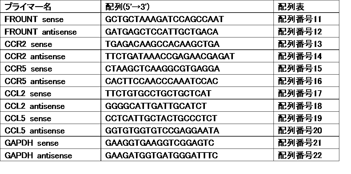
- the primers used are shown in Table 2 below.
- Log gene expression level log 10 [2-(Ct [gene]-Ct [GAPDH])] Ct [gene]: Front, CCR2, CCR5, CCL2 or CCL5 Ct value
- the median gene expression level was set as the cut-off value, and 120 patients were divided into high and low expression groups for each gene. Separately, the relationship with prognosis was evaluated. The significance test was performed by chi-square test. Postoperative survival was analyzed by Kaplan-Meier method (Mantel-Cox test), and differences between groups were evaluated by log rank test. Statistical calculations were performed using Prism 6 (GraphPad Software). p ⁇ 0.05 was considered significant.
- the postoperative survival rate was analyzed by Kaplan-Meier method (Mantel-Cox test) in the same manner as described above, and the difference between groups was evaluated by the log rank test. p ⁇ 0.05 was considered significant.
- CCR2, CCR5, CCL2 and CCL5 mRNA is a significant prognostic factor in the postoperative course of lung adenocarcinoma.
- Prognostic significance of the expression status of chemokine ligands and receptors (CCR2, CCR5, CCL2, CCL5)
- the median mRNA expression level of each gene was used as a cut-off value, and 120 patient groups were divided into a high expression group and a low expression group. All of CCR2, CCR5, CCL2 and CCL5 had a significantly better prognosis in the high expression group than in the low expression group (FIGS. 6A, B, C and D).
- the prognosis was particularly good in the CCR2-high and CCL2-high patient groups, and in the CCR5-high and CCL5-high patient groups (FIGS. 7A and B).
- Front expression is a prognostic exacerbation factor
- the hazard ratios for front and CCR2 expression levels were 7.8 (1.8-34, p ⁇ 0.01) and 0.21 (0.088-0.49, respectively).
- p ⁇ 0.01) Table 3). That is, high expression of the front was associated with a significantly higher risk of death, and high expression of CCR2 was associated with a significantly lower risk of death.
- CCR2 was found to be a good prognostic factor (10-fold expression and 0.21-fold risk), and the front was a prognostic exacerbation factor (10-fold expression and 7.8-fold risk).
- threshold value for CC chemokine receptor / ligand expression level For 68 stage I cases (28 cases), the recursive division method was applied to determine the threshold value (reference value) for CC chemokine receptor / ligand expression level. As a result, 1.42 was obtained for CCR2 (ie, CCR2 log gene expression level> 1.42 and the prognosis was good), CCR5 was 2.23, and CCL5 was 1.34.
- a value within ⁇ 0.15 of these values for example, a value within ⁇ 0.10, or a value within ⁇ 0.05, can be used for patients with various cancers and inflammatory diseases, preferably cancer patients. It can be used as a threshold.
- formula 1-1 When predicting the prognosis based on the expression levels of the CCR2 gene and the front gene, formula 1-1 is used. When predicting the prognosis based on the expression levels of the CCR5 gene and the front gene, formula 1-2 is calculated. When predicting the prognosis based on the expression level, Formula 1-3 can be used. When predicting the prognosis based on the expression levels of the CCL5 gene and the front gene, Formula 1-4 can be used. These formulas can be preferably applied to, for example, cases of stage II or higher, and cases where stage I and [CC] are less than the reference value.
- CC chemokine receptor / ligand genes CCR2, CCR5, and CCL5, and particularly CCR2 and CCR5, among the four types, are particularly preferably usable as prognostic predictors.
- FIG. 1 An example of an immunohistochemically stained image is shown in FIG. Many front positive cells were stained in brown around the cancer cell site and detected. Since the expression level of the front can be evaluated based on the staining intensity and the number of stained cells, the prognosis of the patient can be predicted by immunohistochemical staining of the surgical specimen.
Landscapes
- Life Sciences & Earth Sciences (AREA)
- Health & Medical Sciences (AREA)
- Chemical & Material Sciences (AREA)
- Engineering & Computer Science (AREA)
- Immunology (AREA)
- Organic Chemistry (AREA)
- Proteomics, Peptides & Aminoacids (AREA)
- Molecular Biology (AREA)
- Analytical Chemistry (AREA)
- Zoology (AREA)
- Wood Science & Technology (AREA)
- Microbiology (AREA)
- Physics & Mathematics (AREA)
- Biochemistry (AREA)
- General Health & Medical Sciences (AREA)
- Biotechnology (AREA)
- Pathology (AREA)
- Genetics & Genomics (AREA)
- Urology & Nephrology (AREA)
- Biomedical Technology (AREA)
- Hematology (AREA)
- Bioinformatics & Cheminformatics (AREA)
- Cell Biology (AREA)
- Biophysics (AREA)
- General Engineering & Computer Science (AREA)
- Medicinal Chemistry (AREA)
- Food Science & Technology (AREA)
- General Physics & Mathematics (AREA)
- Toxicology (AREA)
- Oncology (AREA)
- Hospice & Palliative Care (AREA)
- Measuring Or Testing Involving Enzymes Or Micro-Organisms (AREA)
Abstract
Description
(1) がん又は炎症性疾患の予後と負の相関を示す予後予測マーカーとしてのフロント遺伝子の発現量を測定可能なプライマーセット、プローブ、又は抗体。
(2) がん又は炎症性疾患の予後と正の相関を示す予後予測マーカーとしてのCCR2遺伝子、CCR5遺伝子、CCL2遺伝子及びCCL5遺伝子からなる群より選択される少なくとも1種の遺伝子の発現量を測定可能なプライマーセット、プローブ、又は抗体。
CCケモカイン受容体/リガンド遺伝子発現量についての参照値は、既知の患者集団のフロント及びCCケモカイン受容体/リガンドの発現データと予後データを統計学的に解析して関連を調べることにより求めることができる。予後データは、がんの場合、生存期間データ(術後生存期間、又は無病生存期間)を予後データとして用いるのが一般的である。炎症性疾患の場合は、再燃や増悪などについてのデータが予後データとなる。再燃や増悪が見られるまでの期間(日数、月数又は年数)、あるいは一定期間内での症状のスコア評価の合計値など、適宜数値データとして得ることができる。
既知の患者集団の発現データと予後データを再帰分割分析に付し、フロント高発現で予後が不良な患者群(FNT-H)、フロント低発現で予後が良好な患者群(FNT-L)、及びCCケモカイン受容体/リガンド高発現で予後が良好な患者群(CC-H)に分割する。FNT-H及びFNT-LとCC-Hとの間を区別するCCケモカイン受容体/リガンドの発現量を参照値として用いることができる。一例として、下記実施例に記載された68名の肺がん患者集団のうちのステージI症例(28症例)に対してこの手法を適用すると、CCR2についてはlog gene expression levelで1.42、CCR5については2.23、CCL5については1.34という数値が参照値として得られる。従って、様々ながん及び炎症性疾患の患者、好ましくはがん患者に対し、log gene expression levelでCCR2は約1.27~約1.57の間、例えば約1.32~約1.52の間の数値を、CCR5は約2.08~約2.38の間、例えば約2.13~約2.33の間の数値を、またCCL5は約1.19~約1.49の間、例えば約1.24~約1.44の間の数値を、それぞれ参照値として用いることができる。もっとも、これらの数値は一例であり、より多くの患者集団のデータを集めてさらに望ましい数値を求めることができるので、本発明の範囲はこのような具体的数値に限定されるものではない。
予後予測の対象ががん患者である場合、ステージIの既知の患者群とステージII以上の既知の患者群とを有意に分けるCCケモカイン受容体/リガンド遺伝子発現量のカットオフ値を求め、これを参照値とする。ステージIか(Yes)、ステージII以上か(No)の問題になるので、二値変数に対する各種の統計解析手法を用いることができる。例えば、ロジスティック解析、サポートベクターマシン等の解析手法を挙げることができるが、これらに限定されない。
参照値以下の患者については、フロント発現量が高いほど予後が悪いという予測が可能である。上記具体例1の方法で参照値を定めると、FNT-H群とFNT-L群とを分けるカットオフ値となるフロント参照値も得ることができる。以下、フロント参照値との区別のため、CCケモカイン受容体/リガンド発現量の参照値をCC参照値という。患者のCCケモカイン受容体/リガンド発現量がCC参照値以下であった場合、患者のフロント発現量がこのフロント参照値未満であれば予後が良好である可能性が高く、また参照値以上であれば予後が不良である可能性が高いと予測することができる。もっとも、フロント発現量が高いほど予後が悪いという傾向に変わりはない。
式中、
[CC]は、試料におけるCCケモカイン受容体/リガンド遺伝子の発現量のlog値、
[CC]mは、既知の患者集団において測定されたCCケモカイン受容体/リガンド遺伝子の発現量のlog値の平均値又は中央値、
[FROUNT]は、試料におけるフロント遺伝子の発現量のlog値、
[FROUNT]mは、既知の患者集団において測定されたフロント遺伝子の発現量のlog値の平均値又は中央値、
βCCは、既知の患者集団について、予後データに対しCCケモカイン受容体/リガンド遺伝子の発現量のlog値を変量としたコックス比例ハザードモデルを適用して求められた係数、
βFROUNTは、既知の患者集団について、予後データに対しフロント遺伝子の発現量のlog値を変量としたコックス比例ハザードモデルを適用して求められた係数
である。
腫瘍微小環境におけるフロントの役割を調べるため、cre/loxPシステムを用いてフロントをノックアウトしたマウスを作出した。フロントは完全欠損で胎生致死となるため、タモキシフェン依存的に組み換え反応が誘導されるシステムを用いてコンディショナルなノックアウトを行なった。
5×10⁵個のB16メラノーマ細胞をFNTflox/floxマウス及びFNT-cKOマウスの右側腹部に移植し、その後週2回、腫瘍サイズをノギスで測定し、腫瘍体積を算出した。腫瘍体積は下記の式により算出した。
腫瘍体積=(腫瘍短径)2×腫瘍長径/2
1×10⁶個のB16F10メラノーマ細胞を200μLのPBSに懸濁し、FNTflox/floxマウス及びFNT-cKOマウスに尾静脈より投与した。投与8日目にマウスを安楽死させ、左心室からPBSを灌流させた後に肺を分離した。左葉中の転移結節の数及びサイズを目視で確認した。
その結果、転移結節の数もサイズもFNT-cKOマウスで有意に低下することが明らかとなった(図2a, b)。
[方法]
フロント-gfp-ノックインマウスの作出
常法により、マウスのゲノム上のフロントプロモーターの下流にGFP遺伝子を組み込み、フロント-gfpノックインマウスを作出した。
2 mLの4%チオグリコレートを腹腔内投与して腹膜炎を誘導したマウスより、腹膜の浸潤細胞を採取した。2%ウシ胎仔血清を添加したPBSにて細胞を洗浄、再懸濁し、70μmのストレイナーで濾過した。抗マウスCD16/32抗体(BD biosciences)でインキュベーションすることでFcレセプターをブロッキングした後、蛍光標識抗体で細胞を染色した。抗マウスCD11b-Pacific Blue、抗マウスLy6C-APC-Cy7、抗マウスLy6G-Alexa Fluor 700、抗マウスCD4-FITC及び抗マウスB220-PE-Cy7はBiolegendより購入した。抗マウスCD8-Pacific BlueはBD biosciencesより購入した。
フロント-GFPノックインマウス由来の細胞のフローサイトメトリー解析によると、腹膜炎モデルの炎症部位にリクルートされた免疫細胞の中でも特にマクロファージにおいてフロントが高発現していた(図3)。CCL2で動員された細胞集団ではフロント高発現の単球/マクロファージが濃縮されており、その受容体であるCCR2を発現している細胞においてフロントが高発現していることが示された(データ省略)。
[方法]
フローサイトメトリー
上記1(2)に記載したFNT-cKOマウス及びFNTflox/floxマウスの肺転移モデルの肺細胞を、右下葉よりコラゲナーゼ及びDNaseで消化させて得た。2%ウシ胎仔血清を添加したPBSにて細胞を洗浄、再懸濁し、70μmのストレイナーで濾過した。抗マウスCD16/32抗体(BD biosciences)でインキュベーションすることでFcレセプターをブロッキングした後、蛍光標識抗体で細胞を染色した。抗マウスCD11b-Pacific Blue、抗マウスLy6C-APC-Cy7、抗マウスLy6G-Alexa Fluor 700はBiolegendより購入した。染色した細胞は、Galliosフローサイトメーター(Beckman coulter)を用いて解析した。
マウスをPBSで灌流し、左肺を分離した。気管からOptimal Cutting Temperatureコンパウンド(OCT)(サクラファインテック)を注入し、OCT中に包埋して液体窒素で凍結させた。8μm厚の新鮮凍結切片を調製し、4%パラホルムアルデヒド-PBSで固定した。0.05% Tween 20-PBSで洗浄後、切片をBlocking One試薬(ナカライテスク)でブロッキングし、抗マウスF4/80抗体(BioLegend)及びAlexa Fluor 594 抗ラットIgG (Life technologies)で順次染色した。SP5共焦点顕微鏡(Leica Microsystems)により蛍光像を取得した。
フロントを欠損したFNT-cKOマウスでは、フロント非欠損のFNTflox/floxマウスと比較してがん転移結節へのマクロファージの集積が低下していた(図4)。
[方法]
in vivo走化性アッセイ
FNTflox/floxマウス及びFNT-cKOマウスに4%チオグリコレート培地(Difco社)2mLを腹腔内投与することで腹膜炎を誘導させた。腹腔中に5mLの氷冷PBSを注入して穏やかにマッサージすることで、腹膜の浸潤細胞を収集した。収集した細胞集団を、0.1%FBSを含むPBSで洗浄し、フローサイトメトリー解析に付して細胞数及び細胞ポピュレーションを調べた。
結果を図5に示す。フロント欠損マウスではマクロファージ数が低下しており、腹膜炎症部位へのマクロファージの浸潤が低下することが確認された(図5上段)。好中球数はフロント欠損マウスと非欠損マウスの間で差異がなく、フロント欠損は好中球の浸潤には影響しないことが確認された(図5下段)。
<方法>
患者
千葉県がんセンターにおいて1997~2004年の間に肺腺癌に対する肺切除術(肺葉の完全切除、又は門部と縦隔リンパ節の切開を伴う区域切除)を受けた患者120名(男性患者52名、女性患者68名)を対象とし、フロント発現量並びにCCR2、CCR5、CCL2及びCCL5発現量と術後予後の関連を調べた。本研究は千葉県がんセンターの施設内審査委員会及び倫理委員会に承認された。全ての患者から書面によるインフォームドコンセントを得た。患者の臨床的特徴を下記表1に示す。

全ての組織サンプルは、採取後直ちに液体窒素で凍結し、使用時まで-80℃で保存した。がんの進行度(ステージ)はTNM(tumor node metastasis)分類システムを用いて評価した。サンプルは千葉県がんセンターの病理学者2名により評価し、肺病理学専門の病理学者1名が確証した。
手術検体の癌病変部(癌細胞を含む微小環境)より、RNA-Bee試薬(Tel-Test社)又はTRIzol試薬(Invitrogen社)を製造者の指示書に従い使用して全RNAを抽出した。SuperScript synthesis kit(Life Technologies社)を用いて1μgの全RNAよりcDNAを調製した。SYBR Green PCR Master Mix(Qiagen社)を用いてcDNAを増幅し、リアルタイム定量RT-PCRを行なった。フォワードプライマー及びリバースプライマーはそれぞれ終濃度0.5μM、20μLの反応系とした。ABI 7500 real-time PCR system(Life Technologies社)を製造者の指示書に従い使用し、2回反復してアッセイを行なった。結果はGapdh遺伝子のRNA発現レベルに対してノーマライズした。使用したプライマーを下記表2に示す。
Log gene expression level = log10 [2 - (Ct[gene] - Ct[GAPDH])]
Ct[gene]:フロント、CCR2、CCR5、CCL2又はCCL5のCt値
Ct[GAPDH]:GAPDHのCt値
CCR2、CCR5、CCL2及びCCL5の発現量と予後との関連については、遺伝子発現量の中央値をカットオフ値として、120名の患者を各遺伝子につき高発現群と低発現群に分けて予後との関連を評価した。有意性の検定はカイ二乗検定により行なった。術後生存率はカプラン-マイヤー法(マンテル-コックス検定)により解析し、群間の差はログランク検定により評価した。統計学的計算はPrism 6(GraphPad Software社)を用いて行なった。p<0.05を有意差ありとした。
CCR2, CCR5, CCL2及びCCL5のmRNAの高発現は肺腺癌の術後経過の有意な予後良好因子である
ケモカインリガンド及び受容体(CCR2、CCR5、CCL2、CCL5)の発現状態の予後的意義を評価するため、各遺伝子のmRNA発現量の中央値をカットオフ値として、120名の患者集団を高発現群及び低発現群に分けた。CCR2、CCR5、CCL2及びCCL5いずれも、高発現群では低発現群よりも有意に予後良好であった(図6A, B, C, D)。CCR2-highかつCCL2-highの患者群、及びCCR5-highかつCCL5-highの患者群では、特に予後が良好であった(図7A, B)。
女性肺がん患者68名の手術検体を用いたフロントおよびCCR2の遺伝子発現解析の結果、フロントおよびCCR2の発現の間には正の相間があった(Fig. 3a, r = 0.51, p < 0.01)。フロントおよびCCR2の発現量と予後の関連を多変量コックス比例ハザード分析により評価した結果、フロントおよびCCR2の発現量に対するハザード比はそれぞれ7.8(1.8-34, p < 0.01)、0.21(0.088-0.49, p < 0.01)であった(表3)。すなわち、フロントの高発現は有意に高い死亡リスクと関連しており、CCR2の高発現は有意に低い死亡リスクと関連していた。言い換えると、CCR2は予後良好因子(発現が10倍でリスクは0.21倍)であり、フロントは予後増悪因子(発現が10倍でリスクは7.8倍)であることが明らかとなった。

68症例のうちのステージI症例(28症例)に対し、再帰分割法を適用してCCケモカイン受容体/リガンド発現量の閾値(参照値)を求めたところ、CCR2については1.42(すなわち、CCR2のlog gene expression level>1.42で予後良好)、CCR5については2.23、CCL5については1.34の値が得られた。これらの値の±0.15以内の値、例えば±0.10以内の値、又は±0.05以内の値を、様々ながん及び炎症性疾患の患者、好ましくはがん患者に対し、各遺伝子の発現量の閾値として用いることができる。
68名の肺がん患者集団のうちステージII以上の症例(40症例)に対してコックス比例ハザードモデルを適用し、上述の式1における各遺伝子発現量に対する係数を求めた。[CC]m及び[FROUNT]mは40症例における中央値を採用した。その結果、4種のCCケモカイン受容体/リガンド遺伝子について、それぞれ下記の式が得られた。
h = -0.659 x ([CCR2] - 0.833) + 2.44 x ([FROUNT] - 0.431)・・・式1-1
h = -0.385 x ([CCR5] - 1.69) + 2.34 x ([FROUNT] - 0.431)・・・式1-2
h = 0.0732 x ([CCL2] - 0.774) + 1.95 x ([FROUNT] - 0.431)・・・式1-3
h = -0.466 x ([CCL5] - 1.15) + 2.33 x ([FROUNT] - 0.431)・・・式1-4
以上の解析結果より、フロントはがんおよび炎症性疾患の発症/増悪における鍵分子であることが明らかとなった。フロント発現の定量解析はがんおよび炎症性疾患の診断、モニタリングおよび予後予測に有用な新規技術である。CCケモカイン受容体/リガンド遺伝子では、4種の中でもCCR2、CCR5及びCCL5が、特にCCR2及びCCR5が、予後予測因子としてとりわけ好ましく利用可能であると考えられる。
[方法]
フロント抗体の作出
フロントタンパク質(配列番号2)の第23番~第141番残基に対応する領域を組換えタンパク質として大腸菌で発現し、この組換えフロントタンパク質を常法により精製してウサギへ複数回免疫した。免疫したウサギ及び未免疫ウサギの血清からそれぞれIgGを常法により精製し、抗フロントポリクローナル抗体及びコントロール抗体を得た。
肺がん患者より、腫瘍が存在する肺葉を完全切除し、肺門リンパ節と縦隔リンパ節を摘出した。手術検体は10%ホルマリン中で固定し、パラフィン包埋して4μm厚切片を作製した。上記の通り作出したウサギ抗フロントポリクローナル抗体及びDAKO Envision FLEX/HRPを使用して切片の免疫染色を行なった後、核の染色を行なった。
免疫組織染色像の一例を図11に示す。フロント陽性の細胞は、がん細胞部位の周辺に多数、褐色に染色されて検出された。フロントの発現量は染色強度や染色細胞数に基づいて評価できるので、手術検体の免疫組織染色による患者の予後予測も可能である。
Claims (23)
- がん又は炎症性疾患の患者の予後を予測する方法であって、前記患者から採取された試料におけるフロント遺伝子の発現量を測定することを含み、フロント遺伝子の発現量が低いほど、前記患者の予後が良好であると予測される、方法。
- がん又は炎症性疾患の患者の予後を予測する方法であって、前記患者から採取された試料におけるCCケモカイン受容体/リガンド遺伝子の発現量を測定することを含み、CCケモカイン受容体/リガンド遺伝子の発現量が高いほど、前記患者の予後が良好であると予測され、前記CCケモカイン受容体/リガンド遺伝子は、CCR2遺伝子、CCR5遺伝子、CCL2遺伝子及びCCL5遺伝子からなる群より選択される少なくとも1種である、方法。
- 前記CCケモカイン受容体/リガンド遺伝子として、CCR2遺伝子及びCCL2遺伝子の発現、又はCCR5遺伝子及びCCL5遺伝子の発現を調べる、請求項2記載の方法。
- 発現量の測定が免疫組織染色により行なわれる、請求項1ないし3のいずれか1項に記載の方法。
- がん又は炎症性疾患の患者の予後を予測する方法であって、前記患者から採取された試料におけるフロント遺伝子の発現量及びCCケモカイン受容体/リガンド遺伝子の発現量を測定することを含み、
前記試料におけるCCケモカイン受容体/リガンド遺伝子の発現量が予め定められた参照値以上である場合、前記患者の予後が良好であると予測され、
前記試料におけるCCケモカイン受容体/リガンド遺伝子の発現量が予め定められた参照値未満である場合、フロント遺伝子の発現量が高いほど前記患者の予後が不良であると予測され、
前記CCケモカイン受容体/リガンド遺伝子は、CCR2遺伝子、CCR5遺伝子、CCL2遺伝子及びCCL5遺伝子からなる群より選択される少なくとも1種である、方法。 - フロント遺伝子の発現量の測定が免疫組織染色により行なわれる、請求項5記載の方法。
- がん患者の予後を予測する方法である、請求項1ないし6のいずれか1項に記載の方法。
- ステージII以上の進行度のがん患者の予後を予測する方法である、請求項1記載の方法。
- がん患者の予後を予測する方法であって、前記参照値は、既知のがん患者群において、ステージIの患者群とステージII以上の患者群を有意に分割するCCケモカイン受容体/リガンド遺伝子の発現量のカットオフ値として統計学的解析により求められた値である、請求項5記載の方法。
- 前記参照値は、既知のがん又は炎症性疾患患者群のフロント遺伝子発現量データ、CCケモカイン受容体/リガンド遺伝子発現量データ、及び予後データを再帰分割分析に付すことにより求められた値である、請求項5記載の方法。
- 前記試料において測定されたCCケモカイン受容体/リガンド遺伝子の発現量が前記参照値未満である患者について、測定されたフロント遺伝子の発現量及びCCケモカイン受容体/リガンド遺伝子の発現量を下記式1に適用することをさらに含み、h≧0のとき、前記患者は予後不良と予測され、h<0のとき、前記患者は予後良好と予測される、請求項5、7ないし10のいずれか1項に記載の方法。
h=βCC×([CC]-[CC]m)+βFROUNT×([FROUNT]-[FROUNT]m)・・・式1
ここで、
[CC]:試料におけるCCケモカイン受容体/リガンド遺伝子の発現量のlog値
[CC]m:既知の患者集団において測定されたCCケモカイン受容体/リガンド遺伝子の発現量のlog値の平均値又は中央値
[FROUNT]:試料におけるフロント遺伝子の発現量のlog値
[FROUNT]m:既知の患者集団において測定されたフロント遺伝子の発現量のlog値の平均値又は中央値
βCC:既知の患者集団について、予後データに対しCCケモカイン受容体/リガンド遺伝子の発現量のlog値を変量としたコックス比例ハザードモデルを適用して求められた係数
βFROUNT:既知の患者集団について、予後データに対しフロント遺伝子の発現量のlog値を変量としたコックス比例ハザードモデルを適用して求められた係数 - 前記CCケモカイン受容体/リガンド遺伝子が、CCR2遺伝子及びCCR5遺伝子から選択される少なくとも1種である、請求項1ないし11のいずれか1項に記載の方法。
- 前記試料が病変部組織試料である、請求項1ないし12のいずれか1項に記載の方法。
- 前記がんが肺がんである、請求項1ないし13のいずれか1項に記載の方法。
- フロント阻害剤又はケモカイン阻害剤を有効成分とする医薬の有効性を予測する方法であって、前記医薬の投与が検討される患者から分離された試料におけるフロント遺伝子の発現量を測定することを含み、フロント遺伝子の発現が高いほど、当該患者において前記医薬の効果が高いと予測される、方法。
- フロント阻害剤又はケモカイン阻害剤を有効成分とする医薬の効果が高い患者を選別する方法であって、請求項15記載の方法によりフロント阻害剤又はケモカイン阻害剤を有効成分とする医薬の効果が高いと予測された患者を、当該医薬の効果が高い患者として選別することを含み、前記ケモカイン阻害剤は、CCR2、CCR5、CCL2及びCCL5からなる群より選択される少なくとも1種を阻害する阻害剤である、方法。
- フロント遺伝子の発現量の測定が、免疫組織染色により行なわれる、請求項15又は16記載の方法。
- フロント阻害剤又はケモカイン阻害剤を有効成分とする医薬の投与が望まれる患者を選別する方法であって、患者から採取された試料におけるフロント遺伝子の発現を調べることを含み、前記ケモカイン阻害剤は、CCR2、CCR5、CCL2及びCCL5からなる群より選択される少なくとも1種を阻害する阻害剤であり、フロント遺伝子の発現が検出される患者が、前記医薬の投与が望まれる患者として選別される、方法。
- フロント遺伝子の発現の検出が、免疫組織染色により行なわれる、請求項18記載の方法。
- 前記患者ががん又は炎症性疾患の患者である、請求項15ないし19のいずれか1項に記載の方法。
- がん又は炎症性疾患の予後と負の相関を示す予後予測マーカーとしての、フロント遺伝子産物の使用。
- がん又は炎症性疾患の予後と正の相関を示す予後予測マーカーとしての、CCR2遺伝子、CCR5遺伝子、CCL2遺伝子及びCCL5遺伝子からなる群より選択される少なくとも1種の遺伝子の産物の使用。
- 下記(1)及び(2)の少なくともいずれか一方を含む、がん又は炎症性疾患の予後予測用キット。
(1) がん又は炎症性疾患の予後と負の相関を示す予後予測マーカーとしてのフロント遺伝子の発現量を測定可能なプライマーセット、プローブ、又は抗体。
(2) がん又は炎症性疾患の予後と正の相関を示す予後予測マーカーとしてのCCR2遺伝子、CCR5遺伝子、CCL2遺伝子及びCCL5遺伝子からなる群より選択される少なくとも1種の遺伝子の発現量を測定可能なプライマーセット、プローブ、又は抗体。
Priority Applications (2)
| Application Number | Priority Date | Filing Date | Title |
|---|---|---|---|
| US15/542,267 US11193172B2 (en) | 2015-01-09 | 2016-01-08 | Method for predicting prognosis of patient with cancer or inflammatory disease |
| JP2016568763A JP6836230B2 (ja) | 2015-01-09 | 2016-01-08 | がん又は炎症性疾患患者の予後を予測する方法 |
Applications Claiming Priority (2)
| Application Number | Priority Date | Filing Date | Title |
|---|---|---|---|
| JP2015003630 | 2015-01-09 | ||
| JP2015-003630 | 2015-01-09 |
Publications (1)
| Publication Number | Publication Date |
|---|---|
| WO2016111364A1 true WO2016111364A1 (ja) | 2016-07-14 |
Family
ID=56356049
Family Applications (1)
| Application Number | Title | Priority Date | Filing Date |
|---|---|---|---|
| PCT/JP2016/050555 Ceased WO2016111364A1 (ja) | 2015-01-09 | 2016-01-08 | がん又は炎症性疾患患者の予後を予測する方法 |
Country Status (3)
| Country | Link |
|---|---|
| US (1) | US11193172B2 (ja) |
| JP (1) | JP6836230B2 (ja) |
| WO (1) | WO2016111364A1 (ja) |
Cited By (1)
| Publication number | Priority date | Publication date | Assignee | Title |
|---|---|---|---|---|
| CN115873954A (zh) * | 2022-12-20 | 2023-03-31 | 中南大学湘雅二医院 | 检测cgref1基因表达量的试剂在制备骨肉瘤预后制剂中的应用 |
Families Citing this family (1)
| Publication number | Priority date | Publication date | Assignee | Title |
|---|---|---|---|---|
| CN118311268B (zh) * | 2024-02-28 | 2025-09-23 | 华中科技大学同济医学院附属同济医院 | 一种分子标记物组合在放射性肺炎中的应用 |
Citations (1)
| Publication number | Priority date | Publication date | Assignee | Title |
|---|---|---|---|---|
| JP2008520251A (ja) * | 2004-11-22 | 2008-06-19 | ワイス | 固形腫瘍の予後および処置のための方法およびシステム |
Family Cites Families (1)
| Publication number | Priority date | Publication date | Assignee | Title |
|---|---|---|---|---|
| EP1484396B1 (en) | 2002-02-19 | 2015-10-07 | Japan Science And Technology Agency | Molecules associating to c-terminal domain in receptor cell |
-
2016
- 2016-01-08 JP JP2016568763A patent/JP6836230B2/ja active Active
- 2016-01-08 US US15/542,267 patent/US11193172B2/en active Active
- 2016-01-08 WO PCT/JP2016/050555 patent/WO2016111364A1/ja not_active Ceased
Patent Citations (1)
| Publication number | Priority date | Publication date | Assignee | Title |
|---|---|---|---|---|
| JP2008520251A (ja) * | 2004-11-22 | 2008-06-19 | ワイス | 固形腫瘍の予後および処置のための方法およびシステム |
Non-Patent Citations (3)
| Title |
|---|
| ESAKI KAORI ET AL.: "Structural basis for the binding of the membrane-proximal C-terminal region of chemokine receptor CCR2 with the cytosolic regulator FROUNT", FEBS JOURNAL, vol. 281, 2014, pages 5552 - 5566 * |
| ETSUKO TODA ET AL., CLINICAL IMMUNOLOGY AND ALLERGOLOGY, vol. 59, no. 3, 2013, pages 386 - 391 * |
| ITAKURA MEIJI ET AL.: "CCR2, CCR5, their ligands and chemokine receptor-interacting protein FROUNT have impressive effects on pulmonary adenocarcinoma.", EUROPEAN RESPIRATORY JOURNAL, vol. 42, no. Supp. 57, 2013 * |
Cited By (1)
| Publication number | Priority date | Publication date | Assignee | Title |
|---|---|---|---|---|
| CN115873954A (zh) * | 2022-12-20 | 2023-03-31 | 中南大学湘雅二医院 | 检测cgref1基因表达量的试剂在制备骨肉瘤预后制剂中的应用 |
Also Published As
| Publication number | Publication date |
|---|---|
| US11193172B2 (en) | 2021-12-07 |
| US20180237860A1 (en) | 2018-08-23 |
| JPWO2016111364A1 (ja) | 2017-12-28 |
| JP6836230B2 (ja) | 2021-02-24 |
Similar Documents
| Publication | Publication Date | Title |
|---|---|---|
| Guccini et al. | Senescence reprogramming by TIMP1 deficiency promotes prostate cancer metastasis | |
| Brueckl et al. | Clinically relevant prognostic and predictive markers for immune-checkpoint-inhibitor (ICI) therapy in non-small cell lung cancer (NSCLC) | |
| Blum et al. | Sarcomatoid renal cell carcinoma: biology, natural history and management | |
| Rozeman et al. | Survival and biomarker analyses from the OpACIN-neo and OpACIN neoadjuvant immunotherapy trials in stage III melanoma | |
| Zhang et al. | Targeting myeloid derived suppressor cells reverts immune suppression and sensitizes BRAF-mutant papillary thyroid cancer to MAPK inhibitors | |
| DeNardo et al. | Leukocyte complexity predicts breast cancer survival and functionally regulates response to chemotherapy | |
| Janiszewska et al. | The impact of tumor epithelial and microenvironmental heterogeneity on treatment responses in HER2+ breast cancer | |
| Martínez-Bosch et al. | Soluble AXL is a novel blood marker for early detection of pancreatic ductal adenocarcinoma and differential diagnosis from chronic pancreatitis | |
| DK2859120T3 (en) | Method for diagnosis and prognosis of lung cancer metastasis | |
| JP6430560B2 (ja) | 前立腺癌マーカーとしてのホスホジエステラーゼ4d7 | |
| Petrova et al. | Sarcopenia and high NLR are associated with the development of hyperprogressive disease after second-line pembrolizumab in patients with non-small-cell lung cancer | |
| Wik et al. | High phospho-Stathmin (Serine38) expression identifies aggressive endometrial cancer and suggests an association with PI3K inhibition | |
| US11041213B2 (en) | Method for the diagnosis, prognosis and treatment of prostate cancer metastasis | |
| Liu et al. | Bioinformatic exploration of OLFML2B overexpression in gastric cancer base on multiple analyzing tools | |
| US20110166030A1 (en) | Prediction of response to docetaxel therapy based on the presence of TMPRSSG2:ERG fusion in circulating tumor cells | |
| US10782297B2 (en) | Compositions and methods for diagnosing lung cancers | |
| Queiroz et al. | NAP1L1: a novel human colorectal cancer biomarker derived from animal models of Apc inactivation | |
| Wu et al. | Mutant-allele tumor heterogeneity, a favorable biomarker to assess intra-tumor heterogeneity, in advanced lung adenocarcinoma | |
| Radhakrishnan et al. | Inhibition of rapamycin-induced AKT activation elicits differential antitumor response in head and neck cancers | |
| Yang et al. | Secretogranin 2 binds LILRB4 resulting in immunosuppression | |
| Zhang et al. | Clinical significance of cripto-1 expression in lung adenocarcinoma | |
| Giannone et al. | Inflammation-related prognostic markers in resected hepatocellular carcinoma | |
| Cómitre-Mariano et al. | S100A proteins show a spatial distribution of inflammation associated with the glioblastoma microenvironment architecture | |
| JP2015524055A (ja) | ガン患者における抗血管新生療法に対する応答及び該療法後の生存についての予知バイオマーカーとしてのmmp2 | |
| Yu et al. | An emerging prognosis prediction model for multiple myeloma: Hypoxia-immune related microenvironmental gene signature |
Legal Events
| Date | Code | Title | Description |
|---|---|---|---|
| 121 | Ep: the epo has been informed by wipo that ep was designated in this application |
Ref document number: 16735103 Country of ref document: EP Kind code of ref document: A1 |
|
| ENP | Entry into the national phase |
Ref document number: 2016568763 Country of ref document: JP Kind code of ref document: A |
|
| WWE | Wipo information: entry into national phase |
Ref document number: 15542267 Country of ref document: US |
|
| NENP | Non-entry into the national phase |
Ref country code: DE |
|
| 122 | Ep: pct application non-entry in european phase |
Ref document number: 16735103 Country of ref document: EP Kind code of ref document: A1 |