WO2016100376A1 - Nanotherapeutic for treating infections caused by intracellular and extracellular pathogens - Google Patents
Nanotherapeutic for treating infections caused by intracellular and extracellular pathogens Download PDFInfo
- Publication number
- WO2016100376A1 WO2016100376A1 PCT/US2015/065873 US2015065873W WO2016100376A1 WO 2016100376 A1 WO2016100376 A1 WO 2016100376A1 US 2015065873 W US2015065873 W US 2015065873W WO 2016100376 A1 WO2016100376 A1 WO 2016100376A1
- Authority
- WO
- WIPO (PCT)
- Prior art keywords
- mxf
- mesoporous silica
- silica particles
- msn
- composition
- Prior art date
- Legal status (The legal status is an assumption and is not a legal conclusion. Google has not performed a legal analysis and makes no representation as to the accuracy of the status listed.)
- Ceased
Links
Classifications
-
- C—CHEMISTRY; METALLURGY
- C07—ORGANIC CHEMISTRY
- C07K—PEPTIDES
- C07K16/00—Immunoglobulins [IG], e.g. monoclonal or polyclonal antibodies
- C07K16/40—Immunoglobulins [IG], e.g. monoclonal or polyclonal antibodies against enzymes
-
- A—HUMAN NECESSITIES
- A61—MEDICAL OR VETERINARY SCIENCE; HYGIENE
- A61K—PREPARATIONS FOR MEDICAL, DENTAL OR TOILETRY PURPOSES
- A61K31/00—Medicinal preparations containing organic active ingredients
- A61K31/33—Heterocyclic compounds
- A61K31/395—Heterocyclic compounds having nitrogen as a ring hetero atom, e.g. guanethidine or rifamycins
- A61K31/435—Heterocyclic compounds having nitrogen as a ring hetero atom, e.g. guanethidine or rifamycins having six-membered rings with one nitrogen as the only ring hetero atom
- A61K31/4353—Heterocyclic compounds having nitrogen as a ring hetero atom, e.g. guanethidine or rifamycins having six-membered rings with one nitrogen as the only ring hetero atom ortho- or peri-condensed with heterocyclic ring systems
- A61K31/4375—Heterocyclic compounds having nitrogen as a ring hetero atom, e.g. guanethidine or rifamycins having six-membered rings with one nitrogen as the only ring hetero atom ortho- or peri-condensed with heterocyclic ring systems the heterocyclic ring system containing a six-membered ring having nitrogen as a ring heteroatom, e.g. quinolizines, naphthyridines, berberine, vincamine
-
- A—HUMAN NECESSITIES
- A61—MEDICAL OR VETERINARY SCIENCE; HYGIENE
- A61K—PREPARATIONS FOR MEDICAL, DENTAL OR TOILETRY PURPOSES
- A61K31/00—Medicinal preparations containing organic active ingredients
- A61K31/33—Heterocyclic compounds
- A61K31/395—Heterocyclic compounds having nitrogen as a ring hetero atom, e.g. guanethidine or rifamycins
- A61K31/435—Heterocyclic compounds having nitrogen as a ring hetero atom, e.g. guanethidine or rifamycins having six-membered rings with one nitrogen as the only ring hetero atom
- A61K31/47—Quinolines; Isoquinolines
- A61K31/4709—Non-condensed quinolines and containing further heterocyclic rings
-
- A—HUMAN NECESSITIES
- A61—MEDICAL OR VETERINARY SCIENCE; HYGIENE
- A61K—PREPARATIONS FOR MEDICAL, DENTAL OR TOILETRY PURPOSES
- A61K31/00—Medicinal preparations containing organic active ingredients
- A61K31/33—Heterocyclic compounds
- A61K31/395—Heterocyclic compounds having nitrogen as a ring hetero atom, e.g. guanethidine or rifamycins
- A61K31/495—Heterocyclic compounds having nitrogen as a ring hetero atom, e.g. guanethidine or rifamycins having six-membered rings with two or more nitrogen atoms as the only ring heteroatoms, e.g. piperazine or tetrazines
- A61K31/496—Non-condensed piperazines containing further heterocyclic rings, e.g. rifampin, thiothixene or sparfloxacin
-
- A—HUMAN NECESSITIES
- A61—MEDICAL OR VETERINARY SCIENCE; HYGIENE
- A61K—PREPARATIONS FOR MEDICAL, DENTAL OR TOILETRY PURPOSES
- A61K31/00—Medicinal preparations containing organic active ingredients
- A61K31/33—Heterocyclic compounds
- A61K31/395—Heterocyclic compounds having nitrogen as a ring hetero atom, e.g. guanethidine or rifamycins
- A61K31/535—Heterocyclic compounds having nitrogen as a ring hetero atom, e.g. guanethidine or rifamycins having six-membered rings with at least one nitrogen and one oxygen as the ring hetero atoms, e.g. 1,2-oxazines
- A61K31/5375—1,4-Oxazines, e.g. morpholine
- A61K31/5383—1,4-Oxazines, e.g. morpholine ortho- or peri-condensed with heterocyclic ring systems
-
- A—HUMAN NECESSITIES
- A61—MEDICAL OR VETERINARY SCIENCE; HYGIENE
- A61K—PREPARATIONS FOR MEDICAL, DENTAL OR TOILETRY PURPOSES
- A61K47/00—Medicinal preparations characterised by the non-active ingredients used, e.g. carriers or inert additives; Targeting or modifying agents chemically bound to the active ingredient
- A61K47/50—Medicinal preparations characterised by the non-active ingredients used, e.g. carriers or inert additives; Targeting or modifying agents chemically bound to the active ingredient the non-active ingredient being chemically bound to the active ingredient, e.g. polymer-drug conjugates
- A61K47/69—Medicinal preparations characterised by the non-active ingredients used, e.g. carriers or inert additives; Targeting or modifying agents chemically bound to the active ingredient the non-active ingredient being chemically bound to the active ingredient, e.g. polymer-drug conjugates the conjugate being characterised by physical or galenical forms, e.g. emulsion, particle, inclusion complex, stent or kit
- A61K47/6921—Medicinal preparations characterised by the non-active ingredients used, e.g. carriers or inert additives; Targeting or modifying agents chemically bound to the active ingredient the non-active ingredient being chemically bound to the active ingredient, e.g. polymer-drug conjugates the conjugate being characterised by physical or galenical forms, e.g. emulsion, particle, inclusion complex, stent or kit the form being a particulate, a powder, an adsorbate, a bead or a sphere
- A61K47/6923—Medicinal preparations characterised by the non-active ingredients used, e.g. carriers or inert additives; Targeting or modifying agents chemically bound to the active ingredient the non-active ingredient being chemically bound to the active ingredient, e.g. polymer-drug conjugates the conjugate being characterised by physical or galenical forms, e.g. emulsion, particle, inclusion complex, stent or kit the form being a particulate, a powder, an adsorbate, a bead or a sphere the form being an inorganic particle, e.g. ceramic particles, silica particles, ferrite or synsorb
-
- A—HUMAN NECESSITIES
- A61—MEDICAL OR VETERINARY SCIENCE; HYGIENE
- A61K—PREPARATIONS FOR MEDICAL, DENTAL OR TOILETRY PURPOSES
- A61K9/00—Medicinal preparations characterised by special physical form
- A61K9/48—Preparations in capsules, e.g. of gelatin, of chocolate
- A61K9/50—Microcapsules having a gas, liquid or semi-solid filling; Solid microparticles or pellets surrounded by a distinct coating layer, e.g. coated microspheres, coated drug crystals
- A61K9/51—Nanocapsules; Nanoparticles
- A61K9/5107—Excipients; Inactive ingredients
- A61K9/5115—Inorganic compounds
-
- A—HUMAN NECESSITIES
- A61—MEDICAL OR VETERINARY SCIENCE; HYGIENE
- A61P—SPECIFIC THERAPEUTIC ACTIVITY OF CHEMICAL COMPOUNDS OR MEDICINAL PREPARATIONS
- A61P11/00—Drugs for disorders of the respiratory system
-
- C—CHEMISTRY; METALLURGY
- C01—INORGANIC CHEMISTRY
- C01B—NON-METALLIC ELEMENTS; COMPOUNDS THEREOF; METALLOIDS OR COMPOUNDS THEREOF NOT COVERED BY SUBCLASS C01C
- C01B37/00—Compounds having molecular sieve properties but not having base-exchange properties
- C01B37/02—Crystalline silica-polymorphs, e.g. silicalites dealuminated aluminosilicate zeolites
Definitions
- the field of the currently claimed embodiments of this invention relate to compositions and methods for treating infectious diseases caused by intracellular pathogens within host cells.
- MSNs Mesoporous silica nanoparticles
- Embodiments of the invention include a composition for treating infectious diseases caused by intracellular pathogens within host cells, comprising: a plurality of mesoporous silica particles defining pores that are suitable to contain an antibiotic loaded therein, said plurality of mesoporous silica particles comprising capping structures that prevent release of said antibiotic prior to being exposed to an activation stimulus present in said host cells; and an antibiotic loaded into said pores of said plurality of mesoporous silica particles and contained therein by said capping structures to be available to be released therefrom when said plurality of mesoporous silica particles are exposed to said activation stimulus, wherein said antibiotic comprises at least one antibiotic selected from the fluoroquinolone group of antibiotics, and wherein said composition has a ratio of weight of said plurality of mesoporous silica particles to weight of said antibiotic loaded into said pores and contained therein and available to be released of at least 5%.
- Embodiments of the invention include a method of treating an infectious disease caused by an intracellular pathogen within host cells in a subject comprising administering an effective amount of a composition for treating infectious diseases caused by intracellular pathogens within host cells, comprising: a plurality of mesoporous silica particles defining pores that are suitable to contain an antibiotic loaded therein, said plurality of mesoporous silica particles comprising capping structures that prevent release of said antibiotic prior to being exposed to an activation stimulus present in said host cells; and an antibiotic loaded into said pores of said plurality of mesoporous silica particles and contained therein by said capping structures to be available to be released therefrom when said plurality of mesoporous silica particles are exposed to said activation stimulus, wherein said antibiotic comprises at least one antibiotic selected from the fluoroquinolone group of antibiotics, and wherein said composition has a ratio of weight of said plurality of mesoporous silica particles to weight of said antibiotic loaded into said pores and contained there
- Embodiments of the invention include a composition for treating infectious diseases caused by intracellular pathogens within host cells, comprising: a first plurality of mesoporous silica particles defining pores that are suitable to contain a first antibiotic loaded therein, said first plurality of mesoporous silica particles comprising capping structures that prevent release of said first antibiotic prior to being exposed to an activation stimulus present in said host cells; a first antibiotic loaded into said pores of said first plurality of mesoporous silica particles and contained therein by said capping structures to be available to be released therefrom when said first plurality of mesoporous silica particles are exposed to said activation stimulus; a second plurality of mesoporous silica particles defining pores that are suitable to contain a second antibiotic loaded therein, said second plurality of mesoporous silica particles comprising capping structures that prevent release of said second antibiotic prior to being exposed to an activation stimulus present in said host cells; and a second antibiotic loaded into said pores
- Embodiments of the invention include a composition for treating infectious diseases caused by intracellular pathogens within host cells, comprising: a plurality of mesoporous silica particles defining pores; a chemical linker attached to said plurality of mesoporous silica particles; and an antibiotic attached to said plurality of mesoporous silica particles by said chemical linker, wherein said chemical linker prevents release of said antibiotic prior to being exposed to an activation stimulus present in said host cells.
- Embodiments of the invention include a method of treating an infectious disease caused by an intracellular pathogen within host cells in a subject comprising administering an effective amount of a composition for treating infectious diseases caused by intracellular pathogens within host cells, comprising: a plurality of mesoporous silica particles defining pores; a chemical linker attached to said plurality of mesoporous silica particles; and an antibiotic attached to said plurality of mesoporous silica particles by said chemical linker, wherein said chemical linker prevents release of said antibiotic prior to being exposed to an activation stimulus present in said host cells.
- Embodiments of the invention include a composition for treating infectious diseases caused by intracellular pathogens within host cells, comprising: a first plurality of mesoporous silica particles defining pores; a first chemical linker attached to said first plurality of mesoporous silica particles; a first antibiotic attached to said first plurality of mesoporous silica particles by said first chemical linker; a second plurality of mesoporous silica particles defining pores; a second chemical linker attached to said second plurality of mesoporous silica particles; and a second antibiotic attached to said second plurality of mesoporous silica particles by said second chemical linker; wherein said first chemical linker prevents release of said first antibiotic prior to being exposed to an activation stimulus present in said host cells, and wherein said second chemical linker prevents release of said second antibiotic prior to being exposed to said activation stimulus present in said host cells.
- Embodiments of the invention include a method of treating an infectious disease caused by an intracellular pathogen within host cells in a subject comprising administering an effective amount of a composition for treating infectious diseases caused by intracellular pathogens within host cells, comprising: a first plurality of mesoporous silica particles defining pores; a first chemical linker attached to said first plurality of mesoporous silica particles; a first antibiotic attached to said first plurality of mesoporous silica particles by said first chemical linker; a second plurality of mesoporous silica particles defining pores; a second chemical linker attached to said second plurality of mesoporous silica particles; and a second antibiotic attached to said second plurality of mesoporous silica particles by said second chemical linker; wherein said first chemical linker prevents release of said first antibiotic prior to being exposed to an activation stimulus present in said host cells, and wherein said second chemical linker prevents release of said second antibiotic prior to being
- Embodiments of the invention include a composition for treating infectious diseases caused by intracellular pathogens within host cells, comprising: a first plurality of mesoporous silica particles defining pores; a first chemical linker attached to said first plurality of mesoporous silica particles; a first antibiotic attached to said first plurality of mesoporous silica particles by said first chemical linker; a second plurality of mesoporous silica particles defining pores; a second chemical linker attached to said second plurality of mesoporous silica particles; and a second antibiotic attached to said second plurality of mesoporous silica particles by said second chemical linker; wherein said first chemical linker prevents release of said first antibiotic prior to being exposed to an activation stimulus present in said host cells, and wherein said second chemical linker prevents release of said second antibiotic prior to being exposed to said activation stimulus present in said host cells.
- Embodiments of the invention include a composition for treating infectious diseases caused by intracellular pathogens within host cells, comprising: a first plurality of mesoporous silica particles defining pores that are suitable to contain a first antibiotic loaded therein, said first plurality of mesoporous silica particles comprising capping structures that prevent release of said first antibiotic prior to being exposed to an activation stimulus present in said host cells; a first antibiotic loaded into said pores of said first plurality of mesoporous silica particles and contained therein by said capping structures to be available to be released therefrom when said first plurality of mesoporous silica particles are exposed to said activation stimulus; a second plurality of mesoporous silica particles defining pores; a chemical linker attached to said second plurality of mesoporous silica particles; and a second antibiotic attached to said second plurality of mesoporous silica particles by said chemical linker; wherein said chemical linker prevents release of said second
- Embodiments of the invention include a composition for targeting a nanoparticle to a particular organ in a subject, comprising: a plurality of mesoporous silica particles defining pores; and a compound attached to said plurality of mesoporous silica particles for directing said plurality of mesoporous silica particles to a particular organ.
- Embodiments of the invention include a method of targeting a nanoparticle to a particular organ in a subject comprising administering a composition for targeting a nanoparticle to a particular organ in a subject, comprising: a plurality of mesoporous silica particles defining pores; and a compound attached to said plurality of mesoporous silica particles for directing said plurality of mesoporous silica particles to a particular organ.
- FIG. 1 shows a depiction of the drug trapping and intracellular release mechanism of
- FIG 2A is a schematic showing a silane stalk (3-mercaptopropyl) trimethoxysilane is attached to the surface of the MSN.
- FIG. 2B shows that the disulfide bond on the thread is cleaved by the reducing agent, 2-mercaptoethanol in the laboratory or glutathione inside cells, removing the bulky ⁇ -CD cap and releasing MXF from the pores of the nanoparticle.
- FIG. 3 is a graph showing MSN-SS-MXF is released by MXF in DI water when 2- mercaptoethanol is added and cleaves the disulfide bond.
- FIG. 4 is a graph showing Hoechst dye release from MSN-SS snap-top by physiological concentrations of GSH. Snap-Top nanoparticles (1 mg/mL) loaded with the membrane permeant DNA stain Hoechst 33342 were incubated with various concentrations of GSH ranging from 0 - 16 mM, as indicated, overnight at room temperature.
- FIG. 5 is a fluorescent image showing that MSN-SS-Hoechst but not their PBS eluates stain the nuclei of THP-1 cells.
- FIG. 6 A shows PMA-differentiated THP-1 macrophages infected with F. tularensis
- FIG. 6B shows results with eluates prepared from MSN-SS-MXF incubated in aqueous PBS with and without reducing agent 2-mercaptoethanol (PME)sand with eluates prepared in DMSO with ⁇ .
- FIG. 6C shows results with free MXF.
- FIG. 6D shows a scale showing the impact of MSN-SS-MXF and MXF treatment on bacterial viability compared using median-effect analysis.
- Median-effect curves generated by CompuSyn for free MXF and an equivalent amount of MXF on the nanoparticle (MSN) were plotted in the same graph.
- Log(D) is dose of free MXF or MXF equivalent of MSN-SS-MXF in logarithm;
- Log(Fa/Fu) is the division of the fraction of bacteria killed (Fa) by the fraction of bacteria surviving (Fu) in logarithm.
- FIG. 7A shows percentage change in weight of mice.
- FIG. 7B shows percentage change in weight of mice.
- FIG. 8A is a graph showing bacterial burden in the lung monitored over the course of infection.
- FIG. 8B is a graph showing bacterial burden in the lung monitored over the course of infection.
- FIG. 8C is a graph showing the effect of each treatment on F. tularensis burden in lung, liver, and spleen as determined by assaying the bacterial CFU one day after the final treatment.
- FIG. 8A is a graph showing bacterial burden in the lung monitored over the course of infection.
- FIG. 8B is a graph showing bacterial burden in the lung monitored over the course of infection.
- FIG. 8C is a graph showing the effect of each treatment on F. tularensis burden in lung, liver, and spleen as determined by assaying the bacterial CFU one day after the final treatment.
- 8D is a graph showing the effect of each treatment on F. tularensis burden in lung, liver, and spleen as determined by assaying the bacterial CFU one day after the final treatment.
- FIG. 9A is a graph showing the distribution of i.v. administered MSN-SS-MXF in lung, liver, spleen, heart and kidney after a single injection.
- FIG. 9B is a graph showing the distribution of i.v. administered MSN-SS-MXF in lung, liver, spleen, heart and kidney after a three injections over 6 days.
- FIG. 9C is a graph showing control results.
- FIG. 10 is a spectroscopy graph showing adamantyl group attachment.
- FIG. 11 A is a graph showing dose dependent inhibition of F. novicida growth by
- FIG. 1 IB is a graph showing MXF concentrations plotted against the difference in OD540 readings between an F. novicida culture not treated with MXF and a culture treated with MXF in the amounts indicated.
- FIG. 11C is a linear standard curve converted from the log value of MXF concentrations plotted against the difference in OD540 reading between an F. novicida culture not treated with MXF and an F. novicida culture treated with MXF.
- FIG. 12 shows graphs showing median-effect plots to compare efficacy of MXF administered as free drug vs. MSN-SS-MXF.
- FIG. 13 is a graphic showing gated nanoparticles carry large quantities of moxifloxicin into macrophages, release the cargo and kill intracellular F. tularensis both in cultures and in mice.
- FIG. 14 shows chemical structures of the stalks (top) and caps (bottom) of two nanovalves.
- FIG. 15A shows attachment of two different pH-sensitive nanovalves on MCM-41 surface.
- FIG. 15B shows MSN-MBI-MXF drug release profile. There is no leakage at pH 7, as indicated by the flat baseline; drug release starts when the pH is lowered to 5 by addition of acid.
- FIG. 15C is a TEM image of MCM-41 showing its hexagonal pore structure.
- FIG. 16A is a graph showing uptake capacity of MSN-MBI with different inner mesopore charges and stalk synthetic pathways.
- FIG. 16B is a schematic showing MSN mesopores modified (left to right) with amine (+), unmodified silanol (-), or phosphonate (-).
- FIG. 17A-17E show confocal microscopy images demonstrating avid uptake of
- FIG. 18A is a graph showing human THP-1 macrophages infected with F. tularensis
- FIG. 18B is a graph showing human THP-1 macrophages infected with F. tularensis LVS and treated with MSN-ANA-MXF.
- FIG. 18C is a graph showing human THP-1 macrophages infected with F. tularensis LVS and treated with MSN-MBI-MXF. Viable bacteria were determined by enumerating colony forming units (CFU) of F. tularensis in the macrophage monolayer.
- FIG. 18D is a graph showing impact of the drug released from MSN-ANA-MXF.
- FIG. 18E is a graph showing impact of the drug released from MSN-MBI-MXF.
- FIG. 19 is a graph showing uptake and release capacity of negatively charged MSN-
- MBI loaded at pH 4 or 7 and positively charged MSN-MBI loaded at pH 7, 10, or 12 MXF aqueous solution.
- FIG. 20 shows uptake capacity, uptake efficiency and release capacity of phosphonated MSN-MBI loaded in 20 mM MXF aqueous solution (pH 7), 20 mM MXF PBS solution (pH 7.4) and 40 mM MXF PBS solution (pH 7.4).
- FIG. 21 A is a graph showing that release profiles show that the more times the MSN are washed, the lower the amount of residual and release capacity.
- FIG. 21B is a graph showing the amount of MXF washed away each time decreases as the number of washes increases; the decrease for each step is -30 %.
- FIG. 22A shows results from experiment 1 where treatment with MSN-MBI-MXF prevents weight loss caused by pneumonic tularemia.
- FIG. 22B shows results from experiment 2 where treatment with MSN-MBI-MXF prevents weight loss caused by pneumonic tularemia.
- FIG. 23 A shows results of mice infected with F. tularensis LVS by the intranasal route.
- FIG. 23B shows results of mice infected with F. tularensis LVS by the intranasal route.
- FIG. 23 C shows bacterial numbers in the lung, liver, and spleen.
- FIG. 23D shows bacterial numbers in the lung, liver, and spleen.
- FIG. 24 is a graph showing MSN-MBI-MXF release profile.
- FIG. 25 is a graph showing dynamic light scattering (DLS) measurement of MSN with pH sensitive nanovalve.
- FIG. 26 is a graph showing the uptake efficiency of MSN-MBI-MXF loading with 5 mM and 10 mM MXF aqueous solution for 24, 48 and 72 hours.
- FIG. 27 shows median-effect plots to compare efficacy of MSN-MBI-MXF with
- FIG. 28 is a schematic showing disulfide snap-top system synthesis.
- FIG. 29 is a schematic showing a MSN with disulfide snap-top release mechanism.
- FIG. 30 is a TEM image of MSN with disulfide snap-top that shows structure integrity preserved after surface modification.
- FIG. 31 shows dynamic light scattering (DLS) measurement of MSN with disulfide snap-top in PBS. It shows that mean hydrodynamic diameter of the modified nanoparticle is around 740 nm due to disulfide formation among MSN.
- DLS dynamic light scattering
- FIG. 32 is a graph shwoing UV-Vis spectrum of moxifloxacin in PBS.
- FIG. 33 is a graph showing moxifloxacin loaded MCM-41 with disulfide snap-top release profile.
- FIG. 34 shows graphs showing standard curves (left panels) for MXF established by spectrophotometry used to calculate the amount of MXF present in the aqueous eluates prepared from MSN-SS-MXF in PBS with and without ⁇ -mercaptoethanol reducing reagent (right panels).
- FIG. 35 is a median-effect plot of MXF standards generated by CompuSyn.
- Logarithmic plot of log(Fa/Fu) vs. log(D) serves as a standard curve for calculating MXF loading on nanoparticles in the F. tularensis LVS bioassay.
- FIG. 36A is a graph showing dose dependent inhibition of F. novicida growth by
- FIG. 36B is a graph showing MXF concentrations plotted against the difference in OD540 readings between an F. novicida culture without MXF and a culture treated with standard amounts of MXF.
- FIG. 36C is a linear standard curve converted from the log value of MXF concentrations plotted against the difference in OD540 reading between an F. novicida culture not treated with MXF and an F. novicida culture treated with a standard amount of MXF.
- FIG. 37A is a graph showing that free MXF kill F. tularensis LVS in human macrophages in a dose-dependent manner.
- FIG. 37B is a graph showing that disulfide snap-top MSN-SS-MXF kill F. tularensis LVS in human macrophages in a dose-dependent manner.
- FIG. 38 shows that MSN-SS-Hoechst but not their PBS eluates stain the nuclei of
- FIG. 39A is a graph showing killing of intracellular F. tularensis LVS by MXF. GI.
- 39B is a graph showing killing of intracellular F. tularensis LVS by eluates prepared from MSN- SS-MXF.
- FIG. 40A is a graph showing killing of F. tularensis LVS by MSN-SS-MXF in human macrophages.
- FIG. 40B shows median-effect curves generated by CompuSyn for free MXF (MXF) and an equivalent amount of MXF on the nanoparticle (MSN) plotted in the same graph.
- FIG. 41 is a graph showing weight changes in infected mice.
- FIG. 42A is a graph showing bacterial burdens in the lung.
- FIG. 42B is a graph showing bacterial burden in the liver.
- FIG. 42C is a graph showing bacterial burden in the spleen.
- FIG. 43 is a graph showing weight changes in infected mice.
- FIG. 44A is a graph showing bacterial burden in the lung.
- FIG. 44B is a graph showing bacterial burden in the liver.
- FIG. 44C is a graph showing bacterial burden in the spleen.
- FIG. 45 is a schematic of the pH sensitive nanovalve mechanism.
- FIG. 46 is a schematic of pH sensitive nanovalve (MBI) system synthesis.
- FIG. 47 is a TEM image of MSN with pH sensitive nanovalve that shows structural integrity preserved after all surface modifications and surfactant temple extraction
- FIG. 48 shows dynamic light scattering (DLS) measurement of MSN with pH sensitive nanovalve. It shows that mean hydrodynamic diameter of the modified nanoparticle is around 100 nm
- FIG. 49A is a 13 C-CPMS NMR of MBI MSN.
- FIG. 49B is a 29 Si-CPMS NMR spectra of MBI MSN. It shows bulk silica band and attached thread containing a Si-C bond-band, proving the attachment of the MBI compound.
- FIG. 50 is a UV-Vis spectrum of moxifloxacin under pH 1 and 7.4
- FIG. 51 is a graph showing Moxifloxacin loaded MCM-41 with MBI pH sensitive nanovalve release profile.
- FIG. 52 is an example of a MXF standard curve used to calculate drug loading on nanoparticles eluted under neutral pH or acidic pH conditions.
- FIG. 53 is an example of a MXF standard curve used for calculating drug loading on nanoparticles after sequential elution under neutral and acidic pH conditions.
- FIG. 54A is a graph showing that free MXF kill F. tularensis LVS in human macrophages in a dose dependent manner.
- FIG. 54B is a graph showing that pH-gated MSN1-MXF kill F. tularensis LVS in human macrophages in a dose dependent manner.
- FIG. 54C is a graph showing that MSN2-MXF kill F. tularensis LVS in human macrophages in a dose dependent manner.
- FIG. 55A is a graph showing that acid eluates of pH-gated MSN1-MXF reduce the number of F. tularensis LVS in macrophages.
- FIG. 55B is a graph showing that MSN2-MXF do not reduce the number of F. tularensis LVS in macrophages.
- FIG. 56A shows THP-1 macrophages infected with M tuberculosis and treated with various doses of MXF.
- FIG. 56B shows THP-1 macrophages infected with M tuberculosis and treated with various doses of MSNl-MXF.
- FIG. 56C shows THP-1 macrophages infected with M tuberculosis and treated with various doses of eluates prepared from MSNl-MXF in acidified DMSO.
- FIG. 57 is a chart showing the percentage change in weight of the F. tularensis- infected mice that were sham-treated, treated with the broad spectrum antibiotic MXF administered as a free drug, or treat with pH-gated MSNl-MXF were monitored over the course of treatment.
- FIG. 58A is a graph showing bacterial burdens in the liver.
- FIG. 58B is a graph showing bacterial burdens in the lung.
- FIG. 58C is a graph showing bacterial burdens in the spleen.
- FIG. 59 is a graph showing percentage change in weight of the F. tularensis-miected mice that were sham-treated, treated with the broad spectrum antibiotic MXF administered as a free drug, or treated with pH-gated MSNl-MXF was monitored over the treatment period.
- FIG. 60A is a graph showing that pH-gated MSNl-MXF treatment reduces bacterial burden in the lung of F. tularensis-miected mice.
- FIG. 60B is a graph showing that pH-gated MSNl-MXF treatment reduces bacterial burden in the liver of F. tularensis-m ' iected mice.
- FIG. 60C is a graph showing that pH-gated MSNl-MXF treatment reduces bacterial burden in the spleen of F. tularensis-m ' iected mice.
- FIG. 61 is a graph showing percentage change in weight of the F. tularensis-miected mice that were sham-treated, treated with MXF administered as a free drug, or treated with pH- gated MSNl-MXF was monitored over the treatment period.
- FIG. 62A is a graph showing bacterial burden in lung.
- FIG. 62B is a graph showing bacterial burden in the liver.
- FIG. 62C is a graph showing bacterial burden in the spleen.
- FIG. 63 is a diagram of isoniazid (INH) attaching to the surface of the aldehyde- modified nanoparticles to form the 'pro-drug' MSN.
- FIG. 64A is a TEM image of INH-CHO-PEI-PEG- SMSNs.
- FIG. 64B is a TEM image of INH-CHO-PEI-PEG- MSNs.
- FIG. 65 is a graph showing Isoniazid standard curve measured at 262 nm to measure loading and release.
- FIG. 66A is a UV-vis spectra of supernatant after washing INH-loaded nanoparticles
- FIG. 66B is a UV-vis spectra of supernatant after washing INH-loaded nanoparticles (black trace) and the release of INH (red trace) for SMSN.
- FIG. 67 A is a graph showing that after 24 hours INH-CHO-MSNs are primarily in the liver.
- FIG. 67B is a graph showing that after 24 hours INH-CHO-SMSNs are well distributed throughout the body.
- FIG. 67C is a graph showing that after two weeks of accumulation, INH- MSNs are still primarily in the liver.
- FIG. 67D is a graph showing that after two weeks of accumulation, INH-SMSNs have higher quantities of silica in the lung, liver, and spleen.
- FIG. 68 is an example of an INH standard curve used to calculate drug loading on nanoparticles eluted under neutral pH or acidic pH conditions.
- FIG. 69 is an example of an INH standard curve used for calculating drug loading on nanoparticles after sequential elution under neutral and acidic pH conditions.
- FIG. 70 A is a graph showing that INH kills M tuberculosis in human macrophages in a dose dependent manner.
- FIG. 70B is a graph showing that MSN-CHO-INH kill M tuberculosis in human macrophages in a dose dependent manner.
- FIG. 71 is a graph showing that acid eluates of MSN-CHO-INH kill M. tuberculosis in macrophages to a similar extent as the nanoparticle.
- FIG. 72 is a graph showing that MSN-CHO-INH is stable at 4°C for at least one month.
- FIG. 73 A is a graph showing killing of M. tuberculosis by INH.
- FIG. 73B is a graph showing killing of M. tuberculosis by MSN-CHO-INH.
- FIG. 73C is a graph showing killing of M. tuberculosis by SMSN-CHO-INH.
- FIG. 73D is a graph showing killing of M tuberculosis by MSN- CHO-INH under neutral or acidic pH conditions.
- FIG. 73E is a graph showing killing of M.
- SMSN-CHO-INH under neutral or acidic pH conditions.
- FIG. 74A is a graph showing bacterial burdens in the lung throughout the course of infection.
- FIG. 74B is a graph showing the effect of the treatments on M tuberculosis burden in the lung.
- FIG. 74C is a graph showing the effect of the treatments on M tuberculosis burden in the liver.
- FIG. 74D is a graph showing the effect of the treatments on M tuberculosis burden in the spleen.
- FIG. 75 A is a chart showing weights of infected mice that were sham-treated, treated with the anti-TB drug INH administered as a free drug or delivered by MSN-CHO-INH.
- FIG. 75B is a chart showing weights of infected mice that were sham-treated, treated with the anti-TB drug rifampin (RTF) as free drug or delivered by MSN-PEI-RTF.
- RTF anti-TB drug rifampin
- FIG. 76A is a graph showing in sham-treated mice, bacterial burden in the lung was assayed on the first day after infection (Day 1) and bacterial burden in all organs was assayed two weeks later at the start of the treatment period (Day 14) and three weeks and 3 days later (Day 38), 3 days after the conclusion of the three week treatment period.
- FIG. 76B is a graph showing the effect of various treatments on M tuberculosis burden in the lung.
- FIG. 76C is a graph showing the effect of various treatments on M tuberculosis burden in the liver.
- FIG. 76D is a graph showing the effect of various treatments on M tuberculosis burden in the spleen.
- FIG. 77 is a graph showing lung tubercle lesion counts.
- FIG. 78A shows bacterial burdens in the lung throughout the course of infection.
- FIG. 78B is a graph showing the effect of the various treatments on M. tuberculosis burden in lung.
- FIG. 78C is a graph showing the effect of the various treatments on M. tuberculosis burden in liver.
- FIG. 78D is a graph showing the effect of the various treatments on M tuberculosis burden in spleen.
- FIG. 79A is a chart showing weights of infected mice that were sham-treated or treated with the anti-TB drug INH as free drug or delivered by SMSN-CHO-INH.
- FIG. 79B is a chart showing weights of infected mice that were sham-treated or treated with the anti-TB drug RTF as free drug or delivered by MSN-Z-RTF.
- FIG. 80A is a graph showing bacterial burden in the lung throughout the course of infection.
- FIG. 80B is a graph showing the effect of the treatments on M. tuberculosis burden in lung.
- FIG. 80C is a graph showing the effect of the treatments on M tuberculosis burden in liver.
- FIG. 80D is a graph showing the effect of the treatments on M. tuberculosis burden in spleen.
- FIG. 81 A is a graph showing that after 24 hours, INH-CHO-SMSNs lacking a targeting molecule are located primarily in the spleen, followed by the lung.
- FIG. 8 IB is a graph showing that targeted nanoparticles APP2-INH-CHO-SMSNs show much greater localization to the lung.
- FIG. 81C show that after 2 weeks of dosing, non-targeted INH-CHO-SMSNs are primarily localized in the liver with negligible amounts in the lung.
- FIG. 8 ID shows that targeted APP2- INH-CHO-SMSNs show greatly increased localization in the lung compared with the non-targeted nanoparticles.
- FIG. 82A shows animal organs (liver, spleen, heart, lungs, and kidneys, as indicated) photographed under normal light.
- FIG. 82B shows animal organs (liver, spleen, heart, lungs, and kidneys, as indicated) imaged for near infra-red emission using the IVIS Imaging System.
- FIG. 83 is a graph showing silica nanoparticle distribution over a period of 24 hours
- the invention relates to a composition for treating infectious diseases caused by intracellular pathogens within host cells, comprising: a plurality of mesoporous silica particles defining pores that are suitable to contain an antibiotic loaded therein, said plurality of mesoporous silica particles comprising capping structures that prevent release of said antibiotic prior to being exposed to an activation stimulus present in said host cells; and an antibiotic loaded into said pores of said plurality of mesoporous silica particles and contained therein by said capping structures to be available to be released therefrom when said plurality of mesoporous silica particles are exposed to said activation stimulus, wherein said antibiotic comprises at least one antibiotic selected from the fluoroquinolone group of antibiotics, and wherein said composition has a ratio of weight of said plurality of mesoporous silica particles to weight of said antibiotic loaded into said pores and contained therein and available to be released of at least 5%.
- the invention relates to a composition for treating infectious diseases caused by intracellular pathogens within host cells, comprising: a plurality of mesoporous silica particles defining pores that are suitable to contain an antibiotic loaded therein wherein said capping structure is a redox -responsive disulfide snap-top structure that releases said antibiotic loaded into said pores in response to a reducing environment in said host cells.
- the invention relates to a composition for treating infectious diseases caused by intracellular pathogens within host cells, comprising: a plurality of mesoporous silica particles defining pores that are suitable to contain an antibiotic loaded therein further comprising a capping structure is a pH-responsive valve structure that releases said antibiotic loaded into said pores in response to a pH environment in said host cells.
- the invention relates to a composition for treating infectious diseases caused by intracellular pathogens within host cells, comprising: a plurality of mesoporous silica particles defining pores that are suitable to contain an antibiotic loaded therein wherein said antibiotic is selected from the group consisting of ciprofloxacin, gatifloxacin, gemifloxacin, levofloxacin, moxifloxacin, ofloxacin, and norfloxacin.
- the invention relates to a composition for treating infectious diseases caused by intracellular pathogens within host cells, comprising: a plurality of mesoporous silica particles defining pores that are suitable to contain an antibiotic loaded therein wherein said antibiotic is moxifloxacin.
- the invention relates to a composition for treating infectious diseases caused by intracellular pathogens within host cells, comprising: a plurality of mesoporous silica particles defining pores that are suitable to contain an antibiotic loaded therein wherein said ratio is at least 10%.
- the invention relates to a composition for treating infectious diseases caused by intracellular pathogens within host cells, comprising: a plurality of mesoporous silica particles defining pores that are suitable to contain an antibiotic loaded therein wherein said ratio is at least 20%.
- the invention relates to a composition for treating infectious diseases caused by intracellular pathogens within host cells, comprising: a plurality of mesoporous silica particles defining pores that are suitable to contain an antibiotic loaded therein wherein said ratio is at least 30%.
- the invention relates to a composition for treating infectious diseases caused by intracellular pathogens within host cells, comprising: a plurality of mesoporous silica particles defining pores that are suitable to contain an antibiotic loaded therein wherein said ratio is at least 40%.
- the invention relates to a composition for treating infectious diseases caused by intracellular pathogens within host cells, comprising: a plurality of mesoporous silica particles defining pores that are suitable to contain an antibiotic loaded therein wherein said ratio is at least 45%.
- the invention relates to a composition for treating infectious diseases caused by intracellular pathogens within host cells, comprising: a plurality of mesoporous silica particles defining pores that are suitable to contain an antibiotic loaded therein wherein said ratio is about 50%.
- the invention relates to a composition for treating infectious diseases caused by intracellular pathogens within host cells, comprising: a plurality of mesoporous silica particles defining pores that are suitable to contain an antibiotic loaded therein wherein said ratio is substantially a maximum amount that can be loaded into said pores.
- the invention relates to a composition for treating infectious diseases caused by intracellular pathogens within host cells, comprising: a plurality of mesoporous silica particles defining pores that are suitable to contain an antibiotic loaded therein wherein said plurality of mesoporous silica particles have a mean hydrodynamic diameter of at least 20 nm and less than 2 ⁇ .
- the invention relates to a composition for treating infectious diseases caused by intracellular pathogens within host cells, comprising: a plurality of mesoporous silica particles defining pores that are suitable to contain an antibiotic loaded therein wherein said plurality of mesoporous silica particles have a mean hydrodynamic diameter of at least 25 nm and less than 400 nm.
- the invention relates to a composition for treating infectious diseases caused by intracellular pathogens within host cells, comprising: a plurality of mesoporous silica particles defining pores that are suitable to contain an antibiotic loaded therein wherein said plurality of mesoporous silica particles have a mean hydrodynamic diameter of at least 30 nm and less than 300 nm.
- the invention relates to a composition for treating infectious diseases caused by intracellular pathogens within host cells, comprising: a plurality of mesoporous silica particles defining pores that are suitable to contain an antibiotic loaded therein wherein said plurality of mesoporous silica particles have a mean hydrodynamic diameter of at least 50 nm and less than 200 nm.
- the invention relates to a composition for treating infectious diseases caused by intracellular pathogens within host cells, comprising: a plurality of mesoporous silica particles defining pores that are suitable to contain an antibiotic loaded therein wherein said plurality of mesoporous silica particles have a mean hydrodynamic diameter of at least 50 nm and less than 100 nm.
- the invention relates to a composition for treating infectious diseases caused by intracellular pathogens within host cells, comprising: a plurality of mesoporous silica particles defining pores that are suitable to contain an antibiotic loaded therein wherein said plurality of mesoporous silica particles further comprise a compound attached to said plurality of mesoporous silica particles for directing said plurality of mesoporous silica particles to a particular organ.
- the invention relates to a method of treating an infectious disease caused by an intracellular pathogen within host cells in a subject comprising administering an effective amount of a composition for treating infectious diseases caused by intracellular pathogens within host cells, comprising: a plurality of mesoporous silica particles defining pores that are suitable to contain an antibiotic loaded therein.
- the invention relates to a method of treating an infectious disease caused by an intracellular pathogen within host cells in a subject comprising administering an effective amount of a composition for treating infectious diseases caused by intracellular pathogens within host cells, comprising: a plurality of mesoporous silica particles defining pores that are suitable to contain an antibiotic loaded therein wherein said administering is at least one of administering orally, administering intravenously, administering subcutaneously, administering intramuscularly, administering with a patch, administering with a cream, or administering by inhalation.
- the invention relates to a method of treating an infectious disease caused by an intracellular pathogen within host cells in a subject comprising administering an effective amount of a composition for treating infectious diseases caused by intracellular pathogens within host cells, comprising: a plurality of mesoporous silica particles defining pores that are suitable to contain an antibiotic loaded therein wherein said administering further comprises administering at least one additional therapeutic agent to said subject.
- the invention relates to a composition for treating infectious diseases caused by intracellular pathogens within host cells, comprising: a first plurality of mesoporous silica particles defining pores that are suitable to contain a first antibiotic loaded therein, said first plurality of mesoporous silica particles comprising capping structures that prevent release of said first antibiotic prior to being exposed to an activation stimulus present in said host cells; a first antibiotic loaded into said pores of said first plurality of mesoporous silica particles and contained therein by said capping structures to be available to be released therefrom when said first plurality of mesoporous silica particles are exposed to said activation stimulus; a second plurality of mesoporous silica particles defining pores that are suitable to contain a second antibiotic loaded therein, said second plurality of mesoporous silica particles comprising capping structures that prevent release of said second antibiotic prior to being exposed to an activation stimulus present in said host cells; and a second antibiotic loaded into
- the invention relates to a composition for treating infectious diseases caused by intracellular pathogens within host cells, comprising: a plurality of mesoporous silica particles defining pores; a chemical linker attached to said plurality of mesoporous silica particles; and an antibiotic attached to said plurality of mesoporous silica particles by said chemical linker, wherein said chemical linker prevents release of said antibiotic prior to being exposed to an activation stimulus present in said host cells.
- the invention relates to a composition for treating infectious diseases caused by intracellular pathogens within host cells, comprising: a plurality of mesoporous silica particles defining pores; a chemical linker attached to said plurality of mesoporous silica particles; and an antibiotic attached to said plurality of mesoporous silica particles by said chemical linker, wherein said chemical linker is a pH-responsive chemical element that releases said antibiotic in response to a pH environment in said host cells.
- the invention relates to a composition for treating infectious diseases caused by intracellular pathogens within host cells, comprising: a plurality of mesoporous silica particles defining pores; a chemical linker attached to said plurality of mesoporous silica particles; and an antibiotic attached to said plurality of mesoporous silica particles by said chemical linker, wherein said chemical linker comprises an aldehyde group.
- the invention relates to a composition for treating infectious diseases caused by intracellular pathogens within host cells, comprising: a plurality of mesoporous silica particles defining pores; a chemical linker attached to said plurality of mesoporous silica particles; and an antibiotic attached to said plurality of mesoporous silica particles by said chemical linker, wherein said antibiotic is isoniazid.
- the invention relates to a composition for treating infectious diseases caused by intracellular pathogens within host cells, comprising: a plurality of mesoporous silica particles defining pores; a chemical linker attached to said plurality of mesoporous silica particles; and an antibiotic attached to said plurality of mesoporous silica particles by said chemical linker, wherein said plurality of mesoporous silica particles have a mean hydrodynamic diameter of at least 20 nm and less than 2 ⁇ .
- the invention relates to a composition for treating infectious diseases caused by intracellular pathogens within host cells, comprising: a plurality of mesoporous silica particles defining pores; a chemical linker attached to said plurality of mesoporous silica particles; and an antibiotic attached to said plurality of mesoporous silica particles by said chemical linker, wherein said plurality of mesoporous silica particles have a mean hydrodynamic diameter of at least 25 nm and less than 400 nm.
- the invention relates to a composition for treating infectious diseases caused by intracellular pathogens within host cells, comprising: a plurality of mesoporous silica particles defining pores; a chemical linker attached to said plurality of mesoporous silica particles; and an antibiotic attached to said plurality of mesoporous silica particles by said chemical linker, wherein said plurality of mesoporous silica particles have a mean hydrodynamic diameter of at least 30 nm and less than 300 nm.
- the invention relates to a composition for treating infectious diseases caused by intracellular pathogens within host cells, comprising: a plurality of mesoporous silica particles defining pores; a chemical linker attached to said plurality of mesoporous silica particles; and an antibiotic attached to said plurality of mesoporous silica particles by said chemical linker, wherein said plurality of mesoporous silica particles have a mean hydrodynamic diameter of at least 50 nm and less than 200 nm.
- the invention relates to a composition for treating infectious diseases caused by intracellular pathogens within host cells, comprising: a plurality of mesoporous silica particles defining pores; a chemical linker attached to said plurality of mesoporous silica particles; and an antibiotic attached to said plurality of mesoporous silica particles by said chemical linker, wherein said plurality of mesoporous silica particles have a mean hydrodynamic diameter of at least 50 nm and less than 100 nm.
- the invention relates to a composition for treating infectious diseases caused by intracellular pathogens within host cells, comprising: a plurality of mesoporous silica particles defining pores; a chemical linker attached to said plurality of mesoporous silica particles; and an antibiotic attached to said plurality of mesoporous silica particles by said chemical linker, wherein said plurality of mesoporous silica particles further comprise a compound attached to said plurality of mesoporous silica particles for directing said plurality of mesoporous silica particles to a particular organ.
- the invention relates to a composition for treating infectious diseases caused by intracellular pathogens within host cells, comprising: a plurality of mesoporous silica particles defining pores; a chemical linker attached to said plurality of mesoporous silica particles; and an antibiotic attached to said plurality of mesoporous silica particles by said chemical linker, wherein said plurality of mesoporous silica particles further comprise a copolymer attached to said plurality of mesoporous silica particles.
- this polymer is
- the invention relates to a method of treating an infectious disease caused by an intracellular pathogen within host cells in a subject comprising administering an effective amount of a composition for treating infectious diseases caused by intracellular pathogens within host cells, comprising: a plurality of mesoporous silica particles defining pores; a chemical linker attached to said plurality of mesoporous silica particles; and an antibiotic attached to said plurality of mesoporous silica particles by said chemical linker.
- the invention relates to a method of treating an infectious disease caused by an intracellular pathogen within host cells in a subject comprising administering an effective amount of a composition for treating infectious diseases caused by intracellular pathogens within host cells, comprising: a plurality of mesoporous silica particles defining pores; a chemical linker attached to said plurality of mesoporous silica particles; and an antibiotic attached to said plurality of mesoporous silica particles by said chemical linker, wherein said administering is at least one of administering orally, administering intravenously, administering subcutaneously, administering intramuscularly, administering with a patch, administering with a cream, or administering by inhalation.
- the invention relates to a method of treating an infectious disease caused by an intracellular pathogen within host cells in a subject comprising administering an effective amount of a composition for treating infectious diseases caused by intracellular pathogens within host cells, comprising: a plurality of mesoporous silica particles defining pores; a chemical linker attached to said plurality of mesoporous silica particles; and an antibiotic attached to said plurality of mesoporous silica particles by said chemical linker, wherein said administering further comprises administering at least one additional therapeutic agent to said subject.
- the invention relates to a composition for treating infectious diseases caused by intracellular pathogens within host cells, comprising: a first plurality of mesoporous silica particles defining pores; a first chemical linker attached to said first plurality of mesoporous silica particles; a first antibiotic attached to said first plurality of mesoporous silica particles by said first chemical linker; a second plurality of mesoporous silica particles defining pores; a second chemical linker attached to said second plurality of mesoporous silica particles; and a second antibiotic attached to said second plurality of mesoporous silica particles by said second chemical linker; wherein said first chemical linker prevents release of said first antibiotic prior to being exposed to an activation stimulus present in said host cells, and wherein said second chemical linker prevents release of said second antibiotic prior to being exposed to said activation stimulus present in said host cells.
- the invention relates to a composition for treating infectious diseases caused by intracellular pathogens within host cells, comprising: a first plurality of mesoporous silica particles defining pores that are suitable to contain a first antibiotic loaded therein, said first plurality of mesoporous silica particles comprising capping structures that prevent release of said first antibiotic prior to being exposed to an activation stimulus present in said host cells; a first antibiotic loaded into said pores of said first plurality of mesoporous silica particles and contained therein by said capping structures to be available to be released therefrom when said first plurality of mesoporous silica particles are exposed to said activation stimulus; a second plurality of mesoporous silica particles defining pores; a chemical linker attached to said second plurality of mesoporous silica particles; and a second antibiotic attached to said second plurality of mesoporous silica particles by said chemical linker; wherein said chemical linker prevents release of
- the invention relates to a composition for targeting a nanoparticle to a particular organ in a subject, comprising: a plurality of mesoporous silica particles defining pores; and a compound attached to said plurality of mesoporous silica particles for directing said plurality of mesoporous silica particles to a particular organ.
- the invention relates to a method of targeting a nanoparticle to a particular organ in a subject comprising administering a composition for targeting a
- nanoparticle to a particular organ in a subject comprising: a plurality of mesoporous silica particles defining pores; and a compound attached to said plurality of mesoporous silica particles for directing said plurality of mesoporous silica particles to a particular organ.
- the release capacity is dependent upon the mass and internal pore volume of the nanoparticle and the mass and dimensions of the cargo molecule.
- the pore volume is calculated from its dimensions measured by transmission electron microscopy, BET.
- the mass and size of the cargo molecule is known from its chemical structure and x-ray crystallographic analysis.
- the mass of the nanoparticles is based on the density of amorphous silica. Based on these factors, the theoretical upper limit of release capacity (after the nanoparticle is loaded with drug capped and washed) is 30-50 weight percent for moxifloxacin.
- tularemia Effective and rapid treatment of tularemia, especially the inhalational form, is needed to reduce morbidity and mortality of this serious and potentially fatal infectious disease.
- the etiologic agent of tularemia Francisella tularensis, is a facultative intracellular bacterial pathogen which infects and multiplies to high numbers in macrophages.
- Nanotherapeutics are particularly promising for treatment of infectious diseases caused by intracellular pathogens whose primary host cells are macrophages because nanoparticles preferentially target and are avidly internalized by macrophages.
- MSN mesoporous silica nanoparticle
- disulfide snap-tops that has high drug loading and selectively releases drug intracellularly in response to the intracellular redox potential.
- These nanoparticles when loaded with Hoechst fluorescent dye, release their cargo exclusively intracellularly and stain the nuclei of macrophages.
- We demonstrate the utility of the nanoparticles by comparing the efficacy of the antibiotic moxifloxacin delivered by MSNs vs. administered as free drug in macrophages infected with F. tularensis and in a mouse model of pneumonic tularemia. The MSNs loaded with moxifloxacin killed F.
- Francisella tularensis is a highly infectious bacterium that causes a life threatening disease, tularemia. Inhalation of as few as 25 bacteria is sufficient to cause severe illness. 1 Because of its extremely high infectivity, ease of dissemination by the air borne route, and capacity to cause severe disease, F. tularensis was developed as a biological weapon by Japan during World War 2 2 and by both the U.S. and the former Soviet Union during the cold war, 3 and it is classified as a Tier 1 Select Agent. Although effective antibiotics for treatment of tularemia are available, intensive care is frequently required and the infection can be fatal even with appropriate treatment. It has been estimated that deliberate dispersal of F.
- a delivery strategy that targets macrophages and delivers high concentrations of antibiotic to the macrophages has the potential to provide more effective treatment.
- nanoparticles are avidly taken up by macrophages of the mononuclear phagocyte system in the lung, liver, and spleen. 6"8 Because these are the cells infected by F. tularensis, a nanoparticle delivery system has the potential to deliver high
- Nanoparticles also have several other advantages over free drug, including shielding the drug from metabolism and excretion and providing more favorable pharmacokinetics. While several different nanoparticle delivery platforms have been studied for antibiotic delivery, including liposomes, solid lipid particles, poly-L-lactide (PLGA), and biological materials such as gelatin, chitosan, and alginates, 9 10 mesoporous silica nanoparticles (MSNs) offer several important advantages, including structural and chemical stability, uniformity, inherent lack of toxicity, capacity to encapsulate exceptionally high concentrations of different types of cargo, and versatility in incorporating rationale design features, including stimulus responsive drug release systems. In this work, we have developed a stimulus-responsive MSN platform for treatment of tularemia that delivers the antibiotic moxifloxacin (MXF) intracellularly in response to the intracellular redox potential. .
- MXF antibiotic moxifloxacin
- redox couples that are kept primarily in the reduced state by metabolic processes such as glycolysis, mitochondrial electron transport, and the pentose phosphate pathway.
- These redox couples include NADH/NAD; NADPH/NADP; thioredoxin/oxidized-thioredoxin, cysteine/cystine, and glutathione (GSH)/GSSG, with the latter redox couple being quantitatively the most abundant inside cells, with cytosolic GSH concentrations in the 1 -10 mM range.
- GSH glutathione
- Disulfide snap-top MSNs release cargo selectively intracellularly because the redox potential is much lower in the intracellular than in the extracellular environment. 12 13 On the basis of the intracellular glutathione/glutathione disulfide ratio, the redox potential is estimated to range from - 250 mV in rapidly dividing cells to -200 mV in differentiating cells to -160 mV in cells undergoing apoptosis. 14 Different compartments within the cell also maintain different ambient potentials; for example, based on the thioredoxin redox poise, the cytoplasm, nucleus, and mitochondria exhibit redox potentials of -280, -300, and -340 mV, respectively.
- the GSH/GSSG redox couple in plasma has a redox potential of -140 mV 15 and the much more abundant cysteine- cystine is even more oxidized, with a redox potential of -80 mV.
- 16 A similar situation is replicated in cell culture model systems, as human cell lines regulate the redox state of the cysteine-cystine couple in their culture medium to approximately -80 mV. 17
- cysteine-free RPMI-1640 Prior to addition to cultured cells, cysteine-free RPMI-1640 has a relatively high redox potential of -37 mV and RPMI supplemented with 0.45 mM cysteine has a redox potential of -182 mV.
- Ciprofloxacin has been used successfully both in animal models of tularemia 19 and in the treatment of clinical tularemia infections.
- 20 In a mouse model of pneumonic tularemia comparing ciprofloxacin, gatifloxacin, and MXF, while all three fluorquinolones showed efficacy during the treatment phase, both MXF and gatifloxacin were superior to ciprofloxacin in preventing relapse, indicating greater efficacy in eradicating the F.
- tularensis 21 Because of its potent antimicrobial activity against F.
- MSN-SS-MXF redox-responsive disulfide snap-top MSNs
- FIG. 1 shows a depiction of the drug trapping and intracellular release mechanism of MSN-SS-MXF.
- MSN-SS-MXF is a mesoporous silica nanoparticle functionalized with disulfide snap-tops that carries a large quantity of the broad spectrum antibiotic moxifloxacin within its pores.
- the snap-top has a bulky ⁇ -cyclodextrin cap that blocks the pores but is detached by reducing agents, releasing the cargo.
- MSN-SS-MXF naturally targets macrophages, releases the antibiotic in response to the intracellular redox potential, and kills intracellular bacterial pathogens, such as Francisella tularensis, in vitro and in vivo.
- FIG. 2A is a schematic showing a silane stalk (3-mercaptopropyl) trimethoxysilane is attached to the surface of the MSN. Subsequently, 1-adamantanethiol is reacted with the silane linker in the presence of the oxidant thiocyanogen to form a disulfide bond.
- Disulfide modified MSNs are then loaded with MXF, followed by the addition of ⁇ -cyclodextrin ( ⁇ -CD) as the capping molecule.
- FIG 2B shows that the disulfide bond on the thread is cleaved by the reducing agent, 2- mercaptoethanol in the laboratory or glutathione inside cells, removing the bulky ⁇ -CD cap and releasing MXF from the pores of the nanoparticle.
- FIG. 10 is a spectroscopy graph showing adamantyl group attachment.
- FIG. 11 shows MXF loading on MSN-SS-MXF can be calculated from a MXF standard curve generated in the F. novicida bioassay.
- FIG. 11 A is a graph showing dose dependent inhibition of F. novicida growth by MXF at the concentrations indicated.
- FIG. 1 IB is a graph showing MXF concentrations plotted against the difference in OD540 readings between an F.
- FIG. 11C is a linear standard curve converted from the log value of MXF concentrations plotted against the difference in OD540 reading between an F. novicida culture not treated with MXF and an F. novicida culture treated with MXF.
- Disulfide modified MSN was then mixed with MXF PBS solution for 24 hours, followed by adding ⁇ -cyclodextrin ( ⁇ -CD) as the capping molecule which formed a stable complex with the adamantyl group.
- ⁇ -CD ⁇ -cyclodextrin
- the disulfide bond is cleaved and cargo is released.
- the strong binding affinity between the adamantyl group and ⁇ -CD ensure that cargo is trapped inside the pores and prevents premature leakage before reaching target cells.
- MXF is a fourth generation fluoroquinolone active against both Gram-positive and Gram-negative bacteria. It has a UV-Vis maximum absorption peak at 288 nm in PBS allowing measurement of its concentration.
- FOG. 3 We measured the absorption of MXF in solution before and after loading the nanoparticles (FIG. 3) and used the difference in concentration to calculate the amount of MXF taken up by the particles (including inside pore channels and on external surfaces).
- FIG. 3 is a graph showing MSN-SS-MXF is released by MXF in DI water when 2-mercaptoethanol is added and cleaves the disulfide bond.
- the mass of MXF taken up by particles divided by the mass of MSNs is defined as "uptake capacity” (expressed in wt %).
- uptake capacity expressed in wt %.
- the nanoparticles were dispersed in deionized water or PBS and then an excess amount of 2-mercaptoethanol was added to cleave the disulfide bond and release the drug.
- release capacity expressed in wt %
- Release capacity of a nanoparticle delivery system is an important factor that impacts in vivo efficacy, as a higher release capacity allows a greater amount of drug to be delivered to target cells with the same number of MSNs.
- MXF has two ionizable groups with pKa' s of 6.3 and 9.3, and the extent to which the drug is positively charged, neutral, or negatively charged is pH-dependent. Hence, the pH of the loading solution markedly impacts uptake capacity. In PBS buffer with pH 7.4, 87.8% of MXF molecules are zwitterionic species, 7.3% molecules are positively charged, and 4.8% are negatively charged.
- We modified the inner pores of MSNs with either amine groups or phosphonate groups to make the inner environment positively or negatively charged, respectively. Positively charged cargo interacts electrostatically with negatively charged inner pores, thereby increasing the uptake capacity;
- Inner pore modification was achieved by co-condensation of two silanes, in which diethylphosphatoethyltriethoxysilane (DEPETS) was mixed with tetraethyl orthosilicate (TEOS) and then added to heated base solution in a dropwise fashion.
- DEPETS diethylphosphatoethyltriethoxysilane
- TEOS tetraethyl orthosilicate
- MSN-SS with a more concentrated MXF PBS solution (40 mM MXF in a volume of lmL PBS vs. 10 mM MXF in a volume of lmL PBS).
- Table IE the highest release capacity yet obtained
- MSN-SS-MXF released 9 wt% MXF in pure PBS, and after adding 2-mercaptoethanol, released a total of 21 wt% MXF.
- ⁇ -CD dissociates from the adamantyl group because of hydrophobic-hydrophobic interaction with DMSO.
- Table 2 by UV-Vis measurement, MSN-SS-MXF released 73 wt% MXF in pure DMSO, and the release capacity increased further to 133 wt% upon the addition of a reducing agent to cleave the disulfide bond.
- GSH is the major reducing agent in cells, with intracellular concentrations of approximately 10 mM in healthy cells. 25 ' 26
- disulfide snap-top MSNs with Hoechst 33342, a membrane permeant probe for double-stranded DNA, and incubated them with 0 - 16 mM GSH in PBS for 18 hours at room temperature.
- the nanoparticles were pelleted by centrifugation and the supernates were diluted 20-fold with RPMI culture medium and added to monolayers of human macrophage-like THP-1 cells.
- FIG. 4 is a graph showing Hoechst dye release from MSN-SS snap-top by physiological concentrations of GSH.
- Snap-Top nanoparticles (1 mg/mL) loaded with the membrane permeant DNA stain Hoechst 33342 were incubated with various concentrations of GSH ranging from 0 - 16 mM, as indicated, overnight at room temperature.
- the nanoparticles were pelleted by centrifugation and the supernate was diluted 20-fold with RPMI culture medium and added to THP-1 cells.
- Cells were incubated for 3 hours at 37 °C, stained with WGA-AlexaFluor 633, fixed, and examined by fluorescence microscopy with fixed exposure and gain settings. Data are relative fluorescence intensity of the Hoechst staining per cell as quantitated using CellProfiler.
- FIG. 5 is a fluorescent image showing that MSN-SS-Hoechst but not their PBS eluates stain the nuclei of THP-1 cells.
- THP-1 macrophages were incubated with snap-top MSNs loaded with the membrane permeant DNA stain Heochst 33342 (MSN-SS-Hoechst) or the PBS anteate from MSN-SS-Hoechst for 18 h, fixed with 4% paraformaldehyde, and incubated with Alexa Fluor 633 -conjugated wheat germ agglutanin (WGA) to stain the plasma membrane of the cells.
- WGA agglutanin
- macrophages with F. tularensis Live Vaccine Strain (LVS) and treated the infected macrophages with serial two-fold increasing concentrations of MSN-SS-MXF or free MXF.
- the infected macrophages that were not treated were lysed at 3 hours and 1 day post infection to monitor bacterial growth. All infected macrophages that were treated were lysed at 1 day post infection to determine the impact of each treatment on the bacterial viability in macrophages by enumerating colony forming units (CFU).
- CFU colony forming units
- MSN-SS-MXF (6.25 - 400 ng/mL) or MXF (1 - 64 ng/mL) reduced bacterial CFU in macrophages in a dose-dependent manner (FIG. 6A and C).
- the amount of releasable drug loaded on the disulfide snap-top MSN was determined by the level of bacterial killing using the supernatants prepared from the MSN under a) aqueous PBS non-reducing condition; b) aqueous PBS with reducing agent 2-mercaptoethanol; and c) organic DMSO with reducing agent 2-mercaptoethanol.
- FIG. 6A shows PMA-differentiated THP-1 macrophages infected with F.
- FIG. 6B shows results with eluates prepared from MSN-SS-MXF incubated in aqueous PBS with and without reducing agent 2- mercaptoethanol (PME)sand with eluates prepared in DMSO with ⁇ .
- FIG. 6C shows results with free MXF. Bacterial colony forming units (CFU) in the macrophages with or without treatment were determined at 30 min and 24 hours post infection.
- FIG. 6D shows a scale showing the impact of MSN-SS-MXF and MXF treatment on bacterial viability compared using median-effect analysis.
- MSN-SS-MXF is Much More Efficacious Than an Equivalent Amount of Free
- mice were infected by the intranasal route (i.n.) with 4000 CFU of F. tularensis LVS, a dose equivalent to about 6 times the LD50.
- the bacterial number in the lung increased by 1.5 logs. Without treatment, the bacteria continued to grow in the lung and disseminate to other organs.
- the bacterial number reached approximately 10 7 in the lung and 10 5 - 10 6 in the liver and spleen (FIG. 8 A and 8C).
- mice were treated with 50, 100 or 200 ⁇ g of free MXF or 260 ⁇ g of the MSN-SS-MXF (loaded with 91 ⁇ g free MXF) per dose by tail vein injection every other day for a total of 3 treatment doses.
- sham (PBS)- treated control mice suffered significant weight loss, whereas mice treated with free MXF or MSN-SS-MXF maintained their body weights (FIG. 7 A and 7B).
- Treatment with MSN-SS-MXF prevents weight loss in mice infected with F. tularensis. Mice with pneumonic tularemia were weighed daily during the course of treatment.
- mice 7A and 7B show percentage change in weight of mice in two independent experiments.
- the mice were sham-treated, treated with three different doses of the broad spectrum antibiotic MXF administered as a free drug, or treated with one or two doses of MSN-SS-MXF, as indicated.
- MSN-SS-MXF is more efficacious than an equivalent amount of free MXF in the lung, spleen, and liver with an efficacy ratio (MSN-SS-MXF : free MXF) of -3-4 : 1 in the lung and spleen, and ⁇ 1 : 1 in the liver (FIG. 12, left panel).
- FIG. 8A-D show In vivo efficacy of MSN-SS-MXF in two independent experiments, Experiment 1 (8A and 8C) and Experiment 2 (8B and 8D). Mice were infected with F. tularensis LVS by the intranasal route.
- FIG. 8A is a graph showing bacterial burden in the lung monitored over the course of infection.
- FIG. 8B is a graph showing bacterial burden in the lung monitored over the course of infection.
- mice were sham-treated, treated with one of the three doses of free MXF as indicated, or treated with MXF delivered by the disulfide snap-top MSN (MSN-SS-MXF) by tail vein injection.
- 8C and 8D are graphs showing the effect of each treatment on F. tularensis burden in lung, liver, and spleen as determined by assaying the bacterial CFU one day after the final treatment.
- the equivalent amount of free MXF for the MSN-SS-MXF is shown in parenthesis.
- Statistics were analyzed using one-way ANOVA with Bonferroni post-test correction. **p ⁇ 0.01, ***p ⁇ 0.001. Error bars represent standard errors with 3 mice per group. ⁇ Bacterial CFU below limit of detection.
- FIG. 12 shows graphs showing median-effect plots to compare efficacy of MXF administered as free drug vs. MSN-SS-MXF.
- the efficacy of MSN-SS-MXF in the lung, spleen, and liver was compared to that of free MXF in a median-effect plot for mouse Experiments 1 and 2.
- an upward shift, as indicated by the red arrows paralleling the y-axis denotes a greater / , tularensis killing efficacy.
- Fa Fraction of bacteria killed
- Fu Fraction of bacteria surviving
- D Dose of MXF in micrograms.
- mice treated with 50, 150, and 300 ⁇ g of MXF had 5.2-, 4.2-, and 3.6-logs CFU, respectively. Although it did not reach statistical significance, CFU in the lung of mice treated with 230 ⁇ g of the MSN-SS-MXF (containing 117 ⁇ g releasable MXF) was 0.75 logs lower than that of mice treated with 300 ⁇ g free MXF, the highest dose of free MXF tested in the experiment ( Figure 7B).
- MXF delivered by the disulfide snap-top MSN is more efficacious than 3-fold the equivalent amount of free MXF in the lung.
- MSN-SS-MXF is much more efficacious than an equivalent amount of free MXF in the lung, spleen, and liver with an efficacy ratio (MSN-SS-MXF : free MXF) of ⁇ 5 : 1 in the lung, ⁇ 3 : 1 in the spleen, and ⁇ 3 : 1 in the liver (Figure 12, right panel).No space here
- FIG. 9A is a graph showing the distribution of i.v. administered MSN-SS-MXF in lung, liver, spleen, heart and kidney after a single injection.
- FIG. 9B is a graph showing the distribution of i.v. administered MSN-SS-MXF in lung, liver, spleen, heart and kidney after a three injections over 6 days.
- FIG. 9C is a graph showing control results. Data represent means ⁇ standard errors of results from 3 mice per experimental condition with 3 technical repeats per mouse.
- a nanoparticle delivery platform that releases drug exclusively intracellularly has the potential to release high concentrations of drug into infected cells, thus providing for a greater killing efficacy relative to free drug and at the same time limiting systemic exposure to the drug and off-target toxicities.
- the nanoparticle delivery platform also has the potential to improve the pharmacokinetic profile of the drug by shielding it from excretion and metabolism before it reaches its target cells.
- Key to the success of such a nanoparticle delivery system is a nanovalve mechanism that releases the drug cargo only after uptake of the nanoparticle into the host cell.
- ⁇ - interferon (often elevated in infections) has been shown to lower GSH levels in macrophages. 39
- lysosomes have a powerful ⁇ -interferon-inducible lysosomal thiol reductase (GILT) 40 capable of cleaving disulfide linkages, including those present in ⁇ -CD-based polyrotaxanes therapeutics for lysosomal storage disease.
- GILT ⁇ -interferon-inducible lysosomal thiol reductase
- Modification of the mesopores with phosphonate groups has allowed us to increase the loading and release capacity of our MSNs and functionalization of the MSN with a disulfide-cleavable capping system provides for very tight closure of the mesopores that prevents premature release of drug cargo yet opens readily in response to the intracellular environment. While redox -responsive disulfide gate mechanisms have been described, 12 18 they have not previously been tested in vitro or in vivo for safety or efficacy in the delivery of an antibiotic for treatment of an intracellular pathogen. Ma et al.
- the MSN-delivered MXF can achieve higher levels in the infected tissues and host cells than free MXF.
- MSN-encapsulated drug is shielded from metabolism and excretion, it is likely to have a more favorable Area Under the Curve/Minimal Inhibitory Concentration (AUC/MIC) ratio compared with free drug.
- AUC/MIC Area Under the Curve/Minimal Inhibitory Concentration
- the MSNs passively target infected macrophages, but it is likely that even greater enhancement of therapeutic efficacy can be achieved by surface modifications (e.g. targeting to specific cellular receptors) that further enhance targeting to infected tissues and uptake by macrophages or by use of an aerosol delivery device that delivers the MSNs directly to the lung, as has recently been demonstrated for liposomally encapsulated ciprofloxacin in treatment of tularemia. 54
- Cetyltrimethylammonium bromide 250 mg, 0.7 mmol was dissolved in H 2 0 (120 mL) and NaOH (875 uL, 2 M). The mixture was heated to 80 °C and kept stable for 30 minutes, followed by adding a mixture of tetraethyl orthosilicate (TEOS, 1.2 mL) and diethylphosphatoethyltriethoxysilane (DEPETS) (0.2 mL) drop-wise into the solution while stirring vigorously. The solution was kept at 80 °C for 2 hours and as-synthesized
- nanoparticles were centrifuged and washed thoroughly with methanol.
- MCM-41 100 mg was dispersed into dry toluene (10 mL), mixed with (3-mercaptopropyl) trimethoxysilane (24 ⁇ , O. lmmol), and refluxed for 12 hours under nitrogen atmosphere.
- Thiol group modified MCM-41 100 mg was washed and dispersed again in anhydrous toluene (10 mL) in a second step.
- lead thiocyanate 800 mg was dispersed in 10 mL chloroform and titrated by bromine (200 ⁇ ) in chloroform (10 mL).
- the titration product mixture was filtered and the filtrate containing thiocyanogen in chloroform was light yellowish.
- 1-adamantanethiol (17 mg, 0.1 mmole) and as-synthesized thiocyanogen were added into the MSN toluene suspension.
- the disulfide oxidation reaction took four days under 4 °C and nitrogen gas atmosphere.
- As-synthesized material was yellowish and washed thoroughly with toluene, methanol and water.
- MCM-41 (10 mg) with disulfide snap-tops was suspended in 1 mL of 40 mM MXF in PBS solution and rotated overnight, ⁇ -cyclodextrin (40 mg) was added into the solution as capping agent to prevent the drug from leaking out. After mixing the solution for another 12 hours, MXF loaded MCM-41 with disulfide snap-tops (MSN-SS-MXF) was dried under vacuum overnight.
- MXF from MSN-SS-MXF in solution was measured by fluorescence spectroscopy using a 5 mW 377 nm laser beam to excite MXF in solution within a glass vial and a charge coupled device (CCD) connected to a computer to detect and collect emitted fluorescence.
- CCD charge coupled device
- the dried MSN-SS-MXF powder was put at a corner of the bottom of the glass vial containing 10 mL DI water. Baseline fluorescence spectra were collected for 1 hour to establish that there was no MXF leakage, and then 2-mercaptoethanol (200 ⁇ ) was added to the suspension. This resulted in a dramatic increase in fluorescence emission in the supernatant fluid, indicating release of MXF.
- a release profile was constructed by integration of MXF emission peak area from 480 nm to 520 nm. After collecting data for 17 hours, by which time the MXF was released completely, the MXF concentration in the solution was calculated based on the UV-Vis spectrum and standard curve by Beer's law.
- LVS Bacteria. Francisella tularensis subsp. holarctica Live Vaccine Strain (LVS) was obtained from the Centers for Disease Control and Prevention (Atlanta, GA). For in vitro experiments, LVS was grown from frozen stock on GCII chocolate agar plates for 3 days prior to being used to infect macrophages. For in vivo experiments, pre-titered LVS frozen stock was used directly to infect mice and was serially diluted and plated on agar plates after infection to confirm bacterial CFU in the stock. For use in the bioassay, F. tularensis subsp. novicida strain Utah 112 (F. novicida) was grown at 37 °C with aeration in trypticase soy broth supplemented with 0.2% cysteine (TSBC).
- TSBC cysteine
- F. novicida Bioassay MXF was eluted from 1 mg/ml of MSN-SS-MXF under a) aqueous conditions by PBS; b) aqueous reducing conditions by PBS and 2-mercaptoethanol; and c) organic reducing conditions by DMSO and 2-mercaptoethanol; mixed by end-to-end rotation for 1 hour at room temperature; and centrifuged at 10,000 g for 10 min. The supernates (1.5 ⁇ ) were added to F. novicida in 3 ml trypticase soy broth supplemented with 0.2% L-cysteine (TSBC) at a starting optical density (O.D.) at 540 nm of 0.05.
- TSBC L-cysteine
- novicida broth cultures were grown at 37 °C with shaking at 200 rpm for 6 h. At the end of the incubation, the O.D. of the bacterial broth cultures was measured. The amount of releasable MXF from the nanoparticles was determined by comparing the O.D. of the bacterial cultures treated with the supernates to the O.D. of the cultures treated with standard concentrations of MXF.
- Macrophages Human monocytic THP-1 cells (ATCC TIB 202) were maintained in RPMI-1640 (Lonza) with 10% fetal bovine serum (Cellgro), 2 mM GlutaMAX (Life Technology), penicillin (100 IU) and streptomycin (100 ⁇ g/mL). Prior to use, the TFIP-1 cells were suspended in culture medium without antibiotics and treated with 100 nM phorbol 12-myristate 13- acetate (PMA; Sigma) for 3 days to mature the cells into a macrophage-like cell type.
- PMA phorbol 12-myristate 13- acetate
- TFIP-1 macrophages were infected with F. tularensis LVS at a multiplicity of infection ratio of 10 bacteria to 1 THP-1 cell for 90 min at 37 °C, 5% C0 2 - 95% air atmosphere. Infected monolayers were washed to remove extracellular bacteria. Fresh medium with or without MXF or MSN-SS- MXF was added to the infected macrophage monolayer. The cultures were incubated in the continued presence of the treatment for one day. F. tularensis LVS was harvested from untreated cultures at 30 min and 1 day post infection to determine bacterial growth without treatment and from infected cultures at 1 day to assess the effect of treatment.
- the bacteria were harvested by lysing the infected macrophages with 1% saponin in PBS and the lysate was serially diluted and plated on GCII chocolate agar plates. Bacterial CFU on agar plates were counted after incubation at 37 °C, 5% C0 2 - 95% air atmosphere for 3 days.
- mice Eight-week old, female, pathogen-free Balb/c mice purchased from Taconic were acclimated for one week. Mice were infected by the intranasal route with 4000 - 8,000 CFU of F. tularensis LVS, a dose equivalent to about 6-12 times the LD50, respectively. Two mice were euthanized 5 hours after infection (day 0) to establish the number of bacteria in the lung at the start of the experiment. An additional 3 mice were euthanized one day later (day 1) to determine bacterial growth over that time period.
- mice per group were then sham-treated or treated with either free MXF or MSN- SS-MXF by tail vein injection every other day for a week (days 1, 3, and 5 for a total of 3 treatments). Mice were euthanized one day after the last treatment (day 6). Lungs, livers, and spleens from infected mice that were sham treated or treated with free MXF or MSN-SS-MXF were homogenized in PBS, pH 7.4.
- the organ homogenates were serially diluted and plated on GCII chocolate agar plates containing sulfamethoxazole (40 ⁇ g/mL), trimethoprim (8 ⁇ g/mL), and erythromycin (50 ⁇ g/mL) to prevent growth of contaminants.
- the agar plates were incubated at 37 °C for 4 days at which time the number of bacterial colonies on each plate was counted.
- mice that were either sham-treated or treated with MSN-SS-MXF were homogenized in PBS, digested with 0.1% HNO3, and analyzed by ICP-OES (ICPE-9000,
- a median-effect plot 27 for MXF or MSN-SS-MXF was generated using MXF or MXF equivalent (MSN) dose in base- 10 logarithm as the X-axis and the fraction of surviving bacteria divided by the fraction of killed bacteria in base-10 logarithm as the Y-axis.
- Bonferroni' s post-test correction A P value of 0.05 or less was considered statistically significant.
- MSNs mesoporous silica nanoparticles
- MXF moxifloxacin
- MSNs mesoporous silica nanoparticles
- MXF moxifloxacin
- ANA anilinoalkane
- MSNs Mesoporous silica nanoparticles
- 1"4 MSNs readily accommodate stimulus-responsive functionalizations to enable on-command release of drug cargo in response to a variety of stimuli, including pH, 5"8 light, 9 and remote magnetic actuation, 10 and have shown superiority over free drug both in cell culture, 11"13 and in animal models.
- release capacity An important parameter that influences the amount of MSNs that must be administered to animals or humans for therapeutic efficacy is the "release capacity", defined as the ratio between the masses of releasable drug and of silica.
- the uptake and release capacity of a MSN platform depends on the properties of both the nanoparticles and the cargo molecules, including the cargo molecule size, charge in various solutions, and
- MSNs hydrophilic/hydrophobic properties.
- MXF moxifloxacin
- Francisella tularensis is a facultative intracellular bacterial pathogen that causes tularemia, a serious and potentially fatal disease. 15 Because / , tularensis has extraordinarily high infectivity, causes serious morbidity and mortality, is readily cultured on a large scale, is relatively easily dispersed, and was developed as a biological weapon during World War II by Japan and in the Cold War by both the U.S. and the former Soviet Union , 16"18 it is classified as a Tier 1 Select Agent.
- Nanoparticles are attractive as drug delivery platforms for tularemia treatment because the nanoparticles are avidly taken up by cells of the mononuclear phagocyte system - such cells are the primary host cells in which F. tularensis resides and multiplies. By releasing high concentrations of antibiotic in the host cells that are infected by F.
- nanoparticles have the potential to have a greater efficacy than free drug while simultaneously limiting off-target toxicities.
- Nanoparticle delivery platforms also have the advantage of shielding the drug from metabolism and clearance, thereby providing more favorable pharmacokinetics than free drug.
- MSN-MBI-MXF fluoroquinolone antibiotic
- Intravenously injected nanoparticles, or nanoparticles delivered by other routes of administration are preferentially taken up by macrophages of the mononuclear phagocyte (reticuloendothelial) system and accumulate in liver, spleen and lung, 22"24 a distribution that mirrors the tissues infected by F. tularensis and many other important intracellular pathogens that cause serious human diseases, including those that cause tuberculosis, Legionnaires' disease, Q-fever, Salmonellosis, Listeriosis, Leishmaniasis, and chlamydial, mycoplasmal, and rickettsial infections.
- macrophages of the mononuclear phagocyte (reticuloendothelial) system and accumulate in liver, spleen and lung, 22"24 a distribution that mirrors the tissues infected by F. tularensis and many other important intracellular pathogens that cause serious human diseases, including those that cause tuberculosis, Legionnaires'
- FIG. 13 is a graphic showing gated nanoparticles carry large quantities of moxifloxicin into macrophages, release the cargo and kill intracellular F. tularensis both in cultures and in mice.
- the second nanovalve system has a 1 -methyl- 1-H-benzimidazole (MB I) stalk with pK a about 6, and ⁇ -CD as the capping molecule because of its suitable cavity size and stable association with the benzimidazole moiety at physiological pH 7.4 (FIG. 14).
- MB I 1 -methyl- 1-H-benzimidazole
- ⁇ -CD the capping molecule because of its suitable cavity size and stable association with the benzimidazole moiety at physiological pH 7.4 (FIG. 14).
- benzimidazole is protonated at pH 6 or lower, the binding affinity between benzimidazole and ⁇ -CD decreases, leading to dissociation of the cyclodextrin.
- Both nanovalves are closed tightly at physiological pH 7.4 and only open and release cargo at pH 6 and lower when the hydrophobic interaction between cyclodextrin and the organic stalk moiety is weakened and interrupted.
- FIG. 14 shows chemical structures of the stalks (top) and caps (bottom) of two nanovalves. Left: the ANA (stalk) and a-CD (cap); Right: the MBI (stalk) and ⁇ -CD (cap)
- MSN-ANA and MSN-MBI nanoparticles were loaded in MXF aqueous/PBS solution overnight and then the a-CD or ⁇ -CD capping molecule, respectively, was added to the mixture with stirring overnight.
- the MXF solution concentrations before and after loading were measured and calculated based on UV-Vis
- the amount of MXF released was calculated based on the supernatant MXF concentration measured by UV-Vis.
- the mass of released MXF divided by the mass of particle is defined as "release capacity" (expressed in wt %).
- release capacity expressed in wt %).
- the porous structure is preserved after these modifications (Figure 15C) and the hydrodynamic diameter is around 100 nm ( Figure 25).
- FIG. 15A shows attachment of two different pH-sensitive nanovalves on MCM-41 surface.
- the cap molecule a-CD or ⁇ -CD dissociates from it due to the decrease of the binding constant between them.
- FIG. 15B shows MSN-MBI-MXF drug release profile. There is no leakage at pH 7, as indicated by the flat baseline; drug release starts when the pH is lowered to 5 by addition of acid.
- FIG. 15C is a TEM image of MCM-41 showing its hexagonal pore structure.
- FIG. 24 is a graph showing M SN-MB I-MXF release profile. There is no leakage at pH 7 evidenced by the flat baseline. Drug release starts at when the pH is lower than 6. The release rate can be further increased by lowering pH to 4.5.
- FIG. 25 is a graph showing dynamic light scattering (DLS) measurement of MSN with pH sensitive nanovalve.
- the mean hydrodynamic diameter of the modified nanoparticle is around 100 nm.
- MXF is a fourth generation fluoroquinolone used to treat various bacterial infections including F. tularensis. It has two ionizable groups with pK a of 6.3 and 9.3. Based on the
- MXF has a positive net charge at neutral pH.
- a negatively modified inner pore readily attracts positive cargo molecules, but the release may be slow and incomplete after the cap dissociates due to the electrostatic interaction between cargo molecules and inner pores at the pH of acidifying endosomal compartments. 26
- a positively charged inner pore surface will lead to lower uptake capacity than when negatively charged but may promote expulsion of the positive cargo molecules upon protonation.
- MSN-MBI (10 mg) was dispersed in 2 ml of a 5 mM MXF aqueous solution and uptake capacity was measured as described above.
- Amine modified MSN-MBI (indicated as "+") had a very low uptake capacity compared with that of phosphonate modified MSN-MBI (indicated as "-") ( Figure 16 A). This result indicates that MXF with positive net charge diffuse poorly into positively charged inner mesopores, resulting in very low uptake and release capacities.
- Phosphonated particles show much greater uptake of MXF, potentially providing a much greater release capacity.
- FIG. 16A is a graph showing uptake capacity of MSN-MBI with different inner mesopore charges and stalk synthetic pathways. From left to right, samples are: slightly negatively charged underivatized MSN with stalk MBI synthesized by pathway I; negatively charged MSN- MBI by pathway I; positively charged MSN-MBI by pathway I; negatively charged MSN-MBI by pathway II; and positively charged MSN-MBI by pathway II. Pathyway I: synthesize the whole stalk first and then attach it on MCM-41 ; pathway II: attach first part of stalk on MCM-41 first and then synthesize the whole stalk.
- FIG. 16B is a schematic showing MSN mesopores modified (left to right) with amine (+), unmodified silanol (-), or phosphonate (-).
- chloromethyltrimethoxysilane to produce the MBI stalk, and then covalently attached this to the MCM-41 surface.
- This method has the disadvantage that, in the presence of small amounts of water or moisture, the MBI stalk readily hydrolyses and undergoes self-condensation prior to coupling to the nanoparticle.
- chloromethyltrimethoxysilane to the silica surface first and then coupled it with benzimidazole to form the MBI stalk.
- MSN-MBI-MXF Because negatively charged inner pores provided greater uptake of MXF, we used phosphonated MCM-41 and compared the uptake and release of MXF of MSN-ANA-MXF, which has a-CD as cap, and MSN-MBI-MXF which has ⁇ -CD as cap. The same amount of phosphonated MCM-41 with one or the other nanovalve was loaded in 1 ml 10 mM MXF PBS solutions and stirred for one day. MSN-MBI-MXF had a much higher uptake capacity (7.4 wt%) and release capacity (1.02 wt%) than MSN-ANA-MXF (Table 4).
- the superior uptake and release capacity of the MSN-MBI-MXF is likely attributable to better trapping of the MXF within the pores.
- the ⁇ -CD has a 15.6 A outer diameter compared with 14.6 A for a-CD while MCM-41 has an average pore diameter of 22 A. 6
- the larger ⁇ -CD has more steric hindrance and blocks the MSN pores more effectively than the smaller a-CD.
- MSN-MBI has a shorter stalk length that positions the ⁇ -CD cap closer to the MSN surface, again providing more effective steric hindrance to prevent MXF leakage.
- FIG. 17A-E show confocal microscopy images demonstrating avid uptake of RITC- labeled MSN-MBI by F. tularensis- infected THP-1 macrophages.
- Human macrophage-like THP-1 cells were infected with GFP-expressing F. tularensis for 90 min, washed, and incubated with 12.5 ⁇ g/mL of RITC -labeled 100 nm MSN-MBI. After 3 hours, the cells were washed; the plasma membrane was stained with WGA-AlexaFluor 633; the cells were fixed; and nuclei were stained with DAPI.
- MSN-MBI-MXF at 1 ⁇ g/mL reduced bacterial colony forming units (CFU) by 3.4 logs compared with the level in the untreated group at one day, whereas the same concentration of MSN-ANA-MXF reduced bacterial CFU by only 0.2 logs compared with the untreated control group.
- the minimal inhibitory concentration in our macrophage assay is 4 ⁇ g/mL for MSN-ANA-MXF and it falls to between 0.25 and 0.5 ⁇ g/mL for MSN-MBI-MXF (Table 6).
- FIG. 18 shows In vitro efficacy of MXF-loaded MSNs functionalized with two different types of pH-sensitive nanovalves.
- FIG. 18A is a graph showing human THP-1
- FIG. 18B is a graph showing human THP-1 macrophages infected with F. tularensis LVS and treated with MSN-ANA-MXF.
- FIG. 18C is a graph showing human THP-1 macrophages infected with F. tularensis LVS and treated with MSN-MBI-MXF. Viable bacteria were determined by enumerating colony forming units (CFU) of F. tularensis in the macrophage monolayer.
- FIG. 18D is a graph showing impact of the drug released from MSN-ANA-MXF.
- FIG. 18E is a graph showing impact of the drug released from MSN-MBI-MXF.
- MSN-MBI-MXF or MSN-ANA-MXF at neutral pH had no effect in the infected macrophage bioassay.
- This study demonstrates that 1) the pH operative valves on MSN-MBI-MXF are tightly closed at neutral pH and open at acidic pH, 2) MXF eluted under acidic pH retains biological activity, 3) MSN-ANA- MXF and MSN-MBI-MXF kill F. tularensis LVS in macrophages in a dose-dependent fashion, and 4) MSN-MBI-MXF has greater efficacy than MSN-ANA-MXF, most likely because of its higher
- Acid-released solution obtained from 1 ⁇ g/mL of MSN-MBI-MXF exerted the same inhibitory effect on F. tularensis as 0.016 ⁇ g/mL MXF in our macrophage bioassay, indicating a 1.6% (wt/wt) aqueous acid release capacity. Based on this estimation, 0.5 ⁇ g/mL of MSN-MBI- MXF could release 0.008 ⁇ g of MXF in the acidified endolysosomes. In our F.
- MSN-MBI-MXF at 0.5 ⁇ g/mL had a biological effect equivalent to that exerted by free MXF at a concentration of 0.016 ⁇ g/mL, indicating an efficacy ratio of 2 (MSN-MBI-MXF : free MXF), as nanoparticle-delivered drug appeared to have an efficacy twice that of the same amount of free drug in killing F. tularensis in macrophages in vitro.
- this efficacy ratio is likely an over-estimation since some of the yellowish color of MXF still remained on MSN-MBI- MXF after maleate treatment.
- FIG. 19 is a graph showing uptake and release capacity of negatively charged MSN- MBI loaded at pH 4 or 7 and positively charged MSN-MBI loaded at pH 7, 10, or 12 MXF aqueous solution.
- MXF has positive net charge in solution at pH ⁇ 7 and MCM-41 is negatively charged. Decreasing the loading pH from 7 to 4 increases uptake capacity, but, not release capacity because the nanovalve is open at pH 6 and particles must be transferred to neutral solution before capping. Most of MXF diffuses out of the pores because of these extra steps.
- positively charged MCM-41 repels MXF and leads to very low uptake and release capacities.
- MXF has negative charge when solution pH > 7, and increasing pH dramatically improves uptake capacities. However, loading at pH 12 does not lead to highest release capacity because particles degrade in base solution within 24 hours.
- FIG. 20 shows uptake capacity, uptake efficiency and release capacity of phosphonated MSN-MBI loaded in 20 mM MXF aqueous solution (pH 7), 20 mM MXF PBS solution (pH 7.4) and 40 mM MXF PBS solution (pH 7.4).
- PBS loading increases the uptake more than 10 times than neutral water and release capacity got increased to 6.2 wt%, which is more than 3 times of 1.7 wt% from neutral loading.
- MSN-MBI loaded with 40 mM MXF in PBS had an uptake capacity twice that of MSN-MBI loaded with 20 mM MXF in PBS, and the release capacity reached 8.1 wt% compared with 6.2 wt% for MSN-MBI loaded with 20 mM MXF.
- uptake efficiency which is defined as the percentage of MXF taken up by MSN from the original solution (expressed in percent)
- almost 70% of MXF in high concentration solution was taken up by nanoparticles.
- a loading time of 24 hours was appropriate to allow MXF to diffuse into pore channels and reach equilibrium.
- FIG. 26 is a graph showing the uptake efficiency of MSN-MBI-MXF loading with 5 mM and 10 mM MXF aqueous solution for 24, 48 and 72 hours. 24 hours loading yielded the highest uptake efficiency for both low and high MXF concentrations.
- FIG. 21 A is a graph showing that release profiles show that the more times the MSN are washed, the lower the amount of residual and release capacity. When particles were washed 15 times, there was negligible residual drug detected from the particle surface (no fluorescence detected). A small amount of residual was observed when drug loaded particles were washed 8 times.
- FIG. 21B is a graph showing the amount of MXF washed away each time decreases as the number of washes increases; the decrease for each step is -30 %. The first eight washes contribute -95 % to the total amount of MXF ultimately removed by washing.
- FIG. 22A shows results from experiment 1 where treatment with MSN-MBI-MXF prevents weight loss caused by pneumonic tularemia.
- FIG. 22B shows results from experiment 2 where treatment with MSN-MBI-MXF prevents weight loss caused by pneumonic tularemia.
- Percentage change in weight of F. tularensis-mfected mice was monitored over the course of the experiments. The mice were sham treated, treated with one of three doses of MXF as a free drug, as indicated, or treated with MSN-MBI-MXF (loaded with 138 ⁇ g MXF in Experiment 1 and 50 ⁇ g MXF in Experiment 2).
- FIG. 23 shows In vivo efficacy of MSN-MBI-MXF assessed by assay of F.
- FIG. 23 A shows results of mice infected with F. tularensis LVS by the intranasal route.
- FIG. 23B shows results of mice infected with F. tularensis LVS by the intranasal route. Bacterial burden in the lung was monitored over the course of infection.
- mice were sham treated, treated with one of three doses of free MXF, as indicated, or treated with MSN-MBI-MXF (loaded with 138 ⁇ g in Experiment 1 shown in A and 50 ⁇ g in Experiment 2 shown in B) by tail vein injection on days 1, 3, and 5.
- FIG. 23 C shows bacterial numbers in the lung, liver, and spleen.
- FIG. 23D shows bacterial numbers in the lung, liver, and spleen. ⁇ Bacterial CFU below limit of detection. *P ⁇ 0.05 by one-tailed t-test.
- FIG. 27 shows median-effect plots to compare efficacy of MSN-MBI-MXF with
- MXF administered as free drug The efficacy of MSN-MBI-MXF in the lung, spleen, and liver was compared with that of free MXF in a median-effect plot of the results of mouse Experiments 1 and 2. For a given dose of MXF, an upward shift as indicated by the red arrows on the y-axis indicates greater / , tularensis killing efficacy of the MSN-MBI-MXF. Fa: Fraction of bacteria killed; Fu: Fraction of bacteria surviving; D: Dose of MXF in micrograms.
- mice were infected with -4000 CFU of F. tularensis LVS (-6 x LD50) by the intranasal route. One day later, mice were sham-treated or treated with 640 ⁇ g of MSN-MBI-MXF (with - 50 ⁇ g of releasable MXF) or with one of the three doses of MXF (50, 100, and 200 ⁇ g) equal to lx, 2x, and 4x the amount of the releasable MXF from 640 ⁇ g of MSN-MBI-MXF by acidic DMSO.
- mice suffered substantial weight loss but mice treated with free MXF or MSN-MBI-MXF did not (Figure 9B).
- MSN-MBI-MXF treatment reduced the bacterial burden by 2.8 logs in the lung, 3.2 logs in the liver, and 3.3 logs in the spleen to a level close to that achieved by 100 ⁇ g free MXF ( Figure 23D).
- MSN-MBI-MXF had an efficacy twice the equivalent amount of free MXF in the lung, spleen, and liver (Table S2 and Figure 27, right panel). Again, we observed no toxicity in the mice from MSN-MBI-MXF treatment.
- Intracellular pathogens that reside in mononuclear phagocytes present an ideal target for nanotherapeutics because nanoparticles are readily taken up by cells of the Mononuclear Phagocyte System and have the potential to deliver high concentrations of antibiotics selectively to the intracellular compartment, thereby providing increased efficacy with reduced systemic exposure and off-target side effects.
- MSN-MBI-MXF tularensis-miected macrophages and that it was 2.7 fold more effective than the amount of free drug released from the particles by aqueous acid.
- MSN-MBI-MXF was well tolerated and was more effective than a 2- to 4-fold greater dose of free MXF in reducing bacterial load in the lung.
- Our MSN-MBI-MXF delivery system has the potential to provide more effective treatment than free drug, shortening the duration of treatment of intracellular infectious diseases such as tularemia, tuberculosis, Q-fever, and Legionnaires' disease and reducing systemic toxicity of the MXF.
- the nanoparticle delivered drug By providing high concentrations of antibiotic directly to the site of infection, the nanoparticle delivered drug also has the potential to decrease the emergence of drug resistance. Further optimization of our platform may be possible by incorporation of additional functionalizations to increase targeting to infected tissues and macrophages, employment of different delivery modalities, such as aerosol delivery, or utilization of other internal and external stimulus-response systems.
- MCM-41 Synthesis of MCM-41.
- CTCAB Cetyltrimethyl ammonium bromide
- H20 120 mL
- NaOH 875 ⁇ iL, 2M
- TEOS tetraethyl orthosilicate
- phosphonated MCM-41 3-(trihydroxysilyl)propyl methylphosphonate (315 pL) was added into the solution 15 minutes after adding TEOS.
- N-(2-Aminoethyl)-3-aminopropyltrimethoxysilane (90 %) was mixed with TEOS before adding to CTAB solution.
- the solution was kept at 80 °C for 2 hours.
- the synthesized nanoparticles were centrifuged and washed thoroughly with methanol. The successful synthesis of nanoparticles is very sensitive to the temperature and stirring speed.
- IPTMS IPTMS, 20 uL, O. lmmol
- the IPTMS modified nanoparticles were washed with toluene to remove unreacted agents and re-dispersed in anhydrous toluene, and mixed with p-anisidine (123.2 mg, 1 mmol) and triethylamine (TEA, 420 pL, 3 mmol).
- p-anisidine (123.2 mg, 1 mmol
- TEA triethylamine
- the solution was refluxed under N 2 for another 24 hours.
- the final product was centrifuged and washed with toluene, methanol and water to be ready for drug/dye loading process.
- MCM-41 (100 mg) was washed and dispersed in anhydrous toluene, mixed with chloromethyltrimethoxysilane (15 pL) and refluxed for 12 hours.
- the modified MCM-41 was washed by toluene and dimethyoformamide (DMF) and dispersed in 8 ml DMF.
- Tetrabutyammonium iodide (2 mg), benzimidazole (12 mg) and triethylamine (150 pL) were added into the solution and the mixture was heated up to 70 °C under N 2 for 24 hours.
- Nanovalve-modified MCM-41 (100 mg) was dispersed in methanol (60 mL), mixed with concentrated HC1 (12 M, 2.3 mL) and refluxed for 8 hours under N 2 , and then washed extensively with methanol and water.
- Release capacity (wt %) ( Wreleased MXF / Wparticle ) ⁇ 100 %.
- the release profile was the plot of the integrated emission peak area between 480 nm to 520 nm as a function of time.
- TEM Transmission electron microscopy
- JEOL JEM1200-EX
- Particle size and zeta potential were measured by ZetaSizer Nano (Malvern Instruments Ltd, Worcestershire, UK) with 50 ⁇ g/mL MSN dispersed in DI water.
- LVS glycerol stocks were prepared as described and stored at -80°C. 30 ' 31
- a vial of the LVS frozen glycerol stock was thawed in a 37 °C water bath and cultivated on GCII chocolate agar plates for 3 days before use.
- LVS-GFP superfolder green fluorescent protein
- Macrophages Human peripheral blood monocytes were prepared from the blood of healthy donors and cultivated in Teflon wells for 5 days to differentiate them into monocyte derived macrophages. 31 Human THP-1 monocytic cells (American Type Culture Collection, TH3-202) were maintained in RPMI-1640 (Lonza) supplemented with 10% fetal bovine serum (Mediatech), 2 mM GlutaMAX (Life Technology), penicillin (100 IU) and streptomycin (100 ⁇ g/mL) at 37 °C, 5% C0 2 - 95% air atmosphere. Prior to usage, THP-1 cells were differentiated into macrophages with 100 nM phorbol 12-myristate 13-acetate (PMA; Sigma) in antibiotic-free RPMI with 10% fetal bovine serum.
- PMA phorbol 12-myristate 13-acetate
- the infected macrophage cultures were incubated in the continued presence of the treatment for one day. Thereafter, F. tularensis LVS was harvested from the infected macrophages to assess the effect of treatment.
- the bacteria were harvested by lysing the macrophage monolayers with 1% saponin in PBS for 5 min at room temperature, serially diluted, and plated on GCII chocolate agar. Bacterial colony forming units (CFU) on agar plates were enumerated after incubation at 37 °C for 3 days.
- CFU Bacterial colony forming units
- mice Female Balb/c mice (Taconic) of approximately 18 g were provided with standard diet ad libitum and acclimated for one week. Mice were infected by the intranasal route with -8000 (Experiment 1) or -4000 (Experiment 2) CFU of F. tularensis LVS. Two mice were euthanized 5 hours after intranasal infection (day 0) to determine the number of bacteria delivered to the lung at the start of the experiment.
- mice An additional 3 mice were euthanized one day later (day 1) to determine bacterial growth during that period of time. Mice were then sham-treated or treated with MXF or MSN-MBI-MXF by tail vein injection every other day (day 1, day 3, and day 5) for a total of 3 treatments. Mice were euthanized one day after the last treatment (day 6).
- Lungs, livers, and spleens from infected mice that were sham-treated or treated with MXF or MSN-MBI-MXF were homogenized and serially diluted for plating on GCII chocolate agar containing sulfamethoxazole (40 ⁇ g/mL), trimethoprim (8 ⁇ g/mL), and erythromycin (50 ⁇ g/mL).
- Bacterial CFU on the agar plates were enumerated after incubation at 37 °C for 4 days.
- a median-effect plot for MSN-MBI-MXF or MXF was generated using MXF or MXF equivalent (MSNs) dose in base-10 logarithm as the X-axis and the fraction of surviving bacteria divided by the fraction of killed bacteria in base-10 logarithm as the Y-axis.
- a Francisella tularensis live vaccine strain (LVS) mutant with a deletion in capB, encoding a putative capsular biosynthesis protein, is significantly more attenuated than LVS yet induces potent protective immunity in mice against F. tularensis challenge.
- One embodiment of this invention is a composition for treating infections caused by pathogens sensitive to the antibiotic moxifloxacin (MXF), including Tularemia, Tuberculosis (TB) and other mycobacterial diseases (e.g. Mycobacterium kansasii infection, Mycobacterium intracellular infection, disseminated BCG, etc.) comprising MXF loaded into a mesoporous silica nanoparticle (MSN) equipped with a disulfide snap-top valve. MXF releases inside host cells in response to a reducing and hydrophobic environment inside the host cell.
- MXF mesoporous silica nanoparticle
- Another embodiment is a method for treating tularemia, TB, other mycobacterial diseases, and infections caused by intracellular pathogens in general utilizing this technology.
- MSN Mesoporous silica nanoparticles
- MSNs are synthesized by first dissolving a surfactant, cetyltrimethylammonium bromide (250 mg,
- CTAB into a basic solution (120 mL, pH 12) and heating up to 80 °C.
- the silica precursor mixture of tetraethyl orthosilicate (1.2 mL, TEOS) and diethylphosphatoethyltriethoxysilane (0.2 ml, DEPETS)
- TEOS tetraethyl orthosilicate
- DEPETS diethylphosphatoethyltriethoxysilane
- MCM-41 (100 mg) was dispersed into dry toluene (10 ml), mixed with (3-mercaptopropyl) trimethoxysilane (24 ⁇ , O. lmmol) and refluxed for 12 hours under nitrogen atmosphere as shown in Figure 1.
- Thiol group modified MCM-41(100 mg) was washed and dispersed again in anhydrous toluene (10 ml) in second step.
- lead thiocyanate 800 mg was dispersed in 10 ml chloroform and titrated by bromine (200 ⁇ ) in chloroform (10ml). The titration product mixture was filtered and the supernatant containing thiocyanogen in chloroform was light yellowish.
- 1-adamantanethiol (17 mg, O. lmmole) and as-synthesized thiocyanogen were added into MSN toluene dispersion.
- the disulfide oxidation reaction took four days under 4°C and nitrogen gas atmosphere.
- As-synthesized material was yellowish and washed thoroughly with toluene, methanol and water.
- FIG. 28 is a schematic showing disulfide snap-top system synthesis.
- the disulfide snap-top consists of an adamantyl group which binds with ⁇ cyclodextrin and when nanoparticles are endocytosed into cells the disulfide bond can be reduced by glutathione. Then adamantly group will be removed together with cyclodextrin and drug will be released as shown in FIG. 29.
- FIG. 29 is a schematic showing a MSN with disulfide snap-top release mechanism.
- TEM images of MSNs were obtained using a JEM1200-EX (JEOL) instrument (JEOL USA, Inc., Peabody, MA). Particle size was measured by
- UV-Vis spectra of moxiflxoacin were collected by a Cary 500 UV- vis-NTR spectrophotometer. The release profile was obtained by time-resolved fluorescence spectroscopy.
- FIG. 30 is a TEM image of MSN with disulfide snap-top that shows structure integrity preserved after surface modification.
- FIG. 31 shows dynamic light scattering (DLS) measurement of MSN with disulfide snap-top in PBS. It shows that mean hydrodynamic diameter of the modified nanoparticle is around 740 nm due to disulfide formation among MSN.
- DLS dynamic light scattering
- FIG. 32 is a graph shwoing UV-Vis spectrum of moxifloxacin in PBS.
- FIG. 33 is a graph showing moxifloxacin loaded MCM-41 with disulfide snap- top release profile. After adding reducing agent 2-mercaptothanol, moxifloxacin was immediately released and finally reached a plateau. The beginning fluorescence increase is due to external surface drug desorption.
- Moxifloxacin was released from 100 nm disulfide snap-top MSN-SS-
- MXF MXF by the reducing agent ⁇ -mercaptoethanol and measured by spectrophotometry at 288 nm or by Francisella bioassays.
- MSN-SS-MXF in 1 ml of phosphate buffered saline (PBS), pH 7.4 with and without ⁇ - mercaptoethanol (20 ⁇ ), mixed by end-to-end rotation overnight at room temperature, and centrifuged at 10,000 g for 10 min.
- the supernate was diluted 1 : 150 to 1 : 1200 in PBS to a final volume of 1 ml, and the absorbance read at 288 nm (Table 7).
- standards with known amounts of MXF were also prepared in PBS, pH 7.4, and used to calculate MXF
- FIG. 34 shows graphs showing standard curves (left panels) for MXF established by spectrophotometry used to calculate the amount of MXF present in the aqueous eluates prepared from MSN-SS-MXF in PBS with and without ⁇ -mercaptoethanol reducing reagent (right panels).
- the amount of MXF present in the eluates prepared from MSN-SS-MXF under reducing and non-reducing conditions can also be determined by comparing the amount of killing of Francisella tularensis subsp. holarctica Live Vaccine Strain (LVS) in macrophages by drug eluted from the nanoparticles to the amount of killing by standard concentrations of MXF.
- LVS holarctica Live Vaccine Strain
- tularensis LVS and determining bacterial colony forming units (CFU) in macrophage monolayers are described in detail below in the section on the efficacy assay in infected macrophages.
- CFU bacterial colony forming units
- FIG. 35 is a median-effect plot of MXF standards generated by CompuSyn.
- Logarithmic plot of log(Fa/Fu) vs. log(D) serves as a standard curve for calculating MXF loading on nanoparticles in the F. tularensis LVS bioassay.
- D is dose of MXF; Fa is the fraction of bacteria killed; Fu is the fraction of bacteria surviving.
- F. tularensis subsp. novicida grows faster than F. tularensis LVS and serves as an alternative bioassay for MXF.
- MXF was eluted from 1 mg/ml of MSN-SS-MXF under an 1) aqueous condition by PBS, 2) aqueous reducing condition by PBS with ⁇ - mercaptoethanol, and 3) organic reducing condition by DMSO with ⁇ -mercaptoethanol; mixed by end-to-end rotation for 1 hour at room temperature; and centrifuged at 10,000 g for 10 min. Eluates (1.5 ⁇ ) were added to F.
- novicida in 3 ml trypticase soy broth containing 0.2% cysteine (TSBC) at a starting optical density at 540 nm of 0.05.
- F. novicida broth cultures were grown at 37°C, with shaking at 200 rpm for 6 hours.
- the amount of releasable MXF from the nanoparticles was determined by comparing the optical density of the cultures treated with MXF eluted from the nanoparticles to the optical density of the cultures treated with standard concentrations of MXF.
- FIG. 36 A-C show MXF loading on MSN-SS-MXF can be calculated from a
- FIG. 36A is a graph showing dose dependent inhibition of F. novicida growth by MXF.
- FIG. 36B is a graph showing MXF concentrations plotted against the difference in OD540 readings between an F. novicida culture without MXF and a culture treated with standard amounts of MXF.
- FIG. 36C is a linear standard curve converted from the log value of MXF concentrations plotted against the difference in OD540 reading between an F. novicida culture not treated with MXF and an F. novicida culture treated with a standard amount of MXF.
- tularensis subsp. holarctica was grown from frozen stocks on GCII chocolate agar at 37°C for 3 days prior to being used to infect macrophages.
- the human monocytic THP-1 cell line was differentiated with phorbol 12-myristate 13-acetate (PMA) for 3 days to mature the cells into a macrophage-like cell type and infected with F. tularensis LVS at a multiplicity of infection ratio of about 10 bacteria to 1 THP-1 cell for 90 min at 37°C, 5% C0 2 - 95% air atmosphere.
- Infected monolayers were washed to remove extracellular bacteria. Fresh medium with or without MXF or MXF loaded nanoparticles was added to the infected monolayer.
- F. tularensis LVS was harvested from infected but not treated cultures at 2 hours and 1 day post infection to determine bacterial growth and from infected cultures that were treated at 1 day to assess the effect of treatment.
- To harvest the bacteria we lysed the infected macrophages with 1% saponin in PBS and serially diluted the lysate for plating on GCII chocolate agar plates.
- Bacterial colony forming units (CFU) on agar plates were counted after incubation at 37°C, 5% C0 2 - 95% air atmosphere for 3 days.
- MSN-SS-MXF at serial two-fold increases in concentration.
- the MSN-SS-MXF tested had 2.69% (wt/wt) releasable drug in PBS with ⁇ -mercaptoethanol as determined by the spectrophotometry assay.
- the amount of releasable MXF at each concentration of MSN-SS-MXF tested in this study was calculated according to the 2.69% drug loading and shown in Table 7.
- MSN-SS-MXF Treatment with MXF or MSN-SS-MXF reduced bacterial CFU in macrophages in a dose-dependent manner (Figure 37).
- MSN-SS-MXF killed more F. tularensis LVS than an equivalent amount of free MXF.
- MSN-SS-MXF was at least two fold more efficacious than an equivalent amount of free MXF in killing F.
- FIG. 37A is a graph showing that free MXF kill F. tularensis LVS in human macrophages in a dose-dependent manner.
- FIG. 37B is a graph showing that disulfide snap- top MSN-SS-MXF kill F. tularensis LVS in human macrophages in a dose-dependent manner.
- THP-1 macrophages were infected with F. tularensis LVS and treated for one day before lysing and plating for bacterial CFU. Infected but untreated macrophages were lysed at 2 hours and at one day to determine the extent of bacterial growth during this time period.
- FIG. 38 shows that MSN-SS-Hoechst but not their PBS eluates stain the nuclei of THP-1 cells.
- THP-1 macrophages were incubated with MSN-SS-MXF or the PBS anteate from MSN-SS-MXF for 18 h, fixed with 4% paraformaldehyde, and incubated with Alexa Fluor 633- conjugated wheat germ agglutanin (WGA) to stain the plasma membrane of the cells. Images were acquired with a Nikon Optishot microscope equipped with SPOT RKT camera using SPOT software and fixed exposure and gain settings.
- the infected macrophages that were not treated were lysed at 2 h and 1 day post infection, and all infected macrophages that were treated were lysed at 1 day post infection to determine bacterial CFU in the macrophages.
- the infected macrophages were lysed as described and plated on GCII chocolate agar. Bacterial colonies on the plates were counted after incubating for 3 days at 37°C, 5% C0 2 - 95% air atmosphere.
- macrophages treated with MSN-SS-MXF exhibited no evidence of toxicity by morphology.
- MXF may absorb to mesoporous silica nanoparticles through hydrophobic interactions
- releasable drug loaded on MSN-SS-MXF was measured using eluates prepared from MSN-SS-MXF under an 1) aqueous PBS non-reducing condition, 2) aqueous PBS with reducing agent ⁇ -mercaptoethanol, and 3) organic DMSO with reducing agent ⁇ - mercaptoethanol.
- eluates from MSN-SS-MXF in organic DMSO with ⁇ - mercaptoethanol were much more effective in killing LVS than eluates from MSN-SS-MXF in aqueous PBS with ⁇ -mercaptoethanol.
- a hydrophobic environment such as DMSO or an intracellular environment, is also required for complete release of MXF from the nanoparticle carrier.
- FIG. 39A is a graph showing killing of intracellular F. tularensis LVS by MXF.
- GI. 39B is a graph showing killing of intracellular F. tularensis LVS by eluates prepared from MSN-SS-MXF.
- TFIP-1 macrophages were infected with F. tularensis LVS and treated with various doses of MXF (A) or with eluates (B) prepared from MSN-SS-MXF incubated in aqueous PBS with and without reducing agent ⁇ -mercaptoethanol ( ⁇ ) or in DMSO with ⁇ - ⁇ .
- FIG. 40A is a graph showing killing of F. tularensis LVS by MSN-SS-MXF in human macrophages.
- THP-1 macrophages were infected with F. tularensis LVS and treated with various doses of MSN-SS-MXF (A).
- FIG. 40B shows median-effect curves generated by
- D is dose of free MXF or MXF equivalent of MSN-SS-MXF; Fa is the fraction of bacteria killed; Fu is the fraction of bacteria surviving.
- mice were infected by the intranasal route with about 4000 CFU of F. tularensis LVS, a dose equivalent to about 6 times the LD50. Five hours later (day 0), two mice were euthanized to establish the number of bacteria in the lung at the start of the experiment. One day later (day 1), an additional three mice were euthanized to determine bacterial growth over that time period.
- mice per group were then either sham treated or treated with 50, 100 or 200 ⁇ g of MXF or 260 ⁇ g of disulfide snap-top MSN-SS-MXF by tail vein injection every other day (Friday, Sunday, and Tuesday) for a week.
- the 260 ⁇ g of MSN-SS-MXF had about 91 ⁇ g of MXF (Table 9).
- Mice were euthanized one day after the last treatment (day 6).
- Table 9 LVS burden (Log CFU) in the organs of infected mice 1 day after the last treatment dose
- mice showed no toxicity from MSN-SS-MXF nanoparticles. During the course of infection, sham control (PBS treated) mice lost more than 20% of their body weight, whereas mice treated with free MXF or MSN-SS-MXF did not (Figure 41).
- FIG. 41 is a graph showing weight changes in infected mice. The percentage change in weight of infected mice that were sham-treated, treated with the broad spectrum antibiotic MXF administered as a free drug, or treated with MSN-SS-MXF over the course of treatment.
- Lungs, livers, and spleens from infected mice that were untreated or treated with free MF or MSN-SS-MXF were homogenized.
- the organ homogenates were serially diluted and plated on GCII chocolate agar containing sulfamethoxazole (40 ⁇ g/ml), trimethoprim (8 ⁇ g/ml), and erythromycin (50 ⁇ g/ml) to prevent growth of contaminants.
- the agar plates were incubated at 37°C for 3 days at which time the number of bacterial colonies on each plate was counted.
- MSN-SS-MXF reduced bacterial burden in the liver to a level below that of free MXF at a dose of 100 ⁇ g.
- FIG. 42A-C show that MSN-SS-MXF kills more F. tularensis LVS than equivalent amount of free MXF in infected mice. Mice were infected with F. tularensis LVS and either sham treated or treated with MXF or MSN-SS-MXF.
- FIG. 42A is a graph showing bacterial burdens in the lung.
- FIG. 42B is a graph showing bacterial burden in the liver.
- FIG. 42C is a graph showing bacterial burden in the spleen. All tissues were monitored throughout the course of infection.
- mice were euthanized to determine the initial bacterial burden in the lung.
- day 1 three additional mice were euthanized to determine bacterial growth over this time period.
- mice per group were then either sham treated or treated with various concentrations of MSN-SS-MXF or free MXF by tail vein injection every other day, 3 days a week (Friday, Sunday and Tuesday) for one week. Mice were euthanized one day (day 6) after the last treatment.
- This batch of MSN-SS-MXF had 51% (wt/wt) drug release capacity under organic reducing conditions.
- the amount of MXF for each dose of MSN-SS-MXF used for treatment was calculated according to the drug release capacity and shown in Table 10.
- mice Over the course of the F. tularensis infection, sham control mice started to lose weight after day 3 and lost about 12% of their body weight by the end of the treatment period. In contrast, mice treated with free MXF or MSN-SS-MXF maintained their weight. Again this confirms that the nanoparticle was well tolerated by the mice (Figure 10).
- FIG. 43 is a graph showing weight changes in infected mice. Percentage change in weight of infected mice that were sham-treated, treated with MXF administered as a free drug or treated with MSN-SS-MXF over the treatment period.
- FIG. 44A-C shows disulfide snap-top MSN-SS-MXF treatment reduces bacterial burden in infected mice.
- Mice were infected with F. tularensis LVS and either sham treated or treated with one of three different doses of free MXF or with one of two different doses of MSN- SS-MXF.
- bacterial burden was assayed on the first day after infection (Day 0) and one day later (Day 1).
- FIG. 44A is a graph showing bacterial burden in the lung.
- FIG. 44B is a graph showing bacterial burden in the liver.
- FIG. 44C is a graph showing bacterial burden in the spleen.
- Benefits of the MSN-SS-MXF controlled drug release nanoparticle technology include a) it is more efficacious than an equivalent amount of free drug for treating tularemia and other infectious diseases; b) it preferentially targets macrophages, the host cells for F. tularensis and many other intracellular pathogens, thereby increasing the therapeutic index; c) it provides for controlled release of the drug intracellularly in the host cells for F.
- NPs can be administered by a variety of routes including intravenously, subcutaneously, intramuscularly, orally, by inhalation, etc; and f) the MSNs are biodegraded and do not accumulate after administration.
- Francisella tularensis is a facultative intracellular bacterial pathogen that causes tularemia, a serious and potentially fatal disease. Because Ft has extraordinarily high infectivity, causes serious morbidity and mortality, is relatively easily dispersed, is readily cultured on a large scale, and has previously been developed as a biological weapon, it is classified as a Tier 1 potential agent of bioterrorism. Pneumonic tularemia, the type of tularemia of greatest concern in a bioterrorist attack, has a very high morbidity with at least half the patients requiring
- LTBI M. tuberculosis
- ⁇ causes neurotoxicity and optic neuritis
- MXF moxifloxacin
- MXF moxifloxacin
- Nanoparticle (NP) delivery platforms provide a more effective, less toxic, and shorter treatment for TB. Because host mononuclear phagocytes internalize particles more efficiently than other cells, intravenously (i.v.) injected NPs, or NPs delivered by other routes of administration, are preferentially taken up by macrophages of the mononuclear phagocyte (reticuloendothelial) system (MPS) and accumulate in liver, spleen, and lung.
- MFS mononuclear phagocyte
- NPs are ideally suited to treat Mtb, which infects macrophages in these organs.
- Targeting antibiotic-loaded NPs to infected organs and tissues, selectively delivering the antibiotics into macrophages and releasing them at high concentrations intracellularly greatly increases their therapeutic index by achieving higher drug concentrations locally where Mtb replicate while limiting systemic toxicities.
- by controlled release of the drug only after the NPs have been ingested protects the drug from hepatic metabolism and drug clearance before the drug has had the opportunity to attack target pathogens.
- Increasing drug concentrations at the site of infection by orders of magnitude allows for a much shorter duration of therapy.
- NP drug delivery vs. free drug a system that delivers high antibiotic concentrations to the site where bacteria divide facilitates sterilization of sites of infection and minimize emergence of drug resistance. Additional advantages of NP drug delivery vs. free drug are a) the drug is shielded from degradation or modification during delivery to infected tissues and b) the drug, by being targeted to macrophages rather than hepatocytes, will not impact hepatic cytochrome P450 metabolism of other drugs.
- MSNs offer many advantages over previous delivery vehicles (e.g. liposomes, solid lipid particles, alginates) for TB drugs because of their stability, uniformity, inherent lack of toxicity, high internal surface area for drug binding, and versatility in incorporating additional design features. Because of their ultra-high internal surface area (-1000 m 2 /g), MSNs can encapsulate exceptionally high concentrations of different types of cargos. Loading capacities as high as 50 weight percent have been achieved, exceeding by several orders of magnitude that of conventional liposomal nanocarriers. MSNs can be synthesized with a variety of different internal and surface design features, including those that allow for specific targeting to infected host organs and tissues and those that enableakily controlled release of cargo under specific
- concentrations of combined cargos with disparate physicochemical properties to be simultaneously delivered to overcome multidrug resistance achieve synergistic effects and/or enable combined therapy and diagnostics (theranostics).
- MSN are degraded in the body over several days and the degradation products are excreted.
- One embodiment of this invention is a composition for treating infectious diseases caused by pathogens sensitive to the antibiotic moxifloxacin (MXF). These diseases include those caused by intracellular pathogens including Tuberculosis (TB) and other
- mycobacterial diseases e.g. Mycobacterium kansasii infection, Mycobacterium intracellular infection, disseminated BCG, etc.
- tularemia etc.
- diseases also include those caused by extracellular pathogens that are sensitive to the antibiotic moxifloxacin.
- One embodiment of the invention comprises a mesoporous silica nanoparticle (MSN) with pores into which moxifloxacin is loaded and with valves on top of the pores that contain the moxifloxacin inside the pores until the nanoparticle encounters a low pH environment, e.g. the inside of a host cell for a pathogen, at which point the valves open and release the moxifloxacin.
- Another embodiment is a method for treating infections caused by intracellular and extracellular pathogens sensitive to the antibiotic
- MSN Mesoporous silica nanoparticles
- amorphous silica which is highly porous and has large surface area. These materials have particulate sizes on the order of -100 nm and possess pore diameters of approximately 2 nm.
- MSNs are synthesized by first dissolving a surfactant, cetyltrimethylammonium bromide (250 mg, CTAB) into a basic solution (120 mL, pH 12) and heating up to 80 °C. Once the solution is thermally stable, the silica precursor, tetraethyl orthosilicate (1.2 mL, TEOS), is added drop-wise into the solution and the solution slowly begins to become opaque.
- FIG. 45 is a schematic of the pH sensitive nanovalve mechanism.
- the pore orifice of MCM-41 are attached with organic molecule with pKa around 6 as stalk and modified nanoparticles are soaked in drug/dye solution for 12 hours, after which cyclodextrin is added as capping agent.
- the outer ring of cyclodextrin is hydrophilic while the inner ring is hydrophobic which binds with stalk organic moiety.
- tetrabutyammonium iodide (2 mg), benzimidazole (12 mg) and anhydrous triethylamine (150 ⁇ ) .
- the solution was stirred and heated up to 70°C under N 2 for 24 hours.
- the final product, pH sensitive nnaovalve modified MSN was washed with methanol, and then dispersed in 30 ml methanol and 1.15 ml 12 M HC1 and refluxed under nitrogen for 12 hours to extract surfactant template.
- the MSN were washed with methanol and water to be prepared for loading.
- FIG. 46 is a schematic of pH sensitive nanovalve (MBI) system synthesis.
- MCM-41 (10 mg) with pH valve was dispersed in 1 ml ImM Moxifloaxcin PBS solution and rotating overnight, ⁇ cyclodextrin (40 mg) was added into the solution as capping agent to prevent drug from leaking out. After mixing the solution for another 12 hours, moxifloxacin loaded MCM-41 with pH nanovalve system was dried under vacuum overnight. In order to prove moxifloxacin can be release from MSN and detect moxiflxoacin fluorescence emission in supernatant, the dried powder was put at the corner of a glass vial containing 10 ml DI water.
- TEM images of SMSNs and MSNs were obtained using a JEM1200-EX (JEOL) instrument (JEOL USA, Inc., Peabody, MA). Particle size was measured by ZetaSizer Nano (Malvern Insstruments Ltd, Worcestershire, UK) and MSN was dispersed in PBS in 50 ⁇ . 13 C-CPMS and 29 Si-CPMS NMR spectra were collected on a
- UV-Vis spectra of moxifloxacin were collected by a Cary 500 UV-vis-NTR
- the release profile was obtained by time-resolved fluorescence spectroscopy.
- FIG. 47 is a TEM image of MSN with pH sensitive nanovalve that shows structural integrity preserved after all surface modifications and surfactant temple extraction
- FIG. 48 shows dynamic light scattering (DLS) measurement of MSN with pH sensitive nanovalve. It shows that mean hydrodynamic diameter of the modified nanoparticle is around 100 nm
- FIG. 49 A is a 13 C-CPMS NMR of MBI MSN.
- the data illustrate that benzimidazole is bonded to the silica surface. The nanoparticle alone does not produce peaks in the aromatic region.
- FIG. 49B is a 29 Si-CPMS NMR spectra of MBI MSN. It shows bulk silica band and attached thread containing a Si-C bond-band, proving the attachment of the MBI compound.
- FIG. 50 is a UV-Vis spectrum of moxifloxacin under pH 1 and 7.4
- FIG. 51 is a graph showing Moxifloxacin loaded MCM-41 with MBI pH sensitive nanovalve release profile. There is no leakage at pH 7 indicated as flat baseline. Drug release starts at pH 6 and release rate can be further increased by lowering pH down to 4.5
- MXF was released from 100 nm pH-gated MSN1 -MXF or MSN2-MXF at neutral and acidic pH conditions and measured by spectrophotometry at 288 nm.
- FIG. 52 is an example of a MXF standard curve used to calculate drug loading on nanoparticles eluted under neutral pH or acidic pH conditions. Table 11. MXF released under neutral pH or acidic pH from 1 mg of pH
- MXF loading on pH-gated MSN-MXF also can be measured by sequentially eluting first at neutral pH and subsequently under acidic aqueous and organic conditions.
- pH-gated MSN-MXF was suspended in 1 ml of 0.1 M FEPES, pH 7.4 at a concentration of 1 mg/ml, mixed by end-to-end rotation for about 1 hour at room temperature, and centrifuged at 10,000 g for 10 min. The supernate (Neutral Eluate) was collected for assay of MXF concentration.
- nanoparticles were resuspended in 1 ml of 0.1 N HC1 (aqueous acid), mixed by end-to-end rotation for about 2 hours at room temperature, and centrifuged to pellet the nanoparticles.
- the supernate (Aqueous Acid Eluate) was removed for assay of MXF release by aqueous acid.
- the nanoparticles were resuspended in 1 ml of 0.1 N HC1 in DMSO (DMSO acid), mixed by end-to-end rotation overnight at room temperature, and centrifuged to pellet the nanoparticles.
- the amount of MXF eluted under neutral pH, aqueous acid, and DMSO acid conditions was measured at 288 nm (Table 2) and calculated from the MXF standard curve ( Figure 2).
- the amount of MXF eluted with 0.1 M FEPES was considered to be the residual MXF outside of the nanoparticle in that batch of pH-gated MSN-MXF.
- the amount of MXF eluted by aqueous acid was considered to be the MXF released from the nanoparticle upon the opening of its pH-sensitive gates.
- the additional amount of MXF released by acidified DMSO was considered to be the MXF absorbed on the nanoparticle through hydrophobic interaction.
- the sum of MXF measured in neutral eluate, aqueous acid eluate, and DMSO acid eluate is the total MXF loaded on that batch of pH-gated MSN-MXF.
- FIG. 53 is an example of a MXF standard curve used for calculating drug loading on nanoparticles after sequential elution under neutral and acidic pH conditions.
- tularensis subsp. holarctica was grown from frozen stocks on GCII chocolate agar at 37°C for 3 days prior to use for infecting macrophaes.
- Human monocytic THP-l cell line was differentiated with phorbol 12-myristate 13-acetate (PMA) for 3 days to mature the cells into a macrophage-like cell type and infected with F. tularensis LVS at a multiplicity of infection ratio of about 10 bacteria to 1 THP-1 cell for 90 min at 37°C, 5% C0 2 - 95% air atmosphere. Infected monolayers were washed to remove extracellular bacteria.
- PMA phorbol 12-myristate 13-acetate
- F. tularensis LVS were harvested from infected but not treated cultures at 3 hours and 1 day post infection to determine bacterial growth and from infected cultures that were treated at 1 day to assess the effect of treatment.
- PBS phosphate buffered saline
- Bacterial colony forming units (CFU) on agar plates were counted after incubation at 37°C, 5% CO2 - 95% air atmosphere for 3 days.
- MSNl-MXF and MSN2-MXF also with serial two-fold increases in concentration from 0.0625 ⁇ g/ml to 8 ⁇ g/ml.
- MSNl-MXF had a total drug loading measured as 2.64% wt/wt (0.00%) in Neutral Eluate + 2.64% in Aqueous Acid Eluate)
- the amount of MXF that could potentially be released by acidic pH in the endo-lysosomal compartments of macrophages for MSNl-MXF over the range of concentrations tested was estimated to be from 1.7 to 211 ng/ml (Table 3).
- the total drug loading on MSN2-MXF was 0.36%, therefore the acid releasable MXF for the nanoparticle over the range of nanoparticle concentrations tested was estimated to be from 0.23 to 29 ng/ml.
- MSNl-MXF at 1 ⁇ g/ml reduced CFU by 3.37 logs compared with the level in the untreated control group, whereas the same concentration of MSN2-MXF reduced bacterial CFU by only by 0.17 logs compared with the untreated control group.
- MSN2-MXF its neutral eluate had no impact on F. tularensis LVS growth in macrophages and the acid eluate reduced bacterial number by merely 0.2 logs (Figure 55B).
- This study demonstrates that 1) the pH operative valves on MSNl-MXF are tightly closed at neutral pH and open at acidic pH, 2) MXF eluted under acidic pH retains biological activity, 3) MSNl-MXF and MSN2-MXF kill F. tularensis LVS in macrophages in a dose-dependent fashion, and 4) MSNl-MXF has greater efficacy than MSN2-MXF, most likely because of higher MXF loading on MSNl-MXF.
- FIG. 54A is a graph showing that free MXF kill F. tularensis LVS in human macrophages in a dose dependent manner.
- FIG. 54B is a graph showing that pH-gated MSNl-MXF kill F. tularensis LVS in human macrophages in a dose dependent manner.
- FIG. 54C is a graph showing that MSN2-MXF kill F. tularensis LVS in human macrophages in a dose dependent manner.
- THP-1 macrophages were infected with F. tularensis LVS, treated for one day, lysed, and the lysate serially diluted and plated to determine bacterial CFU. Infected but untreated
- macrophages were lysed at 3 hours and one day to determine bacterial growth.
- FIG. 55A is a graph showing that acid eluates of pH-gated MSN1-MXF reduce the number of F. tularensis LVS in macrophages.
- FIG. 55B is a graph showing that MSN2-MXF do not reduce the number of F. tularensis LVS in macrophages.
- tuberculosis Erdman strain for 90 min and not treated or treated with a) control MSN1 not loaded with MXF; b) various concentrations of pH- gated MSNl-MXF; or c) various concentrations of free MXF.
- the infected macrophages that were not treated were lysed at 3 hours and 3 days post infection, and all infected macrophages that were treated were lysed at 3 days post infection to determine bacterial CFU in the macrophages.
- the infected macrophages were lysed with 0.1% SDS, and the lysates serially diluted and plated on 7H11 agar plates. Bacterial colonies on the plates were counted after two weeks of incubation at 37°C, 5% CO2 - 95% air atmosphere.
- the amount of residual (neutral) and acidic pH releasable MXF from the nanoparticles was measured by spectrophotometry as described above.
- the total amount of MXF on this batch of MSNl-MXF was determined to be 15.71% (wt/wt) with 5.23% (wt/wt) being in the neutral eluate.
- the amount of MXF available from each concentration of MSNl-MXF tested in the experiment is calculated based on the total % wt/wt of MXF for the nanoparticle and is shown in Table 14.
- MSNl-MXF at 3.1, 6.25, and 12.5 ⁇ g/ml killed 84%, 97%, and 99% of intracellular M. tuberculosis over 3 days. That the extent ofM tuberculosis killing achieved by any selected dose of MSNl-MXF is about the same as that of the corresponding amount of MXF available from that dose of nanoparticle suggests that MXF delivered by pH-gated MSNl-MXF has the same potency in killing M. tuberculosis in macrophages as the equivalent amount of MXF by itself. Thus, the efficacy ratio of MSNl-MXF to MXF in macrophages (the amount of killing by MSNl-MXF/amount of killing by an equivalent amount of free MXF) was close to 1.
- FIG. 56A-56C shows killing of M. tuberculosis by pH-gated MSN1-MXF in human macrophages.
- FIG. 56A shows THP-1 macrophages infected with M tuberculosis and treated with various doses of MXF.
- FIG. 56B shows THP-1 macrophages infected with M tuberculosis and treated with various doses of MSN 1 -MXF.
- FIG. 56C shows THP-1 macrophages infected with M tuberculosis and treated with various doses of eluates prepared from MSN1-MXF in acidified DMSO.
- mice per group were then either sham treated or treated with 100, 200 or 400 ⁇ g of MXF or 2 mg of pH-gated MSN1- MXF by tail vein injection every other day (Friday, Sunday, and Tuesday) for a week.
- the 2 mg of MSN1-MXF had about 140 ⁇ g of MXF.
- Mice were euthanized one day after the last treatment (day 6) and lungs spleens, and livers assayed for CFU of F. tularensis.
- FIG. 57 is a chart showing the percentage change in weight of the F. tularensis- infected mice that were sham-treated, treated with the broad spectrum antibiotic MXF administered as a free drug, or treat with pH-gated MSNl-MXF were monitored over the course of treatment.
- Lungs, livers, and spleens from the infected mice with or without treatment were homogenized.
- the organ homogenates were serially diluted and plated on GCII chocolate agar containing sulfamethoxazole (40 ⁇ g/ml), trimethoprim (8 ⁇ g/ml), and erythromycin (50 ⁇ g/ml).
- the agar plates were incubated at 37°C for 3 days at which time bacterial colonies on each plate were counted.
- Treatment with MSNl-MXF reduced bacterial burden in the lung and spleen by 4.0-, and 4.3-logs, respectively, more so than treatment with 400 ⁇ g of free MXF (Table 15).
- pH-gated MSNl-MXF kills more F. tularensis LVS than about 2.8-fold its equivalent amount of free MXF in infected mice.
- Mice were infected with F. tularensis LVS and either sham treated or treated with MXF or MSNl-MXF.
- FIG. 58B is a graph showing bacterial burdens in the lung.
- FIG. 58A is a graph showing bacterial burdens in the liver.
- FIG. 58C is a graph showing bacterial burdens in the spleen. All were monitored throughout the course of infection.
- mice were euthanized to determine the initial bacterial burden in the lung.
- days 1 two mice were euthanized to determine the initial bacterial burden in the lung.
- day 2 three additional mice were euthanized to determine bacterial growth at the onset of the treatment period.
- Three mice per group were then either sham treated or treated with various concentrations of MSNl-MXF or free MXF by tail vein injection every other day, 3 days a week (Friday, Sunday and Tuesday) for one week. Mice were euthanized one day (day 6) after the last treatment.
- MXF at each dose of MSNl-MXF used for treatment was calculated according to the drug release capacity and shown in Table 16.
- FIG. 59 is a graph showing percentage change in weight of the F. tularensis- infected mice that were sham-treated, treated with the broad spectrum antibiotic MXF administered as a free drug, or treated with pH-gated MSNl-MXF was monitored over the treatment period.
- FIG. 60A is a graph showing that pH-gated MSNl-MXF treatment reduces bacterial burden in the lung of F. tularensis-m ' iected mice.
- FIG. 60B is a graph showing that pH- gated MSNl-MXF treatment reduces bacterial burden in the liver of F. tularensis-miected mice.
- FIG. 60C is a graph showing that pH-gated MSNl-MXF treatment reduces bacterial burden in the spleen of F. tularensis-m ' iected mice. Mice were infected with F.
- tularensis LVS tularensis LVS and either sham treated or treated with one of four different doses of free MXF or MSNl-MXF.
- bacterial burden was assayed on the first day after infection (Day 0) and one day later (Day 1).
- Bacterial burden in lung (A), liver (B), and spleen (C) was determined day 6, one day after the last treatment (Day 6) by assaying bacterial CFU.
- mice were infected with about 4000 CFU of F. tularensis LVS by the intranasal route.
- mice were sham treated or treated with one of the three doses of MXF (50, 100, and 200 ⁇ g) or with 640 ⁇ g of MSNl-MXF (with about 50 ⁇ g of releasable MXF) every other day (Friday, Sunday and Tuesday) for a week.
- the three doses of MXF were equal to lx, 2x, and 4x the amount of the releasable MXF from 640 ⁇ g of MSNl-MXF by acidic DMSO (Table 17).
- mice suffered substantial weight loss but mice treated with free
- MXF or MSNl-MXF did not ( Figure 61).
- MSNl-MXF treatment reduced bacterial burden by 2.8 logs in the lung, 3.2 logs in the liver, and 3.3 logs in the spleen to a level close to that achieved by 100 ⁇ g MXF ( Figure 62).
- This experiment demonstrates that mice showed no toxicity from pH- gated MSNl-MXF.
- MSNl-MXF had an efficacy twice the equivalent amount of free MXF in the lung, spleen and liver.
- FIG. 61 is a graph showing percentage change in weight of the F. tularensis- infected mice that were sham-treated, treated with MXF administered as a free drug, or treated with pH-gated MSNl-MXF was monitored over the treatment period.
- FIG. 62A-C are graphs showing that pH-gated MSNl-MXF treatment reduced bacterial burden in F. tularensis-m ' iected mice. Mice were infected with F. tularensis LVS and either sham treated or treated with MSNl-MXF or lx, 2x or 4x the equivalent amount of free MXF. In sham-treated mice, bacterial burden was assayed on the first day after infection (Day 0) and one day later (Day 1).
- FIG. 62A is a graph showing bacterial burden in lung.
- FIG. 62B is a graph showing bacterial burden in the liver.
- FIG. 62C is a graph showing bacterial burden in the spleen. All were determined on day 6, one day after the last treatment by assaying bacterial CFU.
- Benefits of the MSNl-MXF controlled drug release nanoparticle technology include a) it is more efficacious than an equivalent amount of free drug for treating tuberculosis and tularemia and other infectious diseases; b) it preferentially targets macrophages, the host cells for M. tuberculosis, F. tularensis and many other intracellular pathogens, thereby increasing the therapeutic index; c) it provides for controlled release of the drug intracellularly in the host cells for M. tuberculosis and F.
- tularensis and other intracellular pathogens thereby avoiding off-target effects and premature metabolism of the drug; d) it allows for improved treatment of both active pulmonary and extra-pulmonary tuberculosis (TB) and other mycobacterial diseases (e.g.
- NPs can be administered by a variety of routes including intravenously, subcutaneously, intramuscularly, orally, by inhalation, etc; and g) the MSNs are biodegraded and do not accumulate after
- LTBI M. tuberculosis
- F H causes neurotoxicity and optic neuritis
- MXF moxifloxacin
- MXF moxifloxacin
- Nanoparticle (NP) delivery platforms provide a more effective, less toxic, and shorter treatment for TB. Because host mononuclear phagocytes internalize particles more efficiently than other cells, intravenously (i.v.) injected NPs, or NPs delivered by other routes of administration, are preferentially taken up by macrophages of the mononuclear phagocyte (reticuloendothelial) system (MPS) and accumulate in liver, spleen, and lung.
- MFS mononuclear phagocyte
- NPs are ideally suited to treat Mtb, which infects macrophages in these organs.
- Targeting antibiotic-loaded NPs to infected organs and tissues, selectively delivering the antibiotics into macrophages and releasing them at high concentrations intracellularl greatly increases their therapeutic index by achieving higher drug concentrations locally where Mtb replicate while limiting systemic toxicities.
- by controlled release of the drug only after the NPs have been ingested protects the drug from hepatic metabolism and drug clearance before the drug has had the opportunity to attack target pathogens.
- Increasing drug concentrations at the site of infection by orders of magnitude allows for a much shorter duration of therapy.
- Francisella tularensis is a facultative intracellular bacterial pathogen that causes tularemia, a serious and potentially fatal disease. Because Ft has extraordinarily high infectivity, causes serious morbidity and mortality, is relatively easily dispersed, is readily cultured on a large scale, and has previously been developed as a biological weapon, it is classified as a Tier 1 potential agent of bioterrorism. Pneumonic tularemia, the type of tularemia of greatest concern in a bioterrorist attack, has a very high morbidity with at least half the patients requiring
- MSNs offer many advantages over previous delivery vehicles (e.g. liposomes, solid lipid particles, alginates) for TB drugs because of their stability, uniformity, inherent lack of toxicity, high internal surface area for drug binding, and versatility in incorporating additional design features. Because of their ultra-high internal surface area (-1000 m 2 /g), MSNs can encapsulate exceptionally high concentrations of different types of cargos. Loading capacities as high as 50 weight percent have been achieved, exceeding by several orders of magnitude that of conventional liposomal nanocarriers. MSNs can be synthesized with a variety of different internal and surface design features, including those that allow for specific targeting to infected host organs and tissues and those that enableakily controlled release of cargo under specific
- concentrations of combined cargos with disparate physicochemical properties to be simultaneously delivered to overcome multidrug resistance achieve synergistic effects and/or enable combined therapy and diagnostics (theranostics).
- MSN are degraded in the body over several days and the degradation products are excreted.
- One embodiment of the invention is a composition for treating Tuberculosis (TB) and other mycobacterial diseases (e.g. Mycobacterium kansasii infection, Mycobacterium
- intracellular infection disseminated BCG, etc.
- an anti-TB drug e.g. isoniazid (INH)
- INF isoniazid
- MSN mesoporous silica nanoparticle
- Another embodiment of the invention is a method for treating TB, other mycobacterial diseases, and infections caused by intracellular pathogens in general utilizing this technology.
- Another embodiment of the invention is a method for targeting nanoparticles preferentially to the lung for the treatment of lung diseases of all kinds.
- Another embodiment of the invention is a method for loading a nanoparticle with a prodrug by directly binding the prodrug to the nanoparticle in such a way that it is released in a controlled way as an active drug under low pH conditions; as such the technology is broadly applicable to the delivery of prodrugs by nanoparticles for the treatment of many diseases, infectious and otherwise.
- MSN Mesoporous silica nanoparticles
- amorphous silica which is highly porous and has large surface area. These materials have particulate sizes on the order of -100 nm and possess pore diameters of approximately 2 nm.
- MSNs are synthesized by first dissolving a surfactant, cetyltrimethylammonium bromide (250 mg, CTAB) into a basic solution (120 mL, pH 12) and heating up to 80 °C. Once the solution is thermally stable, the silica precursor, tetraethyl orthosilicate (1.2 mL, TEOS), is added drop-wise into the solution and the solution slowly begins to become opaque.
- a coating of 3-(trihydroxysilyl)propyl methylphosphonate, (300 ⁇ ⁇ , HTMP) is added to the solution, and the solution is further aged for 90 minutes.
- the nanoparticles are collected by centrifugation and washed with methanol.
- the as-synthesized nanoparticles are suspended in methanol, and hydrochloric acid is added, and refluxed overnight to remove the templating surfactant.
- Nanoparticles are collected by centrifugation and washed extensively with methanol and water.
- SMSNs mesoporous silica nanoparticles
- a co-surfactant we made nanoparticles with diameters of 50 nm and pore diameters of approximately 2 nm.
- SMSNs are synthesized in a similar method as the MSNs (see above). Briefly, cetyltrimethylammonium bromide (250 mg, CTAB) and Pluronic F127 (200 mg) are dissolved into aqueous solution (120 mL, pH 12) and heated to 80 °C. Tetraethyl orthosilicate (1.2 mL, TEOS), is added drop-wise into the solution.
- Nanoparticles can be labeled with fluorescent dye molecules for imaging purposes using two different methods: co-condensation and post-synthetic grafting.
- the dye is co- condensed with the silica precursor to ensure labeling throughout the silica matrix.
- Rhodamine B isothiocyanate (RITC, 2 mg) is dissolved in dry ethanol (1.5 mL), 3-aminopropyltrimethoxysilane (6 ⁇ ⁇ , APTES) is added, and the molecules are left to react under nitrogen for 2 hours.
- TEOS is then added to the solution, and the solution is added in the same manner as the silica precursor mentioned in the previous procedure.
- the dye can be post-synthetically grafted by condensing amines (6 ⁇ ., APTES) on the surface of nanoparticles (100 mg) and refluxing overnight in toluene (10 mg). The particles are washed with toluene, methanol, water, and finally suspended in DMF (10 mL). The amine-reactive DyLight 680 (N-hydroxysuccinimide ester-activated, 10 mg) is then added and left to react for 12 hours. The near-IR labeled particles are washed with water.
- the hydroxyl group on m-PEG was replaced with an NHS-ester to react with the amines groups on PEI [1].
- PEG was attached to the amine groups of the PEI coating by suspending the PEI-coated MSNs (10 mg) in dry DMF (1.5 mL), adding 50 mg of activated m-PEG, and stirring for 24 hours. The nanoparticles were washed with DMF, water, and resuspended in PBS.
- Antibodies were used to direct nanoparticles to the lung.
- Protein G was first covalently attached to the nanoparticles through amine-carboxyl coupling to immobilize the APP2 antibody onto the surface of MSNs.
- INH-PEI-PEG-MSNs (10 mg) were suspended in PBS (1 mL, pH 7.4) and slowly added dropwise to a solution of Protein G (5 mg/mL, PBS). Finally a solution of l-ethyl-3-(3-dimethylaminopropyl)carbodiimide (EDAC, 50 ⁇ ., 50 mg/200 ⁇ . PBS) was added to the solution and left to react for 24 hours. The nanoparticles were carefully washed with PBS.
- EDAC l-ethyl-3-(3-dimethylaminopropyl)carbodiimide
- TEM images of SMSNs and MSNs were obtained using a JEM1200-EX (JEOL) instrument ( Figure 1).
- UV-vis spectra of INH loading were collected by a Cary 500 UV-vis-NTR spectrophotometer.
- DLS and zeta potentials were measured by ZetaSizer Nano (Malvern Instruments Ltd., Worcestershire, U.K.).
- ICP-OES ICP-OES
- FIG. 63 is a diagram of isoniazid ( ⁇ ) attaching to the surface of the aldehyde- modified nanoparticles to form the 'pro-drug' MSN.
- FIG. 64A is a TEM image of INH-CHO-PEI-PEG- SMSNs.
- FIG. 64B is a TEM image of INH-CHO-PEI-PEG- MSNs.
- FIG. 65 is a graph showing Isoniazid standard curve measured at 262 nm to measure loading and release.
- FIG. 66A is a UV-vis spectra of supernatant after washing INH-loaded nanoparticles (black trace) and the release of INH (red trace) for SMSN.
- FIG. 66B is a UV-vis spectra of supernatant after washing INH-loaded nanoparticles (black trace) and the release of INH (red trace) for SMSN.
- FIG. 67A-67D are graphs showing distribution of injected silica determined by
- FIG. 67A is a graph showing that after 24 hours INH-CHO-MSNs are primarily in the liver.
- FIG. 67B is a graph showing that after 24 hours INH-CHO-SMSNs are well distributed throughout the body.
- FIG. 67C is a graph showing that after two weeks of accumulation, INH- MSNs are still primarily in the liver.
- FIG. 67D is a graph showing that after two weeks of accumulation, INH-SMSNs have higher quantities of silica in the lung, liver, and spleen.
- Trans-cinnamaldehyde reagent (0.04%).
- a 1 ml/100 ml stock solution of trans-cinnamaldehyde is prepared in absolute EtOH and stored for up to 3 weeks at 4°C. Before use in INH determinations, the stock solution is diluted 25-fold with absolute ethanol to give a final concentration of 0.04 ml of trans-cinnamaldehyde per 100 ml.
- An INH standard curve is generated using 0 ⁇ g/ml, 2 ⁇ g/ml, 4 ⁇ g/ml, 8 ⁇ g/ml, and 10 ⁇ g/ml INH in 0.1 N HCl. Each tube has 1 ml of 0.1 N HCl.
- transcinnamaldehyde reagent 0.15 ml of transcinnamaldehyde reagent is added to each of the tubes containing sample or a standard, and the tube vortexed and incubated at Room Temperature for 15 min.
- MSN-CHO-INH or SMSN-CHO-INH in 1 ml of phosphate buffered saline (PBS) or in 0.1 N HCl, mixed the suspension on a nutator for 1 hour at room temperature, and centrifuged at 10,000 g for 10 min to pellet the nanoparticles.
- the supernate was diluted 1 :25 to 1 :200 in 0.1 N HCl to a final volume of 1 ml and mixed with trans-cinnamldehyde reagent.
- standards with known amounts of INH were prepared in tubes with 0.1 N HCl at a final volume of 1 ml.
- FIG. 68 is an example of an INH standard curve used to calculate drug loading on nanoparticles eluted under neutral pH or acidic pH conditions.
- INH on MSN-CHO-INH and SMSM-CHO-INH also can be measured by sequentially eluting first at neutral pH and then at acidic pH.
- MSN-CHO-INH and SMSM-CHO- INH were suspended in PBS or 1% BSA in PBS, pH 7.4, at a concentration of 1 mg/ml, mixed by end-to-end rotation for about 30 min at room temperature, and centrifuged at 10,000 g for 10 min. The supernate was collected for assay of INH concentration.
- the nanoparticles were resuspended in 1 ml of 0.1 N HC1, mixed by end-to-end rotation for about 30 min at room temperature, and centrifuged to pellet the nanoparticles.
- the amount of INH eluted under neutral pH or acidic pH conditions was measured using the trans-cinnamaldehyde assay and a standard curve for INH (Figure 69) as described above (Table 19).
- FIG. 69 is an example of an INH standard curve used for calculating drug loading on nanoparticles after sequential elution under neutral and acidic pH conditions.
- MSN-CHO-INH The efficacy of MSN-CHO-INH was assessed in a macrophage infection model ofM tuberculosis.
- Human monocytic THP-1 cell line was differentiated with phorbol 12-myristate 13 -acetate (PMA) for 3 days to mature the cells into a macrophage-like cell type and infected with single-cell bacterial suspension of virulent M. tuberculosis Erdman strain at a multiplicity of infection ratio of about 10 bacteria to 1 THP-1 cell for 90 min at 37°C, 5% C0 2 - 95% air atmosphere.
- Infected monolayers were washed to remove extracellular bacteria. Fresh medium with or without FNH and MSN-CHO-FNH was added to the infected monolayer.
- M. tuberculosis were harvested from infected but not treated cultures at 2 hours and 3 days post infection to assess bacterial growth over the three days of the assay and from infected cultures that were treated at 3 days to assess the effect of treatment.
- SDS serum-derived sulfate
- CFU Bacterial colony forming units
- MSN-CHO-INH INH killed M. tuberculosis in infected macrophages in a dose dependent manner (Figure 70).
- MSN-CHO-INH at the two lowest concentrations tested i.e. 0.625 and 1.25 ⁇ g/ml
- Total drug loading of MSN-CHO-INH was measured as 9.61% wt/wt (1.09% in Ethanol Wash + 0.23% in 1% BSA Neutral Eluate + 8.29% in 0.1 N HCl).
- the amount of INH that could potentially be released from 0.625 and 1.25 ⁇ g/ml of MSN-CHO-INH by acidic pH in the endo-lysosomal compartments of macrophages was estimated to be 0.06 and 0.12 ⁇ g/ml, respectively.
- INH treatment at a concentration of 0.05 and 0.1 ⁇ g/ml yielded a 1.4- and 2.1-log reduction in CFU in infected THP-1 macrophages, respectively, a level close to that achieved by the MSN-CHO-INH (Table 20).
- This study demonstrates that INH delivered by the nanoparticle MSN-CHO-INH is as effective as an equivalent amount of free INH in killing M. tuberculosis in infected human macrophages.
- FIG. 70 A is a graph showing that INH kills M. tuberculosis in human
- FIG. 70B is a graph showing that MSN-CHO-INH kill M. tuberculosis in human macrophages in a dose dependent manner.
- THP-1 macrophages were infected with M. tuberculosis and treated for 3 days before lysing and plating for bacterial CFU. Infected but untreated macrophages were lysed at 2 hours and 3 days to determine bacterial growth.
- tuberculosis in infected human macrophages.
- M. tuberculosis grew about 0.8 logs in macrophages over 3 days with no treatment or when treated with the eluate from 5 ⁇ g/ml of MSN-CHO-INH under neutral pH (Neutral Eluate).
- This result indicates that negligible INH activity is present in the Neutral Eluate.
- the eluates prepared from 1.25 and 5 ⁇ g/ml of MSN-CHO-INH under acidic pH (Acid Eluate) reduced bacterial numbers in macrophages by 2.4 and 3.3 logs, respectively, to a level close to the 2.6 and 3.5 logs of reduction achieved by an equivalent amount of MSN-CHO-INH.
- FIG. 71 is a graph showing that acid eluates of MSN-CHO-INH kill M
- tuberculosis in macrophages to a similar extent as the nanoparticle.
- MSN-CHO-INH was washed with ethanol and incubated sequentially with 1% bovine serum albumin (BSA) in PBS, pH 7.4, and 0.1 N HC1 at room temperature for about 30 min for each step. Between each wash and incubation step, the sample was centrifuged to bring down the nanoparticles. The sequential supernates (referred to as "Ethanol Wash”, “1% BSA Neutral Eluate”, or “Acid Eluate”) were collected, and the amount of INH present in the supernate was assayed by the trans-cinnamldehyde assay as described above.
- BSA bovine serum albumin
- FIG. 72 is a graph showing that MSN-CHO-INH is stable at 4°C for at least one month. INH was eluted from MSN-CHO-INH after storage for one month in refrigerator and measured by the trans-cinnamldehyde assay. [00483] In the second macrophage experiment, the efficacies of MSN-CHO-INH nanoparticles of two different sizes (50 nm and 100 nm) were studied. PMA differentiated THP-1 macrophages were infected with M.
- tuberculosis for 90 min and not treated or treated with a) control MSN not loaded with INH; b) 100 nm MSN-CHO-INH; c) 50 nm SMSN-CHO-INH; or d) various concentrations of free INH.
- the infected macrophages that were not treated were lysed at 2 h and 3 days post infection, and all infected macrophages that were treated were lysed at 3 days post infection to determine bacterial CFU in the macrophages.
- M. tuberculosis grew similarly in macrophages that were untreated or treated with control nanoparticles (no INH loaded) indicating that the nanoparticle carrier by itself has no inhibitory effect on the bacterium.
- INH delivered by MSN or SMSN killed the bacteria in macrophages in a dose-dependent fashion ( Figure 73).
- We determined the drug loading on the nanoparticles by incubating MSN-CHO-INH and SMSN-CHO-INH sequentially with 1% BSA at neutral pH and then with 0.1 N HCl. The amount of residual (neutral) and acidic pH releasable INH from the nanoparticles was measured by spectrophotometry as described above.
- the amount of INH was determined to be 0.28% (wt/wt) in the neutral eluate and 5.45% (wt/wt) in the acid eluate, and thus a total of 5.73% INH (wt/wt) was loaded on this batch of nanoparticles.
- the amount of INH was determined to be 0.45% (wt/wt) in the neutral eluate and 2.81% (wt/wt) in the acid eluate, and thus a total of 3.26% INH (wt/wt) was loaded on the nanoparticles.
- the amount of INH available from each concentration of MSN-CHO- INH and SMSN-CHO-INH tested in the experiment is calculated based on the total % wt/wt of INH for the nanoparticles and is shown in Table 21. That the extent ofM tuberculosis killing achieved by any selected dose of MSN-CHO-INH or SMSN-CHO-INH is about the same as that of the corresponding amount of INH available from that dose of nanoparticle suggests that INH delivered by MSN-CHO-INH and SMSN-CHO-INH has the same potency in killing M. tuberculosis in macrophages as the equivalent amount of INH by itself.
- SMSN-CHO-INH (0.125 0.0041 ⁇ g/ml 3 days 4.47
- SMSN-CHO-INH (0.25 ⁇ g/ml) 0.0082 ⁇ g/ml 3 days 4.50
- SMSN-CHO-INH (0.5 ⁇ g/ml) 0.0163 ⁇ / ⁇ 1 3 days 4.49
- SMSN-CHO-INH (1 ⁇ ) 0.0326 ⁇ g/ml 3 days 4.23
- FIG. 73 A is a graph showing killing of M tuberculosis by INH.
- FIG. 73B is a graph showing killing ofM tuberculosis by MSN-CHO-INH.
- FIG. 73C is a graph showing killing ofM tuberculosis by SMSN-CHO-INH.
- FIG. 73D is a graph showing killing ofM tuberculosis by MSN-CHO-INH under neutral or acidic pH conditions.
- FIG. 73E is a graph showing killing of M tuberculosis by SMSN-CHO-INH under neutral or acidic pH conditions.
- Nanoparticles carrying INH in human macrophages THP-1 macrophages were infected with M tuberculosis and treated with various doses of INH (A), MSN-CHO-INH (B), or SMSN-CHO-INH (C), or with eluates prepared from MSN-CHO-INH (D) or SMSN-CHO-INH (E) under neutral or acidic pH conditions.
- mice in the sham control group had the worst pathology with numerous tubercle lesions visible on the surface of their lungs and enlarged livers and spleens.
- Organs of mice in the INH treated group showed less pathology than organs of sham treated mice, and organs of mice in the MSN-CHO-INH treated group showed less pathology than organs of INH treated mice.
- Lungs, livers, and spleens from mice with or without treatment were homogenized.
- the organ homogenates were serially diluted and plated on 7H11 agar containing ampicillin (12.5 ⁇ g/ml), amphotericin B (5 ⁇ g/ml), and polymyxin B (20 U/ml).
- the agar plates were incubated at 37°C, 5% C0 2 -95% air atmosphere for two and half weeks at which time bacterial colonies on each plate were counted.
- FIG. 74A-74D show that MSN-CHO-INH kills more M. tuberculosis than an equivalent amount of free INH in infected mice. Mice were infected with M. tuberculosis and either sham treated or treated with INH or MSN-CHO-INH.
- FIG. 74A is a graph showing bacterial burdens in the lung throughout the course of infection.
- FIG. 74B is a graph showing the effect of the treatments on M tuberculosis burden in the lung.
- FIG. 74C is a graph showing the effect of the treatments on M tuberculosis burden in the liver.
- FIG. 74D is a graph showing the effect of the treatments on M tuberculosis burden in the spleen. All were determined by assaying bacterial CFU three days after the final treatment.
- mice treated with MSN-CHO-INH maintained their weights indicating that the nanoparticle was well tolerated by the mice ( Figure 75A).
- PEI polyethylenimine coated mesoporous silica nanoparticle carrying anti-TB drug rifampicin
- mice treated with MSN-CHO-INH lost 23% of their body weight after a single treatment dose and had to be euthanized (Figure 75B).
- This result shows that efficacy of a nanoparticle in killing M. tuberculosis macrophages and non-toxicity in vitro does not ensure that the nanoparticle is safe and effective in vivo.
- FIG. 75 A is a chart showing weights of infected mice that were sham-treated, treated with the anti-TB drug INH administered as a free drug or delivered by MSN-CHO-INH.
- FIG. 75B is a chart showing weights of infected mice that were sham-treated, treated with the anti- TB drug rifampin (RIF) as free drug or delivered by MSN-PEI-RIF. All were monitored over the course of treatment.
- RIF anti- TB drug rifampin
- MSN-CHO-INH Since the efficacy of MSN-CHO-INH in the first mouse experiment was greater than an equivalent amount of free INH, in the second mouse experiment, we tested the efficacy of MSN-CHO-INH against an equivalent amount and twice the equivalent amount of free INH. With 9.01% drug release capacity, 2 mg of MSN-CHO-INH has 180 ⁇ g of INH. Mice were infected with 500 CFU ofM tuberculosis Erdman strain by aerosol. One day later, two mice were euthanized to determine the initial bacterial burden in the lung. Two weeks later, three additional mice were euthanized to determine bacterial growth over the previous two weeks.
- mice were then either sham treated or treated with 180 ⁇ g of free INH; 360 ⁇ g of free INH; or 2 mg of MSN-CHO-INH (180 ⁇ g releasable INH) by tail vein injection every other day, 3 days a week (Monday, Wednesday and Friday) for three weeks. Mice were euthanized three days after the last treatment. As observed in the first mouse experiment, mice in the sham control group had the worst lung pathology and many lung tubercle lesions. Mice in the INH treated groups had less lung pathology than mice in the sham treated group, and mice in the MSN-CHO-INH treated group had the least pathology.
- INH at 180 ⁇ g per dose over three weeks was reduced by 0.9 logs in the lung, 2.1 logs in the liver, and 3.4 logs in the spleen (Table 23).
- MSN-CHO-INH killed 0.45 logs more M tuberculosis in the lung, 0.36 logs more in the liver, and 0.34 logs more in the spleen than an equivalent amount of free INH (180 ⁇ g).
- MSN-CHO-INH was more efficacious than twice the equivalent dose of INH (360 ⁇ g) in liver and lung, but not in spleen, where the effectiveness of MSN-CHO-INH was comparable to twice the equivalent dose of free INH (Table 23 & Figure 76).
- FIG. 76A is a graph showing in sham-treated mice, bacterial burden in the lung was assayed on the first day after infection (Day 1) and bacterial burden in all organs was assayed two weeks later at the start of the treatment period (Day 14) and three weeks and 3 days later (Day 38), 3 days after the conclusion of the three week treatment period.
- FIG. 76B is a graph showing the effect of various treatments on M tuberculosis burden in the lung.
- FIG. 76C is a graph showing the effect of various treatments on M tuberculosis burden in the liver.
- FIG. 76D is a graph showing the effect of various treatments on M tuberculosis burden in the spleen. All were determined in all treatment groups at the end of the experiment (Day 38) by assaying bacterial CFU.
- SMSN-CHO-INH nanoparticles that had been shown to be effective in killing M tuberculosis in our macrophage assay in vitro.
- SMSN-CHO-INH delivered by two different routes, namely by the intravenous or subcutaneous route.
- This batch of 50 nm SMSN-CHO-INH had 10.6% (wt/wt) releasable INH under acidic pH conditions.
- Mice were infected with 500 CFU ofM tuberculosis Erdman strain by aerosol. One day later, two mice were euthanized to determine the initial number of bacteria in the lung. Two weeks later, three additional mice were euthanized to determine bacterial growth over the previous two week period.
- mice per group were then sham treated or treated with one of the three doses of free INH (15, 106, 212 ⁇ g) by tail vein injection; 1 mg of SMSN-CHO-INH (106 ⁇ g INH that is releasable) by tail vein injection; or 1 mg of SMSN-CHO-INH by subcutaneous injection every other day, 3 days a week (Monday, Wednesday and Friday) for two weeks. Mice were euthanized three days after the last treatment.
- mice treated with 1 mg SMSN-CHO-INH either by intravenous or subcutaneous injection had fewer surface lesions than those of mice treated with an equivalent amount (106 ⁇ g) or twice the equivalent amount of INH (212 ⁇ g) by intravenous injection.
- SMSN-CHO-INH delivered either by intravenous or subcutaneous injection killed more M tuberculosis than twice the equivalent amount (212 ⁇ g) of free INH in the lung, liver and spleen (Table 24 and Figure 78).
- FIG. 77 is a graph showing lung tubercle lesion counts.
- *i.v. indicates drug delivered by tail vein injection.
- ⁇ SQ indicates drug delivered by subcutaneous injection.
- SMSN-CHO-INH reduced bacterial burden in organs of M. tuberculosis infected mice to a much greater extent than an equivalent amount or twice the equivalent amount of free INH.
- Mice were infected with M. tuberculosis and either sham treated or treated with one of three different doses of INH or with SMSN-CHO-FNH by either intravenous or subcutaneous (SQ) injection.
- FIG. 78 A shows bacterial burdens in the lung throughout the course of infection.
- FIG. 78B is a graph showing the effect of the various treatments on M tuberculosis burden in lung.
- FIG. 78C is a graph showing the effect of the various treatments on M tuberculosis burden in liver.
- mice treated with 50 nm SMSN-CHO-INH either by tail vein injection (Group G) or by subcutaneous injection (Group J) maintained stable body weight with a net gain of about 2% at the end of the two weeks of treatment ( Figure 79A). This result indicated that 50 nm SMSN-CHO-INH is well tolerated by mice and by both the i.v. and SQ routes of administration.
- FIG. 79A is a chart showing weights of infected mice that were sham-treated or treated with the anti-TB drug INH as free drug or delivered by SMSN-CHO-INH.
- FIG. 79B is a chart showing weights of infected mice that were sham-treated or treated with the anti-TB drug RIF as free drug or delivered by MSN-Z-RIF. All were monitored over the course of treatment.
- mice were aerosol infected with 250 CFU ofM tuberculosis as described. Two weeks later, mice were sham treated or treated with one of the three doses of INH (164, 328, and 656 ⁇ g) or with 2 mg of MSN-CHO-INH (with 164 ⁇ g of releasable INH) every other day, 3 days a week (Monday, Wednesday and Friday) for a total of two weeks.
- the three doses of INH were equal to lx, 2x, and 4x the amount of the releasable INH from 2 mg of MSN-CHO-INH by acidic pH (Table 26).
- MSN-CHO-INH reduced bacterial burden by 1.3 logs to a level equivalent to that of 4x free INH.
- both the MSN-CHO-INH and the 4x INH lowered bacterial CFU to a level below the experimental limit of detection.
- MSN-CHO-INH reduces bacterial burden in organs ofM tuberculosis infected mice. Mice were infected with M. tuberculosis and either sham treated or treated with one of three different doses of INH, as indicated, or with 2 mg of MSN-CHO-INH (164 ⁇ g of acid releasable INH) by tail vein injection.
- FIG. 80A is a graph showing bacterial burden in the lung throughout the course of infection.
- FIG. 80B is a graph showing the effect of the treatments on M tuberculosis burden in lung.
- FIG. 80C is a graph showing the effect of the treatments on M tuberculosis burden in liver.
- FIG. 80D is a graph showing the effect of the treatments on M tuberculosis burden in spleen. All were determined by assaying bacterial CFU. The limit of detection is indicated by the dashed line.
- Antibody APP2 was used to target nanoparticles to the lung, as these antibodies are known to bind to antigen on lung endothelial cells and thereafter to enter lung tissue. Particles of two sizes (50, 100 nm diameter) were made with and without the targeting antibody and delivered intravenously into mice. Mice were sacrificed and the organs were homogenized for CFU plating and ICP-OES elemental analysis. To analyze the efficacy of the anti-APP2, the organs were analyzed by ICP-OES for Si content to provide an organ distribution. In both cases, the SMSNs and MSNs with anti-APP2 targeting resulted in higher silica content in the lung than SMSNs and MSNs that were not targeted (Figure 81).
- FIG. 81 A is a graph showing that after 24 hours, INH-CHO-SMSNs lacking a targeting molecule are located primarily in the spleen, followed by the lung.
- FIG. 8 IB is a graph showing that targeted
- nanoparticles APP2-INH-CHO- SMSNs show much greater localization to the lung.
- FIG. 81C show that after 2 weeks of dosing, non-targeted INH-CHO-SMSNs are primarily localized in the liver with negligible amounts in the lung.
- FIG. 8 ID shows that targeted APP2-INH-CHO- SMSNs show greatly increased localization in the lung compared with the non-targeted nanoparticles.
- mice were given DyLight 680 near-infrared labeled MSN (NIR-NP) or
- NIR-NP coated with anti-aminopeptidase 2 antibody by tail vein injection. Two days later, mice were euthanized and their organs were collected for ex vivo imaging using the IVIS Imaging System. NIR-NP preferentially distributed to liver over lungs. APP2 antibody coating changes the dynamic of bio-distribution and targets the nanoparticle (Anti-APP2-NIR-NP) preferentially to the lung ( Figure 82).
- FIG. 82A shows animal organs (liver, spleen, heart, lungs, and kidneys, as indicated) photographed under normal light.
- FIG. 82B shows animal organs (liver, spleen, heart, lungs, and kidneys, as indicated) imaged for near infra-red emission using the IVIS Imaging System.
- mice had the fewest lung tubercles of all groups ( Figure 77). They also had the lowest burden ofM tuberculosis in their liver and spleen, although the M. tuberculosis burden in the lung was comparable to that of mice treated with the same nanoparticle without anti-APP2 targeting (Table 24).
- Nanoparticles (NPs) with INH of two sizes were analyzed, and SMSNs with targeting (SMSN-INH-APP2) were also analyzed.
- the silica distribution was the fraction of silica measured from the ICP-OES analysis divided by the total quantity of silica injected during each experiment. In the short-term studies, one dose of 2 mg nanoparticles was injected into each mouse. In the long term, study six doses of up to 12 mg nanoparticles were injected. Organs were digested and analyzed for Si content via ICP-OES elemental analysis. Figure 83 shows the distribution of nanoparticles can vary, depending on surface chemistry, size, and time.
- SMSN-INH the larger NPs
- SMSN-INH the smaller NPs
- a much greater amount ( ⁇ 4-fold greater) of SMSNs were found in the lung than MSNs.
- the targeted SMSNs (SMSN-INH- APP2) like the non-targeted SMSNs also preferentially localized to the spleen followed by the lung, but a much greater amount ( ⁇ 5-fold greater) localized to the lung than in the case of the non-targeted NPs.
- the total quantity of silica recovered in the organs after 24 hours was 15% (0.3 mg of 2 mg) of the total injected silica; for SMSN-INH, the total quantity of silica recovered in the organs after 24 hours was 5% of the total injected silica (0.1 of 2 mg); for SMSN-INH-APP2, the total quantity of silica recovered in the organs after 24 hours was 9.1% of the total injected silica (0.182 of 2 mg).
- SMSNs showed a similar localization pattern as in the short-term study, but the amounts present were much lower, indicating that most of the silica had been cleared by that time point.
- the SMSNs showed a different localization pattern with the liver now the primary location of silica. With targeting, the SMSNs also showed a different localization pattern, with the liver now the primary location of the silica. Again, a much higher amount ( ⁇ 4-fold greater) of silica was present in the lung with targeting than without targeting.
- FIG. 83 is a graph showing silica nanoparticle distribution over a period of 24 hours (1 dose). The majority of particles are distributed to the liver, spleen, and lung (blue bars). Three days after a full regimen (2 weeks, 6 doses total), particles remain in the liver, spleen and lung (pink bars). The majority of the silica has been excreted, likely through the urine or feces.
- Benefits of the MSN-CHO-INH controlled drug release nanoparticle technology include a) it is more efficacious than an equivalent amount of free drug for treating tuberculosis; b) it preferentially targets macrophages, the host cells for M. tuberculosis and many other intracellular pathogens, thereby increasing the therapeutic index; c) it provides for controlled release of the drug intracellularly in the host cells for M. tuberculosis, thereby avoiding off-target effects and premature metabolism of the drug; d) it provides improved treatment of both active pulmonary and extrapulmonary tuberculosis (TB) and other mycobacterial diseases (e.g.
- Mycobacterium kansasii infection Mycobacterium intracellular infection, disseminated BCG, etc.
- it can be used to treat latent TB infection (LTBI), which also requires a prolonged treatment regimen, more rapidly and effectively
- LTBI latent TB infection
- NPs can be administered by a variety of routes including intravenously, subcutaneously, intramuscularly, orally, by inhalation, etc; and g) the MSNs are biodegraded and do not accumulate after administration.
- One benefit of the lung targeting technology is that it allows for improved treatment of lung diseases while avoiding off-target effects.
- This lung targeting technology has broad applicability for nanoparticle delivery of numerous drugs for the treatment of lung diseases of all kinds including infectious diseases and other types of diseases that affect the lungs.
- This technology also comprises a method for attaching a prodrug to a
- nanoparticle in such a way that the drug is released as an active drug in a controlled fashion under low pH conditions.
- This technology has broad applicability for nanoparticle delivery of numerous drugs for treatment of infectious diseases as well as other types of diseases including cancers, heart diseases, liver diseases, renal diseases, neurological diseases arthritis, etc.
- LTBI M. tuberculosis
- NP nanoparticle delivery platforms provide a more effective, less toxic, and shorter treatment for TB. Because host mononuclear phagocytes internalize particles more efficiently than other cells, intravenously (i.v.) injected NPs, or NPs delivered by other routes of administration, are preferentially taken up by macrophages of the mononuclear phagocyte (reticuloendothelial) system (MPS) and accumulate in liver, spleen, and lung.
- MXF moxifloxacin
- NPs are ideally suited to treat Mtb, which infects macrophages in these organs.
- Targeting antibiotic-loaded NPs to infected organs and tissues, selectively delivering the antibiotics into macrophages and releasing them at high concentrations intracellularly greatly increases their therapeutic index by achieving higher drug concentrations locally where Mtb replicate while limiting systemic toxicities.
- by controlled release of the drug only after the NPs have been ingested protects the drug from hepatic metabolism and drug clearance before the drug has had the opportunity to attack target pathogens.
- Increasing drug concentrations at the site of infection by orders of magnitude allows for a much shorter duration of therapy.
- hepatocytes will not impact hepatic cytochrome P450 metabolism of other drugs.
- MSNs offer many advantages over previous delivery vehicles (e.g. liposomes, solid lipid particles, alginates) for TB drugs because of their stability, uniformity, inherent lack of toxicity, high internal surface area for drug binding, and versatility in incorporating additional design features. Because of their ultra-high internal surface area (-1000 m 2 /g), MSNs can encapsulate exceptionally high concentrations of different types of cargos. Loading capacities as high as 50 weight percent have been achieved, exceeding by several orders of magnitude that of conventional liposomal nanocarriers. MSNs can be synthesized with a variety of different internal and surface design features, including those that allow for specific targeting to infected host organs and tissues and those that enableakily controlled release of cargo under specific
- concentrations of combined cargos with disparate physicochemical properties to be simultaneously delivered to overcome multidrug resistance achieve synergistic effects and/or enable combined therapy and diagnostics (theranostics).
- MSN are degraded in the body over several days and the degradation products are excreted.
- the antibiotics are distributed throughout the body rather than being targeted to a specific organ or tissue.
- the major site of infection for TB for many other mycobacterial diseases, for many other infectious diseases, and for many non-infectious diseases, higher drug concentrations can be delivered at the site of disease, improving therapeutic efficacy while decreasing off-target effects.
Landscapes
- Health & Medical Sciences (AREA)
- Chemical & Material Sciences (AREA)
- Life Sciences & Earth Sciences (AREA)
- General Health & Medical Sciences (AREA)
- Medicinal Chemistry (AREA)
- Veterinary Medicine (AREA)
- Animal Behavior & Ethology (AREA)
- Public Health (AREA)
- Pharmacology & Pharmacy (AREA)
- Epidemiology (AREA)
- Engineering & Computer Science (AREA)
- Organic Chemistry (AREA)
- Bioinformatics & Cheminformatics (AREA)
- Inorganic Chemistry (AREA)
- Ceramic Engineering (AREA)
- General Chemical & Material Sciences (AREA)
- Immunology (AREA)
- Molecular Biology (AREA)
- Biophysics (AREA)
- Biochemistry (AREA)
- Genetics & Genomics (AREA)
- Pulmonology (AREA)
- Chemical Kinetics & Catalysis (AREA)
- Proteomics, Peptides & Aminoacids (AREA)
- Nuclear Medicine, Radiotherapy & Molecular Imaging (AREA)
- General Life Sciences & Earth Sciences (AREA)
- Geology (AREA)
- Physics & Mathematics (AREA)
- Biomedical Technology (AREA)
- Nanotechnology (AREA)
- Optics & Photonics (AREA)
- Pharmaceuticals Containing Other Organic And Inorganic Compounds (AREA)
Abstract
The field of the currently claimed embodiments of this invention relate to compositions comprising a plurality of mesoporous silica particles defining pores and methods of using such compositions for treating infectious diseases caused by intracellular pathogens within host cells.
Description
NANOTHERAPEUTIC FOR TREATING INFECTIONS CAUSED BY INTRACELLULAR
AND EXTRACELLULAR PATHOGENS
CROSS-REFERENCE OF RELATED APPLICATION
[0001] This application claims priority to U.S. Provisional Application No. 62/091873 filed
December 15, 2014, U.S. Provisional Application No. 62/091883 filed December 15, 2014, U.S. Provisional Application No. 62/091886 filed December 15, 2014, and U.S. Provisional Application No. 62/191287 filed July 10, 2015; the entire contents of each of which are hereby incorporated by reference.
FEDERAL FUNDING
[0002] This invention was made with Government support from the Defense Threat
Reduction Agency Grant HDTRA1-13-1-0046 and the National Institutes of Health (Sub-award 108402 of NIH Grant 7UM1 AI068636 to Brigham and Women's Hospital, Inc.). The Government has certain rights in the invention.
BACKGROUND
[0003] Technical Field
[0004] The field of the currently claimed embodiments of this invention relate to compositions and methods for treating infectious diseases caused by intracellular pathogens within host cells.
[0005] Discussion of Related Art
[0006] Mesoporous silica nanoparticles (MSNs) have been developed recently as potential drug delivery vehicles. However, attempts to provide compositions of such MSNs loaded with fluoroquinolones, and more particularly moxifloxacin, based on the prior teachings led to MSNs that were loaded with only 3% or less of the antibiotic as a weight ratio of the MSNs to the contained and deliverable antibiotic. There thus remains a need for improved compositions that can contain greater relative amounts of moxifloxacin and/or other fluoroquinolones contained within MSNs so as to be available for delivery to the host cells of the pathogens.
SUMMARY
[0007] Embodiments of the invention include a composition for treating infectious diseases caused by intracellular pathogens within host cells, comprising: a plurality of mesoporous silica particles defining pores that are suitable to contain an antibiotic loaded therein, said plurality of mesoporous silica particles comprising capping structures that prevent release of said antibiotic prior to being exposed to an activation stimulus present in said host cells; and an antibiotic loaded into said pores of said plurality of mesoporous silica particles and contained therein by said capping structures to be available to be released therefrom when said plurality of mesoporous silica particles are exposed to said activation stimulus, wherein said antibiotic comprises at least one antibiotic selected from the fluoroquinolone group of antibiotics, and wherein said composition has a ratio of weight of said plurality of mesoporous silica particles to weight of said antibiotic loaded into said pores and contained therein and available to be released of at least 5%.
[0008] Embodiments of the invention include a method of treating an infectious disease caused by an intracellular pathogen within host cells in a subject comprising administering an effective amount of a composition for treating infectious diseases caused by intracellular pathogens within host cells, comprising: a plurality of mesoporous silica particles defining pores that are suitable to contain an antibiotic loaded therein, said plurality of mesoporous silica particles comprising capping structures that prevent release of said antibiotic prior to being exposed to an activation stimulus present in said host cells; and an antibiotic loaded into said pores of said plurality of mesoporous silica particles and contained therein by said capping structures to be available to be released therefrom when said plurality of mesoporous silica particles are exposed to said activation stimulus, wherein said antibiotic comprises at least one antibiotic selected from the fluoroquinolone group of antibiotics, and wherein said composition has a ratio of weight of said plurality of mesoporous silica particles to weight of said antibiotic loaded into said pores and contained therein and available to be released of at least 5%.
[0009] Embodiments of the invention include a composition for treating infectious diseases caused by intracellular pathogens within host cells, comprising: a first plurality of mesoporous silica particles defining pores that are suitable to contain a first antibiotic loaded therein, said first plurality of mesoporous silica particles comprising capping structures that prevent release of said first antibiotic prior to being exposed to an activation stimulus present in said host cells; a first antibiotic loaded into said pores of said first plurality of mesoporous silica particles and contained
therein by said capping structures to be available to be released therefrom when said first plurality of mesoporous silica particles are exposed to said activation stimulus; a second plurality of mesoporous silica particles defining pores that are suitable to contain a second antibiotic loaded therein, said second plurality of mesoporous silica particles comprising capping structures that prevent release of said second antibiotic prior to being exposed to an activation stimulus present in said host cells; and a second antibiotic loaded into said pores of said second plurality of mesoporous silica particles and contained therein by said capping structures to be available to be released therefrom when said second plurality of mesoporous silica particles are exposed to said activation stimulus.
[0010] Embodiments of the invention include a composition for treating infectious diseases caused by intracellular pathogens within host cells, comprising: a plurality of mesoporous silica particles defining pores; a chemical linker attached to said plurality of mesoporous silica particles; and an antibiotic attached to said plurality of mesoporous silica particles by said chemical linker, wherein said chemical linker prevents release of said antibiotic prior to being exposed to an activation stimulus present in said host cells.
[0011] Embodiments of the invention include a method of treating an infectious disease caused by an intracellular pathogen within host cells in a subject comprising administering an effective amount of a composition for treating infectious diseases caused by intracellular pathogens within host cells, comprising: a plurality of mesoporous silica particles defining pores; a chemical linker attached to said plurality of mesoporous silica particles; and an antibiotic attached to said plurality of mesoporous silica particles by said chemical linker, wherein said chemical linker prevents release of said antibiotic prior to being exposed to an activation stimulus present in said host cells.
[0012] Embodiments of the invention include a composition for treating infectious diseases caused by intracellular pathogens within host cells, comprising: a first plurality of mesoporous silica particles defining pores; a first chemical linker attached to said first plurality of mesoporous silica particles; a first antibiotic attached to said first plurality of mesoporous silica particles by said first chemical linker; a second plurality of mesoporous silica particles defining pores; a second chemical linker attached to said second plurality of mesoporous silica particles; and a second antibiotic attached to said second plurality of mesoporous silica particles by said second chemical linker;
wherein said first chemical linker prevents release of said first antibiotic prior to being exposed to an activation stimulus present in said host cells, and wherein said second chemical linker prevents release of said second antibiotic prior to being exposed to said activation stimulus present in said host cells.
[0013] Embodiments of the invention include a method of treating an infectious disease caused by an intracellular pathogen within host cells in a subject comprising administering an effective amount of a composition for treating infectious diseases caused by intracellular pathogens within host cells, comprising: a first plurality of mesoporous silica particles defining pores; a first chemical linker attached to said first plurality of mesoporous silica particles; a first antibiotic attached to said first plurality of mesoporous silica particles by said first chemical linker; a second plurality of mesoporous silica particles defining pores; a second chemical linker attached to said second plurality of mesoporous silica particles; and a second antibiotic attached to said second plurality of mesoporous silica particles by said second chemical linker; wherein said first chemical linker prevents release of said first antibiotic prior to being exposed to an activation stimulus present in said host cells, and wherein said second chemical linker prevents release of said second antibiotic prior to being exposed to said activation stimulus present in said host cells.
[0014] Embodiments of the invention include a composition for treating infectious diseases caused by intracellular pathogens within host cells, comprising: a first plurality of mesoporous silica particles defining pores; a first chemical linker attached to said first plurality of mesoporous silica particles; a first antibiotic attached to said first plurality of mesoporous silica particles by said first chemical linker; a second plurality of mesoporous silica particles defining pores; a second chemical linker attached to said second plurality of mesoporous silica particles; and a second antibiotic attached to said second plurality of mesoporous silica particles by said second chemical linker; wherein said first chemical linker prevents release of said first antibiotic prior to being exposed to an activation stimulus present in said host cells, and wherein said second chemical linker prevents release of said second antibiotic prior to being exposed to said activation stimulus present in said host cells.
[0015] Embodiments of the invention include a composition for treating infectious diseases caused by intracellular pathogens within host cells, comprising: a first plurality of mesoporous silica particles defining pores that are suitable to contain a first antibiotic loaded therein, said first
plurality of mesoporous silica particles comprising capping structures that prevent release of said first antibiotic prior to being exposed to an activation stimulus present in said host cells; a first antibiotic loaded into said pores of said first plurality of mesoporous silica particles and contained therein by said capping structures to be available to be released therefrom when said first plurality of mesoporous silica particles are exposed to said activation stimulus; a second plurality of mesoporous silica particles defining pores; a chemical linker attached to said second plurality of mesoporous silica particles; and a second antibiotic attached to said second plurality of mesoporous silica particles by said chemical linker; wherein said chemical linker prevents release of said second antibiotic prior to being exposed to said activation stimulus present in said host cells.
[0016] Embodiments of the invention include a composition for targeting a nanoparticle to a particular organ in a subject, comprising: a plurality of mesoporous silica particles defining pores; and a compound attached to said plurality of mesoporous silica particles for directing said plurality of mesoporous silica particles to a particular organ.
[0017] Embodiments of the invention include a method of targeting a nanoparticle to a particular organ in a subject comprising administering a composition for targeting a nanoparticle to a particular organ in a subject, comprising: a plurality of mesoporous silica particles defining pores; and a compound attached to said plurality of mesoporous silica particles for directing said plurality of mesoporous silica particles to a particular organ.
BRIEF DESCRIPTION OF THE DRAWINGS
[0018] Further objectives and advantages will become apparent from a consideration of the description, drawings, and examples.
[0019] FIG. 1 shows a depiction of the drug trapping and intracellular release mechanism of
MSN-SS-MXF.
[0020] FIG 2A is a schematic showing a silane stalk (3-mercaptopropyl) trimethoxysilane is attached to the surface of the MSN. FIG. 2B shows that the disulfide bond on the thread is cleaved by the reducing agent, 2-mercaptoethanol in the laboratory or glutathione inside cells, removing the bulky β-CD cap and releasing MXF from the pores of the nanoparticle.
[0021] FIG. 3 is a graph showing MSN-SS-MXF is released by MXF in DI water when 2- mercaptoethanol is added and cleaves the disulfide bond.
[0022] FIG. 4 is a graph showing Hoechst dye release from MSN-SS snap-top by physiological concentrations of GSH. Snap-Top nanoparticles (1 mg/mL) loaded with the membrane permeant DNA stain Hoechst 33342 were incubated with various concentrations of GSH ranging from 0 - 16 mM, as indicated, overnight at room temperature.
[0023] FIG. 5 is a fluorescent image showing that MSN-SS-Hoechst but not their PBS eluates stain the nuclei of THP-1 cells.
[0024] FIG. 6 A shows PMA-differentiated THP-1 macrophages infected with F. tularensis
LVS and treated with various doses of MSN-SS-MXF. FIG. 6B shows results with eluates prepared from MSN-SS-MXF incubated in aqueous PBS with and without reducing agent 2-mercaptoethanol (PME)sand with eluates prepared in DMSO with βΜΕ. FIG. 6C shows results with free MXF.
Bacterial colony forming units (CFU) in the macrophages with or without treatment were determined at 30 min and 24 hours post infection. FIG. 6D shows a scale showing the impact of MSN-SS-MXF and MXF treatment on bacterial viability compared using median-effect analysis. Median-effect curves generated by CompuSyn for free MXF and an equivalent amount of MXF on the nanoparticle (MSN) were plotted in the same graph. Log(D) is dose of free MXF or MXF equivalent of MSN-SS-MXF in logarithm; Log(Fa/Fu) is the division of the fraction of bacteria killed (Fa) by the fraction of bacteria surviving (Fu) in logarithm.
[0025] FIG. 7A shows percentage change in weight of mice. FIG. 7B shows percentage change in weight of mice.
[0026] FIG. 8A is a graph showing bacterial burden in the lung monitored over the course of infection. FIG. 8B is a graph showing bacterial burden in the lung monitored over the course of infection. FIG. 8C is a graph showing the effect of each treatment on F. tularensis burden in lung, liver, and spleen as determined by assaying the bacterial CFU one day after the final treatment. FIG.
8D is a graph showing the effect of each treatment on F. tularensis burden in lung, liver, and spleen as determined by assaying the bacterial CFU one day after the final treatment.
[0027] FIG. 9A is a graph showing the distribution of i.v. administered MSN-SS-MXF in lung, liver, spleen, heart and kidney after a single injection. FIG. 9B is a graph showing the distribution of i.v. administered MSN-SS-MXF in lung, liver, spleen, heart and kidney after a three injections over 6 days. FIG. 9C is a graph showing control results.
[0028] FIG. 10 is a spectroscopy graph showing adamantyl group attachment.
[0029] FIG. 11 A is a graph showing dose dependent inhibition of F. novicida growth by
MXF at the concentrations indicated. FIG. 1 IB is a graph showing MXF concentrations plotted against the difference in OD540 readings between an F. novicida culture not treated with MXF and a culture treated with MXF in the amounts indicated. FIG. 11C is a linear standard curve converted from the log value of MXF concentrations plotted against the difference in OD540 reading between an F. novicida culture not treated with MXF and an F. novicida culture treated with MXF.
[0030] FIG. 12 shows graphs showing median-effect plots to compare efficacy of MXF administered as free drug vs. MSN-SS-MXF.
[0031] FIG. 13 is a graphic showing gated nanoparticles carry large quantities of moxifloxicin into macrophages, release the cargo and kill intracellular F. tularensis both in cultures and in mice.
[0032] FIG. 14 shows chemical structures of the stalks (top) and caps (bottom) of two nanovalves.
[0033] FIG. 15A shows attachment of two different pH-sensitive nanovalves on MCM-41 surface. FIG. 15B shows MSN-MBI-MXF drug release profile. There is no leakage at pH 7, as indicated by the flat baseline; drug release starts when the pH is lowered to 5 by addition of acid.
FIG. 15C is a TEM image of MCM-41 showing its hexagonal pore structure.
[0034] FIG. 16A is a graph showing uptake capacity of MSN-MBI with different inner mesopore charges and stalk synthetic pathways. FIG. 16B is a schematic showing MSN mesopores modified (left to right) with amine (+), unmodified silanol (-), or phosphonate (-).
[0035] FIG. 17A-17E show confocal microscopy images demonstrating avid uptake of
RITC-labeled MSN-MBI by F. tularensis- infected THP-1 macrophages.
[0036] FIG. 18A is a graph showing human THP-1 macrophages infected with F. tularensis
LVS and treated with MXF. FIG. 18B is a graph showing human THP-1 macrophages infected with F. tularensis LVS and treated with MSN-ANA-MXF. FIG. 18C is a graph showing human THP-1 macrophages infected with F. tularensis LVS and treated with MSN-MBI-MXF. Viable bacteria were determined by enumerating colony forming units (CFU) of F. tularensis in the macrophage monolayer. FIG. 18D is a graph showing impact of the drug released from MSN-ANA-MXF. FIG. 18E is a graph showing impact of the drug released from MSN-MBI-MXF.
[0037] FIG. 19 is a graph showing uptake and release capacity of negatively charged MSN-
MBI loaded at pH 4 or 7 and positively charged MSN-MBI loaded at pH 7, 10, or 12 MXF aqueous solution.
[0038] FIG. 20 shows uptake capacity, uptake efficiency and release capacity of phosphonated MSN-MBI loaded in 20 mM MXF aqueous solution (pH 7), 20 mM MXF PBS solution (pH 7.4) and 40 mM MXF PBS solution (pH 7.4).
[0039] FIG. 21 A is a graph showing that release profiles show that the more times the MSN are washed, the lower the amount of residual and release capacity. FIG. 21B is a graph showing the amount of MXF washed away each time decreases as the number of washes increases; the decrease for each step is -30 %.
[0040] FIG. 22A shows results from experiment 1 where treatment with MSN-MBI-MXF prevents weight loss caused by pneumonic tularemia. FIG. 22B shows results from experiment 2 where treatment with MSN-MBI-MXF prevents weight loss caused by pneumonic tularemia.
[0041] FIG. 23 A shows results of mice infected with F. tularensis LVS by the intranasal route. FIG. 23B shows results of mice infected with F. tularensis LVS by the intranasal route. FIG. 23 C shows bacterial numbers in the lung, liver, and spleen. FIG. 23D shows bacterial numbers in the lung, liver, and spleen.
[0042] FIG. 24 is a graph showing MSN-MBI-MXF release profile.
[0043] FIG. 25 is a graph showing dynamic light scattering (DLS) measurement of MSN with pH sensitive nanovalve.
[0044] FIG. 26 is a graph showing the uptake efficiency of MSN-MBI-MXF loading with 5 mM and 10 mM MXF aqueous solution for 24, 48 and 72 hours.
[0045] FIG. 27 shows median-effect plots to compare efficacy of MSN-MBI-MXF with
MXF administered as free drug.
[0046] FIG. 28 is a schematic showing disulfide snap-top system synthesis.
[0047] FIG. 29 is a schematic showing a MSN with disulfide snap-top release mechanism.
[0048] FIG. 30 is a TEM image of MSN with disulfide snap-top that shows structure integrity preserved after surface modification.
[0049] FIG. 31 shows dynamic light scattering (DLS) measurement of MSN with disulfide snap-top in PBS. It shows that mean hydrodynamic diameter of the modified nanoparticle is around 740 nm due to disulfide formation among MSN.
[0050] FIG. 32 is a graph shwoing UV-Vis spectrum of moxifloxacin in PBS.
[0051] FIG. 33 is a graph showing moxifloxacin loaded MCM-41 with disulfide snap-top release profile.
[0052] FIG. 34 shows graphs showing standard curves (left panels) for MXF established by spectrophotometry used to calculate the amount of MXF present in the aqueous eluates prepared from MSN-SS-MXF in PBS with and without β-mercaptoethanol reducing reagent (right panels).
[0053] FIG. 35 is a median-effect plot of MXF standards generated by CompuSyn.
Logarithmic plot of log(Fa/Fu) vs. log(D) serves as a standard curve for calculating MXF loading on nanoparticles in the F. tularensis LVS bioassay.
[0054] FIG. 36A is a graph showing dose dependent inhibition of F. novicida growth by
MXF. FIG. 36B is a graph showing MXF concentrations plotted against the difference in OD540 readings between an F. novicida culture without MXF and a culture treated with standard amounts of MXF. FIG. 36C is a linear standard curve converted from the log value of MXF concentrations plotted against the difference in OD540 reading between an F. novicida culture not treated with MXF and an F. novicida culture treated with a standard amount of MXF.
[0055] FIG. 37A is a graph showing that free MXF kill F. tularensis LVS in human macrophages in a dose-dependent manner. FIG. 37B is a graph showing that disulfide snap-top MSN-SS-MXF kill F. tularensis LVS in human macrophages in a dose-dependent manner.
[0056] FIG. 38 shows that MSN-SS-Hoechst but not their PBS eluates stain the nuclei of
THP-1 cells.
[0057] FIG. 39A is a graph showing killing of intracellular F. tularensis LVS by MXF. GI.
39B is a graph showing killing of intracellular F. tularensis LVS by eluates prepared from MSN- SS-MXF.
[0058] FIG. 40A is a graph showing killing of F. tularensis LVS by MSN-SS-MXF in human macrophages. FIG. 40B shows median-effect curves generated by CompuSyn for free MXF (MXF) and an equivalent amount of MXF on the nanoparticle (MSN) plotted in the same graph.
[0059] FIG. 41 is a graph showing weight changes in infected mice.
[0060] FIG. 42A is a graph showing bacterial burdens in the lung. FIG. 42B is a graph showing bacterial burden in the liver. FIG. 42C is a graph showing bacterial burden in the spleen.
[0061] FIG. 43 is a graph showing weight changes in infected mice.
[0062] FIG. 44A is a graph showing bacterial burden in the lung. FIG. 44B is a graph showing bacterial burden in the liver. FIG. 44C is a graph showing bacterial burden in the spleen.
[0063] FIG. 45 is a schematic of the pH sensitive nanovalve mechanism.
[0064] FIG. 46 is a schematic of pH sensitive nanovalve (MBI) system synthesis.
[0065] FIG. 47 is a TEM image of MSN with pH sensitive nanovalve that shows structural integrity preserved after all surface modifications and surfactant temple extraction
[0066] FIG. 48 shows dynamic light scattering (DLS) measurement of MSN with pH sensitive nanovalve. It shows that mean hydrodynamic diameter of the modified nanoparticle is around 100 nm
[0067] FIG. 49A is a 13C-CPMS NMR of MBI MSN. FIG. 49B is a 29Si-CPMS NMR spectra of MBI MSN. It shows bulk silica band and attached thread containing a Si-C bond-band, proving the attachment of the MBI compound.
[0068] FIG. 50 is a UV-Vis spectrum of moxifloxacin under pH 1 and 7.4
[0069] FIG. 51 is a graph showing Moxifloxacin loaded MCM-41 with MBI pH sensitive nanovalve release profile.
[0070] FIG. 52 is an example of a MXF standard curve used to calculate drug loading on nanoparticles eluted under neutral pH or acidic pH conditions.
[0071] FIG. 53 is an example of a MXF standard curve used for calculating drug loading on nanoparticles after sequential elution under neutral and acidic pH conditions.
[0072] FIG. 54A is a graph showing that free MXF kill F. tularensis LVS in human macrophages in a dose dependent manner. FIG. 54B is a graph showing that pH-gated MSN1-MXF kill F. tularensis LVS in human macrophages in a dose dependent manner. FIG. 54C is a graph showing that MSN2-MXF kill F. tularensis LVS in human macrophages in a dose dependent manner.
[0073] FIG. 55A is a graph showing that acid eluates of pH-gated MSN1-MXF reduce the number of F. tularensis LVS in macrophages. FIG. 55B is a graph showing that MSN2-MXF do not reduce the number of F. tularensis LVS in macrophages.
[0074] FIG. 56A shows THP-1 macrophages infected with M tuberculosis and treated with various doses of MXF. FIG. 56B shows THP-1 macrophages infected with M tuberculosis and treated with various doses of MSNl-MXF. FIG. 56C shows THP-1 macrophages infected with M tuberculosis and treated with various doses of eluates prepared from MSNl-MXF in acidified DMSO.
[0075] FIG. 57 is a chart showing the percentage change in weight of the F. tularensis- infected mice that were sham-treated, treated with the broad spectrum antibiotic MXF administered as a free drug, or treat with pH-gated MSNl-MXF were monitored over the course of treatment.
[0076] FIG. 58A is a graph showing bacterial burdens in the liver. FIG. 58B is a graph showing bacterial burdens in the lung. FIG. 58C is a graph showing bacterial burdens in the spleen.
[0077] FIG. 59 is a graph showing percentage change in weight of the F. tularensis-miected mice that were sham-treated, treated with the broad spectrum antibiotic MXF administered as a free drug, or treated with pH-gated MSNl-MXF was monitored over the treatment period.
[0078] FIG. 60A is a graph showing that pH-gated MSNl-MXF treatment reduces bacterial burden in the lung of F. tularensis-miected mice. FIG. 60B is a graph showing that pH-gated MSNl-MXF treatment reduces bacterial burden in the liver of F. tularensis-m' iected mice. FIG. 60C is a graph showing that pH-gated MSNl-MXF treatment reduces bacterial burden in the spleen of F. tularensis-m' iected mice.
[0079] FIG. 61 is a graph showing percentage change in weight of the F. tularensis-miected mice that were sham-treated, treated with MXF administered as a free drug, or treated with pH- gated MSNl-MXF was monitored over the treatment period.
[0080] FIG. 62A is a graph showing bacterial burden in lung. FIG. 62B is a graph showing bacterial burden in the liver. FIG. 62C is a graph showing bacterial burden in the spleen.
[0081] FIG. 63 is a diagram of isoniazid (INH) attaching to the surface of the aldehyde- modified nanoparticles to form the 'pro-drug' MSN.
[0082] FIG. 64A is a TEM image of INH-CHO-PEI-PEG- SMSNs. FIG. 64B is a TEM image of INH-CHO-PEI-PEG- MSNs.
[0083] FIG. 65 is a graph showing Isoniazid standard curve measured at 262 nm to measure loading and release.
[0084] FIG. 66A is a UV-vis spectra of supernatant after washing INH-loaded nanoparticles
(black trace) and the release of INH (red trace) for SMSN. FIG. 66B is a UV-vis spectra of supernatant after washing INH-loaded nanoparticles (black trace) and the release of INH (red trace) for SMSN.
[0085] FIG. 67 A is a graph showing that after 24 hours INH-CHO-MSNs are primarily in the liver. FIG. 67B is a graph showing that after 24 hours INH-CHO-SMSNs are well distributed throughout the body. FIG. 67C is a graph showing that after two weeks of accumulation, INH- MSNs are still primarily in the liver. FIG. 67D is a graph showing that after two weeks of accumulation, INH-SMSNs have higher quantities of silica in the lung, liver, and spleen.
[0086] FIG. 68 is an example of an INH standard curve used to calculate drug loading on nanoparticles eluted under neutral pH or acidic pH conditions.
[0087] FIG. 69 is an example of an INH standard curve used for calculating drug loading on nanoparticles after sequential elution under neutral and acidic pH conditions.
[0088] FIG. 70 A is a graph showing that INH kills M tuberculosis in human macrophages in a dose dependent manner. FIG. 70B is a graph showing that MSN-CHO-INH kill M tuberculosis in human macrophages in a dose dependent manner.
[0089] FIG. 71 is a graph showing that acid eluates of MSN-CHO-INH kill M. tuberculosis in macrophages to a similar extent as the nanoparticle.
[0090] FIG. 72 is a graph showing that MSN-CHO-INH is stable at 4°C for at least one month.
[0091] FIG. 73 A is a graph showing killing of M. tuberculosis by INH. FIG. 73B is a graph showing killing of M. tuberculosis by MSN-CHO-INH. FIG. 73C is a graph showing killing of M. tuberculosis by SMSN-CHO-INH. FIG. 73D is a graph showing killing of M tuberculosis by MSN- CHO-INH under neutral or acidic pH conditions. FIG. 73E is a graph showing killing of M.
tuberculosis by SMSN-CHO-INH under neutral or acidic pH conditions.
[0092] FIG. 74A is a graph showing bacterial burdens in the lung throughout the course of infection. FIG. 74B is a graph showing the effect of the treatments on M tuberculosis burden in the lung. FIG. 74C is a graph showing the effect of the treatments on M tuberculosis burden in the liver. FIG. 74D is a graph showing the effect of the treatments on M tuberculosis burden in the spleen.
[0093] FIG. 75 A is a chart showing weights of infected mice that were sham-treated, treated with the anti-TB drug INH administered as a free drug or delivered by MSN-CHO-INH. FIG. 75B is a chart showing weights of infected mice that were sham-treated, treated with the anti-TB drug rifampin (RTF) as free drug or delivered by MSN-PEI-RTF.
[0094] FIG. 76A is a graph showing in sham-treated mice, bacterial burden in the lung was assayed on the first day after infection (Day 1) and bacterial burden in all organs was assayed two weeks later at the start of the treatment period (Day 14) and three weeks and 3 days later (Day 38), 3 days after the conclusion of the three week treatment period. FIG. 76B is a graph showing the effect of various treatments on M tuberculosis burden in the lung. FIG. 76C is a graph showing the effect of various treatments on M tuberculosis burden in the liver. FIG. 76D is a graph showing the effect of various treatments on M tuberculosis burden in the spleen.
[0095] FIG. 77 is a graph showing lung tubercle lesion counts.
[0096] FIG. 78A shows bacterial burdens in the lung throughout the course of infection.
FIG. 78B is a graph showing the effect of the various treatments on M. tuberculosis burden in lung. FIG. 78C is a graph showing the effect of the various treatments on M. tuberculosis burden in liver. FIG. 78D is a graph showing the effect of the various treatments on M tuberculosis burden in spleen.
[0097] FIG. 79A is a chart showing weights of infected mice that were sham-treated or treated with the anti-TB drug INH as free drug or delivered by SMSN-CHO-INH. FIG. 79B is a chart showing weights of infected mice that were sham-treated or treated with the anti-TB drug RTF as free drug or delivered by MSN-Z-RTF.
[0098] FIG. 80A is a graph showing bacterial burden in the lung throughout the course of infection. FIG. 80B is a graph showing the effect of the treatments on M. tuberculosis burden in lung. FIG. 80C is a graph showing the effect of the treatments on M tuberculosis burden in liver. FIG. 80D is a graph showing the effect of the treatments on M. tuberculosis burden in spleen.
[0099] FIG. 81 A is a graph showing that after 24 hours, INH-CHO-SMSNs lacking a targeting molecule are located primarily in the spleen, followed by the lung. FIG. 8 IB is a graph showing that targeted nanoparticles APP2-INH-CHO-SMSNs show much greater localization to the lung. FIG. 81C show that after 2 weeks of dosing, non-targeted INH-CHO-SMSNs are primarily localized in the liver with negligible amounts in the lung. FIG. 8 ID shows that targeted APP2-
INH-CHO-SMSNs show greatly increased localization in the lung compared with the non-targeted nanoparticles.
[00100] FIG. 82A shows animal organs (liver, spleen, heart, lungs, and kidneys, as indicated) photographed under normal light. FIG. 82B shows animal organs (liver, spleen, heart, lungs, and kidneys, as indicated) imaged for near infra-red emission using the IVIS Imaging System.
[00101] FIG. 83 is a graph showing silica nanoparticle distribution over a period of 24 hours
(1 dose).
DETAILED DESCRIPTION
[00102] Some embodiments of the current invention are discussed in detail below. In describing embodiments, specific terminology is employed for the sake of clarity. However, the invention is not intended to be limited to the specific terminology so selected. A person skilled in the relevant art will recognize that other equivalent components can be employed and other methods developed without departing from the broad concepts of the current invention. All references cited anywhere in this specification, including the Background and Detailed Description sections, are incorporated by reference as if each had been individually incorporated.
[00103] In some embodiments, the invention relates to a composition for treating infectious diseases caused by intracellular pathogens within host cells, comprising: a plurality of mesoporous silica particles defining pores that are suitable to contain an antibiotic loaded therein, said plurality of mesoporous silica particles comprising capping structures that prevent release of said antibiotic prior to being exposed to an activation stimulus present in said host cells; and an antibiotic loaded into said pores of said plurality of mesoporous silica particles and contained therein by said capping structures to be available to be released therefrom when said plurality of mesoporous silica particles are exposed to said activation stimulus, wherein said antibiotic comprises at least one antibiotic selected from the fluoroquinolone group of antibiotics, and wherein said composition has a ratio of weight of said plurality of mesoporous silica particles to weight of said antibiotic loaded into said pores and contained therein and available to be released of at least 5%.
[00104] In some embodiments, the invention relates to a composition for treating infectious diseases caused by intracellular pathogens within host cells, comprising: a plurality of mesoporous
silica particles defining pores that are suitable to contain an antibiotic loaded therein wherein said capping structure is a redox -responsive disulfide snap-top structure that releases said antibiotic loaded into said pores in response to a reducing environment in said host cells.
[00105] In some embodiments, the invention relates to a composition for treating infectious diseases caused by intracellular pathogens within host cells, comprising: a plurality of mesoporous silica particles defining pores that are suitable to contain an antibiotic loaded therein further comprising a capping structure is a pH-responsive valve structure that releases said antibiotic loaded into said pores in response to a pH environment in said host cells.
[00106] In some embodiments, the invention relates to a composition for treating infectious diseases caused by intracellular pathogens within host cells, comprising: a plurality of mesoporous silica particles defining pores that are suitable to contain an antibiotic loaded therein wherein said antibiotic is selected from the group consisting of ciprofloxacin, gatifloxacin, gemifloxacin, levofloxacin, moxifloxacin, ofloxacin, and norfloxacin.
[00107] In some embodiments, the invention relates to a composition for treating infectious diseases caused by intracellular pathogens within host cells, comprising: a plurality of mesoporous silica particles defining pores that are suitable to contain an antibiotic loaded therein wherein said antibiotic is moxifloxacin.
[00108] In some embodiments, the invention relates to a composition for treating infectious diseases caused by intracellular pathogens within host cells, comprising: a plurality of mesoporous silica particles defining pores that are suitable to contain an antibiotic loaded therein wherein said ratio is at least 10%.
[00109] In some embodiments, the invention relates to a composition for treating infectious diseases caused by intracellular pathogens within host cells, comprising: a plurality of mesoporous silica particles defining pores that are suitable to contain an antibiotic loaded therein wherein said ratio is at least 20%.
[00110] In some embodiments, the invention relates to a composition for treating infectious diseases caused by intracellular pathogens within host cells, comprising: a plurality of mesoporous silica particles defining pores that are suitable to contain an antibiotic loaded therein wherein said ratio is at least 30%.
[00111] In some embodiments, the invention relates to a composition for treating infectious diseases caused by intracellular pathogens within host cells, comprising: a plurality of mesoporous silica particles defining pores that are suitable to contain an antibiotic loaded therein wherein said ratio is at least 40%.
[00112] In some embodiments, the invention relates to a composition for treating infectious diseases caused by intracellular pathogens within host cells, comprising: a plurality of mesoporous silica particles defining pores that are suitable to contain an antibiotic loaded therein wherein said ratio is at least 45%.
[00113] In some embodiments, the invention relates to a composition for treating infectious diseases caused by intracellular pathogens within host cells, comprising: a plurality of mesoporous silica particles defining pores that are suitable to contain an antibiotic loaded therein wherein said ratio is about 50%.
[00114] In some embodiments, the invention relates to a composition for treating infectious diseases caused by intracellular pathogens within host cells, comprising: a plurality of mesoporous silica particles defining pores that are suitable to contain an antibiotic loaded therein wherein said ratio is substantially a maximum amount that can be loaded into said pores.
[00115] In some embodiments, the invention relates to a composition for treating infectious diseases caused by intracellular pathogens within host cells, comprising: a plurality of mesoporous silica particles defining pores that are suitable to contain an antibiotic loaded therein wherein said plurality of mesoporous silica particles have a mean hydrodynamic diameter of at least 20 nm and less than 2 μπι.
[00116] In some embodiments, the invention relates to a composition for treating infectious diseases caused by intracellular pathogens within host cells, comprising: a plurality of mesoporous silica particles defining pores that are suitable to contain an antibiotic loaded therein wherein said plurality of mesoporous silica particles have a mean hydrodynamic diameter of at least 25 nm and less than 400 nm.
[00117] In some embodiments, the invention relates to a composition for treating infectious diseases caused by intracellular pathogens within host cells, comprising: a plurality of mesoporous silica particles defining pores that are suitable to contain an antibiotic loaded therein wherein said
plurality of mesoporous silica particles have a mean hydrodynamic diameter of at least 30 nm and less than 300 nm.
[00118] In some embodiments, the invention relates to a composition for treating infectious diseases caused by intracellular pathogens within host cells, comprising: a plurality of mesoporous silica particles defining pores that are suitable to contain an antibiotic loaded therein wherein said plurality of mesoporous silica particles have a mean hydrodynamic diameter of at least 50 nm and less than 200 nm.
[00119] In some embodiments, the invention relates to a composition for treating infectious diseases caused by intracellular pathogens within host cells, comprising: a plurality of mesoporous silica particles defining pores that are suitable to contain an antibiotic loaded therein wherein said plurality of mesoporous silica particles have a mean hydrodynamic diameter of at least 50 nm and less than 100 nm.
[00120] In some embodiments, the invention relates to a composition for treating infectious diseases caused by intracellular pathogens within host cells, comprising: a plurality of mesoporous silica particles defining pores that are suitable to contain an antibiotic loaded therein wherein said plurality of mesoporous silica particles further comprise a compound attached to said plurality of mesoporous silica particles for directing said plurality of mesoporous silica particles to a particular organ.
[00121] In some embodiments, the invention relates to a method of treating an infectious disease caused by an intracellular pathogen within host cells in a subject comprising administering an effective amount of a composition for treating infectious diseases caused by intracellular pathogens within host cells, comprising: a plurality of mesoporous silica particles defining pores that are suitable to contain an antibiotic loaded therein.
[00122] In some embodiments, the invention relates to a method of treating an infectious disease caused by an intracellular pathogen within host cells in a subject comprising administering an effective amount of a composition for treating infectious diseases caused by intracellular pathogens within host cells, comprising: a plurality of mesoporous silica particles defining pores that are suitable to contain an antibiotic loaded therein wherein said administering is at least one of administering orally, administering intravenously, administering subcutaneously, administering
intramuscularly, administering with a patch, administering with a cream, or administering by inhalation.
[00123] In some embodiments, the invention relates to a method of treating an infectious disease caused by an intracellular pathogen within host cells in a subject comprising administering an effective amount of a composition for treating infectious diseases caused by intracellular pathogens within host cells, comprising: a plurality of mesoporous silica particles defining pores that are suitable to contain an antibiotic loaded therein wherein said administering further comprises administering at least one additional therapeutic agent to said subject.
[00124] In some embodiments, the invention relates to a composition for treating infectious diseases caused by intracellular pathogens within host cells, comprising: a first plurality of mesoporous silica particles defining pores that are suitable to contain a first antibiotic loaded therein, said first plurality of mesoporous silica particles comprising capping structures that prevent release of said first antibiotic prior to being exposed to an activation stimulus present in said host cells; a first antibiotic loaded into said pores of said first plurality of mesoporous silica particles and contained therein by said capping structures to be available to be released therefrom when said first plurality of mesoporous silica particles are exposed to said activation stimulus; a second plurality of mesoporous silica particles defining pores that are suitable to contain a second antibiotic loaded therein, said second plurality of mesoporous silica particles comprising capping structures that prevent release of said second antibiotic prior to being exposed to an activation stimulus present in said host cells; and a second antibiotic loaded into said pores of said second plurality of mesoporous silica particles and contained therein by said capping structures to be available to be released therefrom when said second plurality of mesoporous silica particles are exposed to said activation stimulus.
[00125] In some embodiments, the invention relates to a composition for treating infectious diseases caused by intracellular pathogens within host cells, comprising: a plurality of mesoporous silica particles defining pores; a chemical linker attached to said plurality of mesoporous silica particles; and an antibiotic attached to said plurality of mesoporous silica particles by said chemical linker, wherein said chemical linker prevents release of said antibiotic prior to being exposed to an activation stimulus present in said host cells.
[00126] In some embodiments, the invention relates to a composition for treating infectious diseases caused by intracellular pathogens within host cells, comprising: a plurality of mesoporous silica particles defining pores; a chemical linker attached to said plurality of mesoporous silica particles; and an antibiotic attached to said plurality of mesoporous silica particles by said chemical linker, wherein said chemical linker is a pH-responsive chemical element that releases said antibiotic in response to a pH environment in said host cells.
[00127] In some embodiments, the invention relates to a composition for treating infectious diseases caused by intracellular pathogens within host cells, comprising: a plurality of mesoporous silica particles defining pores; a chemical linker attached to said plurality of mesoporous silica particles; and an antibiotic attached to said plurality of mesoporous silica particles by said chemical linker, wherein said chemical linker comprises an aldehyde group.
[00128] In some embodiments, the invention relates to a composition for treating infectious diseases caused by intracellular pathogens within host cells, comprising: a plurality of mesoporous silica particles defining pores; a chemical linker attached to said plurality of mesoporous silica particles; and an antibiotic attached to said plurality of mesoporous silica particles by said chemical linker, wherein said antibiotic is isoniazid.
[00129] In some embodiments, the invention relates to a composition for treating infectious diseases caused by intracellular pathogens within host cells, comprising: a plurality of mesoporous silica particles defining pores; a chemical linker attached to said plurality of mesoporous silica particles; and an antibiotic attached to said plurality of mesoporous silica particles by said chemical linker, wherein said plurality of mesoporous silica particles have a mean hydrodynamic diameter of at least 20 nm and less than 2 μπι.
[00130] In some embodiments, the invention relates to a composition for treating infectious diseases caused by intracellular pathogens within host cells, comprising: a plurality of mesoporous silica particles defining pores; a chemical linker attached to said plurality of mesoporous silica particles; and an antibiotic attached to said plurality of mesoporous silica particles by said chemical linker, wherein said plurality of mesoporous silica particles have a mean hydrodynamic diameter of at least 25 nm and less than 400 nm.
[00131] In some embodiments, the invention relates to a composition for treating infectious diseases caused by intracellular pathogens within host cells, comprising: a plurality of mesoporous
silica particles defining pores; a chemical linker attached to said plurality of mesoporous silica particles; and an antibiotic attached to said plurality of mesoporous silica particles by said chemical linker, wherein said plurality of mesoporous silica particles have a mean hydrodynamic diameter of at least 30 nm and less than 300 nm.
[00132] In some embodiments, the invention relates to a composition for treating infectious diseases caused by intracellular pathogens within host cells, comprising: a plurality of mesoporous silica particles defining pores; a chemical linker attached to said plurality of mesoporous silica particles; and an antibiotic attached to said plurality of mesoporous silica particles by said chemical linker, wherein said plurality of mesoporous silica particles have a mean hydrodynamic diameter of at least 50 nm and less than 200 nm.
[00133] In some embodiments, the invention relates to a composition for treating infectious diseases caused by intracellular pathogens within host cells, comprising: a plurality of mesoporous silica particles defining pores; a chemical linker attached to said plurality of mesoporous silica particles; and an antibiotic attached to said plurality of mesoporous silica particles by said chemical linker, wherein said plurality of mesoporous silica particles have a mean hydrodynamic diameter of at least 50 nm and less than 100 nm.
[00134] In some embodiments, the invention relates to a composition for treating infectious diseases caused by intracellular pathogens within host cells, comprising: a plurality of mesoporous silica particles defining pores; a chemical linker attached to said plurality of mesoporous silica particles; and an antibiotic attached to said plurality of mesoporous silica particles by said chemical linker, wherein said plurality of mesoporous silica particles further comprise a compound attached to said plurality of mesoporous silica particles for directing said plurality of mesoporous silica particles to a particular organ.
[00135] In some embodiments, the invention relates to a composition for treating infectious diseases caused by intracellular pathogens within host cells, comprising: a plurality of mesoporous silica particles defining pores; a chemical linker attached to said plurality of mesoporous silica particles; and an antibiotic attached to said plurality of mesoporous silica particles by said chemical linker, wherein said plurality of mesoporous silica particles further comprise a copolymer attached to said plurality of mesoporous silica particles. In some embodiments, this polymer is
poly(ethylene imine), poly(ethylene glycol), or a combination thereof.
[00136] In some embodiments, the invention relates to a method of treating an infectious disease caused by an intracellular pathogen within host cells in a subject comprising administering an effective amount of a composition for treating infectious diseases caused by intracellular pathogens within host cells, comprising: a plurality of mesoporous silica particles defining pores; a chemical linker attached to said plurality of mesoporous silica particles; and an antibiotic attached to said plurality of mesoporous silica particles by said chemical linker.
[00137] In some embodiments, the invention relates to a method of treating an infectious disease caused by an intracellular pathogen within host cells in a subject comprising administering an effective amount of a composition for treating infectious diseases caused by intracellular pathogens within host cells, comprising: a plurality of mesoporous silica particles defining pores; a chemical linker attached to said plurality of mesoporous silica particles; and an antibiotic attached to said plurality of mesoporous silica particles by said chemical linker, wherein said administering is at least one of administering orally, administering intravenously, administering subcutaneously, administering intramuscularly, administering with a patch, administering with a cream, or administering by inhalation.
[00138] In some embodiments, the invention relates to a method of treating an infectious disease caused by an intracellular pathogen within host cells in a subject comprising administering an effective amount of a composition for treating infectious diseases caused by intracellular pathogens within host cells, comprising: a plurality of mesoporous silica particles defining pores; a chemical linker attached to said plurality of mesoporous silica particles; and an antibiotic attached to said plurality of mesoporous silica particles by said chemical linker, wherein said administering further comprises administering at least one additional therapeutic agent to said subject.
[00139] In some embodiments, the invention relates to a composition for treating infectious diseases caused by intracellular pathogens within host cells, comprising: a first plurality of mesoporous silica particles defining pores; a first chemical linker attached to said first plurality of mesoporous silica particles; a first antibiotic attached to said first plurality of mesoporous silica particles by said first chemical linker; a second plurality of mesoporous silica particles defining pores; a second chemical linker attached to said second plurality of mesoporous silica particles; and a second antibiotic attached to said second plurality of mesoporous silica particles by said second chemical linker; wherein said first chemical linker prevents release of said first antibiotic prior to
being exposed to an activation stimulus present in said host cells, and wherein said second chemical linker prevents release of said second antibiotic prior to being exposed to said activation stimulus present in said host cells.
[00140] In some embodiments, the invention relates to a composition for treating infectious diseases caused by intracellular pathogens within host cells, comprising: a first plurality of mesoporous silica particles defining pores that are suitable to contain a first antibiotic loaded therein, said first plurality of mesoporous silica particles comprising capping structures that prevent release of said first antibiotic prior to being exposed to an activation stimulus present in said host cells; a first antibiotic loaded into said pores of said first plurality of mesoporous silica particles and contained therein by said capping structures to be available to be released therefrom when said first plurality of mesoporous silica particles are exposed to said activation stimulus; a second plurality of mesoporous silica particles defining pores; a chemical linker attached to said second plurality of mesoporous silica particles; and a second antibiotic attached to said second plurality of mesoporous silica particles by said chemical linker; wherein said chemical linker prevents release of said second antibiotic prior to being exposed to said activation stimulus present in said host cells.
[00141] In some embodiments, the invention relates to a composition for targeting a nanoparticle to a particular organ in a subject, comprising: a plurality of mesoporous silica particles defining pores; and a compound attached to said plurality of mesoporous silica particles for directing said plurality of mesoporous silica particles to a particular organ.
[00142] In some embodiments, the invention relates to a method of targeting a nanoparticle to a particular organ in a subject comprising administering a composition for targeting a
nanoparticle to a particular organ in a subject, comprising: a plurality of mesoporous silica particles defining pores; and a compound attached to said plurality of mesoporous silica particles for directing said plurality of mesoporous silica particles to a particular organ.
[00143] Some concepts and term are defined as follows:
[00144] Maximum Release Capacity
[00145] The release capacity is dependent upon the mass and internal pore volume of the nanoparticle and the mass and dimensions of the cargo molecule. The pore volume is calculated from its dimensions measured by transmission electron microscopy, BET. The mass and size of the cargo molecule is known from its chemical structure and x-ray crystallographic analysis. The mass
of the nanoparticles is based on the density of amorphous silica. Based on these factors, the theoretical upper limit of release capacity (after the nanoparticle is loaded with drug capped and washed) is 30-50 weight percent for moxifloxacin.
[00146] Generalizability to the fluoroquinolone drug family
[00147] Using the teachings described herein, other fluoroquinolone drugs, almost all of which are zwitterionic and have molecular dimensions similar to that of moxifloxacin, are expected to yield similar release capacities to moxifloxacin provided that the pH of the loading buffer is such as to render the molecule cationic.
EXAMPLES
[00148] The following examples help explain some concepts of the current invention.
However, the general concepts of the current invention are not limited to the particular examples.
[00149] Example 1
[00150] ABSTRACT
[00151] Effective and rapid treatment of tularemia, especially the inhalational form, is needed to reduce morbidity and mortality of this serious and potentially fatal infectious disease. The etiologic agent of tularemia, Francisella tularensis, is a facultative intracellular bacterial pathogen which infects and multiplies to high numbers in macrophages. Nanotherapeutics are particularly promising for treatment of infectious diseases caused by intracellular pathogens whose primary host cells are macrophages because nanoparticles preferentially target and are avidly internalized by macrophages. We have developed a mesoporous silica nanoparticle (MSN) functionalized with disulfide snap-tops that has high drug loading and selectively releases drug intracellularly in response to the intracellular redox potential. These nanoparticles, when loaded with Hoechst fluorescent dye, release their cargo exclusively intracellularly and stain the nuclei of macrophages. We demonstrate the utility of the nanoparticles by comparing the efficacy of the antibiotic moxifloxacin delivered by MSNs vs. administered as free drug in macrophages infected with F. tularensis and in a mouse model of pneumonic tularemia. The MSNs loaded with moxifloxacin killed F. tularensis in macrophages in a dose-dependent fashion and had the same potency as an equivalent amount of free drug. In vivo, MSNs loaded with moxifloxacin prevented weight loss, illness, and death in the F. tularensis challenged mice, markedly reduced the organ burden of F.
tularensis in the lung, liver and spleen, and were significantly more efficacious than an equivalent amount of free drug. This study provides an important proof-of-principle for the potential therapeutic use of a novel nanoparticle drug delivery platform.
[00152] INTRODUCTION
[00153] Francisella tularensis is a highly infectious bacterium that causes a life threatening disease, tularemia. Inhalation of as few as 25 bacteria is sufficient to cause severe illness.1 Because of its extremely high infectivity, ease of dissemination by the air borne route, and capacity to cause severe disease, F. tularensis was developed as a biological weapon by Japan during World War 22 and by both the U.S. and the former Soviet Union during the cold war,3 and it is classified as a Tier 1 Select Agent. Although effective antibiotics for treatment of tularemia are available, intensive care is frequently required and the infection can be fatal even with appropriate treatment. It has been estimated that deliberate dispersal of F. tularensis over a large city would overwhelm health care facilities and result in thousands of deaths.4 Development of more effective treatment for tularemia has the potential to reduce the number of patients requiring intensive care and to reduce the duration that such care is required. Because F. tularensis causes disease primarily by replicating
intracellularly within host macrophages,5 a delivery strategy that targets macrophages and delivers high concentrations of antibiotic to the macrophages has the potential to provide more effective treatment.
[00154] After systemic administration, nanoparticles are avidly taken up by macrophages of the mononuclear phagocyte system in the lung, liver, and spleen.6"8 Because these are the cells infected by F. tularensis, a nanoparticle delivery system has the potential to deliver high
concentrations of antibiotic to the site of infection while minimizing systemic exposure.
Nanoparticles also have several other advantages over free drug, including shielding the drug from metabolism and excretion and providing more favorable pharmacokinetics. While several different nanoparticle delivery platforms have been studied for antibiotic delivery, including liposomes, solid lipid particles, poly-L-lactide (PLGA), and biological materials such as gelatin, chitosan, and alginates,9 10 mesoporous silica nanoparticles (MSNs) offer several important advantages, including structural and chemical stability, uniformity, inherent lack of toxicity, capacity to encapsulate exceptionally high concentrations of different types of cargo, and versatility in incorporating rationale design features, including stimulus responsive drug release systems. In this work, we have
developed a stimulus-responsive MSN platform for treatment of tularemia that delivers the antibiotic moxifloxacin (MXF) intracellularly in response to the intracellular redox potential. .
[00155] Living cells have more reducing power than extracellular medium or plasma because of numerous redox couples that are kept primarily in the reduced state by metabolic processes such as glycolysis, mitochondrial electron transport, and the pentose phosphate pathway. These redox couples include NADH/NAD; NADPH/NADP; thioredoxin/oxidized-thioredoxin, cysteine/cystine, and glutathione (GSH)/GSSG, with the latter redox couple being quantitatively the most abundant inside cells, with cytosolic GSH concentrations in the 1 -10 mM range.11 Extracellularly, in culture medium and in plasma, the cysteine/cystine redox couple is quantitatively the most important. Disulfide snap-top MSNs release cargo selectively intracellularly because the redox potential is much lower in the intracellular than in the extracellular environment.12 13 On the basis of the intracellular glutathione/glutathione disulfide ratio, the redox potential is estimated to range from - 250 mV in rapidly dividing cells to -200 mV in differentiating cells to -160 mV in cells undergoing apoptosis.14 Different compartments within the cell also maintain different ambient potentials; for example, based on the thioredoxin redox poise, the cytoplasm, nucleus, and mitochondria exhibit redox potentials of -280, -300, and -340 mV, respectively.13 On the other hand, the GSH/GSSG redox couple in plasma has a redox potential of -140 mV15 and the much more abundant cysteine- cystine is even more oxidized, with a redox potential of -80 mV.16 A similar situation is replicated in cell culture model systems, as human cell lines regulate the redox state of the cysteine-cystine couple in their culture medium to approximately -80 mV.17 Prior to addition to cultured cells, cysteine-free RPMI-1640 has a relatively high redox potential of -37 mV and RPMI supplemented with 0.45 mM cysteine has a redox potential of -182 mV.
[00156] While the concept of redox responsive disulfide snap-top functionalized MSNs has been reported previously,12 18 their functionality in cells or in animals as a means of effective antibiotic drug delivery to kill intracellular bacteria has not previously been described. Although streptomycin and aminoglycosides are historically considered the treatment of choice for tularemia, they cross membranes poorly, have relatively high minimum inhibitory levels against F. tularensis, have side effects of ototoxicity and nephrotoxicity, and are difficult to administer. Doxycycline or ciprofloxacin are recommended for post-exposure treatment in a mass casualty setting.3 In contrast to aminoglycosides, fluoroquinolones cross membranes readily and have much lower minimal
inhibitory concentrations against F. tularensis. Ciprofloxacin has been used successfully both in animal models of tularemia19 and in the treatment of clinical tularemia infections.20 In a mouse model of pneumonic tularemia comparing ciprofloxacin, gatifloxacin, and MXF, while all three fluorquinolones showed efficacy during the treatment phase, both MXF and gatifloxacin were superior to ciprofloxacin in preventing relapse, indicating greater efficacy in eradicating the F. tularensis 21 Because of its potent antimicrobial activity against F. tularensis as well as potent activity against many other important intracellular human pathogens, including Mycobacterium tuberculosis,22 Listeria monocytogenes 23 Mycoplasma, Chlamydia, Shigella, and Salmonella, we developed our redox-responsive disulfide snap-top MSNs (MSN-SS-MXF) for delivery of MXF.
[00157] In this study, we demonstrate that our MSN-SS-MXF delivery platform releases its antibiotic cargo intracellularly in macrophages, is effective in killing F. tularensis in infected macrophages in a cell culture model, and is a more effective treatment than an equivalent amount of free drug in a mouse model of pneumonic tularemia.
[00158] FIG. 1 shows a depiction of the drug trapping and intracellular release mechanism of MSN-SS-MXF. MSN-SS-MXF is a mesoporous silica nanoparticle functionalized with disulfide snap-tops that carries a large quantity of the broad spectrum antibiotic moxifloxacin within its pores. The snap-top has a bulky β-cyclodextrin cap that blocks the pores but is detached by reducing agents, releasing the cargo. MSN-SS-MXF naturally targets macrophages, releases the antibiotic in response to the intracellular redox potential, and kills intracellular bacterial pathogens, such as Francisella tularensis, in vitro and in vivo.
[00159] RESULTS
[00160] Synthesis of Disulfide Snap-top MSNs
[00161] To utilize MSNs to deliver MXF into macrophages and release the drug intracellularly in a controlled fashion, we developed a disulfide snap-top attached to the surface of the MSN so as to trap drug inside mesopores. The synthesis procedure is illustrated in FIG. 2A and FIG 2B. FIG 2A is a schematic showing a silane stalk (3-mercaptopropyl) trimethoxysilane is attached to the surface of the MSN. Subsequently, 1-adamantanethiol is reacted with the silane linker in the presence of the oxidant thiocyanogen to form a disulfide bond. Disulfide modified MSNs are then loaded with MXF, followed by the addition of β-cyclodextrin (β-CD) as the capping molecule. FIG 2B. shows that the disulfide bond on the thread is cleaved by the reducing agent, 2-
mercaptoethanol in the laboratory or glutathione inside cells, removing the bulky β-CD cap and releasing MXF from the pores of the nanoparticle.
[00162] A silane stalk (3-mercaptopropyl) trimethoxysilane was attached on the surface of MSN first and then 1-adamantanethiol reacted with the silane linker in the presence of the oxidant thiocyanogen to form a disulfide bond (FIG. 10). FIG. 10 is a spectroscopy graph showing adamantyl group attachment.
[00163] FIG. 11 shows MXF loading on MSN-SS-MXF can be calculated from a MXF standard curve generated in the F. novicida bioassay. FIG. 11 A is a graph showing dose dependent inhibition of F. novicida growth by MXF at the concentrations indicated. FIG. 1 IB is a graph showing MXF concentrations plotted against the difference in OD540 readings between an F.
novicida culture not treated with MXF and a culture treated with MXF in the amounts indicated. FIG. 11C is a linear standard curve converted from the log value of MXF concentrations plotted against the difference in OD540 reading between an F. novicida culture not treated with MXF and an F. novicida culture treated with MXF.
[00164] Disulfide modified MSN was then mixed with MXF PBS solution for 24 hours, followed by adding β-cyclodextrin (β-CD) as the capping molecule which formed a stable complex with the adamantyl group. In reducing environments (e.g. after addition of glutathione or after uptake by macrophages), the disulfide bond is cleaved and cargo is released. The strong binding affinity between the adamantyl group and β-CD ensure that cargo is trapped inside the pores and prevents premature leakage before reaching target cells.
[00165] MXF is a fourth generation fluoroquinolone active against both Gram-positive and Gram-negative bacteria. It has a UV-Vis maximum absorption peak at 288 nm in PBS allowing measurement of its concentration. We measured the absorption of MXF in solution before and after loading the nanoparticles (FIG. 3) and used the difference in concentration to calculate the amount of MXF taken up by the particles (including inside pore channels and on external surfaces). FIG. 3 is a graph showing MSN-SS-MXF is released by MXF in DI water when 2-mercaptoethanol is added and cleaves the disulfide bond. The mass of MXF taken up by particles divided by the mass of MSNs is defined as "uptake capacity" (expressed in wt %). After washing mechanized MSN with PBS sufficiently to remove MXF from the outer surface, the nanoparticles were dispersed in deionized water or PBS and then an excess amount of 2-mercaptoethanol was added to cleave the
disulfide bond and release the drug. The mass of released MXF divided by the mass of the particle is defined as "release capacity" (expressed in wt %)
[00166] Optimization of Uptake and Release Capacity
[00167] Release capacity of a nanoparticle delivery system is an important factor that impacts in vivo efficacy, as a higher release capacity allows a greater amount of drug to be delivered to target cells with the same number of MSNs. We exploited charge interactions between the cargo molecules and the MSN inner pores to achieve a high uptake and release capacity. MXF has two ionizable groups with pKa' s of 6.3 and 9.3, and the extent to which the drug is positively charged, neutral, or negatively charged is pH-dependent. Hence, the pH of the loading solution markedly impacts uptake capacity. In PBS buffer with pH 7.4, 87.8% of MXF molecules are zwitterionic species, 7.3% molecules are positively charged, and 4.8% are negatively charged. We modified the inner pores of MSNs with either amine groups or phosphonate groups to make the inner environment positively or negatively charged, respectively. Positively charged cargo interacts electrostatically with negatively charged inner pores, thereby increasing the uptake capacity;
however, strong electrostatic interaction between cargo molecules and pore channels may also slow the rate of cargo release.24 On the other hand, positively charged inner pores electrostatically repulse the positively charged cargo molecules, thereby decreasing the uptake capacity but facilitating and increasing the rate of cargo release.
[00168] Before attaching snap-top caps, we measured the uptake capacity of MSNs with different inner pore charges and found that with positively charged mesopores the uptake capacity was near zero, indicating that it is too difficult for MXF molecules with a positive net charge to diffuse into positively charged MSN channels. Use of negatively charged inner pores dramatically increased the uptake capacity to 30 wt% and the release capacity to 3 wt% (10 mM MXF in a volume of 1 mL PBS) (Table 1 A). Other experiments showed that a further increase in negative charge on inner pores does not improve uptake and release capacity. Inner pore modification was achieved by co-condensation of two silanes, in which diethylphosphatoethyltriethoxysilane (DEPETS) was mixed with tetraethyl orthosilicate (TEOS) and then added to heated base solution in a dropwise fashion. Different amounts of DEPETS (ΙΟμΙ., 25μΙ^ and 35 μΕ) were mixed with TEOS (60 pL) to make more negatively charged inner pores, and these nanoparticles showed similar release capacity of -2-3 wt% under the same loading conditions (Table IB). This result
suggested that the amount of phosphonate groups inside the pores is saturated and hence the attraction of positively charged MXF molecules is maximized.
[00169] We also tested loading MXF-SS in solutions of different pH because in acidic solutions, most of MXF molecules are positively charged and interact with negatively charged inner pores, resulting in a higher uptake capacity. However, lowering pH may also render phosphonate groups on inner pores partially protonated and thus less negatively charged, resulting in a lower uptake capacity. Experiments showed that loading with pH 3 MXF solution (1 mL 10 mM) resulted in 9.6 wt% uptake capacity, which is much lower than the 22.2 wt% uptake capacity obtained when loading with pH 7.4 MXF solution (1 mL 10 mM). The enhanced uptake capacity at pH 7.4 is due to more negatively charged mesopores at this pH (Table 1C).
[00170] Moreover, we compared the uptake capacity of MSNs (10 mg) with 10 μιηοΐ disulfide stalk surface coverage with that of MSNs with 20 μπιοΐ surface coverage. We
hypothesized that the higher surface coverage would cap more MXF molecules inside the pores. However, we obtained uptake capacities of 22.2 wt% and 19.7 wt% with surface coverage of 10 μπιοΐ and 20 μπιοΐ, respectively, which indicated that higher surface coverage with the silane stalks may increase the surface hydrophobicity of MSNs and lower the uptake of the hydrophilic drug MXF (Table ID). Therefore, 10 μπιοΐ disulfide stalk surface coverage provided a satisfactory balance between hydrophobicity and capping MXF within pores so as to achieve high uptake.
[00171] To obtain a higher uptake and release capacity, we loaded the same amount of
MSN-SS with a more concentrated MXF PBS solution (40 mM MXF in a volume of lmL PBS vs. 10 mM MXF in a volume of lmL PBS). This yielded an uptake and release capacity of 135 wt% and 51 wt%, respectively, the highest release capacity yet obtained (Table IE). This result indicates that the osmotic gradient of the loading system is an additional major factor impacting uptake and release capacity of MSN-SS-MXF. Considering a) the MSN's inner pores charges; b) the MSN's concentration of phosphonate groups; c) the MSN's disulfide stalk surface coverage; d) the loading concentration of MXF; and e) the loading pH, we found the optimal conditions to be negatively charged phosphonated MSNs (10 μΐ. DEPETS / 10 mg) with disulfide stalk surface coverage of 10 μτηοΐ loaded with lmL 40 mM MXF in PBS solution (pH 7.4).
[00172] Table 1. Optimization of Uptake and Release Capacity
A. Influence of inner pore charges on MSNs uptake and release capacities
Inner Pore Charges Uptake Capacity (wt%) Release Capacity (wt%)
Positive ~ 0 ~ 0
Negative 30.0 3.0
B. Influence of amount of phosphonate groups on MSN-SS uptake and release capacities
Amount of DEPETS ( L) Uptake Capacity (wt%) Release Capacity (wt%)
10 28.8 2.9
25 17.0 2.4
35 22.0 2.1
C. Influence of loading pH on MSN-SS uptake capacity
Loading pH Uptake Capacity (wt%)
3 9.6
7.4 22.2
D. Influence of disulfide stalk surface coverage on MSN-SS uptake capacity
Amount of Disulfide Stalk Surface Uptake Capacity (wt%)
Coverage (10 mg MSNs)
10 μπιοΐ 22.2
20 μιηοΐ 19.7
E. Influence of MXF loading concentration on MSN-SS uptake and release capacities
MXF Loading Concentration Uptake Capacity (wt%) Release Capacity (wt%)
10 mM 30 3
40 mM 135 51
[00173] Measurement of MSN-SS-MXF Release Capacity
[00174] To obtain an accurate determination of MSN-SS-MXF release capacity, we used two different methods of measurement, UV-Vis spectroscopy and F. novicida bioassay (Figure 11), based on the physical property and biological activity of MXF, respectively. Measuring dye/drug UV-Vis absorption in aqueous solution is commonly used to determine release capacity. Because not all MXF molecules were released from mesopores in PBS, we dispersed MSN-SS-MXF in DMSO with 2-mercaptoethanol to completely release the MXF and determine the maximum release capacity. By UV-Vis measurement, MSN-SS-MXF released 9 wt% MXF in pure PBS, and after adding 2-mercaptoethanol, released a total of 21 wt% MXF. When MSN-SS-MXF is dispersed in DMSO, β-CD dissociates from the adamantyl group because of hydrophobic-hydrophobic interaction with DMSO. As shown in Table 2, by UV-Vis measurement, MSN-SS-MXF released 73 wt% MXF in pure DMSO, and the release capacity increased further to 133 wt% upon the addition of a reducing agent to cleave the disulfide bond. We suspected that this may reflect dissociation by DMSO of some byproducts from the MSN surface that overlap with MXF in their absorption spectrum, thus causing the DMSO eluates to overestimate the drug release capacity. To circumvent this complication, we developed a bioassay, based on inhibition of F. novicida growth in broth, to measure the amount of drug released from MSN-SS-MXF in PBS or DMSO with and without 2- mercaptoethanol. Using the F. novicida bioassay, we measured a release capacity for MSN-SS- MXF of 12 wt% in PBS and a total of 18 wt% after addition of reductant, similar to the
measurements obtained by UV spectroscopy under these aqueous conditions. However, when MSN- SS-MXF was dispersed in DMSO and DMSO with 2-mercaptoethanol, the bioassay measurement showed a release capacity of 48 wt% and 51 wt% respectively, lower than the 73 wt% and 133 wt% determined by UV-Vis spectroscopy. The high release capacity in DMSO alone and the minimal increase with the addition of 2-mercaptoethanol indicates that dissociation of β-CD from the adamantyl stalk has a release effect similar to disulfide bond cleavage. From these studies, we
concluded that the release capacity of 51 wt%, measured by the bioassay, is the most accurate measurement of the maximum release capacity for MXF.
[00175] Table 2. MSN-SS-MXF release capacity measurement with methods I and II
Measurement PBS (wt%) PBS +β-ΜΕ DMSO DMSO +P-ME
UV-Vis 9.14 % 21.11 % 72.68 % 133.28 %
Bioassay 11.97 % 18.05 % 48.00 % 51.26 %
[00176] Disulfide Snap-Top MSNs Release Cargo at Physiological GSH Concentrations
[00177] Quantitatively, GSH is the major reducing agent in cells, with intracellular concentrations of approximately 10 mM in healthy cells.25'26 To determine whether the disulfide snap-tops operate at physiological concentrations of GSH, we loaded disulfide snap-top MSNs with Hoechst 33342, a membrane permeant probe for double-stranded DNA, and incubated them with 0 - 16 mM GSH in PBS for 18 hours at room temperature. The nanoparticles were pelleted by centrifugation and the supernates were diluted 20-fold with RPMI culture medium and added to monolayers of human macrophage-like THP-1 cells. The cells were incubated for 3 hours at 37 °C, stained with WGA-AF633 to label the plasma membranes, fixed, and the Hoechst staining of the nuclei measured by fluorescence microscopy. We observed increasing Hoechst staining of the cell nuclei with increasing GSH concentrations in the physiological 1 - 10 mM range, confirming that the snap-top valves function at physiological intracellular concentrations of GSH (FIG. 4). FIG. 4 is a graph showing Hoechst dye release from MSN-SS snap-top by physiological concentrations of GSH. Snap-Top nanoparticles (1 mg/mL) loaded with the membrane permeant DNA stain Hoechst 33342 were incubated with various concentrations of GSH ranging from 0 - 16 mM, as indicated, overnight at room temperature. The nanoparticles were pelleted by centrifugation and the supernate was diluted 20-fold with RPMI culture medium and added to THP-1 cells. Cells were incubated for 3 hours at 37 °C, stained with WGA-AlexaFluor 633, fixed, and examined by fluorescence microscopy with fixed exposure and gain settings. Data are relative fluorescence intensity of the Hoechst staining per cell as quantitated using CellProfiler.
[00178] Release of Cargo in Response to Intracellular Environment
[00179] To investigate whether the disulfide snap-top valves work properly inside of cells, we used the MSNs to deliver Hoechst 33342. We added Hoechst loaded disulfide snap-top
MSNs and eluate prepared from the MSNs in PBS (non-reducing condition) to THP-1 macrophages and incubated at 37 °C for 18 hours. We observed that nuclei of THP-1 cells were stained after incubation with Hoechst 33342 loaded disulfide snap-top MSNs, but not after incubation with the PBS eluate of the MSNs (FIG. 5).
[00180] FIG. 5 is a fluorescent image showing that MSN-SS-Hoechst but not their PBS eluates stain the nuclei of THP-1 cells. THP-1 macrophages were incubated with snap-top MSNs loaded with the membrane permeant DNA stain Heochst 33342 (MSN-SS-Hoechst) or the PBS elulate from MSN-SS-Hoechst for 18 h, fixed with 4% paraformaldehyde, and incubated with Alexa Fluor 633 -conjugated wheat germ agglutanin (WGA) to stain the plasma membrane of the cells. Images were acquired with a Nikon Optishot microscope equipped with SPOT RKT camera using SPOT software and fixed exposure and gain settings.
[00181] These results provide strong evidence that the disulfide snap-top valves remain tightly closed at non-reducing conditions but open and release cargo in a reducing environment, such as the inside of a cell.
[00182] Disulfide Snap-top MSN is Taken Up by Human Macrophages and Kills intracellular F. tularensis
[00183] We assessed the efficacy of the disulfide snap-top MSNs loaded with MXF
(MSN-SS-MXF) in a macrophage model of F. tularensis infection. We infected THP-1
macrophages with F. tularensis Live Vaccine Strain (LVS) and treated the infected macrophages with serial two-fold increasing concentrations of MSN-SS-MXF or free MXF. The infected macrophages that were not treated were lysed at 3 hours and 1 day post infection to monitor bacterial growth. All infected macrophages that were treated were lysed at 1 day post infection to determine the impact of each treatment on the bacterial viability in macrophages by enumerating colony forming units (CFU).
[00184] While with no treatment the bacteria grew 2.5 logs in one day, treatment with
MSN-SS-MXF (6.25 - 400 ng/mL) or MXF (1 - 64 ng/mL) reduced bacterial CFU in macrophages in a dose-dependent manner (FIG. 6A and C). The amount of releasable drug loaded on the disulfide snap-top MSN was determined by the level of bacterial killing using the supernatants prepared from the MSN under a) aqueous PBS non-reducing condition; b) aqueous PBS with reducing agent 2-mercaptoethanol; and c) organic DMSO with reducing agent 2-mercaptoethanol.
By comparing the amount of killing by supernatants prepared from MSN-SS-MXF with the amount of killing by free drug (FIG. 6C), we determined the releasable drug loading under aqueous non- reducing, aqueous reducing, and organic reducing conditions to be 4.9 wt%, 9.9 wt% and 27.4 wt%, respectively. The higher percentage of drug release under organic reducing conditions indicates that MXF is strongly absorbed to MSN through hydrophobic interactions. Hence in addition to a reducing condition, a hydrophobic environment, such as DMSO or an intracellular environment is required for efficient release of MXF from the disulfide snap-top MSN carrier.
[00185] FIG. 6A shows PMA-differentiated THP-1 macrophages infected with F.
tularensis LVS and treated with various doses of MSN-SS-MXF. FIG. 6B shows results with eluates prepared from MSN-SS-MXF incubated in aqueous PBS with and without reducing agent 2- mercaptoethanol (PME)sand with eluates prepared in DMSO with βΜΕ. FIG. 6C shows results with free MXF. Bacterial colony forming units (CFU) in the macrophages with or without treatment were determined at 30 min and 24 hours post infection. FIG. 6D shows a scale showing the impact of MSN-SS-MXF and MXF treatment on bacterial viability compared using median-effect analysis. Median-effect curves generated by CompuSyn for free MXF and an equivalent amount of MXF on the nanoparticle (MSN) were plotted in the same graph. Log(D) is dose of free MXF or MXF equivalent of MSN-SS-MXF in logarithm; Log(Fa/Fu) is the division of the fraction of bacteria killed (Fa) by the fraction of bacteria surviving (Fu) in logarithm.
[00186] Based on the drug release capacity of 27.4 wt%, the impact of MXF delivered by various doses of the MSN-SS-MXF in killing of F. tularensis LVS was compared with that of free drug using a median-effect plot.27 As shown in Figure 6D, the median-effect plot of the MSN-SS- MXF is almost superimposable on that of the free drug, indicating that MXF delivered by the disulfide snap-top MSN has an efficacy equal to that of free MXF in the in vitro macrophage model of F. tularensis LVS infection.
[00187] MSN-SS-MXF is Much More Efficacious Than an Equivalent Amount of Free
MXF in a Mouse Model of Pneumonic Tularemia
[00188] We assessed the efficacy of the MSN-SS-MXF in a mouse model of pneumonic tularemia established previously for evaluation of vaccine candidates.28"30 In the first of two experiments (Experiment 1), mice were infected by the intranasal route (i.n.) with 4000 CFU of F. tularensis LVS, a dose equivalent to about 6 times the LD50. One day later, the bacterial number
in the lung increased by 1.5 logs. Without treatment, the bacteria continued to grow in the lung and disseminate to other organs. At the end of the 6-day infection period, the bacterial number reached approximately 107 in the lung and 105 - 106 in the liver and spleen (FIG. 8 A and 8C). One day after infection, mice were treated with 50, 100 or 200 μg of free MXF or 260 μg of the MSN-SS-MXF (loaded with 91 μg free MXF) per dose by tail vein injection every other day for a total of 3 treatment doses. During the course of infection, sham (PBS)- treated control mice suffered significant weight loss, whereas mice treated with free MXF or MSN-SS-MXF maintained their body weights (FIG. 7 A and 7B). Treatment with MSN-SS-MXF prevents weight loss in mice infected with F. tularensis. Mice with pneumonic tularemia were weighed daily during the course of treatment. FIG. 7A and 7B show percentage change in weight of mice in two independent experiments. The mice were sham-treated, treated with three different doses of the broad spectrum antibiotic MXF administered as a free drug, or treated with one or two doses of MSN-SS-MXF, as indicated.
[00189] Bacterial burden in the lung, liver and spleen was determined one day after the last dose of treatment. With 34.9 wt% release capacity measured under organic reducing conditions, the total amount of intracellularly releasable MXF from 260 μg of MSN-SS-MXF per treatment dose was calculated to be 91 μg. Treatment with MSN-SS-MXF reduced the bacterial burden in the lung and spleen by 3.9- and 4.3 -logs, respectively; a reduction more than that achieved by free MXF at the dose of 200 μg (FIG. 8C). MSN-SS-MXF reduced bacterial burden in the liver to a level below that of free MXF at a dose of 100 μg. These results demonstrate that MSN-SS-MXF is more efficacious than an equivalent amount of free MXF in the lung, spleen, and liver with an efficacy ratio (MSN-SS-MXF : free MXF) of -3-4 : 1 in the lung and spleen, and ~1 : 1 in the liver (FIG. 12, left panel).
[00190] FIG. 8A-D show In vivo efficacy of MSN-SS-MXF in two independent experiments, Experiment 1 (8A and 8C) and Experiment 2 (8B and 8D). Mice were infected with F. tularensis LVS by the intranasal route. FIG. 8A is a graph showing bacterial burden in the lung monitored over the course of infection. FIG. 8B is a graph showing bacterial burden in the lung monitored over the course of infection. One day post-infection, mice were sham-treated, treated with one of the three doses of free MXF as indicated, or treated with MXF delivered by the disulfide snap-top MSN (MSN-SS-MXF) by tail vein injection. FIG. 8C and 8D are graphs showing
the effect of each treatment on F. tularensis burden in lung, liver, and spleen as determined by assaying the bacterial CFU one day after the final treatment. The equivalent amount of free MXF for the MSN-SS-MXF is shown in parenthesis. Statistics were analyzed using one-way ANOVA with Bonferroni post-test correction. **p < 0.01, ***p < 0.001. Error bars represent standard errors with 3 mice per group. § Bacterial CFU below limit of detection.
[00191] FIG. 12 shows graphs showing median-effect plots to compare efficacy of MXF administered as free drug vs. MSN-SS-MXF. The efficacy of MSN-SS-MXF in the lung, spleen, and liver was compared to that of free MXF in a median-effect plot for mouse Experiments 1 and 2. For a given dose of MXF, an upward shift, as indicated by the red arrows paralleling the y-axis denotes a greater / , tularensis killing efficacy. Fa: Fraction of bacteria killed; Fu: Fraction of bacteria surviving; D: Dose of MXF in micrograms.
[00192] In Experiment 2, we assayed the efficacy MSN-SS-MXF using two doses of the MSN [51% (wt/wt) release capacity] and three doses of free MXF. Mice were infected by the intranasal route (i.n.) with 2400 CFU of F. tularensis LVS, a dose equivalent to about 4 times the LD50. Over the course of the F. tularensis infection, weight loss in sham control mice was observed after day 3 and declined steadily afterward to the end of the experiment (Figure 6B). In contrast, there was no net weight loss for mice treated with free MXF or MSN-SS-MXF. This confirms the observation made from the previous experiment that the MSNs are well tolerated by the mice.
Organ bacterial burdens were lowered in mice treated with increasing concentrations of MXF administered as free drug. In the lung, mice treated with 50, 150, and 300 μg of MXF had 5.2-, 4.2-, and 3.6-logs CFU, respectively. Although it did not reach statistical significance, CFU in the lung of mice treated with 230 μg of the MSN-SS-MXF (containing 117 μg releasable MXF) was 0.75 logs lower than that of mice treated with 300 μg free MXF, the highest dose of free MXF tested in the experiment (Figure 7B). Thus, MXF delivered by the disulfide snap-top MSN is more efficacious than 3-fold the equivalent amount of free MXF in the lung. Bacterial burden in the liver and spleen at 5 hours and 1 day post-infection were below the limit of detection for the experiment. By day 6, treatment with both doses of MSN-SS-MXF kept bacterial CFU below the experimental limit of detection at the end of the study. In contrast, F. tularensis LVS were detected in the liver as well as in the spleen from mice treated with all three doses of free MXF (Figure 7D). These results demonstrate that MSN-SS-MXF is much more efficacious than an equivalent amount of free MXF
in the lung, spleen, and liver with an efficacy ratio (MSN-SS-MXF : free MXF) of ~5 : 1 in the lung, ~3 : 1 in the spleen, and ~3 : 1 in the liver (Figure 12, right panel).No space here
[00193] We evaluated the biodistribution of the snap-top MSN following tail vein injection 24 hours after single or repeated doses in infected mice by inductively coupled plasma optical emission spectrometry (ICP-OES) analysis of the silicon content in the animal organs. The MSNs were euthanized 24 hours after a single dose (FIG. 9A) or after 3 injections administered every other day over 5 days (FIG. 9B). In both cases, the silica of the MSNs is found predominantly in the lung, liver, and spleen.
[00194] Distribution of MSN-SS-MXF in animal organs was measured. Mice were injected via tail vein with a single dose or three doses of MSN-SS-MXF (460 μg) every other day and euthanized 24 hours after the last injection. FIG. 9A is a graph showing the distribution of i.v. administered MSN-SS-MXF in lung, liver, spleen, heart and kidney after a single injection. FIG. 9B is a graph showing the distribution of i.v. administered MSN-SS-MXF in lung, liver, spleen, heart and kidney after a three injections over 6 days. A similar distribution pattern is observed after a single or three repeated injections of MSN-SS-MXF, with the majority of the silica found in the liver, lung and spleen. FIG. 9C is a graph showing control results. Data represent means ± standard errors of results from 3 mice per experimental condition with 3 technical repeats per mouse.
[00195] DISCUSSION
[00196] Numerous serious human infections, including those caused by Mycobacterium tuberculosis, Salmonella, Brucella, Legionella pneumophila, and F. tularensis, are caused by microbes that replicate intracellularly in macrophages of the mononuclear phagocyte system. These pathogens exploit the intra-macrophage niche as a source of nutrients and a shelter against host defenses. The macrophage can also pose an obstacle to conventionally administered antibiotics that must cross its plasma membrane and often additional intracellular membranes enclosing the pathogen. Because nanoparticles are preferentially internalized by macrophages of the mononuclear phagocyte system, they are attractive as a drug delivery platform for infections cause by these pathogens. A nanoparticle delivery platform that releases drug exclusively intracellularly has the potential to release high concentrations of drug into infected cells, thus providing for a greater killing efficacy relative to free drug and at the same time limiting systemic exposure to the drug and off-target toxicities. The nanoparticle delivery platform also has the potential to improve the
pharmacokinetic profile of the drug by shielding it from excretion and metabolism before it reaches its target cells. Key to the success of such a nanoparticle delivery system is a nanovalve mechanism that releases the drug cargo only after uptake of the nanoparticle into the host cell. Several different mechanisms have been developed to provide for autonomouslycontrolled release of drug cargo from mechanized nanoparticles in response to the intracellular environment, including pH, competitive binding, enzymatic activation, and redox potential.12'24'31"38 Each system has unique chemistry and must be optimized for its drug cargo to achieve maximum loading and controlled release. In the case of the important antibiotic, MXF, we have demonstrated that we can achieve very high loading and controlled intracellular release at physiological GSH levels using MSN functionalized with disulfide snap-tops. MSN taken up by macrophages will enter the endosomal- lysosomal pathway, which may have a lower concentration of GSH than the cytosol. In addition, γ- interferon (often elevated in infections) has been shown to lower GSH levels in macrophages.39 However, lysosomes have a powerful γ-interferon-inducible lysosomal thiol reductase (GILT)40 capable of cleaving disulfide linkages, including those present in β-CD-based polyrotaxanes therapeutics for lysosomal storage disease.41
[00197] Modification of the mesopores with phosphonate groups has allowed us to increase the loading and release capacity of our MSNs and functionalization of the MSN with a disulfide-cleavable capping system provides for very tight closure of the mesopores that prevents premature release of drug cargo yet opens readily in response to the intracellular environment. While redox -responsive disulfide gate mechanisms have been described,12 18 they have not previously been tested in vitro or in vivo for safety or efficacy in the delivery of an antibiotic for treatment of an intracellular pathogen. Ma et al. used a similar cap and thread system for delivery of doxorubicin by disulfide snap-top MSN in a cell culture system and in zebra fish,42 although we have used a different synthetic route for attaching the adamantine. Most of the previously reported MSN disulfide-snap-tops have used a different chemistry for their redox sensitive gates.43"53
[00198] We have shown that our disulfide snap-top MSN loaded with MXF is safe and well tolerated in vitro and in vivo. Importantly, we demonstrated the successful treatment of a serious infectious disease, pneumonic tularemia, using the MSN-SS-MXF. In our cell culture model, the MSN-delivered MXF showed efficacy equivalent to that of free MXF. In contrast, in our in vivo mouse model of pneumonic tularemia, the MSN-delivered MXF was three to five times
more efficacious than free drug. The difference in efficacy ratios for our in vitro vs. in vivo models likely reflects the fact that with the in vitro model, the macrophages in tissue culture wells are exposed to a constant concentration of drug over the course of the experiment whether it is released from the MSNs or administered as free drug. In contrast, in the mouse model of pneumonic tularemia, the efficacy of the MXF administered as free drug is reduced because it is subject to metabolism and excretion and there is no preferential targeting of free drug to tissues that are infected by F. tularensis. Hence, the MSN-delivered MXF can achieve higher levels in the infected tissues and host cells than free MXF. Indeed our ICP-OES analysis demonstrated preferential uptake of the MSN by lung, liver, and spleen, which are the main tissues infected by F. tularensis. In addition, because MSN-encapsulated drug is shielded from metabolism and excretion, it is likely to have a more favorable Area Under the Curve/Minimal Inhibitory Concentration (AUC/MIC) ratio compared with free drug. The 3- to 5-fold enhanced efficacy of MSN-SS-MXF compared with free drug serves as proof-of-principle that this platform has potential to provide more effective treatment for tularemia as well as other important infections caused by bacteria that multiply intracellularly in macrophages. With our current design, the MSNs passively target infected macrophages, but it is likely that even greater enhancement of therapeutic efficacy can be achieved by surface modifications (e.g. targeting to specific cellular receptors) that further enhance targeting to infected tissues and uptake by macrophages or by use of an aerosol delivery device that delivers the MSNs directly to the lung, as has recently been demonstrated for liposomally encapsulated ciprofloxacin in treatment of tularemia.54
[00199] MATERIALS AND METHODS
[00200] Materials and Reagents. Cetyltrimethylammonium bromide (CTAB, 95%), tetraorthoethyl-silicate (TEOS, 98%) 3-(trihydro-xysilyl)propyl methylphosphonate (42% in H20), 1-adamantanethiol (95%), 2-mercaptoethanol, lead thiocyanate (99.5%), β-cyclodextrin (>97%), Hoechst 33342 £97%), and toluene (99.8%) were purchased from Sigma (St. Louis, MO). (3- mercaptopropyl) trimethoxysilane, N-(2-Aminoethyl)-3-aminopropyltrimethoxysilane (NAPTS, 90 %) were purchased from Gelest (Morrisville, PA). Chloroform was purchased from EMC (Billerica, MA). Bromine was purchased from Fisher Scientific (Pittsburgh, PA). Chloroform was purchased from EMD (Billerica, MA).
[00201] Synthesis of Phosphonated MSNs. The synthesis of MCM-41 was based on well-established published procedures. Cetyltrimethylammonium bromide (CTAB, 250 mg, 0.7 mmol) was dissolved in H20 (120 mL) and NaOH (875 uL, 2 M). The mixture was heated to 80 °C and kept stable for 30 minutes, followed by adding a mixture of tetraethyl orthosilicate (TEOS, 1.2 mL) and diethylphosphatoethyltriethoxysilane (DEPETS) (0.2 mL) drop-wise into the solution while stirring vigorously. The solution was kept at 80 °C for 2 hours and as-synthesized
nanoparticles were centrifuged and washed thoroughly with methanol.
[00202] Disulfide Snap-top Attachment on Phosphonated MSNs. MCM-41 (100 mg) was dispersed into dry toluene (10 mL), mixed with (3-mercaptopropyl) trimethoxysilane (24 μΕ, O. lmmol), and refluxed for 12 hours under nitrogen atmosphere. Thiol group modified MCM-41 (100 mg) was washed and dispersed again in anhydrous toluene (10 mL) in a second step. To prepare thiocyanogen, lead thiocyanate (800 mg) was dispersed in 10 mL chloroform and titrated by bromine (200 μΕ) in chloroform (10 mL). The titration product mixture was filtered and the filtrate containing thiocyanogen in chloroform was light yellowish. 1-adamantanethiol (17 mg, 0.1 mmole) and as-synthesized thiocyanogen were added into the MSN toluene suspension. The disulfide oxidation reaction took four days under 4 °C and nitrogen gas atmosphere. As-synthesized material was yellowish and washed thoroughly with toluene, methanol and water.
[00203] Loading of MXF and Drug Release Test by Continuously Monitored
Fluorescence Spectroscopy. MCM-41 (10 mg) with disulfide snap-tops was suspended in 1 mL of 40 mM MXF in PBS solution and rotated overnight, β-cyclodextrin (40 mg) was added into the solution as capping agent to prevent the drug from leaking out. After mixing the solution for another 12 hours, MXF loaded MCM-41 with disulfide snap-tops (MSN-SS-MXF) was dried under vacuum overnight. Release of MXF from MSN-SS-MXF in solution was measured by fluorescence spectroscopy using a 5 mW 377 nm laser beam to excite MXF in solution within a glass vial and a charge coupled device (CCD) connected to a computer to detect and collect emitted fluorescence. Specifically, the dried MSN-SS-MXF powder was put at a corner of the bottom of the glass vial containing 10 mL DI water. Baseline fluorescence spectra were collected for 1 hour to establish that there was no MXF leakage, and then 2-mercaptoethanol (200 μΕ) was added to the suspension. This resulted in a dramatic increase in fluorescence emission in the supernatant fluid, indicating release of MXF. A release profile was constructed by integration of MXF emission peak area from 480 nm
to 520 nm. After collecting data for 17 hours, by which time the MXF was released completely, the MXF concentration in the solution was calculated based on the UV-Vis spectrum and standard curve by Beer's law.
[00204] Bacteria. Francisella tularensis subsp. holarctica Live Vaccine Strain (LVS) was obtained from the Centers for Disease Control and Prevention (Atlanta, GA). For in vitro experiments, LVS was grown from frozen stock on GCII chocolate agar plates for 3 days prior to being used to infect macrophages. For in vivo experiments, pre-titered LVS frozen stock was used directly to infect mice and was serially diluted and plated on agar plates after infection to confirm bacterial CFU in the stock. For use in the bioassay, F. tularensis subsp. novicida strain Utah 112 (F. novicida) was grown at 37 °C with aeration in trypticase soy broth supplemented with 0.2% cysteine (TSBC).
[00205] F. novicida Bioassay. MXF was eluted from 1 mg/ml of MSN-SS-MXF under a) aqueous conditions by PBS; b) aqueous reducing conditions by PBS and 2-mercaptoethanol; and c) organic reducing conditions by DMSO and 2-mercaptoethanol; mixed by end-to-end rotation for 1 hour at room temperature; and centrifuged at 10,000 g for 10 min. The supernates (1.5 μ^) were added to F. novicida in 3 ml trypticase soy broth supplemented with 0.2% L-cysteine (TSBC) at a starting optical density (O.D.) at 540 nm of 0.05. F. novicida broth cultures were grown at 37 °C with shaking at 200 rpm for 6 h. At the end of the incubation, the O.D. of the bacterial broth cultures was measured. The amount of releasable MXF from the nanoparticles was determined by comparing the O.D. of the bacterial cultures treated with the supernates to the O.D. of the cultures treated with standard concentrations of MXF.
[00206] Macrophages. Human monocytic THP-1 cells (ATCC TIB 202) were maintained in RPMI-1640 (Lonza) with 10% fetal bovine serum (Cellgro), 2 mM GlutaMAX (Life Technology), penicillin (100 IU) and streptomycin (100 μg/mL). Prior to use, the TFIP-1 cells were suspended in culture medium without antibiotics and treated with 100 nM phorbol 12-myristate 13- acetate (PMA; Sigma) for 3 days to mature the cells into a macrophage-like cell type.
[00207] Efficacy in Killing F. tularensis in Infected Macrophages. PMA-differentiated
TFIP-1 macrophages were infected with F. tularensis LVS at a multiplicity of infection ratio of 10 bacteria to 1 THP-1 cell for 90 min at 37 °C, 5% C02 - 95% air atmosphere. Infected monolayers were washed to remove extracellular bacteria. Fresh medium with or without MXF or MSN-SS-
MXF was added to the infected macrophage monolayer. The cultures were incubated in the continued presence of the treatment for one day. F. tularensis LVS was harvested from untreated cultures at 30 min and 1 day post infection to determine bacterial growth without treatment and from infected cultures at 1 day to assess the effect of treatment. The bacteria were harvested by lysing the infected macrophages with 1% saponin in PBS and the lysate was serially diluted and plated on GCII chocolate agar plates. Bacterial CFU on agar plates were counted after incubation at 37 °C, 5% C02 - 95% air atmosphere for 3 days.
[00208] Efficacy in Killing F. tularensis in a Mouse Model of Pneumonic Tularemia.
Eight-week old, female, pathogen-free Balb/c mice purchased from Taconic were acclimated for one week. Mice were infected by the intranasal route with 4000 - 8,000 CFU of F. tularensis LVS, a dose equivalent to about 6-12 times the LD50, respectively. Two mice were euthanized 5 hours after infection (day 0) to establish the number of bacteria in the lung at the start of the experiment. An additional 3 mice were euthanized one day later (day 1) to determine bacterial growth over that time period. Three mice per group were then sham-treated or treated with either free MXF or MSN- SS-MXF by tail vein injection every other day for a week (days 1, 3, and 5 for a total of 3 treatments). Mice were euthanized one day after the last treatment (day 6). Lungs, livers, and spleens from infected mice that were sham treated or treated with free MXF or MSN-SS-MXF were homogenized in PBS, pH 7.4. The organ homogenates were serially diluted and plated on GCII chocolate agar plates containing sulfamethoxazole (40 μg/mL), trimethoprim (8 μg/mL), and erythromycin (50 μg/mL) to prevent growth of contaminants. The agar plates were incubated at 37 °C for 4 days at which time the number of bacterial colonies on each plate was counted.
[00209] Biodistribution of MSN-SS-MXF In Vivo. Organs harvested from F.
tularensis- iected mice that were either sham-treated or treated with MSN-SS-MXF were homogenized in PBS, digested with 0.1% HNO3, and analyzed by ICP-OES (ICPE-9000,
SHFMADZU, Japan).
[00210] Median-effect Plots. Relative efficacies of free MXF and MSN-SS-MXF were subjected to median effect analysis. The fraction of inhibition for samples treated with different amount of MXF was calculated using bacterial CFU in base- 10 logarithm (log CFU) with the equation: Fraction of inhibition = 1 - (log CFU from sample treated with a known concentration of MXF or releasable MXF from MSN-SS-MXF / log CFU from untreated sample). A median-effect
plot27 for MXF or MSN-SS-MXF was generated using MXF or MXF equivalent (MSN) dose in base- 10 logarithm as the X-axis and the fraction of surviving bacteria divided by the fraction of killed bacteria in base-10 logarithm as the Y-axis.
[00211] Statistics. Statistical analyses were performed using GraphPad Prism software
(version 5.01). Experimental comparisons with multiple groups used ANOVA analysis with
Bonferroni' s post-test correction. A P value of 0.05 or less was considered statistically significant.
[00212] References for Example 1
1. Saslaw, S.; Eigelsbach, H. T.; Prior, J. A.; Wilson, H. E.; Carhart, S. Arch. Intern. Med. 1961,
107, 702.
2. Harris, S. Ann. N. Y. Acad. Sci. 1992, 666, 21.
3. Dennis, D. T.; Inglesby, T. V; Henderson, D. A.; Bartlett, J. G; Ascher, M. S.; Eitzen, E.;
Fine, A. D.; Friedlander, A. M.; Hauer, J.; Layton, M.; Lillibndge, S. R.; McDade, J. E.; Osterholm, M. T.; O'Toole, T.; Parker, G; Perl, T. M.; Russell, P. K.; Tonat, K. JAMA 2001, 285, 2763.
4. Oyston, P. C; Sjostedt, A.; Titball, R. W. Nat. Rev. Microbiol. 2004, 2, 967.
5. Twenhafel, N. A.; Alves, D. A.; Purcell, B. K. Vet Pathol 2009, 46, 698.
6. Chen, H.; Wang, L.; Yeh, J.; Wu, X.; Cao, Z.; Wang, Y. A.; Zhang, M.; Yang, L.; Mao, H.
Biomaterials 2010, 31, 5397.
7. Lee, J. E.; Lee, N.; Kim, H.; Kim, J.; Choi, S. H.; Kim, J. H.; Kim, T.; Song, I. C; Park, S.
P.; Moon, W. K.; Hyeon, T. J Am Chem Soc 2010, 132, 552.
8. He, Q.; Zhang, Z.; Gao, E; Li, Y; Shi, J. Small 2011, 7, 271.
9. Ungaro, E; d'Angelo, I; Coletta, C; d'Emmanuele di Villa Bianca, R.; Sorrentino, R.;
Perfetto, B.; Tufano, M. A.; Miro, A.; La Rotonda, M. I; Quaglia, F. J Control Release 2012, 157, 149.
10. Pinto-Alphandary, H.; Andremont, A.; Couvreur, P. Int J Antimicrob Agents 2000, 13, 155.
11. Hwang, C; Sinskey, A. J.; Lodish, H. F. Science 1992, 257, 1496.
12. Ambrogio, M. W.; Pecorelli, T. A.; Patel, K.; Khashab, N. M.; Trabolsi, A.; Khatib, H. A.;
Botros, Y. Y; Zink, J. I.; Stoddart, J. F. Org Lett 2010, 12, 3304.
13. Go, Y. M.; Jones, D. P. Biochimica et biophysica acta 2008, 1780, 1273.
14. Schafer, F. Q.; Buettner, G. R. Free radical biology & medicine 2001, 30, 1191.
Jones, D. P.; Mody, V. C, Jr.; Carlson, J. L.; Lynn, M. J.; Sternberg, P., Jr. Free radical biology & medicine 2002, 33, 1290.
Jones, D. P.; Carlson, J. L.; Mody, V. C; Cai, J.; Lynn, M. J.; Sternberg, P. Free radical biology & medicine 2000, 28, 625.
Anderson, C. L.; Iyer, S. S.; Ziegler, T. R.; Jones, D. P. American journal of physiology. Regulatory, integrative and comparative physiology 2007, 293, R1069.
Guardado- Alvarez, T. M.; Devi, L. S.; Vabre, J. M.; Pecorelli, T. A.; Schwartz, B. J.; Durand, J. O.; Mongin, O.; Blanchard-Desce, M.; Zink, J. I. Nanoscale 2014, 6, 4652.
Russell, P.; Eley, S. M.; Fulop, M. J.; Bell, D. L.; Titball, R. W. J Antimicrob Chemother 1998, 41, 461.
Chocarro, A.; Gonzalez, A.; Garcia, I. Clin Infect Dis 2000, 31, 623.
Steward, J.; Piercy, T.; Lever, M. S.; Simpson, A. J.; Brooks, T. J. Int J Antimicrob Agents 2006, 27, 439.
Nuermberger, E. L.; Yoshimatsu, T.; Tyagi, S.; O'Brien, R. J.; Vernon, A. N.; Chaisson, R. E.; Bishai, W. R.; Grosset, J. H. Am JRespir Crit Care Med 2004, 169, 421.
Grayo, S.; Join-Lambert, O.; Desroches, M. C; Le Monnier, A. Antimicrob Agents Chemother 2008, 52, 1697.
Meng, H.; Xue, M.; Xia, T.; Zhao, Y. L.; Tamanoi, E; Stoddart, J. F.; Zink, J. I; Nel, A. E. J Am Chem Soc 2010, 132, 12690.
Meister, A. J Biol Chem 1988, 263, 17205.
Morgan, B.; Ezerina, D.; Amoako, T. N.; Riemer, J.; Seedorf, M.; Dick, T. P. Nat Chem Biol 2013, 9, 119.
Chou, T. C. Pharmacol Rev 2006, 58, 621.
Jia, Q. M.; Bowen, R.; Sahakian, J.; Dillon, B. J.; Horwitz, M. A. Infect. Immun. 2013, 81, 1550.
Jia, Q.; Lee, B. Y; Clemens, D. L.; Bowen, R. A.; Horwitz, M. A. Vaccine 2009, 27, 1216. Jia, Q.; Lee, B. Y; Bowen, R.; Dillon, B. J.; Som, S. M.; Horwitz, M. A. Infect. Immun. 2010, 78, 4341.
Angelos, S.; Khashab, N. M.; Yang, Y. W.; Trabolsi, A.; Khatib, H. A.; Stoddart, J. E; Zink, J. I. J Am Chem Soc 2009, 131, 12912.
Patel, K.; Angelos, S.; Dichtel, W. R.; Coskun, A.; Yang, Y. W.; Zink, J. I; Stoddart, J. F. J Am Chem Soc 2008, 130, 2382.
Angelos, S.; Yang, Y. W.; Patel, K.; Stoddart, J. F.; Zink, J. I. Angew Chem IntEdEngl 2008, 47, 2222.
Ambrogio, M. W.; Thomas, C. R.; Zhao, Y. L.; Zink, J. I.; Stoddart, J. F. Acc Chem Res 2011, 44, 903.
Li, Z.; Barnes, J. C; Bosoy, A.; Stoddart, J. F.; Zink, J. I. Chem Soc Rev 2012, 41, 2590. Xue, M.; Zhong, X.; Shaposhnik, Z.; Qu, Y; Tamanoi, F.; Duan, X.; Zink, J. I. J Am Chem Soc 2011, 133, 8798.
Liong, M.; Angelos, S.; Choi, E.; Patel, K.; Stoddart, J. F.; Zink, J. I. Journal of Materials Chemistry 2009, 19, 6251.
Liong, M.; Lu, J.; Kovochich, M.; Xia, T.; Ruehm, S. G; Nel, A. E.; Tamanoi, E; Zink, J. I. ACSNano 2008, 2, 889.
Venketaraman, V; Dayaram, Y. K.; Amin, A. G; Ngo, R.; Green, R. M.; Talaue, M. T.; Mann, J.; Connell, N. D. Infect. Immun. 2003, 71, 1864.
Arunachalam, B.; Phan, U. T.; Geuze, H. J.; Cresswell, P. Proc. Natl. Acad. Sci. U. S. A. 2000, 97, 745.
Tamura, A.; Yui, N. Scientific reports 2014, 4, 4356.
Ma, X.; Teh, C; Zhang, Q.; Borah, P.; Choong, C; Korzh, V; Zhao, Y. Antioxid Redox Signal 2014, 21, 707.
Zhao, Q.; Wang, C; Liu, Y; Wang, J.; Gao, Y; Zhang, X.; Jiang, T.; Wang, S. Int J Pharm 2014, 477, 613.
Zhao, Q.; Geng, H.; Wang, Y; Gao, Y; Huang, J.; Wang, Y; Zhang, J.; Wang, S. ACS Appl Mater Interfaces 2014, 6, 20290.
Zhang, J.; Niemela, M.; Westermarck, J.; Rosenholm, J. M. Dalton transactions 2014, 43, 4115.
Zhang, B.; Luo, Z.; Liu, J.; Ding, X.; Li, J.; Cai, K. J Control Release 2014, 192, 192.
Yi, Z.; Hussain, H. I.; Feng, C; Sun, D.; She, E; Rookes, J. E.; Cahill, D. M.; Kong, L. ACS Appl Mater Interfaces 2015.
Wang, Y; Han, N.; Zhao, Q.; Bai, L.; Li, J.; Jiang, T.; Wang, S. European journal of
pharmaceutical sciences : official journal of the European Federation for Pharmaceutical Sciences 2015, 72, 12.
49. Wan, X.; Wang, D.; Liu, S. Langmuir 2010, 26, 15574.
50. Sun, J. T.; Piao, J. G; Wang, L. H.; Javed, M.; Hong, C. Y; Pan, C. Y. Macromolecular rapid communications 2013, 34, 1387.
51. Nadrah, P.; Porta, E; Planinsek, O.; Kros, A.; Gaberscek, M. Physical chemistry chemical physics : PCCP 2013, 15, 10740.
52. Luo, Z.; Cai, K.; Hu, Y; Zhao, L.; Liu, P.; Duan, L.; Yang, W. Angew Chem Int Ed Engl
2011, 50, 640.
53. Dai, L.; Li, J.; Zhang, B.; Liu, J.; Luo, Z.; Cai, K. Langmuir 2014, 30, 7867.
54. Hamblin, K. A.; Wong, J. P.; Blanchard, J. D.; Atkins, H. S. Front Cell Infect Microbiol
2014, 4, 79.
[00213] Example 2
[00214] ABSTRACT
[00215] We have optimized mesoporous silica nanoparticles (MSNs) functionalized with pH- sensitive nanovalves for the delivery of the broad spectrum fluoroquinolone, moxifloxacin (MXF), and demonstrated its efficacy in treating Francisella tularensis infections both in vitro and in vivo. We compared two different nanovalve systems, positive and negative charge modifications of the mesopores, and different loading conditions - varying pH, cargo concentration, and duration of loading - and identified conditions that maximize both the uptake and release capacity of MXF by MSNs. We have demonstrated in macrophage cell culture that the MSN-MXF delivery platform is highly effective in killing F. tularensis in infected macrophages, and in a mouse model of lethal pneumonic tularemia, we have shown that the drug-loaded MSNs are much more effective in killing F. tularensis than an equivalent amount of free MXF.
[00216] ABBREVIATIONS
[00217] MSNs, mesoporous silica nanoparticles; MXF: moxifloxacin; ANA, anilinoalkane;
MB I, 1 -methyl- 1-H-Benzimidazole; CD, cyclodextrin
[00218] Mesoporous silica nanoparticles (MSNs) offer a biocompatible multifunctional platform with intrinsically high surface area and porosity capable of delivering chemotherapeutic
agents and antibiotics.1"4 MSNs readily accommodate stimulus-responsive functionalizations to enable on-command release of drug cargo in response to a variety of stimuli, including pH,5"8 light,9 and remote magnetic actuation,10 and have shown superiority over free drug both in cell culture,11"13 and in animal models.14 An important parameter that influences the amount of MSNs that must be administered to animals or humans for therapeutic efficacy is the "release capacity", defined as the ratio between the masses of releasable drug and of silica. The uptake and release capacity of a MSN platform depends on the properties of both the nanoparticles and the cargo molecules, including the cargo molecule size, charge in various solutions, and
hydrophilic/hydrophobic properties. Herein we have systematically optimized moxifloxacin (MXF) loading of MSNs functionalized with pH sensitive nanovalves. We have studied two different pH sensitive nanovalve systems, both of which remain closed at the pH of blood (7.4) but open at pH 6 or lower and release cargo within endosomal compartments, which acidify to pH ~5 or less. We chose the most promising MSN-nanovalve platform for further optimizations based upon physical and chemical properties of MSNs and MXF.
[00219] Francisella tularensis is a facultative intracellular bacterial pathogen that causes tularemia, a serious and potentially fatal disease.15 Because / , tularensis has extraordinarily high infectivity, causes serious morbidity and mortality, is readily cultured on a large scale, is relatively easily dispersed, and was developed as a biological weapon during World War II by Japan and in the Cold War by both the U.S. and the former Soviet Union ,16"18 it is classified as a Tier 1 Select Agent. Pneumonic tularemia, the type of tularemia of greatest concern in a bioterrorist attack, has a very high morbidity with at least half the patients requiring hospitalization, and can be fatal, resolve slowly19 or relapse20 even in a setting where awareness is high and appropriate treatment is available. Therefore modalities allowing more effective and rapid treatment of tularemia are needed. Nanoparticles are attractive as drug delivery platforms for tularemia treatment because the nanoparticles are avidly taken up by cells of the mononuclear phagocyte system - such cells are the primary host cells in which F. tularensis resides and multiplies. By releasing high concentrations of antibiotic in the host cells that are infected by F. tularensis, nanoparticles have the potential to have a greater efficacy than free drug while simultaneously limiting off-target toxicities. Nanoparticle delivery platforms also have the advantage of shielding the drug from metabolism and clearance, thereby providing more favorable pharmacokinetics than free drug. Here, we report the optimization
of our MSNs functionalized with pH-sensitive nanovalves for delivery of the fluoroquinolone antibiotic MXF, which has been shown to be more effective than Ciprofloxacin at preventing relapse of tularemia in a mouse model.21. We demonstrate that our optimized delivery platform, MSN-MBI-MXF, is safe in vivo and much more efficacious than an equivalent amount of free drug in treating F. tularensis infection in a mouse model of pneumonic tularemia.
[00220] Considerable research has been devoted to the use of MSNs for delivery of chemotherapeutic agents for cancer; relatively less has been devoted to their use for treating infectious diseases. In the case of nanotherapeutics for cancer, uptake by macrophages is a problem to be overcome. In contrast, for infectious diseases caused by pathogens that reside and multiply within macrophages, such as F. tularensis, the fact that the host mononuclear phagocytes internalize nanoparticles more efficiently than other cells provides an advantageous targeting strategy with potential to increase efficacy and decrease systemic toxicities. Intravenously injected nanoparticles, or nanoparticles delivered by other routes of administration, are preferentially taken up by macrophages of the mononuclear phagocyte (reticuloendothelial) system and accumulate in liver, spleen and lung,22"24 a distribution that mirrors the tissues infected by F. tularensis and many other important intracellular pathogens that cause serious human diseases, including those that cause tuberculosis, Legionnaires' disease, Q-fever, Salmonellosis, Listeriosis, Leishmaniasis, and chlamydial, mycoplasmal, and rickettsial infections.
[00221] RESULTS AND DISCUSSION
[00222] FIG. 13 is a graphic showing gated nanoparticles carry large quantities of moxifloxicin into macrophages, release the cargo and kill intracellular F. tularensis both in cultures and in mice.
[00223] Construction of Two pH-sensitive Nanovalve Systems
[00224] We have previously developed two pH-sensitive nanovalve systems based on the MCM-41 framework.6, 25 Both nanovalves consist of a stalk covalently attached to the pore entrances of MCM-41 and a cap molecule cyclodextrin (CD), which interacts with the organic moiety of the stalk through hydrophobic-hydrophobic interaction and traps the cargo inside the pores. The first nanovalve is composed of an anilinoalkane (ANA) stalk and a-CD as the capping molecule. The pKa of the nitrogen of p-anisidine is approximately 6, and at pH 7.4 the binding affinity between a-CD and the hydrophobic stalk is high. When the stalk is protonated the binding
constant dramatically decreases, thereby causing the a-CD cap to dissociate from the stalk and the cargo to be released. The second nanovalve system has a 1 -methyl- 1-H-benzimidazole (MB I) stalk with pKa about 6, and β-CD as the capping molecule because of its suitable cavity size and stable association with the benzimidazole moiety at physiological pH 7.4 (FIG. 14). When benzimidazole is protonated at pH 6 or lower, the binding affinity between benzimidazole and β-CD decreases, leading to dissociation of the cyclodextrin. Both nanovalves are closed tightly at physiological pH 7.4 and only open and release cargo at pH 6 and lower when the hydrophobic interaction between cyclodextrin and the organic stalk moiety is weakened and interrupted.
[00225] FIG. 14 shows chemical structures of the stalks (top) and caps (bottom) of two nanovalves. Left: the ANA (stalk) and a-CD (cap); Right: the MBI (stalk) and β-CD (cap)
[00226] After the stalks were attached to MCM-41 , the MSN- ANA and MSN-MBI nanoparticles were loaded in MXF aqueous/PBS solution overnight and then the a-CD or β-CD capping molecule, respectively, was added to the mixture with stirring overnight. The MXF solution concentrations before and after loading were measured and calculated based on UV-Vis
spectroscopy measurement. The amount of MXF taken up by the MSNs (including inside pore channels and on external surfaces) was calculated from this concentration difference. The mass of MXF taken up by the MSNs divided by their mass is defined as "uptake capacity" (expressed in wt %). After washing the mechanized MSNs sufficiently to remove MXF on the outer surface, the nanoparticles were dispersed in neutral water, and then acid was added to decrease the pH and release the drug (Figure 15 A). When MSNs were placed at the corner of a cuvette in neutral solution, no MXF was detected in the supernatant fluid by fluorescence measurement. When the solution pH was adjusted to 5 by adding HCl, immediate release of MXF was observed (Figure 15B and 24). The amount of MXF released was calculated based on the supernatant MXF concentration measured by UV-Vis. The mass of released MXF divided by the mass of particle is defined as "release capacity" (expressed in wt %). The porous structure is preserved after these modifications (Figure 15C) and the hydrodynamic diameter is around 100 nm (Figure 25).
[00227] FIG. 15A shows attachment of two different pH-sensitive nanovalves on MCM-41 surface. When the stalk is protonated, the cap molecule a-CD or β-CD dissociates from it due to the decrease of the binding constant between them. FIG. 15B shows MSN-MBI-MXF drug release profile. There is no leakage at pH 7, as indicated by the flat baseline; drug release starts when the
pH is lowered to 5 by addition of acid. FIG. 15C is a TEM image of MCM-41 showing its hexagonal pore structure.
[00228] FIG. 24 is a graph showing M SN-MB I-MXF release profile. There is no leakage at pH 7 evidenced by the flat baseline. Drug release starts at when the pH is lower than 6. The release rate can be further increased by lowering pH to 4.5.
[00229] FIG. 25 is a graph showing dynamic light scattering (DLS) measurement of MSN with pH sensitive nanovalve. The mean hydrodynamic diameter of the modified nanoparticle is around 100 nm.
[00230] Enhancement of Uptake Capacity by Charge Modification of the Mesopore Channels
[00231] To optimize the uptake and release capacities, we must consider five relevant factors: charges of cargo molecules and MCM-41 inner pore channels, nanovalve synthesis pathway, loading solvent pH, MXF concentration and loading time. MSNs with the MBI stalks (MSN-MBI) were selected as the initial model; when investigating the effects of one factor on the uptake and release capacities, all of the other parameters were kept constant.
[00232] MXF is a fourth generation fluoroquinolone used to treat various bacterial infections including F. tularensis. It has two ionizable groups with pKa of 6.3 and 9.3. Based on the
calculation of the molecular species distribution, at pH 7, 83.3% of MXF molecules are
zwitterionic, 17% are positively charged, and almost none are negatively charged (Table 3). MXF has a positive net charge at neutral pH. A negatively modified inner pore readily attracts positive cargo molecules, but the release may be slow and incomplete after the cap dissociates due to the electrostatic interaction between cargo molecules and inner pores at the pH of acidifying endosomal compartments.26 On the other hand, a positively charged inner pore surface will lead to lower uptake capacity than when negatively charged but may promote expulsion of the positive cargo molecules upon protonation. To modify the MSN inner pores with either negative or positive charges, we synthesized MCM-41 with co-condensation of phosphonate or amine silanes respectively (Figure 16B). Phosphonate silane-modified MSNs exhibited a zeta potential of -46.28 mV and amine-silane modified MSNs exhibited a zeta potential of 38.76 mV as measured in DI water. MSN-MBI (10 mg) was dispersed in 2 ml of a 5 mM MXF aqueous solution and uptake capacity was measured as described above. Amine modified MSN-MBI (indicated as "+") had a
very low uptake capacity compared with that of phosphonate modified MSN-MBI (indicated as "-") (Figure 16 A). This result indicates that MXF with positive net charge diffuse poorly into positively charged inner mesopores, resulting in very low uptake and release capacities. Phosphonated particles, on the other hand, show much greater uptake of MXF, potentially providing a much greater release capacity.
[00233] Table 3 MXF molecular species distribution under different pH
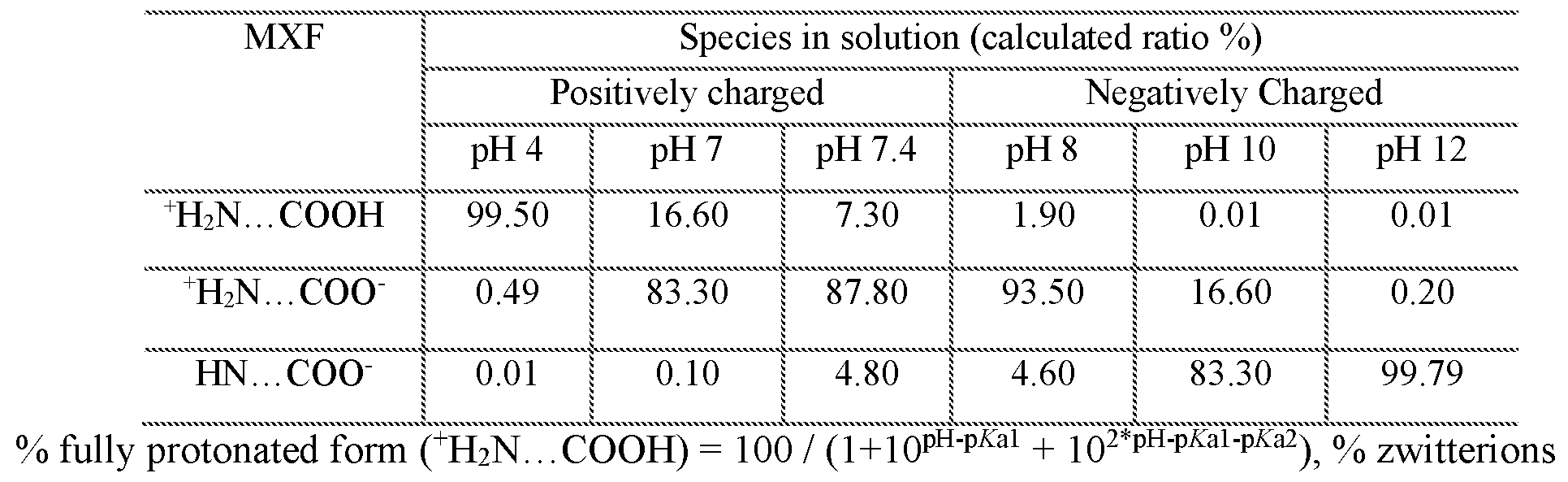
(+H2N. . COOH") = 100 / (1+10P&1-PH + 10PH"PIA2), % fully deprotonated form (HN. . COOH") = 100 - % (+H2N. . . COOH) - % (+H2N. . . COOH")33
[00234] FIG. 16A is a graph showing uptake capacity of MSN-MBI with different inner mesopore charges and stalk synthetic pathways. From left to right, samples are: slightly negatively charged underivatized MSN with stalk MBI synthesized by pathway I; negatively charged MSN- MBI by pathway I; positively charged MSN-MBI by pathway I; negatively charged MSN-MBI by pathway II; and positively charged MSN-MBI by pathway II. Pathyway I: synthesize the whole stalk first and then attach it on MCM-41 ; pathway II: attach first part of stalk on MCM-41 first and then synthesize the whole stalk. Negatively charged MCM-41 with nanovalve-MBI, synthesized by pathway II has highest uptake capacity, and positively charged MCM-41 uptakes almost nothing. FIG. 16B is a schematic showing MSN mesopores modified (left to right) with amine (+), unmodified silanol (-), or phosphonate (-).
[00235] We next tested two different MBI stalk synthetic pathways to optimize the efficiency of attachment. In the first pathway, we first reacted benzimidazole with
chloromethyltrimethoxysilane to produce the MBI stalk, and then covalently attached this to the
MCM-41 surface. This method has the disadvantage that, in the presence of small amounts of water or moisture, the MBI stalk readily hydrolyses and undergoes self-condensation prior to coupling to the nanoparticle. In the second pathway, we covalently attached chloromethyltrimethoxysilane to the silica surface first and then coupled it with benzimidazole to form the MBI stalk. We compared the uptake capacities of negatively charged MSN-MBI nanoparticles prepared by these two pathways and found that MBI stalk attachment by pathway II had a higher uptake capacity than attachment by pathway I, consistent with greater MSN surface coverage by the MBI stalks and hence greater trapping of drug in the pore channels.
[00236] Uptake and Release Capacity Utilizing Different Nanovalves
[00237] Both MSN- ANA and MSN-MBI were tested and proven to work effectively in our previous papers when loaded with doxorubicin, Hoechst 33342, or propidium iodide (PI).6' 25 However, we know that the uptake capacity and release capacity of the MCM-41 nanovalve system is dependent upon the size and charge of the cargo molecule, as well as the length of the stalk and the outer diameter of the CD. We measured and compared the uptake capacity of MXF utilizing these two systems in order to find the best one for subsequent in vitro and in vivo studies.
[00238] Because negatively charged inner pores provided greater uptake of MXF, we used phosphonated MCM-41 and compared the uptake and release of MXF of MSN-ANA-MXF, which has a-CD as cap, and MSN-MBI-MXF which has β-CD as cap. The same amount of phosphonated MCM-41 with one or the other nanovalve was loaded in 1 ml 10 mM MXF PBS solutions and stirred for one day. MSN-MBI-MXF had a much higher uptake capacity (7.4 wt%) and release capacity (1.02 wt%) than MSN-ANA-MXF (Table 4). The superior uptake and release capacity of the MSN-MBI-MXF is likely attributable to better trapping of the MXF within the pores. The β-CD has a 15.6 A outer diameter compared with 14.6 A for a-CD while MCM-41 has an average pore diameter of 22 A.6 The larger β-CD has more steric hindrance and blocks the MSN pores more effectively than the smaller a-CD. Moreover MSN-MBI has a shorter stalk length that positions the β-CD cap closer to the MSN surface, again providing more effective steric hindrance to prevent MXF leakage. For both types of MSNs, the uptake capacity was greater than the release capacity in aqueous acid, indicating that some MXF remains bound to the MSNs and is not released in aqueous acid conditions, possibly reflecting binding of MXF to MSN via hydrophobic interactions.
[00239] Table 4: Uptake and release capacity of phosphonated MSN with pH sensitive nanovalves
Sample Uptake capacity (wt %) Release capacity (wt %)
2.8 % 0.16 %
MSN-MBI-MXF (β-CD cap) 7.4 % 1.02 %
[00240] Uptake of MSN with pH Sensitive Nanovalves by Human Macrophages
[00241] Because efficacy of our MSN platform requires that the particles be taken up by F. tularensis-mfected macrophages, we examined the uptake of rhodamine-labeled MSN by F.
tularensis-mfected human macrophages, using both peripheral blood monocyte-derived
macrophages and differentiated macrophage-like THP-1 cells. We observed abundant uptake of MSN-MBI-MXF for both the monocyte-derived macrophages and the differentiated THP-1 cells (Figure 17).
[00242] FIG. 17A-E show confocal microscopy images demonstrating avid uptake of RITC- labeled MSN-MBI by F. tularensis- infected THP-1 macrophages. Human macrophage-like THP-1 cells were infected with GFP-expressing F. tularensis for 90 min, washed, and incubated with 12.5 μg/mL of RITC -labeled 100 nm MSN-MBI. After 3 hours, the cells were washed; the plasma membrane was stained with WGA-AlexaFluor 633; the cells were fixed; and nuclei were stained with DAPI. (a) LVS-GFP (green, arrows) and DAPI-stained nucleus (blue); (b) RITC-labeled MSN-MBI (red, arrowheads); (c) merged red, green, and blue color image; (d) contours of the cell are stained with WGA-AlexaFluor 633 (gray scale); (e) gray scale image superimposed onto merged color image, with the WGA-AlexaFluor 633 gray scale channel made partially transparent to allow the other channels to be seen. Scale bars, 10 μπι (pending).
[00243] In Vitro Efficacy of MSN-ANA-MXF and MSN-MBI-MXF in Killing F.
tularensis in Human Macrophages.
[00244] To investigate whether these two different types of pH-sensitive a- and β-CD nanovalves are functional under biological conditions, we assessed the efficacy of MSN-ANA- MXF and MSN-MBI-MXF in a macrophage infection model of F. tularensis.21 Differentiated human THP-1 macrophages were infected with F. tularensis Live Vaccine Strain (LVS) and either
not treated or treated with increasing concentrations of a) MXF, b) MSN-ANA-MXF, or c) MSN- MBI-MXF for one day. At the end of the treatment period, the number of viable bacteria remaining in the macrophages was determined to evaluate the F. tularensis killing effect exerted by each treatment. With no treatment, F. tularensis LVS grew 2.5 logs over one day. Similar levels of bacterial growth were also observed in infected macrophages treated with control MSNs (no MXF loading) indicating that the nanoparticle carriers alone do not possess any bactericidal activity (Figure 18B and C). All treatments including MXF, MSN-ANA-MXF and MSN-MBI-MXF killed F. tularensis in macrophages in a dose- dependent manner (Figure 18 A, B and C). However, when compared at the same concentration, MSN-MBI-MXF was much more potent than MSN-ANA- MXF in killing F. tularensis. For example, MSN-MBI-MXF at 1 μg/mL reduced bacterial colony forming units (CFU) by 3.4 logs compared with the level in the untreated group at one day, whereas the same concentration of MSN-ANA-MXF reduced bacterial CFU by only 0.2 logs compared with the untreated control group. The minimal inhibitory concentration in our macrophage assay is 4 μg/mL for MSN-ANA-MXF and it falls to between 0.25 and 0.5 μg/mL for MSN-MBI-MXF (Table 6).
[00245] Table 6: Bacterial CFU in infected macrophages with and without treatment

MSN-MBI-MXF (1 μg/ml) 26.4 ng/ml 1 day 4.23
MSN-MBI-MXF (2 μg/ml) 52.8 ng/ml 1 day 3.49
MSN-MBI-MXF (4 μg/ml) 105.6 ng/ml 1 day 3.49
MSN-MBI-MXF (8 μg/ml) 211.2 ng/ml 1 day 1.73
MSN- ANA control (8 μg/ml) 0 ng/ml 1 day 7.70
MSN-ANA-MXF (0.0625
0.23 ng/ml 1 day 7.71
μg/ml)
MSN-ANA-MXF (0.125 μg/ml) 0.45 ng/ml 1 day 7.72
MSN-ANA-MXF (0.25 μg/ml) 0.9 ng/ml 1 day 7.59
MSN-ANA-MXF (0.5 μg/ml) 1.8 ng/ml 1 day 7.59
MSN-ANA-MXF (1 μg/ml) 3.6 ng/ml 1 day 7.43
MSN-ANA-MXF (2 μg/ml) 7.2 ng/ml 1 day 7.06
MSN-ANA-MXF (4 μg/ml) 14.4 ng/ml 1 day 5.34
MSN-ANA-MXF (8 μg/ml) 28.8 ng/ml 1 day 3.61
[00246] FIG. 18 shows In vitro efficacy of MXF-loaded MSNs functionalized with two different types of pH-sensitive nanovalves. FIG. 18A is a graph showing human THP-1
macrophages infected with F. tularensis LVS and treated with MXF. FIG. 18B is a graph showing human THP-1 macrophages infected with F. tularensis LVS and treated with MSN-ANA-MXF. FIG. 18C is a graph showing human THP-1 macrophages infected with F. tularensis LVS and treated with MSN-MBI-MXF. Viable bacteria were determined by enumerating colony forming units (CFU) of F. tularensis in the macrophage monolayer. FIG. 18D is a graph showing impact of the drug released from MSN-ANA-MXF. FIG. 18E is a graph showing impact of the drug released from MSN-MBI-MXF.
[00247] We prepared supernate from MSN-ANA-MXF and MSN-MBI-MXF after an hour of incubation with 100 mM maleic acid, pH 1.8 (Acid Release) and assayed its capacity to kill F. tularensis in the infected macrophage. While the acid-released solution prepared from 0.5 and 1 μg/mL MSN-MBI-MXF reduced F. tularensis CFU in macrophages by 1.6 and 2.5 logs, respectively, the solution prepared from 1 μg/mL of MSN-ANA-MXF reduced bacterial number by only 0.2 logs (Figure 18D and E). Supernates obtained from MSN-MBI-MXF or MSN-ANA-MXF
at neutral pH (Neutral Eluate) had no effect in the infected macrophage bioassay. This study demonstrates that 1) the pH operative valves on MSN-MBI-MXF are tightly closed at neutral pH and open at acidic pH, 2) MXF eluted under acidic pH retains biological activity, 3) MSN-ANA- MXF and MSN-MBI-MXF kill F. tularensis LVS in macrophages in a dose-dependent fashion, and 4) MSN-MBI-MXF has greater efficacy than MSN-ANA-MXF, most likely because of its higher
[00248] MXF uptake and release properties.
[00249] Acid-released solution obtained from 1 μg/mL of MSN-MBI-MXF exerted the same inhibitory effect on F. tularensis as 0.016 μg/mL MXF in our macrophage bioassay, indicating a 1.6% (wt/wt) aqueous acid release capacity. Based on this estimation, 0.5 μg/mL of MSN-MBI- MXF could release 0.008 μg of MXF in the acidified endolysosomes. In our F. tularensis-miected macrophage assay, MSN-MBI-MXF at 0.5 μg/mL had a biological effect equivalent to that exerted by free MXF at a concentration of 0.016 μg/mL, indicating an efficacy ratio of 2 (MSN-MBI-MXF : free MXF), as nanoparticle-delivered drug appeared to have an efficacy twice that of the same amount of free drug in killing F. tularensis in macrophages in vitro. However, this efficacy ratio is likely an over-estimation since some of the yellowish color of MXF still remained on MSN-MBI- MXF after maleate treatment. In lieu of a possible hydrophobic interaction between MXF and MSN, we used acidic DMSO solution for measuring drug release capacity in subsequent in vivo studies.
[00250] Maximization of Uptake and Release Capacity by Optimization of Loading pH
[00251] Our in vitro study indicated that it is important to obtain a high release capacity in order to achieve high efficacy. Therefore we sought to increase further the uptake and release capacities of MSN-MBI-MXF.
[00252] Specifically, we prepared MXF in different pH solutions for use in the loading process to take advantage of electrostatic interactions based on positively or negatively charged inner pore channels. It is known that MXF has a positive net charge below pH 7.4 and negative net charge above pH 7.4 (Table 3). Therefore we loaded phosphonate modified MSN-MBI (10 mg) (indicated as "-") in pH 4 and pH 7 MXF solution (5 mM, 2 ml) in order to attract positive MXF molecules and increase uptake capacity. However, this acid loading presents some practical problems in experiments. The pH 4 loading helps improve uptake capacity as expected. However, it results in lower release capacity than neutral loading. Because the nanovalve can open at pH 6,
loaded MSN-MBI must be transferred to neutral solution before capping. The additional steps of centrifugation and dispersion of uncapped MSN-MBI-MXF in neutral water cause significant leakage of MXF from the particle pores before capping can be completed. We loaded amine modified MSN-MBI (10 mg) (indicated as "+") in pH 7, 10, and 12 MXF solutions (5 mM, 2 ml) to attract negative MXF molecules. We observed that uptake capacities as well as release capacities increased as pH was increased (Figure 19). Among these five conditions of loading, amine modified MSN-MBI in pH 12 loading gave the highest uptake capacity; however, the release capacity was still no better than pH 7 loading with phosphonate modified MSN-MBI. Using a pH 12 loading solution may gradually degrade the MSNs within 24 hours and cause stalks to detach from the pores, enabling MXF to leak out of the pores during washing.
[00253] FIG. 19 is a graph showing uptake and release capacity of negatively charged MSN- MBI loaded at pH 4 or 7 and positively charged MSN-MBI loaded at pH 7, 10, or 12 MXF aqueous solution. MXF has positive net charge in solution at pH < 7 and MCM-41 is negatively charged. Decreasing the loading pH from 7 to 4 increases uptake capacity, but, not release capacity because the nanovalve is open at pH 6 and particles must be transferred to neutral solution before capping. Most of MXF diffuses out of the pores because of these extra steps. At pH 7, positively charged MCM-41 repels MXF and leads to very low uptake and release capacities. MXF has negative charge when solution pH > 7, and increasing pH dramatically improves uptake capacities. However, loading at pH 12 does not lead to highest release capacity because particles degrade in base solution within 24 hours.
[00254] We found that loading phosphonated MSN-MBI with MXF in pH 7.4 PBS yielded the highest uptake and release capacity among all conditions (Figure 20). When phosphonate modified MSN-MBI (10 mg) was loaded in 1 ml 20 mM MXF in PBS (pH 7.4), its uptake capacity increased more than 10 times compared with its loading in neutral solution. Considering stalk protonation and deprotonation equilibrium, MSN-MBI is more tightly closed with β-CD at pH 7.4 than it is at pH 7. Negatively charged mesopores also have strong electrostatic interaction with positive MXF molecules. Moreover, compared with strong base, the pH 7.4 loading solution will not cause hydrolysis of silica nanoparticles and degradation of the nanovalves attached at the entrances of pore channels. All of these factors contribute to the highest uptake capacity.
[00255] FIG. 20 shows uptake capacity, uptake efficiency and release capacity of phosphonated MSN-MBI loaded in 20 mM MXF aqueous solution (pH 7), 20 mM MXF PBS solution (pH 7.4) and 40 mM MXF PBS solution (pH 7.4). At same concentration 20 mM MXF, PBS loading increases the uptake more than 10 times than neutral water and release capacity got increased to 6.2 wt%, which is more than 3 times of 1.7 wt% from neutral loading. Increasing the loading concentration to 40 mM further improves uptake capacity to almost 120 wt% and release capacity 8.1 wt%. In terms of uptake efficiency, MSN-MBI uptakes around 70 % MXF from original solution for both 20 mM and 40 mM MXF loading. is a
[00256] We have found that a higher MXF loading concentration resulted in a higher uptake and release capacity. MSN-MBI loaded with 40 mM MXF in PBS had an uptake capacity twice that of MSN-MBI loaded with 20 mM MXF in PBS, and the release capacity reached 8.1 wt% compared with 6.2 wt% for MSN-MBI loaded with 20 mM MXF. Moreover, when we compared uptake efficiency, which is defined as the percentage of MXF taken up by MSN from the original solution (expressed in percent), almost 70% of MXF in high concentration solution was taken up by nanoparticles. We also observed that a loading time of 24 hours was appropriate to allow MXF to diffuse into pore channels and reach equilibrium. Simply extending loading time did not increase uptake and release capacity (Figure 26). We tested phosphonated MSNs modified with two different nanovalves (ANA, MB I); the MSNs were loaded in MXF in PBS at low concentration (lOmM) and washed extensively with PBS buffer. The MSN- ANA and MSN-MBI showed uptake and release capacities of 20 wt% / 0.24 wt% and 51.4 wt% / 1.6 wt%, respectively; therefore MSN-MBI still showed the best performance in the final optimized condition (Table 5). In conclusion,
phosphonated MSN-MBI loaded with MXF in PBS showed the highest release capacity, 6 - 8 wt%, among all systems tested; this was highly reproducible and this MSN loaded with MXF in PBS was employed in subsequent / , tularensis in vitro and in vivo studies.
[00257] Table 5: Uptake capacity of MXF loaded MSN with different nanovalves (at low concentration)

[00259] Influence of Washing on Release Capacity
[00260] In the washing process, the mass of MXF washed away each time divided by the mass of particle is defined as "residual" (expressed in wt %), of which the final residual is reflected as the starting baseline in a release profile. Through measurement by UV-Vis spectroscopy, the amount of residual drug in supernatant did not increase after dispersing and rotating particles in neutral water overnight, which indicated that the residual was not due to release or leakage, but caused by non-trapped MXF dissociating from the MSN-MBI-MXF surface. In the release process, we dispersed particles in neutral deionized water and observed no leakage from MSN-MBI as indicated by the flat baseline in the release profile. After adding HC1 to adjust the pH to 5, we measured an immediate increase in fluorescence intensity from the MXF released into the supernatant. The release profile reached a plateau after 14 hours, and the concentration of completely released MXF was measured by UV-Vis spectroscopy after 24 hours.
[00261] We investigated how the washing process influences particle release capacity. In testing drug-loaded nanoparticles, we routinely wash them to remove free MXF adsorbed on the external surface of the MSN to insure that the great majority of the drug is released via the nanovalves. However, excess washing will gradually degrade the silica nanoparticle surface due to silanol group hydrolysis. Moreover, it will remove some of the cyclodextrin caps by disrupting host-guest interaction equilibrium. Therefore, it is important to determine the optimal number of washing steps that strikes an acceptable balance between removing MXF from the external surface and maintaining relatively high release capacity. MSNs washed 15 times did not have any residual surface MXF detectable by UV-Vis absorption measurement (Figure 21 A); however, their release capacity was only 1.6 wt%. In contrast, MSNs washed 8 times had 2.5 wt% residual and 6.9 wt% release capacity. MSN-MBI-MXF washed 21 times had almost zero residual in the last few washes. There is an exponential decay of the amount of MXF washed away each time, with the first 8 washes removing around 95 % of the total amount of MXF (Figure 2 IB). We found that washing 8
times is enough to remove most of the MXF on the MSN external surface and at the same time constrain damage to surface modifications.
[00262] FIG. 21 A is a graph showing that release profiles show that the more times the MSN are washed, the lower the amount of residual and release capacity. When particles were washed 15 times, there was negligible residual drug detected from the particle surface (no fluorescence detected). A small amount of residual was observed when drug loaded particles were washed 8 times. FIG. 21B is a graph showing the amount of MXF washed away each time decreases as the number of washes increases; the decrease for each step is -30 %. The first eight washes contribute -95 % to the total amount of MXF ultimately removed by washing.
[00263] In Vivo Efficacy of MSN-MBI-MXF in Treating Pneumonic Tularemia
[00264] After the above optimizations to achieve high uptake and release capacity, we assessed the efficacy of MSN-MBI-MXF in a mouse model of pneumonic tularemia.28' 29 In the first in vivo experiment (Experiment 1), mice were infected by the intranasal route with -8000 CFU of F. tularensis LVS, a dose approximately 11 times the LD50 of 700 CFU. Without treatment, the mice succumbed rapidly to the infection and suffered severe weight loss (Figure 22A). Mice treated with MSN-MBI-MXF (with a drug release capacity of 6.88 wt%) maintained their weight, indicating that the nanoparticle was well tolerated by the mice and helped to control the severe bacterial infection. Without treatment, bacteria multiplied to high numbers in the lungs of the mice (Figure 23 A). However, treatment with MSN-MBI-MXF (loaded with 138 μg MXF) reduced bacterial burden in the lung by 4.0-logs, more so than treatment with 400 μg of free MXF (Figure 23C). Treatment with MSN-MBS-MXF reduced bacterial burden in the spleen by 4.3-logs to a level similar to that with 400 μg of free MXF which was below our experimental limit of detection. All treatments reduced bacterial burden in the liver to a level below the experimental detection limit. On the basis of a median-effect plot, the efficacy of the MSN-MBI-MXF was 4.5 fold and 3 fold the efficacy of free MXF in the lung and spleen, respectively (Figure 27, left panel). This study demonstrates that MSN-MBI-MXF administered intravenously is much more efficacious than free MXF in treating F. tularensis infection in mice.
[00265] FIG. 22A shows results from experiment 1 where treatment with MSN-MBI-MXF prevents weight loss caused by pneumonic tularemia. FIG. 22B shows results from experiment 2 where treatment with MSN-MBI-MXF prevents weight loss caused by pneumonic tularemia.
Percentage change in weight of F. tularensis-mfected mice was monitored over the course of the experiments. The mice were sham treated, treated with one of three doses of MXF as a free drug, as indicated, or treated with MSN-MBI-MXF (loaded with 138 μg MXF in Experiment 1 and 50 μg MXF in Experiment 2).
[00266] FIG. 23 shows In vivo efficacy of MSN-MBI-MXF assessed by assay of F.
tularensis burden in the mouse organ in two independent experiments, Experiment 1 (A and C) and Experiment 2 (B and D). FIG. 23 A shows results of mice infected with F. tularensis LVS by the intranasal route. FIG. 23B shows results of mice infected with F. tularensis LVS by the intranasal route. Bacterial burden in the lung was monitored over the course of infection. One day postinfection, mice were sham treated, treated with one of three doses of free MXF, as indicated, or treated with MSN-MBI-MXF (loaded with 138 μg in Experiment 1 shown in A and 50 μg in Experiment 2 shown in B) by tail vein injection on days 1, 3, and 5. FIG. 23 C shows bacterial numbers in the lung, liver, and spleen. FIG. 23D shows bacterial numbers in the lung, liver, and spleen. § Bacterial CFU below limit of detection. *P < 0.05 by one-tailed t-test.
[00267] FIG. 27 shows median-effect plots to compare efficacy of MSN-MBI-MXF with
MXF administered as free drug. The efficacy of MSN-MBI-MXF in the lung, spleen, and liver was compared with that of free MXF in a median-effect plot of the results of mouse Experiments 1 and 2. For a given dose of MXF, an upward shift as indicated by the red arrows on the y-axis indicates greater / , tularensis killing efficacy of the MSN-MBI-MXF. Fa: Fraction of bacteria killed; Fu: Fraction of bacteria surviving; D: Dose of MXF in micrograms.
[00268] In a subsequent in vivo experiment (Experiment 2), we evaluated another batch of
MSN-MBI-MXF (with a drug release capacity of 8.08 wt%). Mice were infected with -4000 CFU of F. tularensis LVS (-6 x LD50) by the intranasal route. One day later, mice were sham-treated or treated with 640 μg of MSN-MBI-MXF (with - 50 μg of releasable MXF) or with one of the three doses of MXF (50, 100, and 200 μg) equal to lx, 2x, and 4x the amount of the releasable MXF from 640 μg of MSN-MBI-MXF by acidic DMSO. As observed in the first in vivo study, sham treated mice suffered substantial weight loss but mice treated with free MXF or MSN-MBI-MXF did not (Figure 9B). MSN-MBI-MXF treatment reduced the bacterial burden by 2.8 logs in the lung, 3.2 logs in the liver, and 3.3 logs in the spleen to a level close to that achieved by 100 μg free MXF (Figure 23D). Thus, in the treatment of pneumonic tularemia in mice, MSN-MBI-MXF had an
efficacy twice the equivalent amount of free MXF in the lung, spleen, and liver (Table S2 and Figure 27, right panel). Again, we observed no toxicity in the mice from MSN-MBI-MXF treatment.
[00269] SUMMARY AND OUTLOOK
[00270] Intracellular pathogens that reside in mononuclear phagocytes present an ideal target for nanotherapeutics because nanoparticles are readily taken up by cells of the Mononuclear Phagocyte System and have the potential to deliver high concentrations of antibiotics selectively to the intracellular compartment, thereby providing increased efficacy with reduced systemic exposure and off-target side effects.
[00271] We have optimized MSNs with pH-sensitive nanovalves for uptake and release of the antibiotic MXF. We evaluated a) two different pH-sensitive nanovalves; b) modification of the MSN's inner pores with positive or negative charges; c) loading of the MSNs with MXF in different pH solutions; and d) loading MSNs with different drug concentrations and loading durations. We found that phosphonated MSN-MBI-MXF loaded in pH 7.4 PBS gave the highest uptake and release capacity. We demonstrated that this delivery system released MXF efficiently in F.
tularensis-miected macrophages and that it was 2.7 fold more effective than the amount of free drug released from the particles by aqueous acid. We demonstrated in a mouse model of lethal pneumonic tularemia that MSN-MBI-MXF was well tolerated and was more effective than a 2- to 4-fold greater dose of free MXF in reducing bacterial load in the lung. Our MSN-MBI-MXF delivery system has the potential to provide more effective treatment than free drug, shortening the duration of treatment of intracellular infectious diseases such as tularemia, tuberculosis, Q-fever, and Legionnaires' disease and reducing systemic toxicity of the MXF. By providing high concentrations of antibiotic directly to the site of infection, the nanoparticle delivered drug also has the potential to decrease the emergence of drug resistance. Further optimization of our platform may be possible by incorporation of additional functionalizations to increase targeting to infected tissues and macrophages, employment of different delivery modalities, such as aerosol delivery, or utilization of other internal and external stimulus-response systems.
[00272] MATERIALS AND METHODS
[00273] Materials. Cetyltrimethylammonium bromide (CTAB, 95%), tetraorthoethylsilicate
(TEOS, 98%) 3-(trihydro-xysilyl)propyl methylphosphonate (42% in H20), 3-
iodopropyltrimethoxysilane (IPTMS, 95%), N,N'-dimethylformamide (99.8%), p-anisidine (99%), a-cyclodextrin (>98%), β-cyclodextrin (>97%), benzimidazole (98%), tetrabutylammonium iodide (98%), Hoechst 33342 £97%), triethylamine (>99%), toluene (99.8%) were purchased from Sigma (St. Louis, MO). Chloromethyltrimethoxysilane (90%), N-(2-Aminoethyl)-3- aminopropyltrimethoxysilane (NAPTS, 90 %) was purchased from Gelest (Morrisville, PA).
[00274] Synthesis of MCM-41. The synthesis of MCM-41 was based on well-established published procedures. Cetyltrimethyl ammonium bromide (CTAB, 250 mg, 0.7 mmol) was dissolved in H20 (120 mL) and NaOH (875 \iL, 2M). The mixture was heated up to 80 °C and kept stable for 30 minutes, followed by adding tetraethyl orthosilicate (TEOS, 1.2 mL) drop-wise into the solution while stirring vigorously. For phosphonated MCM-41, 3-(trihydroxysilyl)propyl methylphosphonate (315 pL) was added into the solution 15 minutes after adding TEOS. For amine modified MCM-41, N-(2-Aminoethyl)-3-aminopropyltrimethoxysilane (90 %) was mixed with TEOS before adding to CTAB solution. The solution was kept at 80 °C for 2 hours. The synthesized nanoparticles were centrifuged and washed thoroughly with methanol. The successful synthesis of nanoparticles is very sensitive to the temperature and stirring speed.
[00275] Synthesis of Anilinoalkane (ANA) Nanovalve. As-synthesized MCM-41 (100 mg) was washed and dispersed in anhydrous toluene, mixed with 3-iodopropyl trimethoxysilane
(IPTMS, 20 uL, O. lmmol) and heated up to 40 °C under N2 for 12 hours. The IPTMS modified nanoparticles were washed with toluene to remove unreacted agents and re-dispersed in anhydrous toluene, and mixed with p-anisidine (123.2 mg, 1 mmol) and triethylamine (TEA, 420 pL, 3 mmol). The solution was refluxed under N2 for another 24 hours. The final product was centrifuged and washed with toluene, methanol and water to be ready for drug/dye loading process.
[00276] Synthesis of 1-Methyl-lH-benzimidazole (MBI) Nanovalve. MCM-41 (100 mg) was washed and dispersed in anhydrous toluene, mixed with chloromethyltrimethoxysilane (15 pL) and refluxed for 12 hours. The modified MCM-41 was washed by toluene and dimethyoformamide (DMF) and dispersed in 8 ml DMF. Tetrabutyammonium iodide (2 mg), benzimidazole (12 mg) and triethylamine (150 pL) were added into the solution and the mixture was heated up to 70 °C under N2 for 24 hours. As-synthesized nanoparticles were washed with DMF, methanol and water thoroughly.
[00277] Surfactant Template Extraction. Nanovalve-modified MCM-41 (100 mg) was dispersed in methanol (60 mL), mixed with concentrated HC1 (12 M, 2.3 mL) and refluxed for 8 hours under N2, and then washed extensively with methanol and water.
[00278] Drug Loading and Washing. 10 mg of nanovalve-modified MCM-41 was suspended in MXF PBS solution at various concentrations overnight. β-CD (40 mg) was added to the suspension and mixed for 12 hours to make sure the capping molecule reached an equilibrium with stalks on the nanoparticle surface. Loaded and capped nanoparticles (10 mg) were centrifuged down in a 2 mL tube and the supernate kept for UV-Vis absorbance measurement. Filtered PBS was added into the tube and nanoparticles were suspended and sonicated again. This washing process was repeated and the number of times the nanoparticles were washed was the same for each group being compared.
[00279] Uptake Capacity and Release Capacity Measurement. After measuring UV-Vis absorbance of MXF remaining in the PBS loading solution and of standard MXF PBS solution with known concentrations of 0.01 mM, 0.02 mM and 0.025 mM MXF, the amount of unloaded MXF concentration was calculated based on Beer's law. Uptake capacity (wt %) = [(WMXF before loading - WMXF after loading) / Wparticle)] χ 100 %. In the optimization experiments, loaded particle MSN-MXF was dispersed in pH 4.5 HC1 solution for 24 hours and then centrifuged down to measure the concentration of MXF released into the supernate. Release capacity (wt %) =( Wreleased MXF / Wparticle ) χ 100 %. In our in vitro and in vivo studies of the efficacy of the nanoparticles in treating F. tularensis infection, MSN-MXF was dispersed in pH 1 HC1/DMSO solution to measure the maximum release capacity.
[00280] Stimulated Release Studies. To measure MXF release from MSNs and detect MXF fluorescence emission in supernates, dried MSN-MXF powder was put in the corner of a glass vial containing 10 mL DI water. A probe laser beam (5 mW 377 nm) was passed through the supernatant fluid in the glass vial such that released MXF was excited. The fluorescence was detected and collected by a charge-coupled device (CCD) detector and a computer at 1 s intervals over the course of the experiment. Baseline spectra were collected for 1 hour to confirm that there was no MXF leakage, and then 1 M HC1 solution was added to adjust the pH to 4.5. The release profile was the plot of the integrated emission peak area between 480 nm to 520 nm as a function of time.
[00281] Physisochemical Characterization of Nanovalve Modified MSN. Transmission electron microscopy (TEM) images of MSN were obtained using a JEM1200-EX (JEOL) instrument (JEOL USA, Inc., Peabody, MA). Particle size and zeta potential were measured by ZetaSizer Nano (Malvern Instruments Ltd, Worcestershire, UK) with 50 μg/mL MSN dispersed in DI water.
[00282] Bacteria. F. tularensis subsp. holarctica Live Vaccine Strain (LVS) was obtained from Centers for Disease Control and Prevention (Atlanta, GA). LVS glycerol stocks were prepared as described and stored at -80°C.30' 31 For in vitro macrophage experiments, a vial of the LVS frozen glycerol stock was thawed in a 37 °C water bath and cultivated on GCII chocolate agar plates for 3 days before use. For in vivo mouse experiments, a vial of pre-titered LVS frozen stock was used directly to infect mice and immediately afterward serially diluted and plated on GCII chocolate agar to confirm the bacterial numbers used to infect. For fluorescence studies, LVS expressing superfolder green fluorescent protein (LVS-GFP) was grown on GCII chocolate agar containing kanamycin at a concentration of 10 μg/mL for 3 days prior to use for infecting macrophages.
[00283] Macrophages. Human peripheral blood monocytes were prepared from the blood of healthy donors and cultivated in Teflon wells for 5 days to differentiate them into monocyte derived macrophages.31 Human THP-1 monocytic cells (American Type Culture Collection, TH3-202) were maintained in RPMI-1640 (Lonza) supplemented with 10% fetal bovine serum (Mediatech), 2 mM GlutaMAX (Life Technology), penicillin (100 IU) and streptomycin (100 μg/mL) at 37 °C, 5% C02 - 95% air atmosphere. Prior to usage, THP-1 cells were differentiated into macrophages with 100 nM phorbol 12-myristate 13-acetate (PMA; Sigma) in antibiotic-free RPMI with 10% fetal bovine serum.
[00284] Assessment of MSN Efficacy in Macrophages. PMA-differentiated THP-1 cells were plated at 1 x 10⁵ cells per 200 μΕ per well in 96-well plates (Matrical) and infected with 106 F. tularensis LVS for 90 min. The infected THP-1 macrophages were washed and incubated with fresh medium alone or fresh medium containing MXF, control MSNs (no MXF loading) or MXF -loaded MSNs. F. tularensis LVS infection and growth in THP-1 macrophages was determined by harvesting the bacteria from the infected macrophages at 2 hours and 1 day post infection. For all treatment groups, the infected macrophage cultures were incubated in the continued presence of the treatment for one day. Thereafter, F. tularensis LVS was harvested from the infected macrophages
to assess the effect of treatment. The bacteria were harvested by lysing the macrophage monolayers with 1% saponin in PBS for 5 min at room temperature, serially diluted, and plated on GCII chocolate agar. Bacterial colony forming units (CFU) on agar plates were enumerated after incubation at 37 °C for 3 days.
[00285] Assessment of MSN Efficacy in Mice. Animal procedures were conducted according to protocols approved by the UCLA Animal Research Committee and NIH Guidelines for the Care and Use of Laboratory Animals in Research. In two experiments (Experiment 1 and Experiment 2), female Balb/c mice (Taconic) of approximately 18 g were provided with standard diet ad libitum and acclimated for one week. Mice were infected by the intranasal route with -8000 (Experiment 1) or -4000 (Experiment 2) CFU of F. tularensis LVS. Two mice were euthanized 5 hours after intranasal infection (day 0) to determine the number of bacteria delivered to the lung at the start of the experiment. An additional 3 mice were euthanized one day later (day 1) to determine bacterial growth during that period of time. Mice were then sham-treated or treated with MXF or MSN-MBI-MXF by tail vein injection every other day (day 1, day 3, and day 5) for a total of 3 treatments. Mice were euthanized one day after the last treatment (day 6). Lungs, livers, and spleens from infected mice that were sham-treated or treated with MXF or MSN-MBI-MXF were homogenized and serially diluted for plating on GCII chocolate agar containing sulfamethoxazole (40 μg/mL), trimethoprim (8 μg/mL), and erythromycin (50 μg/mL). Bacterial CFU on the agar plates were enumerated after incubation at 37 °C for 4 days.
[00286] Median-Effect Plots. We used median-effect plots32 to compare the relative efficacy of MSN-MBI-MXF and free MXF. The fraction of inhibition for samples treated with different amounts of MXF was calculated using bacterial CFU in base- 10 logarithm (log CFU) with the equation: Fraction of inhibition = 1 - (log CFU from sample treated with MSN-MBI-MXF or MXF/log CFU from untreated sample). A median-effect plot for MSN-MBI-MXF or MXF was generated using MXF or MXF equivalent (MSNs) dose in base-10 logarithm as the X-axis and the fraction of surviving bacteria divided by the fraction of killed bacteria in base-10 logarithm as the Y-axis.
[00287] Statistics. Statistical analyses were performed using the Student's t-test. A P value of 0.05 or less was considered statistically significant.
[00288] References for Example 2
Barbe, C; Bartlett, J.; Kong, L. G.; Finnie, K.; Lin, H. Q.; Larkin, M.; Calleja, S.; Bush, A.; Calleja, G. Silica particles: A novel drug-delivery system. Adv Mater 2004, 16, 1959-1966. Argyo, C; Weiss, V.; Brauchle, C; Bein, T. Multifunctional Mesoporous Silica
Nanoparticles as a Universal Platform for Drug Delivery. Chem Mater 2014, 26, 435-451. Li, Z. X.; Barnes, J. C; Bosoy, A.; Stoddart, J. F.; Zink, J. I. Mesoporous silica
nanoparticles in biomedical applications. Chem Soc Rev 2012, 41, 2590-2605.
Tarn, D.; Ashley, C. E.; Xue, M.; Carnes, E. C; Zink, J. I; Brinker, C. J. Mesoporous Silica Nanoparticle Nanocarriers: Biofunctionality and Biocompatibility. Accounts Chem Res 2013, 46, 792-801.
Xue, M.; Zhong, X.; Shaposhnik, Z.; Qu, Y. Q.; Tamanoi, F.; Duan, X. F.; Zink, J. I. pH- Operated Mechanized Porous Silicon Nanoparticles. J Am Chem Soc 2011, 133, 8798-8801. Meng, H. A.; Xue, M.; Xia, T. A.; Zhao, Y. L.; Tamanoi, F.; Stoddart, J. F.; Zink, J. I; Nel, A. E. Autonomous in Vitro Anticancer Drug Release from Mesoporous Silica Nanoparticles by pH-Sensitive Nanovalves. J Am Chem Soc 2010, 132, 12690-12697.
Hwang, A. A.; Lu, J.; Tamanoi, F.; Zink, J. I. Functional Nanovalves on Protein-Coated Nanoparticles for In vitro and In vivo Controlled Drug Delivery. Small 2015, 11, 319-328. Dong, J. Y.; Xue, M.; Zink, J. I. Functioning of nanovalves on polymer coated mesoporous silica Nanoparticles. Nanoscale 2013, 5, 10300-10306.
Ferris, D. P.; Zhao, Y. L.; Khashab, N. M.; Khatib, H. A.; Stoddart, J. F.; Zink, J. I. Light- operated mechanized nanoparticles. J. Am. Chem. Soc. 2009, 131, 1686-1688.
Thomas, C. R.; Ferris, D. P.; Lee, J. H.; Choi, E.; Cho, M. H.; Kim, E. S.; Stoddart, J. F.; Shin, J. S.; Cheon, J.; Zink, J. I. Noninvasive Remote-Controlled Release of Drug Molecules in Vitro Using Magnetic Actuation of Mechanized Nanoparticles. J Am Chem Soc 2010, 132, 10623-10625.
Clemens, D. L.; Lee, B. Y.; Xue, M.; Thomas, C. R; Meng, H.; Ferris, D.; Nel, A. E.; Zink, J. I; Horwitz, M. A. Targeted Intracellular Delivery of Antituberculosis Drugs to
Mycobacterium tuberculosis-Infected Macrophages via Functionalized Mesoporous Silica Nanoparticles. Antimicrob Agents Ch 2012, 56, 2535-2545.
Liong, M.; Lu, J.; Kovochich, M.; Xia, T.; Ruehm, S. G.; Nel, A. E.; Tamanoi, F.; Zink, J. I. Multifunctional inorganic nanoparticles for imaging, targeting, and drug delivery. ACS Nano 2008, 2, 889-896.
Luo, G. F.; Chen, W. H.; Liu, Y.; Lei, Q.; Zhuo, R. X.; Zhang, X. Z. Multifunctional enveloped mesoporous silica nanoparticles for subcellular co-delivery of drug and therapeutic peptide. Scientific reports 2014, 4, 6064.
Lu, J.; Liong, M.; Li, Z. X.; Zink, J. I; Tamanoi, F. Biocompatibility, Biodistribution, and Drug-Delivery Efficiency of Mesoporous Silica Nanoparticles for Cancer Therapy in Animals. Small 2010, 6, 1794-1805.
Ellis, J.; Oyston, P. C; Green, M.; Titball, R. W. Tularemia. Clinical microbiology reviews 2002, 15, 631-646.
Christopher, G. W.; Cieslak, T. J.; Pavlin, J. A.; Eitzen, E. M. Biological warfare - A historical perspective. Jama-J Am Med Assoc 1997, 278, 412-417.
Harris, S. Japanese Biological Warfare Research on Humans - a Case-Study of
Microbiology and Ethics. Ann Ny Acad Sci 1992, 666, 21-52.
Alibek, K.; Handelman, S. Biohazard : the chilling true story of the largest covert biological weapons program in the world, told from the inside by the man who ran it. Random House: New York, 1999; p xi, 319 p.
Feldman, K. A.; Enscore, R. E.; Lathrop, S. L.; Matyas, B. T.; McGuill, M.; Schriefer, M. E.; Stiles-Enos, D.; Dennis, D. T.; Petersen, L. R.; Hayes, E. B. An outbreak of primary pneumonic tularemia on martha's vineyard. New Engl J Med 2001, 345, 1601-1606.
Chocarro, A.; Gonzalez, A.; Carcia, I. Treatment of tularemia with Ciprofloxacin. Clin Infect Dis 2000, 31, 623-623.
Steward, J.; Piercy, I; Lever, M. S.; Simpson, A. J. H.; Brooks, T. J. G. Treatment of murine pneumonic Francisella tularensis infection with gatifloxacin, moxifloxacin or ciprofloxacin. Int J Antimicrob Ag 2006, 27, 439-443.
Chen, H. W.; Wang, L. Y.; Yeh, J.; Wu, X. Y.; Cao, Z. H.; Wang, Y. A.; Zhang, M. M.; Yang, L.; Mao, H. Reducing non-specific binding and uptake of nanoparticles and improving cell targeting with an antifouling PEO-b-P gamma MPS copolymer coating. Biomaterials 2010, 31, 5397-5407.
Lee, J. E.; Lee, N.; Kim, H.; Kim, J.; Choi, S. H.; Kim, J. H.; Kim, T.; Song, I. C; Park, S. P.; Moon, W. K., et al. Uniform Mesoporous Dye-Doped Silica Nanoparticles Decorated with Multiple Magnetite Nanocrystals for Simultaneous Enhanced Magnetic Resonance Imaging, Fluorescence Imaging, and Drug Delivery. J Am Chem Soc 2010, 132, 552-557. He, Q. J.; Zhang, Z. W.; Gao, F.; Li, Y. P.; Shi, J. L. In vivo Biodistribution and Urinary Excretion of Mesoporous Silica Nanoparticles: Effects of Particle Size and PEGylation. Small 2011, 7, 271-280.
Du, L.; Liao, S. J.; Khatib, H. A.; Stoddart, J. F.; Zink, J. I. Controlled-Access Hollow Mechanized Silica Nanocontainers. J Am Chem Soc 2009, 131, 15136-15142.
Meng, H.; Liong, M.; Xia, T.; Li, Z.; Ji, Z.; Zink, J. I; Nel, A. E. Engineered design of mesoporous silica nanoparticles to deliver doxorubicin and P-glycoprotein siRNA to overcome drug resistance in a cancer cell line. ACSNano 2010, 4, 4539-4550.
Clemens, D. L.; Lee, B. Y.; Horwitz, M. A. O-Antigen-Deficient Francisella tularensis Live Vaccine Strain Mutants Are Ingested via an Aberrant Form of Looping Phagocytosis and Show Altered Kinetics of Intracellular Trafficking in Human Macrophages. Infect Immun 2012, 80, 952-967.
Jia, Q. M.; Lee, B. Y.; Bowen, R.; Dillon, B. T; Som, S. M.; Horwitz, M. A. A Francisella tularensis Live Vaccine Strain (LVS) Mutant with a Deletion in capB, Encoding a Putative Capsular Biosynthesis Protein, Is Significantly More Attenuated than LVS yet Induces Potent Protective Immunity in Mice against F. tularensis Challenge. Infect Immun 2010, 78, 4341-4355.
Jia, Q. M.; Lee, B. Y.; Clemens, D. L.; Bowen, R. A.; Horwitz, M. A. Recombinant attenuated Listeria monocytogenes vaccine expressing Francisella tularensis IglC induces protection in mice against aerosolized Type A F. tularensis. Vaccine 2009, 27, 1216-1229. Jia, Q.; Lee, B. Y.; Bowen, R.; Dillon, B. J.; Som, S. M.; Horwitz, M. A. A Francisella tularensis live vaccine strain (LVS) mutant with a deletion in capB, encoding a putative capsular biosynthesis protein, is significantly more attenuated than LVS yet induces potent protective immunity in mice against F. tularensis challenge. Infect. Immun. 2010, 78, 4341- 4355.
31. Clemens, D. L.; Lee, B. Y.; Horwitz, M. A. O-antigen-deficient Francisella tularensis Live Vaccine Strain mutants are ingested via an aberrant form of looping phagocytosis and show altered kinetics of intracellular trafficking in human macrophages. Infect. Immun. 2012, 80, 952-967.
32. Chou, T. C. Theoretical basis, experimental design, and computerized simulation of
synergism and antagonism in drug combination studies. Pharmacol Rev 2006, 58, 621-681.
33. Lemaire, S.; Tulkens, P. M.; Van Bambeke, F. Contrasting Effects of Acidic pH on the Extracellular and Intracellular Activities of the Anti-Gram-Positive Fluoroquinolones Moxifloxacin and Delafloxacin against Staphylococcus aureus. Antimicrob Agents Ch 2011, 55, 649-658.
[00289] Example 3
[00290] One embodiment of this invention is a composition for treating infections caused by pathogens sensitive to the antibiotic moxifloxacin (MXF), including Tularemia, Tuberculosis (TB) and other mycobacterial diseases (e.g. Mycobacterium kansasii infection, Mycobacterium intracellular infection, disseminated BCG, etc.) comprising MXF loaded into a mesoporous silica nanoparticle (MSN) equipped with a disulfide snap-top valve. MXF releases inside host cells in response to a reducing and hydrophobic environment inside the host cell. Another embodiment is a method for treating tularemia, TB, other mycobacterial diseases, and infections caused by intracellular pathogens in general utilizing this technology.
[00291] Details of the invention are as follows:
[00292] Materials Synthesis
[00293] Synthesis of MSNs (100 nm)
[00294] Mesoporous silica nanoparticles (MSN) are nanoscale materials that are composed of amorphous silica, which is highly porous and has large surface area. These materials have particulate sizes on the order of -100 nm and possess pore diameters of approximately 2 nm.
MSNs are synthesized by first dissolving a surfactant, cetyltrimethylammonium bromide (250 mg,
CTAB) into a basic solution (120 mL, pH 12) and heating up to 80 °C. Once the solution is thermally stable, the silica precursor, mixture of tetraethyl orthosilicate (1.2 mL, TEOS) and
diethylphosphatoethyltriethoxysilane (0.2 ml, DEPETS), is added drop-wise into the solution and the solution slowly begins to become opaque. The nanoparticles are collected by centrifugation and washed with methanol.
[00295] MSN Functionalizations:
[00296] Disulfide snap-top synthesis
[00297] In order to construct disulfide snap-top on MSN to trap and release drug cargo,
MCM-41 (100 mg) was dispersed into dry toluene (10 ml), mixed with (3-mercaptopropyl) trimethoxysilane (24 μΐ, O. lmmol) and refluxed for 12 hours under nitrogen atmosphere as shown in Figure 1. Thiol group modified MCM-41(100 mg) was washed and dispersed again in anhydrous toluene (10 ml) in second step. To prepare thiocyanogen, lead thiocyanate (800 mg) was dispersed in 10 ml chloroform and titrated by bromine (200 μΐ) in chloroform (10ml). The titration product mixture was filtered and the supernatant containing thiocyanogen in chloroform was light yellowish. 1-adamantanethiol (17 mg, O. lmmole) and as-synthesized thiocyanogen were added into MSN toluene dispersion. The disulfide oxidation reaction took four days under 4°C and nitrogen gas atmosphere. As-synthesized material was yellowish and washed thoroughly with toluene, methanol and water.
[00298] FIG. 28 is a schematic showing disulfide snap-top system synthesis.
[00299] The disulfide snap-top consists of an adamantyl group which binds with β cyclodextrin and when nanoparticles are endocytosed into cells the disulfide bond can be reduced by glutathione. Then adamantly group will be removed together with cyclodextrin and drug will be released as shown in FIG. 29.
[00300] FIG. 29 is a schematic showing a MSN with disulfide snap-top release mechanism.
[00301] Loading of moxifloxacin and drug release test by time-resolved florescence spectroscopy
[00302] MCM-41 (10 mg) with disulfide snap-top was dispersed in 1 ml ImM
Moxifloaxcin PBS solution and rotating overnight, β cyclodextrin (40 mg) was added into the solution as capping agent to prevent drug from leaking out. After mixing the solution for another 12 hours, moxifloxacin loaded MCM-41 with disulfide snap-top was dried under vacuum overnight. In order to prove moxifloxacin can be release from MSN and detect moxiflxoacin fluorescence
emission in supernatant, the dried powder was put at the corner of a glass vial containing 10 ml DI water. Baseline spectra were collected for 1 hour to show that there was no moxifloxacin leaking out, and then 2-mercaptoethanol (200 μΐ) was added into solution and resulted in a dramatic increase of fluorescence emission in supernatant which indicated release of moxifloxacin. A 5 mW 377 nm laser beam shot through the supernatant in glass vial and released moxifloxacin was excited and emitted fluorescence detected and collected by CCD and computer connected with it. Release profile was constructed by intergration of moxifloxacin emission peak area from 480 nm to 520 nm. After collecting data for 17 hours and moxifloxacin was released completely, the supernatant released moxifloxacin concentration was calculated based on UV-Vis spectrum and standard curve by Beer's law. Release capacity was defined as released moxifloxacin weight over MCM-41 weight.
[00303] II. Characterization of MSN with pH sensitive nanovalve
[00304] Transmission electron microscopy (TEM) images of MSNs were obtained using a JEM1200-EX (JEOL) instrument (JEOL USA, Inc., Peabody, MA). Particle size was measured by
ZetaSizer Nano (Malvern Insstruments Ltd, Worcestershire, UK) and MSN was dispersed in PBS in
50 μg/ml for DLS measurement. UV-Vis spectra of moxiflxoacin were collected by a Cary 500 UV- vis-NTR spectrophotometer. The release profile was obtained by time-resolved fluorescence spectroscopy.
[00305] FIG. 30 is a TEM image of MSN with disulfide snap-top that shows structure integrity preserved after surface modification.
[00306] FIG. 31 shows dynamic light scattering (DLS) measurement of MSN with disulfide snap-top in PBS. It shows that mean hydrodynamic diameter of the modified nanoparticle is around 740 nm due to disulfide formation among MSN.
[00307] FIG. 32 is a graph shwoing UV-Vis spectrum of moxifloxacin in PBS.
[00308] FIG. 33 is a graph showing moxifloxacin loaded MCM-41 with disulfide snap- top release profile. After adding reducing agent 2-mercaptothanol, moxifloxacin was immediately released and finally reached a plateau. The beginning fluorescence increase is due to external surface drug desorption.
[00309] Assay for MXF released from MSN-SS-MXF under reducing conditions)
[00310] Example of assay
[00311] Assay
[00312] Moxifloxacin (MXF) was released from 100 nm disulfide snap-top MSN-SS-
MXF by the reducing agent β-mercaptoethanol and measured by spectrophotometry at 288 nm or by Francisella bioassays.
[00313] Spectrophotometry assay
[00314] To assess the amount of MXF loaded on nanoparticles, we suspended 1 mg
MSN-SS-MXF in 1 ml of phosphate buffered saline (PBS), pH 7.4 with and without β- mercaptoethanol (20 μΐ), mixed by end-to-end rotation overnight at room temperature, and centrifuged at 10,000 g for 10 min. The supernate was diluted 1 : 150 to 1 : 1200 in PBS to a final volume of 1 ml, and the absorbance read at 288 nm (Table 7). At the same time, standards with known amounts of MXF were also prepared in PBS, pH 7.4, and used to calculate MXF
concentration in the eluates (Figure 34). We added β-mercaptoethanol to the MXF standards and found that it had only a small impact on the absorbance at 288 nm. The amount of MXF eluted with PBS was considered to be the residual MXF outside of the nanoparticle in that batch of MSN-SS- MXF. The amount of MXF released by PBS with the reducing agent β-mercaptoethanol was considered to be the aqueous reducing condition elutable MXF loaded on that batch of MSN-SS- MXF. Assay by UV absorbance is less sensitive than the F. tularensis LVS bioassay described below, but very rapid.
[00315] FIG. 34 shows graphs showing standard curves (left panels) for MXF established by spectrophotometry used to calculate the amount of MXF present in the aqueous eluates prepared from MSN-SS-MXF in PBS with and without β-mercaptoethanol reducing reagent (right panels).
[00316] F. tularensis LVS bioassay in macrophages
[00317] The amount of MXF present in the eluates prepared from MSN-SS-MXF under reducing and non-reducing conditions can also be determined by comparing the amount of killing of Francisella tularensis subsp. holarctica Live Vaccine Strain (LVS) in macrophages by drug eluted from the nanoparticles to the amount of killing by standard concentrations of MXF. This bioassay is extremely sensitive in a nanogram/ml range, but has a turn-around time of 3 days. Methods for infecting human TFIP-1 macrophages with F. tularensis LVS and determining bacterial colony forming units (CFU) in macrophage monolayers are described in detail below in the section on the efficacy assay in infected macrophages. We calculated the fraction of inhibition using log CFU data
and the equation: 1 - (log CFU of LVS from macrophages treated with a known concentration of MXF/log CFU of LVS from macrophages with no treatment) and input this data into CompuSyn, a computer program developed by Chou T. C. (Pharmacol. Rev. 58: 621-681, 2006) to generate a median-effect plot for MXF standards (Figure 2). The amount of MXF present in the MSN-SS- MXF eluates was then calculated based on the MXF standard curve.
[00318] FIG. 35 is a median-effect plot of MXF standards generated by CompuSyn.
Logarithmic plot of log(Fa/Fu) vs. log(D) serves as a standard curve for calculating MXF loading on nanoparticles in the F. tularensis LVS bioassay. D is dose of MXF; Fa is the fraction of bacteria killed; Fu is the fraction of bacteria surviving.
[00319] F. novicida bioassay in TSBC broth
[00320] F. tularensis subsp. novicida (F. novicida) grows faster than F. tularensis LVS and serves as an alternative bioassay for MXF. MXF was eluted from 1 mg/ml of MSN-SS-MXF under an 1) aqueous condition by PBS, 2) aqueous reducing condition by PBS with β- mercaptoethanol, and 3) organic reducing condition by DMSO with β-mercaptoethanol; mixed by end-to-end rotation for 1 hour at room temperature; and centrifuged at 10,000 g for 10 min. Eluates (1.5 μΐ) were added to F. novicida in 3 ml trypticase soy broth containing 0.2% cysteine (TSBC) at a starting optical density at 540 nm of 0.05. F. novicida broth cultures were grown at 37°C, with shaking at 200 rpm for 6 hours. The amount of releasable MXF from the nanoparticles was determined by comparing the optical density of the cultures treated with MXF eluted from the nanoparticles to the optical density of the cultures treated with standard concentrations of MXF.
[00321] FIG. 36 A-C show MXF loading on MSN-SS-MXF can be calculated from a
MXF standard curve generated in the F. novicida bioassay. FIG. 36A is a graph showing dose dependent inhibition of F. novicida growth by MXF. FIG. 36B is a graph showing MXF concentrations plotted against the difference in OD540 readings between an F. novicida culture without MXF and a culture treated with standard amounts of MXF. FIG. 36C is a linear standard curve converted from the log value of MXF concentrations plotted against the difference in OD540 reading between an F. novicida culture not treated with MXF and an F. novicida culture treated with a standard amount of MXF.
[00322] Assay for efficacy of nanoparticles in killing of F. tularensis in infected macrophages
[00323] Example 3.1
[00324] The efficacy of MXF loaded snap-top mesoporous silica nanoparticles was assessed in a macrophage infection model of F. tularensis. Live Vaccine Strain (LVS) of F.
tularensis subsp. holarctica was grown from frozen stocks on GCII chocolate agar at 37°C for 3 days prior to being used to infect macrophages. The human monocytic THP-1 cell line was differentiated with phorbol 12-myristate 13-acetate (PMA) for 3 days to mature the cells into a macrophage-like cell type and infected with F. tularensis LVS at a multiplicity of infection ratio of about 10 bacteria to 1 THP-1 cell for 90 min at 37°C, 5% C02 - 95% air atmosphere. Infected monolayers were washed to remove extracellular bacteria. Fresh medium with or without MXF or MXF loaded nanoparticles was added to the infected monolayer. The cultures were incubated in the continued presence of the treatments for one day. F. tularensis LVS was harvested from infected but not treated cultures at 2 hours and 1 day post infection to determine bacterial growth and from infected cultures that were treated at 1 day to assess the effect of treatment. To harvest the bacteria, we lysed the infected macrophages with 1% saponin in PBS and serially diluted the lysate for plating on GCII chocolate agar plates. Bacterial colony forming units (CFU) on agar plates were counted after incubation at 37°C, 5% C02 - 95% air atmosphere for 3 days.
[00325] In the experiment, we assayed LVS killing by free MXF and disulfide snap-top
MSN-SS-MXF at serial two-fold increases in concentration. The MSN-SS-MXF tested had 2.69% (wt/wt) releasable drug in PBS with β-mercaptoethanol as determined by the spectrophotometry assay. The amount of releasable MXF at each concentration of MSN-SS-MXF tested in this study was calculated according to the 2.69% drug loading and shown in Table 7. We did not observe any morphological differences between macrophages treated with MSN-SS-MXF and those treated with free MXF, indicating that the nanoparticle was not toxic to the macrophages.
[00326] With no treatment, the bacteria grew 2.7 logs in one day in macrophages.
Treatment with MXF or MSN-SS-MXF reduced bacterial CFU in macrophages in a dose-dependent manner (Figure 37). In all cases, MSN-SS-MXF killed more F. tularensis LVS than an equivalent amount of free MXF. For instance, MSN-SS-MXF at 250 ng/ml (= 6.7 ng/ml of MXF) reduced bacterial CFU by 4.1 logs, which was more than the 2.8 logs reduction achieved by 16 ng/ml of MXF. MSN-SS-MXF at 500 ng/ml (= 13.5 ng/ml of MXF) reduced bacterial CFU by 4.9 logs, which was more than the 4.7 logs reduction achieved by 32 ng/ml of MXF. Thus, MSN-SS-MXF
was at least two fold more efficacious than an equivalent amount of free MXF in killing F.
tularensis LVS in macrophages.
[00327] Table 7. Bacterial CFU in infected macrophages with and without treatment

[00328] Figure 37. FIG. 37A is a graph showing that free MXF kill F. tularensis LVS in human macrophages in a dose-dependent manner. FIG. 37B is a graph showing that disulfide snap- top MSN-SS-MXF kill F. tularensis LVS in human macrophages in a dose-dependent manner. THP-1 macrophages were infected with F. tularensis LVS and treated for one day before lysing and plating for bacterial CFU. Infected but untreated macrophages were lysed at 2 hours and at one day to determine the extent of bacterial growth during this time period.
[00329] To determine whether the disulfide snap-top valves work properly inside of cells, we used this nanoparticle to deliver Hoechst 33342, which is a membrane permeable fluorescent probe for double-stranded DNA. We added MSN-SS-Hoechst or PBS eluate prepared from MSN- SS-Hoechst to THP-1 macrophages and incubated at 37°C for 18 hours. We observed that nuclei of
THP-1 cells were stained after incubation with MSN-SS-Hoechst 33342, but not after incubation with the PBS eluate of MSN-SS-Hoechst 33342. These results provide strong evidence that the disulfide snap-top valves remain tightly closed at non-reducing conditions (since there was insufficient Hoechst dye in the PBS eluate to stain cells) but open and release cargo in a reducing environment, such as inside of a cell.
[00330] FIG. 38 shows that MSN-SS-Hoechst but not their PBS eluates stain the nuclei of THP-1 cells. THP-1 macrophages were incubated with MSN-SS-MXF or the PBS elulate from MSN-SS-MXF for 18 h, fixed with 4% paraformaldehyde, and incubated with Alexa Fluor 633- conjugated wheat germ agglutanin (WGA) to stain the plasma membrane of the cells. Images were acquired with a Nikon Optishot microscope equipped with SPOT RKT camera using SPOT software and fixed exposure and gain settings.
[00331] A second macrophage experiment was carried out to assay the efficacy of MSN-
SS-MXF in killing F. tularensis LVS. The total releasable MXF at each concentration of MSN-SS- MXF tested in this experiment was calculated based on the 27% (wt/wt) loading measured by the LVS bioassay, using eluates prepared from MSN-SS-MXF under organic reducing conditions (Table 8). PMA differentiated THP-1 macrophages were infected with LVS for about 90 min and not treated or treated with a) control MSN1 not loaded with MXF; b) various concentrations of MSN-SS-MXF; or c) various concentrations of free MXF. The infected macrophages that were not treated were lysed at 2 h and 1 day post infection, and all infected macrophages that were treated were lysed at 1 day post infection to determine bacterial CFU in the macrophages. The infected macrophages were lysed as described and plated on GCII chocolate agar. Bacterial colonies on the plates were counted after incubating for 3 days at 37°C, 5% C02 - 95% air atmosphere. As in the first experiment, macrophages treated with MSN-SS-MXF exhibited no evidence of toxicity by morphology.
[00332] Table 8. Bacterial CFU in infected macrophages with and without treatment

MXF 4 ng/ml 1 day 7.38
MXF 8 ng/ml 1 day 6.52
MXF 16 ng/ml 1 day 3.79
MXF 32 ng/ml 1 day 1.75
MXF 64 ng/ml 1 day 0.52
MSN-SS-MXF (6.25 ng/ml) 1.7 ng/ml 1 day 7.59
MSN-SS-MXF (12.5 ng/ml) 3.4 ng/ml 1 day 7.54
MSN-SS-MXF (25 ng/ml) 6.8 ng/ml 1 day 7.15
MSN-SS-MXF (50 ng/ml)) 13.5 ng/ml 1 day 4.75
MSN-SS-MXF (100 ng/ml) 27 ng/ml 1 day 2.89
MSN-SS-MXF (200 ng/ml) 54 ng/ml 1 day 2.15
MSN-SS-MXF (400 ng/ml) 108 ng/ml 1 day 1.64
[00333] Because MXF may absorb to mesoporous silica nanoparticles through hydrophobic interactions, releasable drug loaded on MSN-SS-MXF was measured using eluates prepared from MSN-SS-MXF under an 1) aqueous PBS non-reducing condition, 2) aqueous PBS with reducing agent β-mercaptoethanol, and 3) organic DMSO with reducing agent β- mercaptoethanol. As shown in Figure 6B, eluates from MSN-SS-MXF in organic DMSO with β- mercaptoethanol were much more effective in killing LVS than eluates from MSN-SS-MXF in aqueous PBS with β-mercaptoethanol. This result suggests MXF is strongly absorbed to the nanoparticle and that, in addition to a reducing condition, a hydrophobic environment, such as DMSO or an intracellular environment, is also required for complete release of MXF from the nanoparticle carrier.
[00334] By comparing the amount of killing by elutes from MSN-SS-MXF with the amount of killing by free MXF (Figure 39A), we determined the releasable drug loading under aqueous non-reducing, aqueous reducing, and organic reducing conditions to be 4.9%, 9.9% and
27.4%) (wt/wt), respectively. We also performed the F. novicida bioassay on the organic (DMSO) reducing eluate and determined the total releasable MXF loading to be 24%> (wt/wt). Thus, the two bioassays based on F. novicida and LVS, respectively, gave very similar results.
[00335] FIG. 39A is a graph showing killing of intracellular F. tularensis LVS by MXF.
GI. 39B is a graph showing killing of intracellular F. tularensis LVS by eluates prepared from MSN-SS-MXF. TFIP-1 macrophages were infected with F. tularensis LVS and treated with various doses of MXF (A) or with eluates (B) prepared from MSN-SS-MXF incubated in aqueous PBS with and without reducing agent β-mercaptoethanol (βΜΕ) or in DMSO with β-ΜΕ.
[00336] F. tularensis LVS grew 2.5 logs in macrophages that were untreated. MXF delivered by MSN-SS-MXF (Figure 40A) killed the bacteria in macrophages in a dose-dependent fashion. We used CompuSyn to calculate the dose of free MXF needed to accomplish the same level of killing for each dose of MSN-SS-MXF and compared this plot with the plot of the MXF standard. As shown in Figure 40B, these two plots are almost superimposable, indicating that MSN- SS-MXF has an efficacy equal to that of free MXF in the in vitro macrophage model of F.
tularensis LVS infection.
[00337] FIG. 40A is a graph showing killing of F. tularensis LVS by MSN-SS-MXF in human macrophages. THP-1 macrophages were infected with F. tularensis LVS and treated with various doses of MSN-SS-MXF (A). FIG. 40B shows median-effect curves generated by
CompuSyn for free MXF (MXF) and an equivalent amount of MXF on the nanoparticle (MSN) plotted in the same graph. D is dose of free MXF or MXF equivalent of MSN-SS-MXF; Fa is the fraction of bacteria killed; Fu is the fraction of bacteria surviving.
[00338] Assay for efficacy of nanoparticles in treating tularemia using a mouse model of pneumonic tularemia
[00339] The efficacy of MSN-SS-MXF in treating tularemia was assessed in a mouse model of pneumonic tularemia. Mice were infected by the intranasal route with about 4000 CFU of F. tularensis LVS, a dose equivalent to about 6 times the LD50. Five hours later (day 0), two mice were euthanized to establish the number of bacteria in the lung at the start of the experiment. One day later (day 1), an additional three mice were euthanized to determine bacterial growth over that time period. Three mice per group were then either sham treated or treated with 50, 100 or 200 μg of MXF or 260 μg of disulfide snap-top MSN-SS-MXF by tail vein injection every other day (Friday, Sunday, and Tuesday) for a week. With 34.9% drug release capacity under organic reducing conditions, the 260 μg of MSN-SS-MXF had about 91 μg of MXF (Table 9). Mice were euthanized one day after the last treatment (day 6).
[00340] Table 9. LVS burden (Log CFU) in the organs of infected mice 1 day after the last treatment dose
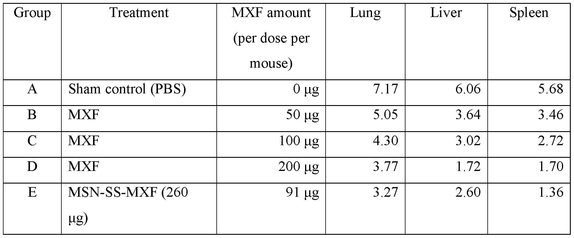
[00341] Mice showed no toxicity from MSN-SS-MXF nanoparticles. During the course of infection, sham control (PBS treated) mice lost more than 20% of their body weight, whereas mice treated with free MXF or MSN-SS-MXF did not (Figure 41).
[00342] FIG. 41 is a graph showing weight changes in infected mice. The percentage change in weight of infected mice that were sham-treated, treated with the broad spectrum antibiotic MXF administered as a free drug, or treated with MSN-SS-MXF over the course of treatment.
[00343] Lungs, livers, and spleens from infected mice that were untreated or treated with free MF or MSN-SS-MXF were homogenized. The organ homogenates were serially diluted and plated on GCII chocolate agar containing sulfamethoxazole (40 μg/ml), trimethoprim (8 μg/ml), and erythromycin (50 μg/ml) to prevent growth of contaminants. The agar plates were incubated at 37°C for 3 days at which time the number of bacterial colonies on each plate was counted. Treatment with 260 μg of MSN-SS-MXF (= 91 μg MXF) reduced the bacterial burden in the lung and spleen by 3.9- and 4.3-logs, respectively - more than that achieved by free MXF at the dose of 200 μg (Figure 9). MSN-SS-MXF reduced bacterial burden in the liver to a level below that of free MXF at a dose of 100 μg. This study demonstrates that MSN-SS-MXF is more efficacious than an equivalent amount of free MXF in the lung, spleen, and liver with an efficacy ratio (MSN-SS- MXF:Free MXF) of -3-4: 1 in the lung and spleen, and -1 : 1 in the liver.
[00344] FIG. 42A-C show that MSN-SS-MXF kills more F. tularensis LVS than equivalent amount of free MXF in infected mice. Mice were infected with F. tularensis LVS and either sham treated or treated with MXF or MSN-SS-MXF. FIG. 42A is a graph showing bacterial burdens in the lung. FIG. 42B is a graph showing bacterial burden in the liver. FIG. 42C is a graph showing bacterial burden in the spleen. All tissues were monitored throughout the course of infection.
[00345] We repeated the efficacy assay of MSN-SS-MXF in vivo with three doses of free
MXF and two doses of the nanoparticles. 32 mice were infected with about 4000 CFU of F.
tularensis LVS by the intranasal route on day 0. Five hours later, two mice were euthanized to determine the initial bacterial burden in the lung. One day later (day 1), three additional mice were euthanized to determine bacterial growth over this time period. Three to four mice per group were then either sham treated or treated with various concentrations of MSN-SS-MXF or free MXF by tail vein injection every other day, 3 days a week (Friday, Sunday and Tuesday) for one week. Mice were euthanized one day (day 6) after the last treatment.
[00346] This batch of MSN-SS-MXF had 51% (wt/wt) drug release capacity under organic reducing conditions. The amount of MXF for each dose of MSN-SS-MXF used for treatment was calculated according to the drug release capacity and shown in Table 10.
[00347] Table 10. The number of F. tularensis LVS (Log CFU) in the organs one day after the last dose of treatment in the second mouse experiment


[00348] Over the course of the F. tularensis infection, sham control mice started to lose weight after day 3 and lost about 12% of their body weight by the end of the treatment period. In contrast, mice treated with free MXF or MSN-SS-MXF maintained their weight. Again this confirms that the nanoparticle was well tolerated by the mice (Figure 10).
[00349] FIG. 43 is a graph showing weight changes in infected mice. Percentage change in weight of infected mice that were sham-treated, treated with MXF administered as a free drug or treated with MSN-SS-MXF over the treatment period.
[00350] F. tularensis LVS grew 1.8 logs in the lung during the first day after intranasal infection and another 1.8 logs over the next 5 days with no treatment (Figure 44 A). Treating with 230 μg MSN-SS-MXF (= 117 μg MXF) reduced bacterial burden in the lung by 3.6 logs compared with untreated mice, much more than the 2.8-log reduction obtained by treatment with the highest dose of free MXF (= 300 μg) tested in the experiment. Thus, MXF delivered by the disulfide snap- top nanoparticle is more efficacious than 3-fold the equivalent amount of free MXF in the lung. Bacterial burden in the liver and spleen at 5 hours and at 1 day post-infection were below the limit of detection for the experiment. Both treatment doses of MSN-SS-MXF kept bacterial CFU below the limit of detection at the end of the study. In contrast, F. tularensis LVS were detected in the liver (Figure 44B) as well as in the spleen (Figure 44C) from mice treated with all three doses of free MXF. It was apparent from these results that MXF delivered by MSN-SS-MXF was at least 3- fold more efficacious than an equivalent amount of free MXF in the liver and spleen.
[00351] FIG. 44A-C shows disulfide snap-top MSN-SS-MXF treatment reduces bacterial burden in infected mice. Mice were infected with F. tularensis LVS and either sham treated or treated with one of three different doses of free MXF or with one of two different doses of MSN- SS-MXF. In sham-treated mice, bacterial burden was assayed on the first day after infection (Day 0) and one day later (Day 1). FIG. 44A is a graph showing bacterial burden in the lung. FIG. 44B is a graph showing bacterial burden in the liver. FIG. 44C is a graph showing bacterial burden in the spleen. All were determined one day after the last treatment (Day 6) by assaying bacterial CFU. *Bacterial CFU at the limit of detection.
[00352] Benefits of the MSN-SS-MXF controlled drug release nanoparticle technology include a) it is more efficacious than an equivalent amount of free drug for treating tularemia and other infectious diseases; b) it preferentially targets macrophages, the host cells for F. tularensis and many other intracellular pathogens, thereby increasing the therapeutic index; c) it provides for controlled release of the drug intracellularly in the host cells for F. tularensis and other intracellular pathogens, thereby avoiding off-target effects and premature metabolism of the drug; d) it provides improved treatment of tularemia; and other diseases caused by intracellular pathogens by treating the infection more rapidly and with less toxicity than current modalities; e) NPs can be administered by a variety of routes including intravenously, subcutaneously, intramuscularly, orally, by inhalation, etc; and f) the MSNs are biodegraded and do not accumulate after administration.
[00353] Francisella tularensis (Ft) is a facultative intracellular bacterial pathogen that causes tularemia, a serious and potentially fatal disease. Because Ft has extraordinarily high infectivity, causes serious morbidity and mortality, is relatively easily dispersed, is readily cultured on a large scale, and has previously been developed as a biological weapon, it is classified as a Tier 1 potential agent of bioterrorism. Pneumonic tularemia, the type of tularemia of greatest concern in a bioterrorist attack, has a very high morbidity with at least half the patients requiring
hospitalization, and can be fatal even in a setting where awareness is high and appropriate treatment is available. Therefore modalities allowing more effective and rapid treatment of tularemia are needed. The technology described herein allows for shortening the treatment regimen for tularemia, reducing morbidity and mortality, and reducing time in intensive care.
[00354] Many other infectious diseases are caused by intracellular pathogens, and many of these are sensitive to MXF. This technology offers more rapid and effective treatment of such pathogens, e.g. Mycobacterium tuberculosis.
[00355] Approximately 10 million cases of active TB occur annually and -1.7 million people die of TB annually. Approximately 2.5 billion people worldwide are latently infected with M. tuberculosis (LTBI).
[00356] While effective TB antibiotics are available for treatment of TB, conventional therapy is exceptionally prolonged, requiring 6-9 months, and serious toxic side effects are dose- limiting. For example, three of the first line drugs for treating TB - isoniazid (INH), rifampicin (RTF), and pyrazinamide (PZA) - are limited by hepatotoxicity, a side effect due to the action of the
drug on hepatocytes rather than macrophages, the primary host cells of Mycobacterium tuberculosis (Mtb), the causative agent of TB. In addition, ΓΝΗ causes neurotoxicity and optic neuritis, and moxifloxacin (MXF), another potent TB drug, can cause cardiac arrhythmia, tendonitis/tendon rupture, peripheral neuropathy, and photosensitivity. Nanoparticle (NP) delivery platforms provide a more effective, less toxic, and shorter treatment for TB. Because host mononuclear phagocytes internalize particles more efficiently than other cells, intravenously (i.v.) injected NPs, or NPs delivered by other routes of administration, are preferentially taken up by macrophages of the mononuclear phagocyte (reticuloendothelial) system (MPS) and accumulate in liver, spleen, and lung. Therefore, NPs are ideally suited to treat Mtb, which infects macrophages in these organs. Targeting antibiotic-loaded NPs to infected organs and tissues, selectively delivering the antibiotics into macrophages and releasing them at high concentrations intracellularly greatly increases their therapeutic index by achieving higher drug concentrations locally where Mtb replicate while limiting systemic toxicities. Moreover, by controlled release of the drug only after the NPs have been ingested protects the drug from hepatic metabolism and drug clearance before the drug has had the opportunity to attack target pathogens. Increasing drug concentrations at the site of infection by orders of magnitude allows for a much shorter duration of therapy. Moreover, because drug resistance develops when bacteria are treated with subtherapeutic levels of antibiotics, a system that delivers high antibiotic concentrations to the site where bacteria divide facilitates sterilization of sites of infection and minimize emergence of drug resistance. Additional advantages of NP drug delivery vs. free drug are a) the drug is shielded from degradation or modification during delivery to infected tissues and b) the drug, by being targeted to macrophages rather than hepatocytes, will not impact hepatic cytochrome P450 metabolism of other drugs.
[00357] The current prolonged drug treatment regimen for TB frequently results in poor patient compliance. This in turn fosters the emergence of M. tuberculosis resistant to TB.
Currently, about 5% of all TB cases are resistant to the two major drugs used to treat TB - INH and rifampin. The presently claimed method shortens the treatment regimen for TB, thus enhancing patient compliance, and lessons the emergence of drug-resistant TB.
[00358] Many other serious bacterial infections are sensitive to MXF. The controlled release MSN-SS-MXF technology allows for improved pharmacodynamics for MXF and hence improved treatment of such pathogens.
[00359] MSNs offer many advantages over previous delivery vehicles (e.g. liposomes, solid lipid particles, alginates) for TB drugs because of their stability, uniformity, inherent lack of toxicity, high internal surface area for drug binding, and versatility in incorporating additional design features. Because of their ultra-high internal surface area (-1000 m2/g), MSNs can encapsulate exceptionally high concentrations of different types of cargos. Loading capacities as high as 50 weight percent have been achieved, exceeding by several orders of magnitude that of conventional liposomal nanocarriers. MSNs can be synthesized with a variety of different internal and surface design features, including those that allow for specific targeting to infected host organs and tissues and those that enable exquisitely controlled release of cargo under specific
environmental conditions, such as the acidified endosomal/lysosomal compartment of macrophages. Internal encapsulation protects therapeutic agents from biodegradation and allows high
concentrations of combined cargos with disparate physicochemical properties to be simultaneously delivered to overcome multidrug resistance, achieve synergistic effects and/or enable combined therapy and diagnostics (theranostics). Finally, MSN are degraded in the body over several days and the degradation products are excreted.
[00360] Example 4
[00361] One embodiment of this invention is a composition for treating infectious diseases caused by pathogens sensitive to the antibiotic moxifloxacin (MXF). These diseases include those caused by intracellular pathogens including Tuberculosis (TB) and other
mycobacterial diseases (e.g. Mycobacterium kansasii infection, Mycobacterium intracellular infection, disseminated BCG, etc.), tularemia, etc. These diseases also include those caused by extracellular pathogens that are sensitive to the antibiotic moxifloxacin. One embodiment of the invention comprises a mesoporous silica nanoparticle (MSN) with pores into which moxifloxacin is loaded and with valves on top of the pores that contain the moxifloxacin inside the pores until the nanoparticle encounters a low pH environment, e.g. the inside of a host cell for a pathogen, at which point the valves open and release the moxifloxacin. Another embodiment is a method for treating infections caused by intracellular and extracellular pathogens sensitive to the antibiotic
moxifloxacin.
[00362] I. Construction of MSN with chemically linked pH sensitive nanovalve
[00363] Materials Synthesis
[00364] Synthesis ofMSNs (100 nm)
[00365] Mesoporous silica nanoparticles (MSN) are nanoscale materials that are composed of amorphous silica, which is highly porous and has large surface area. These materials have particulate sizes on the order of -100 nm and possess pore diameters of approximately 2 nm. MSNs are synthesized by first dissolving a surfactant, cetyltrimethylammonium bromide (250 mg, CTAB) into a basic solution (120 mL, pH 12) and heating up to 80 °C. Once the solution is thermally stable, the silica precursor, tetraethyl orthosilicate (1.2 mL, TEOS), is added drop-wise into the solution and the solution slowly begins to become opaque. After 15 minutes of aging, a coating of 3-(trihydroxysilyl)propyl methylphosphonate, (300 uL, HTMP) is added to the solution, and the solution is further aged for 90 minutes. The nanoparticles are collected by centrifugation and washed with methanol.
[00366] FIG. 45 is a schematic of the pH sensitive nanovalve mechanism.
[00367] In order to trap and release cargo molecule, the pore orifice of MCM-41 are attached with organic molecule with pKa around 6 as stalk and modified nanoparticles are soaked in drug/dye solution for 12 hours, after which cyclodextrin is added as capping agent. The outer ring of cyclodextrin is hydrophilic while the inner ring is hydrophobic which binds with stalk organic moiety. When the environmental pH (acidifying compartment in cells) is equal or lower than the pka of stalk, it gets protonated and binding affinity between the stalk and cyclodextrin dramatically decreases, which leads to cap dissociating from stalk molecule and drug release.
[00368] The stalk of pH sensitive nanovalve synthesis is a straightforward two step synthesis which has been optimized as shown in Figure 2. Benzimidazole group is utilized because of its pka around 6 which mean this valve can open when pH is 6 or lower. MCM-41 (100 mg) was dispersed in anhydrous toluene, mixed with chloromethyltrimethoxysilane (15 μΐ) and refluxed for 12 hours under nitrogen atmosphere. Then chloromethyl modified MCM-41 was washed by toluene and dimethyoformamide (DMF) and dispersed in 8 ml DMF, followed by adding
tetrabutyammonium iodide (2 mg), benzimidazole (12 mg) and anhydrous triethylamine (150 μΐ) .The solution was stirred and heated up to 70°C under N2 for 24 hours. The final product, pH sensitive nnaovalve modified MSN was washed with methanol, and then dispersed in 30 ml
methanol and 1.15 ml 12 M HC1 and refluxed under nitrogen for 12 hours to extract surfactant template. The MSN were washed with methanol and water to be prepared for loading.
[00369] FIG. 46 is a schematic of pH sensitive nanovalve (MBI) system synthesis.
[00370] Loading of moxifloxacin and drug release test by time-resolved florescence spectroscopy
[00371] MCM-41 (10 mg) with pH valve was dispersed in 1 ml ImM Moxifloaxcin PBS solution and rotating overnight, β cyclodextrin (40 mg) was added into the solution as capping agent to prevent drug from leaking out. After mixing the solution for another 12 hours, moxifloxacin loaded MCM-41 with pH nanovalve system was dried under vacuum overnight. In order to prove moxifloxacin can be release from MSN and detect moxiflxoacin fluorescence emission in supernatant, the dried powder was put at the corner of a glass vial containing 10 ml DI water.
Baseline spectra were collected for 1 hour to show that there was no moxifloxacin leaking out, and then 1M HC1 solution was added to adjust pH to 4.5, which resulted in a dramatic increase of fluorescence emission in supernatant which indicated release of moxifloxacin.. A 5 mW 377 nm laser beam shot through the supernatant in glass vial and released moxifloxacin was excited and emitted fluorescence detected and collected by CCD and computer. Release profile was constructed by intergration of emission peak area from 480 nm to 520 nm. After collecting data for 17 hours and moxifloxacin was released completely, the supernatant released moxifloxacin concentration was calculated based on UV-Vis spectrum and standard curve by Beer' s law. Release capacity was defined as released moxifloxacin weight over MCM-41 weight.
[00372] II. Characterization of MSN with pH sensitive nanovalve
[00373] Transmission electron microscopy (TEM) images of SMSNs and MSNs were obtained using a JEM1200-EX (JEOL) instrument (JEOL USA, Inc., Peabody, MA). Particle size was measured by ZetaSizer Nano (Malvern Insstruments Ltd, Worcestershire, UK) and MSN was dispersed in PBS in 50 μ^πιΐ.13C-CPMS and 29Si-CPMS NMR spectra were collected on a
DSX300 NMR spectrometer (Bruker, 300 MHz), using a 4 mm CP/MAS probe at room
temperature. UV-Vis spectra of moxifloxacin were collected by a Cary 500 UV-vis-NTR
spectrophotometer. The release profile was obtained by time-resolved fluorescence spectroscopy.
[00374] FIG. 47 is a TEM image of MSN with pH sensitive nanovalve that shows structural integrity preserved after all surface modifications and surfactant temple extraction
[00375] FIG. 48 shows dynamic light scattering (DLS) measurement of MSN with pH sensitive nanovalve. It shows that mean hydrodynamic diameter of the modified nanoparticle is around 100 nm
[00376] FIG. 49 A is a 13C-CPMS NMR of MBI MSN. The data illustrate that benzimidazole is bonded to the silica surface. The nanoparticle alone does not produce peaks in the aromatic region. FIG. 49B is a 29Si-CPMS NMR spectra of MBI MSN. It shows bulk silica band and attached thread containing a Si-C bond-band, proving the attachment of the MBI compound.
[00377] FIG. 50 is a UV-Vis spectrum of moxifloxacin under pH 1 and 7.4
[00378] FIG. 51 is a graph showing Moxifloxacin loaded MCM-41 with MBI pH sensitive nanovalve release profile. There is no leakage at pH 7 indicated as flat baseline. Drug release starts at pH 6 and release rate can be further increased by lowering pH down to 4.5
[00379] III. Assay for MXF released from pH-gated MSN-MXF by low pH conditions
[00380] Example of assay
[00381] Assay
[00382] MXF was released from 100 nm pH-gated MSN1 -MXF or MSN2-MXF at neutral and acidic pH conditions and measured by spectrophotometry at 288 nm.
[00383] Release of MXF by neutral pH or acidic pH
[00384] To assess the amount of MXF loading on nanoparticles, we suspended 1 mg pH- gated MSN-MXF in 1 ml of 0.1 M HEPES, pH 7.4 or in 1 ml of 0.1 N HC1 in DMSO (acidified DMSO), mixed by end-to-end rotation overnight at room temperature, and centrifuged at 10,000 g for 10 min. The supernate was diluted 1 :4 to 1 : 100 in 0.1 M HEPES, pH 7.4, to a final volume of 1 ml, and the absorbance read at 288 nm (Table 1). At the same time, standards with known amounts of MXF were also prepared in 0.1 M HEPES, pH 7.4, and used to calculate MXF concentration in the eluates (Figure 1). We confirmed that addition of acidified DMSO to the standards at levels corresponding to those in the diluted samples only minimally affects the absorbance at 288 nm. The amount of MXF eluted with 0.1 M HEPES was considered to be the residual MXF outside of the nanoparticle in that batch of pH-gated MSN-MXF. The amount of MXF released by acidified DMSO was considered to be the total MXF loaded on that batch of pH-gated MSN-MXF.
[00385] FIG. 52 is an example of a MXF standard curve used to calculate drug loading on nanoparticles eluted under neutral pH or acidic pH conditions.
Table 11. MXF released under neutral pH or acidic pH from 1 mg of pH

[00387] Release of MXF sequentially under neutral pH or acidic pH conditions
[00388] MXF loading on pH-gated MSN-MXF also can be measured by sequentially eluting first at neutral pH and subsequently under acidic aqueous and organic conditions. pH-gated MSN-MXF was suspended in 1 ml of 0.1 M FEPES, pH 7.4 at a concentration of 1 mg/ml, mixed by end-to-end rotation for about 1 hour at room temperature, and centrifuged at 10,000 g for 10 min. The supernate (Neutral Eluate) was collected for assay of MXF concentration. Next, the nanoparticles were resuspended in 1 ml of 0.1 N HC1 (aqueous acid), mixed by end-to-end rotation for about 2 hours at room temperature, and centrifuged to pellet the nanoparticles. The supernate (Aqueous Acid Eluate) was removed for assay of MXF release by aqueous acid. Finally, the nanoparticles were resuspended in 1 ml of 0.1 N HC1 in DMSO (DMSO acid), mixed by end-to-end rotation overnight at room temperature, and centrifuged to pellet the nanoparticles. The amount of MXF eluted under neutral pH, aqueous acid, and DMSO acid conditions was measured at 288 nm (Table 2) and calculated from the MXF standard curve (Figure 2). The amount of MXF eluted with 0.1 M FEPES was considered to be the residual MXF outside of the nanoparticle in that batch of pH-gated MSN-MXF. The amount of MXF eluted by aqueous acid was considered to be the MXF released from the nanoparticle upon the opening of its pH-sensitive gates. The additional amount of MXF released by acidified DMSO was considered to be the MXF absorbed on the nanoparticle through hydrophobic interaction. The sum of MXF measured in neutral eluate, aqueous acid eluate, and DMSO acid eluate is the total MXF loaded on that batch of pH-gated MSN-MXF.
[00389] FIG. 53 is an example of a MXF standard curve used for calculating drug loading on nanoparticles after sequential elution under neutral and acidic pH conditions.
[00390] Table 12. MXF released under sequential elution under neutral pH and acidic pH condition from 1 mg of pH-gated MSN1-MXF
Sample MXF (μ§) MXF/NP (wt/wt)
Neutral Eluate 25.0 2.5%
Aqueous Acid Eluate 26.9 2.7%
DMSO Acid Eluate 16.9 1.7%
Total Eluted MXF 68.8 6.9%
[00391] IV. Assay for efficacy of nanoparticles in killing of F. tularensis in infected macrophages
[00392] The efficacy of MXF loaded mesoporous silica nanoparticles equipped with two different types of pH operative valves (pH-gated MSN1-MXF and pH-gated MSN2-MXF) was assessed in a macrophage infection model of F. tularensis. Live Vaccine Strain (LVS) of F.
tularensis subsp. holarctica was grown from frozen stocks on GCII chocolate agar at 37°C for 3 days prior to use for infecting macrophaes. Human monocytic THP-l cell line was differentiated with phorbol 12-myristate 13-acetate (PMA) for 3 days to mature the cells into a macrophage-like cell type and infected with F. tularensis LVS at a multiplicity of infection ratio of about 10 bacteria to 1 THP-1 cell for 90 min at 37°C, 5% C02 - 95% air atmosphere. Infected monolayers were washed to remove extracellular bacteria. Fresh medium with or without MXF and MXF loaded pH- gated MSN1 or MSN2 nanoparticles was added to the infected monolayer. The cultures were incubated in continued presence of the treatments for one day. F. tularensis LVS were harvested from infected but not treated cultures at 3 hours and 1 day post infection to determine bacterial growth and from infected cultures that were treated at 1 day to assess the effect of treatment. To harvest the bacteria, we lysed the infected macrophages with 1% saponin in phosphate buffered saline (PBS) and serially diluted the lysate for plating on GCII chocolate agar plates. Bacterial colony forming units (CFU) on agar plates were counted after incubation at 37°C, 5% CO2 - 95% air atmosphere for 3 days.
[00393] In the experiment, we assayed the killing capacity of free MXF at serial two-fold increases in concentration, from 1 ng/ml to 64 ng/ml, and of the two types of pH-gated
nanoparticles, MSNl-MXF and MSN2-MXF, also with serial two-fold increases in concentration from 0.0625 μg/ml to 8 μg/ml. Because MSNl-MXF had a total drug loading measured as 2.64% wt/wt (0.00%) in Neutral Eluate + 2.64% in Aqueous Acid Eluate), the amount of MXF that could
potentially be released by acidic pH in the endo-lysosomal compartments of macrophages for MSNl-MXF over the range of concentrations tested was estimated to be from 1.7 to 211 ng/ml (Table 3). The total drug loading on MSN2-MXF was 0.36%, therefore the acid releasable MXF for the nanoparticle over the range of nanoparticle concentrations tested was estimated to be from 0.23 to 29 ng/ml.
[00394] While F. tularensis LVS grew 2.5 logs in macrophages with no treatment, all treatments including free MXF, MSNl-MXF and MSN2-MXF, killed F. tularensis LVS in infected macrophages in a dose dependent manner (Figure 54). When compared at the same concentration, MSNl-MXF was much more effective than MSN2-MXF in killing F. tularensis LVS. For example, at one day MSNl-MXF at 1 μg/ml reduced CFU by 3.37 logs compared with the level in the untreated control group, whereas the same concentration of MSN2-MXF reduced bacterial CFU by only by 0.17 logs compared with the untreated control group.
[00395] We assayed the capacity of neutral and acid eluates of pH-gated MSNl-MXF and MSN2-MXF to kill F. tularensis LVS in infected human macrophages. The eluate prepared from 1 μ^ηιΐ of MSNl-MXF in 0.1 M HEPES, pH 7.4 (Neutral Eluate) had no bactericidal activity indicating negligible MXF release from the nanoparticles at neutral pH. In contrast, eluates prepared from 0.5 and 1 μg/ml of MSNl-MXF in 100 mM maleic acid, pH 1.8 (aqueous Acid Eluate) reduced bacterial numbers in macrophages by 1.6 and 2.5 logs, respectively (Figure 55A). As for MSN2-MXF, its neutral eluate had no impact on F. tularensis LVS growth in macrophages and the acid eluate reduced bacterial number by merely 0.2 logs (Figure 55B). This study demonstrates that 1) the pH operative valves on MSNl-MXF are tightly closed at neutral pH and open at acidic pH, 2) MXF eluted under acidic pH retains biological activity, 3) MSNl-MXF and MSN2-MXF kill F. tularensis LVS in macrophages in a dose-dependent fashion, and 4) MSNl-MXF has greater efficacy than MSN2-MXF, most likely because of higher MXF loading on MSNl-MXF.
[00396] Table 13. Bacterial CFU in infected macrophages with and without treatment

MXF 2 ng/ml 1 day 7.59
MXF 4 ng/ml 1 day 7.59
MXF 8 ng/ml 1 day 6.92
MXF 16 ng/ml 1 day 5.09
MXF 32 ng/ml 1 day 3.72
MXF 64 ng/ml 1 day 3.43
MSN1 control (8 μ /ηι1) 0 ng/ml 1 day 7.72
MSNl-MXF (0.0625 μg/ml) 1.65 ng/ml 1 day 7.59
MSNl-MXF (0.125 μg/ml) 3.3 ng/ml 1 day 7.21
MSNl-MXF (0.25 μ^ηιΐ) 6.6 ng/ml 1 day 5.49
MSNl-MXF (0.5 μ^ηιΐ) 13.2 ng/ml 1 day 4.49
MSNl-MXF (1 μg/ml) 26.4 ng/ml 1 day 4.23
MSNl-MXF (2 μg/ml) 52.8 ng/ml 1 day 3.49
MSNl-MXF (4 μg/ml) 105.6 ng/ml 1 day 3.49
MSNl-MXF (8 μg/ml) 211.2 ng/ml 1 day 1.73
MSN2 control (8 μ^πιΐ) 0 ng/ml 1 day 7.70
MSN2-MXF (0.0625 μg/ml) 0.23 ng/ml 1 day 7.71
MSN2-MXF (0.125 μg/ml) 0.45 ng/ml 1 day 7.72
MSN2-MXF (0.25 μ^ηιΐ) 0.9 ng/ml 1 day 7.59
MSN2-MXF (0.5 μ^ηιΐ) 1.8 ng/ml 1 day 7.59
MSN2-MXF (1 μg/ml) 3.6 ng/ml 1 day 7.43
MSN2-MXF (2 μg/ml) 7.2 ng/ml 1 day 7.06
MSN2-MXF (4 μg/ml) 14.4 ng/ml 1 day 5.34
MSN2-MXF (8 μg/ml) 28.8 ng/ml 1 day 3.61
[00397] FIG. 54A is a graph showing that free MXF kill F. tularensis LVS in human macrophages in a dose dependent manner. FIG. 54B is a graph showing that pH-gated MSNl-MXF
kill F. tularensis LVS in human macrophages in a dose dependent manner. FIG. 54C is a graph showing that MSN2-MXF kill F. tularensis LVS in human macrophages in a dose dependent manner. THP-1 macrophages were infected with F. tularensis LVS, treated for one day, lysed, and the lysate serially diluted and plated to determine bacterial CFU. Infected but untreated
macrophages were lysed at 3 hours and one day to determine bacterial growth.
[00398] FIG. 55A is a graph showing that acid eluates of pH-gated MSN1-MXF reduce the number of F. tularensis LVS in macrophages. FIG. 55B is a graph showing that MSN2-MXF do not reduce the number of F. tularensis LVS in macrophages.
[00399] The pH-gated MSN1-MXF was shown in our first macrophage experiment to be efficacious in killing F. tularensis, a Gram-negative bacterium. Because moxifloxacin is a broad- spectrum antibiotic, in our second experiment we investigated whether MSN1-MXF is also active against M tuberculosis, a Gram-positive bacterium, in macrophages. PMA differentiated THP-1 macrophages were infected with the virulent M. tuberculosis Erdman strain for 90 min and not treated or treated with a) control MSN1 not loaded with MXF; b) various concentrations of pH- gated MSNl-MXF; or c) various concentrations of free MXF. The infected macrophages that were not treated were lysed at 3 hours and 3 days post infection, and all infected macrophages that were treated were lysed at 3 days post infection to determine bacterial CFU in the macrophages. The infected macrophages were lysed with 0.1% SDS, and the lysates serially diluted and plated on 7H11 agar plates. Bacterial colonies on the plates were counted after two weeks of incubation at 37°C, 5% CO2 - 95% air atmosphere.
[00400] M. tuberculosis grew similarly in macrophages that were untreated or treated with control MSN1 nanoparticles (no MXF loaded) indicating that the nanoparticle carrier by itself has no inhibitory effect on the bacterium (Figure 56B). Both free MXF and MXF delivered by pH- gated MSN1 killed the bacteria in macrophages in a dose-dependent fashion (Figure 56A & 56B). We determined the drug loading on the nanoparticles by incubating MSNl-MXF with 0.1 M HEPES at neutral pH or with 0.1 N HC1 in DMSO (acidified DMSO) at the concentration of 1 mg/ml. The amount of residual (neutral) and acidic pH releasable MXF from the nanoparticles was measured by spectrophotometry as described above. The total amount of MXF on this batch of MSNl-MXF was determined to be 15.71% (wt/wt) with 5.23% (wt/wt) being in the neutral eluate.
The amount of MXF available from each concentration of MSNl-MXF tested in the experiment is calculated based on the total % wt/wt of MXF for the nanoparticle and is shown in Table 14.
[00401] MSNl-MXF at 3.1, 6.25, and 12.5 μg/ml killed 84%, 97%, and 99% of intracellular M. tuberculosis over 3 days. That the extent ofM tuberculosis killing achieved by any selected dose of MSNl-MXF is about the same as that of the corresponding amount of MXF available from that dose of nanoparticle suggests that MXF delivered by pH-gated MSNl-MXF has the same potency in killing M. tuberculosis in macrophages as the equivalent amount of MXF by itself. Thus, the efficacy ratio of MSNl-MXF to MXF in macrophages (the amount of killing by MSNl-MXF/amount of killing by an equivalent amount of free MXF) was close to 1.
[00402] As shown in Figure 56C, while the acidified DMSO used to release MXF from the nanoparticle had a minimal effect by itself (at a concentration matching that used in the MSNl- MXF 12.5 μg/ml acid eluate condition), the eluate prepared from MSNl-MXF with the organic acid killed M. tuberculosis to the same extent as the nanoparticles. This result confirms that MXF released from pH-gated MSNl-MXF by acidic pH is biologically active.
[00403] Table 14. M. tuberculosis CFU in infected macrophages with and without treatment

[00405] V. Assay for efficacy of nanoparticles in treating tularemia using a mouse model of pneumonic tularemia
[00406] The efficacy of pH-gated MSN1 -MXF that had an MXF release capacity measured to be 6.88% in treating tularemia was assessed in a mouse model of pneumonic tularemia. 20 Balb/c mice were infected by intranasal route with about 8000 CFU of F. tularensis LVS, a dose close to ten times the LD50. Five hours later (day 0), two mice were euthanized to establish bacterial numbers in the lung. One day later (day 1), an additional three mice were euthanized to determine the number of bacteria in the organs at the onset of the treatment period. Three mice per group were then either sham treated or treated with 100, 200 or 400 μg of MXF or 2 mg of pH-gated MSN1- MXF by tail vein injection every other day (Friday, Sunday, and Tuesday) for a week. With 6.88% drug release capacity (Table 12), the 2 mg of MSN1-MXF had about 140 μg of MXF. Mice were euthanized one day after the last treatment (day 6) and lungs spleens, and livers assayed for CFU of F. tularensis.
[00407] During the course of infection, sham control (PBS treated) mice lost 15% of their body weight, whereas mice treated with pH-gated MSN1-MXF maintained their weights indicating that the nanoparticle was well tolerated by the mice (Figure 57).
[00408] FIG. 57 is a chart showing the percentage change in weight of the F. tularensis- infected mice that were sham-treated, treated with the broad spectrum antibiotic MXF administered as a free drug, or treat with pH-gated MSNl-MXF were monitored over the course of treatment.
[00409] Lungs, livers, and spleens from the infected mice with or without treatment were homogenized. The organ homogenates were serially diluted and plated on GCII chocolate agar containing sulfamethoxazole (40 μg/ml), trimethoprim (8 μg/ml), and erythromycin (50 μg/ml). The agar plates were incubated at 37°C for 3 days at which time bacterial colonies on each plate were counted. Treatment with MSNl-MXF reduced bacterial burden in the lung and spleen by 4.0-, and
4.3-logs, respectively, more so than treatment with 400 μg of free MXF (Table 15). All treatments reduced bacterial burden in the liver to a level below the experimental detection limit (Figure 57). This study demonstrates that pH-gated MSNl-MXF administered intravenously is more effective than a 2.8-fold greater dose of free MXF in treating F. tularensis LVS in mice.
[00410] Table 15. F. tularensis LVS burden (Log CFU) in the organs 3 days after the last treatment dose

[00411] pH-gated MSNl-MXF kills more F. tularensis LVS than about 2.8-fold its equivalent amount of free MXF in infected mice. Mice were infected with F. tularensis LVS and either sham treated or treated with MXF or MSNl-MXF. FIG. 58B is a graph showing bacterial burdens in the lung. FIG. 58A is a graph showing bacterial burdens in the liver. FIG. 58C is a graph showing bacterial burdens in the spleen. All were monitored throughout the course of infection.
[00412] Results from our first animal study showed that 2 mg of pH-gated MSNl-MXF was efficacious in treating F. tularensis LVS infection in mice, however, any potential differences in killing efficacy between free MXF and nanoparticles could not be ascertained in the liver because the number of CFU was at the limit of detection for both the nanoparticle and all doses of free MXF. Therefore, in our second animal study we repeated the experiment with lower doses of MSNl-MXF and with an equivalent amount of free MXF to match each dose of the nanoparticle. 32 mice were infected with about 4000 CFU of F. tularensis LVS by the intranasal route on day 0. One day later, two mice were euthanized to determine the initial bacterial burden in the lung. One day later (day 1), three additional mice were euthanized to determine bacterial growth at the onset of the treatment period. Three mice per group were then either sham treated or treated with various
concentrations of MSNl-MXF or free MXF by tail vein injection every other day, 3 days a week (Friday, Sunday and Tuesday) for one week. Mice were euthanized one day (day 6) after the last treatment.
[00413] This batch of MSNl-MXF had 15.7% drug release capacity. The amount of
MXF at each dose of MSNl-MXF used for treatment was calculated according to the drug release capacity and shown in Table 16.
[00414] Table 16. F. tularensis LVS number (Log CFU) in the organs one day after the last treatment dose in the second mouse experiment

[00415] During the course of F. tularensis infection, sham control mice started to lose weight after day 3 and, by day 6, lost more than 22% of their body weight. Mice treated with the two lowest doses of pH-gated MSNl-MXF (80 μg and 160 μg) and their equivalent amount of free MXF (12.5 μg and 25 μg) also lost weight, but in both cases the mice treated with the nanoparticle suffered less weight loss than those treated with the free MXF. This indicates that at lower doses the pH-gated MSNl-MXF probably controls the infection better than the equivalent amount of free MXF. Mice treated with the two highest doses of the MSNl-MXF (320 μg and 640 μg) or free MXF (50 μg and 100 μg) gained weight indicating that the nanoparticle was well tolerated by the mice (Figure 8).
[00416] FIG. 59 is a graph showing percentage change in weight of the F. tularensis- infected mice that were sham-treated, treated with the broad spectrum antibiotic MXF administered as a free drug, or treated with pH-gated MSNl-MXF was monitored over the treatment period.
[00417] In comparison with sham-treated mice, all treatments led to substantial reductions in the organ bacterial burden, with greater than 1.2-, 1.5-, and 1.2-logs of reduction in bacterial CFU in the lung, liver, and spleen, respectively (Figure 9). In the lung, the pH-gated MSNl-MXF had an efficacy similar to that of an equivalent dose of MXF. In the liver, MSNl- MXF at the dose of 160 μg (= 25 μg MXF) reduced bacterial CFU to a level similar to that achieved by 100 μg of free MXF indicating a 4-fold efficacy ratio. In the spleen, lower doses of MSNl-MXF were about twice as effective as equivalent doses of free MXF. Overall this study demonstrates that the pH-gated MSNl-MXF is at least as efficacious as an equivalent amount of free MXF in the lung and about 2 - 4 fold more efficacious as an equivalent amount of free MXF in the liver and spleen.
[00418] FIG. 60A is a graph showing that pH-gated MSNl-MXF treatment reduces bacterial burden in the lung of F. tularensis-m' iected mice. FIG. 60B is a graph showing that pH- gated MSNl-MXF treatment reduces bacterial burden in the liver of F. tularensis-miected mice. FIG. 60C is a graph showing that pH-gated MSNl-MXF treatment reduces bacterial burden in the spleen of F. tularensis-m' iected mice. Mice were infected with F. tularensis LVS and either sham treated or treated with one of four different doses of free MXF or MSNl-MXF. In sham-treated mice, bacterial burden was assayed on the first day after infection (Day 0) and one day later (Day 1). Bacterial burden in lung (A), liver (B), and spleen (C) was determined day 6, one day after the last treatment (Day 6) by assaying bacterial CFU.
[00419] We tested another batch of pH-gated MSNl-MXF with 8.08% (wt/wt) drug loading in the third mouse experiment. Mice were infected with about 4000 CFU of F. tularensis LVS by the intranasal route. One day later, mice were sham treated or treated with one of the three doses of MXF (50, 100, and 200 μg) or with 640 μg of MSNl-MXF (with about 50 μg of releasable MXF) every other day (Friday, Sunday and Tuesday) for a week. The three doses of MXF were equal to lx, 2x, and 4x the amount of the releasable MXF from 640 μg of MSNl-MXF by acidic DMSO (Table 17).
[00420] Table 17. F. tularensis LVS number (Log CFU) in the organs one day after the last dose of treatment in the third mouse experiment
MXF amount
Group Treatment (per dose per Lung Liver Spleen
mouse)
A Sham control 0 μ8 7.17 6.06 5.68
B MXF (lx) 50 μ§ 5.05 3.64 3.46
C MXF (2x) 100 μ§ 4.30 3.02 2.72
D MXF (4x) 200 μ§ 3.77 1.72 1.70
E MSNl-MXF (640 μg) 50 μ§ 4.42 2.83 2.35
[00421] Sham treated mice suffered substantial weight loss but mice treated with free
MXF or MSNl-MXF did not (Figure 61). MSNl-MXF treatment reduced bacterial burden by 2.8 logs in the lung, 3.2 logs in the liver, and 3.3 logs in the spleen to a level close to that achieved by 100 μg MXF (Figure 62). This experiment demonstrates that mice showed no toxicity from pH- gated MSNl-MXF. Moreover, MSNl-MXF had an efficacy twice the equivalent amount of free MXF in the lung, spleen and liver.
[00422] FIG. 61 is a graph showing percentage change in weight of the F. tularensis- infected mice that were sham-treated, treated with MXF administered as a free drug, or treated with pH-gated MSNl-MXF was monitored over the treatment period.
[00423] FIG. 62A-C are graphs showing that pH-gated MSNl-MXF treatment reduced bacterial burden in F. tularensis-m' iected mice. Mice were infected with F. tularensis LVS and either sham treated or treated with MSNl-MXF or lx, 2x or 4x the equivalent amount of free MXF. In sham-treated mice, bacterial burden was assayed on the first day after infection (Day 0) and one day later (Day 1). FIG. 62A is a graph showing bacterial burden in lung. FIG. 62B is a graph showing bacterial burden in the liver. FIG. 62C is a graph showing bacterial burden in the spleen. All were determined on day 6, one day after the last treatment by assaying bacterial CFU.
[00424] Benefits of the MSNl-MXF controlled drug release nanoparticle technology include a) it is more efficacious than an equivalent amount of free drug for treating tuberculosis and tularemia and other infectious diseases; b) it preferentially targets macrophages, the host cells for M. tuberculosis, F. tularensis and many other intracellular pathogens, thereby increasing the therapeutic index; c) it provides for controlled release of the drug intracellularly in the host cells for
M. tuberculosis and F. tularensis and other intracellular pathogens, thereby avoiding off-target effects and premature metabolism of the drug; d) it allows for improved treatment of both active pulmonary and extra-pulmonary tuberculosis (TB) and other mycobacterial diseases (e.g.
Mycobacterium kansasii infection, Mycobacterium intracellular infection, disseminated BCG, etc.); tularemia; and other diseases caused by intracellular pathogens by treating the infection more rapidly and with less toxicity than current modalities; e) it can be used to treat latent TB infection (LTBI), which also requires a prolonged treatment regimen, more rapidly and effectively; f) NPs can be administered by a variety of routes including intravenously, subcutaneously, intramuscularly, orally, by inhalation, etc; and g) the MSNs are biodegraded and do not accumulate after
administration.
[00425] Approximately 10 million cases of active TB occur annually and -1.7 million people die of TB annually. Approximately 2.5 billion people worldwide are latently infected with M. tuberculosis (LTBI).
[00426] While effective TB antibiotics are available for treatment of TB, conventional therapy is exceptionally prolonged, requiring 6-9 months, and serious toxic side effects are dose- limiting. For example, three of the first line drugs for treating TB - isoniazid (INH), rifampicin (RTF), and pyrazinamide (PZA) - are limited by hepatotoxicity, a side effect due to the action of the drug on hepatocytes rather than macrophages, the primary host cells of Mycobacterium tuberculosis (Mtb), the causative agent of TB. In addition, F H causes neurotoxicity and optic neuritis, and moxifloxacin (MXF), another potent TB drug, can cause cardiac arrhythmia, tendonitis/tendon rupture, peripheral neuropathy, and photosensitivity. Nanoparticle (NP) delivery platforms provide a more effective, less toxic, and shorter treatment for TB. Because host mononuclear phagocytes internalize particles more efficiently than other cells, intravenously (i.v.) injected NPs, or NPs delivered by other routes of administration, are preferentially taken up by macrophages of the mononuclear phagocyte (reticuloendothelial) system (MPS) and accumulate in liver, spleen, and lung. Therefore, NPs are ideally suited to treat Mtb, which infects macrophages in these organs. Targeting antibiotic-loaded NPs to infected organs and tissues, selectively delivering the antibiotics into macrophages and releasing them at high concentrations intracellularl greatly increases their therapeutic index by achieving higher drug concentrations locally where Mtb replicate while limiting systemic toxicities. Moreover, by controlled release of the drug only after the NPs have
been ingested protects the drug from hepatic metabolism and drug clearance before the drug has had the opportunity to attack target pathogens. Increasing drug concentrations at the site of infection by orders of magnitude allows for a much shorter duration of therapy. Moreover, because drug resistance develops when bacteria are treated with subtherapeutic levels of antibiotics, a system that delivers high antibiotic concentrations to the site where bacteria divide facilitates sterilization of sites of infection and minimizes the emergence of drug resistance. Additional advantages of P drug delivery vs. free drug are a) the drug is shielded from degradation or modification during delivery to infected tissues and b) the drug, by being targeted to macrophages rather than hepatocytes, will not impact hepatic cytochrome P450 metabolism of other drugs.
[00427] The current prolonged drug treatment regimen for TB frequently results in poor patient compliance. This in turn fosters the emergence of M. tuberculosis resistant to TB.
Currently, about 5% of all TB cases are resistant to the two major drugs used to treat TB - INH and rifampin. The presentl claimed technology shortens the treatment regimen for TB, thus enhancing patient compliance, and lessoning the emergence of drug-resistant TB.
[00428] Francisella tularensis (Ft) is a facultative intracellular bacterial pathogen that causes tularemia, a serious and potentially fatal disease. Because Ft has extraordinarily high infectivity, causes serious morbidity and mortality, is relatively easily dispersed, is readily cultured on a large scale, and has previously been developed as a biological weapon, it is classified as a Tier 1 potential agent of bioterrorism. Pneumonic tularemia, the type of tularemia of greatest concern in a bioterrorist attack, has a very high morbidity with at least half the patients requiring
hospitalization, and can be fatal even in a setting where awareness is high and appropriate treatment is available. Therefore modalities allowing more effective and rapid treatment of tularemia are needed. The presently claimed technology shortens the treatment regimen for tularemia, reducing morbidity and mortality, and reduces time in intensive care.
[00429] Many other infectious diseases are caused by intracellular pathogens, and many of these are sensitive to MXF. This technology allows for more rapid and effective treatment of such pathogens.
[00430] Many other serious bacterial infections are sensitive to MXF. The controlled release MSN1-MXF technology allows for improved pharmacodynamics for MXF and hence improved treatment of such pathogens.
[00431] MSNs offer many advantages over previous delivery vehicles (e.g. liposomes, solid lipid particles, alginates) for TB drugs because of their stability, uniformity, inherent lack of toxicity, high internal surface area for drug binding, and versatility in incorporating additional design features. Because of their ultra-high internal surface area (-1000 m2/g), MSNs can encapsulate exceptionally high concentrations of different types of cargos. Loading capacities as high as 50 weight percent have been achieved, exceeding by several orders of magnitude that of conventional liposomal nanocarriers. MSNs can be synthesized with a variety of different internal and surface design features, including those that allow for specific targeting to infected host organs and tissues and those that enable exquisitely controlled release of cargo under specific
environmental conditions, such as the acidified endosomal/lysosomal compartment of macrophages. Internal encapsulation protects therapeutic agents from biodegradation and allows high
concentrations of combined cargos with disparate physicochemical properties to be simultaneously delivered to overcome multidrug resistance, achieve synergistic effects and/or enable combined therapy and diagnostics (theranostics). Finally, MSN are degraded in the body over several days and the degradation products are excreted.
[00432] Example 5
[00433] One embodiment of the invention is a composition for treating Tuberculosis (TB) and other mycobacterial diseases (e.g. Mycobacterium kansasii infection, Mycobacterium
intracellular infection, disseminated BCG, etc.) comprising an anti-TB drug, e.g. isoniazid (INH), chemically linked to a mesoporous silica nanoparticle (MSN) in such a way that the linkage is broken and the drug released inside host cells in response to a lowering of pH inside the host cell. Another embodiment of the invention is a method for treating TB, other mycobacterial diseases, and infections caused by intracellular pathogens in general utilizing this technology. Another embodiment of the invention is a method for targeting nanoparticles preferentially to the lung for the treatment of lung diseases of all kinds. Another embodiment of the invention is a method for loading a nanoparticle with a prodrug by directly binding the prodrug to the nanoparticle in such a way that it is released in a controlled way as an active drug under low pH conditions; as such the technology is broadly applicable to the delivery of prodrugs by nanoparticles for the treatment of many diseases, infectious and otherwise.
[00434] Details of embodiments of the invention are as follows:
[00435] I. Construction of MSN with chemically linked INH (MSN-CHO-INH).
[00436] Materials Synthesis
[00437] Synthesis ofMSNs (100 nm)
[00438] Mesoporous silica nanoparticles (MSN) are nanoscale materials that are composed of amorphous silica, which is highly porous and has large surface area. These materials have particulate sizes on the order of -100 nm and possess pore diameters of approximately 2 nm. MSNs are synthesized by first dissolving a surfactant, cetyltrimethylammonium bromide (250 mg, CTAB) into a basic solution (120 mL, pH 12) and heating up to 80 °C. Once the solution is thermally stable, the silica precursor, tetraethyl orthosilicate (1.2 mL, TEOS), is added drop-wise into the solution and the solution slowly begins to become opaque. After 15 minutes of aging, a coating of 3-(trihydroxysilyl)propyl methylphosphonate, (300 μΐ^, HTMP) is added to the solution, and the solution is further aged for 90 minutes. The nanoparticles are collected by centrifugation and washed with methanol. The as-synthesized nanoparticles are suspended in methanol, and hydrochloric acid is added, and refluxed overnight to remove the templating surfactant.
Nanoparticles are collected by centrifugation and washed extensively with methanol and water.
[00439] Synthesis ofSMSNs (50 nm)
[00440] In order to test the size dependence of the delivery platform, we also synthesized smaller diameter mesoporous silica nanoparticles (SMSN). Using a co-surfactant, we made nanoparticles with diameters of 50 nm and pore diameters of approximately 2 nm. SMSNs are synthesized in a similar method as the MSNs (see above). Briefly, cetyltrimethylammonium bromide (250 mg, CTAB) and Pluronic F127 (200 mg) are dissolved into aqueous solution (120 mL, pH 12) and heated to 80 °C. Tetraethyl orthosilicate (1.2 mL, TEOS), is added drop-wise into the solution. After 15 minutes of aging, a coating of 3-(trihydroxysilyl)propyl methylphosphonate, (300 μΕ, HTMP) is added to the solution and aged for 90 minutes. The nanoparticles are collected by centrifugation. To extract the surfactant, we add ethanol (120 mL) and ammonium nitrate (0.8 g) to the solution and reflux for 30 min. Nanoparticles are collected by centrifugation and washed extensively with methanol and water.
[00441] MSN Functionalizations:
[00442] Fluorescence Labeling
[00443] Nanoparticles can be labeled with fluorescent dye molecules for imaging purposes using two different methods: co-condensation and post-synthetic grafting. The dye is co- condensed with the silica precursor to ensure labeling throughout the silica matrix. Rhodamine B isothiocyanate (RITC, 2 mg) is dissolved in dry ethanol (1.5 mL), 3-aminopropyltrimethoxysilane (6 μΐ^, APTES) is added, and the molecules are left to react under nitrogen for 2 hours. TEOS is then added to the solution, and the solution is added in the same manner as the silica precursor mentioned in the previous procedure. Alternatively, the dye can be post-synthetically grafted by condensing amines (6 μΐ., APTES) on the surface of nanoparticles (100 mg) and refluxing overnight in toluene (10 mg). The particles are washed with toluene, methanol, water, and finally suspended in DMF (10 mL). The amine-reactive DyLight 680 (N-hydroxysuccinimide ester-activated, 10 mg) is then added and left to react for 12 hours. The near-IR labeled particles are washed with water.
[00444] Functionalization and Loading of INH
[00445] To functionalize the surface of the MSNs with aldehyde groups (CHO) for binding drugs, we suspended MSNs (100 mg) in dry toluene (10 mL) and added
triethoxysilylbutyraldehyde (200 μΕ) to the solution. The reaction was heated overnight and under nitrogen, cooled, and washed with toluene, methanol, and water. Aldehyde-functionalized MSNs (CHO-MSNs) were suspended in a concentrated INH solution (40 mg/mL, PBS) and left to load for up to 48 hours. The INH-loaded nanoparticles (INH-CHO-MSNs) were extensively washed with PBS.
[00446] Addition of Copolymers (PEI and PEG)
[00447] Stability and dispersion of the MSNs were improved by using copolymers poly(ethylene imine) (PEI) and poly(ethylene glycol) (PEG). Due to the positively charged amines in PEI, the polymer was electrostatically attached to the negatively charged silica surface. INH- CHO-MSNs (10 mg) were suspended in a PEI solution (2.5 mg/mL, 1.8 kD) for 30 minutes, and the coating was repeated to ensure full coverage. MSNs were collected by centrifugation and washed with ethanol and water. The PEG coating was performed by first synthesizing the activated form of PEG, using poly(ethylene glycol) methyl ether (m-PEG) to prevent crosslinking. The hydroxyl group on m-PEG was replaced with an NHS-ester to react with the amines groups on PEI [1]. PEG was attached to the amine groups of the PEI coating by suspending the PEI-coated MSNs (10 mg)
in dry DMF (1.5 mL), adding 50 mg of activated m-PEG, and stirring for 24 hours. The nanoparticles were washed with DMF, water, and resuspended in PBS.
[00448] Protein G Modification
[00449] Antibodies were used to direct nanoparticles to the lung. Protein G was first covalently attached to the nanoparticles through amine-carboxyl coupling to immobilize the APP2 antibody onto the surface of MSNs. INH-PEI-PEG-MSNs (10 mg) were suspended in PBS (1 mL, pH 7.4) and slowly added dropwise to a solution of Protein G (5 mg/mL, PBS). Finally a solution of l-ethyl-3-(3-dimethylaminopropyl)carbodiimide (EDAC, 50 μΐ., 50 mg/200 μΐ. PBS) was added to the solution and left to react for 24 hours. The nanoparticles were carefully washed with PBS.
[00450] II. Characterization of MSN-CHO-INH
[00451] Transmission electron microscopy (TEM) images of SMSNs and MSNs were obtained using a JEM1200-EX (JEOL) instrument (Figure 1). UV-vis spectra of INH loading were collected by a Cary 500 UV-vis-NTR spectrophotometer. DLS and zeta potentials were measured by ZetaSizer Nano (Malvern Instruments Ltd., Worcestershire, U.K.). After organ digestion, the quantity of Si was measured by ICP-OES (ICPE-9000, SHFMADZU, Japan).
[00452] FIG. 63 is a diagram of isoniazid (ΙΝΉ) attaching to the surface of the aldehyde- modified nanoparticles to form the 'pro-drug' MSN.
[00453] FIG. 64A is a TEM image of INH-CHO-PEI-PEG- SMSNs. FIG. 64B is a TEM image of INH-CHO-PEI-PEG- MSNs.
[00454] FIG. 65 is a graph showing Isoniazid standard curve measured at 262 nm to measure loading and release.
[00455] FIG. 66A is a UV-vis spectra of supernatant after washing INH-loaded nanoparticles (black trace) and the release of INH (red trace) for SMSN. FIG. 66B is a UV-vis spectra of supernatant after washing INH-loaded nanoparticles (black trace) and the release of INH (red trace) for SMSN.
[00456] FIG. 67A-67D are graphs showing distribution of injected silica determined by
ICP-OES analysis. FIG. 67A is a graph showing that after 24 hours INH-CHO-MSNs are primarily in the liver. FIG. 67B is a graph showing that after 24 hours INH-CHO-SMSNs are well distributed throughout the body. FIG. 67C is a graph showing that after two weeks of accumulation, INH-
MSNs are still primarily in the liver. FIG. 67D is a graph showing that after two weeks of accumulation, INH-SMSNs have higher quantities of silica in the lung, liver, and spleen.
[00457] III. Assay for INH released from MSN-CHO-INH by low pH condition
[00458] Assay
[00459] We assayed the amount of INH released from 100 nm MSN-CHO-INH and 50 nm SMSN-CHO-INH using the trans-cinnamaldehyde derivatization technique published by Sadeq et al. (J. Chromatogr. B Biomed. Appl. 1996, 675 : 113-7) in which INH is reacted with the reagent trans-cinnamaldehyde to form a derivative absorbing at 340 nm.
[00460] Protocol for trans-cinnamaldehyde spectrofluorimetric Assay of INH
[00461] 1. Trans-cinnamaldehyde reagent (0.04%). A 1 ml/100 ml stock solution of trans-cinnamaldehyde is prepared in absolute EtOH and stored for up to 3 weeks at 4°C. Before use in INH determinations, the stock solution is diluted 25-fold with absolute ethanol to give a final concentration of 0.04 ml of trans-cinnamaldehyde per 100 ml.
[00462] 2. An INH standard curve is generated using 0 μg/ml, 2 μg/ml, 4 μg/ml, 8 μg/ml, and 10 μg/ml INH in 0.1 N HCl. Each tube has 1 ml of 0.1 N HCl.
[00463] 3. Samples (acid eluates, neutral eluates, alcohol eluates) are diluted in 0.1 N
HCl to a final volume of 1 ml. Dilutions are in the range of 1 :20 to 1 : 1000.
[00464] 4. 0.15 ml of transcinnamaldehyde reagent is added to each of the tubes containing sample or a standard, and the tube vortexed and incubated at Room Temperature for 15 min.
[00465] 5. Absorbance at OD 340 nm is measured.
[00466] Release of INH by neutral pH or acid pH
[00467] To assess the amount of INH loading on nanoparticles, we suspended 1 mg
MSN-CHO-INH or SMSN-CHO-INH in 1 ml of phosphate buffered saline (PBS) or in 0.1 N HCl, mixed the suspension on a nutator for 1 hour at room temperature, and centrifuged at 10,000 g for 10 min to pellet the nanoparticles. The supernate was diluted 1 :25 to 1 :200 in 0.1 N HCl to a final volume of 1 ml and mixed with trans-cinnamldehyde reagent. At the same time, standards with known amounts of INH were prepared in tubes with 0.1 N HCl at a final volume of 1 ml. After 15- min incubation at room temperature, absorbance of the reactions was measured at 340 nm (Table 18), and the amount of INH loaded on MSN-CHO-INH was calculated from the INH standard curve
(Figure 68). The amount of INH eluted with PBS was considered to be the residual INH not bound to the nanoparticle in that batch of MSN-CHO-INH or SMSN-CHO-INH. The amount of INH released under acidic pH was the total INH on that batch of MSN-CHO-INH or SMSN-CHO-INH.
[00468] FIG. 68 is an example of an INH standard curve used to calculate drug loading on nanoparticles eluted under neutral pH or acidic pH conditions.
[00469] Table 18. FNH released under neutral pH or acidic pH from a batch of MSN-
CHO-INH


[00471] INH on MSN-CHO-INH and SMSM-CHO-INH also can be measured by sequentially eluting first at neutral pH and then at acidic pH. MSN-CHO-INH and SMSM-CHO- INH were suspended in PBS or 1% BSA in PBS, pH 7.4, at a concentration of 1 mg/ml, mixed by end-to-end rotation for about 30 min at room temperature, and centrifuged at 10,000 g for 10 min. The supernate was collected for assay of INH concentration. Next, the nanoparticles were resuspended in 1 ml of 0.1 N HC1, mixed by end-to-end rotation for about 30 min at room temperature, and centrifuged to pellet the nanoparticles. The amount of INH eluted under neutral
pH or acidic pH conditions was measured using the trans-cinnamaldehyde assay and a standard curve for INH (Figure 69) as described above (Table 19).
[00472] FIG. 69 is an example of an INH standard curve used for calculating drug loading on nanoparticles after sequential elution under neutral and acidic pH conditions.
[00473] Table 19. F H released under sequential elution under neutral pH and acidic pH condition from a batch of MSN-CHO-INH

[00474] IV. Assay for efficacy of nanoparticles in killing of M. tuberculosis in infected macrophages
[00475] The efficacy of MSN-CHO-INH was assessed in a macrophage infection model ofM tuberculosis. Human monocytic THP-1 cell line was differentiated with phorbol 12-myristate 13 -acetate (PMA) for 3 days to mature the cells into a macrophage-like cell type and infected with single-cell bacterial suspension of virulent M. tuberculosis Erdman strain at a multiplicity of infection ratio of about 10 bacteria to 1 THP-1 cell for 90 min at 37°C, 5% C02 - 95% air atmosphere. Infected monolayers were washed to remove extracellular bacteria. Fresh medium with or without FNH and MSN-CHO-FNH was added to the infected monolayer. The cultures were incubated in continued presence of the treatments for 3 days. M. tuberculosis were harvested from infected but not treated cultures at 2 hours and 3 days post infection to assess bacterial growth over the three days of the assay and from infected cultures that were treated at 3 days to assess the effect of treatment. To harvest the bacteria, we lysed the infected macrophages with 0.1% SDS and
serially diluted the lysate for plating on 7H11 agar plates. Bacterial colony forming units (CFU) on agar plates were counted after incubation at 37°C, 5% C02 - 95% air atmosphere for 2 weeks.
[00476] While M. tuberculosis grew in macrophages with no treatment, MSN-CHO-
INH killed M. tuberculosis in infected macrophages in a dose dependent manner (Figure 70). MSN- CHO-INH at the two lowest concentrations tested (i.e. 0.625 and 1.25 μg/ml) achieved 1.8 and 2.6 logs killing ofM tuberculosis. Total drug loading of MSN-CHO-INH was measured as 9.61% wt/wt (1.09% in Ethanol Wash + 0.23% in 1% BSA Neutral Eluate + 8.29% in 0.1 N HCl). At this level of drug loading, the amount of INH that could potentially be released from 0.625 and 1.25 μg/ml of MSN-CHO-INH by acidic pH in the endo-lysosomal compartments of macrophages was estimated to be 0.06 and 0.12 μg/ml, respectively. INH treatment at a concentration of 0.05 and 0.1 μg/ml yielded a 1.4- and 2.1-log reduction in CFU in infected THP-1 macrophages, respectively, a level close to that achieved by the MSN-CHO-INH (Table 20). This study demonstrates that INH delivered by the nanoparticle MSN-CHO-INH is as effective as an equivalent amount of free INH in killing M. tuberculosis in infected human macrophages.
[00477] Table 20. Bacterial CFU in infected macrophages with and without treatment

[00478] FIG. 70 A is a graph showing that INH kills M. tuberculosis in human
macrophages in a dose dependent manner. FIG. 70B is a graph showing that MSN-CHO-INH kill M. tuberculosis in human macrophages in a dose dependent manner. THP-1 macrophages were
infected with M. tuberculosis and treated for 3 days before lysing and plating for bacterial CFU. Infected but untreated macrophages were lysed at 2 hours and 3 days to determine bacterial growth.
[00479] To assess whether INH eluted from MSN-CHO-INH by acidic pH is biologically active, we assayed the capacity of neutral and acid eluates of MSN-CHO-INH to kill M.
tuberculosis in infected human macrophages. As shown in the Figure 71, M. tuberculosis grew about 0.8 logs in macrophages over 3 days with no treatment or when treated with the eluate from 5 μg/ml of MSN-CHO-INH under neutral pH (Neutral Eluate). This result indicates that negligible INH activity is present in the Neutral Eluate. In contrast, the eluates prepared from 1.25 and 5 μg/ml of MSN-CHO-INH under acidic pH (Acid Eluate) reduced bacterial numbers in macrophages by 2.4 and 3.3 logs, respectively, to a level close to the 2.6 and 3.5 logs of reduction achieved by an equivalent amount of MSN-CHO-INH. These results confirm that INH released from MSN-CHO- INH by low pH retains its anti -mycobacterial activity.
[00480] FIG. 71 is a graph showing that acid eluates of MSN-CHO-INH kill M
tuberculosis in macrophages to a similar extent as the nanoparticle.
[00481] We assessed whether INH remains stably bound to MSN-CHO-INH over time.
After storage at 4°C for about 1 month, MSN-CHO-INH was washed with ethanol and incubated sequentially with 1% bovine serum albumin (BSA) in PBS, pH 7.4, and 0.1 N HC1 at room temperature for about 30 min for each step. Between each wash and incubation step, the sample was centrifuged to bring down the nanoparticles. The sequential supernates (referred to as "Ethanol Wash", "1% BSA Neutral Eluate", or "Acid Eluate") were collected, and the amount of INH present in the supernate was assayed by the trans-cinnamldehyde assay as described above. As shown in Figure 72, the amount of INH in the Ethanol Wash and the 1% BSA Neutral Eluate was low. As much as 8.29% (wt/wt) of INH was released from the MSN-CHO-INH under acidic pH, an amount similar to the 8.86% (wt/wt) acid releasable INH from the same batch of MSN-CHO-INH one month earlier (as shown in Table 19). This result demonstrates that MSN-CHO-INH is stable for at least one month in storage, as there is little INH leakage from the nanoparticle carrier.
[00482] FIG. 72 is a graph showing that MSN-CHO-INH is stable at 4°C for at least one month. INH was eluted from MSN-CHO-INH after storage for one month in refrigerator and measured by the trans-cinnamldehyde assay.
[00483] In the second macrophage experiment, the efficacies of MSN-CHO-INH nanoparticles of two different sizes (50 nm and 100 nm) were studied. PMA differentiated THP-1 macrophages were infected with M. tuberculosis for 90 min and not treated or treated with a) control MSN not loaded with INH; b) 100 nm MSN-CHO-INH; c) 50 nm SMSN-CHO-INH; or d) various concentrations of free INH. The infected macrophages that were not treated were lysed at 2 h and 3 days post infection, and all infected macrophages that were treated were lysed at 3 days post infection to determine bacterial CFU in the macrophages.
[00484] M. tuberculosis grew similarly in macrophages that were untreated or treated with control nanoparticles (no INH loaded) indicating that the nanoparticle carrier by itself has no inhibitory effect on the bacterium. INH delivered by MSN or SMSN killed the bacteria in macrophages in a dose-dependent fashion (Figure 73). We determined the drug loading on the nanoparticles by incubating MSN-CHO-INH and SMSN-CHO-INH sequentially with 1% BSA at neutral pH and then with 0.1 N HCl. The amount of residual (neutral) and acidic pH releasable INH from the nanoparticles was measured by spectrophotometry as described above. For the MSN- CHO-INH, the amount of INH was determined to be 0.28% (wt/wt) in the neutral eluate and 5.45% (wt/wt) in the acid eluate, and thus a total of 5.73% INH (wt/wt) was loaded on this batch of nanoparticles. For the SMSN-CHO-INH, the amount of INH was determined to be 0.45% (wt/wt) in the neutral eluate and 2.81% (wt/wt) in the acid eluate, and thus a total of 3.26% INH (wt/wt) was loaded on the nanoparticles. The amount of INH available from each concentration of MSN-CHO- INH and SMSN-CHO-INH tested in the experiment is calculated based on the total % wt/wt of INH for the nanoparticles and is shown in Table 21. That the extent ofM tuberculosis killing achieved by any selected dose of MSN-CHO-INH or SMSN-CHO-INH is about the same as that of the corresponding amount of INH available from that dose of nanoparticle suggests that INH delivered by MSN-CHO-INH and SMSN-CHO-INH has the same potency in killing M. tuberculosis in macrophages as the equivalent amount of INH by itself. Consistent with the previous experiment, the efficacy ratio of MSN-CHO-INH to INH in macrophages (the amount of killing by MSN-CHO- INH/amount of killing by an equivalent amount of free INH) was close to 1. The same conclusion also applies to SMSN-CHO-INH.
[00485] Table 21. Bacterial CFU in infected macrophages with and without treatment
Condition INH amount Culture Duration Log CFU
No treatment 0 μg/ml 2 hours 3.94
No treatment 0 μg/ml 3 days 4.53
INH 0.0125 μg/ml 3 days 4.34
INH 0.025 μg/ml 3 days 4.31
INH 0.05 μ§/ιη1 3 days 3.71
INH 0.1 μg/ml 3 days 2.63
INH 0.2 μg/ml 3 days 1.52
MSN control (2 μg/ml) 0 μ§/ιη1 3 days 4.62
MSN-CHO-INH (0.125 μg/ml) 0.0072 μg/ml 3 days 4.48
MSN-CHO-INH (0.25 μg/ml) 0.0143 μ /ιη1 3 days 4.57
MSN-CHO-INH (0.5 μg/ml) 0.0287 μg/ml 3 days 4.16
MSN-CHO-INH (1 μ^ιηΐ) 0.0573 μg/ml 3 days 3.54
MSN-CHO-INH (2 μ^ιηΐ) 0.1146 μ /ι Ι 3 days 2.42
MSN-CHO-INH (4 μ^ιηΐ) 0.2292 μ /ιη1 3 days 0.82
SMSN control (2 μg/ml) 0 μ§/ιη1 3 days 4.54
SMSN-CHO-INH (0.125 0.0041 μg/ml 3 days 4.47
SMSN-CHO-INH (0.25 μg/ml) 0.0082 μg/ml 3 days 4.50
SMSN-CHO-INH (0.5 μg/ml) 0.0163 μ§/ιη1 3 days 4.49
SMSN-CHO-INH (1 μ^πιΐ) 0.0326 μg/ml 3 days 4.23
SMSN-CHO-INH (2 μ^ιηΐ) 0.0652 μ§/ιη1 3 days 3.48
SMSN-CHO-INH (4 μ^ιηΐ) 0.1304 μ /ιηΙ 3 days 2.42
[00486] As shown in Figure 73 panels D and E, the neutral eluate prepared from MSN-
CHO-INH or SMSN-CHO-INH has minimal killing effect on M. tuberculosis growing in macrophages. In contrast, the acid eluate prepared from MSN-CHO-INH or SMSN-CHO-INH kills M. tuberculosis to the same extent as the nanoparticles. These results again confirm that INH released from MSN-CHO-INH by acidic pH is biologically active.
[00487] FIG. 73 A is a graph showing killing of M tuberculosis by INH. FIG. 73B is a graph showing killing ofM tuberculosis by MSN-CHO-INH. FIG. 73C is a graph showing killing ofM tuberculosis by SMSN-CHO-INH. FIG. 73D is a graph showing killing ofM tuberculosis by MSN-CHO-INH under neutral or acidic pH conditions. FIG. 73E is a graph showing killing of M tuberculosis by SMSN-CHO-INH under neutral or acidic pH conditions. Nanoparticles carrying INH in human macrophages. THP-1 macrophages were infected with M tuberculosis and treated with various doses of INH (A), MSN-CHO-INH (B), or SMSN-CHO-INH (C), or with eluates prepared from MSN-CHO-INH (D) or SMSN-CHO-INH (E) under neutral or acidic pH conditions.
[00488] V. Assay for efficacy of nanoparticles in treating tuberculosis using a mouse model of pulmonary tuberculosis
[00489] The efficacy of MSN-CHO-INH with INH release capacity measured to be
18.6% in treating tuberculosis was assessed in a mouse model of pulmonary tuberculosis. Mice were infected with 500 CFU of M. tuberculosis Erdman strain by aerosol. One day later, two mice were euthanized to establish bacterial numbers in the lung. Two weeks later, an additional three mice were euthanized to determine bacterial growth. Mice were then either sham treated or treated with 372 μg of INH or 2 mg of MSN-CHO-INH by tail vein injection every other day, 3 days a week for two weeks. With 18.6% drug release capacity, the 2 mg of MSN-CHO-INH had 372 μg of INH. Mice were euthanized three days after the last treatment. Inspection of the animal organs revealed that mice in the sham control group had the worst pathology with numerous tubercle lesions visible on the surface of their lungs and enlarged livers and spleens. Organs of mice in the INH treated group showed less pathology than organs of sham treated mice, and organs of mice in the MSN-CHO-INH treated group showed less pathology than organs of INH treated mice.
[00490] Lungs, livers, and spleens from mice with or without treatment were homogenized. The organ homogenates were serially diluted and plated on 7H11 agar containing ampicillin (12.5 μg/ml), amphotericin B (5 μg/ml), and polymyxin B (20 U/ml). The agar plates were incubated at 37°C, 5% C02-95% air atmosphere for two and half weeks at which time bacterial colonies on each plate were counted. Treatment with MSN-CHO-INH reduced bacterial burden in the lung, liver and spleen by 2.2-, 3.4-, and 4.5-logs, respectively, whereas treatment with an equivalent amount of free INH reduced bacterial burden in the lung, liver and spleen by 1.9-, 2.5-
, and 3.7-logs, respectively (Table 22 & Figure 74). This study demonstrates that MSN-CHO-INH has a greater efficacy than an equivalent amount of free INH in mice.
[00491] Table 12. M. tuberculosis burden (Log CFU) in the organs 3 days after the last treatment dose

[00492] FIG. 74A-74D show that MSN-CHO-INH kills more M. tuberculosis than an equivalent amount of free INH in infected mice. Mice were infected with M. tuberculosis and either sham treated or treated with INH or MSN-CHO-INH. FIG. 74A is a graph showing bacterial burdens in the lung throughout the course of infection. FIG. 74B is a graph showing the effect of the treatments on M tuberculosis burden in the lung. FIG. 74C is a graph showing the effect of the treatments on M tuberculosis burden in the liver. FIG. 74D is a graph showing the effect of the treatments on M tuberculosis burden in the spleen. All were determined by assaying bacterial CFU three days after the final treatment.
[00493] During the two weeks of treatment, mice treated with MSN-CHO-INH maintained their weights indicating that the nanoparticle was well tolerated by the mice (Figure 75A). In the same mouse experiment, we also tested a polyethylenimine (PEI) coated mesoporous silica nanoparticle carrying anti-TB drug rifampicin (MSN-PEI-RIF) which was non-toxic for macrophages in vitro and showed efficacy in killing M tuberculosis in macrophages in vitro.
Surprisingly, in contrast to mice treated with MSN-CHO-INH, mice treated with MSN-PEI-RIF lost 23% of their body weight after a single treatment dose and had to be euthanized (Figure 75B). This result shows that efficacy of a nanoparticle in killing M. tuberculosis macrophages and non-toxicity in vitro does not ensure that the nanoparticle is safe and effective in vivo.
[00494] FIG. 75 A is a chart showing weights of infected mice that were sham-treated, treated with the anti-TB drug INH administered as a free drug or delivered by MSN-CHO-INH. FIG. 75B is a chart showing weights of infected mice that were sham-treated, treated with the anti-
TB drug rifampin (RIF) as free drug or delivered by MSN-PEI-RIF. All were monitored over the course of treatment.
[00495] Since the efficacy of MSN-CHO-INH in the first mouse experiment was greater than an equivalent amount of free INH, in the second mouse experiment, we tested the efficacy of MSN-CHO-INH against an equivalent amount and twice the equivalent amount of free INH. With 9.01% drug release capacity, 2 mg of MSN-CHO-INH has 180 μg of INH. Mice were infected with 500 CFU ofM tuberculosis Erdman strain by aerosol. One day later, two mice were euthanized to determine the initial bacterial burden in the lung. Two weeks later, three additional mice were euthanized to determine bacterial growth over the previous two weeks. Mice were then either sham treated or treated with 180 μg of free INH; 360 μg of free INH; or 2 mg of MSN-CHO-INH (180 μg releasable INH) by tail vein injection every other day, 3 days a week (Monday, Wednesday and Friday) for three weeks. Mice were euthanized three days after the last treatment. As observed in the first mouse experiment, mice in the sham control group had the worst lung pathology and many lung tubercle lesions. Mice in the INH treated groups had less lung pathology than mice in the sham treated group, and mice in the MSN-CHO-INH treated group had the least pathology.
[00496] In comparison to sham-treated mice, the bacterial burden in mice treated with
INH at 180 μg per dose over three weeks was reduced by 0.9 logs in the lung, 2.1 logs in the liver, and 3.4 logs in the spleen (Table 23). Over the same course of treatment, MSN-CHO-INH killed 0.45 logs more M tuberculosis in the lung, 0.36 logs more in the liver, and 0.34 logs more in the spleen than an equivalent amount of free INH (180 μg). Moreover, MSN-CHO-INH was more efficacious than twice the equivalent dose of INH (360 μg) in liver and lung, but not in spleen, where the effectiveness of MSN-CHO-INH was comparable to twice the equivalent dose of free INH (Table 23 & Figure 76).
[00497] Table 23. M. tuberculosis number (Log CFU) in the organs 3 days after the last dose of treatment in the second mouse experiment

[00498] MSN-CHO-INH treatment reduced bacterial burden in infected mice. Mice were infected with M. tuberculosis and either sham treated or treated with one of two different doses of free INH or with MSN-CHO-INH. FIG. 76A is a graph showing in sham-treated mice, bacterial burden in the lung was assayed on the first day after infection (Day 1) and bacterial burden in all organs was assayed two weeks later at the start of the treatment period (Day 14) and three weeks and 3 days later (Day 38), 3 days after the conclusion of the three week treatment period. FIG. 76B is a graph showing the effect of various treatments on M tuberculosis burden in the lung. FIG. 76C is a graph showing the effect of various treatments on M tuberculosis burden in the liver. FIG. 76D is a graph showing the effect of various treatments on M tuberculosis burden in the spleen. All were determined in all treatment groups at the end of the experiment (Day 38) by assaying bacterial CFU.
[00499] In the third mouse experiment, we evaluated the efficacy of the 50 nm SMSN-
CHO-INH nanoparticles that had been shown to be effective in killing M tuberculosis in our macrophage assay in vitro. In this experiment, we also investigated the effectiveness of SMSN- CHO-INH delivered by two different routes, namely by the intravenous or subcutaneous route. This batch of 50 nm SMSN-CHO-INH had 10.6% (wt/wt) releasable INH under acidic pH conditions. Mice were infected with 500 CFU ofM tuberculosis Erdman strain by aerosol. One day later, two mice were euthanized to determine the initial number of bacteria in the lung. Two weeks later, three additional mice were euthanized to determine bacterial growth over the previous two week period. Three mice per group were then sham treated or treated with one of the three doses of free INH (15, 106, 212 μg) by tail vein injection; 1 mg of SMSN-CHO-INH (106 μg INH that is releasable) by tail vein injection; or 1 mg of SMSN-CHO-INH by subcutaneous injection every other day, 3 days a week (Monday, Wednesday and Friday) for two weeks. Mice were euthanized three days after the last treatment.
[00500] Mice treated with SMSN-CHO-INH had the least lung pathology. As shown in
Figure 77, mice treated with 1 mg SMSN-CHO-INH either by intravenous or subcutaneous injection had fewer surface lesions than those of mice treated with an equivalent amount (106 μg) or twice the equivalent amount of INH (212 μg) by intravenous injection.
[00501] SMSN-CHO-INH delivered either by intravenous or subcutaneous injection killed more M tuberculosis than twice the equivalent amount (212 μg) of free INH in the lung, liver and spleen (Table 24 and Figure 78). These results demonstrate that 50 nm SMSN-CHO-INH was much more effective than twice the equivalent dose of INH regardless of the route of delivery.
[00502] FIG. 77 is a graph showing lung tubercle lesion counts.
[00503] Table 24. M tuberculosis number (Log CFU) in the organs 3 days after the last dose of treatment in the third mouse experiment

*i.v. indicates drug delivered by tail vein injection.
§SQ indicates drug delivered by subcutaneous injection.
[00504] SMSN-CHO-INH reduced bacterial burden in organs of M. tuberculosis infected mice to a much greater extent than an equivalent amount or twice the equivalent amount of free INH. Mice were infected with M. tuberculosis and either sham treated or treated with one of three different doses of INH or with SMSN-CHO-FNH by either intravenous or subcutaneous (SQ) injection. FIG. 78 A shows bacterial burdens in the lung throughout the course of infection. FIG. 78B is a graph showing the effect of the various treatments on M tuberculosis burden in lung. FIG. 78C is a graph showing the effect of the various treatments on M tuberculosis burden in liver. FIG. 78D is a graph showing the effect of the various treatments on M. tuberculosis burden in spleen. All were determined by assaying bacterial CFU.
[00505] While the sham treated mice (Group A) lost about 6% of their body weight, mice treated with 50 nm SMSN-CHO-INH either by tail vein injection (Group G) or by subcutaneous injection (Group J) maintained stable body weight with a net gain of about 2% at the end of the two weeks of treatment (Figure 79A). This result indicated that 50 nm SMSN-CHO-INH is well tolerated by mice and by both the i.v. and SQ routes of administration.
[00506] In the same experiment, we tested rifampicin (RTF), another first line anti-TB drug, delivered by mesoporous silica nanoparticles of which the surface was functionalized by zwitterion (Z). We observed steady weight loss in mice treated with MSN-Z-RIF that reached 10% of their body weight before the end of the treatment period. Since the weight loss was greater than that seen in the sham treated mice, it had to be attributed to toxicity of the nanoparticle (Figure 79B). Despite the fact that MSN-Z-RIF reduced bacterial burden in the organs (Table 25), the mice had to be euthanized before the conclusion of the experiment on account of toxicity of the nanoparticles. These results show that although MSN-MSN-Z-RIF is efficacious, this nanoparticle is unsafe in vivo. In contrast, MSN-CHO-INH and SMSN-CHO-INH are both safe and efficacious in vivo.
[00507] Table 25. M. tuberculosis number (Log CFU) in organs of mice treated with free RIF or RIF delivered by nanoparticles

[00508] FIG. 79A is a chart showing weights of infected mice that were sham-treated or treated with the anti-TB drug INH as free drug or delivered by SMSN-CHO-INH. FIG. 79B is a chart showing weights of infected mice that were sham-treated or treated with the anti-TB drug RIF as free drug or delivered by MSN-Z-RIF. All were monitored over the course of treatment.
[00509] In the fourth mouse experiment, we tested the efficacy of 100 nm MSN-CHO-
INH. This batch of MSN-CHO-INH had a drug loading of 8.2% (wt/wt). Mice were aerosol
infected with 250 CFU ofM tuberculosis as described. Two weeks later, mice were sham treated or treated with one of the three doses of INH (164, 328, and 656 μg) or with 2 mg of MSN-CHO-INH (with 164 μg of releasable INH) every other day, 3 days a week (Monday, Wednesday and Friday) for a total of two weeks. The three doses of INH were equal to lx, 2x, and 4x the amount of the releasable INH from 2 mg of MSN-CHO-INH by acidic pH (Table 26).
[00510] As observed in the previous mice experiments, sham control mice had the most severe lung pathology, INH treated mice had less lung pathology, and MSN-CHO-INH treated mice had the least lung pathology. In the lung, MSN-CHO-INH reduced bacterial burden by 1.3 logs to a level equivalent to that of 4x free INH. In the liver and spleen, both the MSN-CHO-INH and the 4x INH lowered bacterial CFU to a level below the experimental limit of detection. These results demonstrate that MSN-CHO-INH is about 4-fold more efficacious than an equivalent amount of INH in killing M tuberculosis in infected mice (Table 26 & Figure 80).
[00511] Table 26. M. tuberculosis number (Log CFU) in the organs 3 days after the last dose of treatment in the fourth mouse experiment

[00512] MSN-CHO-INH reduces bacterial burden in organs ofM tuberculosis infected mice. Mice were infected with M. tuberculosis and either sham treated or treated with one of three different doses of INH, as indicated, or with 2 mg of MSN-CHO-INH (164 μg of acid releasable INH) by tail vein injection. FIG. 80A is a graph showing bacterial burden in the lung throughout the course of infection. FIG. 80B is a graph showing the effect of the treatments on M tuberculosis burden in lung. FIG. 80C is a graph showing the effect of the treatments on M tuberculosis burden in liver. FIG. 80D is a graph showing the effect of the treatments on M tuberculosis burden in
spleen. All were determined by assaying bacterial CFU. The limit of detection is indicated by the dashed line.
[00513] VI. Targeting nanoparticles to lung with anti-APP2
[00514] Example: ICP analysis
[00515] Antibody APP2 was used to target nanoparticles to the lung, as these antibodies are known to bind to antigen on lung endothelial cells and thereafter to enter lung tissue. Particles of two sizes (50, 100 nm diameter) were made with and without the targeting antibody and delivered intravenously into mice. Mice were sacrificed and the organs were homogenized for CFU plating and ICP-OES elemental analysis. To analyze the efficacy of the anti-APP2, the organs were analyzed by ICP-OES for Si content to provide an organ distribution. In both cases, the SMSNs and MSNs with anti-APP2 targeting resulted in higher silica content in the lung than SMSNs and MSNs that were not targeted (Figure 81).
[00516] Distribution of injected silica determined by ICP-OES analysis. FIG. 81 A is a graph showing that after 24 hours, INH-CHO-SMSNs lacking a targeting molecule are located primarily in the spleen, followed by the lung. FIG. 8 IB is a graph showing that targeted
nanoparticles APP2-INH-CHO- SMSNs show much greater localization to the lung. FIG. 81C show that after 2 weeks of dosing, non-targeted INH-CHO-SMSNs are primarily localized in the liver with negligible amounts in the lung. FIG. 8 ID shows that targeted APP2-INH-CHO- SMSNs show greatly increased localization in the lung compared with the non-targeted nanoparticles.
[00517] Example: Imaging
[00518] Balb/c mice were given DyLight 680 near-infrared labeled MSN (NIR-NP) or
NIR-NP coated with anti-aminopeptidase 2 antibody (Anti-APP2-NIR-NP) by tail vein injection. Two days later, mice were euthanized and their organs were collected for ex vivo imaging using the IVIS Imaging System. NIR-NP preferentially distributed to liver over lungs. APP2 antibody coating changes the dynamic of bio-distribution and targets the nanoparticle (Anti-APP2-NIR-NP) preferentially to the lung (Figure 82).
[00519] Ex vivo organ imaging. FIG. 82A shows animal organs (liver, spleen, heart, lungs, and kidneys, as indicated) photographed under normal light. FIG. 82B shows animal organs (liver, spleen, heart, lungs, and kidneys, as indicated) imaged for near infra-red emission using the IVIS Imaging System.
[00520] Example: CFU
[00521] In the same experiment described in Example 3 above, an additional group of animals was treated with 1 mg SMSN-CHO-INH-ProG-APP2 loaded with 106 μg of releasable INH by i.v. On pathological evaluation, these mice had the fewest lung tubercles of all groups (Figure 77). They also had the lowest burden ofM tuberculosis in their liver and spleen, although the M. tuberculosis burden in the lung was comparable to that of mice treated with the same nanoparticle without anti-APP2 targeting (Table 24).
[00522] VII. Clearance of MSNs in vivo
[00523] Example: ICP analysis over time
[00524] ICP-OES analysis of silica content in animal organs was utilized to understand clearance of nanoparticles in vivo over time. Nanoparticles (NPs) with INH of two sizes (SMSN- INH and MSN-INH) were analyzed, and SMSNs with targeting (SMSN-INH-APP2) were also analyzed. In the short-term study, mice were injected once with nanoparticles and euthanized 24 hours after injection (24 hour time points). In the long-term study, mice were injected three times a week for two weeks and euthanized 72 hours after the last injection (2 week time points). The silica distribution was the fraction of silica measured from the ICP-OES analysis divided by the total quantity of silica injected during each experiment. In the short-term studies, one dose of 2 mg nanoparticles was injected into each mouse. In the long term, study six doses of up to 12 mg nanoparticles were injected. Organs were digested and analyzed for Si content via ICP-OES elemental analysis. Figure 83 shows the distribution of nanoparticles can vary, depending on surface chemistry, size, and time.
[00525] In the short-term study, the larger NPs (MSN-INH) preferentially localized to the liver followed by the spleen, whereas the smaller NPs (SMSN-INH) preferentially localized to the spleen followed by the lung. A much greater amount (~4-fold greater) of SMSNs were found in the lung than MSNs. The targeted SMSNs (SMSN-INH- APP2) like the non-targeted SMSNs also preferentially localized to the spleen followed by the lung, but a much greater amount (~5-fold greater) localized to the lung than in the case of the non-targeted NPs. For MSN-INH, the total quantity of silica recovered in the organs after 24 hours was 15% (0.3 mg of 2 mg) of the total injected silica; for SMSN-INH, the total quantity of silica recovered in the organs after 24 hours
was 5% of the total injected silica (0.1 of 2 mg); for SMSN-INH-APP2, the total quantity of silica recovered in the organs after 24 hours was 9.1% of the total injected silica (0.182 of 2 mg).
[00526] In the long-term study, MSNs showed a similar localization pattern as in the short-term study, but the amounts present were much lower, indicating that most of the silica had been cleared by that time point. The SMSNs showed a different localization pattern with the liver now the primary location of silica. With targeting, the SMSNs also showed a different localization pattern, with the liver now the primary location of the silica. Again, a much higher amount (~4-fold greater) of silica was present in the lung with targeting than without targeting. The study also showed that the majority of the silica was cleared from animal organs by three days after the last of six doses, as only a small percentage of the total injected silica remained (5.1% for the five organs for MSN-INH; 12.7% for SMSN-INH; and 6.6% for SM SN-INH- APP2) .
[00527] FIG. 83 is a graph showing silica nanoparticle distribution over a period of 24 hours (1 dose). The majority of particles are distributed to the liver, spleen, and lung (blue bars). Three days after a full regimen (2 weeks, 6 doses total), particles remain in the liver, spleen and lung (pink bars). The majority of the silica has been excreted, likely through the urine or feces.
[00528] Benefits of the MSN-CHO-INH controlled drug release nanoparticle technology include a) it is more efficacious than an equivalent amount of free drug for treating tuberculosis; b) it preferentially targets macrophages, the host cells for M. tuberculosis and many other intracellular pathogens, thereby increasing the therapeutic index; c) it provides for controlled release of the drug intracellularly in the host cells for M. tuberculosis, thereby avoiding off-target effects and premature metabolism of the drug; d) it provides improved treatment of both active pulmonary and extrapulmonary tuberculosis (TB) and other mycobacterial diseases (e.g. Mycobacterium kansasii infection, Mycobacterium intracellular infection, disseminated BCG, etc.), by treating the infection more rapidly and with less toxicity than current modalities; e) it can be used to treat latent TB infection (LTBI), which also requires a prolonged treatment regimen, more rapidly and effectively; f) NPs can be administered by a variety of routes including intravenously, subcutaneously, intramuscularly, orally, by inhalation, etc; and g) the MSNs are biodegraded and do not accumulate after administration.
[00529] One benefit of the lung targeting technology is that it allows for improved treatment of lung diseases while avoiding off-target effects. This lung targeting technology has
broad applicability for nanoparticle delivery of numerous drugs for the treatment of lung diseases of all kinds including infectious diseases and other types of diseases that affect the lungs.
[00530] This technology also comprises a method for attaching a prodrug to a
nanoparticle in such a way that the drug is released as an active drug in a controlled fashion under low pH conditions. This technology has broad applicability for nanoparticle delivery of numerous drugs for treatment of infectious diseases as well as other types of diseases including cancers, heart diseases, liver diseases, renal diseases, neurological diseases arthritis, etc.
[00531] Approximately 10 million cases of active TB occur annually and -1.7 million people die of TB annually. Approximately 2.5 billion people worldwide are latently infected with M. tuberculosis (LTBI).
[00532] While effective TB antibiotics are available for treatment of TB, conventional therapy is exceptionally prolonged, requiring 6-9 months, and serious toxic side effects are dose- limiting. For example, three of the first line drugs for treating TB - isoniazid (INH), rifampicin (RTF), and pyrazinamide (PZA) - are limited by hepatotoxicity, a side effect due to the action of the drug on hepatocytes rather than macrophages, the primary host cells of Mycobacterium tuberculosis (Mtb), the causative agent of TB. In addition, INH causes neurotoxicity and optic neuritis, and moxifloxacin (MXF), another potent TB drug, can cause cardiac arrhythmia, tendonitis/tendon rupture, peripheral neuropathy, and photosensitivity. Nanoparticle (NP) delivery platforms provide a more effective, less toxic, and shorter treatment for TB. Because host mononuclear phagocytes internalize particles more efficiently than other cells, intravenously (i.v.) injected NPs, or NPs delivered by other routes of administration, are preferentially taken up by macrophages of the mononuclear phagocyte (reticuloendothelial) system (MPS) and accumulate in liver, spleen, and lung. Therefore, NPs are ideally suited to treat Mtb, which infects macrophages in these organs. Targeting antibiotic-loaded NPs to infected organs and tissues, selectively delivering the antibiotics into macrophages and releasing them at high concentrations intracellularly greatly increases their therapeutic index by achieving higher drug concentrations locally where Mtb replicate while limiting systemic toxicities. Moreover, by controlled release of the drug only after the NPs have been ingested protects the drug from hepatic metabolism and drug clearance before the drug has had the opportunity to attack target pathogens. Increasing drug concentrations at the site of infection by orders of magnitude allows for a much shorter duration of therapy. Moreover, because drug
resistance develops when bacteria are treated with subtherapeutic levels of antibiotics, a system that delivers high antibiotic concentrations to the site where bacteria divide facilitates sterilization of sites of infection and minimizes the emergence of drug resistance. Additional advantages of P drug delivery vs. free drug are a) the drug is shielded from degradation or modification during delivery to infected tissues and b) the drug, by being targeted to macrophages rather than
hepatocytes, will not impact hepatic cytochrome P450 metabolism of other drugs.
[00533] The current prolonged drug treatment regimen for TB frequently results in poor patient compliance. This in turn fosters the emergence of M. tuberculosis resistant to TB.
Currently, about 5% of all TB cases are resistant to the two major drugs used to treat TB - INH and rifampin. This technology allows for shortening the treatment regimen for TB, thus enhancing patient compliance, and lessoning the emergence of drug-resistant TB.
[00534] MSNs offer many advantages over previous delivery vehicles (e.g. liposomes, solid lipid particles, alginates) for TB drugs because of their stability, uniformity, inherent lack of toxicity, high internal surface area for drug binding, and versatility in incorporating additional design features. Because of their ultra-high internal surface area (-1000 m2/g), MSNs can encapsulate exceptionally high concentrations of different types of cargos. Loading capacities as high as 50 weight percent have been achieved, exceeding by several orders of magnitude that of conventional liposomal nanocarriers. MSNs can be synthesized with a variety of different internal and surface design features, including those that allow for specific targeting to infected host organs and tissues and those that enable exquisitely controlled release of cargo under specific
environmental conditions, such as the acidified endosomal/lysosomal compartment of macrophages. Internal encapsulation protects therapeutic agents from biodegradation and allows high
concentrations of combined cargos with disparate physicochemical properties to be simultaneously delivered to overcome multidrug resistance, achieve synergistic effects and/or enable combined therapy and diagnostics (theranostics). Finally, MSN are degraded in the body over several days and the degradation products are excreted.
[00535] With current drug therapy, the antibiotics are distributed throughout the body rather than being targeted to a specific organ or tissue. By targeting nanoparticles to the lung, the major site of infection for TB, for many other mycobacterial diseases, for many other infectious
diseases, and for many non-infectious diseases, higher drug concentrations can be delivered at the site of disease, improving therapeutic efficacy while decreasing off-target effects.
[00536] The embodiments illustrated and discussed in this specification are intended only to teach those skilled in the art how to make and use the invention. In describing embodiments of the invention, specific terminology is employed for the sake of clarity. However, the invention is not intended to be limited to the specific terminology so selected. The above-described
embodiments of the invention may be modified or varied, without departing from the invention, as appreciated by those skilled in the art in light of the above teachings. It is therefore to be understood that, within the scope of the claims and their equivalents, the invention may be practiced otherwise than as specifically described.
Claims
1. A composition for treating infectious diseases caused by intracellular pathogens within host cells, comprising:
a plurality of mesoporous silica particles defining pores that are suitable to contain an antibiotic loaded therein, said plurality of mesoporous silica particles comprising capping structures that prevent release of said antibiotic prior to being exposed to an activation stimulus present in said host cells; and
an antibiotic loaded into said pores of said plurality of mesoporous silica particles and contained therein by said capping structures to be available to be released therefrom when said plurality of mesoporous silica particles are exposed to said activation stimulus,
wherein said antibiotic comprises at least one antibiotic selected from the fluoroquinolone group of antibiotics, and
wherein said composition has a ratio of weight of said plurality of mesoporous silica particles to weight of said antibiotic loaded into said pores and contained therein and available to be released of at least 5%.
2. The composition of claim 1, wherein said capping structure is a redox-responsive disulfide snap-top structure that releases said antibiotic loaded into said pores in response to a reducing environment in said host cells.
3. The composition of claim 1, wherein said capping structure is a pH-responsive valve structure that releases said antibiotic loaded into said pores in response to a pH environment in said host cells.
4. The composition of claim 1, wherein said antibiotic is selected from the group consisting of ciprofloxacin, gatifloxacin, gemifloxacin, levofloxacin, moxifloxacin, ofloxacin, and norfloxacin.
5. The composition of claim 1, wherein said antibiotic is moxifloxacin.
6. The composition of claim 1, wherein said ratio is at least 10%.
7. The composition of claim 1, wherein said ratio is at least 20%.
8 The composition of claim 1, wherein said ratio is at least 30%.
9. The composition of claim 1, wherein said ratio is at least 40%.
10. The composition of claim 1, wherein said ratio is at least 45%.
11. The composition of claim 1, wherein said ratio is about 50%.
12. The composition of claim 1, wherein said ratio is substantially a maximum amount that can be loaded into said pores.
13. The composition of claim 1, wherein said plurality of mesoporous silica particles have a mean hydrodynamic diameter of at least 20 nm and less than 2 μηι.
14. The composition of claim 1, wherein said plurality of mesoporous silica particles have a mean hydrodynamic diameter of at least 25 nm and less than 400 nm.
15. The composition of claim 1, wherein said plurality of mesoporous silica particles have a mean hydrodynamic diameter of at least 30 nm and less than 300 nm.
16. The composition of claim 1, wherein said plurality of mesoporous silica particles have a mean hydrodynamic diameter of at least 50 nm and less than 200 nm.
17. The composition of claim 1, wherein said plurality of mesoporous silica particles have a mean hydrodynamic diameter of at least 50 nm and less than 100 nm.
18. The composition of claim 1, wherein said plurality of mesoporous silica particles further comprise a compound attached to said plurality of mesoporous silica particles for directing said plurality of mesoporous silica particles to a particular organ.
19. The composition of claim 18, wherein said particular organ is a lung.
20. The composition of claim 18, wherein said compound is an antibody.
21. The composition of claim 20, wherein said antibody is anti-aminopeptidase 2 antibody.
22. A method of treating an infectious disease caused by an intracellular pathogen within host cells in a subject comprising administering an effective amount of the composition of any one of claims 1-21 to said subject.
23. The method of claim 22, wherein said infectious disease is tuberculosis.
24. The method of claim 22, wherein said administering is at least one of administering orally, administering intravenously, administering subcutaneously, administering intramuscularly, administering with a patch, administering with a cream, or administering by inhalation.
25. The method of claim 22, wherein said administering further comprises administering at least one additional therapeutic agent to said subject.
26. A composition for treating infectious diseases caused by intracellular pathogens within host cells, comprising:
a first plurality of mesoporous silica particles defining pores that are suitable to contain a first antibiotic loaded therein, said first plurality of mesoporous silica particles comprising capping structures that prevent release of said first antibiotic prior to being exposed to an activation stimulus present in said host cells;
a first antibiotic loaded into said pores of said first plurality of mesoporous silica particles and contained therein by said capping structures to be available to be released therefrom when said first plurality of mesoporous silica particles are exposed to said activation stimulus;
a second plurality of mesoporous silica particles defining pores that are suitable to contain a second antibiotic loaded therein, said second plurality of mesoporous silica particles comprising capping structures that prevent release of said second antibiotic prior to being exposed to an activation stimulus present in said host cells; and
a second antibiotic loaded into said pores of said second plurality of mesoporous silica particles and contained therein by said capping structures to be available to be released therefrom when said second plurality of mesoporous silica particles are exposed to said activation stimulus.
27. A composition for treating infectious diseases caused by intracellular pathogens within host cells, comprising:
a plurality of mesoporous silica particles defining pores;
a chemical linker attached to said plurality of mesoporous silica particles; and
an antibiotic attached to said plurality of mesoporous silica particles by said chemical linker, wherein said chemical linker prevents release of said antibiotic prior to being exposed to an activation stimulus present in said host cells.
28. The composition of claim 27, wherein said chemical linker is a pH-responsive chemical element that releases said antibiotic in response to a pH environment in said host cells.
29. The composition of claim 28, wherein said chemical linker comprises an aldehyde group.
30. The composition of claim 27, wherein said antibiotic is isoniazid.
31. The composition of claim 27, wherein said plurality of mesoporous silica particles have a mean hydrodynamic diameter of at least 20 nm and less than 2 μπι.
32. The composition of claim 27, wherein said plurality of mesoporous silica particles have a mean hydrodynamic diameter of at least 25 nm and less than 400 nm.
33. The composition of claim 27, wherein said plurality of mesoporous silica particles have a mean hydrodynamic diameter of at least 30 nm and less than 300 nm.
34. The composition of claim 27, wherein said plurality of mesoporous silica particles have a mean hydrodynamic diameter of at least 50 nm and less than 200 nm.
35. The composition of claim 27, wherein said plurality of mesoporous silica particles have a mean hydrodynamic diameter of at least 50 nm and less than 100 nm.
36. The composition of claim 27, wherein said plurality of mesoporous silica particles further comprise a compound attached to said plurality of mesoporous silica particles for directing said plurality of mesoporous silica particles to a particular organ.
37. The composition of claim 36, wherein said particular organ is a lung.
38. The composition of claim 36, wherein said compound is an antibody.
39. The composition of claim 38, wherein said antibody is anti-aminopeptidase 2 antibody.
40. The composition of claim 27, wherein said plurality of mesoporous silica particles further comprise a copolymer attached to said plurality of mesoporous silica particles.
41. The composition of claim 40, wherein said copolymer is poly(ethylene imine), poly(ethylene glycol), or a combination thereof.
42. A method of treating an infectious disease caused by an intracellular pathogen within host cells in a subject comprising administering an effective amount of the composition of any one of claims 27-41 to said subject.
43. The method of claim 42, wherein said infectious disease is tuberculosis.
44. The method of claim 42, wherein said administering is at least one of administering orally, administering intravenously, administering subcutaneously, administering intramuscularly, administering with a patch, administering with a cream, or administering by inhalation.
45. The method of claim 42, wherein said administering further comprises administering at least one additional therapeutic agent to said subject.
46. A composition for treating infectious diseases caused by intracellular pathogens within host cells, comprising:
a first plurality of mesoporous silica particles defining pores;
a first chemical linker attached to said first plurality of mesoporous silica particles;
a first antibiotic attached to said first plurality of mesoporous silica particles by said first chemical linker;
a second plurality of mesoporous silica particles defining pores;
a second chemical linker attached to said second plurality of mesoporous silica particles; and a second antibiotic attached to said second plurality of mesoporous silica particles by said second chemical linker;
wherein said first chemical linker prevents release of said first antibiotic prior to being exposed to an activation stimulus present in said host cells, and
wherein said second chemical linker prevents release of said second antibiotic prior to being exposed to said activation stimulus present in said host cells.
47. A composition for treating infectious diseases caused by intracellular pathogens within host cells, comprising:
a first plurality of mesoporous silica particles defining pores that are suitable to contain a first antibiotic loaded therein, said first plurality of mesoporous silica particles comprising capping structures that prevent release of said first antibiotic prior to being exposed to an activation stimulus present in said host cells;
a first antibiotic loaded into said pores of said first plurality of mesoporous silica particles and contained therein by said capping structures to be available to be released therefrom when said first plurality of mesoporous silica particles are exposed to said activation stimulus;
a second plurality of mesoporous silica particles defining pores;
a chemical linker attached to said second plurality of mesoporous silica particles; and a second antibiotic attached to said second plurality of mesoporous silica particles by said chemical linker;
wherein said chemical linker prevents release of said second antibiotic prior to being exposed to said activation stimulus present in said host cells.
48. A composition for targeting a nanoparticle to a particular organ in a subject, comprising: a plurality of mesoporous silica particles defining pores; and
a compound attached to said plurality of mesoporous silica particles for directing said plurality of mesoporous silica particles to a particular organ.
49. The composition of claim 48, wherein said particular organ is a lung.
50. The composition of claim 48, wherein said compound is an antibody.
51. The composition of claim 50, wherein said antibody is anti-aminopeptidase 2 antibody.
52. A method of targeting a nanoparticle to a particular organ in a subject comprising administering the composition of any one of claims 48-51 to said subject.
Applications Claiming Priority (8)
| Application Number | Priority Date | Filing Date | Title |
|---|---|---|---|
| US201462091873P | 2014-12-15 | 2014-12-15 | |
| US201462091883P | 2014-12-15 | 2014-12-15 | |
| US201462091886P | 2014-12-15 | 2014-12-15 | |
| US62/091,873 | 2014-12-15 | ||
| US62/091,883 | 2014-12-15 | ||
| US62/091,886 | 2014-12-15 | ||
| US201562191287P | 2015-07-10 | 2015-07-10 | |
| US62/191,287 | 2015-07-10 |
Publications (1)
| Publication Number | Publication Date |
|---|---|
| WO2016100376A1 true WO2016100376A1 (en) | 2016-06-23 |
Family
ID=56127480
Family Applications (1)
| Application Number | Title | Priority Date | Filing Date |
|---|---|---|---|
| PCT/US2015/065873 Ceased WO2016100376A1 (en) | 2014-12-15 | 2015-12-15 | Nanotherapeutic for treating infections caused by intracellular and extracellular pathogens |
Country Status (1)
| Country | Link |
|---|---|
| WO (1) | WO2016100376A1 (en) |
Cited By (8)
| Publication number | Priority date | Publication date | Assignee | Title |
|---|---|---|---|---|
| CN106492221A (en) * | 2016-11-21 | 2017-03-15 | 吉林大学 | A kind of reduction responsive nano graphene oxide cladding carries medicine mesoporous silicon dioxide nano particle and preparation method thereof |
| CN108743958A (en) * | 2018-05-31 | 2018-11-06 | 四川大学 | Drug molecule and the GSH response type nanometers of valve molecule synergy carry medicine particle and preparation method thereof |
| CN109529049A (en) * | 2018-12-28 | 2019-03-29 | 陕西师范大学 | A kind of the mesoporous silicon enveloped carbon nanometer tube graft block copolymer composite material and preparation method and application of triple responsiveness |
| CN111074282A (en) * | 2019-10-15 | 2020-04-28 | 东营施普瑞石油工程技术有限公司 | A kind of PH-responsive degradable intelligent corrosion inhibitor, preparation method and application thereof |
| CN112007426A (en) * | 2020-07-08 | 2020-12-01 | 山东联科科技股份有限公司 | Preparation method of high-performance mesoporous silica-chitosan composite antibacterial filter sheet |
| CN113941005A (en) * | 2021-10-29 | 2022-01-18 | 上海唯可生物科技有限公司 | Disulfide bond functionalized silica nanoparticles, preparation, composite and use |
| JP2023516111A (en) * | 2020-10-27 | 2023-04-18 | エルシダ オンコロジー, インコーポレイテッド | Methods for functionalizing nanoparticles |
| CN116172973A (en) * | 2023-03-07 | 2023-05-30 | 澳门大学 | Intracellular self-assembled cell motor drug-loading system suitable for inflammation and preparation method thereof |
Citations (2)
| Publication number | Priority date | Publication date | Assignee | Title |
|---|---|---|---|---|
| US20120207795A1 (en) * | 2010-07-13 | 2012-08-16 | The Regents Of The University Of California | Cationic polymer coated mesoporous silica nanoparticles and uses thereof |
| WO2014165608A1 (en) * | 2013-04-02 | 2014-10-09 | Stc. Unm | Antibiotic protocells and related pharmaceutical formulations and methods of treatment |
-
2015
- 2015-12-15 WO PCT/US2015/065873 patent/WO2016100376A1/en not_active Ceased
Patent Citations (2)
| Publication number | Priority date | Publication date | Assignee | Title |
|---|---|---|---|---|
| US20120207795A1 (en) * | 2010-07-13 | 2012-08-16 | The Regents Of The University Of California | Cationic polymer coated mesoporous silica nanoparticles and uses thereof |
| WO2014165608A1 (en) * | 2013-04-02 | 2014-10-09 | Stc. Unm | Antibiotic protocells and related pharmaceutical formulations and methods of treatment |
Non-Patent Citations (3)
| Title |
|---|
| ARGYO, CHRISTIAN ET AL.: "Multifunctional mesoporous silica nanoparticles as a universal platform for drug delivery", CHEMISTRY OF MATERIALS, vol. 26, no. 1, 3 October 2013 (2013-10-03), pages 435 - 451 * |
| GUARDADO-ALVAREZ, TANIA M. ET AL.: "Photo-redox activated drug delivery systems operating under two photon excitation in the near-IR", NANOSCALE, vol. 6, no. 9, 7 May 2014 (2014-05-07), pages 4652 - 4658 * |
| YANG, KE -NI ET AL.: "pH-responsive mesoporous silica nanoparticles employed in controlled drug delivery systems for cancer treatment", CANCER BIOLOGY & MEDICINE, vol. 11, no. 1, March 2014 (2014-03-01), pages 34 - 43 * |
Cited By (13)
| Publication number | Priority date | Publication date | Assignee | Title |
|---|---|---|---|---|
| CN106492221A (en) * | 2016-11-21 | 2017-03-15 | 吉林大学 | A kind of reduction responsive nano graphene oxide cladding carries medicine mesoporous silicon dioxide nano particle and preparation method thereof |
| CN108743958B (en) * | 2018-05-31 | 2021-09-28 | 四川大学 | GSH response type mesoporous silicon nano drug-loaded particle with combined action of drug molecules and valve molecules and preparation method thereof |
| CN108743958A (en) * | 2018-05-31 | 2018-11-06 | 四川大学 | Drug molecule and the GSH response type nanometers of valve molecule synergy carry medicine particle and preparation method thereof |
| CN109529049A (en) * | 2018-12-28 | 2019-03-29 | 陕西师范大学 | A kind of the mesoporous silicon enveloped carbon nanometer tube graft block copolymer composite material and preparation method and application of triple responsiveness |
| CN109529049B (en) * | 2018-12-28 | 2021-09-28 | 陕西师范大学 | Triple-responsiveness mesoporous silicon-coated carbon nanotube grafted block copolymer composite material and preparation method and application thereof |
| CN111074282A (en) * | 2019-10-15 | 2020-04-28 | 东营施普瑞石油工程技术有限公司 | A kind of PH-responsive degradable intelligent corrosion inhibitor, preparation method and application thereof |
| CN111074282B (en) * | 2019-10-15 | 2021-11-05 | 东营施普瑞石油工程技术有限公司 | A kind of pH-responsive degradable intelligent corrosion inhibitor, preparation method and application thereof |
| CN112007426A (en) * | 2020-07-08 | 2020-12-01 | 山东联科科技股份有限公司 | Preparation method of high-performance mesoporous silica-chitosan composite antibacterial filter sheet |
| CN112007426B (en) * | 2020-07-08 | 2022-06-14 | 山东联科科技股份有限公司 | Preparation method of high-performance mesoporous silica-chitosan composite antibacterial filter sheet |
| JP2023516111A (en) * | 2020-10-27 | 2023-04-18 | エルシダ オンコロジー, インコーポレイテッド | Methods for functionalizing nanoparticles |
| JP7489472B2 (en) | 2020-10-27 | 2024-05-23 | エルシダ オンコロジー, インコーポレイテッド | Methods for functionalizing nanoparticles |
| CN113941005A (en) * | 2021-10-29 | 2022-01-18 | 上海唯可生物科技有限公司 | Disulfide bond functionalized silica nanoparticles, preparation, composite and use |
| CN116172973A (en) * | 2023-03-07 | 2023-05-30 | 澳门大学 | Intracellular self-assembled cell motor drug-loading system suitable for inflammation and preparation method thereof |
Similar Documents
| Publication | Publication Date | Title |
|---|---|---|
| WO2016100376A1 (en) | Nanotherapeutic for treating infections caused by intracellular and extracellular pathogens | |
| Lee et al. | Redox‐triggered release of moxifloxacin from mesoporous silica nanoparticles functionalized with disulfide snap‐tops enhances efficacy against pneumonic tularemia in mice | |
| Benincasa et al. | Antifungal activity of amphotericin B conjugated to carbon nanotubes | |
| Clemens et al. | Targeted intracellular delivery of antituberculosis drugs to Mycobacterium tuberculosis-infected macrophages via functionalized mesoporous silica nanoparticles | |
| Patel et al. | Alginate lyase immobilized chitosan nanoparticles of ciprofloxacin for the improved antimicrobial activity against the biofilm associated mucoid P. aeruginosa infection in cystic fibrosis | |
| US10849922B2 (en) | Treatments for retinal disorders | |
| Shaker et al. | Formulation of carbapenems loaded gold nanoparticles to combat multi-antibiotic bacterial resistance: In vitro antibacterial study | |
| Rabinow et al. | Itraconazole IV nanosuspension enhances efficacy through altered pharmacokinetics in the rat | |
| Du et al. | Improved biofilm antimicrobial activity of polyethylene glycol conjugated tobramycin compared to tobramycin in Pseudomonas aeruginosa biofilms | |
| Colilla et al. | Organically modified mesoporous silica nanoparticles against bacterial resistance | |
| Horvati et al. | Nanoparticle encapsulated lipopeptide conjugate of antitubercular drug isoniazid: in vitro intracellular activity and in vivo efficacy in a Guinea pig model of tuberculosis | |
| Shah et al. | Synthesis, characterization, and in vivo efficacy of shell cross-linked nanoparticle formulations carrying silver antimicrobials as aerosolized therapeutics | |
| Zia et al. | Self-assembled amphotericin B-loaded polyglutamic acid nanoparticles: preparation, characterization and in vitro potential against Candida albicans | |
| Yadav et al. | Plausible mechanistic insights in biofilm eradication potential against Candida spp. using in situ-synthesized tyrosol-functionalized chitosan gold nanoparticles as a versatile antifouling coating on implant surfaces | |
| Lin et al. | Vaginal epithelial cell membrane-based phototherapeutic decoy confers a “three-in-one” strategy to treat against intravaginal infection of Candida albicans | |
| Lin et al. | Hybrid bicelles as a pH-sensitive nanocarrier for hydrophobic drug delivery | |
| Liu et al. | Peptide-based nano-antibiotic transformers with antibiotic adjuvant effect for multidrug resistant bacterial pneumonia therapy | |
| Horvat et al. | Engineering nanogels for drug delivery to pathogenic fungi aspergillus fumigatus by tuning polymer amphiphilicity | |
| Usman et al. | Bioactivity, safety, and efficacy of amphotericin B nanomicellar aerosols using sodium deoxycholate sulfate as the lipid carrier | |
| JP2019513837A (en) | Anti-infective composition comprising phytoglycogen nanoparticles | |
| Bucki et al. | Susceptibility of microbial cells to the modified PIP2-binding sequence of gelsolin anchored on the surface of magnetic nanoparticles | |
| Srinivasulu et al. | Gold nanocluster based nanocomposites for combinatorial antibacterial therapy for eradicating biofilm forming pathogens | |
| Mohamed | Myco-engineered gold nanoparticles from Jahnula aquatica coated with ampicillin/amoxicillin and their antibacterial and anticancer activity against cancer cells | |
| Costabile et al. | Antibiotic-loaded nanoparticles for the treatment of intracellular methicillin-resistant Staphylococcus Aureus infections: In vitro and in vivo efficacy of a novel antibiotic | |
| Wilczewska et al. | Magnetic nanoparticles bearing metallocarbonyl moiety as antibacterial and antifungal agents |
Legal Events
| Date | Code | Title | Description |
|---|---|---|---|
| 121 | Ep: the epo has been informed by wipo that ep was designated in this application |
Ref document number: 15870898 Country of ref document: EP Kind code of ref document: A1 |
|
| NENP | Non-entry into the national phase |
Ref country code: DE |
|
| 122 | Ep: pct application non-entry in european phase |
Ref document number: 15870898 Country of ref document: EP Kind code of ref document: A1 |