WO2016074864A1 - System for obtaining quantitative x-ray images using hilbert transform on imaged fringes - Google Patents
System for obtaining quantitative x-ray images using hilbert transform on imaged fringes Download PDFInfo
- Publication number
- WO2016074864A1 WO2016074864A1 PCT/EP2015/073356 EP2015073356W WO2016074864A1 WO 2016074864 A1 WO2016074864 A1 WO 2016074864A1 EP 2015073356 W EP2015073356 W EP 2015073356W WO 2016074864 A1 WO2016074864 A1 WO 2016074864A1
- Authority
- WO
- WIPO (PCT)
- Prior art keywords
- fringe
- detector
- grating
- periodic
- differential phase
- Prior art date
- Legal status (The legal status is an assumption and is not a legal conclusion. Google has not performed a legal analysis and makes no representation as to the accuracy of the status listed.)
- Ceased
Links
Classifications
-
- G—PHYSICS
- G01—MEASURING; TESTING
- G01N—INVESTIGATING OR ANALYSING MATERIALS BY DETERMINING THEIR CHEMICAL OR PHYSICAL PROPERTIES
- G01N23/00—Investigating or analysing materials by the use of wave or particle radiation, e.g. X-rays or neutrons, not covered by groups G01N3/00 – G01N17/00, G01N21/00 or G01N22/00
- G01N23/02—Investigating or analysing materials by the use of wave or particle radiation, e.g. X-rays or neutrons, not covered by groups G01N3/00 – G01N17/00, G01N21/00 or G01N22/00 by transmitting the radiation through the material
- G01N23/04—Investigating or analysing materials by the use of wave or particle radiation, e.g. X-rays or neutrons, not covered by groups G01N3/00 – G01N17/00, G01N21/00 or G01N22/00 by transmitting the radiation through the material and forming images of the material
- G01N23/041—Phase-contrast imaging, e.g. using grating interferometers
-
- G—PHYSICS
- G01—MEASURING; TESTING
- G01N—INVESTIGATING OR ANALYSING MATERIALS BY DETERMINING THEIR CHEMICAL OR PHYSICAL PROPERTIES
- G01N23/00—Investigating or analysing materials by the use of wave or particle radiation, e.g. X-rays or neutrons, not covered by groups G01N3/00 – G01N17/00, G01N21/00 or G01N22/00
- G01N23/20—Investigating or analysing materials by the use of wave or particle radiation, e.g. X-rays or neutrons, not covered by groups G01N3/00 – G01N17/00, G01N21/00 or G01N22/00 by using diffraction of the radiation by the materials, e.g. for investigating crystal structure; by using scattering of the radiation by the materials, e.g. for investigating non-crystalline materials; by using reflection of the radiation by the materials
- G01N23/20075—Investigating or analysing materials by the use of wave or particle radiation, e.g. X-rays or neutrons, not covered by groups G01N3/00 – G01N17/00, G01N21/00 or G01N22/00 by using diffraction of the radiation by the materials, e.g. for investigating crystal structure; by using scattering of the radiation by the materials, e.g. for investigating non-crystalline materials; by using reflection of the radiation by the materials by measuring interferences of X-rays, e.g. Borrmann effect
-
- G—PHYSICS
- G01—MEASURING; TESTING
- G01N—INVESTIGATING OR ANALYSING MATERIALS BY DETERMINING THEIR CHEMICAL OR PHYSICAL PROPERTIES
- G01N2223/00—Investigating materials by wave or particle radiation
- G01N2223/20—Sources of radiation
- G01N2223/203—Sources of radiation synchrotron
-
- G—PHYSICS
- G01—MEASURING; TESTING
- G01N—INVESTIGATING OR ANALYSING MATERIALS BY DETERMINING THEIR CHEMICAL OR PHYSICAL PROPERTIES
- G01N2223/00—Investigating materials by wave or particle radiation
- G01N2223/30—Accessories, mechanical or electrical features
- G01N2223/345—Accessories, mechanical or electrical features mathematical transformations on beams or signals, e.g. Fourier
-
- G—PHYSICS
- G01—MEASURING; TESTING
- G01N—INVESTIGATING OR ANALYSING MATERIALS BY DETERMINING THEIR CHEMICAL OR PHYSICAL PROPERTIES
- G01N2223/00—Investigating materials by wave or particle radiation
- G01N2223/40—Imaging
- G01N2223/401—Imaging image processing
-
- G—PHYSICS
- G01—MEASURING; TESTING
- G01N—INVESTIGATING OR ANALYSING MATERIALS BY DETERMINING THEIR CHEMICAL OR PHYSICAL PROPERTIES
- G01N2223/00—Investigating materials by wave or particle radiation
- G01N2223/50—Detectors
- G01N2223/501—Detectors array
Definitions
- the present invention relates to a system and a method for obtaining quantitative x-ray images from a sample wherein a differential phase contrast and a (small-angle) scattering data is obtained from an arrangement for x-rays, in particular hard X-rays.
- GI X-ray grating interferometry
- the systems and methods employ a grating (GI) in the X-ray beam to produce an interference pattern at certain distances downstream of the sample (Talbot effect) .
- GI grating
- the three contrasts are encoded in the fringe and can be retrieved with the use of a second absorption grating (G2) which is placed right in front of a position- sensitive x-ray detector and scanned laterally, this is the so called phase stepping procedure [3] .
- the employment of the G2 grating blocks half of the photons, which constitutes a major limitation for
- (f) means for estimating the differential phase contrast and (small-angle) scattering data from one section of the respective reconstructed fringe.
- the present invention therefore deals with this technical limitation by the use of one small pitch phase grating or one absorption grating which generates an interference or projective image at certain distances downstream the sample .
- the interference image is recorded by a detector that can achieve high resolution but without limiting the field of view.
- a preferred embodiment of the present invention provides for a detector that is designed as a pixelated (2D) or stripe (ID) detector having a pixel size in the range of 10-50 ⁇ m x 10-50 ⁇ m.
- This detector usually comprises a number of semiconductor photo diodes with an on-chip read-out circuitry for each diode in order to achieve a capability of single photon counting.
- This detector type provides a sufficient resolution that allows for a readout of the spatially modulated version of the interference fringe or the projected periodic intensity fringe using actual pixels or virtual pixels.
- detector may be a pixelated (2D) or stripe (ID) position-sensitive detector which has actual pixel size smaller than the period of the fringe and is therefore capable of resolving the fringe directly.
- 2D pixelated
- ID stripe
- a further preferred embodiment of the present invention provides for the detector being capable of creating virtual pixel sizes in the ⁇ x ⁇ range, preferably by using a single photon sensitivity and an evaluation charge sharing capability, wherein the virtual pixels having a resolution to record a section of the ID or 2D periodic intensity fringe generated by the ID or 2D grating (Gl) .
- the evaluation charge sharing capability provides for an evaluation mechanism that is able to gain information on photon incidents on neighbored photo diode that have shared the impact dose of the same incident photon.
- This mechanism allows to evaluate the charge of photons that hit the detector at the edge of one distinct photo diode but created also a collateral charge in one or more photo diodes in the direct neighborhood of the hit photon diode and therefore create virtual pixel with desired pixel size.
- the present setup creates also the advantageous option that multiple adjacent pixels of the detector are grouped together to form one virtual pixel which records a section of the ID or 2D periodic intensity fringe generated by ID or 2D grating (Gl) .
- one further preferred embodiment of the present invention proposes the measure of recording a flat field image without the sample and a sample image of the periodic intensity fringe.
- the recorded signal is integrated along the direction orthogonal to the periodic intensity fringe .
- the integration can be done within an actual pixel if a photon counting detector with single photon sensitivity and charge integration capability is used.
- the integration can be done for a selected number of actual pixels if a high resolution detector is used wherein the resolution per pixel is greater than the period of the spatially modulated version of the interference fringe or the projected periodic intensity fringe using actual pixels or virtual pixels .
- the retrieval of the differential phase and (small angle) scattering contrast of ID periodic signals may be based on the Hilbert transform, wherein the differential phase and (small angle) scattering signals are given by where and is the Hilbert Transform of the periodic interference fringe
- the indices f and s refer to flat and sample fringes.
- the retrieval of the differential phase contrast of 2D periodic signals may be based on the Hilbert Transform, wherein the differential phase and scattering contrasts are given by
- the complex numbers A- and A+ are calculated from the periodic interference fringe for the flat (f) and sample (s) image.
- Figure 1 schematically an experimental setup of a grating-based interferometer
- Figure 2 schematically a physical pixel that is interpolated to create smaller size virtual pixels with higher resolution capable of recording an interference fringe generated by a setup according to Figure 1.
- Figure 1 shows schematically an experimental setup of a grating based interferometer 2 comprising an x-ray source, an optional line grating GO, a phase grating Gl, a position sensitive pixelated detector 6 and a data processing means 8.
- the phase grating Gl is a ID or 2D grating that generates an interference fringe or a projected periodic intensity fringe at defined distances.
- the detector 6 records and resolves a spatially modulated version of the interference fringe or the projected periodic intensity fringe using actual pixels or virtual pixels .
- the data processing means 8 reconstruct the interference fringe or the projected periodic intensity fringe using one or multiple adjacent actual pixels or virtual pixels and estimates the differential phase contrast and (small-angle) scattering data from one section of the respective reconstructed fringe.
- a preferred measure for achieving this goal provides for a detector 6 that has single photon sensitivity of charge
- Such detectors currently developed by the detector group at Paul Scherrer Institut are the GOTTHARD [5] (stripe detector) and the MOENCH [6] (pixel detector) .
- the exposure time is set in the microsecond range in order to capture single photon events on the detector 6, by reading out the charge values at the pixels in the neighborhood of the incident photon event.
- the spatial position of the incident photon can be estimated with a resolution much higher than the actual pixel size of the detector 6 (10-50 ⁇ m x 10-50 ⁇ m) . This is analog to segmenting the actual pixel of the detector 6 into virtual pixels of much smaller size ( ⁇ ⁇ ) .
- a further aim of the proposed invention is to provide absorption differential phase and scattering contrast images of a pixel size in the (10-50 m) 2 range, therefore a phase retrieval method retrieves average differential phase values for each actual pixel of the detector 6.
- Figure 2 therefore schematically shows a physical pixel that is interpolated to create smaller size virtual pixels with higher resolution capable of recording the interference fringe.
- the recorded interference fringe in the ID case can be
- the HT is defined as a multiplication with -isgn( f ) in the frequency domain.
- the Fourier transform of B(x) is given by
- This spectrum can be expressed as the sum of the spectrums of c(x) and c* (x) centered at f c and -f c respectively.
- BW bandwidth
- the HT can be performed as
- the method can be generalized also for 2D periodic structures that are separable in x and y.
- 2D checker board grating the flat (without sample) and the sample interference fringes can be written as :
Landscapes
- Health & Medical Sciences (AREA)
- Chemical & Material Sciences (AREA)
- Analytical Chemistry (AREA)
- Physics & Mathematics (AREA)
- Life Sciences & Earth Sciences (AREA)
- Biochemistry (AREA)
- General Health & Medical Sciences (AREA)
- General Physics & Mathematics (AREA)
- Immunology (AREA)
- Pathology (AREA)
- Radiology & Medical Imaging (AREA)
- Nuclear Medicine, Radiotherapy & Molecular Imaging (AREA)
- Crystallography & Structural Chemistry (AREA)
- Analysing Materials By The Use Of Radiation (AREA)
Abstract
The invention is concerning grating based X-ray phase contrast imaging. More specifically, the invention is a single shot, single grating (phase) method capable of obtaining differential phase contrast, absorption and scattering images of a sample simultaneously. This is achieved by utilizing high resolution which is provided by single photon sensitivity of charge integrating detectors (with pixel sizes in the 10-50 μιη range) or like. The high resolution (approx. 1 μm) allows the recording of the interference fringe (Talbot effect) that is created by a phase or absorption grating 1D or 2D. Once the fringe has been recorded a phase retrieval method based on the Hilbert Transform (HT) was developed in order to retrieve the three aforementioned images. The phase retrieval method provides average phase values (also absorption and scattering) for each pixel or area of the detector.
Description
System for obtaining quantitative x-ray images using Hilbert transform on imaged fringes
The present invention relates to a system and a method for obtaining quantitative x-ray images from a sample wherein a differential phase contrast and a (small-angle) scattering data is obtained from an arrangement for x-rays, in particular hard X-rays.
X-ray grating interferometry (GI) [1] has been well established in the recent years. The GI systems and the corresponding methods allow the acquisition of three complementary contrasts associated with the physical properties of the sample, those are, absorption, differential phase and scattering. The high sensitivity of the differential phase signal to electron variations of the sample makes it suitable for investigating soft tissues and biological samples [2] .
The systems and methods employ a grating (GI) in the X-ray beam to produce an interference pattern at certain distances downstream of the sample (Talbot effect) . The three contrasts are encoded in the fringe and can be retrieved with the use of a second absorption grating (G2) which is placed right in front of a position- sensitive x-ray detector and scanned laterally, this is the so called phase stepping procedure [3] .
The mechanical scanning during phase stepping is a time-consuming procedure in general which requires high precision motors and is prone to mechanical instabilities drift, vibrations etc.
Additionally, the employment of the G2 grating blocks half of the photons, which constitutes a major limitation for
applications of GI in medical imaging due to the dose efficiency and does not allow for a high photon utilization.
Alternative single-shot grating-based methods have been developed using only one phase grating (or absorption) with large-pitch (in the 100 μιτι range) creating an interference pattern or a
projection image that can be recorded with lower resolution requirements, however these methods inevitably compromised the sensitivity and the image resolution [4].
It is therefore the objective of the present invention to provide a GI system that can be used eventually for medical imaging due to an effective photon utilization and high dose efficiency.
This objective is achieved according to the present invention by a method and a system for obtaining quantitative x-ray images from a sample wherein a differential phase contrast and a (small- angle) scattering data is obtained from an arrangement for x- rays, in particular hard X-rays, comprising:
(a) an x-ray source (X-rays) ;
(b) optionally a source absorption grating (GO) ;
(c) a ID or 2D grating (Gl) that generates an interference fringe or a projected periodic intensity fringe at defined distances;
(d) a detector for recording and resolving a spatially modulated version of the interference fringe or the projected periodic intensity fringe using actual pixels or virtual pixels;
(e) means of reconstructing the interference fringe or the projected periodic intensity fringe using one or multiple adjacent actual pixels or virtual pixels; and
(f) means for estimating the differential phase contrast and (small-angle) scattering data from one section of the respective reconstructed fringe.
The present invention therefore deals with this technical limitation by the use of one small pitch phase grating or one absorption grating which generates an interference or projective
image at certain distances downstream the sample . The interference image is recorded by a detector that can achieve high resolution but without limiting the field of view. A preferred embodiment of the present invention provides for a detector that is designed as a pixelated (2D) or stripe (ID) detector having a pixel size in the range of 10-50μm x 10-50 μm. This detector usually comprises a number of semiconductor photo diodes with an on-chip read-out circuitry for each diode in order to achieve a capability of single photon counting. This detector type provides a sufficient resolution that allows for a readout of the spatially modulated version of the interference fringe or the projected periodic intensity fringe using actual pixels or virtual pixels.
Preferably, detector may be a pixelated (2D) or stripe (ID) position-sensitive detector which has actual pixel size smaller than the period of the fringe and is therefore capable of resolving the fringe directly.
A further preferred embodiment of the present invention provides for the detector being capable of creating virtual pixel sizes in the Ιμιτι x Ιμιτι range, preferably by using a single photon sensitivity and an evaluation charge sharing capability, wherein the virtual pixels having a resolution to record a section of the ID or 2D periodic intensity fringe generated by the ID or 2D grating (Gl) . The evaluation charge sharing capability provides for an evaluation mechanism that is able to gain information on photon incidents on neighbored photo diode that have shared the impact dose of the same incident photon. This mechanism allows to evaluate the charge of photons that hit the detector at the edge of one distinct photo diode but created also a collateral charge in one or more photo diodes in the direct neighborhood of the hit
photon diode and therefore create virtual pixel with desired pixel size.
As already indicated above, the present setup creates also the advantageous option that multiple adjacent pixels of the detector are grouped together to form one virtual pixel which records a section of the ID or 2D periodic intensity fringe generated by ID or 2D grating (Gl) .
Usually, in many scientific evaluation methods the actual measurement results are compared to a standard or a reference, such as a reference image or the like. Therefore, one further preferred embodiment of the present invention proposes the measure of recording a flat field image without the sample and a sample image of the periodic intensity fringe. In the case of a ID grating and a pixel detector the recorded signal is integrated along the direction orthogonal to the periodic intensity fringe . The integration can be done within an actual pixel if a photon counting detector with single photon sensitivity and charge integration capability is used. Alternatively, the integration can be done for a selected number of actual pixels if a high resolution detector is used wherein the resolution per pixel is greater than the period of the spatially modulated version of the interference fringe or the projected periodic intensity fringe using actual pixels or virtual pixels .
With respect to the evaluation of the ID or 2D grating based images, the retrieval of the differential phase and (small angle) scattering contrast of ID periodic signals may be based on the Hilbert transform, wherein the differential phase and (small angle) scattering signals are given by
where
 and is the Hilbert Transform of the periodic interference fringe

the indices f and s refer to flat and sample fringes.
With respect to 2D grating based images, the retrieval of the differential phase contrast of 2D periodic signals may be based on the Hilbert Transform, wherein the differential phase and scattering contrasts are given by
with

the complex numbers A- and A+ are calculated from the periodic interference fringe for the flat (f) and sample (s) image.
Preferably, the 2D grating can be a phase grating that produces a 2D periodic fringe that is separable in x and y direction, wherein g(x,y) is the 2D periodic fringe which is written as g (x, y) = f (x) h (y) .
Another option would be to exploit additionally an
autocorrelation algorithm in order to retrieve differential phase contrast from 2D periodic intensity fringes. Possible other alternatives or additions can be realized when weighted mean or median values of the differential phase contrast values are used within one actual pixel or when a stripe of the detector or an arbitrary area of the recorded intensity fringe produced by the Hilbert Transform based phase retrieval algorithm for ID or 2D signals is used.
Further preferred embodiment of the method and the system are given in the remaining dependent claims. Preferred embodiments of the present invention are described hereinafter with reference to the attached drawings which depict in :
Figure 1 schematically an experimental setup of a grating-based interferometer; and
Figure 2 schematically a physical pixel that is interpolated to create smaller size virtual pixels with higher resolution capable of recording an interference fringe generated by a setup according to Figure 1.
Figure 1 shows schematically an experimental setup of a grating based interferometer 2 comprising an x-ray source, an optional
line grating GO, a phase grating Gl, a position sensitive pixelated detector 6 and a data processing means 8. The phase grating Gl is a ID or 2D grating that generates an interference fringe or a projected periodic intensity fringe at defined distances. The detector 6 records and resolves a spatially modulated version of the interference fringe or the projected periodic intensity fringe using actual pixels or virtual pixels . The data processing means 8 reconstruct the interference fringe or the projected periodic intensity fringe using one or multiple adjacent actual pixels or virtual pixels and estimates the differential phase contrast and (small-angle) scattering data from one section of the respective reconstructed fringe.
A preferred measure for achieving this goal provides for a detector 6 that has single photon sensitivity of charge
integration with analog readout. Such detectors currently developed by the detector group at Paul Scherrer Institut are the GOTTHARD [5] (stripe detector) and the MOENCH [6] (pixel detector) . The exposure time is set in the microsecond range in order to capture single photon events on the detector 6, by reading out the charge values at the pixels in the neighborhood of the incident photon event. The spatial position of the incident photon can be estimated with a resolution much higher than the actual pixel size of the detector 6 (10-50 μm x 10-50 μm) . This is analog to segmenting the actual pixel of the detector 6 into virtual pixels of much smaller size ( ~ Ιμιτι) . The current resolution of this technique is in the range of one micrometer which is sufficient to record interference fringes with pitches of a few micrometers. A further aim of the proposed invention is to provide absorption differential phase and scattering contrast images of a pixel size in the (10-50 m)2 range, therefore a phase retrieval method retrieves average differential phase values for each actual pixel of the detector
6.
In the case of a detector 6 with sufficiently small pixel size to record directly the interference fringe again actual pixels (~1μm) are grouped together to form virtual pixels of larger dimensions (10-50 μm) . The phase retrieval method that is described in the following section covers both cases.
Figure 2 therefore schematically shows a physical pixel that is interpolated to create smaller size virtual pixels with higher resolution capable of recording the interference fringe.
A robust phase retrieval method based on the Hilbert Transform (HT) is developed to overcome the unique challenge.
The recorded interference fringe in the ID case can be
approximated as
 The information of the three contrasts are encoded in the unknown terms a(x) (absorption), φ (χ ) (differential phase) and b (x) (scattering) . It is assumed that the term a(x) is varying much slower than the frequency fc of the fringe within the area of interest, then the HT of the fringe is equal to the HT of the second term which will be calculated analytically. First, equation 1.1 is rewritten using Euler's identity
where
 The HT is defined as a multiplication with -isgn( f ) in the frequency domain. The Fourier transform of B(x) is given by

This spectrum can be expressed as the sum of the spectrums of c(x) and c* (x) centered at fc and -fc respectively. Under the assumption that the bandwidth of c (x) (BW) fulfills the following requirement

the HT can be performed as

Applying the inverse Fourier transform and using Euler's formula to equation 1.6 we obtain the HT of the interference fringe

According to HT property

In order to retrieve the differential phase two fringes are recorded, a reference fringe If without the sample and a fringe with the sample in beam position Is. For each fringe, the following complex signal known as the analytical signal is defined :

The three contrasts are finally given by the following
expressions :

The method can be generalized also for 2D periodic structures that are separable in x and y. In the case of a 2D checker board grating the flat (without sample) and the sample interference fringes can be written as :

Our objective is to retrieve

6s(y). For both sample fringes and flat fringes, the following calculations are performed. First, the HT is applied only in the x-direction which is possible due to the separability in x and y of the fringe pattern:

As a next step the following complex number is formed:
 The same operation is performed with R (x, y) instead of I(x,y) but in the y-direction which finally leads to the following complex number :

By performing this operations for the flat and sample fringe and calculating the argument of their ratio the following value is retrieved :

As a next step the same procedure is followed but instead of forming the complex quantity

the complex expression

is formed which finally leads to

Calculating again the argument of the ratio yields:

Finally, the phase shift in x and y are given by


References
[1] C. David et al, Applied Physics Letters, 81 (2002) p. 3287 - 3289.
[2] Z. Wang et al, Nature Communications, 5 (2014)
[3] T. Weitkamp et al, Optics Express, 13 (2005) p. 6296-6304.
[4] H. Wen et al, Optics Letters, 35 (2010) p. 1932.
[5] A. Mozzanica et al, JINST, 7 (2012)
[6] R. Dinapoli et al, JINST, 9 (2014)
[7] A. Schubert et al, Journal of Synchrotron Radiation, 19 (2012) p. 359-365.
[8] R. Turchetta, Nuclear Instruments and Methods in Physics Research A, 335 (1993) p. 44-58
[9] M. Stampanoni et al, Synchrotron Radiation Instrumentation, 877 (2007) p. 848
Claims
1. A system for obtaining quantitative x-ray images from a sample wherein a differential phase contrast and a (small-angle) scattering data is obtained from an arrangement for x-rays, in particular hard X-rays, comprising:
(a) an x-ray source (X-rays) ;
(b) optionally a source absorption grating (GO) ;
(c) a ID or 2D grating (Gl) that generates an interference fringe or a projected periodic intensity fringe at defined distances;
(d) a detector for recording and resolving a spatially modulated version of the interference fringe or the projected periodic intensity fringe using actual pixels or virtual pixels;
(e) means of reconstructing the interference fringe or the projected periodic intensity fringe using one or multiple adjacent actual pixels or virtual pixels; and
(f) means for estimating the differential phase contrast and (small-angle) scattering data from one section of the respective reconstructed fringe.
2. The system according to claim 1, wherein the detector is a pixelated (2D) or stripe (ID) detector having a pixel size in the range of 10-50μm x 10-50 μm.
3. The system according to claim 1 or 2, wherein the detector is capable of creating virtual pixel sizes in the Ιμιτι x Ιμιτι range, for example by using single photon sensitivity and charge sharing capability, wherein the virtual pixels having a resolution to record a section of the ID or 2D periodic intensity fringe generated by the ID or 2D grating (Gl) .
4. The system according to claim 1 or 2, wherein the detector is a pixelated (2D) or stripe (ID) position-sensitive detector which
has actual pixel size smaller than the period of the fringe and is capable resolving the fringe directly.
5. The system according any of the preceding claims, wherein multiple adjacent pixels of the detector are grouped together to form one virtual pixel which records a section of the ID or 2D periodic intensity fringe generated by ID or 2D grating (Gl).
6. The system according any of the preceding claims, wherein a flat field image without the sample and a sample image of the periodic intensity fringe are recorded wherein in the case of a ID grating and a pixel detector the recorded signal is integrated along the direction orthogonal to the periodic intensity fringe, and wherein the integration is done within an actual pixel if a photon counting detector with single photon sensitivity and charge integration capability is used or the integration is done for a selected number of actual pixels if a high resolution detector is used.
7. A system according to any of the preceding claims, wherein the retrieval of the differential phase and (small angle) scattering contrast of ID periodic signals is based on the Hilbert
transform, wherein the differential phase and (small angle) scattering signals are given by
where

and Ι (χ) is the Hilbert Transform of the periodic interference
fringe

the indices f and s refer to flat and sample fringes.
8. The system according to any of the preceding claims 1 to 6, wherein the retrieval of the differential phase contrast of 2D periodic signals is based on the Hilbert Transform, wherein the differential phase contrast in x direction are given by
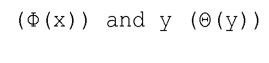
with

the complex numbers A- and A+ are calculated from the periodic interference fringe for the flat (f) and sample (s) image.
9. The system according to claim 8, wherein the 2D grating is a phase grating that produces a 2D periodic fringe that is separable in x and y direction, wherein g(x,y) is the 2D periodic fringe which is written as g(x,y) = f(x) h(y).
10. The system according to any of the preceding claims wherein
an autocorrelation algorithm is exploited in order to retrieve differential phase contrast from 2D periodic intensity fringes .
11. The system according to any of the preceding claims, wherein mean, weighted mean or median values of the differential phase contrast values are used within one actual pixel or stripe of the detector or an arbitrary area of the recorded intensity fringe produced by the Hilbert Transform based phase retrieval algorithm for ID or 2D signals is used.
Applications Claiming Priority (2)
| Application Number | Priority Date | Filing Date | Title |
|---|---|---|---|
| EP14192694.9A EP3021110A1 (en) | 2014-11-11 | 2014-11-11 | System for obtaining quantitative x-ray images using Hilbert transform on imaged fringes |
| EP14192694.9 | 2014-11-11 |
Publications (1)
| Publication Number | Publication Date |
|---|---|
| WO2016074864A1 true WO2016074864A1 (en) | 2016-05-19 |
Family
ID=51870916
Family Applications (1)
| Application Number | Title | Priority Date | Filing Date |
|---|---|---|---|
| PCT/EP2015/073356 Ceased WO2016074864A1 (en) | 2014-11-11 | 2015-10-09 | System for obtaining quantitative x-ray images using hilbert transform on imaged fringes |
Country Status (2)
| Country | Link |
|---|---|
| EP (1) | EP3021110A1 (en) |
| WO (1) | WO2016074864A1 (en) |
Cited By (5)
| Publication number | Priority date | Publication date | Assignee | Title |
|---|---|---|---|---|
| KR101769476B1 (en) | 2016-06-16 | 2017-08-18 | 한국과학기술원 | Apparatus and method for increasing spatial resolution of medical image using charge sharing |
| CN111089871A (en) * | 2019-12-12 | 2020-05-01 | 中国科学院苏州生物医学工程技术研究所 | Phase information separation method and system of X-ray grating phase contrast image, storage medium and equipment |
| CN115329256A (en) * | 2022-10-13 | 2022-11-11 | 南京理工大学 | Underwater optical measurement error compensation method based on FPP |
| CN116385300A (en) * | 2023-04-07 | 2023-07-04 | 南京大学 | Restoration method for imaging distortion of superconducting nanowire single-photon imager |
| CN119334984A (en) * | 2024-12-11 | 2025-01-21 | 中国科学技术大学 | Soft X-ray spatially resolved absorption spectrum acquisition system |
Families Citing this family (7)
| Publication number | Priority date | Publication date | Assignee | Title |
|---|---|---|---|---|
| US20180188190A1 (en) * | 2016-12-30 | 2018-07-05 | Nikon Corporation | Single-projection photon-counting x-ray quantitative phase-contrast measurements |
| CN107358584A (en) * | 2017-06-29 | 2017-11-17 | 天津大学 | The filtering method of ESPI bar graphs based on adaptive Hilbert transform |
| CN109580667B (en) * | 2018-12-05 | 2020-10-27 | 中国科学技术大学 | Single grating phase contrast imaging method and system |
| CN110068585B (en) * | 2019-03-15 | 2022-03-22 | 中国工程物理研究院流体物理研究所 | Dual-energy X-ray grating interferometric imaging system and method |
| CN110197486B (en) * | 2019-07-02 | 2021-04-06 | 清华大学 | X-ray grating integrated imaging information extraction method, system and storage medium |
| CN110806598B (en) * | 2019-11-11 | 2021-06-25 | 清华大学 | Information extraction method based on dose distribution and X-ray phase contrast imaging system |
| EP3855725A1 (en) * | 2020-01-21 | 2021-07-28 | Paul Scherrer Institut | Single photon counting detectors in strip or pixel design having digital inter-pixel communication and logic |
-
2014
- 2014-11-11 EP EP14192694.9A patent/EP3021110A1/en not_active Withdrawn
-
2015
- 2015-10-09 WO PCT/EP2015/073356 patent/WO2016074864A1/en not_active Ceased
Non-Patent Citations (4)
| Title |
|---|
| A. SCHUBERT ET AL: "Micrometre resolution of a charge integrating microstrip detector with single photon sensitivity", JOURNAL OF SYNCHROTRON RADIATION, vol. 21, no. 3, 15 March 2012 (2012-03-15), pages 295 - 365, XP055164618, ISSN: 0909-0495, DOI: 10.1107/S090904951200235X * |
| ARNISON M R ET AL: "Using the Hilbert transform for 3D visualization of differential interference contrast microscope images", JOURNAL OF MICROSCOPY, BLACKWELL SCIENCE, GB, vol. 199, July 2000 (2000-07-01), pages 79 - 84, XP002397531, ISSN: 0022-2720, DOI: 10.1046/J.1365-2818.2000.00706.X * |
| F. PFEIFFER ET AL: "Hard-X-ray dark-field imaging using a grating interferometer", NATURE MATERIALS, vol. 7, no. 2, 13 January 2008 (2008-01-13), pages 134 - 137, XP055003146, ISSN: 1476-1122, DOI: 10.1038/nmat2096 * |
| WENXIANG CONG ET AL: "Fourier transform based iterative method for x-ray differential phase-contrast computed tomography", 24 June 2011 (2011-06-24), XP055164517, Retrieved from the Internet <URL:http://arxiv.org/abs/1106.5101> [retrieved on 20150123] * |
Cited By (7)
| Publication number | Priority date | Publication date | Assignee | Title |
|---|---|---|---|---|
| KR101769476B1 (en) | 2016-06-16 | 2017-08-18 | 한국과학기술원 | Apparatus and method for increasing spatial resolution of medical image using charge sharing |
| CN111089871A (en) * | 2019-12-12 | 2020-05-01 | 中国科学院苏州生物医学工程技术研究所 | Phase information separation method and system of X-ray grating phase contrast image, storage medium and equipment |
| CN111089871B (en) * | 2019-12-12 | 2022-12-09 | 中国科学院苏州生物医学工程技术研究所 | Phase information separation method and system of X-ray grating phase contrast image, storage medium and equipment |
| CN115329256A (en) * | 2022-10-13 | 2022-11-11 | 南京理工大学 | Underwater optical measurement error compensation method based on FPP |
| CN115329256B (en) * | 2022-10-13 | 2023-02-28 | 南京理工大学 | Underwater optical measurement error compensation method based on FPP |
| CN116385300A (en) * | 2023-04-07 | 2023-07-04 | 南京大学 | Restoration method for imaging distortion of superconducting nanowire single-photon imager |
| CN119334984A (en) * | 2024-12-11 | 2025-01-21 | 中国科学技术大学 | Soft X-ray spatially resolved absorption spectrum acquisition system |
Also Published As
| Publication number | Publication date |
|---|---|
| EP3021110A1 (en) | 2016-05-18 |
Similar Documents
| Publication | Publication Date | Title |
|---|---|---|
| WO2016074864A1 (en) | System for obtaining quantitative x-ray images using hilbert transform on imaged fringes | |
| Kingston et al. | Neutron ghost imaging | |
| US9335281B2 (en) | Apparatus for coded aperture X-ray scatter imaging and method therefor | |
| US20150049860A1 (en) | Single step differential phase contrast x-ray imaging | |
| US9494534B2 (en) | Material differentiation with phase contrast imaging | |
| US8306183B2 (en) | Detection setup for X-ray phase contrast imaging | |
| Taguchi | Multi‐energy inter‐pixel coincidence counters for charge sharing correction and compensation in photon counting detectors | |
| CN102221565B (en) | X-ray source grating stepping imaging system and imaging method | |
| EP3341714B1 (en) | Omnidirectional scattering- and bidirectional phase-sensitivity with single shot grating interferometry | |
| US20180226168A1 (en) | X-ray detector for phase contrast and/or dark-field imaging | |
| JP2016535642A (en) | X-ray grating phase contrast imaging apparatus and method | |
| Kallon et al. | Comparing signal intensity and refraction sensitivity of double and single mask edge illumination lab-based x-ray phase contrast imaging set-ups | |
| JP7167201B2 (en) | X-ray phase detector | |
| JP2018519866A (en) | X-ray imaging | |
| Kingston et al. | Inherent dose-reduction potential of classical ghost imaging | |
| EP3112850B1 (en) | X-ray imaging apparatus and x-ray imaging method | |
| Greenberg et al. | Coding and sampling for compressive x-ray diffraction tomography | |
| Barbes et al. | Material-specific imaging system using energy-dispersive X-ray diffraction and spatially resolved CdZnTe detectors with potential application in breast imaging | |
| US20150310609A1 (en) | Systems and methods for regularized fourier analysis in x-ray phase contrast imaging | |
| EP3344979B1 (en) | Dual phase grating interferometer for x-ray phase contrast imaging | |
| Heise et al. | The optimum open fraction of coded apertures. With an application to the wide field X-ray cameras of SAX | |
| EP3889885A1 (en) | Bias correction for dark-field imaging based on sliding window phase retrieval | |
| Boardman et al. | Single pixel compressive gamma-ray imaging with randomly encoded masks | |
| Fleenor et al. | Correlated statistical uncertainties in coded-aperture imaging | |
| Holmgren et al. | Multi-view coded aperture coherent scatter tomography |
Legal Events
| Date | Code | Title | Description |
|---|---|---|---|
| 121 | Ep: the epo has been informed by wipo that ep was designated in this application |
Ref document number: 15778923 Country of ref document: EP Kind code of ref document: A1 |
|
| NENP | Non-entry into the national phase |
Ref country code: DE |
|
| 122 | Ep: pct application non-entry in european phase |
Ref document number: 15778923 Country of ref document: EP Kind code of ref document: A1 |