WO2015083648A1 - 細胞被覆層の形成に関する方法及びキット - Google Patents
細胞被覆層の形成に関する方法及びキット Download PDFInfo
- Publication number
- WO2015083648A1 WO2015083648A1 PCT/JP2014/081640 JP2014081640W WO2015083648A1 WO 2015083648 A1 WO2015083648 A1 WO 2015083648A1 JP 2014081640 W JP2014081640 W JP 2014081640W WO 2015083648 A1 WO2015083648 A1 WO 2015083648A1
- Authority
- WO
- WIPO (PCT)
- Prior art keywords
- molecule
- cell
- catalyst
- crosslinkable
- binding
- Prior art date
- Legal status (The legal status is an assumption and is not a legal conclusion. Google has not performed a legal analysis and makes no representation as to the accuracy of the status listed.)
- Ceased
Links
Images

















Classifications
-
- C—CHEMISTRY; METALLURGY
- C12—BIOCHEMISTRY; BEER; SPIRITS; WINE; VINEGAR; MICROBIOLOGY; ENZYMOLOGY; MUTATION OR GENETIC ENGINEERING
- C12N—MICROORGANISMS OR ENZYMES; COMPOSITIONS THEREOF; PROPAGATING, PRESERVING, OR MAINTAINING MICROORGANISMS; MUTATION OR GENETIC ENGINEERING; CULTURE MEDIA
- C12N5/00—Undifferentiated human, animal or plant cells, e.g. cell lines; Tissues; Cultivation or maintenance thereof; Culture media therefor
- C12N5/0006—Modification of the membrane of cells, e.g. cell decoration
Definitions
- the present invention relates to a method and kit relating to the formation of a cell coating layer, and more particularly to the formation of a cell coating layer by a crosslinking reaction of a crosslinking molecule contributed by a catalyst molecule.
- a local cross-linking reaction is performed only near the cell surface of a specific type of cell to selectively cover the cell surface of the specific cell.
- a coating layer could not be formed.
- the present invention has been made in view of the above problems, and an object thereof is to provide a method and kit relating to the effective formation of a cell coating layer.
- a method according to an embodiment of the present invention for solving the above-described problems includes the following (a) and (b): (a) a specific molecule on the cell surface of a target cell via a binding molecule that binds to the specific molecule. (B) binding the catalyst molecule contributing to the crosslinking reaction of the crosslinking molecule; (b) the crosslinking contributed by the catalyst molecule in a solution containing the target cell to which the catalyst molecule is bound and the crosslinking molecule. Cross-linking the cross-linkable molecule by a reaction to form a coating layer covering the cell surface of the target cell. ADVANTAGE OF THE INVENTION According to this invention, the method of forming a cell coating layer effectively can be provided.
- the method further includes the following (c): (c) separating the first cell having the cell surface covered with the coating layer and the second cell. It is good.
- the specific molecule may be one or more selected from the group consisting of proteins, glycoproteins, peptides, and sugar chains.
- the binding molecule may be an antibody against the specific molecule.
- the method includes, as (a), binding the catalyst molecule that catalyzes the crosslinking reaction to the specific molecule of the target cell via the binding molecule, and as (b), In the solution containing the target cell to which the catalyst molecule is bound and the crosslinkable molecule, the crosslinkable molecule is crosslinked by the crosslinking reaction catalyzed by the catalyst molecule, and the cell surface of the target cell is formed. It is good also as forming forming the said coating layer to coat
- the catalyst molecule may be at least one selected from the group consisting of peroxidase, catalase, phenol oxidase, catechol oxidase, and metalloporphyrin complex.
- the specific molecule of the target cell may be the first catalyst molecule that catalyzes a chemical reaction that generates reactive oxygen species via the binding molecule.
- the cross-linking by the cross-linking reaction catalyzed by the second catalyst molecule in the presence of the active oxygen species produced by the second catalytic molecule catalyzing the cross-linking reaction in the presence of the active oxygen species It is good also as including forming the said coating layer which bridge
- the first catalyst molecule may be one or more selected from the group consisting of glucose oxidase, superoxide dismutase, amino acid oxidase, choline oxidase, alcohol oxidase, pyruvate oxidase and cholesterol oxidase.
- the second catalyst molecule may be one or more selected from the group consisting of peroxidase, catalase, and metalloporphyrin complex.
- a kit according to an embodiment of the present invention for solving the above-described problem is a cell coating layer forming kit used in any of the above methods, and includes the following (x) and / or (y): (x A binding molecule that binds to a specific molecule on the cell surface of the target cell, and / or a catalyst molecule that contributes to a cross-linking reaction of a cross-linkable molecule that binds to the specific molecule of the target cell via the binding molecule; A cross-linkable molecule, or a base molecule into which a cross-linkable group is to be introduced and / or a compound for introducing a cross-linkable group into the base molecule.
- the kit which forms a cell coating layer effectively can be provided.
- the kit may include the binding molecule and the catalyst molecule bound to the binding molecule as (x).
- the kit may further include the crosslinkable molecule as (y).
- the kit may further include the following (z): (z) an instruction manual for using the cell coating layer forming kit in the method.
- the method according to an embodiment of the present invention for solving the above-described problem is the following (x) and / or (y): (x) binding to a specific molecule on the cell surface of a target cell.
- the cross-linking molecule or cross-linking group is introduced And / or a compound for introducing a crosslinkable group into the base molecule.
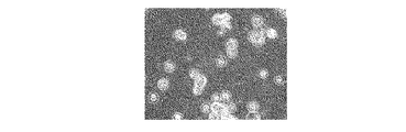
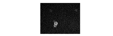
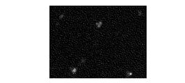
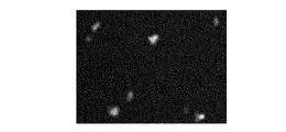
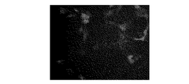
- Example 1 which concerns on one Embodiment of this invention, it is explanatory drawing which shows an example of the result of having observed the cell of the group 1 with the microscope under the microscope. In Example 1 which concerns on one Embodiment of this invention, it is explanatory drawing which shows the other example of the result of having carried out the fluorescence observation of the cell of group 1 under the microscope. In Example 1 which concerns on one Embodiment of this invention, it is explanatory drawing which shows an example of the result of having observed the bright field observation of the cell of the group 2 under the microscope. In Example 1 which concerns on one Embodiment of this invention, it is explanatory drawing which shows an example of the result of having observed the cell of the group 2 with the microscope under the microscope.
- Example 1 which concerns on one Embodiment of this invention, it is explanatory drawing which shows the other example of the result of carrying out fluorescence observation of the cell of group 2 under the microscope.
- Example 2 which concerns on one Embodiment of this invention, it is explanatory drawing which shows an example of the result of having observed the bright field of the cell under the microscope.
- Example 2 which concerns on one Embodiment of this invention, it is explanatory drawing which shows an example of the result of having carried out fluorescence observation of the cell under the microscope.
- Example 2 which concerns on one Embodiment of this invention, it is explanatory drawing which shows the other example of the result of having observed the fluorescence of the cell under the microscope.
- Example 2 which concerns on one Embodiment of this invention, it is explanatory drawing which shows the further another example of the result of having observed the fluorescence of the cell under the microscope.
- Example 3 which concerns on one Embodiment of this invention, it is explanatory drawing which shows an example of the result of having observed the bright field of the cell under the microscope.
- Example 3 which concerns on one Embodiment of this invention, it is explanatory drawing which shows an example of the result of having observed the fluorescence of the cell under the microscope.
- Example 3 which concerns on one Embodiment of this invention, it is explanatory drawing which shows the other example of the result of having observed the fluorescence of the cell under the microscope.
- Example 4 which concerns on one Embodiment of this invention, it is explanatory drawing which shows an example of the result of having carried out fluorescence observation of the cell under the microscope. In Example 4 which concerns on one Embodiment of this invention, it is explanatory drawing which shows the other example of the result of having observed the fluorescence of the cell under the microscope. In Example 4 which concerns on one Embodiment of this invention, it is explanatory drawing which shows the further another example of the result of having observed the fluorescence of the cell under the microscope. In Example 5 which concerns on one Embodiment of this invention, it is explanatory drawing which shows an example of the result of having observed the bright field under the microscope of the cell.
- Example 5 which concerns on one Embodiment of this invention, it is explanatory drawing which shows an example of the result of having carried out fluorescence observation of the cell under the microscope. In Example 5 which concerns on one Embodiment of this invention, it is explanatory drawing which shows the other example of the result of having observed the fluorescence of the cell under the microscope. In Example 5 which concerns on one Embodiment of this invention, it is explanatory drawing which shows another example of the result of having observed the fluorescence of the cell under the microscope. In Example 6 which concerns on one Embodiment of this invention, it is explanatory drawing which shows an example of the result of having observed the bright field observation of the cell contained in a supernatant solution under a microscope.
- Example 6 which concerns on one Embodiment of this invention, it is explanatory drawing which shows an example of the result of having carried out the fluorescence observation of the cell contained in a supernatant solution under a microscope.
- Example 6 which concerns on one Embodiment of this invention, it is explanatory drawing which shows an example of the result of having observed the bright field observation of the cell adhere
- Example 6 which concerns on one Embodiment of this invention, it is explanatory drawing which shows an example of the result of having fluorescence-observed the cell adhere
- Example 6 which concerns on one Embodiment of this invention, it is explanatory drawing which shows the other example of the result of having carried out the fluorescence observation of the cell adhere
- Example 7 which concerns on one Embodiment of this invention, it is explanatory drawing which shows an example of the result of having observed the bright field of the cell under the microscope.
- Example 7 which concerns on one Embodiment of this invention, it is explanatory drawing which shows an example of the result of having observed the fluorescence of the cell under the microscope.
- Example 8 which concerns on one Embodiment of this invention, it is explanatory drawing which shows an example of the result of having observed the bright field of the cell under the microscope.
- Example 7 which concerns on one Embodiment of this invention, it is explanatory drawing which shows an example of the result of having observed the fluorescence of the cell under the microscope. In Example 7 which concerns on one Embodiment of this invention, it is explanatory drawing which shows the other example of the result of having carried out the fluorescence observation of the cell under the microscope.
- the method according to this embodiment includes the following (a) and (b): (a) a specific molecule on the cell surface of a target cell via a binding molecule that binds to the specific molecule. And (b) binding the catalyst molecule to which the catalyst molecule contributes in a solution containing the target cell to which the catalyst molecule is bound and the crosslinkable molecule. Cross-linking the cross-linkable molecule by a reaction to form a coating layer that covers the cell surface of the target cell.
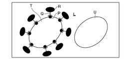
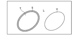
- 1A to 1C show a process of forming a coating layer S that covers the cell surface of a target cell T in an example of the present method.
- the target cell T whose cell surface is to be coated with the coating layer S has a specific molecule P on the cell surface as shown in FIG. 1A.
- a coating layer S is formed on a specific molecule P of a target cell T via a binding molecule Q that binds to the specific molecule P. Therefore, the catalyst molecules R that contribute to the crosslinking reaction of the crosslinkable molecules are bonded.
- the crosslinkable molecule is subjected to a crosslink reaction contributed by the catalyst molecule R.
- a coating layer S that covers the cell surface of the target cell T is formed.
- a cross-linking reaction of a local cross-linking molecule is performed in the vicinity of the cell surface of the target cell T, and the cell surface of the target cell T Can be formed.
- a catalyst molecule is bound to a specific molecule on the cell surface of a target cell via a binding molecule.
- the binding of the catalyst molecule to the target cell is a pretreatment for performing a local crosslinking reaction of the crosslinkable molecule in the vicinity of the cell surface of the target cell, as will be described later.
- the specific molecule of the target cell is not particularly limited as long as it is a molecule present on the cell surface of the target cell.
- the specific molecule is, for example, one or more selected from the group consisting of proteins, glycoproteins, peptides, and sugar chains. That is, the specific molecule may be, for example, a so-called cell surface protein and / or cell membrane protein. More specifically, the specific molecule may be, for example, a CD (cluster of differentiation) antigen.
- the specific molecule may be a receptor present on the cell surface, for example. In addition, the specific molecule may be, for example, a molecule bound to the cell surface of the target cell by artificial manipulation.
- the target cell is not particularly limited as long as it has a specific molecule on its cell surface.
- a target cell is good also as being 1 or more types selected from the group which consists of an animal cell, a plant cell, and microorganisms, for example.
- the animal cell may be, for example, a vertebrate cell and / or an invertebrate cell.
- the vertebrate may be, for example, one or more selected from the group consisting of mammals, reptiles, birds, amphibians and fish.
- the mammal may be, for example, one or more selected from the group consisting of human, monkey, cow, horse, sheep, pig, dog, cat, rat, mouse, hamster and guinea pig.
- the invertebrates may be one or more selected from the group consisting of insects, nematodes, squirts, squids, octopuses and crabs, for example.
- the target cell may be, for example, a stem cell and / or a differentiated cell.
- the stem cell is not particularly limited as long as it has pluripotency and self-renewal ability.
- the stem cells are, for example, one or more selected from the group consisting of induced pluripotent stem cells (induced pluripotent stem cells: iPS cells), embryonic stem cells (Embryonic Stem cells: ES cells), somatic stem cells and reproductive stem cells. It is good as well.
- the somatic stem cells are, for example, one or more selected from the group consisting of mesenchymal stem cells, hematopoietic stem cells, neural stem cells, hepatic stem cells, pancreatic stem cells, skin stem cells, adipose stem cells, muscle stem cells, bone stem cells, and cartilage stem cells. It is good as well.
- Differentiated cells are not particularly limited as long as they are differentiated cells.
- the differentiated cells are, for example, one type selected from the group consisting of bone marrow cells, blood cells, nerve cells, hepatocytes, pancreatic cells, skin cells, fat cells, muscle cells, heart cells, bone cells, chondrocytes and germ cells. It is good also as being above.
- the differentiated cell may be a differentiated cell derived from a stem cell in a culture system, for example.
- the target cell may be, for example, one or more selected from the group consisting of primary cells, passage cells, and established cell lines.
- the target cell may be a cultured cell, for example.
- the target cells may be dispersed in the solution, or may be adhered to the substrate in the solution.
- the binding molecule is not particularly limited as long as it is a molecule that binds to a specific molecule of a target cell, and is preferably a molecule that specifically binds to the specific molecule.
- the binding molecule is, for example, selected from the group consisting of proteins (eg, antibodies, lectins, alginic acid binding proteins), peptides (eg, collagen binding peptides) and nucleic acid molecules (eg, DNA aptamers, RNA aptamers, peptide aptamers). It is good also as being 1 or more types.
- the antibody may be a fragment antibody, for example.
- the binding molecule may be a ligand for the receptor.
- the catalyst molecule is not particularly limited as long as it is a molecule exhibiting catalytic activity that contributes to the crosslinking reaction of the crosslinking molecule for forming a coating layer covering the cell surface of the target cell.
- the catalyst molecule may be, for example, a catalyst molecule that catalyzes a crosslinking reaction of a crosslinkable molecule and / or a catalyst molecule that catalyzes a chemical reaction that generates active oxygen species.
- a catalyst molecule that catalyzes a crosslinking reaction of a crosslinking molecule is bound to a specific molecule on the cell surface of a target cell via a binding molecule that binds to the specific molecule.
- the crosslinkable molecule is crosslinked by the crosslinking reaction catalyzed by the catalyst molecule in a solution containing the target cell to which the catalyst molecule is bound and the crosslinkable molecule.
- the method may include forming a coating layer that covers the cell surface of the target cell.
- the catalyst molecule that catalyzes the cross-linking reaction of the cross-linking molecule is not particularly limited as long as it is a molecule that exhibits an activity of catalyzing the cross-linking reaction.
- This catalyst molecule may be, for example, a catalyst molecule that catalyzes a crosslinking reaction of a crosslinkable molecule in the presence of reactive oxygen species (ROS) or oxygen.
- ROS reactive oxygen species
- ROS is 1 or more types selected from the group which consists of a superoxide, a hydroxyl radical, hydrogen peroxide, and singlet oxygen, for example, and hydrogen peroxide is utilized preferably.
- the catalyst molecule may be, for example, one or more selected from the group consisting of peroxidase, catalase, phenol oxidase, catechol oxidase, and metalloporphyrin complex.
- Peroxidase, catalase and metalloporphyrin complexes are catalytic molecules that catalyze the crosslinking reaction of crosslinkable molecules in the presence of ROS.
- Phenol oxidase and catechol oxidase are catalytic molecules that catalyze the crosslinking reaction of crosslinking molecules in the presence of oxygen.
- the peroxidase may be, for example, at least one selected from the group consisting of horseradish peroxidase, soybean-derived peroxidase, myeloperoxidase, lactoperoxidase, eosinophil peroxidase, and thyroid peroxidase.
- the phenol oxidase may be, for example, laccase and / or tyrosinase.
- the metal porphyrin complex may be, for example, an iron porphyrin complex and / or a copper porphyrin complex.
- the iron porphyrin complex may be, for example, one or more selected from the group consisting of hemin (hematin), hemoglobin, ferrihem, and hemochrome.
- this method catalyzes the chemical reaction which produces
- ROS reactive oxygen species
- bonded with the said specific molecule as said (a) for example.
- the cross-linkable molecule is cross-linked by the cross-linking reaction catalyzed by the second catalyst molecule to cover the cell surface of the target cell. It is good also as including forming a coating layer.
- the catalyst molecule (first catalyst molecule) that catalyzes the chemical reaction that generates ROS is not particularly limited as long as it is a molecule that exhibits the activity of catalyzing the chemical reaction that generates ROS used for the crosslinking reaction of the crosslinkable molecule.
- the second catalyst molecule used together with the first catalyst molecule is not particularly limited as long as it is described above as the catalyst molecule that catalyzes the crosslinking reaction of the crosslinkable molecule in the presence of ROS.
- the first catalyst molecule is, for example, a catalyst molecule that catalyzes a chemical reaction that consumes a substrate contained in a solution and generates ROS. That is, the first catalyst molecule may be, for example, a catalyst molecule that catalyzes a chemical reaction that oxidizes a substrate (for example, an organic compound) contained in a solution to generate ROS.
- the first catalyst molecule may be, for example, one or more selected from the group consisting of glucose oxidase, superoxide dismutase, amino acid oxidase, choline oxidase, alcohol oxidase, pyruvate oxidase, and cholesterol oxidase. .
- Glucose oxidase, amino acid oxidase, choline oxidase, alcohol oxidase, pyruvate oxidase, and cholesterol oxidase each perform a chemical reaction that oxidizes glucose, amino acid, choline, alcohol, pyruvate, and cholesterol to produce ROS (hydrogen peroxide).
- ROS hydrogen peroxide
- Superoxide dismutase is a superoxide (for example, a superoxide contained in a solution containing a target cell to which a first catalytic molecule is bound and a crosslinkable molecule, and / or a superoxide produced by the target cell in the solution. ) To catalyze chemical reactions that produce oxygen and hydrogen peroxide.
- the method for binding the catalyst molecule to the specific molecule on the cell surface of the target cell via the binding molecule is not particularly limited.
- a solution containing a target cell and a catalyst molecule bound to a binding molecule may be prepared, and the catalyst molecule may be bound to a specific molecule of the target cell via the binding molecule.
- a catalyst molecule bound to a binding molecule a catalyst molecule bound to an antibody against a specific molecule (more specifically, for example, a peroxidase-labeled antibody) may be used.
- a solution containing the target cell and the first catalyst molecule and the second catalyst molecule bound to the binding molecule is prepared, and the target molecule is passed through the binding molecule to the specific molecule of the target cell.
- One catalyst molecule and a second catalyst molecule may be combined.
- the first catalyst molecule and the second catalyst molecule bound to the binding molecule the first catalyst molecule and the second catalyst molecule bound to the antibody against the specific molecule (more specifically, For example, peroxidase and glucose oxidase coupled to an antibody against a specific molecule may be used.
- a solution containing the target cell and the binding molecule is prepared, the binding molecule is bound to a specific molecule of the target cell, and then the target cell to which the binding molecule is bound and the binding molecule A solution containing a catalyst molecule bound to another molecule to be bound is prepared, and the catalyst molecule is bound to the binding molecule bound to the target cell via the other molecule.
- the target cell The catalyst molecule may be bound to the specific molecule via the binding molecule.
- an antibody against a specific molecule is used as a binding molecule, and the antibody is bound to an antibody against the binding molecule (secondary antibody) as a catalyst molecule bound to another molecule that binds to the binding molecule.
- the catalyst molecule thus prepared (more specifically, for example, a peroxidase-labeled antibody) may be used.
- the crosslinkable molecule in the solution containing the target cell to which the catalyst molecule is bound and the crosslinkable molecule, the crosslinkable molecule is crosslinked by the crosslinking reaction contributed by the catalyst molecule. Then, a coating layer that covers the cell surface of the target cell is formed.
- the solvent contained in the solution is not particularly limited as long as it contains water.
- the crosslinkable molecule is not particularly limited as long as it is a molecule that is crosslinked in a solution containing the crosslinkable molecule by a crosslinking reaction to which a catalyst molecule contributes.
- the crosslinkable molecule is a molecule having a crosslinkable group, and is preferably a hydrophilic molecule having a crosslinkable group.
- the crosslinkable group is not particularly limited as long as it is a functional group that contributes to the formation of a crosslinked structure of molecules.
- a crosslinkable group is good also as being 1 or more types selected from the group which consists of a hydroxyl group, an amino group, a carboxyl group, a thiol group, and a vinyl group, for example.
- the hydroxyl group may be, for example, a hydroxyl group bonded to a benzene ring.
- the hydroxyl group bonded to the benzene ring may be, for example, a phenolic hydroxyl group and / or a catechol group.
- the amino group may be, for example, an amino group bonded to a benzene ring.
- the crosslinkable molecule is sugar beet pectin
- the ferulic acid moiety and / or dihydroferulic acid moiety contained in the sugar beet pectin may be used as the crosslinkable group.
- the molecular weight of the crosslinkable molecule is not particularly limited, but may be, for example, 100 Da or more, or 300 Da or more. Although the upper limit of the molecular weight of the crosslinkable molecule is not particularly limited, the molecular weight may be, for example, 10 7 Da or less.
- the crosslinkable molecule may be, for example, a crosslinkable polymer.
- the crosslinkable polymer is a polymer having a crosslinkable group, and is preferably a hydrophilic polymer having a crosslinkable group.
- the crosslinkable polymer may be a natural polymer and / or a synthetic polymer having a crosslinkable group.
- the natural polymer may be, for example, one or more selected from the group consisting of proteins, polysaccharides, nucleic acids, polyphenol polymers, and derivatives thereof.
- the protein may be, for example, one or more selected from the group consisting of gelatin, collagen, albumin, globulin, fibrin, and derivatives thereof.
- the polysaccharide is, for example, selected from the group consisting of alginic acid, hyaluronic acid, chondroitin sulfate, starch, dextran, pullulan, cellulose, pectin, sugar beet pectin, amylopectin, chitin, chitosan, amylose, heparin, heparan sulfate and derivatives thereof. It is good also as being 1 or more types.
- the nucleic acid may be, for example, one or more selected from the group consisting of deoxyribonucleic acid, ribonucleic acid, and derivatives thereof.
- a polyphenol polymer is good also as being 1 or more types selected from the group which consists of a tannin polymer, a catechin polymer, and these derivatives, for example.
- the synthetic polymer may be, for example, one or more selected from the group consisting of polyethylene glycol, polyvinyl alcohol, polyacrylic acid, polymethacrylic acid, polypropylene, polyvinyl chloride, and derivatives thereof.
- the crosslinkable molecule may be, for example, a molecule that can be decomposed after being crosslinked. That is, the crosslinkable molecule may be, for example, a molecule that can be degraded by an enzyme after being crosslinked. Specifically, for example, when the crosslinkable molecule is a protein, the crosslinkable molecule is degraded by a proteolytic enzyme even after the crosslinking. Further, for example, when the crosslinkable molecule is a polysaccharide, the crosslinkable molecule is decomposed by the polysaccharide degrading enzyme even after the crosslinking.
- the crosslinkable molecule may be obtained by introducing a crosslinkable group.
- the method for introducing a crosslinkable group is not particularly limited.
- a molecule (base molecule) into which a crosslinkable group is to be introduced and a compound for introducing the crosslinkable group into the base molecule are chemically synthesized. It is good also as performing by making it react.
- the chemical reaction between the base molecule and the compound for introducing a crosslinkable group into the base molecule is not particularly limited, and may be, for example, a condensation reaction.
- the basic molecule is not particularly limited as long as it can introduce a crosslinkable group.
- the base molecule is, for example, a natural polymer and / or a synthetic polymer as described above, which does not have a crosslinkable group, or does not have a sufficient amount of a crosslinkable group to form a coating layer. It may be a polymer.
- the compound for introducing a crosslinkable group into the base molecule is not particularly limited as long as it is a compound that binds to the base molecule.
- the bond between the basic molecule and the compound for introducing a crosslinkable group into the basic molecule is, for example, one or more selected from the group consisting of a covalent bond, an electrostatic bond, a hydrophobic bond, and a hydrogen bond It is good as well.
- a compound for introducing a crosslinkable group into a base molecule includes, for example, a reactive functional group that chemically reacts with the base molecule to form a chemical bond, and a crosslinkability to be introduced into the base molecule. It may be a compound having a group.
- the reactive functional group may be, for example, one or more selected from the group consisting of a carboxyl group, an amino group, a hydroxyl group, a thiol group, a maleimide group, a succinimide group, an azide group, and an alkyne.
- the compound for introducing a crosslinkable group into the base molecule is, for example, tyramine, phenylpropionic acid, aniline, catecholamine (for example, one or more selected from the group consisting of adrenaline, noradrenaline and dopamine) and It may be one or more selected from the group consisting of these derivatives.
- the crosslinkable molecule may be dispersed in the solution or previously bound to the cell surface of the target cell. It is good.
- the crosslinkable molecule bonded to the above-described catalyst molecule may be dispersed in the solution (for example, Ryu JH et al. Chitosan-g -hematin: Enzyme-mimicking polymeric catalyst for adhesive hydrogels. See Acta Biomater (2013)).
- the crosslinkable molecule when the crosslinkable molecule before the cross-linking reaction is bound to the cell surface of the target cell, the crosslinkable molecule is, for example, another molecule that binds to the molecule (for example, the It may be bound via an antibody to a cell surface molecule).
- the crosslinkable molecule is crosslinked by the crosslinking reaction contributed by the catalyst molecule under the condition that the crosslinking reaction is started. Is done.
- a catalyst molecule that catalyzes a crosslinking reaction of a crosslinkable molecule is bound to a specific molecule on the cell surface of a target cell via a binding molecule that binds to the specific molecule.
- the crosslinkable molecule is crosslinked by the crosslinking reaction catalyzed by the catalyst molecule in a solution containing the target cell to which the catalyst molecule is bound and the crosslinkable molecule.
- the crosslinking of the crosslinkable molecule in the solution is performed under the condition that the crosslinking reaction catalyzed by the catalyst molecule is initiated. This is done by preparing the solution.
- a catalyst molecule that catalyzes a crosslinking reaction of a crosslinkable molecule in the presence of ROS or oxygen is bound to a target cell, the target cell to which the catalyst molecule is bound, the crosslinkable molecule, By preparing a solution containing the ROS or oxygen, the crosslinking reaction of the crosslinkable molecule is started.
- crosslinkable molecule and ROS by adding the crosslinkable molecule and ROS to a solution containing target cells bound with a catalyst molecule that catalyzes the crosslinking reaction of the crosslinkable molecule in the presence of ROS, It is good also as starting a crosslinking reaction.
- the ROS contained in the solution may be externally added to the solution, or may be generated by another catalyst molecule that catalyzes a chemical reaction that generates the ROS in the solution. It may be generated electrochemically in the solution (see, for example, Japanese Patent Application Laid-Open No. 2004-10904), or generated by irradiating a photocatalyst (for example, titanium oxide) with light in the solution. It is good.
- a photocatalyst for example, titanium oxide
- the above-described molecule such as glucose oxidase
- the above-described molecule may be used as a catalyst molecule that catalyzes a chemical reaction that generates ROS.
- Other catalyst molecules may be dispersed in a solution, or may be bound to a cell surface molecule of a target cell via a molecule that binds to the molecule.
- the other catalytic molecule specifically, for example, glucose bound to an antibody against a molecule on the cell surface of the target cell.
- An oxidase-labeled antibody may be used.
- the molecule on the cell surface of the target cell to which the other catalyst molecule is bound may be the specific molecule described above, or may be a molecule different from the specific molecule.
- the crosslinkable reaction of the crosslinkable molecule is performed. May be started.
- the first catalyst molecule that catalyzes a chemical reaction that generates ROS to a specific molecule on the cell surface of the target cell via a binding molecule that binds to the specific molecule.
- the ROS produced by the chemical reaction catalyzed by the first catalyst molecule and the cross-linking reaction of the crosslinkable molecule in the presence of the ROS are catalyzed.
- the cross-linking molecule is cross-linked by the cross-linking reaction catalyzed by the second catalyst molecule to form a coating layer covering the cell surface of the target cell
- the crosslinking of the crosslinkable molecule in the solution is performed by the chemical reaction catalyzed by the first catalyst, the ROS being generated, and the crosslinking catalyzed by the second catalyst molecule in the presence of the ROS. Reaction open In conditions it is carried out by preparing the solution.
- the crosslinking of the crosslinkable molecule includes, for example, a target cell to which the first catalyst molecule is bound, a substrate consumed by the first catalyst molecule in a chemical reaction that generates ROS, and a second It is carried out by preparing a solution containing catalyst molecules and the crosslinkable molecules.
- the crosslinking is performed. It is good also as starting the crosslinking reaction of a sex molecule.
- the second catalyst molecule may be dispersed in a solution, or may be bound to a molecule on the cell surface of the target cell via a molecule that binds to the molecule.
- the second catalyst molecule is dispersed in the solution, for example, the second catalyst molecule bonded to the crosslinkable molecule may be dispersed in the solution.
- the second catalytic molecule (specifically, for example, a peroxidase-labeled antibody) bound to an antibody against a cell surface molecule of the target cell is used. It is good.
- the molecule on the cell surface of the target cell to which the second catalyst molecule is bound may be the specific molecule described above, or may be a molecule different from the specific molecule. That is, for example, using a first catalytic molecule and a second catalytic molecule (specifically, for example, an antibody labeled with peroxidase and glucose oxidase) bound to an antibody against a specific molecule on the cell surface of the target cell. It is good.
- Crosslinking of the crosslinkable molecule may be performed by forming a new chemical bond between the crosslinkable groups of the crosslinkable molecule, or the crosslinkable molecule may be renewed via another molecule. It may be performed by forming a bond.
- crosslinkable molecules form a new chemical bond via another molecule
- the first crosslinkable group of the crosslinkable molecule and the second crosslinkable group of the other molecule are chemically May react to form a new chemical bond.
- this other molecule for example, a polyphenol compound having a plurality of phenolic hydroxyl groups may be used.
- the coating layer covering the cell surface of the target cell is in the vicinity of the cell surface of the target cell in a solution containing the target cell and a crosslinkable molecule bound to the cell surface. It is formed by a local crosslinking reaction of the crosslinkable molecule to which the catalyst molecule contributes.
- the coating layer covering the cell surface of the target cell includes a crosslinked structure formed by crosslinking of the crosslinking molecule.
- the coating layer is, for example, a structure (for example, a gel layer) formed by cross-linking and insolubilizing soluble crosslinkable molecules in a solution.
- the predetermined amount of a part of the crosslinkable molecule is obtained by a crosslinking reaction contributed by the catalyst molecule.
- the target cell comprising a cross-linked structure formed by cross-linking of the part of the cross-linkable molecule in the solution containing the predetermined amount of the other part of the cross-linkable molecule that is cross-linked A coating layer covering the cell surface is formed.
- the coating layer covering the cell surface of the target cell covers all or part of the cell surface of the target cell. That is, for example, when the target cells are dispersed in a solution (when the target cells are suspended in the solution), a coating layer that covers the entire cell surface of the target cells is added to the solution. It is good also as forming.
- microcapsules (gel particles) including target cells and a coating layer (gel layer) covering the cell surface of the target cells may be formed.
- a part of the cell surface of the target cell (for example, the base material) in the solution
- the thickness of the coating layer covering the cell surface of the target cell depends on the conditions for the crosslinking reaction (type and amount of crosslinking molecule, time and temperature of the crosslinking reaction, etc.) For example, it may be 1 nm to 2000 ⁇ m, or may be 10 nm to 200 ⁇ m.
- a coating layer (cell coating layer) that covers the cell surface of the target cell can be effectively formed. That is, in this method, in a solution containing a target cell having a catalytic molecule bound to the cell surface and a crosslinkable molecule, the target cell is subjected to a crosslinking reaction that proceeds from the catalyst molecule bound to the target cell as a starting point. Crosslinking of the crosslinkable molecules locally in the vicinity of the cell surface can be performed.
- minute particles including a target cell and a thin coating layer covering the cell surface of the target cell can be easily formed.
- particles containing only one target cell can be easily formed.
- the target cell is located approximately in the center of the particle.
- grains containing a some target cell can also be formed by increasing the density of the target cell in a solution, or forming the cell clump containing a some target cell previously. It can be said that this method described above is a method for producing target cells whose cell surfaces are coated with a coating layer.
- the crosslinkable molecule in the method, for example, as (a) above, in a solution containing a first cell (target cell) having a specific molecule on the cell surface and a second cell not having the specific molecule on the cell surface. Including binding a catalyst molecule that contributes to the crosslinking reaction of the crosslinkable molecule to the specific molecule of the first cell via a binding molecule that specifically binds to the specific molecule, In the solution containing the first cell to which the catalyst molecule is bound, the second cell, and the crosslinkable molecule, the crosslinkable molecule is crosslinked by the crosslinking reaction contributed by the catalyst molecule. It is good also as including forming the coating layer which coat
- the first cell (target cell T) having the specific molecule P and the second cell (other cell U) not having the specific molecule P are included.
- the catalyst molecule R is selectively bound to the specific molecule P of the first cell T via the binding molecule Q that specifically binds to the specific molecule P. Since the binding of the catalyst molecule R to the first cell T is performed using the binding molecule Q that specifically binds to the specific molecule P of the first cell T, the catalyst molecule for the second cell U is used. No specific binding of R occurs.
- a coating layer S that selectively covers the cell surface of the first cell T is formed by a cross-linking reaction of a local cross-linking molecule contributed by the catalyst molecule R.
- the crosslinking of the crosslinkable molecule is locally performed in the vicinity of the cell surface by a crosslinking reaction in which the catalyst molecule selectively bonded to the cell surface of the first cell contributes, It is selectively formed on the cell surface of the first cell. That is, in this case, for example, a coating layer that covers the cell surface of the first cell can be formed without forming a coating layer that covers the cell surface of the second cell.
- the adsorption of uncrosslinked crosslinkable molecules to the cell surface is not the formation of a coating layer covering the cell surface.
- the specific molecule of the first cell is not particularly limited as long as it is the above-described specific molecule and exists on the cell surface of the first cell and does not exist on the cell surface of the second cell.
- the combination of the first cell and the second cell is the first cell and the second cell described above, the first cell having a specific molecule on the cell surface,
- the second cell is not particularly limited as long as it is a combination that does not have the specific molecule on the cell surface.
- one of the first cell and the second cell may be a stem cell, and the other may be a differentiated cell derived from the stem cell.
- one of the first cell and the second cell may be a first stem cell, and the other may be a second stem cell derived from the first stem cell.
- one of the first cell and the second cell may be a first differentiated cell, and the other may be a second differentiated cell different from the first differentiated cell.
- the binding molecule that specifically binds to the specific molecule of the first cell is not particularly limited as long as it is the above-described binding molecule and is a molecule that specifically binds to the specific molecule. That is, the binding molecule is, for example, a protein (eg, antibody, lectin, alginic acid binding protein), peptide (eg, collagen binding peptide) and nucleic acid molecule (eg, DNA aptamer, RNA) that specifically binds to a specific molecule. It may be at least one selected from the group consisting of aptamers and peptide aptamers), and an antibody against a specific molecule is preferably used. The antibody may be a fragment antibody, for example. For example, when the specific molecule is a receptor, the binding molecule may be a ligand for the receptor.
- a protein eg, antibody, lectin, alginic acid binding protein
- peptide eg, collagen binding peptide
- nucleic acid molecule
- the catalyst molecule is not particularly limited as long as it is a catalyst molecule that catalyzes the crosslinking reaction of the crosslinking molecule described above.
- the catalyst molecule catalyzes the crosslinking reaction of the crosslinking molecule in the presence of ROS or oxygen. It may be a catalyst molecule.
- the ROS may be externally added to the solution, and the chemical reaction that generates the ROS in the solution is catalyzed. It may be generated by other catalyst molecules, or may be generated electrochemically in a solution, and is generated by irradiating a photocatalyst (for example, titanium oxide) with light in the solution. It is good as well.
- a photocatalyst for example, titanium oxide
- the above-described catalyst molecule (such as glucose oxidase) may be used as a catalyst molecule that catalyzes a chemical reaction that generates ROS. Good.
- This other catalyst molecule may be dispersed in a solution, or may be bound to a cell surface molecule of the first cell via a molecule that binds to the molecule.
- the other catalyst molecule specifically, for example, a glucose oxidase-labeled antibody bound to an antibody against a cell surface molecule of the first cell. It may be used.
- the molecule on the cell surface of the first cell to which the other catalyst molecule is bound may be the above-described specific molecule or may be a molecule different from the specific molecule.
- the molecule may also be present on the cell surface of the second cell. Even if another catalytic molecule that catalyzes the chemical reaction that generates ROS is bound to the cell surface molecule of the second cell, the crosslinking reaction of the crosslinkable molecule is performed on the cell surface of the second cell. Since the catalyst molecule to be catalyzed is not bound, a coating layer is selectively formed on the cell surface of the first cell in the solution.
- the first cell has a first specific molecule and a second specific molecule different from the first specific molecule as the specific molecule
- the second cell has the first specific molecule and
- the first specific molecule of the first cell is specifically bound to the catalyst molecule that catalyzes the crosslinking reaction of the crosslinkable molecule
- the first cell Another catalyst molecule that catalyzes a chemical reaction that generates ROS may be specifically bound to the second specific molecule.
- crosslinkable molecule In the presence of the ROS and the second catalyst molecule that catalyzes the crosslinking reaction of the crosslinkable molecule in the presence of the ROS, the crosslinkable molecule is converted by the crosslinking reaction catalyzed by the second catalyst molecule.
- Crosslink and cover the cell surface of the first cell It may include forming a that coating layer.
- the first catalyst molecule is not particularly limited as long as it is a catalyst molecule that catalyzes the above-described chemical reaction for generating ROS.
- the second catalyst molecule is not particularly limited as long as it is a catalyst molecule that catalyzes the crosslinking reaction of the crosslinking molecule in the presence of ROS as described above.
- the second catalyst molecule may be dispersed in a solution, or may be bound to a cell surface molecule of the first cell via a molecule that binds to the molecule.
- the second catalyst molecule bound to an antibody against a molecule on the cell surface of the first cell (specifically, for example, a peroxidase-labeled antibody). May be used.
- the molecule on the cell surface of the first cell to which the second catalyst molecule is bound may be the specific molecule described above, or may be a molecule different from the specific molecule.
- the second catalyst molecule When the second catalyst molecule is bound to a molecule different from the specific molecule on the cell surface of the first cell, the molecule may also be present on the cell surface of the second cell. Even if a second catalyst molecule that catalyzes the crosslinking reaction of the crosslinkable molecule in the presence of ROS is bound to the cell surface molecule of the second cell, the cell surface of the second cell has ROS Since the first catalyst molecule that catalyzes the chemical reaction that generates s is not bound, a coating layer that covers the cell surface of the second cell is not formed in the solution.
- the first cell has a first specific molecule and a second specific molecule different from the first specific molecule as the specific molecule
- the second cell has the first specific molecule and
- the first catalytic molecule is bound to the first specific molecule of the first cell
- the second specific molecule of the first cell is bound to the second specific molecule.
- the catalyst molecules may be bound.
- the present method it is possible to form a coating layer that selectively coats the cell surface of a specific type of cell in a solution containing a plurality of types of cells. That is, as shown in FIG. 1A to FIG. 1C, in the solution L containing the first cell (target cell T) and the second cell (other cell U), the cell surface of the first cell T A coating layer S to be selectively coated can be formed.
- a predetermined amount of the predetermined amount is obtained by a crosslinking reaction contributed by the catalyst molecule.
- Cross-linking a part of the cross-linkable molecule and as a result, the first cell whose cell surface is coated with a coating layer including a cross-linked structure formed by cross-linking of the part of the cross-linkable molecule;
- the solution containing the second cell not covered with the coating layer and containing the predetermined amount of the other part of the crosslinkable molecule that is not crosslinked is obtained.
- the method may further include the following (c): (c) separating the first cell and the second cell whose cell surfaces are coated with a coating layer. That is, in this case, the first cell whose cell surface is coated with the coating layer and the second cell are separated based on the presence of the coating layer.
- the method for separating the first cell and the second cell is not particularly limited as long as it is a method based on the presence of the coating layer covering the first cell. For example, optical detection of the coating layer is performed. And / or the first cell and the second cell may be separated based on the binding property between the coating layer and the substrate.
- the first cell is irradiated with light by irradiating the solution containing the first cell and the second cell.
- the first cell and the second cell may be separated based on the result of detecting light from the coating layer covering the cell surface.
- a coating layer is formed by crosslinking a crosslinkable molecule to which a fluorescent dye is bound
- the cell surface is coated with the coating layer by detecting fluorescence from the coating layer.
- One cell may be separated from a second cell whose cell surface is not covered with the coating layer.
- a cell sorter device used for flow cytometry is preferably used for cell separation based on optical detection of such a coating layer.
- a solution containing the first cell and the second cell is bound to the coating layer.
- a substrate that has the ability to bind to the second cell, or that does not have the binding property to the coating layer and has the binding property to the second cell It is good also as isolate
- molecules having binding properties to molecules contained in the coating layer are immobilized.
- First cells bound to the substrate through the coating layer first cells whose cell surface is coated with the coating layer
- the second cell surface may be separated from the second cell whose surface is not covered with a coating layer.
- the substrate to which the second cell adheres and the first cell coated with the coating layer does not adhere (for example, culture for cell culture)
- the second cell adhered to the substrate and the first cell not adhered to the substrate the first cell whose cell surface is coated with a coating layer. It is good also as separating.
- the cross-linking reaction of local crosslinkable molecules is performed only near the cell surface of the specific cells, and the specific cells
- the specific cell and the other cells can be effectively separated after forming a coating layer that selectively covers the cell surface.
- a specific molecule that only one of the stem cell and the differentiated cell is expressed on the cell surface
- the cell surface of the one cell having the specific molecule is selectively covered with a coating layer, Based on the presence of the coating layer, the one cell and the other cell can be separated.
- one or both of the first cell and the second cell may be collected. That is, for example, the first cell whose cell surface is coated with the coating layer and the second cell may be separated, and one or both of the first cell and the second cell may be collected. .
- One or both of the collected first cells and second cells may be used for other applications or may be discarded.
- the coating layer may be decomposed. That is, for example, the first cell whose cell surface is coated with the coating layer and the second cell are separated, the first cell is collected, and the collected coating layer of the first cell is collected. It is good also as disassembling.
- the coating layer may be decomposed by an enzyme, for example. Specifically, when the coating layer is formed by crosslinking of a crosslinkable molecule capable of enzymatic degradation, an enzyme that degrades the crosslinkable molecule is allowed to act on the coating layer that covers the cell surface of the first cell. It is good also as decomposing
- This kit is a cell coating layer forming kit used in the above-described method, and includes the following (x) and / or (y): (x) a binding molecule that binds to a specific molecule on the cell surface of a target cell, And / or a catalyst molecule that contributes to the crosslinking reaction of the crosslinkable molecule that binds to the specific molecule of the target cell via the binding molecule; (y) the basis on which the crosslinkable molecule or the crosslinkable group is to be introduced A compound for introducing a crosslinkable group into the molecule and / or the base molecule.
- the kit includes, for example, a binding molecule that binds to a specific molecule on the cell surface of the target cell as (x) above.
- the user of the kit uses the binding molecule contained in the kit, the catalyst molecule to be bound to the specific molecule of the target cell via the binding molecule, and the crosslinkable molecule. carry out.
- this kit does not contain a catalyst molecule and / or a crosslinkable molecule, the user can arbitrarily select the catalyst molecule and / or the crosslinkable molecule.
- the binding molecule included in the kit is not particularly limited as long as it is a binding molecule used in the method described above. That is, the kit may include, for example, the above-described molecule (x) that specifically binds to the specific molecule of the target cell described above.
- this kit contributes to the crosslinking reaction of the crosslinkable molecule
- the user of the kit performs the method using the catalyst molecule, the binding molecule, and the crosslinkable molecule included in the kit.
- this kit does not contain a binding molecule and / or a crosslinkable molecule, the user can arbitrarily select the binding molecule and / or the crosslinkable molecule.
- the catalyst molecule contained in the kit is not particularly limited as long as it is the catalyst molecule used in the method described above. That is, the kit may include, for example, the catalyst molecule that catalyzes the crosslinking reaction of the crosslinking molecule described above as (x). In this case, the catalyst molecule may be a catalyst molecule that catalyzes the crosslinking reaction of the crosslinkable molecule in the presence of ROS or oxygen. Moreover, this kit is good also as including the catalyst molecule which catalyzes the chemical reaction which produces
- the kit may include, for example, as (x) above, a catalyst molecule that catalyzes a cross-linking reaction of a cross-linkable molecule and a catalyst molecule that catalyzes a chemical reaction that generates ROS.
- the kit includes, for example, (x) a binding molecule that binds to a specific molecule on the cell surface of a target cell, and a crosslinkable molecule that binds to the specific molecule of the target cell via the binding molecule. It is good also as including the catalyst molecule which contributes to a crosslinking reaction.
- this kit performs this method using the binding molecule and catalyst molecule contained in the kit and the crosslinkable molecule.
- this kit does not contain a crosslinkable molecule, the user can arbitrarily select the crosslinkable molecule.
- the kit includes, as (x) above, a binding molecule that binds to a specific molecule on the cell surface of the target cell, and a catalyst molecule that contributes to the crosslinking reaction of the crosslinkable molecule that is bound to the binding molecule. Also good.
- the kit may include, as (x) above, a binding molecule that specifically binds to a specific molecule of a target cell, to which a catalytic molecule that contributes to the crosslinking reaction of the crosslinkable molecule is bound.
- the catalyst molecule bound to the binding molecule included in the kit is not particularly limited as long as it is the above-described catalyst molecule used in the present method and bound to the binding molecule. That is, this kit is, for example, a catalyst molecule that catalyzes a crosslinking reaction of a crosslinkable molecule bound to a binding molecule (specifically, for example, a peroxidase (peroxidase-labeled antibody) bound to an antibody against a specific molecule of a target cell). ) May be included.
- the kit also includes, for example, a catalytic molecule that catalyzes a chemical reaction that generates ROS bound to a binding molecule (specifically, for example, glucose oxidase (glucose oxidase label bound to an antibody against a specific molecule of a target cell). Antibody)).
- a catalytic molecule that catalyzes a chemical reaction that generates ROS bound to a binding molecule specifically, for example, glucose oxidase (glucose oxidase label bound to an antibody against a specific molecule of a target cell). Antibody
- a catalyst molecule that catalyzes a crosslinking reaction of a crosslinkable molecule bound to a binding molecule and a catalyst molecule that catalyzes a chemical reaction bound to the binding molecule to generate ROS. It is good also as including.
- the said kit when this kit is used for this method which bridge
- this kit contains a crosslinkable molecule as said (y), for example.
- the user of the kit performs the method using the crosslinkable molecule, the binding molecule, and the catalyst molecule included in the kit.
- the kit does not contain a binding molecule and / or a catalyst molecule, the user of the kit can arbitrarily select the binding molecule and / or the catalyst molecule.
- the crosslinkable molecule contained in the kit is not particularly limited as long as it is a crosslinkable molecule used in the method described above.
- the kit may include a binding molecule and / or a catalyst molecule as the above (x), and may include a crosslinkable molecule as the above (y). That is, this kit contains, for example, as (x) above, a binding molecule that binds to a specific molecule on the cell surface of a target cell, and a catalyst molecule that contributes to the crosslinking reaction of the crosslinkable molecule bound to the binding molecule.
- the (y) may contain a crosslinkable molecule.
- the user of the kit is labeled with a catalyst molecule (specifically, for example, a peroxidase-labeled antibody and / or a glucose oxidase-labeled antibody, or a peroxidase and a glucose oxidase, which is included in the kit.
- a catalyst molecule specifically, for example, a peroxidase-labeled antibody and / or a glucose oxidase-labeled antibody, or a peroxidase and a glucose oxidase, which is included in the kit.
- Antibody a cross-linkable molecule
- this kit catalyzes a cross-linking reaction between a binding molecule that binds to a specific molecule on the cell surface of a target cell and a cross-linkable molecule in the presence of ROS that is bound to the binding molecule.
- R for example, hydrogen peroxide
- (y) may include a crosslinkable molecule.
- this kit contains, for example, a basic molecule into which a crosslinkable group is to be introduced as (y) above.
- the user of this kit includes the basic molecule to be introduced with the crosslinkable group, the compound for introducing the crosslinkable group into the basic molecule, the binding molecule, and the catalyst.
- the method is performed using molecules.
- this kit does not include one or more selected from the group consisting of a compound for introducing a crosslinkable group into a basic molecule, a binding molecule and a catalyst molecule
- the user of the kit can use the compound, binding molecule and One or more selected from the group consisting of catalyst molecules can be arbitrarily selected.
- the basic molecule to be introduced with the crosslinkable group contained in the kit is not particularly limited as long as it is the basic molecule to be used for the method described above and into which the crosslinkable group is to be introduced.
- this kit contains a compound for introducing a crosslinkable group into the base molecule, for example, as the above (y).
- the user of this kit includes the compound for introducing a crosslinkable group into the base molecule, the base molecule into which the crosslinkable group is to be introduced, the binding molecule, The method is carried out using catalyst molecules.
- this kit does not contain one or more selected from the group consisting of the basic molecule, the binding molecule and the catalyst molecule into which the crosslinkable group is to be introduced, the user of the kit can use the basic molecule, the binding molecule and the One or more selected from the group consisting of catalyst molecules can be arbitrarily selected.
- the compound included in this kit for introducing a crosslinkable group into a base molecule is not particularly limited as long as it is a compound for introducing a crosslinkable group into a base molecule as described above. .
- the kit includes a base molecule into which a crosslinkable group is to be introduced and a compound for introducing the crosslinkable group into the base molecule as (y) above.
- the user of this kit includes the basic molecule to be introduced with the crosslinkable group, the compound for introducing the crosslinkable group into the basic molecule, the binding molecule, and the catalyst. The method is performed using molecules.
- the kit does not contain a binding molecule and / or a catalyst molecule, the user of the kit can arbitrarily select the binding molecule and / or the catalyst molecule.
- the kit includes the binding molecule and / or the catalyst molecule as (x) above, and further, as the above (y), a base molecule into which a crosslinkable group is to be introduced, and / or A compound for introducing a crosslinkable group into the basic molecule may be included.
- the kit may further include the following (z): (z) an instruction manual for using the above-described cell coating layer forming kit (this kit) in the above-described method. That is, in this case, the kit includes an instruction manual for carrying out the method described above using (x) and / or (y).
- This instruction manual describes the implementation of the method using the kit (more specifically, the (x) and / or (y) included in the kit) as described above. Is done.
- the use of the kit for the method as described above is the use for the method of (x) and / or (y) included in the kit. That is, in the present embodiment, the following (x) and / or (y): (x) a binding molecule that binds to a specific molecule on the cell surface of a target cell and / or the binding molecule is added to the above-described method. A catalyst molecule that contributes to the cross-linking reaction of the cross-linkable molecule that binds to the specific molecule of the target cell; Using a compound for introducing a group.
- the above (x) and / or (y) according to this method is the above (x) and / or (y) contained in the kit.
- the precipitated FS-HA-Ph was washed with an 80 wt% ethanol (20 wt% water) solution, and the fluorescence peak due to the presence of aminofluorescein was washed. The ethanol solution exchange was continued every hour until it was no longer detected.
- FS-HA-Ph was washed twice in 100% ethanol for 30 minutes, and then vacuum-dried.
- FS-HA-Ph having about 0.5 phenolic hydroxyl groups introduced per 100 units of hyaluronic acid sugar units was obtained as a crosslinkable polymer.
- Human umbilical vein endothelial cells that were cultured on a culture dish for animal cell culture and then detached using a trypsin solution were treated with Alexa Fluor647-labeled anti-human CD31 mouse IgG1 antibody (primary antibody). It was dispersed in phosphate buffered saline (pH 7.4) containing 0.005 mg / mL so as to be 1.5 ⁇ 10 6 cells / mL, and allowed to stand at room temperature for 30 minutes.
- the Huvec cells were washed with phosphate physiological saline (pH 7.4), and then dispersed in phosphate buffered saline containing peroxidase-labeled anti-mouse IgG + IgM antibody (secondary antibody) at 0.1 mg / mL. For 30 minutes.
- the Huvec cells were washed with phosphate buffered saline, then dispersed in the solution 1 prepared as described above so as to be 1.0 ⁇ 10 6 cells / mL, and allowed to stand at room temperature for 10 minutes. (Group 1).
- Huvec cells dispersed only in phosphate buffered saline containing the primary antibody (group 2), Huvec cells dispersed only in phosphate buffered saline containing the secondary antibody (group 3).
- Huvec cells (group 4) dispersed only in phosphate buffered saline containing neither primary antibody nor secondary antibody were prepared. Then, each Huvec cell of groups 1 to 4 was washed three times with phosphate buffered saline and then observed with a microscope.
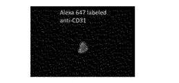
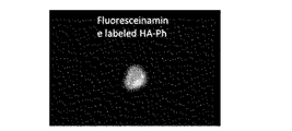
- FIGS. 3A to 3C show the results of observing the group 2 Huvec cells.
- 2A and 3A show the results of observation in a bright field
- FIGS. 2B and 3B show the results of observation of fluorescence derived from Alexa Fluor 647
- FIGS. 2C and 3C observe the fluorescence derived from fluorescein. The results are shown.
- the precipitated FS-Alg-Ph was washed with an 80 wt% ethanol (20 wt% water) solution, and the fluorescence peak due to the presence of aminofluorescein was washed. The ethanol solution exchange was continued every hour until it was no longer detected.
- FS-HA-Ph was washed twice in 100% ethanol for 30 minutes, and then vacuum-dried.
- FS-Alg-Ph having about 3 phenolic hydroxyl groups introduced per 100 units of saccharide units of alginic acid was obtained as a crosslinkable polymer.
- This cell dispersion containing Huvec cells and mouse 10T1 / 2 cells was added to phosphate buffered saline (pH 7.4) containing Alexa Fluor647-labeled anti-human CD31 mouse IgG1 antibody (primary antibody) at 0.005 mg / mL. 1.5 ⁇ 10 6 / mL, and left at room temperature for 30 minutes.
- Huvec cells and mouse 10T1 / 2 cells were washed with phosphate buffered saline, and then dispersed in Solution 1 prepared as described above at 1.0 ⁇ 10 6 cells / mL. Let stand for a minute. The Huvec cells and mouse 10T1 / 2 cells were washed three times with phosphate buffered saline and then observed with a microscope.
- [result] 4A to 4D show the results of observing Huvec cells and mouse 10T1 / 2 cells.
- 4A shows the result of observation in a bright field
- FIG. 4B shows the result of observation of fluorescence derived from Cell Tracker TM Orange
- FIG. 4C shows the result of observation of fluorescence derived from Cell Tracker TM Blue
- FIG. 4D shows the result of observation of fluorescence derived from fluorescein.
- fluorescence derived from FS-Alg-Ph fluorescein was observed on the cell surface of Huvec cells emitting fluorescence derived from Cell Tracker TM Orange.
- no fluorescence derived from FS-Alg-Ph fluorescein was observed on the cell surface of mouse 10T1 / 2 cells emitting fluorescence derived from Cell Tracker TM Blue.
- the solution containing it at a density of 5 ⁇ 10 6 cells / mL was mixed.
- This cell dispersion containing HepG2 cells and mouse 10T1 / 2 cells was added to phosphate buffered saline (pH 7.4) containing labeled anti-human CD326 mouse IgG2b antibody (primary antibody) at 0.005 mg / mL. It was dispersed so as to be 5 ⁇ 10 6 cells / mL and allowed to stand for 30 minutes in the dark at room temperature.
- the HepG2 cells and mouse 10T1 / 2 cells were washed with phosphate buffered saline, and then dispersed in Solution 1 prepared as described above at 1.0 ⁇ 10 6 cells / mL. Let stand for a minute.
- the HepG2 cells and mouse 10T1 / 2 cells were washed three times with phosphate buffered saline and then observed with a microscope.
- [result] 5A to 5C show the results of observing HepG2 cells and mouse 10T1 / 2 cells.
- FIG. 5A shows the result of observation in a bright field
- FIG. 5B shows the result of observation of fluorescence derived from Cell Tracker TM Orange
- FIG. 5C shows the result of observation of fluorescence derived from fluorescein.
- fluorescence derived from fluorescein of FS-Alg-Ph was observed on the cell surface of HepG2 cells emitting fluorescence derived from Cell Tracker TM Orange.
- no fluorescence derived from FS-Alg-Ph fluorescein was observed on the cell surface of mouse 10T1 / 2 cells.
- HRP-labeled anti-mouse IgG + IgM antibody (secondary antibody) labeled with horseradish peroxidase (HRP) and 270 ⁇ L of Krebs Ringer Hepes buffer (pH 7.4) are obtained by mixing the HepG2 cells and mouse 10T1 / 2 cells. 20 ⁇ L of the obtained solution was added, and the mixture was allowed to stand for 30 minutes in the dark (HRP-labeled anti-mouse IgG + IgM antibody (secondary antibody) 0.1 mg / mL).
- HRP horseradish peroxidase
- [result] 6A to 6C show the results of observing HepG2 cells and mouse 10T1 / 2 cells.
- FIG. 6A shows the result of observing fluorescence derived from Cell Tracker TM Orange
- FIG. 6B shows the result of observing fluorescence derived from Cell Tracker TM Blue
- FIG. 6C observed the fluorescence derived from fluorescein. Results are shown.
- fluorescence derived from FS-Alg-Ph fluorescein was observed on the cell surface of HepG2 cells emitting fluorescence derived from Cell Tracker TM Orange.
- no fluorescence derived from FS-Alg-Ph fluorescein was observed on the cell surface of mouse 10T1 / 2 cells emitting fluorescence derived from Cell Tracker TM Blue.
- FS-Alg-Ph obtained in Example 2 above was dissolved in phosphate buffered saline (pH 7.4) so as to be 1 w / v%. Hydrogen peroxide was mixed in this solution so that it might become 0.5 mM (solution 1). In addition, mouse 10T1 / 2 cells were stained with Cell Tracker TM Blue (manufactured by Life Technologies corporation).
- an APC-labeled anti-human CD326 antibody (HRP / APC-CD326 antibody) labeled with horseradish-derived peroxidase was obtained using a horseradish-derived peroxidase labeling kit (Dojindo Laboratories). Then, human hepatoma-derived HepG2 cells were dispersed in a serum-free DMEM medium containing 0.005 mg / mL of this HRP / APC-CD326 antibody, and allowed to stand in the dark at room temperature for 30 minutes.
- HepG2 cells and mouse 10T1 / 2 cells was washed, mixed at 1.0 ⁇ 10 6 cells / mL, and immersed in Solution 1 for 30 minutes at room temperature in the dark. Thereafter, these cells were washed with phosphate buffered saline and observed with a microscope.
- [result] 7A to 7D show the results of observation of HepG2 cells and mouse 10T1 / 2 cells with a microscope.
- FIG. 7A shows the result of observation in bright field
- FIG. 7B shows the result of observation of fluorescence derived from Cell Tracker TM Blue
- FIG. 7C shows the result of observation of fluorescence derived from APC
- FIG. These show the result of having observed the fluorescence derived from fluorescein.
- fluorescence derived from FS-Alg-Ph fluorescein was observed on the cell surface of HepG2 cells emitting fluorescence derived from the HRP / APC-CD326 antibody.
- no fluorescence derived from FS-Alg-Ph fluorescein was observed on the cell surface of mouse 10T1 / 2 cells emitting fluorescence derived from Cell Tracker TM Blue.
- the cell culture dish after recovering the supernatant solution was washed several times and then observed with a microscope.
- FIG. 8A and FIG. 8B show the results of observing the supernatant solution collected from the cell culture dish with a microscope.
- FIG. 8A shows the result of observation in a bright field
- FIG. 8B shows the result of observation of fluorescence derived from fluorescein.
- fluorescence derived from the presence of FS-Alg-Ph was observed in about 90% or more of the cells contained in the supernatant solution. That is, it was confirmed that about 90% or more of the cells contained in the supernatant solution were HepG2 cells having a coating layer formed on the cell surface.
- FIG. 9A to 9C show the results of observing the cell culture dish after collecting the supernatant solution with a microscope.
- FIG. 8A shows the result of observation in a bright field
- FIG. 8B shows the result of observation of fluorescence derived from Cell Tracker TM Blue
- FIG. 8C shows the result of observation of fluorescence derived from fluorescein.
- the HepG2 cell and the mouse 10T1 / 2 cell could be separated based on the binding property between the coating layer covering the cell surface of the HepG2 cell and the bottom surface of the cell culture dish.
- Anti-human CD326 antibody was labeled with horseradish peroxidase (HRP) using a horseradish peroxidase labeling kit (manufactured by Dojindo Laboratories) to obtain an anti-human CD326 antibody labeled with HRP (HRP-CD326 antibody). It was.
- HRP horseradish peroxidase
- cell group 1 human liver cancer-derived cell line HepG2 cells were dispersed in a phosphate physiological buffer solution (pH 7.4) containing the above HRP-CD326 antibody at a concentration of 0.005 mg / mL, and in the dark at room temperature. Let stand for 30 minutes.
- APC-labeled anti-human CD326 antibody is labeled with glucose oxidase (GOx), and anti-human CD326 antibody (GOx labeled with GOx and APC) / APC-CD326 antibody).
- HepG2 cells were dispersed in phosphate physiological buffer solution (pH 7.4) containing the above-mentioned GOx / APC-CD326 antibody at a concentration of 0.010 mg / mL, and allowed to stand still for 30 minutes in the dark at room temperature. I put it.
- cell group 3 a phosphate physiological buffer solution containing HepG2 cells, the HRP-CD326 antibody at a concentration of 0.005 mg / mL, and the GOx / APC-CD326 antibody at a concentration of 0.010 mg / mL It was dispersed in (pH 7.4) and allowed to stand at room temperature in a dark place for 30 minutes.
- the FS-Alg-Ph obtained in the above Example 2 was adjusted to 2 w / v%, and The solution in which glucose was dissolved to 1500 mg / L was allowed to stand for 10 minutes in the dark at room temperature. Thereafter, each of the cell groups 1 to 3 was washed with phosphate buffered saline and then observed with a microscope.
- [result] 10A and 10B show the results of observing HepG2 cells in cell group 3.
- FIG. FIG. 10A shows the result of observation in a bright field
- FIG. 10B shows the result of observation of fluorescence derived from fluorescein.
- An APC-labeled anti-human CD326 antibody was labeled with glucose oxidase (GOx) using a glucose oxidase labeling kit (EasyLink Glkucose Oxidase Conjugation Kit, manufactured by Abcam plc), and anti-human CD326 antibody (GOx / APC labeled with GOx and APC).
- GOx glucose oxidase labeling kit
- EasyLink Glkucose Oxidase Conjugation Kit manufactured by Abcam plc
- anti-human CD326 antibody GOx / APC labeled with GOx and APC.
- the HepG2 cells were dispersed in a phosphate physiological buffer solution (pH 7.4) containing the GOx / APC-CD326 antibody at a concentration of 0.010 mg / mL, and allowed to stand in the dark at room temperature for 30 minutes.
- the FS-Alg-Ph obtained in Example 2 described above was adjusted to 2 w / v%, and glucose was added to 1500 mg / The solution was further allowed to stand in a dark place at room temperature for 10 minutes in a solution in which horseradish peroxidase (manufactured by Wako Pure Chemical Industries, Ltd.) was dissolved so as to be 10 L / mL. Thereafter, HepG2 cells were washed with phosphate buffered saline and then observed with a microscope.
- horseradish peroxidase manufactured by Wako Pure Chemical Industries, Ltd.
- FIG. 11A to 11C show the results of observing HepG2 cells.
- FIG. 11A shows the result of observation in bright field
- FIG. 11B shows the result of observation of fluorescence derived from GOx / APC-CD326 antibody
- FIG. 11C shows the result of observation of fluorescence derived from fluorescein.
- HepG2 cells bound with GOx / APC-CD326 antibody are replaced with a solution containing FS-Alg-Ph, glucose and horseradish peroxidase, and FS-Alg-Ph.
- glucose-containing peroxidase-free solution and when dispersed in a solution containing FS-Alg-Ph and horseradish-derived peroxidase-free glucose, the GOx / APC-CD326 antibody No fluorescence derived from the presence of FS-Alg-Ph was observed in HepG2 cells in which fluorescence due to the presence of was observed.
Landscapes
- Life Sciences & Earth Sciences (AREA)
- Engineering & Computer Science (AREA)
- Health & Medical Sciences (AREA)
- Chemical & Material Sciences (AREA)
- Biomedical Technology (AREA)
- Biotechnology (AREA)
- Organic Chemistry (AREA)
- Bioinformatics & Cheminformatics (AREA)
- Genetics & Genomics (AREA)
- Wood Science & Technology (AREA)
- Zoology (AREA)
- Microbiology (AREA)
- Biochemistry (AREA)
- General Engineering & Computer Science (AREA)
- General Health & Medical Sciences (AREA)
- Cell Biology (AREA)
- Apparatus Associated With Microorganisms And Enzymes (AREA)
- Micro-Organisms Or Cultivation Processes Thereof (AREA)
Abstract
細胞被覆層の効果的な形成に関する方法及びキットを提供する。本発明に係る方法は、下記(a)及び(b):(a)標的細胞の細胞表面の特定分子に、前記特定分子に結合する結合分子を介して、架橋性分子の架橋反応に寄与する触媒分子を結合させること;(b)前記触媒分子が結合した前記標的細胞と前記架橋性分子とを含む溶液中で、前記触媒分子が寄与する前記架橋反応により前記架橋性分子を架橋して、前記標的細胞の前記細胞表面を被覆する被覆層を形成すること;を含む。
Description
本発明は、細胞被覆層の形成に関する方法及びキットに関し、特に、触媒分子が寄与する架橋性分子の架橋反応による細胞被覆層の形成に関する。
従来、細胞を含むゲル粒子を形成する方法としては、例えば、当該細胞を分散したアルギン酸ナトリウム溶液を塩化カルシウム溶液に滴下する方法があった(例えば、特許文献1参照)。
しかしながら、従来、標的とする細胞を含む溶液中において、当該細胞の細胞表面近傍で局所的な架橋反応を行って、当該細胞の当該細胞表面を被覆する被覆層を形成することはできなかった。
また、従来、例えば、複数種類の細胞を含む溶液中において、特定の種類の細胞の細胞表面近傍でのみ局所的な架橋反応を行って、当該特定の細胞の当該細胞表面を選択的に被覆する被覆層を形成することはできなかった。
本発明は、上記課題に鑑みて為されたものであり、細胞被覆層の効果的な形成に関する方法及びキットを提供することを目的の一つとする。
上記課題を解決するための本発明の一実施形態に係る方法は、下記(a)及び(b):(a)標的細胞の細胞表面の特定分子に、前記特定分子に結合する結合分子を介して、架橋性分子の架橋反応に寄与する触媒分子を結合させること;(b)前記触媒分子が結合した前記標的細胞と前記架橋性分子とを含む溶液中で、前記触媒分子が寄与する前記架橋反応により前記架橋性分子を架橋して、前記標的細胞の前記細胞表面を被覆する被覆層を形成すること;を含むことを特徴とする。本発明によれば、細胞被覆層を効果的に形成する方法を提供することができる。
また、前記方法は、前記(a)として、前記標的細胞である第一の細胞と、細胞表面に前記特定分子を有しない第二の細胞とを含む溶液中で、前記第一の細胞の前記特定分子に、前記特定分子に特異的に結合する前記結合分子を介して、前記触媒分子を結合させることを含み、前記(b)として、前記触媒分子が結合した前記第一の細胞と、前記第二の細胞と、前記架橋性分子とを含む前記溶液中で、前記触媒分子が寄与する前記架橋反応により前記架橋性分子を架橋して、前記第一の細胞の前記細胞表面を被覆する前記被覆層を形成することを含むこととしてもよい。また、この場合、前記方法は、下記(c):(c)前記細胞表面を前記被覆層で被覆された前記第一の細胞と、前記第二の細胞とを分離すること;をさらに含むこととしてもよい。
また、前記方法において、前記特定分子は、タンパク質、糖タンパク質、ペプチド及び糖鎖からなる群より選択される1種以上であることとしてもよい。また、前記方法において、前記結合分子は、前記特定分子に対する抗体であることとしてもよい。
また、前記方法は、前記(a)として、前記標的細胞の前記特定分子に、前記結合分子を介して、前記架橋反応を触媒する前記触媒分子を結合させることを含み、前記(b)として、前記触媒分子が結合した前記標的細胞と前記架橋性分子とを含む前記溶液中で、前記触媒分子に触媒される前記架橋反応により前記架橋性分子を架橋して、前記標的細胞の前記細胞表面を被覆する前記被覆層を形成することを含むこととしてもよい。また、この場合、前記触媒分子は、ペルオキシダーゼ、カタラーゼ、フェノールオキシダーゼ、カテコールオキシダーゼ及び金属ポルフィリン錯体からなる群より選択される1種以上であることとしてもよい。
また、前記方法は、前記(a)として、前記標的細胞の前記特定分子に、前記結合分子を介して、活性酸素種を生成する化学反応を触媒する第一の触媒分子である前記触媒分子を結合させることを含み、前記(b)として、前記第一の触媒分子が結合した前記標的細胞と前記架橋性分子とを含む前記溶液中で、前記第一の触媒分子に触媒される前記化学反応により生成された前記活性酸素種と、前記活性酸素種の存在下で前記架橋反応を触媒する第二の触媒分子との存在下、前記第二の触媒分子に触媒される前記架橋反応により前記架橋性分子を架橋して、前記標的細胞の前記細胞表面を被覆する前記被覆層を形成することを含むこととしてもよい。また、この場合、前記第一の触媒分子は、グルコースオキシダーゼ、スーパーオキシドディスムターゼ、アミノ酸オキシダーゼ、コリンオキシダーゼ、アルコールオキシダーゼ、ピルビン酸オキシダーゼ及びコレステロールオキシダーゼからなる群から選ばれる1種以上であることとしてもよい。さらに、これらの場合、前記第二の触媒分子は、ペルオキシダーゼ、カタラーゼ及び金属ポルフィリン錯体からなる群より選択される1種以上であることとしてもよい。
上記課題を解決するための本発明の一実施形態に係るキットは、前記いずれかの方法に使用される細胞被覆層形成用キットであって、下記(x)及び/又は(y):(x)標的細胞の細胞表面の特定分子に結合する結合分子、及び/又は前記結合分子を介して前記標的細胞の前記特定分子に結合させる、架橋性分子の架橋反応に寄与する触媒分子;(y)前記架橋性分子、又は架橋性基が導入されるべき基礎分子及び/又は基礎分子に架橋性基を導入するための化合物;を含むことを特徴とする。本発明によれば、細胞被覆層を効果的に形成するキットを提供することができる。
また、前記キットは、前記(x)として、前記結合分子、及び前記結合分子に結合された前記触媒分子を含むこととしてもよい。また、この場合、前記キットは、前記(y)として、前記架橋性分子をさらに含むこととしてもよい。また、前記キットは、下記(z):(z)前記細胞被覆層形成用キットを前記方法に使用するための取扱説明書;をさらに含むこととしてもよい。
上記課題を解決するための本発明の一実施形態に係る方法は、前記いずれかの方法に、下記(x)及び/又は(y):(x)標的細胞の細胞表面の特定分子に結合する結合分子、及び/又は前記結合分子を介して前記標的細胞の前記特定分子に結合させる、架橋性分子の架橋反応に寄与する触媒分子;(y)前記架橋性分子、又は架橋性基が導入されるべき基礎分子及び/又は基礎分子に架橋性基を導入するための化合物;を使用することを特徴とする。本発明によれば、細胞被覆層の効果的な形成に関する方法を提供することができる。
本発明によれば、細胞被覆層の効果的な形成に関する方法及びキットを提供することができる。
以下に、本発明の一実施形態について説明する。なお、本発明は、本実施形態に限られるものではない。
本実施形態に係る方法(以下、「本方法」という。)は、下記(a)及び(b):(a)標的細胞の細胞表面の特定分子に、当該特定分子に結合する結合分子を介して、架橋性分子の架橋反応に寄与する触媒分子を結合させること;(b)当該触媒分子が結合した当該標的細胞と当該架橋性分子とを含む溶液中で、当該触媒分子が寄与する当該架橋反応により当該架橋性分子を架橋して、当該標的細胞の当該細胞表面を被覆する被覆層を形成すること;を含む方法である。
ここで、まず、本方法の概要について説明する。図1A~図1Cには、本方法の一例において、標的細胞Tの細胞表面を被覆する被覆層Sを形成する過程を示す。本方法において、その細胞表面を被覆層Sで被覆すべき標的細胞Tは、図1Aに示すように、当該細胞表面に特定分子Pを有している。
そこで、本方法では、まず、上記(a)として、図1Bに示すように、標的細胞Tの特定分子Pに、当該特定分子Pに結合する結合分子Qを介して、被覆層Sを形成するための架橋性分子の架橋反応に寄与する触媒分子Rを結合させる。
そして、本方法では、次に、上記(b)として、触媒分子Rが結合した標的細胞Tと架橋性分子とを含む溶液L中で、当該触媒分子Rが寄与する架橋反応により当該架橋性分子を架橋して、図1Cに示すように、当該標的細胞Tの細胞表面を被覆する被覆層Sを形成する。
このように、本方法によれば、標的細胞Tを含む溶液L中において、当該標的細胞Tの細胞表面近傍で局所的な架橋性分子の架橋反応を行って、当該標的細胞Tの当該細胞表面を被覆する被覆層Sを形成することができる。
また、本方法では、図1A~図1Cに示すように、細胞表面に特定分子Pを有する標的細胞Tと、細胞表面に当該特定分子Pを有しない他の細胞Uとを含む溶液L中において、当該標的細胞Tの特定分子Pに、結合分子Qを介して触媒分子Rを特異的に結合させることで、当該標的細胞Tの細胞表面近傍のみで局所的な架橋性分子の架橋反応を行って、当該標的細胞Tの当該細胞表面を選択的に被覆する被覆層Sを形成することもできる。
次に、本方法の詳細について説明する。本方法においては、上述のとおり、まず、上記(a)として、標的細胞の細胞表面の特定分子に、結合分子を介して、触媒分子を結合する。この標的細胞に対する触媒分子の結合は、後述するように当該標的細胞の細胞表面近傍で局所的な架橋性分子の架橋反応を行うための前処理である。
標的細胞の特定分子は、当該標的細胞の細胞表面に存在する分子であれば特に限られない。特定分子は、例えば、タンパク質、糖タンパク質、ペプチド及び糖鎖からなる群より選択される1種以上である。すなわち、特定分子は、例えば、いわゆる細胞表面タンパク質及び/又は細胞膜タンパク質であることとしてもよい。より具体的に、特定分子は、例えば、CD(cluster of differentiation)抗原であることとしてもよい。また、特定分子は、例えば、細胞表面に存在する受容体であることとしてもよい。また、特定分子は、例えば、人為的操作によって標的細胞の細胞表面に結合された分子であることとしてもよい。
標的細胞は、その細胞表面に特定分子を有する細胞であれば特に限られない。標的細胞は、例えば、動物細胞、植物細胞及び微生物からなる群より選択される1種以上であることとしてもよい。動物細胞は、例えば、脊椎動物の細胞及び/又は無脊椎動物の細胞であることとしてもよい。脊椎動物は、例えば、哺乳類、爬虫類、鳥類、両生類及び魚類からなる群より選択される1種以上であることとしてもよい。哺乳動物は、例えば、ヒト、サル、ウシ、ウマ、ヒツジ、ブタ、イヌ、ネコ、ラット、マウス、ハムスター及びモルモットからなる群より選択される1種以上であることとしてもよい。無脊椎動物は、例えば、昆虫、線虫、ホヤ、イカ、タコ及びカニからなる群より選択される1種以上であることとしてもよい。
また、標的細胞は、例えば、幹細胞及び/又は分化細胞であることとしてもよい。幹細胞は、多分化能と自己複製能とを有する細胞であれば特に限られない。幹細胞は、例えば、人工多能性幹細胞(induced pluripotent stem cell:iPS細胞)、胚性幹細胞(Embryonic Stem cell:ES細胞)、体性幹細胞及び生殖幹細胞からなる群より選択される1種以上であることとしてもよい。体性幹細胞は、例えば、間葉系幹細胞、造血幹細胞、神経幹細胞、肝幹細胞、膵幹細胞、皮膚幹細胞、脂肪幹細胞、筋幹細胞、骨幹細胞及び軟骨幹細胞からなる群より選択される1種以上であることとしてもよい。
分化細胞は、分化した細胞であれば特に限られない。分化細胞は、例えば、骨髄細胞、血液細胞、神経細胞、肝細胞、膵細胞、皮膚細胞、脂肪細胞、筋肉細胞、心臓細胞、骨細胞、軟骨細胞及び生殖細胞からなる群より選択される1種以上であることとしてもよい。また、分化細胞は、例えば、培養系において幹細胞から誘導された分化細胞であることとしてもよい。
また、標的細胞は、例えば、初代細胞、継代細胞及び株化細胞からなる群より選択される1種以上であることとしてもよい。また、標的細胞は、例えば、培養細胞であることとしてもよい。また、標的細胞は、溶液中に分散されていてもよいし、溶液中で基材に接着していてもよい。
結合分子は、標的細胞の特定分子に結合する分子であれば特に限られず、当該特定分子に特異的に結合する分子であることが好ましい。結合分子は、例えば、タンパク質(例えば、抗体、レクチン、アルギン酸結合性タンパク質)、ペプチド(例えば、コラーゲン結合性ペプチド)及び核酸分子(例えば、DNAアプタマー、RNAアプタマー、ペプチドアプタマー)からなる群より選択される1種以上であることとしてもよい。なお、抗体は、例えば、フラグメント抗体であることとしてもよい。また、例えば、特定分子が受容体である場合、結合分子は、当該受容体に対するリガンドであることとしてもよい。
触媒分子は、標的細胞の細胞表面を被覆する被覆層を形成するための架橋性分子の架橋反応に寄与する触媒活性を示す分子であれば特に限られない。触媒分子は、例えば、架橋性分子の架橋反応を触媒する触媒分子及び/又は活性酸素種を生成する化学反応を触媒する触媒分子であることとしてもよい。
すなわち、本方法は、例えば、上記(a)として、標的細胞の細胞表面の特定分子に、当該特定分子に結合する結合分子を介して、架橋性分子の架橋反応を触媒する触媒分子を結合させることを含み、上記(b)として、当該触媒分子が結合した当該標的細胞と当該架橋性分子とを含む溶液中で、当該触媒分子に触媒される当該架橋反応により当該架橋性分子を架橋して、当該標的細胞の当該細胞表面を被覆する被覆層を形成することを含むこととしてもよい。
架橋性分子の架橋反応を触媒する触媒分子は、当該架橋反応を触媒する活性を示す分子であれば特に限られない。この触媒分子は、例えば、活性酸素種(Reactive Oxygen Species:ROS)又は酸素の存在下で架橋性分子の架橋反応を触媒する触媒分子であることとしてもよい。なお、ROSは、例えば、スーパーオキシド、ヒドロキシラジカル、過酸化水素及び一重項酸素からなる群より選択される1種以上であり、過酸化水素が好ましく利用される。
具体的に、触媒分子は、例えば、ペルオキシダーゼ、カタラーゼ、フェノールオキシダーゼ、カテコールオキシダーゼ及び金属ポルフィリン錯体からなる群より選択される1種以上であることとしてもよい。ペルオキシダーゼ、カタラーゼ及び金属ポルフィリン錯体は、ROSの存在下で架橋性分子の架橋反応を触媒する触媒分子である。フェノールオキシダーゼ及びカテコールオキシダーゼは、酸素の存在下で架橋性分子の架橋反応を触媒する触媒分子である。
ペルオキシダーゼは、例えば、西洋ワサビペルオキシダーゼ、大豆由来ペルオキシダーゼ、ミエロペルオキシダーゼ、ラクトペルオキシダーゼ、好酸球ペルオキシダーゼ及びチロイドペルオキシダーゼからなる群より選択される1種以上であることとしてもよい。フェノールオキシダーゼは、例えば、ラッカーゼ及び/又はチロシナーゼであることとしてもよい。金属ポルフィリン錯体は、例えば、鉄ポルフィリン錯体及び/又は銅ポルフィリン錯体であることとしてもよい。鉄ポルフィリン錯体は、例えば、ヘミン(ヘマチン)、ヘモグロビン、フェリヘム及びヘモクロムからなる群より選択される1種以上であることとしてもよい。
また、本方法は、例えば、上記(a)として、標的細胞の細胞表面の特定分子に、当該特定分子に結合する結合分子を介して、活性酸素種(ROS)を生成する化学反応を触媒する第一の触媒分子を結合させることを含み、上記(b)として、当該第一の触媒分子に触媒される化学反応により生成された当該活性酸素種と、当該活性酸素種の存在下で架橋性分子の架橋反応を触媒する第二の触媒分子との存在下、当該第二の触媒分子に触媒される当該架橋反応により当該架橋性分子を架橋して、当該標的細胞の当該細胞表面を被覆する被覆層を形成することを含むこととしてもよい。
ROSを生成する化学反応を触媒する触媒分子(第一の触媒分子)は、架橋性分子の架橋反応に利用されるROSを生成する化学反応を触媒する活性を示す分子であれば特に限られない。また、第一の触媒分子と共に使用される第二の触媒分子は、ROSの存在下で架橋性分子の架橋反応を触媒する触媒分子として上述したものであれば特に限られない。
第一の触媒分子は、例えば、溶液に含まれる基質を消費してROSを生成する化学反応を触媒する触媒分子である。すなわち、第一の触媒分子は、例えば、溶液に含まれる基質(例えば、有機化合物)を酸化してROSを生成する化学反応を触媒する触媒分子であることとしてもよい。
具体的に、第一の触媒分子は、例えば、グルコースオキシダーゼ、スーパーオキシドディスムターゼ、アミノ酸オキシダーゼ、コリンオキシダーゼ、アルコールオキシダーゼ、ピルビン酸オキシダーゼ及びコレステロールオキシダーゼからなる群から選ばれる1種以上であることとしてもよい。
グルコースオキシダーゼ、アミノ酸オキシダーゼ、コリンオキシダーゼ、アルコールオキシダーゼ、ピルビン酸オキシダーゼ及びコレステロールオキシダーゼは、それぞれ、グルコース、アミノ酸、コリン、アルコール、ピルビン酸及びコレステロールを酸化してROS(過酸化水素)を生成する化学反応を触媒する。スーパーオキシドディスムターゼは、スーパーオキシド(例えば、第一の触媒分子が結合した標的細胞と架橋性分子とを含む溶液に含まれるスーパーオキシド、及び/又は当該溶液中で当該標的細胞により産生されるスーパーオキシド)から酸素及び過酸化水素を生成する化学反応を触媒する。
本方法において、標的細胞の細胞表面の特定分子に、結合分子を介して触媒分子を結合させる方法は、特に限られない。例えば、標的細胞と、結合分子に結合された触媒分子とを含む溶液を調製して、当該標的細胞の特定分子に当該結合分子を介して当該触媒分子を結合させることとしてもよい。
この場合、例えば、結合分子に結合された触媒分子として、特定分子に対する抗体に結合された触媒分子(より具体的には、例えば、ペルオキシダーゼ標識抗体)を使用することとしてもよい。
また、例えば、標的細胞と、結合分子に結合された上記第一の触媒分子及び第二の触媒分子とを含む溶液を調製して、当該標的細胞の特定分子に当該結合分子を介して当該第一の触媒分子及び第二の触媒分子を結合させることとしてもよい。
この場合、例えば、結合分子に結合された第一の触媒分子及び第二の触媒分子として、特定分子に対する抗体に結合された第一の触媒分子及び第二の触媒分子(より具体的には、例えば、特定分子に対する抗体に結合された、ペルオキシダーゼ及びグルコースオキシダーゼ)を使用することとしてもよい。
また、例えば、まず、標的細胞と結合分子とを含む溶液を調製して、当該標的細胞の特定分子に当該結合分子を結合させ、次いで、当該結合分子が結合した標的細胞と、当該結合分子に結合する他の分子に結合された触媒分子とを含む溶液と調製して、当該標的細胞に結合した結合分子に、当該他の分子を介して当該触媒分子を結合させ、その結果、当該標的細胞の特定分子に、当該結合分子を介して当該触媒分子を結合させることとしてもよい。
この場合、例えば、結合分子として、特定分子に対する抗体(一次抗体)を使用し、当該結合分子に結合する他の分子に結合された触媒分子として、当該結合分子に対する抗体(二次抗体)に結合された触媒分子(より具体的には、例えば、ペルオキシダーゼ標識抗体)を使用することとしてもよい。
次いで、本方法においては、上述のとおり、上記(b)として、触媒分子が結合した標的細胞と架橋性分子とを含む溶液中で、当該触媒分子が寄与する架橋反応により当該架橋性分子を架橋して、当該標的細胞の当該細胞表面を被覆する被覆層を形成する。なお、溶液に含まれる溶媒は、水を含む溶媒であれば特に限られない。
架橋性分子は、当該架橋性分子を含む溶液中において、触媒分子が寄与する架橋反応により架橋される分子であれば特に限られない。架橋性分子は、架橋性基を有する分子であり、架橋性基を有する親水性分子であることが好ましい。架橋性基は、分子の架橋構造の形成に寄与する官能基であれば特に限られない。架橋性基は、例えば、水酸基、アミノ基、カルボキシル基、チオール基及びビニル基からなる群より選択される1種以上であることとしてもよい。水酸基は、例えば、ベンゼン環に結合した水酸基であることとしてもよい。ベンゼン環に結合した水酸基は、例えば、フェノール性水酸基及び/又はカテコール基であることとしてもよい。アミノ基は、例えば、ベンゼン環に結合したアミノ基であることとしてもよい。また、例えば、架橋性分子がシュガービートペクチンである場合、当該シュガービートペクチンに含まれるフェルラ酸部分及び/又はジヒドロフェルラ酸部分を架橋性基として利用することとしてもよい。
架橋性分子の分子量は、特に限られないが、例えば、100Da以上であることとしてもよく、300Da以上であることとしてもよい。架橋性分子の分子量の上限値は特に限られないが、当該分子量は、例えば、107Da以下であることとしてもよい。
架橋性分子は、例えば、架橋性高分子であることとしてもよい。架橋性高分子は、架橋性基を有する高分子であり、架橋性基を有する親水性高分子であることが好ましい。架橋性高分子は、架橋性基を有する天然高分子及び/又は合成高分子であることとしてもよい。天然高分子は、例えば、タンパク質、多糖類、核酸、ポリフェノール重合体及びこれらの誘導体からなる群より選択される1つ以上であることとしてもよい。
タンパク質は、例えば、ゼラチン、コラーゲン、アルブミン、グロブリン、フィブリン及びこれらの誘導体からなる群より選択される1つ以上であることとしてもよい。多糖類は、例えば、アルギン酸、ヒアルロン酸、コンドロイチン硫酸、デンプン、デキストラン、プルラン、セルロース、ペクチン、シュガービートペクチン、アミロペクチン、キチン、キトサン、アミロース、ヘパリン、ヘパラン硫酸及びこれらの誘導体からなる群より選択される1種以上であることとしてもよい。核酸は、例えば、デオキシリボ核酸、リボ核酸及びこれらの誘導体からなる群より選択される1種以上であることとしてもよい。ポリフェノール重合体は、例えば、タンニン重合体、カテキン重合体及びこれらの誘導体からなる群より選択される1種以上であることとしてもよい。合成高分子は、例えば、ポリエチレングリコール、ポリビニルアルコール、ポリアクリル酸、ポリメタクリル酸、ポリプロピレン、ポリ塩化ビニル及びこれらの誘導体からなる群より選択される1種以上であることとしてもよい。
架橋性分子は、例えば、架橋された後に分解可能な分子であることとしてもよい。すなわち、架橋性分子は、例えば、架橋された後に酵素により分解可能な分子であることとしてもよい。具体的に、例えば、架橋性分子がタンパク質である場合、当該架橋性分子は、架橋後であっても、タンパク質分解酵素により分解される。また、例えば、架橋性分子が多糖類である場合、当該架橋性分子は、架橋後であっても、多糖類分解酵素により分解される。
架橋性分子は、架橋性基を導入することにより得られたものであることとしてもよい。架橋性基を導入する方法は特に限られないが、例えば、架橋性基が導入されるべき分子(基礎分子)と、当該基礎分子に当該架橋性基を導入するための化合物とを化学的に反応させることにより行うこととしてもよい。基礎分子と、当該基礎分子に架橋性基を導入するための化合物との化学反応は、特に限られず、例えば、縮合反応であることとしてもよい。
基礎分子は、架橋性基を導入可能な分子であれば特に限られない。基礎分子は、例えば、上述したような天然高分子及び/又は合成高分子であって、架橋性基を有しない高分子、又は被覆層を形成するために十分な量の架橋性基を有しない高分子であることとしてもよい。
基礎分子に架橋性基を導入するための化合物は、当該基礎分子と結合する化合物であれば特に限られない。基礎分子と、当該基礎分子に架橋性基を導入するための化合物との結合は、例えば、共有結合、静電的結合、疎水的結合及び水素結合からなる群より選択される1種以上であることとしてもよい。
具体的に、基礎分子に架橋性基を導入するための化合物は、例えば、基礎分子と化学的に反応して化学結合を形成する反応性官能基と、当該基礎分子に導入されるべき架橋性基とを有する化合物であることとしてもよい。この場合、反応性官能基は、例えば、カルボキシル基、アミノ基、水酸基、チオール基、マレイミド基、スクシミド基、アジド基及びアルキンからなる群より選択される1種以上であることとしてもよい。
より具体的に、基礎分子に架橋性基を導入するための化合物は、例えば、チラミン、フェニルプロピオン酸、アニリン、カテコールアミン(例えば、アドレナリン、ノルアドレナリン及びドーパミンからなる群より選択される1種以上)及びこれらの誘導体からなる群より選択される1種以上であることとしてもよい。
架橋反応前の標的細胞と架橋性分子とを含む溶液中において、当該架橋性分子は、当該溶液中に分散されていることとしてもよいし、予め当該標的細胞の細胞表面に結合されていることとしてもよい。架橋性分子が溶液中に分散されている場合、例えば、上述した触媒分子と結合された当該架橋性分子が溶液中に分散されていることとしてもよい(例えば、Ryu JH et al. Chitosan-g-hematin: Enzyme-mimicking polymeric catalyst for adhesive hydrogels. Acta Biomater (2013)参照)。
架橋反応前の架橋性分子が標的細胞の細胞表面に結合されている場合、当該架橋性分子は、例えば、当該標的細胞の細胞表面の分子に、当該分子に結合する他の分子(例えば、当該細胞表面の分子に対する抗体)を介して結合されていることとしてもよい。
触媒分子が結合した標的細胞と架橋性分子とを含む溶液中における、当該触媒分子が寄与する架橋反応による当該架橋性分子の架橋は、当該架橋反応が開始される条件で当該溶液を調製することにより行われる。
すなわち、例えば、本方法が、上記(a)として、標的細胞の細胞表面の特定分子に、当該特定分子に結合する結合分子を介して、架橋性分子の架橋反応を触媒する触媒分子を結合させることを含み、上記(b)として、当該触媒分子が結合した当該標的細胞と当該架橋性分子とを含む溶液中で、当該触媒分子に触媒される当該架橋反応により当該架橋性分子を架橋して、当該標的細胞の当該細胞表面を被覆する被覆層を形成することを含む場合、当該溶液中における当該架橋性分子の架橋は、当該触媒分子に触媒される当該架橋反応が開始される条件で、当該溶液を調製することにより行われる。
具体的に、例えば、標的細胞に、ROS又は酸素の存在下で架橋性分子の架橋反応を触媒する触媒分子を結合させる場合、当該触媒分子が結合した当該標的細胞と、当該架橋性分子と、当該ROS又は酸素とを含む溶液を調製することにより、当該架橋性分子の架橋反応を開始する。
より具体的に、例えば、ROSの存在下で架橋性分子の架橋反応を触媒する触媒分子が結合した標的細胞を含む溶液に、当該架橋性分子及びROSを添加することにより、当該架橋性分子の架橋反応を開始することとしてもよい。
なお、溶液に含まれるROSは、当該溶液に外的に添加されることとしてもよく、当該溶液中で当該ROSを生成する化学反応を触媒する他の触媒分子により生成されることとしてもよく、当該溶液中で電気化学的に生成されることとしてもよく(例えば、特開2004-10904号公報参照)、当該溶液中で光触媒(例えば、酸化チタン)に光を照射することにより生成されることとしてもよい。
なお、溶液中でROSを生成する化学反応を触媒する他の触媒分子としては、例えば、ROSを生成する化学反応を触媒する触媒分子として上述した分子(グルコースオキシダーゼ等)を使用することとしてもよい。この他の触媒分子は、溶液中に分散されていることとしてもよく、標的細胞の細胞表面の分子に、当該分子に結合する分子を介して結合されていることとしてもよい。ROSを生成する化学反応を触媒する他の触媒分子を標的細胞に結合させる場合、例えば、当該標的細胞の細胞表面の分子に対する抗体に結合された当該他の触媒分子(具体的に、例えば、グルコースオキシダーゼ標識抗体)を使用することとしてもよい。他の触媒分子を結合させる、標的細胞の細胞表面の分子は、上述した特定分子であることとしてもよく、当該特定分子とは異なる分子であることとしてもよい。
また、例えば、酸素の存在下で架橋性分子の架橋反応を触媒する触媒分子が結合した標的細胞と酸素とを含む溶液に、当該架橋性分子を添加することにより、当該架橋性分子の架橋反応を開始することとしてもよい。
また、例えば、本方法が、上記(a)として、標的細胞の細胞表面の特定分子に、当該特定分子に結合する結合分子を介して、ROSを生成する化学反応を触媒する第一の触媒分子を結合させることを含み、上記(b)として、当該第一の触媒分子に触媒される化学反応により生成された当該ROSと、当該ROSの存在下で架橋性分子の架橋反応を触媒する第二の触媒分子との存在下、当該第二の触媒分子に触媒される当該架橋反応により当該架橋性分子を架橋して、当該標的細胞の当該細胞表面を被覆する被覆層を形成することを含む場合、当該溶液中における当該架橋性分子の架橋は、当該第一の触媒に触媒される化学反応により当該ROSが生成され、且つ当該ROSの存在下、当該第二の触媒分子に触媒される当該架橋反応が開始される条件で、当該溶液を調製することにより行われる。
具体的に、この場合、架橋性分子の架橋は、例えば、第一の触媒分子が結合した標的細胞と、ROSを生成する化学反応において当該第一の触媒分子が消費する基質と、第二の触媒分子と、当該架橋性分子とを含む溶液を調製することにより行われる。
より具体的に、例えば、第一の触媒分子が結合した標的細胞と当該第一の触媒分子の基質とを含む溶液に、第二の触媒分子及び当該架橋性分子を添加することにより、当該架橋性分子の架橋反応を開始することとしてもよい。
なお、第二の触媒分子は、溶液中に分散されていることとしてもよく、標的細胞の細胞表面の分子に、当該分子に結合する分子を介して結合されていることとしてもよい。第二の触媒分子が溶液中に分散されている場合、例えば、架橋性分子と結合した当該第二の触媒分子が当該溶液中に分散されていることとしてもよい。
第二の触媒分子を標的細胞に結合させる場合、例えば、当該標的細胞の細胞表面の分子に対する抗体に結合された当該第二の触媒分子(具体的に、例えば、ペルオキシダーゼ標識抗体)を使用することとしてもよい。第二の触媒分子を結合させる、標的細胞の細胞表面の分子は、上述した特定分子であることとしてもよく、当該特定分子とは異なる分子であることとしてもよい。すなわち、例えば、標的細胞の細胞表面の特定分子に対する抗体に結合された第一の触媒分子及び第二の触媒分子(具体的に、例えば、ペルオキシダーゼ及びグルコースオキシダーゼで標識された抗体)を使用することとしてもよい。
架橋性分子の架橋は、当該架橋性分子の架橋性基同士が新たな化学結合を形成することにより行われることとしてもよいし、当該架橋性分子同士が、他の分子を介して新たな化学結合を形成することにより行われることとしてもよい。架橋性分子同士が、他の分子を介して新たな化学結合を形成する場合、例えば、当該架橋性分子の第一の架橋性基と、当該他の分子の第二の架橋性基とが化学的に反応して当該新たな化学結合を形成することとしてもよい。この他の分子としては、例えば、複数のフェノール性水酸基を有するポリフェノール化合物を使用することとしてもよい。
本方法において、標的細胞の細胞表面を被覆する被覆層は、当該細胞表面に触媒分子が結合した当該標的細胞と架橋性分子とを含む溶液中で、当該標的細胞の当該細胞表面近傍にて、当該触媒分子が寄与する局所的な当該架橋性分子の架橋反応により形成される。
標的細胞の細胞表面を被覆する被覆層は、架橋性分子の架橋により形成された架橋構造を含む。被覆層は、例えば、溶液中において、可溶性の架橋性分子が架橋して不溶化することにより形成された構造体(例えば、ゲル層)である。
本方法においては、例えば、触媒分子が結合した標的細胞と、所定量の架橋性分子とを含む溶液中で、当該触媒分子が寄与する架橋反応により、当該所定量の一部の架橋性分子を架橋し、その結果、架橋されていない当該所定量の他の一部の架橋性分子を含む当該溶液中で、当該一部の架橋性分子の架橋により形成された架橋構造を含み、当該標的細胞の細胞表面を被覆する被覆層を形成する。
標的細胞の細胞表面を被覆する被覆層は、当該標的細胞の当該細胞表面の全部又は一部を被覆する。すなわち、例えば、標的細胞が溶液中に分散されている場合(標的細胞が溶液中に浮遊している場合)には、当該溶液中において、当該標的細胞の細胞表面の全部を被覆する被覆層を形成することとしてもよい。この場合、例えば、標的細胞と当該標的細胞の細胞表面を被覆する被覆層(ゲル層)とを含むマイクロカプセル(ゲル粒子)が形成されることとしてもよい。
また、例えば、標的細胞が溶液中で基材(例えば、細胞培養ディッシュの底面)上に接着している場合には、当該溶液中において、当該標的細胞の細胞表面の一部(例えば、基材への接着に寄与していない、当該細胞表面の一部)を被覆する被覆層を形成する。
標的細胞の細胞表面を被覆する被覆層の厚さ(当該細胞表面と被覆層の外表面との距離)は、架橋反応の条件(架橋性分子の種類や量、架橋反応の時間や温度等)によって調整されるため特に限られないが、例えば、1nm~2000μmであることとしてもよく、10nm~200μmであることとしてもよい。
上述した本方法によれば、標的細胞の細胞表面を被覆する被覆層(細胞被覆層)を効果的に形成することができる。すなわち、本方法においては、細胞表面に触媒分子が結合した標的細胞と架橋性分子とを含む溶液中で、当該標的細胞に結合した当該触媒分子を起点として進行する架橋反応により、当該標的細胞の細胞表面近傍における局所的な当該架橋性分子の架橋を行うことができる。
したがって、例えば、標的細胞と、当該標的細胞の細胞表面を被覆する薄い被覆層とを含む微小な粒子を簡便に形成することができる。また、例えば、標的細胞を1つだけ含む粒子を簡便に形成することもできる。この場合、標的細胞は、粒子のほぼ中央に配置される。なお、溶液中の標的細胞の密度を増加させることにより、又は複数の標的細胞を含む細胞塊を予め形成することにより、複数個の標的細胞を含む粒子を形成することもできる。上述した本方法は、その細胞表面が被覆層で被覆された標的細胞を製造する方法であるともいえる。
また、本方法は、例えば、上記(a)として、細胞表面に特定分子を有する第一の細胞(標的細胞)と、細胞表面に当該特定分子を有しない第二の細胞とを含む溶液中で、当該第一の細胞の当該特定分子に、当該特定分子に特異的に結合する結合分子を介して、架橋性分子の架橋反応に寄与する触媒分子を結合させることを含み、上記(b)として、当該触媒分子が結合した当該第一の細胞と、当該第二の細胞と、当該架橋性分子とを含む溶液中で、当該触媒分子が寄与する当該架橋反応により当該架橋性分子を架橋して、当該第一の細胞の当該細胞表面を被覆する被覆層を形成することを含むこととしてもよい。
すなわち、この場合、まず、図1Bに示すように、特定分子Pを有する第一の細胞(標的細胞T)と、当該特定分子Pを有しない第二の細胞(他の細胞U)とを含む溶液L中で、当該第一の細胞Tの特定分子Pに、当該特定分子Pに特異的に結合する結合分子Qを介して、触媒分子Rを選択的に結合させる。なお、触媒分子Rの第一の細胞Tへの結合は、当該第一の細胞Tの特定分子Pに特異的に結合する結合分子Qを使用して行うため、第二の細胞Uに対する触媒分子Rの特異的な結合は起こらない。
そして、次いで、図1Cに示すように、触媒分子Rが結合した第一の細胞Tと、第二の細胞Uとを含む溶液L中で、当該第一の細胞Tの細胞表面近傍における、当該触媒分子Rが寄与する局所的な架橋性分子の架橋反応により、当該第一の細胞Tの細胞表面を選択的に被覆する被覆層Sを形成する。
ここで、架橋性分子の架橋は、第一の細胞の細胞表面に選択的に結合された触媒分子が寄与する架橋反応により、当該細胞表面近傍で局所的に行われるため、被覆層は、当該第一の細胞の細胞表面に選択的に形成される。すなわち、この場合、例えば、第二の細胞の細胞表面を被覆する被覆層を形成することなく、第一の細胞の細胞表面を被覆する被覆層を形成することができる。なお、例えば、細胞表面に対する未架橋の架橋性分子の吸着は、当該細胞表面を被覆する被覆層の形成ではない。
第一の細胞の特定分子は、上述した特定分子であって、当該第一の細胞の細胞表面に存在し、第二の細胞の細胞表面には存在しない分子であれば特に限られない。第一の細胞と第二の細胞との組み合わせは、当該第一の細胞及び当該第二の細胞が上述した細胞であって、当該第一の細胞は細胞表面に特定分子を有し、当該第二の細胞は細胞表面に当該特定分子を有しないような組み合わせであれば、特に限られない。
具体的に、例えば、第一の細胞及び第二の細胞の一方が幹細胞であり、他方が当該幹細胞から誘導された分化細胞であることとしてもよい。また、例えば、第一の細胞及び第二の細胞の一方が第一の幹細胞であり、他方が当該第一の幹細胞から誘導された第二の幹細胞であることとしてもよい。また、例えば、第一の細胞及び第二の細胞の一方が第一の分化細胞であり、他方が当該第一の分化細胞とは異なる第二の分化細胞であることとしてもよい。
第一の細胞の特定分子に特異的に結合する結合分子は、上述した結合分子であって、当該特定分子に特異的に結合する分子であれば、特に限られない。すなわち、結合分子は、例えば、特定分子に特異的に結合する、タンパク質(例えば、抗体、レクチン、アルギン酸結合性タンパク質)、ペプチド(例えば、コラーゲン結合性ペプチド)及び核酸分子(例えば、DNAアプタマー、RNAアプタマー、ペプチドアプタマー)からなる群より選択される1種以上であることとしてもよく、特定分子に対する抗体が好ましく使用される。なお、抗体は、例えば、フラグメント抗体であることとしてもよい。また、例えば、特定分子が受容体である場合、結合分子は、当該受容体に対するリガンドであることとしてもよい。
また、本方法は、例えば、上記(a)として、細胞表面に特定分子を有する第一の細胞(標的細胞)と、細胞表面に当該特定分子を有しない第二の細胞とを含む溶液中で、当該第一の細胞の当該特定分子に、当該特定分子に特異的に結合する結合分子を介して、架橋性分子の架橋反応を触媒する触媒分子を結合させることを含み、上記(b)として、当該触媒分子が結合した当該第一の細胞と、当該第二の細胞と、当該架橋性分子とを含む溶液中で、当該触媒分子に触媒される当該架橋反応により当該架橋性分子を架橋して、当該第一の細胞の当該細胞表面を被覆する被覆層を形成することを含むこととしてもよい。
この場合、触媒分子は、上述した、架橋性分子の架橋反応を触媒する触媒分子であれば、特に限られず、例えば、上述した、ROS又は酸素の存在下で架橋性分子の架橋反応を触媒する触媒分子であることとしてもよい。
触媒分子が、ROSの存在下で架橋性分子の架橋反応を触媒する場合、当該ROSは、溶液に外的に添加されることとしてもよく、溶液中で、当該ROSを生成する化学反応を触媒する他の触媒分子により生成されることとしてもよく、溶液中で電気化学的に生成されることとしてもよく、当該溶液中で光触媒(例えば、酸化チタン)に光を照射することにより生成されることとしてもよい。
なお、溶液中でROSを生成する化学反応を触媒する他の触媒分子としては、例えば、ROSを生成する化学反応を触媒する触媒分子として上述した触媒分子(グルコースオキシダーゼ等)を使用することとしてもよい。この他の触媒分子は、溶液中に分散されていることとしてもよく、第一の細胞の細胞表面の分子に、当該分子に結合する分子を介して結合されていることとしてもよい。他の触媒分子を第一の細胞に結合させる場合、例えば、当該第一の細胞の細胞表面の分子に対する抗体に結合された当該他の触媒分子(具体的に、例えば、グルコースオキシダーゼ標識抗体)を使用することとしてもよい。
他の触媒分子を結合させる、第一の細胞の細胞表面の分子は、上述した特定分子であることとしてもよく、当該特定分子とは異なる分子であることとしてもよい。他の触媒分子を、第一の細胞の細胞表面の、特定分子とは異なる分子に結合させる場合、当該分子は、第二の細胞の細胞表面にも存在することとしてもよい。仮に第二の細胞の細胞表面の分子に、ROSを生成する化学反応を触媒する他の触媒分子が結合したとしても、当該第二の細胞の当該細胞表面には、架橋性分子の架橋反応を触媒する触媒分子が結合していないため、溶液中において、当該第一の細胞の細胞表面に選択的に被覆層が形成される。
また、第一の細胞が、特定分子として、第一の特定分子と、当該第一の特定分子とは異なる第二の特定分子とを有し、第二の細胞が当該第一の特定分子及び当該第二の特定分子を有しない場合、当該第一の細胞の当該第一の特定分子に、架橋性分子の架橋反応を触媒する触媒分子を特異的に結合させるとともに、当該第一の細胞の当該第二の特定分子に、ROSを生成する化学反応を触媒する他の触媒分子を特異的に結合させることとしてもよい。
また、本方法は、例えば、上記(a)として、細胞表面に特定分子を有する第一の細胞(標的細胞)と、細胞表面に当該特定分子を有しない第二の細胞とを含む溶液中で、当該第一の細胞の特定分子に、当該特定分子に特異的に結合する結合分子を介して、ROSを生成する化学反応を触媒する第一の触媒分子を結合させることを含み、上記(b)として、当該第一の触媒分子が結合した当該第一の細胞と、当該第二の細胞と、架橋性分子とを含む溶液中で、当該第一の触媒分子に触媒された化学反応により生成された当該ROSと、当該ROSの存在下で架橋性分子の架橋反応を触媒する第二の触媒分子との存在下、当該第二の触媒分子に触媒される当該架橋反応により当該架橋性分子を架橋して、当該第一の細胞の当該細胞表面を被覆する被覆層を形成することを含むこととしてもよい。
この場合、第一の触媒分子は、上述した、ROSを生成する化学反応を触媒する触媒分子であれば、特に限られない。また、第二の触媒分子は、上述した、ROSの存在下で架橋性分子の架橋反応を触媒する触媒分子であれば、特に限られない。
第二の触媒分子は、溶液中に分散されていることとしてもよく、第一の細胞の細胞表面の分子に、当該分子に結合する分子を介して結合されていることとしてもよい。第二の触媒分子を第一の細胞に結合させる場合、例えば、当該第一の細胞の細胞表面の分子に対する抗体に結合された当該第二の触媒分子(具体的に、例えば、ペルオキシダーゼ標識抗体)を使用することとしてもよい。第二の触媒分子を結合させる、第一の細胞の細胞表面の分子は、上述した特定分子であることとしてもよく、当該特定分子とは異なる分子であることとしてもよい。
第二の触媒分子を、第一の細胞の細胞表面の、特定分子とは異なる分子に結合させる場合、当該分子は、第二の細胞の細胞表面にも存在することとしてもよい。仮に第二の細胞の細胞表面の分子に、ROSの存在下で架橋性分子の架橋反応を触媒する第二の触媒分子が結合したとしても、当該第二の細胞の当該細胞表面には、ROSを生成する化学反応を触媒する第一の触媒分子が結合していないため、溶液中において、当該第二の細胞の当該細胞表面を被覆する被覆層は形成されない。
また、第一の細胞が、特定分子として、第一の特定分子と、当該第一の特定分子とは異なる第二の特定分子とを有し、第二の細胞が当該第一の特定分子及び当該第二の特定分子を有しない場合、当該第一の細胞の当該第一の特定分子に第一の触媒分子を結合させるとともに、当該第一の細胞の当該第二の特定分子に第二の触媒分子を結合させることとしてもよい。
このような本方法によれば、複数種類の細胞が含まれる溶液中において、特定の種類の細胞の細胞表面を選択的に被覆する被覆層を形成することができる。すなわち、図1A~図1Cに示すように、第一の細胞(標的細胞T)及び第二の細胞(他の細胞U)が含まれる溶液L中において、当該第一の細胞Tの細胞表面を選択的に被覆する被覆層Sを形成することができる。
具体的に、例えば、触媒分子が結合した第一の細胞と、第二の細胞と、所定量の架橋性分子とを含む溶液中で、当該触媒分子が寄与する架橋反応により、当該所定量の一部の架橋性分子を架橋し、その結果、当該一部の架橋性分子の架橋により形成された架橋構造を含む被覆層で細胞表面が被覆された当該第一の細胞と、細胞表面が当該被覆層で被覆されていない当該第二の細胞とを含み、架橋されていない当該所定量の他の一部の架橋性分子を含む当該溶液が得られる。
また、本方法は、下記(c):(c)細胞表面を被覆層で被覆された第一の細胞と、第二の細胞とを分離すること;をさらに含むこととしてもよい。すなわち、この場合、細胞表面を被覆層で被覆された第一の細胞と、第二の細胞とを、当該被覆層の存在に基づいて分離する。
第一の細胞と第二の細胞とを分離する方法は、当該第一の細胞を被覆する被覆層の存在に基づく方法であれば特に限られないが、例えば、当該被覆層の光学的な検出に基づき、及び/又は当該被覆層と基材との結合性に基づき、当該第一の細胞と第二の細胞とを分離することとしてもよい。
被覆層の光学的な検出に基づき第一の細胞と第二の細胞とを分離する場合、例えば、第一の細胞及び第二の細胞を含む溶液に光を照射して、当該第一の細胞の細胞表面を被覆する被覆層からの光を検出した結果に基づき、当該第一の細胞と、当該第二の細胞とを分離することとしてもよい。
具体的に、例えば、蛍光色素が結合した架橋性分子を架橋して被覆層を形成した場合には、当該被覆層からの蛍光を検出することにより、細胞表面を当該被覆層で被覆された第一の細胞と、細胞表面を当該被覆層で被覆されていない第二の細胞とを分離することとしてもよい。このような被覆層の光学的な検出に基づく細胞の分離には、例えば、フローサイトメトリーに使用されるセルソーター装置が好ましく使用される。
また、被覆層と基材との結合性に基づき第一の細胞と第二の細胞とを分離する場合、例えば、第一の細胞と第二の細胞とを含む溶液を、当該被覆層に対する結合性を有し且つ当該第二の細胞に対する結合性を有しない基材、又は当該被覆層に対する結合性を有さず且つ当該第二の細胞に対する結合性を有する基材と接触させることにより、当該第一の細胞と、当該第二の細胞とを分離することとしてもよい。
具体的に、例えば、被覆層に対する結合性を有する基材として、当該被覆層に含まれる分子に対する結合性を有する分子(例えば、被覆層がアルギン酸を含む場合にはアルギン酸結合性タンパク質)が固定化された基材を使用し、当該被覆層を介して当該基材に結合された第一の細胞(細胞表面が被覆層で被覆されている第一の細胞)と、当該基材に結合されない第二の細胞表面が被覆層で被覆されていない第二の細胞)とを分離することとしてもよい。
また、例えば、第二の細胞に対する結合性を有する基材として、当該第二の細胞が接着し、且つ被覆層に被覆された第一の細胞は接着しない基材(例えば、細胞培養用の培養ディッシュの底面)を使用し、当該基材に接着した当該第二の細胞と、当該基材に接着しない当該第一の細胞(細胞表面が被覆層で被覆されている第一の細胞)とを分離することとしてもよい。
このような本方法によれば、特定の細胞と他の細胞とを含む溶液中において、当該特定の細胞の細胞表面近傍でのみ局所的な架橋性分子の架橋反応を行って、当該特定の細胞の当該細胞表面を選択的に被覆する被覆層を形成した上で、当該特定の細胞と、当該他の細胞とを効果的に分離することができる。
すなわち、例えば、幹細胞を培養して、当該幹細胞と、当該幹細胞から誘導され分化細胞とを含む細胞群を得た場合において、当該幹細胞及び分化細胞の一方のみが細胞表面に発現する特定分子に、触媒分子を特異的に結合させ、当該触媒分子が寄与する架橋反応により架橋性分子を架橋させることにより、当該特定分子を有する当該一方の細胞の細胞表面を選択的に被覆層で被覆し、当該被覆層の存在に基づいて、当該一方の細胞と、他方の細胞とを分離することができる。
また、本方法においては、例えば、第一の細胞の細胞表面を被覆する被覆層を形成した後、当該第一の細胞及び第二の細胞の一方又は両方を回収することとしてもよい。すなわち、例えば、細胞表面を被覆層で被覆された第一の細胞と、第二の細胞とを分離し、当該第一の細胞及び当該第二の細胞の一方又は両方を回収することとしてもよい。回収された第一の細胞及び第二の細胞の一方又は両方は、さらに他の用途に利用されることとしてもよく、廃棄されることとしてもよい。
また、被覆層の形成に使用された架橋性分子が分解可能な分子である場合、本方法においては、例えば、当該被覆層を分解することとしてもよい。すなわち、例えば、細胞表面を被覆層で被覆された第一の細胞と、第二の細胞とを分離して、当該第一の細胞を回収し、回収された当該第一の細胞の当該被覆層を分解することとしてもよい。
被覆層は、例えば、酵素により分解することとしてもよい。具体的に、被覆層が、酵素分解可能な架橋性分子の架橋により形成された場合、第一の細胞の細胞表面を被覆する当該被覆層に、当該架橋性分子を分解する酵素を作用させることにより、当該被覆層を分解することとしてもよい。
次に、本実施形態に係る細胞被覆層形成用キット(以下、「本キット」という。)について説明する。本キットは、上述した本方法に使用される細胞被覆層形成用キットであって、下記(x)及び/又は(y):(x)標的細胞の細胞表面の特定分子に結合する結合分子、及び/又は当該結合分子を介して当該標的細胞の当該特定分子に結合させる、架橋性分子の架橋反応に寄与する触媒分子;(y)当該架橋性分子、又は架橋性基が導入されるべき基礎分子及び/又は基礎分子に架橋性基を導入するための化合物;を含む。
本キットは、例えば、上記(x)として、標的細胞の細胞表面の特定分子に結合する結合分子を含む。この場合、本キットのユーザーは、当該本キットに含まれる結合分子と、当該結合分子を介して標的細胞の特定分子に結合させるべき触媒分子と、架橋性分子とを使用して、本方法を実施する。本キットが、触媒分子及び/又は架橋性分子を含まない場合、ユーザーは、当該触媒分子及び/又は架橋性分子を任意に選択することができる。
本キットに含まれる結合分子は、上述した、本方法に使用される結合分子であれば、特に限られない。すなわち、本キットは、例えば、上記(x)として、上述した、標的細胞の特定分子に特異的に結合する分子を含むこととしてもよい。
また、本キットは、例えば、上記(x)として、標的細胞の細胞表面の特定分子に結合する結合分子を介して当該標的細胞の当該特定分子に結合させる、架橋性分子の架橋反応に寄与する触媒分子を含む。
この場合、本キットのユーザーは、当該本キットに含まれる触媒分子と、上記結合分子と、架橋性分子とを使用して、本方法を実施する。本キットが、結合分子及び/又は架橋性分子を含まない場合、ユーザーは、当該結合分子及び/又は架橋性分子を任意に選択することができる。
本キットに含まれる触媒分子は、上述した、本方法に使用される触媒分子であれば、特に限られない。すなわち、本キットは、例えば、上記(x)として、上述した、架橋性分子の架橋反応を触媒する触媒分子を含むこととしてもよい。この場合、触媒分子は、ROS又は酸素の存在下で架橋性分子の架橋反応を触媒する触媒分子であることとしてもよい。また、本キットは、例えば、上記(x)として、上述した、ROSを生成する化学反応を触媒する触媒分子を含むこととしてもよい。また、本キットは、例えば、上記(x)として、架橋性分子の架橋反応を触媒する触媒分子、及びROSを生成する化学反応を触媒する触媒分子を含むこととしてもよい。
また、本キットは、例えば、上記(x)として、標的細胞の細胞表面の特定分子に結合する結合分子、及び当該結合分子を介して当該標的細胞の当該特定分子に結合させる、架橋性分子の架橋反応に寄与する触媒分子を含むこととしてもよい。
この場合、本キットのユーザーは、当該本キットに含まれる結合分子及び触媒分子と、架橋性分子とを使用して、本方法を実施する。本キットが、架橋性分子を含まない場合、ユーザーは、当該架橋性分子を任意に選択することができる。
また、本キットは、上記(x)として、標的細胞の細胞表面の特定分子に結合する結合分子、及び当該結合分子に結合された、架橋性分子の架橋反応に寄与する触媒分子を含むこととしてもよい。この場合、本キットは、上記(x)として、架橋性分子の架橋反応に寄与する触媒分子が結合され、標的細胞の特定分子に特異的に結合する結合分子を含むこととしてもよい。
本キットに含まれる、結合分子に結合された触媒分子は、上述した、本方法に使用される触媒分子であって、当該結合分子に結合された分子あれば、特に限られない。すなわち、本キットは、例えば、結合分子に結合された、架橋性分子の架橋反応を触媒する触媒分子(具体的に、例えば、標的細胞の特定分子に対する抗体に結合されたペルオキシダーゼ(ペルオキシダーゼ標識抗体))を含むこととしてもよい。また、本キットは、例えば、結合分子に結合された、ROSを生成する化学反応を触媒する触媒分子(具体的に、例えば、標的細胞の特定分子に対する抗体に結合されたグルコースオキシダーゼ(グルコースオキシダーゼ標識抗体))を含むこととしてもよい。また、本キットは、上記(x)として、結合分子に結合された、架橋性分子の架橋反応を触媒する触媒分子と、結合分子に結合された、ROSを生成する化学反応を触媒する触媒分子とを含むこととしてもよい。
また、本キットが、ROSの存在下で架橋性分子の架橋を行う本方法に使用される場合、当該本キットは、上記(x)として、当該ROS(例えば、過酸化水素)を含むこととしてもよい。すなわち、例えば、本キットは、上記(x)として、ROSの存在下で架橋性分子の架橋反応を触媒する触媒分子と、当該ROSとを含むこととしてもよい。
また、本キットは、例えば、上記(y)として、架橋性分子を含む。この場合、本キットのユーザーは、当該本キットに含まれる架橋性分子と、上記結合分子と、上記触媒分子とを使用して、本方法を実施する。本キットが、結合分子及び/又は触媒分子を含まない場合、当該本キットのユーザーは、当該結合分子及び/又は触媒分子を任意に選択することができる。本キットに含まれる架橋性分子は、上述した、本方法に使用される架橋性分子であれば、特に限られない。
また、本キットは、上述のように、上記(x)として、結合分子及び/又は触媒分子を含み、上記(y)として、架橋性分子を含むこととしてもよい。すなわち、本キットは、例えば、上記(x)として、標的細胞の細胞表面の特定分子に結合する結合分子、及び当該結合分子に結合された、架橋性分子の架橋反応に寄与する触媒分子を含み、上記(y)として、架橋性分子を含むこととしてもよい。
この場合、本キットのユーザーは、当該本キットに含まれる、結合分子に結合された触媒分子(具体的に、例えば、ペルオキシダーゼ標識抗体及び/又はグルコースオキシダーゼ標識抗体、又はペルオキシダーゼ及びグルコースオキシダーゼで標識された抗体)と、架橋性分子とを使用して、本方法を実施する。すなわち、この場合、本キットのユーザーは、当該本キットに含まれる結合分子、触媒分子及び架橋性分子を使用して、本方法を簡便に実施することができる。
また、本キットは、例えば、上記(x)として、標的細胞の細胞表面の特定分子に結合する結合分子と、当該結合分子に結合された、ROSの存在下で架橋性分子の架橋反応を触媒する触媒分子と、当該ROS(例えば、過酸化水素)とを含み、上記(y)として、架橋性分子を含むこととしてもよい。
また、本キットは、例えば、上記(y)として、架橋性基が導入されるべき基礎分子を含む。この場合、本キットのユーザーは、当該本キットに含まれる、架橋性基が導入されるべき基礎分子と、当該基礎分子に架橋性基を導入するための化合物と、上記結合分子と、上記触媒分子とを使用して、本方法を実施する。本キットが、基礎分子に架橋性基を導入するための化合物、結合分子及び触媒分子からなる群より選択される1種以上を含まない場合、当該本キットのユーザーは、当該化合物、結合分子及び触媒分子からなる群より選択される1種以上を任意に選択することができる。本キットに含まれる、架橋性基が導入されるべき基礎分子は、上述した、本方法に使用される、架橋性基が導入されるべき基礎分子であれば、特に限られない。
また、本キットは、例えば、上記(y)として、基礎分子に架橋性基を導入するための化合物を含む。この場合、本キットのユーザーは、当該本キットに含まれる、基礎分子に架橋性基を導入するための化合物と、当該架橋性基が導入されるべき当該基礎分子と、上記結合分子と、上記触媒分子とを使用して、本方法を実施する。本キットが、架橋性基が導入されるべき当該基礎分子、結合分子及び触媒分子からなる群より選択される1種以上を含まない場合、当該本キットのユーザーは、当該基礎分子、結合分子及び触媒分子からなる群より選択される1種以上を任意に選択することができる。本キットに含まれる、基礎分子に架橋性基を導入するための化合物は、上述した、本方法に使用される、基礎分子に架橋性基を導入するための化合物であれば、特に限られない。
また、本キットは、例えば、上記(y)として、架橋性基が導入されるべき基礎分子、及び当該基礎分子に架橋性基を導入するための化合物を含む。この場合、本キットのユーザーは、当該本キットに含まれる、架橋性基が導入されるべき基礎分子、及び当該基礎分子に架橋性基を導入するための化合物と、上記結合分子と、上記触媒分子とを使用して、本方法を実施する。本キットが、結合分子及び/又は触媒分子を含まない場合、当該本キットのユーザーは、当該結合分子及び/又は触媒分子を任意に選択することができる。
また、本キットは、上述のように、上記(x)として、上記結合分子及び/又は上記触媒分子を含み、さらに上記(y)として、架橋性基が導入されるべき基礎分子、及び/又は当該基礎分子に架橋性基を導入するための化合物を含むこととしてもよい。
また、本キットは、下記(z):(z)上述した細胞被覆層形成用キット(本キット)を、上述した本方法に使用するための取扱説明書;をさらに含むこととしてもよい。すなわち、この場合、本キットは、上記(x)及び/又は上記(y)を使用して上述した本方法を実施するための取扱説明書を含む。この取扱説明書には、上述のような、本キット(より具体的には、本キットに含まれる上記(x)及び/又は上記(y))を使用した上記本方法の実施に関する説明が記載される。
また、上述したような、本キットの本方法のための使用は、当該本キットに含まれる上記(x)及び/又は上記(y)の本方法のための使用である。すなわち、本実施形態は、上述した本方法に、下記(x)及び/又は(y):(x)標的細胞の細胞表面の特定分子に結合する結合分子、及び/又は当該結合分子を介して当該標的細胞の当該特定分子に結合させる、架橋性分子の架橋反応に寄与する触媒分子;(y)当該架橋性分子、又は架橋性基が導入されるべき基礎分子及び/又は基礎分子に架橋性基を導入するための化合物;を使用する方法を含む。この方法に係る上記(x)及び/又は上記(y)は、上述した、本キットに含まれる上記(x)及び/又は上記(y)である。
次に、本実施形態に係る具体的な実施例について説明する。
[架橋性分子の製造]
0.05モル/Lの2-モルフォリノエタンスルホン酸一水和物(2-Morpholinoethanesulfolic acid, monohydrate、MES)緩衝液(pH6)100mLにヒアルロン酸ナトリウム(JNC株式会社製)0.75gを溶解させた。この溶液に、0.11gのN-ヒドロキシコハク酸イミド(和光純薬工業株式会社製)を加えた後、0.326gのチラミン塩酸塩(東京化成工業株式会社製)を加え溶解させた。
0.05モル/Lの2-モルフォリノエタンスルホン酸一水和物(2-Morpholinoethanesulfolic acid, monohydrate、MES)緩衝液(pH6)100mLにヒアルロン酸ナトリウム(JNC株式会社製)0.75gを溶解させた。この溶液に、0.11gのN-ヒドロキシコハク酸イミド(和光純薬工業株式会社製)を加えた後、0.326gのチラミン塩酸塩(東京化成工業株式会社製)を加え溶解させた。
次いで、ジメチルスルホキシド1.6mLに0.08gのアミノフルオレセイン(Sigma-Aldrich Co. LLC製)を溶解させた溶液を加え3分間撹拌した。この溶液に0.18gの水溶性カルボジイミド(株式会社ペプチド研究所製)を溶解させた後、20時間25°Cにて撹拌した。この溶液を、80重量%エタノール(20重量%水)溶液に少量ずつ加えて、フルオレセイン・チラミン修飾ヒアルロン酸(FS-HA-Ph)を析出させた。
未反応のチラミン及びアミノフルオレセインを除去するために、析出したFS-HA-Phを80重量%エタノール(20重量%水)溶液で洗浄し、アミノフルオレセインの存在に起因する蛍光ピークが洗浄後の溶液中に検出されなくなるまで1時間毎にエタノール溶液の交換を続けた。
その後、100%エタノール中で30分間のFS-HA-Phの洗浄を2回行い、次いで真空乾燥を行った。その結果、架橋性高分子として、ヒアルロン酸の糖単位ユニット100個あたり約0.5個のフェノール性水酸基が導入されたFS-HA-Phを得た。
[細胞被覆層の形成]
上述のようにしてで得られたFS-HA-Phを1w/v%となるようにリン酸緩衝生理食塩水(pH7.4)に溶解させた。この溶液中に過酸化水素を0.5mMとなるように混合した(溶液1)。
上述のようにしてで得られたFS-HA-Phを1w/v%となるようにリン酸緩衝生理食塩水(pH7.4)に溶解させた。この溶液中に過酸化水素を0.5mMとなるように混合した(溶液1)。
一方、動物細胞培養用の培養ディッシュ上で培養した後にトリプシン溶液を用いて剥離された正常ヒト臍帯静脈内皮細胞(Huvec細胞)を、Alexa Fluor647標識抗ヒトCD31マウスIgG1抗体(1次抗体)を0.005mg/mLで含むリン酸緩衝生理食塩水(pH7.4)に、1.5×106個/mLとなるように分散し、室温で30分間静置した。
このHuvec細胞を、リン酸生理食塩水(pH7.4)で洗浄した後、ペルオキシダーゼ標識抗マウスIgG+IgM抗体(2次抗体)を0.1mg/mLで含むリン酸緩衝生理食塩水に分散し、室温で30分間静置した。このHuvec細胞を、リン酸緩衝生理食塩水で洗浄した後、上述のように調製した溶液1に、1.0×106個/mLとなるように分散し、室温にて10分間静置した(群1)。
また、対照サンプルとして、1次抗体を含むリン酸緩衝生理食塩水にのみ分散させたHuvec細胞(群2)、2次抗体を含むリン酸緩衝生理食塩水にのみ分散させたHuvec細胞(群3)、1次抗体及び2次抗体のいずれも含まないリン酸緩衝生理食塩水にのみ分散させたHuvec細胞(群4)を調製した。そして、群1~4のそれぞれのHuvec細胞を、リン酸緩衝生理食塩水を用いて3回洗浄した後、顕微鏡で観察した。
[結果]
図2A~図2Cには、群1のHuvec細胞を観察した結果を示し、図3A~図3Cには、群2のHuvec細胞を観察した結果を示す。図2A及び図3Aは、明視野で観察した結果を示し、図2B及び図3Bは、Alexa Fluor647に由来する蛍光を観察した結果を示し、図2C及び図3Cは、フルオレセインに由来する蛍光を観察した結果を示す。
図2A~図2Cには、群1のHuvec細胞を観察した結果を示し、図3A~図3Cには、群2のHuvec細胞を観察した結果を示す。図2A及び図3Aは、明視野で観察した結果を示し、図2B及び図3Bは、Alexa Fluor647に由来する蛍光を観察した結果を示し、図2C及び図3Cは、フルオレセインに由来する蛍光を観察した結果を示す。
図2A~図2Cに示すように、群1のHuvec細胞の細胞表面には、抗ヒトCD31抗体のAlexa Fluor647に起因する蛍光(図2B)と、FS-HA-Phのフルオロセインに起因する蛍光(図2A)の両方が観察された。
これに対し、図3A~図3Cに示すように、群2のHuvec細胞の細胞表面には、抗ヒトCD31抗体のAlexa Fluor647に起因する蛍光のみが観察され、FS-HA-Phのフルオロセインに起因する蛍光は観察されなかった。
また、群3のHuvec細胞及び群4のHuvec細胞については、Alexa Fluor647に起因する蛍光及びフルオロセインに起因する蛍光のいずれも観察されなかった。
[架橋性分子の製造]
0.05モル/Lの2-モルフォリノエタンスルホン酸一水和物(2-Morpholinoethanesulfolic acid, monohydrate、MES)緩衝液(pH6)200mLにアルギン酸ナトリウム(I-1G、株式会社キミカ製)2gを溶解させた。この溶液に、0.23gのN-ヒドロキシコハク酸イミド(和光純薬工業株式会社製)を加えた後、1.406gのチラミン塩酸塩(東京化成工業株式会社製)を加え溶解させた。
0.05モル/Lの2-モルフォリノエタンスルホン酸一水和物(2-Morpholinoethanesulfolic acid, monohydrate、MES)緩衝液(pH6)200mLにアルギン酸ナトリウム(I-1G、株式会社キミカ製)2gを溶解させた。この溶液に、0.23gのN-ヒドロキシコハク酸イミド(和光純薬工業株式会社製)を加えた後、1.406gのチラミン塩酸塩(東京化成工業株式会社製)を加え溶解させた。
次いで、ジメチルスルホキシド3mLに0.05gのアミノフルオレセイン(Sigma-Aldrich Co. LLC製)を溶解させた溶液を加え3分間撹拌した。この溶液に1.406gの水溶性カルボジイミド(株式会社ペプチド研究所製)を溶解させた後、20時間、25°Cにて撹拌した。この溶液を、80重量%エタノール(20重量%水)溶液に少量ずつ加えて、フルオレセイン・チラミン修飾アルギン酸(FS-Alg-Ph)を析出させた。
未反応のチラミン及びアミノフルオレセインを除去するために、析出したFS-Alg-Phを80重量%エタノール(20重量%水)溶液で洗浄し、アミノフルオレセインの存在に起因する蛍光ピークが洗浄後の溶液中に検出されなくなるまで1時間毎にエタノール溶液の交換を続けた。
その後、100%エタノール中で30分間のFS-HA-Phの洗浄を2回行い、次いで真空乾燥を行った。その結果、架橋性高分子として、アルギン酸の糖単位ユニット100個あたり約3個のフェノール性水酸基が導入されたFS-Alg-Phを得た。
[細胞被覆層の形成]
上述のようにしてで得られたFS-Alg-Phを2w/v%となるようにリン酸緩衝生理食塩水(pH7.4)に溶解させた。この溶液中に過酸化水素を0.5mMとなるように混合した(溶液1)。
上述のようにしてで得られたFS-Alg-Phを2w/v%となるようにリン酸緩衝生理食塩水(pH7.4)に溶解させた。この溶液中に過酸化水素を0.5mMとなるように混合した(溶液1)。
一方、予めCell TrackerTM Orange(Life Technologies corporation製)で染色された正常ヒト臍帯静脈内皮細胞(Huvec細胞)を0.5×10⁶個/mLの密度で含む溶液と、予めCell TrackerTM Blue(Life Technologies corporation製)で染色されたマウス10T1/2細胞を0.5×106個/mLの密度で含む溶液とを混合した。
このHuvec細胞及びマウス10T1/2細胞を含む細胞分散液を、Alexa Fluor647標識抗ヒトCD31 マウスIgG1抗体(1次抗体)を0.005mg/mLで含むリン酸緩衝生理食塩水(pH7.4)に、1.5×106個/mLとなるように分散し、室温で30分間静置した。
このHuvec細胞及びマウス10T1/2細胞をリン酸生理食塩水(pH7.4)で洗浄した後、ペルオキシダーゼ標識抗マウスIgG+IgM抗体(2次抗体)を0.1mg/mLで含むリン酸緩衝生理食塩水に分散し、室温で30分間静置した。
このHuvec細胞及びマウス10T1/2細胞をリン酸緩衝生理食塩水で洗浄した後、上述のように調製した溶液1に1.0×106個/mLとなるように分散し、室温にて10分間静置した。このHuvec細胞及びマウス10T1/2細胞をリン酸緩衝生理食塩水を用いて3回洗浄した後、顕微鏡で観察した。
[結果]
図4A~図4Dには、Huvec細胞及びマウス10T1/2細胞を観察した結果を示す。図4Aは、明視野で観察した結果を示し、図4Bは、Cell TrackerTM Orangeに由来する蛍光を観察した結果を示し、図4Cは、Cell TrackerTM Blueに由来する蛍光を観察した結果を示し、図4Dは、フルオレセインに由来する蛍光を観察した結果を示す。
図4A~図4Dには、Huvec細胞及びマウス10T1/2細胞を観察した結果を示す。図4Aは、明視野で観察した結果を示し、図4Bは、Cell TrackerTM Orangeに由来する蛍光を観察した結果を示し、図4Cは、Cell TrackerTM Blueに由来する蛍光を観察した結果を示し、図4Dは、フルオレセインに由来する蛍光を観察した結果を示す。
図4A~図4Dに示すように、Cell TrackerTM Orangeに由来する蛍光を発するHuvec細胞の細胞表面には、FS-Alg-Phのフルオレセインに由来する蛍光が観察された。これに対し、Cell TrackerTM Blueに由来する蛍光を発するマウス10T1/2細胞の細胞表面には、FS-Alg-Phのフルオレセインに由来する蛍光は観察されなかった。
[細胞被覆層の形成]
上述の実施例2で得られたFS-Alg-Phを2w/v%となるようにリン酸緩衝生理食塩水(pH7.4)に溶解させた。この溶液中に過酸化水素を0.5 mMとなるように混合した(溶液1)。
上述の実施例2で得られたFS-Alg-Phを2w/v%となるようにリン酸緩衝生理食塩水(pH7.4)に溶解させた。この溶液中に過酸化水素を0.5 mMとなるように混合した(溶液1)。
一方、予めCell TrackerTM Orange(Life Technologies corporation製)で染色されたヒト肝臓ガン由来細胞株HepG2細胞を0.5×10⁶個/mLの密度で含む溶液と、マウス10T1/2細胞を0.5×106個/mLの密度で含む溶液とを混合した。
このHepG2細胞及びマウス10T1/2細胞を含む細胞分散液を、標識抗ヒトCD326マウスIgG2b抗体(1次抗体)を0.005mg/mLで含むリン酸緩衝生理食塩水(pH7.4)に、1.5×106個/mLとなるように分散し、室温暗所で30分間で静置した。
このHepG2細胞及びマウス10T1/2細胞をリン酸生理食塩水(pH7.4)で洗浄した後、ペルオキシダーゼ標識抗マウスIgG+IgM抗体(2次抗体)を0.1mg/mLで含むリン酸緩衝生理食塩水に分散し、室温暗所で30分間静置した。
このHepG2細胞及びマウス10T1/2細胞をリン酸緩衝生理食塩水で洗浄した後、上述のように調製した溶液1に1.0×106個/mLとなるように分散し、室温にて10分間静置した。このHepG2細胞及びマウス10T1/2細胞をリン酸緩衝生理食塩水を用いて3回洗浄した後、顕微鏡で観察した。
[結果]
図5A~図5Cには、HepG2細胞及びマウス10T1/2細胞を観察した結果を示す。図5Aは、明視野で観察した結果を示し、図5Bは、Cell TrackerTM Orangeに由来する蛍光を観察した結果を示し、図5Cは、フルオレセインに由来する蛍光を観察した結果を示す。
図5A~図5Cには、HepG2細胞及びマウス10T1/2細胞を観察した結果を示す。図5Aは、明視野で観察した結果を示し、図5Bは、Cell TrackerTM Orangeに由来する蛍光を観察した結果を示し、図5Cは、フルオレセインに由来する蛍光を観察した結果を示す。
図5A~図5Cに示すように、Cell TrackerTM Orangeに由来する蛍光を発するHepG2細胞の細胞表面には、FS-Alg-Phのフルオレセインに由来する蛍光が観察された。これに対し、マウス10T1/2細胞の細胞表面には、FS-Alg-Phのフルオレセインに由来する蛍光は観察されなかった。
[細胞被覆層の形成]
上述の実施例2で得られたFS-Alg-Phを1w/v%含む溶液2.0mLと、0.5 mMの過酸化水素を含む溶液100μLとを混合した(溶液1)。予めCell TrackerTM Orange(Life Technologies corporation製)で染色されたヒト肝臓ガン由来細胞株HepG2細胞と、予めCell TrackerTM Blue(Life Technologies corporation製)で染色されたマウス10T1/2細胞とを、24well培養ディッシュに接着させ、各細胞が約0.25×105個となるように1日培養した。
上述の実施例2で得られたFS-Alg-Phを1w/v%含む溶液2.0mLと、0.5 mMの過酸化水素を含む溶液100μLとを混合した(溶液1)。予めCell TrackerTM Orange(Life Technologies corporation製)で染色されたヒト肝臓ガン由来細胞株HepG2細胞と、予めCell TrackerTM Blue(Life Technologies corporation製)で染色されたマウス10T1/2細胞とを、24well培養ディッシュに接着させ、各細胞が約0.25×105個となるように1日培養した。
このHepG2細胞及びマウス10T1/2細胞に、抗ヒトCD326マウスIgG2b抗体(1次抗体)30μLとKrebs Ringer Hepes緩衝液(pH7.4)270μLとを混合して得られた溶液20μLを加えて、暗所で30分間静置した(抗ヒトCD326マウスIgG2b抗体(1次抗体)0.005mg/mL)。
次いで、このHepG2細胞及びマウス10T1/2細胞に、西洋ワサビペルオキシダーゼ(HRP)を標識した抗マウスIgG+IgM抗体(2次抗体)30μLとKrebs Ringer Hepes緩衝液(pH7.4)270μLとを混合して得られた溶液20μLを加えて、暗所で30分間静置した(HRP標識抗マウスIgG+IgM抗体(2次抗体)0.1mg/mL)。
このHepG2細胞及びマウス10T1/2細胞をリン酸緩衝生理食塩水で洗浄した後、上述のように調製した溶液1を100μL加えた。このHepG2細胞及びマウス10T1/2細胞をリン酸緩衝生理食塩水でよく洗浄した後、顕微鏡で観察した。
[結果]
図6A~図6Cには、HepG2細胞及びマウス10T1/2細胞を観察した結果を示す。図6Aは、Cell TrackerTM Orangeに由来する蛍光を観察した結果を示し、図6Bは、Cell TrackerTM Blueに由来する蛍光を観察した結果を示し、図6Cは、フルオレセインに由来する蛍光を観察した結果を示す。
図6A~図6Cには、HepG2細胞及びマウス10T1/2細胞を観察した結果を示す。図6Aは、Cell TrackerTM Orangeに由来する蛍光を観察した結果を示し、図6Bは、Cell TrackerTM Blueに由来する蛍光を観察した結果を示し、図6Cは、フルオレセインに由来する蛍光を観察した結果を示す。
図6A~図6Cに示すように、Cell TrackerTM Orangeに由来する蛍光を発するHepG2細胞の細胞表面には、FS-Alg-Phのフルオレセインに由来する蛍光が観察された。これに対し、Cell TrackerTM Blueに由来する蛍光を発するマウス10T1/2細胞の細胞表面には、FS-Alg-Phのフルオレセインに由来する蛍光は観察されなかった。
[細胞被覆層の形成]
上述の実施例2で得られたFS-Alg-Phを1w/v%となるようにリン酸緩衝生理食塩水(pH7.4)に溶解させた。この溶液中に過酸化水素を0.5 mMとなるように混合した(溶液1)。また、Cell TrackerTM Blue(Life Technologies corporation製)でマウス10T1/2細胞を染色した。
上述の実施例2で得られたFS-Alg-Phを1w/v%となるようにリン酸緩衝生理食塩水(pH7.4)に溶解させた。この溶液中に過酸化水素を0.5 mMとなるように混合した(溶液1)。また、Cell TrackerTM Blue(Life Technologies corporation製)でマウス10T1/2細胞を染色した。
一方、西洋ワサビ由来ペルオキシダーゼ標識キット(同仁化学)を用いて、西洋ワサビ由来ペルオキシダーゼで標識された、APC標識抗ヒトCD326抗体(HRP/APC-CD326抗体)を得た。そして、このHRP/APC-CD326抗体を0.005mg/mLで含む血清不含DMEM培地にヒト肝臓ガン由来HepG2細胞を分散し、室温暗所で30分間で静置した。
HepG2細胞及びマウス10T1/2細胞の各々を洗浄後、各1.0×106個/mLとなるように混合し、溶液1に30分間室温暗所にて浸した。その後、これらの細胞をリン酸緩衝生理食塩水で洗浄し、顕微鏡で観察した。
[結果]
図7A~図7Dには、HepG2細胞及びマウス10T1/2細胞を顕微鏡で観察した結果を示す。図7Aは、明視野で観察した結果を示し、図7Bは、Cell TrackerTM Blueに由来する蛍光を観察した結果を示し、図7Cは、APCに由来する蛍光を観察した結果を示し、図7Dは、フルオレセインに由来する蛍光を観察した結果を示す。
図7A~図7Dには、HepG2細胞及びマウス10T1/2細胞を顕微鏡で観察した結果を示す。図7Aは、明視野で観察した結果を示し、図7Bは、Cell TrackerTM Blueに由来する蛍光を観察した結果を示し、図7Cは、APCに由来する蛍光を観察した結果を示し、図7Dは、フルオレセインに由来する蛍光を観察した結果を示す。
図7A~図7Dに示すように、HRP/APC-CD326抗体に由来する蛍光を発するHepG2細胞の細胞表面には、FS-Alg-Phのフルオレセインに由来する蛍光が観察された。これに対し、Cell TrackerTM Blueに由来する蛍光を発するマウス10T1/2細胞の細胞表面には、FS-Alg-Phのフルオレセインに由来する蛍光は観察されなかった。
[細胞の分離]
上述の実施例5で得られた、細胞表面に被覆層(ゲル層)が形成されたHepG2細胞と、細胞表面に被覆層の形成が観察されなかったマウス10T1/2細胞とを培養液に分散した。次いで、このHepG2細胞及びマウス10T1/2細胞を含む細胞分散液を細胞培養ディッシュ(コーニング社製、品番430165)に注ぎ、37℃の細胞培養インキュベーター(95%空気、5%CO2)内で3時間静置した。
上述の実施例5で得られた、細胞表面に被覆層(ゲル層)が形成されたHepG2細胞と、細胞表面に被覆層の形成が観察されなかったマウス10T1/2細胞とを培養液に分散した。次いで、このHepG2細胞及びマウス10T1/2細胞を含む細胞分散液を細胞培養ディッシュ(コーニング社製、品番430165)に注ぎ、37℃の細胞培養インキュベーター(95%空気、5%CO2)内で3時間静置した。
その後、細胞培養ディッシュ内の上澄み溶液を回収し、顕微鏡にて観察した。また、上澄み溶液を回収した後の細胞培養ディッシュを数回洗浄した後、顕微鏡にて観察した。
[結果]
図8A及び図8Bには、細胞培養ディッシュから回収された上澄み溶液を顕微鏡で観察した結果を示す。図8Aは、明視野で観察した結果を示し、図8Bは、フルオレセインに由来する蛍光を観察した結果を示す。
図8A及び図8Bには、細胞培養ディッシュから回収された上澄み溶液を顕微鏡で観察した結果を示す。図8Aは、明視野で観察した結果を示し、図8Bは、フルオレセインに由来する蛍光を観察した結果を示す。
図8A及び図8Bに示すように、上澄み溶液に含まれる細胞の約90%以上の細胞にFS-Alg-Phの存在に由来する蛍光が観察された。すなわち、上澄み溶液に含まれる細胞の約90%以上の細胞は、その細胞表面に被覆層が形成されたHepG2細胞であることが確認された。
また、図9A~図9Cには、上澄み溶液を回収した後の細胞培養ディッシュを顕微鏡で観察した結果を示す。図8Aは、明視野で観察した結果を示し、図8Bは、Cell TrackerTM Blueに由来する蛍光を観察した結果を示し、図8Cは、フルオレセインに由来する蛍光を観察した結果を示す。
図9A~図9Cに示すように、細胞培養ディッシュには、Cell TrackerTM Blueに由来する蛍光を発する細胞が多く接着し、伸展していた。すなわち、細胞培養ディッシュに接着した細胞の多くは、マウス10T1/2細胞であることが確認された。
このようにHepG2細胞の細胞表面を被覆する被覆層と、細胞培養ディッシュの底面との結合性に基づき、当該HepG2細胞とマウス10T1/2細胞とを分離することができた。
[細胞被覆層の形成]
西洋ワサビ由来ペルオキシダーゼ標識キット(同仁化学研究所製)を用いて、抗ヒトCD326抗体を西洋ワサビ由来ペルオキシダーゼ(HRP)で標識し、HRPで標識された抗ヒトCD326抗体(HRP-CD326抗体)を得た。
西洋ワサビ由来ペルオキシダーゼ標識キット(同仁化学研究所製)を用いて、抗ヒトCD326抗体を西洋ワサビ由来ペルオキシダーゼ(HRP)で標識し、HRPで標識された抗ヒトCD326抗体(HRP-CD326抗体)を得た。
そして、細胞群1として、ヒト肝臓ガン由来細胞株HepG2細胞を、上記HRP-CD326抗体を0.005mg/mLの濃度で含むリン酸生理緩衝液(pH7.4)に分散し、室温暗所で30分間で静置した。
また、グルコースオキシダーゼ標識キット(EasyLink Glkucose Oxidase Conjugation Kit、Abcam plc製)を用いて、APC標識抗ヒトCD326抗体をグルコースオキシダーゼ(GOx)で標識し、GOx及びAPCで標識された抗ヒトCD326抗体(GOx/APC-CD326抗体)を得た。
そして、細胞群2として、HepG2細胞を、上記GOx/APC-CD326抗体を0.010mg/mLの濃度で含むリン酸生理緩衝液(pH7.4)に分散し、室温暗所で30分間で静置した。
また、細胞群3として、HepG2細胞を、上記HRP-CD326抗体を0.005mg/mLの濃度で含み、且つ上記GOx/APC-CD326抗体を0.010mg/mLの濃度で含むリン酸生理緩衝液(pH7.4)に分散し、室温暗所で30分間で静置した。
次いで、細胞群1~3の各々をリン酸生理緩衝液(pH7.4)にて洗浄後、上述の実施例2で得られたFS-Alg-Phを2w/v%となるように、またグルコースを1500mg/Lとなるようにそれぞれ溶解させた溶液に10分間、室温暗所にて静置した。その後、細胞群1~3の各々をリン酸緩衝生理食塩水で洗浄した後、顕微鏡で観察した。
[結果]
図10A及び図10Bには、細胞群3のHepG2細胞を観察した結果を示す。図10Aは、明視野で観察した結果を示し、図10Bは、フルオレセインに由来する蛍光を観察した結果を示す。
図10A及び図10Bには、細胞群3のHepG2細胞を観察した結果を示す。図10Aは、明視野で観察した結果を示し、図10Bは、フルオレセインに由来する蛍光を観察した結果を示す。
細胞群1~3のうち、細胞群3についてのみ、図10A及び図10Bに示すように、HepG2細胞の細胞表面に、FS-Alg-Phの存在に由来する蛍光が観察された。
[細胞被覆層の形成]
グルコースオキシダーゼ標識キット(EasyLink Glkucose Oxidase Conjugation Kit、Abcam plc製)を用いて、APC標識抗ヒトCD326抗体をグルコースオキシダーゼ(GOx)で標識し、GOx及びAPCで標識された抗ヒトCD326抗体(GOx/APC-CD326抗体)を得た。
グルコースオキシダーゼ標識キット(EasyLink Glkucose Oxidase Conjugation Kit、Abcam plc製)を用いて、APC標識抗ヒトCD326抗体をグルコースオキシダーゼ(GOx)で標識し、GOx及びAPCで標識された抗ヒトCD326抗体(GOx/APC-CD326抗体)を得た。
そして、HepG2細胞を、上記GOx/APC-CD326抗体を0.010mg/mLの濃度で含むリン酸生理緩衝液(pH7.4)に分散し、室温暗所で30分間で静置した。
次いで、このHepG2細胞をリン酸生理緩衝液(pH7.4)にて洗浄後、上述の実施例2で得られたFS-Alg-Phを2w/v%となるように、またグルコースを1500mg/Lとなるように、さらに西洋ワサビ由来ペルオキシダーゼ(和光純薬工業株式会社製)を10U/mLとなるように溶解させた溶液に10分間、室温暗所にて静置した。その後、HepG2細胞をリン酸緩衝生理食塩水で洗浄した後、顕微鏡で観察した。
[結果]
図11A~図11Cには、HepG2細胞を観察した結果を示す。図11Aは、明視野で観察した結果を示し、図11Bは、GOx/APC-CD326抗体に由来する蛍光を観察した結果を示し、図11Cは、フルオレセインに由来する蛍光を観察した結果を示す。
図11A~図11Cには、HepG2細胞を観察した結果を示す。図11Aは、明視野で観察した結果を示し、図11Bは、GOx/APC-CD326抗体に由来する蛍光を観察した結果を示し、図11Cは、フルオレセインに由来する蛍光を観察した結果を示す。
図11A~図11Cに示すように、GOx/APC-CD326抗体の存在に起因する蛍光が観察されるHepG2細胞に、FS-Alg-Phの存在に由来する蛍光が観察された。
これに対し、結果は図示していないが、GOx/APC-CD326抗体を結合させたHepG2細胞を、FS-Alg-Ph、グルコース及び西洋ワサビ由来ペルオキシダーゼを含む溶液に代えて、FS-Alg-Ph及びグルコースを含み、西洋ワサビ由来ペルオキシダーゼを含まない溶液に分散した場合、及びFS-Alg-Ph及び西洋ワサビ由来ペルオキシダーゼを含み、グルコースを含まない溶液に分散した場合には、GOx/APC-CD326抗体の存在に起因する蛍光が観察されるHepG2細胞に、FS-Alg-Phの存在に由来する蛍光は観察されなかった。
Claims (15)
- 下記(a)及び(b):
(a)標的細胞の細胞表面の特定分子に、前記特定分子に結合する結合分子を介して、架橋性分子の架橋反応に寄与する触媒分子を結合させること;
(b)前記触媒分子が結合した前記標的細胞と前記架橋性分子とを含む溶液中で、前記触媒分子が寄与する前記架橋反応により前記架橋性分子を架橋して、前記標的細胞の前記細胞表面を被覆する被覆層を形成すること;
を含む
ことを特徴とする方法。 - 前記(a)として、前記標的細胞である第一の細胞と、細胞表面に前記特定分子を有しない第二の細胞とを含む溶液中で、前記第一の細胞の前記特定分子に、前記特定分子に特異的に結合する前記結合分子を介して、前記触媒分子を結合させることを含み、
前記(b)として、前記触媒分子が結合した前記第一の細胞と、前記第二の細胞と、前記架橋性分子とを含む前記溶液中で、前記触媒分子が寄与する前記架橋反応により前記架橋性分子を架橋して、前記第一の細胞の前記細胞表面を被覆する前記被覆層を形成することを含む
ことを特徴とする請求項1に記載の方法。 - 下記(c):
(c)前記細胞表面を前記被覆層で被覆された前記第一の細胞と、前記第二の細胞とを分離すること;
をさらに含む
ことを特徴とする請求項2に記載の方法。 - 前記特定分子は、タンパク質、糖タンパク質、ペプチド及び糖鎖からなる群より選択される1種以上である
ことを特徴とする請求項1乃至3のいずれかに記載の方法。 - 前記結合分子は、前記特定分子に対する抗体である
ことを特徴とする請求項1乃至4のいずれかに記載の方法。 - 前記(a)として、前記標的細胞の前記特定分子に、前記結合分子を介して、前記架橋反応を触媒する前記触媒分子を結合させることを含み、
前記(b)として、前記触媒分子が結合した前記標的細胞と前記架橋性分子とを含む前記溶液中で、前記触媒分子に触媒される前記架橋反応により前記架橋性分子を架橋して、前記標的細胞の前記細胞表面を被覆する前記被覆層を形成することを含む
ことを特徴とする請求項1乃至5のいずれかに記載の方法。 - 前記触媒分子は、ペルオキシダーゼ、カタラーゼ、フェノールオキシダーゼ、カテコールオキシダーゼ及び金属ポルフィリン錯体からなる群より選択される1種以上である
ことを特徴とする請求項6に記載の方法。 - 前記(a)として、前記標的細胞の前記特定分子に、前記結合分子を介して、活性酸素種を生成する化学反応を触媒する第一の触媒分子である前記触媒分子を結合させることを含み、
前記(b)として、前記第一の触媒分子が結合した前記標的細胞と前記架橋性分子とを含む前記溶液中で、前記第一の触媒分子に触媒される前記化学反応により生成された前記活性酸素種と、前記活性酸素種の存在下で前記架橋反応を触媒する第二の触媒分子との存在下、前記第二の触媒分子に触媒される前記架橋反応により前記架橋性分子を架橋して、前記標的細胞の前記細胞表面を被覆する前記被覆層を形成することを含む
ことを特徴とする請求項1乃至5のいずれかに記載の方法。 - 前記第一の触媒分子は、グルコースオキシダーゼ、スーパーオキシドディスムターゼ、アミノ酸オキシダーゼ、コリンオキシダーゼ、アルコールオキシダーゼ、ピルビン酸オキシダーゼ及びコレステロールオキシダーゼからなる群から選ばれる1種以上である
ことを特徴とする請求項8に記載の方法。 - 前記第二の触媒分子は、ペルオキシダーゼ、カタラーゼ及び金属ポルフィリン錯体からなる群より選択される1種以上である
ことを特徴とする請求項8又は9に記載の方法。 - 請求項1乃至10のいずれかに記載の方法に使用される細胞被覆層形成用キットであって、
下記(x)及び/又は(y):
(x)標的細胞の細胞表面の特定分子に結合する結合分子、及び/又は前記結合分子を介して前記標的細胞の前記特定分子に結合させる、架橋性分子の架橋反応に寄与する触媒分子;
(y)前記架橋性分子、又は架橋性基が導入されるべき基礎分子及び/又は基礎分子に架橋性基を導入するための化合物;
を含む
ことを特徴とする細胞被覆層形成用キット。 - 前記(x)として、前記結合分子、及び前記結合分子に結合された前記触媒分子を含む
ことを特徴とする請求項11に記載の細胞被覆層形成用キット。 - 前記(y)として、前記架橋性分子をさらに含む
ことを特徴とする請求項12に記載の細胞被覆層形成用キット。 - 下記(z):
(z)前記細胞被覆層形成用キットを前記方法に使用するための取扱説明書;
をさらに含む
ことを特徴とする請求項11乃至13のいずれかに記載の細胞被覆層形成用キット。 - 請求項1乃至10のいずれかに記載の方法に、下記(x)及び/又は(y):
(x)標的細胞の細胞表面の特定分子に結合する結合分子、及び/又は前記結合分子を介して前記標的細胞の前記特定分子に結合させる、架橋性分子の架橋反応に寄与する触媒分子;
(y)前記架橋性分子、又は架橋性基が導入されるべき基礎分子及び/又は基礎分子に架橋性基を導入するための化合物;
を使用する
ことを特徴とする方法。
Applications Claiming Priority (2)
| Application Number | Priority Date | Filing Date | Title |
|---|---|---|---|
| JP2013250478A JP2015107066A (ja) | 2013-12-03 | 2013-12-03 | 細胞被覆層の形成に関する方法及びキット |
| JP2013-250478 | 2013-12-03 |
Publications (1)
| Publication Number | Publication Date |
|---|---|
| WO2015083648A1 true WO2015083648A1 (ja) | 2015-06-11 |
Family
ID=53273411
Family Applications (1)
| Application Number | Title | Priority Date | Filing Date |
|---|---|---|---|
| PCT/JP2014/081640 Ceased WO2015083648A1 (ja) | 2013-12-03 | 2014-11-28 | 細胞被覆層の形成に関する方法及びキット |
Country Status (2)
| Country | Link |
|---|---|
| JP (1) | JP2015107066A (ja) |
| WO (1) | WO2015083648A1 (ja) |
Citations (3)
| Publication number | Priority date | Publication date | Assignee | Title |
|---|---|---|---|---|
| US20020115113A1 (en) * | 1996-11-08 | 2002-08-22 | Lawman Michael J.P. | Materials and procedures for the purification of cells |
| JP2008174510A (ja) * | 2007-01-19 | 2008-07-31 | Kyushu Univ | 多糖質微粒子及び多糖質微粒子の製造方法 |
| JP2012162621A (ja) * | 2011-02-04 | 2012-08-30 | Osaka Univ | ハイドロゲル用組成物及びその製造方法 |
-
2013
- 2013-12-03 JP JP2013250478A patent/JP2015107066A/ja active Pending
-
2014
- 2014-11-28 WO PCT/JP2014/081640 patent/WO2015083648A1/ja not_active Ceased
Patent Citations (3)
| Publication number | Priority date | Publication date | Assignee | Title |
|---|---|---|---|---|
| US20020115113A1 (en) * | 1996-11-08 | 2002-08-22 | Lawman Michael J.P. | Materials and procedures for the purification of cells |
| JP2008174510A (ja) * | 2007-01-19 | 2008-07-31 | Kyushu Univ | 多糖質微粒子及び多糖質微粒子の製造方法 |
| JP2012162621A (ja) * | 2011-02-04 | 2012-08-30 | Osaka Univ | ハイドロゲル用組成物及びその製造方法 |
Non-Patent Citations (1)
| Title |
|---|
| SHINJI SAKAI ET AL.: "Koso Hanno ni yori Erareru Hydrogel o Riyo shita Saisei Iryo Gijutsu no Kaihatsu", SCEJ THREE-BRANCH JOINT MEETING YOSHISHU, vol. 2 ND, 2009, pages 47 * |
Also Published As
| Publication number | Publication date |
|---|---|
| JP2015107066A (ja) | 2015-06-11 |
Similar Documents
| Publication | Publication Date | Title |
|---|---|---|
| US20250067746A1 (en) | Methods for culturing stem and progenitor cells | |
| Massia et al. | Immobilized RGD peptides on surface‐grafted dextran promote biospecific cell attachment | |
| Higuchi et al. | Long-term xeno-free culture of human pluripotent stem cells on hydrogels with optimal elasticity | |
| Chen et al. | Xeno-free culture of human pluripotent stem cells on oligopeptide-grafted hydrogels with various molecular designs | |
| JP6967452B2 (ja) | 発育期神経毒性予測ヒト多能性幹細胞系モデル | |
| Meyer et al. | Enzyme immobilization in hydrogels: A perfect liaison for efficient and sustainable biocatalysis | |
| Blackford et al. | Validation of current good manufacturing practice compliant human pluripotent stem cell-derived hepatocytes for cell-based therapy | |
| Deller et al. | Artificial cell membrane binding thrombin constructs drive in situ fibrin hydrogel formation | |
| US20210190765A1 (en) | Plant-derived nanofibrillar cellulose hydrogel for cell culture and chemical testing | |
| De Luca et al. | Immobilization of cell‐binding peptides on poly‐ε‐caprolactone film surface to biomimic the peripheral nervous system | |
| Bavaro et al. | Design of epidermal growth factor immobilization on 3D biocompatible scaffolds to promote tissue repair and regeneration | |
| Zhao et al. | Decellularized liver matrix-modified chitosan fibrous scaffold as a substrate for C3A hepatocyte culture | |
| Lambshead et al. | Long-term maintenance of human pluripotent stem cells on cRGDfK-presenting synthetic surfaces | |
| US20180010091A1 (en) | Hydrogel Comprising A Scaffold Macromer Crosslinked With A Peptide And A Recognition Motif | |
| Babaei et al. | Selective biofunctionalization of 3D cell-imprinted PDMS with collagen immobilization for targeted cell attachment | |
| Murugappan et al. | Protease immobilized nanoparticles: a cleaner and sustainable approach to dehairing of skin | |
| Kang et al. | Characterization of cell signaling, morphology, and differentiation potential of human mesenchymal stem cells based on cell adhesion mechanism | |
| Keller et al. | Azide-functional extracellular matrix coatings as a bioactive platform for bioconjugation | |
| Xiao et al. | Nonexpansive biodegradable matrix promotes blood vessel organoid development for neurovascular repair and functional recovery in ischaemic stroke | |
| Prante et al. | The formation of extracellular matrix during chondrogenic differentiation of mesenchymal stem cells correlates with increased levels of xylosyltransferase I | |
| Nakamura et al. | Contrasting effect of perlecan on adipogenic and osteogenic differentiation of mesenchymal stem cells in vitro | |
| Luque-Badillo et al. | Evaluating different methods for kidney recellularization | |
| JP2015107066A (ja) | 細胞被覆層の形成に関する方法及びキット | |
| Komura et al. | Optimization of surface‐immobilized extracellular matrices for the proliferation of neural progenitor cells derived from induced pluripotent stem cells | |
| JP2015107067A (ja) | 細胞被覆層の形成に関する方法及びキット |
Legal Events
| Date | Code | Title | Description |
|---|---|---|---|
| 121 | Ep: the epo has been informed by wipo that ep was designated in this application |
Ref document number: 14868248 Country of ref document: EP Kind code of ref document: A1 |
|
| NENP | Non-entry into the national phase |
Ref country code: DE |
|
| 122 | Ep: pct application non-entry in european phase |
Ref document number: 14868248 Country of ref document: EP Kind code of ref document: A1 |