WO2015074992A1 - Protection of plants against oxidative stress - Google Patents

Protection of plants against oxidative stress Download PDF

Info

Publication number
WO2015074992A1
WO2015074992A1 PCT/EP2014/074758 EP2014074758W WO2015074992A1 WO 2015074992 A1 WO2015074992 A1 WO 2015074992A1 EP 2014074758 W EP2014074758 W EP 2014074758W WO 2015074992 A1 WO2015074992 A1 WO 2015074992A1
Authority
WO
WIPO (PCT)
Prior art keywords
smr5
plants
plant
smr7
genes
Prior art date
Legal status (The legal status is an assumption and is not a legal conclusion. Google has not performed a legal analysis and makes no representation as to the accuracy of the status listed.)
Ceased
Application number
PCT/EP2014/074758
Other languages
French (fr)
Inventor
Lieven De Veylder
Toon COOLS
Current Assignee (The listed assignees may be inaccurate. Google has not performed a legal analysis and makes no representation or warranty as to the accuracy of the list.)
Universiteit Gent
Vlaams Instituut voor Biotechnologie VIB
Original Assignee
Universiteit Gent
Vlaams Instituut voor Biotechnologie VIB
Priority date (The priority date is an assumption and is not a legal conclusion. Google has not performed a legal analysis and makes no representation as to the accuracy of the date listed.)
Filing date
Publication date
Application filed by Universiteit Gent, Vlaams Instituut voor Biotechnologie VIB filed Critical Universiteit Gent
Priority to US15/037,308 priority Critical patent/US20160272992A1/en
Priority to CA2930690A priority patent/CA2930690A1/en
Publication of WO2015074992A1 publication Critical patent/WO2015074992A1/en
Anticipated expiration legal-status Critical
Ceased legal-status Critical Current

Links

Classifications

    • CCHEMISTRY; METALLURGY
    • C12BIOCHEMISTRY; BEER; SPIRITS; WINE; VINEGAR; MICROBIOLOGY; ENZYMOLOGY; MUTATION OR GENETIC ENGINEERING
    • C12NMICROORGANISMS OR ENZYMES; COMPOSITIONS THEREOF; PROPAGATING, PRESERVING, OR MAINTAINING MICROORGANISMS; MUTATION OR GENETIC ENGINEERING; CULTURE MEDIA
    • C12N15/00Mutation or genetic engineering; DNA or RNA concerning genetic engineering, vectors, e.g. plasmids, or their isolation, preparation or purification; Use of hosts therefor
    • C12N15/09Recombinant DNA-technology
    • C12N15/63Introduction of foreign genetic material using vectors; Vectors; Use of hosts therefor; Regulation of expression
    • C12N15/79Vectors or expression systems specially adapted for eukaryotic hosts
    • C12N15/82Vectors or expression systems specially adapted for eukaryotic hosts for plant cells, e.g. plant artificial chromosomes (PACs)
    • C12N15/8241Phenotypically and genetically modified plants via recombinant DNA technology
    • C12N15/8261Phenotypically and genetically modified plants via recombinant DNA technology with agronomic (input) traits, e.g. crop yield
    • C12N15/8271Phenotypically and genetically modified plants via recombinant DNA technology with agronomic (input) traits, e.g. crop yield for stress resistance, e.g. heavy metal resistance
    • CCHEMISTRY; METALLURGY
    • C12BIOCHEMISTRY; BEER; SPIRITS; WINE; VINEGAR; MICROBIOLOGY; ENZYMOLOGY; MUTATION OR GENETIC ENGINEERING
    • C12NMICROORGANISMS OR ENZYMES; COMPOSITIONS THEREOF; PROPAGATING, PRESERVING, OR MAINTAINING MICROORGANISMS; MUTATION OR GENETIC ENGINEERING; CULTURE MEDIA
    • C12N15/00Mutation or genetic engineering; DNA or RNA concerning genetic engineering, vectors, e.g. plasmids, or their isolation, preparation or purification; Use of hosts therefor
    • C12N15/09Recombinant DNA-technology
    • C12N15/63Introduction of foreign genetic material using vectors; Vectors; Use of hosts therefor; Regulation of expression
    • C12N15/79Vectors or expression systems specially adapted for eukaryotic hosts
    • C12N15/82Vectors or expression systems specially adapted for eukaryotic hosts for plant cells, e.g. plant artificial chromosomes (PACs)
    • C12N15/8216Methods for controlling, regulating or enhancing expression of transgenes in plant cells
    • C12N15/8218Antisense, co-suppression, viral induced gene silencing [VIGS], post-transcriptional induced gene silencing [PTGS]

Definitions

  • the present invention relates to the use of SMR5, possibly in combination with SMR4 and/or SMR7 to modulate ROS and oxidative stress response in plants. More specifically, it relates to a SMR5 knock out or knock down to improve the oxidative stress tolerance in plants.
  • ROS reactive oxygen species
  • H 2 0 2 Hydrogen peroxide
  • H 2 0 2 is a major ROS compound and is able to transverse cellular membranes, migrating into different compartments. This feature grants H 2 0 2 not only the potential to damage a variety of cellular structures, but also to serve as a signaling molecule, allowing the activation of pathways that modulate developmental, metabolic and defence pathways (Mittler et al., 201 1 ).
  • One of the signaling effects of H 2 0 2 is the activation of a cell division arrest by cell cycle checkpoint activation (Tsukagoshi, 2012), however the molecular mechanisms involved remain unknown.
  • Cell cycle checkpoints adjust cellular proliferation to changing growth conditions, arresting it by the inhibition of the main cell cycle controllers: the heterodimeric complexes between the cyclin-dependent kinases (CDK) and the regulatory cyclins (Lee and Nurse, 1987; Norbury and Nurse, 1992).
  • the activators of these checkpoints are the highly conserved ATAXIA TELANGIECTASIA MUTATED (ATM) and ATM AND RAD3-RELATED (ATR) kinases that are recruited in accordance with the type of DNA damage (Zhou and Elledge, 2000; Abraham, 2001 ; Bartek and Lukas, 2001 ; Kurz and Lees-Miller, 2004).
  • ATM is activated by double- stranded breaks (DSBs); whereas ATR is activated by single-strand breaks or stalled replication forks, causing inhibition of DNA replication.
  • ATM and ATR activation result in the phosphorylation of the Chk2 and Chk1 kinases, respectively.
  • both kinases subsequently phosphorylate p53, a critical transcription factor responsible to conduct DNA damage responses (Chaturvedi et al., 1999; Shieh et al., 2000; Chen and Sanchez, 2004; Rozan and El-Deiry, 2007).
  • p53 seemingly appears to have no plant ortholog, although an analogous role for p53 is suggested for the plant-specific SUPPRESSOR OF GAMMA RESPONSE 1 (SOG1 ) transcription factor that is under direct posttranscriptional control of ATM (Yoshiyama et al., 2009; Yoshiyama et al., 2013).
  • Another distinct feature relates to the inactivation of CDKs in response to DNA stress. CDK activity is in part controlled by its phosphorylation status at the N-terminus, determined by the interplay of the CDC25 phosphatase and the antagonistic WEE1 kinase, acting as the "on” and "off” switches of CDK activity, respectively (Francis, 201 1 ).
  • WEE • /-deficient plants respond similarly to control plants exposed to other types of DNA damage (De Schutter et al., 2007; Dissmeyer et al., 2009); other, yet to be identified pathways controlling cell cycle progression under DNA stress, operating independently of WEE1 may exist.
  • CKI proteins are mostly low molecular weight proteins that inhibit cell division by their direct interaction with the CDK and/or cyclin subunit (Sherr and Roberts, 1995; De Clercq and Inze, 2006).
  • the first identified class of plant CKIs was the ICK KRP (interactors of CDK Kip-related protein) protein family comprising seven members in A. thaliana, all sharing a conserved C-terminal domain being similar to the CDK-binding domain of the animal CIP/KIP proteins (Wang et al., 1998; Wang et al., 2000; De Veylder et al., 2001 ).
  • the TIC tissue-specific inhibitors of CDK
  • the TIC tissue-specific inhibitors of CDK is the most recently suggested class of CKIs (DePaoli et al., 2012) and encompasses SCI 1 in tobacco, the only tissue-specific CKI reported so far (DePaoli et al., 201 1 ).
  • SCI 1 shares no outstanding sequence similarity with the other classes of CKIs in plants, and has been suggested to connect cell cycle progression and auxin signaling in pistils (DePaoli et al., 2012).
  • the third class of CKIs is the plant-specific SIAMESE/SIAMESE-RELATED ⁇ SIM/SMR) gene family.
  • SIM has been identified as a cell cycle inhibitor with a role in trichome development and endocycle control (Churchman et al., 2006). Based on sequence analysis, five additional gene family members have been identified in A. thaliana, and together with EL2 from rice, been suggested to act as cell cycle inhibitors modulated either by biotic and abiotic stresses (Peres et al., 2007). Plants subjected to treatments inducing DSBs showed a rapid and strong induction of specific family members (Culligan et al., 2006; Adachi et al., 201 1 ). Surprisingly we found three SMR genes (SMR4, SRM5 and SMR7) that are transcriptionally activated by DNA damage.
  • SMR4 SMR4, SRM5 and SMR7
  • SMR5 gene encodes for a novel protein, not described earlier.
  • Cell cycle inhibitory activity was demonstrated by overexpression analysis, whereas knockout data illustrated that both SMR5 and SMR7 are essential for DNA cell cycle checkpoint activation in leaves of plants grown in the presence of HU.
  • SMR induction mainly depends on ATM and SOG1 , rather than ATR as would be expected for a drug that triggers replication fork defects.
  • ROS replication problems
  • a first aspect of the invention is the use of SMR5, or a homologue, orthologue or paralogue thereof to modulate ROS signalling and/or oxidative stress response in plants.
  • said use is combined with the use of SMR4 and/or SMR7.
  • the use of an SMR comprises the use of the gene, and/or the use of the protein encoded by said gene.
  • said use of SMR5 is the use of a gene encoding a protein comprising, preferably consisting of a protein selected from the group consisting of SEQ ID No.2, SEQ ID No. 4 and SEQ ID No. 6.
  • said use of SMR5 is the use of a gene encoding a protein comprising, preferably consisting of SEQ ID N°2.
  • said use of SMR5 is the use of a gene encoding a protein comprising, preferably consisting a of a sequence selected from the group consisting of SEQ ID N°4 and SEQ ID No. 6.
  • "Homologues" of a protein encompass peptides, oligopeptides, polypeptides, proteins and enzymes having amino acid substitutions, deletions and/or insertions relative to the unmodified protein in question and having similar biological and functional activity as the unmodified protein from which they are derived.
  • Orthologues and paralogues encompass evolutionary concepts used to describe the ancestral relationships of genes. Paralogues are genes within the same species that have originated through duplication of an ancestral gene; orthologues are genes from different organisms that have originated through speciation, and are also derived from a common ancestral gene.
  • said use is a downregulation of the expression of the protein, and/or the inactivation of the protein.
  • said downregulation is used to improve oxidative stress tolerance in plants.
  • "Improve" as used here means that the plants wherein said SMR is downregulated have a significantly better oxidative stress resistance than the plants with the same genetic background, except for the modifications needed for the downregulation, grown under the same conditions.
  • Methods for downregulation are known to the person skilled in the art, and include, but are not limited to mutations, insertions or deletions in the gene and/or its promoter, the use of anti-sense RNA or RNAi and gene silencing methods.
  • _Methods to induce site specific mutations in plants are known to the person skilled in the art and include Zinc- finger nucleases, transcription activator-like nucleases (TALENs) and the clustered regularly interspaced short palindromic repeat (CRISPR)/Cas-based RNA guided DNA endonucleases (Gaj et al., 2013).
  • Inactivation of the protein can be obtained, as a non-limiting example, by the use of antigen binding proteins directed against the protein, or by protein aggregation, as described in WO2012123419.
  • the downregulation of SMR5 can be measured by measuring the activity of its substrate (Cyclin dependent kinase A, CDKA) as described in De Veylder et al. (1997); a higher CDKA activity points to a downregulation of SMR5.
  • a plant as used here may be any plant. Plants include gymnosperms and angiosperms, monocotyledons and dicotyledons, trees, fruit trees, field and vegetable crops and ornamental species. Preferably said plant is a crop plant, including but not limited to soybean, corn, wheat, barley and rice.
  • Another aspect of the invention is a genetically modified plant, comprising an inactivated SMR5 gene and/or protein.
  • Inactivated means that the activity of the inactivated form is significantly lower than that of the active form.
  • the activity of the mutant gene or protein is at least 20% lower, preferably at least 50% lower, more preferably at least 75% lower, most preferably at least 90% lower than the wild type gene or protein.
  • the activity of the gene is measured as the amount of messenger RNA.
  • the activity of the protein is measured as inhibition of cell division.
  • the active form of the gene is encoding a protein comprising, preferably consisting of SEQ ID N°2.
  • said use of SMR5 is the use of a gene encoding a protein comprising, preferably consisting a of a sequence selected from the group consisting of SEQ ID N°4 and SEQ ID No. 6.
  • said plan is a maize plant in which ZmSMRg and/or ZmSMRh are inactivated, preferably as a CRISPR Cas knock out.
  • the gene encoding the SMR5p is disrupted. In another preferred embodiment, the gene encoding the SMR5p is silenced. In still another embodiment, the SMR5p itself is inactivated by protein aggregation.
  • said genetically modified plant further comprises an inactivated SMR4 gene and/or protein, and or an inactivated SMR7 gene and or protein.
  • Still another aspect of the invention is a method to increase oxidative stress resistance in a plant, comprising the downregulation of SMR5p expression and/or activity.
  • said downregulation is combined with the downregulation of SMR4p expression and/or activity, and/or downregulation of SMR7p expression and/or activity.
  • the method comprises a step wherein the plan is transformed with an RNAi construct against one or more of the SMR genes.
  • said RNAi construct is placed under control of a constitutive promoter.
  • said RNAi construct is placed under control of an oxidative stress inducible promoter.
  • Venn diagram showing the overlap between transcripts induced by hydroxyurea (HU), bleomycin (Bm), and ⁇ -radiation ( ⁇ -rays).
  • HU hydroxyurea
  • Bm bleomycin
  • ⁇ -radiation ⁇ -rays
  • FIG. 1 Hierarchical average linkage clustering of SIM/SMR genes induced in response to different abiotic (A) and biotic stresses (B).
  • Data comprise the SIM/SMR represented in publicly available Affymetrix ATH1 microarrays obtained with the Genevestigator toolbox. Blue and yellow indicate down- and up-regulation, respectively, whereas black indicates no change in expression.
  • FIG. 5 Transcriptional induction of SIM/SMR genes upon HU and bleomycin treatment.
  • Figure 6. Transcriptional induction of SIM/SMR genes upon ⁇ -irradiation.
  • A-D Four-week-old rosettes of control (A), SMR4 OE (B), SMR5 OE (C) and SMR7 OE (D) plants.
  • E-H Leaf abaxial epidermal cell images of in v/ ' iro-grown 3-week-old control (E), SMR4 OE (F), SMR5 OE (G) and SMR7° E (H) plants.
  • I-L Ploidy level distribution of the first leaves of 3-week- old in v/ ' iro-grown control (I), SMR4 OE (J), SMR5 OE (K) and SMR7° E (L) plants.
  • Figure 8. Graphical representation of the SMR5 and SMR7 T-DNA insertion.
  • (B), qRT-PCR analysis on wild-type, SMR5 KO , SMR7 KO , and SMR5 KO SMR7 KO seedlings using primers specific to either SMR5 or SMR7. Expression levels in wild type were arbitrary set to one. Data represent mean ⁇ SE (n 3).
  • FIG. 9 SMR5 and SMR7 are required for an HU-dependent cell cycle checkpoint.
  • Figure 10. SMR5 and SMR7 expression is ATM- and SOG1 -dependent.
  • A-B PSMR5:GUS (A) and PSMR7:GUS (B) reporter constructs introgressed into atr-2, atm-1 and sog-1 mutant backgrounds were control-treated (Ctrl), or treated with HU or bleomycin (Bm) for 24 h.
  • SMR5 and SMR7 are induced by oxidative stress-inducing stimuli.
  • C One-week-old PSMR5:GUS and PSMR7:GUS seedlings grown under low- versus high-light conditions.
  • D Abaxial epidermal cell number of the first leaves of 3-week-old plants transferred at the age of 8 days for 48 h to control (circles) or high light (squares) conditions. Data represent mean with 95% confidence interval (n > 8).
  • the smr5 (SALK_100918) and smr7 (SALK_128496) alleles were acquired from the Arabidopsis Biological Research Center. Homozygous insertion alleles were checked by genotyping PCR using the primers listed in Table 3. The atm-1, atr-2 and sog1-1 mutants have been described previously (Garcia et al., 2003; Preuss and Britt, 2003; Culligan et al., 2004; Yoshiyama et al., 2009). Unless stated otherwise, plants of Arabidopsis thaliana (L.) Heyhn.
  • one-week- old seedlings were transferred to continuous high-light conditions (growth rooms kept at 22°C with 24-h day/0-h night cycles and a light intensity of 300-400 ⁇ m "2 s "1 ) for 2 days, and subsequently retransferred to low-light conditions.
  • the first leaf pair was harvested and incubated in 100% ethanol for epidermis cell drawing as described by De Veylder et al. (2001 ).
  • Genomic DNA was extracted from Arabidopsis leaves with the DNeasy Plant Kit (Qiagen) and RNA was extracted from Arabidopsis tissues with the RNeasy Mini Kit (Qiagen). After DNase treatment with the RQ1 RNase-Free DNase (Promega), cDNA was synthesized with the iScript cDNA Synthesis Kit (Bio-Rad). A quantitative RT-PCR was performed with the SYBR Green kit (ROCHE) with 100 nM primers and 0.125 ⁇ _ of RT reaction product in a total of 5 ⁇ _ per reaction.
  • ROCHE SYBR Green kit
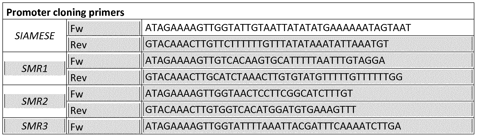
  • SIM/SMR promoter sequences were amplified from genomic DNA by PCR using the primers described in Table 5.
  • the product fragments were created with the Pfu DNA Polymerase Kit (Promega, Catalog #M7745), and were cloned into a pDONR P4-P1 r entry vector by BP recombination cloning and subsequently transferred into the pMK7S * NFm14GW,0 destination vector by LR cloning, resulting in a transcriptional fusion between the promoter of the SMR genes and the nlsGFP-GUS fusion gene (Karimi et al., 2007).
  • the SMR coding regions were amplified using primers described in Table 5, and cloned into the pDONR221 vector by BP recombination cloning and subsequently transferred into the pK2GW7 destination vector (Kamimi et al., 2002) by LR cloning. All constructs were transferred into the Agrobacterium tumefaciens C58C1 RifR strain harboring the pMP90 plasmid. The obtained Agrobacterium strains were used to generate stably transformed Arabidopsis lines with the floral dip transformation method (Clough and Bent, 1998). Transgenic plants were obtained on kanamycin-containing medium and later transferred to soil for optimal seed production. All cloning primers are listed in Table 5.
  • leaves were harvested at 21 days after sowing on control medium, medium supplemented with 1 mM hydroxyurea or 0.3 ⁇ g mL bleomycin. Leaves were cleared overnight in ethanol, stored in lactic acid for microscopy, and observed with a microscopy fitted with DIC optics (Leica). The total (blade) area was determined from images digitized directly with a digital camera (Olympus BX51 microscope) mounted on a binocular (Stemi SV1 1 ; Zeiss).
  • Plant material was incubated for 2 min in a 10 ⁇ PI solution to stain the cell walls and was visualized with a HeNe laser through excitation at 543 nm.
  • GFP fluorescence was detected with the 488-nm line of an Argon laser.
  • GFP and PI were detected simultaneously by combining the settings indicated above in the sequential scanning facility of the microscope. Acquired images were quantitatively analyzed with the ImageJ v1.45s software (http://rsbweb.nih.gov/ii/) and Cell-o- Tape plug-ins (French et al., 2012). Chlorophyll a fluorescence parameters were measured using the IMAGING PAM M-Series Chlorofyll Fluorescence (Walz) and associated software.
  • root tip tissues were chopped with a razor blade in 300 ⁇ of 45 mM MgCI 2 , 30 mM sodium citrate, 20 mM MOPS, pH 7 (Galbraith et al., 1991 ).
  • DAPI 4,6-diamidino-2-phenylindole
  • Leaf material was chopped in 200 ⁇ of Cystain UV Precise P Nuclei extraction buffer (Partec), supplemented with 800 ⁇ of staining buffer. The mix was filtered through a 50- ⁇ green filter and read by the Cyflow MB flow cytometer (Partec). The nuclei were analyzed with the Cyflogic software.
  • Catalase Assay Plants were germinated on either control medium, medium with 1 mM HU or 6 ⁇ 3-AT.
  • Leaf tissue of 10 plants was ground in 200 ⁇ extraction buffer (60 mM Tris (pH 6.9), 1 mM phenylmethylsulfonylfluoride, 10 mM DTT) on ice.
  • the homogenate was centrifuged at 13,000 g for 15 min at 4°C.
  • a total of 45 ⁇ g protein extract was mixed with potassium phosphate buffer (50 mM, pH 7.0) (Vandenabeele et al., 2004).
  • Seeds were plated on sterilized membranes and grown under a 16-h/8-h light/dark regime at 21 °C. After 2 days of germination and 5 days of growth, the membrane was transferred to MS medium containing 0.3 ⁇ g/mL bleomycin for 24 h. Triplicate batches of root meristem material seedlings were harvested for total RNA preparation using the RNeasy plant mini kit (Qiagen). Each of the different root tip RNA extracts were hybridized to 12 Affymetrix® Arabidopsis Gene 1 .0 ST Arrays according to manufacturer's instructions at the Nucleomics Core Facility (Leuven, Belgium; http://www.nucleomics.be).
  • Raw data were processed with the RMA algorithm (Irizarry et al., 2003) using the Affymetrix Power Tools and subsequently subjected to a Significance Analysis of Microarray (SAM) analysis with "MultiExperiment Viewer 4" (MeV4) of The Institute for Genome Research (TIGR) (Tusher et al., 2001 ).
  • SAM Significance Analysis of Microarray
  • MeV4 MultiExperiment Viewer 4"
  • TIGR Institute for Genome Research
  • Example 2 The SMR Gene Family Comprises 14 Family Members that Respond to Different Stresses
  • transcriptional reporter lines containing the putative upstream promoter sequences were constructed for all. After selection of representative reporter lines, one-week-old seedlings were transferred to control medium, or medium supplemented with HU (resulting into stalled replication forks) or bleomycin (causing DSBs). Focusing on the root tips revealed distinct expression patterns ( Figure 3; Figure 4), with some family members being restricted to the root elongation zone (including SIM and SMR1), while others were confined to vascular tissue (e.g. SMR2 and SMR8), or columella cells (e.g. SMR5).
  • vascular tissue e.g. SMR2 and SMR8
  • columella cells e.g. SMR5
  • SMR4-, SMR5- and SA/R7-overexpressing (SMR4 OE , SMRtP E and SMR7° E ) plants were generated.
  • SMR4 OE , SMRtP E and SMR7° E were generated.
  • SMR5 and SMR7 expression levels were analyzed in plants that are knockout for CAT2 and/or APX1, encoding two enzymes important for the scavenging of H 2 0 2 .
  • SMR5 expression levels were clearly induced in the apxl cat2 double mutant, whereas SMR7 transcriptional activation was observed in the apxl knockout and apxl cat2 double mutant ( Figure 12A).
  • plants grown for two days under high light conditions displayed PSMR5:GUS and SMR7:GUS induction in proliferating leaves (Figure 12B).
  • the ZmSMRg gene and the ZmSMRh gene are knocked out using the CRISPR-Cas technology, generating single and double knock out mutants. These knock out mutants are submitted to oxidative stress as described for Arabidopsis, and the mutants show a significant protection against oxidative stress, when compared to the wild type grown under the same conditions.
  • Table 1 Overview of the transcriptionally induced core DNA damage genes
  • Table 5 List of primers used for cloning, genotyping, and RT-PCR
  • Adachi S., Minamisawa, K., Okushima, Y., Inagaki, S., Yoshiyama, K., Kondou, Y., Kaminuma, E., Kawashima, M., Toyoda, T., Matsui, M., Kurihara, D., Matsunaga, S., and Umeda, M. (2011). Programmed induction of endoreduplication by DNA double- strand breaks in Arabidopsis. Proc. Natl. Acad. Sci. USA 108: 10004-10009.
  • CDKB1 ;1 forms a functional complex with CYCA2;3 to suppress endocycle onset. Plant Physiol. 150: 1482-1493.
  • SIAMESE a plant-specific cell cycle regulator, controls endoreplication onset in Arabidopsis thaliana. Plant Cell 18: 3145-3157.
  • Floral dip a simplified method for Agrobacterium- mediated transformation of Arabidopsis thaliana. Plant J. 16: 735-743.
  • the Arabidopsis CkslAt protein binds the cyclin-dependent kinases Cdc2aAt and Cdc2bAt.
  • SCI 1 the first member of the tissue-specific inhibitors of CDK (TIC) class, is probably connected to the auxin signaling pathway. Plant Signal. Behav. 7: 53-58.
  • AtATM is essential for meiosis and the somatic response to DNA damage in plants. Plant Cell 15: 1 19-132.
  • Oxidative stress produced by xanthine oxidase induces apoptosis in human extravillous trophoblast cells. J. Reprod. Dev. 59: 7-13.
  • Vandenabeele S., Vand Vogelwera, S., Vuylsteke, M., Rombauts, S., Langebartels, C, Seidlitz, H.K., Zabeau, M., Van Montagu, M., Inze, D., and Van Breusegem, F. (2004).
  • ROOT MERISTEMLESS 1/CADMIUM SENSITIVE2 gene defines a glutathione-dependent pathway involved in initiation and maintenance of cell division during postembryonic root development. Plant Cell 12: 97-109.

Landscapes

  • Health & Medical Sciences (AREA)
  • Genetics & Genomics (AREA)
  • Life Sciences & Earth Sciences (AREA)
  • Engineering & Computer Science (AREA)
  • Zoology (AREA)
  • Wood Science & Technology (AREA)
  • Organic Chemistry (AREA)
  • Biomedical Technology (AREA)
  • Chemical & Material Sciences (AREA)
  • Biotechnology (AREA)
  • General Engineering & Computer Science (AREA)
  • Molecular Biology (AREA)
  • Bioinformatics & Cheminformatics (AREA)
  • Microbiology (AREA)
  • Physics & Mathematics (AREA)
  • Plant Pathology (AREA)
  • Biophysics (AREA)
  • Biochemistry (AREA)
  • General Health & Medical Sciences (AREA)
  • Cell Biology (AREA)
  • Virology (AREA)
  • Breeding Of Plants And Reproduction By Means Of Culturing (AREA)
  • Micro-Organisms Or Cultivation Processes Thereof (AREA)

Abstract

The present invention relates to the use SMR5, possibly in combination with SMR4 and/or SMR7 to modulate ROS and oxidative stress response in plants. More specifically, it relates to a SMR5 knock out or knock down to improve the oxidative stress tolerance in plants.

Description

Protection of plants against oxidative stress
The present invention relates to the use of SMR5, possibly in combination with SMR4 and/or SMR7 to modulate ROS and oxidative stress response in plants. More specifically, it relates to a SMR5 knock out or knock down to improve the oxidative stress tolerance in plants.
Being immobile, plants are continuously exposed to changing environmental conditions that can impose biotic and abiotic stresses. One of the consequences observed in plants subjected to altered growth conditions is the disruption of the reactive oxygen species (ROS) homeostasis (Mittler et al., 2004). Under steady-state conditions, ROS are efficiently scavenged by different non-enzymatic and enzymatic antioxidant systems, involving the activity of catalases, peroxidases, and glutathione reductases. However, when stress prevails, the ROS production rate can exceed the scavenging mechanisms, resulting into a cell- or tissue-specific rise in ROS. These oxygen derivatives possess a strong oxidizing potential that can damage a wide diversity of biological molecules, including the electron-rich bases of DNA, which results into single- and double-stranded breaks (Amor et al., 1998; Dizdaroglu et al., 2002; Roldan-Arjona and Ariza, 2009). Hydrogen peroxide (H202) is a major ROS compound and is able to transverse cellular membranes, migrating into different compartments. This feature grants H202 not only the potential to damage a variety of cellular structures, but also to serve as a signaling molecule, allowing the activation of pathways that modulate developmental, metabolic and defence pathways (Mittler et al., 201 1 ). One of the signaling effects of H202 is the activation of a cell division arrest by cell cycle checkpoint activation (Tsukagoshi, 2012), however the molecular mechanisms involved remain unknown.
Cell cycle checkpoints adjust cellular proliferation to changing growth conditions, arresting it by the inhibition of the main cell cycle controllers: the heterodimeric complexes between the cyclin-dependent kinases (CDK) and the regulatory cyclins (Lee and Nurse, 1987; Norbury and Nurse, 1992). The activators of these checkpoints are the highly conserved ATAXIA TELANGIECTASIA MUTATED (ATM) and ATM AND RAD3-RELATED (ATR) kinases that are recruited in accordance with the type of DNA damage (Zhou and Elledge, 2000; Abraham, 2001 ; Bartek and Lukas, 2001 ; Kurz and Lees-Miller, 2004). ATM is activated by double- stranded breaks (DSBs); whereas ATR is activated by single-strand breaks or stalled replication forks, causing inhibition of DNA replication. In mammals, ATM and ATR activation result in the phosphorylation of the Chk2 and Chk1 kinases, respectively. In mammals, both kinases subsequently phosphorylate p53, a critical transcription factor responsible to conduct DNA damage responses (Chaturvedi et al., 1999; Shieh et al., 2000; Chen and Sanchez, 2004; Rozan and El-Deiry, 2007). p53 seemingly appears to have no plant ortholog, although an analogous role for p53 is suggested for the plant-specific SUPPRESSOR OF GAMMA RESPONSE 1 (SOG1 ) transcription factor that is under direct posttranscriptional control of ATM (Yoshiyama et al., 2009; Yoshiyama et al., 2013). Another distinct feature relates to the inactivation of CDKs in response to DNA stress. CDK activity is in part controlled by its phosphorylation status at the N-terminus, determined by the interplay of the CDC25 phosphatase and the antagonistic WEE1 kinase, acting as the "on" and "off" switches of CDK activity, respectively (Francis, 201 1 ). Whereas in mammals and budding yeast the activation of the DNA replication checkpoint, leading to a cell cycle arrest, is predominantly achieved by the inactivation of the CDC25 phosphatase, plant cells respond to replication stress by transcriptional induction of WEE1 (De Schutter et al., 2007). In absence of WEE1 , Arabidopsis thaliana plants become hypersensitive to replication inhibitory drugs such as hydroxyurea (HU), which causes a depletion of dNTPs because of an inhibition of the ribonucleotide reductase (RNR) protein. However, WEE/-deficient plants respond similarly to control plants exposed to other types of DNA damage (De Schutter et al., 2007; Dissmeyer et al., 2009); other, yet to be identified pathways controlling cell cycle progression under DNA stress, operating independently of WEE1 may exist.
There are several potential candidates to operate in checkpoint activation upon DNA stress mainly belonging to the family of CDK inhibitors (CKIs). CKI proteins are mostly low molecular weight proteins that inhibit cell division by their direct interaction with the CDK and/or cyclin subunit (Sherr and Roberts, 1995; De Clercq and Inze, 2006). The first identified class of plant CKIs was the ICK KRP (interactors of CDK Kip-related protein) protein family comprising seven members in A. thaliana, all sharing a conserved C-terminal domain being similar to the CDK-binding domain of the animal CIP/KIP proteins (Wang et al., 1998; Wang et al., 2000; De Veylder et al., 2001 ). The TIC (tissue-specific inhibitors of CDK) is the most recently suggested class of CKIs (DePaoli et al., 2012) and encompasses SCI 1 in tobacco, the only tissue-specific CKI reported so far (DePaoli et al., 201 1 ). SCI 1 shares no outstanding sequence similarity with the other classes of CKIs in plants, and has been suggested to connect cell cycle progression and auxin signaling in pistils (DePaoli et al., 2012). The third class of CKIs is the plant-specific SIAMESE/SIAMESE-RELATED {SIM/SMR) gene family. SIM has been identified as a cell cycle inhibitor with a role in trichome development and endocycle control (Churchman et al., 2006). Based on sequence analysis, five additional gene family members have been identified in A. thaliana, and together with EL2 from rice, been suggested to act as cell cycle inhibitors modulated either by biotic and abiotic stresses (Peres et al., 2007). Plants subjected to treatments inducing DSBs showed a rapid and strong induction of specific family members (Culligan et al., 2006; Adachi et al., 201 1 ). Surprisingly we found three SMR genes (SMR4, SRM5 and SMR7) that are transcriptionally activated by DNA damage. Even more surprisingly the SMR5 gene encodes for a novel protein, not described earlier. Cell cycle inhibitory activity was demonstrated by overexpression analysis, whereas knockout data illustrated that both SMR5 and SMR7 are essential for DNA cell cycle checkpoint activation in leaves of plants grown in the presence of HU. Remarkably, we found that SMR induction mainly depends on ATM and SOG1 , rather than ATR as would be expected for a drug that triggers replication fork defects. Correspondingly, we demonstrate that the HU-dependent activation of SMR genes is triggered by ROS rather than replication problems, linking SMR genes with cell cycle checkpoint activation upon the occurrence of DNA damage-inducing oxidative stress.
A first aspect of the invention is the use of SMR5, or a homologue, orthologue or paralogue thereof to modulate ROS signalling and/or oxidative stress response in plants. In a preferred embodiment, said use is combined with the use of SMR4 and/or SMR7. The use of an SMR, as used here, comprises the use of the gene, and/or the use of the protein encoded by said gene. Preferably, said use of SMR5 is the use of a gene encoding a protein comprising, preferably consisting of a protein selected from the group consisting of SEQ ID No.2, SEQ ID No. 4 and SEQ ID No. 6. In one preferred embodiment, said use of SMR5 is the use of a gene encoding a protein comprising, preferably consisting of SEQ ID N°2. In another preferred embodiment, said use of SMR5 is the use of a gene encoding a protein comprising, preferably consisting a of a sequence selected from the group consisting of SEQ ID N°4 and SEQ ID No. 6. "Homologues" of a protein encompass peptides, oligopeptides, polypeptides, proteins and enzymes having amino acid substitutions, deletions and/or insertions relative to the unmodified protein in question and having similar biological and functional activity as the unmodified protein from which they are derived. Orthologues and paralogues encompass evolutionary concepts used to describe the ancestral relationships of genes. Paralogues are genes within the same species that have originated through duplication of an ancestral gene; orthologues are genes from different organisms that have originated through speciation, and are also derived from a common ancestral gene.
Preferably, said use is a downregulation of the expression of the protein, and/or the inactivation of the protein. Preferably, said downregulation is used to improve oxidative stress tolerance in plants. "Improve" as used here, means that the plants wherein said SMR is downregulated have a significantly better oxidative stress resistance than the plants with the same genetic background, except for the modifications needed for the downregulation, grown under the same conditions. Methods for downregulation are known to the person skilled in the art, and include, but are not limited to mutations, insertions or deletions in the gene and/or its promoter, the use of anti-sense RNA or RNAi and gene silencing methods._Methods to induce site specific mutations in plants are known to the person skilled in the art and include Zinc- finger nucleases, transcription activator-like nucleases (TALENs) and the clustered regularly interspaced short palindromic repeat (CRISPR)/Cas-based RNA guided DNA endonucleases (Gaj et al., 2013). Inactivation of the protein can be obtained, as a non-limiting example, by the use of antigen binding proteins directed against the protein, or by protein aggregation, as described in WO2012123419. The downregulation of SMR5 can be measured by measuring the activity of its substrate (Cyclin dependent kinase A, CDKA) as described in De Veylder et al. (1997); a higher CDKA activity points to a downregulation of SMR5. A plant as used here may be any plant. Plants include gymnosperms and angiosperms, monocotyledons and dicotyledons, trees, fruit trees, field and vegetable crops and ornamental species. Preferably said plant is a crop plant, including but not limited to soybean, corn, wheat, barley and rice.
Another aspect of the invention is a genetically modified plant, comprising an inactivated SMR5 gene and/or protein. Inactivated, as used here, means that the activity of the inactivated form is significantly lower than that of the active form. Significantly, as used here, means that the activity of the mutant gene or protein is at least 20% lower, preferably at least 50% lower, more preferably at least 75% lower, most preferably at least 90% lower than the wild type gene or protein. Preferably, the activity of the gene is measured as the amount of messenger RNA. Preferably, the activity of the protein is measured as inhibition of cell division. In one preferred embodiment, the active form of the gene is encoding a protein comprising, preferably consisting of SEQ ID N°2. In another preferred embodiment, said use of SMR5 is the use of a gene encoding a protein comprising, preferably consisting a of a sequence selected from the group consisting of SEQ ID N°4 and SEQ ID No. 6. In a preferred embodiment, said plan is a maize plant in which ZmSMRg and/or ZmSMRh are inactivated, preferably as a CRISPR Cas knock out.
In one preferred embodiment, the gene encoding the SMR5p is disrupted. In another preferred embodiment, the gene encoding the SMR5p is silenced. In still another embodiment, the SMR5p itself is inactivated by protein aggregation. Preferably, said genetically modified plant further comprises an inactivated SMR4 gene and/or protein, and or an inactivated SMR7 gene and or protein.
Still another aspect of the invention is a method to increase oxidative stress resistance in a plant, comprising the downregulation of SMR5p expression and/or activity. Preferably, said downregulation is combined with the downregulation of SMR4p expression and/or activity, and/or downregulation of SMR7p expression and/or activity.
In one preferred embodiment, the method comprises a step wherein the plan is transformed with an RNAi construct against one or more of the SMR genes. In one preferred embodiment, said RNAi construct is placed under control of a constitutive promoter. In another preferred embodiment, said RNAi construct is placed under control of an oxidative stress inducible promoter.
BRIEF DESCRIPTION OF THE FIGURES Figure 1. DNA stress meta-analysis.
Venn diagram showing the overlap between transcripts induced by hydroxyurea (HU), bleomycin (Bm), and γ-radiation (γ-rays). In total, 61 genes were positively regulated in at least two DNA stress experiments, and 22 genes accumulated in all DNA stress experiments.
Figure 2. Hierarchical average linkage clustering of SIM/SMR genes induced in response to different abiotic (A) and biotic stresses (B).
Data comprise the SIM/SMR represented in publicly available Affymetrix ATH1 microarrays obtained with the Genevestigator toolbox. Blue and yellow indicate down- and up-regulation, respectively, whereas black indicates no change in expression.
Figure 3. SIM/SMR induction in response to HU.
One-week-old transgenic Arabidopsis seedlings were transferred to control (-HU) medium or medium supplemented with 1 mM HU (+HU). GUS assays were performed 24 h after transfer.
Figure 4. SIM/SMR induction in response to Bleomycine.
One-week-old transgenic Arabidopsis seedlings were transferred to control (-Bm) medium or medium supplemented with 0.3 μg mL bleomycin (+Bm). GUS assays were performed after 24 h after transfer.
Figure 5. Transcriptional induction of SIM/SMR genes upon HU and bleomycin treatment. One-week-old wild type Arabidopsis seedlings were transferred to control medium (blue), or medium supplemented with 1 mM hydroxyurea (red) or 0.3 μg mL bleomycin (green). Root tips were harvested after 24 h for RT-PCR analysis. Expression levels in control condition were arbitrary set to one. Data represent mean ± SE (n = 3). Figure 6. Transcriptional induction of SIM/SMR genes upon γ-irradiation.
(A-F) PSMR4:GUS (A and D), PSMR5:GUS (B and E) and PSMR7:GUS (C and D) either control-treated (A-C) or irradiated with 20 Gy of γ-rays (D-F). GUS assays were performed 1.5 h after irradiation. Figure 7. Ectopic SMR4, SMR5 and SMR7 expression inhibits cell division.
(A-D) Four-week-old rosettes of control (A), SMR4OE (B), SMR5OE (C) and SMR7OE (D) plants. (E-H) Leaf abaxial epidermal cell images of in v/'iro-grown 3-week-old control (E), SMR4OE (F), SMR5OE (G) and SMR7°E (H) plants. (I-L) Ploidy level distribution of the first leaves of 3-week- old in v/'iro-grown control (I), SMR4OE (J), SMR5OE (K) and SMR7°E (L) plants. Figure 8. Graphical representation of the SMR5 and SMR7 T-DNA insertion. (A), Intron-exon organization of the Arabidopsis SMR5 and SMR7 genes. Black and white boxes represent coding and non-coding regions, respectively, while lines represent introns. The white triangles indicate the T-DNA insertion sites. (B), qRT-PCR analysis on wild-type, SMR5KO, SMR7KO, and SMR5KO SMR7KO seedlings using primers specific to either SMR5 or SMR7. Expression levels in wild type were arbitrary set to one. Data represent mean ± SE (n = 3).
Figure 9. SMR5 and SMR7 are required for an HU-dependent cell cycle checkpoint. (A-B) Leaf size (A) and abaxial epidermal cell number (B) of the first leaves of 3-week-old plants grown on control medium (circles) or medium supplemented with 1 mM HU (squares). Data represent mean with 95% confidence interval (n = 10). Figure 10. SMR5 and SMR7 expression is ATM- and SOG1 -dependent. (A-B) PSMR5:GUS (A) and PSMR7:GUS (B) reporter constructs introgressed into atr-2, atm-1 and sog-1 mutant backgrounds were control-treated (Ctrl), or treated with HU or bleomycin (Bm) for 24 h.
Figure 11. HU triggers oxidative stress.
(A) H202 scavenging of control, HU- and 3-AT (positive control) treated plants. Error bars show SEM (n = 3-4). (B) Maximum quantum efficiency of PSII (F'v/F'm) of seedlings grown under low (LL) and high light (HL), in absence (-HU) and presence (+HU) of HU. (C) Light microscope pictures of plants shown in (B).
Figure 12. SMR5 and SMR7 are induced by oxidative stress-inducing stimuli. (A-B) Relative SMR5 (A) and SMR7 (B) expression levels in wild-type (Col-0), apxl, cat2 and apx cat2 mutant plants. Expression levels in wild type were arbitrary set to one. Data represent mean ± SE (n = 3). (C) One-week-old PSMR5:GUS and PSMR7:GUS seedlings grown under low- versus high-light conditions. (D) Abaxial epidermal cell number of the first leaves of 3-week-old plants transferred at the age of 8 days for 48 h to control (circles) or high light (squares) conditions. Data represent mean with 95% confidence interval (n > 8).
Figure 13. Cluster analysis of the maize SMR family with the Arabidopsis SMR5
EXAMPLES
Materials and methods to the examples
Plant Materials and Growth Conditions
The smr5 (SALK_100918) and smr7 (SALK_128496) alleles were acquired from the Arabidopsis Biological Research Center. Homozygous insertion alleles were checked by genotyping PCR using the primers listed in Table 3. The atm-1, atr-2 and sog1-1 mutants have been described previously (Garcia et al., 2003; Preuss and Britt, 2003; Culligan et al., 2004; Yoshiyama et al., 2009). Unless stated otherwise, plants of Arabidopsis thaliana (L.) Heyhn. (ecotype Columbia) were grown under long-day conditions (16 h of light, 8 h of darkness) at 22°C on half-strength Murashige and Skoog (MS) germination medium (Murashige and Skoog, 1962). Arabidopsis plants were treated with HU as described by Cools et al. (201 1 ). For bleomycin treatments, five-day-old seedlings were transferred into liquid MS medium supplemented with 0.3 μg mL bleomycin. For γ-irradiation treatments, five-day-old in vitro- grown plantlets were irradiated with γ-rays at a dose of 20 Gy. For light treatments, one-week- old seedlings were transferred to continuous high-light conditions (growth rooms kept at 22°C with 24-h day/0-h night cycles and a light intensity of 300-400 μηηοΙ m"2 s"1) for 2 days, and subsequently retransferred to low-light conditions. The first leaf pair was harvested and incubated in 100% ethanol for epidermis cell drawing as described by De Veylder et al. (2001 ).
DNA and RNA Manipulation Genomic DNA was extracted from Arabidopsis leaves with the DNeasy Plant Kit (Qiagen) and RNA was extracted from Arabidopsis tissues with the RNeasy Mini Kit (Qiagen). After DNase treatment with the RQ1 RNase-Free DNase (Promega), cDNA was synthesized with the iScript cDNA Synthesis Kit (Bio-Rad). A quantitative RT-PCR was performed with the SYBR Green kit (ROCHE) with 100 nM primers and 0.125 μΙ_ of RT reaction product in a total of 5 μΙ_ per reaction. Reactions were run and analyzed on the LightCycler 480 (Roche) according to the manufacturer's instructions with the use of the following reference genes for normalization: ACTIN2 (At3g46520), EMB2386 (At1 g02780), PAC1 (At3g221 10) and RPS26C (At3g56340). Primers used for the RT-PCR are given in Table 5.
SIM/SMR promoter sequences were amplified from genomic DNA by PCR using the primers described in Table 5. The product fragments were created with the Pfu DNA Polymerase Kit (Promega, Catalog #M7745), and were cloned into a pDONR P4-P1 r entry vector by BP recombination cloning and subsequently transferred into the pMK7S*NFm14GW,0 destination vector by LR cloning, resulting in a transcriptional fusion between the promoter of the SMR genes and the nlsGFP-GUS fusion gene (Karimi et al., 2007). For the overexpression constructs, the SMR coding regions were amplified using primers described in Table 5, and cloned into the pDONR221 vector by BP recombination cloning and subsequently transferred into the pK2GW7 destination vector (Kamimi et al., 2002) by LR cloning. All constructs were transferred into the Agrobacterium tumefaciens C58C1 RifR strain harboring the pMP90 plasmid. The obtained Agrobacterium strains were used to generate stably transformed Arabidopsis lines with the floral dip transformation method (Clough and Bent, 1998). Transgenic plants were obtained on kanamycin-containing medium and later transferred to soil for optimal seed production. All cloning primers are listed in Table 5.
GUS Assays
Complete seedlings or tissue cuttings were stained in multiwell plates (Falcon 3043; Becton Dickinson). GUS assays were performed as described by Beeckman and Engler (1994). Samples mounted in lactic acid were observed and photographed with a stereomicroscope (Olympus BX51 microscope) or with a differential interference contrast (DIC) microscope (Leica).
Microscopy
For leaf measurements, first leaves were harvested at 21 days after sowing on control medium, medium supplemented with 1 mM hydroxyurea or 0.3 μg mL bleomycin. Leaves were cleared overnight in ethanol, stored in lactic acid for microscopy, and observed with a microscopy fitted with DIC optics (Leica). The total (blade) area was determined from images digitized directly with a digital camera (Olympus BX51 microscope) mounted on a binocular (Stemi SV1 1 ; Zeiss). From scanned drawing-tube images of the outlines of at least 30 cells of the abaxial epidermis located between 25% to 75% of the distance between the tip and the base of the leaf, halfway between the midrib and the leaf margin, the following parameters were determined: total area of all cells in the drawing and total numbers of pavement and guard cells, from which the average cell area was calculated. The total number of cells per leaf was estimated by dividing the leaf area by the average cell area. For confocal microscopy, root meristems were analyzed 2 days after transfer using a Zeiss LSM 510 Laser Scanning Microscope and the LSM Browser version 4.2 software (Zeiss). Plant material was incubated for 2 min in a 10 μηη PI solution to stain the cell walls and was visualized with a HeNe laser through excitation at 543 nm. GFP fluorescence was detected with the 488-nm line of an Argon laser. GFP and PI were detected simultaneously by combining the settings indicated above in the sequential scanning facility of the microscope. Acquired images were quantitatively analyzed with the ImageJ v1.45s software (http://rsbweb.nih.gov/ii/) and Cell-o- Tape plug-ins (French et al., 2012). Chlorophyll a fluorescence parameters were measured using the IMAGING PAM M-Series Chlorofyll Fluorescence (Walz) and associated software.
Flow Cytometry Analysis
For flow cytometric analysis, root tip tissues were chopped with a razor blade in 300 μί of 45 mM MgCI2, 30 mM sodium citrate, 20 mM MOPS, pH 7 (Galbraith et al., 1991 ). One microliter of 4,6-diamidino-2-phenylindole (DAPI) from a stock of 1 mg/mL was added to the filtered supernatant. Leaf material was chopped in 200 μί of Cystain UV Precise P Nuclei extraction buffer (Partec), supplemented with 800 μί of staining buffer. The mix was filtered through a 50-μηι green filter and read by the Cyflow MB flow cytometer (Partec). The nuclei were analyzed with the Cyflogic software.
Catalase Assay Plants were germinated on either control medium, medium with 1 mM HU or 6 μΜ 3-AT. Leaf tissue of 10 plants was ground in 200 μί extraction buffer (60 mM Tris (pH 6.9), 1 mM phenylmethylsulfonylfluoride, 10 mM DTT) on ice. The homogenate was centrifuged at 13,000 g for 15 min at 4°C. A total of 45 μg protein extract was mixed with potassium phosphate buffer (50 mM, pH 7.0) (Vandenabeele et al., 2004). After addition of 1 1.4 μί H202 (7.5%), the absorbance of the sample at 240 nm after 0 and 60 s was measured to determine catalase activity by H202 breakdown (Beers and Sizer, 1952; Vandenabeele et al., 2004).
Microarray Analysis
Seeds were plated on sterilized membranes and grown under a 16-h/8-h light/dark regime at 21 °C. After 2 days of germination and 5 days of growth, the membrane was transferred to MS medium containing 0.3 μg/mL bleomycin for 24 h. Triplicate batches of root meristem material seedlings were harvested for total RNA preparation using the RNeasy plant mini kit (Qiagen). Each of the different root tip RNA extracts were hybridized to 12 Affymetrix® Arabidopsis Gene 1 .0 ST Arrays according to manufacturer's instructions at the Nucleomics Core Facility (Leuven, Belgium; http://www.nucleomics.be). Raw data were processed with the RMA algorithm (Irizarry et al., 2003) using the Affymetrix Power Tools and subsequently subjected to a Significance Analysis of Microarray (SAM) analysis with "MultiExperiment Viewer 4" (MeV4) of The Institute for Genome Research (TIGR) (Tusher et al., 2001 ). The imputation engine was set as 10-nearest neighbor imputer and the number of permutations was 100. Expression values were obtained by log2-transforming the average value of the normalized signal intensities of the triplicate samples. Fold changes were obtained using the expression values of the treatment relative to the control samples. Genes with Q-values < 0.1 and fold change > 1.5 or < 0.666 were retained for further analysis.
Microarray Meta-Analysis
Transcripts induced by bleomycin (Q-value < 0.1 and fold change > 1 .5) were compared with different published DNA stress-related data sets. For γ-irradiation, an intersect of the genes with a significant induction (P-value < 0.05, Q-value < 0.1 , and fold change >1.5) in 5-day-old wild-type seedlings 1 .5 h post-irradiation (100 Gy) was made of two independent experiments (Culligan et al., 2006; Yoshiyama et al., 2009). For replication stress, genes showing a significant induction (P-value (Time) < 0.05, Q-value (Time) < 0.1 and fold change >1.5) in 5- day-old wild-type root tips after 24 h of 2-mM hydroxyurea treatment were selected (Cools et al., 201 1 ). Meta-analysis of the SMR genes during various stress conditions and treatments were obtained using Genevestigator (Hruz et al., 2008). Using the "Response Viewer" tool, the expression profiles of genes following different stimuli were analyzed. Only biotic and abiotic stress treatments with a more than 2-fold change in the transcription level (P-value < 0.01 ) for at least one of the SMR genes were taken into account. Fold-change values were hierarchically clustered for genes and experiments by average linkage in MeV from TIGR.
Accession Numbers
Microarray results have been submitted to MiamExpress (www.ebi.ac.uk/miamexpress), with accession E-MEXP-3977. Sequence data from this article can be found in the Arabidopsis Genome Initiative or GenBank/EMBL databases under the following accession numbers: SMR4 (At5g02220); SMR5 (At1 g07500); SMR7 (At3g27630); ATM (At3g48490); ATR (At5g40820); SOG1 (At1 g25580). Example 1 : Meta-Analysis of DNA Stress Datasets Identifies DNA Damage-Induced SMR Genes
When DNA damage occurs, two global cellular responses are essential for cell survival: activation of the DNA repair machinery, and delay or arrest of cell cycle progression. In recent years, gene expression inventories have been collected that focus on the transcriptional changes in response to different types of DNA stress (Culligan et al., 2006; Ricaud et al., 2007; Yoshiyama et al., 2009; Cools et al., 2010). To identify novel key signaling components that contribute to cell cycle checkpoint activation, we compared bleomycin-induced genes to those induced by HU treatment (Cools et al., 2010) and γ-radiation (Culligan et al., 2006; Yoshiyama et al., 2009). Twenty-two genes were upregulated in all DNA stress experiments and can be considered as transcriptional hallmarks of the DNA damage response (DDR), regardless of the type of DNA stress (Figure 1 ; Table 1 ). Within this selection, genes known to be involved in DNA stress and DNA repair are predominantly present, including PARP2, BRCA1 and RAD51. In addition, we recognized one member of the SIM/SMR gene family, being SMR5 (At1 g07500). When expanding the selection by considering genes induced in at least two of the three DNA stress experiments, we identified a total of 61 genes (Table 2). Besides DDR- related genes, this expanded dataset included an additional SMR family member (SMR4; At5g02220), being expressed upon treatment with HU or γ-radiation.
Example 2: The SMR Gene Family Comprises 14 Family Members that Respond to Different Stresses
Previously, we reported on the existence of one SIM and five SMR genes (SMR1-SMR5) in the A. thaliana genome (Peres et al., 2007), whereas protein purification of CDK/cyclin complexes resulted into the identification of two additional family members (SMR6 and SMR8) (Van Leene et al., 2010). With the availability of new sequenced plant genomes, we re-examined the Arabidopsis genome using iterative BLAST searches for the presence of additional SMR genes, resulting in the identification of six non-annotated family members, nominated SMR7 to SMR13 (Table 3). With the Genevestigator toolbox (Hruz et al., 2008), the expression pattern of the twelve SIM/SMR genes represented on the Affymetrix ATH1 microarray platform was analyzed in response to different biotic and abiotic stress treatments. Distinct family members were induced under various stress conditions, albeit with different specificity (Figure 2). Every SMR gene appeared to be transcriptionally active under at least a number of stress conditions, with SMR5 responding to most diverse types of abiotic stresses. In response to DNA stress (genotoxic stress and UV-B treatment), two SMR genes responded strongly, being SMR4 and SMR5, corresponding with their presence among the DNA stress genes identified by our microarray meta-analysis. To confirm involvement of SIM/SMR genes in the genotoxic stress response, transcriptional reporter lines containing the putative upstream promoter sequences were constructed for all. After selection of representative reporter lines, one-week-old seedlings were transferred to control medium, or medium supplemented with HU (resulting into stalled replication forks) or bleomycin (causing DSBs). Focusing on the root tips revealed distinct expression patterns (Figure 3; Figure 4), with some family members being restricted to the root elongation zone (including SIM and SMR1), while others were confined to vascular tissue (e.g. SMR2 and SMR8), or columella cells (e.g. SMR5). When plants were exposed to HU, three SMR genes showed strong transcriptional induction in the root meristem, being SMR4, SMR5 and SMR7, with the latter two displaying the strongest response (Figure 3). In the presence of bleomycin, an additional weak cell-specific induction of SMR6 was observed (Figure 4). Transcriptional induction of SMR4, SMR5 and SMR7 by HU and bleomycin was confirmed by qRT-PCR experiments (Figure 5). These data fit the above described microarray analysis, with the lack of SMR7 (At3g27630) being explained by its absence on the ATH1 microarray of the HU and γ-irradiation experiments, although being induced 5.68-fold in the bleomycin experiment performed using the Aragene array. Next to HU and bleomycin, we confirmed transcriptional activation of SMR4, SMR5 and SMR7 by γ-irradiation (Figure 6).
Example 3: DNA Stress-Induced SMR Genes Encode Potent Cell Cycle Inhibitors
Previously, SIM had been proven to encode a potent cell cycle inhibitor, since its ectopic expression results into dwarf plants holding less cells compared to control plants (Churchman et al., 2006). To test whether the DNA stress-induced SMR genes encode proteins with cell division inhibitory activity, SMR4-, SMR5- and SA/R7-overexpressing (SMR4OE, SMRtPE and SMR7°E) plants were generated. For each gene, multiple lines with strong transcript levels were isolated, all showing a reduction in rosette size compared to wild-type plants (Figures 7A to 7D). This decrease in leaf size correlated with an increase in cell size (Figures 7E to H), indicative of a strong inhibition of cell division. Similar to SIM (Churchman et al., 2006), ectopic expression did not only inhibit cell division but also triggered an increase in the DNA content by stimulation of endoreplication (Figures 7I to L; Table 4), likely representing a premature onset of cell differentiation. Together with the previously described biochemical interaction between SMR4 and SMR5, and CDKA;1 and D-type cyclins (Van Leene et al., 2010), it can be concluded that the DNA stress-induced SMR genes encode potent cell cycle inhibitors.
Example 4: SMR5 and SMR7 Control a HU-Dependent Checkpoint in Leaves
To address the role of the different SMR genes in DNA stress checkpoint control, the growth response to HU treatment of plants being knocked out for SMR5 or SMR7 (Figure 8) was compared to that of control plants (Col-0). No significant difference in leaf size was observed for plants grown under standard conditions. In contrast, when comparing plants grown for 3 weeks in the presence of HU, the size of the SMR5KO and SMR7KO leaves was significantly bigger than that of the control plants (Figure 9A). This difference was attributed to a difference in cell number. Control plants responded to the HU treatment with a 47% reduction in epidermal cell number, reflecting an activation of a stringent cell cycle checkpoint. In contrast, in SMR5KO and SMR7KO plants this reduction was restricted to 29% and 30%, respectively (Figure 9B). Within the SMR5KO SMR7KO double mutant, the reduction in leaf size and cell number was even less (Figures 9A and 9B), suggesting that both inhibitors contribute to the cell cycle arrest observed in the control plants by checkpoint activation upon HU stress. A similar role of SMR4 could unfortunately not be tested due to the lack of an available knockout.
Example 5: SMR5 and SMR7 Expression is Triggered by Oxidative Stress
Because of the observed role of the SMR5 and SMR7 genes in DNA stress checkpoint control, we analyzed the dependence of their expression on the ATM and ATR signaling kinases and the SOG1 transcription factor by introducing the SMR5 and SMR7 GUS reporter lines into the atr-2, atm-1 and sog1-1 mutant backgrounds. Both genes were induced in the proliferating leaf upon HU and bleomycin treatment (Figure 10). Moreover, as would be expected for a DSB- inducing agent, the transcriptional activation of SMR5 and SMR7 by bleomycin depended on ATM and SOG1. Surprisingly, the same pattern was observed for HU, whereas one would expect that SMR5/SMR7 induction after arrest of the replication fork would rely on ATR- dependent signaling. These data indicate that the HU-dependent activation of the SMR5 and SMR7 genes might be caused by a genotoxic effect of HU being unrelated to replication stress induced by the depletion of dNTPs. A recent study demonstrated that HU directly inhibits catalase-mediated H202 decomposition (Juul et al., 2010). Analogously, in combination with H202, HU has been demonstrated to act as a suicide inhibitor of ascorbate peroxidase (Chen and Asada, 1990). Combined, both mechanisms are likely responsible for an increase in the cellular H202 concentration, which might trigger DNA damage and consequently transcriptional induction of the SMR5 and SMR7 genes. Indeed, extracts of control plants treated with HU displayed a reduced H202 decomposition rate (Figure 1 1A). As catalase and ascorbate peroxidase activity are essential for the scavenging of H202 that is generated upon high-light exposure, we subsequently tested the effects of HU treatment on photosystem II (PSII) efficiency in one-week-old seedlings after transfer from low- to high-light conditions. As illustrated in Figure 1 1 B, transfer for 48 h to high light resulted in a decrease of maximum quantum efficiency of PSII (F'v/F'm). In the presence of HU, the F'v/F'm decrease was even more pronounced, which again corroborates the idea that HU might interfere with H202 scavenging. Macroscopically, plants grown in the presence of HU accumulated anthocyanins in the young leaf tissue within 48 h after transfer, whereas plants grown on control medium showed no effect of the transfer to high light (Figure 1 1 C).
To examine whether an increase in H202 might trigger expression of SMR genes, SMR5 and SMR7 expression levels were analyzed in plants that are knockout for CAT2 and/or APX1, encoding two enzymes important for the scavenging of H202. SMR5 expression levels were clearly induced in the apxl cat2 double mutant, whereas SMR7 transcriptional activation was observed in the apxl knockout and apxl cat2 double mutant (Figure 12A). Analogously, plants grown for two days under high light conditions displayed PSMR5:GUS and SMR7:GUS induction in proliferating leaves (Figure 12B). To examine whether this transcriptional induction contributed to a high light-induced cell cycle checkpoint, we measured epidermal cell numbers in mature first leaves of control (Col-0), SMR5KO and SMR7KO plants that were transferred for two days to high light condition at the moment that their leaves were proliferating. This high light treatment resulted into a 34% and 38% reduction in cell number in control and SMR7KO plants, respectively (Figure 12C). In contrast, SMR5KO plants displayed only a 13% reduction in cell number, illustrating that SMR5 is essential to activate a high light-dependent cell cycle checkpoint.
Example 6: Identification of maize SMR5 orthologues
Sequences of the Arabidopsis and maize SMR proteins were aligned and subsequently clustered. The maize proteins ZmSMRg and ZmSMRh were identified as the closest orthologues of Arabidopsis SMR5. The coding sequence is given in SEQ ID No.3 (ZmSMRg) and SEQ ID No.5 (ZmSMRh). The results are given in Figure 13.
The transcriptional induction of the maize SMR genes after HU treatment was measured using qRT-PCR analysis, similar as described for Arabidopsis, and both genes show a strong upregulation upon HU treatment, both in root tips and in leaves.^
Detailed expression analysis of both the ZmSMRg gene and the ZmSMRh gene is carried out using promoter-GUS fusions, transformed into maize. These transformed plants are tested under a variety of stresses, including but not limited to drought, high light, cold, heat, hydroxyurea and bleomycin treatment. Example 7: Knock out mutants in maize
The ZmSMRg gene and the ZmSMRh gene are knocked out using the CRISPR-Cas technology, generating single and double knock out mutants. These knock out mutants are submitted to oxidative stress as described for Arabidopsis, and the mutants show a significant protection against oxidative stress, when compared to the wild type grown under the same conditions.
Table 1 : Overview of the transcriptionally induced core DNA damage genes
AGI locus Annotation HU γ-rays - γ-rays - Bleo¬
24h/0ha 1b 2C mycin
AT4G21070 Breast cancer susceptibilityl j 10.375 j 581.570 j j 57.803 j j 2.386 j
Figure imgf000016_0001
J" ATI (307500 j Siamese-related 5 I 7.863 38.Ϊ60 j j 35.842 j j Ϊ.595
Figure imgf000016_0002
a: According to Cools et al., 201 1
b: According to Culligan et al., 2006
c: According to Yoshiyama et al., 2009 Table 2: Meta-analysis of genes induced in multiple DNA damage experiments.
Figure imgf000017_0001
Figure imgf000018_0001
Figure imgf000019_0001
Figure imgf000020_0001
Figure imgf000021_0001
Figure imgf000022_0001
Figure imgf000023_0001
Figure imgf000024_0001
Figure imgf000025_0001
Figure imgf000026_0001
c: According to Yoshiyama et al., 2009
Table 3: Annotated Arabidopsis SIM/SMR genes
Figure imgf000027_0001
Table 4: DNA ploidy level distribution in transgenic plants overexpressing SMR4, SMR5, or SMR7
Figure imgf000027_0002
Table 5: List of primers used for cloning, genotyping, and RT-PCR
Figure imgf000027_0003
Figure imgf000028_0001
Figure imgf000029_0001
REFERENCES
Abraham, R.T. (2001 ). Cell cycle checkpoint signaling through the ATM and ATR kinases.
Genes Dev. 15: 2177-2196.
Adachi, S., Minamisawa, K., Okushima, Y., Inagaki, S., Yoshiyama, K., Kondou, Y., Kaminuma, E., Kawashima, M., Toyoda, T., Matsui, M., Kurihara, D., Matsunaga, S., and Umeda, M. (2011). Programmed induction of endoreduplication by DNA double- strand breaks in Arabidopsis. Proc. Natl. Acad. Sci. USA 108: 10004-10009.
Amor, Y., Babiychuk, E., Inze, D., and Levine, A. (1998). The involvement of poly(ADP- ribose) polymerase in the oxidative stress responses in plants. FEBS Lett. 440: 1 -7.
Bartek, J., and Lukas, J. (2001 ). Pathways governing G1/S transition and their response to DNA damage. FEBS Lett. 490: 1 17-122.
Beeckman, T., and Engler, G. (1994). An easy technique for the clearing of histochemically stained plant tissue. Plant Mol. Biol. Rep. 12: 37-42.
Beers, R.F., Jr., and Sizer, I.W. (1952). A spectrophotometric method for measuring the breakdown of hydrogen peroxide by catalase. J. Biol. Chem. 195: 133-140.
Boudolf, V., Vlieghe, K., Beemster, G.T.S., Magyar, Z., Torres Acosta, J.A., Maes, S., Van Der Schueren, E., Inze, D., and De Veylder, L. (2004). The plant-specific cyclin- dependent kinase CDKB1 ;1 and transcription factor E2Fa-DPa control the balance of mitotically dividing and endoreduplicating cells in Arabidopsis. Plant Cell 16: 2683-2692. Boudolf, V., Lammens, T., Boruc, J., Van Leene, J., Van Den Daele, H., Maes, S., Van Isterdael, G., Russinova, E., Kondorosi, E., Witters, E., De Jaeger, G., Inze, D., and De Veylder, L. (2009). CDKB1 ;1 forms a functional complex with CYCA2;3 to suppress endocycle onset. Plant Physiol. 150: 1482-1493.
Cadet, J., Douki, T., Ravanat, J.-L, and Wagner, J.R. (2012). Measurement of oxidatively generated base damage to nucleic acids in cells: facts and artifacts. Bioanal. Rev. 4: 55-74.
Chaturvedi, P., Eng, W.K., Zhu, Y., Mattern, M.R., Mishra, R., Hurle, M.R., Zhang, X., Annan, R.S., Lu, Q., Faucette, L.F., Scott, G.F., Li, X., Carr, S.A., Johnson, R.K., Winkler, J.D., and Zhou, B.B. (1999). Mammalian Chk2 is a downstream effector of the ATM-dependent DNA damage checkpoint pathway. Oncogene 18: 4047-4054.
Che, F.-S., Nakajima, Y., Tanaka, N., Iwano, M., Yoshida, T., Takayama, S., Kadota, I., and Isogai, A. (2000). Flagellin from an incompatible strain of Pseudomonas avenae induces a resistance response in cultured rice cells. J. Biol. Chem. 275: 32347-32356.
Chen, G.X., and Asada, K. (1990). Hydroxyurea and p-aminophenol are the suicide inhibitors of ascorbate peroxidase. J. Biol. Chem. 265: 2775-2781 . Chen, Y., and Sanchez, Y. (2004). Chk1 in the DNA damage response: conserved roles from yeasts to mammals. DNA Repair 3: 1025-1032.
Churchman, M.L., Brown, M.L., Kato, N., Kirik, V., Hulskamp, M., Inze, D., De Veylder, L,
Walker, J.D., Zheng, Z., Oppenheimer, D.G., Gwin, T., Churchman, J., and Larkin, J.C. (2006). SIAMESE, a plant-specific cell cycle regulator, controls endoreplication onset in Arabidopsis thaliana. Plant Cell 18: 3145-3157.
Clough, S.J., and Bent, A.F. (1998). Floral dip: a simplified method for Agrobacterium- mediated transformation of Arabidopsis thaliana. Plant J. 16: 735-743.
Cools, T., lantcheva, A., Maes, S., Van den Daele, H., and De Veylder, L. (2010). A replication stress-induced synchronization method for Arabidopsis thaliana root meristems. Plant J. 64: 705-714.
Cools, T., lantcheva, A., Weimer, A.K., Boens, S., Takahashi, N., Maes, S., Van den Daele, H., Van Isterdael, G., Schnittger, A., and De Veylder, L. (201 1 ). The Arabidopsis thaliana checkpoint kinase WEE1 protects against premature vascular differentiation during replication stress. Plant Cell 23: 1435-1448.
Culligan, K., Tissier, A., and Britt, A. (2004). ATR regulates a G2-phase cell-cycle checkpoint in Arabidopsis thaliana. Plant Cell 16: 1091 -1 104.
Culligan, K.M., Robertson, C.E., Foreman, J., Doerner, P., and Britt, A.B. (2006). ATR and ATM play both distinct and additive roles in response to ionizing radiation. Plant J. 48: 947- 961 .
De Clercq, A., and Inze, D. (2006). Cyclin-dependent kinase inhibitors in yeast, animals, and plants: a functional comparison. Crit. Rev. Biochem. Mol. Biol. 41 : 293-313.
De Schutter, K., Joubes, J., Cools, T., Verkest, A., Corellou, F., Babiychuk, E., Van Der
Schueren, E., Beeckman, T., Kushnir, S., Inze, D., and De Veylder, L. (2007). Arabidopsis WEE1 kinase controls cell cycle arrest in response to activation of the DNA integrity checkpoint. Plant Cell 19: 21 1 -225.
De Veylder, L, Segers, G., Glab, N., Casteels, P., Van Montagu, M., and Inze, D. (1997).
The Arabidopsis CkslAt protein binds the cyclin-dependent kinases Cdc2aAt and Cdc2bAt.
FEBS Lett. 412: 446-452.
De Veylder, L, Beeckman, T., Beemster, G.T.S., Krols, L, Terras, F., Landrieu, I., Van
Der Schueren, E., Maes, S., Naudts, M., and Inze, D. (2001 ). Functional analysis of cyclin-dependent kinase inhibitors of Arabidopsis. Plant Cell 13: 1653-1667.
DePaoli, H.C., Goldman, G.H., and Goldman, M.-H.S. (2012). SCI 1 , the first member of the tissue-specific inhibitors of CDK (TIC) class, is probably connected to the auxin signaling pathway. Plant Signal. Behav. 7: 53-58. DePaoli, H.C., Brito, M.S., Quiapim, A.C., Teixeira, S.P., Goldman, G.H., Dornelas, M.C., and Goldman, M.H.S. (201 1 ). Stigma/style cell cycle inhibitor 1 (SCI1 ), a tissue-specific cell cycle regulator that controls upper pistil development. New Phytol. 190: 882-895.
Dewitte, W., and Murray, J.A.H. (2003). The plant cell cycle. Annu. Rev. Plant Biol. 54: 235- 264.
Dissmeyer, N., Weimer, A.K., Pusch, S., De Schutter, K., Alvim Kamei, C.L., Nowack,
M.K., Novak, B., Duan, G.-L, Zhu, Y.-G., De Veylder, L, and Schnittger, A. (2009).
Control of cell proliferation, organ growth, and DNA damage response operate independently of dephosphorylation of the Arabidopsis Cdk1 homolog CDKA;1. Plant Cell 21 : 3641 -3654.
Dizdaroglu, M., Jaruga, P., Birincioglu, M., and Rodriguez, H. (2002). Free radical-induced damage to DNA: mechanisms and measurement. Free Radic. Biol. Med. 32: 1 102-1 1 15. Dubacq, C, Chevalier, A., Courbeyrette, R., Petat, C, Gidrol, X., and Mann, C. (2006).
Role of the iron mobilization and oxidative stress regulons in the genomic response of yeast to hydroxyurea. Mol. Genet. Genomics 275: 1 14-124.
Dunand, C, Crevecoeur, M., and Penel, C. (2007). Distribution of superoxide and hydrogen peroxide in Arabidopsis root and their influence on root development: possible interaction with peroxidases. New Phytol. 174: 332-341.
Francis, D. (201 1 ). A commentary on the G2/M transition of the plant cell cycle. Ann. Bot. 107:
1065-1070.
French, A.P., Wilson, M.H., Kenobi, K., Dietrich, D., νοβ, U., Ubeda-Tomas, S., Pridmore,
T.P., and Wells, D.M. (2012). Identifying biological landmarks using a novel cell measuring image analysis tool: Cell-o-Tape. Plant Methods 8: 7.
Gaj, T., Gersbach, C.A., and BARBAS, C.F.III (2013). ZFN, TALEN and CRISPR/Cas-based methods for genome engineering. Trends Biotechnol. 31 : 397-405.
Galbraith, D.W., Harkins, K.R., and Knapp, S. (1991 ). Systemic Endopolyploidy in
Arabidopsis thaliana. Plant Physiol. 96: 985-989.
Garcia, V., Bruchet, H., Camescasse, D., Granier, F., Bouchez, D., and Tissier, A. (2003).
AtATM is essential for meiosis and the somatic response to DNA damage in plants. Plant Cell 15: 1 19-132.
Gendrel, A.-V., Lippman, Z., Martienssen, R., and Colot, V. (2005). Profiling histone modification patterns in plants using genomic tiling microarrays. Nat. Methods 2: 213-218. Guo, Z., Kozlov, S., Lavin, M.F., Person, M.D., and Paull, T.T. (2010). ATM activation by oxidative stress. Science 330: 517-521. Hruz, T., Laule, O., Szabo, G., Wessendorp, F., Bleuler, S., Oertle, L., Widmayer, P., Gruissem, W., and Zimmermann, P. (2008). Genevestigator V3: a reference expression database for the meta-analysis of transcriptomes. Adv. Bioinformatics 2008: 420747.
Irizarry, R.A., Hobbs, B., Collin, F., Beazer-Barclay, Y.D., Antonellis, K.J., Scherf, U., and Speed, T.P. (2003). Exploration, normalization, and summaries of high density oligonucleotide array probe level data. Biostatistics 4: 249-264.
Juul, T., Malolepszy, A., Dybkaer, K., Kidmose, R., Rasmussen, J.T., Andersen, G.R., Johnsen, H.E., Jorgensen, J.-E., and Andersen, S.U. (2010). The in vivo toxicity of hydroxyurea depends on its direct target catalase. J. Biol. Chem. 285: 2141 1 -21415.
Karimi, fVL, Bleys, A., Vanderhaeghen, R., and Hilson, P. (2007) Building blocks for plant gene assembly. Plant Physiol 145: 1 183-1 191 .
Karimi, M., Inze, D., and Depicker, A. (2002). GATEWAY™ vectors for Agrobacterium- mediated plant transformation. Trends Plant Sci. 7: 193-195.
Kinoshita, E., Kinoshita-Kikuta, E., Takiyama, K., and Koike, T. (2006). Phosphate-binding tag, a new tool to visualize phosphorylated proteins. Mol. Cell. Proteomics 5: 749-757.
Kurz, E.U., and Lees-Miller, S.P. (2004). DNA damage-induced activation of ATM and ATM- dependent signaling pathways. DNA Repair 3: 889-900.
Lee, M.G., and Nurse, P. (1987). Complementation used to clone a human homologue of the fission yeast cell cycle control gene cdc2. Nature 327: 31 -35.
Minami, E., Kuchitsu, K., He, D.-Y., Kouchi, H., Midoh, N., Ohtsuki, Y., and Shibuya, N.
(1996). Two novel genes rapidly and transiently activated in suspension-cultured rice cells by treatment with /V-acetylchitoheptaose, a biotic elicitor for phytoalexin production. Plant Cell Physiol. 37: 563-567.
Mittler, R., Vanderauwera, S., Gollery, M., and Van Breusegem, F. (2004). Reactive oxygen gene network of plants. Trends Plant Sci. 9: 490-498.
Mittler, R., Vanderauwera, S., Suzuki, N., Miller, G., Tognetti, V.B., Vandepoele, K., Gollery, M., Shulaev, V., and Van Breusegem, F. (201 1 ). ROS signaling: the new wave? Trends Plant Sci. 16: 300-309.
Murashige, T., and Skoog, F. (1962). A revised medium for rapid growth and bio assays with tobacco tissue cultures. Physiol. Plant. 15: 473-497.
Murata, M., Fukushima, K., Takao, T., Seki, H., Takeda, S., and Wake, N. (2013). Oxidative stress produced by xanthine oxidase induces apoptosis in human extravillous trophoblast cells. J. Reprod. Dev. 59: 7-13.
Norbury, C, and Nurse, P. (1992). Animal cell cycles and their control. Annu. Rev. Biochem.
61 : 441 -468. Nowack, M.K., Ungru, A., Bjerkan, K.N., Grini, P.E., and Schnittger, A. (2010). Reproductive cross-talk: seed development in flowering plants. Biochem. Soc. Trans. 38: 604-612.
Nowack, M.K., Harashima, H., Dissmeyer, N., Zhao, X., Bouyer, D., Weimer, A.K., De Winter, F., Yang, F., and Schnittger, A. (2012). Genetic framework of cyclin-dependent kinase function in Arabidopsis. Dev. Cell 22: 1030-1040.
Nyholm, S., Mann, G.J., Johansson, A.G., Bergeron, R.J., Graslund, A., and Thelander, L.
(1993). Role of ribonucleotide reductase in inhibition of mammalian cell growth by potent iron chelators. J. Biol. Chem. 268: 26200-26205.
Peres, A., Churchman, M.L., Hariharan, S., Himanen, K., Verkest, A., Vandepoele, K., Magyar, Z., Hatzfeld, Y., Van Der Schueren, E., Beemster, G.T.S., Frankard, V., Larkin, J.C., Inze, D., and De Veylder, L. (2007). Novel plant-specific cyclin-dependent kinase inhibitors induced by biotic and abiotic stresses. J. Biol. Chem. 282: 25588-25596.
Preuss, S.B., and Britt, A.B. (2003). A DNA-damage-induced cell cycle checkpoint in Arabidopsis. Genetics 164: 323-334.
Reichheld, J. -P., Vernoux, T., Lardon, F., Van Montagu, M., and Inze, D. (1999). Specific checkpoints regulate plant cell cycle progression in response to oxidative stress. Plant J. 17: 647-656.
Ricaud, L, Proux, C, Renou, J. -P., Pichon, O., Fochesato, S., Ortet, P., and Montane, M.- H. (2007). ATM-mediated transcriptional and developmental responses to γ-rays in Arabidopsis. PLoS ONE 2: e430.
Riou-Khamlichi, C, Menges, M., Healy, J. M.S., and Murray, J.A.H. (2000). Sugar control of the plant cell cycle: differential regulation of Arabidopsis D-type cyclin gene expression. Mol. Cell. Biol. 20: 4513-4521 .
Roeder, A.H.K., Chickarmane, V., Cunha, A., Obara, B., Manjunath, B.S., and Meyerowitz, E.M. (2010). Variability in the control of cell division underlies sepal epidermal patterning in Arabidopsis thaliana. PLoS Biol. 8: e1000367.
Roldan-Arjona, T., and Ariza, R.R. (2009). Repair and tolerance of oxidative DNA damage in plants. Mutat. Res. 681 : 169-179.
Rozan, L.M., and El-Deiry, W.S. (2007). p53 downstream target genes and tumor suppression: a classical view in evolution. Cell Death Differ. 14: 3-9.
Sherr, C.J., and Roberts, J.M. (1995). Inhibitors of mammalian G1 cyclin-dependent kinases.
Genes Dev. 9: 1 149-1 163.
Shieh, S.-Y., Ahn, J., Tamai, K., Taya, Y., and Prives, C. (2000). The human homologs of checkpoint kinases Chk1 and Cds1 (Chk2) phosphorylate p53 at multiple DNA damage- inducible sites. Genes Dev. 14: 289-300. Tsukagoshi, H. (2012). Defective root growth triggered by oxidative stress is controlled through the expression of cell cycle-related genes. Plant Sci. 197: 30-39.
Tsukagoshi, H., Busch, W., and Benfey, P.N. (2010). Transcriptional regulation of ROS controls transition from proliferation to differentiation in the root. Cell 143: 606-616.
Tusher, V.G., Tibshirani, R., and Chu, G. (2001 ). Significance analysis of microarrays applied to the ionizing radiation response. Proc. Natl. Acad. Sci. USA 98: 51 16-5121 .
Van Leene, J., Hollunder, J., Eeckhout, D., Persiau, G., Van De Slijke, E., Stals, H., Van Isterdael, G., Verkest, A., Neirynck, S., Buffel, Y., De Bodt, S., Maere, S., Laukens, K., Pharazyn, A., Ferreira, P.C.G., Eloy, N., Renne, C, Meyer, C, Faure, J.-D., Steinbrenner, J., Beynon, J., Larkin, J.C., Van de Peer, Y., Hilson, P., Kuiper, M., De Veylder, L, Van Onckelen, H., Inze, D., Witters, E., and De Jaeger, G. (2010). Targeted interactomics reveals a complex core cell cycle machinery in Arabidopsis thaliana. Mol. Syst. Biol. 6: 397.
Vandenabeele, S., Vanderauwera, S., Vuylsteke, M., Rombauts, S., Langebartels, C, Seidlitz, H.K., Zabeau, M., Van Montagu, M., Inze, D., and Van Breusegem, F. (2004).
Catalase deficiency drastically affects gene expression induced by high light in Arabidopsis thaliana. Plant J. 39: 45-58.
Vanderauwera, S., Suzuki, N., Miller, G., van de Cotte, B., Morsa, S., Ravanat, J.-L,
Hegie, A., Triantaphylides, C, Shulaev, V., Van Montagu, M.C.E., Van Breusegem, F., and Mittler, R. (201 1 ). Extranuclear protection of chromosomal DNA from oxidative stress.
Proc. Natl. Acad. Sci. USA 108: 171 1 -1716.
Vernoux, T., Wilson, R.C., Seeley, K.A., Reichheld, J. -P., Muroy, S., Brown, S., Maughan,
S.C., Cobbett, C.S., Van Montagu, M., Inze, D., May, M.J., and Sung, Z.R. (2000). The
ROOT MERISTEMLESS 1/CADMIUM SENSITIVE2 gene defines a glutathione-dependent pathway involved in initiation and maintenance of cell division during postembryonic root development. Plant Cell 12: 97-109.
Wang, H., Zhou, Y., Gilmer, S., WhitwiM, S., and Fowke, L.C. (2000). Expression of the plant cyclin-dependent kinase inhibitor ICK1 affects cell division, plant growth and morphology.
Plant J. 24: 613-623.
Wang, H., Qi, Q., Schorr, P., Cutler, A.J., Crosby, W.L, and Fowke, L.C. (1998). ICK1 , a cyclin-dependent protein kinase inhibitor from Arabidopsis thaliana interacts with both Cdc2a and CycD3, and its expression is induced by abscisic acid. Plant J. 15: 501 -510. Yoshiyama, K., Conklin, P.A., Huefner, N.D., and Britt, A.B. (2009). Suppressor of gamma response 1 (SOG1) encodes a putative transcription factor governing multiple responses to DNA damage. Proc. Natl. Acad. Sci. USA 106: 12843-12848. Yoshiyama, K.O., Kobayashi, J., Ogita, N., Ueda, M., Kimura, S., Maki, H., and Umeda, M.
(2013). ATM-mediated phosphorylation of SOG1 is essential for the DNA damage response in Arabidopsis. EMBO Rep. 14: 817-822.
Zhou, B.-B., and EMedge, S.J. (2000). The DNA damage response: putting checkpoints in perspective. Nature 408: 433-439.

Claims

1 . The use of SMR5, possibly in combination with SMR4 and/or SMR7 to modulate ROS signalling and/or oxidative stress response in plants.
2. The use of SMR5 according to claim 1 , wherein said SMR5 encodes a protein selected from the group consisting of SEQ ID N° 2, SEQ ID N° 4 and SEQ ID N° 6.
3. The use of SMR5 according to claim 1 or 2, wherein said use is a downregulation of SMR5 expression.
4. The use of SMR5 according to claims 1 to 3 to improve oxidative stress tolerance in plants.
5. The use of SMR5 downregulation, further combined with SMR4 and/or SMR7 downregulation, to improve oxidative stress tolerance in plants.
6. A genetically modified plant, comprising an inactivated SMR5 gene and/or protein
7. The genetically modified plant according to claim 6, further comprising an inactivated SMR4 gene and/or protein, and/or an inactivated SMR7 gene and/or protein.
8. A method to increase oxidative stress resistance in a plant, comprising the downregulation of SMR5p expression and/or activity.
9. The method according to claim 8, further comprising the downregulation of SMR4p and/or SMR7p expression and/or activity.
PCT/EP2014/074758 2013-11-19 2014-11-17 Protection of plants against oxidative stress Ceased WO2015074992A1 (en)

Priority Applications (2)

Application Number Priority Date Filing Date Title
US15/037,308 US20160272992A1 (en) 2013-11-19 2014-11-17 Protection of plants against oxidative stress
CA2930690A CA2930690A1 (en) 2013-11-19 2014-11-17 Protection of plants against oxidative stress

Applications Claiming Priority (2)

Application Number Priority Date Filing Date Title
EP13193423 2013-11-19
EP13193423.4 2013-11-19

Publications (1)

Publication Number Publication Date
WO2015074992A1 true WO2015074992A1 (en) 2015-05-28

Family

ID=49596149

Family Applications (1)

Application Number Title Priority Date Filing Date
PCT/EP2014/074758 Ceased WO2015074992A1 (en) 2013-11-19 2014-11-17 Protection of plants against oxidative stress

Country Status (3)

Country Link
US (1) US20160272992A1 (en)
CA (1) CA2930690A1 (en)
WO (1) WO2015074992A1 (en)

Families Citing this family (1)

* Cited by examiner, † Cited by third party
Publication number Priority date Publication date Assignee Title
CA3246441A1 (en) * 2022-01-06 2023-07-13 Pairwise Plants Services, Inc. Methods and compositions for trichome removal

Family Cites Families (2)

* Cited by examiner, † Cited by third party
Publication number Priority date Publication date Assignee Title
US7396979B2 (en) * 2004-06-30 2008-07-08 Ceres, Inc. Nucleotide sequences and polypeptides encoded thereby useful for modifying plant characteristics and phenotypes
AU2010337936B2 (en) * 2009-12-28 2016-06-23 Evogene Ltd. Isolated polynucleotides and polypeptides and methods of using same for increasing plant yield, biomass, growth rate, vigor, oil content, abiotic stress tolerance of plants and nitrogen use efficiency

Non-Patent Citations (5)

* Cited by examiner, † Cited by third party
Title
A. PERES ET AL: "Novel Plant-specific Cyclin-dependent Kinase Inhibitors Induced by Biotic and Abiotic Stresses", JOURNAL OF BIOLOGICAL CHEMISTRY, vol. 282, no. 35, 27 June 2007 (2007-06-27), pages 25588 - 25596, XP055166651, ISSN: 0021-9258, DOI: 10.1074/jbc.M703326200 *
GENEVIEVE LAMY: "Etude du role des inhibiteurs de kinases-cycline-dependantes (CKI) de laclasse des SIM/SMR en reponse au stress abiotique chez Arabidopsis thaliana", 29 May 2013 (2013-05-29), XP055166933, Retrieved from the Internet <URL:https://tel.archives-ouvertes.fr/tel-00919383> [retrieved on 20150204] *
KRESLAVSKI V D ET AL: "Signaling role of reactive oxygen species in plants under stress", RUSSIAN JOURNAL OF PLANT PHYSIOLOGY, NAUKA/INTERPERIODICA, MO, vol. 59, no. 2, 26 February 2012 (2012-02-26), pages 141 - 154, XP035020710, ISSN: 1608-3407, DOI: 10.1134/S1021443712020057 *
NOBUHIRO SUZUKI ET AL: "ROS and redox signalling in the response of plants to abiotic stress", PLANT, CELL & ENVIRONMENT, vol. 35, no. 2, 20 February 2012 (2012-02-20), pages 259 - 270, XP055166800, ISSN: 0140-7791, DOI: 10.1111/j.1365-3040.2011.02336.x *
PLANT SIGNALING & BEHAVIOR ISSUEPLANT SIGNALING & BEHAVIOR, 1 January 2008 (2008-01-01), pages 156 - 165, XP055166664, Retrieved from the Internet <URL:http://www.ncbi.nlm.nih.gov/pmc/articles/PMC2634109/pdf/psb0303_0156.pdf> [retrieved on 20150202] *

Also Published As

Publication number Publication date
CA2930690A1 (en) 2015-05-28
US20160272992A1 (en) 2016-09-22

Similar Documents

Publication Publication Date Title
Yi et al. The Arabidopsis SIAMESE-RELATED cyclin-dependent kinase inhibitors SMR5 and SMR7 regulate the DNA damage checkpoint in response to reactive oxygen species
He et al. Simultaneous editing of three homoeologues of TaCIPK14 confers broad‐spectrum resistance to stripe rust in wheat
Kumar et al. Functional conservation in the SIAMESE-RELATED family of cyclin-dependent kinase inhibitors in land plants
Zhang et al. Arabidopsis DDB1-CUL4 ASSOCIATED FACTOR1 forms a nuclear E3 ubiquitin ligase with DDB1 and CUL4 that is involved in multiple plant developmental processes
Zhao et al. Micro‐trichome as a class I homeodomain‐leucine zipper gene regulates multicellular trichome development in Cucumis sativus
Ma et al. ABNORMAL VASCULAR BUNDLES regulates cell proliferation and procambium cell establishment during aerial organ development in rice
Guo et al. OsSIDP366, a DUF1644 gene, positively regulates responses to drought and salt stresses in rice
Berkowitz et al. Characterization of TCTP, the translationally controlled tumor protein, from Arabidopsis thaliana
Peng et al. CONSTITUTIVE EXPRESSER OF PATHOGENESIS-RELATED GENES 5 is an RNA-binding protein controlling plant immunity via an RNA processing complex
Ji et al. Inactivation of the CTD phosphatase‐like gene OsCPL1 enhances the development of the abscission layer and seed shattering in rice
Goyal et al. Analysis of MAPK and MAPKK gene families in wheat and related Triticeae species
Robles et al. Mutations in the plant‐conserved MTERF9 alter chloroplast gene expression, development and tolerance to abiotic stress in Arabidopsis thaliana
Zhang et al. Arabidopsis SCAR s function interchangeably to meet actin-related protein 2/3 activation thresholds during morphogenesis
Du et al. Genome-wide identification and characterization of SPX domain-containing members and their responses to phosphate deficiency in Brassica napus
Creff et al. The two Arabidopsis RPS6 genes, encoding for cytoplasmic ribosomal proteins S6, are functionally equivalent
Ali et al. An Arabidopsis ATPase gene involved in nematode‐induced syncytium development and abiotic stress responses
Gao et al. Fine mapping and candidate gene analysis of a QTL associated with leaf rolling index on chromosome 4 of maize (Zea mays L.)
Ma et al. TaTIP41 and TaTAP46 positively regulate drought tolerance in wheat by inhibiting PP2A activity
Su et al. ZmELF6‐ZmPRR37 module regulates maize flowering and salt response
Park et al. A subset of OsSERK genes, including OsBAK1, affects normal growth and leaf development of rice
Zou et al. A ubiquitin‐specific protease functions in regulating cell death and immune responses in rice
Sun et al. A tomato receptor-like cytoplasmic kinase, SlZRK1, acts as a negative regulator in wound-induced jasmonic acid accumulation and insect resistance
Li et al. Genome-wide analysis of SET-domain group histone methyltransferases in apple reveals their role in development and stress responses
Rajkumar et al. Discovery of DNA polymorphisms via whole genome resequencing and their functional relevance in salinity stress response in chickpea
Qing et al. Regulation of metal homoeostasis by two F‐group bZIP transcription factors bZIP48 and bZIP50 in rice

Legal Events

Date Code Title Description
121 Ep: the epo has been informed by wipo that ep was designated in this application

Ref document number: 14799445

Country of ref document: EP

Kind code of ref document: A1

ENP Entry into the national phase

Ref document number: 2930690

Country of ref document: CA

WWE Wipo information: entry into national phase

Ref document number: 15037308

Country of ref document: US

NENP Non-entry into the national phase

Ref country code: DE

122 Ep: pct application non-entry in european phase

Ref document number: 14799445

Country of ref document: EP

Kind code of ref document: A1