WO2014204001A1 - Method using abnormally-activated-cell detection to test for malignant tumors and abnormally-activated-cell apheresis-therapy apparatus - Google Patents
Method using abnormally-activated-cell detection to test for malignant tumors and abnormally-activated-cell apheresis-therapy apparatus Download PDFInfo
- Publication number
- WO2014204001A1 WO2014204001A1 PCT/JP2014/066480 JP2014066480W WO2014204001A1 WO 2014204001 A1 WO2014204001 A1 WO 2014204001A1 JP 2014066480 W JP2014066480 W JP 2014066480W WO 2014204001 A1 WO2014204001 A1 WO 2014204001A1
- Authority
- WO
- WIPO (PCT)
- Prior art keywords
- cells
- cell
- abnormally activated
- blood
- abnormally
- Prior art date
Links
Images


Classifications
-
- A—HUMAN NECESSITIES
- A61—MEDICAL OR VETERINARY SCIENCE; HYGIENE
- A61M—DEVICES FOR INTRODUCING MEDIA INTO, OR ONTO, THE BODY; DEVICES FOR TRANSDUCING BODY MEDIA OR FOR TAKING MEDIA FROM THE BODY; DEVICES FOR PRODUCING OR ENDING SLEEP OR STUPOR
- A61M1/00—Suction or pumping devices for medical purposes; Devices for carrying-off, for treatment of, or for carrying-over, body-liquids; Drainage systems
- A61M1/34—Filtering material out of the blood by passing it through a membrane, i.e. hemofiltration or diafiltration
- A61M1/3496—Plasmapheresis; Leucopheresis; Lymphopheresis
-
- A—HUMAN NECESSITIES
- A61—MEDICAL OR VETERINARY SCIENCE; HYGIENE
- A61M—DEVICES FOR INTRODUCING MEDIA INTO, OR ONTO, THE BODY; DEVICES FOR TRANSDUCING BODY MEDIA OR FOR TAKING MEDIA FROM THE BODY; DEVICES FOR PRODUCING OR ENDING SLEEP OR STUPOR
- A61M1/00—Suction or pumping devices for medical purposes; Devices for carrying-off, for treatment of, or for carrying-over, body-liquids; Drainage systems
- A61M1/36—Other treatment of blood in a by-pass of the natural circulatory system, e.g. temperature adaptation, irradiation ; Extra-corporeal blood circuits
- A61M1/3681—Other treatment of blood in a by-pass of the natural circulatory system, e.g. temperature adaptation, irradiation ; Extra-corporeal blood circuits by irradiation
-
- A—HUMAN NECESSITIES
- A61—MEDICAL OR VETERINARY SCIENCE; HYGIENE
- A61M—DEVICES FOR INTRODUCING MEDIA INTO, OR ONTO, THE BODY; DEVICES FOR TRANSDUCING BODY MEDIA OR FOR TAKING MEDIA FROM THE BODY; DEVICES FOR PRODUCING OR ENDING SLEEP OR STUPOR
- A61M1/00—Suction or pumping devices for medical purposes; Devices for carrying-off, for treatment of, or for carrying-over, body-liquids; Drainage systems
- A61M1/36—Other treatment of blood in a by-pass of the natural circulatory system, e.g. temperature adaptation, irradiation ; Extra-corporeal blood circuits
- A61M1/3693—Other treatment of blood in a by-pass of the natural circulatory system, e.g. temperature adaptation, irradiation ; Extra-corporeal blood circuits using separation based on different densities of components, e.g. centrifuging
- A61M1/3696—Other treatment of blood in a by-pass of the natural circulatory system, e.g. temperature adaptation, irradiation ; Extra-corporeal blood circuits using separation based on different densities of components, e.g. centrifuging with means for adding or withdrawing liquid substances during the centrifugation, e.g. continuous centrifugation
-
- A—HUMAN NECESSITIES
- A61—MEDICAL OR VETERINARY SCIENCE; HYGIENE
- A61M—DEVICES FOR INTRODUCING MEDIA INTO, OR ONTO, THE BODY; DEVICES FOR TRANSDUCING BODY MEDIA OR FOR TAKING MEDIA FROM THE BODY; DEVICES FOR PRODUCING OR ENDING SLEEP OR STUPOR
- A61M2202/00—Special media to be introduced, removed or treated
- A61M2202/04—Liquids
- A61M2202/0413—Blood
- A61M2202/0439—White blood cells; Leucocytes
Definitions
- the present invention relates to a method for examining a malignant tumor by detecting abnormally activated cells, and an abnormally activated cell-removal perfusion return treatment apparatus. More specifically, the present invention relates to an abnormally activated cell-removal reflux return treatment apparatus for suppressing or treating the onset of leukemia by removing abnormally activated cells.
- T-cell leukemia / lymphoma is an intractable leukemia / lymphoma.
- lymphoma is an intractable leukemia / lymphoma.
- HTLV-I Human T-cell leukemia virus type I
- the annual incidence of ATLL per 1,000 HTLV-I carriers is 1.0 to 1.5 in men and 0.5 to 0.7 in women
- the lifetime incidence in HTLV-I carriers over 30 years of age is , 4-7% for men and 2% for women.
- HTLV-I Infects CD4-positive T cells, and cell-to-cell infection, and clones that escape from the immune system in a long incubation period accumulate genetic abnormalities, chromosomal abnormalities, epigenetic abnormalities, etc. It is thought to develop by causing cells to become tumors. 3-5% of HTLV-I carriers develop ATLL 40-60 years after HTLV-I infection. Infection routes are mainly breast milk infection, blood transfusion, and sexual intercourse.
- ATLL was diagnosed in 1991 based on prognostic factor analysis and clinical pathological features, depending on the presence and extent of leukoplasia, organ invasion, hyper-LDH, hypercalcemia, and 4 types of disease: acute, lymphoma, chronic, and smoldering
- a classification has been proposed and the median survival according to recent reports is 11 months for acute, 20 months for lymphoma, 24 months for chronic, and 3 years or more for smoldering.
- Treatment outcomes with recent chemotherapy and allogeneic hematopoietic stem cell transplantation (allo-HSCT) have improved, but the prognosis is still poor compared to other leukemias.
- the survival rate of patients with acute and lymphoma ATLL falls to less than 50% within one year from the start of observation.
- the number of patients is slightly higher in men, and the median age of onset in Japan is 67 years, with onset rarely before 40 years.
- Conventional methods for diagnosing such intractable ATLL include identification of HTLV-I-infected tumor cells in the patient's peripheral blood, as well as lymphadenopathy, hepatosplenomegaly, hypercalcemia, skin lesions, and opportunistic infections. Based on clinical symptoms specific to ATLL, such as many mergers.
- Patent Document 1 In the collection of data for estimating the prognosis of ATLL, there is disclosed a method for measuring the methylation status of CpG islands in the promoter region of specific genes of peripheral blood mononuclear cells (PBMC) ( Patent Document 1). Creates a methylation profile of CpG islands in the promoter region of the gene, calculates CIMP (CpG island methylator phenotype) as a prognostic factor, and develops / progresses ATLL calculated from methylated genes, number of methylated genes, and CIMP value HRLV-I carrier onset or indolent (slow progression) smoldering and chronic ATLL by displaying risk stratification by risk score and / or methylation status of specific genes
- the present invention relates to a method for creating data for estimating ATLL prognosis that makes it possible to predict the progression to aggressive type (high malignant type) acute type and lymphoma type ATLL.
- High-grade acute type and lymphoma type ATLL and chronic type ATLL with poor prognosis are indications of chemotherapy, but are resistant to standard treatment of non-Hodgkin's lymphoma (CHOP therapy).
- -Strong chemotherapy is repeated at short treatment intervals in combination with CSF (granulocyte colony stimulating factor).
- graft-versus-host disease a complication often seen in allogeneic transplants, in which donor-derived lymphocytes attempt to attack and eliminate the patient's organs as if they were not their own There is also a problem. Further, although development of new therapeutic methods such as antibody drugs is expected in the future, development of effective therapeutic methods with an excellent prognosis is desired.
- Protoporphyrin IX is cultured in histiocytic lymphoma via the heme metabolic pathway by administration of 5-aminolevulinic acid (5-ALA), a physiological precursor of a photosensitizer porphyrin-related compound. It has been reported that cells accumulate in large quantities and generate strong fluorescence. Using this property, it has been studied that leukemia cells that emit strong PpIX fluorescence can be distinguished from healthy cells by administration of 5-ALA.
- 5-ALA 5-aminolevulinic acid
- Non-Patent Document 1 5-ALA-dependent photodynamic therapy (PDT: Photo) -Dynamic (Therapy) reported that reactive oxygen species (ROS) such as singlet oxygen generated by light irradiation of PpIX accumulated in target tumor cells can specifically induce cell death of tumor cells (Non-Patent Document 1).
- ROS reactive oxygen species
- Water-insoluble indirect bilirubin can be converted into water-soluble direct bilirubin by irradiation with light of a predetermined wavelength. Irradiation can be converted to water-soluble direct bilirubin to reduce the concentration of water-insoluble indirect bilirubin.
- An object of the present invention is to provide a method for examining a malignant tumor. Then, it is an object of the present invention to provide a method for removing abnormally activated cells from a blood sample, an abnormally activated cell-removed perfusion return treatment apparatus, and a tumor treatment method using the abnormally activated cell-removal perfusion return treatment apparatus. .
- ATL adult T-cell leukemia / lymphoma
- allo-HSCT allogeneic hematopoietic stem cell transplantation
- new antibody drugs is progressing, but in the case of this disease, which is characterized by high incidence in the elderly, problems with the supply of transplanted stem cells and patients
- problems with the supply of transplanted stem cells and patients there is a problem that many treatment-resistant patients are not rarely excluded from the application of treatment methods with a large burden.
- An object of the present invention is to provide an abnormally activated cell-removing perfusion return treatment apparatus that is less invasive, less burdensome on the patient, and can specifically remove leukemia cells with high efficiency.
- the present inventors have made extensive studies by focusing on the fact that PpIX is accumulated in abnormally activated cells such as leukemia cells in blood so that the abnormally activated cells can be identified.
- the inventors have completed an invention relating to an abnormally activated cell-removal perfusion return device that specifically removes abnormally activated cells such as leukemia cells in blood.
- the abnormally activated cell-removal perfusion return treatment apparatus of the present invention includes a blood collection line having a puncture needle for blood collection at one end, and a centrifuge that separates a white blood cell fraction from blood fed by the blood collection line
- a cell sorter that removes abnormally activated cells from the leukocyte fraction separated by the centrifuge, and light that irradiates light of a predetermined wavelength to a normal blood cell fraction from which abnormally activated cells have been removed by the cell sorter Refluxing blood that returns the circulating blood that consists of blood components other than the leukocyte fraction separated by the centrifuge and the normal white blood cell fraction for circulating blood that has been irradiated by the light irradiator. Line.
- 5-ALA is administered to the blood collected via the blood collection line to the patient 2 to 48 hours before, preferably 2 to 12 hours before. It is characterized by accumulating PpIX in abnormally activated cells including tumor cells and irradiating blood containing abnormally activated cells with light having a wavelength of 350 to 490 nm or 600 to 700 nm with a light irradiator. Is.
- this invention consists of the following. 1. A method for examining a malignant tumor, comprising analyzing a distribution pattern of abnormally activated cells and tumor marker positive cells for a collected blood sample. 2. 2. The method for examining a malignant tumor according to item 1, wherein the abnormally activated cells are PpIX positive cells. 3.
- the malignant tumor is a hematopoietic tumor, carcinoma or sarcoma
- the tumor marker is a hematopoietic tumor
- immunology consisting of TSLC1, CD3, CD13, CD19, CD20, CD10, CD13, CD7, CD56 and HLA-D Any of the above markers, and in the case of carcinoma or sarcoma, any one selected from cytokeratin 19-9 (CA19-9), CEA, NSE, PSA, TPA, AFP, SCC, PTH, TSH 3.
- the test method according to item 4 above wherein the donor of the collected blood sample is a carrier of HTLV-I or an ATLL patient, and the test for malignant tumor is a test for predicting the risk of developing ATLL and / or confirming progress. 6).
- the examination method according to item 5 above wherein the confirmation of progress of ATLL is confirmation of progress of ATLL to any one of (A) smoldering type, (B) chronic type, and (C) acute type. 7).
- the collected blood sample is irradiated with light having a wavelength of 350 to 490 nm and / or 600 to 700 nm, PpIX positive cells present in the sample are detected, and cell death is induced by the light irradiation of the PpIX positive cells.
- a method for removing abnormally activated cells from a specimen 8).
- the method for removing abnormally activated cells according to item 7 or 8 above, wherein the abnormally activated cells are any of malignant tumor cells, chronic inflammatory cells, and immune abnormal cells.
- the abnormally activated cells are malignant tumor cells, and the collected blood sample is examined using the examination method described in any one of 1 to 6 above, and the cells classified as malignant tumor cells are collected. 11.
- the abnormally activated cell-removal reflux return treatment apparatus comprising the following a) to e) and comprising a mechanism for carrying out the method for removing an abnormally activated cell according to any one of items 7 to 13: a) a blood collection line having a puncture needle for blood collection at one end; b) a centrifuge for separating the red blood cell fraction and the white blood cell fraction from the blood fed by the blood collection line; c) a cell sorter for removing the abnormally activated cells from the leukocyte fraction separated by the centrifuge; d) a light irradiator for irradiating the normal leukocyte fraction from which the abnormally activated cells have been removed by the cell sorter of c) with light having a wavelength of 350 to 490 nm and / or 600 to 700 nm; e) Recirculation that recirculates the recirculated blood composed of blood components other than the leukocyte fraction separated by the centrifuge and the normal white blood cell fraction for recirculation
- Blood return line. 15. The abnormally activated cell-removal recirculating blood return treatment device according to 14 above, wherein an artificial dialysis device is further connected to the recirculation blood return line of e). 16.
- the abnormally activated cells are removed from the blood sample using the abnormally activated cell-removal reflux return treatment apparatus according to the above 14 or 15, and the blood sample after the abnormally activated cells are removed is returned to the living body.
- a method of treating a hematopoietic tumor characterized by the above.
- malignant tumor cells can be detected effectively.
- the risk of tumor development / development can be ascertained.
- abnormally activated cell-removal reflux return treatment apparatus of the present invention not only abnormally activated cells such as leukemia cells are removed by a cell sorter, but also malignant tumor cells remaining by irradiation of light with a light irradiator, chronic inflammatory
- abnormally activated cells such as immune abnormal cells such as self-antigen-responsive cells in cells and autoimmune diseases
- the risk of abnormally activated cells remaining can be reduced to a minimum.
- abnormally activated cell-removal perfusion treatment apparatus of the present invention for example, only abnormally activated cells are removed from blood collected from a patient, such as component transfusion, and normal blood cell components, platelets, and plasma are returned to the patient again For example, it can contribute to leukemia treatment.
- abnormally activated cells-removal circulating blood recirculation treatment apparatus of the present invention in the separation using a centrifuge and a cell sorter, in principle, abnormally activated cells are almost completely separated by setting the cutoff value of the fluorescence intensity from PpIX. Can be removed. Even if abnormally activated cells mixed in the normal cell fraction coexist, 98.7% of abnormally activated cells can be removed by inducing cell death by further light irradiation. It is possible to remove most abnormally activated cells efficiently.
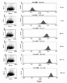
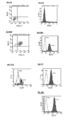
- Example 1 (1) Healthy individuals, (2) Low risk HTLV-I carriers, (3) Medium risk HTLV-I carriers, (4) High risk HTLV-I carriers, (5) Smoldering ATLL patients, (6) It is a figure which shows the result of having analyzed the cell distribution pattern of PpIX fluorescence intensity and TSLC1 expression intensity which is a leukemia marker about the peripheral blood derived from a chronic ATLL patient and (7) acute type ATLL patient by flow cytometry.
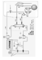
- Example 2 It is a figure which shows the concept of the abnormally activated cell removal reflux return treatment apparatus of this invention.
- Example 1 It is a figure which shows the concept at the time of connecting a dialysis apparatus to the abnormally activated cell removal reflux return treatment apparatus of this invention.
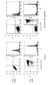
- Example 1 Peripheral blood mononuclear in patients with TLOm1 (ATLL leukemia cell line) or chronic ATLL treated with photodynamic therapy by irradiating with Na-Li lamp for 48 hours in the presence of 1 mM 5-ALA It is a figure which shows the result of having investigated the cell death pattern by flow cytometry of the sphere (PBMC) by double staining of PI (Propidium iodide) staining and Annexin-V-FITC staining. (Example 3) It is a figure which shows the result when the peripheral blood of a chronic type
- PBMC Peripheral blood mononuclear cells isolated from the blood of chronic ATLL patients were cultured for 48 hours in the presence of 1 mM 5-ALA, and then photodynamic therapy was performed by irradiating light with a Na-Li lamp. The flow cytometry analysis result of specific cell death induction is shown.
- PBMC peripheral blood mononuclear cells
- ATLL leukemia cells were specifically dead (implemented)
- Example 4 Specific cell death in the case of photodynamic therapy by adding 1 mM 5-ALA to peripheral blood of chronic ATLL patients and culturing for 48 hours followed by irradiation with Na-Li lamp The flow cytometry analysis result of induction is shown.
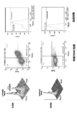
- Example 6 It is a figure which shows the result of having analyzed the cell distribution pattern which concerns on expression of a PpIX positive cell and a tumor marker about various cell culture malignant tumor cells by flow cytometry. (Example 6) It is a figure which shows the result of having analyzed the cell distribution pattern which concerns on expression of a PpIX positive cell and a tumor marker about various cell culture cancer cells by flow cytometry. (Example 6)
- the malignant tumor test of the present invention can be performed by the following steps 1) to 3). 1) a step of detecting PpIX positive cells in the collected blood sample; 2) a step of detecting tumor marker positive cells in the collected blood sample; 3) Analyzing the distribution pattern of PpIX positive cells and tumor marker positive cells obtained in 1) and 2), respectively.
- PpIX protoporphyrin IX
- physiological precursors of porphyrin-related compounds include 5-ALA (5-aminolevulinic acid), which is an amino acid that is a precursor substance for heme synthesis, but is not particularly limited thereto. Any physiological precursor that generates strong fluorescence with a photosensitive substance may be used.
- the “PpIX positive cell” refers to a cell in which PpIX is accumulated by excessive administration of a physiological precursor of a porphyrin-related compound such as 5-ALA from the outside. It is known that PpIX selectively accumulates in immunologically abnormal cells such as malignant tumor cells, chronic inflammatory cells and autoantigen-responsive cells in autoimmune diseases, and in particular, selectively accumulates at high concentrations in malignant tumor cells.
- PpIX is irradiated with excitation light having a peak at a wavelength of 350 to 490 nm, preferably a wavelength of 400 to 490 nm or a wavelength of 600 to 700 nm, preferably a wavelength of 610 to 640 nm, and a wavelength of 600 to 700 nm, preferably a wavelength of 620 to 650 nm.
- it has the property of exhibiting red fluorescence having a peak at a wavelength of 635 nm, and has been clinically applied to photodynamic diagnosis (PDD) of tumor tissues in a wide range.
- PPD photodynamic diagnosis
- FIG. 1 is a graph showing the measurement results of the time course of accumulation of PpIX positive cells after administration of 1 mM 5-ALA to TLOm1 (ATLL leukemia cell line). It can be seen that the PpIX concentration reached the upper limit 48 hours after administration.
- Fig. 2 shows PpIX fluorescence of peripheral blood mononuclear cells (PBMC), TLOm1 (ATLL leukemia cell line) and ED-40515 (ATLL leukemia cell line) of healthy subjects after 48 hours of culture after administration of 1 mM 5-ALA. It is a figure which shows the result of having analyzed intensity
- PBMC peripheral blood mononuclear cells
- TLOm1 ATLL leukemia cell line
- ED-40515 ATLL leukemia cell line
- FIG. 3 shows contact activated by stimulation with peripheral blood mononuclear cells (PBMC) and CD3 / CD28 immuno-beads (Dynabeads (R) human T-cell activator CD3 / CD28 (Invitrogen)) from patients with contact dermatitis.
- PBMC peripheral blood mononuclear cells
- TLOm1 ATLL leukemia cell line
- ED-40515 ATLL leukemia cell line
- peripheral blood mononuclear cells PBMC
- PBMC peripheral blood mononuclear cell
- abnormally activated cells refers to pathological cells, and examples thereof include immunologically abnormal cells such as malignant tumor cells, chronic inflammatory cells, and autoantigen-responsive cells in autoimmune diseases.
- cells that emit strong PpIX fluorescence in the presence of 5-ALA by addition or administration of 5-ALA are also referred to as “abnormally activated cells”.
- the “malignant tumor” may be any of “hematopoietic tumor”, “carcinoma (carcinoma), sarcoma”, and is not particularly limited, but preferably “hematopoietic tumor” is Can be mentioned.
- Examples of “hematopoietic tumors” include leukemia and lymphoma.
- Leukemia is roughly classified into acute leukemia and chronic leukemia, and is divided into myeloid leukemia that develops from myeloid cells and lymphocytic leukemia that develops from lymphoid cells. Specifically, acute myeloid leukemia (Acute Myeloid Leukemia: AML), acute lymphoblastic leukemia (Acute Lymphoblastic Leukemia: ALL), chronic myelogenous leukemia (Leukemia: CML), chronic lymphocytic leukemia (Chronic Lymphoblastic Leukemia: CLL) Etc. ATLL is also listed as another type of leukemia. In this specification, it is ATLL that is the object of examination and treatment. As shown in the background art section, ATLL is caused by HTLV-I.
- a sample collected for examination or treatment is a blood sample.
- the collected blood sample may be irradiated with light as it is, but is preferably processed by the following steps. 1) The red blood cell fraction and the white blood cell fraction are separated by centrifugation of the collected blood sample. 2) The separated leukocyte fraction is irradiated with light having a wavelength of 350 to 490 nm or 600 to 700 nm. 3) After the light irradiation, the accumulated PpIX positive cells are detected.
- leukocytes exhibiting abnormal proliferation ability including hematopoietic tumors such as leukemia cells are also referred to as “abnormally activated leukocytes”.
- the tumor marker may be a tumor marker specified for a tumor, and is not particularly limited.
- a tumor marker specified for a tumor, and is not particularly limited.
- PBMC peripheral blood mononuclear cells
- TSLC1 a cell surface marker
- CD3, CD13, CD19, CD20, CD10, CD13, CD7, CD56, HLA -Immunological markers such as DR an example of a cell surface marker whose expression is specifically increased in ATLL leukemia cells is TSLC1.
- Tumor markers for carcinoma include per se known cytokeratin 19-9 (CA19-9), CEA, NSE, PSA, EMA, AFP, SCC, PTH, TSH, but are particularly limited is not.
- test method combining “detection of PpIX positive cells” and “detection of tumor marker positive cells” of the present invention, cells that have become malignant tumors can be detected more reliably than when they are performed alone.
- FIG. 4 is a diagram showing the results of analyzing the cell distribution pattern by flow cytometry from the PpIX measurement value and the measurement value of TSLC1, which is a tumor marker, for peripheral blood derived from ATLL patients.
- TSLC1 a tumor marker
- FIG. 4 is a diagram showing the results of analyzing the cell distribution pattern by flow cytometry from the PpIX measurement value and the measurement value of TSLC1, which is a tumor marker, for peripheral blood derived from ATLL patients.
- the TSLC1 (-) / PpIX (-) pattern in which cells accumulate on the lower left side, whereas in the HTLV-I carrier, (2) TSLC1 that is almost the same as that of healthy individuals (4) From low risk HTLV-I carriers with (-) / PpIX (-) pattern, (3) Medium risk HTLV-I carriers with increasing fraction of TSLC1 (+) / PpIX (-), (4)
- HTLV-I carriers Especially in high-risk HTLV-I carriers, it means that there are many cell groups showing TSLC1 (+) / PpIX (+) leukemia cell-specific profiles. Is high. Thus, in the case of a carrier, it is possible to predict the onset of ATLL by confirming the cell distribution pattern (flow cytometry profile) in the blood sample, and to start preemptive treatment according to the progress of the disease state at an early stage Can do.
- the ATLL smoldering type, chronic type, or acute type also gradually changes as shown in the patterns (5) to (7) in Fig. 4, and the increase in TSLC1 (+) / PpIX (-) cells indicates that TSLC1 ( +) / PpIX (+) migration to leukemia cells.
- Low-grade ATLL such as smoldering type and chronic type is also acutely changed to high-grade ATLL by monitoring the transition from TSLC1 (+) / PpIX (-) cells to TSLC1 (+) / PpIX (+) leukemia cells Conversion (progress) can be detected at an early stage.
- the risk and progression of malignant tumor onset / progress can be predicted or grasped using, for example, the following as an index.
- the risk / progression index of malignant tumor and the index of the progression are RF (RF: Risk Factor)
- the RF value can be derived as follows.
- the flow cytometry profile shown in FIG. 4 is considered as follows.
- RF Risk Factor
- Healthy people 0.01 ⁇ RF ⁇ 1.00
- Low risk HTLV-I carrier 0.01 ⁇ RF ⁇ 2.50
- Medium risk HTLV-I carrier 2.00 ⁇ RF ⁇ 6.00
- High risk HTLV-I carrier 3.50 ⁇ RF ⁇ 20.00
- Smoldering ATLL 4.00 ⁇ RF ⁇ 30.00
- Chronic ATLL 5.00 ⁇ RF ⁇ 80.00
- Acute ATLL 20.00 ⁇ RF ⁇ 100.00
- the present invention also extends to a method for removing malignant tumor cells.
- the “malignant tumor” in the method for removing malignant tumor cells may be any hematopoietic tumor, carcinoma or sarcoma, but is particularly preferably a hematopoietic tumor.
- the collected specimen is irradiated with light having a wavelength of 350 to 490 nm or 600 to 700 nm, PpIX positive cells are detected, and cell death is induced by the light irradiation to remove malignant tumor cells. it can.
- PpIX positive cells it is preferably a specimen containing a physiological precursor of a porphyrin-related compound, specifically 5-ALA.
- a sample containing 5-ALA may be administered in advance before the sample is collected, or may be administered to the sample after the sample is collected, but it is preferable to administer the sample before collecting the sample. is there.
- 5-ALA has proven to be highly safe when used for oral administration and intravenous injection.
- By administering 5-ALA to a patient in advance accumulation of PpIX in abnormally activated cells such as leukemia cells can be caused. Since it takes some time for PpIX to accumulate in abnormally activated cells such as leukemia cells, it is desirable to administer 5-ALA at least 2 days before the test.
- the present invention also extends to an abnormally activated cell removal perfusion return treatment device for removing abnormally activated cells including hematopoietic tumors from a collected blood sample.
- the abnormally activated cell removal perfusion return treatment device of the present invention is a device for removing abnormally activated cells in blood, and in particular, it accumulates PpIX in malignant tumor cells, chronic inflammatory cells and immune abnormal cells. It selectively accumulates, and in particular, abnormally activated cells such as malignant tumor cells such as leukemia cells or leukemia progenitor cells can be identified and effectively removed.
- the abnormally activated cell-removal reflux return treatment apparatus of the present invention has the following configuration. a) a blood collection line having a puncture needle for blood collection at one end; b) a centrifuge for separating the red blood cell fraction and the white blood cell fraction from the blood fed by the blood collection line; c) a cell sorter for removing the abnormally activated cells from the leukocyte fraction separated by the centrifuge; d) a light irradiator for irradiating the normal white blood cell fraction from which the abnormally activated cells have been removed by the cell sorter of c) with a predetermined wavelength; f) A recirculation that recirculates the recirculated blood composed of blood components other than the leukocyte fraction separated by the centrifuge and the normal white blood cell fraction for recirculation that has been irradiated with light by the light irradiator. Blood return line. Further, g) an artificial dialysis apparatus may be connected (see FIG. 6).
- the abnormally activated cell-removal perfusion return treatment apparatus of the present invention will be described in more detail.
- the device of the present invention is a device for removing abnormally activated cells such as tumor cells, chronic inflammatory cells or immune abnormal cells in blood, and in particular, distinguishing abnormally activated cells from normal cells by accumulating PpIX. As possible, it is an abnormally activated cell-removal reflux return treatment device that effectively removes.
- Abnormally activated cells relate to tumor cells and include leukemia cells found in hematopoietic tumors as well as tumor cells dropped into the blood from carcinomas (epitheliomas), sarcomas, and the like.
- the abnormally activated cells are particularly preferably abnormally activated leukocytes such as leukemia cells.
- the abnormally activated cell-removal reflux return treatment apparatus of the present invention is an abnormally activated cell-removal reflux return treatment apparatus that can effectively identify abnormally activated leukocytes or leukemia progenitor cells, and effectively remove them.
- 5-ALA a physiological precursor of a photosensitizer porphyrin-related compound
- PpIX a physiological precursor of a photosensitizer porphyrin-related compound
- Abnormally activated cells can be identified because strong fluorescence is generated by 700 nm excitation light irradiation.
- the 5-ALA that is a physiological precursor of a porphyrin-related compound as a photosensitive substance is not particularly limited as long as it is a physiological precursor that generates strong fluorescence with a photosensitive substance.
- 5-ALA can cause PpIX accumulation in abnormally activated cells by pre-administration to patients. Since it takes some time for PpIX to accumulate in the abnormally activated cells, 5-ALA is preferably used 2 to 48 hours, preferably 2 to 12 hours before using the abnormally activated cell-removal reflux treatment apparatus. It is desirable to administer.
- An abnormally activated cell removal perfusion return treatment device as shown in FIG. 5, has a blood collection with a puncture needle (not shown) for blood collection at one end.
- Line 10 a centrifuge 20 for separating a leukocyte fraction from a blood sample supplied by the blood collection line 10, and abnormal activity such as leukemia cells against the leukocyte fraction separated by the centrifuge 20
- the cell sorter 30 for removing activated cells
- the light irradiation device 40 for irradiating the normal leukocyte fraction from which abnormally activated cells such as leukemia cells have been removed by the cell sorter 30, and the centrifuge 20
- Blood components other than the leukocyte fraction, blood circulation components such as erythrocyte fraction and platelet fraction, and blood circulation solution that is irradiated with light from the light irradiator 40 and plasma other than the leukocyte fraction Components, red blood cell fraction and platelets It is composed of a circulation return line 50 that returns blood components
- abnormally activated cells can be detected by PpIX-specific fluorescence emitted when irradiated with excitation light having a wavelength of 488 nm, and the detected PpIX (+) leukocytes can be removed with a cell sorter.
- the blood collection line 10 may be a blood collection line used in general component blood donation.
- a blood collection puncture needle is attached to one end of the blood collection line 10, and the other end can be connected to the centrifuge 20.
- the centrifuge 20 can also be a continuous centrifuge used for component blood donation.
- the leukocyte fraction is separated by centrifugation with the centrifuge 20, and the plasma component, the red blood cell fraction and the platelet fraction are used as the recirculation blood for recirculation. It can be delivered to the first circulation return line 51 of the line 50.
- the white blood cell fraction separated from the blood by the centrifuge 20 is fed to the cell sorter 30 via the first connection line 61.
- the cell sorter 30 is normally used as a cell analysis / separation device, in which individual cells of fine particles are dispersed in a fluid, and the fluid is finely flowed to excite each particle cell with laser light of a specific wavelength. , Optically analyze the specific fluorescence generated, perform flow cytometry, charge droplets containing cells with specific optical properties after droplet formation, and charge with a charged deflector It is an apparatus for performing cell sorting for separating cells contained in a droplet.
- the cell sorter 30 of the present embodiment detects abnormally activated cells such as leukemia cells that emit strong fluorescence due to a large amount of PpIX accumulation in the leukocyte fraction fed from the centrifuge 20, and abnormal activity such as leukemia cells. Abnormally activated cells such as leukemia cells are removed by sorting the activated cells.
- the leukocyte fraction separated in the centrifuge 20 is mixed with a buffer solution such as sheath liquid, and leukocytes are diluted to an extent that can be individually identified, abnormalities such as leukemia cells in the cell sorter 30 After removal of the activated cells, removal of excess components contained in the sheath liquid and appropriate concentration are performed. That is, although not shown, the cell sorter 30 is connected to a concentrator using a centrifuge or the like, or a concentration adjuster using physiological saline, etc. A normal leukocyte fluid for reflux return from which activated cells have been removed is generated. The normal white blood cell fraction for recirculation is returned to the light irradiator 40 via the second connection line 62.
- abnormally activated cells such as almost 100% leukemia cells can be removed by setting the cutoff value of the fluorescence intensity from PpIX.
- the abnormally activated cells removed by the cell sorter 30 may be discarded as they are or used for inspection.
- the number of abnormally activated cells such as removed leukemia cells can be used for early examination of leukemia and disease state monitoring.
- 63 is a discharge line through which abnormally activated cells such as leukemia cells removed by the cell sorter 30 are discharged.
- the normal white blood cell fraction for recirculation returning blood fed via the second connecting line 62 is irradiated with light of a predetermined wavelength by the light irradiator 40, and this light is used for the normal white blood cell fraction for recirculation recirculation. It is supposed to cause cell death in the leukemia cells that remain.
- the light irradiator 40 is constituted by a feeding tube 41 that feeds a normal white blood cell fraction for recirculation and a light source 42 that radiates light toward the feeding tube 41.
- the feed pipe 41 is made of a transparent or semi-transparent pipe, and can irradiate light to the normal white blood cell fraction fed through the pipe.
- the light source 42 is a metal halide lamp (Na-Li lamp) in which sodium and lithium are enclosed.
- the light source 42 may be an LED lamp capable of irradiating a wavelength of 350 to 490 nm or 600 to 700 nm or another light source including laser light.
- a pump for feeding may be provided in the middle of the second connecting line 62, A pump for feeding may be provided.
- a cooling device that cools the heat generated by light irradiation may be provided in the feed pipe 41.
- the length of the feeding tube 41 and the feeding speed of the normal white blood cell fraction for reflux return are adjusted so that the irradiation time is 10 minutes or longer.
- one light source 42 is used, but a plurality of light sources may be used.
- the light irradiator 40 irradiates a normal white blood cell fraction for recirculation with a light having a wavelength of 350 to 490 nm or 600 to 700 nm by a Na-Li lamp.
- PpIX is excited by light irradiation to abnormally activated cells such as leukemia cells that have not been removed by the cell sorter 30 despite the accumulation of PpIX, and singlet oxygen is generated from oxygen with this excitation.
- cell death is induced by the strong cell destruction action by the singlet oxygen.
- a dialysis apparatus may be connected as shown in FIG.
- PBMC nuclear cells
- TLOm1 and ED-40515 ATLL leukemia cell line
- PpIX accumulation in cells 48 hours after administration of 1 mM 5-ALA was analyzed by flow cytometry.
- accumulation of PpIX was confirmed specifically for activated T cells and tumors (FIGS. 2 and 3).
- Example 2 Confirmation of flow cytometry profile of mononuclear cells
- flow cytometry was performed on peripheral blood mononuclear cells (PBMC) isolated from the peripheral blood of healthy subjects, HTLV-I carriers or ATLL patients. ⁇ The profile was confirmed. 1 mM 5-ALA was added to the culture broth containing each peripheral blood mononuclear cell (PBMC) and cultured for 48 hours. PpIX accumulation and TSLC1 expression were confirmed in these cells, and cell distribution by flow cytometry Pattern analysis was performed.
- PBMC peripheral blood mononuclear cells
- HTLV-I carriers are at about 5% risk of developing ATLL in their lifetime, but for HTLV-I carriers, knowing the risk of developing ATLL is very important for the quality of life (QOL) of HTLV-I carriers is important. Based on the method of the present invention, an appropriate treatment method is selected immediately before the onset or at the beginning of the onset by recognizing the pattern of the medium risk and high risk HTLV-I carriers, which is the risk pattern of (3) and (4) be able to.
- the RF value was derived as follows.
- the flow cytometry profile shown in FIG. 4 is considered as follows.
- Example 3 In this example, 1 mM 5-ALA was added to the culture solution of TLOm1 (ATLL leukemia cell line) or peripheral blood mononuclear cells (PBMC) of chronic ATLL patients and cultured for 48 hours, followed by a Na-Li lamp for 10 minutes. Irradiation (29 mW / cm 2 ). Thereafter, the cell death pattern by flow cytometry was examined by double staining of PI (Propidium iodide) staining and Annexin-V-FITC staining. As a control, the same analysis was performed on cells not added with 5-ALA. As a result, in TLOm1, cells were cultured for 48 hours in the presence of 1 mM 5-ALA.
- TLOm1 ATLL leukemia cell line
- PBMC peripheral blood mononuclear cells
- AnnexinV (+) / PI ( -) Apoptosis
- AnnexinV (+) / PI (+) necrosis
- PBMC Peripheral blood mononuclear cells
- PBMC Peripheral blood mononuclear cells
- Example 4 was performed in order to confirm what kind of cells the fraction causing cell death and the fraction remaining alive consist of.
- Example 4 In this example, 1 mM 5-ALA was administered to peripheral blood mononuclear cells (PBMC) of chronic ATLL patients using the apparatus of Example 1 and cultured for 48 hours, followed by a 10-minute Na-Li lamp (29 mW). / cm 2 ) Irradiation. Subsequently, triple staining with Annexin-V-FITC, PI, and TSLC1-Alexa647 was performed, and detailed analysis of the cell death fraction by flow cytometry was performed. As a result, the fraction of cells in which ATLL leukemia cells of TSLC1 (+) / AnnexinV (+) caused cell death (upper right) reached 98.7% of the fraction of all ATLL leukemia cells (TSLC1 (+)).
- PBMC peripheral blood mononuclear cells
- Photodynamic therapy by irradiating light with Na-Li lamp after adding 1 mM 5-ALA to peripheral blood mononuclear cells (PBMC) of chronic ATLL patients for 48 hours
- PBMC peripheral blood mononuclear cells
- abnormally activated cells such as leukemia cells that have not been removed by the cell sorter 30 are killed by the light irradiator 40, so that abnormally activated cells such as leukemia cells remain in the normal leukocyte fraction for recirculation. Only healthy white blood cells can be returned to the patient without fear.
- Abnormally activated cells such as leukemia cells that caused cell death can be removed using the discoloration function of the patient's liver and spleen even if they are returned to the patient together with the normal white blood cell fraction for recirculation.
- abnormally activated cell components such as leukemia cells that have been killed by a filter or the like may be removed to prevent oncolysis syndrome.
- the normal white blood cell fraction for reflux return sent from the light irradiator 40 is sent to the second reflux return line 52 of the reflux return line 50 connected at one end to the light irradiator 40.
- the first circulating blood return line 51 and the second circulating blood return line 52 are connected by a Y-shaped connector (not shown), thereby comprising normal blood components other than the leukocyte fraction separated by the centrifuge 20.
- the recirculating blood and the normal white blood cell fraction for recirculating blood irradiated with light by the light irradiator 40 are mixed.
- a third recirculation return line 53 provided with a blood puncture needle (not shown) at one end is connected, and the recirculation composed of blood components other than the leukocyte fraction A mixture of the returned blood and the normal white blood cell fraction for recirculation is returned to the patient.
- abnormally activated cells such as leukemia cells are removed by the cell sorter 30, but also leukemia cells and the like by irradiation with light from the light irradiator 40.
- the possibility of abnormally activated cells remaining can be eliminated.
- the specific abnormally activated cell removal reflux return treatment apparatus of the present invention is used not only as a treatment for removing abnormally activated cells such as leukemia cells, but also for counting the number of abnormally activated cells such as leukemia cells removed by the cell sorter 30, Since it can be used for pathological analysis, it can also be used for early examination of leukemia and pathological monitoring as described above.
- the heterospecific normal activated leukocyte removal reflux return treatment device of the present invention is used for transplantation of patient immunity against other organ transplantation in addition to ATLL lymphoid leukemia, myeloproliferative diseases including myeloid leukemia, etc.
- aggressive activated lymphocyte clones in organ rejection or attack of patient organs by donor lymphocytes due to acute GVHD (graft versus host disease) or chronic GVHD after hematopoietic stem cell transplantation or bone marrow transplantation It can be used for removal.
- it may be widely used for various diseases associated with abnormal growth of activation-specific clones such as various autoimmune diseases or allergic diseases.
- it can be used for suppression of metastasis by separation / removal of epithelial carcinoma cells and sarcoma cells in blood undergoing hematogenous metastasis.
- Example 5 In this example, 1 mM 5-ALA was administered to peripheral blood of chronic ATLL patients and cultured for 48 hours, and then a photodynamic therapy was performed by irradiating light with a Na-Li lamp. Flow cytometric analysis was performed for specific cell death induction. As a result of analyzing the cell distribution pattern of the chronic ATLL leukemia fraction (TSLC1 (+)) and the normal mononuclear cell fraction (TSLC1 (-)) with PpIX fluorescence intensity and TSLC1 expression intensity as a leukemia marker by flow cytometry, This treatment removed 98.83% ATLL leukemia cells by cell death.
- TSLC1 (+) chronic ATLL leukemia fraction
- TSLC1 (-) normal mononuclear cell fraction
- PBMC peripheral blood mononuclear cells
- Example 6 In this example, 1 mM 5-ALA was administered to various established malignant tumor cultured cells and cultured for 48 hours, and tumor markers and PpIX accumulation were confirmed for these cells.
- hematopoietic tumor-derived cells are Ramos cells (Burkitt lymphoma (malignant lymphoma)), K562 cells (chronic myeloid leukemia) and BALL1 cells (acute B lymphocytic leukemia).
- the expression of TSLC1 as a tumor marker was also confirmed (FIG. 10).
- Jurkat cells T-lymphocytic leukemia
- FL18 cystic lymphoma (malignant lymphoma)
- A549 cells lung cancer
- SCCKN cells thyroidgue cancer
- LK79 cells lung cancer
- PpIX accumulation by administration of 1 mM 5-ALA positive for PpIX positive cells and tumor marker (CK19-9)
- PpIX (+) / CK19-9 (+) the distribution pattern of epithelial cancer cells
- normal cells PpIX ( ⁇ ) / CK19-9 ( ⁇ )
- FIG. 12 the distribution of PpIX positive cells and tumor marker positive cells was analyzed for hematopoietic tumors and solid tumors, confirming a cell distribution different from normal cells. It was thought that tumor cells could be removed by kinetic therapy.
- malignant tumor cells can be detected effectively.
- the risk of tumor onset / progress can be ascertained.
- the risk of ATLL onset / progress can be predicted for the HTLV-I carrier.
- ATLL is categorized as (A) smoldering type, (B) chronic type, (C) acute type, and (D) lymphoma type.
- ATLL is classified into high-risk HTLV-I carriers. Since it is possible to monitor in detail the pathology of low-grade ATLL patients who are expected to be identified and progressed, more appropriate preemptive treatment can be performed before onset / progress.
- abnormally activated cells such as leukemia cells are removed from blood collected from a patient, for example, component transfusion, and normal blood cell components, platelets, and plasma are removed again. Can be returned to the patient and contribute to leukemia treatment.
- leukemia cells can be substantially removed in principle by setting the cutoff value of the fluorescence intensity from PpIX in the separation using a centrifuge and a cell sorter. it can. Even if leukemia cells mixed in the normal cell fraction coexist, 98.7% of leukemia cells can be removed by inducing cell death by further light irradiation. It is possible to remove leukemia cells.
- the abnormally activated cell-removal perfusion return treatment apparatus of the present invention is a lymphatic leukemia other than ATLL, myeloproliferative diseases including myeloid leukemia, etc., as well as rejection of patient immunity against transplanted organs of patient immunity against allogeneic organ transplantation
- it can be used to remove aggressive lymphocyte clones in patients' organs attacked by donor lymphocytes due to acute GVHD (graft-versus-host disease) or chronic GVHD that occurs after hematopoietic stem cell transplantation or bone marrow transplantation It is thought that.
- GVHD graft-versus-host disease
- chronic GVHD that occurs after hematopoietic stem cell transplantation or bone marrow transplantation It is thought that.
- it may be widely used for various diseases such as various autoimmune diseases or lymphoproliferative diseases accompanying abnormal growth of specific clones such as allergic diseases.
- it can be used for suppression of metastasis by separation / removal of epithelial carcinoma cells
Abstract
This invention provides an abnormally-activated-cell apheresis-therapy apparatus for inhibiting the onset of or treating leukemia by removing abnormally activated cells, specifically abnormally activated leukocytes or leukemia progenitor cells, from blood. Said abnormally-activated-cell apheresis-therapy apparatus, which is an abnormally-activated-leukocyte apheresis-therapy apparatus that removes abnormally activated cells such as leukemia cells from blood, said cells having been made identifiable via high-concentration accumulation of protoporphyrin IX, is provided with the following: a blood-collection line, one end of which is provided with a needle for blood collection; a centrifugal separator that separates out a leukocyte fraction from blood sent down the blood-collection line; a cell sorter that removes abnormally activated cells from the separated-out leukocyte fraction; a light-exposure device that exposes the normal leukocyte fraction resulting from the removal of the aforementioned abnormally activated cells to light of a prescribed wavelength; and a return line that returns, to the patient, the normal leukocyte fraction and a return fluid comprising the blood components other than the leukocyte fraction.
Description
本発明は、異常活性化細胞検出による悪性腫瘍の検査方法および異常活性化細胞除去環流返血治療装置に関する。より詳しくは、異常活性化細胞を除去することにより白血病の発症を抑制または治療するための異常活性化細胞除去環流返血治療装置に関する。
The present invention relates to a method for examining a malignant tumor by detecting abnormally activated cells, and an abnormally activated cell-removal perfusion return treatment apparatus. More specifically, the present invention relates to an abnormally activated cell-removal reflux return treatment apparatus for suppressing or treating the onset of leukemia by removing abnormally activated cells.
本出願は、参照によりここに援用されるところの日本出願、特願2013-130758号優先権を請求する。
This application claims the priority of Japanese application No. 2013-130758, which is incorporated herein by reference.
昨今、日本国においては、白血病の発生率が年々増加する傾向にある。白血病を原因とする死亡者数は、2008年において約11,156人であり、人口10万人あたり8.8人(男性10.5人、女性7.1人)となっている。
Recently, the incidence of leukemia tends to increase year by year in Japan. The number of deaths due to leukemia was approximately 11,156 in 2008, 8.8 per 100,000 population (10.5 males, 7.1 females).
特に、成人T細胞白血病・リンパ腫(adult T-cell leukemia/lymphoma: ATLL)は難治性白血病・リンパ腫であることが知られている。その原因ウイルスであるヒトT細胞白血病ウイルスI型(Human T-cell leukemia virus type I: HTLV-I)のキャリアーの中から日本で年間約700例が発症している。特に、HTLV-Iのキャリアー1,000人あたりの年間ATLL発症率は、男性で1.0~1.5人、女性で0.5~0.7人であり、さらには、30歳以上のHTLV-Iのキャリアーにおける生涯発症率は、男性で4~7%、女性で2%台である。現在、日本で約108万人、世界では約2,000万人のキャリアーが存在する。
In particular, it is known that adult T-cell leukemia / lymphoma (lymoma: lymphoma) is an intractable leukemia / lymphoma. About 700 cases occur annually in Japan from carriers of human T-cell leukemia virus type I (Human T-cell leukemia virus type I: HTLV-I), the causative virus. In particular, the annual incidence of ATLL per 1,000 HTLV-I carriers is 1.0 to 1.5 in men and 0.5 to 0.7 in women, and the lifetime incidence in HTLV-I carriers over 30 years of age is , 4-7% for men and 2% for women. Currently, there are about 1.08 million people in Japan and about 20 million people worldwide.
ATLLは、HTLV-IがCD4陽性T細胞に感染し、細胞-細胞間で感染し、長い潜伏期間の中で免疫機構から逃れたクローンが遺伝子異常、染色体異常、エピジェネティック異常などを集積して、細胞を腫瘍化させることで発症すると考えられている。HTLV-Iのキャリアーの3~5%が、HTLV-I感染後40~60年してATLLを発症する。また、感染経路は主として母乳感染、輸血、性交である。
In ATLL, HTLV-I infects CD4-positive T cells, and cell-to-cell infection, and clones that escape from the immune system in a long incubation period accumulate genetic abnormalities, chromosomal abnormalities, epigenetic abnormalities, etc. It is thought to develop by causing cells to become tumors. 3-5% of HTLV-I carriers develop ATLL 40-60 years after HTLV-I infection. Infection routes are mainly breast milk infection, blood transfusion, and sexual intercourse.
ATLLは1991年に予後因子解析と臨床病態の特徴から、白血化、臓器浸潤、高LDH血症、高Ca血症の有無と程度により急性型、リンパ腫型、慢性型、くすぶり型の4病型分類が提唱され、最近の報告による生存期間中央値は急性型11ヶ月、リンパ腫型20ヶ月、慢性型24ヶ月、くすぶり型では3年以上である。最近の化学療法と同種造血幹細胞移植(allo-HSCT)による治療成績は改善しているが、他の白血病と比べると依然予後不良である。ATLL患者の臨床病型別生存曲線によると、急性型およびリンパ腫型ATLLの患者の生存率は、観察開始から1年以内に50%以下にまで低下する。患者は男性にやや多く、日本での発症年齢の中央値は67歳であり、40歳未満での発症は稀である。
ATLL was diagnosed in 1991 based on prognostic factor analysis and clinical pathological features, depending on the presence and extent of leukoplasia, organ invasion, hyper-LDH, hypercalcemia, and 4 types of disease: acute, lymphoma, chronic, and smoldering A classification has been proposed and the median survival according to recent reports is 11 months for acute, 20 months for lymphoma, 24 months for chronic, and 3 years or more for smoldering. Treatment outcomes with recent chemotherapy and allogeneic hematopoietic stem cell transplantation (allo-HSCT) have improved, but the prognosis is still poor compared to other leukemias. According to the clinical disease-type survival curve of ATLL patients, the survival rate of patients with acute and lymphoma ATLL falls to less than 50% within one year from the start of observation. The number of patients is slightly higher in men, and the median age of onset in Japan is 67 years, with onset rarely before 40 years.
このような難治性のATLLの従来の診断方法は、患者の末梢血におけるHTLV-I感染腫瘍細胞の同定の他、リンパ節腫脹、肝脾腫、高カルシウム血症、皮膚病変が多く日和見感染症の合併が多いなどATLLに特異的な臨床症状を基準とするものである。
Conventional methods for diagnosing such intractable ATLL include identification of HTLV-I-infected tumor cells in the patient's peripheral blood, as well as lymphadenopathy, hepatosplenomegaly, hypercalcemia, skin lesions, and opportunistic infections. Based on clinical symptoms specific to ATLL, such as many mergers.
しかし、このような方法では、特に急性型やリンパ腫型ATLLでは、発症し症状が出始めた段階にならなければ診断ができず、それゆえ早期にこれらを診断することができないため、治療が手遅れになってしまう可能性がある。また抗癌剤による治療に抵抗性で予後不良である。
However, with such a method, especially in the acute type and lymphoma type ATLL, diagnosis cannot be made unless the symptoms begin and symptoms begin to appear, and therefore these cannot be diagnosed early, so treatment is too late. There is a possibility of becoming. It is resistant to treatment with anticancer drugs and has a poor prognosis.
ATLLの予後を推定するためのデータの収集において、末梢血単核球(PBMC)の特定の遺伝子群について、それらの遺伝子のプロモーター領域におけるCpG島のメチル化状態を測定する方法について開示がある(特許文献1)。当該遺伝子のプロモーター領域におけるCpG島のメチル化プロファイルを作成するとともに、予後因子としてCIMP(CpG island methylator phenotype)を算出し、メチル化遺伝子、メチル化遺伝子数およびCIMP値から算出されるATLL発症/進展の危険度スコアによる危険度の階層化および/または特定の遺伝子のメチル化状態を表示することにより、HTLV-Iのキャリアーの発症またはindolent型(緩徐進行型)であるくすぶり型および慢性型ATLLがaggressive型(高悪性型)である急性型およびリンパ腫型ATLLへの進展を予測することを可能とするATLL予後推定用データを作成する方法に関するものである。
In the collection of data for estimating the prognosis of ATLL, there is disclosed a method for measuring the methylation status of CpG islands in the promoter region of specific genes of peripheral blood mononuclear cells (PBMC) ( Patent Document 1). Creates a methylation profile of CpG islands in the promoter region of the gene, calculates CIMP (CpG island methylator phenotype) as a prognostic factor, and develops / progresses ATLL calculated from methylated genes, number of methylated genes, and CIMP value HRLV-I carrier onset or indolent (slow progression) smoldering and chronic ATLL by displaying risk stratification by risk score and / or methylation status of specific genes The present invention relates to a method for creating data for estimating ATLL prognosis that makes it possible to predict the progression to aggressive type (high malignant type) acute type and lymphoma type ATLL.
高悪性度の急性型やリンパ腫型ATLLおよび予後不良因子を有する慢性型ATLLは、化学療法の適応であるが、非ホジキンリンパ腫の標準的治療法(CHOP療法)などに抵抗性であるため、G-CSF(顆粒球コロニー刺激因子)を併用して短い治療間隔で強力な化学療法が繰り返して行われる。
High-grade acute type and lymphoma type ATLL and chronic type ATLL with poor prognosis are indications of chemotherapy, but are resistant to standard treatment of non-Hodgkin's lymphoma (CHOP therapy). -Strong chemotherapy is repeated at short treatment intervals in combination with CSF (granulocyte colony stimulating factor).
一方、低悪性度のくすぶり型や、予後不良因子を有さない慢性型ATLLは、皮膚病変には局所的に対処し、慢性リンパ性白血病などの疾患と同様に急性転化するまでは化学療法をせずに経過観察することが原則とされるが、その長期予後は良好ではない。近年、allo-HSCTでは宿主片対ATL効果により長期生存が期待でき、検討されるべき治療法であるものの、通常のallo-HSCTでは治療が難しい宿主や疾患も存在し、高齢者等では胸腺が委縮しているためT細胞の産生能が低く、免疫不全や間質性肺炎を起こしやすいなどの問題もある。同種移植を受けた場合にしばしばみられる合併症で、ドナー由来のリンパ球が患者の臓器を自分のものでないとみなして攻撃し、排除しようとする、移植片対宿主病(GVHD)の危険性の問題もある。また、今後抗体医薬などの新規治療法の開発が期待されるものの、予後が優れた効果的な治療方法の開発が望まれている。
On the other hand, low-grade smoldering type and chronic type ATLL with no poor prognostic factors deal with skin lesions locally, and chemotherapy is required until the disease becomes acute, like chronic lymphocytic leukemia. In principle, the follow-up is not, but the long-term prognosis is not good. In recent years, allo-HSCT can be expected to survive for a long time due to the host-versus-ATL effect and should be studied, but there are some hosts and diseases that are difficult to treat with normal allo-HSCT. There are also problems such as low T cell production ability due to atrophy, and susceptibility to immunodeficiency and interstitial pneumonia. The risk of graft-versus-host disease (GVHD), a complication often seen in allogeneic transplants, in which donor-derived lymphocytes attempt to attack and eliminate the patient's organs as if they were not their own There is also a problem. Further, although development of new therapeutic methods such as antibody drugs is expected in the future, development of effective therapeutic methods with an excellent prognosis is desired.
光感受性物質のポルフィリン関連化合物の生理的前駆体である5-アミノレブリン酸(5-aminolevulinic acid(5-ALA))の投与により、ヘム代謝経路を経てプロトポルフィリンIX(PpIX)が組織球性リンパ腫培養細胞に多量に蓄積され、強い蛍光を発生が報告されている。この性質を利用して、5-ALA投与により強いPpIX蛍光を発する白血病細胞を健常細胞から識別することが可能となることが検討され、5-ALA依存性光動力学的治療法(PDT:Photo-Dynamic Therapy)では、標的腫瘍細胞に蓄積したPpIXへ の光照射で生成する一重項酸素などの活性酸素種(ROS)が、特異的に腫瘍細胞の細胞死を誘導できると考えられることが報告されている(非特許文献1)。
Protoporphyrin IX (PpIX) is cultured in histiocytic lymphoma via the heme metabolic pathway by administration of 5-aminolevulinic acid (5-ALA), a physiological precursor of a photosensitizer porphyrin-related compound. It has been reported that cells accumulate in large quantities and generate strong fluorescence. Using this property, it has been studied that leukemia cells that emit strong PpIX fluorescence can be distinguished from healthy cells by administration of 5-ALA. 5-ALA-dependent photodynamic therapy (PDT: Photo) -Dynamic (Therapy) reported that reactive oxygen species (ROS) such as singlet oxygen generated by light irradiation of PpIX accumulated in target tumor cells can specifically induce cell death of tumor cells (Non-Patent Document 1).
一方、白血病ではないが、血液中の不水溶性の間接(非抱合型)ビリルビンの代謝不全の患者に対して、人工透析の際に人体から取り出した血液に所定波長の光を照射することにより水溶性の直接ビリルビンに変換して不水溶性の間接ビリルビンの濃度を低下させる方法が開示されている(特許文献2)。体内でヘモグロビンから形成される不水溶性の間接ビリルビンは、通常肝臓で水溶性の直接(抱合型)ビリルビンへと変換されて腎臓を介して排出されているが、肝機能不全等が生じると水溶性の直接ビリルビンへの変換が行われず、血中の不水溶性の間接ビリルビンの濃度が上昇することが知られている。不水溶性の間接ビリルビンは、所定波長の光の照射によって水溶性の直接ビリルビンに変換可能であり、肝機能不全等によって不水溶性の間接ビリルビンの濃度が上昇した患者に対して、当該光を照射することにより水溶性の直接ビリルビンに変換して不水溶性の間接ビリルビンの濃度を低下させることができる。
On the other hand, by irradiating the blood extracted from the human body during artificial dialysis with light of a predetermined wavelength to patients who are not leukemia but who have metabolic insufficiency of water-insoluble indirect (non-conjugated) bilirubin A method for reducing the concentration of water-insoluble indirect bilirubin by converting it into water-soluble direct bilirubin has been disclosed (Patent Document 2). Water-insoluble indirect bilirubin formed from hemoglobin in the body is usually converted into water-soluble direct (conjugated) bilirubin by the liver and excreted through the kidney. It is known that the direct conversion to sex bilirubin is not performed and the concentration of water-insoluble indirect bilirubin in the blood is increased. Water-insoluble indirect bilirubin can be converted into water-soluble direct bilirubin by irradiation with light of a predetermined wavelength. Irradiation can be converted to water-soluble direct bilirubin to reduce the concentration of water-insoluble indirect bilirubin.
ATLL発症の危険性の高いHTLV-Iキャリアーおよび慢性型ATLLやくすぶり型ATLLの様な低悪性度ATLLのなかで高悪性度の急性型およびリンパ腫型ATLLへ急性転化(進展)する可能性の高い患者や予後不良因子を有する慢性型ATLL患者に関し、早期に発症/進展の危険度を検出する方法、また発症後も予後不良を回避可能な有効な治療方法の開発が望まれている。この方法の開発は発症可能性の低いHTLV-Iキャリアーおよび病態進展の可能性の低い低悪性度ATLL患者に対しては発症/進展の不安を可能な限り取り除き、生活の質(QOL)の改善にもつながる。
Among HTLV-I carriers at high risk of developing ATLL and low-grade ATLL such as chronic ATLL and smoldering ATLL, there is a high possibility of acute transformation (advance) to high-grade acute and lymphoma ATLL With respect to patients and chronic ATLL patients with poor prognosis, it is desired to develop a method for detecting the risk of onset / advance at an early stage and an effective treatment method capable of avoiding poor prognosis even after onset. Development of this method will eliminate as much as possible anxiety of onset / progression and improve quality of life (QOL) for low-incidence HTLV-I carriers and low-grade ATLL patients with low probability of progression It also leads to.
本発明は、悪性腫瘍の検査方法を提供することを課題とする。そして、血液検体からの異常活性化細胞の除去方法、異常活性化細胞除去環流返血治療装置、並びに異常活性化細胞除去環流返血治療装置を用いる腫瘍の治療方法を提供することを課題とする。
An object of the present invention is to provide a method for examining a malignant tumor. Then, it is an object of the present invention to provide a method for removing abnormally activated cells from a blood sample, an abnormally activated cell-removed perfusion return treatment apparatus, and a tumor treatment method using the abnormally activated cell-removal perfusion return treatment apparatus. .
造血器腫瘍のうち、成人T細胞白血病・リンパ腫(ATLL)はきわめて予後不良の難治性白血病・リンパ腫である。同種造血幹細胞移植(allo-HSCT)や新規抗体医薬などの新規治療法の開発が進みつつあるが、高齢者に発症頻度が高いという特徴を持つ本疾患の場合、移植幹細胞の供給の問題や患者に負担の大きい治療法は適用除外となる場合も稀ではなく治療抵抗性患者も多いという問題がある。
Among hematopoietic tumors, adult T-cell leukemia / lymphoma (ATLL) is an intractable leukemia / lymphoma with a very poor prognosis. Development of new therapies such as allogeneic hematopoietic stem cell transplantation (allo-HSCT) and new antibody drugs is progressing, but in the case of this disease, which is characterized by high incidence in the elderly, problems with the supply of transplanted stem cells and patients However, there is a problem that many treatment-resistant patients are not rarely excluded from the application of treatment methods with a large burden.
本発明は、低侵襲性で患者の負担が小さく白血病細胞を特異的に高効率で取り除くことが可能な異常活性化細胞除去環流返血治療装置を提供することを課題とする。
An object of the present invention is to provide an abnormally activated cell-removing perfusion return treatment apparatus that is less invasive, less burdensome on the patient, and can specifically remove leukemia cells with high efficiency.
上記課題を解決するために、本発明者らはPpIXを血液中の白血病細胞など異常活性化細胞に蓄積させることにより、当該異常活性化細胞を識別可能とすることに着目し、鋭意研究を重ねた結果、血液中の白血病細胞など異常活性化細胞を特異的に除去する異常活性化細胞除去環流返血治療装置に係る発明を完成するに至った。
In order to solve the above-mentioned problems, the present inventors have made extensive studies by focusing on the fact that PpIX is accumulated in abnormally activated cells such as leukemia cells in blood so that the abnormally activated cells can be identified. As a result, the inventors have completed an invention relating to an abnormally activated cell-removal perfusion return device that specifically removes abnormally activated cells such as leukemia cells in blood.
本発明の異常活性化細胞除去環流返血治療装置は、一端に採血用の穿刺針を備えた採血用ラインと、この採血用ラインによって送給された血液から白血球分画を分離する遠心分離器と、この遠心分離器で分離された白血球分画から異常活性化細胞を除去するセルソーターと、このセルソーターにより異常活性化細胞が除去された正常血球分画に対して所定波長の光を照射する光照射器と、遠心分離器で分離された白血球分画以外の血液成分からなる環流返血液と光照射器で光が照射された環流返血用の正常白血球分画とを返血する環流返血用ラインとを備えてなる。
The abnormally activated cell-removal perfusion return treatment apparatus of the present invention includes a blood collection line having a puncture needle for blood collection at one end, and a centrifuge that separates a white blood cell fraction from blood fed by the blood collection line A cell sorter that removes abnormally activated cells from the leukocyte fraction separated by the centrifuge, and light that irradiates light of a predetermined wavelength to a normal blood cell fraction from which abnormally activated cells have been removed by the cell sorter Refluxing blood that returns the circulating blood that consists of blood components other than the leukocyte fraction separated by the centrifuge and the normal white blood cell fraction for circulating blood that has been irradiated by the light irradiator. Line.
また、本発明の異常活性化細胞除去環流返血治療装置では、採血用ラインを介して採取される血液には患者に2~48時間前、好ましくは2~12時間前に5-ALAが投与されていることで、腫瘍細胞を含む異常活性化細胞にPpIXを蓄積させ、光照射器で異常活性化細胞を含む血液に波長350~490nmまたは600~700nmの光を照射することを特徴とするものである。
Further, in the abnormally activated cell-removal circulating blood recirculation treatment apparatus of the present invention, 5-ALA is administered to the blood collected via the blood collection line to the patient 2 to 48 hours before, preferably 2 to 12 hours before. It is characterized by accumulating PpIX in abnormally activated cells including tumor cells and irradiating blood containing abnormally activated cells with light having a wavelength of 350 to 490 nm or 600 to 700 nm with a light irradiator. Is.
すなわち本発明は以下よりなる。
1.採取した血液検体について、異常活性化細胞と腫瘍マーカー陽性細胞の分布パターンを解析することを特徴とする、悪性腫瘍の検査方法。
2.異常活性化細胞がPpIX陽性細胞である、前項1に記載の悪性腫瘍の検査方法。
3.悪性腫瘍が、造血器腫瘍、癌腫または肉腫であり、腫瘍マーカーが、造血器腫瘍の場合はTSLC1や、CD3、CD13、CD19、CD20、CD10、CD13、CD7、CD56およびHLA-Dからなる免疫学的マーカーのいずれかであり、癌腫または肉腫の場合はサイトケラチン19-9(CA19-9)、CEA、NSE、PSA、TPA、AFP、SCC、PTH、TSHから選択されるいずれかである、前項1または2に記載の悪性腫瘍の検査方法。
4.造血器腫瘍がATLLである前項1~3のいずれか1に記載の悪性腫瘍の検査方法。
5.採取した血液検体のドナーがHTLV-IのキャリアーまたはATLL患者であり、悪性腫瘍の検査が、ATLL発症危険度予測および/または進展確認のための検査である、前項4に記載の検査方法。
6.ATLLの進展確認が、ATLLの(A)くすぶり型、(B)慢性型、または(C)急性型のいずれかへの進展確認である、前項5に記載の検査方法。
7.採取した血液検体について、波長350~490nmおよび/または600~700nmの光で照射し、検体中に存在するPpIX陽性細胞を検出し、当該PpIX陽性細胞を前記光照射により細胞死を誘発することで、検体から異常活性化細胞を除去する方法。
8.以下の工程を含む、前項7に記載の異常活性化細胞を除去する方法:
1)採取した血液検体について、遠心分離操作により赤血球分画と白血球分画を分離する工程;
2)当該分離された白血球分画について、波長350~490nmおよび/または600~700nmの光で照射し、PpIX陽性細胞を検出し、当該PpIX陽性細胞を前記光照射により細胞死を誘発することで、血液検体から異常活性化細胞を除去する工程;
3)前記2)において白血球分画から異常活性化細胞を除去した残りの白血球分画に、前記1)で分離した赤血球分画を統合する工程。
9.前記2)の光照射が、5-ALAを含む白血球分画について行なわれる、前項8に記載の異常活性化細胞を除去する方法。
10.異常活性化細胞が、悪性腫瘍細胞、慢性炎症性細胞、免疫異常細胞のいずれかである、前項7または8に記載の異常活性化細胞を除去する方法。
11.異常活性化細胞が、悪性腫瘍細胞であり、採取した血液検体について前項1~6のいずれか1に記載の検査方法を用いて検査し、悪性腫瘍細胞に分類される細胞を収集し、当該収集した細胞を波長350~490nm または600~700nmの光で照射することを特徴とする、前項10に記載の異常活性化細胞を除去する方法。
12.悪性腫瘍が造血器腫瘍であり、異常活性化細胞が異常活性化白血球である、前項10または11に記載の異常活性化細胞を除去する方法。
13.造血器腫瘍が、ATLLである前項12に記載の異常活性化細胞を除去する方法。
14.以下のa)~e)を含み、前項7~13のいずれかに1記載の異常活性化細胞を除去する方法を実施するための機構を含む、異常活性化細胞除去環流返血治療装置:
a)一端に採血用の穿刺針を備えた採血用ラインと、
b)当該採血用ラインによって送給された血液から赤血球分画と白血球分画を分離する遠心分離器と、
c)当該遠心分離器で分離された前記白血球分画から前記異常活性化細胞を除去するセルソーターと、
d)前記c)のセルソーターにより前記異常活性化細胞が除去された正常白血球分画に対して波長350~490nmおよび/または600~700nmの光で照射し光を照射するための光照射器と、
e)前記遠心分離器で分離された前記白血球分画以外の血液成分からなる環流返血液と前記光照射器で光が照射された環流返血用の正常白血球分画とを環流返血する環流返血用ライン。
15.上記e)の環流返血用ラインに、さらにf)人工透析装置が連結されている、前項14に記載の異常活性化細胞除去環流返血治療装置。
16.前項14または15に記載の異常活性化細胞除去環流返血治療装置を用いて血液検体から異常活性化細胞を除去し、当該異常活性化細胞を除去した後の血液検体を生体に返血することを特徴とする造血器腫瘍の治療方法。 That is, this invention consists of the following.
1. A method for examining a malignant tumor, comprising analyzing a distribution pattern of abnormally activated cells and tumor marker positive cells for a collected blood sample.
2. 2. The method for examining a malignant tumor according toitem 1, wherein the abnormally activated cells are PpIX positive cells.
3. If the malignant tumor is a hematopoietic tumor, carcinoma or sarcoma, and the tumor marker is a hematopoietic tumor, immunology consisting of TSLC1, CD3, CD13, CD19, CD20, CD10, CD13, CD7, CD56 and HLA-D Any of the above markers, and in the case of carcinoma or sarcoma, any one selected from cytokeratin 19-9 (CA19-9), CEA, NSE, PSA, TPA, AFP, SCC, PTH, TSH 3. The method for examining a malignant tumor according to 1 or 2.
4). 4. The method for examining a malignant tumor according to any one ofitems 1 to 3, wherein the hematopoietic tumor is ATLL.
5. 5. The test method according toitem 4 above, wherein the donor of the collected blood sample is a carrier of HTLV-I or an ATLL patient, and the test for malignant tumor is a test for predicting the risk of developing ATLL and / or confirming progress.
6). 6. The examination method according toitem 5 above, wherein the confirmation of progress of ATLL is confirmation of progress of ATLL to any one of (A) smoldering type, (B) chronic type, and (C) acute type.
7). The collected blood sample is irradiated with light having a wavelength of 350 to 490 nm and / or 600 to 700 nm, PpIX positive cells present in the sample are detected, and cell death is induced by the light irradiation of the PpIX positive cells. A method for removing abnormally activated cells from a specimen.
8). A method for removing abnormally activated cells according to item 7, comprising the following steps:
1) A step of separating a red blood cell fraction and a white blood cell fraction by centrifugation for the collected blood sample;
2) The separated leukocyte fraction is irradiated with light having a wavelength of 350 to 490 nm and / or 600 to 700 nm, PpIX positive cells are detected, and cell death is induced by the light irradiation on the PpIX positive cells. Removing abnormally activated cells from the blood sample;
3) A step of integrating the red blood cell fraction separated in 1) above with the remaining white blood cell fraction from which abnormally activated cells have been removed from the white blood cell fraction in 2) above.
9. 9. The method for removing abnormally activated cells according toitem 8 above, wherein the light irradiation of 2) is performed on a leukocyte fraction containing 5-ALA.
10. 9. The method for removing abnormally activated cells according toitem 7 or 8 above, wherein the abnormally activated cells are any of malignant tumor cells, chronic inflammatory cells, and immune abnormal cells.
11. The abnormally activated cells are malignant tumor cells, and the collected blood sample is examined using the examination method described in any one of 1 to 6 above, and the cells classified as malignant tumor cells are collected. 11. The method for removing abnormally activated cells according toitem 10 above, wherein the irradiated cells are irradiated with light having a wavelength of 350 to 490 nm or 600 to 700 nm.
12 12. The method for removing abnormally activated cells according toitem 10 or 11, wherein the malignant tumor is a hematopoietic tumor and the abnormally activated cells are abnormally activated leukocytes.
13. 13. The method for removing abnormally activated cells according to item 12 above, wherein the hematopoietic tumor is ATLL.
14 The abnormally activated cell-removal reflux return treatment apparatus comprising the following a) to e) and comprising a mechanism for carrying out the method for removing an abnormally activated cell according to any one of items 7 to 13:
a) a blood collection line having a puncture needle for blood collection at one end;
b) a centrifuge for separating the red blood cell fraction and the white blood cell fraction from the blood fed by the blood collection line;
c) a cell sorter for removing the abnormally activated cells from the leukocyte fraction separated by the centrifuge;
d) a light irradiator for irradiating the normal leukocyte fraction from which the abnormally activated cells have been removed by the cell sorter of c) with light having a wavelength of 350 to 490 nm and / or 600 to 700 nm;
e) Recirculation that recirculates the recirculated blood composed of blood components other than the leukocyte fraction separated by the centrifuge and the normal white blood cell fraction for recirculation that has been irradiated with light by the light irradiator. Blood return line.
15. 15. The abnormally activated cell-removal recirculating blood return treatment device according to 14 above, wherein an artificial dialysis device is further connected to the recirculation blood return line of e).
16. The abnormally activated cells are removed from the blood sample using the abnormally activated cell-removal reflux return treatment apparatus according to the above 14 or 15, and the blood sample after the abnormally activated cells are removed is returned to the living body. A method of treating a hematopoietic tumor characterized by the above.
1.採取した血液検体について、異常活性化細胞と腫瘍マーカー陽性細胞の分布パターンを解析することを特徴とする、悪性腫瘍の検査方法。
2.異常活性化細胞がPpIX陽性細胞である、前項1に記載の悪性腫瘍の検査方法。
3.悪性腫瘍が、造血器腫瘍、癌腫または肉腫であり、腫瘍マーカーが、造血器腫瘍の場合はTSLC1や、CD3、CD13、CD19、CD20、CD10、CD13、CD7、CD56およびHLA-Dからなる免疫学的マーカーのいずれかであり、癌腫または肉腫の場合はサイトケラチン19-9(CA19-9)、CEA、NSE、PSA、TPA、AFP、SCC、PTH、TSHから選択されるいずれかである、前項1または2に記載の悪性腫瘍の検査方法。
4.造血器腫瘍がATLLである前項1~3のいずれか1に記載の悪性腫瘍の検査方法。
5.採取した血液検体のドナーがHTLV-IのキャリアーまたはATLL患者であり、悪性腫瘍の検査が、ATLL発症危険度予測および/または進展確認のための検査である、前項4に記載の検査方法。
6.ATLLの進展確認が、ATLLの(A)くすぶり型、(B)慢性型、または(C)急性型のいずれかへの進展確認である、前項5に記載の検査方法。
7.採取した血液検体について、波長350~490nmおよび/または600~700nmの光で照射し、検体中に存在するPpIX陽性細胞を検出し、当該PpIX陽性細胞を前記光照射により細胞死を誘発することで、検体から異常活性化細胞を除去する方法。
8.以下の工程を含む、前項7に記載の異常活性化細胞を除去する方法:
1)採取した血液検体について、遠心分離操作により赤血球分画と白血球分画を分離する工程;
2)当該分離された白血球分画について、波長350~490nmおよび/または600~700nmの光で照射し、PpIX陽性細胞を検出し、当該PpIX陽性細胞を前記光照射により細胞死を誘発することで、血液検体から異常活性化細胞を除去する工程;
3)前記2)において白血球分画から異常活性化細胞を除去した残りの白血球分画に、前記1)で分離した赤血球分画を統合する工程。
9.前記2)の光照射が、5-ALAを含む白血球分画について行なわれる、前項8に記載の異常活性化細胞を除去する方法。
10.異常活性化細胞が、悪性腫瘍細胞、慢性炎症性細胞、免疫異常細胞のいずれかである、前項7または8に記載の異常活性化細胞を除去する方法。
11.異常活性化細胞が、悪性腫瘍細胞であり、採取した血液検体について前項1~6のいずれか1に記載の検査方法を用いて検査し、悪性腫瘍細胞に分類される細胞を収集し、当該収集した細胞を波長350~490nm または600~700nmの光で照射することを特徴とする、前項10に記載の異常活性化細胞を除去する方法。
12.悪性腫瘍が造血器腫瘍であり、異常活性化細胞が異常活性化白血球である、前項10または11に記載の異常活性化細胞を除去する方法。
13.造血器腫瘍が、ATLLである前項12に記載の異常活性化細胞を除去する方法。
14.以下のa)~e)を含み、前項7~13のいずれかに1記載の異常活性化細胞を除去する方法を実施するための機構を含む、異常活性化細胞除去環流返血治療装置:
a)一端に採血用の穿刺針を備えた採血用ラインと、
b)当該採血用ラインによって送給された血液から赤血球分画と白血球分画を分離する遠心分離器と、
c)当該遠心分離器で分離された前記白血球分画から前記異常活性化細胞を除去するセルソーターと、
d)前記c)のセルソーターにより前記異常活性化細胞が除去された正常白血球分画に対して波長350~490nmおよび/または600~700nmの光で照射し光を照射するための光照射器と、
e)前記遠心分離器で分離された前記白血球分画以外の血液成分からなる環流返血液と前記光照射器で光が照射された環流返血用の正常白血球分画とを環流返血する環流返血用ライン。
15.上記e)の環流返血用ラインに、さらにf)人工透析装置が連結されている、前項14に記載の異常活性化細胞除去環流返血治療装置。
16.前項14または15に記載の異常活性化細胞除去環流返血治療装置を用いて血液検体から異常活性化細胞を除去し、当該異常活性化細胞を除去した後の血液検体を生体に返血することを特徴とする造血器腫瘍の治療方法。 That is, this invention consists of the following.
1. A method for examining a malignant tumor, comprising analyzing a distribution pattern of abnormally activated cells and tumor marker positive cells for a collected blood sample.
2. 2. The method for examining a malignant tumor according to
3. If the malignant tumor is a hematopoietic tumor, carcinoma or sarcoma, and the tumor marker is a hematopoietic tumor, immunology consisting of TSLC1, CD3, CD13, CD19, CD20, CD10, CD13, CD7, CD56 and HLA-D Any of the above markers, and in the case of carcinoma or sarcoma, any one selected from cytokeratin 19-9 (CA19-9), CEA, NSE, PSA, TPA, AFP, SCC, PTH, TSH 3. The method for examining a malignant tumor according to 1 or 2.
4). 4. The method for examining a malignant tumor according to any one of
5. 5. The test method according to
6). 6. The examination method according to
7). The collected blood sample is irradiated with light having a wavelength of 350 to 490 nm and / or 600 to 700 nm, PpIX positive cells present in the sample are detected, and cell death is induced by the light irradiation of the PpIX positive cells. A method for removing abnormally activated cells from a specimen.
8). A method for removing abnormally activated cells according to item 7, comprising the following steps:
1) A step of separating a red blood cell fraction and a white blood cell fraction by centrifugation for the collected blood sample;
2) The separated leukocyte fraction is irradiated with light having a wavelength of 350 to 490 nm and / or 600 to 700 nm, PpIX positive cells are detected, and cell death is induced by the light irradiation on the PpIX positive cells. Removing abnormally activated cells from the blood sample;
3) A step of integrating the red blood cell fraction separated in 1) above with the remaining white blood cell fraction from which abnormally activated cells have been removed from the white blood cell fraction in 2) above.
9. 9. The method for removing abnormally activated cells according to
10. 9. The method for removing abnormally activated cells according to
11. The abnormally activated cells are malignant tumor cells, and the collected blood sample is examined using the examination method described in any one of 1 to 6 above, and the cells classified as malignant tumor cells are collected. 11. The method for removing abnormally activated cells according to
12 12. The method for removing abnormally activated cells according to
13. 13. The method for removing abnormally activated cells according to item 12 above, wherein the hematopoietic tumor is ATLL.
14 The abnormally activated cell-removal reflux return treatment apparatus comprising the following a) to e) and comprising a mechanism for carrying out the method for removing an abnormally activated cell according to any one of items 7 to 13:
a) a blood collection line having a puncture needle for blood collection at one end;
b) a centrifuge for separating the red blood cell fraction and the white blood cell fraction from the blood fed by the blood collection line;
c) a cell sorter for removing the abnormally activated cells from the leukocyte fraction separated by the centrifuge;
d) a light irradiator for irradiating the normal leukocyte fraction from which the abnormally activated cells have been removed by the cell sorter of c) with light having a wavelength of 350 to 490 nm and / or 600 to 700 nm;
e) Recirculation that recirculates the recirculated blood composed of blood components other than the leukocyte fraction separated by the centrifuge and the normal white blood cell fraction for recirculation that has been irradiated with light by the light irradiator. Blood return line.
15. 15. The abnormally activated cell-removal recirculating blood return treatment device according to 14 above, wherein an artificial dialysis device is further connected to the recirculation blood return line of e).
16. The abnormally activated cells are removed from the blood sample using the abnormally activated cell-removal reflux return treatment apparatus according to the above 14 or 15, and the blood sample after the abnormally activated cells are removed is returned to the living body. A method of treating a hematopoietic tumor characterized by the above.
本発明の検査方法によれば、効果的に悪性腫瘍細胞を検出することができる。悪性腫瘍についてフローサイトメトリー・プロファイルを確認することにより、腫瘍の発展/進展の危険度を把握することができる。
According to the inspection method of the present invention, malignant tumor cells can be detected effectively. By confirming the flow cytometry profile for a malignant tumor, the risk of tumor development / development can be ascertained.
本発明の異常活性化細胞除去環流返血治療装置によれば、セルソーターによって白血病細胞など異常活性化細胞を除去するだけでなく、光照射器による光の照射によって残存した悪性腫瘍細胞、慢性炎症性細胞や自己免疫疾患における自己抗原反応細胞などの免疫異常細胞などの異常活性化細胞に細胞死を誘導することで、異常活性化細胞の残存のおそれを極限まで低減できる。つまり、本発明の異常活性化細胞除去環流返血治療装置によれば、例えば成分輸血のように患者から採取した血液から異常活性化細胞だけを除去して正常血球成分、血小板、血漿を再度患者に戻すことができ、例えば白血病治療に貢献しうる。
According to the abnormally activated cell-removal reflux return treatment apparatus of the present invention, not only abnormally activated cells such as leukemia cells are removed by a cell sorter, but also malignant tumor cells remaining by irradiation of light with a light irradiator, chronic inflammatory By inducing cell death in abnormally activated cells such as immune abnormal cells such as self-antigen-responsive cells in cells and autoimmune diseases, the risk of abnormally activated cells remaining can be reduced to a minimum. That is, according to the abnormally activated cell-removal perfusion treatment apparatus of the present invention, for example, only abnormally activated cells are removed from blood collected from a patient, such as component transfusion, and normal blood cell components, platelets, and plasma are returned to the patient again For example, it can contribute to leukemia treatment.
特に、本発明の異常活性化細胞除去環流返血治療装置によれば、遠心分離器およびセルソーターによる分離において、PpIXからの蛍光強度のカット・オフ値の設定によって原理的に異常活性化細胞をほぼ除去することができる。仮に正常細胞分画に紛れ込んだ異常活性化細胞が混在しても、更に光照射することにより98.7%の異常活性化細胞を細胞死誘導により取り除くことができ、両者を連続的に行うことにより高効率にほとんどの異常活性化細胞を除去することが可能である。
In particular, according to the abnormally activated cell-removal circulating blood recirculation treatment apparatus of the present invention, in the separation using a centrifuge and a cell sorter, in principle, abnormally activated cells are almost completely separated by setting the cutoff value of the fluorescence intensity from PpIX. Can be removed. Even if abnormally activated cells mixed in the normal cell fraction coexist, 98.7% of abnormally activated cells can be removed by inducing cell death by further light irradiation. It is possible to remove most abnormally activated cells efficiently.
本発明の悪性腫瘍の検査は、以下の1)~3)工程により行うことができる。
1)採取した血液検体についてPpIX陽性細胞を検出する工程;
2)採取した血液検体について腫瘍マーカー陽性細胞を検出する工程;
3)前記1)および2)で各々得られたPpIX陽性細胞および腫瘍マーカー陽性細胞の分布パターンを解析する工程。 The malignant tumor test of the present invention can be performed by the following steps 1) to 3).
1) a step of detecting PpIX positive cells in the collected blood sample;
2) a step of detecting tumor marker positive cells in the collected blood sample;
3) Analyzing the distribution pattern of PpIX positive cells and tumor marker positive cells obtained in 1) and 2), respectively.
「PpIX(プロトポルフィリンIX)」とは、光感受性物質のひとつであり、ミトコンドリア内でポルフィリン関連化合物の生理的前駆体より合成される物質である。「ポルフィリン関連化合物の生理的前駆体」とは、例えばヘム合成の前駆体物質であるアミノ酸であるところの5-ALA(5-アミノレブリン酸)が挙げられるが、特にこれに限定されるものではなく、光感受性物質で強い蛍光を発生する生理的前駆体であればよい。
“PpIX (protoporphyrin IX)” is one of photosensitizers and is a substance synthesized in the mitochondria from physiological precursors of porphyrin-related compounds. Examples of “physiological precursors of porphyrin-related compounds” include 5-ALA (5-aminolevulinic acid), which is an amino acid that is a precursor substance for heme synthesis, but is not particularly limited thereto. Any physiological precursor that generates strong fluorescence with a photosensitive substance may be used.
本明細書において、「PpIX陽性細胞」とは、5-ALAなどのポルフィリン関連化合物の生理的前駆体を外部から過剰投与することによりPpIXが蓄積された細胞をいう。PpIXは悪性腫瘍細胞、慢性炎症性細胞や自己免疫疾患における自己抗原反応細胞などの免疫異常細胞に選択的に蓄積し、特に悪性腫瘍細胞に選択的に高濃度蓄積することが知られている。PpIXは波長350~490nm、好ましくは波長400~490nmまたは波長600~700nm、好ましくは波長610~640nmにピークを持つ励起光を照射することにより、波長600~700nm、好ましくは波長620~650nm、より好ましくは波長635nmにピークを持つ赤色蛍光を呈する性質を有し、幅広い領域で腫瘍組織の光線力学的診断(Photodynamic diagnosis; PDD)に臨床応用されている。図1は、TLOm1(ATLL白血病細胞株)に1 mMの5-ALAを投与した後のPpIX陽性細胞の蓄積の時間経過の測定結果を示す図である。投与後、48時間経過にPpIX濃度が上限に達していることが分かる。
図2は健常人の末梢血単核球(PBMC)、TLOm1(ATLL白血病細胞株)およびED-40515(ATLL白血病細胞株)について、1 mMの5-ALAを投与後48時間培養後のPpIX蛍光強度をフローサイトメトリーで解析した結果を示す図である。図1~3の結果、5-ALA投与により、腫瘍特異的にPpIXの蓄積が観察された。また、図3は接触性皮膚炎患者の末梢血単核球(PBMC)、CD3/CD28 immuno-beads (Dynabeads(R) human T-cell activator CD3/CD28 (Invitrogen))で刺激し活性化した接触性皮膚炎患者の末梢血単核球(PBMC)、TLOm1(ATLL白血病細胞株)およびED-40515(ATLL白血病細胞株)について、1 mMの5-ALAを投与後48時間培養後のPpIX蓄積をフローサイトメトリーで解析した結果を示す図である。これにより接触性皮膚炎患者の末梢血単核球(PBMC)においては、正常末梢血単核球(PBMC)画分と少し活性化しPpIX蛍光強度の高い画分が存在することが判る。更にCD3/CD28 immuno-beadsを用いてT細胞に強い刺激を与えると更にPpIX蛍光強度の強い分画が誘導されることが判る。図1~3の結果、5-ALA投与により、腫瘍細胞および活性化されたリンパ球特異的にPpIXの強い蓄積が観察された。 In the present specification, the “PpIX positive cell” refers to a cell in which PpIX is accumulated by excessive administration of a physiological precursor of a porphyrin-related compound such as 5-ALA from the outside. It is known that PpIX selectively accumulates in immunologically abnormal cells such as malignant tumor cells, chronic inflammatory cells and autoantigen-responsive cells in autoimmune diseases, and in particular, selectively accumulates at high concentrations in malignant tumor cells. PpIX is irradiated with excitation light having a peak at a wavelength of 350 to 490 nm, preferably a wavelength of 400 to 490 nm or a wavelength of 600 to 700 nm, preferably a wavelength of 610 to 640 nm, and a wavelength of 600 to 700 nm, preferably a wavelength of 620 to 650 nm. Preferably, it has the property of exhibiting red fluorescence having a peak at a wavelength of 635 nm, and has been clinically applied to photodynamic diagnosis (PDD) of tumor tissues in a wide range. FIG. 1 is a graph showing the measurement results of the time course of accumulation of PpIX positive cells after administration of 1 mM 5-ALA to TLOm1 (ATLL leukemia cell line). It can be seen that the PpIX concentration reached theupper limit 48 hours after administration.
Fig. 2 shows PpIX fluorescence of peripheral blood mononuclear cells (PBMC), TLOm1 (ATLL leukemia cell line) and ED-40515 (ATLL leukemia cell line) of healthy subjects after 48 hours of culture after administration of 1 mM 5-ALA. It is a figure which shows the result of having analyzed intensity | strength by flow cytometry. As a result of FIGS. 1 to 3, accumulation of PpIX was observed in a tumor-specific manner by administration of 5-ALA. In addition, FIG. 3 shows contact activated by stimulation with peripheral blood mononuclear cells (PBMC) and CD3 / CD28 immuno-beads (Dynabeads (R) human T-cell activator CD3 / CD28 (Invitrogen)) from patients with contact dermatitis. Of peripheral blood mononuclear cells (PBMC), TLOm1 (ATLL leukemia cell line) and ED-40515 (ATLL leukemia cell line) in patients with atopic dermatitis after PhIX accumulation after 48 hours of culture after administration of 1 mM 5-ALA It is a figure which shows the result analyzed by flow cytometry. This shows that in peripheral blood mononuclear cells (PBMC) of patients with contact dermatitis, there is a fraction that is slightly activated with a normal peripheral blood mononuclear cell (PBMC) fraction and has a high PpIX fluorescence intensity. It can also be seen that when a strong stimulation is applied to T cells using CD3 / CD28 immuno-beads, a fraction with a stronger PpIX fluorescence intensity is induced. As a result of FIGS. 1 to 3, a strong accumulation of PpIX was observed specifically by tumor cells and activated lymphocytes by administration of 5-ALA.
図2は健常人の末梢血単核球(PBMC)、TLOm1(ATLL白血病細胞株)およびED-40515(ATLL白血病細胞株)について、1 mMの5-ALAを投与後48時間培養後のPpIX蛍光強度をフローサイトメトリーで解析した結果を示す図である。図1~3の結果、5-ALA投与により、腫瘍特異的にPpIXの蓄積が観察された。また、図3は接触性皮膚炎患者の末梢血単核球(PBMC)、CD3/CD28 immuno-beads (Dynabeads(R) human T-cell activator CD3/CD28 (Invitrogen))で刺激し活性化した接触性皮膚炎患者の末梢血単核球(PBMC)、TLOm1(ATLL白血病細胞株)およびED-40515(ATLL白血病細胞株)について、1 mMの5-ALAを投与後48時間培養後のPpIX蓄積をフローサイトメトリーで解析した結果を示す図である。これにより接触性皮膚炎患者の末梢血単核球(PBMC)においては、正常末梢血単核球(PBMC)画分と少し活性化しPpIX蛍光強度の高い画分が存在することが判る。更にCD3/CD28 immuno-beadsを用いてT細胞に強い刺激を与えると更にPpIX蛍光強度の強い分画が誘導されることが判る。図1~3の結果、5-ALA投与により、腫瘍細胞および活性化されたリンパ球特異的にPpIXの強い蓄積が観察された。 In the present specification, the “PpIX positive cell” refers to a cell in which PpIX is accumulated by excessive administration of a physiological precursor of a porphyrin-related compound such as 5-ALA from the outside. It is known that PpIX selectively accumulates in immunologically abnormal cells such as malignant tumor cells, chronic inflammatory cells and autoantigen-responsive cells in autoimmune diseases, and in particular, selectively accumulates at high concentrations in malignant tumor cells. PpIX is irradiated with excitation light having a peak at a wavelength of 350 to 490 nm, preferably a wavelength of 400 to 490 nm or a wavelength of 600 to 700 nm, preferably a wavelength of 610 to 640 nm, and a wavelength of 600 to 700 nm, preferably a wavelength of 620 to 650 nm. Preferably, it has the property of exhibiting red fluorescence having a peak at a wavelength of 635 nm, and has been clinically applied to photodynamic diagnosis (PDD) of tumor tissues in a wide range. FIG. 1 is a graph showing the measurement results of the time course of accumulation of PpIX positive cells after administration of 1 mM 5-ALA to TLOm1 (ATLL leukemia cell line). It can be seen that the PpIX concentration reached the
Fig. 2 shows PpIX fluorescence of peripheral blood mononuclear cells (PBMC), TLOm1 (ATLL leukemia cell line) and ED-40515 (ATLL leukemia cell line) of healthy subjects after 48 hours of culture after administration of 1 mM 5-ALA. It is a figure which shows the result of having analyzed intensity | strength by flow cytometry. As a result of FIGS. 1 to 3, accumulation of PpIX was observed in a tumor-specific manner by administration of 5-ALA. In addition, FIG. 3 shows contact activated by stimulation with peripheral blood mononuclear cells (PBMC) and CD3 / CD28 immuno-beads (Dynabeads (R) human T-cell activator CD3 / CD28 (Invitrogen)) from patients with contact dermatitis. Of peripheral blood mononuclear cells (PBMC), TLOm1 (ATLL leukemia cell line) and ED-40515 (ATLL leukemia cell line) in patients with atopic dermatitis after PhIX accumulation after 48 hours of culture after administration of 1 mM 5-ALA It is a figure which shows the result analyzed by flow cytometry. This shows that in peripheral blood mononuclear cells (PBMC) of patients with contact dermatitis, there is a fraction that is slightly activated with a normal peripheral blood mononuclear cell (PBMC) fraction and has a high PpIX fluorescence intensity. It can also be seen that when a strong stimulation is applied to T cells using CD3 / CD28 immuno-beads, a fraction with a stronger PpIX fluorescence intensity is induced. As a result of FIGS. 1 to 3, a strong accumulation of PpIX was observed specifically by tumor cells and activated lymphocytes by administration of 5-ALA.
本明細書において、「異常活性化細胞」とは、病的な細胞をいい、例えば悪性腫瘍細胞、慢性炎症性細胞や自己免疫疾患における自己抗原反応細胞などの免疫異常細胞が挙げられる。本明細書では、5-ALAの添加や投与による5-ALA存在下で強いPpIX蛍光を発する細胞を「異常活性化細胞」ともいう。上記において、「悪性腫瘍」とは「造血器腫瘍」または「癌腫(上皮腫:carcinoma)、肉腫(sarcoma)」のいずれであってもよく、特に限定されないが、好ましくは「造血器腫瘍」が挙げられる。「造血器腫瘍」としては、白血病やリンパ腫が挙げられる。白血病は、急性白血病および慢性白血病に大別され、それぞれ骨髄系細胞から発生する骨髄性白血病と、リンパ球系細胞から発生するリンパ性白血病に分けられる。具体的には、急性骨髄性白血病(Acute Myeloid Leukemia:AML) 、急性リンパ性白血病(Acute Lymphoblastic Leukemia:ALL)、慢性骨髄性白血病(Leukemia:CML)、慢性リンパ性白血病(Chronic Lymphoblastic Leukemia:CLL)などが挙げられる。また、ATLLはその他の白血病として挙げられる。本明細書において、特に検査および治療の対象となるのは、ATLLである。ATLLは、背景技術の欄でも示したように、HTLV-Iが原因ウイルスとなっている。
In the present specification, “abnormally activated cells” refers to pathological cells, and examples thereof include immunologically abnormal cells such as malignant tumor cells, chronic inflammatory cells, and autoantigen-responsive cells in autoimmune diseases. In the present specification, cells that emit strong PpIX fluorescence in the presence of 5-ALA by addition or administration of 5-ALA are also referred to as “abnormally activated cells”. In the above, the “malignant tumor” may be any of “hematopoietic tumor”, “carcinoma (carcinoma), sarcoma”, and is not particularly limited, but preferably “hematopoietic tumor” is Can be mentioned. Examples of “hematopoietic tumors” include leukemia and lymphoma. Leukemia is roughly classified into acute leukemia and chronic leukemia, and is divided into myeloid leukemia that develops from myeloid cells and lymphocytic leukemia that develops from lymphoid cells. Specifically, acute myeloid leukemia (Acute Myeloid Leukemia: AML), acute lymphoblastic leukemia (Acute Lymphoblastic Leukemia: ALL), chronic myelogenous leukemia (Leukemia: CML), chronic lymphocytic leukemia (Chronic Lymphoblastic Leukemia: CLL) Etc. ATLL is also listed as another type of leukemia. In this specification, it is ATLL that is the object of examination and treatment. As shown in the background art section, ATLL is caused by HTLV-I.
本発明において、検査や治療のために採取する検体は血液検体である。採取した血液検体は、そのまま光照射してもよいが、以下の工程により処理するのが好ましい。
1)採取した血液検体を遠心分離操作により赤血球分画と白血球分画を分離する。
2)当該分離された白血球分画について、波長350~490nmまたは600~700nmの光で照射する。
3)光照射後、集積したPpIX陽性細胞を検出する。本明細書において、白血病細胞など造血器腫瘍を含む異常増殖能を示す白血球を「異常活性化白血球」ともいう。 In the present invention, a sample collected for examination or treatment is a blood sample. The collected blood sample may be irradiated with light as it is, but is preferably processed by the following steps.
1) The red blood cell fraction and the white blood cell fraction are separated by centrifugation of the collected blood sample.
2) The separated leukocyte fraction is irradiated with light having a wavelength of 350 to 490 nm or 600 to 700 nm.
3) After the light irradiation, the accumulated PpIX positive cells are detected. In the present specification, leukocytes exhibiting abnormal proliferation ability including hematopoietic tumors such as leukemia cells are also referred to as “abnormally activated leukocytes”.
本明細書において、腫瘍マーカーとは腫瘍に対して特定される腫瘍マーカーであればよく、特に限定されない。例えば造血器腫瘍に対しては患者末梢血単核球(PBMC)または多核白血球分画では、細胞表面マーカーとしてのTSLC1の他、CD3、CD13、CD19、CD20、CD10、CD13、CD7、CD56、HLA-DR等の免疫学的マーカーが挙げられる。ATLL白血病細胞特異的に発現が亢進する細胞表面マーカーとしてはTSLC1が挙げられる。癌腫(上皮腫)、肉腫の腫瘍マーカーとしては自体公知のサイトケラチン19-9(CA19-9)、CEA、NSE、PSA、EMA、AFP、SCC、PTH、TSHが挙げられるが特に限定されるものではない。
In the present specification, the tumor marker may be a tumor marker specified for a tumor, and is not particularly limited. For example, for hematopoietic tumors, patients with peripheral blood mononuclear cells (PBMC) or multinucleated leukocyte fractions, in addition to TSLC1 as a cell surface marker, CD3, CD13, CD19, CD20, CD10, CD13, CD7, CD56, HLA -Immunological markers such as DR. An example of a cell surface marker whose expression is specifically increased in ATLL leukemia cells is TSLC1. Tumor markers for carcinoma (epithelioma) and sarcoma include per se known cytokeratin 19-9 (CA19-9), CEA, NSE, PSA, EMA, AFP, SCC, PTH, TSH, but are particularly limited is not.
本発明の「PpIX陽性細胞の検出」および「腫瘍マーカー陽性細胞の検出」を組み合わせた検査方法によれば、各々単独で行うよりもより確実に悪性腫瘍化した細胞を検出することができる。「PpIX陽性細胞」および「腫瘍マーカー陽性細胞」をフローサイトメトリーを用いて行い、細胞分布パターン(フローサイトメトリー・プロファイル)を確認することで、悪性腫瘍の発症/進展について予測または進行度を把握することができる。
According to the test method combining “detection of PpIX positive cells” and “detection of tumor marker positive cells” of the present invention, cells that have become malignant tumors can be detected more reliably than when they are performed alone. Perform “PpIX positive cells” and “tumor marker positive cells” using flow cytometry, and confirm the cell distribution pattern (flow cytometry profile) to predict the onset / progress of malignant tumors. can do.
例えば、図4はATLL患者由来の末梢血について、PpIX測定値と腫瘍マーカーであるTSLC1の測定値から細胞分布パターンをフローサイトメトリーで解析した結果を示す図である。(1)の健常人の場合には左下方面に細胞が集積するTSLC1(-)/PpIX(-)パターンであるのに対し、HTLV-Iキャリアーにおいては、(2)ほとんど健常人と変わらないTSLC1(-)/PpIX(-)パターンの低危険度HTLV-Iキャリアー、(3)TSLC1(+)/PpIX(-)の分画が増えてきつつある中危険度HTLV-Iキャリアーから、(4)ほとんどくすぶり型ATLLのパターンと差がない高危険度HTLV-Iキャリアーへと発症危険度が高まってくるにつれて、細胞分布パターンに変化が認められる。特に高危険度HTLV-IキャリアーではTSLC1(+)/PpIX(+)の白血病細胞特有プロファイルを示す細胞群が多数存在していることを意味し、免疫監視機構の状態が悪化すれば発症危険度が高いことが判る。このようにキャリアーの場合は、血液検体中の細胞分布パターン(フローサイトメトリー・プロファイル)を確認することでATLL発症を予測することができ、早期に病態の進展に応じた先制治療を開始することができる。
For example, FIG. 4 is a diagram showing the results of analyzing the cell distribution pattern by flow cytometry from the PpIX measurement value and the measurement value of TSLC1, which is a tumor marker, for peripheral blood derived from ATLL patients. In the case of (1) healthy individuals, the TSLC1 (-) / PpIX (-) pattern in which cells accumulate on the lower left side, whereas in the HTLV-I carrier, (2) TSLC1 that is almost the same as that of healthy individuals (4) From low risk HTLV-I carriers with (-) / PpIX (-) pattern, (3) Medium risk HTLV-I carriers with increasing fraction of TSLC1 (+) / PpIX (-), (4) As the risk of onset increases to a high-risk HTLV-I carrier that is almost no different from the pattern of smoldering ATLL, changes in the cell distribution pattern are observed. Especially in high-risk HTLV-I carriers, it means that there are many cell groups showing TSLC1 (+) / PpIX (+) leukemia cell-specific profiles. Is high. Thus, in the case of a carrier, it is possible to predict the onset of ATLL by confirming the cell distribution pattern (flow cytometry profile) in the blood sample, and to start preemptive treatment according to the progress of the disease state at an early stage Can do.
また、ATLLのくすぶり型、慢性型、または急性型についても図4の(5)~(7)のパターンのように徐々に変化し、TSLC1(+)/PpIX(-)細胞の増加からTSLC1(+)/PpIX(+)白血病細胞への移行が認められる。くすぶり型、慢性型の様な低悪性度ATLLもTSLC1(+)/PpIX(-)細胞からTSLC1(+)/PpIX(+)白血病細胞への移行をモニターすることにより高悪性度ATLLへの急性転化(進展)を早期に検出することが可能となる。各々の患者検体のパターン解析をすることより、ATLL発症/進展の危険度および進行度、疾病の型を詳細に把握することができ先制医療を施行することにより発症/進展予防の可能性も展望できる。
The ATLL smoldering type, chronic type, or acute type also gradually changes as shown in the patterns (5) to (7) in Fig. 4, and the increase in TSLC1 (+) / PpIX (-) cells indicates that TSLC1 ( +) / PpIX (+) migration to leukemia cells. Low-grade ATLL such as smoldering type and chronic type is also acutely changed to high-grade ATLL by monitoring the transition from TSLC1 (+) / PpIX (-) cells to TSLC1 (+) / PpIX (+) leukemia cells Conversion (progress) can be detected at an early stage. By analyzing the pattern of each patient specimen, it is possible to grasp in detail the risk and progression of ATLL onset / progress and the type of disease, and the possibility of onset / progression prevention is anticipated by conducting preemptive medicine it can.
悪性腫瘍の発症/進展の危険度や進行度は、例えば以下を指標として予測または把握することができる。悪性腫瘍の発症/進展の危険度や進行度の指標をRF(RF:Risk Factor)とすると、RF値は以下のように導き出すことができる。例えば図4に示すフローサイトメトリー・プロファイルについて、以下のように考えられる。
(A)LL: Lower Left (PpIX (-), TSLC1(-)) (下左)領域の細胞数
(B)LR: Lower Right (PpIX (-), TSCLC1(+))(下右) 領域の細胞数
(C)UR: Upper Right (PpIX (+), TSLC1(+)) (上右)領域の細胞数
RF = 0.01×(LL/total count)+ 10×(LR/total count) + 100×(UR/total count)
総細胞数(total count): 10,000 cells
健常人:LL=100% RF = 0.01
中危険度HTLV-Iキャリアー: LL=50%, LR=50%の場合RF=5.05
高危険度HTLV-Iキャリアー: LL=50%, LR=45%, UR=5%の場合 RF=9.55
急性型ATLL患者: UR=100%の場合 RF=100
なお、上記RF値は例示であって、(A)(B)(C)の細胞数から適宜決定することができる。悪性腫瘍の発症/進展の危険度や進行度の指標RF(RF:Risk Factor)はATLLの場合以下の範囲で評価することができる。
健常人:0.01<RF<1.00
低危険度HTLV-Iキャリアー:0.01<RF<2.50
中危険度HTLV-Iキャリアー:2.00<RF<6.00
高危険度HTLV-Iキャリアー:3.50<RF<20.00
くすぶり型ATLL:4.00<RF<30.00
慢性型ATLL: 5.00<RF<80.00
急性型ATLL: 20.00<RF<100.00 The risk and progression of malignant tumor onset / progress can be predicted or grasped using, for example, the following as an index. When the risk / progression index of malignant tumor and the index of the progression are RF (RF: Risk Factor), the RF value can be derived as follows. For example, the flow cytometry profile shown in FIG. 4 is considered as follows.
(A) LL: Lower Left (PpIX (-), TSLC1 (-)) (Lower left) Number of cells in the region (B) LR: Lower Right (PpIX (-), TSCLC1 (+)) (Lower right) Number of cells (C) UR: Number of cells in the Upper Right (PpIX (+), TSLC1 (+)) region
RF = 0.01 x (LL / total count) + 10 x (LR / total count) + 100 x (UR / total count)
Total count: 10,000 cells
Healthy people: LL = 100% RF = 0.01
Medium risk HTLV-I carrier: LL = 50%, LR = 50% RF = 5.05
High risk HTLV-I carrier: LL = 50%, LR = 45%, UR = 5% RF = 9.55
Acute ATLL patients: RF = 100 when UR = 100%
In addition, the said RF value is an illustration, Comprising: It can determine suitably from the cell number of (A) (B) (C). The risk of onset / progression of malignant tumor and the progress index RF (RF: Risk Factor) can be evaluated in the following ranges in the case of ATLL.
Healthy people: 0.01 <RF <1.00
Low risk HTLV-I carrier: 0.01 <RF <2.50
Medium risk HTLV-I carrier: 2.00 <RF <6.00
High risk HTLV-I carrier: 3.50 <RF <20.00
Smoldering ATLL: 4.00 <RF <30.00
Chronic ATLL: 5.00 <RF <80.00
Acute ATLL: 20.00 <RF <100.00
また、本発明は悪性腫瘍細胞の除去方法にも及ぶ。悪性腫瘍細胞の除去方法における「悪性腫瘍」とは造血器腫瘍、癌腫または肉腫のいずれであってもよいが、特に好適には造血器腫瘍である。採取した検体について、波長350~490nmまたは600~700nmの光で照射し、PpIX陽性細胞を検出し、当該陽性細胞を前記光照射により細胞死を誘発することで、悪性腫瘍細胞を除去することができる。PpIX陽性細胞を検出するために、ポルフィリン関連化合物の生理的前駆体、具体的には5-ALAを含む検体であることが好ましい。5-ALAを含む検体は、検体を採取する前にあらかじめ投与していてもよいし、検体を採取した後に検体に投与してもよいが、検体を採取する前にあらかじめ投与するのが好適である。5-ALAは、経口投与や静脈注射に用いても安全性が高いことが証明されている。あらかじめ患者に5-ALAを投与しておくことにより、白血病細胞など異常活性化細胞へのPpIXの蓄積を生じさせておくことができる。白血病細胞など異常活性化細胞にPpIXが蓄積されるには多少の時間を要するために、検査の2日前、少なくとも2時間前には5-ALAを投与しておくことが望ましい。
The present invention also extends to a method for removing malignant tumor cells. The “malignant tumor” in the method for removing malignant tumor cells may be any hematopoietic tumor, carcinoma or sarcoma, but is particularly preferably a hematopoietic tumor. The collected specimen is irradiated with light having a wavelength of 350 to 490 nm or 600 to 700 nm, PpIX positive cells are detected, and cell death is induced by the light irradiation to remove malignant tumor cells. it can. In order to detect PpIX positive cells, it is preferably a specimen containing a physiological precursor of a porphyrin-related compound, specifically 5-ALA. A sample containing 5-ALA may be administered in advance before the sample is collected, or may be administered to the sample after the sample is collected, but it is preferable to administer the sample before collecting the sample. is there. 5-ALA has proven to be highly safe when used for oral administration and intravenous injection. By administering 5-ALA to a patient in advance, accumulation of PpIX in abnormally activated cells such as leukemia cells can be caused. Since it takes some time for PpIX to accumulate in abnormally activated cells such as leukemia cells, it is desirable to administer 5-ALA at least 2 days before the test.
本発明は、採取した血液検体から造血器腫瘍を含む異常活性化細胞を除去するための異常活性化細胞除去環流返血治療装置にも及ぶ。本発明の異常活性化細胞除去環流返血治療装置は、血液中の異常活性化細胞を除去する装置であり、特に、PpIXを蓄積させることにより悪性腫瘍細胞、慢性炎症性細胞や免疫異常細胞に選択的に蓄積し、特に悪性腫瘍細胞などの異常活性化細胞、例えば白血病細胞あるいは白血病前駆細胞を識別可能として、効果的に除去することができる。
The present invention also extends to an abnormally activated cell removal perfusion return treatment device for removing abnormally activated cells including hematopoietic tumors from a collected blood sample. The abnormally activated cell removal perfusion return treatment device of the present invention is a device for removing abnormally activated cells in blood, and in particular, it accumulates PpIX in malignant tumor cells, chronic inflammatory cells and immune abnormal cells. It selectively accumulates, and in particular, abnormally activated cells such as malignant tumor cells such as leukemia cells or leukemia progenitor cells can be identified and effectively removed.
本発明の異常活性化細胞除去環流返血治療装置は以下の構成よりなる。
a)一端に採血用の穿刺針を備えた採血用ラインと、
b)当該採血用ラインによって送給された血液から赤血球分画と白血球分画を分離する遠心分離器と、
c)当該遠心分離器で分離された前記白血球分画から前記異常活性化細胞を除去するセルソーターと、
d)前記c)のセルソーターにより前記異常活性化細胞が除去された正常白血球分画に対して所定波長の光を照射するための光照射器と、
f)前記遠心分離器で分離された前記白血球分画以外の血液成分からなる環流返血液と前記光照射器で光が照射された環流返血用の正常白血球分画とを環流返血する環流返血用ライン。
さらにg)人工透析装置が連結されていてもよい(図6参照)。 The abnormally activated cell-removal reflux return treatment apparatus of the present invention has the following configuration.
a) a blood collection line having a puncture needle for blood collection at one end;
b) a centrifuge for separating the red blood cell fraction and the white blood cell fraction from the blood fed by the blood collection line;
c) a cell sorter for removing the abnormally activated cells from the leukocyte fraction separated by the centrifuge;
d) a light irradiator for irradiating the normal white blood cell fraction from which the abnormally activated cells have been removed by the cell sorter of c) with a predetermined wavelength;
f) A recirculation that recirculates the recirculated blood composed of blood components other than the leukocyte fraction separated by the centrifuge and the normal white blood cell fraction for recirculation that has been irradiated with light by the light irradiator. Blood return line.
Further, g) an artificial dialysis apparatus may be connected (see FIG. 6).
本発明の異常活性化細胞除去環流返血治療装置について、より詳細に説明する。本発明の装置は、血液中の腫瘍細胞、慢性炎症性細胞または免疫異常細胞など異常活性化細胞を除去する装置であり、特に、PpIXを蓄積させることにより異常活性化細胞と正常な細胞を識別可能として、効果的に除去する異常活性化細胞除去環流返血治療装置である。異常活性化細胞は、腫瘍細胞に関し、造血器腫瘍にみられる白血病細胞の他、癌腫(上皮腫)、肉腫等から血液中に脱落した腫瘍細胞なども挙げられる。血液中に脱落した腫瘍細胞を除去することで、癌や肉腫などの悪性腫瘍の転移を抑制することができる。本発明において異常活性化細胞は特に好適には白血病細胞など異常活性化白血球である。本発明の異常活性化細胞除去環流返血治療装置は、特に異常活性化白血球あるいは白血病前駆細胞を識別可能として、効果的に除去する異常活性化細胞除去環流返血治療装置である。
The abnormally activated cell-removal perfusion return treatment apparatus of the present invention will be described in more detail. The device of the present invention is a device for removing abnormally activated cells such as tumor cells, chronic inflammatory cells or immune abnormal cells in blood, and in particular, distinguishing abnormally activated cells from normal cells by accumulating PpIX. As possible, it is an abnormally activated cell-removal reflux return treatment device that effectively removes. Abnormally activated cells relate to tumor cells and include leukemia cells found in hematopoietic tumors as well as tumor cells dropped into the blood from carcinomas (epitheliomas), sarcomas, and the like. By removing tumor cells that have fallen into the blood, metastasis of malignant tumors such as cancer and sarcoma can be suppressed. In the present invention, the abnormally activated cells are particularly preferably abnormally activated leukocytes such as leukemia cells. The abnormally activated cell-removal reflux return treatment apparatus of the present invention is an abnormally activated cell-removal reflux return treatment apparatus that can effectively identify abnormally activated leukocytes or leukemia progenitor cells, and effectively remove them.
光感受性物質のポルフィリン関連化合物の生理的前駆体である5-ALAの投与により、異常活性化細胞においてポルフィリン代謝経路を経てPpIXが特異的に高濃度に蓄積されるとともに波長350~490nmまたは600~700nmの励起光照射により強い蛍光を発生させるため、異常活性化細胞が識別可能となる。
Administration of 5-ALA, a physiological precursor of a photosensitizer porphyrin-related compound, causes PpIX to accumulate specifically at high concentrations via the porphyrin metabolic pathway in abnormally activated cells and has a wavelength of 350-490 nm or 600- Abnormally activated cells can be identified because strong fluorescence is generated by 700 nm excitation light irradiation.
光感受性物質のポルフィリン関連化合物の生理的前駆体である5-ALAは、特にこれに限定されるものではなく、光感受性物質で強い蛍光を発生する生理的前駆体であればよい。
The 5-ALA that is a physiological precursor of a porphyrin-related compound as a photosensitive substance is not particularly limited as long as it is a physiological precursor that generates strong fluorescence with a photosensitive substance.
5-ALAは、あらかじめ患者に投与しておくことにより、異常活性化細胞へのPpIXの蓄積を生じさせておくことができる。異常活性化細胞にPpIXが蓄積されるには多少の時間を要するために、異常活性化細胞除去環流返血治療装置の使用の2~48時間前、好ましくは2~12時間前に5-ALAを投与しておくことが望ましい。
5-ALA can cause PpIX accumulation in abnormally activated cells by pre-administration to patients. Since it takes some time for PpIX to accumulate in the abnormally activated cells, 5-ALA is preferably used 2 to 48 hours, preferably 2 to 12 hours before using the abnormally activated cell-removal reflux treatment apparatus. It is desirable to administer.
以下に、本発明の理解を深めるために、実施例を用いて本発明の異常活性化細胞除去環流返血治療装置について説明する。本発明を完成させるために行なった実験結果についても具体的に説明するが、本発明はこれらに限定されるものではなく、本発明の技術的思想を逸脱しない範囲内で種々の応用が可能である。
Hereinafter, in order to deepen the understanding of the present invention, the abnormally activated cell-removal perfusion return blood treatment apparatus of the present invention will be described using examples. The experimental results conducted to complete the present invention will also be described in detail, but the present invention is not limited to these, and various applications are possible without departing from the technical idea of the present invention. is there.
(実施例1)異常活性化細胞除去環流返血治療装置
異常活性化細胞除去環流返血治療装置は、図5に示すように、一端に採血用の穿刺針(図示せず)を備えた採血用ライン10と、この採血用ライン10によって送給された血液検体から白血球分画を分離する遠心分離器20と、この遠心分離器20で分離された白血球分画に対して白血病細胞など異常活性化細胞を除去するセルソーター30と、このセルソーター30により白血病細胞など異常活性化細胞が除去された正常白血球分画に対して所定波長の光を照射する光照射器40と、遠心分離器20で分離された白血球分画以外の血漿成分、赤血球分画および血小板分画など血液成分からなる環流返血液と光照射器40で光が照射された環流返血用の白血球液と白血球分画以外の血漿成分、赤血球分画および血小板分画など血液成分を返血する環流返血用ライン50とで構成されている。
ここでは、「異常活性化細胞」は波長488nmの励起光を照射した際に出るPpIX特異蛍光により検出することができ、検出されたPpIX(+)白血球はセルソーターで除去することができる。 (Example 1) Abnormally Activated Cell Removal Perfusion Recirculation Treatment Device An abnormally activated cell removal perfusion return treatment device, as shown in FIG. 5, has a blood collection with a puncture needle (not shown) for blood collection at one end.Line 10, a centrifuge 20 for separating a leukocyte fraction from a blood sample supplied by the blood collection line 10, and abnormal activity such as leukemia cells against the leukocyte fraction separated by the centrifuge 20 The cell sorter 30 for removing activated cells, the light irradiation device 40 for irradiating the normal leukocyte fraction from which abnormally activated cells such as leukemia cells have been removed by the cell sorter 30, and the centrifuge 20 Blood components other than the leukocyte fraction, blood circulation components such as erythrocyte fraction and platelet fraction, and blood circulation solution that is irradiated with light from the light irradiator 40 and plasma other than the leukocyte fraction Components, red blood cell fraction and platelets It is composed of a circulation return line 50 that returns blood components such as fractions.
Here, “abnormally activated cells” can be detected by PpIX-specific fluorescence emitted when irradiated with excitation light having a wavelength of 488 nm, and the detected PpIX (+) leukocytes can be removed with a cell sorter.
異常活性化細胞除去環流返血治療装置は、図5に示すように、一端に採血用の穿刺針(図示せず)を備えた採血用ライン10と、この採血用ライン10によって送給された血液検体から白血球分画を分離する遠心分離器20と、この遠心分離器20で分離された白血球分画に対して白血病細胞など異常活性化細胞を除去するセルソーター30と、このセルソーター30により白血病細胞など異常活性化細胞が除去された正常白血球分画に対して所定波長の光を照射する光照射器40と、遠心分離器20で分離された白血球分画以外の血漿成分、赤血球分画および血小板分画など血液成分からなる環流返血液と光照射器40で光が照射された環流返血用の白血球液と白血球分画以外の血漿成分、赤血球分画および血小板分画など血液成分を返血する環流返血用ライン50とで構成されている。
ここでは、「異常活性化細胞」は波長488nmの励起光を照射した際に出るPpIX特異蛍光により検出することができ、検出されたPpIX(+)白血球はセルソーターで除去することができる。 (Example 1) Abnormally Activated Cell Removal Perfusion Recirculation Treatment Device An abnormally activated cell removal perfusion return treatment device, as shown in FIG. 5, has a blood collection with a puncture needle (not shown) for blood collection at one end.
Here, “abnormally activated cells” can be detected by PpIX-specific fluorescence emitted when irradiated with excitation light having a wavelength of 488 nm, and the detected PpIX (+) leukocytes can be removed with a cell sorter.
採血用ライン10は、一般的な成分献血で用いられている採血用ラインを利用することができる。この採血用ライン10の一端には採血用の穿刺針が装着されており、他端は遠心分離器20に接続可能としている。
The blood collection line 10 may be a blood collection line used in general component blood donation. A blood collection puncture needle is attached to one end of the blood collection line 10, and the other end can be connected to the centrifuge 20.
遠心分離器20も成分献血で用いられている連続遠心分離器を使用することができる。採血用ライン10によって送給された血液検体について、遠心分離器20による遠心分離操作で白血球分画を分離して、血漿成分、赤血球分画および血小板分画は環流返血液として、環流返血用ライン50の第1環流返血用ライン51に送出することができる。
The centrifuge 20 can also be a continuous centrifuge used for component blood donation. For the blood sample delivered by the blood collection line 10, the leukocyte fraction is separated by centrifugation with the centrifuge 20, and the plasma component, the red blood cell fraction and the platelet fraction are used as the recirculation blood for recirculation. It can be delivered to the first circulation return line 51 of the line 50.
遠心分離器20で血液から分離された白血球分画は、第1連結ライン61を介してセルソーター30に送給される。
The white blood cell fraction separated from the blood by the centrifuge 20 is fed to the cell sorter 30 via the first connection line 61.
セルソーター30は、通常は、細胞分析・分離装置として使用されており、微細な粒子個々の細胞を流体中に分散させ、その流体を細く流して個々の粒子細胞に特定波長のレーザー光で励起し、発生する特異蛍光を光学的に分析してフローサイトメトリーを実行し、液滴形成後に特定の光学的特性をもつ細胞を含む液滴を帯電させて、荷電を持った偏向板により帯電している液滴に含まれる細胞を分離するセルソーティングを実行する装置である。
The cell sorter 30 is normally used as a cell analysis / separation device, in which individual cells of fine particles are dispersed in a fluid, and the fluid is finely flowed to excite each particle cell with laser light of a specific wavelength. , Optically analyze the specific fluorescence generated, perform flow cytometry, charge droplets containing cells with specific optical properties after droplet formation, and charge with a charged deflector It is an apparatus for performing cell sorting for separating cells contained in a droplet.
本実施形態のセルソーター30は、遠心分離器20から送給された白血球分画において多量のPpIX蓄積により強い蛍光を発している白血病細胞など異常活性化細胞を検知するとともに、この白血病細胞など異常活性化細胞を分取することにより白血病細胞など異常活性化細胞を除去している。
The cell sorter 30 of the present embodiment detects abnormally activated cells such as leukemia cells that emit strong fluorescence due to a large amount of PpIX accumulation in the leukocyte fraction fed from the centrifuge 20, and abnormal activity such as leukemia cells. Abnormally activated cells such as leukemia cells are removed by sorting the activated cells.
セルソーター30では、遠心分離器20で分離された白血球分画にシース液等のバッファ液を混合して、白血球を個別に識別可能な程度に希釈しているため、セルソーター30での白血病細胞など異常活性化細胞の除去後には、シース液に含まれている余分な成分の除去や適宜な濃縮を行っている。すなわち、図示しないが、セルソーター30には、遠心分離器等を利用した濃縮器や、生理食塩水等による濃度調整器等を接続して、成分および濃度等を適宜調整して、白血病細胞など異常活性化細胞が除去された環流返血用の正常白血球液を生成している。環流返血用の正常白血球分画は、第2連結ライン62を介して光照射器40に送給している。
In the cell sorter 30, because the leukocyte fraction separated in the centrifuge 20 is mixed with a buffer solution such as sheath liquid, and leukocytes are diluted to an extent that can be individually identified, abnormalities such as leukemia cells in the cell sorter 30 After removal of the activated cells, removal of excess components contained in the sheath liquid and appropriate concentration are performed. That is, although not shown, the cell sorter 30 is connected to a concentrator using a centrifuge or the like, or a concentration adjuster using physiological saline, etc. A normal leukocyte fluid for reflux return from which activated cells have been removed is generated. The normal white blood cell fraction for recirculation is returned to the light irradiator 40 via the second connection line 62.
セルソーター30による分離において、PpIXからの蛍光強度のカット・オフ値の設定によって原理的にほぼ100%白血病細胞などの異常活性化細胞を除去可能である。
In the separation with the cell sorter 30, in principle, abnormally activated cells such as almost 100% leukemia cells can be removed by setting the cutoff value of the fluorescence intensity from PpIX.
セルソーター30によって除去された異常活性化細胞は、そのまま廃棄してもよいし、検査に利用してもよい。特に、除去された白血病細胞など異常活性化細胞の数から白血病の早期検査や病態モニタリングにも利用することができる。図5中、63はセルソーター30によって除去された白血病細胞など異常活性化細胞が排出される排出ラインである。
The abnormally activated cells removed by the cell sorter 30 may be discarded as they are or used for inspection. In particular, the number of abnormally activated cells such as removed leukemia cells can be used for early examination of leukemia and disease state monitoring. In FIG. 5, 63 is a discharge line through which abnormally activated cells such as leukemia cells removed by the cell sorter 30 are discharged.
第2連結ライン62を介して送給された環流返血用の正常白血球分画には、光照射器40によって所定波長の光を照射し、この光によって環流返血用の正常白血球分画中に残存している白血病細胞に細胞死を生じさせることとしている。
The normal white blood cell fraction for recirculation returning blood fed via the second connecting line 62 is irradiated with light of a predetermined wavelength by the light irradiator 40, and this light is used for the normal white blood cell fraction for recirculation recirculation. It is supposed to cause cell death in the leukemia cells that remain.
すなわち、光照射器40は、本実施例では、環流返血用の正常白血球分画を送給する送給管41と、この送給管41に向けて光を照射する光源42とで構成しており、送給管41は透明または半透明な配管で構成して、この配管内を送給される正常白血球分画に光を照射可能としている。光源42は、ナトリウムとリチウムを封入したメタルハライドランプ(Na-Liランプ)としている。なお、光源42は、350~490nmまたは600~700nmの波長を照射できるLEDランプまたはレーザー光を含む別の光源でもよい。なお、環流返血用の正常白血球分画をスムーズに送給可能とするために、例えば第2連結ライン62の中途部に送給用のポンプを設けてもよいし、光照射器40内に送給用のポンプを設けてもよい。また光照射により発生する熱を冷却する冷却装置を送給管41に設けてもよい。
That is, in the present embodiment, the light irradiator 40 is constituted by a feeding tube 41 that feeds a normal white blood cell fraction for recirculation and a light source 42 that radiates light toward the feeding tube 41. The feed pipe 41 is made of a transparent or semi-transparent pipe, and can irradiate light to the normal white blood cell fraction fed through the pipe. The light source 42 is a metal halide lamp (Na-Li lamp) in which sodium and lithium are enclosed. The light source 42 may be an LED lamp capable of irradiating a wavelength of 350 to 490 nm or 600 to 700 nm or another light source including laser light. In order to enable smooth feeding of the normal white blood cell fraction for reflux return, for example, a pump for feeding may be provided in the middle of the second connecting line 62, A pump for feeding may be provided. In addition, a cooling device that cools the heat generated by light irradiation may be provided in the feed pipe 41.
本実施形態では、照射時間が10分以上となるように、送給管41の長さおよび環流返血用の正常白血球分画の送給速度を調整している。図5の実施態様では、光源42を1つとしているが、複数の光源を用いてもよい。
In this embodiment, the length of the feeding tube 41 and the feeding speed of the normal white blood cell fraction for reflux return are adjusted so that the irradiation time is 10 minutes or longer. In the embodiment of FIG. 5, one light source 42 is used, but a plurality of light sources may be used.
光照射器40では、環流返血用の正常白血球分画にNa-Liランプによって350~490nmまたは600~700nmの波長の光を照射することにより、特に波長630nmの光を照射することにより、例えばPpIXが蓄積しているにもかかわらずセルソーター30で除去されなかった白血病細胞など異常活性化細胞に対して、光照射によるPpIXの励起を生じさせ、この励起にともなって酸素から一重項酸素を生じさせ、この一重項酸素による強い細胞破壊作用により細胞死を誘導している。なお、この装置において、図6に示すように透析装置を連結していてもよい。
The light irradiator 40 irradiates a normal white blood cell fraction for recirculation with a light having a wavelength of 350 to 490 nm or 600 to 700 nm by a Na-Li lamp. PpIX is excited by light irradiation to abnormally activated cells such as leukemia cells that have not been removed by the cell sorter 30 despite the accumulation of PpIX, and singlet oxygen is generated from oxygen with this excitation. In addition, cell death is induced by the strong cell destruction action by the singlet oxygen. In this apparatus, a dialysis apparatus may be connected as shown in FIG.
(参考例1)株化細胞を用いたPpIXの蓄積の確認
本参考例では、TLOm1(ATLL白血病細胞株)の培養液に1 mMの5-ALAを添加した後の細胞内PpIXの蓄積について時間経過を確認した。その結果、5-ALA添加後、48時間経過後も細胞内PpIXが十分存在していることが確認された(図1)。また、健常人の末梢血単核球(PBMC)、CD3/CD28 immuno-beads (Dynabeads(R) human T-cell activator CD3/CD28 (Invitrogen))で細胞を刺激し、活性化させた末梢血単核球(PBMC)、TLOm1およびED-40515(ATLL白血病細胞株)について、1 mMの5-ALAを投与後48時間目の細胞のPpIXの蓄積をフローサイトメトリーで解析した。その結果、活性化T細胞および腫瘍特異的にPpIXの蓄積が確認された(図2、3)。 (Reference Example 1) Confirmation of PpIX Accumulation Using Cell Lines In this reference example, accumulation of intracellular PpIX after adding 1 mM 5-ALA to the culture solution of TLOm1 (ATLL leukemia cell line) The progress was confirmed. As a result, it was confirmed that intracellular PpIX was sufficiently present even after the lapse of 48 hours after addition of 5-ALA (FIG. 1). In addition, peripheral blood mononuclear cells (PBMC) from healthy individuals, CD3 / CD28 immuno-beads (Dynabeads (R) human T-cell activator CD3 / CD28 (Invitrogen)) stimulated cells and activated peripheral blood mononuclear cells. With regard to nuclear cells (PBMC), TLOm1 and ED-40515 (ATLL leukemia cell line), PpIX accumulation incells 48 hours after administration of 1 mM 5-ALA was analyzed by flow cytometry. As a result, accumulation of PpIX was confirmed specifically for activated T cells and tumors (FIGS. 2 and 3).
本参考例では、TLOm1(ATLL白血病細胞株)の培養液に1 mMの5-ALAを添加した後の細胞内PpIXの蓄積について時間経過を確認した。その結果、5-ALA添加後、48時間経過後も細胞内PpIXが十分存在していることが確認された(図1)。また、健常人の末梢血単核球(PBMC)、CD3/CD28 immuno-beads (Dynabeads(R) human T-cell activator CD3/CD28 (Invitrogen))で細胞を刺激し、活性化させた末梢血単核球(PBMC)、TLOm1およびED-40515(ATLL白血病細胞株)について、1 mMの5-ALAを投与後48時間目の細胞のPpIXの蓄積をフローサイトメトリーで解析した。その結果、活性化T細胞および腫瘍特異的にPpIXの蓄積が確認された(図2、3)。 (Reference Example 1) Confirmation of PpIX Accumulation Using Cell Lines In this reference example, accumulation of intracellular PpIX after adding 1 mM 5-ALA to the culture solution of TLOm1 (ATLL leukemia cell line) The progress was confirmed. As a result, it was confirmed that intracellular PpIX was sufficiently present even after the lapse of 48 hours after addition of 5-ALA (FIG. 1). In addition, peripheral blood mononuclear cells (PBMC) from healthy individuals, CD3 / CD28 immuno-beads (Dynabeads (R) human T-cell activator CD3 / CD28 (Invitrogen)) stimulated cells and activated peripheral blood mononuclear cells. With regard to nuclear cells (PBMC), TLOm1 and ED-40515 (ATLL leukemia cell line), PpIX accumulation in
(実施例2)単核球のフローサイトメトリー・プロファイルの確認
本実施例では、健常人、HTLV-IのキャリアーまたはATLL患者の末梢血より分離した末梢血単核球(PBMC)についてフローサイトメトリー・プロファイルの確認を行った。各末梢血単核球(PBMC)を含む培養液に1 mMの5-ALAを添加して48時間培養し、これらの細胞についてPpIXの蓄積およびTSLC1の発現を確認し、フローサイトメトリーによる細胞分布パターンの解析を行なった。その結果、(1)健常者、(2)低危険度HTLV-Iキャリアー、(3)中危険度HTLV-Iキャリアー、(4)高危険度HTLV-Iキャリアー、(5)くすぶり型ATLL患者、(6)慢性型ATLL患者および(7)急性型ATLL患者由来の末梢血単核球(PBMC)について、各々独特の細胞分布パターンが確認された(図4)。HTLV-Iキャリアーが生涯においてATLLを発症する危険性は5%程度であるが、HTLV-IキャリアーにとってはATLL発症の危険性を知ることはHTLV-Iキャリアーの生活の質(QOL)にとって非常に重要である。本発明の方法より、(3)および(4)のリスクパターンである中危険度および高危険度HTLV-Iキャリアーのパターンを認識することで、発症直前または発症初期に適切な治療方法を選択することができる。 (Example 2) Confirmation of flow cytometry profile of mononuclear cells In this example, flow cytometry was performed on peripheral blood mononuclear cells (PBMC) isolated from the peripheral blood of healthy subjects, HTLV-I carriers or ATLL patients.・ The profile was confirmed. 1 mM 5-ALA was added to the culture broth containing each peripheral blood mononuclear cell (PBMC) and cultured for 48 hours. PpIX accumulation and TSLC1 expression were confirmed in these cells, and cell distribution by flow cytometry Pattern analysis was performed. As a result, (1) healthy individuals, (2) low-risk HTLV-I carriers, (3) medium-risk HTLV-I carriers, (4) high-risk HTLV-I carriers, (5) smoldering ATLL patients, A unique cell distribution pattern was confirmed for (6) chronic ATLL patients and (7) peripheral blood mononuclear cells (PBMC) derived from acute ATLL patients (FIG. 4). HTLV-I carriers are at about 5% risk of developing ATLL in their lifetime, but for HTLV-I carriers, knowing the risk of developing ATLL is very important for the quality of life (QOL) of HTLV-I carriers is important. Based on the method of the present invention, an appropriate treatment method is selected immediately before the onset or at the beginning of the onset by recognizing the pattern of the medium risk and high risk HTLV-I carriers, which is the risk pattern of (3) and (4) be able to.
本実施例では、健常人、HTLV-IのキャリアーまたはATLL患者の末梢血より分離した末梢血単核球(PBMC)についてフローサイトメトリー・プロファイルの確認を行った。各末梢血単核球(PBMC)を含む培養液に1 mMの5-ALAを添加して48時間培養し、これらの細胞についてPpIXの蓄積およびTSLC1の発現を確認し、フローサイトメトリーによる細胞分布パターンの解析を行なった。その結果、(1)健常者、(2)低危険度HTLV-Iキャリアー、(3)中危険度HTLV-Iキャリアー、(4)高危険度HTLV-Iキャリアー、(5)くすぶり型ATLL患者、(6)慢性型ATLL患者および(7)急性型ATLL患者由来の末梢血単核球(PBMC)について、各々独特の細胞分布パターンが確認された(図4)。HTLV-Iキャリアーが生涯においてATLLを発症する危険性は5%程度であるが、HTLV-IキャリアーにとってはATLL発症の危険性を知ることはHTLV-Iキャリアーの生活の質(QOL)にとって非常に重要である。本発明の方法より、(3)および(4)のリスクパターンである中危険度および高危険度HTLV-Iキャリアーのパターンを認識することで、発症直前または発症初期に適切な治療方法を選択することができる。 (Example 2) Confirmation of flow cytometry profile of mononuclear cells In this example, flow cytometry was performed on peripheral blood mononuclear cells (PBMC) isolated from the peripheral blood of healthy subjects, HTLV-I carriers or ATLL patients.・ The profile was confirmed. 1 mM 5-ALA was added to the culture broth containing each peripheral blood mononuclear cell (PBMC) and cultured for 48 hours. PpIX accumulation and TSLC1 expression were confirmed in these cells, and cell distribution by flow cytometry Pattern analysis was performed. As a result, (1) healthy individuals, (2) low-risk HTLV-I carriers, (3) medium-risk HTLV-I carriers, (4) high-risk HTLV-I carriers, (5) smoldering ATLL patients, A unique cell distribution pattern was confirmed for (6) chronic ATLL patients and (7) peripheral blood mononuclear cells (PBMC) derived from acute ATLL patients (FIG. 4). HTLV-I carriers are at about 5% risk of developing ATLL in their lifetime, but for HTLV-I carriers, knowing the risk of developing ATLL is very important for the quality of life (QOL) of HTLV-I carriers is important. Based on the method of the present invention, an appropriate treatment method is selected immediately before the onset or at the beginning of the onset by recognizing the pattern of the medium risk and high risk HTLV-I carriers, which is the risk pattern of (3) and (4) be able to.
悪性腫瘍の発症/進展の危険度や進行度の指標をRF(RF:Risk Factor)とすると、RF値は以下のように導き出した。例えば図4に示すフローサイトメトリー・プロファイルについて、以下のように考えられる。
(A)LL: Lower Left (PpIX (-), TSLC1(-)) 領域の細胞数
(B)LR: Lower Right (PpIX (-), TSCLC1(+)) 領域の細胞数
(C)UR: Upper Right (PpIX (+), TSLC1(+)) 領域の細胞数
RF = 0.01×(LL/total count)+ 10×(LR/total count) + 100×(UR/total count)
総細胞数(total count): 10,000 cells
健常人:LL=100% RF = 0.01
中危険度HTLV-Iキャリアー: LL=50%, LR=50% RF = 5.05
高危険度HTLV-Iキャリアー: LL=50%, LR=45%, UR=5% RF = 9.55
急性型ATLL患者: UR = 100% RF = 100
本実施例の場合、健常者:RF=0.014、低危険度HTLV-Iキャリアー:RF=0.99、中危険度HTLV-Iキャリアー:RF=3.49、高危険度HTLV-Iキャリアー:RF=6.17、くすぶり型ATLL:RF=4.68、慢性型ATLL:RF=72.11、急性型ATLL:RF=29.49となる。 Assuming that the risk of malignant tumor onset / progression and the index of progression are RF (Risk Factor), the RF value was derived as follows. For example, the flow cytometry profile shown in FIG. 4 is considered as follows.
(A) LL: Number of cells in the Lower Left (PpIX (-), TSLC1 (-)) region (B) LR: Number of cells in the Lower Right (PpIX (-), TSCLC1 (+)) region (C) UR: Upper Right (PpIX (+), TSLC1 (+)) number of cells
RF = 0.01 x (LL / total count) + 10 x (LR / total count) + 100 x (UR / total count)
Total count: 10,000 cells
Healthy people: LL = 100% RF = 0.01
Medium risk HTLV-I carrier: LL = 50%, LR = 50% RF = 5.05
High risk HTLV-I carrier: LL = 50%, LR = 45%, UR = 5% RF = 9.55
Acute ATLL patients: UR = 100% RF = 100
In this example, healthy person: RF = 0.014, low risk HTLV-I carrier: RF = 0.99, medium risk HTLV-I carrier: RF = 3.49, high risk HTLV-I carrier: RF = 6.17, smoldering Type ATLL: RF = 4.68, Chronic ATLL: RF = 72.11, Acute ATLL: RF = 29.49
(実施例3)
本実施例では、TLOm1(ATLL白血病細胞株)または慢性型ATLL患者の末梢血単核球(PBMC)の培養液に1 mM 5-ALAを添加して48時間培養し、10分間Na-Liランプ(29 mW/cm2)照射した。その後PI(Propidium iodide)染色およびAnnexin-V-FITC染色の二重染色によりフローサイトメトリーによる細胞死パターンを調べた。対照として、5-ALAを加えていない細胞についても同様の解析を行った。その結果、TLOm1においては1 mM 5-ALA存在下で48時間培養した後のNa-Liランプによる光を照射による光動力学的治療法により、ほぼ100%の細胞がAnnexinV(+)/PI(-)(アポトーシス)かAnnexinV(+)/PI(+)(壊死)による細胞死を起こしていることが明らかとなった。一方、0 mM 5-ALA存在下で培養した対照群のTLOm1はAnnexinV(-)/PI(-)ですべて生存していた。慢性型ATLL患者の末梢血単核球(PBMC)については、AnnexinV(+)/PI(-)(アポトーシス)かAnnexinV(+)/PI(+)(壊死)の分画の細胞死を起こしている細胞群とAnnexinV(-)/PI(-)で生存している細胞群に分離された(図7)。この細胞死を起こしている分画と生存している分画がどのような細胞で構成されているか確認するために実施例4を行った。 (Example 3)
In this example, 1 mM 5-ALA was added to the culture solution of TLOm1 (ATLL leukemia cell line) or peripheral blood mononuclear cells (PBMC) of chronic ATLL patients and cultured for 48 hours, followed by a Na-Li lamp for 10 minutes. Irradiation (29 mW / cm 2 ). Thereafter, the cell death pattern by flow cytometry was examined by double staining of PI (Propidium iodide) staining and Annexin-V-FITC staining. As a control, the same analysis was performed on cells not added with 5-ALA. As a result, in TLOm1, cells were cultured for 48 hours in the presence of 1 mM 5-ALA. After photodynamic therapy using irradiation with a Na-Li lamp, almost 100% of the cells were AnnexinV (+) / PI ( -) (Apoptosis) or AnnexinV (+) / PI (+) (necrosis) caused cell death. On the other hand, TLOm1 of the control group cultured in the presence of 0 mM 5-ALA was all alive with AnnexinV (−) / PI (−). Peripheral blood mononuclear cells (PBMC) in patients with chronic ATLL caused cell death in the fraction of AnnexinV (+) / PI (-) (apoptosis) or AnnexinV (+) / PI (+) (necrosis) Cell groups and cell groups surviving with Annexin V (−) / PI (−) (FIG. 7). Example 4 was performed in order to confirm what kind of cells the fraction causing cell death and the fraction remaining alive consist of.
(実施例4)
本実施例は、実施例1の装置を用いて慢性型ATLL患者の末梢血単核球(PBMC)に1 mMの5-ALAを投与して48時間培養し、10分間Na-Liランプ(29mW/cm2)照射した。その後Annexin-V-FITC、PI、TSLC1-Alexa647による3重染色を行いフローサイトメトリーによる細胞死分画の詳細な解析を行った。その結果、TSLC1(+)/ AnnexinV(+)のATLL白血病細胞が細胞死を起こしている分画(右上方)の細胞が、全ATLL白血病細胞(TSLC1(+))分画の98.7%に至り、正常単核球生存分画(TSLC1(-)/ AnnexinV(-))(左下方)では全正常単核球(TSLC1-)の77.5%が生存していることが確認された(図8)。この結果より白血病細胞特異的に細胞死が誘導されていることが示された。 Example 4
In this example, 1 mM 5-ALA was administered to peripheral blood mononuclear cells (PBMC) of chronic ATLL patients using the apparatus of Example 1 and cultured for 48 hours, followed by a 10-minute Na-Li lamp (29 mW). / cm 2 ) Irradiation. Subsequently, triple staining with Annexin-V-FITC, PI, and TSLC1-Alexa647 was performed, and detailed analysis of the cell death fraction by flow cytometry was performed. As a result, the fraction of cells in which ATLL leukemia cells of TSLC1 (+) / AnnexinV (+) caused cell death (upper right) reached 98.7% of the fraction of all ATLL leukemia cells (TSLC1 (+)). In the normal mononuclear cell survival fraction (TSLC1 (-) / AnnexinV (-)) (lower left), it was confirmed that 77.5% of all normal mononuclear cells (TSLC1-) were alive (Fig. 8). . This result showed that cell death was induced specifically in leukemia cells.
慢性型ATLL患者の末梢血単核球(PBMC)に1 mMの5-ALAを添加して48時間処理した後にNa-Liランプによる光を照射することによる光動力学的治療法(PDT)を行った後のATLL白血病分画および正常末梢血単核球(PBMC)の存在を解析した(図8)。図8のAに示すように、98.7%(66.31%/(0.88%+66.31%)=98.7%)のATLL白血病細胞(ATLL白血病マーカーTSLCI(+); UL2, UR2)に細胞死(PI(+))が誘導された。細胞死の内訳は、図8のBに示すように、壊死78.5%(Annexin V(+), PI(+); UR3)で、アポトーシス21.5%(Annexin V(+), PI(-); UL3)であった。それに対して、正常末梢血単核球(PBMC)は、図8のAに示すように77.5%(LL2)が生存しており、高い特異性で白血病細胞の細胞死が誘導されていることを示している。すなわち、図4の光照射器40による方法は、低侵襲性で、高い効率で白血病細胞死を誘導できる方法であることを示している。
Photodynamic therapy (PDT) by irradiating light with Na-Li lamp after adding 1 mM 5-ALA to peripheral blood mononuclear cells (PBMC) of chronic ATLL patients for 48 hours The presence of ATLL leukemia fraction and normal peripheral blood mononuclear cells (PBMC) after the analysis was analyzed (FIG. 8). As shown in FIG. 8A, 98.7% (66.31% / (0.88% + 66.31%) = 98.7%) of ATLL leukemia cells (ATLL leukemia marker TSLCI (+); UL2, UR2) cell death (PI (+ )) Was induced. As shown in Fig. 8B, the breakdown of cell death is necrosis 78.5% (Annexin V (+), PI (+); UR3), apoptosis 21.5% (Annexin V (+), PI (-); UL3 )Met. In contrast, 77.5% (LL2) of normal peripheral blood mononuclear cells (PBMC) are alive as shown in FIG. 8A, indicating that cell death of leukemia cells is induced with high specificity. Show. That is, the method using the light irradiator 40 in FIG. 4 is a method that can induce leukemia cell death with low invasiveness and high efficiency.
したがって、セルソーター30で除去されなかった白血病細胞など異常活性化細胞は、光照射器40によって細胞死させられることにより、環流返血用の正常白血球分画に白血病細胞など異常活性化細胞の残存のおそれがなく、健常な白血球のみを患者に戻すことができる。
Therefore, abnormally activated cells such as leukemia cells that have not been removed by the cell sorter 30 are killed by the light irradiator 40, so that abnormally activated cells such as leukemia cells remain in the normal leukocyte fraction for recirculation. Only healthy white blood cells can be returned to the patient without fear.
なお、細胞死を引き起こした白血病細胞など異常活性化細胞は、環流返血用の正常白血球分画とともに患者に戻しても、患者の肝臓・脾臓の貪色機能を利用して除去することが可能であるが、必要に応じて濾過器等によって細胞死した白血病細胞など異常活性化細胞成分を除去し腫瘍崩壊症候群の発症を防止してもよい。
Abnormally activated cells such as leukemia cells that caused cell death can be removed using the discoloration function of the patient's liver and spleen even if they are returned to the patient together with the normal white blood cell fraction for recirculation. However, if necessary, abnormally activated cell components such as leukemia cells that have been killed by a filter or the like may be removed to prevent oncolysis syndrome.
本実施形態では、光照射器40から送出される環流返血用の正常白血球分画は、光照射器40に一端を接続した環流返血用ライン50の第2環流返血用ライン52に送出され、第1環流返血用ライン51と第2環流返血用ライン52とを図示しないY字コネクタで連結することにより、遠心分離器20で分離された白血球分画以外の正常血液成分からなる環流返血液と、光照射器40で光が照射された環流返血用の正常白血球分画とを混合している。
In the present embodiment, the normal white blood cell fraction for reflux return sent from the light irradiator 40 is sent to the second reflux return line 52 of the reflux return line 50 connected at one end to the light irradiator 40. The first circulating blood return line 51 and the second circulating blood return line 52 are connected by a Y-shaped connector (not shown), thereby comprising normal blood components other than the leukocyte fraction separated by the centrifuge 20. The recirculating blood and the normal white blood cell fraction for recirculating blood irradiated with light by the light irradiator 40 are mixed.
Y字コネクタの出口側には、一端に送血用の穿刺針(図示せず)を備えた第3環流返血用ライン53の他端を接続し、白血球分画以外の血液成分からなる環流返血液と、環流返血用の正常白血球分画との混合液を患者に戻している。
On the outlet side of the Y-shaped connector, the other end of a third recirculation return line 53 provided with a blood puncture needle (not shown) at one end is connected, and the recirculation composed of blood components other than the leukocyte fraction A mixture of the returned blood and the normal white blood cell fraction for recirculation is returned to the patient.
このように、本発明の特異的異常活性化細胞除去環流返血治療装置では、セルソーター30によって白血病細胞など異常活性化細胞を除去するだけでなく、光照射器40による光の照射によって白血病細胞など異常活性化細胞の残存のおそれを解消することができる。
As described above, in the specific abnormally activated cell-removal reflux return treatment apparatus of the present invention, not only abnormally activated cells such as leukemia cells are removed by the cell sorter 30, but also leukemia cells and the like by irradiation with light from the light irradiator 40. The possibility of abnormally activated cells remaining can be eliminated.
本発明の特異的異常活性化細胞除去環流返血治療装置は、白血病細胞など異常活性化細胞除去の治療として用いるだけでなく、セルソーター30で除去した白血病細胞など異常活性化細胞の数のカウントや病態解析への利用が可能であることから、上述したように白血病の早期検査や病態モニタリングにも利用することができる。
The specific abnormally activated cell removal reflux return treatment apparatus of the present invention is used not only as a treatment for removing abnormally activated cells such as leukemia cells, but also for counting the number of abnormally activated cells such as leukemia cells removed by the cell sorter 30, Since it can be used for pathological analysis, it can also be used for early examination of leukemia and pathological monitoring as described above.
さらには、本発明の異特異的常活性化白血球除去環流返血治療装置は、ATLL以外のリンパ性白血病、骨髄性白血病を含む骨髄増殖性疾患等の他、他家臓器移植に対する患者免疫の移植臓器に対する拒絶反応、あるいは造血幹細胞移植や骨髄移植の後に発生する急性GVHD(移植片対宿主病)や慢性GVHDによるドナーのリンパ球による患者臓器への攻撃において、攻撃性の活性化リンパ球クローンの除去にも利用可能であると考えられる。さらには、各種の自己免疫疾患、あるいはアレルギー疾患などの活性化特異的クローンの異常増殖に伴う諸疾患に広く利用できる可能性がある。また血行性転移をしつつある血液中の上皮性癌腫細胞および肉腫細胞の分離/除去による転移抑制にも利用可能であると考えられる。
Further, the heterospecific normal activated leukocyte removal reflux return treatment device of the present invention is used for transplantation of patient immunity against other organ transplantation in addition to ATLL lymphoid leukemia, myeloproliferative diseases including myeloid leukemia, etc. Of aggressive activated lymphocyte clones in organ rejection or attack of patient organs by donor lymphocytes due to acute GVHD (graft versus host disease) or chronic GVHD after hematopoietic stem cell transplantation or bone marrow transplantation It can be used for removal. Furthermore, it may be widely used for various diseases associated with abnormal growth of activation-specific clones such as various autoimmune diseases or allergic diseases. In addition, it can be used for suppression of metastasis by separation / removal of epithelial carcinoma cells and sarcoma cells in blood undergoing hematogenous metastasis.
(実施例5)
本実施例では、慢性型ATLL患者の末梢血全血に1 mMの5-ALAを投与して48時間培養した後Na-Liランプによる光を照射することによる光動力学的治療法を行った場合の特異的細胞死誘導についてフローサイトメトリー解析した。慢性型ATLL白血病分画(TSLC1(+))と正常単核球分画(TSLC1(-))をPpIX蛍光強度と白血病マーカーであるTSLC1発現強度の細胞分布パターンをフローサイトメトリーで解析した結果、この処理により98.83%のATLL白血病細胞が細胞死により除かれた。その結果99.50%の末梢血単核球(PBMC)が正常細胞になったことが示された。本実施例よりATLL白血病分画と正常単核球分画を高精度に分離できることが明らかであり、PpIXの蛍光強度の設定により、100%のATLL白血病細胞を除くことができると考えられた(図9)。 (Example 5)
In this example, 1 mM 5-ALA was administered to peripheral blood of chronic ATLL patients and cultured for 48 hours, and then a photodynamic therapy was performed by irradiating light with a Na-Li lamp. Flow cytometric analysis was performed for specific cell death induction. As a result of analyzing the cell distribution pattern of the chronic ATLL leukemia fraction (TSLC1 (+)) and the normal mononuclear cell fraction (TSLC1 (-)) with PpIX fluorescence intensity and TSLC1 expression intensity as a leukemia marker by flow cytometry, This treatment removed 98.83% ATLL leukemia cells by cell death. As a result, it was shown that 99.50% of peripheral blood mononuclear cells (PBMC) became normal cells. From this example, it is clear that the ATLL leukemia fraction and the normal mononuclear cell fraction can be separated with high accuracy, and it was considered that 100% of ATLL leukemia cells can be removed by setting the fluorescence intensity of PpIX ( FIG. 9).
(実施例6)
本実施例では、各種株化悪性腫瘍培養細胞について1 mMの5-ALAを投与して48時間培養し、これらの細胞について腫瘍マーカーおよびPpIX蓄積を確認した。
各株化細胞としては、造血器腫瘍由来細胞としてRamos細胞(Burkittリンパ腫(悪性リンパ腫))、K562細胞(慢性骨髄性白血病)およびBALL1細胞(急性Bリンパ球性白血病)であり、各細胞については腫瘍マーカーとしてTSLC1の発現も確認した(図10)。さらに、Jurkat細胞(T-リンパ球性白血病)、FL18(瀘胞性リンパ腫(悪性リンパ腫))についても同様に確認した(図11)。この結果、1 mMの5-ALAを投与することで、造血器腫瘍由来細胞のPpIXの蓄積およびTSLC1の発現について、フローサイトメトリーによりPpIX陽性細胞および腫瘍マーカー陽性細胞の分布パターン(PpIX(+)/TSLCV1(+))が正常細胞(PpIX(-)/TSLC1(-))と明らかに異なることが確認された。また、固形癌では、MCF7細胞(乳癌)やHS-OS1細胞(骨肉腫)についても1 mMの5-ALAの投与によりPpIXの蓄積が認められた(図11)。 さらに、A549細胞(肺癌)、SCCKN細胞(舌癌)およびLK79細胞(肺癌)についても1 mMの5-ALAの投与によりPpIXの蓄積が認められ、PpIX陽性細胞および腫瘍マーカー(CK19-9)陽性上皮性癌細胞の分布パターン(PpIX(+)/CK19-9(+))が正常細胞(PpIX(-)/CK19-9(-))と異なることが確認された(図12)。
上記の結果より、造血器腫瘍および固形癌について、PpIX陽性細胞と腫瘍マーカー陽性細胞の分布パターンを解析することで正常細胞とは異なる細胞分布が確認され、腫瘍細胞について光照射等の処理による光動力学的治療を行うことで、腫瘍細胞を除去できるものと考えられた。 (Example 6)
In this example, 1 mM 5-ALA was administered to various established malignant tumor cultured cells and cultured for 48 hours, and tumor markers and PpIX accumulation were confirmed for these cells.
As each cell line, hematopoietic tumor-derived cells are Ramos cells (Burkitt lymphoma (malignant lymphoma)), K562 cells (chronic myeloid leukemia) and BALL1 cells (acute B lymphocytic leukemia). The expression of TSLC1 as a tumor marker was also confirmed (FIG. 10). Furthermore, Jurkat cells (T-lymphocytic leukemia) and FL18 (cystic lymphoma (malignant lymphoma)) were confirmed in the same manner (FIG. 11). As a result, by administering 1 mM 5-ALA, the distribution pattern of PpIX-positive cells and tumor marker-positive cells (PpIX (+)) was confirmed by flow cytometry for PpIX accumulation and TSLC1 expression in hematopoietic tumor-derived cells. / TSLCV1 (+)) was clearly different from normal cells (PpIX (-) / TSLC1 (-)). In solid tumors, MCF7 cells (breast cancer) and HS-OS1 cells (osteosarcoma) were also found to accumulate PpIX by administration of 1 mM 5-ALA (FIG. 11). Furthermore, A549 cells (lung cancer), SCCKN cells (tongue cancer), and LK79 cells (lung cancer) also showed PpIX accumulation by administration of 1 mM 5-ALA, positive for PpIX positive cells and tumor marker (CK19-9) It was confirmed that the distribution pattern of epithelial cancer cells (PpIX (+) / CK19-9 (+)) was different from normal cells (PpIX (−) / CK19-9 (−)) (FIG. 12).
Based on the above results, the distribution of PpIX positive cells and tumor marker positive cells was analyzed for hematopoietic tumors and solid tumors, confirming a cell distribution different from normal cells. It was thought that tumor cells could be removed by kinetic therapy.
10 採血用ライン
20 遠心分離器
30 セルソーター
40 光照射器
41 送給管
42 光源
50 環流返血用ライン
51 第1環流返血用ライン
52 第2環流返血用ライン
53 第3環流返血用ライン
61 第1連結ライン
62 第2連結ライン
63 排出ライン 10Blood collection line 20 Centrifugal separator 30 Cell sorter 40 Light irradiator 41 Feed tube 42 Light source 50 Recirculation return line 51 First recirculation return line 52 Second recirculation return line 53 Third recirculation return line 61 First connection line 62 Second connection line 63 Discharge line
20 遠心分離器
30 セルソーター
40 光照射器
41 送給管
42 光源
50 環流返血用ライン
51 第1環流返血用ライン
52 第2環流返血用ライン
53 第3環流返血用ライン
61 第1連結ライン
62 第2連結ライン
63 排出ライン 10
以上詳述したように、本発明の検査方法によれば、効果的に悪性腫瘍細胞を検出することができる。悪性腫瘍についてフローサイトメトリー・プロファイルを確認することにより、腫瘍の発症/進展の危険度を把握することができる。例えば、悪性腫瘍がATLLの場合には、HTLV-Iキャリアーについて、ATLL発症/進展の危険度を予測することができる。ATLLは(A)くすぶり型、(B)慢性型、(C)急性型、および(D)リンパ腫型に分類されるが、フローサイトメトリー・プロファイルを確認することにより、ハイリスクHTLV-Iキャリアーの同定や病態進展が予想される低悪性度ATLL患者の病態を詳細にモニタリングできることより、発症/進展前に、より適切な先制治療法を行うことができる。
As described in detail above, according to the inspection method of the present invention, malignant tumor cells can be detected effectively. By confirming the flow cytometry profile for a malignant tumor, the risk of tumor onset / progress can be ascertained. For example, when the malignant tumor is ATLL, the risk of ATLL onset / progress can be predicted for the HTLV-I carrier. ATLL is categorized as (A) smoldering type, (B) chronic type, (C) acute type, and (D) lymphoma type. By confirming the flow cytometry profile, ATLL is classified into high-risk HTLV-I carriers. Since it is possible to monitor in detail the pathology of low-grade ATLL patients who are expected to be identified and progressed, more appropriate preemptive treatment can be performed before onset / progress.
本発明の異常活性化細胞除去環流返血治療装置によれば、例えば成分輸血のように患者から採取した血液から白血病細胞など異常活性化細胞だけを除去して正常血球成分、血小板、血漿を再度患者に戻すことができ、白血病治療に貢献しうる。本発明の異常活性化細胞除去環流返血治療装置によれば、遠心分離器およびセルソーターによる分離において、PpIXからの蛍光強度のカット・オフ値の設定によって原理的に白血病細胞をほぼ除去することができる。仮に正常細胞分画に紛れ込んだ白血病細胞が混在しても、更に光照射することにより98.7%の白血病細胞を細胞死誘導により取り除くことができ、両者を連続的に行うことにより高効率にほとんどの白血病細胞を除去することが可能である。
According to the abnormally activated cell removal perfusion return treatment device of the present invention, for example, abnormally activated cells such as leukemia cells are removed from blood collected from a patient, for example, component transfusion, and normal blood cell components, platelets, and plasma are removed again. Can be returned to the patient and contribute to leukemia treatment. According to the abnormally activated cell removal perfusion return treatment apparatus of the present invention, leukemia cells can be substantially removed in principle by setting the cutoff value of the fluorescence intensity from PpIX in the separation using a centrifuge and a cell sorter. it can. Even if leukemia cells mixed in the normal cell fraction coexist, 98.7% of leukemia cells can be removed by inducing cell death by further light irradiation. It is possible to remove leukemia cells.
また、本発明の異常活性化細胞除去環流返血治療装置は、ATLL以外のリンパ性白血病、骨髄性白血病を含む骨髄増殖性疾患等の他、他家臓器移植に対する患者免疫の移植臓器に対する拒絶反応、あるいは造血幹細胞移植や骨髄移植の後に発生する急性GVHD(移植片対宿主病)や慢性GVHDによるドナーのリンパ球による患者の臓器への攻撃において、攻撃性のリンパ球クローンの除去にも利用可能であると考えられる。さらには、各種の自己免疫疾患、あるいはアレルギー疾患などの特異的クローンの異常増殖に伴うリンパ増殖性疾患など諸疾患に広く利用できる可能性がある。また血行性転移をしつつある血液中の上皮性癌腫細胞および肉腫細胞の分離/除去による転移抑制にも利用可能であると考えられる。
Further, the abnormally activated cell-removal perfusion return treatment apparatus of the present invention is a lymphatic leukemia other than ATLL, myeloproliferative diseases including myeloid leukemia, etc., as well as rejection of patient immunity against transplanted organs of patient immunity against allogeneic organ transplantation Alternatively, it can be used to remove aggressive lymphocyte clones in patients' organs attacked by donor lymphocytes due to acute GVHD (graft-versus-host disease) or chronic GVHD that occurs after hematopoietic stem cell transplantation or bone marrow transplantation It is thought that. Furthermore, it may be widely used for various diseases such as various autoimmune diseases or lymphoproliferative diseases accompanying abnormal growth of specific clones such as allergic diseases. In addition, it can be used for suppression of metastasis by separation / removal of epithelial carcinoma cells and sarcoma cells in blood undergoing hematogenous metastasis.
Claims (16)
- 採取した血液検体について、異常活性化細胞と腫瘍マーカー陽性細胞の分布パターンを解析することを特徴とする、悪性腫瘍の検査方法。 A method for examining a malignant tumor, comprising analyzing a distribution pattern of abnormally activated cells and tumor marker positive cells for a collected blood sample.
- 異常活性化細胞がプロトポルフィリンIX陽性細胞である、請求項1に記載の悪性腫瘍の検査方法。 The method for examining a malignant tumor according to claim 1, wherein the abnormally activated cells are protoporphyrin IX positive cells.
- 悪性腫瘍が、造血器腫瘍、癌腫または肉腫であり、腫瘍マーカーが、造血器腫瘍の場合はTSLC1や、CD3、CD13、CD19、CD20、CD10、CD13、CD7、CD56およびHLA-Dからなる免疫学的マーカーのいずれかであり、癌腫または肉腫の場合はサイトケラチン19-9(CA19-9)、CEA、NSE、PSA、TPA、AFP、SCC、PTH、TSHから選択されるいずれかである、請求項1または2に記載の悪性腫瘍の検査方法。 If the malignant tumor is a hematopoietic tumor, carcinoma or sarcoma, and the tumor marker is a hematopoietic tumor, immunology consisting of TSLC1, CD3, CD13, CD19, CD20, CD10, CD13, CD7, CD56 and HLA-D Any marker selected and, in the case of carcinoma or sarcoma, one selected from cytokeratin 19-9 (CA19-9), CEA, NSE, PSA, TPA, AFP, SCC, PTH, TSH Item 3. A method for examining a malignant tumor according to Item 1 or 2.
- 造血器腫瘍が成人T細胞白血病・リンパ腫である請求項1~3のいずれか1に記載の悪性腫瘍の検査方法。 The method for examining a malignant tumor according to any one of claims 1 to 3, wherein the hematopoietic tumor is adult T-cell leukemia / lymphoma.
- 採取した血液検体のドナーがヒトT細胞白血病ウイルスI型のキャリアーまたは成人T細胞白血病・リンパ腫患者であり、悪性腫瘍の検査が、成人T細胞白血病・リンパ腫発症危険度予測および/または進展確認のための検査である、請求項4に記載の検査方法。 The donor of the collected blood sample is a human T-cell leukemia virus type I carrier or an adult T-cell leukemia / lymphoma patient, and malignant tumor testing is performed to predict the risk of developing adult T-cell leukemia / lymphoma and / or to confirm progress The inspection method according to claim 4, wherein
- 成人T細胞白血病・リンパ腫の進展確認が、成人T細胞白血病・リンパ腫の(A)くすぶり型、(B)慢性型、または(C)急性型のいずれかへの進展確認である、請求項5に記載の検査方法。 6. The progress confirmation of adult T cell leukemia / lymphoma is confirmation of progress of adult T cell leukemia / lymphoma to (A) smoldering type, (B) chronic type, or (C) acute type. Inspection method described.
- 採取した血液検体について、波長350~490nmおよび/または600~700nmの光で照射し、検体中に存在するプロトポルフィリンIX陽性細胞を検出し、当該プロトポルフィリンIX陽性細胞を前記光照射により細胞死を誘発することで、検体から異常活性化細胞を除去する方法。 The collected blood sample is irradiated with light having a wavelength of 350 to 490 nm and / or 600 to 700 nm to detect protoporphyrin IX positive cells present in the sample, and cell death of the protoporphyrin IX positive cells by the light irradiation is detected. A method of removing abnormally activated cells from a specimen by triggering.
- 以下の工程を含む、請求項7に記載の異常活性化細胞を除去する方法:
1)採取した血液検体について、遠心分離操作により赤血球分画と白血球分画を分離する工程;
2)当該分離された白血球分画について、波長350~490nmおよび/または600~700nmの光で照射し、プロトポルフィリンIX陽性細胞を検出し、当該プロトポルフィリンIX陽性細胞を前記光照射により細胞死を誘発することで、血液検体から異常活性化細胞を除去する工程;
3)前記2)において白血球分画から異常活性化細胞を除去した残りの白血球分画に、前記1)で分離した赤血球分画を統合する工程。 The method for removing abnormally activated cells according to claim 7, comprising the following steps:
1) A step of separating a red blood cell fraction and a white blood cell fraction by centrifugation for the collected blood sample;
2) The separated leukocyte fraction is irradiated with light having a wavelength of 350 to 490 nm and / or 600 to 700 nm to detect protoporphyrin IX positive cells, and the protoporphyrin IX positive cells are subjected to cell death by the light irradiation. Removing the abnormally activated cells from the blood sample by triggering;
3) A step of integrating the red blood cell fraction separated in 1) above with the remaining white blood cell fraction from which abnormally activated cells have been removed from the white blood cell fraction in 2) above. - 前記2)の光照射が、5-アミノレブリン酸を含む白血球分画について行なわれる、請求項8に記載の異常活性化細胞を除去する方法。 The method for removing abnormally activated cells according to claim 8, wherein the light irradiation of 2) is performed on a leukocyte fraction containing 5-aminolevulinic acid.
- 異常活性化細胞が、悪性腫瘍細胞、慢性炎症性細胞、免疫異常細胞のいずれかである、請求項7または8に記載の異常活性化細胞を除去する方法。 The method for removing abnormally activated cells according to claim 7 or 8, wherein the abnormally activated cells are any of malignant tumor cells, chronic inflammatory cells, and immune abnormal cells.
- 異常活性化細胞が、悪性腫瘍細胞であり、採取した血液検体について請求項1~6のいずれか1に記載の検査方法を用いて検査し、悪性腫瘍細胞に分類される細胞を収集し、当該収集した細胞を波長350~490nm または600~700nmの光で照射することを特徴とする、請求項10に記載の異常活性化細胞を除去する方法。 The abnormally activated cells are malignant tumor cells, and the collected blood sample is examined using the examination method according to any one of claims 1 to 6, and the cells classified as malignant tumor cells are collected. The method for removing abnormally activated cells according to claim 10, wherein the collected cells are irradiated with light having a wavelength of 350 to 490 nm or 600 to 700 nm.
- 悪性腫瘍が造血器腫瘍であり、異常活性化細胞が異常活性化白血球である、請求項10または11に記載の異常活性化細胞を除去する方法。 The method for removing abnormally activated cells according to claim 10 or 11, wherein the malignant tumor is a hematopoietic tumor and the abnormally activated cells are abnormally activated leukocytes.
- 造血器腫瘍が、成人T細胞白血病リンパ腫である請求項12に記載の異常活性化細胞を除去する方法。 The method for removing abnormally activated cells according to claim 12, wherein the hematopoietic tumor is an adult T cell leukemia lymphoma.
- 以下のa)~e)を含み、請求項7~13のいずれかに1記載の異常活性化細胞を除去する方法を実施するための機構を含む、異常活性化細胞除去環流返血治療装置:
a)一端に採血用の穿刺針を備えた採血用ラインと、
b)当該採血用ラインによって送給された血液から赤血球分画と白血球分画を分離する遠心分離器と、
c)当該遠心分離器で分離された前記白血球分画から前記異常活性化細胞を除去するセルソーターと、
d)前記c)のセルソーターにより前記異常活性化細胞が除去された正常白血球分画に対して波長350~490nmおよび/または600~700nmの光で照射し光を照射するための光照射器と、
e)前記遠心分離器で分離された前記白血球分画以外の血液成分からなる環流返血液と前記光照射器で光が照射された環流返血用の正常白血球分画とを環流返血する環流返血用ライン。 An abnormally activated cell-removal reflux return treatment device comprising the following a) to e) and comprising a mechanism for carrying out the method for removing an abnormally activated cell according to any one of claims 7 to 13:
a) a blood collection line having a puncture needle for blood collection at one end;
b) a centrifuge for separating the red blood cell fraction and the white blood cell fraction from the blood fed by the blood collection line;
c) a cell sorter for removing the abnormally activated cells from the leukocyte fraction separated by the centrifuge;
d) a light irradiator for irradiating the normal leukocyte fraction from which the abnormally activated cells have been removed by the cell sorter of c) with light having a wavelength of 350 to 490 nm and / or 600 to 700 nm;
e) Recirculation that recirculates the recirculated blood composed of blood components other than the leukocyte fraction separated by the centrifuge and the normal white blood cell fraction for recirculation that has been irradiated with light by the light irradiator. Blood return line. - 上記e)の環流返血用ラインに、さらにf)人工透析装置が連結されている、請求項14に記載の異常活性化細胞除去環流返血治療装置。 The abnormally activated cell-removal reflux return treatment apparatus according to claim 14, wherein f) an artificial dialysis apparatus is further connected to the reflux return line of e).
- 請求項14または15に記載の異常活性化細胞除去環流返血治療装置を用いて血液検体から異常活性化細胞を除去し、当該異常活性化細胞を除去した後の血液検体を生体に返血することを特徴とする造血器腫瘍の治療方法。 The abnormally activated cells are removed from the blood sample by using the abnormally activated cell-removal reflux return treatment device according to claim 14 or 15, and the blood sample after the abnormally activated cells are removed is returned to the living body. A method of treating a hematopoietic tumor characterized by the above.
Priority Applications (2)
| Application Number | Priority Date | Filing Date | Title |
|---|---|---|---|
| CN201480035534.6A CN105683752B (en) | 2013-06-21 | 2014-06-20 | The inspection method and removal abnormal activation cellular blood circulation of malignant tumour based on abnormal activation cell detection feed back therapeutic device |
| JP2015522993A JP6352912B2 (en) | 2013-06-21 | 2014-06-20 | Test method for malignant tumor by detecting abnormally activated cells and abnormally activated cell-removal perfusion return device |
Applications Claiming Priority (2)
| Application Number | Priority Date | Filing Date | Title |
|---|---|---|---|
| JP2013-130758 | 2013-06-21 | ||
| JP2013130758 | 2013-06-21 |
Publications (1)
| Publication Number | Publication Date |
|---|---|
| WO2014204001A1 true WO2014204001A1 (en) | 2014-12-24 |
Family
ID=52104726
Family Applications (1)
| Application Number | Title | Priority Date | Filing Date |
|---|---|---|---|
| PCT/JP2014/066480 WO2014204001A1 (en) | 2013-06-21 | 2014-06-20 | Method using abnormally-activated-cell detection to test for malignant tumors and abnormally-activated-cell apheresis-therapy apparatus |
Country Status (3)
| Country | Link |
|---|---|
| JP (2) | JP6352912B2 (en) |
| CN (1) | CN105683752B (en) |
| WO (1) | WO2014204001A1 (en) |
Cited By (3)
| Publication number | Priority date | Publication date | Assignee | Title |
|---|---|---|---|---|
| JP2017513952A (en) * | 2014-04-24 | 2017-06-01 | オスロ ウニヴェルスィテーツシケヒュース ホーエフOslo Universitetssykehus Hf | Modification of extracorporeal circulation photochemotherapy technology using porphyrin precursor |
| WO2018159833A1 (en) * | 2017-03-02 | 2018-09-07 | 株式会社ニコン | Method for distinguishing cells, method for inspecting cancer, measurement device, device for inspecting cancer, and inspection program |
| EP3932447A4 (en) * | 2019-02-25 | 2022-11-23 | Otsuka Electronics Co., Ltd. | Photodynamic therapy device and photodynamic therapy device cartridge |
Families Citing this family (3)
| Publication number | Priority date | Publication date | Assignee | Title |
|---|---|---|---|---|
| CN106085962B (en) * | 2016-06-20 | 2022-03-22 | 上海交通大学 | Device for detecting and separating circulating melanoma cells |
| CN111855541A (en) * | 2019-04-26 | 2020-10-30 | 朱德新 | Circulating tumor cell detection reagent, kit and detection method |
| US20230233716A1 (en) * | 2020-06-01 | 2023-07-27 | Seoul Viosys Co., Ltd. | Sterilization module and sterilization system comprising the same |
Citations (7)
| Publication number | Priority date | Publication date | Assignee | Title |
|---|---|---|---|---|
| JPH053917A (en) * | 1991-06-28 | 1993-01-14 | Toshiro Tachibana | Blood treating apparatus |
| JP2005013266A (en) * | 2003-06-23 | 2005-01-20 | Kazuhiko Ikeuchi | Face holder |
| JP2005074234A (en) * | 2003-09-03 | 2005-03-24 | Therakos Inc | Apparatus for separating biological fluid into components continuously |
| JP3124006U (en) * | 2006-05-23 | 2006-08-03 | ワン、シャンユ | Hemodialysis machine |
| JP2008511829A (en) * | 2004-08-27 | 2008-04-17 | ベックマン コールター,インコーポレーテッド | Whole blood products for cytometric analysis of cell signaling pathways |
| JP2009045111A (en) * | 2007-08-14 | 2009-03-05 | Renaissance Energy Investment:Kk | Column for photopheresis process, photopheresis process system and photopheresis process method |
| JP2010505845A (en) * | 2006-10-04 | 2010-02-25 | ジヤンセン・フアーマシユーチカ・ナームローゼ・フエンノートシヤツプ | Preparation of inactivated artificial antigen-presenting cells and their use in cell therapy |
Family Cites Families (9)
| Publication number | Priority date | Publication date | Assignee | Title |
|---|---|---|---|---|
| US6190877B1 (en) * | 1999-12-27 | 2001-02-20 | Edwin L. Adair | Method of cancer screening primarily utilizing non-invasive cell collection and fluorescence detection techniques |
| JP2001249126A (en) * | 2000-03-06 | 2001-09-14 | Bml Inc | Method for checking immune state |
| JP2005132766A (en) * | 2003-10-30 | 2005-05-26 | Cosmo Oil Co Ltd | Photodynamic cancer therapeutic agent |
| DK2472264T3 (en) * | 2007-02-27 | 2016-02-29 | Sentoclone Internat Ab | Multiplekspåvisning of tumor cells using a panel of agents which bind to extracellular markers |
| CN101126758A (en) * | 2007-09-06 | 2008-02-20 | 江苏省肿瘤医院 | Flow cytometry synchronous detection method for multiple protein expression of tumor cell |
| GB0724279D0 (en) * | 2007-12-12 | 2008-01-23 | Photocure Asa | Use |
| CN101319201A (en) * | 2008-04-18 | 2008-12-10 | 中国科学院昆明动物研究所 | Method for inducing malignant tumor cell apoptosis and uses thereof |
| EP2590714B1 (en) * | 2010-07-09 | 2017-03-08 | Photocure ASA | Devices containing dry compositions for use in photodynamic therapy |
| KR101448020B1 (en) * | 2010-09-14 | 2014-10-07 | 학교법인 도쿄농업대학 | Cancer heat therapy-enhancing agent |
-
2014
- 2014-06-20 CN CN201480035534.6A patent/CN105683752B/en active Active
- 2014-06-20 WO PCT/JP2014/066480 patent/WO2014204001A1/en active Application Filing
- 2014-06-20 JP JP2015522993A patent/JP6352912B2/en not_active Expired - Fee Related
-
2018
- 2018-06-07 JP JP2018109331A patent/JP6703736B2/en active Active
Patent Citations (7)
| Publication number | Priority date | Publication date | Assignee | Title |
|---|---|---|---|---|
| JPH053917A (en) * | 1991-06-28 | 1993-01-14 | Toshiro Tachibana | Blood treating apparatus |
| JP2005013266A (en) * | 2003-06-23 | 2005-01-20 | Kazuhiko Ikeuchi | Face holder |
| JP2005074234A (en) * | 2003-09-03 | 2005-03-24 | Therakos Inc | Apparatus for separating biological fluid into components continuously |
| JP2008511829A (en) * | 2004-08-27 | 2008-04-17 | ベックマン コールター,インコーポレーテッド | Whole blood products for cytometric analysis of cell signaling pathways |
| JP3124006U (en) * | 2006-05-23 | 2006-08-03 | ワン、シャンユ | Hemodialysis machine |
| JP2010505845A (en) * | 2006-10-04 | 2010-02-25 | ジヤンセン・フアーマシユーチカ・ナームローゼ・フエンノートシヤツプ | Preparation of inactivated artificial antigen-presenting cells and their use in cell therapy |
| JP2009045111A (en) * | 2007-08-14 | 2009-03-05 | Renaissance Energy Investment:Kk | Column for photopheresis process, photopheresis process system and photopheresis process method |
Cited By (5)
| Publication number | Priority date | Publication date | Assignee | Title |
|---|---|---|---|---|
| JP2017513952A (en) * | 2014-04-24 | 2017-06-01 | オスロ ウニヴェルスィテーツシケヒュース ホーエフOslo Universitetssykehus Hf | Modification of extracorporeal circulation photochemotherapy technology using porphyrin precursor |
| WO2018159833A1 (en) * | 2017-03-02 | 2018-09-07 | 株式会社ニコン | Method for distinguishing cells, method for inspecting cancer, measurement device, device for inspecting cancer, and inspection program |
| JPWO2018159833A1 (en) * | 2017-03-02 | 2020-01-16 | 株式会社ニコン | Cell discrimination method, cancer test method, measuring device, cancer test device, and test program |
| EP3932447A4 (en) * | 2019-02-25 | 2022-11-23 | Otsuka Electronics Co., Ltd. | Photodynamic therapy device and photodynamic therapy device cartridge |
| TWI812839B (en) * | 2019-02-25 | 2023-08-21 | 日商大塚電子股份有限公司 | Photodynamic therapy device and cartridge for photodynamic therapy device |
Also Published As
| Publication number | Publication date |
|---|---|
| JPWO2014204001A1 (en) | 2017-02-23 |
| CN105683752A (en) | 2016-06-15 |
| JP6352912B2 (en) | 2018-07-04 |
| CN105683752B (en) | 2019-03-29 |
| JP6703736B2 (en) | 2020-06-03 |
| JP2018153195A (en) | 2018-10-04 |
Similar Documents
| Publication | Publication Date | Title |
|---|---|---|
| JP6703736B2 (en) | Method for examining malignant tumor by detecting abnormally activated cells and apparatus for removing abnormally activated cells and perfusion return | |
| Yoshida et al. | Surgical margin and recurrence after resection of hepatocellular carcinoma in patients with cirrhosis. Further evaluation of limited hepatic resection. | |
| CN111494417B (en) | Application of inducible extracellular vesicles in preparation of medicines for treating tumors | |
| Viens et al. | First-line high-dose sequential chemotherapy with rG-CSF and repeated blood stem cell transplantation in untreated inflammatory breast cancer: toxicity and response (PEGASE 02 trial) | |
| KR102278713B1 (en) | Microwave treatment apparatus for blood cancer therapy | |
| Yan et al. | Efficacy of dendritic cell-cytokine-induced killer immunotherapy plus intensity-modulated radiation therapy in treating elderly patients with esophageal carcinoma | |
| Crous et al. | Low-intensity laser irradiation at 636 nm induces increased viability and proliferation in isolated lung cancer stem cells | |
| Toshkova et al. | Bioinfluence with Infrared Thermal and Electromagnetic Fields as a Therapeutic Approach of Hamsters with Experimental Graffi Myeloid Tumor | |
| RU2392668C1 (en) | Method of antiproliferative and cytotoxic effect induction in tumor cell lines and freshly isolated tumour cell cultures | |
| Zing et al. | Diagnosis, prevention and treatment of central nervous system involvement in peripheral t-cell lymphomas | |
| Toshkova et al. | Beneficial Effects of Drossinakis Bio-influence (With Infrared Thermal and Electromagnetic Fields) on the Development of Experimental Graffi Myeloid Tumors in Hamsters. Hematological Studies | |
| Itoh et al. | A New Drug-Free Cancer Therapy Using Ultraviolet Pulsed Irradiation. PDT (PhotoDynamic Therapy) to PPT (Pulsed Photon Therapy) | |
| Aminullah et al. | Effect of Astaxanthin Supplementation in Preventing Anemia in Head and Neck Cancer Patients Receiving Cisplatin Chemotherapy | |
| RU2505817C1 (en) | Method for assessing radiosensitivity of airway cancer | |
| CN101843606B (en) | Application of emodin as sensitizer and multidrug-resistance reversal agent of acute leukemia chemotherapy medicament | |
| Schirrmann | Modulation of neutrophil functions by gold nanoparticles | |
| WO2021206164A1 (en) | Method for testing for developmental disorder, dependence and mental disease | |
| Gao et al. | Transfusion of stored autologous blood in patients with low-grade pseudomyxoma peritonei: A retrospective analysis of its safety and outcome | |
| Awad | Assessment of Complete Blood Count Among Patients Receiving Chemotherapy at Tumor Therapy and Cancer Research Center in Shendi Town | |
| KOCSIS | CHAPTER FIFTEEN SUSTAINABLE DETECTION OF CIRCULATING MELANOMA CELLS WITHIN PATIENT BLOOD SAMPLES USING TWO-PHASE PHOTOACOUSTIC FLOW CYTOMETRY | |
| Greiner et al. | Advances in fluorescence-based in vivo flow cytometry for cancer applications | |
| Wei et al. | In vivo flow cytometry visualizes the effects of tumor resection on metastasis by real-time monitoring of rare circulating cancer cells | |
| Bortoluzzi et al. | Diffuse large B-cell lymphoma of the oral cavity | |
| Kaushik | High-Dose Vitamin C in Conjunction with Anti-Cancer Agent Phenformin in Colon Cancer. | |
| Kumar et al. | Hemato-biochemical changes in perianal tumors affected dogs |
Legal Events
| Date | Code | Title | Description |
|---|---|---|---|
| 121 | Ep: the epo has been informed by wipo that ep was designated in this application |
Ref document number: 14813629 Country of ref document: EP Kind code of ref document: A1 |
|
| DPE1 | Request for preliminary examination filed after expiration of 19th month from priority date (pct application filed from 20040101) | ||
| ENP | Entry into the national phase |
Ref document number: 2015522993 Country of ref document: JP Kind code of ref document: A |
|
| NENP | Non-entry into the national phase |
Ref country code: DE |
|
| 122 | Ep: pct application non-entry in european phase |
Ref document number: 14813629 Country of ref document: EP Kind code of ref document: A1 |