WO2014159239A2 - Antibodies against notch 3 - Google Patents
Antibodies against notch 3 Download PDFInfo
- Publication number
- WO2014159239A2 WO2014159239A2 PCT/US2014/022642 US2014022642W WO2014159239A2 WO 2014159239 A2 WO2014159239 A2 WO 2014159239A2 US 2014022642 W US2014022642 W US 2014022642W WO 2014159239 A2 WO2014159239 A2 WO 2014159239A2
- Authority
- WO
- WIPO (PCT)
- Prior art keywords
- seq
- notch
- lnr
- antibody
- fragment
- Prior art date
Links
Classifications
-
- C—CHEMISTRY; METALLURGY
- C07—ORGANIC CHEMISTRY
- C07K—PEPTIDES
- C07K16/00—Immunoglobulins [IGs], e.g. monoclonal or polyclonal antibodies
- C07K16/18—Immunoglobulins [IGs], e.g. monoclonal or polyclonal antibodies against material from animals or humans
- C07K16/28—Immunoglobulins [IGs], e.g. monoclonal or polyclonal antibodies against material from animals or humans against receptors, cell surface antigens or cell surface determinants
-
- A—HUMAN NECESSITIES
- A61—MEDICAL OR VETERINARY SCIENCE; HYGIENE
- A61K—PREPARATIONS FOR MEDICAL, DENTAL OR TOILETRY PURPOSES
- A61K39/00—Medicinal preparations containing antigens or antibodies
- A61K39/395—Antibodies; Immunoglobulins; Immune serum, e.g. antilymphocytic serum
- A61K39/39533—Antibodies; Immunoglobulins; Immune serum, e.g. antilymphocytic serum against materials from animals
- A61K39/3955—Antibodies; Immunoglobulins; Immune serum, e.g. antilymphocytic serum against materials from animals against proteinaceous materials, e.g. enzymes, hormones, lymphokines
-
- A—HUMAN NECESSITIES
- A61—MEDICAL OR VETERINARY SCIENCE; HYGIENE
- A61P—SPECIFIC THERAPEUTIC ACTIVITY OF CHEMICAL COMPOUNDS OR MEDICINAL PREPARATIONS
- A61P35/00—Antineoplastic agents
-
- A—HUMAN NECESSITIES
- A61—MEDICAL OR VETERINARY SCIENCE; HYGIENE
- A61P—SPECIFIC THERAPEUTIC ACTIVITY OF CHEMICAL COMPOUNDS OR MEDICINAL PREPARATIONS
- A61P35/00—Antineoplastic agents
- A61P35/02—Antineoplastic agents specific for leukemia
-
- G—PHYSICS
- G01—MEASURING; TESTING
- G01N—INVESTIGATING OR ANALYSING MATERIALS BY DETERMINING THEIR CHEMICAL OR PHYSICAL PROPERTIES
- G01N33/00—Investigating or analysing materials by specific methods not covered by groups G01N1/00 - G01N31/00
- G01N33/48—Biological material, e.g. blood, urine; Haemocytometers
- G01N33/50—Chemical analysis of biological material, e.g. blood, urine; Testing involving biospecific ligand binding methods; Immunological testing
- G01N33/53—Immunoassay; Biospecific binding assay; Materials therefor
- G01N33/574—Immunoassay; Biospecific binding assay; Materials therefor for cancer
- G01N33/57484—Immunoassay; Biospecific binding assay; Materials therefor for cancer involving compounds serving as markers for tumor, cancer, neoplasia, e.g. cellular determinants, receptors, heat shock/stress proteins, A-protein, oligosaccharides, metabolites
-
- A—HUMAN NECESSITIES
- A61—MEDICAL OR VETERINARY SCIENCE; HYGIENE
- A61K—PREPARATIONS FOR MEDICAL, DENTAL OR TOILETRY PURPOSES
- A61K39/00—Medicinal preparations containing antigens or antibodies
- A61K2039/505—Medicinal preparations containing antigens or antibodies comprising antibodies
-
- C—CHEMISTRY; METALLURGY
- C07—ORGANIC CHEMISTRY
- C07K—PEPTIDES
- C07K2317/00—Immunoglobulins specific features
- C07K2317/20—Immunoglobulins specific features characterized by taxonomic origin
- C07K2317/21—Immunoglobulins specific features characterized by taxonomic origin from primates, e.g. man
-
- C—CHEMISTRY; METALLURGY
- C07—ORGANIC CHEMISTRY
- C07K—PEPTIDES
- C07K2317/00—Immunoglobulins specific features
- C07K2317/30—Immunoglobulins specific features characterized by aspects of specificity or valency
- C07K2317/32—Immunoglobulins specific features characterized by aspects of specificity or valency specific for a neo-epitope on a complex, e.g. antibody-antigen or ligand-receptor
-
- C—CHEMISTRY; METALLURGY
- C07—ORGANIC CHEMISTRY
- C07K—PEPTIDES
- C07K2317/00—Immunoglobulins specific features
- C07K2317/30—Immunoglobulins specific features characterized by aspects of specificity or valency
- C07K2317/33—Crossreactivity, e.g. for species or epitope, or lack of said crossreactivity
-
- C—CHEMISTRY; METALLURGY
- C07—ORGANIC CHEMISTRY
- C07K—PEPTIDES
- C07K2317/00—Immunoglobulins specific features
- C07K2317/30—Immunoglobulins specific features characterized by aspects of specificity or valency
- C07K2317/34—Identification of a linear epitope shorter than 20 amino acid residues or of a conformational epitope defined by amino acid residues
-
- C—CHEMISTRY; METALLURGY
- C07—ORGANIC CHEMISTRY
- C07K—PEPTIDES
- C07K2317/00—Immunoglobulins specific features
- C07K2317/50—Immunoglobulins specific features characterized by immunoglobulin fragments
- C07K2317/54—F(ab')2
-
- C—CHEMISTRY; METALLURGY
- C07—ORGANIC CHEMISTRY
- C07K—PEPTIDES
- C07K2317/00—Immunoglobulins specific features
- C07K2317/50—Immunoglobulins specific features characterized by immunoglobulin fragments
- C07K2317/55—Fab or Fab'
-
- C—CHEMISTRY; METALLURGY
- C07—ORGANIC CHEMISTRY
- C07K—PEPTIDES
- C07K2317/00—Immunoglobulins specific features
- C07K2317/50—Immunoglobulins specific features characterized by immunoglobulin fragments
- C07K2317/56—Immunoglobulins specific features characterized by immunoglobulin fragments variable (Fv) region, i.e. VH and/or VL
- C07K2317/565—Complementarity determining region [CDR]
-
- C—CHEMISTRY; METALLURGY
- C07—ORGANIC CHEMISTRY
- C07K—PEPTIDES
- C07K2317/00—Immunoglobulins specific features
- C07K2317/70—Immunoglobulins specific features characterized by effect upon binding to a cell or to an antigen
- C07K2317/73—Inducing cell death, e.g. apoptosis, necrosis or inhibition of cell proliferation
-
- C—CHEMISTRY; METALLURGY
- C07—ORGANIC CHEMISTRY
- C07K—PEPTIDES
- C07K2317/00—Immunoglobulins specific features
- C07K2317/70—Immunoglobulins specific features characterized by effect upon binding to a cell or to an antigen
- C07K2317/76—Antagonist effect on antigen, e.g. neutralization or inhibition of binding
-
- C—CHEMISTRY; METALLURGY
- C07—ORGANIC CHEMISTRY
- C07K—PEPTIDES
- C07K2317/00—Immunoglobulins specific features
- C07K2317/90—Immunoglobulins specific features characterized by (pharmaco)kinetic aspects or by stability of the immunoglobulin
- C07K2317/92—Affinity (KD), association rate (Ka), dissociation rate (Kd) or EC50 value
-
- G—PHYSICS
- G01—MEASURING; TESTING
- G01N—INVESTIGATING OR ANALYSING MATERIALS BY DETERMINING THEIR CHEMICAL OR PHYSICAL PROPERTIES
- G01N2800/00—Detection or diagnosis of diseases
- G01N2800/50—Determining the risk of developing a disease
-
- G—PHYSICS
- G01—MEASURING; TESTING
- G01N—INVESTIGATING OR ANALYSING MATERIALS BY DETERMINING THEIR CHEMICAL OR PHYSICAL PROPERTIES
- G01N2800/00—Detection or diagnosis of diseases
- G01N2800/70—Mechanisms involved in disease identification
- G01N2800/7023—(Hyper)proliferation
- G01N2800/7028—Cancer
Definitions
- This invention relates generally to antibodies or fragments thereof which interact with Notch 3.
- it relates to antibodies or fragments thereof that recognize at least one conformational epitope of Notch 3 or a mutant Notch 3 comprising continuous and discontinuous amino acid residues from the LNR region and the HD of the NRR domain.
- Notch signaling is an evolutionarily conserved pathway that regulates a diverse set of biological functions including stem cell maintenance, cell differentiation and proliferation in both embryonic development and adult tissues (Kopan et ah, (2009) Cell 137: 216-233, Guruharsha et ah, (2012) Nat Rev Genet. 13 : 654-66, and Andersson et ah, (2001)
- Notch 1-4 The extracellular domain (ECD) consists of a series of EGF-like repeats followed by a negative regulatory region (NRR) which contains 3 LNR repeats and a heterodimerization domain.
- ECD extracellular domain
- NRR negative regulatory region
- Canonical Notch signaling is activated when a Notch receptor on one cell interacts with a ligand on a neighboring cell.
- DLL1, DLL4, and DLL3 three Delta-like ligands
- Jaggedl Jagged2
- Ligand binding results in cleavage of Notch by ADAM proteases at the S2 site within the NRR domain. This initial cleavage generates the substrate for subsequent cleavage of the Notch receptor at the S3 site by the ⁇ - secretase complex. Following ⁇ -secretase cleavage, the intracellular domain of Notch (ICD) translocates to the nucleus where it interacts with a CSL transcription factor (CBF-l/RBP-Jk in mammals) and the co-activator mastermind (MAMLl) to activate target gene transcription.
- CSL transcription factor CBF-l/RBP-Jk in mammals
- MAMLl co-activator mastermind
- Notch receptors in cancer have focused primarily on alterations in Notch 1 signaling, but very little on other Notch receptors. Accordingly, a need exists to study and identify methods and compositions that alter other Notch receptor signaling, such as Notch 3 signaling.
- the disclosure pertains to a number of distinct conformational epitopes in Notch 3 or mutant Notch 3.
- the disclosure also pertains to antibodies or fragments thereof that recognize at least one conformational epitope of Notch 3 or a mutant Notch 3 comprising continuous and discontinuous amino acid residues from the LNR region and the HD of the NRR domain.
- the disclosure pertains to an isolated polypeptide comprising a conformational epitope of a Notch 3 receptor, wherein the conformational epitope comprises continuous and discontinuous amino acid sequences within a Lin Notch Repeat (LNR) region, a heterodimerization (HD) domain, and a linker region of a NRR domain of Notch 3 receptor; wherein the LNR region is selected from the group consisting of LNR-A, LNR-B, LNR-C and the corresponding linkers between these LNRs; wherein the HD domain is selected from the group consisting of the N-terminal HD and the C-terminal HD, and wherein the linker region is selected from the group consisting of LNR-A/B linker, LNR-B/C linker, LNR-HD linker .
- LNR Lin Notch Repeat
- HD heterodimerization
- the disclosure pertains an isolated polypeptide comprising a conformational epitope of a Notch 3 receptor, wherein the conformational epitope comprises continuous and discontinuous amino acid sequences within a Lin Notch Repeat (LNR) region, a
- LNR Lin Notch Repeat
- heterodimerization (HD) domain and a linker region of a NRR domain of Notch 3 receptor; wherein the LNR region is LNR-B, and the HD domain is the HD a3 helix, and wherein the linker region is selected from the group consisting of LNR-A/B linker, LNR-B/C linker, LNR-HD linker.
- the disclosure pertains n isolated polypeptide comprising a conformational epitope of a Notch 3 receptor, wherein the conformational epitope comprises continuous and discontinuous amino acid sequences within a Lin Notch Repeat (LNR) region, a
- LNR Lin Notch Repeat
- heterodimerization (HD) domain and a linker region of a NRR domain of Notch 3 receptor; wherein the LNR region is LNR-C, and the HD domain is the HD a2 helix; and the linker region is selected from the group consisting of LNR-A/B linker, LNR-B/C linker, LNR-HD linker.
- the disclosure pertains an isolated antibody or fragment thereof that recognizes a conformational epitope of a Notch 3 receptor, wherein the conformational epitope comprises continuous and discontinuous amino acid sequences within a Lin Notch Repeat (LNR) region, a heterodimerization (HD) domain, and a linker region of a NRR domain of Notch 3 receptor; wherein the LNR region is selected from the group consisting of LNR-A, LNR-B, LNR-C; wherein the HD domain is selected from the group consisting of the N-terminal HD and the C-terminal HD; and wherein the antibody or fragment thereof blocks ligand-dependent signal transduction.
- LNR Lin Notch Repeat
- HD heterodimerization
- the antibody or fragment thereof stabilizes the Notch 3 receptor LNR region in the autoinhibited state.
- the present disclosure provides a mutant Notch 3 receptor, where the LNR region or the HD domain has at least one amino acid residue mutation.
- the Notch 3 mutant comprises a mutation selected from the group consisting of S1580L, and G1487D, or combinations thereof.
- the disclosure pertains an isolated antibody or fragment thereof that specifically binds a conformational epitope of a Notch 3 receptor, wherein the conformational epitope comprises continuous and discontinuous amino acid sequences within a Lin Notch Repeat (LNR) region, a heterodimerization (HD) domain, and a linker region of a NRR domain of Notch 3 receptor; wherein the LNR region is LNR-B, and the HD domain is the HD a3 helix; and wherein the antibody or fragment thereof blocks ligand-dependent signal transduction.
- LNR Lin Notch Repeat
- HD heterodimerization
- the conformational epitope further comprises amino acid residues in the LNR-A/B linker of the NRR region. In one embodiment, the conformational epitope further comprises amino acid residues in the LNR-B/C linker of the NRR region. In one embodiment, the conformational epitope further comprises amino acid residues in the LNR-HD linker of the NRR region. In one embodiment, the conformational epitope further comprises amino acid residues in a HD ⁇ 4- ⁇ 3 loop. In one embodiment, the conformational epitope further comprises amino acid residues in the LNR-A/B linker, the LNR-B/C linker, the LNR-HD linker, and the HD ⁇ 4- ⁇ 3 loop.
- the antibody or fragment thereof stabilizes the Notch 3 receptor LNR region in the autoinhibited state.
- the Notch 3 receptor is a mutant Notch 3 receptor, wherein the LNR region or the HD domain has at least one amino acid residue mutation.
- the Notch 3 mutant comprises a mutation selected from the group consisting of S1580L D 1587N, R1589Q, , Y1624H, A1608T, L1518M, A1537T, N1597K, L1547V, R1526C (HD) and G1487D, A1476T (LNR- C), or combinations thereof.
- the conformational epitope comprises amino acid residues: 1427-1429 (of the LNR-A/B linker), 1442, 1444-1445, 1447-1450, 1453, 1458 (of LNR-B), 1461-1462, 1464 (of the LNR-B/C linker), 1507-1508, 1510 (of the LNR-HD linker), 1592, 1594-1599, 1602 (of the HD ⁇ 4- ⁇ 3 loop), and 1606 (of the HD a3 helix), or a subset thereof.
- the VH of the antibody or fragment thereof binds to at least one of the following Notch 3 residues: Cysl442, Prol444, Alal445, Serl447, Serl448, Prol449, Tyrl453, Cysl458, Glyl461, Glyl462, Glyl464, Leul592, Serl594, Prol595, Glul596, Asnl597, Aspl598, and His l599.
- the Notch 3 receptor is a mutant Notch 3 receptor, wherein the LNR region or the HD domain has at least one amino acid residue mutation.
- the VL of the antibody or fragment thereof binds to at least one of the following Notch 3 residues: Glnl427, Cysl428, Glul429, Prol444, Serl445, Serl447, Serl448, Prol449, Tyrl453, Leul507, Leul508, Argl510, Leul592,
- the antibody is selected from the group consisting of a monoclonal antibody, a polyclonal antibody, a chimeric antibody, a humanized antibody, and a synthetic antibody.
- the disclosure pertains an isolated antibody or fragment thereof that specifically binds a conformational epitope of a Notch 3 receptor, wherein the conformational epitope comprises continuous and discontinuous amino acid sequences within a Lin Notch Repeat (LNR) region, a heterodimerization (HD) domain, and a linker of a NRR domain of Notch 3 receptor; wherein the LNR region is LNR-C, and the HD domain is the HD a2 helix; and wherein the antibody or fragment thereof blocks ligand-dependent signal transduction.
- the conformational epitope further comprises amino acid residues in the LNR-B of the NRR region.
- the conformational epitope further comprises amino acid residues in the LNR-B/C linker of the NRR region. In one embodiment, the conformational epitope further comprises amino acid residues in a HD ⁇ 3- ⁇ 5 loop. In one embodiment, the conformational epitope further comprises amino acid residues in LNR-B, the LNR-B/C linker, and the HD ⁇ 3- ⁇ 5 loop. In one embodiment, the antibody or fragment thereof stabilizes the Notch 3 receptor LNR region in the autoinhibited state.
- the mutant Notch 3 receptor comprises a mutation selected from the group consisting of S1580L, R1510H, D 1587N, R1589Q,Y1624H, L1518M, A1537T, N1597K, L1547V, R1526C (HD), and A1476T (LNR-C), or combinations thereof.
- the conformational epitope comprises amino acid residues: 1440 (of LNR-B), 1463, 1465- 1468 (of the LNR-B/C linker) 1469-1472, 1474, 1486-1487, (of LNR-C), 1534 (of HD a2 helix), and 1618, 1619, and 1621 (of the ⁇ 3- ⁇ 5 loop), or a subset thereof.
- the VH of the antibody or fragment thereof binds to at least one of the following Notch 3 residues: Argl463, Thrl466, Asnl468, Prol469, Vall470, Tyrl471, Tyrl474, Glnl486, and Glyl487.
- the VL of the antibody or fragment thereof binds to at least one of the following Notch 3 residues: Serl440, Argl465, Thrl466, Asnl468, Prol469, Vall470, glul472, Argl434, Glul618, Argl619, and Aspl621.
- the antibody is selected from the group consisting of a monoclonal antibody, a polyclonal antibody, a chimeric antibody, a humanized antibody, and a synthetic antibody.
- the antibody or fragment thereof inhibits Notch 3 signaling as assessed by an assasy sleeted from the group consisting of a Notch 3 ligand-driven reporter gene assay, FACS assay, Notch 3 target gene mRNA quantitation, in vitro proliferation of TALL- 1 cells, and by detecting gamma secretase cleaved form of Notch 3 intracellular domain (ICD).
- ICD gamma secretase cleaved form of Notch 3 intracellular domain
- the disclosure pertains an isolated antibody or fragment thereof to a Notch 3 receptor, having a dissociation (K D ) of at least 1 x 10 7 M “1 , 10 8 M “1 , 10 9 M “1 , 10 10 M “1 , 10 11 M “ l , 10 12 M “1 , 10 13 M “1 , wherein the antibody or fragment thereof inhibits inhibits Notch 3 signaling as assessed by an assay selected from the group consisting of a Notch 3 ligand- driven reporter gene assay, FACS assay, Notch 3 target gene mRNA quantitation, in vitro proliferation of TALL- 1 cells, and by detecting gamma secretase cleaved form of Notch 3 intracellular domain (ICD).
- K D dissociation
- the antibody or fragment thereof binds to the same conformational epitope as an antibody described in Table 2. In one embodiment, the antibody or fragment thereof cross-competes with an antibody described in Table 2.
- the disclosure pertains an isolated antibody or fragment thereof to Notch 3 receptor which antibody comprises a VH selected from the group consisting of SEQ ID NO: 9, SEQ ID NO: 29, SEQ ID NO: 49, SEQ ID NO: 69, SEQ ID NO: 89, SEQ ID NO: 109, SEQ ID NO: 129, SEQ ID NO: 149, SEQ ID NO: 169, SEQ ID NO: 189, SEQ ID NO: 209, and SEQ ID NO: 229 ; and a VL selected from the group consisting of SEQ ID NO: 19, SEQ ID NO: 39, SEQ ID NO: 59, SEQ ID NO: 79, SEQ ID NO: 99, SEQ ID NO: 1 19, SEQ ID NO: 139, SEQ ID NO: 159, SEQ ID NO: 179, SEQ ID NO: 199, SEQ ID NO: 219, and SEQ ID NO: 239or an amino acid sequence with 97-99% identity thereof.
- VH selected from the group consisting of SEQ ID NO:
- the disclosure pertains to a single chain antibody or fragment thereof comprising a variable heavy chain sequence and a variable light chain sequence selected from the group consisting of a variable heavy chain having SEQ ID NO: 9 and a variable light chain sequence having SEQ ID NO: 19 ; variable heavy chain sequence having SEQ ID NO: 29 and a variable light chain sequence having SEQ ID NO: 39; a variable heavy chain sequence having SEQ ID NO: 49 and a variable light chain sequence having SEQ ID NO: 59; a variable heavy chain sequence having SEQ ID NO: 69 and a variable light chain sequence having SEQ ID NO: 79; a variable heavy chain sequence having SEQ ID NO: 89 and a variable light chain sequence having SEQ ID NO: 99; a variable heavy chain sequence having SEQ ID NO: 109 and a variable light chain sequence having SEQ ID NO: 1 19; a variable heavy chain sequence having SEQ ID NO: 129 and a variable light chain sequence having SEQ ID NO: 139; a variable heavy chain sequence having SEQ ID NO: 149 and
- the disclosure pertains an isolated antibody or fragment thereof comprising a variable heavy chain sequence and a variable light chain sequence selected from the group consisting of a variable heavy chain having SEQ ID NO: 9 and a variable light chain sequence having SEQ ID NO: 19 ; variable heavy chain sequence having SEQ ID NO: 29 and a variable light chain sequence having SEQ ID NO: 39; a variable heavy chain sequence having SEQ ID NO: 49 and a variable light chain sequence having SEQ ID NO: 59; a variable heavy chain sequence having SEQ ID NO: 69 and a variable light chain sequence having SEQ ID NO: 79; a variable heavy chain sequence having SEQ ID NO: 89 and a variable light chain sequence having SEQ ID NO: 99; a variable heavy chain sequence having SEQ ID NO: 109 and a variable light chain sequence having SEQ ID NO: 119; a variable heavy chain sequence having SEQ ID NO: 129 and a variable light chain sequence having SEQ ID NO: 139; a variable heavy chain sequence having SEQ ID NO: 149 and a variable variable heavy
- the disclosure pertains an isolated antibody or fragment thereof to Notch 3 receptor comprising a heavy chain CDR3 selected from the group consisting of SEQ ID NO: 5, SEQ ID NO: 25, SEQ ID NO: 45, SEQ ID NO: 65, SEQ ID NO: 85, SEQ ID NO: 105, SEQ ID NO: 125, SEQ ID NO: 145, SEQ ID NO: 165, SEQ ID NO: 185, SEQ ID NO: 205, and SEQ ID NO: 225.
- a heavy chain CDR3 selected from the group consisting of SEQ ID NO: 5, SEQ ID NO: 25, SEQ ID NO: 45, SEQ ID NO: 65, SEQ ID NO: 85, SEQ ID NO: 105, SEQ ID NO: 125, SEQ ID NO: 145, SEQ ID NO: 165, SEQ ID NO: 185, SEQ ID NO: 205, and SEQ ID NO: 225.
- the disclosure pertains an isolated antibody or fragment thereof comprising heavy and light chain variable regions CDRl, CDR2 and CDR3 selected from the group consisting of a heavy chain variable region CDRl of SEQ ID NO: 3; CDR2 of SEQ ID NO: 4; CDR3 of SEQ ID NO: 5; a light chain variable region CDRl of SEQ ID NO: 13; CDR2 of SEQ ID NO: 14; and CDR3 of SEQ ID NO: 15; a heavy chain variable region CDRl of SEQ ID NO: 23; CDR2 of SEQ ID NO: 24; CDR3 of SEQ ID NO: 25; a light chain variable region CDRl of SEQ ID NO: 33; CDR2 of SEQ ID NO: 34; and CDR3 of SEQ ID NO: 35; a heavy chain variable region CDRl of SEQ ID NO: 43; CDR2 of SEQ ID NO: 44; CDR3 of SEQ ID NO: 45; a light chain variable region CDRl of SEQ ID NO: 53; C
- the disclosure pertains to a pharmaceutical composition
- a pharmaceutical composition comprising an antibody or fragment thereof and a pharmaceutically acceptable carrier.
- the disclosure pertains an antibody or fragment thereof for use in the treatment of a cancer mediated by Notch 3 signal transduction pathway selected from the group consisting of breast cancer, colorectal cancer, lung cancer, multiple myeloma, ovarian cancer, liver cancer, gastric cancer, pancreatic cancer, prostate cancer, acute myeloid leukemia, t-cell acute lymphoblastic leukemia, mantle cell lymphoma, chronic lymphocytic leukemia, Ewings sarcoma, chronic myeloid leukemia, acute lymphoblastic leukemia, lymphoma, osteosarcoma, squamous cell carcinoma, peripheral nerve sheath tumors, schwannoma, head and neck cancer, bladder cancer, esophageal cancer, glioblastoma, clear cell sarcoma of soft tissue, malignant mesothelioma, neurofibromatosis, renal cancer, and melanoma.
- Notch 3 signal transduction pathway selected from the group consisting of breast
- the antibody or fragment thereof is used for the treatment of a cancer mediated by Notch 3 signal transduction pathway wherein the cancer is T-cell acute lymphoblastic leukemia (TALL).
- TALL T-cell acute lymphoblastic leukemia
- the disclosure pertains an antibody or fragment thereof for use as a medicament for treating a cancer mediated by Notch 3 signal transduction pathway selected from the group consisting of breast cancer, colorectal cancer, lung cancer, multiple myeloma, ovarian cancer, liver cancer, gastric cancer, pancreatic cancer, prostate cancer, acute myeloid leukemia, T-cell acute lymphoblastic leukemia, mantle cell lymphoma, chronic lymphocytic leukemia, Ewings sarcoma, chronic myeloid leukemia, acute lymphoblastic leukemia, lymphoma, osteosarcoma, squamous cell carcinoma, peripheral nerve sheath tumors , schwannoma, head and neck cancer, bladder cancer, esophageal cancer, glioblastoma, clear cell sarcoma of soft tissue, malignant mesothelioma, neurofibromatosis, renal cancer, and melanoma.
- the antibody or fragment thereof is used as
- Figure 1 Domain structure of Notch 3
- Figure 2 Domain structure of Notch 3 NRR with amino acid positions of each region
- FIG. 6A-F FACS data of Notch 3 antibodies in HCC1143 Notch amplified cells
- Figure 7A-D Percentage inhibition and IC5 0 values of Notch3 antibodies in the presence of Notch ligands (Jagged 1 and Delta 1) in a Notch 3 reporter gene assay;
- Figure 8 Notch 3 target gene mRNA quantitation
- Figure 9A-B Notch 3 NRR (Top) and PEST (Bottom) mutations
- Figure 10A-B Graphs showing the characterization of Notch 3 NRR mutations
- Figure 1 1A-B: Graphs showing TALL-1 mRNA and inhibition of proliferation in the presence of Notch 3 antibodies;
- Figure 12A-B Photographs of Western blots showing the presence of a neo-epitope ICD3 antibody in TALL-1 cells only;
- Figure 13A-B Photographs of Western blots showing decreased Notch 3 signaling with Notch 3 antibody treatment in TALL-1 cells and MDA-MB468 cells;
- Figure 14A-C Photographs of Western blots showing decreased Notch 3 signaling with Notch 3 antibody treatment in Ishikawaheraklio02_ER cells, TE-1 1 cells, and A549 cells;
- Figure 15 Photographs of Western blots showing decreased Notch 3 signaling with Notch 3 antibody treatment in a Notch 3 amplified cell-line, HCC1 143;
- Figure 16A-B Photographs of Western blots and IHC photographs of in vivo PD studies in TALL-1 xenograft;
- Figure 17A-C Photographs of Western blots of in vivo PD studies in MDA-MB468 xenograft
- Figure 18 Photographs of Western blots in an in vivo PD HLUX1823 model xenograft
- FIG. 19A-B Photographs of mice showing TALL-1 in vivo efficacy
- Figure 20 Shows epitope binning of the Notch 3 antibodies identifying 4 distinct epitopes in the NRR domain of Notch 3, designated as NRR-A, NRR-B, NRR-C and NRR-D;
- Figure 21 Surface and ribbon representation of the Notch 3 NRR X-ray crystal structure; labeled are 1) N- and C-terminus of the proteins; 2) the three LNR repeats and the coordinated Ca 2+ ions; 3) L1419, the autoinhibitory plug; 4) SI and S2 sites; 5) secondary structures within HD domain; and 6) the two regions in Notch3 with significantly different conformation than Notch 1 and Notch2 (LNR-B/C linker plus first half of LNR-C, and ⁇ 4- ⁇ 3 loop in HD domain);
- Figure 22 A sequence alignment of human Notch 1, 2, and 3. Shown in the dashed boxes show are regions of Notch 3 with significantly different structures than Notch 1 or Notch 2;
- Figure 23 X-ray crystal structure of the Notch3/20350 Fab complex determined at 3.2A, with the overall structure of 20350 Fab binding to Notch3 NRR (left panel) and detailed interactions on Notch3 NRR with epitope residues labeled (right panel);
- Figure 24 X-ray crystal structure of the Notch3/20358 Fab complex determined at 2. lA, with the overall structure of 20358 Fab binding to Notch3 NRR (left panel) and detailed interactions on Notch3 NRR with epitope residues labeled (right panel)
- Figure 25 Comparison of 20350 and 20358 epitopes on Notch3 NRR. X-ray crystal structure of the Notch3/20350 Fab complex and Notch 3/20358 Fab complex superimposed on Notch 3 NRR showing that the two antibodies bind to disctinct epitopes on Notch 3 NR;
- Figure 26 The amino acid residues in conformational epitopes of 20350, 20358, and A4;
- Figure 27 HDx-MS epitope mapping of Notch 3 NRR + Ca 2+ showing average deuterium uptake of Notch 3 NRR in an unbound state
- Figure 28 Differential plot of Notch 3 NRR + Ca 2+ showing absolute protection amounts with 20350, 20358 antibody binding to Notch 3 NRR;
- Figure 29 Structures showing regions that are protected (black) upon 20350 and 20358 binding to Notch 3 NRR;
- Figure 30 Comparison of buried X-ray amino acid residues to protected regions detected in HDx-MS;
- Figure 31 Difference Plot for Notch 3 NRR without Ca 2+ for 20037 and 20358 and protected regions (black) mapped onto Notch 3 NRR structure;
- Figure 32 Conformational epitopes of antibodies 20337, 20350, 20358 and A4 mapped onto Notch 3 NRR surface;
- Figure 33 Surface on Notch3 NRR left uncovered by antibodies 20337, 20350, 20358 and A4;
- Figure 34 Potential conformational epitopes on the surface of Notch3 NRR left uncovered by antibodies 20337, 20350, 20358 and A4. Detailed Description
- signal transduction or “signaling activity” as used herein refers to a biochemical causal relationship generally initiated by a protein-protein interaction such as binding of a growth factor to a receptor, resulting in transmission of a signal from one portion of a cell to another portion of a cell.
- ligand binding results in cleavage of Notch 3 by ADAM proteases at the S2 site within the NRR domain. This initial cleavage generates the substrate for subsequent cleavage of the Notch receptor at the S3 site by the ⁇ -secretase complex.
- Notch 3 or “Notch 3 receptor” as used herein refers to mammalian human Notch 3 protein.
- Notch 3 The domain structure of Notch 3 is depicted in Figure I, which shows the ligand binding domain (LBD), negative regulatory region (NRR) comprising the Lin Notch Repeats (LNR) and the N-, C-terminal heterodimerization domain (HD-N and HD-C, respectively), as well as the ankarin domain (ANK) and PEST domains.
- LBD ligand binding domain
- NRR negative regulatory region
- HD-N and HD-C the N-, C-terminal heterodimerization domain
- ANK ankarin domain
- PEST domains PEST domains
- Figure 2 shows the overall structure of Notch 3 NRR and the corresponding amino acid residues:
- LNR-A has amino acid residues E1383-G1422;
- LNR-A-B linker has amino acid residues Asp 1423 -Leu- 1431;
- LNR-B has amino acid residues Glnl432-Alal460;
- LNR-BC linker has amino acid residues Glyl461- Asnl468;
- LNR-C has amino acid residues Prol469-Serl502;
- LNR-HD linker has amino acid residues Glul503-Argl510;
- HD-N has amino acid residues Glyl511-Argl571; and
- HD-C has amino acid residuesl572-Serl640.
- Cynomolgus monkey Notch 3 is represented below as SEQ ID NO: 2.
- Notch ligand refers to polypeptides which bind and activate Notch 3 receptor.
- Examples of Notch 3 ligands include, but are not limited to Delta-like ligands (e.g., DLL1, DLL3, and DLL4) and Jagged ligands (e.g., Jagged 1, and Jagged 2).
- the term “stabilization” or “stabilized” used in the context of Notch 3 refers to an antibody or fragment thereof that directly maintains (locks, tethers, holds, preferentially binds, favors) the autoinhibited conformation or state of Notch 3 receptor. Assays described in the Examples can be used to measure signal transduction of the stabilized Notch 3 receptor, e.g. in vitro screening using an ICD3 antibody disclosed herein.
- ligand-dependent signaling refers to the activation of Notch 3 via ligand (e.g., Delta or Jagged ligand). Ligand binding results in Notch 3 proteolytic cleavage events that lead to Notch 3 signal transduction.
- the antibody or fragment thereof can inhibit Notch 3 signaling of a cell exposed to the antibody or fragment thereof relative to an untreated (control) cell, as measured using the assays described in the Examples.
- the cell which expresses Notch 3 can be a naturally occurring cell line or can be recombinantly produced by introducing nucleic acids encoding Notch 3 protein into a host cell.
- ligand-independent signaling refers to cellular Notch 3 activity (e.g Notch 3 cleaved at an S2 within the NRR domain and subsequently cleaved at S3 site in the absence of a requirement for ligand binding.
- ligand-independent Notch 3 activation can be a result of Notch 3 overexpression/amplification or activating mutations in Notch 3.
- the antibody or fragment thereof can inhibit Notch 3 signaling of a cell exposed to antibody or fragment thereof relative to an unmutated (control) cell, as measured using the assays described in the Examples.
- the cell which overexpresses Notch 3 can be a naturally occurring cell line (e.g.HCCl 143, TALL-1) or can be recombinantly produced by introducing nucleic acids encoding Notch 3 protein into a host cell.
- a cell may have both ligand-dependent and ligand-independent Notch 3 signaling.
- blocks refers to stopping or preventing an interaction or a process, e.g., stopping ligand-dependent or ligand-independent signaling.
- recognition refers to an antibody or fragment thereof that finds and interacts (e.g., binds) with its conformational epitope.
- antibody refers to whole antibodies that interact with (e.g., by binding, steric hindrance, stabilizing spatial distribution) a Notch 3 epitope and inhibit signal transduction.
- a naturally occurring "antibody” is a glycoprotein comprising at least two heavy (H) chains and two light (L) chains inter-connected by disulfide bonds.
- Each heavy chain is comprised of a heavy chain variable region (abbreviated herein as VH) and a heavy chain constant region.
- the heavy chain constant region is comprised of three domains, CHI, CH2 and CH3.
- Each light chain is comprised of a light chain variable region (abbreviated herein as VL) and a light chain constant region.
- the light chain constant region is comprised of one domain, CL.
- the VH and VL regions can be further subdivided into regions of
- CDR complementarity determining regions
- FR framework regions
- Each VH and VL is composed of three CDRs and four FRs arranged from amino-terminus to carboxy -terminus in the following order: FR1, CDR1, FR2, CDR2, FR3, CDR3, FR4.
- the variable regions of the heavy and light chains contain a binding domain that interacts with an antigen.
- the constant regions of the antibodies may mediate the binding of the immunoglobulin to host tissues or factors, including various cells of the immune system (e.g., effector cells) and the first component (Clq) of the classical complement system.
- antibody includes for example, monoclonal antibodies, human antibodies, humanized antibodies, camelised antibodies, chimeric antibodies, single-chain Fvs (scFv), disulfide-linked Fvs (sdFv), Fab fragments, F (ab') fragments, and anti-idiotypic (anti-Id) antibodies (including, e.g., anti-Id antibodies to antibodies of the disclosure), and epitope-binding fragments of any of the above.
- scFv single-chain Fvs
- sdFv disulfide-linked Fvs
- Fab fragments fragments
- F (ab') fragments fragments
- anti-idiotypic (anti-Id) antibodies including, e.g., anti-Id antibodies to antibodies of the disclosure, and epitope-binding fragments of any of the above.
- the antibodies can be of any isotype (e.g., IgG, IgE, IgM, IgD, IgA and IgY), class (e.g., IgGl, IgG2, IgG3, IgG4, IgAl and IgA2) or subclass.
- variable domains of both the light (VL) and heavy (VH) chain portions determine antigen recognition and specificity.
- the constant domains of the light chain (CL) and the heavy chain (CHI, CH2 or CH3) confer important biological properties such as secretion, transplacental mobility, Fc receptor binding, complement binding, and the like.
- the N-terminus is a variable region and at the C-terminus is a constant region; the CH3 and CL domains actually comprise the carboxy -terminus of the heavy and light chain, respectively.
- antibody fragment refers to one or more portions of an antibody that retain the ability to specifically interact with (e.g., by binding, steric hindrance, stabilizing spatial distribution) a Notch 3 epitope and inhibit signal transduction.
- binding fragments include, but are not limited to, a Fab fragment, a monovalent fragment consisting of the VL, VH, CL and CHI domains; a F(ab)2 fragment, a bivalent fragment comprising two Fab fragments linked by a disulfide bridge at the hinge region; a Fd fragment consisting of the VH and CHI domains; a Fv fragment consisting of the VL and VH domains of a single arm of an antibody; a dAb fragment (Ward et al, (1989) Nature 341 :544-546), which consists of a VH domain; and an isolated complementarity determining region (CDR).
- a Fab fragment a monovalent fragment consisting of the VL, VH, CL and CHI domains
- F(ab)2 fragment a bivalent fragment comprising two Fab fragments linked by a disulfide bridge at the hinge region
- a Fd fragment consisting of the VH and CHI domains
- a Fv fragment consist
- the two domains of the Fv fragment, VL and VH are coded for by separate genes, they can be joined, using recombinant methods, by a synthetic linker that enables them to be made as a single protein chain in which the VL and VH regions pair to form monovalent molecules (known as single chain Fv (scFv); see e.g., Bird et al, (1988) Science 242:423-426; and Huston et al, (1988) Proc. Natl. Acad. Sci. 85:5879-5883).
- single chain Fv single chain Fv
- Such single chain antibodies are also intended to be encompassed within the term "antibody fragment”.
- antibody fragments are obtained using conventional techniques known to those of skill in the art, and the fragments are screened for utility in the same manner as are intact antibodies.
- Antibody fragments can also be incorporated into single domain antibodies, maxibodies, minibodies, intrabodies, diabodies, triabodies, tetrabodies, v-NAR and bis-scFv (see, e.g.,
- Antibody fragments can be grafted into scaffolds based on polypeptides such as Fibronectin type III (Fn3) (see U.S. Pat. No. 6,703, 199, which describes fibronectin polypeptide monobodies).
- Fn3 Fibronectin type III
- Antibody fragments can be incorporated into single chain molecules comprising a pair of tandem Fv segments (VH-CH1-VH-CH1) which, together with complementary light chain polypeptides, form a pair of antigen binding regions (Zapata et al, (1995) Protein Eng.
- monoclonal antibody or “monoclonal antibody composition” as used herein refers to polypeptides, including antibodies, antibody fragments, bispecific antibodies, etc. that have substantially identical to amino acid sequence or are derived from the same genetic source. This term also includes preparations of antibody molecules of single molecular composition.
- a monoclonal antibody composition displays a single binding specificity and affinity for a particular epitope.
- human antibody includes antibodies having variable regions in which both the framework and CDR regions are derived from sequences of human origin. Furthermore, if the antibody contains a constant region, the constant region also is derived from such human sequences, e.g., human germline sequences, or mutated versions of human germline sequences or antibody containing consensus framework sequences derived from human framework sequences analysis, for example, as described in Knappik et ah, (2000) J Mol Biol 296:57-86).
- immunoglobulin variable domains e.g., CDRs
- CDRs may be defined using well known numbering schemes, e.g., the Kabat numbering scheme, the Chothia numbering scheme, or a combination of Kabat and Chothia (see, e.g., Sequences of Proteins of Immunological Interest, U.S. Department of Health and Human Services (1991), eds. Kabat et ah; Lazikani et ah, (1997) J. Mol. Bio. 273 :927-948); Kabat et ah, (1991) Sequences of Proteins of Immunological Interest, 5th edit., NIH Publication no. 91-3242 U.S.
- human antibodies disclosed herein may include amino acid residues not encoded by human sequences (e.g., mutations introduced by random or site-specific mutagenesis in vitro or by somatic mutation in vivo, or a conservative substitution to promote stability or manufacturing).
- human monoclonal antibody refers to antibodies displaying a single binding specificity which have variable regions in which both the framework and CDR regions are derived from human sequences.
- the human monoclonal antibodies are produced by a hybridoma which includes a B cell obtained from a transgenic nonhuman animal, e.g., a transgenic mouse, having a genome comprising a human heavy chain transgene and a light chain transgene fused to an immortalized cell.
- recombinant human antibody includes all human antibodies that are prepared, expressed, created or isolated by recombinant means, such as antibodies isolated from an animal (e.g., a mouse) that is transgenic or transchromosomal for human
- immunoglobulin genes or a hybridoma prepared therefrom antibodies isolated from a host cell transformed to express the human antibody, e.g., from a transfectoma, antibodies isolated from a recombinant, combinatorial human antibody library, and antibodies prepared, expressed, created or isolated by any other means that involve splicing of all or a portion of a human immunoglobulin gene, sequences to other DNA sequences.
- Such recombinant human antibodies have variable regions in which the framework and CDR regions are derived from human germline immunoglobulin sequences.
- such recombinant human antibodies can be subjected to in vitro mutagenesis (or, when an animal transgenic for human Ig sequences is used, in vivo somatic mutagenesis) and thus the amino acid sequences of the VH and VL regions of the recombinant antibodies are sequences that, while derived from and related to human germline VH and VL sequences, may not naturally exist within the human antibody germline repertoire in vivo.
- Specific binding between two entities means a binding with an equilibrium constant (KA) (kon/koff) of at least 10 2 M _1 , at least 5xl0 2 M _1 , at least 10 3 M _1 , at least 5xl0 3 M _1 , at least 10 4 M " x at least 5xl0 4 M _1 , at least 10 5 M _1 , at least 5xl0 5 M _1 , at least 10 6 M _1 , at least 5xl0 6 M _1 , at least 10 7 M _1 , at least 5xl0 7 M _1 , at least 10 8 ⁇ _1 , at least 5 ⁇ 10 8 ⁇ _1 , at least 10 9 M _1 , at least 5xl0 9 M _1 , at least lO ⁇ M "1 , at least SxlO ⁇ M "1 , at least 10 ⁇ ⁇ ⁇ , at least 10 lO ⁇ M "1 , at least
- an antibody e.g., a Notch 3 binding antibody
- a binding reaction that is determinative of the presence of a cognate antigen (e.g., a human Notch 3) in a heterogeneous population of proteins and other biologies.
- a Notch 3 binding antibody disclosed herein typically also has a dissociation rate constant (K D ) (koff/k on ) of less than 5x10 " 2 M, less than 10 "2 M, less than 5xl0 ⁇ 3 M, less than 10 "3 M, less than 5xlO ⁇ 4 M, less than 10 "4 M, less than 5xl0 ⁇ 5 M, less than 10 "5 M, less than 5xlO ⁇ 6 M, less than 10 "6 M, less than 5xlO ⁇ 7 M, less than 10 "7 M, less than 5xl0 ⁇ 8 M, less than 10 "8 M, less than 5xlO ⁇ 9 M, less than 10 "9 M, less than 5xl0 ⁇ 10 M, less than 10 "10 M, less than 5xlO ⁇ n M, less than 10 "n M, less than 5xlO ⁇ 12 M, less than 10 "12 M, less than 5xl0 "13 M, less than 10
- the antibody or fragment thereof has dissociation constant (Ka) of less than 3000 pM, less than 2500 pM, less than 2000 pM, less than 1500 pM, less than 1000 pM, less than 750 pM, less than 500 pM, less than 250 pM, less than 200 pM, less than 150 pM, less than 100 pM, less than 75 pM, less than 10 pM, less than 1 pM as assessed using a method described herein or known to one of skill in the art (e.g., a BIAcore assay, ELISA, FACS, SET) (Biacore International AB, Uppsala, Sweden).
- Ka dissociation constant
- Kas SOC or "K a ", as used herein, refers to the association rate of a particular antibody-antigen interaction
- Kdi s or "Ka,” as used herein, refers to the dissociation rate of a particular antibody- antigen interaction
- KD refers to the dissociation constant, which is obtained from the ratio of Kd to K a (i.e. Ka/K a ) and is expressed as a molar concentration (M).
- KD values for antibodies can be determined using methods well established in the art. A method for determining the K D of an antibody is by using surface plasmon resonance, or using a biosensor system such as a Biacore ® system.
- antibody refers to the strength of interaction between antibody and antigen at single antigenic sites. Within each antigenic site, the variable region of the antibody “arm” interacts through weak non-covalent forces with antigen at numerous sites; the more interactions, the stronger the affinity.
- vidity refers to an informative measure of the overall stability or strength of the antibody-antigen complex. It is controlled by three major factors: antibody epitope affinity; the valence of both the antigen and antibody; and the structural arrangement of the interacting parts. Ultimately these factors define the specificity of the antibody, that is, the likelihood that the particular antibody is binding to a precise antigen epitope.
- valency refers to the number of potential target binding sites in a polypeptide. Each target binding site specifically binds one target molecule or specific site (i.e, epitope) on a target molecule. When a polypeptide comprises more than one target binding site, each target binding site may specifically bind the same or different molecules (e.g., may bind to different molecules, e.g., different antigens, or different epitopes on the same molecule).
- an antibody that "inhibits" one or more of the Notch 3 functional properties e.g., biochemical, immunochemical, cellular, physiological or other biological activities, or the like
- an antibody that inhibits Notch 3 activity effects such a statistically significant decrease by at least 10% of the measured parameter, by at least 50%, 80% or 90%, and in certain
- an antibody disclosed herein may inhibit greater than 95%, 98% or 99% of Notch 3 functional activity.
- isolated antibody refers to an antibody that is substantially free of other antibodies having different antigenic specificities (e.g., an isolated antibody that specifically binds Notch 3 is substantially free of antibodies that specifically bind antigens other than Notch 3).
- An isolated antibody that specifically binds Notch 3 may, however, have cross- reactivity to other antigens.
- an isolated antibody may be substantially free of other cellular material and/or chemicals.
- epitope includes any protein determinant capable of specific binding to an immunoglobulin or otherwise interacting with a molecule.
- Epitopic determinants generally consist of chemically active surface groupings of molecules such as amino acids or carbohydrate or sugar side chains and can have specific three-dimensional structural characteristics, as well as specific charge characteristics.
- An epitope may be "linear” or “conformational.”
- linear epitope refers to an epitope with all of the points of interaction between the protein and the interacting molecule (such as an antibody or fragment thereof) occur linearally along the primary amino acid sequence of the protein (continuous).
- conformational epitope refers to an epitope in which discontinuous amino acid residues come together in a three dimensional shape. In a conformational epitope, the points of interaction occur across amino acid residues that are separated from one another by at least one amino acid residue (discontinous), i.e., the points of contact occur on distinct and separate regions of the NRR such as the LNR region, the HD, as well as a linker region.
- the conformational epitope may also comprise continuous contacts on separate and distinct regions of NRR, for example continuous contacts with at least two amino acids in the LNR region, at least two amino acids in the HD region, and at least one amino acid residue in a linker region (e.g., LNR-A/B linker, LNR-B/C linker, LNR-HD linker).
- a linker region e.g., LNR-A/B linker, LNR-B/C linker, LNR-HD linker.
- the conformational epitope is that described in Examples herein.
- the conformational epitope comprising discontinuous points of interaction between amino acid residues within the LNR region (LNR-A, LNR-B, LNR-C) and the HD (e.g., a3 helix).
- the conformational epitope comprising discontinuous points of interaction between amino acid residues within the LNR region (LNR-A, LNR-B, LNR-C) and the HD and at least one linker between the LNR region (e.g., a3 helix) and the HD (e.g., LNR-HD linker).
- the conformational epitope comprising discontinuous points of interaction between amino acid residues within the LNR region (LNR-A, LNR-B, LNR-C) and the HD (e.g., a3 helix), and at least one linker within the HD (e.g., ⁇ 4- ⁇ 3 loop).
- the conformational epitope comprising discontinuous points of interaction between amino acid residues within the LNR region (LNR-A, LNR-B, LNR-C) and the HD (e.g., a2 helix), and at least one linker within the HD (e.g., ⁇ 3- ⁇ 5 loop).
- the conformational epitope is defined by Notch 3 amino acid residues: 1427-1429 (of the LNR-A/B linker), 1442, 1444-1445, 1447-1450, 1453, 1458 (of LNR-B), 1461-1462, 1464 (of the LNR-B/C linker), 1507-1508, 1510 (of the LNR-HD linker), 1592, 1594-1599, 1602 (of the HD ⁇ 4- ⁇ 3 loop), and 1606 (of the HD a3 helix) or SEQ ID NO: l, or a subset thereof.
- the conformational epitope is defined by Notch 3 amino acid residues: 1440 (of LNR-B), 1463, 1465-1468 (of the LNR-B/C linker) 1469-1472, 1474, 1486-1487(of LNR-C), 1534 (of HD a2 helix), and 1618, 1619, and 1621 (of the ⁇ 3- ⁇ 5 loop) of SEQ ID NO: 1, or a subset thereof.
- the conformational epitope is defined by Notch 3 amino acid residues: 1489-1498 (LNR-C), 1500-1506 (LNR-HD linker), 1538-1568 (HD) and 1571-1591 (HD).
- the space that is occupied by a residue or side chain that creates the shape of a molecule helps to determine what an epitope is.
- antibodies specific for a particular target antigen will preferentially recognize an epitope on the target antigen in a complex mixture of proteins and/or macromolecules.
- Regions of a given polypeptide that include an epitope can be identified using any number of epitope mapping techniques, well known in the art. See, e.g., Epitope Mapping Protocols in Methods in Molecular Biology, Vol. 66 (Glenn E.Morris, Ed., 1996) Humana Press, Totowa, New Jersey.
- linear epitopes may be determined by e.g., concurrently synthesizing large numbers of peptides on solid supports, the peptides corresponding to portions of the protein molecule, and reacting the peptides with antibodies while the peptides are still attached to the supports. Such techniques are known in the art and described in, e.g., U.S. Patent No.
- conformational epitopes are readily identified by determining spatial conformation of amino acids such as by, e.g., hydrogen/deuterium exchange, X-ray crystallography and two-dimensional nuclear magnetic resonance. See, e.g. Epitope Mapping Protocols, supra.
- Antigenic regions of proteins can also be identified using standard antigenicity and hydropathy plots, such as those calculated using, e.g., the Omiga version 1.0 software program available from the Oxford Molecular Group. This computer program employs the Hopp/Woods method, Hopp et al, (1981) Proc. Natl. Acad.
- paratope refers to the general structure of a binding region that determines binding to an epitope. This structure influences whether or not and in what manner the binding region might bind to an epitope.
- the term “pratope” can refer to an antigenic site of an antibody or fragment thereof that is responsible for an antibody or fragment thereof binding to an antigenic determinant. Paratope also refers to the idiotope of the antibody, and the complementary determining region (CDR) region that binds to the epitope.
- CDR complementary determining region
- the conformational epitope is defined by Notch 3 amino acid residues: 1427-1429 (of the LNR-A/B linker), 1442, 1444-1445, 1447-1450, 1453, 1458 (of LNR-B), 1461-1462, 1464 (of the LNR-B/C linker), 1507-1508, 1510 (of the LNR-HD linker), 1592, 1594-1599, 1602 (of the HD ⁇ 4- ⁇ 3 loop), and 1606 (of the HD a3 helix) of SEQ ID NO: l, or a subset thereof.
- the paratope is the region of the antibody that comprises the CDR sequences. In one embodiment, the paratope comprises the sequences listed in Table 2.
- the paratope comprises at least one amino acid residue that binds with Notch 3 residues: Cysl442, Prol444, Alal445, Serl447, Serl448, Prol449, Tyrl453, Cysl458, Glyl461, Glyl462, Glyl464, Leul592, Serl594, Prol595, Glul596, Asnl597, Aspl598, and Hisl599.
- the paratope comprises at least one amino acid residue that binds with Notch 3 residues: Glnl427, Cysl428, Glul429, Prol444, Serl445, Serl447, Serl448, Prol449, Tyrl453, Leul507, Leul508, Argl510, Leul592, Aspl598, Prol602, and Serl606.
- the conformational epitope is defined by Notch 3 amino acid residues: 1440 (of LNR-B), 1463, 1465-1468 (of the LNR-B/C linker) 1469-1472, 1474, 1486-1487 (of LNR-C), 1534 (of HD a2 helix), and 1618, 1619, and 1621 (of the ⁇ 3- ⁇ 5 loop) of SEQ ID NO: l, or a subset thereof.
- the paratope comprises at least one amino acid residue that binds with Notch 3 residues: Argl463, Thrl466, Asnl468, Prol469, Vall470, Tyrl471, Tyrl474, Glnl486, and Glyl487.
- the paratope comprises at least one amino acid residue that binds with Notch 3 residues: Serl440, Argl465, Thrl466, Asnl468, Prol469, Vall470, Glul472, Argl434, Glul618, Argl619, and Aspl621.
- the paratope of any antibody, or variant thereof can be determined in the manner set forth by the present application.
- cross-compete and “cross-competing” are used interchangeably herein to mean the ability of an antibody or fragment thereof to interfere with the binding of other antibodies or fragments to Notch 3 in a standard competitive binding assay.
- cross-competes refers to an antibody or fragment thereof that intefers with the binding of other antibodies or fragments thereof to at least one conformational epitope of Notch 3.
- the ability or extent to which an antibody or other binding agent is able to interfere with the binding of another antibody or fragment thereof to Notch 3 , and therefore whether it can be said to cross-compete according to the invention, can be determined using standard competition binding assays.
- One suitable assay involves the use of the Biacore technology (e.g. by using the BIAcore 3000 instrument (Biacore, Uppsala, Sweden)), which can measure the extent of interactions using surface plasmon resonance technology.
- Another assay for measuring cross-competing uses an ELISA-based approach.
- polypeptide and "protein” are used interchangeably herein to refer to a polymer of amino acid residues.
- the terms apply to amino acid polymers in which one or more amino acid residue is an artificial chemical mimetic of a corresponding naturally occurring amino acid, as well as to naturally occurring amino acid polymers and non-naturally occurring amino acid polymer. Unless otherwise indicated, a particular polypeptide sequence also implicitly encompasses conservatively modified variants thereof.
- conservatively modified variants refers to those nucleic acids which encode identical or essentially identical amino acid sequences, or where the nucleic acid does not encode an amino acid sequence, to essentially identical sequences. Because of the degeneracy of the genetic code, a large number of functionally identical nucleic acids encode any given protein. For instance, the codons GCA, GCC, GCG and GCU all encode the amino acid alanine. Thus, at every position where an alanine is specified by a codon, the codon can be altered to any of the corresponding codons described without altering the encoded polypeptide.
- nucleic acid variations are "silent variations," which are one species of conservatively modified variations. Every nucleic acid sequence herein which encodes a polypeptide also describes every possible silent variation of the nucleic acid.
- each codon in a nucleic acid except AUG, which is ordinarily the only codon for methionine, and TGG, which is ordinarily the only codon for tryptophan
- TGG which is ordinarily the only codon for tryptophan
- conservatively modified variants include individual substitutions, deletions or additions to a polypeptide sequence which result in the substitution of an amino acid with a chemically similar amino acid. Conservative substitution tables providing functionally similar amino acids are well known in the art. Such conservatively modified variants are in addition to and do not exclude polymorphic variants, interspecies homologs, and alleles disclosed herein.
- the following eight groups contain amino acids that are conservative substitutions for one another: 1) Alanine (A), Glycine (G); 2) Aspartic acid (D), Glutamic acid (E); 3) Asparagine (N), Glutamine (Q); 4) Arginine (R), Lysine (K); 5) Isoleucine (I), Leucine (L), Methionine (M), Valine (V); 6) Phenylalanine (F), Tyrosine (Y), Tryptophan (W); 7) Serine (S), Threonine (T); and 8) Cysteine (C), Methionine (M) (see, e.g., Creighton, Proteins (1984)).
- the term "conservative sequence modifications” are used to refer to amino acid modifications that do not significantly affect or alter the binding characteristics of the antibody containing the amino acid sequence.
- the term "optimized” as used herein refers to a nucleotide sequence has been altered to encode an amino acid sequence using codons that are preferred in the production cell or organism, generally a eukaryotic cell, for example, a cell oiPichia, a cell of Trichoderma, a Chinese Hamster Ovary cell (CHO) or a human cell.
- the optimized nucleotide sequence is engineered to retain completely or as much as possible the amino acid sequence originally encoded by the starting nucleotide sequence, which is also known as the "parental" sequence.
- Standard assays to evaluate the binding ability of the antibodies toward Notch 3 of various species are known in the art, including for example, ELISAs, western blots and RIAs.
- Suitable assays are described in detail in the Examples.
- the binding kinetics (e.g., binding affinity) of the antibodies also can be assessed by standard assays known in the art, such as by Biacore analysis, or FACS relative affinity (Scatchard).
- Assays to evaluate the effects of the antibodies on functional properties of Notch 3 are described in further detail in the Examples.
- percent identical in the context of two or more nucleic acids or polypeptide sequences, refers to two or more sequences or subsequences that are the same.
- Two sequences are “substantially identical” if two sequences have a specified percentage of amino acid residues or nucleotides that are the same (i.e., 60% identity, optionally 65%, 70%, 75%, 80%, 85%, 90%, 95%, or 99% identity over a specified region, or, when not specified, over the entire sequence), when compared and aligned for maximum correspondence over a comparison window, or designated region as measured using one of the following sequence comparison algorithms or by manual alignment and visual inspection.
- the identity exists over a region that is at least about 50 nucleotides (or 10 amino acids) in length, or more preferably over a region that is 100 to 500 or 1000 or more nucleotides (or 20, 50, 200 or more amino acids) in length.
- sequence comparison typically one sequence acts as a reference sequence, to which test sequences are compared.
- test and reference sequences are entered into a computer, subsequence coordinates are designated, if necessary, and sequence algorithm program parameters are designated. Default program parameters can be used, or alternative parameters can be designated.
- the sequence comparison algorithm then calculates the percent sequence identities for the test sequences relative to the reference sequence, based on the program parameters.
- a “comparison window”, as used herein, includes reference to a segment of any one of the number of contiguous positions selected from the group consisting of from 20 to 600, usually about 50 to about 200, more usually about 100 to about 150 in which a sequence may be compared to a reference sequence of the same number of contiguous positions after the two sequences are optimally aligned.
- Methods of alignment of sequences for comparison are well known in the art.
- Optimal alignment of sequences for comparison can be conducted, e.g., by the local homology algorithm of Smith and Waterman, (1970) Adv. Appl. Math. 2:482c, by the homology alignment algorithm of Needleman and Wunsch, (1970) J. Mol. Biol.
- BLAST and BLAST 2.0 algorithms Two examples of algorithms that are suitable for determining percent sequence identity and sequence similarity are the BLAST and BLAST 2.0 algorithms, which are described in Altschul et al, (1977) Nuc. Acids Res. 25:3389-3402; and Altschul et al, (1990) J. Mol. Biol. 215:403-410, respectively.
- Software for performing BLAST analyses is publicly available through the National Center for Biotechnology Information. This algorithm involves first identifying high scoring sequence pairs (HSPs) by identifying short words of length W in the query sequence, which either match or satisfy some positive-valued threshold score T when aligned with a word of the same length in a database sequence. T is referred to as the neighborhood word score threshold (Altschul et al, supra).
- initial neighborhood word hits act as seeds for initiating searches to find longer HSPs containing them.
- the word hits are extended in both directions along each sequence for as far as the cumulative alignment score can be increased. Cumulative scores are calculated using, for nucleotide sequences, the parameters M (reward score for a pair of matching residues; always > 0) and N (penalty score for mismatching residues; always ⁇ 0). For amino acid sequences, a scoring matrix is used to calculate the cumulative score. Extension of the word hits in each direction are halted when: the cumulative alignment score falls off by the quantity X from its maximum achieved value; the cumulative score goes to zero or below, due to the accumulation of one or more negative- scoring residue alignments; or the end of either sequence is reached.
- the BLAST algorithm parameters W, T, and X determine the sensitivity and speed of the alignment.
- the BLAST algorithm also performs a statistical analysis of the similarity between two sequences (see, e.g., Karlin and Altschul, (1993) Proc. Natl. Acad. Sci. USA 90:5873-5787).
- One measure of similarity provided by the BLAST algorithm is the smallest sum probability (P(N)), which provides an indication of the probability by which a match between two nucleotide or amino acid sequences would occur by chance.
- P(N) the smallest sum probability
- a nucleic acid is considered similar to a reference sequence if the smallest sum probability in a comparison of the test nucleic acid to the reference nucleic acid is less than about 0.2, more preferably less than about 0.01, and most preferably less than about 0.001.
- the percent identity between two amino acid sequences can also be determined using the algorithm of E. Meyers and W. Miller, (1988) Comput. Appl. Biosci. 4: 11-17) which has been incorporated into the ALIGN program (version 2.0), using a PAM120 weight residue table, a gap length penalty of 12 and a gap penalty of 4.
- the percent identity between two amino acid sequences can be determined using the Needleman and Wunsch (1970) J. Mol. Biol.
- nucleic acid sequences or polypeptides are substantially identical is that the polypeptide encoded by the first nucleic acid is immunologically cross reactive with the antibodies raised against the polypeptide encoded by the second nucleic acid, as described below.
- a polypeptide is typically substantially identical to a second polypeptide, for example, where the two peptides differ only by conservative substitutions.
- Another indication that two nucleic acid sequences are substantially identical is that the two molecules or their complements hybridize to each other under stringent conditions, as described below.
- Yet another indication that two nucleic acid sequences are substantially identical is that the same primers can be used to amplify the sequence.
- nucleic acid is used herein interchangeably with the term “polynucleotide” and refers to deoxyribonucleotides or ribonucleotides and polymers thereof in either single- or double-stranded form.
- the term encompasses nucleic acids containing known nucleotide analogs or modified backbone residues or linkages, which are synthetic, naturally occurring, and non-naturally occurring, which have similar binding properties as the reference nucleic acid, and which are metabolized in a manner similar to the reference nucleotides.
- Examples of such analogs include, without limitation, phosphorothioates, phosphoramidates, methyl phosphonates, chiral-methyl phosphonates, 2-O-methyl ribonucleotides, peptide-nucleic acids (PNAs).
- nucleic acid sequence also implicitly encompasses conservatively modified variants thereof (e.g., degenerate codon substitutions) and complementary sequences, as well as the sequence explicitly indicated.
- degenerate codon substitutions may be achieved by generating sequences in which the third position of one or more selected (or all) codons is substituted with mixed-base and/or deoxyinosine residues (Batzer et al, (1991) Nucleic Acid Res. 19:5081; Ohtsuka et al, (1985) J. Biol. Chem. 260:2605-2608; and Rossolini et al, (1994) Mol. Cell. Probes 8:91-98).
- operably linked refers to a functional relationship between two or more polynucleotide (e.g., DNA) segments. Typically, it refers to the functional relationship of a transcriptional regulatory sequence to a transcribed sequence.
- a promoter or enhancer sequence is operably linked to a coding sequence if it stimulates or modulates the transcription of the coding sequence in an appropriate host cell or other expression system.
- promoter transcriptional regulatory sequences that are operably linked to a transcribed sequence are physically contiguous to the transcribed sequence, i.e., they are cis- acting.
- some transcriptional regulatory sequences, such as enhancers need not be physically contiguous or located in close proximity to the coding sequences whose transcription they enhance.
- subject includes human and non-human animals.
- Non-human animals include all vertebrates, e.g., mammals and non-mammals, such as non-human primates, sheep, dog, cow, chickens, amphibians, and reptiles. Except when noted, the terms “patient” or “subject” are used herein interchangeably.
- Notch signaling is an evolutionarily conserved pathway that regulates a diverse set of biological functions including stem cell maintenance, cell differentiation and proliferation in both embryonic development and adult tissues (Kopan et al, (2009) Cell 137: 216-233, Guruharsha et al, (2012) Nat Rev Genet. 13 : 654-66, and Andersson et al, (2001)
- Notch 1-4 The extracellular domain (ECD) consists of a series of EGF-like repeats followed by a negative regulatory region (NRR) which contains 3 LNR repeats and a heterodimerization domain as shown in Figure 1.
- ECD extracellular domain
- NRR negative regulatory region
- Figure 1 the role of Notch signaling in tumor initiation and progression is not well understood (Ranganathan et al, (2011) Nat Rev Cancer 11 :338-51).
- MMTV mouse mammary tumor virus
- Notch4 initially known as Int3
- mammary tumorigenesis For example, activation of Notch4 (initially known as Int3) by MMTV, resulted in mammary tumorigenesis (Gallahan et al, (1987) J Virol 61 :218-220, Gallahan et al, (1997) Oncogene 14: 1883-1890).
- ER estrogen receptor
- Notch3 NRR has a similar overall folding as that of Notch 1 (Gordan et al, (2009) Blood 1 13 :4381-4390; Gordon et al, (2009) 4:e6613; Wu e? al, (2010) Nature 464: 1052-1057) and Notch2 (Gordon et al, (2007) Nat Struct Mol Biol 14:295-300). It is composed of three Linl2/Notch repeats (LNR), namely LNR-A, LNR-B and LNR-C; and a heterodimerization (HD) domain divided into N-terminal part (HD-N) and C-terminal part (HD-C) by furin cleavage at SI site (between R1571 and E1572) (see Figure 2).
- LNR Linl2/Notch repeats
- HD heterodimerization domain divided into N-terminal part (HD-N) and C-terminal part (HD-C) by furin cleavage at SI site (between R1571 and E1572)
- NRR domains regulate the activation of Notch receptors, which involves three proteolysis steps.
- Furin-like convertase cleaves at SI site within NRR during maturation of Notch precursor, to prime the activation.
- ADAM proteases cleave at S2 site, also within NRR, to create the substrate for intramembrane proteolysis at S3 site by gamma secretase.
- S3 cleavage the intracellular part of Notch then enters nucleus to activate transcription.
- S2 cleavage is the key step of this activation series and is negatively regulated by NRR domains. The mechanism of this so called autoinhibition can be explained by NRR structures below.
- Notch 3 NRR typically exists in an autoinhibited conformation in which the three LNRs, each coordinating a Ca 2+ ion, wrap around HD to protect S2 site from access by ADAM proteases.
- the stability of the interactions between LNRs and HD, as well as those within these regions, is critical to maintain the autoinhibited conformation of NRR. Mutations in the Notch 3 NRR alter the autoinhibited conformation, thereby exposing the HD domain, such that the S2, andsubsequently the S3 site is available for cleavage by proteases, thereby activating downstream Notch 3 signal transduction.
- the antibody or fragment thereof binds to the conformational epitope such that it restricts the mobility of the LNR regions (LNR-A, LNR-B, LNR-C as well as corresponding linkers between LNR domains) relative to HD, stabilizing Notch 3 NRR in an autoinhibited conformation.
- the failure to form the active (uninhibited, open) conformation results in failure to activate signal transduction.
- the antibody or fragment thereof binds to the conformational epitope such that it prevents the HD within the NRR from becoming exposed, thereby rendering it unavailable for cleavage at the S2, and/or
- the disclosure pertains to mutations in the Notch 3 receptor.
- Activating mutations in Notchl were identified in >50% of T-ALL patients in two general regions of the receptor (Weng et al, (2004) Science 306:269-71).
- One class of mutations was found to be clustered in the hydrophobic core of the HD domain of the NRR.
- Rare mutations have also been identified in the LNR domain (Gordon et al, (2009) Blood 1 13 :4381-4390).
- the NRR mutations likely act by partially, or completely unfolding the HD domain, altering the pocket that protects the S2 site and disrupting interactions with the LNR.
- Notch 3 has been shown in several studies, including the TCGA analysis of serous ovarian cancer to be amplified in 1 1-25% of patient samples (Nakayama et al, (2007) Int J Cancer 120:2613-17, Etemadmoghadam et al, (2009) Clin Can Res 15: 1417- 27, Bell et al, (201 1) Nature 474:609-615).
- Notch 3 has been reported in Cerebral Autosomal Dominant Arteriopathy with Subcortical Infarcts and Leukoencephalopathy (CADASIL) syndrome, these mutations are generaly missense in nature and the link to alterations in Notch 3 function and disease pathology is not clear (see Ayata, (2010), Stroke 41 :S129-S134). Comprehensive analysis of gene mutations in various cancer types has been performed by TCGA and other organizations. The standard technique used is exon-capture. As part of these studies, Notch 3 mutations have been reported in around 1% of head and neck squamous carcinomas, ovarian cancers and lung adenocarcinoma.
- CADASIL Cerebral Autosomal Dominant Arteriopathy with Subcortical Infarcts and Leukoencephalopathy
- Notch 3 mutations 947 human cancer cell lines were characterized and mutation information was obtained for >1600 genes by massively parallel sequencing using a solution phase hybrid capture technology, as described in Example 5.
- primary tumor samples were sequenced with RNAseq (Wang et al (2009) Nature Reviews Genetics 10:57- 63. Mutations were identified in both the NRR and PEST domain in multiple cell lines and tumor samples as shown in Table 1.
- Notch 3 activating mutations which interfere with the function of Notch 3 are involved in the pathogenesis of cancer. As the presence of an altered Notch 3 having a loss of function, gain of function or altered function, directly increases the risk of cancer, detection of such mutations lends itself to diagnostic and prognostic methods. The identification of such activating mutations may then be treated by antibodies or fragments thereof that bind to the mutant Notch 3. Table 1 : Notch 3 activating mutations
- TALL-1 cells which are a T-cell acute lymphoblastic cell line with a S1580L mutation
- breast tumor (X-1004) with a G1487D mutation The Examples show that introduction of either a S1580L mutation or a G1487D mutation into a Notch 3 receptor resulted in an approximately 10 fold increase in the basal signaling from the receptor relative to a wild-type control. In this system the wild-type and mutant receptors were expressed at approximately equivalent levels as determined by FACS assay. This data shows that these mutations activate Notch3 signaling in cell lines and tumors expressing these and other similar mutations. This activation of Notch 3 signaling is inhibited by Notch 3 antibodies or fragments therof
- Mutant Notch 3 can be identified by any of the techniques described herein.
- the mutant Notch 3 can be sequenced to identify the specific mutation (activating mutations that increase Notch 3 signal transduction).
- the mutations, especially those which lead to an altered function of the protein, are then used for the diagnostic and prognostic methods of the present invention.
- mutants can be introduced into wild-type Notch 3 (SEQ ID NO: 1) to investigate the effect on antibody binding.
- Mutagenesis using known techniques such as alanine-scanning can help define functionally relevant epitopes.
- Mutagenesis utilizing an arginine/glutamic acid scanning protocol can also be employed (see, e.g., Nanevicz et ah, (1995), J. Biol. Chem. 270(37):21619-21625 and Zupnick ei a/., (2006), J. Biol. Chem.
- arginine and glutamic acids are substituted (typically individually) for an amino acid in the wild-type polypeptide because these amino acids are charged and bulky and thus have the potential to disrupt binding between an antigen binding protein and an antigen in the region of the antigen where the mutation is introduced.
- Arginines that exist in the wild-type antigen are replaced with glutamic acid.
- a variety of such individual mutants can be obtained and the collected binding results analyzed to determine what residues affect binding.
- a series of mutant Notch 3 can be created, with each mutant Notch 3 having a single mutation. Binding of each mutant Notch 3 with various Notch 3 antibodies or fragments thereof can be measured and compared to the ability of the selected antibody or fragments thereof to bind wild-type Notch 3 (SEQ ID NO: 1).
- An alteration (for example a reduction or increase) in binding between an antibody or fragment thereof and a mutant or variant Notch 3 as used herein means that there is a change in binding affinity (e.g., as measured by known methods such as Biacore testing or the bead based assay described below in the examples), EC5 0 , and/or a change (for example a reduction) in the total binding capacity of the antigen binding protein (for example, as evidenced by a decrease in B max in a plot of antigen binding protein concentration versus antigen concentration).
- a significant alteration in binding indicates that the mutated residue is involved in binding to the antibody or fragment thereof.
- a significant reduction in binding means that the binding affinity, EC50, and/or capacity between an antibody or fragments thereof and a mutant Notch 3 antigen is reduced by greater than 10%, greater than 20%, greater than 40%, greater than 50%, greater than 55%, greater than 60%, greater than 65%, greater than 70%, greater than 75%, greater than 80%, greater than 85%, greater than 90% or greater than 95% relative to binding between the an antibody or fragment thereof and a wild type Notch 3 (e.g., SEQ ID NO: 1).
- a wild type Notch 3 e.g., SEQ ID NO: 1
- binding of an antibody or fragments thereof is significantly reduced or increased for a mutant Notch 3 having one or more (e.g., 1, 2, 3, 4, 5, 6, 7, 8, 9, 10, or more) mutations as compared to a wild-type Notch3 protein (e.g., SEQ ID NO: 1).
- a mutant Notch 3 having one or more (e.g., 1, 2, 3, 4, 5, 6, 7, 8, 9, 10, or more) mutations as compared to a wild-type Notch3 protein (e.g., SEQ ID NO: 1).
- variant forms are referenced with respect to the wild-type sequence shown in SEQ ID NO: 1, it will be appreciated that in an allelic or splice variants of Notch 3 the amino acids could differ. Antibodies or fragments thereof showing significantly altered binding (e.g., lower or higher binding) for such allelic forms of Notch 3 are also contemplated.
- any one of the mutatnts described in Table 1 can be be combined with any other 1, 2, 3, 4, 5, 6, 7, 8, 9, 10, 11, or 12 of the other mutants in Table 1 to produce an "expression pattern" or "expression signature” that can be used to identify, diagnose, or monitor a subject.
- the expression signature comprises one or more group 1 mutations, for example a combination of S1580L, R1510H, D1587N, R1580Q, and Y1624H.
- the expression signature comprises one or more group 2 mutations, for example a combination of G1487D, A1476T, A1609T, L1518M, and A1537T.
- the expression signature comprises one or more group 3 mutations, for example a combination ofN1597K, L1547V, and R1526C.
- the expression signature comprises one or more group 1 mutations, for example a combination of S1580L, R1510H, D1587N, R1580Q, and Y1624H; and one or more group 2 mutations, for example a combination of G1487D, A1476T, A1609T, L1518M, and A1537T.
- the expression signature comprises one or more group 1 mutations, for example a combination of S1580L, R1510H, D1587N, R1580Q, and Y1624H; and one or more group 3 mutations, for example a combination of N1597K, L1547V, and R1526C.
- the expression signature comprises one or more group 2 mutations, for example a combination of G1487D, A1476T, A1609T, L1518M, and A1537T; and one or more group 3 mutations, for example a combination of N1597K, L1547V, and R1526C Notch 3 Structure and Conformational Epitopes
- the disclosure pertains to the identification of a number of distinct
- the disclosure also shows for the first time that there are multiple conformational epitopes within the NRR and that antibody fragment binds to a unique conformational epitope that are separated from each other, as shown in Figure 25.
- the antibodies or fragments thereof bind to the autoinhibited state of Notch 3 and stabilizes Notch 3 in this autoinhibited state.
- a first class of antibodies (e.g., 20350) binds to a first conformational epitope in the NRR domain;
- a second class of antibodies (e.g., 20358) binds to a second conformational epitope in the NRR domain;
- a third class of antibodies binds to a third conformational epitope in the NRR domain.
- the first, second and third conformational epitopes of the NRR do not overlap; and the first, second and third class of antibodies bind to distinct regions of the NRR.
- first and second conformational epitopes of the NRR do not overlap; and the first and second class of antibodies bind to distinct regions of the NRR. In one embodiment, the first and third conformational epitopes of the NRR do not overlap, and the first and third class of antibodies bind to distinct regions of the NRR. In one embodiment, the second and conformational epitopes of the NRR do not overlap, and the second and third class of antibodies bind to distinct regions of the NRR.
- the first, second and third conformational epitopes of the NRR overlap with each other by at least one, two, three, four, five, six, seven, eight, nine, or ten amino acid residues; and the first, second, and third class of antibodies bind to at least one, two, three, four, five, six, seven, eight, nine, or ten overlapping amino acid residues.
- the first and second conformational epitopes of the NRR overlap with each other by at least one, two, three, four, five, six, seven, eight, nine, or ten amino acid residues; and the first and second class of antibodies bind to at least one, two, three, four, five, six, seven, eight, nine, or ten overlapping amino acid residues.
- the first and third conformational epitopes of the NRR overlap with each other by at least one, two, three, four, five, six, seven, eight, nine, or ten amino acid residues; and the first and third class of antibodies bind to at least one, two, three, four, five, six, seven, eight, nine, or ten overlapping amino acid residues.
- the second and third conformational epitopes of the NRR overlap with each other by at least one, two, three, four, or five, six, seven, eight, nine, or ten amino acid residues; and the second and third class of antibodies bind to at least one, two, three, four, five, six, seven, eight, nine, or ten overlapping amino acid residues.
- the crystals of Notch 3 can be prepared by expressing a nucleotide sequence encoding Notch 3 or a variant thereof in a suitable host cell, and then crystallizing the purified protein(s) in the presence of the relevant Notch 3 targeted Fab.
- Notch 3 polypeptides may also be produced as fusion proteins, for example to aid in extraction and purification.
- fusion protein partners include glutathione-S- transferase (GST), histidine (HIS), hexahistidine (6HIS), GAL4 (DNA binding and/or transcriptional activation domains) and beta-galactosidase. It may also be convenient to include a proteolytic cleavage site between the fusion protein partner and the protein sequence of interest to allow removal of fusion protein sequences. After expression, the proteins may be purified and/or concentrated, for example by immobilised metal affinity chromatography, ion-exchange chromatography, and/or gel filtration.
- the protein(s) may be crystallised using techniques described herein. Commonly, in a crystallization process, a drop containing the protein solution is mixed with the crystallization buffer and allowed to equilibrate in a sealed container. Equilibration may be achieved by known techniques such as the "hanging drop” or the “sitting drop” method. In these methods, the drop is hung above or sitting beside a much larger reservoir of crystallization buffer and equilibration is reached through vapor diffusion. Alternatively, equilibration may occur by other methods, for example under oil, through a semi-permeable membrane, or by free- interface diffusion (See e.g., Chayen et ah, (2008) Nature Methods 5, 147 - 153).
- the structure may be solved by known X-ray diffraction techniques.
- Many techniques use chemically modified crystals, such as those modified by heavy atom derivatization to approximate phases.
- a crystal is soaked in a solution containing heavy metal atom salts, or organometallic compounds, e.g., lead chloride, gold thiomalate, thimerosal or uranyl acetate, which can diffuse through the crystal and bind to the surface of the protein.
- the location(s) of the bound heavy metal atom(s) can then be determined by X-ray diffraction analysis of the soaked crystal.
- the patterns obtained on diffraction of a monochromatic beam of X-rays by the atoms (scattering centres) of the crystal can be solved by mathematical equations to give mathematical coordinates.
- the diffraction data are used to calculate an electron density map of the repeating unit of the crystal.
- Another method of obtaining phase information is using a technique known as molecular replacement. In this method, rotational and translational alogrithms are applied to a search model derived from a related structure, resulting in an approximate orientation for the protein of interest (See Rossmann, (1990) Acta Crystals A 46, 73-82).
- the electron density maps are used to establish the positions of the individual atoms within the unit cell of the crystal (Blundel et al, (1976) Protein Crystallography, Academic Press).
- the present disclosure describes for the first time, multiple three-dimensional structures of Notch 3 and a Fab of a Notch 3 antibody, and show there are multiple conformational epitopes within the NRR.
- the extracellular NRR domain of Notch 3 is shown in Figure 2.
- the approximate domain boundaries are as follows: LNR-A has amino acid residues E1383- G1422; LNR-A-B linker has amino acid residues Aspl423-Leul431; LNR-B has amino acid residues Glnl432-Alal460; LNR-B -C linker has amino acid residues Glyl461-Asnl468; LNR-C has amino acid residues Prol469-Serl502; LNR-HD liner has amino acid residues Glul503-Argl510; HD-N has amino acid residues Glyl51 1-Argl571; and HD-C has amino acid residuesl572-Serl640.
- Human Notch 3 has Accession No. (NP_000
- the three-dimensional structure of Notch 3 and the antibody or fragment thereof allows the identification of target binding sites for potential Notch 3 receptor modulators.
- Preferred target binding sites are those involved in the activation of Notch 3.
- the target binding site is located within the LNR and HD domains of Notch 3.
- an antibody or fragment thereof which binds to either LNR or HD, and preferably to both LNR and HD domains can modulate Notch 3 activation by preventing the domains from dissociating from each other and exposing the HD domain such that the S2, and subsequently the S3 cleavage sites are exposed.
- an antibody or fragment thereof that bind to amino acid residues within these domains causes Notch 3 to maintain an autoinhibited conformation that prevents activation and downstream signal transduction.
- the conformational epitope is defined by Notch 3 amino acid residues: 1427-1429 (of the LNR-A/B linker), 1442, 1444-1445, 1447-1450, 1453, 1458 (of LNR-B), 1461-1462, 1464 (of the LNR-B/C linker), 1507-1508, 1510 (of the LNR-HD linker), 1592, 1594-1599, 1602 (of the HD ⁇ 4- ⁇ 3 loop), and 1606 (of the HD a3 helix) of SEQ ID NO: l, or a subset thereof.
- the paratope is the region of the antibody that comprises the CDR sequences. In one embodiment, the paratope comprises the sequences listed in Table 1.
- the paratope comprises at least one amino acid residue that binds with Notch 3 residues: Cysl442, Prol444, Alal445, Serl447, Serl448, Prol449, Tyrl453, Cysl458, Glyl461, Glyl462, Glyl464, Leul592, Serl594, Prol595, Glul596, Asnl597, Aspl598, and Hisl599.
- the paratope comprises at least one amino acid residue that binds with Notch 3 residues: Glnl427, Cysl428, Glul429, Prol444, Serl445, Serl447, Serl448, Prol449, Tyrl453, Leul507, Leul508, Argl510, Leul592, Aspl598, Prol602, and Serl606.
- the antibody or fragment thereof binds to human Notch 3 protein having a conformational epitope comprising amino acid residues: 1427-1429 (of the LNR- A/B linker), 1442, 1444-1445, 1447-1450, 1453, 1458 (of LNR-B), 1461-1462, 1464 (of the LNR-B/C linker), 1507-1508, 1510 (of the LNR-HD linker), 1592, 1594-1599, 1602 (of the HD ⁇ 4- ⁇ 3 loop), and 1606 (of the HD a3 helix) of SEQ ID NO: 1, or a subset thereof.
- the antibody or fragment thereof binds to amino acids within or overlapping amino acid residues amino acid residues: 1427-1429 (of the LNR-A/B linker), 1442, 1444-1445, 1447-1450, 1453, 1458 (of LNR-B), 1461-1462, 1464 (of the LNR-B/C linker), 1507-1508, 1510 (of the LNR-HD linker), 1592, 1594-1599, 1602 (of the HD ⁇ 4- ⁇ 3 loop), and 1606 (of the HD a3 helix) of SEQ ID NO: l, or a subset thereof.
- the antibody or fragment thereof binds to amino acids within (and/or amino acid sequences consisting of) amino acid residues: 1427-1429 (of the LNR-A/B linker), 1442, 1444-1445, 1447-1450, 1453, 1458 (of LNR-B), 1461-1462, 1464 (of the LNR-B/C linker), 1507-1508, 1510 (of the LNR-HD linker), 1592, 1594-1599, 1602 (of the HD ⁇ 4- ⁇ 3 loop), and 1606 (of the HD a3 helix) of SEQ ID NO: 1, or a subset thereof.
- the conformational epitope is defined by Notch 3 amino acid residues: 1440 (of LNR-B), 1463, 1465-1468 (of the LNR-B/C linker) 1469-1472, 1474, 1486-1487 (of LNR-C), 1534 (of HD a2 helix), and 1618, 1619, and 1621 (of the ⁇ 3- ⁇ 5 loop) of SEQ ID NO: l, or a subset thereof.
- the paratope comprises at least one amino acid residue that binds with Notch 3 residues: Argl463, Thrl466, Asnl468, Prol469, Vall470, Tyrl471, Tyrl474, Glnl486, and Glyl487.
- the paratope comprises at least one amino acid residue that binds with Notch 3 residues: Serl440, Argl465, Thrl466, Asnl468, Prol469, Vall470, glul472, Argl434, Glul618, Argl619, and Aspl621.
- the antibody or fragment thereof binds to human Notch 3 protein having a conformational epitope comprising amino acid residues: 1440 (of LNR-B), 1463, 1465-1468 (of the LNR-B/C linker) 1469-1472, 1474, 1486-1487 (of LNR-C), 1534 (of HD a2 helix), and 1618, 1619, and 1621 (of the ⁇ 3- ⁇ 5 loop) of SEQ ID NO: l, or a subset thereof.
- the antibody or fragment thereof binds to amino acids within or overlapping amino acid residues amino acid residues: 1440 (of LNR-B), 1463, 1465-1468 (of the LNR-B/C linker) 1469-1472, 1474, 1486-1487 (of LNR-C), 1534 (of HD a2 helix), and 1618, 1619, and 1621 (of the ⁇ 3- ⁇ 5 loop) of SEQ ID NO: l, or a subset thereof.
- the antibody or fragment thereof binds to amino acids within (and/or amino acid sequences consisting of) amino acid residues: 1440 (of LNR-B), 1463, 1465-1468 (of the LNR-B/C linker) 1469-1472, 1474, 1486-1487(of LNR-C), 1534 (of HD a2 helix), and 1618, 1619, and 1621 (of the ⁇ 3- ⁇ 5 loop) of SEQ ID NO: l, or a subset thereof.
- the antibody or fragment thereof binds to the conformational epitope such that it restricts the mobility of the LNR regions (LNR-A, LNR-B, LNR-C), stabilizing it in an autoinhibited conformation.
- the failure to form the active (uninhibited, open) conformation results in failure to activate signal transduction.
- the antibody or fragment thereof binds to the conformational epitope such that it prevents the HD within the NRR from becoming exposed, thereby rendering it unavailable for cleavage at the S2, and/or S3 sites by proteases.
- the failure to cleave the HD results in failure to activate signal transduction.
- the depicted structures also allows one to identify specific core Notch 3 amino acid residues for the interaction interface of an antibody or fragment thereof (e.g., 20350 and 20358) with Notch 3. For 20350, these were defined as residues that are within 5A of the 20350 VH chain.
- the core residues are as follows: Cysl442, Prol444, Alal445, Serl447, Serl448, Prol449, Tyrl453, Cysl458, Glyl461, Glyl462, Glyl464, Leul592, Serl594, Prol595, Glul596, Asnl597, Aspl598, and Hisl599.
- the core residues are as follows: Glnl427, Cysl428, Glul429, Prol444, Serl445, Serl447, Serl448, Prol449, Tyrl453, Leul507, Leul508, Argl510, Leul592, Aspl598, Prol602, and Serl606.
- residues were defined as residues that are within 5A of the 20358 VH chain.
- the core residues are as follows: Argl463, Thrl466, Asnl468, Prol469, Vall470, Tyrl471, Tyrl474, Glnl486, and Glyl487.
- the core residues are as follows:
- Interaction interface amino acids were determined as all amino acid residues with at least one atom less than or equal to 5A from the Notch 3 partner protein. 5A was chosen as the cutoff distance to allow for atoms within a van der Waals radius plus a possible water-mediated hydrogen bond.
- any antigen binding protein that binds to, covers, or prevents 20350 from interacting with any of the above residues can be employed to bind to or inhibit Notch 3.
- the antibodies or fragments thereof bind to or interact with at least one of the following Notch 3 residues: Cysl442, Prol444, A 445, Serl447, Serl448, Prol449, Tyrl453, Cysl458, Glyl461, Glyl462, Glyl464, Leul592, Serl594, Prol595, Glul596, Asnl597, Aspl598, and Hisl599.
- the antibodies and fragments thereof bind to or interact with at least one of the following Notch 3 residues: Glnl427, Cysl428, Glul429, Prol444, Serl445, Serl447, Serl448, Prol449, Tyrl453, Leul507, Leul508, Argl510, Leul592, Aspl598, Prol602, and Serl606.
- the antibodies or fragments thereof bind to or interact with at least one of the following Notch 3 residues: Cysl442, Prol444, A 445, Serl447, Serl448, Prol449, Tyrl453, Cysl458, Glyl461, Glyl462, Glyl464, Leul592, Serl594, Prol595, Glul596, Asnl597, Aspl598, Hisl599, Glnl427, Cysl428, Glul429, Prol444, Serl445, Serl447, Serl448, Prol449, Tyrl453, Leul507, Leul508, Argl510, Leul592, Aspl598, Prol602, and Serl606.
- the antibodies or fragments thereof bind to or interact with a combination of the following Notch 3 residues: Cysl442, Prol444, Alal445, Serl447, Serl448, Prol449, Tyrl453, Cysl458, Glyl461, Glyl462, Glyl464, Leul592, Serl594, Prol595, Glul596, Asnl597, Aspl598, Hisl599, Glnl427, Cysl428, Glul429, Prol444, Serl445, Serl447, Serl448, Prol449, Tyrl453, Leul507, Leul508, Argl510, Leul592, Aspl598, Prol602, and Serl606.
- the antibodies or fragments thereof bind to or interact with all of the following Notch 3 residues: Cysl442, Prol444, A 445, Serl447, Serl448, Prol449, Tyrl453, Cysl458, Glyl461, Glyl462, Glyl464, Leul592, Serl594, Prol595, Glul596, Asnl597, Aspl598, Hisl599, Glnl427, Cysl428, Glul429, Prol444, Serl445, Serl447, Serl448, Prol449, Tyrl453, Leul507, Leul508, Argl510, Leul592, Aspl598, Prol602, and Serl606.
- any antigen binding protein that binds to, covers, or prevents 20358 from interacting with any of the above residues can be employed to bind to or inhibit Notch 3.
- the antibodies or fragments thereof bind to or interact with at least one of the following Notch 3 residues: Argl463, Thrl466, Asnl468, Prol469, Vall470, Tyrl471, Tyrl474, Glnl486, and Glyl487.
- the antibodies or fragments thereof bind to or interact with at least one of the following Notch 3 residues: Serl440, Argl465, Thrl466, Asnl468, Prol469, Vall470, Glul472, Argl434, Glul618, Argl619, and Aspl621.
- the antibodies or fragments thereof bind to or interact with at least one of the following Notch 3 residues: Argl463, Thrl466, Asnl468, Prol469, Vall470, Tyrl471, Tyrl474, Glnl486, Glyl487, Serl440, Argl465, Thrl466, Asnl468, Prol469, Vall470, glul472, Argl434, Glul618, Argl619, and Aspl621.
- the antibodies or fragments thereof bind to or interact with a combination of the following Notch 3 residues: Argl463, Thrl466, Asnl468, Prol469, Vall470, Tyrl471, Tyrl474, Glnl486, Glyl487, Serl440, Argl465, Thrl466, Asnl468, Prol469, Vall470, Glul472, Argl434, Glul618, Argl619, and Asp 1621.
- the antibodies or fragments thereof bind to or interact with all of the following Notch 3 residues: Argl463, Thrl466, Asnl468, Prol469, Vall470, Tyrl471, Tyrl474, Glnl486, Glyl487, Serl440, Argl465, Thrl466, Asnl468, Prol469, Vall470, Glul472, Argl434, Glul618, Argl619, and Aspl621.
- the antibodies disclosed herein bind to the same conformational epitope as 20350. In one embodiment, the antibodies disclosed herein bind to the same
- a Notch 3 conformational epitope can be utilized to isolate antibodies of fragments thereof that bind to Notch 3.
- a Notch 3 conformational epitope can be utilized to generate antibodies or fragments thereof which bind to Notch 3.
- a Notch 3 conformational epitope can be utilized as an immunogen to generate antibodies of fragments thereof that bind to the Notch 3 conformational epitope.
- a Notch 3 conformational epitope can be administered to an animal, and antibodies that bind to Notch 3 can subsequently be obtained from the animal.
- conformational epitopes identified for 20337, 20350, and 20358 further conformational epitopes on Notch 3 NRR can also be found using the methods disclosed herein and as disclosed in the Examples.
- conformational epitopes that bridge LNR and HD; and conformational epitopes in which the S2 plug (L1419) is part of an epitope In addition to the conformational epitopes identified for 20337, 20350, and 20358, further conformational epitopes on Notch 3 NRR can also be found using the methods disclosed herein and as disclosed in the Examples.
- conformational epitopes that bridge LNR and HD; and conformational epitopes in which the S2 plug (L1419) is part of an epitope are particularly.
- NRR/20350 and Notch 3 NRR/20358 provides the framework to explore other Notch 3 antibodies in more detail.
- the 3D structure of Notch 3 allows the epitopes for monoclonal antibodies to be mapped and their mode of action inferred, since some inhibit, some stimulate and others have no effect on cell growth.
- the conformational epitope for Notch 3 comprises discontinuous amino acid residues from the LNR region and the HDs of the NRR. The availability of the 3D structures of Notch 3 will facilitate the determination of the precise mechanism of action of these inhibitory agents and the design of new approaches for interfering with Notch 3 function.
- the structure of the CDRs contribute to a paratope, through which an antibody is able to bind to an epitope.
- the shape of such a paratope may be determined in a number of ways. Traditional structural examination approaches can be used, such as NMR or x-ray crystallography. These approaches can examine the shape of the paratope alone, or while it is bound to the epitope.
- the paratope is the region of the antibody that comprises the CDR sequences. In one embodiment, the paratope comprises the sequences listed in Table 1.
- the paratope comprises at least one amino acid residue that binds with Notch 3 residues: Cysl442, Prol444, Alal445, Serl447, Serl448, Prol449, Tyrl453, Cysl458, Glyl461, Glyl462, Glyl464, Leul592, Serl594, Prol595, Glul596, Asnl597, Aspl598, and His 1599.
- the paratope comprises at least one amino acid residue that binds with Notch 3 residues: Glnl427, Cysl428, Glul429, Prol444, Serl445, Serl447, Serl448, Prol449, Tyrl453, Leul507, Leul508, Argl510, Leul592, Aspl598, Prol602, and Serl606.
- the paratope comprises at least one amino acid residue that binds with Notch 3 residues: Argl463, Thrl466, Asnl468, Prol469, Vall470, Tyrl471, Tyrl474,
- the paratope comprises at least one amino acid residue that binds with Notch 3 residues: Serl440, Argl465, Thrl466, Asnl468, Prol469, Vall470, Glul472, Argl434, Glul618, Argl619, and Aspl621.
- molecular models may be generated in silico.
- a structure can be generated through homology modeling, aided with a commercial package, such as Insightll modeling package from Accelrys (San Diego, Calif). Briefly, one can use the sequence of the antibody to be examined to search against a database of proteins of known structures, such as the Protein Data Bank. After one identifies homologous proteins with known structures, these homologous proteins are used as modeling templates. Each of the possible templates can be aligned, thus producing structure based sequence alignments among the templates. The sequence of the antibody with the unknown structure can then be aligned with these templates to generate a molecular model for the antibody with the unknown structure.
- the result is that one is able to estimate where and how the epitope interacts with the paratope.
- only a fragment, or variant, of the epitope is used to assist in determining the relevant interactions.
- the entire epitope is used in the modeling of the interaction between the paratope and the epitope.
- one is able to predict which residues are the most important in the interaction between the epitope and the paratope.
- one is able to readily select which residues to change in order to alter the binding characteristics of the antibody. For instance, it may be apparent from the docking models that the side chains of certain residues in the paratope may sterically hinder the binding of the epitope, thus altering these residues to residues with smaller side chains may be beneficial.
- One can determine this in many ways. For example, one may simply look at the two models and estimate interactions based on functional groups and proximity. Alternatively, one may perform repeated pairings of epitope and paratope, as described above, in order to obtain more favorable energy interactions.
- the models determined above can be tested through various techniques. For example, the interaction energy can determined with the programs discussed above in order to determine which of the variants to further examine. Also, coulumbic and van der Waals interactions are used to determine the interaction energies of the epitope and the variant paratopes. Also site directed mutagenesis is used to see if predicted changes in antibody structure actually result in the desired changes in binding characteristics. Alternatively, changes may be made to the epitope to verify that the models are correct or to determine general binding themes that may be occurring between the paratope and the epitope.
- any modification may also have additional side effects on the activity of the antibody. For instance, while any alteration predicted to result in greater binding, may induce greater binding, it may also cause other structural changes which might reduce or alter the activity of the antibody. The determination of whether or not this is the case is routine in the art and can be achieved in many ways. For example, the activity can be tested through an ELISA test. Alternatively, the samples can be tested through the use of a surface plasmon resonance device.
- the present disclosure provides antibodies that recognize at least one conformational epitope of Notch 3.
- the disclosure is based on the finding that a class of antibodies against Notch 3 bind to the particular conformation epitope of Notch 3 is disclosed in Table 2.
- SEQ ID NO: 22 DNA Light Chain AGGCACTAAGGTCGAGATTAAGCGTACGGTGGCCGCTCCCAGCGTGTTCATCTTCCCCCC
- SEQ ID NO: 30 DNA VH ACTACGCTCAGAAATTTCAGGGTAGAGTGACTATCACCGCCGACGAGTCTACTAGCACCG
- SEQ ID NO: 50 DNA VH TATTCACTACGCCGCTAGTGTGAAGGGCCGGTTCACTATCTCTAGGGACGACTCTAAGAA
- SEQ ID NO: 62 DNA Light Chain GTTCGGCGGAGGCACTAAGCTGACCGTGCTGGGTCAACCTAAGGCTGCCCCCAGCGTGA
- SEQ ID NO: 70 DNA VH CGCCGACTACGCTGCCCCTGTGAAGGGCCGGTTCACTATCTCTAGGGACGACTCTAAGA
- SEQ ID NO: 102 I DNA Light Chain CCAGGGCACGAAAGTTGAAATTAAACGTACGGTGGCCGCTCCCAGCGTGTTCATCTTCCC
- SEQ ID NO: 162 DNA Light Chain CCAGGGCACGAAAGTTGAAATTAAACGTACGGTGGCCGCTCCCAGCGTGTTCATCTTCCC
- SEQ ID NO: 182 DNA Light Chain CAGGGCACGAAAGTTGAAATTAAACGTACGGTGGCCGCTCCCAGCGTGTTCATCTTCCCC
- SEQ ID NO: 190 DNA VH ATCCATTATGCCGCCTCTGTGAAAGGCCGCTTTACCATTAGCCGCGATGATTCGAAAAAC
- SEQ ID NO: 202 DNA Light Chain GCAAGCAGAAGACGAAGCGGATTATTACTGCTCTACTTGGACTGGTACTTCTGAATCTCA
- SEQ ID NO: 210 DNA VH ACTGACTATGCCGCCCCAGTGAAAGGCCGCTTTACCATTAGCCGCGATGATTCGAAAAAC
Abstract
The present disclosure relates to antibodies or fragments thereof that target at least one conformational epitope of a Notch 3 or mutant Notch 3 receptor; and compositions and methods of use thereof.
Description
ANTIBODIES AGAINST NOTCH 3
Related Applications
This application claims priority to US Provisional Application No. 61/781,421 filed on March 14, 2013, the contents of which are incorporated herein by reference in their entirety.
Field of the Invention
This invention relates generally to antibodies or fragments thereof which interact with Notch 3. In particular, it relates to antibodies or fragments thereof that recognize at least one conformational epitope of Notch 3 or a mutant Notch 3 comprising continuous and discontinuous amino acid residues from the LNR region and the HD of the NRR domain.
Background of the Invention
Notch signaling is an evolutionarily conserved pathway that regulates a diverse set of biological functions including stem cell maintenance, cell differentiation and proliferation in both embryonic development and adult tissues (Kopan et ah, (2009) Cell 137: 216-233, Guruharsha et ah, (2012) Nat Rev Genet. 13 : 654-66, and Andersson et ah, (2001)
Development 138: 3593-3612). In mammals, four Notch receptors have been described (Notch 1-4), which have a conserved domain architecture. The extracellular domain (ECD) consists of a series of EGF-like repeats followed by a negative regulatory region (NRR) which contains 3 LNR repeats and a heterodimerization domain. Canonical Notch signaling is activated when a Notch receptor on one cell interacts with a ligand on a neighboring cell. In mammals there are five trans -membrane ligands, three Delta-like ligands (DLL1, DLL4, and DLL3) and two Jagged ligands (Jaggedl, Jagged2). Ligand binding results in cleavage of Notch by ADAM proteases at the S2 site within the NRR domain. This initial cleavage generates the substrate for subsequent cleavage of the Notch receptor at the S3 site by the γ- secretase complex. Following γ-secretase cleavage, the intracellular domain of Notch (ICD) translocates to the nucleus where it interacts with a CSL transcription factor (CBF-l/RBP-Jk in mammals) and the co-activator mastermind (MAMLl) to activate target gene transcription. The HES/HEY family of transcription factors are well-characterized Notch target genes, however a large number of transcriptional targets are cell-type specific.
To date, the evidence for Notch receptors in cancer has focused primarily on alterations in Notch 1 signaling, but very little on other Notch receptors. Accordingly, a need exists to study and identify methods and compositions that alter other Notch receptor signaling, such as Notch 3 signaling.
Summary of the Invention
The disclosure pertains to a number of distinct conformational epitopes in Notch 3 or mutant Notch 3. The disclosure also pertains to antibodies or fragments thereof that recognize at least one conformational epitope of Notch 3 or a mutant Notch 3 comprising continuous and discontinuous amino acid residues from the LNR region and the HD of the NRR domain.
Accordingly, in one aspect, the disclosure pertains to an isolated polypeptide comprising a conformational epitope of a Notch 3 receptor, wherein the conformational epitope comprises continuous and discontinuous amino acid sequences within a Lin Notch Repeat (LNR) region, a heterodimerization (HD) domain, and a linker region of a NRR domain of Notch 3 receptor; wherein the LNR region is selected from the group consisting of LNR-A, LNR-B, LNR-C and the corresponding linkers between these LNRs; wherein the HD domain is selected from the group consisting of the N-terminal HD and the C-terminal HD, and wherein the linker region is selected from the group consisting of LNR-A/B linker, LNR-B/C linker, LNR-HD linker .
In another aspect, the disclosure pertains an isolated polypeptide comprising a conformational epitope of a Notch 3 receptor, wherein the conformational epitope comprises continuous and discontinuous amino acid sequences within a Lin Notch Repeat (LNR) region, a
heterodimerization (HD) domain, and a linker region of a NRR domain of Notch 3 receptor; wherein the LNR region is LNR-B, and the HD domain is the HD a3 helix, and wherein the linker region is selected from the group consisting of LNR-A/B linker, LNR-B/C linker, LNR-HD linker..
In another aspect, the disclosure pertains n isolated polypeptide comprising a conformational epitope of a Notch 3 receptor, wherein the conformational epitope comprises continuous and discontinuous amino acid sequences within a Lin Notch Repeat (LNR) region, a
heterodimerization (HD) domain, and a linker region of a NRR domain of Notch 3 receptor; wherein the LNR region is LNR-C, and the HD domain is the HD a2 helix; and the linker
region is selected from the group consisting of LNR-A/B linker, LNR-B/C linker, LNR-HD linker.
In another aspect, the disclosure pertains an isolated antibody or fragment thereof that recognizes a conformational epitope of a Notch 3 receptor, wherein the conformational epitope comprises continuous and discontinuous amino acid sequences within a Lin Notch Repeat (LNR) region, a heterodimerization (HD) domain, and a linker region of a NRR domain of Notch 3 receptor; wherein the LNR region is selected from the group consisting of LNR-A, LNR-B, LNR-C; wherein the HD domain is selected from the group consisting of the N-terminal HD and the C-terminal HD; and wherein the antibody or fragment thereof blocks ligand-dependent signal transduction.
In one embodiment, the antibody or fragment thereof stabilizes the Notch 3 receptor LNR region in the autoinhibited state. In one embodiment, the present disclosure provides a mutant Notch 3 receptor, where the LNR region or the HD domain has at least one amino acid residue mutation. In one embodiment, the Notch 3 mutant comprises a mutation selected from the group consisting of S1580L, and G1487D, or combinations thereof.
In another aspect, the disclosure pertains an isolated antibody or fragment thereof that specifically binds a conformational epitope of a Notch 3 receptor, wherein the conformational epitope comprises continuous and discontinuous amino acid sequences within a Lin Notch Repeat (LNR) region, a heterodimerization (HD) domain, and a linker region of a NRR domain of Notch 3 receptor; wherein the LNR region is LNR-B, and the HD domain is the HD a3 helix; and wherein the antibody or fragment thereof blocks ligand-dependent signal transduction.
In one embodiment, the conformational epitope further comprises amino acid residues in the LNR-A/B linker of the NRR region. In one embodiment, the conformational epitope further comprises amino acid residues in the LNR-B/C linker of the NRR region. In one embodiment, the conformational epitope further comprises amino acid residues in the LNR-HD linker of the NRR region. In one embodiment, the conformational epitope further comprises amino acid residues in a HD β4-α3 loop. In one embodiment, the conformational epitope further comprises amino acid residues in the LNR-A/B linker, the LNR-B/C linker, the LNR-HD linker, and the HD β4-α3 loop. In one embodiment, the antibody or fragment thereof stabilizes the Notch 3 receptor LNR region in the autoinhibited state. In one embodiment, the Notch 3 receptor is a mutant Notch 3 receptor, wherein the LNR region or the HD domain has
at least one amino acid residue mutation. In one embodiment, the Notch 3 mutant comprises a mutation selected from the group consisting of S1580L D 1587N, R1589Q, , Y1624H, A1608T, L1518M, A1537T, N1597K, L1547V, R1526C (HD) and G1487D, A1476T (LNR- C), or combinations thereof. In one embodiment, the conformational epitope comprises amino acid residues: 1427-1429 (of the LNR-A/B linker), 1442, 1444-1445, 1447-1450, 1453, 1458 (of LNR-B), 1461-1462, 1464 (of the LNR-B/C linker), 1507-1508, 1510 (of the LNR-HD linker), 1592, 1594-1599, 1602 (of the HD β4-α3 loop), and 1606 (of the HD a3 helix), or a subset thereof. In one embodiment, the VH of the antibody or fragment thereof binds to at least one of the following Notch 3 residues: Cysl442, Prol444, Alal445, Serl447, Serl448, Prol449, Tyrl453, Cysl458, Glyl461, Glyl462, Glyl464, Leul592, Serl594, Prol595, Glul596, Asnl597, Aspl598, and His l599. In one embodiment, the Notch 3 receptor is a mutant Notch 3 receptor, wherein the LNR region or the HD domain has at least one amino acid residue mutation. In one embodiment, the VL of the antibody or fragment thereof binds to at least one of the following Notch 3 residues: Glnl427, Cysl428, Glul429, Prol444, Serl445, Serl447, Serl448, Prol449, Tyrl453, Leul507, Leul508, Argl510, Leul592,
Aspl598, Prol602, and Serl606. In one embodiment, the antibody is selected from the group consisting of a monoclonal antibody, a polyclonal antibody, a chimeric antibody, a humanized antibody, and a synthetic antibody.
In another aspect, the disclosure pertains an isolated antibody or fragment thereof that specifically binds a conformational epitope of a Notch 3 receptor, wherein the conformational epitope comprises continuous and discontinuous amino acid sequences within a Lin Notch Repeat (LNR) region, a heterodimerization (HD) domain, and a linker of a NRR domain of Notch 3 receptor; wherein the LNR region is LNR-C, and the HD domain is the HD a2 helix; and wherein the antibody or fragment thereof blocks ligand-dependent signal transduction. In one embodiment, the conformational epitope further comprises amino acid residues in the LNR-B of the NRR region. In one embodiment, the conformational epitope further comprises amino acid residues in the LNR-B/C linker of the NRR region. In one embodiment, the conformational epitope further comprises amino acid residues in a HD α3-β5 loop. In one embodiment, the conformational epitope further comprises amino acid residues in LNR-B, the LNR-B/C linker, and the HD α3-β5 loop. In one embodiment, the antibody or fragment thereof stabilizes the Notch 3 receptor LNR region in the autoinhibited state. In one embodiment, the mutant Notch 3 receptor comprises a mutation selected from the group consisting of S1580L, R1510H, D 1587N, R1589Q,Y1624H, L1518M, A1537T, N1597K,
L1547V, R1526C (HD), and A1476T (LNR-C), or combinations thereof. In one embodiment, the conformational epitope comprises amino acid residues: 1440 (of LNR-B), 1463, 1465- 1468 (of the LNR-B/C linker) 1469-1472, 1474, 1486-1487, (of LNR-C), 1534 (of HD a2 helix), and 1618, 1619, and 1621 (of the α3-β5 loop), or a subset thereof. In one embodiment, the VH of the antibody or fragment thereof binds to at least one of the following Notch 3 residues: Argl463, Thrl466, Asnl468, Prol469, Vall470, Tyrl471, Tyrl474, Glnl486, and Glyl487. In one embodiment, the VL of the antibody or fragment thereof binds to at least one of the following Notch 3 residues: Serl440, Argl465, Thrl466, Asnl468, Prol469, Vall470, glul472, Argl434, Glul618, Argl619, and Aspl621. In one embodiment, the antibody is selected from the group consisting of a monoclonal antibody, a polyclonal antibody, a chimeric antibody, a humanized antibody, and a synthetic antibody. In one embodiment, the antibody or fragment thereof inhibits Notch 3 signaling as assessed by an assasy sleeted from the group consisting of a Notch 3 ligand-driven reporter gene assay, FACS assay, Notch 3 target gene mRNA quantitation, in vitro proliferation of TALL- 1 cells, and by detecting gamma secretase cleaved form of Notch 3 intracellular domain (ICD).
In another aspect, the disclosure pertains an isolated antibody or fragment thereof to a Notch 3 receptor, having a dissociation (KD) of at least 1 x 10⁷ M"1, 108 M"1, 109 M"1, 1010 M"1, 1011 M" l, 1012 M"1, 1013 M"1, wherein the antibody or fragment thereof inhibits inhibits Notch 3 signaling as assessed by an assay selected from the group consisting of a Notch 3 ligand- driven reporter gene assay, FACS assay, Notch 3 target gene mRNA quantitation, in vitro proliferation of TALL- 1 cells, and by detecting gamma secretase cleaved form of Notch 3 intracellular domain (ICD).
In one embodiment, the antibody or fragment thereof binds to the same conformational epitope as an antibody described in Table 2. In one embodiment, the antibody or fragment thereof cross-competes with an antibody described in Table 2.
In another aspect, the disclosure pertains an isolated antibody or fragment thereof to Notch 3 receptor which antibody comprises a VH selected from the group consisting of SEQ ID NO: 9, SEQ ID NO: 29, SEQ ID NO: 49, SEQ ID NO: 69, SEQ ID NO: 89, SEQ ID NO: 109, SEQ ID NO: 129, SEQ ID NO: 149, SEQ ID NO: 169, SEQ ID NO: 189, SEQ ID NO: 209, and SEQ ID NO: 229 ; and a VL selected from the group consisting of SEQ ID NO: 19, SEQ ID NO: 39, SEQ ID NO: 59, SEQ ID NO: 79, SEQ ID NO: 99, SEQ ID NO: 1 19, SEQ ID NO: 139, SEQ ID NO: 159, SEQ ID NO: 179, SEQ ID NO: 199, SEQ ID NO: 219, and SEQ ID NO: 239or an amino acid sequence with 97-99% identity thereof.
In another aspect, the disclosure pertains to a single chain antibody or fragment thereof comprising a variable heavy chain sequence and a variable light chain sequence selected from the group consisting of a variable heavy chain having SEQ ID NO: 9 and a variable light chain sequence having SEQ ID NO: 19 ; variable heavy chain sequence having SEQ ID NO: 29 and a variable light chain sequence having SEQ ID NO: 39; a variable heavy chain sequence having SEQ ID NO: 49 and a variable light chain sequence having SEQ ID NO: 59; a variable heavy chain sequence having SEQ ID NO: 69 and a variable light chain sequence having SEQ ID NO: 79; a variable heavy chain sequence having SEQ ID NO: 89 and a variable light chain sequence having SEQ ID NO: 99; a variable heavy chain sequence having SEQ ID NO: 109 and a variable light chain sequence having SEQ ID NO: 1 19;a variable heavy chain sequence having SEQ ID NO: 129 and a variable light chain sequence having SEQ ID NO: 139; a variable heavy chain sequence having SEQ ID NO: 149 and a variable light chain sequence having SEQ ID NO: 159; a variable heavy chain sequence having SEQ ID NO: 169 and a variable light chain sequence having SEQ ID NO: 179; a variable heavy chain sequence having SEQ ID NO: 189 and a variable light chain sequence having SEQ ID NO: 199; a variable heavy chain sequence having SEQ ID NO: 209 and a variable light chain sequence having SEQ ID NO: 219; and a variable heavy chain sequence having SEQ ID NO: 229 and a variable light chain sequence having SEQ ID NO: 239.
In another aspect, the disclosure pertains an isolated antibody or fragment thereof comprising a variable heavy chain sequence and a variable light chain sequence selected from the group consisting of a variable heavy chain having SEQ ID NO: 9 and a variable light chain sequence having SEQ ID NO: 19 ; variable heavy chain sequence having SEQ ID NO: 29 and a variable light chain sequence having SEQ ID NO: 39; a variable heavy chain sequence having SEQ ID NO: 49 and a variable light chain sequence having SEQ ID NO: 59; a variable heavy chain sequence having SEQ ID NO: 69 and a variable light chain sequence having SEQ ID NO: 79; a variable heavy chain sequence having SEQ ID NO: 89 and a variable light chain sequence having SEQ ID NO: 99; a variable heavy chain sequence having SEQ ID NO: 109 and a variable light chain sequence having SEQ ID NO: 119;a variable heavy chain sequence having SEQ ID NO: 129 and a variable light chain sequence having SEQ ID NO: 139; a variable heavy chain sequence having SEQ ID NO: 149 and a variable light chain sequence having SEQ ID NO: 159; a variable heavy chain sequence having SEQ ID NO: 169 and a variable light chain sequence having SEQ ID NO: 179; a variable heavy chain sequence having SEQ ID NO: 189 and a variable light chain sequence having SEQ ID NO: 199; a variable heavy chain sequence having SEQ ID NO: 209 and a variable light chain sequence
having SEQ ID NO: 219; and a variable heavy chain sequence having SEQ ID NO: 229 and a variable light chain sequence having SEQ ID NO: 239.
In another aspect, the disclosure pertains an isolated antibody or fragment thereof to Notch 3 receptor comprising a heavy chain CDR3 selected from the group consisting of SEQ ID NO: 5, SEQ ID NO: 25, SEQ ID NO: 45, SEQ ID NO: 65, SEQ ID NO: 85, SEQ ID NO: 105, SEQ ID NO: 125, SEQ ID NO: 145, SEQ ID NO: 165, SEQ ID NO: 185, SEQ ID NO: 205, and SEQ ID NO: 225.
In another aspect, the disclosure pertains an isolated antibody or fragment thereof comprising heavy and light chain variable regions CDRl, CDR2 and CDR3 selected from the group consisting of a heavy chain variable region CDRl of SEQ ID NO: 3; CDR2 of SEQ ID NO: 4; CDR3 of SEQ ID NO: 5; a light chain variable region CDRl of SEQ ID NO: 13; CDR2 of SEQ ID NO: 14; and CDR3 of SEQ ID NO: 15; a heavy chain variable region CDRl of SEQ ID NO: 23; CDR2 of SEQ ID NO: 24; CDR3 of SEQ ID NO: 25; a light chain variable region CDRl of SEQ ID NO: 33; CDR2 of SEQ ID NO: 34; and CDR3 of SEQ ID NO: 35; a heavy chain variable region CDRl of SEQ ID NO: 43; CDR2 of SEQ ID NO: 44; CDR3 of SEQ ID NO: 45; a light chain variable region CDRl of SEQ ID NO: 53; CDR2 of SEQ ID NO: 54; and CDR3 of SEQ ID NO: 55; a heavy chain variable region CDRl of SEQ ID NO: 63;
CDR2 of SEQ ID NO: 64; CDR3 of SEQ ID NO: 65; a light chain variable region CDRl of SEQ ID NO: 73; CDR2 of SEQ ID NO: 74; and CDR3 of SEQ ID NO: 75 ; a heavy chain variable region CDRl of SEQ ID NO: 83; CDR2 of SEQ ID NO: 84; CDR3 of SEQ ID NO: 85; a light chain variable region CDRl of SEQ ID NO: 93; CDR2 of SEQ ID NO: 94; and CDR3 of SEQ ID NO: 95; a heavy chain variable region CDRl of SEQ ID NO: 103; CDR2 of SEQ ID NO: 104; CDR3 of SEQ ID NO: 105; a light chain variable region CDRl of SEQ ID NO: 113; CDR2 of SEQ ID NO: 114; and CDR3 of SEQ ID NO: 115; a heavy chain variable region CDRl of SEQ ID NO: 123; CDR2 of SEQ ID NO: 124; CDR3 of SEQ ID NO: 125; a light chain variable region CDRl of SEQ ID NO: 133; CDR2 of SEQ ID NO: 134; and CDR3 of SEQ ID NO: 135; a heavy chain variable region CDRl of SEQ ID NO: 143; CDR2 of SEQ ID NO: 144; CDR3 of SEQ ID NO: 145; a light chain variable region CDRl of SEQ ID NO: 153; CDR2 of SEQ ID NO: 154; and CDR3 of SEQ ID NO: 155; a heavy chain variable region CDRl of SEQ ID NO: 163; CDR2 of SEQ ID NO: 164; CDR3 of SEQ ID NO: 165; a light chain variable region CDRl of SEQ ID NO: 173; CDR2 of SEQ ID NO: 174; and CDR3 of SEQ ID NO: 175; a heavy chain variable region CDRl of SEQ ID NO: 183; CDR2 of SEQ ID NO: 184; CDR3 of SEQ ID NO: 185; a light chain variable region CDRl of SEQ ID NO:
193; CDR2 of SEQ ID NO: 194; and CDR3 of SEQ ID NO: 195; a heavy chain variable region CDRl of SEQ ID NO: 203; CDR2 of SEQ ID NO: 204; CDR3 of SEQ ID NO: 205; a light chain variable region CDRl of SEQ ID NO: 213; CDR2 of SEQ ID NO: 214; and CDR3 of SEQ ID NO: 215; a heavy chain variable region CDRl of SEQ ID NO: 223; CDR2 of SEQ ID NO: 224; CDR3 of SEQ ID NO: 225; a light chain variable region CDRl of SEQ ID NO: 233; CDR2 of SEQ ID NO: 234; and CDR3 of SEQ ID NO: 235.
In another aspect, the disclosure pertains to a pharmaceutical composition comprising an antibody or fragment thereof and a pharmaceutically acceptable carrier.
In another aspect, the disclosure pertains an antibody or fragment thereof for use in the treatment of a cancer mediated by Notch 3 signal transduction pathway selected from the group consisting of breast cancer, colorectal cancer, lung cancer, multiple myeloma, ovarian cancer, liver cancer, gastric cancer, pancreatic cancer, prostate cancer, acute myeloid leukemia, t-cell acute lymphoblastic leukemia, mantle cell lymphoma, chronic lymphocytic leukemia, Ewings sarcoma, chronic myeloid leukemia, acute lymphoblastic leukemia, lymphoma, osteosarcoma, squamous cell carcinoma, peripheral nerve sheath tumors, schwannoma, head and neck cancer, bladder cancer, esophageal cancer, glioblastoma, clear cell sarcoma of soft tissue, malignant mesothelioma, neurofibromatosis, renal cancer, and melanoma.
In one embodiment, the antibody or fragment thereof is used for the treatment of a cancer mediated by Notch 3 signal transduction pathway wherein the cancer is T-cell acute lymphoblastic leukemia (TALL).
In another aspect, the disclosure pertains an antibody or fragment thereof for use as a medicament for treating a cancer mediated by Notch 3 signal transduction pathway selected from the group consisting of breast cancer, colorectal cancer, lung cancer, multiple myeloma, ovarian cancer, liver cancer, gastric cancer, pancreatic cancer, prostate cancer, acute myeloid leukemia, T-cell acute lymphoblastic leukemia, mantle cell lymphoma, chronic lymphocytic leukemia, Ewings sarcoma, chronic myeloid leukemia, acute lymphoblastic leukemia, lymphoma, osteosarcoma, squamous cell carcinoma, peripheral nerve sheath tumors , schwannoma, head and neck cancer, bladder cancer, esophageal cancer, glioblastoma, clear cell sarcoma of soft tissue, malignant mesothelioma, neurofibromatosis, renal cancer, and melanoma. In one embodiment, the antibody or fragment thereof is used as a medicament for
treating a cancer mediated by Notch 3 signal transduction pathway wherein the cancer is T- cell acute lymphoblastic leukemia (TALL).
Brief Description of Figures
Figure 1 : Domain structure of Notch 3; Figure 2: Domain structure of Notch 3 NRR with amino acid positions of each region;
Figure 3A-C ELISA data of Notch 3 antibodies binding to human, cyno and mouse recombinant proteins;
Figure 4 Biacore data of Notch 3 antibodies binding to human, cyno and mouse recombinant proteins Figure 5A-C FACS data of various Notch 3 antibodies;
Figure 6A-F FACS data of Notch 3 antibodies in HCC1143 Notch amplified cells;
Figure 7A-D: Percentage inhibition and IC50 values of Notch3 antibodies in the presence of Notch ligands (Jagged 1 and Delta 1) in a Notch 3 reporter gene assay;
Figure 8: Notch 3 target gene mRNA quantitation; Figure 9A-B: Notch 3 NRR (Top) and PEST (Bottom) mutations;
Figure 10A-B: Graphs showing the characterization of Notch 3 NRR mutations;
Figure 1 1A-B: Graphs showing TALL-1 mRNA and inhibition of proliferation in the presence of Notch 3 antibodies;
Figure 12A-B: Photographs of Western blots showing the presence of a neo-epitope ICD3 antibody in TALL-1 cells only;
Figure 13A-B: Photographs of Western blots showing decreased Notch 3 signaling with Notch 3 antibody treatment in TALL-1 cells and MDA-MB468 cells;
Figure 14A-C: Photographs of Western blots showing decreased Notch 3 signaling with Notch 3 antibody treatment in Ishikawaheraklio02_ER cells, TE-1 1 cells, and A549 cells; Figure 15: Photographs of Western blots showing decreased Notch 3 signaling with Notch 3 antibody treatment in a Notch 3 amplified cell-line, HCC1 143;
Figure 16A-B: Photographs of Western blots and IHC photographs of in vivo PD studies in TALL-1 xenograft;
Figure 17A-C: Photographs of Western blots of in vivo PD studies in MDA-MB468 xenograft; Figure 18: Photographs of Western blots in an in vivo PD HLUX1823 model xenograft;
Figure 19A-B: Photographs of mice showing TALL-1 in vivo efficacy;
Figure 20: Shows epitope binning of the Notch 3 antibodies identifying 4 distinct epitopes in the NRR domain of Notch 3, designated as NRR-A, NRR-B, NRR-C and NRR-D;
Figure 21 : Surface and ribbon representation of the Notch 3 NRR X-ray crystal structure; labeled are 1) N- and C-terminus of the proteins; 2) the three LNR repeats and the coordinated Ca2+ ions; 3) L1419, the autoinhibitory plug; 4) SI and S2 sites; 5) secondary structures within HD domain; and 6) the two regions in Notch3 with significantly different conformation than Notch 1 and Notch2 (LNR-B/C linker plus first half of LNR-C, and β4-α3 loop in HD domain); Figure 22: A sequence alignment of human Notch 1, 2, and 3. Shown in the dashed boxes show are regions of Notch 3 with significantly different structures than Notch 1 or Notch 2;
Figure 23 : X-ray crystal structure of the Notch3/20350 Fab complex determined at 3.2A, with the overall structure of 20350 Fab binding to Notch3 NRR (left panel) and detailed interactions on Notch3 NRR with epitope residues labeled (right panel); Figure 24: X-ray crystal structure of the Notch3/20358 Fab complex determined at 2. lA, with the overall structure of 20358 Fab binding to Notch3 NRR (left panel) and detailed interactions on Notch3 NRR with epitope residues labeled (right panel)
Figure 25: Comparison of 20350 and 20358 epitopes on Notch3 NRR. X-ray crystal structure of the Notch3/20350 Fab complex and Notch 3/20358 Fab complex superimposed on Notch 3 NRR showing that the two antibodies bind to disctinct epitopes on Notch 3 NR;
Figure 26: The amino acid residues in conformational epitopes of 20350, 20358, and A4;
Figure 27: HDx-MS epitope mapping of Notch 3 NRR + Ca2+ showing average deuterium uptake of Notch 3 NRR in an unbound state;
Figure 28: Differential plot of Notch 3 NRR + Ca2+ showing absolute protection amounts with 20350, 20358 antibody binding to Notch 3 NRR;
Figure 29: Structures showing regions that are protected (black) upon 20350 and 20358 binding to Notch 3 NRR; Figure 30: Comparison of buried X-ray amino acid residues to protected regions detected in HDx-MS;
Figure 31 : Difference Plot for Notch 3 NRR without Ca2+ for 20037 and 20358 and protected regions (black) mapped onto Notch 3 NRR structure;
Figure 32: Conformational epitopes of antibodies 20337, 20350, 20358 and A4 mapped onto Notch 3 NRR surface;
Figure 33: Surface on Notch3 NRR left uncovered by antibodies 20337, 20350, 20358 and A4;
Figure 34: Potential conformational epitopes on the surface of Notch3 NRR left uncovered by antibodies 20337, 20350, 20358 and A4. Detailed Description
Definitions
In order that the present invention may be more readily understood, certain terms are first defined. Additional definitions are set forth throughout the detailed description.
The phrase "signal transduction" or "signaling activity" as used herein refers to a biochemical causal relationship generally initiated by a protein-protein interaction such as binding of a growth factor to a receptor, resulting in transmission of a signal from one portion of a cell to another portion of a cell. For Notch 3, ligand binding results in cleavage of Notch 3 by ADAM proteases at the S2 site within the NRR domain. This initial cleavage generates the substrate for subsequent cleavage of the Notch receptor at the S3 site by the γ-secretase complex. Following γ-secretase cleavage, the intracellular domain of Notch (ICD) translocates to the nucleus where it interacts with a CSL transcription factor (CBF-l/RBP-Jk in mammals) and the co-activator mastermind (MAMLl) to activate target gene transcription.
The term "Notch 3" or "Notch 3 receptor" as used herein refers to mammalian human Notch 3 protein. The domain structure of Notch 3 is depicted in Figure I, which shows the ligand binding domain (LBD), negative regulatory region (NRR) comprising the Lin Notch Repeats (LNR) and the N-, C-terminal heterodimerization domain (HD-N and HD-C, respectively), as well as the ankarin domain (ANK) and PEST domains. Figure 2 shows the overall structure of Notch 3 NRR and the corresponding amino acid residues: LNR-A has amino acid residues E1383-G1422; LNR-A-B linker has amino acid residues Asp 1423 -Leu- 1431; LNR-B has amino acid residues Glnl432-Alal460; LNR-BC linker has amino acid residues Glyl461- Asnl468; LNR-C has amino acid residues Prol469-Serl502; LNR-HD linker has amino acid residues Glul503-Argl510; HD-N has amino acid residues Glyl511-Argl571; and HD-C has amino acid residuesl572-Serl640.
Human Notch 3, as represented below as SEQ ID NO: 1.
MGPGARGRRRRRRPMSPPPPPPPVRALPLLLLLAGPGAAAPPCLDGSPCANGGRCTQ
LPSREAACLCPPGWVGERCQLEDPCHSGPCAGRGVCQSSVVAGTARFSCRCPRGFRG
PDCSLPDPCLSSPCAHGARCSVGPDGRFLCSCPPGYQGRSCRSDVDECRVGEPCRHGG
TCLNTPGSFRCQCPAGYTGPLCENPAVPCAPSPCRNGGTCRQSGDLTYDCACLPGFEG
QNCEVNVDDCPGHRCLNGGTCVDGVNTYNCQCPPEWTGQFCTEDVDECQLQPNAC
HNGGTCFNTLGGHSCVCVNGWTGESCSQNIDDCATAVCFHGATCHDRVASFYCACP
MGKTGLLCHLDDACVSNPCHEDAICDTNPVNGRAICTCPPGFTGGACDQDVDECSIG
ANPCEHLGRCVNTQGSFLCQCGRGYTGPRCETDVNECLSGPCRNQATCLDRIGQFTCI
CMAGFTGTYCEVDIDECQSSPCVNGGVCKDRVNGFSCTCPSGFSGSTCQLDVDECAS
TPCRNGAKCVDQPDGYECRCAEGFEGTLCDRNVDDCSPDPCHHGRCVDGIASFSCAC
APGYTGTRCESQVDECRSQPCRHGGKCLDLVDKYLCRCPSGTTGVNCEVNIDDCASN
PCTFGVCRDGINRYDCVCQPGFTGPLCNVEINECASSPCGEGGSCVDGENGFRCLCPP
GSLPPLCLPPSHPCAHEPCSHGICYDAPGGFRCVCEPGWSGPRCSQSLARDACESQPC
RAGGTCSSDGMGFHCTCPPGVQGRQCELLSPCTPNPCEHGGRCESAPGQLPVCSCPQ
GWQGPRCQQDVDECAGPAPCGPHGICTNLAGSFSCTCHGGYTGPSCDQDINDCDPNP
CLNGGSCQDGVGSFSCSCLPGFAGPRCARDVDECLSNPCGPGTCTDHVASFTCTCPPG
YGGFHCEQDLPDCSPSSCFNGGTCVDGVNSFSCLCRPGYTGAHCQHEADPCLSRPCL
HGGVCSAAHPGFRCTCLESFTGPQCQTLVDWCSRQPCQNGGRCVQTGAYCLCPPGW
SGRLCDIRSLPCREAAAQIGVRLEQLCQAGGQCVDEDSSHYCVCPEGRTGSHCEQEV
DPCLAQPCQHGGTCRGYMGGYMCECLPGYNGDNCEDDVDECASQPCQHGGSCIDL
VARYLCSCPPGTLGVLCEINEDDCGPGPPLDSGPRCLHNGTCVDLVGGFRCTCPPGYT
GLRCEADINECRSGACHAAHTRDCLQDPGGGFRCLCHAGFSGPRCQTVLSPCESQPC
QHGGQCRPSPGPGGGLTFTCHCAQPFWGPRCERVARSCRELQCPVGVPCQQTPRGPR
CACPPGLSGPSCRSFPGSPPGASNASCAAAPCLHGGSCRPAPLAPFFRCACAQGWTGP
RCEAPAAAPEVSEEPRCPRAACQAKRGDQRCDRECNSPGCGWDGGDCSLSVGDPWR
QCEALQCWRLF SRCDPACSSPACLYDNFDCHAGGRERTCNPVYEKYCADHFADG
RCDQGCNTEECGWDGLDCASEVPALLARGVLVLTVLLPPEELLRSSADFLQRLSAILR
TSLRFRLDAHGQAMVFPYHRPSPGSEPRARRELAPEVIGSVVMLEIDNRLCLQSPEND
HCFPDAQSAADYLGALSAVERLDFPYPLRDVRGEPLEPPEPSVPLLPLLVAGAVLLLV
ILVLGVMVARRKREHSTLWFPEGFSLHKDVASGHKGRREPVGQDALGMKNMAKGE
SLMGEVATDWMDTECPEAKRLKVEEPGMGAEEAVDCRQWTQHHLVAADIRVAPA
MALTPPQGDADADGMDV VRGPDGFTPLMLASFCGGALEPMPTEEDEADDTSASIIS
DLICQGAQLGARTDRTGETALHLAARYARADAAKRLLDAGADTNAQDHSGRTPLHT
AVTADAQGVFQILIRNRSTDLDARMADGSTALILAARLAVEGMVEELIASHADV AV
DELGKSALHWAAAV VEATLALLKNGANKDMQDSKEETPLFLAAREGSYEAAKL
LLDHFANREITDHLDRLPRDVAQERLHQDIVRLLDQPSGPRSPPGPHGLGPLLCPPGAF
LPGLKAAQSGSKKSRRPPGKAGLGPQGPRGRGKKLTLACPGPLADSSVTLSPVDSLDS
PRPFGGPPASPGGFPLEGPYAAATATAVSLAQLGGPGRAGLGRQPPGGCVLSLGLLNP
VAVPLDWARLPPPAPPGPSFLLPLAPGPQLLNPGTPVSPQERPPPYLAVPGHGEEYPA
AGAHSSPPKARFLRVPSEHPYLTPSPESPEHWASPSPPSLSDWSESTPSPATATGAMAT
TTGALPAQPLPLSVPSSLAQAQTQLGPQPEVTPKRQVLA (SEQ ID NO: 1)
Cynomolgus monkey Notch 3 is represented below as SEQ ID NO: 2.
MGPGARGRRRRRRPMSPPPPPVRALPLLLLLAGPGAAVPPCLDGSPCANGGRCTQLP
SREAACLCPPGWVGERCQLEDPCHSGPCAGRGVCQSSVVAGTARFSCRCPRGFRGPD
CSLPDPCLSSPCAHSARCSVGPDGRFLCSCPPGYQGRSCRSDVDECRVGEPCRHGGTC
LNTPGSFRCQCPAGYTGPLCENPAVPCAPSPCRNGGTCRQSGDLTYDCACLPGFEGQ
NCEVNVDDCPGHRCLNGGTCVDGVNTYNCQCPPEWTGQFCTEDVDECQLQPNACH
NGGTCFNTLGGHSCVCVNGWTGESCSQNIDDCATAVCFHGATCHDRVASFYCACPM
GKTGLLCHLDDACVSNPCHEDAICDTNPVNGRAICTCPPGFTGGACDQDVDECSIGA
NPCEHLGRCVNTQGSFLCQCGRGYTGPRCETDVNECLSGPCRNQATCLDRIGQFTCIC
MAGFTGTYCEVDIDECQSSPCVNGGICKDRVNGFSCTCPSGFSGSTCQLDVDECASTP
CRNGAKCVDQPDGYECRCAEGFEGMLCERNVDDCSPDPCHHGRCVDGIASFSCACA
PGYTGTRCESQVDECRSQPCRHGGKCLDLVDKYLCRCPSGTTGVNCEVNIDDCASNP
CSFGVCRDGINRYDCVCQPGFTGPLCNVEINECASSPCGEGGSCVDGENGFRCLCPPG
SLPPLCLPPSHPCAHEPCSHGICYDAPGGFRCVCEPGWSGPRCSQSLARDACESQPCR
AGGTCSSDGMGFHCTCPPGVQGRQCELLSPCTPNPCEHGGRCESAPGQLPVCSCPQG
WQGPRCQQDVDECAGPAPCGPHGICTNLAGSFSCTCHGGYTGPSCDQDINDCDPNPC
LNGGSCQDGVGSFSCSCLLGFAGPRCARDVDECLSNPCGPGTCTDHVASFTCTCPPG
YGGFHCEQDLPDCSPSSCFNGGTCVDGV SFSCLCRPGYTGAHCQHEADPCLSRPCL
HGGVCSAAHPGFRCTCPQSFTGPQCQTLVDWCSRQPCQNGGRCVQTGAYCLCPPGW
SGRLCDIRSLPCREAAAQIGVRLEQLCQAGGQCVDEDSSHYCVCPEGRTGSHCEQEV
DPCLAQPCQHGGTCRGYMGGYMCECLPGY GENCEDDVDECASQPCQHGGSCIDL
VARYLCSCPPGTLGVLCEINEDDCGPGPPLDSGPRCLHNGTCVDLVGGFRCTCPPGYT
GLRCEADINECRSGACHAAHTRDCLQDPGGGFRCLCHAGFSGPRCQTVLSPCESQPC
QHGGQCRPSPGPGGGLTFTCHCAQPFWGPRCERVARSCRELQCPVGVPCQQTPRGPR
CACPPGLSGPSCRSFSGSPPGASNASCAAAPCLHGGSCRPAPLAPFFRCACAQGWTGP
RCEAPAAAPEVSEEPRCPRAACQAKRGDQRCDRECNSPGCGWDGGDCSLSVGDPWR
QCEALQCWRLF SRCDPACSSPACLYDNFDCHAGGRERTCNPVYEKYCADHFADG
RCDQGCNTEECGWDGLDCASEVPALLARGVLVLTVLLPPEELLRSSADFLQRLSAILR
TSLRFRLDAHGQAMVFPYHRPSPGSEPRARRELAPEVIGSVVMLEIDNRLCLQSPEND
HCFPDAQSAADYLGALSAVERLDFPYPLRDVRGEPLEPPEPSVPLLPLLVAGAVLLLV
ILVLGVMVARRKREHSTLWFPEGFSLHKDVAAGHKGRREPVGQDALGMKNMAKGE
SLMGEVATDWMDTECPEAKRLKVEELGMGAEEAVDCRQWTQHHLVAADIRVAPA
MALTPPQGDADADGMDV VRGPDGFTPLMLASFCGGALEPMPTEEDEADDTSASIIS
DLICQGAQLGARTDRTGETALHLAARYARADAAKRLLDAGADTNAQDHSGRTPLHT
AVTADAQGVFQILIRNRSTDLDARMADGSTALILAARLAVEGMVEELIASHADV AV
DELGKSALHWAAAV VEATLALLKNGANKDMQDSKEETPLFLAAREGSYEAAKL
LLDHFANREITDHLDRLPRDVAQERLHQDIVRLLDQPSGPRSPPGTHGLGPLLCPPGA
FLPGLKVTQSGSKKSRRPPGKAGLGPQGPRGRGKKLTLACPGPLADSSVTLSPVDSLD
SPRPFGGPPASPGGFPLEGPYAAATATAVSLAQLGGPGRAGLGRQPPGGCVLSLGLLN
PVAVPLDWARLPPPAPPGPSFLLPLAPGPQLLNPGTPVSPQERPPPYLAVPGHGEEYPA
AGAHSSPPKARFLRVPSEHPYLTPSPESPEHWASPSPPSLSDWSESTPSPATATGAMAT
ATGALPAQPLPLSVPSSLAQAQTQLGPQPEVTPKRQVLA (SEQ ID NO: 2)
The term "Notch ligand" as used herein refers to polypeptides which bind and activate Notch 3 receptor. Examples of Notch 3 ligands include, but are not limited to Delta-like ligands (e.g., DLL1, DLL3, and DLL4) and Jagged ligands (e.g., Jagged 1, and Jagged 2).
The term "stabilization" or "stabilized" used in the context of Notch 3 refers to an antibody or fragment thereof that directly maintains (locks, tethers, holds, preferentially binds, favors) the autoinhibited conformation or state of Notch 3 receptor. Assays described in the Examples can be used to measure signal transduction of the stabilized Notch 3 receptor, e.g. in vitro screening using an ICD3 antibody disclosed herein.
The term "ligand-dependent signaling" as used herein refers to the activation of Notch 3 via ligand (e.g., Delta or Jagged ligand). Ligand binding results in Notch 3 proteolytic cleavage events that lead to Notch 3 signal transduction. The antibody or fragment thereof can inhibit Notch 3 signaling of a cell exposed to the antibody or fragment thereof relative to an untreated (control) cell, as measured using the assays described in the Examples. The cell which expresses Notch 3 can be a naturally occurring cell line or can be recombinantly produced by introducing nucleic acids encoding Notch 3 protein into a host cell.
The term "ligand-independent signaling" as used herein refers to cellular Notch 3 activity (e.g Notch 3 cleaved at an S2 within the NRR domain and subsequently cleaved at S3 site in the absence of a requirement for ligand binding. For example, ligand-independent Notch 3 activation can be a result of Notch 3 overexpression/amplification or activating mutations in Notch 3. The antibody or fragment thereof can inhibit Notch 3 signaling of a cell exposed to antibody or fragment thereof relative to an unmutated (control) cell, as measured using the assays described in the Examples. The cell which overexpresses Notch 3 can be a naturally occurring cell line (e.g.HCCl 143, TALL-1) or can be recombinantly produced by introducing nucleic acids encoding Notch 3 protein into a host cell. In another example, a cell may have both ligand-dependent and ligand-independent Notch 3 signaling.
The term "blocks" as used herein refers to stopping or preventing an interaction or a process, e.g., stopping ligand-dependent or ligand-independent signaling. The term "recognize" as used herein refers to an antibody or fragment thereof that finds and interacts (e.g., binds) with its conformational epitope.
The term "antibody" as used herein refers to whole antibodies that interact with (e.g., by binding, steric hindrance, stabilizing spatial distribution) a Notch 3 epitope and inhibit signal transduction. A naturally occurring "antibody" is a glycoprotein comprising at least two heavy (H) chains and two light (L) chains inter-connected by disulfide bonds. Each heavy chain is comprised of a heavy chain variable region (abbreviated herein as VH) and a heavy chain
constant region. The heavy chain constant region is comprised of three domains, CHI, CH2 and CH3. Each light chain is comprised of a light chain variable region (abbreviated herein as VL) and a light chain constant region. The light chain constant region is comprised of one domain, CL. The VH and VL regions can be further subdivided into regions of
hypervariability, termed complementarity determining regions (CDR), interspersed with regions that are more conserved, termed framework regions (FR). Each VH and VL is composed of three CDRs and four FRs arranged from amino-terminus to carboxy -terminus in the following order: FR1, CDR1, FR2, CDR2, FR3, CDR3, FR4. The variable regions of the heavy and light chains contain a binding domain that interacts with an antigen. The constant regions of the antibodies may mediate the binding of the immunoglobulin to host tissues or factors, including various cells of the immune system (e.g., effector cells) and the first component (Clq) of the classical complement system. The term "antibody" includes for example, monoclonal antibodies, human antibodies, humanized antibodies, camelised antibodies, chimeric antibodies, single-chain Fvs (scFv), disulfide-linked Fvs (sdFv), Fab fragments, F (ab') fragments, and anti-idiotypic (anti-Id) antibodies (including, e.g., anti-Id antibodies to antibodies of the disclosure), and epitope-binding fragments of any of the above. The antibodies can be of any isotype (e.g., IgG, IgE, IgM, IgD, IgA and IgY), class (e.g., IgGl, IgG2, IgG3, IgG4, IgAl and IgA2) or subclass.
Both the light and heavy chains are divided into regions of structural and functional homology. The terms "constant" and "variable" are used functionally. In this regard, it will be appreciated that the variable domains of both the light (VL) and heavy (VH) chain portions determine antigen recognition and specificity. Conversely, the constant domains of the light chain (CL) and the heavy chain (CHI, CH2 or CH3) confer important biological properties such as secretion, transplacental mobility, Fc receptor binding, complement binding, and the like. By convention the numbering of the constant region domains increases as they become more distal from the antigen binding site or amino-terminus of the antibody. The N-terminus is a variable region and at the C-terminus is a constant region; the CH3 and CL domains actually comprise the carboxy -terminus of the heavy and light chain, respectively.
The phrase "antibody fragment", as used herein, refers to one or more portions of an antibody that retain the ability to specifically interact with (e.g., by binding, steric hindrance, stabilizing spatial distribution) a Notch 3 epitope and inhibit signal transduction. Examples of binding fragments include, but are not limited to, a Fab fragment, a monovalent fragment consisting of the VL, VH, CL and CHI domains; a F(ab)2 fragment, a bivalent fragment comprising two
Fab fragments linked by a disulfide bridge at the hinge region; a Fd fragment consisting of the VH and CHI domains; a Fv fragment consisting of the VL and VH domains of a single arm of an antibody; a dAb fragment (Ward et al, (1989) Nature 341 :544-546), which consists of a VH domain; and an isolated complementarity determining region (CDR). Furthermore, although the two domains of the Fv fragment, VL and VH, are coded for by separate genes, they can be joined, using recombinant methods, by a synthetic linker that enables them to be made as a single protein chain in which the VL and VH regions pair to form monovalent molecules (known as single chain Fv (scFv); see e.g., Bird et al, (1988) Science 242:423-426; and Huston et al, (1988) Proc. Natl. Acad. Sci. 85:5879-5883). Such single chain antibodies are also intended to be encompassed within the term "antibody fragment". These antibody fragments are obtained using conventional techniques known to those of skill in the art, and the fragments are screened for utility in the same manner as are intact antibodies.
Antibody fragments can also be incorporated into single domain antibodies, maxibodies, minibodies, intrabodies, diabodies, triabodies, tetrabodies, v-NAR and bis-scFv (see, e.g.,
Hollinger and Hudson, (2005) Nature Biotechnology 23: 1126-1 136). Antibody fragments can be grafted into scaffolds based on polypeptides such as Fibronectin type III (Fn3) (see U.S. Pat. No. 6,703, 199, which describes fibronectin polypeptide monobodies).
Antibody fragments can be incorporated into single chain molecules comprising a pair of tandem Fv segments (VH-CH1-VH-CH1) which, together with complementary light chain polypeptides, form a pair of antigen binding regions (Zapata et al, (1995) Protein Eng.
8: 1057-1062; and U.S. Pat. No. 5,641,870).
The phrases "monoclonal antibody" or "monoclonal antibody composition" as used herein refers to polypeptides, including antibodies, antibody fragments, bispecific antibodies, etc. that have substantially identical to amino acid sequence or are derived from the same genetic source. This term also includes preparations of antibody molecules of single molecular composition. A monoclonal antibody composition displays a single binding specificity and affinity for a particular epitope.
The phrase "human antibody", as used herein, includes antibodies having variable regions in which both the framework and CDR regions are derived from sequences of human origin. Furthermore, if the antibody contains a constant region, the constant region also is derived
from such human sequences, e.g., human germline sequences, or mutated versions of human germline sequences or antibody containing consensus framework sequences derived from human framework sequences analysis, for example, as described in Knappik et ah, (2000) J Mol Biol 296:57-86). The structures and locations of immunoglobulin variable domains, e.g., CDRs, may be defined using well known numbering schemes, e.g., the Kabat numbering scheme, the Chothia numbering scheme, or a combination of Kabat and Chothia (see, e.g., Sequences of Proteins of Immunological Interest, U.S. Department of Health and Human Services (1991), eds. Kabat et ah; Lazikani et ah, (1997) J. Mol. Bio. 273 :927-948); Kabat et ah, (1991) Sequences of Proteins of Immunological Interest, 5th edit., NIH Publication no. 91-3242 U.S. Department of Health and Human Services; Chothia et ah, (1987) J. Mol. Biol. 196:901-917; Chothia et ah, (1989) Nature 342:877-883; and Al-Lazikani et ah, (1997) J. Mol. Biol. 273:927-948.
The human antibodies disclosed herein may include amino acid residues not encoded by human sequences (e.g., mutations introduced by random or site-specific mutagenesis in vitro or by somatic mutation in vivo, or a conservative substitution to promote stability or manufacturing).
The phrase "human monoclonal antibody" as used herein refers to antibodies displaying a single binding specificity which have variable regions in which both the framework and CDR regions are derived from human sequences. In one embodiment, the human monoclonal antibodies are produced by a hybridoma which includes a B cell obtained from a transgenic nonhuman animal, e.g., a transgenic mouse, having a genome comprising a human heavy chain transgene and a light chain transgene fused to an immortalized cell.
The phrase "recombinant human antibody", as used herein, includes all human antibodies that are prepared, expressed, created or isolated by recombinant means, such as antibodies isolated from an animal (e.g., a mouse) that is transgenic or transchromosomal for human
immunoglobulin genes or a hybridoma prepared therefrom, antibodies isolated from a host cell transformed to express the human antibody, e.g., from a transfectoma, antibodies isolated from a recombinant, combinatorial human antibody library, and antibodies prepared, expressed, created or isolated by any other means that involve splicing of all or a portion of a human immunoglobulin gene, sequences to other DNA sequences. Such recombinant human antibodies have variable regions in which the framework and CDR regions are derived from human germline immunoglobulin sequences. In certain embodiments, however, such
recombinant human antibodies can be subjected to in vitro mutagenesis (or, when an animal transgenic for human Ig sequences is used, in vivo somatic mutagenesis) and thus the amino acid sequences of the VH and VL regions of the recombinant antibodies are sequences that, while derived from and related to human germline VH and VL sequences, may not naturally exist within the human antibody germline repertoire in vivo.
Specific binding between two entities means a binding with an equilibrium constant (KA) (kon/koff) of at least 102M_1, at least 5xl02M_1, at least 103M_1, at least 5xl03M_1, at least 104M" xat least 5xl04M_1, at least 105M_1, at least 5xl05M_1, at least 106M_1, at least 5xl06M_1, at least 107M_1, at least 5xl07M_1, at least 108Μ_1, at least 5χ108Μ_1, at least 109M_1, at least 5xl09M_1, at least lO^M"1, at least SxlO^M"1, at least 10ηΜΛ, at least SxK^M"1, at least 1012M_1, at least 5xl012M_1, at least 1013M_1, at least 5xl013 M"1, at least 1014M_1, at least 5xl014M_1, at least 1015M_1, or at least 5xl015M_1.
The phrase "specifically (or selectively) binds" to an antibody (e.g., a Notch 3 binding antibody) refers to a binding reaction that is determinative of the presence of a cognate antigen (e.g., a human Notch 3) in a heterogeneous population of proteins and other biologies. In addition to the equilibrium constant (KA) noted above, a Notch 3 binding antibody disclosed herein typically also has a dissociation rate constant (KD) (koff/kon) of less than 5x10" 2M, less than 10"2M, less than 5xl0~3M, less than 10"3M, less than 5xlO~4M, less than 10"4M, less than 5xl0~5M, less than 10"5M, less than 5xlO~6M, less than 10"6M, less than 5xlO~7M, less than 10"7M, less than 5xl0~8M, less than 10"8M, less than 5xlO~9M, less than 10"9M, less than 5xl0~10M, less than 10"10M, less than 5xlO~nM, less than 10"nM, less than 5xlO~12M, less than 10"12M, less than 5xl0"13M, less than 10"13M, less than 5xlO"14M, less than 10"14M, less than 5xl0"15M, or less than 10"15M or lower, and binds to Notch 3 with an affinity that is at least two-fold greater than its affinity for binding to a non-specific antigen (e.g., HSA). In one embodiment, the antibody or fragment thereof has dissociation constant (Ka) of less than 3000 pM, less than 2500 pM, less than 2000 pM, less than 1500 pM, less than 1000 pM, less than 750 pM, less than 500 pM, less than 250 pM, less than 200 pM, less than 150 pM, less than 100 pM, less than 75 pM, less than 10 pM, less than 1 pM as assessed using a method described herein or known to one of skill in the art (e.g., a BIAcore assay, ELISA, FACS, SET) (Biacore International AB, Uppsala, Sweden). The term "KasSOC" or "Ka", as used herein, refers to the association rate of a particular antibody-antigen interaction, whereas the term "Kdis" or "Ka," as used herein, refers to the dissociation rate of a particular antibody-
antigen interaction. The term "KD", as used herein, refers to the dissociation constant, which is obtained from the ratio of Kd to Ka (i.e. Ka/Ka) and is expressed as a molar concentration (M). KD values for antibodies can be determined using methods well established in the art. A method for determining the KD of an antibody is by using surface plasmon resonance, or using a biosensor system such as a Biacore® system.
The term "affinity" as used herein refers to the strength of interaction between antibody and antigen at single antigenic sites. Within each antigenic site, the variable region of the antibody "arm" interacts through weak non-covalent forces with antigen at numerous sites; the more interactions, the stronger the affinity. The term "avidity" as used herein refers to an informative measure of the overall stability or strength of the antibody-antigen complex. It is controlled by three major factors: antibody epitope affinity; the valence of both the antigen and antibody; and the structural arrangement of the interacting parts. Ultimately these factors define the specificity of the antibody, that is, the likelihood that the particular antibody is binding to a precise antigen epitope. The term "valency" as used herein refers to the number of potential target binding sites in a polypeptide. Each target binding site specifically binds one target molecule or specific site (i.e, epitope) on a target molecule. When a polypeptide comprises more than one target binding site, each target binding site may specifically bind the same or different molecules (e.g., may bind to different molecules, e.g., different antigens, or different epitopes on the same molecule).
The phrase "antagonist antibody" as used herein refers to an antibody that binds with Notch 3 and inhibits Notch 3 signaling, as determined by the assays described herein, e.g., ICD3 assay. Accordingly, an antibody that "inhibits" one or more of the Notch 3 functional properties (e.g., biochemical, immunochemical, cellular, physiological or other biological activities, or the like) as determined according to the assays described herein, will be understood to relate to a statistically significant decrease in the particular activity relative to that seen in the absence of the antibody (e.g., or when a control antibody of irrelevant specificity is present). An antibody that inhibits Notch 3 activity effects such a statistically significant decrease by at least 10% of the measured parameter, by at least 50%, 80% or 90%, and in certain
embodiments an antibody disclosed herein may inhibit greater than 95%, 98% or 99% of Notch 3 functional activity.
The phrase "isolated antibody" refers to an antibody that is substantially free of other antibodies having different antigenic specificities (e.g., an isolated antibody that specifically binds Notch 3 is substantially free of antibodies that specifically bind antigens other than Notch 3). An isolated antibody that specifically binds Notch 3 may, however, have cross- reactivity to other antigens. Moreover, an isolated antibody may be substantially free of other cellular material and/or chemicals.
The term "epitope" includes any protein determinant capable of specific binding to an immunoglobulin or otherwise interacting with a molecule. Epitopic determinants generally consist of chemically active surface groupings of molecules such as amino acids or carbohydrate or sugar side chains and can have specific three-dimensional structural characteristics, as well as specific charge characteristics. An epitope may be "linear" or "conformational."
The term "linear epitope" refers to an epitope with all of the points of interaction between the protein and the interacting molecule (such as an antibody or fragment thereof) occur linearally along the primary amino acid sequence of the protein (continuous). Once a desired epitope on an antigen is determined, it is possible to generate antibodies to that epitope, e.g., using the techniques described herein. Alternatively, during the discovery process, the generation and characterization of antibodies or fragments thereof may elucidate information about desirable epitopes. From this information, it is then possible to competitively screen antibodies for binding to the same epitope. An approach to achieve this is to conduct cross-competition studies to find antibodies that competitively bind with one another, e.g., the antibodies compete for binding to the antigen. A high throughput process for "binning" antibodies based upon their cross-competition is described in International Patent Application No.
WO2003/4873. The term "conformational epitope" refers to an epitope in which discontinuous amino acid residues come together in a three dimensional shape. In a conformational epitope, the points of interaction occur across amino acid residues that are separated from one another by at least one amino acid residue (discontinous), i.e., the points of contact occur on distinct and separate regions of the NRR such as the LNR region, the HD, as well as a linker region. For illustrative purposes only, the conformational epitope may also comprise continuous contacts on separate and distinct regions of NRR, for example continuous contacts with at least two amino acids in the LNR region, at least two amino acids in the HD region, and at least one amino acid residue in a linker region (e.g., LNR-A/B linker, LNR-B/C linker, LNR-HD linker). In one
embodiment, the conformational epitope is that described in Examples herein. In one embodiment, the conformational epitope comprising discontinuous points of interaction between amino acid residues within the LNR region (LNR-A, LNR-B, LNR-C) and the HD (e.g., a3 helix). In one embodiment, the conformational epitope comprising discontinuous points of interaction between amino acid residues within the LNR region (LNR-A, LNR-B, LNR-C) and the HD and at least one linker between the LNR region (e.g., a3 helix) and the HD (e.g., LNR-HD linker). In one embodiment, the conformational epitope comprising discontinuous points of interaction between amino acid residues within the LNR region (LNR-A, LNR-B, LNR-C) and the HD (e.g., a3 helix), and at least one linker within the HD (e.g., β4-α3 loop). In one embodiment, the conformational epitope comprising discontinuous points of interaction between amino acid residues within the LNR region (LNR-A, LNR-B, LNR-C) and the HD (e.g., a2 helix), and at least one linker within the HD (e.g., α3-β5 loop). In one embodiment, the conformational epitope is defined by Notch 3 amino acid residues: 1427-1429 (of the LNR-A/B linker), 1442, 1444-1445, 1447-1450, 1453, 1458 (of LNR-B), 1461-1462, 1464 (of the LNR-B/C linker), 1507-1508, 1510 (of the LNR-HD linker), 1592, 1594-1599, 1602 (of the HD β4-α3 loop), and 1606 (of the HD a3 helix) or SEQ ID NO: l, or a subset thereof. In one embodiment, the conformational epitope is defined by Notch 3 amino acid residues: 1440 (of LNR-B), 1463, 1465-1468 (of the LNR-B/C linker) 1469-1472, 1474, 1486-1487(of LNR-C), 1534 (of HD a2 helix), and 1618, 1619, and 1621 (of the α3-β5 loop) of SEQ ID NO: 1, or a subset thereof. In one embodiment, the conformational epitope is defined by Notch 3 amino acid residues: 1489-1498 (LNR-C), 1500-1506 (LNR-HD linker), 1538-1568 (HD) and 1571-1591 (HD). As will be appreciated by one of skill in the art, the space that is occupied by a residue or side chain that creates the shape of a molecule helps to determine what an epitope is. Generally, antibodies specific for a particular target antigen will preferentially recognize an epitope on the target antigen in a complex mixture of proteins and/or macromolecules.
Regions of a given polypeptide that include an epitope can be identified using any number of epitope mapping techniques, well known in the art. See, e.g., Epitope Mapping Protocols in Methods in Molecular Biology, Vol. 66 (Glenn E.Morris, Ed., 1996) Humana Press, Totowa, New Jersey. For example, linear epitopes may be determined by e.g., concurrently synthesizing large numbers of peptides on solid supports, the peptides corresponding to portions of the protein molecule, and reacting the peptides with antibodies while the peptides are still attached to the supports. Such techniques are known in the art and described in, e.g.,
U.S. Patent No. 4,708,871 ; Geysen et al, (1984) Proc. Natl. Acad. Sci. USA 8:3998-4002; Geysen et al, (1985) Proc. Natl. Acad. Sci. USA 82:78-182; Geysen et al, (1986) Mol.
Immunol. 23:709-715. Similarly, conformational epitopes are readily identified by determining spatial conformation of amino acids such as by, e.g., hydrogen/deuterium exchange, X-ray crystallography and two-dimensional nuclear magnetic resonance. See, e.g. Epitope Mapping Protocols, supra. Antigenic regions of proteins can also be identified using standard antigenicity and hydropathy plots, such as those calculated using, e.g., the Omiga version 1.0 software program available from the Oxford Molecular Group. This computer program employs the Hopp/Woods method, Hopp et al, (1981) Proc. Natl. Acad. Sci USA 78:3824-3828; for determining antigenicity profiles, and the Kyte-Doolittle technique, Kyte et al, (1982) J.MoI. Biol. 157: 105-132; for hydropathy plots.
The term "paratope" as used herein refers to the general structure of a binding region that determines binding to an epitope. This structure influences whether or not and in what manner the binding region might bind to an epitope. The term "pratope" can refer to an antigenic site of an antibody or fragment thereof that is responsible for an antibody or fragment thereof binding to an antigenic determinant. Paratope also refers to the idiotope of the antibody, and the complementary determining region (CDR) region that binds to the epitope.
In one embodiment, the conformational epitope is defined by Notch 3 amino acid residues: 1427-1429 (of the LNR-A/B linker), 1442, 1444-1445, 1447-1450, 1453, 1458 (of LNR-B), 1461-1462, 1464 (of the LNR-B/C linker), 1507-1508, 1510 (of the LNR-HD linker), 1592, 1594-1599, 1602 (of the HD β4-α3 loop), and 1606 (of the HD a3 helix) of SEQ ID NO: l, or a subset thereof. In one embodiment, the paratope is the region of the antibody that comprises the CDR sequences. In one embodiment, the paratope comprises the sequences listed in Table 2. In one embodiment, the paratope comprises at least one amino acid residue that binds with Notch 3 residues: Cysl442, Prol444, Alal445, Serl447, Serl448, Prol449, Tyrl453, Cysl458, Glyl461, Glyl462, Glyl464, Leul592, Serl594, Prol595, Glul596, Asnl597, Aspl598, and Hisl599. In one embodiment, the paratope comprises at least one amino acid residue that binds with Notch 3 residues: Glnl427, Cysl428, Glul429, Prol444, Serl445, Serl447, Serl448, Prol449, Tyrl453, Leul507, Leul508, Argl510, Leul592, Aspl598, Prol602, and Serl606.
In one embodiment, the conformational epitope is defined by Notch 3 amino acid residues: 1440 (of LNR-B), 1463, 1465-1468 (of the LNR-B/C linker) 1469-1472, 1474, 1486-1487 (of
LNR-C), 1534 (of HD a2 helix), and 1618, 1619, and 1621 (of the α3-β5 loop) of SEQ ID NO: l, or a subset thereof. In one embodiment, the paratope comprises at least one amino acid residue that binds with Notch 3 residues: Argl463, Thrl466, Asnl468, Prol469, Vall470, Tyrl471, Tyrl474, Glnl486, and Glyl487. In one embodiment, the paratope comprises at least one amino acid residue that binds with Notch 3 residues: Serl440, Argl465, Thrl466, Asnl468, Prol469, Vall470, Glul472, Argl434, Glul618, Argl619, and Aspl621.
As will be appreciated by one of skill in the art, the paratope of any antibody, or variant thereof, can be determined in the manner set forth by the present application.
The terms "cross-compete" and "cross-competing" are used interchangeably herein to mean the ability of an antibody or fragment thereof to interfere with the binding of other antibodies or fragments to Notch 3 in a standard competitive binding assay. In one embodiment, the term "cross-competes" refers to an antibody or fragment thereof that intefers with the binding of other antibodies or fragments thereof to at least one conformational epitope of Notch 3.
The ability or extent to which an antibody or other binding agent is able to interfere with the binding of another antibody or fragment thereof to Notch 3 , and therefore whether it can be said to cross-compete according to the invention, can be determined using standard competition binding assays. One suitable assay involves the use of the Biacore technology (e.g. by using the BIAcore 3000 instrument (Biacore, Uppsala, Sweden)), which can measure the extent of interactions using surface plasmon resonance technology. Another assay for measuring cross-competing uses an ELISA-based approach.
The terms "polypeptide" and "protein" are used interchangeably herein to refer to a polymer of amino acid residues. The terms apply to amino acid polymers in which one or more amino acid residue is an artificial chemical mimetic of a corresponding naturally occurring amino acid, as well as to naturally occurring amino acid polymers and non-naturally occurring amino acid polymer. Unless otherwise indicated, a particular polypeptide sequence also implicitly encompasses conservatively modified variants thereof.
The phrase "conservatively modified variant" applies to both amino acid and nucleic acid sequences. With respect to particular nucleic acid sequences, conservatively modified variants refers to those nucleic acids which encode identical or essentially identical amino acid sequences, or where the nucleic acid does not encode an amino acid sequence, to essentially identical sequences. Because of the degeneracy of the genetic code, a large number of functionally identical nucleic acids encode any given protein. For instance, the
codons GCA, GCC, GCG and GCU all encode the amino acid alanine. Thus, at every position where an alanine is specified by a codon, the codon can be altered to any of the corresponding codons described without altering the encoded polypeptide. Such nucleic acid variations are "silent variations," which are one species of conservatively modified variations. Every nucleic acid sequence herein which encodes a polypeptide also describes every possible silent variation of the nucleic acid. One of skill will recognize that each codon in a nucleic acid (except AUG, which is ordinarily the only codon for methionine, and TGG, which is ordinarily the only codon for tryptophan) can be modified to yield a functionally identical molecule. Accordingly, each silent variation of a nucleic acid that encodes a polypeptide is implicit in each described sequence.
For polypeptide sequences, "conservatively modified variants" include individual substitutions, deletions or additions to a polypeptide sequence which result in the substitution of an amino acid with a chemically similar amino acid. Conservative substitution tables providing functionally similar amino acids are well known in the art. Such conservatively modified variants are in addition to and do not exclude polymorphic variants, interspecies homologs, and alleles disclosed herein. The following eight groups contain amino acids that are conservative substitutions for one another: 1) Alanine (A), Glycine (G); 2) Aspartic acid (D), Glutamic acid (E); 3) Asparagine (N), Glutamine (Q); 4) Arginine (R), Lysine (K); 5) Isoleucine (I), Leucine (L), Methionine (M), Valine (V); 6) Phenylalanine (F), Tyrosine (Y), Tryptophan (W); 7) Serine (S), Threonine (T); and 8) Cysteine (C), Methionine (M) (see, e.g., Creighton, Proteins (1984)). In some embodiments, the term "conservative sequence modifications" are used to refer to amino acid modifications that do not significantly affect or alter the binding characteristics of the antibody containing the amino acid sequence.
The term "optimized" as used herein refers to a nucleotide sequence has been altered to encode an amino acid sequence using codons that are preferred in the production cell or organism, generally a eukaryotic cell, for example, a cell oiPichia, a cell of Trichoderma, a Chinese Hamster Ovary cell (CHO) or a human cell. The optimized nucleotide sequence is engineered to retain completely or as much as possible the amino acid sequence originally encoded by the starting nucleotide sequence, which is also known as the "parental" sequence. Standard assays to evaluate the binding ability of the antibodies toward Notch 3 of various species are known in the art, including for example, ELISAs, western blots and RIAs. Suitable assays are described in detail in the Examples. The binding kinetics (e.g., binding affinity) of the antibodies also can be assessed by standard assays known in the art, such as by Biacore
analysis, or FACS relative affinity (Scatchard). Assays to evaluate the effects of the antibodies on functional properties of Notch 3 (e.g., receptor binding assays, modulating the Notch 3 signaling pathway) are described in further detail in the Examples.
The phrases "percent identical" or "percent identity," in the context of two or more nucleic acids or polypeptide sequences, refers to two or more sequences or subsequences that are the same. Two sequences are "substantially identical" if two sequences have a specified percentage of amino acid residues or nucleotides that are the same (i.e., 60% identity, optionally 65%, 70%, 75%, 80%, 85%, 90%, 95%, or 99% identity over a specified region, or, when not specified, over the entire sequence), when compared and aligned for maximum correspondence over a comparison window, or designated region as measured using one of the following sequence comparison algorithms or by manual alignment and visual inspection. Optionally, the identity exists over a region that is at least about 50 nucleotides (or 10 amino acids) in length, or more preferably over a region that is 100 to 500 or 1000 or more nucleotides (or 20, 50, 200 or more amino acids) in length. For sequence comparison, typically one sequence acts as a reference sequence, to which test sequences are compared. When using a sequence comparison algorithm, test and reference sequences are entered into a computer, subsequence coordinates are designated, if necessary, and sequence algorithm program parameters are designated. Default program parameters can be used, or alternative parameters can be designated. The sequence comparison algorithm then calculates the percent sequence identities for the test sequences relative to the reference sequence, based on the program parameters.
A "comparison window", as used herein, includes reference to a segment of any one of the number of contiguous positions selected from the group consisting of from 20 to 600, usually about 50 to about 200, more usually about 100 to about 150 in which a sequence may be compared to a reference sequence of the same number of contiguous positions after the two sequences are optimally aligned. Methods of alignment of sequences for comparison are well known in the art. Optimal alignment of sequences for comparison can be conducted, e.g., by the local homology algorithm of Smith and Waterman, (1970) Adv. Appl. Math. 2:482c, by the homology alignment algorithm of Needleman and Wunsch, (1970) J. Mol. Biol. 48:443, by the search for similarity method of Pearson and Lipman, (1988) Proc. Nat'l. Acad. Sci. USA 85:2444, by computerized implementations of these algorithms (GAP, BESTFIT, FASTA, and TFASTA in the Wisconsin Genetics Software Package, Genetics Computer
Group, 575 Science Dr., Madison, WI), or by manual alignment and visual inspection (see, e.g., Brent et al, (2003) Current Protocols in Molecular Biology).
Two examples of algorithms that are suitable for determining percent sequence identity and sequence similarity are the BLAST and BLAST 2.0 algorithms, which are described in Altschul et al, (1977) Nuc. Acids Res. 25:3389-3402; and Altschul et al, (1990) J. Mol. Biol. 215:403-410, respectively. Software for performing BLAST analyses is publicly available through the National Center for Biotechnology Information. This algorithm involves first identifying high scoring sequence pairs (HSPs) by identifying short words of length W in the query sequence, which either match or satisfy some positive-valued threshold score T when aligned with a word of the same length in a database sequence. T is referred to as the neighborhood word score threshold (Altschul et al, supra). These initial neighborhood word hits act as seeds for initiating searches to find longer HSPs containing them. The word hits are extended in both directions along each sequence for as far as the cumulative alignment score can be increased. Cumulative scores are calculated using, for nucleotide sequences, the parameters M (reward score for a pair of matching residues; always > 0) and N (penalty score for mismatching residues; always < 0). For amino acid sequences, a scoring matrix is used to calculate the cumulative score. Extension of the word hits in each direction are halted when: the cumulative alignment score falls off by the quantity X from its maximum achieved value; the cumulative score goes to zero or below, due to the accumulation of one or more negative- scoring residue alignments; or the end of either sequence is reached. The BLAST algorithm parameters W, T, and X determine the sensitivity and speed of the alignment. The BLASTN program (for nucleotide sequences) uses as defaults a wordlength (W) of 1 1, an expectation (E) or 10, M=5, N=-4 and a comparison of both strands. For amino acid sequences, the BLASTP program uses as defaults a wordlength of 3, and expectation (E) of 10, and the BLOSUM62 scoring matrix (see Henikoff and Henikoff, (1989) Proc. Natl. Acad. Sci. USA 89: 10915) alignments (B) of 50, expectation (E) of 10, M=5, N=-4, and a comparison of both strands.
The BLAST algorithm also performs a statistical analysis of the similarity between two sequences (see, e.g., Karlin and Altschul, (1993) Proc. Natl. Acad. Sci. USA 90:5873-5787). One measure of similarity provided by the BLAST algorithm is the smallest sum probability (P(N)), which provides an indication of the probability by which a match between two nucleotide or amino acid sequences would occur by chance. For example, a nucleic acid is considered similar to a reference sequence if the smallest sum probability in a comparison of
the test nucleic acid to the reference nucleic acid is less than about 0.2, more preferably less than about 0.01, and most preferably less than about 0.001.
The percent identity between two amino acid sequences can also be determined using the algorithm of E. Meyers and W. Miller, (1988) Comput. Appl. Biosci. 4: 11-17) which has been incorporated into the ALIGN program (version 2.0), using a PAM120 weight residue table, a gap length penalty of 12 and a gap penalty of 4. In addition, the percent identity between two amino acid sequences can be determined using the Needleman and Wunsch (1970) J. Mol. Biol. 48:444-453) algorithm which has been incorporated into the GAP program in the GCG software package (available at www.gcg.com), using either a Blossom 62 matrix or a PAM250 matrix, and a gap weight of 16, 14, 12, 10, 8, 6, or 4 and a length weight of 1, 2, 3, 4, 5, or 6.
Other than percentage of sequence identity noted above, another indication that two nucleic acid sequences or polypeptides are substantially identical is that the polypeptide encoded by the first nucleic acid is immunologically cross reactive with the antibodies raised against the polypeptide encoded by the second nucleic acid, as described below. Thus, a polypeptide is typically substantially identical to a second polypeptide, for example, where the two peptides differ only by conservative substitutions. Another indication that two nucleic acid sequences are substantially identical is that the two molecules or their complements hybridize to each other under stringent conditions, as described below. Yet another indication that two nucleic acid sequences are substantially identical is that the same primers can be used to amplify the sequence.
The phrase "nucleic acid" is used herein interchangeably with the term "polynucleotide" and refers to deoxyribonucleotides or ribonucleotides and polymers thereof in either single- or double-stranded form. The term encompasses nucleic acids containing known nucleotide analogs or modified backbone residues or linkages, which are synthetic, naturally occurring, and non-naturally occurring, which have similar binding properties as the reference nucleic acid, and which are metabolized in a manner similar to the reference nucleotides. Examples of such analogs include, without limitation, phosphorothioates, phosphoramidates, methyl phosphonates, chiral-methyl phosphonates, 2-O-methyl ribonucleotides, peptide-nucleic acids (PNAs).
Unless otherwise indicated, a particular nucleic acid sequence also implicitly encompasses conservatively modified variants thereof (e.g., degenerate codon substitutions) and
complementary sequences, as well as the sequence explicitly indicated. Specifically, as detailed below, degenerate codon substitutions may be achieved by generating sequences in which the third position of one or more selected (or all) codons is substituted with mixed-base and/or deoxyinosine residues (Batzer et al, (1991) Nucleic Acid Res. 19:5081; Ohtsuka et al, (1985) J. Biol. Chem. 260:2605-2608; and Rossolini et al, (1994) Mol. Cell. Probes 8:91-98).
The phrase "operably linked" refers to a functional relationship between two or more polynucleotide (e.g., DNA) segments. Typically, it refers to the functional relationship of a transcriptional regulatory sequence to a transcribed sequence. For example, a promoter or enhancer sequence is operably linked to a coding sequence if it stimulates or modulates the transcription of the coding sequence in an appropriate host cell or other expression system. Generally, promoter transcriptional regulatory sequences that are operably linked to a transcribed sequence are physically contiguous to the transcribed sequence, i.e., they are cis- acting. However, some transcriptional regulatory sequences, such as enhancers, need not be physically contiguous or located in close proximity to the coding sequences whose transcription they enhance.
The term "subject" includes human and non-human animals. Non-human animals include all vertebrates, e.g., mammals and non-mammals, such as non-human primates, sheep, dog, cow, chickens, amphibians, and reptiles. Except when noted, the terms "patient" or "subject" are used herein interchangeably. Various aspects of the disclosure are described in further detail in the following sections and subsections
Notch 3 Receptor
Notch signaling is an evolutionarily conserved pathway that regulates a diverse set of biological functions including stem cell maintenance, cell differentiation and proliferation in both embryonic development and adult tissues (Kopan et al, (2009) Cell 137: 216-233, Guruharsha et al, (2012) Nat Rev Genet. 13 : 654-66, and Andersson et al, (2001)
Development 138: 3593-3612). In mammals, four Notch receptors have been described (Notch 1-4), which have a conserved domain architecture. The extracellular domain (ECD) consists of a series of EGF-like repeats followed by a negative regulatory region (NRR) which contains 3 LNR repeats and a heterodimerization domain as shown in Figure 1.
In solid tumors, the role of Notch signaling in tumor initiation and progression is not well understood (Ranganathan et al, (2011) Nat Rev Cancer 11 :338-51). Early evidence for Notch receptors in transformation of epithelial cells came from mouse mammary tumor virus (MMTV) insertional mutagenesis studies. For example, activation of Notch4 (initially known as Int3) by MMTV, resulted in mammary tumorigenesis (Gallahan et al, (1987) J Virol 61 :218-220, Gallahan et al, (1997) Oncogene 14: 1883-1890). In 2011, rearrangements of Notch 1 or Notch2 in estrogen receptor (ER) negative breast cancer were identified (Robinson et al., (2011) Nat Med 17: 1646-51). These rearrangements of the Notch receptor result in production of a membrane tethered form of the receptor lacking an intact NRR domain or an ICD-like protein.
Notch3 NRR has a similar overall folding as that of Notch 1 (Gordan et al, (2009) Blood 1 13 :4381-4390; Gordon et al, (2009) 4:e6613; Wu e? al, (2010) Nature 464: 1052-1057) and Notch2 (Gordon et al, (2007) Nat Struct Mol Biol 14:295-300). It is composed of three Linl2/Notch repeats (LNR), namely LNR-A, LNR-B and LNR-C; and a heterodimerization (HD) domain divided into N-terminal part (HD-N) and C-terminal part (HD-C) by furin cleavage at SI site (between R1571 and E1572) (see Figure 2).
NRR domains regulate the activation of Notch receptors, which involves three proteolysis steps. Furin-like convertase cleaves at SI site within NRR during maturation of Notch precursor, to prime the activation. ADAM proteases cleave at S2 site, also within NRR, to create the substrate for intramembrane proteolysis at S3 site by gamma secretase. Following S3 cleavage, the intracellular part of Notch then enters nucleus to activate transcription. S2 cleavage is the key step of this activation series and is negatively regulated by NRR domains. The mechanism of this so called autoinhibition can be explained by NRR structures below.
Although not bound to provide a theory, one possible model for the mechanism of action is that Notch 3 NRR typically exists in an autoinhibited conformation in which the three LNRs, each coordinating a Ca2+ ion, wrap around HD to protect S2 site from access by ADAM proteases. The stability of the interactions between LNRs and HD, as well as those within these regions, is critical to maintain the autoinhibited conformation of NRR. Mutations in the Notch 3 NRR alter the autoinhibited conformation, thereby exposing the HD domain, such that the S2, andsubsequently the S3 site is available for cleavage by proteases, thereby activating downstream Notch 3 signal transduction. Therefore, mutations destabilizing NRR, like those found in relevant cancers (disclosed herein), could enhance activation of Notch 3. On the other hand, reagents like antibodies or fragments thereof that can stabilize LNR-HD
interaction can potentially inhibit Notch 3 signaling. Antibodies or fragments thereof such as 20350, and 20358 bind the auto inhibited conformation of Notch 3 and stabilizes (directly maintains, holds, locks,) the autoinhibited conformation thereby preventing exposure of the
52 site to protease cleavage, and subsequent downstream Notch 3 signaling.
In some embodiments, the antibody or fragment thereof binds to the conformational epitope such that it restricts the mobility of the LNR regions (LNR-A, LNR-B, LNR-C as well as corresponding linkers between LNR domains) relative to HD, stabilizing Notch 3 NRR in an autoinhibited conformation. The failure to form the active (uninhibited, open) conformation results in failure to activate signal transduction. In some embodiments, the antibody or fragment thereof binds to the conformational epitope such that it prevents the HD within the NRR from becoming exposed, thereby rendering it unavailable for cleavage at the S2, and/or
53 sites by proteases. The failure to cleave the S2 site results in failure to activate signal transduction.
Notch 3 Mutants
In one aspect, the disclosure pertains to mutations in the Notch 3 receptor. Activating mutations in Notchl were identified in >50% of T-ALL patients in two general regions of the receptor (Weng et al, (2004) Science 306:269-71). One class of mutations was found to be clustered in the hydrophobic core of the HD domain of the NRR. Rare mutations have also been identified in the LNR domain (Gordon et al, (2009) Blood 1 13 :4381-4390). The NRR mutations likely act by partially, or completely unfolding the HD domain, altering the pocket that protects the S2 site and disrupting interactions with the LNR. This hypothesis is supported by biochemical data that HD domains with leukemia-associated mutations are less stable (Malecki et al, (2006) Mol. Cell Biol. 26:4642-4651). Mutations were also identified in the PEST (proline-glutamate-serine-threonine-rich) domain at the C-terminus of the protein. The levels of the ICD are tightly regulated and
phosphorylation of the PEST domain and subsequent ubiquitination, target the ICD for degradation by E3 ligases such as Fbw7. Mutations are either nonsense mutations or frame- shift mutations that result in deletion of the PEST domain and result in an ICD with increased stability and longer protein half-life.
To date, the evidence for Notch receptors in cancer has focused primarily on alterations in Notchl signaling. However, Notch 3 has been shown in several studies, including the TCGA
analysis of serous ovarian cancer to be amplified in 1 1-25% of patient samples (Nakayama et al, (2007) Int J Cancer 120:2613-17, Etemadmoghadam et al, (2009) Clin Can Res 15: 1417- 27, Bell et al, (201 1) Nature 474:609-615). Although mutations in Notch 3 have been reported in Cerebral Autosomal Dominant Arteriopathy with Subcortical Infarcts and Leukoencephalopathy (CADASIL) syndrome, these mutations are generaly missense in nature and the link to alterations in Notch 3 function and disease pathology is not clear (see Ayata, (2010), Stroke 41 :S129-S134). Comprehensive analysis of gene mutations in various cancer types has been performed by TCGA and other organizations. The standard technique used is exon-capture. As part of these studies, Notch 3 mutations have been reported in around 1% of head and neck squamous carcinomas, ovarian cancers and lung adenocarcinoma. However, the lack of sufficient exon coveragei for Notch 3 exon 25, and 33 make it difficult for the skilled artisan to look for mutations in the Notch 3 gene. Further, the high GC content in the Notch 3 gene has discouraged the skilled artisan from looking at mutations. In addition, the mutations identified in squamous cell lung cancerhave been suggested to be loss of function mutations (see Egloff & Grandis (2012) Clin Can Res 18:5188-519). In contrast, and contrary to previous studies, the disclosure herein shows a number of mutations that activate Notch 3 signal transduction ("activating mutations") and are involved in the pathogenesis of cancer..
To identify Notch 3 mutations, 947 human cancer cell lines were characterized and mutation information was obtained for >1600 genes by massively parallel sequencing using a solution phase hybrid capture technology, as described in Example 5. In addition primary tumor samples were sequenced with RNAseq (Wang et al (2009) Nature Reviews Genetics 10:57- 63. Mutations were identified in both the NRR and PEST domain in multiple cell lines and tumor samples as shown in Table 1.
Activating mutations which interfere with the function of Notch 3 are involved in the pathogenesis of cancer. As the presence of an altered Notch 3 having a loss of function, gain of function or altered function, directly increases the risk of cancer, detection of such mutations lends itself to diagnostic and prognostic methods. The identification of such activating mutations may then be treated by antibodies or fragments thereof that bind to the mutant Notch 3.
Table 1 : Notch 3 activating mutations

Two mutations from the NRR domain from different cell-lines were selected for
characterization: (i) TALL-1 cells, which are a T-cell acute lymphoblastic cell line with a S1580L mutation; and (ii) breast tumor (X-1004) with a G1487D mutation. The Examples show that introduction of either a S1580L mutation or a G1487D mutation into a Notch 3 receptor resulted in an approximately 10 fold increase in the basal signaling from the receptor relative to a wild-type control. In this system the wild-type and mutant receptors were expressed at approximately equivalent levels as determined by FACS assay. This data shows that these mutations activate Notch3 signaling in cell lines and tumors expressing these and other similar mutations. This activation of Notch 3 signaling is inhibited by Notch 3 antibodies or fragments therof
In order to detect a Notch 3 mutant, a biological sample is prepared and analyzed for a difference between the sequence of the test sample thought to contain the mutant Notch 3 with the sequence of the wild-type Notch 3. Mutant Notch 3 can be identified by any of the techniques described herein. The mutant Notch 3 can be sequenced to identify the specific mutation (activating mutations that increase Notch 3 signal transduction). The mutations, especially those which lead to an altered function of the protein, are then used for the diagnostic and prognostic methods of the present invention.
For further analysis, cancer mutations of the Notch 3 mutants were mapped onto Notch 3 NRR X-ray crystal structure. Structural analysis shows that some of these mutations can
disrupt intra- and inter-domain interactions, destabilize the autoinhibitory conformation of Notch 3 NRR and cause Notch 3 activation and signal transduction.
A comparison of these mutations with 20350 and 20358 epitopes (described below) shows that most of them are not within the epitopes, indicating that the 20350 and 20358 antibody fragments can bind both wild type and mutant Notch3 NRRs in its autoinhibited conformation and inhibit Notch 3 signal transduction.
In some embodiments, mutants can be introduced into wild-type Notch 3 (SEQ ID NO: 1) to investigate the effect on antibody binding. Mutagenesis using known techniques such as alanine-scanning can help define functionally relevant epitopes. Mutagenesis utilizing an arginine/glutamic acid scanning protocol can also be employed (see, e.g., Nanevicz et ah, (1995), J. Biol. Chem. 270(37):21619-21625 and Zupnick ei a/., (2006), J. Biol. Chem.
281(29):20464-20473). In general, arginine and glutamic acids are substituted (typically individually) for an amino acid in the wild-type polypeptide because these amino acids are charged and bulky and thus have the potential to disrupt binding between an antigen binding protein and an antigen in the region of the antigen where the mutation is introduced. Arginines that exist in the wild-type antigen are replaced with glutamic acid. A variety of such individual mutants can be obtained and the collected binding results analyzed to determine what residues affect binding. A series of mutant Notch 3 can be created, with each mutant Notch 3 having a single mutation. Binding of each mutant Notch 3 with various Notch 3 antibodies or fragments thereof can be measured and compared to the ability of the selected antibody or fragments thereof to bind wild-type Notch 3 (SEQ ID NO: 1).
An alteration (for example a reduction or increase) in binding between an antibody or fragment thereof and a mutant or variant Notch 3 as used herein means that there is a change in binding affinity (e.g., as measured by known methods such as Biacore testing or the bead based assay described below in the examples), EC50, and/or a change (for example a reduction) in the total binding capacity of the antigen binding protein (for example, as evidenced by a decrease in Bmax in a plot of antigen binding protein concentration versus antigen concentration). A significant alteration in binding indicates that the mutated residue is involved in binding to the antibody or fragment thereof.
In some embodiments, a significant reduction in binding means that the binding affinity, EC50, and/or capacity between an antibody or fragments thereof and a mutant Notch 3 antigen is reduced by greater than 10%, greater than 20%, greater than 40%, greater than 50%, greater
than 55%, greater than 60%, greater than 65%, greater than 70%, greater than 75%, greater than 80%, greater than 85%, greater than 90% or greater than 95% relative to binding between the an antibody or fragment thereof and a wild type Notch 3 (e.g., SEQ ID NO: 1).
In some embodiments, binding of an antibody or fragments thereof is significantly reduced or increased for a mutant Notch 3 having one or more (e.g., 1, 2, 3, 4, 5, 6, 7, 8, 9, 10, or more) mutations as compared to a wild-type Notch3 protein (e.g., SEQ ID NO: 1).
Although the variant forms are referenced with respect to the wild-type sequence shown in SEQ ID NO: 1, it will be appreciated that in an allelic or splice variants of Notch 3 the amino acids could differ. Antibodies or fragments thereof showing significantly altered binding (e.g., lower or higher binding) for such allelic forms of Notch 3 are also contemplated. The skilled artisan will appreciate that any one of the mutatnts described in Table 1 can be be combined with any other 1, 2, 3, 4, 5, 6, 7, 8, 9, 10, 11, or 12 of the other mutants in Table 1 to produce an "expression pattern" or "expression signature" that can be used to identify, diagnose, or monitor a subject.
In some embodiments, the expression signature comprises one or more group 1 mutations, for example a combination of S1580L, R1510H, D1587N, R1580Q, and Y1624H. In some embodiments, the expression signature comprises one or more group 2 mutations, for example a combination of G1487D, A1476T, A1609T, L1518M, and A1537T. In some embodiments, the expression signature comprises one or more group 3 mutations, for example a combination ofN1597K, L1547V, and R1526C.
In some embodiments, the expression signature comprises one or more group 1 mutations, for example a combination of S1580L, R1510H, D1587N, R1580Q, and Y1624H; and one or more group 2 mutations, for example a combination of G1487D, A1476T, A1609T, L1518M, and A1537T. In some embodiments, the expression signature comprises one or more group 1 mutations, for example a combination of S1580L, R1510H, D1587N, R1580Q, and Y1624H; and one or more group 3 mutations, for example a combination of N1597K, L1547V, and R1526C. In some embodiments, the expression signature comprises one or more group 2 mutations, for example a combination of G1487D, A1476T, A1609T, L1518M, and A1537T; and one or more group 3 mutations, for example a combination of N1597K, L1547V, and R1526C
Notch 3 Structure and Conformational Epitopes
In one aspect, the disclosure pertains to the identification of a number of distinct
conformational epitopes in Notch 3. For the first time, the three dimensional structure of the NRR domain (residues 1379-1640) of Notch 3 complexed with an antibody or fragment thereof of a number of antibodies has been shown. The Notch 3 NRR/20350 Fab complex and the Notch 3 NRR/20358 Fab have been determined at 3.2 angstrom (A) and 2.1 A
resolution, respectively, and shown in Figures 23 and 24. The disclosure also shows for the first time that there are multiple conformational epitopes within the NRR and that antibody fragment binds to a unique conformational epitope that are separated from each other, as shown in Figure 25. The antibodies or fragments thereof bind to the autoinhibited state of Notch 3 and stabilizes Notch 3 in this autoinhibited state.
The disclosure herein shows that there are number of distinct conformational epitopes in the NRR to which different classes of Notch 3 antibodies or fragments thereof bind. In one embodiment, a first class of antibodies (e.g., 20350) binds to a first conformational epitope in the NRR domain; a second class of antibodies (e.g., 20358) binds to a second conformational epitope in the NRR domain; and a third class of antibodies binds to a third conformational epitope in the NRR domain. In one embodiment, the first, second and third conformational epitopes of the NRR do not overlap; and the first, second and third class of antibodies bind to distinct regions of the NRR. In one embodiment, the first and second conformational epitopes of the NRR do not overlap; and the first and second class of antibodies bind to distinct regions of the NRR. In one embodiment, the first and third conformational epitopes of the NRR do not overlap, and the first and third class of antibodies bind to distinct regions of the NRR. In one embodiment, the second and conformational epitopes of the NRR do not overlap, and the second and third class of antibodies bind to distinct regions of the NRR. In one embodiment, the first, second and third conformational epitopes of the NRR overlap with each other by at least one, two, three, four, five, six, seven, eight, nine, or ten amino acid residues; and the first, second, and third class of antibodies bind to at least one, two, three, four, five, six, seven, eight, nine, or ten overlapping amino acid residues. In one embodiment, the first and second conformational epitopes of the NRR overlap with each other by at least one, two, three, four, five, six, seven, eight, nine, or ten amino acid residues; and the first and second class of antibodies bind to at least one, two, three, four, five, six, seven, eight, nine, or ten overlapping amino acid residues. In one embodiment, the first and third conformational epitopes of the NRR overlap with each other by at least one, two, three, four, five, six, seven,
eight, nine, or ten amino acid residues; and the first and third class of antibodies bind to at least one, two, three, four, five, six, seven, eight, nine, or ten overlapping amino acid residues. In one embodiment, the second and third conformational epitopes of the NRR overlap with each other by at least one, two, three, four, or five, six, seven, eight, nine, or ten amino acid residues; and the second and third class of antibodies bind to at least one, two, three, four, five, six, seven, eight, nine, or ten overlapping amino acid residues.
To analyze the different conformational epitopes within the NRR, X-ray chrystallography and hydrogen-deuterium exchange experiments were conducted as described in detail in the experiements section. The crystals of Notch 3 can be prepared by expressing a nucleotide sequence encoding Notch 3 or a variant thereof in a suitable host cell, and then crystallizing the purified protein(s) in the presence of the relevant Notch 3 targeted Fab.
Notch 3 polypeptides may also be produced as fusion proteins, for example to aid in extraction and purification. Examples of fusion protein partners include glutathione-S- transferase (GST), histidine (HIS), hexahistidine (6HIS), GAL4 (DNA binding and/or transcriptional activation domains) and beta-galactosidase. It may also be convenient to include a proteolytic cleavage site between the fusion protein partner and the protein sequence of interest to allow removal of fusion protein sequences. After expression, the proteins may be purified and/or concentrated, for example by immobilised metal affinity chromatography, ion-exchange chromatography, and/or gel filtration.
The protein(s) may be crystallised using techniques described herein. Commonly, in a crystallization process, a drop containing the protein solution is mixed with the crystallization buffer and allowed to equilibrate in a sealed container. Equilibration may be achieved by known techniques such as the "hanging drop" or the "sitting drop" method. In these methods, the drop is hung above or sitting beside a much larger reservoir of crystallization buffer and equilibration is reached through vapor diffusion. Alternatively, equilibration may occur by other methods, for example under oil, through a semi-permeable membrane, or by free- interface diffusion (See e.g., Chayen et ah, (2008) Nature Methods 5, 147 - 153).
Once the crystals have been obtained, the structure may be solved by known X-ray diffraction
techniques. Many techniques use chemically modified crystals, such as those modified by heavy atom derivatization to approximate phases. In practice, a crystal is soaked in a solution containing heavy metal atom salts, or organometallic compounds, e.g., lead chloride, gold thiomalate, thimerosal or uranyl acetate, which can diffuse through the crystal and bind to the surface of the protein. The location(s) of the bound heavy metal atom(s) can then be determined by X-ray diffraction analysis of the soaked crystal. The patterns obtained on diffraction of a monochromatic beam of X-rays by the atoms (scattering centres) of the crystal can be solved by mathematical equations to give mathematical coordinates. The diffraction data are used to calculate an electron density map of the repeating unit of the crystal. Another method of obtaining phase information is using a technique known as molecular replacement. In this method, rotational and translational alogrithms are applied to a search model derived from a related structure, resulting in an approximate orientation for the protein of interest (See Rossmann, (1990) Acta Crystals A 46, 73-82). The electron density maps are used to establish the positions of the individual atoms within the unit cell of the crystal (Blundel et al, (1976) Protein Crystallography, Academic Press).
The present disclosure describes for the first time, multiple three-dimensional structures of Notch 3 and a Fab of a Notch 3 antibody, and show there are multiple conformational epitopes within the NRR. The extracellular NRR domain of Notch 3 is shown in Figure 2. The approximate domain boundaries are as follows: LNR-A has amino acid residues E1383- G1422; LNR-A-B linker has amino acid residues Aspl423-Leul431; LNR-B has amino acid residues Glnl432-Alal460; LNR-B -C linker has amino acid residues Glyl461-Asnl468; LNR-C has amino acid residues Prol469-Serl502; LNR-HD liner has amino acid residues Glul503-Argl510; HD-N has amino acid residues Glyl51 1-Argl571; and HD-C has amino acid residuesl572-Serl640. Human Notch 3 has Accession No. (NP_000426) (human), and represented below as SEQ ID NO: 1.
The three-dimensional structure of Notch 3 and the antibody or fragment thereof allows the identification of target binding sites for potential Notch 3 receptor modulators. Preferred target binding sites are those involved in the activation of Notch 3. In one embodiment, the target binding site is located within the LNR and HD domains of Notch 3. Thus an antibody or fragment thereof which binds to either LNR or HD, and preferably to both LNR and HD domains can modulate Notch 3 activation by preventing the domains from dissociating from each other and exposing the HD domain such that the S2, and subsequently the S3 cleavage
sites are exposed. Thus, an antibody or fragment thereof that bind to amino acid residues within these domains causes Notch 3 to maintain an autoinhibited conformation that prevents activation and downstream signal transduction.
In one embodiment, the conformational epitope is defined by Notch 3 amino acid residues: 1427-1429 (of the LNR-A/B linker), 1442, 1444-1445, 1447-1450, 1453, 1458 (of LNR-B), 1461-1462, 1464 (of the LNR-B/C linker), 1507-1508, 1510 (of the LNR-HD linker), 1592, 1594-1599, 1602 (of the HD β4-α3 loop), and 1606 (of the HD a3 helix) of SEQ ID NO: l, or a subset thereof. In one embodiment, the paratope is the region of the antibody that comprises the CDR sequences. In one embodiment, the paratope comprises the sequences listed in Table 1. In one embodiment, the paratope comprises at least one amino acid residue that binds with Notch 3 residues: Cysl442, Prol444, Alal445, Serl447, Serl448, Prol449, Tyrl453, Cysl458, Glyl461, Glyl462, Glyl464, Leul592, Serl594, Prol595, Glul596, Asnl597, Aspl598, and Hisl599. In one embodiment, the paratope comprises at least one amino acid residue that binds with Notch 3 residues: Glnl427, Cysl428, Glul429, Prol444, Serl445, Serl447, Serl448, Prol449, Tyrl453, Leul507, Leul508, Argl510, Leul592, Aspl598, Prol602, and Serl606.
In some embodiments, the antibody or fragment thereof binds to human Notch 3 protein having a conformational epitope comprising amino acid residues: 1427-1429 (of the LNR- A/B linker), 1442, 1444-1445, 1447-1450, 1453, 1458 (of LNR-B), 1461-1462, 1464 (of the LNR-B/C linker), 1507-1508, 1510 (of the LNR-HD linker), 1592, 1594-1599, 1602 (of the HD β4-α3 loop), and 1606 (of the HD a3 helix) of SEQ ID NO: 1, or a subset thereof. In some embodiments, the antibody or fragment thereof binds to amino acids within or overlapping amino acid residues amino acid residues: 1427-1429 (of the LNR-A/B linker), 1442, 1444-1445, 1447-1450, 1453, 1458 (of LNR-B), 1461-1462, 1464 (of the LNR-B/C linker), 1507-1508, 1510 (of the LNR-HD linker), 1592, 1594-1599, 1602 (of the HD β4-α3 loop), and 1606 (of the HD a3 helix) of SEQ ID NO: l, or a subset thereof. In some embodiments, the antibody or fragment thereof binds to amino acids within (and/or amino acid sequences consisting of) amino acid residues: 1427-1429 (of the LNR-A/B linker), 1442, 1444-1445, 1447-1450, 1453, 1458 (of LNR-B), 1461-1462, 1464 (of the LNR-B/C linker), 1507-1508, 1510 (of the LNR-HD linker), 1592, 1594-1599, 1602 (of the HD β4-α3 loop), and 1606 (of the HD a3 helix) of SEQ ID NO: 1, or a subset thereof.
In one embodiment, the conformational epitope is defined by Notch 3 amino acid residues: 1440 (of LNR-B), 1463, 1465-1468 (of the LNR-B/C linker) 1469-1472, 1474, 1486-1487 (of LNR-C), 1534 (of HD a2 helix), and 1618, 1619, and 1621 (of the α3-β5 loop) of SEQ ID NO: l, or a subset thereof. In one embodiment, the paratope comprises at least one amino acid residue that binds with Notch 3 residues: Argl463, Thrl466, Asnl468, Prol469, Vall470, Tyrl471, Tyrl474, Glnl486, and Glyl487. In one embodiment, the paratope comprises at least one amino acid residue that binds with Notch 3 residues: Serl440, Argl465, Thrl466, Asnl468, Prol469, Vall470, glul472, Argl434, Glul618, Argl619, and Aspl621.
In some embodiments, the antibody or fragment thereof binds to human Notch 3 protein having a conformational epitope comprising amino acid residues: 1440 (of LNR-B), 1463, 1465-1468 (of the LNR-B/C linker) 1469-1472, 1474, 1486-1487 (of LNR-C), 1534 (of HD a2 helix), and 1618, 1619, and 1621 (of the α3-β5 loop) of SEQ ID NO: l, or a subset thereof. In some embodiments, the antibody or fragment thereof binds to amino acids within or overlapping amino acid residues amino acid residues: 1440 (of LNR-B), 1463, 1465-1468 (of the LNR-B/C linker) 1469-1472, 1474, 1486-1487 (of LNR-C), 1534 (of HD a2 helix), and 1618, 1619, and 1621 (of the α3-β5 loop) of SEQ ID NO: l, or a subset thereof. In some embodiments, the antibody or fragment thereof binds to amino acids within (and/or amino acid sequences consisting of) amino acid residues: 1440 (of LNR-B), 1463, 1465-1468 (of the LNR-B/C linker) 1469-1472, 1474, 1486-1487(of LNR-C), 1534 (of HD a2 helix), and 1618, 1619, and 1621 (of the α3-β5 loop) of SEQ ID NO: l, or a subset thereof. In some embodiments, the antibody or fragment thereof binds to the conformational epitope such that it restricts the mobility of the LNR regions (LNR-A, LNR-B, LNR-C), stabilizing it in an autoinhibited conformation. The failure to form the active (uninhibited, open) conformation results in failure to activate signal transduction. In some embodiments, the antibody or fragment thereof binds to the conformational epitope such that it prevents the HD within the NRR from becoming exposed, thereby rendering it unavailable for cleavage at the S2, and/or S3 sites by proteases. The failure to cleave the HD results in failure to activate signal transduction.
The depicted structures also allows one to identify specific core Notch 3 amino acid residues for the interaction interface of an antibody or fragment thereof (e.g., 20350 and 20358) with Notch 3. For 20350, these were defined as residues that are within 5A of the 20350 VH chain. The core residues are as follows: Cysl442, Prol444, Alal445, Serl447, Serl448,
Prol449, Tyrl453, Cysl458, Glyl461, Glyl462, Glyl464, Leul592, Serl594, Prol595, Glul596, Asnl597, Aspl598, and Hisl599. For the VL chain, the core residues are as follows: Glnl427, Cysl428, Glul429, Prol444, Serl445, Serl447, Serl448, Prol449, Tyrl453, Leul507, Leul508, Argl510, Leul592, Aspl598, Prol602, and Serl606.
For 20358, these residues were defined as residues that are within 5A of the 20358 VH chain. The core residues are as follows: Argl463, Thrl466, Asnl468, Prol469, Vall470, Tyrl471, Tyrl474, Glnl486, and Glyl487. For the VL chain, the core residues are as follows:
Serl440, Argl465, Thrl466, Asnl468, Prol469, Vall470, glul472, Argl434, Glul618, Argl619, and Aspl621.
The Experimental section shows that the conformational epitopes of 20350 and 20358 do not overlap as determined by superimposing the crystal structures of Notch 3 NRR/20350 complex and Notch3 NRR/20358 complex on Notch3 NRR, as shown in Figure 25. 20350 and 20358 bind to distinct separate conformational epitopes that do not overlap. Even the closest region (E1464 - R54 hydrogen bond with 20350 and R1463 - D100 salt bridge with 20358) is completely separated. This indicates that the two antibodies can bind Notch 3 NRR at the same time and do not cross-compete. This observation is in alignment with the binning experiment showing that these antibodies are in different bins and do not compete with each other in binding Notch 3. Using the teachings disclosed herein, the skilled artisan can predict which residues and areas of the antigen binding proteins can be varied without unduly interfering with the antigen binding protein's ability to bind to Notch 3.
Interaction interface amino acids were determined as all amino acid residues with at least one atom less than or equal to 5A from the Notch 3 partner protein. 5A was chosen as the cutoff distance to allow for atoms within a van der Waals radius plus a possible water-mediated hydrogen bond.
In some embodiments, any antigen binding protein that binds to, covers, or prevents 20350 from interacting with any of the above residues can be employed to bind to or inhibit Notch 3. In some embodiments, the antibodies or fragments thereof bind to or interact with at least one of the following Notch 3 residues: Cysl442, Prol444, A 445, Serl447, Serl448, Prol449, Tyrl453, Cysl458, Glyl461, Glyl462, Glyl464, Leul592, Serl594, Prol595, Glul596,
Asnl597, Aspl598, and Hisl599. In some embodiments, the antibodies and fragments thereof bind to or interact with at least one of the following Notch 3 residues: Glnl427, Cysl428, Glul429, Prol444, Serl445, Serl447, Serl448, Prol449, Tyrl453, Leul507, Leul508, Argl510, Leul592, Aspl598, Prol602, and Serl606. In some embodiments, the antibodies or fragments thereof bind to or interact with at least one of the following Notch 3 residues: Cysl442, Prol444, A 445, Serl447, Serl448, Prol449, Tyrl453, Cysl458, Glyl461, Glyl462, Glyl464, Leul592, Serl594, Prol595, Glul596, Asnl597, Aspl598, Hisl599, Glnl427, Cysl428, Glul429, Prol444, Serl445, Serl447, Serl448, Prol449, Tyrl453, Leul507, Leul508, Argl510, Leul592, Aspl598, Prol602, and Serl606. In some embodiments, the antibodies or fragments thereof bind to or interact with a combination of the following Notch 3 residues: Cysl442, Prol444, Alal445, Serl447, Serl448, Prol449, Tyrl453, Cysl458, Glyl461, Glyl462, Glyl464, Leul592, Serl594, Prol595, Glul596, Asnl597, Aspl598, Hisl599, Glnl427, Cysl428, Glul429, Prol444, Serl445, Serl447, Serl448, Prol449, Tyrl453, Leul507, Leul508, Argl510, Leul592, Aspl598, Prol602, and Serl606. In some embodiments, the antibodies or fragments thereof bind to or interact with all of the following Notch 3 residues: Cysl442, Prol444, A 445, Serl447, Serl448, Prol449, Tyrl453, Cysl458, Glyl461, Glyl462, Glyl464, Leul592, Serl594, Prol595, Glul596, Asnl597, Aspl598, Hisl599, Glnl427, Cysl428, Glul429, Prol444, Serl445, Serl447, Serl448, Prol449, Tyrl453, Leul507, Leul508, Argl510, Leul592, Aspl598, Prol602, and Serl606.
In some embodiments, any antigen binding protein that binds to, covers, or prevents 20358 from interacting with any of the above residues can be employed to bind to or inhibit Notch 3. In some embodiments, the antibodies or fragments thereof bind to or interact with at least one of the following Notch 3 residues: Argl463, Thrl466, Asnl468, Prol469, Vall470, Tyrl471, Tyrl474, Glnl486, and Glyl487. In some embodiments, the antibodies or fragments thereof bind to or interact with at least one of the following Notch 3 residues: Serl440, Argl465, Thrl466, Asnl468, Prol469, Vall470, Glul472, Argl434, Glul618, Argl619, and Aspl621. In some embodiments, the antibodies or fragments thereof bind to or interact with at least one of the following Notch 3 residues: Argl463, Thrl466, Asnl468, Prol469, Vall470, Tyrl471, Tyrl474, Glnl486, Glyl487, Serl440, Argl465, Thrl466, Asnl468, Prol469, Vall470, glul472, Argl434, Glul618, Argl619, and Aspl621. In some embodiments, the antibodies or fragments thereof bind to or interact with a combination of the following Notch 3 residues: Argl463, Thrl466, Asnl468, Prol469, Vall470, Tyrl471, Tyrl474, Glnl486, Glyl487, Serl440, Argl465, Thrl466, Asnl468, Prol469, Vall470, Glul472, Argl434, Glul618,
Argl619, and Asp 1621. In some embodiments, the antibodies or fragments thereof bind to or interact with all of the following Notch 3 residues: Argl463, Thrl466, Asnl468, Prol469, Vall470, Tyrl471, Tyrl474, Glnl486, Glyl487, Serl440, Argl465, Thrl466, Asnl468, Prol469, Vall470, Glul472, Argl434, Glul618, Argl619, and Aspl621. In one embodiment, the antibodies disclosed herein bind to the same conformational epitope as 20350. In one embodiment, the antibodies disclosed herein bind to the same
conformational epitope as 20358. In some embodiments, the conformational epitope bound by any of the antibodies listed in Table 1 is especially useful. In certain embodiments, a Notch 3 conformational epitope can be utilized to isolate antibodies of fragments thereof that bind to Notch 3. In certain embodiments, a Notch 3 conformational epitope can be utilized to generate antibodies or fragments thereof which bind to Notch 3. In certain embodiments, a Notch 3 conformational epitope can be utilized as an immunogen to generate antibodies of fragments thereof that bind to the Notch 3 conformational epitope. In certain embodiments, a Notch 3 conformational epitope can be administered to an animal, and antibodies that bind to Notch 3 can subsequently be obtained from the animal.
In addition to the conformational epitopes identified for 20337, 20350, and 20358, further conformational epitopes on Notch 3 NRR can also be found using the methods disclosed herein and as disclosed in the Examples. In particular, conformational epitopes that bridge LNR and HD; and conformational epitopes in which the S2 plug (L1419) is part of an epitope. In addition to the conformational epitopes identified for 20337, 20350, and 20358, further conformational epitopes on Notch 3 NRR can also be found using the methods disclosed herein and as disclosed in the Examples. In particular, conformational epitopes that bridge LNR and HD; and conformational epitopes in which the S2 plug (L1419) is part of an epitope.
The availability of 3D structures for the Notch 3 NRR and the complex of Notch 3
NRR/20350 and Notch 3 NRR/20358 provides the framework to explore other Notch 3 antibodies in more detail. The 3D structure of Notch 3 allows the epitopes for monoclonal antibodies to be mapped and their mode of action inferred, since some inhibit, some stimulate and others have no effect on cell growth. The conformational epitope for Notch 3 comprises discontinuous amino acid residues from the LNR region and the HDs of the NRR. The availability of the 3D structures of Notch 3 will facilitate the determination of the precise
mechanism of action of these inhibitory agents and the design of new approaches for interfering with Notch 3 function.
In addition to the general structural aspects of antibodies, the more specific interaction between the paratope and the epitope may be examined through structural approaches. In one embodiment, the structure of the CDRs contribute to a paratope, through which an antibody is able to bind to an epitope. The shape of such a paratope may be determined in a number of ways. Traditional structural examination approaches can be used, such as NMR or x-ray crystallography. These approaches can examine the shape of the paratope alone, or while it is bound to the epitope. In one embodiment, the paratope is the region of the antibody that comprises the CDR sequences. In one embodiment, the paratope comprises the sequences listed in Table 1. In one embodiment, the paratope comprises at least one amino acid residue that binds with Notch 3 residues: Cysl442, Prol444, Alal445, Serl447, Serl448, Prol449, Tyrl453, Cysl458, Glyl461, Glyl462, Glyl464, Leul592, Serl594, Prol595, Glul596, Asnl597, Aspl598, and His 1599. In one embodiment, the paratope comprises at least one amino acid residue that binds with Notch 3 residues: Glnl427, Cysl428, Glul429, Prol444, Serl445, Serl447, Serl448, Prol449, Tyrl453, Leul507, Leul508, Argl510, Leul592, Aspl598, Prol602, and Serl606.
In one embodiment, the paratope comprises at least one amino acid residue that binds with Notch 3 residues: Argl463, Thrl466, Asnl468, Prol469, Vall470, Tyrl471, Tyrl474,
Glnl486, and Glyl487. In one embodiment, the paratope comprises at least one amino acid residue that binds with Notch 3 residues: Serl440, Argl465, Thrl466, Asnl468, Prol469, Vall470, Glul472, Argl434, Glul618, Argl619, and Aspl621.
Alternatively, molecular models may be generated in silico. A structure can be generated through homology modeling, aided with a commercial package, such as Insightll modeling package from Accelrys (San Diego, Calif). Briefly, one can use the sequence of the antibody to be examined to search against a database of proteins of known structures, such as the Protein Data Bank. After one identifies homologous proteins with known structures, these homologous proteins are used as modeling templates. Each of the possible templates can be aligned, thus producing structure based sequence alignments among the templates. The sequence of the antibody with the unknown structure can then be aligned with these templates to generate a molecular model for the antibody with the unknown structure. As will be
appreciated by one of skill in the art, there are many alternative methods for generating such structures in silico, any of which may be used. For instance, a process similar to the one described in Hardman et al, issued U.S. Pat. No. 5,958,708 employing QUANTA (Polygen Corp., Waltham, Mass.) and CHARM (Brooks et al, (1983), J. Comp. Chem. 4: 187) may be used (hereby incorporated in its entirety by reference).
Not only is the shape of the paratope important in determining whether and how well a possible paratope will bind to an epitope, but the interaction itself, between the epitope and the paratope is a source of great information in the design of variant antibodies. As appreciated by one of skill in the art, there are a variety of ways in which this interaction can be studied. One way is to use the structural model generated, perhaps as described above, and then to use a program such as Insightll (Accelrys, San Diego, Calif), which has a docking module, which, among other things, is capable of performing a Monte Carlo search on the conformational and orientational spaces between the paratope and its epitope. The result is that one is able to estimate where and how the epitope interacts with the paratope. In one embodiment, only a fragment, or variant, of the epitope is used to assist in determining the relevant interactions. In one embodiment, the entire epitope is used in the modeling of the interaction between the paratope and the epitope.
Through the use of these modelled structures, one is able to predict which residues are the most important in the interaction between the epitope and the paratope. Thus, in one embodiment, one is able to readily select which residues to change in order to alter the binding characteristics of the antibody. For instance, it may be apparent from the docking models that the side chains of certain residues in the paratope may sterically hinder the binding of the epitope, thus altering these residues to residues with smaller side chains may be beneficial. One can determine this in many ways. For example, one may simply look at the two models and estimate interactions based on functional groups and proximity. Alternatively, one may perform repeated pairings of epitope and paratope, as described above, in order to obtain more favorable energy interactions. One can also determine these interactions for a variety of variants of the antibody to determine alternative ways in which the antibody may bind to the epitope. One can also combine the various models to determine how one should alter the structure of the antibodies in order to obtain an antibody with the particular characteristics that are desired.
The models determined above can be tested through various techniques. For example, the interaction energy can determined with the programs discussed above in order to determine which of the variants to further examine. Also, coulumbic and van der Waals interactions are used to determine the interaction energies of the epitope and the variant paratopes. Also site directed mutagenesis is used to see if predicted changes in antibody structure actually result in the desired changes in binding characteristics. Alternatively, changes may be made to the epitope to verify that the models are correct or to determine general binding themes that may be occurring between the paratope and the epitope.
As will be appreciated by one of skill in the art, while these models will provide the guidance necessary to make the antibodies and variants thereof of the present embodiments, it may still be desirable to perform routine testing of the in silico models, perhaps through in vitro studies. In addition, as will be apparent to one of skill in the art, any modification may also have additional side effects on the activity of the antibody. For instance, while any alteration predicted to result in greater binding, may induce greater binding, it may also cause other structural changes which might reduce or alter the activity of the antibody. The determination of whether or not this is the case is routine in the art and can be achieved in many ways. For example, the activity can be tested through an ELISA test. Alternatively, the samples can be tested through the use of a surface plasmon resonance device.
Notch 3 Antibodies
The present disclosure provides antibodies that recognize at least one conformational epitope of Notch 3. The disclosure is based on the finding that a class of antibodies against Notch 3 bind to the particular conformation epitope of Notch 3 is disclosed in Table 2.
Table 2: Examples of Notch 3 Antibodies

QVQLVQSGAEVKKPGSSVKVSCKASGGTFSSYTISWVRQAPGQGLEWMGWIKPRWGAAH
SEQ ID NO: 9 VH
YAQKFQGRVTITADESTSTAYMELSSLRSEDTAVYYCARGSFWFGYWGQGTLVTVSS
CAGGTGCAGCTGGTGCAGTCAGGCGCCGAAGTGAAGAAACCCGGCTCTAGCGTGAAAG
TCAGCTGTAAAGCTAGTGGCGGAACCTTCTCTAGCTACACTATTAGCTGGGTCAGACAGG
CCCCAGGTCAAGGCCTGGAGTGGATGGGCTGGATTAAGCCTCGCTGGGGCGCTGCTCAC
SEQ ID NO: 10 DNA VH
TACGCTCAGAAATTTCAGGGTAGAGTGACTATCACCGCCGACGAGTCTACTAGCACCGCC
TATATGGAACTGAGTTCCCTGAGGTCAGAGGACACCGCCGTCTACTACTGCGCTAGAGG
CTCCTTTTGGTTCGGCTACTGGGGTCAGGGCACCCTGGTCACCGTGTCTAGC
QVQLVQSGAEVKKPGSSVKVSCKASGGTFSSYTISWVRQAPGQGLEWMGWIKPRWGAAH
YAQKFQGRVTITADESTSTAYMELSSLRSEDTAVYYCARGSFWFGYWGQGTLVTVSSASTKG
PSVFPLAPSSKSTSGGTAALGCLVKDYFPEPVTVSWNSGALTSGVHTFPAVLQSSGLYSLSSVV
TVPSSSLGTQTYICNVNHKPSNTKVDKRVEPKSCDKTHTCPPCPAPELLGGPSVFLFPPKPKDT
SEQ ID NO: 11 Heavy Chain
LMISRTPEVTCVVVDVSHEDPEVKFNWYVDGVEVHNAKTKPREEQYNSTYRVVSVLTVLHQ
DWLNGKEYKCKVSNKALPAPIEKTISKAKGQPREPQVYTLPPSREEMTKNQVSLTCLVKGFYP
SDIAVEWESNGQPENNYKTTPPVLDSDGSFFLYSKLTVDKSRWQQGNVFSCSVMHEALHN
HYTQKSLSLSPGK
CAGGTGCAGCTGGTGCAGTCAGGCGCCGAAGTGAAGAAACCCGGCTCTAGCGTGAAAG
TCAGCTGTAAAGCTAGTGGCGGAACCTTCTCTAGCTACACTATTAGCTGGGTCAGACAGG
CCCCAGGTCAAGGCCTGGAGTGGATGGGCTGGATTAAGCCTCGCTGGGGCGCTGCTCAC
TACGCTCAGAAATTTCAGGGTAGAGTGACTATCACCGCCGACGAGTCTACTAGCACCGCC
TATATGGAACTGAGTTCCCTGAGGTCAGAGGACACCGCCGTCTACTACTGCGCTAGAGG
CTCCTTTTGGTTCGGCTACTGGGGTCAGGGCACCCTGGTCACCGTGTCTAGCGCTAGCAC
TAAGGGCCCAAGTGTGTTTCCCCTGGCCCCCAGCAGCAAGTCTACTTCCGGCGGAACTGC
TGCCCTGGGTTGCCTGGTGAAGGACTACTTCCCCGAGCCCGTGACAGTGTCCTGGAACTC
TGGGGCTCTGACTTCCGGCGTGCACACCTTCCCCGCCGTGCTGCAGAGCAGCGGCCTGT
ACAGCCTGAGCAGCGTGGTGACAGTGCCCTCCAGCTCTCTGGGAACCCAGACCTATATCT
GCAACGTGAACCACAAGCCCAGCAACACCAAGGTGGACAAGAGAGTGGAGCCCAAGAG
DNA Heavy
SEQ ID NO: 12 CTGCGACAAGACCCACACCTGCCCCCCCTGCCCAGCTCCAGAACTGCTGGGAGGGCCTTC
Chain
CGTGTTCCTGTTCCCCCCCAAGCCCAAGGACACCCTGATGATCAGCAGGACCCCCGAGGT
GACCTGCGTGGTGGTGGACGTGTCCCACGAGGACCCAGAGGTGAAGTTCAACTGGTAC
GTGGACGGCGTGGAGGTGCACAACGCCAAGACCAAGCCCAGAGAGGAGCAGTACAACA
GCACCTACAGGGTGGTGTCCGTGCTGACCGTGCTGCACCAGGACTGGCTGAACGGCAAA
GAATACAAGTGCAAAGTCTCCAACAAGGCCCTGCCAGCCCCAATCGAAAAGACAATCAG
CAAGGCCAAGGGCCAGCCACGGGAGCCCCAGGTGTACACCCTGCCCCCCAGCCGGGAG
GAGATGACCAAGAACCAGGTGTCCCTGACCTGTCTGGTGAAGGGCTTCTACCCCAGCGA
TATCGCCGTGGAGTGGGAGAGCAACGGCCAGCCCGAGAACAACTACAAGACCACCCCCC
CAGTGCTGGACAGCGACGGCAGCTTCTTCCTGTACAGCAAGCTGACCGTGGACAAGTCC
AGGTGGCAGCAGGGCAACGTGTTCAGCTGCAGCGTGATGCACGAGGCCCTGCACAACC
ACTACACCCAGAAGTCCCTGAGCCTGAGCCCCGGCAAG
SEQ ID NO: 13 (Kabat) LCDR1 RASQGINNYLN
SEQ ID NO: 14 (Kabat) LCDR2 DASKLQS
SEQ ID NO: 15 (Kabat) LCDR3 QQYLQYPMT
SEQ ID NO: 16 (Chothia) LCDR1 SQGINNY
SEQ ID NO: 17 (Chothia) LCDR2 DAS
SEQ ID NO: 18 (Chothia) LCDR3 YLQYPM
DIQMTQSPSSLSASVGDRVTITCRASQGINNYLNWYQQKPGKAPKLLIYDASKLQSGVPSRFS
SEQ ID NO: 19 VL
GSGSGTDFTLTISSLQPEDFATYYCQQYLQYPMTFGQGTKVEIK
GATATTCAGATGACTCAGTCACCTAGTAGCCTGAGCGCTAGTGTGGGCGATAGAGTGAC
SEQ ID NO: 20 DNA VL TATCACCTGTAGAGCCTCTCAGGGGATTAACAACTACCTGAACTGGTATCAGCAGAAGCC
CGGTAAAGCCCCTAAGCTGCTGATCTACGACGCCTCTAAGCTGCAGTCAGGCGTGCCCTC TAGGTTTAGCGGTAGCGGTAGTGGCACCGACTTCACCCTGACTATCTCTAGCCTGCAGCC
CGAGGACTTCGCTACCTACTACTGTCAGCAGTACCTGCAGTACCCTATGACCTTCGGTCA
AGGCACTAAGGTCGAGATTAAG
DIQMTQSPSSLSASVGDRVTITCRASQGINNYLNWYQQKPGKAPKLLIYDASKLQSGVPSRFS GSGSGTDFTLTISSLQPEDFATYYCQQYLQYPMTFGQGTKVEIKRTVAAPSVFIFPPSDEQLKS
SEQ ID NO: 21 Light Chain
GTASVVCLLNNFYPREAKVQWKVDNALQSGNSQESVTEQDSKDSTYSLSSTLTLSKADYEKH KVYACEVTHQGLSSPVTKSFNRGEC
GATATTCAGATGACTCAGTCACCTAGTAGCCTGAGCGCTAGTGTGGGCGATAGAGTGAC
TATCACCTGTAGAGCCTCTCAGGGGATTAACAACTACCTGAACTGGTATCAGCAGAAGCC
CGGTAAAGCCCCTAAGCTGCTGATCTACGACGCCTCTAAGCTGCAGTCAGGCGTGCCCTC
TAGGTTTAGCGGTAGCGGTAGTGGCACCGACTTCACCCTGACTATCTCTAGCCTGCAGCC
CGAGGACTTCGCTACCTACTACTGTCAGCAGTACCTGCAGTACCCTATGACCTTCGGTCA
SEQ ID NO: 22 DNA Light Chain AGGCACTAAGGTCGAGATTAAGCGTACGGTGGCCGCTCCCAGCGTGTTCATCTTCCCCCC
CAGCGACGAGCAGCTGAAGAGCGGCACCGCCAGCGTGGTGTGCCTGCTGAACAACTTCT
ACCCCCGGGAGGCCAAGGTGCAGTGGAAGGTGGACAACGCCCTGCAGAGCGGCAACAG
CCAGGAGAGCGTCACCGAGCAGGACAGCAAGGACTCCACCTACAGCCTGAGCAGCACC
CTGACCCTGAGCAAGGCCGACTACGAGAAGCATAAGGTGTACGCCTGCGAGGTGACCCA
CCAGGGCCTGTCCAGCCCCGTGACCAAGAGCTTCAACAGGGGCGAGTGC
20358
SEQ ID NO: 23 (Kabat) HCDR1 TYAMH
SEQ ID NO: 24 (Kabat) HCDR2 GIVPYHGITDYAQKFQG
SEQ ID NO: 25 (Kabat) HCDR3 DDYSTYAFAY
SEQ ID NO: 26 (Chothia) HCDR1 GGTFRTY
SEQ ID NO: 27 (Chothia) HCDR2 VPYHGI
SEQ ID NO: 28 (Chothia) HCDR3 DDYSTYAFAY
QVQLVQSGAEVKKPGSSVKVSCKASGGTFRTYAMHWVRQAPGQGLEWMGGIVPYHGITD
SEQ ID NO: 29 VH
YAQKFQGRVTITADESTSTAYMELSSLRSEDTAVYYCARDDYSTYAFAYWGQGTLVTVSS
CAGGTGCAGCTGGTGCAGTCAGGCGCCGAAGTGAAGAAACCCGGCTCTAGCGTGAAAG
TCAGCTGTAAAGCTAGTGGCGGAACCTTTAGAACCTACGCTATGCACTGGGTCAGACAG
GCCCCAGGTCAAGGCCTGGAGTGGATGGGCGGAATCGTGCCCTATCACGGAATCACCG
SEQ ID NO: 30 DNA VH ACTACGCTCAGAAATTTCAGGGTAGAGTGACTATCACCGCCGACGAGTCTACTAGCACCG
CCTATATGGAACTGAGTTCCCTGAGGTCAGAGGACACCGCCGTCTACTACTGCGCTAGG
GACGACTACTCTACCTACGCCTTCGCCTACTGGGGTCAAGGCACCCTGGTCACCGTGTCT
AGC
QVQLVQSGAEVKKPGSSVKVSCKASGGTFRTYAMHWVRQAPGQGLEWMGGIVPYHGITD
YAQKFQGRVTITADESTSTAYMELSSLRSEDTAVYYCARDDYSTYAFAYWGQGTLVTVSSAST
KGPSVFPLAPSSKSTSGGTAALGCLVKDYFPEPVTVSWNSGALTSGVHTFPAVLQSSGLYSLSS
VVTVPSSSLGTQTYICNVNHKPSNTKVDKRVEPKSCDKTHTCPPCPAPELLGGPSVFLFPPKPK
SEQ ID NO: 31 Heavy Chain
DTLMISRTPEVTCVVVDVSHEDPEVKFNWYVDGVEVHNAKTKPREEQYNSTYRVVSVLTVL
HQDWLNGKEYKCKVSNKALPAPIEKTISKAKGQPREPQVYTLPPSREEMTKNQVSLTCLVKG
FYPSDIAVEWESNGQPENNYKTTPPVLDSDGSFFLYSKLTVDKSRWQQGNVFSCSVMHEAL
HNHYTQKSLSLSPGK
CAGGTGCAGCTGGTGCAGTCAGGCGCCGAAGTGAAGAAACCCGGCTCTAGCGTGAAAG
TCAGCTGTAAAGCTAGTGGCGGAACCTTTAGAACCTACGCTATGCACTGGGTCAGACAG
GCCCCAGGTCAAGGCCTGGAGTGGATGGGCGGAATCGTGCCCTATCACGGAATCACCG
ACTACGCTCAGAAATTTCAGGGTAGAGTGACTATCACCGCCGACGAGTCTACTAGCACCG
DNA Heavy
SEQ ID NO: 32 CCTATATGGAACTGAGTTCCCTGAGGTCAGAGGACACCGCCGTCTACTACTGCGCTAGG
Chain
GACGACTACTCTACCTACGCCTTCGCCTACTGGGGTCAAGGCACCCTGGTCACCGTGTCT
AGCGCTAGCACTAAGGGCCCAAGTGTGTTTCCCCTGGCCCCCAGCAGCAAGTCTACTTCC
GGCGGAACTGCTGCCCTGGGTTGCCTGGTGAAGGACTACTTCCCCGAGCCCGTGACAGT
GTCCTGGAACTCTGGGGCTCTGACTTCCGGCGTGCACACCTTCCCCGCCGTGCTGCAGAG
CAGCGGCCTGTACAGCCTGAGCAGCGTGGTGACAGTGCCCTCCAGCTCTCTGGGAACCC
AGACCTATATCTGCAACGTGAACCACAAGCCCAGCAACACCAAGGTGGACAAGAGAGTG
GAGCCCAAGAGCTGCGACAAGACCCACACCTGCCCCCCCTGCCCAGCTCCAGAACTGCT
GGGAGGGCCTTCCGTGTTCCTGTTCCCCCCCAAGCCCAAGGACACCCTGATGATCAGCAG
GACCCCCGAGGTGACCTGCGTGGTGGTGGACGTGTCCCACGAGGACCCAGAGGTGAAG
TTCAACTGGTACGTGGACGGCGTGGAGGTGCACAACGCCAAGACCAAGCCCAGAGAGG
AGCAGTACAACAGCACCTACAGGGTGGTGTCCGTGCTGACCGTGCTGCACCAGGACTGG
CTGAACGGCAAAGAATACAAGTGCAAAGTCTCCAACAAGGCCCTGCCAGCCCCAATCGA
AAAGACAATCAGCAAGGCCAAGGGCCAGCCACGGGAGCCCCAGGTGTACACCCTGCCC
CCCAGCCGGGAGGAGATGACCAAGAACCAGGTGTCCCTGACCTGTCTGGTGAAGGGCTT
CTACCCCAGCGATATCGCCGTGGAGTGGGAGAGCAACGGCCAGCCCGAGAACAACTAC
AAGACCACCCCCCCAGTGCTGGACAGCGACGGCAGCTTCTTCCTGTACAGCAAGCTGACC
GTGGACAAGTCCAGGTGGCAGCAGGGCAACGTGTTCAGCTGCAGCGTGATGCACGAGG
CCCTGCACAACCACTACACCCAGAAGTCCCTGAGCCTGAGCCCCGGCAAG

CAGCGACGAGCAGCTGAAGAGCGGCACCGCCAGCGTGGTGTGCCTGCTGAACAACTTCT
ACCCCCGGGAGGCCAAGGTGCAGTGGAAGGTGGACAACGCCCTGCAGAGCGGCAACAG
CCAGGAGAGCGTCACCGAGCAGGACAGCAAGGACTCCACCTACAGCCTGAGCAGCACC
CTGACCCTGAGCAAGGCCGACTACGAGAAGCATAAGGTGTACGCCTGCGAGGTGACCCA
CCAGGGCCTGTCCAGCCCCGTGACCAAGAGCTTCAACAGGGGCGAGTGC
20802

SEQ ID NO: 48 (Chothia) HCDR3 ERSRAGSIFDP
QVQLVESGGGLVKPGGSLRLSCAASGFTFSSYTMNWVRQAPGKGLEWVGRVKGEQFGGSI
SEQ ID NO: 49 VH
HYAASVKGRFTISRDDSKNTLYLQMNSLKTEDTAVYYCARERSRAGSIFDPWGQGTLVTVSS
CAGGTGCAGCTGGTGGAATCAGGCGGCGGACTGGTCAAGCCTGGCGGTAGCCTGAGAC TGAGCTGCGCTGCTAGTGGCTTCACCTTCTCTAGCTACACTATGAACTGGGTCAGACAGG CCCCTGGTAAAGGCCTGGAGTGGGTCGGAAGAGTGAAGGGCGAGCAGTTCGGCGGCTC
SEQ ID NO: 50 DNA VH TATTCACTACGCCGCTAGTGTGAAGGGCCGGTTCACTATCTCTAGGGACGACTCTAAGAA
CACCCTGTACCTGCAGATGAATAGCCTGAAAACCGAGGACACCGCCGTCTACTACTGCGC TAGAGAGCGGTCTAGGGCCGGCTCTATCTTCGACCCTTGGGGTCAAGGCACCCTGGTCA CCGTGTCTAGC
QVQLVESGGGLVKPGGSLRLSCAASGFTFSSYTMNWVRQAPGKGLEWVGRVKGEQFGGSI
HYAASVKGRFTISRDDSKNTLYLQMNSLKTEDTAVYYCARERSRAGSIFDPWGQGTLVTVSS
ASTKGPSVFPLAPSSKSTSGGTAALGCLVKDYFPEPVTVSWNSGALTSGVHTFPAVLQSSGLY
SLSSVVTVPSSSLGTQTYICNVNHKPSNTKVDKRVEPKSCDKTHTCPPCPAPELLGGPSVFLFP
SEQ ID NO: 51 Heavy Chain
PKPKDTLMISRTPEVTCVVVDVSHEDPEVKFNWYVDGVEVHNAKTKPREEQYNSTYRVVSV
LTVLHQDWLNGKEYKCKVSNKALPAPIEKTISKAKGQPREPQVYTLPPSREEMTKNQVSLTCL
VKGFYPSDIAVEWESNGQPENNYKTTPPVLDSDGSFFLYSKLTVDKSRWQQGNVFSCSVMH
EALHNHYTQKSLSLSPGK
CAGGTGCAGCTGGTGGAATCAGGCGGCGGACTGGTCAAGCCTGGCGGTAGCCTGAGAC
TGAGCTGCGCTGCTAGTGGCTTCACCTTCTCTAGCTACACTATGAACTGGGTCAGACAGG
CCCCTGGTAAAGGCCTGGAGTGGGTCGGAAGAGTGAAGGGCGAGCAGTTCGGCGGCTC
TATTCACTACGCCGCTAGTGTGAAGGGCCGGTTCACTATCTCTAGGGACGACTCTAAGAA
CACCCTGTACCTGCAGATGAATAGCCTGAAAACCGAGGACACCGCCGTCTACTACTGCGC
TAGAGAGCGGTCTAGGGCCGGCTCTATCTTCGACCCTTGGGGTCAAGGCACCCTGGTCA
CCGTGTCTAGCGCTAGCACTAAGGGCCCAAGTGTGTTTCCCCTGGCCCCCAGCAGCAAGT
CTACTTCCGGCGGAACTGCTGCCCTGGGTTGCCTGGTGAAGGACTACTTCCCCGAGCCCG
TGACAGTGTCCTGGAACTCTGGGGCTCTGACTTCCGGCGTGCACACCTTCCCCGCCGTGC
TGCAGAGCAGCGGCCTGTACAGCCTGAGCAGCGTGGTGACAGTGCCCTCCAGCTCTCTG
GGAACCCAGACCTATATCTGCAACGTGAACCACAAGCCCAGCAACACCAAGGTGGACAA
DNA Heavy
SEQ ID NO: 52 GAGAGTGGAGCCCAAGAGCTGCGACAAGACCCACACCTGCCCCCCCTGCCCAGCTCCAG
Chain
AACTGCTGGGAGGGCCTTCCGTGTTCCTGTTCCCCCCCAAGCCCAAGGACACCCTGATGA
TCAGCAGGACCCCCGAGGTGACCTGCGTGGTGGTGGACGTGTCCCACGAGGACCCAGA
GGTGAAGTTCAACTGGTACGTGGACGGCGTGGAGGTGCACAACGCCAAGACCAAGCCC
AGAGAGGAGCAGTACAACAGCACCTACAGGGTGGTGTCCGTGCTGACCGTGCTGCACCA
GGACTGGCTGAACGGCAAAGAATACAAGTGCAAAGTCTCCAACAAGGCCCTGCCAGCCC
CAATCGAAAAGACAATCAGCAAGGCCAAGGGCCAGCCACGGGAGCCCCAGGTGTACAC
CCTGCCCCCCAGCCGGGAGGAGATGACCAAGAACCAGGTGTCCCTGACCTGTCTGGTGA
AGGGCTTCTACCCCAGCGATATCGCCGTGGAGTGGGAGAGCAACGGCCAGCCCGAGAA
CAACTACAAGACCACCCCCCCAGTGCTGGACAGCGACGGCAGCTTCTTCCTGTACAGCAA
GCTGACCGTGGACAAGTCCAGGTGGCAGCAGGGCAACGTGTTCAGCTGCAGCGTGATG
CACGAGGCCCTGCACAACCACTACACCCAGAAGTCCCTGAGCCTGAGCCCCGGCAAG
SEQ ID NO: 53 (Kabat) LCDR1 SGSSSNIGFNYVS
SEQ ID NO: 54 (Kabat) LCDR2 YNNQRPS
SEQ ID NO: 55 (Kabat) LCDR3 STWTGTSESHV
SEQ ID NO: 56 (Chothia) LCDR1 SSSNIGFNY
SEQ ID NO: 57 (Chothia) LCDR2 YNN
SEQ ID NO: 58 (Chothia) LCDR3 WTGTSESH
QSVLTQPPSVSGAPGQRVTISCSGSSSNIGFNYVSWYQQLPGTAPKLLIYYNNQRPSGVPDRF
SEQ ID NO: 59 VL
SGSKSGTSASLAITGLQAEDEADYYCSTWTGTSESHVFGGGTKLTVL
CAGTCAGTCCTGACTCAGCCCCCTAGCGTCAGCGGCGCTCCCGGTCAAAGAGTGACTATT
AGCTGTAGCGGCTCTAGCTCTAATATCGGCTTTAACTACGTCAGCTGGTATCAGCAGCTG CCCGGCACCGCCCCTAAGCTGCTGATCTACTATAACAATCAGCGGCCTAGCGGCGTGCCC
SEQ ID NO: 60 DNA VL
GATAGGTTTAGCGGATCTAAGTCAGGCACTTCTGCTAGTCTGGCTATCACCGGACTGCAG GCTGAGGACGAGGCCGACTACTACTGCTCTACCTGGACCGGAACTAGCGAGTCTCACGT GTTCGGCGGAGGCACTAAGCTGACCGTGCTG
QSVLTQPPSVSGAPGQRVTISCSGSSSNIGFNYVSWYQQLPGTAPKLLIYYNNQRPSGVPDRF SGSKSGTSASLAITGLQAEDEADYYCSTWTGTSESHVFGGGTKLTVLGQPKAAPSVTLFPPSS
SEQ ID NO: 61 Light Chain
EELQANKATLVCLISDFYPGAVTVAWKADSSPVKAGVETTTPSKQSNNKYAASSYLSLTPEQ WKSHRSYSCQVTHEGSTVEKTVAPTECS
CAGTCAGTCCTGACTCAGCCCCCTAGCGTCAGCGGCGCTCCCGGTCAAAGAGTGACTATT AGCTGTAGCGGCTCTAGCTCTAATATCGGCTTTAACTACGTCAGCTGGTATCAGCAGCTG CCCGGCACCGCCCCTAAGCTGCTGATCTACTATAACAATCAGCGGCCTAGCGGCGTGCCC GATAGGTTTAGCGGATCTAAGTCAGGCACTTCTGCTAGTCTGGCTATCACCGGACTGCAG GCTGAGGACGAGGCCGACTACTACTGCTCTACCTGGACCGGAACTAGCGAGTCTCACGT
SEQ ID NO: 62 DNA Light Chain GTTCGGCGGAGGCACTAAGCTGACCGTGCTGGGTCAACCTAAGGCTGCCCCCAGCGTGA
CCCTGTTCCCCCCCAGCAGCGAGGAGCTGCAGGCCAACAAGGCCACCCTGGTGTGCCTG ATCAGCGACTTCTACCCAGGCGCCGTGACCGTGGCCTGGAAGGCCGACAGCAGCCCCGT GAAGGCCGGCGTGGAGACCACCACCCCCAGCAAGCAGAGCAACAACAAGTACGCCGCC AGCAGCTACCTGAGCCTGACCCCCGAGCAGTGGAAGAGCCACAGGTCCTACAGCTGCCA GGTGACCCACGAGGGCAGCACCGTGGAAAAGACCGTGGCCCCAACCGAGTGCAGC
20337
SEQ ID NO: 63 (Kabat) HCDR TYVMH
SEQ ID NO: 64 (Kabat) HCDR2 RIRANAYGGAADYAAPVKG
SEQ ID NO: 65 (Kabat) HCDR3 AEARYRDV
SEQ ID NO: 66 (Chothia) HCDR1 GFTFSTY
SEQ ID NO: 67 (Chothia) HCDR2 RANAYGGA
SEQ ID NO: 68 (Chothia) HCDR3 AEARYRDV
QVQLVESGGGLVKPGGSLRLSCAASGFTFSTYVMHWVRQAPGKGLEWVGRIRANAYGGA
SEQ ID NO: 69 VH
ADYAAPVKGRFTISRDDSKNTLYLQMNSLKTEDTAVYYCARAEARYRDVWGQGTLVTVSS
CAGGTGCAGCTGGTGGAATCAGGCGGCGGACTGGTCAAGCCTGGCGGCTCCCTGAGGC
TGAGCTGCGCTGCTAGTGGCTTCACCTTTAGCACCTACGTGATGCACTGGGTCCGCCAGG
CCCCTGGTAAAGGCCTGGAGTGGGTCGGACGGATTAGAGCTAACGCCTACGGCGGAGC
SEQ ID NO: 70 DNA VH CGCCGACTACGCTGCCCCTGTGAAGGGCCGGTTCACTATCTCTAGGGACGACTCTAAGA
ACACCCTGTACCTGCAGATGAATAGCCTGAAAACCGAGGACACCGCCGTCTACTACTGCG
CTAGAGCCGAGGCTAGATATAGGGACGTGTGGGGTCAAGGCACCCTGGTCACCGTGTCT
AGC
QVQLVESGGGLVKPGGSLRLSCAASGFTFSTYVMHWVRQAPGKGLEWVGRIRANAYGGA
ADYAAPVKGRFTISRDDSKNTLYLQMNSLKTEDTAVYYCARAEARYRDVWGQGTLVTVSSA
STKGPSVFPLAPSSKSTSGGTAALGCLVKDYFPEPVTVSWNSGALTSGVHTFPAVLQSSGLYSL
SSVVTVPSSSLGTQTYICNVNHKPSNTKVDKRVEPKSCDKTHTCPPCPAPELLGGPSVFLFPPK
SEQ ID NO: 71 Heavy Chain
PKDTLMISRTPEVTCVVVDVSHEDPEVKFNWYVDGVEVHNAKTKPREEQYNSTYRVVSVLT
VLHQDWLNGKEYKCKVSNKALPAPIEKTISKAKGQPREPQVYTLPPSREEMTKNQVSLTCLV
KGFYPSDIAVEWESNGQPENNYKTTPPVLDSDGSFFLYSKLTVDKSRWQQGNVFSCSVMHE
ALH N H YTQKS LSLS PG K
CAGGTGCAGCTGGTGGAATCAGGCGGCGGACTGGTCAAGCCTGGCGGCTCCCTGAGGC
TGAGCTGCGCTGCTAGTGGCTTCACCTTTAGCACCTACGTGATGCACTGGGTCCGCCAGG
DNA Heavy
SEQ ID NO: 72 CCCCTGGTAAAGGCCTGGAGTGGGTCGGACGGATTAGAGCTAACGCCTACGGCGGAGC
Chain
CGCCGACTACGCTGCCCCTGTGAAGGGCCGGTTCACTATCTCTAGGGACGACTCTAAGA
ACACCCTGTACCTGCAGATGAATAGCCTGAAAACCGAGGACACCGCCGTCTACTACTGCG
CTAGAGCCGAGGCTAGATATAGGGACGTGTGGGGTCAAGGCACCCTGGTCACCGTGTCT
AGCGCTAGCACTAAGGGCCCAAGTGTGTTTCCCCTGGCCCCCAGCAGCAAGTCTACTTCC
GGCGGAACTGCTGCCCTGGGTTGCCTGGTGAAGGACTACTTCCCCGAGCCCGTGACAGT
GTCCTGGAACTCTGGGGCTCTGACTTCCGGCGTGCACACCTTCCCCGCCGTGCTGCAGAG
CAGCGGCCTGTACAGCCTGAGCAGCGTGGTGACAGTGCCCTCCAGCTCTCTGGGAACCC
AGACCTATATCTGCAACGTGAACCACAAGCCCAGCAACACCAAGGTGGACAAGAGAGTG
GAGCCCAAGAGCTGCGACAAGACCCACACCTGCCCCCCCTGCCCAGCTCCAGAACTGCT
GGGAGGGCCTTCCGTGTTCCTGTTCCCCCCCAAGCCCAAGGACACCCTGATGATCAGCAG
GACCCCCGAGGTGACCTGCGTGGTGGTGGACGTGTCCCACGAGGACCCAGAGGTGAAG
TTCAACTGGTACGTGGACGGCGTGGAGGTGCACAACGCCAAGACCAAGCCCAGAGAGG
AGCAGTACAACAGCACCTACAGGGTGGTGTCCGTGCTGACCGTGCTGCACCAGGACTGG
CTGAACGGCAAAGAATACAAGTGCAAAGTCTCCAACAAGGCCCTGCCAGCCCCAATCGA
AAAGACAATCAGCAAGGCCAAGGGCCAGCCACGGGAGCCCCAGGTGTACACCCTGCCC
CCCAGCCGGGAGGAGATGACCAAGAACCAGGTGTCCCTGACCTGTCTGGTGAAGGGCTT
CTACCCCAGCGATATCGCCGTGGAGTGGGAGAGCAACGGCCAGCCCGAGAACAACTAC
AAGACCACCCCCCCAGTGCTGGACAGCGACGGCAGCTTCTTCCTGTACAGCAAGCTGACC
GTGGACAAGTCCAGGTGGCAGCAGGGCAACGTGTTCAGCTGCAGCGTGATGCACGAGG
CCCTGCACAACCACTACACCCAGAAGTCCCTGAGCCTGAGCCCCGGCAAG

AGCGACGAGCAGCTGAAGAGCGGCACCGCCAGCGTGGTGTGCCTGCTGAACAACTTCTA
CCCCCGGGAGGCCAAGGTGCAGTGGAAGGTGGACAACGCCCTGCAGAGCGGCAACAGC
CAGGAGAGCGTCACCGAGCAGGACAGCAAGGACTCCACCTACAGCCTGAGCAGCACCCT
GACCCTGAGCAAGGCCGACTACGAGAAGCATAAGGTGTACGCCTGCGAGGTGACCCAC
CAGGGCCTGTCCAGCCCCGTGACCAAGAGCTTCAACAGGGGCGAGTGC
20345
 SEQ ID NO: 85 (Kabat) HCDR3 GSFWFGY
SEQ ID NO: 86 (Chothia) HCDR1 GGTFSSY
SEQ ID NO: 87 (Chothia) HCDR2 KPKLGM
SEQ ID NO: 88 (Chothia) HCDR3 GSFWFGY
QVQLVQSGAEVKKPGSSVKVSCKASGGTFSSYTISWVRQAPGQGLEWMGWIKPKLGMAH
SEQ ID NO: 89 VH
YAQKFQGRVTITADESTSTAYMELSSLRSEDTAVYYCARGSFWFGYWGQGTLVTVSS
CAGGTGCAATTGGTGCAGAGCGGTGCCGAAGTGAAAAAACCGGGCAGCAGCGTGAAAG TTAGCTGCAAAGCATCCGGAGGGACGTTTTCTTCTTACACTATCTCTTGGGTGCGCCAGG CCCCGGGCCAGGGCCTCGAGTGGATGGGCTGGATCAAACCGAAACTGGGCATGGCTCA
SEQ ID NO: 90 DNA VH
TTACGCCCAGAAATTTCAGGGCCGGGTGACCATTACCGCCGATGAAAGCACCAGCACCG CCTATATGGAACTGAGCAGCCTGCGCAGCGAAGATACGGCCGTGTATTATTGCGCGCGT GGTTCTTTCTGGTTCGGTTACTGGGGCCAAGGCACCCTGGTGACTGTTAGCTCA
QVQLVQSGAEVKKPGSSVKVSCKASGGTFSSYTISWVRQAPGQGLEWMGWIKPKLGMAH
YAQKFQGRVTITADESTSTAYMELSSLRSEDTAVYYCARGSFWFGYWGQGTLVTVSSASTKG
PSVFPLAPSSKSTSGGTAALGCLVKDYFPEPVTVSWNSGALTSGVHTFPAVLQSSGLYSLSSVV
TVPSSSLGTQTYICNVNHKPSNTKVDKRVEPKSCDKTHTCPPCPAPELLGGPSVFLFPPKPKDT
SEQ ID NO: 91 Heavy Chain
LMISRTPEVTCVVVDVSHEDPEVKFNWYVDGVEVHNAKTKPREEQYNSTYRVVSVLTVLHQ
DWLNGKEYKCKVSNKALPAPIEKTISKAKGQPREPQVYTLPPSREEMTKNQVSLTCLVKGFYP
SDIAVEWESNGQPENNYKTTPPVLDSDGSFFLYSKLTVDKSRWQQGNVFSCSVMHEALHN
HYTQKSLSLSPGK
CAGGTGCAATTGGTGCAGAGCGGTGCCGAAGTGAAAAAACCGGGCAGCAGCGTGAAAG
TTAGCTGCAAAGCATCCGGAGGGACGTTTTCTTCTTACACTATCTCTTGGGTGCGCCAGG
CCCCGGGCCAGGGCCTCGAGTGGATGGGCTGGATCAAACCGAAACTGGGCATGGCTCA
TTACGCCCAGAAATTTCAGGGCCGGGTGACCATTACCGCCGATGAAAGCACCAGCACCG
CCTATATGGAACTGAGCAGCCTGCGCAGCGAAGATACGGCCGTGTATTATTGCGCGCGT
GGTTCTTTCTGGTTCGGTTACTGGGGCCAAGGCACCCTGGTGACTGTTAGCTCAGCCTCC
ACCAAGGGTCCATCGGTCTTCCCCCTGGCACCCTCCTCCAAGAGCACCTCTGGGGGCACA
GCGGCCCTGGGCTGCCTGGTCAAGGACTACTTCCCCGAACCGGTGACGGTGTCGTGGAA
CTCAGGCGCCCTGACCAGCGGCGTGCACACCTTCCCGGCTGTCCTACAGTCCTCAGGACT
CTACTCCCTCAGCAGCGTGGTGACCGTGCCCTCCAGCAGCTTGGGCACCCAGACCTACAT
CTGCAACGTGAATCACAAGCCCAGCAACACCAAGGTGGACAAGAGAGTTGAGCCCAAAT
DNA Heavy
SEQ ID NO: 92 CTTGTGACAAAACTCACACATGCCCACCGTGCCCAGCACCTGAACTCCTGGGGGGACCGT
Chain
CAGTCTTCCTCTTCCCCCCAAAACCCAAGGACACCCTCATGATCTCCCGGACCCCTGAGGT
CACATGCGTGGTGGTGGACGTGAGCCACGAAGACCCTGAGGTCAAGTTCAACTGGTACG
TGGACGGCGTGGAGGTGCATAATGCCAAGACAAAGCCGCGGGAGGAGCAGTACAACA
GCACGTACCGGGTGGTCAGCGTCCTCACCGTCCTGCACCAGGACTGGCTGAATGGCAAG
GAGTACAAGTGCAAGGTCTCCAACAAAGCCCTCCCAGCCCCCATCGAGAAAACCATCTCC
AAAGCCAAAGGGCAGCCCCGAGAACCACAGGTGTACACCCTGCCCCCATCCCGGGAGG
AGATGACCAAGAACCAGGTCAGCCTGACCTGCCTGGTCAAAGGCTTCTATCCCAGCGAC
ATCGCCGTGGAGTGGGAGAGCAATGGGCAGCCGGAGAACAACTACAAGACCACGCCTC
CCGTGCTGGACTCCGACGGCTCCTTCTTCCTCTACAGCAAGCTCACCGTGGACAAGAGCA
GGTGGCAGCAGGGGAACGTCTTCTCATGCTCCGTGATGCATGAGGCTCTGCACAACCAC
TACACGCAGAAGAGCCTCTCCCTGTCTCCGGGTAAA
SEQ ID NO: 93 (Kabat) LCDR1 RASQGINNYLN
SEQ ID NO: 94 (Kabat) LCDR2 DASKLQS
ID NO: 95 (Kabat) LCDR3
ID NO: 96 (Chothia) LCDR1
ID NO: 97 (Chothia) LCDR2 DAS
ID NO: 98 (Chothia) LCDR3
DIQMTQSPSSLSASVGDRVTITCRASQGINNYLNWYQQKPGKAPKLLIYDASKLQSGVPSRFS
SEQ ID NO: 99 j VL
GSGSGTDFTLTISSLQPEDFATYYCQQYLQYPMTFGQGTKVEIK
GATATCCAGATGACCCAGAGCCCGAGCAGCCTGAGCGCCAGCGTGGGCGATCGCGTGA CCATTACCTGCAGAGCCAGCCAGGGTATTAACAACTACCTGAACTGGTACCAGCAGAAA CCGGGCAAAGCGCCGAAACTATTAATCTACGACGCTTCTAAACTGCAAAGCGGCGTGCC
SEQ ID NO: 100 j DNA VL
GAGCCGCTTTAGCGGCAGCGGATCCGGCACCGATTTCACCCTGACCATTAGCTCTCTGCA ACCGGAAGACTTTGCGACCTATTATTGCCAGCAGTACCTGCAGTACCCGATGACCTTTGG CCAGGGCACGAAAGTTGAAATTAAA
DIQMTQSPSSLSASVGDRVTITCRASQGINNYLNWYQQKPGKAPKLLIYDASKLQSGVPSRFS GSGSGTDFTLTISSLQPEDFATYYCQQYLQYPMTFGQGTKVEIKRTVAAPSVFIFPPSDEQLKS
SEQ ID NO: 101 j Light Chain
GTASVVCLLNNFYPREAKVQWKVDNALQSGNSQESVTEQDSKDSTYSLSSTLTLSKADYEKH KVYACEVTHQGLSSPVTKSFNRGEC
GATATCCAGATGACCCAGAGCCCGAGCAGCCTGAGCGCCAGCGTGGGCGATCGCGTGA
CCATTACCTGCAGAGCCAGCCAGGGTATTAACAACTACCTGAACTGGTACCAGCAGAAA
CCGGGCAAAGCGCCGAAACTATTAATCTACGACGCTTCTAAACTGCAAAGCGGCGTGCC
GAGCCGCTTTAGCGGCAGCGGATCCGGCACCGATTTCACCCTGACCATTAGCTCTCTGCA
ACCGGAAGACTTTGCGACCTATTATTGCCAGCAGTACCTGCAGTACCCGATGACCTTTGG
SEQ ID NO: 102 I DNA Light Chain CCAGGGCACGAAAGTTGAAATTAAACGTACGGTGGCCGCTCCCAGCGTGTTCATCTTCCC
CCCCAGCGACGAGCAGCTGAAGAGCGGCACCGCCAGCGTGGTGTGCCTGCTGAACAACT
TCTACCCCCGGGAGGCCAAGGTGCAGTGGAAGGTGGACAACGCCCTGCAGAGCGGCAA
CAGCCAGGAAAGCGTCACCGAGCAGGACAGCAAGGACTCCACCTACAGCCTGAGCAGC
ACCCTGACCCTGAGCAAGGCCGACTACGAGAAGCACAAGGTGTACGCCTGCGAGGTGA
CCCACCAGGGCCTGTCCAGCCCCGTGACCAAGAGCTTCAACCGGGGCGAGTGT
20351
SEQ ID NO: 103 (Kabatj I HCDRl SYTIS
SEQ ID NO: 104 (Kabatj j HCDR2 Wl KPRYGAAMYAQKFQG
SEQ ID NO: 105 (Kabat) j HCDR3 GSFWFGY
SEQ ID NO: 106 (Chothiaj j HCDRl GGTFSSY
SEQ ID NO: 107 (Chothia) ; HCDR2 KPRYGA
SEQ 1 D NO: 108 (Choth iaj j HCDR3 GSFWFGY
QVQLVQSGAEVKKPGSSVKVSCKASGGTFSSYTISWVRQAPGQGLEWMGWIKPRYGAAM
SEQ ID NO: 109 j VH
YAQKFQGRVTITADESTSTAYMELSSLRSEDTAVYYCARGSFWFGYWGQGTLVTVSS
CAGGTGCAATTGGTGCAGAGCGGTGCCGAAGTGAAAAAACCGGGCAGCAGCGTGAAAG TTAGCTGCAAAGCATCCGGAGGGACGTTTTCTTCTTACACTATCTCTTGGGTGCGCCAGG CCCCGGGCCAGGGCCTCGAGTGGATGGGCTGGATCAAACCGCGTTACGGCGCTGCTATG
SEQ ID NO: 110 j DNA VH
TACGCCCAGAAATTTCAGGGCCGGGTGACCATTACCGCCGATGAAAGCACCAGCACCGC CTATATGGAACTGAGCAGCCTGCGCAGCGAAGATACGGCCGTGTATTATTGCGCGCGTG GTTCTTTCTGGTTCGGTTACTGGGGCCAAGGCACCCTGGTGACTGTTAGCTCA
QVQLVQSGAEVKKPGSSVKVSCKASGGTFSSYTISWVRQAPGQGLEWMGWIKPRYGAAM
YAQKFQGRVTITADESTSTAYMELSSLRSEDTAVYYCARGSFWFGYWGQGTLVTVSSASTKG
PSVFPLAPSSKSTSGGTAALGCLVKDYFPEPVTVSWNSGALTSGVHTFPAVLQSSGLYSLSSVV
TVPSSSLGTQTYICNVNHKPSNTKVDKRVEPKSCDKTHTCPPCPAPELLGGPSVFLFPPKPKDT
SEQ ID NO: 111 j Heavy Chain
LMISRTPEVTCVVVDVSHEDPEVKFNWYVDGVEVHNAKTKPREEQYNSTYRVVSVLTVLHQ
DWLNGKEYKCKVSNKALPAPIEKTISKAKGQPREPQVYTLPPSREEMTKNQVSLTCLVKGFYP
SDIAVEWESNGQPENNYKTTPPVLDSDGSFFLYSKLTVDKSRWQQGNVFSCSVMHEALHN
HYTQKSLSLSPGK
CAGGTGCAATTGGTGCAGAGCGGTGCCGAAGTGAAAAAACCGGGCAGCAGCGTGAAAG
! DNA Heavy
SEQ ID NO: 112 ■ TTAGCTGCAAAGCATCCGGAGGGACGTTTTCTTCTTACACTATCTCTTGGGTGCGCCAGG
■ Chain
CCCCGGGCCAGGGCCTCGAGTGGATGGGCTGGATCAAACCGCGTTACGGCGCTGCTATG TACGCCCAGAAATTTCAGGGCCGGGTGACCATTACCGCCGATGAAAGCACCAGCACCGC
CTATATGGAACTGAGCAGCCTGCGCAGCGAAGATACGGCCGTGTATTATTGCGCGCGTG
GTTCTTTCTGGTTCGGTTACTGGGGCCAAGGCACCCTGGTGACTGTTAGCTCAGCCTCCA
CCAAGGGTCCATCGGTCTTCCCCCTGGCACCCTCCTCCAAGAGCACCTCTGGGGGCACAG
CGGCCCTGGGCTGCCTGGTCAAGGACTACTTCCCCGAACCGGTGACGGTGTCGTGGAAC
TCAGGCGCCCTGACCAGCGGCGTGCACACCTTCCCGGCTGTCCTACAGTCCTCAGGACTC
TACTCCCTCAGCAGCGTGGTGACCGTGCCCTCCAGCAGCTTGGGCACCCAGACCTACATC
TGCAACGTGAATCACAAGCCCAGCAACACCAAGGTGGACAAGAGAGTTGAGCCCAAATC
TTGTGACAAAACTCACACATGCCCACCGTGCCCAGCACCTGAACTCCTGGGGGGACCGTC
AGTCTTCCTCTTCCCCCCAAAACCCAAGGACACCCTCATGATCTCCCGGACCCCTGAGGTC
ACATGCGTGGTGGTGGACGTGAGCCACGAAGACCCTGAGGTCAAGTTCAACTGGTACGT
GGACGGCGTGGAGGTGCATAATGCCAAGACAAAGCCGCGGGAGGAGCAGTACAACAG
CACGTACCGGGTGGTCAGCGTCCTCACCGTCCTGCACCAGGACTGGCTGAATGGCAAGG
AGTACAAGTGCAAGGTCTCCAACAAAGCCCTCCCAGCCCCCATCGAGAAAACCATCTCCA
AAGCCAAAGGGCAGCCCCGAGAACCACAGGTGTACACCCTGCCCCCATCCCGGGAGGA
GATGACCAAGAACCAGGTCAGCCTGACCTGCCTGGTCAAAGGCTTCTATCCCAGCGACAT
CGCCGTGGAGTGGGAGAGCAATGGGCAGCCGGAGAACAACTACAAGACCACGCCTCCC
GTGCTGGACTCCGACGGCTCCTTCTTCCTCTACAGCAAGCTCACCGTGGACAAGAGCAGG
TGGCAGCAGGGGAACGTCTTCTCATGCTCCGTGATGCATGAGGCTCTGCACAACCACTAC
ACGCAGAAGAGCCTCTCCCTGTCTCCGGGTAAA
SEQ ID NO: 113 (Kabat) LCDR1 RASQGINNYLN
SEQ ID NO: 114 (Kabat) LCDR2 DASKLQS
SEQ ID NO: 115 (Kabat) LCDR3 QQYLQYPMT
SEQ ID NO: 116 (Chothia) LCDR1 SQGINNY
SEQ ID NO: 117 (Chothia) LCDR2 DAS
SEQ ID NO: 118 (Chothia) LCDR3 YLQYPM
DIQMTQSPSSLSASVGDRVTITCRASQGINNYLNWYQQKPGKAPKLLIYDASKLQSGVPSRFS
SEQ ID NO: 119 VL
GSGSGTDFTLTISSLQPEDFATYYCQQYLQYPMTFGQGTKVEIK
GATATCCAGATGACCCAGAGCCCGAGCAGCCTGAGCGCCAGCGTGGGCGATCGCGTGA CCATTACCTGCAGAGCCAGCCAGGGTATTAACAACTACCTGAACTGGTACCAGCAGAAA CCGGGCAAAGCGCCGAAACTATTAATCTACGACGCTTCTAAACTGCAAAGCGGCGTGCC
SEQ ID NO: 120 DNA VL
GAGCCGCTTTAGCGGCAGCGGATCCGGCACCGATTTCACCCTGACCATTAGCTCTCTGCA ACCGGAAGACTTTGCGACCTATTATTGCCAGCAGTACCTGCAGTACCCGATGACCTTTGG CCAGGGCACGAAAGTTGAAATTAAA
DIQMTQSPSSLSASVGDRVTITCRASQGINNYLNWYQQKPGKAPKLLIYDASKLQSGVPSRFS GSGSGTDFTLTISSLQPEDFATYYCQQYLQYPMTFGQGTKVEIKRTVAAPSVFIFPPSDEQLKS
SEQ ID NO: 121 Light Chain
GTASVVCLLNNFYPREAKVQWKVDNALQSGNSQESVTEQDSKDSTYSLSSTLTLSKADYEKH KVYACEVTHQGLSSPVTKSFNRGEC
GATATCCAGATGACCCAGAGCCCGAGCAGCCTGAGCGCCAGCGTGGGCGATCGCGTGA
CCATTACCTGCAGAGCCAGCCAGGGTATTAACAACTACCTGAACTGGTACCAGCAGAAA
CCGGGCAAAGCGCCGAAACTATTAATCTACGACGCTTCTAAACTGCAAAGCGGCGTGCC
GAGCCGCTTTAGCGGCAGCGGATCCGGCACCGATTTCACCCTGACCATTAGCTCTCTGCA
ACCGGAAGACTTTGCGACCTATTATTGCCAGCAGTACCTGCAGTACCCGATGACCTTTGG
SEQ ID NO: 122 DNA Light Chain CCAGGGCACGAAAGTTGAAATTAAACGTACGGTGGCCGCTCCCAGCGTGTTCATCTTCCC
CCCCAGCGACGAGCAGCTGAAGAGCGGCACCGCCAGCGTGGTGTGCCTGCTGAACAACT
TCTACCCCCGGGAGGCCAAGGTGCAGTGGAAGGTGGACAACGCCCTGCAGAGCGGCAA
CAGCCAGGAAAGCGTCACCGAGCAGGACAGCAAGGACTCCACCTACAGCCTGAGCAGC
ACCCTGACCCTGAGCAAGGCCGACTACGAGAAGCACAAGGTGTACGCCTGCGAGGTGA
CCCACCAGGGCCTGTCCAGCCCCGTGACCAAGAGCTTCAACCGGGGCGAGTGT
12215
SEQ ID NO: 123 (Kabat) HCDR1 TYVMH
SEQ ID NO: 124 (Kabat) HCDR2 RIRSNTYGGITDYAAPVKG
SEQ ID NO: 125 (Kabat) HCDR3 AEARYRDV
SEQ ID NO: 126 (Chothia) I HCDR1 GFTFSTY
SEQ ID NO: 127 (Chothia) ; HCDR2 RSNTYGGI
SEQ ID NO: 128 (Chothia) 1 HCDR3 AEARYRDV
QVQLVESGGGLVKPGGSLRLSCAASGFTFSTYVMHWVRQAPGKGLEWVGRIRSNTYGGITD
SEQ ID NO: 129 ; VH
YAAPVKGRFTISRDDSKNTLYLQMNSLKTEDTAVYYCARAEARYRDVWGQGTLVTVSS
CAGGTGCAATTGGTGGAAAGCGGCGGTGGCCTGGTGAAACCAGGCGGCAGCCTGCGCC
TGAGCTGCGCCGCCTCCGGATTCACCTTTTCTACTTACGTTATGCATTGGGTGCGCCAGG
CCCCGGGCAAAGGTCTCGAGTGGGTGGGCCGTATCCGTTCTAACACTTACGGTGGTATC
SEQ ID NO: 130 DNA VH
ACTGACTATGCCGCCCCAGTGAAAGGCCGCTTTACCATTAGCCGCGATGATTCGAAAAAC
ACCCTGTATCTGCAAATGAACAGCCTGAAAACCGAAGATACGGCCGTGTATTATTGCGCG
CGTGCTGAAGCTCGTTACCGTGATGTTTGGGGCCAAGGCACCCTGGTGACTGTTAGCTCA
QVQLVESGGGLVKPGGSLRLSCAASGFTFSTYVMHWVRQAPGKGLEWVGRIRSNTYGGITD
YAAPVKGRFTISRDDSKNTLYLQMNSLKTEDTAVYYCARAEARYRDVWGQGTLVTVSSASTK
GPSVFPLAPSSKSTSGGTAALGCLVKDYFPEPVTVSWNSGALTSGVHTFPAVLQSSGLYSLSS
VVTVPSSSLGTQTYICNVNHKPSNTKVDKRVEPKSCDKTHTCPPCPAPELLGGPSVFLFPPKPK
SEQ ID NO: 131 Heavy Chain
DTLMISRTPEVTCVVVDVSHEDPEVKFNWYVDGVEVHNAKTKPREEQYNSTYRVVSVLTVL
HQDWLNGKEYKCKVSNKALPAPIEKTISKAKGQPREPQVYTLPPSREEMTKNQVSLTCLVKG
FYPSDIAVEWESNGQPENNYKTTPPVLDSDGSFFLYSKLTVDKSRWQQGNVFSCSVMHEAL
HNHYTQKSLSLSPGK
CAGGTGCAATTGGTGGAAAGCGGCGGTGGCCTGGTGAAACCAGGCGGCAGCCTGCGCC
TGAGCTGCGCCGCCTCCGGATTCACCTTTTCTACTTACGTTATGCATTGGGTGCGCCAGG
CCCCGGGCAAAGGTCTCGAGTGGGTGGGCCGTATCCGTTCTAACACTTACGGTGGTATC
ACTGACTATGCCGCCCCAGTGAAAGGCCGCTTTACCATTAGCCGCGATGATTCGAAAAAC
ACCCTGTATCTGCAAATGAACAGCCTGAAAACCGAAGATACGGCCGTGTATTATTGCGCG
CGTGCTGAAGCTCGTTACCGTGATGTTTGGGGCCAAGGCACCCTGGTGACTGTTAGCTCA
GCCTCCACCAAGGGTCCATCGGTCTTCCCCCTGGCACCCTCCTCCAAGAGCACCTCTGGG
GGCACAGCGGCCCTGGGCTGCCTGGTCAAGGACTACTTCCCCGAACCGGTGACGGTGTC
GTGGAACTCAGGCGCCCTGACCAGCGGCGTGCACACCTTCCCGGCTGTCCTACAGTCCTC
AGGACTCTACTCCCTCAGCAGCGTGGTGACCGTGCCCTCCAGCAGCTTGGGCACCCAGA
CCTACATCTGCAACGTGAATCACAAGCCCAGCAACACCAAGGTGGACAAGAGAGTTGAG
DNA Heavy
SEQ ID NO: 132 CCCAAATCTTGTGACAAAACTCACACATGCCCACCGTGCCCAGCACCTGAACTCCTGGGG
Chain
GGACCGTCAGTCTTCCTCTTCCCCCCAAAACCCAAGGACACCCTCATGATCTCCCGGACCC
CTGAGGTCACATGCGTGGTGGTGGACGTGAGCCACGAAGACCCTGAGGTCAAGTTCAAC
TGGTACGTGGACGGCGTGGAGGTGCATAATGCCAAGACAAAGCCGCGGGAGGAGCAGT
ACAACAGCACGTACCGGGTGGTCAGCGTCCTCACCGTCCTGCACCAGGACTGGCTGAAT
GGCAAGGAGTACAAGTGCAAGGTCTCCAACAAAGCCCTCCCAGCCCCCATCGAGAAAAC
CATCTCCAAAGCCAAAGGGCAGCCCCGAGAACCACAGGTGTACACCCTGCCCCCATCCC
GGGAGGAGATGACCAAGAACCAGGTCAGCCTGACCTGCCTGGTCAAAGGCTTCTATCCC
AGCGACATCGCCGTGGAGTGGGAGAGCAATGGGCAGCCGGAGAACAACTACAAGACCA
CGCCTCCCGTGCTGGACTCCGACGGCTCCTTCTTCCTCTACAGCAAGCTCACCGTGGACA
AGAGCAGGTGGCAGCAGGGGAACGTCTTCTCATGCTCCGTGATGCATGAGGCTCTGCAC
AACCACTACACGCAGAAGAGCCTCTCCCTGTCTCCGGGTAAA
SEQ ID NO: 133 (Kabat) I LCDR1 RASQSISSHLN
SEQ ID NO: 134 (Kabat) ; LCDR2 AASNLQS
SEQ ID NO: 135 (Kabat) ; LCDR3 QQDYHTPFT
SEQ ID NO: 136 (Chothia) : LCDR1 SQSISSH
"sEQTD"NoTY37TcnoTh¾ j LCDR2 AAS
SEQ ID NO: 138 (Chothia) LCDR3 DYHTPF
DIQMTQSPSSLSASVGDRVTITCRASQSISSHLNWYQQKPGKAPKLLIYAASNLQSGVPSRFS
SEQ ID NO: 139 VL
GSGSGTDFTLTISSLQPEDFATYYCQQDYHTPFTFGQGTKVEIK
GATATCCAGATGACCCAGAGCCCGAGCAGCCTGAGCGCCAGCGTGGGCGATCGCGTGA
CCATTACCTGCAGAGCCAGCCAGTCTATTTCTTCTCATCTGAACTGGTACCAGCAGAAACC
GGGCAAAGCGCCGAAACTATTAATCTACGCTGCTTCTAACCTGCAAAGCGGCGTGCCGA
SEQ ID NO: 140 DNA VL
GCCGCTTTAGCGGCAGCGGATCCGGCACCGATTTCACCCTGACCATTAGCTCTCTGCAAC
CGGAAGACTTTGCGACCTATTATTGCCAGCAGGACTACCATACTCCGTTCACCTTTGGCC
AGGGCACGAAAGTTGAAATTAAA
DIQMTQSPSSLSASVGDRVTITCRASQSISSHLNWYQQKPGKAPKLLIYAASNLQSGVPSRFS GSGSGTDFTLTISSLQPEDFATYYCQQDYHTPFTFGQGTKVEIKRTVAAPSVFIFPPSDEQLKS
SEQ ID NO: 141 Light Chain
GTASVVCLLNNFYPREAKVQWKVDNALQSGNSQESVTEQDSKDSTYSLSSTLTLSKADYEKH KVYACEVTHQGLSSPVTKSFNRGEC
GATATCCAGATGACCCAGAGCCCGAGCAGCCTGAGCGCCAGCGTGGGCGATCGCGTGA
CCATTACCTGCAGAGCCAGCCAGTCTATTTCTTCTCATCTGAACTGGTACCAGCAGAAACC
GGGCAAAGCGCCGAAACTATTAATCTACGCTGCTTCTAACCTGCAAAGCGGCGTGCCGA
GCCGCTTTAGCGGCAGCGGATCCGGCACCGATTTCACCCTGACCATTAGCTCTCTGCAAC
CGGAAGACTTTGCGACCTATTATTGCCAGCAGGACTACCATACTCCGTTCACCTTTGGCC
SEQ ID NO: 142 DNA Light Chain AGGGCACGAAAGTTGAAATTAAACGTACGGTGGCCGCTCCCAGCGTGTTCATCTTCCCCC
CCAGCGACGAGCAGCTGAAGAGCGGCACCGCCAGCGTGGTGTGCCTGCTGAACAACTTC
TACCCCCGGGAGGCCAAGGTGCAGTGGAAGGTGGACAACGCCCTGCAGAGCGGCAACA
GCCAGGAAAGCGTCACCGAGCAGGACAGCAAGGACTCCACCTACAGCCTGAGCAGCAC
CCTGACCCTGAGCAAGGCCGACTACGAGAAGCACAAGGTGTACGCCTGCGAGGTGACCC
ACCAGGGCCTGTCCAGCCCCGTGACCAAGAGCTTCAACCGGGGCGAGTGT
12225
SEQ ID NO: 143 (Kabat) HCDR1 SYTIS
SEQ ID NO: 144 (Kabat) HCDR2 Wl KP AFGTANYAQKFQG
SEQ ID NO: 145 (Kabat) HCDR3 GSFWFGY
SEQ ID NO: 146 (Chothia) HCDR1 GGTFSSY
SEQ ID NO: 147 (Chothia) HCDR2 KPAFGT
SEQ ID NO: 148 (Chothia) HCDR3 GSFWFGY
QVQLVQSGAEVKKPGSSVKVSCKASGGTFSSYTISWVRQAPGQGLEWMGWIKPAFGTANY
SEQ ID NO: 149 VH
AQKFQGRVTITADESTSTAYMELSSLRSEDTAVYYCARGSFWFGYWGQGTLVTVSS
CAGGTGCAATTGGTGCAGAGCGGTGCCGAAGTGAAAAAACCGGGCAGCAGCGTGAAAG TTAGCTGCAAAGCATCCGGAGGGACGTTTTCTTCTTACACTATCTCTTGGGTGCGCCAGG CCCCGGGCCAGGGCCTCGAGTGGATGGGCTGGATCAAACCGGCTTTCGGCACTGCGAAC
SEQ ID NO: 150 DNA VH
TACGCCCAGAAATTTCAGGGCCGGGTGACCATTACCGCCGATGAAAGCACCAGCACCGC CTATATGGAACTGAGCAGCCTGCGCAGCGAAGATACGGCCGTGTATTATTGCGCGCGTG GTTCTTTCTGGTTCGGTTACTGGGGCCAAGGCACCCTGGTGACTGTTAGCTCA
QVQLVQSGAEVKKPGSSVKVSCKASGGTFSSYTISWVRQAPGQGLEWMGWIKPAFGTANY AQKFQGRVTITADESTSTAYMELSSLRSEDTAVYYCARGSFWFGYWGQGTLVTVSSASTKGP SVFPLAPSSKSTSGGTAALGCLVKDYFPEPVTVSWNSGALTSGVHTFPAVLQSSGLYSLSSVVT VPSSSLGTQTYICNVNHKPSNTKVDKRVEPKSCDKTHTCPPCPAPELLGGPSVFLFPPKPKDTL
SEQ ID NO: 151 Heavy Chain
MISRTPEVTCVVVDVSHEDPEVKFNWYVDGVEVHNAKTKPREEQYNSTYRVVSVLTVLHQD WLNGKEYKCKVSNKALPAPIEKTISKAKGQPREPQVYTLPPSREEMTKNQVSLTCLVKGFYPS DIAVEWESNGQPENNYKTTPPVLDSDGSFFLYSKLTVDKSRWQQGNVFSCSVMHEALHNH YTQKSLSLSPGK
SEQ ID NO: 152 CAGGTGCAATTGGTGCAGAGCGGTGCCGAAGTGAAAAAACCGGGCAGCAGCGTGAAAG
DNA Heavy
TTAGCTGCAAAGCATCCGGAGGGACGTTTTCTTCTTACACTATCTCTTGGGTGCGCCAGG
Chain CCCCGGGCCAGGGCCTCGAGTGGATGGGCTGGATCAAACCGGCTTTCGGCACTGCGAAC
TACGCCCAGAAATTTCAGGGCCGGGTGACCATTACCGCCGATGAAAGCACCAGCACCGC
CTATATGGAACTGAGCAGCCTGCGCAGCGAAGATACGGCCGTGTATTATTGCGCGCGTG
GTTCTTTCTGGTTCGGTTACTGGGGCCAAGGCACCCTGGTGACTGTTAGCTCAGCCTCCA
CCAAGGGTCCATCGGTCTTCCCCCTGGCACCCTCCTCCAAGAGCACCTCTGGGGGCACAG
CGGCCCTGGGCTGCCTGGTCAAGGACTACTTCCCCGAACCGGTGACGGTGTCGTGGAAC
TCAGGCGCCCTGACCAGCGGCGTGCACACCTTCCCGGCTGTCCTACAGTCCTCAGGACTC
TACTCCCTCAGCAGCGTGGTGACCGTGCCCTCCAGCAGCTTGGGCACCCAGACCTACATC
TGCAACGTGAATCACAAGCCCAGCAACACCAAGGTGGACAAGAGAGTTGAGCCCAAATC
TTGTGACAAAACTCACACATGCCCACCGTGCCCAGCACCTGAACTCCTGGGGGGACCGTC
AGTCTTCCTCTTCCCCCCAAAACCCAAGGACACCCTCATGATCTCCCGGACCCCTGAGGTC
ACATGCGTGGTGGTGGACGTGAGCCACGAAGACCCTGAGGTCAAGTTCAACTGGTACGT
GGACGGCGTGGAGGTGCATAATGCCAAGACAAAGCCGCGGGAGGAGCAGTACAACAG
CACGTACCGGGTGGTCAGCGTCCTCACCGTCCTGCACCAGGACTGGCTGAATGGCAAGG
AGTACAAGTGCAAGGTCTCCAACAAAGCCCTCCCAGCCCCCATCGAGAAAACCATCTCCA
AAGCCAAAGGGCAGCCCCGAGAACCACAGGTGTACACCCTGCCCCCATCCCGGGAGGA
GATGACCAAGAACCAGGTCAGCCTGACCTGCCTGGTCAAAGGCTTCTATCCCAGCGACAT
CGCCGTGGAGTGGGAGAGCAATGGGCAGCCGGAGAACAACTACAAGACCACGCCTCCC
GTGCTGGACTCCGACGGCTCCTTCTTCCTCTACAGCAAGCTCACCGTGGACAAGAGCAGG
TGGCAGCAGGGGAACGTCTTCTCATGCTCCGTGATGCATGAGGCTCTGCACAACCACTAC
ACGCAGAAGAGCCTCTCCCTGTCTCCGGGTAAA
SEQ ID NO: 153 (Kabat) LCDR1 RASQGINNYLN
SEQ ID NO: 154 (Kabat) LCDR2 DASKLQS
SEQ ID NO: 155 (Kabat) LCDR3 QQYLQYPMT
SEQ ID NO: 156 (Chothia) LCDR1 SQGINNY
SEQ ID NO: 157 (Chothia) LCDR2 DAS
SEQ ID NO: 158 (Chothia) LCDR3 YLQYPM
DIQMTQSPSSLSASVGDRVTITCRASQGINNYLNWYQQKPGKAPKLLIYDASKLQSGVPSRFS
SEQ ID NO: 159 VL
GSGSGTDFTLTISSLQPEDFATYYCQQYLQYPMTFGQGTKVEIK
GATATCCAGATGACCCAGAGCCCGAGCAGCCTGAGCGCCAGCGTGGGCGATCGCGTGA CCATTACCTGCAGAGCCAGCCAGGGTATTAACAACTACCTGAACTGGTACCAGCAGAAA CCGGGCAAAGCGCCGAAACTATTAATCTACGACGCTTCTAAACTGCAAAGCGGCGTGCC
SEQ ID NO: 160 DNA VL
GAGCCGCTTTAGCGGCAGCGGATCCGGCACCGATTTCACCCTGACCATTAGCTCTCTGCA ACCGGAAGACTTTGCGACCTATTATTGCCAGCAGTACCTGCAGTACCCGATGACCTTTGG CCAGGGCACGAAAGTTGAAATTAAA
DIQMTQSPSSLSASVGDRVTITCRASQGINNYLNWYQQKPGKAPKLLIYDASKLQSGVPSRFS GSGSGTDFTLTISSLQPEDFATYYCQQYLQYPMTFGQGTKVEIKRTVAAPSVFIFPPSDEQLKS
SEQ ID NO: 161 Light Chain
GTASVVCLLNNFYPREAKVQWKVDNALQSGNSQESVTEQDSKDSTYSLSSTLTLSKADYEKH KVYACEVTHQGLSSPVTKSFNRGEC
GATATCCAGATGACCCAGAGCCCGAGCAGCCTGAGCGCCAGCGTGGGCGATCGCGTGA
CCATTACCTGCAGAGCCAGCCAGGGTATTAACAACTACCTGAACTGGTACCAGCAGAAA
CCGGGCAAAGCGCCGAAACTATTAATCTACGACGCTTCTAAACTGCAAAGCGGCGTGCC
GAGCCGCTTTAGCGGCAGCGGATCCGGCACCGATTTCACCCTGACCATTAGCTCTCTGCA
ACCGGAAGACTTTGCGACCTATTATTGCCAGCAGTACCTGCAGTACCCGATGACCTTTGG
SEQ ID NO: 162 DNA Light Chain CCAGGGCACGAAAGTTGAAATTAAACGTACGGTGGCCGCTCCCAGCGTGTTCATCTTCCC
CCCCAGCGACGAGCAGCTGAAGAGCGGCACCGCCAGCGTGGTGTGCCTGCTGAACAACT
TCTACCCCCGGGAGGCCAAGGTGCAGTGGAAGGTGGACAACGCCCTGCAGAGCGGCAA
CAGCCAGGAAAGCGTCACCGAGCAGGACAGCAAGGACTCCACCTACAGCCTGAGCAGC
ACCCTGACCCTGAGCAAGGCCGACTACGAGAAGCACAAGGTGTACGCCTGCGAGGTGA
CCCACCAGGGCCTGTCCAGCCCCGTGACCAAGAGCTTCAACCGGGGCGAGTGT
12981
SEQ ID NO: 163 (Kabat) HCDR1 TYAMH
SEQ ID NO: 164 (Kabat) HCDR2 Gl I PI FGI AN YAQKFQG
SEQ ID NO: 165 (Kabat) HCDR3 DDYSTYAFAY
SEQ ID NO: 166 (Chothia) HCDR1 GGTFRTY
SEQ ID NO: 167 (Chothia) HCDR2
SEQ ID NO: 168 (Chothia) NO: 169 VH
CAGGTGCAATTGGTGCAGAGCGGTGCCGAAGTGAAAAAACCGGGCAGCAGCGTGAAAG
TTAGCTGCAAAGCATCCGGAGGGACGTTTCGTACTTACGCTATGCATTGGGTGCGCCAG
GCCCCGGGCCAGGGCCTCGAGTGGATGGGCGGTATCATCCCGATCTTCGGCATCGCGAA NO: 170 CTACGCCCAGAAATTTCAGGGCCGGGTGACCATTACCGCCGATGAAAGCACCAGCACCG
CCTATATGGAACTGAGCAGCCTGCGCAGCGAAGATACGGCCGTGTATTATTGCGCGCGT
GACGACTACTCTACTTACGCTTTCGCTTACTGGGGCCAAGGCACCCTGGTGACTGTTAGC
TCA
QVQLVQSGAEVKKPGSSVKVSCKASGGTFRTYAMHWVRQAPGQGLEWMGGIIPIFGIANY
AQKFQGRVTITADESTSTAYMELSSLRSEDTAVYYCARDDYSTYAFAYWGQGTLVTVSSASTK
GPSVFPLAPSSKSTSGGTAALGCLVKDYFPEPVTVSWNSGALTSGVHTFPAVLQSSGLYSLSS
VVTVPSSSLGTQTYICNVNHKPSNTKVDKRVEPKSCDKTHTCPPCPAPELLGGPSVFLFPPKPK
SEQ ID NO: 171 Heavy Chain
DTLMISRTPEVTCVVVDVSHEDPEVKFNWYVDGVEVHNAKTKPREEQYNSTYRVVSVLTVL
HQDWLNGKEYKCKVSNKALPAPIEKTISKAKGQPREPQVYTLPPSREEMTKNQVSLTCLVKG
FYPSDIAVEWESNGQPENNYKTTPPVLDSDGSFFLYSKLTVDKSRWQQGNVFSCSVMHEAL
HNHYTQKSLSLSPGK
CAGGTGCAATTGGTGCAGAGCGGTGCCGAAGTGAAAAAACCGGGCAGCAGCGTGAAAG
TTAGCTGCAAAGCATCCGGAGGGACGTTTCGTACTTACGCTATGCATTGGGTGCGCCAG
GCCCCGGGCCAGGGCCTCGAGTGGATGGGCGGTATCATCCCGATCTTCGGCATCGCGAA
CTACGCCCAGAAATTTCAGGGCCGGGTGACCATTACCGCCGATGAAAGCACCAGCACCG
CCTATATGGAACTGAGCAGCCTGCGCAGCGAAGATACGGCCGTGTATTATTGCGCGCGT
GACGACTACTCTACTTACGCTTTCGCTTACTGGGGCCAAGGCACCCTGGTGACTGTTAGC
TCAGCCTCCACCAAGGGTCCATCGGTCTTCCCCCTGGCACCCTCCTCCAAGAGCACCTCTG
GGGGCACAGCGGCCCTGGGCTGCCTGGTCAAGGACTACTTCCCCGAACCGGTGACGGT
GTCGTGGAACTCAGGCGCCCTGACCAGCGGCGTGCACACCTTCCCGGCTGTCCTACAGTC
CTCAGGACTCTACTCCCTCAGCAGCGTGGTGACCGTGCCCTCCAGCAGCTTGGGCACCCA
GACCTACATCTGCAACGTGAATCACAAGCCCAGCAACACCAAGGTGGACAAGAGAGTTG
DNA Heavy
SEQ ID NO: 172 AGCCCAAATCTTGTGACAAAACTCACACATGCCCACCGTGCCCAGCACCTGAACTCCTGG
Chain
GGGGACCGTCAGTCTTCCTCTTCCCCCCAAAACCCAAGGACACCCTCATGATCTCCCGGA
CCCCTGAGGTCACATGCGTGGTGGTGGACGTGAGCCACGAAGACCCTGAGGTCAAGTTC
AACTGGTACGTGGACGGCGTGGAGGTGCATAATGCCAAGACAAAGCCGCGGGAGGAGC
AGTACAACAGCACGTACCGGGTGGTCAGCGTCCTCACCGTCCTGCACCAGGACTGGCTG
AATGGCAAGGAGTACAAGTGCAAGGTCTCCAACAAAGCCCTCCCAGCCCCCATCGAGAA
AACCATCTCCAAAGCCAAAGGGCAGCCCCGAGAACCACAGGTGTACACCCTGCCCCCAT
CCCGGGAGGAGATGACCAAGAACCAGGTCAGCCTGACCTGCCTGGTCAAAGGCTTCTAT
CCCAGCGACATCGCCGTGGAGTGGGAGAGCAATGGGCAGCCGGAGAACAACTACAAGA
CCACGCCTCCCGTGCTGGACTCCGACGGCTCCTTCTTCCTCTACAGCAAGCTCACCGTGG
ACAAGAGCAGGTGGCAGCAGGGGAACGTCTTCTCATGCTCCGTGATGCATGAGGCTCTG
CACAACCACTACACGCAGAAGAGCCTCTCCCTGTCTCCGGGTAAA
SEQ ID NO: 173 (Kabat) LCDR1 RASQSIASYLA
SEQ ID NO: 174 (Kabat) LCDR2 DASNLQS
SEQ ID NO: 175 (Kabat) LCDR3
SEQ ID NO: 176 (Chothia) LCDRl
SEQ ID NO: 177 (Chothia) LCDR2 DAS
SEQ ID NO: 178 (Chothia) LCDR3 AYKTPY
DIQMTQSPSSLSASVGDRVTITCRASQSIASYLAWYQQKPGKAPKLLIYDASNLQSGVPSRFS
SEQ ID NO: 179 VL
GSGSGTDFTLTISSLQPEDFATYYCQQAYKTPYTFGQGTKVEIK
GATATCCAGATGACCCAGAGCCCGAGCAGCCTGAGCGCCAGCGTGGGCGATCGCGTGA CCATTACCTGCAGAGCCAGCCAGTCTATTGCTTCTTACCTGGCTTGGTACCAGCAGAAAC CGGGCAAAGCGCCGAAACTATTAATCTACGACGCTTCTAACCTGCAAAGCGGCGTGCCG
SEQ ID NO:180 DNA VL
AGCCGCTTTAGCGGCAGCGGATCCGGCACCGATTTCACCCTGACCATTAGCTCTCTGCAA CCGGAAGACTTTGCGACCTATTATTGCCAGCAGGCTTACAAAACTCCGTACACCTTTGGC CAGGGCACGAAAGTTGAAATTAAA
DIQMTQSPSSLSASVGDRVTITCRASQSIASYLAWYQQKPGKAPKLLIYDASNLQSGVPSRFS GSGSGTDFTLTISSLQPEDFATYYCQQAYKTPYTFGQGTKVEIKRTVAAPSVFIFPPSDEQLKS
SEQ ID NO: 181 Light Chain
GTASVVCLLNNFYPREAKVQWKVDNALQSGNSQESVTEQDSKDSTYSLSSTLTLSKADYEKH KVYACEVTHQGLSSPVTKSFNRGEC
GATATCCAGATGACCCAGAGCCCGAGCAGCCTGAGCGCCAGCGTGGGCGATCGCGTGA
CCATTACCTGCAGAGCCAGCCAGTCTATTGCTTCTTACCTGGCTTGGTACCAGCAGAAAC
CGGGCAAAGCGCCGAAACTATTAATCTACGACGCTTCTAACCTGCAAAGCGGCGTGCCG
AGCCGCTTTAGCGGCAGCGGATCCGGCACCGATTTCACCCTGACCATTAGCTCTCTGCAA
CCGGAAGACTTTGCGACCTATTATTGCCAGCAGGCTTACAAAACTCCGTACACCTTTGGC
SEQ ID NO: 182 DNA Light Chain CAGGGCACGAAAGTTGAAATTAAACGTACGGTGGCCGCTCCCAGCGTGTTCATCTTCCCC
CCCAGCGACGAGCAGCTGAAGAGCGGCACCGCCAGCGTGGTGTGCCTGCTGAACAACTT
CTACCCCCGGGAGGCCAAGGTGCAGTGGAAGGTGGACAACGCCCTGCAGAGCGGCAAC
AGCCAGGAAAGCGTCACCGAGCAGGACAGCAAGGACTCCACCTACAGCCTGAGCAGCA
CCCTGACCCTGAGCAAGGCCGACTACGAGAAGCACAAGGTGTACGCCTGCGAGGTGACC
CACCAGGGCCTGTCCAGCCCCGTGACCAAGAGCTTCAACCGGGGCGAGTGT
20364
SEQ ID NO: 183 (Kabat) HCDR1 SYTMN
SEQ ID NO: 184 (Kabat) HCDR2 RVKGEQFGGSIHYAASVKG
SEQ ID NO: 185 (Kabat) HCDR3 ERSRAGSIFDP
SEQ ID NO: 186 (Chothia) HCDRl GFTFSSY
SEQ ID NO: 187 (Chothia) HCDR2 KGEQFGGS
SEQ ID NO: 188 (Chothia) HCDR3 ERSRAGSIFDP
QVQLVESGGGLVKPGGSLRLSCAASGFTFSSYTMNWVRQAPGKGLEWVGRVKGEQFGGSI
SEQ ID NO: 189 VH
HYAASVKGRFTISRDDSKNTLYLQMNSLKTEDTAVYYCARERSRAGSIFDPWGQGTLVTVSS
CAGGTGCAATTGGTGGAAAGCGGCGGTGGCCTGGTGAAACCAGGCGGCAGCCTGCGCC
TGAGCTGCGCCGCCTCCGGATTCACCTTTTCTTCTTACACTATGAACTGGGTGCGCCAGG
CCCCGGGCAAAGGTCTCGAGTGGGTGGGCCGTGTTAAAGGTGAACAGTTCGGCGGTTCT
SEQ ID NO: 190 DNA VH ATCCATTATGCCGCCTCTGTGAAAGGCCGCTTTACCATTAGCCGCGATGATTCGAAAAAC
ACCCTGTATCTGCAAATGAACAGCCTGAAAACCGAAGATACGGCCGTGTATTATTGCGCG
CGTGAACGTTCTCGTGCTGGTTCTATCTTCGATCCGTGGGGCCAAGGCACCCTGGTGACT
GTTAGCTCA
QVQLVESGGGLVKPGGSLRLSCAASGFTFSSYTMNWVRQAPGKGLEWVGRVKGEQFGGSI
HYAASVKGRFTISRDDSKNTLYLQMNSLKTEDTAVYYCARERSRAGSIFDPWGQGTLVTVSS
ASTKGPSVFPLAPSSKSTSGGTAALGCLVKDYFPEPVTVSWNSGALTSGVHTFPAVLQSSGLY
SEQ ID NO: 191 Heavy Chain
SLSSVVTVPSSSLGTQTYICNVNHKPSNTKVDKRVEPKSCDKTHTCPPCPAPELLGGPSVFLFP
PKPKDTLMISRTPEVTCVVVDVSHEDPEVKFNWYVDGVEVHNAKTKPREEQYNSTYRVVSV
LTVLHQDWLNGKEYKCKVSNKALPAPIEKTISKAKGQPREPQVYTLPPSREEMTKNQVSLTCL
VKGFYPSDIAVEWESNGQPENNYKTTPPVLDSDGSFFLYSKLTVDKSRWQQGNVFSCSVMH
EALHNHYTQKSLSLSPGK
CAGGTGCAATTGGTGGAAAGCGGCGGTGGCCTGGTGAAACCAGGCGGCAGCCTGCGCC
TGAGCTGCGCCGCCTCCGGATTCACCTTTTCTTCTTACACTATGAACTGGGTGCGCCAGG
CCCCGGGCAAAGGTCTCGAGTGGGTGGGCCGTGTTAAAGGTGAACAGTTCGGCGGTTCT
ATCCATTATGCCGCCTCTGTGAAAGGCCGCTTTACCATTAGCCGCGATGATTCGAAAAAC
ACCCTGTATCTGCAAATGAACAGCCTGAAAACCGAAGATACGGCCGTGTATTATTGCGCG
CGTGAACGTTCTCGTGCTGGTTCTATCTTCGATCCGTGGGGCCAAGGCACCCTGGTGACT
GTTAGCTCAGCCTCCACCAAGGGTCCATCGGTCTTCCCCCTGGCACCCTCCTCCAAGAGC
ACCTCTGGGGGCACAGCGGCCCTGGGCTGCCTGGTCAAGGACTACTTCCCCGAACCGGT
GACGGTGTCGTGGAACTCAGGCGCCCTGACCAGCGGCGTGCACACCTTCCCGGCTGTCC
TACAGTCCTCAGGACTCTACTCCCTCAGCAGCGTGGTGACCGTGCCCTCCAGCAGCTTGG
GCACCCAGACCTACATCTGCAACGTGAATCACAAGCCCAGCAACACCAAGGTGGACAAG
DNA Heavy
SEQ ID NO: 192 AGAGTTGAGCCCAAATCTTGTGACAAAACTCACACATGCCCACCGTGCCCAGCACCTGAA
Chain
CTCCTGGGGGGACCGTCAGTCTTCCTCTTCCCCCCAAAACCCAAGGACACCCTCATGATCT
CCCGGACCCCTGAGGTCACATGCGTGGTGGTGGACGTGAGCCACGAAGACCCTGAGGT
CAAGTTCAACTGGTACGTGGACGGCGTGGAGGTGCATAATGCCAAGACAAAGCCGCGG
GAGGAGCAGTACAACAGCACGTACCGGGTGGTCAGCGTCCTCACCGTCCTGCACCAGGA
CTGGCTGAATGGCAAGGAGTACAAGTGCAAGGTCTCCAACAAAGCCCTCCCAGCCCCCA
TCGAGAAAACCATCTCCAAAGCCAAAGGGCAGCCCCGAGAACCACAGGTGTACACCCTG
CCCCCATCCCGGGAGGAGATGACCAAGAACCAGGTCAGCCTGACCTGCCTGGTCAAAGG
CTTCTATCCCAGCGACATCGCCGTGGAGTGGGAGAGCAATGGGCAGCCGGAGAACAACT
ACAAGACCACGCCTCCCGTGCTGGACTCCGACGGCTCCTTCTTCCTCTACAGCAAGCTCA
CCGTGGACAAGAGCAGGTGGCAGCAGGGGAACGTCTTCTCATGCTCCGTGATGCATGA
GGCTCTGCACAACCACTACACGCAGAAGAGCCTCTCCCTGTCTCCGGGTAAA
SEQ ID NO: 193 (Kabat) LCDR1 SGSSSNIGFNYVS
SEQ ID NO: 194 (Kabat) LCDR2 YNNQRPS
SEQ ID NO: 195 (Kabat) LCDR3 STWTGTSESHV
SEQ ID NO: 196 (Chothia) LCDR1 SSSNIGFNY
SEQ ID NO: 197 (Chothia) LCDR2 YNN
SEQ ID NO: 198 (Chothia) LCDR3 WTGTSESH
DIVLTQPPSVSGAPGQRVTISCSGSSSNIGFNYVSWYQQLPGTAPKLLIYYNNQRPSGVPDRF
SEQ ID NO: 199 VL
SGSKSGTSASLAITGLQAEDEADYYCSTWTGTSESHVFGGGTKLTVL
GATATCGTGCTGACCCAGCCGCCGAGCGTGAGCGGTGCACCGGGCCAGCGCGTGACCA
TTAGCTGTAGCGGCAGCAGCAGCAACATTGGTTTCAACTACGTGTCTTGGTACCAGCAGC
TGCCGGGCACGGCGCCGAAACTGCTGATCTACTACAACAACCAGCGCCCGAGCGGCGTG
SEQ ID NO: 200 DNA VL
CCGGATCGCTTTAGCGGATCCAAAAGCGGCACCAGCGCCAGCCTGGCGATTACCGGCCT
GCAAGCAGAAGACGAAGCGGATTATTACTGCTCTACTTGGACTGGTACTTCTGAATCTCA
TGTGTTTGGCGGCGGCACGAAGTTAACCGTCCTA
DIVLTQPPSVSGAPGQRVTISCSGSSSNIGFNYVSWYQQLPGTAPKLLIYYNNQRPSGVPDRF SGSKSGTSASLAITGLQAEDEADYYCSTWTGTSESHVFGGGTKLTVLGQPKAAPSVTLFPPSS
SEQ ID NO: 201 Light Chain
EELQANKATLVCLISDFYPGAVTVAWKADSSPVKAGVETTTPSKQSNNKYAASSYLSLTPEQ WKSHRSYSCQVTHEGSTVEKTVAPTECS
GATATCGTGCTGACCCAGCCGCCGAGCGTGAGCGGTGCACCGGGCCAGCGCGTGACCA
TTAGCTGTAGCGGCAGCAGCAGCAACATTGGTTTCAACTACGTGTCTTGGTACCAGCAGC
TGCCGGGCACGGCGCCGAAACTGCTGATCTACTACAACAACCAGCGCCCGAGCGGCGTG
CCGGATCGCTTTAGCGGATCCAAAAGCGGCACCAGCGCCAGCCTGGCGATTACCGGCCT
SEQ ID NO: 202 DNA Light Chain GCAAGCAGAAGACGAAGCGGATTATTACTGCTCTACTTGGACTGGTACTTCTGAATCTCA
TGTGTTTGGCGGCGGCACGAAGTTAACCGTCCTAGGTCAGCCCAAGGCTGCCCCCTCGG
TCACTCTGTTCCCGCCCTCCTCTGAGGAGCTTCAAGCCAACAAGGCCACACTGGTGTGTC
TCATAAGTGACTTCTACCCGGGAGCCGTGACAGTGGCCTGGAAGGCAGATAGCAGCCCC
GTCAAGGCGGGAGTGGAGACCACCACACCCTCCAAACAAAGCAACAACAAGTACGCGG
CCAGCAGCTATCTGAGCCTGACGCCTGAGCAGTGGAAGTCCCACAGAAGCTACAGCTGC
CAGGTCACGCATGAAGGGAGCACCGTGGAGAAGACAGTGGCCCCTACAGAATGTTCA
12229
SEQ ID NO: 203 (Kabat) HCDR1 SYTMN
SEQ ID NO: 204 (Kabat) HCDR2 RIKTKTNGGTTDYAAPVKG
SEQ ID NO: 205 (Kabat) HCDR3 ERSRAGSIFDP
SEQ ID NO: 206 (Chothia) HCDR1 GFTFSSY
SEQ ID NO: 207 (Chothia) HCDR2 KTKTNGGT
SEQ ID NO: 208 (Chothia) HCDR3 ERSRAGSIFDP
QVQLVESGGGLVKPGGSLRLSCAASGFTFSSYTMNWVRQAPGKGLEWVGRIKTKTNGGTT
SEQ ID NO: 209 VH
DYAAPVKGRFTISRDDSKNTLYLQMNSLKTEDTAVYYCARERSRAGSIFDPWGQGTLVTVSS
CAGGTGCAATTGGTGGAAAGCGGCGGTGGCCTGGTGAAACCAGGCGGCAGCCTGCGCC
TGAGCTGCGCCGCCTCCGGATTCACCTTTTCTTCTTACACTATGAACTGGGTGCGCCAGG
CCCCGGGCAAAGGTCTCGAGTGGGTGGGCCGTATCAAAACTAAAACTAACGGTGGTACT
SEQ ID NO: 210 DNA VH ACTGACTATGCCGCCCCAGTGAAAGGCCGCTTTACCATTAGCCGCGATGATTCGAAAAAC
ACCCTGTATCTGCAAATGAACAGCCTGAAAACCGAAGATACGGCCGTGTATTATTGCGCG
CGTGAACGTTCTCGTGCTGGTTCTATCTTCGATCCGTGGGGCCAAGGCACCCTGGTGACT
GTTAGCTCA
QVQLVESGGGLVKPGGSLRLSCAASGFTFSSYTMNWVRQAPGKGLEWVGRIKTKTNGGTT DYAAPVKGRFTISRDDSKNTLYLQMNSLKTEDTAVYYCARERSRAGSIFDPWGQGTLVTVSS ASTKGPSVFPLAPSSKSTSGGTAALGCLVKDYFPEPVTVSWNSGALTSGVHTFPAVLQSSGLY SLSSVVTVPSSSLGTQTYICNVNHKPSNTKVDKRVEPKSCDKTHTCPPCPAPELLGGPSVFLFP
SEQ ID NO: 211 Heavy Chain
PKPKDTLMISRTPEVTCVVVDVSHEDPEVKFNWYVDGVEVHNAKTKPREEQYNSTYRVVSV LTVLHQDWLNGKEYKCKVSNKALPAPIEKTISKAKGQPREPQVYTLPPSREEMTKNQVSLTCL VKGFYPSDIAVEWESNGQPENNYKTTPPVLDSDGSFFLYSKLTVDKSRWQQGNVFSCSVMH EALHNHYTQKSLSLSPGK
CAGGTGCAATTGGTGGAAAGCGGCGGTGGCCTGGTGAAACCAGGCGGCAGCCTGCGCC
TGAGCTGCGCCGCCTCCGGATTCACCTTTTCTTCTTACACTATGAACTGGGTGCGCCAGG
CCCCGGGCAAAGGTCTCGAGTGGGTGGGCCGTATCAAAACTAAAACTAACGGTGGTACT
ACTGACTATGCCGCCCCAGTGAAAGGCCGCTTTACCATTAGCCGCGATGATTCGAAAAAC
ACCCTGTATCTGCAAATGAACAGCCTGAAAACCGAAGATACGGCCGTGTATTATTGCGCG
CGTGAACGTTCTCGTGCTGGTTCTATCTTCGATCCGTGGGGCCAAGGCACCCTGGTGACT
GTTAGCTCAGCCTCCACCAAGGGTCCATCGGTCTTCCCCCTGGCACCCTCCTCCAAGAGC
ACCTCTGGGGGCACAGCGGCCCTGGGCTGCCTGGTCAAGGACTACTTCCCCGAACCGGT
GACGGTGTCGTGGAACTCAGGCGCCCTGACCAGCGGCGTGCACACCTTCCCGGCTGTCC
TACAGTCCTCAGGACTCTACTCCCTCAGCAGCGTGGTGACCGTGCCCTCCAGCAGCTTGG
GCACCCAGACCTACATCTGCAACGTGAATCACAAGCCCAGCAACACCAAGGTGGACAAG
DNA Heavy
SEQ ID NO: 212 AGAGTTGAGCCCAAATCTTGTGACAAAACTCACACATGCCCACCGTGCCCAGCACCTGAA
Chain
CTCCTGGGGGGACCGTCAGTCTTCCTCTTCCCCCCAAAACCCAAGGACACCCTCATGATCT
CCCGGACCCCTGAGGTCACATGCGTGGTGGTGGACGTGAGCCACGAAGACCCTGAGGT
CAAGTTCAACTGGTACGTGGACGGCGTGGAGGTGCATAATGCCAAGACAAAGCCGCGG
GAGGAGCAGTACAACAGCACGTACCGGGTGGTCAGCGTCCTCACCGTCCTGCACCAGGA
CTGGCTGAATGGCAAGGAGTACAAGTGCAAGGTCTCCAACAAAGCCCTCCCAGCCCCCA
TCGAGAAAACCATCTCCAAAGCCAAAGGGCAGCCCCGAGAACCACAGGTGTACACCCTG
CCCCCATCCCGGGAGGAGATGACCAAGAACCAGGTCAGCCTGACCTGCCTGGTCAAAGG
CTTCTATCCCAGCGACATCGCCGTGGAGTGGGAGAGCAATGGGCAGCCGGAGAACAACT
ACAAGACCACGCCTCCCGTGCTGGACTCCGACGGCTCCTTCTTCCTCTACAGCAAGCTCA
CCGTGGACAAGAGCAGGTGGCAGCAGGGGAACGTCTTCTCATGCTCCGTGATGCATGA
GGCTCTGCACAACCACTACACGCAGAAGAGCCTCTCCCTGTCTCCGGGTAAA
SEQ ID NO: 213 (Kabat) LCDR1 SGSSSNIGFNYVS
SEQ ID NO: 214 (Kabat) LCDR2 YNNQRPS

KEFKCKVHNKALPAPIEKTISKARGQPLEPKVYTMGPPREELSSRSVSLTCMINGFYPSDISVE WEKNGKAEDNYKTTPAVLDSDGSYFLYSKLSVPTSEWQRGDVFTCSVMHEALHNHYTQKSI SRSPGK
CAGTCGTTGGAGGAGTCTGGGGGAGACCTGGTCAAGCCTGGGGCATCCCTGACACTCAC
CTGCACAGCCTCTGGATTCTCCTTCACTAAGAACGCCTACATGTGCTGGGACCGCCAGGC
TCCAGGGAAGAGGCCTGAGTGGATCGCATGCATTGAGACTGGTGACGGCACCACATATT
ATGCGAGCTGGGCGAAAGGCCGATTCACCGTCTCCAAAACCTCGTCGACCACGGTGACT
CTGCAAATGACCAGTCTGACAGCCGCGGACACGGCCACCTATTTCTGTGCGAGGGAATT
ATACGATGACTATGGTGATTACTTCAATTTGTGGGGCCCAGGCACCCTGGTCACCGTCTC
CTCAGGGCAACCTAAGGCTCCATCAGTCTTCCCACTGGCCCCCTGCTGCGGGGACACACC
CAGCTCCACGGTGACCCTGGGCTGCCTGGTCAAAGGGTACCTCCCGGAGCCAGTGACCG
TGACCTGGAACTCGGGCACCCTCACCAATGGGGTACGCACCTTCCCGTCCGTCCGGCAGT
CCTCAGGCCTCTACTCGCTGAGCAGCGTGGTGAGCGTGACCTCAAGCAGCCAGCCCGTC
ACCTGCAACGTGGCCCACCCAGCCACCAACACCAAAGTGGACAAGACCGTTGCGCCCTC
DNA Heavy
SEQ ID NO: 232 GACATGCAGCAAGCCCACGTGCCCACCCCCTGAACTCCTGGGGGGACCGTCTGTCTTCAT
Chain
CTTCCCCCCAAAACCCAAGGACACCCTCATGATCTCACGCACCCCCGAGGTCACATGCGT
GGTGGTGGACGTGAGCCAGGATGACCCCGAGGTGCAGTTCACATGGTACATAAACAAC
GAGCAGGTGCGCACCGCCCGGCCGCCGCTACGGGAGCAGCAGTTCAACAGCACGATCC
GCGTGGTCAGCACCCTCCCCATCGCGCACCAGGACTGGCTGAGGGGCAAGGAGTTCAA
GTGCAAAGTCCACAACAAGGCACTCCCGGCCCCCATCGAGAAAACCATCTCCAAAGCCA
GAGGGCAGCCCCTGGAGCCGAAGGTCTACACCATGGGCCCTCCCCGGGAGGAGCTGAG
CAGCAGGTCGGTCAGCCTGACCTGCATGATCAACGGCTTCTACCCTTCCGACATCTCGGT
GGAGTGGGAGAAGAACGGGAAGGCAGAGGACAACTACAAGACCACGCCGGCCGTGCT
GGACAGCGACGGCTCCTACTTCCTCTACAGCAAGCTCTCAGTGCCCACGAGTGAGTGGC
AGCGGGGCGACGTCTTCACCTGCTCCGTGATGCACGAGGCCTTGCACAACCACTACACG
CAGAAGTCCATCTCCCGCTCTCCGGGTAAA
SEQ ID NO: 233 (Kabat) LCDR1 QTSENFYSNDILS
SEQ ID NO: 234 (Kabat) LCDR2 EASTLAS
SEQ ID NO: 235 (Kabat) LCDR3 QGSVLDSGWYDIS
SEQ ID NO: 236 (Chothia) LCDR1 SENFYSNDI
SEQ ID NO: 237 (Chothia) LCDR2 EAS
SEQ ID NO: 238 (Chothia) LCDR3 SVLDSGWYDI
ALVMTQTPSSVSAAVGGTVTINCQTSENFYSNDILSWYQQKPGQPPKLLIYEASTLASGVPSR
SEQ ID NO: 239 VL
FKGSGSGTQFTLTISDVQCDDAATYYCQGSVLDSGWYDISFGGGTEVWK
GCCCTTGTGATGACCCAGACTCCATCGTCCGTGTCTGCAGCTGTGGGAGGCACAGTCACC
ATCAATTGCCAGACCAGTGAGAATTTTTATAGTAACGACATCTTATCCTGGTATCAGCAG
AAGCCAGGGCAGCCTCCCAAGCTCCTGATCTATGAAGCATCCACTCTGGCATCTGGGGTC
SEQ ID NO: 240 DNA VL
CCCTCGCGATTCAAAGGCAGTGGATCTGGGACACAGTTCACTCTCACCATCAGCGACGTG
CAGTGTGACGATGCTGCCACTTACTATTGTCAAGGCAGTGTTCTTGATAGTGGTTGGTAC
GATATTTCTTTCGGCGGAGGGACCGAGGTGGTGGTCAAA
ALVMTQTPSSVSAAVGGTVTINCQTSENFYSNDILSWYQQKPGQPPKLLIYEASTLASGVPSR FKGSGSGTQFTLTISDVQCDDAATYYCQGSVLDSGWYDISFGGGTEVVVKGDPVAPTVLIFP
SEQ ID NO: 241 Light Chain
PAADQVATGTVTIVCVANKYFPDVTVTWEVDGTTQTTGIENSKTPQNSADCTYNLSSTLTLT STQYNSHKEYTCKVTQGTTSVVQSFNRGDC
GCCCTTGTGATGACCCAGACTCCATCGTCCGTGTCTGCAGCTGTGGGAGGCACAGTCACC
ATCAATTGCCAGACCAGTGAGAATTTTTATAGTAACGACATCTTATCCTGGTATCAGCAG
AAGCCAGGGCAGCCTCCCAAGCTCCTGATCTATGAAGCATCCACTCTGGCATCTGGGGTC
SEQ ID NO: 242 DNA Light Chain
CCCTCGCGATTCAAAGGCAGTGGATCTGGGACACAGTTCACTCTCACCATCAGCGACGTG
CAGTGTGACGATGCTGCCACTTACTATTGTCAAGGCAGTGTTCTTGATAGTGGTTGGTAC
GATATTTCTTTCGGCGGAGGGACCGAGGTGGTGGTCAAAGGTGATCCAGTTGCACCTAC
TGTCCTCATCTTCCCACCAGCTGCTGATCAGGTGGCAACTGGAACAGTCACCATCGTGTG
TGTGGCGAATAAATACTTTCCCGATGTCACTGTCACCTGGGAGGTGGATGGCACCACCCA
AACAACTGGCATCGAGAACAGTAAAACACCGCAGAATTCTGCAGATTGTACCTACAACCT CAGCAGCACTCTGACACTGACCAGCACACAGTACAACAGCCACAAAGAGTACACCTGCA AGGTGACCCAGGGCACGACCTCAGTCGTCCAGAGCTTCAATAGGGGTGACTGT
The present disclosure provides antibodies or fragments thereof that specifically bind a Notch 3 protein (e.g., human and/or cynomologus Notch 3), the antibodies comprising a VH domain having an amino acid sequence of SEQ ID NO: 9, 29, 49, 69, 89, 109, 129, 149, 169, 189, and 209. The present disclosure provides antibodies or fragments thereof that specifically bind a Notch 3 protein (e.g., human and/or cynomologus Notch 3), said antibodies comprising a VL domain having an amino acid sequence of SEQ ID NO: 19, 39, 59, 79, 99, 1 19, 139, 159, 179, 199, and 219. The present disclosure also provides antibodies or fragments thereof that specifically bind to a Notch 3 (e.g., human and/or cynomologus Notch 3), said antibodies comprising a CDR having an amino acid sequence of any one of the CDRs listed in Table 1, infra. In particular, the disclosure provides antibodies that specifically bind to a Notch 3 protein (e.g., human and/or cynomologus Notch 3), said antibodies comprising (or alternatively, consisting of) one, two, three, four, five or more CDRs having an amino acid sequence of any of the CDRs listed in Table 2. Other antibodies or fragments thereof include those where the amino acids or nucleic acids encoding the amino acids have been mutated, yet have at least 60, 70, 80, 90, 95, 96, 97, 98, and 99 percent identity to the sequences described in Table 2. In some embodiments, it include mutant amino acid sequences wherein no more than 1, 2, 3, 4 or 5 amino acids have been mutated in the variable regions when compared with the variable regions depicted in the sequence described in Table 2, while retaining substantially the same therapeutic activity.
Since each of these antibodies or fragments thereof can bind to Notch 3, the VH, VL, full length light chain, and full length heavy chain sequences (amino acid sequences and the nucleotide sequences encoding the amino acid sequences) can be "mixed and matched" to create other Notch 3 -binding antibodies . Such "mixed and matched" Notch 3 -binding antibodies can be tested using the binding assays known in the art (e.g., ELISAs, and other assays described in the Example section). When these chains are mixed and matched, a VH sequence from a particular VH/VL pairing should be replaced with a structurally similar VH sequence. Likewise a full length heavy chain sequence from a particular full length heavy chain / full length light chain pairing should be replaced with a structurally similar full length heavy chain sequence. Likewise, a VL sequence from a particular VH/VL pairing should be
replaced with a structurally similar VL sequence. Likewise a full length light chain sequence from a particular full length heavy chain / full length light chain pairing should be replaced with a structurally similar full length light chain sequence.
Accordingly, in one aspect, the disclosure provides an isolated monoclonal antibody or fragment thereof having: a heavy chain variable region comprising an amino acid sequence selected from the group consisting of SEQ ID NO: 9, 29, 49, 69, 89, 109, 129, 149, 169, 189, and 209; and a light chain variable region comprising an amino acid sequence selected from the group consisting of SEQ ID NOs: 19, 39, 59, 79, 99, 119, 139, 159, 179, 199, and 219, wherein the antibody specifically binds to Notch 3 (e.g., human and/or cynomologus). In another aspect, the present disclosure provides Notch 3 antibodies that comprise the heavy chain and light chain CDRls, CDR2s and CDR3s as described in Table 2, or combinations thereof. The amino acid sequences of the heavy chain variable region CDRls of the antibodies are shown in SEQ ID NOs: 3, 23, 43, 63, 83, 103, 123, 143, 163, 183, and 203. The amino acid sequences of the heavy chain variable region CDR2s of the antibodies and are shown in SEQ ID NOs: 4, 24, 44, 64, 84, 104, 124, 144, 164, 184, and 204. The amino acid sequences of the heavy chain variable region CDR3s of the antibodies are shown in SEQ ID NOs: 5, 25, 45, 65, 85, 105, 125, 145, 165, 185, and 205. The amino acid sequences of the light chain variable region CDRls of the antibodies are shown in SEQ ID NOs: 13, 33, 53, 73, 93, 113, 133, 153, 173, 193, and 213. The amino acid sequences of the light chain variable region CDR2s of the antibodies are shown in SEQ ID NOs: 14, 34, 54, 74, 94, 1 14, 134, 154, 174, 194, and 214. The amino acid sequences of the light chain variable region CDR3s of the antibodies are shown in SEQ ID NOs: 15, 35, 55, 75, 95, 115, 135, 155, 175, 195, and 215. The CDR regions are delineated using the Kabat system (Kabat et al, (1991) Sequences of Proteins of Immunological Interest, Fifth Edition, U.S. Department of Health and Human Services, NIH Publication No. 91-3242; Chothia et al, (1987) J. Mol. Biol. 196:901-917; Chothia et al, (1989) Nature 342: 877-883; and Al-Lazikani et al, (1997) J. Mol. Biol. 273, 927-948).
In a specific embodiment, an antibody that binds to Notch 3 comprises a heavy chain variable region CDRl of SEQ ID NO: 3; a CDR2 of SEQ ID NO: 4; a CDR3 of SEQ ID NO: 5; a light chain variable region CDRl of SEQ ID NO: 13; a CDR2 of SEQ ID NO: 14; and a CDR3 of SEQ ID NO: 15.
In a specific embodiment, an antibody that binds to Notch 3 comprises a VH of SEQ ID NO: 9 and VL of SEQ ID NO: 19.
In a specific embodiment, an antibody that binds to Notch 3 comprises a heavy chain variable region CDR1 of SEQ ID NO: 23; a CDR2 of SEQ ID NO: 24; a CDR3 of SEQ ID NO: 25; a light chain variable region CDR1 of SEQ ID NO: 33; a CDR2 of SEQ ID NO: 34; and a CDR3 of SEQ ID NO: 35.
In a specific embodiment, an antibody that binds to Notch 3 comprises a VH of SEQ ID NO: 29 and VL of SEQ ID NO: 39.
In a specific embodiment, an antibody that binds to Notch 3 comprises a heavy chain variable region CDR1 of SEQ ID NO: 43; a CDR2 of SEQ ID NO: 44; a CDR3 of SEQ ID NO: 45; a light chain variable region CDR1 of SEQ ID NO: 53; a CDR2 of SEQ ID NO: 54; and a CDR3 of SEQ ID NO: 55.
In a specific embodiment, an antibody that binds to Notch 3 comprises a VH of SEQ ID NO: 49 and VL of SEQ ID NO: 59. In a specific embodiment, an antibody that binds to Notch 3 comprises a heavy chain variable region CDR1 of SEQ ID NO: 63; a CDR2 of SEQ ID NO: 64; a CDR3 of SEQ ID NO: 65; a light chain variable region CDR1 of SEQ ID NO: 73; a CDR2 of SEQ ID NO: 74; and a CDR3 of SEQ ID NO: 75.
In a specific embodiment, an antibody that binds to Notch 3 comprises a VH of SEQ ID NO: 69 and VL of SEQ ID NO: 79.
In a specific embodiment, an antibody that binds to Notch 3 comprises a heavy chain variable region CDR1 of SEQ ID NO: 83; a CDR2 of SEQ ID NO: 84; a CDR3 of SEQ ID NO: 85; a light chain variable region CDR1 of SEQ ID NO: 93; a CDR2 of SEQ ID NO: 94; and a CDR3 of SEQ ID NO: 95. In a specific embodiment, an antibody that binds to Notch 3 comprises a VH of SEQ ID NO: 89 and VL of SEQ ID NO: 99.
In a specific embodiment, an antibody that binds to Notch 3 comprises a heavy chain variable region CDR1 of SEQ ID NO: 103; a CDR2 of SEQ ID NO: 104; a CDR3 of SEQ ID NO: 105; a light chain variable region CDR1 of SEQ ID NO: 113; a CDR2 of SEQ ID NO: 114; and a CDR3 of SEQ ID NO: 115.
In a specific embodiment, an antibody that binds to Notch 3 comprises a VH of SEQ ID NO: 109 and VL of SEQ ID NO: 119.
In a specific embodiment, an antibody that binds to Notch 3 comprises a heavy chain variable region CDR1 of SEQ ID NO: 123; a CDR2 of SEQ ID NO: 124; a CDR3 of SEQ ID NO: 125; a light chain variable region CDR1 of SEQ ID NO: 133; a CDR2 of SEQ ID NO: 134; and a CDR3 of SEQ ID NO: 135.
In a specific embodiment, an antibody that binds to Notch 3 comprises a VH of SEQ ID NO: 129 and VL of SEQ ID NO: 139.
In a specific embodiment, an antibody that binds to Notch 3 comprises a heavy chain variable region CDR1 of SEQ ID NO: 143; a CDR2 of SEQ ID NO: 144; a CDR3 of SEQ ID NO: 145; a light chain variable region CDR1 of SEQ ID NO: 153; a CDR2 of SEQ ID NO: 154; and a CDR3 of SEQ ID NO: 155.
In a specific embodiment, an antibody that binds to Notch 3 comprises a VH of SEQ ID NO: 149 and VL of SEQ ID NO: 159. In a specific embodiment, an antibody that binds to Notch 3 comprises a heavy chain variable region CDR1 of SEQ ID NO: 163; a CDR2 of SEQ ID NO: 164; a CDR3 of SEQ ID NO: 165; a light chain variable region CDR1 of SEQ ID NO: 173; a CDR2 of SEQ ID NO: 174; and a CDR3 of SEQ ID NO: 175.
In a specific embodiment, an antibody that binds to Notch 3 comprises a VH of SEQ ID NO: 169 and VL of SEQ ID NO: 179.
In a specific embodiment, an antibody that binds to Notch 3 comprises a heavy chain variable region CDR1 of SEQ ID NO: 183; a CDR2 of SEQ ID NO: 184; a CDR3 of SEQ ID NO: 185; a light chain variable region CDR1 of SEQ ID NO: 193; a CDR2 of SEQ ID NO: 194; and a CDR3 of SEQ ID NO: 195. In a specific embodiment, an antibody that binds to Notch 3 comprises a VH of SEQ ID NO: 189 and VL of SEQ ID NO: 199.
In a specific embodiment, an antibody that binds to Notch 3 comprises a heavy chain variable region CDR1 of SEQ ID NO: 203; a CDR2 of SEQ ID NO: 204; a CDR3 of SEQ ID NO: 205; a light chain variable region CDR1 of SEQ ID NO: 213; a CDR2 of SEQ ID NO: 214; and a CDR3 of SEQ ID NO: 215.
In a specific embodiment, an antibody that binds to Notch 3 comprises a VH of SEQ ID NO: 209 and VL of SEQ ID NO: 219.
In a specific embodiment, an antibody that binds to Notch 3 comprises a heavy chain variable region CDR1 of SEQ ID NO: 223; a CDR2 of SEQ ID NO: 224; a CDR3 of SEQ ID NO: 225; a light chain variable region CDR1 of SEQ ID NO: 233; a CDR2 of SEQ ID NO: 234; and a CDR3 of SEQ ID NO: 235.
In a specific embodiment, an antibody that binds to Notch 3 comprises a VH of SEQ ID NO: 229 and VL of SEQ ID NO: 239.
In one embodiment, the Notch 3 antibodies are antagonist antibodies. In certain embodiments, an antibody that binds to Notch 3 is an antibody that is described in Table 2.
In one embodiment, the invention pertains to an isolated antibody or fragment thereof that recognizes a conformational epitope of a Notch 3 receptor, wherein the conformational epitope comprises continuous and discontinuous amino acid sequences within a Lin Notch Repeat (LNR) region, a heterodimerization (HD) domain, and a linker region of a NRR domain of Notch 3 receptor; wherein the LNR region is selected from the group consisting of LNR-A, LNR-B, LNR-C; wherein the HD domain is selected from the group consisting of the N-terminal HD and the C-terminal HD; wherein the antibody or fragment thereof blocks ligand-dependent signal transduction; and wherein the antibody or fragment thereof stabilizes the Notch 3 receptor LNR region in the autoinhibited state. In one embodiment, the invention pertains to an isolated antibody or fragment thereof that recognizes a conformational epitope of a Notch 3 receptor, wherein the conformational epitope comprises continuous and discontinuous amino acid sequences within a Lin Notch Repeat (LNR) region, a heterodimerization (HD) domain, and a linker region of a NRR domain of Notch 3 receptor; wherein the LNR region is selected from the group consisting of LNR-A, LNR-B, LNR-C; wherein the HD domain is selected from the group consisting of the N-terminal HD and the C-terminal HD; wherein the antibody or fragment thereof blocks ligand-dependent signal transduction; and wherein the antibody or fragment thereof stabilizes the Notch 3 receptor LNR region in the autoinhibited state, and wherein the LNR region or the HD domain has at least one amino acid residue mutation. In one embodiment, the invention pertains to an isolated antibody or fragment thereof that recognizes a conformational epitope of a Notch 3 receptor, wherein the conformational
epitope comprises continuous and discontinuous amino acid sequences within a Lin Notch Repeat (LNR) region, a heterodimerization (HD) domain, and a linker region of a NRR domain of Notch 3 receptor; wherein the LNR region is selected from the group consisting of LNR-A, LNR-B, LNR-C; wherein the HD domain is selected from the group consisting of the N-terminal HD and the C-terminal HD; wherein the antibody or fragment thereof blocks ligand-dependent signal transduction; and wherein the antibody or fragment thereof stabilizes the Notch 3 receptor LNR region in the autoinhibited state; and wherein the LNR region or the HD domain has at least one amino acid residue mutation.
In one embodiment, the invention pertains to an isolated antibody or fragment thereof that recognizes a conformational epitope of a Notch 3 receptor, wherein the conformational epitope comprises continuous and discontinuous amino acid sequences within a Lin Notch Repeat (LNR) region, a heterodimerization (HD) domain, and a linker region of a NRR domain of Notch 3 receptor; wherein the LNR region is selected from the group consisting of LNR-A, LNR-B, LNR-C; wherein the HD domain is selected from the group consisting of the N-terminal HD and the C-terminal HD; wherein the antibody or fragment thereof blocks ligand-dependent signal transduction; and wherein the antibody or fragment thereof stabilizes the Notch 3 receptor LNR region in the autoinhibited state; and wherein the LNR region or the HD domain has at least one amino acid residue mutation; wherein the mutation is selected from the group consisting of S1580L, and G1487D. In one embodiment, the invention pertains to an isolated antibody or fragment thereof that specifically binds a conformational epitope of a Notch 3 receptor, wherein the conformational epitope comprises continuous and discontinuous amino acid sequences within a Lin Notch Repeat (LNR) region, the heterodimerization (HD) domain, and a linker region of the NRR domain of Notch 3 receptor; wherein the LNR region is LNR-B, and the HD domain is the HD a3 helix; and wherein the antibody or fragment thereof blocks ligand-dependent signal transduction.
In one embodiment, the invention pertains to an isolated antibody or fragment thereof that specifically binds a conformational epitope of a Notch 3 receptor, wherein the conformational epitope comprises continuous and discontinuous amino acid sequences within a Lin Notch Repeat (LNR) region, the heterodimerization (HD) domain, and a linker region of the NRR domain of Notch 3 receptor; wherein the LNR region is LNR-B, and the HD domain is the HD a3 helix; and wherein the antibody or fragment thereof blocks ligand-dependent signal
transduction, wherein the conformational epitope further comprises amino acid residues in the LNR-A/B linker of the NRR region.
In one embodiment, the invention pertains to an isolated antibody or fragment thereof that specifically binds a conformational epitope of a Notch 3 receptor, wherein the conformational epitope comprises continuous and discontinuous amino acid sequences within a Lin Notch Repeat (LNR) region, the heterodimerization (HD) domain, and a linker region of the NRR domain of Notch 3 receptor; wherein the LNR region is LNR-B, and the HD domain is the HD a3 helix; and wherein the antibody or fragment thereof blocks ligand-dependent signal transduction, wherein the conformational epitope further comprises amino acid residues in the LNR-A/B linker of the NRR region and amino acid residues in the LNR— HD linker of the NRR region.
In one embodiment, the invention pertains to an isolated antibody or fragment thereof that specifically binds a conformational epitope of a Notch 3 receptor, wherein the conformational epitope comprises continuous and discontinuous amino acid sequences within a Lin Notch Repeat (LNR) region, the heterodimerization (HD) domain, and a linker region of the NRR domain of Notch 3 receptor; wherein the LNR region is LNR-B, and the HD domain is the HD a3 helix; and wherein the conformational epitope further comprises amino acid residues in the LNR— HD linker of the NRR region.
In one embodiment, the invention pertains to an isolated antibody or fragment thereof that specifically binds a conformational epitope of a Notch 3 receptor, wherein the conformational epitope comprises continuous and discontinuous amino acid sequences within a Lin Notch Repeat (LNR) region,a heterodimerization (HD) domain, and a linker region of a NRR domain of Notch 3 receptor; wherein the LNR region is LNR-B, and the HD domain is the HD a3 helix; and wherein the conformational epitope further comprises amino acid residues in the LNR— HD linker of the NRR region and amino acid residues in the HD β4-α3 loop.
In one embodiment, the invention pertains to an isolated antibody or fragment thereof that specifically binds a conformational epitope of a Notch 3 receptor, wherein the conformational epitope comprises continuous and discontinuous amino acid sequences within a Lin Notch Repeat (LNR) region, a heterodimerization (HD) domain, and a linker region of a NRR domain of Notch 3 receptor; wherein the LNR region is LNR-B, and the HD domain is the HD a3 helix; and wherein the conformational epitope further comprises amino acid residues in the HD β4-α3 loop.
In one embodiment, the invention pertains to an isolated antibody or fragment thereof that specifically binds a conformational epitope of a Notch 3 receptor, wherein the conformational epitope comprises continuous and discontinuous amino acid sequences within a Lin Notch Repeat (LNR) region, a heterodimerization (HD) domain, and a linker region of a NRR domain of Notch 3 receptor; wherein the LNR region is LNR-B, and the HD domain is the HD a3 helix; and wherein the conformational epitope further comprises amino acid residues amino acid residues in the LNR-A/B linker, the LNR-B/C linker, the LNR-HD linker, and the HD β4-α3 loop.
In one embodiment, the invention pertains to an isolated antibody or fragment thereof that specifically binds a conformational epitope of a Notch 3 receptor, wherein the conformational epitope comprises continuous and discontinuous amino acid sequences within a Lin Notch Repeat (LNR) region, a heterodimerization (HD) domain, and a linker region of a NRR domain of Notch 3 receptor; wherein the LNR region is LNR-B, and the HD domain is the HD a3 helix; and wherein the conformational epitope further comprises amino acid residues amino acid residues in the LNR-A/B linker, the LNR-B/C linker, the LNR-HD linker, and the HD β4-α3 loop, and wherein the antibody or fragment thereof stabilizes the Notch 3 receptor LNR region in the autoinhibited state.
In one embodiment, the invention pertains to an isolated antibody or fragment thereof that specifically binds a conformational epitope of a Notch 3 receptor, wherein the conformational epitope comprises continuous and discontinuous amino acid sequences within a Lin Notch Repeat (LNR) region, the heterodimerization (HD) domain, and a linker region of the NRR domain of Notch 3 receptor; wherein the LNR region is LNR-B, and the HD domain is the HD a3 helix; and wherein the antibody or fragment thereof stabilizes the Notch 3 receptor LNR region in the autoinhibited state. In one embodiment, the invention pertains to an isolated antibody or fragment thereof that specifically binds a conformational epitope of a Notch 3 receptor, wherein the conformational epitope comprises continuous and discontinuous amino acid sequences within a Lin Notch Repeat (LNR) region, the heterodimerization (HD) domain, and a linker region of the NRR domain of Notch 3 receptor; wherein the LNR region is LNR-B, and the HD domain is the HD a3 helix; and wherein the antibody or fragment thereof stabilizes the Notch 3 receptor LNR region in the autoinhibited state, and wherein the LNR region or the HD domain has at least one amino acid residue mutation.
In one embodiment, the invention pertains to an isolated antibody or fragment thereof that specifically binds a conformational epitope of a Notch 3 receptor, wherein the conformational epitope comprises continuous and discontinuous amino acid sequences within a Lin Notch Repeat (LNR) region, the heterodimerization (HD) domain, and a linker region of the NRR domain of Notch 3 receptor; wherein the LNR region is LNR-B, and the HD domain is the HD a3 helix, wherein the LNR region or the HD domain has at least one amino acid residue mutation.
In one embodiment, the invention pertains to an isolated antibody or fragment thereof that specifically binds a conformational epitope of a Notch 3 receptor, wherein the conformational epitope comprises continuous and discontinuous amino acid sequences within a Lin Notch Repeat (LNR) region, the heterodimerization (HD) domain, and a linker region of the NRR domain of Notch 3 receptor; wherein the LNR region is LNR-B, and the HD domain is the HD a3 helix, wherein the LNR region or the HD domain has at least one amino acid residue mutation selected from the group consisting of S1580L, D1587N, Y1624H, L1518M, A1537T, N1597K, L1547V, R1526C (HD) and G1487D, (LNR-C), P2034fs, P2067fs ("fs" refers to frame shift), p2177fs, Q2075* ("*" refers to stop codon), W2172*, G2112D, L2212M, F2121L, G2038S, G2059R, R2022H, Y2127H, Y2211C, V2202I, S2096L, P2089L, P2209L, R1981C, R2145Q, P2178S, or combinations thereof.
In one embodiment, the invention pertains to an isolated antibody or fragment thereof that specifically binds a conformational epitope of a Notch 3 receptor, wherein the conformational epitope comprises continuous and discontinuous amino acid sequences within a Lin Notch Repeat (LNR) region, a heterodimerization (HD) domain, and a linker region of a NRR domain of Notch 3 receptor; wherein the LNR region is LNR-B, and the HD domain is the HD a3 helix, wherein the conformational epitope comprises amino acid residues: 1427-1429 (of the LNR-A/B linker), 1442, 1444-1445, 1447-1450, 1453, 1458 (of LNR-B), 1461-1462, 1464 (of the LNR-B/C linker), 1507-1508, 1510 (of the LNR-HD linker), 1592, 1594-1599, 1602 (of the HD β4-α3 loop), and 1606 (of the HD a3 helix), or a subset thereof.
In one embodiment, the invention pertains to an isolated antibody or fragment thereof that specifically binds a conformational epitope of a Notch 3 receptor, wherein the conformational epitope comprises continuous and discontinuous amino acid sequences within a Lin Notch Repeat (LNR) region, a heterodimerization (HD) domain, and a linker region of a NRR domain of Notch 3 receptor; wherein the LNR region is LNR-B, and the HD domain is the HD a3 helix, and wherein the VH of the antibody or fragment thereof binds to at least one of
the following Notch 3 residues: Cysl442, Prol444, A 445, Serl447, Serl448, Prol449, Tyrl453, Cys l458, Glyl461, Glyl462, Glyl464, Leul592, Serl594, Prol595, Glul596, Asnl597, Aspl598, and Hisl599.
In one embodiment, the invention pertains to an isolated antibody or fragment thereof that specifically binds a conformational epitope of a Notch 3 receptor, wherein the conformational epitope comprises continuous and discontinuous amino acid sequences within a Lin Notch Repeat (LNR) region, a heterodimerization (HD) domain, and a linker region of a NRR domain of Notch 3 receptor; wherein the LNR region is LNR-B, and the HD domain is the HD a3 helix, and wherein the VL of the antibody or fragment thereof binds to at least one of the following Notch 3 residues: Glnl427, Cysl428, Glul429, Prol444, Serl445, Serl447, Serl448, Prol449, Tyrl453, Leul507, Leul508, Argl510, Leul592, Aspl598, Prol602, and Serl606.
In one embodiment, the invention pertains to an isolated antibody or fragment thereof that specifically binds a conformational epitope of a Notch 3 receptor, wherein the conformational epitope comprises continuous and discontinuous amino acid sequences within a Lin Notch Repeat (LNR) region, a heterodimerization (HD) domain, and a linker region of a NRR domain of Notch 3 receptor; wherein the LNR region is LNR-C, and the HD domain is the HD a2 helix; and wherein the antibody or fragment thereof blocks ligand-dependent signal transduction. In one embodiment, the invention pertains to an isolated antibody or fragment thereof that specifically binds a conformational epitope of a Notch 3 receptor, wherein the conformational epitope comprises continuous and discontinuous amino acid sequences within a Lin Notch Repeat (LNR) region, a heterodimerization (HD) domain, and a linker region of a NRR domain of Notch 3 receptor; wherein the LNR region is LNR-C, and the HD domain is the HD a2 helix; and wherein the antibody or fragment thereof blocks ligand-dependent signal transduction, wherein the conformational epitope further comprises amino acid residues in the LNR-B of the NRR region.
In one embodiment, the invention pertains to an isolated antibody or fragment thereof that specifically binds a conformational epitope of a Notch 3 receptor, wherein the conformational epitope comprises continuous and discontinuous amino acid sequences within a Lin Notch Repeat (LNR) region, aheterodimerization (HD) domain, and a linker region of a NRR domain of Notch 3 receptor; wherein the LNR region is LNR-C, and the HD domain is the
HD α2 helix; and wherein the antibody or fragment thereof blocks ligand-dependent signal transduction, wherein the conformational epitope further comprises amino acid residues in the LNR-B of the NRR region and further comprises amino acid residues in the LNR-B/C linker of the NRR region. In one embodiment, the invention pertains to an isolated antibody or fragment thereof that specifically binds a conformational epitope of a Notch 3 receptor, wherein the conformational epitope comprises continuous and discontinuous amino acid sequences within a Lin Notch Repeat (LNR) region, a heterodimerization (HD) domain, and a linker region of a NRR domain of Notch 3 receptor; wherein the LNR region is LNR-C, and the HD domain is the HD a2 helix; and wherein the antibody or fragment thereof blocks ligand-dependent signal transduction, wherein the conformational epitope further comprises amino acid residues in the LNR-B/C linker of the NRR region.
In one embodiment, the invention pertains to an isolated antibody or fragment thereof that specifically binds a conformational epitope of a Notch 3 receptor, wherein the conformational epitope comprises continuous and discontinuous amino acid sequences within a Lin Notch Repeat (LNR) region, a heterodimerization (HD) domain, and a linker region of a NRR domain of Notch 3 receptor; wherein the LNR region is LNR-C, and the HD domain is the HD a2 helix; and wherein the antibody or fragment thereof blocks ligand-dependent signal transduction, wherein the conformational epitope further comprises amino acid residues in the LNR-B/C linker of the NRR region, and further comprises amino acid residues in a HD α3-β5 loop.
In one embodiment, the invention pertains to an isolated antibody or fragment thereof that specifically binds a conformational epitope of a Notch 3 receptor, wherein the conformational epitope comprises continuous and discontinuous amino acid sequences within a Lin Notch Repeat (LNR) region, a heterodimerization (HD) domain, and a linker region of a NRR domain of Notch 3 receptor; wherein the LNR region is LNR-C, and the HD domain is the HD a2 helix; and wherein the antibody or fragment thereof blocks ligand-dependent signal transduction, wherein the conformational epitope further comprises amino acid residues in a HD α3-β5 loop. In one embodiment, the invention pertains to an isolated antibody or fragment thereof that specifically binds a conformational epitope of a Notch 3 receptor, wherein the conformational epitope comprises continuous and discontinuous amino acid sequences within a Lin Notch
Repeat (LNR) region, a heterodimerization (HD) domain, and a linker region of a NRR domain of Notch 3 receptor; wherein the LNR region is LNR-C, and the HD domain is the HD a2 helix; and wherein the antibody or fragment thereof blocks ligand-dependent signal transduction, wherein the conformational epitope further comprises amino acid residues in LNR-B, the LNR-B/C linker, and the HD α3-β5 loop.
In one embodiment, the invention pertains to an isolated antibody or fragment thereof that specifically binds a conformational epitope of a Notch 3 receptor, wherein the conformational epitope comprises continuous and discontinuous amino acid sequences within a Lin Notch Repeat (LNR) region, a heterodimerization (HD) domain, and a linker region of a NRR domain of Notch 3 receptor; wherein the LNR region is LNR-C, and the HD domain is the HD a2 helix; and wherein the antibody or fragment thereof blocks ligand-dependent signal transduction, wherein the antibody or fragment thereof stabilizes the Notch 3 receptor LNR region in the autoinhibited state.
In one embodiment, the invention pertains to an isolated antibody or fragment thereof that specifically binds a conformational epitope of a Notch 3 receptor, wherein the conformational epitope comprises continuous and discontinuous amino acid sequences within a Lin Notch Repeat (LNR) region, a heterodimerization (HD) domain, and a linker region of a NRR domain of Notch 3 receptor; wherein the LNR region is LNR-C, and the HD domain is the HD a2 helix; and wherein the antibody or fragment thereof blocks ligand-dependent signal transduction, wherein the Notch 3 receptor is a mutant Notch 3 receptor, wherein the LNR region or the HD domain has at least one amino acid residue mutation.
In one embodiment, the invention pertains to an isolated antibody or fragment thereof that specifically binds a conformational epitope of a Notch 3 receptor, wherein the conformational epitope comprises continuous and discontinuous amino acid sequences within a Lin Notch Repeat (LNR) region, a heterodimerization (HD) domain, and a linker region of a NRR domain of Notch 3 receptor; wherein the LNR region is LNR-C, and the HD domain is the HD a2 helix; and wherein the antibody or fragment thereof blocks ligand-dependent signal transduction, wherein the Notch 3 receptor is a mutant Notch 3 receptor, wherein the LNR region or the HD domain has at least one amino acid residue mutation selected from the group consisting of S 1580L, D1587N, Y1624H, L1518M, A1537T, N1597K, L1547V, R1526C (HD) and G1487D, (LNR-C), P2034fs, P2067fs, p2177fs, Q2075*, W2172*, G2112D, L2212M, F2121L, G2038S, G2059R, R2022H, Y2127H, Y2211C, V2202I, S2096L, P2089L, P2209L, R1981C, R2145Q, P2178S, or combinations thereof.
In one embodiment, the invention pertains to an isolated antibody or fragment thereof that specifically binds a conformational epitope of a Notch 3 receptor, wherein the conformational epitope comprises continuous and discontinuous amino acid sequences within a Lin Notch Repeat (LNR) region, a heterodimerization (HD) domain, and a linker region of a NRR domain of Notch 3 receptor; wherein the LNR region is LNR-C, and the HD domain is the HD a2 helix; and wherein the antibody or fragment thereof blocks ligand-dependent signal transduction, wherein the Notch 3 receptor is a mutant Notch 3 receptor, wherein the LNR region or the HD domain has at least one amino acid residue mutation, wherein the conformational epitope comprises amino acid residues: 1440 (of LNR-B), 1463, 1465-1468 (of the LNR-B/C linker) 1469-1472, 1474, 1486-1487, (of LNR-C), 1534 (of HD a2 helix), and 1618, 1619, and 1621 (of the α3-β5 loop), or a subset thereof.
In one embodiment, the invention pertains to an isolated antibody or fragment thereof that specifically binds a conformational epitope of a Notch 3 receptor, wherein the conformational epitope comprises continuous and discontinuous amino acid sequences within a Lin Notch Repeat (LNR) region, a heterodimerization (HD) domain, and a linker region of a NRR domain of Notch 3 receptor; wherein the LNR region is LNR-C, and the HD domain is the HD a2 helix; and wherein the antibody or fragment thereof blocks ligand-dependent signal transduction, wherein the Notch 3 receptor is a mutant Notch 3 receptor, wherein the LNR region or the HD domain has at least one amino acid residue mutation, wherein the VH of the antibody or fragment thereof binds to at least one of the following Notch 3 residues:
Arg 1463, Thr 1466, Asnl468, Prol469, Vall470, Tyrl471, Tyrl474, Glnl486, and Glyl487.
In one embodiment, the invention pertains to an isolated antibody or fragment thereof that specifically binds a conformational epitope of a Notch 3 receptor, wherein the conformational epitope comprises continuous and discontinuous amino acid sequences within a Lin Notch Repeat (LNR) region, a heterodimerization (HD) domain, and a linker region of a NRR domain of Notch 3 receptor; wherein the LNR region is LNR-C, and the HD domain is the HD a2 helix; and wherein the antibody or fragment thereof blocks ligand-dependent signal transduction, wherein the Notch 3 receptor is a mutant Notch 3 receptor, wherein the LNR region or the HD domain has at least one amino acid residue mutation, wherein the VL of the antibody or fragment thereof binds to at least one of the following Notch 3 residues: Serl440, Argl465, Thrl466, Asnl468, Prol469, Vall470, Glul472, Argl434, Glul618, Argl619, and Aspl621.
In one embodiment, the invention pertains to an isolated antibody or fragment thereof that specifically binds a conformational epitope of a Notch 3 receptor, wherein the conformational epitope comprises continuous and discontinuous amino acid sequences within a Lin Notch Repeat (LNR) region, a heterodimerization (HD) domain, and a linker region of a NRR domain of Notch 3 receptor; wherein the LNR region is LNR-C, and the HD domain is the HD a2 helix; and wherein the antibody or fragment thereof blocks ligand-dependent signal transduction, wherein the Notch 3 receptor is a mutant Notch 3 receptor, wherein the LNR region or the HD domain has at least one amino acid residue mutation.
As used herein, a human antibody comprises heavy or light chain variable regions or full length heavy or light chains that are "the product of or "derived from" a particular germline sequence if the variable regions or full length chains of the antibody are obtained from a system that uses human germline immunoglobulin genes. Such systems include immunizing a transgenic mouse carrying human immunoglobulin genes with the antigen of interest or screening a human immunoglobulin gene library displayed on phage with the antigen of interest. A human antibody that is "the product of or "derived from" a human germline immunoglobulin sequence can be identified as such by comparing the amino acid sequence of the human antibody to the amino acid sequences of human germline immunoglobulins and selecting the human germline immunoglobulin sequence that is closest in sequence (i.e., greatest % identity) to the sequence of the human antibody. A human antibody that is "the product of or "derived from" a particular human germline immunoglobulin sequence may contain amino acid differences as compared to the germline sequence, due to, for example, naturally occurring somatic mutations or intentional introduction of site-directed mutations. However, in the VH or VL framework regions, a selected human antibody typically is at least 90% identical in amino acids sequence to an amino acid sequence encoded by a human germline immunoglobulin gene and contains amino acid residues that identify the human antibody as being human when compared to the germline immunoglobulin amino acid sequences of other species (e.g., murine germline sequences). In certain cases, a human antibody may be at least 60%, 70%, 80%, 90%, or at least 95%, or even at least 96%, 97%, 98%, or 99% identical in amino acid sequence to the amino acid sequence encoded by the germline immunoglobulin gene. Typically, a recombinant human antibody will display no more than 10 amino acid differences from the amino acid sequence encoded by the human germline immunoglobulin gene in the VH or VL framework regions. In certain cases, the human antibody may display no more than 5, or even no more than 4, 3, 2, or 1 amino acid difference from the amino acid sequence encoded by the germline immunoglobulin gene.
The antibodies disclosed herein can be derivatives of single chain antibodies, diabodies, domain antibodies, nanobodies, and unibodies. A "single-chain antibody" (scFv) consists of a single polypeptide chain comprising a VL domain linked to a VH domain, wherein VL domain and VH domain are paired to form a monovalent molecule. Single chain antibody can be prepared according to method known in the art (see, for example, Bird et al, (1988)
Science 242:423-426 and Huston et al, (1988) Proc. Natl. Acad. Sci. USA 85:5879-5883). A "disbud" consists of two chains, each chain comprising a heavy chain variable region connected to a light chain variable region on the same polypeptide chain connected by a short peptide linker, wherein the two regions on the same chain do not pair with each other but with complementary domains on the other chain to form a bispecific molecule. Methods of preparing diabodies are known in the art (See, e.g., Holliger et al, (1993) Proc. Natl. Acad. Sci. USA 90:6444-6448, and Poljak et al, (1994) Structure 2: 1 121-1 123). Domain antibodies (dAbs) are small functional binding units of antibodies, corresponding to the variable regions of either the heavy or light chains of antibodies. Domain antibodies are well expressed in bacterial, yeast, and mammalian cell systems. Further details of domain antibodies and methods of production thereof are known in the art (see, for example, U.S. Pat. Nos.
6,291, 158; 6,582,915; 6,593,081; 6, 172, 197; 6,696,245; European Patents 0368684 & 0616640; WO05/035572, WO04/101790, WO04/081026, WO04/058821, WO04/003019 and WO03/002609. Nanobodies are derived from the heavy chains of an antibody. A nanobody typically comprises a single variable domain and two constant domains (CH2 and CH3) and retains antigen-binding capacity of the original antibody. Nanobodies can be prepared by methods known in the art (See e.g., U.S. Pat. No. 6,765,087; U.S. Pat. No. 6,838,254, WO 06/079372). Unibodies consist of one light chain and one heavy chain of a IgG4 antibody. Unibodies may be made by the removal of the hinge region of IgG4 antibodies. Further details of unibodies and methods of preparing them may be found in WO2007/059782.
Homologous antibodies
In yet another embodiment, the present disclosure provides an antibody or fragment thereof comprising amino acid sequences that are homologous to the sequences described in Table 2, and said antibody binds to a Notch 3 protein (e.g., human and/or cynomologus Notch 3), and retains the desired functional properties of those antibodies described in Table 2.
For example, the disclosure provides an isolated monoclonal antibody (or a functional fragment thereof) comprising a heavy chain variable region and a light chain variable region, wherein the heavy chain variable region comprises an amino acid sequence that is at least
80%, at least 90%, or at least 95% identical to an amino acid sequence selected from the group consisting of SEQ ID Os: 9, 29, 49, 69, 89, 109, 129, 149, 169, 189, 209, and 229; the light chain variable region comprises an amino acid sequence that is at least 80%, at least 90%, or at least 95% identical to an amino acid sequence selected from the group consisting of SEQ ID Os: 19, 39, 59, 79, 99, 1 19, 139, 159, 179, 199, and 219; the antibody binds to Notch 3 (e.g., human and/or cynomologus Notch 3) and inhibits the signaling activity of Notch 3, which can be measured for example, by the ICD3 assay as described in the
Examples). Also includes within the scope are variable heavy and light chain parental nucleotide sequences; and full length heavy and light chain sequences optimized for expression in a mammalian cell. Other antibodies include amino acids or nucleic acids that have been mutated, yet have at least 60, 70, 80, 90, 95, or 98% percent identity to the sequences described above. In some embodiments, it include mutant amino acid sequences wherein no more than 1, 2, 3, 4 or 5 amino acids have been mutated by amino acid deletion, insertion or substitution in the variable regions when compared with the variable regions depicted in the sequence described above.
In other embodiments, the VH and/or VL amino acid sequences may be 50%, 60%, 70%, 80%, 90%, 95%, 96%, 97%, 98% or 99% identical to the sequences set forth in Table 2. In other embodiments, the VH and/or VL amino acid sequences may be identical except an amino acid substitution in no more than 1, 2, 3, 4 or 5 amino acid position. An antibody having VH and VL regions having high (i. e., 80% or greater) identity to the VH and VL regions of the antibodies described in Table 2 can be obtained by mutagenesis (e.g., site- directed or PCR-mediated mutagenesis), followed by testing of the encoded altered antibody for retained function using the functional assays described herein.
In other embodiments, the variable regions of heavy chain and/or light chain nucleotide sequences may be 60%, 70%, 80%, 90%, 95%, 96%, 97%, 98% or 99% identical to the sequences set forth above.
As used herein, "percent identity" between the two sequences is a function of the number of identical positions shared by the sequences (i.e., % identity equals number of identical positions/total number of positions x 100), taking into account the number of gaps, and the length of each gap, which needs to be introduced for optimal alignment of the two sequences. The comparison of sequences and determination of percent identity between two sequences can be accomplished using a mathematical algorithm, as described in the non-limiting examples below.
Additionally or alternatively, the protein sequences of the present disclosure can further be used as a "query sequence" to perform a search against public databases to, for example, identifies related sequences. For example, such searches can be performed using the BLAST program (version 2.0) of Altschul et al, (1990) J.Mol. Biol. 215:403-10. Antibodies with Conservative Modifications
Other antibodies or fragments thereof include amino acids that have been mutated, yet have at least 60, 70, 80, 90, 95, or 98 percent identity in the CDR regions with the CDR regions depicted in the sequences described in Table 2. In some embodiments, it includes mutant amino acid sequences wherein no more than 1, 2, 3, 4 or 5 amino acids have been mutated in the CDR regions when compared with the CDR regions depicted in the sequence described Table 2, while still maintaining their specificity for the original antibody's epitope
Other antibodies or fragments thereof include amino acids that have been mutated, yet have at least 60, 70, 80, 90, 95, or 98 percent identity in the framework regions with the framework regions depicted in the sequences described in Table 2. In some embodiments, it includes mutant amino acid sequences wherein no more than 1, 2, 3, 4, 5, 6, or 7 amino acids have been mutated in the framework regions when compared with the framework regions depicted in the sequence described Table 2, while still maintaining their specificity for the original antibody's epitope. The present disclosure also provides nucleic acid sequences that encode VH, VL, the full length heavy chain, and the full length light chain of the antibodies that specifically bind to a Notch 3 protein (e.g., human and/or cynomologus Notch 3).
In certain embodiments, an antibody has a heavy chain variable region comprising CDR1, CDR2, and CDR3 sequences and a light chain variable region comprising CDR1, CDR2, and CDR3 sequences, wherein one or more of these CDR sequences have specified amino acid sequences based on the antibodies described herein or conservative modifications thereof, and wherein the antibodies retain the desired functional properties of the Notch 3 -binding antibodies of the disclosure.
Accordingly, the disclosure provides an isolated Notch 3 monoclonal antibody, or a fragment thereof, consisting of a heavy chain variable region comprising CDR1, CDR2, and CDR3 sequences and a light chain variable region comprising CDR1, CDR2, and CDR3 sequences, wherein: the heavy chain variable region CDR1 amino acid sequences are selected from the group consisting of SEQ ID NOs: 3, 23, 43, 63, 83, 103, 123, 143, 163, 183, and 203, and conservative modifications thereof; the heavy chain variable region CDR2 amino acid
sequences are selected from the group consisting of SEQ ID NOs: 4, 24, 44, 64, 84, 104, 124, 144, 164, 184, and 204, and conservative modifications thereof; the heavy chain variable region CDR3 amino acid sequences are selected from the group consisting of SEQ ID NOs: 5, 25, 45, 65, 85, 105, 125, 145, 165, 185, and 205, and conservative modifications thereof; the light chain variable regions CDR1 amino acid sequences are selected from the group consisting of SEQ ID NOs: 13, 33, 53, 73, 93, 1 13, 133, 153, 173, 193, and 213, and conservative modifications thereof; the light chain variable regions CDR2 amino acid sequences are selected from the group consisting of SEQ ID NOs: 14, 34, 54, 74, 94, 114, 134, 154, 174, 194, and 214, and conservative modifications thereof; the light chain variable regions of CDR3 amino acid sequences are selected from the group consisting of SEQ ID NOs: 15, 35, 55, 75, 95, 115, 135, 155, 175, 195, and 215, and conservative modifications thereof; the antibody or fragment thereof specifically binds to Notch 3, and inhibits Notch 3 activity by inhibiting a Notch 3 signaling pathway, which can be measured a Notch 3 assay (e.g., ICD3 assay) described in the Examples. Antibodies That Bind to the Same Conformational Epitope
The present disclosure provides antibodies that interacts with (e.g., by binding, steric hindrance, stabilizing spatial distribution) the same conformational epitope as do the Notch 3- binding antibodies described in Table 2. Additional antibodies can therefore be identified based on their ability to cross-compete (e.g., to competitively inhibit the binding of, in a statistically significant manner) with other antibodies in Notch 3 binding assays. The ability of a test antibody to inhibit the binding of antibodies of the present disclosure to a Notch 3 protein (e.g., human and/or cynomologus Notch 3) demonstrates that the test antibody can compete with that antibody for binding to Notch 3 ; such an antibody may, according to non- limiting theory, bind to the same or a related (e.g., a structurally similar or spatially proximal) conformational epitope on the Notch 3 protein as the antibody with which it competes. In a certain embodiment, the antibody that binds to the same conformational epitope on Notch 3 is a human monoclonal antibody. Such human monoclonal antibodies can be prepared and isolated as described herein.
In one embodiment, the antibody or fragments thereof binds to a conformational epitope comprising discontinous amino acid residues in both an LNR region and a HD of Notch 3 to hold the Notch 3 in an autoinhibited conformation which prevents exposure of the S2 sites within the HD to proteases and subsequent cleavage of S3 sites by proteases. The lack of cleavage at these sites prevents downstream Notch 3 signal transduction.
Although not bound to provide a theory, one possible model for the mechanism of action is that Notch 3 NRR typically exists in an autoinhibited conformation in which the three LNRs, each coordinating a Ca2+ ion, wrap around HD to protect S2 site from access by ADAM proteases (e.g., the conserved L1419 from LNR-A/B linker directly plugs into S2 site and sterically occludes it from prrotease access). The stability of the interactions between LNRs and HD, as well as those within these regions, is critical to maintain the autoinhibited conformation of NRR. Mutations in the Notch 3 NRR open the autoinhibited conformation, thereby exposing the HD domain, such that the S2 sites and subsequently S3 sites are available for cleavage by proteases, thereby activating downstream Notch 3 signal transduction. Therefore, mutations destabilizing NRR, like those found in relevant cancers (disclosed herein), could enhance activation of Notch3. On the other hand, reagents like antibodies that can stabilize LNR-HD interaction can potentially inhibit Notch3 signaling. Antibodies or fragments thereof such as 20350, and 20358 bind the autoinhibited conformation of Notch 3 and stabilizes (directly maintains, holds, locks,) the autoinhibited conformation thereby preventing exposure of the HD to protease cleavage, and subsequent downstream Notch 3 signaling.
The antibodies or fragments thereof inhibit ligand activation of Notch 3; ligand independent activation of Notch 3; and both ligand dependent and independent activation of Notch 3 without preventing ligand binding. This is considered advantageous as the therapeutic antibody would have clinical utility in a broad spectrum of tumors than an antibody which targeted a single mechanism of Notch 3 activation (i.e. ligand dependent or ligand independent) since distinct tumor types are driven by each mechanism.
Consequently, the antibodies may be used to treat conditions where existing therapeutic antibodies are clinically ineffective. Engineered and Modified Antibodies
An antibody further can be prepared using an antibody having one or more of the VH and/or VL sequences shown herein as starting material to engineer a modified antibody, which modified antibody may have altered properties from the starting antibody. An antibody can be engineered by modifying one or more residues within one or both variable regions (i. e., VH and/or VL), for example within one or more CDR regions and/or within one or more framework regions. Additionally or alternatively, an antibody can be engineered by modifying residues within the constant region(s), for example to alter the effector function(s) of the antibody.
One type of variable region engineering that can be performed is CDR grafting. Antibodies interact with target antigens predominantly through amino acid residues that are located in the six heavy and light chain complementarity determining regions (CDRs). For this reason, the amino acid sequences within CDRs are more diverse between individual antibodies than sequences outside of CDRs. Because CDR sequences are responsible for most antibody- antigen interactions, it is possible to express recombinant antibodies that mimic the properties of specific naturally occurring antibodies by constructing expression vectors that include CDR sequences from the specific naturally occurring antibody grafted onto framework sequences from a different antibody with different properties (see, e.g., Riechmann et al, (1998) Nature 332:323-327; Jones et al, (1986) Nature 321 :522-525; Queen et al, (1989) Proc. Natl. Acad., U.S.A. 86: 10029-10033; U.S. Patent No. 5,225,539 to Winter, and U.S. Patent Nos.
5,530,101 ; 5,585,089; 5,693,762 and 6, 180,370 to Queen et al.)
Accordingly, another embodiment pertains to an isolated Notch 3 antibody, or fragment thereof, comprising a heavy chain variable region comprising CDR1 sequences having an amino acid sequence selected from the group consisting of SEQ ID NOs: 3, 23, 43, 63, 83, 103, 123, 143, 163, 183, and 203; CDR2 sequences having an amino acid sequence selected from the group consisting of SEQ ID NOs:4, 24, 44, 64, 84, 104, 124, 144, 164, 184, and 204; CDR3 sequences having an amino acid sequence selected from the group consisting of SEQ ID NOs: 4, 10, 22, 28, 40, 46, 58, 64, 76, 82, 94, 100, 1 12, 1 18, 130, 136, 148, 154, 166, 172, 184, 190, 202, 208, 220, 226, 238, 244, 256, 262, 274, 280, 292, 298, 310, 316, 328, 334, 346, 352, 364, and 370, respectively; and a light chain variable region having CDR1 sequences having an amino acid sequence selected from the group consisting of SEQ ID NOs: 13, 33, 53, 73, 93, 1 13, 133, 153, 173, 193, and 213; CDR2 sequences having an amino acid sequence selected from the group consisting of SEQ ID NOs: 14, 34, 54, 74, 94, 1 14, 134, 154, 174, 194, and 214; and CDR3 sequences consisting of an amino acid sequence selected from the group consisting of SEQ ID NOs: 15, 35, 55, 75, 95, 115, 135, 155, 175, 195, and 215, respectively.
Thus, such antibodies contain the VH and VL CDR sequences of antibodies, yet may contain different framework sequences from these antibodies. Such framework sequences can be obtained from public DNA databases or published references that include germline antibody gene sequences. For example, germline DNA sequences for human heavy and light chain variable region genes can be found in the "Vase" human germline sequence database
(available on the Internet at www.mrc- cpe.cam.ac.uk/vbase), as well as in Kabat et al, (1991)
Sequences of Proteins of Immunological Interest, Fifth Edition, U.S. Department of Health and Human Services, NIH Publication No. 91-3242; Chothia et al, (1987) J. Mol. Biol. 196:901-917; Chothia et al, (1989) Nature 342:877-883; and Al-Lazikani et al, (1997) J. Mol. Biol. 273:927-948; Tomlinson et al, (1992) J. fol. Biol. 227:776-798; and Cox et al, (1994) Eur. J Immunol. 24:827-836; the contents of each of which are expressly incorporated herein by reference.
An example of framework sequences for use in the antibodies are those that are structurally similar to the framework sequences used by selected antibodies of the disclosure, e.g., consensus sequences and/or framework sequences used by monoclonal antibodies of the disclosure. The VH CDR1, 2 and 3 sequences, and the VL CDR1, 2 and 3 sequences, can be grafted onto framework regions that have the identical sequence as that found in the germline immunoglobulin gene from which the framework sequence derive, or the CDR sequences can be grafted onto framework regions that contain one or more mutations as compared to the germline sequences. For example, it has been found that in certain instances it is beneficial to mutate residues within the framework regions to maintain or enhance the antigen binding ability of the antibody (see e.g., U.S. Patent Nos. 5,530, 101 ; 5,585,089; 5,693,762 and 6,180,370 to Queen et al).
Another type of variable region modification is to mutate amino acid residues within the VH and/or VL CDR1, CDR2 and/or CDR3 regions to thereby improve one or more binding properties (e.g., affinity) of the antibody of interest, known as "affinity maturation." Site- directed mutagenesis or PCR-mediated mutagenesis can be performed to introduce the mutation(s) and the effect on antibody binding, or other functional property of interest, can be evaluated in in vitro or in vivo assays as described herein and provided in the Examples. Conservative modifications (as discussed above) can be introduced. The mutations may be amino acid substitutions, additions or deletions. Moreover, typically no more than one, two, three, four or five residues within a CDR region are altered.
Accordingly, in another embodiment, the disclosure provides isolated Notch 3 antibodies, or fragment thereof, consisting of a heavy chain variable region having: a VH CDR1 region consisting of an amino acid sequence selected from the group having SEQ ID NOs: 3, 23, 43, 63, 83, 103, 123, 143, 163, 183, and 203, or an amino acid sequence having one, two, three, four or five amino acid substitutions, deletions or additions as compared to SEQ ID NOs: 3, 23, 43, 63, 83, 103, 123, 143, 163, 183, and 203; a VH CDR2 region having an amino acid sequence selected from the group consisting of SEQ ID NOs: 4, 24, 44, 64, 84, 104, 124, 144,
164, 184, and 204, or an amino acid sequence having one, two, three, four or five amino acid substitutions, deletions or additions as compared to SEQ ID NOs: 4, 24, 44, 64, 84, 104, 124, 144, 164, 184, and 204; a VH CDR3 region having an amino acid sequence selected from the group consisting of SEQ ID NOs: 5, 25, 45, 65, 85, 105, 125, 145, 165, 185, and 205, or an amino acid sequence having one, two, three, four or five amino acid substitutions, deletions or additions as compared to SEQ ID NOs: 5, 25, 45, 65, 85, 105, 125, 145, 165, 185, and 205; a VL CDR1 region having an amino acid sequence selected from the group consisting of SEQ ID NOs: 13, 33, 53, 73, 93, 1 13, 133, 153, 173, 193, and 213, or an amino acid sequence having one, two, three, four or five amino acid substitutions, deletions or additions as compared to SEQ ID NOs: 13, 33, 53, 73, 93, 1 13, 133, 153, 173, 193, and 213; a VL CDR2 region having an amino acid sequence selected from the group consisting of SEQ ID NOs: 14, 34, 54, 74, 94, 1 14, 134, 154, 174, 194, and 214, or an amino acid sequence having one, two, three, four or five amino acid substitutions, deletions or additions as compared to SEQ ID NOs: 14, 34, 54, 74, 94, 114, 134, 154, 174, 194, and 214; and a VL CDR3 region having an amino acid sequence selected from the group consisting of SEQ ID NOs: 15, 35, 55, 75, 95, 1 15, 135, 155, 175, 195, and 215, or an amino acid sequence having one, two, three, four or five amino acid substitutions, deletions or additions as compared to SEQ ID NOs: 15, 35, 55, 75, 95, 1 15, 135, 155, 175, 195, and 215.
Grafting Antibody Fragments Into Alternative Frameworks or Scaffolds A wide variety of antibody/ immunoglobulin frameworks or scaffolds can be employed so long as the resulting polypeptide includes at least one binding region which specifically binds to Notch 3. Such frameworks or scaffolds include the 5 main idiotypes of human
immunoglobulins, or fragments thereof, and include immunoglobulins of other animal species, preferably having humanized aspects. Novel frameworks, scaffolds and fragments continue to be discovered and developed by those skilled in the art.
In one aspect, the disclosure pertains to generating non-immunoglobulin based antibodies using non- immunoglobulin scaffolds onto which CDRs can be grafted. Known or future non-immunoglobulin frameworks and scaffolds may be employed, as long as they comprise a binding region specific for the target Notch 3 protein (e.g., human and/or cynomologus Notch 3). Known non-immunoglobulin frameworks or scaffolds include, but are not limited to, fibronectin (Compound Therapeutics, Inc., Waltham, MA), ankyrin (Molecular Partners AG, Zurich, Switzerland), domain antibodies (Domantis, Ltd., Cambridge, MA, and Ablynx nv, Zwijnaarde, Belgium), lipocalin (Pieris Proteolab AG, Freising, Germany), small modular
immuno-pharmaceuticals (Trubion Pharmaceuticals Inc., Seattle, WA), maxybodies (Avidia, Inc., Mountain View, CA), Protein A (Affibody AG, Sweden), and affilin (gamma-crystallin or ubiquitin) (Scil Proteins GmbH, Halle, Germany).
The fibronectin scaffolds are based on fibronectin type III domain (e.g., the tenth module of the fibronectin type III (10 Fn3 domain)). The fibronectin type III domain has 7 or 8 beta strands which are distributed between two beta sheets, which themselves pack against each other to form the core of the protein, and further containing loops (analogous to CDRs) which connect the beta strands to each other and are solvent exposed. There are at least three such loops at each edge of the beta sheet sandwich, where the edge is the boundary of the protein perpendicular to the direction of the beta strands (see US 6,818,418). These fibronectin-based scaffolds are not an immunoglobulin, although the overall fold is closely related to that of the smallest functional antibody fragment, the variable region of the heavy chain, which comprises the entire antigen recognition unit in camel and llama IgG. Because of this structure, the non-immunoglobulin antibody mimics antigen binding properties that are similar in nature and affinity to those of antibodies. These scaffolds can be used in a loop randomization and shuffling strategy in vitro that is similar to the process of affinity maturation of antibodies in vivo. These fibronectin-based molecules can be used as scaffolds where the loop regions of the molecule can be replaced with CDRs using standard cloning techniques. The ankyrin technology is based on using proteins with ankyrin derived repeat modules as scaffolds for bearing variable regions which can be used for binding to different targets. The ankyrin repeat module is a 33 amino acid polypeptide consisting of two anti-parallel a-helices and a β-turn. Binding of the variable regions is mostly optimized by using ribosome display.
Avimers are derived from natural A-domain containing protein such as Notch 3. These domains are used by nature for protein-protein interactions and in human over 250 proteins are structurally based on A-domains. Avimers consist of a number of different "A-domain" monomers (2-10) linked via amino acid linkers. Avimers can be created that can bind to the target antigen using the methodology described in, for example, U.S. Patent Application Publication Nos. 20040175756; 20050053973; 20050048512; and 20060008844. Affibody affinity ligands are small, simple proteins composed of a three-helix bundle based on the scaffold of one of the IgG-binding domains of Protein A. Protein A is a surface protein from the bacterium Staphylococcus aureus. This scaffold domain consists of 58 amino acids,
13 of which are randomized to generate affibody libraries with a large number of ligand variants (See e.g., US 5,831,012). Affibody molecules mimic antibodies, they have a molecular weight of 6 kDa, compared to the molecular weight of antibodies, which is 150 kDa. In spite of its small size, the binding site of affibody molecules is similar to that of an antibody.
Anticalins are products developed by the company Pieris ProteoLab AG. They are derived from lipocalins, a widespread group of small and robust proteins that are usually involved in the physiological transport or storage of chemically sensitive or insoluble compounds.
Several natural lipocalins occur in human tissues or body liquids. The protein architecture is reminiscent of immunoglobulins, with hypervariable loops on top of a rigid framework.
However, in contrast with antibodies or their recombinant fragments, lipocalins are composed of a single polypeptide chain with 160 to 180 amino acid residues, being just marginally bigger than a single immunoglobulin domain. The set of four loops, which makes up the binding pocket, shows pronounced structural plasticity and tolerates a variety of side chains. The binding site can thus be reshaped in a proprietary process in order to recognize prescribed target molecules of different shape with high affinity and specificity. One protein of lipocalin family, the bilin-binding protein (BBP) of Pieris Brassicae has been used to develop anticalins by mutagenizing the set of four loops. One example of a patent application describing anticalins is in PCT Publication No. WO 199916873. Affilin molecules are small non-immunoglobulin proteins which are designed for specific affinities towards proteins and small molecules. New affilin molecules can be very quickly selected from two libraries, each of which is based on a different human derived scaffold protein. Affilin molecules do not show any structural homology to immunoglobulin proteins. Currently, two affilin scaffolds are employed, one of which is gamma crystalline, a human structural eye lens protein and the other is "ubiquitin" superfamily proteins. Both human scaffolds are very small, show high temperature stability and are almost resistant to pH changes and denaturing agents. This high stability is mainly due to the expanded beta sheet structure of the proteins. Examples of gamma crystalline derived proteins are described in WO200104144 and examples of "ubiquitin-like" proteins are described in WO2004106368. Protein epitope mimetics (PEM) are medium-sized, cyclic, peptide-like molecules (MW 1- 2kDa) mimicking beta-hairpin secondary structures of proteins, the major secondary structure involved in protein-protein interactions.
In some embodiments, the Fabs are converted to silent IgGl format by changing the Fc region. For example, antibodies in Table 2 can be converted to IgG format.
Human or humanized antibodies
The present disclosure provides fully human antibodies that specifically bind to a Notch 3 protein (e.g., human and/or cynomologus/ mouse/mouse Notch 3). Compared to the chimeric or humanized antibodies, the human Notch 3 -binding antibodies have further reduced antigenicity when administered to human subjects.
The human Notch 3 -binding antibodies can be generated using methods that are known in the art. For example, the humaneering technology used to converting non-human antibodies into engineered human antibodies. U.S. Patent Publication No. 20050008625 describes an in vivo method for replacing a nonhuman antibody variable region with a human variable region in an antibody while maintaining the same or providing better binding characteristics relative to that of the nonhuman antibody. The method relies on epitope guided replacement of variable regions of a non-human reference antibody with a fully human antibody. The resulting human antibody is generally unrelated structurally to the reference nonhuman antibody, but binds to the same epitope on the same antigen as the reference antibody. Briefly, the serial epitope-guided complementarity replacement approach is enabled by setting up a competition in cells between a "competitor" and a library of diverse hybrids of the reference antibody ("test antibodies") for binding to limiting amounts of antigen in the presence of a reporter system which responds to the binding of test antibody to antigen. The competitor can be the reference antibody or derivative thereof such as a single-chain Fv fragment. The competitor can also be a natural or artificial ligand of the antigen which binds to the same epitope as the reference antibody. The only requirements of the competitor are that it binds to the same epitope as the reference antibody, and that it competes with the reference antibody for antigen binding. The test antibodies have one antigen-binding V-region in common from the nonhuman reference antibody, and the other V-region selected at random from a diverse source such as a repertoire library of human antibodies. The common V-region from the reference antibody serves as a guide, positioning the test antibodies on the same epitope on the antigen, and in the same orientation, so that selection is biased toward the highest antigen- binding fidelity to the reference antibody.
Many types of reporter system can be used to detect desired interactions between test antibodies and antigen. For example, complementing reporter fragments may be linked to
antigen and test antibody, respectively, so that reporter activation by fragment
complementation only occurs when the test antibody binds to the antigen. When the test antibody- and antigen-reporter fragment fusions are co-expressed with a competitor, reporter activation becomes dependent on the ability of the test antibody to compete with the competitor, which is proportional to the affinity of the test antibody for the antigen. Other reporter systems that can be used include the reactivator of an auto-inhibited reporter reactivation system (RAIR) as disclosed in U.S. Patent Application Ser. No. 10/208,730 (Publication No. 20030198971), or competitive activation system disclosed in U.S. Patent Application Ser. No. 10/076,845 (Publication No. 20030157579). With the serial epitope-guided complementarity replacement system, selection is made to identify cells expresses a single test antibody along with the competitor, antigen, and reporter components. In these cells, each test antibody competes one-on-one with the competitor for binding to a limiting amount of antigen. Activity of the reporter is proportional to the amount of antigen bound to the test antibody, which in turn is proportional to the affinity of the test antibody for the antigen and the stability of the test antibody. Test antibodies are initially selected on the basis of their activity relative to that of the reference antibody when expressed as the test antibody. The result of the first round of selection is a set of "hybrid" antibodies, each of which is comprised of the same non-human V-region from the reference antibody and a human V-region from the library, and each of which binds to the same epitope on the antigen as the reference antibody. One of more of the hybrid antibodies selected in the first round will have an affinity for the antigen comparable to or higher than that of the reference antibody.
In the second V-region replacement step, the human V-regions selected in the first step are used as guide for the selection of human replacements for the remaining non-human reference antibody V-region with a diverse library of cognate human V-regions. The hybrid antibodies selected in the first round may also be used as competitors for the second round of selection. The result of the second round of selection is a set of fully human antibodies which differ structurally from the reference antibody, but which compete with the reference antibody for binding to the same antigen. Some of the selected human antibodies bind to the same epitope on the same antigen as the reference antibody. Among these selected human antibodies, one or more binds to the same epitope with an affinity which is comparable to or higher than that of the reference antibody.
Using one of the mouse or chimeric Notch 3 -binding antibodies described above as the reference antibody, this method can be readily employed to generate human antibodies that bind to human Notch 3 with the same binding specificity and the same or better binding affinity. In addition, such human Notch 3 -binding antibodies can also be commercially obtained from companies which customarily produce human antibodies, e.g., KaloBios, Inc. (Mountain View, CA).
Camelid antibodies
Antibody proteins obtained from members of the camel and dromedary (Camelus bactrianus and Calelus dromaderius) family including new world members such as llama species (Lama paccos, Lama glama and Lama vicugna) have been characterized with respect to size, structural complexity and antigenicity for human subjects. Certain IgG antibodies from this family of mammals as found in nature lack light chains, and are thus structurally distinct from the typical four chain quaternary structure having two heavy and two light chains, for antibodies from other animals. See PCT/EP93/02214 (WO 94/04678 published 3 March 1994).
A region of the camelid antibody which is the small single variable domain identified as VHH can be obtained by genetic engineering to yield a small protein having high affinity for a target, resulting in a low molecular weight antibody-derived protein known as a "camelid nanobody". See U.S. patent number 5,759,808 issued June 2, 1998; see also Stijlemans et al, (2004) J Biol Chem 279: 1256-1261; Dumoulin et al, (2003) Nature 424:783-788;
Pleschberger et al, (2003) Bioconjugate Chem 14:440-448; Cortez-Retamozo et al, (2002) Int J Cancer 89:456-62; and Lauwereys et al, (1998) EMBO J 17:3512-3520. Engineered libraries of camelid antibodies and antibody fragments are commercially available, for example, from Ablynx, Ghent, Belgium (e.g., US20060115470; Domantis (US20070065440, US20090148434). As with other antibodies of non-human origin, an amino acid sequence of a camelid antibody can be altered recombinantly to obtain a sequence that more closely resembles a human sequence, i.e., the nanobody can be "humanized". Thus the natural low antigenicity of camelid antibodies to humans can be further reduced.
The camelid nanobody has a molecular weight approximately one-tenth that of a human IgG molecule, and the protein has a physical diameter of only a few nanometers. One
consequence of the small size is the ability of camelid nanobodies to bind to antigenic sites that are functionally invisible to larger antibody proteins, i.e., camelid nanobodies are useful
as reagents detect antigens that are otherwise cryptic using classical immunological techniques, and as possible therapeutic agents. Thus yet another consequence of small size is that a camelid nanobody can inhibit as a result of binding to a specific site in a groove or narrow cleft of a target protein, and hence can serve in a capacity that more closely resembles the function of a classical low molecular weight drug than that of a classical antibody.
The low molecular weight and compact size further result in camelid nanobodies being extremely thermostable, stable to extreme pH and to proteolytic digestion, and poorly antigenic. Another consequence is that camelid nanobodies readily move from the circulatory system into tissues, and even cross the blood-brain barrier and can treat disorders that affect nervous tissue. Nanobodies can further facilitated drug transport across the blood brain barrier. See U.S. patent application 20040161738 published August 19, 2004. These features combined with the low antigenicity to humans indicate great therapeutic potential. Further, these molecules can be fully expressed in prokaryotic cells such as E. coli and are expressed as fusion proteins with bacteriophage and are functional. Accordingly, a feature of the present disclosure is a camelid antibody or nanobody having high affinity for Notch 3. In certain embodiments herein, the camelid antibody or nanobody is naturally produced in the camelid animal, i.e., is produced by the camelid following immunization with Notch 3 or a peptide fragment thereof, using techniques described herein for other antibodies. Alternatively, the Notch 3 -binding camelid nanobody is engineered, i.e., produced by selection for example from a library of phage displaying appropriately mutagenized camelid nanobody proteins using panning procedures with Notch 3 as a target as described in the examples herein. Engineered nanobodies can further be customized by genetic engineering to have a half life in a recipient subject of from 45 minutes to two weeks. In a specific embodiment, the camelid antibody or nanobody is obtained by grafting the CDRs sequences of the heavy or light chain of the human antibodies into nanobody or single domain antibody framework sequences, as described for example in PCT/EP93/02214.
In one embodiment, the camelid antibody or nanobody binds to at least one of the following Notch 3 residues: Cysl442, Prol444, Alal445, Serl447, Serl448, Prol449, Tyrl453, Cysl458, Glyl461, Glyl462, Glyl464, Leul592, Serl594, Prol595, Glul596, Asnl597, Asp 1598, and His 1599. In one embodiment, the camelid antibody or nanobody binds to at least one of the following Notch 3 residues: Glnl427, Cysl428, Glul429, Prol444, Serl445, Serl447, Serl448, Prol449, Tyrl453, Leul507, Leul508, Argl510, Leul592, Aspl598, Prol602, and Serl606.
In one embodiment, the camelid antibody or nanobody binds to at least one of the following Notch 3 residues: Argl463, Thrl466, Asnl468, Prol469, Vall470, Tyrl471, Tyrl474, Glnl486, and Glyl487. In one embodiment, the camelid antibody or nanobody binds to at least one of the following Notch 3 residues: Serl440, Argl465, Thrl466, Asnl468, Prol469, Vall470, Glul472, Argl434, Glul618, Argl619, and Aspl621.
Bispecific Molecules and Multivalent Antibodies
In another aspect, the present disclosure features biparatopic, bispecific or multispecific molecules comprising a Notch 3 antibody, or a fragment thereof, of the disclosure. An antibody of the disclosure, or fragments thereof, can be derivatized or linked to another functional molecule, e.g., another peptide or protein (e.g., another antibody or ligand for a receptor) to generate a bispecific molecule that binds to at least two different binding sites or target molecules. The antibody may in fact be derivatized or linked to more than one other functional molecule to generate biparatopic or multi-specific molecules that bind to more than two different binding sites and/or target molecules; such biparatopic or multi-specific molecules. To create a bispecific molecule of the disclosure, an antibody can be functionally linked (e.g., by chemical coupling, genetic fusion, non-covalent association or otherwise) to one or more other binding molecules, such as another antibody, antibody fragment, peptide or binding mimetic, such that a bispecific molecule results.
Further clinical benefits may be provided by the binding of two or more antigens within one antibody (Coloma et al, (1997); Merchant et al, (1998); Alt et al, (1999); Zuo et al, (2000); Lu et al, (2004); Lu et al, (2005); Marvin et al, (2005); Marvin et al, (2006); Shen et al, (2007); Wu et al, (2007); Dimasi et al, (2009); Michaelson et al, (2009)). (Morrison et al, (1997) Nature Biotech. 15: 159-163; Alt et al. (1999) FEBS Letters 454:90-94; Zuo et al, (2000) Protein Engineering 13:361-367; Lu et al, (2004) JBC 279:2856-2865; Lu et al, (2005) JBC 280: 19665-19672; Marvin et al, (2005) Acta Pharmacologica Sinica 26:649- 658; Marvin et al, (2006) Curr Opin Drug Disc Develop 9: 184-193; Shen et al, (2007) J Immun Methods 218:65-74; Wu et al, (2007) Nat Biotechnol. 1 1 : 1290-1297; Dimasi et al, (2009) J Mol Biol. 393 :672-692; and Michaelson et al, (2009) mAbs 1 : 128-141. The bispecific molecules of the present disclosure can be prepared by conjugating the constituent binding specificities, using methods known in the art. For example, each binding specificity of the bispecific molecule can be generated separately and then conjugated to one another. When the binding specificities are proteins or peptides, a variety of coupling or
cross-linking agents can be used for covalent conjugation. Examples of cross-linking agents include protein A, carbodiimide, N-succinimidyl-S-acetyl-thioacetate (SATA), 5,5'- dithiobis(2-nitrobenzoic acid) (DTNB), o-phenylenedimaleimide (oPDM), N-succinimidyl-3- (2-pyridyldithio)propionate (SPDP), and sulfosuccinimidyl 4-(N-maleimidomethyl) cyclohaxane-l-carboxylate (sulfo-SMCC) (see e.g., Karpovsky et al, (1984) J. Exp. Med. 160: 1686; Liu et al, (1985) Proc. Natl. Acad. Sci. USA 82:8648). Other methods include those described in Paulus (1985) Behring Ins. Mitt. No. 78: 118-132; Brennan et al, (1985) Science 229:81-83), and Glennie et al, (1987) J. Immunol. 139: 2367-2375). Conjugating agents are SATA and sulfo-SMCC, both available from Pierce Chemical Co. (Rockford, IL). When the binding specificities are antibodies, they can be conjugated by sulfhydryl bonding of the C-terminus hinge regions of the two heavy chains. In a particularly embodiment, the hinge region is modified to contain an odd number of sulfhydryl residues, for example one, prior to conjugation.
Alternatively, both binding specificities can be encoded in the same vector and expressed and assembled in the same host cell. This method is particularly useful where the bispecific molecule is a mAb x mAb, mAb x Fab, Fab x F(ab¾ or ligand x Fab fusion protein. A bispecific molecule can be a single chain molecule comprising one single chain antibody and a binding determinant, or a single chain bispecific molecule comprising two binding determinants. Bispecific molecules may comprise at least two single chain molecules.
Methods for preparing bispecific molecules are described for example in U.S. Patent Number 5,260,203; U.S. Patent Number 5,455,030; U.S. Patent Number 4,881,175; U.S. Patent Number 5, 132,405; U.S. Patent Number 5,091,513; U.S. Patent Number 5,476,786; U.S. Patent Number 5,013,653; U.S. Patent Number 5,258,498; and U.S. Patent Number
5,482,858. Binding of the bispecific molecules to their specific targets can be confirmed by, for example, enzyme-linked immunosorbent assay (ELISA), radioimmunoassay (REA), FACS analysis, bioassay (e.g., growth inhibition), or Western Blot assay. Each of these assays generally detects the presence of protein-antibody complexes of particular interest by employing a labeled reagent (e.g., an antibody) specific for the complex of interest.
In another aspect, the present disclosure provides multivalent compounds comprising at least two identical or different fragments of the antibodies binding to Notch 3. The antibody fragments can be linked together via protein fusion or covalent or non covalent linkage.
Tetravalent compounds can be obtained for example by cross-linking antibodies of the antibodies with an antibody that binds to the constant regions of the antibodies of the disclosure, for example the Fc or hinge region. Trimerizing domain are described for example in Borean patent EP 1012280B1. Pentamerizing modules are described for example in PCT/EP97/05897.
In one embodiment, a biparatopic/bispecific binds to amino acid residues within the LNR and HD of Notch 3.
In another embodiment, the disclosure pertains to dual function antibodies in which a single monoclonal antibody has been modified such that the antigen binding site binds to more than one antigen, such as a dual function antibody which binds both Notch 3 and another antigen (e.g., Notch 1, EGFR). Thus, a dual function antibody may bind to both Notch 3 and Notch 1 or EGFR. The dual binding specificity of the dual function antibody may further translate into dual activity, or inhibition of activity. (See e.g., Jenny Bostrom et ah, (2009) Science: 323; 1610-1614). Antibodies with Extended Half Life
The present disclosure provides for antibodies that specifically bind to Notch 3 protein which have an extended half-life in vivo.
Many factors may affect a protein's half life in vivo. For examples, kidney filtration, metabolism in the liver, degradation by proteolytic enzymes (proteases), and immunogenic responses (e.g., protein neutralization by antibodies and uptake by macrophages and dentritic cells). A variety of strategies can be used to extend the half life of the antibodies of the present disclosure. For example, by chemical linkage to polyethyleneglycol (PEG), reCODE PEG, antibody scaffold, polysialic acid (PSA), hydroxy ethyl starch (HES), albumin-binding ligands, and carbohydrate shields; by genetic fusion to proteins binding to serum proteins, such as albumin, IgG, FcRn, and transferring; by coupling (genetically or chemically) to other binding moieties that bind to serum proteins, such as nanobodies, Fabs, DARPins, avimers, affibodies, and anticalins; by genetic fusion to rPEG, albumin, domain of albumin, albumin- binding proteins, and Fc; or by incorporation into nanocarriers, slow release formulations, or medical devices. To prolong the serum circulation of antibodies in vivo, inert polymer molecules such as high molecular weight PEG can be attached to the antibodies or a fragment thereof with or without
a multifunctional linker either through site-specific conjugation of the PEG to the N- or C- terminus of the antibodies or via epsilon-amino groups present on lysine residues. To pegylate an antibody, the antibody, or fragment thereof, typically is reacted with polyethylene glycol (PEG), such as a reactive ester or aldehyde derivative of PEG, under conditions in which one or more PEG groups become attached to the antibody or antibody fragment. The pegylation can be carried out by an acylation reaction or an alkylation reaction with a reactive PEG molecule (or an analogous reactive water-soluble polymer). As used herein, the term "polyethylene glycol" is intended to encompass any of the forms of PEG that have been used to derivatize other proteins, such as mono (CI -CIO) alkoxy- or aryloxy-poly ethylene glycol or polyethylene glycol-maleimide. In certain embodiments, the antibody to be pegylated is an aglycosylated antibody. Linear or branched polymer derivatization that results in minimal loss of biological activity will be used. The degree of conjugation can be closely monitored by SDS-PAGE and mass spectrometry to ensure proper conjugation of PEG molecules to the antibodies. Unreacted PEG can be separated from antibody-PEG conjugates by size- exclusion or by ion-exchange chromatography. PEG-derivatized antibodies can be tested for binding activity as well as for in vivo efficacy using methods well-known to those of skill in the art, for example, by immunoassays described herein. Methods for pegylating proteins are known in the art and can be applied to the antibodies of the disclosure. See for example, EP 0 154 316 by Nishimura et al. and EP 0 401 384 by Ishikawa et al. Other modified pegylation technologies include reconstituting chemically orthogonal directed engineering technology (ReCODE PEG), which incorporates chemically specified side chains into biosynthetic proteins via a reconstituted system that includes tR A synthetase and tR A. This technology enables incorporation of more than 30 new amino acids into biosynthetic proteins in E.coli, yeast, and mammalian cells. The tRNA incorporates a nonnative amino acid any place an amber codon is positioned, converting the amber from a stop codon to one that signals incorporation of the chemically specified amino acid.
Recombinant pegylation technology (rPEG) can also be used for serum half-life extension. This technology involves genetically fusing a 300-600 amino acid unstructured protein tail to an existing pharmaceutical protein. Because the apparent molecular weight of such an unstructured protein chain is about 15-fold larger than its actual molecular weight, the serum half-life of the protein is greatly increased. In contrast to traditional PEGylation, which requires chemical conjugation and repurification, the manufacturing process is greatly simplified and the product is homogeneous.
Polysialytion is another technology, which uses the natural polymer polysialic acid (PSA) to prolong the active life and improve the stability of therapeutic peptides and proteins. PSA is a polymer of sialic acid (a sugar). When used for protein and therapeutic peptide drug delivery, polysialic acid provides a protective microenvironment on conjugation. This increases the active life of the therapeutic protein in the circulation and prevents it from being recognized by the immune system. The PSA polymer is naturally found in the human body. It was adopted by certain bacteria which evolved over millions of years to coat their walls with it. These naturally polysialylated bacteria were then able, by virtue of molecular mimicry, to foil the body's defense system. PSA, nature's ultimate stealth technology, can be easily produced from such bacteria in large quantities and with predetermined physical characteristics.
Bacterial PSA is completely non-immunogenic, even when coupled to proteins, as it is chemically identical to PSA in the human body.
Another technology include the use of hydroxy ethyl starch ("HES") derivatives linked to antibodies. HES is a modified natural polymer derived from waxy maize starch and can be metabolized by the body's enzymes. HES solutions are usually administered to substitute deficient blood volume and to improve the rheological properties of the blood. Hesylation of an antibody enables the prolongation of the circulation half-life by increasing the stability of the molecule, as well as by reducing renal clearance, resulting in an increased biological activity. By varying different parameters, such as the molecular weight of HES, a wide range of HES antibody conjugates can be customized.
Antibodies having an increased half-life in vivo can also be generated introducing one or more amino acid modifications (i.e., substitutions, insertions or deletions) into an IgG constant domain, or FcRn binding fragment thereof (preferably a Fc or hinge Fc domain fragment). See, e.g., International Publication No. WO 98/23289; International Publication No. WO 97/34631 ; and U.S. Patent No. 6,277,375.
Further, antibodies can be conjugated to albumin in order to make the antibody or antibody fragment more stable in vivo or have a longer half life in vivo. The techniques are well-known in the art, see, e.g., International Publication Nos. WO 93/15199, WO 93/15200, and WO 01/77137; and European Patent No. EP 413,622. The Notch 3 antibody or a fragment thereof may also be fused to one or more human serum albumin (HSA) polypeptides, or a portion thereof. HSA, a protein of 585 amino acids in its mature form, is responsible for a significant proportion of the osmotic pressure of serum and
also functions as a carrier of endogenous and exogenous ligands. The role of albumin as a carrier molecule and its inert nature are desirable properties for use as a carrier and transporter of polypeptides in vivo. The use of albumin as a component of an albumin fusion protein as a carrier for various proteins has been suggested in WO 93/15199, WO 93/15200, and EP 413 622. The use of N-terminal fragments of HSA for fusions to polypeptides has also been proposed (EP 399 666). Accordingly, by genetically or chemically fusing or conjugating the antibodies or fragments thereof to albumin, can stabilize or extend the shelf-life, and/or to retain the molecule's activity for extended periods of time in solution, in vitro and/or in vivo.
Fusion of albumin to another protein may be achieved by genetic manipulation, such that the DNA coding for HSA, or a fragment thereof, is joined to the DNA coding for the protein. A suitable host is then transformed or transfected with the fused nucleotide sequences, so arranged on a suitable plasmid as to express a fusion polypeptide. The expression may be effected in vitro from, for example, prokaryotic or eukaryotic cells, or in vivo e.g. from a transgenic organism. Additional methods pertaining to HSA fusions can be found, for example, in WO 2001077137 and WO 200306007, incorporated herein by reference. In a specific embodiment, the expression of the fusion protein is performed in mammalian cell lines, for example, CHO cell lines. Altered differential binding of an antibody to a receptor at low or high pHs is also contemplated to be within the scope of the disclosure. For example, the affinity of an antibody may be modified such that it remains bound to it's receptor at a low pH, e.g., the low pH within a lyzozome, by modifying the antibody to include additional amino acids such as a histine in a CDR of the antibody (See e.g., Tomoyuki Igawa et al. (2010) Nature Biotechnology; 28, 1203-1207).
Antibody Conjugates
The present disclosure provides antibodies or fragments thereof that specifically bind to a Notch 3 protein recombinantly fused or chemically conjugated (including both covalent and non-covalent conjugations) to a heterologous protein or polypeptide (or fragment thereof, preferably to a polypeptide of at least 10, at least 20, at least 30, at least 40, at least 50, at least 60, at least 70, at least 80, at least 90 or at least 100 amino acids) to generate fusion proteins. In particular, the disclosure provides fusion proteins comprising an antibody fragment described herein (e.g. , a Fab fragment, Fd fragment, Fv fragment, F(ab)2 fragment, a VH domain, a VH CDR, a VL domain or a VL CDR) and a heterologous protein, polypeptide, or peptide. Methods for fusing or conjugating proteins, polypeptides, or peptides to an antibody or an antibody fragment are known in the art. See, e.g., U.S. Patent Nos. 5,336,603,
5,622,929, 5,359,046, 5,349,053, 5,447,851, and 5, 1 12,946; European Patent Nos. EP 307,434 and EP 367, 166; International Publication Nos. WO 96/04388 and WO 91/06570; Ashkenazi et al, (1991) Proc. Natl. Acad. Sci. USA 88: 10535-10539; Zheng et al, (1995) J. Immunol. 154:5590-5600; and Vil et al, (1992) Proc. Natl. Acad. Sci. USA 89: 1 1337- 1 1341. Additional fusion proteins may be generated through the techniques of gene-shuffling, motif- shuffling, exon-shuffling, and/or codon-shuffling (collectively referred to as "DNA shuffling"). DNA shuffling may be employed to alter the activities of antibodies or fragments thereof (e.g., antibodies or fragments thereof with higher affinities and lower dissociation rates). See, generally, U.S. Patent Nos. 5,605,793, 5,81 1,238, 5,830,721, 5,834,252, and 5,837,458; Patten et al, (1997) Curr. Opinion Biotechnol. 8:724-33; Harayama, (1998)
Trends Biotechnol. 16(2):76-82; Hansson et al, (1999) J. Mol. Biol. 287:265-76; and Lorenzo and Blasco, (1998) Biotechniques 24(2):308- 313 (each of these patents and publications are hereby incorporated by reference in its entirety). Antibodies or fragments thereof, or the encoded antibodies or fragments thereof, may be altered by being subjected to random mutagenesis by error-prone PCR, random nucleotide insertion or other methods prior to recombination. A polynucleotide encoding an antibody or fragment thereof that specifically binds to a Notch 3 protein may be recombined with one or more components, motifs, sections, parts, domains, fragments, etc. of one or more heterologous molecules.
Moreover, the antibodies or fragments thereof can be fused to marker sequences, such as a peptide to facilitate purification. In preferred embodiments, the marker amino acid sequence is a hexa-histidine peptide, such as the tag provided in a pQE vector (QIAGEN, Inc., 9259 Eton Avenue, Chatsworth, CA, 9131 1), among others, many of which are commercially available. As described in Gentz et al, (1989) Proc. Natl. Acad. Sci. USA 86:821-824, for instance, hexa-histidine provides for convenient purification of the fusion protein. Other peptide tags useful for purification include, but are not limited to, the hemagglutinin ("HA") tag, which corresponds to an epitope derived from the influenza hemagglutinin protein (Wilson et al, (1984) Cell 37:767), and the "flag" tag.
In other embodiments, antibodies of the present disclosure or fragments thereof conjugated to a diagnostic or detectable agent. Such antibodies can be useful for monitoring or prognosing the onset, development, progression and/or severity of a disease or disorder as part of a clinical testing procedure, such as determining the efficacy of a particular therapy. Such diagnosis and detection can accomplished by coupling the antibody to detectable substances including, but not limited to, various enzymes, such as, but not limited to, horseradish
peroxidase, alkaline phosphatase, beta-galactosidase, or acetylcholinesterase; prosthetic groups, such as, but not limited to, streptavidinlbiotin and avidin/biotin; fluorescent materials, such as, but not limited to, umbelliferone, fluorescein, fluorescein isothiocynate, rhodamine, dichlorotriazinylamine fluorescein, dansyl chloride or phycoerythrin; luminescent materials, such as, but not limited to, luminol; bioluminescent materials, such as but not limited to, luciferase, luciferin, and aequorin; radioactive materials, such as, but not limited to, iodine (131I, 1251, 123I, and 121I,), carbon (14C), sulfur (35S), tritium (3H), indium (115In, 113In, 112In, and mIn,), technetium (99Tc), thallium (201Ti), gallium (68Ga, 67Ga), palladium (103Pd), molybdenum (99Mo), xenon (133Xe), fluorine (18F), 153Sm, 177Lu, 159Gd, 149Pm, 140La, 175Yb, 166Ho, 90Y, 47Sc, 186Re, 188Re, 142 Pr, 105Rh, 97Ru, 68Ge, 57Co, 65Zn, 85Sr, 32P, 153Gd, 169Yb, 51Cr, 54Mn, 75Se, 113Sn, and 117Tin; and positron emitting metals using various positron emission tomographies, and noradioactive paramagnetic metal ions.
The present disclosure further encompasses uses of antibodies or fragments thereof conjugated to a therapeutic moiety. An antibody or fragment thereof may be conjugated to a therapeutic moiety such as a cytotoxin, e.g., a cytostatic or cytocidal agent, a therapeutic agent or a radioactive metal ion, e.g., alpha-emitters. A cytotoxin or cytotoxic agent includes any agent that is detrimental to cells.
Further, an antibody or fragment thereof may be conjugated to a therapeutic moiety or drug moiety that modifies a given biological response. Therapeutic moieties or drug moieties are not to be construed as limited to classical chemical therapeutic agents. For example, the drug moiety may be a protein, peptide, or polypeptide possessing a desired biological activity. Such proteins may include, for example, a toxin such as abrin, ricin A, pseudomonas exotoxin, cholera toxin, or diphtheria toxin; a protein such as tumor necrosis factor, a- interferon, β-interferon, nerve growth factor, platelet derived growth factor, tissue
plasminogen activator, an apoptotic agent, an anti-angiogenic agent; or, a biological response modifier such as, for example, a lymphokine. In one embodiment, the anti-Notch 3 antibody, or a fragment thereof, conjugated to a therapeutic moiety, such as a cytotoxin, a drug (e.g., an immunosuppressant) or a radiotoxin. Such conjugates are referred to herein as
"immunoconjugates". Immunoconjugates that include one or more cytotoxins are referred to as "immunotoxins." A cytotoxin or cytotoxic agent includes any agent that is detrimental to (e.g., kills) cells. Examples include taxon, cytochalasin B, gramicidin D, ethidium bromide, emetine, mitomycin, etoposide, tenoposide, vincristine, vinblastine, t. colchicin, doxorubicin, daunorubicin, dihydroxy anthracin dione, mitoxantrone, mithramycin, actinomycin D, 1 -
dehydrotestosterone, glucocorticoids, procaine, tetracaine, lidocaine, propranolol, and puromycin and analogs or homologs thereof. Therapeutic agents also include, for example, antimetabolites (e.g., methotrexate, 6-mercaptopurine, 6-thioguanine, cytarabine, 5- fluorouracil decarbazine), ablating agents (e.g., mechlorethamine, thioepa chloraxnbucil, meiphalan, carmustine (BSNU) and lomustine (CCNU), cyclothosphamide, busulfan, dibromomannitol, streptozotocin, mitomycin C, and cis-dichlorodiamine platinum (II) (DDP) cisplatin, anthracyclines (e.g., daunorubicin (formerly daunomycin) and doxorubicin), antibiotics (e.g., dactinomycin (formerly actinomycin), bleomycin, mithramycin, and anthramycin (AMC)), and anti-mitotic agents (e.g., vincristine and vinblastine). (See e.g., Seattle Genetics US20090304721).
Other examples of therapeutic cytotoxins that can be conjugated to an antibody include duocarmycins, calicheamicins, maytansines and auristatins, and derivatives thereof. An example of a calicheamicin antibody conjugate is commercially available (MylotargTm; Wyeth-Ayerst). Cytoxins can be conjugated to antibodies using linker technology available in the art.
Examples of linker types that have been used to conjugate a cytotoxin to an antibody include, but are not limited to, hydrazones, thioethers, esters, disulfides and peptide-containing linkers. A linker can be chosen that is, for example, susceptible to cleavage by low pH within the lysosomal compartment or susceptible to cleavage by proteases, such as proteases
preferentially expressed in tumor tissue such as cathepsins (e.g., cathepsins B, C, D).
For further discussion of types of cytotoxins, linkers and methods for conjugating therapeutic agents to antibodies, see also Saito et al, (2003) Adv. Drug Deliv. Rev. 55: 199-215; Trail et al, (2003) Cancer Immunol. Immunother. 52:328-337; Payne, (2003) Cancer Cell 3 :207-212; Allen, (2002) Nat. Rev. Cancer 2:750-763; Pastan and Kreitman, (2002) Curr. Opin. Investig. Drugs 3 : 1089-1091 ; Senter and Springer, (2001) Adv. Drug Deliv. Rev. 53:247-264.
Antibodies of the present disclosure also can be conjugated to a radioactive isotope to generate cytotoxic radiopharmaceuticals, also referred to as radioimmunoconjugates.
Examples of radioactive isotopes that can be conjugated to antibodies for use diagnostically or therapeutically include, but are not limited to, iodine131, indium111, yttrium90, and lutetium177. Method for preparing radioimmunconjugates are established in the art. Examples of radioimmunoconjugates are commercially available, including Zevalin™ (DEC
Pharmaceuticals) and Bexxar™ (Corixa Pharmaceuticals), and similar methods can be used to
prepare radioimmunoconjugates using the antibodies of the disclosure. In certain embodiments, the macrocyclic chelator is l,4,7, 10-tetraazacyclododecane-N,N',N",N" '- tetraacetic acid (DOTA) which can be attached to the antibody via a linker molecule. Such linker molecules are commonly known in the art and described in Denardo et al, (1998) Clin Cancer Res. 4(10):2483-90; Peterson et al, (1999) Bioconjug. Chem. 10(4):553-7; and
Zimmerman et al, (1999) Nucl. Med. Biol. 26(8):943-50, each incorporated by reference in their entireties.
Techniques for conjugating therapeutic moieties to antibodies are well known, see, e.g., Arnon et al, "Monoclonal Antibodies For Immunotargeting Of Drugs In Cancer Therapy", in Monoclonal Antibodies And Cancer Therapy, Reisfeld et al. (eds.), pp. 243-56 (Alan R. Liss, Inc. 1985); Hellstrom et al, "Antibodies For Drug Delivery", in Controlled Drug Delivery (2nd Ed.), Robinson et al (eds.), pp. 623-53 (Marcel Dekker, Inc. 1987); Thorpe, "Antibody Carriers Of Cytotoxic Agents In Cancer Therapy: A Review", in Monoclonal Antibodies 84: Biological And Clinical Applications, Pinchera et al. (eds.), pp. 475-506 (1985); "Analysis, Results, And Future Prospective Of The Therapeutic Use Of Radiolabeled Antibody In
Cancer Therapy", in Monoclonal Antibodies For Cancer Detection And Therapy, Baldwin et al. (eds.), pp. 303-16 (Academic Press 1985), and Thorpe et al, (1982) Immunol. Rev.
62: 119-58.
Antibodies may also be attached to solid supports, which are particularly useful for immunoassays or purification of the target antigen. Such solid supports include, but are not limited to, glass, cellulose, polyacrylamide, nylon, polystyrene, polyvinyl chloride or polypropylene.
An another aspect, the disclosure pertains to Notch 3 antibodies, or fragments thereof used in combination with other therapeutic agents such as another antibodies, small molecule inhibitors, and standard of care therapies such as EGFR, and platinum chemotherapy.
Methods of Producing Antibodies
(i) Nucleic Acids Encoding the Antibodies
The disclosure provides substantially purified nucleic acid molecules which encode polypeptides comprising segments or domains of the Notch 3 antibody chains described above. Some of the nucleic acids comprise the nucleotide sequence encoding the Notch 3 antibody heavy chain variable region, and/or the nucleotide sequence encoding the light chain
variable region. In a specific embodiment, the nucleic acid molecules are those identified in Table 2. Some other nucleic acid molecules comprise nucleotide sequences that are substantially identical (e.g., at least 65, 80%, 95%, or 99%) to the nucleotide sequences of those identified in Table 2. When expressed from appropriate expression vectors, polypeptides encoded by these polynucleotides are capable of exhibiting Notch 3 antigen binding capacity.
Also provided in the disclosure are polynucleotides which encode at least one CDR region and usually all three CDR regions from the heavy or light chain of the Notch 3 antibody set forth above. Some other polynucleotides encode all or substantially all of the variable region sequence of the heavy chain and/or the light chain of the Notch 3 antibody set forth above.
Because of the degeneracy of the code, a variety of nucleic acid sequences will encode each of the immunoglobulin amino acid sequences.
The nucleic acid molecules can encode both a variable region and a constant region of the antibody. Some of nucleic acid sequences comprise nucleotides encoding a mature heavy chain variable region sequence that is substantially identical (e.g., at least 80%, 90%, or 99%) to the mature heavy chain variable region sequence of a Notch 3 antibody set forth in Table 2. Some other nucleic acid sequences comprising nucleotide encoding a mature light chain variable region sequence that is substantially identical (e.g., at least 80%, 90%, or 99%) to the mature light chain variable region sequence of a Notch 3 antibody set forth in Table 2. The polynucleotide sequences can be produced by de novo solid-phase DNA synthesis or by PCR mutagenesis of an existing sequence (e.g., sequences as described in the Examples below) encoding an Notch 3 antibody or its binding fragment. Direct chemical synthesis of nucleic acids can be accomplished by methods known in the art, such as the phosphotriester method of Narang et al, (1979) Meth. Enzymol. 68:90; the phosphodiester method of Brown et al, (1979) Meth. Enzymol. 68: 109; the diethylphosphoramidite method of Beaucage et al, (1981) Tetra. Lett, 22: 1859; and the solid support method of U.S. Patent No. 4,458,066. Introducing mutations to a polynucleotide sequence by PCR can be performed as described in, e.g., PCR Technology: Principles and Applications for DNA Amplification, H.A. Erlich (Ed.), Freeman Press, NY, NY, 1992; PCR Protocols: A Guide to Methods and Applications, Innis et al (Ed.), Academic Press, San Diego, CA, 1990; Mattila et al, (1991) Nucleic Acids Res. 19:967; and Eckert et al, (1991) PCR Methods and Applications 1 : 17.
Also provided in the disclosure are expression vectors and host cells for producing the Notch 3 -binding antibodies described above. Various expression vectors can be employed to express the polynucleotides encoding the Notch 3 antibody chains or binding fragments. Both viral-based and nonviral expression vectors can be used to produce the antibodies in a mammalian host cell. Nonviral vectors and systems include plasmids, episomal vectors, typically with an expression cassette for expressing a protein or RNA, and human artificial chromosomes (see, e.g., Harrington et al, (1997) Nat Genet 15:345). For example, nonviral vectors useful for expression of the Notch 3-binding polynucleotides and polypeptides in mammalian (e.g., human) cells include pThioHis A, B & C, pcDNA3.1/His, pEBVHis A, B & C, (Invitrogen, San Diego, CA), MPSV vectors, and numerous other vectors known in the art for expressing other proteins. Useful viral vectors include vectors based on retroviruses, adenoviruses, adenoassociated viruses, herpes viruses, vectors based on SV40, papilloma virus, HBP Epstein Barr virus, vaccinia virus vectors and Semliki Forest virus (SFV). See, Brent et al, (1995) supra; Smith, Annu. Rev. Microbiol. 49:807; and Rosenfeld et al, (1992) Cell 68: 143.
The choice of expression vector depends on the intended host cells in which the vector is to be expressed. Typically, the expression vectors contain a promoter and other regulatory sequences (e.g., enhancers) that are operably linked to the polynucleotides encoding an Notch 3 antibody chain or fragment. In some embodiments, an inducible promoter is employed to prevent expression of inserted sequences except under inducing conditions. Inducible promoters include, e.g., arabinose, lacZ, metallothionein promoter or a heat shock promoter. Cultures of transformed organisms can be expanded under noninducing conditions without biasing the population for coding sequences whose expression products are better tolerated by the host cells. In addition to promoters, other regulatory elements may also be required or desired for efficient expression of an Notch 3 antibody chain or fragment. These elements typically include an ATG initiation codon and adjacent ribosome binding site or other sequences. In addition, the efficiency of expression may be enhanced by the inclusion of enhancers appropriate to the cell system in use (see, e.g., Scharf et al, (1994) Results Probl. Cell Differ. 20: 125; and Bittner et al, (1987) Meth. Enzymol, 153:516). For example, the SV40 enhancer or CMV enhancer may be used to increase expression in mammalian host cells.
The expression vectors may also provide a secretion signal sequence position to form a fusion protein with polypeptides encoded by inserted Notch 3 antibody sequences. More often, the
inserted Notch 3 antibody sequences are linked to a signal sequences before inclusion in the vector. Vectors to be used to receive sequences encoding Notch 3 antibody light and heavy chain variable domains sometimes also encode constant regions or parts thereof. Such vectors allow expression of the variable regions as fusion proteins with the constant regions thereby leading to production of intact antibodies or fragments thereof. Typically, such constant regions are human.
The host cells for harboring and expressing the Notch 3 antibody chains can be either prokaryotic or eukaryotic. E. coli is one prokaryotic host useful for cloning and expressing the polynucleotides of the present disclosure. Other microbial hosts suitable for use include bacilli, such as Bacillus subtilis, and other enterobacteriaceae, such as Salmonella, Serratia, and various Pseudomonas species. In these prokaryotic hosts, one can also make expression vectors, which typically contain expression control sequences compatible with the host cell (e.g., an origin of replication). In addition, any number of a variety of well-known promoters will be present, such as the lactose promoter system, a tryptophan (trp) promoter system, a beta-lactamase promoter system, or a promoter system from phage lambda. The promoters typically control expression, optionally with an operator sequence, and have ribosome binding site sequences and the like, for initiating and completing transcription and translation. Other microbes, such as yeast, can also be employed to express Notch 3-binding polypeptides of the disclosure. Insect cells in combination with baculovirus vectors can also be used. In some embodiments, mammalian host cells are used to express and produce the Notch 3- binding polypeptides of the present disclosure. For example, they can be either a hybridoma cell line expressing endogenous immunoglobulin genes (e.g., the 1D6.C9 myeloma hybridoma clone as described in the Examples) or a mammalian cell line harboring an exogenous expression vector (e.g., the SP2/0 myeloma cells exemplified below). These include any normal mortal or normal or abnormal immortal animal or human cell. For example, a number of suitable host cell lines capable of secreting intact immunoglobulins have been developed including the CHO cell lines, various Cos cell lines, HeLa cells, myeloma cell lines, transformed B-cells and hybridomas. The use of mammalian tissue cell culture to express polypeptides is discussed generally in, e.g., Winnacker, FROM GENES TO CLONES, VCH Publishers, N.Y., N.Y., 1987. Expression vectors for mammalian host cells can include expression control sequences, such as an origin of replication, a promoter, and an enhancer (see, e.g., Queen et ah, (1986) Immunol. Rev. 89:49-68), and necessary processing information sites, such as ribosome binding sites, RNA splice sites, polyadenylation sites, and
transcriptional terminator sequences. These expression vectors usually contain promoters derived from mammalian genes or from mammalian viruses. Suitable promoters may be constitutive, cell type-specific, stage-specific, and/or modulatable or regulatable. Useful promoters include, but are not limited to, the metallothionein promoter, the constitutive adenovirus major late promoter, the dexamethasone-inducible MMTV promoter, the SV40 promoter, the MRP polIII promoter, the constitutive MPSV promoter, the tetracycline- inducible CMV promoter (such as the human immediate-early CMV promoter), the constitutive CMV promoter, and promoter-enhancer combinations known in the art.
Methods for introducing expression vectors containing the polynucleotide sequences of interest vary depending on the type of cellular host. For example, calcium chloride transfection is commonly utilized for prokaryotic cells, whereas calcium phosphate treatment or electroporation may be used for other cellular hosts. (See generally Sambrook, et ah, supra). Other methods include, e.g., electroporation, calcium phosphate treatment, liposome- mediated transformation, injection and microinjection, ballistic methods, virosomes, immunoliposomes, polycation:nucleic acid conjugates, naked DNA, artificial virions, fusion to the herpes virus structural protein VP22 (Elliot and O'Hare, (1997) Cell 88:223), agent- enhanced uptake of DNA, and ex vivo transduction. For long-term, high-yield production of recombinant proteins, stable expression will often be desired. For example, cell lines which stably express Notch 3 antibody chains or binding fragments can be prepared using expression vectors which contain viral origins of replication or endogenous expression elements and a selectable marker gene. Following the introduction of the vector, cells may be allowed to grow for 1-2 days in an enriched media before they are switched to selective media. The purpose of the selectable marker is to confer resistance to selection, and its presence allows growth of cells which successfully express the introduced sequences in selective media. Resistant, stably transfected cells can be proliferated using tissue culture techniques appropriate to the cell type.
(ii) Generation of monoclonal antibodies
Monoclonal antibodies (mAbs) can be produced by a variety of techniques, including conventional monoclonal antibody methodology e.g., the standard somatic cell hybridization technique of Kohler and Milstein, (1975) Nature 256:495. Many techniques for producing monoclonal antibody can be employed e.g., viral or oncogenic transformation of B lymphocytes.
An animal system for preparing hybridomas is the murine system. Hybridoma production in the mouse is a well established procedure. Immunization protocols and techniques for isolation of immunized splenocytes for fusion are known in the art. Fusion partners (e.g., murine myeloma cells) and fusion procedures are also known. Chimeric or humanized antibodies of the present disclosure can be prepared based on the sequence of a murine monoclonal antibody prepared as described above. DNA encoding the heavy and light chain immunoglobulins can be obtained from the murine hybridoma of interest and engineered to contain non-murine (e.g.,. human) immunoglobulin sequences using standard molecular biology techniques. For example, to create a chimeric antibody, the murine variable regions can be linked to human constant regions using methods known in the art (see e.g., U.S. Patent No. 4,816,567 to Cabilly et al). To create a humanized antibody, the murine CDR regions can be inserted into a human framework using methods known in the art. See e.g., U.S. Patent No. 5225539 to Winter, and U.S. Patent Nos. 5530101; 5585089;
5693762 and 6180370 to Queen et al In a certain embodiment, the antibodies are human monoclonal antibodies. Such human monoclonal antibodies directed against Notch 3 can be generated using transgenic or transchromosomic mice carrying parts of the human immune system rather than the mouse system. These transgenic and transchromosomic mice include mice referred to herein as HuMAb mice and KM mice, respectively, and are collectively referred to herein as "human Ig mice."
The HuMAb mouse® (Medarex, Inc.) contains human immunoglobulin gene miniloci that encode un-rearranged human heavy (μ and γ) and κ light chain immunoglobulin sequences, together with targeted mutations that inactivate the endogenous μ and κ chain loci (see e.g., Lonberg et al, (1994) Nature 368(6474): 856-859). Accordingly, the mice exhibit reduced expression of mouse IgM or κ, and in response to immunization, the introduced human heavy and light chain transgenes undergo class switching and somatic mutation to generate high affinity human IgGK monoclonal (Lonberg et al, (1994) supra; reviewed in Lonberg, (1994) Handbook of Experimental Pharmacology 1 13 :49-101; Lonberg and Huszar, (1995) Intern. Rev. Immunol.13:65-93, and Harding and Lonberg, (1995) Ann. N. Y. Acad. Sci. 764:536- 546). The preparation and use of HuMAb mice, and the genomic modifications carried by such mice, is further described in Taylor et al, (1992) Nucleic Acids Research 20:6287-6295; Chen et al, (1993) International Immunology 5:647-656; Tuaillon et al, (1993) Proc. Natl. Acad. Sci. USA 94:3720-3724; Choi et al, (1993) Nature Genetics 4: 1 17-123; Chen et al,
(1993) EMBO J. 12:821-830; Tuaillon et al, (1994) J. Immunol. 152:2912-2920; Taylor ei al, (1994) International Immunology 579-591 ; and Fishwild et al, (1996) Nature
Biotechnology 14:845-851, the contents of all of which are hereby specifically incorporated by reference in their entirety. See further, U.S. Patent Nos. 5,545,806; 5,569,825; 5,625, 126; 5,633,425; 5,789,650; 5,877,397; 5,661,016; 5,814,318; 5,874,299; and 5,770,429; all to Lonberg and Kay; U.S. Patent No. 5,545,807 to Surani et al; PCT Publication Nos. WO 92103918, WO 93/12227, WO 94/25585, WO 97113852, WO 98/24884 and WO 99/45962, all to Lonberg and Kay; and PCT Publication No. WO 01/14424 to Korman et al
In another embodiment, human antibodies can be raised using a mouse that carries human immunoglobulin sequences on transgenes and transchomosomes such as a mouse that carries a human heavy chain transgene and a human light chain transchromosome. Such mice, referred to herein as "KM mice", are described in detail in PCT Publication WO 02/43478 to Ishida et al
Still further, alternative transgenic animal systems expressing human immunoglobulin genes are available in the art and can be used to raise Notch 3 -binding antibodies of the disclosure. For example, an alternative transgenic system referred to as the Xenomouse (Abgenix, Inc.) can be used. Such mice are described in, e.g., U.S. Patent Nos. 5,939,598; 6,075,181 ;
6,1 14,598; 6, 150,584 and 6, 162,963 to Kucherlapati et al
Moreover, alternative transchromosomic animal systems expressing human immunoglobulin genes are available in the art and can be used to raise Notch 3-binding antibodies of the disclosure. For example, mice carrying both a human heavy chain transchromosome and a human light chain tranchromosome, referred to as "TC mice" can be used; such mice are described in Tomizuka et al, (2000) Proc. Natl. Acad. Sci. USA 97:722-727 '. Furthermore, cows carrying human heavy and light chain transchromosomes have been described in the art (Kuroiwa et al, (2002) Nature Biotechnology 20:889-894) and can be used to raise Notch 3- binding antibodies of the disclosure.
Human monoclonal antibodies can also be prepared using phage display methods for screening libraries of human immunoglobulin genes. Such phage display methods for isolating human antibodies are established in the art or described in the examples below. See for example: U.S. Patent Nos. 5,223,409; 5,403,484; and 5,571,698 to Ladner et al ; U.S. Patent Nos. 5,427,908 and 5,580,717 to Dower et al ; U.S. Patent Nos. 5,969, 108 and
6,172,197 to McCafferty e? al; and U.S. Patent Nos. 5,885,793; 6,521,404; 6,544,731 ;
6,555,313; 6,582,915 and 6,593,081 to Griffiths et al.
Human monoclonal antibodies can also be prepared using SCID mice into which human immune cells have been reconstituted such that a human antibody response can be generated upon immunization. Such mice are described in, for example, U.S. Patent Nos. 5,476,996 and 5,698,767 to Wilson et al.
(Hi) Framework or Fc engineering
Engineered antibodies include those in which modifications have been made to framework residues within VH and/or VL, e.g. to improve the properties of the antibody. Typically such framework modifications are made to decrease the immunogenicity of the antibody. For example, one approach is to "backmutate" one or more framework residues to the corresponding germline sequence. More specifically, an antibody that has undergone somatic mutation may contain framework residues that differ from the germline sequence from which the antibody is derived. Such residues can be identified by comparing the antibody framework sequences to the germline sequences from which the antibody is derived. To return the framework region sequences to their germline configuration, the somatic mutations can be "backmutated" to the germline sequence by, for example, site-directed mutagenesis. Such "backmutated" antibodies are also intended to be encompassed by the disclosure.
Another type of framework modification involves mutating one or more residues within the framework region, or even within one or more CDR regions, to remove T cell -epitopes to thereby reduce the potential immunogenicity of the antibody. This approach is also referred to as "deimmunization" and is described in further detail in U.S. Patent Publication No.
20030153043 by Carr e? al.
In addition or alternative to modifications made within the framework or CDR regions, antibodies may be engineered to include modifications within the Fc region, typically to alter one or more functional properties of the antibody, such as serum half-life, complement fixation, Fc receptor binding, and/or antigen-dependent cellular cytotoxicity. Furthermore, an antibody may be chemically modified (e.g., one or more chemical moieties can be attached to the antibody) or be modified to alter its glycosylation, again to alter one or more functional properties of the antibody. Each of these embodiments is described in further detail below. The numbering of residues in the Fc region is that of the EU index of Kabat.
In one embodiment, the hinge region of CHI is modified such that the number of cysteine residues in the hinge region is altered, e.g., increased or decreased. This approach is described further in U.S. Patent No. 5,677,425 by Bodmer et al. The number of cysteine residues in the hinge region of CHI is altered to, for example, facilitate assembly of the light and heavy chains or to increase or decrease the stability of the antibody.
In another embodiment, the Fc hinge region of an antibody is mutated to decrease the biological half-life of the antibody. More specifically, one or more amino acid mutations are introduced into the CH2-CH3 domain interface region of the Fc-hinge fragment such that the antibody has impaired Staphylococcyl protein A (SpA) binding relative to native Fc-hinge domain SpA binding. This approach is described in further detail in U.S. Patent No. 6, 165,745 by Ward et al.
In yet other embodiments, the Fc region is altered by replacing at least one amino acid residue with a different amino acid residue to alter the effector functions of the antibody. For example, one or more amino acids can be replaced with a different amino acid residue such that the antibody has an altered affinity for an effector ligand but retains the antigen-binding ability of the parent antibody. The effector ligand to which affinity is altered can be, for example, an Fc receptor or the CI component of complement. This approach is described in further detail in U.S. Patent Nos. 5,624,821 and 5,648,260, both by Winter et al.
In another embodiment, one or more amino acids selected from amino acid residues can be replaced with a different amino acid residue such that the antibody has altered Clq binding and/or reduced or abolished complement dependent cytotoxicity (CDC). This approach is described in further detail in U.S. Patent Nos. 6, 194,551 by Idusogie et al.
In another embodiment, one or more amino acid residues are altered to thereby alter the ability of the antibody to fix complement. This approach is described further in PCT
Publication WO 94/29351 by Bodmer et al.
In yet another embodiment, the Fc region is modified to increase the ability of the antibody to mediate antibody dependent cellular cytotoxicity (ADCC) and/or to increase the affinity of the antibody for an Fey receptor by modifying one or more amino acids. This approach is described further in PCT Publication WO 00/42072 by Presta. Moreover, the binding sites on human IgGl for FcyRl, FcyRII, FcyRIII and FcRn have been mapped and variants with improved binding have been described (see Shields et al, (2001) J. Biol. Chen. 276:6591- 6604).
In still another embodiment, the glycosylation of an antibody is modified. For example, an aglycoslated antibody can be made (i.e., the antibody lacks glycosylation). Glycosylation can be altered to, for example, increase the affinity of the antibody for "antigen'. Such
carbohydrate modifications can be accomplished by, for example, altering one or more sites of glycosylation within the antibody sequence. For example, one or more amino acid substitutions can be made that result in elimination of one or more variable region framework glycosylation sites to thereby eliminate glycosylation at that site. Such aglycosylation may increase the affinity of the antibody for antigen. Such an approach is described in further detail in U.S. Patent Nos. 5,714,350 and 6,350,861 by Co et al. Additionally or alternatively, an antibody can be made that has an altered type of
glycosylation, such as a hypofucosylated antibody having reduced amounts of fucosyl residues or an antibody having increased bisecting GlcNac structures. Such altered glycosylation patterns have been demonstrated to increase the ADCC ability of antibodies. Such carbohydrate modifications can be accomplished by, for example, expressing the antibody in a host cell with altered glycosylation machinery. Cells with altered glycosylation machinery have been described in the art and can be used as host cells in which to express recombinant antibodies to thereby produce an antibody with altered glycosylation. For example, EP 1, 176, 195 by Hang et al. describes a cell line with a functionally disrupted FUT8 gene, which encodes a fucosyl transferase, such that antibodies expressed in such a cell line exhibit hypofucosylation. PCT Publication WO 03/035835 by Presta describes a variant CHO cell line, Lecl3 cells, with reduced ability to attach fucose to Asn(297)-linked carbohydrates, also resulting in hypofucosylation of antibodies expressed in that host cell (see also Shields et al, (2002) J. Biol. Chem. 277:26733-26740). PCT Publication WO 99/54342 by Umana et al. describes cell lines engineered to express glycoprotein-modifying glycosyl transferases (e.g., beta(l,4)-N acetylglucosaminyltransferase III (GnTIII)) such that antibodies expressed in the engineered cell lines exhibit increased bisecting GlcNac structures which results in increased ADCC activity of the antibodies (see also Umana et al, (1999) Nat. Biotech. 17: 176-180).
In another embodiment, the antibody is modified to increase its biological half-life. Various approaches are possible. For example, one or more of the following mutations can be introduced: T252L, T254S, T256F, as described in U.S. Patent No. 6,277,375 to Ward.
Alternatively, to increase the biological half life, the antibody can be altered within the CHI or CL region to contain a salvage receptor binding epitope taken from two loops of a CH2
domain of an Fc region of an IgG, as described in U.S. Patent Nos. 5,869,046 and 6, 121,022 by Presta et al.
(iv) Methods of Engineering Altered Antibodies
As discussed above, the Notch 3 -binding antibodies having VH and VL sequences or full length heavy and light chain sequences shown herein can be used to create new Notch 3- binding antibodies by modifying full length heavy chain and/or light chain sequences, VH and/or VL sequences, or the constant region(s) attached thereto. Thus, in another aspect of the disclosure, the structural features of a Notch 3 antibody are used to create structurally related Notch 3 -binding antibodies that retain at least one functional property of the antibodies of the disclosure, such as binding to human Notch 3 and also inhibiting one or more functional properties of Notch 3. For example, one or more CDR regions of the antibodies of the present disclosure, or mutations thereof, can be combined recombinantly with known framework regions and/or other CDRs to create additional, recombinantly-engineered, Notch 3-binding antibodies of the disclosure, as discussed above. Other types of modifications include those described in the previous section. The starting material for the engineering method is one or more of the VH and/or VL sequences provided herein, or one or more CDR regions thereof. To create the engineered antibody, it is not necessary to actually prepare (i.e., express as a protein) an antibody having one or more of the VH and/or VL sequences provided herein, or one or more CDR regions thereof. Rather, the information contained in the sequence(s) is used as the starting material to create a "second generation" sequence(s) derived from the original sequence(s) and then the "second generation" sequence(s) is prepared and expressed as a protein.
Accordingly, in another embodiment, the disclosure provides a method for preparing a Notch 3 antibody consisting of: a heavy chain variable region antibody sequence having a CDR1 sequence selected from the group consisting of SEQ ID NOs: 3, 23, 43, 63, 83, 103, 123, 143, 163, 183, and 203; a CDR2 sequence selected from the group consisting of SEQ ID NOs: 4, 24, 44, 64, 84, 104, 124, 144, 164, 184, and 204; and/or a CDR3 sequence selected from the group consisting of SEQ ID NOs: 5, 25, 45, 65, 85, 105, 125, 145, 165, 185, and 205; and a light chain variable region antibody sequence having a CDR1 sequence selected from the group consisting of SEQ ID NOs: 13, 33, 53, 73, 93, 113, 133, 153, 173, 193, and 213; a
CDR2 sequence selected from the group consisting of SEQ ID NOs: 14, 34, 54, 74, 94, 114, 134, 154, 174, 194, and 214; and/or a CDR3 sequence selected from the group consisting of SEQ ID NOs: 15, 35, 55, 75, 95, 1 15, 135, 155, 175, 195, and 215; altering at least one amino
acid residue within the heavy chain variable region antibody sequence and/or the light chain variable region antibody sequence to create at least one altered antibody sequence; and expressing the altered antibody sequence as a protein.The altered antibody sequence can also be prepared by screening antibody libraries having fixed CDR3 sequences or minimal essential binding determinants as described in US20050255552 and diversity on CDR1 and CDR2 sequences. The screening can be performed according to any screening technology appropriate for screening antibodies from antibody libraries, such as phage display technology.
Standard molecular biology techniques can be used to prepare and express the altered antibody sequence. The antibody encoded by the altered antibody sequence(s) is one that retains one, some or all of the functional properties of the Notch 3 -binding antibodies described herein, which functional properties include, but are not limited to, specifically binding to human and/or cynomologus Notch 3; the antibody binds to Notch 3 and neutralizes Notch 3 biological activity by inhibiting the Notch 3 signaling activity in a repoter assay described herein.
The functional properties of the altered antibodies can be assessed using standard assays available in the art and/or described herein, such as those set forth in the Examples (e.g., ELISAs).
In certain embodiments of the methods of engineering antibodies of the disclosure, mutations can be introduced randomly or selectively along all or part of an Notch 3 antibody coding sequence and the resulting modified Notch 3 -binding antibodies can be screened for binding activity and/or other functional properties as described herein. Mutational methods have been described in the art. For example, PCT Publication WO 02/092780 by Short describes methods for creating and screening antibody mutations using saturation mutagenesis, synthetic ligation assembly, or a combination thereof. Alternatively, PCT Publication WO 03/074679 by Lazar et al. describes methods of using computational screening methods to optimize physiochemical properties of antibodies.
Characterization of the Antibodies
The antibodies can be characterized by various functional assays. For example, they can be characterized by their ability to neutralize biological activity by inhibiting Notch 3 signaling using gene reporter assays as described herein, their affinity to a Notch 3 protein (e.g., human and/or cynomologus Notch 3), the epitope binning, their resistance to proteolysis, and their
ability to block Notch 3 downstream signaling. Various methods can be used to measure Notch 3 -mediated signaling. For example, the Notch 3 signaling pathway can be monitored by measurement of ICD3.
The ability of an antibody to bind to Notch 3 can be detected by labelling the antibody of interest directly, or the antibody may be unlabelled and binding detected indirectly using various sandwich assay formats known in the art.
In some embodiments, the Notch 3 antibodies block or compete with binding of a reference Notch 3 antibody to a Notch 3 polypeptide or protein. These can be fully human Notch 3 antibodies described above. They can also be other mouse, chimeric or humanized Notch 3 antibodies which bind to the same epitope as the reference antibody. The capacity to block or compete with the reference antibody binding indicates that a Notch 3 antibody under test binds to the same or similar epitope as that defined by the reference antibody, or to an epitope which is sufficiently proximal to the epitope bound by the reference Notch 3 antibody. Such antibodies are especially likely to share the advantageous properties identified for the reference antibody. The capacity to block or compete with the reference antibody may be determined by, e.g., a competition binding assay. With a competition binding assay, the antibody under test is examined for ability to inhibit specific binding of the reference antibody to a common antigen, such as a Notch 3 polypeptide or protein. A test antibody competes with the reference antibody for specific binding to the antigen if an excess of the test antibody substantially inhibits binding of the reference antibody. Substantial inhibition means that the test antibody reduces specific binding of the reference antibody usually by at least 10%, 25%, 50%, 75%, or 90%.
There are a number of known competition binding assays that can be used to assess competition of a Notch 3 antibody with the reference Notch 3 antibody for binding to a Notch 3 protein. These include, e.g., solid phase direct or indirect radioimmunoassay (RIA), solid phase direct or indirect enzyme immunoassay (EIA), sandwich competition assay (see Stahli et al, (1983) Methods in Enzymology 9:242-253); solid phase direct biotin-avidin EIA (see Kirkland et al, (1986) J. Immunol. 137:3614-3619); solid phase direct labeled assay, solid phase direct labeled sandwich assay (see Harlow & Lane, supra); solid phase direct label RIA using 1-125 label (see Morel et al, (1988) Molec. Immunol. 25:7-15); solid phase direct biotin-avidin EIA (Cheung et al, (1990) Virology 176:546-552); and direct labeled RIA (Moldenhauer et al, (1990) Scand. J. Immunol. 32:77-82). Typically, such an assay involves the use of purified antigen bound to a solid surface or cells bearing either of these, an
unlabelled test Notch 3 antibody and a labelled reference antibody. Competitive inhibition is measured by determining the amount of label bound to the solid surface or cells in the presence of the test antibody. Usually the test antibody is present in excess. Antibodies identified by competition assay (competing antibodies) include antibodies binding to the same epitope as the reference antibody and antibodies binding to an adjacent epitope sufficiently proximal to the epitope bound by the reference antibody for steric hindrance to occur.
To determine if the selected Notch 3 monoclonal antibodies bind to unique epitopes, each antibody can be biotinylated using commercially available reagents (e.g., reagents from Pierce, Rockford, IL). Competition studies using unlabeled monoclonal antibodies and biotinylated monoclonal antibodies can be performed using a Notch 3 polypeptide coated- ELISA plates. Biotinylated mAb binding can be detected with a strep-avidin-alkaline phosphatase probe. To determine the isotype of a purified Notch 3 antibody, isotype ELISAs can be performed. For example, wells of microtiter plates can be coated with 1 μg/ml of anti- human IgG overnight at 4°C. After blocking with 1% BSA, the plates are reacted with 1 μg/ml or less of the monoclonal Notch 3 antibody or purified isotype controls, at ambient temperature for one to two hours. The wells can then be reacted with either human IgGl or human IgM-specific alkaline phosphatase-conjugated probes. Plates are then developed and analyzed so that the isotype of the purified antibody can be determined.
To demonstrate binding of monoclonal Notch 3 antibodies to live cells expressing a Notch 3 polypeptide, flow cytometry can be used. Briefly, cell lines expressing Notch 3 (grown under standard growth conditions) can be mixed with various concentrations of a Notch 3 antibody in PBS containing 0.1% BSA and 10% fetal calf serum, and incubated at 4°C for 1 hour. After washing, the cells are reacted with Fluorescein-labeled anti-human IgG antibody under the same conditions as the primary antibody staining. The samples can be analyzed by FACScan instrument using light and side scatter properties to gate on single cells. An alternative assay using fluorescence microscopy may be used (in addition to or instead of) the flow cytometry assay. Cells can be stained exactly as described above and examined by fluorescence microscopy. This method allows visualization of individual cells, but may have diminished sensitivity depending on the density of the antigen. Notch 3 antibodies can be further tested for reactivity with a Notch 3 polypeptide or antigenic fragment by Western blotting. Briefly, purified Notch 3 polypeptides or fusion proteins, or cell extracts from cells expressing Notch 3 can be prepared and subjected to sodium dodecyl sulfate polyacrylamide gel electrophoresis. After electrophoresis, the separated antigens are
transferred to nitrocellulose membranes, blocked with 10% fetal calf serum, and probed with the monoclonal antibodies to be tested. Human IgG binding can be detected using anti-human IgG alkaline phosphatase and developed with BCIP/NBT substrate tablets (Sigma Chem. Co., St. Louis, MO). A number of readouts can be used to assess the efficacy, and specificity, of Notch 3 antibodies in cell-based assays such as those described herein (e.g., ICD3 assay). Examples of functional assays are also described in the Example section below.
The ability of antibodies or fragments thereof to block in vivo growth of tumour xenografts of human tumour cell lines whose tumorigenic phenotype as shown herein is at least partly dependent on Notch 3 cell signaling, and can be assessed in immunocompromised mice either alone or in combination with an appropriate cytotoxic agent for the cell line in question.
Prophylactic and Therapeutic Uses
The present disclosure provides methods of treating a disease or disorder associated with the Notch 3 signaling pathway by administering to a subject in need thereof an effective amount of the antibodies of the disclosure. In a specific embodiment, the present disclosure provides a method of treating or preventing cancers (e.g., breast cancer, colorectal cancer, lung cancer, multiple myeloma, ovarian cancer, liver cancer, gastric cancer, pancreatic cancer, prostate cancer, acute myeloid leukemia, chronic myeloid leukemia, t-cell acute lymphoblastic leukemia, mantle cell lymphoma, chronic lymphocytic leukemia, Ewings sarcoma, osteosarcoma, squamous cell carcinoma, peripheral nerve sheath tumors, schwannoma, head and neck cancer, bladder cancer, esophageal cancer, glioblastoma, clear cell sarcoma of soft tissue, malignant mesothelioma, neurofibromatosis, renal cancer and melanoma) by administering to a subject in need thereof an effective amount of the antibodies of the disclosure. In some embodiments, the present disclosure provides methods of treating or preventing cancers associated with a Notch 3 signaling pathway by administering to a subject in need thereof an effective amount of the antibodies of the disclosure.
In a specific embodiment, the present disclosure provides methods of treating cancers associated with a Notch 3 signaling pathway that include, but are not limited to breast cancer, lung cancer, and T-cell acute lymphoblastic leukemia (TALL).
Notch 3 antibodies can also be used to treat or prevent other disorders associated with aberrant or defective Notch 3 signaling, including but are not limited to respiratory diseases, osteoporosis, osteoarthritis, polycystic kidney disease, diabetes, schizophrenia, vascular disease, cardiac disease, non-oncogenic proliferative diseases, fibrosis, and neurodegenerative diseases such as Alzheimer's disease.
Suitable agents for combination treatment with Notch 3 antibodies include standard of care agents known in the art that are able to modulate the Notch signaling pathway. Suitable examples of standard of care agents for Notch 3 include, but are not limited to EGFR inhibitors or platinum based chemotherapy. Other agents that may be suitable for
combination treatment with Notch 3 antibodies include, but are not limited to those that modulate receptor tyrosine kinases, G-protein coupled receptors, growth/ survival signal transduction pathways, nuclear hormone receptors, apoptotic pathways, cell cycle and angiogenesis.
Diagnostic Uses In one aspect, the disclosure encompasses diagnostic assays for determining Notch 3 protein and/or nucleic acid expression as well as Notch 3 protein function, in the context of a biological sample (e.g., blood, serum, cells, tissue) or from individual afflicted with cancer, or is at risk of developing cancer.
The present disclosure provides methods for identifying a disease or disorder associated with the Notch 3 signaling pathway by administering to a subject in need thereof an effective amount of the antibodies of the disclosure. In a specific embodiment, the present disclosure provides a method of treating or preventing cancers (e.g., breast cancer, colorectal cancer, lung cancer, multiple myeloma, ovarian cancer, liver cancer, gastric cancer, pancreatic cancer, prostate cancer, acute myeloid leukemia, chronic myeloid leukemia, t-cell acute lymphoblastic leukemia, mantle cell lymphoma, chronic lymphocytic leukemia, Ewings sarcoma, osteosarcoma, squamous cell carcinoma, peripheral nerve sheath tumors, schwannoma, head and neck cancer, bladder cancer, esophageal cancer, glioblastoma, clear cell sarcoma of soft tissue, malignant mesothelioma, neurofibromatosis, renal cancer and melanoma) by administering to a subject in need thereof an effective amount of the antibodies of the disclosure. In some embodiments, the present disclosure provides methods of treating or preventing cancers associated with a Notch 3 signaling pathway by administering to a subject in need thereof an effective amount of the antibodies of the disclosure.
In a specific embodiment, the present disclosure provides methods for identifying cancers associated with a Notch 3 signaling pathway that include, but are not limited to breast cancer, lung cancer, and T-cell acute lymphoblastic leukemia (TALL).
The detection of Notch 3 mutations can be done by any number of ways, for example: DNA sequencing, PCR based methods, including RT-PCR, microarray analysis, Southern blotting, Northern blotting and dip stick analysis.
The polymerase chain reaction (PCR) can be used to amplify and identify Notch 4 mutations from either genomic DNA or RNA extracted from tumor tissue. PCR is well known in the art and is described in detail in Saiki et al, Science 1988, 239:487 and in U.S. Patent No.
4,683, 195 and U.S. Patent No. 4,683,203.
Detection of gene expression can be by any appropriate method, including for example, detecting the quantity of mRNA transcribed from the gene or the quantity of cDNA produced from the reverse transcription of the mRNA transcribed from the gene or the quantity of the polypeptide or protein encoded by the gene. These methods can be performed on a sample by sample basis or modified for high throughput analysis. For example, using Affymetrix™ \microarray chips.
In one aspect, gene expression is detected and quantitated by hybridization to a probe that specifically hybridizes to the appropriate probe for that biomarker. The probes also can be attached to a solid support for use in high throughput screening assays using methods known in the art. WO 97/10365 and U.S. Pat. Nos. 5,405,783, 5,412,087 and 5,445,934, for example, disclose the construction of high density oligonucleotide chips which can contain one or more of the sequences disclosed herein. Using the methods disclosed in U.S. Pat. Nos. 5,405,783, 5,412,087 and 5,445,934, the probes of this invention are synthesized on a derivatized glass surface. Photoprotected nucleoside phosporamidites are coupled to the glass surface, selectively deprotected by photolysis through a photolithographic mask, and reacted with a second protected nucleoside phosphoramidite. The coupling/deprotection process is repeated until the desired probe is complete.
Alternatively any one of gene copy number, transcription, or translation can be determined using known techniques. For example, an amplification method such as PCR may be useful. General procedures for PCR are taught in MacPherson et al, PCR: A Practical Approach, (IRL Press at Oxford University Press (1991)). However, PCR conditions used for each application reaction are empirically determined. A number of parameters influence the
success of a reaction. Among them are annealing temperature and time, extension time, Mg 2+ and /or ATP concentration, pH, and the relative concentration of primers, templates, and deoxyribonucleotides. After amplification, the resulting DNA fragments can be detected by agarose gel electrophoresis followed by visualization with ethidium bromide staining and ultraviolet illumination.
In one embodiment, the hybridized nucleic acids are detected by detecting one or more labels attached to the sample nucleic acids. The labels can be incorporated by any of a number of means well known to those of skill in the art. However, in one aspect, the label is
simultaneously incorporated during the amplification step in the preparation of the sample nucleic acid. Thus, for example, polymerase chain reaction (PCR) with labeled primers or labeled nucleotides will provide a labeled amplification product. In a separate embodiment, transcription amplification, as described above, using a labeled nucleotide (e.g. fluorescein- labeled UTP and/or CTP) incorporates a label in to the transcribed nucleic acids.
Alternatively, a label may be added directly to the original nucleic acid sample (e.g., mRNA, polyA, mRNA, cDNA, etc.) or to the amplification product after the amplification is completed. Means of attaching labels to nucleic acids are well known to those of skill in the art and include, for example nick translation or end-labeling (e.g. with a labeled RNA) by kinasing of the nucleic acid and subsequent attachment (ligation) of a nucleic acid linker joining the sample nucleic acid to a label (e.g., a fluorophore). Detectable labels suitable for use in the present disclosure include any composition detectable by spectroscopic, photochemical, biochemical, immunochemical, electrical, optical or chemical means. Useful labels in the present invention include biotin for staining with labeled streptavidin conjugate, magnetic beads (e.g., Dynabeads™), fluorescent dyes (e.g., fluorescein, texas red, rhodamine, green fluorescent protein, and the like), radiolabels (e.g., 3H, 1251, 35s, 14c, or 32p) enzymes (e.g., horse radish peroxidase, alkaline phosphatase and others commonly used in an ELISA), and calorimetric labels such as colloidal gold or colored glass or plastic (e.g., polystyrene, polypropylene, latex, etc.) beads. Patents teaching the use of such labels include U.S. Pat. Nos. 3,817,837; 3,850,752; 3,939,350; 3,996,345; 4,277,437; 4,275, 149; and 4,366,241. Detection of labels is well known to those of skill in the art. Thus, for example, radiolabels may be detected using photographic film or scintillation counters, fluorescent markers may be detected using a photodetector to detect emitted light. Enzymatic labels are typically detected
by providing the enzyme with a substrate and detecting the reaction product produced by the action of the enzyme on the substrate, and calorimetric labels are detected by simply visualizing the coloured label.
The detectable label may be added to the target (sample) nucleic acid(s) prior to, or after the hybridization, such as described in WO 97/10365. These detectable labels are directly attached to or incorporated into the target (sample) nucleic acid prior to hybridization. In contrast, "indirect labels" are joined to the hybrid duplex after hybridization. Generally, the indirect label is attached to a binding moiety that has been attached to the target nucleic acid prior to the hybridization. For example, the target nucleic acid may be biotinylated before the hybridization. After hybridization, an avidin-conjugated fluorophore will bind the biotin bearing hybrid duplexes providing a label that is easily detected. For a detailed review of methods of labeling nucleic acids and detecting labeled hybridized nucleic acids see
Laboratory Techniques in Biochemistry and Molecular Biology, Vol. 24: Hybridization with Nucleic Acid Probes, P. Tijssen, ed. Elsevier, N.Y. (1993). Notch 3 mutations when translated into proteins can be detected by specific antibodies.
Expression level of Notch 3 mutations can also be determined by examining protein expression or the protein product of Notch 3 mutants. Determining the protein level involves measuring the amount of any immunospecific binding that occurs between an antibody that selectively recognizes and binds to the polypeptide of the biomarker in a sample obtained from a patient and comparing this to the amount of immunospecific binding of at least one biomarker in a control sample. The amount of protein expression of the Notch 3 can be increased or reduced when compared with control expression.
Diagnostic assays, such as competitive assays rely on the ability of a labelled analogue (the "tracer") to compete with the test sample analyte for a limited number of binding sites on a common binding partner. The binding partner generally is insolubilized before or after the competition and then the tracer and analyte bound to the binding partner are separated from the unbound tracer and analyte. This separation is accomplished by decanting (where the binding partner was preinsolubilized) or by centrifuging (where the binding partner was precipitated after the competitive reaction). The amount of test sample analyte is inversely proportional to the amount of bound tracer as measured by the amount of marker substance. Dose-response curves with known amounts of analyte are prepared and compared with the test results in order to quantitatively determine the amount of analyte present in the test sample. These assays are called ELISA systems when enzymes are used as the detectable markers. In
an assay of this form, competitive binding between antibodies and Notch 3 antibodies results in the bound Notch 3 protein, preferably the Notch 3 epitopes of the disclosure, being a measure of antibodies in the serum sample, most particularly, neutralizing antibodies in the serum sample. A significant advantage of the assay is that measurement is made of neutralizing antibodies directly (i.e., those which interfere with binding of Notch 3 protein, specifically, epitopes). Such an assay, particularly in the form of an ELISA test has considerable applications in the clinical environment and in routine blood screening.
Assaying for Biomarkers
Another aspect of the disclosure provides methods for determining Notch 3 nucleic acid expression or Notch 3 protein activity in an individual to thereby select appropriate therapeutic or prophylactic agents for that individual (referred to herein as
"pharmacogenomics"). Pharmacogenomics allows for the selection of agents (e.g., small molecule drugs or biologies such as antibodies or fragments thereof) for therapeutic or prophylactic treatment of an individual based on the genotype of the individual (e.g., the genotype of the individual examined to determine the ability of the individual to respond to a particular agent.)
Yet another aspect of the disclosure pertains to monitoring the influence of agents (e.g., small molecule drugs or biologies such as antibodies or fragments thereof) on the expression or activity of Notch 3 protein in clinical trials.
Once a patient has been assayed for Notch 3 mutation and predicted to be sensitive to a Notch 3 inhibitor (e,g., a small molecule inhibitor or a biologic such as a Notch 3 antibody or fragment thereof) administration of any Notch 3 inhibitor to a patient can be effected by dose, continuously or intermittently throughout the course of treatment. Suitable dosage formulations and methods of administering the agents may be empirically adjusted baased on the presence and expression level of Notch 3 mutants.
Notch 3 mutations can be assayed for after Notch 3 inhibitor administration in order to determine if the patient remains sensitive to the Notch 3 treatment. In addition, Notch 3 mutations can be assayed for in multiple timepoints after a single Notch 3 inhibitor administration. For example, an initial bolus of a Notch 3 inhibitor is administered, a Notch 3
mutation can be assayed for at 1 hour, 2 hours, 3 hours, 4 hours, 8 hours, 16 hours, 24 hours, 48 hours, 3 days, 1 week or 1 month or several months after the first treatment.
The patient could undergo multiple Notch 3 inhibitor administrations and then assayed for Notch 3 mutations at different timepoints. For example, a course of treatment may require administration of an initial dose of Notch 3 inhibitor, a second dose a specified time period later, and still a third dose hours after the second dose. Notch 3 mutations can be assayed for at 1 hour, 2 hours, 3 hours, 4 hours, 8 hours, 16 hours, 24 hours, 48 hours, 3 days, 1 week or 1 month or several months after administration of each dose of the Notch 3 inhibitor.
Kits for assessing the activity of any Notch 3 inhibitor (e.g., antibody or fragment thereof) can be made. For example, a kit comprising nucleic acid primers for PCR or for microarray hybridization for a Notch 3 mutation can be used for assessing the presence of Notch 3 mutants. Alternatively, a kit supplied with antibodies or fragments thereof for the Notch 3 mutations listed in Table 2.
It is possible to use the Notch 3 mutations to screen for Notch 3 inhibitor. This method comprises providing for a cell containing a Notch 3 mutation from Table 2, contacting the cell with a candidate Notch 3 inhibitor (e.g., a small molecule or a biologic such as an antibody or fragment thereof, and comparing the IC50 of the treated cell with a known Notch 3 inhibitor.
Pharmaceutical Compositions
To prepare pharmaceutical or sterile compositions including a Notch 3 antibodies or fragments thereof, the Notch 3 antibody or fragment thereof is mixed with a pharmaceutically acceptable carrier or excipient. The compositions can additionally contain one or more other therapeutic agents that are suitable for treating or preventing cancer (breast cancer, colorectal cancer, lung cancer, multiple myeloma, ovarian cancer, liver cancer, gastric cancer, pancreatic cancer, prostate cancer, acute myeloid leukemia, T-cell acute lymphoblastic leukemia, mantle cell lymphoma, chronic lymphocytic leukemia, Ewings sarcoma, chronic myeloid leukemia, osteosarcoma, squamous cell carcinoma, peripheral nerve sheath tumors schwannoma, head and neck cancer, bladder cancer, esophageal cancer, glioblastoma, clear cell sarcoma of soft tissue, malignant mesothelioma, neurofibromatosis, renal cancer, and melanoma).
Formulations of therapeutic and diagnostic agents can be prepared by mixing with physiologically acceptable carriers, excipients, or stabilizers in the form of, e.g., lyophilized powders, slurries, aqueous solutions, lotions, or suspensions (see, e.g., Hardman et ah, (2001)
Goodman and Gilman's The Pharmacological Basis of Therapeutics, McGraw-Hill, New York, N.Y.; Gennaro (2000) Remington: The Science and Practice of Pharmacy, Lippincott, Williams, and Wilkins, New York, N.Y.; Avis, et al (eds.) (1993) Pharmaceutical Dosage Forms: Parenteral Medications, Marcel Dekker, NY; Lieberman, et al. (eds.) (1990)
Pharmaceutical Dosage Forms: Tablets, Marcel Dekker, NY; Lieberman, et al (eds.) (1990) Pharmaceutical Dosage Forms: Disperse Systems, Marcel Dekker, NY; Weiner and Kotkoskie (2000) Excipient Toxicity and Safety, Marcel Dekker, Inc., New York, N.Y.).
Selecting an administration regimen for a therapeutic depends on several factors, including the serum or tissue turnover rate of the entity, the level of symptoms, the immunogenicity of the entity, and the accessibility of the target cells in the biological matrix. In certain embodiments, an administration regimen maximizes the amount of therapeutic delivered to the patient consistent with an acceptable level of side effects. Accordingly, the amount of biologic delivered depends in part on the particular entity and the severity of the condition being treated. Guidance in selecting appropriate doses of antibodies, cytokines, and small molecules are available (see, e.g., Wawrzynczak (1996) Antibody Therapy, Bios Scientific Pub. Ltd, Oxfordshire, UK; Kresina (ed.) (1991) Monoclonal Antibodies, Cytokines and Arthritis, Marcel Dekker, New York, N.Y.; Bach (ed.) (1993) Monoclonal Antibodies and Peptide Therapy in Autoimmune Diseases, Marcel Dekker, New York, N.Y.; Baert et al, (2003) New Engl. J. Med. 348:601-608; Milgrom et al, (1999) New Engl. J. Med. 341 : 1966-1973;
Slamon et al, (2001) New Engl. J. Med. 344:783-792; Beniaminovitz et al, (2000) New Engl. J. Med. 342:613-619; Ghosh et al, (2003) New Engl. J. Med. 348:24-32; Lipsky et al, (2000) New Engl. J. Med. 343: 1594-1602). Determination of the appropriate dose is made by the clinician, e.g., using parameters or factors known or suspected in the art to affect treatment or predicted to affect treatment.
Generally, the dose begins with an amount somewhat less than the optimum dose and it is increased by small increments thereafter until the desired or optimum effect is achieved relative to any negative side effects. Important diagnostic measures include those of symptoms of, e.g., the inflammation or level of inflammatory cytokines produced.
Actual dosage levels of the active ingredients in the pharmaceutical compositions of the present disclosure may be varied so as to obtain an amount of the active ingredient which is effective to achieve the desired therapeutic response for a particular patient, composition, and
mode of administration, without being toxic to the patient. The selected dosage level will depend upon a variety of pharmacokinetic factors including the activity of the particular compositions of the present disclosure employed, or the ester, salt or amide thereof, the route of administration, the time of administration, the rate of excretion of the particular compound being employed, the duration of the treatment, other drugs, compounds and/or materials used in combination with the particular compositions employed, the age, sex, weight, condition, general health and prior medical history of the patient being treated, and like factors known in the medical arts. Compositions comprising antibodies or fragments thereof can be provided by continuous infusion, or by doses at intervals of, e.g., one day, one week, or 1-7 times per week. Doses may be provided intravenously, subcutaneous ly, topically, orally, nasally, rectally, intramuscular, intracerebrally, or by inhalation. A specific dose protocol is one involving the maximal dose or dose frequency that avoids significant undesirable side effects. A total weekly dose may be at least 0.05 μg/kg body weight, at least 0.2 μg/kg, at least 0.5 μg/kg, at least 1 μg/kg, at least 10 μg/kg, at least 100 μg/kg, at least 0.2 mg/kg, at least 1.0 mg/kg, at least 2.0 mg/kg, at least 10 mg/kg, at least 25 mg/kg, or at least 50 mg/kg (see, e.g., Yang et al, (2003) New Engl. J. Med. 349:427-434; Herold et al, (2002) New Engl. J. Med.
346: 1692-1698; Liu et al, (1999) J. Neurol. Neurosurg. Psych. 67:451-456; Portielji et al, (2003) Cancer Immunol. Immunother. 52: 133-144). The desired dose of antibodies or fragments thereof is about the same as for an antibody or polypeptide, on a moles/kg body weight basis. The desired plasma concentration of the antibodies or fragments thereof is about, on a moles/kg body weight basis. The dose may be at least 15 μg at least 20 μg, at least 25 μg, at least 30 μg, at least 35 μg, at least 40 μg, at least 45 μg, at least 50 μg, at least 55 μg, at least 60 μg, at least 65 μg, at least 70 μg, at least 75 μg, at least 80 μg, at least 85 μg, at least 90 μg, at least 95 μg, or at least 100 μg. The doses administered to a subject may number at least 1, 2, 3, 4, 5, 6, 7, 8, 9, 10, 11, or 12, or more.
For antibodies or fragments thereofof the disclosure, the dosage administered to a patient may be 0.0001 mg/kg to 100 mg/kg of the patient's body weight. The dosage may be between
0.0001 mg/kg and 20 mg/kg, 0.0001 mg/kg and 10 mg/kg, 0.0001 mg/kg and 5 mg/kg, 0.0001 and 2 mg/kg, 0.0001 and 1 mg/kg, 0.0001 mg/kg and 0.75 mg/kg, 0.0001 mg/kg and 0.5 mg/kg, 0.0001 mg/kg to 0.25 mg/kg, 0.0001 to 0.15 mg/kg, 0.0001 to 0.10 mg/kg, 0.001 to 0.5 mg/kg, 0.01 to 0.25 mg/kg or 0.01 to 0.10 mg/kg of the patient's body weight.
The dosage of the antibodies or fragments thereof may be calculated using the patient's weight in kilograms (kg) multiplied by the dose to be administered in mg/kg. The dosage of the antibodies or fragments thereofmay be 150 μg/kg or less, 125 μg/kg or less, 100 μg/kg or less, 95 μg/kg or less, 90 μg/kg or less, 85 μg/kg or less, 80 μg kg or less, 75 μg/kg or less, 70 μg kg or less, 65 μg/kg or less, 60 μg/kg or less, 55 μg/kg or less, 50 μg/kg or less, 45 μg/kg or less, 40 μg/kg or less, 35 μg/kg or less, 30 μg/kg or less, 25 μg/kg or less, 20 μg/kg or less, 15 μg/kg or less, 10 μg/kg or less, 5 μg kg or less, 2.5 μg/kg or less, 2 μg/kg or less, 1.5 μg/kg or less, 1 μg/kg or less, 0.5 μg/kg or less, or 0.5 μg/kg or less of a patient's body weight.
Unit dose of the antibodies or fragments thereof may be 0.1 mg to 20 mg, 0.1 mg to 15 mg, 0.1 mg to 12 mg, 0.1 mg to 10 mg, 0.1 mg to 8 mg, 0.1 mg to 7 mg, 0.1 mg to 5 mg, 0.1 to 2.5 mg, 0.25 mg to 20 mg, 0.25 to 15 mg, 0.25 to 12 mg, 0.25 to 10 mg, 0.25 to 8 mg, 0.25 mg to 7 m g, 0.25 mg to 5 mg, 0.5 mg to 2.5 mg, 1 mg to 20 mg, 1 mg to 15 mg, 1 mg to 12 mg, 1 mg to 10 mg, 1 mg to 8 mg, 1 mg to 7 mg, 1 mg to 5 mg, or 1 mg to 2.5 mg.
The dosage of the antibodies or fragments thereof may achieve a serum titer of at least 0.1 μg/ml, at least 0.5 μg/ml, at least 1 μg/ml, at least 2 μg/ml, at least 5 μg/ml, at least 6 μg/ml, at least 10 μg/ml, at least 15 μg/ml, at least 20 μg/ml, at least 25 μg/ml, at least 50 μg/ml, at least 100 μg/ml, at least 125 μg/ml, at least 150 μg/ml, at least 175 μg/ml, at least 200 μg/ml, at least 225 μg/ml, at least 250 μg/ml, at least 275 μg/ml, at least 300 μg/ml, at least 325 μg/ml, at least 350 μg/ml, at least 375 μg/ml, or at least 400 μg/ml in a subject. Alternatively, the dosage of the antibodies or fragments thereof may achieve a serum titer of at least 0.1 μg/ml, at least 0.5 μg/ml, at least 1 μg/ml, at least, 2 μg/ml, at least 5 μg/ml, at least 6 μg/ml, at least 10 μg/ml, at least 15 μg/ml, at least 20 .mu.g/ml, at least 25 μg/ml, at least 50 μg/ml, at least 100 μg/ml, at least 125 μg/ml, at least 150 μg/ml, at least 175 μg/ml, at least 200 μg/ml, at least 225 μg/ml, at least 250 μg/ml, at least 275 μg/ml, at least 300 μg/ml, at least 325 μg/ml, at least 350 μg/ml, at least 375 μg/ml, or at least 400 μg/ml in the subject. Doses of antibodies or fragments thereof may be repeated and the administrations may be separated by at least 1 day, 2 days, 3 days, 5 days, 10 days, 15 days, 30 days, 45 days, 2 months, 75 days, 3 months, or at least 6 months.
An effective amount for a particular patient may vary depending on factors such as the condition being treated, the overall health of the patient, the method route and dose of administration and the severity of side affects (see, e.g., Maynard et al, (1996) A Handbook of SOPs for Good Clinical Practice, Interpharm Press, Boca Raton, Fla.; Dent (2001) Good Laboratory and Good Clinical Practice, Urch Publ, London, UK).
The route of administration may be by, e.g., topical or cutaneous application, injection or infusion by intravenous, intraperitoneal, intracerebral, intramuscular, intraocular, intraarterial, intracerebrospinal, intralesional, or by sustained release systems or an implant (see, e.g., Sidman et al, (1983) Biopolymers 22:547-556; Langer et al, (1981) J. Biomed. Mater. Res. 15: 167-277; Langer (1982) Chem. Tech. 12:98-105; Epstein et al, (1985) Proc. Natl. Acad. Sci. USA 82:3688-3692; Hwang et al, (1980) Proc. Natl. Acad. Sci. USA 77:4030-4034; U.S. Pat. Nos. 6,350,466 and 6,316,024). Where necessary, the composition may also include a solubilizing agent and a local anesthetic such as lidocaine to ease pain at the site of the injection. In addition, pulmonary administration can also be employed, e.g., by use of an inhaler or nebulizer, and formulation with an aerosolizing agent. See, e.g., U.S. Pat. Nos. 6,019,968, 5,985,320, 5,985,309, 5,934,272, 5,874,064, 5,855,913, 5,290,540, and 4,880,078; and PCT Publication Nos. WO 92/19244, WO 97/32572, WO 97/44013, WO 98/31346, and WO 99/66903, each of which is incorporated herein by reference their entirety.
A composition of the present disclosure may also be administered via one or more routes of administration using one or more of a variety of methods known in the art. As will be appreciated by the skilled artisan, the route and/or mode of administration will vary depending upon the desired results. Selected routes of administration for antibodies or fragments thereof include intravenous, intramuscular, intradermal, intraperitoneal, subcutaneous, spinal or other parenteral routes of administration, for example by injection or infusion. Parenteral administration may represent modes of administration other than enteral and topical administration, usually by injection, and includes, without limitation, intravenous, intramuscular, intraarterial, intrathecal, intracapsular, intraorbital, intracardiac, intradermal, intraperitoneal, transtracheal, subcutaneous, subcuticular, intraarticular, subcapsular, subarachnoid, intraspinal, epidural and intrasternal injection and infusion. Alternatively, a composition can be administered via a non-parenteral route, such as a topical, epidermal or mucosal route of administration, for example, intranasally, orally, vaginally, rectally, sublingually or topically. In one embodiment, the antibodies or fragments thereof is
administered by infusion. In another embodiment, the multispecific epitope binding protein is administered subcutaneously.
If the antibodies or fragments thereof are administered in a controlled release or sustained release system, a pump may be used to achieve controlled or sustained release (see Langer, supra; Sefton, (1987) CRC Crit. Ref Biomed. Eng. 14:20; Buchwald et al, (1980), Surgery 88:507; Saudek et al, (1989) N. Engl. J. Med. 321 :574). Polymeric materials can be used to achieve controlled or sustained release of the therapies (see e.g., Medical Applications of Controlled Release, Langer and Wise (eds.), CRC Pres., Boca Raton, Fla. (1974); Controlled Drug Bioavailability, Drug Product Design and Performance, Smolen and Ball (eds.), Wiley, New York (1984); Ranger and Peppas, (1983) J. Macromol. Sci. Rev. Macromol. Chem. 23 :61 ; see also Levy et al, (1985) Science 228: 190; During et al, (1989) Ann. Neurol.
25:351; Howard et al, (1989) J. Neurosurg. 7 1 : 105); U.S. Pat. No. 5,679,377; U.S. Pat. No. 5,916,597; U.S. Pat. No. 5,912,015; U.S. Pat. No. 5,989,463; U.S. Pat. No. 5, 128,326; PCT Publication No. WO 99/15154; and PCT Publication No. WO 99/20253. Examples of polymers used in sustained release formulations include, but are not limited to, poly(2- hydroxy ethyl methacrylate), poly(methyl methacrylate), poly(acrylic acid), poly(ethylene-co- vinyl acetate), poly(methacrylic acid), polyglycolides (PLG), polyanhydrides, poly(N-vinyl pyrrolidone), poly(vinyl alcohol), polyacrylamide, poly(ethylene glycol), polylactides (PLA), poly(lactide-co-glycolides) (PLGA), and polyorthoesters. In one embodiment, the polymer used in a sustained release formulation is inert, free of leachable impurities, stable on storage, sterile, and biodegradable. A controlled or sustained release system can be placed in proximity of the prophylactic or therapeutic target, thus requiring only a fraction of the systemic dose (see, e.g., Goodson, in Medical Applications of Controlled Release, supra, vol. 2, pp. 115-138 (1984)).
Controlled release systems are discussed in the review by Langer, (1990), Science 249: 1527- 1533). Any technique known to one of skill in the art can be used to produce sustained release formulations comprising one or more antibodies or fragments thereofof the disclosure. See, e.g., U.S. Pat. No. 4,526,938, PCT publication WO 91/05548, PCT publication WO 96/20698, Ning et al, (1996), Radiotherapy & Oncology 39: 179-189, Song et al, (1995) PDA Journal of Pharmaceutical Science & Technology 50:372-397, Cleek et al, (1997) Pro. Int'l. Symp. Control. Rel. Bioact. Mater. 24:853-854, and Lam et al, (1997) Proc. Int'l. Symp. Control
Rel. Bioact. Mater. 24:759-760, each of which is incorporated herein by reference in their entirety.
If the antibodies or fragments thereof are administered topically, they can be formulated in the form of an ointment, cream, transdermal patch, lotion, gel, shampoo, spray, aerosol, solution, emulsion, or other form well-known to one of skill in the art. See, e.g., Remington's
Pharmaceutical Sciences and Introduction to Pharmaceutical Dosage Forms, 19th ed., Mack Pub. Co., Easton, Pa. (1995). For non-sprayable topical dosage forms, viscous to semi-solid or solid forms comprising a carrier or one or more excipients compatible with topical application and having a dynamic viscosity, in some instances, greater than water are typically employed. Suitable formulations include, without limitation, solutions, suspensions, emulsions, creams, ointments, powders, liniments, salves, and the like, which are, if desired, sterilized or mixed with auxiliary agents (e.g., preservatives, stabilizers, wetting agents, buffers, or salts) for influencing various properties, such as, for example, osmotic pressure. Other suitable topical dosage forms include sprayable aerosol preparations wherein the active ingredient, in some instances, in combination with a solid or liquid inert carrier, is packaged in a mixture with a pressurized volatile (e.g., a gaseous propellant, such as freon) or in a squeeze bottle.
Moisturizers or humectants can also be added to pharmaceutical compositions and dosage forms if desired. Examples of such additional ingredients are well-known in the art.
If the compositions comprising antibodies or fragments thereof are administered intranasally, it can be formulated in an aerosol form, spray, mist or in the form of drops. In particular, prophylactic or therapeutic agents for use according to the present disclosure can be conveniently delivered in the form of an aerosol spray presentation from pressurized packs or a nebuliser, with the use of a suitable propellant (e.g., dichlorodifluoromethane,
trichlorofluoromethane, dichlorotetrafluoroethane, carbon dioxide or other suitable gas). In the case of a pressurized aerosol the dosage unit may be determined by providing a valve to deliver a metered amount. Capsules and cartridges (composed of, e.g., gelatin) for use in an inhaler or insufflator may be formulated containing a powder mix of the compound and a suitable powder base such as lactose or starch.
Methods for co-administration or treatment with a second therapeutic agent, e.g., a cytokine, steroid, chemotherapeutic agent, antibiotic, or radiation, are known in the art (see, e.g., Hardman et ah, (eds.) (2001) Goodman and Gilman's The Pharmacological Basis of
Therapeutics, lO.sup.th ed., McGraw-Hill, New York, N.Y.; Poole and Peterson (eds.) (2001) Pharmacotherapeutics for Advanced Practice:A Practical Approach, Lippincott, Williams & Wilkins, Phila., Pa.; Chabner and Longo (eds.) (2001) Cancer Chemotherapy and Biotherapy, Lippincott, Williams & Wilkins, Phila., Pa.). An effective amount of therapeutic may decrease the symptoms by at least 10%; by at least 20%; at least about 30%; at least 40%, or at least 50%.
Additional therapies (e.g., prophylactic or therapeutic agents), which can be administered in combination with the antibodies or fragments thereof may be administered less than 5 minutes apart, less than 30 minutes apart, 1 hour apart, at about 1 hour apart, at about 1 to about 2 hours apart, at about 2 hours to about 3 hours apart, at about 3 hours to about 4 hours apart, at about 4 hours to about 5 hours apart, at about 5 hours to about 6 hours apart, at about 6 hours to about 7 hours apart, at about 7 hours to about 8 hours apart, at about 8 hours to about 9 hours apart, at about 9 hours to about 10 hours apart, at about 10 hours to about 11 hours apart, at about 11 hours to about 12 hours apart, at about 12 hours to 18 hours apart, 18 hours to 24 hours apart, 24 hours to 36 hours apart, 36 hours to 48 hours apart, 48 hours to 52 hours apart, 52 hours to 60 hours apart, 60 hours to 72 hours apart, 72 hours to 84 hours apart, 84 hours to 96 hours apart, or 96 hours to 120 hours apart from the antibodies or fragments thereofof the disclosure. The two or more therapies may be administered within one same patient visit.
The antibodies or fragments thereof and the other therapies may be cyclically administered. Cycling therapy involves the administration of a first therapy (e.g., a first prophylactic or therapeutic agent) for a period of time, followed by the administration of a second therapy (e.g., a second prophylactic or therapeutic agent) for a period of time, optionally, followed by the administration of a third therapy (e.g., prophylactic or therapeutic agent) for a period of time and so forth, and repeating this sequential administration, i.e., the cycle in order to reduce the development of resistance to one of the therapies, to avoid or reduce the side effects of one of the therapies, and/or to improve the efficacy of the therapies.
In certain embodiments, the antibodies or fragments thereof can be formulated to ensure proper distribution in vivo. For example, the blood-brain barrier (BBB) excludes many highly hydrophilic compounds. To ensure that the therapeutic compounds cross the BBB (if desired), they can be formulated, for example, in liposomes. For methods of manufacturing liposomes,
see, e.g., U.S. Pat. Nos. 4,522,81 1; 5,374,548; and 5,399,331. The liposomes may comprise one or more moieties which are selectively transported into specific cells or organs, thus enhance targeted drug delivery (see, e.g., Ranade, (1989) J. Clin. Pharmacol. 29:685).
Exemplary targeting moieties include folate or biotin (see, e.g., U.S. Pat. No. 5,416,016 to Low et al); mannosides (Umezawa et al, (1988) Biochem. Biophys. Res. Commun.
153: 1038); antibodies (Bloeman e? al, (1995) FEBS Lett. 357: 140; Owais et al, (1995) Antimicrob. Agents Chemother. 39: 180); surfactant protein A receptor (Briscoe et al, (1995) Am. J. Physiol. 1233: 134); p 120 (Schreier et al, (1994) J. Biol. Chem. 269:9090); see also K. Keinanen; M. L. Laukkanen (1994) FEBS Lett. 346: 123; J. J. Killion; I. J. Fidler (1994) Immunomethods 4:273.
The disclosure provides protocols for the administration of pharmaceutical composition comprising antibodies or fragments thereof alone or in combination with other therapies to a subject in need thereof. The therapies (e.g., prophylactic or therapeutic agents) of the combination therapies of the present disclosure can be administered concomitantly or sequentially to a subject. The therapy (e.g., prophylactic or therapeutic agents) of the combination therapies of the present disclosure can also be cyclically administered. Cycling therapy involves the administration of a first therapy (e.g., a first prophylactic or therapeutic agent) for a period of time, followed by the administration of a second therapy (e.g., a second prophylactic or therapeutic agent) for a period of time and repeating this sequential administration, i.e., the cycle, in order to reduce the development of resistance to one of the therapies (e.g., agents) to avoid or reduce the side effects of one of the therapies (e.g., agents), and/or to improve, the efficacy of the therapies. The therapies (e.g., prophylactic or therapeutic agents) of the combination therapies can be administered to a subject concurrently. The term "concurrently" is not limited to the administration of therapies (e.g., prophylactic or therapeutic agents) at exactly the same time, but rather it is meant that a pharmaceutical composition comprising antibodies or fragments thereof are administered to a subject in a sequence and within a time interval such that the antibodies can act together with the other therapy(ies) to provide an increased benefit than if they were administered otherwise. For example, each therapy may be administered to a subject at the same time or sequentially in any order at different points in time; however, if not administered at the same time, they should be administered sufficiently close in time so as to provide the desired therapeutic or prophylactic effect. Each therapy can be administered to
a subject separately, in any appropriate form and by any suitable route. In various
embodiments, the therapies (e.g., prophylactic or therapeutic agents) are administered to a subject less than 15 minutes, less than 30 minutes, less than 1 hour apart, at about 1 hour apart, at about 1 hour to about 2 hours apart, at about 2 hours to about 3 hours apart, at about 3 hours to about 4 hours apart, at about 4 hours to about 5 hours apart, at about 5 hours to about 6 hours apart, at about 6 hours to about 7 hours apart, at about 7 hours to about 8 hours apart, at about 8 hours to about 9 hours apart, at about 9 hours to about 10 hours apart, at about 10 hours to about 11 hours apart, at about 1 1 hours to about 12 hours apart, 24 hours apart, 48 hours apart, 72 hours apart, or 1 week apart. In other embodiments, two or more therapies (e.g., prophylactic or therapeutic agents) are administered to a within the same patient visit.
The prophylactic or therapeutic agents of the combination therapies can be administered to a subject in the same pharmaceutical composition. Alternatively, the prophylactic or therapeutic agents of the combination therapies can be administered concurrently to a subject in separate pharmaceutical compositions. The prophylactic or therapeutic agents may be administered to a subject by the same or different routes of administration.
The disclosure having been fully described, it is further illustrated by the following examples and claims, which are illustrative and are not meant to be further limiting.
ICD3 Assay and Uses Thereof In one apect, the disclosure pertains to an assay for detecting Notch 3 signal transduction. Notch signaling is activated by a series of proteolytic cleavages. The gamma secretase complex mediates the final cleavage of the Notch receptor ultimately releasing the Notch intracellular domain (ICD) that translocates to the nucleus to activate Notch target gene transcription. A neoepitope antibody (detection antibody) was generated to detect the gamma secretase cleaved form of the Notch3 ICD (ICD3) only when cleaved between amino acids Glyl661 and Vail 662 of human Notch 3.
The assay comprises using a detection antibody that detects a neoepitope VMVARRK (SEQ ID NO: 243) in the gamma secretase cleaved domain of Notch 3 (ICD3). The ICD3 can be produced by cleavage at positions Gly 1661 -Vail 662 of either wild type Notch 3 or mutant Notch 3.
Detection of the ICD3 by the assay disclosed herein indicates Notch 3 signal activation and transduction. An antibody or fragment thereof that prevents Notch 3 signal activation and
transduction prevents the production of ICD3, and thereby detection of the neoepitope contained therein by the detection antibody. In one embodiment, the antibody or fragment thereof holds the Notch 3 in an auto inhibited conformation, thereby precluding exposure of the S2, and S3 cleavage sites to proteases, thereby preventing the formation of ICD3 comprising the neoepitope recognized by the detection antibody.
In one aspect, the disclosure encompasses diagnostic assays for determining Notch 3 protein and/or nucleic acid expression as well as Notch 3 protein function, in the context of a biological sample (e.g., blood, serum, cells, tissue) or from individual afflicted with cancer, or is at risk of developing cancer. The ICD3 assay can be used to detect the presence of activated Notch3 signaling. Activation of Notch3 signaling may be achieved by Notch 3 mutations or high Notch3 expression/gene amplification. A biological sample may be prepared and analyzed for the presence or absence of ICD3 protein. If the Notch 3 ICD is present, the NRR domain may contain a mutation that results in the autoinhibited conformation of the NRR being altered thereby exposing the HD domain to protease cleavage and the production of the ICD3, which can be detected by the detecting antibody of the disclosure. Results of these tests and interpretive information can be returned to the health care provider for communication to the tested individual. Such diagnoses may be performed by diagnostic laboratories, or, alternatively, diagnostic kits can manufactured and sold to health care providers or to private individuals for self-diagnosis.
Another aspect of the disclosure provides methods for determining Notch 3 nucleic acid expression or Notch 3 protein activity in an individual to thereby select appropriate therapeutic or prophylactic agents for that individual (referred to herein as
"pharmacogenomics"). Pharmacogenomics allows for the selection of agents (e.g., small molecule drugs or biologies such as antibodies or fragments thereof) for therapeutic or prophylactic treatment of an individual based on the genotype of the individual (e.g., the genotype of the individual examined to determine the ability of the individual to respond to a particular agent.)
Yet another aspect of the disclosure pertains to monitoring the influence of agents (e.g., small molecule drugs or biologies such as antibodies or fragments thereof) on the expression or activity of Notch 3 protein in clinical trials.
Examples
Example 1 : Cloning of cynomolgus monkey Notch3
As the sequence of cynomolgus monkey Notch3 was not available in public data bases, it \ it was cloned as follows:
Cynomolgus Total RNA
All total RNAs were purchased from Zyagen (http://zyagen.com/index.php), San Diego, CA92121). Total RNAs were extracted from various tissues (brain, kidney, liver, lung, skeletal muscle, pancreas, spleen, skin, stomach, testis, thymus, thyroid, bone marrow) of cynomolgus monkeys. Origin and individual monkey's references were not specified by Zyagen. Total RNA was routinely extracted from tissues/cells using the guanidine isothiocyanate-phenol: chloroform extraction method which allows the rapid isolation of total RNA including microRNA. RNA was treated with RNase-free DNase to remove residual DNA, precisely quantified, and stored at -80°C. The integrity of each RNA sample, as indicated by intact ribosomal RNA, was verified by denatured agarose gel electrophoresis.
The purity of RNA was assessed by spectrophotometer (A260/A280: 1.9-2.1). RNA was ideal for Northern blotting, ribonuclease protection assay, SI nuclease assay, RT-PCR Q-PCR analysis, rapid amplification of cDNA ends (RACE) and purification of mRNA for library construction. Total RNA wasprovided in RNase-free water, ImM sodium citrate, or 0.1 mM EDTA at a concentration of 1 mg/ml and shipped on dry ice. After receipt all Total RNAs samples are stored at -80°C.
Reverse transcription of RNA to cDNA and PCR amplification
All Total RNAs were reverse transcribed using the Thermo Script RT-PCR System
(Invitrogen, Cat.1 1146-016) and oligodT. 2 μg of Total RNA was generally used for each cDNA pool and was eluted in 20 μΐ. 1 μΐ primer (50 μΜ Oligo (dT20), 2μg (tissue). Total RNA and 2 μΐ 10 mM dNTPs mix were combined and the volume adjusted to 12 μΐ with DEPC-treated water. After incubation at 65°C for 5 min, a master mix of 4 μΐ 5x cDNA Synthesis buffer, 1 μΐ of 0.1 M DTT, 1 μΐ RNaseOUT™ (40 U/μΙ), 1 μΐ DEPC-treated water and 1 μΐ ThermoScript™ RT (15 units/μΐ) was prepared and the 8 μΐ total volume was added to each previous reaction tube on ice. The reverse transcription phase of the total RNA sample was completed in 90 minutes at 55°C. This reaction was then stopped by incubating the whole
reaction at 85 °C for five minutes. At last, 1 μΐ of R ase H was added and the samples were incubated at 37°C for 20 minutes. The cDNA reactions were stored at -20 °C as base material for all polymerase chain reactions.
PCR amplifications were performed using 2 μΐ of cDNA. Primers were designed in the UTR regions and in the coding sequences. PCR products were directly gel extracted and analyzed by direct sequencing.
PCR primers for cynomolgus Notch3 gene fishing
The target sequences of non-human primates for example gorilla, ourangutan, rhesus were aligned to human sequence for primer design and specificity testing. Mouse and rat sequences of the target sequences may also be required. The target sequences for the alignment can be extracted from databases like NCBI, eEnsembl or UniProt.
Primers Sequences
RS4242 UTR Fw 5'-
CGGAGCCCAGGGAAGGAGGGAGGAGGGGAGG GTCGCGGCCGGCCGCC-3' (SEQ ID NO: 244)
RS4243 UTR Rev 5'-
CAGGACGGGGGTCTCTTTAGGCCCCCAAGATC TAAGAACTGACGAGCGTCTCA-3 ' (SEQ ID NO:
245)
RS4244 CDS1825bp 5'-CCATGGCGGCAAATGCCTAGACCTGGTGG- FW 3'(SEQ ID NO: 246)
RS4245 CDS 1999bp 5'- Rev CAAAGGGGCCCTGTGAAGCCAGGTTGGCAGA
CACAGTCG-3'(SEQ ID NO: 247)
RS4246 CDS 4384bp 5'-
Fw CTTCAACAACAGCCGCTGCGACCCCGCCTGCA
GCTCG-3'(SEQ ID NO: 248)
RS4247 CDS 4560bp 5'-
Rev CAGCCGCACTCCTCCGTGTTGCAGCCCTGGTC
G-3'(SEQ ID NO: 249)
RS4277 CDS 1137bp 5'-
Rev GTCACAGATAGCATCCTCGTGGCAGGGGTTGC
TGACACAGG-3 '(SEQ ID NO: 250)
RS4278 CDS 821bp Fw 5'-
GGGACATGCGTGGATGGCGTCAACACCTATAA CTGCCAGTGCCC-3'(SEQ ID NO: 251)
RS4279 CDS 3136bp 5'-
Rev GGCCCCAGTCTGGACGCAGCGACCCCCGTTTT
GACAAGGC-3 ' (SEQ ID NO: 252)
RS4280 CDS 2905bp 5'-
Primers Sequences
Fw GAACTCGTTCAGCTGCCTGTGCCGTCCCGGCT
ACACAGGAGCCCACTGC-3'(SEQ ID NO: 253)
RS4281 CDS 5692bp 5'- Rev GCCTGAGTGGTCCTGGGCATTGGTGTCTGCCC
CAGCATCC-3'(SEQ ID NO: 254)
RS4282 cds5501bp Fw 5'-
GAAGAGGATGAGGCAGATGACACATCAGCTA GCATCATCTCC-3'(SEQ ID NO: 255)
RS4302 CDS 309 lbp 5'-TCACTGTGCCCAGCCGTTCT-3'(SEQ ID NO:
Fw 256)
RS4303 CDS 4147bp 5 '-CTTCTTCCGCTGCGCTTGCGCGCAG-3 ' (SEQ
Fw ID NO: 257)
RS4304 CDS 5046bp 5 '-ATGACCAGCAGCAAGACAGCGC-3 ' (SEQ ID
Rev NO: 258)
RS4305 CDS 5100bp 5 ' -CAGAGGGTGCTGTGCTCGCGCTTG-3 ' (SEQ ID
Rev NO: 259)
RS4306 CDS 3901bp 5 ' -ACAGTGCTGCTGCCGCCAGAGGAGCTAC- Fw 3'(SEQ ID NO: 260)
RS4361 CDS Seq. Fw 5'-CAGTCCCAGGACATGGCGAGGAGTAC- 3'(SEQ ID NO: 261)
RS4362 UTR Fw 5'-
AGCCCAGGGAAGGAGGGAGGAGGGGAGGGTC
G-3'(SEQ ID NO: 262)
RS4363 CDS 86 lbp 5'- Rev ACTGGCAGTTATAGGTGTTGACGCCATCCACG
C-3'(SEQ ID NO: 263)
RS4364 CDS 1950bp 5 ' -GCACAGTCGTCAATGTTCACTTCGCAG- Rev 3'(SEQ ID NO: 264)
RS4365 CDS 2822bp 5 ' -TACGGAGGCTTCCACTGCGAACAG-3 ' (SEQ
Fw ID NO: 265)
RS4366 CDS 4067bp 5'-CGACCCCGAGAAACTGCGGCAGGAG-3'(SEQ
Rev ID NO: 266)
RS4367 UTR Rev 5 '-CCCCAAGATCTAAGAACTGACGAGC-3 ' (SEQ
ID NO: 267)
PCR and gel purification
PCR of the cDNA was achieved by the Corbett® Rotor-Gene 6000 (now QIAGEN® Rotor- Gene Q) RT-PCR using KAPA™ SYBR® FAST Master Mix (2X). The KAPA™ SYBR® FAST qPCR Master Mix (2X) Universal, a ready-to-use cocktail containing antibody- mediated hot start, SYBR® Green I fluorescent dye, MgC12, dNTPs and stabilizers for the amplification and detection of DNA in qPCR (KAPABIOSYSTEMS).. For PCR, a reaction mix with a volume of 20 μΐ, consisting of 10 μΐ SYBR® green, 0.4 μΐ forward-primer (10
μΜ), 0.4 μΐ reverse-primer (10 μΜ), 2 μΐ template and 7.2 μΐ Η20 RNase-free was prepared to each 0.1 ml PCR tube and the tubes closed by caps. The PCR cycling was preceded by a hold temperature of 95 °C for five minutes and the cycling steps were repeated 45 times. The denaturation consisted of heating the reaction to a temperature of 95°C for ten seconds. After that step the temperature was reduced to 60°C for 30 seconds, allowing annealing of the primers to the single-stranded DNA template. The elongation was obtained by increasing temperature to 72°C for 30 seconds and the cycling steps were repeated. All PCR products were then loaded on a 1 x TBE agarose gel, 1%, PCR fragment size and gel extracted and stained with Ethidium Bromide (3xl0~3 mg/ml). Then gel extractions of target DNA fragments were then performed. In this case, a procedure based on the QIAquick® Gel Extraction Kit protocol in combination with a NucleoSpin® 8 / 96 Extract II by MACHEREY-NAGEL® was used to purify the DNA fragment. For the extraction of the PCR DNA fragment, 400 μΐ QG solubilization buffer of QIAGEN® were added to each piece of gel band in a 96-well plate. To melt down the gel bands, the Deep well plate was placed into hot water bath (50 to 60°C) for about 15 minutes. Before pipetting the solution onto the NucleoSpin® 8 / 96 Extract II filter plate, the solution was vortexed carefully. An additional 100 μΐ of Isopropanol was used if the DNA bands were lower than 400 bp. The solution was filtrated two times. After this step, the column was washed by 650 μΐ wash buffer NT3 two times and then dried by placing it under vacuum for 20 minutes before elution of DNA fragment with RNase-free water. For that the collection-reservoir below the NucleoSpin® 8 / 96 Extract II filter plate was replaced by an elution plate "U- bottom" and 100 μΐ of RNase-free water was added directly onto the middle of membrane without touching it. The extraction of DNA was achieved by the usage of vacuum
filtrationand the eluate could finally be used for sequencing. Sequencing and Data analysis
For sequencing purified DNA fragment, 8 μΐ of purified PCR sample was mixed with 4 μΐ H20 RNase-free and 1 μΐ forward or 1 μΐ reversed primer (10 μΜ). The sequencing of the PCR fragments was completed with the Sanger method in combination with an Applied Biosystems® ABI 3730x1 DNA Analyzer. . The DNA sequence reads were imported to the program, trimmed and then assembled to a reference, in this case the sequence of the human gene. The sequence of the corresponding gene was directly copied from Ensembl or Swiss- Prot genome database browser into Vector NTI®. The use of the reference sequences allowed identification of full-length sequences.
Cynomolgus monkey Notch3 sequence. Three natural SNPs were identified at positions: 213S/N; 719E/D; and 2053V/A.
MGPGARGRRRRRRPMSPPPPPVRALPLLLLLAGPGAAVPPCLDGSPCANGGRCTQLP
SREAACLCPPGWVGERCQLEDPCHSGPCAGRGVCQSSVVAGTARFSCRCPRGFRGPD
CSLPDPCLSSPCAHSARCSVGPDGRFLCSCPPGYQGRSCRSDVDECRVGEPCRHGGTC
LNTPGSFRCQCPAGYTGPLCENPAVPCAPSPCR GGTCRQSSGDLTYDCACLPGFEGQ
NCEV VDDCPGHRCLNGGTCVDGVNTY CQCPPEWTGQFCTEDVDECQLQPNACH
NGGTCFNTLGGHSCVCV GWTGESCSQNIDDCATAVCFHGATCHDRVASFYCACPM
GKTGLLCHLDDACVSNPCHEDAICDTNPVNGRAICTCPPGFTGGACDQDVDECSIGA
NPCEHLGRCVNTQGSFLCQCGRGYTGPRCETDV ECLSGPCR QATCLDRIGQFTCIC
MAGFTGTYCEVDIDECQSSPCVNGGICKDRV GFSCTCPSGFSGSTCQLDVDECASTP
CRNGAKCVDQPDGYECRCAEGFEGMLCERNVDDCSPDPCHHGRCVDGIASFSCACA
PGYTGTRCESQVDECRSQPCRHGGKCLDLVDKYLCRCPSGTTGV CEV IDDCASNP
CSFGVCRDGI RYDCVCQPGFTGPLCNVEI ECASSPCGEGGSCVDGENGFRCLCPPG
SLPPLCLPPSHPCAHEPCSHGICYDAPGGFRCVCEPGWSGPRCSQSLARDACESQPCR
AGGTCSSDGMGFHCTCPPGVQGRQCELLSPCTPNPCEHGGRCESAPGQLPVCSCPQG
WQGPRCQQDVDECAGPAPCGPHGICTNLAGSFSCTCHGGYTGPSCDQDINDCDPNPC
LNGGSCQDGVGSFSCSCLLGFAGPRCARDVDECLSNPCGPGTCTDHVASFTCTCPPG
YGGFHCEQDLPDCSPSSCFNGGTCVDGV SFSCLCRPGYTGAHCQHEADPCLSRPCL
HGGVCSAAHPGFRCTCPQSFTGPQCQTLVDWCSRQPCQNGGRCVQTGAYCLCPPGW
SGRLCDIRSLPCREAAAQIGVRLEQLCQAGGQCVDEDSSHYCVCPEGRTGSHCEQEV
DPCLAQPCQHGGTCRGYMGGYMCECLPGY GENCEDDVDECASQPCQHGGSCIDL
VARYLCSCPPGTLGVLCEI EDDCGPGPPLDSGPRCLFINGTCVDLVGGFRCTCPPGYT
GLRCEADINECRSGACHAAHTRDCLQDPGGGFRCLCHAGFSGPRCQTVLSPCESQPC
QHGGQCRPSPGPGGGLTFTCHCAQPFWGPRCERVARSCRELQCPVGVPCQQTPRGPR
CACPPGLSGPSCRSFSGSPPGASNASCAAAPCLHGGSCRPAPLAPFFRCACAQGWTGP
RCEAPAAAPEVSEEPRCPRAACQAKRGDQRCDRECNSPGCGWDGGDCSLSVGDPWR
QCEALQCWRLF SRCDPACSSPACLYDNFDCHAGGRERTCNPVYEKYCADHFADG
RCDQGCNTEECGWDGLDCASEVPALLARGVLVLTVLLPPEELLRSSADFLQRLSAILR
TSLRFRLDAHGQAMVFPYHRPSPGSEPRARRELAPEVIGSVVMLEIDNRLCLQSPEND
HCFPDAQSAADYLGALSAVERLDFPYPLRDVRGEPLEPPEPSVPLLPLLVAGAVLLLV
ILVLGVMVARRKREHSTLWFPEGFSLHKDVAAGHKGRREPVGQDALGMKNMAKGE
SLMGEVATDWMDTECPEAKRLKVEELGMGAEEAVDCRQWTQHHLVAADIRVAPA
MALTPPQGDADADGMDV VRGPDGFTPLMLASFCGGALEPMPTEEDEADDTSASIIS
DLICQGAQLGARTDRTGETALHLAARYARADAAKRLLDAGADTNAQDHSGRTPLHT AVTADAQGVFQILIRNRSTDLDARMADGSTALILAARLAVEGMVEELIASHADVNAV DELGKSALHWAAAVNNVEATLALLKNGANKDMQDSKEETPLFLAAREGSYEAAKL LLDHFANREITDHLDRLPRDVAQERLHQDrVRLLDQPSGPRSPPGTHGLGPLLCPPGA FLPGLKVVTQSGSKKSRRPPGKAGLGPQGPRGRGKKLTLACPGPLADSSVTLSPVDSL DSPRPFGGPPASPGGFPLEGPYAAATATAVSLAQLGGPGRAGLGRQPPGGCVLSLGLL NPVAVPLDWARLPPPAPPGPSFLLPLAPGPQLLNPGTPVSPQERPPPYLAVPGHGEEYP AAGAHSSPPKARFLRVPSEHPYLTPSPESPEHWASPSPPSLSDWSESTPSPATATGAMA TATGALPAQPLPLSVPSSLAQAQTQLGPQPEVTPKRQVLA (SEQ ID NO:268). Example 2: Screening for Notch3 antibodies and evaluation of protein/cell binding
For selection of antibodies recognizing human Notch 3, several recombinant proteins representing key regions of the Notch 3 receptor were used (see extracellular domain structure schematic in Figure 1) in pannings with a phage display library. The NRR, EGF32-NRR and ligand binding (LBD) regions of Notch 3 were used in pannings. In addition, cell lines expressing either exogenous or endogenous Notch3 were used in either whole cell panning or differential whole cell panning as described below. Antibodies against human Notch3 proteins were generated by selection of clones having high affinity binding affinities, using as the source of antibody variant proteins a commercially available phage display library, the Morphosys HuCAL PLATINUM® library. The binding properties of Notch 3 antibodies to both recombinant proteins (human, mouse, cyno) as well as to cells expressing Notch 3 is shown in Figures 3-6.
Cell Lines
U20S, MDA-MB-468, HCC1143 were purchased from ATCC and routinely maintained in growth media supplemented with 10% FBS and 1% Penicillin-Streptomycin. Cell line TALL- 1 was purchased from DSMZ and routinely maintained in growth media supplemented with 10% FBS and 1% Penicillin-Streptomycin. HLR PathDetect cells were purchased from Stratagene, and maintained according to manufacturer's instructions.
Generation of expression vectors for Notch3-NRR domain and Notch3-NRR-EGF domain
The coding sequence for the human Notch 3 NRR domain (amino acids 1378-1640) was gene synthesized and subcloned into a derivative of the expression vector pRS5a to include the mouse IgK signal peptide and C-terminal six histidine purification tag. Similarly, the human,
cyno and mouse EGF32-NRR dual domains (amino acids 1246-1640, 1246-1640, 1247-1641, respectively) were gene synthesized and cloned into pRS5a to include the mouse IgK signal peptide and C-terminal six histidine purification tag. pRS5a contains the CMV promoter and was used for expression of the proteins by transient transfection of HEK293 Freestyle cells (Invitrogen).
Expression/purification of Recombinant Notch3 proteins
The vectors for expression of Notch3 proteins were transiently transfected into HEK293 Freestyle cells using a ratio of 1 :3 DNA:PEI transfection reagent. Seven days after transfection, cell supernatant was applied to a Nickel-NTA column (Qiagen) for purification of the histidine-tagged proteins. Protein was eluted with imidazole, followed by further purification by size exclusion on Superdex 200 (GE) in PBS to remove any aggregated protein. Protein was analyzed by SDS-PAGE and HPLC-size exclusion to assess purity and aggregation state.
Notch3-LBD proteins Recombinant human Notch3-Fc Chimera (Accession # Q9UM47, aa 40-467) was purchased from R&D systems (# 1559-NT-050). Recombinant mouse Notch3-Fc Chimera (Accession # Q61982, aa 40-468) was purchased from R&D systems (#1308-NT-050)
Generation ofNotch3 over-expressing cell lines
To generate a cell line that overexpresses Notch3, and Notch3-GFP cDNA was purchased from Origene (cat# RG224711). U20S cells were electroporated using the NEON electroporation machine (Invitrogen) following instruction from the manufacturer.
Electroporation parameters were: 1230V pulse voltage, 10 ms pulse width, 4 pulses. One million U20S cells were mixed with 2μg Notch3-GFP cDNA using 100 uL NEON tips (Invitrogen, cat#MPK10096). Twenty four hours after electroporation, the cells were put under selection (G418 at 200 μg/mL) for 2 weeks. Notch3 GFP positive cells were FACS sorted into two populations - high GFP and medium GFP positive cells. Clonal lines from each sub population were selected and further tested for Notch3 cell surface expression by FACS.
HuCAL PLATINUM® Pannings
For selection of antibodies recognizing human Notch3 multiple panning strategies were employed. Therapeutic antibodies against human Notch3 proteins were generated by selection of clones having high affinity binding affinities, using as the source of antibody variant proteins a commercially available phage display library, the Morphosys HuCAL
PLATINUM® library. The phagemid library is based on the HuCAL® concept (Knappik et al, (2000) J Mol Biol 296: 57-86) and employs the CysDisplay® technology for displaying the Fab on the phage surface (Lohning, WO 01/05950). For isolation of anti-Notch3 antibodies, standard panning strategies were performed using solid phase, solution, whole cell and differential whole cell panning approaches.
Solid Phase Panning
Prior to the antigen selection process a coating check ELISA was performed to determine the optimal coating concentration for the antigen. An appropriate number (dependent on the number of sub-library pools) of wells of a 96-well MaxisorpTM plate were coated with 300 μΐ of Notch3 antigen overnight (o/n) at 4°C. For each panning, about 4xl013 HuCAL
PLATINUM® phage-antibodies were blocked with PBS/0.05% Tween20/5% milk powder/5% BSA. After the blocking procedure, the pre-blocked phage mix was added to each antigen coated and blocked well and incubated for 2 h at room temperature ( T) on a microtiter plate shaker. Afterwards, unspecific bound phages were washed off by several washing steps andfor elution of specifically bound phages, DTT in 10 mM Tris/HCl pH 8 was used. The DTT eluate was transferred into 14 ml of E. coli TGI, and afterwardsthe mix of E. coli TGI and DTT eluate was incubated for 45 min in a water bath at 37°C for phage infection. The bacterial pellets were resuspended in 2xYT medium, plated on LB/Cam agar plates and incubated o/n at 30°C. Colonies were scraped off the plates and were used for phage rescue, polyclonal amplification of selected clones, and phage production. With purified phages the next panning round was started.
The second and third round of solid phase panning was performed according to the protocol of the first round except for decreased amounts of antigen and more stringent washing conditions.
Solution Panning Protocol with Streptavidin-Coupled Magnetic Beads
Prerequisite for a solution panning was biotinylation of the antigen and confirmation of retained activity of biotinylated antigen. During solution panning, the Fab displaying phages and the biotinylated antigen were incubated in solution which facilitated the accessibility of the antigen by the phages.
Per phage pool, Streptavidin beads (Dynabeads® M-280 Streptavidin; Invitrogen)and for each panning, HuCAL PLATINUM® phage-antibodies were blocked with Chemiblocker/ Then, 100 nM biotinylated Notch3 antigen was added to the phage particles and incubated for 1-2 h at RT on a rotator. The phage-antigen complexes were captured using 2 mg blocked
Streptavidin beads and phage particles bound to the Streptavidin beads were collected with a magnetic separator. Unspecific bound phages were washed off by several washing steps using PBS/0.05% Tween20 and PBS. For elution of specifically bound phages from Streptavidin beads, DTT was used. The DTT eluate was then transferred into E. coli TGI, and the mix of TGI and DTT eluate was incubated for 45 min in a water bath at 37°C for phage infection. The bacterial pellets were resuspended in 2xYT medium, plated on LB/Cam agar plates and incubated o/n at 30°C. Colonies were scraped off the plates and were used for phage rescue, polyclonal amplification of selected clones, and phage production. With purified phages the next panning round was started.
The second and third round of the solution panning was performed according to the protocol of the first round except for decreased amounts of antigen and more stringent washing conditions.
Whole Cell Panning Against Notchi
For each panning, about 4xl013 HuCAL PLATINUM® phage-antibodies were blocked in PBS/5% FCS. In parallel, 0.5-1. OxlO7 target cells expressing Notch3 antigen and (if applied) 0.5-1.OxlO7 adsorption cells without expression of Notch3 per phage pool were resuspended in 1 ml PBS/5% FCS for blocking on ice. The blocked target cells were spun down, resuspended in the pre-blocked phage particles and incubated for 2 h at 4°C on a rotator. The phage-cell complexes were washed three times in PBS/5% FCS. Elution of specifically bound phages from target cells was performed by 10 min acidic elution with 0.1 M glycine-HCl/0.5 M NaCl, pH 2.2. After centrifugation, the supernatant (eluate) was neutralized by adding 2 M unbuffered Tris. The final supernatant was used for phage infection of E. coli TGI culture.. The bacterial pellets were resuspended in 2xYT medium, plated on LB/Cam agar plates and
incubated o/n at 30°C. Colonies were scraped off the plates and were used for phage rescue and phage amplification. Amplified phages were used for the next panning round.
The second and third round of the whole cell panning was performed according to the protocol of the first round. Differential Whole Cell Panning Against Notchi
In the differential whole cell panning, the selection was done alternating on cells and purified protein. The selection rounds on purified antigen were performed as described for solid phase pannings. For the selection rounds on cells please refer to the procedure for whole cell panning. In contrast to whole cell panning, post-adsorption could be omitted in DWCP. Maturation Pannings
To increase affinity and biological activity of selected antibody fragments, L-CDR3 and H- CDR2 regions were optimized in parallel by cassette mutagenesis using trinucleotide directed mutagenesis (Virnekas et ah, (1994) Nucleic Acids Res. 22:5600-5607), while the framework regions were kept constant. Prior to cloning for affinity maturation, parental Fab fragments were subcloned from the corresponding expression vector into the display vector.
For the selection of affinity improved binders phage derived from maturation libraries were subjected to three rounds of solid phase, solution or differential whole cell panning using Notch3 antigens (hN3_EGF4-l l_Fc, hN3_EGF4-l l_Fc_biot, hN3_EGF32_NRR_His and hN3_NRR_His_biot). Stringency was increased by lowering the antigen concentration in each panning round (Low et ah, (1996) J Mol Biol 260: 359-368). In addition to antigen reduction off-rate selection (Hawkins et ah, (1992) J Mol Biol 226: 889-896) was performed. This was combined with prolonged washing steps at 22°C.
Subcloning and Microexpression of Selected Fab Fragments
To facilitate rapid expression of soluble Fab, the Fab encoding inserts of the selected HuCAL PLATINUM® phage were subcloned from pMORPH®30 display vector into pMORPH®xl 1 expression vector pMORPH®xl 1_FH.
Subcloning was performed by triple digest via EcoRI/Xbal/Bmtl. After transformation of E. coli TG1-F- single clone expression and preparation of periplasmic extracts containing HuCAL®-Fab fragments were performed as described previously (Rauchenberger et ah, (2003) J Biol Chem. 278:38194-38205 ).
Preparation of Fab Containing Bacterial Lysates for ELISA Screening
5 μΐ of each o/n culture were transferred to a sterile 384-well microtiter plate pre-filled with 40 μΐ 2xYT medium (34 μ^ηιΐ chloramphenicol (Cam); 0.1% Glucose) per well. Plates were incubated at 37°C until the cultures were slightly turbid. To these expression plates, 10 μΐ 2xYT medium (34 μg/ml Cam and 5 mM IPTG) was added per well. Plates were sealed with a gas-permeable tape, and incubated o/n at 22°C. To each well of the expression plates, 15 μΐ BEL buffer (2.5 mg/ml lysozyme, 4 mM EDTA, 10 U/μΙ Benzonase) was added and plates were incubated for 1 h at 22°C. For subsequent ELISA screening Fab containing E. coli lysates were blocked by adding 15 μΐ 12.5% MPBST to each well and shaking the plates for at least 30 min at 400 rpm and 22°C. Expressionplates were used immediately or stored at - 20°C.
Preparation of Fab Containing Bacterial Lysates for FACS Screening
5 μΐ of each o/n culture were transferred to a sterile 96-well microtiter plate pre- filled with 100 μΐ 2xYT medium (34 μg/ml Cam; 0.1% Glucose) per well. Plates were incubated at 22°C until the cultures were slightly turbid. To these expression plates, 20 μΐ 2xYT medium (34 μg/ml Cam; 3 mM IPTG) was added per well. Plates were sealed with a gas-permeable tape, and incubated o/n at 22°C. To each well of the expression plates, 15 μΐ BEL buffer (2.5 mg/ml lysozyme, 4 mM EDTA, 10 U/μΙ Benzonase) was added and plates were incubated for 1 h at 22°C. For subsequent FACS screening Fab containing E. coli lysates were blocked by adding 15 μΐ 16% FBS to each well and shaking the plates for at least 30 min at 400 rpm and 22°C. After incubation, BEL-lysates were centrifuges to spin down bacterial cell debris. Fab containing supernatants were used for screening purposes immediately or store at -20°C.
ELISA Screening on Directly Coated Antigen
Maxisorp™ 384 well plates were coated with Notch3 antigens hN3_NRR_His,
hN3_EGF32_NRR_His and hN3_EGF4-l l_Fc at a concentration of 2.5 μg/ml, 5 μg/ml or 1.25 μg/ml respectively, in PBS. After blocking of plates with 5% skim milk powder in PBS, Fab-containing E. coli lysates were added. Binding of Fabs was detected by F(ab)2 specific goat anti-human IgG conjugated to alkaline phosphatase (Jackson Immuno Research# 109- 055-097; diluted 1 :5000) using Attophos fluorescence substrate (Roche, #1 1681982001). Fluorescence emission at 535 nm was recorded with excitation at 430 nm.
ELISA Screening of Biotinylated Antigen
NeutrAvidin™-Coated plates were coated with either 2.5 - 5 μg/ml hN3_NRR_His, 5 - 10 μ^πιΐ hN3_EGF32_NRR_His biotinylated Notch3 antigens or with 1.25 μg/ml hN3_EGF4- 1 l_Fc antigen diluted in PBS. After blocking with 3% bovine serum albumin in PBS, Fab- containing E. coli lysates were added. Subsequently the captured HuCAL®-Fab fragments were detected by F(ab)2 specific goat anti-human IgG conjugated to alkaline phosphatase (Jackson Immuno Research# 109-055-097; diluted 1 :5000) using Attophos fluorescence substrate (Roche, #1 1681982001). Fluorescence emission at 535 nm was recorded with excitation at 430 nm. FACS Screening
In FACS screening, single Fab clones binding to cell surface expressed antigen were identified from the panning output. Fabs were tested using Fab containing crude E. coli lysates. Cell lines used were U20S cells (ATCC #HTB-96; either unmodified cells =
U20S _par or genetically modified to highly express human Notch3 = U20S_N3) and breast cancer cell line HCC1143 (ATCC #CRL-2321) harboring an amplification of the Notch3 gene.
100 μΐ of cell-suspension were transferred into a fresh 96-well plate (resulting in lxlO5 cells/well). Target cell suspension containing plate was centrifuged and supernatant was discarded. Remaining cell pellet was resuspended and 50 μΐ of Fab containing FACS BEL extracts was added to the corresponding wells. Plate was incubated on ice for 1 hour.
Following incubation, cells were spun down and washed three times with 200 μΐ FACS buffer (PBS, 3% FCS). After each washing step, cells were centrifuged and carefully resuspended.
The PE conjugated goat-anti-human IgG secondary detection antibody (Jackson, Cat# 109- 1 16-098) was added and samples were incubate on ice and subsequently washed according to Fab incubation.
Finally, cell pellets were resuspended in 150 μΐ FACS buffer per well and samples were analyzed in a BD FACS array.
Screening after Affinity Maturation
For ranking of the matured binders by Solution Equilibrium Titration based on the principles described by (Haenel et al, (2005) Anal Biochem. 339: 182-184), a constant amount of diluted BEL extract was equilibrated over night with different concentrations of antigen. Then the mixture was transferred to MSD Plates which were previously coated with antigen, and after incubation and washing, a suitable MSD-Sulfo-tag labeled detection antibody was added.
Subsequently, the concentration of unbound Fab was quantified via ECL detection using the Sector Imager 6000 (Meso Scale Discovery, Gaithersburg, MD, USA). Results were processed using XLfit (IDBS) software, applying the corresponding fit model to estimate affinities and thus identify clones most improved by the maturation.
Expression and Purification of His-tagged HuCAL® Fab Fragments in E. coli (mg scale)
Expression of Fab fragments encoded by pMORPH®xl l_Fab_FH in E. coli TGI F- cells was carried out in shake flask cultures using 500 ml of 2xYT medium supplemented with 0.1% glucose and 34 μg/ml chloramphenicol. Cultures were shaken at 30°C until the OD600 reached a value of 0.5. Fab expression was induced by addition IPTG (isopropyl-B-D- thiogalactopyranoside) at a final concentration of 0.75 mM and further cultivation for 20 h at 30°C. Cells were harvested and disrupted using lysozyme. His6-tagged Fab fragments were isolated via IMAC (Bio-Rad, Germany) and eluted using imidazole. Buffer exchange to lx Dulbecco's PBS (pH 7.2) was performed using PD10 columns (GE Healthcare, Germany). Samples were sterile filtered (0.2 μιη). Protein concentrations were determined by UV- spectrophotometry. The purity of the samples was analyzed in denaturing, reducing 15% SDS-PAGE. The homogeneity of Fab preparations was determined in native state by size exclusion chromatography (HP-SEC) with calibration standards. Conversion to IgG
In order to express full length IgG, variable domain fragments of heavy (VH) and light chains (VL) were subcloned from Fab expression vectors into appropriate pMorph4®_hIgGlf vectors for human IgG If.
Transient Expression of Human IgG in IgG Screening Scale
Eukaryotic HEK293 cl8 cells (ATCC #CRL- 10852) were used in a 96-well expression system for the generation of conditioned cell culture supernatants containing full-length IgG for the subsequent use in specificity and/or functional screening assays. HuCAL® Fab fragments were subcloned from pMORPH® expression or display vectors into pMORPH®4 Ig expression vectors. The resulting ligations were used for transformation of E. coli XL1 Blue followed by plating the samples onto LB plates containing 100 μg/ml ampicillin and 1% glucose.
DNA preparations of single colonies were prepared by using an appropriate DNA preparation kit in combination with the BioRobot®8000 device. Individual DNA concentrations were determined by UV-spectrophotometry.
Eukaryotic HEK293 cl8 cells were seeded in a 96-well flat-bottom plate to a density of
 the day before and transfected with equal amounts of Ig expression vector DNA. After incubation for 40-50 h at 37°C and 6% CO2 the culture supernatants were transferred to a 96-well U-bottom plate and cleared by centrifugation. The resulting Ig supernatants were tested by an anti-Fd capture ELISA for calculation of Ig concentration in reference to known standards and stored at -20°C for later use in specificity and/or functional screening assays.
ELISA with purified IgGs on Directly Coated Antigen Maxisorp™ 384 well plates were coated with Notch3 antigens hN3_NRR_His (2.5 μg/ml), N3_EGF32_NRR_His (5 μg/ml for human, cyno and mouse) and N3_EGF4-1 l_Fc (hu 1.25μg/ml; cyno and mouse: 5 μg/ml) in PBS. After blocking of plates with 5% skim milk powder in PBS, IgGs from exploratory scale expression were added (For ELISA EC50- determinations, 12 point titration down from 50 μg/ml (333.3nM), in 1 :3 dilution steps were performed). Binding of IgGs was detected by F(ab)2 specific goat anti-human IgG conjugated to alkaline phosphatase (Jackson Immuno Research# 109-055-097; diluted 1 :5000) using Attophos fluorescence substrate (Roche, #1 1681982001). Fluorescence emission at 535 nm was recorded with excitation at 430 nm.
FACS Screening with exploratory scale IgGs
In FACS screening, IgGs from exploratory scale expression binding to cell surface expressed antigen were identified. Cell lines used were U20S cells (ATCC #HTB-96; either unmodified cells = U20S _par or genetically modified to highly express human Notch3 = U20S_N3) and breast cancer cell line HCC1 143 (ATCC #CRL-2321) harboring an amplification of the Notch3 gene.
100 μΐ of cell-suspension were transferred into a fresh 96-well plate (resulting in lxlO5 cells/well). Target cell suspension containing plate was centrifuged and supernatant was discarded. Remaining cell pellet was resuspended and 50 μΐ of IgG dilution was added to the corresponding wells. FACS EC50 determination was performed down from 20μg/ml in 1 :5 dilution steps. Plate was incubated on ice for 1 hour. Following incubation, cells were spun down and washed three times with 200 μΐ FACS buffer (PBS, 3% FCS). After each washing step, cells were centrifuged and carefully resuspended. The PE conjugated goat-anti-human IgG secondary detection antibody (Jackson, Cat # 109-116-098) was added and samples were incubate on ice and subsequently washed according to Fab incubation.Finally, cell pellets were resuspended in 150 μΐ FACS buffer per well and samples were analyzed in a BD FACS array.
Expression vectors for generation of stable cells lines expressing Notch3 antibodies
For larger scale expression of Notch3 antibodies, light and heavy chain coding sequences were cloned from phage vectors into a single dual CMV promoter plasmid containing mouse IgK signal peptides for generation of stable cell lines. Cells were screened by ELISA for high level expression of Notch3 antibodies.
Biacore Kd determination
Affinity determination by determining kinetic parameters were performed via SPR using the Biacore 3000 instrument (Biacore, GE Healthcare) as described subsequently.
Biacore determination on directly coated antigen. Binding to immobilized antigen was analyzed as follows. The antigen was immobilized on a chip surface following the manufacturer's protocol. Kinetic measurements were done using six different Fab
concentrations (2-fold serial dilution). After each cycle the sensor chip was regenerated. A blank injection of running buffer was used for double referencing. All sensorgrams were fitted
using BIA evaluation software 4.1.1 (Biacore, GE Healthcare), to determine kon and koff rate constants, which were used to calculate KD.
Alternatively, for qualitative binding experiments, IgG samples were used as samples. The binding curves were fitted using the same model as for Fab fragments for relative comparison of the IgG samples.
Approximately 100 RU hN3_NRR_His (or 400RU hN3_EGF32_NRR_His) diluted in 10 mM acetate buffer, pH 4.5 to 10 μg/mL was immobilized on a CM5 chip (Biacore, GE Healthcare) using standard EDC-NHS amine coupling chemistry. The reference flow cell 1 was only activated and deactivated. The running buffer was PBS (GIBCO) pH7.2 with 0.005% (v/v) Tween 20 with a flow rate of 25 μΐ/min. Fab concentrations ranging from 15.6 to 500 nM were used with an injection time of 120 s and an appropriate dissociation time of e.g. 240 s. Regeneration of bound analyte was done with 1 injection of Glycine/HCl pH2.5 (20s) and 1 injection of Glycine/HCl pH2 (20s). Fitting was performed with parameters Rmax set to "global" and RI set to 0. Biacore KD determination via antibody capture setup. Binding of monomeric antigen to captured antibody was analyzed as follows. On a CM5 chip (Biacore, GE Healthcare) an appropriate capture antibody (Biacore, GE Healthcare) was covalently immobilized using EDC/NHS chemistry. Kinetic measurements were done by capturing the antibody and subsequent injection of six different antigen concentrations (2-fold serial dilution). After each cycle the sensor chip was regenerated. A blank injection of running buffer was used for double referencing. All sensorgrams were fitted using BIA evaluation software 4.1.1 (Biacore, GE Healthcare), to determine kon and koff rate constants, which were used to calculate KD.
Running buffer was PBS (GIBCO) pH7.2 with 0.05% Tween 20. 100 RU IgG was captured using an anti-human Fc antibody (Biacore, GE Healthcare). Antigen concentrations
(hN3_NRR_His or hN3_EGF32_NRR_His), e.g. ranging from 15.6 to 500 nM were used with a flow rate of 30 μΐ/min, an injection time of 180 s and an appropriate dissociation time of e.g. 360 s. Regeneration of the antibody/ antigen complex was done with 2 injections of 3M MgCl2 at 30s. Fitting was performed with parameters Rmax set to "global" and RI set to 0. For Biacore ¾ determination via antigen capture. Binding of Fab to captured antigen was analyzed as follows. On a CM5 chip (Biacore, GE Healthcare) an appropriate anti-antigen tag capture antibody was covalently immobilized using EDC/NHS chemistry. Kinetic
measurements were done by capturing the antigen and subsequent injection of six different Fab concentrations (2n serial dilution). After each cycle the sensor chip was regenerated. A blank injection of running buffer was used for double referencing. All sensorgrams were fitted using BIA evaluation software 4.1.1 (Biacore, GE Healthcare), to determine kon and koff rate constants, which were used to calculate KD.
Running buffer was PBS (GIBCO) pH7.2 with 0.05% Tween 20. Approximately 75 RU antigen (e.g. hN3_EGF4-l l_Fc, 50nM) was captured using an anti-human Fc antibody (Biacore, GE Healthcare). Antibody Fab fragment concentrations, e.g. ranging from 15.6 to 500 nM were used with a flow rate of 30 μΐ/min, an injection time of 180 s and an appropriate dissociation time (e.g. up to 1800 s). Regeneration of the antibody/ antigen complex was done with 2 injections 3 M MgC¾ at 35 s. Fitting was performed with parameters Rmax set to "global" and RI set to 0.
Solution Equilibrium Titration (SET) Method for KD Determination Using Sector Imager 6000 (MSD) Affinity determination in solution was basically performed as described in the literature
(Friquet et al, (1985) J Immunol Methods 77: 305-319). In order to improve the sensitivity and accuracy of the SET method, it was transferred from classical ELISA to ECL based technology (Haenel et al, (2005) Anal Biochem 339: 182-184).
1 mg/ml goat-anti-human (Fab)2 fragment specific antibodies (Dianova, or Bethyl) were labeled with MSD Sulfo-TAGTM NHS-Ester (Meso Scale Discovery, Gaithersburg, MD,
USA) according to the manufacturer's instructions. For KD determination of mouse antibodies, a corresponding anti-mouse-IgG was labeled and used as detection reagent.
The experiments were carried out in polypropylene microtiter plates and PBS pH 7.4 containing 0.5% BSA and 0.02% Tween-20 as assay buffer. Unlabeled antigen was diluted in a 2-fold series, starting with a concentration at least 10 times higher than the expected KD. Wells without antigen were used to determine Bmax values; wells containing only assay buffer were used to determine background. After addition of appropriate amount of binder (antibody concentration similar to or below the expected KD, 60 μΐ final volume), the mixture was incubated over night at RT. MSD plates were coated with antigen (30 μΐ per well). After washing the plate with PBS with 0.02% Tween-20, the equilibrated samples were transferred to those plates (30 μΐ per well)
and incubated for 20 min. After washing, 30 μΐ per well of the MSD-Sulfo-tag labeled detection antibody (anti-human (Fab)2, final dilution typically 1 :2,000) was added to the MSD plate and incubated for 30 min at RT on an Eppendorf shaker (700 rpm).
After washing the MSD plate and adding 30 μΐ/well MSD Read Buffer T with surfactant, electrochemiluminescence signals were detected using a Sector Imager 6000 (Meso Scale Discovery, Gaithersburg, MD, USA).
The data was evaluated with XLfit (IDBS) software applying customized fitting models. For KD determination of Fab molecules the following fit model was used (according to (Haenel et al, (2005) Anal Biochem 339: 182-184), modified according to (Abraham et al, (1996) J Mol Recognit. 9, 456-461):

[Fab]t: applied total Fab concentration
x: applied total soluble antigen concentration (binding sites)
Bmax: maximal signal of Fab without antigen
KD: affinity
For KD determination of IgG molecules the following fit model for IgG was used (modified according to (Piehler et al, (1997) J Immunol Methods 201 : 189-206):

[IgG]: applied total IgG concentration
x: applied total soluble antigen concentration (binding sites)
Bmax: maximal signal of IgG without antigen
KD: affinity
Experimental settings:
KD determination of HuCAL®_ Fab (or IgG) was basically performed as follows: Antigen hN3_EGF4-l l_Fc was coated at 0,2 μg/mL in PBS o/n at 4°C on standard MSD plates (or hN3_NRR_His at ^g/mL, depending on the samples' specificities). Subsequently MSD plates were blocked with PBS containing 3% BSA for 1 h at RT. Monomeric antigen
(hN3_NRR_His) had to be used for titration of IgG samples; for KD determination of Fab fragments both hN3_NRR_His and hN3_EGF4-l l_Fc could be used.
Summary of panning strategies and screening
1 1 initial panning strategies were performed using recombinant Notch3 antigens as well as Notch3 expressing cell lines, either alone or in combination. Panning output was screened for antigen binding and functional activity. 3771 primary hits showed binding to Notch3 recombinant antigen, 774 were positive on cells expressing Notch3 and resulted in 295 unique clones. Complete antibody characterization was performed with 55 IgGs, 12 candidates were selected for affinity maturation (including 5 clones binding to Notch3 NRR-domain and 7 clones binding to Notch3 LBD-domain). SET-screening after affinity maturation resulted in 315 improved unique clones (9 families) 11 1 μ-scale expressed IgGs were screened for antigen binding and functionality. 31/1 11 IgGs (from 8 different HCDR3 families) were further characterized and resulted in 9 prioritized clones belonging to 4 different HCDR3 families. Example 3: Characterization of Notch3 antibodies in a ligand-driven reporter gene assay
Canonical Notch signaling is activated when a Notch receptor on one cell interacts with a ligand on a neighboring cell. In mammals there are five trans-membrane ligands, three Deltalike ligands (DLL1, DLL4, and DLL3) and two Jagged ligands (Jagl, Jag2). To determine the capacity of anti-Notch3 antibodies to inhibit Notch3 ligand-induced signaling, a reporter gene assay (RGA) using the double stable reporter cell line HLR-huNotch3-Gal4-NLS-VP16 / Gal4-UA-Luciferase was developed. Using this assay the inhibition of Notch3 signaling activated by either Jagl or DLL1 was examined. Similar assays were developed for human Notch 1 and Notch2 receptors. Testing of Notch3 antibodies in this series of Notch receptor- specific RGA assay allowed specificity assessment of the antibodies for inhibition of Notch3.
To determine the capacity of anti-Notch3 antibodies to inhibit Notch3 ligand-induced signaling, a reporter gene assay (RGA) using the double stable reporter cell line HLR- huNotch3-Gal4-NLS-VP16 / Gal4-UA-Luciferase was developed.
Generation of a cell line expressing human Notch3-Gal4-NLS-VP16/Gal4-UA-luciferase
Human Notch 1, Notch2 and Notch3 as well as cyno Notch3 extracellular and trans -membrane portions followed by Gal4 DNA binding domain, VP 16 and a nuclear localization sequence (NLS) were cloned into the retroviral vector pLNCX2 (Clontech, cat# 631503). Generation of these chimeric Notch receptors and corresponding reporter gene assays allowed for examination of the effects of Notch3 antibodies of Notch receptor specific signaling.
Expression vectors for Notchl-, Notch2-, and Notch3-Gal4-VP16
The coding sequence for Gal4-VP16 was gene synthesized and cloned into the Sall-Clal sites of the vector pLNXC2 (Clontech) to make pLNXC2-Gal4-VP16. The extracellular (ECD) and transmembrane domains of cyno Notch3 (amino acids 1-1669), human Notchl (amino acids 1-1762) and human Notch 2 (1-1704) were gene synthesized and cloned into the Hindlll-Sall sites of pLNXC2-Gal4-VP16 to produce fusions of the respective Notch proteins to Gal4- VP16.
Constructs for Notch-Gal4-VP 16 expression vectors Human Notch3 -Gal4-VP 16
MGPGARGRRRRRRPMSPPPPPPPVRALPLLLLLAGPGAAAPPCLDGSPCANGGRCTQ
LPSREAACLCPPGWVGERCQLEDPCHSGPCAGRGVCQSSVVAGTARFSCRCPRGFRG
PDCSLPDPCLSSPCAHGARCSVGPDGRFLCSCPPGYQGRSCRSDVDECRVGEPCRHGG
TCLNTPGSFRCQCPAGYTGPLCENPAVPCAPSPCRNGGTCRQSGDLTYDCACLPGFEG
QNCEVNVDDCPGHRCLNGGTCVDGVNTYNCQCPPEWTGQFCTEDVDECQLQPNAC
HNGGTCFNTLGGHSCVCVNGWTGESCSQNIDDCATAVCFHGATCHDRVASFYCACP
MGKTGLLCHLDDACVSNPCHEDAICDTNPVNGRAICTCPPGFTGGACDQDVDECSIG
ANPCEHLGRCVNTQGSFLCQCGRGYTGPRCETDVNECLSGPCRNQATCLDRIGQFTCI
CMAGFTGTYCEVDIDECQSSPCVNGGVCKDRVNGFSCTCPSGFSGSTCQLDVDECAS
TPCRNGAKCVDQPDGYECRCAEGFEGTLCDRNVDDCSPDPCHHGRCVDGIASFSCAC
APGYTGTRCESQVDECRSQPCRHGGKCLDLVDKYLCRCPSGTTGVNCEVNIDDCASN
PCTFGVCRDGINRYDCVCQPGFTGPLCNVEINECASSPCGEGGSCVDGENGFRCLCPP
GSLPPLCLPPSHPCAHEPCSHGICYDAPGGFRCVCEPGWSGPRCSQSLARDACESQPC
RAGGTCSSDGMGFHCTCPPGVQGRQCELLSPCTPNPCEHGGRCESAPGQLPVCSCPQ
GWQGPRCQQDVDECAGPAPCGPHGICTNLAGSFSCTCHGGYTGPSCDQDINDCDPNP
CLNGGSCQDGVGSFSCSCLPGFAGPRCARDVDECLSNPCGPGTCTDHVASFTCTCPPG
YGGFHCEQDLPDCSPSSCFNGGTCVDGV SFSCLCRPGYTGAHCQHEADPCLSRPCL
HGGVCSAAHPGFRCTCLESFTGPQCQTLVDWCSRQPCQNGGRCVQTGAYCLCPPGW
SGRLCDIRSLPCREAAAQIGVRLEQLCQAGGQCVDEDSSHYCVCPEGRTGSHCEQEV
DPCLAQPCQHGGTCRGYMGGYMCECLPGY GDNCEDDVDECASQPCQHGGSCIDL
VARYLCSCPPGTLGVLCEINEDDCGPGPPLDSGPRCLHNGTCVDLVGGFRCTCPPGYT
GLRCEADINECRSGACHAAHTRDCLQDPGGGFRCLCHAGFSGPRCQTVLSPCESQPC
QHGGQCRPSPGPGGGLTFTCHCAQPFWGPRCERVARSCRELQCPVGVPCQQTPRGPR
CACPPGLSGPSCRSFPGSPPGASNASCAAAPCLHGGSCRPAPLAPFFRCACAQGWTGP
RCEAPAAAPEVSEEPRCPRAACQAKRGDQRCDRECNSPGCGWDGGDCSLSVGDPWR
QCEALQCWRLF SRCDPACSSPACLYDNFDCHAGGRERTCNPVYEKYCADHFADG
RCDQGCNTEECGWDGLDCASEVPALLARGVLVLTVLLPPEELLRSSADFLQRLSAILR
TSLRFRLDAHGQAMVFPYHRPSPGSEPRARRELAPEVIGSVVMLEIDNRLCLQSPEND
HCFPDAQSAADYLGALSAVERLDFPYPLRDVRGEPLEPPEPSVPLLPLLVAGAVLLLV
ILVLGVMVARRKRVDKLLSSIEQACDICRLKKLKCSKEKPKCAKCLKNNWECRYSPK
TKRSPLTRAHLTEVESRLERLEQLFLLIFPREDLDMILKMDSLQDIKALLTGLFVQDNV
NKDAVTDRLASVETDMPLTLRQHRISATSSSEESSNKGQRQLTVSKLKLLSSIEQACP
KKKRKVDEFPGISTAPPTDVSLGDELHLDGEDVAMAHADALDDFDLDMLGDGDSPG
PG (SEQ ID NO: 272)
Cyno Notch3-Gal4-VP16
MGPGARGRRRRRRPMSPPPPPVRALPLLLLLAGPGAAVPPCLDGSPCANGGRCTQLP
SREAACLCPPGWVGERCQLEDPCHSGPCAGRGVCQSSVVAGTARFSCRCPRGFRGPD
CSLPDPCLSSPCAHSARCSVGPDGRFLCSCPPGYQGRSCRSDVDECRVGEPCRHGGTC
LNTPGSFRCQCPAGYTGPLCENPAVPCAPSPCRNGGTCRQSGDLTYDCACLPGFEGQ
NCEVNVDDCPGHRCLNGGTCVDGVNTYNCQCPPEWTGQFCTEDVDECQLQPNACH
NGGTCFNTLGGHSCVCVNGWTGESCSQNIDDCATAVCFHGATCHDRVASFYCACPM
GKTGLLCHLDDACVSNPCHEDAICDTNPVNGRAICTCPPGFTGGACDQDVDECSIGA
NPCEHLGRCVNTQGSFLCQCGRGYTGPRCETDVNECLSGPCRNQATCLDRIGQFTCIC
MAGFTGTYCEVDIDECQSSPCVNGGICKDRVNGFSCTCPSGFSGSTCQLDVDECASTP
CRNGAKCVDQPDGYECRCAEGFEGMLCERNVDDCSPDPCHHGRCVDGIASFSCACA
PGYTGTRCESQVDECRSQPCRHGGKCLDLVDKYLCRCPSGTTGVNCEVNIDDCASNP
CSFGVCRDGINRYDCVCQPGFTGPLCNVEINECASSPCGEGGSCVDGENGFRCLCPPG
SLPPLCLPPSHPCAHEPCSHGICYDAPGGFRCVCEPGWSGPRCSQSLARDACESQPCR
AGGTCSSDGMGFHCTCPPGVQGRQCELLSPCTPNPCEHGGRCESAPGQLPVCSCPQG
WQGPRCQQDVDECAGPAPCGPHGICTNLAGSFSCTCHGGYTGPSCDQDINDCDPNPC
LNGGSCQDGVGSFSCSCLLGFAGPRCARDVDECLSNPCGPGTCTDHVASFTCTCPPG
YGGFHCEQDLPDCSPSSCFNGGTCVDGV SFSCLCRPGYTGAHCQHEADPCLSRPCL
HGGVCSAAHPGFRCTCPQSFTGPQCQTLVDWCSRQPCQNGGRCVQTGAYCLCPPGW
SGRLCDIRSLPCREAAAQIGVRLEQLCQAGGQCVDEDSSHYCVCPEGRTGSHCEQEV
DPCLAQPCQHGGTCRGYMGGYMCECLPGY GENCEDDVDECASQPCQHGGSCIDL
VARYLCSCPPGTLGVLCEINEDDCGPGPPLDSGPRCLHNGTCVDLVGGFRCTCPPGYT
GLRCEADINECRSGACHAAHTRDCLQDPGGGFRCLCHAGFSGPRCQTVLSPCESQPC
QHGGQCRPSPGPGGGLTFTCHCAQPFWGPRCERVARSCRELQCPVGVPCQQTPRGPR
CACPPGLSGPSCRSFSGSPPGASNASCAAAPCLHGGSCRPAPLAPFFRCACAQGWTGP
RCEAPAAAPEVSEEPRCPRAACQAKRGDQRCDRECNSPGCGWDGGDCSLSVGDPWR
QCEALQCWRLF SRCDPACSSPACLYDNFDCHAGGRERTCNPVYEKYCADHFADG
RCDQGCNTEECGWDGLDCASEVPALLARGVLVLTVLLPPEELLRSSADFLQRLSAILR
TSLRFRLDAHGQAMVFPYHRPSPGSEPRARRELAPEVIGSVVMLEIDNRLCLQSPEND
HCFPDAQSAADYLGALSAVERLDFPYPLRDVRGEPLEPPEPSVPLLPLLVAGAVLLLV
ILVLGVMVARRKRVDKLLSSIEQACDICRLKKLKCSKEKPKCAKCLKNNWECRYSPK
TKRSPLTRAHLTEVESRLERLEQLFLLIFPREDLDMILKMDSLQDIKALLTGLFVQDNV
NKDAVTDRLASVETDMPLTLRQHRISATSSSEESSNKGQRQLTVSQLKLLSSIEQACP
KKKRKVDEFPGISTAPPTDVSLGDELHLDGEDVAMAHADALDDFDLDMLGDGDSPG
PG (SEQ ID NO: 273)
Human Notch 1 -Gal4-VP 16
MPPLLAPLLCLALLPALAARGPRCSQPGETCLNGGKCEAANGTEACVCGGAFVGPRC
QDPNPCLSTPCKNAGTCHVVDRRGVADYACSCALGFSGPLCLTPLDNACLTNPCRNG
GTCDLLTLTEYKCRCPPGWSGKSCQQADPCASNPCANGGQCLPFEASYICHCPPSFHG
PTCRQDVNECGQKPGLCRHGGTCHNEVGSYRCVCRATHTGPNCERPYVPCSPSPCQN
GGTCRPTGDVTHECACLPGFTGQNCEENIDDCPGNNCKNGGACVDGVNTYNCRCPP
EWTGQYCTEDVDECQLMPNACQNGGTCHNTHGGYNCVCVNGWTGEDCSENIDDC
ASAACFHGATCHDRVASFYCECPHGRTGLLCHLNDACISNPCNEGSNCDTNPVNGKA
ICTCPSGYTGPACSQDVDECSLGANPCEHAGKCINTLGSFECQCLQGYTGPRCEIDVN
ECVSNPCQNDATCLDQIGEFQCICMPGYEGVHCEVNTDECASSPCLHNGRCLDKINEF
QCECPTGFTGHLCQYDVDECASTPCKNGAKCLDGPNTYTCVCTEGYTGTHCEVDIDE
CDPDPCHYGSCKDGVATFTCLCRPGYTGHHCETNINECSSQPCRHGGTCQDRDNAYL
CFCLKGTTGPNCEINLDDCASSPCDSGTCLDKIDGYECACEPGYTGSMCNINIDECAG
NPCHNGGTCEDGINGFTCRCPEGYHDPTCLSEV ECNSNPCVHGACRDSLNGYKCDC
DPGWSGTNCDINN ECESNPCV GGTCKDMTSGYVCTCREGFSGPNCQTNINECASN
PCLNQGTCIDDVAGYKCNCLLPYTGATCEVVLAPCAPSPCRNGGECRQSEDYESFSC
VCPTGWQGQTCEVDINECVLSPCRHGASCQNTHGGYRCHCQAGYSGRNCETDIDDC
RPNPCHNGGSCTDGINTAFCDCLPGFRGTFCEEDINECASDPCRNGANCTDCVDSYTC
TCPAGFSGIHCE TPDCTESSCFNGGTCVDGINSFTCLCPPGFTGSYCQHDV ECDS
QPCLHGGTCQDGCGSYRCTCPQGYTGPNCQNLVHWCDSSPCKNGGKCWQTHTQYR
CECPSGWTGLYCDVPSVSCEVAAQRQGVDVARLCQHGGLCVDAGNTHHCRCQAGY
TGSYCEDLVDECSPSPCQNGATCTDYLGGYSCKCVAGYHGV CSEEIDECLSHPCQN
GGTCLDLPNTYKCSCPRGTQGVHCEINVDDCNPPVDPVSRSPKCF GTCVDQVGGY
SCTCPPGFVGERCEGDV ECLSNPCDARGTQNCVQRV DFHCECRAGHTGRRCESVI
NGCKGKPCKNGGTCAVASNTARGFICKCPAGFEGATCENDARTCGSLRCLNGGTCIS
GPRSPTCLCLGPFTGPECQFPASSPCLGGNPCY QGTCEPTSESPFYRCLCPAKFNGLL
CHILDYSFGGGAGRDIPPPLIEEACELPECQEDAGNKVCSLQC HACGWDGGDCSL
NFNDPWKNCTQSLQCWKYFSDGHCDSQCNSAGCLFDGFDCQRAEGQCNPLYDQYC
KDHFSDGHCDQGCNSAECEWDGLDCAEHVPERLAAGTLVVVVLMPPEQLR SSFHF
LRELSRVLHTNVVFKRDAHGQQMIFPYYGREEELRKHPIKRAAEGWAAPDALLGQV
KASLLPGGSEGGRRRRELDPMDVRGSIVYLEIDNRQCVQASSQCFQSATDVAAFLGA
LASLGSLNIPYKIEAVQSETVEPPPPAQLHFMYVAAAAFVLLFFVGCGVLLSRKRRRV
DKLLSSIEQACDICRLKKLKCSKEKPKCAKCLK WECRYSPKTKRSPLTRAHLTEVE
SRLERLEQLFLLIFPREDLDMILKMDSLQDIKALLTGLFVQDNV KDAVTDRLASVET
DMPLTLRQHRISATSSSEESSNKGQRQLTVSQLKLLSSIEQACPKKKRKVDEFPGISTA
PPTDVSLGDELHLDGEDVAMAHADALDDFDLDMLGDGDSPGPG (SEQ ID NO: 274)
Human Notch2-Gal4-VP 16
MPALRPALLWALLALWLCCAAPAHALQCRDGYEPCVNEGMCVTYHNGTGYCKCPE
GFLGEYCQHRDPCEKNRCQNGGTCVAQAMLGKATCRCASGFTGEDCQYSTSHPCFV
SRPCLNGGTCHMLSRDTYECTCQVGFTGKECQWTDACLSHPCANGSTCTTVANQFS
CKCLTGFTGQKCETDVNECDIPGHCQHGGTCLNLPGSYQCQCPQGFTGQYCDSLYVP
CAPSPCVNGGTCRQTGDFTFECNCLPGFEGSTCERNIDDCPNHRCQNGGVCVDGVNT
YNCRCPPQWTGQFCTEDVDECLLQPNACQNGGTCANRNGGYGCVCVNGWSGDDCS
ENIDDCAFASCTPGSTCIDRVASFSCMCPEGKAGLLCHLDDACISNPCHKGALCDTNP
LNGQYICTCPQGYKGADCTEDVDECAMANSNPCEHAGKCV TDGAFHCECLKGYA
GPRCEMDINECHSDPCQNDATCLDKIGGFTCLCMPGFKGVHCELEINECQSNPCV
GQCVDKV RFQCLCPPGFTGPVCQIDIDDCSSTPCLNGAKCIDHPNGYECQCATGFTG
VLCEENIDNCDPDPCHHGQCQDGIDSYTCICNPGYMGAICSDQIDECYSSPCLNDGRCI
DLVNGYQCNCQPGTSGV CEINFDDCASNPCIHGICMDGINRYSCVCSPGFTGQRCNI
DIDECASNPCRKGATCINGV GFRCICPEGPHHPSCYSQV ECLSNPCIHGNCTGGLSG
YKCLCDAGWVGINCEVDKNECLSNPCQNGGTCDNLV GYRCTCKKGFKGY CQV
IDECASNPCLNQGTCFDDISGYTCHCVLPYTGKNCQTVLAPCSPNPCENAAVCKESPN
FESYTCLCAPGWQGQRCTIDIDECISKPCMNHGLCHNTQGSYMCECPPGFSGMDCEE
DIDDCLANPCQNGGSCMDGV TFSCLCLPGFTGDKCQTDMNECLSEPCKNGGTCSD
YV SYTCKCQAGFDGVHCE INECTESSCFNGGTCVDGINSFSCLCPVGFTGSFCLH
EINECSSHPCLNEGTCVDGLGTYRCSCPLGYTGKNCQTLV LCSRSPCKNKGTCVQK
KAESQCLCPSGWAGAYCDVPNVSCDIAASRRGVLVEHLCQHSGVCINAGNTHYCQC
PLGYTGSYCEEQLDECASNPCQHGATCSDFIGGYRCECVPGYQGV CEYEVDECQN
QPCQNGGTCIDLV HFKCSCPPGTRGLLCEENIDDCARGPHCLNGGQCMDRIGGYSC
RCLPGFAGERCEGDINECLSNPCSSEGSLDCIQLTNDYLCVCRSAFTGRHCETFVDVCP
QMPCLNGGTCAVASNMPDGFICRCPPGFSGARCQSSCGQVKCRKGEQCVHTASGPR
CFCPSPRDCESGCASSPCQHGGSCHPQRQPPYYSCQCAPPFSGSRCELYTAPPSTPPAT
CLSQYCADKARDGVCDEACNSHACQWDGGDCSLTMENPWANCSSPLPCWDYIN Q
CDELCNTVECLFDNFECQGNSKTCKYDKYCADHFKDNHCDQGCNSEECGWDGLDC
AADQPENLAEGTLVIVVLMPPEQLLQDARSFLRALGTLLHTNLRIKRDSQGELMVYP
YYGEKSAAMKKQRMTRRSLPGEQEQEVAGSKVFLEIDNRQCVQDSDHCFK TDAA
AALLASHAIQGTLSYPLVSVVSESLTPERTQLLYLLAVAWIILFIILLGVIMAKRKRVD
KLLSSIEQACDICRLKKLKCSKEKPKCAKCLK WECRYSPKTKRSPLTRAHLTEVES
RLERLEQLFLLIFPREDLDMILKMDSLQDIKALLTGLFVQDNV KDAVTDRLASVETD
MPLTLRQHRISATSSSEESSNKGQRQLTVSQLKLLSSIEQACPKKKRKVDEFPGISTAPP
TDVSLGDELHLDGEDVAMAHADALDDFDLDMLGDGDSPGPG (SEQ ID NO: 275)
Generation of a retrovirus to expression Notch3-Gal4-VP16
Retrovirus was produced transfecting 293-GP2 Packaging Cell Line (Clontech, cat# 631458) with the appropriate retroviral vector (pLNCX2_hNotchl_Gal4-VP16,
pLNCX2_hNotch2_Gal4-VP 16 pLNCX2_hNotch3_Gal4-VP 16 pLNCX2_cNotch3_Gal4- VP16). Promega's Fugene6 was used as the lipid-based transfection reagent. Transfection was
carried out according to manufacturer's instructions. Virus was collected at 48 h after transfection and immediately used to transduce HLR cells (HLR-PathDetect, Stratagene). Transduced cells were under selection for at least two weeks, before they were tested in a co- culture assay. Clonal populations for each cell line were selected. Notch3-Gal4-NLS- VP16- UA-lucif erase Ugand-induced reporter gene assay
HLR-Notch3-Gal4-NLS-VP 16 / Gal4-UA-TATA-Luciferase (HLR-N3) cells are activated byco-culture with L cells stably expressing either cell surface expressed rrJaggedl (SN3T9) or rrDeltal (DLL1-19) (Hicks C et al. (2000) Nature Cell Bio 2:515-520; Lindsell C et al (1995) Cell 80:909-917). Co-culture with ligand expressing cells results in activation of Notch3 signaling and proteolytic cleavage of the Notch3 chimeric receptors to release the Gal4-NLS-VP16. This Gal4-NLS-VP 16 translocates to the nuclease where it binds to the Gal4-luciferase reporter resulting in production of luciferase. At 90% confluency HLR-N3 cells were detached using Trypsin-EDTA and diluted in assay medium (DMEM, High glucose, L-Glu, Invitrogen, Cat# 21063-029; supplemented with 10% FBS, 1% P/S) to a concentration of 2xl05 cells/ml. 50 μΐ HLR-N3 cells per well (= lxlO4 cells) were seeded into white flat-bottomed 96-well plates (Costar, Cat #:3917) and incubated at 37°C and 5% C02 overnight.
The next day, the HuCAL® antibodies (IgGs) were diluted at the desired concentrations in PBS. Per well 10 μΐ of antibody dilution were added to the seeded cells and incubated for 2 h at 37°C and 5% CO2. Next Jaggedl and Deltal ligand expressing mouse L-cells were detached using Trypsin-EDTA and diluted in assay media to a concentration of 8xl05 cells/ml. Per well 50 μΐ mouse L-cells (= 4xl04 cells/ well) were added to the cultured HLR- N3 cells (50 μΐ HLR cells + 10 μΐ antibody + 50 μΐ mouse cells = 1 10 μΐ final volume) and incubated over night at 37°C and 5% CO2. As a control 50 μΐ mouse parental L-cells were added instead for the ligand independent setting.
After overnight incubation, 50 μΐ of freshly prepared Bright-Glo reagent was adapted to room temperature (Promega, Cat #E2610) and added to each well. After 5 min incubation time, the luminescence was read in a luminometer (GeniosPro, Tecan). IC50 values were calculated using Prism after full titration of the respective antibodies. Percentage inhibition relative to an IgG control is indicated. If increased signaling was detected upon antibody addition then a negative number is used.
Summary and discussion
In addition to the huNotch3 RGA, cynoNotch3 RGAs as well as huNotchl RGA (only DLL1 ligand setting) and huNotch2 RGA (Jagged 1 and DLL1) were performed as described above. None of the Notch3 antibodies described, showed any activity in the huNotchl or huNotch2 RGAs up to a maximal concentration of lC^g/ml. Notch3 antibodies were identified that inhibit both Jaggedl and Deltal induced Notch3 signaling. The percentage of inhibition and IC50 varied depending on the antibody and the ligand used for activation. Antibodies that were identified from pannings directed against the LBD domain (12229, 20364, 20802) were most effective in inhibiting signaling from this ligand-driven RGA assay, as shown in Figures 7A-D.
Example 4: Effects of Notch3 antibodies on Notch target gene mRNA levels
In order to identify Notch target genes in a series of breast cancer cell lines the effect of gamma secretase inhibitor (GSI) treatment on the mRNA expression of genes was evaluated. Affymetrix human U133A Arrays were used to profile treatment of HCC70, MDA-MB468 or HCC1143 cells with either DMSO or 10μΜ DAPT (Calbiochem 565770) for 72h. There were three replicates per time point. The R / Bioconductor framework was used and the Limma package was employed to determine differentially expressed genes between the DMSO treatment and the DAPT treatment. An adjusted P-value of .05 was used as the threshold to determine the set of differentially expressed genes. Ultimately, two target genes were selected per cell line, and are summarized in the table below. Hes l, MMP7 and
VSNL1 mRNA levels are decreased upon inhibition of Notch signaling while DKK1 mRNA levels are increased upon inhibition of Notch signaling.

HCC1143 were plated in 100 μΐ, in 96-well plates (Costar, cat#3610) at a cell density of lxlO5 cells/mL. Plates were incubated overnight at 37°C before treatment with antibodies at appropriate concentrations. Treated plates were returned to the incubator for an extra 72h before being lysed for RNA extraction using Qiagen's RNeasy kit (cat# 74181). cDNA was synthesized using Taqman Reverse Transcription Reagents (Applied Biosystems, cat# N808- 0234). mRNA expression was determined by real-time PCR (Taqman Fast Advanced Master Mix, Applied Biosystems, cat# 4444557). Real-time PCR was run in a ViiA 7 Real-Time PCR System or 7900HT Fast Real-Time PCR System (Applied Biosystems). To quantitate the levels of each target gene, 2-[delta] [delta] Ct method was employed. Calculation of delta delta Ct involves comparing the Ct values of the samples of interest with a control such as a non-treated sample or DMSO treated sample (Schmittgen and Livak 2008 Nature Protocols 3 : 1 101-1108)
Summary and discussion As shown in Figure 8, Notch3 antibodies were identified that could inhibit endogenous
Notch3 signaling in a series of breast cancer cell lines. Treatment of breast cancer cell lines with Notch3 antibodies resulted in decreased expression of HES1 or MMP7 mRNA and increased expression of DKK1 mRNA.
Example 5: Identification and Characterization of Mutations in Notch3 NRR and PEST Domains
To date, the evidence for Notch receptors in cancer has focused primarily on alterations in Notch 1 signaling. Although Notch3 is amplified in ovarian cancer there is no direct evidence that its amplification leads to dependence on Notch3 signaling. In addition, there is no evidence for activating mutations in Notch3. Notch 3 was sequenced in a panel of cells lines to identify mutations in the gene for further characterization.
The Cancer Cell Line encyclopedia (CCLE) was used to characterize 947 human cancer cell lines (Barretina J. et al. (2012) Nature 483:603-7). Mutation information was obtained for >1600 genes by massively parallel sequencing using a solution phase hybrid capture technology. Multiplexed libraries for exome capture sequencing were constructed as described using the SureSelect Target Enrichment system (Aligent Technologies). Notch3 was one of the genes sequenced and the data was analyzed to identify any mutations in the NRR (exon 25, 26, amino acid 1378-1640) and PEST (exon 33 amino acid 1972-2322) domains of the
protein. Upon close examination of the sequence data from the 947 cancer cell lines, it was determined that there was insufficient sequence coverage in exons 25 and 33 to identify mutations. The table shows the average coverage of exons in Notch3. The numbers listed are the average number of reads per base pair in Table 3. Table 3: Notch 3 Exon reads.
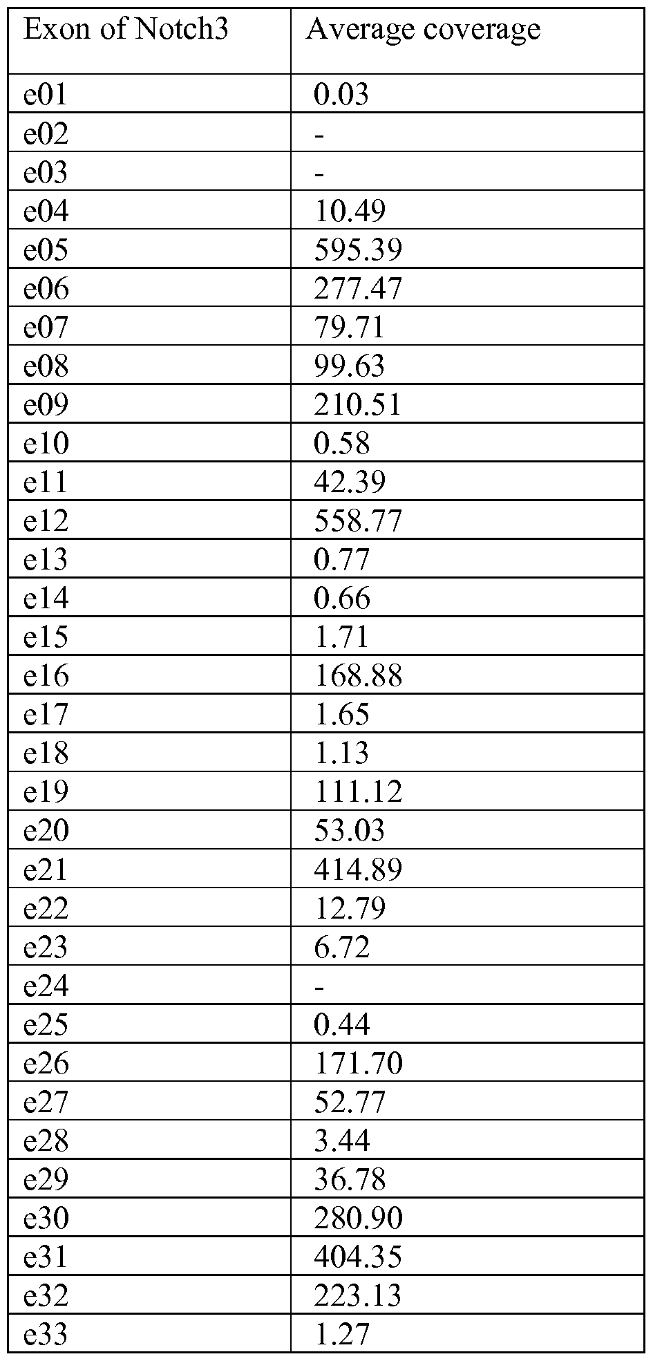
In order to determine whether any of these cell lines or primary tumors contain mutations in these regions, three approaches were used including Sanger Sequencing (Genewiz),
RainDance (Tewhey et al. (2009) Nature Biotechnology 27: 1025-1031) and RNAseq (Wang
et al. (2009) Nature Reviews Genetics 10:57-63). Mutations were identified in both the NRR and PEST domain in multiple cell lines and tumor samples as shown in Figure 9. In Figure 9a the upper panel shows cells lines with NRR mutations while the lower panel has PEST mutations. The NRR mutations identified in primary tumors are indicated in Figure 9b. Isolation of primary tumors and generation of a bank of primary tumor xenografts
Data obtained from primary human tumor xenografts was generated in the following manner: tumor specimens were collected in RPMI supplemented with 1% penicillin/streptomycin from patients during surgical resection with ischemic time less than one hour. Fragments of 15-30 mm3 free of necrotic tissue were grafted subcutaneous ly into interscapular fat pad of 6- to 8- week-old female nude mice under isoflurane anesthesia. Mice were maintained in specific pathogen-free animal housing and handled in accordance with approved protocols and regulations. Xenografts appeared at the graft site 2 to 8 months after grafting. They were subsequently transplanted from mouse to mouse once tumors reached 700-800 mm3 until a reasonably consistent growth rate is achieved. Frozen stocks in RPMI supplemented with 50% FBS and 10% DMSO were generated during serial passage in mice and were tested to ensure successful establishment of a xenograft model. Fragments of 30-50 mg from patients and xenografts at each passage were snap frozen for gene expression profiling, copy number as well as mutation analyses. Fragments of 150 mg of each successfully engrafted xenograft model were also collected and subject to histological analysis. An established tumor xenograft model was further used for in vivo studies after passage four. For gene expression profiling, total RNA was isolated using affinity resin (QIAGEN RNeasy Mini Kit; QIAGEN AG). RNA integrity and purity were assessed with the RNA 6000 Nano LabChip system on a
Bioanalyzer 2100 (Agilent Technologies).
Example 6: Characterization of Notch3 NRR mutations in a reporter gene assay Generation ofNotchi expression vectors with Notch3 NRR mutations
Two mutations were selected for characterization. TALL-1 cells are a t-cell acute
lymphoblastic cell line with a S1580L mutation. TALL-1 cells were purchased from DSMZ (#ACC 521). A breast tumor (X-1004) was also identified with a G1487D mutation. The RNA used for RNAseq analysis to detect mutations in the X-1004 sample was from a passage 5 mouse. These mutations were introduced into the vector pLNCX2_Notch3-GAL4-NLS- VP16
Constructs
Notch3 S1580L Gal4-VP16
MGPGARGRRRRRRPMSPPPPPPPVRALPLLLLLAGPGAAAPPCLDGSPCANGGRCTQ
LPSREAACLCPPGWVGERCQLEDPCHSGPCAGRGVCQSSVVAGTARFSCRCPRGFRG
PDCSLPDPCLSSPCAHGARCSVGPDGRFLCSCPPGYQGRSCRSDVDECRVGEPCRHGG
TCLNTPGSFRCQCPAGYTGPLCENPAVPCAPSPCR GGTCRQSGDLTYDCACLPGFEG
QNCEV VDDCPGHRCLNGGTCVDGV TY CQCPPEWTGQFCTEDVDECQLQPNAC
HNGGTCFNTLGGHSCVCV GWTGESCSQNIDDCATAVCFHGATCHDRVASFYCACP
MGKTGLLCHLDDACVSNPCHEDAICDTNPV GRAICTCPPGFTGGACDQDVDECSIG
ANPCEHLGRCV TQGSFLCQCGRGYTGPRCETDV ECLSGPCR QATCLDRIGQFTCI
CMAGFTGTYCEVDIDECQSSPCV GGVCKDRVNGFSCTCPSGFSGSTCQLDVDECAS
TPCRNGAKCVDQPDGYECRCAEGFEGTLCDRNVDDCSPDPCHHGRCVDGIASFSCAC
APGYTGTRCESQVDECRSQPCRHGGKCLDLVDKYLCRCPSGTTGV CEV IDDCASN
PCTFGVCRDGI RYDCVCQPGFTGPLCNVEINECASSPCGEGGSCVDGENGFRCLCPP
GSLPPLCLPPSHPCAHEPCSHGICYDAPGGFRCVCEPGWSGPRCSQSLARDACESQPC
RAGGTCSSDGMGFHCTCPPGVQGRQCELLSPCTPNPCEHGGRCESAPGQLPVCSCPQ
GWQGPRCQQDVDECAGPAPCGPHGICTNLAGSFSCTCHGGYTGPSCDQDI DCDPNP
CLNGGSCQDGVGSFSCSCLPGFAGPRCARDVDECLSNPCGPGTCTDHVASFTCTCPPG
YGGFHCEQDLPDCSPSSCFNGGTCVDGV SFSCLCRPGYTGAHCQHEADPCLSRPCL
HGGVCSAAHPGFRCTCLESFTGPQCQTLVDWCSRQPCQNGGRCVQTGAYCLCPPGW
SGRLCDIRSLPCREAAAQIGVRLEQLCQAGGQCVDEDSSHYCVCPEGRTGSHCEQEV
DPCLAQPCQHGGTCRGYMGGYMCECLPGY GDNCEDDVDECASQPCQHGGSCIDL
VARYLCSCPPGTLGVLCEINEDDCGPGPPLDSGPRCLHNGTCVDLVGGFRCTCPPGYT
GLRCEADI ECRSGACHAAHTRDCLQDPGGGFRCLCHAGFSGPRCQTVLSPCESQPC
QHGGQCRPSPGPGGGLTFTCHCAQPFWGPRCERVARSCRELQCPVGVPCQQTPRGPR
CACPPGLSGPSCRSFPGSPPGASNASCAAAPCLHGGSCRPAPLAPFFRCACAQGWTGP
RCEAPAAAPEVSEEPRCPRAACQAKRGDQRCDRECNSPGCGWDGGDCSLSVGDPWR
QCEALQCWRLF SRCDPACSSPACLYDNFDCHAGGRERTCNPVYEKYCADHFADG
RCDQGCNTEECGWDGLDCASEVPALLARGVLVLTVLLPPEELLRSSADFLQRLSAILR
TSLRFRLDAHGQAMVFPYHRPSPGSEPRARRELAPEVIGLVVMLEIDNRLCLQSPEND
HCFPDAQSAADYLGALSAVERLDFPYPLRDVRGEPLEPPEPSVPLLPLLVAGAVLLLV
ILVLGVMVARRKRVDKLLSSIEQACDICRLKKLKCSKEKPKCAKCLKNNWECRYSPK
TKRSPLTRAHLTEVESRLERLEQLFLLIFPREDLDMILKMDSLQDIKALLTGLFVQDNV
NKDAVTDRLASVETDMPLTLRQHRISATSSSEESSNKGQRQLTVSKLKLLSSIEQACP KKKRKVDEFPGISTAPPTDVSLGDELHLDGEDVAMAHADALDDFDLDMLGDGDSPG PG (SEQ ID NO: 276)
Notch3_G 1487D_Gal4-VP 16
MGPGARGRRRRRRPMSPPPPPPPVRALPLLLLLAGPGAAAPPCLDGSPCANGGRCTQ
LPSREAACLCPPGWVGERCQLEDPCHSGPCAGRGVCQSSVVAGTARFSCRCPRGFRG
PDCSLPDPCLSSPCAHGARCSVGPDGRFLCSCPPGYQGRSCRSDVDECRVGEPCRHGG
TCLNTPGSFRCQCPAGYTGPLCENPAVPCAPSPCRNGGTCRQSGDLTYDCACLPGFEG
QNCEVNVDDCPGHRCLNGGTCVDGVNTYNCQCPPEWTGQFCTEDVDECQLQPNAC
HNGGTCFNTLGGHSCVCVNGWTGESCSQNIDDCATAVCFHGATCHDRVASFYCACP
MGKTGLLCHLDDACVSNPCHEDAICDTNPVNGRAICTCPPGFTGGACDQDVDECSIG
ANPCEHLGRCVNTQGSFLCQCGRGYTGPRCETDVNECLSGPCRNQATCLDRIGQFTCI
CMAGFTGTYCEVDIDECQSSPCVNGGVCKDRVNGFSCTCPSGFSGSTCQLDVDECAS
TPCRNGAKCVDQPDGYECRCAEGFEGTLCDRNVDDCSPDPCHHGRCVDGIASFSCAC
APGYTGTRCESQVDECRSQPCRHGGKCLDLVDKYLCRCPSGTTGVNCEVNIDDCASN
PCTFGVCRDGINRYDCVCQPGFTGPLCNVEINECASSPCGEGGSCVDGENGFRCLCPP
GSLPPLCLPPSHPCAHEPCSHGICYDAPGGFRCVCEPGWSGPRCSQSLARDACESQPC
RAGGTCSSDGMGFHCTCPPGVQGRQCELLSPCTPNPCEHGGRCESAPGQLPVCSCPQ
GWQGPRCQQDVDECAGPAPCGPHGICTNLAGSFSCTCHGGYTGPSCDQDINDCDPNP
CLNGGSCQDGVGSFSCSCLPGFAGPRCARDVDECLSNPCGPGTCTDHVASFTCTCPPG
YGGFHCEQDLPDCSPSSCFNGGTCVDGVNSFSCLCRPGYTGAHCQHEADPCLSRPCL
HGGVCSAAHPGFRCTCLESFTGPQCQTLVDWCSRQPCQNGGRCVQTGAYCLCPPGW
SGRLCDIRSLPCREAAAQIGVRLEQLCQAGGQCVDEDSSHYCVCPEGRTGSHCEQEV
DPCLAQPCQHGGTCRGYMGGYMCECLPGYNGDNCEDDVDECASQPCQHGGSCIDL
VARYLCSCPPGTLGVLCEINEDDCGPGPPLDSGPRCLHNGTCVDLVGGFRCTCPPGYT
GLRCEADINECRSGACHAAHTRDCLQDPGGGFRCLCHAGFSGPRCQTVLSPCESQPC
QHGGQCRPSPGPGGGLTFTCHCAQPFWGPRCERVARSCRELQCPVGVPCQQTPRGPR
CACPPGLSGPSCRSFPGSPPGASNASCAAAPCLHGGSCRPAPLAPFFRCACAQGWTGP
RCEAPAAAPEVSEEPRCPRAACQAKRGDQRCDRECNSPGCGWDGGDCSLSVGDPWR
QCEALQCWRLFNNSRCDPACSSPACLYDNFDCHAGGRERTCNPVYEKYCADHFADG
RCDQDCNTEECGWDGLDCASEVPALLARGVLVLTVLLPPEELLRSSADFLQRLSAILR
TSLRFRLDAHGQAMVFPYHRPSPGSEPRARRELAPEVIGSVVMLEIDNRLCLQSPEND
HCFPDAQSAADYLGALSAVERLDFPYPLRDVRGEPLEPPEPSVPLLPLLVAGAVLLLV
ILVLGVMVARRKRVDKLLSSIEQACDICRLKKLKCSKEKPKCAKCLKNNWECRYSPK TKRSPLTRAHLTEVESRLERLEQLFLLIFPREDLDMILKMDSLQDIKALLTGLFVQDNV NKDAVTDRLASVETDMPLTLRQHRISATSSSEESSNKGQRQLTVSKLKLLSSIEQACP KKKRKVDEFPGISTAPPTDVSLGDELHLDGEDVAMAHADALDDFDLDMLGDGDSPG PG (SEQ ID NO: 277)
Retrovirus was produced by transfecting 293-GP2 Packaging Cell Line (Clontech, cat# 631458) with the appropriate retroviral vector. Promega's Fugene6 was used as the lipid- based transfection reagent. Transfection was carried out according to manufacturer's instructions. Virus was collected at 48 hs after transfection and immediately used to transduce HLR cells (HLR-PathDetect, Stratage). HLR cells (Stratagene) were transduced with either Notch3wt-Gal4-VP 16, Notch3_p. S 1850L-Gal4-VP 16 or Notch3_p.G 1487D-Gal4-VP 16 retroviral particles. Cells were selected with G418 for 2 weeks before testing.
Notch3 reporter gene assay to assess basal activity ofNotch3 wild-type and Notch3 NRR mutant receptors Notch3 reporter gene assay: HLR-Notch3wt-Gal4-VP16, HLR-Notch3_p.S1580L-Gal4-VP16 and HLR-Notch3_p.G1487D-Gal4-VP16 cells were maintained in DMEM no phenol red, 10% FBS (Hyclone, cat# SH30071), 1% penicillin-streptomycin (Gibco cat# 15140-122), L- Glutamine (Gibco, cat#25030-081), 100 μg/mL hygromycin (Gibco, cat# 10687-010) and 400 μg/mL G418 (Gibco, cat# 10131 -027). The HLR parental line was maintained in DMEM no phenol red, 10% FBS (Hyclone, cat# SH30071), 1% penicillin-streptomycin (Gibco cat# 15140-122), L-Glutamine (Gibco, cat#25030-081) and 100μg/mL hygromycin (Gibco, cat# 10687-010). Sub-confluent cells grown in complete medium were washed with PBS (Gibco, cat# 20012-027), trypsinized with TryplE (Gibco, cat# 12605010), and diluted into 4xl04 cells/mL; 100 μΐ^ of cell suspension was plated in 96-well clear bottom white plates (Costar, cat# 3610) at a density of 4000 cells/well. All plates were then incubated overnight at 37°C prior to treatment with DAPT (10 μΜ, CalBiochem). Plates were returned to the incubator for 24 hrs before luciferase activity was determined using Bright-Glo (Promega). The Envision plate reader (PerkinElmer) was used to determine amount of luminescence.
FACS assay to assess cell surface levels of wild-type and mutant Notchi receptors To demonstrate expression of mutant Notch3 receptors in a cell line, flow cytometry was used. Cell lines expressing mutant Notch3 and wild-type Notch3 (grown under standard conditions) were mixed with an anti- Notch3 binding and detection antibody that contains an
APC fluorescein label (R&D cat# FAB1559A) in PBS containing 0.1% BSA and 0.01% sodium azide, and incubated for 1 hr at 4°C. After washing, the cells were analyzed by BD FACSCanto instrument using light and side scatter properties to gate on single cells.
The level of Notch3 receptors on the cell surface was determined by binding of commercially available anti-Notch3 APC (R&D # FAB 1559A) labeled antibody to cells expressing mutant and endogenous Notch3 and assessed by FACS. Cells were trypsinized (Invitrogen TrypLE cat# 12605-010) and diluted to 2xl06 cells/mL in FACS Buffer (PBS/3% FBS/0.01%NaN3). 2.5xl05 cells/well were added to each well of a 96 well plate (Corning cat#3610) and centrifuged at 1500 rpm for 5 min at 4°C before removing the supernatant. Anti-Notch3 APC antibody or Sheep IgG Isotype Control labeled with APC (R&D cat#IC016A) was added to the cell pellets at a final concentration of 0.1 μg in 100 μϊ^ of FACS buffer and incubated for 1 hour at 4°C. The cells were washed and pelleted 2 times with 100 μΐ, FACS Buffer.
Finally cells were resuspended in 200 μΐ, FACS buffer and fluorescence values were measured with a BD FACSCanto (BD Biosciences). The amount of cell surface bound anti- Notch3 APC antibody was assessed by measuring the mean channel fluorescence.
Summary and Discussion
Introduction of either a S1580L mutation or a G1487D mutation into a Notch3 receptor resulted in an approximately 10 fold increase in the basal signaling from the receptor relative to a wild-type control. In this system the wild-type and mutant receptors were expressed at approximately equivalent levels as determined by FACS assay. This data suggests that these mutations activate Notch3 signaling in cell lines and tumors expressing these and other similar mutations (see further discussion in Examples 7, 9, 10, 1 1, 15).
Example 7: Effect of Notch3 antibodies on Notch3 signaling and in vitro proliferation in TALL-1 cells The TALL-1 cell line has a mutation in the NRR domain of Notch3 at S1580L. Introduction of this mutation into a Notch3 expression construct resulted in activation of Notch3 signaling. To further characterize the effects of inhibition of Notch3 signaling in this cells line, the mRNA levels of Notch target genes were examined and the in vitro proliferation of the cells was monitored in the presence of Notch 3 antibodies.
TALL-1 in vitro proliferation assay lxlO4 TALL-1 cells/well were seeded into 96-well tissue culture plates (Corning, Catalog #3610) in lOOul medium (RPMI-1640 supplemented with 10% fetal bovine serum and 1% penicillin/ streptomycin). The same day, antibody dilutions were prepared in IX PBS from which 5μ1 of 20X antibody dilution was added per well. Cells were incubated with antibody at 37°C/5 % C02. After incubation for 0 and 9 days at 37 °C/5 % C02, ΙΟΟμΙ of CellTiter-Glo reagent (Promega) was added and the plates were incubated for lOmin on plate shaker. The amount of luminescence was determined using a Perkin Elmer Envision plate reader.
CellTiter-Glo luminescene values of cells treated with an IgG control were used to normalize the data and calculate percentage inhibition of proliferation due to treatment with Notch3 antibodies.
TALL-1 mRNA quantitation assay
Deltexl is a well characterized target gene of Notch signaling in TALL lines (Weng et ah, 2006, Genes Dev. 20:2096-2109). lxlO4 TALL-1 cells/well were seeded into 96-well tissue culture plates (Corning, cat#3610) in ΙΟΟμΙ medium (RPMI-1640 supplemented with 10% fetal bovine serum and 1% penicillin streptomycin). The same day, antibody and compound dilutions were prepared in IX PBS from which 5μ1 of 20X antibody or compound dilution was added per well. DAPT (Calbiochem, cat#565770) and DMSO (ATCC, Catalog #4-X-5) were the compounds used for this assay. Cells were incubated with antibody or compound at 37°C/5 % C02 for 72hr. RNA was isolated using the Qiagen RNeasy 96 kit. cDNA was made using the TaqMan Reverse Transcription reagents (Life Technologies) and the MJ Research PTC-225 Thermal cycler. TaqMan gene expression assays were run using TaqMan Universal PCR Master Mix (Life Technologies) along with gene expression probes for Deltexl (DTX1) (Hs00269995_ml, Life Technologies) and the housekeeping gene PP1A (Hs99999904_ml, Life Technologies). TaqMan gene expression assays were run on the Applied Biosystems ABI Prism 7900HT Fast Real-Time PCR system. To quantitate the levels of Deltex 1, 2- [delta][delta]Ct method was employed. Calculation of delta delta Ct involves comparing the Ct values of the samples of interest with a control such as a non-treated sample or DMSO treated sample (Schmittgen and Livak (2008) Nature Protocols 3 : 1 101-1 108). Summary and discussion
As shown in Figures 1 1A-B, Notch3 antibodies that were identified from pannings against NRR domain or EGF32-NRR domain (20350, 20358, 20337) potently inhibited Deltexl
mRNA expression in TALL-1 cells. In contrast antibodies directed to the LBD domain (20364) did not significantly inhibit Deltexl mRNA. In addition, 20350, 20358, 20337 significantly inhibited TALL- 1 proliferation in a dose-dependent manner. When Notch3 antibodies were tested in a panel of other TALL cell lines (DND41, P12-Ichikawa, SUPT1, SUPT1 1 and RPMI-8402), no effects on proliferation of were detected.
Example 8: Generation of a neo-epitope antibody that detects the gamma secretase cleaved form of the Notch3 intracellular domain.
Notch signaling is activated by a series of proteolytic cleavages. The gamma secretase complex mediates the final cleavage of the Notch receptor ultimately releasing the Notch intracellular domain (ICD) that translocates to the nucleus to activate Notch target gene transcription. A neo-epitope antibody was generated to detect the gamma secretase cleaved form of the Notch3 ICD (ICD3) only when cleaved between amino acids Glyl661 and Vail 662 (human Notch3).
Generation of a ICD3 rabbit polyclonal antibody The peptides used for immunization and negative selection (depletion peptide) are indicated.
Immunization peptide: H2N-VMVARRK(dPEG4)C-amide (SEQ ID NO: 278)
Depletion peptide: Ac-VILVLGVMVARRK(dPEG4)C-amide (SEQ ID NO: 279). A rabbit polyclonal antibody was generated at Covance using standard procedures. Briefly, a 77 day protocol was employed with a primary boost with 500μg of immunizing peptide and Freund's adjuvant. Additional boosts with 500μg of immunizing peptide were performed on day 21, 42 and 63. To deplete non-specific antibodies that recognize the VMVARRK (SEQ ID NO: 243) sequence of Notch3, but not the neo-epitope following gamma secretase cleavage, a depletion peptide was used for negative selection. The purified sample was depleted using the depletion peptide by "negative" affinity chromatography. Peptides were coupled to a column using terminal cysteine to properly orient the peptide. Cross reacting antibodies were removed from the sample and confirmed by ELISA. Serum from rabbit was tested by Western blot in TALL- 1 cells to determine if a specific band was detected.
Conversion of the rabbit polyclonal ICD3 antibody to a rabbit monoclonal antibody
To convert the rabbit polyclonal antibody to a rabbit monoclonal antibody, a final IV boost of immunizing peptide was performed on the selected rabbit. 4 days later a splenectomy was
performed and rabbit hybridomas were generated by standard procedures at Epitomics.
Briefly all the lymphocytes from 1 rabbit spleen were isolated. Fusion and standard ELISA screen of 40 x 96 well plates was performed. All ELISA positive hybridomas were expanded to 24 well plates and an ELISA was again performed with both the immunizing peptide and the depletion peptide. Supernatant from the 139 positive hybridomas were analyzed by Western blotting in TALL-1 cells. Based on Western screening of the ELISA positive hybridomas, 3 hybridomas (73, 128, 95) were chosen for subcloning. To subclone hybridomas, a limited dilution of the selected parental hybridomas (0.5 cells/well) was perfomed and these sub-clones were plated in 4 x 96 well plates. Subclones were again screened by ELISA using both the immunizing peptide and the depletion peptide. Clones were expanded to 24 well plates and supernatants from ELISA positive sub-clones were screened by Western blotting in TALL-1 cells. Exemplary Western data from 3 sub-clones are shown.
In vitro screening ofNotch3 signaling inhibition using an ICD3 antibody An antibody targeting the Notch3 ICD was used to assess pathway activity. Cell line TALL-1 was purchased from DSMZ and routinely maintained in growth media supplemented with 10% FBS and 1% Penicillin-Streptomycin. Experimental set up: 5 million TALL-1 cells were plated in 10 mL of medium in a 25 cm2 tissue culture flask (Corning, cat#430639). Cells were treated with either 0.5% DMSO or 10μΜ DAPT (Calbiochem, cat#565770) for 72h. TALL- 1 cells were spun down and then washed in PBS. Cells were lysed in 60 of IX Cell Lysis Buffer (CST, cat#9803) with the addition of N-ethylmaleimide (Thermo Scientific, cat# 23030) and protease and phosphatase inhibitors (Pierce, Cat# 78444). Protein quantitation was performed using the BCA method and read in a Spectramax M5 microplate reader. 30 μϊ^ of protein samples were loaded per well in a 4-12% Bis-Tris gel (Invitrogen, cat# NP0006-1). SDS-PAGE: Samples were run under standard conditions in IX NuPage MOPS SDS running buffer (Invitrogen, cat# NP0001) for approximately 90 min at 180 V. Before transfer to a nitrocellulose membrane (iBlot, Invitrogen), gels were soaked in 2x Transfer Buffer
(Invitrogen, cat#NP0006-l) with 20% methanol. Membranes were blocked in 4% milk-TBST for one hour; supernatants from hybidoma supernatants were diluted 1 :4 in 2% milk-TBST and incubated ON at 4°C with gentle shaking. Secondary antibodies were added in 2% milk- TBST for 45 minutes, after a series of membrane washes with TBST. Membrane was developed using ECL Plus Western Detection System (GE healthcare, cat# RPN2232).
Screening of a panel ofT-cell acute lymphoblastic leukemia cells lines with an ICD3 antibody.
Cell lines TALL-1 (# ACC521), RPMI8402 (#ACC290), DND41 (#ACC525), SUPT11 (#ACC605), and P12-Ichikawa (#ACC34) were purchased from DSMZ and routinely maintained in growth media supplemented with 10% FBS and 1% Penicillin-Streptomycin. Cell lines HPB-ALL and Jurkat cells were obtained commercially from Andreas Strasser (Walter and Eliza Hall Institute of Medical Research, Australia. 5 million TALL- 1 cells were plated in 10 mL of medium in a 25 cm2 tissue culture flask (Corning, cat#430639). Cells were spun down and then washed in PBS. Cells were lysed and Western performed as described above. Purified antibody from hybridoma sub-clone 73-8 was used for further studies at a 1 :5000 dilution. This antibody is hereafter referred to as "ICD3 Ab" and its sequence is detailed in Table2. ICD l protein levels were assessed using an antibody from Cell Signaling (#2421) at a dilution of 1 : 1000.
Summary and Discussion As shown in Figures 12A-B, high levels of ICD3 protein was only detected in TALL-1 cells but not in a panel of other T-cell acute lymphoblastic leukemia cell lines. High ICDl levels can be detected in several TALL lines including HPBALL, RPMI-8402, DND41, P12 Ichikawa and Jurkat, which are known to have activating mutations in Notch 1 (Weng et al. (2004) Science 306:269-71). The ICD3 antibody does not cross-react with ICDl as evidenced by lack of signal in these other TALL lines with Notchl mutations.
Example 9: In vitro assessment of Notch3 signaling inhibition upon antibody treatment
Evaluation of Notch3 mutation status in the panel of CCLE lines resulted in identification of TALL-1 with an NRR mutation and MDA-MB468 with a PEST domain mutation. MDA- MB468 cells have a frameshift mutation at amino acid 2034 which results in introduction of a premature stop codon. Therefore the ICD3 has an altered molecular weight which can be detected as a faster migrating band on a Western blot.
Sequences of portions of WT and MDAMB468 PEST domain:
WT Notch3 sequence (NP_000426) amino acid 2034-end
PSGPRSPPGPHGLGPLLCPPGAFLPGLKAAQSGSKKSRRPPGKAGLGPQGPRGRGKKL TLACPGPLADSSVTLSPVDSLDSPRPFGGPPASPGGFPLEGPYAAATATAVSLAQLGGP
GRAGLGRQPPGGCVLSLGLLNPVAVPLDWARLPPPAPPGPSFLLPLAPGPQLLNPGTP VSPQERPPPYLAVPGHGEEYPAAGAHSSPPKARFLRVPSEHPYLTPSPESPEHWASPSP PSLSDWSESTPSPATATGAMATTTGALPAQPLPLSVPSSLAQAQTQLGPQPEVTPKRQ VLA (SEQ ID NO: 280) MDA-MB468 sequence amino acid 2034-end
PSGPRSPPRSPRPGASALSSRGLPPWPQSGTVGVQEEQEAPREGGAGAAGAPGAGQE ADAGLPGPPG. (SEQ ID NO: 281)
Initially these 2 cell line models were used to characterize the effects of Notch3 inhibitory antibodies on Notch3 signaling. Western blots with the ICD3 antibody were used to monitor signaling inhibition. Experimental set up: one million MDA-MB468 cells were plated in a 60 mm dish (Corning, cat# 430196) in 3 mL of medium or 5 million TALL-1 cells in 10 mL of medium in a 25cm2 tissue culture flask (Corning, cat#430639). Plates were incubated overnight at 37°C prior to treatment with 10 μg/mL final concentrations of Notch3 inhibitor antibodies 20337, 20350, 20358 and 20802 as well as an IgG control. Antibodies were added directly to the plate and they were further incubated for 72h at 37°C, 5% CO2. In addition some cells were treated with either 0.5% DMSO or 10μΜ DAPT (Calbiochem, cat#565770) for 72h. Cells were harvested by aspirating the media and rinsing in 1 mL PBS (Gibco, cat#20012-027), scraping the cells off the plate, and spinning down on a bench top centrifuge. Suspension cells were spun down and then washed in PBS. Western blots were performed with the purified ICD3 antibody as described previously.
In addition, three other cells lines were characterized for ICD3 levels and signaling inhibition upon Notch3 antibody treatment:- (i) Ishikawaheraklio02_ER has a NRR mutation at N1597R, (ii) A549 has a PEST frameshift mutation at 2034, while (iii) TE-11 has a PEST frameshift mutation at 2260. Summary and discussion
As shown in Figures 13A-B, in addition to the previously described ligand-driven RGA and Notch target gene mRNA quantitation, Notch3 signaling can also be monitored by measuring levels of ICD3. ICD3 levels are a membrane proximal readout of Notch3 signaling activity. Treatment of TALL-1 cells with Notch3 antibodies 20337, 20350, 20358 resulted in decreased levels of ICD3. Level of ICD3 was equivalent in the IgG control sample and the DMSO samples. This data is consistent with inhibition of Deltexl mRNA and TALL-1
proliferation upon treatment with these antibodies. In contrast no effect on ICD3 levels was detected with 12229 treatment. As shown in Figures 13 B, in MDA-MB468 cells, the frame- shift mutation at amino acid 2034 results in a premature stop codon and smaller ICD3. This ICD3 can be detected as a faster migrating band on a Western blot. Upon treatment with Notch3 NRR antibodies 20337, 20350, 20358, decreased levels of ICD3 was detected. In contrast, treatment with 20802, a LBD antibody, did not alter ICD3 levels relative to a control IgG. As shown in Fig 14 A-C, varying effects on ICD3 levels were detected upon Notch3 antibody treatment inIshikawaheraklio02_ER, TE-1 1, and A549 cells. However in all cell lines tested, 20350 treatment consistently resulted in significantly decreased ICD3 levels. Example 10: In vitro assessment of Notch3 signaling inhibition upon antibody treatment in a Notch3 amplified cell line
HCC1143 cells were described to have an amplification of Notch3 (Yamaguchi et al. (2008) Cancer Res. 68: 1881-1888). The levels of active Notch3 signaling were examined in this cell line using the ICD3 antibody. Western blots with the ICD3 antibody were used to monitor signaling inhibition.
Experimental set up: one million HCC1 143 cells were plated in a 60 mm dish (Corning, cat# 430196) in 3 mL of medium in a 25cm2 tissue culture flask (Corning, cat#430639). Plates were incubated overnight at 37°C prior to treatment with 10 μg/mL final concentrations of Notch3 inhibitor antibodies 20337, 20350, 20358 and 20802 as well as an IgG control.
Antibodies were added directly to the plate and they were further incubated for 72hr at 37°C, 5% CO2. Cells were harvested and Western blots performed as described previously.
Summary and discussion
As shown in Figure 15, HCC1 143 cells are amplified for Notch3 and exhibit high levels of ICD3. All Notch3 antibody treatments resulted in decreased ICD3 levels. At 10μg/ml, 20350 treatment resulted in the largest reduction of ICD3 levels.
Example 11: In vivo PD Assessment
PD modulation was interrogated in three xenograft models harboring genetic abberations in Notch3: the NRR mutant TALL-1 human leukemia model, the PEST mutant MDA-MB-468 human breast model, and the Notch3 -amplified HLUX1823 patient-derived lung model.
In vivo PD in the TALL-1 human leukemia xenograft model
Female SCID-beige mice harboring TALL- 1 xenografts were treated with a single dose of Notch3 antibodies. Mice were inoculated with 10 xlO6 cells injected subcutaneously in a suspension of Hank's balanced salt solution. Once tumors reached between 300 and 500 mm3 (n=3/group), mice were randomly assigned to receive a single intravenous 20 mg/kg dose of 3207 (IgG control), 20350, 20358 or 20802. Following treatment, tumors were harvested at selected time points and ICD3 was evaluated by Western blot and IHC, as described below.
In vivo PD in the MDA-MB-468 human breast cancer xenograft model
Female SCID-beige mice harboring MDA-MB-468 xenografts were treated with a single dose of Notch3 antibodies. A 3 x 3 x 3 mm3 tumor fragment was passaged from a MDA-MB-468 tumor bearing mouse (donor) and implanted subcutaneously into SCID-beige recipient mice on both the left and right flank. Once tumors reached between 300 and 500 mm3 (n=3/group), mice were randomly assigned to an untreated control group or received a single intravenous 20 mg/kg dose of 20350. In additional studies, the effects of a single intravenous 20 mg/kg dose of 20350, 20358, 20337 and 20802 relative to PBS or 3207 non-targeting IgG controls was assessed. Following the various treatments, tumors were harvested and ICD3 was evaluated by Western blot, as described below.
In vivo PD in the HLUX1823 patient derived lung cancer xenograft model
The activity of anti-Notch3 antibodies was also evaluated in a Notch3 -amplified patient- derived primary lung cancer tumor xenograft model, HLUX-1823. In these studies, nu/nu mice were implanted subcutaneously with 3 x 3 x 3 mm3 tumor fragments containing 50% phenol-red free matrigel (BD Biosciences) in DMEM and reached approximately 250 mm3 at 30 days post-implantation. Once tumors reached between 300 and 500 mm3 (n=3/group), mice were randomly assigned to receive either PBS or a single 20 mg/kg intravenous dose of either the 3207 non-targeting IgG control antibody, or 20358 or 12229 (the parental antibody from which 20364 and 20802 were derived). Following the various treatments, tumors were harvested and ICD3 was evaluated by Western blot, as described below.
Preparation of tumor cell lysates and ICD3 Western
Tumor samples were lysed in 200-400 iL of T-PER Tissue Protein Extraction Reagent (Pierce, cat# 78510) with Complete mini EDTA free protease inhibitor cocktail tablets (Roche, Cat# 04693 159001), using a Tissue Lyser II (Qiagen) for 1 min at 30 Hz. One 5 mm stainless steel bead (Qiagen, cat# 69965) was placed per tube to help with tissue lysis. After bead removal, samples were then centrifuged on a bench-top centrifuge at top speed for 15 min at 4°C. Supernatants were collected and either stored at -80°C for studies at a later time or protein concentration as assessed using the BCA method (Pierce, cat# 232550) and a Western blot for ICD3 was run as described previously. Where applicable, Western was also performed with a full-length Notch3 antibody to detect total levels of Notch3 (Cell Signaling #2889, 1 : 1000 dilution).
Detection of ICD3 levels by IHC
Xenograft tumors were fixed in 10% formalin and embedded in paraffin. 5μιη sections were placed on charged polylysine-coated slides. Immunohistochemistry protocol was optimized on an automated system Discovery ULTRA (Ventana Medical System).
Sections were baked at 60°C for 8 minutes, followed by deparaffination. Antigen retrieval was achieved in Cell Conditioning 1 (CC1, a TRIS based buffer with a slightly basic pH) at high temperature for 76 minutes. Blocking of non-specific binding of antibody was carried on using a specific Antibody Blocking (cat#760-4204). Primary antibody Notch3 ICD (20μg/ml) was incubated at 37°C for 60 minutes followed by incubation in secondary antibody for 32 minutes. Amplification step was performed using a specific Discovery Amplification HQ kit #760-052 (Ventana Medical Systems) as per manufacture specifications. Detection was achieved with diaminobenzydine (DAB) and counterstain with Hematoxylin. All these steps were run on Ventana Discovery ULTRA (Ventana Medical Systems). Summary and Discussion
Figures 16- 18 show in vivo PD studies in several xenograft models. As described earlier in this application, in vitro treatment of TALL- 1 cells with otch3 antibodies resulted in inhibition of signaling as assessed by both Deltexl mRNA levels and ICD3 protein levels. TALL- 1 cells were grown as a xenograft and mice were treated with Notch3 antibodies. Changes in Notch3 signaling in TALL- 1 tumors was monitored by assessing ICD3 levels by Western blotting or IHC. Treatment with antibodies 20350 or 20358 resulted in decreased
levels of ICD3 as shown in Figures 16A-B. ICD3 staining by IHC is indicated by the black/dark grey cells in the tumor section as shown in Figure 16B. ICD3 levels in tumors were assessed 72 h following the last Notch3 antibody administration, and there were still some cells within the tumor that showed strong ICD3 expression. In the MDA-MB468 model, as assessed by Western blotting, animals treated with 20350 yielded a marked decrease in ICD3 24 h and 72 h post dose relative to untreated control mice (Figure 17A). It was found that, at the 72 h timepoint, 20350, 20358 and 20337, all of which target the Notch3 NRR, induced decreases in ICD3 levels relative to the PBS and 3207 (IgG) controls. In contrast, following treatment with 20802, which targets a region of Notch3 outside of the NRR, ICD3 levels appeared similar to control levels (Figure 17B). In the HLUX1823 Notch3-gene amplified model, as assessed by Western blotting, animals treated with either 20358 or 12229 yielded a marked decrease in ICD3 at 72 h post dose relative to control mice (Figure
18). Taken together, these data demonstrate that the Notch3 NRR antibodies can inhibit Notch3 signaling in the presence of Notch3 gene-amplification or mutations in either the NRR or PEST domains, whereas Notch3 antibodies raised outside of this region can only inhibit Notch3 signaling in the presence of the gene-amplification and have more limited activity in the presence of mutations.
Example 12: In vivo efficacy in TALL-1 xenografts
Generation of a TALL-1 cell line with constitutive expression of luciferase The TALL-1 cell line was transduced with pMMP-LucNeo retrovirus (see US 7399851) and selected in lmg/mL of Geneticin (G418) for several weeks. TALL-l_Luc cells express high levels of luciferase compared to TALL-1 cells, where it was absent. Wild-type and luciferased cells were subjected to a proliferation experiment with Notch3 antibody inhibitors, showing identical results; suggesting that the infection did not interfere with TALL-1 sensitivity to Notch3 inhibition.
Assessment of in vivo activity ofNotchi antibodies in a TALL-1 cell-line xenograft model
Mice were inoculated with 10 xlO6 T-ALLl_Luc cells injected subcutaneously in a suspension of Hank's balanced salt solution and the presence of tumors was monitored using the Xenogen in vivo imaging system (Caliper Life Sciences). The presence of tumors was detectable by day 7. On day 11, tumor-bearing animals were randomly assigned to receive intravenous doses of either PBS or 20 mg kg of 3207 negative control IgG antibody or the
Notch3 antibodies 20337, 20350, 20358 or 20802 as single agents twice per week. Tumor size was monitored using the Xenogen in vivo imaging system.
Summary and Discussion
As shown in Figure 19, 20358 and 20350 showed the most anti-tumor activityof the antibodies evaluated in this study. Figure 19A shows a graphical representation of the luminescent signal obtained following the various treatments over the time-course of the study and Figure 19B shows the luminescent signal of the control groups at day 29 (the last time point that it was possible to image due to tumor size) and of the anti-Notch3 antibody treatment groups at day 43. Example 13: Epitope binning of NRR Notch 3 antibodies with Biacore
Epitope binning via Biacore was performed to classify Notch 3 antibodies (IgG or Fab fragments) into groups of identical, or significantly overlapping epitopes, i.e. antibodies which were able to inhibit each other's binding.
Experimental set-up epitope binning with Biacore For epitope binning in Biacore, a sensor chip with a low density of immobilized or captured antigen was used (comparable to a kinetic experiment). The same sample prerequisites as for KD determination applied (i.e. monomer content). Experimental conditions, concerning preparation of chip (antigen immobilization/capture), as well as regeneration conditions were identical to KD determination in Biacore . To achieve saturation of an epitope, only one (high) concentration per antibody was used (e.g. 250nM for 90 s).
Antibody samples were injected pair wise in a full factorial assay design, e.g. for two antibody samples, A and B, the following pair wise injections were required: A-A, A-B, B-A, B-B.
The sensor chip had to be saturated with antibody by the first injection, so that the second antibody was only able to bind in case of a different epitope. Complete regeneration of bound antibodies had to be performed after each double injection.
For evaluation of the controls, i.e. double injections of the identical antibodies (A-A, B-B), their binding levels at the end of each injection were evaluated: The second injection was expected to give no additional binding. Double injections of different antibody sample pairs were compared for consistency, e.g. if the injection A-B resulted in additional binding of B (different epitopes) the injection of B-A was expected to result in additional binding of A, too.
Possible causes for creating such inconsistencies were e.g. partially overlapping epitopes, or large differences in KD.
Summary and Discussion:
Figure 20 summarizing the evaluation of all pair wise antibody injections was compiled, indicating their mutual inhibition status from which epitope groups or bins could be concluded. Based on these studies, it was determined that the NRR antibodies identified from phage display screening have different conformational epitopes. As shown in Figure 20 no additional binding was detected when 20345 was added first followed by either 20350 or 20351. Similar information was obtained independent of which antibody was added first in the experiment. Therefore 20345, 20350 and 20351 have overlapping epitopes, which is designated in the table with dark grey shading. This epitope is defined as NRR_B. When 20337 was addedfirst, additional binding to the Biacore could be detected with any of the following antibodies added second: 20345, 20350, 20351, 20358 and A4. The same conclusion was reached if the order of addition of the two antibodies was reversed. Therefore, 20337 has a distinct epitope to any of the other antibodies tested and was designated as epitope NRR_A. When 20358 was added first, additional binding was detected with 20337, 20345, 20350 and 20351. The same conclusion was reached if the order of addition of the two antibodies was reversed. Therefore, 20358 has a distinct epitope to these other antibodies tested and was designated as epitope NRR_C. In contrast when 20358 was added first and A4 was added second, there was minimal additional binding (see cells with light grey shading in summary table). This result was further confirmed in additional Biacore studies. In these experiments, either A4 or 20358 was immobilized to an appropriate sensor chip, Notch3 NRR (SEQ ID: 282) was flowed over the surface and the ability of the other antibody not immobilized to the sensor surface to bind to Notch3 NRR was evaluated. Under these assay conditions, it was found that 20358 could bind to Notch3 bound to immobilized A4 and that A4 could also bind to Notch3 bound to immobilized 20358 on the sensor surface. This data is entirely consistent with the other studies performed and with the conclusion that A4 and 20358 bind to distinct epitopes within the Notch3 NRR. Furthermore, when A4 was added first, additional binding was detected when the following antibodies were added second: 20337, 20345, 20350, 20351. The same conclusion was reached if the order of addition of the two antibodies was reversed. Therefore A4 has a distinct epitope to 20337, 20345, 20350, 20351 and was designated as epitope NRR D.
Example 14: Co-crystal structure studies with 20350 and NRR as well as 20358 and NRR
Two crystal structures of human Notch3 Negative Regulatory Region (NRR, SEQ ID NO: 282) bound to Fab fragment of 20350 or 20358 were determined. As detailed below, Notch3 NRR was expressed, purified and mixed with 20350 or 20358 Fab to form complex. Protein crystallography was employed to generate atomic resolution data for Notch3 NRR bound to 20350 or 20358 Fab, respectively, to define their epitopes (as Notch3 NRR residues within 5 A distance to the antibody residues).
Protein production
The sequences of Notch3 NRR, 20350 Fab, and 20358 Fab produced for crystallography are shown below. Construct of Notch3 NRR comprises residues 1378 to 1640 (underlined) of human Notch3 (UniProt identifier Q9UM47, SEQ ID NO: 1), along with N- and C-terminal residues from recombinant expression vector (shown in lower case letters, SEQ ID NO: 282). The N-terminal signal sequence from mouse IgG kappa light chain was used for secreted expression and was cleaved during expression, leaving intact N-terminus of Notch3 NRR. For 20350 and 20358 Fab, the sequences of heavy and light chains are shown (SEQ ID NOs: -6 283, 284, 285, and 286).
Proteins used for crystal structure determination
Construct:
Human Notch3 NRR (Q9UM47)
MGPGARGRRRRRRPMSPPPPPPPVRALPLLLLLAGPGAAAPPCLDGSPCANGGRCTQ
LPSREAACLCPPGWVGERCQLEDPCHSGPCAGRGVCQSSVVAGTARFSCRCPRGFRG
PDCSLPDPCLSSPCAHGARCSVGPDGRFLCSCPPGYQGRSCRSDVDECRVGEPCRHGG
TCLNTPGSFRCQCPAGYTGPLCENPAVPCAPSPCRNGGTCRQSGDLTYDCACLPGFEG
QNCEVNVDDCPGHRCLNGGTCVDGVNTYNCQCPPEWTGQFCTEDVDECQLQPNAC
HNGGTCFNTLGGHSCVCVNGWTGESCSQNIDDCATAVCFHGATCHDRVASFYCACP
MGKTGLLCHLDDACVSNPCHEDAICDTNPVNGRAICTCPPGFTGGACDQDVDECSIG
ANPCEHLGRCVNTQGSFLCQCGRGYTGPRCETDVNECLSGPCRNQATCLDRIGQFTCI
CMAGFTGTYCEVDIDECQSSPCVNGGVCKDRVNGFSCTCPSGFSGSTCQLDVDECAS
TPCRNGAKCVDQPDGYECRCAEGFEGTLCDRNVDDCSPDPCHHGRCVDGIASFSCAC
APGYTGTRCESQVDECRSQPCRHGGKCLDLVDKYLCRCPSGTTGVNCEVNIDDCASN
PCTFGVCRDGINRYDCVCQPGFTGPLCNVEINECASSPCGEGGSCVDGENGFRCLCPP
GSLPPLCLPPSHPCAHEPCSHGICYDAPGGFRCVCEPGWSGPRCSQSLARDACESQPC
RAGGTCSSDGMGFHCTCPPGVQGRQCELLSPCTPNPCEHGGRCESAPGQLPVCSCPQ
GWQGPRCQQDVDECAGPAPCGPHGICTNLAGSFSCTCHGGYTGPSCDQDINDCDPNP
CLNGGSCQDGVGSFSCSCLPGFAGPRCARDVDECLSNPCGPGTCTDHVASFTCTCPPG
YGGFHCEQDLPDCSPSSCFNGGTCVDGV SFSCLCRPGYTGAHCQHEADPCLSRPCL
HGGVCSAAHPGFRCTCLESFTGPQCQTLVDWCSRQPCQNGGRCVQTGAYCLCPPGW
SGRLCDIRSLPCREAAAQIGVRLEQLCQAGGQCVDEDSSHYCVCPEGRTGSHCEQEV
DPCLAQPCQHGGTCRGYMGGYMCECLPGY GDNCEDDVDECASQPCQHGGSCIDL
VARYLCSCPPGTLGVLCEINEDDCGPGPPLDSGPRCLHNGTCVDLVGGFRCTCPPGYT
GLRCEADINECRSGACHAAHTRDCLQDPGGGFRCLCHAGFSGPRCQTVLSPCESQPC
QHGGQCRPSPGPGGGLTFTCHCAQPFWGPRCERVARSCRELQCPVGVPCQQTPRGPR
CACPPGLSGPSCRSFPGSPPGASNASCAAAPCLHGGSCRPAPLAPFFRCACAQGWTGP
RCEAPAAAPEVSEEPRCPRAACOAKRGDORCDRECNSPGCGWDGGDCSLSVGDPWR
QCEALQCWRLF SRCDPACSSPACLYDNFDCHAGGRERTCNPVYEKYCADHFADG
RCDQGCNTEECGWDGLDCASEVPALLARGVLVLTVLLPPEELLRSSADFLQRLSAILR
TSLRFRLDAHGQAMVFPYHRPSPGSEPRARRELAPEVIGSVVMLEIDNRLCLQSPEND
HCFPDAQSAADYLGALSAVERLDFPYPLRDVRGEPLEPPEPSVPLLPLLVAGAVLLLV
ILVLGVMVARRKREHSTLWFPEGFSLHKDVASGHKGRREPVGQDALGMKNMAKGE
SLMGEVATDWMDTECPEAKRLKVEEPGMGAEEAVDCRQWTQHHLVAADIRVAPA
MALTPPQGDADADGMDV VRGPDGFTPLMLASFCGGALEPMPTEEDEADDTSASIIS
DLICQGAQLGARTDRTGETALHLAARYARADAAKRLLDAGADTNAQDHSGRTPLHT
AVTADAQGVFQILIRNRSTDLDARMADGSTALILAARLAVEGMVEELIASHADV AV
DELGKSALHWAAAV VEATLALLKNGANKDMQDSKEETPLFLAAREGSYEAAKL
LLDHFANREITDHLDRLPRDVAQERLHQDIVRLLDQPSGPRSPPGPHGLGPLLCPPGAF
LPGLKAAQSGSKKSRRPPGKAGLGPQGPRGRGKKLTLACPGPLADSSVTLSPVDSLDS
PRPFGGPPASPGGFPLEGPYAAATATAVSLAQLGGPGRAGLGRQPPGGCVLSLGLLNP
VAVPLDWARLPPPAPPGPSFLLPLAPGPQLLNPGTPVSPQERPPPYLAVPGHGEEYPA
AGAHSSPPKARFLRVPSEHPYLTPSPESPEHWASPSPPSLSDWSESTPSPATATGAMAT
TTGALPAQPLPLSVPSSLAQAQTQLGPQPEVTPKRQVLA (SEQ ID NO: 1)
Notch3 NRR
metdtlllwvlllwvpgstgAPEVSEEPRCPRAACQAKRGDQRCDRECNSPGCGWDGGDCSLS VGDPWRQCEALQCWRLFNNSRCDPACSSPACLYDNFDCHAGGRERTCNPVYEKYC
ADHFADGRCDQGCNTEECGWDGLDCASEVPALLARGVLVLTVLLPPEELLRSSADFL QRLSAILRTSLRFRLDAHGQAMVFPYHRPSPGSEPRARRELAPEVIGSVVMLEIDNRL CLQSPENDHCFPDAQSAADYLGALSAVERLDFPYPLRDVRGEPLEPPEPSgshhhhhh (SEQ ID NO: 282)
20350 Fab heavy chain
QVQLVQSGAEVKKPGSSVKVSCKASGGTFSSYTISWVRQAPGQGLEWMGWIKPRW GAAHYAQKFQGRVTITADESTSTAYMELSSLRSEDTAVYYCARGSFWFGYWGQGTL VTVSSASTKGPSVFPLAPSSKSTSGGTAALGCLVKDYFPEPVTVSWNSGALTSGVHTF P A VLQ S SGLYSLS SWT VP S S SLGTQTYICNVNHKP SNTKVDKRVEPKSCDKTH (SEQ ID NO: 283)
20350 Fab light chain
DIQMTQSPSSLSASVGDRVTITCRASQGINNYLNWYQQKPGKAPKLLIYDASKLQSGV PSRFSGSGSGTDFTLTISSLQPEDFATYYCQQYLQYPMTFGQGTKVEIKRTVAAPSVFI FPPSDEQLKSGTASVVCLLNNFYPREAKVQWKVDNALQSGNSQESVTEQDSKDSTYS LSSTLTLSKADYEKHKVYACEVTHQGLSSPVTKSFNRGEC (SEQ ID NO: 284)
20358 Fab heavy chain
QVQLVQSGAEVKKPGSSVKVSCKASGGTFRTYAMHWVRQAPGQGLEWMGGIVPYH GITDYAQKFQGRVTITADESTSTAYMELSSLRSEDTAVYYCARDDYSTYAFAYWGQ GTLVTVSSASTKGPSVFPLAPSSKSTSGGTAALGCLVKDYFPEPVTVSWNSGALTSGV HTFP A VLQ S SGLYSLS S VVTVP S S SLGTQTYICNVNHKP SNTKVDKRVEPKS CDKTH
(SEQ ID NO: 285)
20358 Fab light chain
DIQMTQSPSSLSASVGDRVTITCRASQSIASYLAWYQQKPGKAPKLLIYDASNLQSGV PSRFSGSGSGTDFTLTISSLQPEDFATYYCQQAYKTPYTFGQGTKVEIKRTVAAPSVFIF PPSDEQLKSGTASVVCLLNNFYPREAKVQWKVDNALQSGNSQESVTEQDSKDSTYSL SSTLTLSKADYEKHKVYACEVTHQGLSSPVTKSFNRGEC (SEQ ID NO: 286)
Production of Notch 3 NRR
Notch3 NRR was expressed as a secreted protein in HEK293S GnTI- cells (ATCC). lmg of Notch3 NRR construct DNA was diluted into 50ml of OptiMEM I medium (Life
Technologies), and incubated with 2.5mg of PEI (Polysciences) in 50ml of the same medium for 30min. The mixture was then added into 1L of HEK293S GnTI- cells growing in
suspension in FreeStyle 293 Expression medium (Life Technoligies) at 1 million cells/ml at 37 °C with 8% of CO2 for transfection. After 72 hours, the medium which contains Notch3 NRR was harvested by centrifugation. 3 ml of Ni-NTA Superflow resin (Qiagen) was added into the medium and continuously stirred at 4 °C overnight. The next day the resin was packed into a gravity column and washed with 50mM Hepes pH 7.4, 500mM NaCl, 20mM imidazole. The target protein was eluted with the same buffer plus 300mM imidazole and dialyzed in 20mM Hepes pH 7.4, 150mM NaCl, lOmM CaCl2 at 4°C overnight. The protein was then concentrated to lmg/ml and diluted by 3 fold in 50mM Tris pH 8.0, lOmM CaC . (buffer A). The diluted protein was loaded onto HiTrap Q HP column (GE Healthcare) equilibrated in buffer A plus 4% of 50mM Tris pH 8.0, 1M CaCl2 and lOmM CaCl2 (buffer B). The Q column was eluted by a gradient of buffer A plus 2% - 100% of buffer B. The major peak containing Notch3 NRR was collected and treated with furin (NEB) at 30 units/mg of target protein at 4°C overnight. The furin treated protein was then concentrated and loaded onto Superdex 75 10/300 GL (GE Healthcare) equilibrated in 20mM Hepes pH 7.4, 150mM NaCl. Peak fractions were analyzed by SDS-PAGE and LCMS, and pooled to complex with Fabs.
Production of 20350 and 20358 Fabs
1L of HEK293F cells (Life Technologies) growing at 1 million cells/ml were transfected with lmg of DNA construct containing full-length IgG of 20350 (or 20358) for three days. The full-length IgGwas purified from the medium by ProSep-vA High Capacity Chromatography Media resin (Millipore) according to manufacturer's protocol. The purified IgG was then digested by immobilized papain (Pierce) to generate Fab fragments. Specifically, IgG at 20 mg/ml in 20 mM sodium phosphate pH 7.0 and 10 mM EDTA was mixed with immobilized papain at a weight ratio of 80: 1. The mixture was rotated in a 15 ml tube at 37 °C overnight. The next day the immobilized papain was removed by gravity flow column; the flow-through, which contains both Fab and Fc segments, was collected and loaded onto HiTrap MabSelect SURE column (GE Healthcare) to remove Fc segment. The flow-through from this step, which contains only Fab fragment, was concentrated and loaded onto HiLoad 16/60 Superdex 75 (GE Healthcare) equilibrated in 20mM Hepes pH 7.4, 150mM NaCl. Peak fractions were analyzed by SDS-PAGE and LCMS, then pooled to form complex with Notch3 NRR.
Crystallization and structure determination
The Notch3 NRR/20350 complex or the Notch3 NRR/20358 complex was prepared in the same way. Purified Notch3 NRR was mixed with the Fab at a 2: 1 molar ratio (concentration
measured via LCUV). The Notch3 NRR/Fab complex was incubated on ice for 30 min, and loaded onto a HiLoad 16/60 Superdex 75 (GE Healthcare) equilibrated in 20mM Hepes pH 7.5, 150mM NaCl. Peak fractions were analyzed by SDS-PAGE and LCMS. Fractions containing Notch3 NRR/Fab complex were concentrated to about 25 mg/ml for the Notch 3 NRR/20350 complex, or 18 mg/ml for the Notch 3 NRR/20358 complex. The Notch3 NRR/Fab complex was immediately centrifuged and screened for crystallization.
Crystals were grown by sitting drop vapor diffusion technique. Specifically for the
NRR/20350 complex , 0.1 μΐ of the complex was mixed with 0.1 μΐ of reservoir solution which contains 0.1M NaAc pH 5.6, 17.5% PEG3000; and the drop was equilibrated against 45 μΐ of the reservoir solution at 20 °C.
For the NRR/20358 complex, 0.1M Hepes pH 7.5, 10% PEG8000, 10% ethylene glycol was used; and the drop was equilibrated against 45 μΐ of the reservoir solution at 20 °C.
Before data collection, the Notch3 NRR/Fab crystals were transferred to reservoir solution containing additional 22.5% glycerol for Notch3 NRR /20350 complex; or 20% ethylene glycol for Notch3 NRR /20358 complex prior to being flash cooled in liquid nitrogen.
Diffraction data was collected at beamline 17-ID at the Advanced Photon Source (Argonne National Laboratory, USA). Data was processed and scaled using HKL2000 (HKL Research). The data of Notch3 NRR /20350 complex was processed to 3.2A in space group C2 with cell dimensions a= 91.92 A, b=104.35 A, c=92.85 A, alpha=90°, beta=l 13.17°, gamma=90°. The data of the Notch3 NRR/20358 complex was processed to 2.1 A in space group V2 1\1\ with cell dimensions a= 88.34 A, b=123.86 A, c=150.57 A, alpha=90°, beta=90°, gamma=90°. The structures of Notch3 NRR/Fab complexes were solved by molecular replacement using Phaser (McCoy et al, (2007) J. Appl. Cryst. 40:658-674) with Notchl NRR structure (PDB ID: 3ETO) and in-house Fab structures with highest sequence identity with 20350 or 20358 Fab as search models. The final models were built in COOT (Emsley & Cowtan (2004) Acta Cryst. 60:2126-2132) and refined with Buster (Global Phasing, LTD). For the Notch3 NRR /20350 complex, the Rwork and ¾ββ values were 23.0% and 26.9%, respectively; and rmsd values of bond lengths and bond angles are 0.008A and 1.17°, respectively. For the Notch3 NRR /20358 complex, the Rwork and ¾ββ values were 19.2% and 22.6%, respectively; and rmsd values of bond lengths and bond angles were O.OIOA and 1.13°, respectively.
Residues of Notch3 NRR that contain atoms within 5 A of any atom in 20350 or 20358 Fab are identified by PyMOL (Schrodinger, LLC) and listed in Tables 4 and 5. The buried surface
area between Notch3 NRR and Fabs are calculated by AREAIMOL from CCP4 program suite (Winn et al, (201 1) Acta. Cryst. D67:235-242).
Structure of Notch 3 NRR
The structures of Notch3 NRR are very similar between Notch3 NRR /20350 complex and Notch3 NRR /20358 complex. The root-mean-square distance (RMSD) of superposing Notch3 NRR from the two complexes is 0.42 A, indicating almost identical structures of NRR. Therefore, Notch3 NRR /20358 complex is used as a representative to analyze the structure further.
Notch3 NRR has a similar overall folding as that of Notch 1 (Gordan et al, (2009) Blood
1 13 :4381-4390; Gordon et al, (2009) 4:e6613; Wu e? al, (2010) Nature 464: 1052-1057) and Notch2 (Gordon et al, (2007) Nat Struct Mol Biol 14:295-300). It is composed of three Linl2/Notch repeats (LNR), namely LNR-A, LNR-B and LNR-C; and a heterodimerization (HD) domain divided into N-terminal part (HD-N) and C-terminal part (HD-C) by furin cleavage at SI site (between R1571 and E1572).
NRR domains regulate the activation of Notch receptors, which involves three proteolysis steps. Furin-like convertase cleaves at SI site within NRR during maturation of Notch precursor, to prime the activation. ADAM proteases cleave at S2 site, also within NRR, to create the substrate for intramembrane proteolysis at S3 site by gamma secretase. Following S3 cleavage, the intracellular part of Notch enters nucleus to activate transcription. S2 cleavage is the key step of this activation series and is negatively regulated by NRR domains. The mechanism of this so called autoinhibition can be explained by NRR structures.
Figure 21 shows the overall X-ray structure of Notch3 NRR. Labeled are 1) N- and C- terminus of the proteins; 2) the three LNR repeats and the coordinated Ca2+ ions; 3) L1419, the autoinhibitory plug; 4) SI and S2 sites; 5) secondary structures within HD domain; and 6) the two regions in Notch3 with significantly different conformation than Notch 1 and Notch2 (LNR-B/C linker plus first half of LNR-C, and β4-α3 loop in HD domain).
As in the Notch3 NRR structure, three LNRs, each coordinating a Ca2+ ion, wrap around HD to protect S2 site from access by ADAM proteases. Notably the conserved L1419 from LNR- A/B linker directly plugs into S2 site and sterically occludes it from protease access. The stability of the interactions between LNRs and HD, as well as those within the domains, is critical to maintain the auto inhibited conformation of NRR. Therefore, mutations destabilizing
NRR, like those found in relevant cancers, could enhance activation of Notch3. On the other hand, reagents like antibodies that can stabilize LNR-HD interaction can potentially inhibit Notch3 signaling.
Figure 22 shows the sequence alignment of Notch 1, Notch2 and Notch3 NRR. Labeled are 1) domain names and boundaries; 2) secondary structures; 3) uniquely structured regions of Notch3 v.s. Notchl and Notch2; and 4) SI and S2 sites. Structural superposition of Notch3 NRR with Notchl NRR (PDB ID: 3108) and Notch2 NRR (PDB ID: 2004) result in RMSD values of 1.68 A and 1.45 A, indicating similar overall foldings. However, some parts of Notch3 NRR have significantly different conformations (RMSD values > 2 A), mainly in two regions, LNR-B/C linker plus first half of LNR-C (R1463 - A1476) and β4-α3 loop (C1591- D1598) in HD domain. Interestingly, majority of these two unique regions are captured by 20350 and 20358 antibodies. The detailed interactions are described in the next section.
Notch3 epitope for 20350 and 20358
20350 epitope
The crystal structure of the Notch3 NRR/20350 Fab complex was used to identify the Notch3 epitope for 20350. The interaction surface on Notch3 NRR by 20350 Fab was formed by several discontinuous (i.e. noncontiguous) sequences: namely residues 1427 - 1429, 1442, 1444, 1445, 1447 - 1450, 1453, 1458, 1461, 1462, 1464, 1507, 1508, 1510, 1592, 1594 - 1599, 1602, and 1606, as detailed in Table 4. These residues form the three-dimensional surface that is recognized by 20350 Fab, as shown in Figure 23. Interestingly , the β4-α3 loop in HD domain has a unique structure compared with Notchl and Notch2, and a majority of this segment is within the 20350 epitope. Furthermore, this loop is mostly unstructured (no electron density due to flexibility) in Notch3 NRR/20358 complex, but is stabilized and structured in this 20350 complex by direct binding to the Fab.
Table 4: Interactions between human Notch3 NRR and 20350. Notch3 residues are numbered based on UniProt ID Q9UM47 (SEQ ID NO: 1), and grouped into domains. Fab heavy and light chain residues are numbered based upon their linear amino acid sequences SEQ ID NO: 283 and SEQ ID NO: 284, respectively. Notch3 residues shown have at least one atom within 5 A of an atom in the 20350 Fab, to account for potential water mediated interactions.

SER 52 L
LYS 53 L
HD β4-α3 LEU 1592 TRP 102 H
linker TRP 102 H
TYR 49 L
SER 1594 TYR 32 H
PRO 1595 SER 31 H
TYR 32 H
GLU 1596 SER 31 H
TYR 32 H
THR 33 H
LYS 52 H
SER 100 H
ASN 1597 TRP 102 H
ASP 1598 THR 33 H
GLY 99 H
SER 100 H
PHE 101 H
TRP 102 H
ASP 1598 TYR 91 L
HIS 1599 SER 100 H
PHE 101 H
PRO 1602 TYR 32 L
ASP 50 L
TYR 91 L
HD a3 helix SER 1606 TYR 32 L
20350 Fab binds across both LNR (mainly around LNR-B) and HD domains (mainly around β4-α3 loop) of Notch3 NRR. The buried surface area between 20350 Fab and LNR is 554.9 A2, and 535.2 A2 between 20350 Fab and HD domain. This positioning of the Fab indicates 20350 can clamp LNR and HD domain together, stabilize the autoinhibitory conformation of Notch3 NRR, and inhibit Notch 3 activation.
20358 epitope
The crystal structure of the Notch3 NRR/20358 Fab complex was used to identify the Notch3 epitope for 20358. The interaction surface on Notch3 NRR by 20358 Fab was formed by several discontinuous (i.e. noncontiguous) sequences: namely residues 1440, 1463, 1465- 1472, 1474, 1486, 1487, 1534, 1618, 1619, and 1621, as detailed in Table 5. These residues form the three-dimensional surface that is recognized by 20350 Fab, as shown in Figure 24. Interestingly, the LNR-B/C linker in the first half LNR-C has a unique structure compared with Notchl and Notch2, and a majority of this segment is within 20358 epitope.
Table 5: Interactions between human Notch3 NRR and 20358. Notch3 residues are numbered based on UniProt ID Q9UM47 (SEQ ID NO: 1), and grouped into domains. Fab heavy and light chain residues are numbered based upon their linear amino acid sequences SEQ ID NO: 4285 and SEQ ID NO: 5286, respectively. Notch3 residues shown have at least one atom within 5 A of an atom in the 20358 Fab, to account for potential water mediated interactions.

TYR 1474 ILE 57 H
GLN 1486 HIS 55 H
GLY 1487 HIS 55 H
ILE 57 H
HD α2 helix ARG 1534 LYS 93 L
HD α3-β5 GLU 1618 SER 28 L
linker ARG 1619 ALA 30 L
SER 31 L
TYR 32 L
SER 67 L
ASP 1621 GLN 27 L
SER 28 L
TYR 92 L
20358 Fab binds across both LNR (mainly around LNR-B/C linker and LNR-C) and HD domains (mainly around α3-β5 loop) of Notch3 NRR. The buried surface area between 20358 Fab and LNR is 729.6 A2, and 152.2 A2 between 20358 Fab and HD domain. This positioning of the Fab indicates 20358 can clamp LNR and HD domain together, stabilize the
auto inhibitory conformation ofNotch3 NRR, and inhibit Notch 3 activation.
20350 and 20358 epitopes do not overlap
To determine whether the epitopes of 20350 and 20358 overlap, the crystal structures of Notch3 NRR/20350 complex and Notch3 NRR/20358 complex was superposed on Notch3 NRR, as shown in Figure 25. This Figure clearly demonstrates that 20350 and 20358 bind to distinct separate conformational epitopes within the Notch 3 NRR that do not overlap. In fact, they are well separated in even the closest region (E1464 - R54 hydrogen bond with 20350 and R1463 - D100 salt bridge with 20358). This indicates that the two antibodies can bind Notch3 NRR at the same time, which is in agreement with the binning experiment showing that they are in different bins and do not compete with each other in binding Notch3 (see Figure 20).
Epitope comparison among 20350, 20358, 256A-13, 256A-4 and 256A-8
Epitopes of 20350 and 20358 were compared with the epitope of 256A-13 (hereafter referred to as "A 13") (US 2008/01 18520 Al); and with the epitopes of 256A-4 (hereafter referred to as "A4") and 256A-8 (hereafter referred to as "A8") (US 7,935,791 B2). For A13, its epitope comprises D1402, R1403 and E1404 in LNR-A domain, which is completely outside the epitopes of 20350 and 20358.
For A4 and A8, since they have been mapped to the same epitope on Notch3 (US 7,935,791 B2), only A4 epitope will be used for comparison.
As shown in Figure26, the epitope of 20350 is completely outside that of A4; and the epitope of 20358 has three residues (Glul618, Argl619 and Aspl621) that may overlap, although highly unlikely to, for the following reasons: 1) The epitope of 20358 was determined by X- ray crystallography to a resolution of single atoms, and hence single residues, whereas the epitope of A4, was determined by mutagenesis to a limited resolution of three-to-eight-amino acid residue stretches. Fine epitope mapping was not conducted to further define the actual epitope for A4, or whether it was a linear or conformational epitope. This means that as long as there is one residue in contact with A4 within that 3-8 amino acid stretch, the rest of the stretch will be defined as epitope even though it is not. Thus, there remains a high degree of uncertainty on whether the three amino acids actually constitute an epitope for A4. 2) The epitope binning and binding experiments detailed in Example 13 shows that 20358 and A4 do not compete with each other in binding Notch3 NRR, and that both can bind simultaneously to Notch3 NRR This can only be achieved when the two antibodies do not have overlapped epitopes. Accordingly, the epitopes of 20358 and A4 are deemed not to overlap,
Cancer mutations mapped on structure ofNotch3 NRR
In order to gain additional mechanistic insight into the NRR of Notch 3, cancer mutations were mapped onto Notch3 NRR structure. Structural analysis suggested that some of these mutations disrupted intra- or inter-domain interactions, destabilize the autoinhibitory conformation of Notch3 NRR and cause Notch3 activation and signal transduction
Meanwhile, comparison of these mutations with 20350 and 20358 epitopes shows that most of them are not within the epitopes, indicating that 20350 and 20358 can bind to both wild type and mutant Notch3 NRRs in an autoinhibited conformation to inhibit Notch 3 signal transduction.
Table 7: Shows the structure-based interpretation of Notch3 mutations

Group 1 (S1580L, R1510H, D1587N, Y1624H,R1589Q)
Mutations in this group lose hydrogen bonds within HD domain and thus cause
destabilization.
A representative from this group is S1580L. It activates Notch3 signaling in cellular assays (Figure 13 a) and is a driving force of TALL- 1. In the structure, the side-chain oxygen of SI 580 (in HD-N) forms a hydrogen bond with the backbone nitrogen of PI 521 (in HD-C). S1580L mutation can lose this hydrogen bond and destabilize HD domain. Considering SI 580 is close to S2 site (~10 A), this destabilization can make S2 site more accessible to ADAM proteases and thus enhance activation of Notch3.
Similarly, R1510H mutation in HD-N can lose hydrogen bond with D1603 in HD-C, D1587N R1589Q mutation can lose salt bridge originally existing between the two residues t, and Y1624H mutation in HD-N can lose hydrogen bonds with S1527 and D1530 in HD-C. All these mutations can destabilize the HD domain and potentially activate Notch3 signaling.
Group 2 (G1487D, A1476T, A1608T, L1518M, A1537T)
Mutations in this group can affect structural integrity within domains or cause clash with surrounding residues, thus destabilize Notch3 NRR.
A representative from this group is G1487D. It activates Notch3 signaling in cellular assays. G1487 is adjacent to the C1475-C1488 disulfide bond of LNR-C, which is critical for the
structural integrity and Ca2+ coordination within this domain. G1487D mutation can interfere with the correction positioning of this disulfide bond and destabilize LNR-C domain.
L1518 is in a hydrophobic pocket adjacent to S2 site, formed by side-chains of R1627, Y1558, and 11578. L1518M mutation can clash with this hydrophobic pocket and thus destabilize S2 site.
A1537 in HD-N is only 3.3 A away from E1492 in LNR-C. A1537T mutation can clash with El 492 and destabilize LNR-HD interaction.
Group 3 (N1597K, L1547V, R1526C)
Mutations in this group are on the surface of Notch3 NRR. N1597K activates Notch3 signaling in cellular assays (Figure 14a), indicating these surface mutations might function through mechanisms other than destabilization of NRR, e.g. interference with protein-protein interaction events.
Cancer mutations v.s. epitopes
Cancer mutations v.s. 20350 epitope
The cancer mutations fall within or outside the majority of 20350 epitope, indicating 20350 can still bind to both wild-type and mutant Notch3 NRRs.
Two cancer mutations within the epitope are R1510H and N1597K. For example, R1510H might weaken the binding of 20350 to Notch3 NRR, because this mutation can lose several interactions with the light chain, including salt bridge with D50 and hydrogen bond with N31. Cancer mutations v.s. 20358 epitope
All cancer mutations except G1487D are outside of 20358 epitope, indicating 20358 can still bind to both wild-type and mutant Notch3 NRRs.
G1487D might weaken the binding of 20358 to Notch3 NRR because this mutation can clash with and break the hydrogen bond between Y1471 (Notch3) and H55 (20358 heavy chain) Example 15: HDx-MS epitope mapping of human Notch3 NRR / 20350, human Notch3 NRR / 20358 and human Notch3 NRR / 20337 complexes
Deuterium exchange mass spectrometry (HDx-MS) measures the deuterium uptake on the amide backbone of a protein; these measurements are sensitive to the amide's solvent accessibility and to changes in the hydrogen bonding network of the backbone amides.
HDx-MS is often used to compare a protein in two different states, such as apo and ligand-
bound, and coupled with rapid pepsin digestion. In such experiments one can locate regions, typically of 10 to 15 amino acids, that show differential deuterium uptake between two different states. Regions that are protected in complexes are either directly involved in ligand binding or allosterically affected by binding of the ligand.
In these experiments, the deuterium uptake of Notch 3 NRR protein (SEQ ID NO: 282) was measured with calcium in the absence and presence of three IgG antibodies: 20350 (SEQ ID NO: 287), 20358 (SEQ ID NO: 288) and 20337 (SEQ ID No: 289). Regions in Notch 3 NRR that show a decrease in deuterium uptake upon binding of the antibody are likely involved in the epitope; however, due to the nature of the measurement it is also possible to detect changes remote from the direct binding site (allosteric effects). In order to delineate direct binding events from allosteric effects orthogonal measurements (e. g. X-ray crystallography, shown in the previous example) were also conducted
Proteins used in HDx-MS experiments:
Notch3 NRR metdtlllwvlllwvpgstgAPEVSEEPRCPRAACQAKRGDQRCDRECNSPGCGWDGGDCSLS
VGDPWRQCEALQCWRLFNNSRCDPACSSPACLYDNFDCHAGGRERTCNPVYEKYC
ADHFADGRCDQGCNTEECGWDGLDCASEVPALLARGVLVLTVLLPPEELLRSSADFL
QRLSAILRTSLRFRLDAHGQAMVFPYHRPSPGSEPRARRELAPEVIGSVVMLEIDNRL
CLQSPENDHCFPDAQSAADYLGALSAVERLDFPYPLRDVRGEPLEPPEPSgshhhhhh
(SEQ ID NO: 282)
20350 IgG
MGLGARGRRRRRRLMALPPPPPPMRALPLLLLLAGLGAAAPPCLDGSPCANGGRCTH
QQPSLEAACLCLPGWVGERCQLEDPCHSGPCAGRGVCQSSVVAGTARFSCRCLRGFQ
GPDCSQPDPCVSRPCVHGAPCSVGPDGRFACACPPGYQGQSCQSDIDECRSGTTCRH
GGTCLNTPGSFRCQCPLGYTGLLCENPVVPCAPSPCRNGGTCRQSSDVTYDCACLPGF
EGQNCEVNVDDCPGHRCLNGGTCVDGVNTYNCQCPPEWTGQFCTEDVDECQLQPN
ACHNGGTCFNLLGGHSCVCVNGWTGESCSQNIDDCATAVCFHGATCHDRVASFYCA
CPMGKTGLLCHLDDACVSNPCHEDAICDTNPVSGRAICTCPPGFTGGACDQDVDECSI
GANPCEHLGRCVNTQGSFLCQCGRGYTGPRCETDVNECLSGPCRNQATCLDRIGQFT
CICMAGFTGTYCEVDIDECQSSPCVNGGVCKDRVNGFSCTCPSGFSGSMCQLDVDEC
ASTPCRNGAKCVDQPDGYECRCAEGFEGTLCERNVDDCSPDPCHHGRCVDGIASFSC
ACAPGYTGIRCESQVDECRSQPCRYGGKCLDLVDKYLCRCPPGTTGV CEV IDDCA
SNPCTFGVCRDGINRYDCVCQPGFTGPLCNVEINECASSPCGEGGSCVDGENGFHCLC
PPGSLPPLCLPANHPCAHKPCSHGVCHDAPGGFRCVCEPGWSGPRCSQSLAPDACES
QPCQAGGTCTSDGIGFRCTCAPGFQGHQCEVLSPCTPSLCEHGGHCESDPDRLTVCSC
PPGWQGPRCQQDVDECAGASPCGPHGTCTNLPGNFRCICHRGYTGPFCDQDIDDCDP
NPCLHGGSCQDGVGSFSCSCLDGFAGPRCARDVDECLSSPCGPGTCTDHVASFTCAC
PPGYGGFHCEIDLPDCSPSSCFNGGTCVDGVSSFSCLCRPGYTGTHCQYEADPCFSRPC
LHGGICNPTHPGFECTCREGFTGSQCQNPVDWCSQAPCQNGGRCVQTGAYCICPPGW
SGRLCDIQSLPCTEAAAQMGVRLEQLCQEGGKCIDKGRSHYCVCPEGRTGSHCEHEV
DPCTAQPCQHGGTCRGYMGGYVCECPAGYAGDSCEDNIDECASQPCQNGGSCIDLV
ARYLCSCPPGTLGVLCEINEDDCDLGPSLDSGVQCLHNGTCVDLVGGFRCNCPPGYT
GLHCEADINECRPGACHAAHTRDCLQDPGGHFRCVCHPGFTGPRCQIALSPCESQPCQ
HGGQCRHSLGRGGGLTFTCHCVPPFWGLRCERVARSCRELQCPVGIPCQQTARGPRC
ACPPGLSGPSCRVSRASPSGATNASCASAPCLHGGSCLPVQSVPFFRCVCAPGWGGPR
CETPSAAPEVPEEPRCPRAACQAKRGDQNCDRECNTPGCGWDGGDCSLNVDDPWRQ
CEALQCWRLF SRCDPACSSPACLYDNFDCYSGGRDRTCNPVYEKYCADHFADGR
CDQGCNTEECGWDGLDCASEVPALLARGVLVLTVLLPPEELLRSSADFLQRLSAILRT
SLRFRLDARGQAMVFPYHRPSPGSESRVRRELGPEVIGSWMLEIDNRLCLQSAENDH
CFPDAQSAADYLGALSAVERLDFPYPLRDVRGEPLEAPEQSVPLLPLLVAGAVFLLIIF
ILGVMVARRKREHSTLWFPEGFALHKDIAAGHKGRREPVGQDALGMKNMAKGESL
MGEVVTDLNDSECPEAKRLKVEEPGMGAEEPEDCRQWTQHHLVAADIRVAPATALT
PPQGDADADGVDV VRGPDGFTPLMLASFCGGALEPMPAEEDEADDTSASIISDLICQ
GAQLGARTDRTGETALHLAARYARADAAKRLLDAGADTNAQDHSGRTPLHTAVTA
DAQGVFQILIR RSTDLDARMADGSTALILAARLAVEGMVEELIASHADV AVDELG
KSALHWAAAV VEATLALLKNGANKDMQDSKEETPLFLAAREGSYEAAKLLLDH
LANREITDHLDRLPRDVAQERLHQDIVRLLDQPSGPRSPSGPHGLGPLLCPPGAFLPGL
KAVQSGTKKSRRPPGKTGLGPQGTRGRGKKLTLACPGPLADSSVTLSPVDSLDSPRPF
SGPPASPGGFPLEGPYATTATAVSLAQLGASRAGPLGRQPPGGCVLSFGLLNPVAVPL
DWARLPPPAPPGPSFLLPLAPGPQLLNPGAPVSPQERPPPYLAAPGHGEEYPAAGTRSS
PTKARFLRVPSEHPYLTPSPESPEHWASPSPPSLSDWSDSTPSPATATNATASGALPAQ
PHPISVPSLPQSQTQLGPQPEVTPKRQVMA (SEQ ID NO: 287)
20358 IgG
MGLGARGRRRRRRLMALPPPPPPMRALPLLLLLAGLGAAAPPCLDGSPCANGGRCTH
QQPSLEAACLCLPGWVGERCQLEDPCHSGPCAGRGVCQSSVVAGTARFSCRCLRGFQ
GPDCSQPDPCVSRPCVHGAPCSVGPDGRFACACPPGYQGQSCQSDIDECRSGTTCRH
GGTCLNTPGSFRCQCPLGYTGLLCENPVVPCAPSPCRNGGTCRQSSDVTYDCACLPGF
EGQNCEV VDDCPGHRCLNGGTCVDGV TY CQCPPEWTGQFCTEDVDECQLQPN
ACHNGGTCFNLLGGHSCVCV GWTGESCSQNIDDCATAVCFHGATCHDRVASFYCA
CPMGKTGLLCHLDDACVSNPCHEDAICDTNPVSGRAICTCPPGFTGGACDQDVDECSI
GANPCEHLGRCV TQGSFLCQCGRGYTGPRCETDV ECLSGPCR QATCLDRIGQFT
CICMAGFTGTYCEVDIDECQSSPCV GGVCKDRV GFSCTCPSGFSGSMCQLDVDEC
ASTPCRNGAKCVDQPDGYECRCAEGFEGTLCERNVDDCSPDPCHHGRCVDGIASFSC
ACAPGYTGIRCESQVDECRSQPCRYGGKCLDLVDKYLCRCPPGTTGV CEV IDDCA
SNPCTFGVCRDGINRYDCVCQPGFTGPLCNVEINECASSPCGEGGSCVDGENGFHCLC
PPGSLPPLCLPANHPCAHKPCSHGVCHDAPGGFRCVCEPGWSGPRCSQSLAPDACES
QPCQAGGTCTSDGIGFRCTCAPGFQGHQCEVLSPCTPSLCEHGGHCESDPDRLTVCSC
PPGWQGPRCQQDVDECAGASPCGPHGTCTNLPGNFRCICHRGYTGPFCDQDIDDCDP
NPCLHGGSCQDGVGSFSCSCLDGFAGPRCARDVDECLSSPCGPGTCTDHVASFTCAC
PPGYGGFHCEIDLPDCSPSSCFNGGTCVDGVSSFSCLCRPGYTGTHCQYEADPCFSRPC
LHGGICNPTHPGFECTCREGFTGSQCQNPVDWCSQAPCQNGGRCVQTGAYCICPPGW
SGRLCDIQSLPCTEAAAQMGVRLEQLCQEGGKCIDKGRSHYCVCPEGRTGSHCEHEV
DPCTAQPCQHGGTCRGYMGGYVCECPAGYAGDSCEDNIDECASQPCQNGGSCIDLV
ARYLCSCPPGTLGVLCEINEDDCDLGPSLDSGVQCLHNGTCVDLVGGFRCNCPPGYT
GLHCEADINECRPGACHAAHTRDCLQDPGGHFRCVCHPGFTGPRCQIALSPCESQPCQ
HGGQCRHSLGRGGGLTFTCHCVPPFWGLRCERVARSCRELQCPVGIPCQQTARGPRC
ACPPGLSGPSCRVSRASPSGATNASCASAPCLHGGSCLPVQSVPFFRCVCAPGWGGPR
CETPSAAPEVPEEPRCPRAACQAKRGDQNCDRECNTPGCGWDGGDCSLNVDDPWRQ
CEALQCWRLF SRCDPACSSPACLYDNFDCYSGGRDRTCNPVYEKYCADHFADGR
CDQGCNTEECGWDGLDCASEVPALLARGVLVLTVLLPPEELLRSSADFLQRLSAILRT
SLRFRLDARGQAMVFPYHRPSPGSESRVRRELGPEVIGSWMLEIDNRLCLQSAENDH
CFPDAQSAADYLGALSAVERLDFPYPLRDVRGEPLEAPEQSVPLLPLLVAGAVFLLIIF
ILGVMVARRKREHSTLWFPEGFALHKDIAAGHKGRREPVGQDALGMKNMAKGESL
MGEVVTDLNDSECPEAKRLKVEEPGMGAEEPEDCRQWTQHHLVAADIRVAPATALT
PPQGDADADGVDV VRGPDGFTPLMLASFCGGALEPMPAEEDEADDTSASIISDLICQ
GAQLGARTDRTGETALHLAARYARADAAKRLLDAGADTNAQDHSGRTPLHTAVTA
DAQGVFQILIR RSTDLDARMADGSTALILAARLAVEGMVEELIASHADV AVDELG
KSALHWAAAV VEATLALLKNGANKDMQDSKEETPLFLAAREGSYEAAKLLLDH
LANREITDHLDRLPRDVAQERLHQDIVRLLDQPSGPRSPSGPHGLGPLLCPPGAFLPGL
KAVQSGTKKSRRPPGKTGLGPQGTRGRGKKLTLACPGPLADSSVTLSPVDSLDSPRPF
SGPPASPGGFPLEGPYATTATAVSLAQLGASRAGPLGRQPPGGCVLSFGLLNPVAVPL
DWARLPPPAPPGPSFLLPLAPGPQLLNPGAPVSPQERPPPYLAAPGHGEEYPAAGTRSS
PTKARFLRVPSEHPYLTPSPESPEHWASPSPPSLSDWSDSTPSPATATNATASGALPAQ
PHPISVPSLPQSQTQLGPQPEVTPKRQVMA (SEQ ID NO: 288)
20337 IgG
MGLGARGRRRRRRLMALPPPPPPMRALPLLLLLAGLGAAAPPCLDGSPCANGGRCTH
QQPSLEAACLCLPGWVGERCQLEDPCHSGPCAGRGVCQSSVVAGTARFSCRCLRGFQ
GPDCSQPDPCVSRPCVHGAPCSVGPDGRFACACPPGYQGQSCQSDIDECRSGTTCRH
GGTCLNTPGSFRCQCPLGYTGLLCENPVVPCAPSPCRNGGTCRQSSDVTYDCACLPGF
EGQNCEVNVDDCPGHRCLNGGTCVDGVNTYNCQCPPEWTGQFCTEDVDECQLQPN
ACHNGGTCFNLLGGHSCVCVNGWTGESCSQNIDDCATAVCFHGATCHDRVASFYCA
CPMGKTGLLCHLDDACVSNPCHEDAICDTNPVSGRAICTCPPGFTGGACDQDVDECSI
GANPCEHLGRCVNTQGSFLCQCGRGYTGPRCETDVNECLSGPCRNQATCLDRIGQFT
CICMAGFTGTYCEVDIDECQSSPCVNGGVCKDRVNGFSCTCPSGFSGSMCQLDVDEC
ASTPCRNGAKCVDQPDGYECRCAEGFEGTLCERNVDDCSPDPCHHGRCVDGIASFSC
ACAPGYTGIRCESQVDECRSQPCRYGGKCLDLVDKYLCRCPPGTTGVNCEVNIDDCA
SNPCTFGVCRDGINRYDCVCQPGFTGPLCNVEINECASSPCGEGGSCVDGENGFHCLC
PPGSLPPLCLPANHPCAHKPCSHGVCHDAPGGFRCVCEPGWSGPRCSQSLAPDACES
QPCQAGGTCTSDGIGFRCTCAPGFQGHQCEVLSPCTPSLCEHGGHCESDPDRLTVCSC
PPGWQGPRCQQDVDECAGASPCGPHGTCTNLPGNFRCICHRGYTGPFCDQDIDDCDP
NPCLHGGSCQDGVGSFSCSCLDGFAGPRCARDVDECLSSPCGPGTCTDHVASFTCAC
PPGYGGFHCEIDLPDCSPSSCFNGGTCVDGVSSFSCLCRPGYTGTHCQYEADPCFSRPC
LHGGICNPTHPGFECTCREGFTGSQCQNPVDWCSQAPCQNGGRCVQTGAYCICPPGW
SGRLCDIQSLPCTEAAAQMGVRLEQLCQEGGKCIDKGRSHYCVCPEGRTGSHCEHEV
DPCTAQPCQHGGTCRGYMGGYVCECPAGYAGDSCEDNIDECASQPCQNGGSCIDLV
ARYLCSCPPGTLGVLCEINEDDCDLGPSLDSGVQCLHNGTCVDLVGGFRCNCPPGYT
GLHCEADINECRPGACHAAHTRDCLQDPGGHFRCVCHPGFTGPRCQIALSPCESQPCQ
HGGQCRHSLGRGGGLTFTCHCVPPFWGLRCERVARSCRELQCPVGIPCQQTARGPRC
ACPPGLSGPSCRVSRASPSGATNASCASAPCLHGGSCLPVQSVPFFRCVCAPGWGGPR
CETPSAAPEVPEEPRCPRAACQAKRGDQNCDRECNTPGCGWDGGDCSLNVDDPWRQ
CEALQCWRLF SRCDPACSSPACLYDNFDCYSGGRDRTCNPVYEKYCADHFADGR
CDQGCNTEECGWDGLDCASEVPALLARGVLVLTVLLPPEELLRSSADFLQRLSAILRT
SLRFRLDARGQAMVFPYHRPSPGSESRVRRELGPEVIGSWMLEIDNRLCLQSAENDH
CFPDAQSAADYLGALSAVERLDFPYPLRDVRGEPLEAPEQSVPLLPLLVAGAVFLLIIF
ILGVMVARRKREHSTLWFPEGFALHKDIAAGHKGRREPVGQDALGMKNMAKGESL
MGEVVTDLNDSECPEAKRLKVEEPGMGAEEPEDCRQWTQHHLVAADIRVAPATALT
PPQGDADADGVDV VRGPDGFTPLMLASFCGGALEPMPAEEDEADDTSASIISDLICQ
GAQLGARTDRTGETALHLAARYARADAAKRLLDAGADTNAQDHSGRTPLHTAVTA
DAQGVFQILIR RSTDLDARMADGSTALILAARLAVEGMVEELIASHADV AVDELG
KSALHWAAAVNNVEATLALLKNGANKDMQDSKEETPLFLAAREGSYEAAKLLLDH
LANREITDHLDRLPRDVAQERLHQDIVRLLDQPSGPRSPSGPHGLGPLLCPPGAFLPGL
KAVQSGTKKSRRPPGKTGLGPQGTRGRGKKLTLACPGPLADSSVTLSPVDSLDSPRPF
SGPPASPGGFPLEGPYATTATAVSLAQLGASRAGPLGRQPPGGCVLSFGLLNPVAVPL
DWARLPPPAPPGPSFLLPLAPGPQLLNPGAPVSPQERPPPYLAAPGHGEEYPAAGTRSS
PTKARFLRVPSEHPYLTPSPESPEHWASPSPPSLSDWSDSTPSPATATNATASGALPAQ
PHPISVPSLPQSQTQLGPQPEVTPKRQVMA (SEQ ID NO: 289)
HDx-MS Experimental Section
HDx-MS experiments were performed on a Waters Synapt G2 HDX platform, which includes LEAP robot system, nanoACQUITY UPLC System, and Synapt G2 mass spectrometer. Two experiment methods were used to perform HDx-MS measurements. In the first method all experiments awere carried out in solution, and in the second method the antibodies were immobilized onto beads to improve the peptide coverage of the Notch 3 NRR antigen.
In the first method, the experiments were automated by a LEAP robot operated by the LeapShell software, which performed initiation of the deuterium exchange reaction, reaction time control, quench reaction, injection onto the UPLC system and digestion time control. The Leap robot is equipped with two temperature controlled stacks maintained at 37°C for HDx reaction and maintained at 2°C for storage of protein and quench solution. Triplicate control experiments were carried out using 250 pmol of Notch 3 NNR antigen. Antigen was exchanged into deuterated Tris buffer (15 mM Tris HC1, 10 mM calcium chloride, 10 mM sodium chloride in D20, pH = 7.6) at a final deuterium concentration of 90.0% D for 40 minutes at 37°C. Deuterium exchange was quenched by diluting the exchanged solution 1: 1 with (6M Urea and 1M TCEP pH = 2.5) for 5 minutes at 2°C. After quenching, the sample
was injected onto the Waters UPLC system where it was digested using an immobilized pepsin column that was maintained at 12°C. After digestion, peptides were retained on a Waters UPLC HSS T3 2.1 x 5 mm pre-column. Peptides were then eluted from the pre- column and separated on a Waters UPLC CSH CI 8 1.0 x 100 mm column using an 8-minute 2 to 35% acetonitrile gradient. Next, triplicate experiments were carried out on antigen-mAb complex. In these experiments, the 250 pmol of Notch 3 NRR antigen was preincubated with 375 pmols of antibody at room temperature for 15 minutes in Tris buffer (15 mM TrisHCl, 10 mM calcium chloride, 10 mM sodium chloride in water, pH = 7.6). All other experimental parameters were identical to the control experiments. In order to improve the peptide coverage of Notch 3 NRR in the LNR-B domain, a second HDx-MS method that incorporates immobilization of the mAb to beads to minimize the antibody signal in the LC-MS experiments was used. In this method, triplicate control experiments were carried out as follows. 400 pmol of Notch3 NRR antigen was diluted into pre-warmed 37°C, 99% deuterated TRIS buffer (pH 7.6) and incubates at 37°C in Thermo- mixer (700rpm) for 40 minutes (%D = 96.1%). Deuterium exchange was quenched by 1 : 1 dilution with cold quench buffer (6 M Urea and 1 M TCEP pH = 2.5) on ice for 5 min. After quenching the tube was transferred onto a LEAP system and the quenched sample was injected by the LEAP system onto the UPLC system for analysis. The UPLC system incorporates an on-line pepsin digestion (maintained at 12°C). An 8-minute 2 to 35% acetonitrile gradient and Waters UPLC CSH C18 1.0x100 mm column was used for separation. Next, triplicate experiments were carried out using the antibodies. 20350 and 20358 antibodies were immobilized on Protein G agarose beads (Thermo cat#22851) using standard techniques. Briefly, the antibody was centrifuged to remove a storage buffer. Then 200 μΐ of TRIS buffer (pH 7.6) and 400 pmol of Notch3 NRR were added to the immobilized Ab and incubated for 15 min at 25°C. After incubation, the complex was centrifuged and washed with 200 ul TRIS buffer and centrifuged again. For deuterium exchange, 200 of deuterated TRIS was added to the antigen-antibody complex for incubation at 37°C for 40 minutes (%D = 96.1%). Deuterium buffer was then removed, and immediately, 125 ice cold quench buffer was added. After quenching, the column was centrifuged and the flow- throughwas transferred into prechilled HPLC vial and analyzed using the same on-line pepsin digestion / LC-MS setup.
HDx-MS Results 20350 and 20358
HDx-MS results are summarized in Figure 27 and Figure 28. Figure 27 shows the average deuterium uptake for Notch 3 NRR peptides in the absence (control) and presence of 20350 and 20358. In Figure 27 it is useful to examine two differences: differences between control and mAbs and differences between mAbs. Figure 28 shows the difference between apo and bound states for 20350 and 20358 antibodies. Differences less than 0.5 Da are considered insignificant (e.g. no change relative to unbound Notch 3 NRR control). Examination of the differences allows one to determine the regions of Notch3 NRR that are protected upon IgG binding. From Figures 27 and 28, in the LNR-A region there was an overall insignificant amount of protection. This observation suggests that neither the 20350 nor 20358 antibodies interact significantly with this region of the Notch 3 NRR. In the LNR-B domain there was an increase in protection especially in the peptide 1432-1463. This peptide spans the vast majority of LNR-B and part of the LNR-B/C linker and was only detected in the immobilized antibody experiments. In the LNR-B/C linker and LNR-C region a few peptides (1457-1471, 1457-1472, and 1457-1489) are protected in 20358, but these peptides are not protected in 20350. A shorter region 1478-1489 that was not protected by binding either antibody to Notch 3 NRR was also detected. With this information, one can deduce that the region that is differentially protected by 20358 is the region spanning 1457-1477. Lastly, one other region in LNR-C (1490-1500) was protected by both antibodies. In the HD-N domain, no protection by either of the antibodies studied was observed except for a region spanning 1532-1545. In the HD-C domain regions 1580-1583, 1592-1616, and 1617-1628 were both protected from deuterium exchange upon binding of either 20350 or 20358 to Notch 3 NRR.
Figure 29 shows a map of the protected regions in HDx-MS experiments using the Notch 3 NNR crystal structure shown in Example 14. Mapping the protected regions onto a structure allows one to delineate regions of protection that are more buried in the structure (likely allosteric effects) from those that are on the surface of the protein (potentially involved in forming the epitope). For example, in Figures 27 and 28 the region 1609-1616, which corresponds to the center section of the a3 helix, is protected substantially by both antibodies even though it does not directly interact with either 20350 or 20358. The protection is allosteric in nature and may be due to a stabilization of the LNR domains, which surround the a3 helix, upon antibody binding. Stabilization of the LNR domains surrounding the a3 helix can subsequently limit the deuterium accessibility to the center of the a3 helix. The a3 helix
protection can also be due to an increase in the stabilization of the a3 helix hydrogen bonding network upon antibody binding.
Examining the protection on the surface, it was immediately observed that the region 1457- 1477 was located on the surface. This region, which spans the entire LNR-B/C linker and the N-terminus of the LNR-C, was only protected upon binding of 20358 to Notch 3 NRR and not with 20350. The differential protection in this region can be used to differentiate the 20350 and 20358 antibodies and is in agreement with the X-ray crystallography studies (Example 14). From Table 5, the region 1457-1477 contains 11 residues that are buried in 20358 complex. In contrast, Table 4 indicates that this region contains only 4 residues that are buried in the 20350 complex. From Figure 29 it is possible to interpret that both antibodies also interact substantially with the LNR-B domain. From X-ray crystallography (Example 14) only 20350 interacts substantially with this region (9 versus 1 buried residue on Notch 3 NRR). Limited resolution in the HDx-MS experiments and sensitivity to allosteric effects are likely responsible for the misleading interpretation. As mentioned earlier, in the LNR-B domain, the peptide that was most protected spans 31 amino acids ranging from 1432-1463. This peptide contains residues that are buried in both crystal structures (1 1 buried residues in 20350 v.s. 2 buried residues in 20358). Because both complexes have similar protection from 1432-1457, binding of 20358 in the nearby LNR-C domain appears to also induce some allosteric protection in this region. Lastly, smaller peptides (e.g. 1420-1430) containing the C-terminus of LNR-A and A/B linker also show some (and equal) protection by both antibodies. X-ray crystallography (Example 14) indicates that this region was only buried in the 20350 complex. The HDx-MS data suggest that this region was also allosterically protected upon 20358 binding and cannot be differentiated from the 20358 direct binding events. Lastly, Figure 30 provides a summary of the protected regions determined by HDx-MS and compares them to the buried residues from the X-ray crystallography studies (Example 14). Overall, the HDx-MS experiments detect protected regions containing almost all of the buried residues via X-ray crystallography. As mentioned previously, some additional regions of protection were also detected, that are likely allosteric in nature, such as the center of the a3 helix.
HDx-MS Experimental and Results for IgG antibody 20337
Preliminary experiments with Notch 3 NRR in the absence of calcium were also performed using unbound Notch 3 NRR and Notch 3 in the presence of the two previously described IgG antibodies and one additional IgG antibody: 20337 (SEQ ID NO: 289) using the automated HDx-MS system. In these experiments 200 pmol Notch 3 NRR (control) or 200 pmol of Notch 3 NRR + 300 pmol IgG antibody are exchanged into D-PBS (final %D =88.3%) for 40 minutes at 37 C. Deuterium exchange was quenched by diluting the exchanged solution 1 : 1 with (6 M Urea and 1 M TCEP pH = 2.5) for 5 minutes at 2°C. After quenching, the sample was injected onto the Waters UPLC system where it was digested using an immobilized pepsin column that was maintained at 12°C. After digestion, peptides were retained on a Waters UPLC HSS T3 2.1 x 5 mm pre-column. Peptides were then eluted from the pre- column and separated on a Waters UPLC CSH CI 8 1.0 x 100 mm column using an 8-minute 2 to 35% acetonitrile gradient.
The left side of Figure 31 shows the HDX-MS difference plot for Notch 3 NRR complexed with either 20358 or 20337 in the absence of Ca2+. Examination of this plot allows one to see regions that differtially protected between these two antibodies. For example, the regions
1419-1432 and 1456-1488 were more protected in the presence of 20358 relative to 20337. In contrast, the regions 1489-1498, 1500-1506, 1538-1568, 1571-1582, and 1583-1591 are more protected following 20337 binding to Notch 3 NRR relative to that of 20358. The protected regions for 20337 and 20358 are highlighted in black in the two figures on the right side of Figure 31. The observation of differential HDx-MS indicates that 20337 antibody interact differently with Notch 3 NRR antigen relative to 20358 (data shown) and 20350 (data not shown) antibody. Overall, the HDx-MS data are consistent with the epitope binning data (Example 13) and X-ray crystallography (Example 14) that indicate that 20337, 20358, and 20350 have distinct interactions (and epitopes) on Notch 3 NRR. The same experiment can be repeated in to acquire HDx-MS data corresponding to Notch3 NRR - 20337 complex in the presence of Ca2+, with an expectation that similar data to that seen in the absence Ca2+ is likely, i.e., regions of Notch 3 NRR that are differentially protected by all three antibodies will be seen.
Example 16: Identification of additional conformational epitopes of Notch 3 NRR Additional conformational epitopes of Notch 3 NRR can be found by pre-blocking Notch3 NRR with the antibodies disclosed herein (e.g., 20337, 20350, 20358 and A4). Based on the structure of Notch 3 NRR disclosed herein, the epitopes of antibodies 20337, 20350, 20358
and A4 can be mapped onto the surface of Notch 3 NRR. As shown in Figure 32, the epitopes of the four antibodies cover -67% of the surface of Notch 3 NRR, leaving -33% of the surface uncovered, across both LNR and HD domains (Figure 32).
As exemplified by the four antibodies disclosed herein, antibodies that bind conformational epitopes bridging LNR and HD is a potential mechanism for antibodies to inhibit Notch3 signaling. The antibodies function by clamping LNR and HD together and stabilizing the auto inhibitory state of Notch 3 NRR. At least two additional potential epitopes on the surface of Notch 3 NRR were identified that were left uncovered by 20337, 20350, 20358 and A4. As shown in Figure 34, the first potential epitope comprises the LNR-C and HD domains and the the second the LNR-A/B linker, LNR-A and HD domains. Regarding the favorable geometry of these two potential epitopes (directly bridging LNR and HD), it is likely that additional inhibitory antibodies of Notch 3 can be found against them.
To screen for antibodies that target these two potential epitopes as well as other
conformational epitopes the following experimental strategy can be employed. In general, a previously identified Notch3 antibody (for example, either 20337, 20350 or 20358) can be pre-incubated with recombinant NRR protein prior to panning with a commercially available phage display library, such as the Morphosys HuCAL PLATINUM® library. In addition, these Notch3 antibodies (20337, 20350, 20358) could be pre-bound to cells expressing Notch 3 prior to panning. Pre-incubation with the Notch3 antibodies, either alone or in combination, would block the epitope of these antibodies and enrich for clones/antibodies that bind to a distinct and unique epitope. As described, comprehensive panning strategies using either solid phase or solution phase panning with recombinant NRR proteins and whole cell and differential whole cell panning would be used. Clones/antibodies would then be selected that show selective binding to Notch3 both as a recombinant protein as well as to cells expressing Notch3 as described in Figures 3-6. In addition, antibodies identified from the above approach would be screened for inhibition of Notch 3 signaling in cell-based functional assays (ligand driven reporter gene assay, Notch target gene mRNA quantitation, ICD3 protein levels, TALL-1 proliferation) as described in Figures 7, 8, 1 1, 13, 14, 15. In order to determine whether the newly identified Notch3 antibodies bind to the same epitope as 20337, 20350, 20358 or to a distinct non-overlapping epitope, three general approaches can be used: epitope binning with Biacore (Figure 20); co-crystal structures with NRR protein (Figure 21, 23, 24, 25); and HDx-MS epitope mapping of antibody/Notch 3 NRR complexes (Figures 27-31). Ultimately, co-crystal structures of these new antibodies with the NRR protein would allow
one to identify the epitope of the antibodies and determine if the epitopes are distinct from 20337, 20358 and 20350.
Equivalents
The foregoing written specification is considered to be sufficient to enable one skilled in the art to practice the disclosure. The foregoing description and examples detail certain preferred embodiments of the disclosure and describe the best mode contemplated by the inventors. It will be appreciated, however, that no matter how detailed the foregoing may appear in text, the disclosure may be practiced in many ways and the disclosure should be construed in accordance with the appended claims and any equivalents thereof. Incorporation By reference
All references cited herein, including patents, patent applications, papers, text books, and the like, and the references cited therein, to the extent that they are not already, are hereby incorporated herein by reference in their entirety.
Claims
1. An isolated polypeptide comprising a conformational epitope of a Notch 3 receptor, wherein the conformational epitope comprises continuous and discontinuous amino acid sequences within a Lin Notch Repeat (LNR) region, the heterodimerization (HD) domain of the NRR domain of Notch 3 receptor, and a linker region; wherein the LNR region is selected from the group consisting of LNR-A, LNR-B, LNR-C; wherein the HD domain is selected from the group consisting of the N-terminal HD and the C- terminal HD, and wherein the linker region is selected from the group consisting of LNR-A/B linker, LNR-B/C linker, LNR-HD linker.
2. An isolated polypeptide comprising a conformational epitope of a Notch 3 receptor, wherein the conformational epitope comprises continuous and discontinuous amino acid sequences within a Lin Notch Repeat (LNR) region, the heterodimerization (HD) domain of the NRR domain of Notch 3 receptor, and a linker region; wherein the LNR region is LNR-B, and the HD domain is the HD a3 helix, and wherein thelinker region is selected from the group consisting of LNR-A/B linker, LNR-B/C linker, LNR-HD linker..
3. An isolated polypeptide comprising a conformational epitope of a Notch 3 receptor, wherein the conformational epitope comprises continuous and discontinuous amino acid sequences within a Lin Notch Repeat (LNR) region, the heterodimerization (HD) domain of the NRR domain of Notch 3 receptor, and a linker region; wherein the LNR region is LNR-C, and the HD domain is the HD a2 helix; and wherein the linker region is selected from the group consisting of LNR-A/B linker, LNR-B/C linker, LNR-HD linker..
4. An isolated antibody or fragment thereof that recognizes a conformational epitope of a Notch 3 receptor, wherein the conformational epitope comprises continuous and discontinuous amino acid sequences within a Lin Notch Repeat (LNR) region, a heterodimerization (HD) domain of the NRR domain of Notch 3 receptor,and a linker region; wherein the LNR region is selected from the group consisting of LNR-A, LNR-B, LNR-C; wherein the HD domain is selected from the group consisting of the
N-terminal HD and the C-terminal HD; and wherein the antibody or fragment thereof blocks ligand-dependent signal transduction.
5. The isolated antibody or fragment thereof of claim 4, wherein the antibody or
fragment thereof stabilizes the Notch 3 receptor LNR region in the autoinhibited state.
6. The isolated antibody or fragment thereof of claim 4, wherein the Notch 3 receptor is a mutant Notch 3 receptor, wherein the LNR region or the HD domain has at least one amino acid residue mutation.
7. The isolated antibody or fragment thereof of claim 6, wherein the Notch 3 mutant comprises a mutation selected from the group consisting of S1580L, and G1487D.
8. An isolated antibody or fragment thereof that specifically binds a conformational epitope of a Notch 3 receptor, wherein the conformational epitope comprises continuous and discontinuous amino acid sequences within a Lin Notch Repeat (LNR) region, a heterodimerization (HD) domain of the NRR domain of Notch 3 receptor, and a linker region; wherein the LNR region is LNR-B, and the HD domain is the HD a3 helix; and wherein the antibody or fragment thereof blocks ligand-dependent signal transduction.
9. The isolated antibody or fragment thereof of claim 8, wherein the conformational epitope further comprises amino acid residues in the LNR-A/B linker of the NRR region.
10. The isolated antibody or fragment thereof of claim 8, wherein the conformational epitope further comprises amino acid residues in the LNR-B/C linker of the NRR region.
11. The isolated antibody or fragment thereof of claim 8, wherein the conformational epitope further comprises amino acid residues in the LNR-HD linker of the NRR region.
12. The isolated antibody or fragment thereof of claim 8, wherein the conformational epitope further comprises amino acid residues in a HD β4-α3 loop.
13. The isolated antibody or fragment thereof of claim 8, wherein the conformational epitope further comprises amino acid residues in the LNR-A/B linker, the LNR-B/C linker, the LNR-HD linker, and the HD β4-α3 loop.
14. The isolated antibody or fragment thereof of claim 8, wherein the antibody or
fragment thereof stabilizes the Notch 3 receptor LNR region in the autoinhibited state.
15. The isolated antibody or fragment thereof of claim 8, wherein the Notch 3 receptor is a mutant Notch 3 receptor, wherein the LNR region or the HD domain has at least one amino acid residue mutation.
16. The isolated antibody or fragment thereof of claim 8, wherein the Notch 3 mutant comprises a mutation selected from the group consisting ofS1580L, D1587N, Y1624H, L1518M, A1537T, N1597K, L1547V, R1526C (HD) and G1487D, (LNR- C), P2034fs, P2067fs, p2177fs, Q2075*, W2172*, G2112D, L2212M, F2121L, G2038S, G2059R, R2022H, Y2127H, Y2211C, V2202I, S2096L, P2089L, P2209L, R1981C, R2145Q, P2178S, or combinations thereof.
17. The isolated antibody or fragment thereof of claim 8, wherein the conformational epitope comprises amino acid residues: 1427-1429 (of the LNR-A/B linker), 1442, 1444-1445, 1447-1450, 1453, 1458 (of LNR-B), 1461-1462, 1464 (of the LNR-B/C linker), 1507-1508, 1510 (of the LNR-HD linker), 1592, 1594-1599, 1602 (of the HD β4-α3 loop), and 1606 (of the HD a3 helix), or a subset thereof.
18. The isolated antibody or fragment thereof of claim 8, wherein the VH of the antibody or fragment thereof binds to at least one of the following Notch 3 residues: Cysl442, pr01444, Alal445, Serl447, Serl448, Prol449, Tyrl453, Cysl458, Glyl461, Glyl462, Glyl464, Leul592, Serl594, Prol595, Glul596, Asnl597, Aspl598, and Hisl599.
19. The isolated antibody or fragment thereof of claim 8, wherein the VL of the antibody or fragment thereof binds to at least one of the following Notch 3 residues: Glnl427,
Cysl428, Glul429, Prol444, Serl445, Serl447, Serl448, Prol449, Tyrl453, Leul507, Leul508, Argl510, Leul592, Aspl598, Prol602, and Serl606.
20. The isolated antibody or fragment thereof of claim 8, wherein the antibody is selected from the group consisting of a monoclonal antibody, a polyclonal antibody, a chimeric antibody, a humanized antibody, and a synthetic antibody.
21. An isolated antibody or fragment thereof that specifically binds a conformational epitope of a Notch 3 receptor, wherein the conformational epitope comprises continuous and discontinuous amino acid sequences within a Lin Notch Repeat (LNR) region, the heterodimerization (HD) domain, and a linker region of the NRR domain of Notch 3 receptor; wherein the LNR region is LNR-C, and the HD domain is the HD a2 helix; and wherein the antibody or fragment thereof blocks ligand-dependent signal transduction.
22. The isolated antibody or fragment thereof of claim 21, wherein the conformational epitope further comprises amino acid residues in the LNR-B of the NRR region.
23. The isolated antibody or fragment thereof of claim 21, wherein the conformational epitope further comprises amino acid residues in the LNR-B/C linker of the NRR region.
24. The isolated antibody or fragment thereof of claim 21, wherein the conformational epitope further comprises amino acid residues in a HD α3-β5 loop.
25. The isolated antibody or fragment thereof of claim 21, wherein the conformational epitope further comprises amino acid residues in LNR-B, the LNR-B/C linker, and the HD α3-β5 loop.
26. The isolated antibody or fragment thereof of claim 21, wherein the antibody or
fragment thereof stabilizes the Notch 3 receptor LNR region in the autoinhibited state.
27. The isolated antibody or fragment thereof of claim 21, wherein the Notch 3 receptor is a mutant Notch 3 receptor, wherein the LNR region or the HD domain has at least one amino acid residue mutation.
28. The isolated antibody or fragment thereof of claim 21 , wherein mutant Notch 3
receptor comprises a mutation selected from the group consisting of S1580L, D1587N, Y1624H, L1518M, A1537T, N1597K, L1547V, R1526C (HD) and G1487D, (LNR- C), P2034fs, P2067fs, p2177fs, Q2075*, W2172*, G21 12D, L2212M, F2121L, G2038S, G2059R, R2022H, Y2127H, Y2211C, V2202I, S2096L, P2089L, P2209L, R1981C, R2145Q, P2178S, or combinations thereof.
29. The isolated antibody or fragment thereof of claim 21, wherein the conformational epitope comprises amino acid residues: 1440 (of LNR-B), 1463, 1465-1468 (of the LNR-B/C linker) 1469-1472, 1474, 1486-1487, (of LNR-C), 1534 (of HD a2 helix), and 1618, 1619, and 1621 (of the α3-β5 loop), or a subset thereof.
30. The isolated antibody or fragment thereof of claim 21, wherein the VH of the
antibody or fragment thereof binds to at least one of the following Notch 3 residues: Arg 1463, Thr 1466, Asnl468, Prol469, Vall470, Tyrl471, Tyrl474, Glnl486, and Glyl487.
31. The isolated antibody or fragment thereof of claim 21, wherein the VL of the antibody or fragment thereof binds to at least one of the following Notch 3 residues: Serl440, Argl465, Thrl466, Asnl468, Prol469, Vall470, Glul472, Argl434, Glul618, Argl619, and Aspl621.
32. The isolated antibody or fragment thereof of claims 4-31, wherein the antibody is selected from the group consisting of a monoclonal antibody, a polyclonal antibody, a chimeric antibody, a humanized antibody, and a synthetic antibody.
33. The isolated antibody or fragment thereof of claims 4-31 , wherein the antibody or fragment thereof inhibits Notch 3 signaling as assessed by an assasy selected from the group consisting of a Notch 3 ligand-driven reporter gene assay, FACS assay, Notch 3 target gene mRNA quantitation, in vitro proliferation of TALL- 1 cells, and by detecting gamma secretase cleaved form of Notch 3 intracellular domain (ICD).
34. An isolated antibody or fragment thereof to a Notch 3 receptor, having a dissociation (KD) of at least 1 x 10⁷ M"1, 108 M"1, 109 M"1, 1010 M"1, 1011 M"1, 1012 M"1, 1013 M"1, wherein the antibody or fragment thereof inhibits inhibits Notch 3 signaling as assessed by an assasy selected from the group consisting of a Notch 3 ligand-driven reporter gene assay, FACS assay, Notch 3 target gene mRNA quantitation, in vitro proliferation of TALL- 1 cells, and by detecting gamma secretase cleaved form of Notch 3 intracellular domain (ICD).
35. The isolated antibody or fragment thereof of claim 34, wherein the antibody or
fragment thereof binds to the same conformational epitope as an antibody described in
Table 2.
36. The isolated antibody or fragment thereof of claim 34, wherein the antibody or
fragment thereof cross-competes with an antibody described in Table 2.
37. An isolated antibody or fragment thereof to Notch 3 receptor which antibody
comprises a VH selected from the group consisting of SEQ ID NO: 9, SEQ ID NO: 29, SEQ ID NO: 49, SEQ ID NO: 69, SEQ ID NO: 89, SEQ ID NO: 109, SEQ ID NO: 129, SEQ ID NO: 149, SEQ ID NO: 169, SEQ ID NO: 189, SEQ ID NO: 209, and SEQ ID NO: 229; and a VL selected from the group consisting of SEQ ID NO: 19,
SEQ ID NO: 39, SEQ ID NO: 59, SEQ ID NO: 79, SEQ ID NO: 99, SEQ ID NO: 1 19, SEQ ID NO: 139, SEQ ID NO: 159, SEQ ID NO: 179, SEQ ID NO: 199, SEQ ID NO: 219, and SEQ ID NO: 239 or an amino acid sequence with 97-99% identity thereof.
38. An isolated antibody or fragment thereof comprising a variable heavy chain sequence and a variable light chain sequence selected from the group consisting of a variable heavy chain having SEQ ID NO: 9 and a variable light chain sequence having SEQ ID NO: 19 ; variable heavy chain sequence having SEQ ID NO: 29 and a variable light chain sequence having SEQ ID NO: 39; a variable heavy chain sequence having SEQ ID NO: 49 and a variable light chain sequence having SEQ ID NO: 59; a variable heavy chain sequence having SEQ ID NO: 69 and a variable light chain sequence having SEQ ID NO: 79; a variable heavy chain sequence having SEQ ID NO: 89 and a variable light chain sequence having SEQ ID NO: 99; a variable heavy chain sequence having SEQ ID NO: 109 and a variable light chain sequence having SEQ ID NO:
119;a variable heavy chain sequence having SEQ ID NO: 129 and a variable light chain sequence having SEQ ID NO: 139; a variable heavy chain sequence having SEQ ID NO: 149 and a variable light chain sequence having SEQ ID NO: 159; a variable heavy chain sequence having SEQ ID NO: 169 and a variable light chain sequence having SEQ ID NO: 179; a variable heavy chain sequence having SEQ ID NO: 189 and a variable light chain sequence having SEQ ID NO: 199; a variable heavy chain sequence having SEQ ID NO: 209 and a variable light chain sequence having SEQ ID NO: 219; and a variable heavy chain sequence having SEQ ID NO: 229 and a variable light chain sequence having SEQ ID NO: 239.
39. A single chain antibody or fragment thereof comprising a variable heavy chain
sequence and a variable light chain sequence selected from the group consisting of a variable heavy chain having SEQ ID NO: 9 and a variable light chain sequence having SEQ ID NO: 19 ; variable heavy chain sequence having SEQ ID NO: 29 and a variable light chain sequence having SEQ ID NO: 39; a variable heavy chain sequence having SEQ ID NO: 49 and a variable light chain sequence having SEQ ID NO: 59; a variable heavy chain sequence having SEQ ID NO: 69 and a variable light chain sequence having SEQ ID NO: 79; a variable heavy chain sequence having SEQ ID NO: 89 and a variable light chain sequence having SEQ ID NO: 99; a variable heavy chain sequence having SEQ ID NO: 109 and a variable light chain sequence having SEQ ID NO: 119;a variable heavy chain sequence having SEQ ID NO: 129 and a variable light chain sequence having SEQ ID NO: 139; a variable heavy chain sequence having SEQ ID NO: 149 and a variable light chain sequence having SEQ ID NO: 159; a variable heavy chain sequence having SEQ ID NO: 169 and a variable light chain sequence having SEQ ID NO: 179; a variable heavy chain sequence having SEQ ID NO: 189 and a variable light chain sequence having SEQ ID NO: 199; a variable heavy chain sequence having SEQ ID NO: 209 and a variable light chain sequence having SEQ ID NO: 219; and a variable heavy chain sequence having SEQ ID NO: 229 and a variable light chain sequence having SEQ ID NO: 239.
40. An isolated antibody or fragment thereof to Notch 3 receptor comprising a heavy chain CDR3 selected from the group consisting of SEQ ID NO: 5, SEQ ID NO: 25, SEQ ID NO: 45, SEQ ID NO: 65, SEQ ID NO: 85, SEQ ID NO: 105, SEQ ID NO: 125, SEQ
ID NO: 145, SEQ ID NO: 165, SEQ ID NO: 185, SEQ ID NO: 205, and SEQ ID NO: 225.
41. An isolated antibody or fragment thereof comprising heavy and light chain variable regions CDRl, CDR2 and CDR3 selected from the group consisting of a heavy chain variable region CDRl of SEQ ID NO: 3; CDR2 of SEQ ID NO: 4; CDR3 of SEQ ID NO: 5; a light chain variable region CDRl of SEQ ID NO: 13; CDR2 of SEQ ID NO: 14; and CDR3 of SEQ ID NO: 15; a heavy chain variable region CDRl of SEQ ID NO: 23; CDR2 of SEQ ID NO: 24; CDR3 of SEQ ID NO: 25; a light chain variable region CDRl of SEQ ID NO: 33; CDR2 of SEQ ID NO: 34; and CDR3 of SEQ ID NO: 35; a heavy chain variable region CDRl of SEQ ID NO: 43; CDR2 of SEQ ID NO: 44; CDR3 of SEQ ID NO: 45; a light chain variable region CDRl of SEQ ID NO: 53; CDR2 of SEQ ID NO: 54; and CDR3 of SEQ ID NO: 55; a heavy chain variable region CDRl of SEQ ID NO: 63; CDR2 of SEQ ID NO: 64; CDR3 of SEQ ID NO: 65; a light chain variable region CDRl of SEQ ID NO: 73; CDR2 of SEQ ID NO: 74; and CDR3 of SEQ ID NO: 75 ; a heavy chain variable region CDRl of SEQ ID NO: 83; CDR2 of SEQ ID NO: 84; CDR3 of SEQ ID NO: 85; a light chain variable region CDRl of SEQ ID NO: 93; CDR2 of SEQ ID NO: 94; and CDR3 of SEQ ID NO: 95; a heavy chain variable region CDRl of SEQ ID NO: 103; CDR2 of SEQ ID NO: 104; CDR3 of SEQ ID NO: 105; a light chain variable region CDRl of SEQ ID NO: 113; CDR2 of SEQ ID NO: 114; and CDR3 of SEQ ID NO: 115; a heavy chain variable region CDRl of SEQ ID NO: 123; CDR2 of SEQ ID NO: 124; CDR3 of SEQ ID NO: 125; a light chain variable region CDRl of SEQ ID NO: 133; CDR2 of SEQ ID NO: 134; and CDR3 of SEQ ID NO: 135; a heavy chain variable region CDRl of SEQ ID NO: 143; CDR2 of SEQ ID NO: 144; CDR3 of SEQ ID NO: 145; a light chain variable region CDRl of SEQ ID NO: 153; CDR2 of SEQ ID NO: 154; and CDR3 of SEQ ID NO: 155; a heavy chain variable region CDRl of SEQ ID NO: 163; CDR2 of SEQ ID NO: 164; CDR3 of SEQ ID NO: 165; a light chain variable region CDRl of SEQ ID NO: 173; CDR2 of SEQ ID NO: 174; and CDR3 of SEQ ID NO: 175; a heavy chain variable region CDRl of SEQ ID NO: 183; CDR2 of SEQ ID NO: 184; CDR3 of SEQ ID NO: 185; a light chain variable region CDRl of SEQ ID NO: 193; CDR2 of SEQ ID NO: 194; and CDR3 of SEQ ID NO: 195; a heavy chain variable region CDRl of SEQ ID NO: 203; CDR2 of SEQ ID NO: 204; CDR3 of SEQ ID NO: 205; a light chain variable region CDRl of SEQ ID NO: 213;
CDR2 of SEQ ID NO : 214; and CDR3 of SEQ ID NO: 215; a heavy chain variable region CDR1 of SEQ ID NO: 223; CDR2 of SEQ ID NO: 224; CDR3 of SEQ ID NO: 225; a light chain variable region CDR1 of SEQ ID NO: 233; CDR2 of SEQ ID NO: 234; and CDR3 of SEQ ID NO: 235
42. A pharmaceutical composition comprising an antibody or fragment thereof selected from claims 4-41 and a pharmaceutically acceptable carrier.
43. An antibody or fragment thereof of claims 4-41 for use in the treatment of a cancer mediated by Notch 3 signal transduction pathway selected from the group consisting of breast cancer, colorectal cancer, lung cancer, multiple myeloma, ovarian cancer, liver cancer, gastric cancer, pancreatic cancer, prostate cancer, acute myeloid leukemia, t-cell acute lymphoblastic leukemia, mantle cell lymphoma, chronic lymphocytic leukemia, Ewings sarcoma, chronic myeloid leukemia, acute
lymphoblastic leukemia, lymphoma, osteosarcoma, squamous cell carcinoma, peripheral nerve sheath tumors, schwannoma, head and neck cancer, bladder cancer, esophageal cancer, glioblastoma, clear cell sarcoma of soft tissue, malignant mesothelioma, neurofibromatosis, renal cancer, and melanoma.
44. The antibody or fragment of claims 43, for use in the treatment of a cancer mediated by Notch 3 signal transduction pathway wherein the cancer is T-cell acute
lymphoblastic leukemia (TALL).
45. An antibody or fragment thereof of claims 4-41 for use as a medicament for treating a cancer mediated by Notch 3 signal transduction pathway selected from the group consisting of breast cancer, colorectal cancer, lung cancer, multiple myeloma, ovarian cancer, liver cancer, gastric cancer, pancreatic cancer, prostate cancer, acute myeloid leukemia, t-cell acute lymphoblastic leukemia, mantle cell lymphoma, chronic lymphocytic leukemia, Ewings sarcoma, chronic myeloid leukemia, acute
lymphoblastic leukemia, lymphoma, osteosarcoma, squamous cell carcinoma, peripheral nerve sheath tumors , schwannoma, head and neck cancer, bladder cancer, esophageal cancer, glioblastoma, clear cell sarcoma of soft tissue, malignant mesothelioma, neurofibromatosis, renal cancer, and melanoma.
46. The antibody or fragment of claims 45, for use as a medicament for treating a cancer mediated by Notch 3 signal transduction pathway wherein the cancer is T-cell acute lymphoblastic leukemia (TALL).
Priority Applications (5)
| Application Number | Priority Date | Filing Date | Title |
|---|---|---|---|
| EP14722403.4A EP2970479B1 (en) | 2013-03-14 | 2014-03-10 | Antibodies against notch 3 |
| CN201480027023.XA CN105246916A (en) | 2013-03-14 | 2014-03-10 | Antibodies against notch 3 |
| EP19170673.8A EP3611189A1 (en) | 2013-03-14 | 2014-03-10 | Antibodies against notch 3 |
| JP2016501025A JP2016514130A (en) | 2013-03-14 | 2014-03-10 | Antibody against Notch3 |
| US14/853,003 US10538587B2 (en) | 2013-03-14 | 2015-09-14 | Antibodies against notch 3 |
Applications Claiming Priority (2)
| Application Number | Priority Date | Filing Date | Title |
|---|---|---|---|
| US201361781421P | 2013-03-14 | 2013-03-14 | |
| US61/781,421 | 2013-03-14 |
Related Child Applications (2)
| Application Number | Title | Priority Date | Filing Date |
|---|---|---|---|
| EP19170673.8A Previously-Filed-Application EP3611189A1 (en) | 2013-03-14 | 2014-03-10 | Antibodies against notch 3 |
| US14/853,003 Continuation-In-Part US10538587B2 (en) | 2013-03-14 | 2015-09-14 | Antibodies against notch 3 |
Publications (2)
| Publication Number | Publication Date |
|---|---|
| WO2014159239A2 true WO2014159239A2 (en) | 2014-10-02 |
| WO2014159239A3 WO2014159239A3 (en) | 2015-02-19 |
Family
ID=50680110
Family Applications (1)
| Application Number | Title | Priority Date | Filing Date |
|---|---|---|---|
| PCT/US2014/022642 WO2014159239A2 (en) | 2013-03-14 | 2014-03-10 | Antibodies against notch 3 |
Country Status (5)
| Country | Link |
|---|---|
| US (1) | US10538587B2 (en) |
| EP (2) | EP2970479B1 (en) |
| JP (2) | JP2016514130A (en) |
| CN (1) | CN105246916A (en) |
| WO (1) | WO2014159239A2 (en) |
Cited By (2)
| Publication number | Priority date | Publication date | Assignee | Title |
|---|---|---|---|---|
| WO2017190025A1 (en) * | 2016-04-29 | 2017-11-02 | Aveo Pharmaceuticals, Inc. | Anti-notch3 antibody |
| US20210139556A1 (en) * | 2018-04-10 | 2021-05-13 | Stichting Sanquin Bloedvoorziening | Chimeric notch receptors |
Families Citing this family (2)
| Publication number | Priority date | Publication date | Assignee | Title |
|---|---|---|---|---|
| CN108753833B (en) * | 2018-05-28 | 2021-12-03 | 上海海洋大学 | Preparation method of zebra fish notch3 gene mutant |
| TW202237642A (en) * | 2021-01-08 | 2022-10-01 | 美商10X基因組學有限公司 | Antigen-binding polypeptides specific for coronaviruses and uses thereof |
Citations (153)
| Publication number | Priority date | Publication date | Assignee | Title |
|---|---|---|---|---|
| US3817837A (en) | 1971-05-14 | 1974-06-18 | Syva Corp | Enzyme amplification assay |
| US3850752A (en) | 1970-11-10 | 1974-11-26 | Akzona Inc | Process for the demonstration and determination of low molecular compounds and of proteins capable of binding these compounds specifically |
| US3939350A (en) | 1974-04-29 | 1976-02-17 | Board Of Trustees Of The Leland Stanford Junior University | Fluorescent immunoassay employing total reflection for activation |
| US3996345A (en) | 1974-08-12 | 1976-12-07 | Syva Company | Fluorescence quenching with immunological pairs in immunoassays |
| US4275149A (en) | 1978-11-24 | 1981-06-23 | Syva Company | Macromolecular environment control in specific receptor assays |
| US4277437A (en) | 1978-04-05 | 1981-07-07 | Syva Company | Kit for carrying out chemically induced fluorescence immunoassay |
| US4366241A (en) | 1980-08-07 | 1982-12-28 | Syva Company | Concentrating zone method in heterogeneous immunoassays |
| US4458066A (en) | 1980-02-29 | 1984-07-03 | University Patents, Inc. | Process for preparing polynucleotides |
| US4522811A (en) | 1982-07-08 | 1985-06-11 | Syntex (U.S.A.) Inc. | Serial injection of muramyldipeptides and liposomes enhances the anti-infective activity of muramyldipeptides |
| US4526938A (en) | 1982-04-22 | 1985-07-02 | Imperial Chemical Industries Plc | Continuous release formulations |
| EP0154316A2 (en) | 1984-03-06 | 1985-09-11 | Takeda Chemical Industries, Ltd. | Chemically modified lymphokine and production thereof |
| US4683203A (en) | 1984-04-14 | 1987-07-28 | Redco N.V. | Immobilized enzymes, processes for preparing same, and use thereof |
| US4683195A (en) | 1986-01-30 | 1987-07-28 | Cetus Corporation | Process for amplifying, detecting, and/or-cloning nucleic acid sequences |
| US4708871A (en) | 1983-03-08 | 1987-11-24 | Commonwealth Serum Laboratories Commission | Antigenically active amino acid sequences |
| EP0307434A1 (en) | 1987-03-18 | 1989-03-22 | Medical Res Council | Altered antibodies. |
| US4816567A (en) | 1983-04-08 | 1989-03-28 | Genentech, Inc. | Recombinant immunoglobin preparations |
| US4881175A (en) | 1986-09-02 | 1989-11-14 | Genex Corporation | Computer based system and method for determining and displaying possible chemical structures for converting double- or multiple-chain polypeptides to single-chain polypeptides |
| US4880078A (en) | 1987-06-29 | 1989-11-14 | Honda Giken Kogyo Kabushiki Kaisha | Exhaust muffler |
| EP0367166A1 (en) | 1988-10-31 | 1990-05-09 | Takeda Chemical Industries, Ltd. | Modified interleukin-2 and production thereof |
| EP0368684A1 (en) | 1988-11-11 | 1990-05-16 | Medical Research Council | Cloning immunoglobulin variable domain sequences. |
| EP0399666A1 (en) | 1989-04-29 | 1990-11-28 | Delta Biotechnology Limited | Fusion proteins containing N-terminal fragments of human serum albumin |
| EP0401384A1 (en) | 1988-12-22 | 1990-12-12 | Kirin-Amgen, Inc. | Chemically modified granulocyte colony stimulating factor |
| EP0413622A1 (en) | 1989-08-03 | 1991-02-20 | Rhone-Poulenc Sante | Albumin derivatives with therapeutic functions |
| WO1991005548A1 (en) | 1989-10-10 | 1991-05-02 | Pitman-Moore, Inc. | Sustained release composition for macromolecular proteins |
| US5013653A (en) | 1987-03-20 | 1991-05-07 | Creative Biomolecules, Inc. | Product and process for introduction of a hinge region into a fusion protein to facilitate cleavage |
| WO1991006570A1 (en) | 1989-10-25 | 1991-05-16 | The University Of Melbourne | HYBRID Fc RECEPTOR MOLECULES |
| US5091513A (en) | 1987-05-21 | 1992-02-25 | Creative Biomolecules, Inc. | Biosynthetic antibody binding sites |
| WO1992003918A1 (en) | 1990-08-29 | 1992-03-19 | Genpharm International, Inc. | Transgenic non-human animals capable of producing heterologous antibodies |
| US5112946A (en) | 1989-07-06 | 1992-05-12 | Repligen Corporation | Modified pf4 compositions and methods of use |
| US5128326A (en) | 1984-12-06 | 1992-07-07 | Biomatrix, Inc. | Drug delivery systems based on hyaluronans derivatives thereof and their salts and methods of producing same |
| US5132405A (en) | 1987-05-21 | 1992-07-21 | Creative Biomolecules, Inc. | Biosynthetic antibody binding sites |
| WO1992019244A2 (en) | 1991-05-01 | 1992-11-12 | Henry M. Jackson Foundation For The Advancement Of Military Medicine | A method for treating infectious respiratory diseases |
| WO1993012227A1 (en) | 1991-12-17 | 1993-06-24 | Genpharm International, Inc. | Transgenic non-human animals capable of producing heterologous antibodies |
| US5223409A (en) | 1988-09-02 | 1993-06-29 | Protein Engineering Corp. | Directed evolution of novel binding proteins |
| US5225539A (en) | 1986-03-27 | 1993-07-06 | Medical Research Council | Recombinant altered antibodies and methods of making altered antibodies |
| WO1993015199A1 (en) | 1992-01-31 | 1993-08-05 | Rhone-Poulenc Rorer S.A. | Novel biologically active polypeptides, preparation thereof and pharmaceutical composition containing said polypeptides |
| WO1993015200A1 (en) | 1992-01-31 | 1993-08-05 | Rhone-Poulenc Rorer S.A. | Antithrombotic polypeptides as antagonists of the binding of vwf to platelets or to subendothelium |
| US5258498A (en) | 1987-05-21 | 1993-11-02 | Creative Biomolecules, Inc. | Polypeptide linkers for production of biosynthetic proteins |
| US5260203A (en) | 1986-09-02 | 1993-11-09 | Enzon, Inc. | Single polypeptide chain binding molecules |
| WO1994004678A1 (en) | 1992-08-21 | 1994-03-03 | Casterman Cecile | Immunoglobulins devoid of light chains |
| US5336603A (en) | 1987-10-02 | 1994-08-09 | Genentech, Inc. | CD4 adheson variants |
| US5349053A (en) | 1990-06-01 | 1994-09-20 | Protein Design Labs, Inc. | Chimeric ligand/immunoglobulin molecules and their uses |
| EP0616640A1 (en) | 1991-12-02 | 1994-09-28 | Medical Research Council | Production of anti-self antibodies from antibody segment repertoires and displayed on phage |
| US5359046A (en) | 1990-12-14 | 1994-10-25 | Cell Genesys, Inc. | Chimeric chains for receptor-associated signal transduction pathways |
| WO1994025585A1 (en) | 1993-04-26 | 1994-11-10 | Genpharm International, Inc. | Transgenic non-human animals capable of producing heterologous antibodies |
| US5374548A (en) | 1986-05-02 | 1994-12-20 | Genentech, Inc. | Methods and compositions for the attachment of proteins to liposomes using a glycophospholipid anchor |
| WO1994029351A2 (en) | 1993-06-16 | 1994-12-22 | Celltech Limited | Antibodies |
| US5399331A (en) | 1985-06-26 | 1995-03-21 | The Liposome Company, Inc. | Method for protein-liposome coupling |
| US5405783A (en) | 1989-06-07 | 1995-04-11 | Affymax Technologies N.V. | Large scale photolithographic solid phase synthesis of an array of polymers |
| US5412087A (en) | 1992-04-24 | 1995-05-02 | Affymax Technologies N.V. | Spatially-addressable immobilization of oligonucleotides and other biological polymers on surfaces |
| US5416016A (en) | 1989-04-03 | 1995-05-16 | Purdue Research Foundation | Method for enhancing transmembrane transport of exogenous molecules |
| US5427908A (en) | 1990-05-01 | 1995-06-27 | Affymax Technologies N.V. | Recombinant library screening methods |
| US5447851A (en) | 1992-04-02 | 1995-09-05 | Board Of Regents, The University Of Texas System | DNA encoding a chimeric polypeptide comprising the extracellular domain of TNF receptor fused to IgG, vectors, and host cells |
| US5476996A (en) | 1988-06-14 | 1995-12-19 | Lidak Pharmaceuticals | Human immune system in non-human animal |
| US5476786A (en) | 1987-05-21 | 1995-12-19 | Creative Biomolecules, Inc. | Biosynthetic antibody binding sites |
| WO1996004388A1 (en) | 1994-07-29 | 1996-02-15 | Smithkline Beecham Plc | Novel compounds |
| US5530101A (en) | 1988-12-28 | 1996-06-25 | Protein Design Labs, Inc. | Humanized immunoglobulins |
| WO1996020698A2 (en) | 1995-01-05 | 1996-07-11 | The Board Of Regents Acting For And On Behalf Of The University Of Michigan | Surface-modified nanoparticles and method of making and using same |
| US5545806A (en) | 1990-08-29 | 1996-08-13 | Genpharm International, Inc. | Ransgenic non-human animals for producing heterologous antibodies |
| US5545807A (en) | 1988-10-12 | 1996-08-13 | The Babraham Institute | Production of antibodies from transgenic animals |
| US5605793A (en) | 1994-02-17 | 1997-02-25 | Affymax Technologies N.V. | Methods for in vitro recombination |
| WO1997010365A1 (en) | 1995-09-15 | 1997-03-20 | Affymax Technologies N.V. | Expression monitoring by hybridization to high density oligonucleotide arrays |
| WO1997013852A1 (en) | 1995-10-10 | 1997-04-17 | Genpharm International, Inc. | Transgenic non-human animals capable of producing heterologous antibodies |
| US5622929A (en) | 1992-01-23 | 1997-04-22 | Bristol-Myers Squibb Company | Thioether conjugates |
| US5625126A (en) | 1990-08-29 | 1997-04-29 | Genpharm International, Inc. | Transgenic non-human animals for producing heterologous antibodies |
| US5633425A (en) | 1990-08-29 | 1997-05-27 | Genpharm International, Inc. | Transgenic non-human animals capable of producing heterologous antibodies |
| US5641870A (en) | 1995-04-20 | 1997-06-24 | Genentech, Inc. | Low pH hydrophobic interaction chromatography for antibody purification |
| US5661016A (en) | 1990-08-29 | 1997-08-26 | Genpharm International Inc. | Transgenic non-human animals capable of producing heterologous antibodies of various isotypes |
| WO1997032572A2 (en) | 1996-03-04 | 1997-09-12 | The Penn State Research Foundation | Materials and methods for enhancing cellular internalization |
| WO1997034631A1 (en) | 1996-03-18 | 1997-09-25 | Board Of Regents, The University Of Texas System | Immunoglobin-like domains with increased half lives |
| US5677425A (en) | 1987-09-04 | 1997-10-14 | Celltech Therapeutics Limited | Recombinant antibody |
| US5679377A (en) | 1989-11-06 | 1997-10-21 | Alkermes Controlled Therapeutics, Inc. | Protein microspheres and methods of using them |
| WO1997044013A1 (en) | 1996-05-24 | 1997-11-27 | Massachusetts Institute Of Technology | Aerodynamically light particles for pulmonary drug delivery |
| US5714350A (en) | 1992-03-09 | 1998-02-03 | Protein Design Labs, Inc. | Increasing antibody affinity by altering glycosylation in the immunoglobulin variable region |
| WO1998023289A1 (en) | 1996-11-27 | 1998-06-04 | The General Hospital Corporation | MODULATION OF IgG BINDING TO FcRn |
| WO1998024884A1 (en) | 1996-12-02 | 1998-06-11 | Genpharm International | Transgenic non-human animals capable of producing heterologous antibodies |
| WO1998031346A1 (en) | 1997-01-16 | 1998-07-23 | Massachusetts Institute Of Technology | Preparation of particles for inhalation |
| US5789650A (en) | 1990-08-29 | 1998-08-04 | Genpharm International, Inc. | Transgenic non-human animals for producing heterologous antibodies |
| US5814318A (en) | 1990-08-29 | 1998-09-29 | Genpharm International Inc. | Transgenic non-human animals for producing heterologous antibodies |
| US5831012A (en) | 1994-01-14 | 1998-11-03 | Pharmacia & Upjohn Aktiebolag | Bacterial receptor structures |
| US5834252A (en) | 1995-04-18 | 1998-11-10 | Glaxo Group Limited | End-complementary polymerase reaction |
| US5837458A (en) | 1994-02-17 | 1998-11-17 | Maxygen, Inc. | Methods and compositions for cellular and metabolic engineering |
| US5855913A (en) | 1997-01-16 | 1999-01-05 | Massachusetts Instite Of Technology | Particles incorporating surfactants for pulmonary drug delivery |
| US5869046A (en) | 1995-04-14 | 1999-02-09 | Genentech, Inc. | Altered polypeptides with increased half-life |
| US5874299A (en) | 1990-08-29 | 1999-02-23 | Genpharm International, Inc. | Transgenic non-human animals capable of producing heterologous antibodies |
| US5877397A (en) | 1990-08-29 | 1999-03-02 | Genpharm International Inc. | Transgenic non-human animals capable of producing heterologous antibodies of various isotypes |
| US5885793A (en) | 1991-12-02 | 1999-03-23 | Medical Research Council | Production of anti-self antibodies from antibody segment repertoires and displayed on phage |
| WO1999015154A1 (en) | 1997-09-24 | 1999-04-01 | Alkermes Controlled Therapeutics, Inc. | Methods for fabricating polymer-based controlled release preparations |
| WO1999016873A1 (en) | 1997-09-26 | 1999-04-08 | Arne Skerra | Anticalins |
| WO1999020253A1 (en) | 1997-10-23 | 1999-04-29 | Bioglan Therapeutics Ab | Encapsulation method |
| US5912015A (en) | 1992-03-12 | 1999-06-15 | Alkermes Controlled Therapeutics, Inc. | Modulated release from biocompatible polymers |
| US5916597A (en) | 1995-08-31 | 1999-06-29 | Alkermes Controlled Therapeutics, Inc. | Composition and method using solid-phase particles for sustained in vivo release of a biologically active agent |
| US5934272A (en) | 1993-01-29 | 1999-08-10 | Aradigm Corporation | Device and method of creating aerosolized mist of respiratory drug |
| US5939598A (en) | 1990-01-12 | 1999-08-17 | Abgenix, Inc. | Method of making transgenic mice lacking endogenous heavy chains |
| WO1999045962A1 (en) | 1998-03-13 | 1999-09-16 | Genpharm International, Inc. | Transgenic non-human animals capable of producing heterologous antibodies |
| US5958708A (en) | 1992-09-25 | 1999-09-28 | Novartis Corporation | Reshaped monoclonal antibodies against an immunoglobulin isotype |
| US5969108A (en) | 1990-07-10 | 1999-10-19 | Medical Research Council | Methods for producing members of specific binding pairs |
| WO1999054342A1 (en) | 1998-04-20 | 1999-10-28 | Pablo Umana | Glycosylation engineering of antibodies for improving antibody-dependent cellular cytotoxicity |
| US5985309A (en) | 1996-05-24 | 1999-11-16 | Massachusetts Institute Of Technology | Preparation of particles for inhalation |
| WO1999066903A2 (en) | 1998-06-24 | 1999-12-29 | Advanced Inhalation Research, Inc. | Large porous particles emitted from an inhaler |
| US6019968A (en) | 1995-04-14 | 2000-02-01 | Inhale Therapeutic Systems, Inc. | Dispersible antibody compositions and methods for their preparation and use |
| US6075181A (en) | 1990-01-12 | 2000-06-13 | Abgenix, Inc. | Human antibodies derived from immunized xenomice |
| WO2000042072A2 (en) | 1999-01-15 | 2000-07-20 | Genentech, Inc. | Polypeptide variants with altered effector function |
| US6121022A (en) | 1995-04-14 | 2000-09-19 | Genentech, Inc. | Altered polypeptides with increased half-life |
| US6150584A (en) | 1990-01-12 | 2000-11-21 | Abgenix, Inc. | Human antibodies derived from immunized xenomice |
| US6162963A (en) | 1990-01-12 | 2000-12-19 | Abgenix, Inc. | Generation of Xenogenetic antibodies |
| US6165745A (en) | 1992-04-24 | 2000-12-26 | Board Of Regents, The University Of Texas System | Recombinant production of immunoglobulin-like domains in prokaryotic cells |
| US6172197B1 (en) | 1991-07-10 | 2001-01-09 | Medical Research Council | Methods for producing members of specific binding pairs |
| WO2001004144A2 (en) | 1999-07-13 | 2001-01-18 | Scil Proteins Gmbh | Fabrication of beta-pleated sheet proteins with specific binding properties |
| WO2001005950A2 (en) | 1999-07-20 | 2001-01-25 | Morphosys Ag | Methods for displaying (poly)peptides/proteins on bacteriophage particles via disulfide bonds |
| US6194551B1 (en) | 1998-04-02 | 2001-02-27 | Genentech, Inc. | Polypeptide variants |
| WO2001014424A2 (en) | 1999-08-24 | 2001-03-01 | Medarex, Inc. | Human ctla-4 antibodies and their uses |
| US6277375B1 (en) | 1997-03-03 | 2001-08-21 | Board Of Regents, The University Of Texas System | Immunoglobulin-like domains with increased half-lives |
| US6291158B1 (en) | 1989-05-16 | 2001-09-18 | Scripps Research Institute | Method for tapping the immunological repertoire |
| WO2001077137A1 (en) | 2000-04-12 | 2001-10-18 | Human Genome Sciences, Inc. | Albumin fusion proteins |
| US6316024B1 (en) | 1996-10-11 | 2001-11-13 | Sequus Pharmaceuticals, Inc. | Therapeutic liposome composition and method of preparation |
| EP1176195A1 (en) | 1999-04-09 | 2002-01-30 | Kyowa Hakko Kogyo Co., Ltd. | Method for controlling the activity of immunologically functional molecule |
| US6350466B1 (en) | 1994-08-05 | 2002-02-26 | Targesome, Inc. | Targeted polymerized liposome diagnostic and treatment agents |
| WO2002043478A2 (en) | 2000-11-30 | 2002-06-06 | Medarex, Inc. | Transgenic transchromosomal rodents for making human antibodies |
| WO2002092780A2 (en) | 2001-05-17 | 2002-11-21 | Diversa Corporation | Novel antigen binding molecules for therapeutic, diagnostic, prophylactic, enzymatic, industrial, and agricultural applications, and methods for generating and screening thereof |
| WO2003002609A2 (en) | 2001-06-28 | 2003-01-09 | Domantis Limited | Dual-specific ligand and its use |
| WO2003004873A1 (en) | 2001-07-04 | 2003-01-16 | Putzmeister Aktiengesellschaft | Device for conveying free-flowing or bulk materials |
| WO2003006007A1 (en) | 2001-07-11 | 2003-01-23 | Research & Innovation Soc.Coop. A R.L. | Use of compounds as functional antagonists to the central cannabinoid receptors |
| WO2003035835A2 (en) | 2001-10-25 | 2003-05-01 | Genentech, Inc. | Glycoprotein compositions |
| US20030153043A1 (en) | 1997-05-21 | 2003-08-14 | Biovation Limited | Method for the production of non-immunogenic proteins |
| US20030157579A1 (en) | 2002-02-14 | 2003-08-21 | Kalobios, Inc. | Molecular sensors activated by disinhibition |
| WO2003074679A2 (en) | 2002-03-01 | 2003-09-12 | Xencor | Antibody optimization |
| US20030198971A1 (en) | 2002-04-18 | 2003-10-23 | Kalobios, Inc. | Reactivation-based molecular interaction sensors |
| WO2004003019A2 (en) | 2002-06-28 | 2004-01-08 | Domantis Limited | Immunoglobin single variant antigen-binding domains and dual-specific constructs |
| US6696245B2 (en) | 1997-10-20 | 2004-02-24 | Domantis Limited | Methods for selecting functional polypeptides |
| US6703199B1 (en) | 1997-06-12 | 2004-03-09 | Research Corporation Technologies, Inc. | Artificial antibody polypeptides |
| WO2004058821A2 (en) | 2002-12-27 | 2004-07-15 | Domantis Limited | Dual specific single domain antibodies specific for a ligand and for the receptor of the ligand |
| US6765087B1 (en) | 1992-08-21 | 2004-07-20 | Vrije Universiteit Brussel | Immunoglobulins devoid of light chains |
| US20040161738A1 (en) | 2000-05-26 | 2004-08-19 | Arumugam Muruganandam | Single-domain brain-targeting antibody fragments derived from llama antibodies |
| US20040175756A1 (en) | 2001-04-26 | 2004-09-09 | Avidia Research Institute | Methods for using combinatorial libraries of monomer domains |
| WO2004081026A2 (en) | 2003-06-30 | 2004-09-23 | Domantis Limited | Polypeptides |
| EP1012280B1 (en) | 1997-06-11 | 2004-11-10 | Borean Pharma A/S | Trimerising module |
| US6818418B1 (en) | 1998-12-10 | 2004-11-16 | Compound Therapeutics, Inc. | Protein scaffolds for antibody mimics and other binding proteins |
| WO2004101790A1 (en) | 2003-05-14 | 2004-11-25 | Domantis Limited | A process for recovering polypeptides that unfold reversibly from a polypeptide repertoire |
| WO2004106368A1 (en) | 2003-05-28 | 2004-12-09 | Scil Proteins Gmbh | Generation of artificial binding proteins based on ubiquitin proteins |
| US6838254B1 (en) | 1993-04-29 | 2005-01-04 | Conopco, Inc. | Production of antibodies or (functionalized) fragments thereof derived from heavy chain immunoglobulins of camelidae |
| US20050008625A1 (en) | 2003-02-13 | 2005-01-13 | Kalobios, Inc. | Antibody affinity engineering by serial epitope-guided complementarity replacement |
| US20050048512A1 (en) | 2001-04-26 | 2005-03-03 | Avidia Research Institute | Combinatorial libraries of monomer domains |
| US20050053973A1 (en) | 2001-04-26 | 2005-03-10 | Avidia Research Institute | Novel proteins with targeted binding |
| US20050255552A1 (en) | 2004-01-20 | 2005-11-17 | Kalobios, Inc. | Antibody specificity transfer using minimal essential binding determinants |
| US20060008844A1 (en) | 2004-06-17 | 2006-01-12 | Avidia Research Institute | c-Met kinase binding proteins |
| US20060115470A1 (en) | 2002-11-08 | 2006-06-01 | Ablynx N. V. | Camelidae antibodies against imminoglobulin e and use thereof for the treatment of allergic disorders |
| WO2006079372A1 (en) | 2005-01-31 | 2006-08-03 | Ablynx N.V. | Method for generating variable domain sequences of heavy chain antibodies |
| WO2007059782A1 (en) | 2005-11-28 | 2007-05-31 | Genmab A/S | Recombinant monovalent antibodies and methods for production thereof |
| US20080118520A1 (en) | 2006-10-19 | 2008-05-22 | Kang Li | Anti-notch3 agonist antibodies and their use in the treatment of notch3-related diseases |
| US7399851B2 (en) | 2002-07-25 | 2008-07-15 | Dana Farber Cancer Institute, Inc. | Composition and method for imaging cells |
| US20090304721A1 (en) | 2005-09-07 | 2009-12-10 | Medlmmune, Inc | Toxin conjugated eph receptor antibodies |
| US7935791B2 (en) | 2006-12-18 | 2011-05-03 | Genentech, Inc. | Antagonist anti-Notch3 antibodies and their use in the prevention and treatment of Notch3-related diseases |
Family Cites Families (8)
| Publication number | Priority date | Publication date | Assignee | Title |
|---|---|---|---|---|
| US7684505B2 (en) | 2004-04-26 | 2010-03-23 | Qualcomm Incorporated | Method and apparatus for encoding interleaving and mapping data to facilitate GBPS data rates in wireless systems |
| AR063493A1 (en) * | 2006-10-19 | 2009-01-28 | Genentech Inc | NEW ANTI NOTCH 3 ANTIBODIES AND THEIR USE FOR DETECTION AND DISEASE DIAGNOSIS |
| ZA200904382B (en) * | 2006-12-18 | 2010-08-25 | Genentech Inc | Antagonist anti-Notch3 antibodies and their use in the prevention and treatment of Notch3-retaled diseases |
| CA2676008A1 (en) * | 2007-01-24 | 2008-07-31 | Oncomed Pharmaceuticals, Inc. | Compositions and methods for diagnosing and treating cancer |
| AU2009269095B2 (en) * | 2008-07-08 | 2016-05-19 | Oncomed Pharmaceuticals, Inc. | Notch-binding agents and antagonists and methods of use thereof |
| ES2580229T3 (en) * | 2009-09-30 | 2016-08-22 | F. Hoffmann-La Roche Ag | Anti-Notch3 antagonist antibodies for the treatment of a T-lymphocyte leukemia positive for a gamma-secretase inhibitor that does not respond to an anti-Notch1 antagonist antibody |
| EP2917240A1 (en) * | 2012-11-07 | 2015-09-16 | Pfizer Inc. | Anti-notch3 antibodies and antibody-drug conjugates |
| DK2935330T3 (en) * | 2012-12-19 | 2019-07-22 | Aveo Pharmaceuticals Inc | ANTI-NOTCH3 ANTIBODIES |
-
2014
- 2014-03-10 EP EP14722403.4A patent/EP2970479B1/en active Active
- 2014-03-10 EP EP19170673.8A patent/EP3611189A1/en not_active Withdrawn
- 2014-03-10 JP JP2016501025A patent/JP2016514130A/en active Pending
- 2014-03-10 WO PCT/US2014/022642 patent/WO2014159239A2/en active Application Filing
- 2014-03-10 CN CN201480027023.XA patent/CN105246916A/en active Pending
-
2015
- 2015-09-14 US US14/853,003 patent/US10538587B2/en active Active
-
2018
- 2018-08-27 JP JP2018158553A patent/JP2019011331A/en active Pending
Patent Citations (187)
| Publication number | Priority date | Publication date | Assignee | Title |
|---|---|---|---|---|
| US3850752A (en) | 1970-11-10 | 1974-11-26 | Akzona Inc | Process for the demonstration and determination of low molecular compounds and of proteins capable of binding these compounds specifically |
| US3817837A (en) | 1971-05-14 | 1974-06-18 | Syva Corp | Enzyme amplification assay |
| US3939350A (en) | 1974-04-29 | 1976-02-17 | Board Of Trustees Of The Leland Stanford Junior University | Fluorescent immunoassay employing total reflection for activation |
| US3996345A (en) | 1974-08-12 | 1976-12-07 | Syva Company | Fluorescence quenching with immunological pairs in immunoassays |
| US4277437A (en) | 1978-04-05 | 1981-07-07 | Syva Company | Kit for carrying out chemically induced fluorescence immunoassay |
| US4275149A (en) | 1978-11-24 | 1981-06-23 | Syva Company | Macromolecular environment control in specific receptor assays |
| US4458066A (en) | 1980-02-29 | 1984-07-03 | University Patents, Inc. | Process for preparing polynucleotides |
| US4366241B1 (en) | 1980-08-07 | 1988-10-18 | ||
| US4366241A (en) | 1980-08-07 | 1982-12-28 | Syva Company | Concentrating zone method in heterogeneous immunoassays |
| US4526938A (en) | 1982-04-22 | 1985-07-02 | Imperial Chemical Industries Plc | Continuous release formulations |
| US4522811A (en) | 1982-07-08 | 1985-06-11 | Syntex (U.S.A.) Inc. | Serial injection of muramyldipeptides and liposomes enhances the anti-infective activity of muramyldipeptides |
| US4708871A (en) | 1983-03-08 | 1987-11-24 | Commonwealth Serum Laboratories Commission | Antigenically active amino acid sequences |
| US4816567A (en) | 1983-04-08 | 1989-03-28 | Genentech, Inc. | Recombinant immunoglobin preparations |
| EP0154316A2 (en) | 1984-03-06 | 1985-09-11 | Takeda Chemical Industries, Ltd. | Chemically modified lymphokine and production thereof |
| US4683203A (en) | 1984-04-14 | 1987-07-28 | Redco N.V. | Immobilized enzymes, processes for preparing same, and use thereof |
| US5128326A (en) | 1984-12-06 | 1992-07-07 | Biomatrix, Inc. | Drug delivery systems based on hyaluronans derivatives thereof and their salts and methods of producing same |
| US5399331A (en) | 1985-06-26 | 1995-03-21 | The Liposome Company, Inc. | Method for protein-liposome coupling |
| US4683195B1 (en) | 1986-01-30 | 1990-11-27 | Cetus Corp | |
| US4683195A (en) | 1986-01-30 | 1987-07-28 | Cetus Corporation | Process for amplifying, detecting, and/or-cloning nucleic acid sequences |
| US5225539A (en) | 1986-03-27 | 1993-07-06 | Medical Research Council | Recombinant altered antibodies and methods of making altered antibodies |
| US5374548A (en) | 1986-05-02 | 1994-12-20 | Genentech, Inc. | Methods and compositions for the attachment of proteins to liposomes using a glycophospholipid anchor |
| US4881175A (en) | 1986-09-02 | 1989-11-14 | Genex Corporation | Computer based system and method for determining and displaying possible chemical structures for converting double- or multiple-chain polypeptides to single-chain polypeptides |
| US5455030A (en) | 1986-09-02 | 1995-10-03 | Enzon Labs, Inc. | Immunotheraphy using single chain polypeptide binding molecules |
| US5260203A (en) | 1986-09-02 | 1993-11-09 | Enzon, Inc. | Single polypeptide chain binding molecules |
| US5648260A (en) | 1987-03-18 | 1997-07-15 | Scotgen Biopharmaceuticals Incorporated | DNA encoding antibodies with altered effector functions |
| US5624821A (en) | 1987-03-18 | 1997-04-29 | Scotgen Biopharmaceuticals Incorporated | Antibodies with altered effector functions |
| EP0307434A1 (en) | 1987-03-18 | 1989-03-22 | Medical Res Council | Altered antibodies. |
| US5013653A (en) | 1987-03-20 | 1991-05-07 | Creative Biomolecules, Inc. | Product and process for introduction of a hinge region into a fusion protein to facilitate cleavage |
| US5482858A (en) | 1987-05-21 | 1996-01-09 | Creative Biomolecules, Inc. | Polypeptide linkers for production of biosynthetic proteins |
| US5476786A (en) | 1987-05-21 | 1995-12-19 | Creative Biomolecules, Inc. | Biosynthetic antibody binding sites |
| US5091513A (en) | 1987-05-21 | 1992-02-25 | Creative Biomolecules, Inc. | Biosynthetic antibody binding sites |
| US5132405A (en) | 1987-05-21 | 1992-07-21 | Creative Biomolecules, Inc. | Biosynthetic antibody binding sites |
| US5258498A (en) | 1987-05-21 | 1993-11-02 | Creative Biomolecules, Inc. | Polypeptide linkers for production of biosynthetic proteins |
| US4880078A (en) | 1987-06-29 | 1989-11-14 | Honda Giken Kogyo Kabushiki Kaisha | Exhaust muffler |
| US5677425A (en) | 1987-09-04 | 1997-10-14 | Celltech Therapeutics Limited | Recombinant antibody |
| US5336603A (en) | 1987-10-02 | 1994-08-09 | Genentech, Inc. | CD4 adheson variants |
| US5476996A (en) | 1988-06-14 | 1995-12-19 | Lidak Pharmaceuticals | Human immune system in non-human animal |
| US5698767A (en) | 1988-06-14 | 1997-12-16 | Lidak Pharmaceuticals | Human immune system in non-human animal |
| US5223409A (en) | 1988-09-02 | 1993-06-29 | Protein Engineering Corp. | Directed evolution of novel binding proteins |
| US5403484A (en) | 1988-09-02 | 1995-04-04 | Protein Engineering Corporation | Viruses expressing chimeric binding proteins |
| US5571698A (en) | 1988-09-02 | 1996-11-05 | Protein Engineering Corporation | Directed evolution of novel binding proteins |
| US5545807A (en) | 1988-10-12 | 1996-08-13 | The Babraham Institute | Production of antibodies from transgenic animals |
| EP0367166A1 (en) | 1988-10-31 | 1990-05-09 | Takeda Chemical Industries, Ltd. | Modified interleukin-2 and production thereof |
| EP0368684A1 (en) | 1988-11-11 | 1990-05-16 | Medical Research Council | Cloning immunoglobulin variable domain sequences. |
| EP0401384A1 (en) | 1988-12-22 | 1990-12-12 | Kirin-Amgen, Inc. | Chemically modified granulocyte colony stimulating factor |
| US5693762A (en) | 1988-12-28 | 1997-12-02 | Protein Design Labs, Inc. | Humanized immunoglobulins |
| US5585089A (en) | 1988-12-28 | 1996-12-17 | Protein Design Labs, Inc. | Humanized immunoglobulins |
| US5530101A (en) | 1988-12-28 | 1996-06-25 | Protein Design Labs, Inc. | Humanized immunoglobulins |
| US6180370B1 (en) | 1988-12-28 | 2001-01-30 | Protein Design Labs, Inc. | Humanized immunoglobulins and methods of making the same |
| US5416016A (en) | 1989-04-03 | 1995-05-16 | Purdue Research Foundation | Method for enhancing transmembrane transport of exogenous molecules |
| EP0399666A1 (en) | 1989-04-29 | 1990-11-28 | Delta Biotechnology Limited | Fusion proteins containing N-terminal fragments of human serum albumin |
| US6291158B1 (en) | 1989-05-16 | 2001-09-18 | Scripps Research Institute | Method for tapping the immunological repertoire |
| US5405783A (en) | 1989-06-07 | 1995-04-11 | Affymax Technologies N.V. | Large scale photolithographic solid phase synthesis of an array of polymers |
| US5445934A (en) | 1989-06-07 | 1995-08-29 | Affymax Technologies N.V. | Array of oligonucleotides on a solid substrate |
| US5112946A (en) | 1989-07-06 | 1992-05-12 | Repligen Corporation | Modified pf4 compositions and methods of use |
| EP0413622A1 (en) | 1989-08-03 | 1991-02-20 | Rhone-Poulenc Sante | Albumin derivatives with therapeutic functions |
| WO1991005548A1 (en) | 1989-10-10 | 1991-05-02 | Pitman-Moore, Inc. | Sustained release composition for macromolecular proteins |
| WO1991006570A1 (en) | 1989-10-25 | 1991-05-16 | The University Of Melbourne | HYBRID Fc RECEPTOR MOLECULES |
| US5679377A (en) | 1989-11-06 | 1997-10-21 | Alkermes Controlled Therapeutics, Inc. | Protein microspheres and methods of using them |
| US6114598A (en) | 1990-01-12 | 2000-09-05 | Abgenix, Inc. | Generation of xenogeneic antibodies |
| US6162963A (en) | 1990-01-12 | 2000-12-19 | Abgenix, Inc. | Generation of Xenogenetic antibodies |
| US6150584A (en) | 1990-01-12 | 2000-11-21 | Abgenix, Inc. | Human antibodies derived from immunized xenomice |
| US6075181A (en) | 1990-01-12 | 2000-06-13 | Abgenix, Inc. | Human antibodies derived from immunized xenomice |
| US5939598A (en) | 1990-01-12 | 1999-08-17 | Abgenix, Inc. | Method of making transgenic mice lacking endogenous heavy chains |
| US5580717A (en) | 1990-05-01 | 1996-12-03 | Affymax Technologies N.V. | Recombinant library screening methods |
| US5427908A (en) | 1990-05-01 | 1995-06-27 | Affymax Technologies N.V. | Recombinant library screening methods |
| US5349053A (en) | 1990-06-01 | 1994-09-20 | Protein Design Labs, Inc. | Chimeric ligand/immunoglobulin molecules and their uses |
| US5969108A (en) | 1990-07-10 | 1999-10-19 | Medical Research Council | Methods for producing members of specific binding pairs |
| US5545806A (en) | 1990-08-29 | 1996-08-13 | Genpharm International, Inc. | Ransgenic non-human animals for producing heterologous antibodies |
| US5877397A (en) | 1990-08-29 | 1999-03-02 | Genpharm International Inc. | Transgenic non-human animals capable of producing heterologous antibodies of various isotypes |
| US5569825A (en) | 1990-08-29 | 1996-10-29 | Genpharm International | Transgenic non-human animals capable of producing heterologous antibodies of various isotypes |
| US5874299A (en) | 1990-08-29 | 1999-02-23 | Genpharm International, Inc. | Transgenic non-human animals capable of producing heterologous antibodies |
| US5814318A (en) | 1990-08-29 | 1998-09-29 | Genpharm International Inc. | Transgenic non-human animals for producing heterologous antibodies |
| US5770429A (en) | 1990-08-29 | 1998-06-23 | Genpharm International, Inc. | Transgenic non-human animals capable of producing heterologous antibodies |
| WO1992003918A1 (en) | 1990-08-29 | 1992-03-19 | Genpharm International, Inc. | Transgenic non-human animals capable of producing heterologous antibodies |
| US5625126A (en) | 1990-08-29 | 1997-04-29 | Genpharm International, Inc. | Transgenic non-human animals for producing heterologous antibodies |
| US5633425A (en) | 1990-08-29 | 1997-05-27 | Genpharm International, Inc. | Transgenic non-human animals capable of producing heterologous antibodies |
| US5789650A (en) | 1990-08-29 | 1998-08-04 | Genpharm International, Inc. | Transgenic non-human animals for producing heterologous antibodies |
| US5661016A (en) | 1990-08-29 | 1997-08-26 | Genpharm International Inc. | Transgenic non-human animals capable of producing heterologous antibodies of various isotypes |
| US5359046A (en) | 1990-12-14 | 1994-10-25 | Cell Genesys, Inc. | Chimeric chains for receptor-associated signal transduction pathways |
| WO1992019244A2 (en) | 1991-05-01 | 1992-11-12 | Henry M. Jackson Foundation For The Advancement Of Military Medicine | A method for treating infectious respiratory diseases |
| US5290540A (en) | 1991-05-01 | 1994-03-01 | Henry M. Jackson Foundation For The Advancement Of Military Medicine | Method for treating infectious respiratory diseases |
| US6172197B1 (en) | 1991-07-10 | 2001-01-09 | Medical Research Council | Methods for producing members of specific binding pairs |
| US5885793A (en) | 1991-12-02 | 1999-03-23 | Medical Research Council | Production of anti-self antibodies from antibody segment repertoires and displayed on phage |
| US6521404B1 (en) | 1991-12-02 | 2003-02-18 | Medical Research Council | Production of anti-self antibodies from antibody segment repertoires and displayed on phage |
| US6555313B1 (en) | 1991-12-02 | 2003-04-29 | Medical Research Council | Production of anti-self antibodies from antibody segment repertoires and displayed on phage |
| EP0616640A1 (en) | 1991-12-02 | 1994-09-28 | Medical Research Council | Production of anti-self antibodies from antibody segment repertoires and displayed on phage |
| US6544731B1 (en) | 1991-12-02 | 2003-04-08 | Medical Research Council | Production of anti-self antibodies from antibody segment repertories and displayed on phage |
| US6582915B1 (en) | 1991-12-02 | 2003-06-24 | Medical Research Council | Production of anti-self bodies from antibody segment repertories and displayed on phage |
| US6593081B1 (en) | 1991-12-02 | 2003-07-15 | Medical Research Council | Production of anti-self antibodies from antibody segment repertoires and displayed on phage |
| WO1993012227A1 (en) | 1991-12-17 | 1993-06-24 | Genpharm International, Inc. | Transgenic non-human animals capable of producing heterologous antibodies |
| US5622929A (en) | 1992-01-23 | 1997-04-22 | Bristol-Myers Squibb Company | Thioether conjugates |
| WO1993015200A1 (en) | 1992-01-31 | 1993-08-05 | Rhone-Poulenc Rorer S.A. | Antithrombotic polypeptides as antagonists of the binding of vwf to platelets or to subendothelium |
| WO1993015199A1 (en) | 1992-01-31 | 1993-08-05 | Rhone-Poulenc Rorer S.A. | Novel biologically active polypeptides, preparation thereof and pharmaceutical composition containing said polypeptides |
| US5714350A (en) | 1992-03-09 | 1998-02-03 | Protein Design Labs, Inc. | Increasing antibody affinity by altering glycosylation in the immunoglobulin variable region |
| US6350861B1 (en) | 1992-03-09 | 2002-02-26 | Protein Design Labs, Inc. | Antibodies with increased binding affinity |
| US5912015A (en) | 1992-03-12 | 1999-06-15 | Alkermes Controlled Therapeutics, Inc. | Modulated release from biocompatible polymers |
| US5447851B1 (en) | 1992-04-02 | 1999-07-06 | Univ Texas System Board Of | Dna encoding a chimeric polypeptide comprising the extracellular domain of tnf receptor fused to igg vectors and host cells |
| US5447851A (en) | 1992-04-02 | 1995-09-05 | Board Of Regents, The University Of Texas System | DNA encoding a chimeric polypeptide comprising the extracellular domain of TNF receptor fused to IgG, vectors, and host cells |
| US6165745A (en) | 1992-04-24 | 2000-12-26 | Board Of Regents, The University Of Texas System | Recombinant production of immunoglobulin-like domains in prokaryotic cells |
| US5412087A (en) | 1992-04-24 | 1995-05-02 | Affymax Technologies N.V. | Spatially-addressable immobilization of oligonucleotides and other biological polymers on surfaces |
| US6765087B1 (en) | 1992-08-21 | 2004-07-20 | Vrije Universiteit Brussel | Immunoglobulins devoid of light chains |
| WO1994004678A1 (en) | 1992-08-21 | 1994-03-03 | Casterman Cecile | Immunoglobulins devoid of light chains |
| US5759808A (en) | 1992-08-21 | 1998-06-02 | Vrije Universiteit Brussel | Immunoglobulins devoid of light chains |
| US5958708A (en) | 1992-09-25 | 1999-09-28 | Novartis Corporation | Reshaped monoclonal antibodies against an immunoglobulin isotype |
| US5934272A (en) | 1993-01-29 | 1999-08-10 | Aradigm Corporation | Device and method of creating aerosolized mist of respiratory drug |
| WO1994025585A1 (en) | 1993-04-26 | 1994-11-10 | Genpharm International, Inc. | Transgenic non-human animals capable of producing heterologous antibodies |
| US6838254B1 (en) | 1993-04-29 | 2005-01-04 | Conopco, Inc. | Production of antibodies or (functionalized) fragments thereof derived from heavy chain immunoglobulins of camelidae |
| WO1994029351A2 (en) | 1993-06-16 | 1994-12-22 | Celltech Limited | Antibodies |
| US5831012A (en) | 1994-01-14 | 1998-11-03 | Pharmacia & Upjohn Aktiebolag | Bacterial receptor structures |
| US5811238A (en) | 1994-02-17 | 1998-09-22 | Affymax Technologies N.V. | Methods for generating polynucleotides having desired characteristics by iterative selection and recombination |
| US5830721A (en) | 1994-02-17 | 1998-11-03 | Affymax Technologies N.V. | DNA mutagenesis by random fragmentation and reassembly |
| US5605793A (en) | 1994-02-17 | 1997-02-25 | Affymax Technologies N.V. | Methods for in vitro recombination |
| US5837458A (en) | 1994-02-17 | 1998-11-17 | Maxygen, Inc. | Methods and compositions for cellular and metabolic engineering |
| WO1996004388A1 (en) | 1994-07-29 | 1996-02-15 | Smithkline Beecham Plc | Novel compounds |
| US6350466B1 (en) | 1994-08-05 | 2002-02-26 | Targesome, Inc. | Targeted polymerized liposome diagnostic and treatment agents |
| WO1996020698A2 (en) | 1995-01-05 | 1996-07-11 | The Board Of Regents Acting For And On Behalf Of The University Of Michigan | Surface-modified nanoparticles and method of making and using same |
| US6121022A (en) | 1995-04-14 | 2000-09-19 | Genentech, Inc. | Altered polypeptides with increased half-life |
| US5869046A (en) | 1995-04-14 | 1999-02-09 | Genentech, Inc. | Altered polypeptides with increased half-life |
| US6019968A (en) | 1995-04-14 | 2000-02-01 | Inhale Therapeutic Systems, Inc. | Dispersible antibody compositions and methods for their preparation and use |
| US5834252A (en) | 1995-04-18 | 1998-11-10 | Glaxo Group Limited | End-complementary polymerase reaction |
| US5641870A (en) | 1995-04-20 | 1997-06-24 | Genentech, Inc. | Low pH hydrophobic interaction chromatography for antibody purification |
| US5916597A (en) | 1995-08-31 | 1999-06-29 | Alkermes Controlled Therapeutics, Inc. | Composition and method using solid-phase particles for sustained in vivo release of a biologically active agent |
| WO1997010365A1 (en) | 1995-09-15 | 1997-03-20 | Affymax Technologies N.V. | Expression monitoring by hybridization to high density oligonucleotide arrays |
| WO1997013852A1 (en) | 1995-10-10 | 1997-04-17 | Genpharm International, Inc. | Transgenic non-human animals capable of producing heterologous antibodies |
| US5985320A (en) | 1996-03-04 | 1999-11-16 | The Penn State Research Foundation | Materials and methods for enhancing cellular internalization |
| WO1997032572A2 (en) | 1996-03-04 | 1997-09-12 | The Penn State Research Foundation | Materials and methods for enhancing cellular internalization |
| WO1997034631A1 (en) | 1996-03-18 | 1997-09-25 | Board Of Regents, The University Of Texas System | Immunoglobin-like domains with increased half lives |
| US5874064A (en) | 1996-05-24 | 1999-02-23 | Massachusetts Institute Of Technology | Aerodynamically light particles for pulmonary drug delivery |
| WO1997044013A1 (en) | 1996-05-24 | 1997-11-27 | Massachusetts Institute Of Technology | Aerodynamically light particles for pulmonary drug delivery |
| US5985309A (en) | 1996-05-24 | 1999-11-16 | Massachusetts Institute Of Technology | Preparation of particles for inhalation |
| US6316024B1 (en) | 1996-10-11 | 2001-11-13 | Sequus Pharmaceuticals, Inc. | Therapeutic liposome composition and method of preparation |
| WO1998023289A1 (en) | 1996-11-27 | 1998-06-04 | The General Hospital Corporation | MODULATION OF IgG BINDING TO FcRn |
| WO1998024884A1 (en) | 1996-12-02 | 1998-06-11 | Genpharm International | Transgenic non-human animals capable of producing heterologous antibodies |
| WO1998031346A1 (en) | 1997-01-16 | 1998-07-23 | Massachusetts Institute Of Technology | Preparation of particles for inhalation |
| US5855913A (en) | 1997-01-16 | 1999-01-05 | Massachusetts Instite Of Technology | Particles incorporating surfactants for pulmonary drug delivery |
| US6277375B1 (en) | 1997-03-03 | 2001-08-21 | Board Of Regents, The University Of Texas System | Immunoglobulin-like domains with increased half-lives |
| US20030153043A1 (en) | 1997-05-21 | 2003-08-14 | Biovation Limited | Method for the production of non-immunogenic proteins |
| EP1012280B1 (en) | 1997-06-11 | 2004-11-10 | Borean Pharma A/S | Trimerising module |
| US6703199B1 (en) | 1997-06-12 | 2004-03-09 | Research Corporation Technologies, Inc. | Artificial antibody polypeptides |
| US5989463A (en) | 1997-09-24 | 1999-11-23 | Alkermes Controlled Therapeutics, Inc. | Methods for fabricating polymer-based controlled release devices |
| WO1999015154A1 (en) | 1997-09-24 | 1999-04-01 | Alkermes Controlled Therapeutics, Inc. | Methods for fabricating polymer-based controlled release preparations |
| WO1999016873A1 (en) | 1997-09-26 | 1999-04-08 | Arne Skerra | Anticalins |
| US6696245B2 (en) | 1997-10-20 | 2004-02-24 | Domantis Limited | Methods for selecting functional polypeptides |
| WO1999020253A1 (en) | 1997-10-23 | 1999-04-29 | Bioglan Therapeutics Ab | Encapsulation method |
| WO1999045962A1 (en) | 1998-03-13 | 1999-09-16 | Genpharm International, Inc. | Transgenic non-human animals capable of producing heterologous antibodies |
| US6194551B1 (en) | 1998-04-02 | 2001-02-27 | Genentech, Inc. | Polypeptide variants |
| WO1999054342A1 (en) | 1998-04-20 | 1999-10-28 | Pablo Umana | Glycosylation engineering of antibodies for improving antibody-dependent cellular cytotoxicity |
| WO1999066903A2 (en) | 1998-06-24 | 1999-12-29 | Advanced Inhalation Research, Inc. | Large porous particles emitted from an inhaler |
| US6818418B1 (en) | 1998-12-10 | 2004-11-16 | Compound Therapeutics, Inc. | Protein scaffolds for antibody mimics and other binding proteins |
| WO2000042072A2 (en) | 1999-01-15 | 2000-07-20 | Genentech, Inc. | Polypeptide variants with altered effector function |
| EP1176195A1 (en) | 1999-04-09 | 2002-01-30 | Kyowa Hakko Kogyo Co., Ltd. | Method for controlling the activity of immunologically functional molecule |
| WO2001004144A2 (en) | 1999-07-13 | 2001-01-18 | Scil Proteins Gmbh | Fabrication of beta-pleated sheet proteins with specific binding properties |
| WO2001005950A2 (en) | 1999-07-20 | 2001-01-25 | Morphosys Ag | Methods for displaying (poly)peptides/proteins on bacteriophage particles via disulfide bonds |
| WO2001014424A2 (en) | 1999-08-24 | 2001-03-01 | Medarex, Inc. | Human ctla-4 antibodies and their uses |
| WO2001077137A1 (en) | 2000-04-12 | 2001-10-18 | Human Genome Sciences, Inc. | Albumin fusion proteins |
| US20040161738A1 (en) | 2000-05-26 | 2004-08-19 | Arumugam Muruganandam | Single-domain brain-targeting antibody fragments derived from llama antibodies |
| WO2002043478A2 (en) | 2000-11-30 | 2002-06-06 | Medarex, Inc. | Transgenic transchromosomal rodents for making human antibodies |
| US20050053973A1 (en) | 2001-04-26 | 2005-03-10 | Avidia Research Institute | Novel proteins with targeted binding |
| US20040175756A1 (en) | 2001-04-26 | 2004-09-09 | Avidia Research Institute | Methods for using combinatorial libraries of monomer domains |
| US20050048512A1 (en) | 2001-04-26 | 2005-03-03 | Avidia Research Institute | Combinatorial libraries of monomer domains |
| WO2002092780A2 (en) | 2001-05-17 | 2002-11-21 | Diversa Corporation | Novel antigen binding molecules for therapeutic, diagnostic, prophylactic, enzymatic, industrial, and agricultural applications, and methods for generating and screening thereof |
| WO2003002609A2 (en) | 2001-06-28 | 2003-01-09 | Domantis Limited | Dual-specific ligand and its use |
| WO2003004873A1 (en) | 2001-07-04 | 2003-01-16 | Putzmeister Aktiengesellschaft | Device for conveying free-flowing or bulk materials |
| WO2003006007A1 (en) | 2001-07-11 | 2003-01-23 | Research & Innovation Soc.Coop. A R.L. | Use of compounds as functional antagonists to the central cannabinoid receptors |
| WO2003035835A2 (en) | 2001-10-25 | 2003-05-01 | Genentech, Inc. | Glycoprotein compositions |
| US20030157579A1 (en) | 2002-02-14 | 2003-08-21 | Kalobios, Inc. | Molecular sensors activated by disinhibition |
| WO2003074679A2 (en) | 2002-03-01 | 2003-09-12 | Xencor | Antibody optimization |
| US20030198971A1 (en) | 2002-04-18 | 2003-10-23 | Kalobios, Inc. | Reactivation-based molecular interaction sensors |
| WO2004003019A2 (en) | 2002-06-28 | 2004-01-08 | Domantis Limited | Immunoglobin single variant antigen-binding domains and dual-specific constructs |
| US7399851B2 (en) | 2002-07-25 | 2008-07-15 | Dana Farber Cancer Institute, Inc. | Composition and method for imaging cells |
| US20060115470A1 (en) | 2002-11-08 | 2006-06-01 | Ablynx N. V. | Camelidae antibodies against imminoglobulin e and use thereof for the treatment of allergic disorders |
| WO2004058821A2 (en) | 2002-12-27 | 2004-07-15 | Domantis Limited | Dual specific single domain antibodies specific for a ligand and for the receptor of the ligand |
| US20050008625A1 (en) | 2003-02-13 | 2005-01-13 | Kalobios, Inc. | Antibody affinity engineering by serial epitope-guided complementarity replacement |
| WO2004101790A1 (en) | 2003-05-14 | 2004-11-25 | Domantis Limited | A process for recovering polypeptides that unfold reversibly from a polypeptide repertoire |
| WO2004106368A1 (en) | 2003-05-28 | 2004-12-09 | Scil Proteins Gmbh | Generation of artificial binding proteins based on ubiquitin proteins |
| WO2004081026A2 (en) | 2003-06-30 | 2004-09-23 | Domantis Limited | Polypeptides |
| US20070065440A1 (en) | 2003-10-08 | 2007-03-22 | Domantis Limited | Antibody compositions and methods |
| WO2005035572A2 (en) | 2003-10-08 | 2005-04-21 | Domantis Limited | Antibody compositions and methods |
| US20090148434A1 (en) | 2003-10-08 | 2009-06-11 | Domantis Limted | Antibody Compositions and Methods |
| US20050255552A1 (en) | 2004-01-20 | 2005-11-17 | Kalobios, Inc. | Antibody specificity transfer using minimal essential binding determinants |
| US20060008844A1 (en) | 2004-06-17 | 2006-01-12 | Avidia Research Institute | c-Met kinase binding proteins |
| WO2006079372A1 (en) | 2005-01-31 | 2006-08-03 | Ablynx N.V. | Method for generating variable domain sequences of heavy chain antibodies |
| US20090304721A1 (en) | 2005-09-07 | 2009-12-10 | Medlmmune, Inc | Toxin conjugated eph receptor antibodies |
| WO2007059782A1 (en) | 2005-11-28 | 2007-05-31 | Genmab A/S | Recombinant monovalent antibodies and methods for production thereof |
| US20080118520A1 (en) | 2006-10-19 | 2008-05-22 | Kang Li | Anti-notch3 agonist antibodies and their use in the treatment of notch3-related diseases |
| US7935791B2 (en) | 2006-12-18 | 2011-05-03 | Genentech, Inc. | Antagonist anti-Notch3 antibodies and their use in the prevention and treatment of Notch3-related diseases |
Non-Patent Citations (226)
| Title |
|---|
| "Remington's Pharmaceutical Sciences and Introduction to Pharmaceutical Dosage Forms, 19th ed.,", 1995, MACK PUB. CO. |
| "Sequences of Proteins of Immunological Interest", 1991, U. S. DEPARTMENT OF HEALTH AND HUMAN SERVICES |
| ABRAHAM ET AL., J MOL RECOGNIT., vol. 9, 1996, pages 456 - 461 |
| AL-LAZIKANI ET AL., J. MOL. BIOL., vol. 273, 1997, pages 927 - 948 |
| ALLEN, NAT. REV. CANCER, vol. 2, 2002, pages 750 - 763 |
| ALT ET AL., FEBS LETTERS, vol. 454, 1999, pages 90 - 94 |
| ALTSCHUL ET AL., J. MOL. BIOL., vol. 215, 1990, pages 403 - 410 |
| ALTSCHUL ET AL., J.MOL. BIOL., vol. 215, 1990, pages 403 - 10 |
| ALTSCHUL ET AL., NUC. ACIDS RES., vol. 25, 1977, pages 3389 - 3402 |
| ANDERSSON ET AL., DEVELOPMENT, vol. 138, 2001, pages 3593 - 3612 |
| ARNON ET AL.: "Monoclonal Antibodies And Cancer Therapy", 1985, ALAN R. LISS, INC., article "Monoclonal Antibodies For Immunotargeting Of Drugs In Cancer Therapy", pages: 243 - 56 |
| ASHKENAZI ET AL., PROC. NATL. ACAD. SCI. USA, vol. 88, 1991, pages 10535 - 10539 |
| AVIS, ET AL.: "Pharmaceutical Dosage Forms: Parenteral Medications", 1993, MARCEL DEKKER |
| AYATA, STROKE, vol. 41, 2010, pages S129 - S134 |
| BACH: "Monoclonal Antibodies and Peptide Therapy in Autoimmune Diseases", 1993, MARCEL DEKKER |
| BAERT ET AL., NEW ENGL. J. MED., vol. 348, 2003, pages 601 - 608 |
| BALDWIN ET AL.: "Monoclonal Antibodies For Cancer Detection And Therapy", 1985, ACADEMIC PRESS, article "Analysis, Results, And Future Prospective Of The Therapeutic Use Of Radiolabeled Antibody In Cancer Therapy", pages: 303 - 16 |
| BARRETINA J. ET AL., NATURE, vol. 483, 2012, pages 603 - 7 |
| BATZER ET AL., NUCLEIC ACID RES., vol. 19, 1991, pages 081 |
| BEAUCAGE ET AL., TETRA. LETT., vol. 22, 1981, pages 1859 |
| BELL ET AL., NATURE, vol. 474, 2011, pages 609 - 615 |
| BENIAMINOVITZ ET AL., NEW ENGL. J. MED., vol. 342, 2000, pages 613 - 619 |
| BIRD ET AL., SCIENCE, vol. 242, 1988, pages 423 - 426 |
| BITTNER ET AL., METH. ENZYMOL., vol. 153, 1987, pages 516 |
| BLOEMAN ET AL., FEBS LETT., vol. 357, 1995, pages 140 |
| BLUNDEL ET AL.: "Protein Crystallography", 1976, ACADEMIC PRESS |
| BRENNAN ET AL., SCIENCE, vol. 229, 1985, pages 81 - 83 |
| BRENT ET AL., CURRENT PROTOCOLS IN MOLECULAR BIOLOGY, 2003 |
| BRISCOE ET AL., AM. J. PHYSIOL., vol. 1233, 1995, pages 134 |
| BROOKS ET AL., J. COMP. CHEM., vol. 4, 1983, pages 187 |
| BROWN ET AL., METH. ENZYMOL., vol. 68, 1979, pages 109 |
| BUCHWALD ET AL., SURGERY, vol. 88, 1980, pages 507 |
| CHABNER AND LONGO: "Cancer Chemotherapy and Biotherapy", 2001, LIPPINCOTT, WILLIAMS & WILKINS |
| CHAYEN ET AL., NATURE METHODS, vol. 5, 2008, pages 147 - 153 |
| CHEN ET AL., EMBO J., vol. 12, 1993, pages 821 - 830 |
| CHEN ET AL., INTERNATIONAL IMMUNOLOGY, vol. 5, 1993, pages 647 - 656 |
| CHEUNG ET AL., VIROLOGY, vol. 176, 1990, pages 546 - 552 |
| CHOI ET AL., NATURE GENETICS, vol. 4, 1993, pages 117 - 123 |
| CHOTHIA ET AL., J. MOL. BIOL., vol. 196, 1987, pages 901 - 917 |
| CHOTHIA ET AL., NATURE, vol. 342, 1989, pages 877 - 883 |
| CLEEK ET AL., PRO. INT'L. SYMP. CONTROL. REL. BIOACT. MATER., vol. 24, 1997, pages 853 - 854 |
| CORTEZ-RETAMOZO ET AL., INT J CANCER, vol. 89, 2002, pages 456 - 62 |
| COX ET AL., J IMMUNOL., vol. 24, 1994, pages 827 - 836 |
| CREIGHTON, PROTEINS, 1984 |
| DENARDO ET AL., CLIN CANCER RES., vol. 4, no. 10, 1998, pages 2483 - 90 |
| DENT: "Good Laboratory and Good Clinical Practice", 2001, URCH PUBL. |
| DIMASI ET AL., J MOL BIOL., vol. 393, 2009, pages 672 - 692 |
| DUMOULIN ET AL., NATURE, vol. 424, 2003, pages 783 - 788 |
| DURING ET AL., ANN. NEUROL., vol. 25, 1989, pages 51 |
| E. MEYERS; W. MILLER, COMPUT. APPL. BIOSCI., vol. 4, 1988, pages 11 - 17 |
| ECKERT ET AL., PCR METHODS AND APPLICATIONS, vol. 1, 1991, pages 17 |
| EGLOFF; GRANDIS, CLIN CAN RES, vol. 18, 2012, pages 5188 - 519 |
| ELLIOT; O'HARE, CELL, vol. 88, 1997, pages 223 |
| EMSLEY; COWTAN, ACTA CRYST., vol. 60, 2004, pages 2126 - 2132 |
| EPSTEIN ET AL., PROC. NATL. ACAD. SCI. USA, vol. 82, 1985, pages 3688 - 3692 |
| ETEMADMOGHADAM ET AL., CLIN CAN RES, vol. 15, 2009, pages 1417 - 27 |
| FISHWILD ET AL., NATURE BIOTECHNOLOGY, vol. 14, 1996, pages 845 - 851 |
| FRIQUET ET AL., J IMMUNOL METHODS, vol. 77, 1985, pages 305 - 319 |
| GALLAHAN ET AL., J VIROL, vol. 61, 1987, pages 218 - 220 |
| GALLAHAN ET AL., ONCOGENE, vol. 14, 1997, pages 1883 - 1890 |
| GENNARO: "Remington: The Science and Practice of Pharmacy", 2000, LIPPINCOTT, WILLIAMS, AND WILKINS |
| GENTZ ET AL., PROC. NATL. ACAD. SCI. USA, vol. 86, 1989, pages 821 - 824 |
| GEYSEN ET AL., MOL. IMMUNOL., vol. 23, 1986, pages 709 - 715 |
| GEYSEN ET AL., PROC. NATL. ACAD. SCI. USA, vol. 8, 1984, pages 3998 - 4002 |
| GEYSEN ET AL., PROC. NATL. ACAD. SCI. USA, vol. 82, 1985, pages 78 - 182 |
| GHOSH ET AL., NEW ENGL. J. MED., vol. 348, 2003, pages 24 - 32 |
| GLENN E.MORRIS,: "Epitope Mapping Protocols in Methods in Molecular Biology", vol. 66, 1996, HUMANA PRESS |
| GLENNIE ET AL., J. IMMUNOL., vol. 139, 1987, pages 2367 - 2375 |
| GOODSON, MEDICAL APPLICATIONS OF CONTROLLED RELEASE, vol. 2, 1984, pages 115 - 138 |
| GORDAN ET AL., BLOOD, vol. 113, 2009, pages 4381 - 4390 |
| GORDON ET AL., BLOOD, vol. 113, 2009, pages 4381 - 4390 |
| GORDON ET AL., NAT STRUCT MOL BIOL, vol. 14, 2007, pages 295 - 300 |
| GURUHARSHA ET AL., NAT REV GENET., vol. 13, 2012, pages 654 - 66 |
| H.A. ERLICH: "PCR Technology: Principles and Applications for DNA Amplification", 1992, FREEMAN PRESS |
| HAENEL ET AL., ANAL BIOCHEM, vol. 339, 2005, pages 182 - 184 |
| HAENEL ET AL., ANAL BIOCHEM., vol. 339, 2005, pages 182 - 184 |
| HANSSON ET AL., J. MOL. BIOL., vol. 287, 1999, pages 265 - 76 |
| HARAYAMA, TRENDS BIOTECHNOL., vol. 16, no. 2, 1998, pages 76 - 82 |
| HARDING; LONBERG, ANN. N. Y. ACAD. SCI., vol. 764, 1995, pages 536 - 546 |
| HARDMAN ET AL.,: "Goodman and Gilman's The Pharmacological Basis of Therapeutics, 10.sup.th ed.,", 2001, MCGRAW-HILL |
| HARDMAN ET AL.: "Goodman and Gilman's The Pharmacological Basis of Therapeutics", 2001, MCGRAW-HILL |
| HARRINGTON ET AL., NAT GENET, vol. 15, 1997, pages 345 |
| HAWKINS ET AL., J MOL BIOL, vol. 226, 1992, pages 889 - 896 |
| HELLSTROM ET AL.: "Controlled Drug Delivery", 1987, MARCEL DEKKER, INC., article "Antibodies For Drug Delivery", pages: 623 - 53 |
| HENIKOFF; HENIKOFF, PROC. NATL. ACAD. SCI. USA, vol. 89, 1989, pages 10915 |
| HEROLD ET AL., NEW ENGL. J. MED., vol. 346, 2002, pages 1692 - 1698 |
| HICKS C ET AL., NATURE CELL BIO, vol. 2, 2000, pages 515 - 520 |
| HOLLIGER ET AL., PROC. NATL. ACAD. SCI. USA, vol. 90, 1993, pages 6444 - 6448 |
| HOLLINGER; HUDSON, NATURE BIOTECHNOLOGY, vol. 23, 2005, pages 1126 - 1136 |
| HOPP ET AL., PROC. NATL. ACAD. SCI USA, vol. 78, 1981, pages 3824 - 3828 |
| HOWARD ET AL., J. NEUROSURG., vol. 7 1, 1989, pages 105 |
| HUSTON ET AL., PROC. NATL. ACAD. SCI. USA, vol. 85, 1988, pages 5879 - 5883 |
| HUSTON ET AL., PROC. NATL. ACAD. SCI., vol. 85, 1988, pages 5879 - 5883 |
| HWANG ET AL., PROC. NATL. ACAD. SCI. USA, vol. 77, 1980, pages 4030 - 4034 |
| INNIS ET AL.: "PCR Protocols: A Guide to Methods and Applications", 1990, ACADEMIC PRESS |
| J. J. KILLION; I. J. FIDLER, IMMUNOMETHODS, vol. 4, 1994, pages 273 |
| JENNY BOSTROM ET AL., SCIENCE, vol. 323, 2009, pages 1610 - 1614 |
| JONES ET AL., NATURE, vol. 321, 1986, pages 522 - 525 |
| K. KEINANEN; M. L. LAUKKANEN, FEBS LETT., vol. 346, 1994, pages 123 |
| KABAT ET AL.: "Sequences of Proteins of Immunological Interest, 5th edit.,", 1991, NIH PUBLICATION NO. 91-3242 |
| KABAT ET AL.: "Sequences of Proteins of Immunological Interest, Fifth Edition,", 1991, NIH PUBLICATION NO. 91-3242 |
| KARLIN; ALTSCHUL, PROC. NATL. ACAD. SCI. USA, vol. 90, 1993, pages 5873 - 5787 |
| KARPOVSKY ET AL., J. EXP. MED., vol. 160, 1984, pages 1686 |
| KIRKLAND ET AL., J. IMMUNOL., vol. 137, 1986, pages 3614 - 3619 |
| KNAPPIK ET AL., J MOL BIOL, vol. 296, 2000, pages 57 - 86 |
| KOHLER; MILSTEIN, NATURE, vol. 256, 1975, pages 495 |
| KOPAN ET AL., CELL, vol. 137, 2009, pages 216 - 233 |
| KRESINA: "Monoclonal Antibodies, Cytokines and Arthritis", 1991, MARCEL DEKKER |
| KUROIWA ET AL., NATURE BIOTECHNOLOGY, vol. 20, 2002, pages 889 - 894 |
| KYTE ET AL., J.MOI. BIOL., vol. 157, 1982, pages 105 - 132 |
| LAM ET AL., PROC. INT'L. SYMP. CONTROL REL. BIOACT. MATER., vol. 24, 1997, pages 759 - 760 |
| LANGER AND WISE: "Medical Applications of Controlled Release", 1974, CRC PRES. |
| LANGER ET AL., J. BIOMED. MATER. RES., vol. 15, 1981, pages 167 - 277 |
| LANGER, CHEM. TECH., vol. 12, 1982, pages 98 - 105 |
| LANGER, SCIENCE, vol. 249, 1990, pages 1527 - 1533 |
| LAUWEREYS ET AL., EMBO J, vol. 17, 1998, pages 3512 - 3520 |
| LAZIKANI ET AL., J. MOL. BIO., vol. 273, 1997, pages 927 - 948 |
| LEVY ET AL., SCIENCE, vol. 228, 1985, pages 190 |
| LIEBERMAN, ET AL.: "Pharmaceutical Dosage Forms: Disperse Systems", 1990, MARCEL DEKKER |
| LIEBERMAN, ET AL.: "Pharmaceutical Dosage Forms: Tablets", 1990, MARCEL DEKKER |
| LINDSELL C ET AL., CELL, vol. 80, 1995, pages 909 - 917 |
| LIPSKY ET AL., NEW ENGL. J. MED., vol. 343, 2000, pages 1594 - 1602 |
| LIU ET AL., J. NEUROL. NEUROSURG. PSYCH., vol. 67, 1999, pages 451 - 456 |
| LIU ET AL., PROC. NATL. ACAD. SCI. USA, vol. 82, 1985, pages 8648 |
| LONBERG ET AL., NATURE, vol. 368, no. 6474, 1994, pages 856 - 859 |
| LONBERG: "Handbook of Experimental Pharmacology", vol. 113, 1994, pages: 49 - 101 |
| LONBERG; HUSZAR, INTERN. REV. IMMUNOL., vol. 13, 1995, pages 65 - 93 |
| LORENZO; BLASCO, BIOTECHNIQUES, vol. 24, no. 2, 1998, pages 308 - 313 |
| LOW ET AL., J MOL BIOL, vol. 260, 1996, pages 359 - 368 |
| LU ET AL., JBC, vol. 279, 2004, pages 2856 - 2865 |
| LU ET AL., JBC, vol. 280, 2005, pages 19665 - 19672 |
| MACPHERSON ET AL.: "PCR: A Practical Approach", 1991, IRL PRESS AT OXFORD UNIVERSITY PRESS |
| MALECKI ET AL., MOL. CELL BIOL., vol. 26, 2006, pages 642 - 651 |
| MARVIN ET AL., ACTA PHARMACOLOGICA SINICA, vol. 26, 2005, pages 649 - 658 |
| MARVIN ET AL., CURR OPIN DRUG DISC DEVELOP, vol. 9, 2006, pages 184 - 193 |
| MATTILA ET AL., NUCLEIC ACIDS RES., vol. 19, 1991, pages 967 |
| MAYNARD ET AL.: "A Handbook of SOPs for Good Clinical Practice", 1996, INTERPHARM PRESS |
| MCCOY ET AL., J. APPL. CRYST., vol. 40, 2007, pages 658 - 674 |
| MICHAELSON ET AL., MABS, vol. 1, 2009, pages 128 - 141 |
| MILGROM ET AL., NEW ENGL. J. MED., vol. 341, 1999, pages 1966 - 1973 |
| MOLDENHAUER ET AL., SCAND. J. IMMUNOL., vol. 32, 1990, pages 77 - 82 |
| MOREL ET AL., MOLEC. IMMUNOL., vol. 25, 1988, pages 7 - 15 |
| MORRISON ET AL., NATURE BIOTECH., vol. 15, 1997, pages 159 - 163 |
| NAKAYAMA ET AL., INT J CANCER, vol. 120, 2007, pages 2613 - 17 |
| NANEVICZ ET AL., J. BIOL. CHEM., vol. 270, no. 37, 1995, pages 21619 - 21625 |
| NARANG ET AL., METH. ENZYMOL., vol. 68, 1979, pages 90 |
| NEEDLEMAN; WUNSCH, J. MOL. BIOL., vol. 48, 1970, pages 443 |
| NEEDLEMAN; WUNSCH, J. MOL. BIOL., vol. 48, 1970, pages 444 - 453 |
| NING ET AL., RADIOTHERAPY & ONCOLOGY, vol. 39, 1996, pages 179 - 189 |
| OHTSUKA ET AL., J. BIOL. CHEM., vol. 260, 1985, pages 2605 - 2608 |
| OWAIS ET AL., ANTIMICROB. AGENTS CHEMOTHER., vol. 39, 1995, pages 180 |
| P. TIJSSEN,: "Laboratory Techniques in Biochemistry and Molecular Biology, Vol. 24: Hybridization with Nucleic Acid Probes", vol. 24, 1993, ELSEVIER |
| PASTAN; KREITMAN, CURR. OPIN. INVESTIG. DRUGS, vol. 3, 2002, pages 1089 - 1091 |
| PATTEN ET AL., CURR. OPINION BIOTECHNOL., vol. 8, 1997, pages 724 - 33 |
| PAULUS, BEHRING INS. MITT. NO. 78, 1985, pages 118 - 132 |
| PAYNE, CANCER CELL, vol. 3, 2003, pages 207 - 212 |
| PEARSON; LIPMAN, PROC. NAT'L. ACAD. SCI. USA, vol. 85, 1988, pages 2444 |
| PETERSON ET AL., BIOCONJUG. CHEM, vol. 10, no. 4, 1999, pages 553 - 7 |
| PIEHLER ET AL., J IMMUNOL METHODS, vol. 201, 1997, pages 189 - 206 |
| PLESCHBERGER ET AL., BIOCONJUGATE CHEM, vol. 14, 2003, pages 440 - 448 |
| POLJAK ET AL., STRUCTURE, vol. 2, 1994, pages 1121 - 1123 |
| POOLE AND PETERSON: "Pharmacotherapeutics for Advanced Practice:A Practical Approach", 2001, LIPPINCOTT, WILLIAMS & WILKINS |
| PORTIELJI ET AL., CANCER IMMUNOL. IMMUNOTHER., vol. 52, 2003, pages 133 - 144 |
| QUEEN ET AL., IMMUNOL. REV., vol. 89, 1986, pages 49 - 68 |
| QUEEN ET AL., PROC. NATL. ACAD., U.S.A., vol. 86, 1989, pages 10029 - 10033 |
| RANADE, J. CLIN. PHARMACOL., vol. 29, 1989, pages 685 |
| RANGANATHAN ET AL., NAT REV CANCER, vol. 11, 2011, pages 338 - 51 |
| RANGER; PEPPAS, J. MACROMOL. SCI. REV. MACROMOL. CHEM., vol. 23, 1983, pages 61 |
| RAUCHENBERGER ET AL., J BIOL CHEM., vol. 278, 2003, pages 38194 - 38205 |
| RIECHMANN ET AL., NATURE, vol. 332, pages 323 - 327 |
| ROBINSON ET AL., NAT MED, vol. 17, 2011, pages 1646 - 51 |
| ROSENFELD ET AL., CELL, vol. 68, 1992, pages 143 |
| ROSSMANN, ACTA CRYSTALS A, vol. 46, 1990, pages 73 - 82 |
| ROSSOLINI ET AL., MOL. CELL. PROBES, vol. 8, 1994, pages 91 - 98 |
| SAIKI ET AL., SCIENCE, vol. 239, 1988, pages 487 |
| SAITO ET AL., ADV. DRUG DELIV. REV., vol. 55, 2003, pages 199 - 215 |
| SAUDEK ET AL., N. ENGL. J. MED., vol. 321, 1989, pages 574 |
| SCHARF ET AL., RESULTS PROBL. CELL DIFFER., vol. 20, 1994, pages 125 |
| SCHMITTGEN; LIVAK, NATURE PROTOCOLS, vol. 3, 2008, pages 1101 - 1108 |
| SCHREIER ET AL., J. BIOL. CHEM., vol. 269, 1994, pages 9090 |
| SEFTON, CRC CRIT. REF BIOMED. ENG., vol. 14, 1987, pages 20 |
| SENTER; SPRINGER, ADV. DRUG DELIV. REV., vol. 53, 2001, pages 247 - 264 |
| SHEN ET AL., J IMMUN METHODS, vol. 218, 2007, pages 65 - 74 |
| SHIELDS ET AL., J. BIOL. CHEM., vol. 277, 2002, pages 26733 - 26740 |
| SHIELDS ET AL., J. BIOL. CHEN., vol. 276, 2001, pages 6591 - 6604 |
| SIDMAN ET AL., BIOPOLYMERS, vol. 22, 1983, pages 547 - 556 |
| SLAMON ET AL., NEW ENGL. J. MED., vol. 344, 2001, pages 783 - 792 |
| SMITH, ANNU. REV. MICROBIOL., vol. 49, pages 807 |
| SMITH; WATERMAN, ADV. APPL. MATH., vol. 2, 1970, pages 482 |
| SMOLEN AND BALL: "Controlled Drug Bioavailability, Drug Product Design and Performance", 1984, WILEY |
| SONG ET AL., PDA JOURNAL OF PHARMACEUTICAL SCIENCE & TECHNOLOGY, vol. 50, 1995, pages 372 - 397 |
| STAHLI ET AL., METHODS IN ENZYMOLOGY, vol. 9, 1983, pages 242 - 253 |
| STIJLEMANS ET AL., J BIOL CHEM, vol. 279, 2004, pages 1256 - 1261 |
| TAYLOR ET AL., INTERNATIONAL IMMUNOLOGY, 1994, pages 579 - 591 |
| TAYLOR ET AL., NUCLEIC ACIDS RESEARCH, vol. 20, 1992, pages 6287 - 6295 |
| TEWHEY ET AL., NATURE BIOTECHNOLOGY, vol. 27, 2009, pages 1025 - 1031 |
| THORPE ET AL., IMMUNOL. REV., vol. 62, 1982, pages 119 - 58 |
| THORPE ET AL.: "Monoclonal Antibodies 84: Biological And Clinical Applications", vol. 84, 1985, article "Antibody Carriers Of Cytotoxic Agents In Cancer Therapy: A Review", pages: 475 - 506 |
| TOMIZUKA ET AL., PROC. NATL. ACAD. SCI. USA, vol. 97, 2000, pages 722 - 727 |
| TOMLINSON ET AL., J. FOL. BIOL., vol. 227, 1992, pages 776 - 798 |
| TOMOYUKI IGAWA ET AL., NATURE BIOTECHNOLOGY, vol. 28, 2010, pages 1203 - 1207 |
| TRAIL ET AL., CANCER IMMUNOL. IMMUNOTHER., vol. 52, 2003, pages 328 - 337 |
| TUAILLON ET AL., J. IMMUNOL., vol. 152, 1994, pages 2912 - 2920 |
| TUAILLON ET AL., PROC. NATL. ACAD. SCI. USA, vol. 94, 1993, pages 3720 - 3724 |
| UMANA ET AL., NAT. BIOTECH., vol. 17, 1999, pages 176 - 180 |
| UMEZAWA ET AL., BIOCHEM. BIOPHYS. RES. COMMUN., 1988, pages 1531038 |
| VIL ET AL., PROC. NATL. ACAD. SCI. USA, vol. 89, 1992, pages 11337 - 11341 |
| VIMEKAS ET AL., NUCLEIC ACIDS RES., vol. 22, 1994, pages 5600 - 5607 |
| WANG ET AL., NATURE REVIEWS GENETICS, vol. 10, 2009, pages 57 - 63 |
| WARD ET AL., NATURE, vol. 341, 1989, pages 544 - 546 |
| WAWRZYNCZAK: "Antibody Therapy", 1996, BIOS SCIENTIFIC PUB. LTD |
| WEINER; KOTKOSKIE: "Excipient Toxicity and Safety", 2000, MARCEL DEKKER, INC. |
| WENG ET AL., GENES DEV., vol. 20, 2006, pages 2096 - 2109 |
| WENG ET AL., SCIENCE, vol. 306, 2004, pages 69 - 1 |
| WILSON ET AL., CELL, vol. 37, 1984, pages 767 |
| WINN ET AL., ACTA. CRYST., vol. D67, 2011, pages 235 - 242 |
| WINNACKER: "FROM GENES TO CLONES", 1987, VCH PUBLISHERS |
| WU ET AL., NAT BIOTECHNOL., vol. 11, 2007, pages 1290 - 1297 |
| WU ET AL., NATURE, vol. 464, 2010, pages 1052 - 1057 |
| YAMAGUCHI ET AL., CANCER RES., vol. 68, 2008, pages 1881 - 1888 |
| YANG ET AL., NEW ENGL. J. MED., vol. 349, 2003, pages 427 - 434 |
| ZAPATA ET AL., PROTEIN ENG., vol. 8, 1995, pages 1057 - 1062 |
| ZHENG ET AL., J. IMMUNOL., vol. 154, 1995, pages 5590 - 5600 |
| ZIMMERMAN ET AL., NUCL. MED. BIOL., vol. 26, no. 8, 1999, pages 943 - 50 |
| ZUO ET AL., PROTEIN ENGINEERING, vol. 13, 2000, pages 361 - 367 |
| ZUPNICK ET AL., J. BIOL. CHEM., vol. 281, no. 29, 2006, pages 20464 - 20473 |
Cited By (3)
| Publication number | Priority date | Publication date | Assignee | Title |
|---|---|---|---|---|
| WO2017190025A1 (en) * | 2016-04-29 | 2017-11-02 | Aveo Pharmaceuticals, Inc. | Anti-notch3 antibody |
| US11142573B2 (en) | 2016-04-29 | 2021-10-12 | Aveo Pharmaceuticals, Inc. | Anti-Notch3 antibody |
| US20210139556A1 (en) * | 2018-04-10 | 2021-05-13 | Stichting Sanquin Bloedvoorziening | Chimeric notch receptors |
Also Published As
| Publication number | Publication date |
|---|---|
| US20180009889A1 (en) | 2018-01-11 |
| EP2970479A2 (en) | 2016-01-20 |
| JP2019011331A (en) | 2019-01-24 |
| EP3611189A1 (en) | 2020-02-19 |
| WO2014159239A3 (en) | 2015-02-19 |
| US10538587B2 (en) | 2020-01-21 |
| JP2016514130A (en) | 2016-05-19 |
| EP2970479B1 (en) | 2019-04-24 |
| CN105246916A (en) | 2016-01-13 |
Similar Documents
| Publication | Publication Date | Title |
|---|---|---|
| US20210008201A1 (en) | Antibodies for epidermal growth factor receptor 3 (her3) | |
| DK2606070T3 (en) | Antibodies for the epidermal growth factor receptor 3 (HER3) | |
| US20130273029A1 (en) | Antibodies for epidermal growth factor receptor 3 (her3) directed to domain ii of her3 | |
| AU2012349739A1 (en) | Antibodies for epidermal growth factor receptor 3 (HER3) directed to domain III and domain IV of HER3 | |
| JP2019011331A (en) | Antibodies against notch 3 |
Legal Events
| Date | Code | Title | Description |
|---|---|---|---|
| ENP | Entry into the national phase |
Ref document number: 2016501025 Country of ref document: JP Kind code of ref document: A |
|
| WWE | Wipo information: entry into national phase |
Ref document number: 2014722403 Country of ref document: EP |
|
| 121 | Ep: the epo has been informed by wipo that ep was designated in this application |
Ref document number: 14722403 Country of ref document: EP Kind code of ref document: A2 |