WO2014159073A1 - Methods of detecting lung cancer - Google Patents
Methods of detecting lung cancer Download PDFInfo
- Publication number
- WO2014159073A1 WO2014159073A1 PCT/US2014/021854 US2014021854W WO2014159073A1 WO 2014159073 A1 WO2014159073 A1 WO 2014159073A1 US 2014021854 W US2014021854 W US 2014021854W WO 2014159073 A1 WO2014159073 A1 WO 2014159073A1
- Authority
- WO
- WIPO (PCT)
- Prior art keywords
- small
- mir
- sample
- lung cancer
- rna
- Prior art date
Links
Classifications
-
- C—CHEMISTRY; METALLURGY
- C12—BIOCHEMISTRY; BEER; SPIRITS; WINE; VINEGAR; MICROBIOLOGY; ENZYMOLOGY; MUTATION OR GENETIC ENGINEERING
- C12Q—MEASURING OR TESTING PROCESSES INVOLVING ENZYMES, NUCLEIC ACIDS OR MICROORGANISMS; COMPOSITIONS OR TEST PAPERS THEREFOR; PROCESSES OF PREPARING SUCH COMPOSITIONS; CONDITION-RESPONSIVE CONTROL IN MICROBIOLOGICAL OR ENZYMOLOGICAL PROCESSES
- C12Q1/00—Measuring or testing processes involving enzymes, nucleic acids or microorganisms; Compositions therefor; Processes of preparing such compositions
- C12Q1/68—Measuring or testing processes involving enzymes, nucleic acids or microorganisms; Compositions therefor; Processes of preparing such compositions involving nucleic acids
- C12Q1/6876—Nucleic acid products used in the analysis of nucleic acids, e.g. primers or probes
- C12Q1/6883—Nucleic acid products used in the analysis of nucleic acids, e.g. primers or probes for diseases caused by alterations of genetic material
- C12Q1/6886—Nucleic acid products used in the analysis of nucleic acids, e.g. primers or probes for diseases caused by alterations of genetic material for cancer
-
- C—CHEMISTRY; METALLURGY
- C12—BIOCHEMISTRY; BEER; SPIRITS; WINE; VINEGAR; MICROBIOLOGY; ENZYMOLOGY; MUTATION OR GENETIC ENGINEERING
- C12Q—MEASURING OR TESTING PROCESSES INVOLVING ENZYMES, NUCLEIC ACIDS OR MICROORGANISMS; COMPOSITIONS OR TEST PAPERS THEREFOR; PROCESSES OF PREPARING SUCH COMPOSITIONS; CONDITION-RESPONSIVE CONTROL IN MICROBIOLOGICAL OR ENZYMOLOGICAL PROCESSES
- C12Q2600/00—Oligonucleotides characterized by their use
- C12Q2600/158—Expression markers
-
- C—CHEMISTRY; METALLURGY
- C12—BIOCHEMISTRY; BEER; SPIRITS; WINE; VINEGAR; MICROBIOLOGY; ENZYMOLOGY; MUTATION OR GENETIC ENGINEERING
- C12Q—MEASURING OR TESTING PROCESSES INVOLVING ENZYMES, NUCLEIC ACIDS OR MICROORGANISMS; COMPOSITIONS OR TEST PAPERS THEREFOR; PROCESSES OF PREPARING SUCH COMPOSITIONS; CONDITION-RESPONSIVE CONTROL IN MICROBIOLOGICAL OR ENZYMOLOGICAL PROCESSES
- C12Q2600/00—Oligonucleotides characterized by their use
- C12Q2600/178—Oligonucleotides characterized by their use miRNA, siRNA or ncRNA
Definitions
- Lung cancer is the most common cause of cancer death in both men and women. Lung cancer is categorized into two types, small cell lung cancer (“SCLC”) and non-small cell lung cancer (“NSCLC”). About 85% of lung cancer cases are categorized as NSCLC, which includes adenocarcinoma, squamous cell carcinoma, and
- Lung cancer is difficult to diagnose in the early stages because it may manifest no outward symptoms. When symptoms do occur, they can vary depending on the type, location and spreading pattern of the cancer, and therefore, are not readily associated with cancer. Often, lung cancer is only correctly diagnosed when it has already metastasized.
- CT computed tomography
- One proposal for reducing the mortality and morbidity of lung cancer is to institute regular screening of high-risk individuals, e.g., those who smoke or have smoked heavily for a certain period of time, in order to detect and treat lung cancer in asymptomatic individuals. In this way, early stage lung cancer can be eradicated by surgical resection, which is thought to be the only realistic option for a cure. (Field et al. (2008) Br. J. Cancer 99:557-562).
- a method comprises detecting the level of small U2-2, in a sample from the subject. In some embodiments, a method comprises comparing the level of the small U2-2 in the sample to a normal level of the RNA. In some embodiments, detection of a level of small U2-2 that is greater than a normal level of the respective RNA indicates the presence of lung cancer in a subject.
- a method of facilitating the diagnosis of lung cancer in a subject comprises detecting the level of small U2-2, in a sample from the subject. In some embodiments, a method comprises communicating the results of the detection to a medical practitioner for the purpose of determining whether the subject has lung cancer.
- a method comprises detecting the level of small cell
- a method for detecting the presence of lung cancer in a subject comprises detecting the level of small U2-2 in a sample from the subject, wherein detection of a level of small U2-2 that is greater than a normal level of small U2-2 indicates the presence of lung cancer in the subject.
- a method for detecting the presence of lung cancer in a subject comprises obtaining a sample from the subject and providing the sample to a laboratory for detection of the level of small U2-2 in the sample. In some embodiments, a method comprises receiving from the laboratory a communication indicating the level of the at least one RNA. In some embodiments, detection of a level of small U2-2 that is greater than a normal level of the respective RNA indicates the presence of lung cancer in the subject.
- detecting comprises hybridizing at least one polynucleotide comprising at least 8 contiguous nucleotides of a sequence selected from SEQ ID NOs: 21 and 22 to RNA from the sample or cDNA reverse-transcribed from RNA from the sample, and detecting a complex comprising a polynucleotide and small U2-2.
- small U2-2 is selected from mature small U2-2, a mature small U2-2 isomir, pre-small U2-2, and combinations thereof.
- small U2-2 has a sequence selected from SEQ ID NOs: 2 to 20.
- the sample is selected from a tissue sample and a bodily fluid.
- a tissue sample is a lung tissue sample.
- the lung tissue sample comprises lung cancer cells.
- the bodily fluid is selected from blood, urine, sputum, saliva, mucus, and semen.
- the sample is a blood sample.
- the blood sample is a serum sample.
- the blood sample is a plasma sample.
- the lung cancer is early stage lung cancer.
- the lung cancer is stage I lung cancer.
- the detecting comprises quantitative RT-PCR.
- small U2-2 for detecting the presence of lung cancer, including small cell lung cancer and non-small cell lung cancer, in a subject is provided, some embodiments, use of small U2-2 for detecting the presence of lung cancer in a subject is provided.
- use of small U2-2, for monitoring the response of a lung cancer patient to therapy is provided. In some embodiments, use of small U2-2 for monitoring the response of a lung cancer patient to therapy is provided.
- uses of small U2-2 for detecting the presence of lung cancer, early stage lung cancer, or stage I lung cancer in a subject are provided.
- compositions are provided.
- a composition comprises at least one target-specific probe.
- a composition comprises at least one target-specific primer.
- the target is small U2-2.
- a composition comprises an oligonucleotide that comprises at least eight contiguous nucleotides that are complementary to small U2- 2.
- each oligonucleotide comprises at least eight contiguous nucleotides that are complementary to a different RNA.
- a composition comprises an oligonucleotide that comprises at least eight contiguous nucleotides that are complementary to a cDNA reverse-transcribed from small U2-2.
- each oligonucleotide comprises at least eight contiguous nucleotides that are complementary to a different cDNA. In some embodiments, the at least one oligonucleotide comprises 8 to 50 nucleotides, 8 to 45, nucleotides, 8 to 40 nucleotides, 8 to 35 nucleotides, 8 to 30 nucleotides, or 8 to 25 nucleotides. In some embodiments, kits are provided. In some embodiments, a kit comprises a composition described herein. In some embodiments, a kit comprises one or more additional components. In some embodiments, a kit comprises at least one additional component selected from an enzyme, dNTPs, and a buffer. In some embodiments, the enzyme is selected from reverse transcriptase and a heat stable polymerase. [0018] Further embodiments and details of the inventions are described below.
- Figure 1 shows analysis of expression of small U2-2 in the training cohort, as described in Example 1.
- Figure 2 shows AUC analysis of expression of small U2-2 in the training cohort, as described in Example 1.
- Figure 3 shows analysis of expression of small U2-2 in the testing cohort, as described in Example 1.
- Figure 4 shows AUC analysis of expression of small U2-2 in the testing cohort, as described in Example 1.
- Figure 5 shows (A) Ct values and (B) delta Ct values for small U2-2 in serum samples from lung cancer patients, as described in Example 2.
- Figure 6 shows small U2-2 expression in each lung cancer in serum collected before and after surgery, as described in Example 2.
- Figure 7 shows (A) miR-U2-l expression in the training cohort, (B) miR- U2-1 expression in the testing cohort, (C) ROB plot of miR-U2-l expression in training cohort, and (D) ROC plot of miR-U2-l expression in the testing cohort, as described in Example 3.
- Figure 8 shows the correlation between small U2-1 and small U2-2 expression, as described in Example 3.
- Methods for detecting human lung cancer are provided. In some embodiments, methods for detecting early stage lung cancer are provided. In some embodiments, methods of detecting stage I lung cancer are provided. In some embodiments, methods for detecting early stage lung cancer that is likely to progress are provided.
- a method of detecting lung cancer comprises detecting small U2-2.
- the method comprises detecting an above-normal level of small U2-2. [0030] In some embodiments, the level of one or more RNAs is determined in serum. In some embodiments, the method further comprises detecting an above-normal level of at least one additional target RNA. In some embodiments, the method further comprises detecting a below-normal level of at least one additional target RNA. In some embodiments, the method comprises detecting mature microRNA and pre-microRNA. In some embodiments, the method comprises detecting mature microRNA.
- small U2-2 and “small U2-2 RNA” are used interchangeably and mean polynucleotides having between 12 and 40 contiguous nucleotides of the full-length U2 snRNA sequence:
- a small U2-2 has between 15 and 35 contiguous nucleotides of the full-length U2 snRNA sequence. In some embodiments, a small U2-2 has between 18 and 30 contiguous nucleotides of the full-length U2 snRNA sequence. In some embodiments, small U2-2 RNAs are formed through processing of the U2 snRNA polynucleotide. The term "small U2-2" also includes any small U2-2 products of U2 snRNA after eventual post-transcriptional modification or editing.
- a small U2-2 RNA comprises a core sequence:
- Nonlimiting exemplary small U2-2 RNAs have the sequence:
- small U2-2 was detected at elevated levels in certain lung cancer patients, using both microarrays and quantitative RT-PCT.
- sequence selected from encompasses both “one sequence selected from” and “one or more sequences selected from.” Thus, when “a sequence selected from” is used, it is to be understood that one, or more than one, of the listed sequences may be chosen.
- target RNA is used for convenience to refer to small U2-2 and also to other target RNAs.
- target RNA is used for convenience to refer to small U2-2 and also to other target RNAs.
- detection of a level of target RNA that is greater than a normal level of target RNA indicates the presence of lung cancer in the sample. In some embodiments, detection of a level of target RNA that is less than a normal level of target RNA indicates the presence of lung cancer in the sample. In some embodiments, the detecting is done quantitatively. In other embodiments, the detecting is done qualitatively. In some embodiments, detecting a target RNA comprises forming a complex comprising a polynucleotide and a nucleic acid selected from a target RNA, a DNA amplicon of a target NA, and a complement of a target RNA. In some embodiments, the level of the complex is then detected and compared to a normal level of the same complex.
- Non-small cell lung cancer or “NSCLC” is one of two categories of lung cancer found in humans. About 80% of patients diagnosed with lung cancer have non-small cell lung cancer. NSCLC is further broken down into three sub-categories, depending on the cells in which they originate: (i) adenocarcinoma, which originates in the cells that line the alveoli and make substances such as mucus; (ii) squamous cell or epidermoid carcinoma, which originates in the squamous cells; and (iii) large cell carcinoma, which may originate in several different types of large cells. More than 50% of patients with NSCLC have either adenocarcinoma or squamous cell carcinoma. The histology class nonsquamous cell carcinoma includes both adenocarcinoma and large cell carcinoma.
- Cancer can be divided into clinical and pathological stages.
- the clinical stage is based on all available information about a tumor, such as information gathered through physical examination, radiological examination, endoscopy, etc.
- the pathological stage is based on the microscopic pathology of a tumor.
- the TNM (tumor, node, metastasis) system classifies a cancer by three parameters - the size of the tumor and whether it has invaded nearby tissues, involvement of lymph nodes, and metastases.
- T tumor
- N node
- M metalastasis
- Stage 0 is carcinoma in situ, which usually does not form a tumor.
- Stages IA (T1N0M0) and IB (T2N0M0) is cancer that is localized to one part of the body.
- Stage IIA (T1N1M0) and IIB (T2N1M0 and T3N0M0) is cancer that is localized, but more advanced.
- Stage IIIA (T1-3N2M0 or T3N1M0) and IIIB any T4 or any N3M0) cancer is also locally advanced.
- Stage IV (any Ml) is cancer that has metastasized.
- the term "early stage cancer” refers to Stages IA and IB and Stages IIA and IIB cancers.
- Mature human microRNAs are typically composed of 17-27 contiguous ribonucleotides, and often are 21 or 22 nucleotides in length. While not intending to be bound by theory, mammalian microRNAs mature as described herein. A gene coding for a microRNA is transcribed, leading to production of a microRNA precursor known as the "pri-microRNA" or "pri-miRNA.” The pri-miRNA can be part of a polycistronic RNA comprising multiple pri-miRNAs.
- the pri-miRNA forms a hairpin with a stem and loop, which may comprise mismatched bases.
- the hairpin structure of the pri-miRNA is recognized by Drosha, which is an RNase III endonuclease protein. Drosha can recognize terminal loops in the pri-miRNA and cleave
- pre-microRNA 60-70 nucleotide precursor known as the "pre-microRNA” or "pre-miRNA.”
- Drosha can cleave the pri-miRNA with a staggered cut typical of RNase III endonucleases yielding a pre-miRNA stem loop with a 5' phosphate and an approximately 2-nucleotide 3' overhang.
- Approximately one helical turn of the stem (about 10 nucleotides) extending beyond the Drosha cleavage site can be essential for efficient processing.
- the pre-miRNA is subsequently actively transported from the nucleus to the cytoplasm by Ran-GTP and the export receptor Exportin-5.
- the pre-miRNA can be recognized by Dicer, another RNase III endonuclease.
- Dicer recognizes the double-stranded stem of the pre-miRNA.
- Dicer may also recognize the 5' phosphate and 3' overhang at the base of the stem loop.
- Dicer may cleave off the terminal loop two helical turns away from the base of the stem loop leaving an additional 5' phosphate and an approximately 2-nucleotide 3' overhang.
- the resulting siRNA-like duplex which may comprise mismatches, comprises the mature microRNA and a similar-sized fragment known as the microRNA*.
- the microRNA and microRNA* may be derived from opposing arms of the pri-miRNA and pre-miRNA.
- the mature microRNA is then loaded into the RNA-induced silencing complex ("RISC"), a ribonucleoprotein complex.
- RISC RNA-induced silencing complex
- the microRNA* also has gene silencing or other activity
- Nonlimiting exemplary small cellular RNAs include, in addition to microRNAs, small nuclear RNAs, tRNAs, ribosomal RNAs, snoRNAs, piRNAs, siRNAs, and small RNAs formed by processing any of those RNAs.
- a target RNA is a small cellular RNA.
- a target NA such as small U2-2can be measured in samples collected at one or more times from a patient to monitor the status or progress of lung cancer in the patient.
- a sample to be tested is obtained using one or more techniques commonly used for collecting lung tissue, e.g., bronchoscopy, bronchial washing, brushing, or transbronchial needle aspiration.
- the sample is obtained from a patient without lesions by bronchoalveolar lavage, i.e., washing the airways with saline, to obtain cells.
- the sample is obtained by biopsy, such as computed tomography (CT)-aided needle biopsy.
- CT computed tomography
- the sample to be tested is a bodily fluid, such as blood, sputum, mucus, saliva, urine, semen, etc.
- a sample to be tested is a blood sample.
- the blood sample is whole blood.
- the blood sample is a sample of blood cells.
- the blood sample is plasma.
- the blood sample is serum.
- the clinical sample to be tested is, in some embodiments, freshly obtained. In other embodiments, the sample is a fresh frozen specimen. In some embodiments, the sample is a tissue sample, such as a formalin-fixed paraffin embedded sample. In some embodiments, the sample is a liquid cytology sample.
- the methods described herein are used for early detection of lung cancer in a sample of lung cells, such as those obtained by routine bronchoscopy. In some embodiments, the methods described herein are used for early detection of lung cancer in a sample of blood or serum.
- the clinical sample to be tested is obtained from individuals who have one or more of the following risk factors: history of smoking, over 45 years of age, exposure to radon gas, secondhand smoke or occupational carcinogens (e.g., asbestos, radiation, arsenic, chromates, nickel, chloromethyl ethers, mustard gas, or coke-oven emissions), or lungs scarred by prior disease such as tuberculosis.
- the clinical sample is obtained from individuals who have diagnostic signs or clinical symptoms that may be associated with lung cancer, such as abnormal chest x- ray and/or computed tomography ("CT") scan, cough, localized chest pain, or hoarseness.
- CT computed tomography
- methods described herein can be used for routine screening of healthy individuals with no risk factors. In some embodiments, methods described herein are used to screen asymptomatic individuals having one or more of the above-described risk factors.
- the methods described herein can be used to detect early stage lung cancer. In some embodiments, the methods described herein can be used to detect stage I lung cancer. In some embodiments, the methods described herein can be used to detect stage I or stage II lung cancer. In some embodiments, a method of detecting early stage lung cancer comprises detecting small U2-2. In some
- a method of detecting early stage lung cancer comprises detecting small U2-2 and at least one additional RNA.
- a method of detecting stage I lung cancer comprises detecting small U2-2. In some embodiments, a method of detecting stage I lung cancer comprises detecting small U2-2 and at least one additional RNA.
- target RNA levels such as small U2-2 are determined at various times during the treatment, and are compared to target RNA levels from an archival sample taken from the patient before the manifestation of any signs of lung cancer or before beginning treatment.
- target RNA levels are compared to target RNA levels from an archival sample of normal tissue taken from the patient or a sample of tissue taken from a tumor- free part of the patient's lung by biopsy.
- target RNA levels in the normal sample evidence no aberrant changes in target RNA levels.
- the progress of treatment of an individual with lung cancer can be assessed by comparison to a sample from the same individual when he was healthy or prior to beginning treatment, or by comparison to a sample of healthy lung cells from the same individual.
- use of small U2-2 for monitoring the response of a lung cancer patient to therapy is provided.
- a method comprises detecting small U2-2. In some embodiments, in combination with detecting small U2-2, a method further comprises detecting at least one additional target RNA.
- additional target RNAs include, but are not limited to, other microRNAs, small cellular RNAs, and mRNAs.
- the levels of a plurality of RNAs may be detected concurrently or simultaneously in the same assay reaction.
- RNA levels are detected concurrently or simultaneously in separate assay reactions.
- RNA levels are detected at different times, e.g., in serial assay reactions.
- a method comprises detecting the level of small U2-2 in a sample from a subject, wherein detection of a level of small U2-2 that is greater than a normal level of the RNA indicates the presence of lung cancer in the subject.
- a method of facilitating diagnosis of lung cancer in a subject comprises detecting the level of small U2-2 in a sample from the subject.
- information concerning the level of small U2-2 in the sample from the subject is communicated to a medical practitioner.
- a "medical practitioner,” as used herein, refers to an individual or entity that diagnoses and/or treats patients, such as a hospital, a clinic, a physician's office, a physician, a nurse, or an agent of any of the aforementioned entities and individuals.
- detecting the level of small U2-2 is carried out at a laboratory that has received the subject's sample from the medical practitioner or agent of the medical practitioner.
- the laboratory carries out the detection by any method, including those described herein, and then communicates the results to the medical practitioner.
- a result is "communicated," as used herein, when it is provided by any means to the medical practitioner.
- such communication may be oral or written, may be by telephone, in person, by e-mail, by mail or other courier, or may be made by directly depositing the information into, e.g., a database accessible by the medical practitioner, including databases not controlled by the medical practitioner.
- the information is maintained in electronic form.
- the information can be stored in a memory or other computer readable medium, such as RAM, ROM, EEPROM, flash memory, computer chips, digital video discs (DVD), compact discs (CDs), hard disk drives (HDD), magnetic tape, etc.
- a memory or other computer readable medium such as RAM, ROM, EEPROM, flash memory, computer chips, digital video discs (DVD), compact discs (CDs), hard disk drives (HDD), magnetic tape, etc.
- methods of detecting the presence lung cancer are provided.
- methods of diagnosing lung cancer are provided.
- the method comprises obtaining a sample from a subject and providing the sample to a laboratory for detection of the level of small U2-2 in the sample.
- the method further comprises receiving a communication from the laboratory that indicates the levels of small U2-2 in the sample.
- lung cancer is present if the level of small U2-2 in the sample is greater than a normal level of small U2-2.
- a "laboratory,” as used herein, is any facility that detects the level of small U2-2 in a sample by any method, including the methods described herein, and communicates the level to a medical practitioner.
- a laboratory is under the control of a medical practitioner. In some embodiments, a laboratory is not under the control of the medical practitioner.
- a laboratory communicates the level of small U2-2 to a medical practitioner
- the laboratory communicates a numerical value representing the level of small U2-2 in the sample, with or without providing a numerical value for a normal level.
- the laboratory communicates the level of small U2-2 by providing a qualitative value, such as "high,” “low,” “elevated,” “decreased,” etc.
- detecting lung cancer when a method relates to detecting lung cancer, determining the presence of lung cancer, and/or diagnosing lung cancer, the method includes activities in which the steps of the method are carried out, but the result is negative for the presence of lung cancer. That is, detecting, determining, and diagnosing lung cancer include instances of carrying out the methods that result in either positive or negative results (e.g., whether small U2-2 level is normal or greater than normal).
- the term "subject” means a human. In some
- the methods described herein may be used on samples from non-human animals.
- the coding sequence for small U2-2 is located at chromosome l lql2.3, and appears to be present in a single copy.
- the level of expression of one or more target RNAs located within about 1 kilobase (kb), within about 2 kb, within about 5 kb, within about 10 kb, within about 20 kb, within about 30 kb, within about 40 kb, and even within about 50 kb of the chromosomal location of small U2-2 is detected in lieu of, or in addition to, measurement of expression of small U2-2 in the methods described herein. See Baskerville, S. and Bartel D.P. (2005) RNA 1 1:241-247.
- the methods further comprise detecting in a sample the expression of at least one target RNA gene located in close proximity to
- RNA is detected simultaneously in a single reaction.
- at least 2, at least 3, at least 5, or at least 10 RNAs are detected simultaneously in a single reaction.
- all RNAs are detected simultaneously in a single reaction.
- a normal level (a "control") of a target RNA can be determined as an average level or range that is characteristic of normal lung cells or other reference material, against which the level measured in the sample can be compared.
- the determined average or range of a target RNA in normal subjects can be used as a benchmark for detecting above-normal levels of the target RNA that are indicative of lung cancer.
- normal levels of a target RNA can be determined using individual or pooled RNA-containing samples from one or more individuals, such as from normal lung tissue from patients undergoing surgical resections for stage I, II or IIIA non-small cell lung cancer.
- determining a normal level of a target RNA comprises detecting a complex comprising a polynucleotide for detection hybridized to a nucleic acid selected from a target RNA, a DNA amplicon of the target RNA, and a complement of the target RNA. That is, in some embodiments, a normal level can be determined by detecting a DNA amplicon of the target RNA, or a complement of the target RNA rather than the target RNA itself. In some embodiments, a normal level of such a complex is determined and used as a control. The normal level of the complex, in some embodiments, correlates to the normal level of the target RNA. Thus, when a normal level of a target is discussed herein, that level can, in some embodiments, be determined by detecting such a complex.
- a control comprises RNA from cells of a single individual, e.g., from normal tissue of a patient undergoing surgical resection for stage I, II or IIIA lung cancer.
- a control comprises RNA from blood, such as whole blood or serum, of a single individual.
- a control comprises RNA from a pool of cells from multiple individuals.
- a control comprises RNA from a pool of blood, such as whole blood or serum, from multiple individuals.
- a control comprises commercially-available human RNA, such as, for example, human lung total RNA (Ambion; AM7968).
- a normal level or normal range has already been predetermined prior to testing a sample for an elevated level.
- the normal level of a target RNA, small U2-2 can be determined from one or more continuous cell lines, typically cell lines previously shown to have levels of RNAs that approximate the levels in normal lung cells.
- a method comprises detecting the level of small U2-2. In some embodiment, in addition to detecting the level of small U2-2, a method comprises detecting the level of at least one additional target RNA. In some embodiments, a method comprises detecting the level of small U2-2. In some such embodiments, a method further comprises detecting the level of at least one RNA selected from miR-720, miR-451, 13207, and 13750. In some embodiments, a method comprises detecting the level of 13750. In some such embodiments, a method further comprises detecting the level of at least one RNA selected from miR-720, miR-451, 13207, and small U2-2.
- a method further comprises detecting the level of at least one additional target RNA. In some embodiments, a method further comprises comparing the level of small U2-2 to a normal level of the at least one RNA. In some embodiments, a method further comprises comparing the level of at least one target RNA to a control level of the at least one target RNA.
- a control level of a target RNA is, in some embodiments, the level of the target RNA in a normal cell.
- a control level of a target RNA is, in some embodiments, the level of the target RNA in a serum from a healthy individual. In some such embodiments, a control level may be referred to as a normal level.
- a greater level of small U2-2 in a sample relative to the level of small U2-2 in normal cells or normal serum, and/or a reduced level of at least one, at least two, at least three, or at least four RNAS selected from miR-720, miR- 451, 13207, and 13750 relative to the level of the respective RNA in normal cells or normal serum indicates lung cancer.
- a greater level of small U2- 2 in a sample relative to the level of small U2-2 in normal cells or normal serum indicates lung cancer.
- a reduced level of miR-720 in a sample relative to the level of miR-720 in normal cells or normal serum indicates lung cancer.
- a reduced level of miR-4 1 in a sample relative to the level of miR-451 in normal cells or normal serum indicates lung cancer.
- a reduced level of 13207 in a sample relative to the level of 13207 in normal cells or normal serum indicates lung cancer.
- a reduced level of 13750 in a sample relative to the level of 13750 in normal cells or normal serum indicates lung cancer.
- a greater level of at least one additional target RNA relative to the level of the at least one additional target RNA in a normal cell indicates lung cancer.
- a lower level of at least one additional target RNA relative to the level of the at least one additional target RNA in a normal cell indicates lung cancer.
- the level of a target RNA is compared to a reference level, e.g., from a confirmed lung cancer.
- a similar level of a target RNA relative to the reference sample indicates lung cancer.
- a level of a target RNA such as small U2-2, that is at least about two-fold greater than a normal level of the respective target RNA indicates the presence of lung cancer.
- a level of a target RNA, such as small U2-2, that is at least about two-fold greater than the level of the respective target RNA in a control sample indicates the presence of a lung cancer.
- a level of a target RNA such as small U2-2, that is at least about 3 -fold, at least about 4-fold, at least about 5-fold, at least about 6-fold, at least about 7-fold, at least about 8-fold, at least about 9-fold, or at least about 10-fold greater than the level of the respective target RNA in a control sample indicates the presence of lung cancer.
- a level of a target RNA such as small U2-2, that is at least about 3-fold, at least about 4-fold, at least about 5-fold, at least about 6-fold, at least about 7- fold, at least about 8-fold, at least about 9-fold, or at least about 10-fold greater than a normal level of the respective target RNA indicates the presence of lung cancer.
- a control level of a target RNA is determined contemporaneously, such as in the same assay or batch of assays, as the level of the target RNA in a sample.
- a control level of a target RNA is not determined contemporaneously as the level of the target RNA in a sample. In some such embodiments, the control level has been determined previously.
- the level of a target RNA is not compared to a control level, for example, when it is known that the target RNA is present at very low levels, or not at all, in normal cells. In such embodiments, detection of a high level of the target RNA in a sample is indicative of lung cancer. Similarly, in some embodiments, if a target RNA is present at high levels in normal cells or normal serum, the detection of a very low level in a sample is indicative of lung cancer.
- Target RNA can be prepared by any appropriate method.
- Total RNA can be isolated by any method, including, but not limited to, the protocols set forth in Wilkinson, M. (1988) Nucl. Acids Res. 16(22): 10,933; and Wilkinson, M. (1988) Nucl. Acids Res. 16(22): 10934, or by using commercially-available kits or reagents, such as the TRIzol® reagent (InvitrogenTM), Total RNA Extraction Kit (iNtRON
- RNAqueousTM (Ambion)
- MagMAXTM (Ambion)
- RecoverAllTM (Ambion)
- RNeasy Qiagen
- small RNAs are isolated or enriched.
- small RNA refers to RNA molecules smaller than about 200 nucleotides (nt) in length.
- small RNA refers to RNA molecules smaller than about 100 nt, smaller than about 90 nt, smaller than about 80 nt, smaller than about 70 nt, smaller than about 60 nt, smaller than about 50 nt, or smaller than about 40 nt.
- Enrichment of small RNAs can be accomplished by method. Such methods include, but are not limited to, methods involving organic extraction followed by adsorption of nucleic acid molecules on a glass fiber filter using specialized binding and wash solutions, and methods using spin column purification. Enrichment of small RNAs may be accomplished using commercially-available kits, such as mirVanaTM Isolation Kit (Ambion), mirPremierTM microRNA Isolation Kit (Sigma-Aldrich), PureLinkTM miRNA Isolation Kit (Invitrogen), miRCURYTM RNA isolation kit (Exiqon), microRNA Purification Kit (Norgen Biotek Corp.), miRNeasy kit (Qiagen), etc. In some embodiments, purification can be accomplished by the TRIzol®
- RNA-containing aqueous phase RNA-containing aqueous phase
- Small RNAs are subsequently recovered from the aqueous by precipitation with isopropyl alcohol.
- small RNAs can be purified using chromatographic methods, such as gel electrophoresis using the flashPAGETM Fractionator available from Applied Biosystems.
- small RNA is isolated from other RNA molecules to enrich for target RNAs, such that the small RNA fraction (e.g., containing RNA molecules that are 200 nucleotides or less in length, such as less than 100 nucleotides in length, such as less than 50 nucleotides in length, such as from about 10 to about 40 nucleotides in length) is substantially pure, meaning it is at least about 80%, 85%, 90%, 95% pure or more, but less than 100% pure, with respect to larger RNA molecules.
- enrichment of small RNA can be expressed in terms of fold-enrichment.
- small RNA is enriched by about, at least about, or at most about 5X, 10X, 20X, 30X, 40X, 50X, 60X, 70X, 80X, 90X, 100X, 110X, 120X, 130X, 140X, 150X, 160X, 170X, 180X, 190X, 200X, 210X, 220X, 230X, 240X, 250X, 260X, 270X, 280X, 290X, 300X, 310X, 320X, 330X, 340X, 350X, 360X, 370X, 380X, 390X, 400X, 410X, 420X, 430X, 440X, 450X, 460X, 470X, 480X, 490X, 500X, 600X, 700X, 800X, 900X, 1000X, 1100X, 1200X, 1300X, 1400X, 1500X, 1600X, 1700X, 1800X, 1900X, 2000X, 3000X
- RNA levels are measured in a sample in which RNA has not first been purified from the cells. In some embodiments, RNA levels are measured in a sample in which RNA has been isolated, but not enriched for small RNAs.
- RNA is modified before a target RNA, such as small U2-2, is detected.
- the modified RNA is total RNA.
- the modified RNA is small RNA that has been purified from total RNA or from cell lysates, such as RNA less than 200 nucleotides in length, such as less than 100 nucleotides in length, such as less than 50 nucleotides in length, such as from about 10 to about 40 nucleotides in length.
- RNA modifications that can be utilized in the methods described herein include, but are not limited to, the addition of a poly-dA or a poly-dT tail, which can be accomplished chemically or enzymatically, and/or the addition of a small molecule, such as biotin.
- a target RNA such as small U2-2
- cDNA is modified when it is reverse transcribed, such as by adding a poly-dA or a poly-dT tail during reverse transcription.
- RNA is modified before it is reverse transcribed.
- total RNA is reverse transcribed.
- small RNAs are isolated or enriched before the RNA is reverse transcribed.
- a target RNA such as small U2-2
- a complement of the target RNA is formed.
- the complement of a target RNA is detected rather than a target RNA itself (or a DNA copy thereof).
- detection or determination may be carried out on a complement of a target RNA instead of, or in addition to, the target RNA itself.
- a polynucleotide for detection is used that is complementary to the complement of the target RNA.
- a polynucleotide for detection comprises at least a portion that is identical in sequence to the target RNA, although it may contain thymidine in place of uridine, and/or comprise other modified nucleotides.
- the method of detecting a target RNA comprises amplifying cDNA complementary to the target RNA.
- amplification can be accomplished by any method. Exemplary methods include, but are not limited to, real time PCR, endpoint PCR, and amplification using T7 polymerase from a T7 promoter annealed to a cDNA, such as provided by the SenseAmp PlusTM Kit available at Implen, Germany.
- a DNA amplicon of the target RNA is formed.
- a DNA amplicon may be single stranded or double-stranded.
- the sequence of the DNA amplicon is related to the target RNA in either the sense or antisense orientation.
- a DNA amplicon of a target RNA is detected rather than the target RNA itself.
- a target RNA when the methods discussed herein indicate that a target RNA is detected, or the level of a target RNA is determined, such detection or determination may be carried out on a DNA amplicon of the target RNA instead of, or in addition to, the target RNA itself.
- a polynucleotide for detection when the DNA amplicon of the target RNA is detected rather than the target RNA, a polynucleotide for detection is used that is complementary to the complement of the target RNA.
- a polynucleotide for detection when the DNA amplicon of the target RNA is detected rather than the target RNA, a polynucleotide for detection is used that is complementary to the target RNA.
- multiple polynucleotides for detection may be used, and some polynucleotides may be complementary to the target RNA and some polynucleotides may be complementary to the complement of the target RNA.
- the method of detecting one or more target RNAs comprises RT-PCR, as described below.
- detecting one or more target RNAs comprises real-time monitoring of an RT-PCR reaction, which can be accomplished by any method.
- methods include, but are not limited to, the use of TaqMan®, Molecular beacon, or Scorpion probes (i.e., FRET probes) and the use of intercalating dyes, such as SYBR green, EvaGreen, thiazole orange, YO-PRO, TO-PRO, etc.
- the method comprises detecting a level of small U2-2. In some embodiments, the method further comprises detecting a level of at least one additional target RNA.
- a method comprises detecting the level of small cell
- a method comprises detecting a level of a target RNA, such as small U2-2, that is greater in the sample than a normal level of the target RNA in a control sample, such as a sample derived from normal lung cells or a sample of normal serum. In some embodiments, a method comprises detecting a level of a target RNA that is lower in the sample than a normal level of the target RNA in a control sample, such as a sample derived from normal lung cells or normal serum.
- a target RNA in its mature form, comprises fewer than 30 nucleotides.
- a target RNA is a microRNA.
- a target RNA is a small cellular RNA.
- a method in addition to detecting a level of small U2-2, a method further comprises detecting a level of at least one target RNA of the human miRNome.
- human miRNome refers to all microRNA genes in a human cell and the mature microRNAs produced therefrom.
- Any analytical procedure capable of permitting specific and quantifiable (or semi-quantifiable) detection of a target RNA, such as small U2-2, may be used in the methods herein presented.
- Such analytical procedures include, but are not limited to, the microarray methods and the RT-PCR methods set forth in the Examples, and methods known to those skilled in the art.
- detection of a target RNA comprises forming a complex comprising a polynucleotide that is complementary to a target RNA or to a complement thereof, and a nucleic acid selected from the target RNA, a DNA amplicon of the target RNA, and a complement of the target RNA.
- the polynucleotide forms a complex with a target RNA.
- the polynucleotide forms a complex with a complement of the target RNA, such as a cDNA that has been reverse transcribed from the target RNA.
- the polynucleotide forms a complex with a DNA amplicon of the target RNA.
- the complex may comprise one or both strands of the DNA amplicon.
- a complex comprises only one strand of the DNA amplicon.
- a complex is a triplex and comprises the polynucleotide and both strands of the DNA amplicon.
- the complex is formed by hybridization between the polynucleotide and the target RNA, complement of the target RNA, or DNA amplicon of the target RNA.
- the polynucleotide in some embodiments, is a primer or probe.
- a method comprises detecting the complex.
- the complex does not have to be associated at the time of detection. That is, in some embodiments, a complex is formed, the complex is then dissociated or destroyed in some manner, and components from the complex are detected.
- An example of such a system is a TaqMan® assay.
- detection of the complex may comprise amplification of the target RNA, a complement of the target RNA, or a DNA amplicon of a target RNA.
- the analytical method used for detecting at least one target RNA, including small U2-2, in the methods set forth herein includes real-time quantitative RT-PCR. See Chen, C. et al. (2005) Nucl. Acids Res. 33:el79 and PCT Publication No. WO 2007/1 17256, which are incorporated herein by reference in its entirety.
- the analytical method used for detecting at least one target RNA includes the method described in U.S. Publication No. US2009/0123912 Al, which is incorporated herein by reference in its entirety.
- an extension primer comprising a first portion and second portion, wherein the first portion selectively hybridizes to the 3 ' end of a particular small RNA and the second portion comprises a sequence for universal primer, is used to reverse transcribe the small RNA to make a cDNA.
- a reverse primer that selectively hybridizes to the 5' end of the small RNA and a universal primer are then used to amplify the cDNA in a quantitative PCR reaction.
- the analytical method used for detecting at least one target RNA includes the use of a TaqMan® probe.
- the analytical method used for detecting at least one target RNA includes a TaqMan® assay, such as the TaqMan® MicroR A Assays sold by Applied
- RNA is isolated from the sample.
- the assay can be used to analyze about 10 ng of total RNA input sample, such as about 9 ng of input sample, such as about 8 ng of input sample, such as about 7 ng of input sample, such as about 6 ng of input sample, such as about 5 ng of input sample, such as about 4 ng of input sample, such as about 3 ng of input sample, such as about 2 ng of input sample, and even as little as about 1 ng of input sample containing small RNAs.
- the TaqMan® assay utilizes a stem-loop primer that is specifically complementary to the 3 '-end of a target RNA.
- hybridizing the stem-loop primer to the target RNA is followed by reverse transcription of the target RNA template, resulting in extension of the 3' end of the primer.
- the result of the reverse transcription is a chimeric (DNA) amplicon with the step-loop primer sequence at the 5' end of the amplicon and the cDNA of the target RNA at the 3 ' end.
- Quantitation of the target RNA is achieved by real time RT-PCR using a universal reverse primer having a sequence that is complementary to a sequence at the 5' end of all stem-loop target RNA primers, a target RNA-specific forward primer, and a target RNA sequence-specific TaqMan® probe.
- the assay uses fluorescence resonance energy transfer ("FRET") to detect and quantitate the synthesized PCR product.
- the TaqMan® probe comprises a fluorescent dye molecule coupled to the 5 '-end and a quencher molecule coupled to the 3 '-end, such that the dye and the quencher are in close proximity, allowing the quencher to suppress the fluorescence signal of the dye via FRET.
- FRET fluorescence resonance energy transfer
- the TaqMan® probe comprises a fluorescent dye molecule coupled to the 5 '-end and a quencher molecule coupled to the 3 '-end, such that the dye and the quencher are in close proximity, allowing the quencher to suppress the fluorescence signal of the dye via FRET.
- the polymerase replicates the chimeric amplicon template to which the TaqMan® probe is bound
- the 5'- nuclease of the polymerase cleaves the probe, decoupling the dye and the quencher so that FRET is abolished and a
- quantitation of the results of real-time RT-PCR assays is done by constructing a standard curve from a nucleic acid of known concentration and then extrapolating quantitative information for target RNAs of unknown concentration.
- the nucleic acid used for generating a standard curve is an RNA (e.g., a microRNA or other small RNA) of known
- the nucleic acid used for generating a standard curve is a purified double-stranded plasmid DNA or a single-stranded DNA generated in vitro.
- Ct values are inversely proportional to the amount of nucleic acid target in a sample.
- Ct values of a target RNA such as small U2-2
- a control or calibrator such as RNA (e.g., a microRNAs or other small RNA) from normal tissue.
- the Ct values of the calibrator and the target RNA are normalized to an appropriate endogenous housekeeping gene.
- a threshold Ct (or a "cutoff Ct") value for a target RNA, such as small U2-2, below which lung cancer is indicated has previously been determined.
- a control sample may not be assayed concurrently with the test sample.
- RT-PCR chemistries useful for detecting and quantitating PCR products in the methods presented herein include, but are not limited to, Molecular Beacons, Scorpion probes and intercalating dyes, such as SYBR Green, EvaGreen, thiazole orange, YO-PRO, TO-PRO, etc., which are discussed below.
- real-time RT-PCR detection is performed specifically to detect and quantify the level of a single target RNA.
- the target RNA in some embodiments, is small U2-2.
- the level of at least one additional target RNA is detected.
- real-time RT-PCR detection is utilized to detect, in a single multiplex reaction, at least 2, at least 3, at least 4, at least 5, at least 6, at least 7, or at least 8 target RNAs, including small U2-2.
- a plurality of probes such as TaqMan® probes, each specific for a different RNA target, is used.
- each target RNA-specific probe is spectrally distinguishable from the other probes used in the same multiplex reaction.
- quantitation of real-time RT PCR products is accomplished using a dye that binds to double-stranded DNA products, such as SYBR Green, EvaGreen, thiazole orange, YO-PRO, TO-PRO, etc.
- the assay is the QuantiTect SYBR Green PCR assay from Qiagen. In this assay, total RNA is first isolated from a sample. Total RNA is subsequently poly-adenylated at the 3 '-end and reverse transcribed using a universal primer with poly-dT at the 5 '-end. In some embodiments, a single reverse transcription reaction is sufficient to assay multiple target RNAs.
- Real-time RT-PCR is then accomplished using target RNA-specific primers and an miScript Universal Primer, which comprises a poly-dT sequence at the 5'-end.
- SYBR Green dye binds non-specifically to double-stranded DNA and upon excitation, emits light.
- buffer conditions that promote highly-specific annealing of primers to the PCR template e.g., available in the QuantiTect SYBR Green PCR Kit from Qiagen
- the signal from SYBR Green increases, allowing quantitation of specific products.
- Real-time RT-PCR is performed using any RT-PCR instrumentation available in the art.
- instrumentation used in real-time RT-PCR data collection and analysis comprises a thermal cycler, optics for fluorescence excitation and emission collection, and optionally a computer and data acquisition and analysis software.
- the analytical method used in the methods described herein is a DASL® (cDNA-mediated Annealing, Selection, Extension, and Ligation) Assay, such as the MicroRNA Expression Profiling Assay available from Illumina, Inc. (See http://www.illumina.com/downloads/
- total RNA is isolated from a sample to be analyzed by any method.
- small RNAs are isolated from a sample to be analyzed by any method. Total RNA or isolated small RNAs may then be polyadenylated (> 18 A residues are added to the 3 '-ends of the RNAs in the reaction mixture).
- the RNA is reverse transcribed using a biotin-labeled DNA primer that comprises from the 5' to the 3 ' end, a sequence that includes a PCR primer site and a poly-dT region that binds to the poly-dA tail of the sample RNA.
- the resulting biotinylated cDNA transcripts are then hybridized to a solid support via a biotin-streptavidin interaction and contacted with one or more target RNA-specific polynucleotides.
- the target RNA-specific polynucleotides comprise, from the 5 '-end to the 3'-end, a region comprising a PCR primer site, region comprising an address sequence, and a target RNA-specific sequence.
- the target RNA-specific sequence comprises at least 8, at least 9, at least 10, at least 11, at least 12, at least 13, at least 14, at least 15, at least 16, at least 17, at least 18, at least 19 contiguous nucleotides having a sequence that is complementary to at least 8, at least 9, at least 10, at least 11, at least 12, at least 13, at least 14, at least 15, at least 16, at least 17, at least 18, at least 19 contiguous nucleotides of small U2-2.
- the target RNA- specific sequence comprises at least 8, at least 9, at least 10, at least 1 1, at least 12, at least 13, at least 14, at least 15, at least 16, at least 17, at least 18, at least 19, at least 20, at least 21, at least 22, at least 23, or at least 24 contiguous nucleotides having a sequence that is complementary to at least 8, at least 9, at least 10, at least 11, at least 12, at least 13, at least 14, at least 15, at least 16, at least 17, at least 18, at least 19, at least 20, at least 21, at least 22, at least 23, or at least 24 contiguous nucleotides of another target RNA.
- the target RNA-specific polynucleotide is extended, and the extended products are then eluted from the immobilized cDNA array.
- a second PCR reaction using a fluorescently-labeled universal primer generates a fluorescently- labeled DNA comprising the target RNA-specific sequence.
- the labeled PCR products are then hybridized to a microbead array for detection and quantitation.
- the analytical method used for detecting and quantifying the levels of the at least one target RNA, including small U2-2, in the methods described herein is a bead-based flow cytometric assay. See Lu J. et al. (2005) Nature 435:834-838, which is incorporated herein by reference in its entirety.
- An example of a bead-based flow cytometric assay is the xMAP® technology of Luminex, Inc. (See http://www.luminexcorp.com/ technology/index.html).
- total RNA is isolated from a sample and is then labeled with biotin.
- RNA-specific capture probes e.g., FlexmiRTM products sold by Luminex, Inc. at http://www.luminexcorp.com/products/assays/index.html
- a streptavidin-bound reporter molecule e.g., streptavidin- phycoerythrin, also known as "SAPE"
- SAPE streptavidin- phycoerythrin
- the RNA sample (total RNA or enriched small RNAs) is first polyadenylated, and is subsequently labeled with a biotinylated 3DNATM dendrimer (i.e., a multiple-arm DNA with numerous biotin molecules bound thereto), such as those sold by Marligen
- Biosciences as the VantageTM microRNA Labeling Kit using a bridging polynucleotide that is complementary to the 3 '-end of the poly-dA tail of the sample RNA and to the 5'- end of the polynucleotide attached to the biotinylated dendrimer.
- the streptavidin-bound reporter molecule is then attached to the biotinylated dendrimer before analysis by flow cytometry. See http://www.marligen.com/vantage-microrna-labeling-kit.html.
- biotin-labeled RNA is first exposed to SAPE, and the RNA/SAPE complex is subsequently exposed to an anti-phycoerythrin antibody attached to a DNA dendrimer, which can be bound to as many as 900 biotin molecules.
- SAPE serum-binding protein
- an anti-phycoerythrin antibody attached to a DNA dendrimer which can be bound to as many as 900 biotin molecules. This allows multiple SAPE molecules to bind to the biotinylated dendrimer through the biotin- streptavidin interaction, thus increasing the signal from the assay.
- the analytical method used for detecting and quantifying the levels of the at least one target RNA, including small U2-2, in the methods described herein is by gel electrophoresis and detection with labeled probes (e.g., probes labeled with a radioactive or chemiluminescent label), such as by Northern blotting.
- labeled probes e.g., probes labeled with a radioactive or chemiluminescent label
- Northern blotting e.g., total RNA is isolated from the sample, and then is size- separated by SDS polyacrylamide gel electrophoresis. The separated RNA is then blotted onto a membrane and hybridized to radiolabeled complementary probes.
- exemplary probes contain one or more affinity-enhancing nucleotide analogs as discussed below, such as locked nucleic acid (“LNA”) analogs, which contain a bicyclic sugar moiety instead of deoxyribose or ribose sugars.
- LNA locked nucleic acid
- the total RNA sample can be further purified to enrich for small RNAs.
- target RNAs can be amplified by, e.g., rolling circle amplification using a long probe that is complementary to both ends of a target RNA ("padlocked probes"), ligation to circularize the probe followed by rolling circle replication using the target RNA hybridized to the circularized probe as a primer.
- rolling circle amplification using a long probe that is complementary to both ends of a target RNA ("padlocked probes")
- ligation to circularize the probe followed by rolling circle replication using the target RNA hybridized to the circularized probe as a primer.
- the amplified product can then be detected and quantified using, e.g., gel electrophoresis and Northern blotting.
- labeled probes are hybridized to isolated total RNA in solution, after which the RNA is subjected to rapid ribonuclease digestion of single-stranded RNA, e.g., unhybridized portions of the probes or unhybridized target RNAs.
- the ribonuclease treated sample is then analyzed by SDS- PAGE and detection of the radiolabeled probes by, e.g., Northern blotting. See mirVanaTM miRNA Detection Kit sold by Applied Biosystems, Inc. product literature at http://www.ambion.com/catalog/CatNum.php71552.
- the analytical method used for detecting and quantifying the at least one target RNA, including small U2-2, in the methods described herein is by hybridization to a microarray. See, e.g., Liu, C.G. et al. (2004) Proc. Nat'l Acad. Sci. USA 101 :9740-9744; Lim, L.P. et al. (2005) Nature 433:769-773, each of which is incorporated herein by reference in its entirety, and Example 1.
- detection and quantification of a target RNA using a microarray is accomplished by surface plasmon resonance. See, e.g., Nanotech News (2006), available at http://nano.cancer.gov/news_center/ nanotech_news_2006-10- 30b.asp. In these embodiments, total RNA is isolated from a sample being tested.
- the RNA sample is further purified to enrich the population of small RNAs.
- the RNA sample is bound to an addressable microarray containing probes at defined locations on the microarray.
- the RNA is reverse transcribed to cDNA, and the cDNA is bound to an addressable microarray.
- the microarray comprises probes that have regions that are complementary to the cDNA sequence (i.e., the probes comprise regions that have the same sequence as the RNA to be detected).
- Nonlimiting exemplary capture probes comprise a region comprising a sequence selected from (for each probe, it is indicated whether the probe hybridizes to the "sense” mature RNA, or the "antisense” of the mature RNA (i.e., hybridizes to a cDNA reverse-transcribed from the RNA)):
- Further nonlimiting exemplary probes comprise a region having at least 8, at least 9, at least 10, at least 11, at least 12, at least 13, at least 14, at least 15, at least 16, at least 17, or at least 18 contiguous nucleotides of a sequence selected from SEQ ID NOs: 21 and 22.
- a probe may further comprise at least a second region that does not comprise a sequence that is identical to at least 8 contiguous nucleotides of a sequence selected from SEQ ID NOs: 21 and 22.
- Nonlimiting exemplary probes comprise a region having at least 8, at least 9, at least 10, at least 1 1, at least 12, at least 13, at least 14, at lest 15, at least 16, at least 17, or at least 18, at least 19, at least 20, at least 25, at least 30, at least 40, at least 50, at least 60, or at least 70 contiguous nucleotides of a sequence selected from (for each probe, it is indicated whether the probe hybridizes to the "sense" RNA, or the
- RNA i.e., hybridizes to a cDNA reverse-transcribed from the RNA
- GAGAAGCGAT (SEQ ID NO: 23) for small U2-2 sense
- the probes contain one or more affinity-enhancing nucleotide analogs as discussed below, such as locked nucleic acid (“LNA”) nucleotide analogs.
- LNA locked nucleic acid
- microarrays are utilized in a RNA -primed, Array- based Klenow Enzyme ("RAKE") assay.
- RAKE RNA -primed, Array- based Klenow Enzyme
- total RNA is isolated from a sample.
- small RNAs are isolated from a sample. The RNA sample is then hybridized to DNA probes immobilized at the 5 '-end on an addressable array.
- the DNA probes comprise, in some embodiments, from the 5 '-end to the 3 '-end, a first region comprising a "spacer" sequence which is the same for all probes, a second region comprising three thymidine-containing nucleosides, and a third region comprising a sequence that is complementary to a target RNA of interest, such as small U2-2.
- the sample is hybridized to the array, it is exposed to exonuclease I to digest any unhybridized probes.
- the Klenow fragment of DNA polymerase I is then applied along with biotinylated dATP, allowing the hybridized target RNAs to act as primers for the enzyme with the DNA probe as template.
- the slide is then washed and a streptavidin-conjugated fluorophore is applied to detect and quantitate the spots on the array containing hybridized and Klenow-extended target RNAs from the sample.
- the RNA sample is reverse transcribed.
- the RNA sample is reverse transcribed using a biotin/poly-dA random octamer primer.
- primer When than primer is used, the RNA template is digested and the biotin- containing cDNA is hybridized to an addressable microarray with bound probes that permit specific detection of target RNAs.
- the microarray includes at least one probe comprising at least 8, at least 9, at least 10, at least 11, at least 12, at least 13, at least 14, at least 15, at least 16, at least 17, at least 18, at least 19, at least 20, at least 21, at least 22, at least 23, or at least 24 contiguous nucleotides identically present in, or complementary to a region of, a target RNA, such as small U2- 2.
- a target RNA such as small U2- 2.
- the microarray is exposed to a streptavidin-bound detectable marker, such as a fluorescent dye, and the bound cDNA is detected. See Liu C.G. et al. (2008) Methods 44:22-30, which is incorporated herein by reference in its entirety.
- target RNAs including small U2-2
- ELISA-like assay using probes bound in the wells of microtiter plates. See Mora J.R. and Getts R.C. (2006) BioTechniques 41 :420-424 and
- RNA or cDNA is hybridized to probes immobilized in the wells of a microtiter plates, wherein each of the probes comprises a sequence that is identically present in, or complementary to a region of, a target RNA, such as small U2-2.
- the hybridized RNAs are labeled using a capture sequence, such as a DNA dendrimer (such as those available from Genisphere, Inc.,
- an addressable microarray is used to detect a target RNA using quantum dots. See Liang, R.Q. et al. (2005) Nucl. Acids Res.
- total RNA is isolated from a sample.
- small RNAs are isolated from the sample.
- the 3 '-ends of the target RNAs are biotinylated using biotin-X- hydrazide.
- the biotinylated target RNAs are captured on a microarray comprising immobilized probes comprising sequences that are identically present in, or
- target RNAs complementary to a region of, target RNAs, including small U2-2.
- the hybridized target RNAs are then labeled with quantum dots via a biotin-streptavidin binding.
- a confocal laser causes the quantum dots to fluoresce and the signal can be quantified.
- small RNAs can be detected using a colorimetric assay.
- small RNAs are labeled with streptavidin-conjugated gold followed by silver enhancement.
- the gold nanoparticules bound to the hybridized target RNAs catalyze the reduction of silver ions to metallic silver, which can then be detected colorimetrically with a CCD camera
- target RNAs in a sample of isolated total RNA are hybridized to two probes, one which is complementary to nucleic acids at the 5 '-end of the target RNA and the second which is complementary to the 3'-end of the target RNA.
- Each probe comprises, in some embodiments, one or more affinity-enhancing nucleotide analogs, such as LNA nucleotide analogs and each is labeled with a different fluorescent dye having different fluorescence emission spectra.
- the sample is then flowed through a microfluidic capillary in which multiple lasers excite the fluorescent probes, such that a unique coincident burst of photons identifies a particular target RNA, and the number of particular unique coincident bursts of photons can be counted to quantify the amount of the target RNA in the sample.
- a microfluidic capillary in which multiple lasers excite the fluorescent probes, such that a unique coincident burst of photons identifies a particular target RNA, and the number of particular unique coincident bursts of photons can be counted to quantify the amount of the target RNA in the sample.
- a target RNA-specific probe can be labeled with 3 or more distinct labels selected from, e.g., fluorophores, electron spin labels, etc., and then hybridized to an RNA sample, such as total RNA, or a sample that is enriched in small RNAs.
- Nonlimiting exemplary target RNA-specific probes include probes comprising sequences selected from SEQ ID NOs: 21 to 24; sequences having at least 8, at least 9, at least 10, at least 11, at least 12, at least 13, at least 14, at least 15, at least 16, at least 17, or at least 18 contiguous nucleotides of a sequence selected from SEQ ID NOs: 21 and 22; and sequences having at least 8, at least 9, at least 10, at least 11, at least 12, at least 13, at least 14, at least 15, at least 16, at least 17, at least 18, at least 19, at least 20, at least 25, at least 30, at least 40, at least 50, at least 60, or at least 70 contiguous nucleotides of a sequence selected from SEQ ID NOs: 23 and 24.
- the sample RNA is modified before hybridization.
- the target RNA/probe duplex is then passed through channels in a microfluidic device and that comprise detectors that record the unique signal of the 3 labels. In this way, individual molecules are detected by their unique signal and counted. See U.S. Patent Nos.
- the detection and quantification of one or more target RNAs is accomplished by a solution-based assay, such as a modified Invader assay.
- a solution-based assay such as a modified Invader assay. See Allawi H.T. et al. (2004) RNA 10: 1 153-1161, which is incorporated herein by reference in its entirety.
- the modified invader assay can be performed on unfractionated detergent lysates of cervical cells.
- the modified invader assay can be performed on total RNA isolated from cells or on a sample enriched in small RNAs. The target RNAs in a sample are annealed to two probes which form hairpin structures.
- a first probe has a hairpin structure at the 5' end and a region at the 3 '-end that has a sequence that is complementary to the sequence of a region at the 5 '-end of a target RNA.
- the 3 '-end of the first probe is the "invasive polynucleotide”.
- a second probe has, from the 5' end to the 3 '-end a first "flap" region that is not complementary to the target RNA, a second region that has a sequence that is complementary to the 3 '-end of the target RNA, and a third region that forms a hairpin structure.
- the two probes When the two probes are bound to a target RNA target, they create an overlapping configuration of the probes on the target RNA template, which is recognized by the Cleavase enzyme, which releases the flap of the second probe into solution. The flap region then binds to a complementary region at the 3 '-end of a secondary reaction template ("SRT").
- SRT secondary reaction template
- a FRET polynucleotide (having a fluorescent dye bound to the 5'- end and a quencher that quenches the dye bound closer to the 3' end) binds to a complementary region at the 5 '-end of the S T, with the result that an overlapping configuration of the 3 '-end of the flap and the 5 '-end of the FRET polynucleotide is created.
- Cleavase recognizes the overlapping configuration and cleaves the 5 '-end of the FRET polynucleotide, generates a fluorescent signal when the dye is released into solution.
- polynucleotides are provided.
- synthetic polynucleotides are provided. Synthetic polynucleotides, as used herein, refer to polynucleotides that have been synthesized in vitro either chemically or enzymatically. Chemical synthesis of polynucleotides includes, but is not limited to, synthesis using polynucleotide synthesizers, such as OligoPilot (GE).
- Enzymatic synthesis includes, but is not limited, to producing polynucleotides by enzymatic amplification, e.g., PCR.
- a polynucleotide that comprises at least 8, at least 9, at least 10, at least 11, at least 12, at least 13, at least 14, at least 15, at least 16, at least 17, or at least 18 contiguous nucleotides of a sequence selected from SEQ ID NOs: 21 and 22.
- a polynucleotide is provided that comprises at least 8, at least 9, at least 10, at least 1 1, at least 12, at least 13, at least 14, at least 15, at least 16, at least 17, at least 18, at least 19, at least 20, at least 25, at least 30, at least 40, at least 50, at least 60, or at least 70 contiguous nucleotides of a sequence selected from SEQ ID NOs: 23 and 24.
- a polynucleotide comprises fewer than 500, fewer than 300, fewer than 200, fewer than 150, fewer than 100, fewer than 75, fewer than 50, fewer than 40, or fewer than 30 nucleotides. In various embodiments, a polynucleotide is between 8 and 200, between 8 and 150, between 8 and 100, between 8 and 75, between 8 and 50, between 8 and 40, or between 8 and 30 nucleotides long.
- the polynucleotide is a primer.
- the primer is labeled with a detectable moiety.
- a primer is not labeled.
- a primer as used herein, is a polynucleotide that is capable of specifically hybridizing to a target RNA or to a cDNA reverse transcribed from the target RNA or to an amplicon that has been amplified from a target RNA or a cDNA
- template (collectively referred to as "template"), and, in the presence of the template, a polymerase and suitable buffers and reagents, can be extended to form a primer extension product.
- the polynucleotide is a probe.
- the probe is labeled with a detectable moiety.
- a detectable moiety includes both directly detectable moieties, such as fluorescent dyes, and indirectly detectable moieties, such as members of binding pairs.
- the probe can be detectable by incubating the probe with a detectable label bound to the second member of the binding pair.
- a probe is not labeled, such as when a probe is a capture probe, e.g., on a microarray or bead.
- a probe is not extendable, e.g., by a polymerase. In other embodiments, a probe is extendable.
- the polynucleotide is a FRET probe that in some embodiments is labeled at the 5 '-end with a fluorescent dye (donor) and at the 3'- end with a quencher (acceptor), a chemical group that absorbs (i.e., suppresses) fluorescence emission from the dye when the groups are in close proximity (i.e., attached to the same probe).
- the donor and acceptor are not at the ends of the FRET probe.
- the emission spectrum of the donor moiety should overlap considerably with the absorption spectrum of the acceptor moiety.
- the methods of detecting at least one target RNA described herein employ one or more polynucleotides that have been modified, such as polynucleotides comprising one or more affinity-enhancing nucleotide analogs.
- Modified polynucleotides useful in the methods described herein include primers for reverse transcription, PCR amplification primers, and probes.
- the incorporation of affinity-enhancing nucleotides increases the binding affinity and specificity of a polynucleotide for its target nucleic acid as compared to polynucleotides that contain only deoxyribonucleotides, and allows for the use of shorter polynucleotides or for shorter regions of complementarity between the polynucleotide and the target nucleic acid.
- affinity-enhancing nucleotide analogs include nucleotides comprising one or more base modifications, sugar modifications and/or backbone modifications.
- modified bases for use in affinity- enhancing nucleotide analogs include 5-methylcytosine, isocytosine, pseudoisocytosine, 5-bromouracil, 5-propynyluracil, 6-aminopurine, 2-aminopurine, inosine, diaminopurine, 2-chloro-6-aminopurine, xanthine and hypoxanthine.
- affinity-enhancing nucleotide analogs include nucleotides having modified sugars such as 2 '-substituted sugars, such as 2'-0- alkyl-ribose sugars, 2'-amino-deoxyribose sugars, 2'-fluoro- deoxyribose sugars, 2'- fluoro-arabinose sugars, and 2'-0-methoxyethyl-ribose (2'MOE) sugars.
- modified sugars are arabinose sugars, or d-arabino-hexitol sugars.
- affinity-enhancing nucleotide analogs include backbone modifications such as the use of peptide nucleic acids (PNA; e.g., an oligomer including nucleobases linked together by an amino acid backbone).
- PNA peptide nucleic acids
- backbone modifications include phosphorothioate linkages, phosphodiester modified nucleic acids, combinations of phosphodiester and phosphorothioate nucleic acid, methylphosphonate, alkylphosphonates, phosphate esters, alkylphosphonothioates, phosphoramidates, carbamates, carbonates, phosphate triesters, acetamidates, carboxymethyl esters, methylphosphorothioate, phosphorodithioate, p-ethoxy, and combinations thereof.
- a polynucleotide includes at least one affinity-enhancing nucleotide analog that has a modified base, at least nucleotide (which may be the same nucleotide) that has a modified sugar, and/or at least one intemucleotide linkage that is non-naturally occurring.
- an affinity-enhancing nucleotide analog contains a locked nucleic acid ("LNA") sugar, which is a bicyclic sugar.
- a polynucleotide for use in the methods described herein comprises one or more nucleotides having an LNA sugar.
- a polynucleotide contains one or more regions consisting of nucleotides with LNA sugars.
- a polynucleotide contains nucleotides with LNA sugars interspersed with deoxyribonucleotides. See, e.g., Frieden, M. et al. (2008) Curr. Pharm. Des.
- a primer is provided. In some embodiments, a primer is provided.
- a primer is identical or complementary to at least 8, at least 9, at least 10, at least 11, at least 12, at least 13, at least 14, at least 15, at least 16, at least 17, at least 18, at least 19, at least 20, at least 21, at least 22, at least 23, or at least 24 contiguous nucleotides of a target RNA, such as small U2-2.
- a primer may also comprise portions or regions that are not identical or complementary to the target RNA.
- a region of a primer that is identical or complementary to a target RNA is contiguous, such that any region of a primer that is not identical or complementary to the target RNA does not disrupt the identical or complementary region.
- a primer comprises a portion that is identically present in a target RNA, such as small U2-2.
- a primer that comprises a region that is identically present in the target RNA is capable of selectively hybridizing to a cDNA that has been reverse transcribed from the RNA, or to an amplicon that has been produced by amplification of the target RNA or cDNA.
- the primer is complementary to a sufficient portion of the cDNA or amplicon such that it selectively hybridizes to the cDNA or amplicon under the conditions of the particular assay being used.
- polynucleotide such as a primer or probe
- a polynucleotide will hybridize to a particular nucleic acid in a sample with at least 5-fold greater affinity than it will hybridize to another nucleic acid present in the same sample that has a different nucleotide sequence in the hybridizing region.
- Exemplary hybridization conditions are discussed, e.g., in Example 1.
- a polynucleotide will hybridize to a particular nucleic acid in a sample with at least 10-fold greater affinity than it will hybridize to another nucleic acid present in the same sample that has a different nucleotide sequence in the hybridizing region.
- Nonlimiting exemplary primers include primers comprising sequences that are identically present in, or complementary to a region of, small U2-2, or another target RNA.
- Nonlimiting exemplary primers include polynucleotides comprising sequences selected from SEQ ID NOs: 21 to 24; sequences having at least 8, at least 9, at least 10, at least 11, at least 12, at least 13, at least 14, at least 15, at least 16, at least 17, or at least 18 contiguous nucleotides of a sequence selected from SEQ ID NOs: 21 and 22; and sequences having at least 8, at least 9, at least 10, at least 11, at least 12, at least 13, at least 14, at least 15, at least 16, at least 17, at least 18, at least 19, at least 20, at least 25, at least 30, at least 40, at least 50, at least 60, or at least 70 contiguous nucleotides of a sequence selected from SEQ ID NOs: 23 and 24.
- a primer is used to reverse transcribe a target RNA, for example, as discussed herein.
- a primer is used to amplify a target RNA or a cDNA reverse transcribed therefrom. Such amplification, in some embodiments, is quantitative PCR, for example, as discussed herein.
- a primer comprises a detectable moiety.
- methods of detecting the presence of a lung cancer comprise hybridizing nucleic acids of a sample with a probe.
- the probe comprises a portion that is complementary to a target RNA, such as small U2-2.
- the probe comprises a portion that is identically present in the target RNA, such as small U2-2.
- a probe that is complementary to a target RNA is complementary to a sufficient portion of the target RNA such that it selectively hybridizes to the target RNA under the conditions of the particular assay being used.
- a probe that is complementary to a target RNA is complementary to at least 8, at least 9, at least 10, at least 11, at least 12, at least 13, at least 14, at least 15, at least 16, at least 17, at least 18, at least 19, at least 20, at least 21, at least 22, at least 23, or at least 24 contiguous nucleotides of the target RNA.
- a probe that is complementary to a target RNA comprises a region that is complementary to at least 8, at least 9, at least 10, at least 11, at least 12, at least 13, at least 14, at least 15, at least 16, at least 17, at least 18, at least 19, at least 20, at least 21, at least 22, at least 23, or at least 24 contiguous nucleotides of the target RNA.
- a probe that is complementary to a target RNA may also comprise portions or regions that are not complementary to the target RNA.
- a region of a probe that is complementary to a target RNA is contiguous, such that any region of a probe that is not complementary to the target RNA does not disrupt the complementary region.
- the probe comprises a portion that is identically present in the target RNA, such as small U2-2.
- a probe that comprises a region that is identically present in the target RNA is capable of selectively hybridizing to a cDNA that has been reverse transcribed from the RNA, or to an amplicon that has been produced by amplification of the target RNA or cDNA.
- the probe is complementary to a sufficient portion of the cDNA or amplicon such that it selectively hybridizes to the cDNA or amplicon under the conditions of the particular assay being used.
- a probe that is complementary to a cDNA or amplicon is complementary to at least 8, at least 9, at least
- a probe that is complementary to a target NA comprises a region that is complementary to at least 8, at least 9, at least 10, at least 1 1, at least 12, at least 13, at least 14, at least 15, at least 16, at least 17, at least 18, at least 19, at least 20, at least 21, at least 22, at least 23, or at least 24 contiguous nucleotides of the cDNA or amplicon. That is, a probe that is
- complementary to a cDNA or amplicon may also comprise portions or regions that are not complementary to the cDNA or amplicon.
- a region of a probe that is complementary to a cDNA or amplicon is contiguous, such that any region of a probe that is not complementary to the cDNA or amplicon does not disrupt the complementary region.
- Nonlimiting exemplary probes include probes comprising sequences set forth in SEQ ID NOs: 21 to 24, and probes comprising at least 8, at least 9, at least 10, at least 11, at least 12, at least 13, at least 14, at least 15, at least 16, at least 17, or at least 18 contiguous nucleotides of a sequence selected from SEQ ID NOs: 21 and 22.
- Nonlimiting exemplary probes include probes comprising sequences set forth in SEQ ID NOs: 23 and 24, and probes comprising at least 8, at least 9, at least 10, at least
- the method of detectably quantifying one or more target RNAs comprises: (a) isolating total RNA; (b) reverse transcribing a target RNA to produce a cDNA that is complementary to the target RNA; (c) amplifying the cDNA from (b); and (d) detecting the amount of a target RNA using real time RT-PCR and a detection probe.
- the real time RT-PCR detection is performed using a FRET probe, which includes, but is not limited to, a TaqMan® probe, a Molecular beacon probe and a Scorpion probe.
- a FRET probe which includes, but is not limited to, a TaqMan® probe, a Molecular beacon probe and a Scorpion probe.
- the real time RT-PCR detection and quantification is performed with a TaqMan® probe, i.e., a linear probe that typically has a fluorescent dye covalently bound at one end of the DNA and a quencher molecule covalently bound at the other end of the DNA.
- the FRET probe comprises a sequence that is complementary to a region of the cDNA such that, when the FRET probe is hybridized to the cDNA, the dye fluorescence is quenched, and when the probe is digested during amplification of the cDNA, the dye is released from the probe and produces a fluorescence signal.
- the amount of target RNA in the sample is proportional to the amount of fluorescence measured during cDNA amplification.
- the TaqMan® probe typically comprises a region of contiguous nucleotides having a sequence that is complementary to a region of a target RNA or its complementary cDNA that is reverse transcribed from the target RNA template (i.e., the sequence of the probe region is complementary to or identically present in the target RNA to be detected) such that the probe is specifically hybridizable to the resulting PCR amplicon.
- the probe comprises a region of at least 6 contiguous nucleotides having a sequence that is fully complementary to or identically present in a region of a cDNA that has been reverse transcribed from a target RNA template, such as comprising a region of at least 8 contiguous nucleotides, at least 10 contiguous nucleotides, at least 12 contiguous nucleotides, at least 14 contiguous nucleotides, or at least 16 contiguous nucleotides having a sequence that is complementary to or identically present in a region of a cDNA reverse transcribed from a target RNA to be detected.
- the region of the cDNA that has a sequence that is complementary to the TaqMan® probe sequence is at or near the center of the cDNA molecule.
- Molecular Beacons can be used to detect and quantitate PCR products. Like TaqMan® probes, Molecular Beacons use FRET to detect and quantitate a PCR product via a probe having a fluorescent dye and a quencher attached at the ends of the probe. Unlike TaqMan® probes, Molecular Beacons remain intact during the PCR cycles. Molecular Beacon probes form a stem-loop structure when free in solution, thereby allowing the dye and quencher to be in close enough proximity to cause fluorescence quenching. When the Molecular Beacon hybridizes to a target, the stem-loop structure is abolished so that the dye and the quencher become separated in space and the dye fluoresces.
- Scorpion probes can be used as both sequence-specific primers and for PCR product detection and quantitation. Like Molecular Beacons, Scorpion probes form a stem-loop structure when not hybridized to a target nucleic acid. However, unlike Molecular Beacons, a Scorpion probe achieves both sequence-specific priming and PCR product detection. A fluorescent dye molecule is attached to the 5 '-end of the Scorpion probe, and a quencher is attached to the 3 '-end.
- the 3' portion of the probe is complementary to the extension product of the PCR primer, and this complementary portion is linked to the 5 '-end of the probe by a non- amplifiable moiety.
- the target-specific sequence of the probe binds to its complement within the extended amplicon, thus opening up the stem-loop structure and allowing the dye on the 5 '-end to fluoresce and generate a signal.
- Scorpion probes are available from, e.g, Premier Biosoft International (see
- labels that can be used on the FRET probes include colorimetric and fluorescent labels such as Alexa Fluor dyes, BODIPY dyes, such as BODIPY FL; Cascade Blue; Cascade Yellow; coumarin and its derivatives, such as 7-amino-4-methylcoumarin, aminocoumarin and hydroxycoumarin; cyanine dyes, such as Cy3 and Cy5; eosins and erythrosins; fluorescein and its derivatives, such as fluorescein isothiocyanate; macrocyclic chelates of lanthanide ions, such as Quantum DyeTM; Marina Blue; Oregon Green; rhodamine dyes, such as rhodamine red, tetramethylrhodamine and rhodamine 6G; Texas Red; fluorescent energy transfer dyes, such as thiazole orange-ethidium heterodimer; and, TOTAB.
- Alexa Fluor dyes such as Alexa Fluor dyes, BODIPY dyes,
- dyes include, but are not limited to, those identified above and the following: Alexa Fluor 350, Alexa Fluor 405, Alexa Fluor 430, Alexa Fluor 488, Alexa Fluor 500. Alexa Fluor 514, Alexa Fluor 532, Alexa Fluor 546, Alexa Fluor 555, Alexa Fluor 568, Alexa Fluor 594, Alexa Fluor 610, Alexa Fluor 633, Alexa Fluor 647, Alexa Fluor 660, Alexa Fluor 680, Alexa Fluor 700, and, Alexa Fluor 750; amine-reactive BODIPY dyes, such as BODIPY 493/503, BODIPY 530/550, BODIPY 558/568, BODIPY 564/570, BODIPY 576/589, BODIPY 581/591, BODIPY 630/650, BODIPY 650/655, BODIPY FL, BODIPY R6G, BODIPY TMR, and
- BODIPY-TR Cy3, Cy5, 6-FAM, Fluorescein Isothiocyanate, HEX, 6- JOE, Oregon Green 488, Oregon Green 500, Oregon Green 514, Pacific Blue, REG, Rhodamine Green, Rhodamine Red, Renographin, ROX, SYPRO, TAMRA, 2', 4',5',7'- Tetrabromosulfonefluorescein, and TET.
- fluorescently labeled ribonucleotides useful in the preparation of T-PCR probes for use in some embodiments of the methods described herein are available from Molecular Probes (Invitrogen), and these include, Alexa Fluor 488-5-UTP, Fluorescein- 12-UTP, BODIPY FL-14-UTP, BODIPY TMR- 14-UTP, Tetramethylrhodamine-6-UTP, Alexa Fluor 546-14-UTP, Texas Red-5-UTP, and BODIPY TR-14-UTP.
- Other fluorescent ribonucleotides are available from
- Examples of fluorescently labeled deoxyribonucleotides useful in the preparation of RT-PCR probes for use in the methods described herein include Dinitrophenyl (DNP)-l'-dUTP, Cascade Blue-7-dUTP, Alexa Fluor 488-5-dUTP, Fluorescein- 12-dUTP, Oregon Green 488-5-dUTP, BODIPY FL-14-dUTP, Rhodamine Green-5-dUTP, Alexa Fluor 532-5-dUTP, BODIPY TMR-14-dUTP,
- Tetramethylrhodamine-6-dUTP Alexa Fluor 546-14-dUTP, Alexa Fluor 568-5-dUTP, Texas Red-12-dUTP, Texas Red-5-dUTP, BODIPY TR-14-dUTP, Alexa Fluor 594-5- dUTP, BODIPY 630/650-14-dUTP, BODIPY 650/665- 14-dUTP; Alexa Fluor 488-7- OBEA-dCTP, Alexa Fluor 546-16-OBEA-dCTP, Alexa Fluor 594-7-OBEA-dCTP, Alexa Fluor 647-12-OBEA-dCTP.
- Fluorescently labeled nucleotides are commercially available and can be purchased from, e.g., Invitrogen.
- dyes and other moieties are introduced into polynucleotide used in the methods described herein, such as FRET probes, via modified nucleotides.
- a "modified nucleotide” refers to a nucleotide that has been chemically modified, but still functions as a nucleotide.
- the modified nucleotide has a chemical moiety, such as a dye or quencher, covalently attached, and can be introduced into a polynucleotide, for example, by way of solid phase synthesis of the polynucleotide.
- the modified nucleotide includes one or more reactive groups that can react with a dye or quencher before, during, or after incorporation of the modified nucleotide into the nucleic acid.
- the modified nucleotide is an amine-modified nucleotide, i.e., a nucleotide that has been modified to have a reactive amine group.
- the modified nucleotide comprises a modified base moiety, such as uridine, adenosine, guanosine, and/or cytosine.
- the amine-modified nucleotide is selected from 5-(3-aminoallyl)-UTP; 8-[(4-amino)butyl]-amino-ATP and 8-[(6- amino)butyl]-amino-ATP; N6-(4-amino)butyl-ATP, N6-(6-amino)butyl-ATP, ⁇ 4- ⁇ 2.2- oxy-bis-(ethylamine)]-CTP; N6-(6-Amino)hexyl-ATP; 8-[(6-Amino)hexyl]-amino-ATP; 5-propargylamino-CTP, 5-propargylamino-UTP.
- nucleotides with different nucleobase moieties are similarly modified, for example, 5-(3-aminoallyl)- GTP instead of 5-(3-aminoallyl)-UTP.
- Many amine modified nucleotides are commercially available from, e.g., Applied Biosystems, Sigma, Jena Bioscience and TriLink.
- Exemplary detectable moieties also include, but are not limited to, members of binding pairs.
- a first member of a binding pair is linked to a polynucleotide.
- the second member of the binding pair is linked to a detectable label, such as a fluorescent label.
- a detectable label such as a fluorescent label.
- Exemplary binding pairs include, but are not limited to, biotin and streptavidin, antibodies and antigens, etc.
- each probe that is targeted to a unique cDNA is spectrally distinguishable when released from the probe.
- each target RNA is detected by a unique fluorescence signal.
- One skilled in the art can select a suitable detection method for a selected assay, e.g., a real-time RT-PCR assay.
- the selected detection method need not be a method described above, and may be any method.
- compositions are provided.
- compositions are provided for use in the methods described herein.
- a composition comprises at least one polynucleotide. In some embodiments, a composition comprises at least one primer. In some embodiments, a composition comprises at least one probe. In some embodiments, a composition comprises at least one primer and at least one probe.
- compositions that comprise at least one target RNA-specific primer.
- target R A-specific primer encompasses primers that have a region of contiguous nucleotides having a sequence that is (i) identically present in a target RNA, such as small U2-2, or (ii) complementary to the sequence of a region of contiguous nucleotides found in a target RNA, such as small U2-2.
- compositions are provided that comprise at least one target RNA-specific probe.
- target RNA-specific probe encompasses probes that have a region of contiguous nucleotides having a sequence that is (i) identically present in a target RNA, such as small U2-2, or (ii) complementary to the sequence of a region of contiguous nucleotides found in a target RNA, such as small U2-2.
- target RNA-specific primers and probes comprise deoxyribonucleotides.
- target RNA-specific primers and probes comprise at least one nucleotide analog.
- Nonlimiting exemplary nucleotide analogs include, but are not limited to, analogs described herein, including LNA analogs and peptide nucleic acid (PNA) analogs.
- target RNA-specific primers and probes comprise at least one nucleotide analog which increases the hybridization binding energy (e.g., an affinity-enhancing nucleotide analog, discussed above).
- a target RNA-specific primer or probe in the compositions described herein binds to one target RNA in the sample.
- a single primer or probe binds to multiple target RNAs, such as multiple isomirs.
- more than one primer or probe specific for a single target RNA is present in the compositions, the primers or probes capable of binding to overlapping or spatially separated regions of the target RNA.
- the composition comprises at least one target RNA-specific primer or probe (or region thereof) having a sequence that is identically present in a target RNA (or region thereof).
- a composition comprises a target RNA-specific primer.
- the target RNA-specific primer is specific for small U2-2.
- a composition comprises a plurality of target RNA- specific primers for each of at least 2, at least 3, at least 4, at least 5, at least 6, at least 7, or at least 8 target RNAs.
- a composition comprises a target RNA-specific probe.
- the target RNA-specific probe is specific for small U2-2.
- a composition comprises a plurality of target RNA- specific probes for each of at least 2, at least 3, at least 4, at least 5, at least 6, at least 7, or at least 8 target RNAs.
- a composition is an aqueous composition.
- the aqueous composition comprises a buffering component, such as phosphate, tris, HEPES, etc., and/or additional components, as discussed below.
- a composition is dry, for example, lyophilized, and suitable for reconstitution by addition of fluid.
- a dry composition may include a buffering component and/or additional components.
- a composition comprises one or more additional components.
- Additional components include, but are not limited to, salts, such as NaCl , KCl,and MgCl 2 ; polymerases, including thermostable polymerases; dNTPs; RNase inhibitors; bovine serum albumin (BSA) and the like; reducing agents, such as ⁇ -mercaptoethanol; EDTA and the like; etc.
- salts such as NaCl , KCl,and MgCl 2
- polymerases including thermostable polymerases
- dNTPs including RNase inhibitors
- BSA bovine serum albumin
- reducing agents such as ⁇ -mercaptoethanol
- EDTA and the like
- an addressable microarray component comprises target NA-specific probes attached to a substrate.
- Microarrays for use in the methods described herein comprise a solid substrate onto which the probes are covalently or non-covalently attached.
- probes capable of hybridizing to one or more target RNAs or cDNAs are attached to the substrate at a defined location ("addressable array").
- Probes can be attached to the substrate in a wide variety of ways, as will be appreciated by those in the art.
- the probes are synthesized first and subsequently attached to the substrate.
- the probes are synthesized on the substrate.
- probes are synthesized on the substrate surface using techniques such as photopolymerization and photolithography.
- the solid substrate is a material that is modified to contain discrete individual sites appropriate for the attachment or association of the probes and is amenable to at least one detection method.
- substrates include glass and modified or functionalized glass, plastics (including acrylics, polystyrene and copolymers of styrene and other materials, polypropylene, polyethylene, polybutylene, polyurethanes, TeflonJ, etc.), polysaccharides, nylon or nitrocellulose, resins, silica or silica-based materials including silicon and modified silicon, carbon, metals, inorganic glasses and plastics.
- the substrates allow optical detection without appreciably fluorescing.
- the substrate is planar.
- probes are placed on the inside surface of a tube, such as for flow-through sample analysis to minimize sample volume.
- probes can be in the wells of multi-well plates.
- probes can be attached to an addressable microbead array.
- the probes can be attached to a flexible substrate, such as a flexible foam, including closed cell foams made of particular plastics.
- the substrate and the probe can each be derivatized with functional groups for subsequent attachment of the two.
- the substrate is derivatized with one or more chemical functional groups including, but not limited to, amino groups, carboxyl groups, oxo groups and thiol groups.
- probes are attached directly to the substrate through one or more functional groups.
- probes are attached to the substrate indirectly through a linker (i.e., a region of contiguous nucleotides that space the probe regions involved in hybridization and detection away from the substrate surface).
- probes are attached to the solid support through the 5' terminus. In other embodiments, probes are attached through the 3' terminus.
- probes are attached to the substrate through an internal nucleotide.
- the probe is attached to the solid support non-covalently, e.g., via a biotin-streptavidin interaction, wherein the probe biotinylated and the substrate surface is covalently coated with streptavidin.
- the compositions comprise a microarray having probes attached to a substrate, wherein at least one of the probes (or a region thereof) comprises a sequence that is identically present in, or complementary to a region of, small U2-2.
- a microarray in addition to a probe comprising a sequence that is identically present in, or complementary to a region of, at least one of those RNAs, a microarray further comprises at least one probe comprising a sequence that is identically present in, or complementary to a region of, another target RNA.
- a microarray in addition to a probe comprising a sequence that is identically present in, or complementary to a region of, at least one of those RNAs, a microarray further comprises at least two, at least five, at least 10, at least 15, at least 20, at least 30, at least 50, or at least 100 probes comprising sequences that are identically present in, or complementary to regions of, other target RNAs.
- the microarray comprises each target RNA-specific probe at only one location on the microarray. In some embodiments, the microarray comprises at least one target RNA-specific probe at multiple locations on the microarray.
- the terms “complementary” or “partially complementary” to a target RNA (or target region thereof), and the percentage of “complementarity” of the probe sequence to that of the target RNA sequence is the percentage “identity” to the reverse complement of the sequence of the target RNA.
- the degree of “complementarity” is expressed as the percentage identity between the sequence of the probe (or region thereof) and the reverse complement of the sequence of the target RNA that best aligns therewith. The percentage is calculated by counting the number of aligned bases that are identical as between the 2 sequences, dividing by the total number of contiguous nucleotides in the probe, and multiplying by 100.
- the microarray comprises at least one probe having a region with a sequence that is fully complementary to a target region of a target RNA. In other embodiments, the microarray comprises at least one probe having a region with a sequence that comprises one or more base mismatches when compared to the sequence of the best-aligned target region of a target RNA.
- the microarray comprises at least one probe having a region of at least 10, at least 11, at least 13, at least 14, at least 15, at least 16, at least 17, at least 18 contiguous nucleotides identically present in, or complementary to, small U2-2. In some embodiments, the microarray comprises at least one probe having a region of at least 10, at least 11, at least 13, at least 14, at least 15, at least 16, at least 17, at least 18, at least 19, at least 20, at least 21, at least 22, at least 23, at least 24, or at least 25 contiguous nucleotides with a sequence that is identically present in, or
- the microarrays comprise probes having a region with a sequence that is complementary to target RNAs that comprise a substantial portion of the human miRNome (i.e., the publicly known microRNAs that have been accessioned by others into miRBase (http://microrna.sanger.ac.uk/ ' at the time the microarray is fabricated), such as at least about 60%, at least about 70%, at least about 80%, at least about 90%, even at least about 95% of the human miRNome.
- the human miRNome i.e., the publicly known microRNAs that have been accessioned by others into miRBase (http://microrna.sanger.ac.uk/ ' at the time the microarray is fabricated
- the microarrays comprise probes that have a region with a sequence that is identically present in target RNAs that comprise a substantial portion of the human miRNome, such as at least about 60%, at least about 70%, at least about 80%, at least about 90%, even at least about 95% of the human miRNome.
- components are provided that comprise probes attached to microbeads, such as those sold by Luminex, each of which is internally dyed with red and infrared fluorophores at different intensities to create a unique signal for each bead.
- the compositions useful for carrying out the methods described herein include a plurality of microbeads, each with a unique spectral signature. Each uniquely labeled microbead is attached to a unique target RNA- specific probe such that the unique spectral signature from the dyes in the bead is associated with a particular probe sequence.
- Nonlimiting exemplary probe sequences include SEQ ID NOs: 21 to 24.
- Nonlimiting exemplary probe sequences include sequences having at least 8, at least 9, at least 10, at least 1 1, at least 12, at least 13, at least 14, at least 15, at least 16, at least 17, at least 18 contiguous nucleotides of a sequence selected from SEQ ID NOs: 21 and 22.
- Nonlimiting exemplary probe sequences include sequences having at least 8, at least 9, at least 10, at least 11, at least 12, at least 13, at least 14, at least 15, at least 16, at least 17, at least 18, at least 19, at least 20, at least 25, at least 30, at least 40, at least 50, at least 60, or at least 70 contiguous nucleotides of a sequence selected from SEQ ID NOs: 23 and 24.
- Nonlimiting exemplary probe sequences also include probes comprising a region that is identically present in, or complementary to, small U2-2.
- Nonlimiting exemplary probe sequences also include probes comprising a region that is identically present in, or complementary to, other target RNAs.
- a uniquely labeled microbead has attached thereto a probe having a region with a sequence that is identically present in, or complementary to a region of, small U2-2.
- a uniquely labeled microbead has attached thereto a probe comprising a sequence selected from SEQ ID NOs: 21 to 24.
- a uniquely labeled microbead has attached thereto a probe having a region with a sequence having at least 8, at least 9, at least 10, at least 11, at least 12, at least 13, at least 14, at least 15, at least 16, at least 17, at least 18 contiguous nucleotides of a sequence selected from SEQ ID NOs: 21 and 22.
- a uniquely labeled microbead has attached thereto a probe having a region with a sequence having at least 8, at least 9, at least 10, at least 11, at least 12, at least 13, at least 14, at least 15, at least 16, at least 17, at least 18, at least 19, at least 20, at least 25, at least 30, at least 40, at least 50, at least 60, or at least 70 contiguous nucleotides of a sequence selected from SEQ ID NOs: 23 and 24.
- a uniquely labeled microbead has attached thereto a probe having a region with a sequence that is identically present in, or complementary to a region of, another target RNA.
- a composition that comprises a plurality of uniquely labeled microbeads, wherein at least one microbead has attached thereto a probe having a region with a sequence that is identically present in, or complementary to a region of, small U2-2.
- a composition is provided that comprises a plurality of uniquely labeled microbeads, wherein at least one microbead has attached thereto a probe comprising a sequence selected from SEQ ID NOs: 21 to 24.
- a composition comprises a plurality of uniquely labeled microbeads, wherein at least one microbead has attached thereto a probe having a region with a sequence having at least 8, at least 9, at least 10, at least 11, at least 12, at least 13, at least 14, at least 15, at least 16, at least 17, at least 18 contiguous nucleotides of a sequence selected from SEQ ID NOs: 21 and 22.
- a composition comprises a plurality of uniquely labeled microbeads, wherein at least one microbead has attached thereto a probe having a region with a sequence having at least 8, at least 9, at least 10, at least 11, at least 12, at least 13, at least 14, at least 15, at least 16, at least 17, at least 18, at least 19, at least 20, at least 25, at least 30, at least 40, at least 50, at least 60, or at least 70 contiguous nucleotides of a sequence selected from SEQ ID NOs: 23 and 24.
- a composition comprises a plurality of uniquely labeled microbeads, wherein at least one microbead has attached thereto a probe having a region with a sequence that is identically present in, or complementary to a region of, small U2-2, and at least one microbead has attached thereto a probe having a region with a sequence that is identically present in, or complementary to a region of, another target RNA.
- compositions comprise a plurality of uniquely labeled microbeads, each of which has attached thereto a unique probe having a region that is complementary to target RNAs that comprise a substantial portion of the human miRNome, such as at least about 60%, at least about 70%, at least about 80%, at least about 90%, or at least about 95% of the human miRNome.
- compositions comprise a plurality of uniquely labeled microbeads having attached thereto a unique probe having a region with a sequence that is identically present in target RNAs that comprise a substantial portion of the human miRNome, such as at least about 60%, at least about 70%, at least about 80%, at least about 90%, or at least about 95% of the human miRNome.
- compositions are provided that comprise at least one polynucleotide for detecting at least one target RNA.
- the polynucleotide is used as a primer for a reverse transcriptase reaction.
- the polynucleotide is used as a primer for amplification.
- the polynucleotide is used as a primer for RT-PCR.
- the polynucleotide is used as a probe for detecting at least one target RNA. In some embodiments, the polynucleotide is detectably labeled. In some embodiments, the polynucleotide is a FRET probe. In some embodiments, the polynucleotide is a TaqMan® probe, a Molecular Beacon, or a Scorpion probe.
- a composition comprises at least one FRET probe having a sequence that is identically present in, or complementary to a region of, small U2-2. In some embodiments, a composition comprises at least one FRET probe having a sequence selected from SEQ ID NOs: 21 to 24. In some embodiments, a composition comprises at least one FRET probe having a region with a sequence having at least 8, at least 9, at least 10, at least 11, at least 12, at least 13, at least 14, at least 15, at least 16, at least 17, at least 18 contiguous nucleotides of a sequence selected from SEQ ID NOs: 21 and 22.
- a composition comprises at least one FRET probe having a region with a sequence having at least 8, at least 9, at least 10, at least 11, at least 12, at least 13, at least 14, at least 15, at least 16, at least 17, at least 18, at least 19, at least 20, at least 25, at least 30, at least 40, at least 50, at least 60, or at least 70 contiguous nucleotides of a sequence selected from SEQ ID NOs: 23 and 24.
- a composition comprises at least one FRET probe having a region with a sequence that is identically present in, or complementary to a region of, small U2-2, and at least one FRET probe having a region with a sequence that is identically present in, or complementary to a region of, another target RNA.
- a FRET probe is labeled with a donor/acceptor pair such that when the probe is digested during the PCR reaction, it produces a unique fluorescence emission that is associated with a specific target RNA.
- each probe is labeled with a different donor/acceptor pair such that when the probe is digested during the PCR reaction, each one produces a unique fluorescence emission that is associated with a specific probe sequence and/or target RNA.
- the sequence of the FRET probe is complementary to a target region of a target RNA.
- the FRET probe has a sequence that comprises one or more base mismatches when compared to the sequence of the best-aligned target region of a target RNA.
- a composition comprises a FRET probe consisting of at least 8, at least 9, at least 10, at least 11, at least 13, at least 14, at least 15, at least 16, at least 17, at least 18, at least 19, at least 20, at least 21, at least 22, at least 23, at least 24, or at least 25 nucleotides, wherein at least a portion of the sequence is identically present in, or complementary to a region of, small U2-2.
- At least 8, at least 9, at least 10, at least 1 1, at least 13, at least 14, at least 15, at least 16, at least 17, at least 18, at least 19, at least 20, at least 21, at least 22, at least 23, at least 24, or at least 25 nucleotides of the FRET probe are identically present in, or complementary to a region of, small U2-2.
- the FRET probe has a sequence with one, two or three base mismatches when compared to the sequence or complement of small U2-2.
- compositions further comprise a FRET probe consisting of at least 10, at least 11, at least 13, at least 14, at least 15, at least 16, at least 17, at least 18, at least 19, at least 20, at least 21, at least 22, at least 23, at least 24, or at least 25 contiguous nucleotides, wherein the FRET probe comprises a sequence that is identically present in, or complementary to a region of, a region of another target RNA.
- the FRET probe is identically present in, or
- RNA complementary to a region of, at least at least 10, at least 1 1, at least 13, at least 14, at least 15, at least 16, at least 17, at least 18, at least 19, at least 20, at least 21, at least 22, at least 23, or at least 24 contiguous nucleotides of another target RNA.
- kits comprises a polynucleotide discussed above. In some embodiments, a kit comprises at least one primer and/or probe discussed above. In some embodiments, a kit comprises at least one polymerase, such as a thermostable polymerase. In some embodiments, a kit comprises dNTPs. In some embodiments, kits for use in the real time RT-PCR methods described herein comprise one or more target RNA-specific FRET probes and/or one or more primers for reverse transcription of target RNAs and/or one or more primers for amplification of target RNAs or cDNAs reverse transcribed therefrom.
- one or more of the primers and/or probes is “linear".
- a “linear” primer refers to a polynucleotide that is a single stranded molecule, and typically does not comprise a short region of, for example, at least 3, 4 or 5 contiguous nucleotides, which are complementary to another region within the same polynucleotide such that the primer forms an internal duplex.
- the primers for use in reverse transcription comprise a region of at least 4, such as at least 5, such as at least 6, such as at least 7 or more contiguous nucleotides at the 3 '-end that has a sequence that is complementary to region of at least 4, such as at least 5, such as at least 6, such as at least 7 or more contiguous nucleotides at the 5 '-end of a target RNA.
- a kit comprises one or more pairs of linear primers (a "forward primer” and a “reverse primer”) for amplification of a cDNA reverse transcribed from a target RNA, such as small U2-2.
- a first primer comprises a region of at least 4, at least 5, at least 6, at least 7, at least 8, at least 9, or at least 10 contiguous nucleotides having a sequence that is identical to the sequence of a region of at least 4, at least 5, at least 6, at least 7, at least 8, at least 9, or at least 10 contiguous nucleotides at the 5 '-end of a target RNA.
- a second primer comprises a region of at least 4, at least 5, at least 6, at least 7, at least 8, at least 9, or at least 10 contiguous nucleotides having a sequence that is complementary to the sequence of a region of at least 4, at least 5, at least 6, at least 7, at least 8, at least 9, or at least 10 contiguous nucleotides at the 3 '-end of a target RNA.
- the kit comprises at least a first set of primers for amplification of a cDNA that is reverse transcribed from small U2-2.
- the kit further comprises at least a second set of primers for amplification of a cDNA that is reverse transcribed from another target RNA.
- the kit comprises at least two, at least five, at least 10, at least 15, at least 20, at least 25, at least 30, at least 40, at least 50, at least 60, at least 75, or at least 100 sets of primers, each of which is for amplification of a cDNA that is reverse transcribed from a different target RNA, including small U2-2.
- the kit comprises at least one set of primers that is capable of amplifying more than one cDNA reverse transcribed from a target RNA in a sample.
- probes and/or primers for use in the compositions described herein comprise deoxyribonucleotides.
- probes and/or primers for use in the compositions described herein comprise deoxyribonucleotides and one or more nucleotide analogs, such as LNA analogs or other duplex-stabilizing nucleotide analogs described above.
- probes and/or primers for use in the compositions described herein comprise all nucleotide analogs.
- the probes and/or primers comprise one or more duplex- stabilizing nucleotide analogs, such as LNA analogs, in the region of complementarity.
- compositions described herein also comprise probes, and in the case of RT-PC , primers, that are specific to one or more housekeeping genes for use in normalizing the quantities of target RNAs.
- probes include those that are specific for one or more products of housekeeping genes selected from U6 snRNA, ACTB, B2M, GAPDH, GUSB, HPRT1, PPIA, RPLP, RRN18S, TBP, TUBB, UBC, YWHA (TATAA), PGK1, and RPL4.
- kits for use in real time RT-PCR methods described herein further comprise reagents for use in the reverse transcription and amplification reactions.
- the kits comprise enzymes such as reverse transcriptase, and a heat stable DNA polymerase, such as Taq polymerase.
- the kits further comprise deoxyribonucleotide triphosphates (dNTP) for use in reverse transcription and amplification.
- the kits comprise buffers optimized for specific hybridization of the probes and primers.
- quantitation of target RNA levels requires assumptions to be made about the total RNA per cell and the extent of sample loss during sample preparation. In order to correct for differences between different samples or between samples that are prepared under different conditions, the quantities of target RNAs in some embodiments are normalized to the levels of at least one endogenous housekeeping gene.
- Appropriate genes for use as reference genes in the methods described herein include those as to which the quantity of the product does not vary between normal and cancerous lung cells, or between different cell lines or under different growth and sample preparation conditions.
- endogenous housekeeping genes useful as normalization controls in the methods described herein include, but are not limited to, U6 snRNA, RNU44, RNU 48, and U47.
- the at least one endogenous housekeeping gene for use in normalizing the measured quantity of RNAs is selected from U6 snRNA, U6 snRNA, RNU44, RNU 48, and U47.
- one housekeeping gene is used for normalization.
- more than one housekeeping gene is used for normalization.
- a spike-in control polynucleotide is added to a patient sample, such as a serum sample, as a control.
- a nonlimiting exemplary spike-in control is CelmiR-39.
- a spike- in control is used to correct for variations in RNA purification from the sample, such as serum.
- the spike-in control is detected in the same, or a similar, assay as the target RNA(s).
- One skilled in the art can select a suitable spike-in control depending on the application.
- methods comprise detecting a qualitative change in a target RNA profile generated from a clinical sample as compared to a normal target RNA profile (in some exemplary embodiments, a target RNA profile of a control sample).
- a target RNA profile generated from a clinical sample as compared to a normal target RNA profile (in some exemplary embodiments, a target RNA profile of a control sample).
- Some qualitative changes in the RNA profile are indicative of the presence of lung cancer in the subject from which the clinical sample was taken.
- Various qualitative changes in the RNA profile are indicative of the propensity to proceed to lung cancer.
- target RNA profile refers to a set of data regarding the concurrent levels of a plurality of target RNAs in the same sample.
- At least one of the target RNAs of the plurality of target RNAs is small U2-2.
- the plurality of target RNAs comprises at least one, at least two, at least five, at least 10, at least 15, at least 20, at least 25, at least 30, at least 40, at least 50, at least 60, at least 75, or at least 100 additional target RNAs.
- a target RNA, in its mature form comprises fewer than 30 nucleotides.
- a target RNA is a microRNA.
- a target RNA is a small cellular RNA.
- RNA profile data are obtained using, e.g., a microarray, as described above.
- a microarray comprising probes having sequences that are complementary to a substantial portion of the miRNome may be employed to carry out target RNA profiling, for analysis of target RNA expression patterns.
- target RNA profiling for analysis of target RNA expression patterns.
- the polynucleotides are then hybridized to a microarray comprising target RNA-specific probes to provide a hybridization profile for the sample.
- the result is a hybridization profile for the sample representing the target RNA profile of the sample.
- the hybridization profile comprises the signal from the binding of the polynucleotides reverse transcribed from the sample to the target RNA- specific probes in the microarray.
- the profile is recorded as the presence or absence of binding (signal vs. zero signal).
- the profile recorded includes the intensity of the signal from each hybridization.
- the profile is compared to the hybridization profile generated from a normal, i.e., noncancerous, or in some embodiments, a control sample. An alteration in the signal is indicative of the presence of lung cancer in the subject.
- a method in combination with detecting small U2-2, comprises detecting one or more additional target RNAs.
- Additional target RNAs include, but are not limited to, microRNAs, other small cellular RNAs, and mRNAs.
- the methods described herein further comprise detecting chromosomal codependents, i.e., target RNAs clustered near each other in the human genome which tend to be regulated together. Accordingly, in further embodiments, the methods comprise detecting the expression of one or more target RNAs, each situated within the chromosome no more than 50,000 bp from the chromosomal location of small U2-2.
- the disclosure relates to methods of treating lung cancer in which expression of a target RNA is deregulated, e.g., either down- regulated or up-regulated in the lung cancer cells of an individual.
- a target RNA is deregulated, e.g., either down- regulated or up-regulated in the lung cancer cells of an individual.
- the disclosure relates to methods of treating lung cancer in which levels of a target RNA are altered relative to normal cells or serum, e.g., either lower or higher in the lung cancer cells of an individual.
- the method comprises administering to the individual an effective amount of at least one compound that inhibits the expression of the at least one target RNA, such that proliferation of lung cancer cells is inhibited.
- the method comprises administering to the individual an effective amount of at least one compound that inhibits the activity of the at least one target RNA, such that proliferation of lung cancer cells is inhibited.
- a compound may be, in some embodiments, a polynucleotide, including a polynucleotide comprising modified nucleotides.
- the method comprises administering an effective amount of an isolated target RNA (i.e., in some embodiments, a target RNA that is chemically synthesized, recombinantly expressed or purified from its natural environment), or an isolated variant or biologically-active fragment thereof, such that proliferation of cancer cells in the individual is inhibited.
- an isolated target RNA i.e., in some embodiments, a target RNA that is chemically synthesized, recombinantly expressed or purified from its natural environment
- an isolated variant or biologically-active fragment thereof such that proliferation of cancer cells in the individual is inhibited.
- the disclosure further provides pharmaceutical compositions for treating lung cancer.
- the pharmaceutical composition comprises a compound that inhibits the expression of, or the activity of, small U2-2.
- the pharmaceutical compositions comprise at least one isolated target RNA, or an isolated variant or biologically-active fragment thereof, and a
- the at least one isolated target RNA corresponds to a target RNA that is present at decreased levels in lung cancer cells relative to normal levels (in some exemplary embodiments, relative to the level of the target RNA in a control sample).
- the isolated target RNA is identical to an endogenous wild-type target RNA gene product that is down-regulated in the cancer cell.
- the isolated target RNA is a variant target RNA or biologically active fragment thereof.
- a variant refers to a target RNA gene product that has less than 100% sequence identity to the corresponding wild-type target RNA, but still possesses one or more biological activities of the wild-type target RNA (e.g., ability to inhibit expression of a target RNA molecule and cellular processes associated with lung cancer).
- a "biologically active fragment" of a target RNA is a fragment of the target RNA gene product that possesses one or more biological activities of the wild-type target RNA.
- the isolated target RNA can be administered with one or more additional anti-cancer treatments including, but not limited to,
- the isolated target RNA is administered concurrently with additional anti-cancer treatments. In some embodiments, the isolated target RNA is administered sequentially to additional anti-cancer treatments.
- the pharmaceutical compositions comprise at least one compound that inhibits the expression or activity of a target RNA.
- the compound is specific for one or more target RNAs, the levels of which are increased in lung cancer cells relative to normal levels (in some exemplary embodiments, relative to the level of the target RNA in a control sample).
- the target RNA inhibitor is specific for a particular target RNA, such as small U2-2.
- the target RNA inhibitor comprises a nucleotide sequence that is complementary to at least a portion of small U2-2 and/or other target RNA.
- the target RNA inhibitor is selected from double-stranded RNA, antisense nucleic acids and enzymatic RNA molecules.
- the target RNA inhibitor is a small molecule inhibitor.
- the target RNA inhibitor can be administered in combination with other anti-cancer treatments, including but not limited to, chemotherapy, radiation therapy and combinations thereof.
- the target RNA inhibitor is administered concurrently with other anti-cancer treatments.
- the target RNA inhibitor is administered sequentially to other anti-cancer treatments.
- a pharmaceutical composition is formulated and administered according to Semple et al., Nature Biotechnology advance online publication, 17 January 2010 (doi : 10.1038/ nbt.1602 )), which is incorporated by reference herein in its entirety for any purpose.
- treat refers to ameliorating symptoms associated with lung cancer, including preventing or delaying the onset of symptoms and/or lessening the severity or frequency of symptoms of the lung cancer.
- an effective amount of a target RNA or an inhibitor of target RNA expression or activity is an amount sufficient to inhibit proliferation of cancer cells in an individual suffering from lung cancer.
- An effective amount of a compound for use in the pharmaceutical compositions disclosed herein is readily determined by a person skilled in the art, e.g., by taking into account factors such as the size and weight of the individual to be treated, the stage of the disease, the age, health and gender of the individual, the route of administration and whether administration is localized or systemic.
- the pharmaceutical compositions disclosed herein further comprise a pharmaceutically acceptable carrier, including but not limited to, water, buffered water, normal saline, 0.4% saline, 0.3% glycine, and hyaluronic acid.
- a pharmaceutically acceptable carrier including but not limited to, water, buffered water, normal saline, 0.4% saline, 0.3% glycine, and hyaluronic acid.
- the pharmaceutical compositions comprise an isolated target RNA or a target RNA inhibitor that is encapsulated, e.g., in liposomes.
- the pharmaceutical compositions comprise an isolated target RNA or a target RNA inhibitor that is resistant to nucleases, e.g., by modification of the nucleic acid backbone as described above in Section 4.1.5.
- the pharmaceutical compositions further comprise pharmaceutically acceptable excipients such as stabilizers, antioxidants, osmolality adjusting agents and buffers.
- the pharmaceutical compositions further comprise at least one chemotherapeutic agent, including but not limited to, alkylating agents, anti-metabolites, epipodophyllotoxins, anthracyclines, vinca alkaloids, plant alkaloids and terpenoids, monoclonal antibodies, taxanes, topoisomerase inhibitors, platinum compounds, protein kinase inhibitors, and antisense nucleic acids.
- compositions can take the form of solutions, suspensions, emulsions, tablets, pills, pellets, capsules, capsules containing liquids, powders, sustained-release formulations, suppositories, emulsions, aerosols, sprays, suspensions, or any other form suitable for use.
- Methods of administration include, but are not limited to, oral, parenteral, intravenous, oral, and by inhalation.
- Example 1 Profiling of lung primary tumors and adjacent normal tissue reveals a significant over-expression of miR-U2-2 in cancer tissues
- Table 1 summarizes certain information about the training cohort used in the analysis.
- TCGA Cancer Genome Atlas
- the 10 cancer patients included six patients with squamous cell carcinoma (SCC), three patients with adenocarcinoma, and one patient with large cell carcinoma (and possibly SCC). For each individual 5ml of serum was collected. For the 10 lung cancer patient two draws were done: The first one at the time of the diagnostic, the second one month after the surgical resection of the tumor. Table 3 shows certain information about the samples in the cohort. Table 3 : Cohort information
- FIG. 5A shows the Ct values obtained for miR-U2-2.
- miR-U2-2 is over-expressed in the serum of lung cancer patients which was collected at the time of the diagnostic.
- miR-U2-2 expression is close to the healthy controls for serum collected 1 month after the surgical resection of the tumor.
- Figure 5B shows the delta Ct values between miR-U2-2 and the spike-in (Cel-miR-39). The results obtained are very similar to the result obtained with raw Cts, demonstrating a strong robustness of these results.
- Figure 6 shows the status of miR-U2-2 in each lung cancer individual between the serum collected before and after surgery respectively.
- Figures 6A and B present the results for the Ct values and dalta Ct between miR-U2-2 and the spike- in control respectively.
- the proportion of lung cancer patients for which the amount of miR-U2-2 decreases after surgery (60%) is in line with the proportion of lung cancer patients of the training cohort (TCGA analysis) showing a significant over-expression of miR-U2-2 in the PT in comparison with the ANT.
- Example 3 The expression of miR-U2-l & miR-U2-2 is correlated
- FIG. 7 shows the results from the analysis of the two TCGA cohorts.
- miR-U2-l is over-expressed both in the training and the testing cohorts (Fig. 7A and 7C).
- the two groups ANT and PT are stratified with a Pvalue ⁇ 0.001.
- the specificity, sensitivity, and AUC are 78%, 89%, and 0.903, respectively, in the training cohort ( Figure 7B).
- the specificity, sensitivity, and AUC are similar in the testing cohort, 89%, 61%, and 0.824, respectively ( Figure 7D).
- miR-U2-2 is similar than the number of copies of miR-U2-l in the lung tissues (PT and ANT). This can be observed from the number of reads identified for both small RNAs in their tissue of origin, ANT or PT.
- the number of miR-U2-l copies in serum is much higher than the one of miR-U2-2.
- the difference between the median of the Cts in the healthy controls is 6.3 suggesting that miR-U2-l is more than 50 fold more represented than miR-U2-2 in the serum of individuals without a lung cancer.
- the ratio between miR-U2-l and miR-U2-2 is also about the same in the serum of lung cancer patients ( ⁇ 7 Cts representing about 50 fold more for miR-U2-l).
- MANUSCRIPT TITLE Aliersiative processing of the U2 small nuclear RNA produces a I9 ⁇ 22nt fragment with relevance for the detection of Non Small Cell Lung Cancer in human serum.
- ABSTRACT RNU2 exists in two functional forms ⁇ ; R S i 2 - 1 and RNU2-2) distinguishable by the presence of a unique 4-bases motif.
- Detailed investigation of datasets obtained from deep sequencing of five human lung primary tumors revealed that both forms express at a high rate a 19-22nt fragment (miR-U2-l and -2) from its 3 ' region and contains the 4-bases motif.
- Deep sequencing of independent pools of serum samples from healthy donors and lung cancer patients revealed that miR-U2-l and -2 are pervasively processed in lung tissue by means of endonucleolytic cleavages and stably exported to the blood.
- microarrays hybridization experiments of matched normal/tumor samples revealed a significant over-expression of mill- U2-1 in 14 of 18 lung primary tumors.
- qRT-PCR of miR-U2-l using serum from 62 lung cancer patients and 96 various controls demonstrated that its expression levels identify lung cancer patients with 79% sensiti vity and 80% specificity.
- miR-U2-I expression correlated with the presence or absence of lung cancer in patients with chronic obstructive pulmonary disease (COPD), other diseases of the lung - not cancer, and in healthy controls.
- COPD chronic obstructive pulmonary disease
- NGS Next Generation Sequencing technologies
- NGS offers the unique advantage of being able to detect the simultaneous expression of thousands of functional small RNA transcripts in a single tissue type, re vealing a new level of complexity in the production of small ncRNAs in human cells, tissues, and organs under various physiological conditions [2,4,8],
- the very hig sensitivity of NGS methods has also allowed the discovery of previously undetected 'isomirs' produced by post-transcriptional editing mechanisms, single-nucleotide non- template 3' adenosine or uracil additions serving as one example [6,71.
- ncRN can generate different products not simply through random degradation but via specific tuning of the multiple steps of processing involving Dicer [9] or other enzymes [10, 1 1].
- Such alternative ncRNA processing could explain how a single primary transcript ncRNA might subserve more than one function, analogous to the alternative splicing of pre-mR A transcripts.
- the growing collection of bi-functional ncRNAs is offering a new view of human biology as seen through the lens of NGS.
- RNAs 18 -22 nt in length are processed from both the 5' and 3' ends of mature tRNAs, as well as from the 3' trailer regions of pre-tRNAs.
- the mature 3' population also termed Type 1
- the pre- tRMA 3' trailer population (Type II) is Dicer independent and seemingly involves the action of RNase Z.
- the type II RNAs were found to be complexed with Argonautes 3 and 4 and to a much lesser extent to Argonautes 1 and 2 [13].
- snoRNAs Concentrated in nucleoli, several snoRNAs function in the processing of ribosomal RNAs (rRNA) during ribosome subunit biosynthesis and assembly. However most of them serves as guide for enzymatic modification of target rRNAs and spliceosomal U6 snRNAs at nucleotides selected by RNA: RNA antisense interactions [14, 15].
- SnoRNAs are classified in two groups, the C/D box snoRNAs which catalyze 2' 0 ribose niefhylation [16], and the H/ACA box snoRNAs which introduce pseudouridine modifications [17].
- H/ACA- derived ncRNAs use divergent biogenesis pathways depending on the type of precursor snoRNA being processed [21-23].
- H/ACA- derived ncRNAs are predominantly 20-24 nt in length and originate from the 3' end. Their production is independent of Drosha, but requires the action of Dicer [18].
- the C/D derived ncRNAs are either 17-19 nt or >27 nt in length and predominantly originate from the 5' end [21 ].
- ncRNAs One of the vault - derived ncRNAs is bound to Argonaute proteins and mediates mRNA cleavage, mimicing it key micro-RNA-Iike features. All of these examples suggest that alternative processing of ncRNAs may represent an important mechanism by which functional diversity for ncRN As is achieved, and furthermore implies that the new layer of regulatory influence and control imposed by ncRNAs on gene expression may be fundamental to biology.
- smRNAs derived from ncRNAs with another function on the expression of metabolic intermediates in different physiologic states, as well as the influence on key mediators of signal transduction in altered pathophysiologic states, such as cancer.
- RNU2 is one of the most
- a spike-in control (Cel-miR-39) was added after the first denaturing step.
- Archived or freshly snap-frozen tissue specimens were homogenized by mortar and pestle in TRIzol ⁇ Reagent Life Technologies (Carlsbad, CA) and RNA was further extracted according to the manufacturer's protocol. The small RNA fraction was extracted using the 150 Flash PAGE Fractionate! * (Ambion). All microR A samples were diluted in RNase-free
- RNA fragments (16-30 nt or
- RNA 3' and 5' adapters were gel purified followed by ligation of single-stranded RNA 3' and 5' adapters.
- sequence reads 160 sequenced using the Alumina HiSeq technology.
- sequence reads were processed by a
- microRNA was adapted from Castoldi et al. 2006 [34].
- the labeled microRNA fraction was adapted from Castoldi et al. 2006 [34].
- the arrays were scanned using the Axon scanner (Molecular Devices,
- a OVA 200 variance
- the human genome encodes two functional U2 snR A genes, R J2-1 and RNU2-2 that are
- RNU2-2 is present at a single copy whereas R J2-1 is organized in a
- Each repeat unit is 6.1 kb
- R U2-1 differs in gene copy number from individual to individual, the arrays are
- ncRNAs that are
- RNA fragment 226 highly expressed reads ranges from 19 to 22nt in length. This RNA fragment is called mlR-
- miR-U2-l complex nitdeoproiein particles.
- miR-U2-l may also be wrapped in small membranous
- RNU2-2 expresses also the same region with an identical size
- R J2 genes are a complex
- Ago 1 315 afforded by Ago 1 is temporary, it does not give rise to a stable ncRNA product.
- this product could be degraded in lung tissues because of the absence of
- 325 involves the 3 'end cleavage at position 187. We can find no read matching the expected
- This first step involves a base-paring
- Ago proteins may also define the length of mature microRNAs [59].
- miR-U2 could for example
- miR-U2 could be fine tuned.
- Probes- 1246 and - 1290 correspond to miR- 1246 and miR-1290 sequences respectively
- RNU2 is expressed at much higher level in lung than in most of other tissues 401 [32] and miR-U2 is efficiently exported into the circulation (our results and [46]) suggesting
- stage II (Supplementary Material Fig. 87). This suggests that the transition from stage I to
- miR-U2-l deregulation may be a relatively early
- the mi -U2-l level appears to be able to discriminate between
- miR-U2-l could be invaluable as a cost-effective way to
- miR- 1290 is potentially encoded at two locations along the human genome. 479
- Figure S7 The amount of miR-U2-l in serum varies according to the development stage of lung cancer.
- 521 data reveals the existence of abundant Dicer-dependent small RNAs derived from tRNAs.
- RNA fragments 526 derived RNA fragments (tRFs). Genes Dev 23: 2639-2649. 527 12 Yeung ML, Bennasser Y, Watashi K, Le SY, Houzet L, Jeang KT (2009) Pyrosequencing
- RNA16:673-695 531 tRNA-derived small RNAs in the global regulation of RNA silencing. RNA16:673-695.
- nucleolar RNAs the characterization of nine novel species points to their direct role as
- preribosomal RNA is guided by small nucleolar RNAs. Cell 89: 799-809.
- RNAs derived from snoRNAs 548 RNAs derived from snoRNAs. RNA 15: 1233-1240.
- RNA -derived small RNAs are expressed in adult mouse hippocampus and
- RNA of the multidrug resistance linked vault particle encodes multiple
- RNA fragments of diverse origin 571 sorting and reveals Argonaute association with RNA fragments of diverse origin.
- RNA expression profiling miChip
- LNA locked nucleic acids
- 606 complexes carry a population of circulating microRNAs independent of vesicles in human
- 628 RNA a base-pairing interaction between the 3' extension of the precursor and an internal
- RNA silencing 631 identity and pairing geometry determine cooperativity in mammalian RNA silencing.
- the color code shows the number of normalized reads detected in each library.
- the red line shows the number of normalized reads detected in each library. The red line
- each type of read is shown as an histogram. Histograms (c) and (d) display the number of
- 665 reads at the diverse 5' starts and 3' ends of mill- U2 respectively.
- the boxed region corresponds to the miR-U2-i longest and most
- ANT Adjacent Normal Tissue. The number
- Table 3 Overview of demographic and histopathologic data for serum sampl the cohort. Detailed information including Ct values is given in Table S3.
- Table SI Comparison of the number of RPM detected for miR-U2- 1 and several canonical microRNAs.
- Table S3 Detailed information about demographic, histopathologic data and Ct values for serum samples.
- Table S4 Microarrays expression profiles of the 196 microRNAs showing a significant signal. This table contains the raw values obtained after microarrays hybridization experiments as described in the Material and Methods section. Each microRNA was spotted in duplicate on the same glass slide and hybridizations were performed in triplicates. The values given in this table correspond to the average between the different replicates. Figure S4 was built from this Table.
- Table S7 Sequence reads matching RNU2- 1. This Table contains all the sequence reads matching RNU2- 1 after a preliminary bioinformatics removing of the 5' and 3' end adapters, but before any additional manual curation. One internal mismatch with RNU2-1 was allowed. Reads number was normalized as described in the Material and Methods section. Samples ID are the same than in Table 1. The indication of "short ( 16-30)" and “long (25-50)” refer to the gel purification size. Figure SI. Sequence comparison of human U2 snRNA Genes. Differences with RNU2-1 are indicated in red.
- Figure S2 Normalized number of reads mapped along the RNU2-2 gene. Small reads detected in Primary tumors (a) and serum specimens (b). Long-reads detected in Primary tumors (c). The color code shows the number of normalized reads detected in each library. The red lines locates miR-U2.
- miR- 1290 is potentially encoded at two locations along the human genome.
- the chromosome 1 location corresponds to the pre-miR- 1290 as recorded in miRbase (45), whereas the chromosome 5 occurrence was never described before.
- conserveed nucleotides are indicated by a vertical line. Red nucleotides locate the miRbase mature miR- 1290 sequence. Underlined nucleotides correspond to a possible stem-loop structure of a potential precursor.
- the RNU2- 1 sequence is shown for reference. Note the closer relationship between the two sequences susceptible to encode miR-U2, than any of these two sequences with RNU2.
- FIG. S4 Unsupervised hierarchical clustering of the expression of 196 microRNAs. 837 microRNAs (miRbase 1 1.0, April 2008) as well as 900 Cepheid's microRNA candidates (ref. 1 ) were spotted on the microarrays. After normalization, a total of 397 microRNAs and microRNA candidates showed a significant signal at least in one sample. We retained for further analysis the 196 microRNAs and microRNA candidates giving a signal in 4 samples and more. The expression profiles (values are given in Table S4) of these 196 microRNAs were then subjected to unsupervised hierarchical clustering of miRNA profiles for eight primary tumors (indicated "PT") and their matched adjacent normal tissue (identified by "ANT").
- PT primary tumors
- ANT matched adjacent normal tissue
- the molecular classification obtained was compared with the phenotypic annotation of the samples (a). Two branches were obtained: one branch is composed of the 8 primary tumors, while the other is composed of the 8 adjacent normal tissues.
- the row named "ALL ANT-av” which was produced after calculating the averaged value between the expression values of the 8 ANT for each microRNA is also clustered with the ANT phenotype.
- the three probes miR- 1290, miR- 1246 and CPHD-6235 are clearly clustered together (see the cartridge).
- Figure S7 The amount of miR-U2- l in serum varies according to the development stage of lung cancer. "0" stands for control individuals, “ 1 ; 2; 3 and 4" indicate lung cancer patients at stage I, II, III and IV respectively, b) Roc (Receiving Operator Curves) of miR-U2- l in the serum of the cohort. Only stages I, III and IV patients are included in the lung cancer group. In that case, the sensitivity and specificity are 78.8 and 91.7% respectively, while the AUC reaches 0.902.
Abstract
Methods of detecting lung cancer, such as non-small cell lung cancer, including squamous cell carcinoma and adenocarcinoma, are provided. Methods of detecting changes in the levels of one or more small RNAs associated with lung cancer are also provided. Compositions and kits are also provided.
Description
METHODS OF DETECTING LUNG CANCER
1. BACKGROUND
[001] Lung cancer is the most common cause of cancer death in both men and women. Lung cancer is categorized into two types, small cell lung cancer ("SCLC") and non-small cell lung cancer ("NSCLC"). About 85% of lung cancer cases are categorized as NSCLC, which includes adenocarcinoma, squamous cell carcinoma, and
adenosquamous cell carcinoma.
[002] Lung cancer is difficult to diagnose in the early stages because it may manifest no outward symptoms. When symptoms do occur, they can vary depending on the type, location and spreading pattern of the cancer, and therefore, are not readily associated with cancer. Often, lung cancer is only correctly diagnosed when it has already metastasized.
[003] Current techniques for diagnosing lung cancer include chest x-ray and/or computed tomography ("CT") scan. Diagnosis by one of these techniques is usually confirmed by a more invasive procedure, such as transthoracic needle biopsy or transbronchial biopsy, which may still result in misdiagnosis of lung cancer. (Butnor (2008) Arch. Pathol. Lab. Med. 132: 1 118-1 132.)
[004] Despite advances in treatment (e.g., by surgery, chemotherapy, radiation or a combination), the prognosis for lung cancer remains poor, with only 15% of patients surviving more than 5 years from the time of diagnosis. Of the most common NSCLCs, adenocarcinoma progresses more rapidly and therefore has a poorer prognosis than squamous-cell carcinoma, which takes several years to develop and is therefore more likely to be diagnosed in an early stage.
[005] One proposal for reducing the mortality and morbidity of lung cancer is to institute regular screening of high-risk individuals, e.g., those who smoke or have smoked heavily for a certain period of time, in order to detect and treat lung cancer in asymptomatic individuals. In this way, early stage lung cancer can be eradicated by surgical resection, which is thought to be the only realistic option for a cure. (Field et al. (2008) Br. J. Cancer 99:557-562).
[006] There remains a need for molecular markers in lung cancer, including markers for early stage lung cancer.
2. SUMMARY
[007] In some embodiments, methods for detecting the presence of lung cancer in a subject are provided. In some embodiments, a method comprises detecting the level of small U2-2, in a sample from the subject. In some embodiments, a method comprises comparing the level of the small U2-2 in the sample to a normal level of the RNA. In some embodiments, detection of a level of small U2-2 that is greater than a normal level of the respective RNA indicates the presence of lung cancer in a subject.
[008] In some embodiments, a method of facilitating the diagnosis of lung cancer in a subject is provided. In some embodiments, a method comprises detecting the level of small U2-2, in a sample from the subject. In some embodiments, a method comprises communicating the results of the detection to a medical practitioner for the purpose of determining whether the subject has lung cancer.
[009] In some embodiments, a method comprises detecting the level of small
U2-2.
[0010] In some embodiments, a method for detecting the presence of lung cancer in a subject comprises detecting the level of small U2-2 in a sample from the subject, wherein detection of a level of small U2-2 that is greater than a normal level of small U2-2 indicates the presence of lung cancer in the subject.
[0011] In some embodiments, a method for detecting the presence of lung cancer in a subject comprises obtaining a sample from the subject and providing the sample to a laboratory for detection of the level of small U2-2 in the sample. In some embodiments, a method comprises receiving from the laboratory a communication indicating the level of the at least one RNA. In some embodiments, detection of a level of small U2-2 that is greater than a normal level of the respective RNA indicates the presence of lung cancer in the subject.
[0012] In some embodiments, detecting comprises hybridizing at least one polynucleotide comprising at least 8 contiguous nucleotides of a sequence selected from SEQ ID NOs: 21 and 22 to RNA from the sample or cDNA reverse-transcribed from RNA from the sample, and detecting a complex comprising a polynucleotide and small U2-2. In some embodiments, small U2-2 is selected from mature small U2-2, a mature small U2-2 isomir, pre-small U2-2, and combinations thereof. In some embodiments, small U2-2 has a sequence selected from SEQ ID NOs: 2 to 20.
[0013] In some embodiments, the sample is selected from a tissue sample and a bodily fluid. In some embodiments, a tissue sample is a lung tissue sample. In some
embodiments, the lung tissue sample comprises lung cancer cells. In some
embodiments, the bodily fluid is selected from blood, urine, sputum, saliva, mucus, and semen. In some embodiments, the sample is a blood sample. In some embodiments, the blood sample is a serum sample. In some embodiments, the blood sample is a plasma sample. In some embodiments, the lung cancer is early stage lung cancer. In some embodiments, the lung cancer is stage I lung cancer. In some embodiments, the detecting comprises quantitative RT-PCR.
[0014] In some embodiments, use of small U2-2, for detecting the presence of lung cancer, including small cell lung cancer and non-small cell lung cancer, in a subject is provided, some embodiments, use of small U2-2 for detecting the presence of lung cancer in a subject is provided.
[0015] In some embodiments, use of small U2-2, for monitoring the response of a lung cancer patient to therapy is provided. In some embodiments, use of small U2-2 for monitoring the response of a lung cancer patient to therapy is provided.
[0016] In some embodiments, uses of small U2-2 for detecting the presence of lung cancer, early stage lung cancer, or stage I lung cancer in a subject are provided.
[0017] In some embodiments, compositions are provided. In some embodiments, a composition comprises at least one target-specific probe. In some embodiments, a composition comprises at least one target-specific primer. In some embodiments, the target is small U2-2. In some embodiments, a composition comprises an oligonucleotide that comprises at least eight contiguous nucleotides that are complementary to small U2- 2. In some embodiments, each oligonucleotide comprises at least eight contiguous nucleotides that are complementary to a different RNA. In some embodiments, a composition comprises an oligonucleotide that comprises at least eight contiguous nucleotides that are complementary to a cDNA reverse-transcribed from small U2-2. In some embodiments, each oligonucleotide comprises at least eight contiguous nucleotides that are complementary to a different cDNA. In some embodiments, the at least one oligonucleotide comprises 8 to 50 nucleotides, 8 to 45, nucleotides, 8 to 40 nucleotides, 8 to 35 nucleotides, 8 to 30 nucleotides, or 8 to 25 nucleotides. In some embodiments, kits are provided. In some embodiments, a kit comprises a composition described herein. In some embodiments, a kit comprises one or more additional components. In some embodiments, a kit comprises at least one additional component selected from an enzyme, dNTPs, and a buffer. In some embodiments, the enzyme is selected from reverse transcriptase and a heat stable polymerase.
[0018] Further embodiments and details of the inventions are described below.
3. BRIEF DESCRIPTION OF THE FIGURES
[0019] Figure 1 shows analysis of expression of small U2-2 in the training cohort, as described in Example 1.
[0020] Figure 2 shows AUC analysis of expression of small U2-2 in the training cohort, as described in Example 1.
[0021] Figure 3 shows analysis of expression of small U2-2 in the testing cohort, as described in Example 1.
[0022] Figure 4 shows AUC analysis of expression of small U2-2 in the testing cohort, as described in Example 1.
[0023] Figure 5 shows (A) Ct values and (B) delta Ct values for small U2-2 in serum samples from lung cancer patients, as described in Example 2.
[0024] Figure 6 shows small U2-2 expression in each lung cancer in serum collected before and after surgery, as described in Example 2.
[0025] Figure 7 shows (A) miR-U2-l expression in the training cohort, (B) miR- U2-1 expression in the testing cohort, (C) ROB plot of miR-U2-l expression in training cohort, and (D) ROC plot of miR-U2-l expression in the testing cohort, as described in Example 3.
[0026] Figure 8 shows the correlation between small U2-1 and small U2-2 expression, as described in Example 3.
4. DETAILED DESCRIPTION
4.1. Detecting lung cancer
4.1.1. General methods
[0027] Methods for detecting human lung cancer are provided. In some embodiments, methods for detecting early stage lung cancer are provided. In some embodiments, methods of detecting stage I lung cancer are provided. In some embodiments, methods for detecting early stage lung cancer that is likely to progress are provided.
[0028] In some embodiments, a method of detecting lung cancer comprises detecting small U2-2.
[0029] In some embodiments, the method comprises detecting an above-normal level of small U2-2.
[0030] In some embodiments, the level of one or more RNAs is determined in serum. In some embodiments, the method further comprises detecting an above-normal level of at least one additional target RNA. In some embodiments, the method further comprises detecting a below-normal level of at least one additional target RNA. In some embodiments, the method comprises detecting mature microRNA and pre-microRNA. In some embodiments, the method comprises detecting mature microRNA.
[0031] In the sequences herein, "U" and "T" are used interchangeably, such that both letters indicate a uracil or thymine at that position. One skilled in the art will understand from the context and/or intended use whether a uracil or thymine is intended and/or should be used at that position in the sequence. For example, one skilled in the art would understand that native RNA molecules typically include uracil, while native DNA molecules typically include thymine. Thus, where a microRNA sequence includes "T", one skilled in the art would understand that that position in the native microRNA is a likely uracil.
[0032] As used herein, the terms "small U2-2" and "small U2-2 RNA" are used interchangeably and mean polynucleotides having between 12 and 40 contiguous nucleotides of the full-length U2 snRNA sequence:
5 ' - AUCGCUUCUC GGCCUUUUGG CUAAGAUCAA GUGUAGUAUC UGUUCUUAUC
AGUUUAAUAU CUGAUACGUC CUCUAUCCGA GGACAAUAUA UUAAAUGGAU UUUUGGAAAU AGGAGAUGGA AUAGGAGCUU GCUCCGUCCA CUCCACGCAU CGACCUGGUA UUGCAGUACU
UCCAGGAACG GUGCACU- 3 ' (SEQ ID NO: 1)
In some embodiments, a small U2-2 has between 15 and 35 contiguous nucleotides of the full-length U2 snRNA sequence. In some embodiments, a small U2-2 has between 18 and 30 contiguous nucleotides of the full-length U2 snRNA sequence. In some embodiments, small U2-2 RNAs are formed through processing of the U2 snRNA polynucleotide. The term "small U2-2" also includes any small U2-2 products of U2 snRNA after eventual post-transcriptional modification or editing.
[0033] In some embodiments, a small U2-2 RNA comprises a core sequence:
5 ' -UGGAUUUUUGGAAAUAGG- 3 ' (SEQ ID NO: 2)
with 0 to 3 additional contiguous nucleotides from the U2 snRNA sequence on the 5' end, and 0 to 9 additional contiguous nucleotides from the U2 snRNA sequence on the 3 ' end.
[0034] Nonlimiting exemplary small U2-2 RNAs have the sequence:
5 ' -AAAUGGAUUUUUGGAAAUAGGAGAUGGAAU- 3 ' (SEQ ID NO: 3)
5 ' -AAAUGGAUUUUUGGAAAUAGGAGAU- 3 ' (SEQ ID NO: 4)
5 ' -AAAUGGAUUUUUGGAAAUAGGAGA- 3 ' (SEQ ID NO: 5)
5 ' -AAAUGGAUUUUUGGAAAUAGGAG- 3 ' (SEQ ID NO: 6)
5 ' -AAAUGGAUUUUUGGAAAUAGGA- 3 ' (SEQ ID NO: 7)
5 ' -AAAUGGAUUUUUGGAAAUAGG- 3 ' (SEQ ID NO: 8)
5 ' -AAUGGAUUUUUGGAAAUAGGAGAU- 3 ' (SEQ ID NO: 9)
5 ' -AAUGGAUUUUUGGAAAUAGGAGA- 3 ' (SEQ ID NO: 10)
5 ' -AAUGGAUUUUUGGAAAUAGGAG- 3 ' (SEQ ID NO: 11)
5 ' -AAUGGAUUUUUGGAAAUAGGA- 3 ' (SEQ ID NO: 12)
5 ' -AUGGAUUUUUGGAAAUAGGAGAU- 3 ' (SEQ ID NO: 13)
5 ' -AUGGAUUUUUGGAAAUAGGAGA- 3 ' (SEQ ID NO: 14)
5 ' -AUGGAUUUUUGGAAAUAGGAG- 3 ' (SEQ ID NO: 15)
5 ' -AUGGAUUUUUGGAAAUAGGA- 3 ' (SEQ ID NO: 16)
5 ' -AUGGAUUUUUGGAAAUAGG- 3 ' (SEQ ID NO: 17)
5 ' -UGGAUUUUUGGAAAUAGGAGA- 3 ' (SEQ ID NO: 18)
5 ' -UGGAUUUUUGGAAAUAGGAG- 3 ' (SEQ ID NO: 19)
5 ' -UGGAUUUUUGGAAAUAGGA- 3 ' (SEQ ID NO: 20)
As demonstrated in the Examples, small U2-2 was detected at elevated levels in certain lung cancer patients, using both microarrays and quantitative RT-PCT.
[001] In the present disclosure, "a sequence selected from" encompasses both "one sequence selected from" and "one or more sequences selected from." Thus, when "a sequence selected from" is used, it is to be understood that one, or more than one, of the listed sequences may be chosen.
[002] In the present disclosure, the term "target RNA" is used for convenience to refer to small U2-2 and also to other target RNAs. Thus, it is to be understood that when a discussion is presented in terms of a target RNA, that discussion is specifically intended to encompass small U2-2 and/or other target RNAs.
[003] In some embodiments, detection of a level of target RNA that is greater than a normal level of target RNA indicates the presence of lung cancer in the sample. In some embodiments, detection of a level of target RNA that is less than a normal level of target RNA indicates the presence of lung cancer in the sample. In some embodiments, the detecting is done quantitatively. In other embodiments, the detecting is done qualitatively. In some embodiments, detecting a target RNA comprises forming a complex comprising a polynucleotide and a nucleic acid selected from a target RNA, a
DNA amplicon of a target NA, and a complement of a target RNA. In some embodiments, the level of the complex is then detected and compared to a normal level of the same complex.
[004] "Non-small cell lung cancer" or "NSCLC" is one of two categories of lung cancer found in humans. About 80% of patients diagnosed with lung cancer have non-small cell lung cancer. NSCLC is further broken down into three sub-categories, depending on the cells in which they originate: (i) adenocarcinoma, which originates in the cells that line the alveoli and make substances such as mucus; (ii) squamous cell or epidermoid carcinoma, which originates in the squamous cells; and (iii) large cell carcinoma, which may originate in several different types of large cells. More than 50% of patients with NSCLC have either adenocarcinoma or squamous cell carcinoma. The histology class nonsquamous cell carcinoma includes both adenocarcinoma and large cell carcinoma.
[005] Cancer can be divided into clinical and pathological stages. The clinical stage is based on all available information about a tumor, such as information gathered through physical examination, radiological examination, endoscopy, etc. The pathological stage is based on the microscopic pathology of a tumor.
[006] The TNM (tumor, node, metastasis) system classifies a cancer by three parameters - the size of the tumor and whether it has invaded nearby tissues, involvement of lymph nodes, and metastases. T (tumor) is assigned a number from 1 to 4, according to the size and extent of the primary tumor. N (node) is assigned a number from 0 to 3, in which 0 means no spreading to the lymph nodes, 1 is spreading to the closest lymph nodes, and 3 is spreading to the most distant and greatest number of lymph nodes, and 2 is intermediate between 1 and 3. M (metastasis) is assigned 0 for no distant metastases, or 1 for distant metastases beyond regional lymph nodes.
[007] For lung cancer, Overall Stage Grouping assigns a cancer a roman numeral of 0, 1, II, III, and IV, and a letter, A or B, depending on the stage. Stage 0 is carcinoma in situ, which usually does not form a tumor. Stages IA (T1N0M0) and IB (T2N0M0) is cancer that is localized to one part of the body. Stage IIA (T1N1M0) and IIB (T2N1M0 and T3N0M0) is cancer that is localized, but more advanced. Stage IIIA (T1-3N2M0 or T3N1M0) and IIIB (any T4 or any N3M0) cancer is also locally advanced. Stage IV (any Ml) is cancer that has metastasized. As used herein, the term "early stage cancer" refers to Stages IA and IB and Stages IIA and IIB cancers.
[008] Mature human microRNAs are typically composed of 17-27 contiguous ribonucleotides, and often are 21 or 22 nucleotides in length. While not intending to be bound by theory, mammalian microRNAs mature as described herein. A gene coding for a microRNA is transcribed, leading to production of a microRNA precursor known as the "pri-microRNA" or "pri-miRNA." The pri-miRNA can be part of a polycistronic RNA comprising multiple pri-miRNAs. In some circumstances, the pri-miRNA forms a hairpin with a stem and loop, which may comprise mismatched bases. The hairpin structure of the pri-miRNA is recognized by Drosha, which is an RNase III endonuclease protein. Drosha can recognize terminal loops in the pri-miRNA and cleave
approximately two helical turns into the stem to produce a 60-70 nucleotide precursor known as the "pre-microRNA" or "pre-miRNA." Drosha can cleave the pri-miRNA with a staggered cut typical of RNase III endonucleases yielding a pre-miRNA stem loop with a 5' phosphate and an approximately 2-nucleotide 3' overhang. Approximately one helical turn of the stem (about 10 nucleotides) extending beyond the Drosha cleavage site can be essential for efficient processing. The pre-miRNA is subsequently actively transported from the nucleus to the cytoplasm by Ran-GTP and the export receptor Exportin-5.
[009] The pre-miRNA can be recognized by Dicer, another RNase III endonuclease. In some circumstances, Dicer recognizes the double-stranded stem of the pre-miRNA. Dicer may also recognize the 5' phosphate and 3' overhang at the base of the stem loop. Dicer may cleave off the terminal loop two helical turns away from the base of the stem loop leaving an additional 5' phosphate and an approximately 2-nucleotide 3' overhang. The resulting siRNA-like duplex, which may comprise mismatches, comprises the mature microRNA and a similar-sized fragment known as the microRNA*. The microRNA and microRNA* may be derived from opposing arms of the pri-miRNA and pre-miRNA. The mature microRNA is then loaded into the RNA-induced silencing complex ("RISC"), a ribonucleoprotein complex. In some cases, the microRNA* also has gene silencing or other activity.
[0010] Nonlimiting exemplary small cellular RNAs include, in addition to microRNAs, small nuclear RNAs, tRNAs, ribosomal RNAs, snoRNAs, piRNAs, siRNAs, and small RNAs formed by processing any of those RNAs. In some embodiments, a target RNA is a small cellular RNA.
[0011] In some embodiments, a target NA, such as small U2-2can be measured in samples collected at one or more times from a patient to monitor the status or progress of lung cancer in the patient.
[0012] In some embodiments, a sample to be tested is obtained using one or more techniques commonly used for collecting lung tissue, e.g., bronchoscopy, bronchial washing, brushing, or transbronchial needle aspiration. In some embodiments, the sample is obtained from a patient without lesions by bronchoalveolar lavage, i.e., washing the airways with saline, to obtain cells. In some embodiments, the sample is obtained by biopsy, such as computed tomography (CT)-aided needle biopsy.
[0013] In some embodiments, the sample to be tested is a bodily fluid, such as blood, sputum, mucus, saliva, urine, semen, etc. In some embodiments, a sample to be tested is a blood sample. In some embodiments, the blood sample is whole blood. In some embodiments, the blood sample is a sample of blood cells. In some embodiments, the blood sample is plasma. In some embodiments, the blood sample is serum.
[0014] The clinical sample to be tested is, in some embodiments, freshly obtained. In other embodiments, the sample is a fresh frozen specimen. In some embodiments, the sample is a tissue sample, such as a formalin-fixed paraffin embedded sample. In some embodiments, the sample is a liquid cytology sample.
[0015] In some embodiments, the methods described herein are used for early detection of lung cancer in a sample of lung cells, such as those obtained by routine bronchoscopy. In some embodiments, the methods described herein are used for early detection of lung cancer in a sample of blood or serum.
[0016] In some embodiments, the clinical sample to be tested is obtained from individuals who have one or more of the following risk factors: history of smoking, over 45 years of age, exposure to radon gas, secondhand smoke or occupational carcinogens (e.g., asbestos, radiation, arsenic, chromates, nickel, chloromethyl ethers, mustard gas, or coke-oven emissions), or lungs scarred by prior disease such as tuberculosis. In some embodiments, the clinical sample is obtained from individuals who have diagnostic signs or clinical symptoms that may be associated with lung cancer, such as abnormal chest x- ray and/or computed tomography ("CT") scan, cough, localized chest pain, or hoarseness.
[0017] Thus, in some embodiments, methods described herein can be used for routine screening of healthy individuals with no risk factors. In some embodiments,
methods described herein are used to screen asymptomatic individuals having one or more of the above-described risk factors.
[0018] In some embodiments, the methods described herein can be used to detect early stage lung cancer. In some embodiments, the methods described herein can be used to detect stage I lung cancer. In some embodiments, the methods described herein can be used to detect stage I or stage II lung cancer. In some embodiments, a method of detecting early stage lung cancer comprises detecting small U2-2. In some
embodiments, a method of detecting early stage lung cancer comprises detecting small U2-2 and at least one additional RNA.
[0019] In some embodiments, a method of detecting stage I lung cancer comprises detecting small U2-2. In some embodiments, a method of detecting stage I lung cancer comprises detecting small U2-2 and at least one additional RNA.
[0020] In some embodiments, the methods described herein can be used to assess the effectiveness of a treatment for lung cancer in a patient. In some embodiments, target RNA levels, such as small U2-2 are determined at various times during the treatment, and are compared to target RNA levels from an archival sample taken from the patient before the manifestation of any signs of lung cancer or before beginning treatment. In some embodiments, target RNA levels are compared to target RNA levels from an archival sample of normal tissue taken from the patient or a sample of tissue taken from a tumor- free part of the patient's lung by biopsy. Ideally, target RNA levels in the normal sample evidence no aberrant changes in target RNA levels. Thus, in such embodiments, the progress of treatment of an individual with lung cancer can be assessed by comparison to a sample from the same individual when he was healthy or prior to beginning treatment, or by comparison to a sample of healthy lung cells from the same individual.
[0021] In some embodiments, use of small U2-2 for monitoring the response of a lung cancer patient to therapy is provided.
[0022] In some embodiments, a method comprises detecting small U2-2. In some embodiments, in combination with detecting small U2-2, a method further comprises detecting at least one additional target RNA. Such additional target RNAs include, but are not limited to, other microRNAs, small cellular RNAs, and mRNAs.
[0023] In embodiments in which the method comprises detecting levels of at least two RNAs, the levels of a plurality of RNAs may be detected concurrently or simultaneously in the same assay reaction. In some embodiments, RNA levels are
detected concurrently or simultaneously in separate assay reactions. In some embodiments, RNA levels are detected at different times, e.g., in serial assay reactions.
[0024] In some embodiments, a method comprises detecting the level of small U2-2 in a sample from a subject, wherein detection of a level of small U2-2 that is greater than a normal level of the RNA indicates the presence of lung cancer in the subject.
[0025] In some embodiments, a method of facilitating diagnosis of lung cancer in a subject is provided. Such methods comprise detecting the level of small U2-2 in a sample from the subject. In some embodiments, information concerning the level of small U2-2 in the sample from the subject is communicated to a medical practitioner. A "medical practitioner," as used herein, refers to an individual or entity that diagnoses and/or treats patients, such as a hospital, a clinic, a physician's office, a physician, a nurse, or an agent of any of the aforementioned entities and individuals. In some embodiments, detecting the level of small U2-2is carried out at a laboratory that has received the subject's sample from the medical practitioner or agent of the medical practitioner. The laboratory carries out the detection by any method, including those described herein, and then communicates the results to the medical practitioner. A result is "communicated," as used herein, when it is provided by any means to the medical practitioner. In some embodiments, such communication may be oral or written, may be by telephone, in person, by e-mail, by mail or other courier, or may be made by directly depositing the information into, e.g., a database accessible by the medical practitioner, including databases not controlled by the medical practitioner. In some embodiments, the information is maintained in electronic form. In some embodiments, the information can be stored in a memory or other computer readable medium, such as RAM, ROM, EEPROM, flash memory, computer chips, digital video discs (DVD), compact discs (CDs), hard disk drives (HDD), magnetic tape, etc.
[0026] In some embodiments, methods of detecting the presence lung cancer are provided. In some embodiments, methods of diagnosing lung cancer are provided. In some embodiments, the method comprises obtaining a sample from a subject and providing the sample to a laboratory for detection of the level of small U2-2 in the sample. In some embodiments, the method further comprises receiving a communication from the laboratory that indicates the levels of small U2-2 in the sample. In some embodiments, lung cancer is present if the level of small U2-2 in the sample is greater than a normal level of small U2-2. A "laboratory," as used herein, is any facility that
detects the level of small U2-2 in a sample by any method, including the methods described herein, and communicates the level to a medical practitioner. In some embodiments, a laboratory is under the control of a medical practitioner. In some embodiments, a laboratory is not under the control of the medical practitioner.
[0027] When a laboratory communicates the level of small U2-2 to a medical practitioner, in some embodiments, the laboratory communicates a numerical value representing the level of small U2-2 in the sample, with or without providing a numerical value for a normal level. In some embodiments, the laboratory communicates the level of small U2-2 by providing a qualitative value, such as "high," "low," "elevated," "decreased," etc.
[0028] As used herein, when a method relates to detecting lung cancer, determining the presence of lung cancer, and/or diagnosing lung cancer, the method includes activities in which the steps of the method are carried out, but the result is negative for the presence of lung cancer. That is, detecting, determining, and diagnosing lung cancer include instances of carrying out the methods that result in either positive or negative results (e.g., whether small U2-2 level is normal or greater than normal).
[0029] As used herein, the term "subject" means a human. In some
embodiments, the methods described herein may be used on samples from non-human animals.
[0030] The common, or coordinate, expression of target RNAs that are physically proximal to one another in the genome permits the informative use of such chromosome-proximal target RNAs in methods herein.
[0031] The coding sequence for small U2-2 is located at chromosome l lql2.3, and appears to be present in a single copy. In some embodiments, the level of expression of one or more target RNAs located within about 1 kilobase (kb), within about 2 kb, within about 5 kb, within about 10 kb, within about 20 kb, within about 30 kb, within about 40 kb, and even within about 50 kb of the chromosomal location of small U2-2 is detected in lieu of, or in addition to, measurement of expression of small U2-2 in the methods described herein. See Baskerville, S. and Bartel D.P. (2005) RNA 1 1:241-247.
[0032] In some embodiments, the methods further comprise detecting in a sample the expression of at least one target RNA gene located in close proximity to
chromosomal features, such as cancer-associated genomic regions, fragile sites, and human papilloma virus integration sites.
[0033] In some embodiments, more than RNA is detected simultaneously in a single reaction. In some embodiments, at least 2, at least 3, at least 5, or at least 10 RNAs are detected simultaneously in a single reaction. In some embodiments, all RNAs are detected simultaneously in a single reaction.
4.1.2. Exemplary controls
[0034] In some embodiments, a normal level (a "control") of a target RNA, such as small U2-2, can be determined as an average level or range that is characteristic of normal lung cells or other reference material, against which the level measured in the sample can be compared. The determined average or range of a target RNA in normal subjects can be used as a benchmark for detecting above-normal levels of the target RNA that are indicative of lung cancer. In some embodiments, normal levels of a target RNA can be determined using individual or pooled RNA-containing samples from one or more individuals, such as from normal lung tissue from patients undergoing surgical resections for stage I, II or IIIA non-small cell lung cancer.
[0035] In some embodiments, determining a normal level of a target RNA, such as small U2-2, comprises detecting a complex comprising a polynucleotide for detection hybridized to a nucleic acid selected from a target RNA, a DNA amplicon of the target RNA, and a complement of the target RNA. That is, in some embodiments, a normal level can be determined by detecting a DNA amplicon of the target RNA, or a complement of the target RNA rather than the target RNA itself. In some embodiments, a normal level of such a complex is determined and used as a control. The normal level of the complex, in some embodiments, correlates to the normal level of the target RNA. Thus, when a normal level of a target is discussed herein, that level can, in some embodiments, be determined by detecting such a complex.
[0036] In some embodiments, a control comprises RNA from cells of a single individual, e.g., from normal tissue of a patient undergoing surgical resection for stage I, II or IIIA lung cancer. In some embodiments, a control comprises RNA from blood, such as whole blood or serum, of a single individual. In some embodiments, a control comprises RNA from a pool of cells from multiple individuals. In some embodiments, a control comprises RNA from a pool of blood, such as whole blood or serum, from multiple individuals. In some embodiments, a control comprises commercially-available human RNA, such as, for example, human lung total RNA (Ambion; AM7968). In some
embodiments, a normal level or normal range has already been predetermined prior to testing a sample for an elevated level.
[0037] In some embodiments, the normal level of a target RNA, small U2-2, can be determined from one or more continuous cell lines, typically cell lines previously shown to have levels of RNAs that approximate the levels in normal lung cells.
[0038] In some embodiments, a method comprises detecting the level of small U2-2. In some embodiment, in addition to detecting the level of small U2-2, a method comprises detecting the level of at least one additional target RNA. In some embodiments, a method comprises detecting the level of small U2-2. In some such embodiments, a method further comprises detecting the level of at least one RNA selected from miR-720, miR-451, 13207, and 13750. In some embodiments, a method comprises detecting the level of 13750. In some such embodiments, a method further comprises detecting the level of at least one RNA selected from miR-720, miR-451, 13207, and small U2-2. In some embodiments, a method further comprises detecting the level of at least one additional target RNA. In some embodiments, a method further comprises comparing the level of small U2-2 to a normal level of the at least one RNA. In some embodiments, a method further comprises comparing the level of at least one target RNA to a control level of the at least one target RNA. A control level of a target RNA is, in some embodiments, the level of the target RNA in a normal cell. A control level of a target RNA is, in some embodiments, the level of the target RNA in a serum from a healthy individual. In some such embodiments, a control level may be referred to as a normal level.
[0039] In some embodiments, a greater level of small U2-2 in a sample relative to the level of small U2-2 in normal cells or normal serum, and/or a reduced level of at least one, at least two, at least three, or at least four RNAS selected from miR-720, miR- 451, 13207, and 13750 relative to the level of the respective RNA in normal cells or normal serum, indicates lung cancer. In some embodiments, a greater level of small U2- 2 in a sample relative to the level of small U2-2 in normal cells or normal serum indicates lung cancer. In some embodiments, a reduced level of miR-720 in a sample relative to the level of miR-720 in normal cells or normal serum indicates lung cancer. In some embodiments, a reduced level of miR-4 1 in a sample relative to the level of miR-451 in normal cells or normal serum indicates lung cancer. In some embodiments, a reduced level of 13207 in a sample relative to the level of 13207 in normal cells or normal serum indicates lung cancer. In some embodiments, a reduced level of 13750 in
a sample relative to the level of 13750 in normal cells or normal serum indicates lung cancer.
[0040] In some embodiments, a greater level of at least one additional target RNA relative to the level of the at least one additional target RNA in a normal cell indicates lung cancer. In some embodiments, a lower level of at least one additional target RNA relative to the level of the at least one additional target RNA in a normal cell indicates lung cancer.
[0041] In some embodiments, the level of a target RNA, such as small U2-2, is compared to a reference level, e.g., from a confirmed lung cancer. In some such embodiments, a similar level of a target RNA relative to the reference sample indicates lung cancer.
[0042] In some embodiments, a level of a target RNA, such as small U2-2, that is at least about two-fold greater than a normal level of the respective target RNA indicates the presence of lung cancer. In some embodiments, a level of a target RNA, such as small U2-2, that is at least about two-fold greater than the level of the respective target RNA in a control sample indicates the presence of a lung cancer. In various
embodiments, a level of a target RNA, such as small U2-2, that is at least about 3 -fold, at least about 4-fold, at least about 5-fold, at least about 6-fold, at least about 7-fold, at least about 8-fold, at least about 9-fold, or at least about 10-fold greater than the level of the respective target RNA in a control sample indicates the presence of lung cancer. In various embodiments, a level of a target RNA, such as small U2-2, that is at least about 3-fold, at least about 4-fold, at least about 5-fold, at least about 6-fold, at least about 7- fold, at least about 8-fold, at least about 9-fold, or at least about 10-fold greater than a normal level of the respective target RNA indicates the presence of lung cancer.
[0043] In some embodiments, a control level of a target RNA, such as small U2- 2, is determined contemporaneously, such as in the same assay or batch of assays, as the level of the target RNA in a sample. In some embodiments, a control level of a target RNA, such as small U2-2, is not determined contemporaneously as the level of the target RNA in a sample. In some such embodiments, the control level has been determined previously.
[0044] In some embodiments, the level of a target RNA is not compared to a control level, for example, when it is known that the target RNA is present at very low levels, or not at all, in normal cells. In such embodiments, detection of a high level of the target RNA in a sample is indicative of lung cancer. Similarly, in some
embodiments, if a target RNA is present at high levels in normal cells or normal serum, the detection of a very low level in a sample is indicative of lung cancer.
4.1.3. Exemplary methods of preparing RNAs
[0045] Target RNA can be prepared by any appropriate method. Total RNA can be isolated by any method, including, but not limited to, the protocols set forth in Wilkinson, M. (1988) Nucl. Acids Res. 16(22): 10,933; and Wilkinson, M. (1988) Nucl. Acids Res. 16(22): 10934, or by using commercially-available kits or reagents, such as the TRIzol® reagent (Invitrogen™), Total RNA Extraction Kit (iNtRON
Biotechnology), Total RNA Purification Kit (Norgen Biotek Corp.), RNAqueous™ (Ambion), MagMAX™ (Ambion), RecoverAll™ (Ambion), RNeasy (Qiagen), etc.
[0046] In some embodiments, small RNAs are isolated or enriched. In some embodiments "small RNA" refers to RNA molecules smaller than about 200 nucleotides (nt) in length. In some embodiments, "small RNA" refers to RNA molecules smaller than about 100 nt, smaller than about 90 nt, smaller than about 80 nt, smaller than about 70 nt, smaller than about 60 nt, smaller than about 50 nt, or smaller than about 40 nt.
[0047] Enrichment of small RNAs can be accomplished by method. Such methods include, but are not limited to, methods involving organic extraction followed by adsorption of nucleic acid molecules on a glass fiber filter using specialized binding and wash solutions, and methods using spin column purification. Enrichment of small RNAs may be accomplished using commercially-available kits, such as mirVana™ Isolation Kit (Ambion), mirPremier™ microRNA Isolation Kit (Sigma-Aldrich), PureLink™ miRNA Isolation Kit (Invitrogen), miRCURY™ RNA isolation kit (Exiqon), microRNA Purification Kit (Norgen Biotek Corp.), miRNeasy kit (Qiagen), etc. In some embodiments, purification can be accomplished by the TRIzol®
(Invitrogen) method, which employs a phenol/isothiocyanate solution to which chloroform is added to separate the RNA-containing aqueous phase. Small RNAs are subsequently recovered from the aqueous by precipitation with isopropyl alcohol. In some embodiments, small RNAs can be purified using chromatographic methods, such as gel electrophoresis using the flashPAGE™ Fractionator available from Applied Biosystems.
[0048] In some embodiments, small RNA is isolated from other RNA molecules to enrich for target RNAs, such that the small RNA fraction (e.g., containing RNA molecules that are 200 nucleotides or less in length, such as less than 100 nucleotides in
length, such as less than 50 nucleotides in length, such as from about 10 to about 40 nucleotides in length) is substantially pure, meaning it is at least about 80%, 85%, 90%, 95% pure or more, but less than 100% pure, with respect to larger RNA molecules. Alternatively, enrichment of small RNA can be expressed in terms of fold-enrichment. In some embodiments, small RNA is enriched by about, at least about, or at most about 5X, 10X, 20X, 30X, 40X, 50X, 60X, 70X, 80X, 90X, 100X, 110X, 120X, 130X, 140X, 150X, 160X, 170X, 180X, 190X, 200X, 210X, 220X, 230X, 240X, 250X, 260X, 270X, 280X, 290X, 300X, 310X, 320X, 330X, 340X, 350X, 360X, 370X, 380X, 390X, 400X, 410X, 420X, 430X, 440X, 450X, 460X, 470X, 480X, 490X, 500X, 600X, 700X, 800X, 900X, 1000X, 1100X, 1200X, 1300X, 1400X, 1500X, 1600X, 1700X, 1800X, 1900X, 2000X, 3000X, 4000X, 5000X, 6000X, 7000X, 8000X, 9000X, ΙΟ,ΟΟΟΧ or more, or any range derivable therein, with respect to the concentration of larger RNAs in an RNA isolate or total RNA in a sample.
[0049] In some embodiments, RNA levels are measured in a sample in which RNA has not first been purified from the cells. In some embodiments, RNA levels are measured in a sample in which RNA has been isolated, but not enriched for small RNAs.
[0050] In some embodiments, RNA is modified before a target RNA, such as small U2-2, is detected. In some embodiments, the modified RNA is total RNA. In other embodiments, the modified RNA is small RNA that has been purified from total RNA or from cell lysates, such as RNA less than 200 nucleotides in length, such as less than 100 nucleotides in length, such as less than 50 nucleotides in length, such as from about 10 to about 40 nucleotides in length. RNA modifications that can be utilized in the methods described herein include, but are not limited to, the addition of a poly-dA or a poly-dT tail, which can be accomplished chemically or enzymatically, and/or the addition of a small molecule, such as biotin.
[0051] In some embodiments, a target RNA, such as small U2-2, is reverse transcribed. In some embodiments, cDNA is modified when it is reverse transcribed, such as by adding a poly-dA or a poly-dT tail during reverse transcription. In other embodiments, RNA is modified before it is reverse transcribed. In some embodiments, total RNA is reverse transcribed. In other embodiments, small RNAs are isolated or enriched before the RNA is reverse transcribed.
[0052] When a target RNA, such as small U2-2, is reverse transcribed, a complement of the target RNA is formed. In some embodiments, the complement of a target RNA is detected rather than a target RNA itself (or a DNA copy thereof). Thus,
when the methods discussed herein indicate that a target RNA is detected, or the level of a target RNA is determined, such detection or determination may be carried out on a complement of a target RNA instead of, or in addition to, the target RNA itself. In some embodiments, when the complement of a target RNA is detected rather than the target RNA, a polynucleotide for detection is used that is complementary to the complement of the target RNA. In such embodiments, a polynucleotide for detection comprises at least a portion that is identical in sequence to the target RNA, although it may contain thymidine in place of uridine, and/or comprise other modified nucleotides.
[0053] In some embodiments, the method of detecting a target RNA, such as small U2-2, comprises amplifying cDNA complementary to the target RNA. Such amplification can be accomplished by any method. Exemplary methods include, but are not limited to, real time PCR, endpoint PCR, and amplification using T7 polymerase from a T7 promoter annealed to a cDNA, such as provided by the SenseAmp Plus™ Kit available at Implen, Germany.
[0054] When a target RNA or a cDNA complementary to a target RNA is amplified, in some embodiments, a DNA amplicon of the target RNA is formed. A DNA amplicon may be single stranded or double-stranded. In some embodiments, when a DNA amplicon is single-stranded, the sequence of the DNA amplicon is related to the target RNA in either the sense or antisense orientation. In some embodiments, a DNA amplicon of a target RNA is detected rather than the target RNA itself. Thus, when the methods discussed herein indicate that a target RNA is detected, or the level of a target RNA is determined, such detection or determination may be carried out on a DNA amplicon of the target RNA instead of, or in addition to, the target RNA itself. In some embodiments, when the DNA amplicon of the target RNA is detected rather than the target RNA, a polynucleotide for detection is used that is complementary to the complement of the target RNA. In some embodiments, when the DNA amplicon of the target RNA is detected rather than the target RNA, a polynucleotide for detection is used that is complementary to the target RNA. Further, in some embodiments, multiple polynucleotides for detection may be used, and some polynucleotides may be complementary to the target RNA and some polynucleotides may be complementary to the complement of the target RNA.
[0055] In some embodiments, the method of detecting one or more target RNAs, including small U2-2, comprises RT-PCR, as described below. In some embodiments, detecting one or more target RNAs comprises real-time monitoring of an RT-PCR
reaction, which can be accomplished by any method. Such methods include, but are not limited to, the use of TaqMan®, Molecular beacon, or Scorpion probes (i.e., FRET probes) and the use of intercalating dyes, such as SYBR green, EvaGreen, thiazole orange, YO-PRO, TO-PRO, etc.
4.1.4. Exemplary analytical methods
[0056] As described above, methods are presented for detecting lung cancer. In some embodiments, the method comprises detecting a level of small U2-2. In some embodiments, the method further comprises detecting a level of at least one additional target RNA.
[0057] In some embodiments, a method comprises detecting the level of small
U2-2.
[0058] In some embodiments, a method comprises detecting a level of a target RNA, such as small U2-2, that is greater in the sample than a normal level of the target RNA in a control sample, such as a sample derived from normal lung cells or a sample of normal serum. In some embodiments, a method comprises detecting a level of a target RNA that is lower in the sample than a normal level of the target RNA in a control sample, such as a sample derived from normal lung cells or normal serum.
[0059] In some embodiments, a target RNA, in its mature form, comprises fewer than 30 nucleotides. In some embodiments, a target RNA is a microRNA. In some embodiments, a target RNA is a small cellular RNA.
[0060] In some embodiments, in addition to detecting a level of small U2-2, a method further comprises detecting a level of at least one target RNA of the human miRNome. As used herein, the term "human miRNome" refers to all microRNA genes in a human cell and the mature microRNAs produced therefrom.
[0061] Any analytical procedure capable of permitting specific and quantifiable (or semi-quantifiable) detection of a target RNA, such as small U2-2, may be used in the methods herein presented. Such analytical procedures include, but are not limited to, the microarray methods and the RT-PCR methods set forth in the Examples, and methods known to those skilled in the art.
[0062] In some embodiments, detection of a target RNA, such as small U2-2, comprises forming a complex comprising a polynucleotide that is complementary to a target RNA or to a complement thereof, and a nucleic acid selected from the target RNA, a DNA amplicon of the target RNA, and a complement of the target RNA. Thus, in
some embodiments, the polynucleotide forms a complex with a target RNA. In some embodiments, the polynucleotide forms a complex with a complement of the target RNA, such as a cDNA that has been reverse transcribed from the target RNA. In some embodiments, the polynucleotide forms a complex with a DNA amplicon of the target RNA. When a double-stranded DNA amplicon is part of a complex, as used herein, the complex may comprise one or both strands of the DNA amplicon. Thus, in some embodiments, a complex comprises only one strand of the DNA amplicon. In some embodiments, a complex is a triplex and comprises the polynucleotide and both strands of the DNA amplicon. In some embodiments, the complex is formed by hybridization between the polynucleotide and the target RNA, complement of the target RNA, or DNA amplicon of the target RNA. The polynucleotide, in some embodiments, is a primer or probe.
[0063] In some embodiments, a method comprises detecting the complex. In some embodiments, the complex does not have to be associated at the time of detection. That is, in some embodiments, a complex is formed, the complex is then dissociated or destroyed in some manner, and components from the complex are detected. An example of such a system is a TaqMan® assay. In some embodiments, when the polynucleotide is a primer, detection of the complex may comprise amplification of the target RNA, a complement of the target RNA, or a DNA amplicon of a target RNA.
[0064] In some embodiments the analytical method used for detecting at least one target RNA, including small U2-2, in the methods set forth herein includes real-time quantitative RT-PCR. See Chen, C. et al. (2005) Nucl. Acids Res. 33:el79 and PCT Publication No. WO 2007/1 17256, which are incorporated herein by reference in its entirety. In some embodiments, the analytical method used for detecting at least one target RNA includes the method described in U.S. Publication No. US2009/0123912 Al, which is incorporated herein by reference in its entirety. In an exemplary method described in that publication, an extension primer comprising a first portion and second portion, wherein the first portion selectively hybridizes to the 3 ' end of a particular small RNA and the second portion comprises a sequence for universal primer, is used to reverse transcribe the small RNA to make a cDNA. A reverse primer that selectively hybridizes to the 5' end of the small RNA and a universal primer are then used to amplify the cDNA in a quantitative PCR reaction.
[0065] In some embodiments, the analytical method used for detecting at least one target RNA, including small U2-2, includes the use of a TaqMan® probe. In some
embodiments, the analytical method used for detecting at least one target RNA includes a TaqMan® assay, such as the TaqMan® MicroR A Assays sold by Applied
Biosystems, Inc. In an exemplary TaqMan® assay, total RNA is isolated from the sample. In some embodiments, the assay can be used to analyze about 10 ng of total RNA input sample, such as about 9 ng of input sample, such as about 8 ng of input sample, such as about 7 ng of input sample, such as about 6 ng of input sample, such as about 5 ng of input sample, such as about 4 ng of input sample, such as about 3 ng of input sample, such as about 2 ng of input sample, and even as little as about 1 ng of input sample containing small RNAs.
[0066] The TaqMan® assay utilizes a stem-loop primer that is specifically complementary to the 3 '-end of a target RNA. In an exemplary TaqMan® assay, hybridizing the stem-loop primer to the target RNA is followed by reverse transcription of the target RNA template, resulting in extension of the 3' end of the primer. The result of the reverse transcription is a chimeric (DNA) amplicon with the step-loop primer sequence at the 5' end of the amplicon and the cDNA of the target RNA at the 3 ' end. Quantitation of the target RNA is achieved by real time RT-PCR using a universal reverse primer having a sequence that is complementary to a sequence at the 5' end of all stem-loop target RNA primers, a target RNA-specific forward primer, and a target RNA sequence-specific TaqMan® probe.
[0067] The assay uses fluorescence resonance energy transfer ("FRET") to detect and quantitate the synthesized PCR product. Typically, the TaqMan® probe comprises a fluorescent dye molecule coupled to the 5 '-end and a quencher molecule coupled to the 3 '-end, such that the dye and the quencher are in close proximity, allowing the quencher to suppress the fluorescence signal of the dye via FRET. When the polymerase replicates the chimeric amplicon template to which the TaqMan® probe is bound, the 5'- nuclease of the polymerase cleaves the probe, decoupling the dye and the quencher so that FRET is abolished and a fluorescence signal is generated. Fluorescence increases with each RT-PCR cycle proportionally to the amount of probe that is cleaved.
[0068] Additional exemplary methods for RNA detection and/or quantification are described, e.g., in U.S. Publication No. US 2007/0077570 (Lao et al.), PCT
Publication No. WO 2007/025281 (Tan et al.), U.S. Publication No. US2007/0054287 (Bloch), PCT Publication No. WO2006/0130761 (Bloch), and PCT Publication No. WO 2007/011903 (Lao et al.), which are incorporated by reference herein in their entireties for any purpose.
[0069] In some embodiments, quantitation of the results of real-time RT-PCR assays is done by constructing a standard curve from a nucleic acid of known concentration and then extrapolating quantitative information for target RNAs of unknown concentration. In some embodiments, the nucleic acid used for generating a standard curve is an RNA (e.g., a microRNA or other small RNA) of known
concentration. In some embodiments, the nucleic acid used for generating a standard curve is a purified double-stranded plasmid DNA or a single-stranded DNA generated in vitro.
[0070] In some embodiments, where the amplification efficiencies of the target nucleic acids and the endogenous reference are approximately equal, quantitation is accomplished by the comparative Ct (cycle threshold, e.g., the number of PCR cycles required for the fluorescence signal to rise above background) method. Ct values are inversely proportional to the amount of nucleic acid target in a sample. In some embodiments, Ct values of a target RNA, such as small U2-2, can be compared with a control or calibrator, such as RNA (e.g., a microRNAs or other small RNA) from normal tissue. In some embodiments, the Ct values of the calibrator and the target RNA are normalized to an appropriate endogenous housekeeping gene. In some embodiments, a threshold Ct (or a "cutoff Ct") value for a target RNA, such as small U2-2, below which lung cancer is indicated, has previously been determined. In such embodiments, a control sample may not be assayed concurrently with the test sample.
[0071] In addition to the TaqMan® assays, other real-time RT-PCR chemistries useful for detecting and quantitating PCR products in the methods presented herein include, but are not limited to, Molecular Beacons, Scorpion probes and intercalating dyes, such as SYBR Green, EvaGreen, thiazole orange, YO-PRO, TO-PRO, etc., which are discussed below.
[0072] In some embodiments, real-time RT-PCR detection is performed specifically to detect and quantify the level of a single target RNA. The target RNA, in some embodiments, is small U2-2.
[0073] As described above, in some embodiments, in addition to detecting the level of small U2-2, the level of at least one additional target RNA is detected.
[0074] In various other embodiments, real-time RT-PCR detection is utilized to detect, in a single multiplex reaction, at least 2, at least 3, at least 4, at least 5, at least 6, at least 7, or at least 8 target RNAs, including small U2-2.
[0075] In some multiplex embodiments, a plurality of probes, such as TaqMan® probes, each specific for a different RNA target, is used. In some embodiments, each target RNA-specific probe is spectrally distinguishable from the other probes used in the same multiplex reaction.
[0076] In some embodiments, quantitation of real-time RT PCR products is accomplished using a dye that binds to double-stranded DNA products, such as SYBR Green, EvaGreen, thiazole orange, YO-PRO, TO-PRO, etc. In some embodiments, the assay is the QuantiTect SYBR Green PCR assay from Qiagen. In this assay, total RNA is first isolated from a sample. Total RNA is subsequently poly-adenylated at the 3 '-end and reverse transcribed using a universal primer with poly-dT at the 5 '-end. In some embodiments, a single reverse transcription reaction is sufficient to assay multiple target RNAs. Real-time RT-PCR is then accomplished using target RNA-specific primers and an miScript Universal Primer, which comprises a poly-dT sequence at the 5'-end. SYBR Green dye binds non-specifically to double-stranded DNA and upon excitation, emits light. In some embodiments, buffer conditions that promote highly-specific annealing of primers to the PCR template (e.g., available in the QuantiTect SYBR Green PCR Kit from Qiagen) can be used to avoid the formation of non-specific DNA duplexes and primer dimers that will bind SYBR Green and negatively affect quantitation. Thus, as PCR product accumulates, the signal from SYBR Green increases, allowing quantitation of specific products.
[0077] Real-time RT-PCR is performed using any RT-PCR instrumentation available in the art. Typically, instrumentation used in real-time RT-PCR data collection and analysis comprises a thermal cycler, optics for fluorescence excitation and emission collection, and optionally a computer and data acquisition and analysis software.
[0078] In some embodiments, the analytical method used in the methods described herein is a DASL® (cDNA-mediated Annealing, Selection, Extension, and Ligation) Assay, such as the MicroRNA Expression Profiling Assay available from Illumina, Inc. (See http://www.illumina.com/downloads/
MicroRNAAssayWorkflow.pdf). In some embodiments, total RNA is isolated from a sample to be analyzed by any method. Additionally, in some embodiments, small RNAs are isolated from a sample to be analyzed by any method. Total RNA or isolated small RNAs may then be polyadenylated (> 18 A residues are added to the 3 '-ends of the RNAs in the reaction mixture). The RNA is reverse transcribed using a biotin-labeled DNA primer that comprises from the 5' to the 3 ' end, a sequence that includes a PCR
primer site and a poly-dT region that binds to the poly-dA tail of the sample RNA. The resulting biotinylated cDNA transcripts are then hybridized to a solid support via a biotin-streptavidin interaction and contacted with one or more target RNA-specific polynucleotides. The target RNA-specific polynucleotides comprise, from the 5 '-end to the 3'-end, a region comprising a PCR primer site, region comprising an address sequence, and a target RNA-specific sequence.
[0079] In some DASL® embodiments, the target RNA-specific sequence comprises at least 8, at least 9, at least 10, at least 11, at least 12, at least 13, at least 14, at least 15, at least 16, at least 17, at least 18, at least 19 contiguous nucleotides having a sequence that is complementary to at least 8, at least 9, at least 10, at least 11, at least 12, at least 13, at least 14, at least 15, at least 16, at least 17, at least 18, at least 19 contiguous nucleotides of small U2-2. In some DASL® embodiments, the target RNA- specific sequence comprises at least 8, at least 9, at least 10, at least 1 1, at least 12, at least 13, at least 14, at least 15, at least 16, at least 17, at least 18, at least 19, at least 20, at least 21, at least 22, at least 23, or at least 24 contiguous nucleotides having a sequence that is complementary to at least 8, at least 9, at least 10, at least 11, at least 12, at least 13, at least 14, at least 15, at least 16, at least 17, at least 18, at least 19, at least 20, at least 21, at least 22, at least 23, or at least 24 contiguous nucleotides of another target RNA.
[0080] After hybridization, the target RNA-specific polynucleotide is extended, and the extended products are then eluted from the immobilized cDNA array. A second PCR reaction using a fluorescently-labeled universal primer generates a fluorescently- labeled DNA comprising the target RNA-specific sequence. The labeled PCR products are then hybridized to a microbead array for detection and quantitation.
[0081] In some embodiments, the analytical method used for detecting and quantifying the levels of the at least one target RNA, including small U2-2, in the methods described herein is a bead-based flow cytometric assay. See Lu J. et al. (2005) Nature 435:834-838, which is incorporated herein by reference in its entirety. An example of a bead-based flow cytometric assay is the xMAP® technology of Luminex, Inc. (See http://www.luminexcorp.com/ technology/index.html). In some embodiments, total RNA is isolated from a sample and is then labeled with biotin. The labeled RNA is then hybridized to target RNA-specific capture probes (e.g., FlexmiR™ products sold by Luminex, Inc. at http://www.luminexcorp.com/products/assays/index.html ) that are covalently bound to microbeads, each of which is labeled with 2 dyes having different
fluorescence intensities. A streptavidin-bound reporter molecule (e.g., streptavidin- phycoerythrin, also known as "SAPE") is attached to the captured target RNA and the unique signal of each bead is read using flow cytometry. In some embodiments, the RNA sample (total RNA or enriched small RNAs) is first polyadenylated, and is subsequently labeled with a biotinylated 3DNA™ dendrimer (i.e., a multiple-arm DNA with numerous biotin molecules bound thereto), such as those sold by Marligen
Biosciences as the Vantage™ microRNA Labeling Kit, using a bridging polynucleotide that is complementary to the 3 '-end of the poly-dA tail of the sample RNA and to the 5'- end of the polynucleotide attached to the biotinylated dendrimer. The streptavidin-bound reporter molecule is then attached to the biotinylated dendrimer before analysis by flow cytometry. See http://www.marligen.com/vantage-microrna-labeling-kit.html. In some embodiments, biotin-labeled RNA is first exposed to SAPE, and the RNA/SAPE complex is subsequently exposed to an anti-phycoerythrin antibody attached to a DNA dendrimer, which can be bound to as many as 900 biotin molecules. This allows multiple SAPE molecules to bind to the biotinylated dendrimer through the biotin- streptavidin interaction, thus increasing the signal from the assay.
[0082] In some embodiments, the analytical method used for detecting and quantifying the levels of the at least one target RNA, including small U2-2, in the methods described herein is by gel electrophoresis and detection with labeled probes (e.g., probes labeled with a radioactive or chemiluminescent label), such as by Northern blotting. In some embodiments, total RNA is isolated from the sample, and then is size- separated by SDS polyacrylamide gel electrophoresis. The separated RNA is then blotted onto a membrane and hybridized to radiolabeled complementary probes. In some embodiments, exemplary probes contain one or more affinity-enhancing nucleotide analogs as discussed below, such as locked nucleic acid ("LNA") analogs, which contain a bicyclic sugar moiety instead of deoxyribose or ribose sugars. See, e.g., Varallyay, E. et al. (2008) Nature Protocols 3(2): 190-196, which is incorporated herein by reference in its entirety. In some embodiments, the total RNA sample can be further purified to enrich for small RNAs. In some embodiments, target RNAs can be amplified by, e.g., rolling circle amplification using a long probe that is complementary to both ends of a target RNA ("padlocked probes"), ligation to circularize the probe followed by rolling circle replication using the target RNA hybridized to the circularized probe as a primer. See, e.g., Jonstrup, S.P. et al. (2006) RNA 12: 1-6, which is incorporated herein by
reference in its entirety. The amplified product can then be detected and quantified using, e.g., gel electrophoresis and Northern blotting.
[0083] In alternative embodiments, labeled probes are hybridized to isolated total RNA in solution, after which the RNA is subjected to rapid ribonuclease digestion of single-stranded RNA, e.g., unhybridized portions of the probes or unhybridized target RNAs. In these embodiments, the ribonuclease treated sample is then analyzed by SDS- PAGE and detection of the radiolabeled probes by, e.g., Northern blotting. See mirVana™ miRNA Detection Kit sold by Applied Biosystems, Inc. product literature at http://www.ambion.com/catalog/CatNum.php71552.
[0084] In some embodiments, the analytical method used for detecting and quantifying the at least one target RNA, including small U2-2, in the methods described herein is by hybridization to a microarray. See, e.g., Liu, C.G. et al. (2004) Proc. Nat'l Acad. Sci. USA 101 :9740-9744; Lim, L.P. et al. (2005) Nature 433:769-773, each of which is incorporated herein by reference in its entirety, and Example 1.
[0085] In some embodiments, detection and quantification of a target RNA using a microarray is accomplished by surface plasmon resonance. See, e.g., Nanotech News (2006), available at http://nano.cancer.gov/news_center/ nanotech_news_2006-10- 30b.asp. In these embodiments, total RNA is isolated from a sample being tested.
Optionally, the RNA sample is further purified to enrich the population of small RNAs. After purification, the RNA sample is bound to an addressable microarray containing probes at defined locations on the microarray. In some embodiments, the RNA is reverse transcribed to cDNA, and the cDNA is bound to an addressable microarray. In some such embodiments, the microarray comprises probes that have regions that are complementary to the cDNA sequence (i.e., the probes comprise regions that have the same sequence as the RNA to be detected). Nonlimiting exemplary capture probes comprise a region comprising a sequence selected from (for each probe, it is indicated whether the probe hybridizes to the "sense" mature RNA, or the "antisense" of the mature RNA (i.e., hybridizes to a cDNA reverse-transcribed from the RNA)):
5 ' -CCTATTTCCAAAAATCCA- 3 ' (SEQ ID NO: 21) for small U2-2 sense;
5 ' -TGGATTTTTGGAAATAGG- 3 ' (SEQ ID NO: 22) for small U2-2 antisense;
[0086] Further nonlimiting exemplary probes comprise a region having at least 8, at least 9, at least 10, at least 11, at least 12, at least 13, at least 14, at least 15, at least 16, at least 17, or at least 18 contiguous nucleotides of a sequence selected from SEQ ID NOs: 21 and 22. A probe may further comprise at least a second region that does not
comprise a sequence that is identical to at least 8 contiguous nucleotides of a sequence selected from SEQ ID NOs: 21 and 22.
[0087] Nonlimiting exemplary probes comprise a region having at least 8, at least 9, at least 10, at least 1 1, at least 12, at least 13, at least 14, at lest 15, at least 16, at least 17, or at least 18, at least 19, at least 20, at least 25, at least 30, at least 40, at least 50, at least 60, or at least 70 contiguous nucleotides of a sequence selected from (for each probe, it is indicated whether the probe hybridizes to the "sense" RNA, or the
"antisense" of the RNA (i.e., hybridizes to a cDNA reverse-transcribed from the RNA))::
5 ' -AGTGCAC CGTTCCTGGA AGTACTGCAA TACCAGGTCG ATGCGTGGAG TGGACGGAGC AAGCTCCTAT TCCATCTCCT ATTTCCAAAA ATCCATTTAA TATATTGTCC TCGGATAGAG GACGTATCAG ATATTAAACT GATAAGAACA GATACTACAC TTGATCTTAG CCAAAAGGCC
GAGAAGCGAT: (SEQ ID NO: 23) for small U2-2 sense;
5 ' -ATCGCTTCTC GGCCTTTTGG CTAAGATCAA GTGTAGTATC TGTTCTTATC
AGTTTAATAT CTGATACGTC CTCTATCCGA GGACAATATA TTAAATGGAT TTTTGGAAAT AGGAGATGGA ATAGGAGCTT GCTCCGTCCA CTCCACGCAT CGACCTGGTA TTGCAGTACT
TCCAGGAACG GTGCACT: (SEQ ID NO: 24) for small U2-2 antisense;
[0088] In some embodiments, the probes contain one or more affinity-enhancing nucleotide analogs as discussed below, such as locked nucleic acid ("LNA") nucleotide analogs. After hybridization to the microarray, the RNA that is hybridized to the array is first polyadenylated, and the array is then exposed to gold particles having poly-dT bound to them. The amount of bound target RNA is quantitated using surface plasmon resonance.
[0089] In some embodiments, microarrays are utilized in a RNA -primed, Array- based Klenow Enzyme ("RAKE") assay. See Nelson, P.T. et al. (2004) Nature Methods 1(2): 1-7; Nelson, P.T. et al. (2006) RNA 12(2): 1-5, each of which is incorporated herein by reference in its entirety. In some embodiments, total RNA is isolated from a sample. In some embodiments, small RNAs are isolated from a sample. The RNA sample is then hybridized to DNA probes immobilized at the 5 '-end on an addressable array. The DNA probes comprise, in some embodiments, from the 5 '-end to the 3 '-end, a first region comprising a "spacer" sequence which is the same for all probes, a second region comprising three thymidine-containing nucleosides, and a third region comprising a sequence that is complementary to a target RNA of interest, such as small U2-2.
[0090] After the sample is hybridized to the array, it is exposed to exonuclease I to digest any unhybridized probes. The Klenow fragment of DNA polymerase I is then applied along with biotinylated dATP, allowing the hybridized target RNAs to act as
primers for the enzyme with the DNA probe as template. The slide is then washed and a streptavidin-conjugated fluorophore is applied to detect and quantitate the spots on the array containing hybridized and Klenow-extended target RNAs from the sample.
[0091] In some embodiments, the RNA sample is reverse transcribed. In some embodiments, the RNA sample is reverse transcribed using a biotin/poly-dA random octamer primer. When than primer is used, the RNA template is digested and the biotin- containing cDNA is hybridized to an addressable microarray with bound probes that permit specific detection of target RNAs. In typical embodiments, the microarray includes at least one probe comprising at least 8, at least 9, at least 10, at least 11, at least 12, at least 13, at least 14, at least 15, at least 16, at least 17, at least 18, at least 19, at least 20, at least 21, at least 22, at least 23, or at least 24 contiguous nucleotides identically present in, or complementary to a region of, a target RNA, such as small U2- 2. After hybridization of the cDNA to the microarray, the microarray is exposed to a streptavidin-bound detectable marker, such as a fluorescent dye, and the bound cDNA is detected. See Liu C.G. et al. (2008) Methods 44:22-30, which is incorporated herein by reference in its entirety.
[0092] In some embodiments, target RNAs, including small U2-2, are detected and quantified in an ELISA-like assay using probes bound in the wells of microtiter plates. See Mora J.R. and Getts R.C. (2006) BioTechniques 41 :420-424 and
supplementary material in BioTechniques 41(4): l-5; U.S. Patent Publication No.
2006/0094025 to Getts et al., each of which is incorporated by reference herein in its entirety. In these embodiments, a sample of RNA that is enriched in small RNAs is either polyadenylated, or is reverse transcribed and the cDNA is polyadenylated. The RNA or cDNA is hybridized to probes immobilized in the wells of a microtiter plates, wherein each of the probes comprises a sequence that is identically present in, or complementary to a region of, a target RNA, such as small U2-2. In some embodiments, the hybridized RNAs are labeled using a capture sequence, such as a DNA dendrimer (such as those available from Genisphere, Inc.,
http://www.genisphere.com/about_3dna.html) that is labeled with a plurality of biotin molecules or with a plurality of horseradish peroxidase molecules, and a bridging polynucleotide that contains a poly-dT sequence at the 5 '-end that binds to the poly-dA tail of the captured nucleic acid, and a sequence at the 3 '-end that is complementary to a region of the capture sequence. If the capture sequence is biotinylated, the microarray is then exposed to streptavidin-bound horseradish peroxidase. Hybridization of target
RNAs is detected by the addition of a horseradish peroxidase substrate such as tetramethylbenzidine (TMB) and measurement of the absorbance of the solution at 450nM.
[0093] In still other embodiments, an addressable microarray is used to detect a target RNA using quantum dots. See Liang, R.Q. et al. (2005) Nucl. Acids Res.
33(2):el7, available at http://www.pubmedcentral.nih.gov/articlerender.fcgi?artid= 548377, which is incorporated herein by reference in its entirety. In some embodiments, total RNA is isolated from a sample. In some embodiments, small RNAs are isolated from the sample. The 3 '-ends of the target RNAs are biotinylated using biotin-X- hydrazide. The biotinylated target RNAs are captured on a microarray comprising immobilized probes comprising sequences that are identically present in, or
complementary to a region of, target RNAs, including small U2-2. The hybridized target RNAs are then labeled with quantum dots via a biotin-streptavidin binding. A confocal laser causes the quantum dots to fluoresce and the signal can be quantified. In alternative embodiments, small RNAs can be detected using a colorimetric assay. In these embodiments, small RNAs are labeled with streptavidin-conjugated gold followed by silver enhancement. The gold nanoparticules bound to the hybridized target RNAs catalyze the reduction of silver ions to metallic silver, which can then be detected colorimetrically with a CCD camera
[0094] In some embodiments, detection and quantification of one or more target RNAs is accomplished using microfluidic devices and single-molecule detection. In some embodiments, target RNAs in a sample of isolated total RNA are hybridized to two probes, one which is complementary to nucleic acids at the 5 '-end of the target RNA and the second which is complementary to the 3'-end of the target RNA. Each probe comprises, in some embodiments, one or more affinity-enhancing nucleotide analogs, such as LNA nucleotide analogs and each is labeled with a different fluorescent dye having different fluorescence emission spectra. The sample is then flowed through a microfluidic capillary in which multiple lasers excite the fluorescent probes, such that a unique coincident burst of photons identifies a particular target RNA, and the number of particular unique coincident bursts of photons can be counted to quantify the amount of the target RNA in the sample. See U.S. Patent Publication No. 2006/0292616 to Neely et al, which is hereby incorporated by reference in its entirety. In some alternative embodiments, a target RNA-specific probe can be labeled with 3 or more distinct labels selected from, e.g., fluorophores, electron spin labels, etc., and then hybridized to an
RNA sample, such as total RNA, or a sample that is enriched in small RNAs.
Nonlimiting exemplary target RNA-specific probes include probes comprising sequences selected from SEQ ID NOs: 21 to 24; sequences having at least 8, at least 9, at least 10, at least 11, at least 12, at least 13, at least 14, at least 15, at least 16, at least 17, or at least 18 contiguous nucleotides of a sequence selected from SEQ ID NOs: 21 and 22; and sequences having at least 8, at least 9, at least 10, at least 11, at least 12, at least 13, at least 14, at least 15, at least 16, at least 17, at least 18, at least 19, at least 20, at least 25, at least 30, at least 40, at least 50, at least 60, or at least 70 contiguous nucleotides of a sequence selected from SEQ ID NOs: 23 and 24.
[0095] Optionally, the sample RNA is modified before hybridization. The target RNA/probe duplex is then passed through channels in a microfluidic device and that comprise detectors that record the unique signal of the 3 labels. In this way, individual molecules are detected by their unique signal and counted. See U.S. Patent Nos.
7,402,422 and 7,351,538 to Fuchs et al., U.S. Genomics, Inc., each of which is incorporated herein by reference in its entirety.
[0096] In some embodiments, the detection and quantification of one or more target RNAs is accomplished by a solution-based assay, such as a modified Invader assay. See Allawi H.T. et al. (2004) RNA 10: 1 153-1161, which is incorporated herein by reference in its entirety. In some embodiments, the modified invader assay can be performed on unfractionated detergent lysates of cervical cells. In other embodiments, the modified invader assay can be performed on total RNA isolated from cells or on a sample enriched in small RNAs. The target RNAs in a sample are annealed to two probes which form hairpin structures. A first probe has a hairpin structure at the 5' end and a region at the 3 '-end that has a sequence that is complementary to the sequence of a region at the 5 '-end of a target RNA. The 3 '-end of the first probe is the "invasive polynucleotide". A second probe has, from the 5' end to the 3 '-end a first "flap" region that is not complementary to the target RNA, a second region that has a sequence that is complementary to the 3 '-end of the target RNA, and a third region that forms a hairpin structure. When the two probes are bound to a target RNA target, they create an overlapping configuration of the probes on the target RNA template, which is recognized by the Cleavase enzyme, which releases the flap of the second probe into solution. The flap region then binds to a complementary region at the 3 '-end of a secondary reaction template ("SRT"). A FRET polynucleotide (having a fluorescent dye bound to the 5'- end and a quencher that quenches the dye bound closer to the 3' end) binds to a
complementary region at the 5 '-end of the S T, with the result that an overlapping configuration of the 3 '-end of the flap and the 5 '-end of the FRET polynucleotide is created. Cleavase recognizes the overlapping configuration and cleaves the 5 '-end of the FRET polynucleotide, generates a fluorescent signal when the dye is released into solution.
4.1.5. Exemplary polynucleotides
[0097] In some embodiments, polynucleotides are provided. In some embodiments, synthetic polynucleotides are provided. Synthetic polynucleotides, as used herein, refer to polynucleotides that have been synthesized in vitro either chemically or enzymatically. Chemical synthesis of polynucleotides includes, but is not limited to, synthesis using polynucleotide synthesizers, such as OligoPilot (GE
Healthcare), ABI 3900 DNA Synthesizer (Applied Biosystems), and the like. Enzymatic synthesis includes, but is not limited, to producing polynucleotides by enzymatic amplification, e.g., PCR.
[0098] In some embodiments, a polynucleotide is provided that comprises at least 8, at least 9, at least 10, at least 11, at least 12, at least 13, at least 14, at least 15, at least 16, at least 17, or at least 18 contiguous nucleotides of a sequence selected from SEQ ID NOs: 21 and 22. In some embodiments, a polynucleotide is provided that comprises at least 8, at least 9, at least 10, at least 1 1, at least 12, at least 13, at least 14, at least 15, at least 16, at least 17, at least 18, at least 19, at least 20, at least 25, at least 30, at least 40, at least 50, at least 60, or at least 70 contiguous nucleotides of a sequence selected from SEQ ID NOs: 23 and 24.
[0099] In various embodiments, a polynucleotide comprises fewer than 500, fewer than 300, fewer than 200, fewer than 150, fewer than 100, fewer than 75, fewer than 50, fewer than 40, or fewer than 30 nucleotides. In various embodiments, a polynucleotide is between 8 and 200, between 8 and 150, between 8 and 100, between 8 and 75, between 8 and 50, between 8 and 40, or between 8 and 30 nucleotides long.
[00100] In some embodiments, the polynucleotide is a primer. In some embodiments, the primer is labeled with a detectable moiety. In some embodiments, a primer is not labeled. A primer, as used herein, is a polynucleotide that is capable of specifically hybridizing to a target RNA or to a cDNA reverse transcribed from the target RNA or to an amplicon that has been amplified from a target RNA or a cDNA
(collectively referred to as "template"), and, in the presence of the template, a
polymerase and suitable buffers and reagents, can be extended to form a primer extension product.
[00101] In some embodiments, the polynucleotide is a probe. In some embodiments, the probe is labeled with a detectable moiety. A detectable moiety, as used herein, includes both directly detectable moieties, such as fluorescent dyes, and indirectly detectable moieties, such as members of binding pairs. When the detectable moiety is a member of a binding pair, in some embodiments, the probe can be detectable by incubating the probe with a detectable label bound to the second member of the binding pair. In some embodiments, a probe is not labeled, such as when a probe is a capture probe, e.g., on a microarray or bead. In some embodiments, a probe is not extendable, e.g., by a polymerase. In other embodiments, a probe is extendable.
[00102] In some embodiments, the polynucleotide is a FRET probe that in some embodiments is labeled at the 5 '-end with a fluorescent dye (donor) and at the 3'- end with a quencher (acceptor), a chemical group that absorbs (i.e., suppresses) fluorescence emission from the dye when the groups are in close proximity (i.e., attached to the same probe). In other embodiments, the donor and acceptor are not at the ends of the FRET probe. Thus, in some embodiments, the emission spectrum of the donor moiety should overlap considerably with the absorption spectrum of the acceptor moiety.
4.1.5.1. Exemplary polynucleotide modifications
[00103] In some embodiments, the methods of detecting at least one target RNA described herein employ one or more polynucleotides that have been modified, such as polynucleotides comprising one or more affinity-enhancing nucleotide analogs. Modified polynucleotides useful in the methods described herein include primers for reverse transcription, PCR amplification primers, and probes. In some embodiments, the incorporation of affinity-enhancing nucleotides increases the binding affinity and specificity of a polynucleotide for its target nucleic acid as compared to polynucleotides that contain only deoxyribonucleotides, and allows for the use of shorter polynucleotides or for shorter regions of complementarity between the polynucleotide and the target nucleic acid.
[00104] In some embodiments, affinity-enhancing nucleotide analogs include nucleotides comprising one or more base modifications, sugar modifications and/or backbone modifications.
[00105] In some embodiments, modified bases for use in affinity- enhancing nucleotide analogs include 5-methylcytosine, isocytosine, pseudoisocytosine, 5-bromouracil, 5-propynyluracil, 6-aminopurine, 2-aminopurine, inosine, diaminopurine, 2-chloro-6-aminopurine, xanthine and hypoxanthine.
[00106] In some embodiments, affinity-enhancing nucleotide analogs include nucleotides having modified sugars such as 2 '-substituted sugars, such as 2'-0- alkyl-ribose sugars, 2'-amino-deoxyribose sugars, 2'-fluoro- deoxyribose sugars, 2'- fluoro-arabinose sugars, and 2'-0-methoxyethyl-ribose (2'MOE) sugars. In some embodiments, modified sugars are arabinose sugars, or d-arabino-hexitol sugars.
[00107] In some embodiments, affinity-enhancing nucleotide analogs include backbone modifications such as the use of peptide nucleic acids (PNA; e.g., an oligomer including nucleobases linked together by an amino acid backbone). Other backbone modifications include phosphorothioate linkages, phosphodiester modified nucleic acids, combinations of phosphodiester and phosphorothioate nucleic acid, methylphosphonate, alkylphosphonates, phosphate esters, alkylphosphonothioates, phosphoramidates, carbamates, carbonates, phosphate triesters, acetamidates, carboxymethyl esters, methylphosphorothioate, phosphorodithioate, p-ethoxy, and combinations thereof.
[00108] In some embodiments, a polynucleotide includes at least one affinity-enhancing nucleotide analog that has a modified base, at least nucleotide (which may be the same nucleotide) that has a modified sugar, and/or at least one intemucleotide linkage that is non-naturally occurring.
[00109] In some embodiments, an affinity-enhancing nucleotide analog contains a locked nucleic acid ("LNA") sugar, which is a bicyclic sugar. In some embodiments, a polynucleotide for use in the methods described herein comprises one or more nucleotides having an LNA sugar. In some embodiments, a polynucleotide contains one or more regions consisting of nucleotides with LNA sugars. In other embodiments, a polynucleotide contains nucleotides with LNA sugars interspersed with deoxyribonucleotides. See, e.g., Frieden, M. et al. (2008) Curr. Pharm. Des.
14(11): 1138-1142.
4.1.5.2. Exemplary primers
[00110] In some embodiments, a primer is provided. In some
embodiments, a primer is identical or complementary to at least 8, at least 9, at least 10,
at least 11, at least 12, at least 13, at least 14, at least 15, at least 16, at least 17, at least 18, at least 19, at least 20, at least 21, at least 22, at least 23, or at least 24 contiguous nucleotides of a target RNA, such as small U2-2. In some embodiments, a primer may also comprise portions or regions that are not identical or complementary to the target RNA. In some embodiments, a region of a primer that is identical or complementary to a target RNA is contiguous, such that any region of a primer that is not identical or complementary to the target RNA does not disrupt the identical or complementary region.
[0011 1] In some embodiments, a primer comprises a portion that is identically present in a target RNA, such as small U2-2. In some such embodiments, a primer that comprises a region that is identically present in the target RNA is capable of selectively hybridizing to a cDNA that has been reverse transcribed from the RNA, or to an amplicon that has been produced by amplification of the target RNA or cDNA. In some embodiments, the primer is complementary to a sufficient portion of the cDNA or amplicon such that it selectively hybridizes to the cDNA or amplicon under the conditions of the particular assay being used.
[00112] As used herein, "selectively hybridize" means that a
polynucleotide, such as a primer or probe, will hybridize to a particular nucleic acid in a sample with at least 5-fold greater affinity than it will hybridize to another nucleic acid present in the same sample that has a different nucleotide sequence in the hybridizing region. Exemplary hybridization conditions are discussed, e.g., in Example 1. In some embodiments, a polynucleotide will hybridize to a particular nucleic acid in a sample with at least 10-fold greater affinity than it will hybridize to another nucleic acid present in the same sample that has a different nucleotide sequence in the hybridizing region.
[00113] Nonlimiting exemplary primers include primers comprising sequences that are identically present in, or complementary to a region of, small U2-2, or another target RNA. Nonlimiting exemplary primers include polynucleotides comprising sequences selected from SEQ ID NOs: 21 to 24; sequences having at least 8, at least 9, at least 10, at least 11, at least 12, at least 13, at least 14, at least 15, at least 16, at least 17, or at least 18 contiguous nucleotides of a sequence selected from SEQ ID NOs: 21 and 22; and sequences having at least 8, at least 9, at least 10, at least 11, at least 12, at least 13, at least 14, at least 15, at least 16, at least 17, at least 18, at least 19, at least 20, at least 25, at least 30, at least 40, at least 50, at least 60, or at least 70 contiguous nucleotides of a sequence selected from SEQ ID NOs: 23 and 24.
[00114] In some embodiments, a primer is used to reverse transcribe a target RNA, for example, as discussed herein. In some embodiments, a primer is used to amplify a target RNA or a cDNA reverse transcribed therefrom. Such amplification, in some embodiments, is quantitative PCR, for example, as discussed herein. In some embodiments, a primer comprises a detectable moiety.
4.1.5.3. Exemplary probes
[00115] In various embodiments, methods of detecting the presence of a lung cancer comprise hybridizing nucleic acids of a sample with a probe. In some embodiments, the probe comprises a portion that is complementary to a target RNA, such as small U2-2. In some embodiments, the probe comprises a portion that is identically present in the target RNA, such as small U2-2. In some such embodiments, a probe that is complementary to a target RNA is complementary to a sufficient portion of the target RNA such that it selectively hybridizes to the target RNA under the conditions of the particular assay being used. In some embodiments, a probe that is complementary to a target RNA is complementary to at least 8, at least 9, at least 10, at least 11, at least 12, at least 13, at least 14, at least 15, at least 16, at least 17, at least 18, at least 19, at least 20, at least 21, at least 22, at least 23, or at least 24 contiguous nucleotides of the target RNA. In some embodiments, a probe that is complementary to a target RNA comprises a region that is complementary to at least 8, at least 9, at least 10, at least 11, at least 12, at least 13, at least 14, at least 15, at least 16, at least 17, at least 18, at least 19, at least 20, at least 21, at least 22, at least 23, or at least 24 contiguous nucleotides of the target RNA. That is, a probe that is complementary to a target RNA may also comprise portions or regions that are not complementary to the target RNA. In some embodiments, a region of a probe that is complementary to a target RNA is contiguous, such that any region of a probe that is not complementary to the target RNA does not disrupt the complementary region.
[00116] In some embodiments, the probe comprises a portion that is identically present in the target RNA, such as small U2-2. In some such embodiments, a probe that comprises a region that is identically present in the target RNA is capable of selectively hybridizing to a cDNA that has been reverse transcribed from the RNA, or to an amplicon that has been produced by amplification of the target RNA or cDNA. In some embodiments, the probe is complementary to a sufficient portion of the cDNA or amplicon such that it selectively hybridizes to the cDNA or amplicon under the
conditions of the particular assay being used. In some embodiments, a probe that is complementary to a cDNA or amplicon is complementary to at least 8, at least 9, at least
10, at least 1 1, at least 12, at least 13, at least 14, at least 15, at least 16, at least 17, at least 18, at least 19, at least 20, at least 21, at least 22, at least 23, or at least 24 contiguous nucleotides of the cDNA or amplicon. In some embodiments, a probe that is complementary to a target NA comprises a region that is complementary to at least 8, at least 9, at least 10, at least 1 1, at least 12, at least 13, at least 14, at least 15, at least 16, at least 17, at least 18, at least 19, at least 20, at least 21, at least 22, at least 23, or at least 24 contiguous nucleotides of the cDNA or amplicon. That is, a probe that is
complementary to a cDNA or amplicon may also comprise portions or regions that are not complementary to the cDNA or amplicon. In some embodiments, a region of a probe that is complementary to a cDNA or amplicon is contiguous, such that any region of a probe that is not complementary to the cDNA or amplicon does not disrupt the complementary region.
[00117] Nonlimiting exemplary probes include probes comprising sequences set forth in SEQ ID NOs: 21 to 24, and probes comprising at least 8, at least 9, at least 10, at least 11, at least 12, at least 13, at least 14, at least 15, at least 16, at least 17, or at least 18 contiguous nucleotides of a sequence selected from SEQ ID NOs: 21 and 22. Nonlimiting exemplary probes include probes comprising sequences set forth in SEQ ID NOs: 23 and 24, and probes comprising at least 8, at least 9, at least 10, at least
11, at least 12, at least 13, at least 14, at least 15, at least 16, at least 17, at least 18, at least 19, at least 20, at least 25, at least 30, at least 40, at least 50, at least 60, or at least 70 contiguous nucleotides of a sequence selected from SEQ ID NOs: 23 and 24.
[00118] In some embodiments, the method of detectably quantifying one or more target RNAs comprises: (a) isolating total RNA; (b) reverse transcribing a target RNA to produce a cDNA that is complementary to the target RNA; (c) amplifying the cDNA from (b); and (d) detecting the amount of a target RNA using real time RT-PCR and a detection probe.
[00119] As described above, in some embodiments, the real time RT-PCR detection is performed using a FRET probe, which includes, but is not limited to, a TaqMan® probe, a Molecular beacon probe and a Scorpion probe. In some
embodiments, the real time RT-PCR detection and quantification is performed with a TaqMan® probe, i.e., a linear probe that typically has a fluorescent dye covalently bound at one end of the DNA and a quencher molecule covalently bound at the other end of the
DNA. The FRET probe comprises a sequence that is complementary to a region of the cDNA such that, when the FRET probe is hybridized to the cDNA, the dye fluorescence is quenched, and when the probe is digested during amplification of the cDNA, the dye is released from the probe and produces a fluorescence signal. In such embodiments, the amount of target RNA in the sample is proportional to the amount of fluorescence measured during cDNA amplification.
[00120] The TaqMan® probe typically comprises a region of contiguous nucleotides having a sequence that is complementary to a region of a target RNA or its complementary cDNA that is reverse transcribed from the target RNA template (i.e., the sequence of the probe region is complementary to or identically present in the target RNA to be detected) such that the probe is specifically hybridizable to the resulting PCR amplicon. In some embodiments, the probe comprises a region of at least 6 contiguous nucleotides having a sequence that is fully complementary to or identically present in a region of a cDNA that has been reverse transcribed from a target RNA template, such as comprising a region of at least 8 contiguous nucleotides, at least 10 contiguous nucleotides, at least 12 contiguous nucleotides, at least 14 contiguous nucleotides, or at least 16 contiguous nucleotides having a sequence that is complementary to or identically present in a region of a cDNA reverse transcribed from a target RNA to be detected.
[00121] In some embodiments, the region of the cDNA that has a sequence that is complementary to the TaqMan® probe sequence is at or near the center of the cDNA molecule. In some embodiments, there are independently at least 2 nucleotides, such as at least 3 nucleotides, such as at least 4 nucleotides, such as at least 5 nucleotides of the cDNA at the 5'-end and at the 3'-end of the region of complementarity.
[00122] In some embodiments, Molecular Beacons can be used to detect and quantitate PCR products. Like TaqMan® probes, Molecular Beacons use FRET to detect and quantitate a PCR product via a probe having a fluorescent dye and a quencher attached at the ends of the probe. Unlike TaqMan® probes, Molecular Beacons remain intact during the PCR cycles. Molecular Beacon probes form a stem-loop structure when free in solution, thereby allowing the dye and quencher to be in close enough proximity to cause fluorescence quenching. When the Molecular Beacon hybridizes to a target, the stem-loop structure is abolished so that the dye and the quencher become separated in space and the dye fluoresces. Molecular Beacons are available, e.g., from Gene Link™ (see http://www.genelink.com/newsite/products/mbintro.asp).
[00123] In some embodiments, Scorpion probes can be used as both sequence-specific primers and for PCR product detection and quantitation. Like Molecular Beacons, Scorpion probes form a stem-loop structure when not hybridized to a target nucleic acid. However, unlike Molecular Beacons, a Scorpion probe achieves both sequence-specific priming and PCR product detection. A fluorescent dye molecule is attached to the 5 '-end of the Scorpion probe, and a quencher is attached to the 3 '-end. The 3' portion of the probe is complementary to the extension product of the PCR primer, and this complementary portion is linked to the 5 '-end of the probe by a non- amplifiable moiety. After the Scorpion primer is extended, the target-specific sequence of the probe binds to its complement within the extended amplicon, thus opening up the stem-loop structure and allowing the dye on the 5 '-end to fluoresce and generate a signal. Scorpion probes are available from, e.g, Premier Biosoft International (see
http://www.premierbiosoft.com/tech_notes/Scoφion.html).
[00124] In some embodiments, labels that can be used on the FRET probes include colorimetric and fluorescent labels such as Alexa Fluor dyes, BODIPY dyes, such as BODIPY FL; Cascade Blue; Cascade Yellow; coumarin and its derivatives, such as 7-amino-4-methylcoumarin, aminocoumarin and hydroxycoumarin; cyanine dyes, such as Cy3 and Cy5; eosins and erythrosins; fluorescein and its derivatives, such as fluorescein isothiocyanate; macrocyclic chelates of lanthanide ions, such as Quantum Dye™; Marina Blue; Oregon Green; rhodamine dyes, such as rhodamine red, tetramethylrhodamine and rhodamine 6G; Texas Red; fluorescent energy transfer dyes, such as thiazole orange-ethidium heterodimer; and, TOTAB.
[00125] Specific examples of dyes include, but are not limited to, those identified above and the following: Alexa Fluor 350, Alexa Fluor 405, Alexa Fluor 430, Alexa Fluor 488, Alexa Fluor 500. Alexa Fluor 514, Alexa Fluor 532, Alexa Fluor 546, Alexa Fluor 555, Alexa Fluor 568, Alexa Fluor 594, Alexa Fluor 610, Alexa Fluor 633, Alexa Fluor 647, Alexa Fluor 660, Alexa Fluor 680, Alexa Fluor 700, and, Alexa Fluor 750; amine-reactive BODIPY dyes, such as BODIPY 493/503, BODIPY 530/550, BODIPY 558/568, BODIPY 564/570, BODIPY 576/589, BODIPY 581/591, BODIPY 630/650, BODIPY 650/655, BODIPY FL, BODIPY R6G, BODIPY TMR, and,
BODIPY-TR; Cy3, Cy5, 6-FAM, Fluorescein Isothiocyanate, HEX, 6- JOE, Oregon Green 488, Oregon Green 500, Oregon Green 514, Pacific Blue, REG, Rhodamine Green, Rhodamine Red, Renographin, ROX, SYPRO, TAMRA, 2', 4',5',7'- Tetrabromosulfonefluorescein, and TET.
[00126] Specific examples of fluorescently labeled ribonucleotides useful in the preparation of T-PCR probes for use in some embodiments of the methods described herein are available from Molecular Probes (Invitrogen), and these include, Alexa Fluor 488-5-UTP, Fluorescein- 12-UTP, BODIPY FL-14-UTP, BODIPY TMR- 14-UTP, Tetramethylrhodamine-6-UTP, Alexa Fluor 546-14-UTP, Texas Red-5-UTP, and BODIPY TR-14-UTP. Other fluorescent ribonucleotides are available from
Amersham Biosciences (GE Healthcare), such as Cy3-UTP and Cy5-UTP.
[00127] Examples of fluorescently labeled deoxyribonucleotides useful in the preparation of RT-PCR probes for use in the methods described herein include Dinitrophenyl (DNP)-l'-dUTP, Cascade Blue-7-dUTP, Alexa Fluor 488-5-dUTP, Fluorescein- 12-dUTP, Oregon Green 488-5-dUTP, BODIPY FL-14-dUTP, Rhodamine Green-5-dUTP, Alexa Fluor 532-5-dUTP, BODIPY TMR-14-dUTP,
Tetramethylrhodamine-6-dUTP, Alexa Fluor 546-14-dUTP, Alexa Fluor 568-5-dUTP, Texas Red-12-dUTP, Texas Red-5-dUTP, BODIPY TR-14-dUTP, Alexa Fluor 594-5- dUTP, BODIPY 630/650-14-dUTP, BODIPY 650/665- 14-dUTP; Alexa Fluor 488-7- OBEA-dCTP, Alexa Fluor 546-16-OBEA-dCTP, Alexa Fluor 594-7-OBEA-dCTP, Alexa Fluor 647-12-OBEA-dCTP. Fluorescently labeled nucleotides are commercially available and can be purchased from, e.g., Invitrogen.
[00128] In some embodiments, dyes and other moieties, such as quenchers, are introduced into polynucleotide used in the methods described herein, such as FRET probes, via modified nucleotides. A "modified nucleotide" refers to a nucleotide that has been chemically modified, but still functions as a nucleotide. In some embodiments, the modified nucleotide has a chemical moiety, such as a dye or quencher, covalently attached, and can be introduced into a polynucleotide, for example, by way of solid phase synthesis of the polynucleotide. In other embodiments, the modified nucleotide includes one or more reactive groups that can react with a dye or quencher before, during, or after incorporation of the modified nucleotide into the nucleic acid. In specific embodiments, the modified nucleotide is an amine-modified nucleotide, i.e., a nucleotide that has been modified to have a reactive amine group. In some embodiments, the modified nucleotide comprises a modified base moiety, such as uridine, adenosine, guanosine, and/or cytosine. In specific embodiments, the amine-modified nucleotide is selected from 5-(3-aminoallyl)-UTP; 8-[(4-amino)butyl]-amino-ATP and 8-[(6- amino)butyl]-amino-ATP; N6-(4-amino)butyl-ATP, N6-(6-amino)butyl-ATP, Ν4-Γ2.2- oxy-bis-(ethylamine)]-CTP; N6-(6-Amino)hexyl-ATP; 8-[(6-Amino)hexyl]-amino-ATP;
5-propargylamino-CTP, 5-propargylamino-UTP. In some embodiments, nucleotides with different nucleobase moieties are similarly modified, for example, 5-(3-aminoallyl)- GTP instead of 5-(3-aminoallyl)-UTP. Many amine modified nucleotides are commercially available from, e.g., Applied Biosystems, Sigma, Jena Bioscience and TriLink.
[00129] Exemplary detectable moieties also include, but are not limited to, members of binding pairs. In some such embodiments, a first member of a binding pair is linked to a polynucleotide. The second member of the binding pair is linked to a detectable label, such as a fluorescent label. When the polynucleotide linked to the first member of the binding pair is incubated with the second member of the binding pair linked to the detectable label, the first and second members of the binding pair associate and the polynucleotide can be detected. Exemplary binding pairs include, but are not limited to, biotin and streptavidin, antibodies and antigens, etc.
[00130] In some embodiments, multiple target RNAs are detected in a single multiplex reaction. In some such embodiments, each probe that is targeted to a unique cDNA is spectrally distinguishable when released from the probe. Thus, each target RNA is detected by a unique fluorescence signal.
[00131] One skilled in the art can select a suitable detection method for a selected assay, e.g., a real-time RT-PCR assay. The selected detection method need not be a method described above, and may be any method.
4.2. Exemplary compositions and kits
[00132] In another aspect, compositions are provided. In some embodiments, compositions are provided for use in the methods described herein.
[00133] In some embodiments, a composition comprises at least one polynucleotide. In some embodiments, a composition comprises at least one primer. In some embodiments, a composition comprises at least one probe. In some embodiments, a composition comprises at least one primer and at least one probe.
[00134] In some embodiments, compositions are provided that comprise at least one target RNA-specific primer. The term "target R A-specific primer" encompasses primers that have a region of contiguous nucleotides having a sequence that is (i) identically present in a target RNA, such as small U2-2, or (ii) complementary to the sequence of a region of contiguous nucleotides found in a target RNA, such as small U2-2.
[00135] In some embodiments, compositions are provided that comprise at least one target RNA-specific probe. The term "target RNA-specific probe" encompasses probes that have a region of contiguous nucleotides having a sequence that is (i) identically present in a target RNA, such as small U2-2, or (ii) complementary to the sequence of a region of contiguous nucleotides found in a target RNA, such as small U2-2.
[00136] In some embodiments, target RNA-specific primers and probes comprise deoxyribonucleotides. In other embodiments, target RNA-specific primers and probes comprise at least one nucleotide analog. Nonlimiting exemplary nucleotide analogs include, but are not limited to, analogs described herein, including LNA analogs and peptide nucleic acid (PNA) analogs. In some embodiments, target RNA-specific primers and probes comprise at least one nucleotide analog which increases the hybridization binding energy (e.g., an affinity-enhancing nucleotide analog, discussed above). In some embodiments, a target RNA-specific primer or probe in the compositions described herein binds to one target RNA in the sample. In some embodiments, a single primer or probe binds to multiple target RNAs, such as multiple isomirs.
[00137] In some embodiments, more than one primer or probe specific for a single target RNA is present in the compositions, the primers or probes capable of binding to overlapping or spatially separated regions of the target RNA.
[00138] It will be understood, even if not explicitly stated hereinafter, that in some embodiments in which the compositions described herein are designed to hybridize to cDNAs reverse transcribed from target RNAs, the composition comprises at least one target RNA-specific primer or probe (or region thereof) having a sequence that is identically present in a target RNA (or region thereof).
[00139] In some embodiments, a composition comprises a target RNA- specific primer. In some embodiments, the target RNA-specific primer is specific for small U2-2. In some embodiments, a composition comprises a plurality of target RNA- specific primers for each of at least 2, at least 3, at least 4, at least 5, at least 6, at least 7, or at least 8 target RNAs.
[00140] In some embodiments, a composition comprises a target RNA- specific probe. In some embodiments, the target RNA-specific probe is specific for small U2-2. In some embodiments, a composition comprises a plurality of target RNA-
specific probes for each of at least 2, at least 3, at least 4, at least 5, at least 6, at least 7, or at least 8 target RNAs.
[00141] In some embodiments, a composition is an aqueous composition. In some embodiments, the aqueous composition comprises a buffering component, such as phosphate, tris, HEPES, etc., and/or additional components, as discussed below. In some embodiments, a composition is dry, for example, lyophilized, and suitable for reconstitution by addition of fluid. A dry composition may include a buffering component and/or additional components.
[00142] In some embodiments, a composition comprises one or more additional components. Additional components include, but are not limited to, salts, such as NaCl , KCl,and MgCl2; polymerases, including thermostable polymerases; dNTPs; RNase inhibitors; bovine serum albumin (BSA) and the like; reducing agents, such as β-mercaptoethanol; EDTA and the like; etc. One skilled in the art can select suitable composition components depending on the intended use of the composition.
[00143] In some embodiments, an addressable microarray component is provided that comprises target NA-specific probes attached to a substrate.
[00144] Microarrays for use in the methods described herein comprise a solid substrate onto which the probes are covalently or non-covalently attached. In some embodiments, probes capable of hybridizing to one or more target RNAs or cDNAs are attached to the substrate at a defined location ("addressable array"). Probes can be attached to the substrate in a wide variety of ways, as will be appreciated by those in the art. In some embodiments, the probes are synthesized first and subsequently attached to the substrate. In other embodiments, the probes are synthesized on the substrate. In some embodiments, probes are synthesized on the substrate surface using techniques such as photopolymerization and photolithography.
[00145] In some embodiments, the solid substrate is a material that is modified to contain discrete individual sites appropriate for the attachment or association of the probes and is amenable to at least one detection method. Representative examples of substrates include glass and modified or functionalized glass, plastics (including acrylics, polystyrene and copolymers of styrene and other materials, polypropylene, polyethylene, polybutylene, polyurethanes, TeflonJ, etc.), polysaccharides, nylon or nitrocellulose, resins, silica or silica-based materials including silicon and modified silicon, carbon, metals, inorganic glasses and plastics. In some embodiments, the substrates allow optical detection without appreciably fluorescing.
[00146] In some embodiments, the substrate is planar. In other embodiments, probes are placed on the inside surface of a tube, such as for flow-through sample analysis to minimize sample volume. In other embodiments, probes can be in the wells of multi-well plates. In still other embodiments, probes can be attached to an addressable microbead array. In yet other embodiments, the probes can be attached to a flexible substrate, such as a flexible foam, including closed cell foams made of particular plastics.
[00147] The substrate and the probe can each be derivatized with functional groups for subsequent attachment of the two. For example, in some embodiments, the substrate is derivatized with one or more chemical functional groups including, but not limited to, amino groups, carboxyl groups, oxo groups and thiol groups. In some embodiments, probes are attached directly to the substrate through one or more functional groups. In some embodiments, probes are attached to the substrate indirectly through a linker (i.e., a region of contiguous nucleotides that space the probe regions involved in hybridization and detection away from the substrate surface). In some embodiments, probes are attached to the solid support through the 5' terminus. In other embodiments, probes are attached through the 3' terminus. In still other embodiments, probes are attached to the substrate through an internal nucleotide. In some embodiments the probe is attached to the solid support non-covalently, e.g., via a biotin-streptavidin interaction, wherein the probe biotinylated and the substrate surface is covalently coated with streptavidin.
[00148] In some embodiments, the compositions comprise a microarray having probes attached to a substrate, wherein at least one of the probes (or a region thereof) comprises a sequence that is identically present in, or complementary to a region of, small U2-2. In some embodiments, in addition to a probe comprising a sequence that is identically present in, or complementary to a region of, at least one of those RNAs, a microarray further comprises at least one probe comprising a sequence that is identically present in, or complementary to a region of, another target RNA. In some embodiments, in addition to a probe comprising a sequence that is identically present in, or complementary to a region of, at least one of those RNAs, a microarray further comprises at least two, at least five, at least 10, at least 15, at least 20, at least 30, at least 50, or at least 100 probes comprising sequences that are identically present in, or complementary to regions of, other target RNAs. In some embodiments, the microarray comprises each target RNA-specific probe at only one location on the microarray. In
some embodiments, the microarray comprises at least one target RNA-specific probe at multiple locations on the microarray.
[00149] As used herein, the terms "complementary" or "partially complementary" to a target RNA (or target region thereof), and the percentage of "complementarity" of the probe sequence to that of the target RNA sequence is the percentage "identity" to the reverse complement of the sequence of the target RNA. In determining the degree of "complementarity" between probes used in the compositions described herein (or regions thereof) and a target RNA, such as those disclosed herein, the degree of "complementarity" is expressed as the percentage identity between the sequence of the probe (or region thereof) and the reverse complement of the sequence of the target RNA that best aligns therewith. The percentage is calculated by counting the number of aligned bases that are identical as between the 2 sequences, dividing by the total number of contiguous nucleotides in the probe, and multiplying by 100.
[00150] In some embodiments, the microarray comprises at least one probe having a region with a sequence that is fully complementary to a target region of a target RNA. In other embodiments, the microarray comprises at least one probe having a region with a sequence that comprises one or more base mismatches when compared to the sequence of the best-aligned target region of a target RNA.
[00151] In some embodiments, the microarray comprises at least one probe having a region of at least 10, at least 11, at least 13, at least 14, at least 15, at least 16, at least 17, at least 18 contiguous nucleotides identically present in, or complementary to, small U2-2. In some embodiments, the microarray comprises at least one probe having a region of at least 10, at least 11, at least 13, at least 14, at least 15, at least 16, at least 17, at least 18, at least 19, at least 20, at least 21, at least 22, at least 23, at least 24, or at least 25 contiguous nucleotides with a sequence that is identically present in, or
complementary to a region of, another target RNA.
[00152] In some embodiments, the microarrays comprise probes having a region with a sequence that is complementary to target RNAs that comprise a substantial portion of the human miRNome (i.e., the publicly known microRNAs that have been accessioned by others into miRBase (http://microrna.sanger.ac.uk/' at the time the microarray is fabricated), such as at least about 60%, at least about 70%, at least about 80%, at least about 90%, even at least about 95% of the human miRNome. In some embodiments, the microarrays comprise probes that have a region with a sequence that is identically present in target RNAs that comprise a substantial portion of the human
miRNome, such as at least about 60%, at least about 70%, at least about 80%, at least about 90%, even at least about 95% of the human miRNome.
[00153] In some embodiments, components are provided that comprise probes attached to microbeads, such as those sold by Luminex, each of which is internally dyed with red and infrared fluorophores at different intensities to create a unique signal for each bead. In some embodiments, the compositions useful for carrying out the methods described herein include a plurality of microbeads, each with a unique spectral signature. Each uniquely labeled microbead is attached to a unique target RNA- specific probe such that the unique spectral signature from the dyes in the bead is associated with a particular probe sequence. Nonlimiting exemplary probe sequences include SEQ ID NOs: 21 to 24. Nonlimiting exemplary probe sequences include sequences having at least 8, at least 9, at least 10, at least 1 1, at least 12, at least 13, at least 14, at least 15, at least 16, at least 17, at least 18 contiguous nucleotides of a sequence selected from SEQ ID NOs: 21 and 22. Nonlimiting exemplary probe sequences include sequences having at least 8, at least 9, at least 10, at least 11, at least 12, at least 13, at least 14, at least 15, at least 16, at least 17, at least 18, at least 19, at least 20, at least 25, at least 30, at least 40, at least 50, at least 60, or at least 70 contiguous nucleotides of a sequence selected from SEQ ID NOs: 23 and 24.
Nonlimiting exemplary probe sequences also include probes comprising a region that is identically present in, or complementary to, small U2-2. Nonlimiting exemplary probe sequences also include probes comprising a region that is identically present in, or complementary to, other target RNAs.
[00154] In some embodiments, a uniquely labeled microbead has attached thereto a probe having a region with a sequence that is identically present in, or complementary to a region of, small U2-2. In some embodiments, a uniquely labeled microbead has attached thereto a probe comprising a sequence selected from SEQ ID NOs: 21 to 24. In some embodiments, a uniquely labeled microbead has attached thereto a probe having a region with a sequence having at least 8, at least 9, at least 10, at least 11, at least 12, at least 13, at least 14, at least 15, at least 16, at least 17, at least 18 contiguous nucleotides of a sequence selected from SEQ ID NOs: 21 and 22. In some embodiments, a uniquely labeled microbead has attached thereto a probe having a region with a sequence having at least 8, at least 9, at least 10, at least 11, at least 12, at least 13, at least 14, at least 15, at least 16, at least 17, at least 18, at least 19, at least 20, at least 25, at least 30, at least 40, at least 50, at least 60, or at least 70 contiguous nucleotides of
a sequence selected from SEQ ID NOs: 23 and 24. In some embodiments, a uniquely labeled microbead has attached thereto a probe having a region with a sequence that is identically present in, or complementary to a region of, another target RNA.
[00155] In some embodiments, a composition is provided that comprises a plurality of uniquely labeled microbeads, wherein at least one microbead has attached thereto a probe having a region with a sequence that is identically present in, or complementary to a region of, small U2-2. In some embodiments, a composition is provided that comprises a plurality of uniquely labeled microbeads, wherein at least one microbead has attached thereto a probe comprising a sequence selected from SEQ ID NOs: 21 to 24. In some embodiments, a composition is provided that comprises a plurality of uniquely labeled microbeads, wherein at least one microbead has attached thereto a probe having a region with a sequence having at least 8, at least 9, at least 10, at least 11, at least 12, at least 13, at least 14, at least 15, at least 16, at least 17, at least 18 contiguous nucleotides of a sequence selected from SEQ ID NOs: 21 and 22. In some embodiments, a composition is provided that comprises a plurality of uniquely labeled microbeads, wherein at least one microbead has attached thereto a probe having a region with a sequence having at least 8, at least 9, at least 10, at least 11, at least 12, at least 13, at least 14, at least 15, at least 16, at least 17, at least 18, at least 19, at least 20, at least 25, at least 30, at least 40, at least 50, at least 60, or at least 70 contiguous nucleotides of a sequence selected from SEQ ID NOs: 23 and 24. In some embodiments, a composition is provided that comprises a plurality of uniquely labeled microbeads, wherein at least one microbead has attached thereto a probe having a region with a sequence that is identically present in, or complementary to a region of, small U2-2, and at least one microbead has attached thereto a probe having a region with a sequence that is identically present in, or complementary to a region of, another target RNA.
[00156] In some embodiments, the compositions comprise a plurality of uniquely labeled microbeads, each of which has attached thereto a unique probe having a region that is complementary to target RNAs that comprise a substantial portion of the human miRNome, such as at least about 60%, at least about 70%, at least about 80%, at least about 90%, or at least about 95% of the human miRNome. In some embodiments, the compositions comprise a plurality of uniquely labeled microbeads having attached thereto a unique probe having a region with a sequence that is identically present in target RNAs that comprise a substantial portion of the human miRNome, such as at least
about 60%, at least about 70%, at least about 80%, at least about 90%, or at least about 95% of the human miRNome.
[00157] In some embodiments, compositions are provided that comprise at least one polynucleotide for detecting at least one target RNA. In some embodiments, the polynucleotide is used as a primer for a reverse transcriptase reaction. In some embodiments, the polynucleotide is used as a primer for amplification. In some embodiments, the polynucleotide is used as a primer for RT-PCR. In some
embodiments, the polynucleotide is used as a probe for detecting at least one target RNA. In some embodiments, the polynucleotide is detectably labeled. In some embodiments, the polynucleotide is a FRET probe. In some embodiments, the polynucleotide is a TaqMan® probe, a Molecular Beacon, or a Scorpion probe.
[00158] In some embodiments, a composition comprises at least one FRET probe having a sequence that is identically present in, or complementary to a region of, small U2-2. In some embodiments, a composition comprises at least one FRET probe having a sequence selected from SEQ ID NOs: 21 to 24. In some embodiments, a composition comprises at least one FRET probe having a region with a sequence having at least 8, at least 9, at least 10, at least 11, at least 12, at least 13, at least 14, at least 15, at least 16, at least 17, at least 18 contiguous nucleotides of a sequence selected from SEQ ID NOs: 21 and 22. In some embodiments, a composition comprises at least one FRET probe having a region with a sequence having at least 8, at least 9, at least 10, at least 11, at least 12, at least 13, at least 14, at least 15, at least 16, at least 17, at least 18, at least 19, at least 20, at least 25, at least 30, at least 40, at least 50, at least 60, or at least 70 contiguous nucleotides of a sequence selected from SEQ ID NOs: 23 and 24. In some embodiments, a composition comprises at least one FRET probe having a region with a sequence that is identically present in, or complementary to a region of, small U2-2, and at least one FRET probe having a region with a sequence that is identically present in, or complementary to a region of, another target RNA.
[00159] In some embodiments, a FRET probe is labeled with a donor/acceptor pair such that when the probe is digested during the PCR reaction, it produces a unique fluorescence emission that is associated with a specific target RNA. In some embodiments, when a composition comprises multiple FRET probes, each probe is labeled with a different donor/acceptor pair such that when the probe is digested during the PCR reaction, each one produces a unique fluorescence emission that is associated with a specific probe sequence and/or target RNA. In some embodiments, the
sequence of the FRET probe is complementary to a target region of a target RNA. In other embodiments, the FRET probe has a sequence that comprises one or more base mismatches when compared to the sequence of the best-aligned target region of a target RNA.
[00160] In some embodiments, a composition comprises a FRET probe consisting of at least 8, at least 9, at least 10, at least 11, at least 13, at least 14, at least 15, at least 16, at least 17, at least 18, at least 19, at least 20, at least 21, at least 22, at least 23, at least 24, or at least 25 nucleotides, wherein at least a portion of the sequence is identically present in, or complementary to a region of, small U2-2. In some embodiments, at least 8, at least 9, at least 10, at least 1 1, at least 13, at least 14, at least 15, at least 16, at least 17, at least 18, at least 19, at least 20, at least 21, at least 22, at least 23, at least 24, or at least 25 nucleotides of the FRET probe are identically present in, or complementary to a region of, small U2-2. In some embodiments, the FRET probe has a sequence with one, two or three base mismatches when compared to the sequence or complement of small U2-2.
[00161] In some embodiments, the compositions further comprise a FRET probe consisting of at least 10, at least 11, at least 13, at least 14, at least 15, at least 16, at least 17, at least 18, at least 19, at least 20, at least 21, at least 22, at least 23, at least 24, or at least 25 contiguous nucleotides, wherein the FRET probe comprises a sequence that is identically present in, or complementary to a region of, a region of another target RNA. In some embodiments, the FRET probe is identically present in, or
complementary to a region of, at least at least 10, at least 1 1, at least 13, at least 14, at least 15, at least 16, at least 17, at least 18, at least 19, at least 20, at least 21, at least 22, at least 23, or at least 24 contiguous nucleotides of another target RNA.
[00162] In some embodiments, a kit comprises a polynucleotide discussed above. In some embodiments, a kit comprises at least one primer and/or probe discussed above. In some embodiments, a kit comprises at least one polymerase, such as a thermostable polymerase. In some embodiments, a kit comprises dNTPs. In some embodiments, kits for use in the real time RT-PCR methods described herein comprise one or more target RNA-specific FRET probes and/or one or more primers for reverse transcription of target RNAs and/or one or more primers for amplification of target RNAs or cDNAs reverse transcribed therefrom.
[00163] In some embodiments, one or more of the primers and/or probes is "linear". A "linear" primer refers to a polynucleotide that is a single stranded molecule,
and typically does not comprise a short region of, for example, at least 3, 4 or 5 contiguous nucleotides, which are complementary to another region within the same polynucleotide such that the primer forms an internal duplex. In some embodiments, the primers for use in reverse transcription comprise a region of at least 4, such as at least 5, such as at least 6, such as at least 7 or more contiguous nucleotides at the 3 '-end that has a sequence that is complementary to region of at least 4, such as at least 5, such as at least 6, such as at least 7 or more contiguous nucleotides at the 5 '-end of a target RNA.
[00164] In some embodiments, a kit comprises one or more pairs of linear primers (a "forward primer" and a "reverse primer") for amplification of a cDNA reverse transcribed from a target RNA, such as small U2-2. Accordingly, in some embodiments, a first primer comprises a region of at least 4, at least 5, at least 6, at least 7, at least 8, at least 9, or at least 10 contiguous nucleotides having a sequence that is identical to the sequence of a region of at least 4, at least 5, at least 6, at least 7, at least 8, at least 9, or at least 10 contiguous nucleotides at the 5 '-end of a target RNA. Furthermore, in some embodiments, a second primer comprises a region of at least 4, at least 5, at least 6, at least 7, at least 8, at least 9, or at least 10 contiguous nucleotides having a sequence that is complementary to the sequence of a region of at least 4, at least 5, at least 6, at least 7, at least 8, at least 9, or at least 10 contiguous nucleotides at the 3 '-end of a target RNA. In some embodiments, the kit comprises at least a first set of primers for amplification of a cDNA that is reverse transcribed from small U2-2. In some embodiments, the kit further comprises at least a second set of primers for amplification of a cDNA that is reverse transcribed from another target RNA.
[00165] In some embodiments, the kit comprises at least two, at least five, at least 10, at least 15, at least 20, at least 25, at least 30, at least 40, at least 50, at least 60, at least 75, or at least 100 sets of primers, each of which is for amplification of a cDNA that is reverse transcribed from a different target RNA, including small U2-2. In some embodiments, the kit comprises at least one set of primers that is capable of amplifying more than one cDNA reverse transcribed from a target RNA in a sample.
[00166] In some embodiments, probes and/or primers for use in the compositions described herein comprise deoxyribonucleotides. In some embodiments, probes and/or primers for use in the compositions described herein comprise deoxyribonucleotides and one or more nucleotide analogs, such as LNA analogs or other duplex-stabilizing nucleotide analogs described above. In some embodiments, probes and/or primers for use in the compositions described herein comprise all nucleotide
analogs. In some embodiments, the probes and/or primers comprise one or more duplex- stabilizing nucleotide analogs, such as LNA analogs, in the region of complementarity.
[00167] In some embodiments, the compositions described herein also comprise probes, and in the case of RT-PC , primers, that are specific to one or more housekeeping genes for use in normalizing the quantities of target RNAs. Such probes (and primers) include those that are specific for one or more products of housekeeping genes selected from U6 snRNA, ACTB, B2M, GAPDH, GUSB, HPRT1, PPIA, RPLP, RRN18S, TBP, TUBB, UBC, YWHA (TATAA), PGK1, and RPL4.
[00168] In some embodiments, the kits for use in real time RT-PCR methods described herein further comprise reagents for use in the reverse transcription and amplification reactions. In some embodiments, the kits comprise enzymes such as reverse transcriptase, and a heat stable DNA polymerase, such as Taq polymerase. In some embodiments, the kits further comprise deoxyribonucleotide triphosphates (dNTP) for use in reverse transcription and amplification. In further embodiments, the kits comprise buffers optimized for specific hybridization of the probes and primers.
4.2.1. Exemplary normalization of RNA levels
[00169] In some embodiments, quantitation of target RNA levels requires assumptions to be made about the total RNA per cell and the extent of sample loss during sample preparation. In order to correct for differences between different samples or between samples that are prepared under different conditions, the quantities of target RNAs in some embodiments are normalized to the levels of at least one endogenous housekeeping gene.
[00170] Appropriate genes for use as reference genes in the methods described herein include those as to which the quantity of the product does not vary between normal and cancerous lung cells, or between different cell lines or under different growth and sample preparation conditions. In some embodiments, endogenous housekeeping genes useful as normalization controls in the methods described herein include, but are not limited to, U6 snRNA, RNU44, RNU 48, and U47. In typical embodiments, the at least one endogenous housekeeping gene for use in normalizing the measured quantity of RNAs is selected from U6 snRNA, U6 snRNA, RNU44, RNU 48, and U47. In some embodiments, one housekeeping gene is used for normalization. In some embodiments, more than one housekeeping gene is used for normalization.
[00171] In some embodiments, a spike-in control polynucleotide is added to a patient sample, such as a serum sample, as a control. A nonlimiting exemplary spike-in control is CelmiR-39. In some embodiments, a spike- in control is used to correct for variations in RNA purification from the sample, such as serum. In some embodiments, the spike-in control is detected in the same, or a similar, assay as the target RNA(s). One skilled in the art can select a suitable spike-in control depending on the application.
4.2.2. Exemplary qualitative methods
[00172] In some embodiments, methods comprise detecting a qualitative change in a target RNA profile generated from a clinical sample as compared to a normal target RNA profile (in some exemplary embodiments, a target RNA profile of a control sample). Some qualitative changes in the RNA profile are indicative of the presence of lung cancer in the subject from which the clinical sample was taken. Various qualitative changes in the RNA profile are indicative of the propensity to proceed to lung cancer. The term "target RNA profile" refers to a set of data regarding the concurrent levels of a plurality of target RNAs in the same sample.
[00173] In some embodiments, at least one of the target RNAs of the plurality of target RNAs is small U2-2. In some embodiments, the plurality of target RNAs comprises at least one, at least two, at least five, at least 10, at least 15, at least 20, at least 25, at least 30, at least 40, at least 50, at least 60, at least 75, or at least 100 additional target RNAs. In some embodiments, a target RNA, in its mature form, comprises fewer than 30 nucleotides. In some embodiments, a target RNA is a microRNA. In some embodiments, a target RNA is a small cellular RNA.
[00174] Qualitative data for use in preparing target RNA profiles is obtained using any suitable analytical method, including the analytical methods presented herein.
[00175] In some embodiments, for example, concurrent RNA profile data are obtained using, e.g., a microarray, as described above. Thus, in addition to use for quantitatively determining the levels of specific target RNAs as described above, a microarray comprising probes having sequences that are complementary to a substantial portion of the miRNome may be employed to carry out target RNA profiling, for analysis of target RNA expression patterns.
[00176] According to the RNA profiling method, in some embodiments, total RNA from a sample from a subject suspected of having lung cancer is
quantitatively reverse transcribed to provide a set of labeled polynucleotides complementary to the RNA in the sample. The polynucleotides are then hybridized to a microarray comprising target RNA-specific probes to provide a hybridization profile for the sample. The result is a hybridization profile for the sample representing the target RNA profile of the sample. The hybridization profile comprises the signal from the binding of the polynucleotides reverse transcribed from the sample to the target RNA- specific probes in the microarray. In some embodiments, the profile is recorded as the presence or absence of binding (signal vs. zero signal). In some embodiments, the profile recorded includes the intensity of the signal from each hybridization. The profile is compared to the hybridization profile generated from a normal, i.e., noncancerous, or in some embodiments, a control sample. An alteration in the signal is indicative of the presence of lung cancer in the subject.
4.3. Exemplary additional target RNAs
[00177] In some embodiments, in combination with detecting small U2-2, a method comprises detecting one or more additional target RNAs. Additional target RNAs include, but are not limited to, microRNAs, other small cellular RNAs, and mRNAs. In some embodiments, one or more additional target RNAs that have been shown to correlate with lung cancer in general, or a particular type or stage of lung cancer, are selected.
[00178] In some embodiments, the methods described herein further comprise detecting chromosomal codependents, i.e., target RNAs clustered near each other in the human genome which tend to be regulated together. Accordingly, in further embodiments, the methods comprise detecting the expression of one or more target RNAs, each situated within the chromosome no more than 50,000 bp from the chromosomal location of small U2-2.
4.4. Pharmaceutical compositions and methods of treatment
[00179] In some embodiments, the disclosure relates to methods of treating lung cancer in which expression of a target RNA is deregulated, e.g., either down- regulated or up-regulated in the lung cancer cells of an individual. In some
embodiments, the disclosure relates to methods of treating lung cancer in which levels of a target RNA are altered relative to normal cells or serum, e.g., either lower or higher in
the lung cancer cells of an individual. When at least one isolated target RNA is up- regulated in the cancer cells, such as small U2-2, the method comprises administering to the individual an effective amount of at least one compound that inhibits the expression of the at least one target RNA, such that proliferation of lung cancer cells is inhibited. Alternatively, in some embodiments, when at least one target RNA is up-regulated in the cancer cells, the method comprises administering to the individual an effective amount of at least one compound that inhibits the activity of the at least one target RNA, such that proliferation of lung cancer cells is inhibited. Such a compound may be, in some embodiments, a polynucleotide, including a polynucleotide comprising modified nucleotides.
[00180] When at least one target RNA is down-regulated in the lung cancer cells, the method comprises administering an effective amount of an isolated target RNA (i.e., in some embodiments, a target RNA that is chemically synthesized, recombinantly expressed or purified from its natural environment), or an isolated variant or biologically-active fragment thereof, such that proliferation of cancer cells in the individual is inhibited.
[00181 ] The disclosure further provides pharmaceutical compositions for treating lung cancer. In some embodiments, the pharmaceutical composition comprises a compound that inhibits the expression of, or the activity of, small U2-2. In some embodiments, the pharmaceutical compositions comprise at least one isolated target RNA, or an isolated variant or biologically-active fragment thereof, and a
pharmaceutically-acceptable carrier. In some embodiments, the at least one isolated target RNA corresponds to a target RNA that is present at decreased levels in lung cancer cells relative to normal levels (in some exemplary embodiments, relative to the level of the target RNA in a control sample).
[00182] In some embodiments the isolated target RNA is identical to an endogenous wild-type target RNA gene product that is down-regulated in the cancer cell. In some embodiments, the isolated target RNA is a variant target RNA or biologically active fragment thereof. As used herein, a "variant" refers to a target RNA gene product that has less than 100% sequence identity to the corresponding wild-type target RNA, but still possesses one or more biological activities of the wild-type target RNA (e.g., ability to inhibit expression of a target RNA molecule and cellular processes associated with lung cancer). A "biologically active fragment" of a target RNA is a fragment of the target RNA gene product that possesses one or more biological activities of the wild-type
target RNA. In some embodiments, the isolated target RNA can be administered with one or more additional anti-cancer treatments including, but not limited to,
chemotherapy, radiation therapy and combinations thereof. In some embodiments, the isolated target RNA is administered concurrently with additional anti-cancer treatments. In some embodiments, the isolated target RNA is administered sequentially to additional anti-cancer treatments.
[00183] In some embodiments, the pharmaceutical compositions comprise at least one compound that inhibits the expression or activity of a target RNA. In some embodiments, the compound is specific for one or more target RNAs, the levels of which are increased in lung cancer cells relative to normal levels (in some exemplary embodiments, relative to the level of the target RNA in a control sample). In some embodiments, the target RNA inhibitor is specific for a particular target RNA, such as small U2-2. In some embodiments, the target RNA inhibitor comprises a nucleotide sequence that is complementary to at least a portion of small U2-2 and/or other target RNA.
[00184] In some embodiments, the target RNA inhibitor is selected from double-stranded RNA, antisense nucleic acids and enzymatic RNA molecules. In some embodiments, the target RNA inhibitor is a small molecule inhibitor. In some embodiments, the target RNA inhibitor can be administered in combination with other anti-cancer treatments, including but not limited to, chemotherapy, radiation therapy and combinations thereof. In some embodiments, the target RNA inhibitor is administered concurrently with other anti-cancer treatments. In some embodiments, the target RNA inhibitor is administered sequentially to other anti-cancer treatments.
[00185] In some embodiments, a pharmaceutical composition is formulated and administered according to Semple et al., Nature Biotechnology advance online publication, 17 January 2010 (doi : 10.1038/ nbt.1602 )), which is incorporated by reference herein in its entirety for any purpose.
[00186] The terms "treat," "treating" and "treatment" as used herein refer to ameliorating symptoms associated with lung cancer, including preventing or delaying the onset of symptoms and/or lessening the severity or frequency of symptoms of the lung cancer.
[00187] The term "effective amount" of a target RNA or an inhibitor of target RNA expression or activity is an amount sufficient to inhibit proliferation of cancer cells in an individual suffering from lung cancer. An effective amount of a
compound for use in the pharmaceutical compositions disclosed herein is readily determined by a person skilled in the art, e.g., by taking into account factors such as the size and weight of the individual to be treated, the stage of the disease, the age, health and gender of the individual, the route of administration and whether administration is localized or systemic.
[00188] In addition to an isolated target RNA or a target RNA inhibitor, or a pharmaceutically acceptable salt thereof, the pharmaceutical compositions disclosed herein further comprise a pharmaceutically acceptable carrier, including but not limited to, water, buffered water, normal saline, 0.4% saline, 0.3% glycine, and hyaluronic acid. In some embodiments, the pharmaceutical compositions comprise an isolated target RNA or a target RNA inhibitor that is encapsulated, e.g., in liposomes. In some embodiments, the pharmaceutical compositions comprise an isolated target RNA or a target RNA inhibitor that is resistant to nucleases, e.g., by modification of the nucleic acid backbone as described above in Section 4.1.5. In some embodiments, the pharmaceutical compositions further comprise pharmaceutically acceptable excipients such as stabilizers, antioxidants, osmolality adjusting agents and buffers. In some embodiments, the pharmaceutical compositions further comprise at least one chemotherapeutic agent, including but not limited to, alkylating agents, anti-metabolites, epipodophyllotoxins, anthracyclines, vinca alkaloids, plant alkaloids and terpenoids, monoclonal antibodies, taxanes, topoisomerase inhibitors, platinum compounds, protein kinase inhibitors, and antisense nucleic acids.
[00189] Pharmaceutical compositions can take the form of solutions, suspensions, emulsions, tablets, pills, pellets, capsules, capsules containing liquids, powders, sustained-release formulations, suppositories, emulsions, aerosols, sprays, suspensions, or any other form suitable for use. Methods of administration include, but are not limited to, oral, parenteral, intravenous, oral, and by inhalation.
[00190] The following examples are for illustration purposes only, and are not meant to be limiting in any way.
5. EXAMPLES
5.1 Example 1: Profiling of lung primary tumors and adjacent normal tissue reveals a significant over-expression of miR-U2-2 in cancer tissues
Selected cohort and analysis
[00191] Table 1 summarizes certain information about the training cohort used in the analysis.
Table 1. Training cohort

[00192] The sequencing information found in "The Cancer Genome Atlas" (TCGA) database was used for this analysis. 170 patients diagnosed with lung cancer were included in the cohort. For 85 patients, both the primary tumor (PT) and adjacent normal tissue (ANT) have been sequenced and the reads recorded at TCGA. This cohort is called the "training" cohort. For the 85 other lung cancer patients, only PT were sequenced. These patients were used as a "testing" cohort. The testing cohort contains the 85 ANT of the training cohort and the 85 PTs of the testing cohort. Table 2 summarizes certain information about the testing cohort.
Table 2. Testing cohort

[00193] Raw data was downloaded from TCGA. The analysis steps are summarized below. In a step (1), read numbers were normalized to the total number of
reads obtained in each experiment according to the attached Appendix. In step (2), the reads were mapped along the genome; in a step (3) a comparison of RPMs between PT & ANT (Fold changes) in the Training cohort was realized. In a step (4) statistical analysis were performed including Wicoxon tests, Anova tests, Receiving Operating Curves (Roc) curves calculation (AUC, Sensitivity, Specificity), pValues. In a step (5) the same work was done for the testing cohort.
Results
[00194] Increased levels of small U2-2 were observed for most of PT of the training cohort in comparison with the ANT. When analyzing the PT of the testing cohort in comparison with the ANT of the training cohort, the Anova and Wilcoxon tests revealed a sensitivity and specificity of 85% and 80% respectively with Pvalue<0.001 for discriminating between the two types of tissues, ANT and PT (Figure 1). The AUC calculated for this training cohort is of 0.884 (Figure 2).
[00195] The analysis of the testing cohort demonstrated the robustness of the results obtained with the training cohort. The Anova and Wilcoxon tests revealed an identical value of specificity (80%) and a very close value of sensitivity (77%) with a PvalueO.001. The AUC is also very close to the AUC of the training cohort with a value of 0.828. See Figures 3 and 4. These values reveal significant over-expression of miR- U2-2 in PT versus ANT.
[00196] These results validate the observation made with the training cohort.
5.2 Example 2: qRT-PCR of miR-U2-2 in serum specimens
Selected cohort and analysis
[00197] Twenty patients were included in this cohort: 10 healthy controls, 10 lung cancer patients. The 10 cancer patients included six patients with squamous cell carcinoma (SCC), three patients with adenocarcinoma, and one patient with large cell carcinoma (and possibly SCC). For each individual 5ml of serum was collected. For the 10 lung cancer patient two draws were done: The first one at the time of the diagnostic, the second one month after the surgical resection of the tumor. Table 3 shows certain information about the samples in the cohort.
Table 3 : Cohort information


[00198] What is expected is an over expression of miR-U2-2 in lung cancer patients for the serum collected at the time of the diagnostic in comparison with the healthy controls and an amount of miR-U2-2 close to the healthy controls for lung cancer serum collected one month after surgery. Table 4 shows the Ct values obtained in that analysis.
Table 4: Ct values for control celmiR-39, small U2-1, and small U2-2

Post 02 16.69 0.01 25.04 0.023 30.56 0.05 13.87 8.35
Post 03 17.44 0.05 23.67 0.063 28.89 0.06 11.45 6.23
Post 04 15.81 0.1 21.14 0.038 27.66 0.04 11.85 5.33
Post 05 16.17 0.02 20.46 0.028 30.73 0.05 14.56 4.29
Post 06 16.54 0.08 23.54 0.061 31.45 0.02 14.91 7
Post 07 16.97 0.06 22.71 0.102 29.05 0.27 12.08 5.74
Post 08 16.7 0.14 21.15 0.086 31 0.1 14.3 4.45
Post 09 16.49 0.02 23.96 0.086 28.55 0.09 12.06 7.47
Post 10 16.74 0.32 21.06 0.108 29.16 0.02 12.42 4.32
15126 17.27 0.08 23.68 0.01 30.26 0.07 12.99 6.41
18563 16.4 0.18 22.87 0.02 29.09 0.03 12.69 6.47
15127 16.95 0.1 23.92 0.15 31.37 0.05 14.42 6.97
15128 17.24 0.01 24.07 0.14 28.86 0.07 11.62 6.83
15129 17.24 0.05 21.74 0.03 30.61 0.07 13.37 4.5
15130 17.32 0.02 23.38 0.06 29.4 0.1 12.08 6.06
15131 16.44 0.03 22.51 0.01 30.02 0.07 13.58 6.07
18578 15.58 0.11 24.03 0.04 29.03 0.05 13.45 8.45
15132 17.53 0.03 23.44 0.12 30.13 0.05 12.6 5.91
15133 17.13 0.07 22.87 0.02 28.44 0.01 11.31 5.74
Results
[00199] Figure 5A shows the Ct values obtained for miR-U2-2. As expected, miR-U2-2 is over-expressed in the serum of lung cancer patients which was collected at the time of the diagnostic. In contrast, miR-U2-2 expression is close to the healthy controls for serum collected 1 month after the surgical resection of the tumor. Figure 5B shows the delta Ct values between miR-U2-2 and the spike-in (Cel-miR-39). The results obtained are very similar to the result obtained with raw Cts, demonstrating a strong robustness of these results.
[00200] Figure 6 shows the status of miR-U2-2 in each lung cancer individual between the serum collected before and after surgery respectively. Figures 6A and B present the results for the Ct values and dalta Ct between miR-U2-2 and the spike- in control respectively. For six patients out 10, it appears clearly that the amount of miR- U2-2 decreased very significantly 1 month after surgery. For the 4 other patients the amount of miR-U2-2 remains approximately constant. Although not possible on this small cohort to performed statistical calculations, the proportion of lung cancer patients for which the amount of miR-U2-2 decreases after surgery (60%) is in line with the proportion of lung cancer patients of the training cohort (TCGA analysis) showing a significant over-expression of miR-U2-2 in the PT in comparison with the ANT.
5.3 Example 3: The expression of miR-U2-l & miR-U2-2 is correlated
[00201] An analysis of the TCGA training and testing cohorts for miR-U2- 1 confirmed our previous results for this small RNA. RNU2-1 sequence differs from the
sequence of miR-U2-2 by four bases (see Appendix). Figure 7 shows the results from the analysis of the two TCGA cohorts. miR-U2-l is over-expressed both in the training and the testing cohorts (Fig. 7A and 7C). In both cohorts, the two groups ANT and PT are stratified with a Pvalue <0.001. The specificity, sensitivity, and AUC are 78%, 89%, and 0.903, respectively, in the training cohort (Figure 7B). The specificity, sensitivity, and AUC are similar in the testing cohort, 89%, 61%, and 0.824, respectively (Figure 7D).
[00202] We have performed a correlation analysis (Pearson correlation) of the Ct values obtained for miR-U2-l and miR-U2-2 in the TCGA training cohort. The expression of the two small R As in the PTs and the ANTs is highly correlated with an R value equal to 0.946 and a PvalueO.001 (Figure 8). Small U2-1 is described, for example, in U.S. Publication No. US 2012/0244530 Al .
[00203] Of interest is the observation that the number of copies of miR- U2-2 is similar than the number of copies of miR-U2-l in the lung tissues (PT and ANT). This can be observed from the number of reads identified for both small RNAs in their tissue of origin, ANT or PT. The number of miR-U2-l copies in serum is much higher than the one of miR-U2-2. The difference between the median of the Cts in the healthy controls is 6.3 suggesting that miR-U2-l is more than 50 fold more represented than miR-U2-2 in the serum of individuals without a lung cancer. The ratio between miR-U2-l and miR-U2-2 is also about the same in the serum of lung cancer patients (~7 Cts representing about 50 fold more for miR-U2-l).
[00204] All publications, patents, patent applications and other documents cited in this application are hereby incorporated by reference in their entireties for all purposes to the same extent as if each individual publication, patent, patent application or other document were individually indicated to be incorporated by reference for all purposes.
[00205] While various specific embodiments have been illustrated and described, it will be appreciated that changes can be made without departing from the spirit and scope of the invention(s).
5. APPENDIX
[00206] The appendix filed herewith, titled "2014-03-_33216USlPRO_Appendix" forms part of the present application and is incorporated reference in its entirety for any purpose.
MANUSCRIPT TITLE: Aliersiative processing of the U2 small nuclear RNA produces a I9~22nt fragment with relevance for the detection of Non Small Cell Lung Cancer in human serum.
AUTHORS; Julien Mazieres Caroline Catlierinne 2, Olivier Delfour , Sandrine Gouin !, sabelie Rouquette 3, Marie-Bernadette Delisle ~ Gregoire Prevot Roger Escamilla Alain Didier David H. Persing 4, Mike Bates 4, Bernard Michot 2'*
Service de Pneumologie - Hopital Larrey - CHU de Toulouse - Umversite de Toulouse III (Paul Sabaiier), France. (2)- Cepheid Europe, Vira-Solelh, F-81470 Maurens-Scopont, France. ,)- Service d'anatomie pathologique, Hopital Rangueil, CHU de Toulouse, France. (4)- Cepheid USA, 1327 Chesapeake Terrace, Sunnyvale, CA 94089, USA. * Corresponding author: bemard.michot{¾cepheid.com
ABSTRACT: RNU2 exists in two functional forms ·; R S i 2 - 1 and RNU2-2) distinguishable by the presence of a unique 4-bases motif. Detailed investigation of datasets obtained from deep sequencing of five human lung primary tumors revealed that both forms express at a high rate a 19-22nt fragment (miR-U2-l and -2) from its 3 ' region and contains the 4-bases motif. Deep sequencing of independent pools of serum samples from healthy donors and lung cancer patients revealed that miR-U2-l and -2 are pervasively processed in lung tissue by means of endonucleolytic cleavages and stably exported to the blood. Then, microarrays hybridization experiments of matched normal/tumor samples revealed a significant over-expression of mill- U2-1 in 14 of 18 lung primary tumors. Subsequently, qRT-PCR of miR-U2-l using serum from 62 lung cancer patients and 96 various controls demonstrated that its expression levels identify lung cancer patients with 79% sensiti vity and 80% specificity. miR-U2-I expression correlated with the presence or absence of lung cancer in patients with chronic obstructive pulmonary disease (COPD), other diseases of the lung - not cancer, and in healthy controls. These data suggest that RNU2-1 is a new bi-functional ncR A that produces a 19-22nt fragment which may be useful in detecting lung cancer non-invasively in high risk patients.
INTRODUCTION: The exploration of the non-protein-coding RNA (neR A) transcriptome in human cells and tissues has long been focused on profiling microRNAs. The emergence of high-throughput Next Generation Sequencing technologies (NGS) has allowed the identification of new types ofncRNAs and paved the way for the study of their functional associations [1]. Most novel ncRNA species are now discovered using NGS approaches [2-5]. Since functional ncRNAs are thought to be protected from degradation by virtue of their association with proteins and packaging into particles, and therefore are stable, NGS offers the unique advantage of being able to detect the simultaneous expression of thousands of functional small RNA transcripts in a single tissue type, re vealing a new level of complexity in the production of small ncRNAs in human cells, tissues, and organs under various physiological conditions [2,4,8], The very hig sensitivity of NGS methods has also allowed the discovery of previously undetected 'isomirs' produced by post-transcriptional editing mechanisms, single-nucleotide non- template 3' adenosine or uracil additions serving as one example [6,71. Moreover, it was recently shown that a single ncRN can generate different products not simply through random degradation but via specific tuning of the multiple steps of processing involving Dicer [9] or other enzymes [10, 1 1]. Such alternative ncRNA processing could explain how a single primary transcript ncRNA might subserve more than one function, analogous to the alternative splicing of pre-mR A transcripts. Thus, the growing collection of bi-functional ncRNAs is offering a new view of human biology as seen through the lens of NGS. These newly discovered classes of small RNA produced by ncRNAs with another function share some features in common with microRNA, There is now extensive evidence that several tilNAs produce tRNA-derived regulatory small RNAs (smR As) [9, 11-13]. Abundant small RNAs 18 -22 nt in length, are processed from both the 5' and 3' ends of mature tRNAs, as well as from the 3' trailer regions of pre-tRNAs. The mature 3' population (also termed Type 1) was found to be Dicer dependent and complexed with Argonautes 1-4. In contrast, the pre- tRMA 3' trailer population (Type II) is Dicer independent and seemingly involves the action of RNase Z. In addition, the type II RNAs were found to be complexed with Argonautes 3 and 4 and to a much lesser extent to Argonautes 1 and 2 [13]. Concentrated in nucleoli, several snoRNAs function in the processing of ribosomal RNAs (rRNA) during ribosome subunit biosynthesis and assembly. However most of them serves as guide for enzymatic modification of target rRNAs and spliceosomal U6 snRNAs at nucleotides selected by RNA: RNA antisense interactions [14, 15]. SnoRNAs are classified in two groups, the C/D box
snoRNAs which catalyze 2' 0 ribose niefhylation [16], and the H/ACA box snoRNAs which introduce pseudouridine modifications [17]. Recent bioinformatics analyse of small RNA libraries have suggested the existence of smRNAs derived from certain of the snoRNAs following processing [18-23], H/ACA and C/D box derived ncRNAs use divergent biogenesis pathways depending on the type of precursor snoRNA being processed [21-23]. H/ACA- derived ncRNAs are predominantly 20-24 nt in length and originate from the 3' end. Their production is independent of Drosha, but requires the action of Dicer [18]. In contrast, the C/D derived ncRNAs are either 17-19 nt or >27 nt in length and predominantly originate from the 5' end [21 ]. The great variety of their secondar structures, suggests Dicer independent processing based on Ago2 endomiclease activity similar to pre-miR-451 processing [24]. Several examples of smRNAs derived from tRNA or snoRNA precursors have already been shown to have possible biological functions [9,1 1,27,28], Short reads are also produced from a wide variety of other types of structured ncRNAs like 7SL, 38, Yl and Y3 [25], The vault RNAs which are involved in multidrug resistance and intracellular transport in humans have recently been demonstrated to produce a group of~23- nt RNAs in a Drosha-independent but Dicer-mediated processing step [26]. One of the vault - derived ncRNAs is bound to Argonaute proteins and mediates mRNA cleavage, mimicing it key micro-RNA-Iike features. All of these examples suggest that alternative processing of ncRNAs may represent an important mechanism by which functional diversity for ncRN As is achieved, and furthermore implies that the new layer of regulatory influence and control imposed by ncRNAs on gene expression may be fundamental to biology. One can only imagine the potential impact of smRNAs derived from ncRNAs with another function on the expression of metabolic intermediates in different physiologic states, as well as the influence on key mediators of signal transduction in altered pathophysiologic states, such as cancer. In contrast to snoRNAs and tRNAs, little is known about snRNAs which are components of the spliceosome complex, directing the accurate removal of intronic sequences from pre- niRNAs [29], However, recent NGS experiments suggested that spliceosomal snRNAs Ul, U2, U6 and UI2 accumulate small RNA sequences [25], while immunoprecipitation with Argonaute proteins allowed the identification of Ago-bound RNA fragments from U 1 , U2. and U12 [30]. More recently, a fragment ofRNU2-l was shown over-expressed in pancreatic and colorectal cancers relative to normal tissue from those organs [311. To better understand whether the expression of small ncRNA fragments resulting from alternative processing are
102 associated with the presence of disease, we measured the levels of the fragments produced by
103 the snRNA U2 by several methodologies in the lung tissue and serum of patients with lung
104 cancer, and compared them to the R J2 levels in the serum of patients at risk for lung cancer
105 (patients with COPD) as well as norma! healthy control subjects. RNU2 is one of the most
106 highly expressed ncRNAs and it is preferentially expressed in lung tissue [32]. Combining
107 NGS, microarray hybridization experiments and qRT-PCR, we performed a detailed analysis
108 of RNU2-derived smRNAs in lung primary tumors, paired adjacent normal lung tissue from
109 the same patients, and seram specimens from lung cancer patients and healthy control
1 10 subjects, as well as controls with other lung diseases - not cancer. The results suggest that
1 11 miR-U2, a 19-22nt product of RNU2 alternative processing, allows the discrimination of
S. 12 patients with lung cancer from those with COPD but no cancer, and raises the possibility that
1 13 serum-based measurements of miR-U2 levels might be used as a non-invasive, cost-effective,
1 14 early screening tool to select patients for further evaluation with advanced imaging
1 15 diagnostics like spiral CT scanning.
1 16
1 17
MATERIAL AND METHODS : Samples Collection Tissue specimens and serum samples were collected at Hospital Rangueil and Hospital Larrey, Toulouse, France, Written informed consent was obtained from all individuals before their enrollment in this study, and the study was approved by appropriate institutional review boards. All of the records were anonymized to protect individual confidentiality. Clinical data were collected for each individual at the time of tissue or blood collection. Clinical stage was determined according to the 7 International Association for the Study of Lung Cancer staging system [33]. For eight patients out of eighteen who were operated on for lung cancer, paired primary tumor and distal normal tissue specimens were collected. The blood of lung cancer patients was collected at the time of diagnosis but prior to tumor resection or any specific treatment. Five ml of blood were collected from each individual in SST tubes and immediately processed. The tubes were mixed by a few inversions, and then put at room temperature for 30 min for clotting. Tubes were then centrifuged (lOOOg at 4°C) for 10 min. Finally, the serum fraction was aliquoted in 1ml tubes and immediately stored at -80°C until the time of use. 45 healthy control samples were also purchased from Asterand and processed in an identical manner. We organized this cohort into five groups. Three control groups contain individuals without a lung cancer: 1) only people without any symptoms of disease at the time of blood draw. They can be considered "healthy" individuals. 2) patients with a Sung disease which is not COPD. This group is comprised mainly of persons suffering from asthma, bronchitis, and pulmonary infections. 3 ) COPD patients without any evidence of lung cancer at the time of the blood draw. Patients with a lung cancer were split in two groups: 1 ) ail patients with lung cancer without any COPD symptoms, and 2) patients suffering from both a lung cancer and COPD.
Extraction-Purification of RNA RNA was extracted from 0,5 ml of serum using the Qiagen miRNeasy mini kit, according to the manufacturer's instructions. A spike-in control (Cel-miR-39) was added after the first denaturing step. Archived or freshly snap-frozen tissue specimens were homogenized by mortar and pestle in TRIzol© Reagent Life Technologies (Carlsbad, CA) and RNA was further extracted according to the manufacturer's protocol. The small RNA fraction was extracted using the
150 Flash PAGE Fractionate!* (Ambion). All microR A samples were diluted in RNase-free
151 water and stored at -80°C.
152
153 Deep sequencing and data treatment
154 Library preparation and sequencing experiments using RNA purified from tissue or serum
155 specimens were performed by Fastens (Switzerland). Briefly, RNA fragments (16-30 nt or
156 25-50 nt) were gel purified followed by ligation of single-stranded RNA 3' and 5' adapters.
157 An acrylamide gel purification step was performed prior to reverse transcription and PGR
158 amplification to generate the DMA colony template library. cDNAs were gel purified and the
159 library was quantified before dilution to 10 nM. The diluted cDNA libraries were then
160 sequenced using the Alumina HiSeq technology. The sequence reads were processed by a
161 combination ofbioinformati.es algorithms and manual curation to remove die 5' and 3' end
162 adapters (Supplementary Material Table S7), Only the reads of 19nt long or longer following
163 adapter removal were retained for analysis. The 5' and 3 ' ends mismatches were trimmed
164 until perfect-match reads. Reads from the different libraries were normalized using the total
165 number of reads in each library and expressed as reads per million (RPM) for the purpose of
1 6 comparing relative expression levels. Only reads with a unique match to the human genome
167 were used (NCBI build 37). Thus, reads mapping to functional RNU2 genes map to no other
168 genomic location. No internal mismatches were allowed. For higher confidence, only RNA
169 with a minimum of five RPM were considered for further analyse.
170
171 Microarrays Hybridization Experiments
172 Nexterion (Schott) microarray glass slides were used as the solid support for the microarray. .173 Custom in house designed oligonucleotides were spotted into their surface. The labeling of
174 the microRNA was adapted from Castoldi et al. 2006 [34]. The labeled microRNA fraction
175 was hybridized to the spotted arrays using the Discovery hybridization station (Ventana,
176 Tucson, AZ). The arrays were scanned using the Axon scanner (Molecular Devices,
177 Sunnyvale, CA) and data wrere collected using Genepix software. Six in house designed
178 spike-in internal controls were used to normalize the data. These synthetic RNA controls were
179 added to the total RNA fraction before hybridization. All sequences for which the intensity of
180 the spot was higher than the mean local background intensity plus 1.5 times its standard
181 deviation were categorized as expressed microRNAs. Statistical normalization of the data was
182 done by computing the Log?, ratio where the Lo¾ ratio = average intensity signal of the
183 duplicated spots/median intensity of all internal controls for the block. The normalization was
184 done per block to avoid non-homogenous labeling (Supplementary Material Table S4).
185 Microarray data were analyzed using the R bioconductor package [35]. The relative
186 expression of each microRNA was calculated using the 2"MU method [36]. The "MeV 4.8.1"
187 (M liiExperimentViewer) [ 37] was used for hierarchical clustering analysis, where selected
188 miR As were clustered using Euclidean distance.
189
1 0 qRT-PCR of miR-U2 expression
191 Expression levels of miR-U2- 1 were assessed by qRT-PCR using the Exiqon custom LNA™
192 primers (Exiqon, Vedbaek , Denmark) according to the manufacturer's instructions. Although
193 the primers were design to detect the isoform UGGAUUUUUGGAGCAGGGAG the Exiqon
194 primers allow the detection of the other isoforms (Supplementary Material Tables S5 and 86).
195 Experiments were performed in triplicate. Because of the lack of serum housekeeping small
196 ncR As, we normalized microRNA concentration to the initial volume of serum (500μ1). An
197 exogenous spike-in control (Cel-miR-39) was also used in order to check the robustness of the
198 results. We calculated the Δ€ί value matrix for each sample by subtracting the threshold cycle
199 number (Ct) value for miR-U2 from the Ct value of Cel-miR-39. A one-way analysis of
200 variance (A OVA) was used for statistical analysis comparing the sample populations and
201 the control groups. The area under the curve (AUG) was calculated for receiver operator
202 curves (ROC). Results determined using normalization to the Cel-miR-39 spike-in and
203 normalization to volume of serum were compared.
204
205 RESULTS AND DISCUSSION;
206 Lung primary tumors highly express a 19-22 t ncRNA processed from RNU2
207 The human genome encodes two functional U2 snR A genes, R J2-1 and RNU2-2 that are
208 located at 17q21.31 and 1 lqi2.3 respectively. The genomic organization of the two RNU2
209 genes differs radically. RNU2-2 is present at a single copy whereas R J2-1 is organized in a
210 cluster of 6 to -30 tandemly repeated copies spanning 30 to 150 kb. Each repeat unit is 6.1 kb
211 long, contains a single R U2-1 gene and is highly conserved with other repeat units.
212 Although R U2-1 differs in gene copy number from individual to individual, the arrays are
213 stably inherited, subject to dosage compensation and transcribed at an unusually high rate. In
214 order to discover if small ncRNAs might be effectively processed from the two functional
215 RNU2 genes and to delineate such products precisely, we sequenced the small RNA
216 transcriptome of five human lung cancer primary tumors as well as three pools of serum
217 samples, one from healthy donors and two from lung cancer patients (Table 1). Sequencing
218 related samples in parallel minimizes the rate of false discovery of ncRNAs. ncRNAs that are
219 truly over-expressed would be expected to be found in multiple independent samples. In a
220 first step, we mapped the reads sequenced from seven libraries prepared from primary tumors,
221 five short sized (16-30nt) and two long sized (25-50nt) directly onto the RNA sequence of the
222 functional RNU2 genes. The RNU2-1 locus on chromosome 17 was actually removed from
223 the human genome assembly since version Build 37 (NCBI Annotation Information Gene ID:
224 6066, updated on 20-Apr-2012). The large majority of reads from the five short sized libraries
225 maps at a unique location between positions 93 and 1 14 of RNU2-1 (Fig. la-b). The most
226 highly expressed reads ranges from 19 to 22nt in length. This RNA fragment is called mlR-
227 U2-1. Remarkably, small RNA sequencing of the three pools of serum specimens
228 demonstrated that mIR-U2-l is present in the blood circulation in both lung cancer patients
229 and healthy subjects, revealing its pervasive expression (Fig. lc-d). These data suggest that
230 miR-U2-l is actively exported rather than passively released into the circulation. Only a
231 subset of miRNAs has indeed been detected outside cells [38-41], thus miR-U2-l may
232 represent an additional member of this class.
233 Among the population of reads present in primary tumors as well as in serum, we observed an
234 accumulation of RNA fragments starting at position "A-94" or "A-95" and terminating at
235 position "G-l 13" or "A-l 14" (Fig. 2). This limited number of 5' and 3' end reads corresponds
236 to a definite and small population of variants or isomirs, a commonly feature of microRNAs.
237 Together, these miR-U2- 1 isomirs represent more than 75% of the reads matching exclusively
238 to this region of NU2-1 , Their expression levels are in the range of canonical microR As
239 like miR16, miR-17, miR-200a or miR-451 (Supplementary Material Table S i). This
240 significant accumulation of a unique and precisely cleaved RNA fragment from RNU2-1
241 represents clear evidence favoring the hypothesis that specific processing events produce a
242 new functional ncRNA, miR-U2-l, rather than the alternative explanation, that these are the 2.43 random products of RNA degradation. These results further indicate that miR-U2-l is
244 protected against endo- and exo-nucleolytic R ase digestion by virtue of packaging in
245 complex nitdeoproiein particles. miR-U2-l may also be wrapped in small membranous
246 particles (exosomes, microvesicles, apoptotic bodies) before export to the circulation. The
247 majority of microRNAs detectable in serum are indeed concentrated in exosomes [42],
248 although some may simply be associated with proteins [43]. Taken together, this suggests that
249 the processing events producing miR-U2-l represent essential biological processes in human.
250 The sequences of the RNU2-1 and RNU2-2 genes (187 nt) are highly conserved. The
251 differences between them are restricted to two point mutations located at the 3' end (positions 252 170 and 187) and to a 4-base mutation located at positions 108-11 1 (supplementary material
253 Fig. SI ). Remarkably, RNU2-2 expresses also the same region with an identical size
254 (Supplementary m aterial Fig, S2), The higher number of reads detected for miR-U2-1 in
255 comparison with miR-U2-2 is consistent with the RNU2-1 's higher genomic copy number.
256 Fortunately, the 4-base difference (GCAG and AAUA in RNU2-1 and RNTJ2-2 respecti vely)
257 distinguishing the two U2 genes is located within the miR-U2 sequences, permitting
2.58 discrimination of the two products. This polymorphism does not appear to have any
259 perceptible impact on processing, suggesting similar biogenesis mechanisms for miR-U2-l
260 and miR-U2-2. However, the differences in sequence between miR-U2-l and miR-U2-2 may
261 reflect further functional diversification, perhaps facilitating a fine tuning of regulatory
262 function through differential target selection or protein factors binding
263 The use of NGS technology is not without its pitfalls. It is veil established that small RNAs
264 can be inadvertently cross-mapped to t e wrong genomic region. R J2 genes are a complex
265 family comprising more than 50 pseudogenes which makes cross-mapping errors distinctly
266 possible. It has recently been reported that two immunoprecipitated argonaute-associated
267 small RNAs of 23nt and 30nt were mapped to two different U2 pseudogenes [30], to positions
268 168-187 of the U2 pseudogene encoded by chromosome 6 and to positions 95-125 of the U2
269 pseudogene encoded by chromosome 10 respectively. These two fragments map the
270 functional U2-1 RNA with 100% identity We had earlier noted that the RM U2-1 locus on
271 chromosome 17 was actually removed from the Human Genome Sequence Assembly Build
272 37 (NCBI Annotation Information Gene ID: 6066, update 20-April-20i2), and this is the most
273 probable explanation as to why these RNA fragments mapped to these pseudogenes rather
274 than the functional RNXJ2 gene itself. Similarly, two short microRNAs (19 nt), mill- 1246 and
275 miR- 1290, match miR-U2-l with 100% identity and one mismatch respectively [44].
276 Mapping reads from our five short libraries to the respective precursors of these two
277 microRNAs [45] revealed unambiguously that the two putative precursors of mi - 1246 and
278 miR- 1290 are the result of false-mapping. The two proposed mature microRNAs are actually
279 truncated versions of miR-U2-l . This was indirectly confirmed for miR- 1246, when attempts
280 to sequence its precursor in the same tissue where miR-1246 was detected failed, suggesting
281 that the supposed precursor does not exist [46]. This result was recently independently
282 confirmed [31 ]. Regarding miR- 1290 there are two possible genomic locations that could
283 encode it (supplementary material Fig. S3), however, in our experiments it is detected at the
284 same level as the background noise associated with sequencing errors (supplementary
285 material Table S2). VVe conclude from these data that all results related to miR-1246 and miR-
286 1290 can be assigned to miR-U2.
287
288 A dual function for the RNU2 gesies.
289 The experimental results discussed thus far suggest a dual function for the RNU2-1 and
290 RNU2-2 snRNAs. Their 5' domain contains the mRNA branch site involved in mRNA
2,91 splicing, while their 3' domain encodes miR-U2 which contains the Sm binding site and could
292 be involved in the regulation of gene expression. This functional partition ofR J2 correlates
293 with the observed differences in structural features that exist in the molecule (Fig. 3). The 5'
294 portion contains an impressive number of post-transcriptional modifications including 14
295 pseudouridylations and 10 methylated nucleotides [47-49]. In contrast, no modifications are
296 identified downstream of position 90 of R J2, and this domain contains more extensive
297 secondary structure, particularly when the precursor sequence is included. These striking
298 structural dissim ilarities are likely related to the respective differential functions of the two
299 halves of RNU2. Although the 5' domain is sufficient to induce mRNA splicing in vitro [50-
300 52], there is no evidence either from the literature or from our NGS experiments that any part
301 of the 5' region of RNU2 accumulate in cells. This suggests an alternative processing
302 mechanism that produces the full-size RNU2 on the one hand, and miR-U2 on the other,
303 In order to better understand the mechanism of miR-U2 splicing, we sequenced two cDNA
304 libraries for ncRNAs longer than miR-U2 (25-50nt long) in order to identify potential
305 intermediates (Table 1 ). The number of long-reads mapping to R J2 is considerably lower
306 than that seen in the small-size cDNA libraries, suggesting that the intermediate of maturation
307 is rapidly degraded or trimmed to produce the final mature microRNA. Examining the
308 distribution of reads (measured in RPM) along the pre-RNU2 sequence, we observe two
309 peaks corresponding to two smaller fragments which accumulate (Fig. le). As expected, one
310 peak maps to the mature miR-L;2, while surprisingly the second maps to the 3' end of the
311 mature RNU2. a region which does not produce a stable ncRNA since we did not observe it in
312 our sequencing results from the short-sized libraries. This RNU2 fragment corresponds
313 exactly to the 30nt-long fragment binding Agol [30], This suggests that the degradation of the
314 3 ' end of RNU2 is impeded by the transient binding of Ago, but because the protection
315 afforded by Ago 1 is temporary, it does not give rise to a stable ncRNA product.
316 Alternatively, this product could be degraded in lung tissues because of the absence of
317 additional factors, but could be expressed in other cell types or tissues. The decreasing
318 number of reads of both sides of the two peaks reflects a degradation of the intermediates of
319 maturation by trimming mechanisms from both ends, while the valley between them locates
320 the position of an endonucleolytic cleavage site (endo 3 ') in the vicinity of position 145
321 within stem IV (Fig. le and Fig. 3). In contrast, the 5' half does not contain any fragment
322 protected from degradation. Nevertheless, a second endomicleolitic cleavage site (endo 5')
323 appears to be located between positions 70 and 80 within stem lib.
324 Considering the different potential processing pathways for RNU2, the most likely first step
325 involves the 3 'end cleavage at position 187. We can find no read matching the expected
326 specific precursor, indicating an early and fast event. This first step involves a base-paring
327 between the precursor specific sequence (positions 190-200) and positions 100- 110 of RNU2
328 which is a key structural feature required for guiding the 3 ' end processing [531. Remarkably,
329 this base-pairing occurs with the miR-U2 region and delineates precisely the two structurally
330 and functionally distinct halves of RNU2 (Fig, 3), Then, in absence of Agol-binding, the
331 binding of Sm protein, likely in combination with other factors like U2A' and U2B", might
332 stabilize the full length RNU2. In contrast, the Ago! binding to miR-U2 sequences could
333 induce a shift to the endonucleolytic cleavages pathway which cleaves at positions 70-80 and
334 145 respectively. Multiple and adjacent small R A-binding sites for Agol are known to
335 facilitate cooperative interactions that stabilize Argonaute binding [54]. Since Agol has no
336 enzymatic activity, an RNase must be recruited for miR-112 maturation, similarly to miR-iike
337 ncRNAs produced from snoRNAs and IRNAs. Finally, this transient precursor is rapidly
338 trimmed from both ends to yield the mature miR-U2,
339 The exclusive binding of miR-U2 to Agol [30], as opposed to most of canonical microRN As
340 which also bind Ago2 [55], suggests a different role for miR-U2. Although the preponderance
341 of the data indicates that microRNAs regulate gene expression in the cytoplasm and P-bodies,
342 the four Argon aute proteins and a number of microRNAs have been recently discovered in the
343 nucleus of human cells. The transport of small RNAs in complex with Ago proteins into the
344 nucleus depends on Importin-8 [56]. This mechanism suggests their possible interaction with
345 the chromatin and opens the exciting alternative of their direct involvement in affecting
346 nuclear processes like splicing or transcription by targeting gene promoter regions.
347 Interestingly, several recent studies revealed functional segregation between Agol and Ago2
348 [57] as well as a differential distribution in the nucleus in response to promoter-targeted
349 siRNA during transcriptional gene silencing [58] suggesting that Ago proteins mediate the
350 nuclear localization of the small RNAs. In addition to defining the biological activity of small
351 RNAs, Ago proteins may also define the length of mature microRNAs [59].
352 This allows us to consider the possibility that miR-U2 might play epi genetic roles at the DNA
353 level where it could act as a regulator of the expression of large chromosomal regions. It is
354 indeed known that many promoters of tumor-suppressor genes show higher levels of
355 methylation, suggesting a possible connection with cancer risk. miR-U2 could for example
356 participate in guiding cytosine methylation, a process that is known to be responsive to
357 environmental stimuli. The 4-nt difference between RNU2-1 and RN U2-2 could account for
358 differences in target selection or the recruitment of specific protein factors, providing a
359 mechanism by which regulatory influences could be fine tuned. Alternatively, miR-U2 could
360 behave like a canonical microRNA. Recently, expression profiling of human m iRNAs in
361 colorectal tumors combined with miRNA-network analysis, identified an association between
362 miR-1246 over-expression and the development of colorectal cancer [60]. Independent work
363 revealed that miR- 1246 is a target of p.53 family members. TP53 induces the expression of
364 miR-1246 which, in turn, reduces the level of DYRK1A resulting in a decrease in the
365 induction of apoptosis [61]. As previously discussed, miR-1246 is very likely a false mapping
366 of miR-U2, thus these results more probably concern mi -U2.
367 The balance between the two processing pathways producing on the one hand full length
368 RNU2, and on the other hand miR-U2, is certainly under a precise regulator}' control. This
369 makes the level of mill- U2 dependent on Agol levels and/or binding efficiency, as well as on
370 the oilier factors required for its accurate processing. The recent discovery thai U2 is
371 essentially expressed in lung tissue [321 and that Ago! is involved in lung cancer [62],
372 suggest the possibility that a deregulation of this balance could occur during the development
373 of lung cancers.
374
375 Circulating miR-U2 in serum s a potential new biomarker for lung cancer
376 In our attempts to identify raicroRNAs that are significantly over- or under-expressed during
377 lung cancer initiation and development we had profiled 39 primary tumors as well as eight
378 adjacent paired normal lung tissues (Table 2) on custom microarrays with 837 known
379 microRNAs (miRbase 1 1 ) and a proprietary collection of more than 2,300 microRNA
380 candidates (unpublished results). Among the probes spotted, three targeted miR-U2-l (Fig.
381 4a), Probes- 1246 and - 1290 correspond to miR- 1246 and miR-1290 sequences respectively,
382 while CPHD-6235 is longer covering 10 nt upstream of miR-U2-l . Because miR-1246 and
383 miR-1290 cross-map to miR-U2-l , the signal detected by their respective probes corresponds
384 exclusively to hybridization with miR-U2,-i . The probe CPHD-6235 also hybridizes
385 exclusively to miR-U2-l. The analysis of the 8 matched samples reveals a very similar
386 expression profile for the three probes, and a significant over-expression (>3 fold) of miR-U2
387 is observed in the primary tumors relative to the adjacent normal tissue for seven of eight
388 patients (Fig, 4b). Interestingly, among the eight normal lung tissues, the expression profile of
389 most of the microRNA candidates analyzed remained remarkably consistent (Supplementary
390 material Fig. S4). This was particularly true for the three probes matching miR-U2. This
391 allowed us to use the average expression value issued from these three probes in normal
3 2 tissues in comparative analyses of miR-U2 expression in the primary tumors for which no
393 matched normal tissue was available. (Fig. 4b). Using this method, the fold changes for the 1 i
394 primary tumors for which matched adjacent normal tissue was not available showed an over-
395 expression (>x2) in 12 individuals out of 19 (i.e. 63%) with probes corresponding to mill-
396 1290 and miR-1246 sequences, and 14 individuals (i.e. 74%) with the probe CPFTD-6235.
397 These results suggest an association between over-expression of miR-U2 and lung cancer
398 when compared with normal lung tissue.
399 Numerous studies have shown that the level of extracellular and circulating miRNAs correlate
400 with disease. RNU2 is expressed at much higher level in lung than in most of other tissues
401 [32] and miR-U2 is efficiently exported into the circulation (our results and [46]) suggesting
402 tha changes in miR-U2 levels resulting from disease of the lung may indeed be detectable in
403 serum. This prompted us to quantify miR-U2-l in the serum of lung cancer patients. As
404 miRNA have been shown to foe deregulated in many lung diseases, including infection,
405 inflammation or neoplasms, we included in our cohort, individuals with a variety of lung
406 diseases to ensure the specificity of miR-U2-l for lung cancer. We organized this cohort in
407 five groups (Table 3, for detailed information see the material and methods section and
408 supplementary Table S3). In this exploratory phase, we first compared the expression levels
409 ofmiR-U2-l among the five groups (Fig.5). The ANOVA test based on Ct values using
410 normalization to the volume of serum showed that miR-U2-l is significantly over-expressed
411 between patients with lung cancer and control subjects without lung cancer (P < 0,001). These
412 results suggest that lung diseases excluding cancer have no significant effect on the
413 biogenesis of miR~U2-i or its export to the blood. In order to check the robustness of these
414 results we ran the ANOVA test on the ACt values obtained after normalization to the Cel-
415 miR-39 spike-in control (Supplementary Material Fig.85). The results show a very similar
416 stratification between the three non-cancer groups and the two lung cancer groups (P value <
417 0.001). in comparison to the 96 controls, we are able to identify the 62 lung cancer patients
418 with a sensitivity and specificity of 72.6% and 91.7% when data are normalized to the volume
419 of serum (Fig.6a), and 79% and 79.2% respectively after a normalization of Ct values to the
420 spike-in control (Supplementary Material Fig.S6a). The area under the curve (AUG) of the
421 receiver operator characteristic (ROC) plot (Fig.6b and Supplementary Material Fig.S6b) is
422 8.78 and 8.38 when normalization is performed to the serum volume or to the spike-in
423 respectively. Because the detection of this disease in its very early stage of development is a 42,4 critical challenge for developing a reliable diagnostic tool, we included in our cohort six
425 stage -I patients and seven stage -II patients. Although this number of individuals is not
426 sufficient for drawing a definitive conclusion, we observed a similar over-expression at stages
427 I, III and IV of lung cancer patients but a significant decrease of the amount of miR-U2-l at
428 stage II (Supplementary Material Fig. 87). This suggests that the transition from stage I to
429 stage II induces or depends of ver specific molecular mechanisms and genes regulation. This
430 significant increase of miR-U2-l expression from stage I of lung cancer development
431 contrasts with the recent finding that miR-U2-l is up-expressed only from stage Π in
432 colorectal cancer [31]. Thus, it appears that miR-U2-l deregulation may be a relatively early
433 event in lung carcinogenesis, reinforcing its potential utility as a lung cancer marker in a
434 screening context. Of particular clinical interest, primarily because a screening test for lung
435 cancer would be of greatest utility when used in a population of patients at risk for lung
436 cancer such as smokers, the mi -U2-l level appears to be able to discriminate between
437 patients with COPD and patients with COPD and lung cancer with a sensitivity and
438 specificity of 70.6 and 95.5% respectively, and an AUC of 0.866. Thus, in a population of
439 smokers at high risk for lung cancer, miR-U2-l could be invaluable as a cost-effective way to
440 identify patients who are likely to benefit from more expensive diagnostics, such as spiral CT
441 scanning [63]. While miR-U2-I, by virtue of the promising preliminary data presented here.
442 deserves further study as a lung cancer biomarker, more data are needed before it is ready for
443 validation as a clinical tool. In particular, additional markers need to be identified which,
444 when combined with miR-U2-l in a multiplexed expression signature, can increase the
445 sensitivity and specificity for lung cancer. Candidate markers have already been identified
446 and multivanable analyses are being applied to determine which candidate markers contribute
447 to the predictive value of the signature with statistical significance. Once such discover)'
448 work is complete, the resulting miR-U2-l -based signature must be tested in an independent
449 clinical cohort to confirm its clinical utility and predictive power. Nonetheless, the potential
450 of miR-U2-l to address an urgent medical need - that of accurately identifying patients with
451 early lung cancer so that they can be targeted for potentially curative surgical resection - is
452 highly significant.
453
454
455 SUPPLEMENTARY DATA - TABLES AND FIGURES LEGENDS:
456 Table 81. Comparison of the number of RPM detected for miR-U2-l and several canonical
457 microRNAs.
458
459 Tabje S2, Evidence that miR-1290 is a cross-mapping error. 460
461 Table S3. Detailed information about demographic, histopathologic data and Ct values for
462 serum, samples.
463
464 Table 84, Microarrays expression profiles of the 196 microRNAs showing a significant
465 signal,
466
467 Tabje qRT-PCR quantify several isoforms simultaneously. 468
469 Tabic S6. qRT-PCR is less sensitive to additional nucleotides at the 5' and 3' ends, than to
470 internal mutations.
471
472 Table 87. Sequence reads matching RNU2-1. 473 7 Figure Si. Sequence comparison of human U2 snR A Genes.
475 76 Figure 82. Normalized number of reads mapped along the RNU2-2 gene. 477
478 Figure S3. miR- 1290 is potentially encoded at two locations along the human genome. 479
480 FJgHje S4. Unsupervised, hierarchical clustering of the expression of 196 microRNAs. 481
Figure S5. Anova ofmiR-U2-l in the serum of the coliori (Normalized to Cel-miR-39).
Figure S6. Roc (Receiving Operator Curves) of miR-U2-l in the serum of the cohort (Normalized to Cel-miR-39). "0" stands for control individuals, "1" for lung cancer patients.
Figure S7. The amount of miR-U2-l in serum varies according to the development stage of lung cancer.
490
491 AKNOWLEDGEMENT
492 We thank Pr. Gilles Favre and Dr. Anne Pradines for their involvement in setting-up the RNA
493 extraction-purification protocol from serum specimens. We are particularly grateful to Jerome
494 Ciuii and Floreni Denoual for their invaluable help in NGS data analysis and visualization. 495
496
497
498 REFERENCES;
499
500 1 Delfour 0, Vilanova D5 Atzom V & Micliot B (2007) The passionate race for microARN
501 detection and function deciphering. In: Clarke, N.J and Sanseau, P, editors, microARN:
502 Biology, Function and Expression. DNA Press: 335-362.
503 2 Landgraf P, Rusu M, Sheridan R, Sewer A, Iovino N et a], (2007) A mammalian microRNA
504 expression atlas based on small RNA library sequencing. Cell 129: 1401-1414.
505 3 Kuc enbauer F, Morin RD, Argiropoulos B, Petri v Oi, Griffith M, et al. (2008) In-depth
506 characterization of the microRNA transcriptome in a leukemia progression model. Genome
507 Research 18: 1787-1797.
508 4 Jim a DD, Zhang J, Jacobs C, Richards KL, Dunphy CH et al. Hematologic Malignancies
509 Research Consortium. (2010) Deep sequencing of the small RNA transcriptome of normal
510 and malignant human B cells identifies hundreds of novel microRNAs. Blood 1 16: el 18-
511 el27.
512 5 Costa FF (2010) Non-coding RNAs: Meet thy masters. Bioessays 32: 599-608.
513 6 Findeiss 8, Langenberger D, Stadier PF, Hoffmann S (201 1) Traces of post-transcriptional
514 RNA modifications in deep sequencing data. Biol Chem 392: 305-313.
515 7 Rederstorff M, Bernhart SH, Tanzer A, Zywicki M, Perfler K et al. (2010) RNPomics:
516 defining the ncR A transcriptome by cDNA library generation from ribonucleo-protein
517 particles. Nucleic Acids Res 38: el 13.
518 8 Jones MR, Quintoii LJ, Blahna MT, Neilson JR, Fu S et al. (2009) Zcchc 11 -dependent
519 uridylation of microRNA directs cytokine expression. Nat Cell Biol 1 1 : 1 157-1163.
520 9 Cole C, Sobala A, Lu C, Thaicher SR, Bowman A et al. (2009) Filtering of deep sequencing
521 data reveals the existence of abundant Dicer-dependent small RNAs derived from tRNAs.
522 RNA 15: 2147-2160.
523 10 Fu H, Feng J, Liu Q, Sun F, Tie Y et al (2009) Stress induces tR A cleavage by
524 angiogenin in mammalian cells. FEBS Lett 583 : 437-442.
525 1 1 Lee YS, Shibata Y, Malhotra A, Dutta A (2009) A novel class of small RNAs: tRNA-
526 derived RNA fragments (tRFs). Genes Dev 23: 2639-2649.
527 12 Yeung ML, Bennasser Y, Watashi K, Le SY, Houzet L, Jeang KT (2009) Pyrosequencing
528 of small non-coding RNAs in HIV-1 infected cells: evidence for the processing of a viral-
529 cellular double-stranded RNA hybrid. Nucleic Acids Res 37: 6575-6586.
530 13 Haussecker D, Huang Y, Lau A, Parameswaran P, Fire AZ, Kay MA (2010) Human
531 tRNA-derived small RNAs in the global regulation of RNA silencing. RNA16:673-695.
532 14 Matera AG, Terns RM, Terns MP (2007) Non-coding RNAs: lessons from the small
533 nuclear and small nucleolar RNAs. Nat Rev Mol Cell Biol 8: 209-220.
534 15 Kiss T (2001) Small nucleolar RNA-guided post-transcriptional modification of cellular
535 RNAs. EMBO J 20: 3617-3622.
536 16 Nicoloso M, Qu LH, Michot B, Bachellerie JP (1996) Intron-encoded, antisense small
537 nucleolar RNAs: the characterization of nine novel species points to their direct role as
538 guides for the 2'-0-ribose methylation of rRNAs. J Mol Biol 260: 178- 195,
539 17 Ganot P, Bortolin ML, Kiss T (1997) Site-specific pseudouridine formation in
540 preribosomal RNA is guided by small nucleolar RNAs. Cell 89: 799-809.
541 18 Ender C, Krek A, Friedlander MR, Beitzinger M, Weinmann L et al. (2008) A human
542 snoRNA with microRNA-like functions. Mol Cell 32: 519-528.
543 19 Kawaji H, Nakamura M, Takahashi Y, Sandelin A, Katayama S et al. (2008) Hidden layers
544 of human small RNAs. BMC Genomics 9: 157.
545 20 Scott MS, Avoiio F, Ono M, Lamond Al, Barton GJ (2009) Human miRNA precursors
546 with box H/ACA snoRNA features. PLoS Comput Biol 5: el 000507.
547 2 Taft RJ, Glazov EA, Lassmann T, Hayashizaki Y, Caminci P, Mattick JS (2009) Small
548 RNAs derived from snoRNAs. RNA 15: 1233-1240.
549 22 Ono M, Scott MS, Yamada K, Avoiio F, Barton GJ , Lamond Al (201 1) Identification of
550 human miRNA precursors that resemble box C/D snoRNAs. Nucleic Acids Res 39: 3879-
551 3891.
552 23 Brameier M, Herwig A, Reinhardt R, Walter L, Gruber J (201 1) Human box C/D
553 snoRNAs with miRN A like functions: expanding the range of regulatory RNAs. Nucleic
554 Acids Res 39: 675-686.
555 24 Cifuentes D, Xue H, Taylor DW, Patnode H et al, (2010) A novel miRNA processing
556 pathway independent of Dicer requires Argonaute2 catalytic activity. Science 328: 1694-
557 1698.
558 25 Pederson T (2010) Regulatory RNAs derived from transfer RNA? RNA 16: 1865-1869.
559 26 Smajheiser NR, Lugli G, Thimmapurarn J, Cook EH, Larson J (2011) Endogenous siRNAs
560 and noncoding RNA -derived small RNAs are expressed in adult mouse hippocampus and
561 are up-regulated in olfactory discrimination training. RNA 17: 166-181.
562 27 Langenberger D, Bermudez-Santana CI, Stadler PF, Hoffmann S (2010) Identification and
563 classification of small RNAs in transcriptome sequence data, Pac Symp Biocomput 80-87.
564 28 Persson E, Kvist A. Vallon-Christersson J, Medstrand P, Borg A, Rovira C (2009). The
565 non-coding RNA of the multidrug resistance linked vault particle encodes multiple
566 regulatory small RNAs, Nat Cell Biol 1 1 : 1268-1271.
567 29 Valadkhan S (2010) Role of the snRNAs in spliceosomal active site. RNA Biol 7: 345-
568 353.
569 30 Burroughs AM, Ando Y, de Boon ML, Tomaru Y, Suzuki H et al. (201 1) Deep-
570 sequencing of human Argonaute-associated small RNAs provides insigh t into miRNA
571 sorting and reveals Argonaute association with RNA fragments of diverse origin. RNA
572 Biol 8: 158-177.
573 31 Baraniskin A, Nopel-Diinnebacke S, Ahrens M, Jensen SG, Zollner H et al. (2013)
574 Circulating U2 small nuclear RNA fragments as a novel diagnostic biomarker for
575 pancreatic and colorectal adenocarcinoma. Int J Cancer: 15:E48-57.
576 32 Castle JC, Armour CD, Lower M, Haynor D, Biery M et al. (2010) Digital genome -wide
577 ncRNA expression, including SnoRNAs, across 11 human tissues using poiyA -neutral
578 amplification. PLoS One 5: el 1779.
579 33 Goldstraw P, Crowley J, Chansky K, Giroux DJ, Groome PA et al. International
580 Association for the Study of Lung Cancer International Staging Committee; Participating
581 Institutions. The IASLC Lung Cancer Staging Project: proposals for the revision of the
582 TNM stage groupings in the forthcoming (seventh) edition of the TNM Classification of
583 malignant tumours. (2007) J Thorac Oncol 2: 706-714.
584 34 Castoldi M, Schmidt S, Benes V, Noerholm M, Kulozik AE et al, (2006) A sensitive array
585 for microRNA expression profiling (miChip) based on locked nucleic acids (LNA). RNA
586 12: 913-920.
587 35 Gentleman RC, Carey VJ, Bates DM, Bolstad B, Dettling M et al. (2004) Bioconductor:
588 open software development for computational biology and bioinformatics. Genome Biol 5:
589 R80.
590 36 Livak KJ, Schmittgen TD (2001 ) Analysis of relative gene expression data using realtime
591 quantitative PGR and the 2(-Delta Delta C(T)) Method. Methods, 25:402-408.
592 37 Howe EA, Sinha R, Schlauch D, Quackenbush J (2011) RNA-Seq analysis in MeV.
593 Bioinformatics 27: 3209-3210.
594 38 Mitchell PS, Parkin RK, Kroh EM, Fritz BR, Wyman SK et al. (2008) Circulating
595 microRNAs as stable blood-based markers for cancer detection, Proc Natl Acad Sci U S A
596 105: 10513-10518.
597 39 Gilad S, Meiri E, Yogev Y, Benjamin 8, Lebanony D et al. (2008) Serum microRNAs are
598 promising novel biomarkers. PLoS ONE 3: e3148.
599 40 Hunter MP, Ismail N, Zhang X, Aguda BD, Lee EJ et al.(2008) Detection of microRNA
600 expression in human peripheral blood microvesicles. PLoS ONE 3: e3694.
601 41 Weber JA, Baxter DH, Zhang S, Huang DY, Huang KH et al. (2010) The microRNA
602 spectrum in 12 body fluids. Clin Chem 56: 1733-1743.
603 42 Gallo A, Tandon M, Alevizos I, Illei GG (2012) The majority of microRNAs detectable in
604 serum and saliva is concentrated in exosomes. PLoS One 7: e30679
605 43 Arroyo JD, Chevillet JR, Kroh EM, Ruf IK, Pritchard CC et al . (2013) Argonaute2
606 complexes carry a population of circulating microRNAs independent of vesicles in human
607 plasma. Proc Natl Acad Sci U S A 108: 5003-5008.
608 44 Guo L, Liang T, Gu W, Xu Y, Bai Y, Lu Z (2011) Cross-Mapping Events in miRNAs
609 Reveal Potential miRNA-Mimics and Evolutionary Implications. PLoS One 6: e20517,
610 45 Kozomara A, Griffiths-Jones S (2011) miRBase: integrating microRNA annotation and
611 deep-sequencing data. Nucleic Acids Res 39: D 152- 157.
612 46 Pigati L, Yaddanapudi SC, Iyengar R, Kim DJ, Hearn SA et al. (2010) Selective release of
613 micro R A species from normal and malignant mammary epithelial cells. PLoS One 5:
614 el3515.
615 47 Yii YT, Shu MD and Steitz JA (1998) Modifications of U2 snR A are required for snR P
616 assembly and pre-mRNA splicing. The EMBO Journal 17: 5783-5795.
617 48 Karijolich J, Yu YT (2010) Spliceosomal snRNA modifications and their function. RNA
618 Biol 7: 192-204.
619 49 Deryusheva S, Choleza M, Barbarossa A, Gal! JG, Bordonne R (2012) Post-transcriptional
620 modification of spliceosomal RNAs is normal in SMN -deficient cells. R A 18: 31-36.
621 50 Pan ZQ, Prives C (1989) TJ2 snRNA sequences that bind U2 --specific proteins are
622 dispensable for the function of U2 snRNP in splicing. Genes Dev 3: 1887-1898.
623 51 Valadkhan S, Mohammadi A, Jaladat Y, Geisier S (2009) Protein-free small nuclear RNAs
624 catalyze a two-step splicing reaction. Proc Natl Acad Sci U S A 106: 1 1901-11906.
625 52 Jaladat Y, Zhang B, Mohammadi A, Valadkhan S (2011) Splicing of an intervening
626 sequence by protein-tree human snR As. RNA Biol 8: 372-377.
627 53 Huang Q, Jacobson MR, Pederson T (1997) 3' processing of human pre-U2 small nuclear
628 RNA: a base-pairing interaction between the 3' extension of the precursor and an internal
629 region. Moi Cell Biol 17: 7178-7185.
630 54 Broderick JA, Salomon WE, Ryder SP, Aronin N, Zamore PD (2011) Argonaut e protein
631 identity and pairing geometry determine cooperativity in mammalian RNA silencing. RNA
632 17: 1858-1869.
633 55 Wang D, Zhang Z, O'Loughlin E, Lee T, Houel S et al. (2012) Quantitative functions of
634 Argonaute proteins in mammalian development. Genes Dev 26: 693-704.
635 56 Weinmann L, Hock J, Ivacevic T, Ohrt T, Mutze J et al, (2009) Importm 8 is a Gene
636 Silencing Factor that Targets Argonaute Proteins to Distinct mRNAs, Cell 136: 496-507,
637 57 Parisi C, Giorgi C, Batassa EM, Braccini L, Maresca G et al. (2011) Agol and Ago2
638 differentially affect ceil proliferation, motility and apoptosis when overexpressed in SH-
639 SY5Y neuroblastoma cells. FEBS Lett 585: 2965-2971.
640 58 Ahlenstiel CL, Lim HG, Cooper DA, Ishida T, Kelleher AD, Suzuki K (2012) Direct
641 evidence of nuclear Argonaute distribution during transcriptional silencing links the actin
642 cytoskeleton to nuclear RNAi machinery in human cells. Nucleic Acids Res 40: 1579-1595.
643 59 Juwuna PK, Khandelia P, Lee LM, Makeyev EV (2012) Argonaute identity defines the
644 length of mature mammalian microRNAs. Nucleic Acids Res 40: 6808-6820.
645 60 Piepoli A, Tavano F, Copetti M, Mazza T. Palumbo O et al. (2012) Mirna expression
646 profiles identify drivers in colorectal and pancreatic cancers. PLoS One 7: e33663.
647 61 Zhang Y, Liao JM, Zeng SX, Lu H (2011) p53 downregulates Down syndrome-associated
648 DYRK1A through miR-1246. EMBO Rep 12: 811 -817.
649 62 Kim JS, Choi YY, Jin G, Kang HG, Choi JE et al. (2010) Association of a common AGOl
650 variant with lung cancer risk: a two-stage case-control study. Mol Carcinog 49: 913-921.
651 63 Aberle DR, Adams AM, Berg CD, Black WC, Clapp JD et al. National Lung Screening
652 Trial Research Team (2011) Reduced lung-cancer mortality with low-dose computed
653 tomographic screening. N Engl J Med 365: 395-409.
654 FIGURES LEGENDS:
655
656 Figure 1. Normalized number of reads mapped along the R J2-1 gene. Small reads detected
657 in Primary tumors (a) and serum specimens (c). Long-reads detected in Primary tumors (e),
658 The color code shows the number of normalized reads detected in each library. The red line
659 locates miR-U2. The number of reads according to their size is gi ven in (b) for the primary
660 tumors, in (d) for the serum, specimens.
661
6 2 Figure 2. Most represented isoforms. (a) indicates the sequence of each read detected and the
663 normalized number of each read is given for each tumor specimen, (b) The total number of
664 each type of read is shown as an histogram. Histograms (c) and (d) display the number of
665 reads at the diverse 5' starts and 3' ends of mill- U2 respectively.
666
667 Figure 3. miR.-U2 processing. The secondar structure folding of the R J2 precursor is
668 shown. Ψ locations are from [49], Methylated nucleotides are indicated by the symbol "m"
669 Nucleotides in blue and green correspond to miR-U2-1 sequence. The green letters localize
670 the four nucleotide mutation between miR-U2-l and miR-U2-2, Black and red letters show
671 RNU2 and precursor- specific RNU2 sequences respectively. Blue arrows point on the 5' and
672 3' ends ofmiR-U2 isomirs. The red arrow indicates the 3 'end of RNTJ2. The two black
673 arrowheads point, on the two internal cleavage sites identified in this work.
674
675 Figure 4. Microarrays profiling of miR-U2-l. In a) are indicated the three probes used for
676 detecting miR-U2-l . The boxed region corresponds to the miR-U2-i longest and most
677 expressed isoforms; upper case letters indicate nucleotides belonging to miR-U2-l , while
678 lower ease letters are for nucleotides belonging to the RNU2-1 gene. The red nucleotide
679 points on the difference between miR-1290 and miR-U2-l ; b) gives the fold changes
680 calculated between primary tumors and matched normal tissue (column "matched'') or
681 between primary tumors and the averaged value of all normal tissues (column "AH").
682
683 Figure 5. Anova of miR-U2 in the serum of the cohort, "0" stands for the "healthy" group of
684 control individuals; "1" for the "lung diseases not-cancer not-COPD" group ; "4" for the
685 "COPD not-lung cancer" group; "5" for COPD patients with a lung cancer; "6" for lung
686 cancer patients without COPD.
687
Figure 6, Roe (Receiving Operator Curves) of miR-U 2 in the serum of the cohort. "0" stands for control individuals, "1" for lung cancer patients.
ie
;
' see
sec
47041 sec:
sec
sec sec see
sec
Table 1. Deep sequencing of lung cancer primary tumors and semm specimens. The number of reads detected in each condition is indicated, Adc and SCC stand for adenocarcinoma and squamous cell carcinoma respectively. We selected only reads matching the three following criteria: 1 ) reads are 17nt long or more; 2) at least 17 consecutive nucleotides match perfectly with the human genome (GRCh37); 3) the nucleotide at the 5' end of reads maps the human genome. Two types of libraries were built. The short one was built after a gel purification of RJ A fragments between 16 to 30 nt long; the long one after a gel purification of RNA fragments between 25 to 50 nt long.
Table 2. usee
705
706 Table 2. Demographic and histopathologic data for lung cancer patients enrolled in the
707 raicroarray-based discovery cohort. ANT stands for Adjacent Normal Tissue. The number
708 between parentheses indicates primary tumors which were also sequenced: (1) short size
709 fragments (16-30nt long); (2) long size fragments (25-50nt long)
710
Table 3.
Age Gender Smoking Status M;5lo|ogv
Controls without Symptoms 45 4 45 29 16 11 .34
67
45-
CGPD-fJot-Lung Cancer 22 64 oS 17 S 6 IS 1
80
le-
Other Lung Disease-Not- ung Cancer 29 44 40 12 17 4 2 12 11
SC
51-
COPD-Arcd-Lung Cancer 17 67 67 12 S 12 5 9
8S 1 6 3
Lung Cancer-Mot-COPD 45 66 64 33 12 12 22 5 6 23 '3 13 S 7
Table 3. Overview of demographic and histopathologic data for serum sampl the cohort. Detailed information including Ct values is given in Table S3.


96

97
SUPPLEMENTARY DATA - TABLES AND FIGURES LEGENDS:
Alternative processing of the U2 small nuclear RNA produces a 19-22nt fragment with relevance for the detection of Non Small Cell Lung Cancer in human serum.
Table SI . Comparison of the number of RPM detected for miR-U2- 1 and several canonical microRNAs.
miR-
ID miR-U2-1 miR-16 miR-17 miR-451 miR-21
200a
ADK48 574 194 3 831 2 467 18 473 248 669
KMALP25 2 937 88 1 864 42 1 661 90 775
ADK29 376 124 1 139 55 1 613 95 629
ADK15 79 127 1 261 15 1 536 210 449
EPI42 915 122 1 881 1 095 5 283 236 640
Table S2. Evidence that miR- 1290 is a cross-mapping error. The sequence given in the upper line corresponds to the mature form of miR-1290 as recorded in miRbase (45). The double underlined "IT denotes the difference between miR-1290 and miR-U2. The numbers given in this table correspond to the number of time we have detected reads (RPM) with the corresponding nucleotide in our libraries. Bold numbers (44862) correspond to the number of time we detected the miR-U2 sequence. The double underlined number (180) corresponds to the number of time we detected the miR- 1290 sequence. This number corresponds to the background noise detected at each position. The percentage of error detected at each position is given in the bottom line.
Table S3. Detailed information about demographic, histopathologic data and Ct values for serum samples.
Table S4, Microarrays expression profiles of the 196 microRNAs showing a significant signal. This table contains the raw values obtained after microarrays hybridization experiments as described in the Material and Methods section. Each microRNA was spotted in duplicate on the same glass slide and hybridizations were performed in triplicates. The values given in this table correspond to the average between the different replicates. Figure S4 was built from this Table.
Table S5. qRT-PCR quantify several isoforms simultaneously. 12 synthetic RNAs (Ribotask) corresponding to various miR-U2- l isoforms were independently quantified by three set of primers (Exiqon). The Ct and Standard Deviation (SD) values given in grey boxes were obtained with the corresponding set of primers. Other values correspond to the detection of synthetic RNA targets which differ by additional nucleotides at their 5' and/or 3' ends. The last row gives the Ct and SD values observed when the 12 synthetic RNA targets are mixed together. No signal was detected in the "No Reverse Transcriptase" and "No Template" controls.
Table S6. qRT-PCR is less sensitive to additional nucleotides at the 5 ' and 3' ends, than to internal mutations. Custom Exiqon primers for miR-1290 were used to quantify different synthetic RNA targets. qRT-PCR were run in triplicates. The Ct values given are the average between the 3 replicates. The "AAmiR- 1290" synthetic RNA corresponds to the sequence of miR- 1290 with two additional "A" in 5'. No signal was detected in the "No Reverse
Transcriptase" and "No Template" controls.
Table S7. Sequence reads matching RNU2- 1. This Table contains all the sequence reads matching RNU2- 1 after a preliminary bioinformatics removing of the 5' and 3' end adapters, but before any additional manual curation. One internal mismatch with RNU2-1 was allowed. Reads number was normalized as described in the Material and Methods section. Samples ID are the same than in Table 1. The indication of "short ( 16-30)" and "long (25-50)" refer to the gel purification size.
Figure SI. Sequence comparison of human U2 snRNA Genes. Differences with RNU2-1 are indicated in red.
Figure S2. Normalized number of reads mapped along the RNU2-2 gene. Small reads detected in Primary tumors (a) and serum specimens (b). Long-reads detected in Primary tumors (c). The color code shows the number of normalized reads detected in each library. The red lines locates miR-U2.
Figure S3. miR- 1290 is potentially encoded at two locations along the human genome. The chromosome 1 location corresponds to the pre-miR- 1290 as recorded in miRbase (45), whereas the chromosome 5 occurrence was never described before. Conserved nucleotides are indicated by a vertical line. Red nucleotides locate the miRbase mature miR- 1290 sequence. Underlined nucleotides correspond to a possible stem-loop structure of a potential precursor. The RNU2- 1 sequence is shown for reference. Note the closer relationship between the two sequences susceptible to encode miR-U2, than any of these two sequences with RNU2.
Figure S4. Unsupervised hierarchical clustering of the expression of 196 microRNAs. 837 microRNAs (miRbase 1 1.0, April 2008) as well as 900 Cepheid's microRNA candidates (ref. 1 ) were spotted on the microarrays. After normalization, a total of 397 microRNAs and microRNA candidates showed a significant signal at least in one sample. We retained for further analysis the 196 microRNAs and microRNA candidates giving a signal in 4 samples and more. The expression profiles (values are given in Table S4) of these 196 microRNAs were then subjected to unsupervised hierarchical clustering of miRNA profiles for eight primary tumors (indicated "PT") and their matched adjacent normal tissue (identified by "ANT"). The molecular classification obtained was compared with the phenotypic annotation of the samples (a). Two branches were obtained: one branch is composed of the 8 primary tumors, while the other is composed of the 8 adjacent normal tissues. The row named "ALL ANT-av" which was produced after calculating the averaged value between the expression values of the 8 ANT for each microRNA is also clustered with the ANT phenotype. The three probes miR- 1290, miR- 1246 and CPHD-6235 are clearly clustered together (see the cartridge).
Figure S5. Anova of miR-U2- l in the serum of the cohort (Normalized to Cel-miR-39). "0" stands for the "healthy" group of control individuals; " 1 " for the "lung diseases-not lung cancer-not-COPD" group ; "4" for the "COPD-not lung cancer" group; "5" for COPD patients with a lung cancer; "6" for lung cancer patients without COPD.
Figure S6. Roc (Receiving Operator Curves) of miR-U2- l in the serum of the cohort (Normalization to the spike), "0" stands for control individuals, "1 " for lung cancer patients,
Figure S7. a) The amount of miR-U2- l in serum varies according to the development stage of lung cancer. "0" stands for control individuals, " 1 ; 2; 3 and 4" indicate lung cancer patients at stage I, II, III and IV respectively, b) Roc (Receiving Operator Curves) of miR-U2- l in the serum of the cohort. Only stages I, III and IV patients are included in the lung cancer group. In that case, the sensitivity and specificity are 78.8 and 91.7% respectively, while the AUC reaches 0.902.
TABLE S1
ID miR-U2-1 miR-200a miR-16 miR-17 miR-451 mi -21
ADK48 574 194 3,831 2,467 18,473 248,669
K ALP25 2,937 88 1,864 42 1,661 90,775
ADK29 376 124 1,139 55 1,613 95,629
ADK15 79 127 1,261 15 1,536 210,449
EPI42 915 122 1,881 1,095 5,283 236,640
TABLE S2
U G G A U U U U U G G A y C A G G G A A 34 28 158 44862 66 18 2 162 52 148 68 44862 204 158 44862 66 24 58 44862
U 44862 22 74 44 44862 44862 44862 44862 44862 124 94 78 flu 56 106 88 36 42 44 C 36 32 126 258 56 20 4 202 86 156 76 88 248 44862 114 78 36 40 404 6 8 44862 44862 54 30 24 2 92 26 44862 44862 62 44862 48 62 44862 44862 44862 170
¾errors 0.17 0.18 0.8 0,79 0.34 0.14 0.02 1.02 0.37 0.95 0.53 0.51 1.41 0.58 0.(3 0.52 0.21 0.31 1.38
TABLE S3


S10-1S05 L-42 :OPD+Ca 16.78 0.16 20.96 3 f ormer 65 M Ad<
510-1524 L-153 :OPD- 18.28 0.03 22,91 4 II ormer 56
S10-1S26-S W8 :OPD+Ca 19 22.18 3 moker 68 F Adk
S101529 1-108 ungCa 17.38 O.08 17.96 2 ormer 67 M SCLC
SIO-ISSS L-61 .ungCa 16.21 0,1 20.91 2 ormer 53 F Carcinome a grande
S10-1S60 SL-29 Merdis 17,71 0,1 22.98 5 W 72 U ipome; Ade'nocarcinome renal
S10-1562 51-66 iiin Ca 16,47 0,06 19.11 2 ormer 60 M sec
510-1570 SL-27 Other Dis 18.94 0,2 24.02 5 NA 79 jneumoconiose
510-1581 SL-63 Lung Ca 16.86 0.05 21.31 2 moker 46 M Adk
510-1590 SL-60 UngCa 16.44 O.07 20.84 2 ormer 64 M Adk
510-1644 51-124 Other dis 17.36 0.1 21.49 5 NA 45 F hamarto hondiome
510-168 SL-89 LungCa 18.93 0.1 20.77 2 NA 71 f Adk
510-1681 SL43 .ungCa 16.79 0.08 20.28 2 ormer 75 M Other
510-1693 SL-154 COPD- 18.68 0.12 22,06 4 II Ormer 66 M
SlQ-1698 SL-20 LtngCa 2 86
S10-1704 5L-143 COPD- 18.37 0.09 23.11 4 1 former 76
S10-1709 5L-16 Lung a 2 56
S10-1728 SL-62 LungCa 18.2 0.09 23.07 2 smoker 73 Adk
S10-174 SL-91 COPMa 17.01 0.04 21.06 3 smoker 55 Adk
SlO-1742-d 5L-45 COPOtCa 1777 0.07 22.95 3 former 84 sec
51Q-174S SL-44 Lun Ca 17.82 Oil 20.18 2 NA 60 Asthma Adk
S10-1764 SL-163 Other dis 16.86 0.05 20.8 5 40
510-177 SL« Other dis 17,39 0.25 22.44 5 smoker 56
510-1856 SL-70 Lun Ca 1684 0.04 20.33 2 smoker 72 Adk
510-1859 SL-90 LungCa 17,76 0.16 20.53 2 smoker | 56 Other
510-1861 SL-84 COPD+Ca 17.77 0.03 21.04 3 smoker 51 aspergillose Adk
510-1895 SL-96 LungCa 18.48 0.17 21.5 2 former 50 Adk
510-190 SL-64 LungCa 16.75 0.06 2064 2 51 Carcinome a grande
510-1901 SL-150 COPD- 17.6 0.07 22,13 4 II former 56
510-1917-d SL-50 LungCa 18.01 0.14 22.07 2 smoker 67 sec
$10-1937 51-141 COPD- 18.58 0.04 23.96 4 1 smoker 60
510-1963 51-117 COPD+Ca 16.98 0.13 2Q.7 3 former 70 sec
$10-1964 5L-138 Other dis 16.86 0.09 21.95 5 NA 71
510-1967 51-59 LungCa 15.3E 0.09 19.31 2 smoker 58 sec
$10-1979 5L-W2 COPD- 17.6! 0.09 26.69 4 II smoker 45
510-1988 51-99 Other dis 17* 0.12 22.26 5 NA 23 sarcoidose
S10-2 51-161 Other dis 16.6 0.12 23.16 5 39
SlO-256-d 51-51 Lung a 18.1 0.01 21.81 2 smoker 55 Cancer de I'endorr Adk
510-277 51-78 Lung Ca 16. 7 0.06 20.93 2 former 73 5CC
510-281 5L-34 Lun a 16.9 ) 0.07 20.25 2 former 56 Other
510-288 SL-06 COPD- >19 24.98 4 former 65
510-295 SL-87 Lun Ca 170 5 008 19.8 2 NA 38 Adk
510-3 51-164 Other dis 16.6 5 0.15 21,71 5 27
510-304 51-77 Lung Ca 17.2 1 027 19.5 2 NA ■ 57 Adk
TABLE S3


malp21-ANT Kmalp25-ANT Scc27-ANT 1 Wk29-ANT tcnec31-ANT i U!ANT-Av
-4.8 4.93 4.99 4,78 6,47 5.94
■3.83 2.56 2.51 2.56 2.36 2.75
•4.11 2.57 2.48 2.54 218 2.81
-3.97 2,34 2,31 2.55 1,97 2.59
■4.05 2.4 2.37 2.52 1,99 2.7
■4.31 •2,74 2,69 2.93 -2,25 2,97
•4.53 3.11 3.28 3,5 2.89 3.35
•4.92 ■3,15 •3,2 3,43 •2,57 3,55
■6.4 -5,01 •5.27 5.12 -4,76 -5.01
•5.94 •4.85 4,82 5.49 4.74 5.03
-7,0 •4.94 4,8 ■5.08 -4.69 ■5.36
■6.99 •6.06 •5.89 ■6.34 -5,8 -6,22
•7.25 ■6.53 ■6.55 -6.41 •6.22 ■6,56
-9.0 •7.41 -7.64 ■7.19 -7.15 ■8,07
•6.76 -5.99 •5.89 ■6.2 •5.72 ■6.12
•9.0 •6.5 -5.85 ■6.27 -5.29 ■6.73
-6.8 -5.66 ■5.34 ■6.0 •5.4 ■6,04
•5.17 •3.65 ■6.51 -4.33 ■5.9 ■5.36
•5.7 ■5.14 ■5.73 4.79 -6.95 ■6.09
-3.86 ■3.0 ■4.24 ■3.62 -5.01 4.24
-6.88 •6,4 -9.0 •7.39 -7.97 •7.66
-6.12 •5.78 ■5.97 •5.13 •6.55 •5.95
•7.4 ■6.71 -9.0 •7.59 -7.9 •8.17
•3,16 •1.82 4.18 •2.55 4.67 •3.97
■7.54 ■6.46 -7.81 •7.45 ■7.84 •7.97
•2.02 •2.31 -1.29 ■1.51 -2,62 •1.99
-7.64 •6.81 -9.0 •7.44 ■7.91 •7.97
•7.59 ■7.12 -9.0 •7.87 •9.0 •8.31
■7.65 -7.31 •9.0 •7.73 -9,0 •8,33
•4.61 ■4,89 4.66 4.8 -6,65 •5,96
-7.02 -5.98 ■7.61 -7.18 ■7.49 •7.82
■4.02 •3.61 ■3.52 •4.01 ■3.52 •3.81
■4.03 •3.26 ■2.92 •3,78 •3.2 •3,93
4.75 •2,88 -3.16 ■3,17 -3.18 ■3.63 ·
-5.2 4.78 4.77 4.8 •6.1 •5.58
-9.0 ■7,31 -7.28 •7.16 ■7.17 ■8.27
•7.84 -6.67 -6.44 •6.91 •6.23 •7.73
-5.41 -5,37 -5,4 4.82 ■6.67 -5,34
•7.5 -5,19 -7,84 ■7.28 ■7.71 ■7.85
-4.89 4,98 -5,17 ■5.06 ■7.1 ■6.24
-5.0 -5,26 •4,55 4.86 ■6.13 -5.33
-7,76 -6,7 -6.72 -6.65 ■6.67 ■7.66
TABLE S4


-8.3 7.38 7.69 7.08 6.32 7.28
-9.0 7.82 9.0 7.6 6.76 7,66
-6.76 -5,47 6.93 6,79 7.15 7.32
-7.26 6,49 7.53 7,37 7.33 7,76
■7.85 -7.23 7.53 7.21 7.16 8.14
-5.82 ■5.25 ■5.87 5.72 7.03 7.05
•5.9 •2.38 ■3.95 3,3 4.74 3.89
-7.83 -7.17 ■7,02 7,01 ■6,91 7,95
•7.96 ■7.55 -7.87 7.58 7,39 8.32
423 -4,79 4,37 •3,96 •5,33 4.6
•0.64 ■1.85 -1.24 •1.08 0,07 •1.64
•6.37 ■6.49 -6.13 •5.98 -7.78 ■7.24
■7.58 -6,81 ■7.51 •7.15 ■7.3 •7.85
-5.94 -6.59 -5.52 •5.68 •5.93 •5,66
■7.1 ■7.28 •7,55 ■7.18 •6.97 •7,98
■7.89 ■7.12 •6,74 ■7.13 ■6.92 ■7.77
-7.83 ■6.67 •7.4 -7.37 •7.33 ■8.0
■9.0 ■7,25 •7.65 -7.35 •7.27 ■8.06
•3.98 •3.23 ■3.7 •3,24 ■4,8 •4,19
-7,37 ■9.0 •9,0 •7.46 ■7,59 •7.94
-7.45 •6.98 -7.43 -6.84 •7.47 •8.0
-7,01 ■6.82 •5.48 ■5.84 ■9.0 •8.28
•7.53 •7.46 -7.4 ■7.11 ■7,9 •8.32
-9.0 -9,0 •7.44 ■6.89 •7.74 ■8.51
■7.68 •7,77 -7,87 •7.41 ■8.02 •8.54
•5.96 -4,39 •5,43 •4,78 -6.29 -5.68
•7.78 ■7.42 -7,24 ■7.21 -6,88 •8.02
-6.94 -6,56 -7,08 ■6.9 -7.23 •7.62
■8.15 -7,66 -7,98 •7.6 -7.82 •8.45
-7.86 -9.0 -7,15 •7.05 ■9.0 •8.36
•9.0 -7.46 -7,71 ■7.63 ■7.51 -8.37
■7.67 ■7,49 -6,73 ■6.98 -7.49 -8,29
-7.86 •7,16 -7,5 •7.39 •7.8 •8.17
•9.0 •7,68 -7,88 -7,75 -7.78 -8.44
•4.94 ■4.57 -4,5 •4,31 ■5.4 •4,83
-816 •6.58 -8.26 ■7.37 ■7,84 ■8,46
•6,97 •5.68 •7.38 •6.69 -7,16 ■7.61
-7,15 ■6.55 -7,0 •6.31 •6,97 ■6.69
■9,0 ■7,53 -8.06 •7,46 •9,0 -8,7
•5,28 ■4.03 -5.26 •5,08 ■5.93 ■5,23
•2,17 •1.86 -1,34 •1,8 -2.04 ■1.98
-5,17 ■6.21 ■4.75 ■4.66 •5.77 -5,05
TABLE S4

ID Adkl5-PT Adk23-PT Adk29-PT Kmalp21-PT Kmalp25-PT Ksarcl9-PT Lcnec31-PT SCC27-PT AdW-ANT sarcl7-ANT Ksard9-ANT Kmalp21-ANT
6235-R5-2 •4.33 ■4.84 •5,58 ■1.89 ■1.63 ■0.21 -2.43 ■2.38 -5.94 5.61 5.91 4.8 let-7a ■3.6 •3.97 ■3.99 -4.04 ■3.33 4,73 -3.75 -2.7 •1.82 ■2.57 3.14 •3.83 let-7b -4.12 ■4.2 ■4.27 4.27 -3.84 ■501 4.24 -2.93 •1.59 ■2.75 3.29 4.11 let-?c •3.68 -3,87 •3,89 •3,87 -3.31 4,69 -3.87 •2.63 -1.46 ■2.37 •3.1 ■3.97 lei-7d •3.46 -3.84 ■3.78 ■3.99 -3.3 4,6 ■3.84 -2.63 •1.37 •2.61 3.21 4.05 let-7e -4.05 4.15 4.17 •4,28 -3.53 ■5,07 -4.31 •3.04 -1.76 -2.68 -3.56 4.31 let-7f ■3.83 -4.66 -4.07 •4,41 4.18 ■5,41 4.79 •3.29 •2.4 -3.1 ■3.92 4.53 let-7g -3.92 -4.22 -4,34 -4.5 ■3.7 -5,07 •4,72 -3,43 -2.33 •3.44 -4.17 4,92 let-7i •5.74 -5.61 -5.92 ■5,68 -5.4 ■5,92 •6,05 4.52 •4.46 •4.59 ■5.42 •6.4 m'iR-100 -6.74 •6.21 -5.06 •6.33 -6.14 •5.56 •5.87 -5.74 ■4.36 4.51 ■5.5 -5.94 miR-101 -6.69 •7.79 •7.23 •6.93 -6.45 -7.25 •6.31 -5.52 ■4.66 -5.21 ■5.59 •7.0 miR-103 ■6.21 •5.81 •6.09 •6.23 •5,98 -5,87 •5.38 4.38 ■5.01 -5.58 ■6.97 •6.99 miR-106a -5.73 -6.27 •7.17 •6.46 •6.4 -7,0 •5.48 -3.97 •6.07 •6.0 ■6,87 •7.25 miR-106b ■6.96 •8.03 ■9.0 •8.06 •7,16 •8,21 -6.36 -5.59 ■6.9 -9.0 -9.0 •9.0 miR-107 ■6.23 -5.86 ■6.23 •6.04 •5,96 •6,09 ■5.32 4.28 4.87 •5.6 -6,52 •6.76 miR-lOa ■6.74 -7.6 ■9.0 -B.28 •6,79 -6,49 -6.33 -5.9 •5.34 •6.83 •9,0 •9.0 miR-lOb ■6.21 ■7.69 ■6.71 •6.08 •6,25 -693 ■4.95 -5.47 •5.19 •5.55 •6,14 ■6.8 miR-1182 •6.74 ■7.61 ■6.9 ■5.85 •6,19 •4,19 ■6.53 -5.9 •6.05 •5.94 •3.44 ■5.17 miR-1202 •8.52 ■8.11 •9.0 4,99 •7,16 -3,29 ■6.93 -6.49 •6,84 •6.01 •4.65 ■5.7 miR-1207-Sp •6.1 •5,73 -6.8 4,48 •5.51 -3,39 ■5.49 •5.07 ■5.37 4.49 -2.6 ■3.86 miR-1224-3p •7.23 ■9,0 •9.0 -6.95 -9.0 -7.13 ■9.0 -7.73 •6,8 •9.0 •5.82 ■6.8B miR-1224-5p •6,99 •8.13 •9.0 -5,08 -6.95 4,34 ■6.7 -6.48 •6.26 •5.84 -5.39 ■6.12 miR-1225-3p •9.0 ■9.0 •9.0 ■7,88 ■9.0 •8,14 -9.0 -9.0 •8,19 •9.0 -6.46 -7.4 n*1225-5p ■9.0 •7,78 ■9.0 -4.77 -6,3 -3,66 -6.43 -6.45 •5,77 4.66 -1.82 ■3.16 miR-1228 ■8.03 -9.0 ■9.0 •7.9 •9.0 -8,18 -9.0 •9.0 •7,27 •9.0 -6.66 -7.54 miR-1228' ■4.52 •3.4 ■4.61 ■1.55 •3.69 -1.89 -2.89 ■2.09 •3.24 ■1,67 •1.45 •2.02 miR-1234 •8.53 ■9.0 •9,0 -7.54 -9,0 •7,47 -9.0 -9.0 -9.0 ■7,26 -6.75 •7.64 miR-1236 •8,52 •9,0 ■9.0 -8.55 •9.0 ■9,0 •9.0 ■9.0 •8.09 •9,0 •6.59 ■7.59 miR-1237 ■8.15 ■9.0 ■9,0 -8,55 •9.0 ■8,26 •9,0 -9.0 •8,28 •9,0 -6.49 ■7.65 miR-1246 •4,09 •4.7 •5,15 -1.44 •1.26 -0,02 •2.2 •2.12 •6,0 •5,62 •5.78 4.61 n#1249 •7.25 •8.76 •9,0 -7.98 •7.79 ■7.25 -9.0 -7.68 •7,93 •9,0 -5.84 •7.02 miR-125a-5p •6,15 •5.4 ■4,97 •5.74 ■5,03 -5,35 -5.79 4.93 •3,43 ■3,59 4.1 4.02 miR-125b ■6.32 •5.15 •3,55 4.18 •4,57 4,77 -5.64 4.83 ■6.1 ■3,04 -3.86 4.03 miR-126 ■6.05 •8.01 ■6,5 -7.71 ■6.17 -6,84 -6,1 ■6.21 •2,68 -3,46 -3.88 4,75 miR-1268 ■7,91 •8.02 ■9,0 -5.6 -6.3 -4,85 -6.14 4.75 ■6.02 •5.33 4.75 •5.2 miR-1274a •7.01 ■7.41 •7,34 -7.66 ■7,22 -6,81 -7.03 ■7.5 •7.05 -9.0 -9.0 ■9.0 miR-1274b ■6,2 •6.11 ■6,01 -6.59 -6.67 -6,39 -6.92 -7.2 ■5.94 •9.0 -9.0 ■7.84 miR-1275 •5.43 ■6.62 •9.0 ■2.42 •6.69 •2.05 ■5.95 •5.6 •6.8 ■4.03 •4.73 •5.41 miR-1280 •7.22 ■8,05 •9.0 -7.09 -7.27 -7,01 -6.94 -9.0 •7.32 ■9.0 ■6,66 •7.5 miR-1290 ■4.B3 ■4.96 •4.76 0.19 •1.72 •0,84 •2.99 -2.89 •6.47 •5.95 -5.77 4.89 n*1308 ■5.33 •5.57 •5.15 4.98 •3.8 -5,36 ■6,18 -5.63 4.95 ■5.44 ■5.81 •5.0
miR-130a ■7.51 8.81 ■9.0 8.58 7,55 822 7,11 6.78 6.32 9.0 9.0 7.76 miR-130b -7.59 ■8.77 •9.0 •9.0 7.63 ■825 ■7,32 6.86 6.57 9,0 9.0 8.0 miR-1321 ■7.97 ■9,0 ■9.0 ■6,35 7.33 ■4,87 ■9,0 7.19 9.0 9.0 6,22 6.93 miR-1323 -6.59 •7.42 ■9.0 ■5.36 •6,46 ■5.14 -6.55 5.59 ■7.9 9.0 5,37 ■6.48 miR-133a ■8.57 -9.0 •9.0 •8.55 9.0 -9,0 •9.0 9,0 ■9.0 9.0 6.91 7.69 miR-133b -7.84 -9.0 ■9,0 ■8.09 -9.0 ■7,11 ■9.0 9.0 -9.0 •9.0 4.85 ■7.13 miR-134 -9.0 ■9.0 ■9.0 •8.34 •9.0 -6,31 •9.0 •8,17 -9.0 •9.0 •5.57 ■7.61 miR-140-3p -8.06 •8.35 -9,0 ■8,2 ■7.46 -7,31 ■8.02 7,45 -6.49 ■6.13 •7.03 ■7.11 miR-141 -6.66 ■6.36 •8.48 ■9.0 •7.45 -9,0 ■6,34 ■6.31 -6.65 -9.0 ■9.0 ■9.0 miR-142-3p -5.55 -5,18 -4.03 ■4.56 ■4.12 -4,0 ■5,54 ■3.4 ■3.25 4.16 ■4.32 -5.53 miR-143 •6.5 ■6.58 •626 ■4.71 ■9.0 -9.0 ■9.0 -9.0 •9.0 4,02 ■9.0 ■9.0 miR-144 ■9.0 -9.0 •9.0 ■9.0 ■9.0 -9.0 ■9.0 -9.0 ■8.2 -9.0 ■9.0 •9.0 miR-145 -6.07 -5.68 ■6.13 -4.6 ■5.05 ■5.09 ■5.51 -5.73 •3.3 •3.42 •4,52 •5,06 miR-146a ■6.19 -8.07 ■6.88 ■8.08 ■6.93 ■7.17 ■9.0 -6.82 -6.34 ■9.0 ■9,0 ■7.89 miR-146b-5p •5.62 -7.82 -6,39 ■6.92 ■6,77 ■6.73 ■9,0 -6,41 •5.44 ■6.29 •7,04 ■7.72 n#148a ■5.7 •8.09 •6.98 •6.71 •6.5 •7.13 -6.74 -6,23 -6,07 -6.09 ■7,0 ■7.77 miR-148b -6.43 -7.78 ■6.82 ■6.94 ■6.7 -7,31 -6.77 -6.39 -6,24 ■6.36 ■9.0 •8.05 miR-149* -4.98 -4.15 ■5.72 ■2.28 •4,36 •2.52 ■3.35 -2.81 4,16 -3.1 ■2.46 ■2.94 miR-150 -9.0 -9.0 •9.0 ■7.83 ■7,69 •7,12 •9.0 •6.23 -6.32 -9.0 -6.93 -6.79 miR-151-5p -5.68 -6,47 ■6,49 ■6.78 ■6.95 •6.93 •6.52 •6.39 ■5.79 •5.71 -9.0 -7.48 miR-155 •9.0 •8.02 •6.99 •8.56 ■7.57 ■6.8 ■9.0 -7.45 -9,0 ■9.0 -9.0 ■9.0 n*15a -6.39 -7,72 ■6.69 ■7,04 ■6.95 •6.74 •6.75 -6.12 -5,53 ■6.08 ■6.79 ■7.5 miR-lSb •5.91 -8.05 ■7.02 ■5.25 ■7.06 ■7.44 ■6.5 •5.3 -6.17 ■4,47 -9.0 ■7.75 miR-16 •4.42 ■6.31 ■5.21 ■5.51 •5.41 •5,62 -6.0 -6.79 -4.38 4.48 -5.38 -6.27 miR-17 ■6.02 ■6,24 •7.05 ■6.4 •6.25 ■6,9 ■5.2 -3.96 ■6.03 •5.92 -6.99 ■7.28 miR-lBla ■6.04 •6.27 -6.91 ■5,56 ■6.89 •6,2 ■6.41 -5.59 -5.25 -5.77 -6,25 ■6.51 miR-181b ■6.09 -7.84 ■9.0 •8,04 ■7.66 ■7,08 ■8.14 •6.98 -621 -9.0 ■9.0 •9.0 miR-181c ■6.81 ■7.88 •7.59 ■7.76 ■7.3 •6.82 ■6.93 -6.23 -5.72 •6.24 ■7.03 ■7.22 miR-181d ■8.53 •8.15 ■9.0 ■7,33 -9,0 ■7.47 •7,34 -7.12 -6,73 •9.0 ■9.0 -7.89 miR-1826 ■6.36 ■5,95 ■6.3 ■6.13 •5,37 ■5.31 •5,25 ■5.85 -4.12 •9.0 ■5.92 -5.02 miR-184 ■9.0 •8.71 ■9.0 ■9.0 •7.78 ■7.49 ■9,0 ■8.38 -7.1 -9.0 -9.0 -7.69 miR-18S ■7.33 ■8.14 •8.4 ■7.83 •7.22 •6.66 ■7.24 •7.25 -6.76 •9.0 •9.0 -7,32 miR-185* -6.97 ■6.72 ■9.0 ■6.97 ■6.92 ■5.85 ■6,78 ■6.29 -6.52 ■5.42 •5.74 -6,8 miR-188-Sp •9.0 ■9.0 ■9,0 -9.Q ■9.0 •6.84 -9,0 ■7.4 -90 •9.0 ■9.0 -7,84 iraR-lBb* •8.08 ■7.81 ■9.0 •8.23 ■7.03 •7.05 ■7.0 •6.66 -6.35 ■9.0 ■6.26 -7,04 miR-191 •6.67 •6.32 -6.52 ■6.87 -6.75 ■6.59 •6,12 ■6.03 -5.66 -6.11 •6.79 -6,85 miR-193a-5p ■9.0 •8.36 •9.0 -8.3 •9.0 -8.05 •9.0 ■7.41 •7.3 ■6.38 ■9.0 -7.48 miR-195 •4.82 •5.93 •5.61 -5.56 4.8 -5.87 •5.69 4.52 •389 -4,29 ■4.93 -6.16 miR-198 •7.15 •8,1 -9.0 ■3.86 -7.31 -2,3 ■6.53 ■6.15 -7,33 ■5.82 -5.99 ■5.66 miR-199a-3p.miR-l •5.97 ■5.93 •5.81 ■5.12 -5.03 •6.36 ■6.05 ■5.47 4,5 4.68 -5.41 -6.68 miR-199a-Sp ■6.55 ■7.81 ■6.91 •6.12 -6.36 -6,99 -6.97 •6.79 -5,87 •5,89 •6.72 ■7.48 miR-199b-5p ■7.88 ■8.32 ■8.45 •5.99 •6.76 -9.0 ■8,1 ■7.52 ■6.63 ■9.0 ■9.0 ■7.97
miR-19a -6.74 -8.23 -8.48 -8.0 -743 -823 ■7,14 •6.43 •7.17 ■9.0 -9.0 ■9.0 mi -19b -6.46 ■8.02 ■7.41 ■7,76 •7.2 ■7,28 •6.89 -5.87 -6.64 ■6.42 -9.0 -8.04 miR-2D0a •7.1 -6.89 -9.0 •7.12 ■7.78 •9.0 •6.28 ■5.98 ■7.01 •9.0 -9.0 -8.11 miR-20Db -5.66 -5.46 -6.72 ■6.52 ■6.36 -9.0 -5.16 4.72 ■6.18 ■6.05 -6.99 -7,17 miR-200c -4.95 •4,06 -5.98 -5,99 4.87 -9,0 -3.66 •3.92 ■5.07 ■5.65 ■6.05 ■6.2 miR-203 ■9.0 ■8.3 •9.0 •9,0 ■6.25 -9.0 -9.0 -9.0 ■6,88 ■9.0 •9.0 -9.0 miR-205 •9.0 •9.0 •6.1 4.13 •3.11 -7,24 •9.0 ■9.D ■9.0 ■9.0 -9.0 •9.0 mi -20a -6.37 •6.44 ■7.69 ■7.47 ■6.61 ■719 •5.77 ■4.56 ■6.42 •5.96 -9.0 -7.69 miR-20b •5.27 •7.8 •9,0 -7.97 •7.07 -838 ■6.2 ■5,15 •6.8 ■9,0 -9.0 -9.0 miR-21 •0.58 •1,36 •9,0 -9.0 •1.4 -169 ■3.37 ■1,86 •2.65 ■3.53 •3.92 •5.39 miR-210 •7.1 •7.74 •7.09 -7.05 -7.02 -6,95 ■7.0 ■6,36 -6.47 •9.0 ■6.3 -6.83 miR-214 ■8,52 •8.0 •9.0 •5.51 •6.93 -7,2 ■8.15 ■6,97 -6.56 ■6,08 ■6.95 -7.44 miR-22 •7,03 •6.44 ■6.95 -6.85 •7.13 -6,42 ■9.0 ■7.14 -7.72 ■9.0 ■9.0 -7.68 miR-221 •8,43 •7.87 -5.28 -5.95 -6.92 •4,55 -7.0 ■6,82 •5.5 ■5.26 -6.66 •6.97 miR-222 ■7,32 •6.37 •4.59 ■6.16 •6.91 -4,32 •7.9 •6.53 -5.31 -5.65 -6,37 ■6.61 miR-223 •6.42 -6.87 •6.34 -5.11 -6.12 4,67 ■7.13 ■5.82 4,63 ■5.3 ■5.17 ■5.68 miR-23a ■4.32 4.49 -4.68 ■4.16 4.34 4,8 •5.29 4.97 -2.78 ■3.46 4.22 •5.02 miR-23b •4.1 4.39 -4.73 •3.64 -3.89 4,82 4.97 ■4.64 •2.36 -3,47 ■3.96 4.75 miR-24 ■4.04 4.33 ■4.65 4.26 4.16 4.88 -5.36 4.92 •2.92 4.45 4.56 •5.19 miR-25 -7,04 •8.09 •9,0 ■7.08 •7.34 -7.37 -6,31 ■5.78 ■6.98 -9.0 ■9.0 -8.0 miR-26a •4.34 4.82 ■5,14 •4.88 4.62 •5.12 ■4.92 ■4.05 ■2.77 -3.73 -3.77 4.93 miR-26b -4.82 •5.94 ■5,8 •5.9 ■5.52 -6.35 -6.21 ■5.02 ■3.91 4.18 -4.95 -6.06 miR-27a ■4.78 •5.15 -5,26 ■5.1 ■4.83 -5.44 -6.12 ■5.65 ■3.86 •5.14 ■5.21 •6.51 miR-27b ■4,78 ■5.53 -5.59 •5.27 ■4.86 •5.95 -6.02 ■5.61 ■4,04 -5.31 ■5,5 ■6.67 miR-28-5p ■6,24 •6.09 -6.74 •6.78 -6.88 -7.09 ■6.74 -6.52 -5,92 •6.08 ■9.0 ■7.56 miR-296-5p ■7.17 •8.42 -9,0 •6.91 ■7.62 ■6.68 •9.0 ■7.55 •6,76 -9.0 •5.46 -6.51 miR-298 -6.49 -6.52 -6.74 •4.33 •4.2 ■2.44 •5.5 4.69 ■7,07 -9.0 -6,53 -6.61 miR-29a -5,05 ■5.71 •5.95 •6,47 •5.52 -5.6 •6.01 ■5.85 4,26 ■5.23 ■5.3 •6.28 miR-29b •4.31 4.71 ■5.45 ■6,08 -4.89 -5.17 •5,53 •4.92 ■3,64 ■5.34 ■4.69 -5,61 miR-29c 4.91 ■5.48 •5,69 -6.14 •5.4 ■5.56 •6,19 ■5.73 -4,21 -5.27 •5.17 -6.01 n#30a •5.64 -5.7 ■6.77 ■6.3 •6.89 ■6.65 •5,75 -5.01 ■3,99 -4.66 •4.97 -5,5 miR-30b -5.33 -6.11 -6,69 •6.49 -6.82 ■6.84 •5,8 •5,16 ■4.16 -5.06 •5.4 ■6,15 miR-30b* -6.34 •6.78 ■6,79 4.98 •6.29 ■3.27 •5.95 ■5.82 •5.56 ■5.11 ■4.36 -5,31 miR-30c -5.49 -6.34 ■6,81 ■6.63 -6.74 ■6.78 •5.97 ■5,24 ■4.12 ■5.02 •5.34 ■6.03 miR-30c-l* -6.15 -6.57 •9,0 •2.85 ■6.4 •1.47 •5.97 •5.56 •6,56 ■5.78 •5.35 ■5.52 miR-30. -4.85 •5.95 -9,0 -6.64 •7.25 ■6.89 •6,1 ■5,2 ■4,25 ■4.01 ■6.34 ■7,64 n#30e -6.4 ■6.9 ■7.33 •7.21 ■7.34 ■7.13 •6,6 ■6.24 •5,06 •5.56 ■6.21 •6,74 n#320a -6.67 -7.25 ■7,77 4.6 •6.07 -3.2 •6.44 ■5,06 •5,66 •5.51 ■5.63 •6.1 miR-320b -6.66 -7.37 •8.39 ■4.6 ■6.1 -3.28 •6.33 ■5.31 ■5,76 •5.77 •5.05 -5.98 miR-320c -7.0 •6.36 •8.3 •5.03 ■6.2 -2.85 •6.41 -5.35 -6.09 -5.79 -6.94 -6.69 miR-320d -7.03 •6.47 ■9.0 •6.8 ■6.28 -6.48 •6.52 ■5.7 ■6.18 •6,03 -6.75 -6.77 miR-331-3p ■7.14 ■6.8 •7.45 •7.44 ■7.22 -8.12 •6,62 ■6.36 -6.37 -5,81 -6.91 -7.14
TABLE S4


Adk2J-A T malp25-ANT 5CC27-ANT Adk29-ANT Lcnec31-ANT All ANT-Av
■7.36 4.93 4.99 4.78 6.47 5.94
■3.76 2.56 2.51 2.56 2.36 2.75
•4.05 2.57 2.48 2.54 2.18 2,81
•3.77 2.34 2.31 2.55 1.97 •2,59
424 2.4 2.37 2.52 1.99 •2,7
453 2.74 •2.69 2.93 -2.25 -2,97
464 3.11 ■3.28 3.5 •2.89 ■3.35
■5.21 •3.15 -3.2 -3.43 •2.57 -3,55
■6.11 ■5.01 •5,27 5.12 ■4.76 -501
■6.31 485 4.82 -5,49 ■4.74 •5,03
•6.81 ■4.94 •4.8 •5.08 •4.69 •5,36
■7.32 -6,06 ■5.89 ■6.34 ■5.8 •6,22
■7.21 ■6.53 •6.55 ■6,41 ■6.22 •6,56
•9.0 •7.41 -7.64 ■7.19 •7.15 ■8,07
•7.22 ■5,99 -5.89 -6.2 ■5.72 ■6,12
•7.04 ■6,5 •5.85 -6.27 ■5.29 -6,73
•7,19 •5.66 •5,34 -6.0 •5.4 ■6,04
-5,67 ■3,65 •6.51 4.33 •5.9 -5,36
•6,32 -514 •5.73 4.79 -6.95 -6,09
4,35 •3,0 4.24 -3,62 ■5.01 4.24
•7,8 •6,4 •9.0 -7,39 -7.97 -7,66
•6.65 •5,78 ■5.97 -5.13 ■6.55 -5.95
■8.08 •6.71 •9.0 •7.59 ■7.9 -8.17
■3.84 •1,82 4.18 -2.55 4.67 -3.97
•8,14 •6.46 -7.81 -7.45 ■7.84 -7.97
•1,83 •2.31 ■1.29 ■1.51 ■2.62 -1.99
•815 •6,81 •9.0 •7.44 •7.91 -7.97
•8,19 •7,12 •9.0 •7.87 •9.0 •8.31
•8,12 •7.31 •9.0 -7.73 ■9,0 -8.33
•7,38 489 4.66 48 -6.65 ■5,96
•7.68 •5,98 •7.61 -7.18 -7.49 -7,82
4.54 ■361 •3,52 4.01 -3.52 -3,81
4.57 ■326 •2,92 -3.78 ■3.2 •3.93
-5.37 ■2,88 •3,16 -3.17 ■3.18 -3.63
■552 78 •4.77 48 •6.1 •5.58
■9.0 •7.31 -7.28 -7.16 ■7.17 -8,27
■7,98 ■6,67 •6.44 ■6.91 ■6.23 -7.73
•6,5 •537 -5.4 ■4.82 ■6.67 •5.34
■7,95 •5,19 •7.84 -7.28 ■7.71 -7.85
■7,5 ■498 ■5.17 ■5.06 •7.1 •6.24
■6,68 ■5,26 4.55 -4.86 -6.13 -5.33
W


•4.92 •4,03 •5.26 5.08 5.93 •5.23
•2.3 ■1.86 1.34 1.8 2.04 -1.98
■5,48 -6.21 ■4.75 -4.66 5,77 -5.05
-7.35 •5.75 ■7,5? •6.23 •7.4 •7,55
■5.13 ■5.82 ■4.92 -4.72 •5.53 -5,06
■8.33 •5.53 •7,82 ■7.51 ■7.35 ■7,98
•6.01 ■4.24 ■5.97 ■5.03 ■6.48 -5.77
-6.88 ■6,92 •6.38 -5.92 -6.93 -6.52
■7.97 ■7.44 ■7.75 •7.2 ■7.37 -8.1
■4.83 •6,16 ■4.11 -4,56 ■5,48 -5.11
■7.67 ■7.58 -7.79 -7.24 •7.86 -8,38
-7.28 •5.1 •4.61 ■4.35 •6.13 -5,9
■9.0 ■7,27 -9.0 •9.0 -7.72 -8,72
-739 ■9,0 •7.45 -6.95 ■7.41 -8.44
-801 •7,61 -8.1 -7.47 •8,1 -8.49
■4,43 -4,84 ■3.69 •4.1 ■4.72 •4.3
■7.44 •9,0 ■6.01 -6.5 •7.15 -7.41
•6.57 -323 ■2.27 •2,41 ■4.02 -4.11
•7,98 -7.12 ■7.05 ■6.88 •6.91 -7,9
•5.83 •5.23 ■6.29 •5.43 ■6.88 -5,91
•5,89 -6.32 ■5.23 •4.92 •5,9 •5,43
-8.51 •6.99 ■7.37 •6.86 •6.73 -7.84
•7.37 ■6.73 ■7,05 •6.2 •7.9 •7,52
-6.78 •7,01 ■5.83 •5.8 •7.22 •6,79
-7.19 ■5,91 ■7.44 •6.77 •7,52 •7.55
-5.01 •3,15 ■3.2 •3.54 •2,7 •3.5
-6.58 •5,29 ■4.88 •5.84 ■5,11 -5.63
-6,32 -5,28 ■5.24 •5,65 -5,09 -5.68
TABLE 55
Primers UMa Primers U2-2a Primers U2-3a
Designer) for detecting: Designed for delecting: Designed lor delecting; UGGAUUUUUGGAGCAGGGA UGGAUUJUUGGAGCAGGGAG UGGAUUUUUGGAGCAGGGAGA
ID Synthetic NA Sequence of Synthetic RNA Cts SD Cts SD Cts SD
UMa 19,44 0,05 22.7 0.22
UGGAUUUUUGGAGCAGGGA 16.66 0.09
U2-1. 20.38 009
AUGGAUUUUUGGAGCAGGGA 16,65 Q.03 1732 0
UMc 17.96 0,01 20.49 0.04
AAUGGAUUUUUGGAGCAGGGA 17.31 0.01
U2-1d AAAUGGAUUUUUGGAGCAGGGA 16.28 0.03 18.59 024 21.97 008
U2-2a UGGAUUUUUGGAGCAGGGAG 16.7 0.01 16.82 0,02 17.64 0.08
U2-2b 17,22 0.04 17.63 0.64
AUGGAUUUUUGGAGCAGGGAG 17.22 0.01
U2-2C AAUGGAUUUUUGGAGCAGGGAG 18.11 0.1 18.12 0.06 18.64 0.35
U2-2d AAAUGGAUUUUUGGAGCAGGGAG 15.56 0.01 15.63 0,14 16.5 0.04
U2¾ UGGAUUUUUGGAGCAGGGAGA 16.49 0.02 16.45 0 17.14 0.14
112-31) 8.34 0.02
AUGGAIIUUWGGAGCAGGGAGA 17.9 0.13 17.72 0.07 1
U2-1C AAUGGMJUUIWGGAGCAGGGAGA 17.19 0,05 17.18 0.09 17,52 0.01
U2-3d AAAUGGAtiUyUUGGAGCAGGGAGA 16.22 0,13 16.34 0.03 17.01 0.03
Minolta 12 Synthetic 18,01 0.03 1 RHA targets
TABLE S6
Primers Designed for detecting mlR-1290
UGGAUUUUUGGAUCAGGGA
ID Synthetic RNA Sequence of Synthetic RNA Cts mlR-1290 UGGAUUUUUGGAUCAGGGA 31
AAmiR-1290 AAUGGAUUUUUGyAGCAGGGA 29
U2-1a UGGAUUUUUGGAGCAGGGA 37
U2-4a AAUGGAUUUUUGGAGCAGG No Ct
U2-5a AAAUGGAUUUUUGGAGCAG No Ct
TABLE 57

TABLE S7

TOTAL Yellow: ' 162071 2537.6
TABLE S7

TABLE S7


147
Figure S3
)chrS. ΠΤ t&t C ATC AAC AT G- AT G T A AA&G AAAT &TT C A*TT &5 AtC ACT TT ATTT T 6SJLTT TTT G GAT CAGG HILT G-CT C A¾GT GGT A¾&T AT AT AAT GC AAAT ftTT C C AA& CC AA A¾f.
Jehsl: TTT GAGCGTCACGTTGACACTCAAAAAGTTTCAGATTTTC^AACATTTCGGA^^
I I in I I I I II I minium nun n ι
;n TGTTCTTA CAGTTTAATATCTGATACGTCCTCTATCCiAGGACAATATATTAAATGfATTTTTGGA&CAGfGAGAT&GAATAGGAGCTTGCTCCGTCCACTCCACfCATCGACCTGGT!

ST msTTTT TTF ST-TPFT π?τ π .P.
( .
O
O
O

0 8 o 8
CD O
F. ratio 17,859
Significa P< 0.001
0 1! 6


o o o o o o o CO CD CM
Ai!AR!SUSS
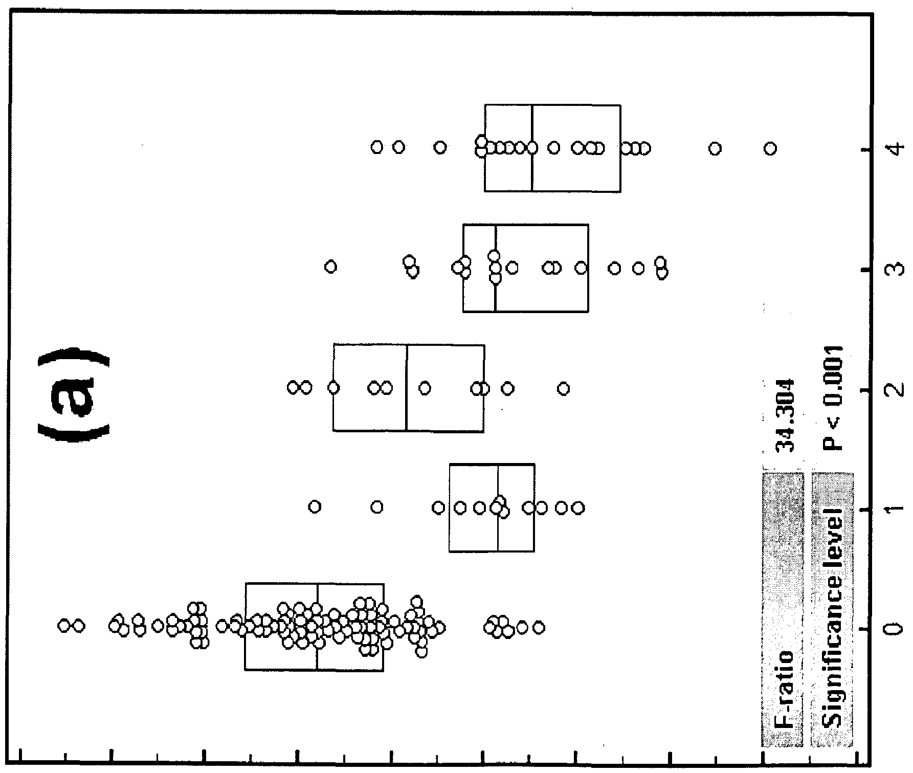
co un O CO s.
CM CM CM CM CM 152 CM CM
Claims
1. A method for detecting the presence of lung cancer in a subject, the method comprising detecting the level of small U2-2, in a sample from the subject, wherein detection of a level of small U2-2 that is greater than a normal level of small U2-2 indicates the presence of lung cancer in the subject.
2. A method for detecting the presence of lung cancer in a subject, the method comprising detecting the level of small U2-2, in a sample from the subject, and comparing the level of the small U2-2 in the sample to a normal level of the RNA, wherein detection of a level of small U2-2 that is greater than a normal level of small U2-2 indicates the presence of lung cancer in the subject.
3. A method of facilitating the diagnosis of lung cancer in a subject or the monitoring of therapy in a lung cancer patient, comprising detecting the level of small U2-2, in a sample from the subject, and communicating the results of the detection to a medical practitioner for the purpose of determining whether the subject has lung cancer or monitoring therapy in the lung cancer patient.
4. A method of monitoring response to therapy in a lung cancer patient, comprising detecting the level of small U2-2, in a first sample from the subject taken at a first time point, and comparing the level of small U2-2 to the level of the respective RNA in a second sample from the patient taken at a second time point, wherein the second time point is prior to the first time point, and wherein a decrease in the level of small U2-2 in the first sample relative to the second sample, indicates that the lung cancer patient is responding to therapy.
5. A method for detecting the presence of lung cancer in a subject, comprising obtaining a sample from the subject, providing the sample to a laboratory for detection of the level of small U2-2, in the sample, receiving from the laboratory a communication indicating the level of the at least one RNA, wherein detection of a level of small U2-2 that is greater than a normal level of small U2-2 indicates the presence of lung cancer in the subject.
6. A method for monitoring response to therapy in a lung cancer patient, comprising obtaining a first sample from the subject at a first time point, providing the first sample to a laboratory for detection of the level of small U2-2, in the sample, receiving from the laboratory a communication indicating the level of the at least one RNA, comparing the level of the at least on RNA in the first sample to the level of the at least on RNA in a
second sample that was taken at a second time point, wherein the second time point is prior to the first time point, wherein a decrease in the level of small U2-2 in the first sample relative to the second sample, indicates that the lung cancer patient is responding to therapy.
7. The method of any one of claims 1 to 6, wherein the detecting comprises hybridizing at least one polynucleotide comprising at least 8 contiguous nucleotides of a sequence selected from SEQ ID NOs: 21 and 22 to RNA from the sample or cDNA reverse-transcribed from RNA from the sample, and detecting a complex comprising a polynucleotide and an RNA or cDNA selected from small U2-2, miR-720, miR-451, 13207, and 13750.
8. The method of any one of the preceding claims, wherein small U2-2 is selected from mature small U2-2, a mature small U2-2 isomir, pre-small U2-2, and combinations thereof.
9. The method of any one of the preceding claims, wherein small U2-2 has a sequence selected from SEQ ID NOs: 2 to 20.
10. The method of any one of the preceding claims, wherein the sample is selected from a tissue sample and a bodily fluid.
11. The method of claim 10, wherein the tissue sample is a lung tissue sample.
12. The method of claim 1 1, wherein the lung tissue sample comprises lung cancer cells.
13. The method of claim 10, wherein the bodily fluid is selected from blood, urine, sputum, saliva, mucus, and semen.
14. The method of claim 13, wherein the sample is a blood sample.
15. The method of claim 14, wherein the blood sample is a serum sample.
16. The method of claim 14, wherein the blood sample is a plasma sample.
17. The method of any one of the preceding claims, wherein the lung cancer is early stage lung cancer.
18. The method of any one of the preceding claims, wherein the lung cancer is stage I lung cancer.
19. The method of any one of the preceding claims, wherein the detecting comprises quantitative RT-PCR.
20. Use of small U2-2 for detecting the presence of lung cancer in a subject, or for monitoring therapy in a lung cancer patient.
21. Use of small U2-2 for detecting the presence of lung cancer in a subject, or for monitoring therapy in a lung cancer patient.
22. A composition comprising an oligonucleotide that comprises at least eight contiguous nucleotides that are complementary to small U2-2.
23. A composition comprising an oligonucleotide that comprises at least eight contiguous nucleotides that are complementary to a cDNA reverse-transcribed from small U2-2.
24. A kit comprising the composition of claim 22 or claim 23.
Priority Applications (2)
| Application Number | Priority Date | Filing Date | Title |
|---|---|---|---|
| EP14722400.0A EP2971103A1 (en) | 2013-03-14 | 2014-03-07 | Methods of detecting lung cancer |
| US14/773,331 US20160108478A1 (en) | 2013-03-14 | 2014-03-07 | Methods of detecting lung cancer |
Applications Claiming Priority (2)
| Application Number | Priority Date | Filing Date | Title |
|---|---|---|---|
| US201361783006P | 2013-03-14 | 2013-03-14 | |
| US61/783,006 | 2013-03-14 |
Publications (1)
| Publication Number | Publication Date |
|---|---|
| WO2014159073A1 true WO2014159073A1 (en) | 2014-10-02 |
Family
ID=50680107
Family Applications (1)
| Application Number | Title | Priority Date | Filing Date |
|---|---|---|---|
| PCT/US2014/021854 WO2014159073A1 (en) | 2013-03-14 | 2014-03-07 | Methods of detecting lung cancer |
Country Status (3)
| Country | Link |
|---|---|
| US (1) | US20160108478A1 (en) |
| EP (1) | EP2971103A1 (en) |
| WO (1) | WO2014159073A1 (en) |
Cited By (1)
| Publication number | Priority date | Publication date | Assignee | Title |
|---|---|---|---|---|
| US9365903B2 (en) | 2011-01-26 | 2016-06-14 | Cepheid | Compositions comprising polynucleotides for detecting lung cancer |
Citations (10)
| Publication number | Priority date | Publication date | Assignee | Title |
|---|---|---|---|---|
| WO2006013076A1 (en) | 2004-08-02 | 2006-02-09 | Societe De Technologie Michelin | Tyre/wheel assembly for motor vehicle |
| US20060094025A1 (en) | 2004-11-02 | 2006-05-04 | Getts Robert C | Methods for detection of microrna molecules |
| US20060292616A1 (en) | 2005-06-23 | 2006-12-28 | U.S. Genomics, Inc. | Single molecule miRNA-based disease diagnostic methods |
| WO2007011903A2 (en) | 2005-07-15 | 2007-01-25 | Applera Corporation | Analyzing messenger rna and micro rna in the same reaction mixture |
| WO2007025281A2 (en) | 2005-08-24 | 2007-03-01 | Applera Corporation | A method to quantify sirnas, mirnas and polymorphic mirnas |
| US20070054287A1 (en) | 2005-05-31 | 2007-03-08 | Applera Corporation | Method for identifying medically important cell populations using micro rna as tissue specific biomarkers |
| US20070077570A1 (en) | 2005-05-31 | 2007-04-05 | Applera Corporation | Multiplexed amplification of short nucleic acids |
| US7351538B2 (en) | 2004-08-23 | 2008-04-01 | U.S. Genomics | Systems and methods for detecting and analyzing polymers |
| US20090123912A1 (en) | 2005-01-25 | 2009-05-14 | Rosetta Inpharmatics Llc | Methods for quantitating small RNA molecules |
| WO2012103355A2 (en) * | 2011-01-26 | 2012-08-02 | Cepheid | Methods of detecting lung cancer |
Family Cites Families (3)
| Publication number | Priority date | Publication date | Assignee | Title |
|---|---|---|---|---|
| EP1268856A2 (en) * | 2000-04-07 | 2003-01-02 | Epigenomics AG | Detection of single nucleotide polymorphisms (snp's) and cytosine-methylations |
| US20070065844A1 (en) * | 2005-06-08 | 2007-03-22 | Massachusetts Institute Of Technology | Solution-based methods for RNA expression profiling |
| CA2753481A1 (en) * | 2009-02-25 | 2010-09-02 | Cepheid | Methods of detecting lung cancer |
-
2014
- 2014-03-07 EP EP14722400.0A patent/EP2971103A1/en not_active Withdrawn
- 2014-03-07 WO PCT/US2014/021854 patent/WO2014159073A1/en active Application Filing
- 2014-03-07 US US14/773,331 patent/US20160108478A1/en not_active Abandoned
Patent Citations (13)
| Publication number | Priority date | Publication date | Assignee | Title |
|---|---|---|---|---|
| WO2006013076A1 (en) | 2004-08-02 | 2006-02-09 | Societe De Technologie Michelin | Tyre/wheel assembly for motor vehicle |
| US7402422B2 (en) | 2004-08-23 | 2008-07-22 | U.S. Genomics, Inc. | Systems and methods for detecting and analyzing polymers |
| US7351538B2 (en) | 2004-08-23 | 2008-04-01 | U.S. Genomics | Systems and methods for detecting and analyzing polymers |
| US20060094025A1 (en) | 2004-11-02 | 2006-05-04 | Getts Robert C | Methods for detection of microrna molecules |
| US20090123912A1 (en) | 2005-01-25 | 2009-05-14 | Rosetta Inpharmatics Llc | Methods for quantitating small RNA molecules |
| US20070077570A1 (en) | 2005-05-31 | 2007-04-05 | Applera Corporation | Multiplexed amplification of short nucleic acids |
| US20070054287A1 (en) | 2005-05-31 | 2007-03-08 | Applera Corporation | Method for identifying medically important cell populations using micro rna as tissue specific biomarkers |
| WO2007117256A1 (en) | 2005-05-31 | 2007-10-18 | Applera Corporation | Multiplexed amplification of short nucleic acids |
| US20060292616A1 (en) | 2005-06-23 | 2006-12-28 | U.S. Genomics, Inc. | Single molecule miRNA-based disease diagnostic methods |
| WO2007011903A2 (en) | 2005-07-15 | 2007-01-25 | Applera Corporation | Analyzing messenger rna and micro rna in the same reaction mixture |
| WO2007025281A2 (en) | 2005-08-24 | 2007-03-01 | Applera Corporation | A method to quantify sirnas, mirnas and polymorphic mirnas |
| WO2012103355A2 (en) * | 2011-01-26 | 2012-08-02 | Cepheid | Methods of detecting lung cancer |
| US20120244530A1 (en) | 2011-01-26 | 2012-09-27 | Cepheid | Methods of detecting lung cancer |
Non-Patent Citations (87)
| Title |
|---|
| ABERLE DR; ADAMS AM; BERG CD; BLACK WC; CLAPP JD ET AL.: "National Lung Screening Trial Research Team (2011) Reduced lung-cancer mortality with low-dose computed tomographic screening", N ENGL J MED, vol. 365, 2011, pages 395 - 409 |
| AHLENSTIEL CL; LIM HG; COOPER DA; ISHIDA T; KELLEHER AD; SUZUKI K: "Direct evidence of nuclear Argonaute distribution during transcriptional silencing links the actin cytoskeleton to nuclear RNAi machinery in human cells", NUCLEIC ACIDS RES, vol. 40, 2012, pages 1579 - 1595 |
| ALEXANDER BARANISKIN ET AL: "Circulating U2 small nuclear RNA fragments as a novel diagnostic biomarker for pancreatic and colorectal adenocarcinoma", INTERNATIONAL JOURNAL OF CANCER, vol. 132, no. 2, 15 January 2013 (2013-01-15), pages E48 - E57, XP055126776, ISSN: 0020-7136, DOI: 10.1002/ijc.27791 * |
| ALLAWI H.T. ET AL., RNA, vol. 10, 2004, pages 1153 - 1161 |
| ARROYO JD; CHEVILLET JR; KROH EM; RUF IK; PRITCHARD CC ET AL.: "Argonaute2 complexes carry a population of circulating microRNAs independent of vesicles in human plasma", PROC NATL ACAD SCI U S A, vol. 108, 2011, pages 5003 - 5008, XP055352377, DOI: doi:10.1073/pnas.1019055108 |
| BARANISKIN A; NÖPEL-DÜNNEBACKE S; AHRENS M; JENSEN SG; ZOLLNER H ET AL.: "Circulating U2 small nuclear RNA fragments as a novel diagnostic biomarker for pancreatic and colorectal adenocarcinoma", INT J CANCER, vol. 15, 2013, pages E48 - 57, XP055126776, DOI: doi:10.1002/ijc.27791 |
| BASKERVILLE, S.; BARTEL D.P., RNA, vol. 11, 2005, pages 241 - 247 |
| BIOTECHNIQUES, vol. 41, no. 4, pages 1 - 5 |
| BRAMEIER M; HERWIG A; REINHARDT R; WALTER L; GRUBER J: "Human box C/D snoRNAs with miRNA like functions: expanding the range of regulatory RNAs", NUCLEIC ACIDS RES, vol. 39, 2011, pages 675 - 686, XP055037465, DOI: doi:10.1093/nar/gkq776 |
| BRODERICK JA; SALOMON WE; RYDER SP; ARONIN N; ZAMORE PD: "Argonaute protein identity and pairing geometry determine cooperativity in mammalian RNA silencing", RNA, vol. 17, 2011, pages 1858 - 1869 |
| BURROUGHS AM; ANDO Y; DE HOON ML; TOMARU Y; SUZUKI H ET AL.: "Deep-sequencing of human Argonaute-associated small RNAs provides insight into miRNA sorting and reveals Argonaute association with RNA fragments of diverse origin", RNA BIOL, vol. 8, 2011, pages 158 - 177 |
| BUTNOR, ARCH. PATHOL. LAB. MED., vol. 132, 2008, pages 1118 - 1132 |
| CASTLE JC; ARMOUR CD; LOWER M; HAYNOR D; BIERY M ET AL.: "Digital genome-wide ncRNA expression, including SnoRNAs, across 11 human tissues using polyA-neutral amplification", PLOS ONE, vol. 5, 2010, pages EL 1779 |
| CASTOLDI M; SCHMIDT S; BENES V; NOERHOLM M; KULOZIK AE ET AL.: "A sensitive array for microRNA expression profiling (miChip) based on locked nucleic acids (LNA", RNA, vol. 12, 2006, pages 913 - 920, XP055409413, DOI: doi:10.1261/rna.2332406 |
| CHEN, C. ET AL., NUCL. ACIDS RES., vol. 33, 2005, pages E179 |
| CIFUENTES D; XUE H; TAYLOR DW; PATNODE H ET AL.: "A novel miRNA processing pathway independent of Dicer requires Argonaute2 catalytic activity", SCIENCE, vol. 328, 2010, pages 1694 - 1698, XP055137558, DOI: doi:10.1126/science.1190809 |
| COLE C; SOBALA A; LU C; THATCHER SR; BOWMAN A ET AL.: "Filtering of deep sequencing data reveals the existence of abundant Dicer-dependent small RNAs derived from tRNAs", RNA, vol. 15, 2009, pages 2147 - 2160 |
| COSTA FF: "Non-coding RNAs: Meet thy masters", BIOESSAYS, vol. 32, 2010, pages 599 - 608 |
| DERYUSHEVA S; CHOLEZA M; BARBAROSSA A; GALL JG; BORDONNE R: "Post-transcriptional modification of spliceosomal RNAs is normal in SMN-deficient cells", RNA, vol. 18, 2012, pages 31 - 36 |
| ENDER C; KREK A; FRIEDLÄNDER MR; BEITZINGER M; WEINMANN L ET AL.: "A human snoRNA with microRNA-like functions", MOL CELL, vol. 32, 2008, pages 519 - 528, XP002510436, DOI: doi:10.1016/J.MOLCEL.2008.10.017 |
| FIELD ET AL., BR. J. CANCER, vol. 99, 2008, pages 557 - 562 |
| FINDEISS S; LANGENBERGER D; STADLER PF; HOFFMANN S: "Traces of post-transcriptional RNA modifications in deep sequencing data", BIOL CHEM, vol. 392, 2011, pages 305 - 313 |
| FRIEDEN, M. ET AL., CURR. PHARM. DES., vol. 14, no. 11, 2008, pages 1138 - 1142 |
| FU H; FENG J; LIU Q; SUN F; TIE Y ET AL.: "Stress induces tRNA cleavage by angiogenin in mammalian cells", FEBS LETT, vol. 583, 2009, pages 437 - 442, XP025926491, DOI: doi:10.1016/j.febslet.2008.12.043 |
| GALLO A; TANDON M; ALEVIZOS I; ILLEI GG: "The majority of microRNAs detectable in serum and saliva is concentrated in exosomes", PLOS ONE, vol. 7, 2012, pages E30679 |
| GANOT P; BORTOLIN ML; KISS T: "Site-specific pseudouridine formation in preribosomal RNA is guided by small nucleolar RNAs", CELL, vol. 89, 1997, pages 799 - 809 |
| GENTLEMAN RC; CAREY VJ; BATES DM; BOLSTAD B; DETTLING M ET AL.: "Bioconductor: open software development for computational biology and bioinformatics", GENOME BIOL, vol. 5, 2004, pages R80, XP021012842, DOI: doi:10.1186/gb-2004-5-10-r80 |
| GILAD S; MEIRI E; YOGEV Y; BENJAMIN S; LEBANONY D ET AL.: "Serum microRNAs are promising novel biomarkers", PLOS ONE, vol. 3, 2008, pages E3148 |
| GOLDSTRAW P; CROWLEY J; CHANSKY K; GIROUX DJ; GROOME PA ET AL.: "International Association for the Study of Lung Cancer International Staging Committee; Participating Institutions. The IASLC Lung Cancer Staging Project: proposals for the revision of the TNM stage groupings in the forthcoming (seventh) edition of the TNM Classification of malignant tumours", J THORAC ONCOL, vol. 2, 2007, pages 706 - 714 |
| GUO L; LIANG T; GU W; XU Y; BAI Y; LU Z: "Cross-Mapping Events in miRNAs Reveal Potential miRNA-Mimics and Evolutionary Implications", PLOS ONE, vol. 6, 2011, pages E20517 |
| HAUSSECKER D; HUANG Y; LAU A; PARAMESWARAN P; FIRE AZ; KAY MA: "Human tRNA-derived small RNAs in the global regulation of RNA silencing", RNA, vol. 16, 2010, pages 673 - 695, XP008125181, DOI: doi:10.1261/rna.2000810 |
| HOWE EA; SINHA R; SCHLAUCH D; QUACKENBUSH J: "RNA-Seq analysis in MeV", BIOINFORMATICS, vol. 27, 2011, pages 3209 - 3210, XP055384011, DOI: doi:10.1093/bioinformatics/btr490 |
| HUANG Q; JACOBSON MR; PEDERSON T: "3' processing of human pre-U2 small nuclear RNA: a base-pairing interaction between the 3' extension of the precursor and an internal region", MOL CELL BIOL, vol. 17, 1997, pages 7178 - 7185 |
| HUNTER MP; ISMAIL N; ZHANG X; AGUDA BD; LEE EJ ET AL.: "Detection of microRNA expression in human peripheral blood microvesicles", PLOS ONE, vol. 3, 2008, pages E3694, XP008149486, DOI: doi:10.1371/journal.pone.0003694 |
| I DELFOUR O; VILANOVA D; ATZOM V; MICHOT B: "microARN: Biology, Function and Expression", 2007, article "The passionate race for microARN detection and function deciphering. In: Clarke", pages: 335 - 362 |
| JALADAT Y; ZHANG B; MOHAMMADI A; VALADKHAN S: "Splicing of an intervening sequence by protein-free human snRNAs", RNA BIOL, vol. 8, 2011, pages 372 - 377 |
| JIMA DD; ZHANG J; JACOBS C; RICHARDS KL; DUNPHY CH ET AL.: "Hematologic Malignancies Research Consortium. (2010) Deep sequencing of the small RNA transcriptome of normal and malignant human B cells identifies hundreds of novel microRNAs", BLOOD, vol. 116, 2010, pages E118 - E127 |
| JONES MR; QUINTON LJ; BLAHNA MT; NEILSON JR; FU S ET AL.: "Zcchcl 11-dependent uridylation of microRNA directs cytokine expression", NAT CELL BIOL, vol. 11, 2009, pages 1157 - 1163, XP055111110, DOI: doi:10.1038/ncb1931 |
| JONSTRUP, S.P. ET AL., RNA, vol. 12, 2006, pages 1 - 6 |
| JULIEN MAZIÈRES ET AL: "Alternative Processing of the U2 Small Nuclear RNA Produces a 19-22nt Fragment with Relevance for the Detection of Non-Small Cell Lung Cancer in Human Serum", PLOS ONE, vol. 8, no. 3, 20 March 2013 (2013-03-20), pages e60134, XP055126737, ISSN: 1932-6203, DOI: 10.1371/journal.pone.0060134 * |
| JUVVUNA PK; KHANDELIA P; LEE LM; MAKEYEV EV: "Argonaute identity defines the length of mature mammalian microRNAs", NUCLEIC ACIDS RES, vol. 40, 2012, pages 6808 - 6820 |
| KARIJOLICH J; YU YT: "Spliceosomal snRNA modifications and their function", RNA BIOL, vol. 7, 2010, pages 192 - 204 |
| KAWAJI H; NAKAMURA M; TAKAHASHI Y; SANDELIN A; KATAYAMA S ET AL.: "Hidden layers of human small RNAs", BMC GENOMICS, vol. 9, 2008, pages 157, XP021032882 |
| KIM JS; CHOI YY; JIN G; KANG HG; CHOI JE ET AL.: "Association of a common AGO I variant with lung cancer risk: a two-stage case-control study", MOL CARCINOG, vol. 49, 2010, pages 913 - 921 |
| KISS T: "Small nucleolar RNA-guided post-transcriptional modification of cellular RNAs", EMBO J, vol. 20, 2001, pages 3617 - 3622 |
| KOZOMARA A; GRIFFITHS-JONES S: "miRBase: integrating microRNA annotation and deep-sequencing data", NUCLEIC ACIDS RES, vol. 39, 2011, pages D152 - 157 |
| KUCHENBAUER F; MORIN RD; ARGIROPOULOS B; PETRIV 01; GRIFFITH M ET AL.: "In-depth characterization of the microRNA transcriptome in a leukemia progression model", GENOME RESEARCH, vol. 18, 2008, pages 1787 - 1797, XP055105854, DOI: doi:10.1101/gr.077578.108 |
| LANDGRAFP; RUSU M; SHERIDAN R; SEWER A; IOVINO N ET AL.: "A mammalian microRNA expression atlas based on small RNA library sequencing", CELL, vol. 129, 2007, pages 1401 - 1414 |
| LANGENBERGER D; BERMUDEZ-SANTANA CL; STADLER PF; HOFFMANN S: "Identification and classification of small RNAs in transcriptome sequence data", PAC SYMP BIOCOMPUT, 2010, pages 80 - 87 |
| LEE YS; SHIBATA Y; MALHOTRA A; DUTTA A: "A novel class of small RNAs: tRNA-derived RNA fragments (tRFs", GENES DEV, vol. 23, 2009, pages 2639 - 2649, XP055128681, DOI: doi:10.1101/gad.1837609 |
| LIANG, R.Q. ET AL., NUCL. ACIDS RES., vol. 33, no. 2, 2005, pages E 17, Retrieved from the Internet <URL:http://www.pubmedcentral.nih.gov/articlerender.fcgi?artid= 548377> |
| LIM, L.P. ET AL., NATURE, vol. 433, 2005, pages 769 - 773 |
| LIU C.G. ET AL., METHODS, vol. 44, 2008, pages 22 - 30 |
| LIU, C.G. ET AL., PROC. NAT'1 ACAD. SCI. USA, vol. 101, 2004, pages 9740 - 9744 |
| LIVAK KJ; SCHMITTGEN TD: "Analysis of relative gene expression data using realtime quantitative PCR and the 2(-Delta Delta C(T)) Method", METHODS, vol. 25, 2001, pages 402 - 408 |
| LU J. ET AL., NATURE, vol. 435, 2005, pages 834 - 838 |
| MATERA AG; TERNS RM; TERNS MP: "Non-coding RNAs: lessons from the small nuclear and small nucleolar RNAs", NAT REV MOL CELL BIOL, vol. 8, 2007, pages 209 - 220 |
| MITCHELL PS; PARKIN RK; KROH EM; FRITZ BR; WYMAN SK ET AL.: "Circulating microRNAs as stable blood-based markers for cancer detection", PROC NATL ACAD SCI U S A, vol. 105, 2008, pages 10513 - 10518, XP055037905, DOI: doi:10.1073/pnas.0804549105 |
| MORA J.R.; GETTS R.C., BIOTECHNIQUES, vol. 41, 2006, pages 420 - 424 |
| NANOTECH NEWS, 2006, Retrieved from the Internet <URL:http//nano.cancer.gov/news_center/ nanotech news 2006-10-30b.asp> |
| NELSON, P.T. ET AL., NATURE METHODS, 2004, pages 7 |
| NELSON, P.T. ET AL., RNA, vol. 12, no. 2, 2006, pages 1 - 5 |
| NICOLOSO M; QU LH; MICHOT B; BACHELLERIE JP: "Intron-encoded, antisense small nucleolar RNAs: the characterization of nine novel species points to their direct role as guides for the 2'-O-ribose methylation of rRNAs", J MOL BIOL, vol. 260, 1996, pages 178 - 195 |
| ONO M; SCOTT MS; YAMADA K; AVOLIO F; BARTON GJ; LAMOND AI: "Identification of human miRNA precursors that resemble box C/D snoRNAs", NUCLEIC ACIDS RES, vol. 39, 2011, pages 3879 - 3891, XP055045922, DOI: doi:10.1093/nar/gkq1355 |
| PAN ZQ; PRIVES C: "U2 snRNA sequences that bind U2-specific proteins are dispensable for the function ofU2 snRNP in splicing", GENES DEV, vol. 3, 1989, pages 1887 - 1898 |
| PARISI C; GIORGI C; BATASSA EM; BRACCINI L; MARESCA G ET AL.: "Ago I and Ago2 differentially affect cell proliferation, motility and apoptosis when overexpressed in SH-SY5Y neuroblastoma cells", FEBS LETT, vol. 585, 2011, pages 2965 - 2971, XP028300038, DOI: doi:10.1016/j.febslet.2011.08.003 |
| PEDERSON T: "Regulatory RNAs derived from transfer RNA?", RNA, vol. 16, 2010, pages 1865 - 1869, XP055128692, DOI: doi:10.1261/rna.2266510 |
| PERSSON E; KVIST A; VALLON-CHRISTERSSON J; MEDSTRAND P; BORG A; ROVIRA C: "The non-coding RNA of the multidrug resistance linked vault particle encodes multiple regulatory small RNAs", NAT CELL BIOL, vol. 11, 2009, pages 1268 - 1271 |
| PIEPOLI A; TAVANO F; COPETTI M; MAZZA T; PALUMBO O ET AL.: "Mirna expression profiles identify drivers in colorectal and pancreatic cancers", PLOS ONE, vol. 7, 2012, pages E33663 |
| PIGATI L; YADDANAPUDI SC; LYENGAR R; KIM DJ; HEARN SA ET AL.: "Selective release of microRNA species from normal and malignant mammary epithelial cells", PLOS ONE, vol. 5, 2010, pages E13515, XP002686873, DOI: doi:10.1371/JOURNAL.PONE.0013515 |
| REDERSTORFF M; BERNHART SH; TANZER A; ZYWICKI M; PERFLER K ET AL.: "RNPomics: defining the ncRNA transcriptome by cDNA library generation from ribonucleo-protein particles", NUCLEIC ACIDS RES, vol. 38, 2010, pages EL 13 |
| SCOTT MS; AVOLIO F; ONO M; LAMOND AI; BARTON GJ: "Human miRNA precursors with box H/ACA snoRNA features", PLOS COMPUT BIOL, vol. 5, 2009, pages E1000507 |
| See also references of EP2971103A1 * |
| SEMPLE ET AL., NATURE BIOTECHNOLOGY, 17 January 2010 (2010-01-17) |
| SMALHEISER NR; LUGLI G; THIMMAPURAM J; COOK EH; LARSON J: "Endogenous siRNAs and noncoding RNA-derived small RNAs are expressed in adult mouse hippocampus and are up-regulated in olfactory discrimination training", RNA, vol. 17, 2011, pages 166 - 181 |
| TAFT RJ; GLAZOV EA; LASSMANN T; HAYASHIZAKI Y; CARNINCI P; MATTICK JS: "Small RNAs derived from snoRNAs", RNA, vol. 15, 2009, pages 1233 - 1240, XP055037429, DOI: doi:10.1261/rna.1528909 |
| VALADKHAN S: "Role of the snRNAs in spliceosomal active site", RNA BIOL, vol. 7, 2010, pages 345 - 353 |
| VALADKHAN S; MOHAMMADI A; JALADAT Y; GEISLER S: "Protein-free small nuclear RNAs catalyze a two-step splicing reaction", PROC NATL ACAD SCI U S A, vol. 106, 2009, pages 11901 - 11906 |
| VARATTYAY, E. ET AL., NATURE PROTOCOLS, vol. 3, no. 2, 2008, pages 190 - 196 |
| WANG D; ZHANG Z; O'LOUGHLIN E; LEE T; HOUEL S ET AL.: "Quantitative functions of Argonaute proteins in mammalian development", GENES DEV, vol. 26, 2012, pages 693 - 704 |
| WEBER JA; BAXTER DH; ZHANG S; HUANG DY; HUANG KH ET AL.: "The microRNA spectrum in 12 body fluids", CLIN CHEM, vol. 56, 2010, pages 1733 - 1741, XP055188106, DOI: doi:10.1373/clinchem.2010.147405 |
| WEINMANN L; HOCK J; IVACEVIC T; OHRT T; MUTZE J ET AL.: "Importin 8 Is a Gene Silencing Factor that Targets Argonaute Proteins to Distinct mRNAs", CELL, vol. 136, 2009, pages 496 - 507 |
| WILKINSON, M., NUCL. ACIDS RES., vol. 16, no. 22, 1988, pages 10,933 |
| WILKINSON, M., NUCL. ACIDS RES., vol. 16, no. 22, 1988, pages 10934 |
| YEUNG ML; BENNASSER Y; WATASHI K; LE SY; HOUZET L; JEANG KT: "Pyrosequencing of small non-coding RNAs in HIV-1 infected cells: evidence for the processing of a viral-cellular double-stranded RNA hybrid", NUCLEIC ACIDS RES, vol. 37, 2009, pages 6575 - 6586 |
| YU YT; SHU MD; STEITZ JA: "Modifications ofU2 snRNA are required for snRNP assembly and pre-mRNA splicing", THE EMBO JOURNAL, vol. 17, 1998, pages 5783 - 5795 |
| ZHANG Y; LIAO JM; ZENG SX; LU H: "p53 downregulates Down syndrome-associated DYRK1A through miR-1246", EMBO REP, vol. 12, 2011, pages 811 - 817 |
Cited By (1)
| Publication number | Priority date | Publication date | Assignee | Title |
|---|---|---|---|---|
| US9365903B2 (en) | 2011-01-26 | 2016-06-14 | Cepheid | Compositions comprising polynucleotides for detecting lung cancer |
Also Published As
| Publication number | Publication date |
|---|---|
| US20160108478A1 (en) | 2016-04-21 |
| EP2971103A1 (en) | 2016-01-20 |
Similar Documents
| Publication | Publication Date | Title |
|---|---|---|
| US20220356528A1 (en) | Microrna assay for detection and management of pancreatic cancer precursors | |
| Bartels et al. | MicroRNAs: novel biomarkers for human cancer | |
| US9365903B2 (en) | Compositions comprising polynucleotides for detecting lung cancer | |
| EP2391738B1 (en) | Methods of detecting sepsis | |
| Wu et al. | Next-generation sequencing of microRNAs for breast cancer detection | |
| JP5755569B2 (en) | How to detect lung cancer | |
| US20160024586A1 (en) | Methods of detecting cancer | |
| US10000808B2 (en) | Methods of detecting cervical cancer | |
| US20110160290A1 (en) | Use of extracellular rna to measure disease | |
| Markou et al. | Prognostic, therapeutic and diagnostic potential of microRNAs in non-small cell lung cancer | |
| WO2014085906A1 (en) | Microrna biomarkers for prostate cancer | |
| Usó et al. | miRNA detection methods and clinical implications in lung cancer | |
| Ghai et al. | Circulating miRNAs as tumor biomarkers | |
| WO2014159073A1 (en) | Methods of detecting lung cancer | |
| Navarro et al. | Exosomal microRNAs as potentially useful tools in cancer biomarker discovery | |
| Ng et al. | Clinical relevance of miRNAs in cancer | |
| Dekker | Essay: Circulating miRNAs a diagnostic tool for cancer detection? | |
| Ziogas | The emerging role of miRNAs in translational cancer medicine. | |
| Joseph et al. | POTENTIAL MICRORNA SIGNATURE ASSOCIATED WITH POST-OPERATIVE TUMOUR RECURRENCE RISK IN SOUTH INDIAN ORAL SQUAMOUS CELL CARCINOMA PATIENTS IDENTIFIED BY NEXT-GENERATION SEQUENCING | |
| Markou et al. | prognostic, therapeutic and diagnostic potential of microRNAs in non-small cell lung cancer. Clin Chem Lab Med |
Legal Events
| Date | Code | Title | Description |
|---|---|---|---|
| 121 | Ep: the epo has been informed by wipo that ep was designated in this application |
Ref document number: 14722400 Country of ref document: EP Kind code of ref document: A1 |
|
| WWE | Wipo information: entry into national phase |
Ref document number: 14773331 Country of ref document: US |
|
| NENP | Non-entry into the national phase |
Ref country code: DE |
|
| WWE | Wipo information: entry into national phase |
Ref document number: 2014722400 Country of ref document: EP |