WO2014102806A1 - Protein biosensors, cross reactive sensor arrays and methods of use thereof - Google Patents
Protein biosensors, cross reactive sensor arrays and methods of use thereof Download PDFInfo
- Publication number
- WO2014102806A1 WO2014102806A1 PCT/IL2013/051102 IL2013051102W WO2014102806A1 WO 2014102806 A1 WO2014102806 A1 WO 2014102806A1 IL 2013051102 W IL2013051102 W IL 2013051102W WO 2014102806 A1 WO2014102806 A1 WO 2014102806A1
- Authority
- WO
- WIPO (PCT)
- Prior art keywords
- odn
- alkyl
- protein
- formula
- derivative
- Prior art date
- Legal status (The legal status is an assumption and is not a legal conclusion. Google has not performed a legal analysis and makes no representation as to the accuracy of the status listed.)
- Ceased
Links
- 0 C*(CCCCCCNC(c(cc1OCc2c[n]nn2)cc(OCC2=CN(*)NN2)c1OCc1c[n]([*+])nn1)=O)CCP(*)(O)OCCOCCOCCOCCOCCOCC*(OC)OC Chemical compound C*(CCCCCCNC(c(cc1OCc2c[n]nn2)cc(OCC2=CN(*)NN2)c1OCc1c[n]([*+])nn1)=O)CCP(*)(O)OCCOCCOCCOCCOCCOCC*(OC)OC 0.000 description 2
- CPKXRSARKPOMAO-UHFFFAOYSA-N C#CCOc(cc(cc1OCC#C)C(ON(C(CC2)=O)C2=O)=O)c1OCC#C Chemical compound C#CCOc(cc(cc1OCC#C)C(ON(C(CC2)=O)C2=O)=O)c1OCC#C CPKXRSARKPOMAO-UHFFFAOYSA-N 0.000 description 1
Classifications
-
- C—CHEMISTRY; METALLURGY
- C09—DYES; PAINTS; POLISHES; NATURAL RESINS; ADHESIVES; COMPOSITIONS NOT OTHERWISE PROVIDED FOR; APPLICATIONS OF MATERIALS NOT OTHERWISE PROVIDED FOR
- C09K—MATERIALS FOR MISCELLANEOUS APPLICATIONS, NOT PROVIDED FOR ELSEWHERE
- C09K11/00—Luminescent materials, e.g. electroluminescent or chemiluminescent
- C09K11/06—Luminescent materials, e.g. electroluminescent or chemiluminescent containing organic luminescent materials
-
- G—PHYSICS
- G01—MEASURING; TESTING
- G01N—INVESTIGATING OR ANALYSING MATERIALS BY DETERMINING THEIR CHEMICAL OR PHYSICAL PROPERTIES
- G01N33/00—Investigating or analysing materials by specific methods not covered by groups G01N1/00 - G01N31/00
- G01N33/48—Biological material, e.g. blood, urine; Haemocytometers
- G01N33/50—Chemical analysis of biological material, e.g. blood, urine; Testing involving biospecific ligand binding methods; Immunological testing
- G01N33/53—Immunoassay; Biospecific binding assay; Materials therefor
-
- C—CHEMISTRY; METALLURGY
- C07—ORGANIC CHEMISTRY
- C07D—HETEROCYCLIC COMPOUNDS
- C07D417/00—Heterocyclic compounds containing two or more hetero rings, at least one ring having nitrogen and sulfur atoms as the only ring hetero atoms, not provided for by group C07D415/00
- C07D417/02—Heterocyclic compounds containing two or more hetero rings, at least one ring having nitrogen and sulfur atoms as the only ring hetero atoms, not provided for by group C07D415/00 containing two hetero rings
- C07D417/06—Heterocyclic compounds containing two or more hetero rings, at least one ring having nitrogen and sulfur atoms as the only ring hetero atoms, not provided for by group C07D415/00 containing two hetero rings linked by a carbon chain containing only aliphatic carbon atoms
-
- C—CHEMISTRY; METALLURGY
- C07—ORGANIC CHEMISTRY
- C07D—HETEROCYCLIC COMPOUNDS
- C07D417/00—Heterocyclic compounds containing two or more hetero rings, at least one ring having nitrogen and sulfur atoms as the only ring hetero atoms, not provided for by group C07D415/00
- C07D417/14—Heterocyclic compounds containing two or more hetero rings, at least one ring having nitrogen and sulfur atoms as the only ring hetero atoms, not provided for by group C07D415/00 containing three or more hetero rings
-
- C—CHEMISTRY; METALLURGY
- C07—ORGANIC CHEMISTRY
- C07F—ACYCLIC, CARBOCYCLIC OR HETEROCYCLIC COMPOUNDS CONTAINING ELEMENTS OTHER THAN CARBON, HYDROGEN, HALOGEN, OXYGEN, NITROGEN, SULFUR, SELENIUM OR TELLURIUM
- C07F9/00—Compounds containing elements of Groups 5 or 15 of the Periodic Table
- C07F9/02—Phosphorus compounds
- C07F9/547—Heterocyclic compounds, e.g. containing phosphorus as a ring hetero atom
- C07F9/6536—Heterocyclic compounds, e.g. containing phosphorus as a ring hetero atom having nitrogen and sulfur atoms with or without oxygen atoms, as the only ring hetero atoms
- C07F9/6539—Five-membered rings
- C07F9/6541—Five-membered rings condensed with carbocyclic rings or carbocyclic ring systems
-
- C—CHEMISTRY; METALLURGY
- C07—ORGANIC CHEMISTRY
- C07K—PEPTIDES
- C07K7/00—Peptides having 5 to 20 amino acids in a fully defined sequence; Derivatives thereof
- C07K7/04—Linear peptides containing only normal peptide links
- C07K7/06—Linear peptides containing only normal peptide links having 5 to 11 amino acids
-
- G—PHYSICS
- G01—MEASURING; TESTING
- G01N—INVESTIGATING OR ANALYSING MATERIALS BY DETERMINING THEIR CHEMICAL OR PHYSICAL PROPERTIES
- G01N33/00—Investigating or analysing materials by specific methods not covered by groups G01N1/00 - G01N31/00
- G01N33/48—Biological material, e.g. blood, urine; Haemocytometers
- G01N33/50—Chemical analysis of biological material, e.g. blood, urine; Testing involving biospecific ligand binding methods; Immunological testing
- G01N33/58—Chemical analysis of biological material, e.g. blood, urine; Testing involving biospecific ligand binding methods; Immunological testing involving labelled substances
- G01N33/582—Chemical analysis of biological material, e.g. blood, urine; Testing involving biospecific ligand binding methods; Immunological testing involving labelled substances with fluorescent label
-
- C—CHEMISTRY; METALLURGY
- C09—DYES; PAINTS; POLISHES; NATURAL RESINS; ADHESIVES; COMPOSITIONS NOT OTHERWISE PROVIDED FOR; APPLICATIONS OF MATERIALS NOT OTHERWISE PROVIDED FOR
- C09K—MATERIALS FOR MISCELLANEOUS APPLICATIONS, NOT PROVIDED FOR ELSEWHERE
- C09K2211/00—Chemical nature of organic luminescent or tenebrescent compounds
- C09K2211/10—Non-macromolecular compounds
- C09K2211/1018—Heterocyclic compounds
- C09K2211/1025—Heterocyclic compounds characterised by ligands
- C09K2211/1029—Heterocyclic compounds characterised by ligands containing one nitrogen atom as the heteroatom
- C09K2211/1037—Heterocyclic compounds characterised by ligands containing one nitrogen atom as the heteroatom with sulfur
-
- G—PHYSICS
- G01—MEASURING; TESTING
- G01N—INVESTIGATING OR ANALYSING MATERIALS BY DETERMINING THEIR CHEMICAL OR PHYSICAL PROPERTIES
- G01N2333/00—Assays involving biological materials from specific organisms or of a specific nature
- G01N2333/90—Enzymes; Proenzymes
- G01N2333/91—Transferases (2.)
- G01N2333/9116—Transferases (2.) transferring alkyl or aryl groups other than methyl groups (2.5)
- G01N2333/91165—Transferases (2.) transferring alkyl or aryl groups other than methyl groups (2.5) general (2.5.1)
- G01N2333/91171—Transferases (2.) transferring alkyl or aryl groups other than methyl groups (2.5) general (2.5.1) with definite EC number (2.5.1.-)
- G01N2333/91177—Glutathione transferases (2.5.1.18)
-
- G—PHYSICS
- G01—MEASURING; TESTING
- G01N—INVESTIGATING OR ANALYSING MATERIALS BY DETERMINING THEIR CHEMICAL OR PHYSICAL PROPERTIES
- G01N2333/00—Assays involving biological materials from specific organisms or of a specific nature
- G01N2333/90—Enzymes; Proenzymes
- G01N2333/914—Hydrolases (3)
- G01N2333/916—Hydrolases (3) acting on ester bonds (3.1), e.g. phosphatases (3.1.3), phospholipases C or phospholipases D (3.1.4)
- G01N2333/918—Carboxylic ester hydrolases (3.1.1)
-
- G—PHYSICS
- G01—MEASURING; TESTING
- G01N—INVESTIGATING OR ANALYSING MATERIALS BY DETERMINING THEIR CHEMICAL OR PHYSICAL PROPERTIES
- G01N2333/00—Assays involving biological materials from specific organisms or of a specific nature
- G01N2333/90—Enzymes; Proenzymes
- G01N2333/914—Hydrolases (3)
- G01N2333/948—Hydrolases (3) acting on peptide bonds (3.4)
- G01N2333/95—Proteinases, i.e. endopeptidases (3.4.21-3.4.99)
- G01N2333/964—Proteinases, i.e. endopeptidases (3.4.21-3.4.99) derived from animal tissue
- G01N2333/96425—Proteinases, i.e. endopeptidases (3.4.21-3.4.99) derived from animal tissue from mammals
- G01N2333/96427—Proteinases, i.e. endopeptidases (3.4.21-3.4.99) derived from animal tissue from mammals in general
- G01N2333/9643—Proteinases, i.e. endopeptidases (3.4.21-3.4.99) derived from animal tissue from mammals in general with EC number
- G01N2333/96486—Metalloendopeptidases (3.4.24)
- G01N2333/96491—Metalloendopeptidases (3.4.24) with definite EC number
- G01N2333/96494—Matrix metalloproteases, e. g. 3.4.24.7
-
- G—PHYSICS
- G01—MEASURING; TESTING
- G01N—INVESTIGATING OR ANALYSING MATERIALS BY DETERMINING THEIR CHEMICAL OR PHYSICAL PROPERTIES
- G01N33/00—Investigating or analysing materials by specific methods not covered by groups G01N1/00 - G01N31/00
- G01N33/48—Biological material, e.g. blood, urine; Haemocytometers
- G01N33/50—Chemical analysis of biological material, e.g. blood, urine; Testing involving biospecific ligand binding methods; Immunological testing
- G01N33/53—Immunoassay; Biospecific binding assay; Materials therefor
- G01N33/573—Immunoassay; Biospecific binding assay; Materials therefor for enzymes or isoenzymes
-
- G—PHYSICS
- G01—MEASURING; TESTING
- G01N—INVESTIGATING OR ANALYSING MATERIALS BY DETERMINING THEIR CHEMICAL OR PHYSICAL PROPERTIES
- G01N33/00—Investigating or analysing materials by specific methods not covered by groups G01N1/00 - G01N31/00
- G01N33/48—Biological material, e.g. blood, urine; Haemocytometers
- G01N33/50—Chemical analysis of biological material, e.g. blood, urine; Testing involving biospecific ligand binding methods; Immunological testing
- G01N33/53—Immunoassay; Biospecific binding assay; Materials therefor
- G01N33/575—Immunoassay; Biospecific binding assay; Materials therefor for cancer
Definitions
- the present invention is directed to molecular biosensors based on oligonucleotide cross reactive sensor arrays including a selective and a non-selective protein binding domain for protein detection. Interaction of different protein isoforms with the sensors of this invention yields a unique optical signature for each protein. The unique optical signature allows differentiating between closely related protein isoforms and diagnosing diseases and disorders associated with the proteins also in biofiuids.
- Fluorescent molecular sensors have become valuable tools in the analytical biosciences owing to their sensitive detection mode, down to the level of a single molecule, the feasibility of naked eye visualization, their versatility, and their small size, which enable them to penetrate the cell membrane and track the rise and fall of various bio-analytes within living cells.
- fluorescent sensors that utilize photo-induced electron transfer (PET), electronic energy transfer (EET) (or fluorescence resonance energy transfer (FRET)), and internal charge transfer (ICT) processes have been developed and used to detect various proteins, most of them suffer from a high background signal that complicates their use in complex biochemical mixtures and within cells.
- a promising method for obtaining multiplexed protein analysis involves the use of antibodies, which can bind and detect the proteins of interest with high affinity and selectivity. This approach has found widespread applications in medical diagnosis, however, the need for producing an antibody for each target and for using stepwise protocols, in addition to their high cost, relative instability and the difficulties associated with quantifications hamper high-throughput analysis.
- Cross-reactive sensor arrays inspired by the mammalian olfactory system, have recently emerged as an alternative detection method that may address these limitations. When the "nose/tongue" approach is used, proteins can be rapidly differentiated using an array of non-specific synthetic receptors that, in combination, generate a unique optical "fingerprint" upon interacting with each protein.
- arrays that rely on “differential sensing” do not require manufacturing multiple antibodies or using technically challenging procedures. As a result, such systems can straightforwardly discriminate among multiple different proteins as well as profile protein mixtures in blood or urine, which may indicate of disease states.
- MMPs Matrix Metalloproteases family of enzymes comprising more than 20 zinc-dependent endopeptidases that share a similar, zinc-dependent binding site, and are capable of degrading virtually every component of the extracellular matrix (ECM).
- ECM extracellular matrix
- MMPs Owing to their role in tumor growth, metastasis, and angiogenesis, MMPs are considered as important therapeutic targets for treating human cancers.
- high levels of members of the MMP family in serum, urine, or tissue have been identified in a variety of human cancers, including breast, pancreatic, bladder, colorectal, ovarian, and prostate cancer
- MMP-1 is identified in breast cancer, lung cancer and colorectal cancer
- MMP-2 is identified in pancreas cancer, bladder cancer, colorectal cancer, ovarian cancer, prostate cancer and brain cancer
- MMP-7 is identified in pancreas cancer, lung cancer and colorectal cancer
- MMP-9 is identified in breast cancer, pancreas cancer, bladder cancer, lung cancer, colorectal cancer, ovarian cancer, prostate cancer and brain cancer.
- GSTs Human Glutathione S-Transferases
- Human soluble GSTs can be mainly subdivided into 7 classes, namely, a (A), ⁇ (M), ⁇ (P), ⁇ (T), ⁇ (S), ⁇ ( ⁇ ), and ⁇ (O), which share a similar GST binding domain but differ in their surface characteristics as well as in their tissue distribution ( Figure ID). Comparative analysis of GST expression in normal and diseased tissues or serum has shown a clear correlation between their expression profiles and disease states.
- abnormal tissue expression of GST a (A) has been associated with an increased risk for colorectal cancer, ovarian cancer, and clear cell renal cell carcinoma
- ⁇ (M) class expression alteration was detected in cases of lung, colon, and bladder cancer
- ⁇ (P) class isozymes are overexpressed in the majority of human tumors.
- GST-A1 is an indicator for colorectal, prostate, breast and lung cancers.
- GST-A2 is an indicator for prostate and lung cancers.
- GST-MI is an indicator for prostate and breast cancers.
- Fibroblast Growth Factors are a family comprising 22 heparin-binding proteins whose over-expression is associated with different types of cancers. Pattern-based detection arrays, specific for this class of proteins, could therefore facilitate their differentiation and, more importantly, might be able to distinguish between medicinally relevant samples involving different combinations or concentrations of FGFs.
- Estrogen Receptors have been mainly implicated in the development and progression of breast cancer, where much research has focused on identifying alterations within the coding sequence of these receptors in clinical samples. Mutations within ERot have been identified in several different diseases, namely, immunohistochemical assays or ligand binding assays, might not be efficient for identifying ERs with abnormal ligand binding capacity or reduced functionality. Therefore, pattern-based detection arrays, specific for ERs, might serve as an additional tool for characterizing ER biomarkers.
- this invention is directed to an oligodeoxyribose nucleotide (ODN) derivatives duplex, which consists of:
- a first ODN derivative comprising a first ODN sequence, at least one selective protein binder and a donor or acceptor
- a second ODN derivative comprising a second ODN sequence, which is complementary to the first ODN sequence, at least one non-selective protein surface binder and a donor or acceptor; wherein if one ODN derivative comprises a donor, the other one comprises an acceptor, and wherein at least one of the donor or acceptor is a fluorophore.
- the selective protein binder is marimastat, ethacrynic acid (EA), bis ethacrynic acid, Ni-nitrilotriacetic acid (Ni-NTA), bis-Ni-NTA, tris-Ni-NTA, PDGF-BB aptamer, heparin derivative, FGF aptamer, or estrogen.
- the non-selective protein surface binder comprises a peptide tripod, represented by the structure of formula A:
- the first ODN derivative of the oligodeoxyribose nucleotide (ODN) derivatives duplex is represented by the structure of formula IA, IB or IC:
- ODN1 ODN1, X 2 , R 1 , X 1 , Y,k are defined below.
- the second ODN derivative is represented by the structure of formula ⁇ , ⁇ or HC
- the first ODN derivative of formula IA is represented by the structure of formula III:
- the second ODN derivative of formula IIA is represented by the structure of formula IV:
- the second ODN derivative of formula IIC is represented by the structure of formula V:
- the first ODN derivative of the oligodeoxyribose nucleotide (ODN) derivatives duplex is represented by the structure of formula ⁇ , ⁇ or IIIC:
- ODN1 ODN1, P, X 2 , R 1 , X 1 , Y,(Split), n, m, v, 1, q, t are defined below.
- the second ODN derivative of the oligodeoxyribose nucleotide (ODN) derivatives duplex is represented by the structure of formula IVA, IVB or IVC:
- ODN2 ODN2
- P, X 2' , R 1' , X 1' , Y' ,Q, p n' , m', v', 1', q', k are defined below.
- this invention is directed to a method of differentiating between proteins and protein isoforms in a biological medium comprising:
- each of said duplexes consists of a first ODN derivative comprising a first ODN sequence, at least one selective protein binder and a donor or acceptor; and a second ODN derivative comprising a second ODN sequence, which is complementary to the first ODN sequence, at least one non-selective protein surface binder and a donor of acceptor, wherein if one of said ODN derivatives comprises a donor than the other one comprises an acceptor, and wherein at least one of said donor or said acceptor is a fluorophore;
- binding said protein with said duplex results in a conformational change of said duplex and thereby to a unique optical signature
- this invention is directed to a method of diagnosing a disease or disorder in a subject, wherein said diagnosis comprises detection of a protein biomarker; said method comprises:
- each of said duplexes consists of a first ODN derivative comprising a first ODN sequence, at least one selective protein binder and a donor or acceptor; and a second ODN derivative comprising a second ODN sequence, which is complementary to the first ODN sequence, at least one non-selective protein surface binder and a donor or acceptor, wherein if one of said ODN derivatives comprises a donor than the other one comprises an acceptor, and wherein at least one of said donor or said acceptor is a fluorophore;
- the protein is matrix metalloproteases (MMPs) protein and its isoforms, glutathione S -Transferase (GSTs) protein and its isoforms, platelet derived growth factor, Histidine-tag proteins, estrogen receptor, or fibroblast growth factor (FGF).
- MMPs matrix metalloproteases
- GSTs glutathione S -Transferase
- FGF fibroblast growth factor
- the disease or disorder is breast cancer, lung cancer, colorectal cancer, pancreatic cancer, bladder cancer, ovarian cancer, prostate cancer, brain cancer or any combination thereof.
- the disease or disorder is renal tubular injury, graft failure or regeneration following living donor liver transplantation.
- this invention is directed to a cross reactive sensor array for discriminating between closely related proteins in a biological medium
- said sensor array comprises an array of synthetic receptors, wherein each of said synthetic receptors comprises complementary two oligodeoxyribose nucleotide (ODN) derivatives (i.e.
- said two ODN derivatives consist of a first ODN derivative which comprises a first ODN sequence, at least one selective protein binder and a donor or acceptor; and a second ODN derivative comprising a second ODN sequence, which is complementary to the first ODN sequence, at least one non-selective protein surface binder and a donor or acceptor, wherein if one ODN derivative comprises a donor, the other one comprises an acceptor, and wherein at least one of said donor or said acceptor is a fiuorophore, and wherein said synthetic receptors differ from each other in their non-selective protein surface binder.
- this invention is directed to a protein biosensor wherein said biosensor comprises an array of synthetic receptors, wherein each synthetic receptor comprises an oligodeoxyribose nucleotide (ODN) derivatives duplex of this invention.
- ODN oligodeoxyribose nucleotide
- this invention is directed to a protein biosensor, said biosensor comprises an array of synthetic receptors, wherein each synthetic receptor comprises a selective protein binder, a non-selective protein surface binder, a donor and an acceptor, wherein each donor or acceptor is attached to or in close proximity to one of the selective and non selective binders, wherein at least one of the donor or acceptor is a fiuorophore.
- this invention is directed to a method of creating libraries of tripodal protein surface binders, said method comprises the conjugation of compound of the formula:
- this invention is directed to a protein biosensor, said biosensor comprises a non selective tripodal protein surface binder represented by the structure of formula A:
- the protein biosensor further comprises a selective protein binder.
- the protein biosensor further comprises a donor and an acceptor, wherein at least one of the donor or acceptor is a fiuorophore.
- each donor or acceptor is attached to or in close proximity to one of the selective and non selective binders of said biosensor.
- Figure 1 depicts (A) A schematic representation of synthetic protein receptors with dual interaction modes. The selective binder appended with a fiuorophore (Cy3) and a selective recognition element (red triangle) provides the molecule with high affinity and selectivity toward a particular protein family. The non-specific protein surface binder appended with a fluorescent donor (dansyl) varies between the different receptors. (B) Binding to different protein isoforms (a-d) is expected to induce different conformations, leading to distinct FRET efficiencies. (C) Schematic representation of different FRET efficiency levels (isoform b vs. c). (D) electrostatic mapping of the GST isoforms Al, A2, Ml, and PI. [0037] Figure 2 depicts Synthetic steps for preparing the (A) specific and (B) non-specific ODN strands. (C) Representative structure of a peptide. Sequences of the peptides are listed in Table 1.
- Figure 3 depicts the fluorescence emission spectra of (dash dotted line) Cy3-labeled ODN-20, (solid line) immediately after addition of ODN-7, and (dashed line) after incubation with ODN-7 for 30 min; and fluorescence emission spectra of (dotted line) dansyl-labeled ODN-7.
- the spiked protein solution contained transferrin (150 nM), haptoglobin mixed type (150 nM), IgG (150 nM), al-acid-glycoprotein (150 nM), APOA1 (150 nM), leptin (100 nM), FGF-21 (100 nM), thyroglobulin (100 nM).
- transferrin 150 nM
- haptoglobin mixed type 150 nM
- IgG 150 nM
- al-acid-glycoprotein 150 nM
- APOA1 150 nM
- leptin 100 nM
- FGF-21 100 nM
- thyroglobulin 100 nM
- the experiments in buffer solutions were carried out using 20 nM of each receptor and 150 nM of GST isozymes.
- final concentrations of the receptors and GST isozymes were 26 nM and 300 nM, respectively.
- FIG. 6 depicts the change in the fluorescence intensities ( ⁇ ) for four GST isoforms and combination of GST-A1 and GST-PI in (a spiked protein solution (total concentration of 1.05 ⁇ ) using receptors 1-5.
- the spiked protein solution contained transferrin (150 nM), haptoglobin mixed type (150 nM), IgG (150 nM), (Xi-acid-glycoprotein (150 nM), APOA1 (150 nM), leptin (100 nM), FGF-21 (100 nM), thyroglobulin (100 nM).
- the experiments in were carried out using 26 nM of each receptor and 300 nM of GST isozymes.
- Figure 8 Change in the fluorescence intensities ( ⁇ ) for four GST isoforms and combination of GST-A2 and GST-MI in urine using receptors 1-5. The experiments were carried out using 50 nM of each receptor and 500 nM of each GST isozyme. For the mixture of GST isozymes, concentration of GST-MI and GST-A2 were 250 nM each.
- FIG. PCA mapping of the GST isozymes in urine, (a) GST-A1, (b) GST-A2- GST-M1, (c) GST-PI, (d) GST-A2, and (e) GST-MI.
- Figure 10 depicts a general concept of synthetic protein receptors with dual interaction modes: specific and non-specific.
- a selective binder (black triangle) labeled with a fluorescent acceptor provides the molecule with high affinity and selectivity toward a particular protein class.
- the branched, non-selective protein surface binder appended with a fluorescent donor varies between the different receptors. The position of the donor and acceptor can interchange. Binding to different proteins is expected to induce different conformations, leading to distinct FRET efficiencies (isoform I vs. II).
- Figure 11 depicts a schematic illustration of protein binders with dual binding modes: specific and non-specific.
- Figure 11A depicts a schematic differential molecular beacons (MBs) consisting of two weakly associated oligonucleotides (ODNs).
- ODN 1 is appended with a fiuorophore and a selective protein binder, whereas ODN2 is modified with a quencher and a non-selective protein surface binder.
- ODN 1 is appended with a fiuorophore and a selective protein binder
- ODN2 is modified with a quencher and a non-selective protein surface binder.
- Binding to an isoform with a complementary surface (isoform I) will decrease the signal, whereas binding to an isoform with different surface characteristics (isoform ⁇ ) will not affect the emission.
- Figure 11B depicts an array of MBs involving different protein surface binders that can generate different patterns for each isoform.
- Figure 12 depicts a schematic differential molecular beacons with dual binding modes: selective and non-selective.
- the variable, non-selective strand (left) is modified with a quencher (e.g. dabcyl) and a tripodal peptide binder, whereas the selective binding strand (right) is appended with a fluorophore (e.g. 6-FAM)( Figure 12A) or Cys 3 ( Figure 12B) and a broad spectrum inhibitor of MMPs (Figure 12A1) or GSTs (e.g. 'marimastat' or ethacrynic acid) ( Figure 12A2 and Figure 12B).
- Figures 12A and 12B are two different designs of molecular beacons of this invention, wherein the position of the quencher/ fluorophore are different in respect to the oligonucleotide.
- Figure 13 depicts a synthetic scheme of the molecular beacons of this invention.
- Figure 13A depicts a synthetic scheme of the non-selective binder (ODN-39).
- Figure 13B depicts a synthetic scheme of a MMP selective binder (ODN-49).
- Figure 13C depicts a synthetic scheme of a GST selective binder possessing one ethacrynic acid (EA) derivative (ODN-51).
- Figure 13D depicts a synthetic scheme of a GST selective binder possessing two EA derivatives (ODN-53).
- Figure 14 depicts Enzymatic activity of four MMPs (8 nM) in the absence (solid line) and presence of 50 nM 'marimastat' (dashed line), ODN-49 (dash-dotted line), and ODN- 48 (dotted line). The changes in emission were followed using 310/20 and 400/30 excitation and emission filters, respectively.
- Figure 15 depicts inhibition of MMP activity by marimastat-DNA conjugate (ODN- 49).
- Figures 15 A-D provide initial velocity analysis of the hydrolysis of a fluorogenic substrate by the active form of A) MMP-1, B) MMP-2, C) MMP-7, and D) MMP-9.
- Figure 15E presents fluorescence polarization analysis of ODN-49 binding to MMP-7.
- Figure 16 depicts a schematic sensing with self-assembled synthetic receptors integrating selective and non-selective GST binders. Changes in the emission of each dye can result from changes in the distance between the donor and acceptor, as well as from changes in dansyl's microenvironment.
- Figure 19 present changes in the fluorescence upon addition of MMP-7 (80 nM) to three distinct MBs consisting of ODN-49 (60nM) and ODN-39-P1, ODN-39-P5, or ODN-39- P6 (7 ⁇ each) (Figure 19A).
- Figure 19B depicts a fluorescence response of a single MB to the presence of MMP-7, VEGF, and PDGF (80nM each).
- the MBs are assembled from ODN- 49(20 nM) and ⁇ -39- ⁇ 1(5 ⁇ ).
- PI, P5 and P6 refer to different peptides.
- Figure 20 depicts fluorescence responses of different MBs of this invention to the presence of different enzymes.
- Figure 21 depicts fluorescence response of an array of MBs to the addition of MMPs in Tris buffer (Figure 21A), and Human urine (Figure 21B).
- Figures 21C and 21D present the corresponding PCA plots.
- Figure 22 depicts a synthetic scheme of the molecular beacons of this invention.
- Figure 22A depicts a synthetic scheme of the non-selective binder (ODN-56).
- Figure 22B depicts a synthetic scheme of a GST selective binder (ODN-58).
- Figure 23 depicts a gel electrophoresis migration pattern of double-stranded ODN- 56-peptides and ODN-58.
- Figure 24 depicts a fluorescence emission spectrum of ODN-56-P5 and ODN- 58 (500 nM) (a) before and (b) after the addition of GST-A2-2 (800 nM) in phosphate buffer (50 mM, pH 7.4).
- Figure 25 depicts PCA mapping of the emission patterns generated by the self- assembled synthetic receptors ODN-56 (P1-P5) and ODN-58 (50 nM) (a) GST-A1-A, (b) GST-A2-2, (c) GST-Ml-1, (d) GST-Pl-1, and (e) none.
- Figure 26 depicts a representative fluorescence emission spectra of 20 nM ODN-5 (solid line) before and (dashed line) after addition of 150 nM GST-A1.
- This invention is directed to an integrated sensing scheme, which utilizes both the "lock and key” and “differential sensing” strategies for discriminating among low concentration protein biomarkers in biological mixtures.
- cross-reactive sensor arrays can be “tuned” to generate patterns that reflect the composition of specific protein groups, even in the presence of highly abundant serum proteins or within human urine.
- this invention is directed to an array of synthetic receptors that can discriminate among closely related proteins (e.g. Glutathione S-Transferase (GST) isozymes, matrix metalloproteases MMPs), in biological media (e.g. human serum, human urine).
- GST Glutathione S-Transferase
- MMPs matrix metalloproteases
- each fluorescent receptor in the array is self-assembled from two complementary oligonucleotides (ODNs) appended with distinct protein recognition motifs: specific or non-specific.
- ODNs complementary oligonucleotides
- these receptors bind particular protein groups with high affinity and selectivity.
- the specific binder can be a synthetic inhibitor, a natural ligand or an aptamer that is selective toward a specific protein group, but also broad spectrum.
- this unit can strongly interact with multiple isoforms of a particular protein family (e.g. ethacrynic acid (EA), a broad-spectrum inhibitor of GSTs; Marimastat, a broad spectrum inhibitor of MMPs).
- EA ethacrynic acid
- Marimastat a broad spectrum inhibitor of MMPs
- the non-specific binding domain (Figure 1A), on the other hand, is a relatively weak binder that varies among the different receptors ( Figure IB).
- the non specific binder contains a tripodal peptide group with a large surface area of 750-1500 A , typical for synthetic protein surface receptors and protein-protein interaction.
- Distinct optical "fingerprint" for different protein isoforms can be generated by modifying the specific and non-specific binders with solvatochromic fluorescent donors and a fluorescent acceptor, respectively ( Figure 1A).
- different family members could be differentiated using e.g. fluorescence resonance energy transfer (FRET) efficiency (which is sensitive to the distance and orientation), owing to changes that occur in the distance between the donor and acceptors and/or the local environment of the solvatochromic probes (Figure 1C).
- FRET fluorescence resonance energy transfer
- the non specific binder peptides connected to one oligonucleotide derivative are labeled with solvatochromic fluorophore, (e.g. dansyl), whose emission spectrum overlaps the excitation spectrum of Cy3 which is incorporated into the complementary oligonucleotide derivative in the vicinity of the selective binder (e.g. EA molecule, Marimastat).
- the oligonucleotide derivative which is connected to the non specific binder peptides is labeled with dabcyl whose emission spectrum overlaps the excitation spectrum of fluorescein (FAM) which is incorporated into the complementary oligonucleotide derivative.
- FAM fluorescein
- this invention is directed to an oligodeoxyribose nucleotide (ODN) derivatives duplex, which consists of a first ODN derivative comprising a first ODN sequence, at least one selective protein binder and a donor or acceptor; and a second ODN derivative comprising a second ODN sequence, which is complementary to the first ODN sequence, at least one non-selective protein surface binder and a donor or acceptor; wherein if one ODN derivative comprise a donor, the other one comprises an acceptor, and wherein at least one of the donor or acceptor is a fluorophore.
- ODN oligodeoxyribose nucleotide
- the non-selective protein surface binder comprises a peptide tripod, represented by the structure of formula A:
- the oligodeoxyribose nucleotide (ODN) derivatives of this invention include a first ODN derivative comprising a first ODN sequence and a second ODN derivative comprising a second ODN sequence.
- the first and second ODN sequneces are complementary.
- the ODN sequences of this invention are commercially available sequences.
- the sequences include between 3- 100 oligodeoxyribose nucleotides.
- the sequences include between 3-50 oligodeoxyribose nucleotides.
- the sequences include between 3-20 oligodeoxyribose nucleotides.
- sequences include between 10-50 oligodeoxyribose nucleotides. In some embodiments the sequences include between 3 and 8 oligodeoxyribose nucleotides. In some embodiments the sequences include 6 oligodeoxyribose nucleotides. In some embodiments the sequences include between 10 and 20 oligodeoxyribose nucleotides. In some embodiments the sequences include 17 oligodeoxyribose nucleotides. In some embodiments, ODN sequences of more than 10 oligodeoxyribose nucleotides form stable duplexes (i.e., of high Tm).
- ODN sequences of less than 10 oligodeoxyribose nucleotides form weak duplexes (i.e., of low Tm).
- the ODN sequence of the weak duplex is TAGTAC.
- the ODN sequence of the stable duplex is CGCAAGACGAGCGAGGC.
- the ODN sequences can be replaced with peptide nucleic acid (PNA) sequences.
- PNA peptide nucleic acid
- this invention is directed to a peptide nucleic acid oligomer (PNA) derivatives duplex, which consists of a first PNA derivative comprising a first PNA sequence, at least one selective protein binder and a donor or acceptor; and a second PNA derivative comprising a second PNA sequence, which is complementary to the first PNA sequnce, at least one non-selective protein surface binder and a donor or acceptor; wherein if one PNA derivative comprise a donor, the other one comprises an acceptor, and wherein at least one of the donor or acceptor is a fluorophore.
- the non-selective protein surface binder comprises a peptide tripod, represented by the structure of formula A:
- this invention is directed to a device for protein sensing, or a protein biosensor, which comprises an array of duplexes as described hereinabove, wherein the duplexes in the array vary in the peptide sequences which are comprised in their nonselective protein surface binders.
- this invention is directed to complementary two oligodeoxyribose nucleotide (ODN) derivatives duplex, which consists of a first ODN and a second ODN derivative, wherein said first ODN derivative is represented by the structure of formula IA, IB or IC: ODNl-X ⁇ -Y-XMR 1 )], (IA);
- ODN oligodeoxyribose nucleotide
- ODN2-X 2, -R 1, -(X 1, -Y')k' (HQ; wherein ODN 1 consists of a first oligodeoxyribose nucleotide sequence and ODN2 consists of a second oligodeoxyribose nucleotide sequence, wherein said the second ODN sequence is complementary to said first ODN sequnce;
- X ⁇ X , X" and " are each independently a linker or a bond, wherein said linker is C 1 -C2 0 alkyl; C 1 -C2 0 alkylether, -C(0)NH-alkyl-; -NHC(0)-alkyl-; -C(0)NH-alkyl-C(0)NH-; -C(0)NH-alkyl-NHC(0)- ; -NHC(0)-alkyl-NHC(0)-; -NHC(0)-alkyl-C(0)NH-; - (OCH 2 CH 2 ) n -P04-(CH 2 )o- 10 -; -P0 4 -[(OCH 2 CH 2 ) n -P0 4 ]3-; -P04-(OCH 2 CH 2 ) n -P0 4 ]3-; -P04-(OCH 2 CH 2 ) n -P0 4 -(CH 2 )o- 10 -, -
- k is an integer between 1 and 5;
- R 1 is a selective protein binder
- R 1 ' is a non selective protein surface binder.
- this invention is directed to a first oligodeoxyribose nucleotide (ODN) derivative comprising a first oligodeoxyribose nucleotide (ODN) sequence, a selective protein binder and a donor, acceptor or combination thereof.
- said first ODN sequence and said selective protein binder are covalently bound; said first ODN sequence and said donor, acceptor or combination thereof, are covalently bound; said selective protein binder and said donor, acceptor or combination thereof are covalently bound; or any combination thereof.
- the first oligodeoxyribose nucleotide (ODN) derivative is represented by the structure of formula IA.
- the first oligodeoxyribose nucleotide (ODN) derivative is represented by the structure of formula IB. In another embodiment, the first oligodeoxyribose nucleotide (ODN) derivative is represented by the structure of formula IC. In another embodiment, the first oligodeoxyribose nucleotide (ODN) derivative is represented by the structure of formula IIIA. In another embodiment, the first oligodeoxyribose nucleotide (ODN) derivative is represented by the structure of formula IIIB. In another embodiment, the first oligodeoxyribose nucleotide (ODN) derivative is represented by the structure of formula IIIC.
- the first oligodeoxyribose nucleotide (ODN) derivative is represented by the structure of formula III. In another embodiment, the first oligodeoxyribose nucleotide (ODN) derivative is represented by the structure of formula ODN-58. In another embodiment, the first oligodeoxyribose nucleotide (ODN) derivative is represented by the structure of formula ODN-20. In another embodiment, the first oligodeoxyribose nucleotide (ODN) derivative is represented by the structure of formula ODN-49. In another embodiment, the first oligodeoxyribose nucleotide (ODN) derivative is represented by the structure of formula ODN-51.
- the first oligodeoxyribose nucleotide (ODN) derivative is represented by the structure of formula ODN-53.
- the selective protein binder is marimastat, ethacrynic acid (EA), bis ethacrynic acid, Ni-nitrilotriacetic acid (NTA), bis-Ni-NTA, tris-Ni-NTA, PDGF-BB aptamer, heparin derivative, FGF aptamer, or estrogen.
- the donor or acceptor is a fluorophore.
- this invention is directed to a second oligodeoxyribose nucleotide (ODN) derivative comprising a second oligodeoxyribose nucleotide (ODN) sequence, a non-selective protein surface binder and a donor, acceptor or combination thereof.
- ODN oligodeoxyribose nucleotide
- said second ODN sequence and said non-selective protein surface binder are covalently bound; said second ODN sequence and said donor, acceptor or combination thereof, are covalently bound; said non-selective protein surface binder and said donor, acceptor or combination thereof are covalently bound; or any combination thereof.
- the second oligodeoxyribose nucleotide (ODN) derivative is represented by the structure of formula IIA. In another embodiment, the second oligodeoxyribose nucleotide (ODN) derivative is represented by the structure of formula IIB. In another embodiment, the second oligodeoxyribose nucleotide (ODN) derivative is represented by the structure of formula IIC. In another embodiment, the second oligodeoxyribose nucleotide (ODN) derivative is represented by the structure of formula IVA. In another embodiment, the second oligodeoxyribose nucleotide (ODN) derivative is represented by the structure of formula IVB.
- the second oligodeoxyribose nucleotide (ODN) derivative is represented by the structure of formula IVC. In another embodiment, the second oligodeoxyribose nucleotide (ODN) derivative is represented by the structure of formula IV. In another embodiment, the second oligodeoxyribose nucleotide (ODN) derivative is represented by the structure of formula V. In another embodiment, the second oligodeoxyribose nucleotide (ODN) derivative is represented by the structure of formula ODN-39. In another embodiment, the second oligodeoxyribose nucleotide (ODN) derivative is represented by the structure of formula ODN-56.
- the second oligodeoxyribose nucleotide (ODN) derivative is represented by the structure of formula ODN-5. In another embodiment, the second oligodeoxyribose nucleotide (ODN) derivative is represented by the structure of formula ODN-6. In another embodiment, the second oligodeoxyribose nucleotide (ODN) derivative is represented by the structure of formula ODN-7. In another embodiment, the second oligodeoxyribose nucleotide (ODN) derivative is represented by the structure of formula ODN-8. In another embodiment, the second oligodeoxyribose nucleotide (ODN) derivative is represented by the structure of formula ODN-9. In another embodiment, the donor or acceptor is a fluorophore. In another embodiment, the non-selective protein surface binder comprises a peptide tripod, represented by the structure of formula A:
- this invention is directed to a complementary two oligodeoxyribose nucleotide (ODN) derivatives duplex and methods of use thereof, wherein said ODN derivatives include a first ODN derivative comprising at least one selective protein binder; and a second ODN derivative comprising at least one non-selective protein binder.
- ODN derivatives include a first ODN derivative comprising at least one selective protein binder; and a second ODN derivative comprising at least one non-selective protein binder.
- the first and second ODN derivatives are not covalently attached.
- the first and second ODN derivatives are attached via hybridization of the two complementary sequences (first and second ODN sequences).
- the first and second ODN derivatives are self assembled in such that first and second ODN sequences hybridize and the donor and acceptor are in close vicinity such that conformational changes resulting from interactions with different protein and/or protein isoforms lead to different FRET efficiencies.
- the first ODN derivative and second ODN derivative are water soluble.
- this invention is directed to an array of oligonucleotide derivative duplexes, each duplex comprises a complementary two oligodeoxyribose nucleotide (ODN) derivatives as described hereinabove, and methods of use thereof, wherein said duplex include a first ODN derivative comprising at least one selective protein binder and a donor or acceptor; and a second ODN derivative comprising at least one non-selective protein binder and a donor or acceptor, wherein if one ODN derivative comprises a donor, then the other one comprises an acceptor, and at least one of the donor and acceptor is a fluorophore.
- ODN oligodeoxyribose nucleotide
- the two oligodeoxyribose nucleotide (ODN) derivatives of this invention consist of a first ODN derivative of formula IA, IB or IC; and a second ODN derivative of formula IIA, IIB or IIC.
- the two oligodeoxyribose nucleotide (ODN) derivatives are of formula IA and IIA.
- the two oligodeoxyribose nucleotide (ODN) derivatives are of formula IB and IIB.
- the two oligodeoxyribose nucleotide (ODN) derivatives are of formula IC and IIC.
- the two oligodeoxyribose nucleotide (ODN) derivatives are of formula IA and IIB. In some embodiments, the two oligodeoxyribose nucleotide (ODN) derivatives are of formula IA and IIC. In some embodiments, the two oligodeoxyribose nucleotide (ODN) derivatives are of formula IB and IIA. In some embodiments, the two oligodeoxyribose nucleotide (ODN) derivatives are of formula IB and IIC. In some embodiments, the two oligodeoxyribose nucleotide (ODN) derivatives are of formula IC and IIA. In some embodiments, the two oligodeoxyribose nucleotide (ODN) derivatives are of formula IC and IIB.
- V are independently a linker.
- X 1 1 , X2", X 1 1 ', X 2"' of formula IA, IB, IC, IIA, IIB, IIC, III, IV, V are independently an alkyl.
- X 1 , X 2 , X 1 ', X 2 ' of formula IA, IB, IC, IIA, IIB, IIC, III, IV, V are independently an alkyl ether.
- X 1 , X 2 , X 1 ' , X 2 ' of formula IA, IB, IC, IIA, IIB, IIC, III, IV, V are independently -C(0)NH-alkyl-.
- X 1 , X 2 , X 1 ', X 2 ' of formula IA, IB, IC, IIA, IIB, IIC, III, IV, V are independently -NHC(0)-alkyl-. In some embodiments X 1 , X 2 , X 1 ', X 2 ' of formula IA, IB, IC, IIA, IIB, IIC, III, IV, V are independently -C(0)NH-alkyl- C(0)NH-. In some embodiments X 1 , X 2 , X 1 ', X 2 ' of formula IA, IB, IC, IIA, IIB, IIC, III, IV,
- V are independently -C(0)NH-alkyl-NHC(0)-.
- X 1 , X 2 , X 1 ', X 2 ' of formula IA, IB, IC, IIA, IIB, IIC, III, IV, V are independently -NHC(0)-alkyl-NHC(0)-.
- X 1 , X 2 , X 1 ', X 2 ' of formula IA, IB, IC, IIA, IIB, IIC, III, IV, V are independently -NHC(0)-alkyl-C(0)NH-.
- X 1 , X 2 , X 1 ', X 2 ' of formula IA, IB, IC, IIA, IIB, IIC, III, IV, V are independently -(OCH 2 CH2)n-P04-(CH 2 )o-io-, wherein
- n is an integer between 0 to 10.
- X 1 , X", X 1 ', X"' of formula IA, IB, IC, IIA, IIB, IIC, III, IV, V are independently -C(0)NH-alkyl-P0 4 -.
- X 1 , X 2 , X 1 ', X 2 ' of formula IA, IB, IC, IIA, IIB, IIC, III, IV, V are independently -NHC(O)- alkyl-P0 4 -.
- X 1 , X 2 , X 1 ', X 2 ' of formula IA, IB, IC, IIA, IIB, IIC, III, IV, V are independently -P0 4 -[(OCH 2 CH 2 )n-P0 4 ] 1 _3-alkyl- .
- X 1 , X 2 , X 1 ', X 2 ' of formula IA, IB, IC, IIA, IIB, IIC, III, IV, V are independently -P0 4 -(OCH 2 CH 2 )n- P0 4 -alkyl- .
- X 1 , X 2 , X 1 ', X 2 ' of formula IA, IB, IC, IIA, IIB, IIC, III, V are independently -P0 4 -[(OCH 2 CH 2 )n- P0 4 -alkyl- .
- IV, V are independently -P0 4 -[(OCH2CH2) n -P0 4 ]2-alkyl- .
- X , X , X 1 ', X 2 ' of formula IA, IB, IC, IIA, IIB, IIC, III, IV, V are independently -P0 4 -[(OCH 2 CH 2 )n- P0 4 ] 3 -alkyl- .
- X 1 , X 2 , X 1 ', X 2 ' of formula IA, IB, IC, IIA, IIB, IIC, III, IV, V are independently -P0 4 -[(OCH 2 CH2)n-P0 4 ]i-3-alkyl-NHC(0)-alkyl-NHC(0)-.
- X 1 , X 2 , X 1 ' , X 2 ' of formula IA, IB, IC, IIA, IIB, IIC, III, IV, V are independently -P0 4 -(OCH 2 CH2)n-P0 4 -alkyl-NHC(0)-alkyl-NHC(0)-.
- X 1 , X 2 , X 1 ' , X 2 ' of formula IA, IB, IC, IIA, IIB, IIC, III, IV, V are independently -P0 4 -[(OCH2CH2) n -P0 4 ]2-alkyl-NHC(0)-alkyl-NHC(0)-.
- X 1 , X 2 , X 1 ' , X 2 ' of formula IA, IB, IC, IIA, IIB, IIC, III, IV, V are independently -P0 4 -[(OCH 2 CH2)n-P0 4 ]3-alkyl-NHC(0)-alkyl-NHC(0)-.
- n is an integer between 0 and 10. In some embodiments, n is 4. In some embodiments, n is 6. In some embodiments, n is 8. In some embodiments X 1 1 , X2", X 1 1 ', X 2"' of formula IA, IB, IC, IIA, IIB, IIC, III, IV, V are independently -P0 4 -[(OCH 2 CH2)n-P04]i-3- alkyl-NHC(O).
- X 1 , X 2 , X 1 ', X 2 ' of formula IA, IB, IC, IIA, IIB, IIC, III, IV, V are independently -P0 4 -(OCH 2 CH2)n-P0 4 -alkyl-NHC(0).
- X 1 , X 2 , X 1 ', X 2 ' of formula IA, IB, IC, IIA, IIB, IIC, III, IV, V are independently -P0 4 - [(OCH 2 CH2)n-P0 4 ]2-alkyl-NHC(0).
- X 1 , X 2 , X 1 ', X 2 ' of formula IA, IB, IC, IIA, IIB, IIC, III, IV, V are independently -P0 4 -[(OCH 2 CH2)n-P0 4 ]3-alkyl-NHC(0).
- X 1 , X 2 , X 1 ', X 2 ' of formula IA, IB, IC, IIA, IIB, IIC, III, IV, V are independently -P0 4 -[(OCH2CH2)n-P0 4 ]i-5-alkyl[NHC(0)-alkyl-NHC(0)]-alkyl-P0 4 -alkyl- P0 4 -alkyl-NHC(0)-.
- X 1 , X 2 , X 1 ', X 2 ' of formula IA, IB, IC, IIA, IIB, IIC, III, IV, V are independently -P0 4 -[(OCH 2 CH2)n-P0 4 ]2-alkyl[NHC(0)-alkyl-NHC(0)]- alkyl-P0 4 -alkyl-P0 4 -alkyl-NHC(0)-.
- X 1 , X 2 , X 1 ', X 2 ' of formula IA, IB, IC, IIA, IIB, IIC, III, IV, V are independently -P0 4 -[(OCH2CH2) n -P0 4 ]3-alkyl[NHC(0)- alkyl-NHC(0)]-alkyl-P0 4 -alkyl-P0 4 -alkyl-NHC(0)-.
- X 1 , X 2 , X 1 ', X 2 ' of formula IA, IB, IC, IIA, IIB, IIC, III, IV, V are independently -P0 4 -[(OCH2CH2)n-P0 4 ]i-5- aikyl-NHC(0)-alkyl-NHC(0)-alkylether-triazole-(CH 2 )o-io-.
- X 1 , X 2 , X 1 ' , X 2 ' of formula IA, IB, IC, IIA, IIB, IIC, III, IV, V are independently -P0 4 -[(OCH 2 CH 2 )n-
- X 2 , X 1 ', X 2 ' of formula IA, IB, IC, IIA, IIB, IIC, III, IV, V are independently -P0 4 -
- X 1 , X 2 , X 1 ' , X 2 ' of formula IA, IB, IC, IIA, IIB, IIC, III, IV, V are independently -P0 4 -CH-[alkyl-NHC(0)-alkyl-P0 4 -alkyl-NHC(0)-alkyl-NHC(0)]2-;
- X 1 , X 2 , X 1 ' , X 2 ' of formula IA, IB, IC, IIA, IIB, IIC, III, IV, V are independently -P0 4 -[(OCH 2 CH2)n-P04]3-;
- V are independently -P0 4 -CH-[alkyl-NHC(0)-alkyl-P0 4 -alkyl-NHC(0)-alkyl-NHC(0)] 2 -.
- X 1 , X 2 , X 1 ', X 2 ' of formula IA, IB, IC, IIA, IIB, IIC, III, IV, V are independently -P0 4 -CH-[alkyl-NHC(0)-alkyl-P0 4 -alkyl-NHC(0)-alkyl-NHC(0)] 2 -.
- X 1 , X", X 1 ', X"' of formula IA, IB, IC, IIA, IIB, IIC, III, IV, V are independently -(OCH 2 CH 2 ) n -P0 4 -alkyl- NHC(O).
- V are independently -(OCH 2 CH 2 ) n -P0 4 -alkyl[NHC(0)-alkyl-NHC(0)]-alkyl-P0 4 -alkyl-P0 4 - alkyl-NHC(O)-.
- X 1 , X 2 , X 1 ', X 2 ' of formula IA, IB, IC, IIA, IIB, IIC, III, IV, V are independently -(OCH 2 CH2)n-(CH2)o-io--- In some embodiments X 1 , X 2 , X 1 ', X 2 ' of formula IA, IB, IC, IIA, IIB, IIC, III, IV, V are independently -(OCH 2 CH2)n-(CH 2 )o-io- NHC(O)-. In some embodiments X 1 , X 2 , X 1 ', X 2 ' of formula IA, IB, IC, IIA, IIB, IIC, III, IV,
- V are independently-P0 4 -[(OCH 2 CH2) n -P0 4 ] 3 -alkyl-NHC(0).
- X 1 , X 2 , X 1 ', X 2 ' of formula IA, IB, IC, IIA, IIB, IIC, III, IV, V are independently -P0 4 -[(OCH 2 CH 2 ) n - P0 4 ] 3 -alkyl-.
- X 1 , X 2 , X 1 ' , X 2 ' of formula IA, IB, IC, IIA, IIB, IIC, III, IV, V are independently -P0 4 -alkyl-CHC(0)-alkyl-.
- X 1 , X 2 , X 1 ', X 2 ' of formula IA, IB, IC, IIA, IIB, IIC, III, IV, V are independently -P0 4 -alkyl-CHC(0)-alkyl- NHC(O)-.
- V are independently alkyl-NHC(0)-(CH 2 )o-io-P0 4 -alkyl-NHC(0)-alkylNHC(0).
- alkyl or alkylene group refers, in one embodiment, to a saturated aliphatic hydrocarbon, including straight-chain and branched-chain. In one embodiment, the alkyl group has 1-20 carbons. In another embodiment, the alkyl has 1-12 carbons. In another embodiment, the alkyl group has 1-7 carbons. In another embodiment, the alkyl group has 1-
- the alkyl group has 1-6 carbons. In another embodiment, the alkyl group has 1-4 carbons.
- the alkyl group may be unsubstituted or substituted by one or more groups selected from halogen, hydroxy, hydroxyalkyl, alkoxy, haloalkyl, carbonyl, amido, alkylamido, dialkylamido, nitro, cyano, amino, alkylamino, diaikylamino, carboxyl, thiol and thioalkyl.
- the alkyl is -(CH2)6-- In another embodiment, the alkyl is -(CH2)2-- In another embodiment, the alkyl is -(CH2)3-.
- the alkyl is -CH 2 -. In another embodiment, the alkyl is -CH2-CH(CH2-OH)-(CH 2 )4-. In another embodiment, the alkyl is -CH 2 -CH(CH 2 -OH)-.
- alkyl ether refers to an alkyl as defined above interrupted by one or more oxygen atoms.
- alkyl ether refers to a PEG (poly ethylene glycol).
- the alkylether has 1-6 carbon atoms.
- the alkylether has 1-12 carbon atoms.
- the alkylether has 1-20 carbon atoms.
- the alkylether has 3 carbon atoms.
- the alkylether has 4 carbon atoms.
- the alkylether has 2- 5 carbon atoms.
- the alkylether has 2 carbon atoms.
- the alkylether is -CH 2 -CH 2 -0-CH 2 -.
- alkyl amine refers to an alkyl as defined above which has an amine moiety within the carbon atom chain.
- alkyl amine refers to (CH 2 ) n -NH-.
- the amine moiety is at one end of the carbon chain.
- the amine moiety is within the backbone of the carbon chain.
- the alkyl amine is a substituted or unsubstituted linear or branched alkyl of 1-6 carbon atoms which has an amine moiety at one end.
- the alkyl amine is a substituted or unsubstituted linear or branched alkyl of 1-12 carbon atoms which has an amine moiety at one end. In another embodiment, the alkyl amine is a substituted or unsubstituted linear or branched alkyl of 1-3 carbon atoms which has an amine moiety at one end.
- alkyl amide refers to an alkyl as defined above which has an amide moiety at one end.
- alkyl amide refers to (CH 2 ) n -NHC(0).
- alkyl amide refers to (CH 2 ) n -C(0)NH wherein n is an integer between 1 and 10.
- the alkyl amide is a substituted or unsubstituted linear or branched alkyl of 1-6 carbon atoms which has an amide moiety at one end.
- the alkyl amide is a substituted or unsubstituted linear or branched alkyl of 1- 12 carbon atoms which has an amide moiety at one end.
- the alkyl amide is a substituted or unsubstituted linear or branched alkyl of 1-3 carbon atoms which has an amide moiety at one end.
- the alkyl amide is -(CH 2 )6- NHC(O).
- the alkyl amide is -(CH 2 ) 2 -NHC(0).
- the alkyl amide is -CH 2 -NHC(0).
- the alkyl amide is - CH 2 -CH(CH 2 -OH)-(CH 2 ) 4 -NHC(0).
- the alkyl amide is -CH 2 - CH(CH 2 -OH)-NHC(0).
- alkyl di-amide refers to an alkyl as defined above which is interrupted by two amide moieties.
- alkyl di-amide refers to (CH 2 ) n - NHC(0)-(CH 2 )m-NHC(0) wherein n is an integer between 1 and 10.
- the alkyl di-amide is a substituted or unsubstituted linear or branched alkyl of 1-6 carbon atoms which has an amide moiety at one end of the carbon chain and another amide moiety inside the backbone of the chain.
- the alkyl di-amide is a substituted or unsubstituted linear or branched alkyl of 2-12 carbon atoms which has two amide moieties within the carbon chain.
- the alkyl di-amide is a substituted or unsubstituted linear or branched alkyl of 2-6 carbon atoms which has two amide moieties within the carbon chain.
- the alkyl di-amide is a substituted or unsubstituted linear or branched alkyl of 1-20 carbon atoms which has two amide moieties within the carbon chain.
- the alkyl di-amide is -CH 2 - CH(CH 2 OH)-NHC(0)-(CH 2 ) 2 -NHC(0)-. In another embodiment, the alkyl di-amide is - NHC(0)-(CH 2 ) 2 -NHC(0)-.
- alkyl triazole refers to an alkyl as defined above which has a triazole moiety at one end.
- alkyl triazole refers to (CH 2 ) n -triazole wherein n is an integer between 1 and 10. In another embodiemtn n is 3.
- the alkyl triazole is a substituted or unsubstituted linear or branched alkyl of 1- 6 carbon atoms which has a triazole moiety at one end.
- the alkyl triazole has 1-12 carbon atoms.
- the alkyl triazole has 1-3 carbon atoms.
- the first oligodeoxyribose nucleotide (ODN) derivatives of this invention and methods of use thereof comprise at least one selective protein binder.
- R 1 of formula IA, IB and IC is a selective protein binder.
- the selective protein binder of this invention is any selective protein binder known in the art.
- the selective protein binder of this invention is marimastat, ethacrynic acid (EA), bis ethacrynic acid, Ni-nitrilotriacetic acid (Ni-NTA), bis- Ni-NTA, tris-Ni-NTA, platelet-derived growth factor-BB homodimer (PDGF-BB) aptamer, heparin derivative, fibroblast growth factor (FGF) aptamer, or estrogen.
- EA ethacrynic acid
- Ni-NTA Ni-nitrilotriacetic acid
- PDGF-BB platelet-derived growth factor-BB homodimer
- FGF fibroblast growth factor
- the selective protein binder of this invention is a derivative of marimastat, ethacrynic acid (EA), bis ethacrynic acid, Ni-nitrilotriacetic acid (Ni-NTA), bis-Ni-NTA, tris-Ni-NTA, platelet-derived growth factor-BB homodimer (PDGF-BB), heparin derivative, fibroblast growth factor (FGF) aptamer, or estrogen, wherein the term "derivative” includes but not limited to alkyl derivatives, amide derivatives, carboxy derivatives, and the like.
- the selective protein binder is DNA aptamar, peptide binder to fibronectin (e.g., VGVMYEYVPQVT), Ethacrynic acid (EA), Modified Marimastat, DNA or RNA aptamer, Ni +2 -NTA, bis-Ni +2 -NTA, peptide aldehydes (for example, Ac-WEHD- CHO or Ac-DEVD-CHO or Z-VAD-FMK), peptide binder to ⁇ -amyloid (for example, KLVFF), suberoylanilide hydroxamic acid (SAHA) derivative, estrogen, estrone or estriol, or any combination thereof.
- DNA aptamar DNA aptamar
- peptide binder to fibronectin e.g., VGVMYEYVPQVT
- Ethacrynic acid EA
- Modified Marimastat DNA or RNA aptamer
- Ni +2 -NTA bis-Ni +2 -NTA
- marimastat is selective to matrix metalloproteases (MMPs) protein and to its isoforms.
- EA ethacrynic acid
- GSTs glutathione S-Transferase
- bis ethacrynic acid is selective to glutathione S-Transferase (GSTs) protein and its isoforms.
- Ni-nitrilotriacetic acid Ni-NTA
- Ni-NTA is selective to His-tag protein.
- bis-Ni-nitrilotriacetic acid is selective to His-tag protein.
- tris-Ni-nitrilotriacetic acid is selective to His-tag protein.
- PDGF-BB is selective to platelet derived growth factor.
- heparin or FGF aptamer BB is selective to fibroblast growth factor (FGF).
- estrogen is selective to estrogen receptor (ERa, ER ).
- SAHA suberoylanilide hydroxamic acid
- HDACs Histone deacetylases
- peptide binder to ⁇ -amyloid e.g., KLVFF is selective to ⁇ -amyloid.
- peptide aldehydes are selective to Caspases.
- Ni +2 -NTA or bis-Ni +2 -NTA are selective to His-tagged Protein.
- DNA or RNA aptamers are selective to PSA.
- peptide binders to fibronectin e.g., VGVMYEYVPQVT
- DNA aptamers are selective to Lysosyme.
- R 1 of formula IA, IB and IC is a selective protein binder derivative, including alkyl derivatives, amide derivatives, carboxy derivatives, and the like.
- the selective protein binder, R 1 , of formula I(A-C) comprise the following structure: [0091]
- a selective protein binder is referred in this invention to an aptamer, a natural ligand, a synthetic group, or a peptide which binds a specific protein with high affinity and selectivity.
- the second oligodeoxyribose nucleotide (ODN) derivative of this invention and methods of use thereof comprise at least one non-selective protein surface binder.
- R 1 ' of formula IIA, IIB and IIC is a non-selective protein surface binder.
- the non selective protein surface binder is a peptide.
- the non selective protein surface binder comprises at least one peptide.
- the non selective protein surface binder comprises three peptides.
- the non selective protein surface binder is a tripodal peptides having a surface of 750-1500 A .
- non selective protein binder R 1 ' according to formula II(A-C), is represented by the structure of formula A:
- non selective protein binder, R ' according to formulas II(A-C) comprise the following structure:
- the peptides comprised in the non-selective binder R 1 ' of formula II(A-C) possess between 2 to 30 amino acid sequence. In another embodiment, the peptides possess between 5 to 15 amino acid sequences. In another embodiment, the peptides possess between 5 to 10 amino acid sequences. In another embodiment the peptides are as described in Table 1, Example 1. In another embodiment the peptides are as described in Table 3, Example 8. In another embodiment the peptides are as described in Table 5, Example 16. In another embodiment the peptide includes amino acid sequence interrupted by a donor or an acceptor, or substituted by a donor or an acceptor. In another embodiment, the donor or acceptor is covalently bonded to a lysine residue (K).
- a non-selective protein binder is referred to a functionalized amino acid sequence or peptide groups that react non-specifically, with complementary protein surfaces based on their size, topology and electrostatic potential.
- the first and second oligodeoxyribose nucleotide (ODN) derivatives of this invention and methods of use thereof comprise a donor and an acceptor, wherein at least one of the donor or acceptor is a fluorophore.
- the other includes an acceptor.
- Y of formula IA, IB, IC or Y' of formula IIA, IIB, IIC is a donor.
- Y of formula IA, IB, IC or Y' of formula IIA, IIB, IIC is an acceptor.
- Y of formula IA, IB and IC is an acceptor and Y' of formula IIA, IIB and IIC is a donor.
- the first ODN derivative includes a fluorophore.
- the second ODN derivative includes a fluorophore.
- the fluorophore of this invention is any fluorescent material or dye known in the art.
- the fluorophore is fluorescein (6-FAM), FAM, cyanine dyes (e.g.
- Cy3, Cy5 sulfoindocyanine , nile red, rhodamine, dansyl, perylene, fluorenyl, coumarin, 7-methoxycoumarin (Mca), dabcyl or derivative thereof.
- the donor or acceptor of this invention is fluorescein (6- FAM), FAM, cyanine dyes (e.g. Cy3, Cy5), nile red, rhodamine, dansyl, dabcyl, perylene, fluorenyl, coumarin, 7-methoxycoumarin (Mca) or derivative thereof.
- the donor of this invention is fluorescein (6-FAM, FAM) and the acceptor is dabcyl.
- the donor is solvatochromic fluorophore.
- the solvatochromic fluorophore is dansyl.
- the donor is solvatochromic fluorophore dansyl and the acceptor is Cy3.
- the donor is a fluorophore and the acceptor is a quencher.
- the donor is Mca and the acceptor is dinitrophenol (Dnp).
- the fluorophore of this invention is a fluorescent dye.
- the fluorescent dye is substituted or unsubstituted anthracene; substituted or unsubstituted nile red; substituted or unsubstituted dansyl; substituted or unsubstituted fluorenyl; substituted or unsubstituted naphthalene; substituted or unsubstituted tetracene; substituted or unsubstituted perylene; substituted or unsubstituted pyrene substituted or unsubstituted fluorescein; substituted or unsubstituted rhodamine; substituted or unsubstituted cyanine, substituted or unsubstituted coumarin; or any other fluorescent dye known in the art and/or disclosed in http ://ww w.
- the fluorescent dye of this invention is anthracene, naphthalene, fluorenyl, dansyl, nile red, fluorescein, rhodamine, perylene, cyanine, Cy3, Cy5, coumarin, derivative thereof, or combination thereof.
- the fluorescent dye of this invention is substituted by one to three substituents.
- the fluorescent dye is substituted by alkyl, alkenyl, haloalkyl, aryl, O- aryl, -(CH 2 )n-aryl, cycloalkyl, O-cycloalkyl, CF 3 , F, I, Br, CI, N0 2 , CN, N(Z) 2 , COOH, CO- Z, NHCOZ, CONHZ, (CH 2 ) n NH 2 , (CH 2 ) n NH-Z, S-Z, SH, O-Z, (CH 2 ) n OH, (CH 2 ) n COOH, or OH; wherein Z is H, alkyl, haloalkyl, dihaloalkyl, trihaloalkyl, aryl, phenyl or halogen and n is between 0 and 8. In another embodiment n is between 1 and 6.
- this invention provides a first oligodeoxyribose nucleotide
- ODN ODN1-X 2 -Y-X 1 -(R1 ) k .
- the first ODN derivative of formula IA is represented by the structure of formula III:
- ODN1, X , X2 and R are as defined for formula IA to IC.
- this invention provides a first oligodeoxyribose nucleotide
- ODN ODN1-X 2 -Y-X 1 -(R1 ) k .
- the first ODN derivative of formula IA is represented by the structure of formula ODN-58:
- X 2 of formula IA is P0 4 -[(OCH 2 CH2)n-P0 4 ]i-3-alkyl-, Y is Cy3, X 1 is - C(0)NH-alkyl-P0 4 -, R 1 is EA and k is 1.
- the first ODN derivative of formula LA is represented by the
- X 2 of formula IA is P0 4 -[(OCH 2 CH2)n-P0 4 ]i-3-alkyl-, Y is Cy3, X 1 is - P0 4 -CH-[alkyl-NHC(0)-alkyl-P0 4 -alkyl-NHC(0)-alkyl-NHe(0)] 2 -, R 1 is EA and k is 2.
- this invention provides a first oligodeoxyribose nucleotide (ODN) derivative represented by formula IB: Y-X 2 -ODNI-X 1 -(R 1 ) k .
- ODN oligodeoxyribose nucleotide
- the first ODN derivative of formula IB is represented by the structure of formula ODN-49:
- Y of formula IB is a FAM
- X 2 is a bond
- X 1 is -P04-[(OCH 2 CH 2 )n- P0 4 ]i.5-alkyl-NHC(0)-alkyl-NHC(0)-alkylether-triazole-(CH 2 )o-io-
- R 1 is marimastat and k is 1.
- the first ODN derivative of formula IB is represented by the structure of formula ODN-51:
- Y of formula IB is FAM
- X 2 is a bond
- X 1 is -P0 4 -[(OCH 2 CH 2 ) n - P0 4 ] i- 3 -alkyl-NHC(0)
- R 1 is EA and k is 1.
- the first ODN derivative of formula IB is represented by the structure of formula ODN-53:
- Y of formula IB is FAM
- X 2 is a bond
- X 1 is -P0 4 -[(OCH 2 CH 2 ) n - P0 4 ] i- 5 -alkyl[NHC(0)-alkyl-NHC(0)]-alkyl-P0 4 -alkyl-P0 4 -alkyl-NHC(0)-
- R 1 is EA and k is 2.
- the first ODN derivative of formula IB is represented by the following structure: wherein Y of formula IB is a fluorophore, X 2 is a bond, X 1 is -P04-(OCH 2 CI3 ⁇ 4)n-P04-alkyl- NHC(O)-, k is 1 and R 1 is EA.
- this invention provides a second oligodeoxyribose nucleotide (ODN) derivative represented by formula IIA: ODN2-X 2 '-Y'- X' -R 1 ' .
- ODN oligodeoxyribose nucleotide
- the second ODN derivative of formula IIA is represented by the structure of formula IV:
- R 1 Of formulas II(A-C) is represented by the following structure:
- this invention provides a second oligodeoxyribose nucleotide (ODN) derivative represented by formula IIB: Y'-X 2, -ODN2- X ⁇ -R 1 ' (IIB).
- ODN oligodeoxyribose nucleotide
- the second ODN derivative of formula IIB is represented by the structure of formula ODN-39:
- Y' of formula IIB is Dabcyl
- X 2 ' is a bond
- X 1 ' is P0 4 -[(OCH 2 CH 2 )n-P0 4 ]i. -alkyl
- R 1 ' is:
- Y' of formula IIB is Dabcyl
- X 2 ' is a bond
- X" is -P0 4 -(OCH 2 CH 2 ) (CH 2 )o-io- wherein n is an integer between 0 to 10 and R 1 ' is:
- the second ODN derivative of formula IIB is represented by the following structure:
- this invention provides a second
- ODN oligodeoxyribose nucleotide
- IIC ODN2-X 2 '-R 1 '- (X -Y') k -.
- the second ODN derivative of formula IIC is represented by the structure of formula V:
- oligodeoxyribose nucleotide (ODN) derivatives of IIC is represented by the structure of formula
- X ' is a bond
- Y' is dansyl
- k' is 3.
- oligodeoxyribose nucleotide (ODN) derivatives of IIC is represented by the structure of formula
- X ' is a bond
- Y' is dansyl
- k' is 3.
- this invention is directed to complementary two oligodeoxyribose nucleotide (ODN) derivatives consisting of a first ODN and a second ODN derivative, wherein said first ODN derivative is represented by the structure of formula IIIA, IIIB or IIIC:
- ODN1 is a first oligodeoxyribose nucleotide sequence and ODN2 is a second oligodeoxyribose nucleotide sequence, wherein said second sequence is complementary to said first sequence;
- each of said linkers is independently a substituted or unsubstituted C1-C20 alkyl; substituted or unsubstituted C1-C20 alkyl ether, substituted or unsubstituted C1-C20 alkyl di-amide- alkylether, substituted or unsubstituted C1-C20 alkyl di-amide-alkylether-triazole-alkyl, substituted or unsubstituted C 1 -C2 0 alkyl amide, substituted or unsubstituted C 1 -C2 0 alkyl di- amide, substituted or unsubstituted Q-C2 0 alkyl triazole, substituted or unsubstituted alkyl- phosphate-alkyl-phosphate-alkylamide-, substituted or unsubstituted alkylamide-alkyl- phosphate-alkylamide-alkylamide-, substituted or unsubstituted alkylamide-
- each P is independently P0 4 , PO 3 OH or P0 2 (OH) 2 ;
- n, n', q and q' are each 0 or 1;
- each PEG is independently (0-CH 2 CH 2 ) w wherein each w is independently an integer between 2 and 300;
- n and m' are each 0 or 1 ;
- v and v' are each an integer between 1 and 10, preferentially 0, 1, 2, and 3;
- t is 1, 2 or 3; wherein if Split is C or CH 2 -C then t is 3; if Split is CH or CH 2 -CH then t is 2 and if Split is absent than t is 1 ;
- Q is either absent, alkyl triazole or represented by the structure of formula B:
- k is either 1 or 3; wherein if Q is B then k is 3, otherwise k is 1 ;
- Y and Y' is a donor or acceptor, wherein at least one of said donor or acceptor is a fluorophore, wherein if Y is a donor the Y' is an acceptor and vice versa;
- R 1 is a selective protein binder
- R 1 ' is a peptide; wherein, if t is 2 or 3, then each of X 1 , X 1' , R 1 , R 1' , Y, Y', Q and k can be same or different.
- this invention provides a first oligodeoxyribose nucleotide (ODN) derivative represented by formula IIIA: ODNl-[(P) n -(PEG) m ] v -(P)i-X 2 -Y-(P) q - (Split X ⁇ R 1 ],.
- ODN oligodeoxyribose nucleotide
- the first ODN derivative of formula IIIA is represented by the structure of formula ODN-58:
- n of formula IIIA is 1, m is 1, 1 is 1, v is 3, X is a bond, Y is Cy3, q is 1, Split is absent, X 1 is alkylamide, R 1 is EA and t is 1.
- n of formula IIIA is 1, m, 1 is 1 and v is 3, X is C 1 -C 20 alkyl, Y is Cy3, q is 1, Split is CH, X 1 is substituted or unsubstituted alkylamide-alkyl-phosphate- alkylamide-alkylamide-, R 1 is EA and t is 2.
- this invention provides a first oligodeoxyribose nucleotide (ODN) derivative represented by formula IIIB: -X 2 -ODNl-[(P) n -(PEG) m ] v -(P) r (Split)[X 1 - R ] t -
- ODN oligodeoxyribose nucleotide
- the ODN derivative of formula IIIB is represented by the structure of formula ODN-51 :
- Y of formula IIIB is FAM, X is a bond, n is 1 , m is 1 , 1 is 1 , v is 2, Split is absent, X 1 is alkyl amide, and R 1 is EA;
- the ODN derivative of formula IIIB is represented by the
- Y of formula IIIB is FAM
- X is a bond
- n is 0, m is 1, 1 is 1, v is 1,
- Split is CH2CH
- t is 2, and each X 1 is independently alkyl di-amide or alkyl-phosphate-alkyl- phosphate-alkylamide, and R 1 are both EA.
- the first ODN derivative of formula IIIB is represented by the following structure:
- Y of formula IIIB is a fluorophore
- X is a bond
- n is 1
- m is 1
- 1 is 1
- v is 1
- Split is absent
- X 1 is alkyl amide
- R 1 is EA and t is 1.
- the first ODN derivative of formula IIIB is represented by the following structure:
- Y of formula IIIB is fluorophore
- X is a bond
- n is 1
- m is 1
- 1 is 1
- v is 1
- Split is absent
- X 1 is alkylamine-alkyltriazole
- R 1 is a Mariamstat and t is 1.
- the first ODN derivative of formula IIIB is represented by the structure of formula ODN-49:
- Y of formula IIIB is a FAM
- X is a bond
- n is 1
- m is 1
- v 2
- Split is absent
- X 1 is alkyl di-amide-alkylether-triazole-alkyl
- R 1 is Marimastat and t is 1.
- this invention provides a second oligodeoxyribose nucleotide (ODN) derivative represented by formula IVB: Y'-X '-ODN2- (PV-[(PEG) m '-(P)r] v '-X 1 '-(Q)(R 1 ')k-
- ODN oligodeoxyribose nucleotide
- Y' of formula IVB is dabcyl
- X ' is a bond
- n' is 1
- m' is 1
- v' is 2
- X 1 ' is alkyl amide
- Q is represented by formula B as described hereinabove
- R 1 ' is a peptide and k is 3.
- the second ODN derivative of formula IVB is represented by the following structure:
- this invention provides a second
- oligodeoxyribose nucleotide (ODN) derivative represented by formula IVC: ODN2-(P) n >- [(PEG) m -(P)r] v -X 2 '-(Q)[R 1 '- X ⁇ -Y'J k -
- ODN-56 the oligodeoxyribose nucleotide (ODN) derivatives of IVC is represented by the structure of formula ODN-56:
- ODN-56 wherein n' of formula IVC is 1, m' is 1, is 1, v' is 3, X ' is alkyl amide, Q is represented by formula B as described hereinabove, R 1 ' is a peptide, X 1 ' is a bond, Y' dansyl and k is 3.
- oligodeoxyribose nucleotide (ODN) derivatives of IVC is represented by the structure of formulas ODN-5-9
- n' of formula IVC is 1, m' is 1, is 1, v' is 3, X ' is substituted C 1 -C20 alkyl di-amide, Q is represented by formula B as described hereinabove, R 1 ' is a peptide, X 1 ' is a bond, Y' is dansyl and k is 3.
- ODNl and ODN2 can be any oligodeoxyribose nucleotide known to the skilled in the art, which are complementary to each other.
- ODNl, ODN2 and/or ODN of formulas I(A-C), II(A-C), III(A-C), IV(A-C) and of formulas ODN-58, ODN-20, ODN-5-9 is 5 '-CGCAAGACGAGCGAGGC-3'.
- ODNl, ODN2 and/or ODN of formulas I(A-C), II(A-C), III(A-C), IV(A- C) and of formulas ODN-58, ODN-20, ODN-5-9 is 5'-GCCTCGCTCGTCTTGCG-3'.
- ODNl of formulas I(A-C) and/or III(A-C) is 5'-GCCTCGCTCGTCTTGCG- 3'.
- ODN2 of formulas II(A-C) and/or IV(A-C) is 5'- CGC A AGACGAGCGAGGC- 3 ' .
- ODNl of formulas I(A-C) and/or III(A-C) is 5'-TAGTAC-3'.
- ODN2 of formulas II(A-C) and/or IV(A-C) is 5'-GTACTA-3'.
- ODN2 of formulas II(A-C) and/or IV(A-C) has between 3 and 8 oligodeoxyribose nucleotides.
- ODNl of formulas I(A-C) and/or III(A-C) has between 3 and 8 oligodeoxyribose nucleotides.
- ODN2 of formulas II(A-C) and/or IV(A-C) has 6 oligodeoxyribose nucleotides.
- ODNl of formulas I(A-C) and/or III(A-C) has 6 oligodeoxyribose nucleotides.
- IV(A-C) has between 10 and 20 oligodeoxyribose nucleotides. In another embodiment,
- ODNl of formulas I(A-C) and/or III(A-C) has between 10 and 20 oligodeoxyribose nucleotides.
- ODN2 of formulas II(A-C) and/or IV(A-C) has 17 oligodeoxyribose nucleotides.
- ODNl of formulas I(A-C) and/or III(A-C) has 17 oligodeoxyribose nucleotides.
- the two oligodeoxyribose nucleotide (ODN) derivatives of this invention consist of a first ODN derivative of formula IIIA, IIIB or IIIC; and a second ODN derivative of formula IVA, IVB or IVC.
- the two oligodeoxyribose nucleotide (ODN) derivatives are of formula IIIA and IVA.
- the two oligodeoxyribose nucleotide (ODN) derivatives are of formula IIIB and IVB.
- the two oligodeoxyribose nucleotide (ODN) derivatives are of formula IIIC and IVC.
- the two oligodeoxyribose nucleotide (ODN) derivatives are of formula IIIA and IVB. In some embodiments, the two oligodeoxyribose nucleotide (ODN) derivatives are of formula IIIA and IVC. In some embodiments, the two oligodeoxyribose nucleotide (ODN) derivatives are of formula IIIB and IVA. In some embodiments, the two oligodeoxyribose nucleotide (ODN) derivatives are of formula IIIB and IVC. In some embodiments, the two oligodeoxyribose nucleotide (ODN) derivatives are of formula IIIC and IVA. In some embodiments, the two oligodeoxyribose nucleotide (ODN) derivatives are of formula IIIC and IVB.
- X 1 , X 2 , X 1 ', X 2 ' of formula IIIA, IIIB, IIIC, IVA, IVB, IVC are independently a bond.
- X 1 1 , X2", X 1 1 ', X 2"' of formula IIIA, IIIB, IIIC, IVA, IVB, IVC are independently a linker.
- X 1 , X 2 , X 1 ', X 2 ' of formula IIIA, IIIB, IIIC, IVA, IVB, IVC are independently an alkyl.
- X 1 , X 2 , X 1 ', X 2 ' of formula IIIA, IIIB, IIIC, IVA, IVB, IVC are independently an alkyl ether. In some embodiments X 1 , X 2 , X 1 ' , X 2 ' of formula IIIA, IIIB, IIIC, IVA, IVB, IVC are independently alkylamide. In some embodiments X 1 1 , X2", X 1 1 ', X 2"' of formula IIIA, IIIB, IIIC, IVA, IVB, IVC are independently alkyl di-amide.
- X 1 , X 2 , X 1 ', X 2 ' of formula IIIA, IIIB, IIIC, IVA, IVB, IVC are independently alkyl triazole. In some embodiments X 1 , X 2 , X 1 ', X 2 ' of formula IIIA, IIIB, IIIC, IVA, IVB, IVC are independently alkyl-phosphate-alkyl- phosphate- alkylamide-. In some embodiments X 1 1 , X2", X 1 1 ', X 2"' of formula IIIA, IIIB, IIIC, IVA, IVB, IVC are independently alkyl-amide-alkyl-phosphate- alkylamide-alkylamide-.
- X 1 , X 2 , X 1 ', X 2 ' of formula IIIA, IIIB, IIIC, IVA, IVB, IVC are independently alkyl di-amide-alkylether-triazole-alkyl. In some embodiments X 1 , X 2 , X 1 ', X 2 ' of formula IIIA, IIIB, IIIC, IVA, IVB, IVC are independently alkyl di-amide-alkylether. [00132] In some embodiments each P of formula IIIA, IIIB, IIIC, IVA, IVB, IVC is independently P0 4 , P0 3 OH or P0 2 (OH) 2 .
- n of formula IIIA, IIIB, IIIC is 1. In some embodiments, n of formula IIIA, IIIB, IIIC is 0.
- n' of formula IVA, IVB, IVC is 1. In some embodiments n' of formula IVA, IVB, IVC is 0.
- q of formula IIIA is 1. In some embodiments q of formula IIIA is 0.
- q' of formula IVA is 1. In some embodiments q' of formula IVA is 0.
- PEG of formula IIIA, IIIB, IIIC, IVA, IVB, IVC is represented by formula (0-CH2CH2) w wherein w is independently an integer between 2 and 300. In some embodiment, w is 6. In another embodiment, w is 8. In another embodiment, w is 12. In another embodiment, w 17. In another embodiment, w is 10.
- m of formula IIIA, IIIB, IIIC is 1. In some embodiments m of formula IIIA, IIIB, IIIC is 0. 1
- n some embodiments m' of formula IVA, IVB, IVC is 1. In some embodiments m' of formula IVA, IVB, IVC is 0.
- 1 of formula IIIA, IIIB, IIIC is 1. In some embodiments 1 of formula IIIA, IIIB, IIIC is 0.
- v of formula IIIA, IIIB, IIIC is an integer between 1 and 10. In some embodiments, v is 0. In some embodiments, v of formula IIIA, IIIB, IIIC is 1. In some embodiments, v of formula IIIA, IIIB, IIIC is 2. In some embodiments, v of formula IIIA, IIIB, IIIC is 3.
- v' of formula IVA, IVB, IVC is an integer between 1 and 10. In some embodiments v' is 0. In some embodiments v' of formula IVA, IVB, IVC is 1. In some embodiments v' of formula IVA, IVB, IVC is 2. In some embodiments v' of formula IVA, IVB, IVC is 3.
- Split of formula IIIA, IIIB, IIIC is absent. In some embodiments Split of formula IIIA, IIIB, IIIC is C. In some embodiments Split of formula IIIA, IIIB, IIIC is CH. In some embodiments Split of formula IIIA, IIIB, IIIC is CH 2 -C. In some embodiments Split of formula IIIA, IIIB, IIIC is CH 2 -CH.
- Q of formula IVA, IVB and/or IVC is absent.
- Q of formula IVA, IVB and/or IVC is alkyl triazole.
- Q of formula IVA, IVB and/or IVC is represented by the structure of formula B:
- t of formula IIIA, IIIB and/or IIIC is 1. In some embodiments t is 2. In some embodiments t is 3.
- k of formula IVA, IVB and/or IVC is 1. In some embodiments k of formula IVA, IVB and/or IVC is 3.
- the first oligodeoxyribose nucleotide (ODN) derivatives of this invention and methods of use thereof comprise at least one selective protein binder.
- R 1 of formula IIIA, IIIB and IIIC is a selective protein binder.
- the selective protein binder of this invention is any selective protein binder known in the art.
- the selective protein binder of this invention is marimastat, ethacrynic acid (EA), bis ethacrynic acid, Ni-nitrilotriacetic acid (Ni-NTA), bis-Ni-NTA, tris-Ni-NTA, platelet-derived growth factor-BB homodimer (PDGF-BB), heparin derivative, fibroblast growth factor (FGF) aptamer, or estrogen.
- EA ethacrynic acid
- Ni-NTA Ni-nitrilotriacetic acid
- PDGF-BB platelet-derived growth factor-BB homodimer
- FGF fibroblast growth factor
- the selective protein binder of this invention is a derivative of marimastat, ethacrynic acid (EA), bis ethacrynic acid, Ni-nitrilotriacetic acid (Ni-NTA), bis-Ni-NTA, tris -Ni-NTA, platelet-derived growth factor-BB homodimer (PDGF-BB), heparin derivative, fibroblast growth factor (FGF) aptamer, or estrogen, wherein the term derivative includes but not limited to alkyl derivatives, amide derivatives, carboxy derivatives, and the like.
- the selective protein binder is DNA aptamar, peptide binder to fibronectin (e.g., VGVMYEYVPQVT), Ethacrynic acid (EA), Modified Marimastat, DNA or RNA aptamer, Ni +2 -NTA, bis-Ni +2 -NTA, peptide aldehydes (for example, Ac-WEHD- CHO or Ac-DEVD-CHO or Z-VAD-FMK), peptide binder to ⁇ -amyloid (for example, KLVFF), suberoylanilide hydroxamic acid (SAHA) derivative, estrogen, estrone or estriol, or any combination thereof.
- DNA aptamar DNA aptamar
- peptide binder to fibronectin e.g., VGVMYEYVPQVT
- Ethacrynic acid EA
- Modified Marimastat DNA or RNA aptamer
- Ni +2 -NTA bis-Ni +2 -NTA
- marimastat is selective to matrix metalloproteases (MMPs) protein and to its isoforms.
- EA ethacrynic acid
- GSTs glutathione S -Transferase
- bis ethacrynic acid is selective to glutathione S-Transferase (GSTs) protein and its isoforms.
- Ni-nitrilotriacetic acid Ni-NTA
- bis-Ni-nitrilotriacetic acid is selective to His-tag protein.
- bis-Ni-nitrilotriacetic acid bis-Ni-NTA is selective to His-tag protein.
- tris-Ni-nitrilotriacetic acid is selective to His-tag protein.
- PDGF-BB is selective to platelet derived growth factor.
- heparin or FGF aptamer BB is selective to fibroblast growth factor (FGF).
- estrogen is selective to estrogen receptor (ERa, ER ).
- R 1 of formula IIIA, IIIB and IIIC is a selective protein binder derivative, including alkyl derivatives, amide derivatives, carboxy derivatives, and the like.
- a selective protein binder is referred in this invention to an aptamer, a natural ligand, a synthetic group, or a peptide which binds a specific protein with high affinity and selectivity.
- the second oligodeoxyribose nucleotide (ODN) derivative of this invention and methods of use thereof comprise at least one non-selective protein surface binder.
- (Q)(R J ') k of formula IVA, IVB and/or (Q)[R 1 '-X 1 '-Y'] k of formula IVC is a non-selective protein surface binder.
- the non selective protein surface binder is a peptide derivative.
- the non selective protein surface binder is a tripodal peptides having a surface of 750-1500 A 2 .
- R of formula IVA, IVB and IVC is a peptide.
- the peptide possesses a sequence of between 2 to 30 amino acids.
- the peptide possesses a sequence of between 5 to 15 amino acids.
- the peptide possesses a sequence of between 5 to 10 amino acids.
- the peptides are as described in Table 3, Example 8.
- the peptides are as described in Table 1 , Example 1.
- the peptides are as described in Table 5, Example 16.
- the peptide includes amino acid sequence interrupted by a donor or an acceptor, or substituted by a donor or an acceptor.
- the donor or acceptor is covalently bonded to a lysine residue (K).
- a non-selective protein binder is referred to a functionalized amino acid sequence or peptide groups that react non-specifically, with complementary protein surfaces based on their size, topology and electrostatic potential.
- the first and second oligodeoxyribose nucleotide (ODN) derivatives of this invention and methods of use thereof comprise a donor and an acceptor, wherein at least one of the donor or acceptor is a fluorophore.
- the other includes an acceptor.
- Y of formula IIIA, IIIB, IIIC, or Y' of formula IVA, IVB, IVC is a donor.
- Y of formula IIIA, IIIB, IIIC or Y' of formula IVA, IVB, IVC is an acceptor.
- Y of formula IIIA, IIIB, IIIC is an acceptor and Y' of formula IVA, IVB, IVC is a donor.
- the first ODN derivative includes a fluorophore.
- the second ODN derivative includes a fluorophore.
- the fluorophore of this invention is any fluorescent material or dye known in the art.
- the fluorophore is fluorescein (6-FAM), FAM, cyanine dyes (e.g. Cy3, Cy5), sulfoindocyanine , nile red, rhodamine, dansyl, perylene, fluorenyl, coumarin, dabcyl or derivative thereof.
- the donor or acceptor of this invention is fluorescein (6- FAM), FAM, cyanine dyes (e.g. Cy3, Cy5), nile red, rhodamine, dansyl, dabcyl, perylene, fluorenyl, coumarin, 7-methoxycoumarin (Mca) or derivative thereof.
- the donor of this invention is fluorescein (6-FAM, FAM) and the acceptor is dabcyl.
- the donor is solvatochromic fluorophore.
- the solvatochromic fluorophore is dansyl.
- the donor is solvatochromic dansyl and the acceptor is Cy3.
- the donor is Mca and the acceptor is dinitrophenol (Dnp).
- the fluorophore of this invention is a fluorescent dye.
- the fluorescent dye is substituted or unsubstituted anthracene; substituted or unsubstituted nile red; substituted or unsubstituted dansyl; substituted or unsubstituted fluorenyl; substituted or unsubstituted naphthalene; substituted or unsubstituted tetracene; substituted or unsubstituted perylene; substituted or unsubstituted pyrene substituted or unsubstituted fluorescein; substituted or unsubstituted rhodamine; substituted or unsubstituted cyanine, substituted or unsubstituted coumarin; or any other fluorescent dye known in the art and/or disclosed in http ://ww w.
- the fluorescent dye of this invention is anthracene, naphthalene, fluorenyl, dansyl, nile red, fluorescein, rhodamine, perylene, cyanine, Cy3, Cy5, coumarin, derivative thereof, or combination thereof.
- the fluorescent dye of this invention is substituted by one to three substituents.
- the fluorescent dye is substituted by alkyl, alkenyl, haloalkyl, aryl, O- aryl, -(CH 2 )n-aryl, cycloalkyl, O-cycloalkyl, CF 3 , F, I, Br, CI, N0 2 , CN, N(Z) 2 , COOH, CO- Z, NHCOZ, CONHZ, (CH 2 ) n NH 2 , (CH 2 ) n NH-Z, S-Z, SH, O-Z, (CH 2 ) n OH, (CH 2 ) n COOH, or OH; wherein Z is H, alkyl, haloalkyl, dihaloalkyl, trihaloalkyl, aryl, phenyl or halogen and n is between 0 to 8. In another embodiment n is between 1 and 6.
- This invention is also directed to the reagents and precursors that are used for the preparation of the first and second ODN derivatives of formulas I(A-C), II(A-C), III(A-C) and/or IV(A-C).
- the precursor for the first or the second ODN derivative of formulas I(A-C), II(A-C), III(A-C) and/or IV(A-C) is represented by the following formulas:
- ODN- 10 5 '- YZ WXXXGCCTCGCTCGTCTTGCG-3 '
- ODN-3 5 '-CGC A AGACGAGCGAGGCXXX Y-3 ' wherein each X, Y, Z and W are represented by the following fragments:
- each end etheral O atom of a fragment (X, Z or W) represent a phosphate O atom of the fragment attached to it.
- the precursor for the non selective tripodal protein surface binder of the second ODN derivative of formulas II(A-C), and/or IV(A-C) is represented by
- said precursor is first conjugated to an amino derivatized compound (e.g. ODN derivative) followed by its conjugation to an azido-modified peptide (i.e. N 3 - peptide) using the copper-catalyzed Huisgen 1,3-dipolar cycloaddition (the "click reaction"), thereby resulting in the formation of a non-selective synthetic protein receptor according to this invention.
- an amino derivatized compound e.g. ODN derivative
- an azido-modified peptide i.e. N 3 - peptide
- the click reaction the copper-catalyzed Huisgen 1,3-dipolar cycloaddition
- this invention provides a method for the preparation of a compound comprising a non selective tripodal protein surface binder, said method comprises the following steps:
- step b conjugation of the product resulting from step a to an azido-modified peptide using the copper-catalyzed Huisgen 1,3-dipolar cycloaddition (the "click reaction"),
- the amino derivatized compound is an oligodeoxyribose nucleotide sequence derivative. In another embodiment, the amino derivatized compound is a peptide nucleic acid oligomer derivative. In another embodiment, the amino derivatized compound is an alkyl amino compound. In another embodiment, the amino derivatized compound is any compound with an amino moiety.
- this invention provides a method for the preparation of a protein biosensor which comprises a non selective tripodal protein surface binder, said method comprises the following steps:
- step b conjugation of the product resulting from step a to an azido-modified peptide using the copper-catalyzed Huisgen 1,3-dipolar cycloaddition (the "click reaction"),
- the amino derivatized compound is an oligodeoxyribose nucleotide (ODN) sequence derivative.
- the amino derivatized compound is a peptide nucleic acid (PNA) oligomer derivative.
- the amino derivatized compound is an alkyl amino compound.
- the amino derivatized compound is any compound with an amino moiety.
- this invention provides a method of differentiating between proteins and protein isoforms in a biological medium comprising:
- each of said duplexes consists of a first ODN derivative comprising an a first ODN sequence, at least one selective protein binder and a donor or acceptor; and a second ODN derivative comprising a second ODN sequence, which is complementary to the first ODN sequence, at least one non-selective protein surface binder and a donor or acceptor, wherein if one of said ODN derivatives comprises a donor than the other one comprises an acceptor, and wherein at least one of said donor or said acceptor is a fluorophore;
- binding said protein with said duplex results in a conformational change of said duplex and thereby provide a unique optical signature
- step (ii) measuring said optical signature obtained by said binding of step (i); and thereby, differentiating between protein isoforms in a biological medium.
- this invention provides a method of diagnosing a disease or disorder in a subject, wherein said diagnosis comprises detection of a protein biomarker; said method comprises:
- each of said duplexes consists of a first ODN derivative comprising a first ODN sequence, at least one selective protein binder and a donor or acceptor; and a second ODN derivative comprising a second ODN sequence, which is complementary to the first ODN sequence, at least one non-selective protein surface binder and a donor or acceptor, wherein if one of said ODN derivatives comprises a donor than the other one comprises an acceptor, and wherein at least one of said donor or said acceptor is a fluorophore;
- binding said protein with said duplex results in a conformational change of said duplex and thereby to a unique optical signature
- the protein biomarker is MMP-1. In another embodiment, the protein biomarker is MMP-2. In another embodiment, the protein biomarker is MMP-7. In another embodiment, the protein biomarker is MMP-9. In another embodiment, the protein biomarker is GST a (A). In another embodiment, the protein biomarker is GST ⁇ (M). In another embodiment, the protein biomarker is GST ⁇ (P). In another embodiment, the protein biomarker is GST- A. In another embodiment, the protein biomarker is GST-Al. In another embodiment, the protein biomarker is GST-A2. In another embodiment, the protein biomarker is GST-P.
- the protein biomarker is GST-MI. In another embodiment, the protein biomarker is GST-P. In another embodiment, the protein biomarker is any combination of MMP-1, MMP-2, MMP-7, MMP-9 biomarkers. In another embodiment, the protein biomarker is any combination of GST a (A), GST ⁇ (M), GST ⁇ (P), GST-A, GST-Al, GST-A2, GST-P, GST-MI biomarkers.
- the disease is cancer.
- the disease is breast cancer, lung cancer, colorectal cancer, pancreas cancer, bladder cancer, ovarian cancer, prostate cancer, brain cancer, clear cell renal cell carcinoma, gastric cancer or any combination thereof.
- the disorder is renal tubular injury. In another embodiment, the disorder is graft failure or regeneration following living donor liver transplantation. [00166] In one embodiment, this invention is directed to a method of creating libraries of tripodal protein surface binders, said method comprises the conjugation of compound 7:
- the amino derivatized compound is an amino ODN derivative.
- the amino derivatized compound is an amino PNA derivative.
- the amino derivatized compound is an amino alkyl derivative.
- this invention is directed to a protein biosensor, said biosensor comprises a tripodal protein surface binder represented by the structure of formula A:
- the protein biosensor further comprises a selective protein binder.
- the protein biosensor further comprises a donor and an acceptor, wherein at least one of the donor or acceptor is a fluorophore.
- each donor or acceptor is attached to or in close proximity to one of the selective and non selective binders of said biosensor.
- this invention is directed to a protein biosensor, said biosensor comprises an array of synthetic receptors, wherein each synthetic receptor comprises a nonselective tripodal protein surface binder represented by the structure of formula A:
- each synthetic receptor further comprises a selective protein binder.
- each synthetic receptor further comprises a donor and an acceptor, wherein at least one of the donor or acceptor is a fluorophore.
- each donor or acceptor is attached to or in close proximity to one of the selective and non selective binders of said synthetic receptor.
- this invention is directed to a protein biosensor, said biosensor comprises an array of synthetic receptors, wherein each synthetic receptor comprises a selective protein binder, a non-selective protein surface binder, a donor and an acceptor, wherein each donor or acceptor is attached to or in close proximity to one of the selective and non selective binders, wherein at least one of the donor or acceptor is a fluorophore.
- the non selective binder comprises a peptide tripod, represented by the structure of formula A:
- this invention is directed to a protein biosensor wherein said biosensor comprises an array of synthetic receptors, wherein each synthetic receptor comprises an oligodeoxyribose nucleotide (ODN) derivatives duplex of this invention.
- ODN oligodeoxyribose nucleotide
- this invention is directed to a protein biosensor, said biosensor consists of an array of complementary oligodeoxyribose nucleotide (ODN) derivatives duplexes, each duplex of such array consists of a first ODN derivative comprising a first ODN sequence and at least one selective protein binder; and a second ODN derivative comprising a second ODN sequence, which is complementary to the first ODN sequence, and at least one non-selective protein surface binder; wherein each of the ODN derivatives independently further comprises a donor, an acceptor or combination thereof, wherein at least one of the donor or acceptor is a fluorophore.
- the non selective binder comprises a peptide tripod, represented by the structure of formula A:
- duplexes of the array are different from each other in the peptide sequence that is comprised in their non-selective binder.
- this invention is directed to a cross reactive protein sensor array, said sensor array consists of an array of complementary oligodeoxyribose nucleotide (ODN) derivatives duplexes, each duplex of such array consists of a first ODN derivative comprising a first ODN sequence and at least one selective protein binder; and a second ODN derivative comprising a second ODN sequence, which is complementary to the first ODN sequence, and at least one non-selective protein surface binder; wherein each of the ODN derivatives independently further comprises a donor, an acceptor or combination thereof, wherein at least one of the donor or acceptor is a fluorophore, and wherein if one ODN derivative comprises a donor then the other ODN derivative comprises an acceptor.
- the non selective binder comprises a peptide tripod, represented by the structure of formula A:
- duplexes of the array are different from each other in the peptide sequence that is comprised in their non-selective binder.
- this invention provides a cross reactive sensor array for discriminating between closely related proteins in a biological medium, said sensor array comprises an array of synthetic receptors, wherein each of said synthetic receptors comprises complementary two oligodeoxyribose nucleotide (ODN) derivatives (i.e.
- ODN oligodeoxyribose nucleotide
- the non selective binder comprises a peptide tripod, represented by the structure of formula A:
- this invention provides an array of synthetic receptors that can discriminate between closely related proteins in a biological medium, wherein each of said synthetic receptors comprises oligodeoxyribose nucleotide (ODN) derivatives duplex, wherein each of said ODN derivatives duplexes consists of a first ODN derivative which comprises a first ODN sequence and at least one selective protein binder; and a second ODN derivative comprising a second ODN sequence, which is complementary to the first ODN sequence, at least one non-selective protein surface binder and a donor or acceptor, wherein if one ODN derivative comprises a donor the other one comprises an acceptor, and wherein at least one of said donor or said acceptor is a fluorophore, and wherein said synthetic receptors differ from each other in their non-selective protein surface binder.
- the closely related proteins are protein isoforms.
- a "protein isoform” refers to several different forms of the same protein, with slightly different amino acid sequences, but with similar activity. Different forms of a protein may be produced from related genes or may arise from the same gene by alternate splicing.
- this invention is directed to a protein biosensor as described hereinabove.
- the proteins are enzymes.
- the enzymes are isozymes.
- the proteins are matrix metalloproteases (MMPs) protein and its isoforms.
- the proteins are glutathione S- Transferase (GSTs) protein and its isoforms.
- the proteins are platelet derived growth factors.
- the proteins are Histidine-tag proteins.
- the proteins are estrogen receptor (ERa or ER ).
- the proteins are fibroblast growth factor (FGF).
- the proteins are glycoforms of the gpl20 protein.
- ODN derivative duplex refers to two complementary ODN derivatives which are hybridized.
- ODN derivative duplex refers to two complementary oligomeric peptide nucleic acid (PNA) derivatives which are hybridized.
- the first ODN derivative of the duplexes according to this invention is represented by the structure of formula IA, IB, IC, IIIA, IIIB, IIIC, III, ODN-20, ODN-58, ODN-49, ODN-51, or ODN-53.
- the second ODN derivative of the duplexes according to this invention is represented by the structure of formula IIA, IIB, IIC, IVA, IVB, IVC, IV, V, ODN-39, ODN-56, ODN-5, ODN-6, ODN-7, ODN-8, or ODN-9.
- ODN-5-9 are five ODN derivatives which differ from each other in their non-selective protein binder.
- each of the non-selective protein binders comprises three peptides.
- ODN-5-9 are five ODN derivatives which differ from each other in their non-selective protein binder peptide sequences.
- An "array” is referred herein as between 3 to 10 ODN duplexes of this invention. Generating an array of ODN duplexes, which differ solely in their protein surface binding domain (i.e possess different amino-acid sequence; different peptides), will afford an analytical device in which all of the receptors bind preferentially to a particular protein family.
- the array according to this invention as described hereinabove consists of between 3 and 10 synthetic receptors. In another embodiment, the array consists of between 3 and 6 synthetic receptors. In another embodiment, the array consists of between 2 and 7 synthetic receptors. In another embodiment, the array consists of 4 synthetic receptors. In one embodiment, the array consists of 5 synthetic receptors. In another embodiment, the array consists of 6 synthetic receptors.
- each of said non-selective protein surface binders according to this invention as described hereinabove comprises at least one peptide, wherein said at least one peptide is different for each synthetic receptor in said array.
- each of said non-selective protein surface binders comprises three peptides, wherein said peptides are different for each synthetic receptor in said array.
- the non selective protein surface binder comprises a peptide tripod, represented by the structure of formula A:
- the methods of this invention are directed to a) differentiating between proteins and protein isoforms; b) identifying proteins and protein isoforms; c) detecting proteins and protein isoforms; or d) diagnosing a disease or disorder, using the ODN derivatives of this invention, ODN derivatives duplexes of this invention and/or arrays of ODN derivatives duplexes of this invention.
- the protein is matrix metalloproteases (MMPs) protein and its isoforms.
- the protein is glutathione S-Transferase (GSTs) protein and its isoforms.
- the protein is a platelet derived growth factor.
- the protein is Histidine-tag proteins.
- the protein is a fibroblast growth factor, Estrogen receptor (ERa or ER ), or glycoform of the pgl20 protein.
- a "protein isoform” refers to several different forms of the same protein, with slightly different amino acid sequences, but with similar activity. Different forms of a protein may be produced from related genes or may arise from the same gene by alternate splicing.
- the methods of this invention are directed to a) differentiating between proteins and protein isoforms; b) identifying proteins and protein isoforms; c) detecting proteins and protein isoforms; or d) diagnosing a disease, comprising measuring an optical signature obtained by contacting ODN derivatives, ODN derivatives duplexes and/or arrays of ODN derivatives duplexes of this invention and a protein.
- the unique optical signature of an array of ODN duplexess of this invention ( Figure 11) provides an optical/fluorescent pattern which is characteristic to a protein or to a protein isoform.
- the fluorescence pattern is obtained following irradiation of the donor of the ODN derivatives of this invention at a wavelength that causes the excitation of said donor. In another embodiment, the fluorescence pattern indicates that the protein is present in the analyzed biological sample.
- a fluorescent pattern is obtained from an array of ODN duplexes of this invention.
- the fluorescent pattern obtained from this invention is used to identify, detect, differentiate a protein of this invention, or diagnose a disease or disorder which is characterize by the presence of the protein.
- Binding of different proteins to the synthetic receptors of the invention affects differently the distance and the orientation of the photoluminescent species which are incorporated into the receptor structure (i.e. donor and acceptor), as well as induces conformational changes in the receptor-protein complex that would result in different optical signature for each protein tested.
- binding of different proteins affects differently the emission of each fluorescent dye, as well as induces conformational changes that would result in different fluorescence resonance energy transfer (FRET) processes between them.
- FRET fluorescence resonance energy transfer
- the methods of this invention comprise an optional isolation of components from a biological sample.
- isolated components refers to isolating cells having proteins; isolating sugars, desalination, isolating glycans, phosphates (non limiting examples include ATP, ADP, AMP, GMP), isolating phospholipids, isolating glycoprotein, a glycolipid or a proteoglycan from the biological sample.
- the method of this invention is directed to a method of diagnosing a disease or disorder in a subject.
- the disease or disorder is characterized by different expression of various proteins and protein isoforms than their normative expression level.
- Non limited examples of a diseases are cancers (colorectal, prostate, breast, lung, gastric, pancreas, bladder, ovarian, or brain), hepatitis c, phenylketonuria, Alzheimer, type II diabetes, familial hypercholesterolemia.
- the method of this invention is directed to identifying a renal tubular injury (using GST proteins as biomarkers).
- the method of this invention is directed to monitoring graft failure or regenerations following living donor liver transplantation (using GST proteins as biomarkers).
- the methods of this invention include a step of measuring the optical signature of the contacting/binding of the ODNs of this invention and a protein.
- the unique optical signature of an array of hybridized ODNs provides a fluorescence pattern ( Figure 11) characteristic of a protein isoform; and said fluorescence pattern is obtained following irradiation of the donor species at a wavelength that causes the excitation of the donor species.
- the fluorescence pattern indicates the presence of at least one protein in a biological medium.
- the methods are directed to a) differentiating between proteins and protein isoforms; b) identifying proteins and protein isoforms; c) detecting proteins and protein isoforms; or d) diagnosing a disease, in a biological medium or biofluids.
- the biological medium is serum, blood, plasma, urine, saliva, a tissue, a peritoneal, stool, mucus, tears, sweat, a biopsy, sperm or a cerebrospinal fluid sample.
- a protein biomarker is a matrix metalloproteases (MMPs) protein, wherein said MMP is a biomarker for cancer.
- MMP comprises for example the following isoforms: MMP-1, MMP-2, MMP-7 or MMP-9.
- MMP-1 is a biomarker for breast, lung and colorectal cancer.
- MMP-2 is a biomarker for pancreas, bladder, colorectal, ovarian, prostate, brain, pancreas, lung and colorectal cancer.
- MMP-7 is a biomarker for pancreas, lung and colorectal cancer.
- MMP-9 is a biomarker for breast, pancreas, bladder, lung, colorectal, ovarian, prostate and brain cancer.
- a protein biomarker is a glutathione S-Transferase (GSTs), wherein said GST is a biomarker for cancer.
- GST comprises for example the following isoforms: GSTA1, GSTA2, GSTM1 and GSTP1.
- GST Al is a biomarker for breast, lung, prostate and colorectal cancer.
- GST A2 is a biomarker for prostate and lung cancer.
- GST Ml is a biomarker for prostate and breast cancer.
- GST PI is a biomarker for breast, lung and gastric cancer.
- the methods of this invention are directed to differentiating Fibroblast Growth Factors (FGFs) proteins.
- FGFs Fibroblast Growth Factors
- Characterization of FGFs that bind to an octasaccharide library of heparin/heparan sulfate revealed that positions 2-O-sulfate (A) and 6-0 sulfate (B), together or separated are essential for binding and that protein recognition involves sequences containing variable degrees of 6-O-sulfation of the A-B disaccharide unit.
- 12-sulfated-octasaccharide heparin is a selective protein binder for FGF.
- the methods of this invention are directed to differentiating and characterizing ER biomarkers. In another embodiment, the methods of this invention are directed to differentiating and characterizing ERa and ER biomarkers.
- the methods of this invention are directed to differentiating glycoforms of the gpl20 protein. Distinguishing between different strains of the immunodeficiency virus type 1 (HIV-1) by targeting its surface envelope protein gpl20. This is a particularly important target because its changing glycosylation patterns enable the virus to evade immune responses.
- HIV-1 immunodeficiency virus type 1
- Existing approaches for differentiating between HIV envelop protein require expression of high concentrations of viral proteins and extensive efforts using mass spectrometry.
- the methods of this invention provide a high-throughput method to assess general glycosylation and therefore assist in classifying strains exhibiting distinct pathogenicities. Differentiation of gpl20 glycoforms will be achieved by using B40 aptamer, which binds gpl20 independently of N-linked glycosylation.
- the method of this invention is directed to a method of diagnosing a disease in a subject by measuring a change in a concentration of a protein biomarker compared to its normative values, wherein said change is characteristic of a disease or a disorder.
- the term "normative value" refers to the control, i.e. a concentration range of the tested protein biomarker found in a normal healthy subject.
- the method of this invention is directed to a method of diagnosing a disease in a subject.
- a subject refers to a mammal, a human, a female or a male.
- Matrix assisted laser desorption ionization time-of-flight (MALDI-TOF) mass spectrometry was performed on an AB SCIEX 5800 System, equipped with an Nd: YAG (355 nm) laser with a 1 KHz pulse (Applied Biosystems), at the Weizmann Institute of Science mass spectrometry facility.
- UV-Vis spectra were measured on a Varian Cary UV- Visible spectrophotometer, and emission spectra were recorded on a Varian Cary Eclipse fluorescence spectrophotometer with excitation and emission slit widths of 20 nm.
- Peptides were synthetized on Aan automated peptide synthesizer (Advanced ChemTech, Apex 396).
- Analytical reversed-phase high-performance liquid chromatography (RP-HPLC) analysis of peptides was performed on a Waters liquid chromatography system equipped with a 2487 dual wavelength UV detector, 600 gradient pump, and a 717 plus Autosampler.
- oligonucleotides were synthesized by the W. M. Keck Foundation Biotechnology at Yale University, using standard automated solid phase synthesis. Concentrations of the oligonucleotides were based on their respective electronic absorption at 260 nm and their molar extinction coefficients were obtained by nearest neighbor calculations.
- the oligonucleotide purification was carried out on a Waters 2695 separation module HPLC system with a 2994 photodiode array detector using either a Waters XBridgeTM OST C18 column (2.5 ⁇ , 4.6x50 mm) or a XBridgeTM OST C18 column (2.5 ⁇ , 10 mm x 50 mm).
- Activated MMPs were dialyzed using a Slide-A-Lyzer MINI Dialysis Device, 10K MWCO, 0.1mL(Thermo Scientific). Urine samples was collected from a healthy man and immediately divided into 1.5-mL aliquots and frozen at -80°C. Urine samples were desalted and concentrated using Amicon Ultra 2mL10K MWCO. Oligonucleotide samples were desalted using illustra MicroSpin G-25 Columns (GE Healthcare) according to the manufacturer's instructions. Concentrations of the oligonucleotides were quantified based on their respective electronic absorption at 260 nm and the molar extinction coefficient for the oligonucleotide at this wavelength.
- the extinction coefficient of 6-FAM and Dabcyl dyes at 260 nm are 20,900M ⁇ 1 cm ⁇ 1 and 29,100M ⁇ 1 cm ⁇ 1 , respectively. Extinction coefficient of Cy3 dye at 260 nm is 4,930 M " cm " .
- Absorbance spectra were recorded on a Varian Cary UV- visible spectrophotometer.
- FRET measurements were performed on a Varian Cary fluorimeter using quartz cuvettes. Fluorescence polarization and enzymatic assays were measured using a BioTek synergy H4 hybrid multiwell plate reader in black flat-bottom polystyrene NBS 384 well microplates (corning). Concentration measurements were performed on a Varian Cary fluorimeter using quartz cuvettes.
- Recombinant human GST isoforms Al-1, A2-2, Ml-1, and Pl-1 were purchased from Oxford Biomedical Research. Transferrin, apolipoprotein-Al(APOAl), Fibroblast growth factor 21 (FGF-21), and leptin were purchased from ProSpec-Tany TechnoGene Ltd. (Ness Ziona, Israel). Immunoglobulin G (IgG) and aj-acid-glycoprotein were purchased from Sigma-Aldrich. Haptoglobin mixed type and thyroglobulin were obtained from Merck Millipore.
- the oligonucleotides purification was carried out on a Waters 2695 separation module HPLC system with 2994 photodiode array detector using either Waters XBridgeTM OST C18 column (2.5 ⁇ , 4.6 mm x 50 mm) or XBridgeTM OST C18 column (2.5 ⁇ , 10 mm x 50 mm). Oligonucleotide samples were desalted using illustra MicroSpin G-25 Columns (GE Healthcare) according to manufacturer's instructions. Concentrations of the oligonucleotides were quantified based on their respective electronic absorption at 260 nm and the molar extinction coefficient of the oligonucleotide at this wavelength.
- Extinction coefficient of Cy3 dye at 260 nm is 4930 M ⁇ cm "1 .
- Absorbance spectra were recorded on a Varian Cary UV-visible spectrophotometer.
- FRET measurements were performed on a Varian Cary fluorimeter using quartz cuvettes.
- Enzymatic assays were carried out using a BioTek synergy H4 hybrid multiwell plate reader in clear flat-bottom polystyrene 384 well microplates, (corning).
- Differentiation assays were carried out using a BioTek synergy H4 hybrid multiwell plate reader in c and black flat-bottom polystyrene NBS 384 well microplates (corning).
- ODN-10 and ODN-3 were obtained from Keck Foundation Biotechnology at Yale University.
- ODN-10 5'-YZWXXXGCCTCGCTCGTCTTGCG-3'
- the sensing strategy according to this invention relies on using receptors with dual function mode; specific and non-specific.
- these receptors bind particular protein groups with high affinity and selectivity.
- the receptors consist of two complementary oligonucleotides (ODNs).
- One strand is linked to ethacrynic acid (EA), a broad-spectrum inhibitor of GSTs, and the complementary strand is modified with a tripodal peptide scaffold that functions as non-specific protein binder.
- EA ethacrynic acid
- GSTs a broad-spectrum inhibitor of GSTs
- the peptide sequences were varied to achieve a series of nonspecific protein surface receptors.
- the tripodal peptide motif interacted with GST surface by means of hydrogen bonding, electrostatic interaction or hydrophobic interaction, allowing the system to respond distinctively to each GST isoform.
- a fluorescent donor and an acceptor were also incorporated in the proximity of both specific and non-specific binders.
- the peptides were labeled with solvatochromic fluorophore (dansyl) whose emission spectrum overlaps the excitation spectrum of Cy3 in the proximity of EA molecule. Binding to different GSTs affected both the distance and the orientation between these fluorophores and thus the fluorescence resonance energy transfer (FRET) efficiency was altered (Figure 1).
- FRET fluorescence resonance energy transfer
- GSTs were selected as the target proteins for this study because comparative analysis of GST isozymes in biofluids revealed a clear correlation between their expression profiles and disease states.
- a broad spectrum inhibitor i.e., ethacrynic acid, EA
- EA ethacrynic acid
- EA was covalently coupled to a bis-amino-modified oligonucleotide (ODN- 10) to afford the specific strand (ODN-20) ( Figure 2A).
- ODNs-5-9, Figure 2B A library of non-specific strands was prepared by coupling of compound 7 (synthesis described below) to an amino-modified oligonucleotide (ODN-3) and subsequent click reaction with azide-functionalized peptides (Table 1).
- a set of DNA duplex-based GST receptors ( Figure IB, duplexes 1-5) consisting of a specific (ODN-20) and non-specific binding strands (ODNs-5-9) was then generated by heating equimolar ratio of ODN-20 and complementary ODNs-5-9 to 95°C for 5 min and slowly cooling to room temperature.
- the formation of these duplexes was confirmed by gel electrophoresis as well as by observing FRET processes between the donors (i.e., dansyl groups) and the Cy3 acceptor (Figure 3).
- the emission of Cy3 was further enhanced when the different duplexes were incubated distinct GST isozymes (150 nM) ( Figure 26), indicating the ability of the DNA-based fluorescent receptors to signal the receptor-GST interactions.
- the peptides were synthesized on Rink amide resin using an automated synthesizer (Advanced ChemTech, Apex 396). Coupling reactions were run on a 0.1-mmol scale using standard Fmoc protocol. The coupling was carried out using six-fold excess of each amino acid (coupling for 2 x 45 min), PyBOP/DIEA as coupling reagents and 25% piperdine in NMP for Fmoc deprotection. Fmoc-Lys(Dansyl)-OH (2 equiv.) and azidopropionic acid (2 equiv.) were coupled to the peptide chain manually.
- the peptides were cleaved from resin by TFA/F ⁇ O/triisopropylsilane (95:2.5:2.5) for 2 h.
- the peptides were purified using preparative RP-HPLC on a Qs column and characterized by electrospray mass spectrometry.
- ODN-4 (60 nmol) was dissolved in fresh MQ water (50 ⁇ ). TEAA buffer (20 ⁇ , 2 M pH 7), corresponding azide functionalized peptide (720 nmol in 85 ⁇ DMSO) and ascorbic acid (20 ⁇ of a 15 mM freshly prepared stock solution) were subsequently added. Then, the solution was degassed by argon for a few minutes and CUSO4 /TBTA complex (30 ⁇ of a 10 mM stock solution) was added. The resulting mixture was tightly closed and stirred overnight. The ODN-peptide conjugates were afterward purified using RE-HPLC. ODN-5: MALDI-TOF MS (m/z): calcd.11072; found 11058.
- ODN-6 MALDI-TOF MS (m/z): calcd.10270; found 10256.
- ODN-7 MALDI-TOF MS (m/z): calcd.10594; found 10578.
- ODN-8 MALDI-TOF MS (m/z): calcd.10432; found 10438.
- ODN-9 MALDI-TOF MS (m/z): calcd.10255; found 10266.
- the GST activity was measured spectrophotometrically at 25 °C using variable concentrations of chloro-2,4-dinitrobenzene (CDNB) and GSH (1 mM ) as substrates, in phosphate buffer (10 mM, pH 6.5).
- CDNB chloro-2,4-dinitrobenzene
- GST and ODN- 20(variable concentrations) were incubated for 10 min and then GSH and CDNB was subsequently added.
- the GST concentration was 20 nM.
- the GSH concentration (1 mM) was kept constant and the concentration of CDNB was varied from 0.1 mM to 0.8 mM.
- the data were fitted to Michaelis-Menten equation using Sigmaplot version 12.0 statistical software (Systat) to determine the K ⁇ values for competitive inhibition.
- the protein mixture (total concentration of 1.05 ⁇ ) used in the experiments contained the following proteins: transferrin (150 nM) haptoglobin mixed type (150 nM), IgG (150 nM), oxacid-glycoprotein (150 nM), APOA1 (150 nM), leptin (100 nM), FGF-21 (100 nM), and thyroglobulin (100 nM).
- the final concentration of the receptors and GST isoforms in buffer solution were 20 nM and 150 nM, respectively.
- the final concentration of the receptors and GST isoforms in buffer solution containing protein mixture were 26 nM and 300 nM, respectively.
- ODN-37 2,5-dioxopyrrolidin-l-yl 3,4,5-tris(prop-2-yn- l-yloxy)benzoate (35, 20mg, 52.5 ⁇ 1) was dissolved in 200 ⁇ L ⁇ DMSO. Amine-terminated Dabcyl tagged oligo (ODN-36, 350 nmol in ⁇ ) [ODN-36 was prepared by automated DNA synthesizer using the suitable phosphoamidite] was added to ⁇ NaHCC>3 / Na 2 CC>3 buffer (pH 8.75). The DMSO and Buffer solutions were mixed and white precipitate was formed. The reaction was mixed overnight at room temperature.
- ODN-39-peptide A solution of alkyne-modified oligonucleotide (ODN-37, 100 nmol) in 100 ⁇ milli-Q water and a solution of ascorbic acid in milli-Q water (80 ⁇ , 5 mM) were mixed and buffered with Triethylammonium acetate (TEAA) (20 ⁇ , pH 7.0) to maintain a neutral pH. After addition of the azide functionalized peptide (15 equiv. in 15 ⁇ DMSO-according to Table 3 below), the solution was degassed by argon bubbling for 1 min.
- TEAA Triethylammonium acetate
- ODN-39-P1 yield: 71%.
- ODN-39-P2 yield: 35%.
- ODN-39-P3 yield: 25%.
- ODN-39-P4 yield: 55%.
- ODN-39-P5 yield: 60%.
- ODN-39-P6 yield: 42%.
- ODN-39-P7 yield: 30%.
- ODN-38-P8 yield: 22%.
- ODN-39-P9 yield: 30%.
- Boc-teri-Leu-OH 45, 75mg, 0.325mmol, leq
- 3-azidopropane amine 39mg, 0.39mmol, 1.2 eq
- EDC 124.6mg, 0.65mmol, 2 eq
- the reaction mixture was diluted with 20 mL DCM and washed with 10% HCl and Brine.
- the organic layer was dried with MgSC and concentrated under reduced pressure by rotary evaporator.
- the residue was purified by Combiflash chromatography using Hexane/EtOAc. Yield 66mg, 65%.
- FAM-DNA-Marimastat (ODN-49).
- ODN-48 (Prepared by automated DNA synthesizer using the suitable phosphoamidite).
- 75 ⁇ , 247nmol was mixed with compound 47 (1.4 ⁇ , about 6 eq).
- TEAA buffer (2M, 10 ⁇ ) and ascorbic acid (20 ⁇ 3 ⁇ 40, 5mM) were added.
- the mixture was degassed with argon, and Cu-TBTA (10 ⁇ , ⁇ in 55% DMSO) was added and stirred overnight. Note: the total volume was 200 ⁇ , H 2 0:DMSO, 1: 1.
- the product was purified using HPLC. Yield: 40%. Maldi: calcd. 4440; found 4442.
- NHS ester of ethacrynic acid (12, 12mg, 30 ⁇ 1) was dissolved in 350 ⁇ DMSO. Then DMSO and buffer solutions were mixed and white precipitate was formed. The reaction mixture was stirred overnight at room temperature. The sediment was removed by centrifugation and then 1 mL milli-Q water was added and the sample was lyophilized.
- the DNA was re-suspended in 300 ⁇ L ⁇ milli-Q water,desalted using MicroSpin G-25 columns according to the manufacturer's instructions (GE Healthcare), and purifiedHPLC. Yield: 30%. Maldi: calcd. 4200; found 4202.
- NHS ester of ethacrynic acid (12 44mg, ⁇ ⁇ ) was dissolved in 350 ⁇ DMSO. Then, DMSO and buffer solutions were mixed and white precipitate was formed. The reaction mixture was stirred overnight at room temperature. The sediment was removed by centrifugation and then 1 mL milli-Q water was added and the sample was lyophilized.
- the DNA was re-suspended in 300 ⁇ milli-Q water, desalted using MicroSpin G-25 columns according to the manufacturer's instructions (GE Healthcare), and purified by HPLC. Yield: 40%. Maldi: calcd. 4847; found 4847.
- MMP-2, MMP-9, and MMP-1 were activated using APMA (ImM final concentration) in Tris buffer (lOOmM Tris, pH 7.5, lOOmM NaCl, lOmM CaCl 2 ), at 37°C for 1 , 2 and 3 hours, respectively.
- MMP 7 does not need any activation.
- Inhibition of MMP-1 , MMP-2, MMP-7, and MMP-9 by ODN-49 was followed by using a fluorescently labeled peptide, namely, Mca-Pro-Leu-Gly-Leu- Dap(Dnp)-Ala-Arg-OH, as the substrate.
- the reaction was followed by monitoring the emission at 393 nm over several hours. As control experiments, the change in emission was also followed in the presence of an unmodified 'marimastat' , ODN-48, as well as without any inhibitor ( Figure 15).
- MMP (8 nM final concentration) was incubated with commercial marimastat (50nM), ODN-48 (50nM), and ODN-49 (50nM) for 10 min at room temperature.
- the peptide substrate was then added (5 ⁇ final concentration) and the fluorescence emission was monitored using a plate reader with 310/20 and 400/30 excitation and emission filters, respectively. After selective binding was established, the K; values were determined by incubating 5nM of each MMP with 7-8 different concentrations of ODN-49 (0, 0.05, 0.1, 0.5, 1, 2, 5, and 12.5 nM) for 10 min at room temperature.
- the peptide substrate was then added (5 ⁇ final concentration) and the reaction emission was monitored using aplate reader 310/20 and 400/30 excitation and emission filters, respectively.
- ODN-49 is comparable to that of the highly potent 'marimastat' and that ODN-48, which lacks the 'marimastat' group, does not inhibit their function. Thus, ODN-49 binds the enzymes via a derivative at the active site, as expected from our design.
- Fluorescence polarization was measured using a Synergy H4 hybrid multi well plate reader using black flat-bottom polystyrene NBS 384-well microplates in Tris buffer (lOOmM Tris, pH 7.5, lOOmM NaCl, lOmM CaCl 2 ).
- the optimal concentrations of ODN-48 and ODN-49 were determined by measuring the fluorescence anisotropy of 50 ⁇ of various concentrations of ODN (0.01- 1000 nM) in triplicate. The measurements were performed using 485/20 and 528/20 excitation and emission filters, respectively, with a 510- nm cut-off mirror.
- ODN- 49 InM, 200 ⁇
- different concentrations of MMP-7 (0, 1, 2, 3, 4, 5, 10, 20, 40, 60, 75, 100, and 150 nM) were incubated at room temperature for 20 min and then divided into 60 ⁇ L ⁇ triplicates.
- the K d value for the interaction between MMP-7 and ODN-49 was obtained by performing fluorescence polarization (FP) measurements ( Figure 15E). Fluorescein has been widely used in FP assays owing to its short excited state lifetime ( ⁇ 4 ns); thus, a binding curve for the MMP-7 -ODN-49 interaction could be obtained by following the change in the anisotropy signal of ODN-49 upon addition of MMP-7 in increasing concentrations ⁇ - 120 nM).
- A A f + ⁇ A b - Af) x
- a f is the anisotropy of the free ligand
- A is the anisotropy of the fully bond ligand
- L T is the concentration of the ligand
- R T is the concentration of the receptor.
- the uninhibited reaction contained GST, GSH, and CDNB.
- the K values were determined by incubating GST A 1-1 (20nM) with 4 different concentrations of ODN-51 (0, 1 , 10, and 50 ⁇ ) and with 4-6 concentrations of ODN-53 (0, 10, 30, 60, 100, 500, 800, and 1200 nM) for 10 min at 30 °C.
- GSH ImM final concentration
- CDNB 200, 400, 600, 800 ⁇ final concentrations
- Ml-1 , and Pl-1 were 33 nM, 3 nM, 3 nM, and 230 nM, respectively, indicating that the bivalent inhibitor binds these enzymes with high affinity.
- Molecular beacons (MBs) of this invention were prepared by incubating three non-selective ODN-peptide conjugates (ODN-39-P1, P5, and P6) with ODN-49 and recording the changes in their fluorescence upon addition of MMP-7 ( Figure 19A).
- the different fluorescence responses confirm that distinct, non-specific peptide binders (i.e., PI, P5, and P6) interact differently with the same protein surface.
- ODN-39 peptide conjugate (ODN-39- PI, ...ODN-39-P 9 , 1-
- FIG. 19 A ODN-49/ODN-39-P1 was incubated with other cancer biomarkers, namely, with vascular endothelial growth factor (VEGF) and platelet-derived growth factor (PDGF)( Figure 19B).
- VEGF vascular endothelial growth factor
- PDGF platelet-derived growth factor
- PBS PBS
- HEPES buffers containing different concentration salts such as NaCl, MgCi 2 , Zn(3 ⁇ 4, and CaCl 2 .
- Different temperatures and incubation times were also tested.
- FIG 20 shows representative emission signals obtained from different MB-enzyme combinations. While in the presence of MMP-1, MMP-9, and MMP- 2, an increase in fluorescence intensity (8-30%) was observed for the different MBs; MMP- 7 induced a decrease (13-33%) in their emission.
- FIGS 21A and 21B show the fluorescence response of this cross-reactive sensor array to the addition of these enzymes in Tris buffer and human urine, respectively.
- Tris buffer was added up to a volume of 1.5mL. The differentiation experiment with urine samples was carried out similarly to the experiments in Tris buffer.
- ODN-39 peptide conjugate ODN-39-P1 , .. .ODN-39-P 9, 1-25 ⁇ final concentration
- Control experiments were performed by addition of PBS buffer to each ODN-39-peptide conjugate. The change in the fluorescence upon the addition of the GST was monitored using a plate reader with 485/20 and 528/20 excitation and emission filters, respectively. The fluorescence value of each well was subtracted for the respective control well divided by the fluorescence of the control well. GST differentiation was observed using the ODN-39- P1-P9 and ODN-53 hybridized forms (not shown).
- ODN-55 (30 nmol) in 90 ⁇ milli-Q water and a solution of ascorbic acid in milli-Q water (40 ⁇ L ⁇ , 50 mM) were mixed and buffered with TEAA ( ⁇ . , pH 7.0) to maintain neutral pH.
- TEAA ⁇ . , pH 7.0
- the solution was degassed by argon bubbling for 1 min.
- a solution of CUSO4 /TBTA (10 mM, 25 ⁇ 1) was added. The reaction was stirred for 1 ⁇ 2 h at 35 °C, and for additional 3 h at 25 °C.
- the azide functionalized peptides 38-P1-P5 (Table 5) that differ in their charge, polarity, and position of the solvatochromic dansyl fluorescent group were prepared using standard Fmoc solid phase peptide synthesis on a Rink amide resin. Their N- termini were functionalized with 3-azidopropanoic acid, whereas dansyl was incorporated into their structures by removing the Mtt protecting group and reacting the ⁇ -amino side chain of lysine with dansyl chloride (Scheme 1). The modified peptides were purified by RP-HPLC and characterized by ES-MS.
- ODN-peptide conjugates were purified using HPLC, eluted at 65%-95% B depending on peptide sequence.
- ODN-56- PI yield: 30%.
- ODN-56-P2 yield: 48%.
- MALDI-TOF MS m/z: calcd.10345; found 10376 (M+Na).
- ODN-56-P3 yield: 14%.
- MALDI-TOF MS (m/z) calcd.10270; found
- ODN-56-P4 yield: 30%.
- MALDI-TOF MS calcd.10242.6; found 10247.3.
- ODN-56-P5 yield: 55%.
- MALDI-TOF MS calcd.10597.69; found 10578.4.
- the oligonucleotide was re-suspended in 300 ⁇ L ⁇ milli-Q water and desalted using MicroSpin G-25 columns according to the manufacturer's instructions (GE Healthcare).
- the HPLC analysis of the product showed minor traces of ODN-57( -11 % peak area, eluted at 5% B) and the product as a major peak ( ⁇ 89 % peak area, eluted at 30-35% B).
- MALDI- TOF MS m/z: calcd. 7134.05; found 7134.67, 7187.5 (M+2Na).
- the GST activity was measured spectrophotometrically at 25 °C using duplicate samples of chloro-2,4-dinitrobenzene (CDNB) and GSH as substrates, in phosphate buffer (50 ⁇ , 1 M, pH 6.5).
- CDNB chloro-2,4-dinitrobenzene
- Concentration of GST-A1- 1, GST-Ml-1, GST-A2-2 and GST-Pl-1 were 15 nM, 15 nM, 45 nM and 75 nM, respectively.
- the GSH concentration (1 mM) was kept constant and the concentration of CDNB was varied from 0.05 mM to 1.5 mM.
- the initial velocities were calculated by subtracting the spontaneous (non GST catalyzed) reaction of GSH and CDNB.
- Similar assay was carried out using two concentrations (0.1 and 1 ⁇ ) of ODN-58. Control experiments were performed with ODN-57 (10 ⁇ ). The data was analyzed using Sigmaplot version 12.0 statistical software (Systat) and the values of V max , K m and K ⁇ were determined from the experimental fitting of the data to partial competitive inhibition model. Results demonstrated inhibition activity of ODN-58 as opposed to ODN-57-(not shown).
- Hybridization of ODN-56 conjugates with the complementary sequence ODN-58 was performed as following: Solutions of the oligonucleotides in 50 ⁇ phosphate buffer (100 mM, pH 7.4) were heated to 98 °C for 1 min, held at 90 °C for 2 min and then slowly cooled to 25 °C at a rate of 1 °C /min. The hybridization was confirmed by gel electrophoresis using 5% agarose gel and ethidium bromide as intercalating agent. The gel ran at a constant voltage of 60 V for 1 hour. The gel was scanned by ChemiDoc XRC UV scanner and analyzed by Image Lab software.
- ODN-56 and ODN-EA (ODN-58) conjugates were hybridized in PBS buffer (0.1 M, pH 7.4). The mixtures were heated to 98 °C for 1 min, held at 90 °C for 2 min, and then cooled down slowly to 25 °C at a constant rate of -1°C /min.
- the hybridization products were analyzed by 5% agarose gel electrophoresis using ethidium bromide staining and visualized by autoradiography (Figure 23).
- the formation of double- stranded ODNs was confirmed by the appearance of strong bands at ⁇ 20-base-pair (bp) position, which migrate slower than a single-stranded ODN-58.
- the lower intensity of control ODN-58 is due to the poor intercalation of ethidium bromide into a single strand. Fluorescence resonance energy transfer measurements
- Figure 24 shows a representative emission spectrum of ODN-56-P5 and ODN-58 prior to and after the addition of GST-A2-2. Interaction with the protein induced a change in the emissions of both dansyl and Cy3. Similarly, emission spectra were recorded from all wells, and the optical patterns resulting from variations in Cy3 emission intensities were analyzed using principal component analysis (PC A). The PCA plot confirms the ability of the system to differentiate between structurally related proteins ( Figure 25).
Landscapes
- Chemical & Material Sciences (AREA)
- Organic Chemistry (AREA)
- Health & Medical Sciences (AREA)
- Life Sciences & Earth Sciences (AREA)
- Engineering & Computer Science (AREA)
- Molecular Biology (AREA)
- Immunology (AREA)
- Hematology (AREA)
- Urology & Nephrology (AREA)
- Biomedical Technology (AREA)
- General Health & Medical Sciences (AREA)
- Biochemistry (AREA)
- Medicinal Chemistry (AREA)
- Physics & Mathematics (AREA)
- Food Science & Technology (AREA)
- Analytical Chemistry (AREA)
- Microbiology (AREA)
- Cell Biology (AREA)
- General Physics & Mathematics (AREA)
- Pathology (AREA)
- Biotechnology (AREA)
- Biophysics (AREA)
- Genetics & Genomics (AREA)
- Proteomics, Peptides & Aminoacids (AREA)
- Materials Engineering (AREA)
- Measuring Or Testing Involving Enzymes Or Micro-Organisms (AREA)
- Investigating Or Analysing Materials By The Use Of Chemical Reactions (AREA)
- Investigating, Analyzing Materials By Fluorescence Or Luminescence (AREA)
- Investigating Or Analysing Biological Materials (AREA)
- Peptides Or Proteins (AREA)
Abstract
The present invention is directed to fluorescent protein biosensors based on oligonucleotide cross reactive sensor arrays including a selective and a non-selective protein binding domain for protein detection. Interaction of different protein isoforms with the sensors of this invention yields a unique optical signature for each protein. The unique optical signature allows differentiating between closely related protein isoforms and diagnosing diseases and disorders associated with the proteins also in biofluids.
Description
PROTEIN BIOSENSORS, CROSS REACTIVE SENSOR ARRAYS AND
METHODS OF USE THEREOF
FIELD OF THE INVENTION
[001] The present invention is directed to molecular biosensors based on oligonucleotide cross reactive sensor arrays including a selective and a non-selective protein binding domain for protein detection. Interaction of different protein isoforms with the sensors of this invention yields a unique optical signature for each protein. The unique optical signature allows differentiating between closely related protein isoforms and diagnosing diseases and disorders associated with the proteins also in biofiuids.
BACKGROUND OF THE INVENTION
[002] Fluorescent molecular sensors have become valuable tools in the analytical biosciences owing to their sensitive detection mode, down to the level of a single molecule, the feasibility of naked eye visualization, their versatility, and their small size, which enable them to penetrate the cell membrane and track the rise and fall of various bio-analytes within living cells. Although fluorescent sensors that utilize photo-induced electron transfer (PET), electronic energy transfer (EET) (or fluorescence resonance energy transfer (FRET)), and internal charge transfer (ICT) processes have been developed and used to detect various proteins, most of them suffer from a high background signal that complicates their use in complex biochemical mixtures and within cells.
[003] Extensive efforts have been taken to develop non-reductionist approaches to disease diagnosis. Profiling the expression of multiple proteins, rather than detecting individual protein analytes, has been explored as a means of improving diagnostic accuracy and better understanding the parameters affecting disease states. Potential techniques for proteome-wide analysis include two-dimensional gel electrophoresis, mass spectrometry, and antibody microarrays.
[004] A promising method for obtaining multiplexed protein analysis involves the use of antibodies, which can bind and detect the proteins of interest with high affinity and selectivity. This approach has found widespread applications in medical diagnosis, however, the need for producing an antibody for each target and for using stepwise protocols, in addition to their high cost, relative instability and the difficulties associated with quantifications hamper high-throughput analysis.
[005] Cross-reactive sensor arrays, inspired by the mammalian olfactory system, have recently emerged as an alternative detection method that may address these limitations. When the "nose/tongue" approach is used, proteins can be rapidly differentiated using an array of non-specific synthetic receptors that, in combination, generate a unique optical "fingerprint" upon interacting with each protein. Unlike antibody arrays, which operate according to the "lock-and key" paradigms, arrays that rely on "differential sensing" do not require manufacturing multiple antibodies or using technically challenging procedures. As a result, such systems can straightforwardly discriminate among multiple different proteins as well as profile protein mixtures in blood or urine, which may indicate of disease states.
[006] Despite the numerous advantages of pattern-generating arrays, applications for this technology in medical diagnostics are largely limited by the difficulty of non-selective receptors to operate within biological mixtures. Human serum contains more than 20,000 proteins, of which only -20 proteins constitute about 99% of the serum protein mass. Thus, although such systems can effectively discriminate among combinations and concentrations of abundant serum proteins, in their current form, they cannot be applied for detecting most disease biomarkers.
[007] Matrix Metalloproteases (MMPs) family of enzymes comprising more than 20 zinc-dependent endopeptidases that share a similar, zinc-dependent binding site, and are capable of degrading virtually every component of the extracellular matrix (ECM). These isozymes can be divided into several subgroups, based on their structures or preferential substrates, which include, among others, collagen, gelatin, and various extracellular matrix proteins.
[008] Owing to their role in tumor growth, metastasis, and angiogenesis, MMPs are considered as important therapeutic targets for treating human cancers. In addition, high levels of members of the MMP family in serum, urine, or tissue have been identified in a variety of human cancers, including breast, pancreatic, bladder, colorectal, ovarian, and prostate cancer (MMP-1 is identified in breast cancer, lung cancer and colorectal cancer; MMP-2 is identified in pancreas cancer, bladder cancer, colorectal cancer, ovarian cancer, prostate cancer and brain cancer; MMP-7 is identified in pancreas cancer, lung cancer and colorectal cancer; MMP-9 is identified in breast cancer, pancreas cancer, bladder cancer, lung cancer, colorectal cancer, ovarian cancer, prostate cancer and brain cancer). Thus, MMPs are considered to be promising biomarkers for different cancers, both for diagnostic and prognostic purposes.
[009] Human Glutathione S-Transferases (GSTs) are a family of widely distributed enzymes that play a role in cell detoxification by catalyzing the conjugation of γ-L-glutamyl- L-cysteinylglycine (gluthation) to a broad range of electrophilic endo toxines and xenobiotics that are subsequently excreted from the cell. This activity is a crucial part of a self-defense mechanism that protects the organism from toxic and sometimes carcinogenic species.
[0010] Human soluble GSTs can be mainly subdivided into 7 classes, namely, a (A), μ (M), π (P), Θ (T), σ (S), κ(Κ), and ω (O), which share a similar GST binding domain but differ in their surface characteristics as well as in their tissue distribution (Figure ID). Comparative analysis of GST expression in normal and diseased tissues or serum has shown a clear correlation between their expression profiles and disease states. For example, abnormal tissue expression of GST a (A) has been associated with an increased risk for colorectal cancer, ovarian cancer, and clear cell renal cell carcinoma, μ (M) class expression alteration was detected in cases of lung, colon, and bladder cancer, whereas the π (P) class isozymes are overexpressed in the majority of human tumors.
[0011] Moreover, several metabolic conditions led to excretion of GST proteins into urine or the blood circulation. For instance, the presence of GST-A in urine or in blood plasma is an early biomarker for hepatocellular damage, whereas elevated serum levels of GST-P is an indicator of various cancers (breast, lung and gastric cancers). GST-A1 is an indicator for colorectal, prostate, breast and lung cancers. GST-A2 is an indicator for prostate and lung cancers. GST-MI is an indicator for prostate and breast cancers. An issue of high importance is distinguishing between combinations of several GST subtypes in urine. For example, measurements of GST-A and GST-P in urine provide information about the site of renal tubular injury. In addition, a combination of plasma a and π levels was proposed as a tool to predict and monitor graft failure or regeneration following living donor liver transplantation.
[0012] Fibroblast Growth Factors (FGFs) are a family comprising 22 heparin-binding proteins whose over-expression is associated with different types of cancers. Pattern-based detection arrays, specific for this class of proteins, could therefore facilitate their differentiation and, more importantly, might be able to distinguish between medicinally relevant samples involving different combinations or concentrations of FGFs.
[0013] Estrogen Receptors (ERot) have been mainly implicated in the development and progression of breast cancer, where much research has focused on identifying alterations within the coding sequence of these receptors in clinical samples. Mutations within ERot have been identified in several different diseases, indicating that the most common technique for determining tumor ER status, namely, immunohistochemical assays or ligand
binding assays, might not be efficient for identifying ERs with abnormal ligand binding capacity or reduced functionality. Therefore, pattern-based detection arrays, specific for ERs, might serve as an additional tool for characterizing ER biomarkers.
[0014] There is a need for an integrated sensing method, which utilizes both the "lock and key" and "differential sensing" strategies for discriminating among low concentration protein biomarkers in biological mixtures. With this approach cross-reactive sensor arrays can be "tuned" to generate patterns that reflect the composition of specific protein groups, even in the presence of highly abundant serum proteins or within human urine.
[0015] The above examples not only stress the importance of developing methods for high-throughput protein analysis in biological fluids but also highlight GSTs, MMPs FGFs and ERs as potential biomarkers for detecting early stages of various diseases, including cancer.
SUMMARY OF THE INVENTION
[0016] In one embodiment, this invention is directed to an oligodeoxyribose nucleotide (ODN) derivatives duplex, which consists of:
a first ODN derivative comprising a first ODN sequence, at least one selective protein binder and a donor or acceptor; and
a second ODN derivative comprising a second ODN sequence, which is complementary to the first ODN sequence, at least one non-selective protein surface binder and a donor or acceptor; wherein if one ODN derivative comprises a donor, the other one comprises an acceptor, and wherein at least one of the donor or acceptor is a fluorophore.
[0017] In another embodiment, the selective protein binder is marimastat, ethacrynic acid (EA), bis ethacrynic acid, Ni-nitrilotriacetic acid (Ni-NTA), bis-Ni-NTA, tris-Ni-NTA, PDGF-BB aptamer, heparin derivative, FGF aptamer, or estrogen. In another embodiment, the non-selective protein surface binder comprises a peptide tripod, represented by the structure of formula A:

A.
[0018] In another embodiment, the first ODN derivative of the oligodeoxyribose nucleotide (ODN) derivatives duplex is represented by the structure of formula IA, IB or IC:
ODNl-X^Y-X'- R'MIA);
Y-X^ODNl-X'-CR' (IB);
ODNl-X^R'-X'-CY (IC);
wherein ODN1, X2, R1, X1, Y,k are defined below.
[0019] In another embodiment, the second ODN derivative is represented by the structure of formula ΠΑ, ΠΒ or HC
ODN2-X2,-Y,-X1,-Rl1 (HA);
Y'-X2'-ODN2-X1,-R1' (IIB);
ODN2-X2,-R1,- X1,-Y')k' (HC);
wherein ODN2, X2', R1 ', X1', Y',k' are defined below.
[0020] In another embodiment, the first ODN derivative of formula IA is represented by the structure of formula III:

ΙΠ.
[0021 ] In another embodiment, the second ODN derivative of formula IIA is represented by the structure of formula IV:
peptide

IV.
[0022] In another embodiment, the second ODN derivative of formula IIC is represented by the structure of formula V:

V.
[0023] In another embodiment, the first ODN derivative of the oligodeoxyribose nucleotide (ODN) derivatives duplex is represented by the structure of formula ΙΠΑ, ΙΙΓΒ or IIIC:
ODN1 -[(P)n-(PEG)m]v-(P)1-X"-Y-(P)q-(Split)[X1-R1]t (HIA);
Y-X2-ODNl-[(P)„-(PEG)m]v-(P),-(Split)[X1-RI]t (IIIB);
ODNl-ECP iPEG^v-iPVX^-iSpUOI '-X'-Y], (inC);
wherein ODN1, P, X2, R1, X1, Y,(Split), n, m, v, 1, q, t are defined below.
[0024] In another embodiment, the second ODN derivative of the oligodeoxyribose nucleotide (ODN) derivatives duplex is represented by the structure of formula IVA, IVB or IVC:
ODN2-[(P)n.-(PEG)m.]v.-(P)r-X2'-Y'-(P)q.-X1'-(Q)(R1')k (IVA);
Y'-X2'-ODN2-[(P)n.-(PEG)m.]v-(P)r-X1'-(Q)(R1')k (IVB);
ODN2-[(P)n.-(PEG)m.]v.-(P)r-X2'-(Q)[R1'-X1'-Y']k (IVC);
wherein ODN2, P, X2', R1', X1', Y' ,Q, p n' , m', v', 1', q', k are defined below.
[0025] In one embodiment, this invention is directed to a method of differentiating between proteins and protein isoforms in a biological medium comprising:
(i) binding an array of complementary two oligodeoxyribose nucleotide (ODN) derivatives duplexes to a protein;
wherein each of said duplexes consists of a first ODN derivative comprising a first ODN sequence, at least one selective protein binder and a donor or acceptor; and a second ODN derivative comprising a second ODN sequence, which is complementary to the first ODN sequence, at least one non-selective protein surface binder and a donor of acceptor, wherein if one of said ODN derivatives comprises a donor than the other one comprises an acceptor, and wherein at least one of said donor or said acceptor is a fluorophore;
wherein binding said protein with said duplex results in a conformational change of said duplex and thereby to a unique optical signature; and
(ii) measuring said optical signature obtained by said binding of step (i);
and thereby, differentiating between protein isoforms in a biological medium.
[0026] In one embodiment, this invention is directed to a method of diagnosing a disease or disorder in a subject, wherein said diagnosis comprises detection of a protein biomarker; said method comprises:
(i) collecting a biological sample from a subject;
(ii) optionally isolating components from said biological sample;
(iii) binding an array of complementary oligodeoxyribose nucleotide (ODN)
derivatives duplexes with a protein;
wherein each of said duplexes consists of a first ODN derivative comprising a first ODN sequence, at least one selective protein binder and a donor or acceptor; and a second ODN derivative comprising a second ODN sequence, which is complementary to the first ODN sequence, at least one non-selective protein surface binder and a donor or acceptor, wherein if one of said ODN derivatives comprises a donor than the other one comprises an acceptor, and wherein at least one of said donor or said acceptor is a fluorophore;
wherein binding said protein with said duplexresults in a conformational change of said duplexand thereby to a unique optical signature;
(iv) measuring said optical signature obtained by said binding of step (iii); and
(v) identifying a protein biomarker in said sample, said protein biomarker being
characteristic of a disease or disorder; or measuring a change in a concentration of a protein biomarker in said sample compared to normative values, wherein said change is characteristic of a disease or disorder;
thereby, diagnosing a disease or disorder in a subject.
[0027] In another embodiment, the protein is matrix metalloproteases (MMPs) protein and its isoforms, glutathione S -Transferase (GSTs) protein and its isoforms, platelet derived growth factor, Histidine-tag proteins, estrogen receptor, or fibroblast growth factor (FGF).
[0028] In another embodiment, the disease or disorder is breast cancer, lung cancer, colorectal cancer, pancreatic cancer, bladder cancer, ovarian cancer, prostate cancer, brain cancer or any combination thereof. In another embodiment, the disease or disorder is renal tubular injury, graft failure or regeneration following living donor liver transplantation.
[0029] In one embodiment, this invention is directed to a cross reactive sensor array for discriminating between closely related proteins in a biological medium, said sensor array comprises an array of synthetic receptors, wherein each of said synthetic receptors comprises complementary two oligodeoxyribose nucleotide (ODN) derivatives (i.e. an ODN duplex), wherein said two ODN derivatives consist of a first ODN derivative which comprises a first ODN sequence, at least one selective protein binder and a donor or acceptor; and a second ODN derivative comprising a second ODN sequence, which is complementary to the first ODN sequence, at least one non-selective protein surface binder and a donor or acceptor, wherein if one ODN derivative comprises a donor, the other one comprises an acceptor, and
wherein at least one of said donor or said acceptor is a fiuorophore, and wherein said synthetic receptors differ from each other in their non-selective protein surface binder.
[0030] In one embodiment, this invention is directed to a protein biosensor wherein said biosensor comprises an array of synthetic receptors, wherein each synthetic receptor comprises an oligodeoxyribose nucleotide (ODN) derivatives duplex of this invention.
[0031] In one embodiment, this invention is directed to a protein biosensor, said biosensor comprises an array of synthetic receptors, wherein each synthetic receptor comprises a selective protein binder, a non-selective protein surface binder, a donor and an acceptor, wherein each donor or acceptor is attached to or in close proximity to one of the selective and non selective binders, wherein at least one of the donor or acceptor is a fiuorophore.
[0032] In one embodiment, this invention is directed to a method of creating libraries of tripodal protein surface binders, said method comprises the conjugation of compound of the formula:

[0033] In one embodiment, this invention is directed to a protein biosensor, said biosensor comprises a non selective tripodal protein surface binder represented by the structure of formula A:

A.
[0034] In another embodiment, the protein biosensor further comprises a selective protein binder. In another embodiment, the protein biosensor further comprises a donor and an acceptor, wherein at least one of the donor or acceptor is a fiuorophore. In another embodiment, each donor or acceptor is attached to or in close proximity to one of the selective and non selective binders of said biosensor.
BRIEF DESCRIPTION OF THE DRAWINGS
[0035] The subject matter regarded as the invention is particularly pointed out and distinctly claimed in the concluding portion of the specification. The invention, however, both as to organization and method of operation, together with objects, features, and advantages thereof, may best be understood by reference to the following detailed description when read with the accompanying drawings in which:
[0036] Figure 1 depicts (A) A schematic representation of synthetic protein receptors with dual interaction modes. The selective binder appended with a fiuorophore (Cy3) and a selective recognition element (red triangle) provides the molecule with high affinity and selectivity toward a particular protein family. The non-specific protein surface binder appended with a fluorescent donor (dansyl) varies between the different receptors. (B) Binding to different protein isoforms (a-d) is expected to induce different conformations, leading to distinct FRET efficiencies. (C) Schematic representation of different FRET efficiency levels (isoform b vs. c). (D) electrostatic mapping of the GST isoforms Al, A2, Ml, and PI.
[0037] Figure 2 depicts Synthetic steps for preparing the (A) specific and (B) non-specific ODN strands. (C) Representative structure of a peptide. Sequences of the peptides are listed in Table 1.
[0038] Figure 3 depicts the fluorescence emission spectra of (dash dotted line) Cy3-labeled ODN-20, (solid line) immediately after addition of ODN-7, and (dashed line) after incubation with ODN-7 for 30 min; and fluorescence emission spectra of (dotted line) dansyl-labeled ODN-7. The experiments were carried out using ΙμΜ concentration of ODN-20 and ODN-7 in PBS buffer (15 mM, pH = 7.4). Excitation wavelength: 325 nm.
[0039] Figure 4 depicts the inhibition of A) GST-A1, (B) GST-MI, (C) GST-A2, and (D) GST-PI activity in the presence of (dash dotted line) ODN- 10 (500 nM), (dashed line) ODN- 20 (500 nM), and (solid gray line) ΕΑ(ΙμΜ). Black solid lines represent the enzyme activity without inhibitor. ODN- 10 and EA were used as control. Formation of CDNB-GSH conjugate was monitored by following the absorbance at ^340 nm. The experiments were performed at room temperature and using GST (20 nM), GSH (0.25 mM), CDNB (0.5 mM) in PBS buffer (10 mM, pH = 6.5).
[0040] Figure 5 depicts (A) Change in the fluorescence intensities (ΔΙ) for four GST isoforms and combination of GST-A1 and GST-PI in (A) PBS buffer (3.5 mM, pH = 6.5) using receptors 1-5. (B) PC A mapping of the GST isozymes discriminated by the array, (a) GST-A1, (b) GST-MI, (c) GSTA1-GST-P1, (d) GST-A2, and (e) GST-PI where the squares and circles represent the replicates in buffer and in the spiked protein solution, respectively. The spiked protein solution contained transferrin (150 nM), haptoglobin mixed type (150 nM), IgG (150 nM), al-acid-glycoprotein (150 nM), APOA1 (150 nM), leptin (100 nM), FGF-21 (100 nM), thyroglobulin (100 nM). The experiments in buffer solutions were carried out using 20 nM of each receptor and 150 nM of GST isozymes. In the spiked protein solution, final concentrations of the receptors and GST isozymes were 26 nM and 300 nM, respectively.
[0041] Figure 6 depicts the change in the fluorescence intensities (ΔΙ) for four GST isoforms and combination of GST-A1 and GST-PI in (a spiked protein solution (total concentration of 1.05 μΜ) using receptors 1-5. The spiked protein solution contained transferrin (150 nM), haptoglobin mixed type (150 nM), IgG (150 nM), (Xi-acid-glycoprotein (150 nM), APOA1 (150 nM), leptin (100 nM), FGF-21 (100 nM), thyroglobulin (100 nM). The experiments in were carried out using 26 nM of each receptor and 300 nM of GST isozymes.
[0042] Figure 7. (A) Change in the intensities (ΔΙ) of (■) receptors 1-5, and ( : ) the corresponding control duplexes lacking EA moiety upon addition of GST-PI. The experiments were carried out using 20 nM of each duplex and 150 nM of each GST isozyme. (B) Change in the fluorescence intensities of receptor 3 (20 nM) upon addition of GST isoforms and the control proteins. Final concentration of GST isoforms and the control proteins were as follow: each GST (150 nM), FGF-21 (ΙμΜ), APOA1(500 nM), IgA (^M),Transferrin (500 nM), leptin (ΙμΜ), αι-acid glycoprotein (ΙμΜ), IgG (500 nM), and heptaglobin (ΙμΜ).
[0043] Figure 8. Change in the fluorescence intensities (ΔΙ) for four GST isoforms and combination of GST-A2 and GST-MI in urine using receptors 1-5. The experiments were carried out using 50 nM of each receptor and 500 nM of each GST isozyme. For the mixture of GST isozymes, concentration of GST-MI and GST-A2 were 250 nM each.
[0044] Figure 9. PCA mapping of the GST isozymes in urine, (a) GST-A1, (b) GST-A2- GST-M1, (c) GST-PI, (d) GST-A2, and (e) GST-MI.
[0045] Figure 10 depicts a general concept of synthetic protein receptors with dual interaction modes: specific and non-specific. A selective binder (black triangle) labeled with a fluorescent acceptor provides the molecule with high affinity and selectivity toward a particular protein class. The branched, non-selective protein surface binder appended with a fluorescent donor varies between the different receptors. The position of the donor and acceptor can interchange. Binding to different proteins is expected to induce different conformations, leading to distinct FRET efficiencies (isoform I vs. II).
[0046] Figure 11 depicts a schematic illustration of protein binders with dual binding modes: specific and non-specific. Figure 11A depicts a schematic differential molecular beacons (MBs) consisting of two weakly associated oligonucleotides (ODNs). ODN 1 is appended with a fiuorophore and a selective protein binder, whereas ODN2 is modified with a quencher and a non-selective protein surface binder. Binding to an isoform with a complementary surface (isoform I) will decrease the signal, whereas binding to an isoform with different surface characteristics (isoform Π) will not affect the emission. Figure 11B depicts an array of MBs involving different protein surface binders that can generate different patterns for each isoform.
[0047] Figure 12 depicts a schematic differential molecular beacons with dual binding modes: selective and non-selective. The variable, non-selective strand (left) is modified with a
quencher (e.g. dabcyl) and a tripodal peptide binder, whereas the selective binding strand (right) is appended with a fluorophore (e.g. 6-FAM)(Figure 12A) or Cys 3 (Figure 12B) and a broad spectrum inhibitor of MMPs (Figure 12A1) or GSTs (e.g. 'marimastat' or ethacrynic acid) (Figure 12A2 and Figure 12B). Figures 12A and 12B are two different designs of molecular beacons of this invention, wherein the position of the quencher/ fluorophore are different in respect to the oligonucleotide.
[0048] Figure 13 depicts a synthetic scheme of the molecular beacons of this invention. Figure 13A depicts a synthetic scheme of the non-selective binder (ODN-39). Figure 13B depicts a synthetic scheme of a MMP selective binder (ODN-49). Figure 13C depicts a synthetic scheme of a GST selective binder possessing one ethacrynic acid (EA) derivative (ODN-51). Figure 13D depicts a synthetic scheme of a GST selective binder possessing two EA derivatives (ODN-53).
[0049] Figure 14 depicts Enzymatic activity of four MMPs (8 nM) in the absence (solid line) and presence of 50 nM 'marimastat' (dashed line), ODN-49 (dash-dotted line), and ODN- 48 (dotted line). The changes in emission were followed using 310/20 and 400/30 excitation and emission filters, respectively.
[0050] Figure 15 depicts inhibition of MMP activity by marimastat-DNA conjugate (ODN- 49). Figures 15 A-D provide initial velocity analysis of the hydrolysis of a fluorogenic substrate by the active form of A) MMP-1, B) MMP-2, C) MMP-7, and D) MMP-9. Figure 15E presents fluorescence polarization analysis of ODN-49 binding to MMP-7.
[0051] Figure 16 depicts a schematic sensing with self-assembled synthetic receptors integrating selective and non-selective GST binders. Changes in the emission of each dye can result from changes in the distance between the donor and acceptor, as well as from changes in dansyl's microenvironment.
[0052] Figure 17 depicts a Lineweaver-Burk plot of inhibition of GST Al-1 enzymatic activity with no inhibitor (·), 10μΜ(ο)^ηά 50μΜ (A) ODN-53 and with increasing concentrations of CDNB: 200, 400, 600, and 800 μΜ. The reaction was followed by monitoring the absorbance at λ = 340 nm.
[0053] Figure 18 depicts the enzymatic activity of four GST isozymes (20 nM) in the absence (solid line) and presence of 50 μΜ commercial ethacrynic acid(dashed line), ODN-53
(dash-dotted line), and ODN-52 (dotted line).The reaction was followed by monitoring the absorbance at λ = 340 nm using
[0054] Figure 19 present changes in the fluorescence upon addition of MMP-7 (80 nM) to three distinct MBs consisting of ODN-49 (60nM) and ODN-39-P1, ODN-39-P5, or ODN-39- P6 (7μΜ each) (Figure 19A). Figure 19B depicts a fluorescence response of a single MB to the presence of MMP-7, VEGF, and PDGF (80nM each). The MBs are assembled from ODN- 49(20 nM) and ΟΌΝ-39-Ρ1(5μΜ). PI, P5 and P6 refer to different peptides.
[0055] Figure 20 depicts fluorescence responses of different MBs of this invention to the presence of different enzymes.
[0056] Figure 21 depicts fluorescence response of an array of MBs to the addition of MMPs in Tris buffer (Figure 21A), and Human urine (Figure 21B). Figures 21C and 21D present the corresponding PCA plots.
[0057] Figure 22 depicts a synthetic scheme of the molecular beacons of this invention. Figure 22A depicts a synthetic scheme of the non-selective binder (ODN-56). Figure 22B depicts a synthetic scheme of a GST selective binder (ODN-58).
[0058] Figure 23 depicts a gel electrophoresis migration pattern of double-stranded ODN- 56-peptides and ODN-58.
[0059] Figure 24 depicts a fluorescence emission spectrum of ODN-56-P5 and ODN- 58 (500 nM) (a) before and (b) after the addition of GST-A2-2 (800 nM) in phosphate buffer (50 mM, pH 7.4).
[0060] Figure 25 depicts PCA mapping of the emission patterns generated by the self- assembled synthetic receptors ODN-56 (P1-P5) and ODN-58 (50 nM) (a) GST-A1-A, (b) GST-A2-2, (c) GST-Ml-1, (d) GST-Pl-1, and (e) none.
[0061] Figure 26 depicts a representative fluorescence emission spectra of 20 nM ODN-5 (solid line) before and (dashed line) after addition of 150 nM GST-A1.
[0062] It will be appreciated that for simplicity and clarity of illustration, elements shown in the figures have not necessarily been drawn to scale. For example, the dimensions of some of the elements may be exaggerated relative to other elements for clarity. Further, where considered appropriate, reference numerals may be repeated among the figures to indicate corresponding or analogous elements.
DETAILED DESCRIPTION OF THE PRESENT INVENTION
[0063] In the following detailed description, numerous specific details are set forth in order to provide a thorough understanding of the invention. However, it will be understood by those skilled in the art that the present invention may be practiced without these specific details. In other instances, well-known methods, procedures, and components have not been described in detail so as not to obscure the present invention.
[0064] This invention is directed to an integrated sensing scheme, which utilizes both the "lock and key" and "differential sensing" strategies for discriminating among low concentration protein biomarkers in biological mixtures. With this approach cross-reactive sensor arrays can be "tuned" to generate patterns that reflect the composition of specific protein groups, even in the presence of highly abundant serum proteins or within human urine.
[0065] Accordingly, this invention is directed to an array of synthetic receptors that can discriminate among closely related proteins (e.g. Glutathione S-Transferase (GST) isozymes, matrix metalloproteases MMPs), in biological media (e.g. human serum, human urine).
[0066] According to this invention, hetero-multivalency and binding cooperatively are used to create a cross-reactive sensor array with selectivity toward specific protein groups (Figure 1). As shown in Figure 1A, each fluorescent receptor in the array is self-assembled from two complementary oligonucleotides (ODNs) appended with distinct protein recognition motifs: specific or non-specific. On the one hand, these receptors bind particular protein groups with high affinity and selectivity. On the other hand, they possess a nonspecific binding domain that respond differently to closely related isoforms, therefore generating unique signal pattern for each isoform. The specific binder can be a synthetic inhibitor, a natural ligand or an aptamer that is selective toward a specific protein group, but also broad spectrum. In other words, this unit can strongly interact with multiple isoforms of a particular protein family (e.g. ethacrynic acid (EA), a broad-spectrum inhibitor of GSTs; Marimastat, a broad spectrum inhibitor of MMPs). Because this binder is maintained in all different duplexes (Figure IB), it provides the receptors with high affinity and selectivity toward a small set of protein biomarkers. The non-specific binding domain (Figure 1A), on the other hand, is a relatively weak binder that varies among the different receptors (Figure IB). In one embodiment, the non specific binder contains a tripodal peptide group with a large surface area of 750-1500 A , typical for synthetic protein surface receptors and
protein-protein interaction. By systematically modifying the non specific binder's sequence and length one can obtain a library of protein surface binders with distinct physicochemical properties.
[0067] Generating an array of duplexes, which differ only in their protein surface binders (Figure IB), results in an analytical device in which all the receptors can bind to a particular protein family. However, each non specific binder in the array (e.g. tripodal peptide) is expected to interact differently with the surfaces of distinct isoforms (Figure 1, isoform a vs. b).
[0068] Distinct optical "fingerprint" for different protein isoforms (e.g. GSTs, MMPs) can be generated by modifying the specific and non-specific binders with solvatochromic fluorescent donors and a fluorescent acceptor, respectively (Figure 1A). In this way, different family members could be differentiated using e.g. fluorescence resonance energy transfer (FRET) efficiency (which is sensitive to the distance and orientation), owing to changes that occur in the distance between the donor and acceptors and/or the local environment of the solvatochromic probes (Figure 1C). Notably, because binding cooperatively between the weak and strong binders can only occur with proteins that possess the specific recognition domain (Figure IB, proteins a-d), other proteins which lack this site should not affect the emission of the array (Figure IB, protein e).
[0069] In one embodiment, the non specific binder peptides connected to one oligonucleotide derivative are labeled with solvatochromic fluorophore, (e.g. dansyl), whose emission spectrum overlaps the excitation spectrum of Cy3 which is incorporated into the complementary oligonucleotide derivative in the vicinity of the selective binder (e.g. EA molecule, Marimastat). In another embodiment, the oligonucleotide derivative which is connected to the non specific binder peptides is labeled with dabcyl whose emission spectrum overlaps the excitation spectrum of fluorescein (FAM) which is incorporated into the complementary oligonucleotide derivative.
[0070] In one embodiment, this invention is directed to an oligodeoxyribose nucleotide (ODN) derivatives duplex, which consists of a first ODN derivative comprising a first ODN sequence, at least one selective protein binder and a donor or acceptor; and a second ODN derivative comprising a second ODN sequence, which is complementary to the first ODN sequence, at least one non-selective protein surface binder and a donor or acceptor; wherein
if one ODN derivative comprise a donor, the other one comprises an acceptor, and wherein at least one of the donor or acceptor is a fluorophore.
[0071] In another embodiment, the non-selective protein surface binder comprises a peptide tripod, represented by the structure of formula A:

A.
[0072] In some embodiments, the oligodeoxyribose nucleotide (ODN) derivatives of this invention include a first ODN derivative comprising a first ODN sequence and a second ODN derivative comprising a second ODN sequence. The first and second ODN sequneces are complementary. In some embodiments, the ODN sequences of this invention are commercially available sequences. In some embodiments the sequences include between 3- 100 oligodeoxyribose nucleotides. In one embodiment the sequences include between 3-50 oligodeoxyribose nucleotides. In one embodiment the sequences include between 3-20 oligodeoxyribose nucleotides. In some embodiments the sequences include between 10-50 oligodeoxyribose nucleotides. In some embodiments the sequences include between 3 and 8 oligodeoxyribose nucleotides. In some embodiments the sequences include 6 oligodeoxyribose nucleotides. In some embodiments the sequences include between 10 and 20 oligodeoxyribose nucleotides. In some embodiments the sequences include 17 oligodeoxyribose nucleotides. In some embodiments, ODN sequences of more than 10 oligodeoxyribose nucleotides form stable duplexes (i.e., of high Tm). In another embodiment, ODN sequences of less than 10 oligodeoxyribose nucleotides form weak duplexes (i.e., of low Tm). In one embodiment, the ODN sequence of the weak duplex is
TAGTAC. In another embodiment, the ODN sequence of the stable duplex is CGCAAGACGAGCGAGGC.
[0073] In some embodiments, the ODN sequences can be replaced with peptide nucleic acid (PNA) sequences. Accordingly, it is understood that the term "ODN sequence" as used hereinabove, encompasses both oligodeoxyribose nucleotide (ODN) sequence and peptide nucleic acid sequence (PNA).
[0074] In one embodiment, this invention is directed to a peptide nucleic acid oligomer (PNA) derivatives duplex, which consists of a first PNA derivative comprising a first PNA sequence, at least one selective protein binder and a donor or acceptor; and a second PNA derivative comprising a second PNA sequence, which is complementary to the first PNA sequnce, at least one non-selective protein surface binder and a donor or acceptor; wherein if one PNA derivative comprise a donor, the other one comprises an acceptor, and wherein at least one of the donor or acceptor is a fluorophore. In another embodiment, the non-selective protein surface binder comprises a peptide tripod, represented by the structure of formula A:

A.
[0075] In one embodiment, this invention is directed to a device for protein sensing, or a protein biosensor, which comprises an array of duplexes as described hereinabove, wherein the duplexes in the array vary in the peptide sequences which are comprised in their nonselective protein surface binders.
[0076] In some embodiments, this invention is directed to complementary two oligodeoxyribose nucleotide (ODN) derivatives duplex, which consists of a first ODN and a second ODN derivative, wherein said first ODN derivative is represented by the structure of formula IA, IB or IC:
ODNl-X^-Y-XMR1)], (IA);
Y-X^ODNl-X1-^1)], (IB);

ODN2-X2,-Y'-X1,-R1' (IIA);
Y'-X2,-ODN2-X1,-R1' (IIB);
ODN2-X2,-R1,-(X1,-Y')k' (HQ; wherein ODN 1 consists of a first oligodeoxyribose nucleotide sequence and ODN2 consists of a second oligodeoxyribose nucleotide sequence, wherein said the second ODN sequence is complementary to said first ODN sequnce;
1 1 2 2
X\ X , X" and " are each independently a linker or a bond, wherein said linker is C1-C20 alkyl; C1-C20 alkylether, -C(0)NH-alkyl-; -NHC(0)-alkyl-; -C(0)NH-alkyl-C(0)NH-; -C(0)NH-alkyl-NHC(0)- ; -NHC(0)-alkyl-NHC(0)-; -NHC(0)-alkyl-C(0)NH-; - (OCH2CH2)n-P04-(CH2)o-10-; -P04-[(OCH2CH2)n-P04]3-; -P04-(OCH2CH2)n-P04-(CH2)o-10-, -P04-(OCH2CH2)n-P04-alkyl-NHC(0)-; -C(0)NH-alkyl-P04-; -P04-CH-[alkyl-NHC(0)- alkyl-P04-alkyl-NHC(0)-alkyl-NHC(0)]2-; -NHC(0)-alkyl-P04-; NH-alkyl-NH-,-0-alkyl- NH-, -NH-alkyl-O-; -P04-[(OCH2CH2)n-P04] 1_5-alkyl-NHC(0)-alkyl-NHC(0)-alkylether- triazole-(CH2)o-io-; -P0 -(OCH2CH2)n-P0 -alkyl-amine-alkyl-triazole-(CH2)0-io-;
(OCH2CH2)n-P04-alkyl-NHC(0); -P04-[(OCH2CH2)n-P04] 1_5-alkyl-NHC(0)-; -P04- [(OCH2CH2)n-P04]1_5-alkyl[NHC(0)-alkyl-NHC(0)]-alkyl-P04-alkyl-P04-alkyl-NHC(0)-; - (OCH2CH2)n-(CH2)o-10-, -(OCH2CH2)n-(CH2)o-10-NHC(0)-;

alkyl-NHC(O); -P04-[(OCH2CH2)n-P04]3-alkyl-NHC(0)-alkyl-; -P04-[(OCH2CH2)n-P04]2- alkyl-NHC(0)-alkyl-; -P04-[(OCH2CH2)n-P04]1_3-alkyl-NHC(0)-alkyl-NHC(0)-; -PO4- [(OCH2CH2)n-P04]i-3-alkyl- wherein n is an integer between 0 to 10, most preferably 6; - P04-alkyl-NHC(0)-alkyl-, -P04-alkyl-NHC(0)-alkyl-NHC(0)-, alkyl-NHC(O)-(CH2)0-10- P04-alkyl-NHC(0)-alkyl-NHC(0), wherein said alkyl is optionally substituted, wherein substitutions include one or more groups selected from halogen, hydroxy, hydroxyalkyl, alkoxy, haloalkyl, carbonyl, amido, alkylamido, dialkylamido, nitro, cyano, amino, alkylamino, dialkylamino, carboxyl, thiol and thioalkyl;and wherein said alkyl is optionally interrupted by one or more heteroatoms selected from O, N, P, S or any combination thereof;
Y and Y' is a donor or acceptor, wherein at least one of said donor or acceptor is a fluorophore, wherein if Y is a donor then Y' is an acceptor and vice versa;
k is an integer between 1 and 5;
R1 is a selective protein binder; and
R1' is a non selective protein surface binder.
[0077] In some embodiments this invention is directed to a first oligodeoxyribose nucleotide (ODN) derivative comprising a first oligodeoxyribose nucleotide (ODN) sequence, a selective protein binder and a donor, acceptor or combination thereof. In another embodiment, said first ODN sequence and said selective protein binder are covalently bound; said first ODN sequence and said donor, acceptor or combination thereof, are covalently bound; said selective protein binder and said donor, acceptor or combination thereof are covalently bound; or any combination thereof. In another embodiment, the first oligodeoxyribose nucleotide (ODN) derivative is represented by the structure of formula IA. In another embodiment, the first oligodeoxyribose nucleotide (ODN) derivative is represented by the structure of formula IB. In another embodiment, the first oligodeoxyribose nucleotide (ODN) derivative is represented by the structure of formula IC. In another embodiment, the first oligodeoxyribose nucleotide (ODN) derivative is represented by the structure of formula IIIA. In another embodiment, the first oligodeoxyribose nucleotide (ODN) derivative is represented by the structure of formula IIIB. In another embodiment, the first oligodeoxyribose nucleotide (ODN) derivative is represented by the structure of formula IIIC. In another embodiment, the first oligodeoxyribose nucleotide (ODN) derivative is represented by the structure of formula III. In another embodiment, the first oligodeoxyribose nucleotide (ODN) derivative is represented by the structure of formula ODN-58. In another embodiment, the first oligodeoxyribose nucleotide (ODN) derivative is represented by the structure of formula ODN-20. In another embodiment, the first oligodeoxyribose nucleotide (ODN) derivative is represented by the structure of formula ODN-49. In another embodiment, the first oligodeoxyribose nucleotide (ODN) derivative is represented by the structure of formula ODN-51. In another embodiment, the first oligodeoxyribose nucleotide (ODN) derivative is represented by the structure of formula ODN-53. In another embodiment, the selective protein binder is marimastat, ethacrynic acid (EA), bis ethacrynic acid, Ni-nitrilotriacetic acid (NTA), bis-Ni-NTA, tris-Ni-NTA, PDGF-BB aptamer, heparin derivative, FGF aptamer, or estrogen. In another embodiment, the donor or acceptor is a fluorophore.
[0078] In some embodiments this invention is directed to a second oligodeoxyribose nucleotide (ODN) derivative comprising a second oligodeoxyribose nucleotide (ODN) sequence, a non-selective protein surface binder and a donor, acceptor or combination thereof. In another embodiment, said second ODN sequence and said non-selective protein surface binder are covalently bound; said second ODN sequence and said donor, acceptor or combination thereof, are covalently bound; said non-selective protein surface binder and said donor, acceptor or combination thereof are covalently bound; or any combination thereof. In another embodiment, the second oligodeoxyribose nucleotide (ODN) derivative is represented by the structure of formula IIA. In another embodiment, the second oligodeoxyribose nucleotide (ODN) derivative is represented by the structure of formula IIB. In another embodiment, the second oligodeoxyribose nucleotide (ODN) derivative is represented by the structure of formula IIC. In another embodiment, the second oligodeoxyribose nucleotide (ODN) derivative is represented by the structure of formula IVA. In another embodiment, the second oligodeoxyribose nucleotide (ODN) derivative is represented by the structure of formula IVB. In another embodiment, the second oligodeoxyribose nucleotide (ODN) derivative is represented by the structure of formula IVC. In another embodiment, the second oligodeoxyribose nucleotide (ODN) derivative is represented by the structure of formula IV. In another embodiment, the second oligodeoxyribose nucleotide (ODN) derivative is represented by the structure of formula V. In another embodiment, the second oligodeoxyribose nucleotide (ODN) derivative is represented by the structure of formula ODN-39. In another embodiment, the second oligodeoxyribose nucleotide (ODN) derivative is represented by the structure of formula ODN-56. In another embodiment, the second oligodeoxyribose nucleotide (ODN) derivative is represented by the structure of formula ODN-5. In another embodiment, the second oligodeoxyribose nucleotide (ODN) derivative is represented by the structure of formula ODN-6. In another embodiment, the second oligodeoxyribose nucleotide (ODN) derivative is represented by the structure of formula ODN-7. In another embodiment, the second oligodeoxyribose nucleotide (ODN) derivative is represented by the structure of formula ODN-8. In another embodiment, the second oligodeoxyribose nucleotide (ODN) derivative is represented by the structure of formula ODN-9. In another embodiment, the donor or acceptor is a fluorophore. In another embodiment, the non-selective protein surface binder comprises a peptide tripod, represented by the structure of formula A:

[0079] In some embodiments, this invention is directed to a complementary two oligodeoxyribose nucleotide (ODN) derivatives duplex and methods of use thereof, wherein said ODN derivatives include a first ODN derivative comprising at least one selective protein binder; and a second ODN derivative comprising at least one non-selective protein binder. In one embodiment, the first and second ODN derivatives are not covalently attached. In one embodiment, the first and second ODN derivatives are attached via hybridization of the two complementary sequences (first and second ODN sequences). In one embodiment, the first and second ODN derivatives are self assembled in such that first and second ODN sequences hybridize and the donor and acceptor are in close vicinity such that conformational changes resulting from interactions with different protein and/or protein isoforms lead to different FRET efficiencies. In some embodiments, the first ODN derivative and second ODN derivative are water soluble.
[0080] In some embodiments, this invention is directed to an array of oligonucleotide derivative duplexes, each duplex comprises a complementary two oligodeoxyribose nucleotide (ODN) derivatives as described hereinabove, and methods of use thereof, wherein said duplex include a first ODN derivative comprising at least one selective protein binder and a donor or acceptor; and a second ODN derivative comprising at least one non-selective protein binder and a donor or acceptor, wherein if one ODN derivative comprises a donor, then the other one comprises an acceptor, and at least one of the donor and acceptor is a fluorophore.
[0081] In some embodiments, the two oligodeoxyribose nucleotide (ODN) derivatives of this invention consist of a first ODN derivative of formula IA, IB or IC; and a second ODN derivative of formula IIA, IIB or IIC. In some embodiments the two oligodeoxyribose nucleotide (ODN) derivatives are of formula IA and IIA. In some embodiments, the two oligodeoxyribose nucleotide (ODN) derivatives are of formula IB and IIB. In some embodiments, the two oligodeoxyribose nucleotide (ODN) derivatives are of formula IC and IIC. In some embodiments, the two oligodeoxyribose nucleotide (ODN) derivatives are of formula IA and IIB. In some embodiments, the two oligodeoxyribose nucleotide (ODN) derivatives are of formula IA and IIC. In some embodiments, the two oligodeoxyribose nucleotide (ODN) derivatives are of formula IB and IIA. In some embodiments, the two oligodeoxyribose nucleotide (ODN) derivatives are of formula IB and IIC. In some embodiments, the two oligodeoxyribose nucleotide (ODN) derivatives are of formula IC and IIA. In some embodiments, the two oligodeoxyribose nucleotide (ODN) derivatives are of formula IC and IIB.
[0082] In some embodiments, X1, X2, X1', X2' of formula IA, IB, IC, IIA, IIB, IIC, III, IV,
V are independently a linker. In some embodiments X 11, X2", X 11', X 2"' of formula IA, IB, IC, IIA, IIB, IIC, III, IV, V are independently an alkyl. In some embodiments X1, X2, X1', X2' of formula IA, IB, IC, IIA, IIB, IIC, III, IV, V are independently an alkyl ether. In some embodiments X1, X2, X1' , X2' of formula IA, IB, IC, IIA, IIB, IIC, III, IV, V are independently -C(0)NH-alkyl-. In some embodiments X1, X2, X1', X2' of formula IA, IB, IC, IIA, IIB, IIC, III, IV, V are independently -NHC(0)-alkyl-. In some embodiments X1, X2, X1', X2' of formula IA, IB, IC, IIA, IIB, IIC, III, IV, V are independently -C(0)NH-alkyl- C(0)NH-. In some embodiments X1, X2, X1', X2' of formula IA, IB, IC, IIA, IIB, IIC, III, IV,
V are independently -C(0)NH-alkyl-NHC(0)-. In some embodiments X1, X2, X1', X2' of formula IA, IB, IC, IIA, IIB, IIC, III, IV, V are independently -NHC(0)-alkyl-NHC(0)-. In some embodiments X1, X2, X1', X2' of formula IA, IB, IC, IIA, IIB, IIC, III, IV, V are independently -NHC(0)-alkyl-C(0)NH-. In some embodiments X1, X2, X1', X2' of formula IA, IB, IC, IIA, IIB, IIC, III, IV, V are independently -(OCH2CH2)n-P04-(CH2)o-io-, wherein
1 2 1 2
n is an integer between 0 to 10. In some embodiments X1, X", X1', X"' of formula IA, IB, IC, IIA, IIB, IIC, III, IV, V are independently -C(0)NH-alkyl-P04-. In some embodiments X1, X2, X1', X2' of formula IA, IB, IC, IIA, IIB, IIC, III, IV, V are independently -NHC(O)- alkyl-P04-. In some embodiments X1, X2, X1', X2' of formula IA, IB, IC, IIA, IIB, IIC, III, IV, V are independently -P04-[(OCH2CH2)n-P04] 1_3-alkyl- . In some embodiments X1, X2,
X1', X2' of formula IA, IB, IC, IIA, IIB, IIC, III, IV, V are independently -P04-(OCH2CH2)n- P04-alkyl- . In some embodiments X1, X2, X1', X2' of formula IA, IB, IC, IIA, IIB, IIC, III,
1 2
IV, V are independently -P04-[(OCH2CH2)n-P04]2-alkyl- . In some embodiments X , X , X1', X2' of formula IA, IB, IC, IIA, IIB, IIC, III, IV, V are independently -P04-[(OCH2CH2)n- P04]3-alkyl- . In some embodiments X1, X2, X1', X2' of formula IA, IB, IC, IIA, IIB, IIC, III, IV, V are independently -P04-[(OCH2CH2)n-P04]i-3-alkyl-NHC(0)-alkyl-NHC(0)-. In some embodiments X1, X2, X1' , X2' of formula IA, IB, IC, IIA, IIB, IIC, III, IV, V are independently -P04-(OCH2CH2)n-P04-alkyl-NHC(0)-alkyl-NHC(0)-. In some embodiments X1, X2, X1' , X2' of formula IA, IB, IC, IIA, IIB, IIC, III, IV, V are independently -P04-[(OCH2CH2)n-P04]2-alkyl-NHC(0)-alkyl-NHC(0)-. In some embodiments X1, X2, X1' , X2' of formula IA, IB, IC, IIA, IIB, IIC, III, IV, V are independently -P04-[(OCH2CH2)n-P04]3-alkyl-NHC(0)-alkyl-NHC(0)-. In some embodiment, n is an integer between 0 and 10. In some embodiments, n is 4. In some embodiments, n is 6. In some embodiments, n is 8. In some embodiments X 11, X2", X 11', X 2"' of formula IA, IB, IC, IIA, IIB, IIC, III, IV, V are independently -P04-[(OCH2CH2)n-P04]i-3- alkyl-NHC(O). In some embodiments X1, X2, X1', X2' of formula IA, IB, IC, IIA, IIB, IIC, III, IV, V are independently -P04-(OCH2CH2)n-P04-alkyl-NHC(0). In some embodiments X1, X2, X1', X2' of formula IA, IB, IC, IIA, IIB, IIC, III, IV, V are independently -P04- [(OCH2CH2)n-P04]2-alkyl-NHC(0). In some embodiments X1, X2, X1', X2' of formula IA, IB, IC, IIA, IIB, IIC, III, IV, V are independently -P04-[(OCH2CH2)n-P04]3-alkyl-NHC(0). In some embodiments X1, X2, X1', X2' of formula IA, IB, IC, IIA, IIB, IIC, III, IV, V are independently -P04-[(OCH2CH2)n-P04]i-5-alkyl[NHC(0)-alkyl-NHC(0)]-alkyl-P04-alkyl- P04-alkyl-NHC(0)-. In some embodiments X1, X2, X1', X2' of formula IA, IB, IC, IIA, IIB, IIC, III, IV, V are independently -P04-[(OCH2CH2)n-P04]2-alkyl[NHC(0)-alkyl-NHC(0)]- alkyl-P04-alkyl-P04-alkyl-NHC(0)-. In some embodiments X1, X2, X1', X2' of formula IA, IB, IC, IIA, IIB, IIC, III, IV, V are independently -P04-[(OCH2CH2)n-P04]3-alkyl[NHC(0)- alkyl-NHC(0)]-alkyl-P04-alkyl-P04-alkyl-NHC(0)-. In some embodiments X1, X2, X1', X2' of formula IA, IB, IC, IIA, IIB, IIC, III, IV, V are independently -P04-[(OCH2CH2)n-P04]i-5- aikyl-NHC(0)-alkyl-NHC(0)-alkylether-triazole-(CH2)o-io-. In some embodiments X1, X2, X1 ' , X2' of formula IA, IB, IC, IIA, IIB, IIC, III, IV, V are independently -P04-[(OCH2CH2)n-
P04]2-alkyl-NHC(0)-alkyl-NHC(0)-alkylether-triazole-(CH2)o-io-- In some embodiments X1,
X2, X1', X2' of formula IA, IB, IC, IIA, IIB, IIC, III, IV, V are independently -P04-
[(OCH2CH2)n-P04]3-alkyl-NHC(0)-alkyl-NHC(0)-alkylether-triazole-(CH2)o-io-- In some
embodiments X1, X2, X1' , X2' of formula IA, IB, IC, IIA, IIB, IIC, III, IV, V are independently -P04-CH-[alkyl-NHC(0)-alkyl-P04-alkyl-NHC(0)-alkyl-NHC(0)]2-; In some embodiments X1, X2, X1' , X2' of formula IA, IB, IC, IIA, IIB, IIC, III, IV, V are independently -P04-[(OCH2CH2)n-P04]3-; In some embodiments X1, X2, X1', X2' of formula IA, IB, IC, IIA, IIB, IIC, III, IV, V are independently -P04-[(OCH2CH2)n-P04]3- (CH2)o-io-- In some embodiments X1, X2, X1', X2' of formula IA, IB, IC, IIA, IIB, IIC, III, IV,
V are independently -P04-CH-[alkyl-NHC(0)-alkyl-P04-alkyl-NHC(0)-alkyl-NHC(0)]2-. In some embodiments X1, X2, X1', X2' of formula IA, IB, IC, IIA, IIB, IIC, III, IV, V are
1 2 1 2 independently -(OCH2CH2)n-triazole-(CH2)o-io-- In some embodiments X1, X", X1', X"' of formula IA, IB, IC, IIA, IIB, IIC, III, IV, V are independently -(OCH2CH2)n-P04-alkyl- NHC(O). In some embodiments X1, X2, X1', X2' of formula IA, IB, IC, IIA, IIB, IIC, III, IV,
V are independently -(OCH2CH2)n-P04-alkyl[NHC(0)-alkyl-NHC(0)]-alkyl-P04-alkyl-P04- alkyl-NHC(O)-. In some embodiments X1, X2, X1', X2' of formula IA, IB, IC, IIA, IIB, IIC, III, IV, V are independently -(OCH2CH2)n-(CH2)o-io-- In some embodiments X1, X2, X1', X2' of formula IA, IB, IC, IIA, IIB, IIC, III, IV, V are independently -(OCH2CH2)n-(CH2)o-io- NHC(O)-. In some embodiments X1, X2, X1', X2' of formula IA, IB, IC, IIA, IIB, IIC, III, IV,
V are independently-P04-[(OCH2CH2)n-P04]3-alkyl-NHC(0). In some embodiments X1, X2, X1', X2' of formula IA, IB, IC, IIA, IIB, IIC, III, IV, V are independently -P04-[(OCH2CH2)n- P04]3-alkyl-. In some embodiments X1, X2, X1' , X2' of formula IA, IB, IC, IIA, IIB, IIC, III, IV, V are independently -P04-alkyl-CHC(0)-alkyl-. In some embodiments X1, X2, X1', X2' of formula IA, IB, IC, IIA, IIB, IIC, III, IV, V are independently -P04-alkyl-CHC(0)-alkyl- NHC(O)-. In some embodiments X1, X2, X1', X2' of formula IA, IB, IC, IIA, IIB, IIC, III, IV,
V are independently alkyl-NHC(0)-(CH2)o-io-P04-alkyl-NHC(0)-alkylNHC(0).
[0083] An "alkyl" or "alkylene" group refers, in one embodiment, to a saturated aliphatic hydrocarbon, including straight-chain and branched-chain. In one embodiment, the alkyl group has 1-20 carbons. In another embodiment, the alkyl has 1-12 carbons. In another embodiment, the alkyl group has 1-7 carbons. In another embodiment, the alkyl group has 1-
5 carbons. In another embodiment, the alkyl group has 1-6 carbons. In another embodiment, the alkyl group has 1-4 carbons. The alkyl group may be unsubstituted or substituted by one or more groups selected from halogen, hydroxy, hydroxyalkyl, alkoxy, haloalkyl, carbonyl, amido, alkylamido, dialkylamido, nitro, cyano, amino, alkylamino, diaikylamino, carboxyl, thiol and thioalkyl. In another embodiment, the alkyl is -(CH2)6-- In another embodiment, the alkyl is -(CH2)2-- In another embodiment, the alkyl is -(CH2)3-. In another embodiment, the
alkyl is -CH2-. In another embodiment, the alkyl is -CH2-CH(CH2-OH)-(CH2)4-. In another embodiment, the alkyl is -CH2-CH(CH2-OH)-.
[0084] An "alkyl ether" of this invention refers to an alkyl as defined above interrupted by one or more oxygen atoms. In another embodiment, alkyl ether refers to a PEG (poly ethylene glycol). In one embodiment, the alkylether has 1-6 carbon atoms. In another embodiment, the alkylether has 1-12 carbon atoms. In another embodiment, the alkylether has 1-20 carbon atoms. In another embodiment, the alkylether has 3 carbon atoms. In another embodiment, the alkylether has 4 carbon atoms. In another embodiment, the alkylether has 2- 5 carbon atoms. In another embodiment, the alkylether has 2 carbon atoms. In another embodiment, the alkylether is -CH2-CH2-0-CH2-.
[0085] An "alkyl amine" of this invention refers to an alkyl as defined above which has an amine moiety within the carbon atom chain. In another embodiment, alkyl amine refers to (CH2)n-NH-. In another embodiment, the amine moiety is at one end of the carbon chain. In another embodiment, the amine moiety is within the backbone of the carbon chain. In another embodiment, the alkyl amine is a substituted or unsubstituted linear or branched alkyl of 1-6 carbon atoms which has an amine moiety at one end. In another embodiment, the alkyl amine is a substituted or unsubstituted linear or branched alkyl of 1-12 carbon atoms which has an amine moiety at one end. In another embodiment, the alkyl amine is a substituted or unsubstituted linear or branched alkyl of 1-3 carbon atoms which has an amine moiety at one end.
[0086] An "alkyl amide" of this invention refers to an alkyl as defined above which has an amide moiety at one end. In another embodiment, alkyl amide refers to (CH2)n-NHC(0). In another embodiment, alkyl amide refers to (CH2)n-C(0)NH wherein n is an integer between 1 and 10. In another embodiment, the alkyl amide is a substituted or unsubstituted linear or branched alkyl of 1-6 carbon atoms which has an amide moiety at one end. In another embodiment, the alkyl amide is a substituted or unsubstituted linear or branched alkyl of 1- 12 carbon atoms which has an amide moiety at one end. In another embodiment, the alkyl amide is a substituted or unsubstituted linear or branched alkyl of 1-3 carbon atoms which has an amide moiety at one end. In another embodiment, the alkyl amide is -(CH2)6- NHC(O). In another embodiment, the alkyl amide is -(CH2)2-NHC(0). In another embodiment, the alkyl amide is -CH2-NHC(0). In another embodiment, the alkyl amide is - CH2-CH(CH2-OH)-(CH2)4-NHC(0). In another embodiment, the alkyl amide is -CH2- CH(CH2-OH)-NHC(0).
[0087] An "alkyl di-amide" of this invention refers to an alkyl as defined above which is interrupted by two amide moieties. In one embodiment, alkyl di-amide refers to (CH2)n- NHC(0)-(CH2)m-NHC(0) wherein n is an integer between 1 and 10. In another embodiment, the alkyl di-amide is a substituted or unsubstituted linear or branched alkyl of 1-6 carbon atoms which has an amide moiety at one end of the carbon chain and another amide moiety inside the backbone of the chain. In another embodiment, the alkyl di-amide is a substituted or unsubstituted linear or branched alkyl of 2-12 carbon atoms which has two amide moieties within the carbon chain. In another embodiment, the alkyl di-amide is a substituted or unsubstituted linear or branched alkyl of 2-6 carbon atoms which has two amide moieties within the carbon chain. In another embodiment, the alkyl di-amide is a substituted or unsubstituted linear or branched alkyl of 1-20 carbon atoms which has two amide moieties within the carbon chain. In another embodiment, the alkyl di-amide is -CH2- CH(CH2OH)-NHC(0)-(CH2)2-NHC(0)-. In another embodiment, the alkyl di-amide is - NHC(0)-(CH2)2-NHC(0)-.
[0088] An "alkyl triazole" of this invention refers to an alkyl as defined above which has a triazole moiety at one end. In one embodiment, alkyl triazole refers to (CH2)n-triazole wherein n is an integer between 1 and 10. In another embodiemtn n is 3. In another embodiment, the alkyl triazole is a substituted or unsubstituted linear or branched alkyl of 1- 6 carbon atoms which has a triazole moiety at one end. In another embodiment, the alkyl triazole has 1-12 carbon atoms. In another embodiment, the alkyl triazole has 1-3 carbon atoms.
[0089] In some embodiments the first oligodeoxyribose nucleotide (ODN) derivatives of this invention and methods of use thereof, comprise at least one selective protein binder. In another embodiment, R1 of formula IA, IB and IC is a selective protein binder. In another embodiment, the selective protein binder of this invention is any selective protein binder known in the art. In another embodiment, the selective protein binder of this invention is marimastat, ethacrynic acid (EA), bis ethacrynic acid, Ni-nitrilotriacetic acid (Ni-NTA), bis- Ni-NTA, tris-Ni-NTA, platelet-derived growth factor-BB homodimer (PDGF-BB) aptamer, heparin derivative, fibroblast growth factor (FGF) aptamer, or estrogen. In another embodiment, the selective protein binder of this invention is a derivative of marimastat, ethacrynic acid (EA), bis ethacrynic acid, Ni-nitrilotriacetic acid (Ni-NTA), bis-Ni-NTA, tris-Ni-NTA, platelet-derived growth factor-BB homodimer (PDGF-BB), heparin derivative, fibroblast growth factor (FGF) aptamer, or estrogen, wherein the term "derivative" includes
but not limited to alkyl derivatives, amide derivatives, carboxy derivatives, and the like. In another embodiment, the selective protein binder is DNA aptamar, peptide binder to fibronectin (e.g., VGVMYEYVPQVT), Ethacrynic acid (EA), Modified Marimastat, DNA or RNA aptamer, Ni+2-NTA, bis-Ni+2-NTA, peptide aldehydes (for example, Ac-WEHD- CHO or Ac-DEVD-CHO or Z-VAD-FMK), peptide binder to β-amyloid (for example, KLVFF), suberoylanilide hydroxamic acid (SAHA) derivative, estrogen, estrone or estriol, or any combination thereof.
In another embodiment, marimastat is selective to matrix metalloproteases (MMPs) protein and to its isoforms. In another embodiment, ethacrynic acid (EA) is selective to glutathione S-Transferase (GSTs) protein and its isoforms. In another embodiment, bis ethacrynic acid, is selective to glutathione S-Transferase (GSTs) protein and its isoforms. In another embodiment Ni-nitrilotriacetic acid (Ni-NTA) is selective to His-tag protein. In another embodiment bis-Ni-nitrilotriacetic acid (Bis-Ni-NTA) is selective to His-tag protein. In another embodiment tris-Ni-nitrilotriacetic acid (tris-Ni-NTA) is selective to His-tag protein. In another embodiment, PDGF-BB is selective to platelet derived growth factor. In another embodiment, heparin or FGF aptamer BB is selective to fibroblast growth factor (FGF). In another embodiment, estrogen is selective to estrogen receptor (ERa, ER ). In another embodiment, suberoylanilide hydroxamic acid (SAHA) derivative is selective to Histone deacetylases (HDACs). In another embodiment, peptide binder to β-amyloid (e.g., KLVFF) is selective to β-amyloid. In another embodiment, peptide aldehydes (e.g., Ac-WEHD-CHO or Ac-DEVD-CHO or Z-VAD-FMK) are selective to Caspases. In another embodiment, Ni+2-NTA or bis-Ni+2-NTA are selective to His-tagged Protein. In another embodiment, DNA or RNA aptamers are selective to PSA. In another embodiment, peptide binders to fibronectin (e.g., VGVMYEYVPQVT) are selective to Firbronectin. In another embodiment, DNA aptamers are selective to Lysosyme. In another embodiment, R1 of formula IA, IB and IC is a selective protein binder derivative, including alkyl derivatives, amide derivatives, carboxy derivatives, and the like.
[0090] In another embodiment, the selective protein binder, R1, of formula I(A-C) comprise the following structure:
 [0091] A selective protein binder is referred in this invention to an aptamer, a natural ligand, a synthetic group, or a peptide which binds a specific protein with high affinity and selectivity.
[0092] In one embodiment the second oligodeoxyribose nucleotide (ODN) derivative of this invention and methods of use thereof comprise at least one non-selective protein surface binder. In another embodiment, R1' of formula IIA, IIB and IIC is a non-selective protein surface binder. In another embodiment, the non selective protein surface binder is a peptide. In another embodiment, the non selective protein surface binder comprises at least one peptide. In another embodiment, the non selective protein surface binder comprises three peptides. In another embodiment, the non selective protein surface binder is a tripodal peptides having a surface of 750-1500 A .
[0093] In another embodiment, the non selective protein binder, R1' according to formula II(A-C), is represented by the structure of formula A:

A
[0094] In another embodiment, the non selective protein binder, R ' , according to formulas II(A-C) comprise the following structure:

[0095] In another embodiment, the peptides comprised in the non-selective binder R1' of formula II(A-C) possess between 2 to 30 amino acid sequence. In another embodiment, the peptides possess between 5 to 15 amino acid sequences. In another embodiment, the peptides possess between 5 to 10 amino acid sequences. In another embodiment the peptides are as
described in Table 1, Example 1. In another embodiment the peptides are as described in Table 3, Example 8. In another embodiment the peptides are as described in Table 5, Example 16. In another embodiment the peptide includes amino acid sequence interrupted by a donor or an acceptor, or substituted by a donor or an acceptor. In another embodiment, the donor or acceptor is covalently bonded to a lysine residue (K).
[0096] A non-selective protein binder is referred to a functionalized amino acid sequence or peptide groups that react non-specifically, with complementary protein surfaces based on their size, topology and electrostatic potential.
[0097] In some embodiments the first and second oligodeoxyribose nucleotide (ODN) derivatives of this invention and methods of use thereof, comprise a donor and an acceptor, wherein at least one of the donor or acceptor is a fluorophore. In one embodiment, if one of the oligodeoxyribose nucleotide (ODN) derivatives includes a donor, the other includes an acceptor. In one embodiment, Y of formula IA, IB, IC or Y' of formula IIA, IIB, IIC is a donor. In one embodiment, Y of formula IA, IB, IC or Y' of formula IIA, IIB, IIC is an acceptor. In one embodiment, Y of formula IA, IB and IC is an acceptor and Y' of formula IIA, IIB and IIC is a donor. In one embodiment the first ODN derivative includes a fluorophore. In one embodiment the second ODN derivative includes a fluorophore. In some embodiment, the fluorophore of this invention is any fluorescent material or dye known in the art. In another embodiment, the fluorophore is fluorescein (6-FAM), FAM, cyanine dyes (e.g. Cy3, Cy5), sulfoindocyanine , nile red, rhodamine, dansyl, perylene, fluorenyl, coumarin, 7-methoxycoumarin (Mca), dabcyl or derivative thereof.
[0098] In some embodiments, the donor or acceptor of this invention is fluorescein (6- FAM), FAM, cyanine dyes (e.g. Cy3, Cy5), nile red, rhodamine, dansyl, dabcyl, perylene, fluorenyl, coumarin, 7-methoxycoumarin (Mca) or derivative thereof.
[0099] In some embodiments, the donor of this invention is fluorescein (6-FAM, FAM) and the acceptor is dabcyl. In another embodiment, the donor is solvatochromic fluorophore. In another embodiment, the solvatochromic fluorophore is dansyl. In another embodiment, the donor is solvatochromic fluorophore dansyl and the acceptor is Cy3. In another embodiment, the donor is a fluorophore and the acceptor is a quencher. In another embodiment, the donor is Mca and the acceptor is dinitrophenol (Dnp).
[00100] In some embodiments, the fluorophore of this invention is a fluorescent dye. In another embodiment, the fluorescent dye is substituted or unsubstituted anthracene; substituted or unsubstituted nile red; substituted or unsubstituted dansyl; substituted or
unsubstituted fluorenyl; substituted or unsubstituted naphthalene; substituted or unsubstituted tetracene; substituted or unsubstituted perylene; substituted or unsubstituted pyrene substituted or unsubstituted fluorescein; substituted or unsubstituted rhodamine; substituted or unsubstituted cyanine, substituted or unsubstituted coumarin; or any other fluorescent dye known in the art and/or disclosed in http ://ww w. fluorophores .org which is incorporated herein by reference. In another embodiment, the fluorescent dye of this invention is anthracene, naphthalene, fluorenyl, dansyl, nile red, fluorescein, rhodamine, perylene, cyanine, Cy3, Cy5, coumarin, derivative thereof, or combination thereof. In another embodiment, the fluorescent dye of this invention is substituted by one to three substituents. In another embodiment the fluorescent dye is substituted by alkyl, alkenyl, haloalkyl, aryl, O- aryl, -(CH2)n-aryl, cycloalkyl, O-cycloalkyl, CF3, F, I, Br, CI, N02, CN, N(Z)2, COOH, CO- Z, NHCOZ, CONHZ, (CH2)nNH2, (CH2)nNH-Z, S-Z, SH, O-Z, (CH2)nOH, (CH2)nCOOH, or OH; wherein Z is H, alkyl, haloalkyl, dihaloalkyl, trihaloalkyl, aryl, phenyl or halogen and n is between 0 and 8. In another embodiment n is between 1 and 6.
[00101] In some embodiments, this invention provides a first oligodeoxyribose nucleotide
(ODN) derivative represented by formula IA: ODN1-X 2 -Y-X 1 -(R1 )k. In one embodiment, the first ODN derivative of formula IA is represented by the structure of formula III:

III
wherein ODN1, X , X2 and R are as defined for formula IA to IC.
[00102] In some embodiments, this invention provides a first oligodeoxyribose nucleotide
(ODN) derivative represented by formula IA: ODN1-X 2 -Y-X 1 -(R1 )k. In one embodiment, the first ODN derivative of formula IA is represented by the structure of formula ODN-58:

ODN-58
wherein X2 of formula IA is P04-[(OCH2CH2)n-P04]i-3-alkyl-, Y is Cy3, X1 is - C(0)NH-alkyl-P04-, R1 is EA and k is 1.
[00103] In one embodiment, the first ODN derivative of formula LA is represented by the

ODN-20
wherein X2 of formula IA is P04-[(OCH2CH2)n-P04]i-3-alkyl-, Y is Cy3, X1 is - P04-CH-[alkyl-NHC(0)-alkyl-P04-alkyl-NHC(0)-alkyl-NHe(0)]2-, R1 is EA and k is 2.
[00104] In some embodiments, this invention provides a first oligodeoxyribose nucleotide (ODN) derivative represented by formula IB: Y-X2-ODNI-X1-(R1)k. In one embodiment, the first ODN derivative of formula IB is represented by the structure of formula ODN-49:

ODN-49
wherein Y of formula IB is a FAM, X2 is a bond, X1 is -P04-[(OCH2CH2)n- P04]i.5-alkyl-NHC(0)-alkyl-NHC(0)-alkylether-triazole-(CH2)o-io-, R1 is marimastat and k is 1.
[00105] In one embodiments, the first ODN derivative of formula IB is represented by the structure of formula ODN-51:

ODN-51,
wherein Y of formula IB is FAM, X2 is a bond, X1 is -P04-[(OCH2CH2)n- P04] i-3-alkyl-NHC(0), R1 is EA and k is 1.
[00106] In one embodiments, the first ODN derivative of formula IB is represented by the structure of formula ODN-53:

ODN-53
wherein Y of formula IB is FAM, X2 is a bond, X1 is -P04-[(OCH2CH2)n- P04] i-5-alkyl[NHC(0)-alkyl-NHC(0)]-alkyl-P04-alkyl-P04-alkyl-NHC(0)-, R1 is EA and k is 2.
[00107] In one embodiment, the first ODN derivative of formula IB is represented by the following structure:
 wherein Y of formula IB is a fluorophore, X2 is a bond, X1 is -P04-(OCH2CI¾)n-P04-alkyl- NHC(O)-, k is 1 and R1 is EA.
[00108] In some embodiments, this invention provides a second oligodeoxyribose nucleotide (ODN) derivative represented by formula IIA: ODN2-X2'-Y'- X' -R1' . In one embodiment, the second ODN derivative of formula IIA is represented by the structure of formula IV:
34
SUBSTITUTE SHEET (RULE 261
peptide

IV
[°° 1 °9] In one embodiment, R1 Of formulas II(A-C) is represented by the following structure:

[00110] In some embodiments, this invention provides a second oligodeoxyribose nucleotide (ODN) derivative represented by formula IIB: Y'-X2,-ODN2-
X^-R1' (IIB). In one embodiment, the second ODN derivative of formula IIB is represented by the structure of formula ODN-39:

peptide
ODN-39
wherein Y' of formula IIB is Dabcyl, X2' is a bond, X1' is P04-[(OCH2CH2)n-P04]i. -alkyl, and R1 ' is:

[00111] In one embodiment, the second ODN derivative of formula IIB represented by the following structure:

wherein Y' of formula IIB is Dabcyl, X2' is a bond, X" is -P04-(OCH2CH2) (CH2)o-io- wherein n is an integer between 0 to 10 and R1 ' is:
36
SUBSTITUTE SHEET (RULE 26

[00112] In one embodiment, the second ODN derivative of formula IIB is represented by the following structure:

wherein Y' of formula IBB is a fluorophore, X2 is a bond, X1 is -P04
(OCH2CH2)n-P04-alkyl-amine-alkyl-triazole-(CH2)o-io-, and R1' is a peptide.
[00113] In some embodiments, this invention provides a second
oligodeoxyribose nucleotide (ODN) derivative represented by formula IIC: ODN2-X2'-R1'- (X -Y')k-. In one embodiment, the second ODN derivative of formula IIC is represented by the structure of formula V:


ODN-56
wherein X2' of formula IIC is -P04-[(OCH2CH2)n-P04]3-alkyl-, R1' is represented by the following formula:
peptide

X ' is a bond, Y' is dansyl and k' is 3.
[00115] In one embodiment the oligodeoxyribose nucleotide (ODN) derivatives of IIC is represented by the structure of formula

ODN-5 to 9
wherein X2' of formula IIC is -P04-[(OCH2CH2)n-P04]3-alkyl-NHC(0)-alkyl-, R1' is represented by the following formula:

X ' is a bond, Y' is dansyl and k' is 3.
[00116] In some embodiments, this invention is directed to complementary two oligodeoxyribose nucleotide (ODN) derivatives consisting of a first ODN and a second ODN derivative, wherein said first ODN derivative is represented by the structure of formula IIIA, IIIB or IIIC:
ODNl-[(P)n-(PEG)m]v-(P)1-X2-Y-(P)q-(Split)[X1-R1]t (IIIA);
Y-X2-ODNl-[(P)n-(PEG)m]v-(P)1-(Split)[X1-R1]t (IIIB);
ODNl-[(P)n-(PEG)m]v-(P)1-X2-(Split)[R1-X1-Y]t (IIIC); and said second ODN derivative is represented by the structure of formula IVA, IVB or IVC: ODN2-[(P)n.-(PEG)m.]v.-(P)r-X2'-Y'-(P)q.-X1'-(Q)(R1')k (IVA);
Y'-X2'-ODN2-[(P)n.-(PEG)m.]v-(P)r-X1'-(Q)(R1 ')k (IVB);
ODN2-[(PV-(PEG)m.]v-(P)i-X2'-(Q)[R1'-X1'-Y']k (IVC); wherein
ODN1 is a first oligodeoxyribose nucleotide sequence and ODN2 is a second oligodeoxyribose nucleotide sequence, wherein said second sequence is complementary to said first sequence;

each P is independently P04, PO3OH or P02(OH)2;
n, n', q and q' are each 0 or 1;
each PEG is independently (0-CH2CH2)w wherein each w is independently an integer between 2 and 300;
m and m' are each 0 or 1 ;
1 and are each 0 or 1 ;
v and v' are each an integer between 1 and 10, preferentially 0, 1, 2, and 3;
Split is C, CH, CH2-C, CH2-CH or absent;
t is 1, 2 or 3; wherein if Split is C or CH2-C then t is 3; if Split is CH or CH2-CH then t is 2 and if Split is absent than t is 1 ;
Q is either absent, alkyl triazole or represented by the structure of formula B:

B
k is either 1 or 3; wherein if Q is B then k is 3, otherwise k is 1 ;
Y and Y' is a donor or acceptor, wherein at least one of said donor or acceptor is a fluorophore, wherein if Y is a donor the Y' is an acceptor and vice versa;
R1 is a selective protein binder ; and
R1' is a peptide;
wherein, if t is 2 or 3, then each of X1, X1', R1, R1', Y, Y', Q and k can be same or different.
[00117] In some embodiments, this invention provides a first oligodeoxyribose nucleotide (ODN) derivative represented by formula IIIA: ODNl-[(P)n-(PEG)m]v-(P)i-X2-Y-(P)q- (Split X^R1],. In one embodiments, the first ODN derivative of formula IIIA is represented by the structure of formula ODN-58:

ODN-58
wherein n of formula IIIA is 1, m is 1, 1 is 1, v is 3, X is a bond, Y is Cy3, q is 1, Split is absent, X1 is alkylamide, R1 is EA and t is 1.
[00118] In another embodiment, the ODN derivative of formula IIIA is represented by the

ODN-20
[00119] wherein n of formula IIIA is 1, m, 1 is 1 and v is 3, X is C1-C20 alkyl, Y is Cy3, q is 1, Split is CH, X1 is substituted or unsubstituted alkylamide-alkyl-phosphate- alkylamide-alkylamide-, R1 is EA and t is 2.
[00120] In some embodiments, this invention provides a first oligodeoxyribose nucleotide (ODN) derivative represented by formula IIIB: -X2-ODNl-[(P)n-(PEG)m]v-(P)r(Split)[X1- R ]t- In one embodiments, the ODN derivative of formula IIIB is represented by the structure of formula ODN-51 :

ODN-51,
wherein Y of formula IIIB is FAM, X is a bond, n is 1 , m is 1 , 1 is 1 , v is 2, Split is absent, X1 is alkyl amide, and R1 is EA;
[00121] In one embodiments, the ODN derivative of formula IIIB is represented by the

ODN-53
wherein Y of formula IIIB is FAM, X is a bond, n is 0, m is 1, 1 is 1, v is 1, Split is CH2CH, t is 2, and each X1 is independently alkyl di-amide or alkyl-phosphate-alkyl- phosphate-alkylamide, and R1 are both EA.
[00122] In one embodiment, the first ODN derivative of formula IIIB is represented by the following structure:

wherein Y of formula IIIB is a fluorophore, X is a bond, n is 1 , m is 1 , 1 is 1 , v is 1 , Split is absent, X1 is alkyl amide, R1 is EA and t is 1.
[00123] In one embodiment, the first ODN derivative of formula IIIB is represented by the following structure:

wherein Y of formula IIIB is fluorophore, X is a bond, n is 1 , m is 1 , 1 is 1 , v is 1 , Split is absent, X1 is alkylamine-alkyltriazole, R1 is a Mariamstat and t is 1.
[00124] In one embodiments, the first ODN derivative of formula IIIB is represented by the structure of formula ODN-49:

wherein Y of formula IIIB is a FAM, X is a bond, n is 1, m is 1, 1 is 1, v is 2, Split is absent, X1 is alkyl di-amide-alkylether-triazole-alkyl, R1 is Marimastat and t is 1.
[00125] In some embodiments, this invention provides a second oligodeoxyribose nucleotide (ODN) derivative represented by formula IVB: Y'-X '-ODN2- (PV-[(PEG)m'-(P)r]v'-X1'-(Q)(R1')k- In one embodiment, the second ODN derivative of formula IVB is represented by the structure of formula ODN-39:

ODN-39
wherein Y' of formula IVB is dabcyl, X ' is a bond, n' is 1, m' is 1, is 1, v' is 2, X1' is alkyl amide, Q is represented by formula B as described hereinabove, R1' is a peptide and k is 3.
[00126] In one embodiment, the second ODN derivative of formula IVB is represented by the following structure:
peptide

wherein Y' of formula IVBis dabcyl, X ' is a bond, n' is 1, m' is 1, is 1, v' is 1, X ' is alkyl amide, Q is represented by formula B as described hereinabove, R1 ' is a peptide and k is 3.
[00127] In some embodiments, this invention provides a second
oligodeoxyribose nucleotide (ODN) derivative represented by formula IVC: ODN2-(P)n>- [(PEG)m-(P)r]v-X2'-(Q)[R1'- X^-Y'Jk- In one embodiment the oligodeoxyribose nucleotide (ODN) derivatives of IVC is represented by the structure of formula ODN-56:

ODN-56
wherein n' of formula IVC is 1, m' is 1, is 1, v' is 3, X ' is alkyl amide, Q is represented by formula B as described hereinabove, R1' is a peptide, X1' is a bond, Y' dansyl and k is 3.
[00128] In one embodiment the oligodeoxyribose nucleotide (ODN) derivatives of IVC is represented by the structure of formulas ODN-5-9
; dansyl

wherein n' of formula IVC is 1, m' is 1, is 1, v' is 3, X ' is substituted C1-C20 alkyl di-amide, Q is represented by formula B as described hereinabove, R1' is a peptide, X1' is a bond, Y' is dansyl and k is 3.
[00129] In one embodiment, ODNl and ODN2 can be any oligodeoxyribose nucleotide known to the skilled in the art, which are complementary to each other. In some embodiments, ODNl, ODN2 and/or ODN of formulas I(A-C), II(A-C), III(A-C), IV(A-C) and of formulas ODN-58, ODN-20, ODN-5-9 is 5 '-CGCAAGACGAGCGAGGC-3'. In some embodiments, ODNl, ODN2 and/or ODN of formulas I(A-C), II(A-C), III(A-C), IV(A- C) and of formulas ODN-58, ODN-20, ODN-5-9 is 5'-GCCTCGCTCGTCTTGCG-3'. In one embodiment, ODNl of formulas I(A-C) and/or III(A-C) is 5'-GCCTCGCTCGTCTTGCG- 3'. In another embodiment, ODN2 of formulas II(A-C) and/or IV(A-C) is 5'- CGC A AGACGAGCGAGGC- 3 ' . In another embodiment, ODNl of formulas I(A-C) and/or III(A-C) is 5'-TAGTAC-3'. In another embodiment, ODN2 of formulas II(A-C) and/or IV(A-C) is 5'-GTACTA-3'. In another embodiment, ODN2 of formulas II(A-C) and/or IV(A-C) has between 3 and 8 oligodeoxyribose nucleotides. In another embodiment, ODNl of formulas I(A-C) and/or III(A-C) has between 3 and 8 oligodeoxyribose nucleotides. In another embodiment, ODN2 of formulas II(A-C) and/or IV(A-C) has 6 oligodeoxyribose nucleotides. In another embodiment, ODNl of formulas I(A-C) and/or III(A-C) has 6 oligodeoxyribose nucleotides. In another embodiment, ODN2 of formulas II(A-C) and/or
IV(A-C) has between 10 and 20 oligodeoxyribose nucleotides. In another embodiment,
ODNl of formulas I(A-C) and/or III(A-C) has between 10 and 20 oligodeoxyribose
nucleotides. In another embodiment, ODN2 of formulas II(A-C) and/or IV(A-C) has 17 oligodeoxyribose nucleotides. In another embodiment, ODNl of formulas I(A-C) and/or III(A-C) has 17 oligodeoxyribose nucleotides.
[00130] In some embodiments, the two oligodeoxyribose nucleotide (ODN) derivatives of this invention consist of a first ODN derivative of formula IIIA, IIIB or IIIC; and a second ODN derivative of formula IVA, IVB or IVC. In some embodiments the two oligodeoxyribose nucleotide (ODN) derivatives are of formula IIIA and IVA. In some embodiments, the two oligodeoxyribose nucleotide (ODN) derivatives are of formula IIIB and IVB. In some embodiments, the two oligodeoxyribose nucleotide (ODN) derivatives are of formula IIIC and IVC. In some embodiments, the two oligodeoxyribose nucleotide (ODN) derivatives are of formula IIIA and IVB. In some embodiments, the two oligodeoxyribose nucleotide (ODN) derivatives are of formula IIIA and IVC. In some embodiments, the two oligodeoxyribose nucleotide (ODN) derivatives are of formula IIIB and IVA. In some embodiments, the two oligodeoxyribose nucleotide (ODN) derivatives are of formula IIIB and IVC. In some embodiments, the two oligodeoxyribose nucleotide (ODN) derivatives are of formula IIIC and IVA. In some embodiments, the two oligodeoxyribose nucleotide (ODN) derivatives are of formula IIIC and IVB.
[00131] In some embodiments, X1, X2, X1', X2' of formula IIIA, IIIB, IIIC, IVA, IVB, IVC, are independently a bond. In some embodiments, X 11, X2", X 11', X 2"' of formula IIIA, IIIB, IIIC, IVA, IVB, IVC, are independently a linker. In some embodiments X1, X2, X1', X2' of formula IIIA, IIIB, IIIC, IVA, IVB, IVC, are independently an alkyl. In some embodiments X1, X2, X1', X2' of formula IIIA, IIIB, IIIC, IVA, IVB, IVC, are independently an alkyl ether. In some embodiments X1, X2, X1' , X2' of formula IIIA, IIIB, IIIC, IVA, IVB, IVC are independently alkylamide. In some embodiments X 11, X2", X 11', X 2"' of formula IIIA, IIIB, IIIC, IVA, IVB, IVC are independently alkyl di-amide. In some embodiments X1, X2, X1', X2' of formula IIIA, IIIB, IIIC, IVA, IVB, IVC are independently alkyl triazole. In some embodiments X1, X2, X1', X2' of formula IIIA, IIIB, IIIC, IVA, IVB, IVC are independently alkyl-phosphate-alkyl- phosphate- alkylamide-. In some embodiments X 11, X2", X 11', X 2"' of formula IIIA, IIIB, IIIC, IVA, IVB, IVC are independently alkyl-amide-alkyl-phosphate- alkylamide-alkylamide-. In some embodiments X1, X2, X1', X2' of formula IIIA, IIIB, IIIC, IVA, IVB, IVC are independently alkyl di-amide-alkylether-triazole-alkyl. In some embodiments X1, X2, X1', X2' of formula IIIA, IIIB, IIIC, IVA, IVB, IVC are independently alkyl di-amide-alkylether.
[00132] In some embodiments each P of formula IIIA, IIIB, IIIC, IVA, IVB, IVC is independently P04, P03OH or P02(OH)2.
[00133] In some embodiments, n of formula IIIA, IIIB, IIIC is 1. In some embodiments, n of formula IIIA, IIIB, IIIC is 0.
[00134] In some embodiments, n' of formula IVA, IVB, IVC is 1. In some embodiments n' of formula IVA, IVB, IVC is 0.
[00135] In some embodiments q of formula IIIA is 1. In some embodiments q of formula IIIA is 0.
[00136] In some embodiments q' of formula IVA is 1. In some embodiments q' of formula IVA is 0.
[00137] In some embodiments, PEG of formula IIIA, IIIB, IIIC, IVA, IVB, IVC is represented by formula (0-CH2CH2)w wherein w is independently an integer between 2 and 300. In some embodiment, w is 6. In another embodiment, w is 8. In another embodiment, w is 12. In another embodiment, w 17. In another embodiment, w is 10.
[00138] In some embodiments, m of formula IIIA, IIIB, IIIC is 1. In some embodiments m of formula IIIA, IIIB, IIIC is 0. 1
[00139] n some embodiments m' of formula IVA, IVB, IVC is 1. In some embodiments m' of formula IVA, IVB, IVC is 0.
[00140] In some embodiments, 1 of formula IIIA, IIIB, IIIC is 1. In some embodiments 1 of formula IIIA, IIIB, IIIC is 0.
[00141] In some embodiments of formula IVA, IVB, IVC is 1. In some embodiments of formula IVA, IVB, IVC is 0.
[00142] In some embodiments, v of formula IIIA, IIIB, IIIC is an integer between 1 and 10. In some embodiments, v is 0. In some embodiments, v of formula IIIA, IIIB, IIIC is 1. In some embodiments, v of formula IIIA, IIIB, IIIC is 2. In some embodiments, v of formula IIIA, IIIB, IIIC is 3.
[00143] In some embodiments v' of formula IVA, IVB, IVC is an integer between 1 and 10. In some embodiments v' is 0. In some embodiments v' of formula IVA, IVB, IVC is 1. In some embodiments v' of formula IVA, IVB, IVC is 2. In some embodiments v' of formula IVA, IVB, IVC is 3.
[00144] In some embodiments Split of formula IIIA, IIIB, IIIC is absent. In some embodiments Split of formula IIIA, IIIB, IIIC is C. In some embodiments Split of formula
IIIA, IIIB, IIIC is CH. In some embodiments Split of formula IIIA, IIIB, IIIC is CH2-C. In some embodiments Split of formula IIIA, IIIB, IIIC is CH2-CH.
[00145] In some embodiments Q of formula IVA, IVB and/or IVC is absent. In some embodiments Q of formula IVA, IVB and/or IVC is alkyl triazole. In some embodiments Q of formula IVA, IVB and/or IVC is represented by the structure of formula B:

B
[00146] In some embodiments t of formula IIIA, IIIB and/or IIIC is 1. In some embodiments t is 2. In some embodiments t is 3.
[00147] In some embodiments k of formula IVA, IVB and/or IVC is 1. In some embodiments k of formula IVA, IVB and/or IVC is 3.
[00148] In some embodiments the first oligodeoxyribose nucleotide (ODN) derivatives of this invention and methods of use thereof, comprise at least one selective protein binder. In another embodiment, R1 of formula IIIA, IIIB and IIIC is a selective protein binder. In another embodiment, the selective protein binder of this invention is any selective protein binder known in the art. In another embodiment, the selective protein binder of this invention is marimastat, ethacrynic acid (EA), bis ethacrynic acid, Ni-nitrilotriacetic acid (Ni-NTA), bis-Ni-NTA, tris-Ni-NTA, platelet-derived growth factor-BB homodimer (PDGF-BB), heparin derivative, fibroblast growth factor (FGF) aptamer, or estrogen. In another embodiment, the selective protein binder of this invention is a derivative of marimastat, ethacrynic acid (EA), bis ethacrynic acid, Ni-nitrilotriacetic acid (Ni-NTA), bis-Ni-NTA, tris -Ni-NTA, platelet-derived growth factor-BB homodimer (PDGF-BB), heparin derivative, fibroblast growth factor (FGF) aptamer, or estrogen, wherein the term derivative includes but not limited to alkyl derivatives, amide derivatives, carboxy derivatives, and the like. In another embodiment, the selective protein binder is DNA aptamar, peptide binder to
fibronectin (e.g., VGVMYEYVPQVT), Ethacrynic acid (EA), Modified Marimastat, DNA or RNA aptamer, Ni+2-NTA, bis-Ni+2-NTA, peptide aldehydes (for example, Ac-WEHD- CHO or Ac-DEVD-CHO or Z-VAD-FMK), peptide binder to β-amyloid (for example, KLVFF), suberoylanilide hydroxamic acid (SAHA) derivative, estrogen, estrone or estriol, or any combination thereof. In another embodiment, marimastat is selective to matrix metalloproteases (MMPs) protein and to its isoforms. In another embodiment, ethacrynic acid (EA) is selective to glutathione S -Transferase (GSTs) protein and its isoforms. In another embodiment, bis ethacrynic acid, is selective to glutathione S-Transferase (GSTs) protein and its isoforms. In another embodiment Ni-nitrilotriacetic acid (Ni-NTA) is selective to His-tag protein. In another embodiment bis-Ni-nitrilotriacetic acid (bis-Ni-NTA) is selective to His-tag protein. In another embodiment tris-Ni-nitrilotriacetic acid (tris-Ni- NTA) is selective to His-tag protein. In another embodiment, PDGF-BB is selective to platelet derived growth factor. In another embodiment, heparin or FGF aptamer BB is selective to fibroblast growth factor (FGF). In another embodiment, estrogen is selective to estrogen receptor (ERa, ER ). In another embodiment, R1 of formula IIIA, IIIB and IIIC is a selective protein binder derivative, including alkyl derivatives, amide derivatives, carboxy derivatives, and the like.
[00149] A selective protein binder is referred in this invention to an aptamer, a natural ligand, a synthetic group, or a peptide which binds a specific protein with high affinity and selectivity.
[00150] In one embodiment the second oligodeoxyribose nucleotide (ODN) derivative of this invention and methods of use thereof comprise at least one non-selective protein surface binder. In another embodiment, (Q)(RJ')k of formula IVA, IVB and/or (Q)[R1'-X1'-Y']k of formula IVC is a non-selective protein surface binder. In another embodiment, the non selective protein surface binder is a peptide derivative. In another embodiment, the non selective protein surface binder is a tripodal peptides having a surface of 750-1500 A2.
[00151 ] In another embodiment, R of formula IVA, IVB and IVC is a peptide. In another embodiment, the peptide possesses a sequence of between 2 to 30 amino acids. In another embodiment, the peptide possesses a sequence of between 5 to 15 amino acids. In another embodiment, the peptide possesses a sequence of between 5 to 10 amino acids. In another embodiment the peptides are as described in Table 3, Example 8. In another embodiment the peptides are as described in Table 1 , Example 1. In another embodiment the peptides are as described in Table 5, Example 16. In another embodiment the peptide includes amino acid
sequence interrupted by a donor or an acceptor, or substituted by a donor or an acceptor. In another embodiment, the donor or acceptor is covalently bonded to a lysine residue (K).
[00152] A non-selective protein binder is referred to a functionalized amino acid sequence or peptide groups that react non-specifically, with complementary protein surfaces based on their size, topology and electrostatic potential.
[00153] In some embodiments the first and second oligodeoxyribose nucleotide (ODN) derivatives of this invention and methods of use thereof, comprise a donor and an acceptor, wherein at least one of the donor or acceptor is a fluorophore. In one embodiment, if one of the oligodeoxyribose nucleotide (ODN) derivatives includes a donor, the other includes an acceptor. In one embodiment, Y of formula IIIA, IIIB, IIIC, or Y' of formula IVA, IVB, IVC is a donor. In one embodiment, Y of formula IIIA, IIIB, IIIC or Y' of formula IVA, IVB, IVC is an acceptor. In one embodiment, Y of formula IIIA, IIIB, IIIC is an acceptor and Y' of formula IVA, IVB, IVC is a donor. In one embodiment the first ODN derivative includes a fluorophore. In one embodiment the second ODN derivative includes a fluorophore. In some embodiment, the fluorophore of this invention is any fluorescent material or dye known in the art. In another embodiment, the fluorophore is fluorescein (6-FAM), FAM, cyanine dyes (e.g. Cy3, Cy5), sulfoindocyanine , nile red, rhodamine, dansyl, perylene, fluorenyl, coumarin, dabcyl or derivative thereof.
[00154] In some embodiment, the donor or acceptor of this invention is fluorescein (6- FAM), FAM, cyanine dyes (e.g. Cy3, Cy5), nile red, rhodamine, dansyl, dabcyl, perylene, fluorenyl, coumarin, 7-methoxycoumarin (Mca) or derivative thereof.
[00155] In some embodiments, the donor of this invention is fluorescein (6-FAM, FAM) and the acceptor is dabcyl. In another embodiment, the donor is solvatochromic fluorophore. In another embodiment, the solvatochromic fluorophore is dansyl. In another embodiment, the donor is solvatochromic dansyl and the acceptor is Cy3. In another embodiment, the donor is Mca and the acceptor is dinitrophenol (Dnp).
[00156] In some embodiment, the fluorophore of this invention is a fluorescent dye. In another embodiment, the fluorescent dye is substituted or unsubstituted anthracene; substituted or unsubstituted nile red; substituted or unsubstituted dansyl; substituted or unsubstituted fluorenyl; substituted or unsubstituted naphthalene; substituted or unsubstituted tetracene; substituted or unsubstituted perylene; substituted or unsubstituted pyrene substituted or unsubstituted fluorescein; substituted or unsubstituted rhodamine; substituted or unsubstituted cyanine, substituted or unsubstituted coumarin; or any other
fluorescent dye known in the art and/or disclosed in http ://ww w. fluorophores .org which is incorporated herein by reference. In another embodiment, the fluorescent dye of this invention is anthracene, naphthalene, fluorenyl, dansyl, nile red, fluorescein, rhodamine, perylene, cyanine, Cy3, Cy5, coumarin, derivative thereof, or combination thereof. In another embodiment, the fluorescent dye of this invention is substituted by one to three substituents. In another embodiment the fluorescent dye is substituted by alkyl, alkenyl, haloalkyl, aryl, O- aryl, -(CH2)n-aryl, cycloalkyl, O-cycloalkyl, CF3, F, I, Br, CI, N02, CN, N(Z)2, COOH, CO- Z, NHCOZ, CONHZ, (CH2)nNH2, (CH2)nNH-Z, S-Z, SH, O-Z, (CH2)nOH, (CH2)nCOOH, or OH; wherein Z is H, alkyl, haloalkyl, dihaloalkyl, trihaloalkyl, aryl, phenyl or halogen and n is between 0 to 8. In another embodiment n is between 1 and 6.
[00157] This invention is also directed to the reagents and precursors that are used for the preparation of the first and second ODN derivatives of formulas I(A-C), II(A-C), III(A-C) and/or IV(A-C). In some embodiments, the precursor for the first or the second ODN derivative of formulas I(A-C), II(A-C), III(A-C) and/or IV(A-C) is represented by the following formulas:
ODN- 10 : 5 '- YZ WXXXGCCTCGCTCGTCTTGCG-3 '
ODN-3 : 5 '-CGC A AGACGAGCGAGGCXXX Y-3 ' wherein each X, Y, Z and W are represented by the following fragments:

[00158] In some embodiments, the precursor for the non selective tripodal protein surface binder of the second ODN derivative of formulas II(A-C), and/or IV(A-C) is represented by
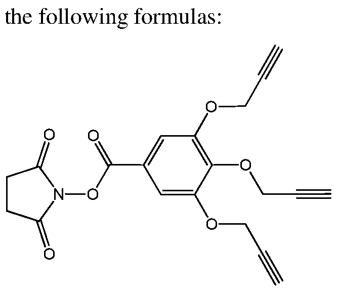
In another embodiment, said precursor is first conjugated to an amino derivatized compound (e.g. ODN derivative) followed by its conjugation to an azido-modified peptide (i.e. N3- peptide) using the copper-catalyzed Huisgen 1,3-dipolar cycloaddition (the "click reaction"), thereby resulting in the formation of a non-selective synthetic protein receptor according to this invention.
[00159] In one embodiment, this invention provides a method for the preparation of a compound comprising a non selective tripodal protein surface binder, said method comprises the following steps:
a. ed by the following formulas:
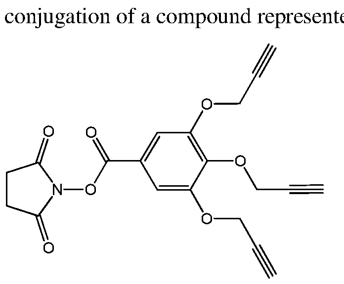
to an amino derivatized compound; and
b. conjugation of the product resulting from step a to an azido-modified peptide using the copper-catalyzed Huisgen 1,3-dipolar cycloaddition (the "click reaction"),
thereby resulting in the formation of a compound comprising a non-selective tripodal protein surface binder.
In another embodiment, the amino derivatized compound is an oligodeoxyribose nucleotide sequence derivative. In another embodiment, the amino derivatized compound is a peptide nucleic acid oligomer derivative. In another embodiment, the amino derivatized compound is an alkyl amino compound. In another embodiment, the amino derivatized compound is any compound with an amino moiety.
[00160] In one embodiment, this invention provides a method for the preparation of a protein biosensor which comprises a non selective tripodal protein surface binder, said method comprises the following steps:
a. ed by the following formulas:

to an amino derivatized compound; and
b. conjugation of the product resulting from step a to an azido-modified peptide using the copper-catalyzed Huisgen 1,3-dipolar cycloaddition (the "click reaction"),
thereby resulting in the formation of a protein biosensor which comprises a non selective tripodal protein surface binder.
In another embodiment, the amino derivatized compound is an oligodeoxyribose nucleotide (ODN) sequence derivative. In another embodiment, the amino derivatized compound is a peptide nucleic acid (PNA) oligomer derivative. In another embodiment, the amino derivatized compound is an alkyl amino compound. In another embodiment, the amino derivatized compound is any compound with an amino moiety.
[00161] In one embodiment, this invention provides a method of differentiating between proteins and protein isoforms in a biological medium comprising:
(i) binding an array of oligodeoxyribose nucleotide (ODN) derivatives duplexes to a protein;
wherein each of said duplexes consists of a first ODN derivative comprising an a first ODN sequence, at least one selective protein binder and a donor or acceptor;
and a second ODN derivative comprising a second ODN sequence, which is complementary to the first ODN sequence, at least one non-selective protein surface binder and a donor or acceptor, wherein if one of said ODN derivatives comprises a donor than the other one comprises an acceptor, and wherein at least one of said donor or said acceptor is a fluorophore;
wherein binding said protein with said duplex results in a conformational change of said duplex and thereby provide a unique optical signature; and
(ii) measuring said optical signature obtained by said binding of step (i); and thereby, differentiating between protein isoforms in a biological medium.
[00162] In one embodiment, this invention provides a method of diagnosing a disease or disorder in a subject, wherein said diagnosis comprises detection of a protein biomarker; said method comprises:
(i) collecting a biological sample from a subject;
(ii) optionally isolating components from said biological sample;
(iii) binding an array of complementary oligodeoxyribose nucleotide (ODN) derivatives duplexes with a protein;
wherein each of said duplexes consists of a first ODN derivative comprising a first ODN sequence, at least one selective protein binder and a donor or acceptor; and a second ODN derivative comprising a second ODN sequence, which is complementary to the first ODN sequence, at least one non-selective protein surface binder and a donor or acceptor, wherein if one of said ODN derivatives comprises a donor than the other one comprises an acceptor, and wherein at least one of said donor or said acceptor is a fluorophore;
wherein binding said protein with said duplex results in a conformational change of said duplex and thereby to a unique optical signature;
(iv) measuring said optical signature obtained by said binding of step (iii); and
(v) identifying a protein biomarker in said sample, said protein biomarker being characteristic of a disease or disorder; or measuring a change in a concentration of a protein biomarker in said sample compared to normative values, wherein said change is characteristic of a disease or disorder;
thereby, diagnosing a disease or disorder in a subject.
[00163] In one embodiment, the protein biomarker is MMP-1. In another embodiment, the protein biomarker is MMP-2. In another embodiment, the protein biomarker is MMP-7. In another embodiment, the protein biomarker is MMP-9. In another embodiment, the protein biomarker is GST a (A). In another embodiment, the protein biomarker is GST μ (M). In another embodiment, the protein biomarker is GST π (P). In another embodiment, the protein biomarker is GST- A. In another embodiment, the protein biomarker is GST-Al. In another embodiment, the protein biomarker is GST-A2. In another embodiment, the protein biomarker is GST-P. In another embodiment, the protein biomarker is GST-MI. In another embodiment, the protein biomarker is GST-P. In another embodiment, the protein biomarker is any combination of MMP-1, MMP-2, MMP-7, MMP-9 biomarkers. In another embodiment, the protein biomarker is any combination of GST a (A), GST μ (M), GST π (P), GST-A, GST-Al, GST-A2, GST-P, GST-MI biomarkers.
[00164] In one embodiment, the disease is cancer. In another embodiment, the disease is breast cancer, lung cancer, colorectal cancer, pancreas cancer, bladder cancer, ovarian cancer, prostate cancer, brain cancer, clear cell renal cell carcinoma, gastric cancer or any combination thereof.
[00165] In another embodiment, the disorder is renal tubular injury. In another embodiment, the disorder is graft failure or regeneration following living donor liver transplantation. [00166] In one embodiment, this invention is directed to a method of creating libraries of tripodal protein surface binders, said method comprises the conjugation of compound 7:

[00167] In one embodiment, this invention is directed to a protein biosensor, said biosensor comprises a tripodal protein surface binder represented by the structure of formula A:

A.
In another embodiment, the protein biosensor further comprises a selective protein binder. In another embodiment, the protein biosensor further comprises a donor and an acceptor, wherein at least one of the donor or acceptor is a fluorophore. In another embodiment, each donor or acceptor is attached to or in close proximity to one of the selective and non selective binders of said biosensor.
[00168] In one embodiment, this invention is directed to a protein biosensor, said biosensor comprises an array of synthetic receptors, wherein each synthetic receptor comprises a nonselective tripodal protein surface binder represented by the structure of formula A:

[00169] In one embodiment, this invention is directed to a protein biosensor, said biosensor comprises an array of synthetic receptors, wherein each synthetic receptor comprises a selective protein binder, a non-selective protein surface binder, a donor and an acceptor, wherein each donor or acceptor is attached to or in close proximity to one of the selective and non selective binders, wherein at least one of the donor or acceptor is a fluorophore. In another embodiment, the non selective binder comprises a peptide tripod, represented by the structure of formula A:

[00170] In one embodiment, this invention is directed to a protein biosensor wherein said biosensor comprises an array of synthetic receptors, wherein each synthetic receptor comprises an oligodeoxyribose nucleotide (ODN) derivatives duplex of this invention.
[00171] In one embodiment, this invention is directed to a protein biosensor, said biosensor consists of an array of complementary oligodeoxyribose nucleotide (ODN) derivatives duplexes, each duplex of such array consists of a first ODN derivative comprising a first ODN sequence and at least one selective protein binder; and a second ODN derivative comprising a second ODN sequence, which is complementary to the first ODN sequence,
and at least one non-selective protein surface binder; wherein each of the ODN derivatives independently further comprises a donor, an acceptor or combination thereof, wherein at least one of the donor or acceptor is a fluorophore. In another embodiment, the non selective binder comprises a peptide tripod, represented by the structure of formula A:

A.
In another embodiment, the duplexes of the array are different from each other in the peptide sequence that is comprised in their non-selective binder.
[00172] In one embodiment, this invention is directed to a cross reactive protein sensor array, said sensor array consists of an array of complementary oligodeoxyribose nucleotide (ODN) derivatives duplexes, each duplex of such array consists of a first ODN derivative comprising a first ODN sequence and at least one selective protein binder; and a second ODN derivative comprising a second ODN sequence, which is complementary to the first ODN sequence, and at least one non-selective protein surface binder; wherein each of the ODN derivatives independently further comprises a donor, an acceptor or combination thereof, wherein at least one of the donor or acceptor is a fluorophore, and wherein if one ODN derivative comprises a donor then the other ODN derivative comprises an acceptor. In another embodiment, the non selective binder comprises a peptide tripod, represented by the structure of formula A:

In another embodiment, the duplexes of the array are different from each other in the peptide sequence that is comprised in their non-selective binder.
[00173] In one embodiment, this invention provides a cross reactive sensor array for discriminating between closely related proteins in a biological medium, said sensor array comprises an array of synthetic receptors, wherein each of said synthetic receptors comprises complementary two oligodeoxyribose nucleotide (ODN) derivatives (i.e. an ODN duplex), wherein said two ODN derivatives consist of a first ODN derivative which comprises a first ODN sequence and at least one selective protein binder; and a second ODN derivative comprising a second ODN sequence, which is complementary to the first ODN sequence, at least one non-selective protein surface binder and a donor or acceptor, wherein if one ODN derivative comprises a donor, the other one comprises an acceptor, and wherein at least one of said donor or said acceptor is a fluorophore, and wherein said synthetic receptors differ from each other in their non-selective protein surface binder. In another embodiment, the non selective binder comprises a peptide tripod, represented by the structure of formula A:

[00174] In one embodiment, this invention provides an array of synthetic receptors that can discriminate between closely related proteins in a biological medium, wherein each of said synthetic receptors comprises oligodeoxyribose nucleotide (ODN) derivatives duplex, wherein each of said ODN derivatives duplexes consists of a first ODN derivative which comprises a first ODN sequence and at least one selective protein binder; and a second ODN derivative comprising a second ODN sequence, which is complementary to the first ODN sequence, at least one non-selective protein surface binder and a donor or acceptor, wherein if one ODN derivative comprises a donor the other one comprises an acceptor, and wherein at least one of said donor or said acceptor is a fluorophore, and wherein said synthetic receptors differ from each other in their non-selective protein surface binder. In another embodiment, the closely related proteins are protein isoforms.
[00175] A "protein isoform" refers to several different forms of the same protein, with slightly different amino acid sequences, but with similar activity. Different forms of a protein may be produced from related genes or may arise from the same gene by alternate splicing.
[00176] In one embodiment, this invention is directed to a protein biosensor as described hereinabove. In another embodiment, the proteins are enzymes. In another embodiment, the enzymes are isozymes. In another embodiment, the proteins are matrix metalloproteases (MMPs) protein and its isoforms. In another embodiment, the proteins are glutathione S- Transferase (GSTs) protein and its isoforms. In another embodiment, the proteins are platelet derived growth factors. In another embodiment, the proteins are Histidine-tag proteins. In another embodiment, the proteins are estrogen receptor (ERa or ER ). In another
embodiment, the proteins are fibroblast growth factor (FGF). In another embodiment, the proteins are glycoforms of the gpl20 protein.
[00177] An "ODN derivative duplex" refers to two complementary ODN derivatives which are hybridized. In another embodiment, an "ODN derivative duplex" refers to two complementary oligomeric peptide nucleic acid (PNA) derivatives which are hybridized.
[00178] In one embodiment, the first ODN derivative of the duplexes according to this invention, as described hereinabove is represented by the structure of formula IA, IB, IC, IIIA, IIIB, IIIC, III, ODN-20, ODN-58, ODN-49, ODN-51, or ODN-53.
[00179] In one embodiment, the second ODN derivative of the duplexes according to this invention, as described hereinabove is represented by the structure of formula IIA, IIB, IIC, IVA, IVB, IVC, IV, V, ODN-39, ODN-56, ODN-5, ODN-6, ODN-7, ODN-8, or ODN-9. In another embodiment, ODN-5-9 are five ODN derivatives which differ from each other in their non-selective protein binder. In another embodiment each of the non-selective protein binders comprises three peptides. In another embodiment, ODN-5-9 are five ODN derivatives which differ from each other in their non-selective protein binder peptide sequences.
[00180] An "array" is referred herein as between 3 to 10 ODN duplexes of this invention. Generating an array of ODN duplexes, which differ solely in their protein surface binding domain (i.e possess different amino-acid sequence; different peptides), will afford an analytical device in which all of the receptors bind preferentially to a particular protein family.
[00181] In one embodiment, the array according to this invention as described hereinabove consists of between 3 and 10 synthetic receptors. In another embodiment, the array consists of between 3 and 6 synthetic receptors. In another embodiment, the array consists of between 2 and 7 synthetic receptors. In another embodiment, the array consists of 4 synthetic receptors. In one embodiment, the array consists of 5 synthetic receptors. In another embodiment, the array consists of 6 synthetic receptors.
[00182] In one embodiment, each of said non-selective protein surface binders according to this invention as described hereinabove comprises at least one peptide, wherein said at least one peptide is different for each synthetic receptor in said array. In another embodiment, each of said non-selective protein surface binders comprises three peptides, wherein said peptides are different for each synthetic receptor in said array. In another embodiment, the non
selective protein surface binder comprises a peptide tripod, represented by the structure of formula A:

A.
[00183] In some embodiments the methods of this invention are directed to a) differentiating between proteins and protein isoforms; b) identifying proteins and protein isoforms; c) detecting proteins and protein isoforms; or d) diagnosing a disease or disorder, using the ODN derivatives of this invention, ODN derivatives duplexes of this invention and/or arrays of ODN derivatives duplexes of this invention. In one embodiment the protein is matrix metalloproteases (MMPs) protein and its isoforms. In another embodiment, the protein is glutathione S-Transferase (GSTs) protein and its isoforms. In another embodiment, the protein is a platelet derived growth factor. In another embodiment, the protein is Histidine-tag proteins. In another embodiment, the protein is a fibroblast growth factor, Estrogen receptor (ERa or ER ), or glycoform of the pgl20 protein.
[00184] A "protein isoform" refers to several different forms of the same protein, with slightly different amino acid sequences, but with similar activity. Different forms of a protein may be produced from related genes or may arise from the same gene by alternate splicing.
[00185] In some embodiments, the methods of this invention are directed to a) differentiating between proteins and protein isoforms; b) identifying proteins and protein isoforms; c) detecting proteins and protein isoforms; or d) diagnosing a disease, comprising measuring an optical signature obtained by contacting ODN derivatives, ODN derivatives duplexes and/or arrays of ODN derivatives duplexes of this invention and a protein. In one embodiment, the unique optical signature of an array of ODN duplexess of this invention
(Figure 11) provides an optical/fluorescent pattern which is characteristic to a protein or to a protein isoform. In another embodiment, the fluorescence pattern is obtained following irradiation of the donor of the ODN derivatives of this invention at a wavelength that causes the excitation of said donor. In another embodiment, the fluorescence pattern indicates that the protein is present in the analyzed biological sample.
[00186] In some embodiments, a fluorescent pattern is obtained from an array of ODN duplexes of this invention. The fluorescent pattern obtained from this invention is used to identify, detect, differentiate a protein of this invention, or diagnose a disease or disorder which is characterize by the presence of the protein.
[00187] Binding of different proteins to the synthetic receptors of the invention, affects differently the distance and the orientation of the photoluminescent species which are incorporated into the receptor structure (i.e. donor and acceptor), as well as induces conformational changes in the receptor-protein complex that would result in different optical signature for each protein tested. In another embodiment binding of different proteins affects differently the emission of each fluorescent dye, as well as induces conformational changes that would result in different fluorescence resonance energy transfer (FRET) processes between them. The combination of these effects provides a vast number of unique optical signatures.
[00188] In another embodiment, the methods of this invention comprise an optional isolation of components from a biological sample. In another embodiment, "isolating components" refers to isolating cells having proteins; isolating sugars, desalination, isolating glycans, phosphates (non limiting examples include ATP, ADP, AMP, GMP), isolating phospholipids, isolating glycoprotein, a glycolipid or a proteoglycan from the biological sample.
[00189] In one embodiment, the method of this invention is directed to a method of diagnosing a disease or disorder in a subject. In another embodiment, the disease or disorder is characterized by different expression of various proteins and protein isoforms than their normative expression level. Non limited examples of a diseases are cancers (colorectal, prostate, breast, lung, gastric, pancreas, bladder, ovarian, or brain), hepatitis c, phenylketonuria, Alzheimer, type II diabetes, familial hypercholesterolemia. In another embodiment, the method of this invention is directed to identifying a renal tubular injury (using GST proteins as biomarkers). In another embodiment, the method of this invention is
directed to monitoring graft failure or regenerations following living donor liver transplantation (using GST proteins as biomarkers).
[00190] In one embodiment, the methods of this invention include a step of measuring the optical signature of the contacting/binding of the ODNs of this invention and a protein. In another embodiment, the unique optical signature of an array of hybridized ODNs provides a fluorescence pattern (Figure 11) characteristic of a protein isoform; and said fluorescence pattern is obtained following irradiation of the donor species at a wavelength that causes the excitation of the donor species. In another embodiment, the fluorescence pattern indicates the presence of at least one protein in a biological medium.
[00191] In some embodiments, the methods are directed to a) differentiating between proteins and protein isoforms; b) identifying proteins and protein isoforms; c) detecting proteins and protein isoforms; or d) diagnosing a disease, in a biological medium or biofluids. In one embodiment, the biological medium is serum, blood, plasma, urine, saliva, a tissue, a peritoneal, stool, mucus, tears, sweat, a biopsy, sperm or a cerebrospinal fluid sample.
[00192] In some embodiments, the methods are directed to diagnosing a disease by identifying or detecting a protein biomarker. In one embodiment, a protein biomarker is a matrix metalloproteases (MMPs) protein, wherein said MMP is a biomarker for cancer. In another embodiment, the MMP comprises for example the following isoforms: MMP-1, MMP-2, MMP-7 or MMP-9. In another embodiment, the MMP-1 is a biomarker for breast, lung and colorectal cancer. In another embodiment, MMP-2 is a biomarker for pancreas, bladder, colorectal, ovarian, prostate, brain, pancreas, lung and colorectal cancer. In another embodiment, MMP-7 is a biomarker for pancreas, lung and colorectal cancer. In another embodiment, MMP-9 is a biomarker for breast, pancreas, bladder, lung, colorectal, ovarian, prostate and brain cancer.
[00193] In some embodiments, the methods are directed to diagnosing a disease by identifying or detecting a protein biomarker. In one embodiment, a protein biomarker is a glutathione S-Transferase (GSTs), wherein said GST is a biomarker for cancer. In another embodiment the GST comprises for example the following isoforms: GSTA1, GSTA2, GSTM1 and GSTP1. In another embodiment, GST Al is a biomarker for breast, lung, prostate and colorectal cancer. In another embodiment GST A2 is a biomarker for prostate and lung cancer. In another embodiment GST Ml is a biomarker for prostate and breast cancer. In another embodiment GST PI is a biomarker for breast, lung and gastric cancer.
[00194] In some embodiments, the methods of this invention are directed to differentiating Fibroblast Growth Factors (FGFs) proteins. Characterization of FGFs that bind to an octasaccharide library of heparin/heparan sulfate revealed that positions 2-O-sulfate (A) and 6-0 sulfate (B), together or separated are essential for binding and that protein recognition involves sequences containing variable degrees of 6-O-sulfation of the A-B disaccharide unit. In another embodiment, 12-sulfated-octasaccharide heparin is a selective protein binder for FGF.
[00195] In some embodiments, the methods of this invention are directed to differentiating and characterizing ER biomarkers. In another embodiment, the methods of this invention are directed to differentiating and characterizing ERa and ER biomarkers.
[00196] In some embodiments, the methods of this invention are directed to differentiating glycoforms of the gpl20 protein. Distinguishing between different strains of the immunodeficiency virus type 1 (HIV-1) by targeting its surface envelope protein gpl20. This is a particularly important target because its changing glycosylation patterns enable the virus to evade immune responses. Existing approaches for differentiating between HIV envelop protein require expression of high concentrations of viral proteins and extensive efforts using mass spectrometry. The methods of this invention provide a high-throughput method to assess general glycosylation and therefore assist in classifying strains exhibiting distinct pathogenicities. Differentiation of gpl20 glycoforms will be achieved by using B40 aptamer, which binds gpl20 independently of N-linked glycosylation.
[00197] Various concentrations of biomarkers may be detected and measured by the methods described herein. Biomarkers at concentrations of between about 1-5 mM, 1-100 μΜ, 50-100 mg/mL, 50-500 nM or less than, e.g., 100 milligrams/milliliter (mg/ml), 10 mg/ml, 1 mg/ml, 100 micrograms/milliliter ^g/rnl), 10 μg/ml, 1 g/ml, 100 nanograms/milliliter (ng/ml), 10 ng/ml, 1 ng/ml, may be detected in the biological sample, and the concentration may be measured.
[00198] In one embodiment, the method of this invention is directed to a method of diagnosing a disease in a subject by measuring a change in a concentration of a protein biomarker compared to its normative values, wherein said change is characteristic of a disease or a disorder.
[00199] The term "normative value" refers to the control, i.e. a concentration range of the tested protein biomarker found in a normal healthy subject.
[00200] In one embodiment, the method of this invention is directed to a method of diagnosing a disease in a subject. In another embodiment, a subject refers to a mammal, a human, a female or a male.
[00201] The terms "comprises" and variations thereof do not have a limiting meaning where these terms appear in the description and claims.
[00202] Unless otherwise specified, "a" "an," "the," and "at least one" are used interchangeably and mean one or more than one.
[00203] The following examples are presented in order to more fully illustrate the preferred embodiments of the invention. They should in no way be construed, however, as limiting the broad scope of the invention.
EXAMPLES
Materials and Methods [00204] All solvents and reagents were obtained from commercial suppliers and used without further purification.
[00205] The JH NMR spectra were recorded either on a Bruker Avance 300 MHz NMR spectrometer. Or on a Bruker DPX-300MHz spectrometer. Chemical shifts are reported in ppm on the δ scale down field from TMS as the internal standard. The following abbreviations were used to describe the peaks: s-singlet, d-doublet, dd-double doublet, t- triplet, q-quartet, quin-quintet, m-multiplet, and b-broad. Electronspray mass spectrometry (ES-MS) was performed with a Micromass Platform LCZ-4000 instrument at the Weizmann Institute of Science mass spectrometry facility. Matrix assisted laser desorption ionization time-of-flight (MALDI-TOF) mass spectrometry was performed on an AB SCIEX 5800 System, equipped with an Nd: YAG (355 nm) laser with a 1 KHz pulse (Applied Biosystems), at the Weizmann Institute of Science mass spectrometry facility.
[00206] The UV-Vis spectra were measured on a Varian Cary UV- Visible spectrophotometer, and emission spectra were recorded on a Varian Cary Eclipse fluorescence spectrophotometer with excitation and emission slit widths of 20 nm.
[00207] Peptides were synthetized on Aan automated peptide synthesizer (Advanced ChemTech, Apex 396). Analytical reversed-phase high-performance liquid chromatography (RP-HPLC) analysis of peptides was performed on a Waters liquid chromatography system
equipped with a 2487 dual wavelength UV detector, 600 gradient pump, and a 717 plus Autosampler.
[00208] The peptides were purified by RP-HPLC using a ThermoSeparation instrument (P200 pump, UV 100 detector) and a pre-packed Vydac Ci8 column. Standard RP-HPLC conditions were as follows: mobile phase A = 0.1% TFA in H2O; mobile phase B: 0.1%TFA in 25:75, H20:CH3CN. Fluorescence and enzymatic assays were measured using a BioTek synergy H4 hybrid multiwell plate reader in black flat-bottom polystyrene NBS 384 well microplates (Corning).
[00209] Modified oligonucleotides were synthesized by the W. M. Keck Foundation Biotechnology at Yale University, using standard automated solid phase synthesis. Concentrations of the oligonucleotides were based on their respective electronic absorption at 260 nm and their molar extinction coefficients were obtained by nearest neighbor calculations. The oligonucleotide purification was carried out on a Waters 2695 separation module HPLC system with a 2994 photodiode array detector using either a Waters XBridge™ OST C18 column (2.5 μΜ, 4.6x50 mm) or a XBridge™ OST C18 column (2.5μΜ, 10 mm x 50 mm). Purifications and analyses were achieved using the following eluents: mobile phase A = 5:5:90 TEAA:CH3CN:H20, mobile phase B = 5:65:30 TEAA: CH3CN:H20. Different gradients were used and details are given, when necessary. Oligonucleotide samples were desalted using illustra MicroSpin G-25 Columns (GE Healthcare) according to the manufacturer's instructions. Recombinant human MMP isoforms 1,2,7, and 9 were purchased from ProSpec-Tany TechnoGene Ltd. Human GST isoforms Al-1, A2-2, Ml-1, and Pl-1 were purchased from Oxford Biomedical Research. All protein concentrations were given by the manufacturers which is determined the Bradford protein assay.
[00210] Activated MMPs were dialyzed using a Slide-A-Lyzer MINI Dialysis Device, 10K MWCO, 0.1mL(Thermo Scientific). Urine samples was collected from a healthy man and immediately divided into 1.5-mL aliquots and frozen at -80°C. Urine samples were desalted and concentrated using Amicon Ultra 2mL10K MWCO. Oligonucleotide samples were desalted using illustra MicroSpin G-25 Columns (GE Healthcare) according to the manufacturer's instructions. Concentrations of the oligonucleotides were quantified based on their respective electronic absorption at 260 nm and the molar extinction coefficient for the oligonucleotide at this wavelength. The extinction coefficient of 6-FAM and Dabcyl dyes at 260 nm are 20,900M~1cm~1 and 29,100M~1cm~1, respectively. Extinction coefficient of Cy3
dye at 260 nm is 4,930 M" cm" . Absorbance spectra were recorded on a Varian Cary UV- visible spectrophotometer. FRET measurements were performed on a Varian Cary fluorimeter using quartz cuvettes. Fluorescence polarization and enzymatic assays were measured using a BioTek synergy H4 hybrid multiwell plate reader in black flat-bottom polystyrene NBS 384 well microplates (corning). Concentration measurements were performed on a Varian Cary fluorimeter using quartz cuvettes.
[00211] Recombinant human GST isoforms Al-1, A2-2, Ml-1, and Pl-1 were purchased from Oxford Biomedical Research. Transferrin, apolipoprotein-Al(APOAl), Fibroblast growth factor 21 (FGF-21), and leptin were purchased from ProSpec-Tany TechnoGene Ltd. (Ness Ziona, Israel). Immunoglobulin G (IgG) and aj-acid-glycoprotein were purchased from Sigma-Aldrich. Haptoglobin mixed type and thyroglobulin were obtained from Merck Millipore. 3-azidopropionic acid, Fmoc-Lys(Dansyl)-OH and compound 8 were synthesiszed according to published procedures, respectively (Grandjean, C; Boutonnier, A.; Guerreiro, C; Fournier, J. M.; Mulard, L. A., /. Org. Chem. 2005, 70, 7123; Dantas de Araujo, A.; Palomo, J. M.; Cramer, J.; Seitz, O.; Alexandrov, K.; Waldmann, H., Chem. Eur. J. 2006, 12, 6095; Camponovo, J.; Ruiz, J.; Cloutet, E.; Astruc, D. Chem. Eur. J. 2009, 15, 2990).
[00212] The oligonucleotides purification was carried out on a Waters 2695 separation module HPLC system with 2994 photodiode array detector using either Waters XBridge™ OST C18 column (2.5 μΜ, 4.6 mm x 50 mm) or XBridge™ OST C18 column (2.5μΜ, 10 mm x 50 mm). Oligonucleotide samples were desalted using illustra MicroSpin G-25 Columns (GE Healthcare) according to manufacturer's instructions. Concentrations of the oligonucleotides were quantified based on their respective electronic absorption at 260 nm and the molar extinction coefficient of the oligonucleotide at this wavelength. Extinction coefficient of Cy3 dye at 260 nm is 4930 M^cm"1. Absorbance spectra were recorded on a Varian Cary UV-visible spectrophotometer. FRET measurements were performed on a Varian Cary fluorimeter using quartz cuvettes. Enzymatic assays were carried out using a BioTek synergy H4 hybrid multiwell plate reader in clear flat-bottom polystyrene 384 well microplates, (corning). Differentiation assays were carried out using a BioTek synergy H4 hybrid multiwell plate reader in c and black flat-bottom polystyrene NBS 384 well microplates (corning).
ODNs Sequences:
[00213] ODN-10 and ODN-3 were obtained from Keck Foundation Biotechnology at Yale University.
ODN-10: 5'-YZWXXXGCCTCGCTCGTCTTGCG-3'
-3 : 5 '-CGC A AGACGAGCGAGGCXXX Y-3 '
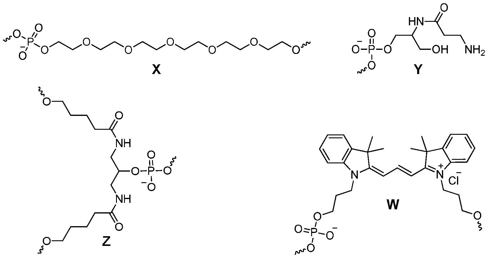
EXAMPLE 1
[00214] The sensing strategy according to this invention relies on using receptors with dual function mode; specific and non-specific. On the one hand, these receptors bind particular protein groups with high affinity and selectivity. On the other hand, they possess a nonspecific binding domain that respond differently to closely related isoforms, therefore generating unique signal pattern for each isoform. The receptors consist of two complementary oligonucleotides (ODNs). One strand is linked to ethacrynic acid (EA), a broad-spectrum inhibitor of GSTs, and the complementary strand is modified with a tripodal peptide scaffold that functions as non-specific protein binder. The specific strand which is maintained in all the receptors' structures allows selective binding of the receptors to the GST protein family. The peptide sequences were varied to achieve a series of nonspecific protein surface receptors. The tripodal peptide motif interacted with GST surface by means of hydrogen bonding, electrostatic interaction or hydrophobic interaction, allowing
the system to respond distinctively to each GST isoform. In addition, a fluorescent donor and an acceptor were also incorporated in the proximity of both specific and non-specific binders. The peptides were labeled with solvatochromic fluorophore (dansyl) whose emission spectrum overlaps the excitation spectrum of Cy3 in the proximity of EA molecule. Binding to different GSTs affected both the distance and the orientation between these fluorophores and thus the fluorescence resonance energy transfer (FRET) efficiency was altered (Figure 1).
Table 1. Sequences of peptide used for generation of a library of receptors
10 [001]
Receptor ODN-Peptide Conjugate Peptide Sequence
[002]
1 ODN-5 N3-(CH2)3-K(dansyl)-F-D-S-E-F-D-S-E [003]
[004]
2 ODN-6 N3-(CH2)3-K(dansyl)-S-T-G-V-S-T-G-V [005]
3 ODN-7 N3-(CH2)3-Y-G-E-G-E-F-N-K(dansyl)L5 [006]
[007]
4 ODN-8 N3-(CH2)3-K(dansyl)-G-P-L-A-S-E-S-T [008]
[009]
5 ODN-9 N3-(CH2)3-K(dansyl)-G-R-V-G-A-V-S-G
[0010]
[00215] Based on these principles, a DNA-based receptors library with selectivity towards glutathione S-transferases (GSTs) was generated (Figure 2). The GST isoforms Al-1, A2-2, Pl-1 and Ml-1 having different isoelectric point were selected as sensing targets (Table 2).
Table 2. Isoelectric point (pi) of the GST isozymes.
„„„, , „ Isoelectric Point
GST Isoform
(pi)
GST-Al-1 8.9
GST-A2-2 8.5
GST-Ml-1 6.2
GST-Pl-1 5.4
[00216] GSTs were selected as the target proteins for this study because comparative analysis of GST isozymes in biofluids revealed a clear correlation between their expression profiles and disease states. In addition, a broad spectrum inhibitor (i.e.,
ethacrynic acid, EA) for this class of enzymes is known and could be used to generate a GST-specific binding strand (Figures 2A).
[00217] Five ODN sets composed of diverse peptide sequences as sensing elements were prepared (Figure 2 and Table 1). The bivalent analogs of EA have been shown to exhibit significantly greater affinity for GST enzymes. Therefore, EA was covalently coupled to a bis-amino-modified oligonucleotide (ODN- 10) to afford the specific strand (ODN-20) (Figure 2A). A library of non-specific strands (ODNs-5-9, Figure 2B) was prepared by coupling of compound 7 (synthesis described below) to an amino-modified oligonucleotide (ODN-3) and subsequent click reaction with azide-functionalized peptides (Table 1). The equimolar ratio of complementary oligonucleotide strands were then heated to 95°C for 5 min and slowly cooled to room temperature to form receptors 1-5. Upon hybridization, an efficient FRET process between the acceptor Cy3 and donor dansyl moieties was observed reflecting the close proximity of two fluorophores (Figure 3).
[00218] In the first step, enzymatic assays were performed to confirm broad spectrum inhibition by ODN-20 (Figure 2A), namely, that the specific GST binder can bind and inhibit the activity of different GST isosymes. These assays, which were conducted according to a previously reported procedure (Habig, W. H.; Pabst, M. J.; Jakoby, W. B. /. Bio. Chem. 1974, 249, 7130) were also used to evaluate the affinity of the system toward GSTs (see below). As shown in Figure 4, the enzymatic activity of GST-A1, GST-MI, GST-A2, and GST-PI (which catalyzes the conjugation of glutathione (GSH) to 1-chloro- 2,4-dinitrobenzene (CDNB)) was followed in the absence and in the presence of ODN-20 (500 nM), a commercial inhibitor (EA, ΙμΜ), and a control strand, ODN-10, which lacks EA groups (500 nM) by monitoring the change in the absorbance at 340 nm. The much stronger inhibition of these isozymes by ODN-20 indicates that a broad spectrum GST binder with nanomolar affinities toward the four different isozymes was obtained.
[00219] A set of DNA duplex-based GST receptors (Figure IB, duplexes 1-5) consisting of a specific (ODN-20) and non-specific binding strands (ODNs-5-9) was then generated by heating equimolar ratio of ODN-20 and complementary ODNs-5-9 to 95°C for 5 min and slowly cooling to room temperature. The formation of these duplexes was confirmed by gel electrophoresis as well as by observing FRET processes between the donors (i.e., dansyl groups) and the Cy3 acceptor (Figure 3). The emission of Cy3 was further enhanced when the different duplexes were incubated distinct GST isozymes (150
nM) (Figure 26), indicating the ability of the DNA-based fluorescent receptors to signal the receptor-GST interactions.
[00220] The most important principle underlying the design of this sensory system is that binding cooperatively between the specific and non-specific binders would transform relatively weak tripodal peptide-GST interactions into much stronger ones. To confirm the manifestation of the "chelate effect", which should endow the modified duplexes with selectivity toward GSTs (Figure IB), two experiments were performed (Figure 7). In the first, the different receptors (1-5) were incubated with different GSTs (150 nM) and their fluorescence response was compared to that of duplexes, which lack a GST inhibitor (Figure 7A). In the second experiment, the different receptors were treated with common serum proteins or protein biomarkers, such as transferrin, mixed-type haptoglobin, αΐ-acid glycoprotein, immunoglobulin G (IgG), leptin, thyroglobulin, apolipoprotein Al (APOA1), and fibroblast growth factor-21 (FGF-21) (Figure 7B). Remarkably, while duplexes 1-5 exhibited strong response to nanomolar concentrations of GSTs (150 nM), only a small or negligible effect on Cy3 emission was observed for the control duplexes (Figure 7A). Similarly, the emission intensity was only slightly changed upon incubating the receptors with excess (0.5-1 μΜ) of other proteins (Figure 7B). Taken together, these results show that the fluorescence response in this system mainly originates from the interaction of the receptors with GSTs.
[00221] The realization of targeted protein surface sensors such as 1-5, might open the way for developing a cross-reactive sensor array that can discriminate among different GST biomarkers in biological mixtures. For example, the presence of GST-A in urine or in blood plasma is an early biomarker for hepatocellular damage, whereas elevated serum levels of GST-P has been proposed as a tool for monitoring cancer patients during the course of treatment. It has also been demonstrated that tracking both GST-A and GST-P in urine could provide information on the site of renal tubular injury, and that detecting these isosymes in plasma can help predict and monitor graft failure or regeneration following living donor liver transplantation.
[00222] To demonstrate the potential use of such systems in medical diagnosis a sensor array prototype, consisting if duplexes 1-5, was prepared and its ability to discriminate among different GSTs and their combinations was tested in the absence and in the presence of other proteins (Figure 5B and Figure 6), as well as within human urine
(Figure 9). In a typical experiment, receptors 1-5 (20 nM) in PBS buffer (3.5 mM, pH 6.5) were loaded onto a 384-well plate and the changes in the Cy3 emission intensities were recorded for four replicates using excitation and emission filters of 325/15 and 620/40 nm, respectively. As shown in Figure 5 A, the addition of different GST isozymes (150 nM) resulted in various fluorescence profiles for GST-Al, GST-A2, GST-A2, GST-Ml as well as for combination of GST-Ml and GST-PI. Analysing the patterns using principal component analysis (PCA) (Figure 5B, squares) resulted in markedly distinct clusters, which indicates that the system can discriminate among closely related GST isozymes.
[00223] In the next step, a mixture of eight highly abundant serum proteins and biomarkers was spiked into the buffer solution (with a total concentration of 1.05 μΜ) and fluorescence patterns were recorded under similar conditions (Figures 5B and Figure 6). The protein mixture constituted of common serum proteins and protein biomarkers, namely, transferrin, mixed-type haptoglobin, αΐ-acid glycoprotein, immunoglobulin G (IgG), leptin, thyroglobulin, apolipoprotein Al (APOAl), and fibroblast growth factor-21 (FGF-21). In this medium, slightly higher concentration of GST (300 nM) was required to obtain sufficient fluorescence response, which might be due to some non-specific interactions with other proteins. Remarkably, the generated fluorescent patterns in the presence and in the absence of the protein mixture were very similar (Figures 5B and 6), which confirm the viability of our approach (Figure 1). The feasibility of distinguishing among different GSTs in human urine (Figure 9) further support the potential of such systems to identify small populations of disease biomarkers in complex medium such as human physiological fluids.
[00224] The detection and identification of proteins in complex medium such as human physiological fluids remains a major challenge in biomarkers discovery and validation. After successful GST differentiation in mixture of other proteins, the performance of the sensor array was investigated in human urine. Urine samples were desalted (see below for details) to minimize the fluorescence background signal of the matrix. Then, the receptors 1-5 (40 nM) were dissolved in desalted urine samples and Cy3 emission intensities of the receptors were recorded before and after addition of the GST isozymes (500 nM). The fluorescent intensity changes of the receptors were recorded for each GST isozyme which resulted in a characteristic fluorescence pattern for each isozyme PCA analysis of the data revealed well separated clusters corresponding to different forms of the GST enzymes (Figure 9).
[00225] In summary, a general methodology for endowing optical cross- reactive sensor arrays with selectivity toward particular protein groups was demonstrated. The fundamental principle underlying this approach is the feasibility of realizing signalling protein receptors with two distinct binding motifs: specific and non-specific. The first enables the receptors to bind particular protein groups with high affinity and selectivity, while the second allows them to differentiate between closely related isoforms. The use of DNA-duplexes for scaffolding the receptors not only provides the system with inherent water solubility - it also facilitates device integration and modification. This should allow one to "program" such devices to identify various other classes of protein biomarkers simply by modifying the specific binding strand.
EXAMPLE 2
Synthesis of various ODN-peptide conjugates (Figure [00226] Synthesis of Compound 6 (Figure 2). Ethacrynic acid (0.9 g, 3 mmol) was suspended in dry DCM, then N-hydroxysuccinimide (0.42 g, 3.6 mmol) and DIC (465 μΐ, 3mmol) were added to the solution. The reaction mixture was stirred overnight. The organic layer was washed with HCl (0.1 Ν) and brine. The organic phase was dried over anhydrous sodium sulfate and the solvent was removed under vacuum. The product was purified by combiflash using silica gel column chromatography (eluent: 2% MeOH in DCM). Yield: 36%. JH NMR (CDC13, 300 MHz): δ 2.49 (t, / = 3.0 Hz, 1H); 2.57 (t, / = 3.0 Hz, 2H); 2.93 (s, 4H); 4.83 (d, / = 2.0 Hz, 4H); 4.89 (d, / = 3.0 Hz, 2H); 7.57 (s, 2H). MS (ESI): calcd for [M]+ 400.21 ; found 400.16.

[00228] Synthesis of ODN-20. The amino- modified ODN-10 (370 nmol) was dissolved in 175 μΐ MQ water and Na2C03/NaHC03 buffer (80 μΐ, 1M, pH = 8.75) was added to maintain basic condition of the reaction. The NHS-ester of ethacrynic acid (compound 6, 74 μπιοΐ in 120 μL· DMSO) was added in two equal portion and the reaction was mixed overnight. The sediment was removed by centrifugation and the sample was lyophilized. The oligonucleotide was re-suspended in 300 μL· MQ water and then was filtrated through MicroSpin G-25 columns (GE Healthcare) according to the manufacturer's protocol. The oligonucleotide was afterward purified using RE-HPLC. MALDI-TOF MS (m/z): calcd. 8040.64, found 8038.
Synthesis of non selective ODN-5-9-peptide conjugates
[00229] Synthesis of ODN-4: The amino-modified oligonucleotide ODN-3
(385 nmol) was dissolved in 150 μΐ MQ water and 150 μΐ of Na2C03/NaHC03 buffer (1 M, pH = 8.75, 150 μΕ) was added to maintain basic condition of the reaction. Then the NHS ester 7 (38.5 μπιοΐ in 120 μΕ DMSO) was added in two portions with time interval of 4 hours. The coupling reaction was performed overnight at room temperature. The sediment
was removed by centrifugation, and the reaction mixture was filtrated through MicroSpin G-25 columns (GE Healthcare) according to the manufacturer's protocol. The product was further purified by RE-HPLC. MALDI-TOF MS (m/z): calcd. 6777.70, found 6768.77. General Procedure for Peptide Synthesis.
[00230] The peptides were synthesized on Rink amide resin using an automated synthesizer (Advanced ChemTech, Apex 396). Coupling reactions were run on a 0.1-mmol scale using standard Fmoc protocol. The coupling was carried out using six-fold excess of each amino acid (coupling for 2 x 45 min), PyBOP/DIEA as coupling reagents and 25% piperdine in NMP for Fmoc deprotection. Fmoc-Lys(Dansyl)-OH (2 equiv.) and azidopropionic acid (2 equiv.) were coupled to the peptide chain manually. The peptides were cleaved from resin by TFA/F^O/triisopropylsilane (95:2.5:2.5) for 2 h. The peptides were purified using preparative RP-HPLC on a Qs column and characterized by electrospray mass spectrometry.
PI : ESI- MS (m/z): calcd.1430.5; found 1432.73
P2: ESI- MS (m/z): calcd.1164.29; found 1164.75
P3: ESI- MS (m/z): calcd.1272.34; found 1271.04
P4: ESI- MS (m/z): calcd.1218.34; found 1217.78
P5: ESI- MS (m/z): calcd.1159.32; found 1158.77
General Procedure for the Synthesis of ODN-Peptide Conjugates (ODN-5-9).
[00231] ODN-4 (60 nmol) was dissolved in fresh MQ water (50 μΕ). TEAA buffer (20 μΕ, 2 M pH 7), corresponding azide functionalized peptide (720 nmol in 85 μΕ DMSO) and ascorbic acid (20 μΕ of a 15 mM freshly prepared stock solution) were subsequently added. Then, the solution was degassed by argon for a few minutes and CUSO4 /TBTA complex (30 μΕ of a 10 mM stock solution) was added. The resulting mixture was tightly closed and stirred overnight. The ODN-peptide conjugates were afterward purified using RE-HPLC. ODN-5: MALDI-TOF MS (m/z): calcd.11072; found 11058.
ODN-6: MALDI-TOF MS (m/z): calcd.10270; found 10256.
ODN-7: MALDI-TOF MS (m/z): calcd.10594; found 10578.
ODN-8: MALDI-TOF MS (m/z): calcd.10432; found 10438.
ODN-9: MALDI-TOF MS (m/z): calcd.10255; found 10266.
EXAMPLE 3
GST Kinetic Measurements
[00232] The GST activity was measured spectrophotometrically at 25 °C using variable concentrations of chloro-2,4-dinitrobenzene (CDNB) and GSH (1 mM ) as substrates, in phosphate buffer (10 mM, pH 6.5). In a typical experiment, GST and ODN- 20(variable concentrations) were incubated for 10 min and then GSH and CDNB was subsequently added. The GST concentration was 20 nM. The GSH concentration (1 mM) was kept constant and the concentration of CDNB was varied from 0.1 mM to 0.8 mM. Formation of S-(2,4-dinitrophenyl)-glutathione was monitored using a plate reader at λ = 340 nm. The data were fitted to Michaelis-Menten equation using Sigmaplot version 12.0 statistical software (Systat) to determine the K{ values for competitive inhibition.
EXAMPLE 4
Hybridization of Complementary DNA Strands
[00233] Equimolar concentration (2 μΜ) of complementary single- strands in phosphate buffer (15 mM, Ph = 7.4) was heated to 95 °C and slowly cooled to room temperature for 30 min to ensure formation of equilibrium structures.
EXAMPLE 5
Fluorescence Resonance Energy Transfer Measurements
[00234] Equimolar concentrations of each of the complementary DNA strands in 15 mM phosphate buffer, pH 7.4 were incubated at 95 °C for 5 min and then slowly cooled to room temperature. FRET experiments were carried out with single- or double- stranded DNA (1.5 μΜ, 60 μΐ) in phosphate buffer (15 mM, pH 7.4). Fluorescence emission spectra were recorded using λβχ= 325 nm and excitation and emission slit widths of 20 nm in quartz cuvettes.
EXAMPLE 6
GST Sensing in Buffer Solution and in presence of Protein Mixture
[00235] The receptors in phosphate buffer (3.5 mM, pH = 6.5) were dispensed into 384-well microplates and then fluorescence intensity were recorded using excitation and emission filters of at 360/40 and 620/40, respectively, along with a 50% cut-off mirror. Then, GST isoforms (0.6 μΚ) were added to each well and the fluorescence intensity values were recorded again. The difference between two reads before and after addition of GST isoforms was treated as the fluorescence response. The protein mixture (total concentration of 1.05 μΜ) used in the experiments contained the following proteins: transferrin (150 nM) haptoglobin mixed type (150 nM), IgG (150 nM), oxacid-glycoprotein (150 nM), APOA1 (150 nM), leptin (100 nM), FGF-21 (100 nM), and thyroglobulin (100 nM). The final concentration of the receptors and GST isoforms in buffer solution were 20 nM and 150 nM, respectively. The final concentration of the receptors and GST isoforms in buffer solution containing protein mixture were 26 nM and 300 nM, respectively. Fluorescence values for GST-Pl and combination of GST-Al and GST-Pl were recorded after 45 min incubation. For other GST isoforms, the fluorescence values were recorded immediately after incubation. Fluorescence data carried out in four replicates and the data was subjected to principal component analysis (PCA) using XLSTAT (version 2013.1.01). EXAMPLE 7
GST Sensing in Urine Samples
[00236] Human urine sample was desalted and concentrated using
ProteoSpin™ urine protein concentration kit from Norgen BioTek Corp. according to manufacturer's protocol. The concentrated and salt-free human urine samples were then diluted to the initial volume of urine in phosphate buffer (3.5 mM, pH = 6.5). The receptors in urinary samples (50 nM) were dispensed into 384-well microplates and then fluorescence intensity were recorded using excitation and emission filters of 360/40 and 620/40, respectively, along with a 50% cut-off mirror. Then GST isoforms (500 nM, 0.6 μΚ) were added to each well and the fluorescence intensity values were recorded again. Fluorescence values for GST-Pl and combination was recorded after 45 min incubation. Fluorescence data for other GST isoforms was recorded immediately after incubation. The difference between two reads before and after addition of GST isoforms was treated as the
fluorescence response. This process was measured in triplicates and the data was subjected to principal component analysis (PCA) using XLSTAT (version 2013.1.01).
EXAMPLE 8
Synthesis of non selective ODN-39-peptide conjugates (Figure 13A)
[00237] Methyl 3,4,5 trihydroxy benzoate (31). To a stirred solution of
Gallic acid (30, 6 g, 35 mmol) in Methanol (100 mL), H2SO4 (4 mL, 74 mmoles) was added and the solution was refluxed at 80 °C for 12 hours. The reaction was monitored by thin layer chromatography (TLC) (EtOAc: Hexane, 80:20). The reaction mixture was evaporated to the crude solid. After extraction with EtOAc the solution was washed once by saturated NaHCC>3, once by distilled water, and dried with MgSC . Finally, the solvents were evaporated under reduced pressure to yield a substance resembling white powder. Yield: 5.76 g, 96 .JH-NMR (300 MHz, in MeOD) δ 7.05(s, 2H); 4.90(s, 3H); 3.83 (s, 3H).
[00238] 3,4,5 tris (proparyloxy)benzoate (33). A mixture of Methyl 3,4,5 trihydroxy benzoate (31, 4.14 g, 22.5 mmol) and propargyl bromide (32) 80% in toluene (8.7mL,78.75 mmol) was stirred for 10 minutes in 50 mL of degassed acetone. K2CO3 (21,05g, 150 mmol) was added. The reaction mixture was stirred under reflux overnight and monitored by TLC (Hexane: EtOAc, 1 : 1 ; colored by potassium permanganate). After redissolving in DCM, MgS04 was added to the stirred reaction mixture and filtered after 10 minutes. The solvents were removed under reduced pressure to produce yellow oil. Yield: 3.02 g, 73%.1H-NMR (300 MHz, in CDC13), δ 7.47(s, 2H,); 4.8 (s,6H); 3.93 (s, 3H). ES- MS (m/z) : calcd. 298.08,found 299.02 (M+H).
[00239] 3,4,5-tris(prop-2-yn-l-yloxy)benzoic acid (34). 3,4,5 tris (proparyloxy)benzoate (33, 4.2 g, 14mmol) was dissolved in dioxane/Methanol/4N NaOH : 130mL/50 mL/ 25 mL. The obtained reaction mixture was stirred for 5 hours at room temperature. Then the reaction mixture was acidified by IN HC1 (pH 1-2) and the solvents were removed under reduced pressure. The residue was redissolved in EtOAc (100 mL) and the organic phase was washed with IN KHSO4 (13.6 g in 100 mL water) 3 times, and brine 3 times as well, dried and evaporated under vacuum. Yield: 3.85 g, 91 %.'H-NMR (300 MHz, in CDCI3), δ 7.77(m, 2H); 7.59(m,lH); 7.51 (s, 1H); 4.85 (d, /=3 Hz,4H); 4.80(d, /=3 Hz, 2H); 3.02 (s, 2H); 2.88(s, H).ES-MS (m/z) : calcd.284.07,found 285 (M+H).
[00240] 2,5-dioxopyrrolidin-l-yl 3,4,5-tris(prop-2-yn-l-yloxy)benzoate
(35). 3,4,5-tris(prop-2-yn-l-yloxy)benzoic acid (34, 0.5g, 1.7mmol), N-hydroxysuccinimide (NHS, 202.4mg, 1.7mmol), and DIC (270μ1, 1.7mmol) were mixed in 8mL DCM. DMF (2mL) was added to obtain a clear solution. The solution was left overnight at room temperature and monitored by TLC (DCM:MeOH, 98:2). The mixture was extracted with HCl (0.1N), followed by drying with MgSC and evaporation;then the product was purified by Combiflash using silica gel column chromatography (eluent: 1 % MeOH in DCM). Yield: 80%. JH NMR (CDC13, 300 MHz): δ 2.49 (t, / = 3.0Hz, 1H);2.57 (t, / = 3.0 Hz, 2H);2.93 (s, 4H); 4.83 (d, / = 2.0Hz, 4H); 4.89 (d, / = 3.0Hz, 2H); 7.57 (s, 2H). ES-MS (m/z): calcd. 400.21 ; found 400.16, 422.10 (M+Na), 823.14 (2M+Na).
[00241] Synthesis of ODN-37. 2,5-dioxopyrrolidin-l-yl 3,4,5-tris(prop-2-yn- l-yloxy)benzoate (35, 20mg, 52.5μπιο1) was dissolved in 200μL· DMSO. Amine-terminated Dabcyl tagged oligo (ODN-36, 350 nmol in ΙΟΟμΚ) [ODN-36 was prepared by automated DNA synthesizer using the suitable phosphoamidite] was added to ΙΟΟμί NaHCC>3 / Na2CC>3 buffer (pH 8.75). The DMSO and Buffer solutions were mixed and white precipitate was formed. The reaction was mixed overnight at room temperature. The sediment was removed by centrifugation and then 1 mL milli-Q water was added and the sample was lyophilized. The DNA was re-suspended in 300 μΐ milli-Q water and desalted using MicroSpin G-25 columns according to the manufacturer's instructions (GE Healthcare). The HPLC analysis of the product showed minor traces of ODN-8 (-10% peak area) and the product with a major peak (- 90% peak area). Yield: 70%. MALDI-TOF MS (m/z): calcd. 6074; found: 6074.
[00242] Synthesis of ODN-39-peptide. A solution of alkyne-modified oligonucleotide (ODN-37, 100 nmol) in 100 μΐ milli-Q water and a solution of ascorbic acid in milli-Q water (80 μΕ, 5 mM) were mixed and buffered with Triethylammonium acetate (TEAA) (20μΕ, pH 7.0) to maintain a neutral pH. After addition of the azide functionalized peptide (15 equiv. in 15 μΐ DMSO-according to Table 3 below), the solution was degassed by argon bubbling for 1 min. Then a solution of CUSO4 / tris-(Benzyltriazolylmethyl)amine (TBTA) (10 mM, 40μΕ) was added. The reaction was stirred for 2-12 h at 25°C. The ODN- peptide conjugates were purified using HPLC, eluted at 60%-70% B, depending on the peptide sequence.
Table 3: Peptide sequences used for preparing different oligonucleotide - peptide conjugate

[00243] ODN-39-P1: yield: 71%. MALDI-TOF MS (m/z): calcd.5743; found
5743.
[00244] ODN-39-P2: yield: 35%. MALDI-TOF MS (m/z): calcd.6380; found
6380.
[00245] ODN-39-P3: yield: 25%. MALDI-TOF MS (m/z):calcd. 6866; found
6868.
[00246] ODN-39-P4: yield: 55%. MALDI-TOF MS (m z): calcd. 6106.6; found 6108.
[00247] ODN-39-P5: yield: 60%. MALDI-TOF MS (m/z): calcd.6665; found
6668.
[00248] ODN-39-P6: yield: 42%. MALDI-TOF MS (m/z): calcd.6668; found
6671.
[00249] ODN-39-P7: yield: 30%. MALDI-TOF MS (m/z): calcd.6861 ; found
6868.
[00250] ODN-38-P8: yield: 22%. MALDI-TOF MS (m/z): calcd.7677; found
7680.
[00251] ODN-39-P9: yield: 30%. MALDI-TOF MS (m/z): calcd.7449; found
7454.
EXAMPLE 9
Synthesis of selective ODN-49 (Figure 13B)
[00252] 2-Hydroxy-3-(2-methyl-allyl)-succinic acid diisopropyl ester (41).
Dry THF (30 mL) was added todiisoprpyl s(-) lactic acid (40, 1.8g, 9.5mmol) under argon. The solution was cooled to -78°C using dry ice/acetone. Then, lOmL LDA (2M, lOmL, 20mmol) was added dropwise. The mixture was stirred in ethylene glycol/dry ice bath (- 15°C) for 2 hours. The mixture was again cooled to 0°C, methallyl bromide (2.4mL, 22.8 mmol) was added dropwise and stirred fori hour at -78°C, 2 hours at -10°C, and 1 hour at room temperature. The solution was quenched by adding 2.6 mL glacial acetic acid, then diluted by diethyl ether (160mL) and water (40mL). The organic layer was washed with saturated NaHC(¾ and saturated brine, dried over MgSC and evaporated. The residue was purified usingCombiflash chromatography andHexane/EtOAc. The product eluted at 7% EtOAc. Yield 738mg, 29%. JH NMR (300MHz, CDCL): δ 5.11-4.97 (m, 2H), 4.81 (d, /=10.5 Hz, 2H), 4.17 (d, /=3.6 Hz, 1H), 3.22 (d, /=6.9 Hz, 1H), 3.02 (m. 1H), 2.56-2.34 (m, 2H), 1.74 (s, 3H), 1.28 (dJ=2.7 Hz, 3H) , 1.26 (d, /=5.1 Hz, 3H), 1.20 (s, 3H), 1.18 (s, 3H). ESI- MS: calcd. 272.3, found 273.3 (M+H), 295.3 (M+Na), 311.3 (M+K).
[00253] 2-Hydroxy-3-isobutyl-succinic acid diisopropyl ester (42).
Compound 41 (738mg, 2.71 mmol) was dissolved in ethanol (lOmL) and degassed with argon. Then 10% Pd/C (240mg) was added. The resulting black suspension was exposed to hydrogen at 1 atm of pressure overnight. Then, the catalyst was removed by filtration, and the filtrate was concentrated to a colorless gum (used for the next step without weighting). JH NMR (300MHz, CDCI3): δ 5.09-4.93 (m, 2H), 4.14 (d, /=6.0 Hz, 1H), 3.18 (s, br, 1H), 2.82 (m. 1H), 1.73-1.57 (m, 2H), 1.42 (m, 1H), 1.25 (s,3H) , 1.22 (s, , 3H), 1.18 (s, 3H), 1.16 (s, 3H), 0.89 (q, /=3 Hz, 6H). ESI MS: [M+H] =275.2 (275.2), [M+Na] =297.2, and[M+K] =313.1.
[00254] (2R)-2-hydroxy-3-isobutylsuccinic acid (43). Compound 42 (crude from previous step) was dissolved in methanol (16 mL) and sodium hydroxide (2N,16 mL) was added at 0°C. The mixture was stirred at room temperature overnight. The solution was quenched by adding 6 N HC1 to pH=l. The mixture was extracted with EtOAc (20mLx8). The combined organic extracts were dried over anhydrous MgSC and evaporated to yield a pale yellow gum. The product, analyzed by mass spectroscopy and JH NMR, was found to be composed of a mixture of mono-isopropyl ester and the di-acid product. Therefore, this
procedure was repeated again. An alternative option: use dioxane (15 mL) and KOH,(15 mL2N) to yield the desired product after a workup. Yield: 340mg, 66%. JH NMR (300MHz, MeOD): δ 4.23 (d, J=5 Hz, 1H), 2.91-2.88 (m, 1H), 1.73-1.61 (m, 2H), 1.41- 1.36 (m,lH) , 0.94 (d, J=6 Hz, 6H). ESI MS: calcd.189.2; found 188.9 = (M-H), 213.0 (M+Na), 229.0 (M+K).
[00255] (S)-2-((R)-2,2-dimethyl-5-oxo-l,3-dioxolan-4-yl)-4- methylpentanoic acid (44). Compound 43 (275mg, 1.45mol) and TsOH hydrate (52mg, 0.27mmol) were dissolved in dimethoxypropane (6mL). The mixture was stirred for 6 hours at room temperature (3-6hours) and then concentrated (during evaporation the colorless solution turned red-brown). Purification was performedby Combiflash chromatography using Hexane/EtOAc. The product eluted at 7% EtOAc. Yield: 30mg, 9%. JH NMR (300MHz, CDC13): δ 4.46 (d, J=6 Hz, 1H), 3.04-2.98 (m, 1H), 1.82-1.62 (m, 3H), 1.60 (s,3H) , 1.54 (s,3H), 0.96 (s,3H), 0.94 (s,3H). ESI MS: calcd 230.3; found 228.9 (M-H), 253.0 (M+Na), 269.0 (M+K).
[00256] (R)-2-amino-N-(3-azidopropyl)-3,3-dimethylbutanamide (46).
Boc-teri-Leu-OH (45, 75mg, 0.325mmol, leq) and 3-azidopropane amine (39mg, 0.39mmol, 1.2 eq) were dissolved in 5mL DCM, followed by addition of EDC (124.6mg, 0.65mmol, 2 eq). After being stirredovernight, the reaction mixture was diluted with 20 mL DCM and washed with 10% HCl and Brine. The organic layer was dried with MgSC and concentrated under reduced pressure by rotary evaporator. The residue was purified by Combiflash chromatography using Hexane/EtOAc. Yield 66mg, 65%.
[00257] Deprotection of BOC protecting group: HCl (4.0M) in dioxane (2.5 mL) was added to a round bottom flask containing 46 (66 mg) under argon with an ice- water bathat 0°C and stirredfor 30min. Then, the reaction mixture was stirred at room temperature for 3 hours. 15mL of DCM were added and the solvent was evaporated. Yield: 30.5mg 68%. JH NMR (300MHz, CDCI3): δ 1.18 (s, 9H), 1.89-1.94 (m, 2H), 3.40-3.45 (m, 4H), 4.14 (s, lH), 8.15 (s,2H), 8.42 (s, 1H). ESI MS: calcd. 213.3; found 214.0 (M+H), 236.0 (M+Na), 427.2 (2M+H).
[00258] (2R,3S)-Nl-((R)-l-((3-azidopropyl)amino)-3,3-dimethyl-l- oxobutan-2-yl)-N4,3-dihydroxy-2-isobutylsuccinamide (47). Compound 44 (25mg, 0.11 mmol) was dissolved in anhydrous DMF (1.2 mL), followed by the addition of 46 (25.5mg, 0.12 mmol), HBTU (41.1mg, O. l lmmol), and DIPEA (ΙΟΟμΙ,, pH ~8). After 1 hour, the
reaction was diluted in ethyl acetate (25 mL) and washed with HCl (IN x 3). The organic layer was then dried with MgS04 and concentrated under reduced pressure by using a rotary evaporator and oil pump. Combiflash chromatography and Hexane/EtOAcused for purification (product eluted at 39% EtOAc). Yield 20mg, 48%. JH NMR (300MHz, CDC13): δ 0.99 (s, 9H), 1.43 (s, 9H), 1.73-1.85 (m, 2H), 3.27-3.39 (m, 4H), 3.85 (d, J=9 Hz, 1H), 5.35 (d, J=9 Hz, 1H), 6.62 (s, 1H). ESI MS: calcd. 313.4; found 336.1 (M+Na), 352.1 (M+K), 649.3 (2M+Na).
[00259] THF was added to a round bottom flask containing the resulting product (18 mg, 0.042mmol), followed by hydroxylamine (1.5 mL 50% in water). This solution was then refluxed at 80°C for 3 hours (analyzed by analytical HPLC). The reaction was concentrated under reduced pressure using a rotary evaporator. This residue was then purified by HPLC to afford 47. Yield: 10.8 mg, 64%. JH NMR (300MHz, CDCI3): 0.93- 1.00 (m, 15H), 1.21 (m, 2H), 1.27 (m, 1H), 3.18 (m, 1H), 3.39 (m, 4H), 4.17 (d, J=9 Hz, 1H), 4.32 (s, 1H), 7.04 (s, 1H), 7.65 (d, /=3 Hz, 1H). ESI MS: calcd. 400.47; found 399.14 (M-H), 423.17 (M+Na), 439.15 (M+K).
[00260] FAM-DNA-Marimastat (ODN-49). ODN-48 (Prepared by automated DNA synthesizer using the suitable phosphoamidite). 75μί, 247nmol) was mixed with compound 47 (1.4 μπιοΐ, about 6 eq). Then TEAA buffer (2M, 10μΚ) and ascorbic acid (20 μΕίη ¾0, 5mM) were added. The mixture was degassed with argon, and Cu-TBTA (10 μί,ΙΟηιΜ in 55% DMSO) was added and stirred overnight. Note: the total volume was 200 μΕ, H20:DMSO, 1: 1. The product was purified using HPLC. Yield: 40%. Maldi: calcd. 4440; found 4442.
EXAMPLE 10
Synthesis of selective ODN-51 (Figure 13C)
[00261] ODN-50 (Prepared by automated DNA synthesizer using the suitable
 350nmol) was added to 250μί NaHC03 / Na2C03 buffer (pH =8.75). NHS ester of ethacrynic acid (12, 12mg, 30μπιο1) was dissolved in 350μί DMSO. Then DMSO and buffer solutions were mixed and white precipitate was formed. The reaction mixture was stirred overnight at room temperature. The sediment was removed by centrifugation and then 1 mL milli-Q water was added and the sample was lyophilized. The DNA was re-suspended in 300 μL· milli-Q water,desalted using MicroSpin G-25 columns
according to the manufacturer's instructions (GE Healthcare), and purifiedHPLC. Yield: 30%. Maldi: calcd. 4200; found 4202.
EXAMPLE 11
Synthesis of selective ODN-53 (Figure 13D)
[00262] ODN-52 (ΙΟΟμΕ, 350nmol,) was added to 250μΕ NaHC03 / Na2C03 buffer (pH =8.75). NHS ester of ethacrynic acid (12 44mg, Ι ΙΟμπιοΙ) was dissolved in 350μΕ DMSO. Then, DMSO and buffer solutions were mixed and white precipitate was formed. The reaction mixture was stirred overnight at room temperature. The sediment was removed by centrifugation and then 1 mL milli-Q water was added and the sample was lyophilized. The DNA was re-suspended in 300 μΕ milli-Q water, desalted using MicroSpin G-25 columns according to the manufacturer's instructions (GE Healthcare), and purified by HPLC. Yield: 40%. Maldi: calcd. 4847; found 4847.
EXAMPLE 12
MMP kinetic measurements
[00263] Enzymatic inhibition assays were performed to confirm the binding of the marimastat-ODN conjugate (ODN 49) to four MMP isoforms (Figure 14) and to obtain the corresponding inhibition constants (Ki) (Figure 15). Marimastat is a highly potent broad-spectrum inhibitor of MMP isoforms, with K; values in the low nanomolar range.
[00264] MMP-2, MMP-9, and MMP-1 (ΙμΜ final concentration) were activated using APMA (ImM final concentration) in Tris buffer (lOOmM Tris, pH 7.5, lOOmM NaCl, lOmM CaCl2), at 37°C for 1 , 2 and 3 hours, respectively. MMP 7 does not need any activation. Inhibition of MMP-1 , MMP-2, MMP-7, and MMP-9 by ODN-49 was followed by using a fluorescently labeled peptide, namely, Mca-Pro-Leu-Gly-Leu- Dap(Dnp)-Ala-Arg-OH, as the substrate. This peptide contains a fluorophore (Mca) whose emission is intramolecularly quenched by the Dnp group. Therefore cleavage of this substrate by MMP results in an increase in the fluorescence signal at em=393 nm. The reaction was followed by monitoring the emission at 393 nm over several hours. As control experiments, the change in emission was also followed in the presence of an unmodified 'marimastat' , ODN-48, as well as without any inhibitor (Figure 15). MMP (8 nM final
concentration) was incubated with commercial marimastat (50nM), ODN-48 (50nM), and ODN-49 (50nM) for 10 min at room temperature. The peptide substrate was then added (5μΜ final concentration) and the fluorescence emission was monitored using a plate reader with 310/20 and 400/30 excitation and emission filters, respectively. After selective binding was established, the K; values were determined by incubating 5nM of each MMP with 7-8 different concentrations of ODN-49 (0, 0.05, 0.1, 0.5, 1, 2, 5, and 12.5 nM) for 10 min at room temperature. The peptide substrate was then added (5μΜ final concentration) and the reaction emission was monitored using aplate reader 310/20 and 400/30 excitation and emission filters, respectively.
[00265] To obtain K; values, eight different concentrations of ODN-49, ranging from 0.1 nM to 15 nM, were added to 5 nM MMP, followed by the addition of 5 μΜ substrate. The fluorescence emission was monitored using a microplate reader with 310/20 nm and 400/30 nm excitation and emission filters, respectively. The Ki values
(Table 4) were obtained by fitting initial velocities into the equation: using Sigmaplot 12.0 statistical software (Systat) (Figure 15A-D, Table 4).
[00266] These experiments showed that the inhibition of the four MMPs by
ODN-49 is comparable to that of the highly potent 'marimastat' and that ODN-48, which lacks the 'marimastat' group, does not inhibit their function. Thus, ODN-49 binds the enzymes via a derivative at the active site, as expected from our design.
EXAMPLE 13
Fluorescence polarization measurements
[00267] Fluorescence polarization was measured using a Synergy H4 hybrid multi well plate reader using black flat-bottom polystyrene NBS 384-well microplates in Tris buffer (lOOmM Tris, pH 7.5, lOOmM NaCl, lOmM CaCl2). The optimal concentrations of ODN-48 and ODN-49 were determined by measuring the fluorescence anisotropy of 50 μΐ of various concentrations of ODN (0.01- 1000 nM) in triplicate. The measurements were performed using 485/20 and 528/20 excitation and emission filters, respectively, with a 510- nm cut-off mirror. In order to monitor the interaction between MMP-7 and ODN-49, ODN- 49 (InM, 200 μΐ) and different concentrations of MMP-7 (0, 1, 2, 3, 4, 5, 10, 20, 40, 60, 75,
100, and 150 nM) were incubated at room temperature for 20 min and then divided into 60 μL· triplicates.
[00268] The Kd value for the interaction between MMP-7 and ODN-49 was obtained by performing fluorescence polarization (FP) measurements (Figure 15E). Fluorescein has been widely used in FP assays owing to its short excited state lifetime (~4 ns); thus, a binding curve for the MMP-7 -ODN-49 interaction could be obtained by following the change in the anisotropy signal of ODN-49 upon addition of MMP-7 in increasing concentrations^- 120 nM).
[00269] A dissociation constant of Kd=0.98 nM was obtained by fitting the data (Figure 15E) to a known FP equation:
(LT + Kd + Rj) - (LT - Kd - RT)2 - 4LTRT]
A = Af + {Ab - Af) x
2LT
where Af is the anisotropy of the free ligand, A is the anisotropy of the fully bond ligand, LT is the concentration of the ligand, and RT is the concentration of the receptor.
EXAMPLE 14
GST kinetic measurements
[00270] Inhibition of GST Al-1, GST A2-2, GST Ml-1, and GST Pl-1 activity by ODN modified with one (ODN-51) or two (ODN-53) ethacrynic acids (EAs) was followed using a GST activity assay developed by Habig et al. all GST isozymes (20nM final concentration) were incubated with ΙμΜ EA, ODN-50, and ODN-51 (GST Al-1 was incubated with 50μΜ EA, 50μΜ DNA, and ΙμΜ ODN-53) in a phosphate buffer at pH 6.5.

Gluthatione (GSH) CDNB GS-CDNB S-Conjugate
[00271] After incubation for 10 min at 30 °C, GSH (350μΜ final concentration) and CDNB (720μΜ final concentration) were added and the absorbance at λ
= 340 nm was monitored using a plate reader. The uninhibited reaction contained GST, GSH, and CDNB without ODN-53.
[00272] The uninhibited reaction contained GST, GSH, and CDNB. After selective binding was established, the K; values were determined by incubating GST A 1-1 (20nM) with 4 different concentrations of ODN-51 (0, 1 , 10, and 50 μΜ) and with 4-6 concentrations of ODN-53 (0, 10, 30, 60, 100, 500, 800, and 1200 nM) for 10 min at 30 °C. GSH (ImM final concentration) and CDNB (200, 400, 600, 800μΜ final concentrations) were then added and the absorbance at λ = 340 nm was monitored using a plate reader. The initial velocities were fitted to a Lineweaver-Burk competitive inhibition model using Sigmaplot 12.0 statistical software (Systat) (Figure 17) and the Ki values were derived according to the fitting parameters, namely, according to the highest determination coefficient (R ) and the lowest Akaike Information criterion correction (AICc). The Lineweaver-Burk competitive inhibition model was chosen.
[00273] Monofunctionalized ODN-53, inhibited the GST-Al-1 with a Ki value of ~14μΜ (Table 4). This value is consistent with reported values for the inhibition of GSTs by EA in the low micromolar range. ODN-53 inhibited all GST isozymes at the nanomolar range. To ensure that the inhibition is induced by the EA derivatives, a control experiment was performed in which GST activity was followed in the presence of ODN-52, which lacks the EA moieties (Figure 18). As expected, ODN-52 displayed no inhibition activity.
[00274] The resulting K; values of ODN-53 were obtained for GST A 1 - 1 , A2-
2, Ml-1 , and Pl-1 were 33 nM, 3 nM, 3 nM, and 230 nM, respectively, indicating that the bivalent inhibitor binds these enzymes with high affinity.
Table 4. Inhibition constants of the DNA-based GSTs and MMPs binders.

EXAMPLE 14
MMP differentiation assay
[00275] Molecular beacons (MBs) of this invention were prepared by incubating three non-selective ODN-peptide conjugates (ODN-39-P1, P5, and P6) with ODN-49 and recording the changes in their fluorescence upon addition of MMP-7 (Figure 19A). The different fluorescence responses confirm that distinct, non-specific peptide binders (i.e., PI, P5, and P6) interact differently with the same protein surface.
[00276] The ODN-39 peptide conjugate (ODN-39- PI, ...ODN-39-P9, 1-
25μΜ final concentration) was added to ODN-49 (20nM final concentration) in Tris buffer (lOOMm Tris, pH 7.5, lOOmM NaCl, lOMm CaCl2). Then the mixture was dispensed into 5 wells (50μΙ., each well). MMP (activated as described before and dialyzed for 4 hours against Slide- A-Lyzer MINI Dialysis Device, 10K MWCO, O. lmL 1L Tris buffer, to remove all APMA) was added to each well (ΙΟΟηΜ final concentration). Control experiments were performed by addition of Tris buffer to each ODN-39 peptide conjugates. The change in the fluorescence upon the addition of MMP was monitored using a plate reader with 485/20 and 528/20 excitation and emission filters, respectively. The fluorescence value of each well was subtracted from the respective control well and then divided by the fluorescence of the control well.
[00277] The MB that showed the most profound change in emission signal
(Figure 19 A, ODN-49/ODN-39-P1) was incubated with other cancer biomarkers, namely, with vascular endothelial growth factor (VEGF) and platelet-derived growth factor (PDGF)(Figure 19B). A change in the fluorescence was observed only upon addition of MMP-7, indicating the system is selective towards MMPs.
[00278] The response of each MB to different proteins was tested in Tris,
PBS, and HEPES buffers containing different concentration salts such as NaCl, MgCi2, Zn(¾, and CaCl2. Different temperatures and incubation times were also tested.
[00279] Figure 20 shows representative emission signals obtained from different MB-enzyme combinations. While in the presence of MMP-1, MMP-9, and MMP- 2, an increase in fluorescence intensity (8-30%) was observed for the different MBs; MMP- 7 induced a decrease (13-33%) in their emission. MMP-7 is the only isozyme with a basic isoelectric point (pl=9.63); therefore, at pH=7 positively charged domains on the protein's surface may interact with the negatively charged sugar-phosphate backbone of the MBs. This could lead to a direct interaction between 6-FAM and peptide side chains on the surface of MMP-7 and, as a result, to quenching of fluorescence.
[00280] In addition, the different MBs were arrayed on a micro well plate and incubated with the four MMP isoforms. Figures 21A and 21B show the fluorescence response of this cross-reactive sensor array to the addition of these enzymes in Tris buffer and human urine, respectively. For experiments with urine, two aliquots were rapidly thawed in 37°C, centrifuged for 1 min at lOOOxg, and then centrifuged for 15 minutes at 4500xg, 4° Cusing A micon Ultra 2mL 10,000 MWCO, followed by addition of 1.5mL Tris buffer and centrifuging for 15 minutes at 4500xg, 4°C. Finally, Tris buffer was added up to a volume of 1.5mL. The differentiation experiment with urine samples was carried out similarly to the experiments in Tris buffer.
[00281] In this experiment, the conditions that maximize the response of the
MBs to MMP-7 were selected. As a result, MMP-1, MMP-2, and MMP-9 induced relatively small changes in the emission signals. Principal component analysis (PC A) mapping of these patterns shows a clear differentiation between the four isoforms (Figures 21C and 21D).
EXAMPLE 15
GST differentiation assay
[00282] The ODN-39 peptide conjugate (ODN-39-P1 , .. .ODN-39-P9, 1-25μΜ final concentration) was added to ODN-53 (20nM final concentration, in PBS buffer(100mM,pH=6.5). Then the mixture was dispensed into 5 wells (50μί in each well). GST was added to each well (ΙΟΟηΜ final concentration). Control experiments were performed by addition of PBS buffer to each ODN-39-peptide conjugate. The change in the fluorescence upon the addition of the GST was monitored using a plate reader with 485/20 and 528/20 excitation and emission filters, respectively. The fluorescence value of each well was subtracted for the respective control well divided by the fluorescence of the control well. GST differentiation was observed using the ODN-39- P1-P9 and ODN-53 hybridized forms (not shown).
EXAMPLE 16
Synthesis of non-selective ODN-56 (Figure 22 A)
[00283] Synthesis of 2,5-dioxopyrrolidin-l-yl 3,4,5-tris(prop-2-yn-l- yloxy)benzoate (35). Compound 34 (1.5 g, 5.28 mmol) was suspended in dry DCM, and N- hydroxysuccinimide (0.06 g, 5.28 mmol) and DIC (818 μΐ, 5.28 mmol) were added to the solution. The mixture was stirred overnight and TLC (3% MeOH in DCM) was used to monitor the reaction progress. The reaction mixture was washed with HC1 (0.1 Ν) and brine solution, then the product was purified by combiflash using silica gel column chromatography (eluent: 1.5% MeOH in DCM). Yield: 75.7%.1H NMR (CDC13, 300 MHz): δ 2.49 (t, / = 3.0Hz, 1H);2.57 (t, / = 3.0Hz, 2H);2.93 (s, 4H); 4.83 (d, / = 2.0Hz, 4H); 4.89 (d, / = 3.0Hz, 2H); 7.57 (s, 2H). ES-MS (m/z): calcd. 381.34; found 404.04 (M+Na), 785.17 (2M+Na).
[00284] Synthesis of 3-azidopropanoic acid. 3-Bromopropionic acid (3 g,
20 mmol) was suspended in 30 mL acetonitrile and cooled in an ice bath. Then sodium azide (1.5 g, 20 mmol) was added under an argon atmosphere. The mixture was stirred overnight at 75 °C, and product formation was monitored by TLC. The solvent was evaporated in vacuum, re-suspended in EtOAc and washed with IN HCL and brine. The organic phase was dried over MgS04 and dried under vacuum. Yield: 50%. H NMR (CDCI3, 300 MHz): δ 2.62 (t, / = 6.0 Hz, 2H); 3.56 (t, / = 6.0Hz, 2H); 11.52 (bs, 1H).
Compound 16 is coupled to the peptide (see for example Scheme 1 below with a terminal azide group to yield 38-P1-P5).
[00285] Synthesis of ODN-55: 300 nmol of the oligonucleotide was dissolved in 80 μΐ. milli-Q water and 40 μΐ. of Na2C03/NaHCO buffer (1 M, pH 8.75) was added to maintain basic conditions of the reaction. Then compound 35 (11.5 mg, 30 μπιοΐ) in 40 μΐ of DMSO was added and the mixture was stirred for 3 h. Additional portion of compound 35 (15 μπιοΐ in 20 μΐ. DMSO) was added and the mixture was allowed to stir for another 2 h. Reaction progress was monitored using HPLC. The sediment was removed by centrifugation and then 1 mL milli-Q water was added and the sample was lyophilized. The DNA was re-suspended in 300 μΐ milli-Q water and desalted using MicroSpin G-25 columns according to the manufacturer's instructions (GE Healthcare). The HPLC analysis of the product showed minor traces of ODN-54 (-11 peak area) and the product with a major peak (~ 89% peak area). Yield: 55%. MALDI-TOF MS (m/z): calcd. 6722; found: 6710.
[00286] Synthesis of ODN-56: A solution of alkyne modified oligonucleotide
ODN-55 (30 nmol) in 90 μΐ milli-Q water and a solution of ascorbic acid in milli-Q water (40 μL· , 50 mM) were mixed and buffered with TEAA ( ΙΟμΙ. , pH 7.0) to maintain neutral pH. After addition of the azide functionalized peptide (12 equiv. in 90 μΐ DMSO), the solution was degassed by argon bubbling for 1 min. Then a solution of CUSO4 /TBTA (10 mM, 25μ1) was added. The reaction was stirred for ½ h at 35 °C, and for additional 3 h at 25 °C.
[00287] The azide functionalized peptides 38-P1-P5 (Table 5) that differ in their charge, polarity, and position of the solvatochromic dansyl fluorescent group were prepared using standard Fmoc solid phase peptide synthesis on a Rink amide resin. Their N- termini were functionalized with 3-azidopropanoic acid, whereas dansyl was incorporated into their structures by removing the Mtt protecting group and reacting the ε-amino side chain of lysine with dansyl chloride (Scheme 1). The modified peptides were purified by RP-HPLC and characterized by ES-MS.

Scheme 1: Introducing a dansyl group to peptide 5 from Table 5 below.
[00288] The ODN-peptide conjugates were purified using HPLC, eluted at 65%-95% B depending on peptide sequence.
[00289] Table 5. Peptide sequences used for preparing different oligonucleotide-peptide conjugates
Oligonucleotide-
Peptide Peptide Sequence
Peptide Conjugate
1 N3- -(CH2)3-I-E-A-D-K(dansyl)-G-I-D ODN56-P1
2 Ny -(CH2)3-K(dansyl)-F-D-S-E-F-D-S-E ODN56-P2
3 N3- -(CH2)3-K(dansyl)-S-T-G-V-S-T-G-V ODN56-P3
4 N3- -(CH2)3_S-T-A-F-A-Y-G-K(dansyl) ODN56-P4
5 N3- -(CH2)3-Y-G-E-G-E-F-N-K(dansyl) ODN56-P5
[00290] ODN-56- PI: yield: 30%. MALDI-TOF MS (m/z): calcd.11020; found 11011.
[00291] ODN-56-P2: yield: 48%. MALDI-TOF MS (m/z): calcd.10345; found 10376 (M+Na).
[00292] ODN-56-P3: yield: 14%. MALDI-TOF MS (m/z) :calcd.10270; found
10319 (M+2Na).
[00293] ODN-56-P4: yield: 30%. MALDI-TOF MS (m/z): calcd.10242.6; found 10247.3.
[00294] ODN-56-P5: yield: 55%. MALDI-TOF MS (m/z): calcd.10597.69; found 10578.4.
EXAMPLE 17
Synthesis of selective ODN-58- (Figure 22B)
[00295] Synthesis of ODN-58. The amine modified ODN-57 (250 nmol)[ ODN-57 prepared by automated DNA synthesizer using the suitable phosphoamidite] was dissolved in 80 μΐ milli-Q water. Then 40 μΐ of Na2C03/NaHC03 buffer (1M, pH 8.75) was added to maintain basic conditions of the reaction. The NHS-ester of ethacrynic acid 12 (see below) (10 mg, 25 μπιοΐ) in DMSO (40 μΐ) was added and the mixture was stirred overnight. Reaction progress was monitored using HPLC. The sediment was removed by centrifugation and then milli-Q water (1 mL) was added and the sample was lyophilized. The oligonucleotide was re-suspended in 300 μL· milli-Q water and desalted using MicroSpin G-25 columns according to the manufacturer's instructions (GE Healthcare).The HPLC analysis of the product showed minor traces of ODN-57( -11 % peak area, eluted at 5% B) and the product as a major peak (~ 89 % peak area, eluted at 30-35% B). MALDI- TOF MS (m/z): calcd. 7134.05; found 7134.67, 7187.5 (M+2Na).
[00296] NHS-ester of ethacrynic acid (12). Ethacrynic acid (11) (0.9 g, 3 mmol) was suspended in dry DCM, and N-hydroxysuccinimide (0.42 g, 3.6 mmol) and DIC (465 μΐ, 3mmol) were added to the solution. The reaction mixture was stirred overnight and TLC (3% MeOH in DCM) was used to monitor the reaction progress. The reaction mixture was washed with HC1 (0.1 Ν) and brine. The product was purified by combiflash using silica gel column chromatography (eluent: 2% MeOH in DCM). Yield: 36%. JH NMR (CDC13, 300 MHz): δ 2.49 (t, / = 3.0Hz, 1H);2.57 (t, / = 3.0Hz, 2H);2.93 (s, 4H); 4.83 (d, / = 2.0Hz, 4H); 4.89 (d, / = 3.0Hz, 2H); 7.57 (s, 2H). ES-MS (m/z): calcd. 400.21 ; found 400.16, 422.10 (M+Na), 823.14 (2M+Na).
EXAMPLE 18
GST kinetic measurements of ODN-57 and ODN-58
[00297] The GST activity was measured spectrophotometrically at 25 °C using duplicate samples of chloro-2,4-dinitrobenzene (CDNB) and GSH as substrates, in
phosphate buffer (50 μΐ, 1 M, pH 6.5). In a typical experiment, GST, GSH, and ODN were incubated for 10 min and then CDNB was added and formation of S-(2,4-dinitrophenyl)- glutathione was monitored using the plate reader at λ = 340 nm. Concentration of GST-A1- 1, GST-Ml-1, GST-A2-2 and GST-Pl-1 were 15 nM, 15 nM, 45 nM and 75 nM, respectively. The GSH concentration (1 mM) was kept constant and the concentration of CDNB was varied from 0.05 mM to 1.5 mM. The initial velocities were calculated by subtracting the spontaneous (non GST catalyzed) reaction of GSH and CDNB. In order to determine the K{ and the inhibition model, similar assay was carried out using two concentrations (0.1 and 1 μΜ) of ODN-58. Control experiments were performed with ODN-57 (10 μΜ). The data was analyzed using Sigmaplot version 12.0 statistical software (Systat) and the values of Vmax, Km and K{ were determined from the experimental fitting of the data to partial competitive inhibition model. Results demonstrated inhibition activity of ODN-58 as opposed to ODN-57-(not shown). EXAMPLE 19
Hybridization of complementary DNA strands
[00298] Hybridization of ODN-56 conjugates with the complementary sequence ODN-58 was performed as following: Solutions of the oligonucleotides in 50 μΐ phosphate buffer (100 mM, pH 7.4) were heated to 98 °C for 1 min, held at 90 °C for 2 min and then slowly cooled to 25 °C at a rate of 1 °C /min. The hybridization was confirmed by gel electrophoresis using 5% agarose gel and ethidium bromide as intercalating agent. The gel ran at a constant voltage of 60 V for 1 hour. The gel was scanned by ChemiDoc XRC UV scanner and analyzed by Image Lab software.
[00299] The ODN-56 and ODN-EA (ODN-58) conjugates were hybridized in PBS buffer (0.1 M, pH 7.4). The mixtures were heated to 98 °C for 1 min, held at 90 °C for 2 min, and then cooled down slowly to 25 °C at a constant rate of -1°C /min. The hybridization products were analyzed by 5% agarose gel electrophoresis using ethidium bromide staining and visualized by autoradiography (Figure 23). The formation of double- stranded ODNs was confirmed by the appearance of strong bands at ~ 20-base-pair (bp) position, which migrate slower than a single-stranded ODN-58. The lower intensity of control ODN-58 is due to the poor intercalation of ethidium bromide into a single strand.
Fluorescence resonance energy transfer measurements
[00300] Equimolar concentrations of each of the complementary DNA strands in 50 mM phosphate buffer, pH 7.4 were incubated at 95 °C for 5 min and then slowly cooled to room temperature. All FRET experiments were carried out with single- or double- stranded DNA (1.5 μΜ, 60μ1) in phosphate buffer (50 Mm, pH 7.4). Fluorescence emission spectra were recorded using λβχ= 325 nm and excitation and emission slit widths of 20 nm in quartz cuvettes. EXAMPLE 20
GST differentiation assay
[00301] Following hybridization, five self-assembled synthetic receptors were arrayed on a microwell plate, and four GST isozymes, namely, Al-1, A2-2, Ml-1, and Pl-1 were incubated with each of the receptors.
[00302] 50μΙ. of the receptor (50 nM) in phosphate buffer (50 mM, pH 7.4) was dispensed into 384-well microplates. The emission spectra and fluorescence intensity were recorded following excitation at 333nm and using Ex: 325/15 and Em: 620/40 filters along with a 50% cut-off mirror. Then, ΙμΕ of GST isoform (200 nm) was added to the appropriate wells, incubated for 30 min at room temperature, and the fluorescence was recorded again. The fluorescence intensity from triplicate samples was averaged.
[00303] Figure 24 shows a representative emission spectrum of ODN-56-P5 and ODN-58 prior to and after the addition of GST-A2-2. Interaction with the protein induced a change in the emissions of both dansyl and Cy3. Similarly, emission spectra were recorded from all wells, and the optical patterns resulting from variations in Cy3 emission intensities were analyzed using principal component analysis (PC A). The PCA plot confirms the ability of the system to differentiate between structurally related proteins (Figure 25).
[00304] While certain features of the invention have been illustrated and described herein, many modifications, substitutions, changes, and equivalents will now occur to those of ordinary skill in the art. It is, therefore, to be understood that the appended claims are intended to cover all such modifications and changes as fall within the true spirit of the invention.
Claims
An oligodeoxyribose nucleotide (ODN) derivatives duplex, which consists of a first ODN derivative comprising a first ODN sequence, at least one selective protein binder and a donor or acceptor; and
a second ODN derivative comprising a second ODN sequence, which is complementary to said first ODN sequence, at least one non-selective protein surface binder and a donor or acceptor;
wherein if one ODN derivative comprises a donor, the other one comprises an acceptor, and wherein at least one of the donor or acceptor is a fluorophore.
The oligodeoxyribose nucleotide (ODN) derivatives duplex of claim 1, wherein said selective protein binder is marimastat, ethacrynic acid (EA), bis ethacrynic acid, Ni-nitrilotriacetic acid (Ni-NTA), bis-Ni-NTA, tris-Ni-NTA, PDGF-BB aptamer, heparin derivative, FGF aptamer, or estrogen.
The oligodeoxyribose nucleotide (ODN) derivatives duplex of claim 1 or 2, wherein said non-selective protein surface binder comprises a peptide tripod, represented by the structure of formula A:

4. The oligodeoxyribose nucleotide (ODN) derivativ 2s duplex according to any one of claims 1-3, for use in a protein sensing device.
5. The oligodeoxyribose nucleotide (ODN) derivativ ss duplex according to any one of claims 1 to 4, for use in a cross reactive protein sensor array.
6. The oligodeoxyribose nucleotide (ODN) derivatives according to any one of claims 1 to 5, wherein said non selective protein surface binder is a tripodal peptides having a surface of 750-1500 A2.
7. The oligodeoxyribose nucleotide (ODN) derivatives according to any one of claims 1 to 6, wherein said donor or acceptor is fluorescein (6-FAM), Cy3, nile red, rhodamine, dansyl, dabcyl, perylene, fluorenyl, coumarin, 7- methoxycoumarin or Cy5.
8. The oligodeoxyribose nucleotide (ODN) derivatives duplex according to any one of claims 1, 2, 4, 5 and 7, wherein said first ODN derivative is represented by the structure of formula IA, IB or IC:
ODNl-X^Y-X'- R1^ (IA);
Y-X^ODNl-X'- R1)!, (IB);
ODNl-X^R'-X1-^ (IC);
wherein
ODN1 is a first ODN sequence:
X1 and X2 are each independently a linker or a bond, wherein said linker is Ci-G20 alkyl; CrC20 alkylether, -C(0)NH-alkyl-; -NHC(0)-alkyl-; - C(0)NH-alkyl-C(0)NH-; -C(0)NH-alkyl-NHC(0)- ; -NHC(0)-alkyl- NHC(O)-; -NHC(0)-alkyl-C(0)NH-; -(OCH2CH2)„-P04-(CH2)0-io-; -P04- [(0CH2CH2)n-P04]3-; -P04-(OCH2CH2)n-P04-(CH2)0-io-, -P04-(0CH2CH2)n- P04-alkyl-NHC(0)-; -C(0)NH-alkyl-P04-; -P04-CH-[alkyl-NHC(0)-alkyl- P04-alkyl-NHC(0)-alkyl-NHC(0)]2-; -NHC(0)-alkyl-P04-; NH-alkyl-NH-,- O-alkyl-NH, -NH-alkyl-O-; -P04-[(OCH2CH2)n-P04] i-5-alkyl-NHC(0)-alkyl- NHC(0)-alkylether-triazole-(CH2)0-io-; -P04-(OCH2CH2)„-P04-alkyl-amine- alkyl-triazole-(CH2)0-io-; -(0CH2CH2)n-PO4-alkyl-NHC(O); -P04- [(OCH2CH2)n-P04]i.5-alkyl-NHC(0)-; -P04-[(OCH2CH2)n-P04],-5- alkyl[NHC(0)-alkyl-NHC(0)]-alkyl-P04-alkyl-P04-alkyl-NHC(0)-;
(OCH2CH2)n-(CH2)0-io-, -(OCH2CH2)n-(CH2)o.io-NHC(0)-; -P04- [(OCH2CH2)n-P04]i-3-alkyl-NHC(0); -P04-[(OCH2CH2)n-P04]3-alkyl- NHC(0)-alkyl-; -P04-[(OCH2CH2)n-P04]2-alkyl-NHC(0)-alkyl-; -P04- [(OCH2CH2)n-P04],-3-alkyl-NHC(0)-alkyl-NHC(0)-; -P04-[(OCH2CH2)„- P04]i_3-alkyl- wherein n is an integer between 0 to 10, most preferably 6; -
9$
SUBSTITUTE SHEET (RULE 26}
P04-alkyl-NHC(0)-alkyl-, -P04-alkyl-NHC(0)-alkyl-NHC(0)-, alkyl- NHC(0)-(CH2)0-io-P04-alkyl-NHC(0)-alkyl-NHC(0), wherein said alkyl is optionally substituted, wherein substitutions include one or more groups selected from halogen, hydroxy, hydroxyalkyl, alkoxy, haloalkyl, carbonyl, amido, alkylamido, dialkylamido, nitro, cyano, amino, alkylamino, dialkylamino, carboxyl, thiol and thioalkyl;and wherein said alkyl is optionally interrupted by one or more heteroatoms selected from O, N, P, S or any combination thereof;
Y is a donor or acceptor;
k is an integer between 1 and 5; and
R1 is a selective binder to a protein.
9. The oligodeoxyribose nucleotide (ODN) derivatives duplex according to any one of claims 1, 2, 4. 5, 7, and 8, wherein said second ODN derivative is represented by the structure of formula IIA, IIB or IIC
ODN2-X2'-Y'-X1'-R1' (IIA);
Y'-X2'-ODN2-X1'-R1' (IIB);
ODN2-X2,-R1'-(X1'-Y')k- (IIC); wherein
ODN2 is a second ODN sequence;
X1' and X2' are each independently a linker or a bond, wherein said linker is Ci-C20 alkyl; Q-C20 alkylether, -C(0)NH-alkyl-; -NHC(0)-alkyl-; -C(0)NH- alkyl-C(0)NH-; -C(0)NH-alkyl-NHC(0)- ; -NHC(0)-alkyl-NHC(0)-; -NHC(O)- alkyl-C(0)NH-; -(OCH2CH2)n-P04-(CH2)o-i0-; -P04-[(OCH2CH2)n-P04]5-; -P04- (OCH2CH2)n-P04-(CH2)o-io-, -P04-(OCH2CH2)n-P04-alkyl-NHC(0)-; -C(0)NH- alkyl-P04-; -P04-CH-[alkyl-NHC(0)-alkyl-P04-alkyl-NHC(0)-alkyi-NHC(0)]2-; -NHC(0)-alkyl-P04-; NH-alkyl-NH-,-0-alkyl-NH-, -NH-alkyl-O-; -PO4- [(OCH2CH2)n-P04]i-5-alkyl-NHC(0)-alkyl-NHC(0)-alkylether-triazole-(CH2)o- 10-; -P04-(OCH2CH2)n-P04-alkyl-amine-alkyl-triazole-(CH2)o-io-; -(OCH2CH2)„- P04-alkyl-NHC(0); -P04-[(0CH2CH2)u-P04]1-5-alkyl-NHC(0)-; -P04- [(0CH2CH2)n-P04]i-5-alkyl[NHC(0)-alkyl-NHC(0)] -alkyl-P04-alkyl-P04 -alkyl»
99
SUBSTITUTC SHEET (RULE 26)
NHC(O)-; -(OCH2CH2)n-(CH2)0-io-, -(OCH2CH2)n-(CH2)o-io-NHC(0)-; -P04- [(OCH2CH2)„-P04]i_3-alkyl-NHC(0); -P04-[(OCH2CH2)n-P04]3-alkyl-NHC(0)- alkyl-; -P04-[(OCH2CH2)n-P04]2-alkyl-NHC(0)-alkyl-; -P04-[(OCH2CH2)n- P04]i.3-alkyl-NHC(0)-alkyl-NHC(0)-; -P04-[(OCH2CH2)n-P04]i-3-alkyI- wherein n is an integer between 0 to 10, most preferably 6; -P04-alkyl-NHC(0)- alkyl-, -P04-alkyl-NHC(0)-alkyl-NHC(0)-, alkyl-NHC(0)-(CH2)0-io-P04-alkyl- NHC(0)-alkyl-NHC(0), wherein said alkyl is optionally substituted, wherein substitutions include one or more groups selected from halogen, hydroxy, hydroxyalkyl, alkoxy, haloalkyl, carbonyl, amido, alkylarriido, dialkylamido, nitro, cyano, amino, alkylamino, dialkylamino, carboxyl, thiol and thioalkyl;and wherein said alkyl is optionally interrupted by one or more heteroatoms selected from O, N, P, S or any combination thereof;
Y' is a donor or acceptor;
k' is an integer between 1 and 5; and
R1 ' is a non selective protein surface binder.
10. The oligodeoxyribose nucleotide (ODN) derivatives duplex according to any one of claims 1 to 9, wherein said first ODN derivative of formula IA is represented by the structure of formula III:

III
11. The oligodeoxyribose nucleotide (ODN) derivatives of any one of claims 1 to 10, wherein said first ODN derivative is represented by the structure of formula
ODN-58:

ODN-58
12. The oligodeoxyribose nucleotide (ODN) derivatives of any one of claims 1 to 10, wherein said first ODN derivative is represented by the structure of formula

ODN-20
13. The oligodeoxyribose nucleotide (ODN) derivatives according to any one of claims 1 to 9, wherein said first ODN derivative of formula IB is represented by the structure of formula ODN-49 or ODN-51 or ODN-53:
1(M


ODN-53
14. The oligodeoxyribose nucleotide (ODN) derivatives according to any one of claims 1 to 13, wherein said second ODN derivative of formula IIA is represented by the structure of formula IV:

15. The oligodeoxyribose nucleotide (ODN) derivatives according to any one of claims 1 to 13, wherein said second ODN derivative of formula IIC is represented by the structure of formula V:

16. The oligodeoxyribose nucleotide (ODN) derivatives according to any one of claims 1 to 14, wherein said second ODN derivative of formula IIB is represented by the structure of formula ODN-39:

pept e
ODN-39
17. The oligodeoxyribose nucleotide (ODN) derivatives according to any one of claims 1-13 and 15, wherein said second ODN derivative of IIC is represented by the structure of formula ODN-56:

dansyl
ODN-56
18. The oligodeoxyribose nucleotide (ODN) derivatives according to any one of claims 1 to 13, 15 and 17 wherein said second ODN derivative of IIC is represented by the structure of formula ODN-5, ODN-6, ODN-7, ODN-8 and/or

the peptide sequence of ODN-5 is N3-(CH2)3-K-F-D-S-E-F-D-S-E, the peptide sequence of ODN-6 is N3-(CH2)3-K-S-T-G-V-S-T-G-V, the peptide sequence of ODN-7 is N3-(CH2)3-Y-G-E-G-E-F-N-K, the peptide sequence of ODN-8 is N3- (CH2)3-K-G-P-L-A-S-E-S-T and the peptide sequence of ODN-9 is N3-(CH )3-K-G- R-V-G-A-V-S-G; and wherein solvatochromic dansyl group is covalently attached to K residue in all sequences.
105
SUBSTITUTE SHEET (RULE 2S)
19. The oligodeoxyribose nucleotide (ODN) derivatives duplex according to any one of claims 1 to 18, wherein said first ODN derivative is represented by the structure of formula IIIA, IIIB or IIIC :
ODNl-[(P)n-(PEG)m]v-(P)I-X2-Y-(P)q-(Split)[X1-R1]t (IIIA);
Y-X2-ODNl-[(P)„-(PEG)m]v-(P),-(Split)[X1-R1]t (ΠΙΒ);
ODN1 -[(P)„-(PEG)m]v-(P)1-X2-(Split)[R1-X1-Y]t (IIIC);
wherein
ODN1 is a first oligodeoxyribose nucleotide sequence;
X1 and X2 are each independently a linker or a bond, wherein each of said linkers is independently a substituted or unsubstituted C1-C20 alkyl; substituted or unsubstituted Ci-C2o alkyl ether, substituted or unsubstituted Ci-C20 alkyl di-amide- alkylether, substituted or unsubstituted Ci-C2o alkyl di-amide-alkylether-triazole- alkyl, substituted or unsubstituted Ci-C2o alkyl amide, substituted or unsubstituted Ci-C2o alkyl di-amide, substituted or unsubstituted Ci-C2o alkyl triazole, substituted or unsubstituted alkyl-phosphate-alkyl-phosphate-alkylamide-, substituted or unsubstituted alkylamide-alkyl-phosphate-alkylamide-alkylamide-, substituted or unsubstituted alkylamine-alkyltriazole; wherein said alkyl is optionally substituted, wherein substitutions are selected from hydroxyl, methylene-hydroxyl, halogen, CN, N02, linear or branched C1-C6 alkyl, linear or branched Q-C6 alkoxy, linear or branched Q-Ce haloalkyl, or any combination thereof;
each P is independently P04, P03OH or P02(OH)2;
n and q are each 0 or 1 ;
each PEG is independently (0-CH2CH2)w wherein each w is independently an integer between 2 and 300;
m and 1 are each 0 or 1 ;
v is an integer between 1 and 10;
Split is C, CH, CH2-C, CH2-CH or absent;
t is 1, 2 or 3; wherein if Split is C or CH2-C then t is 3; if Split is CH or CH2-
CH then t is 2 and if Split is absent than t is 1 ;
Y is a donor or acceptor;
R1 is a selective protein binder ; and
wherein, if t is 2 or 3, then each of X1, R1, and Y can be same or different.
106
JISIITUTE SHEET (RULE 26)
20. The oligodeoxyribose nucleotide (ODN) derivatives duplex according to any one of claims 1 to 19, wherein said second ODN derivative is represented by the structure of formula IVA, IVB or IVC:
ODN2-[(P)n.-(PEG)m.]v-(P)r-X2'-Y'-(P)q-X1 '-(Q)(R1 ')k (IVA)
Y'-X2'-ODN2-[(P)n.-(PEG)m.]v.-(P)r-X1 '-(Q)(RI ')k (IVB)
ODN2-[(P)n-(PEG)m.]v-(P)r-X2'-(Q)[R1 '-X''-Y']k (IVC) wherein
ODN2 is a second oligodeoxyribose nucleotide sequence;
X1 ' and X2' are each independently a linker or a bond, wherein each of said linkers is independently a substituted or unsubstituted Ci-C2o alkyl; substituted or unsubstituted C1-C20 alkyl ether, substituted or unsubstituted C1-C20 alkyl di-amide- alkylether, substituted or unsubstituted C1 -C20 alkyl di-amide-alkylether-triazole- alkyl, substituted or unsubstituted C1-C20 alkyl amide, substituted or unsubstituted C1-C20 alkyl di-amide, substituted or unsubstituted C1-C20 alkyl triazole, substituted or unsubstituted alkyl-phosphate-alkyl-phosphate-alkylamide-, substituted or unsubstituted alkylamide-alkyl-phosphate-alkylamide-alkylamide-, substituted or unsubstituted alkylamine-alkyltriazole; wherein said alkyl is optionally substituted, wherein substitutions are selected from hydroxyl, methylene-hydroxyl, halogen, CN, NO2, linear or branched Ci-Ce alkyl, linear or branched Ci-Ce alkoxy, linear or branched C1-C6 haloalkyl, or any combination thereof;
each P is independently PO4, PO3OH or P02(OH)2;
n' and q' are each 0 or 1 ;
each PEG is independently (0-CH2CH2)w wherein each w is independently an integer between 2 and 300;
m' and 1' are each 0 or 1 ;
v' is an integer between 1 and 10;
Q is either absent, alkyl triazole or represented by the structure of formula B:
10?

Y' is a donor or acceptor; and
R1' is a peptide.
21. The oligodeoxyribose nucleotide (ODN) derivatives duplex according to any one of claims 1-7, 19 or 20 wherein n is 0 or 1, m is 1, 1 is 1, v is 1, 2 or 3, X2 is a bond or Ci-C20 alkyl, Y is Cy3 or FAM, q is 1, Split is absent, CH or CH2CH, each X1 is independently a substituted or unsubstituted alkylamide, alkylamide- alkyl-phosphate-alkylamide-alkylamide-, alkyl-phosphate-alkyl-phosphate- alkylamide, or alkyl di-amide-alkylether-triazole-alkyl, R1 is EA or Marimastat, and t is 1 or 2.
22. The oligodeoxyribose nucleotide (ODN) derivatives duplex according to any one of claims 1-7, 19, 20 or 21, wherein Y' is dabcyl or dansyl, X2' is a bond, a substituted C1-C20 alkyl di-amide, or a substituted or unsubstituted alkylamide, n' is 0 or 1, m' is 1, Γ is 0 or 1, v' is 1, 2 or 3, X1' is a bond, a substituted or unsubstituted alkylamide, Q is represented by formula B:

B,
and k is 3.
23. The oligodeoxynbose nucleotide (ODN) derivatives duplex according to any one of claims 1, 8 and 9, wherein said first ODN derivative is of formula IA and said second ODN derivative is of formula IIA; said first ODN derivative is of formula IB and said second ODN derivative is of formula IIB; said first ODN derivative isof formula IC and said second ODN derivative is of formula IIC; said first ODN derivative is of formula IA and said second ODN derivative is of IIB: said first ODN derivative is of formula IA and said second ODN derivative is of IIC; said first ODN derivative is of formula IB and said second ODN derivative is of IIA; said first ODN derivative is of formula IB and said second ODN derivative is of IIC; said first ODN derivative is of formula IC and said second ODN derivative is of IIA; or said first ODN derivative is of formula IC and said second ODN derivative is of IIB.
24. The oligodeoxynbose nucleotide (ODN) derivatives duplex according to any one of claims 1, 19 and 20, wherein said first ODN derivative is of formula IIIA and said second ODN derivative is of formula IVA; said first ODN derivative is of formula IIIB and said second ODN derivative is of formula IVB; said first ODN derivative is of formula IIIC and said second ODN derivative is of formula IVC; said first ODN derivative is of formula IIIA and said second ODN derivative is of IVB; said first ODN derivative is of formula IIIA and said second ODN derivative is of IVC; said first ODN derivative is of formula IIIB and said second ODN derivative is of IVA; said first ODN derivative is of formula IIIB and said
second ODN derivative is of IVC; said first ODN derivative is of formula IIIC and said second ODN derivative is of IVA; or said first ODN derivative is of formula IIIC and said second ODN derivative is of IVB.
25. A method of differentiating between proteins and protein isoforms in a
biological medium comprising:
(i) binding an array of complementary two oligodeoxyribose nucleotide (ODN) derivatives duplexes to a protein;
wherein each of said duplexes consists of a first ODN derivative comprisinga first ODN sequence, at least one selective protein binder and a donor or acceptor; and a second ODN derivative comprising a second ODN sequence, which is complementary tosaid first ODN sequence, at least one non-selective protein surface binder and a donor of acceptor, wherein if one of said ODN derivatives comprises a donor than the other one comprises an acceptor, and wherein at least one of said donor or said acceptor is a fluorophore;
wherein binding said protein with said duplex results in a conformational change of said duplex and thereby to a unique optical signature; and
(ii) measuring said optical signature obtained by said binding of step (i);
and thereby, differentiating between protein isoforms in a biological medium.
26. A method of diagnosing a disease or disorder in a subject, wherein said diagnosis comprises detection of a protein biomarker; said method comprises:
(i) collecting a biological sample from a subject;
(ii) optionally isolating components from said biological sample;
(iii) binding an array of complementary oligodeoxyribose nucleotide (ODN)
derivatives duplexes with a protein;
wherein each of said duplexes consists of a first ODN derivative comprising a first ODN sequence and at least one selective protein binder; and a second ODN derivative comprising a second ODN sequence, which is complementary to the first ODN sequence, at least one non-selective protein surface binder and a donor or acceptor, wherein if one of said ODN derivatives comprises a donor than the other one comprises an acceptor, and wherein at least one of said donor or said acceptor is a fluorophore;
wherein binding said protein with said duplexresults in a conformational change of said duplexand thereby to a unique optical signature;
(iv) measuring said optical signature obtained by said binding of step (iii); and
(v) identifying a protein biomarker in said sample, said protein biomarker being characteristic of a disease or disorder; or measuring a change in a concentration of a protein biomarker in said sample compared to normative values, wherein said change is characteristic of a disease or disorder;
thereby, diagnosing a disease or disorder in a subject.
27. The methods of any one of claims 25 or 26, wherein said protein is matrix
metalloproteases (MMPs) protein and its isoforms, glutathione S-Transferase
(GSTs) protein and its isoforms, platelet derived growth factor, Histidine-tag proteins, estrogen receptor, or fibroblast growth factor (FGF).
28. The methods of any one of claims 25 to 27 , wherein said selective protein binder is marimastat, ethacrynic acid, bis ethacrynic acid, Ni-nitrilotriacetic acid (Ni- NT A), bis-Ni-NTA, tris-Ni-NTA, PDGF-BB, heparin or estrogen which is selective to matrix metalloproteases (MMPs) protein.
29. The method of any one of claims 25 to 28, wherein said ethacrynic acid or bis ethacrynic acid is selective binder of glutathione S-Transferase (GSTs) protein.
30. The method of any one of claims 25 to 28, wherein said Ni-nitrilotriacetic acid (Ni-NTA), bis-Ni-NTA or tris-Ni- NTA is selective to His-tag protein.
31. The method of any one of claims 25 to 28, wherein said PDGF-BB, heparin and estrogen are selective to platelet derived growth factor, fibroblast growth factor and to estrogen receptor, respectively.
32. The methods of any one of claims 25 to 31, wherein said donor or acceptor is fluorescein (6-FAM), Cy3, nile red, rhodamine, dansyl, dabcyl, perylene, fluorenyl, coumarin, 7-methoxycoumarin or Cy5 .
33. The method of any one of claims 25 to 32, wherein said first ODN comprises a fluorophore.
34. The method of any one of claims 25-32, wherein said unique optical signature is a fluorescence pattern of said binding said duplex with said protein; and said fluorescence pattern is obtained following irradiation of said donor of said ODN derivative at a wavelength that causes the excitation of said donor species. l l f
35. The method of claim 34, wherein said fluorescence pattern indicates that said protein is present in said biological medium.
36. The method of any one of claims 25 to 35, wherein said biological medium is blood, tissue, serum or urine.
37. The method of any one of claims 25 to 36, wherein said protein biomarker is matrix metalloproteases (MMPs) protein, and wherein said MMP is a biomarker for cancer.
38. The method of claim 37, wherein said MMP comprises isoforms MMP-1, MMP- 2, MMP-7 or MMP-9.
39. The method of claim 38, wherein said disease or disorder is breast cancer, lung cancer, colorectal cancer, pancreatic cancer, bladder cancer, ovarian cancer, prostate cancer, brain cancer, clear cell renal cell carcinoma, gastric cancer or any combination thereof.
40. The method of any one of claims 25 to 36, wherein said protein biomarker is matrix glutathione S-Transferase (GSTs), wherein said GST is a biomarker for cancer, renal tubular injury and for monitoring graft failure or regeneration following living donor liver transplantation.
41. The method of claim 40, wherein said GST comprises isoforms GSTA1, GSTA2, GSTM1 and GSTP1.
42. The method of any one of claims 25 to 36, wherein said disease or disorder is renal tubular injury, graft failure or regeneration following living donor liver transplantation.
43. A cross reactive sensor array for discriminating between closely related proteins in a biological medium, said sensor array of synthetic receptors, wherein each of said synthetic receptors comprises an array of complementary two ligodeoxyribose nucleotide (ODN) derivatives (an ODN duplex), wherein said two ODN derivatives consist of
a first ODN derivative which comprises a first ODN sequence, at least one selective protein binder and a donor or acceptor; and
a second ODN derivative which comprises a second ODN sequence, which is complementary to said first ODN sequence, at least one non-selective protein surface binder and a donor or acceptor, wherein if one ODN derivative comprises a donor, the other one comprises an acceptor, and wherein at least one of said
HZ
donor or said acceptor is a fluorophore, and wherein said synthetic receptors differ from each other in their non-selective protein surface binder.
44. The array of claim 43, wherein the non selective binder comprises a peptide tripod, represented by the structure of formula A:

45. A protein biosensor, said biosensor comprises an array of synthetic receptors, wherein each synthetic receptor comprises an oligodeoxyribose nucleotide (ODN) derivatives duplex according to claims 1-24.
46. A protein biosensor, said biosensor comprises an array of synthetic receptors, wherein each synthetic receptor comprises a selective protein binder, a nonselective protein surface binder, a donor and an acceptor, wherein each donor or acceptor is attached to or in close proximity to one of the selective and non selective binders, wherein at least one of the donor or acceptor is a fluorophore.
47. The protein biosensor of claim 46, wherein the non selective binder comprises a peptide tripod, represented by the structure of formula A:
113
SUBSTITUTE SHEET (ROLE 26)

A.
48. A method of creating libraries of tripodal protein surface binders, said method comprises the ula:

49. The method of claim 48, wherein the derivatized compound is an ODN derivative, a PNA derivative or an amino alkyl derivative.
50. A protein biosensor, said biosensor comprises a non selective tripodal protein surface binder represented by the structure of formula A:
11½
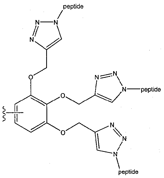
A.
51. The protein biosensor of claim 50, further comprising a selective protein binder.
52. The protein biosensor of claim 50 or 51, further comprising a donor and an acceptor, wherein at least one of the donor or acceptor is a fluorophore.
53. The protein biosensor of claim 52, wherein each donor or acceptor is attached to or in close proximity to one of the selective and non selective binders of said biosensor.
Priority Applications (1)
| Application Number | Priority Date | Filing Date | Title |
|---|---|---|---|
| US14/754,785 US10281459B2 (en) | 2012-12-31 | 2015-06-30 | Protein biosensors, cross reactive sensor arrays and methods of use thereof |
Applications Claiming Priority (2)
| Application Number | Priority Date | Filing Date | Title |
|---|---|---|---|
| US201261747426P | 2012-12-31 | 2012-12-31 | |
| US61/747,426 | 2012-12-31 |
Related Child Applications (1)
| Application Number | Title | Priority Date | Filing Date |
|---|---|---|---|
| US14/754,785 Continuation-In-Part US10281459B2 (en) | 2012-12-31 | 2015-06-30 | Protein biosensors, cross reactive sensor arrays and methods of use thereof |
Publications (1)
| Publication Number | Publication Date |
|---|---|
| WO2014102806A1 true WO2014102806A1 (en) | 2014-07-03 |
Family
ID=50029173
Family Applications (2)
| Application Number | Title | Priority Date | Filing Date |
|---|---|---|---|
| PCT/IL2013/051099 Ceased WO2014102803A1 (en) | 2012-12-31 | 2013-12-31 | Molecular sensor and methods of use thereof |
| PCT/IL2013/051102 Ceased WO2014102806A1 (en) | 2012-12-31 | 2013-12-31 | Protein biosensors, cross reactive sensor arrays and methods of use thereof |
Family Applications Before (1)
| Application Number | Title | Priority Date | Filing Date |
|---|---|---|---|
| PCT/IL2013/051099 Ceased WO2014102803A1 (en) | 2012-12-31 | 2013-12-31 | Molecular sensor and methods of use thereof |
Country Status (2)
| Country | Link |
|---|---|
| US (2) | US9696310B2 (en) |
| WO (2) | WO2014102803A1 (en) |
Cited By (2)
| Publication number | Priority date | Publication date | Assignee | Title |
|---|---|---|---|---|
| JP2018532739A (en) * | 2015-10-20 | 2018-11-08 | ソレント・セラピューティクス・インコーポレイテッドSorrento Therapeutics, Inc. | Intracellular delivery compounds |
| CN109474331A (en) * | 2018-12-18 | 2019-03-15 | 京信通信系统(中国)有限公司 | Operation and maintenance monitoring method, device, storage medium and terminal equipment for optical distribution network |
Families Citing this family (38)
| Publication number | Priority date | Publication date | Assignee | Title |
|---|---|---|---|---|
| US10557853B2 (en) * | 2012-12-31 | 2020-02-11 | Yeda Research And Development Co. Ltd. | Molecular sensor and methods of use thereof |
| EP3019559A4 (en) | 2013-08-22 | 2017-04-05 | Sony Corporation | Water soluble fluorescent or colored dyes and methods for their use |
| US12332246B2 (en) * | 2014-04-29 | 2025-06-17 | Yeda Research And Development Co. Ltd. | Quinoline based cyanine dye turn-on fluorescent probes and methods of use thereof |
| US11639929B2 (en) | 2014-04-29 | 2023-05-02 | Yeda Research And Development Co. Ltd. | Universal histidine-tag binding compounds and methods of use thereof as fluorescent probes and sensors |
| CN104194379B (en) * | 2014-08-08 | 2016-07-06 | 华南理工大学 | A kind of thiazole orange class Molecule of Cyanine Dyes and application thereof |
| KR102646956B1 (en) | 2015-02-26 | 2024-03-14 | 소니그룹주식회사 | Phenylethynylnaphthalene dye and method of using the same |
| WO2016138461A1 (en) | 2015-02-26 | 2016-09-01 | Sony Corporation | Water soluble fluorescent or colored dyes comprising conjugating groups |
| KR102765335B1 (en) | 2015-05-11 | 2025-02-13 | 소니그룹주식회사 | Ultra bright dimeric or polymeric dyes |
| BR112018070164B1 (en) | 2016-04-01 | 2022-09-27 | Sony Corporation Of America | ULTRABRIGHT DIMERIC OR POLYMERIC COLORINGS |
| EP3436528B1 (en) | 2016-04-01 | 2025-07-02 | Sony Group Corporation | Ultra bright dimeric or polymeric dyes with rigid spacing groups |
| CN109153860B (en) | 2016-04-06 | 2021-04-23 | 索尼公司 | Ultrabright dimer or polymer dyes with spacer linker groups |
| JP7527537B2 (en) | 2016-05-10 | 2024-08-05 | ソニーグループ株式会社 | Super-bright polymer dyes with peptide backbones |
| US11390754B2 (en) | 2016-05-10 | 2022-07-19 | Sony Corporation | Compositions comprising a polymeric dye and a cyclodextrin and uses thereof |
| BR112018073199A2 (en) | 2016-05-11 | 2019-04-16 | Sony Corporation | ultra-bright dimeric or polymeric dyes |
| WO2017205350A1 (en) | 2016-05-23 | 2017-11-30 | Portland State University | Molecular probes for detection and imaging of pancreatic cancer |
| EP3464477A1 (en) | 2016-06-06 | 2019-04-10 | Sony Corporation | Ionic polymers comprising fluorescent or colored reporter groups |
| WO2018022925A1 (en) | 2016-07-29 | 2018-02-01 | Sony Corporation | Ultra bright dimeric or polymeric dyes and methods for preparation of the same |
| CN106590630B (en) * | 2016-12-07 | 2019-01-08 | 山东大学 | A kind of mitochondrial membrane potential fluorescence probe of space dependent form and its application |
| CN107746418B (en) * | 2017-09-27 | 2020-05-05 | 玉林师范学院 | Synthesis and application of 9-chloro-1, 2,3, 4-tetrahydroacridine-platinum (II) complex targeting liver cancer |
| US12290571B2 (en) | 2017-10-05 | 2025-05-06 | Sony Group Corporation | Programmable dendritic drugs |
| KR20260011205A (en) | 2017-10-05 | 2026-01-22 | 소니그룹주식회사 | Programmable polymeric drugs |
| CN111836645A (en) | 2017-11-16 | 2020-10-27 | 索尼公司 | Programmable Polymeric Drugs |
| WO2019140128A1 (en) | 2018-01-12 | 2019-07-18 | Sony Corporation | Phosphoalkyl polymers comprising biologically active compounds |
| US12076407B2 (en) | 2018-01-12 | 2024-09-03 | Sony Group Corporation | Polymers with rigid spacing groups comprising biologically active compounds |
| EP3737417B1 (en) | 2018-01-12 | 2025-08-27 | Sony Group Corporation | Phosphoalkyl ribose polymers comprising biologically active compounds |
| CN108362673B (en) * | 2018-03-07 | 2020-09-01 | 济南大学 | Method for detecting glutathione and histidine |
| EP3769085B1 (en) | 2018-03-19 | 2022-08-24 | Sony Group Corporation | Use of divalent metals for enhancement of fluorescent signals |
| KR102864292B1 (en) | 2018-03-21 | 2025-09-26 | 소니그룹주식회사 | Polymeric tandem dyes having linker groups |
| JP7580689B2 (en) | 2018-06-27 | 2024-11-12 | ソニーグループ株式会社 | Polymeric dyes with deoxyribose-containing linker groups |
| US12578342B2 (en) | 2018-07-13 | 2026-03-17 | Sony Group Corporation | Polymeric dyes having a backbone comprising organophosphate units |
| US20220236257A1 (en) * | 2019-06-05 | 2022-07-28 | Yeda Research And Development Co. Ltd. | Artificial receptors, recombinant cells comprising thereof, methods for their preparation, and method of using thereof |
| EP3861074A2 (en) | 2019-09-26 | 2021-08-11 | Sony Group Corporation | Polymeric tandem dyes with linker groups |
| EP4038081A1 (en) | 2019-09-30 | 2022-08-10 | Sony Group Corporation | Nucleotide probes |
| EP4256078A1 (en) | 2020-12-07 | 2023-10-11 | Sony Group Corporation | Spacing linker group design for brightness enhancement in dimeric or polymeric dyes |
| EP4036575A1 (en) * | 2021-01-03 | 2022-08-03 | Yeda Research and Development Co. Ltd | Quinoline based cyanine dye turn-on fluorescent probes and methods of use thereof |
| CA3203106A1 (en) | 2021-01-20 | 2022-07-28 | Parag Mallick | Systems and methods for biomolecule quantitation |
| US12169166B2 (en) | 2021-02-16 | 2024-12-17 | Aat Bioquest, Inc. | Fluorogenic cyanine compounds for detecting nucleic acids |
| CN116271109B (en) * | 2023-02-22 | 2025-11-11 | 安徽大学 | Tumor-targeted fluorescence/photoacoustic molecular probe and application thereof |
Citations (3)
| Publication number | Priority date | Publication date | Assignee | Title |
|---|---|---|---|---|
| WO2002022882A2 (en) * | 2000-09-13 | 2002-03-21 | Archemix Corporation | Target activated nucleic acid biosensor and methods of using same |
| WO2003014375A2 (en) * | 2001-08-09 | 2003-02-20 | Archemix Corporation | Nucleic acid sensor molecules and methods of using same |
| US20110210017A1 (en) * | 2010-03-01 | 2011-09-01 | Lai Rebecca Y | Fabrication of electrochemical biosensors via click chemistry |
Family Cites Families (4)
| Publication number | Priority date | Publication date | Assignee | Title |
|---|---|---|---|---|
| EP1692486B1 (en) * | 2003-12-12 | 2015-10-28 | Saint Louis University | Biosensors for detecting macromolecules and other analytes |
| US8092784B2 (en) * | 2005-11-30 | 2012-01-10 | Biotium, Inc. | Enzyme substrate comprising a functional dye and associated technology and methods |
| EP2320230A1 (en) * | 2008-07-30 | 2011-05-11 | Sysmex Corporation | Reagent for detecting abnormal cell in cervix of uterus, and method for detecting abnormal cell in cervix of uterus by using same |
| US8742114B2 (en) * | 2011-11-26 | 2014-06-03 | Laiqiang Ying | Nucleic acid detections and methods of their use |
-
2013
- 2013-12-31 WO PCT/IL2013/051099 patent/WO2014102803A1/en not_active Ceased
- 2013-12-31 WO PCT/IL2013/051102 patent/WO2014102806A1/en not_active Ceased
-
2015
- 2015-06-14 US US14/738,894 patent/US9696310B2/en active Active
- 2015-06-30 US US14/754,785 patent/US10281459B2/en active Active
Patent Citations (3)
| Publication number | Priority date | Publication date | Assignee | Title |
|---|---|---|---|---|
| WO2002022882A2 (en) * | 2000-09-13 | 2002-03-21 | Archemix Corporation | Target activated nucleic acid biosensor and methods of using same |
| WO2003014375A2 (en) * | 2001-08-09 | 2003-02-20 | Archemix Corporation | Nucleic acid sensor molecules and methods of using same |
| US20110210017A1 (en) * | 2010-03-01 | 2011-09-01 | Lai Rebecca Y | Fabrication of electrochemical biosensors via click chemistry |
Non-Patent Citations (7)
| Title |
|---|
| CAMPONOVO, J.; RUIZ, J.; CLOUTET, E.; ASTRUC, D., CHEM. EUR. J., vol. 15, 2009, pages 2990 |
| DANTAS DE ARAÚJO, A.; PALOMO, J. M.; CRAMER, J.; SEITZ, 0.; ALEXANDROV, K.; WALDMANN, H., CHEM. EUR. J., vol. 12, 2006, pages 6095 |
| GRANDJEAN, C.; BOUTONNIER, A.; GUERREIRO, C.; FOURNIER, J. M.; MULARD, L. A., J. ORG. CHEM., vol. 70, 2005, pages 7123 |
| HABIG, W. H.; PABST, M. J.; JAKOBY, W. B. J., BIO. CHEM., vol. 249, 1974, pages 7130 |
| SARA STEWART ET AL: "Identifying Protein Variants with Cross-Reactive Aptamer Arrays", CHEMBIOCHEM, vol. 12, no. 13, 5 September 2011 (2011-09-05), pages 2021 - 2024, XP055107278, ISSN: 1439-4227, DOI: 10.1002/cbic.201100046 * |
| YANG ET AL: "Detection of MMP activity in living cells by a genetically encoded surface-displayed FRET sensor", BIOCHIMICA ET BIOPHYSICA ACTA. MOLECULAR CELL RESEARCH, ELSEVIER SCIENCE PUBLISHERS, AMSTERDAM, NL, vol. 1773, no. 3, 21 February 2007 (2007-02-21), pages 400 - 407, XP022322702, ISSN: 0167-4889, DOI: 10.1016/J.BBAMCR.2006.11.002 * |
| ZHI-QIANG WANG ET AL: "Specific survivin dual fluorescence resonance energy transfer molecular beacons for detection of human bladder cancer cells", ACTA PHARMACOLOGICA SINICA, vol. 32, no. 12, 24 October 2011 (2011-10-24), pages 1522 - 1528, XP055106728, ISSN: 1671-4083, DOI: 10.1038/aps.2011.122 * |
Cited By (4)
| Publication number | Priority date | Publication date | Assignee | Title |
|---|---|---|---|---|
| JP2018532739A (en) * | 2015-10-20 | 2018-11-08 | ソレント・セラピューティクス・インコーポレイテッドSorrento Therapeutics, Inc. | Intracellular delivery compounds |
| JP7170535B2 (en) | 2015-10-20 | 2022-11-14 | ソレント・セラピューティクス・インコーポレイテッド | Intracellular delivery compound |
| CN109474331A (en) * | 2018-12-18 | 2019-03-15 | 京信通信系统(中国)有限公司 | Operation and maintenance monitoring method, device, storage medium and terminal equipment for optical distribution network |
| CN109474331B (en) * | 2018-12-18 | 2021-02-23 | 京信通信系统(中国)有限公司 | Operation and maintenance monitoring method and device for optical distribution network, storage medium and terminal equipment |
Also Published As
| Publication number | Publication date |
|---|---|
| WO2014102803A1 (en) | 2014-07-03 |
| US20150362490A1 (en) | 2015-12-17 |
| US20150276753A1 (en) | 2015-10-01 |
| US9696310B2 (en) | 2017-07-04 |
| US10281459B2 (en) | 2019-05-07 |
Similar Documents
| Publication | Publication Date | Title |
|---|---|---|
| US10281459B2 (en) | Protein biosensors, cross reactive sensor arrays and methods of use thereof | |
| Ulrich et al. | Probing secondary interactions in biomolecular recognition by dynamic combinatorial chemistry | |
| JP5733760B2 (en) | Fluorogenic molecule and target nucleic acid detection method | |
| ES2668619T3 (en) | Compounds that target RNA and procedures to prepare and use them | |
| JP5223391B2 (en) | Sensitization assay method using fluorescent cysteine derivative and fluorescent cysteine derivative | |
| US20200255668A1 (en) | Labeling dye and kit including same | |
| AU2018202250B2 (en) | Method of detection of analyte active forms and determination of the ability of substances to bind into analyte active sites | |
| Ma et al. | Nucleic acid triggered catalytic drug and probe release: A new concept for the design of chemotherapeutic and diagnostic agents | |
| Ponomarenko et al. | Tetrahedral DNA conjugates from pentaerythritol-based polyazides | |
| Rozovsky et al. | Theranostic system for ratiometric fluorescence monitoring of peptide-guided targeted drug delivery | |
| EP1412755A2 (en) | Use of dendrimers and poly-branched molecules to enhance signal in fluorescent assay systems | |
| US10557852B2 (en) | Fluorescent molecular sensor for targeting changes in protein surfaces, and methods of use thereof | |
| JP5360647B2 (en) | Fluorogenic molecule | |
| US20120178086A1 (en) | Reductive release probes containing a chemoselectively cleavable alpha-azidoether linker and methods of use thereof | |
| US10557853B2 (en) | Molecular sensor and methods of use thereof | |
| Kim et al. | Design and synthesis of dye-conjugated hepsin inhibitors | |
| US8569515B2 (en) | Directed synthesis of oligophosphoramidate stereoisomers | |
| Li et al. | General strategy for the preparation of membrane permeable fluorogenic peptide ester conjugates for in vivo studies of ester prodrug stability | |
| Unger-Angel | Developing fluorescent molecular sensors for detecting protein isoforms and glycoforms in their native environments | |
| WO2023219136A1 (en) | Fluorescent probe for detecting peptide-linked hydrolase | |
| Guo | Catalytic acyl transfer modification of nuclear receptors and detection by in-gel labeling | |
| Schilling et al. | Synthesis and Functionalization of Biomolecules via Click Chemistry | |
| JP2009132705A (en) | Release method of target molecule based on target nucleic acid sequence | |
| JP2006145294A (en) | LABELING REAGENT FOR Fluorescent Labeling, MANUFACTURING METHOD FOR SUBSTRATE WITH LIGAND ON SURFACE AND MEASURING METHOD USING LABELING REAGENT | |
| Ma | Nucleic acid triggered catalytic drug release: A new concept for the design of disease-specific chemotherapeutic agents |
Legal Events
| Date | Code | Title | Description |
|---|---|---|---|
| 121 | Ep: the epo has been informed by wipo that ep was designated in this application |
Ref document number: 13824665 Country of ref document: EP Kind code of ref document: A1 |
|
| NENP | Non-entry into the national phase |
Ref country code: DE |
|
| 122 | Ep: pct application non-entry in european phase |
Ref document number: 13824665 Country of ref document: EP Kind code of ref document: A1 |