WO2014030272A1 - 歯科用部材 - Google Patents
歯科用部材 Download PDFInfo
- Publication number
- WO2014030272A1 WO2014030272A1 PCT/JP2013/001706 JP2013001706W WO2014030272A1 WO 2014030272 A1 WO2014030272 A1 WO 2014030272A1 JP 2013001706 W JP2013001706 W JP 2013001706W WO 2014030272 A1 WO2014030272 A1 WO 2014030272A1
- Authority
- WO
- WIPO (PCT)
- Prior art keywords
- dental
- bone
- metallic glass
- cells
- atomic
- Prior art date
- Legal status (The legal status is an assumption and is not a legal conclusion. Google has not performed a legal analysis and makes no representation as to the accuracy of the status listed.)
- Ceased
Links
Images



Classifications
-
- A—HUMAN NECESSITIES
- A61—MEDICAL OR VETERINARY SCIENCE; HYGIENE
- A61C—DENTISTRY; APPARATUS OR METHODS FOR ORAL OR DENTAL HYGIENE
- A61C7/00—Orthodontics, i.e. obtaining or maintaining the desired position of teeth, e.g. by straightening, evening, regulating, separating, or by correcting malocclusions
- A61C7/12—Brackets; Arch wires; Combinations thereof; Accessories therefor
-
- A—HUMAN NECESSITIES
- A61—MEDICAL OR VETERINARY SCIENCE; HYGIENE
- A61C—DENTISTRY; APPARATUS OR METHODS FOR ORAL OR DENTAL HYGIENE
- A61C8/00—Means to be fixed to the jaw-bone for consolidating natural teeth or for fixing dental prostheses thereon; Dental implants; Implanting tools
-
- A—HUMAN NECESSITIES
- A61—MEDICAL OR VETERINARY SCIENCE; HYGIENE
- A61C—DENTISTRY; APPARATUS OR METHODS FOR ORAL OR DENTAL HYGIENE
- A61C8/00—Means to be fixed to the jaw-bone for consolidating natural teeth or for fixing dental prostheses thereon; Dental implants; Implanting tools
- A61C8/0093—Features of implants not otherwise provided for
- A61C8/0096—Implants for use in orthodontic treatment
-
- C—CHEMISTRY; METALLURGY
- C22—METALLURGY; FERROUS OR NON-FERROUS ALLOYS; TREATMENT OF ALLOYS OR NON-FERROUS METALS
- C22C—ALLOYS
- C22C1/00—Making non-ferrous alloys
- C22C1/11—Making amorphous alloys
-
- C—CHEMISTRY; METALLURGY
- C22—METALLURGY; FERROUS OR NON-FERROUS ALLOYS; TREATMENT OF ALLOYS OR NON-FERROUS METALS
- C22C—ALLOYS
- C22C16/00—Alloys based on zirconium
-
- C—CHEMISTRY; METALLURGY
- C22—METALLURGY; FERROUS OR NON-FERROUS ALLOYS; TREATMENT OF ALLOYS OR NON-FERROUS METALS
- C22C—ALLOYS
- C22C45/00—Amorphous alloys
- C22C45/10—Amorphous alloys with molybdenum, tungsten, niobium, tantalum, titanium, or zirconium or Hf as the major constituent
Definitions
- the present invention relates to a dental member such as a dental implant made of metallic glass and having excellent biocompatibility and mechanical properties.
- a dental implant (hereinafter abbreviated as "implant"), which is a kind of dental member, is an artificial tooth root that is embedded in the jaw bone in place of the missing tooth root. By attaching the tooth crown, the biting function is restored.
- Such an implant requires not only predetermined mechanical properties but also affinity for a living body such as a bone. For this reason, conventionally, titanium that satisfies these requirements has been used as the material for the implant.
- metallic glass is known as a material having excellent mechanical properties such as low Young's modulus, high strength, and high corrosion resistance.
- the metal glass is a metal in a glass state that maintains a supercooled liquid state even at a melting point or lower, solidifies without being crystallized even if it is relatively slowly cooled, has an atomic diameter of 12% or more different from each other, and forms a compound.
- Bulk shape material obtained by mixing, melting and casting three or more elements with a tendency. It has a long-term disordered structure, and a clear glass transition and supercooled liquid region are observed before crystallization. Is defined as metal.
- Patent Document 1 a highly ductile metallic glass alloy having excellent plastic workability and applicable to a metal working process such as cold pressing, represented by the formula: Zr a Ni b Cu c Al d [wherein a, b, c and d are atomic%, a is 60 to 75 atomic%, b is 1 to 30 atomic%, c is 1 to 30 atomic%, and d is 5 to 20 atoms.
- a highly ductile metallic glass alloy characterized by having a composition represented by the following formula, more preferably a Poisson's ratio of 0.38 or more and a macro yield strain of 2.1% or more: A glass alloy was proposed.
- this kind of Zr— (Cu, Ni) —Al-based metallic glass generally prevents vitrification while Cu promotes vitrification. Due to the decrease in corrosion resistance, it may be difficult to maintain stability in an environment that is in contact with biological fluids such as the above-mentioned dental members due to corrosion in vivo, that is, metal elution and the generation of corrosion products associated therewith. Are known. For this reason, conventionally, a part of Zr constituting the base material is replaced with Nb, or, as proposed in the following Patent Document 2, Au or Ag is present at a predetermined ratio, thereby providing corrosion resistance. The structure which improved stability and maintained stability was employ
- the present inventors have conducted extensive research on biocompatibility in the Zr— (Cu, Ni) —Al-based metallic glass having the composition proposed in Patent Document 1 below, and as a result, have the above composition.
- JP 2009-215610 A JP2012-21198A
- the present invention has been made on the basis of the above knowledge, and can obtain mechanical properties such as low Young's modulus and high strength required for a dental member having excellent biocompatibility and small dimensional specifications.
- An object of the present invention is to provide a dental member using a metallic glass that can be produced.
- the dental member according to the first aspect of the present invention has a composition formula: Zr a Ni b Cu c Al d [where a, b, c, d are atomic%, a is 60 to 75 atomic%, b is 1 to 30 atomic%, c is 1 to 30 atomic%, and d is 5 to 20 atomic%].
- the dental member according to the second aspect of the present invention is characterized in that, in the first aspect, the dental member includes at least an orthodontic anchor screw and a dental implant.
- the dental member according to the third aspect of the present invention is the dental member according to the first aspect, wherein the dental member is fixed to the tooth surface at least during orthodontics and the support member. It is characterized by including a wire to be bridged.
- the Young's modulus is lower and the mechanical strength is lower than those of titanium and stainless steel that have been conventionally used. It is possible to realize a dental member that is excellent and has biocompatibility, and thus it is possible to achieve further miniaturization of the dental member.
- dental members that require similar biocompatibility include bone fixation plates and screws.
- support members such as brackets fixed to tooth surfaces, bands attached to tubes or molars, wires spanning between these support members, and various attachments attached as necessary
- the member for use is not directly embedded in the bone of the jaw or the like, it is attached to the oral cavity for a relatively long period of time (for example, one and a half years to two years).
- FIG. 1 is a diagram showing the shape and dimensions of a test piece used in the tensile test of Example 1.
- FIG. FIG. 2 is a graph showing the results of a tensile test.
- FIG. 3 is a graph showing the results of the metal elution test of Example 2.
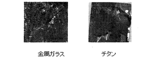
- FIG. 5 is an immunofluorescent-stained image of bone marrow mesenchymal cells on metal glass showing the results of Example 4.
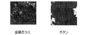
- FIG. 5 is an electron microscopic image of bone marrow mesenchymal cells on metal glass showing the results of Example 5.
- FIG. 6 is a graph showing the cell proliferation ability of bone marrow mesenchymal cells on metal glass showing the results of Example 6.
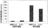
- FIG. 7 is a graph of ALP activity values in the induction of bone differentiation of bone marrow mesenchymal cells on metal glass, showing the results of Example 8.
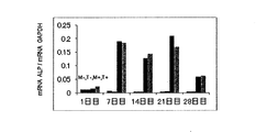
- 8A is a graph showing the results of real-time PCR analysis of bone differentiation markers in bone differentiation induction of bone marrow mesenchymal cells on metal glass in Example 9.
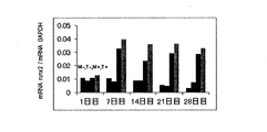
- FIG. 8B is a graph showing the results of real-time PCR analysis of bone differentiation markers in bone differentiation induction of bone marrow mesenchymal cells on metal glass in Example 9, as in FIG. 8A.
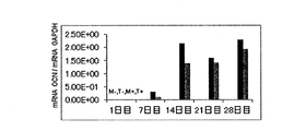
- FIG. 8C is a graph showing the results of real-time PCR analysis of bone differentiation markers in bone differentiation induction of bone marrow mesenchymal cells on metal glass in Example 9, as in FIGS. 8A and 8B.
- Example 10 shows the results of Example 10 and is an image of alizarin red stained in inducing bone differentiation of bone marrow mesenchymal cells on metal glass.
- FIG. 10 is a graph of ALP activity values in the induction of bone differentiation of dental pulp-derived mesenchymal cells on metal glass showing the results of Example 13.
- FIG. 11 shows the results of Example 14 and is a mapping of calcium and phosphorus showing the analysis results of bone differentiation ability of bone marrow mesenchymal cells on metal glass.
- the dental member has a composition formula: Zr a Ni b Cu c Al d [where a, b, c and d are atomic%, a is 60 to 75 atomic%, and b is 1 to 30] Atomic%, c is 1 to 30 atomic%, d is 5 to 20 atomic%]. Further, it is preferable that the Poisson's ratio is 0.38 or more and the macro yield strain is 2.1% or more.
- a suitable example of such a metallic glass is Zr 70 Ni 16 Cu 6 Al 8 metallic glass.
- the metal glass can be produced by a single roll liquid quenching method. That is, first, pure zirconium (Zr), pure nickel (Ni), pure copper (Cu), and pure aluminum (Al) are weighed and mixed within the above atomic% range, and dissolved in an argon gas atmosphere. Create Next, it can be produced by melting the mother alloy loaded in the quartz nozzle in an argon gas atmosphere again using a high-frequency melting method and spraying it on a rotating metal roll.
- Zr zirconium
- Ni pure nickel
- Cu pure copper
- Al pure aluminum
- Orthodontic anchor screw and orthodontic plate When moving a tooth in orthodontic treatment, it is a member that is embedded in the alveolar bone or the like and used as a fixation source. Is classified into class 3, and high biocompatibility is required.
- the shape consists of a plate and a fixing screw.
- the shape of the plate is about 1 cm to 3 cm (length) x 1 mm to 3 mm (width) x 0.1 mm to 3 mm (thickness).
- the shape of the screw is about 2 mm to 2.5 mm in length. The diameter is 1.0 mm to 4 mm.
- Dental implants An artificial tooth root that is embedded in the jaw bone instead of the lost tooth root, and is also embedded in the jaw bone. Affinity is required.
- the shape has a length dimension of about 8 mm to 16 mm and an outer diameter dimension of about 3 mm to 6 mm.
- Bone-fixing plate and screw are members used to fix bones and bones by surgery. Since the plates and screws are devices that are implanted in the body, more biocompatibility is required.
- the shape of the plate is about 1 cm to 10 cm (length) x 1 mm to 10 mm (width) x 0.1 mm to 3 mm (thickness), and the shape of the screw is about 2 mm to 25 mm in length. The diameter is 1.0 mm to 4 mm.
- Orthodontic bracket and tube are support members that are fixed to the tooth surface during orthodontic treatment, and the teeth are three-dimensionally formed by crossing a circular or square-shaped wire in a groove called a slot. Can be displaced. During the general treatment period (one and a half years to two years), it remains in the oral cavity. For this reason, it is classified into class 2 in the Pharmaceutical Affairs Law. The shape is about 3 mm (width) ⁇ about 3 mm (length) ⁇ 2 mm (height).
- Orthodontic wire There are arch-shaped and straight-shaped members spanning between the bracket and the tube described above.
- the one with a circular cross-sectional shape is about 0.30 mm ⁇ to 0.51 mm ⁇ , and the one with a square cross-sectional shape is often about 0.36 mm ⁇ 0.64 mm to about 0.53 mm to 0.64 mm. ing.
- Orthodontic band A member to be attached to a molar at the time of orthodontic treatment, which basically has a shape in which a pair of the above-mentioned tubes are joined and integrated. The shape has a radius of about 5 mm and a thickness dimension of about 0.15 mm. (7) Orthodontic attachment In the case of orthodontic treatment, it is a member that is used as needed, such as when locking the wire. The shape is about 3 mm (horizontal) x about 3 mm (equivalent to the bracket etc.) Vertical) ⁇ 2 mm (height).
- Inlays and onlays A metal lump that is manufactured to fit a part of a tooth that has a fixed shape against a partial loss of the crown due to trauma or caries. It is within the range.
- Zr 70 Ni 16 Cu 6 Al 8 metal glass was prepared as the metal glass that is the material of the dental member according to the present invention, and the following tests were performed.
- Zr 70 Ni 16 Cu 6 Al 8 metal glass is prepared by arc melting first pure zirconium, pure nickel, pure copper and pure aluminum in an argon gas atmosphere to produce a mother alloy, and then in a argon nozzle atmosphere, a quartz nozzle The mother alloy loaded therein was melted again using a high-frequency melting method and produced by a single-roll liquid quenching method in which it was sprayed onto a rotating metal roll.
- Example 1 To investigate the mechanical properties of tensile test the Zr 70 Ni 16 Cu 6 Al 8 metallic glass, rod-shaped Zr 70 Ni 16 Cu 6 Al 8 metal glass having a shape and dimensions shown in FIG. 1 (a) As a comparative example, a test piece of two types of pure titanium (JIS H4600-2012) having the same shape was prepared and subjected to a tensile test and analysis thereof. A hydraulic fatigue testing machine manufactured by Shimadzu Corporation was used for the tensile test.
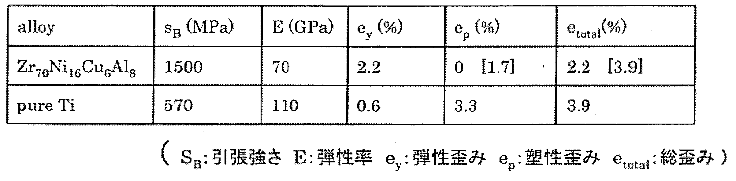
- Table 1 is a table showing mechanical properties such as tensile strength obtained by analyzing the results of the tensile test of Example 1.
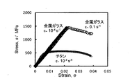
- Zr 70 Ni 16 Cu 6 Al 8 metal glass As shown in FIG. 2, as a result of the tensile test, the tensile strength of Zr 70 Ni 16 Cu 6 Al 8 metal glass is 1500 MPa, the type 2 pure titanium is 570 MPa, and the metal glass is about three times larger, and the elastic modulus Compared to 110 GPa of two types of pure titanium, Zr 70 Ni 16 Cu 6 Al 8 metal glass was 70 GPa.
- Zr 70 Ni 16 Cu 6 Al 8 metallic glass has high tensile strength as compared to the two pure titanium, it is low Young's modulus was shown. Further, it has been found that Zr 70 Ni 16 Cu 6 Al 8 metallic glass has an extremely large elastic limit (strain amount) of 2% which is not found in other crystalline metal materials.
- Example 2 Metal elution test of metal glass Metal glass, stainless steel (SUS316L), titanium-nickel superelastic alloy (Ti-Ni), two kinds of pure titanium (Ti) samples were coated with # 800 water-resistant abrasive paper. After wet polishing, ultrasonic cleaning was performed with acetone and ethanol. Three glass bottles with lids that can be sealed are prepared for each sample, and three blank solution bottles that do not immerse the sample are prepared, washed with nitric acid, rinsed with ultrapure water, and dried.
- a sample was put into a bottle containing 20 ml of an accelerated corrosion test solution (temperature: 310K, pH: 2.28) made of a mixed aqueous solution of 0.1 mol- 1 NaCl and 10 gl- 1 lactic acid (special grade: purity 85-92%).
- a sample was collected from the solution, and the concentration of metal ions in the solution was measured with an inductively coupled plasma emission spectrometer ICPS-7000 (Shimadzu).
- the surface area was calculated from the measurement of the shape of the sample to calculate the elution amount (gm ⁇ 2 ).
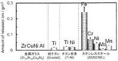
- FIG. 3 shows the results of the above ion elution test. From the stainless steel, elution of metal ions of iron, chromium, nickel, molybdenum and manganese was observed. Further, elution of titanium was observed from the two types of pure titanium, and elution of titanium and nickel ions was also observed from the titanium-nickel superelastic alloy. On the other hand, it was found that almost no ions were eluted from the Zr 70 Ni 16 Cu 6 Al 8 metal glass. As a result, it was shown that the passive film of Zr 70 Ni 16 Cu 6 Al 8 metallic glass has high stability and high corrosion resistance.
- Example 3 Preparation of bone marrow-derived mesenchymal cells on a metallic glass material Bone marrow mesenchymal cells were obtained from the femur of 4-week-old male F344 rats. The femur extracted from the rat was washed with phosphate buffer solution (PBS), and then soft tissue was removed as much as possible. The femoral heads at both ends were cut, and 10% fetal bovine serum (FBS; Nichirei, Tokyo, Japan) and penicillin (100 units / ml) / streptomycin (0.01 mg / ml; Sigma, St.
- FBS fetal bovine serum
- penicillin 100 units / ml
- streptomycin 0.01 mg / ml; Sigma, St.
- the collected cells were then cultured at 37 ° C. in a 5% CO 2 environment. After the growth of bone marrow mesenchymal cells was observed, they were washed with PBS, and then recovered with 0.05% trypsin / 0.53 mM EDTA (Nakarai tesque, Kyoto, Japan), and the cell cryopreservation solution cell banker (Mitsuwa) Frontech, Osaka, Japan). Next, the cryopreserved cells were thawed and seeded on 10 cm culture dishes, and in ⁇ MEM containing 10% FBS and penicillin (100 units / ml) / streptomycin (0.01 mg / ml) as the first passage bone marrow mesenchymal cells.
- Example 4 Immunostaining of bone marrow-derived mesenchymal cells on a metallic glass material After seeding rat bone marrow mesenchymal cells on metallic glass at 4.4 ⁇ 10 4 cells / ml, 4 hours, 7 days, 14 Each day-old cell was fixed with 4% paraformaldehyde / PBS for 15 minutes and washed with PBS. Subsequently, the cell membrane was permeabilized with PBS containing 0.1% Triton-X (Wako) and 3% BSA (B-PBS) for 15 minutes.
- PBS Triton-X
- B-PBS BSA
- the primary antibody was diluted 200-fold with B-PBS and used at 4 ° C. for 18 hours. After the reaction, it was washed 3 times with PBS for 5 minutes, and the secondary antibody was diluted 1000 times with B-PBS and allowed to react at room temperature for 1 hour. After the reaction, it was washed 3 times with PBS for 5 minutes.
- Alexa488-labeled phalloidin (Invitrogen) was diluted 500-fold with B-PBS, reacted at room temperature for 30 minutes, stained for cytoskeletal actin, and then washed 3 times with PBS for 5 minutes. Finally, the cells were reacted with 1 ⁇ M DAPI for 5 minutes at room temperature, stained with nucleus, and washed 3 times with PBS for 5 minutes. After staining, the images were observed with an Olympus IX-71 fluorescence microscope (Olympus). As a result, cytoplasmic actin was observed to be green, vinculin, which is one of the proteins forming focal adhesion, and the nucleus. Cells cultured on metallic glass and two types of pure titanium showed similar cell morphology, and after 4 hours of seeding, expression of vinculin was confirmed, and the number of cells increased on day 7 and day 14. did. FIG. 4 shows this result.
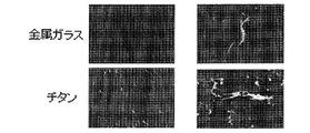
- Example 5 Electron microscopic image of bone marrow-derived mesenchymal cells on metal glass material Rat bone marrow mesenchymal cells were seeded at 4.4 ⁇ 10 4 cells / ml on metal glass and two kinds of pure titanium, and cultured. After 1 day, the cells were washed with PBS, fixed at 4 ° C. for 2 hours with a mixture of 2.5% glutaraldehyde and 2% paraformaldehyde 1: 1, and then 50%, 70%, 80% , 90% and 100% ethanol dehydration system. After dehydration, the sample was replaced with isoamyl acetate, allowed to stand, and the sample was dried with a critical point dryer. The dried sample was coated with metal by an ion sputtering apparatus, made conductive, and observed with a scanning electron microscope JXA-8230 (Nihon-Denshi). FIG. 5 shows the result.
- Example 6 WST8 proliferation analysis of bone marrow-derived mesenchymal cells on metallic glass material
- Cell proliferation ability on metallic glass and two kinds of pure titanium was analyzed using cell counting kit-8 (WST8; DOJINDO). did.
- WST8; DOJINDO cell counting kit-8
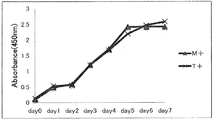
- FIG. 6 since there was no significant difference between bone marrow mesenchymal cells cultured on metal glass and bone marrow mesenchymal cells cultured on two kinds of pure titanium, It was shown that the bone marrow mesenchymal cell is a base material that can adhere and proliferate in the same manner as two types of pure titanium.
- Example 7 Method for Inducing Bone Differentiation of Bone Marrow-Derived Mesenchymal Cells on Metallic Glass Material
- 10 mM ⁇ -glycerophosphate, 10 nM dexamethasone and Bone differentiation was induced with 82 ⁇ m / ml L-ascorbic acid, and the bone differentiation ability was analyzed in comparison with the non-bone differentiation induction group.
- Example 8 ALP activity of bone marrow-derived mesenchymal cells in the induction of bone differentiation on a metallic glass material
- the ALP activity which is a differentiation marker for osteoblasts, was measured using TRACP & ALP Assay kit (TaKaRa Bio). According to the protocol, the medium was aspirated and washed with PBS, and 500 ⁇ l / well of cell extract was added. Next, the cells were gently detached using a cell scraper, collected in a 1.5 ml tube, and centrifuged for 1 minute. The supernatant was dropped onto a 96-well plate (BD Falcon), 50 ⁇ l of the reaction substrate solution was added, and the mixture was incubated at 37 ° C. for 30 minutes.
- Real-time PCR of bone differentiation marker of bone marrow-derived mesenchymal cells in induction of bone differentiation on metallic glass material Real-time PCR was performed using Thermal Cycler Dial Real Time System (TaKaRa Bio), and 2 ⁇ l cDNA as a template and 0.4 ⁇ M sense / antisense primer and SYBR premix Ex TaqII (TaKaRa Bio). After the initial reaction at 95 ° C. for 30 seconds, 40 cycles of reaction at 95 ° C., 5 seconds and 60 ° C. for 30 seconds were performed. After real-time PCR, the data was analyzed with Thermal Cycler Dice Real Time System Software version 2.01 (TaKaRa Bio). The mRNA expression level of each gene was expressed as a relative value to the expression level of glyceraldehyde-3-phosphate dehydrogenase (GAPDH).
- GPDH glyceraldehyde-3-phosphate dehydrogenase
- ALP and Runx2 increased in the differentiation-inducing group from the 7th day, peaked on the 14th day, and then decreased.
- the expression of OCN increased from the 14th day and was expressed until the 28th day.
- the expression levels of ALP, Runx2, and OCN were not significantly different between the metallic glass and the two types of pure titanium.
- Example 10 Alizarin red staining of bone marrow-derived mesenchymal cells in induction of bone differentiation on a metallic glass material After aspirating the medium, it was thoroughly washed with PBS and then washed with 95% ethanol (Wako) at 4 ° C for 15 minutes. Fixed and washed with pure water. Then, alizarin red solution (Sigma) was added and allowed to stand at room temperature for 10 minutes. High-definition microscope digital camera DP71 (Olympus) was used for photographing the stained image. As a result, as shown in FIG. 9, it was confirmed that the bone differentiation induction group was stained and calcified.
- Example 11 Method for inducing bone differentiation of dental pulp-derived mesenchymal cells on a metallic glass material
- 10 mM ⁇ -glycerophosphate and 10 nM dexamethasone And 82 ⁇ m / ml L-ascorbic acid were used to induce bone differentiation, and compared to the non-bone differentiation induction group, the bone differentiation ability was analyzed.
- Example 12 Preparation method of pulp-derived cells on a metallic glass material Human undifferentiated pulp cells were isolated from extracted bone-like impacted third molars. The pulp cells of the unfinished root part of the third molar were washed with PBS, and pulp cells treated with 100 U / ml collagenase (Worthington, NJ, USA) at 37 ° C. for 5 minutes were seeded in a 10 cm culture dish. And cultured at 37 ° C. in a 5% CO 2 environment. After the growth of incomplete human root pulp cells was observed, they were washed with PBS, recovered with 0.05% trypsin / 0.53 mM EDTA, and stored frozen in a cell cryopreservation solution cell banker.
- PBS pulp cells treated with 100 U / ml collagenase (Worthington, NJ, USA) at 37 ° C. for 5 minutes were seeded in a 10 cm culture dish. And cultured at 37 ° C. in a 5% CO 2 environment. After
- Cryopreserved human root incomplete dental pulp cells are seeded on a 10 cm culture dish and cultured as ⁇ -MEM containing 10% FBS and penicillin (100 units / ml) / streptomycin (0.01 mg / ml) as the first passage cells.
- a cell suspension of 4.4 ⁇ 10 4 cells / ml was prepared from the second passage cell, and was dropped onto a metallic glass in a 24 well plate and two types of pure titanium (6 ⁇ 6 mm) and cultured.
- ALP activity of dental pulp-derived mesenchymal cells in induction of bone differentiation on a metallic glass material was measured using TRACP & ALP Assay kit (TaKaRa Bio). According to the protocol, the medium was aspirated and washed with PBS, and 500 ⁇ l / well of cell extract was added. Next, the cells were gently detached using a cell scraper, collected in a 1.5 ml tube, and centrifuged for 1 minute. The supernatant was dropped onto a 96-well plate (BD Falcon), 50 ⁇ l of the reaction substrate solution was added, and the mixture was incubated at 37 ° C. for 30 minutes.
- FIG. 10 shows the result.
- Example 14 Alizarin red staining of dental pulp-derived mesenchymal cells in induction of bone differentiation on a metallic glass material After aspirating the medium, it was thoroughly washed with PBS and then washed with 95% ethanol (Wako) at 4 ° C for 15 minutes. Fixed and washed with pure water. Thereafter, alizarin red solution (Sigma) was added and left at room temperature for 10 minutes. High-definition microscope digital camera DP71 (Olympus) was used for photographing the stained image. As shown in FIG. 11, compared with the non-bone differentiation-inducing group, the bone differentiation-inducing group stained and a stained image showing calcification was obtained.
Landscapes
- Health & Medical Sciences (AREA)
- Chemical & Material Sciences (AREA)
- Animal Behavior & Ethology (AREA)
- Dentistry (AREA)
- Metallurgy (AREA)
- Organic Chemistry (AREA)
- Materials Engineering (AREA)
- Oral & Maxillofacial Surgery (AREA)
- Veterinary Medicine (AREA)
- Mechanical Engineering (AREA)
- Epidemiology (AREA)
- Life Sciences & Earth Sciences (AREA)
- Engineering & Computer Science (AREA)
- General Health & Medical Sciences (AREA)
- Public Health (AREA)
- Orthopedic Medicine & Surgery (AREA)
- Materials For Medical Uses (AREA)
Abstract
生体親和性に優れ、かつ寸法諸元が小さい歯科用部材に対して要求される低ヤング率および高強度等の機械的特性を得ることができる金属ガラスを用いた歯科用部材を提供する。本発明は、組成式:ZraNibCucAld[式中のa、b、c、dは原子%であって、aは60~75原子%、bは1~30原子%、cは1~30原子%、dは5~20原子%]で示される金属ガラスからなる。
Description
本発明は、金属ガラスからなる生体親和性および機械的特性に優れた歯科用インプラント等の歯科用部材に関するものである。
上記歯科用部材の一種である歯科用インプラント(以下、インプラントと略す。)は、抜けて失われた歯根に代えて顎の骨に埋め込む人工の歯根であり、当該インプラントに、セラミック等からなる人工の歯冠を取り付けることにより、噛む機能を回復させるものである。
このようなインプラントは、所定の機械的特性のみならず、骨等の生体に対する親和性が要求される。このため、従来、上記インプラントの素材としては、これらの要求を満たすチタンが用いられている。
しかしながら、チタン製のインプラントにあっては、顎の骨よりも弾性率(E:ヤング率)が高いことから弛みが発現し、これによって骨破壊が生じることが報告されている。また、歯磨剤に含まれるフッ化物の存在下において、耐食性が著しく低下してチタンイオンが溶出することにより、生体に害を及ぼす可能性も報告されている。
このため、上記チタンよりも生体親和性に優れ、かつ低ヤング率化と高強度を兼ね備えた機械的特性と高耐食性を有した新たな生体金属材料の開発が期待されている。
このため、上記チタンよりも生体親和性に優れ、かつ低ヤング率化と高強度を兼ね備えた機械的特性と高耐食性を有した新たな生体金属材料の開発が期待されている。
一方、低ヤング率、高強度、高耐食性などの優れた機械的特性を備えた素材として、金属ガラスが知られている。ここで、金属ガラスとは、融点以下でも過冷液体状態を保ち、比較的徐冷でも結晶化せずに凝固したガラス状態の金属であって、原子径が互いに12%以上異なり、かつ化合物形成傾向をもった元素を3種類以上混合・溶解して鋳造することによって得られるバルク形状材で、長期的に無秩序な構造を有し、結晶化前に明瞭なガラス遷移と過冷却液体域が観察される金属と定義されている。
ちなみに、本発明者等は、先に下記特許文献1において、塑性加工性に優れ、冷間プレス加工などの金属加工プロセスに適用可能な高延性金属ガラス合金として、式:ZraNibCucAld[式中のa、b、c、dは原子%であって、aは60乃至75原子%、bは1乃至30原子%、cは1乃至30原子%、dは5乃至20原子%である]で示される組成を有することを特徴とする高延性金属ガラス合金であって、より好ましくはポアソン比が0.38以上、マクロな降伏ひずみが2.1%以上である高延性金属ガラス合金を提案した。
ところで、下記特許文献2において開示されているように、一般的にこの種のZr-(Cu、Ni)-Al系金属ガラスは、Cuがガラス化を促す一方で、不動態化を阻害して耐食性が低下するために、生体内での腐食、すなわち金属溶出およびこれに伴う腐食生成物の発生により、上述した歯科用部材等の生体液に触れる環境で安定性を維持することが難しいことが知られている。このため、従来では、その基材をなすZrの一部をNbで置き換えたり、あるいは下記特許文献2において提案されているように、AuまたはAgを所定の比率で存在させたりすることにより、耐食性を向上させて、安定性を維持する構成が採用されていた。
しかしながら、このようにNbやAuまたはAgを所定の比率で存在させると、本来の特徴である高強度といった機械的特性が低下するおそれがあるために、これを寸法諸元が1~数mmと極めて小さい上記インプラント等の歯科用部材として用いようとしても、所望とする機械的特性を得ることができなくなってしまうという問題点があった。
そこで、本発明者等は、後述するように、下記特許文献1において提案した組成を有するZr-(Cu、Ni)-Al系金属ガラスにおける生体親和性について鋭意研究した結果、上記組成を有するものは、他のNbやAuあるいはAg等の高価な成分を敢えて存在させることなく、そのまま歯科用部材として用いるに好適な生体親和性を有しているとの知見を得るに至った。
本発明は、上記知見に基づいてなされたもので、生体親和性に優れ、かつ寸法諸元が小さい歯科用部材に対して要求される低ヤング率および高強度等の機械的特性を得ることができる金属ガラスを用いた歯科用部材を提供することを課題とするものである。
上記課題を解決するため、本発明の第1の態様に係る歯科用部材は、組成式:ZraNibCucAld[式中のa、b、c、dは原子%であって、aは60~75原子%、bは1~30原子%、cは1~30原子%、dは5~20原子%]で示される金属ガラスからなることを特徴とするものである。
また、本発明の第2の態様による歯科用部材は、上記第1の態様において、上記歯科用部材が、少なくとも歯科矯正用アンカースクリューおよび歯科用インプラントを含むことを特徴とするものである。
さらに、本発明の第3の態様による歯科用部材は、上記第1の態様の歯科用部材において、上記歯科用部材が、少なくとも歯科矯正時に歯面に固定される支持部材および当該支持部材間に架け渡されるワイヤを含むことを特徴とするものである。
上記第1~3のいずれかの態様によれば、上記組成を有する金属ガラスを用いることにより、従来用いられていたチタンやステンレスと比較し、より低ヤング率であって、かつ機械的強度に優れするとともに、生体親和性を備えた歯科用部材を実現することができ、よって当該歯科用部材の一層の小型化を達成することも可能になる。
また、特に顎の骨内に埋め込まれる歯科矯正用アンカースクリューならびにプレートや歯科用インプラントは、食べ物の咀嚼による咬合力が負荷されるため、機械的特性が歯および骨と適合することが必須であるとともに、口腔内の唾液などによる腐食に対する耐食性が良い等、高い生体親和性を有することも条件であることから、上記第2の態様による歯科用部材のように、上記歯科用部材を歯科矯正用アンカースクリューや歯科用インプラントに適用した場合に、顕著な効果を奏する。なお、同様の生体親和性が要求される歯科用部材としては、骨固定用プレート、スクリューがある。
また、歯科矯正時に、歯面に固定されるブラケット、チューブあるいは大臼歯に装着されるバンド等の支持部材およびこれら支持部材間に架け渡されるワイヤ並びに必要に応じて装着される各種アタッチメント等の歯科用部材は、直接顎の骨等に埋め込まれるものではないものの、口腔内に比較的長期間(例えば、1年半~2年間)装着されることになる。
このような歯科用部材は、従来ステンレスが用いられていたが、当該ステンレスに含まれるニッケルにより、金属アレルギーの問題があった。
この点、後述する実施例2(図3)において示すように、上記第1の態様において特定する金属ガラスは、殆どニッケルの溶出が見られないことから、上記第3の態様によれば、これらの歯科矯正用の支持部材やワイヤを上記金属ガラスによって形成することにより、従来の金属アレルギーに起因する問題を解決することが可能になる。加えて、長期間にわたる矯正時においても、各部材の寸法を従来よりも小さくすることが可能になるために、患者の負担を軽減することができる。
この点、後述する実施例2(図3)において示すように、上記第1の態様において特定する金属ガラスは、殆どニッケルの溶出が見られないことから、上記第3の態様によれば、これらの歯科矯正用の支持部材やワイヤを上記金属ガラスによって形成することにより、従来の金属アレルギーに起因する問題を解決することが可能になる。加えて、長期間にわたる矯正時においても、各部材の寸法を従来よりも小さくすることが可能になるために、患者の負担を軽減することができる。
以下、本発明に係る歯科用部材の実施形態について説明する。
まず、上記歯科用部材は、組成式:ZraNibCucAld[式中のa、b、c、dは原子%であって、aは60~75原子%、bは1~30原子%、cは1~30原子%、dは5~20原子%]で示される金属ガラスによって構成されたものである。また、そのポアソン比が0.38以上であって、かつマクロな降伏ひずみが2.1%以上であることが好ましい。このような金属ガラスの好適な一例としては、Zr70Ni16Cu6Al8金属ガラスがある。
まず、上記歯科用部材は、組成式:ZraNibCucAld[式中のa、b、c、dは原子%であって、aは60~75原子%、bは1~30原子%、cは1~30原子%、dは5~20原子%]で示される金属ガラスによって構成されたものである。また、そのポアソン比が0.38以上であって、かつマクロな降伏ひずみが2.1%以上であることが好ましい。このような金属ガラスの好適な一例としては、Zr70Ni16Cu6Al8金属ガラスがある。
上記金属ガラスは、単ロール液体急冷法にて作製することができる。すなわち、先ず純ジルコニウム(Zr)、純ニッケル(Ni)、純銅(Cu)、純アルミニウム(Al)を、上記原子%の範囲内に秤量して混合し、アルゴンガス雰囲気中で溶解して母合金を作成する。次いで、アルゴンガス雰囲気中で、石英ノズル中に装填した上記母合金を高周波溶融法を用いて再度溶融し、回転している金属ロール上に吹き付けることによって作製することができる。
そして、上記金属ガラスによって製作される歯科用部材としては、以下のようなものがある。
(1)歯科矯正用アンカースクリューおよび歯科矯正用プレート
矯正治療で歯を動かす際に、歯槽骨等に埋入し固定源とするための部材であり、体内に埋め込まれる部材であるため、薬事法においては、クラス3に分類され、高い生体親和性が求められている。形状は、プレートと固定用スクリューからなる。プレートの形状は、約1cm~3cm(長さ)×1mm~3mm(幅)×0.1mm~3mm(厚さ)であり、スクリューの形状は、長さ寸法が約2mm~2.5mm、外径寸法が1.0mm~4mmである。
(1)歯科矯正用アンカースクリューおよび歯科矯正用プレート
矯正治療で歯を動かす際に、歯槽骨等に埋入し固定源とするための部材であり、体内に埋め込まれる部材であるため、薬事法においては、クラス3に分類され、高い生体親和性が求められている。形状は、プレートと固定用スクリューからなる。プレートの形状は、約1cm~3cm(長さ)×1mm~3mm(幅)×0.1mm~3mm(厚さ)であり、スクリューの形状は、長さ寸法が約2mm~2.5mm、外径寸法が1.0mm~4mmである。
(2)歯科用インプラント
失われた歯根に代えて顎の骨に埋め込む人工の歯根であり、同様に顎骨内に埋め込まれるものであることから、薬事法においては、クラス3に分類され、高い生体親和性が求められている。形状は、長さ寸法が約8mm~16mmであって、外径寸法が約3mm~6mmである。
失われた歯根に代えて顎の骨に埋め込む人工の歯根であり、同様に顎骨内に埋め込まれるものであることから、薬事法においては、クラス3に分類され、高い生体親和性が求められている。形状は、長さ寸法が約8mm~16mmであって、外径寸法が約3mm~6mmである。
(3)骨固定用プレート、スクリュー
外科手術により骨と骨を固定するため用いられる部材であり、プレートとスクリューは体内に埋め込まれる装置のため、より生体親和性が求められる。
ちなみに、プレートの形状は、約1cm~10cm(長さ)×1mm~10mm(幅)×0.1mm~3mm(厚さ)であり、スクリューの形状は、長さ寸法が約2mm~25mm、外径寸法が1.0mm~4mmである。
外科手術により骨と骨を固定するため用いられる部材であり、プレートとスクリューは体内に埋め込まれる装置のため、より生体親和性が求められる。
ちなみに、プレートの形状は、約1cm~10cm(長さ)×1mm~10mm(幅)×0.1mm~3mm(厚さ)であり、スクリューの形状は、長さ寸法が約2mm~25mm、外径寸法が1.0mm~4mmである。
(4)歯科矯正用ブラケットおよびチューブ
いずれも歯科矯正時に歯面に固定される支持部材であり、スロットと呼ばれる凹状に溝に断面円形または断面方形のワイヤが架け渡されることにより、歯を3次元に変位可能とするものである。一般的な治療期間(1年半~2年間)中は、口腔内に装着されたままの状態となる。このため、薬事法においては、クラス2に分類されている。形状は、約3mm(横)×約3mm(縦)×2mm(高さ)である。
いずれも歯科矯正時に歯面に固定される支持部材であり、スロットと呼ばれる凹状に溝に断面円形または断面方形のワイヤが架け渡されることにより、歯を3次元に変位可能とするものである。一般的な治療期間(1年半~2年間)中は、口腔内に装着されたままの状態となる。このため、薬事法においては、クラス2に分類されている。形状は、約3mm(横)×約3mm(縦)×2mm(高さ)である。
(5)歯科矯正用ワイヤ
上述したブラケットおよびチューブ間に架け渡される部材で、アーチ形状やストレート形状のものがある。断面形状が円形のものは、約0.30mmφ~0.51mmφであり、断面形状が方形のものは、約0.36mm×0.64mm~約0.53mm~0.64mmのものが多く使用されている。
上述したブラケットおよびチューブ間に架け渡される部材で、アーチ形状やストレート形状のものがある。断面形状が円形のものは、約0.30mmφ~0.51mmφであり、断面形状が方形のものは、約0.36mm×0.64mm~約0.53mm~0.64mmのものが多く使用されている。
(6)歯科矯正用バンド
歯科矯正時に、大臼歯に装着する部材で、基本的には一対の上記チューブを接合一体化した形状のものである。形状は、半径約5mmであって、厚さ寸法が約0.15mmである。
(7)矯正用アタッチメント
矯正治療をするにあたって、上記ワイヤの係止等の必要に応じて、適宜使用される部材であり、形状は、上記ブラケット等と同等の約3mm(横)×約3mm(縦)×2mm(高さ)である。
歯科矯正時に、大臼歯に装着する部材で、基本的には一対の上記チューブを接合一体化した形状のものである。形状は、半径約5mmであって、厚さ寸法が約0.15mmである。
(7)矯正用アタッチメント
矯正治療をするにあたって、上記ワイヤの係止等の必要に応じて、適宜使用される部材であり、形状は、上記ブラケット等と同等の約3mm(横)×約3mm(縦)×2mm(高さ)である。
(8)インレー、アンレー
外傷やう蝕による歯冠の部分的な欠損に対して、歯を一定の形状に形成し、その部分に適合するように製作した金属の塊であり、寸法は歯の大きさの範囲内である。
(9)クラウン
歯質の一部または大部分が、う蝕あるいはその他の原因によって欠損している場合の処置に用いるもので、処置する歯冠のすべての面を切削し、金属で被覆して歯の形態・機能を修復するものである。同様に、その寸法は、歯の大きさの範囲内である。
外傷やう蝕による歯冠の部分的な欠損に対して、歯を一定の形状に形成し、その部分に適合するように製作した金属の塊であり、寸法は歯の大きさの範囲内である。
(9)クラウン
歯質の一部または大部分が、う蝕あるいはその他の原因によって欠損している場合の処置に用いるもので、処置する歯冠のすべての面を切削し、金属で被覆して歯の形態・機能を修復するものである。同様に、その寸法は、歯の大きさの範囲内である。
(10)ブリッジ
歯列のなかで少数の歯が欠損した時に、残っている歯の歯冠または歯根に支台装置を付け、これに人工歯を連結して欠損した歯の形態、機能、外観を回復することを目的とした歯冠修復物であり、同様に、その寸法は、歯の大きさの範囲内である。
歯列のなかで少数の歯が欠損した時に、残っている歯の歯冠または歯根に支台装置を付け、これに人工歯を連結して欠損した歯の形態、機能、外観を回復することを目的とした歯冠修復物であり、同様に、その寸法は、歯の大きさの範囲内である。
(11)その他の歯科用部材
その他、例えば、上記金属ガラス上の口腔組織由来の間葉細胞を用いて、歯髄細胞分化、歯根膜細胞分化を行う部材や、上記金属ガラスの周囲に歯根膜細胞を培養し、人工歯根膜細胞を付与した金属ガラス製の人工歯根としても適用可能である。
その他、例えば、上記金属ガラス上の口腔組織由来の間葉細胞を用いて、歯髄細胞分化、歯根膜細胞分化を行う部材や、上記金属ガラスの周囲に歯根膜細胞を培養し、人工歯根膜細胞を付与した金属ガラス製の人工歯根としても適用可能である。
本発明に係る歯科用部材の素材となる金属ガラスとして、Zr70Ni16Cu6Al8金属ガラスを作成し、以下の試験を行った。ちなみに、Zr70Ni16Cu6Al8金属ガラスは、まず純ジルコニウム、純ニッケル、純銅、純アルミニウムをアルゴンガス雰囲気中でアーク溶解して母合金を作製し、次いでアルゴンガス雰囲気中で、石英ノズル中に装填した上記母合金を高周波溶融法を用いて再度溶融し、回転している金属ロール上に吹き付ける単ロール液体急冷法にて作製した。
[実施例1] 引張試験
上記Zr70Ni16Cu6Al8金属ガラスの機械的性質を調べるため、図1(a)に示す形状および寸法を有する棒状のZr70Ni16Cu6Al8金属ガラスの試験片、および比較例として同形状の2種純チタン(JIS H4600-2012)の試験片を作成して引張試験およびその解析を行った。引張試験には島津製作所製の油圧疲労試験機を使用した。
ここで、表1は、実施例1の引張試験の結果を解析して得られた引張強さ等の機械的特性を示す表である。
上記Zr70Ni16Cu6Al8金属ガラスの機械的性質を調べるため、図1(a)に示す形状および寸法を有する棒状のZr70Ni16Cu6Al8金属ガラスの試験片、および比較例として同形状の2種純チタン(JIS H4600-2012)の試験片を作成して引張試験およびその解析を行った。引張試験には島津製作所製の油圧疲労試験機を使用した。
ここで、表1は、実施例1の引張試験の結果を解析して得られた引張強さ等の機械的特性を示す表である。
図2に見られるように、上記引張試験の結果、Zr70Ni16Cu6Al8金属ガラスの引張強さが1500MPa、2種純チタンが570MPaと金属ガラスの方が3倍程度大きく、弾性率は2種純チタンの110GPaに比べ、Zr70Ni16Cu6Al8金属ガラスは70GPaであった。これにより、表1に示すように、Zr70Ni16Cu6Al8金属ガラスは2種純チタンと比較して引張強さが高く、低ヤング率であることが示された。また、Zr70Ni16Cu6Al8金属ガラスは他の結晶質金属材料では見られない極めて大きな2%もの弾性限(歪み量)を有していることが判明した。
[実施例2] 金属ガラスの金属溶出試験
金属ガラス、ステンレス鋼(SUS316L)、チタン-ニッケル超弾性合金(Ti-Ni)、2種純チタン(Ti)の試料両面を#800の耐水研磨紙で湿式研磨後、アセトンとエタノールで超音波洗浄した。また、蓋付き密栓可能なガラスボトルを1試料毎に3個、また試料を浸漬しないブランク溶液ボトルも3個用意し硝酸で洗浄後、超純水ですすいで乾燥させた。次いで、0.1moll-1NaClと10gl-1乳酸(特級:純度85~92%)の混合水溶液でできた加速腐食試験溶液(温度:310K、pH:2.28)を20ml入れたボトルに試料を浸漬後、310Kの恒温チャンバー内で7日間静置し、溶液から試料を回収後、誘導結合プラズマ発光分析装置ICPS-7000(Shimadzu)で溶液中の金属イオン濃度を測定した。そして、試料の形状の測定より表面積を計算して溶出量(gm-2)を算出した。
金属ガラス、ステンレス鋼(SUS316L)、チタン-ニッケル超弾性合金(Ti-Ni)、2種純チタン(Ti)の試料両面を#800の耐水研磨紙で湿式研磨後、アセトンとエタノールで超音波洗浄した。また、蓋付き密栓可能なガラスボトルを1試料毎に3個、また試料を浸漬しないブランク溶液ボトルも3個用意し硝酸で洗浄後、超純水ですすいで乾燥させた。次いで、0.1moll-1NaClと10gl-1乳酸(特級:純度85~92%)の混合水溶液でできた加速腐食試験溶液(温度:310K、pH:2.28)を20ml入れたボトルに試料を浸漬後、310Kの恒温チャンバー内で7日間静置し、溶液から試料を回収後、誘導結合プラズマ発光分析装置ICPS-7000(Shimadzu)で溶液中の金属イオン濃度を測定した。そして、試料の形状の測定より表面積を計算して溶出量(gm-2)を算出した。
図3は、上記イオン溶出試験の結果を示すもので、ステンレス鋼からは、鉄、クロム、ニッケル、モリブデンおよびマンガンの金属イオン溶出が認められた。また、2種純チタンからは、チタンの溶出が認められ、チタンニッケル超弾性合金からもチタンおよびニッケルイオンの溶出が認められた。これに対して、Zr70Ni16Cu6Al8金属ガラスからは、ほぼイオン溶出しないことが判明した。この結果、Zr70Ni16Cu6Al8金属ガラスの不動態皮膜の安定性が高く、高耐食性であることが示された。
[実施例3] 金属ガラスの材料上における骨髄由来間葉系細胞の調整
骨髄間葉細胞を4週齢雄性F344系ラットの大腿骨から細胞懸濁液を得た。ラットから摘出した大腿骨をリン酸緩衝液(phosphate buffer saline;PBS)で洗浄し、その後、可及的に軟組織を除去、両端の大腿骨頭を切断し、10% fetal bovine serum(FBS;Nichirei,Tokyo,Japan)およびペニシリン(100unit/ml)/ストレプトマイシン(0.01 mg/ml;Sigma,St.Louis,MO,USA)含有alpha-modified Eagle’s medium(αMEM;Sigma)を10mlシリンジに取り、10cm培養ディッシュ(BD Falcon,New Jersey,USA)に24G注射針(Terumo,Tokyo,Japan)で骨管内を洗い出して採取した。
骨髄間葉細胞を4週齢雄性F344系ラットの大腿骨から細胞懸濁液を得た。ラットから摘出した大腿骨をリン酸緩衝液(phosphate buffer saline;PBS)で洗浄し、その後、可及的に軟組織を除去、両端の大腿骨頭を切断し、10% fetal bovine serum(FBS;Nichirei,Tokyo,Japan)およびペニシリン(100unit/ml)/ストレプトマイシン(0.01 mg/ml;Sigma,St.Louis,MO,USA)含有alpha-modified Eagle’s medium(αMEM;Sigma)を10mlシリンジに取り、10cm培養ディッシュ(BD Falcon,New Jersey,USA)に24G注射針(Terumo,Tokyo,Japan)で骨管内を洗い出して採取した。
次いで、採取した細胞を37℃、5%CO2環境下で培養した。骨髄間葉細胞の増殖が認められた後、それらをPBSにて洗浄後、0.05%トリプシン/0.53mM EDTA(Nakarai tesque,Kyoto,Japan)により回収し、細胞凍結保存液セルバンカー(Mitsuwa Frontech,Osaka,Japan)中にて凍結保存した。次に、凍結保存した細胞を融解し、10cm培養ディッシュ上に播種し、第1継代骨髄間葉細胞として10%FBSおよびペニシリン(100unit/ml)/ストレプトマイシン(0.01mg/ml)含有αMEM中にて培養した。サブコンフルエントで継代し、4.4×104cells/mlに調整後、24wellプレート(BD Falcon)内の金属ガラス上、および2種純チタン(6×6mm)上に500μlずつ滴下し培養した。
[実施例4] 金属ガラスの材料上における骨髄由来間葉系細胞の免疫染色
金属ガラス上にラット骨髄間葉細胞を4.4×10⁴cells/mlで播種後、4時間、7日、14日経過した細胞をそれぞれ4%パラホルムアルデヒド/PBSにて15分間固定し、PBSで洗浄した。その後、0.1%Triton-X(Wako),3%BSA含有PBS(B-PBS)にて15分間細胞膜の浸透化処理をおこなった。PBSで3回洗浄後、3%B-PBSにて1時間ブロッキング処理を行い、一次抗体には抗chicken vinculin mouse monoclonal抗体(Sigma)、二次抗体にはAlexa594標識抗マウスIgG抗体(Invitrogen,CA,USA)を用いた。一次抗体はB-PBS液にて200倍希釈して用い、4℃で18時間作用させた。反応後、PBSにて5分3回洗浄し、二次抗体はB-PBSで1000倍希釈して室温にて1時間反応させ、反応後にPBSにて5分3回洗浄した。
金属ガラス上にラット骨髄間葉細胞を4.4×10⁴cells/mlで播種後、4時間、7日、14日経過した細胞をそれぞれ4%パラホルムアルデヒド/PBSにて15分間固定し、PBSで洗浄した。その後、0.1%Triton-X(Wako),3%BSA含有PBS(B-PBS)にて15分間細胞膜の浸透化処理をおこなった。PBSで3回洗浄後、3%B-PBSにて1時間ブロッキング処理を行い、一次抗体には抗chicken vinculin mouse monoclonal抗体(Sigma)、二次抗体にはAlexa594標識抗マウスIgG抗体(Invitrogen,CA,USA)を用いた。一次抗体はB-PBS液にて200倍希釈して用い、4℃で18時間作用させた。反応後、PBSにて5分3回洗浄し、二次抗体はB-PBSで1000倍希釈して室温にて1時間反応させ、反応後にPBSにて5分3回洗浄した。
その後、Alexa488標識phalloidin(Invitrogen)をB-PBSにて500倍に希釈し、室温で30分反応させ細胞骨格アクチンの染色を行った後、PBSにて5分3回洗浄した。最後に、1μM DAPIにて室温5分間反応させ核染色し、PBSで5分3回洗浄した。染色後、オリンパスIX-71蛍光顕微鏡(Olympus)で観察した。その結果、細胞骨格アクチンが緑、focal adhesionを形成するタンパクの一つであるvinculinおよび核が観察された。金属ガラス上、および2種純チタン上で培養した細胞はどちらも同様の細胞形態を示し、播種して4時間後からvinculinの発現が確認され、7日目、14日目と細胞数が増加した。図4は、この結果を示すものである。
[実施例5] 金属ガラスの材料上における骨髄由来間葉系細胞の電子顕微鏡像
金属ガラスおよび2種純チタン上にラット骨髄間葉細胞を4.4×10⁴cells/mlで播種し、培養1日後の細胞をPBSで洗浄し、2.5%グルタールアルデヒドと2%パラホルムアルデヒドを1:1で混合した混合液にて4℃で2時間固定した後、50%,70%,80%,90%,100%のエタノール上昇系にて脱水した。脱水後、酢酸イソアミルに置換し、静置して臨界点乾燥装置にて試料を乾燥させた。乾燥させた試料をイオンスパッタ装置にて金属コーティングし、導電性を持たせ走査型電子顕微鏡JXA-8230(Nihon-Denshi)にて観察した。図5は、その結果を示すものである。
金属ガラスおよび2種純チタン上にラット骨髄間葉細胞を4.4×10⁴cells/mlで播種し、培養1日後の細胞をPBSで洗浄し、2.5%グルタールアルデヒドと2%パラホルムアルデヒドを1:1で混合した混合液にて4℃で2時間固定した後、50%,70%,80%,90%,100%のエタノール上昇系にて脱水した。脱水後、酢酸イソアミルに置換し、静置して臨界点乾燥装置にて試料を乾燥させた。乾燥させた試料をイオンスパッタ装置にて金属コーティングし、導電性を持たせ走査型電子顕微鏡JXA-8230(Nihon-Denshi)にて観察した。図5は、その結果を示すものである。
[実施例6] 金属ガラスの材料上における骨髄由来間葉系細胞のWST8増殖解析
金属ガラス上および2種純チタン上での細胞増殖能をcell counting kit-8(WST8;DOJINDO)を用いて解析した。この結果、図6に見られるように、金属ガラス上で培養した骨髄間葉細胞、2種純チタン上で培養した骨髄間葉細胞の間に大きな差は認められなかったことから、金属ガラスは2種純チタンと同等に骨髄間葉細胞が接着・増殖できる基材であることが示された。
金属ガラス上および2種純チタン上での細胞増殖能をcell counting kit-8(WST8;DOJINDO)を用いて解析した。この結果、図6に見られるように、金属ガラス上で培養した骨髄間葉細胞、2種純チタン上で培養した骨髄間葉細胞の間に大きな差は認められなかったことから、金属ガラスは2種純チタンと同等に骨髄間葉細胞が接着・増殖できる基材であることが示された。
[実施例7] 金属ガラスの材料上における骨髄由来間葉系細胞の骨分化誘導の方法
骨髄間葉細胞を播種して1日後より骨分化誘導群において、10mM β-グリセロフォスフェイト、10nMデキサメタゾン及び82μm/ml L-アスコルビン酸にて骨分化誘導し、非骨分化誘導群と比較して骨分化能を解析した。
骨髄間葉細胞を播種して1日後より骨分化誘導群において、10mM β-グリセロフォスフェイト、10nMデキサメタゾン及び82μm/ml L-アスコルビン酸にて骨分化誘導し、非骨分化誘導群と比較して骨分化能を解析した。
[実施例8] 金属ガラスの材料上における骨分化誘導における骨髄由来間葉系細胞のALP活性
骨芽細胞の分化マーカーとされるALP活性についてTRACP&ALP Assay kit(TaKaRa Bio)を用いて測定した。プロトコールに従って、培地吸引後PBSで洗浄し、細胞抽出液を500μl/well加えた。次いで、セルスクレーパーを用いて細胞を静かに剥離し、1.5mlチューブに回収後、1分間遠心した。上清を96wellプレート(BD Falcon)に滴下し、反応基質液を50μl加え、37℃で30分間インキュベートした。その後、反応停止液にて反応を停止させ、LS-PLATE manager(TECAN)により、405nmの波長を用いて吸光度を測定した。測定値を単位タンパク量あたりのALP活性に換算したところ、図7に示すように、骨分化誘導群で有意に発現上昇が確認された。
骨芽細胞の分化マーカーとされるALP活性についてTRACP&ALP Assay kit(TaKaRa Bio)を用いて測定した。プロトコールに従って、培地吸引後PBSで洗浄し、細胞抽出液を500μl/well加えた。次いで、セルスクレーパーを用いて細胞を静かに剥離し、1.5mlチューブに回収後、1分間遠心した。上清を96wellプレート(BD Falcon)に滴下し、反応基質液を50μl加え、37℃で30分間インキュベートした。その後、反応停止液にて反応を停止させ、LS-PLATE manager(TECAN)により、405nmの波長を用いて吸光度を測定した。測定値を単位タンパク量あたりのALP活性に換算したところ、図7に示すように、骨分化誘導群で有意に発現上昇が確認された。
[実施例9] 金属ガラスの材料上における骨分化誘導における骨髄由来間葉系細胞の骨分化マーカーのリアルタイムPCR
リアルタイムPCRは、Thermal Cycler Dice Real Time System(TaKaRa Bio) を使用し、2μl cDNAを鋳型として0.4μMのセンス・アンチセンスプライマーとSYBR premix Ex TaqII(TaKaRa Bio)を用いて行った。95℃、30秒の初期反応後、95℃、5秒と60℃、30秒の反応を40サイクル行った。リアルタイムPCR後、Thermal Cycler Dice Real Time System Software version 2.01(TaKaRa Bio)でデータを解析した。各遺伝子のmRNA発現量は、glyceraldehyde-3-phosphate dehydrogenase(GAPDH)の発現量に対する相対値で表示した。
リアルタイムPCRは、Thermal Cycler Dice Real Time System(TaKaRa Bio) を使用し、2μl cDNAを鋳型として0.4μMのセンス・アンチセンスプライマーとSYBR premix Ex TaqII(TaKaRa Bio)を用いて行った。95℃、30秒の初期反応後、95℃、5秒と60℃、30秒の反応を40サイクル行った。リアルタイムPCR後、Thermal Cycler Dice Real Time System Software version 2.01(TaKaRa Bio)でデータを解析した。各遺伝子のmRNA発現量は、glyceraldehyde-3-phosphate dehydrogenase(GAPDH)の発現量に対する相対値で表示した。
結果を示す図8A、図8B、図8Cに見られるように、ALP、Runx2は7日目から分化誘導群において発現が上昇し、14日目でピークを迎え、その後減少した。OCNは14日目から発現が上昇し28日目まで発現していた。また、ALP、Runx2、OCNの発現量は金属ガラスと2種純チタンとの間に有意差は認められなかった。
[実施例10] 金属ガラスの材料上における骨分化誘導における骨髄由来間葉系細胞のアリザリンレッド染色
培地吸引後、PBSで十分に洗浄し、95%エタノール(Wako)を用いて4℃で15分間固定し、純水で洗浄した。その後、アリザリンレッド溶液(Sigma)を添加し10分間常温にて静置した。染色像の撮影は、高精細顕微鏡デジタルカメラDP71(Olympus)を用いた。この結果、図9に示すように、骨分化誘導群が染色し、石灰化しているのが確認された。
培地吸引後、PBSで十分に洗浄し、95%エタノール(Wako)を用いて4℃で15分間固定し、純水で洗浄した。その後、アリザリンレッド溶液(Sigma)を添加し10分間常温にて静置した。染色像の撮影は、高精細顕微鏡デジタルカメラDP71(Olympus)を用いた。この結果、図9に示すように、骨分化誘導群が染色し、石灰化しているのが確認された。
[実施例11] 金属ガラスの材料上における歯髄由来間葉系細胞の骨分化誘導の方法
歯髄由来間葉細胞を播種して1日後より骨分化誘導群において、10mM β-グリセロフォスフェイト、10nMデキサメタゾン及び82μm/ml L-アスコルビン酸にて骨分化誘導し、非骨分化誘導群と比較して骨分化能を解析した。また、ヒト歯根未完成歯歯髄細胞の性能評価のために細胞を12wellプレートに播種し、分化誘導培地(β-グリセロフォスフェイト、デキサメタゾン、L-アスコルビン酸)にて骨分化誘導し、非骨分化誘導群と比較して骨分化能を解析した。
歯髄由来間葉細胞を播種して1日後より骨分化誘導群において、10mM β-グリセロフォスフェイト、10nMデキサメタゾン及び82μm/ml L-アスコルビン酸にて骨分化誘導し、非骨分化誘導群と比較して骨分化能を解析した。また、ヒト歯根未完成歯歯髄細胞の性能評価のために細胞を12wellプレートに播種し、分化誘導培地(β-グリセロフォスフェイト、デキサメタゾン、L-アスコルビン酸)にて骨分化誘導し、非骨分化誘導群と比較して骨分化能を解析した。
[実施例12] 金属ガラスの材料上における歯髄由来細胞の調整方法
ヒト未分化歯髄細胞は、抜歯した骨性埋伏第三大臼歯から単離した。第三大臼歯の根未完成部の歯髄細胞をPBSにて洗浄し、100U/mlのコラゲナーゼ(Worthington,NJ,USA)にて37℃、5分間酵素処理した歯髄細胞を、10cm培養ディッシュに播種し、37℃、5%CO2環境下で培養した。ヒト歯根未完成歯髄細胞の増殖が認められた後、それらをPBSにて洗浄後、0.05%トリプシン/0.53 mM EDTAにより回収し、細胞凍結保存液セルバンカー中にて凍結保存した。凍結保存したヒト歯根未完成歯歯髄細胞を10cm培養ディッシュ上に播種し、第1継代細胞として10%FBSおよびペニシリン(100unit/ml)/ストレプトマイシン(0.01mg/ml)含有αMEMで培養する。第2継代細胞から4.4×104 cells/mlの細胞懸濁液を作製して、24wellプレート内の金属ガラス、および2種純チタン(6×6mm)上に滴下し培養した。
ヒト未分化歯髄細胞は、抜歯した骨性埋伏第三大臼歯から単離した。第三大臼歯の根未完成部の歯髄細胞をPBSにて洗浄し、100U/mlのコラゲナーゼ(Worthington,NJ,USA)にて37℃、5分間酵素処理した歯髄細胞を、10cm培養ディッシュに播種し、37℃、5%CO2環境下で培養した。ヒト歯根未完成歯髄細胞の増殖が認められた後、それらをPBSにて洗浄後、0.05%トリプシン/0.53 mM EDTAにより回収し、細胞凍結保存液セルバンカー中にて凍結保存した。凍結保存したヒト歯根未完成歯歯髄細胞を10cm培養ディッシュ上に播種し、第1継代細胞として10%FBSおよびペニシリン(100unit/ml)/ストレプトマイシン(0.01mg/ml)含有αMEMで培養する。第2継代細胞から4.4×104 cells/mlの細胞懸濁液を作製して、24wellプレート内の金属ガラス、および2種純チタン(6×6mm)上に滴下し培養した。
[実施例13] 金属ガラスの材料上における骨分化誘導における歯髄由来間葉系細胞のALP活性
骨芽細胞の分化マーカーとされるALP活性について TRACP&ALP Assay kit(TaKaRa Bio)を用いて測定した。プロトコールに従って、培地吸引後PBSで洗浄し、細胞抽出液を500μl/well加えた。次いで、セルスクレーパーを用いて細胞を静かに剥離し、1.5mlチューブに回収後、1分間遠心した。上清を96wellプレート(BD Falcon)に滴下し、反応基質液を50μl加え、37℃で30分間インキュベートした。その後、反応停止液にて反応を停止させ、LS-PLATE manager(TECAN)により、405nmの波長を用いて吸光度を測定した。測定値を単位タンパク量あたりのALP活性に換算したところ、骨分化誘導群で有意に発現上昇が確認された。図10は、その結果を示すものである。
骨芽細胞の分化マーカーとされるALP活性について TRACP&ALP Assay kit(TaKaRa Bio)を用いて測定した。プロトコールに従って、培地吸引後PBSで洗浄し、細胞抽出液を500μl/well加えた。次いで、セルスクレーパーを用いて細胞を静かに剥離し、1.5mlチューブに回収後、1分間遠心した。上清を96wellプレート(BD Falcon)に滴下し、反応基質液を50μl加え、37℃で30分間インキュベートした。その後、反応停止液にて反応を停止させ、LS-PLATE manager(TECAN)により、405nmの波長を用いて吸光度を測定した。測定値を単位タンパク量あたりのALP活性に換算したところ、骨分化誘導群で有意に発現上昇が確認された。図10は、その結果を示すものである。
[実施例14] 金属ガラスの材料上における骨分化誘導における歯髄由来間葉系細胞のアリザリンレッド染色
培地吸引後、PBSで十分に洗浄し、95%エタノール(Wako)を用いて4℃で15分間固定し、純水で洗浄した。その後、アリザリンレッド溶液(Sigma)を添加し、10分間常温にて静置した。染色像の撮影は、高精細顕微鏡デジタルカメラDP71(Olympus)を用いた。図11に示すように、非骨分化誘導群に比べて、骨分化誘導群が染色し、石灰化を示す染色像が得られた。
培地吸引後、PBSで十分に洗浄し、95%エタノール(Wako)を用いて4℃で15分間固定し、純水で洗浄した。その後、アリザリンレッド溶液(Sigma)を添加し、10分間常温にて静置した。染色像の撮影は、高精細顕微鏡デジタルカメラDP71(Olympus)を用いた。図11に示すように、非骨分化誘導群に比べて、骨分化誘導群が染色し、石灰化を示す染色像が得られた。
以上のことから、上記組成を有する金属ガラスを用いることにより、従来用いられていたチタンやステンレスと比較し、より低ヤング率であって、かつ機械的強度に優れするとともに、生体親和性を備えた歯科用部材を実現することができ、よって当該歯科用部材の一層の小型化を達成することも可能になる。
Claims (3)
- 組成式:ZraNibCucAld[式中のa、b、c、dは原子%であって、aは60~75原子%、bは1~30原子%、cは1~30原子%、dは5~20原子%]で示される金属ガラスからなることを特徴とする歯科用部材。
- 上記歯科用部材は、少なくとも歯科矯正用アンカースクリューおよび歯科用インプラントを含むことを特徴とする請求項1に記載の歯科用部材。
- 上記歯科用部材は、少なくとも歯科矯正時に歯面に固定される支持部材および当該支持部材間に架け渡されるワイヤを含むことを特徴とする請求項2に記載の歯科用部材。
Applications Claiming Priority (2)
| Application Number | Priority Date | Filing Date | Title |
|---|---|---|---|
| JP2012-183999 | 2012-08-23 | ||
| JP2012183999 | 2012-08-23 |
Publications (1)
| Publication Number | Publication Date |
|---|---|
| WO2014030272A1 true WO2014030272A1 (ja) | 2014-02-27 |
Family
ID=50149604
Family Applications (1)
| Application Number | Title | Priority Date | Filing Date |
|---|---|---|---|
| PCT/JP2013/001706 Ceased WO2014030272A1 (ja) | 2012-08-23 | 2013-03-14 | 歯科用部材 |
Country Status (1)
| Country | Link |
|---|---|
| WO (1) | WO2014030272A1 (ja) |
Citations (4)
| Publication number | Priority date | Publication date | Assignee | Title |
|---|---|---|---|---|
| JPH10211184A (ja) * | 1997-01-31 | 1998-08-11 | Olympus Optical Co Ltd | 医療用器具及び核磁気共鳴映像法と組み合わせて使用す る医療用器具 |
| JP2008512562A (ja) * | 2004-09-06 | 2008-04-24 | アイトゲネッシーシェ テヒニッシェ ホッホシューレ チューリッヒ | ジルコニウム系非晶質合金及びその使用 |
| JP2009215610A (ja) * | 2008-03-11 | 2009-09-24 | Tohoku Univ | 高延性金属ガラス合金 |
| JP2010042131A (ja) * | 2008-08-12 | 2010-02-25 | Proceed:Kk | スクリューキャップ |
-
2013
- 2013-03-14 WO PCT/JP2013/001706 patent/WO2014030272A1/ja not_active Ceased
Patent Citations (4)
| Publication number | Priority date | Publication date | Assignee | Title |
|---|---|---|---|---|
| JPH10211184A (ja) * | 1997-01-31 | 1998-08-11 | Olympus Optical Co Ltd | 医療用器具及び核磁気共鳴映像法と組み合わせて使用す る医療用器具 |
| JP2008512562A (ja) * | 2004-09-06 | 2008-04-24 | アイトゲネッシーシェ テヒニッシェ ホッホシューレ チューリッヒ | ジルコニウム系非晶質合金及びその使用 |
| JP2009215610A (ja) * | 2008-03-11 | 2009-09-24 | Tohoku Univ | 高延性金属ガラス合金 |
| JP2010042131A (ja) * | 2008-08-12 | 2010-02-25 | Proceed:Kk | スクリューキャップ |
Non-Patent Citations (3)
| Title |
|---|
| SACHIKO HIROMOTO ET AL.: "Corrosion Behavior of Zr65Al7. Ni10Cu17. 5 Amorphous Alloy for Biomedical Use", MATERIALS TRANSACTIONS, vol. 42, no. 4, 2001, pages 656 - 659 * |
| SACHIKO HIROMOTO ET AL.: "Surface Characterization of Amorphous Zr-Al-(Ni, Cu) Alloys Immersed in Cell-Culture Medium", MATERIALS TRANSACTIONS, vol. 43, no. 2, 2002, pages 261 - 266 * |
| TERUKO YAMAMOTO ET AL.: "Zr Kikinzoku Glass o Mochiita Shika Iyo Zairyo no Kaihatsu", ANNUAL REPORT ON COLLABORATIVE RESEARCH ADVANCED RESEARCH CENTER OF METALLIC GLASSES INSTITUTE FOR MATERIALS RESEARCH, TOHOKU UNIVERSITY, 2011, INSTITUTE FOR MATERIALS RESEARCH, 2012, TOHOKU UNIVERSITY, pages 119 - 120 * |
Similar Documents
| Publication | Publication Date | Title |
|---|---|---|
| Gjerde et al. | Cell therapy induced regeneration of severely atrophied mandibular bone in a clinical trial | |
| Liu et al. | Effects of combined chemical design (Cu addition) and topographical modification (SLA) of Ti-Cu/SLA for promoting osteogenic, angiogenic and antibacterial activities | |
| Apratim et al. | Zirconia in dental implantology: A review | |
| Blaschke et al. | Soft and hard tissue response to zirconium dioxide dental implants--a clinical study in man | |
| Ortega-Martínez et al. | Polyetheretherketone (PEEK) as a medical and dental material. A literature review | |
| Saulacic et al. | Acid and alkaline etching of sandblasted zirconia implants: a histomorphometric study in miniature pigs | |
| Zhao et al. | Human periodontal ligament stem cell seeding on calcium phosphate cement scaffold delivering metformin for bone tissue engineering | |
| Khojasteh et al. | Buccal fat pad–derived stem cells in three-dimensional rehabilitation of large alveolar defects: A report of two cases | |
| Nejatidanesh et al. | Retentiveness of implant-supported metal copings using different luting agents | |
| de Matos et al. | Metal alloys in dentistry: An outdated material or required for oral rehabilitation | |
| EP3162385B1 (en) | Implant cultured periodontal ligament cell sheet complex | |
| Luo et al. | Cytocompatibility of Biodentine and iR oot FS with human periodontal ligament cells: An in vitro study | |
| Anand et al. | Evaluation of immediately loaded dental implants bioactivated with platelet-rich plasma placed in the mandibular posterior region: A clinico-radiographic study | |
| Chen et al. | Synergistic Effect of Nano Strontium Titanate Coating and Ultraviolet C Photofunctionalization on Osteogenic Performance and Soft Tissue Sealing of poly (ether–ether–ketone) | |
| Gallegos et al. | Can dental cement composition affect dental implant success? | |
| KR102292582B1 (ko) | 도재 결합력이 우수한 Co/Cr계 포세린용 치과합금 및 이를 이용하여 제조된 치과 보철물 | |
| US20130053354A1 (en) | Bone-transplant or bone-filling composition comprising a dihydroxybenzoic acid derivative | |
| Shams et al. | Morphology and differentiation of MG63 osteoblast cells on saliva contaminated implant surfaces | |
| Rodrigues | Etiology of peri-implant diseases | |
| Iwata et al. | In vitro proliferation of periodontal ligament-like tissue on extracted teeth | |
| Abdolahpour et al. | Application of biomaterials in dentistry | |
| Wren | Vitreous materials for dental restoration and reconstruction | |
| Termteerapornpimol et al. | Effects of Different Self-Adhesive Resin Cements and Curing Through Zirconia on Gingival Fibroblasts | |
| Iezzi et al. | Peri-implant bone response around a human hydroxyapatite-coated implant retrieved after a 10-year loading period: a case report. | |
| Arash Khojasteh et al. | Buccal Fat Pad–Derived Stem Cells in Three-Dimensional Rehabilitation of Large Alveolar Defects: A Report of Two Cases |
Legal Events
| Date | Code | Title | Description |
|---|---|---|---|
| 121 | Ep: the epo has been informed by wipo that ep was designated in this application |
Ref document number: 13830284 Country of ref document: EP Kind code of ref document: A1 |
|
| NENP | Non-entry into the national phase |
Ref country code: DE |
|
| 122 | Ep: pct application non-entry in european phase |
Ref document number: 13830284 Country of ref document: EP Kind code of ref document: A1 |