WO2014009739A1 - Method for treating microorganism infections - Google Patents
Method for treating microorganism infections Download PDFInfo
- Publication number
- WO2014009739A1 WO2014009739A1 PCT/GB2013/051850 GB2013051850W WO2014009739A1 WO 2014009739 A1 WO2014009739 A1 WO 2014009739A1 GB 2013051850 W GB2013051850 W GB 2013051850W WO 2014009739 A1 WO2014009739 A1 WO 2014009739A1
- Authority
- WO
- WIPO (PCT)
- Prior art keywords
- microorganism
- compound
- enzyme
- infection
- equation
- Prior art date
- Legal status (The legal status is an assumption and is not a legal conclusion. Google has not performed a legal analysis and makes no representation as to the accuracy of the status listed.)
- Ceased
Links
Classifications
-
- A—HUMAN NECESSITIES
- A61—MEDICAL OR VETERINARY SCIENCE; HYGIENE
- A61K—PREPARATIONS FOR MEDICAL, DENTAL OR TOILETRY PURPOSES
- A61K31/00—Medicinal preparations containing organic active ingredients
- A61K31/33—Heterocyclic compounds
- A61K31/395—Heterocyclic compounds having nitrogen as a ring hetero atom, e.g. guanethidine or rifamycins
- A61K31/40—Heterocyclic compounds having nitrogen as a ring hetero atom, e.g. guanethidine or rifamycins having five-membered rings with one nitrogen as the only ring hetero atom, e.g. sulpiride, succinimide, tolmetin, buflomedil
- A61K31/407—Heterocyclic compounds having nitrogen as a ring hetero atom, e.g. guanethidine or rifamycins having five-membered rings with one nitrogen as the only ring hetero atom, e.g. sulpiride, succinimide, tolmetin, buflomedil condensed with other heterocyclic ring systems, e.g. ketorolac, physostigmine
-
- A—HUMAN NECESSITIES
- A61—MEDICAL OR VETERINARY SCIENCE; HYGIENE
- A61K—PREPARATIONS FOR MEDICAL, DENTAL OR TOILETRY PURPOSES
- A61K31/00—Medicinal preparations containing organic active ingredients
- A61K31/33—Heterocyclic compounds
- A61K31/395—Heterocyclic compounds having nitrogen as a ring hetero atom, e.g. guanethidine or rifamycins
- A61K31/41—Heterocyclic compounds having nitrogen as a ring hetero atom, e.g. guanethidine or rifamycins having five-membered rings with two or more ring hetero atoms, at least one of which being nitrogen, e.g. tetrazole
- A61K31/425—Thiazoles
- A61K31/429—Thiazoles condensed with heterocyclic ring systems
- A61K31/43—Compounds containing 4-thia-1-azabicyclo [3.2.0] heptane ring systems, i.e. compounds containing a ring system of the formula, e.g. penicillins, penems
- A61K31/431—Compounds containing 4-thia-1-azabicyclo [3.2.0] heptane ring systems, i.e. compounds containing a ring system of the formula, e.g. penicillins, penems containing further heterocyclic rings, e.g. ticarcillin, azlocillin, oxacillin
-
- A—HUMAN NECESSITIES
- A61—MEDICAL OR VETERINARY SCIENCE; HYGIENE
- A61K—PREPARATIONS FOR MEDICAL, DENTAL OR TOILETRY PURPOSES
- A61K31/00—Medicinal preparations containing organic active ingredients
- A61K31/33—Heterocyclic compounds
- A61K31/395—Heterocyclic compounds having nitrogen as a ring hetero atom, e.g. guanethidine or rifamycins
- A61K31/495—Heterocyclic compounds having nitrogen as a ring hetero atom, e.g. guanethidine or rifamycins having six-membered rings with two or more nitrogen atoms as the only ring heteroatoms, e.g. piperazine or tetrazines
- A61K31/496—Non-condensed piperazines containing further heterocyclic rings, e.g. rifampin, thiothixene or sparfloxacin
-
- A—HUMAN NECESSITIES
- A61—MEDICAL OR VETERINARY SCIENCE; HYGIENE
- A61K—PREPARATIONS FOR MEDICAL, DENTAL OR TOILETRY PURPOSES
- A61K31/00—Medicinal preparations containing organic active ingredients
- A61K31/33—Heterocyclic compounds
- A61K31/395—Heterocyclic compounds having nitrogen as a ring hetero atom, e.g. guanethidine or rifamycins
- A61K31/54—Heterocyclic compounds having nitrogen as a ring hetero atom, e.g. guanethidine or rifamycins having six-membered rings with at least one nitrogen and one sulfur as the ring hetero atoms, e.g. sulthiame
- A61K31/542—Heterocyclic compounds having nitrogen as a ring hetero atom, e.g. guanethidine or rifamycins having six-membered rings with at least one nitrogen and one sulfur as the ring hetero atoms, e.g. sulthiame ortho- or peri-condensed with heterocyclic ring systems
- A61K31/545—Compounds containing 5-thia-1-azabicyclo [4.2.0] octane ring systems, i.e. compounds containing a ring system of the formula:, e.g. cephalosporins, cefaclor, or cephalexine
- A61K31/546—Compounds containing 5-thia-1-azabicyclo [4.2.0] octane ring systems, i.e. compounds containing a ring system of the formula:, e.g. cephalosporins, cefaclor, or cephalexine containing further heterocyclic rings, e.g. cephalothin
-
- A—HUMAN NECESSITIES
- A61—MEDICAL OR VETERINARY SCIENCE; HYGIENE
- A61K—PREPARATIONS FOR MEDICAL, DENTAL OR TOILETRY PURPOSES
- A61K31/00—Medicinal preparations containing organic active ingredients
- A61K31/70—Carbohydrates; Sugars; Derivatives thereof
- A61K31/7028—Compounds having saccharide radicals attached to non-saccharide compounds by glycosidic linkages
- A61K31/7034—Compounds having saccharide radicals attached to non-saccharide compounds by glycosidic linkages attached to a carbocyclic compound, e.g. phloridzin
- A61K31/7036—Compounds having saccharide radicals attached to non-saccharide compounds by glycosidic linkages attached to a carbocyclic compound, e.g. phloridzin having at least one amino group directly attached to the carbocyclic ring, e.g. streptomycin, gentamycin, amikacin, validamycin, fortimicins
-
- A—HUMAN NECESSITIES
- A61—MEDICAL OR VETERINARY SCIENCE; HYGIENE
- A61K—PREPARATIONS FOR MEDICAL, DENTAL OR TOILETRY PURPOSES
- A61K31/00—Medicinal preparations containing organic active ingredients
- A61K31/70—Carbohydrates; Sugars; Derivatives thereof
- A61K31/7042—Compounds having saccharide radicals and heterocyclic rings
- A61K31/7048—Compounds having saccharide radicals and heterocyclic rings having oxygen as a ring hetero atom, e.g. leucoglucosan, hesperidin, erythromycin, nystatin, digitoxin or digoxin
-
- A—HUMAN NECESSITIES
- A61—MEDICAL OR VETERINARY SCIENCE; HYGIENE
- A61P—SPECIFIC THERAPEUTIC ACTIVITY OF CHEMICAL COMPOUNDS OR MEDICINAL PREPARATIONS
- A61P31/00—Antiinfectives, i.e. antibiotics, antiseptics, chemotherapeutics
-
- Y—GENERAL TAGGING OF NEW TECHNOLOGICAL DEVELOPMENTS; GENERAL TAGGING OF CROSS-SECTIONAL TECHNOLOGIES SPANNING OVER SEVERAL SECTIONS OF THE IPC; TECHNICAL SUBJECTS COVERED BY FORMER USPC CROSS-REFERENCE ART COLLECTIONS [XRACs] AND DIGESTS
- Y02—TECHNOLOGIES OR APPLICATIONS FOR MITIGATION OR ADAPTATION AGAINST CLIMATE CHANGE
- Y02A—TECHNOLOGIES FOR ADAPTATION TO CLIMATE CHANGE
- Y02A50/00—TECHNOLOGIES FOR ADAPTATION TO CLIMATE CHANGE in human health protection, e.g. against extreme weather
- Y02A50/30—Against vector-borne diseases, e.g. mosquito-borne, fly-borne, tick-borne or waterborne diseases whose impact is exacerbated by climate change
Definitions
- MRSA methicillin resistant Staphylococcus aureus
- One aspect of the invention provides a method of treating a microorganism infection in a subject in need of treatment, the method comprises:
- the dosing regimen produces a pharmacokinetic (PK) profile of the compound in the subject (at the site of the microorganism infection);
- Equation (III) is:
- [ ] [ ⁇ 0 ] ⁇ ⁇ ([ ⁇ + ⁇ ( ⁇ - 1)] ⁇ + ⁇ ⁇ ⁇ - - yj t 2 + ⁇ (( 1 - e "kt ))
- N 0 is microorganism titer at the beginning of each said series of successive intervals
- N is microorganism titer at the end of each said series of successive intervals
- t is the duration of time for a given interval associated with N 0 ;
- SUBSTITUTE SHEET RULE 26 ⁇ is intrinsic logarithmic growth rate of the microorganism
- ⁇ is maximum kill rate of the microorganism by the compound
- [I] is concentration of the compound at the site of enzyme function ⁇ e.g., inside the microorganism);
- K t is the binding constant defined in Eq. (I);
- ks and k 6 are on- and off-rates defined in Eq. (II);
- KM is the Michaelis-Menten constant of the enzyme
- [S] is concentration of the substrate of the enzyme at the site of enzyme function (e.g., inside the microorganism);
- ⁇ ⁇ is ⁇ defined above for the immediate preceding interval
- ⁇ ⁇ and a new are ⁇ and ⁇ defined above for the current interval
- the microorganism is a bacterium.
- the bacterium is P. aeruginosa, E. coli, K. pneumonia, A.
- the infection is systemic. In other embodiments, the infection is local.
- the subject is a human, or a non-human mammal (e.g., a livestock animal, a pet, a lab animal etc.).
- a non-human mammal e.g., a livestock animal, a pet, a lab animal etc.
- the compound is an antibiotic, such as Ampicillin, Cloxacillin, Methicillin, Cefotaxime, Ceftriaxone, Moxalactam, Imipenem, or Meropenem; vancomycin, erythromycin, Gentamicin, Amikacin, Tobramycin, Rifampin, Trimethoprim, or Ciprofloxacin.
- an antibiotic such as Ampicillin, Cloxacillin, Methicillin, Cefotaxime, Ceftriaxone, Moxalactam, Imipenem, or Meropenem
- vancomycin erythromycin
- Gentamicin Amikacin
- Tobramycin Rifampin
- Trimethoprim Trimethoprim
- Ciprofloxacin Ciprofloxacin
- the antibiotic exhibits a post-antibiotic effect (PAE) in treating infection by the microorganism.
- PAE post-antibiotic effect
- ks is less than about 1 hr "1 .
- the series of successive intervals are of identical duration.
- At least two of the series of successive intervals are of different duration.
- [I] is calculated / determined based on the PK profile of the compound.
- Another aspect of the invention provides a method of treating a microorganism infection in a subject in need of treatment, the method comprising first determining a specific dosing regimen that would produce a desired treatment result based on calculations according to Equation (III), and then administering the drug to the host or patient according to the selected dosing regimen.
- Yet another aspect of the invention provides a method for screening inhibitor compounds that may be used to achieve desired treatment time course and efficacy, while preferably avoiding or minimizing associated side effects or toxicological effects.
- Still another aspect of the invention provides a method for determining or predicting efficacy of a compound administered according to a set dosing regimen, in the context of treating a specific microorganism infection in a specific host.
- a further aspect of the invention provides a method for determining or predicting
- SUBSTITUTE SHEET RULE 26 efficacy of a pre-determined compound used according to a pre-determined dosing regimen, for treating a specific host with a given initial microorganism load.
- FIG. 1 shows a simulated PK /PD profile for a hypothetical drug administered to a host, and the resulting microorganism titer change over time.
- FIG. 2 shows the results of a bacterial killing kinetics and growth recovery experiment with an LpxC inhibitor and the organism P. aeruginosa.
- the inset box reports the fold- concentration relative to the compound MIC (minimal inhibition concentration) that the compound was incubated with the bacteria for during the time period between -1 and 0 hours.
- the "+" symbols are experimental results.
- the fitted curves (solid lines) were calculated with Equation (III) and returned the parameter values listed at the top of the plot.
- Units for the y-axis are in log 10 CFU (colony forming units).
- Units for the x-axis are in hours.
- the methods of the invention described herein are based at least in part on the discovery that the relationship between the various parameters associated with microorganism infection of a host ⁇ e.g., a human or an animal) can be described by a comprehensive equation, based on established pharmacokinetic (PK), pharmacodynamic (PD), and enzyme kinetic principals, such as the Michaelis-Menten equation.
- PK pharmacokinetic
- PD pharmacodynamic
- enzyme kinetic principals such as the Michaelis-Menten equation.
- SUBSTITUTE SHEET RULE 26 parameters can affect the behavior of the particular parameter of interest.
- this approach enables the methods of the invention to address numerous practical problems associated with microorganism infection, including, but not limited to: determining the time course of a given treatment after taking into consideration any PAE (Post Antibiotic Effect) such that the minimal effective drug dose is administered; designing or adjusting dosing regimens based on a desired outcome of treatment; screening for an optimal drug with the desired combination of characteristics, such as efficacy, PAE, toxicity, side effect; etc.
- PAE Post Antibiotic Effect
- Compound includes known and novel molecules that are active against an enzymatic target.
- the compound may be naturally existing, synthetic, or semi-synthetic, and can be any molecule in nature, such as small molecule (e.g., less than or about 5,000 Da, 1000 Da, 500 Da, 250 Da, or 100 Da), peptide, peptidomimetic, steroid, lipid, carbohydrate, nucleic acid, nucleotide, nucleoside, vitamin, prodrugs, or metabolic precursor thereof.
- the compound is an antibiotic or antibacterial agent.
- Antibiotics and “antibacterial” both refer to a compound or substance that kills or slows down the growth of bacteria.
- antibiotic(s) also include a broader range of antimicrobial compounds, including antifungals and other compounds.
- Antibiotic includes any natural substance produced by a microorganism that is antagonistic to the growth of other microorganisms, usually even in high dilution (e.g., aminoglycosides).
- Antibiotic may also include synthetic or semisynthetic ⁇ e.g., with modifications to various natural compounds) antimicrobial ⁇ e.g., antibacterial) compounds.
- Semisynthetic antimicrobial ⁇ e.g., antibacterial) compounds include, for example, the beta-lactam antibacterials ⁇ e.g., penicillin produced by fungi in the genus Penicillium), the cephalosporins, and the carbapenems.
- antimicrobial ⁇ e.g., antibacterial compounds include, for example, the sulfonamides, the quinolones, and the oxazolidinones.
- Many antimicrobial ⁇ e.g., antibacterial) compounds are
- SUBSTITUTE SHEET RULE 26 relatively small molecules with a molecular weight of less than 2000 atomic mass units.
- antibiotic includes both agents that kill microbes (e.g., bactericidal agents that kill bacteria), and agents that slow down or stall microbial growth (e.g., bacteriostatic agents).
- antibiotics that can be used with the methods of the invention include: Aminoglycosides (e.g., Amikacin, Gentamicin, Kanamycin, Neomycin, Netilmicin, Tobramycin, Paromomycin); Ansamycins (e.g., Geldanamycin, Herbimycin);
- Carbacephem e.g., Loracarbef
- Carbapenems e.g., Ertapenem, Doripenem,
- Imipenem/Cilastatin, Meropenem Cephalosporins (e.g., Cefadroxil, Cefazolin, Cefalotin or Cefalothin, Cefalexin, Cefaclor, Cefamandole, Cefoxitin, Cefprozil, Cefuroxime, Cefixime, Cefdinir, Cefditoren, Cefoperazone, Cefotaxime, Cefpodoxime, Ceftazidime, Ceftibuten, Ceftizoxime, Ceftriaxone, Cefepime, Ceftaroline fosamil, Ceftobiprole); Glycopeptides (e.g., Teicoplanin, Vancomycin, Telavancin); Lincosamides (e.g., Clindamycin, Lincomycin);
- Lipopeptide e.g., Daptomycin
- Macrolides e.g., Azithromycin, Clarithromycin
- Penicillins e.g., Amoxicillin, Ampicillin, Azlocillin, Carbenicillin, Cloxacillin, Dicloxacillin, Flucloxacillin, Mezlocillin, Methicillin, Nafcillin, Oxacillin, Penicillin G,
- Penicillin V Piperacillin, Temocillin, Ticarcillin
- Penicillin combinations e.g.,
- the antibiotic is an LpxCl inhibitor (e.g., a compound that inhibits the enzyme LpxC l).
- “Inhibits an enzyme” includes inhibiting at least one function or activity of the enzyme.
- the function or activity inhibited (by the compound) is required or essential for the survival, growth, and/or proliferation of the microorganism.
- the function or activity of the enzyme may be partially inhibited, or nearly completely inhibited.
- the function or activity of the enzyme is inhibited to such an extent that the enzyme is no more than 90%, 80%, 70%, 60%, 50%, 40%, 30%, 20%, 10%, 5%, 1%, 0.1%, 0.01%, 0.001% active compared to the enzyme without inhibition.
- the function or activity of the enzyme is inhibited to such an extent that survival, growth, and/or proliferation of the microorganism is abolished or impaired.
- “Inhibits proliferation of the microorganism” as used herein includes killing the microorganism, or inhibiting / stalling / retarding the proliferation (e.g., increase in number) and/or growth (e.g., increase in size or volume) of the microorganism, or both.
- infectious diseases are used interchangeably to include clinically evident illness (i.e., characteristic medical signs and/or symptoms of disease) resulting from the infection, presence, and/or growth of pathogenic biological agents in an individual host organism.
- infectious diseases may be asymptomatic for much or even all of their courses in a given host. In the latter case, the disease may be defined as a disease in hosts who secondarily become ill after contact with an asymptomatic carrier.
- Infectious diseases are sometimes called “contagious” when they are easily transmitted by contact with an ill person or their secretions (e.g., influenza).
- a "contagious disease” is a subset of infectious disease that is especially infective or easily transmitted.
- Other types of infectious / transmissible / communicable diseases with more specialized routes of infection, such as vector transmission or sexual transmission, are usually not regarded as “contagious,” and often do not require medical isolation or quarantine.
- infectious microorganisms refer to pathogens including, but not limited to, certain viruses, bacteria, fungi, protozoa, and multicellular parasites that are the cause of disease epidemics, in the sense that without the pathogen, no infectious epidemic occurs.
- an infectious microorganism refers to bacteria that cause an infectious disease.
- the infectious microorganism is antibiotic resistant, antifungal resistant or antiviral resistant.
- Acinetobacter infections ⁇ Acinetobacter baumannii), Actinomycosis ⁇ Actinomyces israelii, Actinomyces gerencseriae and Propionibacterium propionicus), African sleeping sickness or African trypanosomiasis ⁇ Trypanosoma brucei), AIDS or Acquired immunodeficiency syndrome (HIV or Human immunodeficiency virus), Amebiasis ⁇ Entamoeba histolytica), Anaplasmosis ⁇ Anaplasma genus), Anthrax ⁇ Bacillus anthracis), Arcanobacterium haemolyticum infection ⁇ Arcanobacterium haemolyticum), Argentine hemorrhagic fever (Junin virus), Ascariasis
- BK virus infection BK virus
- Black piedra ⁇ Piedraia hortae
- VZV Via zoster virus
- Chlamydia ⁇ Chlamydia trachomatis Chlamydia ⁇ Chlamydia trachomatis
- Ehrlichiosis (Ehrlichia genus), Enterobiasis (Pinworm infection) (Enterobius vermicularis), Enterococcus infection (Enterococcus genus), Enterovirus infection (Enterovirus genus), Epidemic typhus (Rickettsia prowazekii), Erythema infectiosum (Fifth disease) (Parvovirus B19), Exanthem subitum (Sixth disease) (Human herpesvirus 6 (HHV-6) and Human herpesvirus 7 (HHV-7)), Fasciolopsiasis (Fasciolopsis buski), Fasciolosis (Fasciola hepatica and Fasciola gigantica), Filariasis (Filarioidea superfamily), Food poisoning by Clostridium perfringens (Clostridium perfringens), Free-living amebic infection multiple (Fusobacterium infection Fusobacterium genus
- Haemophilus influenzae infection Haemophilus influenzae
- Hand, foot and mouth disease HFMD
- Enteroviruses mainly Coxsackie A virus and Enterovirus 71 (EV71)
- HPS Hantavirus Pulmonary Syndrome
- Helicobacter pylori Helicobacter pylori infection
- HUS Hemolytic-uremic syndrome
- HFRS Hemorrhagic fever with renal syndrome
- HFRS Hemorrhagic fever with renal syndrome
- Hepatitis A Hepatitis A Virus
- Hepatitis B Hepatitis B Virus
- Hepatitis C Hepatitis C Virus
- Hepatitis D Hepatitis D Virus
- Hepatitis E Hepatitis E Virus
- Herpes simplex Herpes simplex virus 1 and 2 HSV-1 and HSV-2)
- choriomeningitis Lymphocytic choriomeningitis virus (LCMV)), Malaria (Plasmodium genus), Marburg hemorrhagic fever (MHF) (Marburg virus), Measles (Measles virus), Melioidosis (Whitmore's disease) (Burkholderia pseudomallei), Meningitis (multiple), Meningococcal disease (Neisseria meningitidis), Metagonimiasis (usually Metagonimus yokagawai),
- Microsporidiosis (Microsporidia phylum), Molluscum contagiosum (MC) (Molluscum contagiosum virus (MCV)), Mumps (Mumps virus), Murine typhus (Endemic typhus) (Rickettsia typhi), Mycoplasma pneumonia (Mycoplasma pneumoniae), Mycetoma (numerous species of bacteria (Actinomycetoma) and fungi (Eumycetoma)), Myiasis (parasitic dipterous fly larvae), Neonatal conjunctivitis (Ophthalmia neonatorum) (most commonly Chlamydia trachomatis and Neisseria gonorrhoeae), Nocardiosis (usually Nocardia asteroides and other Nocardia species), Onchocerciasis (River blindness) (Onchocerca volvulus), Paracoccidioidomycosis (South American blastomycosis) (Paracocc
- PID inflammatory disease
- PCP Pneumocystis pneumonia
- Pneumonia multiple
- Poliomyelitis Poliovirus Prevotella infection Prevotella genus
- Primary amoebic meningoencephalitis PAM
- PAM Primary amoebic meningoencephalitis
- JC virus Progressive multifocal leukoencephalopathy
- Psittacosis Cholamydophila psittaci
- Q fever Coxiella burnetii
- Rabies Rabies virus
- Rat-bite fever Streptobacillus moniliformis and Spirillum minus
- Trichophyton mentagrophytes Tinea manuum (Ringworm of the Hand) (Trichophyton rubrum), Tinea nigra (usually Hortaea wasneckii), Tinea pedis (Athlete's foot) (usually Trichophyton genus), Tinea unguium (Onychomycosis) (usually Trichophyton genus), Tinea versicolor (Pityriasis versicolor) (Malassezia genus), Toxocariasis (Ocular Larva Migrans (OLM))
- Toxocara canis or Toxocara cati Toxocariasis (Visceral Larva Migrans (VLM)) (Toxocara canis or Toxocara cati), Toxoplasmosis (Toxoplasma gondii), Trichinellosis (Trichinella spiralis), Trichomoniasis (Trichomonas vaginalis), Trichuriasis (Whipworm infection)
- Tuberculosis usually Mycobacterium tuberculosis
- Venezuelan equine encephalitis Venezuelan equine encephalitis (Venezuelan equine encephalitis virus), Venezuelan
- hemorrhagic fever (Guanarito virus), Viral pneumonia (multiple viruses), West Nile Fever (West Nile virus), White piedra (Tinea blanca) (Trichosporon beigelii), Yersinia pseudotuberculosis infection (Yersinia pseudotuberculosis), Yersiniosis (Yersinia enterocolitica), Yellow fever (Yellow fever virus), Zygomycosis (Mucorales order (Mucormycosis) and Entomophthorales order (Entomophthoramycosis)).
- Bacillus Bacillus (Bacillus anthracis); Bordetella (Bordetella pertussis); Borrelia (Borrelia burgdorferi); Brucella (Brucella abortus, Brucella canis, Brucella melitensis, Brucella suis); Campylobacter (Campylobacter jejuni); Chlamydia and Chlamydophila (Chlamydia pneumoniae, Chlamydia trachomatis, Chlamydophila psittaci); Clostridium (Clostridium botulinum, Clostridium difficile, Clostridium perfringens, Clostridium tetani); Corynebacterium (Corynebacterium diphtheriae); Enterococcus (Enterococcus faecalis, Enterococcus
- Legionella Legionella pneumophila
- Leptospira Leptospira interrogans
- Listeria Listeria monocytogenes
- Mycobacterium Mycobacterium leprae, Mycobacterium tuberculosis, Mycobacterium ulcerans
- Mycoplasma Mycoplasma pneumoniae
- Neisseria Neisseria gonorrhoeae, Neisseria meningitidis
- Pseudomonas Pseudomonas aeruginosa
- Rickettsia Rickettsia (Rickettsia rickettsii)
- Salmonella Salmonella typhi, Salmonella typhimurium
- Staphylococcus Staphylococcus aureus, Staphylococcus epidermidis, Staphylococcus saprophyticus
- Streptococcus Streptococcus agalactiae, Streptococcus pneumoniae, Streptococcus pyogenes
- Treponema Treponema pallidum
- Vibrio Vibrio cholerae
- Yersinia Yersinia pestis
- Microorganism or “microbe,” in the broadest sense, includes pathogenic unicellular or microscopic (e.g., invisible to naked eye) multicellular organisms and prions that infect human and other non-human animals and cause infectious disease, such as bacteria, fungi, algae, virus, prions, and protozoa.
- microorganism refers to the above pathogens that do not include prion.
- microorganism refers to the above pathogens that do not include virus.
- microorganism refers to the above pathogens that do not include eukaryotes such as fungi, parasites, and protists.
- Microorganism infection includes any disease or pathological condition, with or without manifested or detectable symptoms, that is caused by or results from infection by one or more microorganisms as defined herein.
- microorganism infection includes bacterial infection that may or may not be contagious.
- Dosing regimen include a schedule and the related parameters for administration of one or more drugs, compounds, medicaments, or pharmaceutical compositions.
- a dosing regimen may specify some or all of the following non-limiting aspects: the amount / dose for a specific drug, applicable age group (e.g., adult vs. children under certain age or weight class), patient sub-population (e.g., women that is not pregnant, pre-existing conditions, prior treatment history, on-going treatment with other drugs), timing (e.g.
- Intransic logarithmic growth rate of a microorganism describes the growth rate of a microorganism of interest under a set of conditions pertinent to microorganism growth.
- any given microorganism may have a growth rate (e.g., measurable by population doubling time, or CFU increase over unit time, etc.) under a given circumstance, such as culture with defined components, or under the physiological condition of an infection site when the microorganism is growing in vivo.
- intrinsic logarithmic growth rate of a microorganism refers to the growth rate under the condition present, such as the physiological condition of an infection site, in the absence of an added antibiotic agent.
- '"maximum kill rate of the microorganism by a compound describes the rate the microorganism is killed or growth inhibited by the compound under ideal conditions, usually measurable by comparing growth (or death) rate of the microorganism in the absence (or presence) of the compound.
- Replenishment rate of the enzyme refers to the rate of new enzyme (i.e., the target of the compound / drug) added to the system. This mostly relates to the rate any new enzyme is being synthesized by the microorganism under the conditions (such as the physiological condition at the infection site).
- concentration of a compound at the site of enzyme function refers to compound concentration at the immediate vicinity of the enzyme to which the compound binds and inhibits. Thus if the enzyme is in the cytosol, concentration of a compound at the site of enzyme function is the concentration of the compound inside the microorganism, e.g., the compound
- concentration in the cytosol or the specific cytosolic compartment in which the enzyme predominantly resides and functions. If the enzyme is on cell surface, concentration of a
- SUBSTITUTE SHEET RULE 26 compound at the site of enzyme function refers to the compound concentration immediately surrounding the enzyme, which may be partly outside the microorganism.
- concentration of a compound / inhibitor at the site of enzyme function may be represented as [I], which may be accounted for with the combined parameters p m / i for certain intracellular enzymes (see below), which may also be equal to p m ⁇ [I]o (where [I]o is the initial compound / inhibitor concentration within the time period of interest).
- [I] is [I] 0 for the duration of the test, where compound concentration at the site of enzyme function does not substantially deviate from the initial value [I] 0 .
- PK Pharmacokinetics
- the substances of interest include, without limitation, pharmaceutical agents, antibiotics, hormones, nutrients, and toxins, etc.
- the core aspects of pharmacokinetics may include Absorption (the process of a substance entering the blood circulation); Distribution (the dispersion or dissemination of substances throughout the fluids and tissues of the body); Metabolism or Biotransformation (the irreversible transformation of parent compounds into daughter metabolites); and Excretion (the removal of the substances from the body), or the "ADME” scheme.
- Absorption the process of a substance entering the blood circulation
- Distribution the dispersion or dissemination of substances throughout the fluids and tissues of the body
- Metabolism or Biotransformation the irreversible transformation of parent compounds into daughter metabolites
- Excretion the removal of the substances from the body
- LAD ME' may sometimes be used in place of "'ADME” in reference to the core aspects of pharmacokinetics.
- PK profile is sometimes used to describe the relationship between drug concentration and time course after drug administration.
- Drug concentration can be expressed as log concentration, and may represent drug concentration at the site of
- drug concentration may be represented by concentration in a bodily fluid, such as serum or plasma concentration.
- systemic drug concentration can be converted to drug concentration at the site of microorganism infection based on
- PD pharmacodynamic
- Post Antibiotic Effect refers to the phenomenon of persistent suppression of bacterial growth, after a brief exposure (e.g., 1 or 2 hours) of bacteria to an antibiotic even in the absence of minimal inhibitory concentration (MIC) of the antibiotic and other host defense mechanisms.
- Factors that affect the duration of the post antibiotic effect include duration of antibiotic exposure, bacterial species, culture medium, and class of antibiotic, etc.
- the ability of an antibiotic to induce a PAE is an attractive property of the antibiotic, since antibiotic concentrations could fall below the MIC for the bacterium yet retain their effectiveness in their ability to suppress the growth.
- Subject in need of treatment includes subjects (human or non-human patients) that may or may not have a symptom, a detectable symptom, or a symptom that is causing discomfort in the subject. It also includes subjects (human or non-human patients) that may or may not have developed a symptom, a detectable symptom, or a symptom that is causing discomfort in the subject. In certain embodiments, treatment to such subject may be necessary to prevent the ! development or re-development of a symptom, or to prevent the transmission of the disease to ja healthy individual, e.g., one in contact with the subject.
- Treating includes administration of one or more compounds, drugs, or therapeutic agents to a subject, for the purpose of alleviating, reducing, or eliminating one or more symptoms or causes for the symptoms, or for the purpose of retard the progression or
- treating does not include prevention, or administration of any compounds, drugs, or therapeutic agents in anticipation of a microorganism infection or the development of a symptom as the result of such infection.
- administering includes any suitable means or art-recognized methods for contacting, introducing, dispensing, delivering, or applying compounds, drugs, or therapeutic agents to a subject, via any suitable routes to any desired location in the subject, including but not limited to, parenteral, oral, intramuscular injection, subcutaneous / intradermal injection, intravenous injection, buccal administration, transdermal delivery, and administration by the intranasal or respiratory tract route.
- SUBSTITUTE SHEET RULE 26 "Initiating a time course of treatment' ' includes commencing the treatment by administering the compound to the subject.
- the time e.g., the day
- the compound is first administered to the subject marks the beginning of the time course of treatment, which may be subdivided into multiple smaller time intervals (e.g., minutes, hours, etc.), the first of which smaller time intervals is used to calculate ⁇ according to Equation (IV) below.
- Microorganism titer includes the number of the microorganism in a unit volume, preferably viable microorganism and/or microorganism capable of proliferation. Microorganism titer may be measured using different art-recognized methods, so long as the measurement is consistent or compatible when used in the equations described herein.
- One common way to measure microorganism titer is CFU or colony forming units, which essentially measures, within a defined volume, the number of microorganisms capable of proliferating and forming colonies.
- Substantially constant includes small variations or deviations from a constant, such as an average, a maximum value, or a minimum value. The variations or deviations may be no more than 20%, 15%, 10%, 5%, 3%, 1% or less of the constant.
- the "enzymes” that are bound and inhibited by the compounds or antibiotics of the invention are those that mediate one or more critical functions required for microbial (e.g., bacterial) survival and/or growth and/or proliferation.
- the antibiotics may inhibit the enzymes in the following biological pathways through one or more specified mechanisms: cell wall synthesis (e.g., inhibit peptidoglycan synthesis, disrupt peptidoglycan cross-linkage, disrupt movement of peptidoglycan precursors, or disrupt mycolic acid or arabinoglycan synthesis), protein synthesis or translation (e.g., irreversibly bind 30S ribosomal proteins, block tRNA binding to 30S ribosome-mRNA complex, bind peptidyl transferase component of 50S ribosome, block peptide elongation, reversibly bind 50S ribosome, inhibit peptidyl transferase by interfering with binding of amino acid-acyl-tRNA complex), maintaining cell membrane integrity and other
- a specific type of microorganism such as an infectious bacterium
- the bacterium proliferates under the physiological conditions of the infection site within the host to increase its total population.
- host defense mechanisms innate or acquired immune systems
- These drug compounds usually selectively target one or more key enzymes of the infecting microorganism (such as an essential metabolic enzyme required for microorganism survival or proliferation), and it is the impairment of the function(s) of the key enzymes that either kills the microorganism or retards the growth of the microorganism or expansion of the microorganism population.
- key enzymes of the infecting microorganism such as an essential metabolic enzyme required for microorganism survival or proliferation
- [E] represents the concentration of the bacterial target enzyme
- [I] represents the concentration of the compound drug or enzyme inhibitor
- [EI] represents the concentration of the compound-enzyme complex. It can generally be assumed that the enzyme, the compound, and the complex are in fast equilibrium, such that any change in concentration of one of E, I and EI will almost instantly be reflected in concentration changes of the other two, based on the Ki.
- the compound-enzyme complex EI is in further equilibrium with a tight compound-enzyme complex (" ⁇ *"), with an on-rate constant ks and an off-rate constant ke, according to Equation (II):
- the on-rate constant fa and the off-rate constant fa can both be very fast such that EI and EI* are in fast equilibrium based on k 5 and ke.
- the off-rate constant fa can be quite slow, such that when the concentration of at least one of EI and EI* changes, a significant 'lag" is observed before a new equilibrium can be reached.
- PAE Post- Antibiotic Effect
- SUBSTITUTE SHEET RULE 26 growth is inhibited so long as the concentration of the administered antibiotics is at or above a minimum inhibition concentration (MIC).
- MIC minimum inhibition concentration
- PK pharmacokinetic
- SUBSTITUTE SHEET RULE 26 compound in the subject, particularly at the site of the microorganism infection, may be produced.
- the PK profile provides a time course of the relevant drug concentration (e.g., drug concentration at the site of infection and/or site of bacterial growth) over time.
- Equation (III) Equation (III) below:
- N 0 is microorganism titer or concentration (that may be measured by CFU per unit volume) at the beginning of a period of time during which drug compound concentration does not substantially change (e.g., drug compound concentration does not deviate from average drug concentration during the period by more than 20%, 15%, 10%, or 5%);
- N is microorganism titer or concentration (that may be measured by CFU per unit volume) at the end of the period of time;
- t is the duration of time for the period
- ⁇ is intrinsic logarithmic growth rate of the microorganism, under the physiological conditions at the site of infection or growth;
- ⁇ is maximum kill rate of the microorganism by the compound, under the physiological conditions at the site of infection or growth;
- replenishment rate of the enzyme e.g., the rate at which new enzymes are produced by the microorganism or otherwise made available to the microorganism at the site of infection or growth, under the physiological conditions at the site of infection or growth;
- [I] is concentration of the compound at the site of enzyme function (e.g., inside the microorganism for an intracellular enzyme), which, if necessary, can be converted from the known drug compound concentration outside the microorganism, and a correlation between drug concentration outside the microorganism and drug
- Ki is the binding constant defined in Eq. (I); ks and k(, are on- and off-rates defined in Eq. (II);
- KM is the Michaelis-Menten constant of the enzyme
- [S] is concentration of the substrate of the enzyme at the site of enzyme function (e.g., inside the microorganism).
- Parameters ⁇ , ⁇ , k above are auxiliary parameters introduced into the equation to simplify the equation.
- the remaining symbol ⁇ is a constant that can be determined by the initial value of the enzyme substrate complex for the period of time.
- Equation (III) above requires the inhibitor concentration [I] to remain substantially constant during a period of time in question, the practical applicability of the Equation is not so limited. This is because for any PK profile of the drug compound, the time course of interest can be sub-divided into a series of successive intervals, wherein the drug compound concentration does not substantially change within each of the small intervals. Thus Equation (III) can be applied for each of such intervals to determine microorganism titer at the end of each interval (N), based on the microorganism titer at the beginning of each interval (No).
- N can serve as the No for the immediate following interval, in which Equation (III) can still be applied to determine the N at the end of the immediate following interval.
- Equation (III) can still be applied to determine the N at the end of the immediate following interval.
- Equation (IV) The various parameters in Equation (IV) are as defined above.
- constant y for each interval can be determined using the constant y value from the immediate preceding interval and current drug compound concentration and other parameters above. More specifically, to iterate between successive time intervals, the following specific steps can be used for any "current" interval:
- constant y for the immediate preceding interval i.e., the 1 ST interval
- constant y for the immediate preceding interval e.g., 2 ND interval and beyond
- constant y for the immediate preceding interval e.g., 2 ND interval and beyond
- constant y for the immediate preceding interval can be determined using the procedure being described here in steps 1 -3.
- ⁇ 0 ⁇ is ⁇ defined above for the immediate preceding interval
- [ES]end is concentration of enzyme-substrate complex at the end of the immediate preceding interval, and is calculated according to Equation (VI) in step
- the model enables one to determine or simulate the titer of a infecting microorganism in a host from beginning to end, given the known, calculated, and/or measured parameters in the equations, and knowledge of the pharmacokinetics of the inhibitor.
- Equation (III) above may be further simplified under certain conditions, as described in more detail below.
- Equation (III) becomes Equation (III)a below:
- Equation (III)b Equation (III)b:
- Equation (III)c Equation (III)c:
- Equation (III) can be further simplified into Equation (III)d:
- Equation (III) Equation (III)e:
- Equation (III) the linear term [ ⁇ + ⁇ ( ⁇ - 1)] largely determines whether the titer N will be decreased or increased compared to No, wherein a positive [ ⁇ + ⁇ ( ⁇ - 1)] value increases titer, and a negative [ ⁇ + ⁇ ( ⁇ - 1)] value decreases titer.
- Equation (III) add a dip / valley or a bump / peak to the titer curve, and the depth or height of such dips and bumps is equal to ⁇ / k 2 . Since ⁇ is computed as (& ⁇ j[EI*] - ⁇ [EI]) / ⁇ , to increase the depth of the dip, more [EI*] excess is desired,
- SUBSTITUTE SHEET RULE 26 which can be achieved either by increasing the incubation time or with a higher inhibitor concentration.
- Equation (III) The utility of Equation (III) is not limited to calculating / predicting / simulating bacterial titer (N) change over time. In fact, given the established relationship among the above parameters, Equation (III) enables one to simulate any of the given parameters provided that the other parameters are fixed, or provided by experimental data.
- GUI graphic user interface
- Equation (III) is applied to overcome the limitation of requiring substantial constant inhibitor concentration, a specified range of time that includes time zero is provided (i.e., when the process is just started ).
- concentration level of the drug compound or inhibitor can be provided as a time related vector, or as the output of a PK model.
- Kj is always associated with the drug compound concentration [I] at the target site, which is often inside a cell when, for example, the enzyme target of the drug compound is a cytosolic enzyme.
- inhibitor concentration provided in a typical PK profile usually reflects drug compound concentration in an extracellular space, such as in plasma or at a local infection site.
- the drug compound concentration provided by the PK profile may need to be adjusted or converted to intracellular drug compound concentration [I].
- the intracellular drug compound concentration [I] is much less than the extracellular drug compound concentration, due to factors such as microorganism's ability to defend against drug compound permeation into the cell.
- This type of microorganism defense may be partially or completely impaired in mutants that are unable to pump intracellular drug compounds outside the cell.
- the ratio between the inside concentration level and outside concentration level may be correlated to and determined by so-called pump mutant coefficient, denoted as p m -
- [I] in Equation (III) may actually be p m x [I] 0 , wherein [I] 0 is the inhibitor concentration outside the microorganism.
- K, and p m cannot be fitted independently. Instead, these two parameters can be combined into a new fitable parameter as Kpi, which represents the ratio of Ki and p m .
- FIG. 1 shows a simulated PK profile for a hypothetical drug administered to a host.
- a GUI interface is provided such that a user can change one or more parameters to simulate different resulting PK profiles.
- the parameters included in the GUI interface includes drug dose R0 (0.5 meg), dose / day (twice a day, 12 hrs apart), route of administration (/.v. infusion), drug half-life ti /2 (2.5 hrs), Vj (volume of distribution - the apparent volume in which a drug is distributed in vivo, 6 mL), etc.
- the simulated PK profile shows that drug concentration rapidly increases after the first dose at time 0, and peaks around 1 hr after i.v. infusion.
- the lower panel of FIG. 1 shows microorganism cell count (in log scale) over the same
- MIC is about 1 ⁇ 10 4 mcg/mL, such that microorganism cell count begins to recover as soon as drug compound concentration falls below the MIC at about 7.5 hrs after the first administration, and again at just over 20 hrs after the first administration.
- the parameters can be determined by "fitting" Equation (III) using the measured microorganism titer change over time.
- fitted parameter values may be further compared with the corresponding experimentally determined values to show the close matches between the values. See Examples below and FIG. 2.
- Such model fitting typically requires information about microorganism count at different time points, and inhibitor concentrations over time.
- the invention described herein provides a valuable means to control a course of treatment for microorganism infection.
- the invention provides a method of treating a microorganism infection in a subject in need of treatment, such that treatment according to an established dosing regimen can be precisely controlled, and drug administration can be timely terminated once the microorganism titer falls below a pre-determined level.
- the invention provides a method of treating a microorganism infection in a subject in need of treatment, the method comprising:
- SUBSTITUTE SHEET R LE 26 (4) dividing the time course of treatment into a series of successive intervals, wherein within each said intervals, compound concentration at a site of microorganism infection remains substantially constant (e.g., less than 5% deviation from the average concentration);
- the dosing regimen produces a pharmacokinetic (PK) profile of the compound in the subject (at the site of the microorganism infection);
- Equation (III) is:
- N is microorganism titer at the end of each said series of successive intervals
- t is the duration of time for a given interval associated with N 0 ;
- ⁇ is intrinsic logarithmic growth rate of the microorganism
- ⁇ is maximum kill rate of the microorganism by the compound
- [I] is concentration of the compound at the site of enzyme (e.g., inside the microorganism);
- Ki is the binding constant defined in Eq. (I);
- KM is the Michaelis-Menten constant of the enzyme
- [S] is concentration of the substrate of the enzyme at the site of the enzyme function (e.g., inside the microorganism);
- ⁇ 0 ⁇ is ⁇ defined above for the immediate preceding interval
- ⁇ ⁇ and o new are ⁇ and ⁇ defined above for the current interval
- a patient e.g., human or a non-human animal infected by a microorganism (e.g., a species of infectious bacteria) may be administered a drug compound (e.g., an antibiotic effective in treating the bacterial infection) according to an established dosing regimen (e.g., 2-4 times daily, preferably equal interval oral, .v., or i.m.
- a drug compound e.g., an antibiotic effective in treating the bacterial infection
- an established dosing regimen e.g., 2-4 times daily, preferably equal interval oral, .v., or i.m.
- the initial microorganism load at the site of infection may be determined at the beginning of the treatment, when the first dose of the drug compound is administered according to the dosing regimen.
- This initial load serves as N 0 in Equation (III). Since the PK/PD profile of the drug in the given host is usually known or can be readily predicted or measured (such as periodic measurement of drug concentration within the host system), Equation (III) may be used to determine at what point in time microorganism titer falls below a pre-determined safe level, such as a reduction of 2-4 logs depending on the initial o value. The determined time point may be the time to terminate treatment according to the dosing regimen.
- Equation (III) can be used to determine microorgamsm titer N based on a successive series of calculations for a series of small time intervals, during which [I] can be considered substantially invariable.
- N is low enough, e.g., 2-4 logs lower than the starting titer No, several days into the dosing regimen, treatment of infection can be considered complete, and the host or patient no longer needs to follow the dosing regimen from that point on.
- This method is useful, for example, for treating infections in which obvious symptoms of the infection disappear long before a safely low microorganism titer is reached, such that dosing regimens should still be rigorously followed for an additional period of time even after the first disappearance of certain symptoms, but for no longer than necessary.
- the microorganism is a bacterium, such as P. aeruginosa, E. coli,
- Citrobacter spp. Enterobacter aerogenes, Enterobacter cloacae, S. marcescens, A. baumannii, and S. maltophila.
- the bacterium is P. aeruginosa, E. coli, K. pneumonia, A.
- the infection is systemic, such as sepsis.
- the infection is local, e.g., limited to local sites, such as cuts and wounds limited to local tissues.
- the subject is a human, or a non-human mammal ⁇ e.g., a mammal; a primate; a livestock animal including beef cattle, dairy cow, water buffalo, bison, yak, camel, llama, pig, horse, donkey, mule, sheep, goat, deer, rabbit, poultry (chicken, duck, goose, turkey); a pet or a lab animal including cat, dog, mouse, rat, guinea pig, hamster, etc.).
- a non-human mammal ⁇ e.g., a mammal; a primate
- a livestock animal including beef cattle, dairy cow, water buffalo, bison, yak, camel, llama, pig, horse, donkey, mule, sheep, goat, deer, rabbit, poultry (chicken, duck, goose, turkey); a pet or a lab animal including cat, dog, mouse, rat, guinea pig, hamster, etc.
- the compound is an antibiotic, such as Ampicillin, Cloxacillin, Methicillin, Cefotaxime, Ceftriaxone, Moxalactam, Imipenem, or Meropenem; vancomycin, erythromycin, Gentamicin, Amikacin, Tobramycin, Rifampin, Trimethoprim, Ciprofloxacin, and any other antibiotics known to have PAEs.
- an antibiotic such as Ampicillin, Cloxacillin, Methicillin, Cefotaxime, Ceftriaxone, Moxalactam, Imipenem, or Meropenem
- vancomycin erythromycin
- Gentamicin Amikacin
- Tobramycin Tobramycin
- Rifampin Trimethoprim
- Ciprofloxacin Ciprofloxacin
- the antibiotic exhibits a post-antibiotic effect (PAE) in treating infection by the microorganism.
- PAE post-antibiotic effect
- A3 ⁇ 4 is less than about 10 hr “1 , 5 hr “1 , 3 hr “1 , 1 hr “1 , 0.5 hr “1 , 0.3 hr “1 , 0.1 hr “1 or less.
- ke is within the range between any of the two recited values above.
- the series of successive intervals are of identical duration.
- At least two of the series of successive intervals are of different duration. The latter situation may be helpful in that narrower intervals may be beneficial for a steep increase or decrease in [I], while wider intervals may be sufficient for a shallow or gradual increase or decrease in [I].
- [I] is calculated / determined based on the PK or PK/PD profile of the compound.
- [I] may be based on direct measurement of systemic inhibitor compound concentration, which is in turn converted to [I] for use in Equation (III).
- the invention provides a method of treating a microorganism infection in a subject in need of treatment, the method comprising first determining a specific dosing regimen that would produce a desired treatment result based on calculations according to Equation (III), and then administering the drug to the host or patient according to the selected dosing regimen.
- Equation (III) may be used to screen a number of potential dosing regimens for treating the infection, in order to select the dosing regimen that possesses the optimal combination of fast reduction of microorganism titer and tolerable toxicity and/or side effects (due to, for example, the magnitude of the highest drug concentration in the host).
- the invention provides a method of screening or selecting for a dosing regimen for treating a microorganism infection in a subject in need of treatment, the method comprising:
- the dosing regimen produces a pharmacokinetic (PK) profile of the compound in the subject (at the site of the microorganism infection);
- Equation (III) is:
- N is microorganism titer at the end of each said series of successive intervals
- t is the duration of time for a given interval associated with N 0 ;
- [I] is concentration of the compound at the site of enzyme function ⁇ e.g., inside the microorganism); Ki is the binding constant defined in Eq. (I);
- ks and k 6 are on- and off-rates defined in Eq. (II);
- M is the Michaelis-Menten constant of the enzyme
- [S] is concentration of the substrate of the enzyme at the site of enzyme function (e.g., inside the microorganism);
- ⁇ 0 ⁇ is ⁇ defined above for the immediate preceding interval
- the invention provides a method of treating a microorganism infection in a subject in need of treatment, the method comprising: using the method immediately above to screen for and select a dosing regimen that produces the desired treatment (e.g., a 2-4 logs reduction in microorganism titer in relation to starting titer), and further comprising
- the invention described herein further provides a method for screening inhibitor compounds that may be used to achieve desired treatment time course and efficacy, while preferably avoiding or minimizing associated side effects or toxicological effects.
- inhibitor compounds may share similar biological functions, e.g., inhibiting the function of an essential metabolic enzyme of the infectious microorganism.
- Each inhibitor may have different yet determinable Kj, k 5 , k 6 , PK/PD profile, and maximum kill rate ⁇ .
- the invention also allows one to "design'' a drug based on a series of fixed parameters in Equation (III), such that one or more remaining parameters can be calculated / determined accordingly, and compounds having such calculated / determined parameters can be designed or screened for.
- certain parameters such as N 0 , end titer N, duration of treatment, intrinsic logarithmic growth rate of the microorganism ( ⁇ ), replenishment rate of the enzyme (p), and concentration of the compound at the site of enzyme function (e.g., inside the microorganism) can be fixed or pre-determined.
- Equation (III) Other parameters, such as Kj, k 5 , ke, and PK/PD profile, may be fitted using Equation (III), either collectively or individually. The results become screening criteria as to what the parameters for a '"designer compound" should be, thus guiding future screening efforts for compounds meeting such design criteria.
- Equation (III) parameters that are related to an inherent property or characteristic of the inhibitor such as Kj, k$, and A3 ⁇ 4, as well as PK/PD profile, may also be fixed or pre-determined, and Equation (III) may be used to determine what the remaining parameters should be. This may be useful when a lead drug compound is identified, which lead compound may possess some but not all of the desired parameters (e.g., the lead compound may have desired K; and k 5 , but the value of ke for the lead compound is not ideal). Thus further designing or screening may be carried out to identify second generation compounds that possess some of the desired parameters.
- the invention described herein further provides a method for determining or predicting efficacy of a compound administered according to a set dosing regimen, in the context of treating a specific microorganism infection in a specific host. That is, whether the specific compound in question will be efficacious under a given dosing regimen for treating a given host for a given disease.
- the invention described herein further provides a method for determining or predicting efficacy of a dosing regimen for a pre-determined compound, in the context of treating a specific microorganism infection in a specific host. That is, whether the specific dosing regimen in question for the selected compound will be efficacious for treating a given host for a given disease.
- the invention further provides a method for determining or predicting efficacy of a pre-determined compound used according to a pre-determined dosing regimen, for treating a specific host with a given initial microorganism load. That is, whether the host can be successfully treated by the compound under the dosing regimen, and if so, how long the treatment should be.
- Lipid A is the hydrophobic anchor of lipopolysaccharide (LPS), and forms the major lipid component of the outer monolayer of the outer membrane of Gram-negative bacteria, such as Escherichia coli. Lipid A is required for bacterial growth and virulence, and inhibition of its biosynthesis is lethal to bacteria.
- UDP-3-0-(i?-3-hydroxymyristoyl)-N-acetylglucosamine deacetylase (LpxC) is a metalloenzyme that catalyzes the second step in the biosynthesis of lipid A. Consequently, inhibitors of LpxC have been shown to have antibiotic activities, and can be the inhibitor compound of the invention with respect to the essential metabolic enzyme target LpxC.
- LpxC enzyme assay described herein may be used to determine enzyme (LpxC) activity under various experimental conditions, such as with or without the presence of significant inhibitors. More specifically, inhibitor binding kinetics can be measured using an LC/MS based assay of product formation.
- LpxC enzyme assays were performed using a buffer containing 50 mM NaH 2 P0 4 pH 7.4, 0.2 mg/ml Bovine serum albumin (BSA), and 0.5 M sucrose.
- BSA Bovine serum albumin
- the LpxC substrate used was typically UDP-3-0-( ?-3-decanoyl)-N-acetylglucosamine.
- a limited number of assays with E. coli LpxC alternately used the 3-Hydroxymyristoyl version of this substrate.
- Wild type E. coli and P. aeruginosa LpxC were prepared following procedures similar to those published previously (Barb et al., Biochemistry 46: 3793-3802, 2007; and Coggins et al, Biochemistry 44, 11 14-1126, 2005).
- Deacetylated product was detected and quantified by LC/MS/MS using an AB/Sciex 4000 triple quadrupole mass spectrometer interfaced with a Shimadzu LC20 HPLC system.
- Assay samples were subjected to HPLC using a buffer system comprising 10 mM ammonium acetate, pH 6.5 (Buffer A), with reaction product eluting over a 1 minute gradient from 5% to 90% acetonitrile (Buffer B) at a flow rate of 0.5 ml/minute.
- Buffer A 10 mM ammonium acetate, pH 6.5
- Buffer B reaction product eluting over a 1 minute gradient from 5% to 90% acetonitrile
- Deacetylated product was detected by LC/MS/MS using an AB/Sciex 4000 triple quadrupole mass spectrometer.
- the mass spectrometer was operated in negative ion, MRM mode where quantification was accomplished by monitoring peak areas corresponding to the parent ion to m z 79 fragmentation reaction.
- Enzyme was diluted to 32 nM in assay buffer, after which compound was added to a final concentration of 50 nM. After incubation for 3 hours to allow formation of the enzyme-inhibitor complex, samples were diluted 20-fold in assay buffer, followed by another 50-fold dilution in buffer containing 25 ⁇ LpxC substrate. Reaction aliquots were collected at measured time intervals following dilution, quenched with a 2-fold volume of 2% acetic acid, and assayed by LC/MS/MS.
- Ki* and Ki Estimation were accomplished using an 1 ⁇ 2 0 assay format using respective long or short pre-incubation of compounds with enzyme.
- An IC 50 was estimated from percent inhibition as a function of inhibitor concentration and taken as Kj.
- a 6-well plate with 6 mLs of the bacteria-specific media per well was prepared.
- the bacteria was inoculated in the 1 st well, and serial dilutions were made by taking and mixing an aliquot of about 20-50 from the 1 st well to the 2 nd well, and repeating
- SUBSTITUTE SHEET RULE 26 the process for each of the remaining wells (e.g., an aliquot of about 20-50 ⁇ -, was transferred from the 2 nd well to the 3 rd well, and mixed, etc). The 6-well plate was then incubated overnight at 37 °C (with or without C0 2 ). Meanwhile, several flasks were each filled with 200 mL of the bacteria-specific media and warmed overnight at 37 °C. Serial dilution plates for each time point were also prepared.
- bacterial growth in each well were determined by measuring ⁇ 600 , and wells with bacterial OD 6 oo of about 0.3-0.6 were selected for the assay.
- the selected culture was first diluted to a predicted 0.001 ⁇ 60 ⁇ in the pre- warmed 200 mL flask with appropriate media, and 20 mL of the diluted culture were transferred to 7 flasks, one for each concentration of the compound.
- the compounds were then added in appropriate volume to achieve the desired multitudes of the MIC concentration within the flask.
- seven compound concentrations were tested, e.g., Ox, 0.5x, l x, 2x, 4x, 8x, and 16x of MIC. See Table 2.
- the solvent DMSO was added in the same amount (200 ⁇ ,) as the highest compound volume.
- the culture and the various concentrations of compounds were incubated for a predetermined amount of time to allow the formation of any enzyme-inhibitor complex.
- Time Point 0 2 hours 4 hours 6 hours 8 hours 24 hours
Landscapes
- Health & Medical Sciences (AREA)
- Life Sciences & Earth Sciences (AREA)
- General Health & Medical Sciences (AREA)
- Chemical & Material Sciences (AREA)
- Medicinal Chemistry (AREA)
- Pharmacology & Pharmacy (AREA)
- Animal Behavior & Ethology (AREA)
- Public Health (AREA)
- Veterinary Medicine (AREA)
- Epidemiology (AREA)
- Molecular Biology (AREA)
- Communicable Diseases (AREA)
- Oncology (AREA)
- Chemical Kinetics & Catalysis (AREA)
- General Chemical & Material Sciences (AREA)
- Nuclear Medicine, Radiotherapy & Molecular Imaging (AREA)
- Organic Chemistry (AREA)
- Medicines That Contain Protein Lipid Enzymes And Other Medicines (AREA)
- Pharmaceuticals Containing Other Organic And Inorganic Compounds (AREA)
Description
METHOD FOR TREATING MICROORGANISM INFECTIONS
BACKGROUND OF THE INVENTION
Antibiotic tolerance and resistance has become a grave threat to the successful treatment of many common bacterial infections. Indeed, according to the Infectious Disease Society of America, methicillin resistant Staphylococcus aureus (MRSA) kills more Americans every year than emphasyma, HIV/AIDS, Parkinson's disease and homicide combined. Not only is multidrug resistance in common infectious gram positive and negative pathogens such as
Enterococcus faecium, Staphylococcus aureus, Klebsiella pneumoniae, Acinetobacter baumannii, Pseudomonas aeruginosa, Mycobacterium tuberculosis and Enterobacter species on the rise, but evidence of resistance is being seen in Salmonella and Clostridium difficile, and increasingly Neisseria gonorrhea (Gerard D. Wright, "Antibiotics: A New Hope,'" 19 (2012) 3- 10). Due to this increase in resistance, not only is the development of new antibiotics an important medical need, but determining efficacious and safe methods of administration is also vital so that antibiotic resistance does not increase for known antibiotics or arise with new classes of antibiotics.
SUMMARY OF THE INVENTION
One aspect of the invention provides a method of treating a microorganism infection in a subject in need of treatment, the method comprises:
(1) providing a compound that binds to and inhibits an enzyme of the microorganism, thereby inhibiting proliferation of the microorganism;
(2) providing a dosing regimen for administering the compound to the subject;
(3) administering the compound to the subject according to the dosing regimen, and initiating a time course of treatment;
(4) dividing the time course of treatment into a series of successive intervals, wherein within each said interval, compound concentration at a site of microorganism infection remains substantially constant {e.g., less than 5% deviation from the
SUBSTITUTE SHEET RULE 26
average concentration);
(5) beginning from the first of said series of successive intervals, and for each said series of successive intervals, constantly determining, according to Equation (III), microorganism titer N at the end of each said series of successive intervals, using microorganism titer No at the beginning of each said series of successive intervals, wherein N for one interval is N0 for the immediate following interval; and,
(6) terminating treatment according to the dosing regimen when microorganism titer N at a given interval falls below a pre-determined / target microorganism titer, thereby treating the microorganism infection;
wherein:
(a) the compound binds to the enzyme in vivo to form a compound-enzyme complex ("EI"), with a binding constant AT,, according to Equation (I):
[E] + [I] ^ [EI] > Eq. (I).
(b) the compound-enzyme complex EI is in equilibrium with a tight compound- enzyme complex ("EI*"), with an on-rate constant k5 and an off-rate constant k6, according to Equation (II):
[EI] [EI *]
k , Eq. (II);
(c) the dosing regimen produces a pharmacokinetic (PK) profile of the compound in the subject (at the site of the microorganism infection);
(d) Equation (III) is:
[ ] = [Ν0]εχρ([λ + ε(σ - 1)]ί +ερ ^ ^- - yj t2 + ε (( 1 - e"kt))
wherein:
N0 is microorganism titer at the beginning of each said series of successive intervals;
N is microorganism titer at the end of each said series of successive intervals;
t is the duration of time for a given interval associated with N0;
SUBSTITUTE SHEET RULE 26
λ is intrinsic logarithmic growth rate of the microorganism;
ε is maximum kill rate of the microorganism by the compound;
p is replenishment rate of the enzyme;
[I] is concentration of the compound at the site of enzyme function {e.g., inside the microorganism);
Kt is the binding constant defined in Eq. (I);
ks and k6 are on- and off-rates defined in Eq. (II);
β = 1 + KM / [S] + (KM / [S]) * ([I] / K);
k = k6 + (k5 I β) x (KM / [S]) x ([I] / K);
σ = (k x (k6 + p) Ι β - k6 x p I β) I k2;
KM is the Michaelis-Menten constant of the enzyme;
[S] is concentration of the substrate of the enzyme at the site of enzyme function (e.g., inside the microorganism);
for the first of said series of successive intervals, γ is a constant determined by Equation (IV):
y = 1 / β + (k x (k6 + p) I β - k6 x p I β) I k2, Eq. (IV);
for each subsequent said series of successive intervals, y is a constant determined by Equation (V):
-of-last-inlervitl _
γ =
Pnew , Eq. (V);
wherein in Eq. (V):
βοΐά is β defined above for the immediate preceding interval;
βηενι and anew are β and σ defined above for the current interval;
[ES]end is concentration of enzyme-substrate complex at the end of the immediate preceding interval, and is calculated according to Equation (VI), wherein b = (Λ¾ + p) I β; h = k^ x p I β; and y is as defined above for the immediate preceding interval:
[ES] = ^ - + ^- t + ye-kt
k k , Eq. (VI).
in embodiments, the microorganism is a bacterium.
- 3 -
SUBSTITUTE SHEET RULE 26
In certain embodiments, the bacterium is P. aeruginosa, E. coli, K. pneumonia, A.
baumannii , M. tuberculosis, S. aureus, S. pneumonia, or H. influenzae.
In certain embodiments, the infection is systemic. In other embodiments, the infection is local.
In certain embodiments, the subject is a human, or a non-human mammal (e.g., a livestock animal, a pet, a lab animal etc.).
In certain embodiments, the compound is an antibiotic, such as Ampicillin, Cloxacillin, Methicillin, Cefotaxime, Ceftriaxone, Moxalactam, Imipenem, or Meropenem; vancomycin, erythromycin, Gentamicin, Amikacin, Tobramycin, Rifampin, Trimethoprim, or Ciprofloxacin.
In certain embodiments, the antibiotic exhibits a post-antibiotic effect (PAE) in treating infection by the microorganism.
In certain embodiments, ks is less than about 1 hr"1.
In certain embodiments, the series of successive intervals are of identical duration.
In certain embodiments, at least two of the series of successive intervals are of different duration.
In certain embodiments, [I] is calculated / determined based on the PK profile of the compound.
Another aspect of the invention provides a method of treating a microorganism infection in a subject in need of treatment, the method comprising first determining a specific dosing regimen that would produce a desired treatment result based on calculations according to Equation (III), and then administering the drug to the host or patient according to the selected dosing regimen.
Yet another aspect of the invention provides a method for screening inhibitor compounds that may be used to achieve desired treatment time course and efficacy, while preferably avoiding or minimizing associated side effects or toxicological effects.
Still another aspect of the invention provides a method for determining or predicting efficacy of a compound administered according to a set dosing regimen, in the context of treating a specific microorganism infection in a specific host.
A further aspect of the invention provides a method for determining or predicting
- 4 -
SUBSTITUTE SHEET RULE 26
efficacy of a pre-determined compound used according to a pre-determined dosing regimen, for treating a specific host with a given initial microorganism load.
It is contemplated that, where applicable, any and all embodiments of the invention described herein can be combined with one or more other embodiments of the invention, including those described only under one aspect of the invention.
BRIEF DESCRIPTION OF THE DRAWINGS
FIG. 1 shows a simulated PK /PD profile for a hypothetical drug administered to a host, and the resulting microorganism titer change over time.
FIG. 2 shows the results of a bacterial killing kinetics and growth recovery experiment with an LpxC inhibitor and the organism P. aeruginosa. The inset box reports the fold- concentration relative to the compound MIC (minimal inhibition concentration) that the compound was incubated with the bacteria for during the time period between -1 and 0 hours. The "+" symbols are experimental results. The fitted curves (solid lines) were calculated with Equation (III) and returned the parameter values listed at the top of the plot. Units for the y-axis are in log10 CFU (colony forming units). Units for the x-axis are in hours. Returned parameters have units as follows: C - unitless, equal to the ratio KM/[S]; k5 - hr"1; &6 - hr"1; pi, nM; lambda (λ), hr"1; eps (ε), hr"1.
DETAILED DESCRIPTION OF THE INVENTION
1. Overview
The methods of the invention described herein are based at least in part on the discovery that the relationship between the various parameters associated with microorganism infection of a host {e.g., a human or an animal) can be described by a comprehensive equation, based on established pharmacokinetic (PK), pharmacodynamic (PD), and enzyme kinetic principals, such as the Michaelis-Menten equation.
For any given microorganism infection in a host, applying the equation allows one to isolate or choose a particular parameter of interest, and to predict how changing the remaining
- 5 -
SUBSTITUTE SHEET RULE 26
parameters can affect the behavior of the particular parameter of interest. In practice, this approach enables the methods of the invention to address numerous practical problems associated with microorganism infection, including, but not limited to: determining the time course of a given treatment after taking into consideration any PAE (Post Antibiotic Effect) such that the minimal effective drug dose is administered; designing or adjusting dosing regimens based on a desired outcome of treatment; screening for an optimal drug with the desired combination of characteristics, such as efficacy, PAE, toxicity, side effect; etc.
Definitions for certain terms as used herein are provided below, followed by a detailed description of the equation that describes the relationship among the various parameters associated with microorganism infection. Exemplary methods of the invention applying the equation are provided further below. Specific non-limiting examples are also provided to further illustrate the various aspects of the invention.
2. Definitions
"Compound" includes known and novel molecules that are active against an enzymatic target. The compound may be naturally existing, synthetic, or semi-synthetic, and can be any molecule in nature, such as small molecule (e.g., less than or about 5,000 Da, 1000 Da, 500 Da, 250 Da, or 100 Da), peptide, peptidomimetic, steroid, lipid, carbohydrate, nucleic acid, nucleotide, nucleoside, vitamin, prodrugs, or metabolic precursor thereof. In certain
embodiments, the compound is an antibiotic or antibacterial agent.
"Antibiotics" and "antibacterial" both refer to a compound or substance that kills or slows down the growth of bacteria. In the broad sense, antibiotic(s) also include a broader range of antimicrobial compounds, including antifungals and other compounds. Antibiotic includes any natural substance produced by a microorganism that is antagonistic to the growth of other microorganisms, usually even in high dilution (e.g., aminoglycosides). Antibiotic may also include synthetic or semisynthetic {e.g., with modifications to various natural compounds) antimicrobial {e.g., antibacterial) compounds. Semisynthetic antimicrobial {e.g., antibacterial) compounds include, for example, the beta-lactam antibacterials {e.g., penicillin produced by fungi in the genus Penicillium), the cephalosporins, and the carbapenems. Synthetic
antimicrobial {e.g., antibacterial) compounds include, for example, the sulfonamides, the quinolones, and the oxazolidinones. Many antimicrobial {e.g., antibacterial) compounds are
- 6 -
SUBSTITUTE SHEET RULE 26
relatively small molecules with a molecular weight of less than 2000 atomic mass units. The term antibiotic includes both agents that kill microbes (e.g., bactericidal agents that kill bacteria), and agents that slow down or stall microbial growth (e.g., bacteriostatic agents).
A representative but non-limiting list of antibiotics that can be used with the methods of the invention include: Aminoglycosides (e.g., Amikacin, Gentamicin, Kanamycin, Neomycin, Netilmicin, Tobramycin, Paromomycin); Ansamycins (e.g., Geldanamycin, Herbimycin);
Carbacephem (e.g., Loracarbef); Carbapenems (e.g., Ertapenem, Doripenem,
Imipenem/Cilastatin, Meropenem); Cephalosporins (e.g., Cefadroxil, Cefazolin, Cefalotin or Cefalothin, Cefalexin, Cefaclor, Cefamandole, Cefoxitin, Cefprozil, Cefuroxime, Cefixime, Cefdinir, Cefditoren, Cefoperazone, Cefotaxime, Cefpodoxime, Ceftazidime, Ceftibuten, Ceftizoxime, Ceftriaxone, Cefepime, Ceftaroline fosamil, Ceftobiprole); Glycopeptides (e.g., Teicoplanin, Vancomycin, Telavancin); Lincosamides (e.g., Clindamycin, Lincomycin);
Lipopeptide (e.g., Daptomycin); Macrolides (e.g., Azithromycin, Clarithromycin,
Dirithromycin, Erythromycin, Roxithromycin, Troleandomycin, Telithromycin, Spectinomycin, Spiramycin); Monobactams (e.g., Aztreonam); Nitrofurans (e.g., Furazolidone,
Nitrofurantoin); Penicillins (e.g., Amoxicillin, Ampicillin, Azlocillin, Carbenicillin, Cloxacillin, Dicloxacillin, Flucloxacillin, Mezlocillin, Methicillin, Nafcillin, Oxacillin, Penicillin G,
Penicillin V, Piperacillin, Temocillin, Ticarcillin); Penicillin combinations (e.g.,
Amoxicillin/clavulanate, Ampicillin/sulbactam, Piperacillin/tazobactam, Ticarcillin/clavulanate); Polypeptides (e.g., Bacitracin, Colistin, Polymyxin B); Quinolones (e.g., Ciprofloxacin, Enoxacin, Gatifloxacin, Levofloxacin, Lomefloxacin, Moxifloxacin, Nalidixic acid, Norfloxacin, Ofloxacin, Trovafloxacin, Grepafloxacin, Sparfloxacin, Temafloxacin); Sulfonamides (e.g., Mafenide, Sulfonamidochrysoidine (archaic), Sulfacetamide, Sulfadiazine, Silver sulfadiazine, Sulfamethizole, Sulfamethoxazole, Sulfanamide (archaic), Sulfasalazine, Sulfisoxazole, Trimethoprim- Sulfamethoxazole (Co-trimoxazole) (TMP-SMX)); Tetracyclines (e.g.,
Demeclocycline, Doxycycline, Minocycline, Oxytetracycline, Tetracycline); Antibiotics against mycobacteria (e.g., Clofazimine, Dapsone, Capreomycin, Cycloserine, Ethambutol,
Ethionamide, Isoniazid, Pyrazinamide, Rifampicin (Rifampin in US), Rifabutin, Rifapentine, Streptomycin); and other antibiotics (e.g., Arsphenamine, Chloramphenicol, Fosfomycin; Fusidic acid, Linezolid, Metronidazole, Mupirocin, Platensimycin, Quinupristin/Dalfopristin, Rifaximin, Thiamphenicol, Tigecycline, Tinidazole, Trimethoprim). In some embodiments, the
antibiotic is an LpxCl inhibitor (e.g., a compound that inhibits the enzyme LpxC l).
"Inhibits an enzyme" includes inhibiting at least one function or activity of the enzyme. Preferably, the function or activity inhibited (by the compound) is required or essential for the survival, growth, and/or proliferation of the microorganism. The function or activity of the enzyme may be partially inhibited, or nearly completely inhibited. In certain embodiments, the function or activity of the enzyme is inhibited to such an extent that the enzyme is no more than 90%, 80%, 70%, 60%, 50%, 40%, 30%, 20%, 10%, 5%, 1%, 0.1%, 0.01%, 0.001% active compared to the enzyme without inhibition. In certain embodiments, the function or activity of the enzyme is inhibited to such an extent that survival, growth, and/or proliferation of the microorganism is abolished or impaired.
"Inhibits proliferation of the microorganism" as used herein includes killing the microorganism, or inhibiting / stalling / retarding the proliferation (e.g., increase in number) and/or growth (e.g., increase in size or volume) of the microorganism, or both.
"Infectious disease," "transmissible diseases," and "communicable diseases" are used interchangeably to include clinically evident illness (i.e., characteristic medical signs and/or symptoms of disease) resulting from the infection, presence, and/or growth of pathogenic biological agents in an individual host organism. In certain cases, infectious diseases may be asymptomatic for much or even all of their courses in a given host. In the latter case, the disease may be defined as a disease in hosts who secondarily become ill after contact with an asymptomatic carrier.
Infectious diseases are sometimes called "contagious" when they are easily transmitted by contact with an ill person or their secretions (e.g., influenza). Thus, a "contagious disease" is a subset of infectious disease that is especially infective or easily transmitted. Other types of infectious / transmissible / communicable diseases with more specialized routes of infection, such as vector transmission or sexual transmission, are usually not regarded as "contagious," and often do not require medical isolation or quarantine.
"Infectious microorganisms" refer to pathogens including, but not limited to, certain viruses, bacteria, fungi, protozoa, and multicellular parasites that are the cause of disease epidemics, in the sense that without the pathogen, no infectious epidemic occurs. In certain embodiments, an infectious microorganism refers to bacteria that cause an infectious disease. In
certain embodiments, the infectious microorganism is antibiotic resistant, antifungal resistant or antiviral resistant.
A representative but non-limiting list of infectious diseases and the responsible bacteria causing the diseases, to which the subject invention can be applied, is provided below:
Acinetobacter infections {Acinetobacter baumannii), Actinomycosis {Actinomyces israelii, Actinomyces gerencseriae and Propionibacterium propionicus), African sleeping sickness or African trypanosomiasis {Trypanosoma brucei), AIDS or Acquired immunodeficiency syndrome (HIV or Human immunodeficiency virus), Amebiasis {Entamoeba histolytica), Anaplasmosis {Anaplasma genus), Anthrax {Bacillus anthracis), Arcanobacterium haemolyticum infection {Arcanobacterium haemolyticum), Argentine hemorrhagic fever (Junin virus), Ascariasis
{Ascaris lumbricoides), Aspergillosis {Aspergillus genus), Astrovirus infection {Astroviridae family), Babesiosis {Babesia genus), Bacillus cereus infection {Bacillus cereus), Bacterial pneumonia (multiple bacteria), Bacterial vaginosis or BV (multiple bacteria), Bacteroides infection {Bacteroides genus), Balantidiasis {Balantidium coli), Baylisascaris infection
{Baylisascaris genus), BK virus infection (BK virus), Black piedra {Piedraia hortae),
Blastocystis hominis infection {Blastocystis hominis), Blastomycosis {Blastomyces dermatitidis), Bolivian hemorrhagic fever (Machupo virus), Borrelia infection {Borrelia genus), Botulism and Infant botulism {Clostridium botulinum), Brazilian hemorrhagic fever (Sabia), Brucellosis
{Brucella genus), Burkholderia infection (usually Burkholderia cepacia and other Burkholderia species), Buruli ulcer (Mycobacterium ulcerans), Calicivirus infection, Norovirus and Sapovirus {Caliciviridae family), Campylobacteriosis {Campylobacter genus), Candidiasis (Moniliasis; Thrush) (usually Candida albicans and other Candida species), Cat-scratch disease {Bartonella henselae), Cellulitis (usually Group A Streptococcus and Staphylococcus), Chagas Disease (American trypanosomiasis) {Trypanosoma cruzi), Chancroid {Haemophilus ducreyi),
Chickenpox (Varicella zoster virus (VZV)), Chlamydia {Chlamydia trachomatis),
Chlamydophila pneumoniae infection {Chlamydophila pneumoniae), Cholera {Vibrio cholerae), Chromoblastomycosis (usually Fonsecaea pedrosoi), Clonorchiasis {Clonorchis sinensis), Clostridium difficile infection {Clostridium difficile), Coccidioidomycosis {Coccidioides immitis and Coccidioides posadasii), Colorado tick fever (CTF) (Colorado tick fever virus (CTFV)), Common cold (Acute viral rhinopharyngitis; Acute coryza) (usually rhinoviruses and coronaviruses), Crimean-Congo hemorrhagic fever ((CCHF) Crimean-Congo hemorrhagic fever
- 9 -
SUBSTITUTE SHEET RULE 26
virus), Cryptococcosis {Cryptococcus neoformans), Cryptosporidiosis (Cryptosporidium genus), Cutaneous larva migrans (CLM) (usually A ncylostoma braziliense; multiple other parasites), Cyclosporiasis (Cyclospora cayetanensis), Cysticercosis (Taenia solium), Cytomegalovirus infection (Cytomegalovirus), Dengue fever (Dengue viruses (DEN-1, DEN-2, DEN-3 and DEN- 4) - Flaviviruses), Dientamoebiasis (Dientamoeba fragilis), Diphtheria (Corynebacterium diphtheriae), Diphyllobothriasis (Diphyllobothrium), Dracunculiasis (Dracunculus medinensis), Ebola hemorrhagic fever (Ebolavirus (EBOV)), Echinococcosis (Echinococcus genus),
Ehrlichiosis (Ehrlichia genus), Enterobiasis (Pinworm infection) (Enterobius vermicularis), Enterococcus infection (Enterococcus genus), Enterovirus infection (Enterovirus genus), Epidemic typhus (Rickettsia prowazekii), Erythema infectiosum (Fifth disease) (Parvovirus B19), Exanthem subitum (Sixth disease) (Human herpesvirus 6 (HHV-6) and Human herpesvirus 7 (HHV-7)), Fasciolopsiasis (Fasciolopsis buski), Fasciolosis (Fasciola hepatica and Fasciola gigantica), Filariasis (Filarioidea superfamily), Food poisoning by Clostridium perfringens (Clostridium perfringens), Free-living amebic infection multiple (Fusobacterium infection Fusobacterium genus), Gas gangrene (Clostridial myonecrosis) (usually Clostridium
perfringens; other Clostridium species), Geotrichosis (Geotrichum candidum), Giardiasis (Giardia intestinalis), Glanders (Burkholderia mallei), Gnathostomiasis (Gnathostoma spinigerum and Gnathostoma hispidum), Gonorrhea (Neisseria gonorrhoeae), Granuloma inguinale (Donovanosis) (Klebsiella granulomatis), Group A streptococcal infection
(Streptococcus pyogenes), Group B streptococcal infection (Streptococcus agalactiae),
Haemophilus influenzae infection (Haemophilus influenzae), Hand, foot and mouth disease (HFMD) (Enteroviruses, mainly Coxsackie A virus and Enterovirus 71 (EV71)), Hantavirus Pulmonary Syndrome (HPS) (Sin Nombre virus), Helicobacter pylori infection (Helicobacter pylori), Hemolytic-uremic syndrome (HUS) (Escherichia coli 0157:H7, 011 1 and O104:H4), Hemorrhagic fever with renal syndrome (HFRS) (Bunyaviridae family), Hepatitis A (Hepatitis A Virus), Hepatitis B (Hepatitis B Virus), Hepatitis C (Hepatitis C Virus), Hepatitis D (Hepatitis D Virus), Hepatitis E (Hepatitis E Virus), Herpes simplex Herpes simplex virus 1 and 2 (HSV-1 and HSV-2) (Histoplasmosis Histoplasma capsulatum), Hookworm infection (Ancylostoma duodenale and Necator americanus), Human bocavirus infection (Human bocavirus (HBoV)), Human ewingii ehrlichiosis (Ehrlichia ewingii), Human granulocytic anaplasmosis (HGA) (Anaplasma phagocytophilum), Human metapneumovirus infection (Human metapneumovirus
- 10 -
SUBSTITUTE SHEET RULE 26
(hMPV)), Human monocytic ehrlichiosis (Ehrlichia chaffeensis), Human papillomavirus (HPV) infection (Human papillomavirus (HPV)), Human parainfluenza virus infection (Human parainfluenza viruses (HPIV)), Hymenolepiasis (Hymenolepis nana and Hymenolepis diminuta), Epstein-Barr Virus Infectious Mononucleosis (Mono) (Epstein-Barr Virus (EBV)), Influenza (flu) (Orthomyxoviridae family), Isosporiasis (Isospora belli), Keratitis (multiple), Kingella kingae infection (Kingella kingae), Lassa fever (Lassa virus), Legionellosis (Legionnaires' disease) (Legionella pneumophila), Legionellosis (Pontiac fever) (Legionella pneumophila), Leishmaniasis (Leishmania genus), Leprosy (Mycobacterium leprae and Mycobacterium lepromatosis), Leptospirosis (Leptospira genus), Listeriosis (Listeria monocytogenes), Lyme disease (Lyme borreliosis) (usually Borrelia burgdorferi and other Borrelia species), Lymphatic filariasis (Elephantiasis) (Wuchereria bancrofti and Brugia malayi), Lymphocytic
choriomeningitis (Lymphocytic choriomeningitis virus (LCMV)), Malaria (Plasmodium genus), Marburg hemorrhagic fever (MHF) (Marburg virus), Measles (Measles virus), Melioidosis (Whitmore's disease) (Burkholderia pseudomallei), Meningitis (multiple), Meningococcal disease (Neisseria meningitidis), Metagonimiasis (usually Metagonimus yokagawai),
Microsporidiosis (Microsporidia phylum), Molluscum contagiosum (MC) (Molluscum contagiosum virus (MCV)), Mumps (Mumps virus), Murine typhus (Endemic typhus) (Rickettsia typhi), Mycoplasma pneumonia (Mycoplasma pneumoniae), Mycetoma (numerous species of bacteria (Actinomycetoma) and fungi (Eumycetoma)), Myiasis (parasitic dipterous fly larvae), Neonatal conjunctivitis (Ophthalmia neonatorum) (most commonly Chlamydia trachomatis and Neisseria gonorrhoeae), Nocardiosis (usually Nocardia asteroides and other Nocardia species), Onchocerciasis (River blindness) (Onchocerca volvulus), Paracoccidioidomycosis (South American blastomycosis) (Paracoccidioides brasiliensis), Paragonimiasis (usually Paragonimus westermani and other Paragonimus), species (Pasteurellosis Pasteurella genus), Pediculosis capitis (Head lice) (Pediculus humanus capitis), Pediculosis corporis (Body lice) (Pediculus humanus corporis), Pediculosis pubis (Pubic lice, Crab lice) (Phthirus pubis), Pelvic
inflammatory disease (PID) (multiple), Pertussis (Whooping cough) (Bordetella pertussis), Plague (Yersinia pestis), Pneumococcal infection (Streptococcus pneumoniae), Pneumocystis pneumonia (PCP) (Pneumocystis jirovecii), Pneumonia (multiple), Poliomyelitis Poliovirus Prevotella infection (Prevotella genus), Primary amoebic meningoencephalitis (PAM) (usually Naegleria fowleri), Progressive multifocal leukoencephalopathy (JC virus), Psittacosis
(Chlamydophila psittaci), Q fever (Coxiella burnetii), Rabies (Rabies virus), Rat-bite fever (Streptobacillus moniliformis and Spirillum minus), Respiratory syncytial virus infection
(Respiratory syncytial virus (RSV)), Rhinosporidiosis (Rhinosporidium seeberi), Rhinovirus infection (Rhinovirus), Rickettsial infection {Rickettsia genus), Rickettsialpox {Rickettsia akari), Rift Valley fever (RVF) (Rift Valley fever virus), Rocky mountain spotted fever (RMSF) {Rickettsia rickettsii), Rotavirus infection (Rotavirus), Rubella (Rubella virus), Salmonellosis {Salmonella genus), SARS (Severe Acute Respiratory Syndrome) (SARS coronavirus), Scabies {Sarcoptes scabiei), Schistosomiasis {Schistosoma genus), Sepsis (multiple), Shigellosis
(Bacillary dysentery) {Shigella genus), Shingles (Herpes zoster) (Varicella zoster virus (VZV)), Smallpox (Variola) (Variola major or Variola minor), Sporotrichosis {Sporothrix schenckii), Staphylococcal food poisoning (Staphylococcus genus), Staphylococcal infection
(Staphylococcus genus), Strongyloidiasis (Strongyloides stercoralis), Syphilis (Treponema pallidum), Taeniasis (Taenia genus), Tetanus (Lockjaw) (Clostridium tetani), Tinea barbae (Barber's itch) (usually Trichophyton genus), Tinea capitis (Ringworm of the Scalp) (usually Trichophyton tonsurans), Tinea corporis (Ringworm of the Body) (usually Trichophyton genus), Tinea cruris (Jock itch) (usually Epidermophyton floccosum, Trichophyton rubrum, and
Trichophyton mentagrophytes), Tinea manuum (Ringworm of the Hand) (Trichophyton rubrum), Tinea nigra (usually Hortaea werneckii), Tinea pedis (Athlete's foot) (usually Trichophyton genus), Tinea unguium (Onychomycosis) (usually Trichophyton genus), Tinea versicolor (Pityriasis versicolor) (Malassezia genus), Toxocariasis (Ocular Larva Migrans (OLM))
(Toxocara canis or Toxocara cati), Toxocariasis (Visceral Larva Migrans (VLM)) (Toxocara canis or Toxocara cati), Toxoplasmosis (Toxoplasma gondii), Trichinellosis (Trichinella spiralis), Trichomoniasis (Trichomonas vaginalis), Trichuriasis (Whipworm infection)
(Trichuris trichiura), Tuberculosis (usually Mycobacterium tuberculosis), Tularemia
(Francisella tularensis), Ureaplasma urealyticum infection (Ureaplasma urealyticum),
Venezuelan equine encephalitis (Venezuelan equine encephalitis virus), Venezuelan
hemorrhagic fever (Guanarito virus), Viral pneumonia (multiple viruses), West Nile Fever (West Nile virus), White piedra (Tinea blanca) (Trichosporon beigelii), Yersinia pseudotuberculosis infection (Yersinia pseudotuberculosis), Yersiniosis (Yersinia enterocolitica), Yellow fever (Yellow fever virus), Zygomycosis (Mucorales order (Mucormycosis) and Entomophthorales order (Entomophthoramycosis)).
A representative but non-limiting list of bacterial genus and typical species that may cause infectious diseases, and to which the subject invention can be applied, is provided below: Bacillus (Bacillus anthracis); Bordetella (Bordetella pertussis); Borrelia (Borrelia burgdorferi); Brucella (Brucella abortus, Brucella canis, Brucella melitensis, Brucella suis); Campylobacter (Campylobacter jejuni); Chlamydia and Chlamydophila (Chlamydia pneumoniae, Chlamydia trachomatis, Chlamydophila psittaci); Clostridium (Clostridium botulinum, Clostridium difficile, Clostridium perfringens, Clostridium tetani); Corynebacterium (Corynebacterium diphtheriae); Enterococcus (Enterococcus faecalis, Enterococcus faecium); Escherichia (Escherichia coli, including Enteropathogenic, Enterotoxigenic, and E. coli 0157:H7); Francisella (Francisella tularensis); Haemophilus (Haemophilus influenzae); Helicobacter (Helicobacter pylori);
Legionella (Legionella pneumophila); Leptospira (Leptospira interrogans); Listeria (Listeria monocytogenes); Mycobacterium (Mycobacterium leprae, Mycobacterium tuberculosis, Mycobacterium ulcerans); Mycoplasma (Mycoplasma pneumoniae); Neisseria (Neisseria gonorrhoeae, Neisseria meningitidis); Pseudomonas (Pseudomonas aeruginosa); Rickettsia (Rickettsia rickettsii); Salmonella (Salmonella typhi, Salmonella typhimurium); Shigella
(Shigella sonnei); Staphylococcus (Staphylococcus aureus, Staphylococcus epidermidis, Staphylococcus saprophyticus); Streptococcus (Streptococcus agalactiae, Streptococcus pneumoniae, Streptococcus pyogenes); Treponema (Treponema pallidum); Vibrio (Vibrio cholerae); and Yersinia (Yersinia pestis).
"Microorganism" or "microbe," in the broadest sense, includes pathogenic unicellular or microscopic (e.g., invisible to naked eye) multicellular organisms and prions that infect human and other non-human animals and cause infectious disease, such as bacteria, fungi, algae, virus, prions, and protozoa. In certain embodiments, microorganism refers to the above pathogens that do not include prion. In certain embodiments, microorganism refers to the above pathogens that do not include virus. In certain embodiments, microorganism refers to the above pathogens that do not include eukaryotes such as fungi, parasites, and protists.
"Microorganism infection" includes any disease or pathological condition, with or without manifested or detectable symptoms, that is caused by or results from infection by one or more microorganisms as defined herein. In certain embodiments, microorganism infection includes bacterial infection that may or may not be contagious.
- 13 -
SUBSTITUTE SHEET RULE 26
"Dosing regimen" include a schedule and the related parameters for administration of one or more drugs, compounds, medicaments, or pharmaceutical compositions. A dosing regimen may specify some or all of the following non-limiting aspects: the amount / dose for a specific drug, applicable age group (e.g., adult vs. children under certain age or weight class), patient sub-population (e.g., women that is not pregnant, pre-existing conditions, prior treatment history, on-going treatment with other drugs), timing (e.g. , morning, evening, upon waking up, before sleep, before or after meal, etc.), frequency (e.g., once or twice daily, weekly, etc.), and route (e.g., i.v., i.p., s.c, i.m., oral, topical, etc.) of administration, etc.
"Intrinsic logarithmic growth rate of a microorganism" describes the growth rate of a microorganism of interest under a set of conditions pertinent to microorganism growth. For example, any given microorganism may have a growth rate (e.g., measurable by population doubling time, or CFU increase over unit time, etc.) under a given circumstance, such as culture with defined components, or under the physiological condition of an infection site when the microorganism is growing in vivo. Thus intrinsic logarithmic growth rate of a microorganism refers to the growth rate under the condition present, such as the physiological condition of an infection site, in the absence of an added antibiotic agent.
Likewise, '"maximum kill rate of the microorganism by a compound" describes the rate the microorganism is killed or growth inhibited by the compound under ideal conditions, usually measurable by comparing growth (or death) rate of the microorganism in the absence (or presence) of the compound.
"Replenishment rate of the enzyme" refers to the rate of new enzyme (i.e., the target of the compound / drug) added to the system. This mostly relates to the rate any new enzyme is being synthesized by the microorganism under the conditions (such as the physiological condition at the infection site).
"Concentration of a compound at the site of enzyme function" refers to compound concentration at the immediate vicinity of the enzyme to which the compound binds and inhibits. Thus if the enzyme is in the cytosol, concentration of a compound at the site of enzyme function is the concentration of the compound inside the microorganism, e.g., the compound
concentration in the cytosol, or the specific cytosolic compartment in which the enzyme predominantly resides and functions. If the enzyme is on cell surface, concentration of a
- 14 -
SUBSTITUTE SHEET RULE 26
compound at the site of enzyme function refers to the compound concentration immediately surrounding the enzyme, which may be partly outside the microorganism.
In the context of the instant invention, concentration of a compound / inhibitor at the site of enzyme function may be represented as [I], which may be accounted for with the combined parameters pm/ i for certain intracellular enzymes (see below), which may also be equal to pm χ [I]o (where [I]o is the initial compound / inhibitor concentration within the time period of interest). For free compound or inhibitor in PK or in vitro tests, [I] is [I]0 for the duration of the test, where compound concentration at the site of enzyme function does not substantially deviate from the initial value [I]0.
"Pharmacokinetics (PK)" refers to a branch of pharmacology dedicated to the
determination of the fate of substances administered to a living organism. It describes how the body affects a specific substance or drug after administration. Pharmacokinetic properties of drugs may be affected by elements such as the site of administration and the dose of
administered drug, which may both affect the absorption rate. The substances of interest include, without limitation, pharmaceutical agents, antibiotics, hormones, nutrients, and toxins, etc. The core aspects of pharmacokinetics may include Absorption (the process of a substance entering the blood circulation); Distribution (the dispersion or dissemination of substances throughout the fluids and tissues of the body); Metabolism or Biotransformation (the irreversible transformation of parent compounds into daughter metabolites); and Excretion (the removal of the substances from the body), or the "ADME" scheme. Sometimes, a fifth process, Liberation (the process of release of drug from the formulation) has also been highlighted as playing an important role in pharmacokinetics. Hence "LAD ME'" may sometimes be used in place of "'ADME" in reference to the core aspects of pharmacokinetics.
"Pharmacokinetic (PK) Profile" is sometimes used to describe the relationship between drug concentration and time course after drug administration. Drug concentration can be expressed as log concentration, and may represent drug concentration at the site of
microorganism infection. Alternatively, drug concentration may be represented by concentration in a bodily fluid, such as serum or plasma concentration. Such systemic drug concentration can be converted to drug concentration at the site of microorganism infection based on
pharmacodynamic (PD) properties of the drug in the host.
"Pharmacodynamics (PD)'* is the study of the biochemical and physiological effects of drugs on the body or on microorganisms or parasites within or on the body, and the mechanisms of drug action and the relationship between drug concentration and effect.
"Post Antibiotic Effect (PAE)" refers to the phenomenon of persistent suppression of bacterial growth, after a brief exposure (e.g., 1 or 2 hours) of bacteria to an antibiotic even in the absence of minimal inhibitory concentration (MIC) of the antibiotic and other host defense mechanisms. Factors that affect the duration of the post antibiotic effect include duration of antibiotic exposure, bacterial species, culture medium, and class of antibiotic, etc. The ability of an antibiotic to induce a PAE is an attractive property of the antibiotic, since antibiotic concentrations could fall below the MIC for the bacterium yet retain their effectiveness in their ability to suppress the growth.
"Subject in need of treatment" includes subjects (human or non-human patients) that may or may not have a symptom, a detectable symptom, or a symptom that is causing discomfort in the subject. It also includes subjects (human or non-human patients) that may or may not have developed a symptom, a detectable symptom, or a symptom that is causing discomfort in the subject. In certain embodiments, treatment to such subject may be necessary to prevent the ! development or re-development of a symptom, or to prevent the transmission of the disease to ja healthy individual, e.g., one in contact with the subject.
I
"Treating" includes administration of one or more compounds, drugs, or therapeutic agents to a subject, for the purpose of alleviating, reducing, or eliminating one or more symptoms or causes for the symptoms, or for the purpose of retard the progression or
advancement of certain worsening conditions. In certain embodiments, treating does not include prevention, or administration of any compounds, drugs, or therapeutic agents in anticipation of a microorganism infection or the development of a symptom as the result of such infection.
"Administering" includes any suitable means or art-recognized methods for contacting, introducing, dispensing, delivering, or applying compounds, drugs, or therapeutic agents to a subject, via any suitable routes to any desired location in the subject, including but not limited to, parenteral, oral, intramuscular injection, subcutaneous / intradermal injection, intravenous injection, buccal administration, transdermal delivery, and administration by the intranasal or respiratory tract route.
- 16 -
SUBSTITUTE SHEET RULE 26
"Initiating a time course of treatment'' includes commencing the treatment by administering the compound to the subject. Preferably, the time (e.g., the day) the compound is first administered to the subject marks the beginning of the time course of treatment, which may be subdivided into multiple smaller time intervals (e.g., minutes, hours, etc.), the first of which smaller time intervals is used to calculate γ according to Equation (IV) below.
''Microorganism titer" includes the number of the microorganism in a unit volume, preferably viable microorganism and/or microorganism capable of proliferation. Microorganism titer may be measured using different art-recognized methods, so long as the measurement is consistent or compatible when used in the equations described herein. One common way to measure microorganism titer is CFU or colony forming units, which essentially measures, within a defined volume, the number of microorganisms capable of proliferating and forming colonies.
"Substantially constant" includes small variations or deviations from a constant, such as an average, a maximum value, or a minimum value. The variations or deviations may be no more than 20%, 15%, 10%, 5%, 3%, 1% or less of the constant.
The "enzymes" that are bound and inhibited by the compounds or antibiotics of the invention are those that mediate one or more critical functions required for microbial (e.g., bacterial) survival and/or growth and/or proliferation. The antibiotics may inhibit the enzymes in the following biological pathways through one or more specified mechanisms: cell wall synthesis (e.g., inhibit peptidoglycan synthesis, disrupt peptidoglycan cross-linkage, disrupt movement of peptidoglycan precursors, or disrupt mycolic acid or arabinoglycan synthesis), protein synthesis or translation (e.g., irreversibly bind 30S ribosomal proteins, block tRNA binding to 30S ribosome-mRNA complex, bind peptidyl transferase component of 50S ribosome, block peptide elongation, reversibly bind 50S ribosome, inhibit peptidyl transferase by interfering with binding of amino acid-acyl-tRNA complex), maintaining cell membrane integrity and other critical functions of the cell membrane (e.g., disrupt cytoplasmic membranes), nucleic acid synthesis (e.g., inhibit DNA gyrases or topoisomerases required for supercoiling of DNA, generate metabolic cytotoxic byproducts that disrupt DNA, bind to DNA-dependent RNA polymerase inhibiting initiation & Rifabutin of RNA synthesis, inhibit RNA transcription), and synthesis of key metabolic compounds (e.g., compete with p-aminobenzoic acid (PABA) to prevent synthesis of folic acid, inhibit dihydrofolate reductase to prevent synthesis of folic acid).
3. Relationships among the Various Parameters of Microorganism Infection
When a specific type of microorganism, such as an infectious bacterium, infects a host, the bacterium proliferates under the physiological conditions of the infection site within the host to increase its total population. Meanwhile, host defense mechanisms (innate or acquired immune systems) engage the expanding population of bacteria to reduce the population increase. This balance is sometimes shifted by the administration of drug compounds designed to combat the specific microorganism infection, such as one or more antibiotics designed to kill the microorganism, or to inhibit the growth of the microorganism. These drug compounds usually selectively target one or more key enzymes of the infecting microorganism (such as an essential metabolic enzyme required for microorganism survival or proliferation), and it is the impairment of the function(s) of the key enzymes that either kills the microorganism or retards the growth of the microorganism or expansion of the microorganism population.
The binding of a drug compound to its target enzyme in vivo promotes the formation of a compound-enzyme complex (or ''enzyme-inhibitor" complex, or simply "EI"), which process can be described with a binding constant K„ according to Equation (I):

In this equation, [E] represents the concentration of the bacterial target enzyme, [I] represents the concentration of the compound drug or enzyme inhibitor, and [EI] represents the concentration of the compound-enzyme complex. It can generally be assumed that the enzyme, the compound, and the complex are in fast equilibrium, such that any change in concentration of one of E, I and EI will almost instantly be reflected in concentration changes of the other two, based on the Ki.
In some cases, however, the compound-enzyme complex EI is in further equilibrium with a tight compound-enzyme complex ("ΈΙ*"), with an on-rate constant ks and an off-rate constant ke, according to Equation (II):
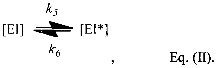
K, rc n TF.Il
At least in theory, the on-rate constant fa and the off-rate constant fa can both be very fast such that EI and EI* are in fast equilibrium based on k5 and ke. However, at least in certain instances, the off-rate constant fa can be quite slow, such that when the concentration of at least one of EI and EI* changes, a significant 'lag" is observed before a new equilibrium can be reached.
This phenomenon correlates somewhat to the Post- Antibiotic Effect (PAE) observed in the treatment of certain bacterial infections with certain antibiotics. The PAE is a term used to describe the persistent suppression of bacterial growth that can occur after limited exposure to an antimicrobial agent. See McDonald, Craig & Kunin, Persistent effect of antibiotics on
Staphylococcus aureus after exposure for limited periods of time. Journal of Infectious Diseases 135: 217-223, 1977 (the entire contents of which is expressly incorporated herein by reference). Also see, Craig, The Role of Pharmacodynamics in Effective Treatment of Community-Acquired Pathogens, Proceedings of the 39th Annual Meeting of the Infectious Disease Society of
America, Vol. 2, No. 4, 2002 (the entire contents of which is expressly incorporated herein by reference); and Craig, Post-Antibiotic Effectsin Experimental Infection Models: Relationship to in vitro Phenomena and to Treatment of Infections in Man, J. Antimicrobial Chemotherapy, 31, Supp. D, pp. 149-158, 1993 (the entire contents of which is expressly incorporated herein by reference).
Briefly, when certain antibiotics are administered to treat a bacterial infection, bacterial
- 19 -
SUBSTITUTE SHEET RULE 26
growth is inhibited so long as the concentration of the administered antibiotics is at or above a minimum inhibition concentration (MIC). As soon as the drug concentration falls below the MIC, due to factors such as drug metabolism or excretion by the host, bacterial growth resumes without discernible delay. That is, no PAE is observed when using the specific antibiotics to treat the specific bacterial infection. In other situations, however, bacterial growth is still inhibited or significantly retarded even after the drug concentration falls below the MIC. It is only after a set period of time after the drug concentration has fallen below the MIC that bacterial growth resumes at full strength. The presence of a prolonged in vivo PAE may be beneficial since it allows wider dosing intervals without sacrificing therapeutic efficacy.
Without wishing to be bound by any particular theory, Applicants believe that at least part of the mechanism for the observed PAE is that the drug compound binds to its target enzyme and locks the enzyme in a stable EI* complex, such that even after the withdrawal or reduction of the drug compound in the host system, a significant portion of the EI* complex is slow to convert back to the EI complex which is in rapid equilibrium with free enzyme E. This slow conversion, as partly dictated by the slow off rate constant ke of Equation (II), helps to prevent the target enzyme from facilitating recovery of the infectious bacteria, at least during the initial phase after the drug concentration falls below the MIC. In practice, by having the knowledge of the existence of PAE for a particular antibiotic used to treat a specific infection, one can design a better dosing regimen that takes into consideration the existence and extent of PAE, such that a minimally effective drug concentration is used to treat a bacterial infection.
Concerning antibiotic dosing regimens, historically, they have been determined based upon the pharmacokinetic (PK) parameters of the particular antibiotic. Recent experience suggests, however, that pharmacodynamic (PD) parameters should also play an important role, because drug concentrations in the tissues or body fluids as well as at the site of infection are considered. Drug concentration in the tissues or body fluids to the large extent determines the pharmacological and toxicological effects of the drug, while drug concentration at the site of microbial infection determines the antimicrobial effect.
Once a dosing regimen has been determined, drug compounds are administered to the host at a given dose or doses, and at specified intervals according to the dosing regimen. As a result of the PK properties of the drug compound, a pharmacokinetic (PK) profile of the
- 20 -
SUBSTITUTE SHEET RULE 26
compound in the subject, particularly at the site of the microorganism infection, may be produced. The PK profile provides a time course of the relevant drug concentration (e.g., drug concentration at the site of infection and/or site of bacterial growth) over time.
It has been discovered that the relationship between several key parameters associated with a particular microor anism infection can be ex ressed with Equation (III) below:
[ ]

N0 is microorganism titer or concentration (that may be measured by CFU per unit volume) at the beginning of a period of time during which drug compound concentration does not substantially change (e.g., drug compound concentration does not deviate from average drug concentration during the period by more than 20%, 15%, 10%, or 5%);
N is microorganism titer or concentration (that may be measured by CFU per unit volume) at the end of the period of time;
t is the duration of time for the period;
λ is intrinsic logarithmic growth rate of the microorganism, under the physiological conditions at the site of infection or growth;
ε is maximum kill rate of the microorganism by the compound, under the physiological conditions at the site of infection or growth;
/ is replenishment rate of the enzyme (e.g., the rate at which new enzymes are produced by the microorganism or otherwise made available to the microorganism at the site of infection or growth), under the physiological conditions at the site of infection or growth;
[I] is concentration of the compound at the site of enzyme function (e.g., inside the microorganism for an intracellular enzyme), which, if necessary, can be converted from the known drug compound concentration outside the microorganism, and a correlation between drug concentration outside the microorganism and drug
concentration at the site of enzyme function;
Ki is the binding constant defined in Eq. (I);
ks and k(, are on- and off-rates defined in Eq. (II);
β = 1 + KM / [S] + (KM / [S]) x ([I] / Kj);
k = k6 + (k5 1 β) x (KM / [S]) x ([I] / K);
σ = (k x (k6+ p) Ι β - k6 p I β) I k2;
KM is the Michaelis-Menten constant of the enzyme;
[S] is concentration of the substrate of the enzyme at the site of enzyme function (e.g., inside the microorganism).
Parameters σ, β, k above are auxiliary parameters introduced into the equation to simplify the equation.
The remaining symbol γ is a constant that can be determined by the initial value of the enzyme substrate complex for the period of time.
Although Equation (III) above requires the inhibitor concentration [I] to remain substantially constant during a period of time in question, the practical applicability of the Equation is not so limited. This is because for any PK profile of the drug compound, the time course of interest can be sub-divided into a series of successive intervals, wherein the drug compound concentration does not substantially change within each of the small intervals. Thus Equation (III) can be applied for each of such intervals to determine microorganism titer at the end of each interval (N), based on the microorganism titer at the beginning of each interval (No). Once N is determined for one particular interval, it can serve as the No for the immediate following interval, in which Equation (III) can still be applied to determine the N at the end of the immediate following interval. By following this procedure, microorganism titer change over time can be determined based on a successive series of calculations according to Equation (III).
It should be noted that, during the series of calculations above, a unique/critical point of the computation is the determination of the value y. The magnitude of this value is dependent on whether [EI*] is increasing or depleting. Additionally, the value of γ varies with time, and is calculated or approximated in successive small intervals. For the first interval (during which drug compound concentration does not substantially change) γ can be determined using the following Equation (IV), taking into consideration the reasonable assumption that there is no EI* complex at the beginning of the period (i.e., [EI*] = 0):
γ = 1 Ι β + (k x (k6 + p) Ι β - k6 x p Ι β) I k2, Eq. (IV).
- 22 -
SUBSTITUTE SHEET RULE 26
The various parameters in Equation (IV) are as defined above.
Once constant y is determined for the first interval, for the second interval and beyond, constant y for each interval can be determined using the constant y value from the immediate preceding interval and current drug compound concentration and other parameters above. More specifically, to iterate between successive time intervals, the following specific steps can be used for any "current" interval:
1. Compute the value of [ES] at the end of the immediate preceding interval {e.g., for the second interval as the current interval, the value of [ES] at the end of the first interval) according to the following Equation (VI):
[ES] = ^f- + - 1 + ye-kt
k k , Eq. (VI).
Wherein b = (k6 + p) I β; h = k6 χ p I β; β (β = 1 + M / [S] + (KM / [S]) ([I] / ¾)) and k (k = k6 + (ks I β) x (KM / [S]) x ([I] / K;)) are as defined above, and y is the constant for the immediate preceding interval (e.g., the 1ST interval for the 2ND interval as the current interval). For the 2ND interval, for example, constant y for the immediate preceding interval (i.e., the 1ST interval) can be determined based on Eq. (IV) above. For the 3RD interval and beyond, constant y for the immediate preceding interval (e.g., 2ND interval and beyond) can be determined using the procedure being described here in steps 1 -3.
2. Compute constant β for the current interval, using the new inhibitor level [I] for the current interval. Note that:
β = 1 + KM / [S] + (KM / [S]) x ([I] / K;).
3. Compute constant y for the current interval using the following Equation (V):
Pold [ E ] enc[.0 [--last- interval
γ =
Pliew , Eq. (V);
wherein in Eq. (V):
β0ι is β defined above for the immediate preceding interval;
fi„ew and a„ew are β and σ (σ = (k x (k6 + p) I β - ke x p I β) I k2) defined above for the current interval;
[ES]end is concentration of enzyme-substrate complex at the end of the immediate preceding interval, and is calculated according to Equation (VI) in step
- 23 -
SUBSTITUTE SHEET RULE 26
1 above.
Once the values of constants γ and β for the current interval (e.g., the 2nd interval) are determined using the above steps 1-3, they can be used to calculate [ES] at the end of the current interval (e.g., the 2nd interval) as in step 1 above, and β0ΐά, which [ES] and β0^ values are then in turn used in calculating constant γ for the 3rd interval according to steps 1-3. So on and so forth.
With the above procedure, the model enables one to determine or simulate the titer of a infecting microorganism in a host from beginning to end, given the known, calculated, and/or measured parameters in the equations, and knowledge of the pharmacokinetics of the inhibitor.
Equation (III) above may be further simplified under certain conditions, as described in more detail below.
First, when k6 is 0 or is nearly 0, the conversion from EI to EI* becomes irreversible, such that EI* does not revert back to EI in any significant way, if at all. Under this condition, Equation (III) becomes Equation (III)a below:

Eq. (III)a
Second, when ke » fa, or when fa I fa is 0 or is nearly 0, any EI* (if any at all) almost instantly reverts back to EI, and Equation (III) becomes Equation (III)b:
[N] = εχρ(λί + ε (t + pt2/2))N0

Eq. (III)b
Third, in some systems where enzyme replenishment rate p is very small or negligible, the total enzyme concentration can be considered as a constant in the process. Under this reasonable approximation, Equation (III) becomes Equation (III)c:
- 24 -
SUBSTITUTE SHEET RULE 26
[Ν] = βχρ([λ + Β(

Eq. (III)c
Fourth, when both A¾ and p are both 0 or both near 0, i.e., irreversible EI to EI* conversion, and no enzyme replenishment, Equation (III) can be further simplified into Equation (III)d:
[N] = βχρ((λ - 6)t + ε _Qi \ 1 - e

5 K,
Eq. (III)d
Finally, if the EI* to EI conversion is reversible {ke » ks), and if there is no enzyme repleshment (p = 0), Equation (III) can be further simplified into Equation (III)e:
1 +J!L
[N] = e\p( t - et ^ ) 0
Eq. (III)e
In Eq. (III)e, [S] / Km is EC50.
Note that in Equation (III), the linear term [λ + ε(σ - 1)] largely determines whether the titer N will be decreased or increased compared to No, wherein a positive [λ + ε(σ - 1)] value increases titer, and a negative [λ + ε(σ - 1)] value decreases titer.
Also note that the non-linear terms in Equation (III) add a dip / valley or a bump / peak to the titer curve, and the depth or height of such dips and bumps is equal to γ / k2. Since γ is computed as (&<j[EI*] - ^[EI]) / β, to increase the depth of the dip, more [EI*] excess is desired,
25 -
SUBSTITUTE SHEET RULE 26
which can be achieved either by increasing the incubation time or with a higher inhibitor concentration.
On the other hand, the time that takes to erase or ease the dip or bump is: t =
k2( - s( l + b/k))
Equation (III) also suggests that at certain inhibitor concentration [I], N neither increases nor decreases compared to N0 ("stasis"). That is, when: λ - ε(Ι - ·γ- +— e"kl) = 0
When the time frame is long term (i.e., t is large), the above equation can be further simplified into:
_ _b_ = k^ _
λ ε(1 k ) 0 ^ λ - ε(1 - K6( 1 +C+C[1]/K.) + k5C[1]/K. ) - 0 ^
Thus the concentration that induces stasis is:

C(E - )(k6 + k5)
The utility of Equation (III) is not limited to calculating / predicting / simulating bacterial titer (N) change over time. In fact, given the established relationship among the above parameters, Equation (III) enables one to simulate any of the given parameters provided that the other parameters are fixed, or provided by experimental data.
Specifically, for simulation in general, the following parameters may be needed: ks, ke, Kj, C = M / [S], I, e , and p. A graphic user interface (GUI) can and has been built to dynamically change the values of these parameters (except for the one of interest) to show how such parameter changes will affect the behavior of the parameter of interest. Given the way Equation (III) is applied to overcome the limitation of requiring substantial constant inhibitor concentration, a specified range of time that includes time zero is provided (i.e., when the process is just started ). In addition, the concentration level of the drug compound or inhibitor can be provided as a time related vector, or as the output of a PK model.
All parameters other than i can be readily fitted as regular parameters, as could be done
- 26 -
SUBSTITUTE SHEET RULE 26
for any standard curve-fitting algorithm. For Kj, however, a potential complication may arise due to a hidden parameter pm (as defined below). In Equation (III), Kj is always associated with the drug compound concentration [I] at the target site, which is often inside a cell when, for example, the enzyme target of the drug compound is a cytosolic enzyme. Meanwhile, inhibitor concentration provided in a typical PK profile usually reflects drug compound concentration in an extracellular space, such as in plasma or at a local infection site. Thus, under this
circumstance, the drug compound concentration provided by the PK profile may need to be adjusted or converted to intracellular drug compound concentration [I].
Under typical circumstances, the intracellular drug compound concentration [I] is much less than the extracellular drug compound concentration, due to factors such as microorganism's ability to defend against drug compound permeation into the cell. This type of microorganism defense may be partially or completely impaired in mutants that are unable to pump intracellular drug compounds outside the cell. The ratio between the inside concentration level and outside concentration level may be correlated to and determined by so-called pump mutant coefficient, denoted as pm- Thus, [I] in Equation (III) may actually be pm x [I]0, wherein [I]0 is the inhibitor concentration outside the microorganism.
Consequently, K, and pm cannot be fitted independently. Instead, these two parameters can be combined into a new fitable parameter as Kpi, which represents the ratio of Ki and pm.
Merely to illustrate, FIG. 1 shows a simulated PK profile for a hypothetical drug administered to a host. A GUI interface is provided such that a user can change one or more parameters to simulate different resulting PK profiles. The parameters included in the GUI interface includes drug dose R0 (0.5 meg), dose / day (twice a day, 12 hrs apart), route of administration (/.v. infusion), drug half-life ti/2 (2.5 hrs), Vj (volume of distribution - the apparent volume in which a drug is distributed in vivo, 6 mL), etc. The simulated PK profile shows that drug concentration rapidly increases after the first dose at time 0, and peaks around 1 hr after i.v. infusion. The concentration of the drug compound then steadily decreases according to a drug half-life t\ of 2.5 hours, reaching nadir after about 12 hrs, at which point a second dose is administered to repeat the process. Here, Ub and Lb refer to "Upper bound'' and "Lower bound" respectively.
The lower panel of FIG. 1 shows microorganism cell count (in log scale) over the same
- 27 -
SUBSTITUTE SHEET RULE 26
time period in response to drug compound concentration change, as determined by applying Equation (III). In this simulation, ks is set at 0.5, is set at 0.3, Kj is set at 30, pm is 0.001, p is set at 0.25, C is set at 1.5. The terms lmd and eps refer to lambda (λ) and epsilon (ε)
respectively. It is apparent that MIC is about 1 χ 104 mcg/mL, such that microorganism cell count begins to recover as soon as drug compound concentration falls below the MIC at about 7.5 hrs after the first administration, and again at just over 20 hrs after the first administration.
Conversely, using any standard curve-fitting algorithm, the parameters can be determined by "fitting" Equation (III) using the measured microorganism titer change over time. Such fitted parameter values may be further compared with the corresponding experimentally determined values to show the close matches between the values. See Examples below and FIG. 2. Such model fitting typically requires information about microorganism count at different time points, and inhibitor concentrations over time.
In general, the greater the number of time points and concentration values are available, the higher the quality and/or confidence level the fitted parameter values will have.
4. Method of Treatment
The invention described herein provides a valuable means to control a course of treatment for microorganism infection.
In one embodiment, the invention provides a method of treating a microorganism infection in a subject in need of treatment, such that treatment according to an established dosing regimen can be precisely controlled, and drug administration can be timely terminated once the microorganism titer falls below a pre-determined level.
Thus in one aspect, the invention provides a method of treating a microorganism infection in a subject in need of treatment, the method comprising:
(1) providing a compound that binds to and inhibits an enzyme of the microorganism, and inhibits proliferation of the microorganism;
(2) providing a dosing regimen for administering the compound to the subject;
(3) administering the compound to the subject according to the dosing regimen, and initiating a time course of treatment;
- 28 -
SUBSTITUTE SHEET R LE 26
(4) dividing the time course of treatment into a series of successive intervals, wherein within each said intervals, compound concentration at a site of microorganism infection remains substantially constant (e.g., less than 5% deviation from the average concentration);
(5) beginning from the first of said series of successive intervals, and for each said series of successive intervals, constantly determining, according to Equation (III), microorganism titer N at the end of each said series of successive intervals, using microorganism titer No at the beginning of each said series of successive intervals, wherein N for one interval is N0 for the immediate following interval; and,
(6) terminating treatment according to the dosing regimen when microorganism titer N at a given interval falls below a pre-determined / target microorganism titer, thereby treating the microorganism infection;
wherein:
(a) the compound binds to the enzyme in vivo to form a compound-enzyme complex ("EI"), with a binding constant Kt, according to Equation (I):
[E] + [I] ^ [EI] f Eq (I).
(b) the compound-enzyme complex EI is in equilibrium with a tight compound- enzyme complex ("EI*"), with an on-rate constant ks and an off-rate constant ke, according to Equation (II):
[EI] [EI*]
1(6 , Eq. (II);
(c) the dosing regimen produces a pharmacokinetic (PK) profile of the compound in the subject (at the site of the microorganism infection);
(d) Equation (III) is:
[Ν] = [Ν0]εχρ([λ + ε(σ - l)]t +sp

wherein:
No is microorganism titer at the beginning of each said series of successive
- 29 -
SUBSTITUTE SHEET RULE 26
intervals;
N is microorganism titer at the end of each said series of successive intervals;
t is the duration of time for a given interval associated with N0;
λ is intrinsic logarithmic growth rate of the microorganism;
ε is maximum kill rate of the microorganism by the compound;
p is replenishment rate of the enzyme;
[I] is concentration of the compound at the site of enzyme (e.g., inside the microorganism);
Ki is the binding constant defined in Eq. (I);
ks and ke are on- and off-rates defined in Eq. (II);
β = 1 + KM / [S] + (KM / [S]) * ([I] / K);
k = k6 +(ks / fi) x (KM / [S]) x ([I] / K);

KM is the Michaelis-Menten constant of the enzyme;
[S] is concentration of the substrate of the enzyme at the site of the enzyme function (e.g., inside the microorganism);
for the first of said series of successive intervals, y is a constant determined by Equation (IV):
γ = 1 / β + χ 6 + ρ) /β - ^ χ ρ / β) / ^, Eq. (rV);
for each subsequent said series of successive intervals, γ is a constant determined by Equation (V):
end-of'-last-interval _
γ = - <rnew
P»'w , Eq. (V);
wherein in Eq. (V):
β0ΐά is β defined above for the immediate preceding interval;
βηβνι and onew are β and σ defined above for the current interval;
[ES]end is concentration of enzyme-substrate complex at the end of the immediate preceding interval, and is calculated according to Equation (VI), wherein b = (ke + p) I β; h = kf, x p I β; and γ is as defined above for the immediate preceding interval:
- 30 -
SUBSTITUTE SHEET R LE 26
[ES] = ^ - + 1 + ye" -kt
, Eq. (VI).
According to this aspect of the invention, a patient (e.g., human or a non-human animal) infected by a microorganism (e.g., a species of infectious bacteria) may be administered a drug compound (e.g., an antibiotic effective in treating the bacterial infection) according to an established dosing regimen (e.g., 2-4 times daily, preferably equal interval oral, .v., or i.m.
administration). The initial microorganism load at the site of infection (e.g., CFU count in the blood of the patient if it is a systemic infection) may be determined at the beginning of the treatment, when the first dose of the drug compound is administered according to the dosing regimen. This initial load serves as N0 in Equation (III). Since the PK/PD profile of the drug in the given host is usually known or can be readily predicted or measured (such as periodic measurement of drug concentration within the host system), Equation (III) may be used to determine at what point in time microorganism titer falls below a pre-determined safe level, such as a reduction of 2-4 logs depending on the initial o value. The determined time point may be the time to terminate treatment according to the dosing regimen.
For example, when a host or patient is following a fixed dose three times daily dosing regimen to treat a microorganism infection, the relevant PK/PD profile of the drug in the host (for example, one that reflects the profile at the site of infection) can be readily determined or predicted or estimated. Based on the PK/PD profile and the associated intracellular drug concentration [I], Equation (III) can be used to determine microorgamsm titer N based on a successive series of calculations for a series of small time intervals, during which [I] can be considered substantially invariable. Once the determined N is low enough, e.g., 2-4 logs lower than the starting titer No, several days into the dosing regimen, treatment of infection can be considered complete, and the host or patient no longer needs to follow the dosing regimen from that point on.
This method is useful, for example, for treating infections in which obvious symptoms of the infection disappear long before a safely low microorganism titer is reached, such that dosing regimens should still be rigorously followed for an additional period of time even after the first disappearance of certain symptoms, but for no longer than necessary.
In certain embodiments, the microorganism is a bacterium, such as P. aeruginosa, E. coli,
- 31 -
SUBSTITUTE SHEET R LE 26
K. pneumonia, A. baumannii , M. tuberculosis, S. aureus, S. pneumonia, H. influenzae,
Citrobacter spp., Enterobacter aerogenes, Enterobacter cloacae, S. marcescens, A. baumannii, and S. maltophila.
In certain embodiments, the bacterium is P. aeruginosa, E. coli, K. pneumonia, A.
baumannii , M. tuberculosis, S. aureus, S. pneumonia, or H. influenzae.
In certain embodiments, the infection is systemic, such as sepsis. In other embodiment, the infection is local, e.g., limited to local sites, such as cuts and wounds limited to local tissues.
In certain embodiments, the subject is a human, or a non-human mammal {e.g., a mammal; a primate; a livestock animal including beef cattle, dairy cow, water buffalo, bison, yak, camel, llama, pig, horse, donkey, mule, sheep, goat, deer, rabbit, poultry (chicken, duck, goose, turkey); a pet or a lab animal including cat, dog, mouse, rat, guinea pig, hamster, etc.).
In certain embodiments, the compound is an antibiotic, such as Ampicillin, Cloxacillin, Methicillin, Cefotaxime, Ceftriaxone, Moxalactam, Imipenem, or Meropenem; vancomycin, erythromycin, Gentamicin, Amikacin, Tobramycin, Rifampin, Trimethoprim, Ciprofloxacin, and any other antibiotics known to have PAEs.
In certain embodiments, the antibiotic exhibits a post-antibiotic effect (PAE) in treating infection by the microorganism.
In certain embodiments, A¾ is less than about 10 hr"1, 5 hr"1, 3 hr"1, 1 hr"1, 0.5 hr"1, 0.3 hr"1, 0.1 hr"1 or less. In other embodiments, ke is within the range between any of the two recited values above.
In certain embodiments, the series of successive intervals are of identical duration.
In related embodiments, at least two of the series of successive intervals are of different duration. The latter situation may be helpful in that narrower intervals may be beneficial for a steep increase or decrease in [I], while wider intervals may be sufficient for a shallow or gradual increase or decrease in [I].
In certain embodiments, [I] is calculated / determined based on the PK or PK/PD profile of the compound. Alternatively, [I] may be based on direct measurement of systemic inhibitor compound concentration, which is in turn converted to [I] for use in Equation (III).
Alternatively, in a related embodiment, the invention provides a method of treating a
microorganism infection in a subject in need of treatment, the method comprising first determining a specific dosing regimen that would produce a desired treatment result based on calculations according to Equation (III), and then administering the drug to the host or patient according to the selected dosing regimen.
For example, for a specific microorganism infection, if it is desirable (e.g., in a particular clinieal situation) that microorganism titer drops over a course of 2 days to below a desired level (e.g., 2-4 logs reduction compared to the starting titer N0), Equation (III) may be used to screen a number of potential dosing regimens for treating the infection, in order to select the dosing regimen that possesses the optimal combination of fast reduction of microorganism titer and tolerable toxicity and/or side effects (due to, for example, the magnitude of the highest drug concentration in the host).
Thus in another aspect, the invention provides a method of screening or selecting for a dosing regimen for treating a microorganism infection in a subject in need of treatment, the method comprising:
(1) providing a compound that binds to and inhibits an enzyme of the microorganism, and inhibits proliferation of the microorganism;
(2) providing a candidate dosing regimen for administering the compound to the subject;
(3) providing a time course of treatment beginning from the time the compound is first administered to the subject according to the dosing regimen;
(4) dividing the time course of treatment into a series of successive intervals, wherein within each said intervals, compound concentration at a site of microorganism infection remains substantially constant (e.g., less than 5% deviation from the average concentration);
(5) beginning from the first of said series of successive intervals, and for each said series of successive intervals, constantly determining, according to Equation (III), microorganism titer N at the end of each said series of successive intervals, using microorganism titer N0 at the beginning of each of said series of successive intervals, wherein N for one interval is N0 for the immediate following interval; and,
(6) determining microorganism titer at the end of a pre-determined end point time, and comparing with a pre-determined end point titer, wherein the candidate dosing regimen is selected when the microorganism titer at the end of the predetermined end point time is no more than the pre-determined end point titer; wherein:
(a) the compound binds to the enzyme in vivo to form a compound-enzyme complex ('ΈΓ'), with a binding constant Kj, according to Equation (I):
[E] + [I] ^ [EI] 5 Eq (I);
(b) the compound-enzyme complex EI is in equilibrium with a tight compound- enzyme complex ("EI*"), with an on-rate constant ks and an off-rate constant k6, according to Equation (II):
[EI] ^ ^- [El*]
k* , Eq. (II);
(c) the dosing regimen produces a pharmacokinetic (PK) profile of the compound in the subject (at the site of the microorganism infection);
(d) Equation (III) is:
[N] = [Ν0]βχρ([λ + ε(σ - l )]t +ερ

wherein:
No is microorganism titer at the beginning of each said series of successive intervals;
N is microorganism titer at the end of each said series of successive intervals;
t is the duration of time for a given interval associated with N0;
λ is intrinsic logarithmic growth rate of the microorganism; ε is maximum kill rate of the microorganism by the compound;
p is replenishment rate of the enzyme;
[I] is concentration of the compound at the site of enzyme function {e.g., inside the microorganism);
Ki is the binding constant defined in Eq. (I);
ks and k6 are on- and off-rates defined in Eq. (II);
β = 1 + KM / [S] + (KM / [S]) x ([I] / K);
k = h + (ks I β) x (KM / [S]) x ([I] / K);
a = (k x (k6 + p) / β - k6 * p I β) Ι k2;
M is the Michaelis-Menten constant of the enzyme;
[S] is concentration of the substrate of the enzyme at the site of enzyme function (e.g., inside the microorganism);
for the first of said series of successive intervals, y is a constant determined by Equation (IV):
y = l / fi + (k x (k6 + p) / fi - k6 x p / p^) / k2, Eq. (IV);
for each subsequent said series of successive intervals, y is a constant determined by Equation (V):
-of-last-interval _
γ = R " σηε\ν
P"ew , Eq. (V);
wherein in Eq. (V):
β0ω is β defined above for the immediate preceding interval;
βηεν/ and Onew are β and σ defined above for the current interval;
[ES]end is concentration of enzyme-substrate complex at the end of the immediate preceding interval, and is calculated according to Equation (VI), wherein b = (ke + p) I β; h = k x p I β; and y is as defined above for the immediate preceding interval:
[ES] = j2k - + - t + ye-kt
k k , Eq. (VI).
In a related aspect, the invention provides a method of treating a microorganism infection in a subject in need of treatment, the method comprising: using the method immediately above to screen for and select a dosing regimen that produces the desired treatment (e.g., a 2-4 logs reduction in microorganism titer in relation to starting titer), and further comprising
administering the compound to the host according to the selected dosing regimen.
Any of the specific embodiments described above apply to all aspects of the invention.
- 35 -
SUBSTITUTE SHEET RULE 26
5. Compound Screening / Design
The invention described herein further provides a method for screening inhibitor compounds that may be used to achieve desired treatment time course and efficacy, while preferably avoiding or minimizing associated side effects or toxicological effects.
Related or unrelated inhibitor compounds may share similar biological functions, e.g., inhibiting the function of an essential metabolic enzyme of the infectious microorganism. Each inhibitor may have different yet determinable Kj, k5, k6, PK/PD profile, and maximum kill rate ε. Given the relationship among the parameters in Equation (III), the time course of treatment using any of the several possible inhibitors can be determined based on Equation (III), and the results compared to identify the optimum balance between efficacy and side effects.
Alternatively, the invention also allows one to "design'' a drug based on a series of fixed parameters in Equation (III), such that one or more remaining parameters can be calculated / determined accordingly, and compounds having such calculated / determined parameters can be designed or screened for. For example, if it is desired to administer an inhibitor compound to treat a microorganism infection according to a pre-determined dosing regimen and/or a predetermined time course of treatment, certain parameters such as N0, end titer N, duration of treatment, intrinsic logarithmic growth rate of the microorganism (λ), replenishment rate of the enzyme (p), and concentration of the compound at the site of enzyme function (e.g., inside the microorganism) can be fixed or pre-determined. Other parameters, such as Kj, k5, ke, and PK/PD profile, may be fitted using Equation (III), either collectively or individually. The results become screening criteria as to what the parameters for a '"designer compound" should be, thus guiding future screening efforts for compounds meeting such design criteria.
In certain embodiments, some of the Equation (III) parameters that are related to an inherent property or characteristic of the inhibitor such as Kj, k$, and A¾, as well as PK/PD profile, may also be fixed or pre-determined, and Equation (III) may be used to determine what the remaining parameters should be. This may be useful when a lead drug compound is identified, which lead compound may possess some but not all of the desired parameters (e.g., the lead compound may have desired K; and k5, but the value of ke for the lead compound is not ideal). Thus further designing or screening may be carried out to identify second generation compounds that possess some of the desired parameters.
- 36 -
SUBSTITUTE SHEET RULE 26
6. Methods of Efficacy Determination
The invention described herein further provides a method for determining or predicting efficacy of a compound administered according to a set dosing regimen, in the context of treating a specific microorganism infection in a specific host. That is, whether the specific compound in question will be efficacious under a given dosing regimen for treating a given host for a given disease.
The invention described herein further provides a method for determining or predicting efficacy of a dosing regimen for a pre-determined compound, in the context of treating a specific microorganism infection in a specific host. That is, whether the specific dosing regimen in question for the selected compound will be efficacious for treating a given host for a given disease.
Likewise, the invention further provides a method for determining or predicting efficacy of a pre-determined compound used according to a pre-determined dosing regimen, for treating a specific host with a given initial microorganism load. That is, whether the host can be successfully treated by the compound under the dosing regimen, and if so, how long the treatment should be.
EXAMPLES
Example 1: LpxC Assay
Lipid A is the hydrophobic anchor of lipopolysaccharide (LPS), and forms the major lipid component of the outer monolayer of the outer membrane of Gram-negative bacteria, such as Escherichia coli. Lipid A is required for bacterial growth and virulence, and inhibition of its biosynthesis is lethal to bacteria. UDP-3-0-(i?-3-hydroxymyristoyl)-N-acetylglucosamine deacetylase (LpxC) is a metalloenzyme that catalyzes the second step in the biosynthesis of lipid A. Consequently, inhibitors of LpxC have been shown to have antibiotic activities, and can be the inhibitor compound of the invention with respect to the essential metabolic enzyme target LpxC.
- 37 -
SUBSTITUTE SHEET RULE 26
The LpxC enzyme assay described herein may be used to determine enzyme (LpxC) activity under various experimental conditions, such as with or without the presence of significant inhibitors. More specifically, inhibitor binding kinetics can be measured using an LC/MS based assay of product formation.
Briefly, at the beginning of the experiment, a given amount of the enzyme and the inhibitor are incubated for a period of time at appropriate temperature (e.g., 2 hrs at room temperature) to allow the formation of enzyme-inhibitor complex EI and EI*. Then an aliquot of the EI/EI* complex is rapidly diluted with excess substrate, such as a 1000-fold dilution with excess substrate S, such that substantially all enzymes in the EI complex (which are in fast equilibrium with free form enzyme E) becomes free form enzyme available to convert the substrate to the product P. The speed with which product P is produced, as measurable by LC/MS, is indicative of the amount of free form enzyme in the reaction mixture. Meanwhile, if there is any enzyme locked in the EI* complex, and thus not available as free form enzyme in the 1000- fold dilution mixture, the amount of product P is reduced accordingly. See Example 2.
All LpxC enzyme assays were performed using a buffer containing 50 mM NaH2P04 pH 7.4, 0.2 mg/ml Bovine serum albumin (BSA), and 0.5 M sucrose. The LpxC substrate used was typically UDP-3-0-( ?-3-decanoyl)-N-acetylglucosamine. A limited number of assays with E. coli LpxC alternately used the 3-Hydroxymyristoyl version of this substrate.
Wild type E. coli and P. aeruginosa LpxC were prepared following procedures similar to those published previously (Barb et al., Biochemistry 46: 3793-3802, 2007; and Coggins et al, Biochemistry 44, 11 14-1126, 2005). Deacetylated product was detected and quantified by LC/MS/MS using an AB/Sciex 4000 triple quadrupole mass spectrometer interfaced with a Shimadzu LC20 HPLC system. Assay samples were subjected to HPLC using a buffer system comprising 10 mM ammonium acetate, pH 6.5 (Buffer A), with reaction product eluting over a 1 minute gradient from 5% to 90% acetonitrile (Buffer B) at a flow rate of 0.5 ml/minute.
Deacetylated product was detected by LC/MS/MS using an AB/Sciex 4000 triple quadrupole mass spectrometer. The mass spectrometer was operated in negative ion, MRM mode where quantification was accomplished by monitoring peak areas corresponding to the parent ion to m z 79 fragmentation reaction.
- 38 -
SUBSTITUTE SHEET RULE 26
Example 2: Enzyme Activity Recovery Assay
To estimate the effective k0ff{ r ke) of compounds from P. aeruginosa or E. coli LpxC, enzyme was incubated with a slight excess of compound (typically 50 nM compound with 30 nM enzyme) for 3 hours, then the resultant enzyme-inhibitor complex was diluted 1000- fold in 2 steps. Compounds for incubation were diluted from 10 mM to 10 μΜ in 10 x steps in fresh DMSO, then diluted to 1 μΜ in 1 : 1 DMSO:assay buffer prior to incubation with enzyme.
Enzyme was diluted to 32 nM in assay buffer, after which compound was added to a final concentration of 50 nM. After incubation for 3 hours to allow formation of the enzyme-inhibitor complex, samples were diluted 20-fold in assay buffer, followed by another 50-fold dilution in buffer containing 25 μΜ LpxC substrate. Reaction aliquots were collected at measured time intervals following dilution, quenched with a 2-fold volume of 2% acetic acid, and assayed by LC/MS/MS.
To estimate the enzyme recovery rate constant, product formation as a function of time were fit using Excel/XLFit to equation 1, where V0 was set to 0 (no background product accumulation was observed) and Vs was set to the linear enzyme velocity observed in the absence of inhibitor. The inverse of the k value obtained, multiplied by the natural logarithm of 2, was taken as the residence time, ti/2.
Equation 1 p = ^ + {Vq _ v ) l _ e -* ) / k
Ki* and Ki Estimation. Estimation of Kj* and Kj was accomplished using an ½0 assay format using respective long or short pre-incubation of compounds with enzyme.
To measure Kj*, compounds were titrated to achieve final assay concentrations from 20 nM to 39 pM in a 2- fold dilution series. Compounds were incubated overnight (18 hours) in assay buffer containing 30 pM E. coli or P. aeruginosa LpxC. Reactions were then initiated by addition of substrate to reach final substrate and enzyme concentrations of 5 μΜ and 15 pM respectively. Reactions proceeded for 60 minutes before quenching with 2-fold volume of 2% acetic acid and quantification of reaction product by LC/MS/MS. Positive and negative inhibition controls used N-[(l S,2R)-2-hydroxy-l-(hydroxycarbamoyl)propyl]-4-[2-[4- (morpholinomethyl)phenyl]ethynyl]benzamide (10 μΜ) and DMSO (2% final) respectively. An ICso was calculated (Excel/XLFit - 4 parameter dose-response model) from the measured percent inhibition as a function of inhibitor concentration.
- 39 -
SUBSTITUTE SHEET RULE 26
For i estimation, an IC50 assay format was used where reactions were run for a minimum length of time with no enzyme pre-incubation. In these assays compounds were titrated to reach assay concentrations from 40 nM to 78 pM. Compounds were first pre- incubated for 20 minutes in assay buffer containing 10 μΜ substrate, then reactions were initiated with enzyme to reach final enzyme and substrate concentrations of 100 pM and 5 μΜ respectively. Reactions were run for 3 minutes, quenched with a 2-fold volume of 2% acetic acid, and assayed by LC/MS/MS.
An IC50 was estimated from percent inhibition as a function of inhibitor concentration and taken as Kj.
Example 3: Killing Kinetics Assay
Media for each bacteria species used in the killing kinetics assay were prepared based on Table 1 below. Culture conditions for each bacteria species are also listed.
Table 1

Two days before the assay, bacteria were streaked / inoculated onto appropriate agar plates, which were then incubated overnight at about 37 °C, with or without C02, depending on bacterial strain used (Chart 1).
One day before the assay, a 6-well plate with 6 mLs of the bacteria-specific media per well was prepared. The bacteria was inoculated in the 1st well, and serial dilutions were made by taking and mixing an aliquot of about 20-50 from the 1st well to the 2nd well, and repeating
- 40 -
SUBSTITUTE SHEET RULE 26
the process for each of the remaining wells (e.g., an aliquot of about 20-50 μΐ-, was transferred from the 2nd well to the 3rd well, and mixed, etc). The 6-well plate was then incubated overnight at 37 °C (with or without C02). Meanwhile, several flasks were each filled with 200 mL of the bacteria-specific media and warmed overnight at 37 °C. Serial dilution plates for each time point were also prepared.
On the day of the assay, bacterial growth in each well were determined by measuring Οϋ600, and wells with bacterial OD6oo of about 0.3-0.6 were selected for the assay. The selected culture was first diluted to a predicted 0.001 Οϋ60ο in the pre- warmed 200 mL flask with appropriate media, and 20 mL of the diluted culture were transferred to 7 flasks, one for each concentration of the compound. The compounds were then added in appropriate volume to achieve the desired multitudes of the MIC concentration within the flask. For convenience, seven compound concentrations were tested, e.g., Ox, 0.5x, l x, 2x, 4x, 8x, and 16x of MIC. See Table 2. For the negative control (Ox), the solvent DMSO was added in the same amount (200 μί,) as the highest compound volume.
The culture and the various concentrations of compounds were incubated for a predetermined amount of time to allow the formation of any enzyme-inhibitor complex.
Table 2

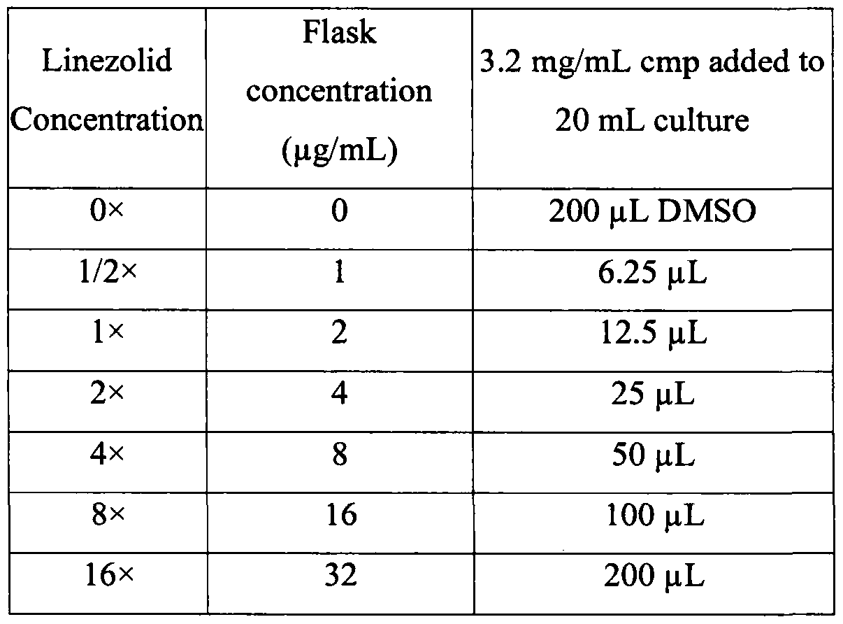
- 41 -
SUBSTITUTE SHEET RULE 26
Once the pre-determined incubation period was over for a specific time point, for each flask (compound concentration), aliquots were added into column 1 of the dilution plate according to Chart #3, and successive 10- fold dilutions were made according to Table 3. Once all the flask cultures have been added to the dilution plate, the flasks were returned to the shaker in the 37 °C incubator (with or without C02 depending on the strain) until the next time point, at which time the above process was repeated until all the time points were covered.
Table 3: Culture Dilution Plate Setup
Culture Serial Dilutions
Compound
Dilutions

U = Undiluted
For the bacterial culture in the dilution plates, aliquots of between 5-100 μΐ,, were plated on agar plates for each compound concentration at each dilution, and the plates were kept at 37 °C overnight (with or without C02 depending on the strain).
On the next day, bacterial growth in each plate was determined by counting CFU/mL, and the data was used to plot growth curve under each condition (compound concentrations over different time points). See Table 4, which shows a representative result for one of the test compounds with about 3 logs of killing in about 6 hours.
- 42 -
SUBSTITUTE SHEET RULE 26
Table 4
Averages
Time Point: 0 2 hours 4 hours 6 hours 8 hours 24 hours

Claims
A method of treating a microorganism infection in a subject in need of treatment, the method comprises:
(1) providing a compound that binds to and inhibits an enzyme of the microorganism, thereby inhibiting proliferation of the microorganism;
(2) providing a dosing regimen for administering the compound to the subject;
(3) administering the compound to the subject according to the dosing regimen, and initiating a time course of treatment;
(4) dividing the time course of treatment into a series of successive intervals, wherein within each said interval, compound concentration at a site of microorganism infection remains substantially constant;
(5) beginning from the first of said series of successive intervals, and for each said series of successive intervals, constantly determining, according to Equation (III), microorganism titer N at the end of each said series of successive intervals, using microorganism titer N0 at the beginning of each said series of successive intervals, wherein N for one interval is N0 for the immediate following interval; and,
(6) terminating treatment according to the dosing regimen when microorganism titer N at a given interval falls below a pre-determined / target microorganism titer, thereby treating the microorganism infection;
wherein:
(a) the compound binds to the enzyme in vivo to form a compound-enzyme complex C'EI"), with a binding constant AT,, according to Equation (I):
[E] + [I] ^ [EI] t Eq. (I).
(b) the compound-enzyme complex EI is in equilibrium with a tight compound- enzyme complex ('ΈΙ*"), with an on-rate constant k5 and an off-rate constant k6, according to Equation (II):
[EI] [EI*]
Eq. (II);
the dosing regimen produces a pharmacokinetic (PK) profile of the compound the subject;
Equation (III) is:
[N] = [Ν0]βχρ([λ + ε(σ - l )]t +ερ

N0 is microorganism titer at the beginning of each said series of successive intervals;
N is microorganism titer at the end of each said series of successive intervals;
t is the duration of time for a given interval associated with No;
λ is intrinsic logarithmic growth rate of the microorganism;
ε is maximum kill rate of the microorganism by the compound;
p is replenishment rate of the enzyme;
[I] is concentration of the compound at the site of enzyme function (e.g., inside the microorganism);
Kj is the binding constant defined in Eq. (I);
ks and ¾ are on- and off-rates defined in Eq. (II);
β = 1 + KM / [S] + (KM / [S]) ([I] / Kj);
k = k6 + (k5 i p) x (KM / [S]) x ([I] / ¾);
a = (k * (k6 + p) / β - k^ p l P) l k
KM is the Michaelis-Menten constant of the enzyme;
[S] is concentration of the substrate of the enzyme at the site of enzyme function (e.g., inside the microorganism);
for the first of said series of successive intervals, y is a constant determined by Equation (IV):
y = 1 Ι β + (k x (k6 + p) Ι β - k6 x p Ι β) I k2, Eq. (IV);
for each subsequent said series of successive intervals, y is a constant determined by Equation (V):

wherein in Eq. (V):
βοΜ is β defined above for the immediate preceding interval;
βηε > and anew are β and a defined above for the current interval;
[ES]end is concentration of enzyme-substrate complex at the end of the immediate preceding interval, and is calculated according to Equation (VI), wherein b = (A¾ + / ) / β; h = χ ρ I β; and y is as defined above for the immediate preceding interval

2. The method of claim 1 , wherein the microorganism is a bacterium.
3. The method of claim 2, wherein the bacterium is P. aeruginosa, E. coli, K. pneumonia, A. baumannii , M. tuberculosis, S. aureus, S. pneumonia, or H. influenzae.
4. The method of claim 1, wherein the infection is systemic.
5. The method of claim 1, wherein the infection is local.
6. The method of claim 1, wherein the subject is a human, or a non-human mammal {e.g., a livestock animal, a pet, a lab animal etc.).
7. The method of claim 1, wherein the compound is an antibiotic.
8. The method of claim 7, wherein the antibiotic is Ampicillin, Cloxacillin, Methicillin, Cefotaxime, Ceftriaxone, Moxalactam, Imipenem, or Meropenem; vancomycin, erythromycin, Gentamicin, Amikacin, Tobramycin, Rifampin, Trimethoprim, or
Ciprofloxacin.
9. The method of claim 7, wherein the antibiotic exhibits a post-antibiotic effect (PAE) in treating infection by the microorganism.
10. The method of claim 1, wherein k6 is less than about 1 hr"1.
11. The method of claim 1 , wherein said series of successive intervals are of identical duration.
12. The method of claim 1 , wherein at least two of said series of successive intervals are of different duration.
13. The method of claim 1, wherein [I] is calculated / determined based on the PK profile of the compound.
Applications Claiming Priority (2)
| Application Number | Priority Date | Filing Date | Title |
|---|---|---|---|
| US201261671322P | 2012-07-13 | 2012-07-13 | |
| US61/671,322 | 2012-07-13 |
Publications (1)
| Publication Number | Publication Date |
|---|---|
| WO2014009739A1 true WO2014009739A1 (en) | 2014-01-16 |
Family
ID=49915463
Family Applications (1)
| Application Number | Title | Priority Date | Filing Date |
|---|---|---|---|
| PCT/GB2013/051850 Ceased WO2014009739A1 (en) | 2012-07-13 | 2013-07-12 | Method for treating microorganism infections |
Country Status (1)
| Country | Link |
|---|---|
| WO (1) | WO2014009739A1 (en) |
-
2013
- 2013-07-12 WO PCT/GB2013/051850 patent/WO2014009739A1/en not_active Ceased
Non-Patent Citations (10)
| Title |
|---|
| BARB ET AL., BIOCHEMISTRY, vol. 46, 2007, pages 3793 - 3802 |
| COGGINS ET AL., BIOCHEMISTRY, vol. 44, 2005, pages 1114 - 1126 |
| CRAIG: "Post-Antibiotic Effectsin Experimental Infection Models: Relationship to in vitro Phenomena and to Treatment ofinfections in Man", J. ANTIMICROBIAL CHEMOTHERAPY, vol. 31, no. D, 1993, pages 149 - 158 |
| CRAIG: "The Role of Pharmacodynamics in Effective Treatment of Community-Acquired Pathogens", PROCEEDINGS OF THE 39TH ANNUAL MEETING OF THE INFECTIOUS DISEASE SOCIETY OF AMERICA, vol. 2, no. 4, 2002 |
| GERARD D. WRIGHT, ANTIBIOTICS: A NEW HOPE, vol. 19, 2012, pages 3 - 10 |
| MCDONALD; CRAIG; KUNIN: "Persistent effect of antibiotics onStaphylococcus aureus after exposure for limited periods of time", JOURNAL OF INFECTIOUS DISEASES, vol. 135, 1977, pages 217 - 223 |
| SCHAUB NORA ET AL: "Addressing unmet clinical needs in the early diagnosis of sepsis", SWISS MEDICAL WEEKLY, vol. 141, July 2011 (2011-07-01), XP002715685 * |
| TUNKEL ALLAN R ET AL: "Practice guidelines for the management of bacterial meningitis", CLINICAL INFECTIOUS DISEASES, vol. 39, no. 9, 1 November 2004 (2004-11-01), pages 1267 - 1284, XP002715683, ISSN: 1058-4838 * |
| VIOLA LUIGI ET AL: "Serious Pseudomonas aeruginosa infection in healthy children: case report and review of the literature", PEDIATRICS INTERNATIONAL, vol. 48, no. 3, June 2006 (2006-06-01), pages 330 - 333, XP002715686, ISSN: 1328-8067 * |
| WARREN JOHN W ET AL: "Guidelines for antimicrobial treatment of uncomplicated acute bacterial cystitis and acute pyelonephritis in women", CLINICAL INFECTIOUS DISEASES, vol. 29, no. 4, October 1999 (1999-10-01), pages 745 - 758, XP002715684, ISSN: 1058-4838 * |
Similar Documents
| Publication | Publication Date | Title |
|---|---|---|
| CA2778142C (en) | Methylsulfonylmethane (msm) for treatment of drug resistant microorganisms | |
| US10596109B2 (en) | Dimethyl sulfoxide (DMSO) or DMSO and methylsulfonylmethane (MSM) formulations to treat infectious diseases | |
| WO2013103780A1 (en) | Compositions and methods to boost endogenous ros production from bacteria | |
| Reith et al. | Behavioral, biological, and chemical perspectives on atypical agents targeting the dopamine transporter | |
| Barbara et al. | Role of immunologic factors and cyclooxygenase 2 in persistent postinfective enteric muscle dysfunction in mice | |
| Zemke et al. | Nitrite modulates bacterial antibiotic susceptibility and biofilm formation in association with airway epithelial cells | |
| AU2018216185A1 (en) | Bicarbonate as a potentiator for antimicrobial agents | |
| Hong et al. | Euphorbia factor L1 inhibits osteoclastogenesis by regulating cellular redox status and induces Fas-mediated apoptosis in osteoclast | |
| WO2012083122A8 (en) | Inhibitors of influenza viruses replication | |
| MX2013006846A (en) | Inhibitors of influenza viruses replication. | |
| PH12014500257A1 (en) | Inhibitors of influenza viruses replication | |
| EP2704726B1 (en) | Proton-motive force stimulation to potentiate aminoglycoside antibiotics against persistent bacteria | |
| Rani et al. | UDP-GlcNAc pathway: Potential target for inhibitor discovery against M. tuberculosis | |
| Sivaraman et al. | The divergent roles of protein kinase C epsilon and delta in simulated ischaemia–reperfusion injury in human myocardium | |
| Su et al. | Tartronic acid as a potential inhibitor of pathological calcium oxalate crystallization | |
| Zingle et al. | Modifications around the hydroxamic acid chelating group of fosmidomycin, an inhibitor of the metalloenzyme 1-deoxyxylulose 5-phosphate reductoisomerase (DXR) | |
| Lee et al. | Adaptive and constitutive activations of malic enzymes confer liver cancer multilayered protection against reactive oxygen species | |
| Waziri et al. | Synthesis, anti-microbial, toxicity and molecular docking studies of N-nitroso-N-phenylhydroxylamine (cupferron) and its derivatives | |
| Swaminathan et al. | Inhibition of CYP2E1 leads to decreased advanced glycated end product formation in high glucose treated ADH and CYP2E1 over-expressing VL-17A cells | |
| US20250382261A1 (en) | Pentamidine analogs | |
| US8865749B2 (en) | Treatment and prevention of Th1 and ‘autoimmune’ diseases effected with antibiotics and/or angiotensin inhibition | |
| Manasanch et al. | Interim results from the phase 1a/1b dose-finding study of CWP232291 (CWP291) in relapsed or refractory myeloma (RRMM) alone or in combination with lenalidomide and dexamethasone | |
| Tafreshi et al. | Combination of clopidogrel and statins: a hypothetical interaction or therapeutic dilemma? | |
| Şenturan et al. | Antibiofilm effects of Fomes fomentarius (L.) Fr. extracts on some microorganisms | |
| Kumar et al. | In vitro effects of rosuvastatin on Mycobacterium tuberculosis |
Legal Events
| Date | Code | Title | Description |
|---|---|---|---|
| 121 | Ep: the epo has been informed by wipo that ep was designated in this application |
Ref document number: 13740047 Country of ref document: EP Kind code of ref document: A1 |
|
| NENP | Non-entry into the national phase |
Ref country code: DE |
|
| 122 | Ep: pct application non-entry in european phase |
Ref document number: 13740047 Country of ref document: EP Kind code of ref document: A1 |