WO2013138341A1 - Topk inhibiting compounds - Google Patents
Topk inhibiting compounds Download PDFInfo
- Publication number
- WO2013138341A1 WO2013138341A1 PCT/US2013/030531 US2013030531W WO2013138341A1 WO 2013138341 A1 WO2013138341 A1 WO 2013138341A1 US 2013030531 W US2013030531 W US 2013030531W WO 2013138341 A1 WO2013138341 A1 WO 2013138341A1
- Authority
- WO
- WIPO (PCT)
- Prior art keywords
- alkyl
- compound
- cycloalkyl
- aryl
- heteroaryl
- Prior art date
- Legal status (The legal status is an assumption and is not a legal conclusion. Google has not performed a legal analysis and makes no representation as to the accuracy of the status listed.)
- Ceased
Links
- RYIWIUXZFHYETP-UHFFFAOYSA-N COc(cc1)ccc1C(Nc1c[n]2c3nc4ccccc4nc3c(C#N)c2cc1)=O Chemical compound COc(cc1)ccc1C(Nc1c[n]2c3nc4ccccc4nc3c(C#N)c2cc1)=O RYIWIUXZFHYETP-UHFFFAOYSA-N 0.000 description 1
- OKSFNMZZGILCOI-UHFFFAOYSA-N Cc(cc1)ccc1S(C(c1nc(cccc2)c2nc1Cl)C#N)(=O)=O Chemical compound Cc(cc1)ccc1S(C(c1nc(cccc2)c2nc1Cl)C#N)(=O)=O OKSFNMZZGILCOI-UHFFFAOYSA-N 0.000 description 1
- KHIMRARLZVKPGN-UHFFFAOYSA-N Cc(cc1)ccc1S(Nc(cc[n]1c(c2nc3ccc4)nc3c4/[O]=C(/c3ccccc3Cl)\Nc(cc[n]3c4nc(cccc5)c5nc44)cc3c4C#N)cc1c2C#N)(=O)=O Chemical compound Cc(cc1)ccc1S(Nc(cc[n]1c(c2nc3ccc4)nc3c4/[O]=C(/c3ccccc3Cl)\Nc(cc[n]3c4nc(cccc5)c5nc44)cc3c4C#N)cc1c2C#N)(=O)=O KHIMRARLZVKPGN-UHFFFAOYSA-N 0.000 description 1
- DXADQTITZAMPLJ-UHFFFAOYSA-N N#Cc1c(cc(cc2)NC(c(cc3)ccc3F)=O)[n]2c2nc3ccccc3nc12 Chemical compound N#Cc1c(cc(cc2)NC(c(cc3)ccc3F)=O)[n]2c2nc3ccccc3nc12 DXADQTITZAMPLJ-UHFFFAOYSA-N 0.000 description 1
- HLFGQICCFZTXTP-UHFFFAOYSA-N N#Cc1c(cc(cc2)NC(c(cccc3)c3Cl)=O)[n]2c2nc3ccccc3nc12 Chemical compound N#Cc1c(cc(cc2)NC(c(cccc3)c3Cl)=O)[n]2c2nc3ccccc3nc12 HLFGQICCFZTXTP-UHFFFAOYSA-N 0.000 description 1
- BCSBXWKRZUPFHW-UHFFFAOYSA-N N#Cc1c(cc(cc2)NC(c3ccc[s]3)=O)[n]2c2nc3ccccc3nc12 Chemical compound N#Cc1c(cc(cc2)NC(c3ccc[s]3)=O)[n]2c2nc3ccccc3nc12 BCSBXWKRZUPFHW-UHFFFAOYSA-N 0.000 description 1
- BMFFDTYKICQJGP-UHFFFAOYSA-N N#Cc1c(ccc(NC(c(cc2)ccc2Cl)=O)c2)[n]2c2c1nc(cccc1)c1n2 Chemical compound N#Cc1c(ccc(NC(c(cc2)ccc2Cl)=O)c2)[n]2c2c1nc(cccc1)c1n2 BMFFDTYKICQJGP-UHFFFAOYSA-N 0.000 description 1
- SGKCULACOSUZRC-UHFFFAOYSA-N N#Cc1c(ccc(NC(c(cc2)ccc2F)=O)c2)[n]2c2nc3ccccc3nc12 Chemical compound N#Cc1c(ccc(NC(c(cc2)ccc2F)=O)c2)[n]2c2nc3ccccc3nc12 SGKCULACOSUZRC-UHFFFAOYSA-N 0.000 description 1
Classifications
-
- C—CHEMISTRY; METALLURGY
- C07—ORGANIC CHEMISTRY
- C07D—HETEROCYCLIC COMPOUNDS
- C07D487/00—Heterocyclic compounds containing nitrogen atoms as the only ring hetero atoms in the condensed system, not provided for by groups C07D451/00 - C07D477/00
- C07D487/02—Heterocyclic compounds containing nitrogen atoms as the only ring hetero atoms in the condensed system, not provided for by groups C07D451/00 - C07D477/00 in which the condensed system contains two hetero rings
- C07D487/04—Ortho-condensed systems
Definitions
- T-LAK-cell-originated protein kinase is a serine-threonine kinase that is a member of MAPKK family and is involved in many cellular functions, including tumor development, cell growth, apoptosis and
- TOPK is highly expressed in many cancers such as lymphoma, leukemia, melanoma, colorectal, breast cancer, lung and cholangiocarcinoma (Zhu F, et al., Gastroenterology, 2007, 133, 219-31; Abe Y, et al., J Biol Chem, 2000, 275, 21525-31; He F, et al., Hum Pathol, 2010, 41, 415-24; Park JH, et al., Cancer Sci, 2010, 01, 403-11 ; and Simons-Evelyn M, et al, Blood Cells Mol Dis, 2001, 27, 825-9).
- TOPK interacts with hDlg through TOPK's C-terminal PDZ-binding motif (Ayllon V, O'Connor R.,
- TOPK expression corresponds with H-Ras-induced cell transformation, UVB-induced JNKs activation and DNA damage-induced p53 expression (Hu F, et al., Oncogene, 2010, 29, 5464- 74; and Oh SM, et al., Cancer Res, 2007, 67, 5186-94).
- TOPK was identified as a downstream target of EWS-FLI1 chimeric fusion protein (Herrero-Martin D, et al., Br J Cancer, 2009, 101, 80-90).
- TOPK interacts with p53 and promotes tumorigenesis by inhibiting p53 functions (Hu F, et al., Oncogene, 2010, 29, 5464-74).
- the mitogen-activated protein kinase kinase 1 and 2 (MEK 1/2) signaling pathway is a major component of the RAS/RAF/MEK/ERKs signaling axis that regulates tumorigenesis and cancer cell growth.
- MEK is frequently activated in various cancers that have mutations in the KRAS and BRAF oncogenes. Therefore MEK has been suggested as a therapeutic target for inhibitor development against tumors that are dependent on the activating mutations in MAPK signaling.
- the invention provides a compound of formula I:
- R 1 is H, halo, nitro, cyano, trifluoromethyl, trifluoromethoxy, (C C ⁇ alkyl, (C 3 - C 6 )cycloalkyl, (C 3 -C 6 )cycloalkyl(Ci-C 6 )alkyl, (C 1 -C 6 )alkoxy, (C 1 -C 6 )alkanoyl,
- R is H, halo, nitro, cyano, trifluoromethyl, trifluoromethoxy, (C 1 -C6)alkyl, (C 3 - C 6 )cycloalkyl, (C 3 -C 6 )cycloalkyl(C 1 -C 6 )alkyl, (C 1 -C 6 )alkoxy, (C r C 6 )alkanoyl,
- R 4 is H, halo, nitro, cyano, trifluoromethyl, trifluoromethoxy, (Q-C ⁇ alkyl, (C 3 - C 6 )cycloalkyl, (C 3 -C 6 )cycloalkyl(C 1 -C6)alkyl, (C r C 6 )alkoxy, (C r C 6 )alkanoyl,
- X is N or CR m ;
- each R a is independently H, (d-C ⁇ alkyl, (C 3 -C 6 )cycloalkyl, (C 3 -C 6 )cycloalkyl(C 1 - C 6 )alkyl, aryl, or ary ⁇ d-C ⁇ alkyl;
- each R c is independently H, (Ci-C 6 )alkyl, (C 3 -C 6 )cycloalkyl, (C 3 -C 6 )cycloalkyl(C 1 - C 6 )alkyl, aryl, or ary ⁇ C C ⁇ alkyl;
- each R d is independently H, (C 1 -C 6 )alkyl, (C 3 -C 6 )cycloalkyl, (C 3 -C )cycloalkyl(Cr C 6 )alkyl, aryl, or aryliQ-C ⁇ alkyl;
- each R f is independently H, (Q-C ⁇ alkyl, (C 3 -C 6 )cycloalkyl, (C 3 -C 6 )cycloalkyl(Ci- C 6 )alkyl, aryl, or aryl(C ! -C 6 )alkyl;
- R m is H, cyano, (C 1 -C 6 )alkoxycarbonyl, heterocycle, or heteroaryl;
- each R ba is independently (C 1 -C 6 )alkyl, (C 3 -C 6 )cycloalkyl, aryl, heteroaryl, aryl(d- C 6 )alkyl, or heteroary ⁇ d-C ⁇ alkyl, wherein any aryl or heteroaryl is optionally substituted with one or more groups independently selected from halo, nitro, trifluoromethyl,
- each R bb and R bc is independently selected from H, (Ci-C 6 )alkyl, (C 3 -C 6 )cycloalkyl, (C C ⁇ cycloalkyliQ-C ⁇ alkyl, aryl, heteroaryl, aryl(C]-C 6 ) alkyl and heteroaryliC Ce) alkyl; or R bb and R bc together with the nitrogen to which they are attached form a aziridino, azetidino, morpholino, piperazino, pyrrolidino or piperidino;
- each R ea is independently (C 1 -C 6 )alkyl, (C 3 -C 6 )cycloalkyl, aryl, heteroaryl, aryl(Ci- C 6 )alkyl, or heteroaryl(CrC 6 )alkyl, wherein any aryl or heteroaryl is optionally substituted with one or more groups independently selected from halo, nitro, trifluoromethyl,
- each R e and R ec is independently selected from H, (d-C ⁇ alkyl, (C 3 -C 6 )cycloalkyl, (C 3 -C 6 )cycIoalkyl(C 1 -C 6 )alkyl, aryl, heteroaryl, aryliCi-Ce) alkyl and alkyl; or R eb and R ec together with the nitrogen to which they are attached form a aziridino, azetidino, morpholin
- the invention also provides a pharmaceutical composition
- a pharmaceutical composition comprising a compound of formula (I) or a pharmaceutically acceptable salt thereof; and a pharmaceutically acceptable carrier.
- the invention also provides a method to treat cancer in an animal comprising administering a compound of formula (I) or a pharmaceutically acceptable salt thereof to the animal.
- the invention also provides a method to treat hair loss in a mammal comprising administering a TOPK inhibitor to the mammal.
- the invention also provides a method to treat hair loss in a mammal comprising administering a compound of formula (I) or a pharmaceutically acceptable salt thereof to the mammal.
- the invention also provides a compound of formula (I) or a pharmaceutically acceptable salt thereof for the prophylactic or therapeutic treatment of cancer.
- the invention also provides a TOPK inhibitor for the prophylactic or therapeutic treatment of hair loss.
- the invention also provides a compound of formula (I) or a pharmaceutically acceptable salt thereof for the prophylactic or therapeutic treatment of hair loss.
- the invention also provides the use of a compound of formula (I) or a
- the invention also provides the use of a TOPK inhibitor to prepare a medicament useful for treating hair loss in a mammal.
- the invention also provides the use of a compound of formula (I) or a
- Representative compounds of the invention were also found to inhibit TOPK. Representative compounds of the invention were also found to be highly effective in suppressing MEK1 and MEK2 in vitro kinase activity as well as anchorage-dependent and - independent cell growth. The inhibitory activity was associated with markedly reduced ERKs and RSK phosphorylation. Additionally, a representative compound of the invention inhibited colon cancer cell growth in an in vivo xenograft mouse model.
- the invention also provides processes and intermediates disclosed herein that are useful for preparing compounds of the invention or salts thereof.
- Compound 1 is also referred to as HI-TOPK-032.
- FIG. 1 Knock-down of TOPK expression suppresses anchorage-dependent and -independent colon cancer cell growth.
- A Colon cancer cells stably expressing knockdown of TOPK were established. The expression of TOPK was determined by Western blotting.
- B Knocking down TOPK suppresses anchorage-dependent proliferation of colon cancer cells.
- C Murine embryonic fibroblasts (MEFs) deficient in TOPK protein expression exhibit decreased proliferation. Anchorage-dependent cell growth was determined at 1, 2 and 3 days using the MTS assay.
- D Knocking down TOPK suppresses anchorage-independent growth of colon cancer cells. HCT116 colon cancer cells stably expressing shMock or shTOPKwere incubated in 0.3% agar for 3 weeks.
- FIG. 1 Compound 1 suppresses TOPK kinase activity.
- A Chemical structure of Compound 1.
- B The effect of Compound 1 on TOPK and MEK1 kinase activities.
- FIG. 3 Computer modeling results indicate that Compound 1 binds to the TOPK active site.
- A Docking model of Compound 1 and the TOPK protein structure. Compound 1 is shown in sphere representation. TOPK is shown as a cartoon model.
- B Binding site of TOPK with Compound 1. The ATP-binding site of TOPK is shown in surface representation. Compound 1 is shown in stick representation.
- C Surface representation of TOPK with Compound 1. Compound 1 is shown in stick representation. TOPK is shown in surface representation ite.
- D Interaction between TOPK and Compound 1. Compound 1 is shown in stick representation.
- FIG. 4 Compound 1 exerts anti-cancer activity against colon cancer cells.
- B Compound 1 inhibits anchorage-independent cancer cell growth. Colon cancer cells were incubated in 0.3% agar for 3 weeks with Compound 1. Colonies were counted using a microscope and the Image-Pro PLUS (v.6) computer software program. Data are represented as means ⁇ S.D.
- the effect of Compound 1 was examined in shMock and knockdown cell lines with medium expression of TOPK (#3 shTOPK) or low expression of TOPK (#2 shTOPK). Cells were incubated for 72 h and growth was determined by MTS assay.
- (B) Effect of Compound 1 on growth of wildtype TOPK MEFs and (C) TOPK knockout MEFs. Cell growth at 1, 2 or 3 days was measured by MTS assay. Data are shown as means ⁇ S.D. (N 5) and similar results were obtained from two independent experiments. The asterisk (*) indicates a significant (p ⁇ 0.05) difference between Compound 1 treated cells and untreated control ceils.
- Colonies were counted using a microscope and the Image-Pro PLUS (v.6) computer software program. Data are represented as means ⁇ S.D. of values from triplicate values and similar results were obtained from 2 independent experiments.
- the asterisk (*) indicates a significant (p ⁇ 0.05) decrease in colony formation induced by Compound 1 compared to untreated control cells.
- FIG. 6 Compound 1 prevents xenograft tumor growth.
- the asterisk (*) indicates a significant difference between tumors from untreated and treated mice as determined by t test (p ⁇ 0.05).
- B Compound 1 has no effect on mouse body weight. Body weights from treated or untreated groups of mice were obtained once a week.
- C Compound 1 inhibits TOPK-target protein expression in HCT116 colon tumor tissues. The tumor tissues from groups treated with vehicle, 1 mg or 10 mg/kg/BW Compound 1 were immunoblotted with antibodies to detect total TOPK, p-TOPK, p53, total ERK, p-ERK, total RSK, p-RSK and ⁇ -actin. ⁇ -Actin was used to verify equivalent loading of protein.
- D Representative signaling pathway of TOPK mediated multifunction by Compound 1.
- FIG. 7 TOPK directly phosphorylates SRD5A2.
- A TOPK phosphorylates SRD5A2 in vitro. The phosphorylation of SRD5 A2 by TOPK was assessed by an in vitro kinase assay using TOPK (active, 500 ng) and GST tagged full-length SRD5A2 with [ ⁇ -32 ⁇ ] ⁇ . The TOPK inhibitor was used a negative control.
- FIG. 1 TOPK promotes the development of hair pores.
- A Representative photographs of mice treated or not treated with a TOPK inhibitor. The TOPK inhibitor (400 nmol) in acetone was topically applied 3 times weekly until the termination of the experiment at 16 weeks.
- B Effect of a TOPK inhibitor on inner hair pores or
- C outer hair pores. The number of inner or outer hair pores was counted after H&E staining.
- FIG. 9 CInQ inhibitors suppress MEK kinase activity.
- A Respective chemical structures of CInQ-01, -03 and -06.
- CInQ inhibitors (CInQ-01, -03 and -06) substantially suppress
- B MEKl and
- C MEK2 kinase activities in a dose-dependent manner.
- the effect of CInQ inhibitors or U0126, a well-known MEK inhibitor, on MEK activity was assessed by an in vitro kinase assay using MEKl (active, 250 ng) or MEK2 (active, 500 ng) and inactive ERK2 (MEKl or 2 substrate, 500 ng) proteins with [ ⁇ - P]ATP. All data are represented as means ⁇ S.D.
- FIG. 10 Computer models of CInQ inhibitors docked with MEK.
- A Docking models of MEKl with CInQ-01, -03 or -06..
- B Docking models of MEK2 with CInQ-01, - 03 or -06..
- CInQ -01, -03 or -06 exerts anti-cancer activity.
- CInQ inhibitors dose-dependently inhibit colon cancer cell growth. Cells were treated with the individual CInQ inhibitor or U0126 for 1, 2 or 3 days.
- the asterisk (*) indicates a significant (p ⁇ 0.05) difference in growth of cells treated inhibitor compared to untreated or only EGF-treated control.
- C CInQ inhibitors dose- dependently suppress anchorage independent colon cancer cell growth. Cells were treated with individual CInQ inhibitors or U0126 in 0.3% agar and incubated for 3 weeks.
- D Effect of individual CInQ inhibitors or U0126 on EGF-induced transformation of HaCaT cells. Cells were co-treated with EGF and CInQ inhibitors or U0126 in 0.3% agar and incubated for 2 weeks. Colonies were counted using a microscope and the Image-Pro PLUS (v6) computer software program. Data are shown as means ⁇ S.D.
- FIG. 12 Effect of CInQ -01, -03 or -06 on AP-1 promoter activity.
- CInQ inhibitors suppress AP-1 reporter activity in colon cancer cells. Cells were transfected with the APl-lucif erase reporter and CMV-renilla plasmids. At 1 day after transfection, cells were treated with individual CInQ inhibitors or U0126 and incubated for 2 days. The AP-1 reporter activity was assessed.
- CInQ inhibitors suppress AP-1 signaling in colon cancer cells. Colon cancer cells were treated with individual CInQ inhibitors or U0126 for 2 days.
- D Effect of CInQ inhibitors on EGF-induced AP-1 signaling in HaCaT cells.
- FIG. 13 The anti-cancer activity exerted by CInQ inhibitors is dependent on MEK expression.
- A Colon cancer cells stably expressing knockdown of MEKl 12 were established using lenti viral infection. The expression of MEKl and MEK2 was determined by Western blotting. ⁇ -Actin was used to verify equal protein loading. Band density was measured using the Image J (NIH) software program.
- B The effect of CInQ inhibitors or U0126 on cell growth was not as efficient in knockdown MEK 1/2 cells compared to control cells. Cells were treated with CInQ inhibitors or U0126 for 3 days and cell growth was analyzed by MTS assay. Data are shown as means ⁇ S.D.
- C CInQ inhibitors or U0126 were not as effective to inhibit anchorage-independent cell growth in knockdown MEK1/2 cells compared to control cells. Cells were treated with CInQ inhibitors or U0126 in 0.3% agar and incubated for 3 weeks at 37°C/5% C0 2 . Data are represented as means ⁇ S.D. of values from triplicate values and similar results were obtained from two independent experiments. The asterisk (*) indicates a significant (p ⁇ 0.05) decrease in colony formation induced by CInQ inhibitors or U0126 compared to untreated control cells.
- FIG. 14 CInQ-03 prevents xenograft tumor growth.
- A Representative photographs of tumor-bearing athymic nude mouse treated or not treated with CInQ-03.
- B left panel
- CInQ-03 suppresses colon tumor growth.
- HCT116 colon cancer cells were injected subcutaneously into the dorsal right flank of mice. Mice were injected with CInQ-03 or vehicle 3 times a week for 11 days. Mice were monitored until tumors reached 1 cm total volume, at which time mice were euthanized and tumors were extracted.
- C Hematocylin & eosin (H&E) staining and immunohistochemistry analysis of tumor and skin tissues. Treated or untreated groups of mice were euthanized and tumors extracted. Colon tumor tissue slides were prepared from paraffin sections after fixation with formalin and then stained with H&E or anti-Ki67. Expression of Ki67 was visualized by light microscope (X200).
- CInQ-03 inhibits MEK-target protein expression in HCT116 colon tumor tissues.
- Tumor tissues from groups treated with vehicle, 1 or 5 mg CInQ-03 per kg B.W. were immunoblotted with antibodies to detect total MEK, phosphorylated MEK, total ERKs, phosphorylated ERKs, total RSK, phosphorylated RSK and ⁇ -actin.
- ⁇ -Actin was used to verify equal protein loading. Band density was measured using the Image J (NIH) software program.
- the 4 th panel in C is from mice not injected with cells but with compound only-no tumors developed.
- FIG. 17 Expression of total and phosphorylated TOPK in colon cancer cell lines. Cells were incubated for 48 h in medium containing 10% FBS and analyzed by Western blot. Similar results were observed from 2 independent experiments. Long and short indicates exposure time.
- FIG. 18 Effect of Compound 1 on transformation of JB6 cells.
- A Compound 1 inhibits cell transformation. JB6-Mock or JB6-TOPK overexpressing cells were incubated in 0.3% agar for 2 weeks with compound 1. Similar results were observed from 2 independent experiments.
- Figure 19. Effect of Compound 1 on API, NF- ⁇ or COX2 reporter activity in colon cancer cells.
- API activity is strongly inhibited by Compound 1 in HCT116 colon cancer cells.
- B NF- ⁇ activity is markedly inhibited by Compound 1 in HCT116 colon cancer cells.
- C COX2 activity is strongly inhibited by Compound 1 in HCT15 colon cancer cells. These reporter activities in colon cancer cells were analyzed using the substrates included in the reporter assay system. Data are represented as means ⁇ S.D. of triplicate values from 2 independent experiments and the asterisk (*) indicates a significant (p ⁇ 0.05) effect of Compound 1 compared to untreated controls.
- FIG. 20 Representative photographs of tumor-bearing athymic nude mouse treated or not treated with Compound 1.
- FIG. 21 Screening of CInQ inhibitors against MEK1 kinase activity.
- A Chemical structure of CInQ-01 to -06.
- B Effect of CInQ inhibitors (CInQ-01 to -06) on MEK1 kinase activity as determined by an in vitro kinase assay. All data are represented as means ⁇ S.D. of values from two independent experiments. Band density was measured using the Image J (NIH) software program. The asterisk (*) indicates a significant (p ⁇ 0.05) decrease induced by CInQ-01 to -06 compared to untreated control.
- FIG. 22 Effect of CInQ inhibitors on TOPK, a MAPKK family member.
- the effect of CInQ inhibitors on TOPK activity was assessed by an in vitro kinase assay using TOPK (active, 500 ng) and histone H2AX (TOPK substrate, 500 ng) proteins with [ ⁇ - 32 P]ATP. All data are represented as means ⁇ S.D. of values from two independent experiments. Band density was measured using the Image J (NIH) software program.
- the asterisk (*) indicates a significant (p ⁇ 0.05) decrease induced by CInQ-01, -03 or -06 compared to untreated control.
- FIG. 23 Establishing knockdown MEK1 and MEK2 stable cell lines.
- HCT1 16 colon cancer cells were stably infected with shMock, shMEKl or
- shMEK2 The expression of MEK1 and MEK2 was analyzed by Western blot.
- halo is fluoro, chloro, bromo, or iodo.
- Alkyl, alkoxy, etc. denote both straight and branched groups; but reference to an individual radical such as propyl embraces only the straight chain radical, a branched chain isomer such as isopropyl being specifically referred to.
- Aryl denotes a phenyl radical or an ortho-fused bicyclic carbocyclic radical having about nine to ten ring atoms in which at least one ring is aromatic.
- Heteroaryl encompasses a radical of a monocyclic aromatic ring containing five or six ring atoms consisting of carbon and one to four heteroatoms each selected from the group consisting of non-peroxide oxygen, sulfur, and N(X) wherein X is absent or is H, O, (C ! -C 4 )alkyl, phenyl or benzyl, as well as a radical of an ortho-fused bicyclic heterocycle of about eight to ten ring atoms comprising one to four heteroatoms each selected from the group consisting of non-peroxide oxygen, sulfur, and N(X).
- heterocyclyl or “heterocycle” as used herein refers to a single saturated or partially unsaturated ring that has at least one atom other than carbon in the ring, wherein the atom is selected from the group consisting of oxygen, nitrogen and sulfur; the term also includes multiple condensed ring systems that have at least one such saturated or partially unsaturated ring, which multiple condensed ring systems are further described below.
- the term includes single saturated or partially unsaturated rings (e.g., 3, 4, 5, 6 or 7-membered rings) from about 1 to 6 carbon atoms and from about 1 to 3 heteroatoms selected from the group consisting of oxygen, nitrogen and sulfur in the ring.
- the ring may be substituted with one or more (e.g., 1, 2 or 3) oxo groups and the sulfur and nitrogen atoms may also be present in their oxidized forms.
- exemplary heterocycles include but are not limited to azetidinyl, tetrahydrofuranyl and piperidinyl.
- heterocycle also includes multiple condensed ring systems (e.g., ring systems comprising 2, 3 or 4 rings) wherein a single heterocycle ring (as defined above) can be condensed with one or more groups selected from heterocycles (to form for example a 1,8-decahydronapthyridinyl ), carbocycles (to form for example a decahydroquinolyl) and aryls to form the multiple condensed ring system.
- a heterocycle a single saturated or single partially unsaturated ring or multiple condensed ring system
- Such multiple condensed ring systems may be optionally substituted with one or more (e.g., 1, 2, 3 or 4) oxo groups on the carbocycle or heterocycle portions of the multiple condensed ring.
- the rings of the multiple condensed ring system can be connected to each other via fused, spiro and bridged bonds when allowed by valency requirements. It is to be understood that the individual rings of the multiple condensed ring system may be connected in any order relative to one another. It is also to be understood that the point of attachment of a multiple condensed ring system (as defined above for a heterocycle) can be at any position of the multiple condensed ring system including a heterocycle, aryl and carbocycle portion of the ring.
- the point of attachment for a heterocycle or heterocycle multiple condensed ring system can be at any suitable atom of the heterocycle or heterocycle multiple condensed ring system including a carbon atom and a heteroatom (e.g., a nitrogen).
- a heteroatom e.g., a nitrogen
- the atom range is for the total ring atoms of the heterocycle and includes carbon atoms and heteroatoms.
- a 3-membered heterocycle would include an aziridinyl and a 10-membered heterocycle would include a 1,2,3,4- tetrahydroquinolyl.
- heterocycles include, but are not limited to aziridinyl, azetidinyl, pyrrolidinyl, piperidinyl, homopiperidinyl, morpholinyl, thiomorpholinyl, piperazinyl, tetrahydrofuranyl, dihydrooxazolyl, tetrahydropyranyl, tetrahydrothiopyranyl, 1,2,3,4- tetrahydroquinolyl, benzoxazinyl, dihydrooxazolyl, chromanyl, 1 ,2-dihydropyridinyl, 2,3-dihydrobenzofuranyl, 1,3-benzodioxolyl, 1,4-benzodioxanyl, spiro [cyclopropane- ⁇ , ⁇ - isoindolinyl]-3'-one, isoindolinyl-l-one, 2-oxa-6-azaspiro[3.3]hept
- treating hair loss may include one or more of the following:
- (Q-C ⁇ alkyl can be methyl, ethyl, propyl, isopropyl, butyl, iso-butyl, sec- butyl, pentyl, 3-pentyl, or hexyl;
- (C 3 -C 6 )cycloalkyl can be cyclopropyl, cyclobutyl, cyclopentyl, or cyclohexyl;
- (C3-C 6 )cycloalkyl(C 1 -C6)alkyl can be cyclopropylmethyl, cyclobutylmethyl, cyclopentylmethyl, cyclohexylmethyl, 2-cyclopropylethyl, 2- cyclobutylethyl, 2-cyclopentylethyl, or 2-cyclohexylethyl;
- (d-C 6 )alkoxy can be methoxy, ethoxy, propoxy, isopropoxy, butoxy, iso-butoxy
- R b is not 2-thienyl when R 1 is H, R 2 is H, R 3 is H, R 4 is H, and R a is H.
- R b is not 2-thienyl.
- R b is not thienyl.
- R 1 and R 4 are each H.
- R 2 and R 3 are each H.
- R a is H.
- R 1 is H
- R 2 is H
- R 3 is H
- R 4 is H
- R 5 is H
- R b is aryl or heteroaryl wherein any aryl or heteroaryl is optionally substituted with one or more groups independently selected from halo, nitro, trifluoromethyl, trifluoromethoxy, (C 1 -C 6 )alkyl, (C 3 -C 6 )cycloalkyl, (C 3 -C 6 )cycloalkyl(d- C 6 )alkyl, (d-C 6 )alkoxy, (d-C 6 )alkanoyl, (C 1 -C6)alkoxycarbonyl, and (C 2 -C 6 )alkanoyloxy;.
- Processes for preparing compounds of formula I are provided as further embodiments of the invention and are illustrated by the following procedures in which the meanings of the generic radicals are as given above unless otherwise qualified.
- R b is -NR bb R bc .
- R b is -NR bb R bc ;
- R bb is H; and
- R bc is aryl.
- the invention provides a compound of formula (la):
- R 1 is H, halo, nitro, cyano, trifluoromethyl, trifluoromethoxy, (C 1 -C 6 )alkyl, (C 3 - C 6 )cycloalkyl, (C 3 -C 6 )cycloalkyl(C 1 -C 6 )alkyl, (d-C 6 )alkoxy, (C 1 -C 6 )alkanoyl,
- R is H, halo, nitro, cyano, trifluoromethyl, trifluoromethoxy, (C 1 -C 6 )alkyl, (C 3 - C 6 )cycloalkyl, (C 3 -C 6 )cycloalkyl(Ci-C 6 )alkyl, (d-C 6 )alkoxy, (d-C 6 )alkanoyl,
- R 4 is H, halo, nitro, cyano, trifluoromethyl, trifluoromethoxy, (C 1 -C 6 )alkyl, (C 3 - C 6 )cycloalkyl, (C 3 -C 6 )cycloalkyl(C 1 -C 6 )alkyl, (C 1 -C 6 )alkoxy, (d-C 6 )alkanoyl,
- R c is H, (C C 6 )alkyl, (C 3 -C 6 )cycloalkyl, (C 3 -C 6 )cycloalkyl(C 1 -C 6 )alkyl, aryl, or aryl(d-C 6 )alkyl;
- R d is H, (d-C ⁇ alkyl, (C 3 -C 6 )cycloalkyl, (C 3 -C 6 )cycloalkyl(C 1 -C 6 )alkyl, aryl, or arylCd-C f alkyl;
- R ba is (C 1 -C 6 )alkyl, (C 3 -C 6 )cycloalkyl, aryl, heteroaryl, aryl(d-C 6 )alkyl, or heteroaryl(d-C )alkyl, wherein any aryl or heteroaryl is optionally substituted with one or more groups independently selected from halo, nitro, trifluoromethyl, trifluoromethoxy, (d- C 6 )alkyl, (C 3 -C 6 )cycloalkyl, (C 3 -C 6 )cycloalkyl(C 1 -C 6 )alkyl, (C 1 -C 6 )alkoxy, (C 1 -C 6 )alkanoyl, (Cj-Cejalkoxycarbonyl, and (C2-C 6 )alkanoyloxy;
- each R bb and R bc is independently selected from H, (C 1 -C 6 )alkyl, (C3-C6)cycloalkyl, (C 3 -C )cycloalkyl(Ci-C 6 )alkyl, aryl, heteroaryl, aryl(d-C 6 ) alkyl and heteroaryl(d-C6) alkyl; or R bb and R bc together with the nitrogen to which they are attached form a aziridino, azetidino, morpholino, piperazino, pyrrolidino or piperidino;
- R ea is (d-C 6 )alkyl, (C 3 -C 6 )cycloalkyl, aryl, heteroaryl, aryl(Ci-C 6 )alkyl, or heteroaryl(C 1 -C 6 )alkyl, wherein any aryl or heteroaryl is optionally substituted with one or more groups independently selected from halo, nitro, trifluoromethyl, trifluoromethoxy, (d- C 6 )alkyl, (C 3 -C 6 )cycloalkyl, (C 3 -C 6 )cycloalkyl(C 1 -C 6 )alkyl, (d-C 6 )alkoxy, (d-C 6 )alkanoyl, (d-C 6 )alkoxycarbonyl, and (C 2 -C 6 )alkanoyloxy; and
- each R cb and R ec is independently selected from H, (d-C 6 )alkyl, (C 3 -C )cycloalkyl, (C 3 -C6)cycloalkyl(d-C 6 )alkyl, aryl, heteroaryl, aryl(d-C 6 ) alkyl and heteroaryl(d-C ) alkyl; or R eb and R ec together with the nitrogen to which they are attached form a aziridino, azetidino, morpholino, piperazino, pyrrolidino or piperidino;
- R 1 is H, halo, nitro, cyano, trifluoromethyl, trifluoromethoxy, (d-C 6 )alkyl, (C 3 - C 6 )cycloalkyl, (C 3 -C 6 )cycloalkyl(C 1 -C 6 )alkyl, (C 1 -C 6 )alkoxy, (C 1 -C 6 )alkanoyl,
- R is H, halo, nitro, cyano, trifluoromethyl, trifluoromethoxy, (C 1 -C 6 )alkyl, (C 3 - C 6 )cycloalkyl, (C 3 -C 6 )cycloalkyl(d-C 6 )alkyl, (C 1 -C 6 )alkoxy, (C 1 -C 6 )alkanoyl,
- R is H, halo, nitro, cyano, trifluoromethyl, trifluoromethoxy, (d-C 6 )alkyl, (C 3 - C 6 )cycloalkyl, (C 3 -C 6 )cycloalkyl(C 1 -C 6 )alkyl, (d-C ⁇ alkoxy, (C 1 -C 6 )alkanoyl,
- R 4 is H, halo, nitro, cyano, trifluoromethyl, trifluoromethoxy, (C 1 -C 6 )alkyl, (C 3 - C 6 )cycloalkyl, (C 3 -C 6 )cycloalkyl(C 1 -C 6 )alkyl, (d-C 6 )alkoxy, (d-C 6 )alkanoyl,
- R a is H, (C C 6 )alkyl, (C 3 -C 6 )cycloalkyl, (C 3 -C 6 )cycloalkyl(C 1 -C 6 )alkyl, aryl, or arylCd- ⁇ alkyl;
- R ba is (d-C 6 )alkyl, (C 3 -C 6 )cycloalkyl, aryl, heteroaryl, aryl(C 1 -C6)alkyl, or
- heteroaryl(d-C6)alkyl wherein any aryl or heteroaryl is optionally substituted with one or more groups independently selected from halo, nitro, trifluoromethyl, trifluoromethoxy, (d- C 6 )alkyl, (C 3 -C 6 )cycloalkyl, (C 3 -C 6 )cycloalkyl(d-C 6 )alkyl, (d-C 6 )alkoxy, (d-C 6 )alkanoyl, (C 1 -C 6 )alkoxycarbonyl, and (C 2 -C 6 )alkanoyloxy; and
- each R bb and R bc is independently selected from H, (d-C 6 )alkyl, (C 3 -C 6 )cycloalkyl, (C 3 -C6)cycloalkyl(d-C 6 )alkyl, aryl, heteroaryl, aryl(d-C6) alkyl and heteroaryl(d-C 6 ) alkyl; or R b and R° together with the nitrogen to which they are attached form a aziridino, azetidino, morpholino, piperazino, pyrrolidine or piperidino;
- the invention provides compounds of formula I that exclude compounds of formula lb.
- R is -OR .
- the compound is not: or a salt thereof.
- a compound of formula (I) or (la) can be prepared as illustrated in the following Schem
- a compound of formula I can also be prepared as illustrated in the following
- the DBA mouse is the oldest of all inbred strains. It was developed in 1909 by Little, who, around 1929, divided it into two lines: DBA/1 and DBA/2. It was introduced into The Jackson Laboratory in 1948 at the F-26th generation, and into Charles River France in February 1982 at the F- 140th generation. It has a coat color of non-agouti dilute brown.
- mice While establishing a breeding colony of these mice homozygote TOPK male and female mice were obtained by mating; they were confirmed through genotyping by PCR and Western blotting. The mice were filtered into the SKH-1 hairless background to facilitate skin cancer UV studies. During this process, it was found that TOPK heterozygote mice grew hair even though their genetic background had been filtered to 93.75% of the SKH-1 hairless background. In contrast, the wildtype mice did not grow hair. This result strongly indicates that inhibition of TOPK might be an effective methodology to treat baldness in humans.
- a salt of a compound of formula I can be useful as an intermediate for isolating or purifying a compound of formula I.
- administration of a compound of formula I as a pharmaceutically acceptable acid or base salt may be appropriate.
- pharmaceutically acceptable salts are organic acid addition salts formed with acids which form a physiological acceptable anion, for example, tosylate, methanesulfonate, acetate, citrate, malonate, tartarate, succinate, benzoate, ascorbate, a-ketoglutarate, and a-glycerophosphate.
- Suitable inorganic salts may also be formed, including hydrochloride, sulfate, nitrate, bicarbonate, and carbonate salts.
- salts may be obtained using standard procedures well known in the art, for example by reacting a sufficiently basic compound such as an amine with a suitable acid affording a physiologically acceptable anion.
- a sufficiently basic compound such as an amine
- a suitable acid affording a physiologically acceptable anion.
- Alkali metal (for example, sodium, potassium or lithium) or alkaline earth metal (for example calcium) salts of carboxylic acids can also be made.
- the compounds of formula I can be formulated as pharmaceutical compositions and administered to a mammalian host, such as a human patient in a variety of forms adapted to the chosen route of administration, i.e., orally or parenterally, by intravenous, intramuscular, topical or subcutaneous routes.
- the present compounds may be systemically administered, e.g., orally, in combination with a pharmaceutically acceptable vehicle such as an inert diluent or an assimilable edible carrier. They may be enclosed in hard or soft shell gelatin capsules, may be compressed into tablets, or may be incorporated directly with the food of the patient's diet.
- a pharmaceutically acceptable vehicle such as an inert diluent or an assimilable edible carrier.
- the active compound may be combined with one or more excipients and used in the form of ingestible tablets, buccal tablets, troches, capsules, elixirs, suspensions, syrups, wafers, and the like.
- Such compositions and preparations should contain at least 0.1% of active compound.
- the percentage of the compositions and preparations may, of course, be varied and may conveniently be between about 2 to about 60% of the weight of a given unit dosage form.
- the amount of active compound in such therapeutically useful compositions is such that an effective dosage level will be obtained.
- the tablets, troches, pills, capsules, and the like may also contain the following:
- binders such as gum tragacanth, acacia, corn starch or gelatin; excipients such as dicalcium phosphate; a disintegrating agent such as corn starch, potato starch, alginic acid and the like; a lubricant such as magnesium stearate; and a sweetening agent such as sucrose, fructose, lactose or aspartame or a flavoring agent such as peppermint, oil of wintergreen, or cherry flavoring may be added.
- a liquid carrier such as a vegetable oil or a polyethylene glycol.
- Various other materials may be present as coatings or to otherwise modify the physical form of the solid unit dosage form.
- tablets, pills, or capsules may be coated with gelatin, wax, shellac or sugar and the like.
- a syrup or elixir may contain the active compound, sucrose or fructose as a sweetening agent, methyl and propylparabens as preservatives, a dye and flavoring such as cherry or orange flavor.
- any material used in preparing any unit dosage form should be pharmaceutically acceptable and substantially non-toxic in the amounts employed.
- the active compound may be incorporated into sustained-release preparations and devices.
- the active compound may also be administered intravenously or intraperitoneally by infusion or injection.
- Solutions of the active compound or its salts can be prepared in water, optionally mixed with a nontoxic surfactant.
- Dispersions can also be prepared in glycerol, liquid polyethylene glycols, triacetin, and mixtures thereof and in oils. Under ordinary conditions of storage and use, these preparations contain a preservative to prevent the growth of microorganisms.
- the pharmaceutical dosage forms suitable for injection or infusion can include sterile aqueous solutions or dispersions or sterile powders comprising the active ingredient which are adapted for the extemporaneous preparation of sterile injectable or infusible solutions or dispersions, optionally encapsulated in liposomes.
- the ultimate dosage form should be sterile, fluid and stable under the conditions of manufacture and storage.
- the liquid carrier or vehicle can be a solvent or liquid dispersion medium comprising, for example, water, ethanol, a polyol (for example, glycerol, propylene glycol, liquid
- the proper fluidity can be maintained, for example, by the formation of liposomes, by the maintenance of the required particle size in the case of dispersions or by the use of surfactants.
- the prevention of the action of microorganisms can be brought about by various antibacterial and antifungal agents, for example, parabens, chlorobutanol, phenol, sorbic acid, thimerosal, and the like. In many cases, it will be preferable to include isotonic agents, for example, sugars, buffers or sodium chloride. Prolonged absorption of the injectable compositions can be brought about by the use in the compositions of agents delaying absorption, for example, aluminum monostearate and gelatin.
- Sterile injectable solutions are prepared by incorporating the active compound in the required amount in the appropriate solvent with various of the other ingredients enumerated above, as required, followed by filter sterilization.
- the preferred methods of preparation are vacuum drying and the freeze drying techniques, which yield a powder of the active ingredient plus any additional desired ingredient present in the previously sterile-filtered solutions.
- the present compounds may be applied in pure form, i.e., when they are liquids. However, it will generally be desirable to administer them to the skin as compositions or formulations, in combination with a dermatologically acceptable carrier, which may be a solid or a liquid.
- Useful solid carriers include finely divided solids such as talc, clay, microcrystalline cellulose, silica, alumina and the like.
- Useful liquid carriers include water, alcohols or glycols or water-alcohol/glycol blends, in which the present compounds can be dissolved or dispersed at effective levels, optionally with the aid of non-toxic surfactants.
- Adjuvants such as fragrances and additional antimicrobial agents can be added to optimize the properties for a given use.
- the resultant liquid compositions can be applied from absorbent pads, used to impregnate bandages and other dressings, or sprayed onto the affected area using pump-type or aerosol sprayers.
- Thickeners such as synthetic polymers, fatty acids, fatty acid salts and esters, fatty alcohols, modified celluloses or modified mineral materials can also be employed with liquid carriers to form spreadable pastes, gels, ointments, soaps, and the like, for application directly to the skin of the user.
- Examples of useful dermatological compositions which can be used to deliver the compounds of formula I to the skin are known to the art; for example, see Jacquet et al. (U.S. Pat. No. 4,608,392), Geria (U.S. Pat. No. 4,992,478), Smith et al. (U.S. Pat. No. 4,559,157) and Wortzman (U.S. Pat. No. 4,820,508).
- Useful dosages of the compounds of formula I can be determined by comparing their in vitro activity, and in vivo activity in animal models. Methods for the extrapolation of effective dosages in mice, and other animals, to humans are known to the art; for example, see U.S. Pat. No. 4,938,949.
- the amount of the compound, or an active salt or derivative thereof, required for use in treatment will vary not only with the particular salt selected but also with the route of administration, the nature of the condition being treated and the age and condition of the patient and will be ultimately at the discretion of the attendant physician or clinician.
- the desired dose may conveniently be presented in a single dose or as divided doses administered at appropriate intervals, for example, as two, three, four or more sub-doses per day.
- the sub-dose itself may be further divided, e.g., into a number of discrete loosely spaced administrations; such as multiple inhalations from an insufflator or by application of a plurality of drops into the eye.
- the ability of a compound of the invention to treat cancer may be determined using pharmacological models which are well known to the art, or using the tests described in Example 1 below.
- the ability of a compound of the invention to treat hair loss may be determined using pharmacological models which are well known to the art.
- MEK1 substrate Active MEK1, inactive ERK2 (MEK1 substrate), active ERK1, active JNK1, active p38, ATF2(p38 substrate), histone H2AX (TOPK substrate) human recombinant protein and MBP (Myelin Basic Protein; TOPK substrate) for kinase assays were purchased from MBP (Myelin Basic Protein; TOPK substrate) for kinase assays.
- the inactive N-terminal RSK2 (ERK1 substrate) and C-terminal c-Jun (JNK1 substrate) human recombinant protein for kinase assays were purified from E. Coli.
- the active TOPK human recombinant protein for the kinase assay was purchased from SignalChem (Richmond, BC).
- Antibodies to detect total TOPK, phosphorylated TOPK (T9), total CDC2, phosphorylated CDC2 (T15), total ERK, phosphorylated ERK (T202/Y204), total RSK, phosphorylated RSK (T356/S360) and caspase 7 were purchased from Cell Signaling Technology (Beverly, MA).
- Antibodies to detect p53, PARP and ⁇ -actin were purchased from Santa Cruz Biotechnology (Santa Cruz, CA).
- DNA stat-60 for genomic DNA isolation was obtained from Tel Test (Friendswood, TX).
- All cell lines were purchased from American Type Culture Collection (ATCC) and were cytogenetically tested and authenticated before the cells were frozen. Each vial of frozen cells was thawed and maintained in culture for a maximum of 8 weeks. Enough frozen vials were available for each cell line to ensure that all cell-based experiments were conducted on cells that had been tested and in culture for 8 weeks or less.
- ATCC American Type Culture Collection
- HCEC human colonic epithelial cells
- basal media HyClone, Logan, UT
- EGF 25 ng/mL
- insulin 10 ⁇ g/mL
- gentamicin sulfate 50 ⁇ g/mL
- transferrin 2 ⁇ g/mL
- hydrocortisone 1 ⁇ g/mL
- sodium selenite 5 nM
- cosmic calf serum HyClone, Logan, UT
- HCT116 and HT29 human colon cancer cells were cultured in McCoy's 5 A medium supplemented with 10% fetal bovine serum (FBS; Atlanta Biologicals, Lawrenceville, GA) and 1% antibiotic- antimycotic.
- HCT15 and DLD1 human colon cancer cells were cultured in RPMI1640 medium supplemented with 10% FBS (Atlanta Biologicals, Lawrenceville, GA) and 1% antibiotic-antimycotic.
- JB6 mouse skin epidermal cells were cultured in MEM supplemented with 5% FBS (Atlanta Biologicals, Lawrenceville, GA) and 1% antibiotic-antimycotic.
- TOPK-WT or KO MEFs mouse embryonic fibroblasts were cultured in DMEM
- the lentiviral expression vectors including Gipz-shTOPK and packaging vectors, including pMD2.0G and psPAX, were purchased from Addgene Inc. (Cambridge, MA).
- each viral vector and packaging vectors (pMD2.0G and psPAX) were transfected into HEK293T cells using JetPEI following the manufacturer's suggested protocols.
- the transfection medium was changed at 4 h after transfection and then cells were cultured for 36 h.
- the viral particles were harvested by filtration using a 0.45 mm syringe filter, then combined with 8 ⁇ g/ml of polybrane (Millipore, Billerica, MA) and infected into 60% confluent HCT-116 cells overnight.
- the cell culture medium was replaced with fresh complete growth medium for 24 h and then cells were selected with puromycine (1.5 ⁇ g/ml) for 36 h. The selected cells were used for experiments.
- a TOPK structure was modeled using comparative modeling.
- the sequence of TOPK was downloaded from NCBI (GI: 83305809) and BLAST was used to search for homologous proteins in the RCSB Protein Data Bank. Results indicated that the sequence identity between the sequences of TOPK and proteins with known structures is below 30% and the sequence similarity is about 45%.
- the protein structure from 2F4J (PDB entry) was selected as the template structure to model the TOPK structure.
- the alignment of sequences of TOPK and 2F4J was generated by BLAST and edited in Prime v3.0.
- the secondary structure of TOPK was predicted by SSpro.
- the TOPK structure was built with Prime v3.0 followed by refining and minimizing loops in the binding site.
- Glide v5.7 was used for docking of TOPK and Compound 1.
- Compound 1 was prepared using LigPrep v2.5 and then assigned AMSOL partial atom charge. Flexible docking was performed with extra precision (XP) mode as described (17). The number of poses per ligand was set to 10 in post-docking minimization and at most 5 poses would be output. The other parameters were kept as default.
- Transient transfection was conducted using jetPEI (Qbiogene, Carlsbad CA), and assays for the activity of firefly luciferase and Renilla activity were performed according to the manufacturer's manual (Promega, Madison, WI).
- Cells (1 x 10 4 per well) were seeded the day before transfection into 12-well culture plates.
- Cells were co-transfected with reporter plasmid (250 ng) and internal control ⁇ CMV-Renilla, 50 ng) in 12-well plates and incubated for 24 h. Colon cancer cells were treated with Compound 1 for 48 h. Cells were harvested in Promega Lysis Buffer.
- the Luciferase and Renilla activities were measured using substrates in the reporter assay system (Promega). The luciferase activity was normalized to Renilla activity.
- Cell lysates were prepared with RIPA buffer (50 mM Tris-HCl pH 7.4, 1% NP-40, 0.25% sodium deoxycholate, 0.1% SDS, 150 mM NaCl, 1 mM EDTA, 1 x Protease inhibitor tablet). Equal amounts of protein were determined using the bicinchoninic acid (BCA) assay (Pierce, Rockford, IL). Proteins were separated by SDS/PAGE and transferred to
- polyvinylidene difluoride membranes (Amersham Pharmacia Biotech). Membranes were blocked with 5% nonfat dry milk for 1 h at room temperature and incubated with appropriate primary antibodies overnight at 4°C. After washing with PBS containing 0.1% Tween 20, the membrane was incubated with a horseradish peroxidase-conjugated secondary antibody at a 1 :5,000 dilution and the signal was detected with a chemiluminescence reagent (Amersham Biosciences Corp).
- Cells were seeded (1 x 10 3 cells per well) in 96- well plates and incubated for 24 hours and then treated with different doses of each compound. After incubation for 1, 2 or 3 days, 20 ⁇ of CellTiter96 Aqueous One Solution (Promega) were added and then cells were incubated for 1 h at 37°C in a 5% C0 2 incubator. Absorbance was measured at 492 nm.
- the kinase assay was performed in accordance with instructions provided by Upstate Biotechnology (Billenca, MA). Briefly, the reaction was carried out in the presence of 10 of [ ⁇ - P] ATP with each compound in 40 ⁇ of reaction buffer containing 20 mM HEPES (pH 7.4), 10 mM MgCl 2 , 10 mM MnCl 2 , and 1 mM dithiothreitol. After incubation at room temperature for 30 min, the reaction was stopped by adding 10 ⁇ protein loading buffer and the mixture was separated by sodium dodecyl sulfate-polyacrylamide gel electrophoresis (SDS-PAGE). Each experiment was repeated twice. The relative amounts of incorporated radioactivity were assessed by autoradiography.
- Compound 1 directly suppresses TOPK kinase activity.
- Thirty-six compounds were screened (30 ⁇ concentration) by in vitro TOPK kinase assay (Fig. 16 A) and by cell proliferation assay (4 or 20 ⁇ concentration) (Fig. 16B).
- Six compounds were selected to test for inhibition of TOPK (10 ⁇ concentration) (Fig. 16C), Based on screening results, Compound 1 (Fig. 2A) was identified as a potent TOPK inhibitor.
- an in vitro TOPK or MEK1 kinase assay was performed with increasing concentrations of Compound 1.
- Compound 1 effectively inhibited TOPK kinase activity.
- Compound 1 occupies the ATP-binding site of TOPK (Fig. 3 A, B) and fits the binding site very well (Fig. 3C). The compound forms hydrogen bonds with GLY83 and ASP151 and has a hydrophobic interaction with LYS30 (Fig. 3D). These results suggest that Compound 1 binds to the TOPK active site.
- Compound 1 inhibits colon cancer cell growth and induces apoptosis.
- Western blot analysis was performed. Results showed that HCT116 colon cancer cells highly expressed phosphoiylated TOPK compared with other colon cancer cells (Fig. 17A) and these cells were used in further studies.
- growth was measured using the MTS assay at 1, 2 or 3 days after treatment with Compound 1. Results indicated that colon cancer cell growth was significantly decreased by Compound 1 in a dose-dependent manner (Fig. 4A). Additionally, the effect of Compound 1 on anchorage-independent cell growth was determined.
- Colon cancer cells were seeded with Compound 1 in 0.3% agar and incubated for 3 weeks. Data showed that anchorage-independent cancer cell growth was strongly suppressed by Compound 1 in a dose- dependent manner (Fig. 4B).
- HCT116 colon cancer cells were treated with Compound 1 and then incubated for 3 days. Results showed that DNA fragmentation induced by Compound 1 was substantially increased in HCT116 colon cancer cells compared with untreated control cells (Fig. 4C). Previous studies suggested that overexpressing TOPK can induce neoplastic cell transformation.
- TOPK was shown to inhibit the p53 signaling pathway, involved the G2/M transition, and activated the ERK signaling pathway. Therefore, inhibition of TOPK kinase activity by a TOPK inhibitor should be able to induce abundance of the p53 protein and its downstream target proteins, G2/M phase marker proteins (i.e., CDC2 phosphorylation) and reduce ERKs phosphorylation.
- the inhibition of TOPK by Compound 1 is dependent on the abundance of TOPK.
- the effect of Compound 1 on growth of shMock, #3 shTOPK (medium expression of TOPK) or #2 shTOPK (low expression of TOPK) colon cancer cells was assessed by MTS assay at 72 h. Results indicated that cells expressing shTOPK were resistant to Compound l's inhibitory effect on growth compared to cells expressing shMock (Fig. 5 A). Similar resistance to Compound 1 was observed in TOPK knockout MEFs (Fig. 5B, C). Additionally, the effect of Compound 1 on anchorage-independent colon cancer cell growth was examined. Results showed that the inhibitory effect of Compound 1 on anchorage-independent cell growth in shTOPK cells was much less than its effect on shMock growth (Fig. 5D). These findings showed that the anticancer activity induced by Compound 1 is dependent on TOPK protein expression.
- Compound 1 inhibits colon cancer tumor growth in a xenograft mouse model.
- HCT116 colon cancer cells were injected into the flank of athymic nude mice. Mice were injected with vehicle or Compound 1 at 1 or 10 mg/kg 3 times a week over a period of 25 days. Treatment of mice with 1 or 10 mg/kg of Compound 1 significantly inhibited HCT116 tumor growth by over 60% relative to the vehicle-treated group (Figs. 20A, Fig. 20B; p ⁇ 0.05). Additionally, mice seemed to tolerate treatment with Compound 1 without overt signs of toxicity or significant loss of body weight similar to the vehicle-treated group (Fig. 6B).
- TOPK serine/threonine kinase
- a TOPK inhibitor has not yet been found, possibly because the TOPK crystal structure has not yet been reported.
- a sequence-based homology search was performed. It was found that the sequences of TOPK and MEKs are highly conserved. Based on this sequence homology, the effect of U0126, a well-known inhibitor of MEK, on the in vitro TOPK kinase activity was tested.
- U0126 inhibited TOPK activity by 25% at the highest concentration (20 ⁇ ; data not shown). Based on these preliminary data, 36 compounds with a similar structure to a MEK inhibitor were selected and Compound 1 was identified as a potent TOPK inhibitor. In addition, a homology model based on the known structure of MEK was built and a docking simulation between Compound 1 and the modeled TOPK protein was performed. Compound 1 was docked to the active site of TOPK (Fig. 3). The inhibitory effect of various Compound 1 analogues on TOPK kinase activity in vitro were then compared. However, none of the analogues had any effect (data not shown).
- TOPK directly interacts with the DBD domain of tumor suppressor p53.
- TOPK's downstream target, tumor suppressor activated pathway-6 (TSAP6,) reportedly binds to myelin transcription factor 1 (MYT1), which induces phosphorylation of CDC2 (Tyrl5).
- MYT1 myelin transcription factor 1
- p53 expression or phosphorylation of CDC2 (Tyrl5) is affected by Compound 1 was investigated.
- Compound 1 is a novel and specific TOPK inhibitor both in vitro and in vivo. These findings should be useful for further development of drugs targeted against TOPK. Additionally Compound 1 may possess therapeutic potential against colorectal cancer and other human cancers.
- DHT 5-alpha reductase
- SRD5A2 is the gene that codes for the protein product that is responsible for the pathogenesis of male pattern baldness.
- DHT androgen dihydrotestosterone
- SRD5A2 is the gene that codes for the protein product that is responsible for the pathogenesis of male pattern baldness.
- the hair follicles become miniaturized; resulting in fine, short hair that is prone to fall out (Kaufman KD., Mol Cell Endocrinol, 2002, 89-95).
- Much classical evidence pinpoints the direct involvement of DHT in male pattern baldness. Studies of
- pseudohermaphrodites (lacking 5 AR) showed protection from male pattern baldness throughout life.
- inhibitors of the enzyme SRD5A2 have slowed the rate of hair loss, indicating its direct involvement (Nyholt DR, et al., J Invest Dermatol. 2003, 121, 1561).
- a TOPK inhibitor (Compound 1, 400 nmol) in acetone was topically applied 3 times weekly until the termination of the experiment at 16 weeks.
- the number of inner or outer hair pores was counted after H&E staining.
- Data are shown in Figure 8 as means ⁇ S.E. of values (n > 5) and the asterisk (*) indicates a significant difference (p ⁇ 0.05) between TOP inhibitor treated groups compared to the vehicle treated group.
- CInQ-01 N-( 12-cyanindolizino [2,3 -b] quinoxalin-3 -yl)-4-fluorobenzamide, purity: 95%)
- CInQ-02 N-(12-cyanindolizino[2,3-b]quinoxalin-3-yl)-2-thiophenecaboxamide, purity: 95%)
- CInQ-03 (2-chloro-N-(12-cyanindolizino[2,3-b]quinoxalin-2-yl)benzamide, purity: 95%)
- CInQ-04 N-(12-cyanindolizino[2,3-b]quinoxalin-2-yl)-4-methylbenzene- sulfonamide, purity: 95%)
- CInQ-05 (2-(l,l-dimethylethyl)-indolizino[2,3-b]quinoxaline-12- carbonitrile, purity: 95%)
- CInQ-06 N-(12-cyanindolizino[2,
- phosphorylated MEK total ERKs, phosphorylated ERKs, total RSK and phosphorylated RSK were purchased from Cell Signaling Technology (Beverly, MA).
- HCT116 human colon cancer cells were cultured in McCoy's 5 A medium supplemented with 10% fetal bovine serum (FBS; Atlanta Biologicals, Lawrenceville, GA) and 1% antibiotic-antimycotic.
- HCT15 human colon cancer cells were cultured in RPMI1640 medium supplemented with 10% FBS (Atlanta Biologicals) and 1% antibiotic-antimycotic.
- HaCaT (human keratinocyte) cells were cultured in DMEM medium supplemented with 10% FBS (Atlanta Biologicals) and 1% antibiotic- antimycotic.
- the crystal structures of MEK1 and MEK2 were obtained from the RCSB Protein Data Bank [PDB entry: lS9J and 1S9I (Ohren JF, et al., Nat Struct Mol Biol. 2004, 11, 1192- 7)].
- the crystal structures were prepared using the Protein Preparation Wizard in Maestro v9.2. Hydrogens were added consistent with a pH of 7. All water molecules were removed and then the structure was minimized with an RMSD cutoff value of 0.3 A.
- Three compounds were prepared using LigPrep v2.5 and then assigned AMSOL partial atom charge.
- the program Glide v5.7 (Friesner RA, et al., J Med Chem. 2006, 49, 6177-96) was used for ligand docking.
- the receptor grid was created with the centroid of the crystal ligand as the center of the grid. Flexible Docking was performed with extra precision (XP) mode. The number of poses per ligand was set to 10 in post-docking minimization and at most 5 poses would be output. The other parameters were kept as default.
- the lentiviral expression vectors including Gipz-shMEKl or shMEK2 and packaging vectors, including pMD2.0G and psPAX, were purchased from Addgene Inc. (Cambridge, MA).
- each viral vector and packaging vectors pMD2.0G and psPAX
- the transfection medium was changed at 4 h after transfection and then cells were cultured for 36 h.
- the viral particles were harvested by filtration using a 0.45 mm sodium acetate syringe filter, then combined with 8 ⁇ g/ml of polybrane (Millipore, Billerica, MA) and infected into 60% confluent HCT116 cells overnight.
- the cell culture medium was replaced with fresh complete growth medium for 24 h and then cells were selected with puromycine for 36 h (1.5 ⁇ g/ml of puromycine). The selected cells were used for
- Transient transfection was conducted using jetPEI (Qbiogene, Carlsbad CA), and assays to determine firefly luciferase and Renilla activities were performed according to the manufacturer's manual (Promega, Madison, WI).
- Cells (1 x 10 4 per well) were seeded the day before transfection into 12-well culture plates.
- Cells were co-transfected with the AP-1 reporter plasmid (250 ng) and an internal control (CMV-Renilla, 50 ng) in 12-well plates and incubated for 24 h.
- Colon cancer cells were treated with individual CInQ inhibitors or U0126 for 2 days. Keratinocytes were treated with CInQ inhibitors or U0126 for 2 h before EGF treatment for 12 or 24 h.
- Cells (8 x 10 3 per well) suspended in complete growth medium (McCoy's 5 A, RPMI1640 or DMEM supplemented with 10% FBS and 1% antibiotics) were added to 0.3% agar with EGF alone or with different doses of each compound in a top layer over a base layer of 0.6% agar with EGF alone or with different doses of each compound.
- the cultures were maintained at 37°C in a 5% C0 2 incubator for 3 weeks and then colonies were counted under a microscope using the Image-Pro Plus software (v.4) program (Media Cybernetics).
- Cell lysates were prepared with RIP A buffer (50 mM Tris-HCl pH 7.4, 1% NP-40, 0.25% sodium deoxycholate, 0.1% SDS, 150 mM NaCl, 1 mM EDTA, 1 x protease inhibitor tablet). Equal amounts of protein were determined using the bicinchoninic acid (BCA) assay (Pierce, Rockford, IL). Proteins were separated by SDS/PAGE and transferred to
- polyvinylidene difluoride membranes (Amersham Pharmacia Biotech). Membranes were blocked with 5% nonfat dry milk for 1 h at room temperature and incubated with appropriate primary antibodies overnight at 4°C. After washing with PBS containing 0.1% Tween 20, the membrane was incubated with a horseradish peroxidase-conjugated secondary antibody at a 1 :5,000 dilution and the signal was detected with a chemiluminescence reagent (Amersham Biosciences Corp). Cell proliferation assay
- Cells were seeded (1 x 10 cells per well) in 96-well plates and incubated for 24 h and then treated with different doses of each compound. After incubation for 1, 2 or 3 days, 20 ⁇ of CellTiter96 Aqueous One Solution (Promega) were added and then cells were incubated for 1 h at 37°C in a 5% C0 2 incubator. Absorbance was measured at 492 nm.
- the kinase assay was performed in accordance with instructions provided by Upstate Biotechnology (Billerica, MA). Briefly, the reaction was carried out in the presence of 10 ⁇ ' of [ ⁇ - P] ATP with each compound in 40 ⁇ of reaction buffer containing 20 mM HEPES (pH 7.4), 10 mM MgCl 2 , 10 mM MnCl 2 , and 1 mM dithiothreitol. After incubation at room temperature for 30 min, the reaction was stopped by adding 10 ⁇ protein loading buffer and the mixture was separated by sodium dodecyl sulfate-polyacrylamide gel electrophoresis (SDS-PAGE). Each experiment was repeated twice and the relative amounts of incorporated radioactivity were assessed by autoradiography.
- SDS-PAGE sodium dodecyl sulfate-polyacrylamide gel electrophoresis
- Tumor and skin tissues from mice were embedded in paraffin blocks and subjected to hematoxylin and eosin (H&E) staining and immunohistochemistry.
- Tumor tissues were de- paraffinized and hydrated then permeabilized with 0.5% Triton X-100/1 x PBS for 10 min. They were then hybridized with Ki-67 (1 :500) as the primary antibody and horse-radish peroxidase (HRP)-conjugated goat anti-rabbit or mouse IgG antibody was used as the secondary antibody. After developing with 3, 3'-diaminobenzidine, the sections were counterstained with H&E. All sections were observed by microscope and the Image-Pro Plus software (v. 4) program (Media Cybernetics).
- HCT116 cells 1.5xl0 6 cells/100 ⁇ ) were suspended in serum free McCoy's 5 A medium and inoculated subcutaneously into the right flank of each mouse.
- ClnQ-inhibitors suppress MEKl and MEK2 kinase activities.
- CInQ-01 to -06 compounds (30 ⁇ concentration) were screened using an in vitro MEKl kinase assay and results showed that MEKl kinase activity was strongly inhibited by CInQ-01, -03 or -06.
- CInQ-01, -03 and -06 are potent MEK inhibitors (Fig. 9 A and 9B). Therefore these three compounds (5 and 10 ⁇ ) were selected and were evaluated further by in vitro MEKl and MEK2 kinase assays.
- Results showed that CInQ-01, -03 and -06 significantly suppressed MEKl and MEK2 kinase activities in a dose dependent manner (Fig. 9B and 9C).
- CInQ-01, -03 or -06 affected the kinase activity of TOPK, a MAPKK family member
- an in vitro TOPK kinase assay was performed with the ClnQ- inhibitors.
- Results indicated that CInQ-01 and -03 had little effect of TOPK kinase activity (Fig. 10) and CInQ-06 suppressed TOPK activity only by about 20%.
- the docking score of MEKl with PD318088 is -9.45, and the docking scores of MEKl with CInQ-01, -03 and - 06 are -7.84, -7.29 and -7.83, respectively. Additionally, in the docked structure of MEK2 with the ClnQ-inhibitors, all compounds formed a hydrogen bond with Ser216 (Fig. 10B). CInQ-03 forms an additional hydrogen bond with LyslOl, which also forms a hydrogen bond with PD334581 in the crystal structure of MEK2. The docking score of MEK2 with -9.45, and the docking scores of MEKl with CInQ-01, -03 and - 06 are -7.84, -7.29 and -7.83, respectively. Additionally, in the docked structure of MEK2 with the ClnQ-inhibitors, all compounds formed a hydrogen bond with Ser216 (Fig. 10B). CInQ-03 forms an additional hydrogen bond with LyslOl, which also forms a hydrogen bond with
- PD334581 is -8.92, and the docking scores of MEK2 with CInQ-01, -03 and -06 are -8.54, -6.49, and -8.58, respectively.
- CInQ-inhibitors suppress anchorage-dependent and -independent cell growth.
- the effect of the CInQ-inhibitors on colon cancer cell growth and on the growth of EGF-induced HaCaT keratinocytes was examined. Cell growth was measured using the MTS assay at 1, 2 or 3 days after treatment with EGF alone or EGF and individual CInQ-inhibitors. Results indicated that colon cancer cell growth was significantly decreased by the respective CInQ- inhibitors (Fig. 11 A). EGF-induced cell growth was also strongly suppressed by CInQ- inhibitors (Fig. 11B).
- CInQ-inhibitors suppress activator protein-1 (AP-1) activity. It was then determined whether CInQ-inhibitors had an effect on activator protein-1 (AP-1) reporter activity in HCT116 cells or HaCaT cells stimulated with EGF. HCT116 cells were treated with individual CInQ-inhibitors for 48 h and HaCaT cells were pre-treated with CInQ-inhibitor for 2 h before stimulation with EGF for 12 or 24 h. AP-1 reporter activity was strongly suppressed by CInQ-inhibitors in colon cancer cells (Fig. 12 A) and in EGF-treated HaCaT cells (Fig. 12B). The effect of these inhibitors on downstream signaling of MEK in colon cancer cells and EGF-induced HaCaT cells was also examined.
- HCT116 cells stably expressing mock (shGipz) or knockdown of MEK 1/2 (shMEKl/2) were established and analyzed MEK expression by Western blot (Fig. 23 A, 23, and 13 A).
- the effect of inhibitors on growth of shGipz or shMEKl/2 colon cancer cells was assessed by MTS assay at 72 h. Results indicated that cells expressing shMEKl/2 were resistant to the anti-growth effect of the CInQ-inhibitors compared to s/iG/pz-expressing cells (Fig. 13B).
- CInQ-03 inhibits colon cancer tumor growth in a xenograft mouse model. Based on the results described above, CInQ-03 was selected for further study in vivo. HCT116 colon cancer cells were injected into the flank of athymic nude mice and mice were treated with CInQ-03 at 1 or 5 mg/kg or vehicle 3 times a week over a period of 11 days after the average tumor volume grew to about 70 mm . Treatment of mice with 1 or 5 mg/kg of CInQ-03 strongly suppressed HCT116 tumor growth by over 70% relative to the vehicle-treated group (Fig. 14A, 14B, left panel; p ⁇ 0.05).
- mice seemed to tolerate treatment with CInQ-03 without overt signs of toxicity or significant loss of body weight similar to the vehicle-treated group (Fig. 14B, right panel). Furthermore, the effects of CInQ-03 on a tumor proliferation marker were evaluated by immunohistochemistry and H&E staining of HCT116 tumor and skin tissues after 11 days of treatment. The expression of Ki67 was markedly decreased by treatment with CInQ-03 (Fig. 14C, upper panel). However, Ki67 expression in skin tissues in CInQ-03 treated tissues was similar to the vehicle-treated group (Fig. 14C, lower panel).
- the CInQ inhibitors may provide clinical advantage such as reduced toxicities compared with current MEK inhibitors.
- CInQ-03 is a novel and specific MEK inhibitor both in vitro and in vivo. These results should be useful for development of novel MEK inhibitors. Future studies will investigate the efficacy of CInQ-03 and pharmacological characterization and examination of dermatologic toxicities.
- Example 4 Preparation of Compound 102:
- the intermediate compound 101 was prepared as follows,
- This compound was prepared according to the method described in Example 6, employing pyridine-2-acetonitrile (118 mg, 1 mmol) and 2, 3-dichloroquinoxaline (199 mg, 1 mmol).
- Example 10 The following illustrate representative pharmaceutical dosage forms, containing a compound of formula I ('Compound X'), for therapeutic or prophylactic use in humans.
Landscapes
- Chemical & Material Sciences (AREA)
- Organic Chemistry (AREA)
- Pharmaceuticals Containing Other Organic And Inorganic Compounds (AREA)
Description
TOPK INHIBITING COMPOUNDS
Priority of Invention
This application claims priority to United States Provisional Application Number 61/611,409 that was filed on 15 March 2012 and United States Provisional Application Number 61/697,668 that was filed on 06 September 2012. The entire content of these provisional applications are hereby incorporated herein by reference.
Background of the Invention
The mitogen-activated protein kinase kinase (MAPKK) signaling pathway is a major component of the RAS/RAF/MEK/ERK signaling axis. T-LAK-cell-originated protein kinase (TOPK) is a serine-threonine kinase that is a member of MAPKK family and is involved in many cellular functions, including tumor development, cell growth, apoptosis and
inflammation (Ayllon V, O'Connor R., Oncogene, 2007, 26, 3451-61; Hu F, et al., Oncogene, 2010, 29, 5464-74; Zhu F, et al., Gastroenterology, 2007, 133, 219-31; Zykova TA, et al., Clin Cancer Res, 2006, 12, 6884-93; and Zykova TA, et al., J Biol Chem, 2010, 85, 29138- 46). Previous studies showed that TOPK is highly expressed in many cancers such as lymphoma, leukemia, melanoma, colorectal, breast cancer, lung and cholangiocarcinoma (Zhu F, et al., Gastroenterology, 2007, 133, 219-31; Abe Y, et al., J Biol Chem, 2000, 275, 21525-31; He F, et al., Hum Pathol, 2010, 41, 415-24; Park JH, et al., Cancer Sci, 2010, 01, 403-11 ; and Simons-Evelyn M, et al, Blood Cells Mol Dis, 2001, 27, 825-9). TOPK interacts with hDlg through TOPK's C-terminal PDZ-binding motif (Ayllon V, O'Connor R.,
Oncogene, 2007, 26, 3451-61; and Gaudet S, Branton D, Lue RA. Proc Natl Acad Sci USA, 2000, 97, 5167-72). During mitosis, TOPK and the cdkl/cyclin Bl complex promote cytokinesis through phosphorylation of PRC 1 (Zhu F, et al., Gastroenterology, 2007, 133, 219-31 ; Abe Y, et al., J Mol Biol, 2007, 370, 231 -45 ; Chen TC, et al., J Proteome Res, 2009, 8, 4943-53 ; and Matsumoto S, et al., Biochem Biophys Res Commun, 2004, 325, 997-1004) and positive feedback between TOPK and ERK2 promotes colorectal cancer formation.
TOPK expression corresponds with H-Ras-induced cell transformation, UVB-induced JNKs activation and DNA damage-induced p53 expression (Hu F, et al., Oncogene, 2010, 29, 5464- 74; and Oh SM, et al., Cancer Res, 2007, 67, 5186-94). Recently, TOPK was identified as a downstream target of EWS-FLI1 chimeric fusion protein (Herrero-Martin D, et al., Br J
Cancer, 2009, 101, 80-90). TOPK interacts with p53 and promotes tumorigenesis by inhibiting p53 functions (Hu F, et al., Oncogene, 2010, 29, 5464-74). It was also reported to increase cell migration by modulating a PI3K/PTEN/AKT-dependent signaling pathway (Shih MC, et al., Oncogene, 2011). Although these studies suggest that TOPK performs an oncogenic cellular function and its inhibition might be useful in cancer therapy, a TOPK inhibitor has not yet been identified.
The mitogen-activated protein kinase kinase 1 and 2 (MEK 1/2) signaling pathway is a major component of the RAS/RAF/MEK/ERKs signaling axis that regulates tumorigenesis and cancer cell growth. MEK is frequently activated in various cancers that have mutations in the KRAS and BRAF oncogenes. Therefore MEK has been suggested as a therapeutic target for inhibitor development against tumors that are dependent on the activating mutations in MAPK signaling.
Currently there is a need for agents that are useful for treating or preventing cancer. There is also a need for agents that are useful to treat or to prevent hair loss.
Summary of the Invention
The invention provides a compound of formula I:

wherein:
R1 is H, halo, nitro, cyano, trifluoromethyl, trifluoromethoxy, (C C^alkyl, (C3- C6)cycloalkyl, (C3-C6)cycloalkyl(Ci-C6)alkyl, (C1-C6)alkoxy, (C1-C6)alkanoyl,
(C!-C6)alkoxycarbonyl, (C2-C6)alkanoyloxy, aryl, or heteroaryl, wherein any aryl or heteroaryl is optionally substituted with one or more groups independently selected from halo, nitro, trifluoromethyl, trifluoromethoxy,
 (C3-C6)cycloalkyl, (C3- C6)cycloalkyl(C C6)alkyl, (C1-C6)alkoxy, (d-C6)alkanoyl, (C1-C6)alkoxycarbonyl, and (C2- C6)alkanoyloxy;
R2 is H, halo, nitro, cyano, trifluoromethyl, trifluoromethoxy, (Ci-C6)alkyl, (C3- C6)cycloalkyl, (C3-C6)cycloalkyl(C1-C6)alkyl, (CrC6)alkoxy, (d-C^alkanoyl,
(C3-C6)cycloalkyl, (C3- C6)cycloalkyl(C C6)alkyl, (C1-C6)alkoxy, (d-C6)alkanoyl, (C1-C6)alkoxycarbonyl, and (C2- C6)alkanoyloxy;
R2 is H, halo, nitro, cyano, trifluoromethyl, trifluoromethoxy, (Ci-C6)alkyl, (C3- C6)cycloalkyl, (C3-C6)cycloalkyl(C1-C6)alkyl, (CrC6)alkoxy, (d-C^alkanoyl,
(d-C^alkoxycarbonyl, (C2-C6)alkanoyloxy, aryl, or heteroaryl, wherein any aryl or heteroaryl is optionally substituted with one or more groups independently selected from halo, nitro, trifluoromethyl, trifluoromethoxy, (Cj-C6)alkyl, (C3-C6)cycloalkyl, (C3-
 and (C2- C6)alkanoyloxy;
R is H, halo, nitro, cyano, trifluoromethyl, trifluoromethoxy, (C1-C6)alkyl, (C3- C6)cycloalkyl, (C3-C6)cycloalkyl(C1-C6)alkyl, (C1-C6)alkoxy, (CrC6)alkanoyl,
(C1-C6)alkoxycarbonyl, (C2-C6)alkanoyloxy, aryl, or heteroaryl, wherein any aryl or heteroaryl is optionally substituted with one or more groups independently selected from halo, nitro, trifluoromethyl, trifluoromethoxy, (C1-C6)alkyl, (C3-C6)cycloalkyl, (C3- C6)cycloalkyl(C!-C6)alkyl, (d-C6)alkoxy, (C1-C6)alkanoyl, (C1-C6)alkoxycarbonyl, and (C2- C6)alkanoyloxy;
R4 is H, halo, nitro, cyano, trifluoromethyl, trifluoromethoxy, (Q-C^alkyl, (C3- C6)cycloalkyl, (C3-C6)cycloalkyl(C1-C6)alkyl, (CrC6)alkoxy, (CrC6)alkanoyl,
(Ci-C )alkoxycarbonyl, (C2-C6)alkanoyloxy, aryl, or heteroaryl, wherein any aryl or heteroaryl is optionally substituted with one or more groups independently selected from halo, nitro, trifluoromethyl, trifluoromethoxy, (C1-C6)alkyl, (C3-C6)cycloalkyl, (C3- C6)cycloalkyl(C!-C6)alkyl, (C1-C6)alkoxy, (C1-C6)alkanoyl, (Ct-C^alkoxycarbonyl, and (C2- C6)alkanoyloxy;
at least one of R5, R6, R7, and R8 is selected from cyano, -N(Ra)C(=0)Rb, - C(=0)N(Ra)Rb, CF3, halo, -C(=0)ORc, -N(Rd)S(=O)0-2Re, (d-C^alkyl, and
-N(Ra)C(=0)N(Ra)(Rb); and the remainder of R5, R6, R7, and R8 are each independently selected from H, -ORf, -N(Ra)C(=0)Rb, -C(=0)N(Ra)Rb, CF3, halo, -C(=0)OR°,
-N(Rd)S(=O)0-2Re, and -N(Ra)C(=0)N(Ra)(Rb);
X is N or CRm;
each Ra is independently H, (d-C^alkyl, (C3-C6)cycloalkyl, (C3-C6)cycloalkyl(C1- C6)alkyl, aryl, or ary^d-C^alkyl;
each Rb is independently
 (C3-C6)cycloalkyl, -CH=CH-Rba, aryl, heteroaryl, aryl(C1-C6)alkyl, -NRbbRbc, or heteroary^d-C^alkyl, wherein any aryl or
heteroaryl is optionally substituted with one or more groups independently selected from halo, nitro, trifluoromethyl, trifluoromethoxy, cyano, (CrC6)alkyl, (C3-C )cycloalkyl, (C3- C6)cycloalkyl(Ci-C6)alkyl, (d-C^alkoxy, (Ci-C6)alkanoyl, (C1-C6)alkoxycarbonyl, and (C2- C )alkanoyloxy;
(C3-C6)cycloalkyl, -CH=CH-Rba, aryl, heteroaryl, aryl(C1-C6)alkyl, -NRbbRbc, or heteroary^d-C^alkyl, wherein any aryl or
heteroaryl is optionally substituted with one or more groups independently selected from halo, nitro, trifluoromethyl, trifluoromethoxy, cyano, (CrC6)alkyl, (C3-C )cycloalkyl, (C3- C6)cycloalkyl(Ci-C6)alkyl, (d-C^alkoxy, (Ci-C6)alkanoyl, (C1-C6)alkoxycarbonyl, and (C2- C )alkanoyloxy;
each Rc is independently H, (Ci-C6)alkyl, (C3-C6)cycloalkyl, (C3-C6)cycloalkyl(C1- C6)alkyl, aryl, or ary^C C^alkyl;
each Rd is independently H, (C1-C6)alkyl, (C3-C6)cycloalkyl, (C3-C )cycloalkyl(Cr C6)alkyl, aryl, or aryliQ-C^alkyl;
each Re is independently (C C6)alkyl, (C3-C6)cycloalkyl, -CH=CH-Rea, aryl, heteroaryl, aryliC C^alkyl, -NRebRec, or heteroaryl(C C6)alkyl, wherein any aryl or heteroaryl is optionally substituted with one or more groups independently selected from halo, nitro, trifluoromethyl, trifluoromethoxy, cyano, (Ci-C )alkyl, (C3-C6)cycloalkyl, (C3- C )cycloalkyl(C C6)alkyl, (d-C^alkoxy, (Ci-C6)alkanoyl, (Q-C^alkoxycarbonyl, and (C2- C6)alkanoyloxy;
each Rf is independently H, (Q-C^alkyl, (C3-C6)cycloalkyl, (C3-C6)cycloalkyl(Ci- C6)alkyl, aryl, or aryl(C!-C6)alkyl;
Rm is H, cyano, (C1-C6)alkoxycarbonyl, heterocycle, or heteroaryl;
each Rba is independently (C1-C6)alkyl, (C3-C6)cycloalkyl, aryl, heteroaryl, aryl(d- C6)alkyl, or heteroary^d-C^alkyl, wherein any aryl or heteroaryl is optionally substituted with one or more groups independently selected from halo, nitro, trifluoromethyl,
trifluoromethoxy, (C C6)alkyl, (C3-C6)cycloalkyl, (C3-C6)cycloalkyl(C]-C6)alkyl, (d- C6)alkoxy, (Ci-C )alkanoyl, (C!-C6)alkoxycarbonyl, and (C2-C6)alkanoyloxy;
each Rbb and Rbc is independently selected from H, (Ci-C6)alkyl, (C3-C6)cycloalkyl, (C C^cycloalkyliQ-C^alkyl, aryl, heteroaryl, aryl(C]-C6) alkyl and heteroaryliC Ce) alkyl; or Rbb and Rbc together with the nitrogen to which they are attached form a aziridino, azetidino, morpholino, piperazino, pyrrolidino or piperidino;
each Rea is independently (C1-C6)alkyl, (C3-C6)cycloalkyl, aryl, heteroaryl, aryl(Ci- C6)alkyl, or heteroaryl(CrC6)alkyl, wherein any aryl or heteroaryl is optionally substituted with one or more groups independently selected from halo, nitro, trifluoromethyl,
trifluoromethoxy, (C1-C6)alkyl, (C3-C6)cycloalkyl, (C3-C6)cycloalkyl(C1-C6)alkyl, (d- C6)alkoxy, (C1-C6)alkanoyl, (C1-C6)alkoxycarbonyl, and (C2-C6)alkanoyloxy; and
each Re and Rec is independently selected from H, (d-C^alkyl, (C3-C6)cycloalkyl, (C3-C6)cycIoalkyl(C1-C6)alkyl, aryl, heteroaryl, aryliCi-Ce) alkyl and
 alkyl; or Reb and Rec together with the nitrogen to which they are attached form a aziridino, azetidino, morpholino, piperazino, pyrrolidino or piperidino;
or a salt thereof.
The invention also provides a pharmaceutical composition comprising a compound of formula (I) or a pharmaceutically acceptable salt thereof; and a pharmaceutically acceptable carrier.
The invention also provides a method to treat cancer in an animal comprising administering a compound of formula (I) or a pharmaceutically acceptable salt thereof to the animal.
The invention also provides a method to treat hair loss in a mammal comprising administering a TOPK inhibitor to the mammal.
The invention also provides a method to treat hair loss in a mammal comprising administering a compound of formula (I) or a pharmaceutically acceptable salt thereof to the mammal.
The invention also provides a compound of formula (I) or a pharmaceutically acceptable salt thereof for the prophylactic or therapeutic treatment of cancer.
The invention also provides a TOPK inhibitor for the prophylactic or therapeutic treatment of hair loss.
The invention also provides a compound of formula (I) or a pharmaceutically acceptable salt thereof for the prophylactic or therapeutic treatment of hair loss.
The invention also provides the use of a compound of formula (I) or a
pharmaceutically acceptable salt, to prepare a medicament useful for treating cancer in an animal.
The invention also provides the use of a TOPK inhibitor to prepare a medicament useful for treating hair loss in a mammal.
The invention also provides the use of a compound of formula (I) or a
pharmaceutically acceptable salt, to prepare a medicament useful for treating hair loss in a mammal.
Representative compounds of the invention were also found to inhibit TOPK.
Representative compounds of the invention were also found to be highly effective in suppressing MEK1 and MEK2 in vitro kinase activity as well as anchorage-dependent and - independent cell growth. The inhibitory activity was associated with markedly reduced ERKs and RSK phosphorylation. Additionally, a representative compound of the invention inhibited colon cancer cell growth in an in vivo xenograft mouse model.
The invention also provides processes and intermediates disclosed herein that are useful for preparing compounds of the invention or salts thereof.
Brief Description of the Figures
In the Figures Compound 1 is also referred to as HI-TOPK-032.
Figure 1. Knock-down of TOPK expression suppresses anchorage-dependent and -independent colon cancer cell growth. (A) Colon cancer cells stably expressing knockdown of TOPK were established. The expression of TOPK was determined by Western blotting. (B) Knocking down TOPK suppresses anchorage-dependent proliferation of colon cancer cells. (C) Murine embryonic fibroblasts (MEFs) deficient in TOPK protein expression exhibit decreased proliferation. Anchorage-dependent cell growth was determined at 1, 2 and 3 days using the MTS assay. (D) Knocking down TOPK suppresses anchorage-independent growth of colon cancer cells. HCT116 colon cancer cells stably expressing shMock or shTOPKwere incubated in 0.3% agar for 3 weeks. Colonies were counted using a microscope and the Image-Pro PLUS (v.6) computer software program. Data are shown as means ± S.D. of values from triplicate samples and similar results were obtained from two independent experiments. The asterisk (*) indicates a significant difference between knock-down or knock-out cells and control cells or between cells expressing low levels of TOPK vs. cells expressing a medium level of TOPK as determined by t test (p < 0.05).
Figure 2. Compound 1 suppresses TOPK kinase activity. (A) Chemical structure of Compound 1. (B) The effect of Compound 1 on TOPK and MEK1 kinase activities.
Kinase activity was assessed by an in vitro kinase assay using TOPK (active, 500 ng), histone H2AX (TOPK substrate, 500 ng), MEK1 (active, 300 ng) and inactive ERK2 (MEK1 substrate, 300 ng) and [γ-32Ρ]ΑΤΡ. (C) Compound 1 has no effect on ERK1, J K1 or p38 kinase activity. The effect of Compound 1 on ERK1, JNK1 and p38 activity was assessed by an in vitro kinase assay using ERK1 (active, 500 ng), inactive RSK2 (ERKl substrate, 1 μg),
JNK1 (active, 50 ng), c-Jun (JNK1 substrate, 1 μg) and p38 (active, 200 ng) and ATF2 (p38 substrate, 500 ng) with [γ-32Ρ]ΑΤΡ. All data are represented as means ± S.D. of values from 3 independent experiments. Band density was measured using the Image J (NIH) software program. The asterisk (*) indicates a significant difference between TOPK kinase activity and MEK1 kinase activity as determined by t test (p < 0.05).
Figure 3. Computer modeling results indicate that Compound 1 binds to the TOPK active site. (A) Docking model of Compound 1 and the TOPK protein structure. Compound 1 is shown in sphere representation. TOPK is shown as a cartoon model. (B) Binding site of TOPK with Compound 1. The ATP-binding site of TOPK is shown in surface representation. Compound 1 is shown in stick representation. (C) Surface representation of TOPK with Compound 1. Compound 1 is shown in stick representation. TOPK is shown in surface representation ite. (D) Interaction between TOPK and Compound 1. Compound 1 is shown in stick representation.
Figure 4. Compound 1 exerts anti-cancer activity against colon cancer cells. (A) Compound 1 inhibits colon cancer cell growth in a dose-dependent manner. Cells were treated with Compound 1 at various doses for 1 , 2 or 3 days and proliferation was measured by MTS assay. Data are shown as means ± S.D. (N=5) and the asterisk (*) indicates a significant (p < 0.05) difference compared to untreated control. (B) Compound 1 inhibits anchorage-independent cancer cell growth. Colon cancer cells were incubated in 0.3% agar for 3 weeks with Compound 1. Colonies were counted using a microscope and the Image-Pro PLUS (v.6) computer software program. Data are represented as means ± S.D. of values from triplicates and similar results were obtained from 2 independent experiments. The asterisk (*) indicates a significant (p < 0.05) decrease in colony formation induced by Compound 1 compared to untreated control. (C) Effect of Compound 1 on apoptosis in HCT116 colon cancer cells. Cells were treated with Compound 1 for 72 h in medium containing 10% FBS and apoptosis was analyzed by DNA fragmentation assay. (D) Effect of Compound 1 on TOPK downstream proteins in colon cancer cells. Cells were treated with Compound 1 for 24 h in medium containing 10% FBS and analyzed by Western blot. Similar results were observed from 2 independent experiments. Numbers indicate density.
Figure 5. The anticancer activity of Compound 1 is TOPK-dependent. (A) The inhibition of cell growth induced by Compound 1 is less apparent in knockdown TOPK cells. The effect of Compound 1 was examined in shMock and knockdown cell lines with medium expression of TOPK (#3 shTOPK) or low expression of TOPK (#2 shTOPK). Cells were incubated for 72 h and growth was determined by MTS assay. (B) Effect of Compound 1 on growth of wildtype TOPK MEFs and (C) TOPK knockout MEFs. Cell growth at 1, 2 or 3 days was measured by MTS assay. Data are shown as means ± S.D. (N=5) and similar results were obtained from two independent experiments. The asterisk (*) indicates a significant (p < 0.05) difference between Compound 1 treated cells and untreated control ceils. (D) Effect of Compound 1 on anchorage-independent growth of cells expressing shMock, #2 shTOPK or #3 shTOPK. Each cell type was incubated in 0.3% agar for 3 weeks at 37°C/5% C02.
Colonies were counted using a microscope and the Image-Pro PLUS (v.6) computer software program. Data are represented as means ± S.D. of values from triplicate values and similar results were obtained from 2 independent experiments. The asterisk (*) indicates a significant (p < 0.05) decrease in colony formation induced by Compound 1 compared to untreated control cells.
Figure 6. Compound 1 prevents xenograft tumor growth. (A) Compound 1 suppresses colon tumor growth. HCT116 colon cancer cells were injected subcutaneously into the dorsal right flank of mice. Mice were injected with Compound 1 or vehicle 3 times a week for 25 days. Mice were monitored until tumors reached 1 cm3 total volume, at which time mice were euthanized and tumors were extracted. Tumor volume was calculated from measurements of 2 diameters of the individual tumor based on the following formula: tumor volume (mm3) = (length x width x height x 0.52). Data are shown as means ± S.E. of values obtained from the experiment. The asterisk (*) indicates a significant difference between tumors from untreated and treated mice as determined by t test (p < 0.05). (B) Compound 1 has no effect on mouse body weight. Body weights from treated or untreated groups of mice were obtained once a week. (C) Compound 1 inhibits TOPK-target protein expression in HCT116 colon tumor tissues. The tumor tissues from groups treated with vehicle, 1 mg or 10 mg/kg/BW Compound 1 were immunoblotted with antibodies to detect total TOPK, p-TOPK, p53, total ERK, p-ERK, total RSK, p-RSK and β-actin. β-Actin was used to verify equivalent
loading of protein. (D) Representative signaling pathway of TOPK mediated multifunction by Compound 1.
Figure 7. TOPK directly phosphorylates SRD5A2. (A) TOPK phosphorylates SRD5A2 in vitro. The phosphorylation of SRD5 A2 by TOPK was assessed by an in vitro kinase assay using TOPK (active, 500 ng) and GST tagged full-length SRD5A2 with [γ-32Ρ]ΑΤΡ. The TOPK inhibitor was used a negative control.
Figure 8. TOPK promotes the development of hair pores. (A) Representative photographs of mice treated or not treated with a TOPK inhibitor. The TOPK inhibitor (400 nmol) in acetone was topically applied 3 times weekly until the termination of the experiment at 16 weeks. (B) Effect of a TOPK inhibitor on inner hair pores or (C) outer hair pores. The number of inner or outer hair pores was counted after H&E staining.
Data are shown as means ± S.E. of values (n > 5) and the asterisk (*) indicates a
significant difference (p < 0.05) between TOPK inhibitor treated groups compared to the vehicle treated group.
Figure 9. CInQ inhibitors suppress MEK kinase activity. (A) Respective chemical structures of CInQ-01, -03 and -06. CInQ inhibitors (CInQ-01, -03 and -06) substantially suppress (B) MEKl and (C) MEK2 kinase activities in a dose-dependent manner. The effect of CInQ inhibitors or U0126, a well-known MEK inhibitor, on MEK activity was assessed by an in vitro kinase assay using MEKl (active, 250 ng) or MEK2 (active, 500 ng) and inactive ERK2 (MEKl or 2 substrate, 500 ng) proteins with [γ- P]ATP. All data are represented as means ± S.D. of values from two independent experiments. Band density was measured using the Image J (NIH) software program and is illustrated as percent of control (no inhibitors, lower panel). The asterisk (*) indicates a significant (p < 0.05) decrease induced by CInQ-01, -03 or -06 compared to untreated control.
Figure 10. Computer models of CInQ inhibitors docked with MEK. (A) Docking models of MEKl with CInQ-01, -03 or -06.. (B) Docking models of MEK2 with CInQ-01, - 03 or -06..
Figure 11. CInQ -01, -03 or -06 exerts anti-cancer activity. (A) CInQ inhibitors dose-dependently inhibit colon cancer cell growth. Cells were treated with the individual CInQ inhibitor or U0126 for 1, 2 or 3 days. (B) Effect of CInQ inhibitors on EGF-induced
growth of human keratinocytes (HaCaT cells). Cells were co-treated with EGF and CInQ inhibitor or U0126 for 1, 2 or 3 days and growth was analyzed by MTS assay. Data are shown as means ± S.D. (N = 5) and similar results were obtained from two independent experiments. The asterisk (*) indicates a significant (p < 0.05) difference in growth of cells treated inhibitor compared to untreated or only EGF-treated control. (C) CInQ inhibitors dose- dependently suppress anchorage independent colon cancer cell growth. Cells were treated with individual CInQ inhibitors or U0126 in 0.3% agar and incubated for 3 weeks. (D) Effect of individual CInQ inhibitors or U0126 on EGF-induced transformation of HaCaT cells. Cells were co-treated with EGF and CInQ inhibitors or U0126 in 0.3% agar and incubated for 2 weeks. Colonies were counted using a microscope and the Image-Pro PLUS (v6) computer software program. Data are shown as means ± S.D. of values from triplicate experiments and similar results were obtained from two independent experiments. The asterisk (*) indicates a significant (p < 0.05) difference in colony formation in cells treated with CInQ inhibitors or U0126 compared to untreated or EGF-treated control cells.
Figure 12. Effect of CInQ -01, -03 or -06 on AP-1 promoter activity. (A) CInQ inhibitors suppress AP-1 reporter activity in colon cancer cells. Cells were transfected with the APl-lucif erase reporter and CMV-renilla plasmids. At 1 day after transfection, cells were treated with individual CInQ inhibitors or U0126 and incubated for 2 days. The AP-1 reporter activity was assessed. (B) Effect of CInQ inhibitors on EGF-induced AP-1 reporter activity in HaCaT cells. Cells were transfected with the API -luciferase reporter and CMV-renilla plasmids for 1 day. Cells were then treated with CInQ inhibitors or U0126 for 2 h before treatment with EGF (10 ng/ml) for 12 or 24 h. Data are shown as means ± S.D. of values from triplicates and similar results were obtained from two independent experiments. The asterisk (*) indicates a significant (p < 0.05) difference in AP-1 reporter activity in cells treated with CInQ inhibitors or U0126 compared to untreated or EGF-treated control cells. (C) CInQ inhibitors suppress AP-1 signaling in colon cancer cells. Colon cancer cells were treated with individual CInQ inhibitors or U0126 for 2 days. (D) Effect of CInQ inhibitors on EGF-induced AP-1 signaling in HaCaT cells. Cells were treated with CInQ inhibitor or U0126 for 2 h before treatment with EGF for 15 min. The expression of MEK downstream proteins was determined by Western blotting. β-Actin was used to verify equal protein loading. Band density was measured using the Image J (NIH) software program. Similar
results were obtained from two independent experiments.
Figure 13. The anti-cancer activity exerted by CInQ inhibitors is dependent on MEK expression. (A) Colon cancer cells stably expressing knockdown of MEKl 12 were established using lenti viral infection. The expression of MEKl and MEK2 was determined by Western blotting. β-Actin was used to verify equal protein loading. Band density was measured using the Image J (NIH) software program. (B) The effect of CInQ inhibitors or U0126 on cell growth was not as efficient in knockdown MEK 1/2 cells compared to control cells. Cells were treated with CInQ inhibitors or U0126 for 3 days and cell growth was analyzed by MTS assay. Data are shown as means ± S.D. (N = 5) and the asterisk (*) indicates a significant difference compared to untreated control. (C) CInQ inhibitors or U0126 were not as effective to inhibit anchorage-independent cell growth in knockdown MEK1/2 cells compared to control cells. Cells were treated with CInQ inhibitors or U0126 in 0.3% agar and incubated for 3 weeks at 37°C/5% C02. Data are represented as means ± S.D. of values from triplicate values and similar results were obtained from two independent experiments. The asterisk (*) indicates a significant (p < 0.05) decrease in colony formation induced by CInQ inhibitors or U0126 compared to untreated control cells.
Figure 14. CInQ-03 prevents xenograft tumor growth. (A) Representative photographs of tumor-bearing athymic nude mouse treated or not treated with CInQ-03. (B, left panel) CInQ-03 suppresses colon tumor growth. HCT116 colon cancer cells were injected subcutaneously into the dorsal right flank of mice. Mice were injected with CInQ-03 or vehicle 3 times a week for 11 days. Mice were monitored until tumors reached 1 cm total volume, at which time mice were euthanized and tumors were extracted. Tumor volume was calculated from measurements of 2 diameters of the individual tumor based on the following formula: tumor volume (mm3) = (length x width x height x 0.52). Data are shown as means ± S.E. of values obtained from the experiments. The asterisk (*) indicates a significant difference between tumors from untreated and treated mice as determined by t test (p < 0.05). (B, right panel) CInQ-03 has no effect on mouse body weight. Body weights from treated or untreated groups of mice were obtained once a week over 5 weeks. (C) Hematocylin & eosin (H&E) staining and immunohistochemistry analysis of tumor and skin tissues. Treated or untreated groups of mice were euthanized and tumors extracted. Colon tumor tissue slides were prepared from paraffin sections after fixation with formalin and then stained with H&E
or anti-Ki67. Expression of Ki67 was visualized by light microscope (X200).
Figure 15. CInQ-03 inhibits MEK-target protein expression in HCT116 colon tumor tissues. Tumor tissues from groups treated with vehicle, 1 or 5 mg CInQ-03 per kg B.W. were immunoblotted with antibodies to detect total MEK, phosphorylated MEK, total ERKs, phosphorylated ERKs, total RSK, phosphorylated RSK and β-actin. β-Actin was used to verify equal protein loading. Band density was measured using the Image J (NIH) software program. The 4th panel in C is from mice not injected with cells but with compound only-no tumors developed.
Figure 16. Screening of compounds for inhibition of TOPK kinase activity. (A)
The screening results of the effect of 36 compounds (concentration: 30 μΜ) on TOPK activity. Kinase activity was assessed by an in vitro kinase assay using TOPK (active, 500 ng), MBP (TOPK substrate, 20 μg) with [γ-32Ρ]ΑΤΡ. All data are represented as means ± S.D. of values from 2 independent experiments. (B) Effect of TOPK inhibitory candidates on HCT116 colon cancer cell growth. Cells were treated with each inhibitor candidate and growth was determined at 2 days using the MTS assay. Data are shown as means ± S.D.
(N=5) and the asterisk (*) indicates a significant (p < 0.05) difference compared to untreated control. (C) Effect on TOPK activity of selected compounds (concentration: 10μΜ). Kinase activity was assessed by an in vitro kinase assay using TOPK (active, 500 ng), MBP (TOPK substrate, 20 μg) with [γ-32Ρ]ΑΤΡ. All data are represented as means ± S.D. of values from 2 independent experiments.
Figure 17. Expression of total and phosphorylated TOPK in colon cancer cell lines. Cells were incubated for 48 h in medium containing 10% FBS and analyzed by Western blot. Similar results were observed from 2 independent experiments. Long and short indicates exposure time.
Figure 18. Effect of Compound 1 on transformation of JB6 cells. (A) Compound 1 inhibits cell transformation. JB6-Mock or JB6-TOPK overexpressing cells were incubated in 0.3% agar for 2 weeks with compound 1. Similar results were observed from 2 independent experiments.
Figure 19. Effect of Compound 1 on API, NF-κΒ or COX2 reporter activity in colon cancer cells. (A) API activity is strongly inhibited by Compound 1 in HCT116 colon cancer cells. (B) NF-κΒ activity is markedly inhibited by Compound 1 in HCT116 colon cancer cells. (C) COX2 activity is strongly inhibited by Compound 1 in HCT15 colon cancer cells. These reporter activities in colon cancer cells were analyzed using the substrates included in the reporter assay system. Data are represented as means ± S.D. of triplicate values from 2 independent experiments and the asterisk (*) indicates a significant (p < 0.05) effect of Compound 1 compared to untreated controls.
Figure 20. Representative photographs of tumor-bearing athymic nude mouse treated or not treated with Compound 1.
Figure 21. Screening of CInQ inhibitors against MEK1 kinase activity. (A) Chemical structure of CInQ-01 to -06. (B) Effect of CInQ inhibitors (CInQ-01 to -06) on MEK1 kinase activity as determined by an in vitro kinase assay. All data are represented as means ± S.D. of values from two independent experiments. Band density was measured using the Image J (NIH) software program. The asterisk (*) indicates a significant (p < 0.05) decrease induced by CInQ-01 to -06 compared to untreated control.
Figure 22. Effect of CInQ inhibitors on TOPK, a MAPKK family member. The effect of CInQ inhibitors on TOPK activity was assessed by an in vitro kinase assay using TOPK (active, 500 ng) and histone H2AX (TOPK substrate, 500 ng) proteins with [γ- 32P]ATP. All data are represented as means ± S.D. of values from two independent experiments. Band density was measured using the Image J (NIH) software program. The asterisk (*) indicates a significant (p < 0.05) decrease induced by CInQ-01, -03 or -06 compared to untreated control.
Figure 23. Establishing knockdown MEK1 and MEK2 stable cell lines. (A) HCT1 16 colon cancer cells were stably infected with shMock, shMEKl or (B) shMEK2. The expression of MEK1 and MEK2 was analyzed by Western blot.
Detailed Description
The following definitions are used, unless otherwise described: halo is fluoro, chloro, bromo, or iodo. Alkyl, alkoxy, etc. denote both straight and branched groups; but reference to an individual radical such as propyl embraces only the straight chain radical, a branched chain
isomer such as isopropyl being specifically referred to. Aryl denotes a phenyl radical or an ortho-fused bicyclic carbocyclic radical having about nine to ten ring atoms in which at least one ring is aromatic. Heteroaryl encompasses a radical of a monocyclic aromatic ring containing five or six ring atoms consisting of carbon and one to four heteroatoms each selected from the group consisting of non-peroxide oxygen, sulfur, and N(X) wherein X is absent or is H, O, (C!-C4)alkyl, phenyl or benzyl, as well as a radical of an ortho-fused bicyclic heterocycle of about eight to ten ring atoms comprising one to four heteroatoms each selected from the group consisting of non-peroxide oxygen, sulfur, and N(X).
The term "heterocyclyl" or "heterocycle" as used herein refers to a single saturated or partially unsaturated ring that has at least one atom other than carbon in the ring, wherein the atom is selected from the group consisting of oxygen, nitrogen and sulfur; the term also includes multiple condensed ring systems that have at least one such saturated or partially unsaturated ring, which multiple condensed ring systems are further described below. Thus, the term includes single saturated or partially unsaturated rings (e.g., 3, 4, 5, 6 or 7-membered rings) from about 1 to 6 carbon atoms and from about 1 to 3 heteroatoms selected from the group consisting of oxygen, nitrogen and sulfur in the ring. The ring may be substituted with one or more (e.g., 1, 2 or 3) oxo groups and the sulfur and nitrogen atoms may also be present in their oxidized forms. Exemplary heterocycles include but are not limited to azetidinyl, tetrahydrofuranyl and piperidinyl. The term "heterocycle" also includes multiple condensed ring systems (e.g., ring systems comprising 2, 3 or 4 rings) wherein a single heterocycle ring (as defined above) can be condensed with one or more groups selected from heterocycles (to form for example a 1,8-decahydronapthyridinyl ), carbocycles (to form for example a decahydroquinolyl) and aryls to form the multiple condensed ring system. Thus, a heterocycle (a single saturated or single partially unsaturated ring or multiple condensed ring system) has about 2-20 carbon atoms and 1-6 heteroatoms within the heterocycle ring. Such multiple condensed ring systems may be optionally substituted with one or more (e.g., 1, 2, 3 or 4) oxo groups on the carbocycle or heterocycle portions of the multiple condensed ring. The rings of the multiple condensed ring system can be connected to each other via fused, spiro and bridged bonds when allowed by valency requirements. It is to be understood that the individual rings of the multiple condensed ring system may be connected in any order relative to one another. It is also to be understood that the point of attachment of a multiple
condensed ring system (as defined above for a heterocycle) can be at any position of the multiple condensed ring system including a heterocycle, aryl and carbocycle portion of the ring. It is also to be understood that the point of attachment for a heterocycle or heterocycle multiple condensed ring system can be at any suitable atom of the heterocycle or heterocycle multiple condensed ring system including a carbon atom and a heteroatom (e.g., a nitrogen). It is also to be understood that when reference is made to a certain atom-range membered heterocycle (e.g., a 3-14 membered heterocycle), the atom range is for the total ring atoms of the heterocycle and includes carbon atoms and heteroatoms. For example, a 3-membered heterocycle would include an aziridinyl and a 10-membered heterocycle would include a 1,2,3,4- tetrahydroquinolyl. Exemplary heterocycles include, but are not limited to aziridinyl, azetidinyl, pyrrolidinyl, piperidinyl, homopiperidinyl, morpholinyl, thiomorpholinyl, piperazinyl, tetrahydrofuranyl, dihydrooxazolyl, tetrahydropyranyl, tetrahydrothiopyranyl, 1,2,3,4- tetrahydroquinolyl, benzoxazinyl, dihydrooxazolyl, chromanyl, 1 ,2-dihydropyridinyl, 2,3-dihydrobenzofuranyl, 1,3-benzodioxolyl, 1,4-benzodioxanyl, spiro [cyclopropane- Ι,Γ- isoindolinyl]-3'-one, isoindolinyl-l-one, 2-oxa-6-azaspiro[3.3]heptanyl, imidazolidin-2-one and pyrrolidin-2-one.
As used herein, Compound 1 is:

N-(12-cyanoindolizino[2,3-b]quinoxalin-2-yl)thiophene-2-carboxamide.
As used herein, "treating hair loss" may include one or more of the following:
preventing hair from being lost, reducing the rate that hair is lost, increasing the amount of hair growth, and increasing the rate of hair growth.
It will be appreciated by those skilled in the art that compounds of the invention having a chiral center may exist in and be isolated in optically active and racemic forms. Some compounds may exhibit polymorphism. It is to be understood that the present invention encompasses any racemic, optically-active, polymorphic, or stereoisomeric form, or mixtures thereof, of a compound of the invention, which possess the useful properties described herein, it being well known in the art how to prepare optically active forms (for example, by resolution of the racemic form by recrystallization techniques, by synthesis from
optically-active starting materials, by chiral synthesis, or by chromatographic separation using a chiral stationary phase.
Specific values listed below for radicals, substituents, and ranges, are for illustration only; they do not exclude other defined values or other values within defined ranges for the radicals and substituents.
Specifically, (Q-C^alkyl can be methyl, ethyl, propyl, isopropyl, butyl, iso-butyl, sec- butyl, pentyl, 3-pentyl, or hexyl; (C3-C6)cycloalkyl can be cyclopropyl, cyclobutyl, cyclopentyl, or cyclohexyl; (C3-C6)cycloalkyl(C1-C6)alkyl can be cyclopropylmethyl, cyclobutylmethyl, cyclopentylmethyl, cyclohexylmethyl, 2-cyclopropylethyl, 2- cyclobutylethyl, 2-cyclopentylethyl, or 2-cyclohexylethyl; (d-C6)alkoxy can be methoxy, ethoxy, propoxy, isopropoxy, butoxy, iso-butoxy, sec-butoxy, pentoxy, 3-pentoxy, or hexyloxy;
 can be iodomethyl, bromomethyl, chloromethyl, fluoromethyl, trifluoromethyl, 2-chloroethyl, 2- fluoroethyl, 2,2,2-trifluoroethyl, or pentafluoroethyl; (d-C^alkoxycarbonyl can be methoxycarbonyl, ethoxycarbonyl, propoxycarbonyl, isopropoxycarbonyl, butoxycarbonyl, pentoxycarbonyl, or hexyloxycarbonyl; (C2-C6)alkanoyloxy can be acetoxy, propanoyloxy, butanoyloxy, isobutanoyloxy, pentanoyloxy, or hexanoyloxy; aryl can be phenyl, indenyl, or naphthyl; and heteroaryl can be furyl, imidazolyl, triazolyl, triazinyl, oxazoyl, isoxazoyl, thiazolyl, isothiazoyl, pyrazolyl, pyrrolyl, pyrazinyl, tetrazolyl, pyridyl, (or its N-oxide), thienyl, pynmidinyl (or its N-oxide), indolyl, isoquinolyl (or its N-oxide) or quinolyl (or its N-oxide).
In one specific embodiment Rb is not 2-thienyl when R1 is H, R2 is H, R3 is H, R4 is H, and Ra is H.
In one specific embodiment Rb is not 2-thienyl.
In one specific embodiment Rb is not thienyl.
In one specific embodiment R1 and R4 are each H.
In one specific embodiment R2 and R3 are each H.
In one specific embodiment Rais H.
In one specific embodiment R1 is H, R2 is H, R3 is H, R4 is H, and R5 is H.
In one specific embodiment Rb is aryl or heteroaryl wherein any aryl or heteroaryl is optionally substituted with one or more groups independently selected from halo, nitro,
trifluoromethyl, trifluoromethoxy, (C1-C6)alkyl, (C3-C6)cycloalkyl, (C3-C6)cycloalkyl(d- C6)alkyl, (d-C6)alkoxy, (d-C6)alkanoyl, (C1-C6)alkoxycarbonyl, and (C2-C6)alkanoyloxy;. Processes for preparing compounds of formula I are provided as further embodiments of the invention and are illustrated by the following procedures in which the meanings of the generic radicals are as given above unless otherwise qualified.
In one specific embodiment Rb is -NRbbRbc.
In one specific embodiment Rb is -NRbbRbc; Rbb is H; and Rbc is aryl.
In one embodiment the invention provides a compound of formula (la):

wherein:
R1 is H, halo, nitro, cyano, trifluoromethyl, trifluoromethoxy, (C1-C6)alkyl, (C3- C6)cycloalkyl, (C3-C6)cycloalkyl(C1-C6)alkyl, (d-C6)alkoxy, (C1-C6)alkanoyl,
(C1-C6)alkoxycarbonyl, (C2-C6)alkanoyloxy, aryl, or heteroaryl, wherein any aryl or heteroaryl is optionally substituted with one or more groups independently selected from halo, nitro, trifluoromethyl, trifluoromethoxy, (d-C6)alkyl, (C3-C6)cycloalkyl, (C3- C6)cycloalkyl(C1-C6)alkyl, (d-C6)alkoxy, (C1-C6)alkanoyl, (C1-C6)alkoxycarbonyl, and (C2- C6)alkanoyloxy;
R is H, halo, nitro, cyano, trifluoromethyl, trifluoromethoxy, (C1-C6)alkyl, (C3- C6)cycloalkyl, (C3-C6)cycloalkyl(Ci-C6)alkyl, (d-C6)alkoxy, (d-C6)alkanoyl,
(C1-C6)alkoxycarbonyl, (C2-C6)alkanoyloxy, aryl, or heteroaryl, wherein any aryl or heteroaryl is optionally substituted with one or more groups independently selected from halo, nitro, trifluoromethyl, trifluoromethoxy, (C1-C )alkyl, (C3-C6)cycloalkyl, (C3- C6)cycloalkyl(C1-C6)alkyl, (d-C6)alkoxy, (d-C6)alkanoyl, (C1-C6)alkoxycarbonyl, and (C2- C6)alkanoyloxy;
R is H, halo, nitro, cyano, trifluoromethyl, trifluoromethoxy, (d-C6)alkyl, (C3- C6)cycloalkyl, (C3-C6)cycloalkyl(C1-C6)alkyl, (C C6)alkoxy, (Ci-C6)alkanoyl,
(C1-C6)alkoxycarbonyl, (C2-C6)alkanoyloxy, aryl, or heteroaryl, wherein any aryl or heteroaryl is optionally substituted with one or more groups independently selected from halo, nitro, trifluoromethyl, trifluoromethoxy, (C1-C6)alkyl, (C3-C6)cycloalkyl, (C3- C6)cycloalkyl(C1-C6)alkyl, (d-C6)alkoxy, (C1-C6)alkanoyl, (d-C6)alkoxycarbonyl, and (C2- C6)alkanoyloxy;
R4 is H, halo, nitro, cyano, trifluoromethyl, trifluoromethoxy, (C1-C6)alkyl, (C3- C6)cycloalkyl, (C3-C6)cycloalkyl(C1-C6)alkyl, (C1-C6)alkoxy, (d-C6)alkanoyl,
(C1-C6)alkoxycarbonyl, (C2-C6)alkanoyloxy, aryl, or heteroaryl, wherein any aryl or heteroaryl is optionally substituted with one or more groups independently selected from halo, nitro, trifluoromethyl, trifluoromethoxy, (d-C6)alkyl, (C3-C6)cycloalkyl, (C3- C6)cycloalkyl(C1-C6)alkyl, (C1-C6)alkoxy, (C1-C6)alkanoyl, (d-C6)alkoxycarbonyl, and (C2- C6)alkanoyloxy;
at least one of R5, R6, R7, and R8 is selected from cyano, -N(Ra)C(=0)Rb,
-C(=0)N(Ra)Rb, CF3, halo, -C(=0)ORc, -N(Rd)S(=O)0-2Re, (d-C6)alkyl, and
-N(Ra)C(=0)N(Ra)(Rb); and the remainder of R5, R6, R7, and R8 are each independently selected from H, -ORf, -N(Ra)C(=0)Rb, -C(=0)N(Ra)Rb, CF3, halo, -C(=0)ORc,
-N(Rd)S(=O)0-2Re, and -N(Ra)C(=0)N(Ra)(Rb),
each R is independently H, (d-C6)alkyl, (C3-C6)cycloalkyl, (C3-C6)cycloalkyl(Cr C6)alkyl, aryl, or aryl(C1-C6)alkyl; -N(Ra)C(=0)Rb;
Rb is (Ci-C6)alkyl, (C3-C6)cycloalkyl, -CH=CH-Rba, aryl, heteroaryl, aiyl(Ci-C6)alkyl, -NRbbRbc, or heteroaryl(C1-C6)alkyl, wherein any aryl or heteroaryl is optionally substituted with one or more groups independently selected from halo, nitro, trifluoromethyl,
trifluoromethoxy, cyano, (d-C6)alkyl, (C3-C6)cycloalkyl, (C3-C6)cycloalkyl(C1-C6)alkyl, (d- C6)alkoxy, (C1-C )alkanoyl, (C1-C6)alkoxycarbonyl, and (C2-C6)alkanoyloxy;
Rc is H, (C C6)alkyl, (C3-C6)cycloalkyl, (C3-C6)cycloalkyl(C1-C6)alkyl, aryl, or aryl(d-C6)alkyl;
Rd is H, (d-C^alkyl, (C3-C6)cycloalkyl, (C3-C6)cycloalkyl(C1-C6)alkyl, aryl, or arylCd-Cf alkyl;
Re is (d-C6)alkyl, (C3-C6)cycloalkyl, -CH=CH-Rea, aryl, heteroaryl, aryl(d-C6)alkyl, -NRebRec, or heteroaryl(d-C )alkyl, wherein any aryl or heteroaryl is optionally substituted with one or more groups independently selected from halo, nitro, trifluoromethyl,
trifluoromethoxy, cyano, (C1-C6)alkyl, (C3-C6)cycloalkyl, (C3-C6)cycloalkyl(Ci-C6)alkyl, (d- C6)alkoxy, (d-C6)alkanoyl, (d-C6)alkoxycarbonyl, and (C2-C6)alkanoyloxy;
each Rf is independently H, (C1-C6)alkyl, (C3-C6)cycloalkyl, (C3-C6)cycloalkyl(C1- C6)alkyl, aryl, or aryl(C1-C6)alkyl; -N(Ra)C(=0)Rb
Rba is (C1-C6)alkyl, (C3-C6)cycloalkyl, aryl, heteroaryl, aryl(d-C6)alkyl, or heteroaryl(d-C )alkyl, wherein any aryl or heteroaryl is optionally substituted with one or more groups independently selected from halo, nitro, trifluoromethyl, trifluoromethoxy, (d- C6)alkyl, (C3-C6)cycloalkyl, (C3-C6)cycloalkyl(C1-C6)alkyl, (C1-C6)alkoxy, (C1-C6)alkanoyl, (Cj-Cejalkoxycarbonyl, and (C2-C6)alkanoyloxy;
each Rbb and Rbc is independently selected from H, (C1-C6)alkyl, (C3-C6)cycloalkyl, (C3-C )cycloalkyl(Ci-C6)alkyl, aryl, heteroaryl, aryl(d-C6) alkyl and heteroaryl(d-C6) alkyl; or Rbb and Rbc together with the nitrogen to which they are attached form a aziridino, azetidino, morpholino, piperazino, pyrrolidino or piperidino;
Rea is (d-C6)alkyl, (C3-C6)cycloalkyl, aryl, heteroaryl, aryl(Ci-C6)alkyl, or heteroaryl(C1-C6)alkyl, wherein any aryl or heteroaryl is optionally substituted with one or more groups independently selected from halo, nitro, trifluoromethyl, trifluoromethoxy, (d- C6)alkyl, (C3-C6)cycloalkyl, (C3-C6)cycloalkyl(C1-C6)alkyl, (d-C6)alkoxy, (d-C6)alkanoyl, (d-C6)alkoxycarbonyl, and (C2-C6)alkanoyloxy; and
each Rcb and Rec is independently selected from H, (d-C6)alkyl, (C3-C )cycloalkyl, (C3-C6)cycloalkyl(d-C6)alkyl, aryl, heteroaryl, aryl(d-C6) alkyl and heteroaryl(d-C ) alkyl; or Reb and Rec together with the nitrogen to which they are attached form a aziridino, azetidino, morpholino, piperazino, pyrrolidino or piperidino;
or a salt thereof.
In one embodiment the invention provides a compound of formula lb:

wherein:
R1 is H, halo, nitro, cyano, trifluoromethyl, trifluoromethoxy, (d-C6)alkyl, (C3- C6)cycloalkyl, (C3-C6)cycloalkyl(C1-C6)alkyl, (C1-C6)alkoxy, (C1-C6)alkanoyl,
(C1-C6)alkoxycarbonyl, (C2-C6)alkanoyloxy, aryl, or heteroaryl, wherein any aryl or heteroaryl is optionally substituted with one or more groups independently selected from halo, nitro, trifluoromethyl, trifluoromethoxy, (C1-C6)alkyl, (C3-C6)cycloalkyl, (C3- C )cycloalkyl(d-C6)alkyl, (d-C6)alkoxy, (d-C6)alkanoyl, (d-C6)alkoxycarbonyl, and (C2- C6)alkanoyloxy;
R is H, halo, nitro, cyano, trifluoromethyl, trifluoromethoxy, (C1-C6)alkyl, (C3- C6)cycloalkyl, (C3-C6)cycloalkyl(d-C6)alkyl, (C1-C6)alkoxy, (C1-C6)alkanoyl,
(d-C6)alkoxycarbonyl, (C2-C6)alkanoyloxy, aryl, or heteroaryl, wherein any aryl or heteroaryl is optionally substituted with one or more groups independently selected from halo, nitro, trifluoromethyl, trifluoromethoxy, (d-C6)alkyl, (C3-C )cycloalkyl, (C3- C6)cycloalkyl(d-C6)alkyl, (d-C6)alkoxy, (d-C6)alkanoyl, (d-C6)alkoxycarbonyl, and (C2- C6)alkanoyloxy;
R is H, halo, nitro, cyano, trifluoromethyl, trifluoromethoxy, (d-C6)alkyl, (C3- C6)cycloalkyl, (C3-C6)cycloalkyl(C1-C6)alkyl, (d-C^alkoxy, (C1-C6)alkanoyl,
(d-C6)alkoxycarbonyl, (C2-C6)alkanoyloxy, aryl, or heteroaryl, wherein any aryl or heteroaryl is optionally substituted with one or more groups independently selected from halo, nitro, trifluoromethyl, trifluoromethoxy, (d-C6)alkyl, (C3-C6)cycloalkyl, (C3- C6)cycloalkyl(C!-C6)alkyl, (d-C6)alkoxy, (C1-C6)alkanoyl, (Ci-C )alkoxycarbonyl, and (C2- C6)alkanoyloxy;
R4 is H, halo, nitro, cyano, trifluoromethyl, trifluoromethoxy, (C1-C6)alkyl, (C3- C6)cycloalkyl, (C3-C6)cycloalkyl(C1-C6)alkyl, (d-C6)alkoxy, (d-C6)alkanoyl,
(d-C6)alkoxycarbonyl, (C2-C6)alkanoyloxy, aryl, or heteroaryl, wherein any aryl or
heteroaryl is optionally substituted with one or more groups independently selected from halo, nitro, trifluoromethyl, trifluoromethoxy, (C1-C6)alkyl, (C3-C6)cycloalkyl, (C3- C6)cycloalkyl(C1-C6)alkyl, (d-C6)alkoxy, (C1-C )alkanoyl, (C!-C6)alkoxycarbonyl, and (C2- C6)alkanoyloxy;
Ra is H, (C C6)alkyl, (C3-C6)cycloalkyl, (C3-C6)cycloalkyl(C1-C6)alkyl, aryl, or arylCd-^alkyl;
Rb is (d-C6)alkyl, (C3-C6)cycloalkyl, -CH=CH-Rba, aryl, heteroaryl, aryl(C1-C6)alkyl, -NRbbRbc, or heteroaryl(C1-C6)alkyl, wherein any aryl or heteroaryl is optionally substituted with one or more groups independently selected from halo, nitro, trifluoromethyl,
trifluoromethoxy, (C1-C6)alkyl, (C3-C6)cycloalkyl, (C3-C6)cycloalkyl(C1-C6)alkyl, (C
C6)alkoxy, (C1-C6)alkanoyl, (C1-C6)alkoxycarbonyl, and (C2-C6)alkanoyloxy;
Rba is (d-C6)alkyl, (C3-C6)cycloalkyl, aryl, heteroaryl, aryl(C1-C6)alkyl, or
heteroaryl(d-C6)alkyl, wherein any aryl or heteroaryl is optionally substituted with one or more groups independently selected from halo, nitro, trifluoromethyl, trifluoromethoxy, (d- C6)alkyl, (C3-C6)cycloalkyl, (C3-C6)cycloalkyl(d-C6)alkyl, (d-C6)alkoxy, (d-C6)alkanoyl, (C1-C6)alkoxycarbonyl, and (C2-C6)alkanoyloxy; and
each Rbb and Rbc is independently selected from H, (d-C6)alkyl, (C3-C6)cycloalkyl, (C3-C6)cycloalkyl(d-C6)alkyl, aryl, heteroaryl, aryl(d-C6) alkyl and heteroaryl(d-C6) alkyl; or Rb and R° together with the nitrogen to which they are attached form a aziridino, azetidino, morpholino, piperazino, pyrrolidine or piperidino;
or a salt thereof.
In one embodiment the invention provides compounds of formula I that exclude compounds of formula lb.
In one specific embodiment R5 is selected from cyano, -N(Ra)C(=0)Rb,
-C(=0)N(Ra)Rb, CF3, halo, -C(=0)ORc, -N(Rd)S(=O)0-2Re, (Ci-C6)alkyl, and
-N(Ra)C(=0)N(Ra)(Rb).
In one specific embodiment R6 is selected from cyano, -N(Ra)C(=0)Rb,
-C(=0)N(Ra)Rb, CF3, halo, -C(-0)ORc, -N(Rd)S(=O)0-2Re, (d-C6)alkyl, and
-N(Ra)C(=0)N(Ra)(Rb).
In one specific embodiment R7 is selected from cyano, -N(Ra)C(=0)Rb,
-C(=0)N(Ra)Rb, CF3, halo, -C(=0)ORc, -N(Rd)S(=O)0-2Re, (Ci-C6)alkyl, and
-N(Ra)C(=0)N(Ra)(Rb).
In one specific embodiment R8 is selected from cyano, -N(Ra)C(=0)Rb,
-C(=0)N(Ra)Rb, CF3, halo, -C(=0)ORc, -N(Rd)S(=O)0-2Re, (CrC6)alkyl, and
-N(Ra)C(=0)N(Ra)(Rb).
In one specific embodiment R5 is selected from -N(Ra)C(=0)Rb and halo.
In one specific embodiment R6 is selected from cyano, -N(Ra)C(=0)Rb, CF3, and -N(Rd)S(=O)0-2Re.
In one specific embodiment R7 is selected from -N(Ra)C(=0)Rb, -C(=0)N(Ra)Rb, -C(=0)ORc, (d-C6)alkyl, and -N(Ra)C(=0)N(Ra)(Rb).
In one specific embodiment R is -OR .
In one specific embodiment R6 is -N(Ra)C(=0)Rb.
In one specific embodiment R7 is-N(Ra)C(=0)Rb.
In one specific embodiment the invention provides the compound:


In one specific embodiment the invention provides the compound:

or a salt thereof.
In one specific embodiment the invention the compound is not:
 or a salt thereof.
In one specific embodiment the invention the compound is not:


A compound of formula (I) or (la) can be prepared as illustrated in the following Schem
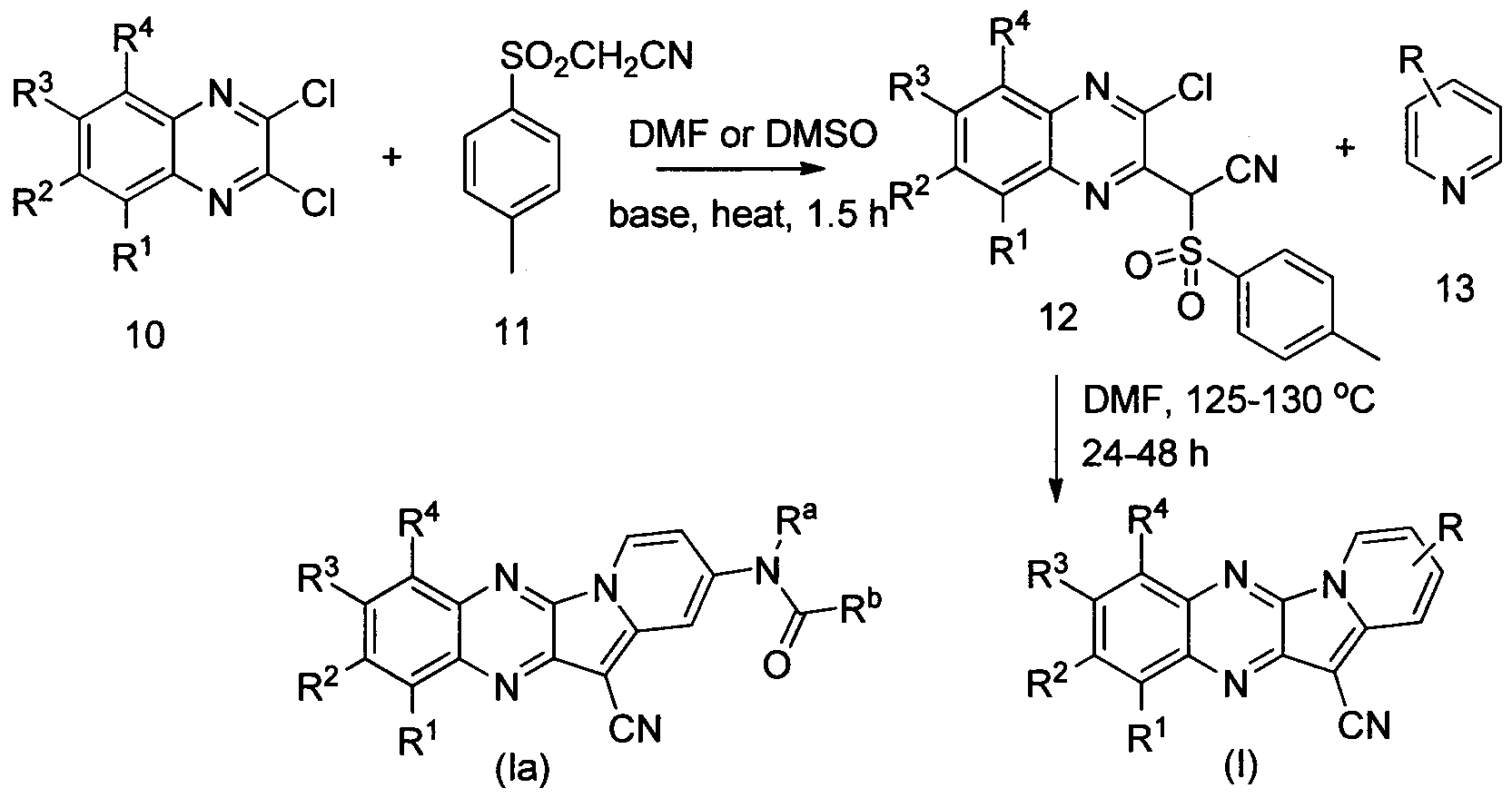
Scheme 1
In one embodiment the invention provides a method for preparing a compound of formula (I) by reacting a compound of formula 12 and a compound of formula 13 (where R = various functional groups).
A compound of formula I can also be prepared as illustrated in the following
Scheme 2.
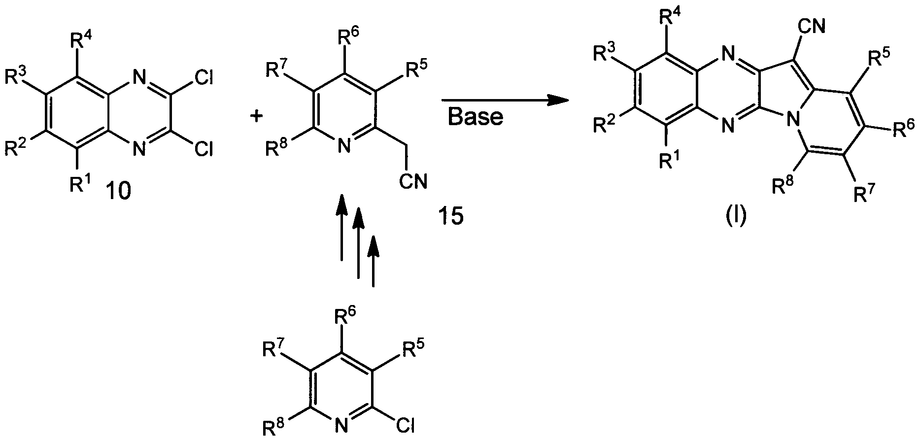
14
Scheme 2
TOPK AND HAIR GROWTH
It has been determined that inhibition of TOPK not only is effective against cancer, but inhibition of TOPK also enhances the growth of hair. This is based on the mouse phenotype observed during genetic background filtration of TOPK knockout mice into the SKH-1 hairless mouse (albino) background. Originally, the TOPK knockout mice were C57BL/6 background according to their documentation. C57BL/6, often referred to as "C57 black 6" or just "black 6" is a common inbred strain of lab mouse. It is probably the most widely used "genetic background" for genetically modified mice for use as models of human disease. They have a dark brown to black coat. However, when these mice were mated, the mice produced were black or agouti in coat color, suggesting that these mice are probably a hybrid of C57BL/6 and DBA background. The DBA mouse is the oldest of all inbred strains.
It was developed in 1909 by Little, who, around 1929, divided it into two lines: DBA/1 and DBA/2. It was introduced into The Jackson Laboratory in 1948 at the F-26th generation, and into Charles River France in February 1982 at the F- 140th generation. It has a coat color of non-agouti dilute brown.
While establishing a breeding colony of these mice homozygote TOPK male and female mice were obtained by mating; they were confirmed through genotyping by PCR and Western blotting. The mice were filtered into the SKH-1 hairless background to facilitate skin cancer UV studies. During this process, it was found that TOPK heterozygote mice grew hair even though their genetic background had been filtered to 93.75% of the SKH-1 hairless background. In contrast, the wildtype mice did not grow hair. This result strongly indicates that inhibition of TOPK might be an effective methodology to treat baldness in humans.
In cases where compounds are sufficiently basic or acidic, a salt of a compound of formula I can be useful as an intermediate for isolating or purifying a compound of formula I. Additionally, administration of a compound of formula I as a pharmaceutically acceptable acid or base salt may be appropriate. Examples of pharmaceutically acceptable salts are organic acid addition salts formed with acids which form a physiological acceptable anion, for example, tosylate, methanesulfonate, acetate, citrate, malonate, tartarate, succinate, benzoate, ascorbate, a-ketoglutarate, and a-glycerophosphate. Suitable inorganic salts may also be formed, including hydrochloride, sulfate, nitrate, bicarbonate, and carbonate salts.
Pharmaceutically acceptable salts may be obtained using standard procedures well known in the art, for example by reacting a sufficiently basic compound such as an amine with a suitable acid affording a physiologically acceptable anion. Alkali metal (for example, sodium, potassium or lithium) or alkaline earth metal (for example calcium) salts of carboxylic acids can also be made.
The compounds of formula I can be formulated as pharmaceutical compositions and administered to a mammalian host, such as a human patient in a variety of forms adapted to the chosen route of administration, i.e., orally or parenterally, by intravenous, intramuscular, topical or subcutaneous routes.
Thus, the present compounds may be systemically administered, e.g., orally, in combination with a pharmaceutically acceptable vehicle such as an inert diluent or an assimilable edible carrier. They may be enclosed in hard or soft shell gelatin capsules, may
be compressed into tablets, or may be incorporated directly with the food of the patient's diet. For oral therapeutic administration, the active compound may be combined with one or more excipients and used in the form of ingestible tablets, buccal tablets, troches, capsules, elixirs, suspensions, syrups, wafers, and the like. Such compositions and preparations should contain at least 0.1% of active compound. The percentage of the compositions and preparations may, of course, be varied and may conveniently be between about 2 to about 60% of the weight of a given unit dosage form. The amount of active compound in such therapeutically useful compositions is such that an effective dosage level will be obtained.
The tablets, troches, pills, capsules, and the like may also contain the following:
binders such as gum tragacanth, acacia, corn starch or gelatin; excipients such as dicalcium phosphate; a disintegrating agent such as corn starch, potato starch, alginic acid and the like; a lubricant such as magnesium stearate; and a sweetening agent such as sucrose, fructose, lactose or aspartame or a flavoring agent such as peppermint, oil of wintergreen, or cherry flavoring may be added. When the unit dosage form is a capsule, it may contain, in addition to materials of the above type, a liquid carrier, such as a vegetable oil or a polyethylene glycol. Various other materials may be present as coatings or to otherwise modify the physical form of the solid unit dosage form. For instance, tablets, pills, or capsules may be coated with gelatin, wax, shellac or sugar and the like. A syrup or elixir may contain the active compound, sucrose or fructose as a sweetening agent, methyl and propylparabens as preservatives, a dye and flavoring such as cherry or orange flavor. Of course, any material used in preparing any unit dosage form should be pharmaceutically acceptable and substantially non-toxic in the amounts employed. In addition, the active compound may be incorporated into sustained-release preparations and devices.
The active compound may also be administered intravenously or intraperitoneally by infusion or injection. Solutions of the active compound or its salts can be prepared in water, optionally mixed with a nontoxic surfactant. Dispersions can also be prepared in glycerol, liquid polyethylene glycols, triacetin, and mixtures thereof and in oils. Under ordinary conditions of storage and use, these preparations contain a preservative to prevent the growth of microorganisms.
The pharmaceutical dosage forms suitable for injection or infusion can include sterile aqueous solutions or dispersions or sterile powders comprising the active ingredient which
are adapted for the extemporaneous preparation of sterile injectable or infusible solutions or dispersions, optionally encapsulated in liposomes. In all cases, the ultimate dosage form should be sterile, fluid and stable under the conditions of manufacture and storage. The liquid carrier or vehicle can be a solvent or liquid dispersion medium comprising, for example, water, ethanol, a polyol (for example, glycerol, propylene glycol, liquid
polyethylene glycols, and the like), vegetable oils, nontoxic glyceryl esters, and suitable mixtures thereof. The proper fluidity can be maintained, for example, by the formation of liposomes, by the maintenance of the required particle size in the case of dispersions or by the use of surfactants. The prevention of the action of microorganisms can be brought about by various antibacterial and antifungal agents, for example, parabens, chlorobutanol, phenol, sorbic acid, thimerosal, and the like. In many cases, it will be preferable to include isotonic agents, for example, sugars, buffers or sodium chloride. Prolonged absorption of the injectable compositions can be brought about by the use in the compositions of agents delaying absorption, for example, aluminum monostearate and gelatin.
Sterile injectable solutions are prepared by incorporating the active compound in the required amount in the appropriate solvent with various of the other ingredients enumerated above, as required, followed by filter sterilization. In the case of sterile powders for the preparation of sterile injectable solutions, the preferred methods of preparation are vacuum drying and the freeze drying techniques, which yield a powder of the active ingredient plus any additional desired ingredient present in the previously sterile-filtered solutions.
For topical administration, the present compounds may be applied in pure form, i.e., when they are liquids. However, it will generally be desirable to administer them to the skin as compositions or formulations, in combination with a dermatologically acceptable carrier, which may be a solid or a liquid.
Useful solid carriers include finely divided solids such as talc, clay, microcrystalline cellulose, silica, alumina and the like. Useful liquid carriers include water, alcohols or glycols or water-alcohol/glycol blends, in which the present compounds can be dissolved or dispersed at effective levels, optionally with the aid of non-toxic surfactants. Adjuvants such as fragrances and additional antimicrobial agents can be added to optimize the properties for a given use. The resultant liquid compositions can be applied from absorbent pads, used to
impregnate bandages and other dressings, or sprayed onto the affected area using pump-type or aerosol sprayers.
Thickeners such as synthetic polymers, fatty acids, fatty acid salts and esters, fatty alcohols, modified celluloses or modified mineral materials can also be employed with liquid carriers to form spreadable pastes, gels, ointments, soaps, and the like, for application directly to the skin of the user.
Examples of useful dermatological compositions which can be used to deliver the compounds of formula I to the skin are known to the art; for example, see Jacquet et al. (U.S. Pat. No. 4,608,392), Geria (U.S. Pat. No. 4,992,478), Smith et al. (U.S. Pat. No. 4,559,157) and Wortzman (U.S. Pat. No. 4,820,508).
Useful dosages of the compounds of formula I can be determined by comparing their in vitro activity, and in vivo activity in animal models. Methods for the extrapolation of effective dosages in mice, and other animals, to humans are known to the art; for example, see U.S. Pat. No. 4,938,949.
The amount of the compound, or an active salt or derivative thereof, required for use in treatment will vary not only with the particular salt selected but also with the route of administration, the nature of the condition being treated and the age and condition of the patient and will be ultimately at the discretion of the attendant physician or clinician.
The desired dose may conveniently be presented in a single dose or as divided doses administered at appropriate intervals, for example, as two, three, four or more sub-doses per day. The sub-dose itself may be further divided, e.g., into a number of discrete loosely spaced administrations; such as multiple inhalations from an insufflator or by application of a plurality of drops into the eye.
The ability of a compound of the invention to treat cancer may be determined using pharmacological models which are well known to the art, or using the tests described in Example 1 below.
The ability of a compound of the invention to treat hair loss may be determined using pharmacological models which are well known to the art.
The invention will now be illustrated by the following non-limiting Examples.
EXAMPLES
Example 1.
Materials and Methods
Reagents
Thirty-six (36) compounds were synthesized or purchased from InterBioScreen (Moscow, Russia). Those compounds that were synthesized were prepared using modified literature procedures (see for example, W.M. Bloch et al. Tetrahedron 67 (2011) 9368 -9375; Chemistry of Heterocyclic Compounds March 1993, Volume 29, Issue 3, pp 307-312; Chemistry of Heterocyclic Compounds January 1990, Volume 26, Issue 1, pp 73-75; Chemistry of Heterocyclic Compounds November 1989, Volume 25, Issue 11, pp 1317-1318; and J. Org. Chem., 1967, 32 (1), pp 49-53 ).
Active MEK1, inactive ERK2 (MEK1 substrate), active ERK1, active JNK1, active p38, ATF2(p38 substrate), histone H2AX (TOPK substrate) human recombinant protein and MBP (Myelin Basic Protein; TOPK substrate) for kinase assays were purchased from
Millipore (Temecula, CA). The inactive N-terminal RSK2 (ERK1 substrate) and C-terminal c-Jun (JNK1 substrate) human recombinant protein for kinase assays were purified from E. Coli. The active TOPK human recombinant protein for the kinase assay was purchased from SignalChem (Richmond, BC). Antibodies to detect total TOPK, phosphorylated TOPK (T9), total CDC2, phosphorylated CDC2 (T15), total ERK, phosphorylated ERK (T202/Y204), total RSK, phosphorylated RSK (T356/S360) and caspase 7 were purchased from Cell Signaling Technology (Beverly, MA). Antibodies to detect p53, PARP and β-actin were purchased from Santa Cruz Biotechnology (Santa Cruz, CA). DNA stat-60 for genomic DNA isolation was obtained from Tel Test (Friendswood, TX).
Cell culture
All cell lines were purchased from American Type Culture Collection (ATCC) and were cytogenetically tested and authenticated before the cells were frozen. Each vial of frozen cells was thawed and maintained in culture for a maximum of 8 weeks. Enough frozen vials were available for each cell line to ensure that all cell-based experiments were conducted on cells that had been tested and in culture for 8 weeks or less. HCEC (human colonic epithelial cells) were cultured in basal media (HyClone, Logan, UT) supplemented with EGF (25
ng/mL), insulin (10 μg/mL), gentamicin sulfate (50 μg/mL) (all from Invitrogen, Grand Island, NY), transferrin (2 μg/mL), hydrocortisone (1 μg/mL), sodium selenite (5 nM) (all from Sigma, St Louis, MO) and 2% cosmic calf serum (HyClone, Logan, UT). HCT116 and HT29 human colon cancer cells were cultured in McCoy's 5 A medium supplemented with 10% fetal bovine serum (FBS; Atlanta Biologicals, Lawrenceville, GA) and 1% antibiotic- antimycotic. HCT15 and DLD1 human colon cancer cells were cultured in RPMI1640 medium supplemented with 10% FBS (Atlanta Biologicals, Lawrenceville, GA) and 1% antibiotic-antimycotic. JB6 mouse skin epidermal cells were cultured in MEM supplemented with 5% FBS (Atlanta Biologicals, Lawrenceville, GA) and 1% antibiotic-antimycotic.
TOPK-WT or KO MEFs (mouse embryonic fibroblasts) were cultured in DMEM
supplemented with 10% FBS and 1% antibiotic-antimycotic.
Lentiviral infection.
The lentiviral expression vectors, including Gipz-shTOPK and packaging vectors, including pMD2.0G and psPAX, were purchased from Addgene Inc. (Cambridge, MA). To prepare TOPK viral particles, each viral vector and packaging vectors (pMD2.0G and psPAX) were transfected into HEK293T cells using JetPEI following the manufacturer's suggested protocols. The transfection medium was changed at 4 h after transfection and then cells were cultured for 36 h. The viral particles were harvested by filtration using a 0.45 mm syringe filter, then combined with 8 μg/ml of polybrane (Millipore, Billerica, MA) and infected into 60% confluent HCT-116 cells overnight. The cell culture medium was replaced with fresh complete growth medium for 24 h and then cells were selected with puromycine (1.5 μg/ml) for 36 h. The selected cells were used for experiments.
Molecular modeling of TOPK and Compound 1.
A TOPK structure was modeled using comparative modeling. The sequence of TOPK was downloaded from NCBI (GI: 83305809) and BLAST was used to search for homologous proteins in the RCSB Protein Data Bank. Results indicated that the sequence identity between the sequences of TOPK and proteins with known structures is below 30% and the sequence similarity is about 45%. The protein structure from 2F4J (PDB entry) was selected as the template structure to model the TOPK structure. The alignment of sequences of TOPK and 2F4J was generated by BLAST and edited in Prime v3.0. The secondary structure of TOPK
was predicted by SSpro. The TOPK structure was built with Prime v3.0 followed by refining and minimizing loops in the binding site. Glide v5.7 was used for docking of TOPK and Compound 1. Compound 1 was prepared using LigPrep v2.5 and then assigned AMSOL partial atom charge. Flexible docking was performed with extra precision (XP) mode as described (17). The number of poses per ligand was set to 10 in post-docking minimization and at most 5 poses would be output. The other parameters were kept as default.
Anchorage-independent cell growth
Cells (8 x 10³ per well) suspended in complete growth medium (McCoy's 5 A, RPMI1640 or BME supplemented with 10% FBS and 1% antibiotics) were added to 0.3% agar with different doses of each compound in a top layer over a base layer of 0.6% agar with different doses of each compound. The cultures were maintained at 37°C in a 5% C02 incubator for 3 weeks and then colonies were counted under a microscope using the Image- Pro Plus software (v.4) program (Media Cybernetics).
Luciferase assay for reporter activity
Transient transfection was conducted using jetPEI (Qbiogene, Carlsbad CA), and assays for the activity of firefly luciferase and Renilla activity were performed according to the manufacturer's manual (Promega, Madison, WI). Cells (1 x 10⁴ per well) were seeded the day before transfection into 12-well culture plates. Cells were co-transfected with reporter plasmid (250 ng) and internal control {CMV-Renilla, 50 ng) in 12-well plates and incubated for 24 h. Colon cancer cells were treated with Compound 1 for 48 h. Cells were harvested in Promega Lysis Buffer. The Luciferase and Renilla activities were measured using substrates in the reporter assay system (Promega). The luciferase activity was normalized to Renilla activity.
Western blot analysis
Cell lysates were prepared with RIPA buffer (50 mM Tris-HCl pH 7.4, 1% NP-40, 0.25% sodium deoxycholate, 0.1% SDS, 150 mM NaCl, 1 mM EDTA, 1 x Protease inhibitor tablet). Equal amounts of protein were determined using the bicinchoninic acid (BCA) assay (Pierce, Rockford, IL). Proteins were separated by SDS/PAGE and transferred to
polyvinylidene difluoride membranes (Amersham Pharmacia Biotech). Membranes were blocked with 5% nonfat dry milk for 1 h at room temperature and incubated with appropriate primary antibodies overnight at 4°C. After washing with PBS containing 0.1% Tween 20, the
membrane was incubated with a horseradish peroxidase-conjugated secondary antibody at a 1 :5,000 dilution and the signal was detected with a chemiluminescence reagent (Amersham Biosciences Corp).
Cell proliferation assay
Cells were seeded (1 x 10³ cells per well) in 96- well plates and incubated for 24 hours and then treated with different doses of each compound. After incubation for 1, 2 or 3 days, 20 μΐ of CellTiter96 Aqueous One Solution (Promega) were added and then cells were incubated for 1 h at 37°C in a 5% C02 incubator. Absorbance was measured at 492 nm.
In vitro kinase assay
The kinase assay was performed in accordance with instructions provided by Upstate Biotechnology (Billenca, MA). Briefly, the reaction was carried out in the presence of 10 of [γ- P] ATP with each compound in 40 μΐ of reaction buffer containing 20 mM HEPES (pH 7.4), 10 mM MgCl2, 10 mM MnCl2, and 1 mM dithiothreitol. After incubation at room temperature for 30 min, the reaction was stopped by adding 10 μΐ protein loading buffer and the mixture was separated by sodium dodecyl sulfate-polyacrylamide gel electrophoresis (SDS-PAGE). Each experiment was repeated twice. The relative amounts of incorporated radioactivity were assessed by autoradiography.
Xenograft mouse model
Athymic mice [Cr:NIH(S), NIH Swiss nude, 6-9 wk old] were obtained from Charles River and were maintained under "specific pathogen-free" conditions based on the guidelines established by the University of Minnesota Institutional Animal Care and Use Committee. Mice were divided into five groups: 1) untreated vehicle group (n = 10); 2) 1 mg Compound 1/kg of body weight (n = 10); 3) 5 mg Compound 1/kg body weight (n = 10); 4) 10 mg Compound 1/kg of body weight (n = 10); and 5) no cells and 10 mg Compound 1 kg of body weight (n = 10). HCT116 cells (1.5xl06 cells/100 μΐ) were suspended in serum free McCoy's 5 A medium and inoculated subcutaneously into the right flank of each mouse. Compound 1 or vehicle was injected 3 times per week for 25 days. Tumor volume was calculated from measurements of 2 diameters of the individual tumor base using the following formula: tumor volume (mm ) = (length x width x height x 0.52). Mice were monitored until tumors reached 1 cm3 total volume, at which time mice were euthanized and tumors were extracted.
Statistical analysis
All quantitative results are expressed as mean values ± S.D. Statistically significant differences were obtained using the Student's t test or by one-way ANOVA. A p < 0.05 was considered to be statistically significant.
Results
Knocking down TOPK expression inhibits cell proliferation. Using lentiviral infection, HCT116 colon cancer cells stably expressing shMock, low levels of TOPK (#2 shTOPK) or medium levels of TOPK (#5 shTOPK) were established. The abundance of TOPK in these cells was confirmed by Western blot analysis (Fig. 1 A). The effect of knocking down TOPK on proliferation was then assessed by MTS assay at 1, 2 or 3 days. Results indicated that cell growth was significantly decreased in a manner dependent on the level of TOPK expression (Fig. IB). Proliferation was also decreased in TOPK knockout MEFs (mouse embryonic fibroblasts) compared to wildtype MEFs (Fig. 1C). Additionally, the effect of knocking down TOPK expression on anchorage-independent colon cancer cell growth was assessed. These results showed that anchorage-independent cell growth was strongly inhibited by knocking down the expression of TOPK and the inhibition was dependent on the level of TOPK expression (Fig. ID).
Compound 1 directly suppresses TOPK kinase activity. Thirty-six compounds were screened (30 μΜ concentration) by in vitro TOPK kinase assay (Fig. 16 A) and by cell proliferation assay (4 or 20 μΜ concentration) (Fig. 16B). Six compounds were selected to test for inhibition of TOPK (10 μΜ concentration) (Fig. 16C), Based on screening results, Compound 1 (Fig. 2A) was identified as a potent TOPK inhibitor. To determine the effect of Compound 1 on TOPK or MEK1 kinase activity, an in vitro TOPK or MEK1 kinase assay was performed with increasing concentrations of Compound 1. Compound 1 effectively inhibited TOPK kinase activity. However, Compound 1 at the highest concentration (5 μΜ) also inhibited MEK1 activity by 40% (Fig. 2B). Thus, this result showed that Compound 1 is a more potent inhibitor against TOPK compared to MEK1. Additionally, in vitro kinase assays were used to investigate the effect of Compound 1 on other MAP kinase family members, such as ERKl, JNK1 and p38 (Fig. 2C). These results showed that Compound 1 suppressed TOPK kinase activity, but not ERKl, JNK1 or p38 activity.
Computer modeling of the TOPK and Compound 1 complex. Molecular docking of the Compound 1 and TOPK complex was performed in order to determine the binding orientation of Compound 1. Compound 1 occupies the ATP-binding site of TOPK (Fig. 3 A, B) and fits the binding site very well (Fig. 3C). The compound forms hydrogen bonds with GLY83 and ASP151 and has a hydrophobic interaction with LYS30 (Fig. 3D). These results suggest that Compound 1 binds to the TOPK active site.
Compound 1 inhibits colon cancer cell growth and induces apoptosis. To determine whether total and phosphoiylated TOPK are highly expressed in normal colon or colon cancer cells, Western blot analysis was performed. Results showed that HCT116 colon cancer cells highly expressed phosphoiylated TOPK compared with other colon cancer cells (Fig. 17A) and these cells were used in further studies. To examine the effect of Compound 1 on colon cancer cell proliferation, growth was measured using the MTS assay at 1, 2 or 3 days after treatment with Compound 1. Results indicated that colon cancer cell growth was significantly decreased by Compound 1 in a dose-dependent manner (Fig. 4A). Additionally, the effect of Compound 1 on anchorage-independent cell growth was determined. Colon cancer cells were seeded with Compound 1 in 0.3% agar and incubated for 3 weeks. Data showed that anchorage-independent cancer cell growth was strongly suppressed by Compound 1 in a dose- dependent manner (Fig. 4B). Next, to examine the effect of Compound 1 on apoptosis, HCT116 colon cancer cells were treated with Compound 1 and then incubated for 3 days. Results showed that DNA fragmentation induced by Compound 1 was substantially increased in HCT116 colon cancer cells compared with untreated control cells (Fig. 4C). Previous studies suggested that overexpressing TOPK can induce neoplastic cell transformation.
Therefore, the effect of Compound 1 on transformation of JB6 cells stably overexpressing TOPK was determined. Results indicated that overexpressing TOPK-mediated cell transformation was significantly suppressed by Compound 1 (Fig. 18A). Next the effect of Compound 1 on downstream targets of TOPK in HCT116 colon cancer cells was
investigated. Cells were treated with Compound 1 for 24 h and then cell lysates were examined by Western blot. In previous studies, TOPK was shown to inhibit the p53 signaling pathway, involved the G2/M transition, and activated the ERK signaling pathway. Therefore, inhibition of TOPK kinase activity by a TOPK inhibitor should be able to induce abundance
of the p53 protein and its downstream target proteins, G2/M phase marker proteins (i.e., CDC2 phosphorylation) and reduce ERKs phosphorylation. Our results showed that the expression of p53, cleaved caspase7, cleaved PARP and phosphorylated CDC2 were all strongly increased by Compound 1 and phosphorylated ERK and RSK were remarkably decreased by Compound 1 (Fig. 4D). However, the expression of total TOPK was not changed. It was determined whether Compound 1 affected the reporter activity of the activator protein- 1 (AP-1), nuclear factor-kappaB (NF-κΒ) or cyclooxygenase 2 (COX2) in HCT116 or HCT15 cells. Cells were treated with Compound 1 for 48 h and then reporter activity was measured. Results show that the reporter activity of these genes was strongly inhibited by Compound 1 (Figs. 19A, 19B, 19C).
The inhibition of TOPK by Compound 1 is dependent on the abundance of TOPK. The effect of Compound 1 on growth of shMock, #3 shTOPK (medium expression of TOPK) or #2 shTOPK (low expression of TOPK) colon cancer cells was assessed by MTS assay at 72 h. Results indicated that cells expressing shTOPK were resistant to Compound l's inhibitory effect on growth compared to cells expressing shMock (Fig. 5 A). Similar resistance to Compound 1 was observed in TOPK knockout MEFs (Fig. 5B, C). Additionally, the effect of Compound 1 on anchorage-independent colon cancer cell growth was examined. Results showed that the inhibitory effect of Compound 1 on anchorage-independent cell growth in shTOPK cells was much less than its effect on shMock growth (Fig. 5D). These findings showed that the anticancer activity induced by Compound 1 is dependent on TOPK protein expression.
Compound 1 inhibits colon cancer tumor growth in a xenograft mouse model. To determine the antitumor activity of Compound 1 in vivo, HCT116 colon cancer cells were injected into the flank of athymic nude mice. Mice were injected with vehicle or Compound 1 at 1 or 10 mg/kg 3 times a week over a period of 25 days. Treatment of mice with 1 or 10 mg/kg of Compound 1 significantly inhibited HCT116 tumor growth by over 60% relative to the vehicle-treated group (Figs. 20A, Fig. 20B; p < 0.05). Additionally, mice seemed to tolerate treatment with Compound 1 without overt signs of toxicity or significant loss of body weight similar to the vehicle-treated group (Fig. 6B). To validate the results of the in vivo xenograft model, the effect of Compound 1 on downstream targets of TOPK by Western blot
analysis of HCT116 colon tumor samples was investigated. The expression of p53 was strongly induced and phosphorylation of ERK and RSK, a direct downstream protein of ERK, was markedly inhibited in the Compound 1 -treated group (Fig. 6C). This finding suggested that Compound 1 inhibits HCT116 colon tumor growth through inhibition of TOPK in vivo. Inhibition of TOPK by Compound 1 might regulate multiple effects such as tumor
development, cancer growth, apoptosis and inflammation mediated through the API or p53 signaling pathway (Fig. 6D).
Discussion
Many groups have reported that the serine/threonine kinase TOPK may contribute to oncogenic cellular functions including tumor development, cancer growth and anti-apoptosis effects. These reports suggested that TOPK is a potential target for development of anticancer agents. Despite these findings, a TOPK inhibitor has not yet been found, possibly because the TOPK crystal structure has not yet been reported. To develop a reliable homology model for TOPK, a sequence-based homology search was performed. It was found that the sequences of TOPK and MEKs are highly conserved. Based on this sequence homology, the effect of U0126, a well-known inhibitor of MEK, on the in vitro TOPK kinase activity was tested. U0126 inhibited TOPK activity by 25% at the highest concentration (20 μΜ; data not shown). Based on these preliminary data, 36 compounds with a similar structure to a MEK inhibitor were selected and Compound 1 was identified as a potent TOPK inhibitor. In addition, a homology model based on the known structure of MEK was built and a docking simulation between Compound 1 and the modeled TOPK protein was performed. Compound 1 was docked to the active site of TOPK (Fig. 3). The inhibitory effect of various Compound 1 analogues on TOPK kinase activity in vitro were then compared. However, none of the analogues had any effect (data not shown). Various analogues of Compound 1 with certain portions deleted, were synthesized but none of these compounds were effective inhibitors of TOPK kinase activity (data not shown). These findings suggested that the complete structure of Compound 1 is important for effectively inhibiting TOPK kinase activity.
Previous studies showed that TOPK directly interacts with the DBD domain of tumor suppressor p53. TOPK's downstream target, tumor suppressor activated pathway-6 (TSAP6,) reportedly binds to myelin transcription factor 1 (MYT1), which induces phosphorylation of
CDC2 (Tyrl5). Whether p53 expression or phosphorylation of CDC2 (Tyrl5) is affected by Compound 1 was investigated. These results indicated that the abundance of total p53 and phosphorylation of CDC2 (Tyrl 5) in colon cancer cells were both strongly increased by Compound 1 treatment and the phosphorylation of ERK, a direct downstream target of TOPK, was dramatically inhibited by Compound 1 (Fig. 4D). Notably, total p53 expression was highly induced and phosphorylation of ERK-RSK was inhibited in vivo in Compound 1- treated tumor tissues (Fig. 6D). These results strongly indicate that the inhibitory effect of Compound 1 in cell-based assays corresponds closely with the in vivo animal results.
In conclusion, Compound 1 is a novel and specific TOPK inhibitor both in vitro and in vivo. These findings should be useful for further development of drugs targeted against TOPK. Additionally Compound 1 may possess therapeutic potential against colorectal cancer and other human cancers.
Example 2
Two isoenzymes of 5-alpha reductase are found in the human genome, type 1 and type 2. The main action of both isoenzymes is the conversion of testosterone into the more potent androgen dihydrotestosterone (DHT). SRD5A2 is the gene that codes for the protein product that is responsible for the pathogenesis of male pattern baldness. In the presence of abundant DHT, the hair follicles become miniaturized; resulting in fine, short hair that is prone to fall out (Kaufman KD., Mol Cell Endocrinol, 2002, 89-95). Much classical evidence pinpoints the direct involvement of DHT in male pattern baldness. Studies of
pseudohermaphrodites (lacking 5 AR) showed protection from male pattern baldness throughout life. The introduction of exogenous testosterone into castrated males can induce typical patterns of hair loss. Finally, inhibitors of the enzyme SRD5A2 have slowed the rate of hair loss, indicating its direct involvement (Nyholt DR, et al., J Invest Dermatol. 2003, 121, 1561).
The phosphorylation of SRD5A2 by TOPK was assessed by an in vitro kinase assay using TOPK (active, 500 ng) and GST tagged full-length SRD5A2 with [γ-32Ρ]ΑΤΡ. The TOPK inhibitor was used a negative control. Results are shown in Figure 7.
A TOPK inhibitor (Compound 1, 400 nmol) in acetone was topically applied 3 times weekly until the termination of the experiment at 16 weeks. The number of inner or outer hair
pores was counted after H&E staining. Data are shown in Figure 8 as means ± S.E. of values (n > 5) and the asterisk (*) indicates a significant difference (p < 0.05) between TOP inhibitor treated groups compared to the vehicle treated group.
Example 3.
Materials and Methods
Reagents
CInQ-01 (N-( 12-cyanindolizino [2,3 -b] quinoxalin-3 -yl)-4-fluorobenzamide, purity: 95%), CInQ-02 (N-(12-cyanindolizino[2,3-b]quinoxalin-3-yl)-2-thiophenecaboxamide, purity: 95%), CInQ-03 (2-chloro-N-(12-cyanindolizino[2,3-b]quinoxalin-2-yl)benzamide, purity: 95%), CInQ-04 (N-(12-cyanindolizino[2,3-b]quinoxalin-2-yl)-4-methylbenzene- sulfonamide, purity: 95%), CInQ-05 (2-(l,l-dimethylethyl)-indolizino[2,3-b]quinoxaline-12- carbonitrile, purity: 95%) and CInQ-06 (N-(12-cyanindolizino[2,3-b]quinoxalin-2-yl)-4- fluoro-benzamide, purity: 95%) were purchased from InterBio Screen (Moscow, Russia), or were prepared using modified literature procedures (see for example, W.M. Bloch et al. Tetrahedron 67 (2011) 9368 -9375; Chemistry of Heterocyclic Compounds March 1993, Volume 29, Issue 3, pp 307-312; Chemistry of Heterocyclic Compounds January 1990, Volume 26, Issue 1, pp 73-75; Chemistry of Heterocyclic Compounds November 1989, Volume 25, Issue 1 1, pp 1317-1318; and J. Org. Chem., 1967, 32 (1), pp 49-53). Active MEK1, inactive ERK2 (MEK substrate) and histone H2AX (TOPK substrate) human recombinant proteins for kinase assays were purchased from Millipore (Temecula, CA). Active MEK2 and active TOPK human recombinant proteins for kinase assays were purchased from SignalChem (Richmond, BC). Antibodies to detect total MEK,
phosphorylated MEK, total ERKs, phosphorylated ERKs, total RSK and phosphorylated RSK were purchased from Cell Signaling Technology (Beverly, MA). Antibodies against total MEK1, total MEK2 and β-actin were purchased from Santa Cruz Biotechnology (Santa Cruz, CA).
Cell culture
All cell lines were purchased from American Type Culture Collection (ATCC) and were cytogenetically tested and authenticated before the cells were frozen. Each vial of frozen cells was thawed and maintained in culture for a maximum of 8 weeks. Enough frozen vials were available for each cell line to ensure that all cell-based experiments were conducted on
cells that had been tested and in culture for 8 weeks or less. HCT116 human colon cancer cells were cultured in McCoy's 5 A medium supplemented with 10% fetal bovine serum (FBS; Atlanta Biologicals, Lawrenceville, GA) and 1% antibiotic-antimycotic. HCT15 human colon cancer cells were cultured in RPMI1640 medium supplemented with 10% FBS (Atlanta Biologicals) and 1% antibiotic-antimycotic. HaCaT (human keratinocyte) cells were cultured in DMEM medium supplemented with 10% FBS (Atlanta Biologicals) and 1% antibiotic- antimycotic.
Molecular modeling
The crystal structures of MEK1 and MEK2 were obtained from the RCSB Protein Data Bank [PDB entry: lS9J and 1S9I (Ohren JF, et al., Nat Struct Mol Biol. 2004, 11, 1192- 7)]. The crystal structures were prepared using the Protein Preparation Wizard in Maestro v9.2. Hydrogens were added consistent with a pH of 7. All water molecules were removed and then the structure was minimized with an RMSD cutoff value of 0.3 A. Three compounds were prepared using LigPrep v2.5 and then assigned AMSOL partial atom charge. The program Glide v5.7 (Friesner RA, et al., J Med Chem. 2006, 49, 6177-96) was used for ligand docking. The receptor grid was created with the centroid of the crystal ligand as the center of the grid. Flexible Docking was performed with extra precision (XP) mode. The number of poses per ligand was set to 10 in post-docking minimization and at most 5 poses would be output. The other parameters were kept as default.
Lentiviral infection.
The lentiviral expression vectors, including Gipz-shMEKl or shMEK2 and packaging vectors, including pMD2.0G and psPAX, were purchased from Addgene Inc. (Cambridge, MA). To prepare MEK1/2 viral particles, each viral vector and packaging vectors (pMD2.0G and psPAX) were transfected into HEK293T cells using JetPEI following the manufacturer's suggested protocols. The transfection medium was changed at 4 h after transfection and then cells were cultured for 36 h. The viral particles were harvested by filtration using a 0.45 mm sodium acetate syringe filter, then combined with 8 μg/ml of polybrane (Millipore, Billerica, MA) and infected into 60% confluent HCT116 cells overnight. The cell culture medium was replaced with fresh complete growth medium for 24 h and then cells were selected with puromycine for 36 h (1.5 μg/ml of puromycine). The selected cells were used for
experiments.
Lucif erase assay for API reporter activity
Transient transfection was conducted using jetPEI (Qbiogene, Carlsbad CA), and assays to determine firefly luciferase and Renilla activities were performed according to the manufacturer's manual (Promega, Madison, WI). Cells (1 x 10⁴ per well) were seeded the day before transfection into 12-well culture plates. Cells were co-transfected with the AP-1 reporter plasmid (250 ng) and an internal control (CMV-Renilla, 50 ng) in 12-well plates and incubated for 24 h. Colon cancer cells were treated with individual CInQ inhibitors or U0126 for 2 days. Keratinocytes were treated with CInQ inhibitors or U0126 for 2 h before EGF treatment for 12 or 24 h. Cells were harvested in Promega Lysis Buffer. The AP-1 and Renilla luciferase activities were measured using substrates provided in the reporter assay system (Promega). The luciferase activity was normalized to Renilla luciferase activity.
Anchorage-independent cell growth
Cells (8 x 10³ per well) suspended in complete growth medium (McCoy's 5 A, RPMI1640 or DMEM supplemented with 10% FBS and 1% antibiotics) were added to 0.3% agar with EGF alone or with different doses of each compound in a top layer over a base layer of 0.6% agar with EGF alone or with different doses of each compound. The cultures were maintained at 37°C in a 5% C02 incubator for 3 weeks and then colonies were counted under a microscope using the Image-Pro Plus software (v.4) program (Media Cybernetics).
Western blot analysis
Cell lysates were prepared with RIP A buffer (50 mM Tris-HCl pH 7.4, 1% NP-40, 0.25% sodium deoxycholate, 0.1% SDS, 150 mM NaCl, 1 mM EDTA, 1 x protease inhibitor tablet). Equal amounts of protein were determined using the bicinchoninic acid (BCA) assay (Pierce, Rockford, IL). Proteins were separated by SDS/PAGE and transferred to
polyvinylidene difluoride membranes (Amersham Pharmacia Biotech). Membranes were blocked with 5% nonfat dry milk for 1 h at room temperature and incubated with appropriate primary antibodies overnight at 4°C. After washing with PBS containing 0.1% Tween 20, the membrane was incubated with a horseradish peroxidase-conjugated secondary antibody at a 1 :5,000 dilution and the signal was detected with a chemiluminescence reagent (Amersham Biosciences Corp).
Cell proliferation assay
Cells were seeded (1 x 10 cells per well) in 96-well plates and incubated for 24 h and then treated with different doses of each compound. After incubation for 1, 2 or 3 days, 20 μΐ of CellTiter96 Aqueous One Solution (Promega) were added and then cells were incubated for 1 h at 37°C in a 5% C02 incubator. Absorbance was measured at 492 nm.
In vitro kinase assay
The kinase assay was performed in accordance with instructions provided by Upstate Biotechnology (Billerica, MA). Briefly, the reaction was carried out in the presence of 10 μϋι' of [γ- P] ATP with each compound in 40 μΐ of reaction buffer containing 20 mM HEPES (pH 7.4), 10 mM MgCl2, 10 mM MnCl2, and 1 mM dithiothreitol. After incubation at room temperature for 30 min, the reaction was stopped by adding 10 μΐ protein loading buffer and the mixture was separated by sodium dodecyl sulfate-polyacrylamide gel electrophoresis (SDS-PAGE). Each experiment was repeated twice and the relative amounts of incorporated radioactivity were assessed by autoradiography.
Hematoxylin-eosin staining and immunohistochemistry
Tumor and skin tissues from mice were embedded in paraffin blocks and subjected to hematoxylin and eosin (H&E) staining and immunohistochemistry. Tumor tissues were de- paraffinized and hydrated then permeabilized with 0.5% Triton X-100/1 x PBS for 10 min. They were then hybridized with Ki-67 (1 :500) as the primary antibody and horse-radish peroxidase (HRP)-conjugated goat anti-rabbit or mouse IgG antibody was used as the secondary antibody. After developing with 3, 3'-diaminobenzidine, the sections were counterstained with H&E. All sections were observed by microscope and the Image-Pro Plus software (v. 4) program (Media Cybernetics).
Xenograft mouse model
Athymic mice [Cr:NIH(S), NIH Swiss nude, 6-9 wk old] were obtained from Charles River and maintained under "specific pathogen-free" conditions based on the guidelines established by the University of Minnesota Institutional Animal Care and Use Committee. Mice were divided into four groups: 1) untreated vehicle group (n = 10); 2) 1 mg CInQ-03/kg of body weight (n = 10), 3) 5 mg CInQ-03/kg body weight (n = 10); and 4) no cells and 5 mg CInQ-03/kg of body weight (n = 10). HCT116 cells (1.5xl06 cells/100 μΐ) were suspended in serum free McCoy's 5 A medium and inoculated subcutaneously into the right flank of each
mouse. CInQ-03 or vehicle was injected 3 times per week for 11 days. Tumor volume was calculated from measurements of 2 diameters of the individual tumor base using the following formula: tumor volume (mm3) = (length x width x height x 0.52). Mice were monitored until tumors reached 1 cm3 total volume, at which time mice were euthanized and tumors extracted.
Statistical analysis
All quantitative results are expressed as mean values ± S.D. Statistically significant differences were obtained using the Student's t test or by one-way ANOVA. A p < 0.05 was considered to be statistically significant.
Results
ClnQ-inhibitors suppress MEKl and MEK2 kinase activities. CInQ-01 to -06 compounds (30 μΜ concentration) were screened using an in vitro MEKl kinase assay and results showed that MEKl kinase activity was strongly inhibited by CInQ-01, -03 or -06. These data suggested that CInQ-01, -03 and -06 are potent MEK inhibitors (Fig. 9 A and 9B). Therefore these three compounds (5 and 10 μΜ) were selected and were evaluated further by in vitro MEKl and MEK2 kinase assays. Results showed that CInQ-01, -03 and -06 significantly suppressed MEKl and MEK2 kinase activities in a dose dependent manner (Fig. 9B and 9C). Next, to determine whether CInQ-01, -03 or -06 affected the kinase activity of TOPK, a MAPKK family member, an in vitro TOPK kinase assay was performed with the ClnQ- inhibitors. Results indicated that CInQ-01 and -03 had little effect of TOPK kinase activity (Fig. 10) and CInQ-06 suppressed TOPK activity only by about 20%. These results suggested that CInQ-01, -03 and -06 are specific and potent MEK inhibitors.
Computer modeling of the MEK and CInQ-inhibitor complexes. Molecular docking of the individual ClnQ-inhibitors and MEK in order to determine the binding orientation of the inhibitors with MEK was performed. In the docked structures of MEKl with the individual ClnQ-inhibitors, all compounds formed hydrogen bonds with LYS97 and SER212 and the docked poses overlap very well with the crystal ligand PD318088 (Fig. 10A). In the crystal structure of MEKl, PD318088 also forms a hydrogen bond with LYS97. The docking score of MEKl with PD318088 is -9.45, and the docking scores of MEKl with CInQ-01, -03 and - 06 are -7.84, -7.29 and -7.83, respectively. Additionally, in the docked structure of MEK2 with the ClnQ-inhibitors, all compounds formed a hydrogen bond with Ser216 (Fig. 10B).
CInQ-03 forms an additional hydrogen bond with LyslOl, which also forms a hydrogen bond with PD334581 in the crystal structure of MEK2. The docking score of MEK2 with
PD334581 is -8.92, and the docking scores of MEK2 with CInQ-01, -03 and -06 are -8.54, -6.49, and -8.58, respectively.
CInQ-inhibitors suppress anchorage-dependent and -independent cell growth. The effect of the CInQ-inhibitors on colon cancer cell growth and on the growth of EGF-induced HaCaT keratinocytes was examined. Cell growth was measured using the MTS assay at 1, 2 or 3 days after treatment with EGF alone or EGF and individual CInQ-inhibitors. Results indicated that colon cancer cell growth was significantly decreased by the respective CInQ- inhibitors (Fig. 11 A). EGF-induced cell growth was also strongly suppressed by CInQ- inhibitors (Fig. 11B).
Next, to determine the effect of the CInQ-inhibitors on anchorage-independent growth and EGF-induced transformation, cells were seeded with EGF alone or with individual CInQ-inhibitors in 0.3% agar and incubated for 2 or 3 weeks. Data showed that anchorage-independent cancer cell growth was strongly suppressed by CInQ-inhibitors in a dose- dependent manner (Fig. 11C). EGF-induced cell transformation was also significantly inhibited by the respective CInQ-inhibitors (Fig. 1 ID).
CInQ-inhibitors suppress activator protein-1 (AP-1) activity. It was then determined whether CInQ-inhibitors had an effect on activator protein-1 (AP-1) reporter activity in HCT116 cells or HaCaT cells stimulated with EGF. HCT116 cells were treated with individual CInQ-inhibitors for 48 h and HaCaT cells were pre-treated with CInQ-inhibitor for 2 h before stimulation with EGF for 12 or 24 h. AP-1 reporter activity was strongly suppressed by CInQ-inhibitors in colon cancer cells (Fig. 12 A) and in EGF-treated HaCaT cells (Fig. 12B). The effect of these inhibitors on downstream signaling of MEK in colon cancer cells and EGF-induced HaCaT cells was also examined. Results showed that each CInQ-inhibitor suppressed phosphorylation of ERKl/2 as well as phosphorylation of RSK (Fig. 12C and 12D). However, the CInQ-inhibitors had little effect on the phosphorylation of MEK.
The anti-cancer effects of CInQ-inhibitors are dependent on MEK expression. To further study the anti-cancer effects of the CInQ-inhibitors HCT116 cells stably expressing mock (shGipz) or knockdown of MEK 1/2 (shMEKl/2) were established and analyzed MEK
expression by Western blot (Fig. 23 A, 23, and 13 A). The effect of inhibitors on growth of shGipz or shMEKl/2 colon cancer cells was assessed by MTS assay at 72 h. Results indicated that cells expressing shMEKl/2 were resistant to the anti-growth effect of the CInQ-inhibitors compared to s/iG/pz-expressing cells (Fig. 13B). Furthermore, the effect of CInQ inhibitors on anchorage-independent colon cancer cell growth was examined. Results showed that the inhibition of anchorage-independent colon cancer cell growth induced by the CInQ-inhibitors was less effective in shMEK cells compared to shGipz cells (Fig. 13C). These results indicated that the anti-cancer activity induced by CInQ inhibitors is dependent on MEK expression.
CInQ-03 inhibits colon cancer tumor growth in a xenograft mouse model. Based on the results described above, CInQ-03 was selected for further study in vivo. HCT116 colon cancer cells were injected into the flank of athymic nude mice and mice were treated with CInQ-03 at 1 or 5 mg/kg or vehicle 3 times a week over a period of 11 days after the average tumor volume grew to about 70 mm . Treatment of mice with 1 or 5 mg/kg of CInQ-03 strongly suppressed HCT116 tumor growth by over 70% relative to the vehicle-treated group (Fig. 14A, 14B, left panel; p < 0.05). Additionally, mice seemed to tolerate treatment with CInQ-03 without overt signs of toxicity or significant loss of body weight similar to the vehicle-treated group (Fig. 14B, right panel). Furthermore, the effects of CInQ-03 on a tumor proliferation marker were evaluated by immunohistochemistry and H&E staining of HCT116 tumor and skin tissues after 11 days of treatment. The expression of Ki67 was markedly decreased by treatment with CInQ-03 (Fig. 14C, upper panel). However, Ki67 expression in skin tissues in CInQ-03 treated tissues was similar to the vehicle-treated group (Fig. 14C, lower panel).
The effect of CInQ-03 on MEK downstream signaling in tumor tissues was then examined. Expression of MEK-targeted proteins was analyzed by Western blot. Results indicated that phosphorylation of ERKs and RSK was strongly suppressed by CInQ-03 treatment (Fig. 15). These findings indicated that HCT116 colon tumor growth was suppressed by CInQ-03 through its targeting of the MEK signaling pathway.
Discussion
The anti-cancer activities of a highly selective non-ATP competitive inhibitor of MEK1/2 have been reported in in vitro and in vivo studies (Davies BR, et al., Mol Cancer Ther. 2007, 6, 2209-19; and Lorusso PM, et al, J Clin Oncol. 2005, 23, 5281-93; Menon U,
et al, J Clin Oncol. 2005, 23, 7919-26; Daouti S, et al., Cancer Res. 2009, 69, 1924-32; Iverson C, et al., Cancer Res. 2009, 69, 6839-47; and Yang JY, et al, Cancer Res. 2010, 70, 4709-18). Recently, phase I and II clinical evaluation of several MEK inhibitors, including PD0325901, AZD6244 and XL518 that share a common core structure, have been conducted (Adjei AA, et al., J Clin Oncol. 2008, 26, 2139-46; Wang D, et al., Biochim Biophys Acta. 2007, 1773, 1248-55; and Wang JY, et al., Curr Top Med Chem. 2007, 7, 1364-78). Despite many efforts to identify MEK inhibitors and provide effective preclinical results using second generation MEK inhibitors, which are currently in clinical trials for various solid
malignancies, none has yet been approved. Current inhibitors are associated with diarrhea and dermatologic toxicities including rash (Schad K, et al., Clin Cancer Res. 2010;16:1058-64; and Wang D, et al., Biochim Biophys Acta. 2007;1773:1248-55). Although subtle structural diversities suggest biological differences, a similar toxicity by these MEK inhibitors have been observed. Therefore, there remains a need to identify MEK inhibitors that are structurally different from current MEK inhibitors. Expression of Ki-67 was strongly inhibited by treatment with CInQ-03 in tumor tissues (Fig. 14C, upper panel). I n contrast, Ki- 67 expression in the basal layer and the suprabasal keratinocyte layers of skin tissues treated with CInQ-03 was similar to the vehicle-treated group (Fig. 14C, lower panel). Thus, the CInQ inhibitors may provide clinical advantage such as reduced toxicities compared with current MEK inhibitors.
In this study, to identify a specific MEK inhibitor, the effect of candidate compounds on MEK family kinase activities by using in vitro MEK and TOPK kinase assays was examined. Certain CInQ inhibitors were found to be are specific and potent MEK inhibitors in in vitro and cell-based assays. Furthermore, results from a xenograft mouse model indicated that administration of CInQ-03 at 1 or 5 mg/kg B.W. for 11 days significantly suppressed colon cancer cell growth and was not toxic (Fig. 14B). Furthermore, suppression of phosphorylation of ERKs by CInQ-03 in a cell-based assay was highly correlated with the in vivo animal results (Fig. 12C, D; Fig. 15).
Based on these findings, CInQ-03 is a novel and specific MEK inhibitor both in vitro and in vivo. These results should be useful for development of novel MEK inhibitors. Future studies will investigate the efficacy of CInQ-03 and pharmacological characterization and examination of dermatologic toxicities.
Example 4. Preparation of Compound 102:

A solution of the compound 101 (357.8 mg, 1 mmol) and N-(pyridin-4-yl)benzamide (594.6 mg, 3 mmol) in dimethylformamide (5 mL) was heated at 125-130 °C for 48 hours. After this time, the reaction mixture was diluted with water. The obtained red solid was filtered, washed with sodium bicarbonate solution and water. The compound was purified by recrystallization (ethanol) followed by the preparative TLC (DCM-Methanol) provided the title compound. HRMS (ESI): found 364.1090; calculated (for M+l) 364.1120.
The intermediate compound 101 was prepared as follows,
a. Preparation of compound 10

To a magnetically stirred solution of P-toluenesulfonyl cyanide (981 mg, 5.02 mmol) and cesium carbonate (1.64 g, 5.02 mmol) in anhydrous DMSO (15 mL) was added 2, 3- dichloroquinoxaline (1 g, 5.02 mmol) under nitrogen atmosphere. The resulting mixture was stirred at 150 °C for 1.5 hours. The reaction mixture was then cooled to room temperature and diluted with water. The aqueous solution was acidified with glacial acetic acid and the resulting solid 101 was filtered and used without further purification.
Example 5. Preparation of Compound 103:

The title compound was obtained from the compound 101 (357.8 mg, 1 mmol) and 4- acetylaminopyridine (408.4 mg, 3 mmol) by a procedure similar to that described in Example 4. HRMS (ESI): found 302.0960; calculated (for M+1) 302.0964.
Example 6. Preparation of compound 104 (Compound 1 or HI-TOPK-032):

104
The title compound was obtained from the compound 101 (1.0 g, 2.8 mmol) and N- (pyridin-4-yl)thiophene-2-carboxamide (1.71 g, 8.4 mmol) by a procedure similar to that described in Example 4. HRMS (ESI): found 370.1477; calculated (for M+1) 370.0684. Example 7. Preparation of Compound 105 (CInQ-03):

105
The title compound was obtained from the compound 101 (1.0 g, 2.8 mmol) and 2- chloro-N-(pyridin-4-yl)benzamide (1.96 g, 8.4 mmol) by a procedure similar to that described in Example 4. HRMS (ESI): found 398.0809; calculated (for M+1) 398.0730.
Example 8. Preparation of Compound 106:

To a stirred solution of 3-chloro-5-trifluoromethyl pyridine-2-acetonitrile (220.6 mg, 1 mmol) in DMSO (5 mL) was added cesium carbonate (325.8 mg, 1 mmol). The reaction mixture was stirred for 10 min and then 2, 3-dichloroquinoxaline (199 mg, 1 mmol) was added portion wise. The resulting mixture was stirred at 65 °C for 12 hours. After this time the reaction mixture was cooled to room temperature and diluted with water. The aqueous solution was acidified with glacial acetic acid. The obtained red solid was filtered, washed with water, dried and purified by recrystallization from ethanol to provide compound 104. HRMS (ESI): found 347.0208; calculated (for M+l) 347.0233.
Example 9. Preparation of Compound 107:

This compound was prepared according to the method described in Example 6, employing pyridine-2-acetonitrile (118 mg, 1 mmol) and 2, 3-dichloroquinoxaline (199 mg, 1 mmol).
Example 10. The following illustrate representative pharmaceutical dosage forms, containing a compound of formula I ('Compound X'), for therapeutic or prophylactic use in humans.
(i) Tablet 1 mg/tablet
Compound X= 100.0
Lactose 77.5
Povidone 15.0
Croscarmellose sodium 12.0
Microcrystalline cellulose 92.5
Magnesium stearate 3.0
300.0
(ii) Tablet 2 mg/tablet
Compound X= 20.0
Microcrystalline cellulose 410.0
Starch 50.0
Sodium starch glycolate 15.0
Magnesium stearate 5.0
500.0
(iii) Capsule mg/capsule
Compound X= 10.0
Colloidal silicon dioxide 1.5
Lactose 465.5
Pregelatinized starch 120.0
Magnesium stearate 3
600.0
(iv) Injection 1 (1 mg/ml) mg/ml
Compound X= (free acid form) 1.0
Dibasic sodium phosphate 12.0
Monobasic sodium phosphate 0.7
Sodium chloride 4.5
1.0 N Sodium hydroxide solution
(pH adjustment to 7.0-7.5) q.s.
Water for injection q.s. ad 1
(V) Injection 2 (10 mg/ml) mg/ml
Compound X= (free acid form) 10.0
Monobasic sodium phosphate 0.3
Dibasic sodium phosphate 1.1
Polyethylene glycol 400 200.0
1.0 N Sodium hydroxide solution
(pH adjustment to 7.0-7.5) q.s.
Water for injection q.s. ad 1 mL
(vi) Aerosol mg/can
Compound X= 20.0
Oleic acid 10.0
Trichloromonofluoromethane 5,000.0
Dichlorodifluoromethane 10,000.0
Dichlorotetrafluoroethane 5,000.0
The above formulations may be obtained by conventional procedures well known in the pharmaceutical art.
All publications, patents, and patent documents are incorporated by reference herein, as though individually incorporated by reference. The invention has been described with reference to various specific and preferred embodiments and techniques. However, it should be understood that many variations and modifications may be made while remaining within the spirit and scope of the invention.
Claims
1. A compound of formula I:

wherein:
R1 is H, halo, nitro, cyano, trifluoromethyl, trifluoromethoxy, (C!-C6)alkyl, (C3- C6)cycloalkyl, (C3-C6)cycloalkyl(C1-C6)alkyl, (d-C6)alkoxy, (Ci-C6)alkanoyl,

R is H, halo, nitro, cyano, trifluoromethyl, tnfluoromethoxy, (CrC^alkyl, (C3- C6)cycloalkyl, (C3-C6)cycloalkyl(C1-C6)alkyl, (C1-C6)alkoxy, (C1-C6)alkanoyl,
(C1-C6)alkoxycarbonyl, (C2-C6)alkanoyloxy, aryl, or heteroaryl, wherein any aryl or heteroaryl is optionally substituted with one or more groups independently selected from halo, nitro, trifluoromethyl, trifluoromethoxy, (C1-C )alkyl, (C3-C6)cycloalkyl, (C3- C6)cycloalkyl(C1-C6)alkyl, (d-C6)alkoxy, (C1-C6)alkanoyl, (C1-C )alkoxycarbonyl, and (C2- C6)alkanoyloxy;
R3 is H, halo, nitro, cyano, trifluoromethyl, trifluoromethoxy, (C!-C6)alkyl, (C3- C6)cycloalkyl, (C3-C6)cycloalkyl(Ci-C6)alkyl, (C1-C6)alkoxy, (C1-C6)alkanoyl,
(C1-C6)alkoxycarbonyl, (C2-C6)alkanoyloxy, aryl, or heteroaryl, wherein any aryl or heteroaryl is optionally substituted with one or more groups independently selected from halo, nitro, trifluoromethyl, trifluoromethoxy, (Ci-C6)alkyl, (C3-C6)cycloalkyl, (C3- C^cycloalky^C Cejalkyl, (Q-Ce^lkoxy, (d-C^alkanoyl, (C1-C6)alkoxycarbonyl, and (C2- C6)alkanoyloxy;
R4 is H, halo, nitro, cyano, trifluoromethyl, trifluoromethoxy, (C1-C6)alkyl, (C3- C6)cycloalkyl, (C3-C6)cycloalkyl(C1-C6)alkyl, (C!-C6)alkoxy, (C C6)alkanoyl,
(C]-C6)alkoxycarbonyl, (C2-C6)alkanoyloxy, aryl, or heteroaryl, wherein any aryl or heteroaryl is optionally substituted with one or more groups independently selected from halo, nitro, trifluoromethyl, trifluoromethoxy, (C1-C6)alkyl, (C3-C6)cycloalkyl, (C3- C^cycloalky^C C^alkyl, (C!-C6)alkoxy, (C1-C6)alkanoyl, (Ci-C6)alkoxycarbonyl, and (C2- C6)alkanoyloxy;
at least one of R5, R6, R7, and R8 is selected from cyano, -N(Ra)C(=0)Rb, - C(=0)N(Ra)Rb, CF3, halo, -C(=0)ORc, -N(Rd)S(=O)0-2Re, (Ci-C6)alkyl, and
-N(Ra)C(=0)N(Ra)(Rb); and the remainder of R5, R6, R7, and R8 are each independently selected from H, -ORf, -N(Ra)C(=0)Rb, -C(=0)N(Ra)Rb, CF3, halo, -C(=0)ORc,
-N(Rd)S(=O)0-2Re, and -N(Ra)C(=0)N(Ra)(Rb);
X is N or CRm;
each Ra is independently H, (d-C^alkyl, (C3-C6)cycloalkyl, (C3-C6)cycloalkyl(Cr C6)alkyl, aryl, or arylid-C^alkyl;
each Rb is independently (C C6)alkyl, (C3-C6)cycloalkyl, -CH=CH-Rba, aryl, heteroaryl, aryl(C1-C6)alkyl, -NRbbRbc, or  wherein any aryl or heteroaryl is optionally substituted with one or more groups independently selected from halo, nitro, trifluoromethyl, trifluoromethoxy, cyano, (Q-C^alkyl, (C3-C6)cycloalkyl, (C3- C6)cycloalkyl(C1-C6)alkyl, (d-C^alkoxy, (C1-C6)alkanoyl, (d-C^alkoxycarbonyl, and (C2- C6)alkanoyloxy;
each Rc is independently H, (CrC6)alkyl, (C3-C6)cycloalkyl, (C3-C6)cycloalkyl(Ci- C )alkyl, aryl, or ary^Q-C^alkyl;
each Rd is independently H, (C1-C6)alkyl, (C3-C6)cycloalkyl, (C3-C6)cycloalkyl(Ci- C6)alkyl, aryl, or aryl(C1-C6)alkyl;
each Re is independently (d-C^alkyl, (C3-C6)cycloalkyl, -CH=CH-Rea, aryl, heteroaryl, aryl(Ci-C6)alkyl, -NRebRec, or heteroaryl(C1-C )alkyl, wherein any aryl or heteroaryl is optionally substituted with one or more groups independently selected from halo, nitro, trifluoromethyl, trifluoromethoxy, cyano, (d-C6)alkyl, (C3-C6)cycloalkyl, (C3- C6)cycloalkyl(C1-C )alkyl, (C!-C6)alkoxy, (d-C6)alkanoyl, (d-C )alkoxycarbonyl, and (C2- C6)alkanoyloxy;
each R is independently H, (C1-C6)alkyl, (C3-C6)cycloalkyl, (C3-C )cycloalkyl(C1- C6)alkyl, aryl, or aryl(C1-C6)alkyl; -N(Ra)C(=0)Rb;
Rm is H, cyano, (d-C6)alkoxycarbonyl, heterocycle, or heteroaryl;
each Rba is independently (d-C6)alkyl, (C3-C6)cycloalkyl, aryl, heteroaryl, aryl(d- C6)alkyl, or heteroaryl(C1-C6)alkyl, wherein any aryl or heteroaryl is optionally substituted with one or more groups independently selected from halo, nitro, trifluoromethyl, trifluoromethoxy, (C1-C6)alkyl, (C3-C6)cycloalkyl, (C3-C6)cycloalkyl(C!-C6)alkyl, (d- C6)alkoxy, (d-C6)alkanoyl, (d-C6)alkoxycarbonyl, and (C2-C6)alkanoyloxy;
each Rbb and Rbc is independently selected from H, (d-C )alkyl, (C3-C6)cycloalkyl, (C3-C6)cycloalkyl(C1-C6)alkyl, aryl, heteroaryl, aryl(C!-C ) alkyl and heteroaryl(d-C6) alkyl; or Rbb and Rbc together with the nitrogen to which they are attached form a aziridino, azetidino, morpholino, piperazino, pyrrolidino or piperidino;
each Rea is independently (d-C6)alkyl, (C3-C6)cycloalkyl, aryl, heteroaryl, aryl(d- C )alkyl, or heteroaryl(C1-C6)alkyl, wherein any aryl or heteroaryl is optionally substituted with one or more groups independently selected from halo, nitro, trifluoromethyl, trifluoromethoxy, (d-C6)alkyl, (C3-C6)cycloalkyl, (C3-C6)cycloalkyl(CrC6)alkyl, (d- C6)alkoxy, (C1-C6)alkanoyl, (C1-C6)alkoxycarbonyl, and (C2-C6)alkanoyloxy; and
each Reb and Rec is independently selected from H, (d-C6)alkyl, (C3-C6)cycloalkyl, (C3-C )cycloalkyl(C1-C )alkyl, aryl, heteroaryl, aryl(d-C6) alkyl and heteroaryl(d-C6) alkyl; or Reb and Rec together with the nitrogen to which they are attached form a aziridino, azetidino, morpholino, piperazino, pyrrolidino or piperidino;
or a salt thereof.
2. The compound of claim 1 which is a compound of formula (la):

or a salt thereof.
3. The compound of claim 1 which is a compound of formula lb:

or a salt thereof.
4. The compound of any one of claims 1-3 wherein R is selected from cyano,
-N(Ra)C(=0)Rb, -C(=0)N(Ra)Rb, CF3, halo, -C(=0)ORc, -N(Rd)S(=O)0-2Re, (d-C6)alkyl, and -N(Ra)C(=0)N(Ra)(Rb).
5. The compound of any one of claims 1-4 wherein R6 is selected from cyano,
-N(Ra)C(=0)Rb, -C(=0)N(Ra)Rb, CF3, halo, -C(=0)ORc, -N(Rd)S(=O)0-2Re, (d-C6)alkyl, and -N(Ra)C(=0)N(Ra)(Rb).
6. The compound of any one of claims 1-5 wherein R is selected from cyano,
-N(Ra)C(=0)Rb, -C(=0)N(Ra)Rb, CF3, halo, -C(=0)ORc, -N(Rd)S(=O)0-2Re, (C1-C6)alkyl, and -N(Ra)C(=0)N(Ra)(Rb).
7. The compound of any one of claims 1-6 wherein R is selected from cyano,
-N(Ra)C(=0)Rb, -C(=0)N(Ra)Rb, CF3, halo, -C(=0)ORc, -N(Rd)S(=O)0-2Re, (Ci-C6)alkyl, and -N(Ra)C(=0)N(Ra)(Rb).
8. The compound of any one of claims 1-3 wherein R5 is selected from -N(Ra)C(=0)Rb and halo.
9. The compound of any one of claims 1-3 and 8 wherein R6 is selected from cyano, -N(Ra)C(=0)Rb, CF3, and -N(Rd)S(=O)0-2Re.
10. The compound of any one of claims 1-3 and 8-9 wherein R is selected from
-N(Ra)C(=0)Rb, -C(=0)N(Ra)Rb, -C(=0)ORc, (d-C6)alkyl, and -N(Ra)C(=0)N(Ra)(Rb).
11. The compound of any one of claims 1-3 and 8-10 wherein R is -OR .
12. The compound of any one of claims 1-3 wherein R6 is -N(Ra)C(=0)Rb.
13. The compound of any one of claims 1 -3 wherein R7 is-N(Ra)C(=0)Rb.
14. The compound of any one of claims 1-13 wherein R b is not 2-thienyl when R 1 is H, R2 is H, R3 is H, R4 is H, and Ra is H.
15. The compound of any one of claims 1-13 wherein Rb is not 2-thienyl.
16. The compound of any one of claims 1-13 wherein Rb is not thienyl.
17. The compound of claim 1 wherein X is N.
18. The compound of claim 1 wherein X is CRm.
19. The compound of any one of claims 1-18 wherein R1 and R4 are each H.
20. The compound of any one of claims 1-18 wherein Rb is H.
21. The compound of any one of claims 1-20 wherein R1 is H, R2 is H, R3 is H, R4 is H, and Ra is H.
22. The compound of any one of claims 1-3 wherein Rb is aryl or heteroaryl wherein any aryl or heteroaryl is optionally substituted with one or more groups independently selected from halo, nitro, trifluoromethyl, trifluoromethoxy,  (C3-C )cycloalkyl, (C3- C6)cycloalkyl(C1-C6)alkyl,
  (C1-C6)alkanoyl, (CrC6)alkoxycarbonyl, and (C2- C6)alkanoyloxy.
23. The com ound:


or a salt thereof.
The compound of any one of claims 1-22 which is not:


25. The compound of claim 1 which is:

or a salt thereof.
26. A pharmaceutical composition comprising a compound of formula (I) as described in any one of claims 1-25, or a pharmaceutically acceptable salt thereof; and a pharmaceutically acceptable carrier.
27. A method to treat cancer in an animal comprising administering a compound of formula (I) as described in any one of claims 1-25, or a pharmaceutically acceptable salt thereof to the animal.
28. A method to treat hair loss in a mammal comprising administering a TOPK inhibitor to the mammal.
29. A method to treat hair loss in a mammal comprising administering a compound of formula (I) as described in any one of claims 1-25, or a pharmaceutically acceptable salt thereof to the mammal.
30. The method of claim 27 wherein the cancer is selected from lymphoma, leukemia, melanoma, colorectal, breast, lung, and cholangiocarcinoma.
31. The method of claim 27 wherein the cancer is colorectal cancer.
32. A compound of formula (I) as described in any one of claims 1 -25, or a
pharmaceutically acceptable salt thereof for the prophylactic or therapeutic treatment of cancer.
33. A TOPK inhibitor for the prophylactic or therapeutic treatment of hair loss.
34. A compound of formula (I) as described in any one of claims 1-25, or a
pharmaceutically acceptable salt thereof for the prophylactic or therapeutic treatment of hair loss.
35. The use of a compound of formula (I) as described in any one of claims 1-25, or a pharmaceutically acceptable salt, to prepare a medicament useful for treating cancer in an animal.
36. The use of a TOPK inhibitor to prepare a medicament useful for treating hair loss in a mammal.
37. The use of a compound of formula (I) as described in any one of claims 1-25, or a pharmaceutically acceptable salt, to prepare a medicament useful for treating hair loss in a mammal.
Applications Claiming Priority (4)
| Application Number | Priority Date | Filing Date | Title |
|---|---|---|---|
| US201261611409P | 2012-03-15 | 2012-03-15 | |
| US61/611,409 | 2012-03-15 | ||
| US201261697668P | 2012-09-06 | 2012-09-06 | |
| US61/697,668 | 2012-09-06 |
Publications (1)
| Publication Number | Publication Date |
|---|---|
| WO2013138341A1 true WO2013138341A1 (en) | 2013-09-19 |
Family
ID=49161728
Family Applications (1)
| Application Number | Title | Priority Date | Filing Date |
|---|---|---|---|
| PCT/US2013/030531 Ceased WO2013138341A1 (en) | 2012-03-15 | 2013-03-12 | Topk inhibiting compounds |
Country Status (1)
| Country | Link |
|---|---|
| WO (1) | WO2013138341A1 (en) |
Cited By (1)
| Publication number | Priority date | Publication date | Assignee | Title |
|---|---|---|---|---|
| US11858934B1 (en) | 2023-07-17 | 2024-01-02 | King Faisal University | Substituted pyrido[4′,3′:5,6]pyrimido[1,2-a]indoles as CK2 inhibitors |
Citations (2)
| Publication number | Priority date | Publication date | Assignee | Title |
|---|---|---|---|---|
| US20090196912A1 (en) * | 2004-07-30 | 2009-08-06 | Gpc Botech Ag | Pyridinylamines |
| WO2010119285A1 (en) * | 2009-04-15 | 2010-10-21 | Astex Therapeutics Limited | Imidazo [1,2-a]pyridine derivatives as fgfr kinase inhibitors for use in therapy |
-
2013
- 2013-03-12 WO PCT/US2013/030531 patent/WO2013138341A1/en not_active Ceased
Patent Citations (2)
| Publication number | Priority date | Publication date | Assignee | Title |
|---|---|---|---|---|
| US20090196912A1 (en) * | 2004-07-30 | 2009-08-06 | Gpc Botech Ag | Pyridinylamines |
| WO2010119285A1 (en) * | 2009-04-15 | 2010-10-21 | Astex Therapeutics Limited | Imidazo [1,2-a]pyridine derivatives as fgfr kinase inhibitors for use in therapy |
Non-Patent Citations (5)
| Title |
|---|
| BLOCH ET AL.: "Fused pyrazino[2,3-b]indolizine and indolizino[2,3-b]quinoxaline derivatives; synthesis, structures and properties", TETRAHEDRON, vol. 67, 2011, pages 9368 - 9375, XP028326193, DOI: doi:10.1016/j.tet.2011.09.133 * |
| DATABASE PUBCHEM CID 13 July 2005 (2005-07-13), accession no. 866569 * |
| DATABASE PUBCHEM CID 13 July 2005 (2005-07-13), accession no. 936439 * |
| DATABASE PUBCHEM CID 13 July 2005 (2005-07-13), accession no. 951334 * |
| KIM ET AL.: "Novel TOPK Inhibitor HI-TOPK-032 Effectively Suppresses Colon Cancer Growth", CANCER RES., vol. 72, 15 June 2012 (2012-06-15), pages 3060, Retrieved from the Internet <URL:http://cancerres.aacrjoumals.org> [retrieved on 20120420] * |
Cited By (1)
| Publication number | Priority date | Publication date | Assignee | Title |
|---|---|---|---|---|
| US11858934B1 (en) | 2023-07-17 | 2024-01-02 | King Faisal University | Substituted pyrido[4′,3′:5,6]pyrimido[1,2-a]indoles as CK2 inhibitors |
Similar Documents
| Publication | Publication Date | Title |
|---|---|---|
| US10160767B2 (en) | Rapamycin derivative, and a preparation method, pharmaceutical composition and use thereof | |
| KR20230058614A (en) | KAT6 inhibitor methods and combinations for cancer treatment | |
| JP6126233B2 (en) | New quinoline compounds and uses thereof | |
| EP2609082A1 (en) | Imidazo[4,5-c]quinolines as dna-pk inhibitors | |
| EP4527383A1 (en) | Drug for treating triple negative breast cancer | |
| EP3680232B1 (en) | Novel anthranilic acid-based compound, and pin1 inhibitor, therapeutic agent for inflammatory diseases and therapeutic agent for cancer that use the same | |
| EP2804852A2 (en) | Therapeutic compounds | |
| MX2014012012A (en) | Combination products with tyrosine kinase inhibitors and their use. | |
| EP2588457A1 (en) | Pyrazolo-quinolines | |
| WO2016198698A2 (en) | P38 inhibitors for the treatment and prophylaxis of liver cancer | |
| WO2020077361A1 (en) | Compounds and methods of their use | |
| KR102127646B1 (en) | Inhibition of Migration and Invasion of Cancer Cells Overexpressing K-Ras | |
| UA129204C2 (en) | ESTROGEN-RELATED RECEPTOR ALPHA (ERRα) MODULATORS | |
| US10487054B2 (en) | Therapeutic compounds | |
| AU2012280204B2 (en) | Carboxylic acid derivatives having an oxazolo[4,5-C]pyridine ring | |
| CN102070555A (en) | 3-(2-amino-ethyl)-5-(3-cyclohexyl-propylidene)-thiazoline-2,4-diketone and derivatives thereof | |
| DK2729471T3 (en) | Carboxylic acid derivatives of A oxazolo [5,4-d] pyrimidine | |
| US9796671B2 (en) | Aurora kinase inhibitors | |
| WO2019222134A1 (en) | Coup-tfii receptor inhibitors and methods using same | |
| EP4053121A1 (en) | Novel heterocyclic-substituted pyrimidine derivative exhibiting cancer cell growth inhibitory effect, and pharmaceutical composition containing same | |
| KR102914180B1 (en) | Pharmaceutical composition comprising bicycle derivative for use in preventing or treating cancer as an active ingredient | |
| Lu et al. | Structure-Based Discovery of Quinazolin-2-amine Derivatives as Potent ROR1 Pseudokinase Inhibitors with In Vitro and In Vivo Efficacy against Triple-Negative Breast Cancer | |
| JP7765811B2 (en) | Fluorine-containing substituted imidazole salt compounds, their preparation, pharmaceutical compositions and uses | |
| US20210205322A1 (en) | Rictor-targeted therapy in the management of brain metastases | |
| WO2023070071A2 (en) | Small molecule activators of the immune system |
Legal Events
| Date | Code | Title | Description |
|---|---|---|---|
| 121 | Ep: the epo has been informed by wipo that ep was designated in this application |
Ref document number: 13761540 Country of ref document: EP Kind code of ref document: A1 |
|
| NENP | Non-entry into the national phase |
Ref country code: DE |
|
| 32PN | Ep: public notification in the ep bulletin as address of the adressee cannot be established |
Free format text: NOTING OF LOSS OF RIGHTS PURSUANT TO RULE 112(1) EPC (EPO FORM 1205A DATED 03/02/2015) |
|
| 122 | Ep: pct application non-entry in european phase |
Ref document number: 13761540 Country of ref document: EP Kind code of ref document: A1 |