WO2013106913A1 - Biomarkers for breast cancer prognosis and treatment - Google Patents
Biomarkers for breast cancer prognosis and treatment Download PDFInfo
- Publication number
- WO2013106913A1 WO2013106913A1 PCT/CA2013/000042 CA2013000042W WO2013106913A1 WO 2013106913 A1 WO2013106913 A1 WO 2013106913A1 CA 2013000042 W CA2013000042 W CA 2013000042W WO 2013106913 A1 WO2013106913 A1 WO 2013106913A1
- Authority
- WO
- WIPO (PCT)
- Prior art keywords
- level
- hsp90b1
- dcn
- control
- breast cancer
- Prior art date
- Legal status (The legal status is an assumption and is not a legal conclusion. Google has not performed a legal analysis and makes no representation as to the accuracy of the status listed.)
- Ceased
Links
Classifications
-
- C—CHEMISTRY; METALLURGY
- C12—BIOCHEMISTRY; BEER; SPIRITS; WINE; VINEGAR; MICROBIOLOGY; ENZYMOLOGY; MUTATION OR GENETIC ENGINEERING
- C12Q—MEASURING OR TESTING PROCESSES INVOLVING ENZYMES, NUCLEIC ACIDS OR MICROORGANISMS; COMPOSITIONS OR TEST PAPERS THEREFOR; PROCESSES OF PREPARING SUCH COMPOSITIONS; CONDITION-RESPONSIVE CONTROL IN MICROBIOLOGICAL OR ENZYMOLOGICAL PROCESSES
- C12Q1/00—Measuring or testing processes involving enzymes, nucleic acids or microorganisms; Compositions therefor; Processes of preparing such compositions
- C12Q1/68—Measuring or testing processes involving enzymes, nucleic acids or microorganisms; Compositions therefor; Processes of preparing such compositions involving nucleic acids
- C12Q1/6876—Nucleic acid products used in the analysis of nucleic acids, e.g. primers or probes
- C12Q1/6883—Nucleic acid products used in the analysis of nucleic acids, e.g. primers or probes for diseases caused by alterations of genetic material
- C12Q1/6886—Nucleic acid products used in the analysis of nucleic acids, e.g. primers or probes for diseases caused by alterations of genetic material for cancer
-
- A—HUMAN NECESSITIES
- A61—MEDICAL OR VETERINARY SCIENCE; HYGIENE
- A61P—SPECIFIC THERAPEUTIC ACTIVITY OF CHEMICAL COMPOUNDS OR MEDICINAL PREPARATIONS
- A61P35/00—Antineoplastic agents
-
- G—PHYSICS
- G01—MEASURING; TESTING
- G01N—INVESTIGATING OR ANALYSING MATERIALS BY DETERMINING THEIR CHEMICAL OR PHYSICAL PROPERTIES
- G01N33/00—Investigating or analysing materials by specific methods not covered by groups G01N1/00 - G01N31/00
- G01N33/48—Biological material, e.g. blood, urine; Haemocytometers
- G01N33/50—Chemical analysis of biological material, e.g. blood, urine; Testing involving biospecific ligand binding methods; Immunological testing
- G01N33/53—Immunoassay; Biospecific binding assay; Materials therefor
- G01N33/575—Immunoassay; Biospecific binding assay; Materials therefor for cancer
- G01N33/57515—Immunoassay; Biospecific binding assay; Materials therefor for cancer of the breast
-
- C—CHEMISTRY; METALLURGY
- C12—BIOCHEMISTRY; BEER; SPIRITS; WINE; VINEGAR; MICROBIOLOGY; ENZYMOLOGY; MUTATION OR GENETIC ENGINEERING
- C12Q—MEASURING OR TESTING PROCESSES INVOLVING ENZYMES, NUCLEIC ACIDS OR MICROORGANISMS; COMPOSITIONS OR TEST PAPERS THEREFOR; PROCESSES OF PREPARING SUCH COMPOSITIONS; CONDITION-RESPONSIVE CONTROL IN MICROBIOLOGICAL OR ENZYMOLOGICAL PROCESSES
- C12Q2600/00—Oligonucleotides characterized by their use
- C12Q2600/106—Pharmacogenomics, i.e. genetic variability in individual responses to drugs and drug metabolism
-
- C—CHEMISTRY; METALLURGY
- C12—BIOCHEMISTRY; BEER; SPIRITS; WINE; VINEGAR; MICROBIOLOGY; ENZYMOLOGY; MUTATION OR GENETIC ENGINEERING
- C12Q—MEASURING OR TESTING PROCESSES INVOLVING ENZYMES, NUCLEIC ACIDS OR MICROORGANISMS; COMPOSITIONS OR TEST PAPERS THEREFOR; PROCESSES OF PREPARING SUCH COMPOSITIONS; CONDITION-RESPONSIVE CONTROL IN MICROBIOLOGICAL OR ENZYMOLOGICAL PROCESSES
- C12Q2600/00—Oligonucleotides characterized by their use
- C12Q2600/158—Expression markers
-
- G—PHYSICS
- G01—MEASURING; TESTING
- G01N—INVESTIGATING OR ANALYSING MATERIALS BY DETERMINING THEIR CHEMICAL OR PHYSICAL PROPERTIES
- G01N2800/00—Detection or diagnosis of diseases
- G01N2800/52—Predicting or monitoring the response to treatment, e.g. for selection of therapy based on assay results in personalised medicine; Prognosis
Definitions
- the present disclosure relates to biomarkers for breast cancer, and more specifically to methods for providing a prognosis for subjects with breast cancer as well as associated compositions and kits.
- the clinical challenge is to correctly identify those patients that have or will develop LN or distant metastasis and that therefore will behave poorly, and use this information to offer supplemental treatment after local therapy.
- a sensitive and specific biomarker able to accurately predict disease recurrence and LN or distant metastasis is lacking and markers of disease progression continue to be needed to improve patient classification.
- Biomarkers such as CA 15-3 and CEA are employed to monitor the progress of the disease as an indirect measurement of tumour burden but with somewhat limited success and are not currently widely used in clinical practice [8][9],
- proteomics can be used to differentiate between two physiologic states using tissue samples that represent underlying biology and pathology. Isotopic labelling [10] and label-free mass spectrometry (MS) [1 1] proteomics enable the quantification of proteins and thus allow direct comparison of protein expression between two sample sets [12][13].
- HSP90B1 also helps cells escape apoptosis and preserves the function of various proto-oncogenes important for breast cancer growth [15].
- HSP90 proteins have several client proteins including mutated p53 and B-RAF, BCR-ABL, v-Src, ErbB-2, AKT, RAF-1 , CDK4, VEGF and PIK3 [16][17],
- the HSP90 family is comprised of 17 genes.
- HSP90A alpha
- HSP90B beta
- HSP90B is the major form of HSP90 involved in normal cellular functions, such as maintenance of the cytoarchitecture, differentiation and cytoprotection [18][ 9].
- HSP90B1 has 2 known splice variants HSP90B1-201 and -202; data is lacking as to any functional difference between the two splice variants.
- the present disclosure describes the identification of biomarkers for breast cancer, and in particular biomarkers useful for providing a prognosis for a subject having or suspected of having breast cancer.
- the biomarkers listed in Table 1 were identified as differentially expressed in subjects with cancer with or without lymph node metastasis using selected reaction monitoring mass spectroscopy (SRM-MS).
- SRM-MS reaction monitoring mass spectroscopy
- the biomarkers HSP90B1 , Decorin (DCN), HMGN2, USP34 and G6PD were then observed to be associated with lymph node (LN) status in a tissue microarray (TMA) of invasive ductal carcinoma.
- TMA expression levels of HSP90B1 and DCN were significantly higher in LN positive tumours relative to LN negative tumours.
- HSP90B1 and DCN are a useful prognostic biomarker for breast cancer and in particular for predicting metastasis, lymph node metastasis, overall survival and disease free survival.
- HSP90B1 was shown to be a useful biomarker for predicting metastasis, distant metastasis, overall survival and disease free survival.
- HSP90B1 and DCN are also shown in the present disclosure to be useful biomarkers for identifying subjects with breast cancer who benefit from hormone treatment.
- the method comprises:
- a determining a level of one or more biomarkers in a test sample from the test subject, the one or more biomarkers selected from Table 1 , b. comparing the level of one or more biomarkers in the test sample with a control, and
- the evaluation provides an indication of the subject's prognosis and/or response to hormone treatment.
- the prognosis comprises an indication of one or more of metastasis and survival time.
- a method of providing a prognosis for a test subject having or suspected of having breast cancer comprising:
- the biomarkers are selected from HSP90B1 , Decorin (DCN), HMGN2, USP34 and G6PD.
- the biomarker is HSP90B1.
- the biomarker is Decorin (DCN).
- the biomarkers include both HSP90B1 and DCN, optionally with one or more of the biomarkers listed in Table 1.
- the prognosis for the test subject is a likelihood of metastasis such as lymph node metastasis or distant metastasis, or a survival time such as overall survival or disease free survival.
- control represents subjects with breast cancer without metastasis and an increase in the level of HSP90B1 and/or DCN the test sample relative to the control indicates an increased likelihood of metastasis in the test subject.
- control represents subjects with breast cancer without lymph node metastasis and an increase in the level of DCN in the test sample relative to the control indicates an increased likelihood of lymph node metastasis in the test subject.
- control sample represents subjects with breast cancer without distant metastasis and an increase in the level of HSP90B1 in the test sample relative to the control indicates an increased likelihood of distant metastasis in the test subject.
- the control represents subjects with breast cancer without metastasis and a similar level of HSP90B1 and/or DCN in the test sample relative to the control indicates a low likelihood of metastasis in the test subject.
- the control represents subjects with breast cancer without metastasis and a similar level of DCN in the test sample and the control indicates a low likelihood of lymph node metastasis.
- the control represents subjects with breast cancer without metastasis and similar level of HSP90B1 in the test sample and the control indicates a low likelihood of distant metastasis in the test subject.
- the methods described herein are used to provide a prognosis related to the survival time of a test subject with breast cancer.
- a difference or similarity in the level of one or more biomarkers between the test sample and the control is used to estimate a survival time for the test subject. For example, in one embodiment an increase in the level of HSP90B1 and/or DCN in a test sample from the test subject relative to a control indicates a decreased overall survival (OS) and/or disease free survival (DFS) for the test subject.
- OS overall survival
- DFS disease free survival
- control represents subject with cancer who survived for at least 2 years, 5 years or 10 years from diagnosis with breast cancer and an increase in the level of HSP90B1 and/or DCN in a test sample from the test subject relative to the control indicates a decreased estimated overall survival for the test subject relative to the control.
- control represents subjects with breast cancer who survived for at least 2 years, 5 years or 10 years from diagnosis with breast cancer without recurrence of breast cancer and the method is useful for providing a prognosis of disease free survival for the test subject.
- the methods described herein are also useful for providing a prognosis for the survival of the test subject relative to other time periods, or to estimate a survival time based on the levels of one or more biomarkers listed in Table 1 , such as HSP90B1 and/or DCN.
- the levels of two or more biomarkers in a test sample are used to generate an expression profile for the test subject.
- the methods described herein include determining a level for two or more biomarkers in the test sample, generating a test sample expression profile based on the level of the two or more biomarkers and comparing the test sample expression profile to a control expression profile. A difference or similarity in the test sample expression profile and the control expression profile is then used to provide a prognosis for the test subject.
- a method of selecting treatment for a test subject with breast cancer or suspected of having breast cancer comprises:
- selecting a treatment for the test subject based on a difference or similarity in the level of HSP90B1 and/or DCN in the test sample compared to the control.
- the control represents subjects with breast cancer who are not responsive to hormone treatment and a test subject with an increase in the level of HSP90B1 and/or DCN in the test sample relative to the control is selected for hormone treatment.
- the method comprises treating a test subject with an increase in the level of HSP90B1 and/or DCN relative to the control with hormone treatment.
- the method further comprises administering a hormone treatment to a test subject selected for hormone treatment.
- the hormone treatment includes Tamoxifen, or an aromatase inhibitor or other agent acting upon estrogen receptors, progesterone receptors or their signaling pathways.
- the present disclosure also provides a method of identifying a test subject with breast cancer with an increased likelihood of being responsive to hormone treatment.
- the method comprises: determining a level of HSP90B1 and/or DCN in a test sample from the test subject,
- the method further comprises treating the test subject identified as having an increased likelihood of being responsive to hormone treatment with hormone treatment.
- the control represents subjects with breast cancer who are not responsive to hormone treatment and an increase in the level of HSP90B1 and/or DCN in the test sample relative to the control indicates that the test subject has an increased likelihood of being responsive to hormone treatment.
- a method of treating a subject with breast cancer comprising:
- test subject identifying the test subject as having an increased likelihood of being responsive to hormone treatment based on a difference or similarity in the level of HSP90B1 and/or DCN in the test sample compared to the control, and
- One embodiment includes the use of hormone therapy for treating breast cancer in a subject, wherein the level of HSP90B1 and/or DCN in a test sample from the subject is increased relative to a control level. There is also provided the use of hormone therapy for treating a breast cancer with an increased level of HSP90B1 and/or DCN compared to a control level. In one embodiment, the use is for treating breast cancer in a subject with metastasis, such as lymph node metastasis. [0026] In one embodiment the methods and uses described herein include determining a level of HSP90B1 in a test sample and comparing the level of HSP90B1 in the test sample with a control.
- the method or use comprises determining a level of DCN and comparing the level of DCN in the test sample with a control. In one embodiment, the method or use comprises determining a level of both HSP90B1 and DCN and comparing the levels of HSP90B1 and DCN with control levels of HSP90B1 and DCN.
- the hormone treatment includes Tamoxifen, or an aromatase inhibitor or other agent acting upon estrogen receptors, progesterone receptors or their signaling pathways.
- the methods described herein optionally include obtaining a test sample from the test subject prior to determining a level of one or more biomarkers in the test sample.
- the method comprises processing or purifying a test sample such as to isolate one or biomarkers prior to determining a level of one or more biomarkers in the test sample.
- the methods and/or uses described herein include determining a level of one or more biomarkers.
- the level of the one or more biomarkers is determined by measuring or detecting the level of a nucleic acid such as mRNA, or the level of a protein or polypeptide.
- the methods described herein include detecting a biomarker using immunohistochemistry, such as by using an antibody specific for the biomarker or another biomarker- specific detection agent.
- the methods described herein include contacting a test sample with one or more biomarker-specific detection agents.
- compositions and kits that include at least two biomarker-specific detection agents, each of which specifically binds a biomarker selected from Table 1.
- the compositions or kits include two or more antibodies selected from Table 2b.
- Figure 1 shows a scoring system used for DCN and HSP90B1 immunohistochemistry.
- A Strong DCN positivity in stroma (3+) and negative in carcinoma (0) (magnification 200x).
- B Strong DCN positivity in carcinoma (3+), weak stromal positivity (1+) (200x).
- C Moderate HSP90B1 positivity in carcinoma (1+) (200x).
- D Strong HSP90B1 positivity in carcinoma (3+) (200x).
- Decorin antibody Sigma-Aldrich, St. Louis, MO
- HSP90B1 antibody Sigma-Aldrich, St. Louis, MO used at a dilution of 1 :4000.
- Figure 2 Venn diagram of differentially expressed proteins. Number of proteins differentially expressed for each comparison group as well as overlapping proteins. Node-negative breast cancer tissue (group A), node positive BC tissue (group B), and normal breast tissue (group N).
- Figure 3 Overall and Disease-free survival based on high and low DCN and HSP90B1 staining.
- A OS curve for DE. B, DFS curve for DE. C, OS curve for HE. D, DFS curve for HE.
- DE Decorin staining in malignant epithelial tissue.
- HE HSP90B1 staining in malignant epithelial tissue.
- OS Time from diagnosis to death from any cause.
- DFS Time from diagnosis to any recurrence or death from any cause.
- Figure 4 Overall survival curves using combinations of DE and
- DE Decorin staining in malignant epithelial tissue.
- HE HSP90B1 staining in malignant epithelial tissue.
- HR Hazard ratio.
- Figure 5 Overall survival curves for high and low DE staining based on tumour molecular subtype. Univariate Cox regression used to determine HR; logrank p-values reported. Molecular subtypes were defined by IHC expression of ER, HER2 and Ki-67 as suggested by Cheang et al. (2009) and Hugh er a/. (2009). DE: Decorin staining in malignant epithelial tissue. HR: Hazard ratio.
- Figure 7 Overall survival curves based on DE/HE expression and hormone treatment.
- A Survival curves for cases with high and low DE that did not receive hormone treatment.
- B Survival curves for cases with high and low DE that received hormone treatment.
- C Survival curves for cases with high and low HE that did not receive hormone treatment.
- D Survival curves for cases with high and low HE that received hormone treatment.
- DE Decorin staining in malignant epithelial tissue.
- HE HSP90B1 staining in malignant epithelial tissue.
- AFC Absolute fold-change
- DCN Decorin
- DE Decorin staining in cancer epithelial cells
- DFS Disease-free survival
- DS Decorin staining in normal stromal cells
- HE HSP90B1 staining in cancer epithelial cells
- HER2 v-erb- b2 erythroblastic leukemia viral oncogene homolog 2
- HR Hazard Ratio
- HSP90B1 Heat shock protein 90kDa beta (Grp94), member 1
- lavg Intensity Average
- IHC Immunohistochemical
- iTRAQ isobaric tags for relative and absolute quantitation
- LC-MS/MS Liquid chromatography tandem mass spectrometry
- LN Lymph Node
- MS Mass spectrometry
- NCI National Cancer Institute
- OR Odds ratio
- OS Overall survival
- SID Stable isotope dilution
- SRM Selected reaction monitoring
- SRM-MS Selected reaction monitoring
- providing a prognosis for a test subject refers to determining the likelihood of a particular outcome for a test subject having or suspected of having breast cancer.
- the prognosis may relate to the presence of absence of breast cancer, the severity of the disease, likelihood of survival of the test subject, likelihood of disease recurrence, the presence of a particular form or subtype of the disease, and/or the likelihood that a subject is responsive to a particular treatment.
- "providing a prognosis for a test subject” includes estimating the likelihood of metastasis in the test subject.
- prognosis for a test subject refers to estimating the survival time, such as overall survival, or disease-free survival for the test subject.
- the methods and biomarkers described herein are useful for estimating the likelihood of a particular outcome related to breast cancer such as metastasis and/or survival time.
- prognosis for a test subject refers to determining the likelihood of the test subject being responsive to hormone treatment.
- biomarker refers to an expression product such as an mRNA, polypeptide, polypeptide antigen, or a fragment thereof, of a gene listed in Table 1 , the level of which can be used to provide a prognosis for a test subject having or suspected of having cancer as described herein.
- HSP90B1 Decorin (DCN), HMGN2, USP34 and G6PD and/or any combination thereof are biomarkers whose levels can be used to provide a prognosis for a test subject having or suspected of having breast cancer.
- biomarker-specific detection agent refers to an agent that selectively binds its cognate biomarker compared to another molecule and which can be used to detect a level and/or the presence of the biomarker.
- biomarker-specific detection agent includes any molecule or compound that can bind to a biomarker expression product including polypeptides such as antibodies, nucleic acids and peptide mimetics.
- a suitable antibody for detecting the level of a biomarker that is a transmembrane protein includes an antibody that binds an extracellular portion of the protein.
- the "detection agent” can for example be coupled to or labeled with a detectable marker.
- the label is preferably capable of producing, either directly or indirectly, a detectable signal.
- the label may be radio-opaque or a radioisotope, such as 3 H, 4 C, 32 P, 35 S, 123 l, 125 l, 31 1; a fluorescent (fluorophore) or chemiluminescent (chromophore) compound, such as fluorescein isothiocyanate, rhodamine or luciferin; an enzyme, such as alkaline phosphatase, beta-galactosidase or horseradish peroxidase; an imaging agent; or a metal ion.
- a radioisotope such as 3 H, 4 C, 32 P, 35 S, 123 l, 125 l, 31 1
- a fluorescent (fluorophore) or chemiluminescent (chromophore) compound such as fluorescein isothiocyanate, rhodamine or luciferin
- an enzyme
- An antibody or fragment (e.g. binding fragment) thereof that specifically binds a biomarker refers to an antibody or fragment that selectively binds its cognate biomarker compared to another molecule. "Selective" is used contextually, to characterize the binding properties of an antibody. An antibody that binds specifically or selectively to a given biomarker or epitope thereof will bind to that biomarker and/or epitope either with greater avidity or with more specificity, relative to other, different molecules. For example, the antibody can bind 3-5 fold, 5-7 fold, 7-10, 10-15, 5-15, or 5-30 fold more efficiently to its cognate biomarker compared to another molecule.
- biomarker-specific detection agent also includes an agent that selectively binds a nucleic acid such as an mRNA or cDNA that encodes for a biomarker and can be used to detect the level and/or the presence of the biomarker in a test sample.
- the term "level” as used herein refers to an amount (e.g. relative amount or concentration) of biomarker that is detectable or measurable in a sample.
- the level can be a concentration such as g/L or a relative amount such as 1 .2, 1.3, 1.4, 1 .5, 1 .6, 1 .7, 1 .8, 1.9, 2.0, 2.2, 2.4, 2.6, 2.8, 3.0, 3.2, 3.4, 3.6, 3.8, 4.0, 4.2, 4.4, 4.6, 4.8, 5.0, 10, 15, 20, 25, 30, 40, 60, 80 and/or 100 times or greater a control level.
- a control is a level such as the average or median level in a control sample.
- the level of biomarker can be, for example, the level of protein, or of an mRNA encoding for the biomarker.
- a test subject having or suspected of having breast cancer refers to a subject that has been diagnosed with breast cancer, or a test subject for whom a diagnosis of breast cancer is uncertain.
- a test subject suspected of having breast cancer may present with one or more symptoms of breast cancer.
- a test subject suspected of having breast cancer may have breast cancer or a benign breast disease, such as mastitis or fibroadenoma.
- breast cancer refers to a cancer that starts in a tissue of the breast, such a ductal carcinoma or lobular carcinoma and includes both early stage and late stage breast cancer.
- Breast cancer may be invasive or non-invasive and/or comprise malignant epithelial cells.
- breast cancer may be classified according to molecular subtypes such as ER and/or Her2 positive or negative as known in the art.
- metastasis refers to the spread of breast cancer from the breast to a non-adjacent part, tissue or organ of the test subject.
- metastasis includes “lymph node metastasis” and/or "distant metastasis”.
- lymph node metastasis refers to the spread of cancer to the lymph system of a test subject.
- lymph node metastasis includes the presence of malignant cells in one or more lymph nodes of a test subject, such as in the lymph nodes that are proximal to the breast cancer, for example in one or more sentinel lymph nodes.
- disant metastasis refers to metastasis that is present in another non-adjacent part, tissue or organ of a test subject such as in lung, liver, brain or bone or in a distal lymph node.
- subject refers to any member of the animal kingdom, preferably a human being including for example a subject having or suspected of having breast cancer. In one embodiment, the subject is a mammal.
- test sample refers to any biological fluid, cell or tissue sample from a subject (e.g. test subject), which can be assayed for one or more biomarkers.
- the test sample can comprise breast tissue such as a biopsy, including a needle biopsy, an excisional biopsy and/or a laparoscopic biopsy.
- the test sample is a frozen sample.
- the test sample is a fixed-tissue sample such as a formalin-fixed paraffin embedded (FFPE) sample.
- the test sample comprises malignant epithelial cells, optionally at least 80%, 90%, 95% or 99% malignant epithelial cells.
- the sample comprises stromal cells.
- the biological fluid is blood, serum, lymph or a fluid that has been in contact with a breast cancer tumour in vivo.
- control refers to a level of one or more biomarkers that has been associated with a prognosis in one or a plurality of subjects with breast cancer.
- the control is a level, such as a median, average or threshold level of a biomarker that is associated with a prognosis in a plurality of subjects with breast cancer.
- the control is an expression profile that comprises a level of two or more biomarkers.
- control refers to the level or one more biomarkers that is representative of subjects with breast cancer who do not have metastasis, optionally lymph node metastasis or distant metastasis.
- control refers to the level of one or more biomarkers that is representative of subjects with breast cancer who have metastasis, optionally lymph node metastasis or distant metastasis.
- control refers to a level of one or more biomarkers that is representative of subjects that have survived for a certain time period after diagnosis.
- the control represents subjects with breast cancer who are still alive 3-months, 6-months, 1 , 2, 3, 4, 5, 6, 7, 8, 9, or 10 years after diagnosis with breast cancer.
- the control represents subjects with breast cancer who did not survive 3-months, 6- months, 1 , 2, 3, 4, 5, 6, 7, 8, 9, or 10 years after diagnosis with breast cancer.
- the control represents subjects with breast cancer who are not responsive to hormonal treatment.
- the control represents subjects with breast cancer who are responsive to hormonal treatment.
- the control is an age-matched control or matched for subjects of a particular breast cancer molecule subtype, such as subjects that Her2 or ER positive or negative.
- control is a predetermined threshold derived from a plurality of subjects with known outcome, wherein a test subject with a level of a biomarker described herein above the threshold is identified as having increased likelihood of metastasis and/or decreased survival and/or increased responsiveness to a breast cancer hormone treatment.
- all survival refers to the time from diagnosis to death from any cause.
- disease free survival refers to the time from cancer diagnosis to any recurrence of cancer or death from any cause.
- hormone treatment refers to the use of Tamoxifen, an aromatase inhibitor or other agent acting upon estrogen receptors, progesterone receptors or their signaling pathways for the treatment of cancer.
- a subject who is "responsive to hormone treatment” refers to a subject with breast cancer for whom hormone treatment ameliorates or helps prevent recurrence of the disease relative to the absence of hormone treatment.
- antibody as used herein is intended to include monoclonal antibodies, polyclonal antibodies, and chimeric antibodies.
- the antibody may be from recombinant sources and/or produced in transgenic animals.
- Antibodies can be fragmented using conventional techniques. For example, F(ab')2 fragments can be generated by treating the antibody with pepsin. The resulting F(ab')2 fragment can be treated to reduce disulfide bridges to produce Fab' fragments. Papain digestion can lead to the formation of Fab fragments.
- Antibody fragments mean binding fragments.
- Antibodies having specificity for a specific protein may be prepared by conventional methods.
- a mammal e.g. a mouse, hamster, or rabbit
- an immunogenic form of the peptide which elicits an antibody response in the mammal.
- Techniques for conferring immunogenicity on a peptide include conjugation to carriers or other techniques well known in the art.
- the peptide can be administered in the presence of adjuvant.
- the progress of immunization can be monitored by detection of antibody titers in plasma or serum. Standard ELISA or other immunoassay procedures can be used with the immunogen as antigen to assess the levels of antibodies.
- antisera can be obtained and, if desired, polyclonal antibodies isolated from the sera.
- antibody producing cells can be harvested from an immunized animal and fused with myeloma cells by standard somatic cell fusion procedures thus immortalizing these cells and yielding hybridoma cells.
- myeloma cells can be harvested from an immunized animal and fused with myeloma cells by standard somatic cell fusion procedures thus immortalizing these cells and yielding hybridoma cells.
- somatic cell fusion procedures thus immortalizing these cells and yielding hybridoma cells.
- Such techniques are well known in the art, (e.g. the hybridoma technique originally developed by Kohler and Milstein (Nature 256:495-497 (1975)) as well as other techniques such as the human B-cell hybridoma technique (Kozbor et al., Immunol.
- Hybridoma cells can be screened immunochemically for production of antibodies specifically reactive with the peptide and the monoclonal antibodies can be isolated.
- the present disclosure provides methods for providing a prognosis for a test subject having or suspected of having breast cancer.
- breast cancer tissue and normal tissue samples from subjects with breast cancer were assigned to two groups based on axillary lymph node (LN) status.
- Quantitative proteomic profiling using mass spectroscopy was then used to identify proteins that were differentially expressed between LN positive and LN negative cancer tissues or between cancer and normal samples.
- the differential expression of the 49 proteins listed in Table 1 was confirmed using selected reaction monitoring mass spectroscopy (SRM-MS).
- TMAs tissue microarrays
- HMGN2, USP34, and G6PD were negatively correlated with lymph node status.
- the expression of HSPB901 and DCN was then analyzed in an independent cohort of 967 breast cancer TMAs with the clinicopathological characteristics set out in Table 4.
- levels of HSP90B1 and/or DCN were found to be useful for providing a prognosis for subjects with breast cancer, such as for predicting the likelihood of lymph node status, overall survival or disease free survival.
- Levels of HSP90B1 and/or DCN were also found to be useful for identifying subjects with breast cancer who are responsive to hormone treatment.
- the methods described herein include evaluating a test subject having or suspected of having breast cancer.
- the method comprises:
- the method comprises detecting an increased level of one or more biomarkers between the test sample and the control, the increased level providing an evaluation of the test subject.
- the evaluation provides an indication of the subject's prognosis and/or response to treatment.
- the prognosis may be an indication of one or more of metastasis such as lymph node metastasis, or survival time such as disease free survival.
- the method comprises:
- comparing the level of one or more biomarkers in the test sample with a control wherein a difference or similarity in the level of the one or more biomarkers between the test sample and the control is used to provide a prognosis for the test subject.
- the one or more biomarkers are selected from HSP90B1 , Decorin (DCN), HMGN2, USP34 and G6PD.
- the control represents subjects with breast cancer without metastasis and an increase in the level of HSP90B1 and/or DCN relative to the control is indicative of metastasis such as lymph node metastasis or distant metastasis in the test subject.
- the control represents subjects with breast cancer without metastasis and a decrease in the level of HMGN2, USP34 and/or G6PD is indicative of metastasis such as lymph node metastasis or distant metastasis in the test subject.
- the level of HSP90B1 , USP34 and/or HMGN2 are negatively associated with tumour grade.
- one of the biomarkers is HSP90B1 and the level of HSP90B1 in the test sample used to provide a prognosis for the test subject.
- one of the biomarkers is Decorin (DCN) and the level of DCN in the test sample is used to provide a prognosis for the test subject.
- the biomarkers include both HSP90B1 and Decorin (DCN) and the levels of both HSP90B1 and Decorin in the test sample are used to provide a prognosis for the test subject.
- HSP90B1 and/or DCN are prognostic biomarkers for the presence of metastasis in a test subject.
- High levels of DCN expression were significantly associated with lymph node metastasis and high levels of HSP90B1 expression were significantly associated with distant metastasis.
- the methods described herein include providing a prognosis with respect to the likelihood of metastasis in a test subject with cancer or suspected of having cancer.
- the methods described herein involve comparing the level of one or more biomarkers in a test sample from a test subject with a control.
- control represents a sample taken from a single subject or a plurality of subjects with breast cancer known to have metastasis, lymph node metastasis or distant metastasis.
- control represents a sample taken from a single subject or a plurality of subjects with breast cancer who do not have metastasis, lymph node metastasis or distant metastasis.
- control is a predetermined level or threshold associated with the presence or absence of a prognostic outcome in a population of subjects with breast cancer such as the presence of absence of metastasis, lymph node metastasis or distant metastasis.
- the control represents subjects with breast cancer without metastasis and an increase in the level of HSP90B1 and/or DCN in the test sample relative to the control indicates an increased likelihood of metastasis in the test subject.
- the control represents subjects with breast cancer without lymph node metastasis and an increase in the level of DCN in the test sample relative to the control indicates an increased likelihood of lymph node metastasis in the test subject.
- the control represents subjects with breast cancer without distant metastasis and an increase in the level of HSP90B1 in the test sample relative to the control indicates an increased likelihood of distant metastasis in the test subject.
- the control represents subjects with breast cancer with metastasis and a similarity in the level of HSP90B1 and/or DCN in the test sample relative to the control indicates metastasis in the test subject.
- HSP90B1 and/or DCN are prognostic biomarkers for the survival of a test subject with breast cancer.
- high levels of HSP90B1 and/or DCN are associated with lower overall survival and disease free survival in subjects with breast cancer relative to subjects with low levels of HSP90B1 and/or DCN. Accordingly, the methods described herein are useful for providing a prognosis related to overall survival or disease free survival for a test subject with breast cancer or suspected or having breast cancer.
- the methods described herein include comparing the levels of HSP90B1 and/or DCN in a test sample to the levels in a control, wherein the control represents subjects with a known survival time such as overall survival or disease free survival.
- the biomarker is DCN and the test subject has a luminal B tumour molecular subtype.
- an increase in the level of HSP90B1 and/or DCN in the test sample relative to the control indicates a decreased overall survival (OS) relative to the control for the test subject.
- OS overall survival
- the control represents subjects who survived for at least 2 years, 5 years or 10 years from diagnosis with breast cancer, or any other suitable time period for which an association between the levels of the biomarkers and survival time is available.
- the levels of one or more biomarkers described herein are used to predict the overall survival time of a test subject.
- an increase in the level of HSP90B1 and/or DCN in the test sample relative to the control indicates a decreased disease- free survival (DFS) for the test subject.
- the control represents subjects who survived for at least 2 years, 5 years or 10 years from diagnosis with breast cancer without recurrence of breast cancer, or another suitable time period for which an association between the levels of the biomarkers and disease free survival time is available.
- the levels of one or more biomarkers described herein are used to predict the disease free survival time of a test subject.
- the present disclosure includes methods for predicting the survival time of a test subject, such as the overall survival or disease free survival.
- comparing the level of the one or more biomarkers in the test sample with a control includes estimating a survival time by statistical methods such as linear regression.
- One aspect of the present disclosure includes determining the level of two or more of the biomarkers listed in Table 1 in a test sample and generating an expression profile for a test subject.
- the methods described herein use multivariate statistical methods known to a skilled person for comparing the expression levels of two or more biomarkers in a test sample with the expression level of two or more biomarkers in a control.
- a method comprising determining a level for two or more biomarkers in the test sample, generating a test sample expression profile based on the level of the two or more biomarkers and comparing the test sample expression profile to a control expression profile, wherein a difference or similarity in the test sample expression profile and the control expression profile is used to provide a prognosis for the test subject.
- the two or more biomarkers include DCN and HSP90B1.
- HSP90B1 and/or DCN are prognostic biomarkers for determining whether a test subject with breast cancer is responsive to hormone treatment. Test subjects with high levels of HSP90B1 and/or DCN were found to benefit significantly from hormone treatments. Accordingly, in one embodiment there is provided a method of selecting treatment for a test subject with breast cancer or suspected of having breast cancer. In one embodiment, the method comprises:
- selecting a treatment for the test subject based on a difference or similarity in the level of HSP90B1 and/or DCN in the test sample compared to the control.
- the method further comprises treating a test subject with an increase in the level of HSP90B1 and/or DCN relative to the control with hormone treatment.
- the control represents subjects with breast cancer who are not responsive to hormone treatment and a test subject with an increase in the level of HSP90B1 and/or DCN in the test sample relative to the control is selected for hormone treatment.
- the control represents subjects with breast cancer who are responsive to hormone treatment and a test subject with a similar level of HSP90B1 and/or DCN in the test sample relative to the control is selected for hormone treatment.
- the methods described herein can be used to exclude subjects from hormone treatment.
- the method comprises: determining a level of HSP90B1 and/or DCN in a test sample from the test subject,
- test subject identifying the test subject as having an increased likelihood of being responsive to hormone treatment based on a difference or similarity in the level of HSP90B1 and/or DCN in the test sample compared to the control.
- the method further comprises treating a subject identified as having an increased likelihood of being responsive to hormone treatment with hormone treatment.
- the methods described herein include administering to a test subject identified as having an increased likelihood of being responsive to hormone treatment, a hormone treatment.
- a method of treating a subject with breast cancer comprising:
- test subject identifying the test subject as having an increased likelihood of being responsive to hormone treatment based on a difference or similarity in the level of HSP90B1 and/or DCN in the test sample compared to the control, and treating a test subject identified as having an increased likelihood of being responsive to hormone treatment with hormone treatment.
- hormone therapy for treating breast cancer in a subject wherein the level of HSP90B1 and/or DCN in a test sample from the subject is increased relative to a control level.
- hormone therapy for treating a breast cancer with an increased level of HSP90B1 and/or DCN relative to a control is also provided.
- the control level represents a level of HSP90B1 and/or DCN in subjects with breast cancer who are not responsive to hormone treatment.
- Other embodiments include the use of control levels as described herein that are representative of cancers with a known subtype or treatment outcome.
- the use is for treating breast cancer in a subject with metastasis, optionally lymph node metastasis or distant metastasis.
- the hormone treatment comprises Tamoxifen, an aromatase inhibitor or other agent acting upon estrogen receptors, progesterone receptors or their signaling pathways
- control represents subjects with breast cancer who are not responsive to hormone treatment and an increase in the level of HSP90B1 and/or DCN in the test sample relative to the control indicates that the test subject has an increased likelihood of being responsive to hormone treatment.
- methods described herein can be used to identify a subject who is not responsive to hormone treatment.
- the control represents subjects with breast cancer who are not responsive to hormone treatment.
- hormone treatments for subjects with breast cancer include, but are not limited to, Tamoxifen, an aromatase inhibitor or other agent acting upon estrogen receptors, progesterone receptors or their signaling pathways.
- the methods and/or uses described herein optionally include obtaining a test sample from the test subject prior to determining a level of one or more of the biomarkers listed in Table 1 as described herein.
- the test sample comprises malignant epithelial cells.
- the test sample comprises stromal cells and a high level of DCN expression in the stroma is indicative of lower tumor grade.
- the test sample is a fresh tissue sample or a frozen tissue sample.
- the test sample is a fixed tissue sample.
- the test sample is a formalin-fixed, paraffin embedded (FFPE) sample.
- FFPE formalin-fixed, paraffin embedded
- the test sample is fresh tumour tissue, snap frozen tumour tissue or a fine needle aspirate of a tumour.
- the test sample is processed prior to detecting the biomarker level.
- a sample may be fractionated (e.g. by centrifugation or using a column for size exclusion), concentrated or proteolytically processed such as trypsinized, depending on the method of determining the level of biomarker employed.
- the methods and/or uses described herein involve determining the level of one or more biomarkers in a test sample.
- the level of the biomarker is a protein level or polypeptide level.
- the level of the biomarker is a level of a nucleic acid encoding for the biomarker such as an mRNA or cDNA.
- the biomarker is a protein, polypeptide, or fragment thereof and the level of the biomarker is determined by mass spectroscopy, immunohistochemistry, or an immunoassay such as an enzyme-linked immunosorbant assay (ELISA).
- the biomarker is a protein and the level of the biomarker in the test sample is determined by contacting the sample with a detection agent, for example an antibody, such as a monoclonal antibody or antibody fragment, wherein the detection agent forms a complex with the biomarker.
- the detection agent comprises an antibody selected from Table 2b.
- the detection agent comprises a commercially available antibody for HSP90B1 or DCN such as HPA003901 or HPA003315 respectively, available from Sigma-Aldrich, St. Louis, MO.
- the biomarker is a nucleic acid encoding for a protein or polypeptide listed in Table 1 , such as an mRNA or cDNA.
- the level of an mRNA encoding for a biomarker is determined by quantitative PCR such as RT-PCR, serial analysis of gene expression (SAGE), use of a microarray, digital molecular barcoding technology or Northern blot.
- the step of determining the biomarker level comprises using immunohistochemistry and/or an immunoassay.
- the immunoassay is an ELISA.
- the ELISA is a sandwich type ELISA.
- the level of two or more markers can be determined for example using mass spectrometry-based methods such as single or multiple reaction monitoring assays.
- An example of such an assay is the "Product-ion monitoring" PIM assay.
- This method is a hybrid assay wherein an antibody for a biomarker is used to extract and purify the biomarker from a sample e.g. a biological fluid, the biomarker is then trypsinized in a microtitre well and a proteolytic peptide is monitored with a triple-quadrapole mass spectrometer, during peptide fragmentation in the collision cell.
- antibodies or antibody fragments are used to determine the level of polypeptide of one or more biomarkers of the disclosure.
- the antibody or antibody fragment is labeled with a detectable marker.
- the antibody or antibody fragment is, or is derived from, a monoclonal antibody.
- a person skilled in the art will be familiar with the procedure for determining the level of a biomarker by using said antibodies or antibody fragments, for example, by contacting the sample from the subject with an antibody or antibody fragment labeled with a detectable marker, wherein said antibody or antibody fragment forms a complex with the biomarker.
- the label is preferably capable of producing, either directly or indirectly, a detectable signal.
- the label may be radio-opaque or a radioisotope, such as 3 H, 1 C, 32 P, 35 S, 23 l, 125 l, 131 1; a fluorescent (fluorophore) or chemiluminescent (chromophore) compound, such as fluorescein isothiocyanate, rhodamine or luciferin; an enzyme, such as alkaline phosphatase, beta-galactosidase or horseradish peroxidase; an imaging agent; or a metal ion.
- a radioisotope such as 3 H, 1 C, 32 P, 35 S, 23 l, 125 l, 131 1
- a fluorescent (fluorophore) or chemiluminescent (chromophore) compound such as fluorescein isothiocyanate, rhodamine or luciferin
- an enzyme
- the level of biomarker of the disclosure is detectable indirectly.
- a secondary antibody that is specific for a primary antibody that is in turn specific for a biomarker of the disclosure wherein the secondary antibody contains a detectable label can be used to detect the target polypeptide biomarker.
- the level of the biomarker is normalized to an internal control.
- the level of a biomarker may be normalized to an internal normalization control such as a polypeptide that is present in the sample type being assayed, for example a house keeping gene protein, such as beta- actin, glyceraldehyde-3-phosphate dehydrogenase, or beta-tubulin, or total protein, e.g. any level which is relatively constant between subjects for a given volume.
- compositions and/or kits that include two or more agents for detecting a biomarker listed in Table 1.
- composition that includes at least two biomarker specific detection agents, each of which specifically binds a biomarker selected from Table 1.
- the composition includes biomarker-specific detection agents for HSP90B1 and DCN.
- the composition includes a suitable carrier, diluent or additive as are known in the art.
- the suitable carrier can be a protein such as BSA.
- kits for detecting two or more of the biomarkers listed in Table 1 are provided.
- the kit is useful for practicing a method as described herein, such as for providing a prognosis for a test subject having or suspected of having breast cancer.
- the kit includes at least two biomarker-specific detections agents, each of which binds a biomarker selected from Table 1 , preferably selected from HSP90B1 and DCN.
- the kit includes instructions for use, such as instructions for performing one of the methods described herein.
- the kit includes at least one standard and/or control.
- the kit includes a control representing tissue from one or more subjects with breast cancer who do not have metastasis.
- the kit includes a quantity of a purified standard, such as a known quantity of a biomarker polypeptide.
- a purified standard such as a known quantity of a biomarker polypeptide.
- the kit includes biomarker specific detection agents for HSP90B1 and DCN.
- the kit can include ancillary agents such as vessels for storing or transporting the detection agents and/or buffers or stabilizers.
- the biomarker specific detection agents bind to a protein, polypeptide, or fragment thereof of one of the biomarkers listed in Table 1.
- the biomarker specific detection agents are antibodies, optionally one or more of the antibodies listed in Table 2b.
- the biomarker specific detection agents bind to a nucleic acid encoding for one or more of the biomarkers listed in Table 1.
- the biomarker-specific detection agent is a nucleic acid such as a primer, that hybridizes to a nucleic acid encoding for one or more of the biomarkers listed in Table 1 .
- the biomarker-specific detection agent is a primer suitable for T-PCR or quantitative RT-PCR.
- the biomarker specific detection agent further comprises a detectable label.
- the label is preferably capable of producing, either directly or indirectly, a detectable signal.
- the label may be radio-opaque or a radioisotope, such as 3 H, 14 C, 32 P, 35 S, 23 l, 2 l, 131 1; a fluorescent (fluorophore) or chemiluminescent (chromophore) compound, such as fluorescein isothiocyanate, rhodamine or luciferin; an enzyme, such as alkaline phosphatase, beta-galactosidase or horseradish peroxidase; an imaging agent; or a metal ion.
- Breast cancer is the most common malignancy among women worldwide in terms of incidence and mortality. About 10% of North American women will be diagnosed with breast cancer during their lifetime and 20% of those will die of the disease. Breast cancer is a heterogeneous disease and biomarkers able to correctly classify patients into prognostic groups are needed to better tailor treatment options and improve outcomes.
- One powerful method used for biomarker discovery is sample screening with mass spectrometry, as it allows direct comparison of protein expression between normal and pathological states.
- the present Example describes the use of a systematic and objective method to identify biomarkers with possible prognostic value in breast cancer patients, particularly in identifying cases most likely to have lymph node metastasis and to validate their prognostic ability using breast cancer tissue microarrays.
- HSP90B1 endoplasmin
- DCN decorin
- HSP90B1 prognostic and predictive markers
- Example 2 Further details are explained in Example 2.
- tumour tissue samples from nineteen breast cancer patients (estrogen receptor (ER) positive and HER2 receptor negative) were identified and collected from the UHN BioBank. Normal samples from tissues adjacent to the tumour were collected from thirteen of nineteen patients. Cancer tissues were assigned to two groups based on axillary LN status and the thirteen normal tissue samples were used as controls for protein quantification.
- Trypsin digested and labeled samples were randomly assigned to six 4-plex iTRAQ sets for LC-MS/MS analysis, with each set consisting of the universal control and three tumour samples (either two LN-negative samples and one LN-positive sample or vice versa).
- Stable isotope dilution (SID) experiments were performed using spike-ins of six isotope-enriched peptides in all tissue samples and analysis by selected reaction monitoring mass spectrometry (SRM-MS).
- Peptides were obtained from Biomatik Corporation (Canada) for HSP90B1 (P14625), 40S ribosomal protein S25 (P62851 ), hemoglobin subunit alpha (P69905) and alpha actin cardiac muscle 1 (P68032), additional peptides were obtained from Thermo Scientific (USA) for GTP binding nuclear protein RAN (P62826) and 14-3-3 protein zeta/delta (P63104).
- An information dependant data acquisition experiment was carried out with the following parameters: 250 or 500 millisecond TOF MS scan of m/z 400 to m/z 1500, MS/MS triggered on ions greater than m/z 400 and less than m/z 1500 with charge state 2 to 4 that exceeded 50 counts, former precursors excluded for 180 seconds, one survey scan and three MS/MS scans per cycle, 50 mDa mass tolerance, automatic collision energy and automatic MS/MS accumulation with a maximum accumulation of 2 seconds and a fragment intensity multiplier of 2.
- the second quadrupole (Q2) was manually set up with parameters optimal for sequencing and iTRAQ quantification. Data acquisition was conducted using Analyst QS 2.0 software (AB SCIEX, USA). Analysis of iTRAQ Dataset
- Protein identification and-relative quantification analyses were conducted on iTRAQ data using ProteinPilot 2.01 software (AB SCIEX, USA) based on the Paragon algorithm [21], using the following parameters: 4-plex iTRAQ, MMTS (Cys alkylation), trypsin, post-translational modifications including multiple phosphorylations, glycosylation, and other post-translational modifications due to sample processing; 66% minimum identification confidence score.
- Absolute fold-changes (AFC) were derived for each protein in each of the iTRAQ sets for 3 comparisons: LN negative vs LN positive; LN negative vs normal and LN positive vs normal. Requirements for putative differential expression were: LN negative vs LN positive AFC ⁇ 1.5 (1 13 proteins); or AFC ⁇ 1.5 in any two comparisons ( 189 proteins); or AFC > 3.0 in any one comparison (12 proteins).
- Tissue microarrays were constructed at UHN from samples obtained from primary breast cancer patients admitted to Princess Margaret Hospital between January and December of 2006.
- FFPE paraffin embedded
- Tissue sections were dewaxed in five changes of xylene and brought down to water through graded alcohols.
- Tissue sections were microwaved in Tris-EDTA Buffer (pH 9.0) for antigen retrieval.
- HSP90B1 HPA003901 - Sigma-Aldrich, St. Louis, MO
- Ki-67 SP6, LabVision, Fremont, CA
- ER SP1 , LabVision
- HER2 4B5, Ventana, Arlington, AZ
- Tissue microarray IHC staining was evaluated under light microscopy and with software-based image analysis (Aperio Technologies, Vista, CA,). Decorin staining intensity was assessed in both normal stromal cells and cancer epithelial cells separately under light microscopy, HSP90B1 staining was scored in the invasive cancer only. The average staining intensity of DCN (Decorinjavg) and HSP90B1 (HSP90B1_lavg) was quantified using Aperio image analysis.
- TMAs were analyzed blindly and independently by two pathologists (DTT and JM) using a four point semiquantitative scale for intensity: 3+ (very strong), 2+ (strong), 1 + (moderate / weak), and 0 (no staining) ( Figure 1 ). In case of disagreement cores were reviewed together and consensus was reached. TMAs were scanned at 20x and the ImageScope Positive Pixel Count algorithm version 9.1 used for software-based analysis on the entire core. An average intensity of positive pixels is calculated by the software generating a continuous variable of intensity scores in which higher scores (pixel colour closer to white) mean lower staining intensity.
- Luminal A ER-positive, HER2-negative and low Ki67
- luminal B ER-positive, HER2-positive and/or high Ki67
- HER2 ER-negative and HER2-positive
- basal-like ER-negative and HER2-negative
- Quantitative proteomic profiling using iTRAQ-labelling identified 988 proteins of which a subset of 477 were determined to be differentially expressed between LN positive and LN negative cancer tissues or between cancer and normal tissues based on a minimum absolute fold-change of 1.5 (Figure 2). Differential expression was verified by targeted quantification using label-free and SID SRM-MS [1 1 ] on breast tumour tissue. Over 70% of the significant proteins identified by iTRAQ-MS were detected by label-free SRM- MS, and differential expression of 49 proteins was confirmed (18 p ⁇ 0.05 and 31 0.05 ⁇ p ⁇ 0.10), of which 23 displayed increased expression and, 26 displayed decreased expression in LN positive tissues (Table 1 ).
- TMA tissue microarray
- HSP90B1 , HMGN2, USP34, DCN and G6PD Five candidates (HSP90B1 , HMGN2, USP34, DCN and G6PD) showed a tentative association with LN status. HSP90B1 and DCN were positively correlated and USP34, G6PD and HMGN2 were negatively correlated with axillary LN status.
- HSP90B1 and DCN play important roles in several biological pathways related to tumorigenesis.
- Decorin is a key modulator of the tumour microenvironment [27] through interactions with EGFR and MAPK [28] pathways.
- Decorin also activates insulin-like growth factor-l receptor [29], attenuates Erb2 signalling [30], binds to TGF-Beta, activates Met and up- regulates p21 [31][32]. While in most studies DCN has been found to have an antioncogenic role, others correlate DCN with increased migration of human osteosarcoma cells [33] and high expression in endothelial cells undergoing angiogenesis [34].
- HSP90B1 is a heat shock chaperone protein that stabilizes and refolds denatured proteins after stress, facilitating cell survival during conditions commonly seen in the tumour microenvironment [33].
- HSP90 proteins are involved in the glucocorticoid receptor and the AKT signalling pathways [35][36], through these interactions they increase glucose metabolism, cell proliferation, transcription and cell migration and decreased apoptosis.
- HSP90 proteins have been found increased in metastatic melanoma compared to the primary [37] and high HSP90 expression predicts worse OS in patients with acute lymphocytic leukemia [38] and breast cancer [39], and decreased DFS in gastrointestinal stromal tumours [40].
- phase II and III trials are evaluating the anticancer activity of HSP90 inhibitors in several types of cancer.
- Described herein is the verification of iTRAQ-based discoveries using quantitative SRM-MS in fresh frozen tissue, as well as preliminary validation of two markers using another analytical technology (i.e. IHC), a different sample preparation (FFPE), and two independent sample populations.
- IHC analytical technology
- FFPE sample preparation
- 10 had commercially available antibodies suitable for use on FFPE tissue.
- 5 of the 10 antibodies showed statistically significant correlation with LN status when protein expression was analyzed by IHC in an independent cohort of 39 patients (UHN).
- UHN independent cohort of 39 patients
- HSP90B1 and DCN remained statistically significant.
- This Example presents the proteomics' discovery results and their correlation with IHC focusing on their prognostic capability. Using an inexpensive and ubiquitous technique such as IHC, it was possible to corroborate that both groups (LN positive vs. LN negative breast cancers) have significant differences in respect to these two proteins, and that these differences are associated with decreased overall survival.
- HSP90B1 Epithelium Low 1.00 ⁇ 0.0001 1.00 ⁇ 0.0001
- Endocytosis of the dermatan sulfate proteoglycan decorin utilizes multiple pathways and is modulated by epidermal growth factor receptor signaling.
- Fiedler LR Eble JA (2009) Decorin regulates endothelial cell-matrix interactions during angiogenesis.
- Binding of hsp90 to the glucocorticoid receptor requires a specific 7-amino acid sequence at the amino terminus of the hormone-binding domain. J. Biol. Chem 273: 13918-13924.
- Hacihanefioglu A Gonullu E, Mehtap O, Keski H, Yavuz M, et al. (2010) Effect of heat shock protein-90 (HSP90) and vascular endothelial growth factor (VEGF) on survival in acute lymphoblastic leukemia: an
Landscapes
- Health & Medical Sciences (AREA)
- Life Sciences & Earth Sciences (AREA)
- Chemical & Material Sciences (AREA)
- Engineering & Computer Science (AREA)
- Immunology (AREA)
- Organic Chemistry (AREA)
- General Health & Medical Sciences (AREA)
- Pathology (AREA)
- Analytical Chemistry (AREA)
- Proteomics, Peptides & Aminoacids (AREA)
- Molecular Biology (AREA)
- Zoology (AREA)
- Medicinal Chemistry (AREA)
- Biotechnology (AREA)
- Microbiology (AREA)
- Physics & Mathematics (AREA)
- Genetics & Genomics (AREA)
- Wood Science & Technology (AREA)
- Urology & Nephrology (AREA)
- Biochemistry (AREA)
- Hematology (AREA)
- Biomedical Technology (AREA)
- General Chemical & Material Sciences (AREA)
- General Engineering & Computer Science (AREA)
- Biophysics (AREA)
- Hospice & Palliative Care (AREA)
- Nuclear Medicine, Radiotherapy & Molecular Imaging (AREA)
- Pharmacology & Pharmacy (AREA)
- Animal Behavior & Ethology (AREA)
- Public Health (AREA)
- Veterinary Medicine (AREA)
- Chemical Kinetics & Catalysis (AREA)
- Cell Biology (AREA)
- Bioinformatics & Cheminformatics (AREA)
- Oncology (AREA)
- Food Science & Technology (AREA)
- General Physics & Mathematics (AREA)
- Investigating Or Analysing Biological Materials (AREA)
- Measuring Or Testing Involving Enzymes Or Micro-Organisms (AREA)
Abstract
Described is a method of providing a prognosis for a subject having or suspected of having breast cancer by determining the level of one or more biomarkers in a test sample. A difference or similarity in the level of one or more biomarkers in the test sample with a control is used to provide a prognosis for the test subject. Also described are methods of identifying a subject with breast cancer who is responsive to hormone treatment. Optionally, the biomarkers include Decorin (DCN) and/or Endoplasmin (HSP90B1). Also described are kits and compositions that include biomarker-specific detection agents useful for performing the methods described herein.
Description
Biomarkers for Breast Cancer Prognosis and Treatment Related Applications
[0001] This application claims priority to US Provisional Patent Application No. 61/588,336 filed on January 19th, 2012, the entire contents of which are hereby incorporated by reference.
Field
[0002] The present disclosure relates to biomarkers for breast cancer, and more specifically to methods for providing a prognosis for subjects with breast cancer as well as associated compositions and kits.
Introduction
[0003] Breast cancer affects more than 1.6 million women worldwide and takes more than 400,000 lives every year [1 ], Patients with local disease have a significantly better 5 year overall survival (OS) (98%) than patients with lymph node (LN) metastasis (83.6%) or distant metastasis (23%). LN and distant metastases are therefore strong predictors of poor prognosis [2][3]. Determination of LN status involves identification and excision of the sentinel lymph node(s) and subsequent histological examination which may include assessment of multiple levels with or without immunohistochemical (IHC) staining [4][5]. It is recognized that about 5% of patients determined to be LN negative in fact have metastatic disease in the LN at the time of diagnosis [6] [7]. The clinical challenge is to correctly identify those patients that have or will develop LN or distant metastasis and that therefore will behave poorly, and use this information to offer supplemental treatment after local therapy. A sensitive and specific biomarker able to accurately predict disease recurrence and LN or distant metastasis is lacking and markers of disease progression continue to be needed to improve patient classification. Biomarkers such as CA 15-3 and CEA are employed to monitor the progress of the disease as an
indirect measurement of tumour burden but with somewhat limited success and are not currently widely used in clinical practice [8][9],
[0004] Differential proteomics can be used to differentiate between two physiologic states using tissue samples that represent underlying biology and pathology. Isotopic labelling [10] and label-free mass spectrometry (MS) [1 1] proteomics enable the quantification of proteins and thus allow direct comparison of protein expression between two sample sets [12][13].
[0005] When organisms are exposed to harsh conditions a number of changes occur at the cellular level including changes in the secondary and tertiary structure of proteins which can lead to alterations in a protein's solubility, transport or ability to carry out its function or to perform it efficiently. Stressors that can alter protein structure include low glucose, hypoxia and acidic conditions, which are commonly seen in tumour microenvironments. One of many responses that take place when cells face stressful microenvironments is a rapid and transient increase in the expression of heat shock genes. Heat shock proteins, Heat shock protein 90kDa beta (Grp94) member 1 (HSP90B1 ) being one of them, facilitate cell survival by stabilizing and refolding denatured proteins after stress [14]. HSP90B1 also helps cells escape apoptosis and preserves the function of various proto-oncogenes important for breast cancer growth [15]. HSP90 proteins have several client proteins including mutated p53 and B-RAF, BCR-ABL, v-Src, ErbB-2, AKT, RAF-1 , CDK4, VEGF and PIK3 [16][17], The HSP90 family is comprised of 17 genes. Six have been recognized as functional in humans and these are divided into two groups: HSP90A (alpha), which includes HSP90AA1 , HSP90AA2, HSP90N and HSP90AB1 , and HSP90B (beta), to which HSP90B1 and TRAP1 (a.k.a HSP90B2P) belong. HSP90B is the major form of HSP90 involved in normal cellular functions, such as maintenance of the cytoarchitecture, differentiation and cytoprotection [18][ 9]. Although each subgroup has slightly different characteristic functions, their functions do overlap and it is accepted that cell proliferation and differentiation are regulated by both HSP90A and HSP90B [20]. HSP90B1 has 2 known splice
variants HSP90B1-201 and -202; data is lacking as to any functional difference between the two splice variants.
[0006] As described herein, a systematic and objective method was used to identify protein biomarkers with possible prognostic value in breast cancer patients, particularly in discriminating cases most likely to have LN metastasis. Differential proteomic analyses were conducted on whole tissue protein extracts of cancerous and normal tissue from breast cancer patients identifying biomarkers including HSP90B1 and Decorin (DCN). These were subsequently validated using prognostic tissue microarrays (TMAs).
Summary
[0007] The present disclosure describes the identification of biomarkers for breast cancer, and in particular biomarkers useful for providing a prognosis for a subject having or suspected of having breast cancer. The biomarkers listed in Table 1 were identified as differentially expressed in subjects with cancer with or without lymph node metastasis using selected reaction monitoring mass spectroscopy (SRM-MS). The biomarkers HSP90B1 , Decorin (DCN), HMGN2, USP34 and G6PD were then observed to be associated with lymph node (LN) status in a tissue microarray (TMA) of invasive ductal carcinoma. TMA expression levels of HSP90B1 and DCN were significantly higher in LN positive tumours relative to LN negative tumours. Further analysis of HSP90B1 and DCN in an additional set of TMAs showed that DCN is a useful prognostic biomarker for breast cancer and in particular for predicting metastasis, lymph node metastasis, overall survival and disease free survival. HSP90B1 was shown to be a useful biomarker for predicting metastasis, distant metastasis, overall survival and disease free survival. Surprisingly, HSP90B1 and DCN are also shown in the present disclosure to be useful biomarkers for identifying subjects with breast cancer who benefit from hormone treatment.
[0008] Accordingly, in one aspect there is provided a method of evaluating a test subject having or suspected of having breast cancer, the method comprising:
a. determining a level of one or more biomarkers in a test sample from the test subject, wherein the one or more biomarkers are selected from Table 1 ,
b. comparing the level of one or more biomarkers in the test sample with a control, and
c. identifying a difference or similarity in the level of the one or more biomarkers between the test sample and the control, the differences or similarities providing an evaluation of the test subject.
[0009] In one embodiment, the method comprises:
a. determining a level of one or more biomarkers in a test sample from the test subject, the one or more biomarkers selected from Table 1 , b. comparing the level of one or more biomarkers in the test sample with a control, and
c. detecting an increased level of one or more of the biomarkers between the test sample and the control, the increased level providing an evaluation of the test subject.
[0010] In one embodiment, the evaluation provides an indication of the subject's prognosis and/or response to hormone treatment. In one embodiment, the prognosis comprises an indication of one or more of metastasis and survival time.
[001 1] Accordingly, in one embodiment, there is provided a method of providing a prognosis for a test subject having or suspected of having breast cancer, the method comprising:
determining a level of one or more biomarkers in a test sample from the test subject, wherein the one or more biomarkers are selected from Table 1 , and
comparing the level of one or more biomarkers in the test sample with a control, wherein a difference or similarity in the level of the one or more biomarkers between the test sample and the control is used to provide a prognosis for the test subject.
[0012] In one embodiment, the biomarkers are selected from HSP90B1 , Decorin (DCN), HMGN2, USP34 and G6PD. In one embodiment, the biomarker is HSP90B1. In one embodiment, the biomarker is Decorin (DCN). In one embodiment, the biomarkers include both HSP90B1 and DCN, optionally with one or more of the biomarkers listed in Table 1.
[0013] In one embodiment, the prognosis for the test subject is a likelihood of metastasis such as lymph node metastasis or distant metastasis, or a survival time such as overall survival or disease free survival.
[0014] In one embodiment, the control represents subjects with breast cancer without metastasis and an increase in the level of HSP90B1 and/or DCN the test sample relative to the control indicates an increased likelihood of metastasis in the test subject.
[0015] In one embodiment, wherein the control represents subjects with breast cancer without lymph node metastasis and an increase in the level of DCN in the test sample relative to the control indicates an increased likelihood of lymph node metastasis in the test subject.
[0016] In one embodiment, the control sample represents subjects with breast cancer without distant metastasis and an increase in the level of HSP90B1 in the test sample relative to the control indicates an increased likelihood of distant metastasis in the test subject.
[0017] In one embodiment, the control represents subjects with breast cancer without metastasis and a similar level of HSP90B1 and/or DCN in the test sample relative to the control indicates a low likelihood of metastasis in the test subject. In one embodiment, the control represents subjects with breast cancer without metastasis and a similar level of DCN in the test sample and the control indicates a low likelihood of lymph node metastasis. In one
embodiment, the control represents subjects with breast cancer without metastasis and similar level of HSP90B1 in the test sample and the control indicates a low likelihood of distant metastasis in the test subject.
[0018] In one aspect, the methods described herein are used to provide a prognosis related to the survival time of a test subject with breast cancer. In one embodiment, a difference or similarity in the level of one or more biomarkers between the test sample and the control is used to estimate a survival time for the test subject. For example, in one embodiment an increase in the level of HSP90B1 and/or DCN in a test sample from the test subject relative to a control indicates a decreased overall survival (OS) and/or disease free survival (DFS) for the test subject. In one embodiment the control represents subject with cancer who survived for at least 2 years, 5 years or 10 years from diagnosis with breast cancer and an increase in the level of HSP90B1 and/or DCN in a test sample from the test subject relative to the control indicates a decreased estimated overall survival for the test subject relative to the control. In one embodiment, the control represents subjects with breast cancer who survived for at least 2 years, 5 years or 10 years from diagnosis with breast cancer without recurrence of breast cancer and the method is useful for providing a prognosis of disease free survival for the test subject. The methods described herein are also useful for providing a prognosis for the survival of the test subject relative to other time periods, or to estimate a survival time based on the levels of one or more biomarkers listed in Table 1 , such as HSP90B1 and/or DCN.
[0019] In one aspect, the levels of two or more biomarkers in a test sample are used to generate an expression profile for the test subject. For example, in one embodiment, the methods described herein include determining a level for two or more biomarkers in the test sample, generating a test sample expression profile based on the level of the two or more biomarkers and comparing the test sample expression profile to a control expression profile. A difference or similarity in the test sample expression
profile and the control expression profile is then used to provide a prognosis for the test subject.
[0020] In one aspect, there is provided a method of selecting treatment for a test subject with breast cancer or suspected of having breast cancer. In one embodiment, the method comprises:
determining a level of HSP90B1 and/or DCN in a test sample from the test subject,
comparing the level of HSP90B1 and/or DCN in the test sample with a control, and
selecting a treatment for the test subject based on a difference or similarity in the level of HSP90B1 and/or DCN in the test sample compared to the control.
[0021] For example, in one embodiment, the control represents subjects with breast cancer who are not responsive to hormone treatment and a test subject with an increase in the level of HSP90B1 and/or DCN in the test sample relative to the control is selected for hormone treatment. In one embodiment, the method comprises treating a test subject with an increase in the level of HSP90B1 and/or DCN relative to the control with hormone treatment. In one embodiment, the method further comprises administering a hormone treatment to a test subject selected for hormone treatment. Optionally, the hormone treatment includes Tamoxifen, or an aromatase inhibitor or other agent acting upon estrogen receptors, progesterone receptors or their signaling pathways.
[0022] The present disclosure also provides a method of identifying a test subject with breast cancer with an increased likelihood of being responsive to hormone treatment. In one embodiment, the method comprises: determining a level of HSP90B1 and/or DCN in a test sample from the test subject,
comparing the level of HSP90B1 and/or DCN in the test sample with a control, and
identifying the test subject as having an increased likelihood of being responsive to hormone treatment based on a difference or similarity in the level of HSP90B1 and/or DCN in the test sample compared to the control.
[0023] In one embodiment, the method further comprises treating the test subject identified as having an increased likelihood of being responsive to hormone treatment with hormone treatment. For example, in one embodiment, the control represents subjects with breast cancer who are not responsive to hormone treatment and an increase in the level of HSP90B1 and/or DCN in the test sample relative to the control indicates that the test subject has an increased likelihood of being responsive to hormone treatment.
[0024] In one embodiment, there is provided a method of treating a subject with breast cancer, the method comprising:
determining a level of HSP90B1 and/or DCN in a test sample from the subject,
comparing the level of HSP90B1 and/or DCN in the test sample with a control,
identifying the test subject as having an increased likelihood of being responsive to hormone treatment based on a difference or similarity in the level of HSP90B1 and/or DCN in the test sample compared to the control, and
treating a test subject identified as having an increased likelihood of being responsive to hormone treatment with hormone treatment.
[0025] One embodiment includes the use of hormone therapy for treating breast cancer in a subject, wherein the level of HSP90B1 and/or DCN in a test sample from the subject is increased relative to a control level. There is also provided the use of hormone therapy for treating a breast cancer with an increased level of HSP90B1 and/or DCN compared to a control level. In one embodiment, the use is for treating breast cancer in a subject with metastasis, such as lymph node metastasis.
[0026] In one embodiment the methods and uses described herein include determining a level of HSP90B1 in a test sample and comparing the level of HSP90B1 in the test sample with a control. In one embodiment, the method or use comprises determining a level of DCN and comparing the level of DCN in the test sample with a control. In one embodiment, the method or use comprises determining a level of both HSP90B1 and DCN and comparing the levels of HSP90B1 and DCN with control levels of HSP90B1 and DCN. Optionally, the hormone treatment includes Tamoxifen, or an aromatase inhibitor or other agent acting upon estrogen receptors, progesterone receptors or their signaling pathways.
[0027] In one aspect of the disclosure, the methods described herein optionally include obtaining a test sample from the test subject prior to determining a level of one or more biomarkers in the test sample. In one embodiment, the method comprises processing or purifying a test sample such as to isolate one or biomarkers prior to determining a level of one or more biomarkers in the test sample.
[0028] In one aspect of the disclosure, the methods and/or uses described herein include determining a level of one or more biomarkers. Optionally, the level of the one or more biomarkers is determined by measuring or detecting the level of a nucleic acid such as mRNA, or the level of a protein or polypeptide. For example, in one embodiment, the methods described herein include detecting a biomarker using immunohistochemistry, such as by using an antibody specific for the biomarker or another biomarker- specific detection agent. In one embodiment, the methods described herein include contacting a test sample with one or more biomarker-specific detection agents.
[0029] Another aspect of the present disclosure includes compositions and kits that include at least two biomarker-specific detection agents, each of which specifically binds a biomarker selected from Table 1. For example, in one embodiment, the compositions or kits include two or more antibodies selected from Table 2b.
[0030] Other features and advantages of the present disclosure will become apparent from the following detailed description. It should be understood, however, that the detailed description and the specific examples while indicating preferred embodiments of the disclosure are given by way of illustration only, since various changes and modifications within the spirit and scope of the disclosure will become apparent to those skilled in the art from this detailed description.
Brief Description of the Drawings
[0031] An embodiment of the disclosure will now be described in relation to the drawings in which:
[0032] Figure 1 shows a scoring system used for DCN and HSP90B1 immunohistochemistry. A, Strong DCN positivity in stroma (3+) and negative in carcinoma (0) (magnification 200x). B, Strong DCN positivity in carcinoma (3+), weak stromal positivity (1+) (200x). C, Moderate HSP90B1 positivity in carcinoma (1+) (200x). D, Strong HSP90B1 positivity in carcinoma (3+) (200x). Decorin antibody (Sigma-Aldrich, St. Louis, MO) used at a dilution of 1 :400. HSP90B1 antibody (Sigma-Aldrich, St. Louis, MO) used at a dilution of 1 :4000.
[0033] Figure 2. Venn diagram of differentially expressed proteins. Number of proteins differentially expressed for each comparison group as well as overlapping proteins. Node-negative breast cancer tissue (group A), node positive BC tissue (group B), and normal breast tissue (group N).
[0034] Figure 3. Overall and Disease-free survival based on high and low DCN and HSP90B1 staining. A, OS curve for DE. B, DFS curve for DE. C, OS curve for HE. D, DFS curve for HE. DE: Decorin staining in malignant epithelial tissue. HE: HSP90B1 staining in malignant epithelial tissue. OS: Time from diagnosis to death from any cause. DFS: Time from diagnosis to any recurrence or death from any cause.
[0035] Figure 4. Overall survival curves using combinations of DE and
HE expression levels. Univariate Cox regression used to determine HR; logrank p-values reported; Bonferroni multiple testing adjustment for pairwise comparisons p = 0.05/5 = 0.01. DE: Decorin staining in malignant epithelial tissue. HE: HSP90B1 staining in malignant epithelial tissue. HR: Hazard ratio.
[0036] Figure 5. Overall survival curves for high and low DE staining based on tumour molecular subtype. Univariate Cox regression used to determine HR; logrank p-values reported. Molecular subtypes were defined by IHC expression of ER, HER2 and Ki-67 as suggested by Cheang et al. (2009) and Hugh er a/. (2009). DE: Decorin staining in malignant epithelial tissue. HR: Hazard ratio.
[0037] Figure 6. Overall survival curves for high and low HE staining based on tumour molecular subtype. Univariate Cox regression used to determine HR; logrank p-values reported. Molecular subtypes were defined by IHC expression of ER, HER2 and Ki-67 as suggested by Cheang et al. (2009) [24] and Hugh et al. (2009) [25]. HE: HSP90B1 staining in malignant epithelial tissue. HR: Hazard ratio.
[0038] Figure 7. Overall survival curves based on DE/HE expression and hormone treatment. A, Survival curves for cases with high and low DE that did not receive hormone treatment. B, Survival curves for cases with high and low DE that received hormone treatment. C, Survival curves for cases with high and low HE that did not receive hormone treatment. D, Survival curves for cases with high and low HE that received hormone treatment. Univariate Cox regression used to determine HR and 95% CI. DE: Decorin staining in malignant epithelial tissue. HE: HSP90B1 staining in malignant epithelial tissue.
Detailed Description
Definitions
[0039] The following abbreviations as used herein:
AFC: Absolute fold-change; DCN: Decorin; DE: Decorin staining in cancer epithelial cells; DFS: Disease-free survival; DS: Decorin staining in normal stromal cells; HE: HSP90B1 staining in cancer epithelial cells; HER2: v-erb- b2 erythroblastic leukemia viral oncogene homolog 2; HR: Hazard Ratio; HSP90B1 : Heat shock protein 90kDa beta (Grp94), member 1 ; lavg: Intensity Average; IHC: Immunohistochemical; iTRAQ: isobaric tags for relative and absolute quantitation; LC-MS/MS: Liquid chromatography tandem mass spectrometry; LN: Lymph Node; MS: Mass spectrometry; NCI: National Cancer Institute; OR: Odds ratio; OS: Overall survival; SID: Stable isotope dilution; SRM: Selected reaction monitoring; SRM-MS: Selected reaction monitoring mass spectrometry; SCX: Strong cation exchange; TMAs: Tissue-microarrays; UHN: University Health Network
[0040] As used herein, "providing a prognosis for a test subject" refers to determining the likelihood of a particular outcome for a test subject having or suspected of having breast cancer. For example, the prognosis may relate to the presence of absence of breast cancer, the severity of the disease, likelihood of survival of the test subject, likelihood of disease recurrence, the presence of a particular form or subtype of the disease, and/or the likelihood that a subject is responsive to a particular treatment. In one embodiment, "providing a prognosis for a test subject" includes estimating the likelihood of metastasis in the test subject. In one embodiment, "providing a prognosis for a test subject" refers to estimating the survival time, such as overall survival, or disease-free survival for the test subject. In one embodiment, the methods and biomarkers described herein are useful for estimating the likelihood of a particular outcome related to breast cancer such as metastasis and/or survival time. In one embodiment, "providing a prognosis for a test subject" refers to determining the likelihood of the test subject being responsive to hormone treatment.
[0041] As used herein "biomarker" refers to an expression product such as an mRNA, polypeptide, polypeptide antigen, or a fragment thereof, of a gene listed in Table 1 , the level of which can be used to provide a prognosis for a test subject having or suspected of having cancer as described herein. For example, HSP90B1 , Decorin (DCN), HMGN2, USP34 and G6PD and/or any combination thereof are biomarkers whose levels can be used to provide a prognosis for a test subject having or suspected of having breast cancer.
[0042] The term "biomarker-specific detection agent" refers to an agent that selectively binds its cognate biomarker compared to another molecule and which can be used to detect a level and/or the presence of the biomarker. The term "biomarker-specific detection agent" includes any molecule or compound that can bind to a biomarker expression product including polypeptides such as antibodies, nucleic acids and peptide mimetics. For example, a suitable antibody for detecting the level of a biomarker that is a transmembrane protein includes an antibody that binds an extracellular portion of the protein. The "detection agent" can for example be coupled to or labeled with a detectable marker. The label is preferably capable of producing, either directly or indirectly, a detectable signal. For example, the label may be radio-opaque or a radioisotope, such as 3H, 4C, 32P, 35S, 123l, 125l, 311; a fluorescent (fluorophore) or chemiluminescent (chromophore) compound, such as fluorescein isothiocyanate, rhodamine or luciferin; an enzyme, such as alkaline phosphatase, beta-galactosidase or horseradish peroxidase; an imaging agent; or a metal ion.
[0043] An antibody or fragment (e.g. binding fragment) thereof that specifically binds a biomarker refers to an antibody or fragment that selectively binds its cognate biomarker compared to another molecule. "Selective" is used contextually, to characterize the binding properties of an antibody. An antibody that binds specifically or selectively to a given biomarker or epitope thereof will bind to that biomarker and/or epitope either with greater avidity or with more specificity, relative to other, different molecules. For example, the antibody can bind 3-5 fold, 5-7 fold, 7-10, 10-15, 5-15, or 5-30 fold more efficiently to its cognate biomarker compared to
another molecule. The term "biomarker-specific detection agent" also includes an agent that selectively binds a nucleic acid such as an mRNA or cDNA that encodes for a biomarker and can be used to detect the level and/or the presence of the biomarker in a test sample.
[0044] The term "level" as used herein refers to an amount (e.g. relative amount or concentration) of biomarker that is detectable or measurable in a sample. For example, the level can be a concentration such as g/L or a relative amount such as 1 .2, 1.3, 1.4, 1 .5, 1 .6, 1 .7, 1 .8, 1.9, 2.0, 2.2, 2.4, 2.6, 2.8, 3.0, 3.2, 3.4, 3.6, 3.8, 4.0, 4.2, 4.4, 4.6, 4.8, 5.0, 10, 15, 20, 25, 30, 40, 60, 80 and/or 100 times or greater a control level. Optionally, a control is a level such as the average or median level in a control sample. The level of biomarker can be, for example, the level of protein, or of an mRNA encoding for the biomarker.
[0045] As used herein, "a test subject having or suspected of having breast cancer" refers to a subject that has been diagnosed with breast cancer, or a test subject for whom a diagnosis of breast cancer is uncertain. Optionally, a test subject suspected of having breast cancer may present with one or more symptoms of breast cancer. For example, a test subject suspected of having breast cancer may have breast cancer or a benign breast disease, such as mastitis or fibroadenoma.
[0046] As used herein, "breast cancer" refers to a cancer that starts in a tissue of the breast, such a ductal carcinoma or lobular carcinoma and includes both early stage and late stage breast cancer. Breast cancer may be invasive or non-invasive and/or comprise malignant epithelial cells. Optionally, breast cancer may be classified according to molecular subtypes such as ER and/or Her2 positive or negative as known in the art.
[0047] As used herein, "metastasis" refers to the spread of breast cancer from the breast to a non-adjacent part, tissue or organ of the test subject. In one embodiment, metastasis includes "lymph node metastasis" and/or "distant metastasis". As used herein, "lymph node metastasis" refers to the spread of cancer to the lymph system of a test subject. For example, lymph node metastasis includes the presence of malignant cells in one or
more lymph nodes of a test subject, such as in the lymph nodes that are proximal to the breast cancer, for example in one or more sentinel lymph nodes. "Distant metastasis" refers to metastasis that is present in another non-adjacent part, tissue or organ of a test subject such as in lung, liver, brain or bone or in a distal lymph node.
[0048] The term "subject" as used herein refers to any member of the animal kingdom, preferably a human being including for example a subject having or suspected of having breast cancer. In one embodiment, the subject is a mammal.
[0049] As used herein, 'test sample" refers to any biological fluid, cell or tissue sample from a subject (e.g. test subject), which can be assayed for one or more biomarkers. For example, the test sample can comprise breast tissue such as a biopsy, including a needle biopsy, an excisional biopsy and/or a laparoscopic biopsy. Optionally, the test sample is a frozen sample. In one embodiment, the test sample is a fixed-tissue sample such as a formalin-fixed paraffin embedded (FFPE) sample. In one embodiment, the test sample comprises malignant epithelial cells, optionally at least 80%, 90%, 95% or 99% malignant epithelial cells. In one embodiment, the sample comprises stromal cells. In one embodiment, the biological fluid is blood, serum, lymph or a fluid that has been in contact with a breast cancer tumour in vivo.
[0050] As used herein, "control" refers to a level of one or more biomarkers that has been associated with a prognosis in one or a plurality of subjects with breast cancer. In one embodiment, the control is a level, such as a median, average or threshold level of a biomarker that is associated with a prognosis in a plurality of subjects with breast cancer. In one embodiment, the control is an expression profile that comprises a level of two or more biomarkers. A skilled person will appreciate selecting a control that is associated with a particular prognosis in subjects with breast cancer such that observing a difference or similarity in the level of the one or more of the biomarkers described herein between the test sample with the control provides a corresponding prognosis for the test subject. For example, in one
embodiment "control" refers to the level or one more biomarkers that is representative of subjects with breast cancer who do not have metastasis, optionally lymph node metastasis or distant metastasis. In another embodiment, the control refers to the level of one or more biomarkers that is representative of subjects with breast cancer who have metastasis, optionally lymph node metastasis or distant metastasis. In one embodiment, the "control" refers to a level of one or more biomarkers that is representative of subjects that have survived for a certain time period after diagnosis. For example, in one embodiment, the control represents subjects with breast cancer who are still alive 3-months, 6-months, 1 , 2, 3, 4, 5, 6, 7, 8, 9, or 10 years after diagnosis with breast cancer. In one embodiment, the control represents subjects with breast cancer who did not survive 3-months, 6- months, 1 , 2, 3, 4, 5, 6, 7, 8, 9, or 10 years after diagnosis with breast cancer. In one embodiment, the control represents subjects with breast cancer who are not responsive to hormonal treatment. In another embodiment, the control represents subjects with breast cancer who are responsive to hormonal treatment. Optionally, the control is an age-matched control or matched for subjects of a particular breast cancer molecule subtype, such as subjects that Her2 or ER positive or negative. In an embodiment, the control is a predetermined threshold derived from a plurality of subjects with known outcome, wherein a test subject with a level of a biomarker described herein above the threshold is identified as having increased likelihood of metastasis and/or decreased survival and/or increased responsiveness to a breast cancer hormone treatment.
[0051 ] As used herein, "overall survival" refers to the time from diagnosis to death from any cause. As used herein, "disease free survival" refers to the time from cancer diagnosis to any recurrence of cancer or death from any cause.
[0052] As used herein, "hormone treatment" refers to the use of Tamoxifen, an aromatase inhibitor or other agent acting upon estrogen receptors, progesterone receptors or their signaling pathways for the treatment of cancer.
[0053] As used herein, a subject who is "responsive to hormone treatment" refers to a subject with breast cancer for whom hormone treatment ameliorates or helps prevent recurrence of the disease relative to the absence of hormone treatment.
[0054] The term "antibody" as used herein is intended to include monoclonal antibodies, polyclonal antibodies, and chimeric antibodies. The antibody may be from recombinant sources and/or produced in transgenic animals. Antibodies can be fragmented using conventional techniques. For example, F(ab')2 fragments can be generated by treating the antibody with pepsin. The resulting F(ab')2 fragment can be treated to reduce disulfide bridges to produce Fab' fragments. Papain digestion can lead to the formation of Fab fragments. Fab, Fab' and F(ab')2, scFv, dsFv, ds-scFv, dimers, minibodies, diabodies, bispecific antibody fragments and other fragments can also be synthesized by recombinant techniques. Antibody fragments mean binding fragments.
[0055] Antibodies having specificity for a specific protein, such as the protein product of a biomarker of the disclosure, or a fragment thereof, may be prepared by conventional methods. A mammal, (e.g. a mouse, hamster, or rabbit) can be immunized with an immunogenic form of the peptide which elicits an antibody response in the mammal. Techniques for conferring immunogenicity on a peptide include conjugation to carriers or other techniques well known in the art. For example, the peptide can be administered in the presence of adjuvant. The progress of immunization can be monitored by detection of antibody titers in plasma or serum. Standard ELISA or other immunoassay procedures can be used with the immunogen as antigen to assess the levels of antibodies. Following immunization, antisera can be obtained and, if desired, polyclonal antibodies isolated from the sera.
[0056] To produce monoclonal antibodies, antibody producing cells (lymphocytes) can be harvested from an immunized animal and fused with myeloma cells by standard somatic cell fusion procedures thus immortalizing these cells and yielding hybridoma cells. Such techniques are well known in
the art, (e.g. the hybridoma technique originally developed by Kohler and Milstein (Nature 256:495-497 (1975)) as well as other techniques such as the human B-cell hybridoma technique (Kozbor et al., Immunol. Today 4:72 (1983)), the EBV-hybridoma technique to produce human monoclonal antibodies (Cole et al., Methods Enzymol, 121 : 140-67 (1986)), and screening of combinatorial antibody libraries (Huse et al., Science 246:1275 (1989)). Hybridoma cells can be screened immunochemically for production of antibodies specifically reactive with the peptide and the monoclonal antibodies can be isolated.
[0057] In understanding the scope of the present disclosure, the term
"comprising" and its derivatives, as used herein, are intended to be open ended terms that specify the presence of the stated features, elements, components, groups, integers, and/or steps, but do not exclude the presence of other unstated features, elements, components, groups, integers and/or steps. The foregoing also applies to words having similar meanings such as the terms, "including", "having" and their derivatives. Finally, terms of degree such as "substantially", "about" and "approximately" as used herein mean a reasonable amount of deviation of the modified term such that the end result is not significantly changed. These terms of degree should be construed as including a deviation of at least +5% of the modified term if this deviation would not negate the meaning of the word it modifies.
[0058] In understanding the scope of the present disclosure, the term
"consisting" and its derivatives, as used herein, are intended to be close ended terms that specify the presence of stated features, elements, components, groups, integers, and/or steps, and also exclude the presence of other unstated features, elements, components, groups, integers and/or steps.
[0059] The recitation of numerical ranges by endpoints herein includes all numbers and fractions subsumed within that range (e.g. 1 to 5 includes 1 , 1.5, 2, 2.75, 3, 3.90, 4, and 5). It is also to be understood that all numbers and fractions thereof are presumed to be modified by the term "about." Further, it
is to be understood that "a," "an," and "the" include plural referents unless the content clearly dictates otherwise. The term "about" means plus or minus 0.1 to 50%, 5-50%, or 10-40%, preferably 10-20%, more preferably 10% or 15%, of the number to which reference is being made.
[0060] Further, the definitions and embodiments described in particular sections are intended to be applicable to other embodiments herein described for which they are suitable as would be understood by a person skilled in the art. For example, in the following passages, different aspects of the invention are defined in more detail. Each aspect so defined may be combined with any other aspect or aspects unless clearly indicated to the contrary. In particular, any feature indicated as being preferred or advantageous may be combined with any other feature or features indicated as being preferred or advantageous.
II. Methods and Uses
[0061] The present disclosure provides methods for providing a prognosis for a test subject having or suspected of having breast cancer. As set out in Example 2, breast cancer tissue and normal tissue samples from subjects with breast cancer were assigned to two groups based on axillary lymph node (LN) status. Quantitative proteomic profiling using mass spectroscopy was then used to identify proteins that were differentially expressed between LN positive and LN negative cancer tissues or between cancer and normal samples. The differential expression of the 49 proteins listed in Table 1 was confirmed using selected reaction monitoring mass spectroscopy (SRM-MS). Immunohistochemical analysis of tissue microarrays (TMAs) with the antibodies listed in Table 2b identified the expression of HSP90B1 and DCN as being positively correlated with lymph node status, while HMGN2, USP34, and G6PD were negatively correlated with lymph node status. The expression of HSPB901 and DCN was then analyzed in an independent cohort of 967 breast cancer TMAs with the clinicopathological characteristics set out in Table 4. As shown in Figures 3-6, levels of HSP90B1 and/or DCN were found to be useful for providing a prognosis for
subjects with breast cancer, such as for predicting the likelihood of lymph node status, overall survival or disease free survival. As shown in Figure 7, Levels of HSP90B1 and/or DCN were also found to be useful for identifying subjects with breast cancer who are responsive to hormone treatment.
[0062] In one aspect, the methods described herein include evaluating a test subject having or suspected of having breast cancer. In one embodiment, the method comprises:
a. determining a level of one or more biomarkers in a test sample from the test subject, wherein the one or more biomarkers are selected from Table 1 ,
b. comparing the level of one or more biomarkers in the test sample with a control, and
c. identifying a difference or similarity in the level of the one or more biomarkers between the test sample and the control, the differences or similarities providing an evaluation of the test subject.
[0063] For example, in one embodiment, the method comprises detecting an increased level of one or more biomarkers between the test sample and the control, the increased level providing an evaluation of the test subject. In one embodiment, the evaluation provides an indication of the subject's prognosis and/or response to treatment. For example, the prognosis may be an indication of one or more of metastasis such as lymph node metastasis, or survival time such as disease free survival.
[0064] Accordingly, in one embodiment there is also provided a method of providing a prognosis for a test subject having or suspected of having breast cancer. In one embodiment, the method comprises:
determining a level of one or more biomarkers in a test sample from the test subject, wherein the one or more biomarkers are selected from Table 1 , and
comparing the level of one or more biomarkers in the test sample with a control, wherein a difference or similarity in the level of the one or
more biomarkers between the test sample and the control is used to provide a prognosis for the test subject.
[0065] In one embodiment, the one or more biomarkers are selected from HSP90B1 , Decorin (DCN), HMGN2, USP34 and G6PD. In one embodiment, the control represents subjects with breast cancer without metastasis and an increase in the level of HSP90B1 and/or DCN relative to the control is indicative of metastasis such as lymph node metastasis or distant metastasis in the test subject. In one embodiment, the control represents subjects with breast cancer without metastasis and a decrease in the level of HMGN2, USP34 and/or G6PD is indicative of metastasis such as lymph node metastasis or distant metastasis in the test subject. In one embodiment, the level of HSP90B1 , USP34 and/or HMGN2 are negatively associated with tumour grade.
[0066] In one embodiment, one of the biomarkers is HSP90B1 and the level of HSP90B1 in the test sample used to provide a prognosis for the test subject. In one embodiment, one of the biomarkers is Decorin (DCN) and the level of DCN in the test sample is used to provide a prognosis for the test subject. In one embodiment, the biomarkers include both HSP90B1 and Decorin (DCN) and the levels of both HSP90B1 and Decorin in the test sample are used to provide a prognosis for the test subject.
[0067] As shown in Example 2, it has been determined that HSP90B1 and/or DCN are prognostic biomarkers for the presence of metastasis in a test subject. High levels of DCN expression were significantly associated with lymph node metastasis and high levels of HSP90B1 expression were significantly associated with distant metastasis. Accordingly, the methods described herein include providing a prognosis with respect to the likelihood of metastasis in a test subject with cancer or suspected of having cancer. In one embodiment, the methods described herein involve comparing the level of one or more biomarkers in a test sample from a test subject with a control. In one embodiment, the control represents a sample taken from a single subject or a plurality of subjects with breast cancer known to have metastasis, lymph
node metastasis or distant metastasis. In another embodiment, the control represents a sample taken from a single subject or a plurality of subjects with breast cancer who do not have metastasis, lymph node metastasis or distant metastasis. Optionally, the control is a predetermined level or threshold associated with the presence or absence of a prognostic outcome in a population of subjects with breast cancer such as the presence of absence of metastasis, lymph node metastasis or distant metastasis.
[0068] For example, in one embodiment, the control represents subjects with breast cancer without metastasis and an increase in the level of HSP90B1 and/or DCN in the test sample relative to the control indicates an increased likelihood of metastasis in the test subject. In one embodiment, the control represents subjects with breast cancer without lymph node metastasis and an increase in the level of DCN in the test sample relative to the control indicates an increased likelihood of lymph node metastasis in the test subject. In one embodiment, the control represents subjects with breast cancer without distant metastasis and an increase in the level of HSP90B1 in the test sample relative to the control indicates an increased likelihood of distant metastasis in the test subject. In one embodiment, the control represents subjects with breast cancer with metastasis and a similarity in the level of HSP90B1 and/or DCN in the test sample relative to the control indicates metastasis in the test subject.
[0069] In another aspect, it has been determined that HSP90B1 and/or DCN are prognostic biomarkers for the survival of a test subject with breast cancer. As shown in Figure 3, high levels of HSP90B1 and/or DCN are associated with lower overall survival and disease free survival in subjects with breast cancer relative to subjects with low levels of HSP90B1 and/or DCN. Accordingly, the methods described herein are useful for providing a prognosis related to overall survival or disease free survival for a test subject with breast cancer or suspected or having breast cancer. Optionally, the methods described herein include comparing the levels of HSP90B1 and/or DCN in a test sample to the levels in a control, wherein the control represents
subjects with a known survival time such as overall survival or disease free survival. In one embodiment, the biomarker is DCN and the test subject has a luminal B tumour molecular subtype.
[0070] In one embodiment, an increase in the level of HSP90B1 and/or DCN in the test sample relative to the control indicates a decreased overall survival (OS) relative to the control for the test subject. For example, in one embodiment the control represents subjects who survived for at least 2 years, 5 years or 10 years from diagnosis with breast cancer, or any other suitable time period for which an association between the levels of the biomarkers and survival time is available. In one embodiment, the levels of one or more biomarkers described herein are used to predict the overall survival time of a test subject.
[0071] In one embodiment, an increase in the level of HSP90B1 and/or DCN in the test sample relative to the control indicates a decreased disease- free survival (DFS) for the test subject. For example, in one embodiment the control represents subjects who survived for at least 2 years, 5 years or 10 years from diagnosis with breast cancer without recurrence of breast cancer, or another suitable time period for which an association between the levels of the biomarkers and disease free survival time is available. In one embodiment, the levels of one or more biomarkers described herein are used to predict the disease free survival time of a test subject.
[0072] In one embodiment, the present disclosure includes methods for predicting the survival time of a test subject, such as the overall survival or disease free survival. In one embodiment, comparing the level of the one or more biomarkers in the test sample with a control includes estimating a survival time by statistical methods such as linear regression.
[0073] One aspect of the present disclosure includes determining the level of two or more of the biomarkers listed in Table 1 in a test sample and generating an expression profile for a test subject. Optionally, the methods described herein use multivariate statistical methods known to a skilled person for comparing the expression levels of two or more biomarkers in a
test sample with the expression level of two or more biomarkers in a control. Accordingly, in one embodiment, there is provided a method comprising determining a level for two or more biomarkers in the test sample, generating a test sample expression profile based on the level of the two or more biomarkers and comparing the test sample expression profile to a control expression profile, wherein a difference or similarity in the test sample expression profile and the control expression profile is used to provide a prognosis for the test subject. In one embodiment the two or more biomarkers include DCN and HSP90B1.
[0074] As shown in Figure 7, It has surprisingly been determined that HSP90B1 and/or DCN are prognostic biomarkers for determining whether a test subject with breast cancer is responsive to hormone treatment. Test subjects with high levels of HSP90B1 and/or DCN were found to benefit significantly from hormone treatments. Accordingly, in one embodiment there is provided a method of selecting treatment for a test subject with breast cancer or suspected of having breast cancer. In one embodiment, the method comprises:
determining a level of HSP90B1 and/or DCN in a test sample from the test subject,
comparing the level of HSP90B1 and/or DCN in the test sample with a control, and
selecting a treatment for the test subject based on a difference or similarity in the level of HSP90B1 and/or DCN in the test sample compared to the control.
[0075] Optionally, the method further comprises treating a test subject with an increase in the level of HSP90B1 and/or DCN relative to the control with hormone treatment. In one embodiment, the control represents subjects with breast cancer who are not responsive to hormone treatment and a test subject with an increase in the level of HSP90B1 and/or DCN in the test sample relative to the control is selected for hormone treatment. In one embodiment, the control represents subjects with breast cancer who are
responsive to hormone treatment and a test subject with a similar level of HSP90B1 and/or DCN in the test sample relative to the control is selected for hormone treatment. Optionally, the methods described herein can be used to exclude subjects from hormone treatment.
[0076] In one aspect there is also provided a method of identifying a test subject with breast cancer with an increased likelihood of being responsive to hormone treatment. In one embodiment, the method comprises: determining a level of HSP90B1 and/or DCN in a test sample from the test subject,
comparing the level of HSP90B1 and/or DCN in the test sample with a control, and
identifying the test subject as having an increased likelihood of being responsive to hormone treatment based on a difference or similarity in the level of HSP90B1 and/or DCN in the test sample compared to the control.
[0077] Optionally, the method further comprises treating a subject identified as having an increased likelihood of being responsive to hormone treatment with hormone treatment. In one embodiment, the methods described herein include administering to a test subject identified as having an increased likelihood of being responsive to hormone treatment, a hormone treatment. For example, in one embodiment there is provided a method of treating a subject with breast cancer, the method comprising:
determining a level of HSP90B1 and/or DCN in a test sample from the subject,
comparing the level of HSP90B1 and/or DCN in the test sample with a control,
identifying the test subject as having an increased likelihood of being responsive to hormone treatment based on a difference or similarity in the level of HSP90B1 and/or DCN in the test sample compared to the control, and
treating a test subject identified as having an increased likelihood of being responsive to hormone treatment with hormone treatment.
[0078] In an embodiment, there is also provided the use of hormone therapy for treating breast cancer in a subject, wherein the level of HSP90B1 and/or DCN in a test sample from the subject is increased relative to a control level. Also provided is the use of hormone therapy for treating a breast cancer with an increased level of HSP90B1 and/or DCN relative to a control. For example, in one embodiment, the control level represents a level of HSP90B1 and/or DCN in subjects with breast cancer who are not responsive to hormone treatment. Other embodiments include the use of control levels as described herein that are representative of cancers with a known subtype or treatment outcome. In one embodiment, the use is for treating breast cancer in a subject with metastasis, optionally lymph node metastasis or distant metastasis. In one embodiment, the hormone treatment comprises Tamoxifen, an aromatase inhibitor or other agent acting upon estrogen receptors, progesterone receptors or their signaling pathways
[0079] In one embodiment, the control represents subjects with breast cancer who are not responsive to hormone treatment and an increase in the level of HSP90B1 and/or DCN in the test sample relative to the control indicates that the test subject has an increased likelihood of being responsive to hormone treatment. Optionally, the methods described herein can be used to identify a subject who is not responsive to hormone treatment.
[0080] In one embodiment, the control represents subjects with breast cancer who are not responsive to hormone treatment. Examples of hormone treatments for subjects with breast cancer include, but are not limited to, Tamoxifen, an aromatase inhibitor or other agent acting upon estrogen receptors, progesterone receptors or their signaling pathways.
[0081 ] In one aspect, the methods and/or uses described herein optionally include obtaining a test sample from the test subject prior to determining a level of one or more of the biomarkers listed in Table 1 as
described herein. In one embodiment, the test sample comprises malignant epithelial cells. In one embodiment, the test sample comprises stromal cells and a high level of DCN expression in the stroma is indicative of lower tumor grade.
[0082] In one embodiment the test sample is a fresh tissue sample or a frozen tissue sample. Optionally, the test sample is a fixed tissue sample. For example, in one embodiment the test sample is a formalin-fixed, paraffin embedded (FFPE) sample. In one embodiment the test sample is fresh tumour tissue, snap frozen tumour tissue or a fine needle aspirate of a tumour.
[0083] In certain embodiments, the test sample is processed prior to detecting the biomarker level. For example, a sample may be fractionated (e.g. by centrifugation or using a column for size exclusion), concentrated or proteolytically processed such as trypsinized, depending on the method of determining the level of biomarker employed.
[0084] In one aspect, the methods and/or uses described herein involve determining the level of one or more biomarkers in a test sample. In one embodiment, the level of the biomarker is a protein level or polypeptide level. In one embodiment, the level of the biomarker is a level of a nucleic acid encoding for the biomarker such as an mRNA or cDNA.
[0085] For example, in one embodiment, the biomarker is a protein, polypeptide, or fragment thereof and the level of the biomarker is determined by mass spectroscopy, immunohistochemistry, or an immunoassay such as an enzyme-linked immunosorbant assay (ELISA). In one embodiment, the biomarker is a protein and the level of the biomarker in the test sample is determined by contacting the sample with a detection agent, for example an antibody, such as a monoclonal antibody or antibody fragment, wherein the detection agent forms a complex with the biomarker. In one embodiment, the detection agent comprises an antibody selected from Table 2b. In one embodiment, the detection agent comprises a commercially available
antibody for HSP90B1 or DCN such as HPA003901 or HPA003315 respectively, available from Sigma-Aldrich, St. Louis, MO.
[0086] In one embodiment, the biomarker is a nucleic acid encoding for a protein or polypeptide listed in Table 1 , such as an mRNA or cDNA. In one embodiment, the level of an mRNA encoding for a biomarker is determined by quantitative PCR such as RT-PCR, serial analysis of gene expression (SAGE), use of a microarray, digital molecular barcoding technology or Northern blot.
[0087] A person skilled in the art will appreciate that a number of methods can be used to determine the level of a biomarker, including mass spectrometry approaches, such as multiple reaction monitoring (MRM) and product-ion monitoring (PIM), and also including antibody based methods such as immunoassays such as Western blots and enzyme-linked immunosorbant assay (ELISA). In certain embodiments, the step of determining the biomarker level comprises using immunohistochemistry and/or an immunoassay. In certain embodiments, the immunoassay is an ELISA. In yet a further embodiment, the ELISA is a sandwich type ELISA.
[0088] The level of two or more markers can be determined for example using mass spectrometry-based methods such as single or multiple reaction monitoring assays. An example of such an assay is the "Product-ion monitoring" PIM assay. This method is a hybrid assay wherein an antibody for a biomarker is used to extract and purify the biomarker from a sample e.g. a biological fluid, the biomarker is then trypsinized in a microtitre well and a proteolytic peptide is monitored with a triple-quadrapole mass spectrometer, during peptide fragmentation in the collision cell. More technical details can be found in Kulasingam V, Smith CR, Batruch I, Buckler A, Jeffery DA, Diamandis EP (2008) "Product ion monitoring" assay for prostate-specific antigen in serum using a linear ion-trap. J of Proteome Res 7: 640-647. Biomarker levels for a model biomarker have been quantified as low as 0.1 ng/mL with CVs less than 20%.
[0089] Alternatively, it is also possible to quantify analytes present at relatively higher concentration in a tissue sample or biological fluid without antibody enrichment. In this case, a tissue sample or biological fluid is digested with trypsin and selected proteotypic peptides are monitored for various transitions during fragmentation, as described above. With such assays, multiplexing 5 or more biomarkers is possible.
[0090] In an embodiment, antibodies or antibody fragments are used to determine the level of polypeptide of one or more biomarkers of the disclosure. In an embodiment, the antibody or antibody fragment is labeled with a detectable marker. In a further embodiment, the antibody or antibody fragment is, or is derived from, a monoclonal antibody. A person skilled in the art will be familiar with the procedure for determining the level of a biomarker by using said antibodies or antibody fragments, for example, by contacting the sample from the subject with an antibody or antibody fragment labeled with a detectable marker, wherein said antibody or antibody fragment forms a complex with the biomarker.
[0091] The label is preferably capable of producing, either directly or indirectly, a detectable signal. For example, the label may be radio-opaque or a radioisotope, such as 3H, 1 C, 32P, 35S, 23l, 125l, 1311; a fluorescent (fluorophore) or chemiluminescent (chromophore) compound, such as fluorescein isothiocyanate, rhodamine or luciferin; an enzyme, such as alkaline phosphatase, beta-galactosidase or horseradish peroxidase; an imaging agent; or a metal ion.
[0092] In another embodiment, the level of biomarker of the disclosure is detectable indirectly. For example, a secondary antibody that is specific for a primary antibody that is in turn specific for a biomarker of the disclosure wherein the secondary antibody contains a detectable label can be used to detect the target polypeptide biomarker.
[0093] In certain embodiments, for example, when using Western blot analysis, the level of the biomarker is normalized to an internal control. For example, the level of a biomarker may be normalized to an internal
normalization control such as a polypeptide that is present in the sample type being assayed, for example a house keeping gene protein, such as beta- actin, glyceraldehyde-3-phosphate dehydrogenase, or beta-tubulin, or total protein, e.g. any level which is relatively constant between subjects for a given volume.
III. Compositions and Kits
[0094] One aspect of the present disclosure includes compositions and/or kits that include two or more agents for detecting a biomarker listed in Table 1.
[0095] In one embodiment there is provided a composition that includes at least two biomarker specific detection agents, each of which specifically binds a biomarker selected from Table 1. In one embodiment, the composition includes biomarker-specific detection agents for HSP90B1 and DCN.
[0096] In an embodiment, the composition includes a suitable carrier, diluent or additive as are known in the art. For example, wherein the detection agent comprises an antibody or fragment thereof, the suitable carrier can be a protein such as BSA.
[0097] In one embodiment, there is provided a kit for detecting two or more of the biomarkers listed in Table 1 . In one embodiment, the kit is useful for practicing a method as described herein, such as for providing a prognosis for a test subject having or suspected of having breast cancer. In one embodiment, the kit includes at least two biomarker-specific detections agents, each of which binds a biomarker selected from Table 1 , preferably selected from HSP90B1 and DCN. In one embodiment, the kit includes instructions for use, such as instructions for performing one of the methods described herein. In one embodiment, the kit includes at least one standard and/or control. For example, in one embodiment the kit includes a control representing tissue from one or more subjects with breast cancer who do not have metastasis. In one embodiment the kit includes a quantity of a purified standard, such as a known quantity of a biomarker polypeptide.
[0098] In one embodiment, the kit includes biomarker specific detection agents for HSP90B1 and DCN. In addition, the kit can include ancillary agents such as vessels for storing or transporting the detection agents and/or buffers or stabilizers.
[0099] In one embodiment, the biomarker specific detection agents bind to a protein, polypeptide, or fragment thereof of one of the biomarkers listed in Table 1. For example, in one embodiment the biomarker specific detection agents are antibodies, optionally one or more of the antibodies listed in Table 2b.
[00100] In one embodiment, the biomarker specific detection agents bind to a nucleic acid encoding for one or more of the biomarkers listed in Table 1. For example, in one embodiment the biomarker-specific detection agent is a nucleic acid such as a primer, that hybridizes to a nucleic acid encoding for one or more of the biomarkers listed in Table 1 . In one embodiment the biomarker-specific detection agent is a primer suitable for T-PCR or quantitative RT-PCR.
[00101] In an embodiment, the biomarker specific detection agent further comprises a detectable label. A person skilled in the art will appreciate that the detection agents can be labeled. The label is preferably capable of producing, either directly or indirectly, a detectable signal. For example, the label may be radio-opaque or a radioisotope, such as 3H, 14C, 32P, 35S, 23l, 2 l, 1311; a fluorescent (fluorophore) or chemiluminescent (chromophore) compound, such as fluorescein isothiocyanate, rhodamine or luciferin; an enzyme, such as alkaline phosphatase, beta-galactosidase or horseradish peroxidase; an imaging agent; or a metal ion.
[00102] The above disclosure generally describes the present application. A more complete understanding can be obtained by reference to the following specific examples. These examples are described solely for the purpose of illustration and are not intended to limit the scope of the application. Changes in form and substitution of equivalents are contemplated as circumstances might suggest or render expedient. Although specific terms
have been employed herein, such terms are intended in a descriptive sense and not for purposes of limitation.
[00103] The following non-limiting examples are illustrative of the present application:
Examples Example 1
[00104] Breast cancer is the most common malignancy among women worldwide in terms of incidence and mortality. About 10% of North American women will be diagnosed with breast cancer during their lifetime and 20% of those will die of the disease. Breast cancer is a heterogeneous disease and biomarkers able to correctly classify patients into prognostic groups are needed to better tailor treatment options and improve outcomes. One powerful method used for biomarker discovery is sample screening with mass spectrometry, as it allows direct comparison of protein expression between normal and pathological states. The present Example describes the use of a systematic and objective method to identify biomarkers with possible prognostic value in breast cancer patients, particularly in identifying cases most likely to have lymph node metastasis and to validate their prognostic ability using breast cancer tissue microarrays.
[00105] Differential proteomic analyses were employed to identify candidate biomarkers in primary breast cancer patients. These analyses identified endoplasmin (HSP90B1 ) and decorin (DCN) which play important roles regulating the tumour microenvironment and in pathways related to tumorigenesis. High expression of HSP90B1 is associated with distant metastasis (p < 0.0001 ) and decreased overall survival (p < 0.0001 ) these patients also appear to benefit significantly from hormonal treatment. High expression of Decorin is associated with lymph node metastasis (p < 0.001 ), higher number of positive lymph nodes (p < 0.0001 ) and worse overall survival (p = 0.01 ).
[00106] Using quantitative proteomic profiling of primary breast cancers, a number of biomarkers were identified including two new promising prognostic and predictive markers (HSP90B1 and DCN) that were found to identify patients with worse survival. In addition HSP90B1 appears to identify a group of patients with distant metastasis with otherwise good prognostic features.
[00107] Further details are explained in Example 2. Example 2
Materials and Methods
Mass spectrometry Study Populations
[00108] Following University Health Network (UHN) Research Ethics Board approval, frozen tumour tissue samples from nineteen breast cancer patients (estrogen receptor (ER) positive and HER2 receptor negative) were identified and collected from the UHN BioBank. Normal samples from tissues adjacent to the tumour were collected from thirteen of nineteen patients. Cancer tissues were assigned to two groups based on axillary LN status and the thirteen normal tissue samples were used as controls for protein quantification.
Tissue Sample Processing and Preparation
[00109] Individual tissue samples were thawed and washed three times with 1 ml of a phosphate-buffered saline (PBS) protease inhibitor cocktail (Sigma-Aldrich, St. Louis, MO) and homogenized. Equal amounts of protein were pooled from homogenized normal tissue samples to form a universal normal control sample. 100 μg of protein from each of the nineteen tumour tissue samples and the universal control were trypsinized and labeled with an iTRAQ tag as per manufacturer's instructions (Applied Biosystems, Foster City, CA).
[00110] Trypsin digested and labeled samples were randomly assigned to six 4-plex iTRAQ sets for LC-MS/MS analysis, with each set consisting of
the universal control and three tumour samples (either two LN-negative samples and one LN-positive sample or vice versa).
[001 1 1] Each set of four iTRAQ labeled samples was pooled prior to fractionation by strong cation exchange (SCX) chromatography using an Agilent 1 100 HPLC system (Santa Clara, CA) coupled to a 15 cm long, 2.1 mm internal diameter Thermo BioBasic SCX column (ThermoFisher Scientific, Waltham, MA). Fractionation resulted in 30 SCX fractions per iTRAQ set, each of which was dried with a Thermo Savant SC1 10A speed vacuum (Holbrook, NY) and resuspended in 30 pL of 0.1 % formic acid.
[001 12] Stable isotope dilution (SID) experiments were performed using spike-ins of six isotope-enriched peptides in all tissue samples and analysis by selected reaction monitoring mass spectrometry (SRM-MS). Peptides were obtained from Biomatik Corporation (Canada) for HSP90B1 (P14625), 40S ribosomal protein S25 (P62851 ), hemoglobin subunit alpha (P69905) and alpha actin cardiac muscle 1 (P68032), additional peptides were obtained from Thermo Scientific (USA) for GTP binding nuclear protein RAN (P62826) and 14-3-3 protein zeta/delta (P63104).
Quadrupole Time-of-flight Mass Spectrometry
[001 13] SCX fractions 6 through 25 were analyzed by nano LC-MS/MS. A Proxeon nano-HPLC system (Odense, Denmark) was coupled to a QSTAR Elite Q-q-TOF mass spectrometer (AB SCIEX, USA). An information dependant data acquisition experiment was carried out with the following parameters: 250 or 500 millisecond TOF MS scan of m/z 400 to m/z 1500, MS/MS triggered on ions greater than m/z 400 and less than m/z 1500 with charge state 2 to 4 that exceeded 50 counts, former precursors excluded for 180 seconds, one survey scan and three MS/MS scans per cycle, 50 mDa mass tolerance, automatic collision energy and automatic MS/MS accumulation with a maximum accumulation of 2 seconds and a fragment intensity multiplier of 2. The second quadrupole (Q2) was manually set up with parameters optimal for sequencing and iTRAQ quantification. Data acquisition was conducted using Analyst QS 2.0 software (AB SCIEX, USA).
Analysis of iTRAQ Dataset
[00114] Protein identification and-relative quantification analyses were conducted on iTRAQ data using ProteinPilot 2.01 software (AB SCIEX, USA) based on the Paragon algorithm [21], using the following parameters: 4-plex iTRAQ, MMTS (Cys alkylation), trypsin, post-translational modifications including multiple phosphorylations, glycosylation, and other post-translational modifications due to sample processing; 66% minimum identification confidence score. Absolute fold-changes (AFC) were derived for each protein in each of the iTRAQ sets for 3 comparisons: LN negative vs LN positive; LN negative vs normal and LN positive vs normal. Requirements for putative differential expression were: LN negative vs LN positive AFC ≥ 1.5 (1 13 proteins); or AFC≥ 1.5 in any two comparisons ( 189 proteins); or AFC > 3.0 in any one comparison (12 proteins).
Selected Reaction Monitoring Mass Spectrometry
[001 15] Specific SRM peptide precursor/product ion transitions were designed for each protein based upon iTRAQ results using MIDAS (MRM- initiated detection and sequencing) workflow designer software (AB SCIEX, USA). Proteins were multiplexed in sets of 50 for each SRM analysis of a patient sample using an Eksigent nano-LC coupled to a 4000 QTRAP hybrid linear ion trap/triple quadrupole mass spectrometer (AB SCIEX, USA) through a nanoflow electrospray ionization source equipped with a 15 m ID emittor tip.
Analysis of SRM Dataset
[001 16] SRM raw data were pre-processed using MultiQuant 1 .0 software (AB SCIEX, USA) to identify and derive peak areas for SRM transitions. A 2-point Gaussian smoothing procedure was applied to all peaks. Mean peak areas for each transition was compared between LN negative and LN positive groups using a-t test assuming unequal variance at a two-tailed significance (alpha) threshold of 0.10. Analyses were conducted using R software.
In-House Human Breast Cancer Tissue Microarrays and
Immunohistochemistrv
[001 17] Tissue microarrays (TMAs) were constructed at UHN from samples obtained from primary breast cancer patients admitted to Princess Margaret Hospital between January and December of 2006. For all TMAs, 4 μπι formalin-fixed, paraffin embedded (FFPE) sections were dewaxed in five changes of xylene and brought down to water through graded alcohols. Tissue sections were microwaved in Tris-EDTA Buffer (pH 9.0) for antigen retrieval.
[00118] After blocking for 15 minutes, sections were incubated at room temperature overnight with the appropriate primary antibodies using previously optimized dilutions. Primary antibody incubation was followed by incubation with a biotinylated secondary antibody (Vector Laboratories, Burlingame, CA) for 30 minutes and horseradish peroxidase-conjugated ultrastreptavidin labeling reagent (ID Labs Biotechnology Inc., London, ON) for 30 minutes.
[001 19] Stained TMA slides were scanned using a high-resolution bright field ScanScope XT scanner (Aperio Technologies, Vista, CA) at the Advanced Optical Microscopy Facility (AOMF) in the Ontario Cancer Institute (OCI). Evaluation of immunohistochemical staining was conducted using image analysis with Aperio ImageScope Software Version 9.0 (Aperio Technologies).
[00120] Statistical analysis was conducted using SPSS (Version 17.0 for Windows) software package (SPSS Inc., Chicago, IL).
Prognostic Tissue-Microarrays, Immunohistochemistrv and Scoring
[00121] Duplicate sets of Stage I and II prognostic human breast cancer TMAs were obtained from the National Cancer Institute (NCI) Cancer Diagnosis Program (Silver Spring, MD), consisting of a total of 990 (590 Stage I and 400 Stage II) distinct invasive breast cancer cases. Details for the TMAs set may be found at http://cdp.nci.nih.gov/breast/prognostic_cs.html.
[00122] A summary of patients' clinical and pathological characteristics for the TMAs is provided in Table 4. Immunostaining was performed according to standard protocols using one of the following primary antibodies: Decorin (HPA003315 - Sigma-Aldrich, St. Louis, MO) at a concentration of 1 :400 overnight, HSP90B1 (HPA003901 - Sigma-Aldrich, St. Louis, MO) at 1 :4000 for 1 hour, Ki-67 (SP6, LabVision, Fremont, CA) at 1 : 1000 for 1 hour, ER (SP1 , LabVision) at 1 :200 for 1 hour, and HER2 (4B5, Ventana, Tucson, AZ) at 1 :50 for 1 hour. A summary of marker characteristics for HSP90B1 and DCN is provided in Table 5.
[00123] Tissue microarray IHC staining was evaluated under light microscopy and with software-based image analysis (Aperio Technologies, Vista, CA,). Decorin staining intensity was assessed in both normal stromal cells and cancer epithelial cells separately under light microscopy, HSP90B1 staining was scored in the invasive cancer only. The average staining intensity of DCN (Decorinjavg) and HSP90B1 (HSP90B1_lavg) was quantified using Aperio image analysis. TMAs were analyzed blindly and independently by two pathologists (DTT and JM) using a four point semiquantitative scale for intensity: 3+ (very strong), 2+ (strong), 1 + (moderate / weak), and 0 (no staining) (Figure 1 ). In case of disagreement cores were reviewed together and consensus was reached. TMAs were scanned at 20x and the ImageScope Positive Pixel Count algorithm version 9.1 used for software-based analysis on the entire core. An average intensity of positive pixels is calculated by the software generating a continuous variable of intensity scores in which higher scores (pixel colour closer to white) mean lower staining intensity.
[00124] Given the heterogeneity and relatively low DCN epithelial staining, any strong even if focal, cancer cell staining was considered high expression, while due to the more diffuse moderate to strong HSP90B1 positivity in the majority of cores, we required both diffuse and strong HSP90B1 positivity to categorize it as high expression. For analytical purposes, 3+ and 2+ staining were considered high expression for both
HSP90B1 and DCN; and 1 + and 0 staining were considered low expression. Evaluation of ER and HER2 staining was performed using current recommendations [22], [23]. HER2 cases that were considered equivocal (2+) were omitted from the analysis since fluorescence in situ hybridization staining for HER2 was not available. Breast cancer molecular subtypes were defined by IHC expression of ER, and Ki-67 as suggested by Cheang et al and Hugh et al: Luminal A (ER-positive, HER2-negative and low Ki67), luminal B (ER-positive, HER2-positive and/or high Ki67), HER2 (ER-negative and HER2-positive) and basal-like (ER-negative and HER2-negative) [24][25].
Statistical Analysis of TMA Datasets
[00125] The association between demographic and clinicopathological variables and the dichotomized values for DCN staining in stroma and malignant tissue as well as HSP90B1 staining in tumour tissues was evaluated using the Chi-square test for categorical factors. Decorinjavg and HSP90B1 _lavg variables had a normal distribution. The association between patient clinicopathological variables and the continuous marker parameters Decorinjavg and HSP90B1_lavg were assessed using a two-sided t test or ANOVA, as appropriate. Kaplan-Meier survival curves were constructed for disease-free survival (DFS) and OS and differences between groups were determined using the log-rank test. The prognostic value of the dichotomized and continuous marker parameters was evaluated using univariate and multivariable Cox proportional hazard models, adjusting for clinicopathological variables. All statistical analyses were performed using SAS 9.2 (SAS Institute, Cary, NC). All reported p values are two-sided and a value < 0.05 was considered statistically significant. The experiments described herein were designed and reported following the "Reporting recommendations for tumour marker prognostic studies (REMARK)" guidelines [26].
Results
[00126] Quantitative proteomic profiling using iTRAQ-labelling identified 988 proteins of which a subset of 477 were determined to be differentially expressed between LN positive and LN negative cancer tissues or between
cancer and normal tissues based on a minimum absolute fold-change of 1.5 (Figure 2). Differential expression was verified by targeted quantification using label-free and SID SRM-MS [1 1 ] on breast tumour tissue. Over 70% of the significant proteins identified by iTRAQ-MS were detected by label-free SRM- MS, and differential expression of 49 proteins was confirmed (18 p < 0.05 and 31 0.05< p <0.10), of which 23 displayed increased expression and, 26 displayed decreased expression in LN positive tissues (Table 1 ). Further, SID SRM-MS using peptides from HSP90B1 (P14625), 40S ribosomal protein S25 (P62851 ), hemoglobin subunit alpha (P69905), alpha actin cardiac muscle 1 (P68032), GTP binding nuclear protein RAN (P62826) and 30 additional 14-3- 3 protein zeta/delta (P63104) confirmed the identification of all except RAN (Table 2a).
[00127] Ten proteins with commercially available antibodies were screened on one tissue microarray (TMA) of invasive ductal carcinoma (Table 2b). Five candidates (HSP90B1 , HMGN2, USP34, DCN and G6PD) showed a tentative association with LN status. HSP90B1 and DCN were positively correlated and USP34, G6PD and HMGN2 were negatively correlated with axillary LN status.
[00128] Mean TMA protein expression levels of HSP90B1 and DCN showed the strongest statistical association with presence of LN metastasis being significantly higher in LN positive tumours relative to LN negative tumours (HSP90B1 p = 0.049; DCN p < 0.001 ). Subset analysis of ER- positive and HER2-negative cases revealed a greater mean expression difference for both HSP90B1 and DCN. The expressions of HSP90B1 , USP34 and HMGN2 were significantly negatively associated with tumour grade (p < 0.001 ).
High HSP90B1 and DCN Predict Metastasis and Worse OS
[00129] Following the MS results the expression levels of HSP90B1 and DCN and their association with clinicopathological characteristics were further assessed in breast cancer tissues from an independent cohort compiled by the NCI and distributed as breast cancer prognostic TMAs.
[00130] Cytoplasmic staining was seen for both HSP90B1 and DCN. Of the 990 cases 928 (94%), 967 (98%) and 930 (94%) were interpretable for DCN staining in stroma, DCN staining in the carcinoma and HSP90B1 staining in the carcinoma, respectively. High expression of DCN in stroma, carcinoma and HSP90B1 staining in carcinoma was seen in 76%, 34% and 84% of cases, respectively (see Table 5).
[00131] High DCN expression in stroma correlated with lower tumour grade (p < 0.0001 ), Ki67 level < 10 % (p = 0.005) and ER positivity (p = 0.0002). These correlations were also found with lower levels of software- based scoring Decorinjavg, (all p < 0.001 ). No correlation was found between intensity of DCN staining in stroma and LN status.
[00132] High— expression of DCN in the malignant epithelium was correlated with LN positivity (p < 0.001 ), higher number of positive lymph nodes (p < 0.0001 ) and HER2-positive status (p = 0.004) compared to patients with low expression. Subgroup analysis by molecular type showed similar odds ratios (OR) for this correlation in 3 out of the 4, subgroups: luminal A (OR: 2.34), luminal B (OR: 2.39) and HER2 (OR: 2.39), respectively.
[00133] Patients with high expression of HSP90B1 in malignant cells were more likely to have distant metastasis compared to patients with low expression (17% vs. 3%, p = 0.0002). This correlation was also found with lower levels (i.e. higher staining intensity) of software-based scoring HSP90B1_lavg (p = 0.03). Lower levels of HSP90B1_lavg were also associated with tumour size≤ 2 cm (p < 0.0001 ), lower grade (p = 0.001 ), negative LN status (p < 0.0001 ), Ki67 < 10 % (p = 0.002) and ER positivity (p = 0.002)). Performance analysis revealed that high DCN expression in the malignant tissue predicts LN metastasis with a sensitivity of 48% and a specificity of 70.8%. High HSP90B1 expression in malignant epithelial cells predicts distant metastasis with a sensitivity of 97.1 % and a specificity of 14.2%.
[00134] Univariate Cox proportional hazards regression analysis found higher staining of the malignant epithelial tissue with DCN and HSP90B1 to be predictors of decreased OS with a HR equal to 1.29 (p = 0.01 ) and 2.12 (p < 0.0001 ), respectively. Age, tumour size, tumour grade and LN status were also associated with OS. All of these variables retained significance on multivariate analysis (Table3). Continuous markers (Decorinjavg and HSP90B1_lavg) were not found to be statistically significant predictors of OS or DFS (DFS data not shown.
[00135] Kaplan-Meier analysis (median follow-up of 12.8 years, range: 1.1 to 23.5 years) showed that patients whose tumours express high levels of DCN or HSP90B1 in the malignant epithelium have significantly lower OS and DFS compared to patients with lower expression of either marker (Figure 3). Combination group analysis showed that patients with high expression of both markers have the worse OS (p < 0.0001 ) (Figure 4) and DFS (p < 0.0001 ) of all four groups.
[00136] Molecular subtype analysis indicated that high expression of DCN in malignant epithelial cells is a predictor of decreased OS (HR: 2.33 vs. low expression p = 0.002) only in luminal B subtype tumours (Figure 5) as is high expression of DCN in the benign peri-lesional stroma (data not shown). High expression of HSP90B1 in malignant epithelial cells is associated with lower OS in all four groups: Luminal A (HR: 1.66 vs. low expression, p = 0.02), Luminal B (HR: 3.05 p = 0.05), HER2 (HR: 6.25, p = 0.04) and basal subtype (HR: 3.19, p = 0.02) (Figure 6). This was also the case for DFS (data not shown).
[00137] Survival analysis based on hormone treatment group showed that OS of patients in which malignant epithelial cells have high expression of DCN or HSP90B1 benefited significantly from hormone treatment (Figure 7), with a HR after hormone treatment approaching that of patients with low expression of both markers. Chemotherapy did not change OS in either group.
Discussion
[00138] As described herein, a systematic and objective method was used to identify possible biomarkers that could have prognostic value in breast cancer patients, particularly in identifying cases most likely to have LN metastasis. Differential proteomic analyses of whole tissue protein extracts of cancerous and normal tissue from breast cancer patients was performed. The initial discovery phase combined iTRAQ labelling with off-line two-dimensional liquid chromatography tandem MS, for global, unbiased protein profiling and quantification. Subsequently label-free SRM-MS was used for targeted quantification of differentially expressed proteins to verify differential expression in individual tissue samples. In the end, parallel, isotope enriched peptides for 6 significant proteins identified by iTRAQ-MS were synthesized for SID SRM-MS analysis of individual tissue samples, providing confirmation of identification for 5 proteins, including HSP90B1. TMA analysis revealed that the expression levels of two candidate markers were positively associated with LN metastasis: DCN (p = 0.001 ) and HSP90B1 (p = 0.049) Finally, IHC analysis using the NCI prognostic TMAs showed significant association of high expression of DCN with LN metastasis, high expression of HSP90B1 with distant metastasis and high expression of both markers with decreased OS and DFS.
[00139] HSP90B1 and DCN play important roles in several biological pathways related to tumorigenesis. Decorin is a key modulator of the tumour microenvironment [27] through interactions with EGFR and MAPK [28] pathways. Decorin also activates insulin-like growth factor-l receptor [29], attenuates Erb2 signalling [30], binds to TGF-Beta, activates Met and up- regulates p21 [31][32]. While in most studies DCN has been found to have an antioncogenic role, others correlate DCN with increased migration of human osteosarcoma cells [33] and high expression in endothelial cells undergoing angiogenesis [34]. HSP90B1 is a heat shock chaperone protein that stabilizes and refolds denatured proteins after stress, facilitating cell survival during conditions commonly seen in the tumour microenvironment [33]. HSP90 proteins are involved in the glucocorticoid receptor and the AKT signalling pathways [35][36], through these interactions they increase glucose
metabolism, cell proliferation, transcription and cell migration and decreased apoptosis. HSP90 proteins have been found increased in metastatic melanoma compared to the primary [37] and high HSP90 expression predicts worse OS in patients with acute lymphocytic leukemia [38] and breast cancer [39], and decreased DFS in gastrointestinal stromal tumours [40]. Several phase II and III trials are evaluating the anticancer activity of HSP90 inhibitors in several types of cancer.
[00140] The MS findings suggested that high DCN and high HSP90B1 expression were associated with LN metastasis, with SRM-MS based fold- change of 1.5 observed for DCN (p = 0.06) and 2.0 for HSP90B1 (p = 0.007) in LN positive tissues. This association was confirmed for DCN staining of epithelial cancer cells (p < 0.001 ), which was also associated with the number of positive LNs (p < 0.0001 ) and worse OS. The negative prognostic value for survival in patients with high DCN expression in malignant cells was clearly significant in Luminal B cases (i.e. ER-positive, HER2-positive) (HR: 2.33 p = 0.002) while lacking any survival prognostic effect on ER only or HER2 only positive tumours. Nevertheless, a marker of LN metastasis risk and worse OS risk exclusive to this subgroup of patients would have clinical utility.
[00141] The expected significant association between high HSP90B1 expression and LN metastasis was not seen, however a significant association between high HSP90B1 expression and distant metastasis was found (median follow-up 1 1 .5 years, p < 0.0001 for dichotomous variable HSP90B1 epithelium and p = 0.03 for continuous variable HSP90B1_lavg). Given that vascular endothelial growth factor-A, a potent pro-angiogenic factor, is down-stream of the EGFR pathway which is in turn increased by HSP90, it may be that high levels of HSP90 could promote metastasis, and in fact an association between high HSP90 expression and metastasis in melanoma has been reported previously. However to the best of our knowledge an association between HSP90B1 and distant metastasis has not been described in breast cancer. Even more interesting is the fact that high HSP90B1 levels were also associated with parameters considered to confer
good prognosis to breast cancer patients (i.e. tumour size≤ 2, lower grade, negative LN status, ER positivity, Ki67 < 10 % and lower grade), although this association was found only with the software-based score. Taken together these findings suggest that high expression of HSP90B1 could potentially identify a subgroup of patients that, based on currently used clinicopathologic variables, are considered to have good prognosis and yet have shorter OS and DFS. OS results found in combination group analysis suggest that having high levels of HSP90B1 in malignant cells is worse than having high levels of DCN. This was also supported in the multivariate analysis as high HSP90B1 staining in malignant epithelial cells had a higher HR than high DCN staining in the same cells.
[00142] An important finding is that in terms of OS, patients with high expression of HSP90B1 in tumour cells appear to benefit significantly from hormonal treatment (HR: 1.07 vs. 2.8 for no hormone treatment group). This HR is similar to that of the group with low expression of HSP90B1 in malignant epithelial cells. This hormonal treatment effect cannot be explained by hormone receptor status bias because OS was significantly better for patients with low-vs. high tumour expression of HSP90B1 for all molecular subtypes. A similar hormonal treatment benefit was seen for patients with high DCN staining in malignant cells, albeit with a less dramatic improvement (HR: 1 .17 vs. 1.35 for no hormone treatment group) and can be at least partially explained by molecular subtype bias since high DCN staining in malignant cells had prognostic value for OS only for Luminal B type of tumours (i.e. ER- positive / HER2 -positive).
[00143] Described herein is the verification of iTRAQ-based discoveries using quantitative SRM-MS in fresh frozen tissue, as well as preliminary validation of two markers using another analytical technology (i.e. IHC), a different sample preparation (FFPE), and two independent sample populations. Of the proteins showing significant differential expression between LN positive and LN negative samples based on SRM-MS, 10 had commercially available antibodies suitable for use on FFPE tissue. Despite
the different technique and sample type, 5 of the 10 antibodies showed statistically significant correlation with LN status when protein expression was analyzed by IHC in an independent cohort of 39 patients (UHN). When the analysis was extended to 234 cases (UHN), HSP90B1 and DCN remained statistically significant. Moreover, when prognostic TMAs (NCI) were used a significant association with decreased survival was observed. Although HSP90B1 and DCN were verified in an independent cohort, the other proteins identified during discovery for which commercial antibodies were available did not replicate in this assay. There are several factors that may have contributed to the differences in protein identification seen in different stages of this study. SRM-MS and IHC measure protein expression in entirely orthogonal ways, (i.e. SRM-MS detects peptides as surrogates of protein expression, while IHC detects specific epitopes of target proteins) and formalin fixation may alter protein quality (e.g. through crosslinking). Furthermore, different cohorts were used and therefore inherent differences between the two cohorts and heterogeneity of the samples are to be expected. Further work is expected to confirm the association between the biomarkers in Table 1 and breast cancer prognosis.
[00144] This Example presents the proteomics' discovery results and their correlation with IHC focusing on their prognostic capability. Using an inexpensive and ubiquitous technique such as IHC, it was possible to corroborate that both groups (LN positive vs. LN negative breast cancers) have significant differences in respect to these two proteins, and that these differences are associated with decreased overall survival.
[00145] As with any IHC-based study, limitations exist due to limited technical reproducibility and subjective interpretation of IHC staining. In addition, the strong ubiquitous DCN stroma staining makes it difficult to evaluate DCN epithelial staining in some invasive cancers with a single-cell invasive pattern. As well, the generally positive HSP90B1 epithelial expression forces the selection of a high cut-off point that clearly separates two groups, one with higher expression than the other.
[00146] Using proteomic profiling of primary breast cancers-two new promising prognostic and predictive markers were found to discriminate patients with worse survival. In addition, high expression-of HSP90B1 appears to be a marker for distant metastasis and a predictive marker for hormone therapy benefit even in patients with negative hormone receptor status.
Table 1. Differentially expressed proteins identified by SRM analysis.

Accession Protein Name
change P
P09382 Galectin 1 2.2 0.067
P11413 Glucose-6-phosphate-1 -dehydrogenase 3.4 0.073
P02763 Alpha- 1 -acid-glycoprotein- 1 2.6 0.074
043707 Alpha actinin 4 2.6 0.077
P01781 Ig heavy chain V III region GAL 2.1 0.079
P00338 L lactate dehydrogenase A chain 1.7 0.081
Q96QF0 RAB3A interacting protein 2.3 0.085
P22307 Non specific lipid transfer protein 1.8 0.087
Q86V81 THO complex subunit 4 2.7 0.092
P08519 Apolipoprotein(a) 1.6 0.095
Q06830 Peroxiredoxin 1 1.7 0.095
(Table 1 Cont.) Proteins identified by SRM-MS analysis with significant difference in mean expression (p < 0.10) between node-negative and node- positive tumour tissue. Proteins displaying significance in more than one SRM transition are marked with an asterisk (*), with minimum p-value and maximum fold-change reported. SRM: Selected reaction monitoring MS: Mass spectrometry. The protein sequence associated with each accession number in Table 1 and any associated nucleic acid sequence(s) are hereby expressly incorporated by reference.
Table 2a. Confirmation of peptide identity using SID SRM-MS
Name Accession Peptide used for SID SEQ ID NO. Confirmed
Endoplasmin precursor P14625 SILFVPTSAPR 1 Yes
40S ribosomal protein S25 P62851 LITPAVVSER 2 Yes
Hemoglobin subunit alpha P69905 LRVDPVNFK 3 Yes
Actin, alpha cardiac muscle 1 P68032 SYELPDGQVITIGNER 4 Yes
GTP-binding nuclear protein RAN P62826 AAQGEPQVQFK 5 No
14-3-3 protein zeta/delta P63104 NLLSVAYK 6 Yes
List of peptides for which SID SRM-MS was used to confirm their identification. UniProtKB accession number and amino acid sequence are shown. SID: Stable isotope dilution SRM- MS: selected reaction monitoring mass spectrometry
Table 2b. Antibodies used to validate putative biomarkers

List of commercially available antibodies purchased, including manufacturing laboratory, used to validate putative biomarkers. Only antibodies designed for use on formalin fixed paraffin embedded tissue were used. The antibody for Endoplasmin (HSP90B1 ) is available as Product Number HPA003901 from Sigma-Aldrich. The antibody for Decorin (DCN is available as Product Number HPA003315 from Sigma-Aldrich.
Table 3. Univariate and multivariate analysis of predictors of Overall Survival (OS).
ί Overall Survival
j Univariate Multivariate (N=864)
Characteristic Unadjusted HR (95% CI) P Adjusted HR (95% CI) P
Age <50 1.00 <0.0001 1.00 <0.0001
>50 1.99 (1.54-2.57) 2.73 (2.04-3.66)
Tumour Size <2 cm 1.00 <0.0001 1.00 <0.0001
>2 cm 1.83 (1.50-2.24) 1.59 (1.26-2.01 )
Tumour Grade I 1.00 0.002 1.00 0.007
II 1.32 (1.04-1.67) 1.29 (0.66-1.67)
III 1.58 (1.23-2.04) 1.75 (1.23-2.47)
LN Status Positive 1.00 <0.0001 1.00 0.001
Negative 1.63 (1.33-2.00) 1.50 (1.18-1.90)
Ki 67 <10 1.00 0.4 1.00 0.17
>10 1.10 (0.88-1.39) 0.76 (0.51 -1.13)
Molecular Subtype Luminal A 1.00 0.49 1.00 0.59
Luminal B 1.19 (0.89-1.60) 1.35 (0.86-2.12)
Her2 1.25 (0.81 -1.91 ) 1.04 (0.64-1.69)
Basal 1.12 (0.84-1.50) 1.25 (0.82-1.90)
Decorin Stroma Low 1.00 0.06 0.01
High 1.28 (0.99-1.65) 1.55 (1.1 1-2.17)
Decorin Epithelium Low 1.00 0.01 1.00 0.05
High 1.29 (1.06-1.57) 1.25 (0.998-1.55)
Decorin_avg 1.00 (0.997-1.006) 0.59
Decorinjwavg 1.00 (0.995-1.01 ) 0.49
HSP90B1 Epithelium Low 1.00 <0.0001 1.00 <0.0001
High 2.12 (1.48-3.02) 2.16 (1.49-3.14)
HSP90B1_lavg 1.00 (0.996-1.008) 0.46 1.00 (0.996-1.01 ) 0.39
Univariate and Multivariable Cox proportional hazards regression to determine predictors of overall survival. Note: Molecular subtype was used in place of ER and HER2 status; Decorin Iw avg was used in place of Decorin lavg.
Table 4 Summary of clinicopathological characteristics of the NCI TMA cohort

NCI: National Cancer Institute
Table 5. Marker characteristics of the entire cohort.
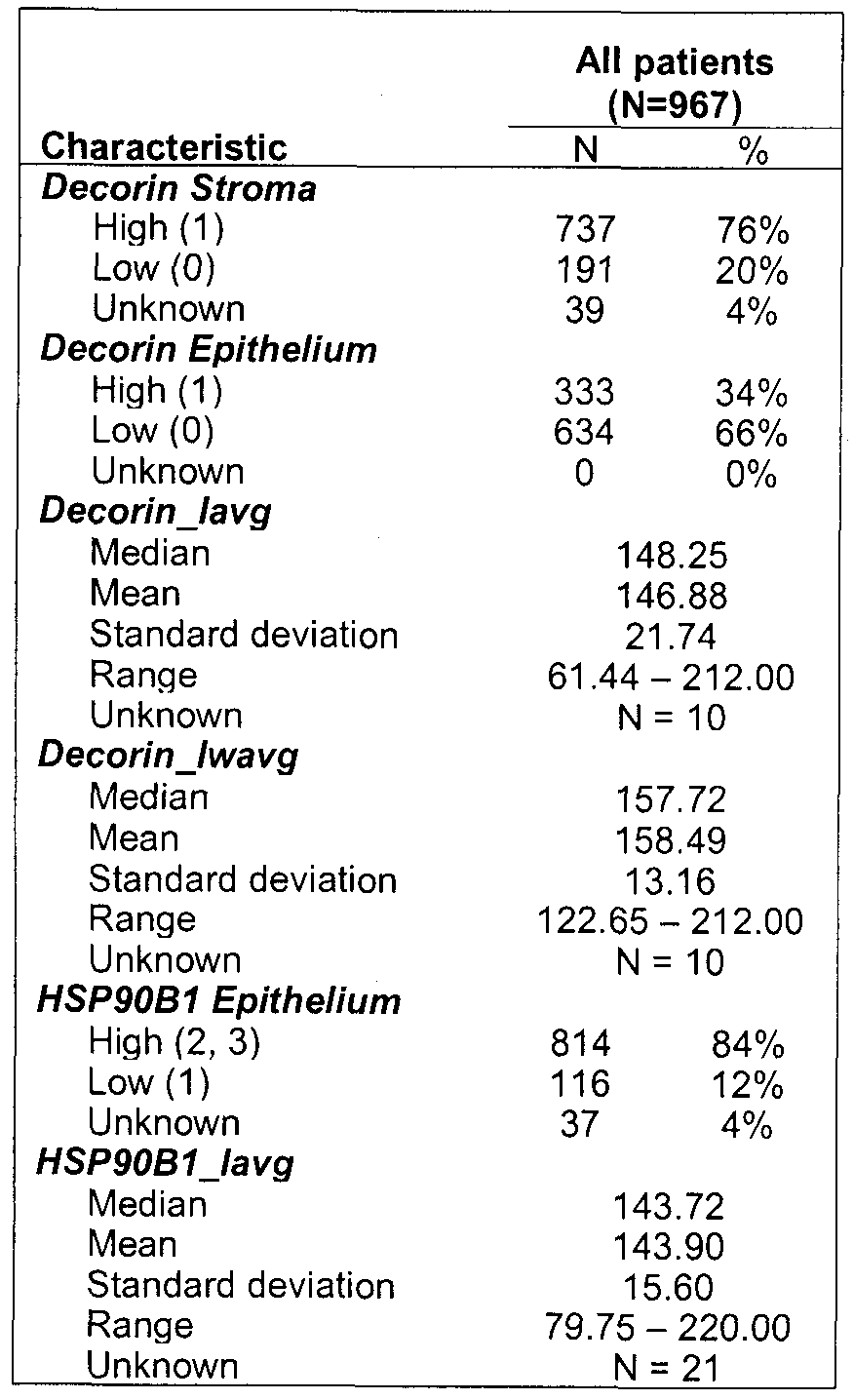
Summary of marker characteristics (Decorin and HSP90B1 ) for all the cases in the cohort (N=967).
[00147] While the present application has been described with reference to what are presently considered to be the preferred examples, it is to be understood that the application is not limited to the disclosed examples. To the contrary, the application is intended to cover various modifications and equivalent arrangements included within the spirit and scope of the appended claims.
[00148] All publications, patents and patent applications are herein incorporated by reference in their entirety to the same extent as if each individual publication, patent or patent application was specifically and individually indicated to be incorporated by reference in its entirety. Specifically, the sequence associated with each accession number provided herein is incorporated by reference in its entirely
References
1. Forouzanfar MH, Foreman KJ, Delossantos AM, Lozano R, Lopez AD, et al. (201 1 ) Breast and cervical cancer in 87 countries between 1980 and 2010: a systematic analysis. The Lancet 378: 1461 -1484.
doi: 10.1016/S0140-6736( 1 1 )61351-2
2. Carter C, Allen C, Henson D (1989) Relation of tumor size, lymph node status, and survival in 24,740 breast cancer cases. Cancer 63: 181 -187.
3. Kell M, Winter D, O'Sullivan G, Shanahan F, Redmond H (2000)
Biological behaviour and clinical implications of micrometastases. Br J Surg 87: 1629-39.
4. Cox C, White L, Allred N, Meyers M, Dickson D, et al. (2006) Survival outcomes in node-negative breast cancer patients evaluated with complete axillary node dissection versus sentinel lymph node biopsy. Ann. Surg. Oncol 13: 708-71 1 . doi: 10.1245/ASO.2006.05.017
5. Kuijt GP, van de Poll-Franse LV, Voogd AC, Nieuwenhuijzen GAP,
Roumen RMH (2007) Survival after negative sentinel lymph node biopsy in breast cancer at least equivalent to after negative extensive axillary dissection. Eur J Surg Oncol 33: 832-837. doi:10. 016/j.ejso.2006. 1 .017
6. Simmons RM (2001 ) Review of sentinel lymph node credentialing: how many cases are enough? J. Am. Coll. Surg 193: 206-209.
7. O'Hea MD, Brian J, Hill MCh AD., El-Shirbiny MD, Ayda M, et al. (1998) Sentinel lymph node biopsy in breast cancer: initial experience at Memorial Sloan- Kettering Cancer Center. Journal of the American College of Surgeons 186: 423-427.
8. Bartsch R, Wenzel C, Pluschnig U, Hussian D, Sevelda U, et al. (2006) Prognostic value of monitoring tumour markers CA 15-3 and CEA during fulvestrant treatment. BMC Cancer 6: 81 . doi:10.1 186/1471-2407-6-81
9. Soletormos G (2004) Monitoring different stages of breast cancer using tumour markers CA 15-3, CEA and TPA. European Journal of Cancer 40: 481-486. doi:10.1016/j.ejca.2003.10.015
10. Ross PL, Huang YN, Marchese JN, Williamson B, Parker K, et al. (2004) Multiplexed protein quantitation in Saccharomyces cerevisiae using amine-reactive isobaric tagging reagents. Mol. Cell Proteomics 3: 1 154- 1 169. doi:10.1074/mcp.M400129-MCP200
1 1. Lange V, Picotti P, Domon B, Aebersold R (n.d.) Selected reaction
monitoring for quantitative proteomics: a tutorial. Mol Syst Biol 4: 222- 222. doi: 10.1038/msb.2008.61
Song X, Bandow J, Sherman J, Baker J, Brown P, et al. (2008) iTRAQ experimental design for plasma biomarker discovery. J. Proteome Res 7: 2952-2958.
Griffiths S, Burthem J, Unwin R, Holyoake T, Melo J, et al. (n.d.) The use of isobaric tag peptide labeling (iTRAQ) and mass spectrometry to examine rare, primitive hematopoietic cells from patients with chronic myeloid leukemia. Mol. Biotechnol 36: 81 -89.
Grbovic OM, Basso AD, Sawai A, Ye Q, Friedlander P, et al. (2006) V600E B-Raf requires the Hsp90 chaperone for stability and is degraded in response to Hsp90 inhibitors. Proc Natl Acad Sci U S A 03: 57-62. doi: 10.1073/pnas.0609973103
Takayama S, Reed JC, Homma S (0000) Heat-shock proteins as regulators of apoptosis. Oncogene 22: 9041 -9047.
Dias S, Shmelkov SV, Lam G, Rafii S (2002) VEGF(165) promotes survival of leukemic cells by Hsp90-mediated induction of Bcl-2 expression and apoptosis inhibition. Blood 99: 2532-2540.
da Rocha Dias S, Friedlos F, Light Y, Springer C, Workman P, et al. (2005) Activated B-RAF Is an Hsp90 Client Protein That Is Targeted by the Anticancer Drug 17-Allylamino-17-Demethoxygeldanamycin. Cancer Research 65: 10686-10691 . doi: 10.1 158/0008-5472.CAN-05-2632 Liu X, Ye L, Wang J, Fan D (1999) Expression of heat shock protein 90 beta in human gastric cancer tissue and SGC7901A/CR of MDR-type gastric cancer cell line. Chin. Med. J 1 12: 1 133-1 137.
Sreedhar AS, Csermely P (2004) Heat shock proteins in the regulation of apoptosis: new strategies in tumor therapy: a comprehensive review. Pharmacol. Ther 101 : 227-257. doi:10.1016/j.pharmthera.2003.1 1.004 Subbarao Sreedhar A, Kalmar E, Csermely P, Shen Y-F (2004) Hsp90 isoforms: functions, expression and clinical importance. FEBS Letters 562: 1 1 -15. doi:10.1016/S0014-5793(04)00229-7
Shilov IV, Seymour SL, Patel AA, Loboda A, Tang WH, et al. (2007) The Paragon Algorithm, a next generation search engine that uses sequence temperature values and feature probabilities to identify peptides from tandem mass spectra. Mol. Cell Proteomics 6: 1638-1655.
doi: 10.1074/mcp.T600050-MCP200
Hammond MEH, Hayes DF, Dowsett M, Allred DC, Hagerty KL, et al. (2010) American Society of Clinical Oncology/College Of American
Pathologists guideline recommendations for immunohistochemical testing of estrogen and progesterone receptors in breast cancer. J. Clin. Oncol 28: 2784-2795. doi: 10.1200/JCO.2009.25.6529
Wolff AC, Hammond MEH, Schwartz JN, Hagerty KL, Allred DC, et al. (2007) American Society of Clinical Oncology/College of American Pathologists guideline recommendations for human epidermal growth factor receptor 2 testing in breast cancer. Arch. Pathol. Lab. Med 131 : 18-43.
Cheang MCU, Chia SK, Voduc D, Gao D, Leung S, et al. (2009) Ki67 Index, HER2 Status, and Prognosis of Patients With Luminal B Breast Cancer. J. Natl. Cancer Inst. 101 : 736-750. doi:10.1093/jnci/djp082 Hugh J, Hanson J, Cheang MCU, Nielsen TO, Perou CM, et al. (2009) Breast cancer subtypes and response to docetaxel in node-positive breast cancer: use of an immunohistochemical definition in the BCIRG 001 trial. J. Clin. Oncol 27: 1 168-1 176. doi:10.1200/JCO.2008.18.1024 McShane LM, Altman DG, Sauerbrei W, Taube SE, Gion M, et al. (2005) Reporting Recommendations for Tumor Marker Prognostic Studies. Journal of Clinical Oncology 23: 9067 -9072.
doi:10.1200/JCO.2004.01 .0454
Goldoni S, lozzo RV (2008) Tumor microenvironment: Modulation by decorin and related molecules harboring leucine-rich tandem motifs. Int. J. Cancer 123: 2473-2479. doi: 10.1002/ijc.23930
Feugaing DDS, Tammi R, Echtermeyer FG, Stenmark H, Kresse H, et al.
(2007) Endocytosis of the dermatan sulfate proteoglycan decorin utilizes multiple pathways and is modulated by epidermal growth factor receptor signaling. Biochimie 89: 637-657. doi:10.1016/j.biochi.2006.12.012 Fiedler LR, Schonherr E, Waddington R, Niland S, Seidler DG, et al.
(2008) Decorin Regulates Endothelial Cell Motility on Collagen I through Activation of Insulin-like Growth Factor I Receptor and Modulation of α2β1 Integrin Activity. Journal of Biological Chemistry 283: 17406 - 17415. doi: 10.1074/jbc.M710025200
Zafiropoulos A, Tzanakakis GN (2008) Decorin-mediated effects in cancer cell biology. Connect. Tissue Res 49: 244-248.
doi: 10.1080/03008200802147746
Goldoni S, Humphries A, Nystrom A, Sattar S, Owens RT, et al. (2009) Decorin is a novel antagonistic ligand of the Met receptor. J. Cell Biol. 185: 743-754. doi: 10.1083/jcb.200901 129
Schonherr E, Sunderkotter C, lozzo RV, Schaefer L (2005) Decorin, a Novel Player in the Insulin-like Growth Factor System. Journal of
Biological Chemistry 280: 15767 -15772. doi: 10.1074/jbc.M500451200 Zafiropoulos A, Nikitovic D, Katonis P, Tsatsakis A, Karamanos NK, et al. (2008) Decorin-lnduced Growth Inhibition Is Overcome through
Protracted Expression and Activation of Epidermal Growth Factor Receptors in Osteosarcoma Cells. Molecular Cancer Research 6: 785 - 794. doi: 10.1 158/1541 -7786.MCR-07-0165
Fiedler LR, Eble JA (2009) Decorin regulates endothelial cell-matrix interactions during angiogenesis. Cell Adh Migr 3: 3-6.
Xu M, Dittmar KD, Giannoukos G, Pratt WB, Simons SS (1998) Binding of hsp90 to the glucocorticoid receptor requires a specific 7-amino acid sequence at the amino terminus of the hormone-binding domain. J. Biol. Chem 273: 13918-13924.
Sato S, Fujita N, Tsuruo T (2000) Modulation of Akt kinase activity by binding to Hsp90. Proc Natl Acad Sci U S A 97: 10832-10837.
Becker B, Multhoff G, Farkas B, Wild P-J, Landthaler M, et al. (2004) Induction of Hsp90 protein expression in malignant melanomas and melanoma metastases. Exp. Dermatol 13: 27-32. doi:10.1 1 1/j.0906- 6705.2004.001 14.x
Hacihanefioglu A, Gonullu E, Mehtap O, Keski H, Yavuz M, et al. (2010) Effect of heat shock protein-90 (HSP90) and vascular endothelial growth factor (VEGF) on survival in acute lymphoblastic leukemia: an
immunohistochemical study. Med Oncol. Available:
http://www.springerlink.com/content/g438673q3440w15t/.
Pick E, Kluger Y, Giltnane JM, Moeder C, Camp RL, et al. (2007) High HSP90 Expression Is Associated with Decreased Survival in Breast Cancer. Cancer Research 67: 2932-2937. doi:10.1 158/0008-5472. CAN- 06-451 1
Li C-F, Huang W-W, Wu J-M, Yu S-C, Hu T-H, et al. (2008) Heat Shock Protein 90 Overexpression Independently Predicts Inferior Disease-Free Survival with Differential Expression of the a and β Isoforms in
Gastrointestinal Stromal Tumors. Clinical Cancer Research 14: 7822- 7831 . doi: 10. 158/1078-0432.CCR-08-1369
Claims
1. A method of evaluating a test subject having or suspected of having breast cancer, the method comprising:
a. determining a level of one or more biomarkers in a test sample from the test subject, wherein the one or more biomarkers are selected from Table 1 ,
b. comparing the level of one or more biomarkers in the test sample with a control, and
c. identifying a difference or similarity in the level of the one or more biomarkers between the test sample and the control, the differences or similarities providing an evaluation of the test subject.
2. The method of claim 1 , the method comprising:
a. determining a level of one or more biomarkers in a test sample from the test subject, the one or more biomarkers selected from Table 1 ,
b. comparing the level of one or more biomarkers in the test sample with a control, and
c. detecting an increased level of one or more of the biomarkers between the test sample and the control, the increased level providing an evaluation of the test subject.
3. The method of claim 1 or 2, wherein the evaluation provides an indication of the subject's prognosis and/or response to hormone treatment.
4. The method of claim 3 wherein the prognosis comprises an indication of one or more of metastasis and survival time.
5. A method of claim 3 or 4 for providing a prognosis for a test subject having or suspected of having breast cancer, the method comprising:
determining a level of one or more biomarkers in a test sample from the test subject, wherein the one or more biomarkers are selected from Table 1 , and
comparing the level of one or more biomarkers in the test sample with a control, wherein a difference or similarity in the level of the one or more biomarkers between the test sample and the control is used to provide a prognosis for the test subject.
6. The method of claim 5, wherein the one or more biomarkers are selected from HSP90B1 , Decorin (DCN), HMGN2, USP34 and G6PD.
7. The method of claim 5, wherein one of the biomarkers is HSP90B1.
8. The method of claim 5, wherein one of the biomarkers is Decorin (DCN).
9. The method of claim 5, wherein the biomarkers include HSP90B1 and Decorin (DCN).
10. The method of any one or claims 3 to 9, wherein the prognosis for the test subject is an increased likelihood of metastasis such as lymph node metastasis or distant metastasis, or a survival time such as overall survival or disease free survival.
1 1. The method of any one of claims 3 to 9, wherein the control represents subjects with breast cancer without metastasis and an increase in the level of HSP90B1 and/or DCN in the test sample relative to the control indicates an increased likelihood of metastasis in the test subject.
12. The method of any one of claims 3 to 9, wherein the control represents subjects with breast cancer without lymph node metastasis and an increase in the level of DCN in the test sample relative to the control indicates an increased likelihood of lymph node metastasis in the test subject.
13. The method of any one of claims 3 to 9, wherein the control sample represents subjects with breast cancer without distant metastasis and an increase in the level of HSP90B1 in the test sample relative to the control indicates an increased likelihood of distant metastasis in the test subject.
14. The method of any one of claims 3 to 9, wherein the control represents subjects with breast cancer without metastasis and a similar level of HSP90B1 and/or DCN in the test sample relative to the control indicates a low likelihood of metastasis in the test subject.
15. The method of claim 14, wherein the biomarker is DCN and the metastasis is lymph node metastasis.
16. The method claim 14, wherein the biomarker is HSP90B1 and the metastasis is distant metastasis.
17. The method of any one or claims 3 to 9, wherein the difference or similarity between the test sample and the control is used to estimate a survival time for the test subject.
18. The method of any one of claims 3 to 9, wherein an increase in the level of HSP90B1 and/or DCN in the test sample relative to the control indicates a decreased overall survival (OS) relative to the control for the test subject.
19. The method of claim 18, wherein the biomarker is DCN and the test subject has a luminal B tumour molecular subtype.
20. The method of claim 18, wherein the control represents subjects who survived for at least 2 years from diagnosis with breast cancer.
21 . The method of claim 18, wherein the control represents subjects who survived for at least 5 years from diagnosis with breast cancer.
22. The method of claim 18, wherein the control represents subjects who survived for at least 0 years from diagnosis with breast cancer.
23. The method of any one of claims 3 to 9, wherein an increase in the level of HSP90B1 and/or DCN in the test sample relative to the control indicates a decreased disease-free survival (DFS) for the test subject.
24. The method of claim 23, wherein the control represents subjects who survived for at least 2 years from diagnosis with breast cancer without recurrence of breast cancer.
25. The method of claim 23, wherein the control represents subjects who survived for at least 5 years from diagnosis with breast cancer without recurrence of breast cancer.
26. The method of claim 23, wherein the control represents subjects who survived for at least 10 years from diagnosis with breast cancer without recurrence of breast cancer.
27. The method of any one of claims 3 to 9, wherein an increase in the level of HSP90B1 and/or DCN in the test sample relative to the control indicates a decreased overall survival and/or disease free survival for the test subject.
28. The method of any one of claims 1 to 27, comprising determining a level for two or more biomarkers in the test sample, generating a test sample expression profile based on the level of the two or more biomarkers and comparing the test sample expression profile to a control expression profile, wherein a difference or similarity in the test sample expression profile and the control expression profile is used to provide an evaluation for the test subject.
29. The method of claim 28, wherein the two or more biomarkers include HSP90B1 and DCN.
30. The method of any one of claims 1 to 5, wherein the control represents subjects with breast cancer who are not responsive to hormone treatment and a test subject with an increase in the level of HSP90B1 and/or DCN relative to the control indicates that the test subject has an increased likelihood of being responsive to hormone treatment.
31. The method of any one of claims 1 to 5, wherein the control represents subjects with breast cancer who are responsive to hormone treatment and a test subject with a similar level of HSP90B1 and/or DCN relative to the control indicates that the test subject has an increased likelihood of being responsive to hormone treatment.
32. A method of selecting treatment for a test subject with breast cancer or suspected of having breast cancer, the method comprising:
determining a level of HSP90B1 and/or DCN in a test sample from the test subject, and
comparing the level of HSP90B1 and/or DCN in the test sample with a control
selecting a treatment for the test subject based on a difference or similarity in the level of HSP90B1 and/or DCN in the test sample compared to the control.
33. The method of claim 32, wherein the control represents subjects with breast cancer who are not responsive to hormone treatment and a test subject with an increase in the level of HSP90B1 and/or DCN in the test sample relative to the control is selected for hormone treatment.
34. The method of claim 33, further comprising treating a test subject with an increase in the level of HSP90B1 and/or DCN relative to the control with hormone treatment.
35. A method of identifying a test subject with breast cancer with an increased likelihood of being responsive to hormone treatment, the method comprising:
determining a level of HSP90B1 and/or DCN in a test sample from the test subject,
comparing the level of HSP90B1 and/or DCN in the test sample with a control, and
identifying the test subject as having an increased likelihood of being responsive to hormone treatment based on a difference or similarity in the level of HSP90B1 and/or DCN in the test sample compared to the control .
36. The method of claim 35, further comprising treating the test subject identified as having an increased likelihood of being responsive to hormone treatment with hormone treatment.
37. The method of claim 35 or 36, wherein the control represents subjects with breast cancer who are not responsive to hormone treatment and an increase in the level of HSP90B1 and/or DCN in the test sample relative to the control indicates that the test subject has an increased likelihood of being responsive to hormone treatment.
38. A method of treating a subject with breast cancer, the method comprising:
determining a level of HSP90B1 and/or DCN in a test sample from the subject,
comparing the level of HSP90B1 and/or DCN in the test sample with a control,
identifying the test subject as having an increased likelihood of being responsive to hormone treatment based on a difference or similarity in the level of HSP90B1 and/or DCN in the test sample compared to the control,
treating a test subject identified as having an increased likelihood of being responsive to hormone treatment with hormone treatment.
39. The method of claim 38, wherein the control represents subjects with breast cancer who are not responsive to hormone treatment and an increase in the level of HSP90B1 and/or DCN in the test sample relative to the control indicates that the test subject has an increased likelihood of being responsive to hormone treatment.
40. The method of any one of claims 33 to 39, wherein hormone treatment comprises Tamoxifen, an aromatase inhibitor or other agent acting upon estrogen receptors, progesterone receptors or their signaling pathways.
41. The method of any one of claims 1 to 40, wherein the method further comprises obtaining a test sample from the test subject.
42. The method of any one of claims 1 to 41 , wherein the test sample comprises malignant epithelial cells.
43. The method of any one of claims 1 to 42, wherein the test sample is a tissue sample or a frozen tissue sample.
44. The method of claim 43, wherein the test sample is a formalin-fixed, paraffin embedded (FFPE) sample.
45. The method of any one of claims 1 to 44, wherein the level of one or more biomarkers in the test sample is a protein level.
46. The method of claim 45, wherein the protein level is determined by mass spectroscopy, immunohistochemistry, or an immunoassay such as an enzyme-linked immunosorbant assay (ELISA).
47. The method of claim 45, wherein the protein level of the biomarker in the test sample is determined by contacting the sample with a detection agent, for example an antibody, such as a monoclonal antibody or antibody fragment, wherein the detection agent forms a complex with the biomarker.
48. The method of any one of claims 1 to 44 wherein the level of one or more biomarkers in the test sample is an mRNA level.
49. The method of claim 48, wherein the mRNA level is determined by quantitative PCR such as RT-PCR, serial analysis of gene expression (SAGE), use of a microarray, digital molecular barcoding technology or Northern blot.
50. The method of any one of claims 1 to 49, wherein the control is a predetermined value or threshold.
51 . Use of hormone therapy for treating breast cancer in a subject, wherein the level of HSP90B1 and/or DCN in a test sample from the subject is increased relative to a control level.
52. The use of claim 51 , wherein the control level represents a level of HSP90B1 and/or DCN in subjects with breast cancer who are not responsive to hormone treatment.
53. The use of claim 51 or 52, for treating breast cancer in a subject with metastasis, optionally lymph node metastasis.
54. The use of any one of claims 51 to 53, wherein the hormone treatment comprises Tamoxifen, an aromatase inhibitor or other agent acting upon estrogen receptors, progesterone receptors or their signaling pathways
55. A composition comprising at least two biomarker-specific detection agents, each of which specifically binds a biomarker selected from Table 1.
56. The composition of claim 55, wherein the biomarkers are HSP90B1 and DCN.
57. The composition of claim 55 or 56, wherein the biomarker-specific detection agents are antibodies, optionally one or more of the antibodies listed in Table 2b.
58. The composition of any one of claims 54 to 56 for use in the method of any one of claims 1 to 50.
59. A kit for detecting a biomarker for use in a method of any one of claims 1 to 50 comprising:
a. at least two biomarker-specific detections agents, each of which binds a biomarker selected from Table 1 , preferably selected from HSP90B1 and DCN; and/or
b. instructions for use.
60. A kit according to claim 59 further comprising at least one standard and/or control.
61. A kit according to claim 59 or 60, wherein the biomarker-specific detections agents include two or more of the antibodies listed in Table 2b, such as antibodies for HSP90B1 and DCN.
Applications Claiming Priority (2)
| Application Number | Priority Date | Filing Date | Title |
|---|---|---|---|
| US201261588336P | 2012-01-19 | 2012-01-19 | |
| US61/588,336 | 2012-01-19 |
Publications (1)
| Publication Number | Publication Date |
|---|---|
| WO2013106913A1 true WO2013106913A1 (en) | 2013-07-25 |
Family
ID=48798442
Family Applications (1)
| Application Number | Title | Priority Date | Filing Date |
|---|---|---|---|
| PCT/CA2013/000042 Ceased WO2013106913A1 (en) | 2012-01-19 | 2013-01-18 | Biomarkers for breast cancer prognosis and treatment |
Country Status (1)
| Country | Link |
|---|---|
| WO (1) | WO2013106913A1 (en) |
Cited By (7)
| Publication number | Priority date | Publication date | Assignee | Title |
|---|---|---|---|---|
| GB2549798A (en) * | 2016-04-29 | 2017-11-01 | Univ Bradford | Peptides and nanoparticle formulations thereof |
| US10451625B2 (en) | 2014-05-09 | 2019-10-22 | Ascendant Diagnostics, LLC | Methods of detecting cancer |
| US11459617B2 (en) | 2016-04-29 | 2022-10-04 | Board Of Regents, The University Of Texas System | Targeted measure of transcriptional activity related to hormone receptors |
| CN115418402A (en) * | 2022-08-23 | 2022-12-02 | 山东大学齐鲁医院 | mRNA marker for breast cancer diagnosis and/or prognosis judgment and application thereof |
| WO2023091967A1 (en) * | 2021-11-16 | 2023-05-25 | The Board Of Trustees Of The Leland Stanford Junior University | Systems and methods for personalized treatment of tumors |
| US12320811B2 (en) | 2014-05-09 | 2025-06-03 | Ascendant Diagnostics, LLC | Methods of detecting cancer |
| WO2025171441A1 (en) * | 2024-02-13 | 2025-08-21 | The Council Of The Queensland Institute Of Medical Research | Prognostic signature for cancer recurrence and responsiveness |
Citations (1)
| Publication number | Priority date | Publication date | Assignee | Title |
|---|---|---|---|---|
| US5447843A (en) * | 1990-04-12 | 1995-09-05 | Board Of Regents, The University Of Texas System | Heat shock/stress response proteins and prognosis in cancer |
-
2013
- 2013-01-18 WO PCT/CA2013/000042 patent/WO2013106913A1/en not_active Ceased
Patent Citations (1)
| Publication number | Priority date | Publication date | Assignee | Title |
|---|---|---|---|---|
| US5447843A (en) * | 1990-04-12 | 1995-09-05 | Board Of Regents, The University Of Texas System | Heat shock/stress response proteins and prognosis in cancer |
Non-Patent Citations (8)
| Title |
|---|
| BARRACLOUGH DL ET AL.: "Microarray analysis of suppression subtracted hybridisation libraries identifies genes associated with breast cancer progression", CELLULAR ONCOLOGY, vol. 32, no. 1-2, 2010, pages 87 - 99 * |
| BARTKOWIAK K. ET AL.: "Discovery of a novel unfolded protein response phenotype of cancer stem/progenitor cells from the bone marrow of breast cancer patients", JOURNAL OF PROTEOME RESEARCH, vol. 9, no. 6, 4 June 2010 (2010-06-04), pages 3158 - 3168, XP002602188 * |
| CAWTHORN, TR ET AL.: "Proteomic Analyses Reveal High Expression of Decorin and Endoplasmin (HSP90B 1) Are Associated with Breast Cancer Metastasis and Decreased Survival", PLOS ONE, vol. 7, no. 2, 20 February 2012 (2012-02-20), pages 1 - 11, XP055078023 * |
| CAWTHORN, TR.: "Identification of molecular alterations associated with loco-regional and distant breast cancer metastasis", A THESIS SUBMITTED IN CONFORMITY WITH THE REQUIREMENTS FOR THE DEGREE OF MASTER OF SCIENCE, DEPARTMENT OF MEDICAL BIOPHYSICS. UNIVERSITY OF TORONTO, 2009, pages FP - 121, XP003031268, Retrieved from the Internet <URL:https://tspace.library.utoronto.ca/bitstream/1807/18239/1/Cawthorn_Thomas_R_200911_MSc_thesis.pdf> * |
| CHENG, Q ET AL.: "Amplification and high-level expression of heat shock protein 90 marks aggressive phenotypes of human epidermal growth factor receptor 2 negative breast cancer, art R62", BREAST CANCER RESEARCH, vol. 14, no. 2, 17 April 2012 (2012-04-17), pages 1 - 15, XP021125993 * |
| CHUNG L. ET AL.: "Breast cancer biomarkers: proteomic Discovery and translation to clinically relevant assays", EXPERT REVIEW OF PROTEOMICS., vol. 9, no. 6, December 2012 (2012-12-01), pages 599 - 614 * |
| DEJEANS, N ET AL.: "Overexpression of GRP94 in breast cancer cells resistant to oxidative stress promotes high levels of cancer cell proliferation and migration: Implications for tumor recurrence", FREE RADICAL BIOLOGY & MEDICINE, vol. 52, no. 6, 15 March 2012 (2012-03-15), pages 993 - 1002, XP028459031 * |
| HILL JJ ET AL.: "Identification of vascular breast tumor markers by laser capture microdissection and label-free LC-MS", JOURNAL OF PROTEOME RESEARCH, vol. 10, no. 5, 6 May 2011 (2011-05-06), pages 2479 - 2493, XP055077598 * |
Cited By (14)
| Publication number | Priority date | Publication date | Assignee | Title |
|---|---|---|---|---|
| US10451625B2 (en) | 2014-05-09 | 2019-10-22 | Ascendant Diagnostics, LLC | Methods of detecting cancer |
| US12320811B2 (en) | 2014-05-09 | 2025-06-03 | Ascendant Diagnostics, LLC | Methods of detecting cancer |
| US10613090B2 (en) | 2014-05-09 | 2020-04-07 | Ascendant Diagnostics, LLC | Methods of detecting cancer |
| CN109415424B (en) * | 2016-04-29 | 2022-08-30 | 布拉德福德大学 | Peptides and nanoparticle formulations thereof |
| CN109415424A (en) * | 2016-04-29 | 2019-03-01 | 布拉德福德大学 | Peptides and their nanoparticle formulations |
| US20210188929A1 (en) * | 2016-04-29 | 2021-06-24 | University Of Bradford | Peptides and Nanoparticle Formulations Thereof |
| GB2549798A (en) * | 2016-04-29 | 2017-11-01 | Univ Bradford | Peptides and nanoparticle formulations thereof |
| US11459617B2 (en) | 2016-04-29 | 2022-10-04 | Board Of Regents, The University Of Texas System | Targeted measure of transcriptional activity related to hormone receptors |
| US11999770B2 (en) | 2016-04-29 | 2024-06-04 | University Of Bradford | Peptides and nanoparticle formulations thereof |
| WO2017187206A1 (en) * | 2016-04-29 | 2017-11-02 | University Of Bradford | Peptides and nanoparticle formulations thereof |
| US12590335B2 (en) | 2016-04-29 | 2026-03-31 | Board Of Regents, The University Of Texas System | Targeted measure of transcriptional activity related to hormone receptors |
| WO2023091967A1 (en) * | 2021-11-16 | 2023-05-25 | The Board Of Trustees Of The Leland Stanford Junior University | Systems and methods for personalized treatment of tumors |
| CN115418402A (en) * | 2022-08-23 | 2022-12-02 | 山东大学齐鲁医院 | mRNA marker for breast cancer diagnosis and/or prognosis judgment and application thereof |
| WO2025171441A1 (en) * | 2024-02-13 | 2025-08-21 | The Council Of The Queensland Institute Of Medical Research | Prognostic signature for cancer recurrence and responsiveness |
Similar Documents
| Publication | Publication Date | Title |
|---|---|---|
| Cawthorn et al. | Proteomic analyses reveal high expression of decorin and endoplasmin (HSP90B1) are associated with breast cancer metastasis and decreased survival | |
| Song et al. | Proteomic analysis on metastasis-associated proteins of human hepatocellular carcinoma tissues | |
| EP2457092B1 (en) | Cancer biomarker and the use thereof | |
| Chen et al. | Elevated level of anterior gradient-2 in pancreatic juice from patients with pre-malignant pancreatic neoplasia | |
| Ibusuki et al. | Midkine in plasma as a novel breast cancer marker | |
| CN112345755A (en) | Biomarker of breast cancer and application thereof | |
| WO2013106913A1 (en) | Biomarkers for breast cancer prognosis and treatment | |
| US8216789B2 (en) | Diagnostic panel of cancer antibodies and methods for use | |
| CN102590511B (en) | Tumor diagnosis reagent or kit based on detection of IGFBP-2 (insulin-like growth factor binding protein 2) autoantibody or combined detection of IGFBP-2 antoantibody and IGFBP-2, as well as application thereof | |
| CN105658810A (en) | Methods for detecting prostate cancer | |
| EP2220505B1 (en) | Methods of diagnosing cancer | |
| US20100081666A1 (en) | Src activation for determining cancer prognosis and as a target for cancer therapy | |
| CN115598345A (en) | Methods for diagnosing cancer and determining overall survival and disease-free survival of cancer patients | |
| He et al. | Proteomic‐based biosignatures in breast cancer classification and prediction of therapeutic response | |
| JP2012501440A (en) | ANLN protein as a predictor of endocrine treatment | |
| Mutlu et al. | Analytical and clinical evaluation of a new urinary tumor marker: bladder tumor fibronectin in diagnosis and follow-up of bladder cancer. | |
| Yeom et al. | Microquantitation of van gogh-like protein 1 by using antibody-conjugated magnetic beads | |
| TW202016545A (en) | Biomarker of urinary tract epithelial cell carcinoma and its application | |
| Papila et al. | Clinical significance and prognostic value of serum sHER-2/neu levels in patients with solid tumors | |
| KR102417089B1 (en) | Biomarker composition comprising CXCL12 in plasma membrane of cancer cells for predicting prognosis of rectal adenocarcinoma | |
| US20130196876A1 (en) | Differential diagnosis of pancreatic adenomas | |
| KR20170096493A (en) | Method for predicting prognosis of luminal A and TNBC type breast cancer patient by using CD24 | |
| KR101594287B1 (en) | A kit for pancreatic cancer diagnosis comprising complememt factor b-specific binding antibody | |
| EP2957913B1 (en) | Methods and products for prognosing the clinical evolution, or predicting the recurrence risk, of a papillary lesion of the breast | |
| US20250277791A1 (en) | Influence of receptor integrative analysis on hr+/her2+ breast cancer molecular subtypes and prognosis |
Legal Events
| Date | Code | Title | Description |
|---|---|---|---|
| 121 | Ep: the epo has been informed by wipo that ep was designated in this application |
Ref document number: 13738596 Country of ref document: EP Kind code of ref document: A1 |
|
| NENP | Non-entry into the national phase |
Ref country code: DE |
|
| 122 | Ep: pct application non-entry in european phase |
Ref document number: 13738596 Country of ref document: EP Kind code of ref document: A1 |