WO2012142022A1 - Assays, compositions, systems, kits, and devices for diagnosing typhoid fever - Google Patents
Assays, compositions, systems, kits, and devices for diagnosing typhoid fever Download PDFInfo
- Publication number
- WO2012142022A1 WO2012142022A1 PCT/US2012/032874 US2012032874W WO2012142022A1 WO 2012142022 A1 WO2012142022 A1 WO 2012142022A1 US 2012032874 W US2012032874 W US 2012032874W WO 2012142022 A1 WO2012142022 A1 WO 2012142022A1
- Authority
- WO
- WIPO (PCT)
- Prior art keywords
- seq
- typhi
- protein
- cytokine
- cells
- Prior art date
- Legal status (The legal status is an assumption and is not a legal conclusion. Google has not performed a legal analysis and makes no representation as to the accuracy of the status listed.)
- Ceased
Links
Classifications
-
- G—PHYSICS
- G01—MEASURING; TESTING
- G01N—INVESTIGATING OR ANALYSING MATERIALS BY DETERMINING THEIR CHEMICAL OR PHYSICAL PROPERTIES
- G01N33/00—Investigating or analysing materials by specific methods not covered by groups G01N1/00 - G01N31/00
- G01N33/48—Biological material, e.g. blood, urine; Haemocytometers
- G01N33/50—Chemical analysis of biological material, e.g. blood, urine; Testing involving biospecific ligand binding methods; Immunological testing
- G01N33/53—Immunoassay; Biospecific binding assay; Materials therefor
- G01N33/569—Immunoassay; Biospecific binding assay; Materials therefor for microorganisms, e.g. protozoa, bacteria, viruses
- G01N33/56911—Bacteria
- G01N33/56916—Enterobacteria, e.g. shigella, salmonella, klebsiella, serratia
-
- C—CHEMISTRY; METALLURGY
- C07—ORGANIC CHEMISTRY
- C07K—PEPTIDES
- C07K14/00—Peptides having more than 20 amino acids; Gastrins; Somatostatins; Melanotropins; Derivatives thereof
- C07K14/195—Peptides having more than 20 amino acids; Gastrins; Somatostatins; Melanotropins; Derivatives thereof from bacteria
- C07K14/24—Peptides having more than 20 amino acids; Gastrins; Somatostatins; Melanotropins; Derivatives thereof from bacteria from Enterobacteriaceae (F), e.g. Citrobacter, Serratia, Proteus, Providencia, Morganella, Yersinia
- C07K14/255—Salmonella (G)
-
- G—PHYSICS
- G01—MEASURING; TESTING
- G01N—INVESTIGATING OR ANALYSING MATERIALS BY DETERMINING THEIR CHEMICAL OR PHYSICAL PROPERTIES
- G01N33/00—Investigating or analysing materials by specific methods not covered by groups G01N1/00 - G01N31/00
- G01N33/48—Biological material, e.g. blood, urine; Haemocytometers
- G01N33/50—Chemical analysis of biological material, e.g. blood, urine; Testing involving biospecific ligand binding methods; Immunological testing
- G01N33/68—Chemical analysis of biological material, e.g. blood, urine; Testing involving biospecific ligand binding methods; Immunological testing involving proteins, peptides or amino acids
- G01N33/6863—Cytokines, i.e. immune system proteins modifying a biological response such as cell growth proliferation or differentiation, e.g. TNF, CNF, GM-CSF, lymphotoxin, MIF or their receptors
- G01N33/6866—Interferon
-
- G—PHYSICS
- G01—MEASURING; TESTING
- G01N—INVESTIGATING OR ANALYSING MATERIALS BY DETERMINING THEIR CHEMICAL OR PHYSICAL PROPERTIES
- G01N2333/00—Assays involving biological materials from specific organisms or of a specific nature
- G01N2333/195—Assays involving biological materials from specific organisms or of a specific nature from bacteria
- G01N2333/24—Assays involving biological materials from specific organisms or of a specific nature from bacteria from Enterobacteriaceae (F), e.g. Citrobacter, Serratia, Proteus, Providencia, Morganella, Yersinia
- G01N2333/255—Salmonella (G)
-
- G—PHYSICS
- G01—MEASURING; TESTING
- G01N—INVESTIGATING OR ANALYSING MATERIALS BY DETERMINING THEIR CHEMICAL OR PHYSICAL PROPERTIES
- G01N2333/00—Assays involving biological materials from specific organisms or of a specific nature
- G01N2333/435—Assays involving biological materials from specific organisms or of a specific nature from animals; from humans
- G01N2333/52—Assays involving cytokines
- G01N2333/555—Interferons [IFN]
- G01N2333/57—IFN-gamma
-
- Y—GENERAL TAGGING OF NEW TECHNOLOGICAL DEVELOPMENTS; GENERAL TAGGING OF CROSS-SECTIONAL TECHNOLOGIES SPANNING OVER SEVERAL SECTIONS OF THE IPC; TECHNICAL SUBJECTS COVERED BY FORMER USPC CROSS-REFERENCE ART COLLECTIONS [XRACs] AND DIGESTS
- Y02—TECHNOLOGIES OR APPLICATIONS FOR MITIGATION OR ADAPTATION AGAINST CLIMATE CHANGE
- Y02A—TECHNOLOGIES FOR ADAPTATION TO CLIMATE CHANGE
- Y02A50/00—TECHNOLOGIES FOR ADAPTATION TO CLIMATE CHANGE in human health protection, e.g. against extreme weather
- Y02A50/30—Against vector-borne diseases, e.g. mosquito-borne, fly-borne, tick-borne or waterborne diseases whose impact is exacerbated by climate change
Definitions
- the patient can be diagnosed as having typhoid fever or acute typhoid fever. If the amount is not increased, the condition in the patient is not typhoid fever or acute typhoid fever.
- the typhoid fever is acute typhoid fever.
- the S. Typhi protein responsive cytokine is selected from the group consisting of IFN- ⁇ , IL-1, IP- 10, IL-8, MCP-1, MDC, and MIP-la or any combination thereof. In some embodiments, the S. Typhi protein responsive cytokine is IFN- ⁇ .
- the biological sample is a blood sample.
- the blood sample is a fresh blood sample.
- the biological sample is peripheral blood mononuclear cell sample.
- the non-human machine comprises a computer implemented software.
- the invention provides a computer program comprising computer program code means adapted to perform all the steps of any of the assays set forth herein when said program is run on a computer.
- the invention provides a composition comprising at least one isolated and purified protein selected from the group consisting of STY0202, STY0372, STY1177, STY1179, STY1304, STY1878, STY2195, STY0206, STY0595, STY0909, STY1375, STY1522, STY2167 FliC, STY2381, STY3089, STY3090, and STY3796 or any combination thereof.
- the composition may be provided in a dry form or in a suitable solution, such as a storage buffer or a buffer suitable for incubating the protein with the biological sample.
- the kit further comprises laboratory materials and instructions to implement an assay as described in any and all embodiments described above.
- the kit further comprises a washing buffer.
- the kit further comprises a solid support comprising an antibody against at least one of S. Typhi protein responsive cytokine against at least wherein the solid support is an ELISA plate and the marker is an enzyme.
- the S. Typhi protein responsive cytokine in the kit is selected from the group consisting of IFN- ⁇ , IL-1, IP- 10, IL- 8, MCP-1, MDC, and MIP-la.
- the kit further comprises a secondary antibody directed against at least one S. Typhi protein responsive cytokine.
- the at least one isolated and purified S is selected from the at least one isolated and purified S.
- the S. Typhi protein responsive cytokine is selected from the group consisting of IFN- ⁇ , IL-1, IP-10, IL-8, MCP-1, MDC, and MIP-la.
- Figure 1 shows interferon- ⁇ ELISPOT responses to S. Typhi proteins
- Typhi proteins including StaF (STY0202), PagC (STY1878), S. Typhi membrane preparation (MP), and PMA and keyhole limpet hemocyanin (KLH) during acute and convalesecent stage illness in S. Typhi bacteremic patients. Mean and standard error of the mean represented.
- FIG. 3 shows cellular proliferation responses to S. Typhi proteins
- the '870 patent does not set forth how the method could be used in diagnosis of typhoid fever including acute typhoid fever. Moreover, the '870 patent only describes the method for diagnosing viral diseases and not for bacterial diseases.
- the biological sample comprises PMBC or isolated
- PMBC PMBC.
- T-cells e.g. CD8+ or CD4+ cells that have been activated or pre-sensitized in vivo to a one or more of the particular S. Typhi peptides described herein.
- T-cells i.e. cells capable of immediate effector function without the need to effect division/differentiation by in vitro culture.
- the cells secrete various cytokines, herein referred to as S. Typhi protein specific cytokines, of which any one may be selected for the purposes of this assay.
- the peptide or peptide fragment typically needs to be of a length, e.g., about 6-15 amino acids, for example, 8-12 or 8-10, amino acid residues long, that is recognised by CD8+ cells. It has been proposed, that the generality of the CD8+ cells (and other PBMC) present the peptide to the small minority of CD8+ cells that may have been pre-sensitised to the S. Typhi peptide. If such activated or pre-sensitized peptide- specific T-cells are present in the test biological fluid, they respond by secreting a cytokine which then becomes bound to the, e.g., immobilized antibody.
- a cytokine which then becomes bound to the, e.g., immobilized antibody.
- the entire protein may be used, but it may be digested into smaller fragments.
- the invention provides compositions comprising fragments of any one or more of the S. Typhi proteins described herein. Such fragments may also be past of a kit or a device or an assay. The fragments typically comprise one or more epitopes.
- Incubation should be continued for a time sufficient to permit CD8+ cells that have been pre- sensitized in vivo to the particular peptide chosen to secrete the cytokine. The incubation should not continue for so long that quiescent CD8+ cells have time to
- the incubation in some embodiments is performed within in a single working day or overnight, and without the use of sterile conditions required for cell culture in vitro.
- the assay may be conveniently carried out, for example, in a multiwell plate or on a microfluidic device.
- each well of the plate has a surface carrying a bound first antibody.
- a fluid containing an appropriate number e.g. 10 3 -10 6 of cells.
- Different peptides and/or controls can be added to individual wells of the plate or channels of the microfluidic device. Cells that secrete a cytokine during incubation show up as spots (spot forming cells or SFCs) and the number or density of these in each well can readily be determined.
- the assay can further comprise a step of diagnosing the patient as having typhoid fever if the amount of the cytokine is increased to at least about 1.5 fold or more compared to a reference amount.
- the typhoid fever is acute tyhoid fever.
- the step of diagnosing can be performed automatically, by a machine or a computer implemented software.
- the amount of cytokines can be determined, measured or detected by well known methods to one skilled in the art. For example, one can measure the cytokines using antibodies that form immunocomplexes with the cytokines.
- the appropriate mediums, appropriate conditions, and protocols for the formation of the immunocomplexes are known for one skilled in the art working in the field of immunology. By way of example, various methods and protocols which can be used are described in "Current Protocols in
- the assay can also be a FluoroSpot assay, which is a modification of the
- a Fluorescence lifetime (FLT) assay can be used to detect and measure the cytokine response in the assays of the present invention.
- the biological fluid can be peripheral blood mononuclear cells (PMBC). They may suitably isolated, e.g., from a patient's blood. In some embodiments, fresh cells are used, because cells cultured in vitro may develop altered characteristics thus reducing the diagnostic value of the assay.
- the purpose of the assay is to identify or quantitate peptide- specific T-cells e.g. CD8+ or CD4+ cells that have been activated or pre- sensitized in vivo to a particular peptide. These are unrestimulated T-cells, i.e. cells capable of immediate effector function without the need to effect division/differentiation by in vitro culture. When the peptide in question is presented to such cells, the cells secrete various cytokines, of which any one may be selected for the purposes of this assay.
- the method comprises use of coated 96-well nitrocellulose plates (Multiscreen HTS, Millipore). Plates can be coated, e.g., with e.g., about 100 ⁇ of 15 ⁇ g/ml human monoclonal anti-interferon- ⁇ antibody (1-DlK), e.g., about 8-24 hours, e.g., overnight at or at about 4°C. The plates can be washed and subsequently blocked with, e.g., about 10% FBS for about 2 h at room temperature.
- PBMCs from individual patients, and optionally controls can be added at a concentration of 2xl0 5 per well.
- the plates can be developed with aminoethylcarbazol plus H 2 0 2 , and the amount of interferon- ⁇ secreted by the cells or the number of interferon- ⁇ secreting cells can be counted. In some embodiments, the counting is performed using a stereomicro scope .
- the enzyme and corresponding soluble substrate can be selected from the following combinations comprising: the alkaline phosphatase and the soluble substrate 4- nitrophenyl phosphate (PNPP); the peroxidase and the soluble substrate orthophenylene diamine (OPD); the .beta.-galactosidase and the soluble substrate 2-nitrophenyl .beta.- galactoside (ONPG); or the glucose-6-phosphate dehydrogenase and the soluble substrate glucose-6-phosphate (G6P).
- PNPP 4- nitrophenyl phosphate
- OPD peroxidase and the soluble substrate orthophenylene diamine
- ONPG 2-nitrophenyl .beta.- galactoside
- G6P glucose-6-phosphate dehydrogenase and the soluble substrate glucose-6-phosphate
- DNase mix (10 mg/ml DNase; Sigma Aldrich in 900 mM MgCl 2 , 100 mM MnCl 2 ) to the lysate and agitated the preparation at 900 rpm for 10 min.
- lysis buffer II 100 mM NaH 2 P0 4 , 10 mM Tris, 6 M guanidine hydrochloride, 10 mM, 2-mercaptoethanol, pH 8.0
- the LabChip90 analyzed 96 proteins at a time with analysis time of 40 seconds per sample. We parsed the output results and imported them into the Harvard Institute of Proteomics protein database. We assessed for presence of contaminating E. coli LPS using a HEK-Blue LPS Detection kit (InvivoGen, San Diego, CA).
- compositions comprising the novel isolated and purified or recombinantly or synthetically produced proteins, selected from the group consisting of STY0202, STY0372, STY1177, STY1179, STY1304, STY1878, STY2195, STY0206, STY0595, STY0909, STY1375, STY1522, STY2167 FliC, STY2381, STY3089, STY3090, STY3796.
- the compositions can be used in assays guiding diagnosis for typhoid fever, including acute typhoid fever.
- the invention further provides kits for determining level of cytokines in a blood sample.
- the kit comprises (a) a composition consisting of at least one of the following isolated S. Typhi proteins: recombinant or synthesized or isolated STY0202, recombinant or synthesized or isolated STY0372, recombinant or synthesized or isolated STY1177, recombinant or synthesized or isolated STY1179, recombinant or synthesized or isolated STY 1304, recombinant or synthesized or isolated STY 1878, recombinant or synthesized or isolated STY2195, recombinant or synthesized or isolated STY0206, recombinant or synthesized or isolated STY0595, recombinant or synthesized or isolated STY0909, recombinant or synthesized or isolated STY1375, recombinant or synthesized or isolated STY1522, recombinant or synthesized or isolated STY2167 Fl
- the kit may further comprise buffers and washing solutions for facilitating incubation of the proteins with the blood sample and detection of the cytokines from the sample after incubation.
- the kit can include a solid support.
- the solid support has bound to it an antibody that can bind one or more of the S. Typhi responsive cytokines.
- the soli support is an ELISA plate.
- the kit comprises a microfluidic device.
- Antibodies both monoclonal and polyclonal, against the human cytokines useful according to the present invention are readily available to one skilled in the art from commercial sources. They can be readily picked according to the application, and can be purchased with or without specific labels already attached to them.
- cytokines useful according to the present invention are also readily available to one of ordinary skill in the art, e.g., in publicly available databases.
- Antibodies can be optionally produced by a cell line, a mixed cell line, an immortalized cell or clonal population of immortalized cells, as well known in the art. See, e.g., Ausubel, et al., ed., Current Protocols in Molecular Biology, John Wiley & Sons, Inc., NY, N.Y.
- IL-8 also referred to as CXCL8; GCP-1 ; GCP1 ; LECT; LUCT; LYNAP; MDNCF; MONAP; NAF; NAP-1; NAP1; C-X-C motif chemokine 8;
- the methods according to the present invention can be automated using robotics and computer directed systems.
- the entire assay can be automated from the point of introducing the biological fluid sample comprising T cells to an protein or a mixture thereof, incubating the reaction mixture, washing the reaction mixture, exposing the reaction mixture to a secondary antibody and detecting the amount of cytokine secreted by the cells.
- the step of comparing and displaying the results can also be automated and connected to the same system or in a remote system.
- the analysis, comparison and the result is performed in one location.
- the invention further provides a system to facilitate the diagnosis of typhoid fever in a human subject, comprising: a determination module configured to receive and output the amount of cytokines produced by the T-cells in the biological sample taken from a human and contacted with at least one S. Typhi protein or fragment thereof; a storage module configured to store output information from the determination module; a comparison module adapted to compare the data stored on the storage module with reference data and/or control data, and to provide a comparison content, and an output module for displaying the comparison content for the user, wherein if the cytokine amount is about 1.5 fold or more compared to the reference value, in the sample, then the subject will be indicated as having typhoid fever.
- the invention provides a computer readable storage medium comprising: a storing data module containing data from a sample obtained from a subject that represents a signal level from the cytokine assay after exposure of the human biological sample comprising T cells to at least one of the S.
- the cut off value is about 1.5 fold the reference value, which is or has been typically obtained from normal cytokine amount in a control human or a pool of control humans or an average of a set of control humans who is/are known not to be affected with typhoid fever.
- the fever is still very high and oscillates very little over 24 hours. Dehydration ensues and the patient is delirious (typhoid state). By the end of third week the fever has started reducing this (defervescence). This carries on into the fourth and final week.
- a person may become an asymptomatic carrier of typhoid fever, suffering no symptoms, but capable of infecting others. According to the CDC approximately 5% of people who contract typhoid continue to carry the disease after they recover.
- S. Typhi contains approximately 4,400 open reading frames, and although protein microarrays can be used to screen for humoral responses across the immunoproteome, no comparable system has yet been developed to assess cellular immune responses in a high throughput manner, despite the critical role that cellular immune responses play against intracellular pathogens.
- CD4 and CD8 cells are critical to the development of protective immunity, and control of Salmonella infection involves prominent expression of interferon- ⁇ by both CD4 and CD8 cells [24,25,26].
- CD4 and CD8 cells are critical to the development of protective immunity, and control of Salmonella infection involves prominent expression of interferon- ⁇ by both CD4 and CD8 cells [24,25,26].
- CD4 and CD8 cells are critical to the development of protective immunity, and control of Salmonella infection involves prominent expression of interferon- ⁇ by both CD4 and CD8 cells [24,25,26].
- CD4 and CD8 cells are critical to the development of protective immunity, and control of Salmonella infection involves prominent expression of interferon- ⁇ by both CD4 and CD8 cells [24,25,26].
- CD4 and CD8 cells include epitopes in FliC and SipC for CD4 cells, and OmpC and GroEL for CD8 cells [24,40,45,46,47,48].
- Salmonella proteins are also able to induce partially protective immunity when included in subunit-based vaccines in mice, including flagellin, MIG-14 and SseB (Salmonella proteins expressed in vivo), suggesting that immune responses against a number of Salmonella proteins could contribute to protective immunity [49,50,51].
- Typhi develop a serum antibody response to PagC and that this response increases at convalescence [28].
- detection of a parallel cellular response against PagC during human infection including both interferon- ⁇ and proliferative responses, and show that responses in convalesence were higher than during acute stage illness although both were significantly increased compared to a control.
- the role of PagC during human infection is not fully understood, its expression is known to be controlled by the PhoP- regulon involved in intra-macrophage survival [19,54].
- coli YadK contains a Pfam motif thought to be involved in cellular adhesion.
- PagC is expressed in vivo under the control of the virulence- associated PhoP-regulon required for intra-macrophage survival of Salmonella.
- STY2195 is a conserved hypothetical protein of unknown function.
- DNase mix (10 mg/ml DNase; Sigma Aldrich in 900 mM MgCl 2 , 100 mM MnCl 2 ) to the lysate and agitated the preparation at 900 rpm for 10 min.
- lysis buffer II 100 mM NaH 2 P0 4 , 10 mM Tris, 6 M guanidine hydrochloride, 10 mM, 2-mercaptoethanol, pH 8.0
- Typhi bacteremia were continued on amoxicillin if they showed signs of improvement and their blood isolates showed sensitivity to first line treatment; or were switched to parenteral ceftriaxone or oral ciprofloxacin, if their isolates were not sensitive and/or they failed to improve by 72 hours; therapy was continued for up to 14 days, or up to 7 days beyond defervescence, whichever occurred first. All patients recovered.
- PBMC isolation We diluted heparinized blood in phosphate buffered saline (PBS; 10 mM, pH 7.2) and isolated peripheral blood mononuclear cell (PBMC) by gradient centrifugation on Ficoll-Isopaque (Pharmacia, Uppsala, Sweden). We re-suspended isolated PBMCs to a concentration of lxl06cells/ml in RPMI complete medium RPMI-1640 (Gibco, Gaithersburg, Md) with 10% heat-inactivated fetal bovine serum (Hyclone-Thermo
- S. Typhi membrane preparation Mass spectrometric analysis of the S. Typhi membrane preparation.
- Our mass spectrometric analysis of S. Typhi membrane preparation identified 934 S. Typhi proteins (636 with three or more spectral counts), including many involved in energy metabolism, protein synthesis and fate, cell envelope or peptidoglycan synthesis or maintenance, cellular processes, proteins involved in transport, proteins involved in regulatory functions, and proteins involved in virulence and pathogenesis (Table 2 and 3 and Supplemental Table 1).
- IFN- ⁇ ELISPOT responses and T cell characterization We found that patients with S. Typhi bacteremia had elevated interferon- ⁇ ELISPOT responses at both acute and convalescent stages of infection compared to healthy controls for all seven of the purified S. Typhi proteins, as well as against S. Typhi crude membrane preparation (P ⁇ 0.05) ( Figure 1). In contrast, responses to PHA did not differ significantly between patients and healthy controls, and minimal responses were detected against control protein and KLH in both patients and healthy controls.
- Table 2 sets forth the S. Typhi protein sequences useful in the methods of the present invention.
Landscapes
- Health & Medical Sciences (AREA)
- Life Sciences & Earth Sciences (AREA)
- Chemical & Material Sciences (AREA)
- Immunology (AREA)
- Molecular Biology (AREA)
- Engineering & Computer Science (AREA)
- Hematology (AREA)
- Biomedical Technology (AREA)
- Urology & Nephrology (AREA)
- Medicinal Chemistry (AREA)
- General Health & Medical Sciences (AREA)
- Biochemistry (AREA)
- Cell Biology (AREA)
- Organic Chemistry (AREA)
- Proteomics, Peptides & Aminoacids (AREA)
- Physics & Mathematics (AREA)
- Analytical Chemistry (AREA)
- Biotechnology (AREA)
- Microbiology (AREA)
- General Physics & Mathematics (AREA)
- Pathology (AREA)
- Food Science & Technology (AREA)
- Gastroenterology & Hepatology (AREA)
- Genetics & Genomics (AREA)
- Biophysics (AREA)
- Tropical Medicine & Parasitology (AREA)
- Virology (AREA)
- Measuring Or Testing Involving Enzymes Or Micro-Organisms (AREA)
- Investigating Or Analysing Biological Materials (AREA)
Abstract
Provided herein are methods for diagnosing typhoid fever and acute typhoid fever in a patient using isolated Salmonella enterica serotype Typhi antigens or proteins. Induction of a T-cell response upon contact with one or more of the isolated antigens or proteins indicates that the patient has typhoid fever.
Description
ASSAYS, COMPOSITIONS, SYSTEMS, KITS, AND DEVICES
FOR DIAGNOSING TYPHOID FEVER
SEQUENCE LISTING
[0001] The instant application contains a Sequence Listing which has been submitted in ASCII format via EFS-Web and is hereby incorporated by reference in its entirety. Said ASCII copy, created on March 30, 2012, is named 030258PC.txt and is 37,989 bytes in size.
CROSS-REFERENCE TO RELATED APPLICATIONS
[0002] The present application claims benefit under 35 U.S.C. § 119(e) of U.S. provisional application Serial No. 61/474,099 filed on April 11, 2011 and U.S. provisional application Serial No. 61/507,867 filed on July 14, 2011, the contents of which are herein incorporated by reference in their entirety.
FIELD OF THE INVENTION
[0003] Disclosed are assays, composition, systems, kits, and devices for diagnosis of typhoid fever in humans.
BACKGROUND OF THE INVENTION
[0004] Salmonella enterica serotype Typhi is a human-restricted intracellular pathogen and the cause of typhoid fever. Cellular immune responses are required to control and clear Salmonella infection. Despite this, there are limited data on cellular immune responses in humans infected with wild type S. Typhi.
[0005] Typhoid fever is a major cause of illness in resource-limited countries and a frequent cause of outbreaks in higher-income countries. Current typhoid diagnostic assays lack both sensitivity and specificity, especially in endemic zones, and are costly in both time and expense. For example, blood culture is only about 30-70% sensitive and requires a minimum of 48-72 hours from collection to reporting to the health provider. The serodiagnosis of typhoid infection using the Widal agglutination assay, relies on patients' antibodies to the 0=9,12 lipopoly saccharide (LPS) proteins, H=d flagellar proteins, and the Vi capsular proteins. The Widal assay lacks sensitivity and specificity. In addition, to detect a four fold change in the amount of antibodies required for diagnosis using Widal assay, one needs both acute and convalescent blood samples. This essentially means that acute first time infections cannot be diagnosed using the method. Moreover, the Widal assay has as at best 85% specificity when analyzing both acute and convalescent phase responses in endemic zones.
[0006] Accordingly, diagnostic assays and methods for typhoid fever, also called enteric fever, that provide more accurate results, particularly at time of acute illness, are greatly needed.
SUMMARY OF THE INVENTION
[0007] The present invention provides proteins and methods for analyzing them that are useful in diagnosing typhoid fever including an acute typhoid fever. The methods, assays, kits, devices and systems described herein use one or more of the specific newly identified proteins, which have been discovered to induce a T-cell mediated cytokine response, including interferon-gamma (IFN-γ), IL-1, IP-10, IL-8, MCP-1, MDC, and MIP-la, response in blood cells from a patient with typhoid fever, including those patients in acute stage of typhoid fever, but not in a control. If the cytokine amount in the patient's sample in response to contact with one or more of these proteins is increased, the patient can be diagnosed as having typhoid fever or acute typhoid fever. If the amount is not increased, the condition in the patient is not typhoid fever or acute typhoid fever. Assays, kits, devices, and
compositions for the diagnosis of typhoid fever and acute typhoid fever are thus provided.
[0008] The invention is based, at least in part, on the analysis of cellular immune responses or T cell responses to purified S. Typhi proteins in patients with typhoid fever. The analysis showed that affected patients generate significant CD4 and CD8 IFN-γ responses to specific S. Typhi proteins during typhoid fever, and that these responses are elevated at the time of clinical presentation. These data show that a cytokine based, such as an IFN-γ based, detection system can now be used to diagnose individuals with typhoid fever during the acute stage of illness.
[0009] Accordingly, in one embodiment, the invention provides an in vitro assay comprising the steps of: (a)contacting and incubating a biological sample comprising T-cells obtained from a human individual with at least one isolated and purified S. Typhi protein or a fragment thereof; and (b) detecting and quantifying the amount of S. Typhi protein responsive cytokine in the incubated sample.
[00010] In some aspects and all aspects of this embodiment, the at least one isolated and purified S. Typhi protein is selected from the group consisting of STY0202, STY0372, STY1177, STY1179, STY1304, STY1878, STY2195, STY0206, STY0595, STY0909, STY1375, STY1522, STY2167 FliC, STY2381, STY3089, STY3090, STY3796 or any combination thereof.
[00011] In some aspects and all aspects of this embodiment, the at least one protein is selected from the group consisting of STY0202, STY0372, STY1177, STY1179, STY1304, STY 1878, STY2195 and any combination thereof.
[00012] In some aspects and all aspects of this embodiment, the assay further comprises a step of comparing the cytokine amount to a reference and diagnosing a typhoid fever in the human individual if the amount of cytokine amount is increased compared to a reference value.
[00013] In some embodiments, the typhoid fever is diagnosed if the cytokine amount is increased about 1.5 fold or more compared to the reference value. The increase in some embodiments can be more than 10%, more than 15%, more than 20%, more than 25%, more than 30%, more than 35%, more than 40%, more than 50%, more than 60%, more than 70%, more than 75%, more than 80%, more than 90%, or more than 100% compared to a reference. The increased amount can also be more than 2 fold of the reference.
[00014] In some aspects and all aspects of this embodiment, the typhoid fever is acute typhoid fever.
[00015] In some aspects and all aspects of this embodiment, the S. Typhi protein responsive cytokine is selected from the group consisting of IFN-γ, IL-1, IP- 10, IL-8, MCP-1, MDC, and MIP-la or any combination thereof. In some embodiments, the S. Typhi protein responsive cytokine is IFN-γ.
[00016] In some aspects and all aspects of this embodiment, the biological sample is a blood sample. In some embodiment, the blood sample is a fresh blood sample. In some embodiments, the biological sample is peripheral blood mononuclear cell sample.
[00017] In some aspects and all aspects of this embodiment, the step of detecting and quantifying the amount of S. Typhi protein responsive cytokine is performed using a stereomicroscope, an ELISA, an ELISPOT, a FLUOROSPOT, a FACS, an FP, an FLT or a FRET.
[00018] In some aspects and all aspects of this embodiment, the step of comparing the cytokine amount to a reference and/or diagnosing a typhoid fever is performed by a non- human machine.
[00019] In some aspects and all aspects of this embodiment, the non-human machine comprises a computer implemented software.
[00020] In another embodiment, the invention provides a computer program comprising computer program code means adapted to perform all the steps of any of the assays set forth herein when said program is run on a computer.
[00021] In yet another embodiment, the invention provides a composition comprising at least one isolated and purified protein selected from the group consisting of STY0202, STY0372, STY1177, STY1179, STY1304, STY1878, STY2195, STY0206, STY0595, STY0909, STY1375, STY1522, STY2167 FliC, STY2381, STY3089, STY3090, and STY3796 or any combination thereof. The composition may be provided in a dry form or in a suitable solution, such as a storage buffer or a buffer suitable for incubating the protein with the biological sample.
[00022] In some aspects and all aspects of this embodiment, the at least one isolated and purified protein is selected from the group consisting of STY0202, STY0372, STY1177, STY1179, STY1304, STY1878, STY2195 and any combination thereof.
[00023] The invention further provides a kit comprising the compositions comprising at least one isolated and purified protein selected from the group consisting of STY0202, STY0372, STY1177, STY1179, STY1304, STY1878, STY2195, STY0206, STY0595, STY0909, STY1375, STY1522, STY2167 FliC, STY2381, STY3089, STY3090, and STY3796 or any combination thereof or at least one isolated and purified protein selected from the group consisting of STY0202, STY0372, STY1177, STY1179, STY1304,
STY 1878, STY2195 and any combination thereof.
[00024] In some aspects and all aspects of this embodiment, the kit further comprises laboratory materials and instructions to implement an assay as described in any and all embodiments described above.
[00025] In some aspects and all aspects of this embodiment the kit further comprises a washing buffer.
[00026] In some aspects and all aspects of this embodiment the kit further comprises a solid support comprising an antibody against at least one of S. Typhi protein responsive cytokine against at least wherein the solid support is an ELISA plate and the marker is an enzyme.
[00027] In some aspects and all aspects of this embodiment the S. Typhi protein responsive cytokine in the kit is selected from the group consisting of IFN-γ, IL-1, IP- 10, IL- 8, MCP-1, MDC, and MIP-la.
[00028] In some aspects and all aspects of this embodiment, the kit further comprises a secondary antibody directed against at least one S. Typhi protein responsive cytokine.
Preferably the secondary antibody is against a different epitope than the first antibody used to bind the S. Typhi protein.
[00029] In some aspects and all aspects of this embodiment, the at least one S. Typhi protein responsive cytokine is selected from IFN-γ, IL-1, IP-10, IL-8, MCP-1, MDC, and MIP-la.
[00030] In some aspects and all aspects of this embodiment, the secondary antibody is labeled with a detectable label.
[00031] In yet another embodiment, the invention provides a microfluidic device comprising at least one inlet for adding a blood sample; at least one chamber comprising at least one isolated and purified S. Typhi protein; and at least one chamber comprising at least one antibody against an S. Typhi protein responsive cytokine. In some aspects and all aspects of this embodiment, the microfluidic device may further comprise at least one chamber comprising at least one secondary antibody against the at least one S. Typhi protein responsive cytokine.
[00032] In some aspects of this embodiment, the at least one isolated and purified S.
Typhi protein is selected from the group consisting of STY0202, STY0372, STY1177, STY1179, STY1304, STY1878, STY2195, STY0206, STY0595, STY0909, STY1375, STY1522, STY2167 FliC, STY2381, STY3089, STY3090, STY3796 or any combination thereof.
[00033] In some aspects, the S. Typhi protein responsive cytokine is selected from the group consisting of IFN-γ, IL-1, IP-10, IL-8, MCP-1, MDC, and MIP-la.
[00034] In some embodiments the device further comprises a chamber comprising at least one secondary antibody against the at least one S. Typhi protein responsive cytokine.
BRIEF DESCRIPTION OF DRAWINGS
[00035] Figure 1 shows interferon-γ ELISPOT responses to S. Typhi proteins including
StaF (STY0202), CsgF (STY1177), PagC (STY1878), StbB (STY0372), CsgD (STY1179), OppA (STY 1304), conserved hypothetical protein encoded by STY2195, S. Typhi membrane preparation (MP), control protein, and phytohaemagglutinin (PHA) and keyhole limpet hemocyanin (KLH) during acute and convalesecent stage illness in S. Typhi bacteremic patients and in healthy controls (HC). Mean and standard error of the mean represented.
[00036] Figure 2 shows characterization of interferon-γ CD4 and CD8 responses to S.
Typhi proteins including StaF (STY0202), PagC (STY1878), S. Typhi membrane preparation (MP), and PMA and keyhole limpet hemocyanin (KLH) during acute and convalesecent stage illness in S. Typhi bacteremic patients. Mean and standard error of the mean represented.
[00037] Figure 3 shows cellular proliferation responses to S. Typhi proteins including
StaF (STY0202), PagC (STY1878), conserved hypothetical protein encoded by STY2195, S. Typhi membrane preparation (MP), control protein, and phytohaemagglutinin (PHA) and keyhole limpet hemocyanin (KLH) during acute and convalesecent stage illness in S. Typhi bacteremic patients and in healthy controls (HC). Stimulation index: net cpm with protein /net cpm without protein (media alone) for each individual on each day (acute and
convalescent). Mean and standard error of the mean represented.
DETAILED DESCRIPTION OF THE INVENTION
[00038] Cellular immune responses, including CD4 and CD8-mediated interferon-γ responses, play a critical role in clearing and controlling systemic Salmonella infections [23, 40]. Despite this, there has been limited evaluation of cellular responses in humans to wild- type S. Typhi. No animal model fully replicates host-pathogen interactions and immunologic events that occur during this human-restricted infection. Evaluation in humans has largely focused on characterizing responses in recipients of attenuated vaccine strains of S. Typhi [23, 38, 41, 42, 43, 44].
[00039] The present invention provides novel assays, devices, compositions, and kits for diagnosing typhoid fever in humans.
[00040] There are about 4400 S. Typhi proteins. We have discovered a set of specific
S. Typhi proteins that result in excess cytokine production in patients with typhoid fever, including those with acute typhoid fever but not in controls or patients whose symptoms are not caused by S. Typhi. The invention thus provides various assays for the detection of cytokine responses in patients to diagnose typhoid fever. In the assays, the patient sample comprising T cells is incubated with at least one of the isolated and purified S. Typhi proteins that were identified as causing excess cytokine secretion, and the amount of one or more secreted cytokines is measured. In some aspects, the sample is taken from a human in need to diagnosis for typhoid fever.
[00041] Thus the present invention provides a method of assaying for selected S. Typhi protein- specific T-cells. The method comprises providing a biological fluid containing T- cells, adding an isolated and purified peptide to the fluid, incubating the fluid to cause cytokine release, and detecting the released cytokine. In some embodiments, the method
comprises contacting the fluid containing T-cells with a surface carrying an immobilized first antibody to the cytokine, adding the isolated and purified S. Typhi protein or a fragment thereof to the fluid, incubating the resulting fluid mixture under conditions to cause any peptide- specific T-cells that have been pre-sensitized in vivo to the peptide to secrete the cytokine, and detecting any secreted cytokine bound to the immobilized first antibody.
[00042] The assays described herein are generally based on a method of assaying for peptide- specific T-cells comprising adding peptide to a biological fluid sample comprising cells of fresh peripheral blood mononuclear cells, and detecting a cytokine produced by T- cells that have been pre-sensitized to the peptide. For example, U.S. Patent No. 7,575,870, incorporated herein by reference in its entirety, sets forth the general steps of the assay.
However, the '870 patent does not set forth how the method could be used in diagnosis of typhoid fever including acute typhoid fever. Moreover, the '870 patent only describes the method for diagnosing viral diseases and not for bacterial diseases.
[00043] The biological sample used in the assays of the invention must comprise T cells. Thus, the biological sample can be, for example, blood, bone marrow or peripheral mononuclear cells(PMBC). In some embodiments the PMBC are isolated from blood. In some embodiments, fresh blood sample is used.
[00044] In some embodiments, the biological sample comprises PMBC or isolated
PMBC. These cells may suitably be taken from a patient, such as a patient suspected of being infected or having symptoms of typhoid fever. In some embodiments, fresh cells are used, because cells cultured in vitro may develop altered characteristics thus reducing the diagnostic value of the assay. The purpose of the assay is to identify or quantitatively measure S. Typhi peptide- or protein- specific T-cells e.g. CD8+ or CD4+ cells that have been activated or pre-sensitized in vivo to a one or more of the particular S. Typhi peptides described herein. These are unrestimulated T-cells, i.e. cells capable of immediate effector function without the need to effect division/differentiation by in vitro culture. When the peptide in question is presented to such cells, the cells secrete various cytokines, herein referred to as S. Typhi protein specific cytokines, of which any one may be selected for the purposes of this assay.
[00045] The secreted cytokine can be detected by any of a variety of methods known in the literature. In some embodiments, the assay involves providing a surface carrying an immobilised first antibody to the cytokine. A biological fluid containing the PBMC or other fresh cells is placed in contact with that immobilised antibody. About 30% of the PBMC are CD8+ cells.
[00046] The method of the invention involves adding the isolated and purified or recombinant S. Typhi protein or peptide or fragment thereof to the biological fluid comprising T-cells. The resulting fluid mixture is incubated under conditions to stimulate any peptide- specific T-cells that may have been pre-sensitized to the S. ryp zz-derived peptide in vivo. The peptide or peptide fragment typically needs to be of a length, e.g., about 6-15 amino acids, for example, 8-12 or 8-10, amino acid residues long, that is recognised by CD8+ cells. It has been proposed, that the generality of the CD8+ cells (and other PBMC) present the peptide to the small minority of CD8+ cells that may have been pre-sensitised to the S. Typhi peptide. If such activated or pre-sensitized peptide- specific T-cells are present in the test biological fluid, they respond by secreting a cytokine which then becomes bound to the, e.g., immobilized antibody.
[00047] The term "fragment" generally refers to any subject polypeptide having an amino acid residue sequence shorter than that of a polypeptide whose amino acid residue sequence is described herein. The fragments of the S. Typhi proteins useful according to the present invention are typically at least 4, 5, 6, 7, 8, 9, 10, 11, 12, 13, 14, 15, 16, 17, 18, 19, 20, 21, 22, 23, 24, 25, 26, 27, 28, 29, or 30 amino acids long. For example, fragments of the length 6-20 or 8-12 consecutive amino acids from any of the disclosed S. Typhi proteins can be used.
[00048] In some aspects, the entire protein may be used, but it may be digested into smaller fragments. Thus, the invention provides compositions comprising fragments of any one or more of the S. Typhi proteins described herein. Such fragments may also be past of a kit or a device or an assay. The fragments typically comprise one or more epitopes.
[00049] An "epitope" is the collective features of a molecule, such as primary, secondary and tertiary peptide structure, and charge, that together form a site recognized by an immunoglobulin, T cell receptor or HLA molecule. Alternatively, an epitope can be defined as a set of amino acid residues which is involved in recognition by a particular immunoglobulin, or in the context of T cells, those residues necessary for recognition by T cell receptor proteins and/or Major Histocompatibility Complex (MHC) receptors.
[00050] Epitopes are present in nature, and can be isolated, purified or otherwise prepared/derived by human or non-human means. For example, epitopes can be prepared by isolating the S. Typhi peptides from a natural source, such as a bacterial culture, or they can be synthesized in accordance with standard protocols in the art.
[00051] The protein or polypeptide molecules that comprise one or more S. Typhi peptide epitopes of the invention as well as additional amino acid(s) are still within the
bounds of the invention. In certain embodiments, there is a limitation on the length of a polypeptide of the invention of, for example, not more than 120 amino acids, not more than 110 amino acids, not more than 100 amino acids, not more than 95 amino acids, not more than 90 amino acids, not more than 85 amino acids, not more than 80 amino acids, not more than 75 amino acids, not more than 70 amino acids, not more than 65 amino acids, not more than 60 amino acids, not more than 55 amino acids, not more than 50 amino acids, not more than 45 amino acids, not more than 40 amino acids, not more than 35 amino acids, not more than 30 amino acids, not more than 25 amino acids, 20 amino acids, 15 amino acids, or 14, 13, 12, 11, 10, 9 or 8 amino acids. In some instances, the embodiment that is length-limited occurs when the protein/polypeptide comprising an epitope of the invention comprises a region (i.e., a contiguous series of amino acids) having 100% identity with a native sequence.
[00052] In some embodiments, the S. Typhi peptide or protein is added in uncombined form to the fresh cells. While it is possible to add cultured cells that have been pulsed with the peptide, this is not necessary when using peptide epitopes. The peptides should be added in an amount sufficient to generate an observable signal; a preferred concentration range in the fluid is 0.01 up to 100 μΜ particularly 0.5-5.0 μΜ. In some embodiments, the peptide concentration in the incubation is about concentration 0.4, 0.5, 0.6, 0.7, 0.8, 0.9 and 1.0 μg/ml. In some embodiments, concentration is about 0.7 μg/ml.
[00053] Incubation should be continued for a time sufficient to permit CD8+ cells that have been pre- sensitized in vivo to the particular peptide chosen to secrete the cytokine. The incubation should not continue for so long that quiescent CD8+ cells have time to
differentiate and become activated by the peptide and start to secrete cytokines. This suggests an incubation time of about 4-24 hours, for example 20 hours. To speed up the assay, the incubation in some embodiments is performed within in a single working day or overnight, and without the use of sterile conditions required for cell culture in vitro.
[00054] During the incubation, any cytokine secreted by CD8+ cells becomes bound to the first antibody, which may be, immobilized on the surface. After incubation, the surface may be washed to remove unbound material. For detection, typically a labeled second antibody to the cytokine is used. When this is applied to the surface it becomes bound to any cytokine present. The second antibody should recognize a different epitope from the first antibody to allow effective binding to the cytokine. One or both of the first and second antibodies should preferably be monoclonal. The label may be any that is conventionally used in the field, including radioisotopes, enzymes to generate color or chemiluminescence,
fluorescent groups or groups for detection by mass spectrometry or refractive index (e.g. by surface plasmon resonance). It is convenient but not necessary to use a labeled antibody, any reagent that binds specifically to the cytokine could be labeled and used. Detection and perhaps quantification of the label is effected by means well known in the field and appropriate to the nature of the label used.
[00055] The assay may be conveniently carried out, for example, in a multiwell plate or on a microfluidic device. For example, each well of the plate has a surface carrying a bound first antibody. To each well is added a fluid containing an appropriate number, e.g. 103-106 of cells. Different peptides and/or controls can be added to individual wells of the plate or channels of the microfluidic device. Cells that secrete a cytokine during incubation show up as spots (spot forming cells or SFCs) and the number or density of these in each well can readily be determined.
[00056] Accordingly, the presently described assay comprises the steps of contacting, in vitro, biological sample comprising T-cells from a human patient with one or more S. Typhi proteins or fragments thereof, incubating the blood sample with the protein(s), and measuring the amount of one or more S. Typhi protein responsive cytokines, that is/are secreted by the cells in the blood sample in response to the contact with the S. Typhi protein(s).
[00057] The assay can be generally performed as described, e.g., in U.S. Patent No.
7,575,870, incorporated herein by reference in its entirety.
[00058] The assay can further comprise a step of diagnosing the patient as having typhoid fever if the amount of the cytokine is increased to at least about 1.5 fold or more compared to a reference amount. In some aspects of all the embodiments described in this application, the typhoid fever is acute tyhoid fever. The step of diagnosing can be performed automatically, by a machine or a computer implemented software.
[00059] The S. Typhi proteins we have identified as eliciting this patient-specific and cytokine-specific response comprise STY0202, STY0372, STY1177, STY1179, STY1304, STY1878, STY2195, STY0206, STY0595, STY0909, STY1375, STY1522, STY2167 FliC, STY2381, STY3089, STY3090, STY3796.
[00060] Any method well known from the skilled person may be used to obtain these proteins or fragments thereof, such as by isolation and purification, by chemical synthesis (solid phase method) or by molecular biology using nucleic acids encoding said proteins or fragments and expression vectors to make recombinant proteins or fragments thereof which may be further purified.
[00061] In the present invention, "isolated" refers to material removed from its native environment (e.g., the natural environment if it is naturally occurring), and thus is altered "by the hand of man" from its natural state.
[00062] The cytokines that show this specific and statistically significant relationship comprise S. Typhi responsive proteins, including, IFN-γ, IL-1, IP-10, IL-8, MCP-1, MDC, and MIP-la. The group of cytokines consisting of IFN-γ, IL-1, IP-10, IL-8, MCP-1, MDC, and MIP-la is herein also referred to as "specific S. Typhi protein responsive cytokines." In some aspects of all the embodiments of the invention, the cytokine is IFN-γ.
[00063] In some embodiments, a crude extract of S. Typhi can also be used to induce cytokine secretion to differentiate human patients who have acute typhoid fever from patients who do not have it.
[00064] Cytokine amount that is about 1.5 fold or higher than a reference, which is based on the normal or control cytokine level in a human, indicates that the human patient has a typhoid fever. In some aspects, the diagnosis of acute typhoid fever can be made when the cytokine amount is at least about 1.5 fold or more than the reference. The increase can also be measured in percentages compared to the reference, wherein at least 10% or more increase is indicative of increased cytokine amount to diagnose typhoid fever. In some aspects, an increase of 50% or more is used as a diagnostic threshold.
[00065] The amount of cytokines can be determined, measured or detected by well known methods to one skilled in the art. For example, one can measure the cytokines using antibodies that form immunocomplexes with the cytokines. The appropriate mediums, appropriate conditions, and protocols for the formation of the immunocomplexes are known for one skilled in the art working in the field of immunology. By way of example, various methods and protocols which can be used are described in "Current Protocols in
Immunology" annually updated (4 volumes) and edited by the "National Institute of Health" by John E. Coligan, Ada M. Kruisbeek, David H. Margulies, Ethan M. Shevach, Warren Strober- Wiley Interscience.
[00066] The diagnosis can be done automatically by a computer implemented software executing a code that instructs it to set forth an output on a computer screen, via facsimile, to a file that can be e-mailed or posted to a secure server or printed out the amount detected in the patient sample and a reference value. The software can also execute instructions wherein if an amount of 1.5 or more times that of a reference value of the selected cytokines is detected, an output is generated either numerically, verbally, in color code, graph, or notation,
such as an asterisk, that indicates that the amount is abnormal or that the amount indicates a diagnosis typhoid fever or acute typhoid fever.
[00067] The blood sample can be whole blood or peripheral blood mononuclear cells
(PBMC) isolated from blood.
[00068] The terms "patient", "individual", and "subject" used herein and throughout the specification all refer to human.
[00069] The term "reference value" means any reference range or maximum cut-off number of the amount of cytokines secreted by human cells that are taken from an individual not infected with S. Typhi. The reference value can be a number or a numeric range, a cut-off value or a graph or even sliding color scale showing normal cytokine levels or it can be a value obtained from a control sample that is run in a parallel reaction with the test sample.
[00070] In one embodiment, the amount is determined using a solid support, such as an
ELISA plate and the method comprises: (a) incubating the blood sample or cells isolated from the blood sample with one or more S. Typhi protein on an ELISA plate coated with an antibody against the cytokine; (b) eliminating from the ELISA plate the cytokines not bound to the cytokine antibodies; (c) contacting the bound proteins with a secondary antibody conjugated with an enzyme, said secondary antibodies being capable of binding to the proteins of said sample, (d) eliminating from the ELISA plate the secondary antibodies not bound to the proteins of said serum or plasma sample, (e) adding the corresponding soluble substrate for the enzyme, and (f) reading the absorbance values of the wells of the ELISA plate in an ELISA reader at an appropriate wavelength, wherein the quantity of said cytokines is determined by means of the obtained absorbance values.
[00071] The detection can also be performed using an Enzyme-linked immunosorbent spot (ELISPOT) assay. ELISPOT assay allows visualization of the secretory product of individual activated or responding cells. ELISPOT assay is much more sensitive than ELISA assay and allows a convenient and better frequency analysis of rare cell populations, e.g., antigen-specific responses, such as S. Typhi protein or antigen. The exceptional sensitivity is in part because the product is rapidly captured around the secreting cell: before it is either diluted in the supernatant, captured by receptors of adjacent cells, or degraded. This makes ELISPOT assays much more sensitive than conventional ELISA measurements. Limits of detection are below 1/100,000 rendering enumerate the actively producing cells. This allows much of the analysis process to be automated, and permits a greater level of accuracy than what can be achieved using manual inspection.
[00072] Generally, the ELISPOT assays employ a technique very similar to the sandwich enzyme-linked immunosorbent assay (ELISA) technique. Either a monoclonal (preferred for greater specificity) or polyclonal capture antibody that is coated aseptically onto a PVDF (polyvinylidene fluoride) -backed microplate. These antibodies are chosen for their specificity for the analyte in question. The plate is blocked, usually with a serum protein that is non-reactive with any of the antibodies in the assay. After this, cells of interest are plated out at varying densities, along with antigen or mitogen, and then placed in a humidified 37°C C02 incubator for a specified period of time.
[00073] Cytokine secreted by activated cells is captured locally by the coated antibody on the high surface area PVDF membrane. After washing the wells to remove cells, debris, and media components, a biotinylated polyclonal antibody specific for the chosen analyte is added to the wells. This antibody is reactive with a distinct epitope of the target cytokine and thus is employed to detect the captured cytokine. Following a wash to remove any unbound biotinylated antibody, the detected cytokine is then visualized using an avidin-HRP, and a precipitating substrate (e.g., AEC, BCIP/NBT). The colored end product (a spot, usually a blackish blue) typically represents an individual cytokine-producing cell. The spots can be counted manually (e.g., with a dissecting microscope or a stereomicroscope) or using an automated reader to capture the microwell images and to analyze spot number and size.
[00074] The assay can also be a FluoroSpot assay, which is a modification of the
ELISPOT assay. FluoroSpot can measure the secretion of several cytokines in each well by the use of fluorescent anti-cytokine antibodies. FluoroSpot assay is particularly useful if the methods of the invention are used to analyze two or more different cytokines as it can be applied to essentially any system where one wants to investigate dual or triple protein secretion at the single cell level or where one wants to benefit from the fact that several cytokines can be assessed simultaneously in the same well, for example when there is a limited supply of cells.
[00075] The principle and procedure for FluoroSpot is similar to ELISPOT. However, to detect cells secreting e.g. two different cytokines, a mixture of two capture antibodies, specific for the two cytokines, is coated in each well of a 96-well plate. Further on, after cellular stimulation, incubation and subsequent washing away of the cells, a mixture of two anti-cytokine antibodies is added to detect the secreted cytokines. To enable a separate detection of the two secreted cytokines, the two anti-cytokine antibodies are labelled with different tags, e.g. biotin and FITC. Following a wash to remove unbound antibody, the secreted cytokines are visualized by addition of a mixture of Streptavidin labelled with red
fluorochrome and antibody to FITC labelled with green fluorochrome. The membrane of a well will thus contain a mixture of green and red spots. A cell, which has secreted both cytokines, will thus give rise to a green and a red spot at the same position on the membrane.
[00076] To analyze the frequency of single and double- secreting cells, an automated
FluoroSpot reader, equipped with separate filters for green and red fluorescence and software that detects single and co-localized green and red fluorescent spots, is preferred. Visually, spots representing dual secreting cells will appear as yellow in an overlay of green and red spot images (see Fig 2). By counting the number of spots in stimulated cultures and controls without stimulus the frequency of responding single and double secreting cells is determined.
[00077] Fluorescent activated cell sorting or FACS method can also be used to detect and quantify the cytokines. The antibodies are also in this method labeled with a fluorescent dye. Data analysis from FACS is typically performed with a computer.
[00078] Fluorescent polarization or FP analysis or a fluorescence polarization immunoassay (FPIA) can be used to detect and measure the cytokine response in the assays of the present invention.
[00079] In some aspects, a fluorescence resonance energy transfer (FRET) can be used to detect and measure the cytokine response in the assays of the present invention.
[00080] In certain aspects, a Fluorescence lifetime (FLT) assay can be used to detect and measure the cytokine response in the assays of the present invention.
[00081] The biological fluid can be peripheral blood mononuclear cells (PMBC). They may suitably isolated, e.g., from a patient's blood. In some embodiments, fresh cells are used, because cells cultured in vitro may develop altered characteristics thus reducing the diagnostic value of the assay. The purpose of the assay is to identify or quantitate peptide- specific T-cells e.g. CD8+ or CD4+ cells that have been activated or pre- sensitized in vivo to a particular peptide. These are unrestimulated T-cells, i.e. cells capable of immediate effector function without the need to effect division/differentiation by in vitro culture. When the peptide in question is presented to such cells, the cells secrete various cytokines, of which any one may be selected for the purposes of this assay.
[00082] In one embodiment, the method comprises use of coated 96-well nitrocellulose plates (Multiscreen HTS, Millipore). Plates can be coated, e.g., with e.g., about 100 μΐ of 15 μg/ml human monoclonal anti-interferon-γ antibody (1-DlK), e.g., about 8-24 hours, e.g., overnight at or at about 4°C. The plates can be washed and subsequently blocked with, e.g., about 10% FBS for about 2 h at room temperature.
[00083] For example, PBMCs from individual patients, and optionally controls, can be added at a concentration of 2xl05 per well.
[00084] Individual S. Typhi proteins or control protein are added to wells at a concentration of, e.g., 140 ng/well of total preparation for each purified protein (in 200 μΐ culture, final concentration 0.7 μg/ml).
[00085] In separate wells, one can also optionally add S. Typhi membrane preparation at a final concentration of 10 μg/ml in 200 μΐ culture, phytohaemagglutinin (PHA; Murex Diagnostics Ltd, Temple Hill, UK) at a final concentration of 2.5 μg/ml in 200 μΐ culture, and keyhole limpet hemocyanin (KLH). One can further include additional control wells with media but lacking protein.
[00086] Following incubation of the plates at or at about 37°C in 5% C02 for 15-24, e.g., 20 hours, plates are washed, labeled, such as biotinylated monoclonal anti-cytokine, such as anti-interferon-γ antibody (7-B6-l-biotin; 1 500 dilution), is added and the plates are incubated at room temperature for an additional 1-4 hours, such as 2 hours. The wells are then washed, and streptavidin-HRP (1 500 dilution) added, and the wells are re-incubated for about 30 min-2 hours, such as 1 hour at room temperature.
[00087] If biotin label is used, the plates can be developed with aminoethylcarbazol plus H202, and the amount of interferon-γ secreted by the cells or the number of interferon-γ secreting cells can be counted. In some embodiments, the counting is performed using a stereomicro scope .
[00088] In some embodiments, the cytokine antibodies are coated on a solid support, such as a plate, a strip or a bead. The cytokine antibodies can be coated to different location (plate or a strip) or a different batch (beads) to allow measurement of one or more cytokines simultaneously.
[00089] The coating may be realized on various solid supports known from the skilled person, directly or indirectly using a spacer. The solid supports may include the glass, polystyrene, polypropylene, polyethylene, dextran, nylon or natural or modified celluloses. These supports may be soluble or insoluble. The support may also consist in microbeads, an enzyme linked immunosorbent assay (ELISA) strip or in a microtiter plate such as an (ELISA) plate.
[00090] Markers which may be used are well known from the skilled person and may be selected from the enzymes, the dyes, the luminescent agents such as the radioluminescent (such as 14C, 36C1, 57Co, 58Co, 51Cr, 152Eu, 59Fe, 3H, 125I, 1311, 32P, 35S, 75SE and "mTc which may be detected using for example gamma-ray counter or scintillation counter,
autoradiography), bioluminescent, chimioluminescent (luminol, dioxetane, luciferase, luciferin), fluorescent, and phosphorescent agents, the ligands such as Worm, avidin, streptavidin, digoxygenin, 5-bromo-deoxyuridine, radioactive isotopes. The secondary antibodies are conjugated for example with enzymes such as peroxydase, alkaline
phosphatase, β-galactosidase, glucose oxydase, glucose amylase, anhydrase,
acetylcholinesterase, lysozyme, la malate dehydrogenase or glucose-6 phosphate
dehydrogenase. Fluorescent markers may be for example the fluorescein and its derived products, the fluorescein isothiocyanate (FITC), the allophycocyanin (APC), the
phycoerythrin-cyanin 5 (PC5) and the phycoerythrine (PE), the calcein (AM), the red fluorescent tetramethyl-rhodamin or the rhodamin and its derived products, the GFP (Green Fluorescent Protein), the dansyl, the umbelliferone etc.
[00091] The enzyme and corresponding soluble substrate can be selected from the following combinations comprising: the alkaline phosphatase and the soluble substrate 4- nitrophenyl phosphate (PNPP); the peroxidase and the soluble substrate orthophenylene diamine (OPD); the .beta.-galactosidase and the soluble substrate 2-nitrophenyl .beta.- galactoside (ONPG); or the glucose-6-phosphate dehydrogenase and the soluble substrate glucose-6-phosphate (G6P).
Production of proteins
[00092] Preferably, the at least one recombinant S. Typhi protein is obtained by cloning the amplification product of the corresponding cDNA in an expression vector. The amplification product of the corresponding cDNA is obtained from an S. Typhi cDNA library using a couple of primers specific for said at least one S. Typhi protein.
[00093] Fragments can be produces either by enzymatic digestion of the proteins or by recombinant production using a nucleic acid encoding the desired fragment.
[00094] The skilled person has at his disposal the molecular and cellular biology tools to realize the cloning and recombinant expression of the STY0202, STY0372, STY1177, STY1179, STY1304, STY1878, STY2195, STY0206, STY0595, STY0909, STY1375, STY1522, STY2167 FliC, STY2381, STY3089, STY3090, STY3796 proteins (see, for example, Sambrook et al., Molecular Cloning: A Laboratory Manual, Cold Spring Harbor Laboratory, Cold Spring Harbor, N.Y. (1989) and Ausubel et al., Current Protocols in Molecular Biology, J. Wiley & Sons, NY (1992)). A wide variety of expression host/vector combinations may be employed, such as bacterial hosts and corresponding known bacterial plasmids, yeast cells/yeast expression vectors, insect cells/insect expression vectors,
mammals cells/mammal expression vectors, etc.. In addition, any suitable expression control sequence may be used in these vectors.
[00095] For example, we selected S. Typhi proteins contained within operons identified during our previous application of IVIAT to S. Typhi [28]. IVIAT identifies proteins expressed in vivo during human infection and that generate an antibody response [28]. We obtained pDONR221 Gateway Based entry clones of the S. Typhi CT18 genes corresponding to selected proteins from the NIAID-sponsored Pathogen Functional Genomic Resource Center, J. Craig Venter Institute (JCVI, formerly The Institute for Genomic Research). We used LR clonase II enzyme reactions (Invitrogen, Carlsbad, CA) as per the manufacturer's instructions to move inserts into pDEST17 (Invitrogen, Carlsbad, CA) to generate a fusion containing an amino terminal 6 x histidine (HIS) tag (SEQ ID NO: 19). We transformed DH5alpha-TlR competent cells with LR reactions and selected for ampicillin resistance. We confirmed insert presence by restriction digestion and PCR analysis and transformed purified plasmids into E. coli protein expression strain BL21 star (DE3) pLysS (Invitrogen).
[00096] We grew transformants harboring recombinant plasmids at 37°C as 1.5 ml cultures in 96-well blocks (Marsh Biomedical Products) to an OD600 of 0.6-0.8. We induced cultures with 1 mM isopropyl β-D-l-thiogalactopyranoside (IPTG) on a 96-well plate shaker (Multitron) (x 900 rpm). After 3 hours at 37°C, we harvested cells at 4°C and stored preparations at - 80°C for further use. We also induced BL21 star (DE3) pLysS containing pDEST17 but lacking an S. Typhi insert. This construct produced a truncated HIS-tagged protein MSYYHHHHHHLESTSLYKKAERERKMI (SEQ ID NO: 1) that we recovered and used as a control protein in immunological assays.
[00097] We performed protein purifications in an automated manner in 96-well plates using a BiomekFx (Beckman Coulter) robotic liquid handler as previously described [27]. For this 6xHIS (SEQ ID NO: 19) denaturing affinity purification, we thawed cell pellets at room temperature for 15 min, lysed them in the presence of protease inhibitors in 115 μΐ lysis buffer I (100 mM NaH2P04, 10 mM Tris, pH 8.0), robotically resuspended product in a 96- well block and agitated at 900 rpm for 10 min (5 min in the clockwise direction and 5 min in the counterclockwise direction). We then added 10 μΐ of DNase mix (10 mg/ml DNase; Sigma Aldrich in 900 mM MgCl2, 100 mM MnCl2) to the lysate and agitated the preparation at 900 rpm for 10 min. Next, we added 115 μΐ of lysis buffer II (100 mM NaH2P04, 10 mM Tris, 6 M guanidine hydrochloride, 10 mM, 2-mercaptoethanol, pH 8.0) to create denaturing conditions. We then allowed these cell lysates to bind to 30 μΐ of MagneHIS beads
(Promega) with shaking at 900 rpm for 20 min (10 min clockwise, 10 min counterclockwise), and separated beads using a magnabot (24-pin magnet; Promega). The robotic liquid handler then washed the MagneHIS beads with bound protein three times with wash buffer (100 mM NaH2P04, 10 mM Tris, 8M urea). We prevented bead adherence to the walls during washing by shaking the samples at 900 rpm for 2.5 min clockwise and then 2.5 min counterclockwise. We then washed the beads with bound protein using 100 μΐ of distilled water, and added 50 μΐ distilled water to make the final suspensions for analysis. We repeated this extraction cycle six times.
[00098] We analyzed proteins in an automated 96-well format using a capillary-based instrument, the LabChip90 (Caliper Sciences). We automated a system that resuspended 3 μΐ of protein sample in 7 μΐ analysis buffer (Caliper Sciences), heated these to 96°C for 5 min., cooled them to room temperature, and briefly centrifuged to collect the sample. We added distilled water (35 μΐ) to each sample prior to analysis. We primed the analysis chip (Caliper Sciences) according to the manufacturer's instructions. The automated protein analysis generated three different forms of output: a chromatogram that showed migration time; a virtual gel that mimicked a Coomassie stained gel; and a results table that included the estimated size, quality, and quantity of each peak. The LabChip90 analyzed 96 proteins at a time with analysis time of 40 seconds per sample. We parsed the output results and imported them into the Harvard Institute of Proteomics protein database. We assessed for presence of contaminating E. coli LPS using a HEK-Blue LPS Detection kit (InvivoGen, San Diego, CA).
[00099] Production and mass spectrometric analysis of S. Typhi crude membrane preparation. We prepared S. Typhi membrane preparation as previously described [29,30]. Briefly, we cultured S. Typhi Ty21a on sheep blood agar plates and harvested in Tris buffer (10 mM Tris, pH 8.0, 5 mM MgCl2). We sonicated the mixture, and centrifuged at 1400 x g for 10 minutes and transferred the supernatant to fresh tubes, centrifuging at 14900 x g for 30 minutes. We suspended the pellet in 10 ml Tris buffer, and determined the protein content by the BioRad Protein Assay per the manufacturer's instructions.
[000100] We performed mass spectrometric analysis of the S. Typhi membrane preparation as previously described using a LTQ-Orbitrap XL (Thermo Fisher Scientific) instrument [19,31]. We identified peptides using SEQUEST (Thermo Fisher Scientific) through Bioworks Browser, version 3.3.1 SRI. MS/MS data were obtained using 10 ppm mass accuracy on precursor m/z and a 0.5 Da window on fragment ions. Fully enzymatic
tryptic searches with up to three missed cleavage sites were allowed. Oxidized methionines were searched as a variable modification and alkylated cysteines were searched as a fixed modification. Salmonella databases for CT18 were downloaded from EMBL-EBI and supplemented with common contaminants. We employed a reverse database strategy [32] using concatenating reversed protein sequences for each database entry in SEQUEST. We filtered peptides for each charge state to a false discovery rate (FDR) of 1%, and then grouped peptides into proteins using Occam's razor logic.
[000101] Based on the sequence information presented in Table 1, one skilled in the art can readily determine the appropriate polypeptide and nucleic acid sequences for any of the proteins or peptides described herein.
Compositions comprising novel proteins
[000102] The invention provides compositions comprising the novel isolated and purified or recombinantly or synthetically produced proteins, selected from the group consisting of STY0202, STY0372, STY1177, STY1179, STY1304, STY1878, STY2195, STY0206, STY0595, STY0909, STY1375, STY1522, STY2167 FliC, STY2381, STY3089, STY3090, STY3796. The compositions can be used in assays guiding diagnosis for typhoid fever, including acute typhoid fever.
[000103] The compositions can contain one or more of the proteins. The compositions can further comprise reagents to facilitate contacting a patient sample with the compositions. Such reagent mixture can include, e.g., buffers, preservatives etc..
[000104] The protein compositions may be dried or in aqueous solution.
[000105] The proteins can also be provided in a combination of a solid support coated with an antibody against one or more of the cytokines selected from IFN-γ, IL-1, IP- 10, IL-8, MCP-1, MDC, and MlP-la. The composition may include buffers for storage of the protein in contact with the solid support, or the proteins may be dried on the plates.
Kits
[000106] The invention further provides kits for determining level of cytokines in a blood sample. The kit comprises (a) a composition consisting of at least one of the following isolated S. Typhi proteins: recombinant or synthesized or isolated STY0202, recombinant or synthesized or isolated STY0372, recombinant or synthesized or isolated STY1177, recombinant or synthesized or isolated STY1179, recombinant or synthesized or isolated STY 1304, recombinant or synthesized or isolated STY 1878, recombinant or synthesized or isolated STY2195, recombinant or synthesized or isolated STY0206, recombinant or
synthesized or isolated STY0595, recombinant or synthesized or isolated STY0909, recombinant or synthesized or isolated STY1375, recombinant or synthesized or isolated STY1522, recombinant or synthesized or isolated STY2167 FliC, recombinant or synthesized or isolated STY2381, recombinant or synthesized or isolated STY3089, recombinant or synthesized or isolated STY3090, and/or recombinant or synthesized or isolated STY3796 or any combination thereof.
[000107] In some embodiments, the kit further comprises a solid support in which one or more of antibodies against the S. Typhi protein responsive cytokines is attached directly or indirectly. The antibody can be selected from antibodies against IFN-γ, IL-1, IP-10, IL-8, MCP-1, MDC, and MIP-la.
[000108] The kit may further comprise a secondary antibody against the cytokines for the detection of the amount of cytokines. The secondary antibody may be labeled or unlabeled.
[000109] The kit may further comprise buffers and washing solutions for facilitating incubation of the proteins with the blood sample and detection of the cytokines from the sample after incubation.
[000110] The kit may include reagents for detection of the label in the secondary antibody.
[000111] The kit may be a diagnostic kit for typhoid fever. In some embodiments the kit a kit for diagnosis of acute typhoid fever.
[000112] In some embodiments, the kit can include a solid support. In some embodiments, the solid support has bound to it an antibody that can bind one or more of the S. Typhi responsive cytokines. In some embodiments, the soli support is an ELISA plate. In some embodiments, the kit comprises a microfluidic device.
[000113] If the label on the secondary antibody is an enzymatic label, the kit may further comprise a solution containing a corresponding soluble substrate for the enzyme.
Antibodies against cytokines
[000114] Antibodies, both monoclonal and polyclonal, against the human cytokines useful according to the present invention are readily available to one skilled in the art from commercial sources. They can be readily picked according to the application, and can be purchased with or without specific labels already attached to them.
[000115] In addition, one of ordinary skill in the art having knowledge of the peptide sequences, can readily prepare additional antibodies that can be used in the assays and
devices of the invention. The human peptide and nucleic acid sequences of the cytokines useful according to the present invention are also readily available to one of ordinary skill in the art, e.g., in publicly available databases. Antibodies can be optionally produced by a cell line, a mixed cell line, an immortalized cell or clonal population of immortalized cells, as well known in the art. See, e.g., Ausubel, et al., ed., Current Protocols in Molecular Biology, John Wiley & Sons, Inc., NY, N.Y. (1987-2001); Sambrook, et al., Molecular Cloning: A Laboratory Manual, 2.sup.nd Edition, Cold Spring Harbor, N.Y. (1989); Harlow and Lane, Antibodies, a Laboratory Manual, Cold Spring Harbor, N.Y. (1989); Colligan, et al., eds., Current Protocols in Immunology, John Wiley & Sons, Inc., NY (1994-2001); Colligan et al., Current Protocols in Protein Science, John Wiley & Sons, NY, N.Y., (1997-2001).
[000116] Antibodies can be attached to a solid surface with various well known techniques.
[000117] Similarly, the antibodies can be labeled using well known techniques.
[000118] The cytokines most useful according to the present invention include IFN-γ, which was discovered to be secreted in excess in response to the specific stimulation with the isolated and purifies proteins in patients with typhoid fever.
[000119] IL-1, particularly IL-l-beta, which is the secreted form of IL-1 was also discovered to be secreted in excess in response to the specific stimulation with the isolated and purifies proteins in patients with typhoid fever.
[000120] Further, IP- 10, also referred to as CXCL10 was discovered to be secreted in excess in response to the specific stimulation with the isolated and purifies proteins in patients with typhoid fever.
[000121] IL-8, also referred to as CXCL8; GCP-1 ; GCP1 ; LECT; LUCT; LYNAP; MDNCF; MONAP; NAF; NAP-1; NAP1; C-X-C motif chemokine 8;
OTTHUMP00000199825; T cell chemotactic factor; T-cell chemotactic factor; beta- thromboglobulin-like protein; chemokine (C-X-C motif) ligand 8; emoctakin; granulocyte chemotactic protein 1; interleukin-8; lymphocyte-derived neutrophil- activating factor;
monocyte-derived neutrophil chemotactic factor; monocyte-derived neutrophil- activating peptide; neutrophil-activating peptide 1; small inducible cytokine subfamily B, member 8, was also discovered to be secreted in excess in response to the specific stimulation with the isolated and purifies proteins in patients with typhoid fever.
[000122] MCP-1, also referred to as GDCF-2; HC11; HSMCR30; MCAF; MCP-1; MCP1; MGC9434; SCYA2; SMC-CF; C-C motif chemokine 2; monocyte chemoattractant
protein 1; monocyte chemoattractant protein- 1; monocyte chemotactic and activating factor; monocyte chemotactic protein 1; monocyte secretory protein JE; small inducible cytokine A2 (monocyte chemotactic protein 1, homologous to mouse Sig-je); small inducible cytokine subfamily A (Cys-Cys), member 2; small-inducible cytokine A2, was also discovered to be secreted in excess in response to the specific stimulation with the isolated and purifies proteins in patients with typhoid fever.
[000123] MDC, also referred to as CCL22 or macrophage derived cytokine, was also discovered to be secreted in excess in response to the specific stimulation with the isolated and purifies proteins in patients with typhoid fever.
[000124] MJP-la, also referred to as chemokine (C-C motif) ligand 3, Macrophage inflammatory protein 1-alpha, G0S19-1, Tonsillar lymphocyte LD78 alpha protein, MIP-1- alpha, SIS-beta, MIP1A, G0/G1 switch regulatory protein 19-1, SCYA3, PAT 464.1, LD78ALPHA, C-C motif chemokine 3, small inducible cytokine A3 (homologous to mouse Mip-la), Small-inducible cytokine A3, was also discovered to be secreted in excess in response to the specific stimulation with the isolated and purifies proteins in patients with typhoid fever.
[000125] Accordingly, one can use any of these cytokines to monitor the T-cell response in the biological sample from the patient that has been exposed to one or more of the S. Typhi proteins or antigens set forth in this specification.
Microfluidic devices
[000126] The invention further provides microfluidic devices for the diagnosis of typhoid fever. Specifically, the components of the assays described herein can be used in the format of a microfluidic device. Such devices have been well described in the art, see, e.g., U.S. Patent Nos. 6,444,461; 6,479,299; 7,041,509, incorporated herein by reference in their entirety.
[000127] The microfluidic devices can be designed to comprise a channel or chamber that contains one or more of the S. Typhi proteins either in dry form or in a suitable buffer so that a sample brought in contact with this chamber can be incubated in it for a suitable time. The device can further comprise a chamber or a portion of a channel, wherein antibodies against one or more cytokines are bound so that after the incubation, the secretion of cytokines can be conveniently measured by binding them to the antibody surfaces. The microfluidic device can be sold as a kit which further comprises incubation buffers, washing buffers and one or more secondary antibody which may be labeled to allow detection of the amount of cytokines.
Automation
[000128] As explained in the examples, the methods according to the present invention can be automated using robotics and computer directed systems. The entire assay can be automated from the point of introducing the biological fluid sample comprising T cells to an protein or a mixture thereof, incubating the reaction mixture, washing the reaction mixture, exposing the reaction mixture to a secondary antibody and detecting the amount of cytokine secreted by the cells. The step of comparing and displaying the results can also be automated and connected to the same system or in a remote system. Thus, the analysis can be performed in one location and the comparison and the analysis in another location, the only connection being, e.g., an internet connection in such way that the analysis result can be fed from the analysis module to the comparison module which can then either in the same location or by sending the result to a third location, which may or may not be the same location as the first location wherein the analysis was performed, to be displayed in a format suitable for either reading by a health professional or by a patient.
[000129] In one embodiment, the analysis, comparison and the result is performed in one location.
[000130] In one embodiment, the invention further provides a system to facilitate the diagnosis of typhoid fever in a human subject, comprising: a determination module configured to receive and output the amount of cytokines produced by the T-cells in the biological sample taken from a human and contacted with at least one S. Typhi protein or fragment thereof; a storage module configured to store output information from the determination module; a comparison module adapted to compare the data stored on the storage module with reference data and/or control data, and to provide a comparison content, and an output module for displaying the comparison content for the user, wherein if the cytokine amount is about 1.5 fold or more compared to the reference value, in the sample, then the subject will be indicated as having typhoid fever. The output may be in the form of a chart, color, text, numbers or a mere indication, such as an asterisk to indicate the diagnosis. The output may also be printing out the amount with a presentation of the normal cytokine range or cut-off value next to it.
[000131] In one embodiment, the invention provides a computer readable storage medium comprising: a storing data module containing data from a sample obtained from a subject that represents a signal level from the cytokine assay after exposure of the human biological sample comprising T cells to at least one of the S. Typhi antigens/proteins or fragments thereof; a comparison module that compares the data stored on the storing data
module with a reference data and/or control data, and to provide a comparison content, and an output module displaying the comparison content for the user, wherein if the cytokine amount detected in the patient sample is greater than the reference value, the result shows that the subject has typhoid fever, and if the cytokine level is within the reference range, the subject does not have typhoid fever. In some embodiments, the cut off value is about 1.5 fold the reference value, which is or has been typically obtained from normal cytokine amount in a control human or a pool of control humans or an average of a set of control humans who is/are known not to be affected with typhoid fever.
Selection of patients
[000132] In some embodiments, the method further comprises selecting an individual having symptoms of typhoid fever, wherein the biological sample comprising T cells is taken from the individual.
[000133] Typhoid fever is characterized by a slowly progressive fever as high as 40 °C (104 °F), profuse sweating and gastroenteritis. Less commonly, a rash of flat, rose-colored spots may appear. Classically, the course of untreated typhoid fever is divided into four individual stages, each lasting approximately one week. In the first week, there is a slowly rising temperature with relative bradycardia, malaise, headache, and cough. A bloody nose (epistaxis) is seen in a quarter of cases and abdominal pain is also possible. There is leukopenia, a decrease in the number of circulating white blood cells, with eosinopenia and relative lymphocytosis, a positive reaction and blood cultures are positive for Salmonella typhi or paratyphi. The classic Widal test is negative in the first week.
[000134] In the second week of the infection, the patient lies prostrate with high fever in plateau around 40 °C (104 °F) and bradycardia (sphygmo thermic dissociation), classically with a dicrotic pulse wave. Delirium is frequent, frequently calm, but sometimes agitated. This delirium gives to typhoid the nickname of "nervous fever". Rose spots appear on the lower chest and abdomen in around a third of patients. There are rhonchi in lung bases. The abdomen is distended and painful in the right lower quadrant where borborygmi can be heard. Diarrhea can occur in this stage: six to eight stools in a day, green with a characteristic smell, comparable to pea soup. However, constipation is also frequent. The spleen and liver are enlarged (hepatosplenomegaly) and tender, and there is elevation of liver transaminases. The Widal reaction is strongly positive with antiO and antiH antibodies. Blood cultures are sometimes still positive at this stage. (The major symptom of this fever is that the fever usually rises in the afternoon up to the first and second week.)
[000135] In the third week of typhoid fever, a number of complications can occur including intestinal hemorrhage due to bleeding in congested Peyer's patches (this can be very serious but is usually not fatal); intestinal perforation in the distal ileum: this is a very serious complication and is frequently fatal - it may occur without alarming symptoms until septicaemia or diffuse peritonitis sets in; encephalitis; neuropsychiatric symptoms (described as "muttering delirium" or "coma vigil"), with picking at bedclothes or imaginary objects; metastatic abscesses, cholecystitis, endocarditis and osteitis. The fever is still very high and oscillates very little over 24 hours. Dehydration ensues and the patient is delirious (typhoid state). By the end of third week the fever has started reducing this (defervescence). This carries on into the fourth and final week. A person may become an asymptomatic carrier of typhoid fever, suffering no symptoms, but capable of infecting others. According to the CDC approximately 5% of people who contract typhoid continue to carry the disease after they recover.
[000136] The assays of this invention can diagnose the typhoid fever both at the acute and later stages as well as in the latent stage.
[000137] The methods, assays, devices, kits and compositions described herein are based on our a screening approach that permitted us to evaluate interferon-γ and proliferation responses, as well as response to other cytokines, such as IL-1, IP-10, IL-8, MCP-1, MDC, and MIP-la to a number of bacterial proteins in S. Typhi-infected humans in Bangladesh.
[000138] We selected proteins that we had previously identified in immuno-affinity screening assays for humoral responses [28], and we recovered these selected proteins using an automated system and high throughput genomic and proteomic technologies. Although we were able to generate adequate samples for only approximately a third of our selected proteins for evaluation in humans, we believe that high throughput approaches such as the one we describe will assist in accelerating analysis of pathogens that express thousands of proteins.
[000139] For instance, S. Typhi contains approximately 4,400 open reading frames, and although protein microarrays can be used to screen for humoral responses across the immunoproteome, no comparable system has yet been developed to assess cellular immune responses in a high throughput manner, despite the critical role that cellular immune responses play against intracellular pathogens.
[000140] High throughput purification techniques may be compromised by issues of contamination, including with LPS when expression occurs in E. coli vectors. However, LPS contamination of all preparations was found to be less than the level of detection of our assay
kit (<300 fg/μΐ), we did not detect cellular immune responses to control protein expressed and purified from E. coli in the same manner as our S. Tyhpi proteins, and we detected cellular immune responses in patients but not healthy controls to purified S. Typhi proteins. All of these observations confirm that the responses we observed were protein- specific and not due to contaminating LPS.
[000141] In the S. Typhimurium mouse model, CD4 and CD8 cells are critical to the development of protective immunity, and control of Salmonella infection involves prominent expression of interferon-γ by both CD4 and CD8 cells [24,25,26]. Overall, only a relatively few defined class I and class II epitopes have been identified in the S. Typhimurium mouse model, including epitopes in FliC and SipC for CD4 cells, and OmpC and GroEL for CD8 cells [24,40,45,46,47,48]. A number of Salmonella proteins are also able to induce partially protective immunity when included in subunit-based vaccines in mice, including flagellin, MIG-14 and SseB (Salmonella proteins expressed in vivo), suggesting that immune responses against a number of Salmonella proteins could contribute to protective immunity [49,50,51].
[000142] In comparison to the murine data, evaluation of cellular responses to S. Typhi in humans have largely involved individuals who have received attenuated S. Typhi vaccine strains such as Ty21a and CVD908 [23,38,41,42,43,44]. In concordance with the mouse data, these studies have shown induction of interferon-y-expressing CD4 and CD8 responses following vaccination [23,24,38,42,52]. Interestingly, CD8 responses may involve both classical (HLA-A, B and C in humans) and non-classical (HLA-E, F, and G) mediated T cell recognition [43,52]. Using an ex vivo model, Sztein and colleagues have also recently found that direct infection of protein-presenting dendritic cells with S. Typhi leads to expression of high levels of TNF-a, IL-6 and IL-8, and low levels of interferon-γ and IL-12 p70, but that dendritic cells can also ingest other infected human cells leading to high level expression of interferon-γ and IL-12 p70, with subsequent induction of a population of
CD3+CD8+CD45RA-CD62L- effector/memory T cells in co-cultured lymphocytes [53].
[000143] We used recombinant proteins to assess and characterize IFN-γ and proliferation responses, as well as IL-1, IP-10, IL-8, MCP-1, MDC, and MIP-la responses, in infected humans in Bangladesh. We focused our initial efforts on a subset of proteins that we had previously identified as generating humoral immunity and being expressed in vivo during human infection [28]. These included a number involved in fimbrial attachment or adhesion such as StaF, StbB, CsgF, and CsgD, as well as OppA, a conserved hypothetical protein encoded by STY2195, and PagC, an outer membrane protein encoded by STY1878. We previously found that humans infected with S. Typhi develop a serum antibody response to
PagC and that this response increases at convalescence [28]. However, we did not at the time know that a similar response can be found at a cellular level with specific S. Typhi proteins. Here we report, for example, detection of a parallel cellular response against PagC during human infection, including both interferon-γ and proliferative responses, and show that responses in convalesence were higher than during acute stage illness although both were significantly increased compared to a control. Although the role of PagC during human infection is not fully understood, its expression is known to be controlled by the PhoP- regulon involved in intra-macrophage survival [19,54].
[000144] We also detected significant increases in cellular responses during
convalesence against StaF, a fimbrial protein homologous to E. coli YadK that contains a Pfam motif believed to be involved in cellular adhesion [17], STY2195, a conserved hypothetical protein of unknown function, and a crude membrane preparation containing over 900 S. Typhi proteins, including GroEL, OmpC, OppA and PagC.
[000145] We found that S. Typhi proteins or antigens elicit both CD4+ and CD8+ IFN-γ expressing responses, with CD4 responses being more numerous than CD8 responses. We detected protein- specific IFN-γ responses in patients, including at the time that patients presented for clinical care, but similar responses were not seen on controls.
[000146] These data show that a protein- specific IFN-y-based or other cytokine-based detection system can be used to diagnose individuals with typhoid fever during the acute stage of illness, similar to the approach used to diagnose infection with Mycobacterium tuberculosis [55,56,57]. Currently, all available diagnostic tests for typhoid fever lack either sensitivity and/or specificity, especially in areas of the world endemic for typhoid. For example, microbiological culturing of blood has approximately 30-70 % sensitivity, depending on the volume of blood obtained and whether previous antibiotics have been administered, and the Widal assay has at best 85% specificity when analyzing both acute and convalescent phase responses in endemic zones where typhoid exacts its highest burden
[58,59,60].
Methodology
[000147] We used a novel automated approach to purify a subset of S. Typhi proteins identified in previous antibody-based immuno-affinity screens and proteins known to be expressed in vivo, including StaF-putative fimbrial protein-STY0202, StbB-fimbrial chaperone-STY0372, CsgF-involved in curli production-STY1177, CsgD- putative regulatory protein-STY1179, OppA-periplasmic oligopeptide binding protein precursor- STY1304, PagC-outer membrane invasion protein-STY1878, and conserved hypothetical
protein-STY2195. We also generated and analyzed a crude membrane preparation of S. Typhi (MP).
[000148] In comparison to samples collected from uninfected Bangladeshi and North American participants, we detected significant IFN-γ responses in PBMCs stimulated with MP, StaF, StbB, CsgF, CsgD, OppA, STY2195, and PagC in patients bacteremic with S. Typhi in Bangladesh. The majority of IFN-γ expressing T cells were CD4 cells, although CD8 responses also occurred. We also assessed cellular proliferation responses in bacteremic patients, and confirmed increased responses in infected individuals to MP, StaF, STY2195, and PagC in convalescent compared to acute phase samples and compared to controls. StaF is a fimbrial protein homologous to E. coli YadK, and contains a Pfam motif thought to be involved in cellular adhesion. PagC is expressed in vivo under the control of the virulence- associated PhoP-regulon required for intra-macrophage survival of Salmonella. STY2195 is a conserved hypothetical protein of unknown function.
[000149] Generation of expression clones. We selected 58 5. Typhi proteins contained within operons identified during our previous application of IVIAT to S. Typhi [28] . IVIAT identifies proteins expressed in vivo during human infection and that generate an antibody response [28]. We obtained pDONR221 Gateway Based entry clones of the S. Typhi CT18 genes corresponding to selected proteins from the NIAID- sponsored Pathogen Functional Genomic Resource Center, J. Craig Venter Institute (JCVI, formerly The Institute for Genomic Research). We used LR clonase II enzyme reactions (Invitrogen, Carlsbad, CA) as per the manufacturer's instructions to move inserts into pDEST17 (Invitrogen, Carlsbad, CA) to generate a fusion containing an amino terminal 6 x histidine (HIS) tag (SEQ ID NO: 19). We transformed DH5alpha-TlR competent cells with LR reactions and selected for ampicillin resistance. We confirmed insert presence by restriction digestion and PCR analysis and transformed purified plasmids into E. coli protein expression strain BL21 star (DE3) pLysS (Invitrogen).
[000150] Protein expression. We grew transformants harboring recombinant plasmids at 37°C as 1.5 ml cultures in 96-well blocks (Marsh Biomedical Products) to an OD600 of 0.6-0.8. We induced cultures with 1 mM isopropyl β-D-l-thiogalactopyranoside (IPTG) on a 96-well plate shaker (Multitron) (x 900 rpm). After 3 hours at 37°C, we harvested cells at 4°C and stored preparations at - 80°C for further use. We also induced BL21 star (DE3) pLysS containing pDEST17 but lacking an S. Typhi insert. This construct produced a truncated HIS-tagged protein MSYYHHHHHHLESTSLYKKAERERKMI (SEQ ID NO: 1) that we recovered and used as a control protein in immunological assays.
[000151] Automated 96-well protein purification. We performed protein purifications in 96-well plates using a BiomekFx (Beckman Coulter) robotic liquid handler as previously described [27]. For this 6xHIS (SEQ ID NO: 19) denaturing affinity purification, we thawed cell pellets at room temperature for 15 min, lysed them in the presence of protease inhibitors in 115 μΐ lysis buffer I (100 mM NaH2P04, 10 mM Tris, pH 8.0), robotically resuspended product in a 96-well block and agitated at 900 rpm for 10 min (5 min in the clockwise direction and 5 min in the counterclockwise direction). We then added 10 μΐ of DNase mix (10 mg/ml DNase; Sigma Aldrich in 900 mM MgCl2, 100 mM MnCl2) to the lysate and agitated the preparation at 900 rpm for 10 min. Next, we added 115 μΐ of lysis buffer II (100 mM NaH2P04, 10 mM Tris, 6 M guanidine hydrochloride, 10 mM, 2-mercaptoethanol, pH 8.0) to create denaturing conditions. We then allowed these cell lysates to bind to 30 μΐ of MagneHIS beads (Promega) with shaking at 900 rpm for 20 min (10 min clockwise, 10 min counterclockwise), and separated beads using a magnabot (24-pin magnet; Promega). The robotic liquid handler then washed the MagneHIS beads with bound protein three times with wash buffer (100 mM NaH2P04, 10 mM Tris, 8M urea). We prevented bead adherence to the walls during washing by shaking the samples at 900 rpm for 2.5 min clockwise and then 2.5 min counterclockwise. We then washed the beads with bound protein using 100 μΐ of distilled water, and added 50 μΐ distilled water to make the final suspensions for analysis. We repeated this extraction cycle six times.
[000152] Automated 96-well protein analysis. We analyzed proteins in a 96-well format using a capillary-based instrument, the LabChip90 (Caliper Sciences). We automated a system that resuspended 3 μΐ of protein sample in 7 μΐ analysis buffer (Caliper Sciences), heated these to 96°C for 5 min., cooled them to room temperature, and briefly centrifuged to collect the sample. We added distilled water (35 μΐ) to each sample prior to analysis. We primed the analysis chip (Caliper Sciences) according to the manufacturer's instructions. The automated protein analysis generated three different forms of output: a chromatogram that showed migration time; a virtual gel that mimicked a Coomassie stained gel; and a results table that included the estimated size, quality, and quantity of each peak. The LabChip90 analyzed 96 proteins at a time with analysis time of 40 seconds per sample. We parsed the output results and imported them into the Harvard Institute of Proteomics protein database. We assessed for presence of contaminating E. coli LPS using a HEK-Blue LPS Detection kit (InvivoGen, San Diego, CA).
[000153] Production and mass spectrometric analysis of S. Typhi crude membrane preparation. We prepared S. Typhi membrane preparation as previously described [29,30]. Briefly, we cultured S. Typhi Ty21a on sheep blood agar plates and harvested in Tris buffer (10 mM Tris, pH 8.0, 5 mM MgCl2). We sonicated the mixture, and centrifuged at 1400 x g for 10 minutes and transferred the supernatant to fresh tubes, centrifuging at 14900 x g for 30 minutes. We suspended the pellet in 10 ml Tris buffer, and determined the protein content by the BioRad Protein Assay per the manufacturer's instructions.
[000154] We performed mass spectrometric analysis of the S. Typhi membrane preparation as previously described using a LTQ-Orbitrap XL (Thermo Fisher Scientific) instrument [19,31]. We identified peptides using SEQUEST (Thermo Fisher Scientific) through Bioworks Browser, version 3.3.1 SRI. MS/MS data were obtained using 10 ppm mass accuracy on precursor m/z and a 0.5 Da window on fragment ions. Fully enzymatic tryptic searches with up to three missed cleavage sites were allowed. Oxidized methionines were searched as a variable modification and alkylated cysteines were searched as a fixed modification. Salmonella databases for CT18 were downloaded from EMBL-EBI and supplemented with common contaminants. We employed a reverse database strategy [32] using concatenating reversed protein sequences for each database entry in SEQUEST. We filtered peptides for each charge state to a false discovery rate (FDR) of 1%, and then grouped peptides into proteins using Occam's razor logic.
[000155] Collection of specimens from study subjects. Individuals (1-59 years of age) with fever of 3-7 days duration (>39°C) having clinical symptoms and signs suggestive of typhoid fever and lacking an alternate diagnosis who presented to the Kamalapur field site of the International Centre for Diarrhoeal Disease Research, Bangladesh (ICDDR,B) Dhaka hospital were eligible for enrollment. We collected venous blood (for children <5 years of age, 3 ml of blood; for older individuals, 5 ml of blood) for culture (n = 69). We used the BacT/ Alert automated system and identified S. Typhi organisms using standard biochemical methods and by reaction with Salmonella- specific antisera [30,33]. Following informed consent from patients or guardians in the case of children, we collected an additional 5 ml of blood from bacteremic individuals within 72 hours of the patient presenting for medical care, and a follow-up sample 21-28 days later (n = 16; ages 2-22 years). All patients with 3 days or longer of fever were treated initially with amoxicillin or cefixime at the discretion of the attending physician until scheduled follow-up 48-72 hours later, or sooner as clinically indicated. Individuals with documented S. Typhi bacteremia were continued on amoxicillin if they showed signs of improvement and their blood isolates showed sensitivity to first line
treatment; or were switched to parenteral ceftriaxone or oral ciprofloxacin, if their isolates were not sensitive and/or they failed to improve by 72 hours; therapy was continued for up to 14 days, or up to 7 days beyond defervescence, whichever occurred first. All patients recovered. We also collected 5 ml of blood from North American volunteers (n = 3) without a history of international travel who had never received typhoid vaccination and who did not have previous known Salmonella infection, and we collected 5 ml of blood from healthy Bangladeshi volunteers (n = 4) who did not have illness, fever or diarrhea in the preceding three months [34] . Studies were approved by the Institutional Review Boards of the ICDDR, B and Massachusetts General Hospital.
[000156] PBMC isolation. We diluted heparinized blood in phosphate buffered saline (PBS; 10 mM, pH 7.2) and isolated peripheral blood mononuclear cell (PBMC) by gradient centrifugation on Ficoll-Isopaque (Pharmacia, Uppsala, Sweden). We re-suspended isolated PBMCs to a concentration of lxl06cells/ml in RPMI complete medium RPMI-1640 (Gibco, Gaithersburg, Md) with 10% heat-inactivated fetal bovine serum (Hyclone-Thermo
Scientific, Waltham, MA, USA), 100 units/ml penicillin, 100 μg/ml streptomycin, 100 mM pyruvate, and 200 mM L-glutamine (Gibco) [35].
[000157] Interferon gamma ELISPOT assay. We used PBMCs to measure human interferon-γ expression using an ELISPOT format with MabTech antibodies, according to the manufacturers' instructions (Mabtech Inc, Cincinati, OH, USA). In brief, we coated 96- well nitrocellulose plates (Multiscreen HTS, Millipore) with 100 μΐ of 15 μg/ml human
monoclonal anti-interferon-γ antibody (1-DlK) overnight at 4°C. Following washing the plates and subsequent blocking with 10% FBS for 2h at room temperature, we added PBMCs from individual patients or controls at a concentration of 2x10⁵ per well for each
experimental condition. We added individual S. Typhi proteins or control protein to wells at a concentration of 140 ng/well of total preparation for each purified protein (in 200 μΐ culture, final concentration 0.7 μg/ml). In separate wells, we also added S. Typhi membrane preparation at a final concentration of 10 μg/ml in 200 μΐ culture, phytohaemagglutinin (PHA; Murex Diagnostics Ltd, Temple Hill, UK) at a final concentration of 2.5 μg/ml in 200 μΐ culture, and keyhole limpet hemocyanin (KLH). We included additional control wells with media but lacking protein. Following incubation of plates at 37°C in 5% C02 for 20 hours, we washed plates, added biotinylated monoclonal anti-interferon-γ antibody (7-B6-1- biotin; 1:500 dilution), incubated plates at room temperature for an additional 2 hours, washed them, added streptavidin-HRP (1:500 dilution), and re-incubated for 1 hour at room temperature. We developed plates with aminoethylcarbazol plus H202, and counted IFN-γ
secreting cells using a stereomicroscope. We subtracted results for wells containing media only and expressed results as the number of spots/ 106 PBMC in each experimental condition [36].
[000158] We also measured other cytokines and our data show that in addition to IFN- gamma, also IL-1, IP-10, IL-8, MCP-1, MDC, and MIP-la can distinguish patients with typhoid fever from individuals with other febrile illnesses as shown in Table 1.

culture and negative S. Typhi serologies at presentation and in convalescence.
[000159] Intracellular cytokine staining. To characterize the IFN-γ Τ cell response further, we resuspended PBMCs at a concentration of 1x10⁶ cells/mL in RPMI medium (Gibco, Carlsbad, CA) and supplemented with 10% fetal calf serum (FCS, Gibco). We cultured PBMCs in U-bottom tissue culture plates (Nunc, Denmark) in the presence of Salmonella membrane preparation (MP; 10 μg/ml), StaF (7 μg/ml), PagC (7 μg/ml), KLH (2.5 μg/ml as a negative control) or PMA (5.0 ng/ml as a positive control; Phorbol 12- myristate 13-acetate) with ionomycin (1.0 μg/ml). Samples containing only unstimulated cells were included to assess in vivo stimulation. We used 1.0 μg/ml of anti-CD28 (clone 28.2; BD Pharmingen) and anti-CD49d (clone 9F10; BD Pharmingen) for co-stimulation. We incubated PBMCs and proteins for 2 hours at 37° C in 5% C02. After 2 hours, we added 10 μg/mL of brefeldin A (BFA, Sigma) and continued incubating the plates for an additional
4 hours [37]. Following stimulation, we washed cells with PBS and 2% FCS. We then stained cells for 30 min at 4°C with the following surface monoclonal antibodies: anti-CD3- APC, anti-CD4-perCP, and anti-CD 8 -FITC (Becton Dickinson, San Jose, USA). Following surface staining, we washed the cells and incubated the preparations with FACS Lysing Solution (BD Bioscience) for 10 minutes, and then re-washed and permeabilized the preparations with FACS permeabilizing solution (BD Bioscience) for 10 min at room temperature. We washed the permeabilized cells and stained them for 30 min at 4°C with fluorochrome-conjugated anti-IFN-γ-ΡΕ (BD Bioscience). Following staining, we re-washed the cells, and fixed them in formaldehyde before performing flow cytometry using a FACS Calibur (BD, San Jose, CA) [37]. We identified the lymphocyte population on forward versus side scatter plot, then gated CD3+CD4+ and CD3+CD8+ subpopulations, and identified CD4+IFN-y+ and CD8+IFN-y+ subpopulations. We subtracted unstimulated responses, and expressed results as IFN-y+ T cells per 10x6 PBMC.
[000160] T-cell proliferation assay. To evaluate proliferative responses to proteins, we cultured PBMCs (105 cell per well) in DMEM/F12 medium (Gibco, GlutaMAX)
supplemented with 1% gentamicin and 5% human AB+ serum in triplicate wells in round- bottomed 96- well plates. We added S. Typhi proteins and controls to wells at the same concentrations used in the interferon-γ ELISPOT assay and with a final culture volume of 200 μΐ. We incubated plates at 37°C in 5% C02 for 5 days. After 48 h incubation, we replaced 100 μΐ of the medium per well with fresh medium. After 5 days of incubation, we added 3H-thymidine (1 μθ) to each well under sterile conditions, incubated plates for an additional 8 hours, harvested cells in Bray's scintillation fluid (Ultimagold, PerkinElmer, Boston, MA) using a cell harvester (Skatron instruments, Norway), and assessed [3H] thymidine incorporation using a liquid scintillation β-counter (Beckman LS6500
multipurpose scintillation counter, USA) as previously described [22,38]. We expressed results as counts per minute (cpm), and calculated stimulation indices for each protein according to the formula: net cpm with protein /net cpm without protein (media alone) for each individual on each day (day 5 and day 20) [39].
[000161] Statistical analysis. We used Prism4 (version 4.03, GraphPad Software, Inc.) for data management, analysis and graphical presentation. We used unpaired T tests to compare differences between groups, and paired T tests to evaluate differences between study days within groups.
Results
[000162] Automated production of S. Typhi proteins. We estimated that we required at least 20 μg of a specific protein for use in our planned immunological assays. Our six production runs resulted in the production of 20 μg or more for 25 of our selected 58 proteins; nine of these samples had purity by LC90 Caliper analysis of >90%, and 17 had purity greater than >80%. Purity was defined as the quantity of protein matching the molecular size of the desired product. The LPS contamination of all preparations was found to be less than the level of detection of our assay kit (<300 fg/μΐ). Of these 17 proteins with sufficient quantity and purity, we selected 7 proteins for our initial analysis (Table 1). These proteins represented a range of cellular location and function, including a number involved in fimbrial attachment or adhesion such as StaF (putative fimbrial protein encoded by
STY0202), StbB (fimbrial chaperone encoded by STY0372), CsgF (involved in curli production encoded by STY1177), and CsgD (a putative regulatory protein encoded by STY1179), as well as OppA (a periplasmic oligopeptide binding protein precursor involved in peptide transport encoded by STY1304), a conserved hypothetical protein encoded by STY2195, and PagC, an outer membrane protein encoded by STY1878 whose expression is regulated by the PhoP regulon involved in intra- macrophage survival [19,28].
[000163] Mass spectrometric analysis of the S. Typhi membrane preparation. Our mass spectrometric analysis of S. Typhi membrane preparation identified 934 S. Typhi proteins (636 with three or more spectral counts), including many involved in energy metabolism, protein synthesis and fate, cell envelope or peptidoglycan synthesis or maintenance, cellular processes, proteins involved in transport, proteins involved in regulatory functions, and proteins involved in virulence and pathogenesis (Table 2 and 3 and Supplemental Table 1). We also identified two of our 7 selected proteins (OppA and PagC) in the S. Typhi membrane preparation.
[000164] IFN-γ ELISPOT responses and T cell characterization We found that patients with S. Typhi bacteremia had elevated interferon-γ ELISPOT responses at both acute and convalescent stages of infection compared to healthy controls for all seven of the purified S. Typhi proteins, as well as against S. Typhi crude membrane preparation (P < 0.05) (Figure 1). In contrast, responses to PHA did not differ significantly between patients and healthy controls, and minimal responses were detected against control protein and KLH in both patients and healthy controls. To assess whether interferon-γ responses were CD4 or CD8- derived, we used intracellular cytokine staining following stimulation with a subset of
proteins, and found that the majority of inteferon-γ expressing cells were CD4-positive, although a CD8 positive response was also detected (Figure 2).
[000165] Proliferation responses. To further evaluate responses, we selected the three proteins associated with the highest interferon-γ expression levels in convalescent phase samples, as well as membrane preparation, for inclusion in cellular proliferation assays. In comparison to healthy Bangladeshi controls residing within the same S. Typhi endemic area, individuals with documented S. Typhi bacteremia had significantly elevated proliferation indices at the acute stage of illness to StaF and PagC (P < 0.01-0.0008), but not to STY2195, or crude membrane preparation, and these acute stage responses further significantly increased within bacteremic individuals by the convalescent period compared to the acute stage responses (P < 0.02-0.001) (Figure 3). We also detected a significantly increased proliferation response to STY2195 and S. Typhi membrane preparation in bacteremic patients at convalescence compared to acute phase samples and compared to control patients (p < 0.01).
[000166] In summary, we have used a screening format to characterize S. Typhi protein- specific IFN-γ responses in patients with typhoid fever. We believe that this is the first characterization of such responses in humans. Our detection of protein- specific IFN-γ and other cytokine responses provide novel cytokine-based diagnostic assays and kits for typhoid fever.
[000167] Table 2 sets forth the S. Typhi protein sequences useful in the methods of the present invention.
[000168] STY0202, STY0372, STY1177, STY1179, STY1304, STY1878, STY2195, STY0206, STY0595, STY0909, STY1375, STY1522, STY2167 FliC, STY2381, STY3089, STY3090, STY3796
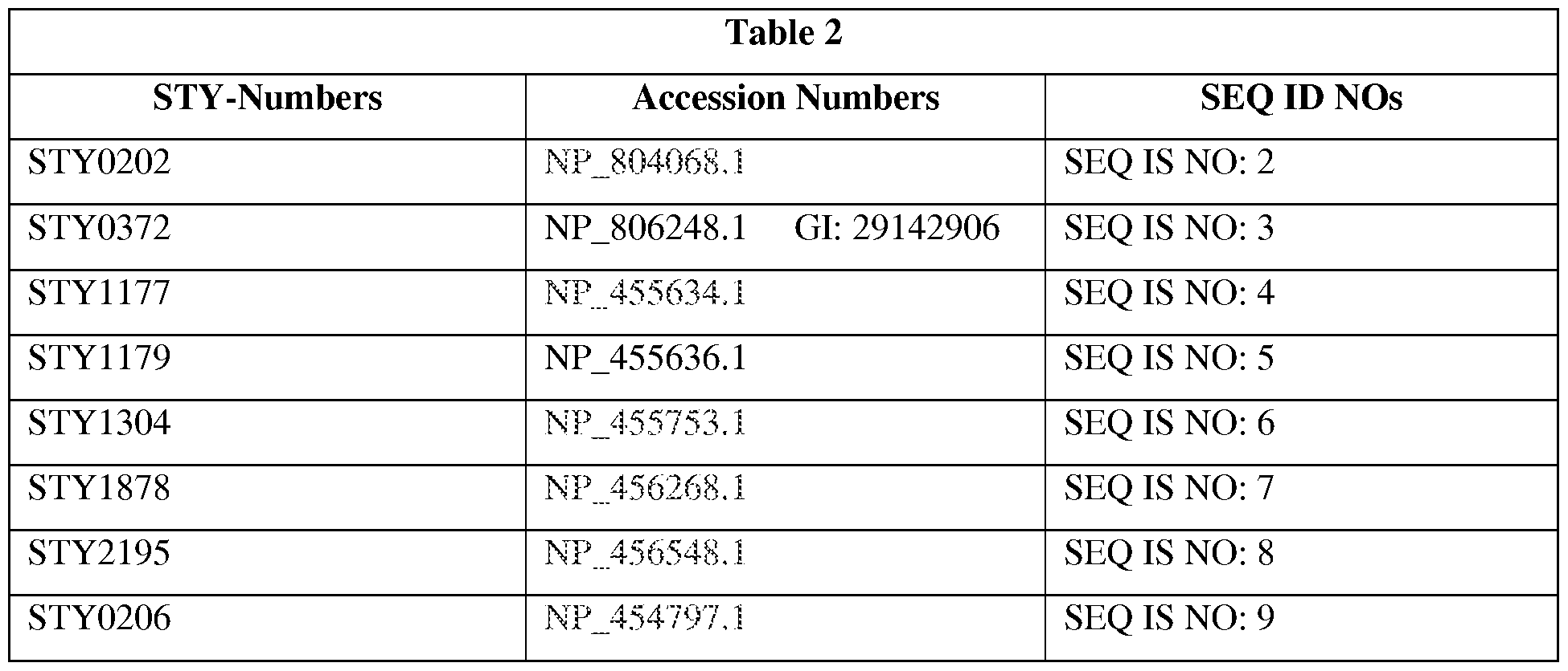
STY-Numbers Accession Numbers SEQ ID NOs
STY0595 NP. 455137.1 SEQ IS NO: 10
STY0909 NP_455403.1 SEQ IS NO: 11
STY1375 P_455817.1 SEQ IS NO: 12
STY 1522 NP .455945.1 SEQ IS NO: 13
STY2167 FliC NP..456520.1 SEQ IS NO: 14
STY2381 NP_456711.1 SEQ IS NO: 15
STY3089 NP_457347.1 SEQ IS NO: 16
STY3090 NP...457348, 1 SEQ IS NO: 17
STY3796 NP_457978.1 SEQ IS NO: 18
REFERENCES
[000169] The references cited below and throughout the specification are herein incorporated by reference in their entirety.
1. Crump JA, Luby SP, Mintz ED (2004) The global burden of typhoid fever. Bull World Health Organ. 82: 346-353.
2. Levine MM, Ferreccio C, Abrego P, Martin OS, Ortiz E, et al. (1999) Duration of efficacy of Ty21a, attenuated Salmonella typhi live oral vaccine. Vaccine 17 Suppl 2: S22-27.
3. Alpuche-Aranda CM, Berthiaume EP, Mock B, Swanson JA, Miller SI (1995) Spacious phagosome formation within mouse macrophages correlates with Salmonella serotype pathogenicity and host susceptibility. Infect Immun 63: 4456-4462.
4. Blaser MJ, Newman LS (1982) A review of human salmonellosis: I. Infective dose. Rev Infect Dis 4: 1096-1106.
5. Buchmeier NA, Heffron F (1989) Intracellular survival of wild-type Salmonella typhimurium and macrophage- sensitive mutants in diverse populations of macrophages. Infect Immun 57: 1-7.
6. Schwan WR, Huang XZ, Hu L, Kopecko DJ (2000) Differential bacterial survival, replication, and apoptosis-inducing ability of Salmonella serovars within human and murine macrophages. Infect Immun 68: 1005-1013.
7. Pang T, Puthucheary SD (1983) Significance and value of the Widal test in the diagnosis of typhoid fever in an endemic area. J Clin Pathol 36: 471-475.
8. Herath HM (2003) Early diagnosis of typhoid fever by the detection of salivary IgA. J Clin Pathol 56: 694-698.
9. House D, Wain J, Ho VA, Diep TS, Chinh NT, et al. (2001) Serology of typhoid fever in an area of endemicity and its relevance to diagnosis. J Clin Microbiol 39: 1002-1007.
10. Perez C, Calderon GM, Ximenez C, Melendro EI (1996) Human cell-mediated immune responses to proteinic fractions of Salmonella typhi. Immunology 89: 262-267.
11. Jesudason MV, Sridharan G, Arulselvan R, Babu PG, John TJ (1998) Diagnosis of typhoid fever by the detection of anti-LPS & anti-flagellin antibodies by ELISA. Indian J Med Res 107: 204-207.
12. Sur D, Ochiai RL, Bhattacharya SK, Ganguly NK, Ali M, et al. (2009) A cluster- randomized effectiveness trial of Vi typhoid vaccine in India. N Engl J Med 361: 335-344.
13. Sarma VN, Malaviya AN, Kumar R, Ghai OP, Bakhtary MM (1977) Development of immune response during typhoid fever in man. Clin Exp Immunol 28: 35-39.
14. Rajagopalan P, Kumar R, Malaviya AN (1982) A study of humoral and cell-mediated immune response following typhoid vaccination in human volunteers. Clin Exp Immunol 47: 275-282.
15. Tiwari H, Kamat RS (1986) Cross-reactions in cell-mediated immunity to Salmonella causing enteric fever. J Med Microbiol 21: 233-237.
16. Ohl ME, Miller SI (2001) Salmonella: a model for bacterial pathogenesis. Annu Rev Med 52: 259-274.
17. Parkhill J, Dougan G, James KD, Thomson NR, Pickard D, et al. (2001) Complete genome sequence of a multiple drug resistant Salmonella enterica serovar Typhi CT18. Nature 413: 848-852.
18. McClelland M, Sanderson KE, Spieth J, Clifton SW, Latreille P, et al. (2001) Complete genome sequence of Salmonella enterica serovar Typhimurium LT2. Nature 413: 852-856.
19. Charles RC, Harris JB, Chase MR, Lebrun LM, Sheikh A, et al. (2009) Comparative proteomic analysis of the PhoP regulon in Salmonella enterica serovar Typhi versus
Typhimurium. PLoS One 4: e6994.
20. Santos RL, Baumler AJ (2004) Cell tropism of Salmonella enterica. Int J Med Microbiol 294: 225-233.
21. Levine MM, DuPont HL, Hornick RB, Snyder MJ, Woodward W, et al. (1976)
Attenuated, streptomycin-dependent Salmonella typhi oral vaccine: potential deleterious effects of lyophilization. J Infect Dis 133: 424-429.
22. Kilhamn J, Lundin SB, Brevinge H, Svennerholm AM, Jertborn M (2003) T- and B-cell immune responses of patients who had undergone colectomies to oral administration of Salmonella enterica serovar Typhi Ty21a vaccine. Clin Diagn Lab Immunol 10: 426-430.
23. Salerno-Goncalves R, Wyant TL, Pasetti MF, Fernandez- Vina M, Tacket CO, et al. (2003) Concomitant induction of CD4+ and CD8+ T cell responses in volunteers immunized with Salmonella enterica serovar Typhi strain CVD 908-htrA. J Immunol 170: 2734-2741.
24. Ravindran R, McSorley SJ (2005) Tracking the dynamics of T-cell activation in response to Salmonella infection. Immunology 114: 450-458.
25. Mastroeni P (2002) Immunity to systemic Salmonella infections. Curr Mol Med 2: 393- 406.
26. Mittrucker HW, Kohler A, Kaufmann SH (2002) Characterization of the murine T- lymphocyte response to Salmonella enterica serovar Typhimurium infection. Infect Immun 70: 199-203.
27. Murthy TV, Wu W, Qiu QQ, Shi Z, LaBaer J, et al. (2004) Bacterial cell-free system for high-throughput protein expression and a comparative analysis of Escherichia coli cell-free and whole cell expression systems. Protein Expr Purif 36: 217-225.
28. Harris JB, Baresch-Bernal A, Rollins SM, Alam A, LaRocque RC, et al. (2006)
Identification of in vivo-induced bacterial protein proteins during human infection with Salmonella enterica serovar Typhi. Infect Immun 74: 5161-5168.
29. Bhuiyan TR, Qadri F, Saha A, Svennerholm AM (2009) Infection by Helicobacter pylori in Bangladeshi children from birth to two years: relation to blood group, nutritional status, and seasonality. Pediatr Infect Dis J 28: 79-85.
30. Sheikh A, Bhuiyan MS, Khanam F, Chowdhury F, Saha A, et al. (2009) Salmonella enterica serovar Typhi- specific immunoglobulin A antibody responses in plasma and antibody in lymphocyte supernatant specimens in Bangladeshi patients with suspected typhoid fever. Clin Vaccine Immunol 16: 1587-1594.
31. LaRocque RC, Krastins B, Harris JB, Lebrun LM, Parker KC, et al. (2008) Proteomic analysis of Vibrio cholerae in human stool. Infect Immun 76: 4145-4151.
32. Elias JE, Gygi SP (2007) Target-decoy search strategy for increased confidence in large- scale protein identifications by mass spectrometry. Nat Methods 4: 207-214.
33. Talawadekar NN, Vadher PJ, Antani DU, Kale VV, Kamat SA (1989) Chloramphenicol resistant Salmonella species isolated between 1978 and 1987. J Postgrad Med 35: 79-82.
34. Bhuiyan TR, Lundin SB, Khan Al, Lundgren A, Harris JB, et al. (2009) Cholera caused by Vibrio cholerae 01 induces T-cell responses in the circulation. Infect Immun 77: 1888- 1893.
35. Qadri F, Ahmed T, Ahmed F, Bradley Sack R, Sack DA, et al. (2003) Safety and immunogenicity of an oral, inactivated enterotoxigenic Escherichia coli plus cholera toxin B subunit vaccine in Bangladeshi children 18-36 months of age. Vaccine 21: 2394-2403.
36. Sacre K, Carcelain G, Cassoux N, Fillet AM, Costagliola D, et al. (2005) Repertoire, diversity, and differentiation of specific CD8 T cells are associated with immune protection against human cytomegalovirus disease. J Exp Med 201: 1999-2010.
37. Gauduin MC (2006) Intracellular cytokine staining for the characterization and quantitation of protein- specific T lymphocyte responses. Methods 38: 263-273.
38. Lundin BS, Johansson C, Svennerholm AM (2002) Oral immunization with a Salmonella enterica serovar Typhi vaccine induces specific circulating mucosa-homing CD4(+) and CD8(+) T cells in humans. Infect Immun 70: 5622-5627.
39. Wahid R, Salerno-Goncalves R, Tacket CO, Levine MM, Sztein MB (2007) Cell- mediated immune responses in humans after immunization with one or two doses of oral live attenuated typhoid vaccine CVD 909. Vaccine 25: 1416-1425.
40. Lo WF, Ong H, Metcalf ES, Soloski MJ (1999) T cell responses to Gram-negative intracellular bacterial pathogens: a role for CD8+ T cells in immunity to Salmonella infection and the involvement of MHC class lb molecules. J Immunol 162: 5398-5406.
41. Sztein MB, Tanner MK, Polotsky Y, Orenstein JM, Levine MM (1995) Cytotoxic T lymphocytes after oral immunization with attenuated vaccine strains of Salmonella typhi in humans. J Immunol 155: 3987-3993.
42. Salerno-Goncalves R, Pasetti MF, Sztein MB (2002) Characterization of CD8(+) effector T cell responses in volunteers immunized with Salmonella enterica serovar Typhi strain Ty21a typhoid vaccine. J Immunol 169: 2196-2203.
43. Salerno-Goncalves R, Fernandez- Vina M, Lewinsohn DM, Sztein MB (2004)
Identification of a human HLA-E-restricted CD8+ T cell subset in volunteers immunized with Salmonella enterica serovar Typhi strain Ty21a typhoid vaccine. J Immunol 173: 5852- 5862.
44. Lundgren A, Kaim J, Jertborn M (2009) Parallel analysis of mucosally derived B- and T- cell responses to an oral typhoid vaccine using simplified methods. Vaccine 27: 4529-4536.
45. Lo WF, Woods AS, DeCloux A, Cotter RJ, Metcalf ES, et al. (2000) Molecular mimicry mediated by MHC class lb molecules after infection with gram-negative pathogens. Nat Med 6: 215-218.
46. Diaz-Quinonez A, Martin-Orozco N, Isibasi A, Ortiz-Navarrete V (2004) Two
Salmonella OmpC K(b)-restricted epitopes for CD8+-T-cell recognition. Infect Immun 72: 3059-3062.
47. Cookson BT, Bevan MJ (1997) Identification of a natural T cell epitope presented by Salmonella-infected macrophages and recognized by T cells from orally immunized mice. J Immunol 158: 4310-4319.
48. Musson JA, Hayward RD, Delvig AA, Hormaeche CE, Koronakis V, et al. (2002) Processing of viable Salmonella typhimurium for presentation of a CD4 T cell epitope from the Salmonella invasion protein C (SipC). Eur J Immunol 32: 2664-2671.
49. Rollenhagen C, Sorensen M, Rizos K, Hurvitz R, Bumann D (2004) Protein selection based on expression levels during infection facilitates vaccine development for an
intracellular pathogen. Proc Natl Acad Sci U S A 101: 8739-8744.
50. McSorley SJ, Jenkins MK (2000) Antibody is required for protection against virulent but not attenuated Salmonella enterica serovar Typhimurium. Infect Immun 68: 3344-3348.
51. Srinivasan A, Foley J, McSorley SJ (2004) Massive number of protein- specific CD4 T cells during vaccination with live attenuated Salmonella causes interclonal competition. J Immunol 172: 6884-6893.
52. Sztein MB (2007) Cell-mediated immunity and antibody responses elicited by attenuated Salmonella enterica Serovar Typhi strains used as live oral vaccines in humans. Clin Infect Dis 45 Suppl 1: S15-19.
53. Salerno-Goncalves R, Sztein MB (2009) Priming of Salmonella enterica serovar typhi- specific CD8(+) T cells by suicide dendritic cell cross-presentation in humans. PLoS One 4: e5879.
54. Miller SI, Kukral AM, Mekalanos JJ (1989) A two-component regulatory system (phoP phoQ) controls Salmonella typhimurium virulence. Proc Natl Acad Sci U S A 86: 5054-5058.
55. Ferrara G, Losi M, Meacci M, Meccugni B, Piro R, et al. (2005) Routine hospital use of a new commercial whole blood interferon-gamma assay for the diagnosis of tuberculosis infection. Am J Respir Crit Care Med 172: 631-635.
56. Goletti D, Vincenti D, Carrara S, Butera O, Bizzoni F, et al. (2005) Selected RD1 peptides for active tuberculosis diagnosis: comparison of a gamma interferon whole-blood enzyme-linked immunosorbent assay and an enzyme-linked immunospot assay. Clin Diagn Lab Immunol 12: 1311-1316.
57. Hauer B, Loddenkemper R, Detjen A, Forssbohm M, Haas W, et al. (2006) [Interferon- gamma assays— description and assessment of a new tool in the diagnosis of tuberculosis]. Pneumologie 60: 29-44.
58. Wain J, Diep TS, Ho VA, Walsh AM, Nguyen TT, et al. (1998) Quantitation of bacteria in blood of typhoid fever patients and relationship between counts and clinical features, transmissibility, and antibiotic resistance. J Clin Microbiol 36: 1683-1687.
59. Wain J, Hosoglu S (2008) The laboratory diagnosis of enteric fever. J Infect Dev Ctries 2: 421-425.
60. Baker S, Favorov M, Dougan G Searching for the elusive typhoid diagnostic. BMC Infect Dis 10: 45.
Claims
1. An in vitro assay comprising the steps of:
a. contacting and incubating a biological sample comprising T cells obtained from a human individual with at least one isolated and purified S. Typhi protein; and
b. detecting and quantifying the amount of at least one S. Typhi protein responsive cytokine in the incubated sample.
2. The in vitro assay of claim 1, wherein the at least one isolated and purified S. Typhi protein is selected from the group consisting of STY0202 (SEQ ID NO: 2), STY0372 (SEQ ID NO: 3), STY1177 (SEQ ID NO: 4), STY1179 (SEQ ID NO: 5), STY1304 (SEQ ID NO: 6), STY1878 (SEQ ID NO: 7), STY2195 (SEQ ID NO: 8), STY0206 (SEQ ID NO: 9), STY0595 (SEQ ID NO: 10), STY0909 (SEQ ID NO: 11), STY1375 (SEQ ID NO: 12), STY1522 (SEQ ID NO: 13), STY2167 FliC (SEQ ID NO: 14), STY2381 (SEQ ID NO: 15), STY3089 (SEQ ID NO: 16), STY3090 (SEQ ID NO: 17), STY3796 (SEQ ID NO: 18) or any combination thereof.
3. The in vitro assay of any one of the preceding claims, wherein the at least one protein is selected from the group consisting of STY0202 (SEQ ID NO: 2), STY0372 (SEQ ID NO: 3), STY1177 (SEQ ID NO: 4), STY1179 (SEQ ID NO: 5), STY1304 (SEQ ID NO: 6), STY1878 (SEQ ID NO: 7), STY2195 (SEQ ID NO: 8) and any combination thereof.
4. The in vitro assay of any one of the preceding claims further comprising a step of comparing the cytokine amount to a reference and diagnosing a typhoid fever in the human individual if the amount of cytokine amount is increased compared to a reference value.
5. The in vitro assay of claim 4, wherein the typhoid fever is diagnosed if the cytokine amount is increased about 1.5 fold or more compared to the reference value.
6. The in vitro assay of any one of the preceding claims wherein the typhoid fever is acute typhoid fever.
7. The in vitro assay of any one of the preceding claims, wherein the at least one S.
Typhi protein responsive cytokine is selected from the group consisting of IFN-γ, IL- 1, IP-10, IL-8, MCP-1, MDC, and MIP-la or any combination thereof.
8. The in vitro assay of claim 7, wherein the at least one S. Typhi protein responsive cytokine is IFN-γ.
9. The in vitro assay of any one of the preceding claims, wherein the biological sample is a blood sample.
10. The in vitro assay of claim 9, wherein the blood sample is a fresh blood sample.
11. The in vitro assay of any one of the preceding claims, wherein the biological sample is isolated peripheral blood mononuclear cells.
12. The in vitro assay of any one of the preceding claims wherein the step of detecting and quantifying the amount of at least one S. Typhi protein responsive cytokine is performed using stereomicroscope, ELISA, ELISPOT, FLUOROSPOT, FACS, FP, FLT or FRET.
13. The in vitro assay of any of the preceding claims, wherein the step of comparing the cytokine amount to a reference and diagnosing a typhoid fever is performed by a non- human machine.
14. The in vitro assay of clam 13, wherein the non-human machine comprises a computer implemented software.
15. A computer program comprising computer program code means adapted to perform all the steps of any of the assays of any of the preceding claims when said program is run on a computer.
16. A composition comprising at least one isolated and purified protein selected from the group consisting of STY0202 (SEQ ID NO: 2), STY0372 (SEQ ID NO: 3), STY1177 (SEQ ID NO: 4), STY1179 (SEQ ID NO: 5), STY1304 (SEQ ID NO: 6), STY1878 (SEQ ID NO: 7), STY2195 (SEQ ID NO: 8), STY0206 (SEQ ID NO: 9), STY0595 (SEQ ID NO: 10), STY0909 (SEQ ID NO: 11), STY1375 (SEQ ID NO: 12), STY1522 (SEQ ID NO: 13), STY2167 FliC (SEQ ID NO: 14), STY2381 (SEQ ID NO: 15), STY3089 (SEQ ID NO: 16), STY3090 (SEQ ID NO: 17), STY3796 (SEQ ID NO: 18) and any combination thereof.
17. The composition of claim 16, wherein the at least one isolated and purified protein selected from the group consisting of STY0202 (SEQ ID NO: 2), STY0372 (SEQ ID NO: 3), STY1177 (SEQ ID NO: 4), STY1179 (SEQ ID NO: 5), STY1304 (SEQ ID NO: 6), STY1878 (SEQ ID NO: 7), STY2195 (SEQ ID NO: 8) and any combination thereof.
18. A kit comprising a composition of any one of claims 16-17.
19. The kit of claim 18 further comprising the composition of claims 16-17, and laboratory materials and instructions to implement an assay of any one of the claims 1-12.
20. The kit of any one of the claims 16-19, which further comprises a washing buffer.
21. The kit of any one of claims 16-19, further comprising a solid support comprising an antibody against at least one S. Typhi protein responsive cytokine against at least wherein the solid support is an ELISA plate and the marker is an enzyme.
22. The kit of claim 21, wherein the S. Typhi protein responsive cytokine is selected from the group consisting of IFN-γ, IL-1, IP-10, IL-8, MCP-1, MDC, and MIP-la.
23. The kit of any one of the claims 15-17, further comprising a secondary antibody
directed to at least one S. Typhi protein responsive cytokine.
24. The kit of claim 18, wherein the at least one S. Typhi protein responsive cytokine is selected from IFN-γ, IL-1, IP-10, IL-8, MCP-1, MDC, and MIP-la.
25. The kit of claims 17-22, wherein the secondary antibody is labeled with a detectable label.
26. A microfluidic device comprising
a. an inlet for adding a blood sample;
b. a chamber comprising at least one isolated and purified S. Typhi protein; and c. a chamber comprising at least one antibody against an S. Typhi protein
responsive cytokine.
27. The microfluidic device of claim 25 further comprising a chamber comprising at least one secondary antibody against the at least one S. Typhi protein responsive cytokine.
28. The microfluidic device of any one of claims 25-26, wherein the at least one isolated and purified S. Typhi protein is selected from the group consisting of STY0202 (SEQ ID NO: 2), STY0372 (SEQ ID NO: 3), STY1177 (SEQ ID NO: 4), STY1179 (SEQ ID NO: 5), STY1304 (SEQ ID NO: 6), STY1878 (SEQ ID NO: 7), STY2195 (SEQ ID NO: 8), STY0206 (SEQ ID NO: 9), STY0595 (SEQ ID NO: 10), STY0909 (SEQ ID NO: 11), STY1375 (SEQ ID NO: 12), STY1522 (SEQ ID NO: 13), STY2167 FliC (SEQ ID NO: 14), STY2381 (SEQ ID NO: 15), STY3089 (SEQ ID NO: 16), STY3090 (SEQ ID NO: 17), STY3796 (SEQ ID NO: 18) or any combination thereof.
29. The microfluidic device of any one of claims 25-27, wherein the S. Typhi protein responsive cytokine is selected from the group consisting of IFN-γ, IL-1, IP-10, IL-8, MCP-1, MDC, and MIP-la.
Applications Claiming Priority (4)
| Application Number | Priority Date | Filing Date | Title |
|---|---|---|---|
| US201161474099P | 2011-04-11 | 2011-04-11 | |
| US61/474,099 | 2011-04-11 | ||
| US201161507867P | 2011-07-14 | 2011-07-14 | |
| US61/507,867 | 2011-07-14 |
Publications (1)
| Publication Number | Publication Date |
|---|---|
| WO2012142022A1 true WO2012142022A1 (en) | 2012-10-18 |
Family
ID=47009652
Family Applications (1)
| Application Number | Title | Priority Date | Filing Date |
|---|---|---|---|
| PCT/US2012/032874 Ceased WO2012142022A1 (en) | 2011-04-11 | 2012-04-10 | Assays, compositions, systems, kits, and devices for diagnosing typhoid fever |
Country Status (1)
| Country | Link |
|---|---|
| WO (1) | WO2012142022A1 (en) |
Cited By (2)
| Publication number | Priority date | Publication date | Assignee | Title |
|---|---|---|---|---|
| WO2018130855A1 (en) * | 2017-01-16 | 2018-07-19 | Oxford University Innovation Limited | Typhoid biomarkers |
| WO2022067076A1 (en) * | 2020-09-25 | 2022-03-31 | Epivax, Inc. | Retro-inverso regulatory t cell epitopes |
Citations (1)
| Publication number | Priority date | Publication date | Assignee | Title |
|---|---|---|---|---|
| US20070009884A1 (en) * | 2005-04-11 | 2007-01-11 | Ghc Technologies, Inc. | Methods and apparatuses for detecting chemical or biological agents |
-
2012
- 2012-04-10 WO PCT/US2012/032874 patent/WO2012142022A1/en not_active Ceased
Patent Citations (1)
| Publication number | Priority date | Publication date | Assignee | Title |
|---|---|---|---|---|
| US20070009884A1 (en) * | 2005-04-11 | 2007-01-11 | Ghc Technologies, Inc. | Methods and apparatuses for detecting chemical or biological agents |
Non-Patent Citations (20)
| Title |
|---|
| DATABASE GENPEPT 18 November 2002 (2002-11-18), retrieved from http://www.ncbi.nlm.nih.gov/protein/AB0525 accession no. B0525 * |
| DATABASE GENPEPT 18 November 2002 (2002-11-18), retrieved from http://www.ncbi.nlm.nih.gov/protein/AB0659 accession no. B0659 * |
| DATABASE GENPEPT 18 November 2002 (2002-11-18), retrieved from http://www.ncbi.nlm.nih.gov/protein/AB0860 accession no. B0860 * |
| DATABASE GENPEPT 18 November 2002 (2002-11-18), retrieved from http://www.ncbi.nlm.nih.gov/protein/AC0860 accession no. C0860 * |
| DATABASE GENPEPT 18 November 2002 (2002-11-18), retrieved from http://www.ncbi.nlm.nih.gov/protein/AC0941 accession no. C0941 * |
| DATABASE GENPEPT 18 November 2002 (2002-11-18), retrieved from http://www.ncbi.nlm.nih.gov/protein/AD0570 accession no. D0570 * |
| DATABASE GENPEPT 18 November 2002 (2002-11-18), retrieved from http://www.ncbi.nlm.nih.gov/protein/AD0754 accession no. D0754 * |
| DATABASE GENPEPT 18 November 2002 (2002-11-18), retrieved from http://www.ncbi.nlm.nih.gov/protein/AE0544 accession no. E0544 * |
| DATABASE GENPEPT 18 November 2002 (2002-11-18), retrieved from http://www.ncbi.nlm.nih.gov/protein/AE0635 accession no. E0635 * |
| DATABASE GENPEPT 18 November 2002 (2002-11-18), retrieved from http://www.ncbi.nlm.nih.gov/protein/AE0776 accession no. E0776 * |
| DATABASE GENPEPT 18 November 2002 (2002-11-18), retrieved from http://www.ncbi.nlm.nih.gov/protein/AF0525 accession no. F0525 * |
| DATABASE GENPEPT 18 November 2002 (2002-11-18), retrieved from http://www.ncbi.nlm.nih.gov/protein/AG0635 accession no. G0635 * |
| DATABASE GENPEPT 18 November 2002 (2002-11-18), retrieved from http://www.ncbi.nlm.nih.gov/protein/AG0650 accession no. G0650 * |
| DATABASE GENPEPT 18 November 2002 (2002-11-18), retrieved from http://www.ncbi.nlm.nih.gov/protein/AG0675 accession no. G0675 * |
| DATABASE GENPEPT 18 November 2002 (2002-11-18), retrieved from http://www.ncbi.nlm.nih.gov/protein/AG0717 accession no. G0717 * |
| DATABASE GENPEPT 18 November 2002 (2002-11-18), retrieved from http://www.ncbi.nlm.nih.gov/protein/AH0750 accession no. H0750 * |
| DATABASE GENPEPT 18 November 2002 (2002-11-18), retrieved from http://www.ncbi.nlm.nih.gov/protein/AI0605 accession no. I0605 * |
| HARRIS J.B. ET AL.: "Identification of In Vivo-Induced Bacterial Protein Antigens during Human Infection with Salmonella enterica Serovar Typhi", INFECTION IMMUNITY, vol. 74, no. 9, 2006, pages 5161 - 5168 * |
| PAI M. ET AL.: "Interferon-y assays in the immunodiagnosis of tuberculosis: a systematic review", LANCET INFECTIOUS DISEASES, vol. 4, 2004, pages 761 - 763 * |
| WYANT T.L. ET AL.: "Salmonella typhi Flagella Are Potent Inducers of Proinflammatory Cytokine Secretion by Human Monocytes", INFECTION AND IMMUNITY, vol. 67, no. 7, 1999, pages 3619 - 362, XP002415285 * |
Cited By (2)
| Publication number | Priority date | Publication date | Assignee | Title |
|---|---|---|---|---|
| WO2018130855A1 (en) * | 2017-01-16 | 2018-07-19 | Oxford University Innovation Limited | Typhoid biomarkers |
| WO2022067076A1 (en) * | 2020-09-25 | 2022-03-31 | Epivax, Inc. | Retro-inverso regulatory t cell epitopes |
Similar Documents
| Publication | Publication Date | Title |
|---|---|---|
| Turner et al. | SARS-CoV-2 infection induces long-lived bone marrow plasma cells in humans | |
| Branda et al. | Laboratory diagnosis of Lyme borreliosis | |
| JP7314358B2 (en) | Use of amino acid sequences from Mycobacterium tuberculosis or their corresponding nucleic acids for the diagnosis and prevention of tuberculosis infection, and diagnostic kits and vaccines derived therefrom | |
| Gimenez et al. | SARS‐CoV‐2‐reactive interferon‐γ‐producing CD8+ T cells in patients hospitalized with coronavirus disease 2019 | |
| Carter et al. | The antibody-secreting cell response to infection: kinetics and clinical applications | |
| CN104628833B (en) | A kind of tuberculosis infection cellular immunization detectable antigens composition and application thereof | |
| Sheikh et al. | Interferon-γ and proliferation responses to Salmonella enterica serotype Typhi proteins in patients with S. Typhi bacteremia in Dhaka, Bangladesh | |
| Dey et al. | Recombinant antigen-based latex agglutination test for rapid serodiagnosis of leptospirosis | |
| Reid et al. | Treponema pallidum periplasmic and membrane proteins are recognized by circulating and skin CD4+ T cells | |
| US10288610B2 (en) | Vitro assays for detecting Salmonella enterica serotype typhi | |
| CN106248934B (en) | Antigen of mycobacterium tuberculosis albumen Rv0446c and its t cell epitope peptide application | |
| EP1735623B1 (en) | Mycobacterium tuberculosis infection diagnostic test | |
| CN106405107B (en) | Antigen of mycobacterium tuberculosis albumen Rv2941 and its t cell epitope peptide application | |
| CN106248935B (en) | Antigen of mycobacterium tuberculosis albumen Rv1798 and its t cell epitope peptide application | |
| WO2012142022A1 (en) | Assays, compositions, systems, kits, and devices for diagnosing typhoid fever | |
| Angkeow et al. | Prevalence, persistence, and genetics of antibody responses to protein toxins and virulence factors | |
| CN104628834B (en) | A kind of tuberculosis infection T cell immunodetection antigen and application thereof | |
| Thirlwall et al. | Improving the specificity of immunodiagnosis for porcine brucellosis | |
| Hoving et al. | Combinatorial multimer staining and spectral flow cytometry facilitate quantification and characterization of polysaccharide-specific B cell immunity | |
| WO2014140833A2 (en) | Methods for differentiating between disease states | |
| Wang et al. | Establishment and evaluation of an interferon-gamma enzyme-linked immunospot method for the detection of Brucella-infected cattle and goats | |
| CN114671928A (en) | Application of mycobacterium tuberculosis T cell epitope protein Rv1566c-444 | |
| CN106248936B (en) | The application of antigen of mycobacterium tuberculosis albumen Rv2201 and its t cell epitope peptide | |
| CN106442983B (en) | The application of antigen of mycobacterium tuberculosis albumen Rv3793 and its t cell epitope peptide | |
| Sharma et al. | Typhoid Diagnostics: Looking Beneath the Surface |
Legal Events
| Date | Code | Title | Description |
|---|---|---|---|
| 121 | Ep: the epo has been informed by wipo that ep was designated in this application |
Ref document number: 12771842 Country of ref document: EP Kind code of ref document: A1 |
|
| NENP | Non-entry into the national phase |
Ref country code: DE |
|
| 122 | Ep: pct application non-entry in european phase |
Ref document number: 12771842 Country of ref document: EP Kind code of ref document: A1 |