WO2012134822A1 - Allosteric reversible pan-caspase inhibitors - Google Patents
Allosteric reversible pan-caspase inhibitors Download PDFInfo
- Publication number
- WO2012134822A1 WO2012134822A1 PCT/US2012/029206 US2012029206W WO2012134822A1 WO 2012134822 A1 WO2012134822 A1 WO 2012134822A1 US 2012029206 W US2012029206 W US 2012029206W WO 2012134822 A1 WO2012134822 A1 WO 2012134822A1
- Authority
- WO
- WIPO (PCT)
- Prior art keywords
- caspase
- comp
- compounds
- allosteric
- apoptosis
- Prior art date
- Legal status (The legal status is an assumption and is not a legal conclusion. Google has not performed a legal analysis and makes no representation as to the accuracy of the status listed.)
- Ceased
Links
- 0 C*c1cc(I)ccc1I Chemical compound C*c1cc(I)ccc1I 0.000 description 3
- JGBDWRSVCOWWAD-UHFFFAOYSA-N CCN(CC)C(SN)=S Chemical compound CCN(CC)C(SN)=S JGBDWRSVCOWWAD-UHFFFAOYSA-N 0.000 description 1
- ISKOGNCMNSEPFR-UHFFFAOYSA-N CNC(SSC(N(C)C)=S)=S Chemical compound CNC(SSC(N(C)C)=S)=S ISKOGNCMNSEPFR-UHFFFAOYSA-N 0.000 description 1
- VSWDORGPIHIGNW-UHFFFAOYSA-N SC(N1CCCC1)=S Chemical compound SC(N1CCCC1)=S VSWDORGPIHIGNW-UHFFFAOYSA-N 0.000 description 1
Classifications
-
- C—CHEMISTRY; METALLURGY
- C07—ORGANIC CHEMISTRY
- C07D—HETEROCYCLIC COMPOUNDS
- C07D211/00—Heterocyclic compounds containing hydrogenated pyridine rings, not condensed with other rings
- C07D211/04—Heterocyclic compounds containing hydrogenated pyridine rings, not condensed with other rings with only hydrogen or carbon atoms directly attached to the ring nitrogen atom
- C07D211/80—Heterocyclic compounds containing hydrogenated pyridine rings, not condensed with other rings with only hydrogen or carbon atoms directly attached to the ring nitrogen atom having two double bonds between ring members or between ring members and non-ring members
- C07D211/82—Heterocyclic compounds containing hydrogenated pyridine rings, not condensed with other rings with only hydrogen or carbon atoms directly attached to the ring nitrogen atom having two double bonds between ring members or between ring members and non-ring members with only hydrogen atoms, hydrocarbon or substituted hydrocarbon radicals, directly attached to ring carbon atoms
-
- A—HUMAN NECESSITIES
- A61—MEDICAL OR VETERINARY SCIENCE; HYGIENE
- A61K—PREPARATIONS FOR MEDICAL, DENTAL OR TOILETRY PURPOSES
- A61K31/00—Medicinal preparations containing organic active ingredients
- A61K31/33—Heterocyclic compounds
- A61K31/395—Heterocyclic compounds having nitrogen as a ring hetero atom, e.g. guanethidine or rifamycins
- A61K31/435—Heterocyclic compounds having nitrogen as a ring hetero atom, e.g. guanethidine or rifamycins having six-membered rings with one nitrogen as the only ring hetero atom
- A61K31/44—Non condensed pyridines; Hydrogenated derivatives thereof
-
- A—HUMAN NECESSITIES
- A61—MEDICAL OR VETERINARY SCIENCE; HYGIENE
- A61K—PREPARATIONS FOR MEDICAL, DENTAL OR TOILETRY PURPOSES
- A61K31/00—Medicinal preparations containing organic active ingredients
- A61K31/33—Heterocyclic compounds
- A61K31/555—Heterocyclic compounds containing heavy metals, e.g. hemin, hematin, melarsoprol
-
- A—HUMAN NECESSITIES
- A61—MEDICAL OR VETERINARY SCIENCE; HYGIENE
- A61K—PREPARATIONS FOR MEDICAL, DENTAL OR TOILETRY PURPOSES
- A61K45/00—Medicinal preparations containing active ingredients not provided for in groups A61K31/00 - A61K41/00
- A61K45/06—Mixtures of active ingredients without chemical characterisation, e.g. antiphlogistics and cardiaca
-
- C—CHEMISTRY; METALLURGY
- C07—ORGANIC CHEMISTRY
- C07F—ACYCLIC, CARBOCYCLIC OR HETEROCYCLIC COMPOUNDS CONTAINING ELEMENTS OTHER THAN CARBON, HYDROGEN, HALOGEN, OXYGEN, NITROGEN, SULFUR, SELENIUM OR TELLURIUM
- C07F1/00—Compounds containing elements of Groups 1 or 11 of the Periodic Table
- C07F1/08—Copper compounds
Definitions
- This invention is in the field of medicinal chemistry and relates to novel compounds, and pharmaceutical compositions and methods of use thereof that inhibit caspases and/or tumor necrosis factor alpha (TNFa) that mediate cell apoptosis and inflammation and inhibit pathophysiologic effects of excessive amounts of TNFa.
- the invention also relates to methods of using the compounds and pharmaceutical compositions of this invention to treat diseases where caspase and/or TNFa activity is implicated.
- the present invention also relates to methods for the use of allosteric, reversible pan-caspase inhibitors with pyridinyl, copper-containing molecules with a multi-ring structure.
- caspase and TNFa inhibitors While a number of caspase and TNFa inhibitors have been reported, it is not clear whether they possess the appropriate pharmacological properties to be therapeutically useful. Therefore, there is a continued need for small molecule caspase and TNFa inhibitors to provide effective inhibition of apoptosis. Such compounds would be extremely useful in treating the disease states where caspase enzymes and/or TNFa cytokines play a role.
- This invention is in the field of medicinal chemistry and relates to novel compounds, and pharmaceutical compositions and methods of use thereof that inhibit caspases and/or tumor necrosis factor alpha (TNFa) that mediate cell apoptosis and inflammation and inhibit pathophysiologic effects of excessive amounts of TNFa.
- the invention also relates to methods of using the compounds and pharmaceutical compositions of this invention to treat diseases where caspase and/or TNFa activity is implicated.
- the present invention also relates to methods for the use of allosteric, reversible pan-caspase inhibitors with pyridinyl, copper-containing molecules with a multi-ring structure.
- the invention contemplates a method comprising; a) providing; i) an allosteric pan-caspase inhibitor of Formula I; ii) a subject suffering from a disease state, wherein said disease has at least one symptom, wherein said subject comprises a caspase; and b) treating said subject with said allosteric pan-caspase inhibitorof Formula I so as to reduce at least one symptom of said disease state.
- said allosteric pan-caspase inhibitor of Formula I comprises:
- R x is CH 3 (Me), -CH 2 CH 3 (Et), -CH 2 CH 2 CH 3 ( «-Pr), -CH(CH 3 ) 2 (iso-Pr), -CH(CH 2 ) 2 (cyclopropyl), -CH 2 CH 2 CH 2 CH 3 (n-Bu), -CH(CH 3 )C3 ⁇ 4CH 3 (sec-butyl), -CH 2 CH(CH 3 ) 2 (z ' so-butyl), -C(CH 3 ) (tert-butyl), -CH 2 C(CH 3 ) 3 (weo-pentyl), cyclobutyl, cyclopentyl, cyclohexyl, cyclohexylmethyl, adamant- 1-yl, adamant-2-yl, phenyl, and substituted phenyl; R 2 is at least one of the following groups comprising: CI, Br, F, I, and (E)-methyl 2-((l)
- said subject has a symptom of cell death.
- symptom of cell death is mediated by an intrinsic pathway of apoptosis.
- said allosteric pan-caspase inhibitorof Formula I provides protection from cell death.
- said subject further comprises a cell containing a caspase, and a step of isolating said cell from said subject that is capable of undergoing apoptosis induced by ultra violet light.
- said disease state results from exposure to at least one of the group comprising: tumor necrosis factor-a (TNFoc) and cycloheximide.
- said allosteric pan-caspase inhibitor of Formula I is selected from the group consisting of:
- said disease state is selected from the group consisting of neurodegeneration, radiation syndrome, ultra violet light exposure, osteoarthritis, pancreatitis, rheumatoid arthritis, chronic active hepatitis, inflammatory bowel disease, Crohn's disease, psoriasis, organ transplant rejection, sepsis, septic shock, cerebral ischemia, myocardial ischemia, myocardial infarction, amyotrophic lateral sclerosis, multiple sclerosis, neurological damage due to stroke, hepatitis-B, hepatitis-C, hepatitis-G, and liver disease.
- the invention contemplates a pharmaceutical composition of
- This invention further provides pharmaceutical compositions comprising caspase inhibitor compound according to Formula I.
- This invention also relates to methods of using said pharmaceutical compositions for treatment of caspase-mediated diseases including inflammatory and degenerative diseases.
- the invention contemplates an allosteric pan-caspase inhibitor of
- Ri is CH 3 (Me), -CH 2 CH 3 (Et), -CH 2 CH 2 CH 3 ( «-Pr), -CH(CH 3 ) 2 (iso-Pr), -CH(CH 2 ) 2 (cyclopropyl), -CH 2 CH 2 CH 2 CH 3 (n-Bu), -CH(CH 3 )CH 2 CH 3 (sec-butyl), -CH 2 CH(CH 3 ) 2 (zso-butyl), -C(CH 3 ) 3 (tert-butyY), -CH 2 C(CH 3 ) 3 ( «eo-pentyl), cyclobutyl, cyclopentyl, cyclohexyl, cyclohexylmethyl, adamant- 1-yl, adamant-2-yl, phenyl, and substituted phenyl; R 2 is at least one of the following groups comprising: CI, Br, F, I, and (E)-methyl 2-(( 1 ,2-
- tumor necrosis factor (TNF, cachexin or cachectin and known as tumor necrosis factor-alpha or TNFa) as used herein refers to a cytokine involved in systemic inflammation and is a member of a group of cytokines that stimulate the acute phase reaction.
- TNF tumor necrosis factor
- Cycloheximide as used herin refers to an inhibitor of protein biosynthesis in eukaryotic organisms, produced by the bacterium Streptomyces griseus. Cycloheximide exerts its effect by interfering with the translocation step in protein synthesis (movement of two tRNA molecules and mRNA in relation to the ribosome) thus blocking translational elongation. Cycloheximide is widely used in biomedical research to inhibit protein synthesis in eukaryotic cells studied in vitro (i.e. outside of organisms). It is inexpensive and works rapidly. Its effects are rapidly reversed by simply removing it from the culture medium. Cycloheximide has the structure:
- UV ultra violet light
- UV electromagnetic radiation with a wavelength shorter than that of visible light, but longer than X-rays, in the range 10 nm to 400 nm, and energies from 3eV to 124 eV. It is so named because the spectrum consists of electromagnetic waves with frequencies higher than those that humans identify as the color violet. Although ultraviolet is invisible to the human eye, most people are aware of the effects of UV through the painful condition of sunburn, but the UV spectrum has many other effects, both beneficial and damaging, to human health.
- neurodegeneration refers to any progressive loss of structure or function of neurons, including death of neurons. Many neurodegenerative diseases including Parkinson's, Alzheimer's, and Huntington's occur as a result of neurodegenerative processes.
- ARS acute radiation syndrome
- radiation poisoning radiation sickness
- radiation toxicity refers to a constellation of health effects which occur within several months of exposure to high amounts of ionizing radiation [5, 6].
- the term generally refers to acute medical problems rather than ones that develop after a prolonged period. The onset and type of symptoms depends on the radiation exposure. Relatively smaller doses result in gastrointestinal effects such as nausea and vomiting and symptoms related to falling blood counts such as infection and bleeding. Relatively larger doses can result in neurological effects and rapid death. Treatment of acute radiation syndrome is generally supportive with blood transfusions and antibiotics [5].
- Classically acute radiation syndrome is divided into three main presentations: hematopoietic, gastrointestinal and neurological/vascular. These symptoms may or may not be preceded by a prodrome [5].
- the speed of onset of symptoms is related to radiation exposure, with greater doses resulting in a shorter delay in symptom onset [5].
- Hematopoietic This syndrome is marked by a drop in the number of blood cells. This may result in infections due to low white blood cells, bleeding due to low platelets, and anemia due to low red blood cells [5].
- Gastrointestinal This syndrome typically occurs at exposure doses of 600-1000 rad (6-10 Gy) [5]. Nausea, vomiting, loss of appetite, and abdominal pain are usually seen within one to two hours [5].
- Neurovascular This syndrome typically occurs at exposure doses greater than 1000 rad (10 Gy) [5]. It presents with neurological symptoms such as dizziness, headache, or decreased level of consciousness with an absence of vomiting [5].
- the prodrome an early symptom (or set of symptoms) that might indicate the start of a disease before specific symptoms occur, associated with ARS typically includes nausea and vomiting, headaches, fatigue, fever and short period of skin reddening [5]. These symptoms may occur at radiation doses as low as 35 rad (0.35 Gy). Thus, they may not be followed by acute radiation sickness [5],
- OA osteoarthritis
- degenerative arthritis also known as degenerative joint disease, as used herein refers to a group of mechanical abnomialities involving degradation of joints, including articular cartilage and subchondral bone.
- pancreatitis refers to inflammation of the pancreas that can occur in two very different forms. Acute pancreatitis is sudden while chronic pancreatitis "is characterized by recurring or persistent abdominal pain with or without steatorrhea or diabetes mellitus.”
- rheumatoid arthritis refers to a chronic, systemic inflammatory disorder that may affect many tissues and organs, but principally attacks synovial joints. The process produces an inflammatory response of the synovium (synovitis) secondary to hyperplasia of synovial cells, excess synovial fluid, and the development of pannus in the synovium.
- chronic active hepatitis refers to a recurrent inflammation of the liver characterized by the presence of inflammatory cells in the tissue of the organ.
- IBD inflammatory bowel disease
- Chronic ulcerative colitis also known as regional enteritis, as used herein refers to an inflammatory disease of the intestines that may affect any part of the gastrointestinal tract from mouth to anus, causing a wide variety of symptoms. It primarily causes abdominal pain, diarrhea (which may be bloody if inflammation is at its worst), vomiting, or weight loss, but may also cause complications outside the gastrointestinal tract such as skin rashes, arthritis, inflammation of the eye, tiredness, and lack of concentration.
- Psoriasis refers to a chronic autoimmune disease that appears on the skin. It occurs when the immune system sends out faulty signals that speed up the growth cycle of skin cells. Psoriasis is not contagious.
- organ transplant rejection refers to when a transplanted organ or tissue is not accepted by the body of the transplant recipient. This is explained by the concept that the immune system of the recipient attacks the transplanted organ or tissue.
- sepsis refers to a potentially serious medical condition that is characterized by a whole-body inflammatory state (called a systemic inflammatory response syndrome or SIRS) and the presence of a known or suspected infection.
- SIRS systemic inflammatory response syndrome
- the body may develop this inflammatory response by the immune system to microbes in the blood, urine, lungs, skin, or other tissues.
- a lay term for sepsis is blood poisoning, more aptly applied to septicemia. Severe sepsis is the systemic inflammatory response, plus infection, plus the presence of organ dysfunction.
- tissue perfusion and oxygen delivery refers to a medical emergency caused by decreased tissue perfusion and oxygen delivery as a result of severe infection and sepsis, though the microbe may be systemic or localized to a particular site. It can cause multiple organ dysfunction syndrome (formerly known as multiple organ failure) and death.
- Cerebral ischemia refers to a condition in which there is insufficient blood flow to the brain to meet metabolic demand.
- myocardial ischemia refers to an imbalance between myocardial oxygen supply and demand resulting in angina pectoris, myocardial stunning, myocardial hibernation, ischemic preconditioning, postconditioning, or under the most severe instances, acute coronary syndrome and myocardial infarction.
- MI myocardial infarction
- AMI acute myocardial infarction
- heart attack refers to the interruption of blood supply to a part of the heart, causing heart cells to die.
- ALS myotrophic lateral sclerosis
- Lou Gehrig's disease refers to a form of motor neuron disease.
- ALS is a progressive, fatal, neurodegenerative disease caused by the degeneration of motor neurons, the nerve cells in the central nervous system that control voluntary muscle movement.
- MS multiple sclerosis
- disseminated sclerosis also known as disseminated sclerosis or encephalomyelitis disseminata
- MS multiple sclerosis
- encephalomyelitis disseminata refers to an inflammatory disease in which the fatty myelin sheaths around the axons of the brain and spinal cord are damaged, leading to demyelination and scarring as well as a broad spectrum of signs and symptoms.
- hepatitis B refers to an infectious illness caused by hepatitis
- HBV B virus which infects the liver of Hominoidea, including humans, and causes an inflammation called hepatitis.
- hepatitis C refers to an infectious disease affecting the liver, caused by the hepatitis C virus (HCV).
- hepatitis G refers to a form of liver inflammation caused by hepatitis G virus (HGV), a distant relative of the hepatitis C virus.
- Liver disease refers to a broad term describing any single number of diseases affecting the liver. Many are accompanied by jaundice caused by increased levels of bilirubin in the system.
- the terms “reduce,” “inhibit,” “diminish,” “suppress,” “decrease,” “prevent” and grammatical equivalents when in reference to the expression of any symptom in an untreated subject relative to a treated subject, mean that the quantity and/or magnitude of the symptoms in the treated subject is lower than in the untreated subject by any amount that is recognized as clinically relevant by any medically trained personnel.
- the quantity and/or magnitude of the symptoms in the treated subject is at least 10% lower than, at least 25% lower than, at least 50% lower than, at least 75% lower than, and/or at least 90% lower than the quantity and/or magnitude of the symptoms in the untreated subject.
- Epimers refers to diastereomers that differ in configuration of only one stereogenic center. Diastereomers are a class of stereoisomers that are non-superposable, non-mirror images of one another, unlike enantiomers that are non-superposable mirror images of one another.
- sugar refers to a monosaccharide, disaccharide, trisaccharides, or polysaccharides.
- Monosaccharides have the general formula (CH 2 0) n , in which n is an integer larger than 2.
- Disaccharides have the general formula C n (H 2 0) n -i, with n larger than 5.
- Polysaccharides include such substances as cellulose, dextrin, glycogen, and starch.
- pharmaceutically acceptable monosaccharide refers to a pharmaceutically acceptable aldose sugar, a pharmaceutically acceptable ketose sugar, or other specified sugar.
- pharmaceutically acceptable aldose sugars within the contemplation of the present invention are erythrose, threose, ribose, arabinose, xylose, lyxose, allose, altrose, glucose, mannose, gulose, idose, galactose and talose.
- ketose sugars preferred for use in the composition of the present invention are erythrulose, ribulose, xylulose, psicose, fructose, sorbose, tagatose, and sedoheptulose.
- other specified sugars preferred for use in the composition of the present invention are fucose, fuculose, rhamnose, or any other deoxy sugar.
- (D) or (L) isomers may be employed, the (D) form is generally preferable.
- compositions of the present invention may be prepared by formulating them in dosage forms, which are suitable for peroral, rectal or nonparenteral administration, the last-mentioned including intravenous injection and administration into the cerebrospinal fluid.
- dosage forms which are suitable for peroral, rectal or nonparenteral administration, the last-mentioned including intravenous injection and administration into the cerebrospinal fluid.
- common carriers and routine formulation techniques may be employed.
- API active pharmaceutical ingredient
- active pharmaceutical ingredient refers to the substance in a pharmaceutical drug that is biologically active.
- Common carriers refers to those which are employed in standard pharmaceutical preparations and includes excipients, binders and disintegrators the choice of which depends on the specific dosage form used.
- excipient are starch, lactose, sucrose, glucose, mannitol and cellulose; illustrative binders are polyvinylpyrrolidone, starch, sucrose, hydroxypropyl cellulose and gum arabic; illustrative disintegrators include starch, agar, gelatin powder, cellulose, and CMC. Any other common excipients, binders and disintegrators may also be employed.
- the pharmaceutical composition of the present invention preferably contains antioxidants for the purpose of stabilizing the effective ingredient.
- antioxidants may be selected from among those which are commonly incorporated in pharmaceuticals and include ascorbic acid, N-acetylcysteine, acetylcysteine, L-cystein, D, L-a-tocopherol, and natural tocopherol.
- Formulations of the pharmaceutical composition of the present invention which are suitable for peroral administration may be provided in the form of tablets, capsules, powders, granules, or suspensions in non-aqueous solutions such as syrups, emulsions or drafts, each containing one or more of the active compounds in predetermined amounts.
- the granule may be provided by first preparing an intimate mixture of one or more of the active ingredients with one or more of the auxiliary components shown above, then granulating the mixture, and classifying the granules by screening through a sieve.
- the tablet may be prepared by compressing or otherwise forming one or more of the active ingredients, optionally with one or more auxiliary components.
- the capsule may be prepared by first making a powder or granules as an intimate mixture of one or more of the active ingredients with one or more auxiliary components, then charging the mixture into an appropriate capsule on a packing machine, etc.
- the pharmaceutical composition of the present invention may be formulated as a suppository (for rectal administration) with the aid of a common carrier such a cocoa butter.
- the pharmaceutical composition of the present invention may also be formulated in a dosage form suitable for non-parenteral administration by packaging one or more active ingredients as dry solids in a sterile nitrogen-purged container. The resulting dry formulation may be administered to patients non-parenterally after being dispersed or dissolved in a given amount of aseptic water.
- the dosage forms are preferably prepared from a mixture of the active ingredients, routine auxiliary components and one or more of the antioxidants listed above.
- the formulations may further contain one or more auxiliary components selected from among excipients, buffers, flavoring agents, binders, surfactants, thickening agents, and lubricants.
- the dose of the various pro-drugs will of course vary with the route of administration, the severity of the disease to be treated, and the patient to be treated, but the exact dose ultimately chosen should be left to the good discretion of the doctor responsible for the treatment. If a desired dose is determined, the active ingredient may be administered once a day or, alternatively, it may be administered in up to as many portions as deemed appropriate at suitable intervals. The active ingredient may be straightforwardly administered without being mixed with any other components. However, for several reasons, typically for the purpose of providing ease in controlling the dose level, the active compound is preferably administered in a pharmaceutical dosage form.
- salts refers to any salt that complexes with identified compounds contained herein while retaining a desired function, e.g., biological activity.
- salts include, but are not limited to, acid addition salts formed with inorganic acids (e.g.
- hydrochloric acid hydrobromic acid, sulfuric acid, phosphoric acid, nitric acid, and the like
- salts formed with organic acids such as, but not limited to, acetic acid, oxalic acid, tartaric acid, succinic acid, malic acid, fumaric acid, maleic acid, ascorbic acid, benzoic acid, tannic acid, pamoic acid, alginic acid, polyglutamic, acid, naphthalene sulfonic acid, naphthalene disulfonic acid, and polygalacturonic acid.
- Pharmaceutically acceptable salts also include base addition salts which may be formed when acidic protons present are capable of reacting with inorganic or organic bases.
- Suitable pharmaceutically-acceptable base addition salts include metallic salts, such as salts made from aluminum, calcium, lithium, magnesium, potassium, sodium and zinc, or salts made from organic bases including primary, secondary and tertiary amines, substituted amines including cyclic amines, such as caffeine, arginine, diethylamine, N-ethyl piperidine, histidine, glucamine, isopropylamine, lysine, morpholine, N-ethyl morpholine, piperazine, piperidine, triethylamine, trimethylamine. All of these salts may be prepared by conventional means from the corresponding compound of the invention by reacting, for example, the appropriate acid or base with the compound of the invention. Unless otherwise specifically stated, the present invention contemplates pharmaceutically acceptable salts of the considered pro-drugs.
- methylene means a chemical species in which a carbon atom is bonded to two hydrogen atoms.
- The— CH 2 - group is considered to be the standard methylene group.
- Methylene groups in a chain or ring contribute to its size and lipophilicity.
- dideoxy also refers the methylene groups.
- alkoxy(c ⁇ io) designates those alkoxy groups having from 1 to 10 carbon atoms (e.g., 1, 2, 3, 4, 5, 6, 7, 8, 9, or 10, or any range derivable therein (e.g., 3-10 carbon atoms)).
- alkyl ( c 2 -io ) designates those alkyl groups having from 2 to 10 carbon atoms (e.g., 2, 3, 4, 5, 6, 7, 8, 9, or 10, or any range derivable therein (e.g., 3-10 carbon atoms)).
- alkyl when used without the "substituted” modifier refers to a non-aromatic monovalent group with a saturated carbon atom as the point of attachment, a linear or branched, cyclo, cyclic or acyclic structure, no carbon-carbon double or triple bonds, and no atoms other than carbon and hydrogen.
- the groups, -CH 3 (Me), -CH 2 CH 3 (Et), -CH 2 CH 2 CH 3 (rc-Pr), -CH(CH 3 ) 2 (fco-Pr or z-Pr), -CH(CH 2 ) 2 (cyclopropyl), -CH 2 CH 2 CH 2 CH 3 (w-Bu), -CH(CH 3 )CH 2 CH 3 (sec-butyl or sec- u), -CH 2 CH(CH 3 ) 2 (z ' 50-butyl or z ' -Bu), -C(CH 3 ) 3 (tert-butyl or t-Bu), -CH 2 C(CH 3 ) 3 (neo-pentyl), cyclobutyl, cyclopentyl, cyclohexyl, cyclohexylmethyl are non-limiting examples of alkyl groups.
- substituted alkyl refers to a non-aromatic monovalent group with a saturated carbon atom as the point of attachment, a linear or branched, cyclo, cyclic or acyclic structure, no carbon-carbon double or triple bonds, and at least one atom independently selected from the group consisting of N, O, F, CI, Br, I, Si, P, and S.
- the following groups are non-limiting examples of substituted alkyl groups: -CH 2 OH, -CH 2 C1, -CH 2 Br, -CH 2 SH, -CF 3 , -CH 2 CN, -CH 2 C(0)H, -CH 2 C(0)OH, -CH 2 C(0)OCH 3 , -CH 2 C(0)NH 2 , -CH 2 C(0)NHCH 3 , -CH 2 C(0)CH 3 , -CH 2 OCH 3 , -CH 2 OC3 ⁇ 4CF 3 , -CH 2 OC(0)CH 3 , -CH 2 NH 2 , -CH 2 NHCH 3 , -CH 2 N(CH 3 ) 2 , -CH 2 CH 2 C1, -CH 2 CH 2 OH, -CH 2 CF 3 , -CH 2 CH 2 OC(0)CH 3 , -CH 2 CH 2 NHC0 2 C(CH 3 ) 3 , and -CH 2 Si(CH 3 ) 3 .
- alkanediyl when used without the "substituted” modifier refers to a non-aromatic divalent group, wherein the alkanediyl group is attached with two ⁇ -bonds, with one or two saturated carbon atom(s) as the point(s) of attachment, a linear or branched, cyclo, cyclic or acyclic structure, no carbon-carbon double or triple bonds, and no atoms other than carbon and hydrogen.
- the groups, -C3 ⁇ 4- (methylene), -CH 2 CH 2 -, -CH 2 C(CH 3 ) 2 CH 2 - -CH 2 CH 2 CH 2 - and ⁇ $ J ⁇ - ⁇ J ⁇ are non-limiting examples of alkanediyl groups.
- substituted alkanediyl refers to a non-aromatic monovalent group, wherein the alkynediyl group is attached with two ⁇ -bonds, with one or two saturated carbon atom(s) as the point(s) of attachment, a linear or branched, cyclo, cyclic or acyclic structure, no carbon-carbon double or triple bonds, and at least one atom independently selected from the group consisting of N, O, F, CI, Br, I, Si, P, and S.
- the following groups are non-limiting examples of substituted alkanediyl groups: -CH(F)-, -CF 2 - -CH(Cl)-, -CH(OH)-, -CH(OCH 3 )- > and -CH 2 CH(C1)-.
- alkenyl when used without the "substituted” modifier refers to a monovalent group with a nonaromatic carbon atom as the point of attachment, a linear or branched, cyclo, cyclic or acyclic structure, at least one nonaromatic carbon-carbon double bond, no carbon-carbon triple bonds, and no atoms other than carbon and hydrogen.
- substituted alkenyl refers to a monovalent group with a nonaromatic carbon atom as the point of attachment, at least one nonaromatic carbon-carbon double bond, no carbon-carbon triple bonds, a linear or branched, cyclo, cyclic or acyclic structure, and at least one atom independently selected from the group consisting of N, O, F, CI, Br, I, Si, P, and S.
- substituted alkenediyl refers to a non-aromatic divalent group, wherein the alkenediyl group is attached with two ⁇ -bonds, with two carbon atoms as points of attachment, a linear or branched, cyclo, cyclic or acyclic structure, at least one nonaromatic carbon-carbon double bond, no carbon-carbon triple bonds, and at least one atom independently selected from the group consisting of N, O, F, CI, Br, I, Si, P, and S.
- alkynyl when used without the "substituted” modifier refers to a monovalent group with a nonaromatic carbon atom as the point of attachment, a linear or branched, cyclo, cyclic or acyclic structure, at least one carbon-carbon triple bond, and no atoms other than carbon and hydrogen.
- the groups, -C ⁇ CH, -C ⁇ CCH 3 , -C ⁇ CC 6 H 5 and -CH 2 C ⁇ CCH 3 are non-limiting examples of alkynyl groups.
- substituted alkynyl refers to a monovalent group with a nonaromatic carbon atom as the point of attachment and at least one carbon-carbon triple bond, a linear or branched, cyclo, cyclic or acyclic structure, and at least one atom independently selected from the group consisting of N, O, F, CI, Br, I, Si, P, and S.
- the group, -C ⁇ CSi(CH 3 ) 3 is a non-limiting example of a substituted alkynyl group.
- alkynediyl when used without the "substituted” modifier refers to a non-aromatic divalent group, wherein the alkynediyl group is attached with two ⁇ -bonds, with two carbon atoms as points of attachment, a linear or branched, cyclo, cyclic or acyclic structure, at least one carbon-carbon triple bond, and no atoms other than carbon and hydrogen.
- the groups, -C ⁇ C-, -C ⁇ CCH 2 -, and -C ⁇ CCH(CH 3 )- are non-limiting examples of alkynediyl groups.
- substituted alkynediyl refers to a non-aromatic divalent group, wherein the alkynediyl group is attached with two ⁇ -bonds, with two carbon atoms as points of attachment, a linear or branched, cyclo, cyclic or acyclic structure, at least one carbon-carbon triple bond, and at least one atom independently selected from the group consisting of N, O, F, CI, Br, I, Si, P, and S.
- the groups -C ⁇ CCFH- and -C ⁇ CCH(C1)- are non-limiting examples of substituted alkynediyl groups.
- aryl when used without the "substituted” modifier refers to a monovalent group with an aromatic carbon atom as the point of attachment, said carbon atom forming part of a six-membered aromatic ring structure wherein the ring atoms are all carbon, and wherein the monovalent group consists of no atoms other than carbon and hydrogen.
- substituted aryl refers to a monovalent group with an aromatic carbon atom as the point of attachment, said carbon atom forming part of a six-membered aromatic ring structure wherein the ring atoms are all carbon, and wherein the monovalent group further has at least one atom independently selected from the group consisting of N, O, F, CI, Br, I, Si, P, and S.
- Non-limiting examples of substituted aryl groups include the groups: -C 6 H 4 F, -C 6 H 4 C1, -C 6 H 4 Br, -C 6 H4I, -C 6 H 4 OH, -C 6 H 4 OCH 3 , -C 6 H 4 OCH 2 CH 3 , -C 6 H40C(0)CH 3 , -C 6 3 ⁇ 4NH 2 , -C63 ⁇ 4NHCH 3> -C 6 H 4 N(CH 3 ) 2 , -C 6 H 4 CH 2 OH, -C 6 H 4 CH 2 OC(0)CH 3 , -C 6 H 4 CH 2 NH 2 , -C 6 H 4 CF 3 , -CeFUC , -C 6 H 4 CHO, -C 6 H 4 CHO, -C 6 H 4 C(0)CH 3 , -C 6 H 4 C(0)C 6 H 5 , -C 6 H 4 C0 2 H, -C 6 H 4 C0 2 CH 3 ,
- arenediyl when used without the "substituted” modifier refers to a divalent group, wherein the arenediyl group is attached with two ⁇ -bonds, with two aromatic carbon atoms as points of attachment, said carbon atoms forming part of one or more six-membered aromatic ring structure(s) wherein the ring atoms are all carbon, and wherein the monovalent group consists of no atoms other than carbon and hydrogen.
- arenediyl groups include:
- substituted arenediyl refers to a divalent group, wherein the arenediyl group is attached with two ⁇ -bonds, with two aromatic carbon atoms as points of attachment, said carbon atoms forming part of one or more six-membered aromatic rings structure(s), wherein the ring atoms are all carbon, and wherein the divalent group further has at least one atom independently selected from the group consisting of N, O, F, CI, Br, I, Si, P, and S.
- aralkyl when used without the "substituted” modifier refers to the monovalent group -alkanediyl-aryl, in which the terms alkanediyl and aryl are each used in a manner consistent with the definitions provided above.
- Non-limiting examples of aralkyls are: phenylmethyl (benzyl, Bn), 1-phenyl-ethyl, 2-phenyl-ethyl, indenyl and 2,3-dihydro-indenyl, provided that indenyl and 2,3-dihydro-indenyl are only examples of aralkyl in so far as the point of attachment in each case is one of the saturated carbon atoms.
- aralkyl When the term “aralkyl” is used with the “substituted” modifier, either one or both the alkanediyl and the aryl is substituted.
- substituted aralkyls are: (3-chlorophenyl)-methyl, 2-oxo-2-phenyl-ethyl (phenylcarbonylmethyl), 2-chloro-2-phenyl-ethyl, chromanyl where the point of attachment is one of the saturated carbon atoms, and tetrahydroquinolinyl where the point of attachment is one of the saturated atoms.
- heteroaryl when used without the “substituted” modifier refers to a monovalent group with an aromatic carbon atom or nitrogen atom as the point of attachment, said carbon atom or nitrogen atom forming part of an aromatic ring structure wherein at least one of the ring atoms is nitrogen, oxygen or sulfur, and wherein the monovalent group consists of no atoms other than carbon, hydrogen, aromatic nitrogen, aromatic oxygen and aromatic sulfur.
- Non-limiting examples of aryl groups include acridinyl, furanyl, imidazoimidazolyl, imidazopyrazolyl, imidazopyridinyl, imidazopyrimidinyl, indolyl, indazolinyl, methylpyridyl, oxazolyl, phenylimidazolyl, pyridyl, pyrrolyl, pyrimidyl, pyrazinyl, quinolyl, quinazolyl, quinoxalinyl, tetrahydroquinolinyl, thienyl, triazinyl, pyrrolopyridinyl, pyrrolopyrimidinyl, pyrrolopyrazinyl, pyrrolotriazinyl, pyrroloimidazolyl, chromenyl (where the point of attachment is one of the aromatic atoms), and chromanyl (where the point of attachment is one of the aromatic atoms).
- substituted heteroaryl refers to a monovalent group with an aromatic carbon atom or nitrogen atom as the point of attachment, said carbon atom or nitrogen atom forming part of an aromatic ring structure wherein at least one of the ring atoms is nitrogen, oxygen or sulfur, and wherein the monovalent group further has at least one atom independently selected from the group consisting of non-aromatic nitrogen, non-aromatic oxygen, non aromatic sulfur F, CI, Br, I, Si, and P.
- heteroarenediyl when used without the “substituted” modifier refers to a divalent group, wherein the heteroarenediyl group is attached with two ⁇ -bonds, with an aromatic carbon atom or nitrogen atom as the point of attachment, said carbon atom or nitrogen atom two aromatic atoms as points of attachment, said carbon atoms forming part of one or more six-membered aromatic ring structure(s) wherein the ring atoms are all carbon, and wherein the monovalent group consists of no atoms other than carbon and hydrogen.
- heteroarenediyl groups include:
- substituted heteroarenediyl refers to a divalent group, wherein the heteroarenediyl group is attached with two ⁇ -bonds, with two aromatic carbon atoms as points of attachment, said carbon atoms forming part of one or more six-membered aromatic rings structure(s), wherein the ring atoms are all carbon, and wherein the divalent group further has at least one atom independently selected from the group consisting of N, O, F, CI, Br, I, Si, P, and S.
- heteroarylkyl when used without the "substituted” modifier refers to the monovalent group -alkanediyl-heteroarvl, in which the terms alkanediyl and heteroaryl are each used in a manner consistent with the definitions provided above.
- Non-limiting examples of aralkyls are: pyridylmethyl, and thienylmethyl.
- acyl when used without the "substituted” modifier refers to a monovalent group with a carbon atom of a carbonyl group as the point of attachment, further having a linear or branched, cyclo, cyclic or acyclic structure, further having no additional atoms that are not carbon or hydrogen, beyond the oxygen atom of the carbonyl group.
- acyl groups are non-limiting examples of acyl groups.
- the term "acyl” therefore encompasses, but is not limited to groups sometimes referred to as "alkyl carbonyl” and "aryl carbonyl” groups.
- substituted acyl refers to a monovalent group with a carbon atom of a carbonyl group as the point of attachment, further having a linear or branched, cyclo, cyclic or acyclic structure, further having at least one atom, in addition to the oxygen of the carbonyl group, independently selected from the group consisting of N, O, F, CI, Br, I, Si, P, and S.
- substituted acyl encompasses, but is not limited to
- alkoxy when used without the "substituted” modifier refers to the group -OR, in which R is an alkyl, as that term is defined above.
- alkoxy groups include: -OCH 3 , -OCH 2 C3 ⁇ 4, -OCH 2 CH 2 CH 3 , -OCH(CH 3 ) 2 , -OCH(CH 2 ) 2 , -Ocyclopentyl, and -O-cyclohexyl.
- substituted alkoxy refers to the group -OR, in which R is a substituted alkyl, as that term is defined above. For example, -OCH 2 CF 3 is a substituted alkoxy group.
- adamant- 1-yl when used without the “substituted” modifier refers to the group
- adamant-2-yl when used without the “substituted” modifier refers to the group
- 2-((l,2-dihydropyridin-2-yl)(phenyl)methylene)hydrazinecarbodithioate refers to a compound with the following structure:
- atoms making up the compounds of the present invention are intended to include all isotopic forms of such atoms.
- Isotopes include those atoms having the same atomic number but different mass numbers.
- isotopes of hydrogen include tritium and deuterium
- isotopes of carbon include 13 C and 14 C.
- one or more carbon atom(s) of a compound of the present invention may be replaced by a silicon atom(s).
- one or more oxygen atom(s) of a compound of the present invention may be replaced by a sulfur or selenium atom(s).
- Any undefined valency on an atom of a structure shown in this application implicitly represents a hydrogen atom bonded to the atom.
- Bonds to copper (Cu) metal may be coordinate bonds and are not necessarily considered covalent.
- hydrate when used as a modifier to a compound means that the compound has less than one (e.g. , hemihydrate), one (e.g. , monohydrate), or more than one (e.g. , dihydrate) water molecules associated with each compound molecule, such as in solid forms of the compound.
- An "isomer" of a first compound is a separate compound in which each molecule contains the same constituent atoms as the first compound, but where the configuration of those atoms in three dimensions differs.
- the term "patient” or “subject” refers to a living mammalian organism, such as a human, monkey, cow, sheep, goat, dog, cat, mouse, rat, guinea pig, or transgenic species thereof.
- the patient or subject is a primate.
- Non-limiting examples of human subjects are adults, juveniles, infants and fetuses.
- Michaelis-Menten kinetics is a model of enzyme kinetics.
- the Michaelis-Menten equation describes the rates of irreversible enzymatic reactions by relating reaction rate to the concentration of the substrate.
- [S] substrate concentration
- “Pharmaceutically acceptable” means that which is useful in preparing a pharmaceutical composition that is generally safe, non-toxic and neither biologically nor otherwise undesirable and includes that which is acceptable for veterinary use as well as human pharmaceutical use.
- “Pharmaceutically acceptable salts” means salts of compounds of the present invention which are pharmaceutically acceptable, as defined above, and which possess the desired pharmacological activity. Such salts include acid addition salts formed with inorganic acids such as hydrochloric acid, hydrobromic acid, sulfuric acid, nitric acid, phosphoric acid, and the like; or with organic acids such as 1 ,2-ethanedisulfonic acid, 2-hydroxyethanesulfonic acid, 2-naphthalenesulfonic acid, 3-phenylpropionic acid, 4,4 !
- Pharmaceutically acceptable salts also include base addition salts which may be formed when acidic protons present are capable of reacting with inorganic or organic bases.
- Acceptable inorganic bases include sodium hydroxide, sodium carbonate, potassium hydroxide, aluminum hydroxide and calcium hydroxide.
- Acceptable organic bases include ethanolamine, diethanolamine, triethanolamine, tromethamine, N-methylglucamine and the like. It should be recognized that the particular anion or cation forming a part of any salt of this invention is not critical, so long as the salt, as a whole, is pharmacologically acceptable. Additional examples of pharmaceutically acceptable salts and their methods of preparation and use are presented in Handbook of Pharmaceutical Salts: Properties, and Use (P. H. Stahl & C. G. Wermuth eds., Verlag Helvetica Chimica Acta, 2002) [7]. Unless otherwise specifically stated, the present invention contemplates pharmaceutically acceptable salts of the considered pro-drugs.
- progenitantly one enantiomer means that a compound contains at least about 85% of one enantiomer, or more preferably at least about 90% of one enantiomer, or even more preferably at least about 95% of one enantiomer, or most preferably at least about 99% of one enantiomer.
- the phrase "substantially free from other optical isomers” means that the composition contains at most about 15% of another enantiomer or diastereomer, more preferably at most about 10% of another enantiomer or diastereomer, even more preferably at most about 5% of another enantiomer or diastereomer, and most preferably at most about 1% of another enantiomer or diastereomer.
- Prevention includes: (1) inhibiting the onset of a disease in a subject or patient which may be at risk and/or predisposed to the disease but does not yet experience or display any or all of the pathology or symptomatology of the disease, and/or (2) slowing the onset of the pathology or symptomatology of a disease in a subject or patient which may be at risk and/or predisposed to the disease but does not yet experience or display any or all of the pathology or symptomatology of the disease.
- saturated when referring to an atom means that the atom is connected to other atoms only by means of single bonds.
- a “stereoisomer” or “optical isomer” is an isomer of a given compound in which the same atoms are bonded to the same other atoms, but where the configuration of those atoms in three dimensions differs.
- “Enantiomers” are stereoisomers of a given compound that are mirror images of each other, like left and right hands.
- “Diastereomers” are stereoisomers of a given compound that are not enantiomers.
- Enantiomers are compounds that individually have properties said to have "optical activity” and consist of molecules with at least one chiral center, almost always a carbon atom. If a particular compound is dextrorotary, its enantiomer will be levorotary, and vice-versa. In fact, the enantiomers will rotate polarized light the same number of degrees, but in opposite directions.
- “Dextrorotation” and “levorotation” also spelled laevorotation refer, respectively, to the properties of rotating plane polarized light clockwise (for dextrorotation) or counterclockwise (for levorotation). A compound with dextrorotation is called “dextrorotary,” while a compound with levorotation is called “levorotary”.
- a standard measure of the degree to which a compound is dextrorotary or levorotary is the quantity called the "specific rotation" "[a]”. Dextrorotary compounds have a positive specific rotation, while levorotary compounds have negative. Two enantiomers have equal and opposite specific rotations.
- a dextrorotary compound is prefixed “(+)-” or “d-”. Likewise, a levorotary compound is often prefixed “(-)-" or "1-”. These "d-" and prefixes should not be confused with the "D-" and "L-” prefixes based on the actual configuration of each enantiomer, with the version synthesized from naturally occurring (+)-compound being considered the D- form.
- a mixture of enantiomers of the compounds is prefixed "( ⁇ )-”. An equal mixture of enantiomers of the compounds is considered “optically inactive".
- stereocenter or axis of chirality for which stereochemistry has not been defined, that stereocenter or axis of chirality can be present in its R form, S form, or as a mixture of the R and S forms, including racemic and non-racemic mixtures.
- compositions in "therapeutically effective amounts” or “pharmaceutically effective amounts”, which means that amount which, when administered to a subject or patient for treating a disease, is sufficient to effect such treatment for the disease or to ameliorate one or more symptoms of a disease or condition (e.g. ameliorate pain).
- the terms “treat” and “treating” are not hmited to the case where the subject (e.g. patient) is cured and the disease is eradicated. Rather, the present invention also contemplates treatment that merely reduces symptoms, improves (to some degree) and/or delays disease progression. It is not intended that the present invention be limited to instances wherein a disease or affliction is cured. It is sufficient that symptoms are reduced.
- Subject refers to any mammal, preferably a human patient, livestock, or domestic pet.
- the term "pharmaceutically acceptable” means approved by a regulatory agency of the federal or a state government or listed in the U.S. Pharmacopeia or other generally recognized pharmacopeia for use in animals, and more particularly in humans.
- carrier refers to a diluent, adjuvant, excipient or vehicle with which the active compound is administered.
- Such pharmaceutical vehicles can be liquids, such as water and oils, including those of petroleum, animal, vegetable or synthetic origin, such as peanut oil, soybean oil, mineral oil, sesame oil and the like.
- the pharmaceutical vehicles can be saline, gum acacia, gelatin, starch paste, talc, keratin, colloidal silica, urea, and the like.
- the pharmaceutically acceptable vehicles are preferably sterile.
- Water can be the vehicle when the active compound is administered intravenously.
- Saline solutions and aqueous dextrose and glycerol solutions can also be employed as liquid vehicles, particularly for injectable solutions.
- Suitable pharmaceutical vehicles also include excipients such as starch, glucose, lactose, sucrose, gelatin, malt, rice, flour, chalk, silica gel, sodium stearate, glycerol monostearate, talc, sodium chloride, dried skim milk, glycerol, propylene glycol, water, ethanol and the like.
- the present compositions if desired, can also contain minor amounts of wetting or emulsifying agents, or pH buffering agents.
- sugars include but are not limited to sucrose, dextrose, maltose, galactose, rhamnose, and lactose.
- sugar alcohols include but are not limited to mannitol, xylitol, and sorbitol. DESCRIPTION OF THE FIGURES
- Figure 1 presents exemplary data that shows the identification of compounds that inhibit cytochrome c— mediated caspase activation in vitro:
- A Time-course of the in vitro reconstituted cytochrome c-mediated caspase activity assay. For the reaction labeled "Complete,” Apaf-1, cytochrome c, dATP, procaspase-3, caspase-9, and a fluoro genie caspase-3 DEVD substrate were added in buffer ASC. For other reactions, individual component was omitted as indicated.
- B Inhibition of caspase activation by 10 ⁇ of each compound. The reaction was performed as in (A), in the presence of compound or DMSO.
- C Dose response of compound inhibition of caspase activation. Compounds were added to the in vitro reconstituted assay at the concentrations indicated. Activity is shown relative to DMSO control at the 20 minute time-point.
- D Structure of Compounds A, B, C, and D. NSC numbers are included for reference.
- Figure 2 presents exemplary data that shows initial characterization of compounds.
- A Compounds A, B, C, D do not have quenching effect on rhodamine fluorescence. Free RhodaminellO was incubated with DMSO or compound as indicated. Fluorescence was measured at 30°C by SpectraFluor Plus Spectrometry Reader (Tecan) with an excitation wavelength of 485 nm and an emission wavelength of 535 nm.
- B Mass confirmation of Compounds A, B, C, and D. Electrospray ionization mass spectrometry was used to determine the masses of compounds (negative ion spectra are shown for Comp-A and Comp-C, positive ion spectra are shown for Comp-B and Comp-D). Numbers in parentheses indicate calculated masses of the compounds. Prominent peaks are labeled. Mass spectrometric data were recorded using Electrospray API- 100 Quadrupole mass spectrometer.
- Figure 3 presents exemplary data that shows the identified compounds are pan-caspase inhibitors.
- A Inhibition of caspase-3 activity by compounds. 2 nM of active recombinant caspase-3 were incubated with either 10 ⁇ compound or DMSO in the presence of a fluorogenic DEVD substrate.
- B inhibition of caspase-9 activity by compounds. Left panel: 20 nM of recombinant caspase-9 were incubated with Apaf-1, cytochrome c, dATP, and either 10 ⁇ compound or DMSO in the presence of a fluorogenic LEHD substrate.
- Recombinant caspase-7 (20 nM), cathepsin C (20 nM), papain (20 nM), and trypsin (20 nM) were incubated with compounds at the indicated concentrations in the presence of their corresponding fluorogenic substrates. Reaction rates are expressed relative to the DMSO control.
- Figure 4 presents exemplary data that shows compounds inhibit multiple members of the caspase family. Normalized compound dose-response curves for caspases-1, -2, and -8. Recombinant caspase proteins (100 nM caspase-1, 200 nM caspase-2, 200 nM caspase-8) were incubated with compounds at the indicated concentrations in the presence of their corresponding fluorogenic substrates. Reaction rates are expressed relative to the DMSO control.
- Figure 5 presents exemplary data that shows the Identified Compounds Inhibit Cellular
- A Compounds diminish UV-induced apoptotic morphology in HeLa cells. Cells were irradiated with UV and either DMSO or 100 nM of each compound was added to the culture medium. Cells were imaged by light microscopy 6 hours following irradiation.
- B Inhibition of caspase activation in HeLa cells. Cells were irradiated with UV and either DMSO or compounds were added to the culture medium following irradiation. Cells were collected after 6 hours and caspase activity in cell extracts was measured. Compound concentrations were as follows: 0.5 ⁇ , 1 ⁇ , 3 ⁇ , as indicated from left to right. Error bars represent the SEM from experiments performed in triplicate.
- C Inhibition of caspase activation in mouse embryonic fibroblast (MEF) cells.
- Cells were irradiated with UV (at 2000 J/m2) and either DMSO or Comp-A was added to the growth medium following irradiation. Cells were collected 4 hours following irradiation and caspase activity was measured in cell extracts as described in the Experimental Procedures.
- D Cell viability following treatment with compounds. HeLa, U937, MEF and MCF10A cells were treated with DMSO or Comp-A at the indicated concentrations for 12 hours. Following treatment, culture medium was replaced and cell growth was measured by counting viable cells at 24 hour intervals using Resazurin dye, as described under Experimental Procedures. Error bars represent SEM from experiments performed in triplicate.
- Figure 7 presents exemplary data that shows kinetic analysis of inhibition of caspase-7 and caspase-9 activity by Comp-A.
- A Recombinant caspase-7 (20 nM) and
- B recombinant caspase-9 (20 nM, with Apaf-1, cytochrome c, and dATP as in Figure 1) activity was determined using the DEVD and the LEHD substrates, respectively, in the presence of Compound-A at the indicated concentrations,
- Figure 8 presents exemplary data that shows kinetic analysis of Inhibition of Caspase-3 and Caspase-9 (LZ) Activity by Comp-A.
- A Recombinant caspase-3 (5 nM) and
- B recombinant caspase-9-LZ (200 nM) activity was determined using the DEVD and the LEHD substrates, respectively, in the presence of Compound- A at the indicated concentrations,
- i -
- Determination of kinetic mechanism and inhibition parameters were performed as described in Figure 7.
- Figure 9 presents exemplary data that shows caspase-7 activity is inhibited by Comp-A in the presence and absence of substrate.
- Caspase-7 (10 nM) was pre-incubated with Comp-A (2 ⁇ ) in the presence or absence of substrate peptide (DEVDGA, 2 ⁇ ) as indicated. Excess compound and substrate were removed through dialysis against buffer A for 2 hrs and caspase activity was measured using the fluorogenic DEVD substrate. Error bars represent SEM from experiments performed in triplicate.
- Figure 10 presents exemplary data that shows crystal structure of caspase-7 in complex with Comp-A.
- A Ball and stick model of the crystal structure of Comp-C. Carbon: yellow; nitrogen: blue; sulfur: gold; copper: orange; bromine: dark red; hydrogen: pink. Atom names are labeled.
- B Ribbon representation of the structure of caspase-7 in complex with Comp-A. The two p20 and plO subunits are shown in different shades of green.
- Comp-A is shown in stick models with chloride atoms in light blue.
- C Comp-A superimposed with the Fo-Fc difference Fourier density contoured at 3.0 ⁇ .
- Figure 11 presents exemplary data that shows a comparison of unliganded caspase-7 structure with DEVD-CHO-bound caspase-7 structure.
- Superposition of the unliganded caspase-7 structure in this study, cyan and 1IBF, light blue
- DEVD-CHO-bound active caspase-7 structure (1F1 J, magenta).
- DEVD is shown in dark blue.
- Figure 12 presents exemplary data that shows conformational changes and disordering in the caspase-7 and structure in complex with Comp-A.
- A Superposition of the structure in complex with Comp-A (green) with that in complex with DEVD-CHO in the active conformation (magenta, accession code 1F1J).
- the LI, L3 and L4 loops and the L2 and L2' regions are labeled in magenta for 1F1 J.
- end residues in these loops are labeled in black and with arrows to indicate the breaking points or last residues in them.
- Relevant secondary structures are also labeled.
- Figure 13 shows sequence alignment of the residues at the dimer interfaces of vertebrate caspase-3 and caspase-7. Cys290 is outlined with a box, other residues explored by mutagenesis are indicated with *.
- Figure 14 presents exemplary data that shows Comp-A inhibits effective dimerization of caspases in a noncovalent, reversible manner.
- A Mutation of C290, F221, and V292 affect caspase-7 inhibition by Comp-A. Initial reaction rates of caspase-7 wild-type (10 nM) and mutants (100 nM of each) were measured in the presence of Comp-A at the indicated concentrations. Reaction rates are expressed relative to the DMSO control. Error bars represent mean ⁇ SEM from experiments performed in triplicate.
- caspase-7 200 nM of caspase-7 were incubated with either DMSO (labeled "DMSO") or 10 ⁇ Comp-A (labeled "Comp-A”) in a final volume of 300 ⁇ in buffer ASC. The samples were dialyzed against 4 liters of buffer ASC. Aliquots were removed at the indicated time-points and caspase-7 activity was measured by adding the fiuorogenic DEVD substrate. Caspase-7 protein that was incubated with Comp-A and dialyzed (“Comp-A”) recovered activity during the course of dialysis and could subsequently be inhibited by the addition of 10 ⁇ of exogenous Comp-A (labeled "Comp-A Add-back").
- Table 1 presents exemplary data that shows the kinetic perameters of caspace activity in the presence of compounds.
- Table 2 presents exemplary data that shows kinetic perameters of caspase activity in the presence of compounds from rep lots of intercepts of the specific velocity plot.
- Table 3 presents exemplary data that shows the crystallographic statistics for Comp-C
- Table 4 presents exemplary data that shows the crystallographic statistics of unliganded Caspase-7 and its comple with Comp-A.
- Table 5 presents exemplary data that shows Caspase-7 mutagenesis.
- This invention is in the field of medicinal chemistry and relates to novel compounds, and pharmaceutical compositions and methods of use thereof that inhibit caspases and/or tumor necrosis factor alpha (TNFa) that mediate cell apoptosis and inflammation and inhibit pathophysiologic effects of excessive amounts of TNFa.
- the invention also relates to methods of using the compounds and pharmaceutical compositions of this invention to treat diseases where caspase and/or TNFa activity is implicated.
- the present invention also relates to methods for the use of allosteric, reversible pan-caspase inhibitors with pyridinyl, copper-containing molecules with a multi-ring structure.
- the invention describes the identification and characterization of a proposed class of small molecule inhibitors for caspases.
- the new inhibitors have unusual characteristics in that (1) they are not peptide-based, (2) they are cell-permeable and reversible inhibitors, and (3) they function in an allosteric manner by binding to the dimerization interface and thus may interfere with the conformation of catalytic site of caspases.
- the original identification of these compounds was by high-throughput screening, and their subsequent kinetic and cellular characterization.
- Apoptosis or programmed cell death, is a principal mechanism by which organisms maintain tissue homeostasis and to eliminate unwanted or damaged cells. It plays a critical role in development, immune responses and many other physiological processes (Ellis et al, 1991; Green, 2000; Vaux and Korsmeyer, 1999) [8-10]. Molecularly, apoptosis is executed by a subfamily of cysteine proteases known as caspases (Degterev et al, 2003; Pop and Salvesen, 2009; Riedl and Shi, 2004; Thornberry and Lazebnik, 1998) [11-14].
- apoptosis has been implicated in a number of diseases such as cancer, acute inflammatory and autoimmune disorders, ischemic diseases and certain neurodegenerative disorders, see generally Golstein 1998 [15]; and Ellis et al. 1991 [8].
- Inhibition of apoptosis is a promising therapeutic approach for treating diseases including neurodegeneration, stroke, and radiation syndrome.
- a group of non-peptide inhibitors for caspases were identified from a chemical library containing over 300,000 compounds. These caspase inhibitors share common chemical scaffolds, suggesting same mechanism of action. Further, they are able to inhibit apoptosis in various cell types induced by multiple stimuli. While these compounds inhibit all the tested caspases in vitro, kinetic analysis indicates that they do not function by competing with caspase substrates for the catalytic sites of the enzymes.
- Caspases are a family of cysteine protease enzymes that are key mediators in the signaling pathways for apoptosis and cell disassembly [14]. These signaling pathways vary depending on cell type and stimulus, but all apoptosis pathways appear to converge at a common effector pathway leading to proteolysis of key proteins. Caspases are involved in both the effector phase of the signaling pathway and further upstream at its initiation. The upstream caspases involved in initiation events become activated and in turn activate other caspases that are involved in the later phases of apoptosis.
- caspase inhibitors to treat a variety of mammalian disease states associated with an increase in cellular apoptosis has been demonstrated using peptidic caspase inhibitors.
- caspase inhibitors have been shown to reduce infarct size and inhibit cardiomyocyte apoptosis after myocardial infarction, to reduce lesion volume and neurological deficit resulting from stroke, to reduce post-traumatic apoptosis and neurological deficit in traumatic brain injury, to be effective in treating fulminant liver destruction, and to improve survival after endotoxic shock (Yaoita et al 1998; Endres et al, 1998; Cheng et al, 1998; Yakovlev et al, 1997; Rodriguez et al, 1996; and Grobmyer et al 1999) [16-21].
- the invention provides a method for the use of a group of compounds as allosteric reversible caspase inhibitors.
- apoptosis is a major form of programmed cell death utilized by multicellular organisms to maintain tissue homeostasis and to eliminate unwanted or damaged cells, it plays a role in development, immune responses and many other physiological processes.
- apoptosis is mediated by a subfamily of cysteine proteases known as caspases. Caspases usually exist in their inactive, pro-zymogen forms in living cells; however, upon apoptotic stimulation, caspases are cleaved into their active, mature forms and may implement the apoptotic process.
- caspase activation is initiated by cytochrome c release from mitochondria, a process closely regulated by the Bcl-2 family of proteins (Garrido et al, 2006; Green and Reed, 1998; Jiang and Wang, 2004; Youle and Strasser, 2008) [23, 25-27].
- cytochrome c binds to the essential mediator Apaf-1 (Zou et al, 1997) [28], activates the nucleotide exchanging activity of Apaf-1 (Bao et al, 2007; Jiang and Wang, 2000; Kim et al, 2005) [23, 29, 30], and consequently triggers the assembly of a multimeric protein complex, the apoptosome, which is the central caspase activation machinery (Srinivasula et al, 1998; Zou et al, 1999) [31, 32]. The apoptosome recruits and activates the initiator caspase, caspase-9.
- Caspase-9 needs to associate with the apoptosome to be active (Jiang and Wang, 2000; Rodriguez and Lazebnik, 1999) [33, 34] and it subsequently activates downstream executioner caspases, such as caspase-3 and caspase-7, which mediate apoptotic cell death by targeting and cleaving a variety of cellular substrates.
- IAP Inhibitors of Apoptosis Proteins
- Smac Diablo and Omi/HtrA2 mitochondrial proteins that are released from mitochondria during apoptosis to antagonize the inhibitory activity of IAP (Du et al, 2000; Suzuki et al, 2001; Verhagen et al, 2000) [35-37].
- Deregulation of the intrinsic apoptotic pathway is involved in various human diseases, such as cancer and autoimmune disorders (when apoptosis is defective), and neurodegenerative diseases and strokes (when apoptosis is improperly activated) (Hotchkiss and Nicholson, 2006; Reed, 2003; Yuan and Yankner, 2000) [38, 39].
- cancer and autoimmune disorders when apoptosis is defective
- neurodegenerative diseases and strokes when apoptosis is improperly activated
- targeting apoptotic components by both enhancing and attenuating apoptosis represents important therapeutic approaches to treat various human diseases.
- caspase inhibitors have been developed with some degree of specificity and are used for basic research, because of their chemical nature as peptides, they possess poor potency and are rapidly degraded in vivo. Another undesirable feature of most of these peptide-based inhibitors is that they are irreversible inhibitors. These weaknesses preclude their realistic clinical usage.
- Cytochrome c-mediated caspase activation in vitro by using purified protein components, nucleotide dATP, and a fluorogenic substrate of caspase-3 was reconstituted.
- a high-throughput screening approach to identify small molecule inhibitors of this pathway was employed.
- Four structurally similar compounds were identified as caspase inhibitors. The compounds were able to inhibit cellular apoptosis. Further kinetic and co-crystallization studies revealed that the compounds inhibit caspases via an allosteric mechanism, by binding to the caspase dimerization interface and subsequently altering the conformation of the catalytic site of the enzymes.
- NCI/ ⁇ (dtp.nci.nih.gov) is an online research database which discloses the existence of the four compounds shown below. These compounds are a subset of available compounds for activity screening. The database does not provide anything except the larger activity screens of which the compounds participated and data deposited. Most of the screens involved tumor cell lines.
- This database does not disclose the minimal basic structure, per se, but does reveal the example compounds from which the minimal basic structure was derived. Further, the database does not directly suggest that these compounds have any caspase inhibitory activity.
- DCs dithiocarbamates
- the reference investigates antiapoptotic actions of DCs and found that inclusion of a membrane-impermeable copper chelator severely compromised the inhibitory activity of reduced DCs. Since copper can promote DC oxidation to the respective DC disulfides, the inhibitory effect on lymphocyte apoptosis might be mediated by the DC disulfides.
- DC disulfides are the active agents behind DC inhibition of apoptosis and (2) their site of action is the proteolytic activation of caspase- 3 proenzyme.
- Thompson et al. 2010 also discusses the apoptosis blocking effect of pyrrolidine dithiocarbamate in exposure to ionizing radiation [43]. Rather than providing an antioxidant prophylactic effect, the result of pyrrolidine dithiocarbamate in exposure to ionizing radiation leads to an increase in radical species and results in greater cell necropsy compared to apoptosis.
- United States Patent 7,410,956 describes compounds of a formula (shown below) which are prodrugs of caspase inhibitors and pharmaceutically acceptable salts thereof [45].
- Y is the caspase inhibitor.
- This invention further relates to pharmaceutical compositions comprising these compounds, which are particularly well-suited for treatment of caspase-mediated diseases, including inflammatory and degenerative diseases.
- Ekert et al. 1999 discloses various known caspase inhibitors, including synthetic caspase inhibitors [46].
- a number of specific caspase inhibitors have been developed based upon the substrate cleavage sites of the caspases. These peptides act as pseudosubstrates for active caspases and are therefore competitive inhibitors. They range from those containing a single aspartate residue (e.g. Boc-aspartyl(OMe)-fluoromethylketone: Boc-Asp-FMK) to trimers (e.g. Benzyloxycarbonyl-val-ala-asp (OMe) fluoromethylketone: z-VAD-FMK) to tetramers (e.g. YYAD-FMK).
- Boc-aspartyl(OMe)-fluoromethylketone Boc-Asp-FMK
- trimers e.g. Benzyloxycarbonyl-val-ala-asp (OMe) fluoromethyl
- the inhibitors described in this reference are peptide derivatives and are described as competitive inhibitors, not as allosteric inhibitors to caspases. Linton, S. D. 2005 also describes the various caspase inhibitors found and used within the pharmaceutical industry [47]. Most of the compounds disclosed are peptide derivatives and pro-drug peptide derivatives.
- caspase activation is a promising therapeutic approach for treating diseases including neurodegeneration, stroke, radiation syndrome, and immune disorders.
- cytochrome c-mediated caspase activation Using a de novo reconstituted assay for cytochrome c-mediated caspase activation and high-throughput screening, a group of non-peptide inhibitors for caspases from a chemical library containing over 300,000 compounds were identified. These caspase inhibitors share common chemical scaffolds, suggesting same mechanism of action. Further, they are able to inhibit apoptosis in various cell types induced by multiple stimuli; they can also inhibit caspase- 1 -mediated interleukin- ⁇ generation in macrophages, suggesting the potential application of these compounds in antiinflammation.
- caspase activation is initiated by cytochrome c release from mitochondria, a process closely regulated by the Bcl-2 family of proteins (Garrido et al., 2006 [25]; Green and Reed, 1998 [26]; Jiang and Wang, 2004 [23]; Youle and Strasser, 2008 [27]).
- cytochrome c binds to the essential mediator Apaf-1 (Zou et al., 1997 [28]), activates the nucleotide binding/exchanging activity of Apaf-1 (Bao et al., 2007 [29]; Jiang and Wang, 2000 [33]; Kim et al., 2005 [30]), and consequently triggers the assembly of a multimeric protein complex, the apoptosome, which is the central caspase activation machinery (Srinivasula et al., 1998 [31]; Zou et al., 1999 [32]).
- the apoptosome recruits and activates the initiator caspase, caspase-9.
- Caspase-9 needs to associate with the apoptosome to be active (Jiang and Wang, 2000 [33]; Rodriguez and Lazebnik, 1999 [34]) and it subsequently activates downstream executioner caspases, such as caspase-3 and caspase-7, which mediate apoptotic cell death by targeting and cleaving a variety of cellular substrates.
- IAP Inhibitors of Apoptosis Proteins
- Smac/Diablo and Omi/HtrA2 are mitochondrial proteins that are released from mitochondria during apoptosis to antagonize the inhibitory activity of IAP (Du et al., 2000 [35]; Suzuki et al, 2001 [36]; Verhagen et al., 2000 [37]).
- Deregulation of the intrinsic apoptotic pathway is involved in various human diseases, such as cancer and autoimmune disorders (when apoptosis is defective), and neurodegenerative diseases and strokes (when apoptosis is improperly activated) (Hotchkiss and Nicholson, 2006 [38]; Reed, 2003 [48]; Yuan and Yankner, 2000 [39]).
- apoptotic components by both enhancing and attenuating apoptosis represents important therapeutic approaches to treat various human diseases.
- caspase inhibition can also be used for treating inflammation, which requires caspase- 1 -mediated interleukin- ⁇ maturation (Martinon and Tschopp, 2006 [49]; Talanian et al., 2000 [50]).
- caspase inhibitors have been developed with some degree of specificity and are used for basic research, because of their chemical nature as peptides, they possess poor potency and are rapidly degraded in vivo.
- Another undesirable feature of most of these peptide-based inhibitors is that they are covalent, irreversible inhibitors.
- Cytochrome c-mediated caspase activation in vitro by using purified protein components (Apaf-1, cytochrome c, caspase-9, and procaspase-3), nucleotide dATP, and a fluorogenic substrate of caspase-3 were reconstituted.
- a high-throughput screening approach to identify small molecule inhibitors of this pathway was employed.
- Four structurally similar compounds were identified as caspase inhibitors. The compounds were able to inhibit apoptosis and caspase- 1 -mediated interleukin- ⁇ generation. Further kinetic and structural studies revealed that the compounds inhibit caspases via an allosteric mechanism, by binding to the caspase dimerization interface and subsequently altering the conformation of the catalytic site of the enzyme. 4. Identification of Chemical Inhibitors of Cytochrome c-mediated Caspase Activation
- the cytochrome c-mediated caspase activation pathway was reconstituted in vitro using purified recombinant proteins at their near-physiological concentrations (Jiang and Wang, 2000; Kim et al., 2005; Zou et al, 1999) [30, 32, 33].
- cytochrome c, caspase-9, procaspase-3 and dATP robust caspase-3 activation was achieved, monitored using a fluorogenic peptide substrate of caspase-3 ( Figure 1A).
- Figure 1A As expected, omission of any single component in the reaction completely abated caspase-3 activation ( Figure 1A).
- caspase-9 dimer containing a leucine-zipper dimerization domain
- caspase- 9/LZ a leucine-zipper dimerization domain
- caspase-9/LZ activity was completely inhibited by 10 ⁇ of each compound ( Figure 3C).
- an apoptosome complex was used to activate unmodified caspase-9 and subsequently assessed the effect of the compounds on caspase-9 activity using a ffuorogenic peptide substrate of caspase-9. In such an assay, the activity of caspase-9 was also inhibited by all four compounds ( Figure 3B).
- the effect of the compounds on the extrinsic apoptotic pathway was tested in U937 cells, a human leukemic monocyte lymphoma cell line.
- the extrinsic apoptotic pathway was induced by tumor necrosis factor- (TNF-a) plus cellular protein synthesis inhibitor cycloheximide.
- TNF-a tumor necrosis factor-
- cycloheximide cellular protein synthesis inhibitor cycloheximide.
- the compounds were able to inhibit the extrinsic apoptotic pathway in U937 cells in a dosedependent manner, as measured by both Annexin V/PI staining and cellular caspase-3 activity assay (Figure 6). Therefore, the newly identified pan-caspase inhibitory compounds can inhibit both intrinsic and extrinsic apoptotic pathways.
- caspase inhibition can restore cell viability permanently. This could be particularly relevant in vivo where apoptotic stimulation is often transient and milder than the conditions used in experimental settings.
- LPS lipopolysaccharide
- ComD-B OnM 50 nM 100 nM 250 nM 400 nM
- V ml:s is expressed in RFU/min; K m is expressed in uM.
- pan-caspase inhibitors because these novel inhibitors are pan-caspase inhibitors, it was predicted that they may act upon a common functional moiety shared by all caspases.
- the catalytic center of all known caspases shares a similar conformation and is a common site of inhibition for most known pharmacological caspase inhibitors (Ivachtchenko et al., 2009) [59].
- the catalytic site contains a substrate binding groove that is shaped by four peptide loops (LI, L2, L3, L4) that harbor the catalytic cysteine and determine substrate specificity (Shi, 2002) [60].
- pan-caspase inhibitors utilize amino-acid sequences containing aspartate and compete with substrate binding at the active site. If indeed Comp-A binds to the catalytic site of caspase-7, it should then inhibit the enzyme by competitively inhibiting substrate binding to the enzyme, thus Michaelis-Menten analysis would yield a constant Vmax but increased Km values when the inhibitor is present in the reaction.
- the mechanism of inhibition type was investigated in greater detail using the specific velocity plot (Baici, 1981 [61]). This method offers many advantages for the analysis of non-tightbinding, reversible inhibitors, and has been previously used to elucidate the mechanism of a caspase-2 inhibitor (Schweizer et al., 2007 [62]).
- the specific velocity (defined under Experimental Procedures) was plotted versus the ratio VO/Vi at different concentrations of Comp-A to obtain a series of linear curves parallel to the abscissa ( Figure 7A, panel iii), indicating no effect of the compound on substrate binding.
- the complex crystals contain a caspase-7 dimer per crystallographic asymmetric unit with each monomer consisting of a p20 (large) and a plO (small) subunit. Because all compounds contain a Cu atom, Cu anomalous difference Fourier to locate Comp-A molecules in the soaked caspase-7 crystals was used. This yielded two high peaks of 11.5 ⁇ and 7.4 ⁇ , respectively, symmetrical with respect to the two chains of the caspase-7 dimer. These difference Fourier peaks were used as Cu positions to model the Comp-A structure rigidly into the Fo-Fc difference electron density map followed by refinement (Figure IOC).
- Comp-A is bound to the solvent-exposed dimer interface in an edge-to-edge fashion
- the substrate binding groove of caspase-7 is composed of flexible surface loops that include LI (residues 75-89, between ⁇ and al), L2 (residues 185-196, after ⁇ 4), L3 (residues 224-232, between ⁇ 5 and a4), and L4 (272-289) from one chain (A), and L2' (residues 212-218) from the other chain (B), among which L2 harbors the catalytic Cysl86 in caspase-7 (Chai et al., 2001; Riedl et al., 2001) [65, 66].
- the unliganded structure of the present invention is highly similar to the 1IBF structure; both structures are essentially in a catalytically productive conformation shown by the resemblance to the DEVD-CHO-bound conformation (accession code 1F1 J) ( Figure 11), but is different from the 1K86 structure, which has a reversed conformation for the L2' loop and an unproductive conformation for the L2 loop. It is worth noting that crystallization condition of the present invention example is also highly similar to that used for the 1IBF structure.
- Comp-A is in direct clash with Arg 187 of L2 and Thr225, Val226 and Pro227 of L3 in the active conformation ( Figure 12C), pushing these residues away to assume a conformation more similar to procaspase-7.
- L2 and the end of L3 coming from the ⁇ 5 strand are both almost 90° away from the conformation in active caspases ( Figure 12A).
- the key inducing event in procaspase-7 which is the linkage between L2 and L2', is different from Comp-A binding, the consequence is similar as they both disrupt the highly interdependent conformations of the active site loops.
- DICA and FICA-inhibited caspase-7 structures L2' is inverted towards the dimenzation interface and interacts with both DICA and FICA directly.
- DICA and FICA are covalent inhibitors that target Cys290, and Comp-A noncovalently interacts with the region of Cys290 at the dimerization interface, they all cause clashing with an active caspase conformation and therefore distortion of the active site.
- caspase-7 The Cys290 residue of caspase-7 is of particular interest. It is conserved in mammalian caspase-7 and caspase-3 ( Figure 13) and is in close contact with Comp-A in the co-crystal structure ( Figure 10E). Caspase-1 also contains a cysteine at the dimer interface, Cys331, which is structurally positioned on a different ⁇ -strand but is nonetheless located in a position close to Cys290 of caspase-7 (Scheer et al., 2006) [68]. Likewise, caspase-2 contains a cysteine at the dimer interface, Cys390, which coordinates the interaction between the two dimers (Schweizer et al., 2003) [69].
- caspase-8 and caspase-9 do not appear to have a cysteine residue at the dimer interface, dimerization is nevertheless essential for the activation of these initiator caspases which exist primarily as monomers in solution (Acehan et al., 2002; Boatright et al., 2003) [70, 71].
- Active caspase-7 consists of two molecules of cleaved caspase, and the dimerization interface resembles a deep cavity between the two dimerizing caspases; importantly, similar cavity exists in all caspases and is required for caspase activity (Hardy et al., 2004 [64]). Residues present within the cavity mediate the dimerization interaction and direct the orientation of the L2 loop which is critical for substrate binding. Therefore, inhibitors binding within the conserved cavity can abolish the ability of these residues to support active site catalysis. To further support this mechanism, a biochemical analysis demonstrates that Comp-A can disrupt caspase-8 dimerization.
- DICA and FICA can only inhibit caspase-3 and caspase-7, while the four compounds identified in this study are pan-caspase inhibitors.
- the irreversible nature of DICA and FICA and lack of evidence that they can function in cells limited their potential as pharmacological tools or therapeutic leads.
- the inhibitors identified in this study are reversible caspase inhibitors and can also inhibit cellular apoptosis at submicromolar levels.
- the irreversible nature of DICA and FICA and lack of evidence that they can function in cells limited their potential as pharmacological tools or therapeutic leads.
- the inhibitors identified herein are reversible caspase inhibitors and can also inhibit cellular caspase activation at sub-micromolar levels, thus warranting further exploration.
- caspase-1 is a prominent member of the pro-inflammatory class of caspases, and is responsible for the proteolytic activation of IL- ⁇ and IL-18.
- IL- ⁇ and IL-18 are cytokines that play a major role in the immune response and within numerous autoimmune and inflammatory diseases (Braddock et al., 2004 [73]). Therefore, inhibitors of caspase-1, such as these newly-identified compounds, could potentially be used as intervention strategies for inflammation disorders such as rheumatoid arthritis and inflammatory bowel disease.
- transition metal compounds are widely-used anticancer agents, contains at its core a transition platinum atom.
- Cisplatin and its derivatives function to bind to and crosslink DNA, ultimately triggering apoptosis in cancer cells.
- Other metal complexes, including ruthenium, titanium and gallium, have also been explored for their therapeutic properties, and are currently under clinical trials (Harmon, 2007) [76].
- the co-crystal structure of caspase-7 in complex with Comp-A may be used as guidance for chemical modification of these compounds.
- the dimerization interface is a common and essential structural moiety shared by all caspases, primary sequence homology among individual caspases at this region is limited. This property presents a unique opportunity: it is possible to develop derivative inhibitors with a certain degree of specificity toward each caspase by conducting chemical modification based on high resolution structures of the dimer interface of each caspase.
- the small molecule inhibitors of apoptosis such as Compounds A-D and those of Formula I, might be developed into therapeutic agents for treating relevant human diseases.
- the common structural features shared by these four compounds and the crystal structure of Comp-A in complex with caspase-7 provide insights and guidance for their future medicinal chemistry, which hopefully will lead to development of a more potent and less toxic inhibitor that can be used therapeutically.
- the invention contemplates a method comprising; a) providing; i) an allosteric pan-caspase inhibitor of Formula I; ii) a subject suffering from a disease state, wherein said disease has at least one symptom, wherein said subject comprises a caspase; and b) treating said subject with said allosteric pan-caspase inhibitorof Formula I so as to reduce at least one symptom of said disease state.
- said allosteric pan-caspase inhibitor of Formula I comprises:
- R x is CH 3 (Me), -CH 2 CH 3 (Et), -CH 2 CH 2 CH 3 (n-Pr), -CH(CH 3 ) 2 (wo-Pr), -CH(CH 2 ) 2 (cyclopropyl), -CH 2 CH 2 CH 2 CH 3 (n-Bu), -CH(CH 3 )CH 2 CH 3 (sec-butyl), -CH 2 CH(CH 3 ) 2 (z ' so-butyl), -C(CH 3 ) 3 (tert-butyl), -CH 2 C(CH 3 ) 3 (neo-pentyl), cyclobutyl, cyclopentyl, cyclohexyl, cyclohexylmethyl, adamant- 1-yl, adamant-2-yl, phenyl, and substituted phenyl; R 2 is at least one of the following groups comprising: CI, Br, F, I, and (E)-methyl 2-((l)
- said subject has a symptom of cell death.
- symptom of cell death is mediated by an intrinsic pathway of apoptosis.
- said allosteric pan-caspase inhibitorof Formula I provides protection from cell death.
- said subject further comprises a cell containing a caspase, and a step of isolating said cell from said subject that is capable of undergoing apoptosis induced by ultra violet light.
- said disease state results from exposure to at least one of the group comprising: tumor necrosis factor-a (TNF ) and cycloheximide.
- said allosteric pan-caspase inhibitor of Formula I is selected from the group consisting of:
- said disease state is selected from the group consisting of neurodegeneration, radiation syndrome, ultra violet light exposure, osteoarthritis, pancreatitis, rheumatoid arthritis, chronic active hepatitis, inflammatory bowel disease, Crohn's disease, psoriasis, organ transplant rejection, sepsis, septic shock, cerebral ischemia, myocardial ischemia, myocardial infarction, amyotrophic lateral sclerosis, multiple sclerosis, neurological damage due to stroke, hepatitis-B, hepatitis-C, hepatitis-G, and liver disease.
- the invention contemplates a pharmaceutical composition of Formula I:
- the invention contemplates an allosteric pan-caspase inhibitor of
- R x is CH 3 (Me), -CH 2 C3 ⁇ 4 (Et), -CH 2 CH 2 CH 3 (w-Pr), -CH(CH 3 ) 2 (iso-Pr), -CH(CH 2 ) 2 (cyclopropyl), -CH 2 CH 2 CH 2 CH 3 (ra-Bu), -CH(CH 3 )CH 2 CH 3 Oec-butyl), -CH 2 CH(CH 3 ) 2 (wo-butyl), -C(CH 3 ) 3 (terf-butyl), -CH 2 C(CH 3 ) 3 (weo-pentyl), cyclobutyl, cyclopentyl, cyclohexyl, cyclohexylmethyl, adamant- 1-yl, adamant-2-yl, phenyl, and substituted phenyl; R 2 is at least one of the following groups comprising: CI, Br, F, I, and (E)-methyl 2-(((
- dATP, TNF-a, and cycloheximide were purchased from Sigma.
- Caspase substrates DEVD-RhodaminellO, DEVD-AFC, LEHD-AFC and YVAD-AFC were purchased from Anaspec. Trypsin and papain were purchased from Sigma; Cathepsin C was purchased from R&D. Trypsin/papain substrate BA-AMC was purchased from Sigma. Cathepsin C substrate GR-AMC was purchased from MP Biomedicals.
- Caspase inhibitors DEVD-CHO and Ac-VADCHO were purchased from Calbiochem. RhodaminellO standard was purchased from Marker Gene.
- Hi-Five cells (Invitrogen) were infected with baculovirus expressing Apaf-l (Jiang and Wang, 2000; Zou et al, 1999)[32, 33]. Cells were collected 48 hours after infection and resuspended in buffer T (20 mM Tris-HCl pH 8.0, 100 mM NaCl, 2 mM ⁇ -mercaptoethanol) supplemented with protease inhibitors. The cell suspension was homogenized in a B-type douncer with 25 strokes twice and centrifuged at 20000 g for 20 minutes.
- the supernatant was loaded to a Nickel-NTA (nitriloacetate) resin (Novagen) equilibrated with buffer T.
- the resin was washed with buffer T, followed by buffer T supplemented with 1 M NaCl, and then followed by buffer T.
- Recombinant protein was eluted using buffer T containing 250 mM imidazole, then diluted with buffer A (20 mM HEPES pH 7.5, 10 mM KCl, 1.5 mM MgC12, 1 mM EDTA, 1 mM EGTA, 1 mM DTT) and further purified using FPLC (fast protein liquid chromatography) on HiTrap Q column (GE Healthcare) equilibrated with buffer A.
- the column was eluted with a 15-mL linear gradient from 150 mM to 450 mM KCl in buffer A. Aliquots were flash-frozen and stored at -80°C.
- Hi-Five cells were infected with baculovirus expressing procaspase-3 (C-terminal 6xHis tag). Cells were collected 24 hours after infection and purified using Nickel-NTA resin as described above for Apaf-l. The eluted protein was diluted with buffer A and loaded on a HiTrap Q column equilibrated with buffer A. The column was eluted with a 15-mL linear gradient from 50 mM to 350 mM KCl in buffer A. Aliquots were flash frozen and stored at -80°C.
- Caspase-9 was expressed with pET28-Caspase-9 (N-terminal 6xHis tag) in Escherichia coli BL21(DE3) (Novagen) induced with 0.3 mM IPTG for 4 hours at 30°C after the culture reached Q 600 of 0.6-0.8.
- the cells were collected, resuspended in buffer T supplemented with protease inhibitors, lysed by sonication, and centrifuged at 20000 g for 20 minutes. The supernatant was loaded to Nickel-NTA resin, washed and eluted as described above for full length Apaf-l.
- a fluorogenic assay was used to measure cytochrome c-mediated caspase-3 activation in vitro.
- Recombinant procaspase-3 (50 nM) and caspase-9 (20 nM) were mixed with Apaf-1 (5 nM), cytochrome c (0.5 ⁇ ) and the nucleotide dATP (10 ⁇ ) in buffer ASC (buffer A supplemented with 20 mM ⁇ -mercaptoethanol, 5% w/v sucrose, 0.1% w/v CHAPS, and 1 mg/niL BSA) in a final volume of 20 ⁇ .
- the automated sequential reagent addition on the robotic platform was coordinated by the Polara software (Thermo CRS) to ensure consistent incubation times across the 238 assay plates. Assay plates were stored at room temperature in the Ambi automated incubators (Thermo Scientific) between reagent additions.
- Four microliters of Mix A, containing Apaf-1 , dATP, and procaspase-3 in buffer ASC were added to each well using the Flexdrop precision reagent dispensers (Perkin Elmer). After a 15-minute pre-incubation period at room temperature, five microliters of Mix B, containing caspase-9, cytochrome c and the Rhodamine-DEVD substrate in buffer ASC were added to each well using Flexdrop.
- the fluorescence signal of the converted Rhodamine-DEVD substrate was read using the CCD-based LEADseeker Multimodality Imaging System (GE Healthcare) equipped with fluorescein excitation emission filters and FLINT epi-mirror. Screening data files were loaded onto the ORIS HTS Core Screening Data Management System, a custom built suite of modules for compound registration, plating, and data management powered by ChemAxon Cheminformatic tools (ChemAxon). ORIS handled data processing, allowing automated and unbiased control analysis and identification of initial positive hits.
- % I (AVG high controls- X)/(AVG high controls -AVG low controls) x 100.
- Compounds inducing greater than 30% inhibition in the assay were selected as initial positives for further studies.
- Caspase activity of recombinant caspases was measured by turnover of a fluorogenic tetrapeptide substrate. Indicated amounts of recombinant caspase were combined with the appropriate fluorogenic substrate (Rhodamine-DEVD for caspase-3, -7, -2, and -8; AFC-LEHD for caspase-9; AFC-YVAD for caspase-1) in the presence of DMSO or compound in a volume of 20 ⁇ in buffer ASC.
- REU relative fluorescence
- the inhibitory effect of compounds in cell-based models of apoptosis was evaluated in HeLa cells, U937 cells, and in mouse embryonic fibroblasts (MEFs).
- apoptosis was induced by irradiation with UV light (2000 J/m2; Stratagene UV Stratalinker 1800) in the presence or absence of compounds. Floating and attached cells were collected 6 hrs after irradiation and washed with PBS.
- the levels of IL- ⁇ of culture supemates of J774 cells were determined by ELISA using a commercially available DuoSet ELISA Development kit (R&D). The assay was performed according to the instruction manual provided by the manufacturer. All samples and standards were measured in triplicate.
- Resazurin a redox-sensitive dye. Resazurin changes from a blue, non-fluorescent state to a pink, highly-fluorescent state upon its reduction to resorufin. Resazurin is converted to its reduced state by viable cells by an unknown mechanism. Cell viability and number are proportional to the value of fluorescence, measured with an excitation wavelength of 544 nm and an emission wavelength of 590 nm. Cells were plated in triplicate in 96-well plates (Becton Dickinson) at the same cell density in a volume of 100 ⁇ . Viable cells were detected by adding 15 ⁇ of Resazurin dye, and measuring fluorescence using SpectraFluor Plus Spectrometry Reader (Tecan) after 3 hours.
- Caspase-7 was expressed with pET28-Caspase-7 (N-terminal 6xHis tag) in Escherichia coli BL21(DE3) induced with 0.3 mM IPTG for 4 hours at 30°C after the culture reached OD600 of 0.6.
- the cells were collected, resuspended in buffer T supplemented with protease inhibitors, lysed by sonication, and centrifuged at 20000 g for 20 minutes. The supernatant was loaded to a Nickel-NT A resin equilibrated with buffer T. The resin was washed with buffer T, followed by buffer T supplemented with 1 M NaCl, and then followed by buffer T.
- Recombinant protein was eluted using buffer T containing 250 mM imidazole, then diluted with buffer A and further purified using FPLC on HiTrap Q column (GE Healthcare) equilibrated with buffer A. The column was eluted with a 15-mL linear gradient from 50 mM to 350 mM KC1 in buffer A. Fractions containing caspase-7 were pooled and further purified using Superdex200 gel filtration column equilibrated with buffer G (20 mM Tris, pH 7.5; 150 mM KC1; 10 mM DTT). EXAMPLE 9
- Caspase-7 mutants were generated by site-directed mutagenesis of the wild-type construct pET28-Caspase-7. All mutations were confirmed by sequencing. Mutant caspase-7 was expressed and purified as wild-type caspase-7.
- Reactions were monitored at an interval of 30 s. Reaction rates were calculated as ratios of relative fluorescence (RFU) over time during the initial 15 minutes of the reaction. Curves and kinetic values representing nonlinear fitting of the data to the Michaelis-Menten equation were generated using Prism software (GraphPad).
- Crystals of Comp-C were obtained using the hanging-drop vapor diffusion method by mixing and equilibrating 1 ⁇ of 10 mM Comp-C solution and a mother-liquor solution (2.5 M NaCl and 100 mM imidazole at pH 8.0). Under this condition, crystals appeared after 2 days and grew to maximum sizes within 7 days.
- Recombinant caspase was incubated in buffer ASC in the presence of DMSO or Comp-A for 15 minutes on ice before the addition of substrate at various concentrations.
- Substrate conversion was measured at 30°C by SpectraFluor Plus Spectrometry Reader with excitation/emission wavelengths of 400/505 nm. Reactions were monitored at an interval of 30 s. Reaction rates were calculated as ratios of relative fluorescence (RFU) over time during the initial 15 minutes of the reaction.
- REU relative fluorescence
- Double-reciprocal plots were constructed to provide an initial analysis of the mechanism of inhibition.
- the reciprocal of the initial reaction rate (1/Vo) was plotted as a function of the reciprocal of substrate concentration (1/[S]).
- Lines representing a linear least-squares fitting of the data were generated using Prism software (GraphPad).
- Kinetic parameters were estimated by observing the intercepts on the ordinate axis (1/Vmax) and on the abscissa (-1/Km).
- the coefficients a and ⁇ are dimensionless and characterize the proportion of competitive and uncompetitive character in the inhibition mechanism. The specific velocity equation is as follows:
- Vo and Vi are initial velocities in the absence and presence of compounds, respectively.
- Data collected at different concentrations of compounds and substrate were plotted as Vo/Vi versus ⁇ /(1 + ⁇ ), giving a family of straight lines.
- Eqn can be rearranged into a reciprocal form, yielding:
- Plots of Eqn (2) and Eqn (3) are straight lines with an intercept on the ordinate axis corresponding to ⁇ /( ⁇ - ⁇ ) for Eqn (2) and 1/(1- ⁇ ) for Eqn (3), and an intercept on the abscissa corresponding to -1/Kifor Eqn (2) and 1/aKifor Eqn (3).
- Crystals of active, unliganded caspase-7 were grown at room temperature using the hanging drop vapor diffusion method from 6-9 mg/mL of caspase-7 with a well solution of 0.1 M sodium citrate buffer at pH 5.0 to 5.7 and 1.9 M sodium formate. Because co-crystallization of caspase-7 and Comp-A was not successful, Comp-A was soaked into unliganded caspase-7 crystals. Extensive soaking trials were performed with different concentrations of Comp-A from 0.1 to 1.0 mM at variable time periods of 5, 10, 15, 30, 35, 45, and 60 minutes. All crystals were quickly cryo-protected in 25% glycerol with the mother liquor and flash frozen in liquid nitrogen.
- Comp-A contains a Cu atom
- presence of Comp-A in the crystals was evaluated using Cu anomalous difference Fourier. Unliganded crystals diffracted to 2.8 A resolution. However, soaking of Comp-A led to progressive worsening of the diffraction, yielding either low resolution data with significant Cu anomalous difference peaks or better resolution data with insignificant Cu anomalous difference peaks. A final Comp-A concentration of 0.4 mM at 30 minutes yielded a reasonable data set at 3.8 A resolution. Because Comp-A is identical to Comp-C except for the replacement of Br with CI, a CI atom was substituted for the Br atom in the crystal structure of Comp-C to generate the structure of Comp-A. Comp-A was fitted to the Fo-Fc difference density by matching the Cu atom in the structure with the Cu anomalous difference peak.
- TNF and TNF Receptor Super families Integrating Mammalian Biology, Cell 104(4), 487-501.
Landscapes
- Health & Medical Sciences (AREA)
- Chemical & Material Sciences (AREA)
- Organic Chemistry (AREA)
- Medicinal Chemistry (AREA)
- Pharmacology & Pharmacy (AREA)
- Epidemiology (AREA)
- Life Sciences & Earth Sciences (AREA)
- Animal Behavior & Ethology (AREA)
- General Health & Medical Sciences (AREA)
- Public Health (AREA)
- Veterinary Medicine (AREA)
- Pharmaceuticals Containing Other Organic And Inorganic Compounds (AREA)
Abstract
The invention provides a method for the use of a group of compounds as allosteric reversible caspase inhibitors. This invention is in the field of medicinal chemistry and relates to novel compounds, and pharmaceutical compositions and methods of use thereof that inhibit caspases and/or tumor necrosis factor alpha (TNFa) that mediate cell apoptosis and inflammation and inhibit pathophysiologic effects of excessive amounts of TNFa. The invention also relates to methods of using the compounds and pharmaceutical compositions of this invention to treat diseases where caspase and/or TNFa activity is implicated. The present invention also relates to methods for the use of allosteric, reversible pan-caspase inhibitors with pyridinyl, copper-containing molecules with a multi-ring structure.
Description
ALLOSTERIC REVERSIBLE PAN-CASPASE INHIBITORS
CROSS-REFERENCE TO RELATED APPLICATIONS
The present application claims the benefit of U.S. Provisional Patent Application No. 61/468,324, filed on. March 28, 2011, which is incorporated herein by reference [1].
STATEMENT OF GOVERNMENTAL SUPPORT
This invention was made with government support awarded by the National Institutes of Health (NIH Grant Numbers R01CA136513, U54CA137788/U54CA132378, and R01CA113890, and an NIH/NCI Cancer Center Support Grant 5P30CA008748-44). The government has certain rights in the invention.
FIELD OF THE INVENTION
This invention is in the field of medicinal chemistry and relates to novel compounds, and pharmaceutical compositions and methods of use thereof that inhibit caspases and/or tumor necrosis factor alpha (TNFa) that mediate cell apoptosis and inflammation and inhibit pathophysiologic effects of excessive amounts of TNFa. The invention also relates to methods of using the compounds and pharmaceutical compositions of this invention to treat diseases where caspase and/or TNFa activity is implicated. The present invention also relates to methods for the use of allosteric, reversible pan-caspase inhibitors with pyridinyl, copper-containing molecules with a multi-ring structure.
BACKGROUND OF THE INVENTION
While a number of caspase and TNFa inhibitors have been reported, it is not clear whether they possess the appropriate pharmacological properties to be therapeutically useful. Therefore, there is a continued need for small molecule caspase and TNFa inhibitors to provide effective inhibition of apoptosis. Such compounds would be extremely useful in treating the disease states where caspase enzymes and/or TNFa cytokines play a role.
SUMMARY OF THE INVENTION
This invention is in the field of medicinal chemistry and relates to novel compounds, and pharmaceutical compositions and methods of use thereof that inhibit caspases and/or tumor necrosis factor alpha (TNFa) that mediate cell apoptosis and inflammation and inhibit pathophysiologic effects of excessive amounts of TNFa. The invention also relates to methods of using the compounds and pharmaceutical compositions of this invention to treat diseases where caspase and/or TNFa activity is implicated. The present invention also relates to methods for the use of allosteric, reversible pan-caspase inhibitors with pyridinyl, copper-containing molecules with a multi-ring structure.
In one embodiment, the invention contemplates a method comprising; a) providing; i) an allosteric pan-caspase inhibitor of Formula I; ii) a subject suffering from a disease state, wherein said disease has at least one symptom, wherein said subject comprises a caspase; and b) treating said subject with said allosteric pan-caspase inhibitorof Formula I so as to reduce at least one symptom of said disease state. In one embodiment, said allosteric pan-caspase inhibitor of Formula I comprises:

wherein Rx is CH3 (Me), -CH2CH3 (Et), -CH2CH2CH3 («-Pr), -CH(CH3)2 (iso-Pr), -CH(CH2)2 (cyclopropyl), -CH2CH2CH2CH3 (n-Bu), -CH(CH3)C¾CH3 (sec-butyl), -CH2CH(CH3)2 (z'so-butyl), -C(CH3) (tert-butyl), -CH2C(CH3)3 (weo-pentyl), cyclobutyl, cyclopentyl, cyclohexyl, cyclohexylmethyl, adamant- 1-yl, adamant-2-yl, phenyl, and substituted phenyl; R2 is at least one of the following groups comprising: CI, Br, F, I, and (E)-methyl 2-((l,2-dihydropyridin-2-yl)(phenyl)methylene)hydrazinecarbodithioate. In one embodiment, said subject has a symptom of cell death. In one embodiment, symptom of cell death is mediated by an intrinsic pathway of apoptosis. In one embodiment, said allosteric pan-caspase inhibitorof Formula I provides protection from cell death. In one embodiment, said subject further comprises a cell containing a caspase, and a step of isolating said cell from said subject that is capable of undergoing apoptosis induced by ultra violet light. In one embodiment, said disease state results from exposure to at least one of the group comprising: tumor necrosis factor-a
(TNFoc) and cycloheximide. In one embodiment, said allosteric pan-caspase inhibitor of Formula I is selected from the group consisting of:

In one embodiment, said disease state is selected from the group consisting of neurodegeneration, radiation syndrome, ultra violet light exposure, osteoarthritis, pancreatitis, rheumatoid arthritis, chronic active hepatitis, inflammatory bowel disease, Crohn's disease, psoriasis, organ transplant rejection, sepsis, septic shock, cerebral ischemia, myocardial ischemia, myocardial infarction, amyotrophic lateral sclerosis, multiple sclerosis, neurological damage due to stroke, hepatitis-B, hepatitis-C, hepatitis-G, and liver disease.
In one embodiment, the invention contemplates a pharmaceutical composition of
Formula I:

wherein ¾ is CH3 (Me), -CH2CH3 (Et), -CH2CH2CH3 (n-Pr), -CH(CH3)2 ( o-Pr), -CH(CH2)2 (cyclopropyl), -CH2CH2CH2CH3 (rc-Bu), -CH(CH3)CH2CH3 (sec-butyl), -CH2CH(CH3)2 (z'so-butyl), -C(CH3)3 (ieri-butyl), -CH2C(CH3)3 (weo-pentyl), cyclobutyl, cyclopentyl, cyclohexyl, cyclohexylmethyl, adamant- 1 -yl, adamant-2-yl, phenyl, and substituted phenyl; R2 is at least one of the following groups comprising: CI, Br, F, I, and (E)-methyl 2-((l,2-dihydropyridin-2-yl)(phenyl)memylene)hydrazinecarbodithioate.
This invention further provides pharmaceutical compositions comprising caspase inhibitor compound according to Formula I. This invention also relates to methods of using said pharmaceutical compositions for treatment of caspase-mediated diseases including inflammatory
and degenerative diseases.
In one embodiment, the invention contemplates an allosteric pan-caspase inhibitor of
Formula I:
Formu la I

wherein Ri is CH3 (Me), -CH2CH3 (Et), -CH2CH2CH3 («-Pr), -CH(CH3)2 (iso-Pr), -CH(CH2)2 (cyclopropyl), -CH2CH2CH2CH3 (n-Bu), -CH(CH3)CH2CH3 (sec-butyl), -CH2CH(CH3)2 (zso-butyl), -C(CH3)3 (tert-butyY), -CH2C(CH3)3 («eo-pentyl), cyclobutyl, cyclopentyl, cyclohexyl, cyclohexylmethyl, adamant- 1-yl, adamant-2-yl, phenyl, and substituted phenyl; R2 is at least one of the following groups comprising: CI, Br, F, I, and (E)-methyl 2-(( 1 ,2-dmydropyridin-2-yl)(phenyl)methylene)hydrazinecarbodithioate.
DEFINITIONS
To facilitate the understanding of this invention, a number of terms are defined below. Terms defined herein have meanings as commonly understood by a person of ordinary skill in the areas relevant to the present invention. Terms such as "a", "an" and "the" are not intended to refer to only a singular entity, but include the general class of which a specific example may be used for illustration. The terminology herein is used to describe specific embodiments of the invention, but their usage does not delimit the invention, except as outlined in the claims.
The term "tumor necrosis factor" (TNF, cachexin or cachectin and known as tumor necrosis factor-alpha or TNFa) as used herein refers to a cytokine involved in systemic inflammation and is a member of a group of cytokines that stimulate the acute phase reaction. The primary role of TNF is in the regulation of immune cells. TNF is able to induce apoptotic cell death, to induce inflammation, and to inhibit tumorigenesis and viral replication. Dysregulation of TNF production has been implicated in a variety of human diseases, including major depression [2] Alzheimer's disease [3] and cancer [4] Recombinant TNF is used as an immunostimulant under the ΓΝΝ tasonermin. Tumor necrosis factor-a can be produced ectopically in the setting of malignancy and parallels parathyroid hormone both in causing secondary hypercalcemia and in the cancers with which excessive production is associated.
The term "cycloheximide" as used herin refers to an inhibitor of protein biosynthesis in eukaryotic organisms, produced by the bacterium Streptomyces griseus. Cycloheximide exerts its effect by interfering with the translocation step in protein synthesis (movement of two tRNA molecules and mRNA in relation to the ribosome) thus blocking translational elongation. Cycloheximide is widely used in biomedical research to inhibit protein synthesis in eukaryotic cells studied in vitro (i.e. outside of organisms). It is inexpensive and works rapidly. Its effects are rapidly reversed by simply removing it from the culture medium. Cycloheximide has the structure:

The term "ultra violet light" (UV) as used herein refers to electromagnetic radiation with a wavelength shorter than that of visible light, but longer than X-rays, in the range 10 nm to 400 nm, and energies from 3eV to 124 eV. It is so named because the spectrum consists of electromagnetic waves with frequencies higher than those that humans identify as the color violet. Although ultraviolet is invisible to the human eye, most people are aware of the effects of UV through the painful condition of sunburn, but the UV spectrum has many other effects, both beneficial and damaging, to human health.
The term "neurodegeneration" as used herein refers to any progressive loss of structure or function of neurons, including death of neurons. Many neurodegenerative diseases including Parkinson's, Alzheimer's, and Huntington's occur as a result of neurodegenerative processes.
The terms "radiation syndrome" or "acute radiation syndrome" (ARS) also known as radiation poisoning, radiation sickness or radiation toxicity, as used herein refer to a constellation of health effects which occur within several months of exposure to high amounts of ionizing radiation [5, 6]. The term generally refers to acute medical problems rather than ones that develop after a prolonged period. The onset and type of symptoms depends on the radiation exposure. Relatively smaller doses result in gastrointestinal effects such as nausea and vomiting and symptoms related to falling blood counts such as infection and bleeding. Relatively larger doses can result in neurological effects and rapid death. Treatment of acute radiation syndrome is
generally supportive with blood transfusions and antibiotics [5].
Classically acute radiation syndrome is divided into three main presentations: hematopoietic, gastrointestinal and neurological/vascular. These symptoms may or may not be preceded by a prodrome [5]. The speed of onset of symptoms is related to radiation exposure, with greater doses resulting in a shorter delay in symptom onset [5].
1. Hematopoietic: This syndrome is marked by a drop in the number of blood cells. This may result in infections due to low white blood cells, bleeding due to low platelets, and anemia due to low red blood cells [5].
2. Gastrointestinal: This syndrome typically occurs at exposure doses of 600-1000 rad (6-10 Gy) [5]. Nausea, vomiting, loss of appetite, and abdominal pain are usually seen within one to two hours [5].
3. Neurovascular: This syndrome typically occurs at exposure doses greater than 1000 rad (10 Gy) [5]. It presents with neurological symptoms such as dizziness, headache, or decreased level of consciousness with an absence of vomiting [5].
The prodrome, an early symptom (or set of symptoms) that might indicate the start of a disease before specific symptoms occur, associated with ARS typically includes nausea and vomiting, headaches, fatigue, fever and short period of skin reddening [5]. These symptoms may occur at radiation doses as low as 35 rad (0.35 Gy). Thus, they may not be followed by acute radiation sickness [5],
The term "osteoarthritis" (OA), also known as degenerative arthritis or degenerative joint disease, as used herein refers to a group of mechanical abnomialities involving degradation of joints, including articular cartilage and subchondral bone.
The term "pancreatitis" as used herein refers to inflammation of the pancreas that can occur in two very different forms. Acute pancreatitis is sudden while chronic pancreatitis "is characterized by recurring or persistent abdominal pain with or without steatorrhea or diabetes mellitus."
The term "rheumatoid arthritis" (RA) as used herein refers to a chronic, systemic inflammatory disorder that may affect many tissues and organs, but principally attacks synovial joints. The process produces an inflammatory response of the synovium (synovitis) secondary to hyperplasia of synovial cells, excess synovial fluid, and the development of pannus in the synovium.
The term "chronic active hepatitis" as used herein refers to a recurrent inflammation of the liver characterized by the presence of inflammatory cells in the tissue of the organ.
The term "inflammatory bowel disease" (IBD) as used herein refers to a group of inflammatory conditions of the colon and small intestine. The major types of IBD are Crohn's disease and ulcerative colitis.
The term "Crohn's disease," also known as regional enteritis, as used herein refers to an inflammatory disease of the intestines that may affect any part of the gastrointestinal tract from mouth to anus, causing a wide variety of symptoms. It primarily causes abdominal pain, diarrhea (which may be bloody if inflammation is at its worst), vomiting, or weight loss, but may also cause complications outside the gastrointestinal tract such as skin rashes, arthritis, inflammation of the eye, tiredness, and lack of concentration.
The term "psoriasis" as used herein refers to a chronic autoimmune disease that appears on the skin. It occurs when the immune system sends out faulty signals that speed up the growth cycle of skin cells. Psoriasis is not contagious.
The term "organ transplant rejection" as used herein refers to when a transplanted organ or tissue is not accepted by the body of the transplant recipient. This is explained by the concept that the immune system of the recipient attacks the transplanted organ or tissue.
The term "sepsis" as used herein refers to a potentially serious medical condition that is characterized by a whole-body inflammatory state (called a systemic inflammatory response syndrome or SIRS) and the presence of a known or suspected infection. The body may develop this inflammatory response by the immune system to microbes in the blood, urine, lungs, skin, or other tissues. A lay term for sepsis is blood poisoning, more aptly applied to septicemia. Severe sepsis is the systemic inflammatory response, plus infection, plus the presence of organ dysfunction.
The term "septic shock" as used herein refers to a medical emergency caused by decreased tissue perfusion and oxygen delivery as a result of severe infection and sepsis, though the microbe may be systemic or localized to a particular site. It can cause multiple organ dysfunction syndrome (formerly known as multiple organ failure) and death.
The term "cerebral ischemia" as used herein refers to a condition in which there is insufficient blood flow to the brain to meet metabolic demand.
The term "myocardial ischemia" as used herein refers to an imbalance between
myocardial oxygen supply and demand resulting in angina pectoris, myocardial stunning, myocardial hibernation, ischemic preconditioning, postconditioning, or under the most severe instances, acute coronary syndrome and myocardial infarction.
The term "myocardial infarction" (MI) or "acute myocardial infarction" (AMI), commonly known as a heart attack, as used herein refers to the interruption of blood supply to a part of the heart, causing heart cells to die.
The term "amyotrophic lateral sclerosis" (abbreviated ALS, also referred to as Lou Gehrig's disease) as used herein refers to a form of motor neuron disease. ALS is a progressive, fatal, neurodegenerative disease caused by the degeneration of motor neurons, the nerve cells in the central nervous system that control voluntary muscle movement.
The term "multiple sclerosis" (abbreviated MS, also known as disseminated sclerosis or encephalomyelitis disseminata) as used herein refers to an inflammatory disease in which the fatty myelin sheaths around the axons of the brain and spinal cord are damaged, leading to demyelination and scarring as well as a broad spectrum of signs and symptoms.
The term "hepatitis B" as used herein refers to an infectious illness caused by hepatitis
B virus (HBV) which infects the liver of Hominoidea, including humans, and causes an inflammation called hepatitis.
The term "hepatitis C" as used herein refers to an infectious disease affecting the liver, caused by the hepatitis C virus (HCV).
The term "hepatitis G" as used herein refers to a form of liver inflammation caused by hepatitis G virus (HGV), a distant relative of the hepatitis C virus.
The term "liver disease" Liver disease (also called hepatic disease) as used herein refers to a broad term describing any single number of diseases affecting the liver. Many are accompanied by jaundice caused by increased levels of bilirubin in the system.
The terms "reduce," "inhibit," "diminish," "suppress," "decrease," "prevent" and grammatical equivalents (including "lower," "smaller," etc.) when in reference to the expression of any symptom in an untreated subject relative to a treated subject, mean that the quantity and/or magnitude of the symptoms in the treated subject is lower than in the untreated subject by any amount that is recognized as clinically relevant by any medically trained personnel. In one embodiment, the quantity and/or magnitude of the symptoms in the treated subject is at least 10%
lower than, at least 25% lower than, at least 50% lower than, at least 75% lower than, and/or at least 90% lower than the quantity and/or magnitude of the symptoms in the untreated subject.
The term "Epimers" as used herein refers to diastereomers that differ in configuration of only one stereogenic center. Diastereomers are a class of stereoisomers that are non-superposable, non-mirror images of one another, unlike enantiomers that are non-superposable mirror images of one another.
The term "Sugar" as used herein refers to a monosaccharide, disaccharide, trisaccharides, or polysaccharides. Monosaccharides have the general formula (CH20)n, in which n is an integer larger than 2. Disaccharides have the general formula Cn(H20)n-i, with n larger than 5. Polysaccharides include such substances as cellulose, dextrin, glycogen, and starch.
The term "pharmaceutically acceptable monosaccharide" as used herein refers to a pharmaceutically acceptable aldose sugar, a pharmaceutically acceptable ketose sugar, or other specified sugar. Among the pharmaceutically acceptable aldose sugars within the contemplation of the present invention are erythrose, threose, ribose, arabinose, xylose, lyxose, allose, altrose, glucose, mannose, gulose, idose, galactose and talose. Among the pharmaceutically acceptable ketose sugars preferred for use in the composition of the present invention are erythrulose, ribulose, xylulose, psicose, fructose, sorbose, tagatose, and sedoheptulose. Among the other specified sugars preferred for use in the composition of the present invention are fucose, fuculose, rhamnose, or any other deoxy sugar. Although either (D) or (L) isomers may be employed, the (D) form is generally preferable.
The pharmaceutical compositions of the present invention may be prepared by formulating them in dosage forms, which are suitable for peroral, rectal or nonparenteral administration, the last-mentioned including intravenous injection and administration into the cerebrospinal fluid. For this purpose, common carriers and routine formulation techniques may be employed.
The term "API" or "active pharmaceutical ingredient" as used herein refers to the substance in a pharmaceutical drug that is biologically active.
The term "Common carriers" as used herein refers to those which are employed in standard pharmaceutical preparations and includes excipients, binders and disintegrators the choice of which depends on the specific dosage form used. Typical examples of the excipient are starch, lactose, sucrose, glucose, mannitol and cellulose; illustrative binders are
polyvinylpyrrolidone, starch, sucrose, hydroxypropyl cellulose and gum arabic; illustrative disintegrators include starch, agar, gelatin powder, cellulose, and CMC. Any other common excipients, binders and disintegrators may also be employed.
In addition to the carriers described above, the pharmaceutical composition of the present invention preferably contains antioxidants for the purpose of stabilizing the effective ingredient. Appropriate antioxidants may be selected from among those which are commonly incorporated in pharmaceuticals and include ascorbic acid, N-acetylcysteine, acetylcysteine, L-cystein, D, L-a-tocopherol, and natural tocopherol.
Formulations of the pharmaceutical composition of the present invention which are suitable for peroral administration may be provided in the form of tablets, capsules, powders, granules, or suspensions in non-aqueous solutions such as syrups, emulsions or drafts, each containing one or more of the active compounds in predetermined amounts.
The granule may be provided by first preparing an intimate mixture of one or more of the active ingredients with one or more of the auxiliary components shown above, then granulating the mixture, and classifying the granules by screening through a sieve.
The tablet may be prepared by compressing or otherwise forming one or more of the active ingredients, optionally with one or more auxiliary components.
The capsule may be prepared by first making a powder or granules as an intimate mixture of one or more of the active ingredients with one or more auxiliary components, then charging the mixture into an appropriate capsule on a packing machine, etc.
The pharmaceutical composition of the present invention may be formulated as a suppository (for rectal administration) with the aid of a common carrier such a cocoa butter. The pharmaceutical composition of the present invention may also be formulated in a dosage form suitable for non-parenteral administration by packaging one or more active ingredients as dry solids in a sterile nitrogen-purged container. The resulting dry formulation may be administered to patients non-parenterally after being dispersed or dissolved in a given amount of aseptic water.
The dosage forms are preferably prepared from a mixture of the active ingredients, routine auxiliary components and one or more of the antioxidants listed above. If desired, the formulations may further contain one or more auxiliary components selected from among excipients, buffers, flavoring agents, binders, surfactants, thickening agents, and lubricants.
The dose of the various pro-drugs will of course vary with the route of administration, the severity of the disease to be treated, and the patient to be treated, but the exact dose ultimately chosen should be left to the good discretion of the doctor responsible for the treatment. If a desired dose is determined, the active ingredient may be administered once a day or, alternatively, it may be administered in up to as many portions as deemed appropriate at suitable intervals. The active ingredient may be straightforwardly administered without being mixed with any other components. However, for several reasons, typically for the purpose of providing ease in controlling the dose level, the active compound is preferably administered in a pharmaceutical dosage form.
The term "salts", as used herein, refers to any salt that complexes with identified compounds contained herein while retaining a desired function, e.g., biological activity. Examples of such salts include, but are not limited to, acid addition salts formed with inorganic acids (e.g. hydrochloric acid, hydrobromic acid, sulfuric acid, phosphoric acid, nitric acid, and the like), and salts formed with organic acids such as, but not limited to, acetic acid, oxalic acid, tartaric acid, succinic acid, malic acid, fumaric acid, maleic acid, ascorbic acid, benzoic acid, tannic acid, pamoic acid, alginic acid, polyglutamic, acid, naphthalene sulfonic acid, naphthalene disulfonic acid, and polygalacturonic acid. Pharmaceutically acceptable salts also include base addition salts which may be formed when acidic protons present are capable of reacting with inorganic or organic bases. Suitable pharmaceutically-acceptable base addition salts include metallic salts, such as salts made from aluminum, calcium, lithium, magnesium, potassium, sodium and zinc, or salts made from organic bases including primary, secondary and tertiary amines, substituted amines including cyclic amines, such as caffeine, arginine, diethylamine, N-ethyl piperidine, histidine, glucamine, isopropylamine, lysine, morpholine, N-ethyl morpholine, piperazine, piperidine, triethylamine, trimethylamine. All of these salts may be prepared by conventional means from the corresponding compound of the invention by reacting, for example, the appropriate acid or base with the compound of the invention. Unless otherwise specifically stated, the present invention contemplates pharmaceutically acceptable salts of the considered pro-drugs.
As used herein, "hydrogen" means -H; "hydroxy" means -OH; "oxo" means =0; "halo" means independently -F, -CI, -Br or -I; "amino" means -NH2 (see below for definitions of groups containing the term amino, e.g., alkylamino); "hydroxyamino" means -NHOH; "nitro"
means -N02; "imino" means =NH (see below for definitions of groups containing the term imino, e.g., alkylamino); "cyano" means -CN; "azido" means -N3; "mercapto" means -SH; "thio" means =S; "sulfonamido" means -NHS(0)2- (see below for definitions of groups containing the term sulfonamido, e.g., alkylsulfonamido); "sulfonyl" means -S(0)2- (see below for definitions of groups containing the term sulfonyl, e.g., alkylsulfonyl); and "silyl" means -S1H3 (see below for definitions of group(s) containing the term silyl, e.g., alkylsilyl).
As used herein, "methylene" means a chemical species in which a carbon atom is bonded to two hydrogen atoms. The— CH2- group is considered to be the standard methylene group. Methylene groups in a chain or ring contribute to its size and lipophilicity. In this context dideoxy also refers the methylene groups. In particular a 2,3-dideoxy compound is the same as 2,3-methylene (2,3-methylene- glycoside =2,3-dideoxy- glycoside).
For the groups below, the following parenthetical subscripts further define the groups as follows: "(Cn)" defines the exact number (n) of carbon atoms in the group; "(C<n)" defines the maximum number (n) of carbon atoms that can be in the group; (Cn-n') defines both the minimum (n) and maximum number (η') of carbon atoms in the group. For example, "alkoxy(c≤io)" designates those alkoxy groups having from 1 to 10 carbon atoms (e.g., 1, 2, 3, 4, 5, 6, 7, 8, 9, or 10, or any range derivable therein (e.g., 3-10 carbon atoms)). Similarly, "alkyl(c2-io)" designates those alkyl groups having from 2 to 10 carbon atoms (e.g., 2, 3, 4, 5, 6, 7, 8, 9, or 10, or any range derivable therein (e.g., 3-10 carbon atoms)).
The term "alkyl" when used without the "substituted" modifier refers to a non-aromatic monovalent group with a saturated carbon atom as the point of attachment, a linear or branched, cyclo, cyclic or acyclic structure, no carbon-carbon double or triple bonds, and no atoms other than carbon and hydrogen. The groups, -CH3 (Me), -CH2CH3 (Et), -CH2CH2CH3 (rc-Pr), -CH(CH3)2 (fco-Pr or z-Pr), -CH(CH2)2 (cyclopropyl), -CH2CH2CH2CH3 (w-Bu), -CH(CH3)CH2CH3 (sec-butyl or sec- u), -CH2CH(CH3)2 (z'50-butyl or z'-Bu), -C(CH3)3 (tert-butyl or t-Bu), -CH2C(CH3)3 (neo-pentyl), cyclobutyl, cyclopentyl, cyclohexyl, cyclohexylmethyl are non-limiting examples of alkyl groups. The term "substituted alkyl" refers to a non-aromatic monovalent group with a saturated carbon atom as the point of attachment, a linear or branched, cyclo, cyclic or acyclic structure, no carbon-carbon double or triple bonds, and at least one atom independently selected from the group consisting of N, O, F, CI, Br, I, Si, P, and S. The following groups are non-limiting examples of substituted alkyl groups: -CH2OH, -CH2C1, -CH2Br,
-CH2SH, -CF3, -CH2CN, -CH2C(0)H, -CH2C(0)OH, -CH2C(0)OCH3, -CH2C(0)NH2, -CH2C(0)NHCH3, -CH2C(0)CH3, -CH2OCH3, -CH2OC¾CF3, -CH2OC(0)CH3, -CH2NH2, -CH2NHCH3, -CH2N(CH3)2, -CH2CH2C1, -CH2CH2OH, -CH2CF3, -CH2CH2OC(0)CH3, -CH2CH2NHC02C(CH3)3, and -CH2Si(CH3)3.
The term "alkanediyl" when used without the "substituted" modifier refers to a non-aromatic divalent group, wherein the alkanediyl group is attached with two σ-bonds, with one or two saturated carbon atom(s) as the point(s) of attachment, a linear or branched, cyclo, cyclic or acyclic structure, no carbon-carbon double or triple bonds, and no atoms other than carbon and hydrogen. The groups, -C¾- (methylene), -CH2CH2-, -CH2C(CH3)2CH2- -CH2CH2CH2- and ~$J^~-~J } are non-limiting examples of alkanediyl groups. The term "substituted alkanediyl" refers to a non-aromatic monovalent group, wherein the alkynediyl group is attached with two σ-bonds, with one or two saturated carbon atom(s) as the point(s) of attachment, a linear or branched, cyclo, cyclic or acyclic structure, no carbon-carbon double or triple bonds, and at least one atom independently selected from the group consisting of N, O, F, CI, Br, I, Si, P, and S. The following groups are non-limiting examples of substituted alkanediyl groups: -CH(F)-, -CF2- -CH(Cl)-, -CH(OH)-, -CH(OCH3)-> and -CH2CH(C1)-.
The term "alkenyl" when used without the "substituted" modifier refers to a monovalent group with a nonaromatic carbon atom as the point of attachment, a linear or branched, cyclo, cyclic or acyclic structure, at least one nonaromatic carbon-carbon double bond, no carbon-carbon triple bonds, and no atoms other than carbon and hydrogen. Non-limiting examples of alkenyl groups include: -CH=CH2 (vinyl), -CH=CHCH3, -CH=CHCHaCH3, -CH2CH=CH2 (allyl), -CH2CH=CHCH3, and -CH=CH-C6H5. The term "substituted alkenyl" refers to a monovalent group with a nonaromatic carbon atom as the point of attachment, at least one nonaromatic carbon-carbon double bond, no carbon-carbon triple bonds, a linear or branched, cyclo, cyclic or acyclic structure, and at least one atom independently selected from the group consisting of N, O, F, CI, Br, I, Si, P, and S. The groups, -CH=CHF, -CH=CHC1 and -CH=CHBr, are non-limiting examples of substituted alkenyl groups.
The term "alkenediyl" when used without the "substituted" modifier refers to a non-aromatic divalent group, wherein the alkenediyl group is attached with two σ-bonds, with two carbon atoms as points of attachment, a linear or branched, cyclo, cyclic or acyclic structure, at
least one nonaromatic carbon-carbon double bond, no carbon-carbon triple bonds, and no atoms other H=CHCH2-

and , are non-limiting examples of alkenediyl groups. The term "substituted alkenediyl" refers to a non-aromatic divalent group, wherein the alkenediyl group is attached with two σ-bonds, with two carbon atoms as points of attachment, a linear or branched, cyclo, cyclic or acyclic structure, at least one nonaromatic carbon-carbon double bond, no carbon-carbon triple bonds, and at least one atom independently selected from the group consisting of N, O, F, CI, Br, I, Si, P, and S. The following groups are non-limiting examples of substituted alkenediyl groups: -CF=CH- -C(OH)=CH- and -CH2CH=C(C1)-.
The term "alkynyl" when used without the "substituted" modifier refers to a monovalent group with a nonaromatic carbon atom as the point of attachment, a linear or branched, cyclo, cyclic or acyclic structure, at least one carbon-carbon triple bond, and no atoms other than carbon and hydrogen. The groups, -C≡CH, -C≡CCH3, -C≡CC6H5 and -CH2C≡CCH3, are non-limiting examples of alkynyl groups. The term "substituted alkynyl" refers to a monovalent group with a nonaromatic carbon atom as the point of attachment and at least one carbon-carbon triple bond, a linear or branched, cyclo, cyclic or acyclic structure, and at least one atom independently selected from the group consisting of N, O, F, CI, Br, I, Si, P, and S. The group, -C≡CSi(CH3)3, is a non-limiting example of a substituted alkynyl group.
The term "alkynediyl" when used without the "substituted" modifier refers to a non-aromatic divalent group, wherein the alkynediyl group is attached with two σ-bonds, with two carbon atoms as points of attachment, a linear or branched, cyclo, cyclic or acyclic structure, at least one carbon-carbon triple bond, and no atoms other than carbon and hydrogen. The groups, -C≡C-, -C≡CCH2-, and -C≡CCH(CH3)- are non-limiting examples of alkynediyl groups. The term "substituted alkynediyl" refers to a non-aromatic divalent group, wherein the alkynediyl group is attached with two σ-bonds, with two carbon atoms as points of attachment, a linear or branched, cyclo, cyclic or acyclic structure, at least one carbon-carbon triple bond, and at least one atom independently selected from the group consisting of N, O, F, CI, Br, I, Si, P, and S. The groups -C≡CCFH- and -C≡CCH(C1)- are non-limiting examples of substituted alkynediyl groups.
The term "aryl" when used without the "substituted" modifier refers to a monovalent group with an aromatic carbon atom as the point of attachment, said carbon atom forming part of a six-membered aromatic ring structure wherein the ring atoms are all carbon, and wherein the monovalent group consists of no atoms other than carbon and hydrogen. Non-limiting examples of aryl groups include phenyl (Ph), methylphenyl, C6H3(CH3)2 (dimethylphenyl), -C6H4CH2CH3 (ethylphenyl), -C6H4CH2CH2CH3 (propylphenyl), -C6H4CH(CH3)2, -C6H4CH(CH2)2, -C6H3(CH3)CH2CH3 (methylethylphenyl), -C6H4CH=CH2 (vinylphenyl), -C6H4CH=CHCH3, -CeH4C≡CH, -C6H4C≡CCH3, naphthyl, and the monovalent group derived from biphenyl. The term "substituted aryl" refers to a monovalent group with an aromatic carbon atom as the point of attachment, said carbon atom forming part of a six-membered aromatic ring structure wherein the ring atoms are all carbon, and wherein the monovalent group further has at least one atom independently selected from the group consisting of N, O, F, CI, Br, I, Si, P, and S. Non-limiting examples of substituted aryl groups include the groups: -C6H4F, -C6H4C1, -C6H4Br, -C6H4I, -C6H4OH, -C6H4OCH3, -C6H4OCH2CH3, -C6H40C(0)CH3, -C6¾NH2, -C6¾NHCH3> -C6H4N(CH3)2, -C6H4CH2OH, -C6H4CH2OC(0)CH3, -C6H4CH2NH2, -C6H4CF3, -CeFUC , -C6H4CHO, -C6H4CHO, -C6H4C(0)CH3, -C6H4C(0)C6H5, -C6H4C02H, -C6H4C02CH3, -C6H4CONH2, -C6H4CONHCH3, and -C6H4CON(CH3)2.
The term "arenediyl" when used without the "substituted" modifier refers to a divalent group, wherein the arenediyl group is attached with two σ-bonds, with two aromatic carbon atoms as points of attachment, said carbon atoms forming part of one or more six-membered aromatic ring structure(s) wherein the ring atoms are all carbon, and wherein the monovalent group consists of no atoms other than carbon and hydrogen. Non-limiting examples of arenediyl groups include:

The term "substituted arenediyl" refers to a divalent group, wherein the arenediyl group is attached with two σ-bonds, with two aromatic carbon atoms as points of attachment, said carbon atoms forming part of one or more six-membered aromatic rings structure(s), wherein the ring atoms are all carbon, and wherein the divalent group further has at least one atom independently selected from the group consisting of N, O, F, CI, Br, I, Si, P, and S.
The term "aralkyl" when used without the "substituted" modifier refers to the monovalent group -alkanediyl-aryl, in which the terms alkanediyl and aryl are each used in a manner consistent with the definitions provided above. Non-limiting examples of aralkyls are: phenylmethyl (benzyl, Bn), 1-phenyl-ethyl, 2-phenyl-ethyl, indenyl and 2,3-dihydro-indenyl, provided that indenyl and 2,3-dihydro-indenyl are only examples of aralkyl in so far as the point of attachment in each case is one of the saturated carbon atoms. When the term "aralkyl" is used with the "substituted" modifier, either one or both the alkanediyl and the aryl is substituted. Non-limiting examples of substituted aralkyls are: (3-chlorophenyl)-methyl, 2-oxo-2-phenyl-ethyl (phenylcarbonylmethyl), 2-chloro-2-phenyl-ethyl, chromanyl where the point of attachment is one of the saturated carbon atoms, and tetrahydroquinolinyl where the point of attachment is one of the saturated atoms.
The term "heteroaryl" when used without the "substituted" modifier refers to a monovalent group with an aromatic carbon atom or nitrogen atom as the point of attachment, said carbon atom or nitrogen atom forming part of an aromatic ring structure wherein at least one of the ring atoms is nitrogen, oxygen or sulfur, and wherein the monovalent group consists of no atoms other than carbon, hydrogen, aromatic nitrogen, aromatic oxygen and aromatic sulfur. Non-limiting examples of aryl groups include acridinyl, furanyl, imidazoimidazolyl, imidazopyrazolyl, imidazopyridinyl, imidazopyrimidinyl, indolyl, indazolinyl, methylpyridyl, oxazolyl, phenylimidazolyl, pyridyl, pyrrolyl, pyrimidyl, pyrazinyl, quinolyl, quinazolyl, quinoxalinyl, tetrahydroquinolinyl, thienyl, triazinyl, pyrrolopyridinyl, pyrrolopyrimidinyl, pyrrolopyrazinyl, pyrrolotriazinyl, pyrroloimidazolyl, chromenyl (where the point of attachment is one of the aromatic atoms), and chromanyl (where the point of attachment is one of the aromatic atoms). The term "substituted heteroaryl" refers to a monovalent group with an aromatic carbon atom or nitrogen atom as the point of attachment, said carbon atom or nitrogen atom forming part of an aromatic ring structure wherein at least one of the ring atoms is nitrogen, oxygen or sulfur, and wherein the monovalent group further has at least one atom independently selected from the group consisting of non-aromatic nitrogen, non-aromatic oxygen, non aromatic sulfur F, CI, Br, I, Si, and P.
The term "heteroarenediyl" when used without the "substituted" modifier refers to a divalent group, wherein the heteroarenediyl group is attached with two σ-bonds, with an aromatic carbon atom or nitrogen atom as the point of attachment, said carbon atom or nitrogen atom two
aromatic atoms as points of attachment, said carbon atoms forming part of one or more six-membered aromatic ring structure(s) wherein the ring atoms are all carbon, and wherein the monovalent group consists of no atoms other than carbon and hydrogen. Non-limiting examples of heteroarenediyl groups include:

The term "substituted heteroarenediyl" refers to a divalent group, wherein the heteroarenediyl group is attached with two σ-bonds, with two aromatic carbon atoms as points of attachment, said carbon atoms forming part of one or more six-membered aromatic rings structure(s), wherein the ring atoms are all carbon, and wherein the divalent group further has at least one atom independently selected from the group consisting of N, O, F, CI, Br, I, Si, P, and S.
The term "heteroaralkyl" when used without the "substituted" modifier refers to the monovalent group -alkanediyl-heteroarvl, in which the terms alkanediyl and heteroaryl are each used in a manner consistent with the definitions provided above. Non-limiting examples of aralkyls are: pyridylmethyl, and thienylmethyl. When the term "heteroaralkyl" is used with the "substituted" modifier, either one or both the alkanediyl and the heteroaryl is substituted.
The term "acyl" when used without the "substituted" modifier refers to a monovalent group with a carbon atom of a carbonyl group as the point of attachment, further having a linear or branched, cyclo, cyclic or acyclic structure, further having no additional atoms that are not carbon or hydrogen, beyond the oxygen atom of the carbonyl group. The groups, -CHO, -C(0)CH3, -C(0)CH2CH3, -C(0)CH2CH2C¾, -C(0)CH(CH3)2, -C(0)CH(CH2)2, -C(0)C6H5, -C(0)C6H4CH3, -C(0)C6H4CH2CH3, -COC6H3(CH3)2, and -C(0)CH2C6H5, are non-limiting examples of acyl groups. The term "acyl" therefore encompasses, but is not limited to groups sometimes referred to as "alkyl carbonyl" and "aryl carbonyl" groups. The term "substituted acyl" refers to a monovalent group with a carbon atom of a carbonyl group as the point of attachment, further having a linear or branched, cyclo, cyclic or acyclic structure, further having at least one atom, in addition to the oxygen of the carbonyl group, independently selected from the group consisting of N, O, F, CI, Br, I, Si, P, and S. The groups, -C(0)CH2CF3, -C(0)CH2C1, -C02H (carboxyl), -C02CH3 (methylcarboxyl), -C02CH2CH3, -C02CH2CH2CH3, -C02C6H5, -C0 CH(CH3)2, -C02CH(CH2)2, -C(0)NH2 (carbamoyl), -C(0)NHCH3, -C(0)NHCH2CH3, -CONHCH(CH3)2, -CONHCH(CH2)2, -CON(CH3)2, -CONHCH2CF3, -CO-pyridyl,
-CO-imidazoyl, and -C(0)N3, are non-limiting examples of substituted acyl groups. The term "substituted acyl" encompasses, but is not limited to, "heteroaryl carbonyl" groups.
The term "alkylidene" when used without the "substituted" modifier refers to the divalent group =CRR', wherein the alkylidene group is attached with one σ-bond and one π-bond, in which R and R' are independently hydrogen, alkyl, or R and R' are taken together to represent alkanediyl. Non-limiting examples of alkylidene groups include: =CH2, =CH(CH2CH3), and =C(CH3)2. The term "substituted alkylidene" refers to the group =CRR', wherein the alkylidene group is attached with one σ-bond and one π-bond, in which R and R' are independently hydrogen, alkyl, substituted alkyl, or R and R' are taken together to represent a substituted alkanediyl, provided that either one of R and R' is a substituted alkyl or R and R' are taken together to represent a substituted alkanediyl.
The term "alkoxy" when used without the "substituted" modifier refers to the group -OR, in which R is an alkyl, as that term is defined above. Non-limiting examples of alkoxy groups include: -OCH3, -OCH2C¾, -OCH2CH2CH3, -OCH(CH3)2, -OCH(CH2)2, -Ocyclopentyl, and -O-cyclohexyl. The term "substituted alkoxy" refers to the group -OR, in which R is a substituted alkyl, as that term is defined above. For example, -OCH2CF3 is a substituted alkoxy group.
The ter "adamant- 1-yl" when used without the "substituted" modifier refers to the group,
in which R is:

The ter "adamant-2-yl" when used without the "substituted" modifier refers to the group,
in which R is:

The term "(E)-methyl
2-((l,2-dihydropyridin-2-yl)(phenyl)methylene)hydrazinecarbodithioate" refers to a compound with the following structure:

In addition, atoms making up the compounds of the present invention are intended to include all isotopic forms of such atoms. Isotopes, as used herein, include those atoms having the same atomic number but different mass numbers. By way of general example and without limitation, isotopes of hydrogen include tritium and deuterium, and isotopes of carbon include 13C and 14C. Similarly, it is contemplated that one or more carbon atom(s) of a compound of the present invention may be replaced by a silicon atom(s). Furthermore, it is contemplated that one or more oxygen atom(s) of a compound of the present invention may be replaced by a sulfur or selenium atom(s).
In structures wherein stereochemistry is not explicitly indicated, it is assumed that either stereochemistry is considered and both isomers claimed.
Any undefined valency on an atom of a structure shown in this application implicitly represents a hydrogen atom bonded to the atom. Bonds to copper (Cu) metal may be coordinate bonds and are not necessarily considered covalent.
The term "effective," as that term is used in the specification and/or claims, means adequate to accomplish a desired, or hoped for result.
The term "hydrate" when used as a modifier to a compound means that the compound has less than one (e.g. , hemihydrate), one (e.g. , monohydrate), or more than one (e.g. , dihydrate) water molecules associated with each compound molecule, such as in solid forms of the compound.
An "isomer" of a first compound is a separate compound in which each molecule contains the same constituent atoms as the first compound, but where the configuration of those atoms in three dimensions differs.
As used herein, the term "patient" or "subject" refers to a living mammalian organism, such as a human, monkey, cow, sheep, goat, dog, cat, mouse, rat, guinea pig, or transgenic species thereof. In certain embodiments, the patient or subject is a primate. Non-limiting examples of human subjects are adults, juveniles, infants and fetuses.
In biochemistry, Michaelis-Menten kinetics is a model of enzyme kinetics. In particular, the Michaelis-Menten equation describes the rates of irreversible enzymatic reactions by relating reaction rate to the concentration of the substrate.
Michaelis Menten Equation:
 Wherein V0 = current reaction rate = k2 [ES] = the rate of bound substrate conversion to product (k2) * the concentration of enzyme currently binding substrate ([ES]). Vmax = maximum reaction rate = k2[E0] = the rate of bound substrate conversion to product * the concentration of enzyme binding substrate when all of the enzyme is binding substrate. Km = inverse of enzyme affinity = [ i + k2]/ki = the rate at which bound enzyme is unbound by substrate (either by becoming product (k2) or just unbinding (ki) ) / the rate at which enzyme binds substrate. [S] = substrate concentration
The term "Pharmaceutically acceptable" means that which is useful in preparing a pharmaceutical composition that is generally safe, non-toxic and neither biologically nor otherwise undesirable and includes that which is acceptable for veterinary use as well as human pharmaceutical use.
"Pharmaceutically acceptable salts" means salts of compounds of the present invention which are pharmaceutically acceptable, as defined above, and which possess the desired pharmacological activity. Such salts include acid addition salts formed with inorganic acids such as hydrochloric acid, hydrobromic acid, sulfuric acid, nitric acid, phosphoric acid, and the like; or with organic acids such as 1 ,2-ethanedisulfonic acid, 2-hydroxyethanesulfonic acid, 2-naphthalenesulfonic acid, 3-phenylpropionic acid, 4,4!-methylenebis(3-hydroxy-2-ene-l -carboxylic acid), 4-methylbicyclo[2.2.2]oct-2-ene-l-carboxylic acid, acetic acid, aliphatic mono- and dicarboxylicacids, aliphatic sulfuric acids, aromatic sulfuric acids, benzenesulfonic acid, benzoic acid, camphorsulfonic acid, carbonic acid, cinnamic acid, citric acid, cyclopentanepropionic acid, ethanesulfonic acid, fumaric acid, glucoheptonic acid, gluconic acid, glutamic acid, glycolic acid, heptanoic acid, hexanoic acid, hydroxynaphthoic acid, lactic acid, laurylsulfuric acid, maleic acid, malic acid, malonic acid, mandelic acid, methanesulfonic acid, muconic acid, o-(4-hydroxybenzoyl)benzoic acid, oxalic acid, jt?-chlorobenzenesulfonic acid, phenyl-substituted alkanoic acids, propionic acid, >-toluenesulfonic acid, pyruvic acid, salicylic
acid, stearic acid, succinic acid, tartaric acid, tertiarybutylacetic acid, trimethylacetic acid, and the like. Pharmaceutically acceptable salts also include base addition salts which may be formed when acidic protons present are capable of reacting with inorganic or organic bases. Acceptable inorganic bases include sodium hydroxide, sodium carbonate, potassium hydroxide, aluminum hydroxide and calcium hydroxide. Acceptable organic bases include ethanolamine, diethanolamine, triethanolamine, tromethamine, N-methylglucamine and the like. It should be recognized that the particular anion or cation forming a part of any salt of this invention is not critical, so long as the salt, as a whole, is pharmacologically acceptable. Additional examples of pharmaceutically acceptable salts and their methods of preparation and use are presented in Handbook of Pharmaceutical Salts: Properties, and Use (P. H. Stahl & C. G. Wermuth eds., Verlag Helvetica Chimica Acta, 2002) [7]. Unless otherwise specifically stated, the present invention contemplates pharmaceutically acceptable salts of the considered pro-drugs.
As used herein, "predominantly one enantiomer" means that a compound contains at least about 85% of one enantiomer, or more preferably at least about 90% of one enantiomer, or even more preferably at least about 95% of one enantiomer, or most preferably at least about 99% of one enantiomer. Similarly, the phrase "substantially free from other optical isomers" means that the composition contains at most about 15% of another enantiomer or diastereomer, more preferably at most about 10% of another enantiomer or diastereomer, even more preferably at most about 5% of another enantiomer or diastereomer, and most preferably at most about 1% of another enantiomer or diastereomer.
"Prevention" or "preventing" includes: (1) inhibiting the onset of a disease in a subject or patient which may be at risk and/or predisposed to the disease but does not yet experience or display any or all of the pathology or symptomatology of the disease, and/or (2) slowing the onset of the pathology or symptomatology of a disease in a subject or patient which may be at risk and/or predisposed to the disease but does not yet experience or display any or all of the pathology or symptomatology of the disease.
The term "saturated" when referring to an atom means that the atom is connected to other atoms only by means of single bonds.
A "stereoisomer" or "optical isomer" is an isomer of a given compound in which the same atoms are bonded to the same other atoms, but where the configuration of those atoms in three dimensions differs. "Enantiomers" are stereoisomers of a given compound that are mirror images
of each other, like left and right hands. "Diastereomers" are stereoisomers of a given compound that are not enantiomers.
Enantiomers are compounds that individually have properties said to have "optical activity" and consist of molecules with at least one chiral center, almost always a carbon atom. If a particular compound is dextrorotary, its enantiomer will be levorotary, and vice-versa. In fact, the enantiomers will rotate polarized light the same number of degrees, but in opposite directions. "Dextrorotation" and "levorotation" (also spelled laevorotation) refer, respectively, to the properties of rotating plane polarized light clockwise (for dextrorotation) or counterclockwise (for levorotation). A compound with dextrorotation is called "dextrorotary," while a compound with levorotation is called "levorotary".
A standard measure of the degree to which a compound is dextrorotary or levorotary is the quantity called the "specific rotation" "[a]". Dextrorotary compounds have a positive specific rotation, while levorotary compounds have negative. Two enantiomers have equal and opposite specific rotations. A dextrorotary compound is prefixed "(+)-" or "d-". Likewise, a levorotary compound is often prefixed "(-)-" or "1-". These "d-" and prefixes should not be confused with the "D-" and "L-" prefixes based on the actual configuration of each enantiomer, with the version synthesized from naturally occurring (+)-compound being considered the D- form. A mixture of enantiomers of the compounds is prefixed "(±)-". An equal mixture of enantiomers of the compounds is considered "optically inactive".
The invention contemplates that for any stereocenter or axis of chirality for which stereochemistry has not been defined, that stereocenter or axis of chirality can be present in its R form, S form, or as a mixture of the R and S forms, including racemic and non-racemic mixtures.
The present invention contemplates the above-described compositions in "therapeutically effective amounts" or "pharmaceutically effective amounts", which means that amount which, when administered to a subject or patient for treating a disease, is sufficient to effect such treatment for the disease or to ameliorate one or more symptoms of a disease or condition (e.g. ameliorate pain).
As used herein, the terms "treat" and "treating" are not hmited to the case where the subject (e.g. patient) is cured and the disease is eradicated. Rather, the present invention also contemplates treatment that merely reduces symptoms, improves (to some degree) and/or delays
disease progression. It is not intended that the present invention be limited to instances wherein a disease or affliction is cured. It is sufficient that symptoms are reduced.
"Subject" refers to any mammal, preferably a human patient, livestock, or domestic pet.
In a specific embodiment, the term "pharmaceutically acceptable" means approved by a regulatory agency of the federal or a state government or listed in the U.S. Pharmacopeia or other generally recognized pharmacopeia for use in animals, and more particularly in humans. The term "carrier" refers to a diluent, adjuvant, excipient or vehicle with which the active compound is administered. Such pharmaceutical vehicles can be liquids, such as water and oils, including those of petroleum, animal, vegetable or synthetic origin, such as peanut oil, soybean oil, mineral oil, sesame oil and the like. The pharmaceutical vehicles can be saline, gum acacia, gelatin, starch paste, talc, keratin, colloidal silica, urea, and the like. In addition, auxiliary, stabilizing, thickening, lubricating and coloring agents can be used. When administered to a subject, the pharmaceutically acceptable vehicles are preferably sterile. Water can be the vehicle when the active compound is administered intravenously. Saline solutions and aqueous dextrose and glycerol solutions can also be employed as liquid vehicles, particularly for injectable solutions. Suitable pharmaceutical vehicles also include excipients such as starch, glucose, lactose, sucrose, gelatin, malt, rice, flour, chalk, silica gel, sodium stearate, glycerol monostearate, talc, sodium chloride, dried skim milk, glycerol, propylene glycol, water, ethanol and the like. The present compositions, if desired, can also contain minor amounts of wetting or emulsifying agents, or pH buffering agents.
Pharmaceutically acceptable sugars include but are not limited to sucrose, dextrose, maltose, galactose, rhamnose, and lactose. Pharmaceutically acceptable sugar alcohols include but are not limited to mannitol, xylitol, and sorbitol. DESCRIPTION OF THE FIGURES
Figure 1 presents exemplary data that shows the identification of compounds that inhibit cytochrome c— mediated caspase activation in vitro: (A) Time-course of the in vitro reconstituted cytochrome c-mediated caspase activity assay. For the reaction labeled "Complete," Apaf-1, cytochrome c, dATP, procaspase-3, caspase-9, and a fluoro genie caspase-3 DEVD substrate were added in buffer ASC. For other reactions, individual component was omitted as indicated. (B)
Inhibition of caspase activation by 10 μΜ of each compound. The reaction was performed as in (A), in the presence of compound or DMSO. (C) Dose response of compound inhibition of caspase activation. Compounds were added to the in vitro reconstituted assay at the concentrations indicated. Activity is shown relative to DMSO control at the 20 minute time-point. (D) Structure of Compounds A, B, C, and D. NSC numbers are included for reference.
Figure 2 presents exemplary data that shows initial characterization of compounds. (A) Compounds A, B, C, D do not have quenching effect on rhodamine fluorescence. Free RhodaminellO was incubated with DMSO or compound as indicated. Fluorescence was measured at 30°C by SpectraFluor Plus Spectrometry Reader (Tecan) with an excitation wavelength of 485 nm and an emission wavelength of 535 nm. (B) Mass confirmation of Compounds A, B, C, and D. Electrospray ionization mass spectrometry was used to determine the masses of compounds (negative ion spectra are shown for Comp-A and Comp-C, positive ion spectra are shown for Comp-B and Comp-D). Numbers in parentheses indicate calculated masses of the compounds. Prominent peaks are labeled. Mass spectrometric data were recorded using Electrospray API- 100 Quadrupole mass spectrometer.
Figure 3 presents exemplary data that shows the identified compounds are pan-caspase inhibitors. (A) Inhibition of caspase-3 activity by compounds. 2 nM of active recombinant caspase-3 were incubated with either 10 μΜ compound or DMSO in the presence of a fluorogenic DEVD substrate. (B) inhibition of caspase-9 activity by compounds. Left panel: 20 nM of recombinant caspase-9 were incubated with Apaf-1, cytochrome c, dATP, and either 10 μΜ compound or DMSO in the presence of a fluorogenic LEHD substrate. Right panel: 200 nM of caspase-9 Leucine-Zipper (LZ) recombinant protein were incubated with either 10 μΜ compound or DMSO in presence of a fluorogenic LEHD substrate. (C) Normalized compound dose-response curves for caspases-3, -7, and -9-LZ. Recombinant caspase proteins (15 nM caspase-3, 20 nM caspase-7, 100 nM caspase-9-LZ) were incubated with compounds at the indicated concentrations in the presence of their corresponding fluorogenic substrates. Reaction rates are expressed relative to the DMSO control. (D) Protease specificity of compound inhibition. Recombinant caspase-7 (20 nM), cathepsin C (20 nM), papain (20 nM), and trypsin (20 nM) were incubated with compounds at the indicated concentrations in the presence of their corresponding fluorogenic substrates. Reaction rates are expressed relative to the DMSO control.
Figure 4 presents exemplary data that shows compounds inhibit multiple members of the
caspase family. Normalized compound dose-response curves for caspases-1, -2, and -8. Recombinant caspase proteins (100 nM caspase-1, 200 nM caspase-2, 200 nM caspase-8) were incubated with compounds at the indicated concentrations in the presence of their corresponding fluorogenic substrates. Reaction rates are expressed relative to the DMSO control.
Figure 5 presents exemplary data that shows the Identified Compounds Inhibit Cellular
Caspase Activation. (A) Compounds diminish UV-induced apoptotic morphology in HeLa cells. Cells were irradiated with UV and either DMSO or 100 nM of each compound was added to the culture medium. Cells were imaged by light microscopy 6 hours following irradiation. (B) Inhibition of caspase activation in HeLa cells. Cells were irradiated with UV and either DMSO or compounds were added to the culture medium following irradiation. Cells were collected after 6 hours and caspase activity in cell extracts was measured. Compound concentrations were as follows: 0.5 μΜ, 1 μΜ, 3 μΜ, as indicated from left to right. Error bars represent the SEM from experiments performed in triplicate. (C) Annexin V/Propidium Iodide double-staining of UV-treated HeLa cells. Cells were irradiated with UV in the presence or absence of Compound-A (100 nM). Cells were collected 6 hours following irradiation, stained with Annexin V-FITC and PI, and subjected to FACS analysis. Bars represent the mean of five experiments; error bars represent the SEM. (D) MCF10A cell growth following TNF-a + cycloheximide (Chx) induction. Cells were treated with TNF-a and cycloheximide in the presence of either DMSO or Comp-A at the indicated concentrations. After 3 hours of induction (denoted by gray bar), TNF-α, cycloheximide, DMSO and Comp-A were removed and growth medium was replaced, and the number of viable cells was determined at the indicated time points using Resazurin dye as described under Experimental Procedures. Error bars represent the SEM from experiments performed in triplicate. (E) Long-term survival of MCF 1 OA cells following TNF-a + cycloheximide induction in the presence of Compound-A. Cells were treated with TNF-a and cycloheximide in the presence of either DMSO or Comp-A (100 nM) as in (D). Cells were imaged by light microscopy before induction, 3 hours after induction, and 8 days after induction. (F) Inhibition of Interleukin- 1 β secretion from J774 cells following LPS stimulation. J774 cells were stimulated with 1 μg ml LPS in the presence of DMSO or compounds. Cell supernatants were collected after 24 hours and analyzed by ELISA for IL-Ιβ. Compound concentrations were as follows: 2 μΜ, 1 μΜ, 0.5 μΜ, as indicated from left to right. Error bars represent the SEM from experiments performed in triplicate.
Figure 6 presents exemplary data that shows effect of compounds on cellular apoptosis and cell proliferation. (A) Compound-A diminishes apoptosis of TNFa-treated U937 cells, as measured by Annexin V and Propidium Iodide staining. Cells were treated with DMSO or with TNFa + cycloheximide in the presence or absence of 150 nM Comp-A. Cells were collected 1 hr following induction, stained with Annexin V-FITC and PI, and analyzed by FACS. Error bars represent the SEM of three experiments. (B) Inhibition of caspase activation in U937 cells treated with TNFa and cycloheximide. U937 cells were treated with TNFa + cycloheximide in the presence of either DMSO or Comp-A at the indicated concentrations. Cells were harvested 1 hr following treatment and caspase activity was measured in cell extracts as described in the Experimental Procedures. (C) Inhibition of caspase activation in mouse embryonic fibroblast (MEF) cells. Cells were irradiated with UV (at 2000 J/m2) and either DMSO or Comp-A was added to the growth medium following irradiation. Cells were collected 4 hours following irradiation and caspase activity was measured in cell extracts as described in the Experimental Procedures. (D) Cell viability following treatment with compounds. HeLa, U937, MEF and MCF10A cells were treated with DMSO or Comp-A at the indicated concentrations for 12 hours. Following treatment, culture medium was replaced and cell growth was measured by counting viable cells at 24 hour intervals using Resazurin dye, as described under Experimental Procedures. Error bars represent SEM from experiments performed in triplicate.
Figure 7 presents exemplary data that shows kinetic analysis of inhibition of caspase-7 and caspase-9 activity by Comp-A. (A) Recombinant caspase-7 (20 nM) and (B) recombinant caspase-9 (20 nM, with Apaf-1, cytochrome c, and dATP as in Figure 1) activity was determined using the DEVD and the LEHD substrates, respectively, in the presence of Compound-A at the indicated concentrations, (i) Substrate concentration-response curves of caspase activity in the presence of the indicated amount of Comp-A. The curves and numerical values of Vmax and Km represent nonlinear fitting of the data to the Michaelis-Menten equation using Prism software, (ii) Lineweaver-Burk double reciprocal transformation of the concentration-response curves in (i). Lines represent a linear least-squares fitting of the data using Prism software, (iii) Specific velocity plot for the inhibition of caspases by Comp-A. The ratio of caspase activation rate in the absence of compound (V0) to the caspase activation rate in the presence of varying concentrations of Comp-A (Vi) was plotted as a function of the specific velocity σ/(1+ σ), where σ = [S]/Km. (iv) Rep lot of the specific velocity plot. Two sets of intercepts on the ordinate axes
of (iii) at abscissa value = 0 (defined as a) and 1 (defined as b) results in the resolution of kinetic parameters α, β, and Ki, shown in Table 2. Lines represent a linear least-squares fitting of the data using Prism software.
Figure 8 presents exemplary data that shows kinetic analysis of Inhibition of Caspase-3 and Caspase-9 (LZ) Activity by Comp-A. (A) Recombinant caspase-3 (5 nM) and (B) recombinant caspase-9-LZ (200 nM) activity was determined using the DEVD and the LEHD substrates, respectively, in the presence of Compound- A at the indicated concentrations, (i) - (iv) Determination of kinetic mechanism and inhibition parameters were performed as described in Figure 7.
Figure 9 presents exemplary data that shows caspase-7 activity is inhibited by Comp-A in the presence and absence of substrate. Caspase-7 (10 nM) was pre-incubated with Comp-A (2 μΜ) in the presence or absence of substrate peptide (DEVDGA, 2 μΜ) as indicated. Excess compound and substrate were removed through dialysis against buffer A for 2 hrs and caspase activity was measured using the fluorogenic DEVD substrate. Error bars represent SEM from experiments performed in triplicate.
Figure 10 presents exemplary data that shows crystal structure of caspase-7 in complex with Comp-A. (A) Ball and stick model of the crystal structure of Comp-C. Carbon: yellow; nitrogen: blue; sulfur: gold; copper: orange; bromine: dark red; hydrogen: pink. Atom names are labeled. (B) Ribbon representation of the structure of caspase-7 in complex with Comp-A. The two p20 and plO subunits are shown in different shades of green. Comp-A is shown in stick models with chloride atoms in light blue. (C) Comp-A superimposed with the Fo-Fc difference Fourier density contoured at 3.0 σ. (D) Surface diagram of caspase-7 (shown with carbon atoms in gray, oxygen atoms in red, nitrogen atoms in blue and sulfur atoms in gold) in complex with stick models of Comp-A bound at the dimerization interface. (E) Detailed interaction between caspase-7 and Comp-A. The different caspase-7 subunits are shown in shades of green. Carbon atoms are shown in the same shades of green as the subunits.
Figure 11 presents exemplary data that shows a comparison of unliganded caspase-7 structure with DEVD-CHO-bound caspase-7 structure. Superposition of the unliganded caspase-7 structure (in this study, cyan and 1IBF, light blue) with the DEVD-CHO-bound active caspase-7 structure (1F1 J, magenta). DEVD is shown in dark blue.
Figure 12 presents exemplary data that shows conformational changes and disordering in
the caspase-7 and structure in complex with Comp-A. (A) Superposition of the structure in complex with Comp-A (green) with that in complex with DEVD-CHO in the active conformation (magenta, accession code 1F1J). The LI, L3 and L4 loops and the L2 and L2' regions are labeled in magenta for 1F1 J. For caspase-7 in complex with Comp-A, end residues in these loops are labeled in black and with arrows to indicate the breaking points or last residues in them. Relevant secondary structures are also labeled. (B) Superposition of the structure in complex with Comp-A (green) with a procaspase-7 structure (yellow, accession code 1K88). (C) Comp-A would have been in clash with active conformation of caspase-7. Active site Cysl 86 and caspase-7 residues in direct clash with Comp-A are shown and labeled. (D) Atoms in Comp-A that would have been in clash with caspase-7 are shown within the oval. (E) Previously reported allosteric inhibitor DICA (blue, accession code 1 SHJ) would have caused similar clash with the active conformation of caspase-7. (F) Superposition of the structure in complex with Comp-A (green) with DICA-bound caspase-7 (blue, accession code 1 SHJ).
Figure 13 shows sequence alignment of the residues at the dimer interfaces of vertebrate caspase-3 and caspase-7. Cys290 is outlined with a box, other residues explored by mutagenesis are indicated with *.
Figure 14 presents exemplary data that shows Comp-A inhibits effective dimerization of caspases in a noncovalent, reversible manner. (A) Mutation of C290, F221, and V292 affect caspase-7 inhibition by Comp-A. Initial reaction rates of caspase-7 wild-type (10 nM) and mutants (100 nM of each) were measured in the presence of Comp-A at the indicated concentrations. Reaction rates are expressed relative to the DMSO control. Error bars represent mean ± SEM from experiments performed in triplicate. (B) Inhibition of caspase-7 activity by Comp-A is reversible. 200 nM of caspase-7 were incubated with either DMSO (labeled "DMSO") or 10 μΜ Comp-A (labeled "Comp-A") in a final volume of 300 μΐ in buffer ASC. The samples were dialyzed against 4 liters of buffer ASC. Aliquots were removed at the indicated time-points and caspase-7 activity was measured by adding the fiuorogenic DEVD substrate. Caspase-7 protein that was incubated with Comp-A and dialyzed ("Comp-A") recovered activity during the course of dialysis and could subsequently be inhibited by the addition of 10 μΜ of exogenous Comp-A (labeled "Comp-A Add-back"). Activity is expressed relative to un-dialyzed Caspase-7. Error bars represent mean ± SEM from experiments performed in triplicate. (C) Compound- A disrupts dimerization of caspase-8. 100 nM of
recombinant caspase-8 (DED domain-deleted) were incubated with DMSO, 10 μΜ Comp-A, or 10 μΜ Ac-VAD-CHO for 15 minutes at 30°C and then subjected to gel filtration on a 25-ml Superdex 200 column (Amersham) to resolve the dimeric and the monomelic form of the enzyme. Collected fractions were subjected to SDS-PAGE analysis and stained with Coomassie Brilliant Blue (middle). Activity in each fraction was measured using the DEVD substrate (bottom plot). The volume (mL) labels on the top and bottom plots indicate the elution volume of the chromatographic run (starting from sample injection). The elution positions of protein mass standards are labeled inside of the top Abs28oplot.
Table 1 presents exemplary data that shows the kinetic perameters of caspace activity in the presence of compounds.
Table 2 presents exemplary data that shows kinetic perameters of caspase activity in the presence of compounds from rep lots of intercepts of the specific velocity plot.
Table 3 presents exemplary data that shows the crystallographic statistics for Comp-C
Table 4 presents exemplary data that shows the crystallographic statistics of unliganded Caspase-7 and its comple with Comp-A.
Table 5 presents exemplary data that shows Caspase-7 mutagenesis.
DESCRIPTION OF THE INVENTION This invention is in the field of medicinal chemistry and relates to novel compounds, and pharmaceutical compositions and methods of use thereof that inhibit caspases and/or tumor necrosis factor alpha (TNFa) that mediate cell apoptosis and inflammation and inhibit pathophysiologic effects of excessive amounts of TNFa. The invention also relates to methods of using the compounds and pharmaceutical compositions of this invention to treat diseases where caspase and/or TNFa activity is implicated. The present invention also relates to methods for the use of allosteric, reversible pan-caspase inhibitors with pyridinyl, copper-containing molecules with a multi-ring structure.
The invention describes the identification and characterization of a proposed class of small molecule inhibitors for caspases. The new inhibitors have unusual characteristics in that (1) they are not peptide-based, (2) they are cell-permeable and reversible inhibitors, and (3) they function in an allosteric manner by binding to the dimerization interface and thus may interfere
with the conformation of catalytic site of caspases. The original identification of these compounds (A-D shown below) was by high-throughput screening, and their subsequent kinetic and cellular characterization.

These identified compounds are potentially new tools for basic apoptosis studies and as a base for potential therapeutic compounds. The similarity of the central scaffold structure of these compounds has led to a minimal basic structure for an allosteric reversible caspase inhibitor:

1. APOPTOSIS
Apoptosis, or programmed cell death, is a principal mechanism by which organisms maintain tissue homeostasis and to eliminate unwanted or damaged cells. It plays a critical role in development, immune responses and many other physiological processes (Ellis et al, 1991; Green, 2000; Vaux and Korsmeyer, 1999) [8-10]. Molecularly, apoptosis is executed by a subfamily of cysteine proteases known as caspases (Degterev et al, 2003; Pop and Salvesen, 2009; Riedl and Shi, 2004; Thornberry and Lazebnik, 1998) [11-14]. The deregulation of apoptosis, either excessive apoptosis or the failure to undergo it, has been implicated in a number of diseases such as cancer, acute inflammatory and autoimmune disorders, ischemic diseases and certain neurodegenerative disorders, see generally Golstein 1998 [15]; and Ellis et al. 1991 [8].
Inhibition of apoptosis is a promising therapeutic approach for treating diseases including neurodegeneration, stroke, and radiation syndrome. Using a de novo reconstituted assay for
cytochrome c-mediated caspase activation and high-throughput screening, a group of non-peptide inhibitors for caspases were identified from a chemical library containing over 300,000 compounds. These caspase inhibitors share common chemical scaffolds, suggesting same mechanism of action. Further, they are able to inhibit apoptosis in various cell types induced by multiple stimuli. While these compounds inhibit all the tested caspases in vitro, kinetic analysis indicates that they do not function by competing with caspase substrates for the catalytic sites of the enzymes. The co-crystal structure of one of these compounds with caspase-7 reveals that these compounds bind to the dimerization interface of active caspases, another common structural element shared by all active caspases. By doing so, compound considered in the present invention may alter the conformation of the catalytic loop of caspases and thus may inhibit caspase activity via an allosteric mechanism.
2. CASPASES
Caspases are a family of cysteine protease enzymes that are key mediators in the signaling pathways for apoptosis and cell disassembly [14]. These signaling pathways vary depending on cell type and stimulus, but all apoptosis pathways appear to converge at a common effector pathway leading to proteolysis of key proteins. Caspases are involved in both the effector phase of the signaling pathway and further upstream at its initiation. The upstream caspases involved in initiation events become activated and in turn activate other caspases that are involved in the later phases of apoptosis.
The utility of caspase inhibitors to treat a variety of mammalian disease states associated with an increase in cellular apoptosis has been demonstrated using peptidic caspase inhibitors. For example, in rodent models, caspase inhibitors have been shown to reduce infarct size and inhibit cardiomyocyte apoptosis after myocardial infarction, to reduce lesion volume and neurological deficit resulting from stroke, to reduce post-traumatic apoptosis and neurological deficit in traumatic brain injury, to be effective in treating fulminant liver destruction, and to improve survival after endotoxic shock (Yaoita et al 1998; Endres et al, 1998; Cheng et al, 1998; Yakovlev et al, 1997; Rodriguez et al, 1996; and Grobmyer et al 1999) [16-21]. The invention provides a method for the use of a group of compounds as allosteric reversible caspase inhibitors. As apoptosis is a major form of programmed cell death utilized by multicellular organisms to maintain tissue homeostasis and to eliminate unwanted or damaged cells, it plays a
role in development, immune responses and many other physiological processes. Molecularly, apoptosis is mediated by a subfamily of cysteine proteases known as caspases. Caspases usually exist in their inactive, pro-zymogen forms in living cells; however, upon apoptotic stimulation, caspases are cleaved into their active, mature forms and may implement the apoptotic process. In mammals, there are two well-characterized caspase activation pathways: the intrinsic mitochondria-mediated pathway and the extrinsic death receptor-mediated pathway (Creagh et al, 2003; Jiang and Wang, 2004; Peter and Krammer, 2003) [22-24].
In the mitochondria-mediated caspase activation pathway, caspase activation is initiated by cytochrome c release from mitochondria, a process closely regulated by the Bcl-2 family of proteins (Garrido et al, 2006; Green and Reed, 1998; Jiang and Wang, 2004; Youle and Strasser, 2008) [23, 25-27]. Released cytochrome c binds to the essential mediator Apaf-1 (Zou et al, 1997) [28], activates the nucleotide exchanging activity of Apaf-1 (Bao et al, 2007; Jiang and Wang, 2000; Kim et al, 2005) [23, 29, 30], and consequently triggers the assembly of a multimeric protein complex, the apoptosome, which is the central caspase activation machinery (Srinivasula et al, 1998; Zou et al, 1999) [31, 32]. The apoptosome recruits and activates the initiator caspase, caspase-9. Caspase-9 needs to associate with the apoptosome to be active (Jiang and Wang, 2000; Rodriguez and Lazebnik, 1999) [33, 34] and it subsequently activates downstream executioner caspases, such as caspase-3 and caspase-7, which mediate apoptotic cell death by targeting and cleaving a variety of cellular substrates. In cells, there is a family of endogenous proteins named Inhibitors of Apoptosis Proteins (IAP) that functions to inhibit caspase activity; whereas Smac Diablo and Omi/HtrA2 are mitochondrial proteins that are released from mitochondria during apoptosis to antagonize the inhibitory activity of IAP (Du et al, 2000; Suzuki et al, 2001; Verhagen et al, 2000) [35-37].
Deregulation of the intrinsic apoptotic pathway is involved in various human diseases, such as cancer and autoimmune disorders (when apoptosis is defective), and neurodegenerative diseases and strokes (when apoptosis is improperly activated) (Hotchkiss and Nicholson, 2006; Reed, 2003; Yuan and Yankner, 2000) [38, 39]. Conversely, targeting apoptotic components by both enhancing and attenuating apoptosis represents important therapeutic approaches to treat various human diseases. For example, many conventional cancer therapies kill cancer cells via activating the cytochrome c apoptotic pathway, and several promising targeted compounds, including small molecule inhibitors of Bcl-2 (Oltersdorf et al, 2005) [40] and Smac mimetics (Li
et αί, 2004) [41], are designed to specifically activate or potentiate this pathway. On the other hand, inhibition of this pathway should also be effective in treating symptoms with pathologically enhanced apoptosis such as neurodegeneration, stroke, and ischemic diseases. Previous efforts in developing potential anti-apoptotic agents focused on caspase inhibition. Although caspase inhibitors have been developed with some degree of specificity and are used for basic research, because of their chemical nature as peptides, they possess poor potency and are rapidly degraded in vivo. Another undesirable feature of most of these peptide-based inhibitors is that they are irreversible inhibitors. These weaknesses preclude their realistic clinical usage.
3. IDENTIFICATION OF CHEMICAL INHIBITORS
Cytochrome c-mediated caspase activation in vitro by using purified protein components, nucleotide dATP, and a fluorogenic substrate of caspase-3 was reconstituted. A high-throughput screening approach to identify small molecule inhibitors of this pathway was employed. Four structurally similar compounds were identified as caspase inhibitors. The compounds were able to inhibit cellular apoptosis. Further kinetic and co-crystallization studies revealed that the compounds inhibit caspases via an allosteric mechanism, by binding to the caspase dimerization interface and subsequently altering the conformation of the catalytic site of the enzymes.
Developmental Therapeutics Program NCI/ΝΓΗ (dtp.nci.nih.gov) is an online research database which discloses the existence of the four compounds shown below. These compounds are a subset of available compounds for activity screening. The database does not provide anything except the larger activity screens of which the compounds participated and data deposited. Most of the screens involved tumor cell lines.
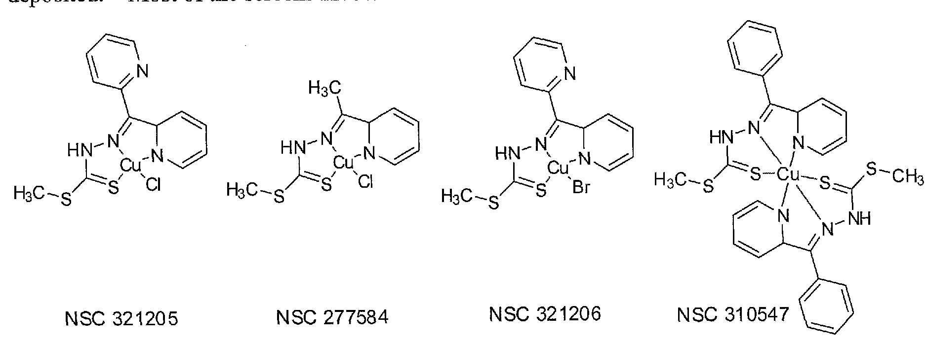
This database does not disclose the minimal basic structure, per se, but does reveal the example
compounds from which the minimal basic structure was derived. Further, the database does not directly suggest that these compounds have any caspase inhibitory activity.
Nobel et al. 1997 discloses that dithiocarbamates (DCs) (shown below generically and with specific examples, central structure darkened) have been reported to be potent inhibitors of apoptosis in several different model systems suggesting a target common to the apoptotic machinery [42]. The reference investigates antiapoptotic actions of DCs and found that inclusion of a membrane-impermeable copper chelator severely compromised the inhibitory activity of reduced DCs. Since copper can promote DC oxidation to the respective DC disulfides, the inhibitory effect on lymphocyte apoptosis might be mediated by the DC disulfides. The reference concludes that (1) DC disulfides are the active agents behind DC inhibition of apoptosis and (2) their site of action is the proteolytic activation of caspase- 3 proenzyme.

generic pyrrolidine diethyldithiocarbamate tetramethylthiuram dithiocarbamate dithiocarbamate disulfide
Thompson et al. 2010 also discusses the apoptosis blocking effect of pyrrolidine dithiocarbamate in exposure to ionizing radiation [43]. Rather than providing an antioxidant prophylactic effect, the result of pyrrolidine dithiocarbamate in exposure to ionizing radiation leads to an increase in radical species and results in greater cell necropsy compared to apoptosis.
S^SH
, Pyrrolidine dithiocarbamate
Some patents have discussed caspase inhibitors including United States Patent 7,807,659 [44]. This patent discloses a fo

Additionally, the 7,807,659 patent also refers to previously disclosed formulas for caspase inhibitors:

United States Patent 7,410,956 describes compounds of a formula (shown below) which are prodrugs of caspase inhibitors and pharmaceutically acceptable salts thereof [45]. Y is the caspase inhibitor. This invention further relates to pharmaceutical compositions comprising these compounds, which are particularly well-suited for treatment of caspase-mediated diseases, including inflammatory and degenerative diseases.
Prodrug formula where
Y= caspase inhibitor

Ekert et al. 1999 discloses various known caspase inhibitors, including synthetic caspase inhibitors [46]. A number of specific caspase inhibitors have been developed based upon the substrate cleavage sites of the caspases. These peptides act as pseudosubstrates for active caspases and are therefore competitive inhibitors. They range from those containing a single aspartate residue (e.g. Boc-aspartyl(OMe)-fluoromethylketone: Boc-Asp-FMK) to trimers (e.g. Benzyloxycarbonyl-val-ala-asp (OMe) fluoromethylketone: z-VAD-FMK) to tetramers (e.g. YYAD-FMK). The inhibitors described in this reference are peptide derivatives and are described as competitive inhibitors, not as allosteric inhibitors to caspases. Linton, S. D. 2005 also describes the various caspase inhibitors found and used within the pharmaceutical industry [47]. Most of the compounds disclosed are peptide derivatives and pro-drug peptide derivatives.
Inhibition of caspase activation is a promising therapeutic approach for treating diseases including neurodegeneration, stroke, radiation syndrome, and immune disorders. Using a de novo reconstituted assay for cytochrome c-mediated caspase activation and high-throughput
screening, a group of non-peptide inhibitors for caspases from a chemical library containing over 300,000 compounds were identified. These caspase inhibitors share common chemical scaffolds, suggesting same mechanism of action. Further, they are able to inhibit apoptosis in various cell types induced by multiple stimuli; they can also inhibit caspase- 1 -mediated interleukin-ΐβ generation in macrophages, suggesting the potential application of these compounds in antiinflammation.
While these compounds inhibit all the tested caspases, kinetic analysis indicates that they do not compete with caspase substrates for the catalytic sites of the enzymes. The co-crystal structure of one of these compounds with caspase-7 reveals that these compounds bind to the dimerization interface of active caspases, another common structural element shared by all active caspases. Consistently, biochemical analysis demonstrates that the compounds abate dimerization of caspase-8. Therefore, via an allosteric mechanism of binding to the dimerization site, these compounds alter the conformation of the catalytic loop of caspases and thus inhibit caspase activity.
In the mitochondria-mediated caspase activation pathway, caspase activation is initiated by cytochrome c release from mitochondria, a process closely regulated by the Bcl-2 family of proteins (Garrido et al., 2006 [25]; Green and Reed, 1998 [26]; Jiang and Wang, 2004 [23]; Youle and Strasser, 2008 [27]). Although it is not necessary to understand the mechanism of an invention, it is believed that, released cytochrome c binds to the essential mediator Apaf-1 (Zou et al., 1997 [28]), activates the nucleotide binding/exchanging activity of Apaf-1 (Bao et al., 2007 [29]; Jiang and Wang, 2000 [33]; Kim et al., 2005 [30]), and consequently triggers the assembly of a multimeric protein complex, the apoptosome, which is the central caspase activation machinery (Srinivasula et al., 1998 [31]; Zou et al., 1999 [32]). Although it is not necessary to understand the mechanism of an invention, it is believed that, the apoptosome recruits and activates the initiator caspase, caspase-9. Caspase-9 needs to associate with the apoptosome to be active (Jiang and Wang, 2000 [33]; Rodriguez and Lazebnik, 1999 [34]) and it subsequently activates downstream executioner caspases, such as caspase-3 and caspase-7, which mediate apoptotic cell death by targeting and cleaving a variety of cellular substrates. Although it is not necessary to understand the mechanism of an invention, it is believed that, in cells, there is a family of endogenous proteins named Inhibitors of Apoptosis Proteins (IAP) that functions to inhibit caspase activity; whereas Smac/Diablo and Omi/HtrA2 are mitochondrial
proteins that are released from mitochondria during apoptosis to antagonize the inhibitory activity of IAP (Du et al., 2000 [35]; Suzuki et al, 2001 [36]; Verhagen et al., 2000 [37]).
Although it is not necessary to understand the mechanism of an invention, it is believed that, Deregulation of the intrinsic apoptotic pathway is involved in various human diseases, such as cancer and autoimmune disorders (when apoptosis is defective), and neurodegenerative diseases and strokes (when apoptosis is improperly activated) (Hotchkiss and Nicholson, 2006 [38]; Reed, 2003 [48]; Yuan and Yankner, 2000 [39]). Conversely, targeting apoptotic components by both enhancing and attenuating apoptosis represents important therapeutic approaches to treat various human diseases. For example, many conventional cancer therapies kill cancer cells via activating the cytochrome c apoptotic pathway, and several promising targeted compounds, including small molecule inhibitors of Bcl-2 (Oltersdorf et al., 2005 [40]) and Smac mimetics (Li et al., 2004 [41]), are designed to specifically activate or potentiate this pathway. On the other hand, inhibition of this pathway should also be effective in treating symptoms with pathologically enhanced apoptosis such as neurodegeneration, stroke, and ischemic diseases. Previous efforts in developing potential anti-apoptotic agents focused on caspase inhibition. Notably, caspase inhibition can also be used for treating inflammation, which requires caspase- 1 -mediated interleukin-ΐβ maturation (Martinon and Tschopp, 2006 [49]; Talanian et al., 2000 [50]). To date, although caspase inhibitors have been developed with some degree of specificity and are used for basic research, because of their chemical nature as peptides, they possess poor potency and are rapidly degraded in vivo. Another undesirable feature of most of these peptide-based inhibitors is that they are covalent, irreversible inhibitors. These weaknesses preclude their realistic clinical usage. Cytochrome c-mediated caspase activation in vitro by using purified protein components (Apaf-1, cytochrome c, caspase-9, and procaspase-3), nucleotide dATP, and a fluorogenic substrate of caspase-3 were reconstituted. A high-throughput screening approach to identify small molecule inhibitors of this pathway was employed. Four structurally similar compounds were identified as caspase inhibitors. The compounds were able to inhibit apoptosis and caspase- 1 -mediated interleukin-ΐβ generation. Further kinetic and structural studies revealed that the compounds inhibit caspases via an allosteric mechanism, by binding to the caspase dimerization interface and subsequently altering the conformation of the catalytic site of the enzyme.
4. Identification of Chemical Inhibitors of Cytochrome c-mediated Caspase Activation
The cytochrome c-mediated caspase activation pathway was reconstituted in vitro using purified recombinant proteins at their near-physiological concentrations (Jiang and Wang, 2000; Kim et al., 2005; Zou et al, 1999) [30, 32, 33]. In the presence of Apaf-1, cytochrome c, caspase-9, procaspase-3 and dATP, robust caspase-3 activation was achieved, monitored using a fluorogenic peptide substrate of caspase-3 (Figure 1A). As expected, omission of any single component in the reaction completely abated caspase-3 activation (Figure 1A). After adapting this highly sensitive assay system to a fully automated high-throughput screening (HTS) format, a collection of 317,856 chemical compounds was screened at a single compound concentration of 10 uM in 1% DMSO (v/v). The optimized assay exhibited a high signal-to-noise ratio of 19: 1, as well as good reproducibility with Z'-value ranging from 0.59 to 0.75. Out of this large compound collection, 34 molecules were identified as initial positive hits with a threshold of at least 30% inhibition.
Four of the 34 compounds were selected after further analysis. These compounds, named Comp-A, B, C, D thereafter for simplicity, are also included in the chemical repository of the Developmental Therapeutics Program (DTP) of National Cancer Institute (NCI), with the codes NSC321205, NSC277584, NSC321206, and NSC310547, respectively. At 10 μΜ concentrations, these compounds all completely inhibited cytochrome c-mediated caspase activation (Figure IB). Further titration of the compounds in this assay indicates their potent inhibition activity with submicromolar IC50 values (Figure ID). A striking feature of these compounds is that they share a very similar molecular scaffold, suggesting a common structure-function relationship. They are pyridinyl, copper-containing molecules with a multi-ring structure (Figure 1C). Although Comp-D is more complex structurally, it appears to be a covalent dimer of the other complexes. Mass spectrometry analysis yielded a molecular mass of each compound consistent with its molecular formula (Figure 2).
5. The Newly Identified Inhibitors Target Caspases
Because a multi-component biochemical assay was used for HTS, a deconvolution analysis was performed to identify the direct target of the inhibitors. Although it is not necessary to understand the mechanism of an invention, it is believed that the results demonstrate that these compounds may directly inhibit both caspase-9 and caspase-3. As shown in Figure 3, the activity
of recombinant caspase-3 (fully activated when expressed and purified from bacteria) was completely inhibited by 10 μΜ of each compound (Figure 3A), and consistent with the result obtained from the cytochrome c-mediated caspase activation assay, these compounds inhibit caspase-3 activity with submicromolar IC50 values (Figure 3C). Although it is not necessary to understand the mechanism of an invention, it is believed that the direct effect of these compounds on caspase-9 activity was measured using an engineered caspase-9 dimer, containing a leucine-zipper dimerization domain (Caspase- 9/LZ) (Yin et al., 2006) [51]. Unlike native caspase-9 whose activity requires an active apoptosome complex even after complete proteolytic processing (Jiang and Wang, 2000; Rodriguez and Lazebnik, 1999) [33, 34], recombinant caspase-9/LZ is constitutively active independent of the apoptosome complex (Yin et al., 2006) [51]. Similar to caspase-3 activity, caspase-9/LZ activity was completely inhibited by 10 μΜ of each compound (Figure 3C). To validate that inhibition of the enzyme by the compounds is not an artificial result caused by the engineered leucine-zipper, an apoptosome complex was used to activate unmodified caspase-9 and subsequently assessed the effect of the compounds on caspase-9 activity using a ffuorogenic peptide substrate of caspase-9. In such an assay, the activity of caspase-9 was also inhibited by all four compounds (Figure 3B).
Although it is not necessary to understand the mechanism of an invention, it is believed that since the compounds were able to inhibit both caspase-9 and caspase-3, it is possible that this inhibitory effect might extend to other members of the caspase family as well. The potential inhibitory effect of the four compounds on the activity of recombinant caspase-8 and caspase-2 was tested; both caspase-8 and caspase-2 are initiator caspases that share only limited similarity with caspase-9 or caspase-3. Caspase-7, an effector caspase that is highly similar to caspase-3, was also inhibited by each of the four compounds with submicromolar IC50 values (Figure 3C). Recombinant caspase-8 and caspase-2, both initiator caspases that share only limited similarity with caspase-9 or caspase-3, and caspase- 1, which is involved in the inflammatory response, were inhibited by the compounds with submicromolar IC50 values as well (Figure 4). Therefore, these four compounds are pan-caspase inhibitors whose chemical nature is different from the previously developed peptide-based caspase inhibitors.
To determine the specificity of these compounds, their inhibitory activity on other proteases, including cathepsin C, papain, and trypsin. Cathepsin C and papain are cysteine proteases was tested. Although they were inhibited by the compounds in a dose-dependent
manner, the IC50 values were 5-10 μΜ, which is about 10-fold higher than for caspases (Figure 3D). Trypsin, a serine protease, was not significantly inhibited by the compounds (Figure 3D). One exception is Comp-D, which inhibits papain and caspase-7 with similar potency. Although it is not necessary to understand the mechanism of an invention, it is believed that that this "offtarget" effect of Comp-D might be due to its more bulky and complex structure compared to the other three compounds.
6. Inhibition of Cellular Apoptosis by the Compounds
Although it is not necessary to understand the mechanism of an invention, to determine whether these compounds were able to block cellular apoptosis, several different mammalian cell lines were used and tested the effect of the compounds on both intrinsic apoptosis and extrinsic apoptosis. Because these compounds are pan-caspase inhibitors, if they are cell-permeable, they should be able to inhibit both pathways.
The intrinsic apoptotic pathway was triggered by UV radiation in HeLa cells. Following UV irradiation, cells that had not been treated with compound displayed typical apoptotic morphology, while cells treated with each of the four compounds showed either diminished apoptotic morphology or resembled non-irradiated cells (Figure 5A). Compound concentrations from 1 μΜ to as low as 100 nM were tested, and it was found that even at 100 nM, the compounds still possess apparent activity in preventing UV-induced apoptosis (Figure 5A). Although it is not necessary to understand the mechanism of an invention, it is believed that because in the in vitro assays 100 nM of compounds only yielded very mild inhibition of caspases, it is possible that the cells might actively take in the compounds from culture medium, resulting in a higher cellular concentration of the compounds. Subsequent measurement of caspase-3 activity in cytosolic extracts of these cells confirmed that caspase activity was inhibited by the compounds in a dose-dependent manner (Figure 5B). Annexin V and propidium iodide (PI) staining confirmed the inhibition of UV-induced apoptosis in HeLa cells in the presence of compound (Figure 5C). Similar to HeLa cells, UV-induced caspase activation in mouse embryonic fibroblast (MEF) cells can also be inhibited by these compounds in a dose-dependent manner (Figure 6).
The effect of the compounds on the extrinsic apoptotic pathway was tested in U937 cells, a human leukemic monocyte lymphoma cell line. The extrinsic apoptotic pathway was induced
by tumor necrosis factor- (TNF-a) plus cellular protein synthesis inhibitor cycloheximide. The compounds were able to inhibit the extrinsic apoptotic pathway in U937 cells in a dosedependent manner, as measured by both Annexin V/PI staining and cellular caspase-3 activity assay (Figure 6). Therefore, the newly identified pan-caspase inhibitory compounds can inhibit both intrinsic and extrinsic apoptotic pathways.
The ability of these compounds to enhance long-term survival of cells that have been transiently treated with an apoptotic trigger was next tested. Although inhibition of caspase activation often fails to prevent eventual cell death due to loss of mitochondrial integrity (for intrinsic apoptosis) or switching to programmed necrosis (for extrinsic apoptosis), under certain conditions, caspase inhibition can restore cell viability permanently. This could be particularly relevant in vivo where apoptotic stimulation is often transient and milder than the conditions used in experimental settings. Indeed, it has been firmly established recently that intrinsic apoptosis-associated mitochondrial outer membrane permeability (MOMP) in a cell can be partial or incomplete (iMOMP); and under such conditions, caspase inhibition can completely restore cell viability (Tait et al., 2010 [52]). Further, in cells with specific genetic backgrounds, such as lack of RIP3 expression, TNF-a-induced programmed necrosis can be defective (He et al., 2009 [53]; Oberst et al, 2011 [54]; Vandenabeele et al., 2010 [55]). It was deterrnined that human mammary epithelial MCFIOA cells appear to be defective in TNF-a-induced necrosis because inhibition of caspase activation completely restored long-term viability in these cells. As shown in Figure 5, apoptosis was induced in MCFIOA cells by the addition of TNF-a and cycloheximide for 3 hours in the presence or absence of Comp-A. Growth medium was subsequently replaced, and cell numbers were monitored daily for a period of eight days. While TNF-a- treated cells in the absence of Comp-A all died, growth of MCFIOA cells following TNF-a induction in the presence of Comp- A was recovered to the level of untreated cells (Figure 5D and Figure 5E). This result demonstrates that under certain specific contexts, inhibition of apoptosis by these compounds can significantly restore long-term cell viability.
To investigate whether the compounds would be effective in inhibiting cellular caspase activation outside of the context of apoptosis, J774 cells, a murine macrophage cell line, were treated with lipopolysaccharide (LPS). LPS treatment can activate inflammatory caspase-1, which results in the cleavage and subsequent secretion of the proinflammatory cytokine Interleukin (Π,)-Ιβ (Martinon et al., 2002) [56]. Indeed, when added to the culture medium
during LPS stimulation, the compounds were able to decrease the secretion of IL-Ι β in a dose-dependent manner (Figure 5F).
It should be noted that when higher concentrations of these four compounds were used, noticeable cell toxicity was observed. In culture dishes, concentrations up to 5 uM of these compounds caused attached cells (such as HeLa cells) to detach in a short period of time. If the compounds were removed rapidly, cells were still able to re- attach and grow. However, longer treatment with high doses of the compounds caused the cells to lose viability. Although it is not necessary to understand the mechanism of an invention, it is believed that it is possible that such toxicity is due to their chemical nature as transition metal complexes. Although it is not necessary to understand the mechanism of an invention, it is believed that it is also possible that the contaminating free copper ions in the solution of the compounds caused the toxicity. Although it is not necessary to understand the mechanism of an invention, it is believed that alternatively, the compounds might exert the toxicity by targeting other biological molecules. For example, it has been reported that Comp-C can target Cdc25B phosphatase (Vogt et al., 2003)[57] and X-IAP (Glover et al., 2003) [58], although both with a lower potency (IC50 over 10 μΜ) than what was observed for caspase inhibition. It should also be noted that at the low concentrations sufficient for apoptosis inhibition in HeLa cells, the compounds do not affect HeLa cell viability (Figure 6). 7. Non-competitive Inhibition of Caspase-7 by Comp-A
Although it is not necessary to understand the mechanism of the invention, a kinetic analysis was performed in order to understand the mechanism of action of these compounds in inhibiting caspase activity, and to subsequently conduct medicinal chemical modification. Although it is not necessary to understand the mechanism of an invention, it is believed that because these four compounds share a similar structure, it is most likely that they inhibit caspases via the same mechanism. For this reason, there was a focus on Comp-A (which showed the strongest activity in inhibiting cellular apoptosis), and the kinetics of its inhibition of caspase-7 activity were examined. The kinetic analysis of all four compounds and multiple caspases (Figure 7 and Figure 8; Table 1 and Table 2) was later expanded.
TABLE 1
Kinetic Parameters of Caspase Activity in the Presence of Compounds
Comp-A OiiM 50 nM 100 nM 250 nM 400 nM
152.1 ±0.5
Caspase-3 136.6 + 0.6 124.1 ±0.4 99.9 + 0.3 83.7 + 0.3
Km 6.3 ±0.1 6.4 = 0.1 6.3 + 0.1 6.5 + 0.9 6.4 + 0.1 v 71.4± 0.4
Caspase-7 v max 64.7 + 0.4 57,6 + 0.3 46.1+0.3 38.2 + 0.2
30.8 + 0.4 31.7 + 0.4 31.0 + 0.4 31.9 + 0.4 31.9 + 03 v 98.6 + 0.9 68.6 + U.5 51.3 + 0.3 33.1 + 0.2
Caspase 25.9 + 0.2 -9-LZ
Km 54.3 ± 0.9 58.2 + 0.7 56.0 + 0.6 57.4 + 0.5 57,8 + 0,9
Caspase-9- V v max 110.8±0.9 81.3 + 0.5 66.1 ±0.6 43.9 + 0.4 34,8 + 0.2 apoptosome m 53.2 ±0.8 53.2 + 0.6 54.3 ±0.8 55.1+0.9 56,5 + 0,6 v .4 114.2 + 0.4
Caspase- 109.4 + 0.5 97.0 + 0.4 S7.7 + 0.2
1 ¥ max 120.0 ±0
m 4.9 J- 0.1 4.9-0.1 4.9 + 0.1 5.0 + 0.1 5.2 + 0.1
V ax 187.1 + 0.7 155.1 + 0.8 150.1 ±0.8
Caspase 11S.4+0.S 97.6 + 0.4 -2 v m
17.1 + 0.2 17.0 + 0.2 17,6 + 0.2 18.4 + 0.3 18.3 + 0.2 v x 85.0 ±0.3 80.2 ± 0.2
Caspase-8 v ma 76.2 + 0.2 66.5 + 0.2 59.0 ± 0,2
K,„ 3.3 + 0.1 33-0.1 3.4 + 0.1 3.4+0.1 3.4+0.1
ComD-B OnM 50 nM 100 nM 250 nM 400 nM
152.7 + 0.5
Caspase-3 136.9 + 0.5 125.6 + 0.5 100.6 + 0.3 83.7 + 0.2
Km 6.5 + 0.1 6.2-0.1 6.2 + 0.1 6.3 ±0.1 6.1 +0.1
71.2 ±0.4 63.3 + 0.3 57.8 + 0.3 45.9 + 0.3 36.7 + 0,3
Caspase-7
Km 30.5 + 0.4 30.2 + 0.4 30.5 ± 0.4 31.5 + 0.4 30.0 + 0,4 v 98.1 ±0.8 76.5 + 0.5 63.5+0.4 41.2 + 0.3 318 ±03
Caspase-9-LZ Y max
K[n 54.1+0.7 53.3 + 0.6 53.7 + U.6 52.1 +0.6 54.1 +0,9
120.7 + 0.4 114.1+0.5 107.0 + 0.4 92.8 ± 0.3
Caspase- 82.0 + 03 1 ^ma
Kra 5.0 + 0.1 5.0 = 0.1 4.8+0.1 4.9 + 0.1 5.0 + 0.1
» max 185.4 + 0.8 164.3 + 0.6 147,9 + 0.7 110.7 + 0.5 90.0 + 0,5
Caspase-2
Km 16.8 + 0.2 17.0 + 0.2 17.2 + 0.2 17.0 + 0.2 17.1 ±0,3 max 84.7 + 0.3 81.0 + 0.2 77.7 + 0.2 68.4 + 0.2
Caspase-8 61.2 + 0,2
Km 3.2 + 0.1 3.3 = 0.1 3.4 + 0.1 3.3 + 0.1 3.2 ±0.1
ComD-C OnM 50iiM 100 nM 250 nM 400 nM
151.6+0.5 135.1+0.4
Caspase-3 123.3 + 0.4 98.3 + 0.4 827 + 0.2
6.4 + 0.1 6.2 = 0.1 6.2 + 0.1 6.4 + 0.1 64 + 0.1 v .0+0.3 61.7 + 0.3 55.8+0.3 42.4 + 0.3
Caspase-7 v max 72 34.2 + 0.2
Km 31.2+0.3 30.5 ± 0.3 31.8+0.3 31.9 + 0.5 31.1 ±0.4 v 97.4 ± 0.5 67.3 ± 0.4 52.0 + 0.4 33.4 + 0.2 26.7 + 0.2
Caspase-9-LZ v max
Km 53.5 ± 0.5 55.5 ± 0.6 55.1 ±0.7 54.6 + 0.5 55.0 + 0.6 v v mix 121.6 + 0.5 113.9 + 0.4 108.1+0.3 93.8+0.3 S2.9 + 0.3

120.9 + 0.4 116.2 + 0.4 112.4 + 0.3 101.6 + 0.5 92.2 + 0.3
Caspase-1
m 5.0 + 0.1 5.1 = 0.1 5.2 + C.l 5.2 + 0.1 5.2 + 0.1
189.1 + 0.7 173.5 + 0.7 165.6 + 1.0 139.9 + 0.6 120.8 + 0.5
Caspase-2
Km 17.4 + 0.2 17.2 + 0.2 18.0 + 0.3 18.2+0.2 18,6 + 0,2
V .6 + 0.2 80.9 + 0.3 76.8+0.2 67.3 + 0.2
Caspase-8 v max 84 60,3 + 0,2
m 3.2 + 0.1 3.3 = 0.1 3.4 + 0.1 3.4 + 0.1 3.5 + 0.1
Vml:s is expressed in RFU/min; Km is expressed in uM.
Values are expressed as mean +SEM from experiments performed in triplicate as described in the Experimental
Procedures.
TABLE 2
Kinetic Parameters of Caspase Activity in the Presence of Compounds from Replots of Intercepts of the Specific Velocity Plot
P Ki bxM)
Comp-A 1.06 0.14 0.35
Comp-B 0.94 0.14 0.71
Caspase-3
Comp-C 0.99 0.19 0.67
Comp-D 1.06 0.28 0.95
Comp-A 1.01 0.06 0.34
Comp-B 0.94 0.09 0.41
Caspase-7
Comp-C 0.84 0.15 0.31
Comp-D 1.04 0.08 0.34
Comp-A 1.06 0.08 0.08
Comp-B 0.92 0.04 0.18
Caspase-9-LZ
Comp-C 1.04 0.10 0.08
Comp-D 1.08 0.04 0.17
Caspase-9-
Comp-A 1.04 0.04 0.12
apoptosome
Comp-A 0.90 0.25 1.02
Comp-B 1.02 0.60 0.44
Caspase-1
Comp-C 0.99 0.34 0.80
Comp-D 1.01 0.46 0.56
Comp-A 0.96 0.16 0.37
Comp-B 0.98 0.02 0.38
Caspase-2
Comp-C 1.04 0.05 0.22
Comp-D 0.97 0.06 0.35
Comp-A 0.98 0.23 0.69
Comp-B 1.04 0.11 0.28
Caspase-8
Comp-C 1.03 0.19 0.40
Comp-D 1.07 0.32 0.67
Because these novel inhibitors are pan-caspase inhibitors, it was predicted that they may act upon a common functional moiety shared by all caspases. The catalytic center of all known caspases shares a similar conformation and is a common site of inhibition for most known pharmacological caspase inhibitors (Ivachtchenko et al., 2009) [59]. The catalytic site contains a substrate binding groove that is shaped by four peptide loops (LI, L2, L3, L4) that harbor the catalytic cysteine and determine substrate specificity (Shi, 2002) [60]. Although it is not necessary to understand the mechanism of an invention, it is believed that although individual caspases show different protein substrate preferences, aspartate at the PI position is universally
required for all caspases (Pop and Salvesen, 2009) [12]. Because of this universal specificity for aspartate residues, most pan-caspase inhibitors utilize amino-acid sequences containing aspartate and compete with substrate binding at the active site. If indeed Comp-A binds to the catalytic site of caspase-7, it should then inhibit the enzyme by competitively inhibiting substrate binding to the enzyme, thus Michaelis-Menten analysis would yield a constant Vmax but increased Km values when the inhibitor is present in the reaction. Although it is not necessary to understand the mechanism of an invention, kinetic analysis surprisingly showed that the addition of Comp-A caused a drastic decrease of Vmax values in a Comp-A concentration-dependent manner, whereas the change of Km values was modest (Figure 7A panel i and Table 1). Such concentration-dependent decrease of Vmax and the relative constant Km values were also confirmed by the double reciprocal analysis (Figure 7A, panel ii). Therefore, Comp-A is not a competitive inhibitor of caspase-7; but rather, Comp-A can decrease the Vmax of caspase-7, and has minor effect on binding of caspase-7 with its substrates; thus Comp-A is a non-competitive inhibitor.
The mechanism of inhibition type was investigated in greater detail using the specific velocity plot (Baici, 1981 [61]). This method offers many advantages for the analysis of non-tightbinding, reversible inhibitors, and has been previously used to elucidate the mechanism of a caspase-2 inhibitor (Schweizer et al., 2007 [62]). The specific velocity (defined under Experimental Procedures) was plotted versus the ratio VO/Vi at different concentrations of Comp-A to obtain a series of linear curves parallel to the abscissa (Figure 7A, panel iii), indicating no effect of the compound on substrate binding. Using the replots of the specific velocity plots (Figure 7A, panel iv), the values of a and β and an approximate value of Ki were calculated graphically (Table 2). For caspase-7 and Comp-A, a = 1.01 and β = 0.06, and Ki = 0.34 μΜ. These values suggest pure non-competitive inhibition (for which a = 1 and β = 0, theoretically).
Kinetic parameters for other caspases in combination with all four compounds (Figure 8; Table 1, and Table 2) were measured. Consistent with the observation for caspase-7 and Comp-A, caspase-3, caspase-9-LZ, caspase-1, caspase-2, and caspase-8 all showed a decrease in Vmax and a constant Km upon the addition of increasing concentrations of each of the four compounds. Specific velocity replots yielded a values close to 1 and β values between 0 and 1. The deviation of β from a value of 0 (indicative of pure non-competitive inhibition) to a value between 0 and 1
is suggestive of partial non-competitive inhibition: a hyperbolic system in which the inhibitor will convert the enzyme into a modified enzyme-substrate-inhibitor complex with a decreased rate of product formation (Baici, 1981 [61]). This partial non-competitive inhibition is especially evident in the β values for caspase-1.
In agreement with a non-competitive inhibition mechanism, the compound appears to bind to caspase-7 with similar affinity regardless of the presence of substrate, thus generating the same degree of inhibition (Figure 9).
Kinetic parameters for caspase-9 that is associated with and activated by the apoptosome, and compared it with Caspase-9-LZ, whose activity requires artificial dimerization driven by leucine-zipper were also measured. It is under debate whether the apoptosome activates caspase-9 by triggering its dimerization or by a dimerization-independent, novel conformational change (Jiang and Wang, 2004 [23]; Renatus et al., 2001 [63]). It was reason that a comparison between caspase-9-LZ and apoptosome-caspase-9 could provide insight into the functional conformation of apoptosome-activated caspase-9. The kinetic profiles of Comp-A inhibition of caspase-9-LZ and apoptosome-activated caspase-9 are almost identical (Figure 7B and Figure 8B, Table 1 and Table 2), indicating the compound inhibits the two forms of caspase-9 enzyme with same mechanism. A further analysis (described later) shows that the compounds inhibit caspase activity by interfering with caspase dimerization, this kinetic analysis lends support that caspase- 9 dimerization is required for the activity of the apoptosome-caspase-9 holoenzyme, in agreement with a model proposed previously (Renatus et al., 2001 [63]).
8. Crystal Structure of Caspase-7 in Complex with Comp-A
How can these compounds be pan-caspase inhibitors but not function by binding to the conserved active site of caspases? To identify the exact nature of the caspase-compound interaction, crystal structures of Comp-C at 0.81 A resolution, unliganded caspase-7 at 2.8 A resolution, and caspase-7 in complex with Comp-A at 3.8 A resolution (Table 3 and Table 4, Figure 10A and Figure 10B) were determined. Because Comp-A is identical to Comp-C except for the replacement of Br with CI, a CI atom was substituted in the crystal structure of Comp-C to generate the structure of Comp-A. The complex crystals contain a caspase-7 dimer per crystallographic asymmetric unit with each monomer consisting of a p20 (large) and a plO (small) subunit. Because all compounds contain a Cu atom, Cu anomalous difference Fourier to
locate Comp-A molecules in the soaked caspase-7 crystals was used. This yielded two high peaks of 11.5 σ and 7.4 σ, respectively, symmetrical with respect to the two chains of the caspase-7 dimer. These difference Fourier peaks were used as Cu positions to model the Comp-A structure rigidly into the Fo-Fc difference electron density map followed by refinement (Figure IOC).
Comp-A is bound to the solvent-exposed dimer interface in an edge-to-edge fashion
(Figure 10B, Figure 10D, Figure 10E), and is away from the catalytic active site Cysl86. It lays essentially flat on β5 and β6 of the central β-sheet at the dimerization interface (Figure 10B, Figure 10E). Comp-A contacts only the plO subunits, including residues Tyr223 and Cys290 from one subunit and Glu216, Phe221, Tyr223, Val292 and Met294 from the neighboring subunits (Figure 10E). Previously reported allosteric caspase-7 inhibitors DICA and FICA interact with the caspase dimerization interface and form covalent bonds with the thiol of Cys290 (Hardy et al., 2004) [64]. In the crystal structure of caspase-7 in complex with comp-A, the thiols of Cys290 in both monomers of the caspase-7 dimer are too far away from any atom in Comp-A to allow covalent bonding (Figure 10E).
TABLE 3
Crystallographic Statistics for Comp-C
Empirical formula Ci3 Hi2 Br Cu N4 S2
Formula weight 431.8 Dalton
Temperature 149 K
Wavelength 0.7107 A
Space group P-l
Unit cell dimensions a = 8.6 A, b = 9.8 A, c = 9.9 A
a= 69.2°, β = 71.5°, γ = 81.9°
Z 2
Number of reflections collected 11,391
Independent reflections 2,898
Resolution Range oo - 0.81A
Completeness to Θ = 26.00n 99.2 %
Refinement method Full-matrix least-squares on F^
Data / restraints / parameters 2,898 / 0 / 190
Goodness-of-fit on 1.064
Final R, (I > 2sigma(I)) 0.1134
Final R, (all data) 0.1456
TABLE 4
Crystallographic Statistics of Unliganded Caspase-7 and its Complex with Comp-A
Caspase7 Caspase7 with Comp-A
Data Collection
Space group P3221 P3221
Cell dimensions (A) 88.4, 88.4, 185.5 88.7, 88.7, 185.7
Wavelength (A) 1.000 1.378
Resolution (A) 37.0 - 2.8 45.0 - 3.8
Rsym (%) 8.3 (46.2) 10.8 (70.0)
Ι / σΙ 30.5 (2.0) 11.9 (2.8)
Completeness (%) 88.7 (46.0) 94.1 (96.0)
Redundancy 3.6 (2.5) 5.1 (5.1)
Refinement
Resolution (A) 20.0 - 2.8 20 - 3.8
No. reflections 17,179 8,046
i?work /i¾rcC (%) 18.7 / 23.1 20.6 / 26.8
No. atoms
Protein 3,750 2,927
Cu 0 2
R.m.s. Deviations
Bond lengths (A) 0.006 0.008
Bond angles (° ) 1.323 1.317
Ramachandran plot
Most favored (%) 95.2 85.3
Allowed (%) 4.8 12.4
* Values in parenthesis are for highest-resolution shell. One crystal was used for each data set.
9. Comp-A Binding Induces Conformational Changes and Disordering of Active Site Loops
The substrate binding groove of caspase-7 is composed of flexible surface loops that include LI (residues 75-89, between βΐ and al), L2 (residues 185-196, after β4), L3 (residues 224-232, between β5 and a4), and L4 (272-289) from one chain (A), and L2' (residues 212-218) from the other chain (B), among which L2 harbors the catalytic Cysl86 in caspase-7 (Chai et al., 2001; Riedl et al., 2001) [65, 66]. While L3 and L4 form the base and one side of the catalytic groove, respectively, the interaction between L4, L2 and L2' is important for maintaining a stable active conformation for substrate binding and catalysis. There are two unliganded caspase-7 structures in the Protein Data Bank (PDB) (www.pdb.org) (accession codes 1IBF and 1K86) (Agniswamy et al., 2009; Chai et al., 2001) [65, 67]. The unliganded structure of the present
invention is highly similar to the 1IBF structure; both structures are essentially in a catalytically productive conformation shown by the resemblance to the DEVD-CHO-bound conformation (accession code 1F1 J) (Figure 11), but is different from the 1K86 structure, which has a reversed conformation for the L2' loop and an unproductive conformation for the L2 loop. It is worth noting that crystallization condition of the present invention example is also highly similar to that used for the 1IBF structure.
Given that the present invention's unliganded structure essentially assumes an active conformation, it was striking to see that extensive conformational changes and disorder were induced in all loops upon Comp-A binding (Figure 12A). L2 has only two residues with the catalytic Cysl86 as the last ordered residue; L3 has a few residues at the beginning and the disorder continues to residue 236; L4 has a few residues each at the beginning and the end of the loop; and L2' is two residues shorter. In one of the monomers of the caspase-7 dimer, even LI, which is usually conserved in conformation in different forms of caspase-7 structures, has only two ordered residues at the beginning of the loop, and a neighboring region from residues 144-155 is also invisible. In the other monomer of the caspase-7 dimer, LI and its neighboring region are involved in crystal packing and are ordered in the crystal data set. However, the tendency of the region to become disordered upon Comp-A soaking explains deterioration of diffraction quality and the need to compromise soaking conditions to both preserve some diffraction and to allow reasonable occupancy of the compound.
Structural comparison showed that the ordered parts of L2, L3 and L4 of the Comp-A bound caspase-7 are more similar to procaspase-7 structures (accession code 1K88 and 1GQF) (Chai et al., 2001; Riedl et al., 2001) (Figure 12B), with the exception that L2 of Comp-A bound caspase-7 is in an open conformation instead of an inverted conformation as in procaspase-7 structures. This change of conformation upon Comp-A binding is induced by steric clash. In particular, Comp-A is in direct clash with Arg 187 of L2 and Thr225, Val226 and Pro227 of L3 in the active conformation (Figure 12C), pushing these residues away to assume a conformation more similar to procaspase-7. L2 and the end of L3 coming from the β5 strand are both almost 90° away from the conformation in active caspases (Figure 12A). Although the key inducing event in procaspase-7, which is the linkage between L2 and L2', is different from Comp-A binding, the consequence is similar as they both disrupt the highly interdependent conformations of the active site loops. Strikingly, Comp-A atoms that are in clash with active caspase-7
conformation are all from the common core of the compound scaffold (Figure 12D), which in turn supports that all compounds would interact with caspase-7 in a manner similar to the interaction between caspase-7 and Comp-A observed here.
Analysis of the previously reported caspase-7 structures inhibited by DICA and FICA (1SHJ and 1SHL) (Hardy et al., 2004) [64] revealed a highly similar pattern of clashes induced by inhibitor binding. DICA clashes with Argl87 at the L2 loop, and Tyr223 and Pro227 of the L3 loop (Figure 12E). FICA clashes with exactly the same residues, except that one FICA molecule clashes with Pro227 of the monomer it is bound to, and with Argl87 and Tyr223 of the neighboring monomer. Similar to Comp-A inhibited structure, distortion of the L2 and L3 loops by inhibitor binding leads to massive conformational changes and disordering of the active site loops (Figure 12F). In DICA and FICA-inhibited caspase-7 structures, L2' is inverted towards the dimenzation interface and interacts with both DICA and FICA directly. In any case, while DICA and FICA are covalent inhibitors that target Cys290, and Comp-A noncovalently interacts with the region of Cys290 at the dimerization interface, they all cause clashing with an active caspase conformation and therefore distortion of the active site.
10. Inhibition of Caspase-7 by Comp-A is Reversible
Although it is not necessary to understand the mechanism of an invention, it is believed that because the dimerization interface is a common structural feature of all caspases, inhibitors binding to this site, such as those identified in this study, can act as pan-caspase inhibitors. On the other hand, alignment of the primary amino acid sequences that construct the dimerization interface of individual caspases does not yield obvious conserved residues across the board, although this region is highly conserved for vertebrate caspase-3 and caspase-7 (Figure 13). Consistently, mutagenesis of residues at the caspase-7 dimerization interface that are proximal to Comp-A (Figure 10E and Table 5) indicates that only some of them are involved in inhibition by Comp-A. Notably, some of the mutations yield inactive caspase, indicating that proper dimerization is essential for caspase activity (Table 5). Of those mutants that yielded functional recombinant caspase-7, F221W, C290T/R, and V292Q/E/R showed a significant lower sensitivity to Comp-A inhibition when compared to wild-type caspase-7 (Figure 14A and Table 5), indicating that the interactions of these residues with Comp-A contribute to the inhibitory function of Comp-A.
TABLE 5
Caspase-7 Mutagenesis
Caspase-7 Caspase Inhibition by Caspase-7 Caspase Inhibition by
Mutant Activity8 Comp-Ab Mutant Activity3 Comp-Ab
WT + 1.0 C290A - N/A
E216A * N/A C290M * N/A
E216L + 1.1 C290F - N/A
F221A + 1.0 C290N N/A
F221L + 1.0 C290R + 0.1
F221Q - N/A C290T + 0.2
F221R ft N/A V292I + 1.1
F221W + 0.3 V292E + 0.3
Y223A - N/A V292L + 1.0
Y223D ft N/A V292Q + 0.3
Y223E - N/A V292R + 0.3
Y223R - N/A V292W - N/A
Y223T - N/A M294A + 1.0
Y223W + 0.9 M294F - N/A
P227A + 1.0 M294R - N/A
P227R + 0.9 M294W - N/A
Caspase activity was measured using 100 nM caspase-7 in the presence of the fluorogenic DEVD substrate, as described under Experimental Procedures
b Inhibition by Comp-A of mutant caspase-7 is expressed relative to that of wild-type caspase-7 in the presence of 5 μΜ Comp-A. The mean of three experiments is shown. The mutations that significantly diminished the inhibitory effect of Comp-A are hi bold
*Caspase-7 recombinant protein was not in the soluble fraction during purification
N/A, not applicable
The Cys290 residue of caspase-7 is of particular interest. It is conserved in mammalian caspase-7 and caspase-3 (Figure 13) and is in close contact with Comp-A in the co-crystal structure (Figure 10E). Caspase-1 also contains a cysteine at the dimer interface, Cys331, which is structurally positioned on a different β-strand but is nonetheless located in a position close to Cys290 of caspase-7 (Scheer et al., 2006) [68]. Likewise, caspase-2 contains a cysteine at the dimer interface, Cys390, which coordinates the interaction between the two dimers (Schweizer et al., 2003) [69]. Although caspase-8 and caspase-9 do not appear to have a cysteine residue at the dimer interface, dimerization is nevertheless essential for the activation of these initiator caspases which exist primarily as monomers in solution (Acehan et al., 2002; Boatright et al., 2003) [70, 71].
Although it is not necessary to understand the mechanism of an invention, it is believed
that the presence of a cysteine residue in the dimerization interface of multiple caspases suggests that covalent modification of this residue might be inhibitory. Indeed, previously reported caspase-7 inhibitors DICA and FICA interact with caspase-7 at the dimerization interface, and their disulfide bonding with the Cys290 residue is required for their inhibitory activity (Hardy et al., 2004; Hardy and Wells, 2009) [64, 72]. On the other hand, because Cys290 is not conserved among all caspases, DICA and FICA can only inhibit caspase-7 and caspase-3. Unlike DICA and FICA, Comp-A contacts Cys290 in a noncovalent manner, thus mutating the Cys290 residue did not fully ablate the ability of Comp-A to inhibit caspase activity (Figure 14A). However, both C290T and C290R mutants show significantly decreased inhibition by Comp-A compared to WT caspase-7, indicating that Cys290, while not functioning to covalently interact with Comp-A, is able to confer a certain degree of selectivity for inhibitor binding to caspases. Further, inhibition by Comp-A can be reversed upon dialysis (Figure 14B), demonstrating a noncovalent, reversible inhibitory mechanism. 11. Inhibition of Caspase-8 Dimerization by Comp-A
Because the dimerization interfaces of caspases are not generally similar to each other at the level of primary amino acid sequence, is the interference of proper caspase dimerization a common mechanism for these compounds to inhibit individual caspases? To test this possibility, recombinant caspase-8 was subjected to gel filtration analysis. Active recombinant caspase-8 is a dimer, which is in equilibrium with inactive monomer composed of only one copy of large and small subunits. The caspase-8 dimer and monomer can be resolved as two distinct peaks by gel filtration, with enzymatic activity present only in the earlier, dimeric peak (Figure 14C). Using this method, it was found that Comp-A caused diminishment of dimerized caspase-8, yielding only the smaller, monomelic species, with enzymatic activity completely abrogated. In contrast, incubation of caspase-8 with the substrate-mimicking inhibitor Ac-VAD-CHO resulted in a shift to the dimeric species, as the enzyme is "locked" in the active conformation upon inhibitor binding (Figure 14C). The disappearance of the dimer peak in the presence of Comp-A is indicative of the compound not only preventing de novo caspase dimer formation, but also dismantling caspase dimers that are already present.
While Comp-A can disrupt the functional dimerization conformation of both caspase-7 and caspase-8, Comp-A-associated caspase-7 can still maintain dimerization, albeit in a distorted,
non-productive conformation. However, Comp-A binding effectively disrupted the physical dimerization of caspase-8. This difference might reflect the dissimilarity of the dimerization interfaces of the two caspases at the level of primary sequence. 12. Discussion
Targeting the intrinsic apoptotic pathway presents an attractive approach for attenuating several pathological conditions in which apoptosis is upregulated, such as neurodegeneration, stroke, and radiation syndrome. Using an in vitro reconstituted caspase activation assay and a high-throughput screening approach, four pan-caspase inhibitors that share a similar chemical scaffold were identified and are distinct from the commonly used peptide inhibitors. A crystal structure of caspase-7 in complex with one of the inhibitors, Comp-A, revealed that the interaction between caspase and the inhibitor occurs at an allosteric site, the dimerization interface of the active caspase. Active caspase-7 consists of two molecules of cleaved caspase, and the dimerization interface resembles a deep cavity between the two dimerizing caspases; importantly, similar cavity exists in all caspases and is required for caspase activity (Hardy et al., 2004 [64]). Residues present within the cavity mediate the dimerization interaction and direct the orientation of the L2 loop which is critical for substrate binding. Therefore, inhibitors binding within the conserved cavity can abolish the ability of these residues to support active site catalysis. To further support this mechanism, a biochemical analysis demonstrates that Comp-A can disrupt caspase-8 dimerization.
Although it is not necessary to understand the mechanism of an invention, it is believed that mechanistically, binding of Comp-A to the dimerization interface of caspase-7 is somewhat reminiscent of the interaction of DICA and FICA with caspase-7 (Hardy et al, 2004; Hardy and Wells, 2009) [64, 72]. However, unlike Comp-A that is a reversible inhibitor, DICA and FICA are irreversible inhibitors that covalently modify a cysteine residue located within the cavity of caspase-7 and caspase-3 (Hardy et al, 2004; Scheer et al, 2006) [64, 68]. Although it is not necessary to understand the mechanism of an invention, it is believed that because this cysteine residue is not conserved in other caspases, DICA and FICA can only inhibit caspase-3 and caspase-7, while the four compounds identified in this study are pan-caspase inhibitors. Further, the irreversible nature of DICA and FICA and lack of evidence that they can function in cells limited their potential as pharmacological tools or therapeutic leads. In contrast, the inhibitors
identified in this study are reversible caspase inhibitors and can also inhibit cellular apoptosis at submicromolar levels. Further, the irreversible nature of DICA and FICA and lack of evidence that they can function in cells limited their potential as pharmacological tools or therapeutic leads. In contrast, the inhibitors identified herein are reversible caspase inhibitors and can also inhibit cellular caspase activation at sub-micromolar levels, thus warranting further exploration.
Importantly, the compounds identified studied herein are also able to inhibit the cellular activation of caspase-1. Caspase-1 is a prominent member of the pro-inflammatory class of caspases, and is responsible for the proteolytic activation of IL-Ιβ and IL-18. IL-Ιβ and IL-18 are cytokines that play a major role in the immune response and within numerous autoimmune and inflammatory diseases (Braddock et al., 2004 [73]). Therefore, inhibitors of caspase-1, such as these newly-identified compounds, could potentially be used as intervention strategies for inflammation disorders such as rheumatoid arthritis and inflammatory bowel disease.
Strikingly, all four compounds identified here contain a transition metal atom, copper, in their molecular structures. Although excess copper is known to be toxic in mammalian tissues, the general consensus is that free copper ions, rather than complexed copper, is the major contributor to this cytotoxicity (Nor, 1987) [74]. In fact, complexed copper is present in a variety of native biological enzymes, including cytochrome c oxidase and copper nitrite reductase (MacPherson and Murphy, 2007) [75]. Therefore, it is likely that contaminating free copper ion in these complexes is what causes cell toxicity. Further, although the presence of a transition metal is a concern for the potential therapeutic application of the future derivatives of this group of compounds, there are precedents of transition metal compounds being used therapeutically. For example, cisplatin, a widely-used anticancer agent, contains at its core a transition platinum atom. Cisplatin and its derivatives function to bind to and crosslink DNA, ultimately triggering apoptosis in cancer cells. Other metal complexes, including ruthenium, titanium and gallium, have also been explored for their therapeutic properties, and are currently under clinical trials (Harmon, 2007) [76].
It should be noted that these compounds, when used at higher concentrations, possess cellular toxicity and can cause detachment of cultured adherent cells. Whether such toxicity is caused by the presence of copper is not clear. Further medicinal chemistry is needed to reduce the toxicity.
In order to develop derivative compounds that possess higher efficacy and lower toxicity,
the co-crystal structure of caspase-7 in complex with Comp-A may be used as guidance for chemical modification of these compounds. Although the dimerization interface is a common and essential structural moiety shared by all caspases, primary sequence homology among individual caspases at this region is limited. This property presents a unique opportunity: it is possible to develop derivative inhibitors with a certain degree of specificity toward each caspase by conducting chemical modification based on high resolution structures of the dimer interface of each caspase.
In one embodiment, the small molecule inhibitors of apoptosis, such as Compounds A-D and those of Formula I, might be developed into therapeutic agents for treating relevant human diseases. In one embodiment, the common structural features shared by these four compounds and the crystal structure of Comp-A in complex with caspase-7 provide insights and guidance for their future medicinal chemistry, which hopefully will lead to development of a more potent and less toxic inhibitor that can be used therapeutically.
In one embodiment, the invention contemplates a method comprising; a) providing; i) an allosteric pan-caspase inhibitor of Formula I; ii) a subject suffering from a disease state, wherein said disease has at least one symptom, wherein said subject comprises a caspase; and b) treating said subject with said allosteric pan-caspase inhibitorof Formula I so as to reduce at least one symptom of said disease state. In one embodiment, said allosteric pan-caspase inhibitor of Formula I comprises:

wherein Rx is CH3 (Me), -CH2CH3 (Et), -CH2CH2CH3 (n-Pr), -CH(CH3)2 (wo-Pr), -CH(CH2)2 (cyclopropyl), -CH2CH2CH2CH3 (n-Bu), -CH(CH3)CH2CH3 (sec-butyl), -CH2CH(CH3)2 (z'so-butyl), -C(CH3)3 (tert-butyl), -CH2C(CH3)3 (neo-pentyl), cyclobutyl, cyclopentyl, cyclohexyl, cyclohexylmethyl, adamant- 1-yl, adamant-2-yl, phenyl, and substituted phenyl; R2 is at least one of the following groups comprising: CI, Br, F, I, and (E)-methyl 2-((l,2-dihychopyridin-2-yl)(phenyl)memylene)hydrazmecarboditiiioate. In one embodiment, said subject has a symptom of cell death. In one embodiment, symptom of cell death is mediated by an intrinsic pathway of apoptosis. In one embodiment, said allosteric pan-caspase inhibitorof Formula I provides protection from cell death. In one embodiment, said subject further
comprises a cell containing a caspase, and a step of isolating said cell from said subject that is capable of undergoing apoptosis induced by ultra violet light. In one embodiment, said disease state results from exposure to at least one of the group comprising: tumor necrosis factor-a (TNF ) and cycloheximide. In one embodiment, said allosteric pan-caspase inhibitor of Formula I is selected from the group consisting of:

In one embodiment, said disease state is selected from the group consisting of neurodegeneration, radiation syndrome, ultra violet light exposure, osteoarthritis, pancreatitis, rheumatoid arthritis, chronic active hepatitis, inflammatory bowel disease, Crohn's disease, psoriasis, organ transplant rejection, sepsis, septic shock, cerebral ischemia, myocardial ischemia, myocardial infarction, amyotrophic lateral sclerosis, multiple sclerosis, neurological damage due to stroke, hepatitis-B, hepatitis-C, hepatitis-G, and liver disease.
In one embodiment, the invention contemplates a pharmaceutical composition of Formula I:
Formula I

wherein ¾ is CH3 (Me), -CH2CH3 (Et), -CH2CH2CH3 (n-Pr), -CH(CH3)2 (iso-P ), -CH(CH2)2 (cyclopropyl), -CH2CH2CH2CH3 (rc-Bu), -CH(CH3)CH2CH3 (sec-butyl), -CH2CH(CH3)2 (wo-butyl), -C(CH3)3 (tert-butyl), -CH2C(CH3)3 (neo-pentyl), cyclobutyl, cyclopentyl, cyclohexyl, cyclohexylmethyl, adamant- 1 -yl, adamant-2-yl, phenyl, and substituted phenyl; R2 is at least one of the following groups comprising: CI, Br, F, I, and (E)-methyl 2-((l,2-dihydropyridin-2-yl)(phenyl)methylene)hydrazinecarbodithioate.
This invention further provides pharmaceutical compositions comprising caspase inhibitor compound according to Formula I. This invention also relates to methods of using said pharmaceutical compositions for treatment of caspase-mediated diseases including inflammatory and degenerative diseases.
In one embodiment, the invention contemplates an allosteric pan-caspase inhibitor of
Formula I:
Formula I

wherein Rx is CH3 (Me), -CH2C¾ (Et), -CH2CH2CH3 (w-Pr), -CH(CH3)2 (iso-Pr), -CH(CH2)2 (cyclopropyl), -CH2CH2CH2CH3 (ra-Bu), -CH(CH3)CH2CH3 Oec-butyl), -CH2CH(CH3)2 (wo-butyl), -C(CH3)3 (terf-butyl), -CH2C(CH3)3 (weo-pentyl), cyclobutyl, cyclopentyl, cyclohexyl, cyclohexylmethyl, adamant- 1-yl, adamant-2-yl, phenyl, and substituted phenyl; R2 is at least one of the following groups comprising: CI, Br, F, I, and (E)-methyl 2-((l,2-dihydropyridin-2-yl)(phenyl)methylene)hydrazinecarbodithioate.
EXPERIMENTAL
The following examples are provided in order to demonstrate and further illustrate certain preferred embodiments and aspects of the present invention and are not to be construed as limiting the scope thereof.
Experimental Procedures
Reagents
dATP, TNF-a, and cycloheximide were purchased from Sigma. Caspase substrates DEVD-RhodaminellO, DEVD-AFC, LEHD-AFC and YVAD-AFC were purchased from Anaspec. Trypsin and papain were purchased from Sigma; Cathepsin C was purchased from R&D. Trypsin/papain substrate BA-AMC was purchased from Sigma. Cathepsin C substrate GR-AMC was purchased from MP Biomedicals. Caspase inhibitors DEVD-CHO and Ac-VADCHO were purchased from Calbiochem. RhodaminellO standard was purchased from
Marker Gene.
EXAMPLE 1
RECOMBINANT PROTEINS
For recombinant full-length Apaf-l purification, Hi-Five cells (Invitrogen) were infected with baculovirus expressing Apaf-l (Jiang and Wang, 2000; Zou et al, 1999)[32, 33]. Cells were collected 48 hours after infection and resuspended in buffer T (20 mM Tris-HCl pH 8.0, 100 mM NaCl, 2 mM β-mercaptoethanol) supplemented with protease inhibitors. The cell suspension was homogenized in a B-type douncer with 25 strokes twice and centrifuged at 20000 g for 20 minutes. The supernatant was loaded to a Nickel-NTA (nitriloacetate) resin (Novagen) equilibrated with buffer T. The resin was washed with buffer T, followed by buffer T supplemented with 1 M NaCl, and then followed by buffer T. Recombinant protein was eluted using buffer T containing 250 mM imidazole, then diluted with buffer A (20 mM HEPES pH 7.5, 10 mM KCl, 1.5 mM MgC12, 1 mM EDTA, 1 mM EGTA, 1 mM DTT) and further purified using FPLC (fast protein liquid chromatography) on HiTrap Q column (GE Healthcare) equilibrated with buffer A. The column was eluted with a 15-mL linear gradient from 150 mM to 450 mM KCl in buffer A. Aliquots were flash-frozen and stored at -80°C.
For procaspase-3 purification, Hi-Five cells were infected with baculovirus expressing procaspase-3 (C-terminal 6xHis tag). Cells were collected 24 hours after infection and purified using Nickel-NTA resin as described above for Apaf-l. The eluted protein was diluted with buffer A and loaded on a HiTrap Q column equilibrated with buffer A. The column was eluted with a 15-mL linear gradient from 50 mM to 350 mM KCl in buffer A. Aliquots were flash frozen and stored at -80°C.
Caspase-9 was expressed with pET28-Caspase-9 (N-terminal 6xHis tag) in Escherichia coli BL21(DE3) (Novagen) induced with 0.3 mM IPTG for 4 hours at 30°C after the culture reached Q 600 of 0.6-0.8. The cells were collected, resuspended in buffer T supplemented with protease inhibitors, lysed by sonication, and centrifuged at 20000 g for 20 minutes. The supernatant was loaded to Nickel-NTA resin, washed and eluted as described above for full length Apaf-l. The eluted protein was dialyzed in buffer A, and aliquots were flash-frozen and stored at -80°C. Caspase-1, caspase-2, caspase-3, and caspase-8 were expressed with pET28 with an N-terminal 6xHis tag under the same conditions as caspase-9. Purified horse cytochrome c was prepared as described previously (Liu et al, 1996) [77].
EXAMPLE 2
Cytochrome c-Mediated Caspase Activation Assay
A fluorogenic assay was used to measure cytochrome c-mediated caspase-3 activation in vitro. Recombinant procaspase-3 (50 nM) and caspase-9 (20 nM) were mixed with Apaf-1 (5 nM), cytochrome c (0.5 μΜ) and the nucleotide dATP (10 μΜ) in buffer ASC (buffer A supplemented with 20 mM β-mercaptoethanol, 5% w/v sucrose, 0.1% w/v CHAPS, and 1 mg/niL BSA) in a final volume of 20 μί. The conversion of a fluorogenic caspase-3 Rhodamine-DEVD substrate (15 μΜ) was measured at 30°C by SpectraFluor Plus Spectrometry Reader (Tecan) with an excitation wavelength of 485 nm and an emission wavelength of 535 nm.
EXAMPLE 3
Primary Screening and Compound Identification
Compounds or high/low controls were pre-plated in 1536-well micro titer plates (#3724, Corning) in a volume of 1 iL using the automated pipetting system TPS-384 (Total Pipetting Solution; Apricot Designs). Compounds were tested at a final concentration of 10 μΜ in 1% DMSO (v/v). Control wells were present on each plate to assess the statistical performance of the assay, as well as to calculate the percentage inhibition induced by each compound; low controls consisted of 2 μΜ DEVD-CHO in 1% DMSO (v/v) (final concentration) and high controls consisted of 1% DMSO (v/v) (final concentration). The automated sequential reagent addition on the robotic platform was coordinated by the Polara software (Thermo CRS) to ensure consistent incubation times across the 238 assay plates. Assay plates were stored at room temperature in the Ambi automated incubators (Thermo Scientific) between reagent additions. Four microliters of Mix A, containing Apaf-1 , dATP, and procaspase-3 in buffer ASC were added to each well using the Flexdrop precision reagent dispensers (Perkin Elmer). After a 15-minute pre-incubation period at room temperature, five microliters of Mix B, containing caspase-9, cytochrome c and the Rhodamine-DEVD substrate in buffer ASC were added to each well using Flexdrop. The fluorescence signal of the converted Rhodamine-DEVD substrate was read using the CCD-based LEADseeker Multimodality Imaging System (GE Healthcare) equipped with fluorescein excitation emission filters and FLINT epi-mirror. Screening data files were loaded onto the ORIS HTS Core Screening Data Management System, a custom built suite of modules for
compound registration, plating, and data management powered by ChemAxon Cheminformatic tools (ChemAxon). ORIS handled data processing, allowing automated and unbiased control analysis and identification of initial positive hits. The statistical performance of the assay during screening was assessed by calculating the Z' factor based on the data for control wells present on each plate, as previously described (Antczak et al., 2007 [78]; Seideman et al., 2010 [79]; Shelton et al., 2009 [80]; Shum et al., 2008 [81]).
Chemical libraries - The Sloan Kettering Institute (SKI) corporate compound library collection was comprised of 317,856 natural and synthetic compounds from various sources including Analyticon Discovery (Potsdam, Germany), BioFocus (Saffron Walden, UK), ChemBridge (San Francisco, CA), and SPECS (Delft, The Netherlands) (Somwar et al, 2009) [82].
Criteria for identification of initial positives - The percentage inhibition (% I) for each tested compound was calculated based on the average (AVG) of the high and low controls present on each plate, according to: % I = (AVG high controls- X)/(AVG high controls -AVG low controls) x 100. Compounds inducing greater than 30% inhibition in the assay were selected as initial positives for further studies.
Quench test to rule out fluorescence interference - To confirm that the decrease in fluorescence intensity induced by positive compounds was not due to direct quenching of the fluorescent substrate, free Rhodaminel lO (5 μΜ in 1% DMSO (v/v)) was incubated with either DMSO (1% v/v) or compound (10 μΜ in 1% DMSO (v/v)) in buffer ASC in a final volume of 20 Fluorescence was measured at 30°C with an excitation wavelength of 485 nm and an emission wavelength of 535 nm at an interval of 5 minutes over the course of three hours.
Immunoblotting to validate primary screen results - To confirm that the compounds that diminished fluorescence could alter caspase activity, caspase activation was assayed by the cleavage of Bid in an in vitro caspase reconstitution assay. 200 ng of recombinant Bid were combined with recombinant procaspase-3 (50 nM), caspase-9 (20 nM), Apaf-1 (5 nM), cytochrome c (0.5 μΜ) and the nucleotide dATP (10 μΜ) in buffer ASC in the presence or absence of compound (10 μΜ in 1% v/v DMSO) in a final volume of 20 μΐ,. The reaction mix was incubated at 30°C for 1 hr and subjected to SDS-PAGE and Western blotting analysis using an anti-Bid antibody.
EXAMPLE 4
Caspase Activity Measurements
Caspase activity of recombinant caspases was measured by turnover of a fluorogenic tetrapeptide substrate. Indicated amounts of recombinant caspase were combined with the appropriate fluorogenic substrate (Rhodamine-DEVD for caspase-3, -7, -2, and -8; AFC-LEHD for caspase-9; AFC-YVAD for caspase-1) in the presence of DMSO or compound in a volume of 20 μΐ in buffer ASC. Fluorescence was measured at 30°C by SpectraFluor Plus Spectrometry Reader with excitation/emission wavelengths of 485/535 nm for Rhodamine-DEVD and 400/505 nm for AFC-LEHD and AFC-YVAD. Reactions were monitored at an interval of 30 s. Reaction rates were calculated as ratios of relative fluorescence (RFU) over time during the initial 15 minutes of the reaction.
EXAMPLE 5
Apoptosis in Mammalian Cells
The inhibitory effect of compounds in cell-based models of apoptosis was evaluated in HeLa cells, U937 cells, and in mouse embryonic fibroblasts (MEFs). For HeLa and MEF cells, apoptosis was induced by irradiation with UV light (2000 J/m2; Stratagene UV Stratalinker 1800) in the presence or absence of compounds. Floating and attached cells were collected 6 hrs after irradiation and washed with PBS. To quantify caspase activity in cell lysates, thirty micrograms of total protein were incubated with 15 μΜ fluorogenic caspase-3 Rhodamine-DEVD substrate in a final volume of 50
 and fluorescence measurements were taken at an interval of 2 min for 3 hours using SpectraFluor Plus Spectrometry Reader (Tecan) with an excitation wavelength of 485 nm and an emission wavelength of 535 nm. For U937 cells, apoptosis was induced by the addition of TNF (10 ng/mL) and cycloheximide (10 l L) in the presence or absence of compounds, as indicated. Cells were collected after 1.5 hrs and washed with PBS. Apoptosis was measured by Annexin V and Propidium Iodide staining and cellular caspase activity assay.
EXAMPLE 6
and fluorescence measurements were taken at an interval of 2 min for 3 hours using SpectraFluor Plus Spectrometry Reader (Tecan) with an excitation wavelength of 485 nm and an emission wavelength of 535 nm. For U937 cells, apoptosis was induced by the addition of TNF (10 ng/mL) and cycloheximide (10 l L) in the presence or absence of compounds, as indicated. Cells were collected after 1.5 hrs and washed with PBS. Apoptosis was measured by Annexin V and Propidium Iodide staining and cellular caspase activity assay.
EXAMPLE 6
ELISA for Interleukin-ΐβ
The levels of IL-Ιβ of culture supemates of J774 cells were determined by ELISA using a commercially available DuoSet ELISA Development kit (R&D). The assay was performed according to the instruction manual provided by the manufacturer. All samples and standards were measured in triplicate.
EXAMPLE 7
Evaluation of Cell Viability
Cell viability was measured using Resazurin (R&D), a redox-sensitive dye. Resazurin changes from a blue, non-fluorescent state to a pink, highly-fluorescent state upon its reduction to resorufin. Resazurin is converted to its reduced state by viable cells by an unknown mechanism. Cell viability and number are proportional to the value of fluorescence, measured with an excitation wavelength of 544 nm and an emission wavelength of 590 nm. Cells were plated in triplicate in 96-well plates (Becton Dickinson) at the same cell density in a volume of 100 μΐ. Viable cells were detected by adding 15 μΐ of Resazurin dye, and measuring fluorescence using SpectraFluor Plus Spectrometry Reader (Tecan) after 3 hours.
EXAMPLE 8
Caspase-7 Expression and Purification
Caspase-7 was expressed with pET28-Caspase-7 (N-terminal 6xHis tag) in Escherichia coli BL21(DE3) induced with 0.3 mM IPTG for 4 hours at 30°C after the culture reached OD600 of 0.6. The cells were collected, resuspended in buffer T supplemented with protease inhibitors, lysed by sonication, and centrifuged at 20000 g for 20 minutes. The supernatant was loaded to a Nickel-NT A resin equilibrated with buffer T. The resin was washed with buffer T, followed by buffer T supplemented with 1 M NaCl, and then followed by buffer T. Recombinant protein was eluted using buffer T containing 250 mM imidazole, then diluted with buffer A and further purified using FPLC on HiTrap Q column (GE Healthcare) equilibrated with buffer A. The column was eluted with a 15-mL linear gradient from 50 mM to 350 mM KC1 in buffer A. Fractions containing caspase-7 were pooled and further purified using Superdex200 gel filtration column equilibrated with buffer G (20 mM Tris, pH 7.5; 150 mM KC1; 10 mM DTT).
EXAMPLE 9
Caspase-7 Mutagenesis
Caspase-7 mutants were generated by site-directed mutagenesis of the wild-type construct pET28-Caspase-7. All mutations were confirmed by sequencing. Mutant caspase-7 was expressed and purified as wild-type caspase-7.
EXAMPLE 10
Enzyme Kinetic Assays
Initial reaction rates of substrate conversion by caspase-7 and caspase-3 were measured using a fluorogenic AFC-DEVD substrate. The concentration of recombinant active caspase-7 and caspase-3 was kept constant at 20 nM and 5 nM, respectively. Recombinant caspase was incubated in buffer ASC in the presence of DMSO or Comp-A for 15 minutes on ice before the addition of substrate at various concentrations. Substrate conversion was measured at 30°C by SpectraFluor Plus Spectrometry Reader with excitation/emission wavelengths of 400/505 nm.
Reactions were monitored at an interval of 30 s. Reaction rates were calculated as ratios of relative fluorescence (RFU) over time during the initial 15 minutes of the reaction. Curves and kinetic values representing nonlinear fitting of the data to the Michaelis-Menten equation were generated using Prism software (GraphPad).
EXAMPLE 11
Crystallization and Structure Determination of Comp-C
Crystals of Comp-C were obtained using the hanging-drop vapor diffusion method by mixing and equilibrating 1 μΐ of 10 mM Comp-C solution and a mother-liquor solution (2.5 M NaCl and 100 mM imidazole at pH 8.0). Under this condition, crystals appeared after 2 days and grew to maximum sizes within 7 days. A crystal suitable for X-ray diffraction was mounted on a Bruker SMART diffractometer equipped with a graphite monochromated Mo Kcr (λ = 0.71073 A) radiation source and a CCD detector. Two-dimensional diffraction images were collected and processed to give structure factors by the program SAINT (Sheldrick, 2006) [83]. The structure was solved by a combination of direct method and difference Fourier method provided by the
program package SHELXTL (Sheldrick, 1998) [84] and refined using full matrix least square against F2 for all data. All the non-H atoms were refined anisotropically. All hydrogen atoms were included in calculated positions with isotropic thermal parameters 1.2 times those of attached atoms. Crystallographic statistics are summarized in Table 3.
Initial reaction rates of substrate conversion by caspases were measured using their respective fluorogenic substrates. The concentration of recombinant active were kept constant as follows: caspase-7, 20 nM; caspase-3, 5 nM; caspase-9 (LZ), 200 nM; caspase-1, 100 nM; caspase-2, 200 nM; caspase-8, 200 nM. For caspase-9 kinetic measurements in the presence of the components of the apoptosome, 20 nM of recombinant caspase-9 were used, in combination with 5 nM Apaf- 1, 0.5 μΜ cytochrome c, and 10 μΜ dATP. . Recombinant caspase was incubated in buffer ASC in the presence of DMSO or Comp-A for 15 minutes on ice before the addition of substrate at various concentrations. Substrate conversion was measured at 30°C by SpectraFluor Plus Spectrometry Reader with excitation/emission wavelengths of 400/505 nm. Reactions were monitored at an interval of 30 s. Reaction rates were calculated as ratios of relative fluorescence (RFU) over time during the initial 15 minutes of the reaction.
Curvesrepresenting nonlinear fitting of the data to the Michaelis-Menten equation V = VmaxKm/(Km + [S]) were constructed. Kinetic values for Vma and Km were generated using Prism software (GraphPad).
Double-reciprocal plots (Lineweaver-Burk) were constructed to provide an initial analysis of the mechanism of inhibition. The reciprocal of the initial reaction rate (1/Vo) was plotted as a function of the reciprocal of substrate concentration (1/[S]). Lines representing a linear least-squares fitting of the data were generated using Prism software (GraphPad). Kinetic parameters were estimated by observing the intercepts on the ordinate axis (1/Vmax) and on the abscissa (-1/Km).
Further kinetic analysis was performed using the specific velocity plot, a graphical method suitable for estimating linear and hyperbolic inhibition mechanisms. (Baici, 1981 [61]). Data obtained from inhibition studies of Comp-A, B, C, and D on caspase activity were fit to the following general scheme for enzyme-catalyzed reactions.
cat

K
<xK ' at
EI + S ESI ■•EI + P
E = enzyme; S = substrate; I = inhibitor; P = product; Ks = [E][S]/[ES], the substrate dissociation constant; Ki= [E][I]/[EI], the competitive inhibitor constant; Ki= [ES][I]/[ESI], the uncompetitive inhibitor constant; feat = catalytic constant. The coefficients a and β are dimensionless and characterize the proportion of competitive and uncompetitive character in the inhibition mechanism. The specific velocity equation is as follows:

Vo and Vi are initial velocities in the absence and presence of compounds, respectively. The ratio σ/(1+σ) is the specific velocity, where σ = [S]/Km. Data collected at different concentrations of compounds and substrate were plotted as Vo/Vi versus σ/(1 + σ), giving a family of straight lines. The intercepts of the graphically extrapolated lines at abscissa = 0 (intercept a) and abscissa = 1 (intercept b) were replotted as al{a - 1) and b/(b - 1), respectively, versus 1/[I]. From these replots, the a and β coefficients and an estimate of Ki were determined. Briefly, Eqn can be rearranged into a reciprocal form, yielding:
ϋ lf JF '' ll
 and
and

Plots of Eqn (2) and Eqn (3) are straight lines with an intercept on the ordinate axis corresponding to α/(α-β) for Eqn (2) and 1/(1-β) for Eqn (3), and an intercept on the abscissa
corresponding to -1/Kifor Eqn (2) and 1/aKifor Eqn (3).
EXAMPLE 12
Gel Filtration
Gel filtration of caspase-8 was performed on an Amersham Pharmacia Superdex 200 column (25-ml column volume) in 20 mM Tris, 100 mM NaCl (pH 8) buffer with a flow rate of 0.4 ml/min; 0.25 ml fractions were collected for analysis.
EXAMPLE 13
Crystallization and Structure Determination of Caspase-7 in Complex with Comp-A
Crystals of active, unliganded caspase-7 were grown at room temperature using the hanging drop vapor diffusion method from 6-9 mg/mL of caspase-7 with a well solution of 0.1 M sodium citrate buffer at pH 5.0 to 5.7 and 1.9 M sodium formate. Because co-crystallization of caspase-7 and Comp-A was not successful, Comp-A was soaked into unliganded caspase-7 crystals. Extensive soaking trials were performed with different concentrations of Comp-A from 0.1 to 1.0 mM at variable time periods of 5, 10, 15, 30, 35, 45, and 60 minutes. All crystals were quickly cryo-protected in 25% glycerol with the mother liquor and flash frozen in liquid nitrogen. Data for the unliganded caspase-7 and the caspase-7 in complex with Comp-A were collected at the Cu edge wavelength at the X4A beam line of BNL and GMCAT beam line of APS and processed and scaled using XDS (Kabsch, 1993) [85] and HKL2000 (Otwinowski and Minor, 1997) [86], respectively. The structures were solved using the program PHASER (McCoy et ah, 2007) [87] using a native unliganded caspase-7 structure (3IBF) as the search model without the residues 186-196, 212-217 to minimize any bias.
Because Comp-A contains a Cu atom, presence of Comp-A in the crystals was evaluated using Cu anomalous difference Fourier. Unliganded crystals diffracted to 2.8 A resolution. However, soaking of Comp-A led to progressive worsening of the diffraction, yielding either low resolution data with significant Cu anomalous difference peaks or better resolution data with insignificant Cu anomalous difference peaks. A final Comp-A concentration of 0.4 mM at 30 minutes yielded a reasonable data set at 3.8 A resolution. Because Comp-A is identical to Comp-C except for the replacement of Br with CI, a CI atom was substituted for the Br atom in the crystal structure of Comp-C to generate the structure of Comp-A. Comp-A was fitted to the
Fo-Fc difference density by matching the Cu atom in the structure with the Cu anomalous difference peak.
Residues omitted in the search model were rebuilt if there were densities for them and the models were subjected to several rounds of model building and refinement using COOT (Emsley and Cowtan, 2004) [88] and CNS (Branger et al, 1998) [89]. At the final round of refinement water molecules were added to the unliganded caspase-7 and no water was added to the caspase-7- compound-A complex. Crystallographic statistics are shown in Table 3and Table 4.
Jiang, X., Djaballah, H., and Feldman, T. "Allosteric Reversible Pan-Caspase Inhibitors," United States Patent Application 61/468324 Application 61/468324, filed March 28, 2011. (Published N/A).
Dowlati, Y. et al. (2010) A Meta- Analysis of Cytokines in Major Depression, Biol.
Psychiatry 67(5), 446-457.
Swardfager, W. et al. (2010) A Meta- Analysis of Cytokines in Alzheimer's Disease, Biol. Psychiatry 68(10), 930-941.
Locksley, R. M., Killeen, N., and Lenardo, M. J. (2001) The TNF and TNF Receptor Super families: Integrating Mammalian Biology, Cell 104(4), 487-501.
Donnelly, E. H. et al. (2010) Acute Radiation Syndrome: Assessment and Management, South. Med. J. 103(6), 541-546.
Xiao, M. and Whitnall, M. H. (2009) Pharmacological Countermeasures for the Acute Radiation Syndrome, Curr. Mol. Pharmacol. 2(1), 122-133.
Stahl, P. H. and Wermuth, C. G., (Eds.) (2002) Handbook of Pharmaceutical Salts:
Properties Selection and Use, Verlag Helvetica Chimica Acta/Wiley-VCH, Zurich.
Ellis, R. E., Yuan, J., and Horvitz, H. R. (1991) Mechanisms and Functions of Cell Death, Annu. Rev. Cell Biol. 7(1), 663-698.
Green, D. R. (2000) Apoptotic Pathways: Paper Wraps Stone Blunts Scissors, Cell 102(1), 1-4.
Vaux, D. L. and Korsmeyer, S. J. (1999) Cell Death in Development, Cell 96(2), 245-254. Degterev, A., Boyce, M., and Yuan, J. (2003) A Decade of Caspases, Oncogene 22(53), 8543-8567.
Pop, C. and Salvesen, G. S. (2009) Human Caspases: Activation, Specificity, and
Regulation, J. Biol. Chem. 284(33), 21777-21781.
Riedl, S. J. and Yigong, S. (2004) Molecular mechanisms of caspase regulation during apoptosis, Nat. Rev. Mol. Cell Biol. 5(11), 897-907.
Thornberry, N. A. and Lazebnik, Y. (1998) Caspases: Enemies within, Science 281(5381), 1312.
Golstein, P. (1998) Cell death in us and others, Science 257(5381), 1283-1283.
Yaoita, H. et al. (1998) Attenuation of Ischemia/Reperfusion Injury in Rats by a Caspase Inhibitor, Circulation 97(3), 276-281.
Endres, M. et al. (1998) Attenuation of Delayed Neuronal Death After Mild Focal Ischemia in Mice by Inhibition of the Caspase Family, J. Cereb. Blood Flow Metab. 18(3), 238-247.
Cheng, Y. et al. (1998) Caspase inhibitor affords neuroprotection with delayed
administration in a rat model of neonatal hypoxic-ischemic brain injury, J Clin. Invest. 101(9), 1992-1999.
Yakovlev, A. G. et al. (1997) Activation of CPP32-Like Caspases Contributes to Neuronal Apoptosis and Neurological Dysfunction after Traumatic Brain Injury, The Journal of Neuroscience 77(19), 7415-7424.
Rodriguez, I. et al. (1996) Systemic injection of a tripeptide inhibits the intracellular activation of CPP32-like proteases in vivo and fully protects mice against Fas-mediated fulminant liver destruction and death, The Journal of Experimental Medicine 184(5), 2067-2072.
Grobmyer, S. R. et al. (1999) Peptidomimetic fluoromethylketone rescues mice from lethal endotoxic shock, Mol. Med. 5(9), 585-594.
Creagh, E. M., Conroy, H., and Martin, S. J. (2003) Caspase-activation pathways in apoptosis and immunity, Immunol. Rev. 193, 10-21.
Jiang, X. and Wang, X. (2004) Cytochrome C-Mediated Apoptosis, Annu. Rev. Biochem. 73(1), 87-106.
Peter, M. E. and Krammer, P. H. (2003) The CD95(APO-l/Fas) DISC and beyond, Cell Death Differ. 10(1), 26.
Garrido, C. et al. (2006) Mechanisms of cytochrome c release from mitochondria, Cell Death Differ. 13(9), 1423-1433.
Green, D. R. and Reed, J. C. (1998) Mitochondria and apoptosis, Science 257(5381), 1309.
Youle, R. J. and Strasser, A. (2008) The BCL-2 protein family: opposing activities that mediate cell death, Nat. Rev. Mol. Cell Biol. 9(1), 47-59.
Zou, H. et al. (1997) Apaf-1, a Human Protein Homologous to C. elegans CED-4, Participates in Cytochrome c-Dependent Activation of Caspase-3, Cell 90(3), 405-413.
29. Bao, Q. et al. (2007) Calcium Blocks Formation of Apoptosome by Preventing Nucleotide Exchange in Apaf- 1 , Mol. Cell 25(2), 181 - 192.
30. Kim, H.-E. et al. (2005) Formation of apoptosome is initiated by cytochrome c-induced dATP hydrolysis and subsequent nucleotide exchange on Apaf-1, Proc. Natl. Acad. Sci. U. S. A. 102(49), 17545-17550.
31. Srinivasula, S. M. et al. (1998) Autoactivation of Procaspase-9 by Apaf-1 -Mediated
Oligomerization, Mol. Cell 1(1), 949-957.
32. Zou, H. et al. (1999) An APAF- 1 -Cytochrome c Multimeric Complex Is a Functional Apoptosome That Activates Procaspase-9, J. Biol. Chem. 274(17), 11549-11556.
33. Jiang, X. and Wang, X. (2000) Cytochrome c Promotes Caspase-9 Activation by Inducing Nucleotide Binding to Apaf-1, J. Biol. Chem. 275(40), 31199-31203.
34. Rodriguez, J. and Lazebnik, Y. (1999) Caspase-9 and APAF-1 form an active holoenzyme, Genes Dev. 13(24), 3179-3184.
35. Du, C. et al. (2000) Smac, a Mitochondrial Protein that Promotes Cytochrome
c-Dependent Caspase Activation by Eliminating IAP Inhibition, Cell 102(1), 33-42.
36. Suzuki, Y. et al. (2001) A Serine Protease, HtrA2, Is Released from the Mitochondria and Interacts with XIAP, Inducing Cell Death, Mol. Cell 8(3), 613-621.
37. Verhagen, A. M. et al. (2000) Identification of DIABLO, a Mammalian Protein that
Promotes Apoptosis by Binding to and Antagonizing IAP Proteins, Cell 102(1), 43-53. 38. Hotchkiss, R. S. and Nicholson, D. W. (2006) Apoptosis and caspases regulate death and inflammation in sepsis, Nat. Rev. Immunol. 6(11), 813-822.
39. Yuan, J. Y. and Yankner, B. A. (2000) Apoptosis in the nervous system, Nature 407,
802-809.
40. Oltersdorf, T. et al. (2005) An inhibitor of Bcl-2 family proteins induces regression of solid tumours, Nature 435(1042), 677-681.
41. Li, L. et al. (2004) A Small Molecule Smac Mimic Potentiates TRAIL- and
TNFa-Mediated Cell Death, Science 305(5689), 1471-1474.
42. Nobel, C. S. I. et al. (1997) Mechanism of Dithiocarbamate Inhibition of Apoptosis:
Thiol Oxidation by Dithiocarbamate Disulfides Directly Inhibits Processing of the Caspase-3 Proenzyme, Chem. Res. Toxicol. 10(6), 636-643.
43. Thompson, J. et al. (2010) Pyrrolidine dithiocarbamate (PDTC) blocks apoptosis and
promotes ionizing radiation-induced necrosis of freshly-isolated normal mouse spleen cells, Apoptosis 15(6), 705-714.
44. Diu-hercend, A. et al. "Caspase inhibitors and uses thereof," United States Patent
7,807,659 Application 11/522375, filed September 14, 2006. (Published October 5, 2010).
45. Mortimore, M. and Golec, J. M. C. "Caspase inhibitor prodrugs," United States Patent 7,410,956 Application 10/366192, filed February 11, 2003. (Published August 12, 2008).
46. Ekert, P. G, Silke, J., and Vaux, D. L. (1999) Caspase inhibitors, Cell Death Differ. 6(11), 1081.
47. Linton, S. D. (2005) Caspase Inhibitors: A Pharmaceutical Industry Perspective, Curr.
Top. Med. Chem. 5(16), 1697-1716.
48. Reed, J. C. (2003) Apoptosis-targeted therapies for cancer, Cancer Cell 3(1), 17-22.
49. Martinon, F. and Tschopp, J. (2006) Inflammatory caspases and inflammasomes: master switches of inflammation, Cell Death Differ. 14(1), 10-22.
50. Talanian, R. V., Brady, K. D., and Cryns, V. L. (2000) Caspases as Targets for
Anti-Inflammatory and Anti-Apoptotic Drug Discovery, /. Med. Chem. 43(1 ),
3351-3371.
51. Yin, Q. et al. (2006) Caspase-9 Holoenzyme Is a Specific and Optimal Procaspase-3
Processing Machine, Mol. Cell 22(2), 259-268.
52. Tait, S. W. G. et al. (2010) Resistance to Caspase-Independent Cell Death Requires
Persistence of Intact Mitochondria, Dev. Cell 18(5), 802-813.
53. He, S. et al. (2009) Receptor Interacting Protein Kinase-3 Determines Cellular Necrotic Response to TNF-±, Cell 137(6), 1100- 1111.
54. Oberst, A. et al. (2011) Catalytic activity of the caspase-8-FLIPL complex inhibits
RIPK3-dependent necrosis, Nature 477(7338), 363-367.
55. Vandenabeele, P. et al. (2010) The Role of the Kinases RIP 1 and RIP3 in TNF-Induced Necrosis, Sci. Signal. 3(115), re4-.
56. Martinon, F., Burns, K., and Tschopp, J. (2002) The Inflammasome: A Molecular
Platform Triggering Activation of Inflammatory Caspases and Processing of proIL-2, Mol. Cell 10(2), 417-426.
57. Vogt, A. et al. (2003) Cell-Active Dual Specificity Phosphatase Inhibitors Identified by
High-Content Screening, Chem. Biol. 70(8), 733-742.
Glover, C. J. et al. (2003) A high-throughput screen for identification of molecular mimics of Smac DIABLO utilizing a fluorescence polarization assay, Anal. Biochem. 320(2), 157-169.
Ivachtchenko, A. et al. (2009) Design of Caspase Inhibitors as Potential Clinical Agents, CRC Press.
Shi, Y. (2002) Mechanisms of Caspase Activation and Inhibition during Apoptosis, Mol. Cell 9(3), 459-470.
Baici, A. (1981) The Specific Velocity Plot, Eur. J. Biochem. 119(1), 9-14.
Schweizer, A. et al. (2007) Inhibition of Caspase-2 by a Designed Ankyrin Repeat Protein: Specificity, Structure, and Inhibition Mechanism, Structure (London, England : 1993) 15(5), 625-636.
Renatus, M. et al. (2001) Dimer formation drives the activation of the cell death protease caspase 9, Proc. Natl. Acad. Sci. U. S. A. 98(25), 14250-14255.
Hardy, J. A. et al. (2004) Discovery of an allosteric site in the caspases, Proc. Natl. Acad. Sci. U. S. A. 101(34), 12461-12466.
Chai, J. et al. (2001) Crystal Structure of a Procaspase-7 Zymogen: Mechanisms of Activation and Substrate Binding, Cell 107(3), 399-407.
Riedl, S. J. et al. (2001) Structural basis for the activation of human procaspase-7, Proc. Natl. Acad. Sci. U. S. A. 98(26), 14790-14795.
Agniswamy, J., Fang, B., and Weber, I. (2009) Conformational similarity in the activation of caspase-3 and -7 revealed by the unliganded and inhibited structures of caspase-7, Apoptosis 14(10), 1135-1144.
Scheer, J. M., Romanowski, M. J., and Wells, J. A. (2006) A common allosteric site and mechanism in caspases, Proc. Natl. Acad. Sci. U. S. A. 103(20), 7595-7600.
Schweizer, A., Briand, C, and Griitter, M. G. (2003) Crystal Structure of Caspase-2, Apical Initiator of the Intrinsic Apoptotic Pathway, J. Biol. Chem. 278(43), 42441-42447. Acehan, D. et al. (2002) Three-Dimensional Structure of the Apoptosome: Implications for Assembly, Procaspase-9 Binding, and Activation, Mol. Cell 9(2), 423-432.
Boatright, K. M. et al. (2003) A Unified Model for Apical Caspase Activation, Mol. Cell 11(2), 529-541.
72. Hardy, J. A. and Wells, J. A. (2009) Dissecting an Allosteric Switch in Caspase-7 Using Chemical and Mutational Probes, J. Biol. Chem. 284(38), 26063-26069.
73. Braddock, M. and Quinn, A. (2004) Targeting IL-1 in inflammatory disease: new
opportunities for therapeutic intervention, Nat. Rev. Drug Discov. 5(4), 330-340.
74. Nor, Y. M. (1987) Ecotoxicity of copper to aquatic biota: Areview, Environ. Res. 43(\), 274-282.
75. MacPherson, I. S. and Murphy, M. E. P. (2007) Type-2 copper-containing enzymes, Cellular And Molecular Life Sciences: CMLS 64(22), 2887-2899.
76. Harmon, M. J. (2007) Metal-based anticancer drugs: From a past anchored in platinum chemistry to a post-genomic future of diverse chemistry and biology, Pure Appl. Chem.
79(12), 2243-2261.
77. Liu, X. et al. (1996) Induction of Apoptotic Program in Cell-Free Extracts: Requirement for dATP and Cytochrome c, Cell 86(1), 147-157.
78. Antczak, C. et al. (2007) High-Throughput Identification of Inhibitors of Human
Mitochondrial Peptide Deformylase, J. Biomol. Screen. 12(4), 521-535.
79. Seideman, J. et al. (2010) A high-throughput screen for alpha particle radiation
protectants, Assay Drug Dev. Technol. 8(5), 602-614.
80. Shelton, C. et al. (2009) A miniaturized 1536- well format gamma-secretase assay, Assay Drug Dev. Technol. 7(5), 461-470.
81. Shum, D. et al. (2008) A high density assay format for the detection of novel cytotoxic agents in large chemical libraries, J. Enzyme Inhib. Med. Chem. 23(6), 931-945.
82. Somwar, R. et al. (2009) Identification and preliminary characterization of novel small molecules that inhibit growth of human lung adenocarcinoma cells, J. Biomol. Screen. 14(10), 1176-1184.
83. Sheldrick, G. M. (2006) SAINT, SAX Area-Detector Integration Program, 7.34 A, Siemens Industrial Automation, Inc., Madison, Wisconsin.
84. Sheldrick, G. M. (1998) SHELXTL: Structure Determination Programs Ver. 6.14. , Bruker Analytical X-ray Instruments, Madison, Wisconsin.
85. Kabsch, W. (1993) Automatic processing of rotation diffraction data from crystals of initially unknown symmetry and cell constants, J. Appl. Crystallogr. 26(6), 795-800.
86. Otwinowski, Z. and Minor, W. (1997) Processing of X-ray Diffraction Data Collected in
Oscillation Mode, Methods Enzymol. 276, 307-326.
87. McCoy, A. et al. (2007) Phaser crystallographic software, J. Appl. Crystallogr. 40(?i 4), 658-674.
88. Emsley, P. and Cowtan, K. (2004) Coot: model-building tools for molecular graphics, Acta Crystallographica: Section D (Wiley-Blackwell) 60(\2pl), 2126-2132.
89. Brunger, A. T. et al. (1998) Crystallography & NMR system: A new software suite for macromolecular structure determination, Acta crystallographica. Section D 54(Pt 5), 905-921.
Claims
1. A method comprising;
a) providing;
i) an allosteric pan-caspase inhibitor of Formula I;
ii) a subject suffering from a disease state, wherein said disease has at least one symptom, wherein said subject comprises a caspase; and b) treating said subject with said allosteric pan-caspase inhibitorof Formula I so as to reduce at least one symptom of said disease state.
2. The method of Claim 1 , wherein said allosteric pan-caspase inhibitor of Formula I comprises:

2-((l,2-dihydropyridin-2-yl)(phenyl)methylene)hydrazinecarbodithioate.
3. The method of Claim 1 , wherein said subject has a symptom of cell death.
4. The method of Claim 3, wherein said symptom of cell death is mediated by an intrinsic pathway of apoptosis.
5. The method of Claim 1, wherein said allosteric pan-caspase inhibitorof Formula I provides protection from cell death.
6. The method of Claim 3, wherein said subject further comprises a cell containing a caspase, and a step of isolating said cell from said subject that is capable of undergoing apoptosis induced by ultra violet light.
7. The method of Claim 1, wherein said disease state results from exposure to at least one of the group comprising: tumor necrosis factor-a (TNFa) and cycloheximide.
8. The method of Claim 1, wherein said allosteric pan-caspase inhibitor of Formula I is selected from the group consisting of:

9. The method of Claim 1, wherein said disease state is selected from the group consisting of neurodegeneration, radiation syndrome, ultra violet light exposure, osteoarthritis, pancreatitis, rheumatoid arthritis, chronic active hepatitis, inflammatory bowel disease, Crohn's disease, psoriasis, organ transplant rejection, sepsis, septic shock, cerebral ischemia, myocardial ischemia, myocardial infarction, amyotrophic lateral sclerosis, multiple sclerosis, neurological damage due to stroke, hepatitis-B, hepatitis-C, hepatitis-G, and liver disease.
10. The pharmaceutical composition of Formula I: Formula I

2-((l,2-dihydropyridin-2-yl)(phenyl)methylene)hydrazinecarbodithioate.
11. An allosteric pan-caspase inhibitor of Formula I:
Formula I

wherein Ri is CH3 (Me), -CH2CH3 (Et), -CH2CH2CH3 (n-Pr), -CH(CH3)2 («o-Pr), -CH(CH2)2 (cyclopropyl), -CH2CH2CH2CH3 (π-Bu), -CH(CH3)CH2CH3 (sec-butyl), -CH2CH(CH3)2 (iso-butyl), -C(CH3)3 (tert-butyl), -CH2C(CH3)3 (neo-pentyl), cyclobutyl, cyclopentyl, cyclohexyl, cyclohexylmethyl, adamant- 1-yl, adamant-2-yl, phenyl, and substituted phenyl; R2 is at least one of the following groups comprising: CI, Br, F, I, and (E)-methyl
2-((l,2-dmydropyiidin-2-yl)(phenyl)methylene)hydrazinecarbodithioate.
Applications Claiming Priority (2)
| Application Number | Priority Date | Filing Date | Title |
|---|---|---|---|
| US201161468324P | 2011-03-28 | 2011-03-28 | |
| US61/468,324 | 2011-03-28 |
Publications (1)
| Publication Number | Publication Date |
|---|---|
| WO2012134822A1 true WO2012134822A1 (en) | 2012-10-04 |
Family
ID=46931842
Family Applications (1)
| Application Number | Title | Priority Date | Filing Date |
|---|---|---|---|
| PCT/US2012/029206 Ceased WO2012134822A1 (en) | 2011-03-28 | 2012-03-15 | Allosteric reversible pan-caspase inhibitors |
Country Status (1)
| Country | Link |
|---|---|
| WO (1) | WO2012134822A1 (en) |
Cited By (1)
| Publication number | Priority date | Publication date | Assignee | Title |
|---|---|---|---|---|
| US11033579B2 (en) | 2015-09-24 | 2021-06-15 | Innolife Co., Ltd. | Use of trientine to deliver copper to ischemic tissue |
Citations (1)
| Publication number | Priority date | Publication date | Assignee | Title |
|---|---|---|---|---|
| US20030176496A1 (en) * | 2000-03-21 | 2003-09-18 | Atherogenics, Inc. | N-substituted Dithiocarbamates for the Treatment of Biological Disorders |
-
2012
- 2012-03-15 WO PCT/US2012/029206 patent/WO2012134822A1/en not_active Ceased
Patent Citations (1)
| Publication number | Priority date | Publication date | Assignee | Title |
|---|---|---|---|---|
| US20030176496A1 (en) * | 2000-03-21 | 2003-09-18 | Atherogenics, Inc. | N-substituted Dithiocarbamates for the Treatment of Biological Disorders |
Non-Patent Citations (3)
| Title |
|---|
| HANCOCK ET AL.: "A Copper Chelate of Thiosemicarbazone NSC 689534 induces OxidativelER Stress and Inhibits Tumor Growth In Vitro and In Vivo", FREE RADIC BIOL MED., vol. 50, no. 1, 1 January 2011 (2011-01-01), pages 110 - 121, XP027576843 * |
| ISKANDER ET AL.: "Coordination Compounds of Hydrazine Derivatives with Transition Metals Part 24. Coordination Chemistry of Hydrazine-S-Methyl Carbodithioate Schiff Bases Derived from [3-Dicarbonyl Compounds", TRANSITION METAL CHEMISTRY, vol. 7, 1982, pages 135 - 140 * |
| LUENG-TOUNG ET AL.: "Thiol Proteases: Inhibitors and Potential Therapeutic Targets", CURRENT MEDICINAL CHEMISTRY, vol. 13, 2006, pages 547 - 581, XP055023844, DOI: doi:10.2174/092986706776055733 * |
Cited By (2)
| Publication number | Priority date | Publication date | Assignee | Title |
|---|---|---|---|---|
| US11033579B2 (en) | 2015-09-24 | 2021-06-15 | Innolife Co., Ltd. | Use of trientine to deliver copper to ischemic tissue |
| US12076340B2 (en) | 2015-09-24 | 2024-09-03 | Innolife Co., Ltd. | Use of trientine to deliver copper to ischemic tissue |
Similar Documents
| Publication | Publication Date | Title |
|---|---|---|
| Bozdag et al. | Combining the tail and the ring approaches for obtaining potent and isoform-selective carbonic anhydrase inhibitors: solution and X-ray crystallographic studies | |
| CA2336160C (en) | Use of compounds for treating hiv | |
| Peterson et al. | PAC-1 activates procaspase-3 in vitro through relief of zinc-mediated inhibition | |
| Vermeulen et al. | Apoptosis: mechanisms and relevance in cancer | |
| EP2817322B1 (en) | Homo- and heterodimeric smac mimetic compounds as apoptosis inducers | |
| Singh et al. | Comparative α-glucosidase and α-amylase inhibition studies of rhodanine–pyrazole conjugates and their simple rhodanine analogues | |
| CN110291084A (en) | Compositions and methods for treating cancer | |
| Yaseen et al. | Pyridazinone substituted benzenesulfonamides as potent carbonic anhydrase inhibitors | |
| RS53599B1 (en) | PIRIDON DERIVATIVES FOR STRESS MODULATION OF ACTIVATED PROTEIN KINASE SYSTEMS | |
| CN108774214A (en) | Heterocyclic amides as kinase inhibitors | |
| Yusufzai et al. | 4-thiazolidinone coumarin derivatives as two-component NS2B/NS3 DENV flavivirus serine protease inhibitors: synthesis, molecular docking, biological evaluation and structure–activity relationship studies | |
| CN105712998A (en) | Azaindole derivatives, preparation method and applications thereof in medicine | |
| Liu et al. | Exploring NAG-thiazoline and its derivatives as inhibitors of chitinolytic β-acetylglucosaminidases | |
| Egu et al. | Rhodanine-benzamides as potential hits for α-amylase enzyme inhibitors and radical (DPPH and ABTS) scavengers | |
| Yusufzai et al. | Synthesis, X-ray crystallographic study, pharmacology and docking of hydrazinyl thiazolyl coumarins as dengue virus NS2B/NS3 serine protease inhibitors | |
| Pawar et al. | Bactericidal activity of esculetin is associated with impaired cell wall synthesis by targeting glutamate racemase of Neisseria gonorrhoeae | |
| US20230331679A1 (en) | Naphthalene monoimide compounds and methods thereof | |
| Vezenkov et al. | Synthesis and biological study of new galanthamine-peptide derivatives designed for prevention and treatment of Alzheimer’s disease | |
| Schöne et al. | STD-NMR experiments identify a structural motif with novel second-site activity against West Nile virus NS2B-NS3 protease | |
| Hu et al. | Synthesis of mangiferin derivates and study their potent PTP1B inhibitory activity | |
| Kudryavtsev et al. | Synthesis and antibacterial evaluation of alkaloid-like phenylfuroacridones | |
| Voos et al. | N-Aryl-2-iso-butylmercaptoacetamides: The discovery of highly potent and selective inhibitors of Pseudomonas aeruginosa virulence factor LasB and Clostridium histolyticum virulence factor ColH | |
| KR20190043842A (en) | Pyrimidine-2-amine derivative, a method for producing the same, and an anticancer drug containing the same | |
| Hemamalini et al. | Studies on the synthesis of a sugar triazole based ligand for protein and DNA binding | |
| Carrasco et al. | Structure-activity studies of novel di-substituted [1, 2, 5] oxadiazolo [3, 4-b] pyrazine analogs targeting the A-loop regulatory site of p38 MAP kinase |
Legal Events
| Date | Code | Title | Description |
|---|---|---|---|
| 121 | Ep: the epo has been informed by wipo that ep was designated in this application |
Ref document number: 12764932 Country of ref document: EP Kind code of ref document: A1 |
|
| NENP | Non-entry into the national phase |
Ref country code: DE |
|
| 122 | Ep: pct application non-entry in european phase |
Ref document number: 12764932 Country of ref document: EP Kind code of ref document: A1 |