WO2012077643A1 - IMMUNOLOGICAL cofilin-1 PROTEIN MEASUREMENT METHOD - Google Patents
IMMUNOLOGICAL cofilin-1 PROTEIN MEASUREMENT METHOD Download PDFInfo
- Publication number
- WO2012077643A1 WO2012077643A1 PCT/JP2011/078091 JP2011078091W WO2012077643A1 WO 2012077643 A1 WO2012077643 A1 WO 2012077643A1 JP 2011078091 W JP2011078091 W JP 2011078091W WO 2012077643 A1 WO2012077643 A1 WO 2012077643A1
- Authority
- WO
- WIPO (PCT)
- Prior art keywords
- seq
- amino acid
- cofilin
- protein
- cofilin1
- Prior art date
Links
Images
Classifications
-
- C—CHEMISTRY; METALLURGY
- C07—ORGANIC CHEMISTRY
- C07K—PEPTIDES
- C07K16/00—Immunoglobulins [IGs], e.g. monoclonal or polyclonal antibodies
- C07K16/18—Immunoglobulins [IGs], e.g. monoclonal or polyclonal antibodies against material from animals or humans
-
- C—CHEMISTRY; METALLURGY
- C12—BIOCHEMISTRY; BEER; SPIRITS; WINE; VINEGAR; MICROBIOLOGY; ENZYMOLOGY; MUTATION OR GENETIC ENGINEERING
- C12N—MICROORGANISMS OR ENZYMES; COMPOSITIONS THEREOF; PROPAGATING, PRESERVING, OR MAINTAINING MICROORGANISMS; MUTATION OR GENETIC ENGINEERING; CULTURE MEDIA
- C12N15/00—Mutation or genetic engineering; DNA or RNA concerning genetic engineering, vectors, e.g. plasmids, or their isolation, preparation or purification; Use of hosts therefor
- C12N15/09—Recombinant DNA-technology
-
- C—CHEMISTRY; METALLURGY
- C12—BIOCHEMISTRY; BEER; SPIRITS; WINE; VINEGAR; MICROBIOLOGY; ENZYMOLOGY; MUTATION OR GENETIC ENGINEERING
- C12P—FERMENTATION OR ENZYME-USING PROCESSES TO SYNTHESISE A DESIRED CHEMICAL COMPOUND OR COMPOSITION OR TO SEPARATE OPTICAL ISOMERS FROM A RACEMIC MIXTURE
- C12P21/00—Preparation of peptides or proteins
-
- G—PHYSICS
- G01—MEASURING; TESTING
- G01N—INVESTIGATING OR ANALYSING MATERIALS BY DETERMINING THEIR CHEMICAL OR PHYSICAL PROPERTIES
- G01N33/00—Investigating or analysing materials by specific methods not covered by groups G01N1/00 - G01N31/00
- G01N33/48—Biological material, e.g. blood, urine; Haemocytometers
- G01N33/50—Chemical analysis of biological material, e.g. blood, urine; Testing involving biospecific ligand binding methods; Immunological testing
- G01N33/53—Immunoassay; Biospecific binding assay; Materials therefor
- G01N33/574—Immunoassay; Biospecific binding assay; Materials therefor for cancer
- G01N33/57407—Specifically defined cancers
- G01N33/57446—Specifically defined cancers of stomach or intestine
-
- G—PHYSICS
- G01—MEASURING; TESTING
- G01N—INVESTIGATING OR ANALYSING MATERIALS BY DETERMINING THEIR CHEMICAL OR PHYSICAL PROPERTIES
- G01N33/00—Investigating or analysing materials by specific methods not covered by groups G01N1/00 - G01N31/00
- G01N33/48—Biological material, e.g. blood, urine; Haemocytometers
- G01N33/50—Chemical analysis of biological material, e.g. blood, urine; Testing involving biospecific ligand binding methods; Immunological testing
- G01N33/53—Immunoassay; Biospecific binding assay; Materials therefor
- G01N33/577—Immunoassay; Biospecific binding assay; Materials therefor involving monoclonal antibodies binding reaction mechanisms characterised by the use of monoclonal antibodies; monoclonal antibodies per se are classified with their corresponding antigens
-
- C—CHEMISTRY; METALLURGY
- C07—ORGANIC CHEMISTRY
- C07K—PEPTIDES
- C07K2317/00—Immunoglobulins specific features
- C07K2317/30—Immunoglobulins specific features characterized by aspects of specificity or valency
- C07K2317/34—Identification of a linear epitope shorter than 20 amino acid residues or of a conformational epitope defined by amino acid residues
Definitions
- the present invention relates to a method for immunologically measuring cofilin1 protein, an anti-cofilin1 monoclonal antibody, a method for determining gastrointestinal cancer incidence, and a kit for measuring cofilin1 protein.
- Cofilin 1 (cofilin, non-muscle isoform 18 kDa phosphoprotein) is a cytoskeleton-binding protein belonging to the ADF / COFILIN family. It is one of the highly conserved proteins in mammals, and its amino acid sequence has been determined in humans, mice, rats, chimpanzees, cows, dogs, etc., and it is known that these species show 98% or more identity. ing. The cofilin1 protein has been shown to be involved in various life phenomena such as regulation of cell morphology and motility (Non-patent document 1), cytokinesis (Non-patent document 2), and endocytosis (Non-patent document 3). ing.
- cofilin1 protein is elevated in the blood of patients with early gastric cancer.
- Cofilin1 protein, its mutants and / or fragments thereof are useful as diagnostic markers useful for early detection of gastric cancer.
- We have developed a method for detecting gastric cancer that can determine the onset of gastric cancer at an early stage. If quantitative measurement of the cofilin 1 protein becomes possible, it can be expected to lead to early detection of gastrointestinal cancer including gastric cancer and effective clinical treatment.
- an object of the present invention is to provide an immunological measurement method for highly sensitive and quantitative detection of cofilin1 protein.
- cofilin1 and / or a fragment thereof can be detected with high sensitivity and quantitatively by combining two or more monoclonal antibodies and measuring immunologically.
- This invention is made
- CDR1 comprises the sequence represented by SEQ ID NO: 14
- CDR2 comprises the sequence represented by SEQ ID NO: 15
- CDR3 comprises the sequence represented by SEQ ID NO: 16
- the anti-cofilin 1 monoclonal antibody or the fragment thereof according to (1) wherein the antibody comprises a sequence represented by SEQ ID NO: 17, CDR2 comprises a sequence represented by SEQ ID NO: 18, and CDR3 comprises a sequence represented by SEQ ID NO: 19.
- CDR1 contains the sequence shown in SEQ ID NO: 20
- CDR2 contains SEQ ID NO:
- the CDR3 comprises the sequence shown in SEQ ID NO: 22
- the CDR1 contains the sequence shown in SEQ ID NO: 23
- the CDR2 contains the sequence shown in SEQ ID NO: 24, And the anti-cofilin 1 monoclonal antibody or fragment thereof according to (4), wherein CDR3 comprises the sequence represented by SEQ ID NO: 25.
- the different epitope includes a peptide region containing at least the amino acid sequence represented by SEQ ID NO: 5 and / or 6, and / or the amino acid represented by SEQ ID NO: 2.
- the two or more anti-cofilin1 monoclonal antibodies and / or fragments thereof are selected from the anti-cofilin1 monoclonal antibodies or fragments thereof according to (2), (3), (5) and (7), (8) ) Measuring method.
- a kit for measuring cofilin1 protein comprising two or more types of anti-cofilin1 monoclonal antibodies and / or fragments thereof that specifically recognize different epitopes of cofilin1 protein.
- the cofilin 1 protein in the sample can be detected quantitatively and with higher sensitivity than in the existing methods. Moreover, the measurement can be completed easily by a simple experimental operation.
- the method for determining the incidence of gastrointestinal cancer it is possible to determine early whether or not the patient has gastrointestinal cancer by measuring the amount of cofilin1 contained in a sample such as blood. Can do.
- Anti-cofilin 1 monoclonal antibody and fragment thereof The first embodiment of the present invention is an anti-cofilin 1 monoclonal antibody and fragment thereof.
- Anti-cofilin 1 monoclonal antibody “cofilin 1” is a human cofilin 1 protein consisting of the amino acid sequence described in GenBank Accession NP_005498.1 or a natural variant thereof, or a rat cofilin 1 protein consisting of the amino acid sequence described in Accession NP_058843, Mouse cofilin1 protein consisting of the amino acid sequence described in session NP_031713.1, chimpanzee cofilin1 protein consisting of the amino acid sequence described in the same accession NP_001170183.1, bovine cofilin1 protein consisting of the amino acid sequence described in the same accession NP_001015655, or Amino acids described in session NP_5333231.1 Ortholog or naturally occurring variants thereof in a mammal showing a human cofilin1 protein 95% or more homology as dogs cofilin1 protein consists of a sequence including.
- naturally variant refers to a variant that exists in nature, for example, one in which one or several amino acids are deleted, substituted, or added in the amino acid sequence, It has 90% or more, 92% or more, or 94% or more, preferably 95% or more, more preferably about 97% or more, more preferably about 99% or more.
- Identity means the total number of amino acid residues in one amino acid sequence when two amino acid sequences are aligned (aligned) with or without introducing a gap so that the maximum degree of coincidence is not achieved. The ratio (%) of the number of identical amino acid residues in the other amino acid sequence with respect to (including gap number).
- “Several” means an integer of 2 to 10, for example, an integer of 2 to 7, 2 to 5, 2 to 4, or 2 to 3.
- Specific examples of natural mutants include mutants based on polymorphisms such as SNP (single nucleotide polymorphism), splice mutants, and the like.
- the substitution is preferably a conservative amino acid substitution. This is because a conservative amino acid substitution may have a structure or property substantially equivalent to the cofilin 1 protein having the amino acid sequence.
- Conservative amino acids include nonpolar amino acids (glycine, alanine, phenylalanine, valine, leucine, isoleucine, methionine, proline, tryptophan), polar amino acids (amino acids other than nonpolar amino acids), charged amino acids (acidic amino acids (aspartic acid) , Glutamic acid) and basic amino acids (arginine, histidine, lysine)) and uncharged amino acids (amino acids other than charged amino acids), aromatic amino acids (phenylalanine, tryptophan, tyrosine), branched amino acids (leucine, isoleucine, valine), and Aliphatic amino acids (glycine, alanine, leucine, isoleucine, valine) and the like are known.
- “monoclonal antibody” refers to a single immunoglobulin, or a framework region thereof (hereinafter referred to as “FR”) and a complementary strand determining region (hereinafter referred to as “complementarity determining region”).
- FR framework region thereof
- complementarity determining region a complementary strand determining region
- Each of these VH and VL consists of approximately 110 amino acid residues, and functions as a framework structure of the variable region and three CDRs (CDR1, CDR2, CDR3) that are directly involved in antigen-binding specificity inside the VH and VL.
- CDR1, CDR2, CDR3 CDR1, CDR2, CDR3
- FR1, FR2, FR3, FR4 CDRs are known to form a three-dimensional structure complementary to an antigen molecule and determine the specificity of the antibody (EA Kabat et al., 1991, Sequences of proteins of immunological interest, Vol. 1, eds.5, NIH publication).
- the amino acid sequence of the constant region is almost constant among intra-specific antibodies, whereas the amino acid sequence of CDR is highly variable between antibodies, and is therefore also called a hypervariable region.
- the CDR and FR are arranged in the order of FR1, CDR1, FR2, CDR2, FR3, CDR3, and FR4 from the amino acid terminal to the carboxy terminal.
- VL and VH form an antigen binding site by forming a dimer relative to each other.
- immunoglobulins IgG, IgM, IgA, IgE, and IgD classes are known, but the antibody of the present invention may be any class. IgG is preferred.
- the anti-cofilin 1 monoclonal antibody of the present invention comprises (1) a peptide region comprising at least the amino acid sequence represented by SEQ ID NO: 5 or 6 among the amino acid sequence represented by SEQ ID NO: 1, and (2) the amino acid sequence represented by SEQ ID NO: 2. Among them, at least one amino acid is deleted, substituted or added to the amino acid sequence of the peptide region comprising at least the amino acid sequence represented by SEQ ID NO: 7, and (3) the peptide region of (1) or (2) above It is characterized by specifically recognizing an epitope present in a peptide region or (4) a peptide region having 90% or more identity with the amino acid sequence of the peptide region of (1) or (2) above.
- anti-cofilin 1 monoclonal antibody that recognizes an epitope present in the C1 region represented by SEQ ID NO: 5 are represented by antibody clone names 1E2 and 2C4 in Table 1 described in Example 3 described later, for example.
- the 1E2 clone is composed of a sequence in which the CDR1 in the light chain is represented by SEQ ID NO: 8, a sequence in which the CDR2 is represented by SEQ ID NO: 9, and a sequence in which the CDR3 is represented by SEQ ID NO: 10, and the CDR1 in the heavy chain is SEQ ID NO: 11.
- CDR2 consists of the sequence shown in SEQ ID NO: 12
- CDR3 consists of the sequence shown in SEQ ID NO: 13.
- the 2C4 clone is composed of a sequence in which the CDR1 in the light chain is represented by SEQ ID NO: 14, a sequence in which the CDR2 is represented by SEQ ID NO: 15, and a sequence in which the CDR3 is represented by SEQ ID NO: 16, and the CDR1 in the heavy chain is represented by SEQ ID NO: 17 consists of the sequence shown by CDR2, the sequence shown by SEQ ID NO: 18, and CDR3 by the sequence shown by SEQ ID NO: 19.
- anti-cofilin 1 monoclonal antibody that recognizes an epitope present in the C3 region represented by SEQ ID NO: 6
- an antibody clone represented by antibody clone name 4E12 in Table 1 The 4E12 clone consists of a sequence in which the CDR1 in the light chain is represented by SEQ ID NO: 20, a sequence in which the CDR2 is represented by SEQ ID NO: 21, and a sequence in which the CDR3 is represented by SEQ ID NO: 22, and the CDR1 in the heavy chain is SEQ ID NO: 23.
- the sequence shown consists of the sequence shown in CDR2 as SEQ ID NO: 24 and CDR3 as shown in SEQ ID NO: 25.
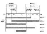
- the amino acid sequence shown in SEQ ID NO: 2 is the amino acid sequence of the N-terminal peptide region of human cofilin1 protein (N1 and N2 regions in FIG. 1 and the N-terminal 5 amino acids of the M region), and the starting methionine is 1 It is equivalent to 1st to 54th place when it is ranked.
- the amino acid sequence represented by SEQ ID NO: 7 is the amino acid sequence of the N2 region in FIG. 1 and corresponds to the sequence of positions 29 to 49 of the human cofilin 1 protein. In the peptide region of the amino acid sequence represented by SEQ ID NO: 7, there is an effective epitope for producing a highly sensitive anti-human cofilin 1 antibody.
- anti-cofilin 1 monoclonal antibody that recognizes the epitope of the N2 region represented by SEQ ID NO: 7
- an antibody clone represented by antibody clone name 4F12 in Table 1 The 4F12 clone is composed of a sequence in which the CDR1 in the light chain is represented by SEQ ID NO: 26, a sequence in which the CDR2 is represented by SEQ ID NO: 27, and a sequence in which the CDR3 is represented by SEQ ID NO: 28, and the CDR1 in the heavy chain is represented by SEQ ID NO: 29.
- the sequence shown, CDR2 consists of the sequence shown in SEQ ID NO: 30, and CDR3 consists of the sequence shown in SEQ ID NO: 31.
- Antibodies useful in the present invention can be derived from any animal source including birds and mammals. Examples include mice, rats, guinea pigs, rabbits, goats, donkeys, sheep, camels, horses, chickens, and humans. Usually, mouse, rat and rabbit antibodies are preferably used.
- fragment of anti-cofilin 1 monoclonal antibody is a partial fragment of the monoclonal antibody, which has substantially the same antigen-specific binding activity as the antibody.
- Polypeptide chain having a substantially equivalent activity or a complex thereof is applicable.
- Specific examples include a number of well-characterized antibody fragments generated by cleaving immunoglobulins with various peptidases. More specific examples include Fab, F (ab ′) 2, Fab ′, and the like.
- Fab ′ can be obtained by reducing F (ab ′) 2 under mild conditions and breaking the disulfide linkage in the hinge region.
- Each of these antibody fragments includes an antigen-binding site and has an ability to specifically bind to an antigen (that is, cofilin1 protein or a fragment thereof in the present invention).
- the fragment of the monoclonal antibody of the present invention may be synthesized chemically or by using a recombinant DNA method.
- an antibody fragment newly synthesized using a recombinant DNA method can be mentioned.
- polypeptides examples include single chain Fv (scFv: singleschain Fragment of variable region) (Pierce catlog and Handbook, 1994-1995, Pierce Chemical co., Rockford, IL, Body, Diab, IL). And synthetic antibodies such as triabodies or tetrabodies.
- VL and VH are usually located on separate polypeptide chains (L and H chains).
- Single-chain Fv is a synthetic antibody fragment having a structure in which these variable regions are connected by a flexible linker having a sufficient length and VL and VH are included in one polypeptide chain. Within the single chain Fv, both variable regions can self-assemble with each other to form one functional antigen binding site.
- Single-stranded Fv can be obtained by integrating and expressing a recombinant DNA encoding the same into a phage genome using known techniques.
- a diabody is a molecule having a structure based on the dimeric structure of a single chain Fv (Holliger et al., 1993, Proc. Natl. Acad. Sci. USA, 90: 6444-6448).
- the linker is shorter than about 12 amino acid residues, the two variable sites within the single chain Fv cannot self-assemble, but by forming a diabody, ie, two single chain Fv , The VL of one Fv chain can be assembled with the VH of the other Fv chain, and two functional antigen binding sites can be formed (Marvin et al., 2005, Acta Pharmacol. Sin). 26: 649-658). Furthermore, by adding a cysteine residue to the C terminus of the single chain Fv, it becomes possible to form a disulfide bond between the two Fv chains and to form a stable diabody (Alafsen et al., 2004, Prot. Engr. Des. Sel., 17: 21-27).
- a diabody is a bivalent antibody fragment, but each antigen-binding site need not bind to the same epitope, and each has a bispecificity that recognizes and specifically binds to a different epitope. It does not matter.
- Triabodies and tetrabodies have their trimer and tetramer structures based on a single-chain Fv structure as with diabodies. These are trivalent and tetravalent antibody fragments, respectively, and may be multispecific antibodies.
- the antibody fragment of the present invention is an antibody fragment identified using a phage display library (see, for example, McCafferty et al., 1990, Nature, Vol. 348, 522-554) and has an antigen-binding ability. Are included. In addition, for example, Kuby, J. et al. , Immunology, 3rd Ed. 1998, W.M. H. Freeman & Co. See also New York.
- anti-cofilin 1 monoclonal antibodies and fragments thereof The antibodies or fragments thereof of the invention can be modified.
- the modification herein refers to a functional modification (for example, glycosylation) necessary for the antibody of the present invention or a fragment thereof to have a specific binding activity with cofilin1 protein, and to detect the antibody of the present invention or a fragment thereof. Includes any of the labels required above.
- the cross-reactivity with other antigen should be verified in advance before use.
- the antigen to be confirmed for crossing include proteins belonging to the ADF / COFILIN family, particularly the cofilin 2 protein that is structurally similar to the cofilin 1 protein.
- an ELISA method using cofilin 1 protein as an antigen can be used.
- the competitive state of the two can be determined. It is possible to confirm the cross property by observing. Such a method for confirming the crossing property using the principle of competitive inhibition can quickly perform screening because it is not necessary to prepare reaction systems for all antigens.
- Anti-cofilin 1 monoclonal antibody and hybridoma production method The anti-cofilin 1 monoclonal antibody of the present invention or a hybridoma producing the antibody can be produced by the method described below. However, it is not limited to this method, It can also produce by any other method known in the said field
- Method for producing anti-cofilin1 monoclonal antibody Producing anti-cofilin1 monoclonal antibody that specifically binds to an epitope present in the peptide region consisting of any of the amino acid sequences shown in SEQ ID NOs: 1 to 7 among the amino acid sequences constituting cofilin1 protein
- the method comprises preparing a monoclonal antibody using the entire length of the cofilin1 protein as an immunogen, and then screening for an antibody that specifically binds to a peptide region consisting of any one of the amino acid sequences represented by SEQ ID NOs: 1 to 7,
- cofilin1 protein used as an immunogen can be prepared, for example, by the following method.
- the cofilin1 protein may be a natural type, a recombinant type, or a synthetic cofilin1 protein obtained by chemically synthesizing all or part of the amino acid sequence, such as peptide synthesis.
- the natural cofilin1 protein is obtained from a sample such as blood or urine using a known protein separation / purification technique such as gel chromatography, ion exchange chromatography, or affinity chromatography from a culture supernatant such as blood or urine. Can be recovered.
- the recombinant cofilin1 protein can be recovered from microorganisms, insect cells, or animal cells into which DNA encoding the protein has been introduced, and then recovered from the cells using known protein separation / purification techniques.
- a synthetic cofilin1 protein can be synthesized by a method known in the art, for example, a solid phase peptide synthesis method, using, for example, published cofilin1 protein amino acid sequence information.
- the synthetic cofilin1 protein may be linked to a carrier protein such as KLH (Scattle hemocyanin), OVA (ovalbumin), or BSA (bovine serum albumin).
- cofilin1 protein fragment represented by SEQ ID NOs: 1 to 7 is used as an immunogen
- any of a natural cofilin1 protein fragment, a recombinant cofilin1 protein fragment, or a synthetic cofilin1 protein fragment can be used.
- the cofilin1 protein fragment is 6 or more, 7 or more, 8 or more, 9 or more, 10 or more, 11 or more, 12 or more, 13 or more, 14 or more, 15 or more, 16 or more in the sequence shown in SEQ ID NOs: 1-7.
- An oligopeptide or polypeptide containing 17 or more, 18 or more, 19 or more, 20 or more, 21 or more, 22 or more, or 23 or more consecutive amino acid residues can be used as an antigen.
- a natural cofilin1 protein fragment as an immunogen
- the peaks are separated and separated, and the peptides contained in each peak are separated.
- the amino acid sequence is determined by a mass spectrometer, and the partial sequence represented by SEQ ID NOs: 1 to 7 or a peak that is a part thereof can be used as an immunogen.
- cofilin1 protein fragment represented by SEQ ID NOs: 1 to 7 (hereinafter referred to as cofilin1 protein fragment) will be described in detail.
- the purified polynucleotide is cleaved with an appropriate restriction enzyme and ligated into the vector cleaved with an appropriate restriction enzyme using DNA ligase or the like. There is a way to do it.
- the method for introducing the vector into bacteria is not particularly limited as long as it is a known method for introducing the vector into bacteria, for example, heat shock method, calcium Examples thereof include a method using ions, an electroporation method, etc. All of these techniques are known in the art and described in various literatures, for example, Sambrook, J. et al., 1989, Molecular. Clo ing: A Laboratory Manual Second Ed., Cold Spring Harbor Laboratory Press, Cold Spring Harbor, New York
- the Lipofectin method PNAS, 1986, 1986, 1989, (PNAS, 1987, Vol. 84, 7413
- electroporation method calcium phosphate method (Virology, 1973, Vol. 52, 456-467), DEAE-Dextran method and the like are preferably used.
- the cofilin1 protein fragment expression vector is autonomously replicable in the bacterium, and at the same time, it is composed of a promoter sequence, a ribosome binding sequence, a DNA sequence encoding the cofilin1 protein fragment, and a transcription termination sequence. It is preferable. Moreover, the gene which codes the regulatory factor which controls a promoter may be contained. Any promoter that can function in a host such as Escherichia coli may be used.
- an antibiotic such as ampicillin or tetracycline may be added to the medium as necessary.
- the culture is usually carried out at 37 ° C. for 6 to 24 hours under aerobic conditions such as aeration and agitation culture.
- the pH is preferably maintained near neutrality.
- the pH is adjusted using an inorganic or organic acid, an alkaline solution, or the like.
- the transformant is an animal cell such as a CHO cell
- the host cell is inoculated into Gibco's DMEM medium at 1 ⁇ 10 5 cells / mL, and cultured in a 5% CO 2 incubator at 37 ° C. do it.
- an antibiotic such as ampicillin or tetracycline may be added to the medium as necessary.
- expression can be induced by IPTG (isopropyl-1-thio- ⁇ -D-galactoside) treatment.
- IPTG isopropyl-1-thio- ⁇ -D-galactoside
- an appropriate amount for example, a final concentration of 1 mM
- IPTG isopropyl-1-thio- ⁇ -D-galactoside
- the prepared immunogen solution is administered to a mammal, for example, a rat, a mouse (for example, an inbred mouse BALB / c), a rabbit or the like, and immunized.
- a mammal for example, a rat, a mouse (for example, an inbred mouse BALB / c), a rabbit or the like
- the method of administering the immunogen include, but are not limited to, subcutaneous injection using FIA or FCA, intraperitoneal injection using FIA, or intravenous injection using 0.15 mol / L sodium chloride.
- a single dose of the immunogen is appropriately determined according to the type of immunized animal, administration route, etc., and is about 50 to 200 ⁇ g per animal.
- the immunization interval is not particularly limited, and after the initial immunization, booster immunization is performed 2 to 6 times, preferably 3 to 4 times at intervals of several days to several weeks, preferably at intervals of 1 to 4 weeks.
- the antibody titer in the serum of the immunized animal is measured by ELISA (Enzyme-LinkedLImmuno Sorbent Assay) method or the like. If the antibody titer reaches a plateau, the immunogen is injected intravenously or intraperitoneally, Final immunization. Then, antibody-producing cells are collected 2 to 5 days, preferably 3 days after the last immunization.
- Hybridoma production method for producing anti-cofilin 1 partial sequence monoclonal antibody (1) Recovery and cell fusion of antibody-producing cells from immunized animals Cell fusion between antibody-producing cells obtained from immunized animals and myeloma cells enables the production of cofilin 1 protein Hybridomas that produce monoclonal antibodies that specifically recognize partial sequences can be produced.
- Examples of antibody-producing cells include spleen cells, lymph node cells, peripheral blood cells, etc., but spleen cells or local lymph node cells are preferred.
- myeloma cells to be fused with antibody-producing cells generally available cell lines derived from mice and the like can be used.
- the cell line to be used has drug selectivity and cannot survive in a HAT selection medium (including hypoxanthine, aminopterin, thymine) in an unfused state, and can only grow in a state fused with antibody-producing cells. Those having the following are preferred.
- the cell line is preferably derived from an animal of the same species as the immunized animal. Specific examples of myeloma cells include P3X62-Ag., Which is a hypoxanthine / guanine / phosphoribosyltransferase (HGPRT) deficient cell line derived from BALB / c mice. 8 strain (ATCC CIB9), P3X63-Ag. 8).
- Examples include U1 strain (JCRB9085), P3 / NSI / 1-Ag4-1 strain (JCRB0009), P3x63Ag8.653 strain (JCRB0028), Sp2 / 0-Ag14 strain (JCRB0029), and the like.
- the antibody-producing cells and myeloma cells are about 1: 1 to 20: 1 in an animal cell culture medium such as serum-free DMEM or RPMI 1640 medium. Mix at a ratio and perform a fusion reaction in the presence of a cell fusion promoter.
- a cell fusion promoter polyethylene glycol having an average molecular weight of 1,500 to 4,000 Da or the like can be used at a concentration of about 10 to 80%.
- an auxiliary such as dimethyl sulfoxide may be used in combination in order to increase the fusion efficiency.
- antibody-producing cells and myeloma cells can be fused using a commercially available cell fusion device utilizing electrical stimulation (for example, electroporation) (Nature, 1977, Vol. 266, 550-552).
- cell suspension is used, for example, RPMI 1640 medium containing fetal bovine serum, etc.
- RPMI 1640 medium containing fetal bovine serum
- the culture temperature is 20 to 40 ° C, preferably about 37 ° C.
- a hybridoma of antibody-producing cells and myeloma cells can be obtained by using a selective medium (HAT medium) containing hypoxanthine, aminopterin, and thymidine. Therefore, cells that grow from about 10 days after the start of culture in a selective medium can be selected as hybridomas.
- HAT medium a selective medium containing hypoxanthine, aminopterin, and thymidine. Therefore, cells that grow from about 10 days after the start of culture in a selective medium can be selected as hybridomas.
- the hybridoma selected in the HAT medium is first screened using the binding activity to the natural or recombinant cofilin1 protein or the peptide region consisting of the amino acid sequence shown in SEQ ID NOs: 1 to 7 as an index.
- the hybridoma producing an antibody having binding activity to the cofilin1 protein is then subjected to a crossover test. That is, the binding activity to other CXC families and the like is verified, and an acceptable one is selected. Acceptable crossing means a negligible degree of crossing in the intended use of the antibody.
- ELISA In order to confirm reactivity to cofilin1 protein and cross-reactivity with other ADF / COFILIN families, for example, ELISA can be used.
- a microplate on which cofilin1 protein or a fragment thereof serving as an antigen is immobilized is prepared, and a sample obtained by appropriately diluting the culture supernatant of the hybridoma is added thereto and reacted. After sufficiently reacting, the wells are washed, and a secondary antibody label against immunoglobulin is added for further reaction. If the well is washed again and measured using the label of the secondary antibody finally bound to the well, the binding activity of the antibody present in the culture supernatant to the antigen can be quantitatively known.
- Hybridomas can also be selected using recombinant DNA technology.
- mRNA is extracted from the hybridoma group obtained according to the method described above.
- a known method as described in Example 1 may be used for extraction of mRNA.
- cDNA of the mRNA is obtained using an OligogdT primer or a random primer.
- PCR is performed using a primer set including a base sequence of a signal sequence upstream of a gene encoding a variable region and a base sequence on the constant region side.
- the obtained amplification product can be inserted into an appropriate cloning vector and cloned to obtain a library of antibody variable region genes produced by the hybridoma.
- PCR is performed using Mouse ⁇ ⁇ Ig Primer provided by Novagen, and amplification product (mouse immunoglobulin variable region cDNA) is obtained from ZERO BLUN PCR TOPO Vector provided by Invitrogen.
- the vector group obtained by inserting into a site and cloning can be used as a gene library encoding a variable region amino acid sequence.
- a hybridoma producing the antibody of the present invention is selected by designing a probe based on the amino acid sequence of the variable region or each CDR disclosed in the present invention and screening positive clones from the library. Can do.
- the hybridoma in the present invention can be used for antibody production by ascites using a mouse. Specifically, ascites fluid containing antibodies is obtained by inoculating the hybridoma into the abdominal cavity of mice derived from the cells used as the fusion partner used to produce the hybridoma or nude mice, and collecting ascites as appropriate. It can be recovered. More specifically, ascites fluid containing antibodies can be collected by inoculating hybridomas using SP / 0 cells as fusion partners into the peritoneal cavity of BALB / c mice 10 days after pristane inoculation.
- the hybridoma in this invention can be used for antibody production by culturing using a suitable culture medium.
- the antibody is obtained by inoculating a hybridoma in Gibco's hybridoma SFM medium at 1 ⁇ 10 5 cells / mL and culturing in a 5% CO 2 incubator at 37 ° C. until the hybridoma dies.
- the present invention is not limited to this.
- the antibody of the present invention or a fragment thereof uses a cDNA sequence encoding the amino acid sequence of a monoclonal antibody specifically recognizing the cofilin1 fragment disclosed in the present invention. Can also be obtained by recombinant DNA manipulation.
- each polynucleotide can be incorporated into an appropriate expression vector, introduced into a host cell, and then expressed as a complete immunoglobulin molecule.
- amino acid sequences encoding the variable region obtained by the technique of “1-4-2 (2)” using the CDR graft antibody technique the amino acid sequence of the CDR sequence is changed to each CDR of any immunoglobulin.
- the polynucleotide encoding the variable region incorporated into may be incorporated into an appropriate expression vector, introduced into a host cell, and expressed as a complete immunoglobulin molecule.
- the heavy chain and the light chain are expressed in the same host cell so that they can be produced as a dimer comprising the heavy chain / light chain.
- cells can be cotransformed with a light chain expression vector and a heavy chain expression vector, and the antibody according to the present invention can be obtained from the transformed cells.
- the polynucleotide encoding the amino acid sequence can be directly incorporated into an appropriate expression vector, introduced into a host cell, and expressed as a fragment of an immunoglobulin molecule.
- phage display antibody technology (Brinkmann et al., 1995, JI Immunol Methods, 182, 41-50, International Publication No.
- WO97 / 13844 which utilizes genetic engineering techniques to express recombinant antibodies on the phage surface 90-02809), a specific antibody can also be obtained by expressing a diversified single chain Fv antibody as a phage fusion protein by artificially shuffling genes encoding heavy and light chains.
- immunoglobulin expression vector examples include, but are not limited to, plasmids, phagemids, cosmids, virus vectors (for example, SV40 virus-based vector, EB virus-based vector, BPV-based vector) and the like.
- virus vectors for example, SV40 virus-based vector, EB virus-based vector, BPV-based vector
- BCMGS Neo vector which is one of the BPV based vectors
- the BCMGS Neo vector which is one of the BPV based vectors, is a desirable vector that efficiently expresses foreign genes by transforming into COS7 cells and the like (Hajime Isayama “Bovine Papillomavirus Vector”, Masami Muramatsu and Hiroshi Okayama). Human edition, experimental medicine separate volume: Genetic Engineering Handbook, 1991, Yodosha, 297-299).
- a host cell containing a vector expressing an antibody or a fragment thereof can be produced in the culture supernatant or the host cell by culturing according to a conventional method. Specifically, when CHO cells are used as a host, inoculate the host cells into Gibco DMEM medium at 1 ⁇ 10 5 cells / mL, and culture in a 5% CO 2 incubator at 37 ° C. By this, a culture supernatant containing the antibody can be obtained.
- the host cell is Escherichia coli
- it is inoculated and cultured in a general medium used for culturing Escherichia coli such as LB medium, and protein expression is induced to induce the expression of the protein.
- a general medium used for culturing Escherichia coli such as LB medium
- protein expression is induced to induce the expression of the protein.
- the antibody or fragment thereof as an expression product contains a constant region
- purification and recovery from the culture supernatant or cell disruption solution using a protein A column, protein G column, anti-immunoglobulin antibody affinity column, etc. can do.
- the expression is made only in the variable region and does not include the constant region, so other appropriate purification methods are applied.
- a tag sequence advantageous for purification such as a histidine tag
- it can be purified by affinity chromatography using the corresponding ligand.
- the protein When the protein is not a fusion protein with the tag, it can be purified according to conventional protein purification methods such as ammonium sulfate precipitation, ion exchange chromatography, reverse phase chromatography, gel filtration chromatography, and hydroxyapatite chromatography.
- conventional protein purification methods such as ammonium sulfate precipitation, ion exchange chromatography, reverse phase chromatography, gel filtration chromatography, and hydroxyapatite chromatography.
- the sequence on the cofilin1 protein specifically recognized by the obtained anti-cofilin1 monoclonal antibody is determined based on the gene of the protein. It can be determined by preparing various deletion mutant genes using a reaction or the like and analyzing the binding properties of the mutant cofilin1 protein obtained from the mutant gene and the monoclonal antibody. Specifically, it is performed by the following method.
- fragments having various lengths in which 10 to 400 bases are deleted from the 5 ′ end side, 3 ′ end side, or both sides of the 5 ′ end and 3 ′ end of the cofilin1 gene are prepared, and these fragments are inserted. Create a vector.
- a method for preparing a gene fragment with such a deletion mutation is described in “Sequence Biochemistry Laboratory, Volume 1, Gene Research Method II, pages 289-305, edited by the Japanese Biochemical Society”.
- various deletion mutant proteins and full-length cofilin1 protein are prepared from the host cells into which the expression vectors of the respective deletion mutants and the expression vector of full-length cofilin1 protein have been introduced by the method described above.
- the binding to various deletion mutants and full-length proteins of anti-cofilin 1 monoclonal antibody is evaluated by ELISA using these proteins as antigens.
- a mutant in which a deletion of a specific amino acid sequence is introduced with respect to the full length when the binding property of the monoclonal antibody is lost, at least a part of the epitope sequence of the monoclonal antibody is deleted. It can be determined to be included in the array. Furthermore, the epitope sequence can be further narrowed down by evaluating the reactivity of the monoclonal antibody against two, preferably three, different deletion mutants.
- the epitope sequence on the cofilin 1 protein recognized by the obtained anti-cofilin 1 monoclonal antibody can also be confirmed by the following method.
- a reductive alkylated cofilin 1 protein and an anti-cofilin 1 monoclonal antibody are reacted to form an antigen-antibody complex, followed by degradation using an appropriate protease such as trypsin.
- an appropriate protease such as trypsin.
- the antibody is difficult to be digested with trypsin, so that the antigen-antibody complex can be recovered using Protein G Sepharose or the like.
- the recovered antigen-antibody complex is analyzed by LC-MS to bind to and protect the antibody.
- the epitope on the cofilin 1 protein recognized by the antibody, ie, the antibody can be identified.
- the epitope sequence on the cofilin1 protein recognized by the anti-cofilin1 monoclonal antibody can be confirmed by, for example, a competition method using a synthetic peptide.
- a synthetic peptide of 6 to 21 amino acids in the amino acid sequence constituting the cofilin 1 protein is prepared by a solid phase synthesis method or the like.
- this synthetic peptide is allowed to act when the anticofilin1 monoclonal antibody is reacted with the solid phase cofilin1 protein, thereby inhibiting the binding of the anticofilin1 monoclonal antibody. Is confirmed, it can be determined that the amino acid sequence of the synthetic peptide is an epitope sequence recognized by the anti-cofilin 1 monoclonal antibody.
- sample used in the measurement method of the present invention refers to various samples that may contain cofilin1 protein and / or fragments thereof.
- a cultured cell a cultured cell lysate, a culture supernatant, or a mammalian sample containing DNA encoding a cofilin1 protein or a fragment thereof.
- mammalian sample means any biological sample derived from mammals such as tissues collected from mammals (for example, post-operatively collected tissues) and body fluids such as blood, lymph, urine, spinal fluid, saliva, and semen. Yes, preferably blood or urine.
- the blood referred to in the present invention includes serum, plasma or interstitial fluid.
- it is a primate, More preferably, it is a human.
- peptide region where the epitope is present include peptide regions consisting of the amino acid sequences represented by SEQ ID NOs: 1 and 2.
- peptide region comprising the amino acid sequence represented by SEQ ID NO: 1 among the peptide region comprising at least the amino acid sequence represented by SEQ ID NO: 3, and among the peptide region comprising the amino acid sequence represented by SEQ ID NO: 2, at least SEQ ID NO: 4 More preferred are peptide regions comprising the amino acid sequence shown.
- the epitope is not particularly limited as long as it exists on the cofilin1 protein or a fragment thereof.
- different epitopes may exist at positions separated from each other on the cofilin 1 protein, or may exist at positions close to each other.
- the different epitopes may be present on different peptide regions, or plural
- the peptide region may be present on a single peptide region as long as it is a peptide region consisting of an amino acid having a sufficient length that can include the epitope.
- the combination of peptide regions is not particularly limited.
- the anti-cofilin 1 monoclonal antibody and / or fragment thereof described in the first embodiment can be used as two or more types of anti-cofilin 1 monoclonal antibody and / or fragments thereof used in the present embodiment.
- an antibody clone group described in Table 1 described below preferably an antibody clone group including at least 1E2, 2C4, 2D12, 2F5, 3F11, 3G2, 4F12, 4E12 and 4G10, more preferably at least 1E2, 2C4, 4F12, 4E12.
- a combination of two or more anti-cofilin1 monoclonal antibodies and / or fragments thereof selected from the group of antibody clones comprising: or at least 4F12 and 1E2, 4F12 and 4E12, 1E2 and 3F11, 1E2 and 4E12, 2C4 and 4E12, 3G2 4E12, 4G10 and 4E12, 2D12 and 1E2, 2D12 and 2C4, or 4E12 and 2F5.
- a sequence in which CDR1 is represented by SEQ ID NO: 17 in the heavy chain An anti-cofilin1 monoclonal antibody comprising a sequence represented by SEQ ID NO: 18 and CDR3 comprising a sequence represented by SEQ ID NO: 19 or a fragment thereof, (3) a sequence represented by CDR1 in the light chain in CDR2 Includes the sequence represented by SEQ ID NO: 21, and CDR3 comprises the sequence represented by SEQ ID NO: 22, and in the heavy chain, CDR1 represents the sequence represented by SEQ ID NO: 23, CDR2 represents the sequence represented by SEQ ID NO: 24, and An anti-cofilin1 monoclonal antibody or a fragment thereof in which CDR3 comprises the sequence represented by SEQ ID NO: 25, and (4) in the light chain, the CDR1 is represented by SEQ ID NO: 26, the CDR2 is represented by SEQ ID NO: 27, and the CDR3 Includes the sequence shown in SEQ ID NO: 28, and in the heavy chain, CDR1 is arranged.
- Two or more kinds of anti-cofilin1 monoclonal antibodies or fragments thereof comprising a sequence represented by No. 29, a CDR2 comprising a sequence represented by SEQ ID No. 30 and a CDR3 comprising a sequence represented by SEQ ID No. 31 a combination of cofilin1 monoclonal antibodies and / or fragments thereof.
- More preferable two or more types of anti-cofilin1 monoclonal antibodies and / or fragments thereof used in the present embodiment are (1) at least the sequence in which CDR1 in the light chain is represented by SEQ ID NO: 8, and CDR2 is represented by SEQ ID NO: 9.
- CDR3 consists of the sequence shown in SEQ ID NO: 10
- CDR1 in the heavy chain consists of the sequence shown in SEQ ID NO: 11
- CDR2 consists of the sequence shown in SEQ ID NO: 12
- CDR3 consists of the sequence shown in SEQ ID NO: 13.
- An antibody or a fragment thereof comprising CDR1 in the light chain as shown in SEQ ID NO: 26, CDR2 as shown in SEQ ID NO: 27, and CDR3 as shown in SEQ ID NO: 28, and CDR1 in the heavy chain as SEQ ID NO: 29
- CDR1 in the light chain is represented by SEQ ID NO: 8
- CDR2 is represented by SEQ ID NO: 9
- CDR3 is represented by SEQ ID NO: 10
- CDR1 in the light chain and CDR1 in the heavy chain the CDR1 in the sequence shown in SEQ ID NO: 11, the CDR2 in the sequence shown in SEQ ID NO: 12 and the CDR3 in the sequence shown in SEQ ID NO: 13, or a fragment thereof
- CDR2 is a sequence represented by SEQ ID NO: 21
- CDR3 is a sequence represented by SEQ ID NO: 22
- CDR1 in the heavy chain is a sequence represented by SEQ ID NO: 23
- CDR2 is SEQ ID NO: 24.
- the CDR3 is composed of the sequence represented by SEQ ID NO: 22, the CDR1 in the heavy chain is composed of the sequence represented by SEQ ID NO: 23, the CDR2 is composed of the sequence represented by SEQ ID NO: 24, and the CDR3 is composed of the sequence represented by SEQ ID NO: 25.
- a combination of antibodies or fragments thereof, and (4) at least the CDR1 in the light chain is SEQ ID NO: 2.
- a sequence represented by 0, CDR2 comprises a sequence represented by SEQ ID NO: 21, and CDR3 comprises a sequence represented by SEQ ID NO: 22, and CDR1 in the heavy chain is represented by SEQ ID NO: 23, and CDR2 is represented by SEQ ID NO: 24.
- amino acid sequences shown in SEQ ID NOs: 1 to 7, which are partial sequences of the amino acid sequence constituting the cofilin1 protein, are highly conserved in mammals showing 100% homology in human, mouse, rat, cow, dog and chimpanzee. By using an antibody that recognizes these sequences, it is possible to construct a general-purpose cofilin 1 protein measurement system corresponding to samples derived from various mammalian species.
- the ELISA (Enzyme-Linked ImmunoSorbent Assay) method is also called an enzyme immunosorbent assay, and a small amount of a target antigen contained in a sample is converted into an antigen using an enzyme-labeled antibody or antigen and the action of the enzyme.
- antibody reaction is detected as color development density or fluorescence intensity to quantify the target antigen. That is, the antibody of the present invention or a fragment thereof, or the cofilin1 protein or a fragment thereof is immobilized on a solid phase carrier, and an immunological reaction with the antibody and the cofilin1 protein is enzymatically detected.
- Methods such as a direct method, an indirect method, and a sandwich method are known. In the present invention, the sandwich method is particularly preferably applied.
- a first antibody (solid phase antibody) immobilized on a solid phase carrier is bound to an antigen, and then a second antibody (labeled antibody / primary antibody) that recognizes an epitope different from the first antibody is added.
- a second antibody labeled antibody
- the label is detected, and when the second antibody is a primary antibody, it is detected by a third antibody (secondary antibody).
- a known method Japanese Clinical Pathology Society, “Special Issue on Clinical Pathology No.
- the solid phase carrier is made of a material such as polystyrene, polycarbonate, polyvinyl toluene, polypropylene, polyethylene, polyvinyl chloride, nylon, polymethacrylate, latex, gelatin, agarose, cellulose, sepharose, glass, metal, ceramics, or magnetic material.
- Insoluble carriers in the form of beads, microplates, test tubes, sticks or test pieces can be used. Immobilization of the antibody of the present invention or a fragment thereof or cofilin1 protein or a fragment thereof to a solid phase carrier can be achieved by binding in accordance with a known method such as a physical adsorption method, a chemical binding method, or a combination method thereof.
- the labeling substance in the case of the ELISA method, for example, peroxidase (POD), alkaline phosphatase, ⁇ -galactosidase, urease, catalase, glucose oxidase, lactate dehydrogenase, amylase or biotin-avidin complex, etc.
- POD peroxidase
- alkaline phosphatase alkaline phosphatase
- ⁇ -galactosidase urease
- catalase glucose oxidase
- lactate dehydrogenase lactate dehydrogenase
- amylase biotin-avidin complex
- NADH-FMNH 2 -luciferase system for the luminescence immunoassay method, NADH-FMNH 2 -luciferase system, luminol-hydrogen peroxide-POD system, acridinium ester system, or dioxetane compound system can be used.
- the binding method between the labeling substance and the antibody is a known method such as glutaraldehyde method, maleimide method, pyridyl disulfide method or periodic acid method in the case of ELISA, and chloramine T method in the case of radioimmunoassay.
- a known method such as the Bolton Hunter method can be used.
- the immunoassay method of the present invention is carried out by sandwich ELISA using anti-cofilin 1 protein monoclonal antibodies that specifically recognize epitopes present in the peptide regions consisting of the amino acid sequences shown in SEQ ID NOs: 5 and 6 below. This will be described in detail with reference to an example. However, the embodiment of the present invention is not limited to this.
- a monoclonal antibody that specifically recognizes an epitope present in the peptide region consisting of the amino acid sequence represented by SEQ ID NO: 6 is labeled as a labeled antibody, and the labeled antibody is labeled with the immobilized antibody and cofilin1. It acts on the carrier to which the protein complex is bound. Since the immobilized antibody and the labeled antibody recognize different epitopes of cofilin1 protein, a triple complex consisting of immobilized antibody / cofilin1 protein / labeled antibody is formed on the solid support.
- the unbound labeled antibody is sufficiently washed with a washing solution, and then the cofilin1 protein present in the sample can be detected and quantified by detecting using the label of the labeled antibody in the triple complex.
- One kind or several kinds of labeled antibodies may be used, but two or more kinds are preferably used, and three kinds are more preferably used. If the animal species from which the immobilized antibody and the labeled antibody are derived is different, the primary antibody is recognized by acting on the immobilized antibody / cofilin1 protein as the primary antibody without labeling the labeled antibody. It can also be detected using a labeled secondary antibody. It should be noted that the antibody used for immobilization and the antibody used for labeling can be used in reverse.
- a sample containing a labeled antibody and cofilin1 protein can be mixed in advance to form an antigen-antibody complex, and then allowed to act on the immobilized antibody.
- the antibody to be immobilized is labeled with biotin
- a biotinylated immobilized antibody, a sample containing cofilin1 protein, and an antibody with a label other than biotin are mixed to form an antigen-antibody complex, and then avidin is added.
- an antigen-antibody complex can be detected using a label other than biotinylation.
- the immunological measurement method of the present invention can use an immunochromatographic test strip.
- the test strip for immunochromatography has been developed, for example, a sample receiving portion made of a material that easily absorbs a sample, a reagent portion containing the diagnostic agent of the present invention, a developing portion where a reaction product of the sample and the diagnostic agent moves, It consists of a labeling section for coloring the reaction product, a presentation unit for developing the colored reaction product, and the like.
- Commercially available pregnancy diagnostic agents and the like have the same form.
- the principle of this measurement method is as follows. First, when a sample is given to the sample receiving portion, the sample receiving portion absorbs the sample and causes the sample to reach the reagent portion.
- a monoclonal antibody (referred to as a first antibody) that specifically recognizes an epitope present in a peptide region consisting of the amino acid sequence represented by SEQ ID NO: 5 as an immunological measurement method using an immunochromatographic test strip; and SEQ ID NO: 6
- a monoclonal antibody (referred to as a second antibody) that specifically recognizes an epitope present in a peptide region consisting of the amino acid sequence shown in FIG.
- an antigen-antibody reaction occurs between the cofilin1 protein in the sample and the first antibody, and the formed antigen-antibody complex moves through the development part and reaches the labeling part.
- the labeling part a reaction between the antigen-antibody complex and the labeled second antibody occurs, and coloration is recognized when the reaction product with the labeled second antibody develops to the presentation part.
- a substitution method, an indirect competition method, and the like are known, and any of them may be used. This technique is well known in the art. See, for example, Kazuhiro Nagata and Hiroshi Handa, real-time analysis of biological material interactions, Springer Fairlark Tokyo, Tokyo, 2000.
- the measurement method of the present invention can use a quartz crystal microbalance measurement method (QCM method).
- QCM method utilizes a phenomenon in which when a substance is adsorbed on the surface of an electrode attached to a crystal oscillator, the resonance frequency of the crystal oscillator decreases according to its mass.
- the QCM sensor using this method is a mass measurement sensor that quantitatively captures a very small amount of adsorbed material based on the amount of change in the water resonance frequency.
- This technique is well known in the art. For example, J. et al. Christopher Love, L. A. Estroff, J.M. K. Kriebel, R.A. G. Nuzzo, G.M. M.M. See Whitesides, 2005, Self-Assembled Monolayers of a Form of Nanotechnology, Chemical Review, 105: 1103-1169;
- the third embodiment of the present invention relates to a digestive organ cancer incidence determination method.
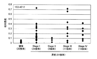
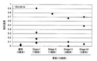
- the amount of cofilin1 protein and / or a fragment thereof in a sample is measured as a marker for determining digestive organ cancer incidence, and the digestive organ cancer disease in a subject is determined based on a quantitative ratio with a healthy body. It is characterized by determining the presence or absence of.
- the method of this embodiment includes a measurement step (1) and a determination step (2). Hereinafter, each step will be specifically described.
- Measurement step is a step of measuring the amount of cofilin1 protein in the sample derived from the subject and the healthy body using the measurement method described in the second embodiment.
- Subject means an individual subjected to the determination method of the present invention.
- the type of subject is not particularly limited as long as it is a mammal.
- it is a human (hereinafter referred to as “subject” when the human is a subject in this specification).
- the sample in the present embodiment is a mammalian sample because it is derived from a subject and a healthy body. That is, biological samples derived from all mammals such as tissues collected from mammals (for example, post-operatively collected tissues) and body fluids such as blood, lymph, urine, spinal fluid, saliva, semen, and the like are applicable. Preferred is blood, particularly preferred is serum or plasma.
- the types of samples derived from the subject and the healthy body must be the same.
- the sample derived from the subject is serum
- the sample derived from the healthy body is also preferably serum.
- the “determination step” is a comparison between the amount of cofilin1 protein and / or a fragment thereof in a sample derived from a subject and a healthy body measured in the measurement step, When the amount of the fragment is statistically significantly larger than that of a healthy person, it is a step of determining that the subject is suffering from digestive organ cancer.
- “Gastrointestinal cancer” refers to a primary malignant tumor that develops in a digestive organ. Examples include esophageal cancer, gastric cancer, duodenal cancer, small intestine cancer (including jejunal cancer and ileal cancer), colon cancer (including cecal cancer, colon cancer, rectal cancer), and pancreatic cancer.
- a particularly preferred gastrointestinal cancer as a determination target of the present embodiment is gastric cancer.
- gastric cancer is not limited to digestive organ cancer, and may be other epithelial tumors such as breast cancer, liver cancer and lung cancer, and skin cancers such as malignant melanoma.
- the amount of cofilin 1 protein and / or fragment thereof obtained from each sample derived from the subject and the healthy body in the measurement step is compared.
- the amount to be compared may be a relative amount such as a concentration or an absolute amount. In the case of an absolute amount, it is necessary to make the amount of the sample used for measurement equal in advance between the subject and the healthy body, or to convert it to an equivalent amount based on the ratio of the sample used for the measurement. is there.
- whether or not the amount of cofilin1 protein and / or fragment thereof in the sample derived from the subject is statistically significantly larger than the amount of cofilin1 protein and / or fragment thereof in the sample derived from the healthy subject.
- a subject classified as having a digestive cancer group is determined to have or is very likely to have digestive cancer, and a subject classified as having a non-digestive cancer group is digested. Although the possibility of having organ cancer cannot be completely excluded, it is determined that the possibility is low.
- “statistically significant” means, for example, a case where the risk rate (significance level) of the obtained value is less than 5%, 1%, or 0.1%. Therefore, “statistically significant” means that the quantitative difference between the cofilin 1 protein and / or fragment thereof obtained from each of the subject and the healthy subject is statistically significant between the two. It means that there is a difference and the amount of the protein in the subject is larger than that in the healthy subject.
- the amount of cofilin1 protein in the blood sample corresponds to a case where the subject is twice or more, preferably 3 times or more, more preferably 4 times or more, and most preferably 5 times or more that of a healthy body.
- test method for statistical processing is not particularly limited as long as a known test method capable of determining the presence or absence of significance is appropriately used. For example, Student's t test or multiple comparison test can be used.
- the amount of cofilin1 protein in a healthy blood sample can be measured every time the amount of cofilin1 protein in a blood sample of a subject is measured, but the amount of cofilin1 protein measured in advance should be used. You can also. In particular, if the amount of cofilin1 protein in various physical conditions of a healthy body is measured in advance and the value is input to a computer and stored in a database, the physical condition of the subject is input to the computer, Conveniently, the amount of cofilin 1 protein in a healthy subject having the optimal physical conditions for comparison with the subject can be readily used.
- the stage of gastrointestinal cancer targeted in the present invention is not particularly limited, and ranges from early gastrointestinal cancer to terminal gastrointestinal cancer.
- the determination method of the present invention is capable of detecting even early-stage gastrointestinal cancer, which has been difficult with conventional methods, due to the high sensitivity of the antibody and / or fragment thereof of Embodiment 1.
- "Early gastrointestinal cancer” refers to those that are localized to the tumor site (in the mucous membrane) and that do not invade surrounding tissues, or those that have invasion but are limited to a local area. To tell. Since early detection of gastrointestinal cancer significantly improves the 5-year survival rate, the actual benefit of the present invention that can determine its morbidity early is high.
- the subject is affected with digestive organ cancer by measuring the digestive cancer marker in the blood sample immunologically using an antibody.
- the determination method of the present invention the subject suffers from digestive organ cancer using a sample such as plasma or serum of the subject. It can be determined from early whether or not.
- the cofilin1 protein contained in an appropriate sample such as blood can be easily and simply measured by the immunological measurement method described in the first embodiment. Further, based on the result and the method of the third embodiment, it is possible to easily and quickly determine whether or not the sample provider is affected by digestive organ cancer.
- cDNA was synthesized using the obtained total mRNA as a template to prepare a human cDNA library.
- the reverse transcription reaction followed the protocol attached to the enzyme.
- PCR was performed using the obtained human cDNA library as a template and a primer set consisting of the nucleotide sequences shown in SEQ ID NOs: 32 and 35.
- the base sequence represented by SEQ ID NO: 32 includes a part of the 5 'terminal region of the human cofilin 1 gene and a BamHI recognition sequence upstream thereof.
- the base sequence represented by SEQ ID NO: 35 includes a part of the 3 'terminal region of the human cofilin 1 gene and an EcoRI recognition sequence downstream thereof.
- a PCR reaction solution was prepared by using KOD (Toyobo Co., Ltd.) as a DNA polymerase so as to contain 10 ng of cDNA library and 10 pmol of each primer according to the protocol attached to KOD.
- the reaction conditions were as follows: after heating at 94 ° C. for 5 minutes, repeating a cycle of holding at 94 ° C. for 15 seconds, 55 ° C. for 30 seconds, and 68 ° C. for 30 seconds, and finally holding at 68 ° C. for 4 minutes. Performed under conditions.
- the amplified DNA fragment was purified using Wizard SV Gel and PCR Clean-up System (manufactured by Promega). By this reaction, a PCR product having a total length of about 500 bp was obtained.
- the resulting DNA fragment was subjected to BamHI cleavage and EcoRI cleavage, followed by ligation reaction for incorporation into BamHI cleavage, EcoRI cleavage and BAP-treated open pET30a (manufactured by Novagen).
- Ligation High Toyobo Co., Ltd.
- competent cells were transformed using the solution after the ligation reaction.
- Escherichia coli strain DH5 ⁇ (manufactured by Takara Bio Inc.) was used, and the details followed the attached protocol.
- Bacteria after the transformation treatment were coated on an LB plate containing 100 ⁇ g / mL of the antibiotic kanamycin and cultured at 37 ° C. overnight.
- the obtained transformant was cultured overnight at 37 ° C. in an LB liquid medium containing 100 ⁇ g / mL kanamycin, and the target pET30a_cofilin 1 was obtained by miniprep.
- PCR was performed using the obtained pET30a_cofilin1 gene (10 ng) as a template and a primer set consisting of the nucleotide sequences shown in SEQ ID NOs: 39 and 40.
- the base sequence represented by SEQ ID NO: 39 contains a part of the 5 'terminal region of the human cofilin 1 gene and an EcoRI recognition sequence upstream thereof.
- the base sequence represented by SEQ ID NO: 40 contains a part of the 3 'terminal region of the human cofilin1 gene and a BglII recognition sequence downstream thereof.
- a PCR reaction solution was prepared by using KOD (Toyobo Co., Ltd.) as a DNA polymerase so as to contain 10 ng of cDNA library and 10 pmol of each primer according to the protocol attached to KOD.
- the reaction conditions were as follows: after heating at 94 ° C. for 5 minutes, repeating a cycle of holding at 94 ° C. for 15 seconds, 55 ° C. for 30 seconds, and 68 ° C. for 30 seconds, and finally holding at 68 ° C. for 4 minutes. Performed under conditions.
- the amplified DNA fragment was purified using Wizard SV Gel and PCR Clean-up System (manufactured by Promega). By this reaction, a PCR product having a total length of about 500 bp was obtained.
- the resulting DNA fragment was digested with EcoRI and BglII and then subjected to a ligation reaction for incorporation into EcoRI, BamHI, and BAP-treated ring-opened pCMV-Myc (Clontech).
- Ligation High Toyobo Co., Ltd.
- competent cells were transformed using the solution after the ligation reaction.
- Escherichia coli strain DH5 ⁇ manufactured by Takara Bio Inc.
- Bacteria after the transformation treatment were spread on an LB plate containing 50 ⁇ g / mL of the antibiotic kanamycin and cultured at 37 ° C. overnight. The obtained transformant was cultured overnight at 37 ° C. in an LB liquid medium containing 100 ⁇ g / mL ampicillin to obtain the target pCMV-Myc_human cofilin1.
- the obtained bacterial cells were washed with PBS, and then the insoluble fraction was prepared as a precipitate using B-PER (PIERCE). The details followed the attached protocol.
- the insoluble fraction was solubilized with 6M urea, and histidine-tagged human cofilin1 protein was adsorbed using TALON®Metal®Affinity®Resin (manufactured by CLONETECH).
- the protein-adsorbed resin was washed with 6M urea containing 10 mM imidazole, and then eluted using a 6M urea solution containing 1M imidazole.
- Example 2 Production and selection of mouse monoclonal antibody against human cofilin1 protein (Production of anti-human cofilin1 antibody-producing mouse) 50 ⁇ L of the 1 mg / mL human cofilin1 protein solution obtained in Example 1 was mixed with 50 ⁇ L of Sigma adjuvant system (manufactured by Sigma), and the whole amount was intraperitoneally administered to 6-week-old BALB / c mice. Two weeks and four weeks later, the same amount of the human cofilin1 protein solution prepared in the same manner was administered. Subsequently, 100 ⁇ L of blood was collected from the tail vein of the mouse and placed overnight, and then centrifuged at 5000 ⁇ g for 5 minutes to collect the supernatant as serum.
- Sigma adjuvant system manufactured by Sigma
- the suspension was again suspended in RPMI 1640 medium, the number of cells was counted, and an SP2 / 0 myeloma cell solution having an amount 1/10 of the number of spleen cells was prepared. Both cell solutions were mixed, centrifuged at 2200 rpm for 10 minutes, and the supernatant was discarded. The cells were tapped to loosen, 1 mL of a solution prepared by mixing PEG (manufactured by ROCHE) and HBSS (manufactured by GIBCO) at a ratio of 5: 1 was added and stirred. In the subsequent work, unless otherwise specified, all solutions and culture media were kept warm at 37 ° C.
- RPMI 1640 medium 9 mL was added over 5 minutes, mixed slowly, and then centrifuged at 2200 rpm for 10 minutes to remove the supernatant.
- the obtained precipitated cells are suspended in RPMI 1640 medium supplemented with 15% FCS and HAT (manufactured by ROCHE), poured into a 96-well cell culture plate (manufactured by Greiner) at 200 ⁇ L per well, and incubated at 37 ° C., 5% CO 2. Incubated for 1 week.
- the colony grown under the HAT-added condition is judged as a hybridoma in which spleen cells and myeloma cells are fused, and the supernatant of the colony-growing well is diluted 5-fold and added to the well of the human cofilin1 protein-immobilized plate. 100 ⁇ L was added, and the presence or absence of antibody production was confirmed by the same method as described above. Wells in which antibody production was confirmed were regarded as positive.
- the positive well colonies were suspended in RPMI medium containing 15% FCS and HT (manufactured by Invitrogen), and positive clones were cloned by the limiting dilution method.
- Example 3 Epitope mapping of cofilin1 protein antibody (production of deletion mutant) Epitope sequences were determined for the 39 purified antibodies obtained by the following method. First, five human cofilin1 protein deletion mutants from mutant A to mutant E shown in Example 1 and full-length human cofilin1 protein were prepared as GST fusion proteins (FIG. 1).
- the primer set consisting of the nucleotide sequences shown in SEQ ID NOs: 32 and 37 is used for variant A
- the primer set consisting of the nucleotide sequences shown in SEQ ID NOs: 33 and 35 is used for variant B
- the primer set consisting of the base sequences shown in SEQ ID NOs: 32 and 38 for variant C using the primer set consisting of the base sequences shown in SEQ ID NOs: 32 and 36 for variant D
- variant E The gene was obtained by PCR using a primer set consisting of the nucleotide sequences shown in SEQ ID NOs: 34 and 38, and using the primer set consisting of the nucleotide sequences shown in SEQ ID NOs: 32 and 35 for full-length human cofilin 1, respectively.
- the base sequences shown in SEQ ID NOs: 32, 33 and 34 include a part of the 5 'terminal region of the human cofilin1 gene and a BamHI recognition sequence upstream thereof.
- the base sequences shown in SEQ ID NOs: 35 to 38 include a part of the 3 'terminal region of the human cofilin 1 gene and an EcoRI recognition sequence on the downstream side thereof.
- the six PCR products thus obtained were subjected to ligation reaction for incorporation into BamHI, EcoRI and BAP-treated ring-opened pGEX6P-1 (GE Healthcare) after BamHI and EcoRI cleavage, respectively.
- Ligation High Toyobo Co., Ltd.
- competent cells were transformed using the solution after the ligation reaction.
- Escherichia coli strain DH5 ⁇ manufactured by Takara Bio Inc.
- the transformed bacteria were spread on an LB plate containing 100 ⁇ g / mL of antibiotic ampicillin and cultured at 37 ° C. overnight.
- the obtained transformant was cultured overnight at 37 ° C. in an LB liquid medium containing 100 ⁇ g / mL ampicillin, and the target full-length human cofilin1 protein and 5 deletion mutant genes were obtained by miniprep. Six introduced expression vectors were obtained.
- Each of the six expression vectors was transformed into Escherichia coli strain Rosetta-Gami2 (manufactured by Novagen), and a full-length human cofilin 1 and five deletion mutant recombinant GST fusion proteins were prepared by the following procedure. did. First, the transformant was precultured overnight at 37 ° C. in 10 mL of LB medium containing ampicillin and chloramphenicol. Next, 1 L of the same medium is inoculated with the preculture, cultured at 37 ° C. for 5 hours, added with IPTG having a final concentration of 0.5 mM, and cultured at 37 ° C. for 12 hours. After inducing the expression of the cofilin1 protein, the cells were collected by centrifugation.
- the obtained bacterial cells were washed with PBS, and then a soluble fraction was obtained using B-PER (PIERCE). The details followed the attached protocol. Next, glutathione sepharose 4B (GE Healthcare) was used to adsorb the recombinant GST fusion protein of the soluble fraction. The resin adsorbed with protein was washed with TBS (150 mM NaCl, 50 mM Tris-Cl, 5 mM EDTA, pH 8.0) and then eluted with TBS containing 10 mM reduced glutathione. The obtained GST fusion protein was prepared to 1 mg / mL and used for the following experiments.
- TBS 150 mM NaCl, 50 mM Tris-Cl, 5 mM EDTA, pH 8.0
- the plate which performed only the blocking reaction without immobilizing GST fusion protein was produced, and it was set as the blank plate.
- each of the 39 types of anti-human cofilin 1 monoclonal antibodies diluted with PBS-T containing 1% BSA to a final concentration of 1 ⁇ g / mL was placed in a blank plate and a GST fusion protein-immobilized plate for 1 hour. And reacted at room temperature. After discarding the solution in the well and washing with PBS-T, 100 ⁇ L of HRP-labeled anti-mouse IgG antibody solution (GE Healthcare) was allowed to react for 1 hour at room temperature.
- Epitope sequences were determined using the differences in reactivity of 39 anti-human cofilin1 monoclonal antibodies to full-length human cofilin1 and 5 deletion mutants as indicators.
- the 4E12 antibody reacts only with the full-length human cofilin1 protein and mutant B and not with other deletion mutants. Therefore, at least a part of the epitope sequence of the 4E12 antibody is present in the full-length human cofilin1 protein and variant B, and in the other deletion variants, the C3 region represented by SEQ ID NO: 4, which is a deleted region (Fig. 1).
- HEK293 cells (2 ⁇ 10 6 cells) were cultured overnight in a DMEM medium (GIBCO) containing 10% FCS using a cell culture dish (Corning), and Lipofectamine 2000 (Invitrogen).
- PCMV-Myc_human cofilin1 or pCMV-Myc was introduced according to the attached protocol.
- 1% NP40 buffer 1% NP40, 150 mM NaCl, 5 mM EDTA, 100 mM Tris-Cl, pH 8.0
- the cells were suspended in 1% NP40 buffer, transferred to a 1 mL centrifuge tube, centrifuged at 1,5000 ⁇ g, and the supernatant was collected.
- pCMV-Myc_human cofilin1-introduced cell or pCMV-Myc-introduced protein extract was diluted 10-fold with 1% NP40 buffer, and 100 ⁇ L of 39 wells were added to each immobilized antibody for 1 hour. And reacted at room temperature. Subsequently, after discarding the solution in the well and washing with PBS-T, the obtained biotin bodies of 39 kinds of antibodies were diluted to 0.5 ⁇ g / mL with 1% NP40 buffer containing 1% BSA.
- Example 5 Analysis of amino acid sequence of anti-cofilin 1 monoclonal antibody (determination of light chain and heavy chain cDNA sequence and amino acid sequence of monoclonal antibody using hybridoma)
- the cDNA and amino acid sequences of the light and heavy chains were determined for the four antibodies required for the combination of the five selected antibodies.
- hybridomas producing each antibody were cultured in RPMI 1640 medium supplemented with 15% FCS at 37 ° C. under 5% CO 2 until 1 ⁇ 10 6 cells / mL. Thereafter, the culture solution was centrifuged at 1200 rpm for 5 minutes, and the cells were collected. MRNA was prepared from the recovered hybridoma.
- the obtained vector solution was subjected to DNA sequence analysis of the region encoding the monoclonal antibody using M13 primer. Analysis was performed using a 3130xl genetic analyzer (Applied Biosystems). A clone having no stop codon in the insertion region was judged as a DNA sequence encoding the target monoclonal antibody, and the light chain and heavy chain DNA sequences of the four antibodies (1E2, 2C4, 4E12, 4F12) were determined. With respect to the determined DNA sequence, the amino acid sequence encoded according to the codon usage of E. coli was determined, and the sequences shown in SEQ ID NOs: 8-31 could be obtained.
- Example 6 Detection of human cofilin1 protein by sandwich ELISA using monoclonal antibody 1E2 and monoclonal antibody 4E12
- Example 3 from the amino acid sequence represented by SEQ ID NO: 5 among the amino acid sequences represented by SEQ ID NO: 1 A monoclonal antibody 1E2 that specifically recognizes an epitope present in the peptide region, and an epitope present in the peptide region consisting of at least the amino acid sequence represented by SEQ ID NO: 6 among the amino acid sequences represented by SEQ ID NO: 1 It was found that sandwich ELISA using a biotin-labeled monoclonal antibody 4E12 that recognizes human cofilin1 protein with the highest sensitivity.
- His-human cofilin1 protein was diluted stepwise from 1000 ng / mL to 10 pg / mL with TBS buffer containing 1% BSA and 1% NP40, and the detection sensitivity of this sandwich ELISA was calculated by measuring this as a sample. did.
- the minimum detection limit value (average value + 2SD and the minimum value of the subject ⁇ 2SD where the mean value + 2SD did not overlap) of the ELISA method by the combination of 1E2 and 4E12 was 10 pg / mL (arrow in FIG. 4).
- Example 7 Detection of gastric cancer by measuring human cofilin1 protein in plasma Human cofilin1 protein in the plasma of gastric cancer patients and healthy individuals was detected by sandwich ELISA using 1E2 and 4E12 biotin-labeled products. Blood was collected from 51 gastric cancer patients (stage I: 24, stage II: 6; stage III: 11; stage IV: 10) and 20 healthy controls using Benedict II vacuum blood collection tubes (manufactured by Terumo). To obtain plasma. Plasma was diluted 10-fold with TBS buffer containing 1% BSA and 1% NP40, respectively, and 100 ⁇ L was used as a sample.
- a 3 ⁇ g / mL solution of anti-human cofilin 1 antibody 1E2 was prepared, and 100 ⁇ L of each was placed in a well of a Flat TM bottom 96-well plate (manufactured by Nunc) and solidified for 5 hours.
- 400 ⁇ L of blocking solution PBS-T containing 1% BSA
- 100 ⁇ L plasma sample was added and allowed to react for 1 hour at room temperature.
- a 3 ⁇ g / mL solution of anti-human cofilin 1 antibody 1E2 was prepared, and 100 ⁇ L of each was placed in a well of a Flat TM bottom 96-well plate (manufactured by Nunc) and solidified for 5 hours.
- 400 ⁇ L of blocking solution PBS-T containing 1% BSA
- 100 ⁇ L of a serum sample was added and allowed to react at room temperature for 1 hour.
Abstract
Description
b)前記a)のペプチド領域のアミノ酸配列に1若しくは数個のアミノ酸が欠失、置換又は付加されたペプチド領域
c)前記a)のペプチド領域のアミノ酸配列と90%以上の同一性を有するペプチド領域
(2)軽鎖において、CDR1が配列番号8で示される配列を含み、CDR2が配列番号9で示される配列を含み、及びCDR3が配列番号10で示される配列を含み、ならびに重鎖において、CDR1が配列番号11で示される配列を含み、CDR2が配列番号12で示される配列を含み、及びCDR3が配列番号13で示される配列を含む、(1)に記載の抗cofilin1モノクローナル抗体又はその断片。 a) a peptide region comprising at least the amino acid sequence represented by SEQ ID NO: 5 among the amino acid sequences represented by SEQ ID NO: 1 b) one or several amino acids deleted, substituted or substituted in the amino acid sequence of the peptide region of a) Added peptide region c) Peptide region having 90% or more identity with the amino acid sequence of the peptide region of a) (2) In the light chain, CDR1 contains the sequence shown in SEQ ID NO: 8, and CDR2 contains SEQ ID NO: And the CDR3 comprises the sequence shown in SEQ ID NO: 10, and in the heavy chain, the CDR1 contains the sequence shown in SEQ ID NO: 11, the CDR2 contains the sequence shown in SEQ ID NO: 12, And CDR3 comprises the sequence represented by SEQ ID NO: 13, or the anti-cofilin 1 monoclonal antibody or fragment thereof according to (1).
b)前記a)のペプチド領域のアミノ酸配列に1若しくは数個のアミノ酸が欠失、置換又は付加されたペプチド領域
c)前記a)のペプチド領域のアミノ酸配列と90%以上の相同性を有するペプチド領域
(5)軽鎖において、CDR1が配列番号20で示される配列を含み、CDR2が配列番号21で示される配列を含み、及びCDR3が配列番号22で示される配列を含み、ならびに重鎖において、CDR1が配列番号23で示される配列を含み、CDR2が配列番号24で示される配列を含み、及びCDR3が配列番号25で示される配列を含む、(4)に記載の抗cofilin1モノクローナル抗体又はその断片。 a) a peptide region comprising at least the amino acid sequence represented by SEQ ID NO: 6 among the amino acid sequences represented by SEQ ID NO: 1 b) one or several amino acids deleted or substituted in the amino acid sequence of the peptide region of a) Added peptide region c) Peptide region having 90% or more homology with the amino acid sequence of the peptide region of a) (5) In the light chain, CDR1 contains the sequence shown in SEQ ID NO: 20, and CDR2 contains SEQ ID NO: And the CDR3 comprises the sequence shown in SEQ ID NO: 22, and in the heavy chain, the CDR1 contains the sequence shown in SEQ ID NO: 23, and the CDR2 contains the sequence shown in SEQ ID NO: 24, And the anti-cofilin 1 monoclonal antibody or fragment thereof according to (4), wherein CDR3 comprises the sequence represented by SEQ ID NO: 25.
b)前記a)のペプチド領域のアミノ酸配列に1若しくは数個のアミノ酸が欠失、置換又は付加されたペプチド領域
c)前記a)のペプチド領域のアミノ酸配列と90%以上の同一性を有するペプチド領域
(7)軽鎖において、CDR1が配列番号26で示される配列を含み、CDR2が配列番号27で示される配列を含み、及びCDR3が配列番号28で示される配列を含み、ならびに重鎖において、CDR1が配列番号29で示される配列を含み、CDR2が配列番号30で示される配列を含み、及びCDR3が配列番号31で示される配列を含む、(6)に記載の抗cofilin1モノクローナル抗体又はその断片。 a) a peptide region comprising at least the amino acid sequence represented by SEQ ID NO: 7 in the amino acid sequence represented by SEQ ID NO: 2 b) one or several amino acids deleted or substituted in the amino acid sequence of the peptide region of a) Or an added peptide region c) a peptide region having 90% or more identity with the amino acid sequence of the peptide region of a) above (7) In the light chain, CDR1 comprises the sequence shown in SEQ ID NO: 26, and CDR2 is a sequence Including the sequence shown in SEQ ID NO: 27, and CDR3 contains the sequence shown in SEQ ID NO: 28, and in the heavy chain, CDR1 contains the sequence shown in SEQ ID NO: 29, and CDR2 contains the sequence shown in SEQ ID NO: 30. And the anti-cofilin1 monoclonal antibody or fragment thereof according to (6), wherein CDR3 comprises the sequence represented by SEQ ID NO: 31.
b)(2)及び(7)のそれぞれに記載の抗cofilin1モノクローナル抗体及び/又はその断片の組合せ
c)(3)及び(5)のそれぞれに記載の抗cofilin1モノクローナル抗体及び/又はその断片の組合せ
d)(5)及び(7)のそれぞれに記載の抗cofilin1モノクローナル抗体及び/又はその断片の組合せ
(16)前記試料が血液、尿、細胞上清、細胞抽出液、組織抽出液、胃液、唾液、リンパ液、涙液又は精液である、(8)~(15)のいずれかに記載の測定方法。 a) Combination of anti-cofilin 1 monoclonal antibody and / or fragment thereof according to each of (2) and (5) b) Combination of anti-cofilin 1 monoclonal antibody and / or fragment thereof according to each of (2) and (7) c) Combination of anti-cofilin 1 monoclonal antibody and / or fragment thereof according to each of (3) and (5) d) Combination of anti-cofilin 1 monoclonal antibody and / or fragment thereof according to each of (5) and (7) (16) The measurement method according to any one of (8) to (15), wherein the sample is blood, urine, cell supernatant, cell extract, tissue extract, gastric juice, saliva, lymph, tears or semen. .
本発明の第1の実施形態は、抗cofilin1モノクローナル抗体及びその断片である。 1. Anti-cofilin 1 monoclonal antibody and fragment thereof The first embodiment of the present invention is an anti-cofilin 1 monoclonal antibody and fragment thereof.
「cofilin1」とは、GenBank アクセッションNP_005498.1に記載のアミノ酸配列からなるヒトcofilin1タンパク質又はその天然変異体、あるいは同アクセッションNP_058843に記載のアミノ酸配列からなるラットcofilin1タンパク質、同アクセッションNP_031713.1に記載のアミノ酸配列からなるマウスcofilin1タンパク質、同アクセッションNP_001170183.1に記載のアミノ酸配列からなるチンパンジーcofilin1タンパク質、同アクセッションNP_001015655に記載のアミノ酸配列からなるウシcofilin1タンパク質、又は同アクセッションNP_533231.1に記載のアミノ酸配列からなるイヌcofilin1タンパク質のようにヒトcofilin1タンパク質と95%以上の相同性を示す哺乳動物におけるオーソログ又はそれらの天然変異体も含む。 1-1. Anti-cofilin 1 monoclonal antibody “
本明細書で使用する場合、「モノクローナル抗体又はその断片」における「その断片」とは、前記モノクローナル抗体の部分断片であって、該抗体が有する抗原特異的結合活性と実質的に同等の活性を有するポリペプチド鎖又はその複合体をいう。例えば、前述の抗原結合部位を少なくとも1つ包含する抗体部分、すなわち、少なくとも1組のVLとVHを有するポリペプチド鎖又はその複合体が該当する。具体例としては、免疫グロブリンを様々なペプチダーゼで切断することによって生じる多数の十分に特徴付けられた抗体断片等が挙げられる。より具体的な例としては、Fab、F(ab’)2、Fab’等が挙げられる。Fabは、パパインによりIgG分子がヒンジ部のジスルフィド結合よりもN末端側で切断されることによって生じる断片であって、VH及びCHを構成する3つのドメイン(CH1、CH2、CH3)のうちVHに隣接するCH1からなるポリペプチドと、軽鎖から構成される。F(ab’)2は、ペプシンによりIgG分子がヒンジ部のジスルフィド結合よりもC末端側で切断されることによって生じるFab’の二量体である。Fab’は、Fabよりもヒンジ部を含む分だけH鎖が若干長いが実質的にはFabと同等の構造を有する。Fundamental Immunology、Paul ed.、3d ed.、1993。Fab’は、F(ab’)2をマイルドな条件下で還元し、ヒンジ領域のジスルフィド連結を切断することによって得ることができる。これらの抗体断片は、いずれも抗原結合部位を包含しており、抗原(すなわち本発明においてはcofilin1タンパク質又はその断片)と特異的に結合する能力を有している。 1-2. Fragment of
本発明の抗体又はその断片は、修飾することができる。ここでいう修飾は、本発明の抗体又はその断片がcofilin1タンパク質との特異的結合活性を有する上で必要な機能上の修飾(例えば、グリコシル化)、及び本発明の抗体又はその断片を検出する上で必要な標識のいずれをも含む。前記抗体標識には、例えば、蛍光色素(FITC、ローダミン、テキサスレッド、Cy3、Cy5)、蛍光タンパク質(例えば、PE、APC、GFP)、酵素(例えば、西洋ワサビペルオキシダーゼ、アルカリフォスファターゼ、グルコースオキシダーゼ)、又はビオチン若しくは(ストレプト)アビジン)による標識が挙げられる。また、本発明の抗体のグリコシル化は、標的抗原に対する抗体の親和性を調整するために改変されていてもよい。このような改変は、例えば、抗体配列内の一以上のグリコシル化部位を変更することで達成できる。より具体的に説明すると、例えば、FR内の一以上のグリコシル化部位を構成するアミノ酸配列に一以上のアミノ酸置換を導入して該グリコシル化部位を除去することにより、その部位のグリコシル化を喪失させることができる。このような脱グリコシル化は、抗原に対する抗体の親和性を増加させる上で有効である(米国特許第5714350号、及び同第6350861号)。 1-3. Other features of
本発明の抗cofilin1モノクローナル抗体又はその抗体を産生するハイブリドーマは、以下に記載する方法によって作製することができる。ただし、本方法に限定されるものではなく、当該分野で公知の他のあらゆる方法で作製することもできる。 1-4. Anti-cofilin 1 monoclonal antibody and hybridoma production method The anti-cofilin 1 monoclonal antibody of the present invention or a hybridoma producing the antibody can be produced by the method described below. However, it is not limited to this method, It can also produce by any other method known in the said field | area.
cofilin1タンパク質を構成するアミノ酸配列のうち、配列番号1~7で示されるいずれかのアミノ酸配列からなるペプチド領域に存在するエピトープと特異的に結合する抗cofilin1モノクローナル抗体を作製するには、cofilin1タンパク質の全長を免疫原としてモノクローナル抗体を作製し、その後、配列番号1~7で示されるいずれかのアミノ酸配列からなるペプチド領域と特異的に結合する抗体をスクリーニングする方法と、予め配列番号1~7で示されるcofilin1タンパク質の断片からなるペプチドを免疫原としてモノクローナル抗体を作製する方法がある。 1-4-1. Method for producing anti-cofilin1 monoclonal antibody Producing anti-cofilin1 monoclonal antibody that specifically binds to an epitope present in the peptide region consisting of any of the amino acid sequences shown in SEQ ID NOs: 1 to 7 among the amino acid sequences constituting cofilin1 protein The method comprises preparing a monoclonal antibody using the entire length of the cofilin1 protein as an immunogen, and then screening for an antibody that specifically binds to a peptide region consisting of any one of the amino acid sequences represented by SEQ ID NOs: 1 to 7, There is a method for producing a monoclonal antibody using a peptide comprising a cofilin1 protein fragment represented by SEQ ID NOs: 1 to 7 as an immunogen.
免疫原(抗原)として用いるcofilin1タンパク質は、例えば、以下の方法によって調製することができる。 (1) Preparation of cofilin1 protein The cofilin1 protein used as an immunogen (antigen) can be prepared, for example, by the following method.
cofilin1タンパク質断片の発現に用いるベクターとしては、宿主微生物で自律的に増殖し得るファージ又はプラスミドを使用することができる。例えば、プラスミドとしては、大腸菌由来のプラスミド(pET30a、pGEX6p、pUC118、pUC119、pUC18、pUC19等)、枯草菌由来のプラスミド(pUB110、pTP5等)、酵母由来のプラスミド(YEp13、YEp24、YCp50等)が挙げられる。また、ファージとしては、λファージ(λgt11、λZAP等)が挙げられる。さらに、ワクシニアウイルス等の動物ウイルス、バキュロウイルス等の昆虫ウイルスベクターも用いることができる。 (A) Preparation of Polynucleotide Encoding Recombinant Cofilin1 Protein Fragment As a vector used for the expression of cofilin1 protein fragment, a phage or a plasmid capable of autonomously growing in a host microorganism can be used. For example, plasmids include Escherichia coli derived plasmids (pET30a, pGEX6p, pUC118, pUC119, pUC18, pUC19 etc.), Bacillus subtilis derived plasmids (pUB110, pTP5 etc.), yeast derived plasmids (YEp13, YEp24, YCp50 etc.). Can be mentioned. Examples of the phage include λ phage (λgt11, λZAP, etc.). Furthermore, animal viruses such as vaccinia virus and insect virus vectors such as baculovirus can be used.
得られたcofilin1タンパク質断片発現ベクターを、その発現ベクターを発現し得る宿主中に導入して、cofilin1タンパク質断片発現形質転換体を得る。使用する宿主については、使用したベクターに適する宿主であって、cofilin1タンパク質を発現できるものであれば特に限定されるものではない。例えば、細菌(大腸菌(例えば、エシェリヒア・コリ:Escherichia coli)、枯草菌(例えば、バチルス・サブチリス(Bacillus subtilis)等)、酵母、昆虫細胞、動物細胞(COS細胞、CHO細胞(Journal of immunology、1998、Vol.160、3393-3402)等が好適に用いられる。細菌への前記ベクターの導入方法は、細菌に該ベクターを導入する公知の方法であれば特に限定されない。例えば、ヒートショック法、カルシウムイオンを用いる方法、エレクトロポレーション法等が挙げられる。これらの技術は、いずれも当該分野で公知であり、様々な文献に記載されている。例えば、Sambrook、J. et.al、1989、Molecular Cloning: A Laboratory Manual Second Ed.、Cold Spring Harbor Laboratory Press、Cold Spring Harbor、New Yorkを参照されたい。また、動物細胞の形質転換には、リポフェクチン法(PNAS、1989、Vol.86、6077)、(PNAS、1987、Vol.84、7413)、エレクトロポレーション法、リン酸カルシウム法(Virology、1973、Vol.52、456-467)、DEAE-Dextran法等が好適に用いられる。 (B) Introduction of cofilin1 protein fragment expression vector into host The obtained cofilin1 protein fragment expression vector is introduced into a host capable of expressing the expression vector to obtain a cofilin1 protein fragment expression transformant. The host to be used is not particularly limited as long as it is suitable for the vector used and can express the cofilin1 protein. For example, bacteria (for example, Escherichia coli), Bacillus subtilis (for example, Bacillus subtilis), yeast, insect cells, animal cells (COS cells, CHO cells (Journal of immunology, 1998), etc. , Vol.160, 3393-3402), etc. The method for introducing the vector into bacteria is not particularly limited as long as it is a known method for introducing the vector into bacteria, for example, heat shock method, calcium Examples thereof include a method using ions, an electroporation method, etc. All of these techniques are known in the art and described in various literatures, for example, Sambrook, J. et al., 1989, Molecular. Clo ing: A Laboratory Manual Second Ed., Cold Spring Harbor Laboratory Press, Cold Spring Harbor, New York For the transformation of animal cells, the Lipofectin method (PNAS, 1986, 1986, 1989, (PNAS, 1987, Vol. 84, 7413), electroporation method, calcium phosphate method (Virology, 1973, Vol. 52, 456-467), DEAE-Dextran method and the like are preferably used.
続いて、上記作製した形質転換体を培養する。形質転換体を培地で培養する方法は、宿主の培養に用いられる通常の方法に従って行われる。例えば、細菌を宿主とする場合、培地は、細菌が資化し得る炭素源、窒素源、無機塩類等を含有し、かつ生育、増殖可能なものであれば、特に限定はしない。天然培地、合成培地のいずれを用いることもできる。より具体的な例としては、LB培地が挙げられるが、もちろんこれに限定はされない。また、形質転換体の培養を選択的に行うために、必要に応じてアンピシリンやテトラサイクリン等の抗生物質を培地に添加してもよい。培養は、通常、通気攪拌培養等の好気的条件下、37℃で6~24時間行う。培養期間中、pHは中性付近に保持することが好ましい。pHの調整は、無機又は有機酸、アルカリ溶液等を用いて行う。形質転換体がCHO細胞等の動物細胞である場合には、Gibco社製DMEM培地に1×10⁵細胞/mLとなるように宿主細胞を接種し、37℃の5%CO2インキュベータにて培養すればよい。培養中は必要に応じてアンピシリンやテトラサイクリン等の抗生物質を培地に添加してもよい。 (C) Culture of transformant and expression of recombinant cofilin1 protein fragment Subsequently, the produced transformant is cultured. The method of culturing a transformant in a medium is performed according to a usual method used for culturing a host. For example, when a bacterium is used as a host, the medium is not particularly limited as long as it contains a carbon source, a nitrogen source, inorganic salts and the like that can be assimilated by the bacterium, and can grow and proliferate. Either a natural medium or a synthetic medium can be used. More specific examples include LB medium, but of course not limited thereto. In order to selectively culture the transformant, an antibiotic such as ampicillin or tetracycline may be added to the medium as necessary. The culture is usually carried out at 37 ° C. for 6 to 24 hours under aerobic conditions such as aeration and agitation culture. During the culture period, the pH is preferably maintained near neutrality. The pH is adjusted using an inorganic or organic acid, an alkaline solution, or the like. When the transformant is an animal cell such as a CHO cell, the host cell is inoculated into Gibco's DMEM medium at 1 × 10 5 cells / mL, and cultured in a 5% CO 2 incubator at 37 ° C. do it. During culture, an antibiotic such as ampicillin or tetracycline may be added to the medium as necessary.
培養後、cofilin1タンパク質断片が菌体内又は細胞内に生産される場合には、菌体又は細胞を回収して破砕することによりタンパク質を抽出することができる。また、cofilin1タンパク質断片が菌体外又は細胞外に生産される場合には、培養液をそのまま使用するか、遠心分離等により菌体又は細胞を除去し、上清を使用すればよい。その後、一般的なタンパク質の精製方法、例えば硫酸アンモニウム沈殿、ゲルクロマトグラフィー、イオン交換クロマトグラフィー、アフィニティークロマトグラフィー等を単独で、又は適宜組合せて用いることにより、前記培養物中からcofilin1タンパク質を単離精製することができる。cofilin1タンパク質断片が得られたか否かは、SDS-ポリアクリルアミドゲル電気泳動等により確認すればよい。 (D) Extraction and / or Recovery of Recombinant Cofilin1 Protein Fragment When the cofilin1 protein fragment is produced in cells or cells after culturing, the protein is extracted by recovering and crushing the cells or cells. can do. When the cofilin1 protein fragment is produced outside the cells or cells, the culture solution may be used as it is, or the cells or cells may be removed by centrifugation or the like, and the supernatant may be used. Thereafter, by using a general protein purification method such as ammonium sulfate precipitation, gel chromatography, ion exchange chromatography, affinity chromatography or the like alone or in combination, the cofilin1 protein is isolated and purified from the culture. can do. Whether or not a cofilin1 protein fragment is obtained may be confirmed by SDS-polyacrylamide gel electrophoresis or the like.
前記(1)で得られた免疫原である組換え型cofilin1タンパク質断片を、緩衝液に溶解して免疫原溶液を調製する。この際、免疫を効果的に行うために、必要であればアジュバントを添加してもよい。アジュバントの例としては、市販の完全フロイントアジュバント(FCA)、不完全フロイントアジュバント(FIA)等が挙げられ、これらを単独で又は混合して用いてもよい。 (2) Production of
(1)免疫動物からの抗体産生細胞の回収と細胞融合
免疫動物から得た抗体産生細胞とミエローマ細胞との細胞融合を行うことで、cofilin1タンパク質の部分配列を特異的に認識するモノクローナル抗体を生産するハイブリドーマを作製することができる。抗体産生細胞としては、脾臓細胞、リンパ節細胞、末梢血細胞等が挙げられるが、脾臓細胞又は局所リンパ節細胞が好ましい。抗体産生細胞と融合させるミエローマ細胞としては、一般に入手可能なマウス等由来の株化細胞を使用することができる。使用する細胞株としては、薬剤選択性を有し、未融合の状態ではHAT選択培地(ヒポキサンチン、アミノプテリン、チミンを含む)で生存できず、抗体産生細胞と融合した状態でのみ生育できる性質を有するものが好ましい。また、株化細胞は、免疫動物と同種系の動物に由来するものが好ましい。ミエローマ細胞の具体例としては、BALB/cマウス由来のヒポキサンチン・グアニン・ホスホリボシル・トランスフェラーゼ(HGPRT)欠損細胞株である、P3X62-Ag.8株(ATCCTIB9)、P3X63-Ag.8.U1株(JCRB9085)、P3/NSI/1-Ag4-1株(JCRB0009)、P3x63Ag8.653株(JCRB0028)又はSp2/0-Ag14株(JCRB0029)等が挙げられる。 1-4-2. Hybridoma production method for producing anti-cofilin 1 partial sequence monoclonal antibody (1) Recovery and cell fusion of antibody-producing cells from immunized animals Cell fusion between antibody-producing cells obtained from immunized animals and myeloma cells enables the production of
細胞融合処理後の細胞から目的とする抗cofilin1断片モノクローナル抗体を産生するハイブリドーマを選別する方法としては、細胞懸濁液を、例えば、ウシ胎児血清含有RPMI1640培地等で適当に希釈後、96ウェルマイクロタイタープレート上に2×10⁶個/ウェル程度播種し、各ウェルに選択培地を加え、以後適当に選択培地を交換して培養を行う。培養温度は20~40℃、好ましくは約37℃である。ミエローマ細胞がHGPRT欠損株又はチミジンキナーゼ(TK)欠損株のものである場合には、ヒポキサンチン・アミノプテリン・チミジンを含む選択培地(HAT培地)を用いることにより、抗体産生細胞とミエローマ細胞のハイブリドーマのみを選択的に生育、増殖させることができるため、選択培地で培養開始後約10日前後から生育してくる細胞をハイブリドーマとして選択することができる。 (2) Selection of target hybridoma As a method for selecting a hybridoma that produces a target anti-cofilin1 fragment monoclonal antibody from cells after cell fusion treatment, cell suspension is used, for example, RPMI 1640 medium containing fetal bovine serum, etc. After appropriately diluting with, seed at about 2 × 10 6 cells / well on a 96-well microtiter plate, add a selective medium to each well, and then perform the culture by appropriately exchanging the selective medium. The culture temperature is 20 to 40 ° C, preferably about 37 ° C. When the myeloma cells are of HGPRT deficient strain or thymidine kinase (TK) deficient strain, a hybridoma of antibody-producing cells and myeloma cells can be obtained by using a selective medium (HAT medium) containing hypoxanthine, aminopterin, and thymidine. Therefore, cells that grow from about 10 days after the start of culture in a selective medium can be selected as hybridomas.
本発明におけるハイブリドーマは、マウスを用いて腹水化することにより抗体生産に用いることが出来る。具体的には、ハイブリドーマを作製する際に用いた融合パートナーに用いた細胞の由来のマウスや、ヌードマウスの腹腔内にハイブリドーマを接種し、腹水を適宜採取することにより、抗体を含む腹水液を回収することができる。より具体的には、SP/0細胞を融合パートナーとしたハイブリドーマを、プリスタン接種後10日間を経たBALB/cマウスの腹腔中に接種することにより、抗体を含む腹水液を回収できる。 (3) Antibody production using hybridoma The hybridoma in the present invention can be used for antibody production by ascites using a mouse. Specifically, ascites fluid containing antibodies is obtained by inoculating the hybridoma into the abdominal cavity of mice derived from the cells used as the fusion partner used to produce the hybridoma or nude mice, and collecting ascites as appropriate. It can be recovered. More specifically, ascites fluid containing antibodies can be collected by inoculating hybridomas using SP / 0 cells as fusion partners into the peritoneal cavity of BALB /
本発明の抗体又はその断片は、本発明で開示されたcofilin1断片を特異的に認識するモノクローナル抗体のアミノ酸配列をコードするcDNA配列を利用して、組換えDNA操作によって得ることもできる。 (4) Method for Producing Recombinant Anti-cofilin1 Protein Fragment Monoclonal Antibody The antibody of the present invention or a fragment thereof uses a cDNA sequence encoding the amino acid sequence of a monoclonal antibody specifically recognizing the cofilin1 fragment disclosed in the present invention. Can also be obtained by recombinant DNA manipulation.
得られた抗cofilin1モノクローナル抗体が特異的に認識するcofilin1タンパク質上の配列、すなわちエピトープ配列は、該タンパク質の遺伝子をもとにPCR反応等を用いて各種の欠失変異遺伝子を作製し、該変異型遺伝子から得られる変異型cofilin1タンパク質とモノクローナル抗体の結合性を解析することにより決定できる。具体的には、次のような方法により行われる。まずcofilin1遺伝子の5’末端側、又は3’末端側、若しくは5’末端と3’末端の両側の10~400塩基が欠失した種々の長さの断片を調製し、これら断片を挿入した発現ベクターを作製する。このような欠失変異を伴う遺伝子断片の調製法は「続生化学実験講座、第1巻、遺伝子研究法II、289-305頁、日本生化学会編」に記載されている。まず、それぞれの欠失変異体の発現ベクター及び完全長cofilin1タンパク質の発現ベクターを導入した宿主細胞から、前述の方法により各種欠失変異体タンパク質及び完全長cofilin1タンパクを調製する。続いて、これらタンパク質を抗原としてELISA法により、抗cofilin1モノクローナル抗体の各種欠失変異体及び完全長タンパク質への結合性の評価を行う。完全長に対して特定のアミノ酸配列の欠失を導入した変異体において、モノクローナル抗体の結合性が失われた場合、該モノクローナル抗体のエピトープ配列のうち、少なくとも一部の領域は欠失させたアミノ酸配列に含まれると判断できる。さらに、欠失変異体の異なる2種、好ましくは3種に対する該モノクローナル抗体の反応性を評価することにより、エピトープ配列をさらに絞り込むことができる。 1-4-3. Confirmation of the epitope on the cofilin1 protein recognized by the obtained anti-cofilin1 monoclonal antibody The sequence on the cofilin1 protein specifically recognized by the obtained anti-cofilin1 monoclonal antibody, that is, the epitope sequence is determined based on the gene of the protein. It can be determined by preparing various deletion mutant genes using a reaction or the like and analyzing the binding properties of the mutant cofilin1 protein obtained from the mutant gene and the monoclonal antibody. Specifically, it is performed by the following method. First, fragments having various lengths in which 10 to 400 bases are deleted from the 5 ′ end side, 3 ′ end side, or both sides of the 5 ′ end and 3 ′ end of the cofilin1 gene are prepared, and these fragments are inserted. Create a vector. A method for preparing a gene fragment with such a deletion mutation is described in “Sequence Biochemistry Laboratory,
本発明の第2の実施形態は、cofilin1タンパク質の免疫学的測定方法に関する。本発明の免疫学的測定方法は、cofilin1タンパク質のアミノ酸配列上の異なるエピトープをそれぞれ特異的に認識する2種以上の抗cofilin1モノクローナル抗体及び/又はその断片を使用して、試料中のcofilin1及び/又はその断片を測定することを特徴とする。モノクローナル抗体又はその断片を用いることによって、cofilin1タンパク質の定量性と検出感度に優れた測定が実現できる。 2. The immunological measurement method of cofilin1 protein The 2nd Embodiment of this invention is related with the immunological measurement method of cofilin1 protein. The immunoassay method of the present invention uses two or more types of anti-cofilin1 monoclonal antibodies and / or fragments thereof that specifically recognize different epitopes on the amino acid sequence of cofilin1 protein. Or the fragment | piece is measured, It is characterized by the above-mentioned. By using a monoclonal antibody or a fragment thereof, measurement with excellent quantitative and detection sensitivity of cofilin1 protein can be realized.
本発明の第3の実施形態は、消化器癌罹患判定方法に関する。本実施形態の判定方法は、試料中のcofilin1タンパク質及び/又はその断片を消化器癌罹患判定用マーカーとしてその量を測定し、健常体との量比に基づいて被検体における消化器癌の罹患の有無を判定することを特徴とする。本実施形態の方法は、測定ステップ(1)及び判定ステップ(2)を含む。以下、各ステップについて具体的に説明をする。 3. Gastrointestinal cancer incidence determination method The third embodiment of the present invention relates to a digestive organ cancer incidence determination method. In the determination method of the present embodiment, the amount of cofilin1 protein and / or a fragment thereof in a sample is measured as a marker for determining digestive organ cancer incidence, and the digestive organ cancer disease in a subject is determined based on a quantitative ratio with a healthy body. It is characterized by determining the presence or absence of. The method of this embodiment includes a measurement step (1) and a determination step (2). Hereinafter, each step will be specifically described.
「判定ステップ」とは、前記測定ステップで測定された被検体及び健常体由来の試料におけるcofilin1タンパク質及び/又はその断片の量を比較して、被検体のcofilin1タンパク質及び/又はその断片の量が健常者のそれよりも統計学的に有意に多い場合には被検体が消化器癌に罹患していると判定するステップである。 「消化器癌」とは、消化器系器官に発生する原発性悪性腫瘍をいう。例えば、食道癌、胃癌、十二指腸癌、小腸癌(空腸癌、回腸癌を含む)、大腸癌(盲腸癌、結腸癌、直腸癌を含む)、及び膵臓癌を含む。本実施形態の判定対象として特に好ましい消化器癌は、胃癌である。ただし、消化器癌に限定されず、乳癌、肝臓癌、肺癌等の他の上皮系腫瘍や、悪性黒色腫等の皮膚癌であってもよい。 (2) Determination step The “determination step” is a comparison between the amount of cofilin1 protein and / or a fragment thereof in a sample derived from a subject and a healthy body measured in the measurement step, When the amount of the fragment is statistically significantly larger than that of a healthy person, it is a step of determining that the subject is suffering from digestive organ cancer. “Gastrointestinal cancer” refers to a primary malignant tumor that develops in a digestive organ. Examples include esophageal cancer, gastric cancer, duodenal cancer, small intestine cancer (including jejunal cancer and ileal cancer), colon cancer (including cecal cancer, colon cancer, rectal cancer), and pancreatic cancer. A particularly preferred gastrointestinal cancer as a determination target of the present embodiment is gastric cancer. However, it is not limited to digestive organ cancer, and may be other epithelial tumors such as breast cancer, liver cancer and lung cancer, and skin cancers such as malignant melanoma.
本発明の判定方法によれば、被検体の血漿や血清等の試料を用いて、該被検体が消化器癌に罹患しているか否かを早期から判定することができる。 Thus, according to the method for detecting digestive organ cancer of the present invention, the subject is affected with digestive organ cancer by measuring the digestive cancer marker in the blood sample immunologically using an antibody. According to the determination method of the present invention, the subject suffers from digestive organ cancer using a sample such as plasma or serum of the subject. It can be determined from early whether or not.
本発明の第4の実施形態は、cofilin1タンパク質の測定用キットに関する。本実施形態のキットは、必須の構成物として、cofilin1タンパク質を構成するアミノ酸配列上の異なるエピトープをそれぞれ特異的に認識する抗cofilin1モノクローナル抗体及び/又はその断片を2種以上含むことを特徴とする。本キットに含まれる前記抗体及び/又はその断片には、例えば、前記実施形態1に記載の抗体及び/又はその断片が挙げられる。また、本キットに含まれる2種以上の抗体及び/又はその断片の組合せには、例えば、前記第2の実施形態に記載の抗体及び/又はその断片の組合せが挙げられる。この他、本キットは、必要に応じて、標識二次抗体、標識の検出に必要な基質、陽性対照や陰性対照、試料の希釈や洗浄に用いる緩衝液及び/又は使用説明書等を加えることもできる。 4). The kit for measuring cofilin1 protein using the measurement method of the present invention The fourth embodiment of the present invention relates to a kit for measuring cofilin1 protein. The kit of this embodiment includes two or more anti-cofilin1 monoclonal antibodies and / or fragments thereof that specifically recognize different epitopes on the amino acid sequence constituting the cofilin1 protein as essential components. . Examples of the antibody and / or fragment thereof included in the kit include the antibody and / or fragment thereof described in
(ヒトcofilin1遺伝子の調製)
抗体の免疫原として用いる組換え型ヒトcofilin1タンパク質を調製するために、ヒト胎児腎細胞株であるHEK293細胞より、ヒトcofilin1mRNAの調製を行った。mRNAの調製は、Qiashredder及びRNeasy mini kit(Qiagen社製)を使用し、詳細は付属のプロトコールに従った。 <Example 1> Preparation of recombinant cofilin1 protein by E. coli (Preparation of human cofilin1 gene)
In order to prepare recombinant human cofilin1 protein to be used as an antibody immunogen, human cofilin1 mRNA was prepared from HEK293 cells, a human fetal kidney cell line. For the preparation of mRNA, Qiasredder and RNeasy mini kit (manufactured by Qiagen) were used, and the details followed the attached protocol.
組換え型ヒトcofilin1タンパク質を調製するために、pET30a_cofilin1を用いて、大腸菌株Rosetta-Gami 2(Novagen社製)の形質転換を行った。得られた形質転換体を、カナマイシン及びクロラムフェニコールを含むLB培地10mLで37℃にて一晩前培養を行った。次に、1Lの同培地に前培養を接種し、37℃にて5時間培養し、終濃度0.25mMのIPTGを添加して32℃にて12時間培養を行い、目的の組換え型ヒトcofilin1タンパク質の発現を誘導させた後、遠心分離により菌体を回収した。 (Preparation of recombinant
In order to prepare recombinant
(抗ヒトcofilin1抗体産生マウスの作製)
実施例1で得られた50μLの1mg/mLのヒトcofilin1タンパク質溶液を50μLのSigma adjuvant system(Sigma社製)と混合し、全量を6週齢のBALB/cマウスに腹腔投与した。2週間後、及び4週間後に同様に調製したヒトcofilin1タンパク質溶液を同量投与した。続いて、マウス尾部静脈より血液を100μL採取し、一晩清置した後、5000×gで5分間遠心して上清を血清として回収した。 <Example 2> Production and selection of mouse monoclonal antibody against human cofilin1 protein (Production of anti-human cofilin1 antibody-producing mouse)
50 μL of the 1 mg / mL human cofilin1 protein solution obtained in Example 1 was mixed with 50 μL of Sigma adjuvant system (manufactured by Sigma), and the whole amount was intraperitoneally administered to 6-week-old BALB / c mice. Two weeks and four weeks later, the same amount of the human cofilin1 protein solution prepared in the same manner was administered. Subsequently, 100 μL of blood was collected from the tail vein of the mouse and placed overnight, and then centrifuged at 5000 × g for 5 minutes to collect the supernatant as serum.
ヒトcofilin1タンパク質に対する抗体の産生が確認されたマウスについて、上記と同様に調製したヒトcofilin1タンパク質溶液を腹腔投与し、3日後に脾臓の摘出を行った。摘出した脾臓にシリンジで穴を開け、RPMI1640培地(GIBCO社製)を注入して脾臓細胞を押し出し、脾臓細胞液を得た。得られた脾臓細胞液を1200rpmで7分間遠心した後に上清を除去し、RPMI1640培地にて洗浄した。再びRPMI1640培地に懸濁して細胞数をカウントし、脾臓細胞数の1/10量のSP2/0ミエローマ細胞液を調製した。両細胞液を混合し、2200rpmで10分間遠心し、上清を廃棄した。細胞をタッピングしてほぐし、PEG(ROCHE社製)とHBSS(GIBCO社製)を5:1で混合した溶液を1mL添加して攪拌した。以降の作業では、特に断りがない限り、溶液や培地は全て37℃で保温したものを用いた。 (Preparation of
About the mouse | mouth with which the production of the antibody with respect to human cofilin1 protein was confirmed, the human cofilin1 protein solution prepared like the above was intraperitoneally administered, and the spleen was extracted 3 days afterward. A hole was made in the excised spleen with a syringe, and RPMI1640 medium (GIBCO) was injected to extrude the spleen cells to obtain a spleen cell solution. The obtained spleen cell solution was centrifuged at 1200 rpm for 7 minutes, and then the supernatant was removed and washed with RPMI1640 medium. The suspension was again suspended in RPMI 1640 medium, the number of cells was counted, and an SP2 / 0 myeloma cell solution having an
(欠失変異体の作製)
取得した39種類の精製抗体について、次のような手法でエピトープ配列を決定した。まず、実施例1に示す変異体A~変異体Eまでの5種のヒトcofilin1タンパク質欠失変異体及び完全長のヒトcofilin1タンパク質をGST融合タンパク質として調製した(図1)。 <Example 3> Epitope mapping of cofilin1 protein antibody (production of deletion mutant)
Epitope sequences were determined for the 39 purified antibodies obtained by the following method. First, five human cofilin1 protein deletion mutants from mutant A to mutant E shown in Example 1 and full-length human cofilin1 protein were prepared as GST fusion proteins (FIG. 1).
完全長のヒトcofilin1、5種の欠失変異体の組み換えGST融合タンパク質について1μg/mL溶液を調製し、それぞれ100μLずつFlat bottom 96well プレート(Nunc社製)のウェルに入れ、3時間固相化した。それぞれについて、ウェル内のGST融合タンパク質溶液を廃棄後、ブロッキング溶液(1%BSAを含むPBS-T)を400μL注ぎ、1時間、4℃で静置した。その後、溶液を廃棄し、PBS‐Tで洗浄したものを抗原タンパク質固相化プレートとした。また、GST融合タンパク質の固相化を施さず、ブロッキング反応のみを行ったプレートを作製し、ブランクプレートとした。次に、1%BSAを含むPBS-Tで終濃度1μg/mLになるように希釈した前記39種の抗ヒトcofilin1モノクローナル抗体それぞれを、ブランクプレート及びGST融合タンパク質固相化プレートに入れ、1時間、室温で反応させた。ウェル内の溶液を廃棄してPBS‐Tで洗浄後、100μLのHRP標識抗マウスIgG抗体溶液(GEヘルスケア)を1時間室温で反応させた。続いて、PBS-Tにて洗浄後、TMB溶液100μLを入れて1分発色反応させた。反応の停止は、100μLの2N硫酸溶液の添加によって行った。発色は、450nmの吸光度の測定により確認した。ブランクウェルに比べ吸光度が0.5以上高い場合「反応性あり」とみなし、固相化した組み換えGST融合タンパク質と添加したモノクローナル抗体とが結合した、と判断した。それぞれのGST融合タンパク質に対する39種の抗ヒトcofilin1モノクローナル抗体の結合性を表1に示す。表1では、「反応性あり」と判断された場合を「○」で、「反応性なし」と判断された場合を「×」で示している。 (Evaluation of reactivity of monoclonal antibody to human cofilin1 deletion mutant)
1 μg / mL solution of full-length human cofilin1, 5 types of deletion mutant recombinant GST fusion protein was prepared, and 100 μL each was placed in a well of a Flat bottom 96-well plate (manufactured by Nunc) and solidified for 3 hours. . For each, after discarding the GST fusion protein solution in the well, 400 μL of blocking solution (PBS-T containing 1% BSA) was poured, and left at 4 ° C. for 1 hour. Thereafter, the solution was discarded and the plate washed with PBS-T was used as an antigen protein-immobilized plate. Moreover, the plate which performed only the blocking reaction without immobilizing GST fusion protein was produced, and it was set as the blank plate. Next, each of the 39 types of
39種それぞれの抗ヒトcofilin1モノクローナル抗体の完全長のヒトcofilin1及び5種の欠失変異体に対する反応性の差異を指標にエピトープ配列を決定した。例えば、4E12抗体は、完全長のヒトcofilin1タンパク質及び変異体Bにのみ反応し、その他の欠失変異体には反応しない。故に、4E12抗体のエピトープ配列の少なくとも一部の配列は、完全長ヒトcofilin1タンパク質及び変異体Bに存在し、その他の欠失変異体においては欠失した領域である配列番号4で示されるC3領域に含まれることが分かる(図1)。 (Identification of epitope sequence)
Epitope sequences were determined using the differences in reactivity of 39 anti-human cofilin1 monoclonal antibodies to full-length human cofilin1 and 5 deletion mutants as indicators. For example, the 4E12 antibody reacts only with the full-length human cofilin1 protein and mutant B and not with other deletion mutants. Therefore, at least a part of the epitope sequence of the 4E12 antibody is present in the full-length human cofilin1 protein and variant B, and in the other deletion variants, the C3 region represented by SEQ ID NO: 4, which is a deleted region (Fig. 1).

(HEK293細胞でのヒトcofilin1タンパク質の発現)
HEK293細胞(2×10⁶細胞)を10%FCSを含むDMEM培地(GIBCO社製)中で細胞培養用ディッシュ(コーニング社製)を用いて一晩培養し、リポフェクトアミン2000(Invitrogen社製)を用いて添付のプロトコールに従いpCMV-Myc_ヒトcofilin1又はpCMV-Mycを導入した。2日間培養の後、培地を取り除き、10mLのPBSで洗浄の後、1%NP40緩衝液(1%NP40、150mM NaCl、5 mM EDTA、100mM Tris-Cl、pH8.0)1.0mLを加え、30分間、4℃に静置した。細胞を1%NP40緩衝液に懸濁し、1mLの遠心チューブに移して、1,5000×gで遠心の後、上澄みを回収した。各サンプル5μLをLDSサンプルバッファー(Invitrogen社製)で変性処理を行い、NuPage 4-12% Bis-Tris Gel(Invitrogen社製)を用いて電気泳動にかけた後、タンパク質をPVDF膜へ移した。これをウサギポリクローナル抗体(Proteintech Group社)、さらにパーオキシダーゼ標識2次抗体と反応させた。免疫反応するタンパク質をWestern Lightning Plus-ECL(Perkin Elmer社製)を用いてX線フィルムに感光させ可視化した。その結果、pCMV-Myc_ヒトcofilin1導入細胞でのみ、ヒトcofilin1タンパク質のバンドが検出された(図2)。 <Example 4> Construction of ELISA for measuring human cofilin1 protein (Expression of human cofilin1 protein in HEK293 cells)
HEK293 cells (2 × 10 6 cells) were cultured overnight in a DMEM medium (GIBCO) containing 10% FCS using a cell culture dish (Corning), and Lipofectamine 2000 (Invitrogen). PCMV-Myc_human cofilin1 or pCMV-Myc was introduced according to the attached protocol. After culturing for 2 days, the medium was removed, and after washing with 10 mL of PBS, 1.0 mL of 1% NP40 buffer (1% NP40, 150 mM NaCl, 5 mM EDTA, 100 mM Tris-Cl, pH 8.0) was added, It left still at 4 degreeC for 30 minutes. The cells were suspended in 1% NP40 buffer, transferred to a 1 mL centrifuge tube, centrifuged at 1,5000 × g, and the supernatant was collected. 5 μL of each sample was denatured with LDS sample buffer (Invitrogen) and subjected to electrophoresis using NuPage 4-12% Bis-Tris Gel (Invitrogen), and then the protein was transferred to a PVDF membrane. This was reacted with a rabbit polyclonal antibody (Proteintech Group) and a peroxidase-labeled secondary antibody. The immunoreactive protein was visualized by exposing it to X-ray film using Western Lighting Plus-ECL (manufactured by Perkin Elmer). As a result, a band of human cofilin1 protein was detected only in the pCMV-Myc_human cofilin1-introduced cells (FIG. 2).
高感度検出が可能なサンドイッチELISA用の2種の抗体の組み合わせを選定するために、取得した39種の抗体とビオチン標識した同39種抗体の組み合わせから得られるに1521通り(39×39通り)について、サンドイッチELISAの検出感度の優劣を比較した。試料の陽性コントロールとしてpCMV-Myc_cofilin1導入細胞抽出液を、陰性コントロールとしてpCMV-Myc導入細胞抽出液を用いてELISA測定を行い、陽性コントロールの測定値から陰性コントロールの測定値を差し引いた値を指標に、検出感度の優劣を判定した。 (Selection of antibody combinations)
In order to select a combination of two kinds of antibodies for sandwich ELISA capable of high-sensitivity detection, 1521 patterns (39 × 39 patterns) obtained from the combination of the obtained 39 antibodies and the biotin-labeled 39 antibodies The comparison of the detection sensitivity of sandwich ELISA was compared. ELISA measurement was performed using the pCMV-Myc_cofilin1-introduced cell extract as the positive control of the sample and the pCMV-Myc-introduced cell extract as the negative control, and the value obtained by subtracting the negative control from the positive control was used as an index. The superiority or inferiority of the detection sensitivity was determined.
(ハイブリドーマを用いたモノクローナル抗体の軽鎖及び重鎖cDNA配列とアミノ酸配列の決定)
選抜した5種類の抗体の組み合わせに必要な抗体4種について、軽鎖及び重鎖のcDNA配列とアミノ酸配列を決定した。まず、それぞれの抗体を産生するハイブリドーマを、15%FCSを添加したRPMI1640培地を用いて、37℃、5%CO2下で1×10⁶細胞/mLになるまで培養した。その後、培養液を1200rpm、5分間遠心分離し、細胞を回収した。回収したハイブリドーマよりmRNAを調製した。調製は、Qiashredder及びRNeazy mini kit(Qiagen社製))を使用し、詳細は付属のプロトコールに従った。次に、逆転写酵素SuperscriptII(invitrogen社製))を用いて、得られたTotal mRNAを鋳型にOligo dTプライマーを用いてcDNAを合成し、cDNAライブラリーを作製した。 <Example 5> Analysis of amino acid sequence of
The cDNA and amino acid sequences of the light and heavy chains were determined for the four antibodies required for the combination of the five selected antibodies. First, hybridomas producing each antibody were cultured in RPMI 1640 medium supplemented with 15% FCS at 37 ° C. under 5% CO 2 until 1 × 10 6 cells / mL. Thereafter, the culture solution was centrifuged at 1200 rpm for 5 minutes, and the cells were collected. MRNA was prepared from the recovered hybridoma. For the preparation, Qiaredder and RNeasy mini kit (manufactured by Qiagen)) were used, and the details followed the attached protocol. Next, reverse transcriptase Superscript II (manufactured by Invitrogen)) was used to synthesize cDNA using the obtained total mRNA as a template and an Oligo dT primer to prepare a cDNA library.
実施例3により、配列番号1で示されるアミノ酸配列のうち、少なくとも配列番号5で示されるアミノ酸配列からなるペプチド領域に存在するエピトープを特異的に認識するモノクローナル抗体1E2と、配列番号1で示されるアミノ酸配列のうち、少なくとも配列番号6で示されるアミノ酸配列からなるペプチド領域に存在するエピトープを特異的に認識するモノクローナル抗体4E12のビオチン標識体を用いたサンドイッチELISA法が、最もヒトcofilin1タンパク質を高感度に検出できることが判明した。そこで、His-ヒトcofilin1タンパク質を1%BSA及び1%NP40を含むTBS緩衝液で1000ng/mLから10pg/mLまで段階的に希釈し、これを試料とした測定により本サンドイッチELISAの検出感度を算出した。その結果、1E2と4E12の組み合わせによるELISA法の最小検出限界値(平均値+2SDと被検体の平均値-2SDが重ならない最小濃度)は、10pg/mLであった(図4矢印)。 <Example 6> Detection of human cofilin1 protein by sandwich ELISA using monoclonal antibody 1E2 and monoclonal antibody 4E12 According to Example 3, from the amino acid sequence represented by SEQ ID NO: 5 among the amino acid sequences represented by SEQ ID NO: 1 A monoclonal antibody 1E2 that specifically recognizes an epitope present in the peptide region, and an epitope present in the peptide region consisting of at least the amino acid sequence represented by SEQ ID NO: 6 among the amino acid sequences represented by SEQ ID NO: 1 It was found that sandwich ELISA using a biotin-labeled monoclonal antibody 4E12 that recognizes human cofilin1 protein with the highest sensitivity. Therefore, His-human cofilin1 protein was diluted stepwise from 1000 ng / mL to 10 pg / mL with TBS buffer containing 1% BSA and 1% NP40, and the detection sensitivity of this sandwich ELISA was calculated by measuring this as a sample. did. As a result, the minimum detection limit value (average value + 2SD and the minimum value of the subject−2SD where the mean value + 2SD did not overlap) of the ELISA method by the combination of 1E2 and 4E12 was 10 pg / mL (arrow in FIG. 4).
1E2と4E12のビオチン標識体を用いたサンドイッチELISA法により、胃癌患者及び健常者の血漿中のヒトcofilin1タンパク質を検出した。胃癌患者51名(ステージI:24名、ステージII:6名、ステージIII:11名、ステージIV:10名)及び対照健常者20名よりベネディクトII真空採血管(テルモ社製)を用いて採血を行い、血漿を得た。血漿はそれぞれ1%BSAと1%NP40を含むTBS緩衝液で10倍希釈した後、100μLを試料として用いた。 <Example 7> Detection of gastric cancer by measuring human cofilin1 protein in plasma Human cofilin1 protein in the plasma of gastric cancer patients and healthy individuals was detected by sandwich ELISA using 1E2 and 4E12 biotin-labeled products. Blood was collected from 51 gastric cancer patients (stage I: 24, stage II: 6; stage III: 11; stage IV: 10) and 20 healthy controls using Benedict II vacuum blood collection tubes (manufactured by Terumo). To obtain plasma. Plasma was diluted 10-fold with TBS buffer containing 1% BSA and 1% NP40, respectively, and 100 μL was used as a sample.
1E2と4E12のビオチン標識体を用いたサンドイッチELISA法により、胃癌患者及び健常者の血清中のヒトcofilin1を検出した。胃癌患者15名(ステージI:7名、ステージII:1名、ステージIII:4名、ステージIV:3名)及び対照健常者5名よりベネディクトII真空採血管(テルモ社製)を用いて採血を行い、血清を得た。血清はそれぞれ1%BSA及び1%NP40を含むTBS緩衝液で10倍希釈した後、100μLを試料として用いた。 <Example 8> Detection of gastric cancer by measuring human cofilin1 protein in serum Human cofilin1 was detected in the serum of gastric cancer patients and healthy individuals by sandwich ELISA using 1E2 and 4E12 biotin-labeled products. Blood was collected from 15 gastric cancer patients (stage I: 7, stage II: 1, stage III: 4, stage IV: 3) and 5 healthy controls using a Benedict II vacuum blood collection tube (manufactured by Terumo). Serum was obtained. Serum was diluted 10-fold with TBS buffer containing 1% BSA and 1% NP40, respectively, and 100 μL was used as a sample.
Claims (21)
- 以下のいずれかのペプチド領域に存在するエピトープを特異的に認識する抗cofilin1モノクローナル抗体又はその断片。
a)配列番号1で示されるアミノ酸配列のうち、少なくとも配列番号5で示されるアミノ酸配列を含むペプチド領域
b)前記a)のペプチド領域のアミノ酸配列に1若しくは数個のアミノ酸が欠失、置換又は付加されたペプチド領域
c)前記a)のペプチド領域のアミノ酸配列と90%以上の同一性を有するペプチド領域 An anti-cofilin 1 monoclonal antibody or a fragment thereof that specifically recognizes an epitope present in any of the following peptide regions.
a) a peptide region comprising at least the amino acid sequence represented by SEQ ID NO: 5 among the amino acid sequences represented by SEQ ID NO: 1 b) one or several amino acids deleted, substituted or substituted in the amino acid sequence of the peptide region of a) Added peptide region c) Peptide region having 90% or more identity with the amino acid sequence of the peptide region of a) above - 軽鎖において、
CDR1が配列番号8で示される配列を含み、
CDR2が配列番号9で示される配列を含み、及び
CDR3が配列番号10で示される配列を含み、ならびに
重鎖において、
CDR1が配列番号11で示される配列を含み、
CDR2が配列番号12で示される配列を含み、及び
CDR3が配列番号13で示される配列を含む、
請求項1に記載の抗cofilin1モノクローナル抗体又はその断片。 In the light chain,
CDR1 comprises the sequence shown in SEQ ID NO: 8,
CDR2 comprises the sequence shown in SEQ ID NO: 9, and CDR3 comprises the sequence shown in SEQ ID NO: 10, and in the heavy chain:
CDR1 comprises the sequence shown in SEQ ID NO: 11,
CDR2 comprises the sequence shown in SEQ ID NO: 12 and CDR3 comprises the sequence shown in SEQ ID NO: 13.
The anti-cofilin 1 monoclonal antibody or fragment thereof according to claim 1. - 軽鎖において、
CDR1が配列番号14で示される配列を含み、
CDR2が配列番号15で示される配列を含み、及び
CDR3が配列番号16で示される配列を含み、ならびに
重鎖において、
CDR1が配列番号17で示される配列を含み、
CDR2が配列番号18で示される配列を含み、及び
CDR3が配列番号19で示される配列を含む、
請求項1に記載の抗cofilin1モノクローナル抗体又はその断片。 In the light chain,
CDR1 comprises the sequence shown in SEQ ID NO: 14,
CDR2 comprises the sequence shown in SEQ ID NO: 15 and CDR3 comprises the sequence shown in SEQ ID NO: 16, and in the heavy chain:
CDR1 comprises the sequence shown in SEQ ID NO: 17,
CDR2 comprises the sequence shown in SEQ ID NO: 18 and CDR3 comprises the sequence shown in SEQ ID NO: 19.
The anti-cofilin 1 monoclonal antibody or fragment thereof according to claim 1. - 以下のいずれかのペプチド領域に存在するエピトープを特異的に認識する抗cofilin1モノクローナル抗体又はその断片。
a)配列番号1で示されるアミノ酸配列のうち、少なくとも配列番号6で示されるアミノ酸配列を含むペプチド領域
b)前記a)のペプチド領域のアミノ酸配列に1若しくは数個のアミノ酸が欠失、置換又は付加されたペプチド領域
c)前記a)のペプチド領域のアミノ酸配列と90%以上の相同性を有するペプチド領域 An anti-cofilin 1 monoclonal antibody or a fragment thereof that specifically recognizes an epitope present in any of the following peptide regions.
a) a peptide region comprising at least the amino acid sequence represented by SEQ ID NO: 6 among the amino acid sequences represented by SEQ ID NO: 1 b) one or several amino acids deleted or substituted in the amino acid sequence of the peptide region of a) Added peptide region c) A peptide region having 90% or more homology with the amino acid sequence of the peptide region a) - 軽鎖において、
CDR1が配列番号20で示される配列を含み、
CDR2が配列番号21で示される配列を含み、及び
CDR3が配列番号22で示される配列を含み、ならびに
重鎖において、
CDR1が配列番号23で示される配列を含み、
CDR2が配列番号24で示される配列を含み、及び
CDR3が配列番号25で示される配列を含む、
請求項4に記載の抗cofilin1モノクローナル抗体又はその断片。 In the light chain,
CDR1 comprises the sequence shown in SEQ ID NO: 20,
CDR2 comprises the sequence shown in SEQ ID NO: 21 and CDR3 comprises the sequence shown in SEQ ID NO: 22, and in the heavy chain,
CDR1 comprises the sequence shown in SEQ ID NO: 23,
CDR2 comprises the sequence shown in SEQ ID NO: 24, and CDR3 comprises the sequence shown in SEQ ID NO: 25.
The anti-cofilin 1 monoclonal antibody or fragment thereof according to claim 4. - 以下のいずれかのペプチド領域に存在するエピトープを特異的に認識する抗cofilin1モノクローナル抗体又はその断片。
a)配列番号2で示されるアミノ酸配列のうち、少なくとも配列番号7で示されるアミノ酸配列を含むペプチド領域
b)前記a)のペプチド領域のアミノ酸配列に1若しくは数個のアミノ酸が欠失、置換又は付加されたペプチド領域
c)前記a)のペプチド領域のアミノ酸配列と90%以上の相同性を有するペプチド領域 An anti-cofilin 1 monoclonal antibody or a fragment thereof that specifically recognizes an epitope present in any of the following peptide regions.
a) a peptide region comprising at least the amino acid sequence represented by SEQ ID NO: 7 among the amino acid sequences represented by SEQ ID NO: 2 b) one or several amino acids deleted, substituted or substituted in the amino acid sequence of the peptide region of a) Added peptide region c) A peptide region having 90% or more homology with the amino acid sequence of the peptide region a) - 軽鎖において、
CDR1が配列番号26で示される配列を含み、
CDR2が配列番号27で示される配列を含み、及び
CDR3が配列番号28で示される配列を含み、ならびに
重鎖において、
CDR1が配列番号29で示される配列を含み、
CDR2が配列番号30で示される配列を含み、及び
CDR3が配列番号31で示される配列を含む、
請求項6に記載の抗cofilin1モノクローナル抗体又はその断片。 In the light chain,
CDR1 comprises the sequence shown in SEQ ID NO: 26,
CDR2 comprises the sequence shown in SEQ ID NO: 27, and CDR3 comprises the sequence shown in SEQ ID NO: 28, and in the heavy chain:
CDR1 comprises the sequence shown in SEQ ID NO: 29,
CDR2 comprises the sequence represented by SEQ ID NO: 30 and CDR3 comprises the sequence represented by SEQ ID NO31.
The anti-cofilin 1 monoclonal antibody or fragment thereof according to claim 6. - cofilin1タンパク質のアミノ酸配列上の異なるエピトープをそれぞれ特異的に認識する2種以上の抗cofilin1モノクローナル抗体及び/又はその断片を使用して、試料中のcofilin1及び/又はその断片を測定する、cofilin1タンパク質の免疫学的測定方法。 a cofilin1 protein that measures cofilin1 and / or fragments thereof in a sample using two or more anti-cofilin1 monoclonal antibodies and / or fragments thereof that specifically recognize different epitopes on the amino acid sequence of the cofilin1 protein. Immunological measurement method.
- 前記異なるエピトープが配列番号1及び/又は2で示されるアミノ酸配列からなるペプチド領域に存在する、請求項8に記載の測定方法。 The measurement method according to claim 8, wherein the different epitope is present in a peptide region consisting of the amino acid sequence represented by SEQ ID NO: 1 and / or 2.
- 前記異なるエピトープが
配列番号1で示されるアミノ酸配列からなるペプチド領域のうち、少なくとも配列番号3で示されるアミノ酸配列を含むペプチド領域、及び/又は
配列番号2で示されるアミノ酸配列からなるペプチド領域のうち、少なくとも配列番号4で示されるアミノ酸配列を含むペプチド領域
に存在する、請求項9に記載の測定方法。 Among the peptide regions consisting of the amino acid sequence shown by SEQ ID NO: 1 in the different epitope, the peptide region containing at least the amino acid sequence shown by SEQ ID NO: 3 and / or the peptide region consisting of the amino acid sequence shown by SEQ ID NO: 2 The measurement method according to claim 9, which is present in a peptide region comprising at least the amino acid sequence represented by SEQ ID NO: 4. - 前記異なるエピトープが
配列番号1で示されるアミノ酸配列からなるペプチド領域のうち、少なくとも配列番号5及び/又は6で示されるアミノ酸配列を含むペプチド領域、及び/又は
配列番号2で示されるアミノ酸配列からなるペプチド領域のうち、少なくとも配列番号7で示されるアミノ酸配列を含むペプチド領域
に存在する、請求項9に記載の測定方法。 Among the peptide regions comprising the amino acid sequence represented by SEQ ID NO: 1, the different epitope comprises at least a peptide region comprising the amino acid sequence represented by SEQ ID NO: 5 and / or 6, and / or the amino acid sequence represented by SEQ ID NO: 2. The measurement method according to claim 9, wherein the peptide region is present in a peptide region comprising at least the amino acid sequence represented by SEQ ID NO: 7. - 前記異なるエピトープが配列番号5~7で示されるアミノ酸配列からなるペプチド領域から選択される2つのペプチド領域にそれぞれ存在する、請求項8に記載の測定方法。 The measurement method according to claim 8, wherein the different epitopes are present in two peptide regions selected from peptide regions each consisting of an amino acid sequence represented by SEQ ID NOs: 5 to 7.
- 前記異なるエピトープが配列番号1及び/又は2で示されるアミノ酸配列からなるペプチド領域における連続する6個以上21個以下のアミノ酸からなるペプチド領域に存在する、請求項9に記載の測定方法。 The measurement method according to claim 9, wherein the different epitope is present in a peptide region composed of 6 to 21 consecutive amino acids in the peptide region composed of the amino acid sequence represented by SEQ ID NO: 1 and / or 2.
- 前記2種以上の抗cofilin1モノクローナル抗体及び/又はその断片が請求項2、3、5及び7に記載の抗cofilin1モノクローナル抗体又はその断片から選択される、請求項8に記載の測定方法。 The measurement method according to claim 8, wherein the two or more types of anti-cofilin 1 monoclonal antibodies and / or fragments thereof are selected from the anti-cofilin 1 monoclonal antibodies or fragments thereof according to claims 2, 3, 5, and 7.
- 2種の抗cofilin1モノクローナル抗体及び/又はその断片が、以下のいずれかの抗cofilin1モノクローナル抗体及び/又はその断片の組合せである、請求項14に記載の測定方法。
a)請求項2及び5のそれぞれに記載の抗cofilin1モノクローナル抗体及び/又はその断片の組合せ
b)請求項2及び7のそれぞれに記載の抗cofilin1モノクローナル抗体及び/又はその断片の組合せ
c)請求項3及び5のそれぞれに記載の抗cofilin1モノクローナル抗体及び/又はその断片の組合せ
d)請求項5及び7のそれぞれに記載の抗cofilin1モノクローナル抗体及び/又はその断片の組合せ The measurement method according to claim 14, wherein the two anti-cofilin 1 monoclonal antibodies and / or fragments thereof are a combination of any of the following anti-cofilin 1 monoclonal antibodies and / or fragments thereof.
a) a combination of anti-cofilin1 monoclonal antibodies and / or fragments thereof according to claims 2 and 5, respectively b) a combination of anti-cofilin1 monoclonal antibodies and / or fragments thereof according to claims 2 and 7, respectively c) claims Combination of anti-cofilin 1 monoclonal antibody and / or fragment thereof according to each of 3 and 5 d) Combination of anti-cofilin 1 monoclonal antibody and / or fragment thereof according to each of claims 5 and 7 - 前記試料が血液、尿、細胞上清、細胞抽出液、組織抽出液、胃液、唾液、リンパ液、涙液又は精液である、請求項8~15のいずれか一項に記載の測定方法。 The measurement method according to any one of claims 8 to 15, wherein the sample is blood, urine, cell supernatant, cell extract, tissue extract, gastric juice, saliva, lymph, tears or semen.
- 請求項8~16のいずれか一項に記載の測定方法を用いて被検体及び健常体由来の試料中のcofilin1タンパク質及び/又はその断片の量を測定するステップ、
前記測定ステップで測定されたcofilin1タンパク質及び/又はその断片の量を比較して、被検体のcofilin1タンパク質及び/又はその断片の量が健常者のそれよりも統計学的に有意に多い場合には被検体が消化器癌に罹患していると判定する判定ステップ
を含む、消化器癌罹患判定方法。 A step of measuring the amount of cofilin1 protein and / or a fragment thereof in a sample derived from a subject and a healthy body using the measurement method according to any one of claims 8 to 16;
When the amount of the cofilin 1 protein and / or fragment thereof measured in the measurement step is compared and the amount of the cofilin 1 protein and / or fragment thereof in the subject is statistically significantly higher than that of the healthy subject A method for determining the presence of digestive organ cancer, comprising a determination step for determining that the subject suffers from digestive organ cancer. - 前記消化器癌が早期消化器癌である、請求項17に記載の判定方法。 The determination method according to claim 17, wherein the digestive organ cancer is early gastrointestinal cancer.
- 前記消化器癌が胃癌である、請求項17又は18に記載の判定方法。 The determination method according to claim 17 or 18, wherein the digestive organ cancer is gastric cancer.
- cofilin1タンパク質の異なるエピトープをそれぞれ特異的に認識する2種以上の抗cofilin1モノクローナル抗体及び/又はその断片を含む、cofilin1タンパク質の測定用キット。 A kit for measuring cofilin1 protein comprising two or more anti-cofilin1 monoclonal antibodies and / or fragments thereof that specifically recognize different epitopes of cofilin1 protein.
- 請求項1~7のいずれか一項に記載の抗cofilin1モノクローナル抗体又はその断片を2種以上含む、請求項20に記載のキット。 The kit according to claim 20, comprising two or more anti-cofilin1 monoclonal antibodies or fragments thereof according to any one of claims 1 to 7.
Priority Applications (8)
| Application Number | Priority Date | Filing Date | Title |
|---|---|---|---|
| CA 2820039 CA2820039A1 (en) | 2010-12-09 | 2011-12-05 | Immunoassay of cofilin 1 protein |
| CN201180059550.5A CN103261221B (en) | 2010-12-09 | 2011-12-05 | The immunological assay method of Cofilin1 protein |
| JP2012547848A JPWO2012077643A1 (en) | 2010-12-09 | 2011-12-05 | Method for immunological measurement of cofilin1 protein |
| KR20137016523A KR20140015283A (en) | 2010-12-09 | 2011-12-05 | Immunological cofilin-1 protein measurement method |
| US13/992,567 US9127054B2 (en) | 2010-12-09 | 2011-12-05 | Immunoassay of cofilin 1 protein |
| BR112013013762A BR112013013762A2 (en) | 2010-12-09 | 2011-12-05 | anti-cofillin 1 monoclonal antibody or fragment thereof, cofilin 1 protein immunoassay, method to determine if gastrointestinal cancer is developing or not, and cofilin 1 protein quantification kit |
| RU2013131236/10A RU2013131236A (en) | 2010-12-09 | 2011-12-05 | IMMUNO ANALYSIS OF PROTEIN COFILIN 1 |
| EP11846996.4A EP2650307A4 (en) | 2010-12-09 | 2011-12-05 | IMMUNOLOGICAL cofilin-1 PROTEIN MEASUREMENT METHOD |
Applications Claiming Priority (2)
| Application Number | Priority Date | Filing Date | Title |
|---|---|---|---|
| JP2010-274879 | 2010-12-09 | ||
| JP2010274879 | 2010-12-09 |
Publications (1)
| Publication Number | Publication Date |
|---|---|
| WO2012077643A1 true WO2012077643A1 (en) | 2012-06-14 |
Family
ID=46207131
Family Applications (1)
| Application Number | Title | Priority Date | Filing Date |
|---|---|---|---|
| PCT/JP2011/078091 WO2012077643A1 (en) | 2010-12-09 | 2011-12-05 | IMMUNOLOGICAL cofilin-1 PROTEIN MEASUREMENT METHOD |
Country Status (9)
| Country | Link |
|---|---|
| US (1) | US9127054B2 (en) |
| EP (1) | EP2650307A4 (en) |
| JP (1) | JPWO2012077643A1 (en) |
| KR (1) | KR20140015283A (en) |
| CN (1) | CN103261221B (en) |
| BR (1) | BR112013013762A2 (en) |
| CA (1) | CA2820039A1 (en) |
| RU (1) | RU2013131236A (en) |
| WO (1) | WO2012077643A1 (en) |
Families Citing this family (1)
| Publication number | Priority date | Publication date | Assignee | Title |
|---|---|---|---|---|
| JP2019509759A (en) * | 2016-01-14 | 2019-04-11 | ビーピーエス バイオサイエンス、インク.Bps Bioscience, Inc. | Anti-PD-1 antibody and use thereof |
Citations (1)
| Publication number | Priority date | Publication date | Assignee | Title |
|---|---|---|---|---|
| JP2000507094A (en) * | 1996-03-04 | 2000-06-13 | サイオス インコーポレイテッド | Assays and reagents for hBNP quantification |
Family Cites Families (11)
| Publication number | Priority date | Publication date | Assignee | Title |
|---|---|---|---|---|
| BRPI0513189A (en) | 2004-07-09 | 2008-04-29 | Tripath Imaging Inc | methods and compositions for the detection of ovarian diseases |
| KR100721507B1 (en) | 2005-12-28 | 2007-05-23 | 한국생명공학연구원 | Mac-2bp as a marker for the diagnosis of gastric cancer |
| JP2007290984A (en) | 2006-04-21 | 2007-11-08 | Kowa Co | Cofilin binder and anticancer agent having cofilin binder as active ingredient |
| CN100479863C (en) | 2006-09-06 | 2009-04-22 | 中国医学科学院北京协和医院 | Correlation of cofilin 1 and resistance to anticancer taxane chemotherapy medicine |
| WO2008112424A1 (en) | 2007-03-08 | 2008-09-18 | Mayo Foundation For Medical Education And Research | Assessing heart failure |
| US20100183604A1 (en) | 2007-06-19 | 2010-07-22 | Takeda Pharmaceutical Company Limited | Preventive/remedy for cancer |
| WO2009009890A1 (en) | 2007-07-16 | 2009-01-22 | University Health Network | Ovarian cancer biomarkers |
| US8048621B2 (en) | 2007-10-03 | 2011-11-01 | OSI Pharmaceuticals, LLC | Biological markers predictive of anti-cancer response to insulin-like growth factor-1 receptor kinase inhibitors |
| CN101532008A (en) * | 2009-04-29 | 2009-09-16 | 朱晓林 | Preparation of anti beta-catenin monoclonal antibody and application thereof in pathologic examination of colon cancer |
| CN101788563B (en) * | 2010-02-03 | 2013-03-06 | 华中农业大学 | Spotted deer gamma-interferon double-antibody sandwich ELISA detection method, kit thereof and application of kit |
| CN102782499B (en) * | 2010-03-03 | 2015-06-10 | 东丽株式会社 | Gastric cancer marker, and method for detecting gastric cancer |
-
2011
- 2011-12-05 BR BR112013013762A patent/BR112013013762A2/en not_active IP Right Cessation
- 2011-12-05 EP EP11846996.4A patent/EP2650307A4/en not_active Withdrawn
- 2011-12-05 CA CA 2820039 patent/CA2820039A1/en not_active Abandoned
- 2011-12-05 RU RU2013131236/10A patent/RU2013131236A/en not_active Application Discontinuation
- 2011-12-05 JP JP2012547848A patent/JPWO2012077643A1/en active Pending
- 2011-12-05 WO PCT/JP2011/078091 patent/WO2012077643A1/en active Application Filing
- 2011-12-05 CN CN201180059550.5A patent/CN103261221B/en not_active Expired - Fee Related
- 2011-12-05 US US13/992,567 patent/US9127054B2/en not_active Expired - Fee Related
- 2011-12-05 KR KR20137016523A patent/KR20140015283A/en not_active Application Discontinuation
Patent Citations (1)
| Publication number | Priority date | Publication date | Assignee | Title |
|---|---|---|---|---|
| JP2000507094A (en) * | 1996-03-04 | 2000-06-13 | サイオス インコーポレイテッド | Assays and reagents for hBNP quantification |
Non-Patent Citations (9)
| Title |
|---|
| AMANDA Y. CHAN ET AL.: "Role of Cofilin in Epidermal Growth Factor-stimulated Actin Polymerization and Lamellipod Protrusion", THE JOURNAL OF CELL BIOLOGY, vol. 148, no. 3, 7 February 2000 (2000-02-07), pages 531 - 542, XP002194594 * |
| FUJIREBIO INC., C-PEPTIDE KIT LUMIPULSE C-PEPTIDE TENPU BUNSHO, October 2007 (2007-10-01), XP008169044 * |
| HIROSHI ABE ET AL.: "A Cofilin-Like Protein Is Involved in the Regulation of Actin Assembly in Developing Skeletal Muscle", J. BIOCHEM, vol. 106, 1989, pages 696 - 702, XP008168730 * |
| OGAWA K. ET AL.: "Coding sequence of human placenta Cofilin cDNA", NUCLEIC ACIDS RESEARCH, vol. 18, no. 23, 1990, pages 7169, XP002947030 * |
| See also references of EP2650307A4 * |
| WANG K. ET AL.: "Enhanced Invasive and Metastatic Potential Induced by Transforming Growth Factor-pl Might be Correlated with Glutathione-S-transferase-n, Cofilin and Heat Shock Protein 27 in SGC-7901 Gastric Cancer Cells", ACTA BIOCHIMICA ET BIOPHYSICA SINICA, vol. 39, no. 7, July 2007 (2007-07-01), pages 520 - 526, XP055108847 * |
| WANG W. ET AL.: "The activity status of cofilin is directly related to invasion, intravasation, and metastasis of mammary tumors", J CELL BIOL, vol. 173, no. 3, 8 May 2006 (2006-05-08), pages 395 - 404, XP055069192 * |
| YAN B. ET AL.: "Cofilin immunolabelling correlates with depth of invasion in gastrointestinal endocrine cell tumors", ACTA HISTOCHEMICA, vol. 112, January 2010 (2010-01-01), pages 101 - 106, XP026852629 * |
| YOSHIYUKI AMANO ET AL.: "Jido Kagaku Hakko Koso Men'eki Bunseki Sochi 'SphereLight180' ni yoru ProGRP Sokutei no Kiso Kento", THE JAPAN SOCIETY FOR CLINICAL LABORATORY AUTOMATION DAI 34 KAI TAIKAI SHOROKUSHU, 2002, pages 549, XP008168865 * |
Also Published As
| Publication number | Publication date |
|---|---|
| JPWO2012077643A1 (en) | 2014-05-19 |
| US9127054B2 (en) | 2015-09-08 |
| BR112013013762A2 (en) | 2019-09-24 |
| KR20140015283A (en) | 2014-02-06 |
| CN103261221B (en) | 2015-11-25 |
| RU2013131236A (en) | 2015-01-20 |
| CA2820039A1 (en) | 2012-06-14 |
| EP2650307A4 (en) | 2014-09-17 |
| CN103261221A (en) | 2013-08-21 |
| EP2650307A1 (en) | 2013-10-16 |
| US20130288277A1 (en) | 2013-10-31 |
Similar Documents
| Publication | Publication Date | Title |
|---|---|---|
| JP5941615B2 (en) | Method for immunological measurement of human CXCL1 protein | |
| JP6519056B2 (en) | Method for detecting pancreatic tumor, antibody and kit for detecting pancreatic tumor | |
| US9823251B2 (en) | Anti-Uroplakin II antibodies systems and methods | |
| JP7156622B2 (en) | Compositions and methods for prostate cancer detection | |
| JP5670422B2 (en) | Gastric cancer detection marker and gastric cancer detection method | |
| WO2013031756A1 (en) | Marker for detecting colorectal cancer or esophageal cancer and method for inspecting same | |
| JP6677655B2 (en) | Biliary tract cancer detection method and biliary tract cancer detection kit | |
| WO2011108626A1 (en) | Gastric cancer marker, and method for detecting gastric cancer | |
| WO2012077643A1 (en) | IMMUNOLOGICAL cofilin-1 PROTEIN MEASUREMENT METHOD | |
| JPWO2009044561A1 (en) | Anti-proNT / NMN monoclonal antibody | |
| WO2022202826A1 (en) | Method and kit for assisting in determination of malignant pancreatic cystic tumor | |
| JP2012018119A (en) | Colon cancer detection marker and colon cancer detection method using the same | |
| JP2023516705A (en) | Novel methods and compounds for diagnosing prostate cancer | |
| JP2014115188A (en) | Method for detecting stomach cancer, pancreas cancer, lung cancer, and/or esophagus cancer | |
| KR20210067120A (en) | Antibody specific for cobll1 protein or antigen binding fragment thereof, and its use | |
| JP2012070648A (en) | Monoclonal antibody to mouse hig1 polypeptide | |
| JPWO2003052102A1 (en) | Specific antibody for Bradion detection |
Legal Events
| Date | Code | Title | Description |
|---|---|---|---|
| 121 | Ep: the epo has been informed by wipo that ep was designated in this application |
Ref document number: 11846996 Country of ref document: EP Kind code of ref document: A1 |
|
| ENP | Entry into the national phase |
Ref document number: 2012547848 Country of ref document: JP Kind code of ref document: A |
|
| ENP | Entry into the national phase |
Ref document number: 2820039 Country of ref document: CA |
|
| WWE | Wipo information: entry into national phase |
Ref document number: 13992567 Country of ref document: US |
|
| NENP | Non-entry into the national phase |
Ref country code: DE |
|
| REEP | Request for entry into the european phase |
Ref document number: 2011846996 Country of ref document: EP |
|
| ENP | Entry into the national phase |
Ref document number: 20137016523 Country of ref document: KR Kind code of ref document: A |
|
| ENP | Entry into the national phase |
Ref document number: 2013131236 Country of ref document: RU Kind code of ref document: A |
|
| REG | Reference to national code |
Ref country code: BR Ref legal event code: B01A Ref document number: 112013013762 Country of ref document: BR |
|
| ENP | Entry into the national phase |
Ref document number: 112013013762 Country of ref document: BR Kind code of ref document: A2 Effective date: 20130604 |