WO2011108796A1 - Letm1을 포함하는 암 예방 또는 치료용 조성물 - Google Patents
Letm1을 포함하는 암 예방 또는 치료용 조성물 Download PDFInfo
- Publication number
- WO2011108796A1 WO2011108796A1 PCT/KR2010/007547 KR2010007547W WO2011108796A1 WO 2011108796 A1 WO2011108796 A1 WO 2011108796A1 KR 2010007547 W KR2010007547 W KR 2010007547W WO 2011108796 A1 WO2011108796 A1 WO 2011108796A1
- Authority
- WO
- WIPO (PCT)
- Prior art keywords
- letm1
- composition
- cancer
- mitochondrial
- ampk
- Prior art date
- Legal status (The legal status is an assumption and is not a legal conclusion. Google has not performed a legal analysis and makes no representation as to the accuracy of the status listed.)
- Ceased
Links
- XDTMQSROBMDMFD-UHFFFAOYSA-N C1CCCCC1 Chemical compound C1CCCCC1 XDTMQSROBMDMFD-UHFFFAOYSA-N 0.000 description 2
Images




Classifications
-
- A—HUMAN NECESSITIES
- A61—MEDICAL OR VETERINARY SCIENCE; HYGIENE
- A61K—PREPARATIONS FOR MEDICAL, DENTAL OR TOILETRY PURPOSES
- A61K38/00—Medicinal preparations containing peptides
- A61K38/16—Peptides having more than 20 amino acids; Gastrins; Somatostatins; Melanotropins; Derivatives thereof
- A61K38/17—Peptides having more than 20 amino acids; Gastrins; Somatostatins; Melanotropins; Derivatives thereof from animals; from humans
- A61K38/1703—Peptides having more than 20 amino acids; Gastrins; Somatostatins; Melanotropins; Derivatives thereof from animals; from humans from vertebrates
- A61K38/1709—Peptides having more than 20 amino acids; Gastrins; Somatostatins; Melanotropins; Derivatives thereof from animals; from humans from vertebrates from mammals
-
- A—HUMAN NECESSITIES
- A61—MEDICAL OR VETERINARY SCIENCE; HYGIENE
- A61P—SPECIFIC THERAPEUTIC ACTIVITY OF CHEMICAL COMPOUNDS OR MEDICINAL PREPARATIONS
- A61P35/00—Antineoplastic agents
Definitions
- the present invention relates to a composition for preventing or treating cancer comprising a leucine zipper / EF hand-containing membrane-1 (LETM1) protein or a gene encoding the same.
- LETM1 leucine zipper / EF hand-containing membrane-1
- Mitochondria are multifunctional organelles whose shape changes, and their activity is directly related to cell physiology (Newmeyer, 2003). For example, fragmented mitochondrial forms are associated with cytochrome c release of apoptosis, while tubular forms enhance resistance to apoptosis stimulation. In addition, mitochondria function as a major component of cell survival through ATP production and determine cell fate by mitochondrial membrane-dependent cell death signals (Karbowski, 2003).
- AMP-activated protein kinase AMP-activated protein kinase
- AMPK AMP-activated protein kinase
- AMPK must be activated to store cellular energy contents and act as key regulators of cell survival or death in response to pathological stress [Hardie, 2004; Carling, 2004].
- AMPK is allosterically activated by binding AMP to AMPK, promoting AMPK phosphorylation on Thr 172 [S. Frank, 2007; Hardie, 2001].
- LETM1 Leucine zipper / EF hand-containing membrane-1
- CMP carboxyl-terminal modulator protein
- LETM1 will have the ability to regulate mitochondria and tumor proliferation due to the potential importance of LETM1 as a potential tumor suppressor gene with an insufficient prognosis for various oncogenic and non-carcinogenic diseases.
- adenovirus-mediated LETM1 can induce AMPK activity and inhibit cell cycle progression by selective destruction of ATP deficient mitochondria, and LETM1 protects against cancer as well as lung cancer.
- the present invention has been completed by revealing the fact that it can function as a tumor suppressor gene.
- the present invention is to provide a composition for the prevention or treatment of cancer with LETM1.
- the present invention provides a composition for preventing or treating cancer comprising a leucine zipper / EF hand-containing membrane-1 (LETM1) protein or a gene encoding the same.
- LETM1 leucine zipper / EF hand-containing membrane-1
- the cancer is not particularly limited in kind, but lung cancer is preferred.
- the gene sequence of the LETM1 protein included in the composition of the present invention is publicly available from the NCBI database as NCBI accession No. (AY17394), and LETM1 encoded by the gene is known in the art. It can be easily prepared by ( SNU-071210-7 ).
- NCBI is an agency National Center for Biotechnology Information (Zip: National Center for Biotechnology Information, National Library of Medicine, Building 38A, Bethesda, MD 20894, USA; Internet address: www.ncbi.nhm.nih.gov).
- the term "LETM1" of the present invention also encompasses sequences, variants, derivatives and fragments with any homology as long as they maintain the biological activity of LETM1 described herein.
- the LETM1 protein or gene encoding it as an active ingredient in a composition of the present invention is a form expressed in a cell (LETM1 protein), a form of a transcriptional activation system or a translational activation system (a gene encoding LETM1). It may be included in the composition of the present invention.
- a transcriptional activation system is any biochemical or cellular system that is capable of carrying out a transcriptional reaction of at least a transcriptional unit.
- Such systems are well known in the art and include in vitro transcription systems that are commercially available as well as cells (eg, primary culture of eukaryotic or prokaryotic cells as well as common laboratory strains or cell lines).
- a translation activation system is any biochemical or cellular system that is capable of at least performing a translation reaction of a transcript.
- Such systems are well known in the art and include in vitro translation systems (which may be commercially available) as well as cells (eg, primary culture of eukaryotic or prokaryotic cells as well as common laboratory strains or cell lines). do.
- composition of the present invention may include a buffer or a reaction solution that stably maintains the structure of the protein or nucleic acid, in addition to the LETM1 protein or the gene encoding the same.
- Mitochondrial function has been shown to be necessary for all cellular processes due to conventional energy requirements (Heidi, 2006). Cytochrome c oxidase or complex IV promotes the final stage of the mitochondrial electron transfer chain and is considered one of the major regulatory sites for oxidative phosphorylation. Detailed biosynthesis and functional analysis of several cell lines that inhibited COX IV expression revealed loss of cytochrome c oxidase complex and thus reduction of cytochrome c oxidase-dependent respiration and overall respiration [Mandal, 2005 ]. In addition, dysfunction of cytochrome c oxidase in cells reduces ATP levels.
- LETM1 overexpression reduces the specific mitochondrial protein cytochrome c oxidase subunit IV to reduce mitochondrial ATP levels in lung cancer cells (FIG. 1).
- AMPK has recently been known as a modulator of different cellular responses that focuses on restoring ATP levels. Increasing the AMP / ATP ratio activates AMPK, which inhibits energy-consuming processes and activates energy-producing processes to restore energy homeostasis of cells (Kahn, 2005).
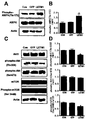
- an increase in LETM1 phosphorylates AMPK on Thr 172 to activate the energy sensor AMPK (FIGS. 1D, E and 4A, B).
- Akt has recently been found to control many metabolic and survival pathways and to be translocated within the mitochondrial substrate in response to insulin stimulation.
- MTOR as a downstream protein of Akt is a key regulator of protein synthesis in response to growth factors as well as metabolic control (Wulschleger, 2006).
- AMPK has been reported to inhibit the activity associated with the activity of mTOR and phosphorylation-mediated activation of the 70-kDa ribosomal protein S6 kinase (p70S6K), inhibition of cell proliferation and induction of pro-apoptotic environment [Kimura, 2003] .
- CDK inhibitors are important mediators of observed cell cycle arrest and act downstream of AMPK activation and cyclin D1 loss.
- Other AMPK activators such as AICAR and antimycin A also triggered downregulation of cyclin D1. Downregulation of cyclin D1 can release p27 and p21 and bind them to CDK2 to inhibit activity (Mandal, 2005). These results showed a decrease in the levels of cyclin D1 and cyclin E and an increase in p21 levels, suggesting that activating AMPK by LETM1 inhibits G 1 / S cell cycle progression (FIGS. 2 and 5).
- activating AMPK has been shown to phosphorylate p53 in Ser 15 , a modification known to protect proteins from degradation and to promote cell-cycle arrest during DNA damage (Jones, 2005).
- activating AMPK by LETM1 significantly increased p53 phosphorylation at Ser 15, resulting in cell cycle arrest (FIG. 2D, E and FIG. 5).
- the present invention has also found that LETM1 overexpression promotes apoptosis of cancer, preferably lung cancer.
- the mechanisms underlying this effect may be associated with the role of LETM1 in the regulation of mitochondrial function.
- Mitochondria are known to actively participate in the process of apoptosis by various mechanisms including energy metabolism and disruption of electron transport.
- the mitochondrial apoptosis pathway is manifested by changes in cytochrome c release, mitochondrial morphology and permeability (Wang, 2001).
- Bax interacts with endogenous mitochondrial proteins such as voltage-dependent anion channels, inducing cytochrome c release from mitochondria [Kim, 2000].
- the present invention has shown that LETM1 overexpression induces Bax as well as cytochrome c release from mitochondria (FIGS. 6A, B and 3C, D).
- the present invention also detected cleavage mitochondria and swelling crista in adenovirus LETM1 infected cells and mice (FIGS. 3A, B and 6E), suggesting that LETM1 may play an important role in mitochondrial apoptosis.
- LETM1 is a mitochondrial lining protein that regulates mitochondrial function and exhibits strong anti-tumor activity. Accordingly, the pharmaceutical composition comprising the LETM1 protein of the present invention or a gene encoding the same may be usefully used as a pharmaceutical composition for preventing or treating cancer, preferably lung cancer.
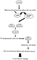
- Figure 9 shows a schematic signal pathway of regulating lung cancer cell proliferation of LETM1, LETM1 induces the activation of AMPK, this activated AMPK inhibits the Akt / mTOR signaling pathway and cyclin D1 expression level, p53 and It is possible to induce apoptosis by reducing p21 expression levels.
- the gene encoding LETM1 can be introduced into the cell in the form of a plasmid vector that can be maintained as an isolated genetic material.
- linear DNA can be introduced into the cell that can be integrated into the chromosome.
- reagents may be added to facilitate the incorporation of DNA into the chromosome, and DNA sequences that promote incorporation may also be included in the composition.
- the vector preferably comprises regulatory elements necessary for the expression of the gene encoding LETM1.
- regulatory elements include, for example, promoters, start codons, stop codons, and polyadenylation signals. As is known in the art, these elements are preferably operably linked to the sequence encoding the desired LETM1, and the regulatory elements are preferably selected such that they can be operated in the species to which they will be administered.
- the present invention provides a pharmaceutical composition for the prophylaxis or treatment of cancer comprising leucine zipper / EF hand-containing membrane-1 (LETM1) protein or a gene encoding the same as an active ingredient.
- LTM1 leucine zipper / EF hand-containing membrane-1
- the cancer is not particularly limited in kind, but lung cancer is preferred.
- the LETM1 may be mixed with a suitable pharmaceutically acceptable carrier or excipient or diluted with a diluent according to a conventional method to prepare a pharmaceutical composition having the above function.
- suitable carriers, excipients and diluents include lactose, dextrose, sucrose, sorbitol, mannitol, xylitol, erythritol, malditol, starch, acacia rubber, alginate, gelatin, calcium phosphate, calcium silicate, cellulose, methyl cellulose , Microcrystalline cellulose, polyvinylpyrrolidone, water, methylhydroxybenzoate, propylhydroxybenzoate, talc, magnesium stearate and mineral oil.
- the pharmaceutical composition may further include fillers, anti-coagulants, lubricants, wetting agents, fragrances, emulsifiers, preservatives and the like.
- the pharmaceutical compositions of the invention may be formulated using methods well known in the art to provide rapid, sustained or delayed release of the active ingredient after administration to a mammal.
- the formulation may be in the form of tablets, pills, powders, sachets, elixirs, suspensions, emulsions, solutions, syrups, aerosols, soft or hard gelatin capsules, sterile injectable solutions, sterile powders, etc. In particular aerosol formulations may be preferred.
- compositions of the invention can be administered via several routes including oral, transdermal, subcutaneous, intravenous or intramuscular.
- Typical daily dosages of the pharmaceutical compositions of the present invention based on the active ingredient LETM1 is in the range of 0.1 to 1000 mg / kg body weight, preferably 10 to 100 mg / kg body weight, divided once or several times May be administered.
- the actual dosage of the active ingredient should be determined in light of several relevant factors such as the route of administration, the age, sex and weight of the patient, and the severity of the disease, and therefore, the dosage may be determined in any respect to the It does not limit the scope of.
- a composition for treating or preventing cancer, preferably lung cancer, containing LETM1 according to the present invention can induce selective destruction of lung cancer cell mitochondria due to ATP deficiency as well as AMPK activation, and can also alter Akt signal, While inhibiting the cycle, it can promote apoptosis. Accordingly, the composition for treating or preventing cancer containing the LETM1 of the present invention is expected to be usefully used in the art as a successful therapeutic approach for inhibiting cancer, preferably lung cancer proliferation and progression.
- Figure 3 shows an increase in apoptosis in vitro of LETM1.
- Figure 6 shows induction of apoptosis in vivo of LETM1.
- Monoclonal antibodies against Akt1 and phospho-Akt in Ser473 were generated by common known methods (Hwang et al., 2007).
- Anti-LETM1 antibodies were purchased from Novus Biological (Littleton, Colorado).
- Anti-COX IV, anti-VDAC, anti-Hsp60, anti-SDHA, anti-AIF and anti-ND 6 antibodies were purchased from Abcam (Cambridge, Mass.).
- Anti-phospho-Akt (Thr308), anti-Bax, anti-BclxL, anti-cycline E, anti-cyclin D1, anti-cycline E, anti-p27, anti-p21, anti-CDK4, anti-AMPK, anti- Cytochrome c and anti-actin antibodies were purchased from Santa Cruz Biotechnology (Santa Cruz, CA, USA).
- Cell signaling with anti-cleaved PARP, anti-phospho-p53 (Ser 15), anti-p53, anti-phospho-AMPK (Thr 172), anti-mTOR and anti-phospho-mTOR (Ser2448) antibodies (Danvers, Massachusetts, USA).
- HRP bound anti-mouse and anti-rabbit IgG antibodies were obtained from Zymed (San Francisco, CA, USA).
- Human non-small cell lung cancer cells A549 were purchased from the US spawn test bureau (ATCC, Manassas, VA, USA). Cells were harvested from RPMI 1640 (Invitrogen, Carlsbad, Calif.) With 10% heat-inactivated fetal bovine serum (FCS), 100,000 units / L penicillin and 100 mg / L streptomycin (Invitrogen). And maintained at 37 ° C. in a 5% CO 2 incubator. Stable cell lines expressing pDsRed-Mito were isolated after selection for 3 weeks by G418 (400 ⁇ g / ml, Sigma-Aldrich, St. Louis, MO).
- Adenovirus expression vectors for wild type LETM1 and GFP were prepared using the Adenoviral Expression Kit (Invitrogen). A549 cells were infected with the appropriate adenovirus for a designated time when the cells reached 80% confluency. The medium was replaced after 6 hours infection and incubated for 48 hours.
- Cells were cultured in 6-well plates and infected for a time designated by Adenovirus-LETM1, Adenovirus-GFP. Cells attached to the plate were harvested with trypsin, washed, resuspended in 100 ⁇ l of PBS and slowly added 5 ml of 70% ethanol, while the cells were stirred continuously and fixed overnight. The following day, cells were spun, washed, resuspended in 400 ⁇ l PBS with the addition of 10 mg / L RNase A and 75 ⁇ M propidium iodide, and FACS Calibur (BD Bioscience, Calif., USA). (San Jose, USA).
- A549 cells were transiently infected by nonsilencing control siRNAs. Whole cell lysates were prepared 48 hours after infection. SiRNA sequences used in the present invention are available upon request.
- Wild type firefly luciferase from the pGL3-base vector was subcloned into the pcDNA3.1-HA vector.
- mitochondrial luciferase (Luc-m) it was a fusion downstream of the sequence encoding the mitochondrial target signal (MTS) of subunit 8 of cytochrome c oxidase.
- MTS mitochondrial target signal
- COX 8 target sequences have been used successfully in other mitochondrial target experiments, and similar COX 8-luciferase constructs have been shown to be properly targeted to mitochondrial substrates (Manfredi, 2002). Luminescence was measured on a luminometer at 5-second intervals until the luminescence maximum was reached.
- the relative luminescence levels in each cell compartment were measured using a luciferase assay kit (Promega, Madison, USA) in the presence of excess ATP (final concentration, 0.5 mM / L). It is expressed as the ratio for total potential luminescence measured for the same aliquots of the same lysed cells.
- A549 cells and lung cancer tissue were fixed at 4 ° C. for 2 hours with 2.5% glutaraldehyde solution with 1% Osmium Tetroxide (OsO 4 ) buffer and dehydrated at 4 ° C. with ethanol.
- Cells and tissues were then infiltrated in a 1: 1 mixture of propylene oxide and Epon, polymerized at 70 ° C. for 24 hours and finally submerged in EPON.
- the ultrathin sections (40-70 nm) were cut and mounted on a pilofoam-coated copper grid (Plano). Sections were stained with lead citrate and uranyl acetate and observed with a JEM 1010 transmission electron microscope (JEOL).
- Mitochondrial expressing stable cells were grown in 2-well chamber slides until 50-70% confluency and then infected with adenovirus-GFP or adenovirus-LETM1 for 6 hours. After 48 hours, cells were fixed for 10 minutes at room temperature with 4% paraformaldehyde and installed with Dakocytomation Faramount Aqueous Mounting solution (Dakocytomation, Kapinteria, CA). And visualized using a Carl Zeiss confocal microscope (Carl Zeiss MicroImaging, Tronwood, NY, USA).
- A549 cells were washed with PBS and mitochondrial fraction buffer (20 mM Hepes, pH 8.0, 10 mM KCl, 1.5 mM MgCl 2 , 1 mM EDTA, 250 mM sucrose, 1 mM PMSF, 10 g / ml lupetin, 10 g / ml Aprotinin and 0.2 mM sodium orthovanadate) were resuspended on ice for 30 minutes and then homogenized. Unbroken cells and nuclei were pelleted at 1500 ⁇ g for 10 minutes by centrifugation.
- Example 9 In vivo Aerosol Delivery of Adenovirus-LETM1
- mice Nine-week-old male K-ras LA1 mice were tested (8 mice / group). Breeding mice were obtained from the Human Cancer Consortium-National Cancer Institute (Frederick, Maryland, USA), with temperature and relative humidity maintained at 23 ⁇ 2 ° C. and 50 ⁇ 20%, respectively. It was placed in a laboratory animal facility and maintained at a 12 hour light / dark cycle. All methods used in this study were approved by the Animal Experimentation Ethics Committee of Seoul National University (SNU-071210-7). For gene delivery, mice were placed in a chamber exposed only to the nose and exposed to aerosols based on previously used methods.
- K-ras LA1 mice were divided into three groups for the effect of LETM1 inhibiting lung cancer development. The control group received no treatment and the other two groups were exposed to aerosols containing adenovirus-GFP or adenovirus-LETM1. K-ras LA1 mice were exposed to aerosol twice a week for a total of four weeks. At the end of the study, K-ras LA1 mice were killed and lungs harvested.
- tumor lesions on the lung surface were carefully counted and measured by digital calipers while viewing the lesion diameter under the microscope described above (Singh, 2006).
- the lungs were perfused and fixed in 10% neutral-buffered formalin for histopathology studies. The remaining lung was stored at -70 ° C for further analysis.
- tissue sections were fixed in 10% neutral buffered formalin, paraffin-depressed tissue sections were cut to 5 ⁇ m and transferred to a plus slide (Fisher Scientific, Pittsburgh, Pa.).
- tissue sections were stained with hematoxylin and eosin (H & E).
- H & E hematoxylin and eosin
- IHC tissue sections were paraffin-depleted from xylene, rehydrated by alcohol gradient, washed subsequently, incubated for 30 minutes in 3% hydrogen peroxide (AppliChem, Boca Raton, FL) The enzyme activity was stopped. After washing in PBS, tissue sections were incubated with 3% BSA in PBS for 1 hour at room temperature to block nonspecific binding sites. Primary antibodies were applied to tissue sections overnight at 4 ° C.
- tissue sections were washed and incubated with secondary HRP-bound antibody (1:50; Zymed) for 2 hours at room temperature. After washing, tissue sections were counterstained by Mayer's Hematoxylin (DAKO, Kapinteria, Calif.) And washed with xylene. Cover slips were installed using Permount (Fisher Scientific) and slides were observed using an optical microscope (Carl Zeiss). Staining intensity of phospho-p53 (Ser 15), LETM1, PCNA and cleaved PARP for IHC assay quantification was performed using an In Studio version 3.01 program (Pixera, San Jose, CA, USA) It was. Color intensity was measured by counting the number of positive cells in a randomly selected field of view observed with an objective lens of appropriate magnification.
- TTBS Tris-buffered saline + Tween 20
- HRP horseradish peroxide
- Immunoblotting was performed by incubating together at room temperature or overnight at 4 ° C. for 3 hours. After washing, the band of interest was photographed with a luminescence image analyzer LAS-3000 (FUJIFILM, Tokyo, Japan) and quantification of Western blot analysis using a multi gauge version 2.02 program (FUJIFILM).
- LETM1 plays a role in the regulation of mitochondrial network and biosynthesis, the expression levels of mitochondrial proteins were analyzed in this experiment.
- Adenovirus-mediated LETM1 infection increases LETM1 protein, while apoptosis-induced in heat shock protein 60 (HSP60), voltage-dependent anion channel (VDAC) in the mitochondrial envelope (OM), mitochondrial inner membrane (IM) Factor (AIF) remained as it was (left column of FIGS. 1A and B).
- HSP60 heat shock protein 60
- VDAC voltage-dependent anion channel
- OM mitochondrial envelope
- IM mitochondrial inner membrane Factor
- Mitochondrial-encoded respiratory chain proteins such as the NADH: ubiquinone oxidoreductase 6 (ND6) complex I subunit; Succinate dehydrogenase complex subunit A (SDHA), which is a complex II subunit; Cytochrome c, which is a complex III subunit; And, among the complex IV subunits, cytochrome c oxidase IV (COXIV), only protein levels of COXIV were selectively reduced (right column of FIGS. 1A and B). To further assess the precise effects of selective increase in LETM1 and decrease in COXIV, changes in ATP levels were observed.
- NADH ubiquinone oxidoreductase 6
- SDHA succinate dehydrogenase complex subunit
- Cytochrome c which is a complex III subunit
- COXIV cytochrome c oxidase IV
- Figure 1 shows the effect of LETM1 on mitochondrial biosynthesis and AMPK activity
- A indicated antibodies [mitochondrial-encoded respiratory chain proteins: NADH: ubiquinone oxidoreductase 6 (ND6), complex I subunit; Succiate dehydrogenase complex subunit A (SDHA), complex II subunit; And cytochrome c, complex III subunits; COXIV, complex IV subunits and other mitochondrial proteins: apoptosis-inducing factor (AIF), heat shock protein 60 (HSP60) and voltage-dependent anion channel (VDAC)], as a result of analysis of mitochondrial proteins, (B) the results of further analysis of the band of interest by densitometry, (C) mitochondrial ATP measured by luciferase assay, and (D) expression levels of phospho-AMPK (Thr 172) and AMPK protein.
- AIF ubiquinone oxidoreductase 6
- SDHA dehydrogenase complex subunit A
- adenovirus-mediated LETM1 inhibited proteins in A549 cells important for cell cycle regulation, such as cyclin D1, cyclin-dependent kinase 4 (CDK4).
- CDK inhibitors such as p53 phosphorylation, p21 were significantly increased in A549 cells (FIGS. 2A and E). This change in cell cycle-related genes has also been shown in vivo studies.
- aerosol delivery of adenovirus-mediated LETM1 inhibited protein expression of CDK4 and cyclin D1 but increased levels of p21, p27 and phospho-p53 at Ser15.
- FIG. 5C An increase in p53 phosphorylation at Ser15 was further confirmed by immunohistochemistry (FIG. 5C) and statistical analysis of phosphor-P53 positive cell counts (FIG. 5D) in the lungs of K-ras LA1 mice treated with aerosol containing LETM1. .
- FIG. 8 AMPK removal by siRNA reversed LETM1-mediated cell cycle arrest.
- FIG. 2 shows that LETM1 induces cell cycle arrest on in vitro G1-S
- A cell cycle distribution was determined by FACS
- B shows the ratio of cell population on G 1 / G 0
- C shows expression of LETM1 protein for different time periods
- D shows expression of p21, cyclin E, cyclin D1, CDK4, p27, phospho-p53 (Ser15) and p53 protein
- E of interest The band is further analyzed by densitometer.
- FIG. 5 shows that LETM1 induces cell cycle arrest on G 1 -S in vivo
- A shows the expression of p21, cyclin E, cyclin D1, CDK4, p27, phospho-p53 (Ser15) and p53 proteins
- FIG. 8 shows AMPK knockdown by siRNAs of A549 cells, wherein A549 cells were transfected either by siRNA AMPK alone or by infected adenovirus-LETM1, followed by AMPK, phospho-Akt (Thr308), cyclin D1 and p53 Western blot analysis.
- Anti-apoptotic members of the Bcl-2 family such as Bcl-2 and Bcl-XL, are known to bind BAX and sequester them from protein complexes to prevent apoptosis induction [O'Neill, 2004], Activation of BAX has been shown to trigger the formation of homopolymeric pores, permeation of mitochondrial membranes to initiate cytochrome c release.
- LETM1 overexpression releases cytochrome c from mitochondria to cytosol (FIGS. 3C and D).
- the present invention delivered adenovirus-LETM1 to K-ras LA1 mice via aerosols and examined the protein expression changes of apoptosis-related proteins.
- Figure 3 shows an increase in apoptosis in vitro of LETM1,
- A A549 cells stably expressing mitochondrial-target Mito-DsRed by GFP (Ad-GFP), LETM1-adenovirus (Ad-LETM1) Or infected for 48 hours without adenovirus (control) and imaged by confocal microscopy
- B mitochondrial microstructures analyzed by transmission electron microscopy, bottom panel being magnified in the indicated area (scale bar; 200 nm)
- D further analysis of the band of interest by densitometry.
- Mitochondria are organelles of morphological changes that continuously divide and fuse to form intracellular units or interconnected networks intracellularly. Analysis of mitochondrial morphology after LETM1 overexpression was analyzed by confocal laser scanning microscopy (CLSM) and transition electron microscopy (TEM). LETM1 overexpression in A549 cells induced a mitochondrial form of cleavage pattern demonstrated by CLSM (FIG. 3A). TEM analysis clearly demonstrated the destruction of the mitochondrial tubular cylinder structure and swelling crista in the lungs of K-ras LA1 mice treated with aerosol containing A549 cells (FIG. 3B) and LETM1 (FIG. 6E). These results clearly demonstrate that LETM1 causes mitochondrial morphology change.
- CLSM confocal laser scanning microscopy
- TEM transition electron microscopy
- LETM1 was delivered to K-ras LA1 mice via aerosol in this experiment.
- this experiment was used to analyze the effect of LETM1 on tumor cell proliferation using proliferating cell nuclear antigen (PCNA) antibodies.
- PCNA proliferating cell nuclear antigen
- LETM1 significantly reduced the expression and number of cells exhibiting PCNA expression (FIGS. 7A and B).
- Immunohistochemistry and PCNA positive cell label index analysis demonstrated that aerosol delivered adenovirus-LETM1 significantly reduced PCNA expression in K-ras LA1 mouse lungs (FIGS. 7C and D).
- FIG. 8E demonstrated that aerosol delivery of LETM1 suppressed lung tumors (arrows) twice a week for one month. Inhibition of this lung tumor formation was further confirmed by histopathological studies (FIG. 7F, red circle). Nodules developed in the lungs of the control group (CON) and the GFP-treated group (GFP) were generally two days / bronchial adenomas.
- K-ras LA1 mice were exposed to aerosols containing adenovirus-LETM1 once a week for a total of four weeks. At the end of the test period, K-ras LA1 mice were sacrificed, lungs were harvested, and the lesions on the surface of the lungs were carefully counted under a microscope. At the same time, mouse lungs were fixed in 10% neutral buffered formalin for histopathological examination. The incidence and multiplicity of lung proliferative lesions were compared. Table 1 below summarizes tumor development in K-ras LA1 mouse lungs. Lung hyperplasia was often observed in the LETM1-treated group (LETM1) where progression to adenomas was inhibited as shown in FIG. 7 and Table 1 below. These test results support that aerosol delivery repetition of LETM1 could inhibit tumor proliferation and progression.
- A represents alveolar adenoma / alveolar cancer
- No. is the number of -ras LA1 mice per county
- GFP is adenovirus-GFP treated group
- LETM1 is adenovirus-LETM1 treated group.
- a is graded, + is mild, ++ is moderate, +++ is severe, * P ⁇ 0.05 is considered significant and ** P ⁇ 0.01 is considered very significant relative to the corresponding control.
- E K-ras LA1 mouse lungs (arrows) with whitish majority nodules in the lungs are shown
Landscapes
- Health & Medical Sciences (AREA)
- Life Sciences & Earth Sciences (AREA)
- Animal Behavior & Ethology (AREA)
- Chemical & Material Sciences (AREA)
- Veterinary Medicine (AREA)
- Public Health (AREA)
- Medicinal Chemistry (AREA)
- General Health & Medical Sciences (AREA)
- Pharmacology & Pharmacy (AREA)
- Nuclear Medicine, Radiotherapy & Molecular Imaging (AREA)
- Engineering & Computer Science (AREA)
- General Chemical & Material Sciences (AREA)
- Chemical Kinetics & Catalysis (AREA)
- Marine Sciences & Fisheries (AREA)
- Zoology (AREA)
- Gastroenterology & Hepatology (AREA)
- Organic Chemistry (AREA)
- Bioinformatics & Cheminformatics (AREA)
- Immunology (AREA)
- Proteomics, Peptides & Aminoacids (AREA)
- Epidemiology (AREA)
- Medicines That Contain Protein Lipid Enzymes And Other Medicines (AREA)
- Pharmaceuticals Containing Other Organic And Inorganic Compounds (AREA)
Abstract
본 발명은 루신 지퍼/EF 손-함유 막-1(LETM1) 단백질 또는 이를 코딩하는 유전자를 포함하는 암의 예방 또는 치료용 약학 조성물에 관한 것으로, 본 발명에 따른 LETM1을 포함하는 조성물은 AMP-활성화된 단백질 키나아제(AMPK) 활성화 뿐만 아니라 ATP 결핍에 의한 암 세포 미토콘드리아의 선택적 파괴를 유도할 수 있으며, 또한 Akt 신호를 변경시킬 수 있고, 세포 주기를 억제하는 한편, 세포자멸사를 촉진시킬 수 있다. 따라서, 본 발명의 LETM1을 함유하는 암 치료 또는 예방용 조성물은 암 증식 및 진행을 억제하는 성공적인 치료 접근법으로 당분야에서 유용하게 이용할 수 있을 것으로 기대된다.
Description
본 발명은 루신 지퍼/EF 손-함유 막-1 (LETM1) 단백질 또는 이를 코딩하는 유전자를 포함하는 암의 예방 또는 치료용 조성물에 관한 것이다.
세계보건기구(WHO : World Health Organization)의 산하기구인 국제암연구소(IARC : International Agency of Research on Cancer) 소속 기술역학부(Unit of Descriptive Epidemiology)의 자료에 의하면 전세계적으로 매년 620만 명이 암으로 사망하고 1,010만 명이 새롭게 암에 걸린다고 한다. 이 중 폐암은 매해 전세계 암 사망의 주된 원인이다. 수술, 화학요법 및 방사선요법을 비롯한 다수의 폐암 치료 방법이 기재되어 왔으나, 치료 효과는 대체로 일시적이고, 많이 진행된 질병에서는 대부분 효과가 없다[왕 (Wang), 2007; 덴스모어 (Densmore), 2006]. 그러므로, 폐암 치료에 대한 더욱 합리적인 접근의 필요성이 절실한 실정이다.
최근, 미토콘드리아가 항암 요법에 있어 효과적인 표적으로서 부상되었다[돈(Don), 2004]. 미토콘드리아는 형태가 변화하는 다기능 소기관으로, 그 활성이 세포 생리학에 직접 관련되어 있다 [뉴메이어(Newmeyer), 2003]. 예컨대, 절편된(fragmented) 미토콘드리아 형태는 세포자멸사의 시토크롬 c 방출과 관련되는 반면, 관 (tubular) 형태는 세포자멸사 자극에 대한 내성을 증진시킨다. 또한, 미토콘드리아는 ATP 생성을 통해 세포 생존의 주요 구성성분으로 기능하고, 미토콘드리아 막-의존적 세포 사멸 신호에 의해 세포 운명을 결정한다 [카르보스키(Karbowski), 2003].
세포 에너지는 대개 미토콘드리아에서 산화적 인산화 과정으로부터 유래된다. 세포 에너지 저장 단계는 AMP-활성화된 단백질 키나아제 (AMPK)에 의해 추정된다. AMPK는 세포 에너지 내용물을 저장하고 병적 스트레스에 반응하여 세포 생존 또는 사멸의 핵심 조절자로서 작용하기 위해서는 활성화되어야 한다 [하디 (Hardie), 2004; 칼링 (Carling), 2004]. ATP 결핍 조건하에서, AMPK는 AMP를 AMPK에 결합시킴으로써 알로스테릭하게 활성화되어서, Thr172상의 AMPK 인산화를 촉진시킨다 [에스.프랑크 (S. Frank), 2007; 하디 (Hardie), 2001].
세포 주기 진행은 내인성 및 외인성 신호에 반응하는 많은 체크포인트하에서 복잡하게 제어된다. G1 상중에 세포는 성장 억제 신호 뿐만 아니라 분열촉진 신호를 통합한 다음, 세포 주기를 진행, 휴지 또는 종료시킬지 여부를 결정한다[쉬케(Schieke), 2008; 마토바(Matoba), 2006]. 세포 주기동안 G1-S 전이는 또한 대사 현상에 의해 조절된다는 증거가 증가하고 있으며, 이는 대사 또는 에너지 체크포인트가 존재할 수 있음을 시사한다[왕(Wang), 2006]. 일부 연구는 세포 주기 과정 및 미토콘드리아 산화능이 G1 초기보다 G1 후기에서 더 크다는 것을 보여주었다. 또한, 미토콘드리아 ATP 생성의 감소는 세포 주기중 G1-S 전이를 차단하여서, 미토콘드리아 기능 또는 형상에 영향을 준다 [마진(Margine), 2002; 아라카키(Arakaki), 2006]. 이들 연구는 대사 체크포인트가 외부 증식 자극 및 영양소 이용도를 통합할 뿐만 아니라 세포 주기 진행에 따라 내인성 미토콘드리아 대사를 동시화할 수 있음을 의미한다고 시사하였다.
루신 지퍼/EF 손-함유 막-1 (LETM1)은 울프-허쉬호른 증후군에서 먼저 밝혀졌고 상기 증후군을 갖는 거의 모든 환자에서 결핍되었던 미토콘드리아 내막 단백질이다[라우츠(Rauch), 2001]. LETM1은 불명확한 기능을 갖는 미토콘드리아-형상 단백질인 효모 Mdm38p의 인간 상동기관(homologue)을 부호화한다. 그러나, 이전 연구는 LETM1이 미토콘드리아와 리보좀 간의 복합체 형성을 위한 고정 단백질 (anchor protein)로서 기능하고 미토콘드리아 생합성을 조절한다는 것을 증명하였다 [프라지어 (Frazier), 2006]. 또한, 최근 연구는 LETM1이 Akt의 음성 조절자인 카복실-말단 조정자 단백질 (CTMP)과 관련되었음을 밝혀냈다 [피아오(Piao) 등, 2009].
이에 본 발명자들은 다양한 발암성, 비발암성 질환의 불충분한 예후를 갖는 잠재적 종양 억제자 유전자로서 LETM1의 잠재적 중요성으로 인해, LETM1이 미토콘드리아 및 종양 증식을 조절하는 기능을 가질 것이라는 가능성에 대하여 집중적으로 연구한 결과, 아데노바이러스-매개된 LETM1이 AMPK 활성을 유도할 수 있고, ATP 결핍된 미토콘드리아를 선택적 파괴시킴으로써 세포 주기 진행을 억제할 수 있음을 발견하고, LETM1이 암, 바람직하게는 폐암 치료 뿐만 아니라 예방을 위한 종양 억제자 유전자로서 기능할 수 있다는 사실을 밝혀냄으로써 본 발명을 완성하게 되었다.
본 발명은 LETM1을 이용한 암의 예방 또는 치료를 위한 조성물을 제공하기 위한 것이다.
본 발명에서 사용되는 기술 용어 및 과학 용어에 있어서 다른 정의가 없다면, 이 발명이 속하는 기술 분야에서 통상의 지식을 가진 자가 통상적으로 이해하고 있는 의미를 갖는 것으로 해석될 수 있다.
본 발명은 루신 지퍼/EF 손-함유 막-1(LETM1) 단백질 또는 이를 코딩하는 유전자를 포함하는 암의 예방 또는 치료용 조성물을 제공한다.
본 발명에서 암은 그 종류에 특별히 한정이 있는 것은 아니나, 폐암이 바람직하다.
본 발명의 조성물에 포함되는 LETM1 단백질의 유전자 서열은 공개적으로 NCBI 데이터베이스에서 NCBI 허가번호 (NCBI accession No.) (AY17394)로서 입수할 수 있으며, 상기 유전자에 의해 코딩되는 LETM1은 당해 업계의 공지된 기술에 의해 용이하게 제조가능할 수 있다 (SNU-071210-7). NCBI는 기관 [National Centre for Biotechnology Information](우편 주소: National Centre for Biotechnology Information, National Library of Medicine, Building 38A, Bethesda, MD 20894, USA; 인터넷 주소:www.ncbi.nhm.nih.gov)이다. 그러나 본 발명의 용어 "LETM1"은 또한 본원에 기술된 LETM1의 생물학적 활성을 유지하는 한, 임의의 상동성을 가진 서열, 변이체, 유도체 및 단편도 포함한다.
바람직한 하나의 양태에서, 본 발명의 조성물에 유효 성분으로서 포함되는 LETM1 단백질 또는 이를 암호화하는 유전자는 세포에 발현된 형태 (LETM1 단백질), 전사활성화 시스템 또는 번역활성화 시스템의 형태 (LETM1을 암호화하는 유전자)로 본 발명의 조성물에 포함될 수 있다. 상기 세포는 본래 LETM1을 발현하는 특성을 가진 세포뿐만 아니라, 이러한 특성을 나타내도록 유전적으로 변형된 세포를 포함한다. 또한, 전사 활성화 시스템은, 적어도 전사 단위의 전사 반응을 수행할 수 있는 능력이 있는, 임의의 생화학적 또는 세포 시스템이다. 이러한 시스템은 당해 기술분야에서 잘 알려져 있고, 세포(예: 보통의 실험실 균주 또는 세포주뿐만 아니라 진핵생물 또는 원핵생물 세포의 일차배양)뿐만 아니라 상업적으로 입수할 수도 있는 시험관 내 전사 시스템을 포함한다.
또한, 번역 활성화 시스템은, 적어도 전사체의 번역 (translation) 반응을 수행할 수 있는 능력이 있는, 임의의 생화학적 또는 세포시스템이다. 이러한 시스템은 당해 기술분야에서 잘 알려져 있고, 세포 (예: 보통의 실험실 균주 또는 세포주뿐만 아니라 진핵생물 또는 원핵생물 세포의 일차배양)뿐만 아니라 시험관 내 해독 시스템 (이는 상업적으로 입수할 수도 있음)을 포함한다.
또한, 본 발명의 조성물은 상기 LETM1 단백질 또는 이를 암호화하는 유전자 외에도, 단백질 또는 핵산의 구조를 안정하게 유지시키는 완충액 또는 반응액을 포함할 수 있다.
미토콘드리아 기능은 통상적인 에너지 필요요건으로 인해 모든 세포 과정에서 필요한 것으로 나타났다 [헤이디(Heidi), 2006]. 시토크롬 c 산화효소 또는 복합체 IV는 미토콘드리아 전자 전이 사슬의 최종 단계를 촉진시키고, 산화적 인산화를 위한 주요 조절 부위중 하나로 간주된다. COX IV 발현이 억제된 여러 세포주의 자세한 생합성 및 기능 분석에 의하면, 시토크롬 c 산화효소 복합체의 상실, 및 이에 따른 시토크롬 c 산화효소-의존적 호흡 및 전체 호흡의 감소가 밝혀졌다 [만달 (Mandal), 2005]. 또한, 세포에서 시토크롬 c 산화효소의 기능이상은 ATP 수준을 감소시킨다. 본 발명에서는 LETM1 과발현이 특이적 미토콘드리아 단백질 시토크롬 c 산화효소 서브유닛 IV를 감소시켜서 폐암 세포의 미토콘드리아 ATP 수준을 감소시킴을 보여주었다 (도 1). AMPK는 ATP 수준 회복에 집중하는 것인 상이한 세포 반응의 조정자로서 최근 알려졌다. AMP/ATP 비의 증가는 AMPK를 활성화시키는데, AMPK는 에너지-소비 과정을 억제하고 에너지-생성 과정을 활성화시켜서 세포의 에너지 항상성을 회복시킨다 [칸(Kahn), 2005]. 본 발명에서는 LETM1의 증가가 Thr172상의 AMPK를 인산화시켜서 에너지 센서 AMPK를 활성화시킨다는 것을 밝혀내었다 (도 1D, E 및 도 4A, B).
Akt는 많은 대사 및 생존 경로를 제어하고, 인슐린 자극에 반응하여 미토콘드리아 기질내에 전위되는 것으로 최근 밝혀졌다. Akt의 다운스트림 (downstream) 단백질로서의 mTOR은 대사 제어 뿐만 아니라 증식 인자에 반응하는 단백질 합성의 핵심 조절자이다 [울슐레거(Wullschleger), 2006]. AMPK는 mTOR의 활성 및 70-kDa 리보좀 단백질 S6 키나아제(p70S6K)의 인산화-매개된 활성화, 세포 증식 억제 및 pro-세포자멸사 환경 유도와 관련된 현상을 억제하는 것으로 보고되었다 [키무라(Kimura), 2003]. 이러한 결과는 LETM1의 에어로졸 전달이 Atk 및 mTOR 인산화를 Thr308 및 Ser2448에서 각각 감소시켰을 명확히 나타냈다.
그러나, 인산화된 AMPK 수준의 증가가 관찰되었다 (도 4C, D). 이러한 결과는 아데노바이러스-매개된 LETM1에 의한 AMPK 활성화가 Akt/mTOR 신호화 경로를 억제하여 암세포 증식을 변경시킬 수 있음을 강하게 시사한다.
최근 연구는 CDK 억제제가, 관측된 세포 주기 정지의 중요한 매개체이고, AMPK 활성화 및 사이클린 D1 상실의 다운스트림으로 작용하는 것으로 보임을 나타냈다. AICAR 및 안티마이신 A와 같은 다른 AMPK 활성화제가 또한 사이클린 D1의 하향 조절을 유발하였다. 사이클린 D1의 하향 조절은 p27과 p21을 방출시켜서, 이들을 CDK2에 결합시켜서 활성을 저해할 수 있다 [만달(Mandal), 2005]. 이러한 결과는 사이클린D1 및 사이클린E의 수준의 감소 및 p21 수준의 증가를 보여주었으며, 이는 LETM1에 의한 활성화 AMPK가 G1/S 세포 주기 진행을 억제함을 시사한다 (도 2 및 도 5). 최근, 활성화 AMPK는 단백질이 분해되지 않도록 보호하고 DNA 손상동안에 세포-주기 정지를 조장하는 것으로 알려진 변형인 Ser15에서 p53를 인산화시키는 것으로 나타났다 [존스(Jones), 2005]. 본 발명에서, LETM1에 의한 활성화 AMPK는 Ser 15에서 p53 인산화를 유의하게 증가시켜서, 세포 주기 정지를 초래하였다 (도 2D, E 및 도 5).
본 발명에서는 또한 LETM1 과발현이 암, 바람직하게는 폐암의 세포자멸사를 조장함을 밝혀냈다. 이러한 효과의 근간을 이루는 메카니즘은 미토콘드리아 기능 조절에서의 LETM1 역할과 관련될 수 있다. 미토콘드리아는 에너지 대사, 전자 운반의 파괴를 비롯한 다양한 메카니즘에 의해 세포자멸사 과정에 적극 참여하는 것으로 알려져 있다. 미토콘드리아 세포자멸사 경로는 시토크롬 c의 방출, 미토콘드리아 형태 및 투과성의 변화에 의해 나타난다 [왕(Wang), 2001]. Bax는 전압 의존적 음이온 채널과 같은 내인성 미토콘드리아 단백질과 상호작용하여서, 미토콘드리아로부터 시토크롬 c 방출을 유도한다 [김(Kim), 2000]. 마찬가지로, 본 발명에서는 LETM1 과발현이 미토콘드리아로부터의 시토크롬 c 방출 뿐만 아니라 Bax를 유도함을 보여주었다 (도 6A, B 및 도 3C, D). 본 발명에서는 또한 아데노바이러스 LETM1 감염된 세포 및 마우스에서 분열 미토콘드리아 및 팽윤 크리스타를 검출하였고 (도 3A, B 및 도 6E), 이러한 결과는 LETM1이 미토콘드리아 세포자멸사에서 중요한 역할을 할 수 있음을 시사한다.
이러한 결과는 아데노바이러스-매개된 LETM1이 종양 증식 및 활성화 AMPK를 억제하였음을 증명한다. LETM1의 과발현은 미토콘드리아 기능, 세포 주기 정지 및 세포자멸사를 조절할 수 있었다. 결론적으로, LETM1은 미토콘드리아 기능을 조절하고 강한 항-종양 활성을 나타내는 미토콘드리아 내막 단백질이다. 따라서, 본 발명의 LETM1 단백질 또는 이를 코딩하는 유전자를 포함하는 약학 조성물은 암, 바람직하게는 폐암의 예방 또는 치료용 약학 조성물로 유용하게 사용될 수 있다.
도 9는 LETM1의 폐암 세포 증식 조절의 개략적인 신호 경로를 나타낸 것으로, LETM1은 AMPK의 활성화를 유도하고, 이러한 활성화된 AMPK는 Akt/mTOR 신호화 경로 및 사이클린 D1 발현 수준을 억제한 후, p53 및 p21 발현 수준을 감소시켜서 세포자멸사를 유도할 수 있다.
상기 LETM1을 코딩하는 유전자를 포함하는 약학 조성물에서, LETM1을 코딩하는 유전자는 분리된 유전자 물질로서 유지될 수 있는 플라스미드 벡터의 형태로 세포내로 도입될 수 있다. 대안적으로, 염색체 내로 통합될 수 있는 선형 DNA를 세포 내로 도입할 수 있다. DNA가 세포 내로 도입될 경우, 당해 분야에 공지된 바와 같이, 염색체 내로의 DNA 혼입을 촉진시키는 시약을 첨가할 수도 있고, 혼입을 촉진시키는 DNA 서열 또한 조성물 내에 포함될 수 있다.
상기 벡터는 바람직하게는 LETM1을 코딩하는 유전자의 발현을 위해 필요한 조절 요소를 포함한다. 이러한 요소는, 예를 들면, 프로모터, 개시 코돈, 정지 코돈 및 폴리아데닐화 신호를 포함한다. 당해 분야에 공지된 바와 같이, 이들 요소는 바람직하게는 목적하는 LETM1을 코딩하는 서열에 작동적으로 연결되며, 조절 요소는 바람직하게는 이들이 투여될 종에서 작동될 수 있도록 선택된다.
본 발명에서는 루신 지퍼/EF 손-함유 막-1(LETM1) 단백질 또는 이를 코딩하는 유전자를 유효성분으로 포함하는 암의 예방 또는 치료용 약학 조성물을 제공한다. 이때 암은 그 종류에 특별히 한정이 있는 것은 아니나, 폐암이 바람직하다.
상기 LETM1은 통상적인 방법에 따라 약제학적으로 허용되는 적절한 담체 또는 부형제와 혼합하거나 희석제로 희석하여 상기한 기능을 갖는 약학 조성물을 제조할 수 있다. 적합한 담체, 부형제 및 희석제의 예로는, 락토즈, 덱스트로즈, 수크로즈, 솔비톨, 만니톨, 자일리톨, 에리스리톨, 말디톨, 전분, 아카시아 고무, 알지네이트, 젤라틴, 칼슘 포스페이트, 칼슘 실리케이트, 셀룰로즈, 메틸 셀룰로즈, 미정질 셀룰로즈, 폴리비닐피롤리돈, 물, 메틸하이드록시벤조에이트, 프로필하이드록시벤조에이트, 탈크, 마그네슘 스테아레이트 및 광물유를 들 수 있다. 상기 약학 조성물은 충진제, 항응집제, 윤활제, 습윤제, 향료, 유화제, 방부제 등을 추가로 포함할 수 있다. 본 발명의 약학 조성물은 포유동물에 투여된 후 활성 성분의 신속, 지속 또는 지연된 방출을 제공할 수 있도록 당업계에 잘 알려진 방법을 사용하여 제형화될 수 있다. 제형은 정제, 알약, 분말, 새세이 (sachet), 엘릭서 (elixir), 현탁액, 에멀젼, 용액, 시럽, 에어로졸, 연질 또는 경질 젤라틴 캅셀, 멸균 주사용액, 멸균 분말 등의 형태일 수 있으나, 본 발명에서는 특히 에어로졸 제형이 바람직할 수 있다.
본 발명의 약학 조성물은 경구, 경피, 피하, 정맥 또는 근육을 포함한 여러 경로를 통해 투여될 수 있다. 본 발명의 약학 조성물의 통상적인 1일 투여량은 유효성분을 기준으로 할 때 LETM1은 0.1 내지 1000 ㎎/㎏체중, 바람직하게는 10 내지 100 ㎎/㎏체중의 범위이며, 1회 또는 수회로 나누어 투여할 수 있다. 그러나, 활성성분의 실제 투여량은 투여 경로, 환자의 연령, 성별 및 체중, 및 질환의 중증도 등의 여러 관련 인자에 비추어 결정되어야 하는 것으로 이해되어야 하며, 따라서, 상기 투여량은 어떠한 면으로든 본 발명의 범위를 한정하는 것은 아니다.
본 발명에 따른 LETM1을 함유하는 암, 바람직하게는 폐암 치료 또는 예방용 조성물은 AMPK 활성화 뿐만 아니라 ATP 결핍에 의한 폐암 세포 미토콘드리아의 선택적 파괴를 유도할 수 있으며, 또한 Akt 신호를 변경시킬 수 있고, 세포 주기를 억제하는 한편, 세포자멸사를 촉진시킬 수 있다. 따라서, 본 발명의 LETM1을 함유하는 암 치료 또는 예방용 조성물은 암, 바람직하게는 폐암 증식 및 진행을 억제하는 성공적인 치료 접근법으로 당분야에서 유용하게 이용할 수 있을 것으로 기대된다.
도 1은 미토콘드리아 생합성 및 AMPK 활성에 대한 LETM1의 효과를 나타낸 것이다.
도 2는 LETM1이 생체외 G1-S 상에서 세포 주기 정지를 유발하는 것을 나타내는 것이다.
도 3은 LETM1의 생체외에서 세포자멸사 증가를 나타낸 것이다.
도 4는 LETM1의 생체내에서 Akt/mTOR 경로 억제를 나타낸 것이다.
도 5는 LETM1이 생체내 G1-S 상에서 세포 주기 정지를 유발하는 것을 나타내는 것이다.
도 6은 LETM1의 생체내 세포자멸사 유도를 나타낸 것이다.
도 7은 LETM1의 종양 증식 억제 여부를 나타낸 것이다.
도 8은 A549 세포의 siRNA에 의한 AMPK 녹다운은 나타낸 것이다.
도 9는 LETM1의 폐암 세포 증식 조절의 개략적인 신호 경로를 나타낸 것이다.
이하 본 발명을 하기 실시예 및 실험예에 의해 보다 상세하게 설명하고자 한다. 단, 하기 실시예 및 실험예는 본 발명을 예시하기 위한 것일 뿐 본 발명의 범위가 이들만으로 한정되는 것은 아니다.
<실시예>
실시예1: 항체 및 시약
Ser473에서 Akt1 및 phospho-Akt에 대한 단일클론 항체를 일반적인 공지 방법을 통해 생성시켰다 [황(Hwang) 등, 2007]. 항-LETM1 항체를 노부스 바이올로지칼 (Novus Biological)(미국 콜로라도주 리틀톤 소재)에서 구입하였다. 항-COX IV, 항-VDAC, 항-Hsp60, 항-SDHA, 항-AIF 및 항-ND 6 항체를 Abcam (미국 매사츄세츠주 캠브리지 소재)에서 구입하였다. 항-phospho-Akt (Thr308), 항-Bax, 항-BclxL, 항-사이클린E, 항-사이클린D1, 항-사이클린E, 항-p27, 항-p21, 항-CDK4, 항-AMPK, 항-시토크롬 c 및 항-액틴 항체를 산타 크루즈 바이오테크놀러지 (Santa Cruz Biotechnology)(미국 캘리포니아주 산타크루즈 소재)에서 구입하였다. 항-절단된 PARP, 항-phospho-p53 (Ser 15), 항-p53, 항-phospho-AMPK (Thr 172), 항-mTOR 및 항-phospho-mTOR (Ser2448) 항체를 셀 시그널링(Cell Signaling)(미국 매사츄세츠주 댄버스 소재)에서 입수하였다. HRP 결합된 항-마우스 및 항-래빗(rabbit) IgG 항체를 자이메드(Zymed) (미국 캘리포니아주 샌프라시스코 소재)에서 입수하였다.
실시예2:
세포 배양, 아데노바이러스 감염
인간의 비-소세포 폐암 세포 A549를 미국 종균 검사국(ATCC, 미국 버지니아주 매나사스 소재)에서 구입하였다. 세포를 RPMI 1640 (인비트로겐(Invitrogen), 미국 캘리포니아주 칼스배드 소재)에서 10% 열-불활성화 소태아혈청(FCS), 100,000 유닛/L의 페니실린 및 100 mg/L의 스트렙토마이신(인비트로겐)과 함께 37℃로 5% CO2 배양기에서 유지시켰다. pDsRed-Mito를 발현하는 안정한 세포주를 G418(400㎍/㎖, 시그마-알드리츠(Sigma-Aldrich), 미국 미주리주 세인트 루이스 소재)에 의해 3주동안 선별한 후에 단리시켰다. 아데노바이럴 익스프레션 키트(Adenoviral Expression Kit)(인비트로겐)를 이용하여 야생형 LETM1 및 GFP에 대한 아데노바이러스 발현 벡터를 준비하였다. 세포가 80% 콘플루언시(confluency)에 도달할 때 A549 세포를 지정된 시간동안 적절한 아데노바이러스에 의해 감염시켰다. 배지를 6시간 감염후에 교체하였고, 48시간동안 배양하였다.
실시예3: 세포 주기의 유식 세포측정 분석
세포를 6-웰 플레이트에서 배양하고, 아데노바이러스-LETM1, 아데노바이러스-GFP에 의해 지정된 시간동안 감염시켰다. 플레이트에 부착된 세포를 트립신으로 수거하고, 세척하고, PBS 100㎕에 재현탁시키고, 70% 에탄올 5㎖을 천천히 첨가하는 한편, 세포를 연속 교반하고 밤새 고정시켰다. 다음날, 세포를 회전시키고, 세척하고, 10mg/L의 RNase A 및 75μM 프로피디움 요오다이드를 첨가하면서 PBS 400㎕에 재현탁시키고, FACS 칼리부르 (Calibur)(BD 바이오사이언스 (Bioscience), 미국 캘리포니아주 산호세 소재)에 의해 분석하였다.
실시예4:
소간섭 RNA(siRNA) 감염
내인성 AMPK α1/α2 및 LETM1을 녹다운 (knock down)시키기 위하여, 제조업체 지시 (산타 크루즈 바이오테크놀러지)에 따라 감염 시약을 이용하여 AMPK α1/α2 및 LETM1을 표적으로 하는 화학 합성된 siRNA 10nM에 의해 또는 비침묵 (nonsilencing) 대조 siRNA에 의해 A549 세포를 일시적으로 감염시켰다. 전세포 용해물 (lysate)을 감염 48시간후에 준비하였다. 본 발명에서 사용된 siRNA 서열은 요청에 따라 입수가능하다.
실시예5: 미토콘드리아 ATP의 측정
pGL3-베이스 벡터로부터의 야생형 개똥벌레 루시페라아제를 pcDNA3.1-HA 벡터에 서브클로닝하였다. 미토콘드리아 루시페라아제 (Luc-m)의 경우, 시토크롬 c 산화효소의 서브유닛 8의 미토콘드리아 표적 신호(MTS)를 부호화하는 서열의 융합 다운스트림이었다. COX 8 표적 서열이 다른 미토콘드리아 표적 실험에서 성공적으로 사용되어 왔고, 유사한 COX 8-루시페라아제 구성물은 미토콘드리아 기질에 적절히 표적화되는 것으로 나타났다[맨프레디(Manfredi), 2002]. 발광 최대치에 도달할 때까지 발광을 5초 간격으로 루미노미터(luminometer)에서 측정하였다. 감염된 세포에서 루시페라아제 발현 변이성을 표준화하기 위하여, 각 세포 구획에서의 상대 발광 수치를 과량의 ATP의 존재하에(최종 농도, 0.5mM/L) 루시페라아제 분석 키트(프로메가(Promega), 미국 매디슨 소재)를 사용하여 동일한 용해된 세포의 동량 분액에 대해 측정된 전체 전위 발광에 대한 비로 표기하였다.
실시예6:
투과형 전자 현미경(TEM)
A549 세포 및 폐암 조직을 1% 오시뮴 테트록사이드(OsO4) 완충액을 갖는 2.5% 글루타르알데하이드 용액에 의해 2시간동안 4℃로 고정시키고, 에탄올에 의해 4℃에서 탈수하였다. 이어서, 세포 및 조직을 프로필렌 옥사이드 및 에폰(Epon)의 1:1 혼합물에서 침윤시키고, 70℃에서 24시간동안 중합하여서 에폰에 최종적으로 함몰시켰다. 초박 절편(40 내지 70nm)으로 절단하고, 피올로폼-피복된 구리 그리드(플라노(Plano))위에 설치하였다. 절편을 리드 시트레이트(lead citrate) 및 우라닐 아세테이트로 착색하고, JEM 1010 투과형 전자 현미경(JEOL)으로 관찰하였다.
실시예7:
공초점 이미지 분석
미토콘드리아 발현 안정 세포(pDsRed-Mito)를 50 내지 70% 콘플루언시때까지 2-웰 챔버 슬라이드에서 증식시키고, 이어서 아데노바이러스-GFP 또는 아데노바이러스-LETM1에 의해 6시간동안 감염시켰다. 48시간후에, 세포를 4% 파라포름알데하이드에 의해 실온에서 10분동안 고정시키고, 다코시토메이션 파라마운트 아쿠어스 마운팅(Dakocytomation Faramount Aqueous Mounting) 용액(다코시토메이션, 미국 캘리포니아주 카핀테리아 소재)과 함께 설치하고, 칼 자이스(Carl Zeiss) 공초점 현미경(칼 자이스 마이크로이미징(Carl Zeiss MicroImaging), 미국 뉴욕주 트론우드 소재)을 이용하여 시각화하였다.
실시예8:
미토콘드리아 및 시토졸 단백질의 단리
A549 세포를 PBS로 세척하고, 미토콘드리아 분획 완충액 (20mM 헤페스 (Hepes), pH 8.0, 10mM KCl, 1.5mM MgCl2, 1mM EDTA, 250mM 수크로오즈, 1mM PMSF, 10g/ml 루펩틴, 10g/ml 아프로티닌 및 0.2mM 소듐 오르토바나데이트)에 30분동안 얼음위에서 재현탁시키고, 이어서 균질화하였다. 파손되지 않은 세포 및 핵을 원심분리에 의해 1500 x g에서 10분동안 펠릿화하였다. 상청액을 10,000 x g에서 30분동안 4℃로 연속하여 원심분리하고, post-미토콘드리아 분획을 위한 새로운 시험관으로 옮겼다. 이어서, 상청액을 100,000 x g에서 1시간동안 4℃로 원심분리하고, 상청액을 시토졸 분획으로 이용하였다. post-미토콘드리아 분획 펠릿을 미토콘드리아 분획 완충액 500㎕으로 세척하고, 미토콘드리아 분획 단리를 위해 이용하였다. 상청액과 미토콘드리아 분획을 수거하고, 단백질 농도를 브래드포드(Bradford) 분석에 의해 결정하였다. 동량의 단백질을 로딩(load)하고, SDS-폴리아크릴아마이드 겔에서 전기 영동하였다. 막을 마우스 항-시토크롬 c(산타 크루즈) 및 래빗 항-HSP60(Abcam, 미국 매사츄세츠주 캠브리지 소재)의 일차 항체와 함께 배양하고, 이어서 호스래디시 퍼옥시다아제 결합된 2차 항체와 함께 배양하였다.
실시예9:
아데노바이러스-LETM1의 생체내 에어로졸 전달
9주령 수컷 K-rasLA1 마우스를 대상으로 실험하였다(8마리 마우스/군). 사육 마우스를 인간 암 협회-국제 암 기관(Human Cancer Consortium-National Cancer Institute)(미국 매릴랜드주 프레더릭 소재)에서 입수하였고, 23±2℃ 및 50±20%로 각각 유지되는 온도 및 상대 습도를 갖는 실험실 동물 시설에 넣고, 12시간 광/암 주기로 유지시켰다. 이번 연구에서 사용된 모든 방법은 서울 국립 대학의 동물 실험 윤리 위원회에 의해 승인받았다(SNU-071210-7). 유전자 전달을 위해, 코만 노출시키는 챔버에 마우스를 넣고, 이전에 사용된 방법에 근거하여 에어로졸에 노출시켰다. 본 에어로졸 유전자 전달 방법은 다양한 연구에서 성공적으로 응용되었다[황(Hwang) 등, 2009; 지앙(Jiang), 2009; 수(Xu) 등, 2008; 진(Jin) 등, 2008; 황(Hwang) 등, 2007; 진(Jin) 등, 2006; 김(Kim), 2004]. 폐암 발생을 억제하는 LETM1 효과에 대해서는 K-rasLA1 마우스를 3개 군으로 나누었다. 대조군은 어떠한 처리도 하지 않고, 다른 2개 군은 아데노바이러스-GFP 또는 아데노바이러스-LETM1을 함유하는 에어로졸에 노출시켰다. K-rasLA1 마우스를 총 4주동안 1주일에 2회 에어로졸에 노출시켰다. 연구 마지막에, K-rasLA1 마우스를 죽이고, 폐를 수거하였다. 부검 절차동안에, 폐 표면의 종양 병변을 조심스럽게 계수하고, 병변 직경을 전술한 현미경으로 보면서 디지털 캘리퍼스에 의해 측정하였다[신(Singh), 2006]. 동시에, 폐를 관류시키고, 조직병리학 연구를 위해 10% 중성-완충된 포르말린에 고정시켰다. 나머지 폐를 추가 분석을 위해 -70℃로 보관하였다.
실시예10: 조직병리학적 분석 및 면역조직화학(IHC)
폐 조직을 10% 중성 완충된 포르말린에 고정시키고, 파라핀-함몰된 조직 절편을 5㎛로 절단하고, 플러스 슬라이드(피셔 사이언티픽(Fisher Scientific), 미국 펜실베이니아주 피츠버그 소재)로 옮겼다. 조직병리학적 분석을 위해, 조직 절편을 헤마톡실린 및 에오신(H&E)으로 착색하였다. IHC을 위해서는 조직 절편을 크실렌에서 파라핀 제거하고, 알코올 구배에 의해 재수화하고, 이어서 세척하고, 3% 과산화수소(아플리켐(AppliChem), 미국 플로리다주 보카 라톤 소재)에서 30분동안 배양하여서 내인성 과산화효소 활성을 중지시켰다. PBS에서 세척한 후에, 조직 절편을 PBS중 3% BSA와 함께 1시간동안 실온에서 배양하여서 비특이적 결합 부위를 차단하였다. 일차 항체를 밤새 4℃에서 조직 절편에 적용하였다. 이튿날, 조직 절편을 세척하고, 2차 HRP-결합된 항체와 함께(1:50; 자이메드(Zymed)) 2시간동안 실온에서 배양하였다. 세척후에, 조직 절편을 메이어스 헤마톡실린(Mayer's Hematoxylin)(다코(DAKO), 미국 캘리포니아주 카핀테리아 소재)에 의해 대비 착색시키고, 크실렌으로 세척하였다. 퍼마운트(Permount)(피셔 사이언티픽)를 이용하여 커버 슬립을 설치하고, 슬라이드를 광학 현미경(칼 자이스)을 이용하여 관찰하였다. IHC 분석 정량화를 위한 phospho-p53(Ser 15), LETM1, PCNA 및 절단된 PARP의 착색 강도는 인 스튜디오(In Studio) 버전 3.01 프로그램(픽세라(Pixera), 미국 캘리포니아주 산호세 소재)을 이용하여 실시하였다. 착색 강도는 적절한 배율의 대물 렌즈로 관찰한 무작위 선택 시야에 있는 양성 세포 수를 계수함으로써 측정하였다.
실시예11:
웨스턴 블 분석
세포 및 조직을 얼음위에 놓고, 50mM 트리스-HCl, pH 7.5, 1%v/v 노니데트(Nonidet) P-40, 120mM NaCl, 25mM 소듐 플루오라이드, 40mM-글리세롤 포스페이트, 0.1mM 소듐 오르토바나데이트, 1mM 페닐메틸설폰일 플루오라이드, 1mM 벤즈아미딘 및 2μM 마이크로시스틴-LR을 함유한 용해 완충액에 의해 추출하였다. 용해물을 15분동안 12,000 x g에서 원심분리하였다. 세포 추출물을 12% SDS-PAGE에 의해 분해하고, 니트로셀룰로오즈 막(아머샴 파마시아(Amersham Pharmacia), 미국 펜실베이니아주 피츠버그 소재)으로 이동시켰다. 막을 5% 탈지유를 함유하는 TTBS(트리스-완충된 염수+트윈(Tween) 20)에서 1시간동안 차단하고, 밤새 4℃로 배양한 다음, 호스래디시 퍼옥사이드(HRP)에 결합된 2차 항체와 함께 3시간동안 실온에서 또는 밤새 4℃에서 배양하여서 면역블롯을 실시하였다. 세척후에, 관심있는 밴드를 발광 이미지 분석기 LAS-3000(후지필름, 일본 도쿄 소재)에 의해 사진을 찍고, 멀티 게이지 버전 2.02 프로그램(후지필름)을 이용하여 웨스턴 블롯 분석의 정량화를 실시하였다.
실시예12: 통계 분석
3회 반복(triplicate)으로 실시된 3회 이상의 별도 실험으로부터의 평균±S.E.로 데이터를 표기하였다. 군간 차이를 스튜던츠 t-테스트(student's t-test)(그래프패드 소프트웨어(Graphpad Software), 미국 캘리포니아주 샌디에고 소재)를 이용하여 분석하였다. p<0.05(*)이 유의하다고 간주되었고, p<0.01(**)이 상응하는 대조 수치에 비해 매우 유의하였다. 웨스턴 블롯 분석의 정량화는 멀티 게이지 버전 2.02 프로그램(후지필름)을 이용하여 실시하였다.
<실험예>
실험예 1: LETM1의 미토콘드리아 ATP 생성을 감소 및 AMPK 단백질 수준 증가 분석
LETM1은 미토콘드리아 네트워크 및 생합성의 조절에서 일정 역할을 하므로 본 실험에서는 미토콘드리아 단백질의 발현 수준을 분석하였다. 아데노바이러스-매개된 LETM1 감염은 LETM1 단백질을 증가시키는 한편, 열 충격 단백질 60(HSP60), 미토콘드리아 외막(OM)에 있는 전압-의존적 음이온 채널(VDAC), 미토콘드리아 내막(IM)에 있는 세포자멸사-유도 인자(AIF)는 그대로였다(도 1A 및 B의 좌측 컬럼). 미토콘드리아-부호화 호흡 사슬 단백질, 예컨대 복합체 I 서브유닛인 NADH:유비퀴논 산화환원효소 6(ND6); 복합체 II 서브유닛인 숙신에이트 탈수소효소 복합체 서브유닛 A(SDHA); 복합체 III 서브유닛인 시토크롬 c; 및 복합체 IV 서브유닛인 시토크롬 c 산화효소 IV(COXIV)중에서, COXIV의 단백질 수준만이 선택적으로 감소되었다(도 1A 및 B의 우측 컬럼). LETM1의 선택적 증가 및 COXIV의 감소의 정확한 효과를 더 평가하기 위하여, ATP 수준의 변화를 관측하였다. COX-8 루시페라아제를 사용하는 상대적인 미토콘드리아 ATP 양의 분석으로 인해, 미토콘드리아 ATP 수준이 대조군에 비해 LETM1 과발현된 세포에서 유의하게 감소되었음이 밝혀졌으며(도 1C), 이는 LETM1이 미토콘드리아 ATP의 생성을 억제함을 의미한다. AMPK가 활성화되어서 세포 에너지 내용물을 저장하므로, 본 실험에서 LETM1 과발현에 의한 전체 AMPK, phospho-AMPK(Thr 172)의 단백질 발현의 변화를 조사하였다. 그 결과는 LETM1이 생체외 및 생체내에서 전체 AMPK 단백질에 비해 phospho-AMPK(Thr 172)를 유의하게 증가시켰음을 보여주었다(도 1D 및 E). 이러한 AMPK 활성화는 siRNA LETM1 처리에 의해 시간 함수로서 감소하였다(도 1F). 도 1은 미토콘드리아 생합성 및 AMPK 활성에 대한 LETM1의 효과를 나타낸 것으로, (A) 지적된 항체[미토콘드리아-부호화 호흡 사슬 단백질: NADH: 유비퀴논 산화환원효소 6 (ND6), 복합체 I 서브유닛; 숙신에이트 탈수소효소 복합체 서브유닛 A (SDHA), 복합체 II 서브유닛; 및 시토크롬 c, 복합체 III 서브유닛; COXIV, 복합체 IV 서브유닛 및 다른 미토콘드리아 단백질: 세포자멸사-유도 인자 (AIF), 열 충격 단백질 60 (HSP60) 및 전압-의존적 음이온 채널(VDAC)]에 의해 면역블롯하여서 미토콘드리아 단백질을 분석한 결과이고, (B) 관심있는 밴드를 농도계측기에 의해 추가 분석한 결과이고, (C) 미토콘드리아 ATP를 루시페라아제 분석에 의해 측정한 결과이고, (D) phospho-AMPK(Thr 172) 및 AMPK 단백질의 발현 수준을 나타낸 것이고, (E) 관심있는 밴드를 농도계측기에 의해 추가 분석한 결과이고, (F) A549 세포를 siRNA LETM1에 의해 시간-의존적 방식으로 감염시키고, 이어서 phospho-AMPK(Thr 172)의 웨스턴 블롯 분석을 실시한 결과이다. 여기에서, 각각의 막대는 평균±SE(n=3)을 나타내고, *P<0.05는 유의하다고 간주되고, **P<0.01는 상응하는 대조 수치에 비해 매우 유의하다고 간주된다.
실험예 2: LETM1의 의한 G1의 S 상 세포 주기 정지 유발 분석
AMPK 경로는 세포 증식을 제어하는 세포 에너지-감지 체크포인트로서 기능하므로[볼스터(Bolster), 2002], 세포 증식에 대한 LETM1의 효과를 측정하였다. 세포 주기 분포에 대한 유식 세포측정 분석에 의하면, LETM1이 G1/G0 부분 모집단을 시간-의존적인 방식으로 유의하게 증가시킴이 밝혀졌고(도 2A 및 B), 이는 LETM1이 LETM1 과발현의 함수로서 G1/G0으로부터 S 상으로의 세포 주기 진행을 억제하였음을 시사한다(도 2C). 상기 결과를 추가 확인하기 위하여, 여러 대표적인 세포 주기-관련된 단백질을 웨스턴 블롯 분석에 의해 분석하였다. 도 2D 및 E에 나타난 바와 같이, 아데노바이러스-매개된 LETM1은 사이클린 D1, 사이클린-의존적 키나아제4 (CDK4)와 같이 세포 주기 조절에 중요한 단백질을 A549 세포에서 억제하였다. 반대로, p53 인산화, p21과 같은 CDK 억제제는 A549 세포에서 유의하게 증가하였다(도 2A 및 E). 세포 주기-관련된 유전자의 이러한 변화는 생체내 연구에서도 밝혀졌다. 도 5A 및 B에 나타난 바와 같이, 아데노바이러스-매개된 LETM1의 에어로졸 전달은 CDK4 및 사이클린 D1의 단백질 발현을 억제하였지만, Ser15에서 p21, p27 및 phospho-p53의 수준을 증가시켰다. Ser15에서 p53 인산화의 증가를 LETM1을 함유하는 에어로졸에 의해 처리된 K-rasLA1 마우스의 폐에서 면역조직화학(도 5C) 및 phosphor-P53 양성 세포 계수의 통계 분석(도 5D)에 의해 추가 확인하였다. 또한, siRNA에 의한 AMPK 제거는 LETM1-매개된 세포 주기 정지를 역전시켰다(도 8).
도 2는 LETM1이 생체외 G1-S 상에서 세포 주기 정지를 유발하는 것을 나타내는 것으로, (A) 세포 주기 분포를 FACS에 의해 결정한 것이고, (B) G1/G0 상 세포 개체수의 비율을 나타낸 것이고, (C) 다른 기간동안 LETM1 단백질의 발현을 나타낸 것이고, (D) p21, 사이클린E, 사이클린D1, CDK4, p27, phospho-p53(Ser15) 및 p53 단백질의 발현을 나타낸 것이고, (E) 관심있는 밴드를 농도계측기에 의해 추가 분석한 결과이다. 여기에서, 각각의 막대는 평균±SE(n=3)을 나타내고, *P<0.05는 유의하다고 간주되고, **P<0.01는 상응하는 대조 수치에 비해 매우 유의하다고 간주된다.
도 5는 LETM1이 생체내 G1-S 상에서 세포 주기 정지를 유발하는 것을 나타내는 것으로, (A) p21, 사이클린E, 사이클린D1, CDK4, p27, phospho-p53(Ser15) 및 p53 단백질의 발현을 나타내고, (B) 관심있는 밴드를 농도계측기에 의해 추가 분석한 결과(n=8)이고, (C) phospho-p53(Ser15)의 면역조직화학 분석. 암갈색이 발현을 나타내고(축척 막대=20㎛), (D) phospho-p53(Ser15) 표지 인덱스의 비교한 것으로, phospho-p53(Ser15) 양성 착색은 절편당 10개의 무작위 선택 시야를 계수하고, 100개 세포당 DAB 양성 세포의 비율을 결정함으로써 결정한 것이다. 여기에서, 각각의 막대는 평균±SE(n=8)을 나타내고, *P<0.05는 유의하다고 간주되고, **P<0.01는 상응하는 대조 수치에 비해 매우 유의하다고 간주된다.
도 8은 A549 세포의 siRNA에 의한 AMPK 녹다운을 나타낸 것으로, A549 세포를 siRNA AMPK 단독에 의해 또는 감염된 아데노바이러스-LETM1에 의해 트랜스펙션시키고, 이어서 AMPK, phospho-Akt(Thr308), 사이클린 D1 및 p53을 웨스턴 블롯 분석한 결과이다.
실험예 3: LETM1의 Akt/mTOR 경로 억제 분석
생체외 연구를 에어로졸을 통해 LETM1 처리된 폐암 모델 마우스에서 동일하게 재현하였고(도 4A 및 B), 이는 아데노바이러스-LETM1 감염된 세포에서의 ATP 수준 감소가 AMPK 활성화를 유발함을 시사한다. 에너지 결핍은 또한 PI3-키나아제 활성도/Akt 경로에 영향을 준다[차초스(Tzatsos), 2006]. 아데노바이러스-매개된 LETM1이 생체내에서 Akt 활성도를 변경시키는지를 연구하기 위하여 웨스턴 블롯 분석을 실시하였다. 이번 결과는 아데노바이러스-매개된 LETM1이 Thr 308에서의 인산화 억제를 통해 Akt1 활성도를 유의하게 억제하였음을 보여주었다(도 4C). 또한, 아데노바이러스-매개된 LETM1은 Ser 2448 발현 수준에서 phospho-mTOR을 유의하게 감소시켰다(도 4C). 관심있는 밴드를 농도계측기로 분석함으로써 웨스턴 블롯 분석의 결과가 명확하게 뒷받침되었다(도 4D).
도 4는 LETM1의 생체내에서 Akt/mTOR 경로 억제를 나타낸 것으로, (A) LETM1, phospho-AMPK(Thr 172) 및 AMPK 단백질의 발현 수준을 나타내고, (B) 관심있는 밴드를 농도계측기에 의해 추가 분석한 결과이고, (C) Akt, phospho-Akt(Ser473), phospho-Akt(Thr308), mTOR 및 phospho-mTOR(Ser2448) 단백질의 발현 수준을 나타낸 것이고, (D) 관심있는 밴드를 농도계측기에 의해 추가 분석한 결과이다. 여기에서, 각각의 막대는 평균±SE(n=8)을 나타내고, *P<0.05는 유의하다고 간주되고, **P<0.01는 상응하는 대조 수치에 비해 매우 유의하다고 간주된다.
실험예 4: LETM1의 생체외 및 생체내 세포자멸사 유도분석
Bcl-2 및 Bcl-XL과 같은 항-세포자멸사 Bcl-2 계열 구성원이 BAX를 결합시켜서 이들을 단백질 복합체에서 격리시켜 세포자멸사 유발을 방지할 수 있다고 알려져 있으나[오닐(O'Neill), 2004], BAX의 활성화는 동질다합체 세공의 형성, 미토콘드리아 막의 투과를 유발하여 시토크롬 c 방출을 개시한다고 제시되었다. 본 실험에서는 LETM1 과발현이 시토크롬 c를 미토콘드리아로부터 시토졸로 방출시키는 것이 증명되었다(도 3C 및 D). 이러한 생체외 관측을 추가로 뒷받침하기 위하여, 본 발명에서는 에어로졸을 통해 아데노바이러스-LETM1을 K-rasLA1 마우스로 전달하고, 세포자멸사-관련 단백질의 단백질 발현 변화를 조사하였다. 이번 결과는 LETM1의 에어로졸 전달이 BAX 및 활성-PARP 단백질 수준을 유의하게 증가시키는 한편, pro-PARP 단백질을 감소시키는 것을 보여주었다. 반대로, 에어로졸 전달된-LETM1은 LETM1-전달된 마우스의 폐에서 Bcl-xL 단백질 수준을 감소시켰다(도 6A 및 B). 또한, 면역조직화학 분석에 따르면, 에어로졸 전달된 아데노바이러스-LETM1이 K-rasLA1 마우스의 폐에서 활성-PARP 발현을 증가시킴이 확인되었다(도 6C 및 D). 이러한 결과로부터 아데노바이러스-LETM1이 생체외 및 생체내에서 세포자멸사를 유도함을 명확히 알 수 있다.
도 3은 LETM1의 생체외에서 세포자멸사 증가를 나타낸 것으로, (A) 미토콘드리아-표적의 Mito-DsRed를 안정하게 발현하는 A549 세포를 GFP(Ad-GFP), LETM1-아데노바이러스(Ad-LETM1)에 의해 또는 아데노바이러스없이(대조군) 48시간동안 감염시키고, 공초점 현미경에 의해 이미지화한 것이고, (B) 미토콘드리아 미세구조를 투과형 전자 현미경에 의해 분석한 것으로, 하부 패널은 표시된 영역의 배율이고(축척 막대; 200nm), (C) 미토콘드리아로부터 시토크롬 c 방출의 웨스턴 블롯 분석 결과이고, (D) 관심있는 밴드를 농도계측기에 의해 추가 분석한 결과이다. 여기에서, 각각의 막대는 평균±SE(n=3)을 나타내고, *P<0.05는 유의하다고 간주되고, **P<0.01는 상응하는 대조 수치에 비해 매우 유의하다고 간주된다.
도 6은 LETM1의 생체내 세포자멸사 유도를 나타낸 것으로, (A) BAX, BclxL, PARP 단백질의 발현 수준이고, (B) 관심있는 밴드를 직경에 의해 추가 분석한 결과이고, (C) 절단된 PARP의 면역조직화학 분석. 암갈색이 발현을 나타내고(축적 막대 = 20㎛), (D) 절단된 PARP 표지 인덱스의 비교. 절단된 PARP 양성 착색은 절편당 10개의 무작위 선택 시야를 계수하여서 결정하고, 100개 세포당 DAB 양성 세포의 비율을 결정하여서 결정한 것이다. 여기에서, 각각의 막대는 평균±SE(n=8)을 나타내고, *P<0.05는 유의하다고 간주되고, **P<0.01는 상응하는 대조 수치에 비해 매우 유의하다고 간주된다. (E) 미토콘드리아 미세구조를 투과형 전자 현미경에 의해 분석한 것으로, 하부 패널은 표시된 영역의 배율이다 (축적 막대 = 200nm).
실험예 5: LETM1의 미토콘드리아 형태 변형 시험
미토콘드리아는 연속적으로 분열, 융합하여 소개체 유닛 또는 상호연결된 네트워크를 세포내에서 형성하는 형태 변동의 소기관이다. LETM1 과발현 이후에 미토콘드리아 형태 변화를 탐지하기 위하여 공초점 레이저 주사 현미경(CLSM) 및 전이 전자 현미경(TEM)에 의해 분석하였다. A549 세포에서의 LETM1 과발현은 CLSM에 의해 증명된 미토콘드리아 형태의 분열 패턴을 유발하였다(도 3A). TEM 분석은 미토콘드리아 관형 실린더 구조 및 팽윤 크리스타의 파괴를 A549 세포(도 3B) 및 LETM1을 함유하는 에어로졸로 처리된 K-rasLA1 마우스의 폐(도 6E)에서 명확히 증명하였다. 이러한 결과는 LETM1이 미토콘드리아 형태 변화를 유발함을 명백히 증명하였다.
실험예 6: LETM1의 폐 종양형성 억제 분석시험
폐암 증식에 대한 아데노바이러스-LETM1의 효과를 연구하기 위하여, 본 실험에서는 에어로졸을 통해 LETM1을 K-rasLA1 마우스로 전달하였다. LETM1이 폐 종양 증식에 영향을 주는 가능한 경로를 연구하기 위하여, 본 실험에서는 증식중인 세포핵 항원(PCNA) 항체를 이용하여 종양 세포 증식에 대한 LETM1의 효과를 분석하였다. LETM1은 PCNA 발현을 나타내는 세포의 발현 및 수를 유의하게 감소시켰다(도 7A 및 B). 면역조직화학 및 PCNA 양성 세포 표지 인덱스 분석에 의하면, 에어로졸 전달된 아데노바이러스-LETM1이 K-rasLA1 마우스 폐에서 PCNA 발현을 유의하게 감소시켰음이 증명되었다(도 7C 및 D). 이러한 결과는 아데노바이러스-LETM1이 인간 폐암 모델 마우스의 폐에서 세포 증식을 억제하였음을 명확히 나타났다. 또한, 아데노바이러스-LETM1의 에어로졸 전달이 폐 종양형성에 영향을 줄 수 있는지를 결정하기 위하여, 조직병리학적 분석을 K-rasLA1 마우스의 폐에서 실시하었다. 도 8E는 1달동안 1주일에 2회, LETM1을 에어로졸 전달하여 폐 종양(화살표)을 억제하였음을 증명하였다. 이러한 폐 종양 형성의 억제는 조직병리학적 연구에 의해 추가로 확인되었다(도 7F, 적색 원). 대조군(CON) 및 GFP-처리된 군(GFP)의 폐에서 발생된 결절은 일반적으로 이틀/기관지 샘종이었다.
K-rasLA1 마우스를 총 4주동안 1주일에 1회, 아데노바이러스-LETM1을 함유하는 에어로졸에 노출시켰다. 시험 기간 마지막에 K-rasLA1 마우스를 희생시키고, 폐를 수거하고, 현미경으로 보면서 폐 표면의 병변을 조심스럽게 계수하였다. 동시에, 조직병리학적 검사를 위해 마우스 폐를 10% 중성 완충된 포르말린에 고정시켰다. 폐 증식성 병변의 발생 및 다중성을 비교하였다. 하기 표 1은 K-rasLA1 마우스 폐에서 종양 발생을 요약한 것이다. 도 7 및 하기 표 1에 나타난 바와 같이 샘종으로의 진행이 억제된 LETM1-처리된 군(LETM1)에서 폐 과다형성이 종종 관찰되었다. 이러한 시험결과는 LETM1의 에어로졸 전달 반복이 종양 증식 및 진행을 억제할 수 있었음을 뒷받침한다.
표 1
| No. | 종양 수 | 폐포성 선종a | 폐포암 | |
| CON | 8 | 10.6±2.26 | A(5)++ | B(3)+++ |
| GFP | 8 | 10.7±3.27 | A(4)+++ | B(4)+++ |
| LETM1 | 8 | 6.37±1.99 | A(2)+ | B(6)++ |
A는 폐포성 선종/폐포암을 나타내고,
B는 폐포성 외피세포 증상이고,
No.는 군당 -rasLA1 마우스 수이며,
CON은 대조군이고,
GFP는 아데노바이러스-GFP 처리된 군이고,
LETM1은 아데노바이러스-LETM1 처리된 군이다.
a는 등급으로 +는 경증, ++은 보통, +++는 중증을 나타내며, *P<0.05는 유의하다고 간주되고, **P<0.01는 상응하는 대조 수치에 비해 매우 유의하다고 간주된다.
도 7은 LETM1의 종양 증식 억제를 보여주는 결과로서, (A) LETM1 및 PCNA(증식중인 세포핵 항원) 단백질의 발현 결과이고, (B) 관심있는 밴드를 농도계측기에 의해 추가 분석한 결과이고, (C) LETM1(상부 패널; 축적 막대=20㎛) 및 PCNA(하부 패널; 축적 막대=20㎛)의 면역조직화학 분석 결과로서 암갈색이 발현을 나타내며, (D) LETM1(좌측) 및 PCNA(우측) 표지 인덱스의 비교 결과로서, LETM1과 PCNA 양성 착색은 절편당 10개의 무작위 선택 시야를 계수하고, 100개 세포당 DAB 양성 세포의 비율을 결정하여서 결정하였다. 여기에서, 각각의 막대는 평균±SE(n=8)을 나타내고, *P<0.05는 유의하다고 간주되고, **P<0.01는 상응하는 대조 수치에 비해 매우 유의하다고 간주된다. (E) 폐에 있는 희끄무레한 다수 결절을 갖는 K-rasLA1 마우스 폐 (화살표)를 나타내고, (F) K-rasLA1 마우스 폐의 조직병리학적 검사 (축적 막대 = 100㎛) 결과이다.
Claims (8)
- 루신 지퍼/EF 손-함유 막-1(LETM1) 단백질 또는 이를 코딩하는 유전자를 포함하는 암의 예방 또는 치료용 조성물.
- 제 1 항에 있어서,LETM1이 미토콘드리아 ATP 생성을 감소시키고, AMP-활성화된 단백질 키나아제(AMPK) 단백질 수준을 증가시키는 것을 특징으로 하는 암의 예방 또는 치료용 조성물.
- 제 1 항에 있어서,LETM1이 Akt/mTOR 경로를 억제하는 것을 특징으로 하는 암의 예방 또는 치료용 조성물.
- 제 1 항에 있어서,LETM1이 생체외 및 생체내에서 세포자멸사를 유도하는 것을 특징으로 하는 암의 예방 또는 치료용 조성물.
- 제1항 내지 제4항에 중 어느 한 항에 있어서, 상기 암은 폐암인 것을 특징으로 하는 조성물.
- 루신 지퍼/EF 손-함유 막-1(LETM1) 단백질 또는 이를 코딩하는 유전자를 유효성분으로 포함하는 암의 예방 또는 치료용 약학 조성물.
- 제 6 항에 있어서, 상기 조성물의 제형은 에어로졸 형태인 것을 특징으로 하는 조성물.
- 제6항 또는 제7항에 있어서, 상기 암은 폐암인 것을 특징으로 하는 조성물.
Applications Claiming Priority (2)
| Application Number | Priority Date | Filing Date | Title |
|---|---|---|---|
| KR1020100019598A KR101156091B1 (ko) | 2010-03-04 | 2010-03-04 | Letm1을 포함하는 암 예방 또는 치료용 조성물 |
| KR10-2010-0019598 | 2010-03-04 |
Publications (1)
| Publication Number | Publication Date |
|---|---|
| WO2011108796A1 true WO2011108796A1 (ko) | 2011-09-09 |
Family
ID=44542412
Family Applications (1)
| Application Number | Title | Priority Date | Filing Date |
|---|---|---|---|
| PCT/KR2010/007547 Ceased WO2011108796A1 (ko) | 2010-03-04 | 2010-10-29 | Letm1을 포함하는 암 예방 또는 치료용 조성물 |
Country Status (2)
| Country | Link |
|---|---|
| KR (1) | KR101156091B1 (ko) |
| WO (1) | WO2011108796A1 (ko) |
Citations (1)
| Publication number | Priority date | Publication date | Assignee | Title |
|---|---|---|---|---|
| KR20090042224A (ko) * | 2009-04-13 | 2009-04-29 | 서울대학교산학협력단 | 폐암의 유전자치료제 |
-
2010
- 2010-03-04 KR KR1020100019598A patent/KR101156091B1/ko not_active Expired - Fee Related
- 2010-10-29 WO PCT/KR2010/007547 patent/WO2011108796A1/ko not_active Ceased
Patent Citations (1)
| Publication number | Priority date | Publication date | Assignee | Title |
|---|---|---|---|---|
| KR20090042224A (ko) * | 2009-04-13 | 2009-04-29 | 서울대학교산학협력단 | 폐암의 유전자치료제 |
Non-Patent Citations (3)
| Title |
|---|
| L. PIAO ET AL.: "Association of LETMI and MRPL36 contributes to the regulation of mitochondrial ATP production and necrotic cell death.", CANCER RESEARCH, vol. 69, 24 March 2009 (2009-03-24), pages 3397 - 3404 * |
| L. PIAO ET AL.: "Regulation of OPAl-mediated mitochondrial fusion by leucine zipper/EF- hand-containing transmembrane protein- plays a role in apoptosis.", IN CELLULAR SIGNALLING., vol. 21, 9 January 2009 (2009-01-09), pages 767 - 777 * |
| L. PIAO: "Regulation of mitochondrial morphology and cell survival by leucine zipper/EF- hand-containing transmembrane protein 1", LETMI, August 2008 (2008-08-01) * |
Also Published As
| Publication number | Publication date |
|---|---|
| KR101156091B1 (ko) | 2012-06-20 |
| KR20110100546A (ko) | 2011-09-14 |
Similar Documents
| Publication | Publication Date | Title |
|---|---|---|
| Qian et al. | Activation of glucagon-like peptide-1 receptor in microglia attenuates neuroinflammation-induced glial scarring via rescuing Arf and Rho GAP adapter protein 3 expressions after nerve injury | |
| Zheng et al. | Essential role of the voltage-dependent anion channel (VDAC) in mitochondrial permeability transition pore opening and cytochrome c release induced by arsenic trioxide | |
| Gómez-Lazaro et al. | Reactive oxygen species and p38 mitogen-activated protein kinase activate Bax to induce mitochondrial cytochrome c release and apoptosis in response to malonate | |
| Wang et al. | Targeting CARD6 attenuates spinal cord injury (SCI) in mice through inhibiting apoptosis, inflammation and oxidative stress associated ROS production | |
| Masgras et al. | Induction of the permeability transition pore in cells depleted of mitochondrial DNA | |
| Tian et al. | Sirtuin 6 inhibits epithelial to mesenchymal transition during idiopathic pulmonary fibrosis via inactivating TGF-β1/Smad3 signaling | |
| Liu et al. | A new LKB1 activator, piericidin analogue S14, retards renal fibrosis through promoting autophagy and mitochondrial homeostasis in renal tubular epithelial cells | |
| He et al. | Hesperetin post-treatment prevents rat cardiomyocytes from hypoxia/reoxygenation injury in vitro via activating PI3K/Akt signaling pathway | |
| Pecoraro et al. | Trastuzumab-induced cardiotoxicity and role of mitochondrial connexin43 in the adaptive response | |
| Pan et al. | YC-1 [3-(5′-hydroxymethyl-2′-furyl)-1-benzyl indazole] inhibits endothelial cell functions induced by angiogenic factors in vitro and angiogenesis in vivo models | |
| Sampaio et al. | p66Shc signaling is involved in stress responses elicited by anthracycline treatment of rat cardiomyoblasts | |
| Rodriguez-Enfedaque et al. | FGF1 nuclear translocation is required for both its neurotrophic activity and its p53-dependent apoptosis protection | |
| Hossain et al. | SULT4A1 protects against oxidative-stress induced mitochondrial dysfunction in neuronal cells | |
| Liu et al. | Gastrodin alleviates glucocorticoid induced osteoporosis in rats via activating the Nrf2 signaling pathways | |
| Rodriguez et al. | Activation of sarcolipin expression and altered calcium cycling in LMNA cardiomyopathy | |
| WO2012064146A2 (ko) | Gkn 1을 함유하는 항암용 조성물 | |
| Shen et al. | Cambogin exerts anti-proliferative and pro-apoptotic effects on breast adenocarcinoma through the induction of NADPH oxidase 1 and the alteration of mitochondrial morphology and dynamics | |
| Kim et al. | p53‐mediated regulation of mitochondrial dynamics plays a pivotal role in the senescence of various normal cells as well as cancer cells | |
| Chen et al. | Nicorandil prevents doxorubicin-induced human umbilical vein endothelial cell apoptosis | |
| Li et al. | Inhibition of NLRP3 and Golph3 ameliorates diabetes-induced neuroinflammation in vitro and in vivo | |
| Tong et al. | FUNDC1 modulates mitochondrial defects and pancreatic β-cell dysfunction under lipotoxicity | |
| Ahn et al. | PEP-1-PEA-15 protects against toxin-induced neuronal damage in a mouse model of Parkinson's disease | |
| Jiang et al. | Multifunctions of CRIF1 in cancers and mitochondrial dysfunction | |
| Xue et al. | Cardiomyocyte GSDME drives doxorubicin-induced cardiotoxicity by targeting the CCL2-CCR2 axis | |
| Xu et al. | Malvidin induced anticancer activity in human colorectal HCT-116 cancer cells involves apoptosis, G2/M cell cycle arrest and upregulation of p21WAFI |
Legal Events
| Date | Code | Title | Description |
|---|---|---|---|
| 121 | Ep: the epo has been informed by wipo that ep was designated in this application |
Ref document number: 10847094 Country of ref document: EP Kind code of ref document: A1 |
|
| NENP | Non-entry into the national phase |
Ref country code: DE |
|
| 122 | Ep: pct application non-entry in european phase |
Ref document number: 10847094 Country of ref document: EP Kind code of ref document: A1 |