WO2011103236A2 - Personalized tumor biomarkers - Google Patents
Personalized tumor biomarkers Download PDFInfo
- Publication number
- WO2011103236A2 WO2011103236A2 PCT/US2011/025152 US2011025152W WO2011103236A2 WO 2011103236 A2 WO2011103236 A2 WO 2011103236A2 US 2011025152 W US2011025152 W US 2011025152W WO 2011103236 A2 WO2011103236 A2 WO 2011103236A2
- Authority
- WO
- WIPO (PCT)
- Prior art keywords
- dna
- tumor
- patient
- rearrangement
- breakpoint
- Prior art date
Links
Classifications
-
- C—CHEMISTRY; METALLURGY
- C12—BIOCHEMISTRY; BEER; SPIRITS; WINE; VINEGAR; MICROBIOLOGY; ENZYMOLOGY; MUTATION OR GENETIC ENGINEERING
- C12Q—MEASURING OR TESTING PROCESSES INVOLVING ENZYMES, NUCLEIC ACIDS OR MICROORGANISMS; COMPOSITIONS OR TEST PAPERS THEREFOR; PROCESSES OF PREPARING SUCH COMPOSITIONS; CONDITION-RESPONSIVE CONTROL IN MICROBIOLOGICAL OR ENZYMOLOGICAL PROCESSES
- C12Q1/00—Measuring or testing processes involving enzymes, nucleic acids or microorganisms; Compositions therefor; Processes of preparing such compositions
- C12Q1/68—Measuring or testing processes involving enzymes, nucleic acids or microorganisms; Compositions therefor; Processes of preparing such compositions involving nucleic acids
- C12Q1/6876—Nucleic acid products used in the analysis of nucleic acids, e.g. primers or probes
- C12Q1/6883—Nucleic acid products used in the analysis of nucleic acids, e.g. primers or probes for diseases caused by alterations of genetic material
- C12Q1/6886—Nucleic acid products used in the analysis of nucleic acids, e.g. primers or probes for diseases caused by alterations of genetic material for cancer
-
- C—CHEMISTRY; METALLURGY
- C12—BIOCHEMISTRY; BEER; SPIRITS; WINE; VINEGAR; MICROBIOLOGY; ENZYMOLOGY; MUTATION OR GENETIC ENGINEERING
- C12Q—MEASURING OR TESTING PROCESSES INVOLVING ENZYMES, NUCLEIC ACIDS OR MICROORGANISMS; COMPOSITIONS OR TEST PAPERS THEREFOR; PROCESSES OF PREPARING SUCH COMPOSITIONS; CONDITION-RESPONSIVE CONTROL IN MICROBIOLOGICAL OR ENZYMOLOGICAL PROCESSES
- C12Q2600/00—Oligonucleotides characterized by their use
- C12Q2600/156—Polymorphic or mutational markers
Definitions
- This invention is related to the area of cancer detection and management. In particular, it relates to identification and use of somatic rearrangements as markers of a person's cancer.
- a nearly universal feature of human cancer is the widespread rearrangement of chromosomes as a result of chromosomal instability (1). Such structural alterations begin to occur at the earliest stages of tumorigenesis and persist throughout tumor development. The consequences of chromosomal instability can include copy number alterations (duplications, amplifications and deletions), inversions, insertions, and translocations (2). Historically, the ability to detect such alterations has been limited by the resolution of genetic analyses. However, a number of more recent approaches including high density oligonucleotide arrays and high throughput sequencing have allowed detection of changes at much higher resolution (3-15).
- Tumor-specific (somatic) chromosomal rearrangements have the potential to serve as highly sensitive biomarkers for tumor detection. Such alterations are not present in normal cells and should be adequately specific. Rearrangement-associated biomarkers therefore offer a reliable measure that would be useful for monitoring tumor response to specific therapies, detecting residual disease after surgery, and for long-term clinical management.
- Recurrent somatic structural alterations such as those involving the BCR- ABL oncogene (the target of the Philadelphia chromosome translocation), immunoglobulin (Ig) genes, T cell receptor (TCR) genes, and the retinoic acid receptor alpha (RARa) gene, have been shown to be useful as diagnostic markers in certain hematopoietic malignancies (16-20). However, recurrent structural alterations do not generally occur in most solid tumors. There is a continuing need in the art to develop tools for diagnosing and monitoring cancers.
- a method for identifying a personalized tumor marker for a cancer patient is provided.
- a mate-paired library is made from tumor DNA of the patient.
- Mate pairs of the library comprise two genomic tags that are co-linear but not contiguous in a segment of the tumor DNA. Sequence of a plurality of mate pairs of the library is determined. Regions of copy number differences among regions in the tumor DNA of the patient are determined. Mate paired tags which map within a region of copy number difference or spanning a boundary of copy number difference are identified as potential markers of a tumor-specific DNA rearrangement in the cancer patient.
- a method for assessing or detecting tumor in a patient.
- a DNA fragment is amplified using a template from the patient's tissues or body fluids and primers that span a patient-specific, tumor-specific rearrangement breakpoint.
- the rearrangement breakpoint is between genes involved in rearrangements in ⁇ 1 % of tumors of patients with the same type of tumor.
- the amount or proportion of amplified DNA fragment in the patient's tissue or body fluid is determined.
- Another aspect of the invention is another method of identifying a personalized tumor marker for a cancer patient. Sequence of two ends of each of a plurality of fragments of DNA from the cancer patient is determined. Regions of copy number differences among regions in the tumor DNA of the patient are determined. Fragments of the plurality of fragments which map within a region of copy number difference or spanning a boundary of copy number difference are identified as potential markers of a tumor-specific DNA rearrangement in the cancer patient.
- a further aspect of the invention is another method of identifying a personalized tumor marker for a cancer patient.
- a plurality of mate paired tags of a library of mate paired tags is tested by comparing to non-tumor DNA or to sequence of non-tumor DNA.
- Each of the mate paired tags comprises two genomic tags that are co-linear but not contiguous in a segment of tumor DNA of the cancer patient.
- a tumor-specific DNA rearrangement is identified if the two genomic tags of a mate paired tag are at different locations or in a different orientation within a chromosome or on different chromosomes of non-tumor DNA compared to tumor DNA.
- Yet another aspect of the invention is another method of identifying a personalized tumor marker for a cancer patient. Two ends of a plurality of fragments of tumor DNA of the cancer patient are tested by comparing to non-tumor DNA or to sequence of non-tumor DNA. A tumor-specific DNA rearrangement is identified if the ends of a fragment are at different locations or in a different orientation within a chromosome or on different chromosomes of non-tumor DNA compared to tumor DNA.
- Still another aspect of the invention is a method of screening for a cancer in a human.
- a plurality of mate paired tags of a library of mate paired tags is tested by comparing to normal DNA or to sequence of normal DNA.
- Each of the mate paired tags comprises two genomic tags that are co-linear but not contiguous in a segment of DNA in the blood of the human.
- a DNA rearrangement is identified if the two genomic tags of a mate paired tag are at different locations or in a different orientation within a chromosome or on different chromosomes of normal DNA compared to blood DNA. The presence of a DNA rearrangement suggests the presence of a cancer in the human.
- a further aspect of the invention is a method of screening for a cancer in a human. Two ends of a fragment of blood DNA of the human are tested by comparing to normal DNA or to sequence of normal DNA. A DNA rearrangement is identified if the ends are at different locations or in a different orientation within a chromosome or on different chromosomes of normal DNA compared to blood DNA. The presence of a DNA rearrangement suggests the presence of a cancer in the human.
- An additional aspect of the invention is a kit for monitoring presence or amount of a breakpoint in a somatic DNA rearrangement in tumor DNA of a patient.
- the kit may comprise one or more pairs of amplification primers. Each pair is complementary to priming sites on opposite sides of a breakpoint. The priming sites are separated by less than 200 basepairs in the tumor DNA.
- the DNA rearrangement occurs in ⁇ 1 % of tumors of patients with the same type of tumor.
- FIG. 1 Schematic of "Personalized Analysis of Rearranged Ends (PARE)" approach.
- the method is based on next generation mate-paired analysis of, e.g., resected tumor DNA to identify individualized tumor-specific rearrangements. Such alterations are used to develop PCR based quantitative analyses for personalized tumor monitoring of plasma samples or other bodily fluids.
- FIGs. 2A and 2B Detection of tumor-specific rearrangements in breast and colorectal cancers. Two representative rearrangements are shown for each tumor sample. PCR amplification across breakpoint regions is indicated in (Fig. 2A) and the genomic coordinates for a representative mate-pair of each rearrangement are listed in (Fig. 2B).
- Fig. 3 Detection of tumor specific rearrangements in mixtures of tumor and normal DNA. Decreasing amounts of tumor DNA were mixed with increasing amounts of normal tissue DNA (300 ng total) and were used as template molecules for PCR using chromosomes 4/8 translocation specific primers (top) or chromosome 3 control primers (see Example 1 for additional information).
- Fig. 4A-4B Detection of tumor-specific rearrangements in plasma of cancer patients.
- Fig. 4 A The identified chromosome 4/8 and 16 rearrangements were used to design PCR primers spanning breakpoints and used to amplify rearranged DNA from tumor tissue and plasma from patients Hx402 and Hx403, respectively. A plasma sample from an unrelated healthy individual was used a control for both rearrangements.
- Fig. 4B Plasma samples from patient Hx402 were analyzed at different time points using digital PCR to determine the fraction of genomic equivalents of plasma DNA containing the chromosome 4/8 rearrangement. The fraction of rearranged DNA at day 137 was 0.3%, consistent with residual metastatic lesions present in the remaining lobe of the liver.
- Fig. 6 Figure S2. Comparison of Digital Karyotyping, Illumina SNP array, and SOLiD sequencing results on chromosome 8.
- any structural alteration identified in an individual's tumor can be used as a tumor marker, even if it is not found in tumors of the same type in other individuals and even if it is not a "driver" - causing a selective growth advantage - but merely a "passenger.” Moreover, such markers can be used to detect tumor and or quantify the tumor burden in an individual by assessment of blood.
- Somatic rearrangements are a focus of the present invention. Such rearrangements are used as markers of a tumor. In particular, the boundaries of the rearrangments can be detected and used as a quantitative or qualitative indicator of the tumor. Because the boundaries are unique to the tumor DNA, they should be extraordinarly specific markers of the tumor. Somatic rearrangements can be detected using any method known in the art. One particularly useful method is a technique called digital karyotyping. This technique identities changes in copy number across regions or windows in the genome. Other methods may employ commercially available arrays to detect regions of copy number differences among regions of a genome. The copy number differences reflect a rearrangement, such as a deletion or amplification, and an amplification can further harbor other rearrangements within it. Once a somatic rearrangement is identified, one or more of its boundaries (also referred to as breakpoints) can be identified and that boundary can be a very specific marker for the tumor. Identifying a boundary can be accomplished by a number of techniques.
- mate-paired genomic tags are tested to determine different copy numbers of one member of the pair compared to the other. A different copy number between two members suggests that the tags span a rearrangement breakpoint or boundary.
- the mate-pairs are typically derived from a single fragment that is processed to yield two smaller portions that can be more readily sequenced or analyzed. An intervening segment is typically removed, leaving the two smaller portions linked on a single molecule in the same orientation that they were found in the tumor genome.
- a similar technique does not involve mate-pairs but involves sequencing and/or analyzing two different portions or ends of a single fragment of genomic DNA from a tumor.
- the two portions or ends may be separated by any distance, from immediately adjacent up to 1 kb, 1.5 kb, 2 kb, or 3 kb, for example.
- the ends may not be the literal ends of a fragment, but may be close to the ends or merely two non-overlapping portions.
- the sequence of the two ends may be determined separately, for example from either end, or the sequence can be determined in one direction and analyzed for separate, non- overlapping segments of differing copy numbers.
- Amplification primers are known in the art and typically comprise between 15 and 50 nucleotides which are complementary to a template.
- a pair of primers is complementary to opposite strands of a template and can amplify a double stranded fragment that contains the two primer sequences in addition to sequences which are between them on the template. From 0 to 10, 20, 50, 100, 200, 500, 1000, 1500, or 2000 basepairs or nucleotides may lie between the two primer-complementary sequences on the template.
- each primer will hybridize to opposite sides of a rearrangement boundary. These primers are also referred to as spanning or flanking the breakpoint, because the amplicon that they generate will span and/or flank the breakpoint.
- a primer may contain the boundary junction. Primers need not be 100 % complementary to template, but may incorporate other bases or sequences of bases for other purposes, such as to facilitate purification or downstream processing.
- primers can be prepared and shipped elsewhere for use. For example pairs or panels of pairs of primers can be packaged in a single or divided container.
- the primers can be in any suitable condition, including in solution, dried, freeze dried, at room temperature, on wet ice, and on dry ice. Additional components may be included in the kits, for example other reagents for performing the monitoring or assessing with the primers. Additional components may include a polymerase for amplification, reagents for preparing template from cancer cells, normal cells, or body fluids, control primers, control templates, labeled or unlabelled deoxyribonucleotides.
- tumor sequences can be compared to a reference sequence, for example in a database, or to a sequence from normal DNA of the same or a related individual.
- Two mate-paired tags or two fragment ends that map to different locations on a chromosome or to different chromosomes or to differently oriented sequences on the same chromosome indicate a rearrangement.
- the comparison can be done in silico or in vitro.
- Breakpoints in a rearrangement are places where two sequences are joined in a tumor DNA that are not joined in normal or reference DNA.
- the breakpoint refers to an inferred break that occurred in order to join the sequences that are found in the tumor DNA. Breakpoints are also referred to as boundaries of a rearrangement.
- Normal DNA may be obtained from lymphocytes or a buccal swab, for example. In cases where the subject has a diagnosed tumor, normal DNA can be obtained from any non-tumor tissue, including a matched tissue from the same organ.
- the breakpoints which are of interest in the present methods are those which are not known to be associated with or causative of leukemia, lymphoma, sarcoma, or prostate cancers.
- the breakpoints which are associated with or causative of those cancers typically occur in a high proportion of such tumors, often between the same or a limited number of genes or gene loci.
- the rearrangements used in the present methods are more idiosyncratic, occurring between the same genes or gene loci in less than 1 %, less than 0.1 %, or less than 0.01 % of the patients with the same type of tumor.
- Assays using tumor-specific primers can be used for a variety of purposes. For example, patients can be monitored over time to see if a tumor is in remission or is progressing. The assay can be used before, during, and/or after a therapy regimen. The assay can be used to assess surgical efficacy. Tumor margins can be assessed to guide the extent of surgical resection. The assay can be used to monitor for relapse or recurrence.
- the tumor rearrangement-specific primers can be conducted using the rearrangement-specific primers and tissues or body fluids from a subject. Suitable body fluids include whole blood, serum, and plasma, which are collectively referred to as blood. Other body fluids which may be used are saliva, sputum, and stool, for example.
- One or more pairs of primers can be used to amplify and assay for one or more tumor-specific rearrangements in a single patient. Using a panel of rearrangements markers may mitigate against any possible loss of marker during tumor growth and progression.
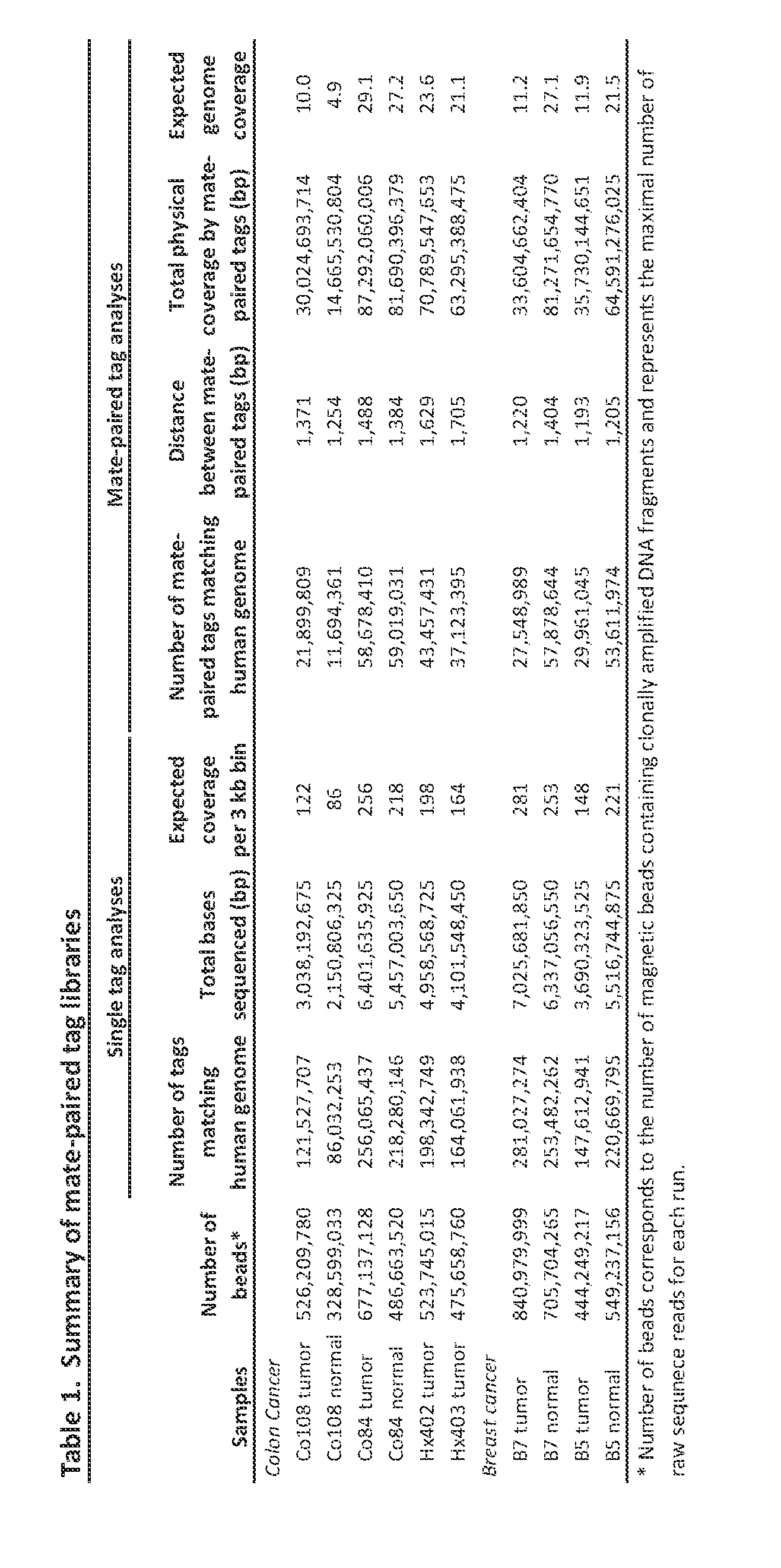
- DNA samples were obtained from early passage xenografts and cell lines of breast and colorectal cancers as described (26). Normal DNA samples were obtained from matched normal tissue. Plasma samples were collected from colorectal cancer patients Hx402 and Hx403 and from an unrelated normal control. All samples were obtained in accordance with the Health Insurance Portability and Accountability Act (HIPAA).
- HIPAA Health Insurance Portability and Accountability Act
- a Digital Karyotyping library for colorectal cancer cell line Co84C was constructed as previously described (6).
- 17 bp genomic DNA tags were generated using the Nlalll and Sacl restriction enzymes.
- the experimental tags obtained were concatenated, cloned and sequenced.
- Previously described software was used to extract the experimental tags from the sequencing data.
- the sequences of the experimental tags were compared to the predicted virtual tags extracted from the human genome reference sequence. Amplifications were identified using sliding windows of variable sizes and windows with tag density ratios > 6 were considered to represent amplified regions.
- the Illumina Infinium II Whole Genome Genotyping Assay employing the BeadChip platform was used to analyze the colorectal cancer cell line Co84C at 317k SNP loci from the Human HapMap collection.
- This assay is a two step procedure; first the sample is hybridized to a 50 nucleotide oligo, then the SNP position is interrogated by a two-color fluorescent single base extension. Image files and data normalization were processed as previously described (10). Amplifications were defined as regions having at least one SNP with a LogR ratio >1.4, at least one in ten SNPs with a LogR ratio >1, and an average LogR ratio of the entire region of >0.9.
- Mate-pair libraries were generated for the SOLiD platform as described (IS).
- genomic DNA was sheared into ⁇ 1.4kb fragments and used as template in emulsion PCR. Fragments were coupled to beads via an adapter sequence and clonally amplified. A 3' modification of the DNA fragments allowed for covalent attachment to a slide.
- Sequencing primers hybridized to the adapter sequence and four fluorescently labeled di- base probes were used in ligation-based sequencing. Each nucleotide is sequenced twice in two different ligation reactions, resulting in two base encoding which has been shown to reduce sequencing artifacts.
- the SOLiD tags were filtered and the remaining tags were grouped by genomic position in non-overlapping 3kb bins.
- a tag density ratio was calculated for each bin by dividing the number of tags observed in the bin by the average number of tags expected to be in each bin (based on the total number of tags obtained for chromosomes 1-22 for each library divided by 849,434 total bins). The tag density ratio thereby allowed a normalized comparison between libraries containing different numbers of total tags.
- a control group of SOLiD libraries made from the four matched normal samples from Table 1 and two itional normal samples (CEPH sample NA07357 and NA18507used to define areas of germline copy number variation or which contained a large fraction of repeated or low complexity sequences. Any bin where at least 2 of the normal libraries had a tag density ratio of ⁇ 0.25 or >1.7S was removed from further analysis.
- Mate-paired tags mapping the reference genome uniquely and without mismatches were analyzed for aberrant mate-pair spacing, orientation and ordering and categorized in 13 three letter data formats (27).
- Mate pairs from the same chromosome that map at appropriate distances ( ⁇ 1.4 kb) and in the appropriate orientation and ordering are categorized as AAA.
- Mate pairs mapping to different chromosomes are categorized as C**.
- C** For the analysis of translocations of the PARE approach, we focused on C** mate pairs, while for analysis of rearrangements adjacent to copy number alterations, we chose all non-AAA (including C**) mate pairs for further analysis.
- Mate pair tag sequences associated with a candidate rearrangement were used as target sequences for primer design using with Primer3 (28). When primers could not be designed from tag sequences alone, adjacent genomic sequence up to 100 bp was used for primer design. Importantly, the observed rearranged tag ordering and orientation was used for Primer3 queries. Primers were used for PCR on tumor and matched normal samples as previously described (26). The candidate rearrangement was confirmed if a PCR product of the expected size was seen in the tumor, but not the matched normal sample. Sanger sequencing of PCR products was used to identify sequence breakpoint in a subset of cases.
- the PARE approach shown schematically in Fig. 1, in one embodiment employs the identification of patient-specific rearrangements in tumor samples.
- PCR emulsion polymerase chain reaction
- the second approach combined mate-paired tag data with copy number alterations identified by analyses of individual 25 bp tags. Tumor-specific copy number alterations are often associated with de novo rearrangements (23) and the boundaries of such alterations would be expected to contain novel junctions not present in the human genome.
- tags were grouped into non-overlapping 3 kb bins. Normalized tag densities, defined as the number of tags per bin divided by average number of tags per bin, were determined for all 3 kb bins in each sample. Bins that displayed tag density ratios >1.7S or ⁇ 0.25 in two or more normal tissue samples (corresponding to ⁇ 6% of all bins) were discarded from the analysis.
Abstract
Clinical management of human cancer is dependent on the accurate monitoring of residual and recurrent tumors. We have developed a method, called personalized analysis of rearranged ends (PARE), which can identify translocations in solid tumors. Analysis of four colorectal and two breast cancers revealed an average of nine rearranged sequences (range 4 to 15) per tumor. Polymerase chain reaction with primers spanning the breakpoints were able to detect mutant DNA molecules present at levels lower than 0.001% and readily identified mutated circulating DNA in patient plasma samples. This approach provides an exquisitely sensitive and broadly applicable approach for the development of personalized biomarkers to enhance the clinical management of cancer patients.
Description
PERSONALIZED TUMOR BIOMARKERS
This invention was made with government support under grants CA121113, CA057345, CA62924, and CA043460 awarded by the National Institutes of Health. The government has certain rights in the invention.
TECHNICAL FIELD OF THE INVENTION
[02] This invention is related to the area of cancer detection and management. In particular, it relates to identification and use of somatic rearrangements as markers of a person's cancer.
BACKGROUND OF THE INVENTION
[03] A nearly universal feature of human cancer is the widespread rearrangement of chromosomes as a result of chromosomal instability (1). Such structural alterations begin to occur at the earliest stages of tumorigenesis and persist throughout tumor development. The consequences of chromosomal instability can include copy number alterations (duplications, amplifications and deletions), inversions, insertions, and translocations (2). Historically, the ability to detect such alterations has been limited by the resolution of genetic analyses. However, a number of more recent approaches including high density oligonucleotide arrays and high throughput sequencing have allowed detection of changes at much higher resolution (3-15).
[04] Tumor-specific (somatic) chromosomal rearrangements have the potential to serve as highly sensitive biomarkers for tumor detection. Such alterations are not present in normal cells and should be exquisitely specific. Rearrangement-associated biomarkers therefore offer a reliable measure that would be useful for monitoring tumor response to specific therapies, detecting residual disease after surgery, and for long-term clinical management. Recurrent somatic structural alterations, such as those involving the BCR- ABL oncogene (the target of the Philadelphia chromosome translocation),
immunoglobulin (Ig) genes, T cell receptor (TCR) genes, and the retinoic acid receptor alpha (RARa) gene, have been shown to be useful as diagnostic markers in certain hematopoietic malignancies (16-20). However, recurrent structural alterations do not generally occur in most solid tumors. There is a continuing need in the art to develop tools for diagnosing and monitoring cancers.
SUMMARY OF THE INVENTION
[05] According to one aspect of the invention a method is provided for identifying a personalized tumor marker for a cancer patient. A mate-paired library is made from tumor DNA of the patient. Mate pairs of the library comprise two genomic tags that are co-linear but not contiguous in a segment of the tumor DNA. Sequence of a plurality of mate pairs of the library is determined. Regions of copy number differences among regions in the tumor DNA of the patient are determined. Mate paired tags which map within a region of copy number difference or spanning a boundary of copy number difference are identified as potential markers of a tumor-specific DNA rearrangement in the cancer patient.
[06] According to another aspect of the invention a method is provided for assessing or detecting tumor in a patient. A DNA fragment is amplified using a template from the patient's tissues or body fluids and primers that span a patient-specific, tumor-specific rearrangement breakpoint. The rearrangement breakpoint is between genes involved in rearrangements in < 1 % of tumors of patients with the same type of tumor. The amount or proportion of amplified DNA fragment in the patient's tissue or body fluid is determined.
[07] Another aspect of the invention is another method of identifying a personalized tumor marker for a cancer patient. Sequence of two ends of each of a plurality of fragments of DNA from the cancer patient is determined. Regions of copy number differences among regions in the tumor DNA of the patient are determined. Fragments of the plurality of
fragments which map within a region of copy number difference or spanning a boundary of copy number difference are identified as potential markers of a tumor-specific DNA rearrangement in the cancer patient.
[08] A further aspect of the invention is another method of identifying a personalized tumor marker for a cancer patient. A plurality of mate paired tags of a library of mate paired tags is tested by comparing to non-tumor DNA or to sequence of non-tumor DNA. Each of the mate paired tags comprises two genomic tags that are co-linear but not contiguous in a segment of tumor DNA of the cancer patient. A tumor-specific DNA rearrangement is identified if the two genomic tags of a mate paired tag are at different locations or in a different orientation within a chromosome or on different chromosomes of non-tumor DNA compared to tumor DNA.
[09] Yet another aspect of the invention is another method of identifying a personalized tumor marker for a cancer patient. Two ends of a plurality of fragments of tumor DNA of the cancer patient are tested by comparing to non-tumor DNA or to sequence of non-tumor DNA. A tumor-specific DNA rearrangement is identified if the ends of a fragment are at different locations or in a different orientation within a chromosome or on different chromosomes of non-tumor DNA compared to tumor DNA.
[10] Still another aspect of the invention is a method of screening for a cancer in a human. A plurality of mate paired tags of a library of mate paired tags is tested by comparing to normal DNA or to sequence of normal DNA. Each of the mate paired tags comprises two genomic tags that are co-linear but not contiguous in a segment of DNA in the blood of the human. A DNA rearrangement is identified if the two genomic tags of a mate paired tag are at different locations or in a different orientation within a chromosome or on different chromosomes of normal DNA compared to blood DNA. The presence of a DNA rearrangement suggests the presence of a cancer in the human.
[11] A further aspect of the invention is a method of screening for a cancer in a human. Two ends of a fragment of blood DNA of the human are tested by comparing to normal DNA
or to sequence of normal DNA. A DNA rearrangement is identified if the ends are at different locations or in a different orientation within a chromosome or on different chromosomes of normal DNA compared to blood DNA. The presence of a DNA rearrangement suggests the presence of a cancer in the human.
[12] An additional aspect of the invention is a kit for monitoring presence or amount of a breakpoint in a somatic DNA rearrangement in tumor DNA of a patient. The kit may comprise one or more pairs of amplification primers. Each pair is complementary to priming sites on opposite sides of a breakpoint. The priming sites are separated by less than 200 basepairs in the tumor DNA. The DNA rearrangement occurs in < 1 % of tumors of patients with the same type of tumor.
[13] These and other embodiments which will be apparent to those of skill in the art upon reading the specification provide the art with methods for detecting and monitoring cancers in the body.
BRIEF DESCRIPTION OF THE DRAWINGS
[14] Fig. 1. Schematic of "Personalized Analysis of Rearranged Ends (PARE)" approach.
The method is based on next generation mate-paired analysis of, e.g., resected tumor DNA to identify individualized tumor-specific rearrangements. Such alterations are used to develop PCR based quantitative analyses for personalized tumor monitoring of plasma samples or other bodily fluids.
[15] Figs. 2A and 2B. Detection of tumor-specific rearrangements in breast and colorectal cancers. Two representative rearrangements are shown for each tumor sample. PCR amplification across breakpoint regions is indicated in (Fig. 2A) and the genomic coordinates for a representative mate-pair of each rearrangement are listed in (Fig. 2B).
[16] Fig. 3. Detection of tumor specific rearrangements in mixtures of tumor and normal DNA. Decreasing amounts of tumor DNA were mixed with increasing amounts of normal tissue DNA (300 ng total) and were used as template molecules for PCR using
chromosomes 4/8 translocation specific primers (top) or chromosome 3 control primers (see Example 1 for additional information).
[17] Fig. 4A-4B. Detection of tumor-specific rearrangements in plasma of cancer patients.
Fig. 4 A. The identified chromosome 4/8 and 16 rearrangements were used to design PCR primers spanning breakpoints and used to amplify rearranged DNA from tumor tissue and plasma from patients Hx402 and Hx403, respectively. A plasma sample from an unrelated healthy individual was used a control for both rearrangements. Fig. 4B. Plasma samples from patient Hx402 were analyzed at different time points using digital PCR to determine the fraction of genomic equivalents of plasma DNA containing the chromosome 4/8 rearrangement. The fraction of rearranged DNA at day 137 was 0.3%, consistent with residual metastatic lesions present in the remaining lobe of the liver.
[18] Fig. 5 (Figure SI.) Flow chart of approach used to identify rearranged sequences
[19] Fig. 6 (Figure S2.) Comparison of Digital Karyotyping, Illumina SNP array, and SOLiD sequencing results on chromosome 8.
DETAILED DESCRIPTION OF THE INVENTION
120} We have found that any structural alteration identified in an individual's tumor can be used as a tumor marker, even if it is not found in tumors of the same type in other individuals and even if it is not a "driver" - causing a selective growth advantage - but merely a "passenger." Moreover, such markers can be used to detect tumor and or quantify the tumor burden in an individual by assessment of blood.
[21] Somatic rearrangements are a focus of the present invention. Such rearrangements are used as markers of a tumor. In particular, the boundaries of the rearrangments can be detected and used as a quantitative or qualitative indicator of the tumor. Because the boundaries are unique to the tumor DNA, they should be exquisitely specific markers of
the tumor. Somatic rearrangements can be detected using any method known in the art. One particularly useful method is a technique called digital karyotyping. This technique identities changes in copy number across regions or windows in the genome. Other methods may employ commercially available arrays to detect regions of copy number differences among regions of a genome. The copy number differences reflect a rearrangement, such as a deletion or amplification, and an amplification can further harbor other rearrangements within it. Once a somatic rearrangement is identified, one or more of its boundaries (also referred to as breakpoints) can be identified and that boundary can be a very specific marker for the tumor. Identifying a boundary can be accomplished by a number of techniques.
[22] In one technique mate-paired genomic tags are tested to determine different copy numbers of one member of the pair compared to the other. A different copy number between two members suggests that the tags span a rearrangement breakpoint or boundary. The mate-pairs are typically derived from a single fragment that is processed to yield two smaller portions that can be more readily sequenced or analyzed. An intervening segment is typically removed, leaving the two smaller portions linked on a single molecule in the same orientation that they were found in the tumor genome.
[23] A similar technique does not involve mate-pairs but involves sequencing and/or analyzing two different portions or ends of a single fragment of genomic DNA from a tumor. The two portions or ends may be separated by any distance, from immediately adjacent up to 1 kb, 1.5 kb, 2 kb, or 3 kb, for example. The ends may not be the literal ends of a fragment, but may be close to the ends or merely two non-overlapping portions. The sequence of the two ends may be determined separately, for example from either end, or the sequence can be determined in one direction and analyzed for separate, non- overlapping segments of differing copy numbers.
[24] Amplification primers are known in the art and typically comprise between 15 and 50 nucleotides which are complementary to a template. A pair of primers is complementary to opposite strands of a template and can amplify a double stranded fragment that
contains the two primer sequences in addition to sequences which are between them on the template. From 0 to 10, 20, 50, 100, 200, 500, 1000, 1500, or 2000 basepairs or nucleotides may lie between the two primer-complementary sequences on the template. According to the invention, each primer will hybridize to opposite sides of a rearrangement boundary. These primers are also referred to as spanning or flanking the breakpoint, because the amplicon that they generate will span and/or flank the breakpoint. Optionally, a primer may contain the boundary junction. Primers need not be 100 % complementary to template, but may incorporate other bases or sequences of bases for other purposes, such as to facilitate purification or downstream processing.
[25] Once tumor-specific breakpoints are ascertained for an individual patient, primers can be prepared and shipped elsewhere for use. For example pairs or panels of pairs of primers can be packaged in a single or divided container. The primers can be in any suitable condition, including in solution, dried, freeze dried, at room temperature, on wet ice, and on dry ice. Additional components may be included in the kits, for example other reagents for performing the monitoring or assessing with the primers. Additional components may include a polymerase for amplification, reagents for preparing template from cancer cells, normal cells, or body fluids, control primers, control templates, labeled or unlabelled deoxyribonucleotides.
[26] In order to identify or confirm a rearrangement in tumor DNA, tumor sequences can be compared to a reference sequence, for example in a database, or to a sequence from normal DNA of the same or a related individual. Two mate-paired tags or two fragment ends that map to different locations on a chromosome or to different chromosomes or to differently oriented sequences on the same chromosome indicate a rearrangement. The comparison can be done in silico or in vitro.
[27] Breakpoints in a rearrangement are places where two sequences are joined in a tumor DNA that are not joined in normal or reference DNA. Thus the breakpoint refers to an inferred break that occurred in order to join the sequences that are found in the tumor DNA. Breakpoints are also referred to as boundaries of a rearrangement. Normal DNA
may be obtained from lymphocytes or a buccal swab, for example. In cases where the subject has a diagnosed tumor, normal DNA can be obtained from any non-tumor tissue, including a matched tissue from the same organ.
[28] The breakpoints which are of interest in the present methods are those which are not known to be associated with or causative of leukemia, lymphoma, sarcoma, or prostate cancers. The breakpoints which are associated with or causative of those cancers typically occur in a high proportion of such tumors, often between the same or a limited number of genes or gene loci. The rearrangements used in the present methods are more idiosyncratic, occurring between the same genes or gene loci in less than 1 %, less than 0.1 %, or less than 0.01 % of the patients with the same type of tumor.
[29] Assays using tumor-specific primers can be used for a variety of purposes. For example, patients can be monitored over time to see if a tumor is in remission or is progressing. The assay can be used before, during, and/or after a therapy regimen. The assay can be used to assess surgical efficacy. Tumor margins can be assessed to guide the extent of surgical resection. The assay can be used to monitor for relapse or recurrence.
[30] Using the tumor rearrangement-specific primers to conduct assays, one can obtain qualitative or quantitative results. The quantitative results can be absolute amounts or relative amounts, for example, compared to a non-rearranged sequence on the same or a different chromosome. Assays can be conducted using the rearrangement-specific primers and tissues or body fluids from a subject. Suitable body fluids include whole blood, serum, and plasma, which are collectively referred to as blood. Other body fluids which may be used are saliva, sputum, and stool, for example. One or more pairs of primers can be used to amplify and assay for one or more tumor-specific rearrangements in a single patient. Using a panel of rearrangements markers may mitigate against any possible loss of marker during tumor growth and progression.
[31] The results shown below in the Examples demonstrate that massively parallel sequencing can be used to develop personalized biomarkers based on somatic rearrangements. We
were able to identify tumor-specific markers in each of the six breast and colorectal cancer cases analyzed. Moreover, we demonstrated that the identified breakpoints can be used to detect tumor DNA in the presence of large quantities of normal DNA and in patient plasma. These results highlight the sensitivity and specificity of the approach and suggest broad clinical utility of the methods disclosed here, collectively referred to as PARE.
[32] Virtually all tumors of clinical consequence are thought to have rearranged DNA sequences resulting from translocations and copy number alterations and these sequences are not present in normal human plasma or non-tumor tissues. A recent genome-wide analysis of 24 breast cancers showed that all analyzed samples contained at least one genomic rearrangement that could be detected by next generation sequencing (24). From a technical perspective, PARE -derived clinical assays should have no false positives: the PCR amplification of aberrant fusions of DNA sequences that are normally thousands of base pairs apart or on different chromosomes should not occur using non-tumor DNA as a template. In contrast, approaches that rely on monitoring of residual disease by analysis of somatic single base alterations in specific genes are limited by polymerase error rates at the bases of interest (25). The PCR process generates background single base mutations that are identical to bona fide mutations, but does not generate false- positive rearrangements with carefully chosen primers. Because of the higher signal-to- noise ratio thereby obtained, PARE theoretically permits more sensitive monitoring of tumor burden.
[33] The PARE approach, however, is not without limitations. Although somatic alterations in oncogenes and tumor suppressor genes persist throughout the clonal evolution of a tumor, it is conceivable that some rearranged sequences could be lost during tumor progression. The identification of several PARE biomarkers, each specific for different chromosomal regions, would mitigate this concern, as it is unlikely that all such markers would be lost in any particular patient. Another limitation is the cost of identifying a patient-specific alteration. In this prototype study, we obtained an average of 194.7 million reads per patient, resulting in ~200 tags in each 3 kb bin. The current cost for
such an assay is ~$5,000, which is expensive for general clinical use. This cost is a consequence of the high physical coverage and the inefficiencies associated with stringent mapping of 25 bp sequence data to the human genome. As read quality and length continue to improve, less stringent mapping criteria and lower physical coverage will permit analyses similar to those in this study but with substantially less sequencing effort. Moreover, the cost of massively parallel sequencing, which has decreased substantially over the last two years, continues to spiral downwards. Finally, there are clinical settings where the fraction of any DNA from tumors, including rearranged sequences, in the patient plasma is exceedingly small and undetectable. To be detectable by PARE, there must be at least one rearrangement template molecule in the plasma sample analyzed. When disease-burden is this light, PARE may yield false negative results. Larger studies will be needed to confirm particular clinical uses of PARE and its prognostic capabilities.
[34] Despite these caveats, there are numerous potential applications of PARE. These include the more accurate identification of surgical margins free of tumor and the analysis of regional lymph nodes as well as the measurement of circulating tumor DNA following surgery, radiation, or chemotherapy. Short term monitoring of circulating tumor DNA may be particularly useful in the testing of new drugs, as it could provide an earlier indication of efficacy than possible through conventional diagnostic methods such as CT scanning. Given current enthusiasm for the personalized management of cancer patients, PARE affords a timely method for uniquely sensitive and specific tumor monitoring.
[35] The above disclosure generally describes the present invention. All references disclosed herein are expressly incorporated by reference. A more complete understanding can be obtained by reference to the following specific examples which are provided herein for purposes of illustration only, and are not intended to limit the scope of the invention.
EXAMPLE 1 -MATERIALS AND METHODS Clinical samples and cell lines
[36] DNA samples were obtained from early passage xenografts and cell lines of breast and colorectal cancers as described (26). Normal DNA samples were obtained from matched normal tissue. Plasma samples were collected from colorectal cancer patients Hx402 and Hx403 and from an unrelated normal control. All samples were obtained in accordance with the Health Insurance Portability and Accountability Act (HIPAA).
Digital Karyotyping and Illumina BeadChip Arrays
[37] A Digital Karyotyping library for colorectal cancer cell line Co84C was constructed as previously described (6). In summary, 17 bp genomic DNA tags were generated using the Nlalll and Sacl restriction enzymes. The experimental tags obtained were concatenated, cloned and sequenced. Previously described software was used to extract the experimental tags from the sequencing data. The sequences of the experimental tags were compared to the predicted virtual tags extracted from the human genome reference sequence. Amplifications were identified using sliding windows of variable sizes and windows with tag density ratios > 6 were considered to represent amplified regions.
(38) The Illumina Infinium II Whole Genome Genotyping Assay employing the BeadChip platform was used to analyze the colorectal cancer cell line Co84C at 317k SNP loci from the Human HapMap collection. This assay is a two step procedure; first the sample is hybridized to a 50 nucleotide oligo, then the SNP position is interrogated by a two-color fluorescent single base extension. Image files and data normalization were processed as previously described (10). Amplifications were defined as regions having at least one SNP with a LogR ratio >1.4, at least one in ten SNPs with a LogR ratio >1, and an average LogR ratio of the entire region of >0.9. SOLID Library Preparation and Sequencing
[39] Mate-pair libraries were generated for the SOLiD platform as described (IS). In brief, genomic DNA was sheared into ~1.4kb fragments and used as template in emulsion PCR.
Fragments were coupled to beads via an adapter sequence and clonally amplified. A 3' modification of the DNA fragments allowed for covalent attachment to a slide. Sequencing primers hybridized to the adapter sequence and four fluorescently labeled di- base probes were used in ligation-based sequencing. Each nucleotide is sequenced twice in two different ligation reactions, resulting in two base encoding which has been shown to reduce sequencing artifacts.
[40] Sequence data was mapped to the human genome reference sequence (hgl8) using the Corona SOLiD software pipeline. All 25bp tags (for both individual tag and mate-paired tag analyses) were required to match the reference genome uniquely and without mismatches.
Analysis of Single Tags for Copy Number Alterations
[41] The SOLiD tags were filtered and the remaining tags were grouped by genomic position in non-overlapping 3kb bins. A tag density ratio was calculated for each bin by dividing the number of tags observed in the bin by the average number of tags expected to be in each bin (based on the total number of tags obtained for chromosomes 1-22 for each library divided by 849,434 total bins). The tag density ratio thereby allowed a normalized comparison between libraries containing different numbers of total tags. A control group of SOLiD libraries made from the four matched normal samples from Table 1 and two itional normal samples (CEPH sample NA07357 and NA18507used to define areas of germline copy number variation or which contained a large fraction of repeated or low complexity sequences. Any bin where at least 2 of the normal libraries had a tag density ratio of <0.25 or >1.7S was removed from further analysis.
[42] Homozygous deletions were identified as three or more consecutive bins with tag ratios <0.2S and at least one bin with a tag ratio <0.005. Amplifications were identified as three or more consecutive bins with tag ratios >2.5 and at least one bin with a tag ratio >6. Single copy gains and losses were identified through visual inspection of tag density data for each sample.
Analysis of Mate-paired Tags
(43] Mate-paired tags mapping the reference genome uniquely and without mismatches were analyzed for aberrant mate-pair spacing, orientation and ordering and categorized in 13 three letter data formats (27). Mate pairs from the same chromosome that map at appropriate distances (~1.4 kb) and in the appropriate orientation and ordering are categorized as AAA. Mate pairs mapping to different chromosomes are categorized as C**. For the analysis of translocations of the PARE approach, we focused on C** mate pairs, while for analysis of rearrangements adjacent to copy number alterations, we chose all non-AAA (including C**) mate pairs for further analysis.
PARE Identification and Confirmation of Candidate Rearrangements
[44] To identify candidate translocations, we grouped C** mate pair tags in lkb bins and looked for bin-pairs which were observed >5 times in the tumor sample but which were not observed in matched normal sample. For identification of candidate rearrangements associated with copy number alterations, we analyzed the 10 kb boundary regions of amplifications, homozygous deletions, or lower copy gains and losses for neighboring non-AAA tags observed > 2 times in the tumor but not matched normal sample. In the case of Hx402 and Hx403 the analysis of rearrangements adjacent to copy number alterations was performed in the absence of SOLiD libraries from normal tissue.
[45] Mate pair tag sequences associated with a candidate rearrangement were used as target sequences for primer design using with Primer3 (28). When primers could not be designed from tag sequences alone, adjacent genomic sequence up to 100 bp was used for primer design. Importantly, the observed rearranged tag ordering and orientation was used for Primer3 queries. Primers were used for PCR on tumor and matched normal samples as previously described (26). The candidate rearrangement was confirmed if a PCR product of the expected size was seen in the tumor, but not the matched normal
sample. Sanger sequencing of PCR products was used to identify sequence breakpoint in a subset of cases.
Detection of PARE Biomarker in human plasma
(46] To determine the sensitivity of rearranged biomarkers in the presence of normal DNA, serial dilutions of tumonnormal DNA mixtures were used as templates for PCR using primers for the chromosome 4/8 translocation in Hx402. The tumor DNA dilution began at 1:125 tumonnormal and continued as a one-in-five serial dilution until reaching 1:390,625 tumonnormal mixture. PCR was performed for each of the six tumonnormal DNA mixtures and for the normal DNA control, using translocation specific primers as well as control primers from chromosome 3.
[47] One ml of human plasma samples were obtained from patients Hx402 and Hx403 and from a control individual and DNA was purified as described (29). Whole genome amplification of plasma DNA was performed by ligation of adaptor sequences and PCR amplification with universal primers from the Illumina Genomic DNA Sample Prep Kit.
[48] Primers designed to amplify <200bp fragments spanning each PARE rearrangement were used in PCR from total plasma DNA using patient or control samples. Digital PCR of plasma DNA dilutions from patient Hx402 using rearrangement specific and control primers were used to quantitate the fraction mutated DNA molecules.
EXAMPLE 2
Description of the approach
[49] The PARE approach, shown schematically in Fig. 1, in one embodiment employs the identification of patient-specific rearrangements in tumor samples. To determine the feasibility of identifying such alterations using next generation sequencing approaches, we initially analyzed four tumor samples (two colon and two breast tumors) and their matched normal tissue samples using the Applied Biosystems SOLiD System (Table 1). Genomic DNA from each sample was purified, sheared and used to generate libraries
with mate-paired tags ~1.4 kb apart Libraries were digitally amplified by emulsion polymerase chain reaction (PCR) on magnetic beads (21) and 25 bp mate-paired tags were sequenced using the sequencing-by-ligation approach (15, 22). An average of 198.1 million 25 bp reads were obtained for each sample where each read aligned perfectly and was uniquely localized in the reference human genome (hgl8), resulting in 4.95 Gb mappable sequence per sample. An average of 40 million mate-paired reads where both tags were perfectly mapped to the reference human genome were obtained for each sample. The total amount of genome base-pairs covered by the mate-paired analysis (i.e. distance between mate-paired tags x number of mate-paired tags) was 53.6 Gb per sample, or a 18-fold physical coverage of the human genome.
Identification of somatic rearrangements
[50] Two methods were used to identify somatic rearrangements from these data (Fig. 5). The first approach involved searching for tags whose mate-pairs were derived from different chromosomes (interchromosomal rearrangements). The high physical coverage of breakpoints provided by the ~ 40 million mate-paired sequences per sample (Table 1) suggested that a large fraction of such translocations could be identified. End sequences from such mate-paired tags were grouped into 1 kb bins and those bin pairs that were observed at least 5 times were analyzed further. The requirement for >5 occurrences minimized the chance that the presumptive fusion sequences represent incorrect mapping to the reference genome or artifacts of library construction. Comparison with SOLiD libraries made from the matched normal samples reduced the possibility that the fusion sequences represented rare germline variants rather than somatic events.
[51] The second approach combined mate-paired tag data with copy number alterations identified by analyses of individual 25 bp tags. Tumor-specific copy number alterations are often associated with de novo rearrangements (23) and the boundaries of such alterations would be expected to contain novel junctions not present in the human genome. To identify somatic copy number gains, losses, high-amplitude amplifications and homozygous deletions, tags were grouped into non-overlapping 3 kb bins. Normalized tag densities, defined as the number of tags per bin divided by average number of tags per bin, were determined for all 3 kb bins in each sample. Bins that displayed tag density ratios >1.7S or <0.25 in two or more normal tissue samples (corresponding to <6% of all bins) were discarded from the analysis. This eliminated confounding regions of common germline copy number variation and resulted in 892,567 bins that were analyzed in each tumor sample. Comparison of 256 million reads from colorectal tumor sample Co84 with Illumina arrays containing -1 million SNP probes and with a ~1 million Digital Karyotyping (DK) tag library obtained with Sanger sequencing showed high concordance for copy number alterations among the three
platforms (Fig. 6 and Table S 1 ). With the higher resolution afforded by the SOLiD data, we were able to identify additional copy number changes not detected with the other methods (Table S2). Boundary regions of copy number alteration were analyzed to identify mate-paired tags corresponding to rearranged DNA sequences. These included fusion of DNA sequences that have inappropriate spacing, order or orientation on the same chromosome (intrachromosomal rearrangements) or inappropriate joining of sequences from different chromosomes (interchromosomal rearrangements).


EXAMPLE 4
Development of PARE, biomarkers from rearranged sequences
[54] Each of the rearranged sequences identified through PARE was unique, as no identical rearrangement was found in any of the other five tumor samples, To determine the utility of these rearranged sequences to serve as potential biomarkers, we designed PGR. assays to detect them in the presence of increasing amounts of normal DN A. These conditions simulate detection of tumor DNA from patient blood or other bodily fluids where tumor DNA comprises a minority of total DNA. PGR products representing a rearranged region from each of the six dilutions of tumor DNA could be identified, even in mixtures of DNA containing 1 cancer genome equivalent among -390,000 normal genome equivalents (Fig. 3). Furthermore, no background PGR products were discemable when DNA from normal tissues was used as control.
[55] To determine whether the rearranged sequences could actually he detected in clinical samples, we evaluated circulating DNA from plasma samples of patients Hx402 and Hx403. The sample from patient Hx403 was obtained prior to surgery while the samples from patient Hx402 were obtained prior to and after surgery. A chromosome 4:8
translocation associated with an amplification was used in tumor Hx402 and an intra- chromosomal rearrangement associated with a homozygous deletion of chromosome 16 was used in tumor Hx403. PCR amplification of plasma DNA using primers spanning the breakpoints produced products of the expected sizes only in the plasma samples from patients with disease and not in plasma from healthy controls (Fig. 4A). Sequencing of the PCR products from plasma DNA identified the identical breakpoints observed in the tumor DNA samples.
EXAMPLE 5
Detection of PARE Biomarker in human plasma
[56] To determine the sensitivity of rearranged biomarkers in the presence of normal DNA, serial dilutions of tumonnormal DNA mixtures were used as templates for PCR using primers for the chromosome 4/8 translocation in Hx402. The tumor DNA dilution began at 1:125 tumonnormal and continued as a one-in-five serial dilution until reaching 1:390,625 tumonnormal mixture. PCR was performed for each of the six tumonnormal DNA mixtures and for the normal DNA control, using translocation specific primers as well as control primers from chromosome 3.
[57] One ml of human plasma samples were obtained from patients Hx402 and Hx403 and from a control individual and DNA was purified as described (29). Whole genome amplification of plasma DNA was performed by ligation of adaptor sequences and PCR amplification with universal primers from the Illumina Genomic DNA Sample Prep Kit.
[58] Primers designed to amplify <200bp fragments spanning each PARE rearrangement were used in PCR from total plasma DNA using patient or control samples. Digital PCR of plasma DNA dilutions from patient Hx402 using rearrangement specific and control primers were used to quantitate the fraction mutated DNA molecules.
References
The disclosure of each reference cited is expressly incorporated herein.
1. C. Lengauer, K. W. Kinzier, B. Vogelstein, Genetic instabilities in human cancers. Nature 396, 643-649 (1998).
2. F. Mitelman, B. Johansson, F. Mertens, The impact of translocations and gene fusions on cancer causation. Nat Rev Cancer 7, 233-245 (2007).
3. D. Pinkel, R. Segraves, D. Sudar, S. Clark, I. Poole, D. Kowbel, C. Collins, W. L. Kuo, C. Chen, Y. Zhai, S. H. Dairkee, B. M. Ljung, J. W. Gray, D. G. Albertson, High resolution analysis of DNA copy number variation using comparative genomic hybridization to microarrays. Nat Genet 20, 207-211. (1998).
4. R. Lucito, J. Healy, J. Alexander, A. Reiner, D. Esposito, M. Chi, L. Rodgers, A. Brady, J. Sebat, J. Troge, J. A. West, S. Rostan, K. C. Nguyen, S. Powers, K. Q. Ye, A. Olshen, E. Venkatraman, L. Norton, M. Wigler, Representational oligonucleotide microarray analysis: a high-resolution method to detect genome copy number variation. Genome Res 13, 2291-2305 (2003).
5. D. A. Peiffer, J. M. Le, F. J. Steemers, W. Chang, T. Jenniges, F. Garcia, K. Haden, J. Li, C. A. Shaw, J. Belmont, S. W. Cheung, R. M. Shen, D. L. Barker, K. L. Gunderson, High-resolution genomic profiling of chromosomal aberrations using Infinium whole-genome genotyping. Genome Res 16, 1136-1148 (2006).
6. T. L. Wang, C. Maierhofer, M. R. Speicher, C. Lengauer, B. Vogelstein, K. W. Kinzier, V. E. Velculescu, Digital karyotyping. Proc Natl Acad Sci U S A 99, 16156- 16161 (2002).
7. T. L. Wang, L. A. Diaz, Jr., K. Romans, A. Bardelli, S. Saha, G. Galizia, M. Choti, R. Donehower, G. Parmigiani, M. Shih Ie, C. Iacobuzio-Donahue, K. W. Kinzier,
B. Vogelstein, C. Lengauer, V. E. Velculescu, Digital karyotyping identifies thymidylate synthase amplification as a mechanism of resistance to 5-fluorouracil in metastatic colorectal cancer patients. Proc Natl Acad Sci U S A 101, 3089-3094 (2004).
8. C. Di, S. Liao, D. C. Adamson, T. J. Parrett, D. K. Broderick, Q. Shi, C.
Lengauer, J. M. Cummins, V. E. Velculescu, D. W. Fults, R. E. McLendon, D. D. Bigner, H. Yan, Identification of OTX2 as a meduUoblastoma oncogene whose product can be targeted by all-trans retinoic acid. Cancer Res 65, 919-924 (2005).
9. K. Nakayama, N. Nakayama, B. Davidson, H. Katabuchi, R J. Kurman, V. E. Velculescu, M. Shih Ie, T. L. Wang, Homozygous deletion of MKK4 in ovarian serous carcinoma. Cancer Biol Ther 5, 630-634 (2006).
10. R. J. Leary, J. C. Lin, J. Cummins, S. Boca, L. D. Wood, D. W. Parsons, S. Jones, T. Sjoblom, B. H. Park, R. Parsons, J. Willis, D. Dawson, J. K. Willson, T. Nikolskaya, Y. Nikolsky, L. Kopelovich, N. Papadopoulos, L. A. Pennacchio, T. L. Wang, S. D. Markowitz, G. Parmigiani, K. W. Kinzler, B. Vogelstein, V. E. Velculescu, Integrated analysis of homozygous deletions, focal amplifications, and sequence alterations in breast and colorectal cancers. Proc Natl Acad Sci U S A 105, 16224-16229 (2008).
1 1. D. Y. Chiang, G. Getz, D. B. Jaffe, M. J. O'Kelly, X. Zhao, S. L. Carter, C. Russ,
C. Nusbaum, M. Meyerson, E. S. Lander, High-resolution mapping of copy-number alterations with massively parallel sequencing. Nat Methods 6, 99-103 (2009).
12. J. O. Korbei, A. E. Urban, J. P. Affourtit, B. Godwin, F. Grubert, J. F. Simons, P. M. Kim, D. Palejev, N. J. Carriero, L. Du, B. E. Taiilon, Z. Chen, A. Tanzer, A. C.
Saunders, J. Chi, F. Yang, N. P. Carter, M. E. Hurles, S. M. Weissman, T. T. Harkins, M.
B. Gerstein, M. Egholm, M. Snyder, Paired-end mapping reveals extensive structural variation in the human genome. Science 318, 420-426 (2007).
13. P. J. Campbell, P. J. Stephens, E. D. Pleasance, S. O'Meara, H. Li, T. Santarius, L. A. Stebbings, C. Leroy, S. Edkins, C. Hardy, J. W. Teague, A. Menzies, I. Goodhead, D. J. Turner, C. M. Clee, M. A. Quail, A. Cox, C. Brown, R. Durbin, M. E. Hurles, P. A. Edwards, G. R. Bignell, M. R. Stratton, P. A. Futreal, Identification of somatically acquired rearrangements in cancer using genome-wide massively parallel paired-end sequencing. Nat Genet 40, 722-729 (2008).
14. C. A. Maher, C. Kumar-Sinha, X. Cao, S. Kalyana-Sundaram, B. Han, X. Jing, L. Sam, T. Barrette, N. Palanisamy, A. M. Chinnaiyan, Transcriptome sequencing to detect gene fusions in cancer. Nature 458, 97-101 (2009).
15. K. J. McKeraan, H. E. Peckham, G. L. Costa, S. F. McLaughlin, Y. Fu, E. F. Tsung, C. R. Clouser, C. Duncan, J. K. Ichikawa, C. C. Lee, Z. Zhang, S. S. Ranade, E. T. Dimalanta, F. C. Hyland, T. D. Sokolsky, L. Zhang, A. Sheridan, H. Fu, C. L.
Hendrickson, B. Li, L. Kotler, J. R. Stuart, J. A. Malek, J. M. Manning, A. A. Antipova, D. S. Perez, M. P. Moore, K. C. Hayashibara, M. R. Lyons, R. E. Beaudoin, B. E.
Coleman, M. W. Laptewicz, A. E. Sannicandro, M. D. Rhodes, R . Gottimukkala, S. Yang, V. Bafna, A. Bashir, A. MacBride, C. Alkan, J. M. Kidd, E. E. Eichler, M. G. Reese, F. M. De La Vega, A. P. Blanchard, Sequence and structural variation in a human genome uncovered by short-read, massively parallel ligation sequencing using two-base encoding. Genome Res 19, 1527-1541 (2009).
16. D. Grimwade, J. V. Jovanovic, R. . Hills, E. A. Nugent, Y. Patel, R. Flora, D. Diverio, K. Jones, H. Aslett, E. Batson, K. Rennie, R. Angell, R. E. Clark, E. Solomon, F.
Lo-Coco, K. Wheatley, A. K. Burnett, Prospective minimal residual disease monitoring to predict relapse of acute promyelocytic leukemia and to direct pre-emptive arsenic trioxide therapy. J Clin Oncol 27, 3650-3658 (2009).
17. M. Bregni, S. Siena, A. Ncri, R. Bassan, T. Barbui, D. Delia, G. Bonadonna, R. Dalla Favera, A. M. Gianni, Minimal residual disease in acute lymphoblastic leukemia detected by immune selection and gene rearrangement analysis. J Clin Oncol 7, 338-343 (1989).
18. V. H. van der Velden, E. R. Panzer-Grumayer, G. Cazzaniga, T. Flohr, R. Sutton, A. Schrauder, G. Basso, M. Schrappe, J. M. Wijkhuijs, M. Konrad, C. R. Bertram, G. Masera, A. Biondi, J. J. van Dongen, Optimization of PCR-based minimal residual disease diagnostics for childhood acute lymphoblastic leukemia in a multi-center setting. Leukemia 21, 706-713 (2007).
19. T. Lion, Minimal residual disease. Curr Opin Hematol 6, 406-411 (1999).
20. T. Hughes, M. Deininger, A. Hochhaus, S. Branford, J. Radich, J. Kaeda, M. Baccarani, J. Cortes, N. C. Cross, B. J. Druker, J. Gabert, D. Grimwade, R. Hehlmann, S. Kamel-Reid, J. H. Lipton, J. Longtine, G. Martinelli, G. Saglio, S. Soverini, W. Stock, J. M. Goldman, Monitoring CML patients responding to treatment with tyrosine kinase inhibitors: review and recommendations for harmonizing current methodology for detecting BCR-ABL transcripts and kinase domain mutations and for expressing results. Blood 108, 28-37 (2006).
21. D. Dressman, H. Yan, G. Traverso, K. W. Kinzler, B. Vogeistein, Transforming single DNA molecules into fluorescent magnetic particles for detection and enumeration of genetic variations. Proc Natl Acad Sci U S A 100, 8817-8822 (2003).
22. J. Shendure, G. J. Porreca, N. B. Reppas, X. Lin, J. P. McCutcheon, A. M.
Rosenbaum, M . D. Wang, K. Zhang, R. D. Mitra, G. M. Church, Accurate multiplex polony sequencing of an evolved bacterial genome. Science 309, 1728-1732 (2005).
23. G.R. Bignell, T. Santarius, J.C. Pole, A.P. Butler, J. Perry, E. Pleasance, C.
Greenman, A. Menzies, S. Taylor, S. Edkins, P. Campbell, M. Quail, B. Plumb, L.
Matthews, K. McLay, PA. Edwards, J. Rogers, R Wooster, P.A. Futreal, M.R. Stratton. Genome Research 17:1296-1303 (2007).
24. PJ. Stephens, D J. McBride, M.L. Lin, I. Varela, E.D. Pleasance, J.T. Simpson, L.A. Stebbings, C. Leroy, S. Edkins, L J. Mudie, CD. Greenman, M. Jia, C. Latimer, J.W. Teague, K.W. Lau, J. Burton, M.A. Quail, H. Swerdlow, C. Churcher , R Natrajan, A.M. Sieuwerts, J.W. Martens, D.P. Silver, A. Langerod, H.E. Russnes, J.A. Foekens, J.S. Reis-Filho, L. van t Veer, A.L. Richardson , A.L. Borresen-Dale, P.J. Campbell, P.A. Futreal, M.R. Stratton. Complex landscapes of somatic rearrangement in human breast cancer genomes. Nature 462, 1005-1010 (2009).
25. M. Li, F. Diehl, D. Dressman, B. Vogelstein, K. W. Kinzler, BEAMing up for detection and quantification of rare sequence variants. Nat Methods 3, 95-97 (2006).
26. T. Sjoblom, S. Jones, L. D. Wood, D. W. Parsons, J. Lin, T. D. Barber, D.
Mandelker, R J. Leary, J. Ptak, N. Silliman, S. Szabo, P. Buckhaults, C. Farrell, P. Meeh, S. D. Markowitz, J. Willis, D. Dawson, J. K. Willson, A. F. Gazdar, J. Hartigan, L. Wu, C. Liu, G. Parmigiani, B. H. Park, . E. Bachman, N. Papadopoulos, B. Vogelstein, K. W. Kinzler, V. E. Velculescu, The consensus coding sequences of human breast and colorectal cancers. Science 314, 268-274 (2006).
27. SOLiD Data format and File Definitions Guide. http://www3.appliedbiosystems. conVcms/groups/mcb_mariieting/documents/generaldocuments/cms_058717.pdf
28. Primer 3 v. 0.4.0. http://frodo.wi.mit.edu/primer3/
29. F. Diehl, K. Schmidt, M. A. Choti, K. Romans, S. Goodman, M. Li, K. Thornton, N. Agrawal, L. Sokoll, S. A. Szabo, K. W. Kinzler, B. Vogelstein, L. A. Diaz, Jr., Circulating mutant DNA to assess tumor dynamics. Nat Med 14, 985-990 (2008).
Claims
1 . A method of identifying a personalized tumor marker for a cancer patient, comprising:
making a mate-paired library from tumor DN A of the patient, wherein mate pairs of said library comprise two genomic tags that are co-linear but not contiguous in a segment of the tumor DNA:
determining sequence of a plurality of mate pairs of the library;
determining regions of copy number differences among regions in the tumor DN A of th e patient;
identifying mate paired tags which map within a region of copy number difference or spanning a boundary of copy number difference as potential markers of a tumor- specific DNA rearrangement in the cancer patient.
2. The method of claim 1 further comprising:
identifying a breakpoint between, within, or spanning the regions of copy number differences and designing amplification primers that hybridize to priming sites that flank the breakpoint, wherein the priming sites are separated by between 20 and 200 basepairs in the tumor DNA,
3. The method of claim 1 further comprising:
testing identified paired tags by comparing to non-tumor DNA or to sequence of non-tumor DNA, and confirming a tumor-specific DNA rearrangement if the two genomic tags of a mate paired tag are at different locations or in a different orientation within a chromosome or on different chromosomes of non-tumor DNA compared to tumor DNA,
4. The method of claim 3 wherein the non-tumor DNA is from the cancer patient,
5. The method of claim 3 further comprising:
identifying a breakpoint in the DNA rearrangement and designing amplification primers that hybridize to priming sites that flank the breakpoint, wherein the priming sites are separated by less than 200 basepairs in the tumor DNA.
6. The method of claim 2 or 5, further comprising: amplifying a DNA fragment using a template from the patient's tissues or body fluids and said amplification primers;
determining the amount or proportion of amplified DNA fragment in the patient's tissue or body fluid, wherein said amount is an indication of tumor burden.
7. A method of assessing or detecting tumor in a patient, comprising:
amplifying a DNA fragment using a template from the patient's tissues or body fluids and primers that span a patient-specific, tumor-specific rearrangement breakpoint, wherein the rearrangement breakpoint is between genes or gene loci involved in rearrangements in < 1 % of tumors of patients with the same type of tumor;
determining the amount or proportion of amplified DNA fragment in the patient's tissue or body fluid.
8. The method of claim 7 wherein the rearrangement breakpoint is between genes or gene loci involved in rearrangements in < 0.1 % of tumors of patients with the same type of tumor.
9. The method of claim 7 wherein the rearrangement breakpoint is not associated with a leukemia, lymphoma, sarcoma, or prostate cancer.
10. The method of claim 7 wherein the step of determining is performed with samples
obtained from the cancer patient at a plurality of times.
11. The method of claim 10 wherein the plurality of times are before and during an anti- tumor therapy.
12. The method of claim 10 wherein the plurality of times are during an anti-tumor therapy.
13. The method of claim 10 wherein the plurality of times are before and after surgery.
14. The method of claim 10 wherein the plurality of times are to monitor a patient in
remission for relapse or recurrence.
15. The method of claim 7 wherein presence of an amount of amplified DNA fragment indicates residual disease,
16. The method of claim 7 wherein presence of an amount of amplified DNA fragment in a tumor margin indicates incomplete surgical resection.
17. The method of claim 7 wherein the template is from the patient's lymph nodes.
18. The method of claim 7 wherein the template is from the patient's blood,
19. A method of identifying a personalized tumor marker for a cancer patient, comprising: determining sequence of two ends of each of a plurality of fragments of DNA from the cancer patient;
determining regions of copy number differences among regions in the tumor DNA of the pa tient:
identifying fragments of the plurality of fragments which map within a region of copy number difference or spanning a boundary of copy number difference as potential markers of a tumor-specilic DN A rearrangement in the cancer patient.
20. The method of claim 19 further comprising:
identifying a breakpoint between, within , or spanning the regions of copy number differences and designing amplification primers that hybridize to priming sites that flank the breakpoint, wherein the priming sites are separated by between 20 and 200 basepairs in the tumor DNA.
21. The method of claim 19 further comprising:
testing two ends of a iragment by comparing to non-tumor DN A or to sequence of non- tumor DNA; and
identifying a tumor-specific DNA rearrangement if the two ends are at different locations or in a different orientation within a chromosome or on different chromosomes of non-tumor DNA compared to tumor DN A.
22. The method of claim 21 wherein the non-tumor DNA is from the cancer patient.
23. The method of claim 21 further comprising:
identifying a breakpoint in the DNA rearrangement and designing amplification primers that hybridize to priming sites that flank the breakpoint, wherein the priming sites are separated by less than 200 basepairs in the tumor DNA.
24. The method of claim 20 or 23 further comprising:
amplifying a DN A fragment using a template from the patient's tissues or body fluids and said amplification primers;
determining the amount or proportion of amplified DNA fragment in the patient's tissue or body fluid, wherein said amount is an indication of tumor burden.
25. A method of identifying a personalized tumor marker for a cancer pa tient, comprising: testing a plurality of mate paired tags of a library of mate paired tags by comparing to non-tumor DNA or to sequence of non-tumor DNA, wherein each of said mate paired tags comprises two genomic tags that are co-linear but not contiguous in a segment of tumor DNA of the cancer patient; and
identifying a tumor-specific DNA rearrangement if the two genomic tags of a mate paired tag are at different locations or in a different orientation within a chromosome or on different chromosomes of non-tumor DNA compared to tumor DNA.
The method of claim 25 further comprising:
identifying a breakpoint in the DNA rearrangement and designing amplification primers that hybridize to priming sites that flank the breakpoint, wherein the priming sites are separated by less than 200 basepairs in the tumor DNA.
The method of claim 25 wherein prior to the step of testing the mate paired tags are identified as being within regions of copy number difference relative to adjacent regions of the genome.
The method of claim 25 wherein prior to the step of testing the mate paired tags are enriched for tags from regions of copy number difference relative to adjacent regions of the genome.
A method of identifying a personalized tumor marker for a cancer patient, comprising: testing two ends of a plurality of fragments of tumor DNA of the cancer patient by comparing to non-tumor DNA or to sequence of non-tumor DNA; and identifying a tumor-specific DNA rearrangement if the ends of a fragment are at different locations or in a different orientation within a chromosome or on different chromosomes of non-tumor DNA compared to tumor DNA.
The method of claim 29 further comprising:
identifying a breakpoint in the DNA rearrangement and designing amplification primers that hybridize to priming sites that flank the breakpoint, wherein the priming sites are separated by less than 200 basepairs in the tumor DNA.
The method of claim 29 wherein prior to the step of testing the plurality of fragments are identified as being within regions of copy number difference relative to adjacent regions of the genome. The method of claim 29 wherein prior to the step of testing the plurality of fragments are enriched for tags from regions of copy number difference relative to adjacent regions of the genome. A method of screening for a cancer in a human, comprising:
testing a plurality of mate paired tags of a library of mate paired tags by comparing to normal DNA or to sequence of normal DNA, wherein each of said mate paired tags comprises two genomic tags that are co-linear but not contiguous in a segment of DNA in the blood of the human; and
identifying DNA rearrangement if the two genomic tags of a mate paired tag are at different locations or in a different orientation within a chromosome or on different chromosomes of normal DNA compared to blood DNA, wherein the presence of a DNA rearrangement suggests the presence of a cancer in the human. A method of screening for a cancer in a human, comprising:
testing two ends of a fragment of blood DNA of the human by comparing to normal DNA or to sequence of normal DNA; and
identifying a DNA rearrangement if the ends are at different locations or in a different orientation within a chromosome or on different chromosomes of normal DNA compared to blood DNA, wherein the presence of a DNA rearrangement suggests the presence of a cancer in the human.
The method of claim 33 or 34 wherein the normal DNA is lymphocyte DNA.
The method of claim 33 or 34 wherein the normal DNA is buccal DNA.
The method of claim 25 or 29 wherein the patient has a cancer that is not a leukemia, lymphoma, sarcoma, or prostate cancer.
A kit for monitoring presence or amount of a breakpoint in a somatic DNA
rearrangement in tumor DNA of a patient, comprising:
one or more pairs of amplification primers, wherein each pair is complementary to priming sites on opposite sides of a breakpoint, wherein the priming sites are separated by less than 200 basepairs in the tumor DNA, and wherein the DNA rearrangement occurs in < 1 % of tumors of patients with the same type of tumor. The kit of claim 38 wherein the tumor DNA is not from a leukemia, lymphoma, sarcoma, or prostate cancer.
The kit of claim 38 wherein the DNA rearrangement occurs in < 0.1 % of tumors of patients with the same type of tumor.
The kit of claim 38 wherein the kit comprises a plurality of pairs of amplification primers and the plurality of pairs are complementary to priming sites on opposite sides of a plurality of breakpoints in tumor DNA of the patient.
The kit of claim 38 wherein the kit further comprises a DNA polymerase for
amplification.
The kit of claim 38 wherein the kit further comprises deoxyribonucleotides for amplification substrates.
The kit of claim 38 wherein the kit further comprises reagents for preparing template from tumor DNA of the patient.
Priority Applications (5)
| Application Number | Priority Date | Filing Date | Title |
|---|---|---|---|
| EP11745196.3A EP2536854B1 (en) | 2010-02-18 | 2011-02-17 | Personalized tumor biomarkers |
| US13/579,964 US20130210645A1 (en) | 2010-02-18 | 2011-02-17 | Personalized tumor biomarkers |
| US14/790,833 US9957572B2 (en) | 2010-02-18 | 2015-07-02 | Personalized tumor biomarkers |
| US15/950,863 US10900088B2 (en) | 2010-02-18 | 2018-04-11 | Personalized tumor biomarkers |
| US17/123,319 US20210172025A1 (en) | 2010-02-18 | 2020-12-16 | Personalized tumor biomarkers |
Applications Claiming Priority (2)
| Application Number | Priority Date | Filing Date | Title |
|---|---|---|---|
| US30558910P | 2010-02-18 | 2010-02-18 | |
| US61/305,589 | 2010-02-18 |
Related Child Applications (3)
| Application Number | Title | Priority Date | Filing Date |
|---|---|---|---|
| US13/579,964 A-371-Of-International US20130210645A1 (en) | 2010-02-18 | 2011-02-17 | Personalized tumor biomarkers |
| US14/790,833 Continuation US9957572B2 (en) | 2010-02-18 | 2015-07-02 | Personalized tumor biomarkers |
| US14/790,833 Division US9957572B2 (en) | 2010-02-18 | 2015-07-02 | Personalized tumor biomarkers |
Publications (2)
| Publication Number | Publication Date |
|---|---|
| WO2011103236A2 true WO2011103236A2 (en) | 2011-08-25 |
| WO2011103236A3 WO2011103236A3 (en) | 2012-02-09 |
Family
ID=44483555
Family Applications (1)
| Application Number | Title | Priority Date | Filing Date |
|---|---|---|---|
| PCT/US2011/025152 WO2011103236A2 (en) | 2010-02-18 | 2011-02-17 | Personalized tumor biomarkers |
Country Status (3)
| Country | Link |
|---|---|
| US (4) | US20130210645A1 (en) |
| EP (1) | EP2536854B1 (en) |
| WO (1) | WO2011103236A2 (en) |
Cited By (20)
| Publication number | Priority date | Publication date | Assignee | Title |
|---|---|---|---|---|
| WO2013031700A1 (en) * | 2011-08-31 | 2013-03-07 | 学校法人 久留米大学 | Method for exclusive selection of circularized dna from monomolecular dna when circularizing dna molecules |
| US8741811B2 (en) | 2010-11-30 | 2014-06-03 | The Chinese University Of Hong Kong | Detection of genetic or molecular aberrations associated with cancer |
| US20160040229A1 (en) * | 2013-08-16 | 2016-02-11 | Guardant Health, Inc. | Systems and methods to detect rare mutations and copy number variation |
| US9598731B2 (en) | 2012-09-04 | 2017-03-21 | Guardant Health, Inc. | Systems and methods to detect rare mutations and copy number variation |
| JP2017522908A (en) * | 2014-07-25 | 2017-08-17 | ユニヴァーシティ オブ ワシントン | Method for determining tissue and / or cell type producing cell-free DNA, and method for identifying disease or abnormality using the same |
| US9920366B2 (en) | 2013-12-28 | 2018-03-20 | Guardant Health, Inc. | Methods and systems for detecting genetic variants |
| EP3421613A1 (en) | 2013-03-15 | 2019-01-02 | The Board Of Trustees Of The Leland Stanford Junior University | Identification and use of circulating nucleic acid tumor markers |
| US10240209B2 (en) | 2015-02-10 | 2019-03-26 | The Chinese University Of Hong Kong | Detecting mutations for cancer screening |
| US10297342B2 (en) | 2012-03-08 | 2019-05-21 | The Chinese University Of Hong Kong | Classification of cancer level based on genomic coordinates of outermost nucleotides of cell-free DNA |
| EP3052651B1 (en) * | 2013-10-01 | 2019-11-27 | Life Technologies Corporation | Systems and methods for detecting structural variants |
| US10633713B2 (en) | 2017-01-25 | 2020-04-28 | The Chinese University Of Hong Kong | Diagnostic applications using nucleic acid fragments |
| US20200157622A1 (en) * | 2016-03-22 | 2020-05-21 | Myriad Women's Health, Inc. | Combinatorial dna screening |
| US10704086B2 (en) | 2014-03-05 | 2020-07-07 | Guardant Health, Inc. | Systems and methods to detect rare mutations and copy number variation |
| US11242569B2 (en) | 2015-12-17 | 2022-02-08 | Guardant Health, Inc. | Methods to determine tumor gene copy number by analysis of cell-free DNA |
| US11261494B2 (en) | 2012-06-21 | 2022-03-01 | The Chinese University Of Hong Kong | Method of measuring a fractional concentration of tumor DNA |
| US11365448B2 (en) | 2009-11-06 | 2022-06-21 | The Chinese University Of Hong Kong | Size-based genomic analysis |
| US11447828B2 (en) | 2013-10-18 | 2022-09-20 | Seven Bridges Genomics Inc. | Methods and systems for detecting sequence variants |
| US11560598B2 (en) * | 2016-01-13 | 2023-01-24 | Seven Bridges Genomics Inc. | Systems and methods for analyzing circulating tumor DNA |
| US11913065B2 (en) | 2012-09-04 | 2024-02-27 | Guardent Health, Inc. | Systems and methods to detect rare mutations and copy number variation |
| US11959139B2 (en) | 2023-05-12 | 2024-04-16 | Guardant Health, Inc. | Methods and systems for detecting genetic variants |
Families Citing this family (44)
| Publication number | Priority date | Publication date | Assignee | Title |
|---|---|---|---|---|
| WO2011103236A2 (en) | 2010-02-18 | 2011-08-25 | The Johns Hopkins University | Personalized tumor biomarkers |
| US9260753B2 (en) | 2011-03-24 | 2016-02-16 | President And Fellows Of Harvard College | Single cell nucleic acid detection and analysis |
| EP2946345B1 (en) | 2013-01-17 | 2024-04-03 | Personalis, Inc. | Methods and systems for genetic analysis |
| US9116866B2 (en) | 2013-08-21 | 2015-08-25 | Seven Bridges Genomics Inc. | Methods and systems for detecting sequence variants |
| US9898575B2 (en) | 2013-08-21 | 2018-02-20 | Seven Bridges Genomics Inc. | Methods and systems for aligning sequences |
| EP3965111A1 (en) | 2013-08-30 | 2022-03-09 | Personalis, Inc. | Methods and systems for genomic analysis |
| WO2015051275A1 (en) | 2013-10-03 | 2015-04-09 | Personalis, Inc. | Methods for analyzing genotypes |
| EP3058332B1 (en) | 2013-10-18 | 2019-08-28 | Seven Bridges Genomics Inc. | Methods and systems for genotyping genetic samples |
| WO2015058120A1 (en) | 2013-10-18 | 2015-04-23 | Seven Bridges Genomics Inc. | Methods and systems for aligning sequences in the presence of repeating elements |
| WO2015058095A1 (en) | 2013-10-18 | 2015-04-23 | Seven Bridges Genomics Inc. | Methods and systems for quantifying sequence alignment |
| US9063914B2 (en) | 2013-10-21 | 2015-06-23 | Seven Bridges Genomics Inc. | Systems and methods for transcriptome analysis |
| US9817944B2 (en) | 2014-02-11 | 2017-11-14 | Seven Bridges Genomics Inc. | Systems and methods for analyzing sequence data |
| US10131954B2 (en) | 2014-03-31 | 2018-11-20 | University Of Cincinnati | Methods for detection and quantification of EGFRvIII in the peripheral blood of GBM patients |
| EP3543356B1 (en) | 2014-07-18 | 2021-06-30 | The Chinese University of Hong Kong | Methylation pattern analysis of tissues in dna mixture |
| EP3212808B1 (en) | 2014-10-30 | 2022-03-02 | Personalis, Inc. | Methods for using mosaicism in nucleic acids sampled distal to their origin |
| US10364467B2 (en) | 2015-01-13 | 2019-07-30 | The Chinese University Of Hong Kong | Using size and number aberrations in plasma DNA for detecting cancer |
| WO2016141294A1 (en) | 2015-03-05 | 2016-09-09 | Seven Bridges Genomics Inc. | Systems and methods for genomic pattern analysis |
| CN107750279A (en) * | 2015-03-16 | 2018-03-02 | 个人基因组诊断公司 | Foranalysis of nucleic acids system and method |
| US10793895B2 (en) | 2015-08-24 | 2020-10-06 | Seven Bridges Genomics Inc. | Systems and methods for epigenetic analysis |
| US10724110B2 (en) | 2015-09-01 | 2020-07-28 | Seven Bridges Genomics Inc. | Systems and methods for analyzing viral nucleic acids |
| US10584380B2 (en) | 2015-09-01 | 2020-03-10 | Seven Bridges Genomics Inc. | Systems and methods for mitochondrial analysis |
| US11347704B2 (en) | 2015-10-16 | 2022-05-31 | Seven Bridges Genomics Inc. | Biological graph or sequence serialization |
| US20170199960A1 (en) | 2016-01-07 | 2017-07-13 | Seven Bridges Genomics Inc. | Systems and methods for adaptive local alignment for graph genomes |
| US20190062846A1 (en) * | 2016-01-29 | 2019-02-28 | Dana-Farber Cancer Institute, Inc. | Compositions and methods for screening pediatric gliomas and methods of treatment thereof |
| US10262102B2 (en) | 2016-02-24 | 2019-04-16 | Seven Bridges Genomics Inc. | Systems and methods for genotyping with graph reference |
| US10790044B2 (en) | 2016-05-19 | 2020-09-29 | Seven Bridges Genomics Inc. | Systems and methods for sequence encoding, storage, and compression |
| US11299783B2 (en) | 2016-05-27 | 2022-04-12 | Personalis, Inc. | Methods and systems for genetic analysis |
| US11289177B2 (en) | 2016-08-08 | 2022-03-29 | Seven Bridges Genomics, Inc. | Computer method and system of identifying genomic mutations using graph-based local assembly |
| US11250931B2 (en) | 2016-09-01 | 2022-02-15 | Seven Bridges Genomics Inc. | Systems and methods for detecting recombination |
| US10319465B2 (en) | 2016-11-16 | 2019-06-11 | Seven Bridges Genomics Inc. | Systems and methods for aligning sequences to graph references |
| JP2020503003A (en) | 2016-11-30 | 2020-01-30 | ザ チャイニーズ ユニバーシティ オブ ホンコン | Analysis of cell-free DNA in urine and other samples |
| US11347844B2 (en) | 2017-03-01 | 2022-05-31 | Seven Bridges Genomics, Inc. | Data security in bioinformatic sequence analysis |
| US10726110B2 (en) | 2017-03-01 | 2020-07-28 | Seven Bridges Genomics, Inc. | Watermarking for data security in bioinformatic sequence analysis |
| US11814750B2 (en) | 2018-05-31 | 2023-11-14 | Personalis, Inc. | Compositions, methods and systems for processing or analyzing multi-species nucleic acid samples |
| US10801064B2 (en) | 2018-05-31 | 2020-10-13 | Personalis, Inc. | Compositions, methods and systems for processing or analyzing multi-species nucleic acid samples |
| GB201810571D0 (en) | 2018-06-27 | 2018-08-15 | Cs Genetics Ltd | Reagents and methods for the analysis of circulating microparticles |
| GB201909325D0 (en) | 2019-06-28 | 2019-08-14 | Cs Genetics Ltd | Reagents and methods for analysis for microparticles |
| CA3156589A1 (en) | 2019-11-06 | 2021-05-14 | David M. Kurtz | Methods and systems for analyzing nucleic acid molecules |
| US11475981B2 (en) | 2020-02-18 | 2022-10-18 | Tempus Labs, Inc. | Methods and systems for dynamic variant thresholding in a liquid biopsy assay |
| US11211147B2 (en) | 2020-02-18 | 2021-12-28 | Tempus Labs, Inc. | Estimation of circulating tumor fraction using off-target reads of targeted-panel sequencing |
| US11211144B2 (en) | 2020-02-18 | 2021-12-28 | Tempus Labs, Inc. | Methods and systems for refining copy number variation in a liquid biopsy assay |
| US11783912B2 (en) | 2021-05-05 | 2023-10-10 | The Board Of Trustees Of The Leland Stanford Junior University | Methods and systems for analyzing nucleic acid molecules |
| CN115425029A (en) * | 2021-05-13 | 2022-12-02 | 中芯国际集成电路制造(上海)有限公司 | Semiconductor structure and forming method thereof |
| WO2023135600A1 (en) * | 2022-01-13 | 2023-07-20 | Nucleix Ltd. | Personalized cancer management and monitoring based on dna methylation changes in cell-free dna |
Family Cites Families (11)
| Publication number | Priority date | Publication date | Assignee | Title |
|---|---|---|---|---|
| US6785614B1 (en) * | 2000-05-31 | 2004-08-31 | The Regents Of The University Of California | End sequence profiling |
| AU2003226925A1 (en) * | 2002-04-08 | 2003-10-27 | Hvidovre Hospital | Timp-1 as a postoperative marker for recurrent cancer |
| US7704687B2 (en) * | 2002-11-15 | 2010-04-27 | The Johns Hopkins University | Digital karyotyping |
| US20090061422A1 (en) * | 2005-04-19 | 2009-03-05 | Linke Steven P | Diagnostic markers of breast cancer treatment and progression and methods of use thereof |
| US20090275057A1 (en) * | 2006-03-31 | 2009-11-05 | Linke Steven P | Diagnostic markers predictive of outcomes in colorectal cancer treatment and progression and methods of use thereof |
| JP2010503407A (en) * | 2006-09-12 | 2010-02-04 | ジェネンテック・インコーポレーテッド | Methods and compositions for diagnosis and treatment of cancer |
| US20100112590A1 (en) * | 2007-07-23 | 2010-05-06 | The Chinese University Of Hong Kong | Diagnosing Fetal Chromosomal Aneuploidy Using Genomic Sequencing With Enrichment |
| US20100041048A1 (en) * | 2008-07-31 | 2010-02-18 | The Johns Hopkins University | Circulating Mutant DNA to Assess Tumor Dynamics |
| ITMI20091007A1 (en) * | 2009-06-09 | 2010-12-10 | Giovanni Porta | MONITORING AND TREATMENT METHOD |
| GB0922006D0 (en) * | 2009-12-17 | 2010-02-03 | Genome Res Ltd | Diagnostic |
| WO2011103236A2 (en) | 2010-02-18 | 2011-08-25 | The Johns Hopkins University | Personalized tumor biomarkers |
-
2011
- 2011-02-17 WO PCT/US2011/025152 patent/WO2011103236A2/en active Application Filing
- 2011-02-17 EP EP11745196.3A patent/EP2536854B1/en active Active
- 2011-02-17 US US13/579,964 patent/US20130210645A1/en not_active Abandoned
-
2015
- 2015-07-02 US US14/790,833 patent/US9957572B2/en active Active
-
2018
- 2018-04-11 US US15/950,863 patent/US10900088B2/en active Active
-
2020
- 2020-12-16 US US17/123,319 patent/US20210172025A1/en active Pending
Non-Patent Citations (2)
| Title |
|---|
| None |
| See also references of EP2536854A4 |
Cited By (78)
| Publication number | Priority date | Publication date | Assignee | Title |
|---|---|---|---|---|
| US11365448B2 (en) | 2009-11-06 | 2022-06-21 | The Chinese University Of Hong Kong | Size-based genomic analysis |
| US9965585B2 (en) | 2010-11-30 | 2018-05-08 | The Chinese University Of Hong Kong | Detection of genetic or molecular aberrations associated with cancer |
| US8741811B2 (en) | 2010-11-30 | 2014-06-03 | The Chinese University Of Hong Kong | Detection of genetic or molecular aberrations associated with cancer |
| US9416358B2 (en) | 2011-08-31 | 2016-08-16 | Kurume University | Method for exclusive selection of circularized DNA from monomolecular DNA in circularizing DNA molecules |
| WO2013031700A1 (en) * | 2011-08-31 | 2013-03-07 | 学校法人 久留米大学 | Method for exclusive selection of circularized dna from monomolecular dna when circularizing dna molecules |
| US11217330B2 (en) | 2012-03-08 | 2022-01-04 | The Chinese University Of Hong Kong | Size-based analysis of fetal DNA fraction in plasma |
| US11031100B2 (en) | 2012-03-08 | 2021-06-08 | The Chinese University Of Hong Kong | Size-based sequencing analysis of cell-free tumor DNA for classifying level of cancer |
| US10741270B2 (en) | 2012-03-08 | 2020-08-11 | The Chinese University Of Hong Kong | Size-based analysis of cell-free tumor DNA for classifying level of cancer |
| US10297342B2 (en) | 2012-03-08 | 2019-05-21 | The Chinese University Of Hong Kong | Classification of cancer level based on genomic coordinates of outermost nucleotides of cell-free DNA |
| US11261494B2 (en) | 2012-06-21 | 2022-03-01 | The Chinese University Of Hong Kong | Method of measuring a fractional concentration of tumor DNA |
| US10947600B2 (en) | 2012-09-04 | 2021-03-16 | Guardant Health, Inc. | Systems and methods to detect rare mutations and copy number variation |
| US11001899B1 (en) | 2012-09-04 | 2021-05-11 | Guardant Health, Inc. | Systems and methods to detect rare mutations and copy number variation |
| US11913065B2 (en) | 2012-09-04 | 2024-02-27 | Guardent Health, Inc. | Systems and methods to detect rare mutations and copy number variation |
| US11879158B2 (en) | 2012-09-04 | 2024-01-23 | Guardant Health, Inc. | Systems and methods to detect rare mutations and copy number variation |
| US11773453B2 (en) | 2012-09-04 | 2023-10-03 | Guardant Health, Inc. | Systems and methods to detect rare mutations and copy number variation |
| US10457995B2 (en) | 2012-09-04 | 2019-10-29 | Guardant Health, Inc. | Systems and methods to detect rare mutations and copy number variation |
| US11434523B2 (en) | 2012-09-04 | 2022-09-06 | Guardant Health, Inc. | Systems and methods to detect rare mutations and copy number variation |
| US10494678B2 (en) | 2012-09-04 | 2019-12-03 | Guardant Health, Inc. | Systems and methods to detect rare mutations and copy number variation |
| US10501810B2 (en) | 2012-09-04 | 2019-12-10 | Guardant Health, Inc. | Systems and methods to detect rare mutations and copy number variation |
| US10501808B2 (en) | 2012-09-04 | 2019-12-10 | Guardant Health, Inc. | Systems and methods to detect rare mutations and copy number variation |
| US11319597B2 (en) | 2012-09-04 | 2022-05-03 | Guardant Health, Inc. | Systems and methods to detect rare mutations and copy number variation |
| US11319598B2 (en) | 2012-09-04 | 2022-05-03 | Guardant Health, Inc. | Systems and methods to detect rare mutations and copy number variation |
| US10683556B2 (en) | 2012-09-04 | 2020-06-16 | Guardant Health, Inc. | Systems and methods to detect rare mutations and copy number variation |
| US9598731B2 (en) | 2012-09-04 | 2017-03-21 | Guardant Health, Inc. | Systems and methods to detect rare mutations and copy number variation |
| US9834822B2 (en) | 2012-09-04 | 2017-12-05 | Guardant Health, Inc. | Systems and methods to detect rare mutations and copy number variation |
| US9902992B2 (en) | 2012-09-04 | 2018-02-27 | Guardant Helath, Inc. | Systems and methods to detect rare mutations and copy number variation |
| US10738364B2 (en) | 2012-09-04 | 2020-08-11 | Guardant Health, Inc. | Systems and methods to detect rare mutations and copy number variation |
| US10793916B2 (en) | 2012-09-04 | 2020-10-06 | Guardant Health, Inc. | Systems and methods to detect rare mutations and copy number variation |
| US10041127B2 (en) | 2012-09-04 | 2018-08-07 | Guardant Health, Inc. | Systems and methods to detect rare mutations and copy number variation |
| US10822663B2 (en) | 2012-09-04 | 2020-11-03 | Guardant Health, Inc. | Systems and methods to detect rare mutations and copy number variation |
| US10837063B2 (en) | 2012-09-04 | 2020-11-17 | Guardant Health, Inc. | Systems and methods to detect rare mutations and copy number variation |
| US10995376B1 (en) | 2012-09-04 | 2021-05-04 | Guardant Health, Inc. | Systems and methods to detect rare mutations and copy number variation |
| US10876172B2 (en) | 2012-09-04 | 2020-12-29 | Guardant Health, Inc. | Systems and methods to detect rare mutations and copy number variation |
| US10876152B2 (en) | 2012-09-04 | 2020-12-29 | Guardant Health, Inc. | Systems and methods to detect rare mutations and copy number variation |
| US10876171B2 (en) | 2012-09-04 | 2020-12-29 | Guardant Health, Inc. | Systems and methods to detect rare mutations and copy number variation |
| US10961592B2 (en) | 2012-09-04 | 2021-03-30 | Guardant Health, Inc. | Systems and methods to detect rare mutations and copy number variation |
| US9840743B2 (en) | 2012-09-04 | 2017-12-12 | Guardant Health, Inc. | Systems and methods to detect rare mutations and copy number variation |
| US10894974B2 (en) | 2012-09-04 | 2021-01-19 | Guardant Health, Inc. | Systems and methods to detect rare mutations and copy number variation |
| EP3795696A1 (en) | 2013-03-15 | 2021-03-24 | The Board of Trustees of the Leland Stanford Junior University | Identification and use of circulating nucleic acid tumor markers |
| EP3421613A1 (en) | 2013-03-15 | 2019-01-02 | The Board Of Trustees Of The Leland Stanford Junior University | Identification and use of circulating nucleic acid tumor markers |
| EP4253558A1 (en) | 2013-03-15 | 2023-10-04 | The Board of Trustees of the Leland Stanford Junior University | Identification and use of circulating nucleic acid tumor markers |
| US20160040229A1 (en) * | 2013-08-16 | 2016-02-11 | Guardant Health, Inc. | Systems and methods to detect rare mutations and copy number variation |
| US10984887B2 (en) | 2013-10-01 | 2021-04-20 | Life Technologies Corporation | Systems and methods for detecting structural variants |
| EP3052651B1 (en) * | 2013-10-01 | 2019-11-27 | Life Technologies Corporation | Systems and methods for detecting structural variants |
| US11447828B2 (en) | 2013-10-18 | 2022-09-20 | Seven Bridges Genomics Inc. | Methods and systems for detecting sequence variants |
| US11118221B2 (en) | 2013-12-28 | 2021-09-14 | Guardant Health, Inc. | Methods and systems for detecting genetic variants |
| US11434531B2 (en) | 2013-12-28 | 2022-09-06 | Guardant Health, Inc. | Methods and systems for detecting genetic variants |
| US10883139B2 (en) | 2013-12-28 | 2021-01-05 | Guardant Health, Inc. | Methods and systems for detecting genetic variants |
| US10889858B2 (en) | 2013-12-28 | 2021-01-12 | Guardant Health, Inc. | Methods and systems for detecting genetic variants |
| US11149307B2 (en) | 2013-12-28 | 2021-10-19 | Guardant Health, Inc. | Methods and systems for detecting genetic variants |
| US11149306B2 (en) | 2013-12-28 | 2021-10-19 | Guardant Health, Inc. | Methods and systems for detecting genetic variants |
| US9920366B2 (en) | 2013-12-28 | 2018-03-20 | Guardant Health, Inc. | Methods and systems for detecting genetic variants |
| US11767556B2 (en) | 2013-12-28 | 2023-09-26 | Guardant Health, Inc. | Methods and systems for detecting genetic variants |
| US11767555B2 (en) | 2013-12-28 | 2023-09-26 | Guardant Health, Inc. | Methods and systems for detecting genetic variants |
| US11667967B2 (en) | 2013-12-28 | 2023-06-06 | Guardant Health, Inc. | Methods and systems for detecting genetic variants |
| US11649491B2 (en) | 2013-12-28 | 2023-05-16 | Guardant Health, Inc. | Methods and systems for detecting genetic variants |
| US11639526B2 (en) | 2013-12-28 | 2023-05-02 | Guardant Health, Inc. | Methods and systems for detecting genetic variants |
| US11639525B2 (en) | 2013-12-28 | 2023-05-02 | Guardant Health, Inc. | Methods and systems for detecting genetic variants |
| US10801063B2 (en) | 2013-12-28 | 2020-10-13 | Guardant Health, Inc. | Methods and systems for detecting genetic variants |
| US10704086B2 (en) | 2014-03-05 | 2020-07-07 | Guardant Health, Inc. | Systems and methods to detect rare mutations and copy number variation |
| US11091796B2 (en) | 2014-03-05 | 2021-08-17 | Guardant Health, Inc. | Systems and methods to detect rare mutations and copy number variation |
| US10870880B2 (en) | 2014-03-05 | 2020-12-22 | Guardant Health, Inc. | Systems and methods to detect rare mutations and copy number variation |
| US11447813B2 (en) | 2014-03-05 | 2022-09-20 | Guardant Health, Inc. | Systems and methods to detect rare mutations and copy number variation |
| US11091797B2 (en) | 2014-03-05 | 2021-08-17 | Guardant Health, Inc. | Systems and methods to detect rare mutations and copy number variation |
| US10982265B2 (en) | 2014-03-05 | 2021-04-20 | Guardant Health, Inc. | Systems and methods to detect rare mutations and copy number variation |
| US10704085B2 (en) | 2014-03-05 | 2020-07-07 | Guardant Health, Inc. | Systems and methods to detect rare mutations and copy number variation |
| US11667959B2 (en) | 2014-03-05 | 2023-06-06 | Guardant Health, Inc. | Systems and methods to detect rare mutations and copy number variation |
| US11352670B2 (en) | 2014-07-25 | 2022-06-07 | University Of Washington | Methods of determining tissues and/or cell types giving rise to cell-free DNA, and methods of identifying a disease or disorder using same |
| JP2017522908A (en) * | 2014-07-25 | 2017-08-17 | ユニヴァーシティ オブ ワシントン | Method for determining tissue and / or cell type producing cell-free DNA, and method for identifying disease or abnormality using the same |
| US11168370B2 (en) | 2015-02-10 | 2021-11-09 | The Chinese University Of Hong Kong | Detecting mutations for cancer screening |
| US10240209B2 (en) | 2015-02-10 | 2019-03-26 | The Chinese University Of Hong Kong | Detecting mutations for cancer screening |
| US11242569B2 (en) | 2015-12-17 | 2022-02-08 | Guardant Health, Inc. | Methods to determine tumor gene copy number by analysis of cell-free DNA |
| US11560598B2 (en) * | 2016-01-13 | 2023-01-24 | Seven Bridges Genomics Inc. | Systems and methods for analyzing circulating tumor DNA |
| US20200157622A1 (en) * | 2016-03-22 | 2020-05-21 | Myriad Women's Health, Inc. | Combinatorial dna screening |
| US11932910B2 (en) | 2016-03-22 | 2024-03-19 | Myriad Women's Health, Inc. | Combinatorial DNA screening |
| US10633713B2 (en) | 2017-01-25 | 2020-04-28 | The Chinese University Of Hong Kong | Diagnostic applications using nucleic acid fragments |
| US11479825B2 (en) | 2017-01-25 | 2022-10-25 | The Chinese University Of Hong Kong | Diagnostic applications using nucleic acid fragments |
| US11959139B2 (en) | 2023-05-12 | 2024-04-16 | Guardant Health, Inc. | Methods and systems for detecting genetic variants |
Also Published As
| Publication number | Publication date |
|---|---|
| US20210172025A1 (en) | 2021-06-10 |
| EP2536854A4 (en) | 2013-08-07 |
| US9957572B2 (en) | 2018-05-01 |
| US20150344970A1 (en) | 2015-12-03 |
| US20130210645A1 (en) | 2013-08-15 |
| US10900088B2 (en) | 2021-01-26 |
| EP2536854B1 (en) | 2017-07-19 |
| WO2011103236A3 (en) | 2012-02-09 |
| EP2536854A2 (en) | 2012-12-26 |
| US20180230550A1 (en) | 2018-08-16 |
Similar Documents
| Publication | Publication Date | Title |
|---|---|---|
| US20210172025A1 (en) | Personalized tumor biomarkers | |
| Robinson et al. | Integrative clinical genomics of metastatic cancer | |
| Buckley et al. | A full-coverage, high-resolution human chromosome 22 genomic microarray for clinical and research applications | |
| Supplee et al. | Sensitivity of next-generation sequencing assays detecting oncogenic fusions in plasma cell-free DNA | |
| Leary et al. | Development of personalized tumor biomarkers using massively parallel sequencing | |
| Chan et al. | Cancer genome scanning in plasma: detection of tumor-associated copy number aberrations, single-nucleotide variants, and tumoral heterogeneity by massively parallel sequencing | |
| Leary et al. | Detection of chromosomal alterations in the circulation of cancer patients with whole-genome sequencing | |
| Nigro et al. | Detection of 1p and 19q loss in oligodendroglioma by quantitative microsatellite analysis, a real-time quantitative polymerase chain reaction assay | |
| Wimmer et al. | Spectrum of single‐and multiexon NF1 copy number changes in a cohort of 1,100 unselected NF1 patients | |
| Laharanne et al. | CDKN2A–CDKN2B deletion defines an aggressive subset of cutaneous T-cell lymphoma | |
| Bridge | The role of cytogenetics and molecular diagnostics in the diagnosis of soft-tissue tumors | |
| Hagemann et al. | Design of targeted, capture-based, next generation sequencing tests for precision cancer therapy | |
| Dietz et al. | Low input whole-exome sequencing to determine the representation of the tumor exome in circulating DNA of non-small cell lung cancer patients | |
| Pfister et al. | Array-based profiling of reference-independent methylation status (aPRIMES) identifies frequent promoter methylation and consecutive downregulation of ZIC2 in pediatric medulloblastoma | |
| CA2784613C (en) | Diagnostic methods based on somatically acquired rearrangement | |
| US20180155770A1 (en) | Genomic Alterations in the Tumor and Circulation of Pancreatic Cancer Patients | |
| Haley et al. | Diagnostic utility of gene fusion panel to detect gene fusions in fresh and formalin-fixed, paraffin-embedded cancer specimens | |
| Watt et al. | Molecular diagnosis of acute myeloid leukemia | |
| KR102112951B1 (en) | Ngs method for the diagnosis of cancer | |
| EP2356255A2 (en) | Methods for determining a prognosis in multiple myeloma | |
| Ho et al. | NUP98-PHF23 fusion is recurrent in acute myeloid leukemia and shares gene expression signature of leukemic stem cells | |
| WO2018186687A1 (en) | Method for determining nucleic acid quality of biological sample | |
| Bunz | Cancer Detection and Prognostication | |
| Park et al. | Application of array comparative genomic hybridization in chronic myeloid leukemia | |
| Burkert | Cis-regulation and genetic control of gene expression in neuroblastoma |
Legal Events
| Date | Code | Title | Description |
|---|---|---|---|
| 121 | Ep: the epo has been informed by wipo that ep was designated in this application |
Ref document number: 11745196 Country of ref document: EP Kind code of ref document: A2 |
|
| NENP | Non-entry into the national phase |
Ref country code: DE |
|
| REEP | Request for entry into the european phase |
Ref document number: 2011745196 Country of ref document: EP |
|
| WWE | Wipo information: entry into national phase |
Ref document number: 2011745196 Country of ref document: EP |
|
| WWE | Wipo information: entry into national phase |
Ref document number: 13579964 Country of ref document: US |