WO2011071968A2 - Nanoparticle carrier systems based on human serum albumin for photodynamic therapy - Google Patents
Nanoparticle carrier systems based on human serum albumin for photodynamic therapy Download PDFInfo
- Publication number
- WO2011071968A2 WO2011071968A2 PCT/US2010/059364 US2010059364W WO2011071968A2 WO 2011071968 A2 WO2011071968 A2 WO 2011071968A2 US 2010059364 W US2010059364 W US 2010059364W WO 2011071968 A2 WO2011071968 A2 WO 2011071968A2
- Authority
- WO
- WIPO (PCT)
- Prior art keywords
- hsa
- nanoparticles
- photosensitizer
- nanoparticle
- pharmaceutical formulation
- Prior art date
- Legal status (The legal status is an assumption and is not a legal conclusion. Google has not performed a legal analysis and makes no representation as to the accuracy of the status listed.)
- Ceased
Links
Classifications
-
- A—HUMAN NECESSITIES
- A61—MEDICAL OR VETERINARY SCIENCE; HYGIENE
- A61K—PREPARATIONS FOR MEDICAL, DENTAL OR TOILETRY PURPOSES
- A61K31/00—Medicinal preparations containing organic active ingredients
- A61K31/33—Heterocyclic compounds
- A61K31/395—Heterocyclic compounds having nitrogen as a ring hetero atom, e.g. guanethidine or rifamycins
- A61K31/40—Heterocyclic compounds having nitrogen as a ring hetero atom, e.g. guanethidine or rifamycins having five-membered rings with one nitrogen as the only ring hetero atom, e.g. sulpiride, succinimide, tolmetin, buflomedil
- A61K31/409—Heterocyclic compounds having nitrogen as a ring hetero atom, e.g. guanethidine or rifamycins having five-membered rings with one nitrogen as the only ring hetero atom, e.g. sulpiride, succinimide, tolmetin, buflomedil having four such rings, e.g. porphine derivatives, bilirubin, biliverdine
-
- A—HUMAN NECESSITIES
- A61—MEDICAL OR VETERINARY SCIENCE; HYGIENE
- A61K—PREPARATIONS FOR MEDICAL, DENTAL OR TOILETRY PURPOSES
- A61K41/00—Medicinal preparations obtained by treating materials with wave energy or particle radiation ; Therapies using these preparations
- A61K41/0057—Photodynamic therapy with a photosensitizer, i.e. agent able to produce reactive oxygen species upon exposure to light or radiation, e.g. UV or visible light; photocleavage of nucleic acids with an agent
- A61K41/0071—PDT with porphyrins having exactly 20 ring atoms, i.e. based on the non-expanded tetrapyrrolic ring system, e.g. bacteriochlorin, chlorin-e6, or phthalocyanines
-
- A—HUMAN NECESSITIES
- A61—MEDICAL OR VETERINARY SCIENCE; HYGIENE
- A61K—PREPARATIONS FOR MEDICAL, DENTAL OR TOILETRY PURPOSES
- A61K9/00—Medicinal preparations characterised by special physical form
- A61K9/48—Preparations in capsules, e.g. of gelatin, of chocolate
- A61K9/50—Microcapsules having a gas, liquid or semi-solid filling; Solid microparticles or pellets surrounded by a distinct coating layer, e.g. coated microspheres, coated drug crystals
- A61K9/51—Nanocapsules; Nanoparticles
- A61K9/5107—Excipients; Inactive ingredients
- A61K9/513—Organic macromolecular compounds; Dendrimers
- A61K9/5169—Proteins, e.g. albumin, gelatin
-
- A—HUMAN NECESSITIES
- A61—MEDICAL OR VETERINARY SCIENCE; HYGIENE
- A61P—SPECIFIC THERAPEUTIC ACTIVITY OF CHEMICAL COMPOUNDS OR MEDICINAL PREPARATIONS
- A61P13/00—Drugs for disorders of the urinary system
-
- A—HUMAN NECESSITIES
- A61—MEDICAL OR VETERINARY SCIENCE; HYGIENE
- A61P—SPECIFIC THERAPEUTIC ACTIVITY OF CHEMICAL COMPOUNDS OR MEDICINAL PREPARATIONS
- A61P17/00—Drugs for dermatological disorders
-
- A—HUMAN NECESSITIES
- A61—MEDICAL OR VETERINARY SCIENCE; HYGIENE
- A61P—SPECIFIC THERAPEUTIC ACTIVITY OF CHEMICAL COMPOUNDS OR MEDICINAL PREPARATIONS
- A61P19/00—Drugs for skeletal disorders
- A61P19/02—Drugs for skeletal disorders for joint disorders, e.g. arthritis, arthrosis
-
- A—HUMAN NECESSITIES
- A61—MEDICAL OR VETERINARY SCIENCE; HYGIENE
- A61P—SPECIFIC THERAPEUTIC ACTIVITY OF CHEMICAL COMPOUNDS OR MEDICINAL PREPARATIONS
- A61P27/00—Drugs for disorders of the senses
-
- A—HUMAN NECESSITIES
- A61—MEDICAL OR VETERINARY SCIENCE; HYGIENE
- A61P—SPECIFIC THERAPEUTIC ACTIVITY OF CHEMICAL COMPOUNDS OR MEDICINAL PREPARATIONS
- A61P27/00—Drugs for disorders of the senses
- A61P27/02—Ophthalmic agents
-
- A—HUMAN NECESSITIES
- A61—MEDICAL OR VETERINARY SCIENCE; HYGIENE
- A61P—SPECIFIC THERAPEUTIC ACTIVITY OF CHEMICAL COMPOUNDS OR MEDICINAL PREPARATIONS
- A61P29/00—Non-central analgesic, antipyretic or antiinflammatory agents, e.g. antirheumatic agents; Non-steroidal antiinflammatory drugs [NSAID]
-
- A—HUMAN NECESSITIES
- A61—MEDICAL OR VETERINARY SCIENCE; HYGIENE
- A61P—SPECIFIC THERAPEUTIC ACTIVITY OF CHEMICAL COMPOUNDS OR MEDICINAL PREPARATIONS
- A61P31/00—Antiinfectives, i.e. antibiotics, antiseptics, chemotherapeutics
- A61P31/04—Antibacterial agents
-
- A—HUMAN NECESSITIES
- A61—MEDICAL OR VETERINARY SCIENCE; HYGIENE
- A61P—SPECIFIC THERAPEUTIC ACTIVITY OF CHEMICAL COMPOUNDS OR MEDICINAL PREPARATIONS
- A61P35/00—Antineoplastic agents
Definitions
- the present invention relates to drug formulation of hydrophobic photosensitizer.
- the invention relates to nanoparticle formulations containing hydrophobic photosensitizers. to their method of preparation and to their use in photodynamie therapy for destruction of unwanted cells or tissues, and more particularly for photodynamie tumor therapy, using intravenous administration.
- Photodynamie therapy is one of the most promising new techniques now being explored for use in a variety of medical applications and particularly is a well-recognized treatment for the destruction of tumors.
- Photodynamie therapy uses light and a
- photosensitizer (a dye) to achieve its desired medical effect.
- a large number of naturally occurring and synthetic dyes have been evaluated as potential photosensitizers for photodynamie therapy.
- Perhaps the most widely studied class of photosensitizers are the tetrapyrrolic macrocyclic compounds. Among them, especially porphyrins and chlorins have been tested for their PDT efficacy.
- Porphyrins are macrocyclic compounds with bridges of one carbon atom joining pyrroles to form a characteristic tetrapyrrole ring structure.
- porphyrin derivatives There are many different classes of porphyrin derivatives including those containing dihydro-pyrro!e units, Chlorins and bacteriochlorins are porphyrin derivative, which contain one dihydro-or two dihydro-pyrrole units respectively.
- Chlorins have their absorption spectrum in the red and near-infrared region of the electromagnetic spectrum. As light of longer wavelength penetrates deeper into the tissue it is possible to treat more expanded and deeper tumors, if the PDT is employed for tumor therapy. Chlorins can either be derived from natural sources or from total synthesis.
- Chlorins from natural compounds are obtained by derivatizing chlorophylls or bacteriochlorophylls. Methods to prepare chlorins and bacteriochlorins by total synthesis generally use porphyrins, and then are converted to a chlorin or bacteriochlorin system. This conversion step can e.g. be performed by the reduction with in situ generated diimine or by dihydroxylation leading to dihydro- or dihydroxy-substituted chlorins or bacteriochlorins, respectively
- Temoporfin (Foscan®) is successfully used in Europe as a photosensitizer for the PDT treatment of head and neck cancer.
- patent application WO 09613504A 1 by David Dolphin et al. and patent application WO 00061584A 1 by Jill Maclpine et al. teach reduction method of preparation of novel photosensitizer having improved properties.
- Porphyrins can be either directly used as photosensitizers for PDT or as a precursors for the synthesis of chlorins by subjecting pyrrole and a!dehyde(s) to a condensation reaction. Suitable methods for this condensation have long been known in the art.
- Nanoparticles are intensively investigated as carriers for lipophilic drug substances (N. P. Preatorius, T. . Mandal, Engineered Nanoparticles in Cancer Therapy, Recent Patents on Drug Delivery & Formulation, 2007, 1, 37-51 ; M. N. V. Ravi Kumar, Engineered Nanoparticles in Cancer Therapy, J. Pharm. Pharmaceut. ScL, 2000, 3, 234-258).
- a nanoparticle formulation of the anti-cancer drug Paclitaxel based on human serum albumin (HSA) has been approved by regulatory authorities in Europe and the USA.
- Anand Burman et al. disclose a method for preparing a pharmaceutical formulation of paclitaxel an anti-cancer drug and its derivatives and analogs entrapped into nanoparticles of co-polymeric micelles.
- the nanoparticle is formed by a polymerization method; but most polymerization reaction based methods require the use of large amount of organic solvent or unsafe stabilizer like surfactant that can result in toxic side effects.
- nanoparticles are used for encapsulation/entrapment/adsorption of macromolecules, other therapeutic agents and diagnosing agents used for biomedical application.
- Majority of the nanoparticles are prepared from polymeric material, and, for their preparation, use large amounts of organic solvents and toxic surfactants which need to be removed completely to avoid any possible side effects in patients.
- One of the problems that is encountered with some nanoparticulate compositions is the solubilization and subsequent recrystallization of the component crystalline drug particles. Crystal growth and particle aggregation in nanoparticulate active agent preparations are highly undesirable. The presence of large crystals in the nanoparticulate active agent composition may cause undesirable side effects, especially when the preparation is in an injectable formulation. Larger particles formed by particle aggregation and recrystallization can also interfere with blood flow, causing pulmonary embolism and death.
- Nanoparticles in general are solid colloidal particles ranging in size from l Onm to l OOOnm and are used in some drug delivery systems. Nanoparticles consist of
- Nanoparticle material such as quantum dots, silica-based nanopartic!es, photonic crystals, liposomes, nanoparticles based on different polymers of natural and synthetic origin, and metal-based nanoparticles. Nanoparticles are diverse both in their shape and composition.
- carrier systems for photosensitizers are nanoparticles that consist of biocompatible materials. Such carrier systems could significantly improve the treatment regimen of photodynamic therapy.
- a carrier system with such known high biocompatibility is e.g. human serum albumin (HSA).
- HSA material has successfully been formulated as nanoparticles (see. K. Langer, et al. in. Int. J. Pharm., 2007, 347, 109- 1 17).
- Desai et al. disclose a composition and method for delivery of hydrophobic anti-cancer drug paclitaxel in the form of suspended particle coated with protein. It discloses protein-based nanoparticles of size less than 200nm diameter for drug delivery and these are sterile-filtered. Smaller size nanoparticles have greater aggregation during storage. This known art describes suspended drug particles coated with protein, which acts as a stabilizing agent, but this patent is unrelated to the present invention.
- nanoparticle formulations for parenteral administration require that the sterility of the formulation according to pharmacopoeial specifications can be assured. Also, for a clinical application it is desirable that the formulation can be freeze dried and later be reconstituted in an aqueous medium. Sterility of nanoparticle photosensitizer formulations involving HSA is challenging because of the lability of the nanoparticle matrix material as well as the lability of the photosensitizer.
- compositions and methods for parenteral or local delivery of photosensitizer using bridgeable nanoparticles containing polyester polymers It also discloses preparation and use of such nanoparticles.
- the nanoparticles are sterilized using filtration methods. Nevertheless, this method has its drawbacks and is not generally compatible with the nanoparticles that are subject of the present invention. Pore size for sterile filtration is usually no greater than 0.22 ⁇ (>220 nm) whereas nanoparticles of the present invention populate essentially the whole size range between 100 and 500nm. Therefore, sterile filtration has its drawbacks and is generally incompatible with the nanoparticles that are subject of the present invention.
- Phthalocyanines, 2001 , 5, 652-661 Phthalocyanines, 2001 , 5, 652-661 ).
- HSA-based nanoparticles used as carriers for photosensitizers does not address problems related to sterility and freeze-drying of HSA-based nanoparticles and the investigated photosensitizers are less problematic in this respect because of their more stable chemical structure.
- Hydrophobic photosensitizers need to be formulated using suitable carriers due to their inherent low solubility in water. Therefore, there is a great need for new formulations of tetrapyrro!e-based photosensitizers to enhance their uptake in the body and their
- PDT photosensitizers
- the present invention obviates the above discussed problems seen in the formulation of hydrophobic photosensitizers by providing a pharmaceutical compatible nanoparticle made of natural material as a drug delivery system and for parenteral administration.
- Present invention also provides method to improve the bioavailability, stability and solubility of sensitive hydrophobic PS.
- hydrophobic photosensitizers of the tetrapyrrole type namely chlorins and bacteriochlorins, based on human serum albumin (HSA) and a stabilizing agent, preferably glutaraldehyde, formaldehyde or thermal treatment.
- HSA human serum albumin
- the present invention provides compositions, which are stable in storage, and a method of production of pharmaceutical based nanoparticulate formulations for photodynamic therapy comprising a hydrophobic photosensitizer, human serum albumin (HSA) and stabilizing agent.
- These nanoparticulate formulations provide therapeutically effective amounts of photosensitizer (PS) for parenteral administration.
- photosensitizer PS
- tetrapyrrole derivatives can be used as photosensitizers whose efficacy and safety are enhanced by such nanoparticulate formulations.
- a method of preparing the HSA-based nanoparticles under sterile conditions is also provided.
- a hydrophobic PS is formulated as a nanoparticle for parenteral administration.
- the formulations are useful for treating hyperplasic and neoplasic conditions, inflammatory problems, and more specifically to target tumor cells.
- Table II Fluorescence lifetime of mTHPP in ethanol and in form of HSA nanoparticles mTHPP-loaded in the presence of 0.75% and 2.0% soluble HSA in aqueous solutions
- Table III Singlet oxygen generation and triplet parameters of Rose Bengal in ethanol and in form of HSA nanoparticles wTHPP-loaded in the presence of 0.75% and 2.0% soluble HSA in aqueous solutions
- mTHPP 5, 10, 15,20-tetrakis(m- hydroxyphenyl)-porphyrin
- mTHPC 5, 10, 15,20-tetrakis(m- hydroxyphenyl)chlorin
- FIG. 3 Transmission electron microscopy image of mTHPP-loaded HSA
- nanoparticles prepared in the presence of 1.5% dissolved HSA in ethanol 34.3% (V/V) for drug adsorption process.
- FIG. 4 A-D Analysis of the cell uptake of HSA-based nanoparticles with the
- FIG. 5 A-D Analysis of the cell uptake of HSA-based nanoparticles with the photosensitizer 5, 10, 15,20-tetrakis(3-hydroxyphenyl)-ch!orin (mTHPC).
- FIG. 6 Dark toxicity and the phototoxicity effects on Jurkat cells in 5 different samples after different incubation times.
- FIG. 7 Intracellular uptake of 3 ⁇ wTHPC, and different mTHPC-loaded HSA nanoparticles by Jurkat cells after different incubation times.
- Protein based nanoparticle pharmaceutical formulations for photosensitizers suitable for parenteral application are biodegradable, non-toxic, stable for long duration, non-antigenic and promote cellular uptake when compared to prior art polymeric based nanoparticles. It also provides a suitable method to prepare protein based nanoparticles for such sensitive compounds as chlorins and bacteriochlorins, which are hydrophobic photosensitizers (PS) that generally have problems in solubility and stability, creating major formulation obstacles particularly for parenteral administration.
- PS hydrophobic photosensitizers
- the present invention also provides methods to prepare pharmaceutical formulations of photosensitizer-containing nanoparticles using photosensitizers preferably selected primarily from the group of chlorin and bacteriochlorin types.
- photosensitizers preferably selected primarily from the group of chlorin and bacteriochlorin types.
- the methods of the present invention can also be used with variety of other known hydrophobic PS in the art.
- Methods of use are provided, as well, for hydrophobic photosensitizer formulations based on HSA nanoparticles for clinical use in PDT.
- the nanoparticle based formulation is used in order to render hydrophobic PS soluble for intravenous administration.
- the methods of use comprise the administration of the PS-entrapped nanoparticles, their accumulation in the target tissue and the activation of the photosensitizer by light of a specific wavelength.
- the administration is preferably by parenteral means such as, but not limited to, intravenous injection.
- Nanoparticles are better for intravenous delivery compared to other delivery systems because the tiniest capillaries are in the 5-6 ⁇ range.
- the therapeutic uses of the HSA-based nanoparticle pharmaceutical formulations include, but are not limited to dermatological disorders, ophthalmologic disorders, uroiogical disorders, and inflammatory conditions such as arthritis. More preferably are uses for treating tumor tissues, neoplasia, hyperplasia and related conditions.
- HSA Human Serum Albumin
- HSA Human Serum Albumin
- rHSA human serum albumin
- HSA a plasma protein has a distinct edge over other materials used for nanoparticles preparation as they are biodegradable and easy to prepare in defined sizes. Moreover, they can carry reactive groups such as thiol, amino, and carboxylic groups making them suitable for ligand binding and surface modification. Drug entrapped HSA can be easily metabolized by proteases enzyme and drug loading can be quantified.
- HSA to be used for the preparations underlying the present invention was obtained from Sigma-Aidrich (purity 96-99% by agarose gel electrophoresis). The product was tested negative for HIV I and HIV II, HCV, and HbsAg. The protein was provided in lyophilized form.
- the photosensitizers used in the present invention are preferably tetrapyrroies of the chlorin and bacteriochlorin type, i.e. dihydro-porphyrins and tetrahydro-porphyrins respectively.
- Such photosensitizers can either be derived from natural sources or by total synthesis.
- the total synthesis of chlorins and bacteriochlorins can be performed by first synthesizing the porphyrin and then transforming it to a chlorin or bacteriochlorin system.
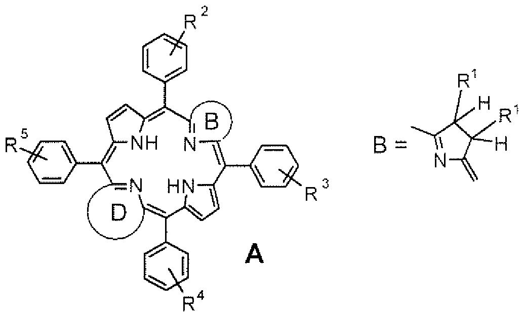
- chlorins and bacteriochlorins to be used with the present invention have the following preferred structures:
- R 1 is: H or OH
- R 2 to R 5 are substituents either in the meta- or para- position of the phenyl ring with R 2 to R 5 independently of one another chosen from a group of substituents consisting of: -OH, - COOH, -NH 2 , -COOX, -NHX, OX, -NH-Y-COOH, or -CO-Y-NH 2 .
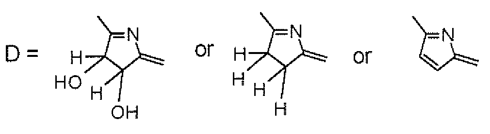
- Ring D has the structure:
- Specifically preferred chlorins to be formulated in nanoparticles according to the present invention have the structure:
- Nanoparticles prepared by the methods disclosed below have a predictable size and uniformity (in size distribution). Nanoparticles are prepared in an aseptic manufacturing process. Preferred HSA-based nanoparticles have a mean size less than 500nm in diameter. The term "diameter” is not intended to mean that the nanoparticles have necessarily a spherical shape. The term refers to the approximate average width of the nanoparticles.
- nanoparticles need to be free of any toxic material for clinical use, therefore the nanoparticles are sterilized usually by different known means in the art such as autoclaving, use of ethylene oxide, and gamma-irradiation. These conventional methods of sterilization are incompatible, however, with the photosensitizer formulations of the present invention.
- nanoparticles as required for medical applications, are prepared under complete sterile conditions.
- the HSA-based nanoparticles have a mean particle size less than 500nm and the photosensitizer is temoporfin, 5, 10,15,20-tetrakis(3-hydroxyphenyl)-chlorin (mTHPC).
- the HSA-based nanoparticles have a mean particle size less than 500nm and the photosensitizer is 2,3-dihydroxy-5, 10, 15,20-tetrakis(3-hydroxyphenyl)- chlorin (mTHPD-OH).
- mTHPP 2,3-dihydroxy-5, 10, 15,20-tetrakis(3-hydroxyphenyl)-porphyrin
- the nanoparticles of the present invention may be dehydrated for improved stability on storage.
- the preferred method of dehydration is freeze-drying or lyophilisation.
- a lyoprotectant may be used as an additive to improve the stability during the freeze-drying and during reconstitution in an aqueous medium (Anhorn, .G., Mahler, H.-C, Langer, K., Freeze-drying of human serum albumin (HSA)-nanoparticles with different excipients. Int. J. Pharm. 2008, 363, 162-1 9.)-
- HSA human serum albumin
- the HSA-based nanoparticles of the present invention were prepared by a desolvation procedure.
- protein desolvation of an aqueous HSA solution was induced by the controlled addition of a hydrophilic organic solvent such as ethanol, methanol, isopropanol, and (or) acetone.
- a hydrophilic organic solvent such as ethanol, methanol, isopropanol, and (or) acetone.
- concentrated polyethylene glycol solutions >20% in water; preferred embodiment 40%
- the resulting nanoparticles were stabilised by thermal processes or by using bifttnctional aldehydes (i.e. glutaraldehyde) or formaldehyde.
- Drug loaded nanoparticles can be freeze dried in the presence of cryoprotective agents including, but not limited to glucose, trehalose, sucrose, sorbitol and mannitol and the like.
- photosensitizer is about 10 to 50 g per milligram of HSA nanoparticle, which corresponds to a particle content of 5-25 mg/ml in water suspension, typically 8 mg/ml.
- Drug incorporation in HSA nanoparticles can be performed by HSA desolvation in the presence of the photosensitizer and the use of polyethylene glycol as desolvating agent. The entire process of nanoparticle preparation was carried out under aseptic conditions.
- the present invention is further illustrated by the following examples, but is not limited thereby.
- HSA Human serum albumin
- nanoparticles were prepared by a desolvation method. In principle, 100 mg HSA was dissolved in I ml of 10 mM sodium chloride solution. The pH was adjusted to 8 and the solution was pre-filtered through a 0.22 ujn filtration unit (Schleicher und Schtill, Dassel, Germany). This filtration process is sufficient to remove essentially all bacteria. Nanoparticles were formed by continuous addition of 4.0 ml ethanol under permanent stirring (380 rpm) at room temperature. A defined amount of ethanol is added at a rate of 1 ml/min using a pumping device (Ismatec IPN, Glattbrugg, Switzerland).
- aqueous glutaraldehyde solution was added to stabilize the resulting protein nanoparticles by chemical cross linking.
- the glutaraldehyde concentration used corresponds to 100% stoichiometric cross linking of the amino groups in 100mg HSA.
- Particles were then stirred for 1 hour and purified by 3 cycles of centrifugation at 20,817xg, for 1 Omin, and the sediment was redispersed in 1.0 ml water. Redispersion step was performed in an ultrasonic bath for 5 min. The nanoparticle content was determined by microgravimetry and was adjusted to 15.0 mg/ml.
- FIGURE 1 illustrates the drug loading of HSA-based nanoparticles on the concentration of dissolved HSA in the system. The suspensions were adjusted to 500.0 ⁇ .
- FIGURES 1 and 2 show, the drug loading of HSA-based nanoparticles is dependent on the concentration of HSA and its pH. The pH value can affect the drug loading.
- HSA-based nanoparticles of present invention are formulated under completely aseptic conditions ensuring production of sterilize nanoparticles for therapeutic use.
- FIGURE 3 depicts the image produced by TEM of wTHPP-!oaded HSA nanoparticles prepared in the presence of 1.5% dissolved HSA in ethano! 34.3% (V/V) for drug adsorption process.
- Indirect quantification procedure The mTHPP loading of the nanoparticles was calculated after spectrophotometric quantification of the unbound drug in the supernatants of the nanoparticles.
- Lvophilisation of the nanoparticles can be performed according to the following protocol:
- trehalose was added at a concentration of 3% (m/V) to the nanoparticle samples.
- the samples were transferred to a freeze drier and the shelf temperature was reduced from 5°C to -40°C at a rate of l °C/min.
- the pressure was set at 0.08 mbar. These parameters were maintained for 6 h.
- Sterility of the nanoparticle preparations was proven according to the monograph 2.6.1 "Sterility" of the European Pharmacopoeia. The sterility test was performed by the direct inoculation method as described in the monograph.
- Table il shows the fluorescent lifetimes of 5,10, 15,20-tetrakis(m-hydroxyphenyl)- porphyrin (mTHPP)-loaded HSA nanoparticles prepared using the above method. Preparation was performed in the presence of 0.75% and 2.0% soluble HSA, respectively.
- the longest component ⁇ 3 is 8.2 ns, which is similar to that of the mTHPP monomers (9.6 ns).
- Such a slightly shortened lifetime has been reported for photosensitizers attached to large units, for instance, pheophorbide a coupled to dendrimers.
- the amplitude of ⁇ 3 exceeded 40% of the total fluorescence intensity of mTHPP loaded HSA nanoparticles.
- the decay time ⁇ 2 with 2.1 - 2.3 ns shows an amplitude of about 25%.
- the shortest lifetime ⁇ 4 (0.35 ns) contributes with 32.9% to the whole fluorescence signal.
- Table II Fluorescence lifetime of mTHPP in elhanol and in form of HSA nanoparticles wTHPP-loaded in the presence of 0.75% and 2.0% soluble HSA in aqueous solutions
- Table 111 shows the lifetime of triplet state and singlet oxygen generated by the reference Rose Bengal and the photosensitizer loaded HSA nanoparticle preparations.
- Rose Bengal shows lifetime and quantum yield as it was described earlier by Redmond et al. (1999). While the quantum yield of the nanoparticles showed a decrease for both preparations, an increase in the lifetime of the reactive oxygen species was observed. The increase of singlet oxygen lifetime indicates that the photosensitizers are preserved in the monomeric form. Due to the hypoxic environment of the nanoparticle surface a decrease of the quantum yield could be expected. An increase of the singlet oxygen generation after degradation of the HSA nanoparticles and the release of the photosensitizer can be assumed.
- Table III Singlet oxygen generation and triplet parameters of Rose Bengal in ethanol and in form of HSA nanoparticles /nTHPP-loaded in the presence of 0.75% and 2.0% soluble HSA in aqueous solutions
- Nanoparticles were prepared according to example la with the exception that mTHPC was used instead of wTHPP.
- wTHPC-loaded nanoparticles were characterized as described within example l a.
- HSA Human serum albumin
- mTHPC Human serum albumin based nanoparticles were prepared by a desolvation method using polyethylene glycol as desoivating agent. In principle, an amount of 90 mg HSA was dissolved in 0.9 ml of 10 mM sodium chloride solution. The pH was adjusted to 6- 8 and the solution was filtered through a 0.22 ⁇ m filtration unit (Schleicher und Schiill, Dassel, Germany). mTHPC was added in form of 0.1 ml ethanolic solution containing 3, 7.5, and 15 mg/ml mTHPC, respectively.
- nanoparticles were formed by continuous addition of 4.0 ml aqueous polyethylene glycol (PEG4000) solution under continuous stirring (400-500 rpm) at room temperature.
- a defined amount of ethanol is added at a rate of 1 ml/min using a pumping device (lsmatec IPN, Glattbrugg, Switzerland).
- protein desolvation 78 uL (or 104, and 182 ⁇ , respectively) of 8% aqueous glutaraldehyde solution were added to stabilize the resulting protein nanoparticles by chemical cross linking.
- the glutaraldehyde concentration used corresponds to 150% (or 200%, and 350%, respectively) stoichiometric cross linking of the amino groups in 90 nig HSA.
- Particles were stirred for 3 h and were purified by 3 cycles of centrifugation at the rate of 20,817g, for 10 min) and redispersion in 1.0 ml water in an ultrasonic bath (5 min).
- the nanoparticle content was determined by microgravimetry and was adjusted to 15.0 mg/ml.
- wTHPC-loaded nanoparticles were characterized as described within example l a.
- DiFi cells were cultured on glass slides (Becton Dickinson) and incubated with the nanoparticulate formulation for 4h at 37°C. Following, the cells were washed twice with PBS and the membranes were stained with Concanavalin A AlexaFluor350 (50 ng/ml; Invitrogen, Düsseldorf) for 2 min. Cells were fixed with 0.4 % paraformaldehyde for 6 min. After fixation, the cells were washed and then embedded in Vectashield HardSet Mounting Medium (Axxora, Griinberg).
- the microscopy analysis was performed with an Axiovert 200 M microscope with a 510 NLO Meta device (Zeiss, Jena), a chameleon femtosecond or an argon ion laser and the LSM Image Examiner software.
- FIGURES 4A-D illustrate the cellular uptake and intracellular distribution of HSA based nanoparticles (0.75 and 2.00 % soluble HSA) with the photosensitizer 5, 10, 15,20- tetrakis(3-hydroxyphenyl)-porphyrin (wTHPP) studied by confocal laser scanning microscopy.
- DiFi cells were cultured on glass slides and incubated with the nanoparticles for 4 h at 37°C. The red autofluorescence of the photosensitizer wTHPP and the green autofluorescence of the nanoparticles were used. Pictures were taken within inner sections of the cells. (FIG.
- FIG. 4A-B Incubation of cells with HSA nanoparticles (0.75 % soluble HSA) with mTHPP.
- FIG. 4C-D Incubation of cells with HSA nanoparticles (2.00 % soluble HSA) with wTHPP.
- FIG. 4A and FIG. 4C display the green nanoparticle channel;
- FIG. 4B and
- FIG. 4D display the red photosensitizer channel.
- Scale bar 20 ⁇
- FIGURES 5A-D illustrate cellular uptake/adhesion and intracellular distribution of HSA based nanoparticles (0.75 and 2.00 % soluble HSA) with the photosensitizer 5, 10, 15,20- tetrakis(3-hydroxyphenyl)-chlorin (mTHPC) studied by confocal laser scanning microscopy.
- DiFi cells were cultured on glass slides and incubated with the nanoparticles for 4 h at 37°C. The red autofluorescence of the photosensitizer mTHPC and the green autofluorescence of the nanoparticles were used. Pictures were taken within inner sections of the cells. (FIG.
- FIG. 5A and FIG. 5C displays the green nanoparticle channel
- FIG. 5B displays the red photosensitizer channel.
- Scale bar 20° ⁇ .
- Intracellular uptake and phototoxicity of the present nanoparticles formulation was studied using Jurkat-cell suspensions cultured in RPMI 1640 medium. All cells were incubated in 3 ⁇ mTHPC and mTHPC loaded into different concentration of HSA based nanoparticles for set period of time (l h, 3h, 5h, 24h). The HSA-based nanoparticles of varying HSA concentration was used to determine the intracellular uptake and phototoxicity effect of the cells suspension.
- the Jurkat-cell suspensions were incubated in five samples:
- the Jurkat-cell suspensions incubated with the above mentioned five samples were irradiated at 660 nm for 2 min (using an LED), having a light dose of 290 mJ/cm 2 to study the phototoxicity effect of cells.
- FIGURES 6A-D depict dark toxicity and the phototoxicity effects on Jurkat ceils in 5 different samples after different incubation times.
- FIGURE 6A illustrates the observed dark toxicity samples, where it was found to be almost zero indicating no toxic effect of the HSA-based nanoparticles.
- Jurkat cells were incubated in five samples. After 1 h, 3 h, 5 h, and 24 h incubation in darkness an aliquot of each sample was investigated. Trypan blue test was used to assess the necrotic ceils, apoptotic cells were detected by their change of cell shape (apoptotic blebbing). Little or no effect was found. In other words dark toxicity was not observed for the wTHPC concentrations used.
- FIGURES 6B, 6C, 6D show apoptosis and necrosis effects, individually and together, on cells due to phototoxicity effect.
- FIGURE 6C shows the rate of apoptosis
- FIGURE 6D shows the rate of necrosis in the incubation medium separately; compared to FIGURE 6B.
- the sample reference in each of FIGURES 6A-D represents the cells which were incubated and irradiated without photosensitizer. The cells were illuminated using a LED at 660 nm for an exposure time of 120 s and light dose of 290 mJ/cm 2 . The experiments were repeated twice and for each measurement the cell number was counted three times two hours after light exposure to get average.
- FIGURES 6C and 6D illustrate the apoptosis and necrosis effect on cells due to phototoxicity respectively when incubated in 5 different samples after different incubation times as mentioned above.
- a formulation of present invention when used in PDT is seen to initiate a high amount of apoptosis - a patient, gentle kind of cell death and a lower amount of necrosis - a kind of cell death with high immune system response but needed in low dose to prevent tumor recovery.
- Jurkat-cell suspension in cell growth medium (RPMI 1640) was incubated in 3 ⁇ of mTHPC and different concentration of HSA encapsulating 3 ⁇ of wTHPC based nanoparticles, for 1 h, 3h, 5h, 24h. After incubation the cells were counted (using a haemacytometer), washed with phosphate buffer solution (PBS, 400g, 3min, 2x) and the cell pellet was stored and frozen overnight at -20°C to disrupt the cell membranes. From these cells the wTHPC was extracted in ethanol using ultrasound (>5 min). The mTHPC concentration in the ethanol extract was determined via fluorescence intensity using a standard fluorescence series. For the calculation of intracellular concentration the diameter of the cells was assumed to be 10 ⁇ .
Landscapes
- Health & Medical Sciences (AREA)
- Chemical & Material Sciences (AREA)
- Life Sciences & Earth Sciences (AREA)
- Animal Behavior & Ethology (AREA)
- Veterinary Medicine (AREA)
- Medicinal Chemistry (AREA)
- Public Health (AREA)
- General Health & Medical Sciences (AREA)
- Pharmacology & Pharmacy (AREA)
- Bioinformatics & Cheminformatics (AREA)
- Engineering & Computer Science (AREA)
- Chemical Kinetics & Catalysis (AREA)
- Organic Chemistry (AREA)
- Nuclear Medicine, Radiotherapy & Molecular Imaging (AREA)
- General Chemical & Material Sciences (AREA)
- Epidemiology (AREA)
- Optics & Photonics (AREA)
- Biomedical Technology (AREA)
- Gastroenterology & Hepatology (AREA)
- Nanotechnology (AREA)
- Biochemistry (AREA)
- Molecular Biology (AREA)
- Physics & Mathematics (AREA)
- Rheumatology (AREA)
- Pain & Pain Management (AREA)
- Ophthalmology & Optometry (AREA)
- Oncology (AREA)
- Communicable Diseases (AREA)
- Urology & Nephrology (AREA)
- Dermatology (AREA)
- Immunology (AREA)
- Orthopedic Medicine & Surgery (AREA)
- Physical Education & Sports Medicine (AREA)
- Medicinal Preparation (AREA)
- Pharmaceuticals Containing Other Organic And Inorganic Compounds (AREA)
- Medicines That Contain Protein Lipid Enzymes And Other Medicines (AREA)
Abstract
Description


Claims


Priority Applications (5)
| Application Number | Priority Date | Filing Date | Title |
|---|---|---|---|
| CN2010800629189A CN102740875A (en) | 2009-12-11 | 2010-12-08 | Human serum albumin-based nanoparticle carrier system for photodynamic therapy |
| BR112012013952-5A BR112012013952A2 (en) | 2009-12-11 | 2010-12-08 | pharmaceutical formulation of nanoparticles and their preparation methods and uses |
| JP2012543221A JP5972171B2 (en) | 2009-12-11 | 2010-12-08 | Nanoparticle carrier system based on human serum albumin for photodynamic therapy |
| EP10836582.6A EP2509620A4 (en) | 2009-12-11 | 2010-12-08 | TRANSPORTATION SYSTEMS OF HUMAN SERUM-ALBUMINUM NANOPARTICLES FOR PHOTODYNAMIC THERAPY |
| CA2784001A CA2784001C (en) | 2009-12-11 | 2010-12-08 | Nanoparticle carrier systems based on human serum albumin for photodynamic therapy |
Applications Claiming Priority (4)
| Application Number | Priority Date | Filing Date | Title |
|---|---|---|---|
| US28590209P | 2009-12-11 | 2009-12-11 | |
| US61/285,902 | 2009-12-11 | ||
| US12/941,350 US9211283B2 (en) | 2009-12-11 | 2010-11-08 | Nanoparticle carrier systems based on human serum albumin for photodynamic therapy |
| US12/941,350 | 2010-11-08 |
Publications (2)
| Publication Number | Publication Date |
|---|---|
| WO2011071968A2 true WO2011071968A2 (en) | 2011-06-16 |
| WO2011071968A3 WO2011071968A3 (en) | 2011-11-17 |
Family
ID=44143223
Family Applications (1)
| Application Number | Title | Priority Date | Filing Date |
|---|---|---|---|
| PCT/US2010/059364 Ceased WO2011071968A2 (en) | 2009-12-11 | 2010-12-08 | Nanoparticle carrier systems based on human serum albumin for photodynamic therapy |
Country Status (7)
| Country | Link |
|---|---|
| US (1) | US9211283B2 (en) |
| EP (1) | EP2509620A4 (en) |
| JP (1) | JP5972171B2 (en) |
| CN (1) | CN102740875A (en) |
| BR (1) | BR112012013952A2 (en) |
| CA (1) | CA2784001C (en) |
| WO (1) | WO2011071968A2 (en) |
Cited By (5)
| Publication number | Priority date | Publication date | Assignee | Title |
|---|---|---|---|---|
| EP2591803A1 (en) * | 2011-11-09 | 2013-05-15 | APOCARE Pharma GmbH | Composition for the photodynamic diagnosis and treatment of tumours |
| WO2016051361A1 (en) | 2014-09-30 | 2016-04-07 | Biolitec Unternehmensbeteiligungs Ii Ag | Specifically meso-substituted porphyrins and chlorins for photodynamic therapy |
| EP3210626A1 (en) | 2016-02-26 | 2017-08-30 | biolitec Unternehmensbeteiligungs II AG | Conjugates of porphyrinoid photosensitizers and glycerol-based polymers for photodynamic therapy |
| WO2020120474A1 (en) | 2018-12-10 | 2020-06-18 | Universität Hamburg | Specifically glyco-substituted porphyrins and chlorins for photodynamic therapy |
| US20240199628A1 (en) * | 2020-10-05 | 2024-06-20 | Biolitec Unternehmensbeteiligungs Ii Ag | Tetrapyrrole-based compounds and their fomulations for anti-microbial therapy |
Families Citing this family (12)
| Publication number | Priority date | Publication date | Assignee | Title |
|---|---|---|---|---|
| CN103169968B (en) * | 2013-03-12 | 2014-11-26 | 中国科学院理化技术研究所 | Hydrophobic chlorin photosensitizer nano-drug preparation based on albumin, preparation method and application thereof |
| KR101329646B1 (en) * | 2013-05-02 | 2013-11-14 | 주식회사 지니스 | Targeting-enhancing anticancer nanoparticles and preparation method the same |
| CN104587466B (en) * | 2014-12-15 | 2017-11-21 | 苏州大学 | The preparation method and application of albumen polypyrrole compound and albumen polypyrrole compound derivative |
| DE102016125666A1 (en) * | 2016-12-23 | 2018-06-28 | Michael Denck | HSA Galenik |
| CN111295094A (en) | 2017-10-09 | 2020-06-16 | 泰尔茂比司特生物技术有限公司 | Freeze-drying container and method of using freeze-drying container |
| WO2020185916A1 (en) | 2019-03-14 | 2020-09-17 | Terumo Bct Biotechnologies, Llc | Lyophilization container fill fixture, system and method of use |
| CN110251672B (en) * | 2019-06-18 | 2022-04-01 | 深圳大学 | Nano diagnosis and treatment agent and preparation method and application thereof |
| CN111110861B (en) * | 2020-02-23 | 2023-08-25 | 上海交通大学医学院附属瑞金医院 | A drug-loaded granule based on human serum albumin and its preparation method |
| CN112972676B (en) * | 2020-08-17 | 2024-09-24 | 滨州医学院 | Synthesis of HSA-Biotin-DDA-TCPP molecules and their applications as photodynamic therapy agents and nanomedicines |
| CN113855645B (en) * | 2021-09-13 | 2024-02-02 | 苏州大学 | Core-shell assembly based on functionalized protein nanoparticles, and preparation method and application thereof |
| WO2023039691A1 (en) * | 2021-09-17 | 2023-03-23 | 苏州大学 | Functionalized protein nanoparticle-based core-shell type assembly, preparation method therefor, and application thereof |
| JP2024534479A (en) * | 2021-09-17 | 2024-09-20 | ユニバーシティ オブ メリーランド, カレッジ パーク | Amorphous photosensitizing particles, methods for preparing same, and methods for using same |
Citations (7)
| Publication number | Priority date | Publication date | Assignee | Title |
|---|---|---|---|---|
| EP0337601B1 (en) | 1988-03-11 | 1993-05-05 | Scotia Holdings Plc | Porphyrins and medicaments for cancer treatment |
| WO1996013504A1 (en) | 1994-10-26 | 1996-05-09 | University Of British Columbia | β,β'-DIHYDROXY MESO-SUBSTITUTED CHLORINS, ISOBACTERIOCHLORINS, BACTERIOCHLORINS, AND METHODS FOR MAKING THE SAME FROM β,β'-UNSUBSTITUTED TETRAPYRROLIC MACROCYCLES |
| US5916596A (en) | 1993-02-22 | 1999-06-29 | Vivorx Pharmaceuticals, Inc. | Protein stabilized pharmacologically active agents, methods for the preparation thereof and methods for the use thereof |
| WO2000061584A1 (en) | 1999-04-14 | 2000-10-19 | The University Of British Columbia | IMPROVED β,β,-DIHYDROXY MESO-SUBSTITUTED CHLORINS, ISOBACTERIOCHLORINS, AND BACTERIOCHLORINS |
| WO2001021174A1 (en) | 1999-09-23 | 2001-03-29 | Dabur Research Foundation | Formulations of paclitaxel entrapped into nanoparticles of polymeric micelles |
| WO2003097096A1 (en) | 2002-05-16 | 2003-11-27 | Qlt Inc. | Compositions and methods for delivery of photosensitive drugs |
| WO2006133271A2 (en) | 2005-06-06 | 2006-12-14 | The General Hospital Corporation | Compositions and methods relating to target-specific photodynamic therapy |
Family Cites Families (12)
| Publication number | Priority date | Publication date | Assignee | Title |
|---|---|---|---|---|
| GB9420355D0 (en) * | 1994-10-10 | 1994-11-23 | Univ Nottingham | Preparation of protein microspheres, films and coatings |
| US6649192B2 (en) * | 1996-07-29 | 2003-11-18 | Universidade De Santiago De Compostela | Application of nanoparticles based on hydrophilic polymers as pharmaceutical forms |
| JP2005502618A (en) * | 2001-06-04 | 2005-01-27 | ザ・ジェネラル・ホスピタル・コーポレイション | Methods for detecting and treating vulnerable plaque using photodynamic compounds |
| US8709449B2 (en) * | 2004-02-12 | 2014-04-29 | Biolitec Pharma Marketing Ltd | Methods and compositions for improving photodynamic therapy through administration of lipids |
| DE102004011776A1 (en) * | 2004-03-09 | 2005-11-03 | Lts Lohmann Therapie-Systeme Ag | Carrier system in the form of protein-based nanoparticles for the cell-specific accumulation of pharmaceutically active substances |
| JP2006056807A (en) * | 2004-08-18 | 2006-03-02 | Konica Minolta Medical & Graphic Inc | Photodynamic therapy formulation |
| US20070218049A1 (en) * | 2006-02-02 | 2007-09-20 | Wei Chen | Nanoparticle based photodynamic therapy and methods of making and using same |
| CN101485629B (en) * | 2008-01-16 | 2013-01-23 | 沈阳药科大学 | Drug delivery system and preparation method thereof |
| KR20150136137A (en) * | 2008-04-10 | 2015-12-04 | 아브락시스 바이오사이언스, 엘엘씨 | Compositions of hydrophobic taxane derivatives and uses thereof |
| US8815931B2 (en) * | 2009-04-28 | 2014-08-26 | Biolitec Pharma Marketing Ltd | Oral formulations for tetrapyrrole derivatives |
| EP2425234B1 (en) * | 2009-04-30 | 2019-10-23 | Thermo Scientific Portable Analytical Instruments Inc. | Localization of an element of interest by xrf analysis of different inspection volumes |
| US20110275686A1 (en) * | 2009-12-11 | 2011-11-10 | Biolitec, Inc. | Nanoparticle carrier systems based on poly(dl-lactic-co-glycolic acid) (plga) for photodynamic therapy (pdt) |
-
2010
- 2010-11-08 US US12/941,350 patent/US9211283B2/en active Active
- 2010-12-08 WO PCT/US2010/059364 patent/WO2011071968A2/en not_active Ceased
- 2010-12-08 CA CA2784001A patent/CA2784001C/en not_active Expired - Fee Related
- 2010-12-08 CN CN2010800629189A patent/CN102740875A/en active Pending
- 2010-12-08 BR BR112012013952-5A patent/BR112012013952A2/en not_active IP Right Cessation
- 2010-12-08 JP JP2012543221A patent/JP5972171B2/en not_active Expired - Fee Related
- 2010-12-08 EP EP10836582.6A patent/EP2509620A4/en active Pending
Patent Citations (7)
| Publication number | Priority date | Publication date | Assignee | Title |
|---|---|---|---|---|
| EP0337601B1 (en) | 1988-03-11 | 1993-05-05 | Scotia Holdings Plc | Porphyrins and medicaments for cancer treatment |
| US5916596A (en) | 1993-02-22 | 1999-06-29 | Vivorx Pharmaceuticals, Inc. | Protein stabilized pharmacologically active agents, methods for the preparation thereof and methods for the use thereof |
| WO1996013504A1 (en) | 1994-10-26 | 1996-05-09 | University Of British Columbia | β,β'-DIHYDROXY MESO-SUBSTITUTED CHLORINS, ISOBACTERIOCHLORINS, BACTERIOCHLORINS, AND METHODS FOR MAKING THE SAME FROM β,β'-UNSUBSTITUTED TETRAPYRROLIC MACROCYCLES |
| WO2000061584A1 (en) | 1999-04-14 | 2000-10-19 | The University Of British Columbia | IMPROVED β,β,-DIHYDROXY MESO-SUBSTITUTED CHLORINS, ISOBACTERIOCHLORINS, AND BACTERIOCHLORINS |
| WO2001021174A1 (en) | 1999-09-23 | 2001-03-29 | Dabur Research Foundation | Formulations of paclitaxel entrapped into nanoparticles of polymeric micelles |
| WO2003097096A1 (en) | 2002-05-16 | 2003-11-27 | Qlt Inc. | Compositions and methods for delivery of photosensitive drugs |
| WO2006133271A2 (en) | 2005-06-06 | 2006-12-14 | The General Hospital Corporation | Compositions and methods relating to target-specific photodynamic therapy |
Non-Patent Citations (13)
| Title |
|---|
| ANHORN, M.G.MAHLER, H.-C.LANGER, K.: "Freeze-drying of human serum albumin (HSA)-nanoparticles with different excipients", INT. J. PHARM., vol. 363, 2008, pages 162 - 169, XP025431308, DOI: 10.1016/j.ijpharm.2008.07.004 |
| C. HADJUR, J. PHOTOCHEM. PHOTOBIOL. B: BIOLOGY, vol. 45, 1998, pages 170 - 178 |
| C. VOLLAND ET AL., J. CONTR. REL., vol. 31, 1994, pages 293 - 305 |
| K. A. ATHANASIOU ET AL., BIOMATERIALS, vol. 17, 1996, pages 93 - 102 |
| K. LANGER ET AL., INT. J. PHARM., vol. 347, 2007, pages 109 - 117 |
| KONAN, Y. N. ET AL., PHOTOCHEMISTRY AND PHOTOBIOLOGY, vol. 77, no. 6, 2003, pages 638 - 644 |
| KUAN CHEN ET AL., JOURNAL OF PHOTOCHEMISTRY AND PHOTOBIOLOGY B: BIOLOGY, vol. 96, 2009, pages 66 - 74 |
| M. N. V. RAVI KUMAR: "Engineered Nanoparticles in Cancer Therapy", J. PHARM. PHARMACEUT. SCI., vol. 3, 2000, pages 234 - 258 |
| N. P. PREATORIUST. K. MANDAL: "Engineered Nanoparticles in Cancer Therapy", RECENT PATENTS ON DRUG DELIVERY & FORMULATION, vol. 1, 2007, pages 37 - 51 |
| R. BONNETT ET AL., J. CHEM. SOC. PERKIN TRANS., vol. 2, 1999, pages 325 - 328 |
| R. BONNETT ET AL., J. PORPHYRINS PHTHALOCYANINES, vol. 5, 2001, pages 652 - 661 |
| See also references of EP2509620A4 |
| Y. HONGYING ET AL., DYES PIGM., vol. 43, 1999, pages 109 - 117 |
Cited By (8)
| Publication number | Priority date | Publication date | Assignee | Title |
|---|---|---|---|---|
| EP2591803A1 (en) * | 2011-11-09 | 2013-05-15 | APOCARE Pharma GmbH | Composition for the photodynamic diagnosis and treatment of tumours |
| WO2013068405A1 (en) * | 2011-11-09 | 2013-05-16 | Apocare Pharma Gmbh | Composition for photodynamic diagnosis and treatment of tumors |
| US9242006B2 (en) | 2011-11-09 | 2016-01-26 | Apocare Pharma Gmbh | Composition of chlorin e6 stabilized within albumin for the photodynamic diagnosis and therapy of tumors |
| WO2016051361A1 (en) | 2014-09-30 | 2016-04-07 | Biolitec Unternehmensbeteiligungs Ii Ag | Specifically meso-substituted porphyrins and chlorins for photodynamic therapy |
| EP3210626A1 (en) | 2016-02-26 | 2017-08-30 | biolitec Unternehmensbeteiligungs II AG | Conjugates of porphyrinoid photosensitizers and glycerol-based polymers for photodynamic therapy |
| US10738059B2 (en) | 2016-02-26 | 2020-08-11 | Biolitec Unternehmensbeteiligungs Ii Ag | Conjugates of porphyrinoid photosensitizers and glycerol-based polymers for photodynamic therapy |
| WO2020120474A1 (en) | 2018-12-10 | 2020-06-18 | Universität Hamburg | Specifically glyco-substituted porphyrins and chlorins for photodynamic therapy |
| US20240199628A1 (en) * | 2020-10-05 | 2024-06-20 | Biolitec Unternehmensbeteiligungs Ii Ag | Tetrapyrrole-based compounds and their fomulations for anti-microbial therapy |
Also Published As
| Publication number | Publication date |
|---|---|
| JP2013513609A (en) | 2013-04-22 |
| WO2011071968A3 (en) | 2011-11-17 |
| BR112012013952A2 (en) | 2018-06-05 |
| CA2784001C (en) | 2018-04-10 |
| US20110142948A1 (en) | 2011-06-16 |
| JP5972171B2 (en) | 2016-08-17 |
| EP2509620A4 (en) | 2015-10-21 |
| CA2784001A1 (en) | 2011-06-16 |
| US9211283B2 (en) | 2015-12-15 |
| CN102740875A (en) | 2012-10-17 |
| EP2509620A2 (en) | 2012-10-17 |
Similar Documents
| Publication | Publication Date | Title |
|---|---|---|
| US9211283B2 (en) | Nanoparticle carrier systems based on human serum albumin for photodynamic therapy | |
| EP2509633B1 (en) | Nanoparticle carrier system based on poly(dl-lactic-co-glycolic acid) (plga) for photodynamic therapy | |
| CA2513759C (en) | Ceramic based nanoparticles for entrapping therapeutic agents for photodynamic therapy and method of using same | |
| KR102081666B1 (en) | Phamaceutical composition for treating cancer | |
| KR101035269B1 (en) | Novel photodynamic therapy using polymer derivative-photosensitizer complex | |
| CN104162164B (en) | A kind of polymer albumin nanospheres comprising indocyanine-green and its preparation method and application | |
| EP1503796B1 (en) | Compositions and methods for delivery of photosensitive drugs | |
| US20100273803A1 (en) | Oral Formulations for Tetrapyrrole Derivatives | |
| KR101188979B1 (en) | Conjugate of biocompatible polymer and photosensitizer for photodynamic diagnosis or therapy and process of preparation thereof | |
| KR102363327B1 (en) | Composition for photodynamic therapy containing 5-aminolevulinic acid hydrochloride | |
| CN101524326A (en) | Chlorin e6 chitosan-stearic acid graft micelle | |
| Dong et al. | Investigation of the intracellular oxidative stress amplification, safety and anti-tumor effect of a kind of novel redox-responsive micelle | |
| da Silva et al. | In vitro photodynamic activity of chloro (5, 10, 15, 20-tetraphenylporphyrinato) indium (III) loaded-poly (lactide-co-glycolide) nanoparticles in LNCaP prostate tumour cells | |
| Huang et al. | Nanogenerators with l-arginine loading: new choices as cascade and synergistic nitric oxide/photodynamic antitumor agents | |
| US11040101B2 (en) | Nanoparticles for cancer therapy and diagnosis | |
| Shabbirahmed et al. | Recent advancements in nanomaterials for photodynamic therapy of cancers | |
| CN113827553B (en) | Zinc phthalocyanine in-situ gel for intratumoral injection administration for photodynamic tumor treatment and preparation method thereof | |
| CN113041359A (en) | Glutathione-responsive osteosarcoma-resistant prodrug nanoparticle and preparation method and application thereof | |
| EP3041510B1 (en) | Polymer micelle containing a photosensitizer | |
| Amirinejad et al. | Structural Design and Modification of Porphyrin-Based Nanovesicles for Enhanced Biomedical Functionality | |
| CN116983284A (en) | A ROS-responsive nanoparticle and its application in the treatment of sonodynamically mediated hydatid disease | |
| Morozova et al. | Selenium-containing Nanosystems based on the Amphiphilic Molecular Brushes and Fotoditazin for Photodynamic Therapy | |
| Lai et al. | Shell Cross-Linked Nanoparticle Based on Poly (ε-caprolactone)-Poly (ethylene glycol) for Photosensitizer Delivery |
Legal Events
| Date | Code | Title | Description |
|---|---|---|---|
| WWE | Wipo information: entry into national phase |
Ref document number: 201080062918.9 Country of ref document: CN |
|
| 121 | Ep: the epo has been informed by wipo that ep was designated in this application |
Ref document number: 10836582 Country of ref document: EP Kind code of ref document: A1 |
|
| ENP | Entry into the national phase |
Ref document number: 2784001 Country of ref document: CA |
|
| NENP | Non-entry into the national phase |
Ref country code: DE |
|
| WWE | Wipo information: entry into national phase |
Ref document number: 2012543221 Country of ref document: JP |
|
| WWE | Wipo information: entry into national phase |
Ref document number: 2010836582 Country of ref document: EP |
|
| WWE | Wipo information: entry into national phase |
Ref document number: 5995/CHENP/2012 Country of ref document: IN |
|
| REG | Reference to national code |
Ref country code: BR Ref legal event code: B01A Ref document number: 112012013952 Country of ref document: BR |
|
| ENP | Entry into the national phase |
Ref document number: 112012013952 Country of ref document: BR Kind code of ref document: A2 Effective date: 20120611 |