WO2011025014A1 - 薬物の安全性の評価方法 - Google Patents
薬物の安全性の評価方法 Download PDFInfo
- Publication number
- WO2011025014A1 WO2011025014A1 PCT/JP2010/064747 JP2010064747W WO2011025014A1 WO 2011025014 A1 WO2011025014 A1 WO 2011025014A1 JP 2010064747 W JP2010064747 W JP 2010064747W WO 2011025014 A1 WO2011025014 A1 WO 2011025014A1
- Authority
- WO
- WIPO (PCT)
- Prior art keywords
- activity
- hepatocytes
- xenobiotic
- test compound
- cyp3a
- Prior art date
- Legal status (The legal status is an assumption and is not a legal conclusion. Google has not performed a legal analysis and makes no representation as to the accuracy of the status listed.)
- Ceased
Links
Images



Classifications
-
- G—PHYSICS
- G01—MEASURING; TESTING
- G01N—INVESTIGATING OR ANALYSING MATERIALS BY DETERMINING THEIR CHEMICAL OR PHYSICAL PROPERTIES
- G01N33/00—Investigating or analysing materials by specific methods not covered by groups G01N1/00 - G01N31/00
- G01N33/48—Biological material, e.g. blood, urine; Haemocytometers
- G01N33/50—Chemical analysis of biological material, e.g. blood, urine; Testing involving biospecific ligand binding methods; Immunological testing
- G01N33/5005—Chemical analysis of biological material, e.g. blood, urine; Testing involving biospecific ligand binding methods; Immunological testing involving human or animal cells
- G01N33/5008—Chemical analysis of biological material, e.g. blood, urine; Testing involving biospecific ligand binding methods; Immunological testing involving human or animal cells for testing or evaluating the effect of chemical or biological compounds, e.g. drugs, cosmetics
- G01N33/5044—Chemical analysis of biological material, e.g. blood, urine; Testing involving biospecific ligand binding methods; Immunological testing involving human or animal cells for testing or evaluating the effect of chemical or biological compounds, e.g. drugs, cosmetics involving specific cell types
- G01N33/5067—Liver cells
-
- G—PHYSICS
- G01—MEASURING; TESTING
- G01N—INVESTIGATING OR ANALYSING MATERIALS BY DETERMINING THEIR CHEMICAL OR PHYSICAL PROPERTIES
- G01N2333/00—Assays involving biological materials from specific organisms or of a specific nature
- G01N2333/795—Porphyrin- or corrin-ring-containing peptides
- G01N2333/80—Cytochromes
Definitions
- the present invention relates to a method for evaluating drug safety, particularly xenobiotic metabolizing enzyme induction ability. Specifically, the present invention relates to a drug comprising: exposing hepatocytes to a test compound in the presence and absence of an inducer of xenobiotic metabolizing enzyme activity and comparing the enzyme activity under both conditions. The present invention relates to a method for evaluating the ability to induce xenobiotic metabolizing enzymes.
- the liver plays a central role in xenobiotic metabolism that converts xenobiotics such as pharmaceuticals into a form that is easy to excrete.
- This function of xenobiotic metabolism may simultaneously induce mutagenesis and toxicity due to xenobiotic metabolites, or reduce the pharmacological effects of concomitant drugs.
- the activity of the xenobiotic-metabolizing enzyme group differs greatly between experimental animals and humans, and the knowledge obtained using experimental animals is often not reflected in humans. Therefore, it is desirable to use human liver or cells derived from human liver in order to correctly understand the effects of pharmaceuticals on human liver metabolism.
- human liver or cells derived from human liver in order to correctly understand the effects of pharmaceuticals on human liver metabolism.
- hepatocytes in the case of hepatocytes, it is generally common to lose the original differentiation trait.
- the activity of xenobiotic metabolizing enzymes is easily lost not only in established cell lines but also in primary culture. Although it is conceivable to use primary hepatocytes prepared from fresh liver slices immediately after excision as hepatocytes that retain xenobiotic metabolism ability, there are various problems such as difficulty in obtaining them, and their practicality is extremely low.
- Patent Document 1 maintain the frozen primary human hepatocytes in a serum-free complete synthetic medium containing glucocorticoid, thereby inducing the activity and gene expression of the xenobiotic-metabolizing enzyme group and the activity induction of the enzyme group. It was also reported that the mechanism of gene expression induction can be stably maintained, and the activity of the xenobiotic metabolizing enzyme group of the test compound and / or the presence or absence of gene expression inducing activity can be determined. However, in actual induction assays, it is known that the activity of xenobiotic metabolizing enzymes decreases at high concentrations depending on the test compound.
- Non-Patent Document 1 after treating human hepatocytes with a test compound alone and after inducing the enzyme (CYP3A) using rifampicin, a known CYP3A inducer, Reported that when the test compound is added to human hepatocytes after removal of the test substance, the appearance of false negatives can be prevented and the test reliability can be improved by measuring and comparing the activity of the enzyme. Yes.
- An object of the present invention is to provide a simple and reliable evaluation method for evaluating the ability of a drug candidate compound to induce xenobiotic metabolizing enzymes in the early drug discovery stage.
- the present inventors have conducted extensive studies, and as a result, exposed hepatocytes to a test compound in the presence and absence of a substance that induces xenobiotic metabolizing enzyme activity. By measuring and comparing enzyme activity, underestimate the ability to induce enzyme activity that occurs not only when the test compound inhibits the enzyme activity but also when it has an inhibitory action based on cytotoxicity or mechanism. It was also found that a more reliable evaluation is possible. In addition, by using a 96-well plate coated with thin layer Matrigel, human hepatocytes were able to maintain high CYP3A activity even during a long culture period. It was confirmed that it could be a performance evaluation system. As a result of further studies based on these findings, the present inventors have completed the present invention.
- the present invention is as follows: [1] The following steps: (A) A sample containing cultured hepatocytes is bisected, one is treated with a test compound in the presence of a xenobiotic metabolizing enzyme activity inducer, and the other is tested in the absence of a xenobiotic metabolizing enzyme activity inducer. Process in (B) a step of measuring the activity of the xenobiotic metabolizing enzyme contained in each sample obtained in step (a), and (c) a step of comparing and analyzing each enzyme activity measured in step (b).
- a method for evaluating the ability of a test compound to induce xenobiotic metabolizing enzymes comprising: [2] The method according to [1] above, wherein the treatment in the step (a) is performed on an incubator coated with a thin layer matrigel. [3] The method of [2] above, wherein the incubator is a 96-well plate. [4] The method according to any one of [1] to [3] above, wherein the treatment in the step (a) is carried out at two or more test compound concentrations. [5] The method according to any one of [1] to [4] above, wherein the xenobiotic-metabolizing enzyme is cytochrome P450.
- the inducer of xenobiotic metabolizing enzyme activity is rifampicin, nifedipine, efavirenz, carbamazepine, abasimibe, troglitazone, gemfibrozil, lansoprazole, modafinil, nevirapine, omeprazole, phenobarbital, phenytoin, rofecoxib, sulfinpyrazone, or topiramate
- a test compound is obtained by exposing hepatocytes to a test compound in the presence and absence of a substance that induces xenobiotic metabolizing enzyme activity, and measuring and comparing the activity of the enzyme under both conditions.
- a substance that induces xenobiotic metabolizing enzyme activity e.g, CYP3A
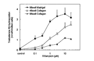
- Figure 3 shows the effect of matrix on induction of CYP3A enzyme activity with rifampicin.
- Frozen primary human hepatocytes were cultured on 48-well plates (white triangles) coated with collagen, 96-well plates (black triangles), or 96-well plates (black circles) coated with thin-layer Matrigel. Hepatocyte cultures were treated with 0.1% DMSO (control) or various concentrations of rifampicin for 3 days. The cell density was 0.16 ⁇ 10 6 cells / well for the 48-well plate and 0.8 ⁇ 10 5 cells / well for the 96-well plate.
- CYP3A activity was determined as the degree of 6 ⁇ -hydroxylation of testosterone.
- hepatocytes were treated with 0.1% DMSO (control), 10 ⁇ M rifampicin, 30 ⁇ M phenytoin or 30 ⁇ M carbamazepine.
- CYP3A-induced responses are expressed as CYP3A activity (degree of 6 ⁇ -hydroxylation of testosterone).
- the bar graph of each column shows DMSO, rifampicin, phenytoin, and carbamazepine from the left.
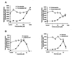
- Figure 2 shows the induction of CYP3A in frozen primary human hepatocytes treated with various inducers and non-inducers. The compounds tested were tested according to their ability to induce CYP3A in fresh or frozen primary human hepatocytes reported in NJ ⁇ Hewitt, et al .; Chem.
- FIG. 2 shows a concentration-response curve of a compound that produces a positive response in frozen primary human hepatocytes. Hepatocytes were treated with several concentrations of test compounds alone (black circles) or in combination with test compounds and 10 ⁇ M rifampicin (white circles).
- Phenobarbital and carbamazepine show typical concentration-response curves and no inhibition of CYP3A induction by rifampicin (A).
- Abashimibe and efavirenz show a bell-type concentration-response relationship and inhibit CYP3A induction by rifampicin (B).
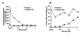
- FIG. 6 shows concentration-response curves for induction of CYP3A activity and CYP3A4 mRNA level in hepatocytes treated with ritonavir.
- the evaluation method for xenobiotic-metabolizing enzyme induction ability of the present invention includes the following steps: (A) A sample containing cultured hepatocytes is bisected, one is treated with a test compound in the presence of a xenobiotic metabolizing enzyme activity inducer, and the other is tested in the absence of a xenobiotic metabolizing enzyme activity inducer. Process in (B) a step of measuring the activity of the xenobiotic metabolizing enzyme contained in each sample obtained in step (a), and (c) a step of comparing and analyzing each enzyme activity measured in step (b). including.
- the “cultured hepatocytes” in the evaluation method of the present invention refers to those obtained by maintaining hepatocytes collected from the liver of any animal in the medium. For example, even if hepatocytes obtained from fresh liver are used, hepatocytes Frozen hepatocytes may be thawed and used, but frozen primary hepatocytes are preferred from the standpoint of supply stability, donor selectivity, and maintenance of conventional cell properties.
- the “sample containing cultured hepatocytes” in the evaluation method of the present invention may be such hepatocytes themselves or hepatocytes maintained in a medium, that is, a medium containing hepatocytes. Good.
- the origin of the cultured hepatocytes in the present invention is not particularly limited, but is preferably derived from mammals (dogs, humans, non-human primates, cats, rabbits, hamsters, guinea pigs, rats, mice, etc.), particularly preferably It is of human origin.
- human hepatocytes can be treated with a known collagenase perfusion method or the like, such as a fresh liver section partially removed from a human adult or a normal tissue such as a fresh liver removed from a brain-dead patient, or the like. (AP Li, et al .; J. Tiss. Cult. Meth. 14, 139-146 (1992)).
- the primary human hepatocytes thus obtained can be obtained from cell culture media containing 5 to 20% dimethyl sulfoxide and 5 to 20% fetal calf serum, cell banker (Nippon Zenyaku Kogyo Co., Ltd.), cell culture ( CELOX CORPORATION) and the like is dispersed in a commercially available cell cryopreservation solution, frozen according to a known method using a program freezer or the like, and then cooled in liquid nitrogen solution or in a nitrogen gas layer cooled to ⁇ 140 ° C. or lower with liquid nitrogen. It can be stored stably for more than several years (A. Ostrowska, et al .; Cell and Tissue Banking 1, 55-68 (2000)).
- the cultured hepatocytes in the present invention for example, frozen primary hepatocytes commercially available from Celsis In Vitro Technologies or immortalized human hepatocytes commercially available from Xenotech or the like can be used.
- the “cultured hepatocytes” in the evaluation method of the present invention are particularly preferably frozen primary human hepatocytes.
- the cells thus stored or obtained can be thawed and maintained as necessary, and used as frozen primary hepatocytes.
- Cell thawing and maintenance can be performed using a commercially available medium according to a known culture method.
- frozen primary human hepatocytes are thawed in a plating medium (eg, In VitroGRO (registered trademark) CP medium; manufactured by Celsis In Vitro Technologies), and 0.1 to 1. Seed at a density of 0 ⁇ 10 5 viable cells / well.
- a plating medium eg, In VitroGRO (registered trademark) CP medium; manufactured by Celsis In Vitro Technologies
- the plating medium is removed and incubation medium supplemented with Torpedo Antibiotic Mix (from Celsis In Vitro Technologies) (eg, In VitroGRO® HI medium; Celsis In Replace with Vitro Technologies).
- Torpedo Antibiotic Mix from Celsis In Vitro Technologies
- the culture system can then be established by incubating the cells for several days (eg, 1, 2, 3, 5, 7 days). Thereafter, it is preferred to change to a fresh medium every 24 to 72 hours.
- the incubation is performed in a water vapor saturated incubator containing 5% CO 2 at 30 to 42 ° C., preferably around 37 ° C.
- frozen primary hepatocytes can be cultured at 30-42 ° C. in a steam-saturated incubator containing 5% CO 2 in Williams'E medium, DMEM medium, or Lanford medium. It can also be cultured by adding ITS (insulin, transferrin, selenium), dexamethasone, antibiotics (penicillin-streptomycin), etc. to the medium.
- ITS insulin, transferrin, selenium
- dexamethasone antibiotics (penicillin-streptomycin), etc.
- the incubator is not particularly limited to a microplate as long as it is in a form suitable for culturing hepatocytes.
- a plastic plate (12, 24, 96-well plate, etc.), a petri dish or dish such as plastic or glass, or the like is used.
- the incubator or the culture support may be coated with a substance that facilitates cell adhesion (for example, collagen, collagen gel, etc., preferably a thin layer matrigel), if necessary.
- the activity of xenobiotic metabolizing enzymes can be maintained high even in the long-term culture of hepatocytes, and the number of necessary cells can be suppressed.
- an incubator preferably a 96-well microplate
- Xenobiotic metabolism in the evaluation method of the present invention means metabolism of substances that are not normally present in the living body, such as pharmaceuticals, food additives, and environmental pollutants, and drug metabolism is a typical example.
- the “xenobiotic metabolizing enzyme” in the evaluation method of the present invention refers to an enzyme that performs xenobiotic metabolism, and representative examples include drug metabolizing enzymes mainly involved in drug metabolism performed in microsomes in hepatocytes. It is done.
- Examples of the “xenobiotic metabolizing enzyme” in the evaluation method of the present invention include enzymes involved in liver-specific xenobiotic metabolism, such as cytochrome P450, flavin-containing monooxygenase, alcohol dehydrogenase, aldehyde dehydrogenase, monoamine oxidase NADPH-cytochrome P450 reductase, aldehyde reductase, ketone reductase, UDP-glucuronosyltransferase, sulfotransferase, amino acid N-acyltransferase, acetyltransferase, methyltransferase, and glutathione S-transferase It is done.
- cytochrome P450 flavin-containing monooxygenase
- alcohol dehydrogenase aldehyde dehydrogenase
- monoamine oxidase NADPH-cytochrome P450 reductase alde
- cytochrome P450 is present in a small amount in almost all organs such as kidney, lung, gastrointestinal tract, adrenal gland, brain and skin in addition to liver in animals, and may be involved in metabolism of 95% or more of pharmaceuticals in humans. It is said to be a particularly important enzyme group in terms of its function in xenobiotic metabolism. Therefore, cytochrome P450 is preferable as the xenobiotic-metabolizing enzyme.
- Cytochrome P450 is a collective term for many enzyme proteins, and examples of molecular species of cytochrome P450 involved in xenobiotic metabolism in human liver include CYP1A1, CYP1A2, CYP2A6, CYP2B6, CYP2C (specifically, CYP2C8). , CYP2C9 and CYP2C19), CYP2D6, CYP2E1, CYP3A (specifically, CYP3A4, CYP3A5 and CYP3A7) are known, among them CYP1A (CYP1A1 or CYP3A4), CYP3A4, CYP3A4 CYP3A, particularly CYP3A4 is preferably used.
- the presence of NADPH-cytochrome P450 reductase is essential for the expression of cytochrome P450 activity.
- Examples of enzyme activities related to liver-specific xenobiotic metabolism include UDP-glucuronyltransferase activity, flavin-containing monooxygenase activity, epoxide hydrase activity, sulfotransferase activity, glutathione S-transferase activity, or NADPH-cytochrome.
- Mixed function oxidation reaction (MFO) activity composed of P450 reductase, cytochrome P450, etc.
- the measurement of xenobiotic metabolizing enzyme activity can be performed using a known method according to the enzyme for the measurement.
- the degree of 6 ⁇ -hydroxytestosterone formation from testosterone can be measured by liquid chromatography. Specifically, for example, after washing the cultured hepatocytes as appropriate, a buffer solution (Hanks balanced salt solution or the like) containing 100 ⁇ M testosterone or a medium (MEM medium, DMEM medium, Williams'E medium, Leibovitz's L-15 medium) Incubate at 37 ° C. for 2 hours. After completion of the incubation, 10 ⁇ L of the medium is injected into a liquid chromatography column, and the hydroxylation activity of testosterone can be measured from the amount of 6 ⁇ -hydroxytestosterone produced.
- the “introducing substance of xenobiotic metabolizing enzyme activity” in the evaluation method of the present invention refers to a substance that induces the activation of xenobiotic metabolizing enzyme by administering the substance. Alternatively, it may be abbreviated as “inducing agent”. Many xenobiotic metabolizing enzymes are known to be induced under certain conditions, and the inducer is not particularly limited as long as it induces the activation of xenobiotic metabolizing enzymes.
- inducers of CYP3A activity include nifedipine, efavirenz, carbamazepine, abasimibe, troglitazone, gemfibrozil, lansoprazole, modafinil, nevirapine, omeprazole, phenobarbital, rofecoxib, sulfinpyrazone, and topiramate.
- Rifampicin is preferably used in the evaluation method of the present invention because of its high induction effect on expression.
- the xenobiotic metabolizing enzyme activity inducer is Used by adding to the medium.
- the concentration of the xenobiotic metabolizing enzyme activity inducer added to the medium is not particularly limited as long as it is sufficient to induce xenobiotic metabolizing enzyme activity.
- rifampicin can be used at a concentration of 0.05 to 20 ⁇ M, preferably 0.1 to 10 ⁇ M.
- test compound in the evaluation method of the present invention is not particularly limited.
- peptides, proteins, non-peptidic natural products, synthetic compounds, fermentation products, cell extracts, plant extracts, animal tissue extracts, plasma, etc. may be novel compounds or known compounds.
- “treating” a sample containing cultured hepatocytes with a test compound means that the cultured hepatocytes are exposed to the test compound. This is done by adding a test compound to a sample containing hepatocytes. More specifically, the treatment can be performed, for example, by incubating a sample containing cultured hepatocytes in a medium to which a test compound is added.
- the concentration of the test compound is Preferably, it is about 1 nmol / L to 1 mol / L.
- the test compound concentration is preferably 2 or more, eg, the sample is processed at test compound concentrations of 3, 4, 5, 7, or 10.
- the test compound can be used as a solution dissolved in a solvent such as physiological saline, methanol or dimethyl sulfoxide, and the proportion of the solvent in the medium is preferably 0.1% to 1%.
- the time for which the cultured hepatocytes are exposed to the test compound is, for example, 1 to 7 days, preferably 2 to 5 days, more preferably around 3 days.
- the step of treating a sample containing cultured hepatocytes with a test compound is preferably performed on an incubator (preferably a 96-well plate) coated with a thin layer matrigel.
- a sample containing cultured hepatocytes (for example, cultured hepatocytes) is treated with a test compound in the presence or absence of the xenobiotic metabolizing enzyme activity inducer, and is contained in each sample obtained.
- a test compound in the presence or absence of the xenobiotic metabolizing enzyme activity inducer, and is contained in each sample obtained.
- test compound In the presence of the inducer of xenobiotic metabolizing enzyme activity, If it is altered by addition (eg, the enzyme activity is significantly reduced compared to the inducer alone), the test compound may have the ability to induce the enzyme. It can be determined that that.
- the evaluation method of the present invention may further include a step of measuring gene expression of xenobiotic metabolizing enzyme in cultured hepatocytes.
- a step of measuring gene expression of xenobiotic metabolizing enzyme in cultured hepatocytes by measuring gene expression, it is possible to evaluate whether or not the test compound truly has the ability to induce xenobiotic metabolizing enzymes.
- Gene expression can be measured by preparing a total RNA fraction from cultured hepatocytes and detecting mRNA of a xenobiotic metabolizing enzyme contained in the fraction.
- the RNA fraction can be prepared using a known method such as guanidine-CsCl ultracentrifugation, AGPC method, etc., but a commercially available RNA extraction kit (eg, SV 96 Total RNA Isolation Kit; manufactured by Promega, RNeasy Mini) Kit; QIAGEN, etc.) can be used to quickly and easily prepare high-purity total RNA from a small amount of sample.
- a commercially available RNA extraction kit eg, SV 96 Total RNA Isolation Kit; manufactured by Promega, RNeasy Mini
- QIAGEN QIAGEN
- RNA of xenobiotic metabolizing enzyme RNA of xenobiotic metabolizing enzyme
- a method using hybridization Northern blot, dot blot, DNA chip (microarray) analysis, etc.
- PCR RT -PCR, competitive PCR, real-time PCR, etc.
- Quantitative PCR methods such as competitive PCR and real-time PCR are preferred in that gene expression fluctuations can be detected quickly and easily from a small amount of sample with high quantitativeness.
- Thin layer matrigel, type I collagen, type IV collagen, laminin, fibronectin, poly-L-ornithine / laminin (PLO / LM) and poly-D-lysine (PDL) coated 96-well plates are available from Becton Dickinson (San Jose, CA) , USA). All commercially available compounds were of the highest grade. Of the 44 compounds used in the experiment to investigate the correlation between CYP3A activity and CYP3A4 mRNA level, those not commercially available were synthesized by Takeda Pharmaceutical Company, Ltd.
- Example 1 Cell culture and induced frozen primary human hepatocytes were thawed in plating medium and transferred to thin layer Matrigel 96-well plates at a density of 0.4-0.8 ⁇ 10 5 viable cells / well. Incubation of frozen primary human hepatocytes (hereinafter simply abbreviated as “hepatocytes”) transferred to a thin-layer Matrigel 96-well plate for 24 hr followed by removal of the plating medium and addition of Torpedo Antibiotic Mix The medium was changed. The hepatocytes were then further incubated for 3 days to establish a culture system.
- hepatocytes frozen primary human hepatocytes
- the medium was removed and hepatocytes were treated with incubation medium containing the test compound in the presence or absence of 10 ⁇ M rifampicin daily for 3 days.
- Test compound wells in the presence of 10 ⁇ M rifampicin were used to predict the inhibitory effect of the test compound on rifampicin induced CYP3A activity. All incubations were performed in a steam saturated atmosphere containing 37 ° C., 5% CO 2 .
- CYP3A Activity Measurement CYP3A activity was quantified by measuring the extent of 6 ⁇ -hydroxytestosterone formation from testosterone by an ultra high performance liquid chromatography system (UPLC; Waters). Hepatocytes were washed twice with Hank's balanced salt solution containing 25 mM HEPES and then incubated with 150 ⁇ L of the same solution containing 100 ⁇ M testosterone at 37 ° C. for 2 hours. At the end of the incubation, 120 ⁇ L of medium was transferred to a 96-well plate containing 10 ⁇ L of 5% HClO 4 and 10 ⁇ L was injected onto the column. 6 ⁇ -hydroxytestosterone was used as a standard substance.
- UPLC ultra high performance liquid chromatography system
- the UPLC / UV conditions are as follows: Column: Acquity BEH C18, 1.7 ⁇ m, 100 ⁇ 2.1 mm D. (Waters) Column temperature: 40 ° C Mobile phase: A) 10 mM ammonium acetate / water / acetonitrile (1: 8: 1, v / v) B) 10 mM ammonium acetate / acetonitrile (1: 9, v / v) Flow rate: 0.5 mL / min Gradient (% with respect to B): 25% (0-1 min), 95% (1.01-2.4 min), 25% (2.41-2.5 min) UV detection: 250 nm
- RNA in hepatocytes was isolated using the SV 96 Total RNA Isolation Kit (Promega, Madison, WI, USA). The amount of CYP3A4 mRNA in hepatocytes was determined using the standard TaqMan qRT-PCR method. TaqMan primer and probe sequences are shown in Table 2. TaqMan one-step RT-PCR was performed using the ABI Prism 7900H sequence for each RNA sample of 2 ⁇ L in a final reaction volume of 10 ⁇ L prepared from TaqMan one-step RT-PCR Master Mix reagent kit (Applied Biosystems, Foster City, Calif., USA). The determination system (Applied Biosystems) was used.
- the first RT step was performed for 30 minutes at 48 ° C, followed by heating to 95 ° C for 10 minutes, followed by 40 cycles of 95 ° C for 15 seconds and 60 ° C for 1 minute.
- Hypoxanthine phosphoribosyltransferase I HPRT was used as an endogenous control, and the amount of CYP3A4 mRNA was normalized by HPRT expression.
- CYP3A shows the effect of different matrix conditions on the induction of CYP3A by rifampicin, phenytoin, and carbamazepine.
- Components of extracellular matrix from biomaterials such as thin layer matrigel (2 lots), type I collagen, type IV collagen, laminin, and fibronectin; and poly-L-ornithine / laminin (PLO / LM) Or a synthetic compound that promotes cell adhesion on a culture substrate such as poly-D-lysine (PDL), and was compared between hepatocytes cultured in 96-well plates with different types of substrates.
- PDL poly-D-lysine
- the compounds used as non-inducing agents were celecoxib, cimetidine, citalopram, famotidine, fexofenadine, fluvoxamine, loxoprofen, midazolam, and risperidone.
- the compounds used as moderate inducers were gemfibrozil, lansoprazole, modafinil, nevirapine, omeprazole, phenobarbital, phenytoin, rofecoxib, sulfinpyrazone, and topiramate.
- the compounds used as potent inducers were abashimibe, carbamazepine, efavirenz, nifedipine, rifampicin, and troglitazone.
- CYP3A inducibility is expressed as a percentage of 10 ⁇ M rifampicin response.
- concentration of 0.1 ⁇ M only a few compounds including rifampicin and abashimibe induced CYP3A activity (FIG. 3). These compounds showed a significant increase in CYP3A activity at 1 ⁇ M.
- Nifedipine, efavirenz, and troglitazone also induced CYP3A activity at 1 ⁇ M.
- the evaluation method of the present invention evaluated the inhibitory effect of a test compound on rifampicin-induced CYP3A activity as an assay control.
- Hepatocytes were cultured for 3 days in the presence of rifampicin and several concentrations of test compound to determine the induced CYP3A activity.
- rifampicin-induced CYP3A activity did not change over the entire concentration range of test compounds (FIG. 4A).
- the compound showed an atypical bell-shaped concentration-response curve, rifampicin-induced CYP3A activity was attenuated at higher compound concentrations, along with a decrease in induction by the test compound (FIG.
- Ritonavir is a well-known CYP3A inducer and inhibitor based on the mechanism (EL LeCluyse, et al .; Eur. J. Pharm. Sci. 13, 343-368 (2001)), but ritonavir has the ability to induce CYP3A.
- CYP3A activity could not be detected by measuring CYP3A activity, and in the presence of rifampicin, ritonavir concentration increased and rifampicin-induced CYP3A activity was attenuated (FIG. 5A).
- FIG. 6 shows a correlation plot between the induced CYP3A activity and the amount of CYP3A4 mRNA for the test compound.
- each symbol represents the degree of residual rifampicin-induced CYP3A activity when co-cultured with a test compound.
- the amount of CYP3A and CYP3A4 mRNA induced by the test compound correlated well when the residual ratio of rifampicin-induced CYP3A activity exceeded 80%.
- the test compound when rifampicin-induced CYP3A activity is suppressed to less than 30%, the test compound has a potential inhibitory effect and the CYP inducing ability of the test compound was not detected by CYP3A activity, whereas CYP3A4 mRNA The expression of was induced by the compound.
- the evaluation method of the present invention is an underestimation of inducibility that occurs when a test compound has cytotoxicity or an inhibitory action based on a mechanism, etc., in evaluating the ability of a drug candidate compound to induce xenobiotic metabolizing enzyme activity in the early search stage. This is particularly useful for searching for compounds that are not truly inducible.
Landscapes
- Health & Medical Sciences (AREA)
- Life Sciences & Earth Sciences (AREA)
- Engineering & Computer Science (AREA)
- Biomedical Technology (AREA)
- Immunology (AREA)
- Hematology (AREA)
- Chemical & Material Sciences (AREA)
- Cell Biology (AREA)
- Urology & Nephrology (AREA)
- Molecular Biology (AREA)
- Microbiology (AREA)
- Medicinal Chemistry (AREA)
- Tropical Medicine & Parasitology (AREA)
- Toxicology (AREA)
- Bioinformatics & Cheminformatics (AREA)
- Gastroenterology & Hepatology (AREA)
- Food Science & Technology (AREA)
- Biotechnology (AREA)
- Physics & Mathematics (AREA)
- Analytical Chemistry (AREA)
- Biochemistry (AREA)
- General Health & Medical Sciences (AREA)
- General Physics & Mathematics (AREA)
- Pathology (AREA)
- Measuring Or Testing Involving Enzymes Or Micro-Organisms (AREA)
Abstract
本発明は、生体異物代謝酵素活性の誘導物質の存在下および非存在下で肝細胞を試験化合物に曝露し、両条件下での該酵素活性を比較することを含む、該試験化合物の生体異物代謝酵素誘導能の評価方法を提供する。
Description
本発明は、薬物の安全性、特に生体異物代謝酵素誘導能の評価方法に関する。詳細には、本発明は、生体異物代謝酵素活性の誘導物質の存在下および非存在下で肝細胞を試験化合物に曝露し、両条件下での該酵素活性を比較することを含む、薬物の生体異物代謝酵素誘導能の評価方法に関する。
(発明の背景)
肝臓は医薬品などの生体異物を排泄しやすい形態に変換する生体異物代謝に関して中心的機能を果たしている。この生体異物代謝の機能は、同時に生体異物代謝産物による突然変異誘発や毒性発現、あるいは併用薬の薬理効果の低減などをもたらす場合もある。このような理由から、医薬品などの開発には、早期探索段階において候補化合物群の肝臓での代謝機能誘導活性を調査する必要があり、実験動物あるいは実験動物より採取した肝細胞を用いた生体異物の肝代謝の研究が広く進められてきた。しかし、生体異物代謝酵素群の活性は、実験動物とヒトとの間で大きく異なり、実験動物を用いて得られた知見がヒトに反映されない場合も多い。それゆえ、医薬品のヒト肝代謝に及ぼす影響を正しく知るためには、ヒトの肝臓あるいはヒト肝臓由来の細胞を用いることが望ましいが、肝細胞の場合、本来の分化形質を失うことが一般に多いとされる株化細胞だけでなく、初代培養においてさえも生体異物代謝酵素群の活性が失われやすい。生体異物代謝能を保持する肝細胞として、摘出後速やかに新鮮肝臓切片から調製された初代肝細胞を用いることが考えられるが、入手の困難さなど様々な問題があり、実用性は極めて低い。
肝臓は医薬品などの生体異物を排泄しやすい形態に変換する生体異物代謝に関して中心的機能を果たしている。この生体異物代謝の機能は、同時に生体異物代謝産物による突然変異誘発や毒性発現、あるいは併用薬の薬理効果の低減などをもたらす場合もある。このような理由から、医薬品などの開発には、早期探索段階において候補化合物群の肝臓での代謝機能誘導活性を調査する必要があり、実験動物あるいは実験動物より採取した肝細胞を用いた生体異物の肝代謝の研究が広く進められてきた。しかし、生体異物代謝酵素群の活性は、実験動物とヒトとの間で大きく異なり、実験動物を用いて得られた知見がヒトに反映されない場合も多い。それゆえ、医薬品のヒト肝代謝に及ぼす影響を正しく知るためには、ヒトの肝臓あるいはヒト肝臓由来の細胞を用いることが望ましいが、肝細胞の場合、本来の分化形質を失うことが一般に多いとされる株化細胞だけでなく、初代培養においてさえも生体異物代謝酵素群の活性が失われやすい。生体異物代謝能を保持する肝細胞として、摘出後速やかに新鮮肝臓切片から調製された初代肝細胞を用いることが考えられるが、入手の困難さなど様々な問題があり、実用性は極めて低い。
近年、凍結保存した初代ヒト肝細胞を利用可能にするための研究が進められ、FDAによって誘導アッセイ用として認可された凍結初代ヒト肝細胞が、商業的に入手可能となった。凍結初代ヒト肝細胞は、新鮮初代ヒト肝細胞と比較して、入手容易性、実験の再現性、ドナーの選択性などの点で有利である。しかし、初代肝細胞における肝機能の発現の多くは、培養液中に含まれる各種成分や血清をはじめとする添加物に著しく影響される。西原ら(特許文献1)は、凍結初代ヒト肝細胞を、グルココルチコイドを含有する無血清完全合成培地で維持することにより、生体異物代謝酵素群の活性および遺伝子発現、並びに該酵素群の活性誘導および遺伝子発現誘導の機構を安定に維持することができ、試験化合物の生体異物代謝酵素群の活性および/または遺伝子発現の誘導活性の有無を判定できることを報告した。
しかし、実際の誘導アッセイでは、試験化合物によっては高濃度域で生体異物代謝酵素の活性が低下することが知られている。この場合でも、mRNAレベルでは誘導能が認められることが多く、試験化合物の生体異物代謝酵素の発現誘導能が過小評価されて、真に誘導能を有しない化合物だけを選択することができないという問題がある。そこで、Nicoll-Griffithら(非特許文献1)は、試験化合物単独でヒト肝細胞を処理した場合と、既知のCYP3A誘導物質であるリファンピシンを用いて、該酵素(CYP3A)を誘導した後に、リファンピシンを除去してから試験化合物をヒト肝細胞に添加した場合とで、該酵素の活性を測定・比較することにより、偽陰性の出現を防ぎ、試験の信頼性を高めることができると報告している。
しかし、実際の誘導アッセイでは、試験化合物によっては高濃度域で生体異物代謝酵素の活性が低下することが知られている。この場合でも、mRNAレベルでは誘導能が認められることが多く、試験化合物の生体異物代謝酵素の発現誘導能が過小評価されて、真に誘導能を有しない化合物だけを選択することができないという問題がある。そこで、Nicoll-Griffithら(非特許文献1)は、試験化合物単独でヒト肝細胞を処理した場合と、既知のCYP3A誘導物質であるリファンピシンを用いて、該酵素(CYP3A)を誘導した後に、リファンピシンを除去してから試験化合物をヒト肝細胞に添加した場合とで、該酵素の活性を測定・比較することにより、偽陰性の出現を防ぎ、試験の信頼性を高めることができると報告している。
Drug Metabolism and Disposition 32: 1509-1515 (2004)
本発明の目的は、早期創薬段階における医薬候補化合物の生体異物代謝酵素誘導能を評価するための、簡便で信頼性のある評価方法を提供することである。
本発明者らは、上記の課題に鑑み、鋭意検討を重ねた結果、生体異物代謝酵素活性の誘導物質の存在下および非存在下に、肝細胞を試験化合物に曝露し、両条件下における該酵素の活性を測定・比較することにより、試験化合物が該酵素活性を阻害する場合のみならず、細胞傷害性や機序に基づく阻害作用を持つ場合等に起こる酵素活性の誘導能の過小評価をも防ぐことができ、より信頼性の高い評価が可能であることを見出した。また、薄層マトリゲルコートした96穴プレートを使用することにより、ヒト肝細胞は長期の培養期間中でも高いCYP3A活性を維持できたことから、このアッセイ系は、頑健で高スループットな生体異物代謝酵素誘導能の評価系となり得ることが確認された。
本発明者らは、これらの知見に基づいてさらに研究を重ねた結果、本発明を完成するに至った。
本発明者らは、これらの知見に基づいてさらに研究を重ねた結果、本発明を完成するに至った。
すなわち、本発明は以下のとおりである:
[1]以下の工程:
(a)培養肝細胞を含む試料を二分し、一方を生体異物代謝酵素活性の誘導物質の存在下、試験化合物で処理し、他方を生体異物代謝酵素活性の誘導物質の非存在下、試験化合物で処理する工程、
(b)工程(a)で得られた各試料中に含まれる該生体異物代謝酵素の活性を測定する工程、及び
(c)工程(b)で測定された各酵素活性を比較し解析する工程
を含むことを特徴とする、試験化合物の生体異物代謝酵素誘導能の評価方法。
[2]工程(a)における処理が、薄層マトリゲルでコーティングされた培養器上で行われることを特徴とする、上記[1]の方法。
[3]培養器が96穴プレートである、上記[2]の方法。
[4]工程(a)における処理が、2以上の試験化合物濃度において行われる、上記[1]~[3]のいずれかの方法。
[5]生体異物代謝酵素が、シトクロムP450である、上記[1]~[4]のいずれかの方法。
[6]シトクロムP450が、CYP3Aである、上記[5]の方法。
[7]シトクロムP450がCYP3A4である、上記[5]の方法。
[8]生体異物代謝酵素活性の誘導物質が、リファンピシン、ニフェジピン、エファビレンツ、カルバマゼピン、アバシミベ、トログリタゾン、ゲムフィブロジル、ランソプラゾール、モダフィニル、ネビラピン、オメプラゾール、フェノバルビタール、フェニトイン、ロフェコキシブ、スルフィンピラゾン、又はトピラメートである、上記[1]~[7]のいずれかの方法。
[9]生体異物代謝酵素活性の誘導物質が、リファンピシンである、上記[1]~[8]のいずれかの方法。
[10]培養肝細胞が哺乳動物由来肝細胞である、上記[1]~[9]のいずれかの方法。
[11]培養肝細胞がヒト肝細胞である、上記[1]~[10]のいずれかの方法。
[12]培養肝細胞が凍結初代ヒト肝細胞である、上記[1]~[11]のいずれかの方法。
[13]培養肝細胞における該生体異物代謝酵素の遺伝子発現を測定する工程をさらに含む、上記[1]~[12]のいずれかの方法。
[1]以下の工程:
(a)培養肝細胞を含む試料を二分し、一方を生体異物代謝酵素活性の誘導物質の存在下、試験化合物で処理し、他方を生体異物代謝酵素活性の誘導物質の非存在下、試験化合物で処理する工程、
(b)工程(a)で得られた各試料中に含まれる該生体異物代謝酵素の活性を測定する工程、及び
(c)工程(b)で測定された各酵素活性を比較し解析する工程
を含むことを特徴とする、試験化合物の生体異物代謝酵素誘導能の評価方法。
[2]工程(a)における処理が、薄層マトリゲルでコーティングされた培養器上で行われることを特徴とする、上記[1]の方法。
[3]培養器が96穴プレートである、上記[2]の方法。
[4]工程(a)における処理が、2以上の試験化合物濃度において行われる、上記[1]~[3]のいずれかの方法。
[5]生体異物代謝酵素が、シトクロムP450である、上記[1]~[4]のいずれかの方法。
[6]シトクロムP450が、CYP3Aである、上記[5]の方法。
[7]シトクロムP450がCYP3A4である、上記[5]の方法。
[8]生体異物代謝酵素活性の誘導物質が、リファンピシン、ニフェジピン、エファビレンツ、カルバマゼピン、アバシミベ、トログリタゾン、ゲムフィブロジル、ランソプラゾール、モダフィニル、ネビラピン、オメプラゾール、フェノバルビタール、フェニトイン、ロフェコキシブ、スルフィンピラゾン、又はトピラメートである、上記[1]~[7]のいずれかの方法。
[9]生体異物代謝酵素活性の誘導物質が、リファンピシンである、上記[1]~[8]のいずれかの方法。
[10]培養肝細胞が哺乳動物由来肝細胞である、上記[1]~[9]のいずれかの方法。
[11]培養肝細胞がヒト肝細胞である、上記[1]~[10]のいずれかの方法。
[12]培養肝細胞が凍結初代ヒト肝細胞である、上記[1]~[11]のいずれかの方法。
[13]培養肝細胞における該生体異物代謝酵素の遺伝子発現を測定する工程をさらに含む、上記[1]~[12]のいずれかの方法。
本発明によれば、生体異物代謝酵素活性の誘導物質の存在下および非存在下に、肝細胞を試験化合物に曝露し、両条件下における該酵素の活性を測定・比較することにより、試験化合物が該酵素活性を阻害する場合のみならず、その試験化合物が細胞傷害性や機序に基づく阻害作用を有する場合等に起こり得る該酵素(例、CYP3A)誘導能の過小評価を防ぐことができ、頑健で高スループットな生体異物代謝酵素の誘導能の評価が可能となる。
(発明の詳細な説明)
本発明の生体異物代謝酵素誘導能の評価方法(本明細書中、本発明の評価方法と略記する場合がある)は、以下の工程:
(a)培養肝細胞を含む試料を二分し、一方を生体異物代謝酵素活性の誘導物質の存在下、試験化合物で処理し、他方を生体異物代謝酵素活性の誘導物質の非存在下、試験化合物で処理する工程、
(b)工程(a)で得られた各試料中に含まれる該生体異物代謝酵素の活性を測定する工程、及び
(c)工程(b)で測定された各酵素活性を比較し解析する工程
を含む。
本発明の生体異物代謝酵素誘導能の評価方法(本明細書中、本発明の評価方法と略記する場合がある)は、以下の工程:
(a)培養肝細胞を含む試料を二分し、一方を生体異物代謝酵素活性の誘導物質の存在下、試験化合物で処理し、他方を生体異物代謝酵素活性の誘導物質の非存在下、試験化合物で処理する工程、
(b)工程(a)で得られた各試料中に含まれる該生体異物代謝酵素の活性を測定する工程、及び
(c)工程(b)で測定された各酵素活性を比較し解析する工程
を含む。
本発明の評価方法における「培養肝細胞」とは、任意の動物の肝臓から採取した肝細胞を培地中で維持したものをいい、例えば、新鮮肝臓から得られる肝細胞を用いても、肝細胞を凍結させた凍結肝細胞を融解して用いてもよいが、供給の安定性、ドナーの選択性、及び細胞従来の性質の維持等の観点から、凍結初代肝細胞が好ましい。本発明の評価方法における「培養肝細胞を含む試料」とは、このような肝細胞自体であってもよいし、培地中で維持される肝細胞、すなわち、肝細胞を含む培地であってもよい。
本発明における培養肝細胞の由来は、特に限定されないが、好ましくは哺乳動物(イヌ、ヒト、非ヒト霊長類、ネコ、ウサギ、ハムスター、モルモット、ラット、及びマウス等)由来であり、特に好ましくはヒト由来である。例えば、ヒト肝細胞は、ヒト成人から手術により部分的に摘出された新鮮肝臓切片あるいは脳死患者より摘出された新鮮肝臓又はこれら摘出肝臓等の正常組織を、公知のコラゲナーゼ潅流法等で処理することで得られる(A.P. Li, et al.; J. Tiss. Cult. Meth. 14, 139-146 (1992))。このようにして得られた初代ヒト肝細胞は、5~20%のジメチルスルホキシドと5~20%の牛胎児血清を含む細胞培養培地や、セルバンカー(日本全薬工業株式会社)、セルヴェイション(CELOX CORPORATION)等市販の細胞凍結保存液に分散させ、プログラムフリーザ等を用いる公知の方法等に準じて凍結し、液体窒素液中あるいは液体窒素で-140℃以下に冷却された窒素気層中で数年以上安定に保存できる(A. Ostrowska, et al.; Cell and Tissue Banking 1, 55-68 (2000))。あるいは、本発明における培養肝細胞として、例えばCelsis In Vitro Technologies社等から市販されている凍結初代肝細胞、又はXenotech社等から市販されている不死化ヒト肝細胞等を用いることもできる。本発明の評価方法における「培養肝細胞」は、特に好ましくは、凍結初代ヒト肝細胞である。
本発明における培養肝細胞の由来は、特に限定されないが、好ましくは哺乳動物(イヌ、ヒト、非ヒト霊長類、ネコ、ウサギ、ハムスター、モルモット、ラット、及びマウス等)由来であり、特に好ましくはヒト由来である。例えば、ヒト肝細胞は、ヒト成人から手術により部分的に摘出された新鮮肝臓切片あるいは脳死患者より摘出された新鮮肝臓又はこれら摘出肝臓等の正常組織を、公知のコラゲナーゼ潅流法等で処理することで得られる(A.P. Li, et al.; J. Tiss. Cult. Meth. 14, 139-146 (1992))。このようにして得られた初代ヒト肝細胞は、5~20%のジメチルスルホキシドと5~20%の牛胎児血清を含む細胞培養培地や、セルバンカー(日本全薬工業株式会社)、セルヴェイション(CELOX CORPORATION)等市販の細胞凍結保存液に分散させ、プログラムフリーザ等を用いる公知の方法等に準じて凍結し、液体窒素液中あるいは液体窒素で-140℃以下に冷却された窒素気層中で数年以上安定に保存できる(A. Ostrowska, et al.; Cell and Tissue Banking 1, 55-68 (2000))。あるいは、本発明における培養肝細胞として、例えばCelsis In Vitro Technologies社等から市販されている凍結初代肝細胞、又はXenotech社等から市販されている不死化ヒト肝細胞等を用いることもできる。本発明の評価方法における「培養肝細胞」は、特に好ましくは、凍結初代ヒト肝細胞である。
こうして保存又は入手した細胞は、必要に応じ融解して維持し、凍結初代肝細胞として用いることができる。細胞の融解及び維持は、市販の培地を用い、公知の培養方法にしたがって行うことができる。例えば、凍結初代ヒト肝細胞を、プレーティング培地(例、In VitroGRO(登録商標)CP培地;Celsis In Vitro Technologies製)中で融解し、96穴マイクロプレートを培養器として、0.1~1.0×10⁵生細胞/ウェルの密度にて播種する。12~48時間、好ましくは約24時間のインキュベーション後、プレーティング培地を除去し、Torpedo Antibiotic Mix(Celsis In Vitro Technologies製)を追加したインキュベーション培地(例、In VitroGRO(登録商標)HI培地;Celsis In Vitro Technologies製)と交換する。次いで、細胞を数日(例、1、2、3、5、7日)間インキュベートすることで、培養系を確立できる。その後は24から72時間毎に新鮮な培地に交換することが好まれる。なお、インキュベーションは、30~42℃、好ましくは約37℃前後、5% CO2を含む水蒸気飽和した培養器中で行われる。
あるいは、本発明において凍結初代肝細胞は、Williams’E培地、DMEM培地、ランフォード培地中において、5% CO2を含む水蒸気飽和した培養器中、30~42℃にて培養できる。培地中にITS(インシュリン、トランスフェリン、セレニウム)、デキサメタゾン、抗生物質(ペニシリン-ストレプトマイシン)等を添加して培養することもできる。
培養器は、肝細胞の培養に適した形態であれば特にマイクロプレートに限定されず、例えばプラスチック製プレート(12、24、96穴プレート等)、プラスチック又はガラス等のシャーレ又はディッシュ等が用いられる。また、キトサン膜、ハイドロゲル薄膜、綿等のガーゼ、ポリウレタン又はポリグリコール酸等の多孔性スポンジ、ナイロン若しくはポリエステル等の中空糸又はホロファイバー、セラミック等の無機材料ビーズ等を培養支持体として用いても良い。培養器又は培養支持体は、必要に応じて細胞の接着を容易にする物質(例えばコラーゲン、コラーゲンゲル等が挙げられ、好ましくは薄層マトリゲルである)でコーティングされてもよい。本発明の評価方法では、肝細胞の長期培養においても生体異物代謝酵素の活性を高く維持することができ、また必要な細胞数を抑えることが可能である点から、薄層マトリゲルでコーティングされた培養器(好ましくは96穴マイクロプレート)を用いるのが最も好ましい。
本発明の評価方法における「生体異物代謝」とは、例えば医薬品、食品添加物、環境汚染物質等の生体に通常存在しない物質の代謝を意味し、なかでも薬物代謝が代表的な例である。本発明の評価方法における「生体異物代謝酵素」とは、生体異物代謝を行う酵素をいい、代表的な例として、主に肝細胞内のミクロソームで行われる薬物代謝に関与する薬物代謝酵素が挙げられる。本発明の評価方法における「生体異物代謝酵素」としては、肝特異的な生体異物代謝に関与する酵素、例えば、シトクロムP450、フラビン含有モノオキシゲナーゼ、アルコール脱水素酵素、アルデヒド脱水素酵素、モノアミン酸化酵素、NADPH-シトクロムP450還元酵素、アルデヒド還元酵素、ケトン還元酵素、UDP-グルクロン酸転移酵素、硫酸転移酵素、アミノ酸N-アシル転移酵素、アセチル転移酵素、メチル転移酵素、及びグルタチオンS-転移酵素が挙げられる。これらの中でシトクロムP450は、動物では肝以外にも腎、肺、消化管、副腎、脳、皮膚等ほとんどすべての臓器に少量ながら存在し、ヒトにおいて医薬品の95%以上の代謝に関与するともいわれており、生体異物代謝における機能の点から特に重要な酵素群である。従って、生体異物代謝酵素としては、シトクロムP450が好ましい。
シトクロムP450は、多数の酵素タンパク質の総称であり、ヒト肝臓で生体異物の代謝に関わる個々のシトクロムP450の分子種の例としては、CYP1A1、CYP1A2、CYP2A6、CYP2B6、CYP2C(具体的には、CYP2C8、CYP2C9及びCYP2C19)、CYP2D6、CYP2E1、CYP3A(具体的には、CYP3A4、CYP3A5及びCYP3A7)等が知られており、なかでもCYP1A(CYP1A1又はCYP1A2)、CYP3A(CYP3A4、CYP3A5又はCYP3A7)等、特にCYP3A、とりわけCYP3A4が好ましく用いられる。シトクロムP450の活性発現には、NADPH-シトクロムP450還元酵素の存在が必須である。
肝特異的な生体異物代謝に関わる酵素活性としては、例えば、UDP-グルクロニルトランスフェラーゼ活性、フラビン含有モノオキシゲナーゼ活性、エポキシドヒドラーゼ活性、スルフォトランスフェラーゼ活性、グルタチオンS-トランスフェラーゼ活性、又はNADPH-シトクロムP450還元酵素及びシトクロムP450等によって構成される混合機能酸化反応(MFO)活性(例えば、メトキシレゾルフィン脱アルキル化活性、エトキシレゾルフィン脱アルキル化活性、ペントキシレゾルフィン脱アルキル化活性、ベンジルオキシレゾルフィン脱アルキル化活性、エトキシクマリン脱アルキル化活性、クマリンヒドロキシル化活性、タキソールヒドロキシル化活性、トルブタミドヒドロキシル化活性、(S)-メフェニトインヒドロキシル化活性、ブフラロールヒドロキシル化活性、ニトロフェノールヒドロキシル化活性やテストステロンヒドロキシル化活性等)等が挙げられる。なかでも、UDP-グルクロニルトランスフェラーゼ活性、フラビン含有モノオキシゲナーゼ活性、MFO活性が重要であり、MFO活性として検出できるシトクロムP450活性は例えば生体異物代謝における機能の点から最も重要な酵素活性と考えられる。
生体異物代謝酵素活性の測定は、測定を目的とする酵素に応じて、公知の方法を用いて測定できる。例えばCYP3A活性を測定する場合、テストステロンからの6β-ヒドロキシテストステロン形成の程度を、液体クロマトグラフィによって測定することが可能である。具体的には、例えば、培養肝細胞を適宜洗浄後、100μM テストステロンを含有する緩衝液(ハンクス平衡塩溶液等)又は培地(MEM培地、DMEM培地、Williams’E培地、Leibovitz’s L-15培地、ランフォード培地等)で、37℃にて2時間インキュベートする。インキュベーション終了後、培地10μLを液体クロマトグラフィカラムに注入し、6β-ヒドロキシテストステロンの生成量からテストステロンのヒドロキシル化活性を測定できる。
本発明の評価方法における「生体異物代謝酵素活性の誘導物質」とは、その物質を投与することによって、生体異物代謝酵素の活性化を誘導する物質をいい、本明細書中、単に、誘導物質または「誘導剤」と略記することがある。多くの生体異物代謝酵素は、ある一定の条件下で誘導を受けることが知られており、生体異物代謝酵素の活性化を誘導する限り、誘導物質は特に限定されないが、例としては、CYP1A発現に対するベンゾ[a]ピレン、ベンズ[a]アントラセン、3-メチルコランスレン、ダイオキシン等のいわゆる多環性芳香族化合物の効果、CYP2B発現に対するフェノバルビタール、フェノバルビトンの効果、CYP3A発現に対するリファンピシン、デキサメタゾン、フェニトイン、フェニルブタゾンの効果等がよく知られている(C.G.ギブソンら、新版生体異物の代謝学 講談社、1995年)。他にも、CYP3A活性の誘導物質として、例えばニフェジピン、エファビレンツ、カルバマゼピン、アバシミベ、トログリタゾン、ゲムフィブロジル、ランソプラゾール、モダフィニル、ネビラピン、オメプラゾール、フェノバルビタール、ロフェコキシブ、スルフィンピラゾン、及びトピラメート等が挙げられるが、CYP3A発現に対する誘導効果の高さから、本発明の評価方法では、リファンピシンが好ましく用いられる。
培養肝細胞を含む試料を試験化合物で処理する工程が、後述のように、試験化合物を添加した培地中で当該試料をインキュベートすることにより実施される場合、生体異物代謝酵素活性の誘導物質は当該培地に添加して用いられる。
生体異物代謝酵素活性の誘導物質の培地への添加濃度は、生体異物代謝酵素活性を誘導するのに十分な濃度であれば特に制限はなく、誘導物質の種類に応じて好ましい濃度範囲が適宜選択されるが、例えば、リファンピシンの場合、0.05~20μM、好ましくは0.1~10μMの濃度で用いることができる。
生体異物代謝酵素活性の誘導物質の培地への添加濃度は、生体異物代謝酵素活性を誘導するのに十分な濃度であれば特に制限はなく、誘導物質の種類に応じて好ましい濃度範囲が適宜選択されるが、例えば、リファンピシンの場合、0.05~20μM、好ましくは0.1~10μMの濃度で用いることができる。
本発明の評価方法における試験化合物としては、特に限定されないが、例えば、ペプチド、タンパク質、非ペプチド性天然物、合成化合物、発酵生産物、細胞抽出液、植物抽出液、動物組織抽出液、血漿等が挙げられ、これら化合物は新規な化合物であってもよいし、公知の化合物であってもよい。
本明細書中、培養肝細胞を含む試料を試験化合物で「処理する」とは、培養肝細胞が試験化合物に曝露される状態におくことを意味し、具体的には、当該処理は、培養肝細胞を含む試料に試験化合物を添加することによって実施される。さらに具体的には、当該処理は、例えば、培養肝細胞を含む試料を、試験化合物を添加した培地中でインキュベートすることにより実施され得る。
培養肝細胞を含む試料を試験化合物で処理する際(具体的には、培養肝細胞を含む試料を、試験化合物を添加した培地中でインキュベートする際)の試験化合物の濃度(試験化合物濃度)は、好ましくは1nmol/Lから1mol/L程度である。該試験化合物濃度は、2以上であることが好ましく、例えば3、4、5、7、又は10の試験化合物濃度において試料が処理される。試験化合物は生理食塩水やメタノール、ジメチルスルホキシド等の溶剤に溶解した溶液として用いることができ、該溶剤の培地に占める割合は0.1%から1%が好ましい。培養肝細胞を試験化合物に曝露させる時間、即ち、培養肝細胞を含む試料を、試験化合物(あるいはさらに生体異物代謝酵素活性の誘導物質)を添加した培地中でインキュベートする時間は、例えば、1~7日間、好ましくは2~5日間、より好ましくは3日間前後である。
また、培養肝細胞を含む試料を試験化合物で処理する工程は、好ましくは、薄層マトリゲルでコーティングされた培養器(好ましくは、96穴プレート)上で行われる。
本明細書中、培養肝細胞を含む試料を試験化合物で「処理する」とは、培養肝細胞が試験化合物に曝露される状態におくことを意味し、具体的には、当該処理は、培養肝細胞を含む試料に試験化合物を添加することによって実施される。さらに具体的には、当該処理は、例えば、培養肝細胞を含む試料を、試験化合物を添加した培地中でインキュベートすることにより実施され得る。
培養肝細胞を含む試料を試験化合物で処理する際(具体的には、培養肝細胞を含む試料を、試験化合物を添加した培地中でインキュベートする際)の試験化合物の濃度(試験化合物濃度)は、好ましくは1nmol/Lから1mol/L程度である。該試験化合物濃度は、2以上であることが好ましく、例えば3、4、5、7、又は10の試験化合物濃度において試料が処理される。試験化合物は生理食塩水やメタノール、ジメチルスルホキシド等の溶剤に溶解した溶液として用いることができ、該溶剤の培地に占める割合は0.1%から1%が好ましい。培養肝細胞を試験化合物に曝露させる時間、即ち、培養肝細胞を含む試料を、試験化合物(あるいはさらに生体異物代謝酵素活性の誘導物質)を添加した培地中でインキュベートする時間は、例えば、1~7日間、好ましくは2~5日間、より好ましくは3日間前後である。
また、培養肝細胞を含む試料を試験化合物で処理する工程は、好ましくは、薄層マトリゲルでコーティングされた培養器(好ましくは、96穴プレート)上で行われる。
本発明の評価方法において、生体異物代謝酵素活性の誘導物質の存在及び非存在下、培養肝細胞を含む試料(例えば、培養肝細胞)を試験化合物で処理し、得られた各試料中に含まれる該生体異物代謝酵素の活性を測定して、測定された各酵素活性を比較及び解析することによって、試験化合物が生体異物代謝酵素活性の誘導能を有するかを評価することができる。その結果、(a)生体異物代謝酵素活性の誘導物質の非存在下において、酵素活性が試験化合物の添加によって上昇した場合、その試験化合物は該酵素の誘導能を有すると判定することができる。一方、(b)生体異物代謝酵素活性の誘導物質の非存在下において、試験化合物の添加による酵素活性の上昇が認められなかった場合であって、(b1)生体異物代謝酵素活性の誘導物質の存在下において、酵素活性が試験化合物の添加によって変化しなかった(例えば、該誘導物質単独の場合と同等の酵素活性の上昇が認められた)場合には、その試験化合物は該酵素生体異物代謝酵素活性の誘導物質の非存在下において試験化合物による酵素活性の誘導能を有しないと判定することができ、(b2)生体異物代謝酵素活性の誘導物質の存在下において、酵素活性が試験化合物の添加によって変化した(例えば、該誘導物質単独の場合と比較して有意に酵素活性が低下した)場合には、その試験化合物は、該酵素の誘導能を有する可能性があると判定することができる。
本発明の評価方法はまた、培養肝細胞における生体異物代謝酵素の遺伝子発現を測定する工程をさらに含んでいても良い。ここで、遺伝子発現を測定することによって、試験化合物が真に生体異物代謝酵素の誘導能を有するか否かを評価することができる。特に、酵素活性に基づく評価結果が上記(b2)の場合となった試験化合物については、遺伝子発現の測定を実施することが望ましい。
遺伝子発現の測定は、培養肝細胞から全RNA画分を調製し、該画分中に含まれる生体異物代謝酵素のmRNAを検出することにより調べることができる。RNA画分の調製は、グアニジン-CsCl超遠心法、AGPC法等公知の手法を用いて行うことができるが、市販のRNA抽出用キット(例:SV 96 Total RNA Isolation Kit;Promega製、RNeasy Mini Kit;QIAGEN製等)を用いることで、微量試料から迅速且つ簡便に高純度の全RNAを調製することができる。RNA画分中の遺伝子転写産物(生体異物代謝酵素のmRNA)を検出する手段としては、例えば、ハイブリダイゼーション(ノーザンブロット、ドットブロット、DNAチップ(マイクロアレイ)解析等)を用いる方法、あるいはPCR(RT-PCR、競合PCR、リアルタイムPCR等)を用いる方法等が挙げられる。微量試料から迅速且つ簡便に定量性よく遺伝子の発現変動を検出できる点で競合PCRやリアルタイムPCR等の定量的PCR法が好ましい。
遺伝子発現の測定は、培養肝細胞から全RNA画分を調製し、該画分中に含まれる生体異物代謝酵素のmRNAを検出することにより調べることができる。RNA画分の調製は、グアニジン-CsCl超遠心法、AGPC法等公知の手法を用いて行うことができるが、市販のRNA抽出用キット(例:SV 96 Total RNA Isolation Kit;Promega製、RNeasy Mini Kit;QIAGEN製等)を用いることで、微量試料から迅速且つ簡便に高純度の全RNAを調製することができる。RNA画分中の遺伝子転写産物(生体異物代謝酵素のmRNA)を検出する手段としては、例えば、ハイブリダイゼーション(ノーザンブロット、ドットブロット、DNAチップ(マイクロアレイ)解析等)を用いる方法、あるいはPCR(RT-PCR、競合PCR、リアルタイムPCR等)を用いる方法等が挙げられる。微量試料から迅速且つ簡便に定量性よく遺伝子の発現変動を検出できる点で競合PCRやリアルタイムPCR等の定量的PCR法が好ましい。
(材料及び方法)
材料
5バッチの凍結初代ヒト肝細胞(表1)は、Celsis In Vitro Technologies(Baltimore,MD,アメリカ)から入手した。肝細胞プレーティング培地(In VitroGRO(登録商標)CP培地)(以下、プレーティング培地と略記することがある)、インキュベーション培地(In VitroGRO(登録商標)HI培地)及びTorpedo Antibiotic Mixは、Celsis In Vitro Technologiesから入手した。薄層マトリゲル、I型コラーゲン、IV型コラーゲン、ラミニン、フィブロネクチン、ポリ-L-オルニチン/ラミニン(PLO/LM)及びポリ-D-リジン(PDL)コート96穴プレートは、Becton Dickinson(San Jose,CA,アメリカ)から入手した。市販されている全ての化合物は、最高グレードの物を用いた。CYP3A活性とCYP3A4 mRNA量との間の相関を調べる実験で用いた44化合物のうち、市販されていないものは、Takeda Pharmaceutical Company,Ltdによって合成された。
材料
5バッチの凍結初代ヒト肝細胞(表1)は、Celsis In Vitro Technologies(Baltimore,MD,アメリカ)から入手した。肝細胞プレーティング培地(In VitroGRO(登録商標)CP培地)(以下、プレーティング培地と略記することがある)、インキュベーション培地(In VitroGRO(登録商標)HI培地)及びTorpedo Antibiotic Mixは、Celsis In Vitro Technologiesから入手した。薄層マトリゲル、I型コラーゲン、IV型コラーゲン、ラミニン、フィブロネクチン、ポリ-L-オルニチン/ラミニン(PLO/LM)及びポリ-D-リジン(PDL)コート96穴プレートは、Becton Dickinson(San Jose,CA,アメリカ)から入手した。市販されている全ての化合物は、最高グレードの物を用いた。CYP3A活性とCYP3A4 mRNA量との間の相関を調べる実験で用いた44化合物のうち、市販されていないものは、Takeda Pharmaceutical Company,Ltdによって合成された。
実施例1
細胞培養及び誘導
凍結初代ヒト肝細胞を、プレーティング培地中で融解し、薄層マトリゲル96穴プレートに、0.4~0.8×10⁵生細胞/ウェルの密度にて移した。薄層マトリゲル96穴プレートに移された凍結初代ヒト肝細胞(以下、単に「肝細胞」と略記する場合がある)を24hrインキュベーションした後、プレーティング培地を除去し、Torpedo Antibiotic Mixを追加したインキュベーション培地に交換した。次いで、肝細胞をさらに3日間インキュベートし、培養系を確立した。最初の4日間のコンディショニング培養後、培地を除去し、肝細胞を3日間毎日、10μMリファンピシンの存在又は非存在下、試験化合物を含むインキュベーション培地で処理した。10μMリファンピシン存在下の試験化合物のウェルを、リファンピシン誘導CYP3A活性に対する試験化合物の阻害作用を予測するのに使用した。全てのインキュベーションを、37℃、5% CO2を含む水蒸気飽和した雰囲気下で行った。
細胞培養及び誘導
凍結初代ヒト肝細胞を、プレーティング培地中で融解し、薄層マトリゲル96穴プレートに、0.4~0.8×10⁵生細胞/ウェルの密度にて移した。薄層マトリゲル96穴プレートに移された凍結初代ヒト肝細胞(以下、単に「肝細胞」と略記する場合がある)を24hrインキュベーションした後、プレーティング培地を除去し、Torpedo Antibiotic Mixを追加したインキュベーション培地に交換した。次いで、肝細胞をさらに3日間インキュベートし、培養系を確立した。最初の4日間のコンディショニング培養後、培地を除去し、肝細胞を3日間毎日、10μMリファンピシンの存在又は非存在下、試験化合物を含むインキュベーション培地で処理した。10μMリファンピシン存在下の試験化合物のウェルを、リファンピシン誘導CYP3A活性に対する試験化合物の阻害作用を予測するのに使用した。全てのインキュベーションを、37℃、5% CO2を含む水蒸気飽和した雰囲気下で行った。
CYP3A活性測定
CYP3A活性を、テストステロンからの6β-ヒドロキシテストステロン形成の程度を、超高速液体クロマトグラフィシステム(UPLC;Waters)によって測定することにより定量した。肝細胞を、25mM HEPESを含むハンクス平衡塩溶液で2回洗浄した後、100μM テストステロンを含む同じ溶液150μLで、37℃にて2時間インキュベートした。インキュベーション終了時、培地120μLを、10μLの5% HClO4を含有する96穴プレートに移し、10μLをカラムに注入した。6β-ヒドロキシテストステロンを標準物質として用いた。UPLC/UV条件は、以下のとおりである:
カラム:Acquity BEH C18,1.7μm,100×2.1mm I.D.(Waters)
カラム温度:40℃
移動相:A)10mM 酢酸アンモニウム/水/アセトニトリル(1:8:1,v/v)
B)10mM 酢酸アンモニウム/アセトニトリル(1:9,v/v)
流速:0.5mL/min
グラジエント(Bに対する%):25%(0-1min),95%(1.01-2.4min),25%(2.41-2.5min)
UV検出:250nm
CYP3A活性を、テストステロンからの6β-ヒドロキシテストステロン形成の程度を、超高速液体クロマトグラフィシステム(UPLC;Waters)によって測定することにより定量した。肝細胞を、25mM HEPESを含むハンクス平衡塩溶液で2回洗浄した後、100μM テストステロンを含む同じ溶液150μLで、37℃にて2時間インキュベートした。インキュベーション終了時、培地120μLを、10μLの5% HClO4を含有する96穴プレートに移し、10μLをカラムに注入した。6β-ヒドロキシテストステロンを標準物質として用いた。UPLC/UV条件は、以下のとおりである:
カラム:Acquity BEH C18,1.7μm,100×2.1mm I.D.(Waters)
カラム温度:40℃
移動相:A)10mM 酢酸アンモニウム/水/アセトニトリル(1:8:1,v/v)
B)10mM 酢酸アンモニウム/アセトニトリル(1:9,v/v)
流速:0.5mL/min
グラジエント(Bに対する%):25%(0-1min),95%(1.01-2.4min),25%(2.41-2.5min)
UV検出:250nm
リアルタイムqRT-PCR
肝細胞における全RNAを、SV 96 Total RNA Isolation Kit(Promega,Madison,WI,アメリカ)を用いて単離した。肝細胞におけるCYP3A4のmRNA量を標準的なTaqMan qRT-PCR法を用いて決定した。TaqManプライマー及びプローブ配列を表2に示す。TaqMan one-step RT-PCRを、TaqMan one-step RT-PCR Master Mix試薬キット(Applied Biosystems,Foster City,CA,アメリカ)から調製した最終反応容積10μL中2μLの各RNA試料について、ABI Prism 7900HT配列決定システム(Applied Biosystems)を用いて行った。最初のRT工程を30分間、48℃にて行った後、続いて95℃まで10分間加熱し、次いで95℃で15秒間及び60℃で1分間のサイクルを40サイクル行った。ヒポキサンチンホスホリボシルトランスフェラーゼI(HPRT)を、内因性コントロールとして用い、CYP3A4 mRNA量をHPRTの発現によって、標準化した。
肝細胞における全RNAを、SV 96 Total RNA Isolation Kit(Promega,Madison,WI,アメリカ)を用いて単離した。肝細胞におけるCYP3A4のmRNA量を標準的なTaqMan qRT-PCR法を用いて決定した。TaqManプライマー及びプローブ配列を表2に示す。TaqMan one-step RT-PCRを、TaqMan one-step RT-PCR Master Mix試薬キット(Applied Biosystems,Foster City,CA,アメリカ)から調製した最終反応容積10μL中2μLの各RNA試料について、ABI Prism 7900HT配列決定システム(Applied Biosystems)を用いて行った。最初のRT工程を30分間、48℃にて行った後、続いて95℃まで10分間加熱し、次いで95℃で15秒間及び60℃で1分間のサイクルを40サイクル行った。ヒポキサンチンホスホリボシルトランスフェラーゼI(HPRT)を、内因性コントロールとして用い、CYP3A4 mRNA量をHPRTの発現によって、標準化した。

データ解析
リファンピシンに対する割合の値(%RIF)及びコントロールに対する倍率(Fold)を以下に示すように算出した。
%RIF=(化合物活性-コントロール活性)/(10μM リファンピシン活性-コントロール活性)×100
Fold=(化合物活性)/(コントロール活性)
リファンピシンに対する割合の値(%RIF)及びコントロールに対する倍率(Fold)を以下に示すように算出した。
%RIF=(化合物活性-コントロール活性)/(10μM リファンピシン活性-コントロール活性)×100
Fold=(化合物活性)/(コントロール活性)
(結果)
CYP3A酵素活性の誘導に対するプレートの基質の影響
CYP3A誘導アッセイにおける96穴プレートの有用性を評価するために、I型コラーゲンでコーティングした48穴若しくは96穴プレート、又は薄層マトリゲルでコーティングした96穴プレート上で培養した夫々のリファンピシン処理肝細胞間で、CYP3A活性を比較した(図1)。肝細胞を各プレートに蒔き、4日間で培養系を確立させ、次いでさらに3日間、0.1% DMSOの存在下(コントロール細胞)又は種々の濃度のリファンピシンの存在下でインキュベートした。CYP3A活性を、テストステロンの6β-ヒドロキシル化の測定によって決定した。リファンピシンによる用量依存的なCYP3A誘導が、各プレート上で培養された全ての肝細胞において観察されたが、CYP3A活性のレベルは、培養条件によって異なった。48穴、96穴のI型コラーゲンプレート、及び96穴のマトリゲルプレートにおけるコントロール活性はそれぞれ80、50、及び153pmol/h/106細胞であり、それらのプレートにおいて10μM リファンピシンで誘導されたCYP3A活性はそれぞれ2243、1200、及び3538pmol/h/106細胞であった。96穴マトリゲルプレートにおける基礎及び誘導されたCYP3A活性は、48又は96穴I型コラーゲンプレートにおける活性よりも有意に高かった。
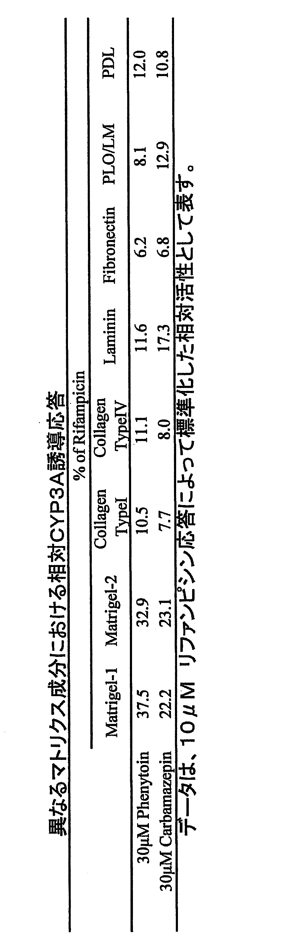
図2は、リファンピシン、フェニトイン、及びカルバマゼピンによるCYP3Aの誘導に対する異なるマトリクス条件の影響を示す。CYP3A誘導応答を、薄層マトリゲル(2ロット)、I型コラーゲン、IV型コラーゲン、ラミニン、及びフィブロネクチン等のバイオマテリアルからの細胞外マトリクスの成分;並びにポリ-L-オルニチン/ラミニン(PLO/LM)、又はポリ-D-リジン(PDL)等の培養基質上への細胞接着を促進する合成化合物、を含む、異なる種類の基質をもつ96穴プレートにおいて培養された肝細胞間で比較した。マトリゲルプレートにおける平均基礎CYP3A活性は、他のプレートにおける活性と比較して、3.6倍~10倍高かった。誘導されたCYP3A活性もまた、これらのプレートの中で、マトリゲルプレートにおいて最も高かった。10μM リファンピシン応答に対して標準化したCYP3A相対活性も、肝細胞をフェニトイン及びカルバマゼピンで刺激した場合に、前記したプレートの中で、マトリゲルコートプレートにおいて最も高かった(表3)。これらの知見は、試験化合物が弱い誘導剤である場合にも、薄層マトリゲルでのコーティングが、CYP3A誘導能の信頼性ある評価に効果的であることを示す。
CYP3A酵素活性の誘導に対するプレートの基質の影響
CYP3A誘導アッセイにおける96穴プレートの有用性を評価するために、I型コラーゲンでコーティングした48穴若しくは96穴プレート、又は薄層マトリゲルでコーティングした96穴プレート上で培養した夫々のリファンピシン処理肝細胞間で、CYP3A活性を比較した(図1)。肝細胞を各プレートに蒔き、4日間で培養系を確立させ、次いでさらに3日間、0.1% DMSOの存在下(コントロール細胞)又は種々の濃度のリファンピシンの存在下でインキュベートした。CYP3A活性を、テストステロンの6β-ヒドロキシル化の測定によって決定した。リファンピシンによる用量依存的なCYP3A誘導が、各プレート上で培養された全ての肝細胞において観察されたが、CYP3A活性のレベルは、培養条件によって異なった。48穴、96穴のI型コラーゲンプレート、及び96穴のマトリゲルプレートにおけるコントロール活性はそれぞれ80、50、及び153pmol/h/106細胞であり、それらのプレートにおいて10μM リファンピシンで誘導されたCYP3A活性はそれぞれ2243、1200、及び3538pmol/h/106細胞であった。96穴マトリゲルプレートにおける基礎及び誘導されたCYP3A活性は、48又は96穴I型コラーゲンプレートにおける活性よりも有意に高かった。
図2は、リファンピシン、フェニトイン、及びカルバマゼピンによるCYP3Aの誘導に対する異なるマトリクス条件の影響を示す。CYP3A誘導応答を、薄層マトリゲル(2ロット)、I型コラーゲン、IV型コラーゲン、ラミニン、及びフィブロネクチン等のバイオマテリアルからの細胞外マトリクスの成分;並びにポリ-L-オルニチン/ラミニン(PLO/LM)、又はポリ-D-リジン(PDL)等の培養基質上への細胞接着を促進する合成化合物、を含む、異なる種類の基質をもつ96穴プレートにおいて培養された肝細胞間で比較した。マトリゲルプレートにおける平均基礎CYP3A活性は、他のプレートにおける活性と比較して、3.6倍~10倍高かった。誘導されたCYP3A活性もまた、これらのプレートの中で、マトリゲルプレートにおいて最も高かった。10μM リファンピシン応答に対して標準化したCYP3A相対活性も、肝細胞をフェニトイン及びカルバマゼピンで刺激した場合に、前記したプレートの中で、マトリゲルコートプレートにおいて最も高かった(表3)。これらの知見は、試験化合物が弱い誘導剤である場合にも、薄層マトリゲルでのコーティングが、CYP3A誘導能の信頼性ある評価に効果的であることを示す。
種々の誘導剤及び非誘導剤で処理した凍結初代ヒト肝細胞におけるCYP3Aの誘導応答
本発明の評価方法を検証するため、N.J. Hewitt, et al.; Chem. Biol. Interact. 168, 51-65 (2007)で報告された新鮮又は凍結初代ヒト肝細胞におけるCYP3A誘導能にしたがって、強力な誘導剤、中程度の誘導剤又は非誘導剤に分類された種々の化合物で細胞を処理した。非誘導剤として用いた化合物は、セレコキシブ、シメチジン、シタロプラム、ファモチジン、フェキソフェナジン、フルボキサミン、ロキソプロフェン、ミダゾラム、及びリスペリドンであった。中程度の誘導剤として用いた化合物は、ゲムフィブロジル、ランソプラゾール、モダフィニル、ネビラピン、オメプラゾール、フェノバルビタール、フェニトイン、ロフェコキシブ、スルフィンピラゾン、及びトピラメートであった。強力な誘導剤として用いた化合物は、アバシミベ、カルバマゼピン、エファビレンツ、ニフェジピン、リファンピシン、及びトログリタゾンであった。各化合物を0.1μM、1μM、及び10μMで検査した。凍結初代肝細胞を3日間処理し、評価項目としてCYP3A活性を測定した。CYP3A誘導能を、10μM リファンピシン応答の割合として表す。0.1μMの濃度では、リファンピシン及びアバシミベを含むわずかな化合物のみが、CYP3A活性を誘導した(図3)。これらの化合物は、1μMでは、CYP3A活性の著しい増加を示した。ニフェジピン、エファビレンツ、及びトログリタゾンもまた、1μMでCYP3A活性を誘導した。10μMの濃度では、全ての強力な誘導剤及び大部分の中程度の誘導剤が、CYP3A活性の増加をもたらした。これに対し、セレコキシブを除く全ての非誘導剤が、10μMでCYP3A誘導をもたらさなかった。
本発明の評価方法を検証するため、N.J. Hewitt, et al.; Chem. Biol. Interact. 168, 51-65 (2007)で報告された新鮮又は凍結初代ヒト肝細胞におけるCYP3A誘導能にしたがって、強力な誘導剤、中程度の誘導剤又は非誘導剤に分類された種々の化合物で細胞を処理した。非誘導剤として用いた化合物は、セレコキシブ、シメチジン、シタロプラム、ファモチジン、フェキソフェナジン、フルボキサミン、ロキソプロフェン、ミダゾラム、及びリスペリドンであった。中程度の誘導剤として用いた化合物は、ゲムフィブロジル、ランソプラゾール、モダフィニル、ネビラピン、オメプラゾール、フェノバルビタール、フェニトイン、ロフェコキシブ、スルフィンピラゾン、及びトピラメートであった。強力な誘導剤として用いた化合物は、アバシミベ、カルバマゼピン、エファビレンツ、ニフェジピン、リファンピシン、及びトログリタゾンであった。各化合物を0.1μM、1μM、及び10μMで検査した。凍結初代肝細胞を3日間処理し、評価項目としてCYP3A活性を測定した。CYP3A誘導能を、10μM リファンピシン応答の割合として表す。0.1μMの濃度では、リファンピシン及びアバシミベを含むわずかな化合物のみが、CYP3A活性を誘導した(図3)。これらの化合物は、1μMでは、CYP3A活性の著しい増加を示した。ニフェジピン、エファビレンツ、及びトログリタゾンもまた、1μMでCYP3A活性を誘導した。10μMの濃度では、全ての強力な誘導剤及び大部分の中程度の誘導剤が、CYP3A活性の増加をもたらした。これに対し、セレコキシブを除く全ての非誘導剤が、10μMでCYP3A誘導をもたらさなかった。
リファンピシン誘導性のCYP3A活性に対する化合物の阻害作用の評価の有用性
本発明の評価方法を用いて、凍結初代ヒト肝細胞におけるさらなる化合物のCYP3A誘導の濃度-応答の研究を行った。試験化合物のうち、フェノバルビタール及びカルバマゼピンは、CYP3A活性の標準的な濃度依存性のアップレギュレーションを示した(図4A)。これに対し、アバシミベ及びエファビレンツは、ベル型の濃度-応答曲線を示した(図4B)。これらの化合物においては、CYP3A活性によって誘導能をスクリーニングする場合に、CYP3A誘導を過小評価するリスクがある。そのような過小評価を防ぐために、本発明の評価方法では、アッセイのコントロールとして、リファンピシン誘導性のCYP3A活性に対する試験化合物の阻害作用を評価した。リファンピシンと、いくつかの濃度の試験化合物の存在下肝細胞を3日間培養し、誘導されたCYP3A活性を決定した。標準的な濃度依存性を示した化合物の場合、リファンピシン誘導性のCYP3A活性は、試験化合物の全濃度範囲において、変化しなかった(図4A)。しかし、化合物が非定型のベル型の濃度-応答曲線を示した場合、リファンピシンによって誘導されたCYP3A活性は、より高い化合物濃度においては減弱し、それと共に、試験化合物による誘導も減少した(図4B)。
リトナビルは、周知のCYP3Aの誘導剤かつ機序に基づく阻害剤であるが(E.L. LeCluyse, et al.; Eur. J. Pharm. Sci. 13, 343-368 (2001))、リトナビルのCYP3A誘導能は、CYP3A活性の測定によっては検出できず、リファンピシン存在下ではリトナビル濃度が増加するとともにリファンピシン誘導性のCYP3A活性が減弱した(図5A)。しかし、同一の肝細胞におけるCYP3A4 mRNA量の測定によっては、リトナビルの濃度依存的なCYP3A4 mRNA量の増加によってリトナビルのCYP3A4誘導能が確認された(図5B)。リトナビルと共培養したリファンピシンによって誘導されたCYP3A4 mRNA量は、高リトナビル濃度においても減弱しなかった。
これらの結果により、本発明の評価方法は、CYP3A誘導能の過小評価を防ぐことを可能とすることが示された。
本発明の評価方法を用いて、凍結初代ヒト肝細胞におけるさらなる化合物のCYP3A誘導の濃度-応答の研究を行った。試験化合物のうち、フェノバルビタール及びカルバマゼピンは、CYP3A活性の標準的な濃度依存性のアップレギュレーションを示した(図4A)。これに対し、アバシミベ及びエファビレンツは、ベル型の濃度-応答曲線を示した(図4B)。これらの化合物においては、CYP3A活性によって誘導能をスクリーニングする場合に、CYP3A誘導を過小評価するリスクがある。そのような過小評価を防ぐために、本発明の評価方法では、アッセイのコントロールとして、リファンピシン誘導性のCYP3A活性に対する試験化合物の阻害作用を評価した。リファンピシンと、いくつかの濃度の試験化合物の存在下肝細胞を3日間培養し、誘導されたCYP3A活性を決定した。標準的な濃度依存性を示した化合物の場合、リファンピシン誘導性のCYP3A活性は、試験化合物の全濃度範囲において、変化しなかった(図4A)。しかし、化合物が非定型のベル型の濃度-応答曲線を示した場合、リファンピシンによって誘導されたCYP3A活性は、より高い化合物濃度においては減弱し、それと共に、試験化合物による誘導も減少した(図4B)。
リトナビルは、周知のCYP3Aの誘導剤かつ機序に基づく阻害剤であるが(E.L. LeCluyse, et al.; Eur. J. Pharm. Sci. 13, 343-368 (2001))、リトナビルのCYP3A誘導能は、CYP3A活性の測定によっては検出できず、リファンピシン存在下ではリトナビル濃度が増加するとともにリファンピシン誘導性のCYP3A活性が減弱した(図5A)。しかし、同一の肝細胞におけるCYP3A4 mRNA量の測定によっては、リトナビルの濃度依存的なCYP3A4 mRNA量の増加によってリトナビルのCYP3A4誘導能が確認された(図5B)。リトナビルと共培養したリファンピシンによって誘導されたCYP3A4 mRNA量は、高リトナビル濃度においても減弱しなかった。
これらの結果により、本発明の評価方法は、CYP3A誘導能の過小評価を防ぐことを可能とすることが示された。
誘導されたCYP3A活性とCYP3A4 mRNA量との間の相関を確認するために、44の化合物をいくつかの濃度で、10μM リファンピシンの存在下又は非存在下で検査した。図6は、試験化合物についての、誘導されたCYP3A活性とCYP3A4 mRNA量との間の相関プロットを示す。この図において、各記号は、試験化合物と共培養した場合の、リファンピシン誘導性CYP3A活性の残留率の程度を表す。試験化合物によって誘導されたCYP3A活性及びCYP3A4 mRNA量は、リファンピシン誘導性のCYP3A活性の残留率が80%を超える場合には、全て良く相関した。しかし、リファンピシン誘導性のCYP3A活性が30%未満に抑制された場合、試験化合物は潜在的に阻害作用を有し、試験化合物のCYP誘導能は、CYP3A活性によって検出されなかった一方で、CYP3A4 mRNAの発現は、化合物によって誘導された。
本発明の評価方法は、早期探索段階における医薬候補化合物の生体異物代謝酵素活性の誘導能評価において、試験化合物が細胞傷害性又は機序に基づく阻害作用等を有する場合に起こる誘導能の過小評価を防ぐことを可能とし、特に真に誘導能のない化合物を探索するために有用である。
本出願は、2009年8月31日付で日本国に出願された特願2009-201043を基礎としており、その内容は全て本明細書に包含される。
Claims (13)
- 以下の工程:
(a)培養肝細胞を含む試料を二分し、一方を生体異物代謝酵素活性の誘導物質の存在下、試験化合物で処理し、他方を生体異物代謝酵素活性の誘導物質の非存在下、試験化合物で処理する工程、
(b)工程(a)で得られた各試料中に含まれる該生体異物代謝酵素の活性を測定する工程、及び
(c)工程(b)で測定された各酵素活性を比較し解析する工程
を含むことを特徴とする、試験化合物の生体異物代謝酵素誘導能の評価方法。 - 工程(a)における処理が、薄層マトリゲルでコーティングされた培養器上で行われることを特徴とする、請求項1記載の方法。
- 培養器が96穴プレートである、請求項2記載の方法。
- 工程(a)における処理が、2以上の試験化合物濃度において行われる、請求項1記載の方法。
- 生体異物代謝酵素が、シトクロムP450である、請求項1記載の方法。
- シトクロムP450が、CYP3Aである、請求項5記載の方法。
- シトクロムP450がCYP3A4である、請求項5記載の方法。
- 生体異物代謝酵素活性の誘導物質が、リファンピシン、ニフェジピン、エファビレンツ、カルバマゼピン、アバシミベ、トログリタゾン、ゲムフィブロジル、ランソプラゾール、モダフィニル、ネビラピン、オメプラゾール、フェノバルビタール、フェニトイン、ロフェコキシブ、スルフィンピラゾン、又はトピラメートである、請求項1記載の方法。
- 生体異物代謝酵素活性の誘導物質が、リファンピシンである、請求項1記載の方法。
- 培養肝細胞が哺乳動物由来肝細胞である、請求項1記載の方法。
- 培養肝細胞がヒト肝細胞である、請求項1記載の方法。
- 培養肝細胞が凍結初代ヒト肝細胞である、請求項1記載の方法。
- 培養肝細胞における該生体異物代謝酵素の遺伝子発現を測定する工程をさらに含む、請求項1記載の方法。
Applications Claiming Priority (2)
| Application Number | Priority Date | Filing Date | Title |
|---|---|---|---|
| JP2009-201043 | 2009-08-31 | ||
| JP2009201043 | 2009-08-31 |
Publications (1)
| Publication Number | Publication Date |
|---|---|
| WO2011025014A1 true WO2011025014A1 (ja) | 2011-03-03 |
Family
ID=43628103
Family Applications (1)
| Application Number | Title | Priority Date | Filing Date |
|---|---|---|---|
| PCT/JP2010/064747 Ceased WO2011025014A1 (ja) | 2009-08-31 | 2010-08-30 | 薬物の安全性の評価方法 |
Country Status (1)
| Country | Link |
|---|---|
| WO (1) | WO2011025014A1 (ja) |
-
2010
- 2010-08-30 WO PCT/JP2010/064747 patent/WO2011025014A1/ja not_active Ceased
Non-Patent Citations (2)
| Title |
|---|
| YASUHIRO YAMADA: "Soyaku ni Okeru Hito Kansaibo no Hitsuyosei to Shihan Hito Kansaibo no Mondaiten", ORGAN BIOLOGY, vol. 13, no. 2, 2006, pages 119 - 135 * |
| YOKO EJIRI ET AL: "Iyakuhin Kaihatsu no Tameno Shinki Hito Kansaibo Baiyokei no Kochiku", HAB KENKYU KIKO GAKUJUTSU NENKAI PROGRAM YOSHISHU, vol. 16, 22 May 2009 (2009-05-22), pages 63, XP008178621 * |
Similar Documents
| Publication | Publication Date | Title |
|---|---|---|
| Gebhardt et al. | New hepatocyte in vitro systems for drug metabolism: metabolic capacity and recommendations for application in basic research and drug development, standard operation procedures | |
| Graaf et al. | Precision-cut tissue slices as a tool to predict metabolism of novel drugs | |
| LeCluyse et al. | Cultured rat hepatocytes | |
| Josse et al. | Long-term functional stability of human HepaRG hepatocytes and use for chronic toxicity and genotoxicity studies | |
| Lübberstedt et al. | HepaRG human hepatic cell line utility as a surrogate for primary human hepatocytes in drug metabolism assessment in vitro | |
| CN105296418B (zh) | 一种长期体外培养和扩增肝细胞的方法及其应用 | |
| Roymans et al. | Determination of cytochrome P450 1A2 and cytochrome P450 3A4 induction in cryopreserved human hepatocytes | |
| Vermeir et al. | Cell-based models to study hepatic drug metabolism and enzyme induction in humans | |
| Olsavsky Goyak et al. | Hepatocyte differentiation | |
| WO2015108944A9 (en) | Stem cell-derived hepatocytes in co-culture and uses thereof | |
| Bonifas et al. | Evaluation of cytochrome P450 1 (CYP1) and N-acetyltransferase 1 (NAT1) activities in HaCaT cells: implications for the development of in vitro techniques for predictive testing of contact sensitizers | |
| Weaver et al. | The morphology, functionality, and longevity of a novel all human hepatic cell-based tri-culture system | |
| Wang et al. | Strain differences between CD-1 and C57BL/6 mice in expression of metabolic enzymes and DNA methylation modifications of the primary hepatocytes | |
| EP3365429B1 (en) | Production of virus-receptive pluripotent stem cell (psc)-derived hepatocytes | |
| Washizu et al. | Long-term maintenance of cytochrome P450 activities by rat hepatocyte/3T3 cell co-cultures in heparinized human plasma | |
| van der Mark et al. | Overexpression of the constitutive androstane receptor and shaken 3D-culturing increase biotransformation and oxidative phosphorylation and sensitivity to mitochondrial amiodarone toxicity of HepaRG cells | |
| Jang et al. | Liver-Chip: Reproducing human and cross-species toxicities | |
| AU2003261303B2 (en) | Methods for perfusion and plating of primary hepatocytes and a medium therefore | |
| Li et al. | Keratinocyte serum-free medium maintains long-term liver gene expression and function in cultured rat hepatocytes by preventing the loss of liver-enriched transcription factors | |
| WO2011025014A1 (ja) | 薬物の安全性の評価方法 | |
| Donato et al. | Liver grafts preserved in Celsior solution as source of hepatocytes for drug metabolism studies: comparison with surgical liver biopsies | |
| Gómez-Lechón et al. | Evaluation of cytochrome P450 activities in human hepatocytes in vitro | |
| EP1356286A2 (en) | Method for estimating metabolic function of xenobiotic and induction thereof | |
| Hansen et al. | Primary rat alveolar epithelial cells for use in biotransformation and toxicity studies | |
| De Kock et al. | A liver-derived rat epithelial cell line from biliary origin acquires hepatic functions upon sequential exposure to hepatogenic growth factors and cytokines |
Legal Events
| Date | Code | Title | Description |
|---|---|---|---|
| 121 | Ep: the epo has been informed by wipo that ep was designated in this application |
Ref document number: 10812060 Country of ref document: EP Kind code of ref document: A1 |
|
| NENP | Non-entry into the national phase |
Ref country code: DE |
|
| 122 | Ep: pct application non-entry in european phase |
Ref document number: 10812060 Country of ref document: EP Kind code of ref document: A1 |
|
| NENP | Non-entry into the national phase |
Ref country code: JP |