WO2010141955A2 - Methods of detecting cancer - Google Patents
Methods of detecting cancer Download PDFInfo
- Publication number
- WO2010141955A2 WO2010141955A2 PCT/US2010/037659 US2010037659W WO2010141955A2 WO 2010141955 A2 WO2010141955 A2 WO 2010141955A2 US 2010037659 W US2010037659 W US 2010037659W WO 2010141955 A2 WO2010141955 A2 WO 2010141955A2
- Authority
- WO
- WIPO (PCT)
- Prior art keywords
- cancer
- genes
- patient
- panel
- mutation
- Prior art date
- Legal status (The legal status is an assumption and is not a legal conclusion. Google has not performed a legal analysis and makes no representation as to the accuracy of the status listed.)
- Ceased
Links
Classifications
-
- C—CHEMISTRY; METALLURGY
- C12—BIOCHEMISTRY; BEER; SPIRITS; WINE; VINEGAR; MICROBIOLOGY; ENZYMOLOGY; MUTATION OR GENETIC ENGINEERING
- C12Q—MEASURING OR TESTING PROCESSES INVOLVING ENZYMES, NUCLEIC ACIDS OR MICROORGANISMS; COMPOSITIONS OR TEST PAPERS THEREFOR; PROCESSES OF PREPARING SUCH COMPOSITIONS; CONDITION-RESPONSIVE CONTROL IN MICROBIOLOGICAL OR ENZYMOLOGICAL PROCESSES
- C12Q1/00—Measuring or testing processes involving enzymes, nucleic acids or microorganisms; Compositions therefor; Processes of preparing such compositions
- C12Q1/68—Measuring or testing processes involving enzymes, nucleic acids or microorganisms; Compositions therefor; Processes of preparing such compositions involving nucleic acids
- C12Q1/6876—Nucleic acid products used in the analysis of nucleic acids, e.g. primers or probes
- C12Q1/6883—Nucleic acid products used in the analysis of nucleic acids, e.g. primers or probes for diseases caused by alterations of genetic material
- C12Q1/6886—Nucleic acid products used in the analysis of nucleic acids, e.g. primers or probes for diseases caused by alterations of genetic material for cancer
-
- G—PHYSICS
- G16—INFORMATION AND COMMUNICATION TECHNOLOGY [ICT] SPECIALLY ADAPTED FOR SPECIFIC APPLICATION FIELDS
- G16B—BIOINFORMATICS, i.e. INFORMATION AND COMMUNICATION TECHNOLOGY [ICT] SPECIALLY ADAPTED FOR GENETIC OR PROTEIN-RELATED DATA PROCESSING IN COMPUTATIONAL MOLECULAR BIOLOGY
- G16B25/00—ICT specially adapted for hybridisation; ICT specially adapted for gene or protein expression
- G16B25/10—Gene or protein expression profiling; Expression-ratio estimation or normalisation
-
- C—CHEMISTRY; METALLURGY
- C12—BIOCHEMISTRY; BEER; SPIRITS; WINE; VINEGAR; MICROBIOLOGY; ENZYMOLOGY; MUTATION OR GENETIC ENGINEERING
- C12Q—MEASURING OR TESTING PROCESSES INVOLVING ENZYMES, NUCLEIC ACIDS OR MICROORGANISMS; COMPOSITIONS OR TEST PAPERS THEREFOR; PROCESSES OF PREPARING SUCH COMPOSITIONS; CONDITION-RESPONSIVE CONTROL IN MICROBIOLOGICAL OR ENZYMOLOGICAL PROCESSES
- C12Q2600/00—Oligonucleotides characterized by their use
- C12Q2600/112—Disease subtyping, staging or classification
-
- C—CHEMISTRY; METALLURGY
- C12—BIOCHEMISTRY; BEER; SPIRITS; WINE; VINEGAR; MICROBIOLOGY; ENZYMOLOGY; MUTATION OR GENETIC ENGINEERING
- C12Q—MEASURING OR TESTING PROCESSES INVOLVING ENZYMES, NUCLEIC ACIDS OR MICROORGANISMS; COMPOSITIONS OR TEST PAPERS THEREFOR; PROCESSES OF PREPARING SUCH COMPOSITIONS; CONDITION-RESPONSIVE CONTROL IN MICROBIOLOGICAL OR ENZYMOLOGICAL PROCESSES
- C12Q2600/00—Oligonucleotides characterized by their use
- C12Q2600/156—Polymorphic or mutational markers
-
- C—CHEMISTRY; METALLURGY
- C12—BIOCHEMISTRY; BEER; SPIRITS; WINE; VINEGAR; MICROBIOLOGY; ENZYMOLOGY; MUTATION OR GENETIC ENGINEERING
- C12Q—MEASURING OR TESTING PROCESSES INVOLVING ENZYMES, NUCLEIC ACIDS OR MICROORGANISMS; COMPOSITIONS OR TEST PAPERS THEREFOR; PROCESSES OF PREPARING SUCH COMPOSITIONS; CONDITION-RESPONSIVE CONTROL IN MICROBIOLOGICAL OR ENZYMOLOGICAL PROCESSES
- C12Q2600/00—Oligonucleotides characterized by their use
- C12Q2600/16—Primer sets for multiplex assays
-
- G—PHYSICS
- G16—INFORMATION AND COMMUNICATION TECHNOLOGY [ICT] SPECIALLY ADAPTED FOR SPECIFIC APPLICATION FIELDS
- G16B—BIOINFORMATICS, i.e. INFORMATION AND COMMUNICATION TECHNOLOGY [ICT] SPECIALLY ADAPTED FOR GENETIC OR PROTEIN-RELATED DATA PROCESSING IN COMPUTATIONAL MOLECULAR BIOLOGY
- G16B25/00—ICT specially adapted for hybridisation; ICT specially adapted for gene or protein expression
Definitions
- the invention generally relates to a molecular classification of disease and particularly to molecular markers for cancer and methods of use thereof.
- Cancer is a major health challenge. Nearly 560,000 people die from cancer annually in the United States alone, representing almost 23% of all deaths. Despite recent advances in molecular and imaging diagnostics, one of the most vexing aspects of cancer remains early detection. In fact, for certain types of cancer — e.g. , pancreatic adenocarcinoma — detection often occurs so late as to practically preclude any good prognosis. Thus there is an urgent need for sensitive methods of detecting cancer.
- Mutations in certain genes are associated with cancer in general and with specific cancer types. For example, inactivating mutations in the TP53 gene are found in approximately 50% of all solid tumors and activating mutations in the KRAS or BRAF genes are often found in colorectal cancer. It has been discovered that screening patients for mutations in certain genes can detect and classify cancer. More specifically, it has been determined that (a) screening certain genes (e.g. , APC, EGFR, KRAS, PTEN, and TP 53) for mutations will detect nearly 95% of all cancers, while (b) screening certain genes (e.g.
- AIMl , APC, CDKN2A , EGFR, FBN2, FBXW7, FLJl 3479, IDHl , KRAS, PIK3CA , PIK3R1 , PTEN, RBl , SMAD4, TGFBR2, TNN, and TP 53) for mutations can accurately classify the cancer (e.g. , as breast cancer, colon cancer, glioblastoma, pancreatic cancer, etc.).
- the invention generally provides methods comprising analyzing panels of genes from a sample obtained from a patient (e.g. , mRNA or cDNA synthesized therefrom) and determining the mutational status of the genes in the panel, wherein the presence of a particular mutational status in particular genes in the panel indicates (a) the patient has cancer and/or (b) the patient has a particular cancer.
- a patient e.g. , mRNA or cDNA synthesized therefrom
- One aspect of the invention provides a method of detecting mutations comprising: ( 1 ) analyzing in a bodily fluid sample from a human subject a panel of genes consisting of between 5 and 5 ,000 genes, wherein said panel comprises at least five genes chosen from the group consisting of the genes listed in Table 1 ; and (2) determining whether any of the genes in Table 1 harbors a mutation.
- the panel comprises the APC, EGFR,
- the panel comprises the genes listed in Table 3. In some embodiments the panel comprises the genes listed in Table 2. In some embodiments the panel comprises the genes listed in Table 1 .
- One aspect of the invention provides a method of detecting cancer comprising : ( 1 ) analyzing a panel of genes comprising the APC, EGFR, KRAS, PTEN, and TP53 genes in a bodily fluid sample; and (2) determining whether any of the APC, EGFR, KRAS, PTEN, or TP53 genes harbors a mutation; wherein said mutation indicates the presence of cancer.
- the panel comprises the genes listed in
- the panel comprises the genes listed in Table 2. In some embodiments the panel comprises the genes listed in Table 1 . In some embodiments the mutation is selected from the group consisting of those listed in Table 7 and/or Table 8.
- Some embodiments further comprise recommending, prescribing, ordering, or performing a test for the presence of cancer C 1 in said patient.
- the test for the presence of cancer C 1 is recommended, prescribed, ordered, or performed if the calculated likelihood said patient has said cancer C 1 is above a threshold value (e.g. , 5%, 10%, 15%, 20%, 25%, 30%, 35%, 40%, 45%, 50%, 55 %, 60%, 65%, 70%, 75%, 80%, 85%, 90%, 95%, 96%, 97%, 98%, 99%, or 100%).
- a threshold value e.g. , 5%, 10%, 15%, 20%, 25%, 30%, 35%, 40%, 45%, 50%, 55 %, 60%, 65%, 70%, 75%, 80%, 85%, 90%, 95%, 96%, 97%, 98%, 99%, or 100%.
- the test for the presence of cancer C 1 is recommended, prescribed, ordered, or performed if the calculated likelihood said patient has said cancer C 1 is higher than the calculated likelihood said patient has cancer C 2 -
- the method further comprises recommending, prescribing, ordering, or performing a test for the presence of cancer C2 in said patient if said test for the presence of cancer a does not indicate the presence of cancer a .
- the said bodily fluid sample is a blood sample.
- the blood sample is a plasma sample.
- the blood sample is a serum sample.
- detecting a mutation or determining whether a gene harbors a mutation comprises analyzing an mRNA molecule from a sample or analyzing a DNA molecule synthesized using the mRNA molecule as a template. In some embodiments detecting a mutation or determining whether a gene harbors a mutation comprises analyzing a nucleic acid from a sample by a technique chosen from resequencing, TaqManTM, microarray analysis, and FISH.
- nucleic acids to be analyzed are derived from an extracellular vesicle.
- extracellular vesicle is an exosome.
- kits comprising reagents for analyzing a panel of genes consisting of between 5 and 5 ,000 genes, said kit comprising reagents for detecting mutations in at least five genes selected from the group consisting of the genes listed in Table 1 .
- the kit comprises reagents for detecting mutations in the APC, EGFR, KRAS, PTEN, and TP 53 genes. E3.
- the kit comprises reagents for detecting mutations in the genes listed in Table 3.
- the kit comprises reagents for detecting mutations in the genes listed in Table 2.
- the kit comprises reagents for detecting mutations in the genes listed in Table 1.
- FIG. l illustrates the sensitivity of a panel of five genes for detecting cancer.
- FIG.2 illustrates one embodiment of the invention using various biomarkers to determine which specific cancer is present in a patient.
- FIG.3 illustrates example mutation frequencies in various cancers.
- FIG.4 illustrates example cancer rates based on cancer site and gender.
- FIG.5 shows the detection of mutations in exosomes from cancer serum samples.
- Mutations in certain genes are associated with cancer in general and with specific cancer types. For example, inactivating mutations in the TP53 gene are found in approximately 50% of all solid tumors and activating mutations in the KRAS or BRAF genes are often found in colorectal cancer.
- the invention is based in part on the discovery that analyzing patient samples for mutations in a relatively small number of genes can (a) detect the vast majority of cancers and (b) specify in which tissue the cancer is located. More specifically, it has been determined that (a) screening certain genes (e.g. , the genes listed in Table 4 below) for mutations will detect cancer (e.g. , nearly 95% of all cancers), while (b) screening certain genes (e.g. , the genes listed in Tables 2 & 3 below) for mutations can accurately classify the cancer (e.g. , as breast cancer, colon cancer, glioblastoma, pancreatic cancer, etc.).
- the invention provides a method of detecting mutations comprising ( 1 ) analyzing a panel of genes consisting of between 5 and 5 ,000 genes in a bodily fluid sample from a human subject, wherein said panel comprises at least five genes chosen from the group consisting of the genes listed in Table 1 ; and (2) determining whether at least one of said five genes harbors a mutation.
- the panel consists of between 5 and
- genes chosen from Table 1 comprise at least 5%, 10%, 15%, 20%, 25%, 30%, 35%, 40%, 45%, 50%, 55%, 60%, 65%, 70%, 75%, 80%, 85 %, 90%, 95%, 96%, 97%, 98%, 99% or 100% of the panel.
- one aspect of the invention provides a method of detecting cancer comprising: ( 1 ) analyzing a panel of genes in a bodily fluid sample from a human subj ect, wherein said panel comprises at least five genes chosen from the group consisting of the genes listed in Table 1 ; and (2) determining whether at least one of said five genes harbors a mutation; wherein said mutation indicates the presence of cancer.
- the mutation is chosen from those listed in Table 7 and/or Table 8.
- the panel consists of between 5 and 4,500, between 5 and 4,000, between 5 and 3 ,500, between 5 and 3 ,000, between 5 and 2,500, between 5 and 2,000, between 5 and 1 ,500, between 5 and 1 ,000, between 5 and 500, between 5 and 400, between 5 and 300, between 5 and 200, between 5 and 150, between 5 and 100, between 5 and 75 , or between 5 and 50 genes.
- the genes chosen from Table 1 comprise at least 5%, 10%, 15 %, 20%, 25%, 30%, 35 %, 40%, 45%, 50%, 55%, 60%, 65%, 70%, 75%, 80%, 85%, 90%, 95 %, 96%, 97%, 98%, 99% or 100% of the panel.
- M(g ⁇ c) is the frequency of somatic mutations in gene g in cancer type c. See, e.g. , FIG.3.
- Po(c) is the a priori probability of cancer type c given that the patient has a cancer. See FIG.4.
- one aspect of the invention provides a method of determining the likelihood a patient has a particular cancer a comprising:
- the " ⁇ priori probability of cancer c given that the patient has a cancer” refers to the general incidence of the particular cancer c in the relevant cancer patient population ⁇ e.g. , males or females). In other words, this is the relative proportion of all cancers in the relevant population represented by the particular cancer c. Such incidences may be gathered from various sources — e.g. , yearly American Cancer Society reports on cancer incidence (as in Example 1 , infra), which often give detailed breakdowns of specific cancer incidence in relevant patient subpopulations such as male vs. female, race or ethnicity, etc.
- the patient has a particular cancer a only if the calculated likelihood said patient has said cancer Ci is above a threshold value.
- This threshold value may be arbitrarily chosen (e.g. , 95% probability is good enough) or determined empirically (e.g. , patients with a calculated probability above 80% have ended up with the particular cancer with enough frequency to validate this as a good threshold).
- said threshold value is chosen from the group consisting of 35%, 40%, 45%, 50%, 55%, 60%, 65%, 70%, 75 %, 80%, 85%, 90%, 95%, 96%, 97%, 98%, and 99%.
- Equation (2) the probabilities are calculated for each organ o, and the organ with the highest probability is the most likely cancer site in the patient. The patient may then optionally be examined by additional diagnostic techniques to confirm cancer site. If the most likely cancer site is not confirmed, the organ with the second highest probability may then be examined and so on.
- one aspect of the invention provides a method of diagnosing cancer in a particular organ oi comprising:
- P( ⁇ ⁇ gl, g2, ... ,gn ) ⁇ c P(c ⁇ gj, g 2 , ... , gn ) wherein the sum is taken over all cancer types c of the organ oj, and P(c) is calculated using the formula:
- Another aspect of the invention provides a method of determining the likelihood a patient has a particular cancer Ci comprising:
- the method further comprises concluding the patient has ci if P(C 1 ) is higher than P(c 2 ) , P(c 3 ) , P(c 4 ) , ... , P(c x ) , where P(c 2 ) through P(c x ) represent the calculated probabilities of each cancer ⁇ e.g. , major cancers such as those listed in Tables 3 & 4) other than a .
- one aspect of the invention provides a method of diagnosing cancer comprising:
- screening the five genes in Table 4 can detect nearly 95 % of solid tumor types and the genes in Tables 2 & 3 can classify the cancer.
- the presence of a mutation in any one of the genes listed in Table 4 is used as a pan-cancer screen to determine for which patients additional analysis should be done on a panel comprising at least one of the genes listed in Table 2 or 3.
- a mutation in any one of the genes in the first panel indicates the patient has cancer and application of the second panel classifies which type.
- somatic mutations are the most informative mutations (e.g. , as in Example 1 ).
- Calculating a patient' s likelihood of having a particular cancer can be useful in various clinical settings. For example, if the calculated probability of the patient having a particular cancer is high enough one may diagnose the particular cancer, prescribe a treatment for the specific cancer, etc. If the patient is at particularly high risk of a specific cancer (e.g. , BRCA mutation carrier), then even a lower calculated likelihood of breast or ovarian cancer might be sufficient to make a diagnosis. A high likelihood of a particular cancer may alternatively prompt the doctor to recommend, prescribe, order, or perform an additional test (e.g. , biopsy, MRI, CT scan, digital rectal exam, mammography, etc.) to confirm the cancer.
- an additional test e.g. , biopsy, MRI, CT scan, digital rectal exam, mammography, etc.
- some embodiments further comprise recommending, prescribing, ordering, or performing a test to confirm the presence of cancer a .
- the test is prescribed, ordered, recommended, or performed if the calculated likelihood exceeds some threshold value.
- some embodiments further comprise recommending, prescribing, ordering, or performing a test to confirm the presence of cancer c; in said patient if the calculated likelihood said patient has said cancer a is higher than the calculated likelihood said patient has cancer C2.
- the test is prescribed, ordered, recommended, or performed if the calculated likelihood of Ci exceeds that of C 2 and also exceeds some threshold value.
- a "panel of genes” is a plurality of genes.
- the panel consists of between 2 and 500, between 3 and 500, between 4 and 500, between 5 and 500, between 6 and 500, between 7 and 500, between 8 and 500, between 9 and 500, between 10 and 500, between 1 1 and 500, between 12 and 500, between 13 and 500, between 14 and 500, between 15 and 500, between 16 and 500, between 17 and 500, between 18 and 500, between 19 and 500, between 20 and 500, between 25 and 500, between 30 and 500, between 35 and 500, between 40 and 500, between 45 and 500, between 50 and 500, between 55 and 500, between 60 and 500, between 65 and 500, between 70 and 500, between 75 and 500, between 80 and 500, between 85 and 500, between 90 and 500, between 95 and 500, between 100 and 500, between 2 and 400, between 2 and 350, between 2 and 300, between 2 and 250, between 2 and 200, between 2 and 150, between 2 and 100, between 2 and 90, between 2 and 80, between between
- the panel comprises genes listed in Table 1 below:
- the panel comprises subsets (e.g. , at least 2, 3 , 4, 5 , 6, 7, 8, 9, 10, 15 , 20, 25 , 30, 35 , 40 or more) of the genes in Table 1.
- the panel comprises APC, EGFR, KRAS, PTEN, and TP 53.
- the panel comprises AIMl , APC, CDKN2A , EGFR, F BN 2, F B XW 7, FLJ 13479, IDHl , KRAS, PIK3CA , PIK3R1 , PTEN, RBl , SMAD4, TGFBR2, TNN, and TP53.
- the panel comprises APC, A TM, BRAF, BRCA l , BRCA2, CDKN2A, CTNNBl , EGFR, FBXW7, FGFR3, KIT, KRAS, HRAS, NRAS, MAP2K4, MET, MLHl , MSH2, MSH6, NFl , NF2, PIK3CA , PRKDC, PTEN, RBl , RET, SMAD4, SMO, STKI l , TAFlL, TP53, TRRAP, and VHL.
- the panel comprises the genes listed in Table 4.
- the panel comprises the genes listed in Table 3.
- the panel comprises the genes listed in Table 1.
- Mutations useful in the methods of the invention include missense mutations, deletions, insertions, frameshifts, copy number variations, and loss of heterozygosity.
- Deleterious mutations i.e. , mutations that reduce or abolish gene and/or protein function
- tumor suppressors e.g. , APC, TP53, PTEN
- Activating mutations i.e. , mutations that increase gene and/or protein function
- oncogenes e.g. , KRAS, EGFR.
- Those skilled in the art are familiar with various deleterious and activating mutations for the genes listed in Tables 1 , 3 , and 4 (e.g.
- codons 12 and 13 in KRAS codons 12 and 13 in KRAS
- Skilled artisans are also familiar with various techniques for determining whether a particular mutation is in fact deleterious or activating. For example, frameshift mutations resulting in early truncation of a tumor suppressor gene are generally expected to be deleterious.
- Table 7 includes mutations found in some of the genes listed in Table 1 . Those skilled in the art are familiar with various resources and databases cataloguing mutations in the genes listed in Table 1. For example, the COSMIC [Catalogue of Somatic Mutations in Cancer] database currently contains over 26,000 entries for these genes. Those skilled in the art will be able to use these entries in the methods of the invention for detecting and classifying cancer.
- determining the "mutational status" of a gene means determining at least one of the following: (a) whether the gene (or any of its products) harbors a sequence mutation (including point mutations, deletions, insertions, copy number variants, etc.), (b) the prevalence of such mutations in a sample, or (c) whether such a sequence mutation is activating or inactivating.
- a particular mutational status includes, but is not limited to, the presence or absence of a mutation, a relatively high or relatively low prevalence of a mutation, an inactivating mutation, an activating mutation, etc.
- determining the mutational status of a gene comprises assaying some marker whose status itself is correlated with the mutational status of the gene of interest. Determining the mutational status of a panel of genes means determining the mutational status of each gene in the panel.
- Mutational status of a gene may be determined by any of several techniques familiar to those skilled in the art. Exemplary techniques include resequencing (either of selected regions of the gene or of the entire gene), allele-specific amplification (e.g. , TaqManTM using mutant allele-specific probes), microarray analysis (e.g. , arrays for CNV or arrays containing mutant allele-specific probes), etc.
- the method comprises physically amplifying and/or isolating nucleic acid of a panel of genes from a sample obtained from a patient. As used herein, "amplifying a nucleic acid” and “isolating nucleic acid” have their conventional meanings in the art.
- the method further comprises amplifying nucleic acid of a panel of genes (e.g. , comprising the genes listed in Table 3) from a sample obtained from a patient, determining the mutational status of each gene in the panel, and calculating the likelihood of a particular cancer as discussed above and below.
- a panel of genes e.g. , comprising the genes listed in Table 3
- sample refers to any biological specimen, including any tissue or fluid, that can be obtained from, or derived from a specimen obtained from, a human subject.
- samples include but are not limited to healthy or tumor tissue, bodily fluids (e.g. , blood), waste matter (e.g. , urine, stool), etc.
- Bodily fluid sample as used herein means any fluid that can be extracted or collected from a human body.
- the bodily fluid sample is blood or a blood derivative. Examples of blood derivatives include, but are not limited to, plasma and serum.
- the bodily fluid sample is urine, stool, pleural effusion, lacrimal effusion, saliva, sputum, etc.
- analyzing genes in a sample refers to analyzing nucleic acids corresponding to those genes in a sample or any substance derived from that sample.
- analyzing the APC, EGFR, KRAS, PTEN and TP53 genes in blood includes analyzing PCRTM amplified portions of these genes in a patient blood sample (including plasma or serum), or in DNA or RNA isolated (i.e. , derived) from such a sample.
- a nucleic acid is chosen from the group consisting of genomic DNA (including PCRTM amplified copies of genomic DNA), mRNA, cDNA, and a portion of any of these.
- the cancer screening and classification methods of the inventions will often involve analyzing nucleic acids from bodily fluids since these are often the least invasive samples to obtain from patients.
- the method of the invention may involve isolating nucleic acids from circulating tumor cells from the blood. This may involve capturing circulating tumor cells (e.g. , using tumor-specific capture antibodies) and subsequent analysis of the DNA or RNA contained in the cell.
- the methods of the invention may isolate and analyze nucleic acids that float freely in the bodily fluid.
- the methods of the invention may also isolate nucleic acids from extracellular vesicles found in the bodily fluid sample.
- cancer type and “type of cancer” mean a cancer in or originating from a particular tissue or organ and/or a cancer with a particular molecular or clinical feature. Often, the specificity of the "cancer type” varies with the application, including tissue type ⁇ e.g. , squamous versus cuboidal), organ type ⁇ e.g. , breast versus lung), and clinical subtype ⁇ e.g. , triple-negative breast cancer).
- Another aspect of the invention provides a method of classifying cancer comprising isolating nucleic acids corresponding to a panel of genes from a sample obtained from a patient and determining the mutational status of each such nucleic acid, wherein a particular mutational status in particular genes in the panel indicates the patient has a particular cancer.
- methods according to this aspect may simultaneously detect and classify cancer.
- the panel comprises the AIMl, APC, CDKN2A, EGFR, FBN2, FBXW7, FLJ13479, IDHl, KRAS, PIK3CA, PIK3R1, PTEN, RBl, SMAD4, TGFBR2, TNN, and TP53 genes or a subset ⁇ e.g., at least 3, 4, 5, 6, 7, 8, 9, 10 or 15 or more) thereof.
- the panel comprises the APC, ATM, BRAF, BRCAl, BRCA2, CDKN2A, CTNNBl, EGFR, FBXW7, FGFR3, KIT, KRAS, HRAS, NRAS, MAP2K4, MET, MLHl, MSH2, MSH6, NFl, NF2, PIK3CA, PRKDC, PTEN, RBl, RET, SMAD4, SMO, STKIl, TAFlL, TP 53, TRRAP, and VHL genes or a subset (e.g., at least 3, 4, 5, 6, 7, 8, 9, 10, 15, 20, 25, or 30 or more) thereof.
- a subset e.g., at least 3, 4, 5, 6, 7, 8, 9, 10, 15, 20, 25, or 30 or more
- the panel comprises the AIMl, APC, ATM, BRAF, BRCAl, BRCA2, CDKN2A, CD95, CTNNBl, EGFR, FBN2, FBXW7, FLJ 13479, FGFR3, IDHl, KIT, KRAS, HRAS, NRAS, MAP2K4, MET, MLHl, MLH 2, MSHl, M SH 2, NFl, NF 2, PIK3CA, PIK3R1, PRKDC, PTEN, PMSl, PMS 2, RBl, RET, SM AD 4, SMO, STKIl, TAFlL, TGFBR2, TNN, TP53, TRRAP, and VHL genes or a subset (e.g., at least 3, 4, 5, 6, 7, 8, 9, 10, 15, 20, 25, 30, 35, 40 or more) thereof.
- a subset e.g., at least 3, 4, 5, 6, 7, 8, 9, 10, 15, 20, 25, 30, 35, 40 or more
- classifying a cancer and “cancer classification” refer to determining one or more clinically-relevant features of a cancer.
- classifying a cancer includes, but is not limited to: (i) determining the tissue type or organ of origin of the cancer (e.g., cancer type); (ii) determining clinical subtype of cancer (e.g., EGFR amplified); (iii) evaluating metastatic potential, potential to metastasize to specific organs, risk of recurrence, and/or course of the tumor; (iv) evaluating tumor stage; (v) determining patient prognosis in the absence of treatment of the cancer; (vi) determining prognosis of patient response (e.g., tumor shrinkage or progression- free survival) to treatment (e.g., chemotherapy, radiation therapy, surgery to excise tumor, etc.); (vii) diagnosis of actual patient response to current and/or past treatment; (viii) determining a preferred course of treatment for the patient; (ix) progno
- the cancer screening and cancer classification aspects of the invention may also be combined to provide a method for diagnosing specific cancer types. This will often involve screening a patient for the presence of cancer generally and, if it is present, classifying the cancer.
- this aspect of the invention provides a method of diagnosing cancer comprising ( 1 ) isolating nucleic acids corresponding to a first panel of genes from a sample obtained from a patient; and (2) determining the mutational status of each nucleic acid corresponding to a gene in the first panel, wherein a particular mutational status in particular genes in the first panel indicates the patient has cancer; (3) isolating nucleic acids corresponding to a second panel of genes from the sample; (4) determining the mutational status of each nucleic acid corresponding to a gene in the second panel, wherein a particular mutational status in particular genes in the second panel indicates the patient has a particular cancer type.
- cancer type refers to tissue, tissue type or organ of origin for a cancer.
- the isolating steps ( 1 ) and (3) are performed sequentially. This allows for a relatively less expensive, quicker initial assessment of the general presence of cancer which can, if necessary, be followed with further analysis of more genes to determine cancer type.
- the isolating steps ( 1 ) and (3) are done at the same time — i. e. , they are in essence collapsed into a single step that isolates and/or analyzes nucleic acids from both panels simultaneously. Isolation and analysis may be performed on the same patient sample or on different samples.
- the first panel comprises the APC
- the second panel comprises the AIMl , APC, A TM, BRAF, BRCA l , BRCA2, CDKN2A, CD95, CTNNBl , EGFR, FBN2, FBXW7, FLJ 13479, FGFR3, IDHl , KIT, KRAS, HRAS, NRAS, MAP2K4, MET, MLHl , MLH 2, MSHl , M SH 2, NFl , NF 2, PIKSCA , PIK3R1 , PRKDC, PTEN, PMSl , PMS2, RBl , RET, SMAD4, SMO, STKI l , TAFlL, TGFBR2, TNN, TP53, TRRAP, and VHL genes or subsets thereof ⁇ e.g.
- aspects of the invention provide methods of detecting cancer in a patient identified as being at heightened risk of having or developing cancer, methods of monitoring cancer therapy ⁇ e.g. , for recurrence or progression), methods of determining whether a patient is a candidate for biopsy or other further testing, methods of determining drug response, etc.
- These methods will generally comprise isolating nucleic acids corresponding to a panel of genes from a patient sample and determining the mutational status of each such nucleic acid, wherein a particular mutational status in particular genes in the panel will indicate some particular clinical feature ⁇ e.g. , desirability of biopsy, desirability of a particular treatment, etc.).
- a panel of genes comprising KRAS may be assayed to determine that a patient has colon cancer, with knowledge of an activating mutation in KRAS further indicating a decreased likelihood of response to anti-EGFR therapy.
- one aspect of the invention provides a method of screening for cancer in a patient comprising identifying a patient at risk of having, or in need of screening for, cancer and determining the mutational status of a panel of genes in a sample obtained from the patient, wherein a particular mutational status in the sample indicates the presence of cancer.
- Patients may be identified as at risk of having, or in need of screening for, cancer in a variety of ways and based on numerous clinical and/or molecular characteristics.
- One class of patients at risk of having cancer and in need of screening is those patients known to carry a germline deleterious mutation in a tumor suppressor gene.
- Examples include, but are not limited to, BRCA l (breast or ovarian), BRCA2 (breast or ovarian), PTEN (glioma), pl 6 (melanoma), MLHl (colorectal), MSH6 (colorectal), APC (colorectal), MYH (colorectal), etc.
- cancer-type specificity is often less crucial since, for example, a BRCA l-mutant patient whose mutational status in a panel of predictive genes (e.g. , APC, EGFR, KRAS, PTEN, and TP 53) indicates cancer would be expected have breast or ovarian cancer rather than some other type of cancer.
- the relatively noninvasive nature of serum detection i. e. , simple blood draw) makes such widespread screening attractive and practical.
- the invention provides a method of detecting cancer comprising identifying a patient having a mutation in a gene selected from the group consisting of BRCA l , BRCA2, PTEN, pi 6, MLHl , MSH6, APC, and MYH; and determining the mutational status of a panel of genes in a sample obtained from the patient; wherein a particular mutational status indicates the presence of cancer.
- the method further comprises additional tests to determine/confirm which type of cancer is present.
- Another aspect of the invention provides a method of detecting recurrence in a cancer patient comprising determining the mutational status of a panel of genes in a sample obtained from the patient, wherein a particular mutational status indicates recurrence. Because it is difficult to remove or kill all cancerous cells, one of the main challenges in cancer treatment is making sure a cancer removed by surgery and/or treated with drugs has not returned. Thus this aspect of the invention is particularly useful in monitoring cancer patients following treatment. Much like the at-risk patients discussed above, cancer-type specificity is often not crucial: If a lung cancer patient is found to have a particular mutational status in his serum several months or years after treatment, then the new cancer is likely to be a return of the former lung cancer.
- further testing e.g. , imaging
- mutational status is measured soon after treatment (e.g. , to determine a post-treatment baseline) and then monitored at regular intervals there after in order to catch any significant change (e.g. , from this baseline).
- Biopsies for example, are generally quite invasive, involving substantial discomfort and risk (e.g. , infection).
- Imaging tests e.g. , MRI, CT scan, etc.
- the methods of the present invention may be used to identify patients who are good candidates for biopsy or imaging.
- the invention provides a method of diagnosing cancer comprising isolating nucleic acids corresponding to a panel of genes from a sample obtained from a patient; determining the mutational status of each such nucleic acid, wherein a particular mutational status in particular genes in the first panel indicates the patient has a particular cancer; and recommending, prescribing or performing further testing to confirm the presence, location or character of the cancer.
- the further testing comprises a biopsy or an imaging test.
- further testing may involve an imaging test to better pinpoint the location of any mass and then biopsy to further analyze the mass (e.g. , to confirm malignancy).
- a simple pan-cancer screen according to the present invention may give the information necessary to propmt further testing of the at-risk area (e.g. , breasts or ovaries).
- Nucleic acids (e.g. , mRNA) for analysis according to the present invention may come from any suitable source, especially those likely to be enriched for tumor nucleic acids.
- One example may be tumor tissue itself (e.g. , unknown metastasis for which origin is to be determined).
- the blood (or serum or plasma) of a patient may be treated to isolate mRNA or DNA for mutation analysis since such body fluids carry circulating mRNA and DNA.
- This nucleic acid may come from circulating tumor cells or it may be free circulating nucleic acid. Techniques for isolating and analyzing nucleic acids from blood and blood derivatives are known to those skilled in the art. See, e.g. , U. S . Pat. No.
- the sample is a bodily fluid (e.g. , blood, pleural fluid, urine, etc.).
- the bodily fluid is blood.
- the sample is a blood derivative such as serum or plasma.
- exosomes small extracellular vesicles, including exosomes, which are abundant in the blood (and serum and plasma) of cancer patients due to increased production by tumor cells. This is especially true of epithelial cancers (e.g. , those of the lung, colon, breast, prostate, ovaries, endometrium, etc). Exosomes carry important biomolecules on their surface (e.g. , protein) and within their interior (e.g. , mRNA). Because exosomes are often derived from tumor cells, the biomolecules they carry can provide valuable information regarding the tumor cells from which they are derived.
- circulating exosomes by generally yielding a relatively high concentration of tumor-derived mRNA, can provide an enriched snapshot or non-invasive "virtual biopsy" of tumor cells. This is especially helpful in general cancer screening, where minimal invasiveness is particularly advantageous.
- mRNA from exosomes may be isolated and analyzed to determine the mutational status of genes.
- nucleic acids are isolated from exosomes obtained from a patient blood (or blood derivative) sample.
- Several techniques for isolating nucleic acids from exosomes and for isolating exosomes themselves are known in the art. See, e.g. , U. S . Pat. No . 7, 198,923. Examples include differential centrifugation, immunoseparation, bead-assisted centrifugation, fluorescence-assisted cell sorting (FACS), affinity chromatography, etc.
- FACS fluorescence-assisted cell sorting
- affinity chromatography etc.
- expression levels of certain genes often differ between cancer and non-cancer and among different cancer types and subtypes.
- some embodiments provide methods as described below further comprising determining the expression level of a gene, wherein a particular mutational status and a high expression level indicate cancer, a particular cancer type, etc. Examples of such genes whose expression level is often informative include, but are not limited to, EGFR, HER2, PSA, CA125 , CEA, etc. Determining the expression level of a gene can include determining the amount of mRNA and/or protein products of the gene.
- the level (including presence, absence, or qualitative amount) of a marker is used not so much to indicate cancer or cancer type, but instead simply to indicate tissue or organ type from which the nucleic acid (e.g. , by way of an exosome) is derived.
- Examples include EpCAM, 34 ⁇ E 12, Ae l /3 , AFP, B72.3 , CA- 125 , Calictonin, Calretinin, CAM5.2, CD l O, CD 15 , CD56, CEA, Chromogranin, CK19, CK5/6, cytokeratin 20, cytokeratin 7, EMA, GCDFP- 15 , HBME- I , HepParl , HER2, Leu, Leu7, M l , Mesothelin, Mucicarmine, NCAM, PSA, PSAP, PSMA, RCC, Synaptophysin, Thyroglobulin, Uroplakinlll, Villin, Vimentin, etc.
- the panel of tissue markers comprises two or more markers shown in FIG.2, wherein the presence or absence (or abnormal status) of specific markers indicates, according to the flowcharts in FIG.2, the patient has cancer of a specific type.
- the status of individual markers in the panel is tested in a certain order in order to narrow down which specific cancer type is present.
- FIG.2A-2D One example is illustrated in FIG.2A-2D.
- CK7 cytokeratin 7
- CK20 cytokeratin 20
- PSA, PSAP, PSMA, Hep Par 1 , AFP, CAM 5.2, CD l O, Vimentin, RCC, and EMA may be tested [210] to determine the specific organ/tissue of origin.
- the cancer is prostate adenocarcinoma [310] . If Hep Par 1 , AFP, and/or CAM 5.2 are present, then the cancer is hepatocellular carcinoma [311] . If CD l O, Vimentin, RCC, and/or EMA are present, then the cancer is renal cell carcinoma (clear cell type) [312] .
- CK7 is absent and CK20 is present as in FIG.2B [120] , then Ae l/3 , CAM 5.2, CK19, CEA (polyclonal), and EMA (or any combination thereof or any single marker) may be tested [220] to confirm that the cancer is colon adenocarcinoma. If any of these markers is found, then the cancer is colon adenocarcinoma [320] . Imaging and/or endoscopy may be performed [420] either in place of the additional marker tests [320] or as an additional confirmation.
- the cancer is thyroid cancer [331] . If HER2 and/or GCDFP- 15 are found, then the cancer is breast cancer [332] . If Chromogranin, Synaptophysin, CD56, (NCAM), and/or Leu7 are found, then the cancer is small cell/neuroendocrine carcinoma of the lung [336] . If CK5/6 is found, then the cancer may be squamous cell carcinoma of the lung [337] (diagnosis may be confirmed by imaging [430] ).
- the cancer may be adenocarcinoma of the lung [338] (diagnosis may be confirmed by imaging [430] ). If Calretinin, HBME- I , CK5/6, and/or Mesothelin are found, then the cancer may be mesothelioma [333] (if the only marker found is CK5/6, imaging [430] may be necessary). If Vimentin is found, then the cancer is endometrial cancer [334] . If CK5/6 and/or CEA are found, then the cancer may be cervical cancer [332] (confirmation, e.g. , by pap smear, may be necessary since these markers are also expressed by other CK7+/CK20- tissue types).
- CA- 125 , Mesothelin, 34 ⁇ E 12, Villin, Uroplakin III, and/or CD l O may be tested [240] to determine the specific organ/tissue of origin. If CA- 125 and/or Mesothelin are found, then the cancer may be ovarian carcinoma [340] (confirmation, e.g. , by imaging, may be necessary since CA- 125 is also expressed in other CK7+/CK20+ tissues). If 34 ⁇ E 12, Villin, and/or CA- 125 are present, then the cancer may be cholangio carcinoma (bile duct cancer) [341 ] (confirmation, e.g.
- the cancer by imaging, may be necessary since CA- 125 is also expressed in other CK7+/CK20+ tissues). If Uroplakin III is found, then the cancer is urothelial carcinoma [342] . If CD l O is found, then the cancer is papillary-type renal cell carcinoma [343] . If no marker is found, then the cancer may be chromophobe renal cell carcinoma [344] (diagnosis may be confirmed microscopically).
- some embodiments of the invention involve mutational analysis combined with more traditional diagnostic techniques. For example, physical examination (e.g. , digital rectal exam for prostate cancer), imaging (e.g. , mammography), and/or biopsy may be used to confirm a diagnosis indicated by mutational analysis according to the invention. Alternatively, such techniques may be combined with mutational analysis (and optionally exosome surface marker analysis) to yield a more comprehensive diagnosis.
- a mutational screen may indicate the presence of cancer in a patient and exosomes may be found to be CK7+/CK20- and have the marker CK5/6 associated with them.
- a physician may take the further step of imaging to pinpoint the location of the cancer (e.g. , in or near the lung). The physician may further perform a biopsy to determine whether the cancer is squamous cell carcinoma of the lung or cancer of the mesothelial lining of the lung.
- the "level” of something in a sample has its conventional meaning in the art. Determining a “level” herein includes quantitative determinations — e.g. , mg/mL, fold change, etc. Determining a “level” herein also includes qualitative determinations — e.g. , determining the presence or absence of a marker or determining whether the level of the marker is "high,” “low” or even “present” relative to some index value.
- the amount of expression is measured within one or more samples and compared to some index value.
- the index value may represent the average expression level of a marker in a plurality of training patients (e.g. , both diseased and healthy patients).
- a "cancer index value” can be generated from a plurality of training patients characterized as having cancer.
- a "cancer-free index value” can be generated from a plurality of training patients defined as not having cancer.
- a cancer index value of expression may represent the average level of expression in patients having cancer
- a cancer-free index value of expression may represent the average level of expression in patients not having cancer.
- graphs showing mutational status information for various genes can be used in explaining the results. Diagrams showing such information for additional target gene(s) are also useful in indicating some testing results.
- the statements and visual forms can be recorded on a tangible medium such as papers, computer readable media such as floppy disks, compact disks, etc. , or on an intangible medium, e.g. , an electronic medium in the form of email or website on internet or intranet.
- results can also be recorded in a sound form and transmitted through any suitable medium, e.g. , analog or digital cable lines, fiber optic cables, etc. , via telephone, facsimile, wireless mobile phone, internet phone and the like.
- the information and data on a test result can be produced anywhere in the world and transmitted to a different location.
- the information and data on a test result may be generated, cast in a transmittable form as described above, and then imported into the United States.
- the present invention also encompasses a method for producing a transmittable form of information on at least mutational status for a panel of genes for at least one patient sample.
- the method comprises the steps of ( 1 ) determining mutational status as described above according to methods of the present invention; and (2) embodying the result of the determining step in a transmittable form.
- the transmittable form is the product of such a method.
- processing of physical samples may be temporally and physically separated from their analysis in the methods of the invention.
- mutational status may be determined in a blood sample for some other purpose and the stored mutational data from an earlier assay may be applied to the methods of the invention in diagnosing cancer.
- the computer-based analysis function can be implemented in any suitable language and/or browsers. For example, it may be implemented with C language and preferably using object-oriented high-level programming languages such as Visual Basic, SmallTalk, C++, and the like.
- the application can be written to suit environments such as the Microsoft WindowsTM environment including WindowsTM 98, WindowsTM 2000, WindowsTM NT, and the like.
- the application can also be written for the MacintoshTM, SUNTM, UNIX or LINUX environment.
- the functional steps can also be implemented using a universal or platform-independent programming language.
- multi-platform programming languages include, but are not limited to, hypertext markup language (HTML), JAVA , JavaScript , Flash programming language, common gateway interface/structured query language (CGI/SQL), practical extraction report language (PERL), AppleScriptTM and other system script languages, programming language/structured query language (PL/SQL), and the like.
- JavaTM- or JavaScriptTM-enabled browsers such as HotJavaTM, MicrosoftTM
- Active content web pages may include Java applets or ActiveX controls or other active content technologies.
- the analysis function can also be embodied in computer program products and used in the systems described above or other computer- or internet-based systems. Accordingly, another aspect of the present invention relates to a computer program product comprising a computer-usable medium having computer-readable program codes or instructions embodied thereon for enabling a processor to carry out gene status analysis. These computer program instructions may be loaded onto a computer or other programmable apparatus to produce a machine, such that the instructions which execute on the computer or other programmable apparatus create means for implementing the functions or steps described above.
- These computer program instructions may also be stored in a computer-readable memory or medium that can direct a computer or other programmable apparatus to function in a particular manner, such that the instructions stored in the computer-readable memory or medium produce an article of manufacture including instruction means which implement the analysis.
- the computer program instructions may also be loaded onto a computer or other programmable apparatus to cause a series of operational steps to be performed on the computer or other programmable apparatus to produce a computer implemented process such that the instructions which execute on the computer or other programmable apparatus provide steps for implementing the functions or steps described above.
- the invention provides a method comprising: accessing mutational status information derived from a patient sample and stored in a computer-readable medium; querying this information to determine whether the patient has a particular mutational status for a panel of genes; calculating the likelihood of the patient having a particular cancer type based on the mutational status of the panel; outputting [or displaying] the likelihood of the patient having a particular cancer type based on the mutational status of the panel.
- a similar computer-implemented diagnostic method may use a panel of genes to indicate likelihood of the presence of cancer generally.
- Yet another method may combine the pan-cancer screen and the caner type- specific screen described above.
- one embodiment provides a method comprising: accessing mutational status information on a first panel of genes derived from a patient sample and stored in a computer-readable medium; querying this information to determine whether the patient has a particular mutational status for the first panel; calculating the likelihood of the patient having cancer based on the mutational status of the first panel; accessing mutational status information on a second panel of genes derived from a patient sample and stored in a computer-readable medium; querying this information to determine whether the patient has a particular mutational status for the second panel; calculating the likelihood of the patient having a particular cancer based on the mutational status of the second panel; outputting [or displaying] the likelihood of the patient having a particular cancer type based on the mutational status of the second panel.
- One may optionally also output [or display] the likelihood of the patient having cancer generally, either before analyzing the mutational status information for the second panel or together with the output of the likelihood of the patient having a particular cancer type.
- Some embodiments further comprise displaying the mutational status information.
- displaying means communicating any information by any sensory means. Examples include, but are not limited to, visual displays, e.g. , on a computer screen or on a sheet of paper printed at the command of the computer, and auditory displays, e.g. , computer generated or recorded auditory expression of a patient's genotype.
- Computer software products of the invention typically include computer readable media having computer-executable instructions for performing the logic steps of the method of the invention.
- Suitable computer readable medium include floppy disk, CD- ROM/DVD/DVD-ROM, hard-disk drive, flash memory, ROM/RAM, magnetic tapes and etc.
- Basic computational biology methods are described in, for example, Setubal et al. , INTRODUCTION TO COMPUTATIONAL BIOLOGY METHODS (PWS Publishing Company, Boston, 1997); Salzberg et al.
- the present invention may also make use of various computer program products and software for a variety of purposes, such as probe design, management of data, analysis, and instrument operation. See U.S. Pat. Nos. 5,593,839; 5,795,716; 5,733,729; 5,974,164; 6,066,454; 6,090,555; 6,185,561; 6,188,783; 6,223,127; 6,229,911 and 6,308,170. Additionally, the present invention may have embodiments that include methods for providing genetic information over networks such as the Internet as shown in U.S. Ser. Nos. 10/197,621 (U.S. Pub. No.20030097222); 10/063,559 (U.S. Pub. No.
- Another aspect of the invention provides microarrays and kits
- the kit may include a carrier for its various components.
- the carrier can be a container or support, in the form of, e.g., bag, box, tube, rack, and is optionally compartmentalized.
- the carrier may define an enclosed confinement for safety purposes during shipment and storage.
- Microarrays and kits (including microarray kits) of the invention may comprise reagents for determining the mutational status of a panel of genes consisting of between 5 and 5,000 genes and comprising at least one gene chosen from the group consisting of: AIMl, APC, ATM, BRAF, BRCAl, BRCA2, CDKN2A, CD95, CTNNBl, EGFR, FBN2, FBXW7, FLJ 13479, FGFR3, IDHl, KIT, KRAS, HRAS, NRAS, MAP2K4, MET, MLHl, MLH 2, MSHl, M SH 2, NFl, NF 2, PIK3CA, PIK3R1, PRKDC, PTEN, PMSl, PMS 2, RBl, RET, SM AD 4, SMO, STKIl, TAFlL, TGFBR2, TNN, TP53, TRRAP, and VHL.
- the panel comprises subsets of these genes, e.g., APC, EGFR, KRAS, PTEN, and TP53; or AIMl, APC, CDKN2A, EGFR, FBN2, FBXW7, FLJ 13479, IDHl, KRAS, PIK3CA, PIK3R1, PTEN, RBl, SM AD 4, TGFBR2, TNN, and TP53; or APC, ATM, BRAF, BRCAl, BRCA2, CDKN2A, CTNNBl, EGFR, FBXW7, FGFR3, KIT, KRAS, HRAS, NRAS, MAP2K4, MET, MLHl, MSH2, MSH6, NFl, NF2, PIK3CA, PRKDC, PTEN, RBl, RET, SMAD4, SMO, STKIl, TAFlL, TP53, TRRAP, and VHL.
- these genes e.g., APC,
- reagents that may be used for determining whether a particular gene harbors a mutation.
- oligonucleotide probes e.g., probes specific for a mutant allele
- primers e.g. , PCR primers in RT-PCR reactions
- the invention provides the use of such reagents for the manufacture of an invitro diagnostic kit.
- Kits of the invention may further comprise reagents (e.g. , antibodies) for assessing the status (e.g. , presence, absence, level) of various additional markers, e.g. , those given in FIG.2.
- reagents e.g. , antibodies
- additional markers e.g. , those given in FIG.2.
- reagents and optionally included apparatuses may be useful in enzyme-linked immunosorbent assay (ELISA), immunohistochemistry (IHC), affinity chromatography, etc.
- Example 1 Using Somatic Mutations to Determine Tumor Site
- M(g ⁇ c) is the frequency of somatic mutations in gene g in cancer type c. See FIG.3 (with mutation frequencies based on data from COSMIC [Catalogue of Somatic Mutations in Cancer] database).
- Po(c) is the a priori probability of cancer type c given that the patient has a cancer.
- Equation ( 1 ) may in some instances be used separately for males and females.
- Equation ( 1 ) the probabilities were calculated for each cancer type c, and the cancer with the highest probability was designated the most likely cancer type in the patient. Such a patient may be examined by available diagnostic techniques for this cancer type. If the most likely cancer type is not confirmed, the cancer type with the second highest probability should be examined and so on.
- Equation (2) the probabilities are calculated for each organ o, and the organ with the highest probability is the most likely cancer site in the patient. The patient may optionally be examined by additional diagnostic techniques to confirm this cancer site. If the most likely cancer site is not confirmed, the organ with the second highest probability may then be examined and so on.
- Percent Correct 1 is the percent of samples for which the cancer type with highest predicted probability coincided with the true cancer type of the sample
- Percent Correct 2 is the percent of samples for which cancer type with the second highest predicted probability coincided with the true cancer type of the sample
- Percent Wrong is the percent of samples for which cancers types with neither highest nor second highest predicted probabilities coincided with the true cancer type of the sample.
- the second set of genes was based on the validation dataset.
- the set was composed of genes which satisfied the following conditions : 1.
- the gene should have two or more somatic mutations observed in samples form at least one cancer type.
- Frequency of somatic mutations in the gene should be more than 5% in prevalence samples.
- the gene should be known to be cancer-related.
- Equation ( 1 ) rather than using somatic mutation frequencies of individual genes, one can use frequencies of somatic mutations in certain combinations of genes. For examples, rather than using individual mutation frequencies for TP53 and KRAS genes, one can use frequencies of events when both genes are mutated or when either of them is mutated.
- Equation (1 ) is relying on the presence of somatic mutations in a set of genes. One can also utilize the absence of mutations in addition to utilizing the presence of mutations. In this case instead of Equation ( 1 ) one would use the following equation:
- P(c ⁇ gi, g 2 , ...,gn ) Po(c) Tl 1 M( gl ⁇ c) Uj (1-M(g, ⁇ c)) / ⁇ t Po(t) Tl 1 where the product over j is a product over all the non-mutated genes in the set.
- 'mutation hot spots ' are small areas where the majority of somatic mutations occur. These areas can be easily identified from COSMIC database. Rather than utilizing any somatic mutations in a gene, one can restrict the approach to 'mutation hot spots ' only.
- a priori probabilities Po(c) in Equation ( 1 ) can incorporate patient' s personal information known to affect cancer risk. For example, females with germline mutations in BRCAl or BRCA2 genes are at high risk of developing breast and ovarian cancers.
- somatic mutations are very specific to cancer or precancerous conditions, the main performance measure of using mutation screening of a set of genes is its sensitivity.
- the sensitivity of screening for any cancer depends on sensitivities within individual cancers as well as on the incidences of the cancers.
- Step 3 Repeated Steps 1 & 2 until the combined sensitivity of the resultant list of genes was high enough. If more sensitivity is desired one may proceed to Step 4.
- the approach can be used not only for detecting any cancer but for detecting certain groups of cancers including individual cancer types (e.g. , screening individuals at high risk of certain cancers).
- na no available sequence
- nd not done
Landscapes
- Health & Medical Sciences (AREA)
- Life Sciences & Earth Sciences (AREA)
- Genetics & Genomics (AREA)
- Physics & Mathematics (AREA)
- Chemical & Material Sciences (AREA)
- Engineering & Computer Science (AREA)
- Proteomics, Peptides & Aminoacids (AREA)
- Molecular Biology (AREA)
- Biophysics (AREA)
- Biotechnology (AREA)
- Organic Chemistry (AREA)
- General Health & Medical Sciences (AREA)
- Bioinformatics & Cheminformatics (AREA)
- Spectroscopy & Molecular Physics (AREA)
- Theoretical Computer Science (AREA)
- Zoology (AREA)
- Immunology (AREA)
- Medical Informatics (AREA)
- Pathology (AREA)
- Evolutionary Biology (AREA)
- Analytical Chemistry (AREA)
- Bioinformatics & Computational Biology (AREA)
- Wood Science & Technology (AREA)
- Oncology (AREA)
- Microbiology (AREA)
- Hospice & Palliative Care (AREA)
- Biochemistry (AREA)
- General Engineering & Computer Science (AREA)
- Measuring Or Testing Involving Enzymes Or Micro-Organisms (AREA)
- Investigating Or Analysing Biological Materials (AREA)
Abstract
Methods and compositions involving molecular markers for the detection and characterization of cancer in a patient are provided.
Description
METHODS OF DETECTING CANCER
METHODS OF DETECTING CANCER
CROSS REFERENCE TO RELATED APPLICATIONS
[0001] This application claims the priority benefit of U. S .
Provisional Application Serial No . 61/184,685 (filed on June 5 , 2009), which is hereby incorporated by reference in its entirety.
FIELD OF THE INVENTION
[0002] The invention generally relates to a molecular classification of disease and particularly to molecular markers for cancer and methods of use thereof.
BACKGROUND OF THE INVENTION
[0003] Cancer is a major health challenge. Nearly 560,000 people die from cancer annually in the United States alone, representing almost 23% of all deaths. Despite recent advances in molecular and imaging diagnostics, one of the most vexing aspects of cancer remains early detection. In fact, for certain types of cancer — e.g. , pancreatic adenocarcinoma — detection often occurs so late as to practically preclude any good prognosis. Thus there is an urgent need for sensitive methods of detecting cancer.
SUMMARY OF THE INVENTION
[0004] Mutations in certain genes are associated with cancer in general and with specific cancer types. For example, inactivating mutations in the TP53 gene are found in approximately 50% of all solid tumors and activating mutations in the KRAS or BRAF genes are often found in colorectal cancer. It has been discovered that screening patients for mutations in certain genes can detect and classify cancer. More specifically, it has been determined that (a)
screening certain genes (e.g. , APC, EGFR, KRAS, PTEN, and TP 53) for mutations will detect nearly 95% of all cancers, while (b) screening certain genes (e.g. , AIMl , APC, CDKN2A , EGFR, FBN2, FBXW7, FLJl 3479, IDHl , KRAS, PIK3CA , PIK3R1 , PTEN, RBl , SMAD4, TGFBR2, TNN, and TP 53) for mutations can accurately classify the cancer (e.g. , as breast cancer, colon cancer, glioblastoma, pancreatic cancer, etc.).
[0005] Thus the invention generally provides methods comprising analyzing panels of genes from a sample obtained from a patient (e.g. , mRNA or cDNA synthesized therefrom) and determining the mutational status of the genes in the panel, wherein the presence of a particular mutational status in particular genes in the panel indicates (a) the patient has cancer and/or (b) the patient has a particular cancer.
[0006] One aspect of the invention provides a method of detecting mutations comprising: ( 1 ) analyzing in a bodily fluid sample from a human subject a panel of genes consisting of between 5 and 5 ,000 genes, wherein said panel comprises at least five genes chosen from the group consisting of the genes listed in Table 1 ; and (2) determining whether any of the genes in Table 1 harbors a mutation.
[0007] In some embodiments the panel comprises the APC, EGFR,
KRAS, PTEN, and TP53 genes. In some embodiments the panel comprises the genes listed in Table 3. In some embodiments the panel comprises the genes listed in Table 2. In some embodiments the panel comprises the genes listed in Table 1 .
[0008] One aspect of the invention provides a method of detecting cancer comprising : ( 1 ) analyzing a panel of genes comprising the APC, EGFR, KRAS, PTEN, and TP53 genes in a bodily fluid sample; and (2) determining whether any of the APC, EGFR, KRAS, PTEN, or TP53 genes harbors a mutation; wherein said mutation indicates the presence of cancer.
[0009] In some embodiments the panel comprises the genes listed in
Table 3. In some embodiments the panel comprises the genes listed in Table 2.
In some embodiments the panel comprises the genes listed in Table 1 . In some embodiments the mutation is selected from the group consisting of those listed in Table 7 and/or Table 8.
[0010] One aspect of the invention provides a method of determining the likelihood a patient has cancer C1 comprising : ( 1 ) analyzing in a fluid sample a panel of genes comprising the genes listed in Table 3 ; (2) detecting a mutation in at least one of said genes listed in Table 3 ; and (3) calculating a likelihood said patient has cancer C1 using the formula: P(a
 Po(ci) TIi M(gj \ C1) / ∑t Po(t) Tl1 M(gj \ t); wherein the product is taken over all genes in said panel mutated in the sample (i = l, 2, ..., n), the sum is taken over all cancer types t, M(g\ C1) is the frequency of somatic mutations in gene g in cancer type C1, and Po(ci) is the a priori probability of cancer C1 given that the patient has a cancer.
[0011] In some embodiments such method further comprises calculating a likelihood said patient has a second cancer c2 using the formula: P(c2 \gi, gi, ...,gn ) = Po(C2) Tl1 M(g, \ C2) / ∑t Po(t) Tl1 M(g, \ t); wherein the product is taken over all genes in said panel mutated in the sample {i = l ,2, ...,n), the sum is taken over all cancer types t, M(g\ C2) is the frequency of somatic mutations in gene g in cancer type C2, and Po(c2) is the a priori probability of cancer type C2 given that the patient has a cancer.
[0012] Some embodiments further comprise recommending, prescribing, ordering, or performing a test for the presence of cancer C1 in said patient. In some embodiments the test for the presence of cancer C1 is recommended, prescribed, ordered, or performed if the calculated likelihood said patient has said cancer C1 is above a threshold value (e.g. , 5%, 10%, 15%, 20%, 25%, 30%, 35%, 40%, 45%, 50%, 55 %, 60%, 65%, 70%, 75%, 80%, 85%, 90%, 95%, 96%, 97%, 98%, 99%, or 100%).
[0013] In some embodiments the test for the presence of cancer C1 is recommended, prescribed, ordered, or performed if the calculated likelihood said patient has said cancer C1 is higher than the calculated likelihood said
patient has cancer C2- In some embodiments the method further comprises recommending, prescribing, ordering, or performing a test for the presence of cancer C2 in said patient if said test for the presence of cancer a does not indicate the presence of cancer a .
[0014] In various embodiments of the invention the said bodily fluid sample is a blood sample. In some embodiments the blood sample is a plasma sample. In some embodiments the blood sample is a serum sample.
[0015] In some embodiments detecting a mutation or determining whether a gene harbors a mutation comprises analyzing an mRNA molecule from a sample or analyzing a DNA molecule synthesized using the mRNA molecule as a template. In some embodiments detecting a mutation or determining whether a gene harbors a mutation comprises analyzing a nucleic acid from a sample by a technique chosen from resequencing, TaqMan™, microarray analysis, and FISH.
[0016] In some embodiments nucleic acids to be analyzed are derived from an extracellular vesicle. In some embodiments such extracellular vesicle is an exosome.
[0017] One aspect of the invention provides a kit comprising reagents for analyzing a panel of genes consisting of between 5 and 5 ,000 genes, said kit comprising reagents for detecting mutations in at least five genes selected from the group consisting of the genes listed in Table 1 . In some embodiments the kit comprises reagents for detecting mutations in the APC, EGFR, KRAS, PTEN, and TP 53 genes. E3. In some embodiments the kit comprises reagents for detecting mutations in the genes listed in Table 3. In some embodiments the kit comprises reagents for detecting mutations in the genes listed in Table 2. In some embodiments the kit comprises reagents for detecting mutations in the genes listed in Table 1.
[0018] Unless otherwise defined, all technical and scientific terms used herein have the same meaning as commonly understood by one of ordinary skill in the art to which this invention pertains. Although methods and materials similar or equivalent to those described herein can be used in the practice or
testing of the present invention, suitable methods and materials are described below. In case of conflict, the present specification, including definitions, will control. In addition, the materials, methods, and examples are illustrative only and not intended to be limiting.
[0019] Other features and advantages of the invention will be apparent from the following detailed description, and from the claims.
BRIEF DESCRIPTION OF THE DRAWINGS
[0020] FIG. l illustrates the sensitivity of a panel of five genes for detecting cancer.
[0021] FIG.2 illustrates one embodiment of the invention using various biomarkers to determine which specific cancer is present in a patient.
[0022] FIG.3 illustrates example mutation frequencies in various cancers.
[0023] FIG.4 illustrates example cancer rates based on cancer site and gender.
[0024] FIG.5 shows the detection of mutations in exosomes from cancer serum samples.
DETAILED DESCRIPTION OF THE INVENTION
[0025] Mutations in certain genes are associated with cancer in general and with specific cancer types. For example, inactivating mutations in the TP53 gene are found in approximately 50% of all solid tumors and activating mutations in the KRAS or BRAF genes are often found in colorectal cancer.
[0026] The invention is based in part on the discovery that analyzing patient samples for mutations in a relatively small number of genes can (a) detect the vast majority of cancers and (b) specify in which tissue the cancer is located. More specifically, it has been determined that (a) screening certain
genes (e.g. , the genes listed in Table 4 below) for mutations will detect cancer (e.g. , nearly 95% of all cancers), while (b) screening certain genes (e.g. , the genes listed in Tables 2 & 3 below) for mutations can accurately classify the cancer (e.g. , as breast cancer, colon cancer, glioblastoma, pancreatic cancer, etc.).
[0027] Thus the invention provides a method of detecting mutations comprising ( 1 ) analyzing a panel of genes consisting of between 5 and 5 ,000 genes in a bodily fluid sample from a human subject, wherein said panel comprises at least five genes chosen from the group consisting of the genes listed in Table 1 ; and (2) determining whether at least one of said five genes harbors a mutation.
[0028] In some embodiments the panel consists of between 5 and
4,500, between 5 and 4,000, between 5 and 3 ,500, between 5 and 3 ,000, between 5 and 2,500, between 5 and 2,000, between 5 and 1 ,500, between 5 and 1 ,000, between 5 and 500, between 5 and 400, between 5 and 300, between 5 and 200, between 5 and 150, between 5 and 100, between 5 and 75 , or between 5 and 50 genes. In some embodiments the genes chosen from Table 1 comprise at least 5%, 10%, 15%, 20%, 25%, 30%, 35%, 40%, 45%, 50%, 55%, 60%, 65%, 70%, 75%, 80%, 85 %, 90%, 95%, 96%, 97%, 98%, 99% or 100% of the panel.
[0029] It has been discovered that screening patient samples for mutations in the genes listed in Table 1 below will detect the vast majority of cancers. In Example 2, for instance, screening for mutations in the APC, EGFR, KRAS, PTEN and TP53 genes is shown to detect nearly 95% of cancers (FIG. l ). Analyzing the remaining genes in Table 1 will detect many of the remaining cancers. Thus one aspect of the invention provides a method of detecting cancer comprising: ( 1 ) analyzing a panel of genes in a bodily fluid sample from a human subj ect, wherein said panel comprises at least five genes chosen from the group consisting of the genes listed in Table 1 ; and (2) determining whether at least one of said five genes harbors a mutation; wherein said mutation indicates
the presence of cancer. In some embodiments the mutation is chosen from those listed in Table 7 and/or Table 8.
[0030] In some embodiments of this aspect the panel consists of between 5 and 4,500, between 5 and 4,000, between 5 and 3 ,500, between 5 and 3 ,000, between 5 and 2,500, between 5 and 2,000, between 5 and 1 ,500, between 5 and 1 ,000, between 5 and 500, between 5 and 400, between 5 and 300, between 5 and 200, between 5 and 150, between 5 and 100, between 5 and 75 , or between 5 and 50 genes. In some embodiments the genes chosen from Table 1 comprise at least 5%, 10%, 15 %, 20%, 25%, 30%, 35 %, 40%, 45%, 50%, 55%, 60%, 65%, 70%, 75%, 80%, 85%, 90%, 95 %, 96%, 97%, 98%, 99% or 100% of the panel.
[0031] It has further been discovered that one can detect a particular cancer c in a patient by screening for somatic mutations in n genes gi, g2, ...,gn in the sample and applying the following equation:
P(c \gi, g2, ...,gn ) = Po(c) Uι M(gι \c) / Σt Po(t) n, M(g, \ t) ( 1 ) where the product is taken over all genes mutated in the sample {i = l ,2, ...,n) and the sum is taken over all cancer types t. See Example 1 , infra. M(g\ c) is the frequency of somatic mutations in gene g in cancer type c. See, e.g. , FIG.3. Po(c) is the a priori probability of cancer type c given that the patient has a cancer. See FIG.4.
[0032] Note that the reference values discussed herein (e.g., frequency of mutations in any particular gene in any particular cancer type and probability of a particular cancer type given the patient has cancer) may be tailored to suit the needs of the skilled artisan. For example, mutation frequencies and the relative prevalence of particular cancer types may vary between, e.g. , ethnic populations, countries, regions, etc. FIGs 3 & 4 therefore present non-limiting examples of how such values may be obtained and used in the methods of the invention.
[0033] Thus one aspect of the invention provides a method of determining the likelihood a patient has a particular cancer a comprising:
( 1 ) analyzing a panel of genes in a bodily fluid sample from a human subject, wherein said panel comprises the genes listed in Table 3 ;
(2) determining whether the genes listed in Table 3 harbor a mutation;
(3) calculating a likelihood said patient has cancer a using the formula: P(cj
 g2, ...,gn ) = Po(cj) U1 M(gl \ C1) / ∑t P0(t) U1 M(gl \ t) ; wherein the product is taken over all genes in said panel mutated in the sample
{i = l , 2, ..., n), the sum is taken over all cancer types t, M(g\ cj) is the frequency of somatic mutations in gene g in cancer type a, and Po(ci) is the a priori probability of cancer a given that the patient has a cancer.
[0034] As used herein, the "α priori probability of cancer c given that the patient has a cancer" refers to the general incidence of the particular cancer c in the relevant cancer patient population {e.g. , males or females). In other words, this is the relative proportion of all cancers in the relevant population represented by the particular cancer c. Such incidences may be gathered from various sources — e.g. , yearly American Cancer Society reports on cancer incidence (as in Example 1 , infra), which often give detailed breakdowns of specific cancer incidence in relevant patient subpopulations such as male vs. female, race or ethnicity, etc.
[0035] In some embodiments it is concluded that the patient has a particular cancer a only if the calculated likelihood said patient has said cancer Ci is above a threshold value. This threshold value may be arbitrarily chosen (e.g. , 95% probability is good enough) or determined empirically (e.g. , patients with a calculated probability above 80% have ended up with the particular cancer with enough frequency to validate this as a good threshold). In some embodiments said threshold value is chosen from the group consisting of 35%, 40%, 45%, 50%, 55%, 60%, 65%, 70%, 75 %, 80%, 85%, 90%, 95%, 96%, 97%, 98%, and 99%.
[0036] Since some organs can develop cancers of different types
(such as adenocarcinoma and squamous cell carcinoma in lung), one may calculate the probability P(o) that the cancer has developed in organ o :
P(θ \ gi, g2, ... ,gn ) = ∑C P(A gI, g2, ... , gn ) (2) where the sum is over all cancer types c of the organ o. Using Equation (2), the probabilities are calculated for each organ o, and the organ with the highest probability is the most likely cancer site in the patient. The patient may then optionally be examined by additional diagnostic techniques to confirm cancer site. If the most likely cancer site is not confirmed, the organ with the second highest probability may then be examined and so on.
[0037] Thus one aspect of the invention provides a method of diagnosing cancer in a particular organ oi comprising:
( 1 ) determining the mutational status of a panel of genes;
(2) calculating a likelihood P(oi) said patient has a cancer in organ oi using the formula:
P(θ \ gl, g2, ... ,gn ) = ∑c P(c \ gj, g2, ... , gn ) wherein the sum is taken over all cancer types c of the organ oj, and P(c) is calculated using the formula: |g/, g2, ...,gn ) = Po(ci) U1 M(gl \ a) / ∑t Po(t) H1 M(gl \ t) wherein the product is taken over all genes in said panel mutated in the sample (i = l, 2, ...,n), the sum is taken over all cancer types t, M(g\ ci) is the frequency of somatic mutations in gene g in cancer type ci, and Po(ci) is the a priori probability of cancer ci given that the patient has a cancer.
[0038] When screening a patient for cancer {e.g. , early detection), it will often be desirable to calculate the probabilities of several different cancers {e.g. , the most prevalent cancers in the relevant patient population or the cancers listed in Tables 3 & 4) so as to allow comparison to determine which of a plurality of cancers is the most likely. Thus another aspect of the invention
provides a method of determining the likelihood a patient has a particular cancer Ci comprising:
( 1 ) determining the mutational status of a panel of genes;
(2) calculating a likelihood P(ci) said patient has a first cancer a using the formula: |g/, g2, ...,gn ) = Po(ci) U1 M(gl \ a) / ∑t Po(t) U1 M(gl \ t) wherein the product is taken over all genes in said panel mutated in the sample (i = l, 2, ...,n), the sum is taken over all cancer types t, M(g\ a) is the frequency of somatic mutations in gene g in cancer type cj, and Po(ci) is the a priori probability of cancer type ci given that the patient has a cancer; and
(3) calculating a likelihood P (02) said patient has a second cancer C2 using the formula:
P(c2 \gi, g2, ...,gn ) = Po(C2) U1 M(gl \ C2) / ∑t Po(O U1 M(gl \ t) wherein the product is taken over all genes in said panel mutated in the sample (i = l, 2, ...,ri), the sum is taken over all cancer types t, M(g\ C2) is the frequency of somatic mutations in gene g in cancer type C2, and P 0(02) is the a priori probability of cancer type C2 given that the patient has a cancer.
[0039] This may be repeated and the various probabilities compared to give the desired confidence that the patient has any particular cancer. In some embodiments the method further comprises concluding the patient has ci if P(C1) is higher than P(c2) , P(c3) , P(c4) , ... , P(cx) , where P(c2) through P(cx) represent the calculated probabilities of each cancer {e.g. , major cancers such as those listed in Tables 3 & 4) other than a .
[0040] It will often be useful to know what particular cancer is present. Thus one aspect of the invention provides a method of diagnosing cancer comprising:
( 1 ) determining the mutational status of a first panel of genes;
(2) determining the mutational status of a second panel of genes; and
(3) calculating a likelihood P(ci) said patient has a particular cancer c; using the formula: |g/, g2, ...,gn ) = Po(ci) U1 M(gl \ a) / ∑t Po(t) H1 M(gl \ t) wherein the product is taken over all genes in said second panel mutated in the sample (i = l,2, ...,n), the sum is taken over all cancer types t, M(g\ cj) is the frequency of somatic mutations in gene g from said second panel in cancer type ci, and Po(ci) is the a priori probability of cancer type ci given that the patient has a cancer.
[0041] As mentioned above, screening the five genes in Table 4 can detect nearly 95 % of solid tumor types and the genes in Tables 2 & 3 can classify the cancer. Thus in some embodiments the presence of a mutation in any one of the genes listed in Table 4 is used as a pan-cancer screen to determine for which patients additional analysis should be done on a panel comprising at least one of the genes listed in Table 2 or 3. In some embodiments a mutation in any one of the genes in the first panel indicates the patient has cancer and application of the second panel classifies which type.
[0042] In some circumstances somatic mutations are the most informative mutations (e.g. , as in Example 1 ). In such cases one may determine the mutational status of the panel genes in both germline and somatic tissue to confirm that the mutation detected in the mutation screen is in fact somatic. In some embodiments this may be done with a single patient blood sample since germline mutational status may be determined from circulating blood cells while the somatic mutational analysis can be done with, e.g. , circulating tumor cells, exosomes derived from tumor cells, or circulating nucleic acids derived from tumor cells.
[0043] Calculating a patient' s likelihood of having a particular cancer can be useful in various clinical settings. For example, if the calculated probability of the patient having a particular cancer is high enough one may diagnose the particular cancer, prescribe a treatment for the specific cancer, etc. If the patient is at particularly high risk of a specific cancer (e.g. , BRCA
mutation carrier), then even a lower calculated likelihood of breast or ovarian cancer might be sufficient to make a diagnosis. A high likelihood of a particular cancer may alternatively prompt the doctor to recommend, prescribe, order, or perform an additional test (e.g. , biopsy, MRI, CT scan, digital rectal exam, mammography, etc.) to confirm the cancer.
[0044] Thus in aspects comprising calculating the likelihood of cancer a, some embodiments further comprise recommending, prescribing, ordering, or performing a test to confirm the presence of cancer a . In some embodiments the test is prescribed, ordered, recommended, or performed if the calculated likelihood exceeds some threshold value. In aspects comprising calculating the likelihood of cancer a and the likelihood of cancer C2, some embodiments further comprise recommending, prescribing, ordering, or performing a test to confirm the presence of cancer c; in said patient if the calculated likelihood said patient has said cancer a is higher than the calculated likelihood said patient has cancer C2. In some embodiments the test is prescribed, ordered, recommended, or performed if the calculated likelihood of Ci exceeds that of C2 and also exceeds some threshold value.
[0045] As used herein, a "panel of genes" is a plurality of genes. In some embodiments the panel consists of between 2 and 500, between 3 and 500, between 4 and 500, between 5 and 500, between 6 and 500, between 7 and 500, between 8 and 500, between 9 and 500, between 10 and 500, between 1 1 and 500, between 12 and 500, between 13 and 500, between 14 and 500, between 15 and 500, between 16 and 500, between 17 and 500, between 18 and 500, between 19 and 500, between 20 and 500, between 25 and 500, between 30 and 500, between 35 and 500, between 40 and 500, between 45 and 500, between 50 and 500, between 55 and 500, between 60 and 500, between 65 and 500, between 70 and 500, between 75 and 500, between 80 and 500, between 85 and 500, between 90 and 500, between 95 and 500, between 100 and 500, between 2 and 400, between 2 and 350, between 2 and 300, between 2 and 250, between 2 and 200, between 2 and 150, between 2 and 100, between 2 and 90, between 2 and 80, between 2 and 70, between 2 and 60, between 2 and 50, between 2 and 45 ,
between 2 and 40, between 2 and 35 , between 2 and 30, between 2 and 25 , between 2 and 20, between 2 and 19, between 2 and 18 , between 2 and 17, between 2 and 16, between 2 and 15 , between 2 and 14, between 2 and 13 , between 2 and 12, between 2 and 1 1 , between 2 and 10, between 2 and 9, between 2 and 8, between 2 and 7, between 2 and 6, between 2 and 5 , between 2 and 4, or between 2 and 3 genes and comprises at least one of the gene listed in Table 1 or a subset of the genes in Table 1. As used in the context of ranges, "between" includes the end of the range (i. e. , "between 2 and 500" includes both 2 and 500).
[0046] In some embodiments of the invention the panel comprises genes listed in Table 1 below:
Table 1

[0047] In some embodiments the panel comprises subsets (e.g. , at least 2, 3 , 4, 5 , 6, 7, 8, 9, 10, 15 , 20, 25 , 30, 35 , 40 or more) of the genes in
Table 1. In some embodiments the panel comprises APC, EGFR, KRAS, PTEN, and TP 53. In some embodiments the panel comprises AIMl , APC, CDKN2A , EGFR, F BN 2, F B XW 7, FLJ 13479, IDHl , KRAS, PIK3CA , PIK3R1 , PTEN, RBl , SMAD4, TGFBR2, TNN, and TP53. In some embodiments the panel comprises APC, A TM, BRAF, BRCA l , BRCA2, CDKN2A, CTNNBl , EGFR, FBXW7, FGFR3, KIT, KRAS, HRAS, NRAS, MAP2K4, MET, MLHl , MSH2, MSH6, NFl , NF2, PIK3CA , PRKDC, PTEN, RBl , RET, SMAD4, SMO, STKI l , TAFlL, TP53, TRRAP, and VHL. In some embodiments the panel comprises the genes listed in Table 4. In some embodiments the panel comprises the genes listed in Table 3. In some embodiments the panel comprises the genes listed in Table 1.
[0048]
[0049] Mutations useful in the methods of the invention include missense mutations, deletions, insertions, frameshifts, copy number variations, and loss of heterozygosity. Deleterious mutations (i.e. , mutations that reduce or abolish gene and/or protein function) are particularly relevant in the context of tumor suppressors (e.g. , APC, TP53, PTEN). Activating mutations (i.e. , mutations that increase gene and/or protein function) are particularly relevant in the context of oncogenes (e.g. , KRAS, EGFR). Those skilled in the art are familiar with various deleterious and activating mutations for the genes listed in Tables 1 , 3 , and 4 (e.g. , codons 12 and 13 in KRAS). Skilled artisans are also familiar with various techniques for determining whether a particular mutation is in fact deleterious or activating. For example, frameshift mutations resulting in early truncation of a tumor suppressor gene are generally expected to be deleterious. Table 7 includes mutations found in some of the genes listed in Table 1 . Those skilled in the art are familiar with various resources and databases cataloguing mutations in the genes listed in Table 1. For example, the COSMIC [Catalogue of Somatic Mutations in Cancer] database currently contains over 26,000 entries for these genes. Those skilled in the art will be able to use these entries in the methods of the invention for detecting and classifying cancer.
[0050] As used herein, determining the "mutational status" of a gene means determining at least one of the following: (a) whether the gene (or any of its products) harbors a sequence mutation (including point mutations, deletions, insertions, copy number variants, etc.), (b) the prevalence of such mutations in a sample, or (c) whether such a sequence mutation is activating or inactivating. Thus a particular mutational status includes, but is not limited to, the presence or absence of a mutation, a relatively high or relatively low prevalence of a mutation, an inactivating mutation, an activating mutation, etc. In some embodiments determining the mutational status of a gene comprises assaying some marker whose status itself is correlated with the mutational status of the gene of interest. Determining the mutational status of a panel of genes means determining the mutational status of each gene in the panel.
[0051] Mutational status of a gene may be determined by any of several techniques familiar to those skilled in the art. Exemplary techniques include resequencing (either of selected regions of the gene or of the entire gene), allele-specific amplification (e.g. , TaqMan™ using mutant allele-specific probes), microarray analysis (e.g. , arrays for CNV or arrays containing mutant allele-specific probes), etc. In some embodiments of the invention the method comprises physically amplifying and/or isolating nucleic acid of a panel of genes from a sample obtained from a patient. As used herein, "amplifying a nucleic acid" and "isolating nucleic acid" have their conventional meanings in the art. Thus in some embodiments the method further comprises amplifying nucleic acid of a panel of genes (e.g. , comprising the genes listed in Table 3) from a sample obtained from a patient, determining the mutational status of each gene in the panel, and calculating the likelihood of a particular cancer as discussed above and below.
[0052] "Sample" as used herein refers to any biological specimen, including any tissue or fluid, that can be obtained from, or derived from a specimen obtained from, a human subject. Such samples include but are not limited to healthy or tumor tissue, bodily fluids (e.g. , blood), waste matter (e.g. , urine, stool), etc. "Bodily fluid sample" as used herein means any fluid that can
be extracted or collected from a human body. In some embodiments of each aspect of the invention the bodily fluid sample is blood or a blood derivative. Examples of blood derivatives include, but are not limited to, plasma and serum. In some embodiments the bodily fluid sample is urine, stool, pleural effusion, lacrimal effusion, saliva, sputum, etc. As used herein, "analyzing genes in a sample" refers to analyzing nucleic acids corresponding to those genes in a sample or any substance derived from that sample. For example, analyzing the APC, EGFR, KRAS, PTEN and TP53 genes in blood includes analyzing PCR™ amplified portions of these genes in a patient blood sample (including plasma or serum), or in DNA or RNA isolated (i.e. , derived) from such a sample. In some embodiments such a nucleic acid is chosen from the group consisting of genomic DNA (including PCR™ amplified copies of genomic DNA), mRNA, cDNA, and a portion of any of these.
[0053] The cancer screening and classification methods of the inventions will often involve analyzing nucleic acids from bodily fluids since these are often the least invasive samples to obtain from patients. For example, the method of the invention may involve isolating nucleic acids from circulating tumor cells from the blood. This may involve capturing circulating tumor cells (e.g. , using tumor-specific capture antibodies) and subsequent analysis of the DNA or RNA contained in the cell. Alternatively, the methods of the invention may isolate and analyze nucleic acids that float freely in the bodily fluid. As discussed in more detail below, the methods of the invention may also isolate nucleic acids from extracellular vesicles found in the bodily fluid sample.
[0054] Mutations in some of genes are associated with particular cancer types. As used herein, "cancer type" and "type of cancer" mean a cancer in or originating from a particular tissue or organ and/or a cancer with a particular molecular or clinical feature. Often, the specificity of the "cancer type" varies with the application, including tissue type {e.g. , squamous versus cuboidal), organ type {e.g. , breast versus lung), and clinical subtype {e.g. , triple-negative breast cancer). Thus another aspect of the invention provides a method of classifying cancer comprising isolating nucleic acids corresponding
to a panel of genes from a sample obtained from a patient and determining the mutational status of each such nucleic acid, wherein a particular mutational status in particular genes in the panel indicates the patient has a particular cancer. Those skilled in the art will appreciate that methods according to this aspect may simultaneously detect and classify cancer. In some embodiments the panel comprises the AIMl, APC, CDKN2A, EGFR, FBN2, FBXW7, FLJ13479, IDHl, KRAS, PIK3CA, PIK3R1, PTEN, RBl, SMAD4, TGFBR2, TNN, and TP53 genes or a subset {e.g., at least 3, 4, 5, 6, 7, 8, 9, 10 or 15 or more) thereof. In other embodiments the panel comprises the APC, ATM, BRAF, BRCAl, BRCA2, CDKN2A, CTNNBl, EGFR, FBXW7, FGFR3, KIT, KRAS, HRAS, NRAS, MAP2K4, MET, MLHl, MSH2, MSH6, NFl, NF2, PIK3CA, PRKDC, PTEN, RBl, RET, SMAD4, SMO, STKIl, TAFlL, TP 53, TRRAP, and VHL genes or a subset (e.g., at least 3, 4, 5, 6, 7, 8, 9, 10, 15, 20, 25, or 30 or more) thereof. In still other embodiments the panel comprises the AIMl, APC, ATM, BRAF, BRCAl, BRCA2, CDKN2A, CD95, CTNNBl, EGFR, FBN2, FBXW7, FLJ 13479, FGFR3, IDHl, KIT, KRAS, HRAS, NRAS, MAP2K4, MET, MLHl, MLH 2, MSHl, M SH 2, NFl, NF 2, PIK3CA, PIK3R1, PRKDC, PTEN, PMSl, PMS 2, RBl, RET, SM AD 4, SMO, STKIl, TAFlL, TGFBR2, TNN, TP53, TRRAP, and VHL genes or a subset (e.g., at least 3, 4, 5, 6, 7, 8, 9, 10, 15, 20, 25, 30, 35, 40 or more) thereof.
[0055] As used herein, "classifying a cancer" and "cancer classification" refer to determining one or more clinically-relevant features of a cancer. Thus "classifying a cancer" includes, but is not limited to: (i) determining the tissue type or organ of origin of the cancer (e.g., cancer type); (ii) determining clinical subtype of cancer (e.g., EGFR amplified); (iii) evaluating metastatic potential, potential to metastasize to specific organs, risk of recurrence, and/or course of the tumor; (iv) evaluating tumor stage; (v) determining patient prognosis in the absence of treatment of the cancer; (vi) determining prognosis of patient response (e.g., tumor shrinkage or progression- free survival) to treatment (e.g., chemotherapy, radiation therapy, surgery to excise tumor, etc.); (vii) diagnosis of actual patient response to current and/or past treatment; (viii) determining a preferred course of treatment for the patient;
(ix) prognosis for patient relapse after treatment (either treatment in general or some particular treatment); (x) prognosis of patient life expectancy (e.g. , prognosis for overall survival), etc. The methods of the invention are particularly suited to determining tumor origin.
[0056] The cancer screening and cancer classification aspects of the invention may also be combined to provide a method for diagnosing specific cancer types. This will often involve screening a patient for the presence of cancer generally and, if it is present, classifying the cancer. Thus this aspect of the invention provides a method of diagnosing cancer comprising ( 1 ) isolating nucleic acids corresponding to a first panel of genes from a sample obtained from a patient; and (2) determining the mutational status of each nucleic acid corresponding to a gene in the first panel, wherein a particular mutational status in particular genes in the first panel indicates the patient has cancer; (3) isolating nucleic acids corresponding to a second panel of genes from the sample; (4) determining the mutational status of each nucleic acid corresponding to a gene in the second panel, wherein a particular mutational status in particular genes in the second panel indicates the patient has a particular cancer type. As used herein, "cancer type" refers to tissue, tissue type or organ of origin for a cancer.
[0057] In some embodiments the isolating steps ( 1 ) and (3) are performed sequentially. This allows for a relatively less expensive, quicker initial assessment of the general presence of cancer which can, if necessary, be followed with further analysis of more genes to determine cancer type. Alternatively, in other embodiments the isolating steps ( 1 ) and (3) are done at the same time — i. e. , they are in essence collapsed into a single step that isolates and/or analyzes nucleic acids from both panels simultaneously. Isolation and analysis may be performed on the same patient sample or on different samples.
[0058] In some embodiments the first panel comprises the APC,
EGFR, KRAS, PTEN, and TP 53 genes and the second panel comprises the AIMl , APC, A TM, BRAF, BRCA l , BRCA2, CDKN2A, CD95, CTNNBl , EGFR, FBN2,
FBXW7, FLJ 13479, FGFR3, IDHl , KIT, KRAS, HRAS, NRAS, MAP2K4, MET, MLHl , MLH 2, MSHl , M SH 2, NFl , NF 2, PIKSCA , PIK3R1 , PRKDC, PTEN, PMSl , PMS2, RBl , RET, SMAD4, SMO, STKI l , TAFlL, TGFBR2, TNN, TP53, TRRAP, and VHL genes or subsets thereof {e.g. , AIMl , APC, CDKN2A , EGFR, FBN2, FBXW7, FLJ13479, IDHl , KRAS, PIK3CA , PIK3R1 , PTEN, RBl , SMAD4, TGFBR2, TNN, and TP53 or APC, A TM, BRAF, BRCA l , BRCA2, CDKN2A, CTNNBl , EGFR, FBXW7, FGFR3, KIT, KRAS, HRAS, NRAS, MAP2K4, MET, MLHl , MSH2, MSH6, NFl , NF2, PIK3CA , PRKDC, PTEN, RBl , RET, SMAD4, SMO, STKI l , TAFlL, TP53, TRRAP, and VHL).
[0059] Knowing that a patient has cancer can be valuable in various clinical settings beyond diagnosis. Thus other aspects of the invention provide methods of detecting cancer in a patient identified as being at heightened risk of having or developing cancer, methods of monitoring cancer therapy {e.g. , for recurrence or progression), methods of determining whether a patient is a candidate for biopsy or other further testing, methods of determining drug response, etc. These methods will generally comprise isolating nucleic acids corresponding to a panel of genes from a patient sample and determining the mutational status of each such nucleic acid, wherein a particular mutational status in particular genes in the panel will indicate some particular clinical feature {e.g. , desirability of biopsy, desirability of a particular treatment, etc.). For example, a panel of genes comprising KRAS may be assayed to determine that a patient has colon cancer, with knowledge of an activating mutation in KRAS further indicating a decreased likelihood of response to anti-EGFR therapy.
[0060] Thus one aspect of the invention provides a method of screening for cancer in a patient comprising identifying a patient at risk of having, or in need of screening for, cancer and determining the mutational status of a panel of genes in a sample obtained from the patient, wherein a particular mutational status in the sample indicates the presence of cancer. Patients may be identified as at risk of having, or in need of screening for, cancer in a variety of ways and based on numerous clinical and/or molecular characteristics. One
class of patients at risk of having cancer and in need of screening is those patients known to carry a germline deleterious mutation in a tumor suppressor gene. Examples include, but are not limited to, BRCA l (breast or ovarian), BRCA2 (breast or ovarian), PTEN (glioma), pl 6 (melanoma), MLHl (colorectal), MSH6 (colorectal), APC (colorectal), MYH (colorectal), etc. In such patients, cancer-type specificity is often less crucial since, for example, a BRCA l-mutant patient whose mutational status in a panel of predictive genes (e.g. , APC, EGFR, KRAS, PTEN, and TP 53) indicates cancer would be expected have breast or ovarian cancer rather than some other type of cancer. The relatively noninvasive nature of serum detection (i. e. , simple blood draw) makes such widespread screening attractive and practical.
[0061] Thus in some embodiments the invention provides a method of detecting cancer comprising identifying a patient having a mutation in a gene selected from the group consisting of BRCA l , BRCA2, PTEN, pi 6, MLHl , MSH6, APC, and MYH; and determining the mutational status of a panel of genes in a sample obtained from the patient; wherein a particular mutational status indicates the presence of cancer. In some such embodiments the method further comprises additional tests to determine/confirm which type of cancer is present.
[0062] Another aspect of the invention provides a method of detecting recurrence in a cancer patient comprising determining the mutational status of a panel of genes in a sample obtained from the patient, wherein a particular mutational status indicates recurrence. Because it is difficult to remove or kill all cancerous cells, one of the main challenges in cancer treatment is making sure a cancer removed by surgery and/or treated with drugs has not returned. Thus this aspect of the invention is particularly useful in monitoring cancer patients following treatment. Much like the at-risk patients discussed above, cancer-type specificity is often not crucial: If a lung cancer patient is found to have a particular mutational status in his serum several months or years after treatment, then the new cancer is likely to be a return of the former lung cancer. As above, in some embodiments further testing (e.g. ,
imaging) to confirm the type of cancer or to characterize the cancer (e.g. , stage) is encompassed by the invention. In some embodiments mutational status is measured soon after treatment (e.g. , to determine a post-treatment baseline) and then monitored at regular intervals there after in order to catch any significant change (e.g. , from this baseline).
[0063] Yet another promising way in which the invention may be used clinically is to identify patients who need further testing to confirm the existence, location, and/or character of a cancer. Biopsies, for example, are generally quite invasive, involving substantial discomfort and risk (e.g. , infection). Imaging tests (e.g. , MRI, CT scan, etc.) are generally less invasive, but are very expensive and some a priori idea of the location of a tumor is generally needed. By indicating which patients are likely to have cancer in a particular organ or tissue, the methods of the present invention may be used to identify patients who are good candidates for biopsy or imaging. For example, the invention provides a method of diagnosing cancer comprising isolating nucleic acids corresponding to a panel of genes from a sample obtained from a patient; determining the mutational status of each such nucleic acid, wherein a particular mutational status in particular genes in the first panel indicates the patient has a particular cancer; and recommending, prescribing or performing further testing to confirm the presence, location or character of the cancer. In some embodiments the further testing comprises a biopsy or an imaging test. In some embodiments, especially if the genetic screen indicates cancer in a large organ like the lung, further testing may involve an imaging test to better pinpoint the location of any mass and then biopsy to further analyze the mass (e.g. , to confirm malignancy). In the case of patients already identified as at- risk for particular cancers (e.g. , BRCA mutation carriers), a simple pan-cancer screen according to the present invention may give the information necessary to propmt further testing of the at-risk area (e.g. , breasts or ovaries).
[0064] Nucleic acids (e.g. , mRNA) for analysis according to the present invention may come from any suitable source, especially those likely to be enriched for tumor nucleic acids. One example may be tumor tissue itself
(e.g. , unknown metastasis for which origin is to be determined). In another example, the blood (or serum or plasma) of a patient may be treated to isolate mRNA or DNA for mutation analysis since such body fluids carry circulating mRNA and DNA. This nucleic acid may come from circulating tumor cells or it may be free circulating nucleic acid. Techniques for isolating and analyzing nucleic acids from blood and blood derivatives are known to those skilled in the art. See, e.g. , U. S . Pat. No. 7,442,507. Thus in some embodiments of the invention the sample is a bodily fluid (e.g. , blood, pleural fluid, urine, etc.). In some embodiments the bodily fluid is blood. In some embodiments the sample is a blood derivative such as serum or plasma.
[0065] An additional source of nucleic acids is small extracellular vesicles, including exosomes, which are abundant in the blood (and serum and plasma) of cancer patients due to increased production by tumor cells. This is especially true of epithelial cancers (e.g. , those of the lung, colon, breast, prostate, ovaries, endometrium, etc). Exosomes carry important biomolecules on their surface (e.g. , protein) and within their interior (e.g. , mRNA). Because exosomes are often derived from tumor cells, the biomolecules they carry can provide valuable information regarding the tumor cells from which they are derived. Thus, circulating exosomes, by generally yielding a relatively high concentration of tumor-derived mRNA, can provide an enriched snapshot or non-invasive "virtual biopsy" of tumor cells. This is especially helpful in general cancer screening, where minimal invasiveness is particularly advantageous. mRNA from exosomes may be isolated and analyzed to determine the mutational status of genes.
[0066] Thus in some embodiments of the invention nucleic acids are isolated from exosomes obtained from a patient blood (or blood derivative) sample. Several techniques for isolating nucleic acids from exosomes and for isolating exosomes themselves are known in the art. See, e.g. , U. S . Pat. No . 7, 198,923. Examples include differential centrifugation, immunoseparation, bead-assisted centrifugation, fluorescence-assisted cell sorting (FACS), affinity chromatography, etc. At times it will be desirable to differentiate tumor-
derived exosomes from exosomes derived from some other cell, especially since normal immune cells in the blood release exosomes. This can be done, e.g. , by FACS or immunocentrifugation using a surface marker specific for cancer or a marker specific for non-immune cells (e.g. , epithelial membrane antigen [EMA] or EpCAM).
[0067] Other information may be combined with mutational status in some aspects of the invention. For example, expression levels of certain genes often differ between cancer and non-cancer and among different cancer types and subtypes. Thus some embodiments provide methods as described below further comprising determining the expression level of a gene, wherein a particular mutational status and a high expression level indicate cancer, a particular cancer type, etc. Examples of such genes whose expression level is often informative include, but are not limited to, EGFR, HER2, PSA, CA125 , CEA, etc. Determining the expression level of a gene can include determining the amount of mRNA and/or protein products of the gene. In some embodiments the level (including presence, absence, or qualitative amount) of a marker is used not so much to indicate cancer or cancer type, but instead simply to indicate tissue or organ type from which the nucleic acid (e.g. , by way of an exosome) is derived. Examples include EpCAM, 34βE 12, Ae l /3 , AFP, B72.3 , CA- 125 , Calictonin, Calretinin, CAM5.2, CD l O, CD 15 , CD56, CEA, Chromogranin, CK19, CK5/6, cytokeratin 20, cytokeratin 7, EMA, GCDFP- 15 , HBME- I , HepParl , HER2, Leu, Leu7, M l , Mesothelin, Mucicarmine, NCAM, PSA, PSAP, PSMA, RCC, Synaptophysin, Thyroglobulin, Uroplakinlll, Villin, Vimentin, etc.
[0068] In some embodiments the panel of tissue markers comprises two or more markers shown in FIG.2, wherein the presence or absence (or abnormal status) of specific markers indicates, according to the flowcharts in FIG.2, the patient has cancer of a specific type.
[0069] In further embodiments the status of individual markers in the panel is tested in a certain order in order to narrow down which specific cancer
type is present. One example is illustrated in FIG.2A-2D. Specifically, when a particular mutational status is found in a patient' s sample, one may also test the sample for the status of cytokeratin 7 (CK7) and cytokeratin 20 (CK20) followed by various other markers. If both CK7 and CK20 are absent as in FIG.2A [110] , then PSA, PSAP, PSMA, Hep Par 1 , AFP, CAM 5.2, CD l O, Vimentin, RCC, and EMA (or any combination thereof or any single marker) may be tested [210] to determine the specific organ/tissue of origin. If PSA, PSAP, and/or PSMA are found, then the cancer is prostate adenocarcinoma [310] . If Hep Par 1 , AFP, and/or CAM 5.2 are present, then the cancer is hepatocellular carcinoma [311] . If CD l O, Vimentin, RCC, and/or EMA are present, then the cancer is renal cell carcinoma (clear cell type) [312] .
[0070] If CK7 is absent and CK20 is present as in FIG.2B [120] , then Ae l/3 , CAM 5.2, CK19, CEA (polyclonal), and EMA (or any combination thereof or any single marker) may be tested [220] to confirm that the cancer is colon adenocarcinoma. If any of these markers is found, then the cancer is colon adenocarcinoma [320] . Imaging and/or endoscopy may be performed [420] either in place of the additional marker tests [320] or as an additional confirmation.
[0071] If CK7 is present and CK20 is absent as in FIG.2C [130] , then PSA, PSAP, PSMA, Thyroglobulin, Calictonin, HER2, GCDFP- 15 , Chromogranin, Synaptophysin, CD56, (NCAM), Leu7, CK5/6, CEA, Mucicarmine, B72.3 , Leu, M l , (CD 15), Calretinin, HBME- I , Mesothelin and Vimentin (or any combination thereof or any single marker) may be tested [230] to determine the specific organ/tissue of origin. If PSA, PSAP, and/or PSMA are found, then the cancer is prostate cancer [330] . If Thyroglobulin and/or Calictonin are present, then the cancer is thyroid cancer [331] . If HER2 and/or GCDFP- 15 are found, then the cancer is breast cancer [332] . If Chromogranin, Synaptophysin, CD56, (NCAM), and/or Leu7 are found, then the cancer is small cell/neuroendocrine carcinoma of the lung [336] . If CK5/6 is found, then the cancer may be squamous cell carcinoma of the lung [337] (diagnosis may be confirmed by imaging [430] ). CEA, Mucicarmine, B72.3 , and/or Leu M l
(CD 15) are found, then the cancer may be adenocarcinoma of the lung [338] (diagnosis may be confirmed by imaging [430] ). If Calretinin, HBME- I , CK5/6, and/or Mesothelin are found, then the cancer may be mesothelioma [333] (if the only marker found is CK5/6, imaging [430] may be necessary). If Vimentin is found, then the cancer is endometrial cancer [334] . If CK5/6 and/or CEA are found, then the cancer may be cervical cancer [332] (confirmation, e.g. , by pap smear, may be necessary since these markers are also expressed by other CK7+/CK20- tissue types).
[0072] If CK7 and CK20 are both present as in FIG.2D [140] , then
CA- 125 , Mesothelin, 34βE 12, Villin, Uroplakin III, and/or CD l O (or any combination thereof or any single marker) may be tested [240] to determine the specific organ/tissue of origin. If CA- 125 and/or Mesothelin are found, then the cancer may be ovarian carcinoma [340] (confirmation, e.g. , by imaging, may be necessary since CA- 125 is also expressed in other CK7+/CK20+ tissues). If 34βE 12, Villin, and/or CA- 125 are present, then the cancer may be cholangio carcinoma (bile duct cancer) [341 ] (confirmation, e.g. , by imaging, may be necessary since CA- 125 is also expressed in other CK7+/CK20+ tissues). If Uroplakin III is found, then the cancer is urothelial carcinoma [342] . If CD l O is found, then the cancer is papillary-type renal cell carcinoma [343] . If no marker is found, then the cancer may be chromophobe renal cell carcinoma [344] (diagnosis may be confirmed microscopically).
[0073] As mentioned above, some embodiments of the invention involve mutational analysis combined with more traditional diagnostic techniques. For example, physical examination (e.g. , digital rectal exam for prostate cancer), imaging (e.g. , mammography), and/or biopsy may be used to confirm a diagnosis indicated by mutational analysis according to the invention. Alternatively, such techniques may be combined with mutational analysis (and optionally exosome surface marker analysis) to yield a more comprehensive diagnosis. As an illustrative example, a mutational screen may indicate the presence of cancer in a patient and exosomes may be found to be CK7+/CK20- and have the marker CK5/6 associated with them. One may not be able to
conclusively determine based solely on this information whether the cancer is squamous cell carcinoma of the lung, cervical cancer, or mesothelioma at some unknown organ (see FIG.2C). Thus, a physician may take the further step of imaging to pinpoint the location of the cancer (e.g. , in or near the lung). The physician may further perform a biopsy to determine whether the cancer is squamous cell carcinoma of the lung or cancer of the mesothelial lining of the lung.
[0074] As used herein in the context of biomarkers and their expression, the "level" of something in a sample has its conventional meaning in the art. Determining a "level" herein includes quantitative determinations — e.g. , mg/mL, fold change, etc. Determining a "level" herein also includes qualitative determinations — e.g. , determining the presence or absence of a marker or determining whether the level of the marker is "high," "low" or even "present" relative to some index value.
[0075] In one embodiment, in determining the level of expression in accordance with the present invention the amount of expression is measured within one or more samples and compared to some index value. The index value may represent the average expression level of a marker in a plurality of training patients (e.g. , both diseased and healthy patients). For example, a "cancer index value" can be generated from a plurality of training patients characterized as having cancer. A "cancer-free index value" can be generated from a plurality of training patients defined as not having cancer. Thus, a cancer index value of expression may represent the average level of expression in patients having cancer, whereas a cancer-free index value of expression may represent the average level of expression in patients not having cancer. Thus, when the level of expression is more similar to the cancer index value than to the cancer-free index value, then it can be concluded that the patient has or is likely to have cancer. On the other hand, if the level of expression is more similar to the cancer-free index value than to the cancer index value, then it can be concluded that the patient does not have or has no increased likelihood of having cancer.
[0076] The results of these and any other analyses according to the invention will often be communicated to physicians, genetic counselors and/or patients (or other interested parties such as researchers) in a transmittable form that can be communicated or transmitted to any of the above parties. Such a form can vary and can be tangible or intangible. The results can be embodied in descriptive statements, diagrams, photographs, charts, images or any other visual forms. For example, graphs showing mutational status information for various genes can be used in explaining the results. Diagrams showing such information for additional target gene(s) are also useful in indicating some testing results. The statements and visual forms can be recorded on a tangible medium such as papers, computer readable media such as floppy disks, compact disks, etc. , or on an intangible medium, e.g. , an electronic medium in the form of email or website on internet or intranet. In addition, results can also be recorded in a sound form and transmitted through any suitable medium, e.g. , analog or digital cable lines, fiber optic cables, etc. , via telephone, facsimile, wireless mobile phone, internet phone and the like.
[0077] Thus, the information and data on a test result can be produced anywhere in the world and transmitted to a different location. As an illustrative example, when an assay is conducted outside the United States, the information and data on a test result may be generated, cast in a transmittable form as described above, and then imported into the United States. Accordingly, the present invention also encompasses a method for producing a transmittable form of information on at least mutational status for a panel of genes for at least one patient sample. The method comprises the steps of ( 1 ) determining mutational status as described above according to methods of the present invention; and (2) embodying the result of the determining step in a transmittable form. The transmittable form is the product of such a method. Thus the processing of physical samples may be temporally and physically separated from their analysis in the methods of the invention. Indeed, mutational status may be determined in a blood sample for some other purpose
and the stored mutational data from an earlier assay may be applied to the methods of the invention in diagnosing cancer.
[0078] Techniques for analyzing mutational status or expression
(indeed any data obtained according to the invention) will often be implemented using hardware, software or a combination thereof in one or more computer systems or other processing systems capable of effectuating such analysis. The computer-based analysis function can be implemented in any suitable language and/or browsers. For example, it may be implemented with C language and preferably using object-oriented high-level programming languages such as Visual Basic, SmallTalk, C++, and the like. The application can be written to suit environments such as the Microsoft Windows™ environment including Windows™ 98, Windows™ 2000, Windows™ NT, and the like. In addition, the application can also be written for the Macintosh™, SUN™, UNIX or LINUX environment. In addition, the functional steps can also be implemented using a universal or platform-independent programming language. Examples of such multi-platform programming languages include, but are not limited to, hypertext markup language (HTML), JAVA , JavaScript , Flash programming language, common gateway interface/structured query language (CGI/SQL), practical extraction report language (PERL), AppleScript™ and other system script languages, programming language/structured query language (PL/SQL), and the like. Java™- or JavaScript™-enabled browsers such as HotJava™, Microsoft™
Explorer , or Netscape can be used. When active content web pages are used, they may include Java applets or ActiveX controls or other active content technologies.
[0079] The analysis function can also be embodied in computer program products and used in the systems described above or other computer- or internet-based systems. Accordingly, another aspect of the present invention relates to a computer program product comprising a computer-usable medium having computer-readable program codes or instructions embodied thereon for enabling a processor to carry out gene status analysis. These computer program instructions may be loaded onto a computer or other programmable apparatus to
produce a machine, such that the instructions which execute on the computer or other programmable apparatus create means for implementing the functions or steps described above. These computer program instructions may also be stored in a computer-readable memory or medium that can direct a computer or other programmable apparatus to function in a particular manner, such that the instructions stored in the computer-readable memory or medium produce an article of manufacture including instruction means which implement the analysis. The computer program instructions may also be loaded onto a computer or other programmable apparatus to cause a series of operational steps to be performed on the computer or other programmable apparatus to produce a computer implemented process such that the instructions which execute on the computer or other programmable apparatus provide steps for implementing the functions or steps described above.
[0080] Thus in some embodiments the invention provides a method comprising: accessing mutational status information derived from a patient sample and stored in a computer-readable medium; querying this information to determine whether the patient has a particular mutational status for a panel of genes; calculating the likelihood of the patient having a particular cancer type based on the mutational status of the panel; outputting [or displaying] the likelihood of the patient having a particular cancer type based on the mutational status of the panel. A similar computer-implemented diagnostic method may use a panel of genes to indicate likelihood of the presence of cancer generally. Yet another method may combine the pan-cancer screen and the caner type- specific screen described above. For example, one embodiment provides a method comprising: accessing mutational status information on a first panel of genes derived from a patient sample and stored in a computer-readable medium; querying this information to determine whether the patient has a particular mutational status for the first panel; calculating the likelihood of the patient having cancer based on the mutational status of the first panel; accessing mutational status information on a second panel of genes derived from a patient sample and stored in a computer-readable medium; querying this information to
determine whether the patient has a particular mutational status for the second panel; calculating the likelihood of the patient having a particular cancer based on the mutational status of the second panel; outputting [or displaying] the likelihood of the patient having a particular cancer type based on the mutational status of the second panel. One may optionally also output [or display] the likelihood of the patient having cancer generally, either before analyzing the mutational status information for the second panel or together with the output of the likelihood of the patient having a particular cancer type. Some embodiments further comprise displaying the mutational status information.
[0081] As used herein in the context of computer-implemented embodiments of the invention, "displaying" means communicating any information by any sensory means. Examples include, but are not limited to, visual displays, e.g. , on a computer screen or on a sheet of paper printed at the command of the computer, and auditory displays, e.g. , computer generated or recorded auditory expression of a patient's genotype.
[0082] The practice of the present invention may also employ conventional biology methods, software and systems. Computer software products of the invention typically include computer readable media having computer-executable instructions for performing the logic steps of the method of the invention. Suitable computer readable medium include floppy disk, CD- ROM/DVD/DVD-ROM, hard-disk drive, flash memory, ROM/RAM, magnetic tapes and etc. Basic computational biology methods are described in, for example, Setubal et al. , INTRODUCTION TO COMPUTATIONAL BIOLOGY METHODS (PWS Publishing Company, Boston, 1997); Salzberg et al. (Ed.), COMPUTATIONAL METHODS IN MOLECULAR BIOLOGY, (Elsevier, Amsterdam, 1998); Rashidi & Buehler, BioiNFORMATics BASICS : APPLICATION IN BIOLOGICAL SCIENCE AND MEDICINE (CRC Press, London, 2000); and Ouelette & Bzevanis, BioiNFORMATics : A PRACTICAL GUIDE FOR ANALYSIS OF GENE AND PROTEINS (Wiley & Sons, Inc. , 2nd ed. , 2001 ); see also, U. S. Pat. No. 6,420, 108.
[0083] The present invention may also make use of various computer program products and software for a variety of purposes, such as probe design, management of data, analysis, and instrument operation. See U.S. Pat. Nos. 5,593,839; 5,795,716; 5,733,729; 5,974,164; 6,066,454; 6,090,555; 6,185,561; 6,188,783; 6,223,127; 6,229,911 and 6,308,170. Additionally, the present invention may have embodiments that include methods for providing genetic information over networks such as the Internet as shown in U.S. Ser. Nos. 10/197,621 (U.S. Pub. No.20030097222); 10/063,559 (U.S. Pub. No. 20020183936), 10/065,856 (U.S. Pub. No.20030100995); 10/065,868 (U.S. Pub. No.20030120432); 10/423,403 (U.S. Pub. No.20040049354).
[0084] Another aspect of the invention provides microarrays and kits
(including a microarray kit) for practicing the methods of the invention. The kit may include a carrier for its various components. The carrier can be a container or support, in the form of, e.g., bag, box, tube, rack, and is optionally compartmentalized. The carrier may define an enclosed confinement for safety purposes during shipment and storage.
[0085] Microarrays and kits (including microarray kits) of the invention may comprise reagents for determining the mutational status of a panel of genes consisting of between 5 and 5,000 genes and comprising at least one gene chosen from the group consisting of: AIMl, APC, ATM, BRAF, BRCAl, BRCA2, CDKN2A, CD95, CTNNBl, EGFR, FBN2, FBXW7, FLJ 13479, FGFR3, IDHl, KIT, KRAS, HRAS, NRAS, MAP2K4, MET, MLHl, MLH 2, MSHl, M SH 2, NFl, NF 2, PIK3CA, PIK3R1, PRKDC, PTEN, PMSl, PMS 2, RBl, RET, SM AD 4, SMO, STKIl, TAFlL, TGFBR2, TNN, TP53, TRRAP, and VHL. In some embodiments the panel comprises subsets of these genes, e.g., APC, EGFR, KRAS, PTEN, and TP53; or AIMl, APC, CDKN2A, EGFR, FBN2, FBXW7, FLJ 13479, IDHl, KRAS, PIK3CA, PIK3R1, PTEN, RBl, SM AD 4, TGFBR2, TNN, and TP53; or APC, ATM, BRAF, BRCAl, BRCA2, CDKN2A, CTNNBl, EGFR, FBXW7, FGFR3, KIT, KRAS, HRAS, NRAS, MAP2K4, MET, MLHl, MSH2, MSH6, NFl, NF2, PIK3CA, PRKDC, PTEN, RBl, RET, SMAD4, SMO, STKIl, TAFlL, TP53, TRRAP, and VHL.
[0086] Those skilled in the art are familiar with various reagents that may be used for determining whether a particular gene harbors a mutation. For example, one may use oligonucleotide probes (e.g., probes specific for a mutant allele) and/or primers (e.g. , PCR primers in RT-PCR reactions) to determine mutational status. In some embodiments the invention provides the use of such reagents for the manufacture of an invitro diagnostic kit.
[0087] Kits of the invention may further comprise reagents (e.g. , antibodies) for assessing the status (e.g. , presence, absence, level) of various additional markers, e.g. , those given in FIG.2. These reagents and optionally included apparatuses may be useful in enzyme-linked immunosorbent assay (ELISA), immunohistochemistry (IHC), affinity chromatography, etc.
EXAMPLES
Example 1 : Using Somatic Mutations to Determine Tumor Site
Methods
[0088] Consider a sample from a patient with some type of cancer.
The mutation screening of this sample identifies somatic mutations in n genes gi, g2, ---,gn- Assuming that somatic mutations occur independently, the probability that this patient has cancer of type c is given by the following equation:
P(Ag1, g2, ...,gn ) = Po(c) Ul M(gl \c) / ∑t Po(t) Ul M(gl \ t) ( 1 ) where the product is taken over all genes mutated in the sample (i = l ,2, ...,n) and the sum is taken over all cancer types t. M(g\c) is the frequency of somatic mutations in gene g in cancer type c. See FIG.3 (with mutation frequencies based on data from COSMIC [Catalogue of Somatic Mutations in Cancer] database). Po(c) is the a priori probability of cancer type c given that the patient has a cancer. See FIG.4 (with these a priori probabilities based on cancer incidences published by the American Cancer Society). It should be noted that for some cancers (such as ovarian and prostate cancers) incidences
are drastically different in males and females, therefore, Equation ( 1 ) may in some instances be used separately for males and females.
[0089] Using Equation ( 1 ), the probabilities were calculated for each cancer type c, and the cancer with the highest probability was designated the most likely cancer type in the patient. Such a patient may be examined by available diagnostic techniques for this cancer type. If the most likely cancer type is not confirmed, the cancer type with the second highest probability should be examined and so on.
[0090] Since some organs can develop cancers of different types
(such as adenocarcinoma and squamous cell carcinoma in lung), one may calculate the probability P(o) that the cancer has developed in organ o :
P(θ \ gi, g2, ... ,gn ) = ∑C P(A gI, g2, ... , gn ) (2) where the sum is over all cancer types c of the organ o. Using Equation (2), the probabilities are calculated for each organ o, and the organ with the highest probability is the most likely cancer site in the patient. The patient may optionally be examined by additional diagnostic techniques to confirm this cancer site. If the most likely cancer site is not confirmed, the organ with the second highest probability may then be examined and so on.
Results
[0091] In order to evaluate the power of using mutations to determine the tumor site, we used three published studies (PMID : 17932254 , PMID : 18772397, PMID : 18772396) in which over 20,000 genes were sequenced in samples representing four cancers : 1 1 breast ductal carcinoma samples, 1 1 colon adenocarcinoma samples, 22 glioblastoma samples, and 24 pancreatic ductal carcinoma samples. We used these datasets as a validation dataset for our approach. In order to calculate the probabilities given by Equation (1 ) for these samples, we used two sets of genes.
[0092] The first set of genes represents all the genes with mutation frequency above 5% in one of 29 common cancer types. Using COSMIC database we identified 33 such genes:
Table 2

[0093] Using this set of genes the following results were obtained:

[0094] "Percent Correct 1 " is the percent of samples for which the cancer type with highest predicted probability coincided with the true cancer type of the sample, "Percent Correct 2" is the percent of samples for which cancer type with the second highest predicted probability coincided with the true cancer type of the sample, and "Percent Wrong" is the percent of samples for which cancers types with neither highest nor second highest predicted probabilities coincided with the true cancer type of the sample.
[0095] The second set of genes was based on the validation dataset.
The set was composed of genes which satisfied the following conditions :
1. The gene should have two or more somatic mutations observed in samples form at least one cancer type.
2. Frequency of somatic mutations in the gene should be more than 5% in prevalence samples.
3. The gene should be known to be cancer-related.
[0096] 17 genes satisfied these conditions :
Table 3

[0097] Using this list of genes thus gave better prediction accuracy, as shown in the following table:

Method variations
[0098] Some modifications to the above approach may be applied individually or in combination to improve results or under certain circumstances.
1. In Equation ( 1 ), rather than using somatic mutation frequencies of individual genes, one can use frequencies of somatic mutations in certain combinations of genes. For examples, rather than using
individual mutation frequencies for TP53 and KRAS genes, one can use frequencies of events when both genes are mutated or when either of them is mutated.
2. Equation (1 ) is relying on the presence of somatic mutations in a set of genes. One can also utilize the absence of mutations in addition to utilizing the presence of mutations. In this case instead of Equation ( 1 ) one would use the following equation:
P(c \gi, g2, ...,gn ) = Po(c) Tl1 M(gl \ c) Uj (1-M(g, \ c)) / ∑t Po(t) Tl1
 where the product over j is a product over all the non-mutated genes in the set.
3. Many cancer-related genes have so called 'mutation hot spots ' which are small areas where the majority of somatic mutations occur. These areas can be easily identified from COSMIC database. Rather than utilizing any somatic mutations in a gene, one can restrict the approach to 'mutation hot spots ' only.
4. A priori probabilities Po(c) in Equation ( 1 ) can incorporate patient' s personal information known to affect cancer risk. For example, females with germline mutations in BRCAl or BRCA2 genes are at high risk of developing breast and ovarian cancers.
Example 2 : Using Somatic Mutations to Detect Presence of Cancer
Method
[0099] Since somatic mutations are very specific to cancer or precancerous conditions, the main performance measure of using mutation screening of a set of genes is its sensitivity. The sensitivity of screening for any cancer depends on sensitivities within individual cancers as well as on the incidences of the cancers. The sensitivity was defined by the following equation:
S = ∑t P0(t)S(t) (4) where S(t) was the sensitivity within cancer type t. S(t) was defined as the percentage of patients with somatic mutations in one or more genes within a predefined set of one or more genes.
[00100] The following algorithm was used to define a small set of genes with high sensitivity:
1. Started with all available samples and an empty list of genes.
2. Within current set of samples, found the gene with highest sensitivity calculated according to Equation (4). This gene was added to the list of genes.
3. Repeated Steps 1 & 2 until the combined sensitivity of the resultant list of genes was high enough. If more sensitivity is desired one may proceed to Step 4.
4. Reduce the set of samples by eliminating all samples which have mutations in any of the genes from the current list.
5. Return to Step 2 to further increase sensitivity.
Results
[00101] The same validation dataset described above was used. The list of genes in the order they were define by the above algorithm is shown below, with the cumulative sensitivity as a function of the number of the genes in the list presented in FIG. l :
Table 4

1. Rather than relaying on any somatic mutations in a gene, one can restrict the approach to mutation hot spots only.
2. The approach can be used not only for detecting any cancer but for detecting certain groups of cancers including individual cancer types (e.g. , screening individuals at high risk of certain cancers).
3. If one needs to distinguish between pre-cancerous benign tumors and malignant cancers, only genes with mutations in cancers but not in benign tumors can be used.
Example 3 : Detecting Mutations in Exosomes
Method
[00103] To confirm our ability to detect cancer-related mutations in serum exosomes, cell culture supernatants ( 1 - l Oml from ovarian and colon cancer cell lines) or ovarian and colon cancer patient serum samples ( l -3ml) were used to prepare exosomes by high-speed centrifugation. Total RNA was extracted from exosomal pellets and converted to cDNA by standard methods. PCR amplicons for a set of mutation hot spots in TP53, KRAS, EGFR and APC were designed and optimized for multiplexing. Exosomal cDNA was pre- amplified with a multiplex of all amplicons. The pre-amplification product was split into separate reactions and re-amplified with the individual target amplicons. Re-amplification primers were synthesized with tails for dye-primer sequencing. Individual PCR products were sequenced by dye-primer chemistry to identify particular mutations.
Results
[00104] Mutations were found in exosomes harvested from cell lines as follows :
Table 5

[00105] Mutations were found in cancer serum samples as follows
(gels showing mutations in ovarian cancer serum shown in FIG.5):
Table 6
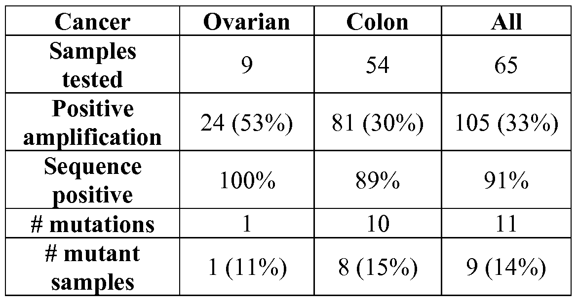
[00106] Examples of important mutations in genes listed in Table 1 are shown below in Table 7 :
Table 7

Table 8


[00108] Although the foregoing invention has been described in some detail by way of illustration and example for purposes of clarity of understanding, it will be clear to those skilled in the art that certain changes and modifications may be practiced within the scope of the appended claims.
Claims
1. A method of detecting mutations comprising:
( 1 ) analyzing in a bodily fluid sample from a human subject a panel of genes consisting of between 5 and 5 ,000 genes, wherein said panel comprises at least five genes chosen from the group consisting of the genes listed in Table 1 ; and
(2) determining whether any of the genes in Table 1 harbors a mutation.
2. The method of Claim 1 , wherein said panel comprises the APC, EGFR, KRAS, PTEN, and TP 53 genes.
3. The method of Claim 1 , wherein said panel comprises the genes listed in Table 3.
4. The method of Claim 1 , wherein said panel comprises the genes listed in Table 2.
5. The method of Claim 1 , wherein said panel comprises the genes listed in Table 1.
6. The method of Claim 1 , wherein said genes chosen from the group consisting of the genes listed in Table 1 constitute at least 10% of said panel.
7. A method of detecting cancer comprising :
( 1 ) analyzing a panel of genes comprising the APC, EGFR, KRAS, PTEN, and TP53 genes in a bodily fluid sample; and
(2) determining whether any of the APC, EGFR, KRAS, PTEN, or TP53 genes harbors a mutation; wherein said mutation indicates the presence of cancer.
8. The method of Claim 7, wherein said mutation is selected from the group consisting of those listed in Table 7 and/ or Table 8.
9. The method of Claim 7, wherein said panel comprises the genes listed in Table 3.
10. The method of Claim 7, wherein said panel comprises the genes listed in Table 2.
1 1. The method of Claim 7, wherein said panel comprises the genes listed in Table 1.
12. A method of determining the likelihood a patient has cancer C1 comprising:
( 1 ) analyzing in a fluid sample a panel of genes comprising the genes listed in Table 3 ;
(2) detecting a mutation in at least one of said genes listed in Table 3 ;
(3) calculating a likelihood said patient has cancer C1 using the formula: P(a \gi, g2, ...,gn ) = Po(C1) U1 M(g, \ C1) / Σ, P0(t) U1 M(g, \ t) ; wherein the product is taken over all genes in said panel mutated in the sample
{i = l , 2, ..., n), the sum is taken over all cancer types t, M(g\ C1) is the frequency of somatic mutations in gene g in cancer type C1, and Po(C1) is the a priori probability of cancer C1 given that the patient has a cancer.
13. The method of Claim 12, further comprising calculating a likelihood said patient has a second cancer C2 using the formula: P(c2  P 0(02) Tl1 M(gj \ c 2) / ∑t Po(t) Tl1 M(gj \ t) ; wherein the product is taken over all genes in said panel mutated in the sample (i = l, 2, ..., ή), the sum is taken over all cancer types t, M(g\ c 2) is the frequency of somatic mutations in gene g in cancer type C2, and P o(c2) is the a priori probability of cancer type C2 given that the patient has a cancer.
14. The method of Claim 12, further comprising recommending, prescribing, ordering, or performing a test for the presence of cancer C1 in said patient.
15. The method of Claim 12 or Claim 13 , wherein said test for the presence of cancer a is recommended, prescribed, ordered, or performed if the calculated likelihood said patient has said cancer a is above a threshold value.
16. The method of Claim 12 or Claim 13 , wherein said threshold value is chosen from the group consisting of 35%, 40%, 45 %, 50%, 55%, 60%, 65%, 70%, 75%, 80%, 85%, 90%, 95%, 96%, 97%, 98%, and 99%.
17. The method of Claim 13 , further comprising recommending, prescribing, ordering, or performing a test for the presence of cancer a in said patient.
18. The method of Claim 17, wherein said test for the presence of cancer c; is recommended, prescribed, ordered, or performed if the calculated likelihood said patient has said cancer a is higher than the calculated likelihood said patient has cancer C2-
19. The method of Claim 17, further comprising recommending, prescribing, ordering, or performing a test for the presence of cancer C2 in said patient if said test for the presence of cancer a does not indicate the presence of cancer a .
20. The method of any one of Claims 1 through 19, wherein said bodily fluid sample is a blood sample.
21. The method of Claim 20, wherein said blood sample is a plasma sample.
22. The method of Claim 20, wherein said blood sample is a serum sample.
23. The method of any one of Claims 1 through 19, wherein detecting a mutation or determining whether a gene harbors a mutation comprises analyzing an mRNA molecule from said sample or analyzing a DNA molecule synthesized using said mRNA molecule as a template.
24. The method of any one of Claims 1 through 19, wherein detecting a mutation or determining whether a gene harbors a mutation comprises analyzing a nucleic acid from said sample by a technique chosen from resequencing, TaqMan™, microarray analysis, and FISH.
25. The method of any one of Claims 1 through 19, wherein detecting a mutation or determining whether a gene harbors a mutation comprises analyzing a nucleic acid deriving from an extracellular vesicle.
26. A kit comprising reagents for analyzing a panel of genes consisting of between 5 and 5 ,000 genes, said kit comprising reagents for detecting mutations in at least five genes selected from the group consisting of the genes listed in Table 1.
27. The kit of Claim 26, wherein said genes are the APC, EGFR, KRAS, PTEN, and TP53 genes.
28. The kit of Claim 26, wherein said genes are the genes listed in Table 3.
29. The kit of Claim 26, wherein said genes are the genes listed in Table 2.
30. The kit of Claim 26, wherein said genes are the genes listed in Table 1.
31. The kit of Claim 26, wherein said genes chosen from the group consisting of the genes listed in Table 1 constitute at least 10% of the genes that may be analyzed in said kit.
Priority Applications (5)
| Application Number | Priority Date | Filing Date | Title |
|---|---|---|---|
| EP10784236.1A EP2438197B1 (en) | 2009-06-05 | 2010-06-07 | Methods of detecting cancer |
| ES10784236.1T ES2544500T3 (en) | 2009-06-05 | 2010-06-07 | Cancer screening methods |
| CA2764468A CA2764468C (en) | 2009-06-05 | 2010-06-07 | Methods of detecting cancer comprising screening for mutations in the apc, egfr, kras, pten and tp53 genes |
| US15/649,274 US20170314081A1 (en) | 2009-06-05 | 2017-07-13 | Methods of detecting cancer |
| US17/575,301 US20220243276A1 (en) | 2009-06-05 | 2022-01-13 | Methods of detecting cancer |
Applications Claiming Priority (2)
| Application Number | Priority Date | Filing Date | Title |
|---|---|---|---|
| US18468509P | 2009-06-05 | 2009-06-05 | |
| US61/184,685 | 2009-06-05 |
Related Child Applications (1)
| Application Number | Title | Priority Date | Filing Date |
|---|---|---|---|
| US13/310,916 Continuation US20130143747A1 (en) | 2009-06-05 | 2011-12-05 | Methods of detecting cancer |
Publications (2)
| Publication Number | Publication Date |
|---|---|
| WO2010141955A2 true WO2010141955A2 (en) | 2010-12-09 |
| WO2010141955A3 WO2010141955A3 (en) | 2011-03-03 |
Family
ID=43298593
Family Applications (1)
| Application Number | Title | Priority Date | Filing Date |
|---|---|---|---|
| PCT/US2010/037659 Ceased WO2010141955A2 (en) | 2009-06-05 | 2010-06-07 | Methods of detecting cancer |
Country Status (4)
| Country | Link |
|---|---|
| EP (1) | EP2438197B1 (en) |
| CA (2) | CA3132169A1 (en) |
| ES (1) | ES2544500T3 (en) |
| WO (1) | WO2010141955A2 (en) |
Cited By (21)
| Publication number | Priority date | Publication date | Assignee | Title |
|---|---|---|---|---|
| EP2475989A4 (en) * | 2009-09-09 | 2013-02-27 | Gen Hospital Corp | USE OF MICROVESICLES IN THE ANALYSIS OF KRAS MUTATIONS |
| WO2014028862A1 (en) * | 2012-08-17 | 2014-02-20 | Cornell University | Use of dna in circulating exosomes as a diagnostic marker for metastasic disease |
| WO2014105748A1 (en) * | 2012-12-31 | 2014-07-03 | University Of Louisville Research Foundation, Inc. | Extracellular vesicle-associated protein markers of cancer |
| WO2015042115A1 (en) * | 2013-09-17 | 2015-03-26 | The Board Of Trustees Of The Leland Stanford Junior University | Biomarkers for ovarian cancer |
| US9186405B2 (en) | 2007-08-16 | 2015-11-17 | The Royal Institution For The Advancement Of Learning/Mcgill University | Tumor cell-derived microvesicles |
| EP3225697A3 (en) * | 2010-12-30 | 2017-11-22 | Foundation Medicine, Inc. | Optimization of multigene analysis of tumor samples |
| EP3421613A1 (en) | 2013-03-15 | 2019-01-02 | The Board Of Trustees Of The Leland Stanford Junior University | Identification and use of circulating nucleic acid tumor markers |
| WO2019067092A1 (en) * | 2017-08-07 | 2019-04-04 | The Johns Hopkins University | Methods and materials for assessing and treating cancer |
| US10352935B2 (en) | 2007-08-16 | 2019-07-16 | The Royal Institution For The Advancement Of Learning/Mcgill University | Tumor cell-derived microvesicles |
| US10407728B2 (en) | 2009-09-09 | 2019-09-10 | The General Hospital Corporation | Use of microvesicles in analyzing nucleic acid profiles |
| US10793914B2 (en) | 2010-08-31 | 2020-10-06 | The General Hospital Corporation | Cancer-related biological materials in microvesicles |
| US10844436B2 (en) | 2014-04-01 | 2020-11-24 | Cornell University | Use of double-stranded DNA in exosomes: a novel biomarker in cancer detection |
| US10988755B2 (en) | 2010-11-10 | 2021-04-27 | Exosome Diagnostics, Inc. | Method for isolation of nucleic acid containing particles and extraction of nucleic acids therefrom |
| US11180803B2 (en) | 2011-04-15 | 2021-11-23 | The Johns Hopkins University | Safe sequencing system |
| US11286531B2 (en) | 2015-08-11 | 2022-03-29 | The Johns Hopkins University | Assaying ovarian cyst fluid |
| US11397182B2 (en) | 2014-10-07 | 2022-07-26 | Cornell University | Methods for prognosing and preventing metastatic liver disease |
| US11525163B2 (en) | 2012-10-29 | 2022-12-13 | The Johns Hopkins University | Papanicolaou test for ovarian and endometrial cancers |
| US11959141B2 (en) | 2014-12-05 | 2024-04-16 | Foundation Medicine, Inc. | Multigene analysis of tumor samples |
| US11971402B2 (en) | 2015-04-24 | 2024-04-30 | Cornell University | Methods and reagents for determination and treatment of organotropic metastasis |
| US12442038B2 (en) | 2020-02-14 | 2025-10-14 | The Johns Hopkins University | Methods and materials for assessing nucleic acids |
| US12473589B2 (en) | 2018-06-11 | 2025-11-18 | Foundation Medicine, Inc. | Compositions and methods for evaluating genomic alterations |
Family Cites Families (1)
| Publication number | Priority date | Publication date | Assignee | Title |
|---|---|---|---|---|
| US20090123928A1 (en) * | 2007-10-11 | 2009-05-14 | The Johns Hopkins University | Genomic Landscapes of Human Breast and Colorectal Cancers |
-
2010
- 2010-06-07 ES ES10784236.1T patent/ES2544500T3/en active Active
- 2010-06-07 EP EP10784236.1A patent/EP2438197B1/en active Active
- 2010-06-07 CA CA3132169A patent/CA3132169A1/en not_active Abandoned
- 2010-06-07 WO PCT/US2010/037659 patent/WO2010141955A2/en not_active Ceased
- 2010-06-07 CA CA2764468A patent/CA2764468C/en active Active
Non-Patent Citations (1)
| Title |
|---|
| See references of EP2438197A4 * |
Cited By (56)
| Publication number | Priority date | Publication date | Assignee | Title |
|---|---|---|---|---|
| US10317407B2 (en) | 2007-08-16 | 2019-06-11 | The Royal Institution For The Advancement Of Learning/Mcgill University | Tumor cell-derived microvesicles |
| US9186405B2 (en) | 2007-08-16 | 2015-11-17 | The Royal Institution For The Advancement Of Learning/Mcgill University | Tumor cell-derived microvesicles |
| US10352935B2 (en) | 2007-08-16 | 2019-07-16 | The Royal Institution For The Advancement Of Learning/Mcgill University | Tumor cell-derived microvesicles |
| US11519036B2 (en) | 2009-09-09 | 2022-12-06 | The General Hospital Corporation | Use of microvesicles in analyzing nucleic acid profiles |
| US11155874B2 (en) | 2009-09-09 | 2021-10-26 | The General Hospital Corporation | Use of microvesicles in analyzing mutations |
| EP3865590A1 (en) * | 2009-09-09 | 2021-08-18 | The General Hospital Corporation | Use of microvesicles in analyzing kras mutations |
| EP2475989A4 (en) * | 2009-09-09 | 2013-02-27 | Gen Hospital Corp | USE OF MICROVESICLES IN THE ANALYSIS OF KRAS MUTATIONS |
| US10407728B2 (en) | 2009-09-09 | 2019-09-10 | The General Hospital Corporation | Use of microvesicles in analyzing nucleic acid profiles |
| US10793914B2 (en) | 2010-08-31 | 2020-10-06 | The General Hospital Corporation | Cancer-related biological materials in microvesicles |
| US10988755B2 (en) | 2010-11-10 | 2021-04-27 | Exosome Diagnostics, Inc. | Method for isolation of nucleic acid containing particles and extraction of nucleic acids therefrom |
| US12312582B2 (en) | 2010-11-10 | 2025-05-27 | Exosome Diagnostics, Inc. | Method for isolation of nucleic acid containing particles and extraction of nucleic acids therefrom |
| US12180540B2 (en) | 2010-12-30 | 2024-12-31 | Foundation Medicine, Inc. | Optimization of multigene analysis of tumor samples |
| EP3225697A3 (en) * | 2010-12-30 | 2017-11-22 | Foundation Medicine, Inc. | Optimization of multigene analysis of tumor samples |
| US11421265B2 (en) | 2010-12-30 | 2022-08-23 | Foundation Medicine, Inc. | Optimization of multigene analysis of tumor samples |
| US11118213B2 (en) | 2010-12-30 | 2021-09-14 | Foundation Medicine, Inc. | Optimization of multigene analysis of tumor samples |
| US11180803B2 (en) | 2011-04-15 | 2021-11-23 | The Johns Hopkins University | Safe sequencing system |
| US11773440B2 (en) | 2011-04-15 | 2023-10-03 | The Johns Hopkins University | Safe sequencing system |
| US12534759B2 (en) | 2011-04-15 | 2026-01-27 | The Johns Hopkins University | Safe sequencing system |
| US12209281B2 (en) | 2011-04-15 | 2025-01-28 | The Johns Hopkins University | Safe sequencing system |
| US12252743B2 (en) | 2011-04-15 | 2025-03-18 | The Johns Hopkins University | Safe sequencing system |
| US11459611B2 (en) | 2011-04-15 | 2022-10-04 | The Johns Hopkins University | Safe sequencing system |
| US12006544B2 (en) | 2011-04-15 | 2024-06-11 | The Johns Hopkins University | Safe sequencing system |
| US11453913B2 (en) | 2011-04-15 | 2022-09-27 | The Johns Hopkins University | Safe sequencing system |
| US11180812B2 (en) | 2012-08-17 | 2021-11-23 | Cornell University | Use of DNA in circulating exosomes as a diagnostic marker for metastatic disease |
| WO2014028862A1 (en) * | 2012-08-17 | 2014-02-20 | Cornell University | Use of dna in circulating exosomes as a diagnostic marker for metastasic disease |
| US11525163B2 (en) | 2012-10-29 | 2022-12-13 | The Johns Hopkins University | Papanicolaou test for ovarian and endometrial cancers |
| US9086412B2 (en) | 2012-12-31 | 2015-07-21 | University Of Louisville Research Foundation, Inc. | Extracellular vesicle-associated protein markers of cancer |
| WO2014105748A1 (en) * | 2012-12-31 | 2014-07-03 | University Of Louisville Research Foundation, Inc. | Extracellular vesicle-associated protein markers of cancer |
| EP3795696A1 (en) | 2013-03-15 | 2021-03-24 | The Board of Trustees of the Leland Stanford Junior University | Identification and use of circulating nucleic acid tumor markers |
| EP4484572A2 (en) | 2013-03-15 | 2025-01-01 | The Board of Trustees of the Leland Stanford Junior University | Identification and use of circulating nucleic acid tumor markers |
| EP4253558A1 (en) | 2013-03-15 | 2023-10-04 | The Board of Trustees of the Leland Stanford Junior University | Identification and use of circulating nucleic acid tumor markers |
| US12421559B2 (en) | 2013-03-15 | 2025-09-23 | The Board Of Trustees Of The Leland Stanford Junior University | Identification and use of circulating nucleic acid tumor markers |
| EP3421613A1 (en) | 2013-03-15 | 2019-01-02 | The Board Of Trustees Of The Leland Stanford Junior University | Identification and use of circulating nucleic acid tumor markers |
| WO2015042115A1 (en) * | 2013-09-17 | 2015-03-26 | The Board Of Trustees Of The Leland Stanford Junior University | Biomarkers for ovarian cancer |
| US10844436B2 (en) | 2014-04-01 | 2020-11-24 | Cornell University | Use of double-stranded DNA in exosomes: a novel biomarker in cancer detection |
| US11397182B2 (en) | 2014-10-07 | 2022-07-26 | Cornell University | Methods for prognosing and preventing metastatic liver disease |
| US11959141B2 (en) | 2014-12-05 | 2024-04-16 | Foundation Medicine, Inc. | Multigene analysis of tumor samples |
| US11971402B2 (en) | 2015-04-24 | 2024-04-30 | Cornell University | Methods and reagents for determination and treatment of organotropic metastasis |
| US11286531B2 (en) | 2015-08-11 | 2022-03-29 | The Johns Hopkins University | Assaying ovarian cyst fluid |
| JP2020530290A (en) * | 2017-08-07 | 2020-10-22 | ザ ジョンズ ホプキンス ユニバーシティ | Methods and Substances for Assessing and Treating Cancer |
| AU2018342007B2 (en) * | 2017-08-07 | 2025-07-17 | The Johns Hopkins University | Methods and materials for assessing and treating cancer |
| JP2023075090A (en) * | 2017-08-07 | 2023-05-30 | ザ ジョンズ ホプキンス ユニバーシティ | Methods and agents for evaluating and treating cancer |
| CN111868260B (en) * | 2017-08-07 | 2025-02-21 | 约翰斯霍普金斯大学 | Methods and materials for evaluating and treating cancer |
| KR102775704B1 (en) * | 2017-08-07 | 2025-02-28 | 더 존스 홉킨스 유니버시티 | Methods and materials for evaluating and treating cancer |
| JP7232476B2 (en) | 2017-08-07 | 2023-03-08 | ザ ジョンズ ホプキンス ユニバーシティ | Methods and agents for evaluating and treating cancer |
| IL272470B1 (en) * | 2017-08-07 | 2025-04-01 | Univ Johns Hopkins | Methods and materials for assessing and treating cancer |
| JP2025062599A (en) * | 2017-08-07 | 2025-04-14 | ザ ジョンズ ホプキンス ユニバーシティ | Methods and Materials for Assessing and Treating Cancer - Patent application |
| CN111868260A (en) * | 2017-08-07 | 2020-10-30 | 约翰斯霍普金斯大学 | Methods and materials for the assessment and treatment of cancer |
| US12195803B2 (en) | 2017-08-07 | 2025-01-14 | The Johns Hopkins University | Methods and materials for assessing and treating cancer |
| IL272470B2 (en) * | 2017-08-07 | 2025-08-01 | Univ Johns Hopkins | Methods and materials for cancer assessment and treatment |
| KR20200115450A (en) * | 2017-08-07 | 2020-10-07 | 더 존스 홉킨스 유니버시티 | Methods and materials for evaluating and treating cancer |
| WO2019067092A1 (en) * | 2017-08-07 | 2019-04-04 | The Johns Hopkins University | Methods and materials for assessing and treating cancer |
| US12473589B2 (en) | 2018-06-11 | 2025-11-18 | Foundation Medicine, Inc. | Compositions and methods for evaluating genomic alterations |
| US12584163B2 (en) | 2018-06-11 | 2026-03-24 | Foundation Medicine, Inc. | Compositions and methods for evaluating genomic alterations |
| US12442038B2 (en) | 2020-02-14 | 2025-10-14 | The Johns Hopkins University | Methods and materials for assessing nucleic acids |
| US12553082B2 (en) | 2020-02-14 | 2026-02-17 | The Johns Hopkins University | Methods and materials for assessing nucleic acids |
Also Published As
| Publication number | Publication date |
|---|---|
| EP2438197B1 (en) | 2015-07-15 |
| CA2764468C (en) | 2021-11-16 |
| EP2438197A4 (en) | 2012-11-21 |
| EP2438197A2 (en) | 2012-04-11 |
| CA3132169A1 (en) | 2010-12-09 |
| ES2544500T3 (en) | 2015-09-01 |
| WO2010141955A3 (en) | 2011-03-03 |
| CA2764468A1 (en) | 2010-12-09 |
Similar Documents
| Publication | Publication Date | Title |
|---|---|---|
| CA2764468C (en) | Methods of detecting cancer comprising screening for mutations in the apc, egfr, kras, pten and tp53 genes | |
| US20170314081A1 (en) | Methods of detecting cancer | |
| Bittla et al. | Exploring circulating tumor DNA (CtDNA) and its role in early detection of cancer: a systematic review | |
| Cheung et al. | Latest development of liquid biopsy | |
| Leygo et al. | DNA methylation as a noninvasive epigenetic biomarker for the detection of cancer | |
| Diaz-Lagares et al. | A novel epigenetic signature for early diagnosis in lung cancer | |
| Kaur et al. | Early diagnosis of pancreatic cancer: challenges and new developments | |
| Motoi et al. | Lung adenocarcinoma: modification of the 2004 WHO mixed subtype to include the major histologic subtype suggests correlations between papillary and micropapillary adenocarcinoma subtypes, EGFR mutations and gene expression analysis | |
| Gao et al. | Whole‐genome bisulfite sequencing analysis of circulating tumour DNA for the detection and molecular classification of cancer | |
| Abdipourbozorgbaghi et al. | Circulating miRNA panels as a novel non-invasive diagnostic, prognostic, and potential predictive biomarkers in non-small cell lung cancer (NSCLC) | |
| Nakamura et al. | Clinical impact of genetic aberrations in gastric MALT lymphoma: a comprehensive analysis using interphase fluorescence in situ hybridisation | |
| Zhang et al. | Circulating cell-free DNA-based methylation patterns for breast cancer diagnosis | |
| CN114214409B (en) | Biomarkers for esophageal cancer classification and their applications | |
| Boulaiz et al. | What’s new in the diagnosis of pancreatic cancer: a patent review (2011-present) | |
| Schwarzbach | Differential expression of GNAS and KRAS mutations in pancreatic cysts | |
| Atwater et al. | The pursuit of noninvasive diagnosis of lung cancer | |
| Ye et al. | Concordance between microsatellite instability and mismatch repair protein expression in colorectal cancer and their clinicopathological characteristics: a retrospective analysis of 502 cases | |
| Nguyen Hoang et al. | Real-world utilization and performance of circulating tumor DNA monitoring to predict recurrence in solid tumors | |
| Chen et al. | Circulating tumor DNA detection and its application status in gastric cancer: a narrative review | |
| Lin et al. | A multicenter-retrospective cohort study of chromosome instability in lung cancer: clinical characteristics and prognosis of patients harboring chromosomal instability detected by metagenomic next-generation sequencing | |
| Zhang et al. | Personalized radiomics signature to screen for KIT-11 mutation genotypes among patients with gastrointestinal stromal tumors: a retrospective multicenter study | |
| Wever et al. | Molecular analysis for ovarian cancer detection in patient-friendly samples | |
| Rengucci et al. | Improved stool DNA integrity method for early colorectal cancer diagnosis | |
| Wu et al. | Adenoid cystic carcinoma of the breast: Molecular profiling updates and multidimensional detection of MYB | |
| US20220243276A1 (en) | Methods of detecting cancer |
Legal Events
| Date | Code | Title | Description |
|---|---|---|---|
| 121 | Ep: the epo has been informed by wipo that ep was designated in this application |
Ref document number: 10784236 Country of ref document: EP Kind code of ref document: A2 |
|
| WWE | Wipo information: entry into national phase |
Ref document number: 2764468 Country of ref document: CA |
|
| NENP | Non-entry into the national phase |
Ref country code: DE |
|
| WWE | Wipo information: entry into national phase |
Ref document number: 2010784236 Country of ref document: EP |